ERGOT ALKALOIDS – BIOLOGY AND MOLECULAR BIOLOGY
42
—CHAPTER 2— ERGOT ALKALOIDS – BIOLOGY AND MOLECULAR BIOLOGY $ CHRISTOPHER L. SCHARDL 1 ,DANIEL G. PANACCIONE 2 AND PAUL TUDZYNSKI 3 1 Department of Plant Pathology, University of Kentucky, Lexington, KY 40546-0312, USA; 2 Division of Plant and Soil Sciences, WV University,Morgantown, WV 26506- 6108, USA; 3 Institut fu¨r Botanik, Westfa¨lische Wilhelms Universita¨t Mu¨nster, Mu¨nster D-48149, Germany I. Introduction II. Through the Ages: A History of Ergot Alkaloid Use, Abuse, and Poisoning III. Ergot Alkaloid Producers IV. Biosynthetic Pathways and Genes V. Routine Analytical Methods for Ergot Alkaloids VI. Ergot Alkaloid Activities and Roles VII. Summary and Conclusions Acknowledgments References I. Introduction The ergot alkaloids (EA) (Fig. 1) are among the most important natural pharmaceuticals and toxins in human history. Some EA have been used purpose- fully for medical benefit in many cultures over the ages (2). Some have been used illicitly for psychedelic recreation, again, throughout the ages into modern times (3,4). Some have been ingested unknowingly, leading to poisonings with dire physio- logical and social implications (2). In the past centuries, poor understanding of the sources of these toxins and their physiological effects exacerbated the problems arising from their intended and unintended uses, and the symptoms of ergotism in humans and livestock were often attributed to witchcraft (5). Even now, the EA pharmaceutical properties are not fully understood and are the subject of intense research. Gaps in our current understanding result from a combination of the shear complexity of the central and peripheral nervous systems on which EA act, the pleiotropic effects of some EA whereby multiple neuroreceptors may be affected simultaneously, and on structural variation among the EA with consequent vari ations in their specificities of action. However, as greater understanding of EA has developed with modern research, their utilization as pharmaceuticals has become $ Dedicated to Prof. Dr. Heinz G. Floss in recognition of his 45 years of profound discoveries relating to the biosynthesis of ergot alkaloids and other microbial metabolites. 45 THE ALKALOIDS, Vol. 63 ISSN: 1099-4831 DOI: 10.1016/S1099-4831(06)63002-2 Copyright r 2006 Elsevier Inc. All rights reserved
-
Upload
khangminh22 -
Category
Documents
-
view
0 -
download
0
Transcript of ERGOT ALKALOIDS – BIOLOGY AND MOLECULAR BIOLOGY

—CHAPTER 2—
ERGOT ALKALOIDS – BIOLOGY AND
MOLECULAR BIOLOGY$
CHRISTOPHER L. SCHARDL1, DANIEL G. PANACCIONE
2AND PAUL TUDZYNSKI
3
1Department of Plant Pathology, University of Kentucky, Lexington, KY 40546-0312,USA; 2Division of Plant and Soil Sciences, WV University,Morgantown, WV 26506-
6108, USA; 3Institut fur Botanik, Westfalische Wilhelms Universitat Munster,
Munster D-48149, Germany
I. Introduction
II. Through the Ages: A History of Ergot Alkaloid Use, Abuse, and Poisoning
III. Ergot Alkaloid Producers
IV. Biosynthetic Pathways and Genes
V. Routine Analytical Methods for Ergot Alkaloids
VI. Ergot Alkaloid Activities and Roles
VII. Summary and Conclusions
Acknowledgments
References
I. Introduction
The ergot alkaloids (EA) (Fig. 1) are among the most important naturalpharmaceuticals and toxins in human history. Some EA have been used purpose-fully for medical benefit in many cultures over the ages (2). Some have been usedillicitly for psychedelic recreation, again, throughout the ages into modern times(3,4). Some have been ingested unknowingly, leading to poisonings with dire physio-logical and social implications (2). In the past centuries, poor understanding of thesources of these toxins and their physiological effects exacerbated the problemsarising from their intended and unintended uses, and the symptoms of ergotismin humans and livestock were often attributed to witchcraft (5). Even now, the EApharmaceutical properties are not fully understood and are the subject of intenseresearch. Gaps in our current understanding result from a combination of the shearcomplexity of the central and peripheral nervous systems on which EA act, thepleiotropic effects of some EA whereby multiple neuroreceptors may be affectedsimultaneously, and on structural variation among the EA with consequent variations in their specificities of action. However, as greater understanding of EA hasdeveloped with modern research, their utilization as pharmaceuticals has become
$Dedicated to Prof. Dr. Heinz G. Floss in recognition of his 45 years of profound discoveries relating
to the biosynthesis of ergot alkaloids and other microbial metabolites.
45THE ALKALOIDS, Vol. 63
ISSN: 1099-4831
DOI: 10.1016/S1099-4831(06)63002-2
Copyright r 2006 Elsevier Inc.
All rights reserved

Fig. 1. Ergoline (A) and ergopeptine (B) structures showing atom numbering and ring des-
ignations. (A) Substituents are color-coded according to their origins: DMAPP, dimethylallyl-
diphosphate (red); AdoMet, S-adenosyl-methionine (green), and L-Trp, L-tryptophan (blue).
(B) Portions of the tripeptide substituent are color-coded according to the positions of
L-amino acid precursors: A.A. I (blue), A.A. II (red), and A.A. III (green). (C, D) Two views
of a space-filling model of ergocornine, demonstrating proximity of the lactam ring and the
lysergic D-ring. Reproduced with permission from Lehner et al. (1).
SCHARDL ET AL.46
more refined and effective, while human poisonings have become rare (6). Problemsof livestock poisonings have also been reduced as some of the culprits – biologicalsources in the field and feed – have been identified. However, in many places,exposure of livestock to EA is unavoidable, and mitigation of EA toxicoses inlivestock is another subject of major research efforts (7–9).
The EA are characterized by the tetracyclic ergoline ring system (Fig. 1), orby related tricyclic alkaloids open between N(6) and C(7) (ergoline numbering).They are categorized as clavines, lysergic acid (1) and its simple amides (Fig. 2), andergopeptines (Fig. 1). Some clavines are biosynthetic intermediates in the lysergicacid pathway, although several fungal species make clavines, but not 1. Otherclavines are derivatives of lysergic acid pathway intermediates. The carboxylic acidgroup at position 17 characterizes lysergic acid (1), and most lysergic acid deriva-tives have substituents linked as amides to that function. Among the simpler lysergicacid amides are ergine (2 ¼ lysergic acid amide) and ergonovine (3 ¼ ergo-metrine ¼ ergobasine) (Fig. 2). In the ergopeptines, which are the most complex

N
HN
CH3
C
H
O OH
1 D-lysergic acid
N
HN
CH3
C
H
O NH2
2 ergine= lysergic acid amide
N
HN
CH3
C
H
O NH
3 ergonovine= ergometrine= ergobasine
CH2
OH
CH3
N
HN
CH3
C
H
O NCH2
H2CCH3
CH3
5 LSD-25
N
HN
CH3
CH2OH
H
6 elymoclavine
N
HN
CH3
CH3
H
11 pibocin
H
Br
N
HN
CH3
CH2OH
H
6a D-lysergol
H N
HN
CH3
CH2OH
H
6b dihydrolysergol-I= dihydroelymoclavine
H
Fig. 2. Structures of lysergic acid and some lysergyl amides and clavines.
ERGOT ALKALOIDS 47
of the natural EA, the C(17) amide substituent is a tripeptide-derived, cyclol-lactamstructure (Fig. 1).
The biosynthetic pathway to EA in Claviceps species has been studied inten-sively since the 1950s, and was reviewed by Groger and Floss in the 1998 volume ofthis series (10). That outstanding and thorough review historically punctuates thetransition from mainly biochemical studies to investigations involving moleculargenetics. Therefore, salient points of the previous review will be summarized here,and this review will emphasize contributions of recent molecular genetic studies toour understanding of the EA pathway. In addition, we will summarize the distri-bution and variation in EA, and activities of EA in humans and livestock, thendiscuss their possible ecological roles.

SCHARDL ET AL.48
II. Through the Ages: A History of Ergot Alkaloid Use, Abuse, and Poisoning
Ergots are hard, dense, and darkly pigmented resting structures (sclerotia) ofcertain fungi – the Claviceps species – that parasitize ears of grain, and are the mostconvenient and abundant source of EA. The medicinal use of ergots is quite ancient(11). Records extend from approximately 600 BCE., in Mesopotamia (now Iraq), forthe use of ergots at specified doses to women in labor as an aid for parturition or tostaunch hemorrhaging. This was a risky treatment, however, because the dosage ofactive ingredients could not be controlled well, and rupture due to hypercontractionof the uterus was a significant risk.
In the Americas, too, EA have been used in traditional medicine or forrecreation. Seeds of Rivea corymbosa or Ipomea species were known as ololiuqui andused ceremonially by indigenous people of Mexico. Investigation of their chemicalcomposition indicated 2 and other lysergic acid amides among the hallucinogenicconstituents (12). Furthermore, indigenous people of South America have usedCyperus spp. (Cyperaceae; sedges), infected with the EA-producing fungus Balansiacyperi, to promote parturition (13,14).
With increased understanding of the chemistry and pharmacology of EA,careful dosing and use of chemically modified EA to treat an ever-greater array ofconditions, this class of compounds emerged with a significant role in modernmedicine.
In ergots produced by Claviceps purpurea on rye (Secale cereale), the con-centration of ergotamine (4) and related ergopeptine alkaloids (Table I), plus simplerlysergyl amides, typically approximates 0.5–2% by dry mass (15–20). These alka-loids can readily be converted into 1 by alkaline or acid hydrolysis, providing aconvenient starting material for legal pharmaceuticals or illicit recreational drugs.Semi-synthesis of lysergic acid diethylamide (LSD) (5; Fig. 2) is sufficiently straight-forward to pose a major problem for law enforcement. This compound, originallynamed LSD-25, was 25th in a series of lysergyl amides produced by Albert Hofmannat Sandoz Research Laboratories, Basel, Switzerland (21). The acronym, LSD,which in English is a better fit to the song title ‘‘Lucy in the Sky with Diamonds,’’ isan abbreviation of the German lysergsaure diathylamid. This alkaloid is the mostpotent hallucinogen known (3). Originally tested as an experimental antidepressantdrug and as a treatment for schizophrenia, its early tests turned up severe problemsincluding paranoia, potentially fatal loss of judgment, and flashbacks. Nevertheless,‘‘acid trips’’ are a feature of the cultural upheavals that gripped the Western Worldin the 1960s and 1970s.
Historically, infected ears of rye have been a common source of ergot-contaminated flour (2). Ergot contamination, particularly of rye, has been a majorproblem in Europe for millennia, and ergot poisoning is now widely believed to bethe key trigger for a number of major historical events, such as the Salem WitchTrials in colonial Massachusetts (5). (It should be noted, however, that some strongarguments have been made against this ‘‘convenient’’ hypothesis (22). In the late20th century, outbreaks were reported in Ethiopia and India. The Ethiopian ergotoutbreak in 1977–1978 is thought to have arisen by ergot-infected wild oats har-vested as a field contaminant of barley (23). Outbreaks of convulsive ergotism in

TABLE I.
Ergopeptine Alkaloids.
N
HN
CH3
C
H
OHN
O
NN
O
OH
O
R1
R2
A.A. I
A.A. II
A.A. III
R1 Amino acid I R2 Amino acid II
Ergotamine group
4 Ergotamine CH3 L-alanine CH2Ph L-phenylalanine
8 Ergovaline CH3 L-alanine i-Pr L-valine
Ergosinea CH3 L-alanine i-Bu L-leucine
b-ergosine CH3 L-alanine sec-Bu L-isoleucine
Ergobine CH3 L-alanine Et L-2-aminobutyric acid
Ergotoxine group
Ergocristine i-Pr L-valine CH2Ph L-phenylalanine
Ergocornine i-Pr L-valine i-Pr L-valine
Ergocryptinea,b i-Pr L-valine i-Bu L-leucine
b-ergocryptineb i-Pr L-valine sec-Bu L-isoleucine
g-ergocryptineb,c i-Pr L-valine n-Bu L-norleucine
Ergobutyrine i-Pr L-valine Et L-2-aminobutyric acid
Ergoladinec i-Pr L-valine EtSCH3 L-methionine
Ergogaline i-Pr L-valine 2-Me-n-Bu L-homoisoleucine
Ergoxine group
Ergostine Et L-2-aminobutyric acid CH2Ph L-phenylalanine
Ergonine Et L-2-aminobutyric acid i-Pr L-valine
Ergoptinea Et L-2-aminobutyric acid i-Bu L-leucine
b-ergoptine Et L-2-aminobutyric acid sec-Bu L-isoleucine
ergobutine Et L-2-aminobutyric acid Et L-2-aminobutyric acid
Other (A.A. III ¼ L-alanine)
9 Ergobalansine CH3 L-alanine i-Bu L-leucine
aSynonyms: ergosine, a-ergosine; ergocryptine, a-ergocryptine; ergoptine, a-ergoptine.bSynonyms: a-, b-, or g-ergocryptine ¼ a-, b-, or g-ergokryptine.cOnly the isolysergyl isomers, ergoladinine, and g-ergocryptinine, have been reported to date.
ERGOT ALKALOIDS 49

SCHARDL ET AL.50
India were attributed to Claviceps fusiformis ergots on pearl millet (Pennisetumglaucum) (24,25).
The symptoms of ergot alkaloid poisoning vary, probably depending on theparticular profiles of alkaloids present in the contaminated flour (6). Two syndromeshave been described as convulsive and gangrenous ergotism. Gangrenous ergotismresults from the extreme vasoconstrictive properties of certain EA (particularly 4and other ergopeptines), resulting in ischaemia (restricted blood-flow to parts of thebody). Limbs may become hypoxic, develop dry gangrene, and self-amputate orrequire amputation (6). The effect on the corotid artery is not as severe as effects onother arteries, thus sparing the brain of substantial damage.
Apparently, many outbreaks of convulsive ergotism occurred during themiddle ages in Europe, where the malady was called ignis sacer (holy fire), orSt. Anthony’s fire (2,5). (The same terms were also applied to intense rashes resultingfrom bacterial infections.) Among the reported symptoms of convulsive ergotism areinvoluntary muscle contractions, painful flexion or extension of the fingers, wrists,and ankles, involuntary twisting (such as wryneck), paresthesia (skin-crawling andtingling), tinnitus, vertigo, headaches, double-vision, profuse sweating, fever,ravenous appetite, hallucinations, mania, melancholy, and delirium (6). Stiff, dis-torted postures (dyskinesias) could last for minutes or hours, with relatively normalintervals (except for a voracious appetite) lasting for hours to days. Dyskinesias aresometimes followed by fatal epileptic seizures.
Clavines are thought to contribute substantially to convulsive ergotism, sinceC. fusiformis ergots, which possess clavines, but no 1 or lysergyl amides, causeconvulsive symptoms (26). However, the ergopeptines are known to produce similarsymptoms, and are also thought to cause gangrenous ergotism (6). The occurrenceof convulsive ergotism without dry gangrene suggests that other clavine or lysergylalkaloids are involved, or that individual effects of specific ergopeptines may giveclinically different syndromes (6).
III. Ergot Alkaloid Producers
The distribution of organisms possessing EA appears disjointed, including twoorders of fungi and three plant families. The EA-producing fungi are in the Eurotialesand Hypocreales, two distantly related orders within the phylum Ascomycota. Withinthe Hypocreales, EA are associated exclusively with plant-associated fungi of thefamily Clavicipitaceae, although not all members of the Clavicipitaceae produce EA.Some producers of EA lack a known sexual state, so according to the Botanical Codethese are classified as Fungi Imperfecti. Nevertheless, the evolutionary derivation ofEA producers from asexual Eurotiales and Hypocreales is clear.
The type species of the Clavicipitaceae is C. purpurea, otherwise known as theergot fungus of rye and related grasses. Parasitism of host plants commences whenascospores (meiotically generated spores) are ejected from fungal fruiting structuresand land on newly exerted stigmata of grass florets (27). The spores germinate,and hyphae grow down the style to the ovary. Should the floret be pollinatedbeforehand, the style will dry and become refractory to ergot parasitism, making
ERGOT ALKALOIDS 51
the window of susceptibility very brief, and rendering self-pollinated wheat far lessprone than rye to ergot infection. Should a successful infection of the floret occur,further proliferation of the fungus encases the ovary, preventing its maturation.In the ensuing ‘‘honeydew’’ stage the fungus produces abundant conidia (mitoticallyderived spores) in a sweet exudate, which attracts insects that facilitate secondaryspread of the ergot fungus. The fungus then produces resting structures calledsclerotia (‘‘ergots’’ in the case of Claviceps species). An interesting and problematiccharacteristic of ergots is that their density prevents removal by winnowing, so thatergot contamination of rye flour was a major hazard for millennia. Modern screen-ing techniques reduce contamination, but may be selecting for genotypes that pro-duce smaller ergots similar in size to caryopses (27).
Ergots of C. purpurea tend to be rich in ergopeptines (28), and strains ofC. purpurea have been developed for fermentation to produce ergopeptines andother EA (29). The production of EA in those cultures has been associated with agrowth characteristic that resembles the early stage of sclerotium development onhost plants (4).
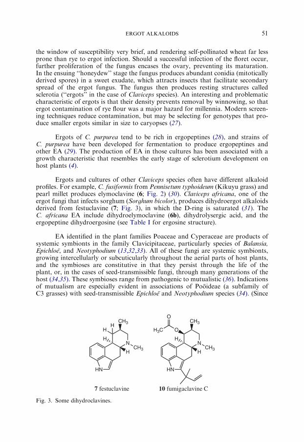
Ergots and cultures of other Claviceps species often have different alkaloidprofiles. For example, C. fusiformis from Pennisetum typhoideum (Kikuyu grass) andpearl millet produces elymoclavine (6; Fig. 2) (30). Claviceps africana, one of theergot fungi that infects sorghum (Sorghum bicolor), produces dihydroergot alkaloidsderived from festuclavine (7; Fig. 3), in which the D-ring is saturated (31). TheC. africana EA include dihydroelymoclavine (6b), dihydrolysergic acid, and theergopeptine dihydroergosine (see Table I for ergosine structure).
EA identified in the plant families Poaceae and Cyperaceae are products ofsystemic symbionts in the family Clavicipitaceae, particularly species of Balansia,Epichloe, and Neotyphodium (13,32,33). All of these fungi are systemic symbionts,growing intercellularly or subcuticularly throughout the aerial parts of host plants,and the symbioses are constitutive in that they persist through the life of theplant, or, in the cases of seed-transmissible fungi, through many generations of thehost (34,35). These symbioses range from pathogenic to mutualistic (36). Indicationsof mutualism are especially evident in associations of Pooideae (a subfamily ofC3 grasses) with seed-transmissible Epichloe and Neotyphodium species (34). (Since
N
HN
HCH3
CH3
H
7 festuclavine
N
HN
HCH3
CH3
H
OH3C
O
10 fumigaclavine C
HH
Fig. 3. Some dihydroclavines.

SCHARDL ET AL.52
Neotyphodium species are asexual derivatives of Epichloe species, members ofthese two genera are collectively called ‘‘epichloe’’ hereafter.) Many, but not all, ofthese fungi produce EA when associated with their host plants, and a few wereshown to produce EA in culture (37).
Like C. purpurea, other plant pathogenic Clavicipitaceae cause atypicaldiseases on their hosts. They form fruiting bodies or sclerotia on inflorescences,florets or at specific locations on vegetative leaves of the infected host. The affectedinflorescences or florets are prevented from producing seeds, presumably benefitingthe fungus by diverting resources to its fruiting bodies. The cause of this effect on theplant is probably hormonal, since even those Balansia species that fruit on leavescan reduce or eliminate host flowering (38). The EA are not involved in pathology,since many species or isolates fail to produce EA, yet exhibit the same effects onplants, and several Neotyphodium species produce EA, but cause no plant disease(39,40). Even so, EA, and the indolediterpenes (also produced by many of theseClavicipitaceae) have precursors in common with phytohormones, including auxin(from tryptophan), cytokinin (containing isoprene), gibberellin, and abscisic acid(from isoprene). To date, there has been rather little investigation of the potentialfor these fungi to produce these hormones or analogues, although some have beenshown to produce auxin (41), and Gibberella fujikuroi (also in Hypocreales) isfamous as a gibberellin producer (42). Thus, the metabolic flux of EA precursorsinto fungal products with phytohormone activity, and possible competition betweenthese pathways, may be worthy of future investigations.
In many grass-epichloe symbiota the ergopeptine, ergovaline (8; Table I), isthe principal EA. Generally 2, and in some cases 3 or 6 and other clavines, can alsobe identified in these symbiota (33,43–47). In two symbiota involving Achnatherumspecies with epichloe endophytes, 2 and 3 are the principal EA, and no 8 or anyother complex ergopeptine has been observed (47,48).
Ergobalansine (9; Table I) was first identified in Cyperus species thathad symbiotic Balansia species, which also produce this alkaloid in culture (13).Later, ergobalansine was also identified in the plant, Ipomoea asarifolia (familyConvolvulaceae) (49). Based on recent studies in which EA were eliminated fromI. asarifolia by fungicide treatment (50), it seems reasonable to expect that 9 isproduced by a fungal symbiont of this plant.
EA producers among the Eurotiales have ecological niches very distinct fromthose of the Clavicipitaceae. Aspergillus fumigatus is a notable example: a heat-resistant saprophyte, well adapted for survival and growth in compost, and currentlythe most common agent of invasive mycosis in humans (51). Spores of this fungusare ubiquitous, and inhaled spores can enter alveoli and induce aspergillosis. Veryhigh levels of festuclavine (7) and fumigaclavines (e.g., 10; Fig. 3) are associated withthe conidia of A. fumigatus (52).
An interesting clavine structure, pibocin (11; Fig. 2), was isolated from atunicate, Eudistoma species (53). This marine animal represents another taxonomickingdom from which EA have been isolated. The preponderance of symbioticfungi as EA sources in plants highlights the possibility that a fungal symbiont of theEudistoma species might be the source of pibocin or a pibocin precursor, but this

ERGOT ALKALOIDS 53
remains to be investigated. This alkaloid, which has antitumor activity, is a 2–bromoderivative of festuclavine. Interestingly, 2–brominated ergocryptine (bromocriptine)has been synthesized and used pharmacologically (11).
IV. Biosynthetic Pathways and Genes
A. ERGOT ALKALOID BIOSYNTHESIS GENE CLUSTERS
Nearly half a century of intensive investigation of the biosynthetic precursors,enzymes and pathways have been additionally informed by the recent identificationof gene clusters that are likely to encode all or most of the enzymes for EA bio-synthesis. The first pathway gene to be cloned, dmaW (54), encodes the determinantstep in EA biosynthesis (Scheme 1). The gene was cloned from C. fusiformis SD58,which produces 6 as an end product. Subsequently, the orthologue was identifiedin C. purpurea P1 in a cluster of genes, of which many are predicted to encodebiosynthetic enzymes (55). To date, 68 kb of the cluster has been sequenced (Fig. 4),and the likely or confirmed EA biosynthesis genes are listed in Table II. Here wedesignate the cluster eas (ergot alkaloid synthesis).
To facilitate discussion of the genes and their products, and comparisonsamong EA-producing fungi, we take this opportunity to adopt a systematic set ofnames for the eas cluster genes and their orthologues in other species (Fig. 4; Table II).
HN NH2
CO2H
CO
PPiH
H∗
HN NH2
CO2H
H H
H∗
HN NH2
CO2H
H
H∗
L-Trp
DMAPP
12 DMATrp
Scheme 1
Fig. 4. Map of eas gene cluster in Claviceps purpurea P1. Arrows indicate direction of tran-
scription. The corresponding modules in lpsA1 and lpsA2 are indicated with similar shading.
White bars indicate intron positions in the lpsA1, lpsA2, and lpsB genes.

TABLE II.
Known and Predicted Genes in the Ergot Alkaloid Gene Cluster of Claviceps purpurea Strain P1.
Gene Synonym Predicted product size (aa) Function or putative function Likely cofactors Conserved domain E-value
easA cpox3 369 Reductase/dehydrogenase (OYE) FMNH2 COG1902 4e-41
lpsB cpps2 1308 LPS subunit 2 40-Phospho-pantetheine, ATP COG1020 1e-54
cloA cpP450-1 507 Elymoclavine oxygenase Heme-Fe pfam00067 3e-18
easC cpcat2 521 Catalase Heme-M cd00328 3e-122
easD cpox2 261 Reductase/dehydrogenase NAD+ or NADH pfam00106 3e-27
easE cpox1 551 Reductase/dehydrogenase FAD pfam01565 8e-14
easF orfB 359 Methyltransferase AdoMet COG4301 3e-24
easG orfA 257 Reductase/dehydrogenase NAD+ COG0702 1e-11
dmaW cpd1 448 DMATrp synthase Ca2+? n.d. –
easH1 orfC 314 Oxygenase/hydroxylase Fe(II) pfam05721 8e-16
lpsA1 cpps1 3585 LPS 1 (ergotamine?) 40-Phospho-pantetheine, ATP COG1020 8e-69
easH2 orfE 154 Hydroxylase or c Fe(II) n.d. –
lpsA2 cpps4 3524 LPS 1 (ergocryptine?) 40-Phospho-pantetheine, ATP COG1020 4e-74
Note: Abbreviations: OYE, old yellow enzyme; Heme-M, heme with metal ion; LPS, lysergyl peptide synthetase; DMATrp, dime-
thylallyltryptophan; n.d., none detected; c pseudogene.
SCHARDLETAL.
54

ERGOT ALKALOIDS 55
Those C. purpurea eas genes that have not yet been fully characterized for function aredesignated easA through easG, plus the two closely related genes easH1 and easH2.Those genes whose products have been characterized biochemically are designatedaccording to the enzyme activities of the proteins they encode.
Four eas cluster genes encode enzymes that have been characterized inC. purpurea P1 or other EA-producing fungi. The dmaW gene product was wellcharacterized in C. fusiformis SD58 (54,56), and its role in the determinant stepof EA biosynthesis was established by disruption of the orthologue in the epichloeendophyte Neotyphodium lolii � Epichloe typhina strain Lp1 (39) (a natural hybridof N. lolii and E. typhina (57)). Similarly, as will be discussed in detail later, the lpsA1and lpsA2 genes were linked by protein sequences to the well-characterized lysergylpeptide synthetase (LPS) of C. purpurea P1 (55,58,59). Then, gene disruption dem-onstrated the role of a close homologue in synthesis of 8, as well as 2 and lysergylala-nine, by N. lolii � E. typhina Lp1 (46,60). Whereas the lpsA genes encode a large,multimodular subunit of LPS, the predicted protein sequence of the lpsB geneproduct indicates that it encodes the smaller subunit responsible for activation ofthe D-lysergic acid moiety. This role has been confirmed by lpsB knockout andheterologous expression (61), as detailed later. Finally, disruption of cloA, predictedto encode a cytochrome-P450 monooxygenase, has demonstrated that its product isinvolved in conversion of 6 into 1 (62).
A cluster of at least nine genes homologous to C. purpurea eas cluster geneshas been identified in C. fusiformis SD58 (54,63). These genes are lpsB, easA, cloA,easC, easD, easE, easF, easG, and dmaW. All are transcribed, but the lpsB sequencesuggests that it does not code for a functional product.
It is possible that the gene cluster so far characterized in C. purpurea encodesall of the enzymes for synthesis of 4, yet some genes found in other fungal secondarymetabolism gene clusters have no counterpart among those identified in C. purpureato date. For example, no obvious transcriptional regulator is encoded in the cluster.However, in A. fumigatus several putative transcription factor genes are looselylinked to the ergot alkaloid gene cluster (64).
The eas gene cluster in A. fumigatus contains homologues for eight of thegenes found in the C. purpurea cluster (Fig. 5; Table III). The genes common to theergot alkaloid clusters of the two divergent fungi are hypothesized to encode enzymesthat catalyze the early steps of the ergot alkaloid pathway, which are presum-ably shared between A. fumigatus and the clavicipitaceous fungi. The A. fumigatus
Fig. 5. Map of eas gene cluster identified on the long arm of chromosome 2 in the sequenced
genome of Aspergillus fumigatus. Arrows indicate direction of transcription. Black arrows
indicate orthologues of genes in the C. purpurea eas cluster. Gray arrows indicate genes
present in A. fumigatus, but not identified in the C. purpurea eas cluster.
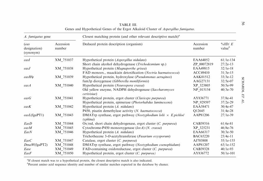
TABLE III.
Genes and Hypothetical Genes of the Ergot Alkaloid Cluster of Aspergillus fumigatus.
A. fumigatus gene Closest matching protein (and other relevant descriptive match)a
(eas
designation)
(synonym)
Accession
number
Deduced protein description (organism) Accession
number
%ID/ E
valueb
easI XM_751037 Hypothetical protein (Aspergillus nidulans) EAA64052 61/1e-134
Short chain alcohol dehydrogenase (Trichodesmium sp.) ZP_00072819 27/2e-13
easJ XM_751038 Hypothetical protein (Magnaporthe grisea) EAA49815 32/1e-18
FAD monoox., maackiain detoxification (Nectria haematococca) ACC49410 31/3e-15
easHc XM_751039 Hypothetical protein, hydroxylase (Pseudomonas aeruginos) AAK01512 33/3e-12
fum3p dioxygenase (Gibberella moniliformis) AAG27131 32/5e-07
easA XM_751040 Hypothetical protein (Neurospora crassa) XP_323805 50/5e-99
Old yellow enzyme, NADPH dehydrogenase (Saccharomyces
cerivisiae)
NP_015154 40/3e-70
easG XM_751041 Hypothetical protein, ergot cluster (Claviceps purpurea) AY836771 37/8e-41
Hypothetical protein, epimerase (Photorhabdus luminescens) NP_929597 37/2e-29
easK XM_751042 Hypothetical protein (A. nidulans) EAA58471 30/4e-47
P450, pisatin demethylase activity (N. haematococca) Q12645 31/4e-28
easL(fgaPT1) XM_751043 DMATrp synthase, ergot pathway (Neotyphodium lolii � E.pichloe
typhina)
AAP81206 27/1e-39
EasD XM_751044 Ox/red, short chain dehydrogenase, ergot cluster (C. purpurea) CAB39316 61/6e-81
easM XM_751045 Cytochrome-P450 monooxygenase (lovA) (N. crassa) XP_325231 46/8e-76
EasN XM_751046 Hypothetical protein (A. nidulans) EAA66317 30/3e-50
Trichothecene 3-O-acetyltransferase (Fusarium oxysporum) BAC65220 23/4e-11
EasC XM_751047 Catalase, ergot cluster (C. purpurea) AJ703808 55/1e-155
DmaW(fgaPT2) XM_751048 DMATrp synthase, ergot pathway (Neotyphodium coenophialum) AAP81207 63/1e-152
Ease XM_751049 FAD-containing oxidoreductase, ergot cluster (C. purpurea) CAB39328 40/1e-93
EasF XM_751050 Hypothetical protein, ergot cluster (C. purpurea) AY836772 50/1e-101
aIf closest match was to a hypothetical protein, the closest descriptive match is also indicated.bPercent amino acid sequence identity and number of similar matches expected in the database by chance.
SCHARDLETAL.
56

ERGOT ALKALOIDS 57
cluster contains additional genes that are presumed to catalyze steps unique to theA. fumigatus pathway (65). These genes and the reactions with which they are likelyassociated are described below.
B. ERGOT ALKALOID PRECURSORS
Although fermentation cultures had been established for the production ofEA, the first experiments to determine the precursors of these molecules wereconducted on rye ears infected with C. purpurea (66). The infected ears were injectedwith L-[14C] tryptophan, which was incorporated into clavine alkaloids. The sameresult was obtained with a Claviceps species fermentation culture, which incorpo-rated L-[14C] tryptophan into 6 (67).
Incorporation of label from [2–14C]mevalonate into the ergoline ring systemsupported the hypothesis that the pathway involved aromatic prenylation (68). Thiswas further supported by the identification and purification of tryptophandimethylallyltransferase (69,70), which transferred the dimethylallyl moiety fromdimethylallyl-diphosphate (DMAPP) to the 4–position on the six-membered ring ofL-tryptophan. Molecular genetic studies confirmed the role of this enzyme in EAbiosynthesis (39,54).
The final component of the clavine intermediates is the N(6)-linked methylgroup, derived from the S-methyl group of S-adenosylmethionine (AdoMet) (71).
The various ergopeptines that characterize many clavicipitaceous EAproducers, are derived from 1 and three hydrophobic L-amino acids, the identitiesof which vary among the ergopeptines (see Table I).
The dihydroclavine alkaloids (e.g., 10) that are produced by A. fumigatushave an unmodified C(17) methyl group, but may bear a hydroxyl or acetyl group atC(9), and a prenyl adduct at C(2).
C. THE PATHWAY-DETERMINANT STEP
1. dmaW and Aromatic Prenylation of Tryptophan
Prenylation of L-tryptophan is the determinant step for the pathway toclavines. The enzyme that catalyzes this determinant step is EC 2.5.1.34, DMAPP:L-tryptophan dimethylallyltransferase (4–g, g-dimethylallyltryptophan synthase ¼DMATrp synthase; Scheme 1). The enzyme was purified to homogeneity fromC. fusiformis SD58 (70), and shown to catalyze an electrophilic substitution, leadingto addition of the 4–(3–methylbut-2–enyl) moiety in an all-trans configurationon C(4) of the L-tryptophan aromatic ring to form DMATrp (12) (56). Althoughno requirement was evident for divalent cations, it is conceivable that the purifiedenzyme might contain a tightly bound ion, or that divalent cations may be allostericeffectors, based on the observation that addition of 4mM CaCl2 to the reactionmixture approximately doubled the Vmax. In the presence of 4mM CaCl2,kcat ¼ 0.44/s; KM ¼ 8.0mM for DMAPP, and KM ¼ 17mM for L-tryptophan wereobserved (70).
SCHARDL ET AL.58
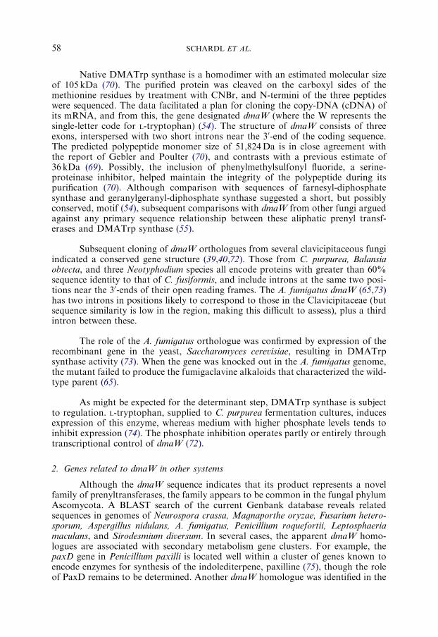
Native DMATrp synthase is a homodimer with an estimated molecular sizeof 105 kDa (70). The purified protein was cleaved on the carboxyl sides of themethionine residues by treatment with CNBr, and N-termini of the three peptideswere sequenced. The data facilitated a plan for cloning the copy-DNA (cDNA) ofits mRNA, and from this, the gene designated dmaW (where the W represents thesingle-letter code for L-tryptophan) (54). The structure of dmaW consists of threeexons, interspersed with two short introns near the 30-end of the coding sequence.The predicted polypeptide monomer size of 51,824Da is in close agreement withthe report of Gebler and Poulter (70), and contrasts with a previous estimate of36 kDa (69). Possibly, the inclusion of phenylmethylsulfonyl fluoride, a serine-proteinase inhibitor, helped maintain the integrity of the polypeptide during itspurification (70). Although comparison with sequences of farnesyl-diphosphatesynthase and geranylgeranyl-diphosphate synthase suggested a short, but possiblyconserved, motif (54), subsequent comparisons with dmaW from other fungi arguedagainst any primary sequence relationship between these aliphatic prenyl transf-erases and DMATrp synthase (55).
Subsequent cloning of dmaW orthologues from several clavicipitaceous fungiindicated a conserved gene structure (39,40,72). Those from C. purpurea, Balansiaobtecta, and three Neotyphodium species all encode proteins with greater than 60%sequence identity to that of C. fusiformis, and include introns at the same two posi-tions near the 30-ends of their open reading frames. The A. fumigatus dmaW (65,73)has two introns in positions likely to correspond to those in the Clavicipitaceae (butsequence similarity is low in the region, making this difficult to assess), plus a thirdintron between these.
The role of the A. fumigatus orthologue was confirmed by expression of therecombinant gene in the yeast, Saccharomyces cerevisiae, resulting in DMATrpsynthase activity (73). When the gene was knocked out in the A. fumigatus genome,the mutant failed to produce the fumigaclavine alkaloids that characterized the wild-type parent (65).
As might be expected for the determinant step, DMATrp synthase is subjectto regulation. L-tryptophan, supplied to C. purpurea fermentation cultures, inducesexpression of this enzyme, whereas medium with higher phosphate levels tends toinhibit expression (74). The phosphate inhibition operates partly or entirely throughtranscriptional control of dmaW (72).
2. Genes related to dmaW in other systems
Although the dmaW sequence indicates that its product represents a novelfamily of prenyltransferases, the family appears to be common in the fungal phylumAscomycota. A BLAST search of the current Genbank database reveals relatedsequences in genomes of Neurospora crassa, Magnaporthe oryzae, Fusarium hetero-sporum, Aspergillus nidulans, A. fumigatus, Penicillium roquefortii, Leptosphaeriamaculans, and Sirodesmium diversum. In several cases, the apparent dmaW homo-logues are associated with secondary metabolism gene clusters. For example, thepaxD gene in Penicillium paxilli is located well within a cluster of genes known toencode enzymes for synthesis of the indolediterpene, paxilline (75), though the roleof PaxD remains to be determined. Another dmaW homologue was identified in the

ERGOT ALKALOIDS 59
sirodesmin synthesis cluster of the plant-pathogenic fungus, L. maculans, and wasdesignated sirD (76). The most obvious role for the SirD protein is prenylation of thehydroxyl group of L-tyrosine either before or after its incorporation with L-serineinto a cyclopeptide.
The EA gene cluster of A. fumigatus (Fig. 5; Table III) contains both thefunctional orthologue of dmaW and a second dmaW homologue (easL) (65). TheeasL gene product catalyzes ‘‘reverse prenylation,’’ linking the clavine C(2) tothe prenyl C(3) rather than C(1), to form fumigaclavine C (10) (76a). Numeroussecondary metabolites are known with such reverse prenyl linkages. Thus, the dmaWfamily is surprisingly diverse in reaction specificities.
D. SYNTHESIS OF CHANOCLAVINE-I
Following prenylation of L-tryptophan to yield 12, the a-amine is methylatedby an N-methyltransferase dependent on AdoMet (Scheme 2) (71). Haarmann et al.(59) suggest that the gene here designated easF (Fig. 4; Table II) might encode theN-methyltransferase for this step. The N-methylation must occur after L-tryptophanprenylation, but before the C-ring closure, since dual-labeled [6–15N–C2H3]-13is incorporated intact into 6 (77). Furthermore, although norchanoclavines (whichlack the N-methyl group) are detectable in fermentation cultures, labeled nor-chanoclavines are not incorporated into 6 (77). Also, when C. purpurea andC. fusiformis dmaW cDNAs were expressed in yeast, the resulting activity catalyzedprenylation of L-tryptophan, but not N-methyl-L-tryptophan (40).
Formation of the chanoclavine isomers from 13 requires two oxidation steps,and Scheme 2 shows the mechanism proposed by Groger and Floss (10). The first
∗
HN
13
NHCH3
∗
HN
CO2H
14
NHCH3
HN
15
510
17
89
HN
H
16 chanoclavine-I
CO2H
NHCH3
∗
C
O
O
O H
HNHCH3
6
∗7OH
12
N-methyl-transferase
monooxygenaseor
oxidase
oxygenase(epoxidase) SN2'
CO2–
Scheme 2
SCHARDL ET AL.60
oxidation results in diene 14. Groger and Floss (10) suggest that this reaction mayproceed through an intermediate hydroxylated at the benzyl carbon, and note that10–hydroxy-13 is unstable and spontaneously dehydrates to form 14. However, thisinstability precluded feeding 10–hydroxy-13 to test the hypothesis. When culturesare fed deuterium-labeled 14, the label is detected in 16. Interestingly, if 12 is labeledin the methyl carbon trans to the vinyl hydrogen, the label that appears in 16 is cis tothe vinyl hydrogen, evidencing the first of two epimerization steps in the pathway.This is further supported by the observation that label from [2–14C]mevalonic acidincorporates into the methyl group trans to the vinyl hydrogen.
Diene 14 formation presents an obvious opportunity for the epimerization byrotation around the newly saturated C(8)–C(9) bond (Scheme 2). The orientationof this bond would likely be dictated by the active site of the next enzyme, which ispredicted to be an epoxidase. The mechanism proposed for C-ring formation isepoxidation of 14, followed by a possibly spontaneous SN2
0 reaction that couplesC(5)–C(10) bond formation with decarboxylation at C(5) (Scheme 2) (10).
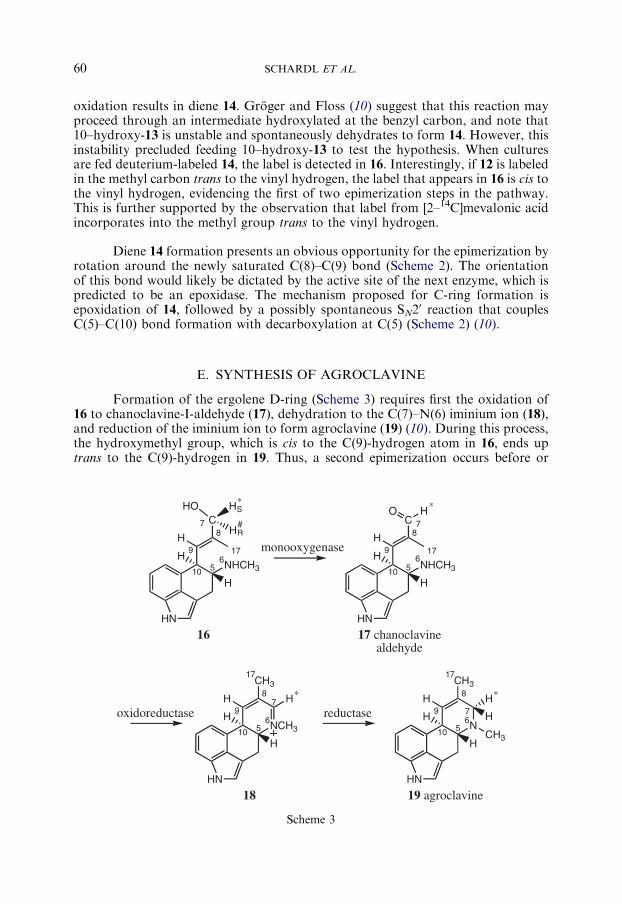
E. SYNTHESIS OF AGROCLAVINE
Formation of the ergolene D-ring (Scheme 3) requires first the oxidation of16 to chanoclavine-I-aldehyde (17), dehydration to the C(7)–N(6) iminium ion (18),and reduction of the iminium ion to form agroclavine (19) (10). During this process,the hydroxymethyl group, which is cis to the C(9)-hydrogen atom in 16, ends uptrans to the C(9)-hydrogen in 19. Thus, a second epimerization occurs before or
510NHCH3
617
8
9
HN
C7
H
16
HO HS
HR
∗
#
510NHCH3
617
8
9
HN
C 7
H
17 chanoclavinealdehyde
O H∗
H H
510NCH3
6
78
9
HN
CH317
H
18 19 agroclavine
H
H∗
510N6
7
8
9
HN
CH317
H
H
H∗
H
CH3
H H
HH
monooxygenase
oxidoreductase reductase
Scheme 3
ERGOT ALKALOIDS 61
during D-ring formation. Such an epimerization would be necessary to bring thealdehyde in proximity to N(6) to form the iminium ion 18.
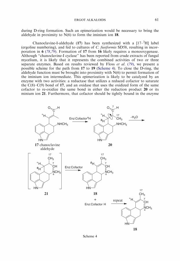
Chanoclavine-I-aldehyde (17) has been synthesized with a [17–3H] label(ergoline numbering), and fed to cultures of C. fusiformis SD58, resulting in incor-poration in 6 (78,79). Formation of 17 from 16 likely requires a monooxygenase.Although ‘‘chanoclavine-I cyclase’’ has been reported from crude extracts of fungalmycelium, it is likely that it represents the combined activities of two or threeseparate enzymes. Based on results reviewed by Floss et al. (79), we present apossible scheme for the path from 17 to 19 (Scheme 4). To close the D-ring, thealdehyde function must be brought into proximity with N(6) to permit formation ofthe iminium ion intermediate. This epimerization is likely to be catalyzed by anenzyme with two activities: a reductase that utilizes a reduced cofactor to saturatethe C(8)–C(9) bond of 17, and an oxidase that uses the oxidized form of the samecofactor to re-oxidize the same bond in either the reduction product 20 or itsminium ion 21. Furthermore, that cofactor should be tightly bound in the enzyme
NHCH3
CH3
17
HN
C7
20
O H
H∗Enz:Cofactor#H
NHCH3
HN
C
17 chanoclavinealdehyde
O H
H∗
H#–H2O
NCH3
CH7
HN
CH3
17
21
H
H# Enz:Cofactor
NCH3
CH7
HN
CH3
17
18
H#
+Enz:Cofactor H
repeat
NCH3
CH7
HN
CH3
17
H∗
18
∗
Scheme 4

SCHARDL ET AL.62
active site. This would account for the observation that when fermentation culturesare fed 16 labeled with 3H at C(9) (ergoline numbering), most, but not all, of thelabel is retained in the tetracyclic clavines (78,79). Furthermore, the reductionproduct 20 or 21 can be released from the active site during the catalytic cycle. Thiswas demonstrated by an elegant experiment in which a mixture of [2–13C]mevalon-ate and [4–2H]mevalonate was fed to fermentation cultures, giving dual-labeled 9and penniclavine, but not dual-labeled 16 (79). The implication is that the hydrogenat C(9) is extracted by the cofactor during re-oxidation of the C(8)–C(9) bond in 20or 21, which is then released without release of the reduced cofactor. This wouldallow a molecule of 17 to enter the active site and be reduced by that cofactor, thusacquiring the H-atom from the previous substrate molecule, and regenerating theoxidized cofactor in the active site of the enzyme.
A candidate enzyme for epimerization of 17 (Scheme 4) is the easA geneproduct, predicted to be similar to old yellow enzymes. These enzymes utilize tightlybound FMNH2 as a cofactor and commonly reduce C–C double bonds conjugatedwith, and trans to, carbonyl groups (80). Different enzymes utilize different subst-rates to re-reduce the enzyme-bound FMN. During epimerization of 17 the reducedproduct 20 or 21must, in turn, reduce the cofactor. It is noteworthy that retention oflabel from [9–3H]-16 (ergolene ring numbering) is incomplete, and the proportion oflabel retained in that position depends on the efficiency of 19 formation (79). As apossible explanation for this finding, we speculate that at each cycle the FMNcofactor may be re-reduced either by its reduction product (20 or 21) or by anotherreducing agent such as NADPH. Alternatively, the epimerization might be carriedout by an enzyme that utilizes NAD(P)H for the reduction step, and the corre-sponding oxidized cofactor for product re-oxidation. If so, the oxidized cofactorshould be exchanged slowly in the active site.
The product of the epimerization and loss of a water molecule would formiminium ion 18, and cyclization would be completed by reduction of 18 (Scheme 3).This must require a separate reductase from the one involved in epimerization of17, or else the aforementioned pattern of H(9) atom retention would be highlyunlikely. Thus, we predict that three distinct enzymes are likely to be involved in the‘‘chanoclavine-I-cyclase’’ step from 16 to 19.
The ‘‘chanoclavine cyclase’’ activity is dependent on NADH or NADPH,Mg2+, and ATP (10). The scheme we propose here is consistent with an NAD(P)Hrequirement for the third enzyme (reductase). Also, we expect the oxidized form,NADP or NAD+, to oxidize 16 to aldehyde 17. Furthermore, and as stated above, itis possible that NAD(P)H is involved in the oxidation and reduction reactions togenerate iminium ion 21. However, it is not obvious why there is a requirementfor ATP. Clearly, elucidation of the mechanisms underlying the steps in D-ringformation will continue to reveal some interesting biochemistry.
F. SYNTHESIS OF ELYMOCLAVINE AND LYSERGIC ACID
Oxidation of C(17) of 19 generates 6, and further oxidation steps lead topaspalic acid (22), the D8,9-isomer of 1 (Scheme 5). In cell-free extracts, the micro-somal (membrane) fraction contains oxygenases for conversion of 19 into 6, and

NCH3
HN
CH3
H
19 agroclavine
NCH3
HN
H
6 elymoclavine
H H
NCH3
HN
H
22 paspalic acid 1 lysergic acid
H
HN
H
Cyt-P450
isomeraseor
spontaneous
Cyt-P450(CloA)
CH2OH
CO2H
NCH3
H CO2H
Scheme 5
ERGOT ALKALOIDS 63
conversion of 6 into 22 (81). These activities are NADPH-dependent, and inhibitorstudies strongly suggest they are catalyzed by cytochrome-P450. Given that con-version of 6 into 22 is a total of a 4–electron oxidation, two sequential mon-ooxygenase reactions by either the same enzyme or two different enzymes should beinvolved. It is also possible that a single enzyme catalyzes oxygenations of both 19and 6, but the observation that in some isolates of Claviceps species these twoactivities differ in the kinetics of expression suggests that distinct enzymes catalyzethese conversions (81).
In the eas gene cluster of C. purpurea, one gene (cloA) has been identified withthe heme-binding motif similar to cytochrome-P450. When this gene was knockedout in C. purpurea strain P1, the mutant produced 6 and 19, but no 1, lysergylamides, or ergopeptines (62). Thus, CloA almost certainly catalyzes one or bothmonooxygenation reactions leading from 6 to 22 (Scheme 5). The observation thatconsiderably more 19 than 6 accumulates in cultures of the mutant is in keeping withearlier findings that the agroclavine monooxygenase is inhibited by its product (81).
The enzyme responsible for the 2–electron oxidation of 19 to 6 remains to beidentified. One possibility is that CloA catalyzes this reaction as well. The differencesin induction profiles of agroclavine monooxygenase and elymoclavine mon-ooxygenase activities (81), and the phenotype of the cloA knockout (62), suggestthat there is a separate agroclavine monooxygenase, but do not exclude thepossibility that CloA, as well as another monooxygenase, can act on 19 (as weconsider below in discussing the C. fusiformis eas cluster). It is also conceivable thata second pathway to 6 is responsible for the accumulation of 6 in the mutant.Previous reports (reviewed by Groger and Floss (10)) indicate that a microsomalfraction can hydroxylate 16, and feeding experiments indicate that 16 hydroxylated
SCHARDL ET AL.64
on either allylic methyl group and labeled in the other allylic methyl carbon, can beconverted into 6 by fermentation cultures. However, the labeled carbon becomesC(7) in 6, in contrast to the specific labeling pattern in chanoclavine-I feeding ex-periments cited above. Therefore, the possibility seems remote that this alternativepathway to 6 operates in vivo. Rather, the accumulation of 6 in the DcloA mutant isprobably due to an agroclavine monooxygenase for which a candidate gene is notapparent in the known cluster.
The final step in the synthesis of 1 from 22 is isomerization from D8,9 to D9,10.This may be catalyzed by a distinct enzyme, or occur spontaneously as observed inaqueous solutions of 22 (10). The prevalence of the 8(b)-diasteroisomer of 1 and itsamides over the 8(a)-diastereoisomers suggests a directed, enzymatic conversion, butmight alternatively be explained by selectivity of the lysergyl-peptide synthetase(LPS) discussed below.
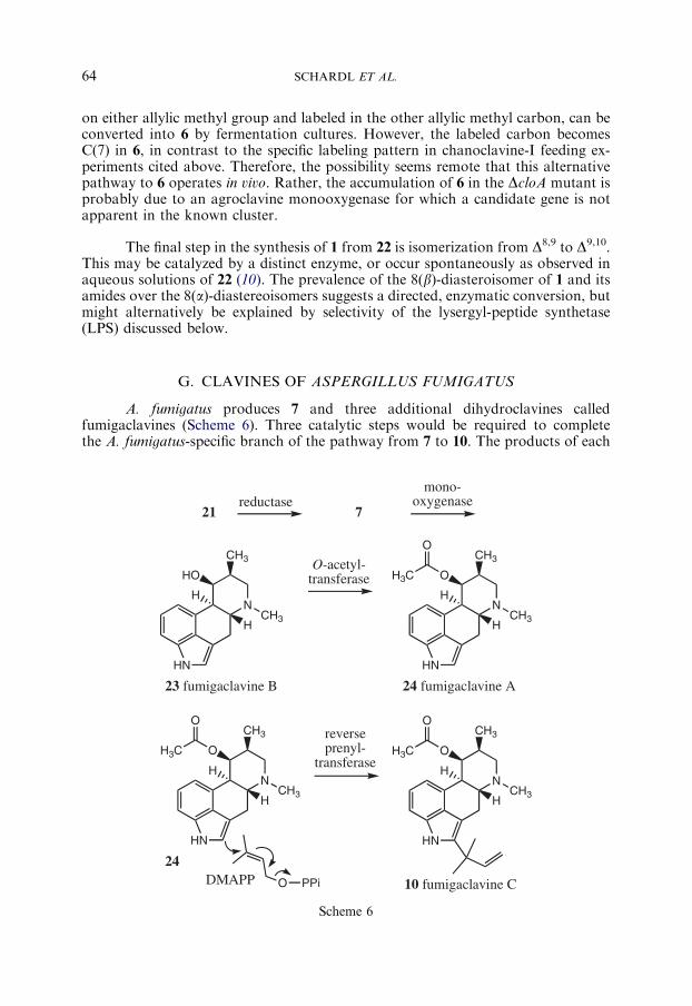
G. CLAVINES OF ASPERGILLUS FUMIGATUS
A. fumigatus produces 7 and three additional dihydroclavines calledfumigaclavines (Scheme 6). Three catalytic steps would be required to completethe A. fumigatus-specific branch of the pathway from 7 to 10. The products of each
21
N
HN
HCH3
CH3
H
23 fumigaclavine B
HO
N
HN
HCH3
CH3
H
24 fumigaclavine A
OH3C
O
N
HN
HCH3
CH3
H
24
OH3C
O
ODMAPP
N
HN
HCH3
CH3
H
OH3C
O
10 fumigaclavine C
reductasemono-
oxygenase
O-acetyl-transferase
reverseprenyl-
transferase
7
PPi
Scheme 6
ERGOT ALKALOIDS 65
of these steps accumulate to easily detectable levels in conidia of the fungus (52),indicating that the pathway is relatively inefficient in converting the product ofthe preceding step. Festuclavine (7) is hydroxylated at C(8) to yield fumigaclavine B(23). The eas gene cluster of A. fumigatus contains three genes that are candidatesto control this step (65): easJ has the capacity to encode an FAD-containingmonooxygenase, and easK and easM appear to encode cytochrome-P450monooxygenases. All three of these oxidases lack orthologues in the C. purpurea andC. fusiformis clusters.
Acetylation of 23 to fumigaclavine A (24) is hypothesized to be catalyzed bythe product of easN, which has high amino-acid-sequence identity with O-acetyltransferases (65). No orthologue of this gene is found in the eas clusters of theClaviceps spp. Moreover, there is no other acetylation step in any part of the ergot-alkaloid pathway or its shunts.
Fumigaclavine C (10) is the product of reverse prenylation of 24 (Scheme 6).In this ultimate product of the A. fumigatus branch, the prenyl group is attached tothe ergoline ring system in a ‘‘reversed’’ orientation relative to the product ofa ‘‘normal’’ prenylation. In a normal prenylation, such as the prenylation of try-ptophan by DMATrp synthase (Scheme 1), the C(1) that is initially attached tothe pyrophosphate group of DMAPP is presented to the indole co-substrate. Incontrast, reverse prenylation is hypothesized to proceed with DMAPP presented in a‘‘reverse’’ orientation relative to the indole cosubstrate promoting a facially non-selective SN
0 attack on the olefinic p electron system of C(3) of DMAPP (82,83). Themechanism of reverse prenylation has experimental support from the labeling stud-ies of Stocking et al. (82). Interestingly, the A. fumigatus ergot alkaloid clustercontains a second dmaW-like gene, easL, encoding a product with 25% amino acidsequence identity to dmaW of A. fumigatus (65). The low, but significant, sequenceidentity with the normal prenyl transferase, coupled with its location in the ergotalkaloid gene cluster, makes this gene an excellent candidate to catalyze the reverseprenylation of fumigaclavine A to fumigaclavine C. This possibility has recentlyreceived experimental confirmation (76a).
The proposed sequence of events in which 23 is acetylated to 24, followed byreverse prenylation to 10, is supported by two observations. The first is the existenceof an acetylated, but nonprenylated, fumigaclavine (24) and the lack of a prenylated,but not acetylated, fumigaclavine intermediate. Fumigaclavine C (10) can beexperimentally deacetylated to yield such a compound, but this compound has notbeen detected in cultures of A. fumigatus (52). The second observation is simply that10 is the most abundant ergot alkaloid in A. fumigatus (52), consistent with itsposition as the ultimate and inconvertible product of the pathway.
H. COMMON STEPS AND GENES IN CLAVINE-ALKALOID PRODUCERS
Relationships among genes in different fungi capable of producing clavinesprovide clues to eas gene functions. C. purpurea P1 produces 6 as an intermediate to1, lysergic acid amides and ergopeptines, C. fusiformis SD58 accumulates 6 as thepathway end product, and A. fumigatus WVU1943 produces 7 (dihydro-19) andfumigaclavines. Functional genes shared between the Claviceps species are easA,

SCHARDL ET AL.66
cloA, easC, easD, easE, easF, easG, and dmaW. It is unknown whether C. fusiformishas an easH gene. Those genes shared between the Claviceps species and A. fumigatusare easA, easC, easD, easE, easF, easG, and dmaW. An easH-homologous sequenceappears to be present in A. fumigatus as a pseudogene.
Given the C. fusiformis chemotype, and the recently established role of cloA(62) in conversion of 6 into 22, the presence of a C. fusiformis cloA is surprising.Possibly, the C. fusiformis cloA product is nonfunctional. Alternatively, it isconceivable that the C. fusiformis cloA encodes a specific agroclavine monooxygenase.This remains an intriguing possibility considering that it has not been excludedthat C. purpurea CloA might have agroclavine monooxygenase activity as well aselymoclavine monooxygenase activity.
The eight biosynthetic steps apparently shared between the Claviceps speciesare DMATrp (12) synthase (encoded by dmaW), methylation of 12 to 13, oxidationof 13 to 14, oxygenation (epoxidation) to give 16 via 15, oxidation or oxygenation of16 to 17, the single-enzyme reduction and oxidation reactions to 18 (possibly by oldyellow enzyme encoded by easA), reduction to 19, and oxygenation to 6. It is notclear that the eight-shared genes in the eas clusters correspond precisely to theseeight steps. Two shared genes appear problematic in this regard: cloA (for reasonsdiscussed above) and easC, predicted to encode a catalase.
Catalases are best known for disproportionating H2O2 to H2O and O2;however, they can also use H2O2 to oxidize other substrates (84). Furthermore,flavine-utilizing enzymes can generate peroxides during oxidation reactions. So, wespeculate that two successive oxidation/oxygenation reactions may be catalyzed by aFAD-containing enzyme (perhaps EasE) and the catalase, EasC. The obviouscandidates would be the two steps from 13 to 16 (Scheme 2), or the two steps from14 to 17 (65) (Scheme 3). Alternatively, the EasC catalase might detoxify peroxideproduced during an FAD-dependent oxidation reaction (64).
Assuming that EasC catalyzes one of the oxygenations, the five genes en-coding redox enzymes and known to be shared between the Claviceps species may besufficient for all redox steps needed to produce 6. However, if EasC does not actdirectly on a clavine intermediate, these five genes would appear to be insufficient. Itis possible that a monooxygenase may catalyze multiple steps. For example, asargued earlier, CloA might catalyze oxygenation of both 19 and 6 (62). It must alsobe considered that some cluster genes may remain to be discovered, and that somesteps might be carried out by enzymes encoded by genes outside of the cluster.Finally, EasH1, predicted to be a homologue of the fum3p hydroxylase in fumonisinbiosynthesis (85), may catalyze an oxygenation in the clavine pathway. Identificationof a homologue in C. fusiformis would provide evidence for this possibility. Sig-nature sequences for a nonheme-iron oxygenase are evident in the predicted EasH1,but not EasH2 (which may be nonfunctional). The EasH step should occur after thepoint of divergence of the clavine pathway in Claviceps species from the festuclavine/fumigaclavine pathway in A. fumigatus, since the latter appears to lack a functionaleasH/fum3 homologue. Therefore, we predict that EasH1 catalyzes either hydro-xylation of 19 to 6, or hydroxylation of ergopeptide lactams to form ergopeptines(discussed later).

ERGOT ALKALOIDS 67
Interestingly, except for cloA, the set of functional genes shared betweenC. purpurea and C. fusiformis is also shared with A. fumigatus (Fig. 5) (65). Wespeculate that the divergence point between the fumigaclavine pathway and thepathways in Claviceps species is marked either by 21 or 19, intermediates that couldbe reduced to festuclavine (7). If the divergence point is at iminium ion 21, thiscompound would not be re-oxidized in A. fumigatus as predicted to occur in theClaviceps species. Instead, it would be released from the enzyme in its reduced form,and its further reduction would yield 7. If the divergence point is at agroclavine (19),then reduction of 19 would yield 7. Further modification of 7 would producefumigaclavines, and the genes likely to carry out those reactions are discussed above.C. africana might have the same pathway to 7, which would then be used analo-gously to 19 in production of dihydrolysergic acid and subsequent dihydroergotalkaloids that characterize this species (20,86).
I. ERGOPEPTINES AND OTHER LYSERGIC ACID AMIDES
1. Ergopeptines in Claviceps Species
The amides of D-lysergic acid show a wide range of pharmacological effects,depending on the amide substituents, and different EA may act as agonists orantagonists of the various neurotransmitters in mammals. An extremely importantgroup of D-lysergic acid amides are the ergopeptines, in which a tripeptide chain,modified to form a bicyclic cyclol-lactam structure, is joined with 1. The two firstamino acids in the tripeptide are variable, but always nonpolar, whereas the thirdposition is taken by L-proline in all known ergopeptines except ergobalansine (seeTable I). Two intramolecular reactions result in the unusual cyclol-lactam structure(Scheme 7). Amino acid III forms a lactam bond to the nitrogen atom of amino acidII. Subsequently, an a-hydroxy group is added to amino acid I and forms a cyclolbridge with the carbonyl carbon of amino acid III (87). Besides the ergopeptines,there are simpler derivatives of 1, whose possible origins are discussed later.
Biochemical analyses of the assembly of the ergopeptines in C. purpurea haveshown that ergopeptines are the products of an enzyme complex consisting of twononribosomal peptide synthetase (NRPS) subunits (58). NRPSs generally exhibitmodular structures, with each module responsible for the addition of an aminoacid or other substituent. A typical module includes an adenylation (A-) domain, athiolation (T-) domain (also known as a peptidyl carrier protein domain), and acondensation (C-) domain. The A-domain specifies the amino acid or other car-boxylic acid substituent, and activates by it by an ATP-dependent adenylationreaction. The activated substituent then forms a thioester with the 40-phosphopan-tetheine prosthetic group in the adjacent T-domain. Finally, the C-domain linksthe substituent to the next substituent in the chain. In a multimodular NRPS pro-tein, the order in which substituents are added corresponds to the arrangement ofmodules from its N- to C terminus.
The two subunits of LPS—LPS 1 and LPS 2—have sizes of 370 and 140kDa,respectively. Together they bind D-lysergic acid (1) and the three L-amino acids of thepeptide portion of the alkaloid cyclopeptide as thioesters, catalyzing their successivecondensation into the D-lysergyl mono-, di-, and tripeptide thioester intermediates,

N
HN
CH3
C
H
SO
N
HN
CH3
C
H
O
S
NH
H3CO
S
N
HN
CH3
C
H
O
HN
NH
H3CO
O
N
N
HN
CH3
C
H
O
HN
NH
H3CO
O
S
O
N
HN
CH3
C
H
OHN
NN
OO
H3C
N
HN
CH3
C
H
OHN
O
NN
O
OH
O
H3CO
HN
NO
H3COOH
D-LA T C L-Ala L-Phe L-Pro CcycT C T C T
mono-oxygenasespontaneous
LPS2 LPS1
ergotamam4 ergotamine
Scheme 7
SCHARDLETAL.
68

ERGOT ALKALOIDS 69
and culminating in cyclization and release of the product D-lysergyltripeptide lactam(Scheme 7) (58). The monomodular subunit, LPS 2, activates 1, and binds it as athioester (61). LPS 2 also contains a C-domain near its C- terminus, which is pre-sumed to catalyze the formation of the peptide bond between 1 and the amino acid inposition I of what will be the tripeptide moiety of the ergopeptine. The remainingmodules, with A-, T-, and C-domains for the three amino acids of the ergopeptine,are found in LPS 1 (55,58,59). The final C-domain catalyzes cyclization betweenamino acids at positions II and III, resulting in release of the lysergyl-peptide lactamfrom the enzyme complex.
The eas gene cluster of C. purpurea strain P1, stretching over ca. 68 kb,contains three characterized NRPS genes, designated lpsA1 (formerly cpps1), lpsA2(formerly cpps4), and lpsB (formerly cpps2) (55,61,62). The cluster also contains aputative fourth NRPS gene tentatively designated lpsC (formerly cpps3). The lpsA1and lpsA2 genes encode related trimodular proteins, each of which is a variant LPS1 subunit. The lpsB gene encodes the monomodular LPS 2 subunit, and lpsC ispredicted to encode another monomodular enzyme with a reduction domain in placeof the condensation domain.
Comparison of the deduced amino acid sequence of lpsA1 with a partialamino acid sequence of purified LPS 1 revealed matches (with a few exceptionsdiscussed below) that indicated that lpsA1 encodes the predominant LPS 1 sub-unit. Analysis of the deduced amino-acid sequence of LpsA1 confirmed that itharbors three modules, each responsible for the recruitment of an amino acid ofthe tripeptide substituent (Scheme 7) (55). Also, sequence data have demonstratedthat both LPS 1 and LPS 2 are separately encoded enzymes and not breakdownproducts of a much larger NRPS enzyme (55,61). In contrast to the more com-mon arrangement of catalytic domains in NRPS elongation modules, the firstmodule of LPS 1 lacks an N-terminal C-domain. This suggests that the C-domaincatalyzing formation of the peptide bond between 1 and the first amino acid ofthe tripeptide chain could be associated with the initiation module LPS 2(a less likely possibility is that this is a stand-alone condensation domain yet to beidentified). The size of the native LPS complex has been estimated to be between500 and 550 kDa, roughly the sum of four modules contained within the two LPSsubunits.
Analysis of the deduced amino-acid sequence of the lpsB product revealedthat the protein comprises an A-domain, a T-domain, and a C-terminal C-domain(61). The predicted molecular mass of the protein is 140 kDa (1308 amino acids), inaccordance with the size previously estimated for the LPS 2 subunit (58). Mostimportantly, the lpsB gene product carries at its C-terminus a typical C-domainthat is almost certainly responsible for peptide bond formation between D-lysergicacid and the first amino acid of the tripeptide moiety of the ergopeptine. Such aC-domain was surprisingly not found at the N-terminus of LPS 1. This domainarrangement (A–T–C) is unusual in NRPS systems because C-domains usually lie onthe N-terminal side of their corresponding A- and T-domains. This placement mayadd to their substrate specificity, which has been reported to be high for the subst-rate bound to the downstream T-domain, and low for the substrate bound to theupstream module (88). Thus, many elongation subunits in NRPSs start at theirN-terminal ends with a C-domain. In the case of LPS 2, the location of the

SCHARDL ET AL.70
C-domain at the C-terminal end of the protein may add to the extremely highspecificity of the A-domain for D-lysergic acid (58), as measured with LPS 2.
Functional analysis in C. purpurea P1 has provided unequivocal evidence thatlpsB encodes the LPS 2 subunit (61). Gene replacement of lpsB created an ergo-peptine-nonproducing phenotype, which resulted in accumulation of 1 instead ofergotamine (4) and other ergopeptines. This biochemical phenotype is consistentwith a block in the assembly of these complex alkaloid peptides. Comparativeenzymatic studies of protein extracts derived from the DlpsB mutant and its parentstrain showed that the parent contained both LPS 1 and LPS 2, whereas extractsfrom the mutant were devoid of any LPS 2–related activity. Furthermore, recom-binant lpsB product expressed in the bacterium, Escherichia coli, catalyzed D-lysergicacid-dependent ATP-pyrophosphate exchange, demonstrating the specificity of themodule for D-lysergic acid.
The putative D-lysergic acid-binding pocket of LPS 2 is similar to pocketsfrom various A-domains that activate hydrophobic amino acids (61). The bestmatch is with the p-hydroxyphenylglycine-activating module of chloroeremomycinsynthetase. Interestingly, no similarity is seen with known tryptophan-activatingA-domains, suggesting that the indole portion of D-lysergic acid is not responsiblefor its recognition by LPS 2.
The distribution of four modules for ergopeptine assembly between twopolypeptides is unique among the known eukaryotic NRPSs, which otherwise con-tain all modules on one polypeptide chain. Although the significance of their in-dependence is as yet unclear, the stand-alone D-lysergic acid module may provide anadvantage to the fungus to use that module in combination with other NRPSs forother lysergyl-peptides such as ergonovine (3) (Scheme 8). In fact, a number ofC. purpurea strains produce 3 as well as ergopeptines. Precursors of 3 are 1 andL-alanine (89), which would require an NRPS with a D-lysergic acid module and an
N
HN
CH3
C
H
OHN CH3
O S
D-LA T C L-Ala T R
LPS2 LPS/Reductase
N
HN
CH3
C
H
O S
3
Scheme 8

ERGOT ALKALOIDS 71
L-alanine module, along with additional reductase and releasing domains. Possibly,the LPS 2 subunit sometimes associates with LPS 1 and sometimes with a differentNRPS subunit containing an L-alanine module, thereby serving both syntheticprocesses in a natural combinatorial biosynthesis (Scheme 8). One candidate for theL-alanine-activating module would be the product of lpsC, for which functionalanalysis is in progress.
The theme of combinatorial biosynthesis is also apparent in the presence ofanother peptide-synthetase gene in the eas cluster (59). The lpsA2 gene lies nearby,and is highly similar to lpsA1, implying a duplication in the origin of these twogenes. The function of the trimodular enzyme encoded by lpsA2 has not yet beendemonstrated. However, comparison of the A-domain substrate-binding pocketsstrongly suggests that lpsA2 encodes a version of LPS 1 with a variant specificity.The lpsA2 product, in complex with LPS 2, could catalyze formation of ergocryp-tine, the second ergopeptine present in fermentation cultures of C. purpurea P1.Evaluation of the partial amino acid sequences from LPS 1 preparations showedthat the few sequences not matching with the lpsA1-derived sequence matched thelpsA2-derived sequence, indicating that the enzyme preparations contained bothLPS 1 variants. Analysis of the putative lpsA1 and lpsA2 orthologues in a differentchemical race (producing ergocristine as a major ergopeptine) indicated that thechemical races differ in the specificity of the LPS 1 modules (59).
2. Ergopeptines and Lysergic Acid Amides of Epichloe Endophytes
Several epichloe endophytes of Lolium spp. grasses, as well as endophytes ofseveral other cool-season grasses, produce 8 (90,91), a member of the ergotaminegroup composed of D-lysergic acid, L-alanine, L-valine, and L-proline (Table I).Neotyphodium coenophialum also produces small amounts of dehydroergovaline,which has a mass that is 2 atomic mass units lower than that of ergovaline, pre-sumably as a result of desaturation in either the valine or proline substituents (43).Reports of other ergopeptines from epichloe endophytes may be due to extractionsfrom endophyte-infected grass seeds that were contaminated with sclerotia or hon-eydew of C. purpurea (43).
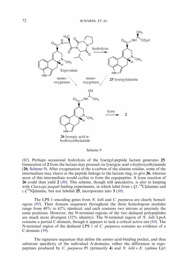
Ergovaline (8) is produced in N. lolii�E. typhina Lp1 by an LPS complexvery similar to the C. purpurea LPS described above. Functional analysis of theepichloe lpsA gene has been conducted by gene knockout, using gene sequences fromN. lolii to direct the recombination event (60). Perennial ryegrass plants symbioticwith the DlpsA mutant lacked 8, as well as two simpler amides of lysergic acid, 2 andlysergylalanine (25; Scheme 9), alkaloids that were all present in plants with the wild-type strain (46). The structural characterization of N. lolii lpsB also indicates that, asin C. purpurea, the D-lysergic acid-activating LPS 2 subunit is encoded separately. Inkeeping with this model, Epichloe festucae (the sexual ancestor of N. lolii) contains agene homologous to the LPS 2–encoding gene from C. purpurea (D. Fleetwood, A.Tanaka, B. Scott, and R. Johnson, personal communication).
The chemical profile of the DlpsAmutant (46) indicates that accumulation of 2and 25 requires the activity or products of the same LPS complex that generates theergopeptine. However, it is unlikely that 2 and 25 are direct products of LPS, sinceNRPSs require specific domains to catalyze release of covalently bound intermediates
N
HN
CH3
C
H
O NH
25 lysergylalanine
CH3
CO2H
26 lysergic acid α-hydroxyethylamide
lyase
mono-oxygenase
N
HN
CH3
C
H
OHN
NN
OO
H3CO
Ergovalam
hydrolysis
2
mono-oxygenase
CO NH CH3
OH
N
HN
CH3H
Scheme 9
SCHARDL ET AL.72
(92). Perhaps occasional hydrolysis of the lysergyl-peptide lactam generates 25.Generation of 2 from the lactam may proceed via lysergyic acid a-hydroxyethylamide(26; Scheme 9). After oxygenation at the a-carbon of the alanine residue, some of theintermediate may cleave at the peptide linkage to the lactam ring, to give 26, whereasmost of this intermediate would cyclize to form the ergopeptine. A lyase reaction of26 could then yield 2 (30). This scheme, though still speculative, is also in keepingwith Claviceps paspali feeding experiments, in which label from L-[2–14C]alanine andL-[15N]alanine, but not labeled 25, incorporates into 3 (10).
The LPS 1–encoding genes from N. lolii and C. purpurea are clearly homol-ogous (93). Their domain sequences throughout the three homologous modulesrange from 49% to 62% identical, and each contains two introns at precisely thesame positions. However, the N-terminal regions of the two deduced polypeptidesare much more divergent (32% identity). The N-terminal region of N. lolii LpsAcontains a partial C-domain, though it appears to lack a critical active site (93). TheN-terminal region of the deduced LPS 1 of C. purpurea contains no evidence of aC-domain (59).
The signature sequences that define the amino acid-binding pocket, and thussubstrate specificity of the individual A-domains, reflect the differences in ergo-peptines produced by C. purpurea P1 (primarily 4) and N. lolii�E. typhina Lp1

ERGOT ALKALOIDS 73
(exclusively 8) (93). The signature sequences for the first and third A-domains ofthe deduced LPS 1 products are nearly identical between these two fungi. Among the10 amino acids lining the amino acid-binding pocket, nine are identical in each ofmodule I (specifying L-alanine) and module III (specifying L-proline). However, thesignature sequences for the second A-domain differ at five of the 10 sites betweenthe corresponding sequences from the two fungi. These data, together with theaforementioned comparison of lpsA1 and lpsA2 in C. purpurea P1, indicate a stronggenetic component to the determination of ergopeptine composition.
J. PATHWAY INTERMEDIATES, SHUNTS AND SPURS
Typically, EA-producing fungi build up substantial levels of intermediates inaddition to pathway end products. For example, quantitative assays indicated thatA. fumigatus conidia possessed 7, 23 and 24 at a combined molar concentration thatwas 25% that of 10 (20), and sclerotia of C. africana had intermediates 7 anddihydrolysergol (6b) at a combined molar concentration 49% that of dihydroergosine(86). Most dramatically, in leaf blades of ryegrass symbiotic with N. lolii�E. typhinaLp1, chanoclavines were 2.5–fold more abundant than 8 on a molar basis (20).
An EA pathway intermediate in one fungus may be an endpoint in another.For example, 6 is an intermediate in C. purpurea P1, but an end product inC. fusiformis SD58. Also, shunts provide multiple end products, such as the pro-duction of 2, 3, 25, and 6,7–secolysergine (28; Fig. 6), in addition to 8, by the epichloeendophyte (46). Two tricyclic clavines, 28 and its isomer, 6,7–secoagroclavine (29)appear to be reduced forms of 16 or 19.
Interestingly, knockout of epichloe lpsA, which controls the penultimate stepin the pathway to 8, affected the profile of alkaloids from earlier stages of thepathway and a shunt pathway (20,46). Perennial ryegrass with the DlpsA mutantaccumulated elevated concentrations of 1, which is the pathway intermediate priorto the LPS step. However, the amount observed to accumulate was only 13% of theamount incorporated into D-lysergic acid derivatives in perennial ryegrass contain-ing the wild-type endophyte. The amounts of 16 and setoclavine (27; Scheme 10)produced by the mutant did not differ significantly from those produced by the wild-type strain. However, in grass containing the DlpsA mutant the amount of shunt
NHCH3
HN
H
28 6,7-secolysergine
CH3
CH3
NHCH3
HN
H
29 6,7-secoagroclavine
CH3
CH3
H
Fig. 6. Secoclavines.

NHCH3
HN
H
30 chanoclavine II
CH3
CH2OH
15H
N
HN
H
27 setoclavine
19 N
HN
H
33 penniclavine
6
CH3HO CH2OHHO
peroxidase
HN32 clavicipitic acids
12 NH
CO2H
H
N
HN
H
31 D-isolysergic acid
1 or 22
HHO2C
peroxidase
H
CH3
CH3CH3
mono-oxygenase?
Scheme 10
SCHARDL ET AL.74
product 28 increased approximately two-fold to levels that actually exceeded theamount of 1, the substrate for LPS (46). These data indicate that expression oractivity of several upstream enzymes in the ergot-alkaloid pathway were affected bythe DlpsA knockout, perhaps by negative feedback from the accumulated 1.
Pathway shunts and the apparently well regulated, but inefficient, fluxthrough intermediates to end products may be selectively favorable because of thevarious biological activities of the different clavine, lysergyl, and ergopeptine alka-loids. Evidence pertinent to this hypothesis is reviewed by Panaccione (20).
Incomplete substrate specificity or stereoselectivity of enzyme reactionsappears to be responsible for several spur products, examples of which are shownin Scheme 10. Although different chanoclavine isomers are produced (10), the ster-eochemistry of the tetracyclic clavines and 1 implies that chanoclavine I (16), but notchanoclavine II (30), is an intermediate. Also, 22 can isomerize spontaneously to 1or isolysergic acid (31). Typically, both C(8) diastereomers of ergopeptines areobtained in preparations, and those with an isolysergic acid moiety – which are

ERGOT ALKALOIDS 75
pharmacologically inactive (6) – are specified by the ‘‘inine’’ ending (e.g., ergot-aminine, ergovalinine, etc.). Isomerization at C(8), can occur in aqueous solutions,so it is generally thought that 1, and not 31, predominates or is selectively incor-porated into ergopeptides by the LPS, and that the ‘‘inine’’ isomers are purificationartifacts (94).
Oxidation or hydroxylation of C(10) of 13 is involved in the synthesis of 16.If the enzyme for this reaction has a low level of activity on 12, the productmay rearrange to form clavicipitic acids (32), which are present in small amounts(Scheme 10) (95,96). More abundant spur products, setoclavine (33) and pennicla-vine (33) (plus their C(8) stereoisomers), arise by peroxidase action on 19 and 6,respectively. In the case of grass-epichloe symbiota, both plant and fungal per-oxidases can catalyze those conversions (46).
V. Routine Analytical Methods for Ergot Alkaloids
Analytical methods for studying EA have changed over the years inresponse to changes in technology. This topic was reviewed comprehensively byFlieger et al. (97), so here we will only briefly provide our perspective on routineanalyses of EA, with particular emphasis on notable changes over the past severalyears.
Historically, EA have been isolated via repeated alternating extractions withan alkaline organic solvent followed by an acidic aqueous solvent. This classicalprocedure exploits the solubility of alkaloids in both the solvents, whereas mostother molecules lack solubility in one of the solvents and are thereby separated fromthe alkaloids.
One of the earliest methods for separating EA from each other was toseparate water-soluble forms (e.g., 3 and several clavine alkaloids) from water-insoluble forms (primarily ergopeptines). However, the solubility differences are notabsolute. Alternatively, ergopeptine alkaloids have been separated from other EA bytheir preferential retention on flash chromatography columns packed with silicacontaining HL binder (e.g., Ergosil) (98). Surprisingly, Ergosil binds ergopeptines,but not their 8(a) stereoisomers (ergopeptinines).
A more recent approach has been to simplify the extraction procedure andrely on extended chromatographic separation to obtain a complete EA profile in asingle step. An extraction solvent composed of 50% 2–propanol+1% lactic acid canrecover EA of a wide spectrum of polarities, from the extremely polar lysergicacid, to moderately polar clavines or simple amides of lysergic acid, to the nonpolarergopeptines (46,52,99). Alkaloids of varying polarity may then be separated byreverse phase HPLC by application of a gradual gradient beginning with a mostlyaqueous solvent (such as 5% acetonitrile in 50mM ammonium acetate) to a non-polar solvent (such as 75% acetonitrile in 50mM ammonium acetate).
Following chromatographic separation, EA are routinely detected with greatsensitivity by fluorescence. Those alkaloids with a D9,10-double bond can be effec-tively detected with an excitation wavelength of 310 nm and an emission wavelength

SCHARDL ET AL.76
of 410 nm. EA with a D8,9-double bond and those with a saturated D-ring fluorescemaximally with excitation and emission wavelengths of 272 and 372 nm, respec-tively. Thus extracts that contain EA with a combination of these fluorescenceproperties must be analyzed twice to detect and quantify a complete profile. Alter-natively, two fluorescence detectors connected in series allow for analysis of the fullergot-alkaloid profile in a single HPLC run (20).
Inclusion of a mass spectrum (MS) detector in the analytical system providesmass information for the eluted alkaloids to confirm their identities. In many cases,EA can be fragmented in such detectors and the masses of the fragments facilitateidentification of alkaloids (1,43,46). Moreover, MS detectors operating in single ionmode are exceptionally sensitive. However, unless appropriately labeled standardsare available for each alkaloid, data from MS detectors do not provide informationon the quantities of EA in the sample.
The primary alternative to the instrumental methods summarized above isimmunodetection of EA with specific antibodies. Antiserum raised in response toimmunization with lysergol (6a) coupled to human serum albumin recognizes severalEA (100). Used for competitive enzyme-linked immunosorbent assay (ELISA), thisbroad-spectrum antiserum allows rapid detection of total EA in many samplesconcurrently. However, it does not separately quantify the different EA thatcontribute to the profile, and different EA show different reactivities to the anti-serum. Monoclonal antibodies specific for individual EA species, such as ergotamine,also have been raised and provide an effective means for detection and quantificationof the particular EA, as well as a valuable tool for physiological studies (101).
VI. Ergot Alkaloid Activities and Roles
A. PHARMACOLOGICAL ACTIVITIES
Three important neurotransmitters are derived from aromatic L-amino acidsby decarboxylation followed by hydroxylation. Likewise, the ergoline ring system isderived from L-tryptophan, which has been decorated (prenylated and N-methyl-ated), and decarboxylated in the process. The implication of this similarity in originsis that structures of noradrenaline, dopamine and 5–hydroxytryptamine (serotonin)can be mapped almost entirely onto the ergoline ring structure. This, it is believed, iswhy many clavine and EA can interact with receptors for all three of these neuro-transmitters (102).
The clinical utility of natural EA like 3 and 4 was a prelude to broader andmore precise clinical applications of semisynthetic derivatives of 1 and ergopeptines.Mukherjee and Menge (103) provide a detailed review of ergot-alkaloid modifica-tions and the effects on their pharmacological activities.
Simple derivatives of ergopeptines include dihydroergotamine (with asaturated D-ring) for the treatment of migraines and dihydroergotoxine for thetreatment of hypertension (11). Also, bromocriptine, derived from ergocryptine bybromination at C(2), is used in treatment of Parkinsonism. Bromocriptine is alsoeffective in cases of hyperprolactinaemia (104), a condition that can result in

ERGOT ALKALOIDS 77
reproductive disorders such as galactorrhoea and anovulation. Interaction of ergo-peptines with D2 dopamine receptors is key to the depression of prolactin levels.
Several EA and dihydro-EA also exhibit agonistic activity on serotoninreceptors. Potency of 4 and 8 as agonists of arterial 5–HT2A and 5–HT1B/1D iscomparable to that of serotonin (105), and seems to be partially responsible forvasoconstrictive activity. In treatments of migraine, which is thought to be due tovasodilatation of carotid arteriovenous anastomoses, 4 and dihydroergotaminecan affect a1- and a2-adrenergic receptors (106,107).
Serotonin agonism in the brain is also thought to be a key factor inhallucinogenic activities. Inhibition of the serotonergic activity by ketanserin, believedto be a selective antagonist of 5–HT2A receptors, supports the contention that 5, aswell as other hallucinogens such as psilocybin, act on 5–HT receptors (108). However,the bases for overall effects of EA are still not fully understood. For example, thepotency of 5 as a 5–HT2-receptor agonist is less than that of amphetamine halluci-nogens, yet the human potency of 5 as a hallucinogen is about 10–fold higher. In fact,5 is only a partial agonist of brain 5–HT2A (108), and shows high affinity for 5–HT1A,as well as the 5–HT5a, 5–HT6, 5–HT7, and dopamine D2 receptors, so activity at thesereceptors might account for the high potency of 5 as a hallucinogen (3).
B. LIVESTOCK POISONING
The possibility that ergot poisoning was the key trigger of the Salem witchtrials is supported by the recorded symptoms exhibited both by the citizens ofthe village and their livestock, for which rye was also a major component of the diet(2). Although human ergotism remains much more widely recognized by the public,in modern developed countries livestock poisoning is far more common andeconomically important. There are two major sources of EA that may be ingested bylivestock. One source is the ergots produced by Claviceps species on ears of pasturegrasses and feed grain crops (such as C. africana on sorghum (109)). To minimizeexposure to ergots, it is important to time grazing and mowing to minimize avail-ability of maturing seed heads in pasture grasses, and to avoid feed with substantialergot contamination. However, the second source of EA is much less conspicuous,and often much more difficult to avoid: epichloe endophytes in pasture grasses.In particular, two Lolium species have become extremely popular for hay and per-ennial pastures in temperate zones of North America, Australia, and New Zealand,even though they often host EA-producing endophytes: tall fescue (Loliumarundinaceum ¼ Festuca arundinacea) is the host of N. coenophialum, and perennialryegrass (Lolium perenne) is the host of N. lolii.
Toxicosis due to EA is prevalent in animals that are grazed on, or fed the hayof, tall fescue with N. coenophialum (7,110), whereas ‘‘staggers’’ symptoms sufferedby animals fed perennial ryegrass with N. lolii have usually been attributed toindolediterpenes, such as the lolitrems (110,111). A possibility that has not beentested is that EA might exacerbate staggers. Another endophyte of perennial rye-grass, N. lolii�E. typhina, is rare in nature, but has been very useful in some of theaforementioned molecular genetic studies of EA biosynthesis genes (39,46,60). Thisnatural hybrid was initially incorporated into perennial ryegrass cultivars in New

SCHARDL ET AL.78
Zealand, but they were withdrawn because of their high EA levels (112). VariousFestuca species, particularly the fine fescues, harbor E. festucae, and many strainsproduce EA (113,114).
To date, little difficulty has been observed in livestock grazing fine fescues,though circumstantial evidence suggests that their EA may have dramatic effects onpopulations of feral Soay sheep in the Outer Hebrides (115). On the island of Hirta,the Soay sheep undergo population crashes of 40–65% every 3–5 years, and redfescue (Festuca rubra) infection by E. festucae correlates with grazing pressure.Furthermore, mock herbivory (clipping) induced production of 8 in plants fromthe island, whereas 8 was undetectable in control plants. Thus, 8 appears to be aninducible defense in red fescue – E. festucae symbiota.
Livestock grazing or eating hay of tall fescue with N. coenophialum can showtoxicosis symptoms similar to animals poisoned by ingesting ergots (7,116). Whenassociated with the tall fescue endophytes, the syndromes are known collectively asfescue toxicosis (117). Symptoms depend heavily on temperature stresses (118). Thesyndrome associated with heat stress is called ‘‘summer slump.’’ The animals ‘‘go offfeed’’ (lose appetite), show poor weight gain or even lose weight, and retain wintercoats. In cold conditions, vasoconstrictive activity of the EA can cause extremitiesto become hypoxic. As a result, the animals may exhibit dry gangrene, with lossof switches (tails) and the tips of ears, and in more extreme cases, loss of hooves,a syndrome called ‘‘fescue foot’’ (7,110). Physiological indications of fescuetoxicosis include elevated core body temperatures, very low prolactin levels, andfat necrosis (7,116). In cows, a major reduction in conception rates, as well asagalactia (low or no milk production), are probably linked to low prolactin (119). Incows, and especially mares, grazing EA-containing tall fescue can cause long gesta-tions culminating in calves or foals that are dysmature, and weak or still-borne(120,121).
Given the symptoms of tall fescue toxicosis, there is little doubt that EA areetiological agents. Traditionally, ergovaline (8) has been measured as a proxy to EAlevels, based in part on the presumption that 8 is the most important toxin. How-ever, most toxicological studies have employed, not 8, but 4 and other ergopeptinesthat could be obtained in abundance from C. purpurea ergots and are commerciallyavailable. Also, in most labs, clavines and simpler lysergic acid amides are not aseasy to measure as ergopeptines. Since epichloe-symbiotic plants can have substan-tial amounts of the simpler EA (20,33,46), it is reasonable to consider that theclavines or simple derivatives of 1 can be as or more important in fescue toxicosiscompared to ergopeptines. An indication of the importance of EA in addition to 8comes from the work of Gadberry et al. (116) with N. lolii-infected perennialryegrass and heat-stressed lambs. In this study, the effect of endophyte-infectedgrass seed (containing 8, other EA, and additional N. lolii metabolites), was com-pared to the effect of an equivalent amount of 8 fed as an additive to endophyte-freeryegrass seed, and to controls without endophyte or 8. Addition of 8 alone to the diethad no significant effect on feed intake, and a marginally significant effect on weightgain (P ¼ 0.10), but significantly lowered the thermocirculation index (P ¼ 0.05) andserum prolactin levels (Po0.01). Lowered thermocirculation index implies vaso-constriction and reduced blood flow to extremities. Interestingly, prolactin levelswere reduced to a significantly greater extent in animals fed endophyte-infected

ERGOT ALKALOIDS 79
seed (94%) than in those with endophyte-free seed supplemented with an equivalentamount of 8 (34%), suggesting that other toxins (perhaps other EA) produced byN. lolii contributed to the prolactin reduction. Also, N. lolii infection significantlyreduced feed intake and daily weight gain. Thus, the effect of endophyte on appetiteseems to be due primarily to toxins other than 8.
The effect of 8 on serum prolactin levels is thought to be due to D2 receptoragonism. Treatment of cultured rat pituitary cells with 10 nM 8 inhibited prolactinrelease at least 40%, and the effect was reversed by the D2 receptor antagonistdomperidone (at 1000 nM) (122).
The relative importance of different EA depends on their kinetics of inter-action with various receptors, as well as absorption, distribution, metabolism, andexcretion. Uptake of different EA has been investigated for bovine reticular, rumi-nal, and omasal tissues (123). Ergopeptines (4 and ergocryptine), 3, lysergol (6a;Fig. 2), and 1 were actively transported across these tissues, and 6a and 1 weretransported at significantly higher rates than were the ergopeptines or 3.
The distribution of EA in infected plant tissues is an important considerationwhen assessing risk to livestock of epichloe-infected pasture grass. In perennialryegrass with N. lolii, a strong gradient of 8 was evident in the leaves (124), withhighest levels in the lower half of the leaf sheath (pseudostem), less in the upper halfof the leaf sheath, and very little in leaf blades. High concentrations (approx.0.4–3 mg/g dry mass) were also measured in the true stems at the bases of the plants.If pasture grasses are overgrazed, the animals are more likely to consume materialfrom the base of the plant, ingesting the lower leaf sheathes and stems that have thegreatest concentration of 8. When plants flower, levels of 8 in the inflorescences andseeds can be very high. In studies of tall fescue with N. coenophialum, paniclescontained 8 at over 2 mg/g dry mass (125), and seeds had 1–8 mg/g (126). Therefore,proper management of livestock on pasture should minimize overgrazing andexposure to developing or ripe seed heads.
Despite their production of toxic alkaloids, it is often necessary to retainepichloe endophytes in cultivars and pastures because they confer increased stresstolerances and protection to the grasses (34,127). For this reason, surveys of naturalLolium species populations in Europe and North Africa have been conducted toidentify endophytes lacking EA in general, or 8 in particular as well as lacking theindolediterpenes. Some such EA-nonproducers have been used to replace the toxin-producing endophytes in popular forage cultivars (91,128,129). As hoped, animalsthat grazed on pastures with these novel endophytes showed none of the symptomsof toxicosis associated with EA or indolediterpenes, and exhibited weight gainssimilar to those that grazed on endophyte-free grasses (129–133). However, fieldperformance of tall fescue with the novel endophytes is not always equal to that oftall fescue with the endemic endophyte (128).
C. ECOLOGICAL ROLES
Available literature is fairly devoid of experimental studies that wouldaddress directly the possible ecological roles of EA in natural circumstances without

SCHARDL ET AL.80
the confounding issues of other toxins and the effects of the pathogens or symbiontsthat produce them. The phenomenon of livestock toxicosis due to alkaloids producedby epichloes suggests a key role in defense against grazing mammals. However, inmost cases the animals must consume such a large amount of infected grass to suffertoxicity, that this protective effect would rarely be significant except in large, nearlymonocultural stands of grass with EA-producing endophytes, as would typifysome modern pastures. However, the widespread distribution of EA within theClavicipitaceae clearly indicates that their origins predate modern pastoral agricul-ture by many millions of years. There are some situations that approximate naturalecosystems, in which roles are apparent for EA produced by epichloe endophytes.Drunken-horse grass (Achnatherum inebrians) in desert regions of northern Chinaoften harbors an epichloe endophyte that produces 2 and 3 in abundance (47). Like-wise, these toxins, 16, and other lysergic acid derivatives (48) are produced by theendophyte(s) in a population of sleepygrass (Achnatherum robustum ¼ Stipa robusta)near Cloudcroft, New Mexico. In both cases, naive horses (and presumably otheranimals) may consume the grass, and suffer several days of stupor as a result. Onceexposed, the animals will avoid these grasses. While illustrative of the protective effectsof EA, these two grasses are characterized by extremely high levels of these alkaloidscompared to those that typify other grass species with EA-producing endophytes.
Conceivably, EA produced by Claviceps species also discourage large grazingmammals. However, to our knowledge there is no evidence that heavily ergotizedears are detected and avoided by large grazers. A deterrent effect against smallmammals and birds seems more plausible. The ergots might otherwise be a significantsource of nutrition for small vertebrates unless they find them distasteful. Indeed, thebad taste of ergots has led to the tradition of spicing rye breads with caraway seeds.Activity of ergopeptines and other EA against invertebrates, particularly insects, maybe as or more important than activities against vertebrates. EA in concentrationstypical of epichloe-symbiotic tall fescue or perennial ryegrass significantly slowgrowth and increase mortality of fall army worm (Spodoptera frugiperda) (134).Resistance to insects may be an important selection on the Claviceps species to devoteconsiderable resources to produce high levels of EA in ergots. However, notall Claviceps species and strains produce EA, leaving an interesting puzzle: Whichselection conditions favor, and which do not favor, EA production?
In considering the roles of EA, it is important to appreciate the diversity ofthe clavine and lysergyl alkaloids. Such diversity may have been selected by multiplechallenges on the producing organisms (20). In fact, current literature suggests thatclavines tend to be much more active against bacteria than are ergopeptines(20,135,136), whereas ergopeptines are much more effective than clavines againstinsects and nematodes (20,134,137). Fumigaclavine levels in A. fumigatus conidia arevery high, totaling approximately 1% of the dry mass (52). Perhaps the fumigacla-vines help to protect these fungal propagules from degradation by microbes, anddeter their consumption by invertebrates.
VII. Summary and Conclusions
EA have been a major benefit, and a major detriment, to humans since earlyin recorded history. Their medicinal properties have been used, and continue to be

ERGOT ALKALOIDS 81
used, to aid in childbirth, with new uses being found in the treatment of neurologicaland cardiovascular disorders. The surprisingly broad range of pharmaceutical usesfor EA stems from their affinities for multiple receptors for three distinct neuro-transmitters (serotonin, dopamine, and adrenaline), from the great structuraldiversity of natural EA, and from the application of chemical techniques thatfurther expand that structural diversity. The dangers posed by EA to humans andtheir livestock stem from the ubiquity of ergot fungi (Claviceps species) as parasitesof cereals, and of related grass endophytes (Epichloe, Neotyphodium, and Balansiaspecies) that may inhabit pasture grasses and produce toxic levels of EA. Furtherconcerns stem from saprophytic EA producers in the genera Aspergillus andPenicillium, especially A. fumigatus, an opportunistic pathogen of humans.
Numerous fungal species produce EA with a wide variety of structures andproperties. These alkaloids are associated with plants in the families Poaceae,Cyperaceae, and Convolvulaceae, apparently because these plants can have sym-biotic fungi that produce EA. Pharmacological activities of EA relate to theirspecific structures. Known as potent vasoconstrictors, the ergopeptines include alysergic acid substituent with an amide linkage to a complex cyclol-lactam ringstructure generated from three amino acids. Simpler lysergyl amides and clavines aremore apt to have oxytonic or psychotropic activities. One of the lysergyl amides isLSD (5), the most potent hallucinogen known.
The EA biosynthetic pathway in Claviceps species has been studied exten-sively for many decades, and recent studies have also employed epichloes andA. fumigatus. The early pathway, shared among these fungi, begins with the actionof an aromatic prenyl transferase, DMATrp synthase, which links a dimethylallylchain to L-tryptophan. When the dmaW gene encoding DMATrp synthase wascloned and sequenced, the predicted product bore no identifiable resemblance toother known prenyl transferases. The dmaW genes of Claviceps species are present inclusters of genes, several of which also have demonstrated roles in EA biosynthesis.In many other fungi, dmaW homologues are identifiable in otherwise very differentgene clusters. The roles of DMATrp synthase homologues in these other fungi areprobably quite variable. One of them is thought to prenylate the phenolic oxygen ofL-tyrosine, and another catalyzes the unusual reverse prenylation reaction in thebiosynthesis of fumigaclavine C(10), an EA characteristic of A. fumigatus.
The second step of the EA pathway is N-methylation of DMATrp (12) toform 13, which is then subjected to a series of oxidation/oxygenation and reductionreactions to generate, in order, chanoclavine-I (16), agroclavine (19), and elymocla-vine (6). Shunt reactions generate a wide variety of other clavines. Two epimerizat-ions occur in this pathway: one from 12 to 16, the other from 16 to 19. Furtheroxidation of 6, catalyzed by the cytochrome-P450 CloA, generates lysergic acid (1).
An unusual NRPS complex, lysergyl peptide synthetase (LPS), is responsiblefor linking 1 to three hydrophobic L-amino acids to generate the ergopeptidelactams. The LPS complex includes two polypeptides, one (LPS 2) possessing asingle module for activation of 1, and the other (LPS 1) possessing three modules,each specifying one of the L-amino acids. Variations in LPS 1 sequences are associ-ated with variations in the incorporated amino acids, leading to differences betweenstrain chemotypes, and even multiple ergopeptines within strains. For example,

SCHARDL ET AL.82
C. purpurea P1 produces two distinct ergopeptines (ergotamine (4) and ergocryptine(Table I)), each of which is believed to be generated by multiple LPS 1 subunitsencoded by separate, but related, genes (lpsA1 and lpsA2).
The main ecological roles of EA in nature are probably to protect the fungifrom consumption by vertebrate and invertebrate animals. The EA produced byplant-symbiotic fungi (such as epichloe endophytes) may protect the fungus byprotecting the health and productivity of the host, which may otherwise sufferexcessive grazing by animals. The EA, at levels typical of plants bearing thesesymbionts, can negatively affect the health of large mammals as well herbivorousinsects. Some clavines have substantial anti-bacterial properties, which mightprotect the fungus and, in some cases, their host plants from infection. However, thefact that a large number of epichloe, and even several Claviceps species, produce nodetectable EA indicates that the selection for their production is not universal. Anunfortunate fact for many livestock producers is that some of the most popularforage grasses tend to possess EA-producing epichloe endophytes. Such endophytesare easily eliminated, but confer such fitness enhancements to their hosts that theirpresence is often preferred, despite the toxic EA.
The future looks promising for continued interest in EA. Research continuesinto their pharmacological properties, medicinal uses, and structure–functionrelationships. New clavines and lysergic acid derivatives are identified regularlyfrom new sources, such as marine animals. Also, programs are well underway tomodify or replace epichloe endophytes of forage grasses in order to produce newgrass cultivars that lack these toxins.
Acknowledgments
Research of the authors cited here was supported by United States Depart-ment of Agriculture (USDA) National Research Initiative (grants 2001-35319-10930and 2005-35318-16184 to D.P. and C.S.), by a USDA Specific Cooperative Agree-ment (grant 200403171013 to C.S.), and by the Deutsche Forschungsgemeinschaft(Tu 50/13; SPP 1152: Evolution of metabolic diversity to P.T.). We thank ThomasHaarmann for assistance with artwork.
References
1. A. F. Lehner, M. Craig, N. Fannin, L. Bush, and T. Tobin, J. Mass Spectrom. 39,1275 (2004).
2. M. K. Matossian, ‘‘Poisons of the Past: Molds, Epidemics, and History’’, Yale Uni-versity Press, New Haven, CT, 1989.
3. D. E. Nichols, in ‘‘The Heffter Review of Psychedelic Research’’, vol. 2, p. 80. HeffterResearch Institute, Santa Fe, NM, 2001.
4. L. C. Vining, in ‘‘Genetics of Industrial Microorganisms II: Actinomycetes and Fungi’’,(Z. Vanek, Z. Hostalek, and J. Cudlın, eds.), vol. II, p. 405. Elsevier, Amsterdam, 1973.
5. L. R. Caporael, Science 192, 21 (1976).6. M. J. Eadie, Lancet Neurol. 2, 429 (2003).7. F. N. Thompson and J. A. Stuedemann, Agric. Ecosyst. Environ. 44, 263 (1993).8. J. R. Strickland, J. W. Oliver, and D. L. Cross, Vet. Hum. Toxicol. 35, 454 (1993).
ERGOT ALKALOIDS 83
9. J. K. Porter, J. Anim. Sci. 73, 871 (1995).10. D. Groger and H. G. Floss, in ‘‘Alkaloids’’, (G. A. Cordell, ed.), vol. 50, p. 171.
Academic Press, New York, 1998.11. A. N. de Groot, P. W. van Dongen, T. B. Vree, Y. A. Hekster, and J. van Roosmalen,
Drugs 56, 523 (1998).12. A. Hoffmann and H. Tscherter, Experientia 16, 414 (1960).13. R. G. Powell, R. D. Plattner, S. G. Yates, K. Clay, and A. Leuchtmann, J. Nat. Prod.
53, 1272 (1990).14. T. C. Plowman, A. Leuchtmann, C. Blaney, and K. Clay, Econ. Bot. 44, 452 (1990).15. L. Cvak, J. Minar, S. Pakhomova, J. Ondracek, B. Kratochvıl, P. Sedmera,
V. Havlıcek, and A. Jegorov, Phytochemistry 42, 231 (1996).16. E. Nemeth, in ‘‘Ergot: The Genus Claviceps’’, (V. Kren and L. Cvak, eds.), p. 303.
Harwood Academic Publishers, Amsterdam, The Netherlands, 1999.17. L. Cvak, in ‘‘Ergot: The Genus Claviceps’’, (V. Kren and L. Cvak, eds.), p. 373.
Harwood Academic Publishers, Amsterdam, The Netherlands, 1999.18. S. Mainka, S. Danicke, H. Bohme, J. Wolff, S. Matthes, and G. Flachowsky, Arch.
Anim. Nutr. 59, 81 (2005).19. P. Tudzynski, T. Correia, and U. Keller, Appl. Microbiol. Biotechnol. 57, 593 (2001).20. D. G. Panaccione, FEMS Microbiol. Lett. 251, 9 (2005).21. A. Hofmann, Pharmacology 16, 1 (1978).22. A. Woolf, J. Toxicol. Clin. Toxicol. 38, 457 (2000).23. K. Urga, A. Debella, Y. W’Medihn, N. Agata, A. Bayu, and W. Zewdie, Ethiop.
J. Health Dev. 16, 317 (2002).24. K. A. V. R. Krishnamachari and R. V. Bhat, Ind. J. Med. Res. 64, 1624 (1976).25. P. G. Tulpule and R. V. Bhat, Ind. J. Med. Res. 68, 99 (1978).26. M. Peraica, B. Radic, A. Lucic, and M. Pavlovic, Bull. World Health Org. 77, 754
(1999).27. P. Tudzynski and J. A. N. Scheffer, Mol. Plant Pathol. 5, 377 (2004).28. S. Pazoutova, J. Olsovska, M. Linka, R. Kolinska, and M. Flieger, Appl. Env. Micro-
biol. 66, 5419 (2000).29. C. Spalla, in ‘‘Genetics of Industrial Microorganisms II: Actinomycetes and Fungi’’,
(Z. Vanek, Z. Hostalek, and J. Cudlın, eds.), vol. II, p. 393. Elsevier, Amsterdam,1973.
30. H. G. Floss, Tetrahedron 32, 873 (1976).31. K. D. Barrow, P. G. Mantle, and F. R. Quigley, Tetrahedron Lett. 16, 1557 (1974).32. A. E. Glenn and C. W. Bacon, in ‘‘Neotyphodium/Grass Interactions’’, (N. S. Hill and
C. W. Bacon, eds.), p. 53. Plenum Press, New York, 1997.33. P. C. Lyons, R. D. Plattner, and C. W. Bacon, Science 232, 487 (1986).34. C. L. Schardl, A. Leuchtmann, and M. J. Spiering, Annu. Rev. Plant Biol. 55, 315
(2004).35. J. F. White, Jr., G. Morgan-Jones, and A. C. Morrow, Agric. Ecosyst. Environ. 44, 13
(1993).36. C. L. Schardl and K. Clay, in ‘‘The Mycota V: Plant Relationships’’, (G. C. Carroll
and P. Tudzynski, eds.), vol. V, part B, p. 221. Springer, Berlin, 1997.37. C. W. Bacon, Appl. Environ. Microbiol. 54, 2615 (1988).38. K. Clay, G. P. Cheplick, and S. Marks, Oecologia 80, 374 (1989).39. J. Wang, C. Machado, D. G. Panaccione, H.-F. Tsai, and C. L. Schardl, Fung. Genet.
Biol. 41, 189 (2004).40. J. Wang, ‘‘dmaW Encoding Tryptophan Dimethylallyltransferase in Ergot Alkaloid
Biosynthesis from Clavicipitaceous Fungi.’’ Ph.D. thesis, University of Kentucky,Lexington, 2000.
41. J. P. de Battista, C. W. Bacon, R. Severson, R. D. Plattner, and J. H. Bouton, Agron.J. 82, 878 (1990).
42. K. Mende, V. Homann, and B. Tudzynski, Mol. Gen. Genet. 255, 96 (1997).

SCHARDL ET AL.84
43. R. A. Shelby, J. Olsovska, V. Havlicek, and M. Flieger, J. Agric. Food Chem. 45,4674 (1997).
44. J. K. Porter, C. W. Bacon, and J. D. Robbins, J. Agric. Food Chem. 27, 595 (1979).45. J. K. Porter, C. W. Bacon, J. D. Robbins, and D. Betowski, J. Agric. Food Chem. 29,
653 (1981).46. D. G. Panaccione, B. A. Tapper, G. A. Lane, E. Davies, and K. Fraser, J. Agric.
Food Chem. 51, 6429 (2003).47. C. O. Miles, G. A. Lane, M. E. Di Menna, I. Garthwaite, E. L. Piper, O. J. P. Ball,
G. C. M. Latch, J. M. Allen, M. B. Hunt, L. P. Bush, F. K. Min, I. Fletcher, andP. S. Harris, J. Agric. Food Chem. 44, 1285 (1996).
48. R. Petroski, R. G. Powell, and K. Clay, Nat. Tox. 1, 84 (1992).49. K. Jenett-Siems, M. Kaloga, and E. Eich, J. Nat. Prod. 57, 1304 (1994).50. S. Kucht, J. Gross, Y. Hussein, T. Grothe, U. Keller, S. Basar, W. A. Konig,
U. Steiner, and E. Leistner, Planta 219, 619 (2004).51. J. P. Latge, Clin. Microbiol. Rev. 12, 310 (1999).52. D. G. Panaccione and C. M. Coyle, Appl. Environ. Microbiol. 71, 3106 (2005).53. T. N. Makarieva, S. G. Ilyin, V. A. Stonik, K. A. Lyssenko, and V. A. Denisenko,
Tetrahedron Lett. 40, 1591 (1999).54. H. -F. Tsai, H. Wang, J. C. Gebler, C. D. Poulter, and C. L. Schardl, Biochem.
Biophys. Res. Commun. 216, 119 (1995).55. P. Tudzynski, K. Holter, T. Correia, C. Arntz, N. Grammel, and U. Keller, Mol.
Gen. Genet. 261, 133 (1999).56. J. C. Gebler, A. B. Woodside, and C. D. Poulter, J. Amer. Chem. Soc. 114, 7354
(1992).57. C. L. Schardl, A. Leuchtmann, H. -F. Tsai, M. A. Collett, D. M. Watt, and D. B.
Scott, Genetics 136, 1307 (1994).58. B. Riederer, M. Han, and U. Keller, J. Biol. Chem. 271, 27524 (1996).59. T. Haarmann, C. Machado, Y. Lubbe, T. Correia, C. L. Schardl, D. G. Panaccione,
and P. Tudzynski, Phytochemistry 66, 1312 (2005).60. D. G. Panaccione, R. D. Johnson, J. H. Wang, C. A. Young, P. Damrongkool,
B. Scott, and C. L. Schardl, Proc. Natl. Acad. Sci. USA 98, 12820 (2001).61. T. Correia, N. Grammel, I. Ortel, U. Keller, and P. Tudzynski, Chem. Biol. 10, 1281
(2003).62. T. Haarmann, I. Ortel, P. Tudzynski, U. Keller, Chembiochem 7, 645(2006).63. C. Machado, ‘‘Studies of Ergot Alkaloid Biosynthesis Genes in Clavicipitaceous
Fungi.’’ Ph.D. thesis, University of Kentucky, Lexington, 2004.64. D. G. Panaccione, C. L. Schardl, and C. M. Coyle, Rec. Adv. Phytochem. 40 in press
(2006), in press.65. C. M. Coyle and D. G. Panaccione, Appl. Environ. Microbiol. 71, 3112 (2005).66. K. V. Mothes, F. Weygand, D. Groger, and H. Grisebach, Z. Naturforsch. Teil 13b,
41 (1958).67. D. Groger, H. J. Wendt, K. Mothes, and F. Weygand, Z. Naturforsch. Teil 14b, 355
(1959).68. D. Groger, K. Mothes, H. Simon, H. G. Floss, and F. Weygand, Z. Naturforsch. Teil
15b, 141 (1960).69. P. F. Heinstein, S.-L. Lee, and H. G. Floss, Biochem. Biophys. Res. Comm. 44, 1244
(1971).70. J. C. Gebler and C. D. Poulter, Arch. Biochem. Biophys. 296, 308 (1992).71. H. Otsuka, F. R. Quigley, D. Groger, J. A. Anderson, and H. G. Floss, Planta Med.
40, 109 (1980).72. C. Arntz and P. Tudzynski, Curr. Genet. 31, 357 (1997).73. I. A. Unsold and S. M. Li, Microbiology (Reading, Engl.) 151, 1499 (2005).74. V. M. Krupinski, J. E. Robbers, and H. G. Floss, J. Bacteriol. 125, 158 (1976).75. C. Young, L. McMillan, E. Telfer, and B. Scott, Mol. Microbiol. 39, 754 (2001).

ERGOT ALKALOIDS 85
76. D. M. Gardiner, A. J. Cozijnsen, L. M. Wilson, M. S. Pedras, and B. J. Howlett,Mol. Microbiol. 53, 1307 (2004).
76a. I. A. Unsold, and S. M. Li, Chembiochem 7, 158 (2006).77. T. Otsuka, J. A. Anderson, and H. G. Floss, Chem. Comm. 15, 660 (1979).78. B. Naidoo, J. M. Cassady, G. E. Blair, and H. G. Floss, Chem. Comm. 258, 471–472
(1970).79. H. G. Floss, M. Tcheng-Lin, C.-j. Chang, T. Bala Naidoo, G. E. Blair, C. I. Abou-
Chaar, and J. M. Cassady, J. Amer. Chem. Soc. 96, 1898 (1974).80. R. E. Williams and N. C. Bruce, Microbiology (Reading, Engl.) 148, 1607 (2002).81. W. Maier, B. Schumann, and D. Groger, J. Basic Microbiol. 28, 83 (1988).82. E. M. Stocking, J. F. Sanz-Cervera, and R. M. Williams, Angew. Chem. Int. Ed. 38,
786 (1999).83. R. M. Williams, E. M. Stocking, and J. F. Sanz-Cervera, Top. Curr. Chem. 209, 97
(2000).84. M. Zamocky and F. Koller, Prog. Biophys. Mol. Biol. 72, 19 (1999).85. Y. Ding, R. S. Bojja, and L. Du, Appl. Environ. Microbiol. 70, 1931 (2004).86. B. J. Blaney, R. Maryam, S. -A. Murray, and M. J. Ryley, Austral. J. Agric. Res. 54,
167 (2003).87. F. R. Quigley and H. G. Floss, J. Org. Chem. 46, 464 (1981).88. P. Belshaw, C. T. Walsh, and T. Stachelhaus, Science 284, 486 (1999).89. N. Castagnoli, Jr., K. Corbett, E. B. Chain, and R. Thomas, Biochem. J. 117, 451
(1970).90. M. R. Siegel, G. C. M. Latch, L. P. Bush, F. F. Fannin, D. D. Rowan, B. A. Tapper,
C. W. Bacon, and M. C. Johnson, J. Chem. Ecol. 16, 3301 (1990).91. M. J. Christensen, A. Leuchtmann, D. D. Rowan, and B. A. Tapper, Mycol. Res. 97,
1083 (1993).92. T. A. Keating, D. E. Ehmann, R. M. Kohli, C. G. Marshall, J. W. Trauger, and
C. T. Walsh, Chembiochem 2, 99 (2001).93. P. Damrongkool, A. B. Sedlock, C. A. Young, R. D. Johnson, K. E. Goetz, B. Scott,
C. L. Schardl, and D. G. Panaccione, DNA Seq. 16, 379 (2005).94. J. K. Porter, in ‘‘Biotechnology of Endophytic Fungi of Grasses’’, (C. W. Bacon and
J. F. White, Jr., eds.), p. 103. CRC Press, Boca Raton, FL, 1994.95. J. E. Robbers and H. G. Floss, Tetrahedron Lett. 23, 1857 (1969).96. Y. Yokoyama, H. Hikawa, M. Mitsuhashi, A. Uyama, Y. Hiroki, and Y. Murakami,
Eur. J. Org. Chem. 2004, 1244 (2004).97. M. Flieger, M. Wurst, and R. Shelby, Folia Microbiol 42, 3 (1997).98. G. E. Rottinghaus, G. B. Garner, C. N. Cornel, and J. L. Ellis, J. Agric. Food Chem.
39, 112 (1991).99. M. J. Spiering, E. Davies, B. A. Tapper, J. Schmid, and G. A. Lane, J. Agric. Food
Chem. 50, 5856 (2002).100. N. S. Hill and C. S. Agee, Crop Sci. 34, 530 (1994).101. R. L. Rice, G. G. Schurig, and D. J. Blodgett, Amer. J. Vet. Res. 59, 1258 (1998).102. B. Berde and E. Sturmer, in ‘‘Ergot Alkaloids and Related Compounds’’, (B. Berde
and H. O. Schild, eds.), p. 1. Springer, Berlin, 1978.103. J. Mukherjee and M. Menge, Adv. Biochem. Eng. Biotechnol. 68, 1 (2000).104. P. G. Crosignani, Eur. J. Obstet. Gynecol. Reprod. Biol. 125, 152 (2006).105. C. Schoning, M. Flieger, and H. H. Pertz, J. Anim. Sci. 79, 2202 (2001).106. C. M. Villalon, P. De Vries, G. Rabelo, D. Centurion, A. Sanchez-Lopez, and
P. Saxena, Br. J. Pharmacol. 126, 585 (1999).107. E. W. Willems, M. Trion, P. De Vries, J. P. C. Heiligers, C. M. Villalon, and
P. R. Saxena, Br. J. Pharmacol. 127, 1263 (1999).108. E. Sanders-Bush, K. D. Burris, and K. Knoth, J. Pharmacol. Exp. Ther. 246, 924
(1988).

SCHARDL ET AL.86
109. B. J. Blaney, J. S. Kopinski, M. H. Magee, R. A. McKenzie, G. W. Blight, R.Maryam, and J. A. Downing, Austral. J. Agric. Res. 51, 785 (2000).
110. J. Tor-Agbidye, L. L. Blythe, and A. M. Craig, Vet. Hum. Toxicol. 43, 140 (2001).111. R. T. Gallagher, A. D. Hawkes, P. S. Steyn, and R. Vleggaar, J. Chem. Soc., Chem.
Comm. 1984, 614 (1984).112. Anonymous, ‘‘Manawatu Evening Standard’’, Palmerston North, New Zealand, p. 1,
October 16, 1992.113. A. Leuchtmann, D. Schmidt, and L. P. Bush, J. Chem. Ecol. 26, 1025 (2000).114. C. L. Schardl, Fung. Genet. Biol. 33, 69 (2001).115. D. R. Bazely, M. Vicari, S. Emmerich, L. Filip, D. Lin, and A. Inman, J. Appl. Ecol.
34, 847 (1997).116. M. S. Gadberry, T. M. Denard, D. E. Spiers, and E. L. Piper, J. Anim. Sci. 81, 1538
(2003).117. C. W. Bacon and M. R. Siegel, J. Prod. Agric. 1, 45 (1988).118. D. E. Spiers, Q. Zhang, P. A. Eichen, G. E. Rottinghaus, G. B. Garner, and M. R.
Ellersieck, J. Anim. Sci. 73, 1954 (1995).119. J. M. Burke, D. E. Spiers, F. N. Kojima, G. A. Perry, B. E. Salfen, S. L. Wood, D. J.
Patterson, M. F. Smith, M. C. Lucy, W. G. Jackson, and E. L. Piper, Biol. Reprod.65, 260 (2001).
120. D. L. Cross, L. M. Redmond, and J. R. Strickland, J. Anim. Sci. 73, 899 (1995).121. D. L. Cross, in ‘‘Neotyphodium/Grass Interactions’’, (N. S. Hill and C. W. Bacon,
eds.), p. 289. Plenum Press, New York, 1997.122. J. R. Strickland, D. L. Cross, G. P. Birrenkott, and L. W. Grimes, Am. J. Vet. Res.
55, 716 (1994).123. N. S. Hill, F. N. Thompson, J. A. Stuedemann, G. W. Rottinghaus, H. J. Ju, D. L.
Dawe, and E. E. Hiatt, J. Anim. Sci. 79, 542 (2001).124. M. J. Spiering, G. A. Lane, M. J. Christensen, and J. Schmid, Phytochemistry 66, 195
(2005).125. J. T. Roylance, N. S. Hill, and C. S. Agee, J. Chem. Ecol. 20, 2171 (1994).126. R. E. Welty, A. M. Craig, and M. D. Azevedo, Plant Dis. 78, 845 (1994).127. D. P. Malinowski and D. P. Belesky, Crop Sci. 40, 923 (2000).128. J. H. Bouton, G. C. M. Latch, N. S. Hill, C. S. Hoveland, M. A. McCann, R. H.
Watson, J. A. Parish, L. L. Hawkins, and F. N. Thompson, Agron. J. 94, 567 (2002).129. M. E. Nihsen, E. L. Piper, C. P. West, R. J. Crawford, T. M. Denard, Z. B. Johnson,
C. A. Roberts, D. A. Spiers, and C. F. Rosenkrans, J. Anim. Sci. 82, 878 (2004).130. S. J. Bluett, E. R. Thom, D. A. Clark, and C. D. Waugh, Aust. J. Exp. Agric. 45, 11
(2005).131. S. J. Bluett, E. S. Kolver, M. J. Auldist, E. R. Thom, S. R. Davis, V. C. Farr, and B.
A. Tapper, N. Zeal. J. Agric. Res. 46, 9 (2003).132. J. A. Parish, M. A. McCann, R. H. Watson, N. N. Paiva, C. S. Hoveland, A. H.
Parks, B. L. Upchurch, N. S. Hill, and J. H. Bouton, J. Anim. Sci. 81, 2856 (2003).133. J. A. Parish, M. A. McCann, R. H. Watson, C. S. Hoveland, L. L. Hawkins, N. S.
Hill, and J. H. Bouton, J. Anim. Sci. 81, 1316 (2003).134. K. Clay and G. P. Cheplick, J. Chem. Ecol. 15, 169 (1989).135. E. Eich and H. Pertz, in ‘‘Ergot: The Genus Claviceps’’, (V. Kren and L. Cvak, eds.),
p. 441. Harwood Academic Publishers, Amsterdam, The Netherlands, 1999.136. G. Schwarz and E. Eich, Planta Med. 47, 212 (1983).137. O. J. P. Ball, C. O. Miles, and R. A. Prestidge, J. Econ. Entomol. 90, 1382 (1997).