Biology 12th Cover.cdr
354
-
Upload
khangminh22 -
Category
Documents
-
view
1 -
download
0
Transcript of Biology 12th Cover.cdr

STANDARD TWELVE
2020
Maharashtra State Bureau of Textbook Production andCurriculum Research, Pune.
The Coordination Committee formed by GR No. Abhyas - 2116/(Pra.Kra.43/16) SD - 4 Dated 25.4.2016 has given approval to prescribe this textbook in its meeting held on 30.01.2020 and it has been decided to implement it from academic year 2020-21.
Download DIKSHA App on your smartphone. If you scan the Q.R.Code on this page of your textbook, you will be able to access full text and the audio-visual study material relevant to each lesson provided as teaching and learning aids.
BIOLOGY

The Maharashtra State Bureau of Textbook Production and Curriculum Research reserves all rights relating to the book. No part of this book should be reproduced without the written permission of the Director, Maharashtra State Bureau of Textbook Production and Curriculum Research, ‘Balbharati’, Senapati Bapat Marg, Pune 411004.
© Maharashtra State Bureau of Textbook Production and Curriculum Research, Pune - 411 004.
Publisher
Shri Vivek Uttam Gosavi Controller
Maharashtra State Textbook Bureau, Prabhadevi,Mumbai - 400 025
Production
Shri Sachchitanand Aphale
Chief Production Officer
Shri Liladhar Atram
Production Officer
Illustration and CoverShri. Vivekanand S. Patil
Typesetting DTP Section, Textbook Bureau, Pune
Paper 70 GSM Creamwove
Print Order
Printer
Co-ordination Shri. Rajiv Arun Patole
Special Officer - Science SectionBiology
First Edition :2020
Committee:
Dr. Chandrashekhar V. Murumkar, (Chairman)
Dr. Vishnu K. Vaze (Convener)
Dr. Vijay Damodar Ranade (Co-convener)
Dr. Avinash Ade, Member
Dr. Prakash Lohar, Member
Dr. Shriram Maruti Naikare, Member
Dr. Satinderjeet Kaur Sushil Kaul, Member
Shri. Rajiv Arun Patole (Member Secretary)
Study Group:
Dr. Sanjay Arun Prabhu
Dr. Sucheta Mihir Waghaye
Dr. Sandhya Rajendra Pawale
Dr. Ravi Narayan Khade
Dr. Nilima Milind Mulgund
Dr. Ravindra Kulkarni
Dr. Milind Manohar Shinkhede
Shri. Sandip Popatlal Chordiya
Shri. Pundalik Mallikarjun Sutar
Shri. Amey Prakash Edlabadkar
Shri. Prashant Pandurang Shirke
Smt. Priya Hemant Taware
Smt. Varsha Anandrao Patil
Smt. Manjusha Suresh Kulkarni
Smt. Shubhangi Shankar Kapare
Smt. Falguni Madlani
Smt. Revati Sunil Inamdar
Smt. Shweta Dilip Thakur
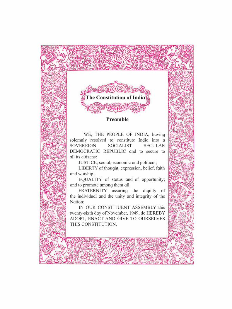
The Constitution of India
Preamble
WE, THE PEOPLE OF INDIA, having solemnly resolved to constitute India into a SOVEREIGN SOCIALIST SECULAR DEMOCRATIC REPUBLIC and to secure to all its citizens: JUSTICE, social, economic and political; LIBERTY of thought, expression, belief, faith and worship; EQUALITY of status and of opportunity; and to promote among them all FRATERNITY assuring the dignity of the individual and the unity and integrity of the Nation; IN OUR CONSTITUENT ASSEMBLY this twenty-sixth day of November, 1949, do HEREBY ADOPT, ENACT AND GIVE TO OURSELVES THIS CONSTITUTION.

NATIONAL ANTHEM

(Vivek Gosavi)Director
Maharashtra State Bureau of Textbook Production and Curriculum Research, Pune 4
Pune Date : 21 February 2020Bharatiya Saur : 2 Phalguna 1941
Dear Students,
We welcome you all to Std. XII. Now you are familiar to the subject of Biology as a separate discipline in standard XI. You have already been acquainted with many concepts of Biological Sciences from Standard six onwards, especially in the subject of General Science up to standard Eight and Science and Technology for standard Nine and Ten.
This textbook aims to create awareness about the biological sciences specially Botany, Zoology and allied aspects of biological sciences. The National Curriculum Framework (NCF) was formulated in 2005, followed by the State Curriculum Framework (SCF) in 2010. Based on the given these two frameworks, reconstruction of the curriculum and preparation of a revised syllabus has been undertaken which will be introduced from the academic year 2019-20. The textbook incorporating the revised syllabus has been prepared and designed by the Maharashtra State Bureau of Textbook Production and Curriculum Research, (Balbharati), Pune.
The subject biology intends to give students understanding, and appreciation of the vast diversity of living beings, their special adaptations to their environments and evolutionary relationships. No compromise is made in any manner over the use of language in the Biology context, but at the same time, the textbook is presented in a simple licid language. In addition, relevant diagrams, graphs, tables used in the textbook will bring about more clarity in the understanding of various terminologies and biological concepts. All the illustrations are in colour form. This will surely enable students to understand various concepts of botany and zoology thoroughly and correlate this with their day-to-day practical life. The new syllabus focuses on the conceptual principles of overall life processes, its understanding, and application in day-to-day life and ability to solve different upcoming problems and issues like inheritance and its significance, conservation; different diseases and remedies, the application of technology, etc. The general teaching-learning objectives of the revised syllabus are further determined based on the ‘principle of constructivism’ i.e. self-learning.
The curriculum and syllabus confirms to the maxims of teaching such as moving from concrete to abstract, known to unknown and from part to whole. For the first time, in the syllabus of biology various independent activities have been introduced. These activities will not only help to understand the content knowledge but also provide scope for gaining relevant and additional application based knowledge on your own efforts. Q. R. Code have been introduced for gaining the additional information, abstracts of chapters and practice questions/ activities.
The efforts taken to prepare the textbook will not only enrich the meaningful learning experience of the students, but also benefit other stakeholders such as teachers, parents as well as those aspiring candidates preparing for the competitive examinations.
We look forward to a positive response from the teachers and students.Our best wishes to all!
Preface
Dear Teachers, We are happy to introduce the revised
textbook of Biology for Std XII in continuation of Std XI. This book is a sincere attempt to follow the maxims of teaching as well as develop a ‘constructive’ approach to enhance the quality of learning and teaching as well. The present day education demands for more activity based, experimental and innovative learning opportunities is the need of the hour. The present curriculum has been restructured so as to bridge the credibility gap that exists between what is being taught and what students learn from the experiences in the outside world. Guidelines provided below will help to enrich the teaching-learning process to achieve the desired learning outcomes.• To begin with, get familiar with the
textbook.• Always teach with proper planning.• The present book has been prepared for
constructive and activity-based teaching. • Teachers must skillfully plan and organize
the activities provided in each chapter to develop interest as well as to stimulate the thought process among the students.
• Use teaching aids as required for the proper understanding of the subject.
• Use demonstration, discussion method for teaching.
• Follow the order of the chapters strictly as listed in the contents because the units are introduced in a graded manner to facilitate knowledge building.
• Facilitate peer learning as much as possible by reorganizing the class structure frequently.
• Teaching-learning interactions, processes and participations of all students are very essential and so is your active guidance.
• Ask questions based on previous knowledge.
• Do not use the boxes titled ‘Do you know?’ for evaluation. However, teachers must ensure that students read this extra information.
• Information provided in boxes with the title ‘Can You Tell’, ‘Always Remember’ should be considered for evaluation.
- For Teachers -
• Exercise is given at the end of lesson. In exercise different type of questions/ activities are given.
• Exercises provided after each unit are prepared using different learning parameters like observation, co-relation, critical thinking, analytical reasoning etc.
• Evaluation pattern should be based on the above mentioned parameters. Equal weightage should be assigned to all the topics. Use different combinations of questions. Stereotype questions should be avoided.
• ‘Can You Recall’ is the first main starting point of lesson which helps for the introduction of topic. This will also helpful for students regarding understanding the content of lesson.
• ‘Internet My Friend’ is given for collecting extra important information related to topic.
• ‘Use Your Brain Power’ is used for the application level questions in different lessons.
• ‘Do Your Self’, ‘Find Out’, ‘Observe and Discuss’ and ‘Try This’ are used for activity based learning.
• ‘Know the Scientist’ is used for the information of different scientist related to concepts in lesson.
• ‘Activity’ is used in lesson and exercise for better understanding and application of the content which studied.
• Teacher should use their freedom to acquaint the students with flora and fauna of given region.
• Remember that mathematical and statistical tools are also important to understand biology
• List of abbreviations are provided towards the end of the textbook for further clarification.
• Use Q. R. Code given in the textbook.
Best wishes for a wonderful teaching experience and fruitful welcome!
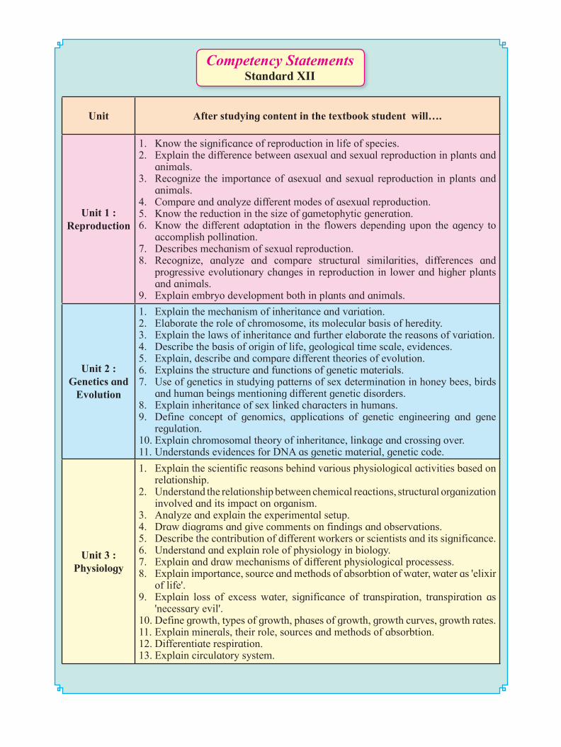
Competency StatementsStandard XII
Unit After studying content in the textbook student will….
Unit 1 : Reproduction
1. Know the significance of reproduction in life of species.2. Explain the difference between asexual and sexual reproduction in plants and
animals. 3. Recognize the importance of asexual and sexual reproduction in plants and
animals. 4. Compare and analyze different modes of asexual reproduction.5. Know the reduction in the size of gametophytic generation.6. Know the different adaptation in the flowers depending upon the agency to
accomplish pollination.7. Describes mechanism of sexual reproduction. 8. Recognize, analyze and compare structural similarities, differences and
progressive evolutionary changes in reproduction in lower and higher plants and animals.
9. Explain embryo development both in plants and animals.
Unit 2 : Genetics and
Evolution
1. Explain the mechanism of inheritance and variation.2. Elaborate the role of chromosome, its molecular basis of heredity.3. Explain the laws of inheritance and further elaborate the reasons of variation.4. Describe the basis of origin of life, geological time scale, evidences. 5. Explain, describe and compare different theories of evolution.6. Explains the structure and functions of genetic materials. 7. Use of genetics in studying patterns of sex determination in honey bees, birds
and human beings mentioning different genetic disorders. 8. Explain inheritance of sex linked characters in humans.9. Define concept of genomics, applications of genetic engineering and gene
regulation.10. Explain chromosomal theory of inheritance, linkage and crossing over.11. Understands evidences for DNA as genetic material, genetic code.
Unit 3 : Physiology
1. Explain the scientific reasons behind various physiological activities based on relationship.
2. Understand the relationship between chemical reactions, structural organization involved and its impact on organism.
3. Analyze and explain the experimental setup.4. Draw diagrams and give comments on findings and observations. 5. Describe the contribution of different workers or scientists and its significance. 6. Understand and explain role of physiology in biology.7. Explain and draw mechanisms of different physiological processess.8. Explain importance, source and methods of absorbtion of water, water as 'elixir
of life'.9. Explain loss of excess water, significance of transpiration, transpiration as
'necessary evil'.10. Define growth, types of growth, phases of growth, growth curves, growth rates.11. Explain minerals, their role, sources and methods of absorbtion.12. Differentiate respiration.13. Explain circulatory system.
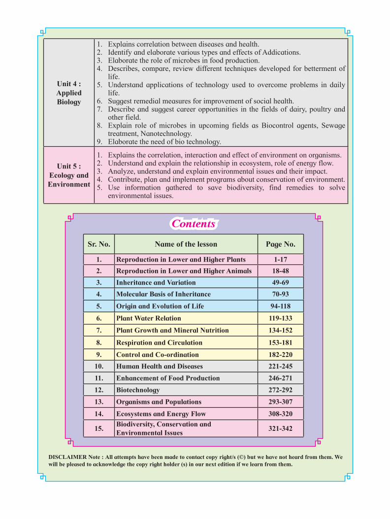
Sr. No. Name of the lesson Page No.
1. Reproduction in Lower and Higher Plants 1-17
2. Reproduction in Lower and Higher Animals 18-48
3. Inheritance and Variation 49-69
4. Molecular Basis of Inheritance 70-93
5. Origin and Evolution of Life 94-118
6. Plant Water Relation 119-133
7. Plant Growth and Mineral Nutrition 134-152
8. Respiration and Circulation 153-181
9. Control and Co-ordination 182-220
10. Human Health and Diseases 221-245
11. Enhancement of Food Production 246-271
12. Biotechnology 272-292
13. Organisms and Populations 293-307
14. Ecosystems and Energy Flow 308-320
15.Biodiversity, Conservation and Environmental Issues
321-342
Contents
DISCLAIMER Note : All attempts have been made to contact copy right/s (©) but we have not heard from them. We will be pleased to acknowledge the copy right holder (s) in our next edition if we learn from them.
Unit 4 : Applied Biology
1. Explains correlation between diseases and health. 2. Identify and elaborate various types and effects of Addications.3. Elaborate the role of microbes in food production.4. Describes, compare, review different techniques developed for betterment of
life.5. Understand applications of technology used to overcome problems in daily
life. 6. Suggest remedial measures for improvement of social health. 7. Describe and suggest career opportunities in the fields of dairy, poultry and
other field. 8. Explain role of microbes in upcoming fields as Biocontrol agents, Sewage
treatment, Nanotechnology. 9. Elaborate the need of bio technology.
Unit 5 : Ecology and Environment
1. Explains the correlation, interaction and effect of environment on organisms. 2. Understand and explain the relationship in ecosystem, role of energy flow. 3. Analyze, understand and explain environmental issues and their impact.4. Contribute, plan and implement programs about conservation of environment. 5. Use information gathered to save biodiversity, find remedies to solve
environmental issues.
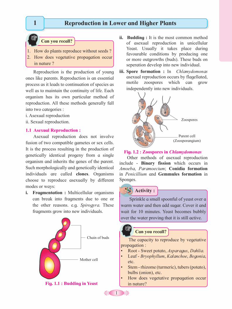
1
Fig. 1.1 : Budding in Yeast
Chain of buds
Mother cell
Reproduction is the production of young ones like parents. Reproduction is an essential process as it leads to continuation of species as well as to maintain the continuity of life. Each organism has its own particular method of reproduction. All these methods generally fall into two categories : i. Asexual reproduction ii. Sexual reproduction.
1.1 Asexual Reproduction : Asexual reproduction does not involve fusion of two compatible gametes or sex cells.It is the process resulting in the production of genetically identical progeny from a single organism and inherits the genes of the parent. Such morphologically and genetically identical individuals are called clones. Organisms choose to reproduce asexually by different modes or ways: i. Fragmentation : Multicellular organisms
can break into fragments due to one or the other reasons. e.g. Spirogyra. These fragments grow into new individuals.
ii. Budding : It is the most common method of asexual reproduction in unicellular Yeast. Usually it takes place during favourable conditions by producing one or more outgrowths (buds). These buds on seperation develop into new individual.
iii. Spore formation : In Chlamydomonas asexual reproduction occurs by flagellated, motile zoospores which can grow independently into new individuals.
Reproduction in Lower and Higher Plants
1. How do plants reproduce without seeds ?2. How does vegetative propagation occur
in nature ?
Can you recall?
Sprinkle a small spoonful of yeast over a warm water and then add sugar. Cover it and wait for 10 minutes. Yeast becomes bubbly over the water proving that it is still active.
Activity :
1
Other methods of asexual reproduction include - Binary fission which occurs in Amoeba, Paramoecium; Conidia formation in Penicillium and Gemmules formation in Sponges.
Fig. 1.2 : Zoospores in Chlamydomonas
Zoospores
Parent cell (Zoosporangium)
The capacity to reproduce by vegetative propagation :• Root - Sweet potato, Asparagus, Dahlia. • Leaf - Bryophyllum, Kalanchoe, Begonia,
etc. • Stem - rhizome (turmeric), tubers (potato),
bulbs (onion), etc.• How does vegetative propagation occur
in nature?
Can you recall?

2
Activity :
carefully grown to give many plant lets.Micropropagation method is also used now a days.
1.2 Sexual Reproduction : It involves fusion of two compatible gametes or sex cells. All organisms reach to the maturity in their life before they can reproduce sexually. In plants, the end of juvenile or vegetative phase marks the begining of the reproductive phase and can be seen easily in the higher plants at the time of flowering.
The flower is specialized reproductive structure of a plant in which sexual reproduction takes place. The function of flower is to produce haploid gametes and to ensure that fertilization will take place. Typical flower consists of four different whorls viz. calyx, corolla androecium and gynoecium. Sexual reproduction involves two major events viz. meiosis and fusion of gametes to form diploid zygote and the production of genetically dissimilar offsprings. Variations are useful from the point of view of the survival and the evolution of species, over the time. Sexual reproduction is characterised by fusion of the male and female gametes (fertilization), the formation of zygote and embryogenesis. Sequential events that occur in sexual reproduction are grouped into three distinct stages viz, Pre-fertilization, Fertilization and the Post-fertilization.
Vegetative Reproduction :
Plants reproduce asexually through their vegetative parts. Hence, the new plants formed are genetically identical to their parents. There are also few methods which would not occur naturally in the plants. Agriculture and horticulture exploit vegetative reproduction in order to multiply fresh stocks of plants. Artificial methods are used to propagate desired varieties according to human requirements. The various methods are as follows :
a. Cutting : The small piece of any vegetative part of a plant having one or more buds is used for propagation viz. Stem cutting - e.g. Rose, Bougainvillea; leaf cutting - e.g. Sansvieria; root cutting e.g. Blackberry.
b. Grafting : Here parts of two plants are joined in such a way that they grow as one plant. In this method, part of the stem containing more than one bud (Scion) is joined onto a rooted plant called stock, is called grafting. Whereas budding is also called bud grafting in which only one bud is joined on the stock, e.g. Apple, Pear, Rose, etc.
Why does gardner choose to propagate plants asexually?
Do you know ?
Fig. 1.3 : Grafting in Rose
Stock
Scion
c. Tissue culture : It is a method by which a small amount of plant tissue is
Label the parts of flower in the given diagram :

3
The male reproductive whorl of flower is called androecium. Individual member of androecium, is called stamen. Stamen consists of filament, connective and anther.
Structure of Anther : An immature stage of anther is represented by group of parenchymatous tissue surrounded by single layered epidermis. Anther is generally dithecous (having two lobes) and tetrasporongiate. Each monothecous anther contains two pollen sacs. In dithecous anther four pollen sacs are present. Therefore, it is tetrasporongiate. The heterogenesity (differenciation) arises when some hypodermal cells get transformed into archesporial cells.
T. S. of Anther : The archesporial cell divides into an inner sporogenous cell and outer primary parietal cell. Sporogenous cell forms sporogenous tissue. Each cell of sporogenous tissue is capable of giving rise to a microspore tetrad. Parietal cell undergoes divisions to form anther wall layers. The wall of mature anther consists of four layers. Epidermis is the outermost protective layer made up of tabular (flattened)cells. Endothecium is sub-epidermal layer made up of radially elongated cells with fibrous thickenings. Inner to endothecium is middle layer made up of thin walled cells (1-2 layered), which may disintegrate in mature anther. Tapetum is the inner most nutritive layer of anther wall. It immediately encloses the sporogenous tissue (microspore mother cells).
1.3 Microsporogenesis : Each microspore mother cell divides meiotically to form tetrad of haploid microspores (pollen grains).
Structure of microspore : Typical pollen grain is a non-motile, haploid, unicellular body with single nucleus. It is surrounded by a two layered wall called sporoderm. The outer layer exine is thick and made up of complex, non-biodegradable, substance called sporopollenin. It may be smooth or with a sculptured pattern (characteristic of the species). It is resistant to chemicals. At some places exine is very thin showing thin areas known as germ-pores. These are meant for the growth of emerging pollen tube during germination of pollen grain. The inner wall layer, intine consists of cellulose and pectin.
Diploid sporophyte is the predominant plant body in all angiosperms, where meiosis takes place to produce haploid spores that form gametophyte. Gametophytes are considerably reduced and develop within the flower. They produce gametes.
Always Remember
Why pollen grains can remain well preserved as fossil?
Find out
Fig. 1.4 : (a) T. S. of anther
Connective Epidermis
Middle layers
Tapetum
Sporogenous tissue
Endothecium
Fig. 1.4 : (b) Dehisced anther
Pollen grains

4
The second mitotic division is concerned with generative cell only and gives rise to two non-motile male gametes. The mitotic division of generative cell takes place either in pollen grain or in the pollen tube. The pollen grains are shed from the anther, at this two- celled stage in most of the angiosperms.
Female reproductive whorl of flower is gynoecium (Pistil). Individual member of gynoecium is called carpel (megasporophyll). A flower with many, free carpels is called apocarpous (e.g. Michelia). A syncarpous flower is one that has many carpels fused together (e.g. Brinjal). Typical carpel has three parts viz, ovary, style and stigma. The number of ovules in the ovary varies e.g. paddy, wheat and mango are uniovulate whereas tomato and lady’s finger are multiovulate.
1.4 Structure of Anatropous ovule : Each ovule develops inside the ovary and is attached to the placenta by a small stalk called funiculus. The place of attachment of funiculus with the main body of ovule, is called hilum. In angiosperms, the most common type of ovule is anatropous in which micropyle is directed downwards and is present adjacent to the funiculus (funicle). The ovule consists of central parenchymatous tissue, the nucellus which is surrounded usually by two protective coverings called integuments viz. Outer and an inner integument. A narrow opening at the apex of the ovule is called micropyle. Chalaza is the base of ovule directly opposite to micropyle. Embryo sac (female gametophyte) is oval multicellular structure embedded in the nucellus.
Fig. 1.5 : Development of male gametophyte
Development of male gametophyte : Pollen grain marks the begining of male gametophyte. It undergoes first mitotic division to produce bigger, naked vegetative cell and small, thin walled generative cell. The vegetative cell is rich in food and having irregular shaped nucleus. The generative cell floats in the cytoplasm of vegetative cell.
Fig. 1.6 : Anatropous Ovule
Chalaza
Antipodals
Secondary Nucleus
Embryo sac Synergids Egg
Outer integument Inner integument
Hilum
Funicle
Placenta
Micropyle
Nucellus
• Pollen viability (viability is the functional
ablity of pollen grain to germinate to develop male gametophyte) depends upon environmental conditions of temperature and humidity. It is 30 minutes in rice and wheat. But in some members of family Solanaceae, Rosaceae, Leguminosae, it lasts even for months.
Always Remember
Pollen grain Intine
CytoplasmExine
Vegetative cell Germ pore
Generative cell Generative nucleus
Vegetative nucleus
Male gametes Pollen tube
Male gametes
Tube nucleus
A B C
D E F

5
Antipodal cells are group of three cells present at the chalazal end. The two haploid polar nuclei of large central cell fuse to form diploid secondary nucleus or definitive nucleus, just prior to fertilization. This seven-celled and eight nucleated structure is called an embryo sac. This method of embryo sac development from a single megaspore is described as monosporic development. In angiosperms, the development of female gametophyte is endosporous i.e. within the megaspore. Female gametophyte is colourless, endosporic and is concealed in the ovule enclosed by ovary.
1.6 Pollination : Pollen grains being non motile, angiosperms have evolved the strategy to use abiotic agents (wind, water) and biotic agents (birds, insects, snails) to their flowers, feeding the visitors and exploiting their mobility for pollination and also seed dispersal. Pollen grains are non-motile and they are usually carried from flower to flower by means of external agents. Pollination is the transfer of pollen grains from anther to the stigma of the flower. It is the pre-requisite for fertilization because both the male and female gametes are non-motile. Moreover gametes are produced at two different sites.
1.5 Megasporogenesis : It is the process of formation of haploid megaspores from diploid megaspore mother cell (MMC). Megaspore mother cell becomes distinguished in the nucellus, more or less in the centre but towards micropylar end of ovule.
Development of female gametophyte : Megaspore mother cell undergoes meiosis to form linear tetrad of haploid cells i.e. megaspore. Upper three megaspores abort and lowest one towards centre of nucellus remains functional. It acts as the first cell of female gametophyte. Generally one megaspore towards centre is functional megaspore. It is infact the first cell of female gametophyte. It undergoes three successive, free nuclear mitotic divisions. Thus total eight nuclei are formed, four of which are located at each pole. One nucleus from each pole migrates towards the centre and are called polar nuclei. Three nuclei towards micropylar end constitute egg apparatus. It consists of large central, haploid egg cell and two supporting haploid synergid cells. Synergid shows hair like projections called filiform apparatus, which guide the pollen tube towards the egg.
Fig. 1.7 : Development of female gametophyte
Megaspore mother cell
(diploid)
Degenerated cells
Four haploid megaspores
Megaspore or embryo
sac (Haploid)
Growth nourished by
nucellus
Mitosis (Ist)
Meiosis
End nearest micropyle
Mitosis (IInd)
Three Antipodal cells
Mitosis (IIIrd)
Two polar nuclei
Egg (Female gamete)
Two synergids
Nuclear fusion
Mature Embryo sac (female
gametophyte) just before fertilisation seven nuclei
present (six are haploid one is
diploid)
6
b. Geitonogamy : It is the transfer of pollen grain to a stigma of a different flower produced on the same plant. It is functionally similar to cross pollination as it involves pollinating agents, but it cannot bring about genetic variations and is only of ecological significance e.g. Cucurbita maxima. It is similar to antogamy as pollen grains come from same plant.
c. Xenogamy (cross polination/ out breeding) : It is a type of cross pollination when pollen grain of one flower is deposited on the stigma of a flower of different plant belonging to same species, with the help of pollinating agency. It generates genetically varied offsprings. Majority of flowering plants depend on the transfer of pollen grains. Virtually all seed plants need to be pollinated. Most of the food and fibre crops grown throughout the world, depend upon pollinators for reproduction. The agents responsible for pollination have been grouped into two main categories :A. Abiotic agents B. Biotic agents
A. Abiotic Agents : These are non-living agents which include wind and water.
1. Pollination by wind (Anemophily) : Most of the important crop plants are wind pollinated. These includes wheat, rice, corn, rye, barley and oats. Palms are also wind pollinated.
Adaptations in anemophilous flowers :• The flowers are small, inconspicuous,
colourless, without nectar and fragrance (odour).
• The pollen grains are light in weight, dry and produced in large numbers to increase chances of pollination considering wastage of pollengrains.
• Stigma is feathery to trap pollens carried by wind currents.
Self pollination is a type of pollination which occurs in a single flower or two flowers on a single plant. It results in inbreeding or selfing. In contrast cross pollination is the transfer of pollen grains from the anther of one flower to the stigma of another flower of different plants of same species. Pollination can be further divided into three types on the basis of source of pollination.
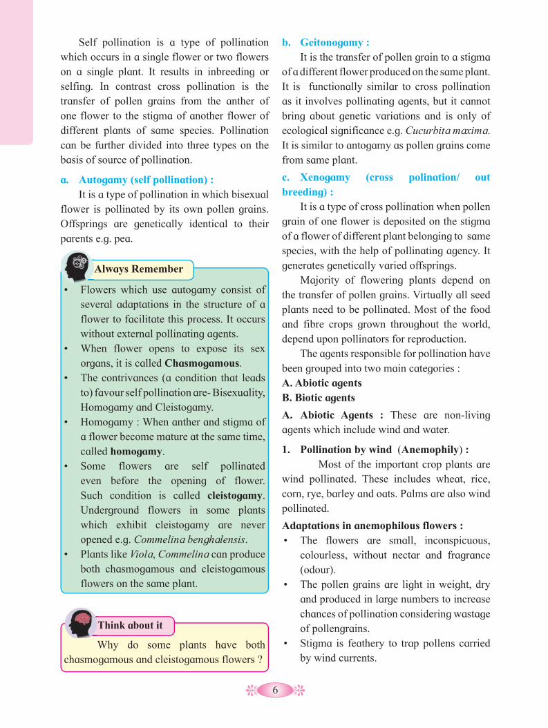
a. Autogamy (self pollination) : It is a type of pollination in which bisexual flower is pollinated by its own pollen grains. Offsprings are genetically identical to their parents e.g. pea.
• Flowers which use autogamy consist of
several adaptations in the structure of a flower to facilitate this process. It occurs without external pollinating agents.
• When flower opens to expose its sex organs, it is called Chasmogamous.
• The contrivances (a condition that leads to) favour self pollination are- Bisexuality, Homogamy and Cleistogamy.
• Homogamy : When anther and stigma of a flower become mature at the same time, called homogamy.
• Some flowers are self pollinated even before the opening of flower. Such condition is called cleistogamy. Underground flowers in some plants which exhibit cleistogamy are never opened e.g. Commelina benghalensis.
• Plants like Viola, Commelina can produce both chasmogamous and cleistogamous flowers on the same plant.
Always Remember
Why do some plants have both chasmogamous and cleistogamous flowers ?
Think about it

7
grass) the pollen grains are long, ribbon like and without exine.
Epihydrophily : The pollen grains float on the water surface and reach the stigma of female flower. e.g. Vallisneria is a submerged dioecious, fresh water aquatic plant in which female flowers reach the water surface temporarily to ensure pollination and male flowers float on the surface of water.• Specific gravity of pollen grain is equal
to that of water. That is why they float on surface of water.
• Some aquatic plants are anemophilous e.g. Potamogeton, Halogaris, etc.
• Some aquatic plants are entomophilous e.g. Lotus, water hyacinth, waterlily, etc.
• Stamens are exserted with long filaments and versatile anthers.
• Stamens and stigmas are exposed to air currents.
The pollens of wind pollinated plants are most frequently associated with symptoms of hayfever among people those are sensitive to pollens. It is caused by hypersensitivity to pollen.
Always Remember
2. Pollination by water (Hydrophily) : Found only in some 30 genera of aquatic monocots. E.g. Vallisneria, Zostera, Ceratophyllum etc.Adaptations in hydrophilous flowers :• Flowers are small and inconspicuous.• Perianth and other floral parts are
unwettable.• Pollen grains are long and unwettable due
to presence of mucilage.• Nectar and fragrance are lacking in
flowers.
Hydrophily is of two types - Hypohydrophily : Pollination occurs below the surface of water. Here the pollen grains are heavier than water, sink down and caught by stigmas of female flowers, e.g. In Zostera (sea
Fig. 1.8 : Pollination by wind (Maize)
Male inflorescence (Tassel)
Flag leaf
Tassel internode
Styles (silks)
Female inflorescence (ear)
Seed (Kernel)
Leaf
Stalk(stem)
Prop roots
Roots
B. Biotic Agents : It includes living agents. About 80% of plants require the help of other living, moving creatures such as insects, birds, bats, snails to transfer their pollens from one flower to another. These also sustain our ecosystems and produce natural resources by helping plants to reproduce.
1. Pollination by insects (Entomophily) : It occurs in Rose, Jasmine, Cestrum, etc.Adaptations in entomophilous flowers :• They are large, showy and often brightly
coloured.• The flowers produce sweet odour (smell)
and have nectar glands.
Fig. 1.9 : Male and female plant Vallisneria
Female flower
Water level
Male flower

8
Adaptations in ornithophilous flowers :• Flowers are usually brightly coloured,
large and showy.• They secrete profuse, dilute nectar.• Pollen grains are sticky and spiny.• Flowers are generally without fragrance,
as birds have poor sense of smell.3. Pollination by Bats (Chiropteryphily) : Bats can transport pollens over long distance, some times several kilometers.
Adaptations in Chiropterphilous flowers :• Flowers are dull coloured with strong
fragrance.• They secrete abundant nectar.• Flowers produce large amount of edible
pollen grains, e.g. Anthocephalous (kadamb tree), Adansonia (Baobab tree), Kigelia (Sausage tree).
1.7 Outbreeding devices (contrivances): Many plants have mechanisms that discourage or prevent self pollination. To promote cross pollination and increase genetic diversity, plants have evolved a wide variety of sexual strategies. Genetic diversity is an essential factor for evolution by natural selection. Continued self pollination results in the inbreeding depression. Thus plants have developed many devices to encourage cross pollination. The examples of outbreeding devices are as follows:
Unisexuality : In this case, the plant bears either male or female flowers. It is also called as dioecism. As flowers are unisexual, self pollination is
• The stigma is rough due to presence of hair or is sticky due to mucilaginous secretion.
• The pollen grains are spiny and surrounded by a yellow sticky substance called pollen-kit.
• Some plants have special adaptations for the insect visitor to help in cross pollination, e.g. lever mechanism or turn-pipe mechanism in Salvia. Fig. 1.11 : Ornithophily
2. Pollination by birds (Ornithophily) : Only a few types of birds are specialised for pollination. They usually have small size and long beaks e.g. Sun birds and humming birds. Some ornithophilous plants are Bombax, Callistemon (Bottle Brush), Butea, etc.
Fig. 1.10 : Lever mechanism in Salvia
You may see bumblebee early in the year as they try to find a suitable place to establish a nest and rear a colony. If you find a bumblebee nest please leave it alone. Their nest lasts only for a season. Educate the world about the need to help the bees.
Think about it
In biotic pollination, plants are adapted to encourage the specific pollinators they need. They are said to have developed pollination contrivance. Plants and pollinators have co-evolved physical characteristics that make them to interact successfully. Such characteristics are considered pollination syndromes.
Do you know ? In biotic pollination, plants are
9
1.8 Pollen - Pistil Interaction : It is the interaction of pollen grains with sporophytic tissue (stigma). It begins with pollination and ends with fertilization. All the events from the deposition of pollen grain on stigma to the entry of pollen tube in the ovule (synergid) are referred as pollen - pistil interaction. Pollination does not guarantee the transfer of right type of pollen, often wrong type also land on stigma. The pistil has the ability to recognise and accept the right or compatible pollen of the same species. Thus wrong type of pollen is discarded by pistil. Compatibility and incompatibility of the pollen-pistil is determined by special proteins. This process involves pollen recognition followed by promotion or inhibition of pollen. The stigmatic surface of flower refuse other wrong type or incompatible pollen grains. A physiologial mechanism operates to ensure that only intraspecific pollen germinate successfully. The compatible pollen absorbs water and nutrients from the surface of stigma, germinates and produces pollen tube. Its growth through the style is determined by specific chemicals. The stigmatic surface provides the essential prerequisites for a successful germination, which are absent in the pollen. The pollen tube is finally pushed through the ovule and reaches the embryo sac. The tip of the pollen tube enters in one of the synergids and then ruptures to release the contents. Due to pollen pistil interaction, intense competition develops even in the compatible pollen grains (gametes). It also plays important role in sexual reproduction and seed formation. Pollen grain can also be induced to germinate in a synthetic medium. Sucrose induces pollen germination and tube growth in vitro. Addition of boric acid facilitates and accelarates pollen germination.
Artificial hybridization : It is one of the major approaches used in the crop improvement. Only the desired
not possible. Plants may be monoecious, e.g. Maize or dioecious, e.g. Mulberry, Papaya.
Dichogamy : In this device, anthers and stigmas mature at different times in a bisexual flower so as to prevent self pollination. It can be further divided into two types:1. Protandry : In this type, androecium
matures earlier than the gynoecium, e.g. in the disc florets of sunflower.
2. Protogyny : In this type, gynoecium matures earlier than the androecium, e.g. Gloriosa.
Prepotency : Pollen grains of other flowers germinate rapidly over the stigma than the pollen grains from the same flower, e.g. Apple.
Heterostyly (heteromorphy): In some plants like Primula (Primrose, there are two or three forms/ types of flowers in which stigmas and anthers are placed at different levels (heterostyly and heteroanthy). This prevents the pollens from reaching the stigma and pollinating it. In heteromorphic flowers, pollen grains produced from anther pollinate stigmas produced at the same level.
Herkogamy : It is a mechanical device to prevent self pollination in a bisexual flower. In plants, natural physical barrier is present between two sex organs and avoid contact of pollen with stigma of same flower, e.g. Calotropis- pentangular stigma is positioned above the level of anthers (pollinia).
Self incompatibility (self sterility): This is a genetic mechanism due to which the germination of pollen on stigma of the same flower is inhibited, e.g. Tobacco, Thea.
In all breeding programmes, the plants are hand pollinated to ensure cross pollination between selected varieties. e.g. wheat, rice.
Do you know ?

10
Significance of Double Fertilization :• It is a unique feature of angiosperms. It
ensures that the parent plant invests a seed with a food store, only if the egg is fertilized.
• The diploid zygote develops into an embryo which consequently develops into a new plant.
pollen grains are hand pollinated and used for fertilization. This is accomplished through emasculation and bagging procedure.
1.9 Double Fertilization : Double fertilization is a complex fertilization mechanism in flowering (angiospermic) plants. It was discovered by Nawaschin in the liliaceous plants like Lilium and Fritillaria. After a pollen grain has reached the surface of the stigma, it germinates and forms a pollen tube, which penetrates the stigma, style, ovary chamber and then enters ovule. The growth of pollen tube is guided by the chemicals secreted by the synergids. It usually enters ovule through the micropyle. It is termed as porogamy. But in some cases, it is found to enter through chalaza, known as chalazogamy and in some plants by piercing the integuments, called mesogamy. Finally, it penetrates embryo sac of ovule through its micropylar end. The pollen tube carrying male gametes penetrates in one of the synergids. Watery contents of synergid are absorbed by pollen tube which then ruptures and release the contents, including the two non-motile male gametes. As non motile male gametes are carried through hollow pollen tube, it is known as siphonogamy that ensures fertilization to take place. Syngamy and triple fusion are two events of sexual reproduction in angiospermic flowering plants. Syngamy is the fusion of
Fig. 1.12 : Entry of pollen tube into the ovulehaploid male gamete with haploid female gamete (egg) to produce a diploid zygote, whereas in triple fusion, second haploid male gamete fuses with diploid secondary nucleus producing primary endosperm nucleus (PEN) that developes into triploid endosperm. The zygote develops into an embryo. Syngamy is a type of generative fertilization whereas triple fusion is a type of vegetative fertilization. Here, both the male gametes participate and therefore, it is described as or called double fertilization.
Antipodals
Polar nuclei
Egg cell
Nucellus
Synergid Micropyle
Chalaza
Pollen tube
Pollen tube
Pollen tube
embryo sac
Funicle
Integument
Integuments
Porogamy A
Chalazogamy B
Mesogamy C
Fig. 1.13 : Double fertilization
SynergidsSyngamy
Egg + Male gamete cell
Triple fusion Secondary nucleus +
Male gamete
Ovary
Ovule
Pollen grain
Pollen tube
Style
Micropyle
Stigma

11
What do you call the kernel that you eat in tender coconut ?
Use your brain power
• The triploid PEN develops into nutritive endosperm tissue.
• It restores the diploid condition by fusion of haploid male gamete with haploid female gamete (i.e. through syngamy).
• It also helps to avoid polyembryony.
1.10 Development of Endosperm : The triploid primary endosperm nucleus repeatedly divides, mitotically to form nutritive tissue, called endosperm. In post-fertilization changes within the ovule, the embryo and endosperm are seen to develop simultaneously. The other cells of embryo sac disorganized sooner or later. The formation of triploid endosperm nucleus triggers cell division which leads to the formation of endosperm.
vacuole appears in the centre of cell pushing the nuclei towards the periphery. Later, walls develop between the nuclei, hence multicellular endosperm is formed. But in several cases cell wall formation remains incomplete. e.g. wheat, sunflower and coconut. Coconut has multicellular endosperm in the outer part and free nuclear as well as vacuolated endosperm in the centre.
b. Cellular Type : In some plants, division of triploid primary endospermic nucleus is immediately followed by wall formation. So that the endosperm is cellular right from the beginning. It is mostly observed in 72 families of dicots as in members - Balsam, Petunia, Adoxa, etc.
There are three types of endosperms on the basis of mode of development. These are i. Nuclear type, ii. Cellular type, iii. Helobial type :a. Nuclear Type : It is the most common type found in 161 angiospermic families. Here, the primary endosperm nucleus repeatedly divides mitotically without wall formation to produce large number of free nuclei. A big central
c. Helobial Type : It occurs in the order Helobiales of monocotyledons. In this case, first divison of primary endosperm nucleus is followed by a transverse wall, which divides the cell unequally. The smaller cell is called chalazal cell and larger cell is the micropylar cell. Then the nuclei in each cell divide by free nuclear divisions and then walls develop between nuclei in micropylar chamber. It is intermediate between cellular and nuclear type endosperm e.g. Asphodelus.
Mosaic Endosperm : Endosperm containing tissues of two different types is called mosaic endosperm. In plants like corn the endosperm contains patches of two different colours. It forms a sort of mosaic pattern.
1.11 Development of Embryo : The process of development of zygote into an embryo is called embryogenesis. The embryo is developed at the micropylar end of embryo sac. The growth of embryo triggers only
Fig. 1.14 : Types of Endosperm
Zygote/ Oospore Embryo
Endosperm Nuclear
Oospore Embryo
Cellular
Endosperm
Helobial
Oospore Embryo
i.
ii. iii.

12
Fig. 1.15 : Development of Dicot Embryo as in Capsella
Fig. 1.16 : Development of Monocot Embryo
Oospore Suspensor initial
Embryonal initial
Suspensor
Suspensor
Plumule Cotyledon
Cotyledon
Radicle Radicle Radicle
Plumule
Hypocotyl Plumule
Cotyledon
Scutellum
Coleoptile
Shoot apex
Epiblast
Root cap
Coleorrhiza
A B
C
D E
F G H
after certain amount of endosperm is formed. After fertilization the embryonic development begins. The zygote divides to form two- celled proembryo. The larger cell towards the micropyle is called basal or suspensor initial cell and smaller cell towards chalaza is called terminal or embryonal initial cell. The suspensor cell divides transversely in one plane to produce filamentous suspensor of 6-10 cells.
The first cell of the suspensor towards the micropylar end becomes swollen and function as a haustorium. The lowermost cell of suspensor is known as hypophysis. The suspensor helps in pushing the embryo in the endosperm. The embryonal initial undergoes three successive mitotic divisions to form octant. The planes of divisions are at right angles to each other. The lower tier of four cells of octant give rise to hypocotyl and radicle whereas four cells of
A. Oospore. B. Two celled proembryo. e=embryonal initial; t=suspensor initial; m=Embryo sac membrane. B1=4-celled I-shaped proembryo; e
1, e
2 are from embryonal
initial; s1, s
2 are from suspensor
initial. C. Further development of embryo. S=Suspensor, h=Hypophysis; E=Embryonal mass D. L. S. of ovule Endo=Endosperm in free nuclear stage. Anti=Antipodal tissue. Embryo= Developing embryo E. Embryo showing further development of embryonic octants and hypophysis. F. L. S. of ovule. Endosperm becoming cellular. G. Embryo Cot=Cotyledons; Hypo=Hypocotyl; Rad=Radicle; R.c=Root-cap H. Mature seed. Pl=Plumule. Endosperm has been consumed almost completely.
A B
B1 C D E
F G H
e t
m
h
S
Anti
Anti
e1 e
2
s1
s2
m Endo
E
h
S
Embryo
Embryo
E
E
h
S
Endo
Cot
Hypo Rad
Rad
R.c R.c
S
Hypo Pl
Cot

13
Seed sometimes consists of two distinct coverings, a typical outer seed coat, the testa and the inner thin, membranous tegmen. The nucellus in the ovule may persist in some genera like black pepper and beet as a thin, papery layer, the perisperm. In some seeds, the food reserves in the endosperm are partially used up in the development of an embryo. Obviously, in such seeds the endosperm remains conspicuous and fills a greater part of the seed. Thus, the resultant seed is endospermic or albuminous e.g. Castor, Coconut, Maize, etc. In other seeds, embryo absorbs food reserve from the endosperm completely during its developmental stages. Thus, endosperm disappears (disorganizes) in mature seeds. The resultant seed is non-endospermic or ex-albuminous e.g. Pea, bean, etc. The cotyledons in some non-endospermic seeds act as a food storage and in others they are the first photosynthetic organs. Micropyle persists as a small pore in seed coat to allow the entry of water and oxygen during soaking.
Fruit development is triggered by hormones produced by developing seeds. As mentioned earlier, after fertilization the zygote is formed and the ovary begins to differentiate into the fruit and ovary wall develops into pericarp. Pericarp is basically three layered which get differentiated in the fleshy fruit like mango, coconut, etc.
upper tier form the plumule and the one or two cotyledons. The hypophysis by further division gives rise to the part of radicle and root cap. Subsequently, the cells in the upper tier of octant divide in several planes so as to become heart shaped which then forms two lateral cotyledons and a terminal plumule. Further enlargement of hypocotyl and cotyledons result in a curvature of embryo and it appears horse-shoe shaped. The embryo development is similar in both dicots and monocots up to the octant stage. The difference appears later. In monocot embryo, single cotyledon occupies terminal position and plumule is lateral. The single shield shaped cotyledon is called as scutellum. The protective sheath of plumule is called coleoptile and that of radicle is coleorhiza. Finally, ovule is transformed into the seed and ovary into the fruit.
1.12 Seed and Fruit Development : The goal of reproduction, in every living organisms including plants, is to create offsprings for the next generation. One of the ways that plants can produce offpsrings is by forming (making) seeds. The flowers must be pollinated in order to produce seeds and fruit. Seed development is initiated by fertilization. The integuments of the fertilized ovule persist and get transformed into the seed coat of mature seed.
Fig. 1.17 : Bean seed (Dicot)
Fig. 1.18 : Maize seed (Monocot)
Embryo
Epicotyl
Hypocotyl
Radicle
Cotyledon
Seed coat
Seed coat and fruit-wall
Aleurone layer
Endosperm
Scutellum
Coleoptile
Plumule
Radicle
Coleorrhiza

14
1. How long seeds stay viable/ healthy?2. Can old seeds still grow?Some examples of oldest mature seeds that have grown into viable plants are as follows :• Lupinus arcticus - 10,000 years• Phoenix dactylifera - 2000 years• Some seeds are short lived, e.g. Citrus.• Some tiny seeds are easy for dispersal.
e.g. Striga, Orchids, Orobancha.
Think about it
Significance of seed and fruit formation :• Fruits provide nourishment to the
developing seeds.• Fruits protect the seeds in immature
condition.• Seeds serve as important propagating
organs (units) of plant.• Seeds and fruits develop special devices
for their dispersal and thus help in the distribution of the species.
Dormancy is a state of metabolic arrest that facillitates the survival of organisms during adverse environmental conditions. Structural or physiological adaptive mechanism for survival is called dormancy. Mature and viable seeds will not germinate even in the presence of favourable conditions and they are dispersed at different places during dormancy. Viable seeds germinate only after completion of dormancy period.
1.13 Apomixis : It is phenomenon of formation of embryo(s) through asexual method of reproduction without formation of gametes and the act of fertilization. Alternatively, it is unusual sexual reproduction where there is no meiosis and syngamy. Embryo develops in the ovule and ovule developes to form seed . In apomixis, when a gametophyte organ or cell produces embryo like structure without fertilization, it is termed as apogamy. Similarly when diploid sporophyte cell produces a diploid gametophyte without undergoing meiosis is called apospory, e.g. Orange, Mango.
1. What are the parts of the fruit ?2. What is the difference between true fruit
and false fruit ?
Can you recall?
Help to rebuild natural ecosystem. Mix seeds and potting soil together with dry clay. Mould the mixture into small balls and allow them to dry in sun. Throw the same at places suitable for germination.
Try This
Collect information about seed mother Rahilbai’s story. How does she save over 80 varieties of native seeds?
Internet my friend
The main categories of apomixis are:a. Recurrent apomixis : In this type, the embryo sac generally rise either from an archesporial cell or from some other part of the nucellus. In diplospory, the unreduced embryo sac is derived from the diploid megaspore mother cell e.g. Taraxacum. In apospory, the nucellar cells give rise to apomictic embryo sac.
b. Non-recurrent apomixis : In this type, megaspore mother cell undergoes usual meotic division and a haploid embryo sac is formed. Here, the embryo arises either from the egg by parthenogenesis or from some other haploid cells of gametophyte through apogamy. Plants produced by this method are generally sterile and do not reproduce sexually, e.g. Nicotiana.
c. Adventive Embryony : In this type, embryos may develop from somatic nucellus or integuments along with normal zygotic embryo. It is common in Mango, Orange, Lemon, etc. It gives rise to a condition called polyembryony.
15
Why are some seeds of Citrus referred to
as polyembryonic ?
Think about it Genetically identical plants can be produced effectively and rapidly by apomixis.
1.14 Parthenocarpy : This term is coined by Noll (1902). It is the condition in which fruit is developed without the process of fertilization. It occurs naturally in some varities of Pineapple, Banana, Papaya, etc. In these plants, it seems that the placental tissue in the unfertilized ovary produces auxin IAA (Indole-3 Acetic Acid) which is responsible for enlargement of ovary into fruit. The fruit resembles the normally produced fruit but it is seedless.
1. Parthenogenesis is the development of embryo directly from egg cell or a male gamete. It is a kind of apogamy.
2. Agamospermy : Here plants produce seeds. But embryo, inside it, is produced without (omitting) meiosis and syngamy.
3. Parthenocarpy can be induced artificially by - spraying of gibberellins, delaying pollination, use of foreign pollens, etc.
4. Genetically uniform parental type seedlings are obtained from nucellar embryos.
Do you know ?
What do bananas and figs have in common ?
Use your brain power
1.15 Polyembryony : It is the development of more than one embryos, inside the seed and the condition is described as polyembryony. It was first noticed by Leeuwenhoek (1719) in the seeds of Citrus genus. It is the occurrence of more than one embryo in a seed which consequently results in the emergence of multiple seedlings. The additional embryos result from the differentiation and development of various maternal and zygotic tissues associated with the ovule of seed. Polyembryony may be true or false depending upon whether many embryos arise in the same embryo sac or in different embryo sacs in the same ovule. In adventive polyembryony, an embryo develop directly from the diploid cell of nucellus and integuments as in Citrus. In cleavage polyembryony, zygote proembryo sometimes divides (cleaves) into many parts or units. Each unit then developes into an embryo. Polyembryony increases the chances of survival of the new plants. Nucellar adventive polyembryony is of great significance in horticulture.
Activity :
Prepare chart for natural vegetative propagation exibited by flowering plants indicating the vegetative part/s and the different examples.
Organ Part Name of plant
16
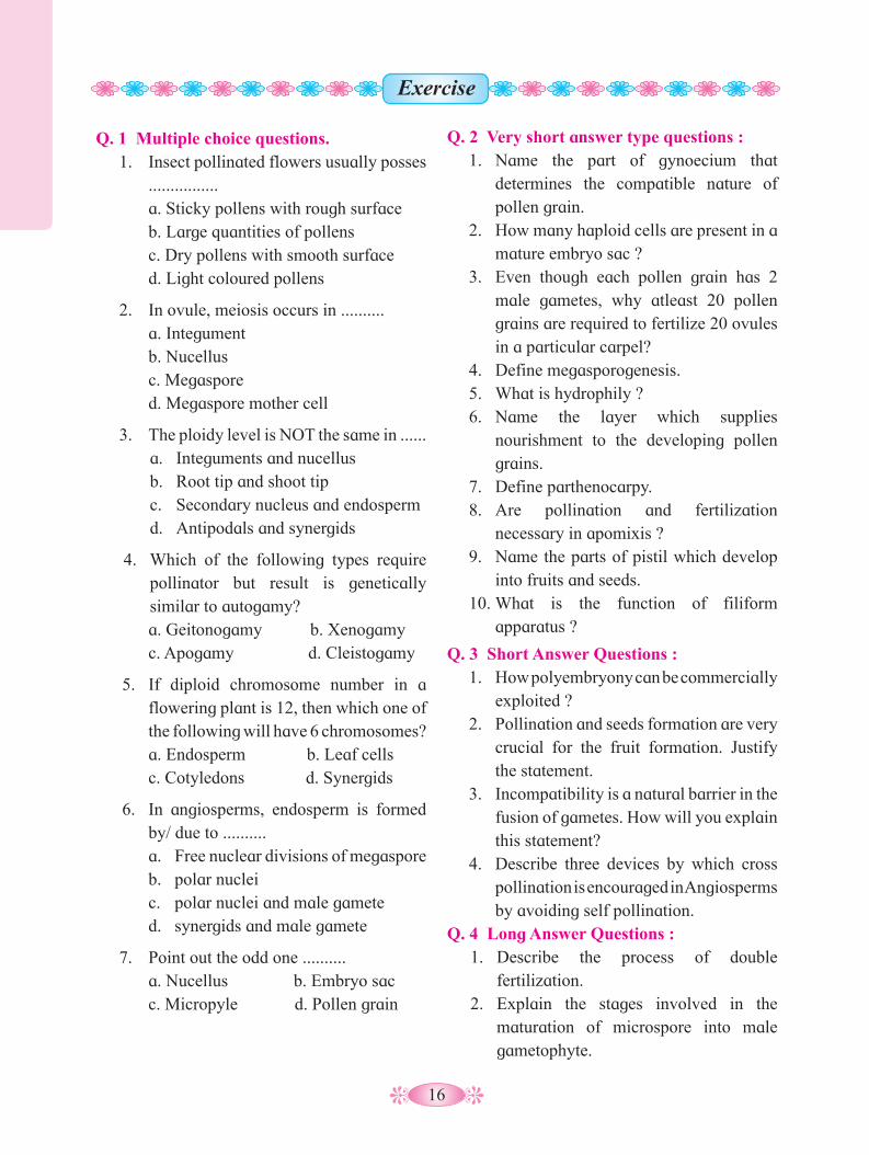
Q. 1 Multiple choice questions.1. Insect pollinated flowers usually posses ................ a. Sticky pollens with rough surface b. Large quantities of pollens c. Dry pollens with smooth surface d. Light coloured pollens
2. In ovule, meiosis occurs in .......... a. Integument b. Nucellus c. Megaspore d. Megaspore mother cell
3. The ploidy level is NOT the same in ......a. Integuments and nucellusb. Root tip and shoot tipc. Secondary nucleus and endospermd. Antipodals and synergids
4. Which of the following types require pollinator but result is genetically similar to autogamy?
a. Geitonogamy b. Xenogamy c. Apogamy d. Cleistogamy
5. If diploid chromosome number in a flowering plant is 12, then which one of the following will have 6 chromosomes?
a. Endosperm b. Leaf cells c. Cotyledons d. Synergids
6. In angiosperms, endosperm is formed by/ due to ..........a. Free nuclear divisions of megasporeb. polar nucleic. polar nuclei and male gameted. synergids and male gamete
7. Point out the odd one .......... a. Nucellus b. Embryo sac c. Micropyle d. Pollen grain
Q. 2 Very short answer type questions :1. Name the part of gynoecium that
determines the compatible nature of pollen grain.
2. How many haploid cells are present in a mature embryo sac ?
3. Even though each pollen grain has 2 male gametes, why atleast 20 pollen grains are required to fertilize 20 ovules in a particular carpel?
4. Define megasporogenesis. 5. What is hydrophily ?6. Name the layer which supplies
nourishment to the developing pollen grains.
7. Define parthenocarpy.8. Are pollination and fertilization
necessary in apomixis ?9. Name the parts of pistil which develop
into fruits and seeds.10. What is the function of filiform
apparatus ?
Q. 3 Short Answer Questions :1. How polyembryony can be commercially
exploited ?2. Pollination and seeds formation are very
crucial for the fruit formation. Justify the statement.
3. Incompatibility is a natural barrier in the fusion of gametes. How will you explain this statement?
4. Describe three devices by which cross pollination is encouraged in Angiosperms by avoiding self pollination.
Q. 4 Long Answer Questions :1. Describe the process of double
fertilization.2. Explain the stages involved in the
maturation of microspore into male gametophyte.
Exercise
17
3. Explain the development of dicot embryo.
Q. 5 Fill in the blanks:
Q. 7 Match the column.
Column - I(Structure before seed formation.
Column - II(Structure after seed formation.
A. Funiculus I. HilumB. Scar of Ovule II. TegmenC. Zygote III. TestaD. Inner integument IV. Stalk of seed
V. Embryo
a. A - V, B - I, C - II, D - IV b. A - III, B - IV, C - I, D - Vc. A - IV, B - I, C - V, D - II d. A - IV, B - V, C - III, D - II
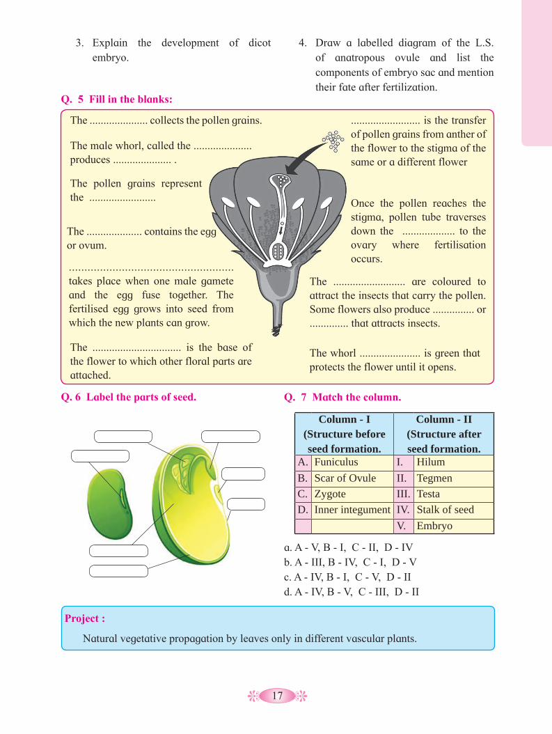
The ..................... collects the pollen grains.
The male whorl, called the ..................... produces ..................... .
The pollen grains represent the ........................
The .................... contains the egg or ovum.
..................................................... takes place when one male gamete and the egg fuse together. The fertilised egg grows into seed from which the new plants can grow.
......................... is the transfer of pollen grains from anther of the flower to the stigma of the same or a different flower
Once the pollen reaches the stigma, pollen tube traverses down the ................... to the ovary where fertilisation occurs.
The ................................ is the base of the flower to which other floral parts are attached.
The .......................... are coloured to attract the insects that carry the pollen. Some flowers also produce ............... or .............. that attracts insects.
The whorl ...................... is green that protects the flower until it opens.
4. Draw a labelled diagram of the L.S. of anatropous ovule and list the components of embryo sac and mention their fate after fertilization.
Q. 6 Label the parts of seed.
Project :
Natural vegetative propagation by leaves only in different vascular plants.

18
Gemmule Formation: Gemmule is an internal bud formed only
in sponges. It has asexually produced mass or aggregation of dormant cells, the archaeocytes capable of developing into a new organism. The archaeocytes get coated by a thick resistant layer of secretion by amoebocytes. The gemmule is formed to overcome unfavourable conditions. On return of favourable conditions of water and temperature, the gemmules hatch and develop into a new individual. e.g. Spongilla.
We know that reproduction is one of the major life processes of any living organism. It helps in maintaining the continuity of the species. Reproduction is defined as the biological process of formation of new life forms from pre-existing similar life. It thus becomes a vital process which enables the species to survive over a long period, even though the individuals or organisms live naturally for a limited period of time i.e. their life span. In this chapter, we will learn about the various methods of reproduction in animals the human reproductive system, gametogenesis and fertilization, early embryology, parturition and reproductive health.
Reproduction in animals occurs mainly by two methods i.e. asexual and sexual.
2.1 Asexual Reproduction in animals : It is a common method among lower
animals. It does not involve meiosis nor the gamete formation and fusion. The formation of progeny is by a single parent only and does not involve both the sexes, so it is called asexual reproduction. The progeny or daughter cells are genetically identical to the single parent and are also referred to as clones. The lower animals reproduce asexually by gemmule formation and budding.
Reproduction in Lower and Higher Animals2
Budding: It is a simple method of asexual
reproduction normally occuring in favourable conditions. It is seen in a variety of animals like coelenterates (Hydra and corals) and in some colonial ascidians. In Hydra, a small outgrowth is produced towards the basal end of the body.
Can you recall?
1. Enlist the various life processes. Name the life process which is responsible for continuation of the human race.
2. What are the common methods of reproduction in the unicellular organisms like Euglena, Amoeba and Paramoecium?
3. What type of asexual reproduction occurs in Hydra?
4. What are the different methods of reproduction in animals?
Fig. 2.1 : Gemmule
Micropyle
Inner membrane
Monaxon spicules
Archaeocytes
Outer membrane
Fig. 2.2 : Budding in Hydra
Bud Developing bud
Fully mature
bud
Young Hydra
separates

19
The sexually reproducing animals show two main phases in their life time. The earlier juvenile phase mainly represents physical growth phase starting from birth. The animals can not reproduce sexually in this phase. The later Reproductive maturity phase is attained usually after physical growth is almost over. It involves growth and activity of the sex organs. The animal can reproduce sexually in this phase. Both these periods (phases) are of variable duration in different animals. After attaining sexual maturity, the animal exhibits various events, namely pre-fertilization (gametogenesis and gamete transfer), fertilization (fusion of male and female gametes) and post fertilization events (formation of zygote and embryogenesis).
The sexually reproducing animals show various breeding patterns. Some like the goat, sheep, and donkey are seasonal breeders while humans and apes are continuous breeders. They can breed throughout the year.
Human Reproduction : Humans are sexually reproducing animals.
The process of reproduction involves various sequential steps such as gametogenesis, insemination, internal fertilization (i.e. fusion of male and female gametes), zygote formation and embryogenesis, gestation and parturition.
The gametes, sperms and eggs are produced by the primary sex organs, testis in male and ovary in female. Organs other than testis and ovary, are called secondary sex organs of the male and female. As male and female can be externally differentiated by certain specific features called secondary sexual characters, they are called sexual dimorphic characters. In males, presence of beard, moustache, hair on the chest, muscular body, enlarged larynx (Adam’s apple) are secondary sexual characters while in females these characters are the developed breast, broader pelvis and high pitched voice.
Do you know ?
Regeneration: A word which in biology refers to the process observed in all living organisms from the unicellular bacteria upto the most complex multicellular forms e.g. humans. By this process, the organism can fundamentally repair or regrow or restore its lost or damaged part. Though it involves asexual processes, it differs distinctly from reproduction e.g. a damaged Hydra can regenerate its lost part. Similarly Planaria if wounded, its cells become active and regenerate lost part or organ back to its original state. They can also reproduce asexually by fragmentation. Also, it is seen in planarians that the anterior end exerts a pull on the posterior end resulting in a constriction in the middle part and splitting into two pieces. Each piece grows into a new Planaria. i.e. two clones of the original have been formed.
It develops as a bud which grows and forms tentacles and develops (get transformed) into a new individual. This process is called budding. The young Hydra gets detached from the parent and becomes an independent new organism.
2.2 Sexual reproduction in animals : It is the process which involves the
production of offspring by the formation and fusion of gametes. It is also called amphimixis. In animals, gamete formation primarily involves meiosis.

20
Can you recall?
Histology of Testis: The testis is externally covered by a
collagenous connective tissue layer called tunica albuginea. Outer to it is an incomplete peritoneal covering called tunica vaginalis, and inner to it is tunica vasculosa, a thin membranous and vascular layer. Fibers from tunica albuginea divide each testis into about 200-300 testicular lobules (refer dig. 2.3 L. S. of testis). Each with 1-4 highly coiled seminiferous tubules. Each seminiferous tubule is internally lined by cuboidal germinal epithelial cells (spermatogonia) and few large pyramidal cells called Sertoli or sustentacular cells.
A. Male Reproductive System : It consists of the primary male organ
(gonad) called testes, the accessory ducts and glands which form internal and external genitalia.
1
2
3
4 5
6
7
8 9
10
a. Testes: A pair of testes, mesodermal in origin, are
formed in the lower abdominal cavity. They are located in a pouch called scrotum. During early foetal life, the testes develop in abdominal cavity and later they descend into the scrotal sac through a passage called inguinal canal. Each testis is oval in shape, 4 to 5cm long, 2 to 3cm wide and 3cm thick.
Label the given male reproductive system you have studied.
Vas deferens
Vasa efferentia
Seminiferous tubule
Body of epididymis
Rete testis
Tail of epididymis
Spermatic cord
Blood vessels and nerves
Head of epididymis
Tunica vaginalis
Tunica albuginea
Lobules
Septum
Fig. 2.3 : L. S. of testis
Fig. 2.4 : T. S. of Testis
Sperm bundle
Connective tissue
Tunica albuginea
Interstitial cells (Leydig cells)
Seminiferous tubule
Germinal epithelium
Sertoli cell
Basement membrane
The germinal epithelial cells undergo gametogenesis to form the spermatozoa. Sertoli cells provide nutrition to the developing sperms. Various stages of spermatogenesis can be seen in the seminiferous tubules. The inner most spermatogonial cell (2n), primary spermatocyte (2n), secondary spermatocyte (n), spermatids (n) and sperms (n). The Interstitial or Leydig’s cells lie in between the seminiferous tubules. They secrete the male hormone androgen or testosterone.

21
c. Glands: The male accessory glands are as follows:
• Seminal vesicles: It is a pair of glands lying on the posterior side of urinary bladder. It secretes an alkaline seminal fluid which contains fructose, fibrinogen and prostaglandins. It contributes about 60% of the total volume of the semen. Fructose provides energy for sperm movement while fibrinogen coagulates the semen into a bolus for quick propulsion in the vagina. The prostaglandins stimulate reverse peristalsis in vagina and uterus aiding faster movement of sperms towards the egg in the female body.
• Prostate gland: It is a large and single gland made up of 20-30 lobes and is located underneath the urinary bladder. It surrounds the urethra and releases a milky white and slightly acidic prostatic fluid into the urethra. It forms about 30% of volume of semen. It contains citric acid, acid phosphatase and various other enzymes. The acid phosphatase protects the sperms from the acidic environment of vagina.
b. Accessory ducts: The accessory ducts include rete testis,
vasa efferentia, epididymis, vas deferens, ejaculatory duct and urethra. All the seminiferous tubules of the testis at the posterior surface form a network of tubules called rete testis. 12-20 fine tubules arising from rete testis are vasa efferentia. They carry the sperms from the testis and open into the epididymis. It is a long and highly coiled tube which is differentiated into an upper caput-, middle corpus- and lower cauda epididymis. The sperms undergo maturation in epididymis. Posteriorly it leads into the vas deferens which travels upto the abdominal cavity and loops over the ureter to open into the urethra. Before doing so, it joins the duct of seminal vesicle to form the ejaculatory duct. The ejaculatory duct passes through the prostate gland and opens into the urethra. The urethra provides a common passage for the urine and semen and hence is also called urinogenital duct. In males the urethra is long and extends through the penis. It opens to the outside by an opening called the urethral meatus or urethral orifice. All the accessory ducts except urethra are present in pairs.
• Cowper’s gland / Bulbourethral gland : It is a small, pea sized and paired gland situated on either side of urethra. These
Do you know ?
1. Presence of the peritoneal covering around the testis is an indication of its abdominal origin.
2. The testis are suspended in the scrotum by the spermatic cord.
3. Testosterone hormone stimulates the descent of testis and the fibro-muscular band called gubernaculum in the scrotum.
4. In some males a loop of the intestine may pass through the inguinal canal into the scrotum and cause a condition called inguinal hernia.
Activity :
Find the symptoms of prostate cancer.
Always Remember Prostate cancer is cancer of the prostate gland. Men who are over 50 years of age and have a daily high consumption of fat, have an increased risk of prostate cancer.
Internet my friend
What is the role of prostaglandin?
22
1. Ovary : It is the primary female sex organ. Its main function is production of egg or ovum and the female reproductive hormones. It is solid, oval or almond shaped organ. It is 3.0 cm in length, 1.5 cm in breadth and 1.0 cm thick. It is located in the upper lateral part of the pelvis near the kidneys. Each ovary is held in position by ligaments by attaching it to the uterus and the abdominal wall. The largest of these is the broad ligament formed by a fold of peritoneum. It holds the ovary, oviduct and the uterus to the dorsal body wall. The ovarian ligament attaches ovary to the uterus. The ovary produces five hormones viz, estrogen, progesteron, relaxin, activin and inhibin.
Structure and development of the ovary : Each ovary is a compact structure differentiated into a central part called medulla and the outer part called cortex. The cortex is covered externally by a layer of germinal
glands secrete an alkaline, viscous, mucous like fluid which acts as a lubricant during copulation.
Semen : It is the viscous, alkaline and milky
fluid (pH 7.2 to 7.7) ejaculated by the male reproductive system. Normally 2.5 to 4.0 ml of semen is given out during a single ejaculation and it contains about 400 million sperms. It contains secretion of the epididymis and the accessory glands for nourishing (fructose), neutralizing acidity (Ca++, bicarbonates), activation for movement (prostaglandins).
d. External genitalia: It includes the penis and the scrotum.
The penis is the male copulatory organ. It is cylindrical and muscular with three bundles of erectile tissue- a pair of postero-lateral tissue called corpora cavernosa and a median corpus spongiousm. The swollen tip of the penis is called glans penis. It is covered by a loose fold of skin called foreskin or prepuce.
Scrotum : It is a loose pouch of pigmented skin
lying behind the penis and is divided into a right and left scrotal sac by a septum of tunica dartos made of smooth muscle fibres. The foetal testes are guided into and retained in the scrotum by a short fibro muscular band called gubernaculum. The testes remain suspended in scrotum by a spermatic chord. Failure of testis to descend into scrotum is called cryptorchidism. The failure also results in the sterility. The cremaster and dartos muscles of scrotum help in drawing testes close or away from the body. This helps in maintaining the temperature of the testis 2-30C lower than the normal body temperature, necessary for spermatogenesis.
B. Female Reproductive System: The female reproductive system consist of
the following parts :
1. A pair of ovaries 2. A pair of oviducts 3. Uterus 4. Vagina 5. External genitalia (vulva) 6. A pair of vestibular glands7. A pair of mammary glands
Can you recall?
Give labels to given female reproductive system:

23
epithelium. The stroma or loose connective tissue of the medulla has blood vessels, lymph vessels, and nerve fibres. The outer cortex is more compact and granular. It shows large number of tiny masses of cells called ovarion follicles. These are collectively formed from the immature ova originating from cells of the dorsal endoderm of the yolk sac. The cells migrate to the gonadal ridge during embryonic development and divide mitotically. Now these cells are called oogonia. As the oogonia continue to grow in size they are surrounded by a layer of granulosa cells and form the rudiments of the ovarian follicles. The process of oogenesis starts much before the birth of the female baby and by the end of twelve weeks the ovary is fully formed. It has more than two million primordial follicles in it.
The large scale destruction of the primordial follicles during growth is called atresia.
The development of the primordial follicles into mature or Graafian follicles restarts with the onset of puberty. During each menstrual cycle only one of the primordial follicle starts growing to form the Graafian follicle.
In each cycle, alternately one of the two ovaries produces the Graafian follicle.
The 1st menstrual cycle or menarche begins normally at about 13 years and Menopause i.e. stopping of the cycles happens at age 45 to 55 years. The period in between menarche and menopause is the reproductive age of the female and is approximately 32 years. In this time the female will be producing a maximum of about 416 eggs (32 ×13 = 416 eggs).
Ovarian histology of a mature female : In the histology of ovary, we have
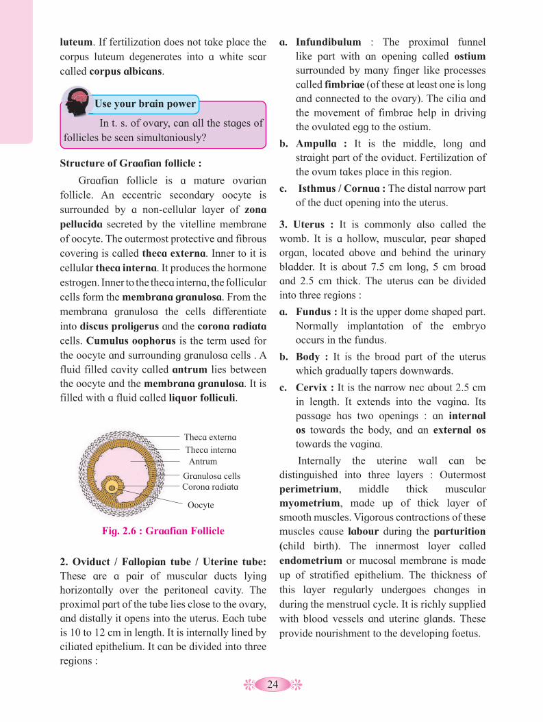
discussed the primary structure of ovary. The following discussion includes the changes seen in a mature ovary, primarily in the cortex. The different stages of development of the oocyte can be seen. These changes in the ovary are cyclic, occuring during each menstrual cycle and it involves maturation of the primordial follicles into primary, secondary and Graafian follicles. Each primary follicle has multilayered cuboidal follicular cells. The stroma cells add theca over the follicle. It now changes into a secondary follicle. There is growth of the oocyte and the granulosa cells increase in number. They start producing the hormone estrogen. The secondary follicle grows into the Graafian follicle by addition of more follicular cells. As this process of maturation of follicles takes place, they begin to move towards the surface of ovary. The Graafian follicle presses against the thin wall of the ovary giving it a blistered appearance. The egg is released from the Graafian follicle during ovulation and the remaining part of the follicle changes into a temporary endocrine gland called corpus
Fig. 2.5 : T.S. of ovary
Blood vessels
Primordial follicle
Primary follicle
Secondary follicle Tertiary
follicle
Cortex
Mature follicle
Oocyte
Medulla
Ovulated ovum
Germinal epithelium Corpus luteum
Corpus albicans
Day 1 Day 12
Day 14
Day 20
The cells of germinal epithelium give rise to groups of oogonia projecting into the cortex in the form of cords called egg tubes of Pfluger. Each cord at its end has a round mass of oogonial cells called egg nests, from which the primordial ovarian follicles develop. Each primordial follicle has, at its center a large primary oocyte (2n) surrounded by a single layer of flat follicular cells. The primary oocyte starts with its meiotic division but gets arrested it at meiosis I. Of the two million primordial follicles embedded in the foetal ovary only about one million remain at birth and only about 40,000 remain at the time of puberty.
24
a. Infundibulum : The proximal funnel like part with an opening called ostium surrounded by many finger like processes called fimbriae (of these at least one is long and connected to the ovary). The cilia and the movement of fimbrae help in driving the ovulated egg to the ostium.
b. Ampulla : It is the middle, long and straight part of the oviduct. Fertilization of the ovum takes place in this region.
c. Isthmus / Cornua : The distal narrow part of the duct opening into the uterus.
3. Uterus : It is commonly also called the womb. It is a hollow, muscular, pear shaped organ, located above and behind the urinary bladder. It is about 7.5 cm long, 5 cm broad and 2.5 cm thick. The uterus can be divided into three regions :
a. Fundus : It is the upper dome shaped part. Normally implantation of the embryo occurs in the fundus.
b. Body : It is the broad part of the uterus which gradually tapers downwards.
c. Cervix : It is the narrow nec about 2.5 cm in length. It extends into the vagina. Its passage has two openings : an internal os towards the body, and an external os towards the vagina.
Internally the uterine wall can be distinguished into three layers : Outermost perimetrium, middle thick muscular myometrium, made up of thick layer of smooth muscles. Vigorous contractions of these muscles cause labour during the parturition (child birth). The innermost layer called endometrium or mucosal membrane is made up of stratified epithelium. The thickness of this layer regularly undergoes changes in during the menstrual cycle. It is richly supplied with blood vessels and uterine glands. These provide nourishment to the developing foetus.
luteum. If fertilization does not take place the corpus luteum degenerates into a white scar called corpus albicans.
2. Oviduct / Fallopian tube / Uterine tube: These are a pair of muscular ducts lying horizontally over the peritoneal cavity. The proximal part of the tube lies close to the ovary, and distally it opens into the uterus. Each tube is 10 to 12 cm in length. It is internally lined by ciliated epithelium. It can be divided into three regions :
In t. s. of ovary, can all the stages of follicles be seen simultaniously?
Use your brain power
Structure of Graafian follicle :
Graafian follicle is a mature ovarian follicle. An eccentric secondary oocyte is surrounded by a non-cellular layer of zona pellucida secreted by the vitelline membrane of oocyte. The outermost protective and fibrous covering is called theca externa. Inner to it is cellular theca interna. It produces the hormone estrogen. Inner to the theca interna, the follicular cells form the membrana granulosa. From the membrana granulosa the cells differentiate into discus proligerus and the corona radiata cells. Cumulus oophorus is the term used for the oocyte and surrounding granulosa cells . A fluid filled cavity called antrum lies between the oocyte and the membrana granulosa. It is filled with a fluid called liquor folliculi.
Fig. 2.6 : Graafian Follicle
Theca externa
Theca interna Antrum
Granulosa cells Corona radiata
Oocyte
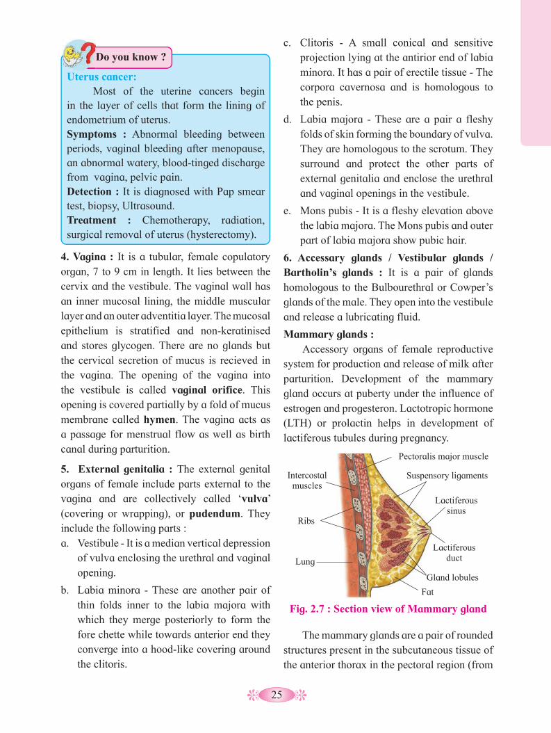
25
c. Clitoris - A small conical and sensitive projection lying at the antirior end of labia minora. It has a pair of erectile tissue - The corpora cavernosa and is homologous to the penis.
d. Labia majora - These are a pair a fleshy folds of skin forming the boundary of vulva. They are homologous to the scrotum. They surround and protect the other parts of external genitalia and enclose the urethral and vaginal openings in the vestibule.
e. Mons pubis - It is a fleshy elevation above the labia majora. The Mons pubis and outer part of labia majora show pubic hair.
6. Accessary glands / Vestibular glands / Bartholin’s glands : It is a pair of glands homologous to the Bulbourethral or Cowper’s glands of the male. They open into the vestibule and release a lubricating fluid.
Mammary glands : Accessory organs of female reproductive
system for production and release of milk after parturition. Development of the mammary gland occurs at puberty under the influence of estrogen and progesteron. Lactotropic hormone (LTH) or prolactin helps in development of lactiferous tubules during pregnancy.
4. Vagina : It is a tubular, female copulatory organ, 7 to 9 cm in length. It lies between the cervix and the vestibule. The vaginal wall has an inner mucosal lining, the middle muscular layer and an outer adventitia layer. The mucosal epithelium is stratified and non-keratinised and stores glycogen. There are no glands but the cervical secretion of mucus is recieved in the vagina. The opening of the vagina into the vestibule is called vaginal orifice. This opening is covered partially by a fold of mucus membrane called hymen. The vagina acts as a passage for menstrual flow as well as birth canal during parturition.
5. External genitalia : The external genital organs of female include parts external to the vagina and are collectively called ‘vulva’ (covering or wrapping), or pudendum. They include the following parts :a. Vestibule - It is a median vertical depression
of vulva enclosing the urethral and vaginal opening.
b. Labia minora - These are another pair of thin folds inner to the labia majora with which they merge posteriorly to form the fore chette while towards anterior end they converge into a hood-like covering around the clitoris.
Do you know ?
Uterus cancer: Most of the uterine cancers begin in the layer of cells that form the lining of endometrium of uterus.Symptoms : Abnormal bleeding between periods, vaginal bleeding after menopause, an abnormal watery, blood-tinged discharge from vagina, pelvic pain. Detection : It is diagnosed with Pap smear test, biopsy, Ultrasound.Treatment : Chemotherapy, radiation, surgical removal of uterus (hysterectomy).
Fig. 2.7 : Section view of Mammary gland
Pectoralis major muscle
Intercostal muscles
Suspensory ligaments
Lactiferous sinus
Lactiferous duct
Gland lobules
Fat
Ribs
Lung
The mammary glands are a pair of rounded structures present in the subcutaneous tissue of the anterior thorax in the pectoral region (from

26
Puberty / Sexual maturity in Males : Puberty is the age at which the reproductive
system becomes functional, sex organs begin to produce gametes and sex hormones. In males the onset of puberty occurs at age 12-15 years. Under the influence of testosterone, the secondary sexual characters appear. Thereafter it remains functional throughout the life.
Puberty / Sexual maturity in Females : The reproductive system becomes
functional at puberty. It is characterised by onset of menstrual cycle also called menarche, which usually occurs at age 10-14 years. However, unlike in the males, the mature females show cyclic changes in their reproductive system- the menstrual cycles. These cycles continue only upto menopause. This normally happens at age 45-50 years. The period from menarche to menopause is thus the reproductive age of the female. The female is unable to bear children (by natural method) after menopause. Menarche, menstrual cycles and menopause are controlled by gonadotropic hormones.
2.3 Menstrual cycle (Ovarian cycle): Menstrual cycle is the characteristic
feature of primates including human. It involves a series of cyclic changes in the ovary and the female reproductive tract, mainly in the uterus. These changes take place under the effect of gonadotropins and the ovarian hormones respectively. The cycles are repeated with a periodicity of approximately 28 days. The middle of each cycle is characterised by the release of an egg. This egg in every cycle comes alternately from one of the two ovaries. The cycle is divided into four phases.
a. Menstrual phase: The begining of each cycle is taken as
the first day where menses or loss of blood (45-100ml) takes place and it lasts for approximately five days (average 3-7 days).
Endometrium of uterus breaks down under the effect of prostaglandins released due to decreased levels of progesteron and estrogen. Due to this blood, tissue fluid, mucus, endometrial lining and the unfertilized oocyte is discharged through vagina. The endometrial lining becomes very thin i.e. about 1 mm. The menstrual discharge continues for an average of 5 days, however this blood does not clot
2nd to 6th rib). These are modified sweat glands. Each mammary gland contains fatty connective tissue and numerous lactiferous ducts. The glandular tissue of each breast is divided into 15-20 irregularly shaped mammary lobes, each with alveolar glands and lactiferous duct. Alveolar glands secrete milk which is stored in the lumen of alveoli. The alveoli open into mammary tubules. The tubules of each lobe join to form a mammary duct. Many mammary ducts join to form a wider mammary ampulla, which is connected to lactiferous duct. These converge towards the nipple located near the tip of the breast. It is surrounded by a dark brown coloured and circular area of the skin called areola.
Do you know ?
Breast cancer : Symptoms: First symptom of breast cancer is a lump in breast or underarm. Lump is painless. Swelling of all or part of breast. Skin irritation, Breast or nipple pain, nipple retraction, Redness, scaliness or thickening of nipple or breast skin, discharge, etc.Detection: Mammogram (x-ray), ultrasound, MRI, Biopsy, Blood test.Treatment: Radiation therapy, chemotherapy lumpectomy, Mammoplasty
Do you know ?
Weaning : Mother’s milk is replaced gradually by solid food after some time. This process is called weaning.
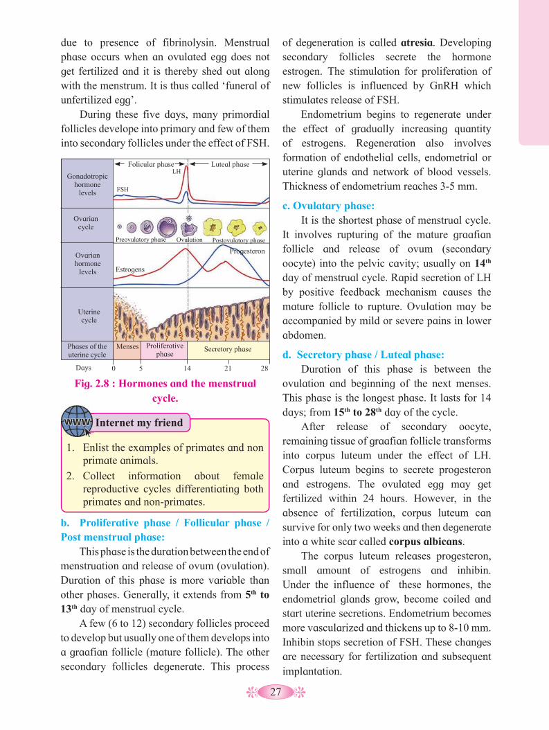
27
due to presence of fibrinolysin. Menstrual phase occurs when an ovulated egg does not get fertilized and it is thereby shed out along with the menstrum. It is thus called ‘funeral of unfertilized egg’.
During these five days, many primordial follicles develope into primary and few of them into secondary follicles under the effect of FSH.
of degeneration is called atresia. Developing secondary follicles secrete the hormone estrogen. The stimulation for proliferation of new follicles is influenced by GnRH which stimulates release of FSH.
Endometrium begins to regenerate under the effect of gradually increasing quantity of estrogens. Regeneration also involves formation of endothelial cells, endometrial or uterine glands and network of blood vessels. Thickness of endometrium reaches 3-5 mm.
c. Ovulatary phase: It is the shortest phase of menstrual cycle.
It involves rupturing of the mature graafian follicle and release of ovum (secondary oocyte) into the pelvic cavity; usually on 14th day of menstrual cycle. Rapid secretion of LH by positive feedback mechanism causes the mature follicle to rupture. Ovulation may be accompanied by mild or severe pains in lower abdomen.
d. Secretory phase / Luteal phase: Duration of this phase is between the
ovulation and beginning of the next menses. This phase is the longest phase. It lasts for 14 days; from 15th to 28th day of the cycle.
After release of secondary oocyte, remaining tissue of graafian follicle transforms into corpus luteum under the effect of LH. Corpus luteum begins to secrete progesteron and estrogens. The ovulated egg may get fertilized within 24 hours. However, in the absence of fertilization, corpus luteum can survive for only two weeks and then degenerate into a white scar called corpus albicans.
The corpus luteum releases progesteron, small amount of estrogens and inhibin. Under the influence of these hormones, the endometrial glands grow, become coiled and start uterine secretions. Endometrium becomes more vascularized and thickens up to 8-10 mm. Inhibin stops secretion of FSH. These changes are necessary for fertilization and subsequent implantation.
Internet my friend
1. Enlist the examples of primates and non primate animals.
2. Collect information about female reproductive cycles differentiating both primates and non-primates.
Fig. 2.8 : Hormones and the menstrual cycle.
Gonadotropic hormone
levels
Ovarian cycle
Ovarian hormone
levels
Uterine cycle
Phases of the uterine cycle
Menses Proliferative phase
Secretory phase
Folicular phase Luteal phase
FSH
Preovulatory phase
LH
Ovulation Postovulatory phase
Estrogens
Progesteron
Days 0 5 14 21 28
b. Proliferative phase / Follicular phase / Post menstrual phase:
This phase is the duration between the end of menstruation and release of ovum (ovulation). Duration of this phase is more variable than other phases. Generally, it extends from 5th to 13th day of menstrual cycle.
A few (6 to 12) secondary follicles proceed to develop but usually one of them develops into a graafian follicle (mature follicle). The other secondary follicles degenerate. This process

28
2.4 Gametogenesis: The gametogenesis is the process of
formation of gametes in sexually reproducing animals. The male gamete is sperm and the female gamete is ovum or egg. The gametes are formed from primordial germ cells of gonads.
Spermatogenesis: The process of formation of the male
gamete (sperm) or spermatozoa from the germinal epithelium of testis is called
I. Multiplication phase: The primordial germ cells (2n) of seminiferous tubules undergo repeated mitotic divisions to produce large number of spermatogonia (2n). Each spermatogonium is diploid and with 46 chromsomes.
II. Growth phase: Some of the spermatogonia stop dividing and grow in size to develop into primary spermatocytes (2n) due to accumulation of food.
spermatogenesis. At the onset of puberty, the hypothalamus begins secretion of gonadotropin releasing hormone (GnRH). It initiates the significant increase in the secretion of follicle stimulating hormone (FSH) which induces spermatogenesis. Each seminiferous tubules is lined by a single layer of cuboidal epithelial cells called germinal epithelium.
The cells of germinal epithelium undergo spermatogenesis to produce sperms. Process of spermatogenesis involves three phases.
However, if the ovulated egg gets fertilized and the embryo is implanted, there is secretion of human chorionic gonadotropin (hCG), which extends the life of corpus luteum and stimulates it’s secretory activity. Presence of hCG in maternal blood and urine is an indicator of pregnancy. In absence of fertilization, next menstrual cycle begins.
Use your brain power
Why the menstruation is painful in some women?
Can you tell?
Can you tell the names of primates who show the presence of menstrual cycle?
Always Remember
Hygiene practices during menstruation :• Keep the pubic area clean. • Change the sanitary napkin every 4-5
hours. • Maintaining personal hygiene during
menstruation is important to reduce the risk of infection.
• Dispose used sanitary napkin properly. • Using damp and dirty clothes or using
a sanitary napkin for a longer time can act as a perfect environment for growth and multiplication of harmful bacteria and lead to infections.
Fig. 2.9 : Spermatogenesis
Spermatogonia (2n)
Primordial cell (2n)
Primary spermatocytes (2n)
II. Growth phase
I. Multiplication phase
Secondary spermatocytes (n)
Spermatids (n)
Spermatozoa (n)
Mitosis
Meiosis I
Meiosis II
Mei
otic
div
isio
n
III.
Mat
urat
ion
phas
e
Sper
mio
gene
sis
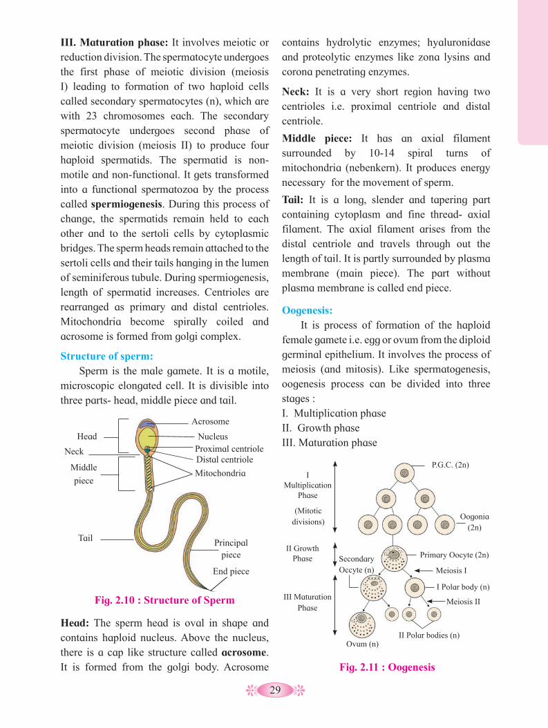
29
III. Maturation phase: It involves meiotic or reduction division. The spermatocyte undergoes the first phase of meiotic division (meiosis I) leading to formation of two haploid cells called secondary spermatocytes (n), which are with 23 chromosomes each. The secondary spermatocyte undergoes second phase of meiotic division (meiosis II) to produce four haploid spermatids. The spermatid is non-motile and non-functional. It gets transformed into a functional spermatozoa by the process called spermiogenesis. During this process of change, the spermatids remain held to each other and to the sertoli cells by cytoplasmic bridges. The sperm heads remain attached to the sertoli cells and their tails hanging in the lumen of seminiferous tubule. During spermiogenesis, length of spermatid increases. Centrioles are rearranged as primary and distal centrioles. Mitochondria become spirally coiled and acrosome is formed from golgi complex.
Structure of sperm: Sperm is the male gamete. It is a motile,
microscopic elongated cell. It is divisible into three parts- head, middle piece and tail.
contains hydrolytic enzymes; hyaluronidase and proteolytic enzymes like zona lysins and corona penetrating enzymes.
Neck: It is a very short region having two centrioles i.e. proximal centriole and distal centriole.
Middle piece: It has an axial filament surrounded by 10-14 spiral turns of mitochondria (nebenkern). It produces energy necessary for the movement of sperm.
Tail: It is a long, slender and tapering part containing cytoplasm and fine thread- axial filament. The axial filament arises from the distal centriole and travels through out the length of tail. It is partly surrounded by plasma membrane (main piece). The part without plasma membrane is called end piece.
Oogenesis: It is process of formation of the haploid
female gamete i.e. egg or ovum from the diploid germinal epithelium. It involves the process of meiosis (and mitosis). Like spermatogenesis, oogenesis process can be divided into three stages :I. Multiplication phase II. Growth phaseIII. Maturation phase
Head: The sperm head is oval in shape and contains haploid nucleus. Above the nucleus, there is a cap like structure called acrosome. It is formed from the golgi body. Acrosome
Fig. 2.10 : Structure of Sperm
Acrosome
Nucleus
Distal centriole
Mitochondria
Head
Neck
Middle
piece
Principal piece
End piece
Tail
Proximal centriole
Fig. 2.11 : Oogenesis
IMultiplication
Phase
II Growth Phase
III Maturation Phase
P.G.C. (2n)
(Mitotic divisions)
Oogonia (2n)
Primary Oocyte (2n)
Meiosis I
Secondary Occyte (n)
I Polar body (n)
Meiosis II
II Polar bodies (n) Ovum (n)
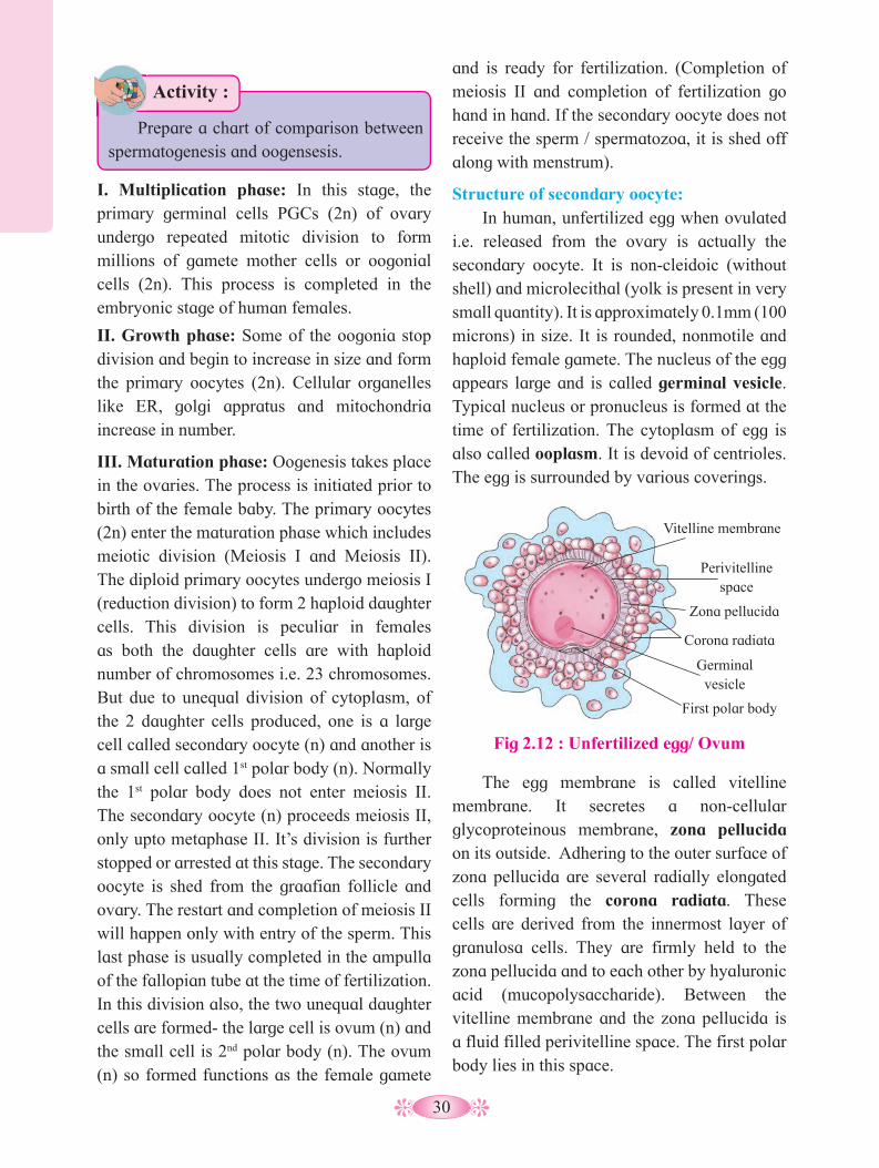
30
I. Multiplication phase: In this stage, the primary germinal cells PGCs (2n) of ovary undergo repeated mitotic division to form millions of gamete mother cells or oogonial cells (2n). This process is completed in the embryonic stage of human females.
II. Growth phase: Some of the oogonia stop division and begin to increase in size and form the primary oocytes (2n). Cellular organelles like ER, golgi appratus and mitochondria increase in number.
III. Maturation phase: Oogenesis takes place in the ovaries. The process is initiated prior to birth of the female baby. The primary oocytes (2n) enter the maturation phase which includes meiotic division (Meiosis I and Meiosis II). The diploid primary oocytes undergo meiosis I (reduction division) to form 2 haploid daughter cells. This division is peculiar in females as both the daughter cells are with haploid number of chromosomes i.e. 23 chromosomes. But due to unequal division of cytoplasm, of the 2 daughter cells produced, one is a large cell called secondary oocyte (n) and another is a small cell called 1st polar body (n). Normally the 1st polar body does not enter meiosis II. The secondary oocyte (n) proceeds meiosis II, only upto metaphase II. It’s division is further stopped or arrested at this stage. The secondary oocyte is shed from the graafian follicle and ovary. The restart and completion of meiosis II will happen only with entry of the sperm. This last phase is usually completed in the ampulla of the fallopian tube at the time of fertilization. In this division also, the two unequal daughter cells are formed- the large cell is ovum (n) and the small cell is 2nd polar body (n). The ovum (n) so formed functions as the female gamete
The egg membrane is called vitelline membrane. It secretes a non-cellular glycoproteinous membrane, zona pellucida on its outside. Adhering to the outer surface of zona pellucida are several radially elongated cells forming the corona radiata. These cells are derived from the innermost layer of granulosa cells. They are firmly held to the zona pellucida and to each other by hyaluronic acid (mucopolysaccharide). Between the vitelline membrane and the zona pellucida is a fluid filled perivitelline space. The first polar body lies in this space.
and is ready for fertilization. (Completion of meiosis II and completion of fertilization go hand in hand. If the secondary oocyte does not receive the sperm / spermatozoa, it is shed off along with menstrum).
Structure of secondary oocyte: In human, unfertilized egg when ovulated
i.e. released from the ovary is actually the secondary oocyte. It is non-cleidoic (without shell) and microlecithal (yolk is present in very small quantity). It is approximately 0.1mm (100 microns) in size. It is rounded, nonmotile and haploid female gamete. The nucleus of the egg appears large and is called germinal vesicle. Typical nucleus or pronucleus is formed at the time of fertilization. The cytoplasm of egg is also called ooplasm. It is devoid of centrioles. The egg is surrounded by various coverings.
Activity :
Prepare a chart of comparison between spermatogenesis and oogensesis.
Fig 2.12 : Unfertilized egg/ Ovum
Germinal vesicle
First polar body
Corona radiata
Perivitelline space
Vitelline membrane
Zona pellucida

31
The egg shows polarity. The side having germinal vesicle and first polar body is called animal pole while the side opposite to it is called vegetal pole.
2.5 Fertilization / Syngamy: Sexual reproduction primarily involves
formation and fusion of gametes. Fertilization is the later process which involves fusion of the haploid male and female gametes resulting in the formation of a diploid zygote (2n). Like in other mammals, in humans the process of fertilization is internal and it usually takes place in the ampulla of the fallopian / uterine tube. The fertilized egg or zygote will develop into an embryo and this process occurs within the uterus.
As a result of capacitation, sperms become extra active and begin to start moving upwards from vagina to uterus and to the oviducts. The prostaglandins activate the sperms. The vestibular secretions of the female also enhance sperms motility. The sperms swim at an average speed of 1.5 to 3.0 mm/min.
Sperms reach upto the ampulla as a result of their own swimming and partly by contraction of uterus and fallopian tubes stimulated by oxytocin of female. After capacitation the sperms may reach ampulla within 5 minutes. Sperms can remain viable for 24-48 hours (Ovum for about 24 hours).
b. Entry of sperm into the egg : Out of 200 to 400 million sperms, only few hundred manage to reach the ampulla. Though many sperms reach the ampulla but only a single sperm fertilizes the ovum. A sperm after reaching the egg / ovum comes to lie against it. Its acrosome releases lysins : hyaluronidase and corona penetrating enzymes. They separate and dissolve the cells of corona radiata, so the sperm head passes through the zona pellucida of egg. The zona pellucida has fertilizin receptor proteins (ZP3, ZP2). The fertilizin binds to specific acid protein- antifertilizin of sperm. It brings about attraction of sperms to the egg to enhance fertilization. Fertilizin-antifertilizin interaction is species specific. Thus, the fertilizin-antifertilizin reaction is also called compatibility reaction.
Fig. 2.13 : Process of Fertilization
Extracellular space Corona radiata Zona pellucida Second meiotic
division of oocyte Second meiotic division of first
Polar body Male pronucleus
Female pro-nucleus (swollen ovum nucleus)
Polar bodies
Male pronucleus Mitotic spindle
Centriole Female pronucleus
Zygote
Sperm nucleus
1. After the sperm penetrates the secondary oocyte, the oocyte completes meiosis II, forming the ovum and second polar body.
2. Sperm and ovum nuclei swell, forming pronuclei.
3. Pronuclei approach each other and mitotic spindle forms between them.
4. Chromosomes of both pronuclei intermix. Fertilization is accomplished.
Mechanism of fertilization : Semen released during ejaculation has sperms and some secretions. The coagulated semen now undergoes liquification and sperms become active. The mechanism of fertilization is as follows :
a. Movement of sperm towards egg :
It involves capacitation of sperms reaching the vagina. Here as many as 50% are demotilised / broken / destroyed. Remaining sperms undergo capacitation. This process requires 5-6 hours. Acrosome membrane
becomes thin, Ca++ enters the sperm and their tails begin to show rapid whiplash movements.
Always Remember
Indian law under the Hindu marriage Act has defined the marriageable age of a boy and girl. As per this act, minimum age for boy must be 21 and for a girl must be 18 years, at the time of marriage.

32
Acrosome reaction : As the sperm head touches the zona pellucida in the animal pole region, its acrosome covering ruptures to release lytic enzymes, acrosin or zona lysin. They act on the zona pellucida at the point of contact. This causes egg reaction - A small fertilization cone / cone of reception is formed on the egg membrane. The sperm head comes in contact with this cone. It results in production of a weak wave of depolarisation. Plasma membrane of the both cells dissolve at the point of contact. The sperm nucleus and the centrioles enter the egg, while other parts remain outside.
As soon as the sperm head touches the vitelline membrane, a cortical reaction gets activated changing the vitelline membrane into a fertilization membrane by deactivating the sperm receptors of zona pellucida. A distinct perivitelline space is created around the fertilization membrane. This prevents any further entry of other sperms into the egg i.e. polyspermy is avoided.
process called syngamy or karyogamy. The zygote is thus formed. The proximal centriole recieved from the sperm helps in formation of the synkaryon spindle and cleavage of cell into two blastomeres.
c. Activation of ovum : The ovum before fertilization was at metaphase II stage. With a contact of sperm head to the vitelline membrane of egg, it gets activated to resume and complete its meiosis II. With this it gives out the second polar body. The germinal vesicle organises into female pronucleus. At this stage, it is the true ovum or egg.
Fusion of egg and sperm : The coverings of male and female pronuclei degenerate allowing the chromosomal pairing. This results in the formation of a synkaryon by the
Always Remember
• Secondary oocyte (egg) is ovulated after LH surge at about the middle of menstual cycle i.e. day 14.
• Egg (arrested at metaphase II) reaches the ampulla of uterine tube in 12-24 hours after ovulation. The cilia and the fimbriae of the fallopian tube help, direct the egg to ostium.
• During coitus/ intercourse semen is deposited into the vagina of the female. This process is called insemination.
• Human male during ejaculation gives out about 2-4ml of semen with an average count of 200-400 million sperms.
Significance of fertilization : • Secondary oocyte completes the process of
oogenesis and is transformed into a mature ovum (n).
• The diploid chromosome number is restored in the zygote by the process of syngamy.
• The ovum lacks the centrioles necessary for further divisions, are received from the sperm during fertilization.
• Fertilization involves fusion of male and female gametes from the two parents. It results in variations which are significant to evolution.
• Sex of the offspring is determined.Do you know ?
1. What would happen if the sperm fuses with the egg before it reaches the fallopian tube?
2. What is ectopic pregnancy? Can ectopic pregnancy continue upto full term?
Internet my friend
Find out about extraembryonic membranes.

33
arranged. Till the formation of morula the zona pellucida is retained around the embryo and thus, there is no change in the overall size from zygote to morula. The morula reaches the isthmus and gains entry into the uterus by the end of day 4.
2.6 Embryonic development : The zygote formed as a result of syngamy
is activated to divide.
Cleavage : It is the process of early mitotic division of
the zygote into a hollow multicellular blastula. It does not involve the growth of the daughter cells. The cells formed by cleavage are called blastomeres.
Since, there is no growth phase between the cleavages, the size of blastomeres will be reduced with every successive cleavage. As the size reduces, the metabolic rate increases. Subsequent cleavages are thus faster than earlier one. This requires rapid replication of DNA and high consumption of oxygen.
Process of cleavage : In human, cleavage is holoblastic i.e. the whole zygote gets divided. The cleavage planes may be longitudinal or meridional and equatorial or horizontal. It is radial and indeterminate i.e. fate of each blastomere is not predetermined.
The 1st cleavage in the zygote is meridional and occurs at about 30 hours after fertilization. It divides longitudinaly into two blastomeres, one slightly larger than the other. The 2nd cleavage is also longitudinal but at the right angle to the 1st one and occurs after 30 hours of 1st cleavage. The 3rd cleavage is horizontal. After 3rd cleavage the embryo is in 8-cell stage. As the cleavages are going on the young embryo is gradually being pushed towards the uterus. By the end of 4th day after fertilization, embryo is a solid ball of 16-32 cells and externally looking like mulberry. This stage is thus called morula. The morula shows cells of two types : smaller, clearer cells towards the outer side and inner cell mass of larger cells. Cells are compactly
Internet my friend
What is meroblastic cleavage? In which organisms, is it observed?
Fig. 2.14 : Process of cleavage and formation of Blastula
12-15 hours
Polar body
24-30 hours
Blastomere
A. 2- Cell stage B. 4- Cell stage
C. 8- Cell stage D. Morula Inner cell mass
Blastocyst cavity
Trophoblast E. Early blastocyst F. Late blastocyst
Blastulation : Blastulation is the process of formation
of the hollow and multicellular blastocyst. The embryo (blastocyst) that enters the uterus remains floating in uterine cavity for 2-4 days after its entry. i.e. till the end of 7th day after fertilization. The outer layer of cells seen in the morula now form the layer called trophoblast.
Cells from the trophoblast begin to absorb the glycogen rich uterine milk. The blastocyst doubles in size from 0.15 mm to 0.30 mm. With more fluid entering inside the blastocyst cavity is formed. These outer cells become flat and are called trophoblast cells (since they help only in absorbing nutrition for the developing embryo). The inner larger cells form inner cell mass or embryoblast (the embryo proper develops from the embryoblasts). These remain attached

34
Implantation : The blastocyst after its formation, gets
implanted or embedded into the endometrium of the uterus. This process usually begins on day 7 after fertilization and by end of 10th day, the embryo is completely burried inside the endometrium. The embryo usually implants in the region of the fundus of uterus. In the process, the embryo attaches itself by its embryonic pole, close to the endometrium. The trophoblast cells of the animal pole have the power to stick to the uterine wall. Rapid division of the trophoblast cells lying against the embryonal knob takes place. It results in the formation of two distinct layers- syncytiotrophoblast and cytotrophoblast. The outer layer, syncytiotrophoblast is syncytium i.e. a layer of protoplasm with many nuclei. It gives out processes which extensively invade the endometrium. The lytic enzymes secreted by the trophoblasts, rupture the endometrial cells thereby making a burrow, into which the embryo begins to get implanted. By the end of the 10th day the whole embryo is deeply embedded into the endometrium, completing the process of implantation.
The inner layer of cells is called cytotrophoblast (cells with defined membrane) since, the cells retain their cell boundaries.
Gastrulation : It is the process of formation of ‘gastrula’
from the blastocyst. In the gastrula stage, there is slowing of the rate of cleavage or divisions but there are two important events that take place actively :
a. Differentiation of blastomeres : This process results in the formation of three germinal layers i.e. ectoderm, mesoderm and endoderm from the cells of the embryoblast.
b. Morphogenetic movements : These are different types of movements to reach their definite place in the embryo.
to the trophoblasts on only one side. The trophoblast cells in contact with the embryonal knob are called cells of Rauber. At this stage, the blastocyst shows polarity. The side with inner cell mass is called the embryonal end and the side opposite to it is the abembryonal end. By the end of the 7th day the blastocyst is fully formed. It is now ready for implantation and gastrulation. The function of zona pellucida is to prevent the implantation of the embryo at an abnormal site. It does not expose the sticky and phagocytic trophoblast cells till it reaches the implantation site i.e. within the uterus, hence zona pellucida now ruptures.
Can you recall?
What do you mean by Monozygotic Dizygotic and Conjoined twins.
Fig. 2.15 : V. S. of late Grastrula
Ectoderm
Mesoderm
Endoderm
Chorion
Amniotic cavity
Yolk sac
Coelom
Gastrulation begins in the embryoblast cells on about 8th day after fertilization. Cell on the free end of inner cell mass called hypoblasts (primitive endoderm) become flatend, start dividing and grows downward towards the blastocoel, cavity of blastocyst. This layer called endoderm is first to differentiate. It grows within the blastocoel and forms a sac called Yolk sac. The remaining cell of the inner cell mass, in contact with cells of Rauber are called epiblasts (primary ectoderm). Both layers form a flat, bilaminar embryonal disc.
After formation of endoderm the second layer to be differentiated is the ectoderm. Cells of epiblast divide and redivide and move in such a way that they enclose the amiotic cavity.

35
The floor of this cavity has the embryonal disc. The pyramidal cells of the disc towards the amniotic cavity form the embryonal ectoderm. The roof of amniotic cavity is lined by amniogenic cells. Later, these cells divide and redivide to form the amnion. Amnion is an extra embryonic membrane that surrounds and protects the embryo. As a result of all these changes, the bilaminar embryonic disc is positioned in between amniotic cavity and Yolk sac.
Actual gastrulation occurs about 15 days after fertilization, in which the bilaminar embryonic disc is transformed into trilaminar embryonic disc. This transformation occurs by division, rearrangement and migration of cells of epiblast. It begins with formation of primitive streak and a shallow groove on the surface is called primitive groove. This streak progresses from posterior to anterior end of embryo. From site of a primitive streak, a third layer of cells called mesoderm extends between ectoderm and endoderm. Anterior end of primitive groove communicates with yolk sac by an aperture called blastopore (future anus). The embryonal disc now has differentiated into three layers- ectoderm, mesoderm and endoderm. The further process after gastrulation is called organogenesis.
2.7 Pregnancy : It is the condition of carrying one or more
embryos in the uterus. It is also called gestation. It refers to the period between fertilization of the egg, upto parturition. The average period of pregnancy in human lasts for 266 days from fertilization or 280 days (266+14) counted from LMC- Last Mensturation Cycle. This pregnancy peroid of approximately nine months is divided into three trimesters of three months each.
Table 2.16 : Fate of germinal layers:
Ectoderm Mesoderm EndodermEctoderm gives rise to epidermis of skin, hair, nails, sweat glands, salivary glands, mammary glands, lacrimal glands, sebaceous glands, cornea, lens, retina, conjunctiva, nasal epithelium, enamel of teeth, internal and external ear, foregut, hindgut, adrenal medulla, anterior and posterior pituitary, pineal gland, entire nervous system.
Mesoderm forms all types of muscles (except iris muscles and ciliary muscles of eye which originate from ectoderm), connective tissues, dermis of skin, adrenal cortex, heart, blood, blood vessels, lymphatic vessels, middle ear, dentine of teeth, urinary and reproductive ducts, gonads, kidneys, sclera and choroid of eye.
Endoderm develops into epithelium of mid- gut, glands of stomach and intestine, tongue, tonsils, lungs, trachea, bronchi, larynx, urinary bladder, vagina, liver, pancreas, thyroid gland, parathyroid gland, thymus gland, Eustachian tube, epithelium of urethra, lining of middle ear.
Do you know ?
Stem cells: These are undifferentiated somatic
cells of a multicellular organism. They are
capable of giving rise to many more cells of the
same type or they can also differentiate into other
type of cells. Bone marrow cells, blood stem cells
cord cells or umbilical cord cells are examples
of stem cells. They can be used in the treatment
of Parkinson’s disease, Alzheimer’s disease,
Diabetes, Leukemia, Arthritis, etc.

36
First Trimester : (from fertilization to 12th week) It is the time of most radical changes in
mother and embryo. The embryo receives nutrients in the first 2-4 weeks directly from the endomerium. It is the main period of organogenesis and the development of body organs. By the end of eight weeks, the major structures found in the adult are formed in the embryo in a rudimentary form. The embryo is now called foetus. It is about 3cm long. Arms, hands, fingers, feet, toes are formed. Foetus can open and close mouth and fists. CNS is fully formed, working of excretory and cirulatory systems begins. Movements of foetus begin but mother can not feel it. Heart beat can be heard from 6th week. Progesterone level becomes high and menstual cycle is suspended till the end of pregnancy. At the end of first trimester foetus is about 7-10 cm long.
Meanwhile, the mother’s body also undergoes rapid changes. High levels of progesterone initiate changes in her reproductive system. The maternal part of placenta grows, the uterus becomes larger. In this period, the mother experiences ‘morning sickness’ (nausea, vomiting, mood swings, etc).
Second Trimester :(from 13th to 26th week) It is the period of rapid growth of foetus.
The uterus grows enough for the pregnancy to become abvious. The foetus is very active and grows to about 30 cms. Development of brain begins. Hormone levels stabilize as hCG declines, the corpus luteum deteriorates (regresses) and the placenta completely takes over the production of progesterone which maintains the pregnancy.
Ultrasound (sonography) at 18-20 weeks shows baby’s growth and position. From this estimated due date of delivery can be established. Baby’s movements can be easily
felt by the mother. Head has hair, eyebrows and eyelashes appear, pinnae are distinct. The baby reaches half the size of a newborn.
Third (final) Trimester :(from 27th week till the parturition) The foetus grows to about 3-4 kg in weight
and 50 cms in length. Eyes are open. There is gain in body weight. As the foetus grows, the uterus expands around it, the mother’s abdominal organs become compressed and displaced, leading to frequent urination, digestive blackages and strain in the back muscles. At the end of third trimester the foetus becomes fully developed and ready for parturition.
2.8 Placenta: It is a flattened, discoidal organ in the
uterus of a pregnant woman. The placenta is a temporary structural and functional connection between foetal and maternal circulation. The placenta facilitate the supply of oxygen and nutrients and also for removal of carbon dioxide and excretory wastes produced by the foetus. The placenta is attached to the wall of the uterus and to the baby’s umbilical cord.
Fig. 2.17 : Placenta
Amniotic sac
Amniotic fluid
Chorion Amnion
Umbilical cord
Intervillous space
Villi Placenta Uterine
wall
Placenta is the only organ, which is formed of tissues from two different individuals- the mother and the foetus. Part of the placenta contributed by the foetus is called the foetal placenta and it is the chorionic villi. The other
37
2.9 Parturition: Humans are viviparous, as they give birth
to their young ones. Parturition is the process of giving birth to a baby. The physical activities involved in parturition like uterine and abdominal contractions, dilation of cervix and passage of baby are collectively called labour. Labour is accompanied by localised sensation of discomfort or agony called labour pains.
Parturition is controlled by a complex neuroendocrine mechanism. Signals arise from the fully formed foetus and placenta cause mild uterine contractions. It is acompanied by rise in estrogen- progesterone ratio, increase in oxytocin receptors in uterine muscles, .
They cause vigorous contractions of myometrium of uterus at the end of pregnancy. The fully developed foetus gives signals for the uterine contractions by secreting Adrenocorticotropic Hormone (ACTH) from pituitary and corticosteroids from adrenal gland. This triggers release of oxytocin from mother’s pituitary gland, which acts on uterine muscles of mother and causes vigourous uterine contractions. This leads to expulsion of the baby from the uterus. It involves the following three steps :
1. Dilation stage : Uterine contractions begin from top, forcing the baby towards the cervix. Contractions are accompanied by pain caused by compression of blood vessels. Oxytocin induced uterine contractions become stronger and stronger due to stimulatory reflex. As the baby is pushed down in the uterus, its head comes to lie against cervix. Cervix gets dilated. The vagina also shows similar dilation. This stage of labour can normally last upto few hours. It ends in rupturing of amniotic membrane of foetus.
part which is rich in blood supply shared by the mother. It is a part of uterine wall, termed as maternal placenta. So human placenta is called haemochorial.
The umbilical cord is formed of three blood vessels. Of these three blood vessels, two are small arteries which carry blood towards the placenta and one is a large vein which returns blood to the foetus.
The placenta also acts as an endocrine tissue and produces hormones like hCG, progesterone, estrogen while relaxin is secreted by the ovary in the later phase of pregnancy. Level of hCG increases upto the end of first trimester and then it declines. By the end of first trimester progesterone is produced by placenta. These hormones are required for foetal growth and maintenance of pregnancy.
Know The Institue :
Cord blood bank, Kolkata India’s first
Government-run cord blood bank at Kolkata was established in 2001 and is accredited by AABB (American Association of Blood Bank). The cord blood bank functions occording to the central and state government policies, rules and guidelines.
Cord blood (umbilical cord blood) is the blood that remains in the umbilical cord and placenta, post delivery. Cord blood banking is the process of collecting the cord blood, extraction and cryogenically preserving for its stem cells and other cells of the immune system for future potential medical use. Cord blood is rich in stem cells that can transform into all sorts of blood cells. They can be used to treat diseases that harm the blood and immune system e.g. leukemia, certain cancers, sickle cell anemia and some metabolic disorders.
Always Remember
hCG, HPL (Human placental Lactogen), relaxin are produced in women only during pregnancy.
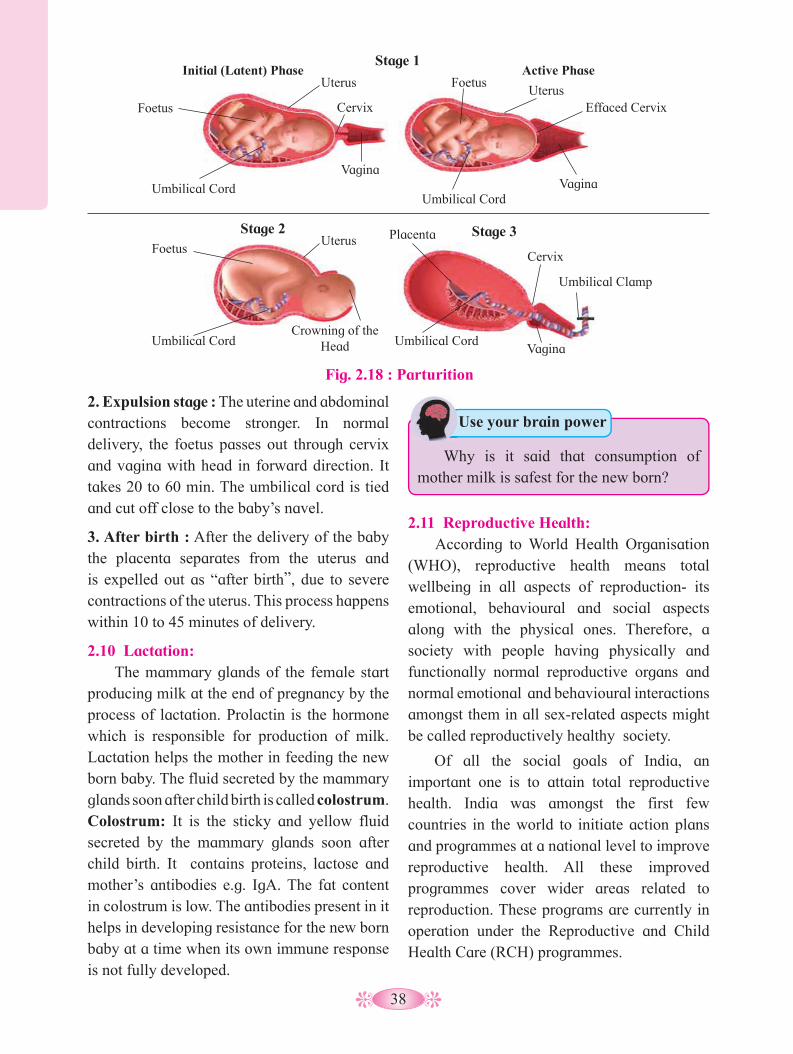
38
2. Expulsion stage : The uterine and abdominal contractions become stronger. In normal delivery, the foetus passes out through cervix and vagina with head in forward direction. It takes 20 to 60 min. The umbilical cord is tied and cut off close to the baby’s navel.
3. After birth : After the delivery of the baby the placenta separates from the uterus and is expelled out as “after birth”, due to severe contractions of the uterus. This process happens within 10 to 45 minutes of delivery.
2.10 Lactation: The mammary glands of the female start
producing milk at the end of pregnancy by the process of lactation. Prolactin is the hormone which is responsible for production of milk. Lactation helps the mother in feeding the new born baby. The fluid secreted by the mammary glands soon after child birth is called colostrum. Colostrum: It is the sticky and yellow fluid secreted by the mammary glands soon after child birth. It contains proteins, lactose and mother’s antibodies e.g. IgA. The fat content in colostrum is low. The antibodies present in it helps in developing resistance for the new born baby at a time when its own immune response is not fully developed.
2.11 Reproductive Health: According to World Health Organisation
(WHO), reproductive health means total wellbeing in all aspects of reproduction- its emotional, behavioural and social aspects along with the physical ones. Therefore, a society with people having physically and functionally normal reproductive organs and normal emotional and behavioural interactions amongst them in all sex-related aspects might be called reproductively healthy society.
Of all the social goals of India, an important one is to attain total reproductive health. India was amongst the first few countries in the world to initiate action plans and programmes at a national level to improve reproductive health. All these improved programmes cover wider areas related to reproduction. These programs are currently in operation under the Reproductive and Child Health Care (RCH) programmes.
Fig. 2.18 : Parturition
Stage 1
Stage 2 Stage 3
Initial (Latent) Phase Active Phase
Foetus
Foetus
Uterus
Uterus
Cervix
Cervix
Placenta
Vagina
Umbilical Cord
Umbilical Cord Umbilical Cord Crowning of the
Head
Umbilical Clamp
Umbilical Cord
Foetus Uterus
Effaced Cervix
Vagina
Vagina
Use your brain power
Why is it said that consumption of mother milk is safest for the new born?
39
Goals of RCH Programmes:1. To create awareness among people about
various aspects related to reproduction. 2. To provide the facilities to people to
understand and build up reproductive health.
3. To provide support for building up a reproductively healthy society.
4. To bring about a change mainly in three critical health indicators i.e. reducing total infertility rate, infant mortality rate and maternal mortality rate.
The goals of RCH can be achieved by the following ways:
1. By introduction of sex education in schools. Schools should be encouraged to provide correct information to the young so as to discourage children from believing in myths and clear the misconceptions about sex related aspects. Proper information about safe and hygenic sexual practices, sexually transmitted diseases (STD, AIDS), problems related to adolescence and proper information about reproductive organs.
2. With the help of audio- visual and the print media, government and non- government organisations should take various steps to create awareness about various aspects related to reproduction.
3. By educating the younger generation about birth control measures, pre-natal care of pregnant woman and post-natal care of the mother and child, importance of breast feeding.
4. By developing awareness about problems arising due to uncontrolled population growth, social evils like sex abuse and sex related crimes and take up necessary steps to prevent them.
5. By creating awareness about statutory ban on amniocentesis for sex determination.
6. By creating awareness about child immunization programmes.
7. By educating couples to reduce mortality rate of new borns and maternal mortality rate.
The population in India which was approximately 350 millions at the time of independence, reached close to a billion mark by 2000 and crossed 1.2 billion in May 2011. Now in 2020 population of India has crossed 1.35 billions. The government is taking serious measures to check this population growth. The most important step to overcome this problem, is to motivate society to have smaller families by using various birth control methods.
2.12 Birth control : The birth control measures which
deliberately prevent fertilization are referred to as contraceptives. The contraceptive methods help to prevent unwanted pregnancies. An ideal contraceptive should be easily available, user friendly, effective and with no or least side effects.
Contraceptive methods are of two main types i.e. temporary and permanent.
a. Temporary methods: These are of following types :
1. Natural method/ Safe period / Rhythm method : In the natural method, the principle of avoiding chances of fertilization is used. A week before and a week after menstrual bleeding is considered the safe period for sexual intercourse. This idea is based on the fact that ovulation occurs on the 14th day of menstrual cycle. Its drawback lies in having a high rate of failure.
2. Coitus Interruptus or withdrawal : In this method, the male partner withdraws his penis from the vagina just before ejaculation, so as to avoid insemination. This method also has some drawbacks, as the pre-ejaculation fluid may contain sperms and this can cause fertilization.
3. Lactational amenorrhea (absence of menstruation) : This method is based on the fact that ovulation does not occur during the period

40
of intense lactation following parturition. Therefore, as long as the mother breastfeeds the child fully, chances of conception are almost negligible. However, this method also has high chances of failure.
4. Chemical means (spermicides): In this method, chemicals like foam, tablets, jellies, and creams are used by the female partner. Before sexual intercourse, if these chemicals are introduced into the vagina, they adhere to the mucous membrane, immobilize and kill the sperms. It may cause allergic reaction. This method also has chances of failure.
5. Mechanical means / Barrier methods: In this method, with the help of barriers the ovum and sperm are prevented from physically meeting. These mechanical barriers are of three types.
i) Condom: It is a thin rubber sheath that is used to cover the penis of the male during coputation. It prevents the entry of ejaculated semen into the female reproductive tract. This can prevent conception. It is a simple and effective method and has no side effects. “Nirodh” is the most widely used contraceptive by males. It is easily available and is given free by the government. It should be properly discarded after every use. Condom is also a safeguard against STDs and AIDS.
ii) Diaphragm, cervical caps and vaults: These devices used by the female are made up of rubber. They prevent conception by blocking the entry of sperms through the cervix. The device is inserted into the female reproductive tract to cover the cervix during copulation.
iii) Intra-uterine devices (IUDs) : These clinical devices are plastic or metal objects. A doctor or trained nurse places the IUDs into the uterus. These devices include Lippes loop, copper releasing IUDs ( Cu-T, Cu7, multiload 375) and hormone releasing IUDs (LNG-20, progestasert).
Lippes loop is a plastic double “s” loop. It attracts the macrophages stimulating them to accumulate in the uterine cavity. Macrophages increase phagocytosis of sperms within the uterus and acts as a contraceptive. Copper releasing IUDs suppress sperm motility and the fertilising capacity of sperms.
The hormone releasing IUDs make the uterus unsuitable for implantation and cervix hostile to the sperms. It delays pregnancy for longer period. The spontaneous expulsion, occasional haemorrhage and chances of infection are the drawbacks of IUDs.
Fig. 2.20 : Copper - T
Hormone Copper
IUD
Strings
Uterus
Cevix
Vagina
Diaphragm
Fig. 2.19 : Mechanical means
Lippes loop

41
6. Physiological (Oral) Devices : Physiological devices are used in the form of tablets and hence are popularly called pills. It is an oral contraceptive, used by the female. The pill contains progesteron and estrogen. They inhibit ovulation, hence no eggs are released from the ovary of the female using this pill and thus conception cannot occur. They also alter the quality of cervical mucus to prevent the entry of sperms.
The pills have side effects such as nausea, weight gain, tenderness of breast and slight blood loss between menstrual periods. The pill “Saheli” is an oral contraceptive for females which is nonsteroidal. Saheli is to be taken once in a week. These pills are sponsored by the Government.
b. Permanent Method:The permanent birth control method in
men is called vasectomy and in women it is called tubectomy.
These are surgical methods, also called sterilization. In vasectomy a small part of the vas deferens is tied and cut where as in tubectomy, a small part of the fallopian tube is tied and cut. This blocks, gamete transport and prevent pregnancy.
7. Other contraceptives : The birth control implant is a contraceptive used by the female. It is a tiny, thin rod about the size of a matchstick. It is implanted under the skin of the upper arm.
They contain progesterone and estrogen. Their mode of action is similar to that of pills. They prevent pregnancy for 3-4 years.
Always Remember
Saheli is now a part of the National Family Programme as an oral contraceptive pill in India.
Fig. 2.21 : Implanon/ Nexplanon
A
Fig. 2.22 : Permanent method
Vas deferens tied and cut
B
Fallopian tubes tied and cut
Medical Termination of Pregnancy (MTP) :An intentional or voluntary termination
of pregnancy before full term is called Medical termination of Pregnancy (MTP) or induced abortion. MTP is essential in cases of unwanted pregnancies or in defective development of foetus. It is safe during the first trimester of pregnancy. The defective development of foetus is examined by amniocentesis.

42
Amniocentesis is a process in which amniotic fluid containing foetal cells is collected using a hollow needle inserted into the uterus under ultrasound guidance. The chromosomes are studied to see the abnormalities in the developing foetus. But the dangerous trend is the misuse of amniocentesis to determine the sex of the unborn child. Frequently, if the foetus is found to be female, it is aborted which is totally illegal. So the Government of India has legalised MTP Act in 1971, with strict conditions to avoid its misuse.
Medical Termination of Pregnancy (Amendment) Act 2017 under section 3 of the MTP Act 1971 was enacted by Government of India. The intention of MTP Act is to reduce the incidence of illegal abortion and consequent maternal mortality. As per the provisions of the MTP Act, only the consent of woman whose pregnancy is being terminated is required. According to MTP Act pregnancy may be terminated : 1. Within first 12 weeks 2. More than 12 weeks but lesser than 20 weeks. The registered medical practitioner’s opinion is mandatory stating the continuation of the pregnancy would involve a risk to the life of the pregnant woman or grave abnormal physical or mental health or is substantial risk to the child.
Activity :
In a sonography clinic, we observe a board saying ‘Sex selection and detection is NOT done in this centre and is punishable under PC-PNDT Act;
Find out what is PC-PNDT Act. Why do you think such a mandate is essential?
Internet my friend
What are the effects of alcohol drinking and smoking on foetus in pregnant women?
Always Remember Act of MTP : The Medical Termination
of Pregnancy (MTP) Act 1971 provides the legal framework for MTP Act 2017.
Internet my friend
Collect information about other sexually transmitted diseases.
Fig. 2.23 : Amniocentesis process
Amniocentesis : Used to extract foetal cells for genetic analysis.
1. Ultrasound used to determine the position of the foetus in the uterus
2. Needle inserted through the abdominal and uterine wall
3. Amnionic fluid containing foetal cells extracted
4. Centrifuge of extracted fluid and foetal cells
5. Cells used in karyotype
Karyotype
Placenta
Amnionic fluid
Risks associated with amniocentesis
1. Miscarriage2. Needle injury to foetus3. Leaking amniotic fluid4. Infection
2.13 Sexually Transmitted Diseases (STDs) : Diseases or infections which are
transmitted through sexual intercourse are collectively called Sexually Transmitted Diseases (STDs) or Venereal Diseases (VDs) or Reproductive Tract Infections (RTI). The major venereal diseases are syphilis and gonorrhoea.

43
Name of Disease Syphilis Gonorrhoea
Causative agent Treponema pallidum (Bacteria) Neisseria gonorrhoeae (Bacteria)
Incubation period
3-4 weeks Male – 2 to 14 daysFemale – 7 to 21 days
Infection site Mucous membrane in genital, rectal and oral region.
Mucous membrane of urino-genital tract, rectum, throat and eye.
Symptoms Primary lesion called chancre at the site of infection. Chancre is formed on external genitalia, skin rashes and mild fever, inflamed joints, loss of hair. Paralysis, Degenerative changes occur in the heart and brain.
In male, partial blockage of urethra and reproductive ducts, pus from penis, pain and burning sensation during urination, arthritis, etc. In female, pelvic inflammation of urinary tract, sterility, arthritis, the children born to affected mother suffer from gonococcal opthalmia and gonococcal vulvovaginitis of girls before puberty.
Preventive measures
Education about sex practices, sex hygiene, avoiding sex with unknown partner or multipartners, using condom during coitus.
Sex hygiene, using condom during coitus, avoiding sex with unknown partner or multipartners.
Treatment Antibiotic-Penicillin Antibiotic-Cefixime
Table 2.24 : Sexually Transmitted Diseases (STDs)
IVF (In Vitro Fertization) : It is a process of fertization where an
egg is combined with sperm outside the body in a test tube or glass plate to form a zygote under simulated conditions in the laboratory. The zygote or early embryos (with up to 8 blastomeres) could be then transferred into the fallopian tube for further development.
ZIFT (Zygote Intrafallopian Transfer) : ZIFT is an infertility treatment used
when there is a blockage in the fallopian tubes which prevents the fertilization of egg by the sperm.
In this method, egg is removed from woman’s ovary. Fertilization of the egg with sperms is brought about outside the body under sterile conditions to form zygote by the process called in vitro fertilization (IVF). The zygote is then transferred to fallopian tube for further development.
2.14 Infertility : Infertility is defined as the inability
to conceive naturally after (one year of) regular unprotected intercourse. The causes of infertility could be physical, congenital, diseases, immunological or even psychological.The common physical causes in females are polycystic ovary syndrome (PCOS), hormonal imbalance, endometriosis while in male, it is less sperm count and small size of penis.
Prior to 1978, infertile couple had two options, adopt or be childless. Today infertile couples have many options to have a child such as fertility drugs, test tube babies, artificial insemination, IUI, surrogate motherhood, etc. The couple could be assisted to have child / children through certain special techniques commonly known as Assisted Reproductive Technologies (ART).
44
GIFT (Gamete Intrafallopian Transfer) : Transfer of an ovum collected from a
donor into the fallopian tube of another female who can provide suitable environment for its fertilization and development.
This technique called gamete intrafallopian transfer (GIFT) has been developed for the cases in which only the entrance to the oviducts or the upper segment of the oviducts in blocked. In this procedure ova and sperms are directly injected into regions of the oviduct, where fertilization produces a blastocyst, which enters the uterus via the normal route. GIFT has a success rate of about 30 percent.
ICSI (Intra Cytoplasmic Sperm Injection) : ICSI is an in vitro fertilization procedure
in which a single sperm cell is injected directly into cytoplasm of an ovum in the laboratory. Here the sperm has to naturally penetrate the egg.
Artificial Insemination (AI) : In some infertility cases, the male partner
is unable to inseminate the female due to a very low sperm count. This problem can be solved by artificial insemination. In this technique, the sperms are collected from the male and artificially introduced into the cervix of female, for the purpose of achieving a pregnancy through in vivo fertilization (inside the body).
IUI (Intra Uterine Insemination) : In this technique the process is somewhat
like that of artificial insemination, the only difference is that the sperms are introduced into the uterine cavity instead of cervix.
Sperm bank / Semen bank: A sperm bank or semen bank is a place
which collects, stores and provides human sperms / semen. The semen is provided by healthy males called sperm donors. The sperms are stored in sperm bank by cryopreservation method (at low temperature).
Can you recall?
Surrogate mother : Some women have problem in implantation of embryo in uterus. Such woman can take help of the modern remedial technique called surrogacy. In this, embryo is implanted in surrogate mother, who is not the biological mother.
Always Remember
Tobacco, marijuana and other drugs smoking may cause infertility in both men and women. Nicotine blocks the production of sperm and decreases the size of testicles. Alcoholism by men interferes with the synthesis of testosterone and has an impact on sperm count. Use of cocaine or marijuana may temporarily reduce the number and quality of sperm.
Try This
IVF centres : Make a list of IVF centres in Maharashtra.
Adoption : Adoption is a legal process by which
a couple or a single parent gets legal rights, privileges and responsibilities that are associated to a biological child for the upbringing of the adopted child.
An adoptive parent should be medically fit and financially able to take care of the adopted child. A person wishing to adopt a child must be at least 21 years old but there is no legal upper age limit for adoption.

45
Always Remember
• Jayesh a young, married man of 26 yrs is suffering from T. B. for the last 2 years. He and his wife are desirous of a child but unable to have one, what could be the possible reason? Explain.
• Neeta is 45 years old and the doctor has advised her not to go for such a late pregnancy. She however wants to be the biological mother of a child, without herself getting pregnant. is this possible and how?
Always Remember
1. Cells of trophoblast do not form any part of the embryo proper.
2. They form ectoderm of the chorion (extra embryonic membrane).
3. They play important role in formation of placenta.
Activity :
1. Prepare concept map on information of male reproductive system.
2. Prepare concept map on information of female reproductive system.

46
Activity :
3. Prepare concept map on information of menstrual cycle.
4. Prepare concept maps on information of gametogenesis.
Spermatogenesis Oogenesis
5. Prepare concept map on information of fertilization.
47
Q. 1 Multiple choice questions.1. The number of nuclei present in a zygote
is ……a. two b.one c. four c.eight
2. Which of these is the male reproductive organ in human?a. sperm b. seminal fluidc. testes d. ovary
3. Attachment of embryo to the wall of the uterus is known as……..a. fertilization b. gestationc. cleavage d. implantation
4. Rupturing of follicles and discharge of ova is known as ..............a. capacitation b. gestationc. ovulation d. copulation
5. In human female, the fertilized egg gets implanted in uterus ......................a. After about7 days of fertilization b. After about 30 days of fertilization c. After about two months of fertilizationd. After about 3 weeks of fertilization
6. Test tube baby technique is called.........a. In vivo fertilization b. In situ fertilizationc. In vitro fertilizationd. Artificial insemination
7. The given figure shows a human sperm. Various parts of it are labelled as A, B, C, and D .Which labelled part represents acrosome ?
8. Presence of beard in boys is a ............a. primary sex organ b. secondary sexual character c. secondary sex organ d. primary sexual character
Q. 2 Answer in one sentence.1. What is the difference between a foetus
and an embryo?2. Outline the path of sperm upto the
urethra.3. Which glands contribute fluids to the
semen?4. Name the endocrine glands involved in
maintaining the sex characteristics of males.
5. Where does fertilization and implantation occur?
6. Enlist the external genital organs in female.
7. Give two differences between blastula and gastrula.
8. What is the difference between embryo and zygote?
Q. 3 Fill in the blanks:1. The primary sex organ in human male
is ……….
2. The………. is also called the womb.
3. Sperm fertilizes ovum in the ………….. of fallopian tube.
4. The disc like structure which helps in the transfer of substances to and from the foetus’s body is called…………
5. Gonorrhoea is caused by ……………bacteria.
6. The hormone produced by the testis is ………………..
a. B b. C c. D d. A
A B C D
Exercise
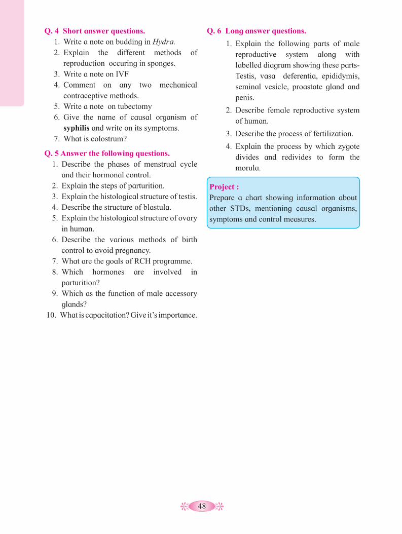
48
Q. 4 Short answer questions.1. Write a note on budding in Hydra.2. Explain the different methods of
reproduction occuring in sponges.3. Write a note on IVF4. Comment on any two mechanical
contraceptive methods. 5. Write a note on tubectomy6. Give the name of causal organism of
syphilis and write on its symptoms.7. What is colostrum?
Q. 5 Answer the following questions.1. Describe the phases of menstrual cycle
and their hormonal control.2. Explain the steps of parturition.3. Explain the histological structure of testis.4. Describe the structure of blastula.5. Explain the histological structure of ovary
in human.6. Describe the various methods of birth
control to avoid pregnancy.7. What are the goals of RCH programme.8. Which hormones are involved in
parturition?9. Which as the function of male accessory
glands?10. What is capacitation? Give it’s importance.
Q. 6 Long answer questions.
1. Explain the following parts of male reproductive system along with labelled diagram showing these parts-Testis, vasa deferentia, epididymis, seminal vesicle, proastate gland and penis.
2. Describe female reproductive system of human.
3. Describe the process of fertilization.
4. Explain the process by which zygote divides and redivides to form the morula.
Project : Prepare a chart showing information about other STDs, mentioning causal organisms, symptoms and control measures.

49
3.1 Chromosomes and Mechanism of inheritance.• The transmission of genetic information
from generation to generation is known as heredity or inheritance. The mechanism of inheritance was successfuly investigated before chromosomes had been observed or genes were known.
• Gregor Mendel, son of the peasant farmer, was born in Moravia in 1822. Gregor Mendel first gave the accurate explanation for the mechanism of inheritance by using hybridization technique.
• Inheritance of the seven traits in garden pea (Refer the diagram below) plant were
1. Is there a similarity between the parents
and offsprings?2. What are the roles of chromosomes in
living organisms?3. How are hybrid seeds produced?4. Which are the chromosomal disorders?
Can you recall? studied individually one at a time or in combination of two or three character at a time. He processed the data mathematically and statistically.
• Mendel postulated the principles of heredity which then became the fundamental laws of heredity, as proposed by Correns (1900). He visualized that the traits as such are not inherited physically but by ‘something’ present inside the gametic cell. To this ‘something’, he coined term ‘factors’ that are responsible for expression of a particular trait/ character. He proposed that factors are particulate in nature. The Mendelian factors are now termed as ‘genes’. These factors occur in pairs in the parents and segregate from each other during gamete formation without blending/ mixing.
Reasons for Mendel’s Success :• His experiments were carefully planned
and involved large sample.• He carefully recorded the number of plants
of each type and expressed his results as ratios.
Inheritance and Variation3
1.
Seven pairs of contransting visible characters in pea plant (Pisum sativum)
Do you know ?
Dominant
Recessive
Pea Plant Traits
Seed shape Seed color Pod shape Pod color Flower color
Flower location Plant height
Round Yellow Inflated Green Purple Axial Tall
Wrinkled Green Constricted Yellow White Terminal Short (Dwarf)
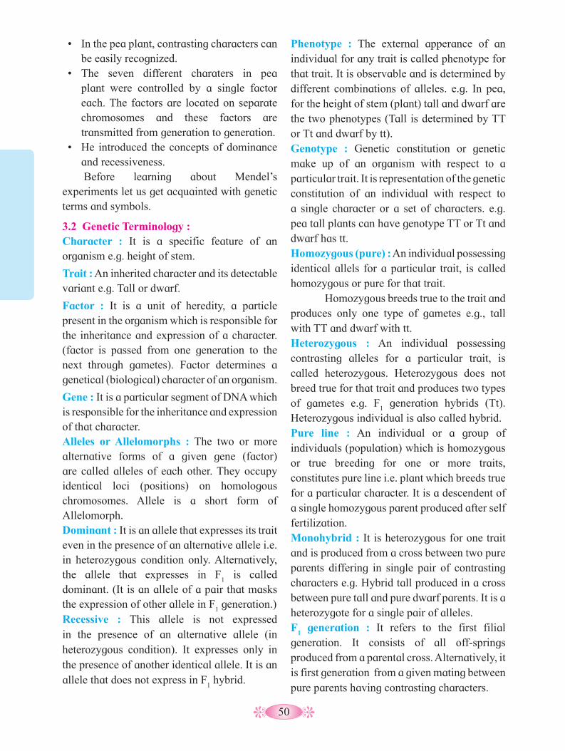
50
• In the pea plant, contrasting characters can be easily recognized.
• The seven different charaters in pea plant were controlled by a single factor each. The factors are located on separate chromosomes and these factors are transmitted from generation to generation.
• He introduced the concepts of dominance and recessiveness. Before learning about Mendel’s
experiments let us get acquainted with genetic terms and symbols.
3.2 Genetic Terminology :Character : It is a specific feature of an organism e.g. height of stem.
Trait : An inherited character and its detectable variant e.g. Tall or dwarf.
Factor : It is a unit of heredity, a particle present in the organism which is responsible for the inheritance and expression of a character. (factor is passed from one generation to the next through gametes). Factor determines a genetical (biological) character of an organism.
Gene : It is a particular segment of DNA which is responsible for the inheritance and expression of that character.Alleles or Allelomorphs : The two or more alternative forms of a given gene (factor) are called alleles of each other. They occupy identical loci (positions) on homologous chromosomes. Allele is a short form of Allelomorph.Dominant : It is an allele that expresses its trait even in the presence of an alternative allele i.e. in heterozygous condition only. Alternatively, the allele that expresses in F
1 is called
dominant. (It is an allele of a pair that masks the expression of other allele in F
1 generation.)
Recessive : This allele is not expressed in the presence of an alternative allele (in heterozygous condition). It expresses only in the presence of another identical allele. It is an allele that does not express in F
1 hybrid.
Phenotype : The external apperance of an individual for any trait is called phenotype for that trait. It is observable and is determined by different combinations of alleles. e.g. In pea, for the height of stem (plant) tall and dwarf are the two phenotypes (Tall is determined by TT or Tt and dwarf by tt).Genotype : Genetic constitution or genetic make up of an organism with respect to a particular trait. It is representation of the genetic constitution of an individual with respect to a single character or a set of characters. e.g. pea tall plants can have genotype TT or Tt and dwarf has tt. Homozygous (pure) : An individual possessing identical allels for a particular trait, is called homozygous or pure for that trait. Homozygous breeds true to the trait and produces only one type of gametes e.g., tall with TT and dwarf with tt.Heterozygous : An individual possessing contrasting alleles for a particular trait, is called heterozygous. Heterozygous does not breed true for that trait and produces two types of gametes e.g. F
1 generation hybrids (Tt).
Heterozygous individual is also called hybrid.Pure line : An individual or a group of individuals (population) which is homozygous or true breeding for one or more traits, constitutes pure line i.e. plant which breeds true for a particular character. It is a descendent of a single homozygous parent produced after self fertilization.Monohybrid : It is heterozygous for one trait and is produced from a cross between two pure parents differing in single pair of contrasting characters e.g. Hybrid tall produced in a cross between pure tall and pure dwarf parents. It is a heterozygote for a single pair of alleles.F
1 generation : It refers to the first filial
generation. It consists of all off-springs produced from a parental cross. Alternatively, it is first generation from a given mating between pure parents having contrasting characters.
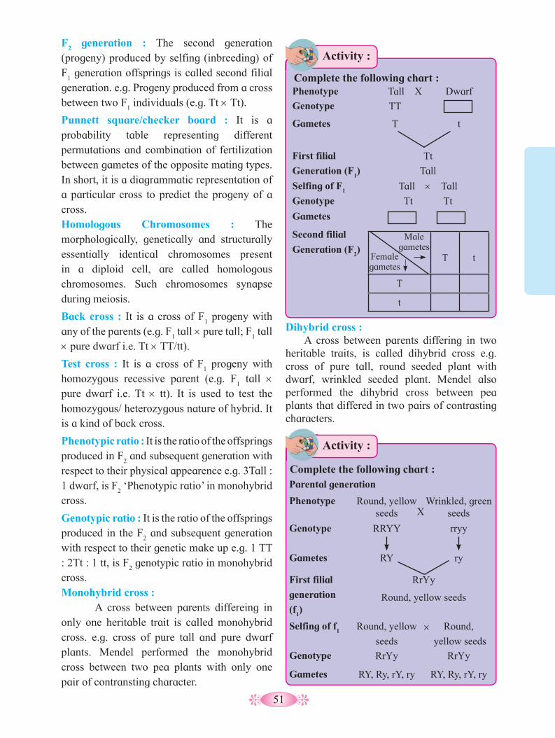
51
Parental generation
Phenotype Round, yellow seeds
Wrinkled, green seeds
Genotype RRYY rryy
Gametes RY ry
First filial
generation
(f1)
RrYy
Round, yellow seeds
Selfing of f1 Round, yellow
seeds
Round,
yellow seeds
Genotype RrYy RrYy
Gametes RY, Ry, rY, ry RY, Ry, rY, ry
F2 generation : The second generation
(progeny) produced by selfing (inbreeding) of F
1 generation offsprings is called second filial
generation. e.g. Progeny produced from a cross between two F
1 individuals (e.g. Tt ×Tt).
Punnett square/checker board : It is a probability table representing different permutations and combination of fertilization between gametes of the opposite mating types. In short, it is a diagrammatic representation of a particular cross to predict the progeny of a cross.Homologous Chromosomes : The morphologically, genetically and structurally essentially identical chromosomes present in a diploid cell, are called homologous chromosomes. Such chromosomes synapse during meiosis.
Back cross : It is a cross of F1 progeny with
any of the parents (e.g. F1 tall ×pure tall; F
1 tall
×pure dwarf i.e. Tt ×TT/tt).
Test cross : It is a cross of F1 progeny with
homozygous recessive parent (e.g. F1 tall ×
pure dwarf i.e. Tt × tt). It is used to test the homozygous/ heterozygous nature of hybrid. It is a kind of back cross.
Phenotypic ratio : It is the ratio of the offsprings produced in F
2 and subsequent generation with
respect to their physical appearence e.g. 3Tall : 1 dwarf, is F
2 ‘Phenotypic ratio’ in monohybrid
cross.
Genotypic ratio : It is the ratio of the offsprings produced in the F
2 and subsequent generation
with respect to their genetic make up e.g. 1 TT : 2Tt : 1 tt, is F
2 genotypic ratio in monohybrid
cross.Monohybrid cross : A cross between parents differeing in only one heritable trait is called monohybrid cross. e.g. cross of pure tall and pure dwarf plants. Mendel performed the monohybrid cross between two pea plants with only one pair of contransting character.
Dihybrid cross :A cross between parents differing in two
heritable traits, is called dihybrid cross e.g. cross of pure tall, round seeded plant with dwarf, wrinkled seeded plant. Mendel also performed the dihybrid cross between pea plants that differed in two pairs of contrasting characters.
×
Phenotype Tall X Dwarf
Genotype TT
Gametes T t
First filial
Generation (F1)
Tt
Tall
Selfing of F1 Tall ×Tall
Genotype Tt Tt
Gametes
Second filial
Generation (F2)
T t
T
t
Male gametes
Female gametes
Activity :
Complete the following chart :
Activity :
Complete the following chart :
X

52
2. Law of segregation (Law of purity of gametes) :
This law is based on the fact that the alleles do not show any blending/ mixing and both the alleles (characters) are recovered as such in the F
2 generation, though one of these
is not seen at the F1 stage. During formation of
gametes, these two alleles (factors) obviously separate or segregate, otherwise recessive type will not appear in F
2.
The gametes which are formed are always pure for a particular character (trait). A gamete may carry either dominant or recessive factor but not both. That’s why it is also called as law of purity of gametes.
Statement of Law of Segregation : The law states that “When hybrid (F
1) forms gametes,
the alleles segregate from each other and enter in different gametes”. The gametes formed are pure in that they carry only one allele each (either dominant allele or recessive allele). Hence, this law is also described as “Law of purity of gametes”.
3. Law of Independent Assortment : This law is based on dihybrid cross. It is basic principle of genetics developed by a Mendel. It describes how different genes or alleles present on separate chromosomes independently separate from each other, during formation of gametes. These alleles are then randomly united in fertilization. In dihybrid cross, F
2 phenotypic ratio 9:3:3:1 indicates
that the two pairs of characters behave independent of each other. It can be concluded that the two characters under consideration are assorted independently giving rise to different combinations.
Statement of Law of Independent Assortment: The law states that “When hybrid possessing two (or more) pairs of contrasting factors (alleles) forms gametes, the factors in each pair segregate independently of the other pair”.
3.3 Mendel’s Laws of Inheritance : Mendel proposed three basic postulates on
the basis of which three laws were formulated. These are described below:
1. Law of Dominance :In monohybrid and dihybrid crosses, the
phenotypic characters are controlled by discrete units, called factors. In a dissimilar pair of factors, one member of the pair dominates (i.e. dominant) over the other (i.e. recessive). The law of dominance is used to explain the expression of only one of the parental characters of a monohybrid cross in F
1 and the
expression of both in F2.
Statement of Law of Dominance : “When two homozygous individuals with one or more sets of contrasting characters are crossed, the alleles (characters) that appear in F
1 are
dominant and those which do not appear in F1
are recessive”.
Why are farmers and gardeners advised to buy new F
1 hybrid seeds every
year?
Can you tell?
There are 16 possible individuals in F2
generation. Try to find out the phenotypes as well as the genotypic and phenotypic ratios.
Use your brain power
RY Ry rY ry
RY
Ry
rY
ry
Male gametes
Female gametes
F2 Generation :
53
Find the ratio of dihybrid test cross by using punnett square.
Try This
3.4 Back Cross and Test Cross : a. Back cross : The F
1 individuals obtained in
a cross are usually selfed to get the F2 progeny.
They can also be crossed with one of the two parents from which they were derived (either recessive or dominant). Such a cross is known as back cross.
b. Test cross : The cross of F1 hybrid with the
homozygous recessive parent is known as a test cross. It is used to test whether an individual is homozygous (pure) or heterozygous(hybrid). Test cross is easy, simple, repeatable and predictable.
Test cross can be used to find out genotype of any plant with dominant expression. But it is not known whether it is homozygous (pure) or heterozygous for that trait. For example, A pea plant having violet (purple) flowers is crossed with a pea plant with white flowers. If all flowers produced are violet, we can conclude that plant is pure or homozygous and if we get violet and white flowers in 1:1 ratio, we can
conclude that plant is heterozygous. Test cross is also used to introduce useful recessive traits in the hybrids of self pollinated plants during rapid crop improvement programs.
Following is the graphic representation of test cross (Fig. 3.1). Recessive parent is crossed to find out unknown genotype.
3.5 Deviations from Mendel’s findings : Few generalizations were arrived at by
Mendel, on the basis of his experiments of garden pea plant- such as,i. Single trait Single gene Two alleles.ii. Two alleles show interaction in which one
is completely dominant.iii. Factors (genes) for different traits
present on different chromosomes assort independently.With the passage of time, number of
deviations were observed/ identified in the post-Mendelian era, that gave additional information on the patterns of inheritance. These deviations are then described as Neo-Mendelism.
Find out the principle involved in heredity of sheep/ coat colour
Internet my friend
Fig. 3.1 : Graphical representation of test cross
Homozygous recessive
Homozygous recessive
r r
rr
rr rr
rr
r r
r
Rr Rr Rr Rr RR Rr
Rr Rr
R
R R
All flowers are violet
Unknown flower is homozygous dominant
Dominant phenotype (Genotype unknown)
Half of the flowers are violet and half of the flowers are white. Result
Interpretation Unknown flower is heterozygous
X

54
It was observed that the phenotypic expression of a gene can be modified or influenced by the other gene. These gene interactions are of two types.
i. Intragenic interactions : Occur between the alleles of same gene e.g. incomplete dominance and co-dominance. It also occurs between the multiple allele series of a gene.
ii. Intergenic (non-allelic) interactions: Occur between the allels of different-genes present on the same or different-chromosomes. e.g. pleiotropy, polygenes, epistasis, supplermetary and complementry genes, etc. Some of these interactions are discussed below :
a. Incomplete dominance:In the incomplete dominance, both the
alleles (genes) of an allelomorphic pair express themselves partially. One allele (gene) cannot supress the expression of the other allele (gene) completely. In such case, there is an intermediate expression in the F
1 hybrid. A
well-known example is the flower colour of Mirabilis jalapa. If a red-flowered (RR) plant is crossed with a white-flowered (rr) plant, then F
1 offsprings have pink (Rr) flowers.
Result : Genotypic ratio - 1RR : 2Rr : 1rrPhenotypic ratio - 1Red : 2 Pink :1 White
b. Co-dominance : In co-dominance, both the alleles (genes)
of an allelomorphic pair express themselves equally in F
1 hybrids. Such alleles which are able
to express themselves equally independently in hybrids, are called co-dominant alleles. Thus in co-dominance both alleles are expressed.
The deviations are :
a. Single trait Single gene two alleles showing interactions like codominance and incomplete dominance.
b. Single trait Single gene more than two alleles (multiple allelism).
c. Single trait more than one genes (Polygenic inheritance) showing different epistatic interactions or additive effect.
d. Single gene influencing many traits (pleiotropy).
Do you know ?
Fig. 3.2 : Representation of co-dominance in cattle
R r
R RR red
Rr pink
r Rr pink
rr
white
F2 Generation : Selfing of F
1
red flowers RR
white flowers rr
R r
F1 - hybrids :
Pink flowers
Parents :
Gametes :
X
Rr
Phenotype Genotype
Red RR
White WW
Parent generation
Gametes
F1
Generation
Roan RW
Roan RW
R
R R
W
W W Gametes
F2
Generation
Phenotypic ration :
Genotypic ration :
RR
RR : RW : WW
Red 1
RW RW WW
Roan 2
White 1
1 : 2 : 1
X
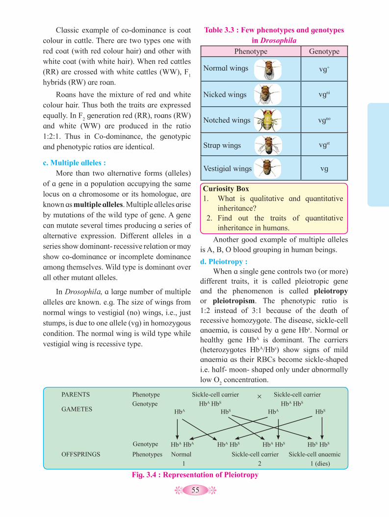
55
PARENTS
OFFSPRINGS
Phenotype
Phenotypes Normal
1
×
1 (dies)2
Genotype HbA HbS HbA HbS
Sickle-cell carrier Sickle-cell carrier
Sickle-cell carrier Sickle-cell anaemic
GAMETES
Genotype
HbA HbS HbA HbS
HbA HbA HbA HbS HbA HbS HbS HbS
Fig. 3.4 : Representation of Pleiotropy
Classic example of co-dominance is coat colour in cattle. There are two types one with red coat (with red colour hair) and other with white coat (with white hair). When red cattles (RR) are crossed with white cattles (WW), F
1
hybrids (RW) are roan.
Roans have the mixture of red and white colour hair. Thus both the traits are expressed equally. In F
2 generation red (RR), roans (RW)
and white (WW) are produced in the ratio 1:2:1. Thus in Co-dominance, the genotypic and phenotypic ratios are identical.
c. Multiple alleles : More than two alternative forms (alleles)
of a gene in a population accupying the same locus on a chromosome or its homologue, are known as multiple alleles. Multiple alleles arise by mutations of the wild type of gene. A gene can mutate several times producing a series of alternative expression. Different alleles in a series show dominant- recessive relation or may show co-dominance or incomplete dominance among themselves. Wild type is dominant over all other mutant alleles.
In Drosophila, a large number of multiple alleles are known. e.g. The size of wings from normal wings to vestigial (no) wings, i.e., just stumps, is due to one allele (vg) in homozygous condition. The normal wing is wild type while vestigial wing is recessive type.
Curiosity Box 1. What is qualitative and quantitative
inheritance? 2. Find out the traits of quantitative
inheritance in humans.
Table 3.3 : Few phenotypes and genotypes in Drosophila
Phenotype Genotype
Normal wings vg+
Nicked wings vgni
Notched wings vgno
Strap wings vgst
Vestigial wings vg
Another good example of multiple alleles is A, B, O blood grouping in human beings.
d. Pleiotropy :When a single gene controls two (or more)
different traits, it is called pleiotropic gene and the phenomenon is called pleiotropy or pleiotropism. The phenotypic ratio is 1:2 instead of 3:1 because of the death of recessive homozygote. The disease, sickle-cell anaemia, is caused by a gene Hbs. Normal or healthy gene HbA is dominant. The carriers (heterozygotes HbA/Hbs) show signs of mild anaemia as their RBCs become sickle-shaped i.e. half- moon- shaped only under abnormally low O
2 concentration.
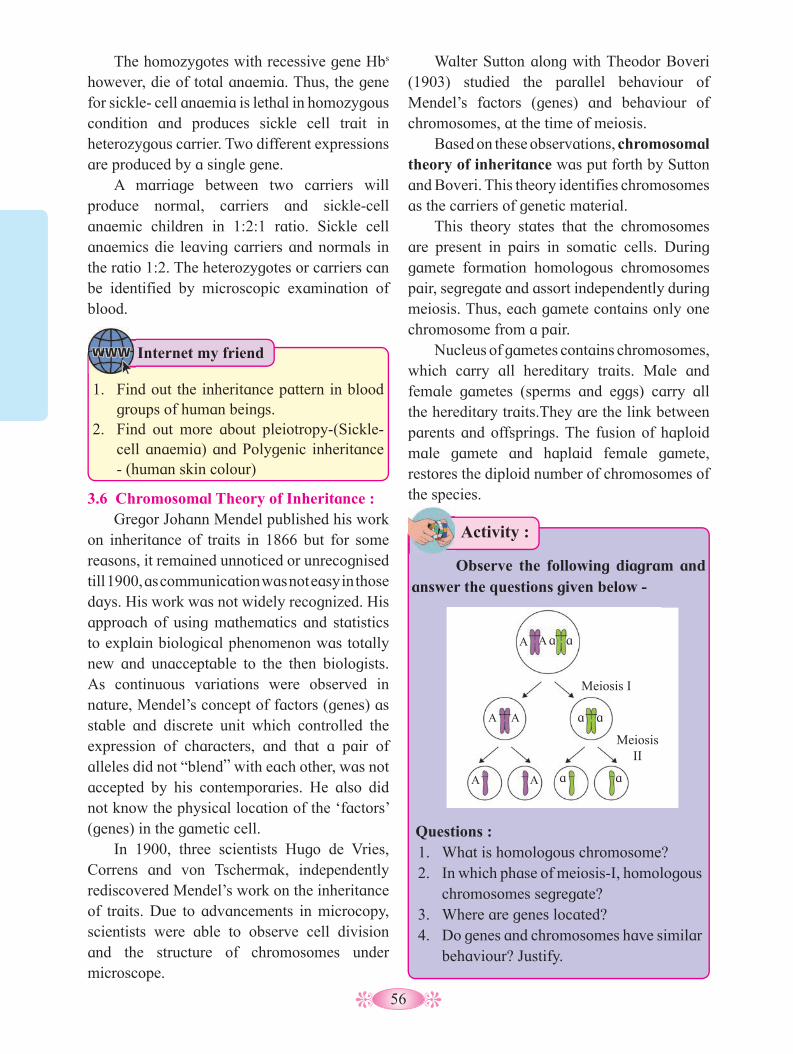
56
3.6 Chromosomal Theory of Inheritance : Gregor Johann Mendel published his work
on inheritance of traits in 1866 but for some reasons, it remained unnoticed or unrecognised till 1900, as communication was not easy in those days. His work was not widely recognized. His approach of using mathematics and statistics to explain biological phenomenon was totally new and unacceptable to the then biologists. As continuous variations were observed in nature, Mendel’s concept of factors (genes) as stable and discrete unit which controlled the expression of characters, and that a pair of alleles did not “blend” with each other, was not accepted by his contemporaries. He also did not know the physical location of the ‘factors’ (genes) in the gametic cell.
In 1900, three scientists Hugo de Vries, Correns and von Tschermak, independently rediscovered Mendel’s work on the inheritance of traits. Due to advancements in microcopy, scientists were able to observe cell division and the structure of chromosomes under microscope.
The homozygotes with recessive gene Hbs however, die of total anaemia. Thus, the gene for sickle- cell anaemia is lethal in homozygous condition and produces sickle cell trait in heterozygous carrier. Two different expressions are produced by a single gene.
A marriage between two carriers will produce normal, carriers and sickle-cell anaemic children in 1:2:1 ratio. Sickle cell anaemics die leaving carriers and normals in the ratio 1:2. The heterozygotes or carriers can be identified by microscopic examination of blood.
Walter Sutton along with Theodor Boveri (1903) studied the parallel behaviour of Mendel’s factors (genes) and behaviour of chromosomes, at the time of meiosis.
Based on these observations, chromosomal theory of inheritance was put forth by Sutton and Boveri. This theory identifies chromosomes as the carriers of genetic material.
This theory states that the chromosomes are present in pairs in somatic cells. During gamete formation homologous chromosomes pair, segregate and assort independently during meiosis. Thus, each gamete contains only one chromosome from a pair.
Nucleus of gametes contains chromosomes, which carry all hereditary traits. Male and female gametes (sperms and eggs) carry all the hereditary traits.They are the link between parents and offsprings. The fusion of haploid male gamete and haplaid female gamete, restores the diploid number of chromosomes of the species.
Activity :
Observe the following diagram and answer the questions given below -
Questions : 1. What is homologous chromosome? 2. In which phase of meiosis-I, homologous
chromosomes segregate? 3. Where are genes located? 4. Do genes and chromosomes have similar
behaviour? Justify.
A
A A
A A
A a a
aa
a a
Meiosis I
Meiosis II
1. Find out the inheritance pattern in blood groups of human beings.
2. Find out more about pleiotropy-(Sickle-cell anaemia) and Polygenic inheritance - (human skin colour)
Internet my friend
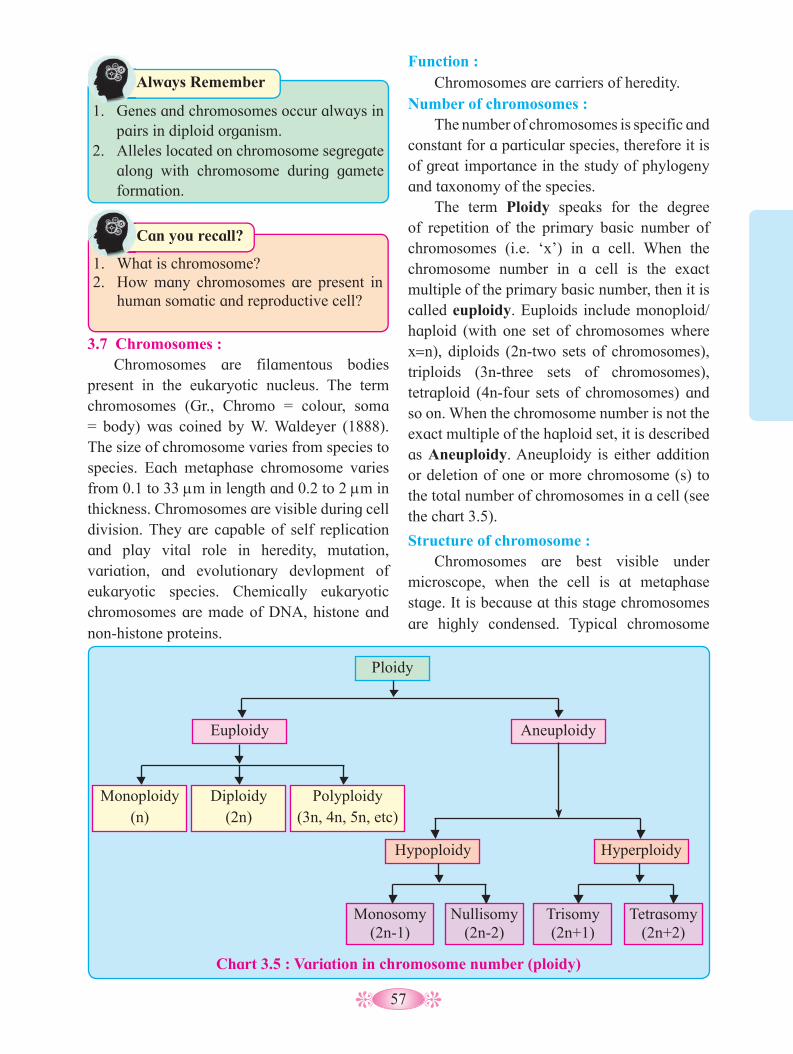
57
1. What is chromosome? 2. How many chromosomes are present in
human somatic and reproductive cell?
Can you recall?
Function : Chromosomes are carriers of heredity.
Number of chromosomes : The number of chromosomes is specific and
constant for a particular species, therefore it is of great importance in the study of phylogeny and taxonomy of the species.
The term Ploidy speaks for the degree of repetition of the primary basic number of chromosomes (i.e. ‘x’) in a cell. When the chromosome number in a cell is the exact multiple of the primary basic number, then it is called euploidy. Euploids include monoploid/ haploid (with one set of chromosomes where x=n), diploids (2n-two sets of chromosomes), triploids (3n-three sets of chromosomes), tetraploid (4n-four sets of chromosomes) and so on. When the chromosome number is not the exact multiple of the haploid set, it is described as Aneuploidy. Aneuploidy is either addition or deletion of one or more chromosome (s) to the total number of chromosomes in a cell (see the chart 3.5).
Structure of chromosome :Chromosomes are best visible under
microscope, when the cell is at metaphase stage. It is because at this stage chromosomes are highly condensed. Typical chromosome
3.7 Chromosomes :Chromosomes are filamentous bodies
present in the eukaryotic nucleus. The term chromosomes (Gr., Chromo = colour, soma = body) was coined by W. Waldeyer (1888). The size of chromosome varies from species to species. Each metaphase chromosome varies from 0.1 to 33 mm in length and 0.2 to 2 mm in thickness. Chromosomes are visible during cell division. They are capable of self replication and play vital role in heredity, mutation, variation, and evolutionary devlopment of eukaryotic species. Chemically eukaryotic chromosomes are made of DNA, histone and non-histone proteins.
Ploidy
Aneuploidy
Hyperploidy
Tetrasomy(2n+2)
Trisomy(2n+1)
Nullisomy(2n-2)
Monosomy(2n-1)
Hypoploidy
Euploidy
Monoploidy(n)
Diploidy(2n)
Polyploidy(3n, 4n, 5n, etc)
Chart 3.5 : Variation in chromosome number (ploidy)
1. Genes and chromosomes occur always in
pairs in diploid organism. 2. Alleles located on chromosome segregate
along with chromosome during gamete formation.
Always Remember

58
consists of two chromatides joind together at centromere or primary constriction. Primary constriction constists of a disk shape plate called kinetochore. It is at the kinetochore, spindle fibres get attached during cell division. Besides primary constriction, some few chromosomes possess additional one or two constrictions called secondary constriction. At secondary constriction I, nucleolus becomes organized during interphase. A satellite body (SAT body) is attached at secondary constriction II, in very few chromosomes. Each chromatid in turn contains a long, unbranched, slender, highly coiled DNA thread, called Chromonema, extending through the length of chromatid.
Chromatid consists a double stranded DNA molecule which extends from one end of chromosomes to other.
Depending upon the position of centromere there are four types (shapes) of chromosomes viz. Acrocentric (j shaped), Telocentric (i shaped), Submetacentric (L shaped) and Metacentric (V shaped). The ends of chromosome (i.e. chromatids) are known as telomeres. Sex Chromosomes :
The chromosomes which are responsible for the determination of sex are known as sex chromosomes (Allosomes). Human being and other mammals have X and Y Chromosomes as sex chromosomes.
X chromosome is straight, rod like and longer than Y chromosome. X chromosome is metacentric, while Y chromosome is acrocentric. X chromosome has large amount of euchromatin (extended region) and small amount of heterochromatin (highly condensed region). Euchromatin has large amount of DNA material, hence genetically active. Y chromosome has small amount of euchromatin and large amount of heterochromatin, hence it is genetically less active or inert. Both X and Y chromosome show homologous and non-homologous regions. Homologous regions show similar genes while non-homologous regions show dissimilar genes.
Activity :
Study the types of chromosome according to position of centromere. Observe and complete the following table. Types of Chromosome
Name of Chromosome
Position of Contromere
Metacentric -
- -
Acrocentric -
- At one end
A : Parts of chromosomes B : Showing secondary constrictions and detailsFig. 3.6 : Structure of Chromosome
Fig. 3.7 : Structure of X and Y chromosomes (in humans)
Telomere
Secondary constriction (ii)
Primary constriction (centromere)
Chromonemata
Secondary constriction (i) or
nucleolar organizer
Chromomeres Chromonemata
Centromere
Matrix Nucleolus
Satellite
A B
Crossing over occurs only between homologous regions of X and Y chromosomes. Non-homologous region of X chromosome is longer and contains more genes than that of
Homologous region
Non-Homologous regions
Centromere

59
3.8 Linkage and Crossing Over : Linkage :
It is a known fact that several genes are present on the chromosome. As chromosomes are carriers of heredity, these genes have tendency to be inherited together. Such genes are called linked genes. This tendency of two or more genes present on the same chromosomes that are inherited together is known as linkage. Linkage was discovered in plants by Bateson and Punnett and in animals by T. H. Morgan. Linkage is of two kinds - complete and incomplete linkage:
I. Complete linkage : The linked genes which are closely located on the chromosome do not separate (no crossing over) and inherit together. They are called completely linked (strongly linked) genes and the phenomenon of their inheritance is called complete linkage. Thus the parental traits are inherited in offsprings. e.g. X chromosome of Drosophila males- show complete linkage.
II. Incomplete linkage : The linked genes which are distantly located on the same chromosome and have chances of separation by crossing over, are known as incompletly linked (weakly linked) genes. The phenomenon of their inheritance, is called incomplete linkage. Thus, new traits occur in offsprings. e.g. In Zea mays - colour and shape of grain show incomplete linkage.
Linkage Groups : All the linked genes in a particular
chromosome, constitute a linkage group.
The number of linkage groups of a particular species corresponds to its haploid number of chromosomes. e.g. Drosophila melanogaster has 4 linkage groups that corrrspond to the 4 pairs of chromosomes. Garden pea has 7 linkage groups and 7 pairs of chromosomes.
Sex-linkage : The transmission (inheritance) of X - linked
and Y-linked genes from parents to offspring, is called sex-linked inheritance. Sex-linked inheritance is of three types viz. X-linked, Y-linked and XY-linked. Sex linkage is of two kinds :
a. Complete sex linkage : It is exhibited by genes located on non-homologous regions of X and Y chromosomes. They inherit together because crossing over does not occur in this region.
Examples of X-linked traits are haemophilia, red-green colour blindness, myopia (near sightedness) and for Y-linked are hypertrichosis, Ichthyosis, etc.
b. Incomplate sex linkage : It is exhibited by genes located on homologous regions of X and Y chromosomes. They do not inherit together because crossing over occurs in this region. Examples of X-Y linked traits are total colour blindess, nephritis, retinitis pigmentosa, etc.
Crossing Over : Crossing over is a process that produces
new combinations (recombinations) of genes by interchanging and exhanging of corresponding segments between non-sister chromatids of homologous chromosomes. It occurs during pachytene of prophase I of meiosis. The term crossing over was coined by Morgan. The mechanism of crossing over consists four sequential steps such as synapsis, tetrad formation, crossing over and terminalization. This you have already studied in the chapter on cell division in class XI. The phenomenon of crossing over is universal and it is necessary for the natural selection, because it increases the chances of variation.
1. What are allosomes? 2. Compare X and Y chromosomes. 3. In which region of chromosomes does
crossing over take place?
Can you tell?
non-homologous region of Y chromosome. X-linked genes are persent on non-homologous region of X-chromosome while Y linked genes are persent on non-homologous region of Y-chromosome.

60
• Morgan’s Experiments showing linkage and crossing over :
Morgan used Drosophila melanogaster (fruit fly) for his experiments because, Drosophila can easily be cultured in laboratory. It’s life span is short, about two weeks. More over, it has high rate of reproduction.
Morgan carried out several dihybrid cross experiments in fruit fly to study genes that are sex-linked. The crosses were similar to dihybrid crosses, as carried out by Mendel in Pea. For example, Morgan and his group crossed yellow-bodied, white eyed female to the wild type with brown-bodied, red eyed males and intercrossed their F
1 progeny.
Thomas Hunt Morgan was an American biologist. He used fruit fly (Drosophila melanogaster) in genetic reserch and established the chromosomal theory of heredity. He also discovered the principle of linkage, sex linkage and crossing over.
Margan’s work played key role in the field of genetics. He was awarded a Nobel Prize in 1933, in Physiology and Medicine.
Know the Scientist :
T. H. Margan
(1866-1945)
How many linkage groups are in human being and maize?Use your brain power
Fig : 3.8 : Linkage and crossing over
Parental
F1 generation
F2 generation
y w
y w y w
y w
y w
y w y w
y w
y w
y+ w+
y+ w+
y+ w+
y+ w+
y w+
y w+
y+ w
y+ w
Yellow, White
Yellow, White
Yellow, White
Yellow, White
Yellow, Red
Yellow, Red
Brown, White
Brown, White
Wild type Wild type
Wild type
Wild type
Wild type
Wild type
Wild type
Wild type
Parental type (98.7%)
Recombinant types (1.3%)
White, Miniature
White, Miniature
White, Miniature
Miniature
White, Miniature
White, Miniature
White, Normal
White, Normal
Parental type (62.8%)
Recombinant types (37.2%)
w m
w m
w m
w m
w m
w m w m
w m
w m
w m
w+ m+
w+ m+
w+ m+
w+ m+
w m+
w m+
w+ m
w+ m
Cross IICross I
x x

61
Fig. 3.9 : Widow’s peak and straight hair line
Parental combinations occur more due to linkage and new combinations less due to crossing over.
Always Remember
Some characters are influenced by dominant genes while some other are by recessive genes, present on autosomes. For example, • Autosomal dominant traits like Widow’s
peak and Huntington’s disease, etc. • Autosomal recessive traits like Phenyl
ketonuria (PKU), Cystic fibrosis and Sickle cell anaemia.
a. Widow’s peak : A prominent “V” shaped hairline on
forehead is described as widow’s peak. It is determined by autosomal dominant gene. Widow’s peak occurs in homozygous dominant (WW) and also heterozygous (Ww) individuals.
Individuals with homozygous recessive (ww) genotype have a straight hair line (no widows peak). Both males and females have equal chance of inheritance.
He observed that the two genes did not segregate independently of each other and F
2
ratio deviated very signiticantly from 9:3:3:1 ratio.
Morgan and his group knew that the genes were located on X chromosome and stated that when two genes in a dihybrid cross are situated on the same chromosome, then the proportion of parental combination is much higher than non-parental type. This occurs due to physical association or linkage of the two genes. He also found that, when genes are grouped on the same chromosome, some genes are strongly linked. They show very few recombinations (1.3 %). When genes are loosely linked i.e. present far away from each other on chromosome, they show more (higher) recombinations (37.2 %).
For example, the genes for yellow body and white eye were strongly linked and showed only 1.3 percent recombination (in cross-I). White bodied and miniature wings showed 37.2 percent recombination (in cross-II). Cross I shows crossing over between genes y and w. Cross II shows crossing over between genes white (w) and miniature wing (m). Here dominant wild type alleles are represented with (+) sign.
3.9 Autosomal Inheritance : Human somatic (2n) cell contains 23 pairs of chromosomes. They can be divided functionally as autosomes and sex chromosomes. A single pair of chromosomes is involved in sex determination and remaining 22 pairs are called autosomes. Autosomes control a variety of traits other than sex. These traits are called autosome linked traits. Transmission of body characters other than the sex linked traits from parents to their offsprings through autosomes, is called autosomal inheritance.
b. Phenylketonuria (PKU):- It is an inborn metabolic disorder caused
due to recessive autosomal genes. When recessive genes are present in homozygous condition, phenylalanine hydroxylase enzyme is not produced. This enzyme is essential for conversion of amino acid phenylalanine into tyrosine. Due to absence of this enzyme, phenylalanine is not converted into tyrosine. Hence, phenylalanine and its derivatives are accumulated in blood and cerebrospinal fluid (CSF). It affects development of brain and causes mental retardation. Excess phenylalanine is excreted in urine, hence this disease is called phenylketonuria.
Widow’s peak No widow’s peak

62
Colour blindness : Colour blindness is X-linked recessive
disorder where person is unable to distinguish between red and green colours as both the colours appear grey. It is caused due to recessive X-linked genes (XC) which prevents formation of colour sensitive cells, the cones, in the retina of eye.
The homozygous recessive females (Xc Xc) and hemizygous recessive male (XcY) are unable to distinguish between red and green colours. The frequency of colour blind women is much less than colour blind men. Dominant X linked gene (XC) is necessary for formation of colour sensitive cells in the retina of eye. Thus, genotypes of male and female individuals can be represented as follows-
Sex Normal Colourblind Carrier
Male XCY XcY
Female XCXC XcXc XCXc
The inheritance of colourblindness can be studied in the following two types of marriages:-
1. Marriage between colour blind male with normal female, will produce normal visioned male and female offspring in F
1. The sons have
normal vision but daughter will be carrier for the disease.
Autosomal recessive traits appear in both sexes with equal frequency. These traits tend to skip generations.
3.10 Sex Linked Inheritance : Genes located on non-homologous region
of sex chromosomes, are called sex-linked genes. The traits that are determined by sex linked genes, are called sex-linked traits.
The inheritance of sex linked genes from parents to their offsprings, is called sex linked inheritance. There are two types of sex-linked genes as X-linked genes and Y-linked genes.
a. X-linked (sex linked) genes :The X linked genes are located on non
homologous region of X chromosome and these gene do not have corresponding alleles on Y chromosome.
Female has two X chromosomes. In female two recessive sex linked genes are required for expression of sex linked traits. If one X chromosome carries a recessive gene for sex-linked trait (defect) its effect is suppressed by the dominant gene present on other X chromosome. The females with one recessive gene are carriers. The carrier female is physically normal as she does not suffer from the disease (disorder).
Male has only one X-chromosome. If X chromosome carries X-linked recessive gene for sex linked trait, then it is expressed phenotypically, because there is no dominant gene on Y chromosome to suppress its effect. Therefore, sex-linked / X-linked traits appear more frequently in males than in the females. Examples of X-linked traits include haemophilia, colour blindness, night blindness, myopia, muscular dystrophy, etc.
b. Y-linked (Holandric) genes : Genes located on non-homologous region
of Y chromosome, are called Y linked genes. The Y-linked genes are inherited directly from male to male. In man, the Y-linked genes such
as hypertrichosis is responsible for excessive development of hair on pinna of ear. This charater is transmitted directly from father to son.
Collect information of Ishihara’s Test for colour blindess.
Internet my friend
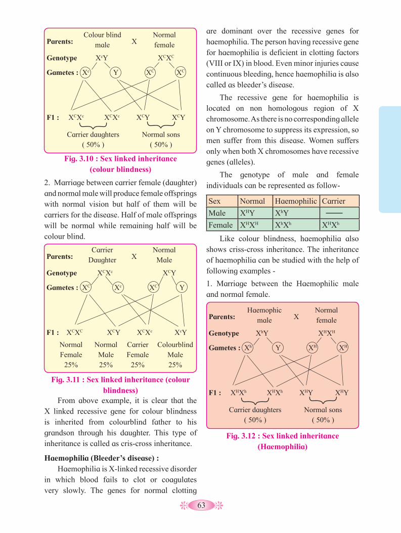
63
Parents: Haemophic
male X Normal female
Genotype XhY XHXH
Gametes : Xh Y XH XH
F1 : XHXh XHXh XHY XHY
Carrier daughters
( 50% )Normal sons
( 50% )
Fig. 3.12 : Sex linked inheritance (Haemophilia)
From above example, it is clear that the X linked recessive gene for colour blindness is inherited from colourblind father to his grandson through his daughter. This type of inheritance is called as cris-cross inheritance.
Haemophilia (Bleeder’s disease) : Haemophilia is X-linked recessive disorder
in which blood fails to clot or coagulates very slowly. The genes for normal clotting
are dominant over the recessive genes for haemophilia. The person having recessive gene for haemophilia is deficient in clotting factors (VIII or IX) in blood. Even minor injuries cause continuous bleeding, hence haemophilia is also called as bleeder’s disease.
The recessive gene for haemophilia is located on non homologous region of X chromosome. As there is no corresponding allele on Y chromosome to suppress its expression, so men suffer from this disease. Women suffers only when both X chromosomes have recessive genes (alleles).
The genotype of male and female individuals can be represented as follow-
Sex Normal Haemophilic Carrier
Male XHY XhY
Female XHXH XhXh XHXh
Like colour blindness, haemophilia also shows criss-cross inheritance. The inheritance of haemophilia can be studied with the help of following examples -
1. Marriage between the Haemophilic male and normal female.
Fig. 3.10 : Sex linked inheritance (colour blindness)
Parents: Colour blind
male X Normal female
Genotype XcY XCXC
Gametes : Xc Y XC XC
F1 : XCXc XCXc XCY XCY
Carrier daughters
( 50% )Normal sons
( 50% )
2. Marriage between carrier female (daughter) and normal male will produce female offsprings with normal vision but half of them will be carriers for the disease. Half of male offsprings will be normal while remaining half will be colour blind.
Fig. 3.11 : Sex linked inheritance (colour blindness)
Parents: Carrier
Daughter X Normal Male
Genotype XCXc XCY
Gametes : XC Xc XC Y
F1 : XCXC XCY XCXc XcY
Normal Female
25%
Normal Male 25%
CarrierFemale
25%
ColourblindMale 25%

64
2. Marriage between carrier female (daughter) and normal male.
On the other hand, some species in which the organism has either male or female reproductive organs, is said to be dioecious or unisexual. Humans are dioecious.
German biologist, Henking in 1891, while studying spermatogenesis of the squash bug (Anasa tristis), noted that 50% of sperms receive the unpaired chromosomes while other 50% sperm do not receive it. Henking gave a name to this structure as the x-body but he could not explain its role in sex determination. Further investigations by other scientists led to conclusion that the “x-body” of Henking was infact a chromosome and gave the name ‘X-Chromosome’.
a. Sex Determination in human beings :
The chromosomal mechanism of sex determination in human beings is XX-XY type. In human beings, the nucleus of each somatic cell contains 46 chromosomes or 23 pairs of chromosomes. Out of these, 22 pairs are autosomes and one pair of sex chromosomes.
Human female has a pair of XX, homomorphic sex chromosomes while male has XY, heteromorphic sex chromosomes.
Thus genotype of :
Female = 44 Autosomes + XX
Male = 44 Autosomes + XY
During gamete formation in male, the diploid germ cells in testis undergo spermatogenesis to produce two types of haplaid sperms, 50% sperms contain 22 autosomes and X chromosome while, 50% sperms contain 22 autosomes and Y chromosome.
In Female, the diploid germ cells in ovaries undergo oogenesis to produce only one type of egg. All eggs contain 22 autosomes and X chromosome. Thus human male is heterogametic and female is homogametic.
If sperm containing X chromosome fertilizes egg (ovum), then diploid zygote is formed, that grows into a female child. If
3.11 Sex Determination :The mechanism by which sex is established
is termed as sex determination. The term sex refers to sexual phenotype. In some species, both male and female reproductive organs are present in same organism. It is discribed as bisexual or hermaphrodite or monoecious.
Fig. 3.13 : Sex linked inheritance (Haemophilia)
Parents: Carrier
Daughter X Normal Male
Genotype XHXh XHY
Gametes : XH Xh XH Y
F1 : XHXH XHY XHXh XhY
Normal Female
25%
Normal Male 25%
CarrierFemale
25%
ColourblindMale 25%
Haemophilia is also referred as “The royal disease”, because it affected the royal families of England, Germany, Russia and Spain in the 19th and 20th centuries. Queen Victoria of England, who ruled from 1837-1901, was belived to have been the carrier of haemophilia. She passed the trait on to her three of nine children.
Do you know ?
Aarya shows normal blood clotting but her mother is haemophilic. Ramesh shows normal blood clotting but his father is haemophilic. If Ramesh and Aarya were to marry, then find out the posible phenotypes of their offsprings.
Find out

65
c. Sex Determination in honey bees :In honey bees, chromosomal mechanism
of sex determination is haplo-diploid type. In this type, sex of individual is determined by the number of set of chromosomes received. Females are diploid (2n=32) and males are haploid (n=16). The female produces haploid eggs (n=16) by meiosis and male produces haplaid sperms (n=16) by mitosis. If the egg is fertilized by sperm, the zygote develops into
sperm containing Y chromosome fertilizes the egg, then diploid zygote is formed that grows into a male child.
This indicates that the sex of a child depends on the type of sperm fertilizing the egg and hence the father is responsible for determination of sex of child and not the mother. Due to lack of knowledge, women are often blamed for giving birth to female child.
b. Sex Determination in birds : In birds, the chromosomal mechanism of
sex determination is ZW-ZZ type. In this type females are heterogametic and produce two types of eggs; 50% eggs carry Z- chromosome, while 50% eggs carry W- chromosome.
Males are homogametic and produce one type of sperms. Each sperm carries a Z- chromosome. Thus sex of individual depends on the kind of egg (ova) fertilized by the sperm.
Fig. 3.14 : Sex determination in human beings
In Bonellia viridis, the environmental factors determine the sex of individual.The sex of worm Bonellia viridis depends on which location the Bonellia larva gets settled. The marine female Bonellia worm has about 10 cm long body. She has a proboscis that can extend over a meter in length. If a Bonellia larva settles on the seafloor, it becomes a female. However when, a larva lands on a female’s proboscis and enters the female’s mouth, it migrates into her uterus and differentiates into a male. Male lives as parasite in uterus of female fertilizing her eggs.
Something Interesting
Fig. 3.15 : Sex determination in birds
Parents :
Gametes : n+Z n+Z n+Z n+W
Male
Male bird
F1 :
2n+ZZ↓
Genotype
X
2n+ZZ 2n+ZW
2n+ZW↓
Female
Female bird
P generation
Male
Male
Male
Female
Female
Female
XY
XY
XY
XX
XX
XX
Meiosis Gametes X Y
X
X
X
X X
Fertilization
F1 generation
Sperm
Eggs
Conclusion : 1:1 sex ratio is produced
Y
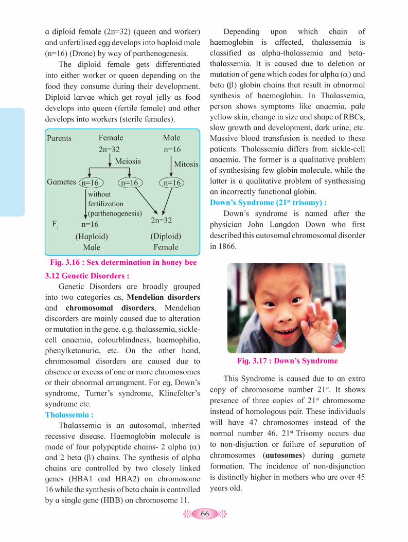
66
Fig. 3.16 : Sex determination in honey bee
Parents
Gametes n=16
n=16
n=16 n=16
Male
(Haploid)Male
Meiosis Mitosis
F1
2n=32
2n=32
n=16
Female
without fertilization (parthenogenesis)
(Diploid) Female
a diploid female (2n=32) (queen and worker)and unfertilised egg develops into haploid male (n=16) (Drone) by way of parthenogenesis.
The diploid female gets differentiated into either worker or queen depending on the food they consume during their development. Diploid larvae which get royal jelly as food develops into queen (fertile female) and other develops into workers (sterile females).
Depending upon which chain of haemoglobin is affected, thalassemia is classified as alpha-thalassemia and beta-thalassemia. It is caused due to deletion or mutation of gene which codes for alpha (a) and beta (b) globin chains that result in abnormal synthesis of haemoglobin. In Thalassemia, person shows symptoms like anaemia, pale yellow skin, change in size and shape of RBCs, slow growth and development, dark urine, etc. Massive blood transfusion is needed to these patients. Thalassemia differs from sickle-cell anaemia. The former is a qualitative problem of synthesising few globin molecule, while the latter is a qualitative problem of synthesising an incorrectly functional globin. Down’s Syndrome (21st trisomy) :
Down’s syndrome is named after the physician John Langdon Down who first described this autosomal chromosomal disorder in 1866.
Fig. 3.17 : Down’s Syndrome
3.12 Genetic Disorders : Genetic Disorders are broadly grouped
into two categories as, Mendelian disorders and chromosomal disorders, Mendelian discorders are mainly caused due to alteration or mutation in the gene. e.g. thalassemia, sickle-cell anaemia, colourblindness, haemophilia, phenylketonuria, etc. On the other hand, chromosomal disorders are caused due to absence or excess of one or more chromosomes or their abnormal arrangment. For eg, Down’s syndrome, Turner’s syndrome, Klinefelter’s syndrome etc. Thalassemia :
Thalassemia is an autosomal, inherited recessive disease. Haemoglobin molecule is made of four polypeptide chains- 2 alpha (a) and 2 beta (b) chains. The synthesis of alpha chains are controlled by two closely linked genes (HBA1 and HBA2) on chromosome 16 while the synthesis of beta chain is controlled by a single gene (HBB) on chromosome 11.
This Syndrome is caused due to an extra copy of chromosome number 21st. It shows presence of three copies of 21st chromosome instead of homologous pair. These individuals will have 47 chromosomes instead of the normal number 46. 21st Trisomy occurs due to non-disjuction or failure of separation of chromosomes (autosomes) during gamete formation. The incidence of non-disjunction is distinctly higher in mothers who are over 45 years old.

67
Fig. 3.18 : Karyotype of Down’s syndrome
These patients have mild or moderate mental retardation and skeletal development is poor. Distinct facial features like small head, ears and mouth, face is typically flat and rounded with flat nose, open mouth and protruding tongue, eyes slant up and out with internal epicanthal folds, flat hands and stubby fingers and palm is broad with single palmer crease.
Down Syndrome - Trisomy 21
1 2 3 4 5 6 7 8
9 10 11 12 13 14 15 16
17 18 19 20 21 22
or
XX XY
Turner’s Syndrome : (X monosomy / XO females) It is sex chromosomal disorder caused
due to non-disjunction of chromosome during gamete formation. Individual born with Turner’s syndrome has 44 autosomes with XO. They are phenotypically female. They have a short stature (height) and webbed neck, lower posterior hair line, broad shield-shaped chest, poorly developed ovaries and breast, and low intelligence.
Klinefelter’s syndrome (XXY males) :It is chromosomal disorder caused due to
extra X chromosome in males. Thus genotype of individuals is 44 + XXY. They are described as feminized males. Extra chromosome is a result of non-disjunction of X-chromosome during meiosis. Individual is male and has over all masculine development. Voice pitch is harsh and have under developed testis. They are tall with long arms, feminine development (development of breast i.e. Gynaecomastia) and no spermatogenesis, therefore, individuals are sterile.
Trisomy
Activity :
Study the complementary and supplementary interaction (digenic interactions) - both in plants and animals
68
Q. 1 Multiple choice questions. 1. Phenotypic ratio of incomplete
dominance in Mirabilis jalapa. a. 2 : 1 : 1 b. 1 : 2 : 1 c. 3 : 1 d. 2 : 22. In dihybrid cross, F
2 generation offsprings
show four different phenotypes while the genotypes are ................
a. six b. nine c. eight d. sixteen3. A cross between an individual with
unknown genotype for a trait with recessive plant for that trait is ..............
a. back cross b. reciprocal cross c. monohybrid cross d. test cross4. When phenotypic and genotypic ratios
are the same, then it is an example of ............
a. incomplete dominance b. complete dominance c. Multiple alleles d. cytoplasmic inheritance
5. If the centromere is situated near the end of the chromosome, the chromosome is called ................
a. Metacentric b. Acrocentric c. Sub-Metacentric d. Telocentric6. Chromosomal theory of inheritance was
proposed by ................ a. Sutton and Boveri b. Watson and Crick c. Miller and Urey d. Oparin and Halden
7. If the genes are located in a chromosome as p-q-r-s-t, which of the following gene pairs will have least probability of being inherited together?
a. p and q b. r and s c. s and t d. p and s
8. Find the mis match pair :- a. Down’s syndrome = 44 + XY b. Turner’s syndrome = 44 + XO c. Klinefelter syndrome = 44 + XXY d. Super female = 44 + XXX
9. A colourblind man marries a woman, who is homozygous for normal colour vision ,the probability of their son being colourblind is –
a. 0% b. 25% c. 50% d. 100%
Q. 2 Very Short Answer Questions. 1. Explain the statements :
a. Test cross is back cross but back cross is not necessarily a test cross.
b. Law of dominance is not universal.2. Define the following terms :
a. Dihybrid cross b. Homozygousc. Heterozygous d. Test cross
3. What is allosome? 4. What is crossing over? 5. Give one example of autosomal recessive
disorder. 6. What are X-linked genes? 7. What are holandric traits? 8. Give an example of chromosomal
disorder caused due to non-disjunction of autosomes.
9. Give one example of complete sex linkage?
Q. 3 Short Answer Questions.
1. Enlist seven traits of pea plant selected/ studied by Mendel.
2. Why law of segregation is also called the law of purity of gametes?
3. Write a note on pleiotropy.4. What are the reasons of Mendel success?
Exercise
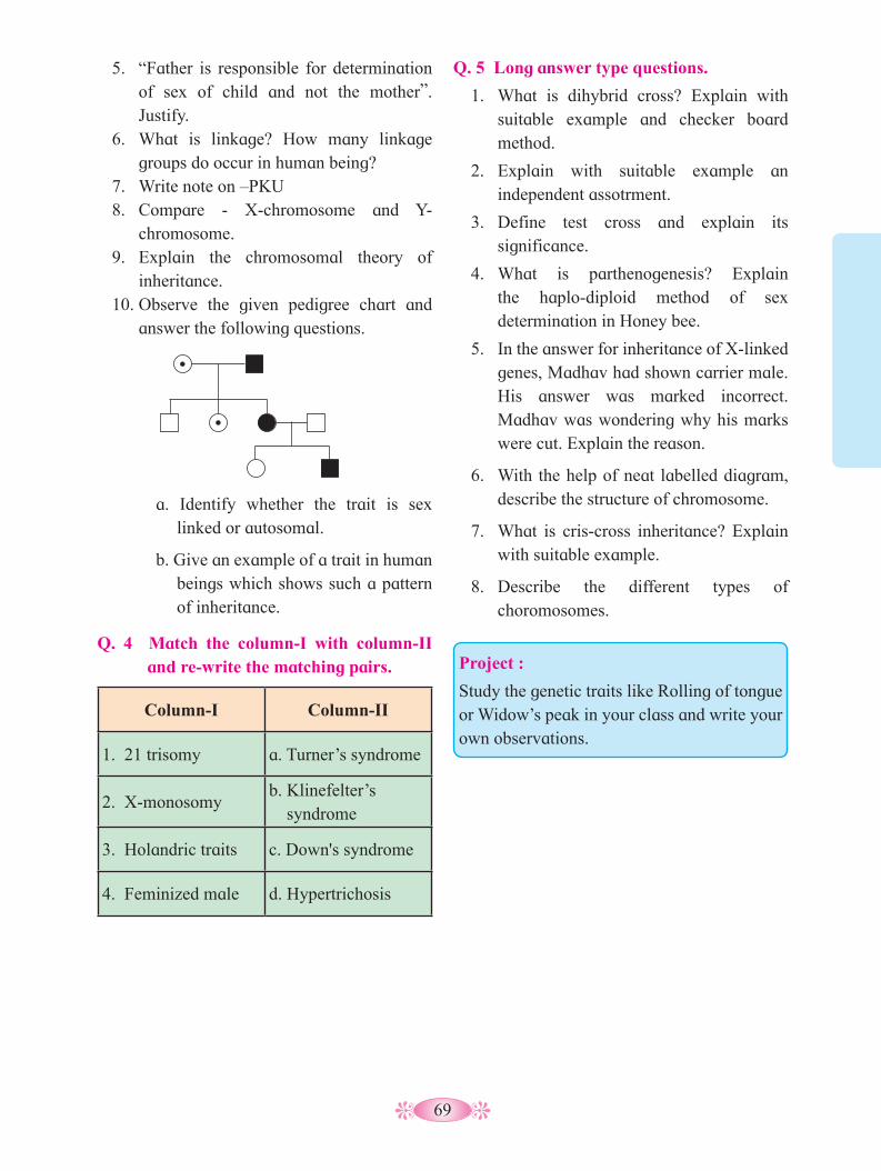
69
5. “Father is responsible for determination of sex of child and not the mother”. Justify.
6. What is linkage? How many linkage groups do occur in human being?
7. Write note on –PKU 8. Compare - X-chromosome and Y-
chromosome. 9. Explain the chromosomal theory of
inheritance.10. Observe the given pedigree chart and
answer the following questions.
Q. 5 Long answer type questions.
1. What is dihybrid cross? Explain with suitable example and checker board method.
2. Explain with suitable example an independent assotrment.
3. Define test cross and explain its significance.
4. What is parthenogenesis? Explain the haplo-diploid method of sex determination in Honey bee.
5. In the answer for inheritance of X-linked genes, Madhav had shown carrier male. His answer was marked incorrect.Madhav was wondering why his marks were cut. Explain the reason.
6. With the help of neat labelled diagram, describe the structure of chromosome.
7. What is cris-cross inheritance? Explain with suitable example.
8. Describe the different types of choromosomes.
a. Identify whether the trait is sex linked or autosomal.
b. Give an example of a trait in human beings which shows such a pattern of inheritance.
Q. 4 Match the column-I with column-II and re-write the matching pairs.
Column-I Column-II
1. 21 trisomy a. Turner’s syndrome
2. X-monosomy b. Klinefelter’s syndrome
3. Holandric traits c. Down's syndrome
4. Feminized male d. Hypertrichosis
Project :
Study the genetic traits like Rolling of tongue or Widow’s peak in your class and write your own observations.
70
4.1 The Discovery of DNA:Modern understanding of DNA has evolved
from the discovery of nucleic acid to the development of the double-helix model. In 1869, Friedrich Miescher began working with white blood cells which are the major component of pus from infections. He collected a lot of pus from bandages at the local hospital. He used a salt solution to wash the pus off the bandages. When he added a weak alkaline solution to the cells, the cells lysed and nuclei precipitated out of the solution. From the cell nuclei, he isolated a unique chemical substance to which he called nuclein. Chemically, nuclein has high phosphorus content. Moreover it showed acidic properties. Hence it was named as nucleic acid.
By the early 1900s, we knew that Miescher's nuclein was a mix (mixture) of proteins and nucleic acids. There are two kinds of nucleic acids. DNA (deoxyribonucleic acid) and RNA (ribonucleic acid).
4.2 The Genetic Material is a DNA: By the early 1900s, geneticist knew that
genes control the inheritance of traits, that genes are located on chromosome and that chemically chromosomes are mainly composed of DNA and proteins. Initially, most geneticists thought that protein are large, complex molecules and store information needed to govern cell metabolism. Hence it was assumed that proteins caused the variations observed within species.
Molecular Basis of Inheritance
On the other hand DNA thought to be small, simple molecule whose composition varied little among species. Over the time, these ideas about DNA were shown to be wrong. In fact DNA molecules are large and vary tremendously within and among species. Variations in the DNA molecules are different than the variation in shape, electrical charge and function shown by proteins so it is not surprising that most researchers initially favored proteins as the genetic material.
Over a period of roughly 25 years (1928-1952), geneticists became convinced that DNA and not protein, was the genetic material. Let us study three important contributions that helped cause this shift of opinion.
Griffith’s experiments :In 1928, a British medical officer Frederick
Griffith performed an experiment on bacterium Streptococcus pneumoniae that causes pneumonia in humans and other mammals. Griffith used two strains or two genetic varieties of Streptococcus to find a cure for pneumonia, which was a common cause of death at that time. The two strains used were : i. Virulent, smooth, pathogenic and
encapsulated S type. ii. Non-virulent, rough, non-pathogenic and
non-capsulated R type.
Griffith conducted four experiments on these bacteria. First, when he injected bacteria of strain R to mice, the mice survived because it did not develop pneumonia. Second, when he injected bacteria of strain S to mice, the mice developed pneumonia and died. In the third experiment, he injected heat-killed strain S bacteria to mice, once again the mice survived. In fourth experiment, he mixed heat-killed S bacteria with live bacteria of strain R and injected to mice. The mice died and Griffith
4
1. What is nucleic acid?2. What are the types of nucleic acid?3. What are the functions of nucleic acid?4. What is the difference between DNA in
prokaryotes and eukaryotes?
Can you recall?

71
recovered large numbers of live strain S bacteria from the blood of the dead mice.
In these four experiments, something had caused harmless strain R bacterium to change into deadly S strain bacterium. Griffith showed that the change was genetic. He suggested that genetic material from heat-killed strain S bacterium had somehow changed the living strain R bacterium into strain S bacterium.
Griffith concluded that the R-strain bacterium must have taken up, to what he called a "transforming principle" from the heat-killed S bacterium, which allowed R strain to get transformed into smooth-coated bacterium and become virulent.
They also discovered that protein –digesting enzymes (proteases), RNA-digesting enzyme (RNAases) did not affect transformation, so the transforming substance was neither a protein nor RNA. DNA digested with DNAse did inhibit the transformation, suggesting that DNA caused the transformation. These experiments proved that the transforming principle is DNA but all biologists were not convinced.
Finally, Alfred Hershey and Martha Chase (1952) proved that DNA is the genetic material and not proteins, by using bacteriophages.
Hershey - Chase Experiment: Hershey and Chase worked with viruses
that infect bacteria i.e. bacteriophages, which are composed of DNA and protein. They used radioactive phosphorous 32P in the medium for some viruses and radioactive sulphur 35S for some others.
They grew some viruses on a medium that contained radioactive phosphorus and some others on medium that contained radioactive sulphur. Viruses grown in the presence of radioactive phosphorus contained radioactive DNA (labelled DNA), but not radioactive proteins because DNA contains phosphorus (labelled DNA) but proteins do not. Similarly, viruses grown on radioactive sulphur contained radioactive protein but not radioactive DNA because DNA does not contain sulphur.
Radioactive phages were allowed to infect E.coli bacteria grown on the medium containing normal ‘P’ and ‘S’. Then, as
Avery, McCarty and MacLeod’s experiment:In 1944, after some 10 years of research
and experimentation, U. S. microbiologists Oswald T. Avery, Colin M. MacLeod and Maclyn McCarty (all at Rockefeller University in New York) first evidenced to prove the DNA is a genetic material (transforming principle), through the experiments. They purified DNA, RNA, Proteins (enzymes) and other materials from cell free extract of S cells/ strain and mixed with heat killed S strain and R cells seperately to confirm which one could transform living R cells into S cells. Only DNA was able to transform harmless strain R into deadly strain S.
Fig. 4.1 : Griffith’s experiment
Rough strain (II-R)
Smooth Strain (III-S)
Heat-killed Smooth Strain
Rough strain and Heat-killed Smooth Strain
Mouse alive Mouse dead Mouse alive Mouse dead
Fig. 4.2 : DNA transforms bacteria
Rough nonvirulent (strain R)
Heat-killed smooth virulent (strain S)
+ DNase
+ Protease
Mouse dead
Mouse alive
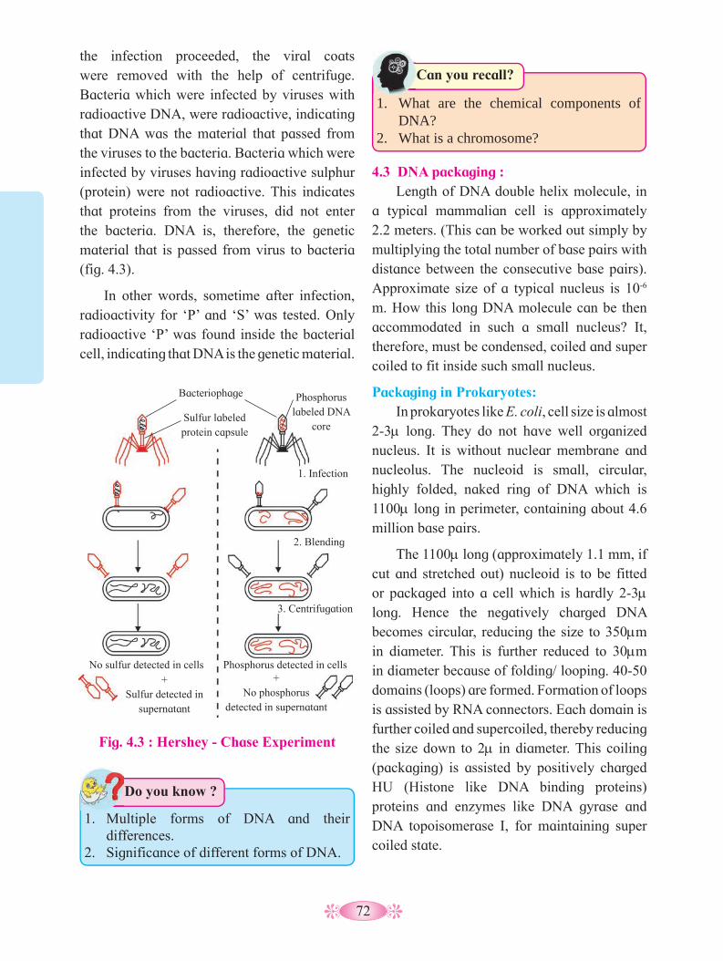
72
the infection proceeded, the viral coats were removed with the help of centrifuge. Bacteria which were infected by viruses with radioactive DNA, were radioactive, indicating that DNA was the material that passed from the viruses to the bacteria. Bacteria which were infected by viruses having radioactive sulphur (protein) were not radioactive. This indicates that proteins from the viruses, did not enter the bacteria. DNA is, therefore, the genetic material that is passed from virus to bacteria (fig. 4.3).
In other words, sometime after infection, radioactivity for ‘P’ and ‘S’ was tested. Only radioactive ‘P’ was found inside the bacterial cell, indicating that DNA is the genetic material.
4.3 DNA packaging :Length of DNA double helix molecule, in
a typical mammalian cell is approximately 2.2 meters. (This can be worked out simply by multiplying the total number of base pairs with distance between the consecutive base pairs). Approximate size of a typical nucleus is 10-6
m. How this long DNA molecule can be then accommodated in such a small nucleus? It, therefore, must be condensed, coiled and super coiled to fit inside such small nucleus.
Packaging in Prokaryotes:In prokaryotes like E. coli, cell size is almost
2-3m long. They do not have well organized nucleus. It is without nuclear membrane and nucleolus. The nucleoid is small, circular, highly folded, naked ring of DNA which is 1100mlong in perimeter, containing about 4.6 million base pairs.
The 1100m long (approximately 1.1 mm, if cut and stretched out) nucleoid is to be fitted or packaged into a cell which is hardly 2-3mlong. Hence the negatively charged DNA becomes circular, reducing the size to 350mm in diameter. This is further reduced to 30mm in diameter because of folding/ looping. 40-50 domains (loops) are formed. Formation of loops is assisted by RNA connectors. Each domain is further coiled and supercoiled, thereby reducing the size down to 2m in diameter. This coiling (packaging) is assisted by positively charged HU (Histone like DNA binding proteins) proteins and enzymes like DNA gyrase and DNA topoisomerase I, for maintaining super coiled state.
1. What are the chemical components of DNA?
2. What is a chromosome?
Can you recall?
Fig. 4.3 : Hershey - Chase Experiment
Bacteriophage
Sulfur labeled protein capsule
No sulfur detected in cells +
Sulfur detected in supernatant
+ No phosphorus
detected in supernatant
Phosphorus detected in cells
3. Centrifugation
2. Blending
1. Infection
Phosphorus labeled DNA
core
1. Multiple forms of DNA and their differences.
2. Significance of different forms of DNA.
Do you know ?
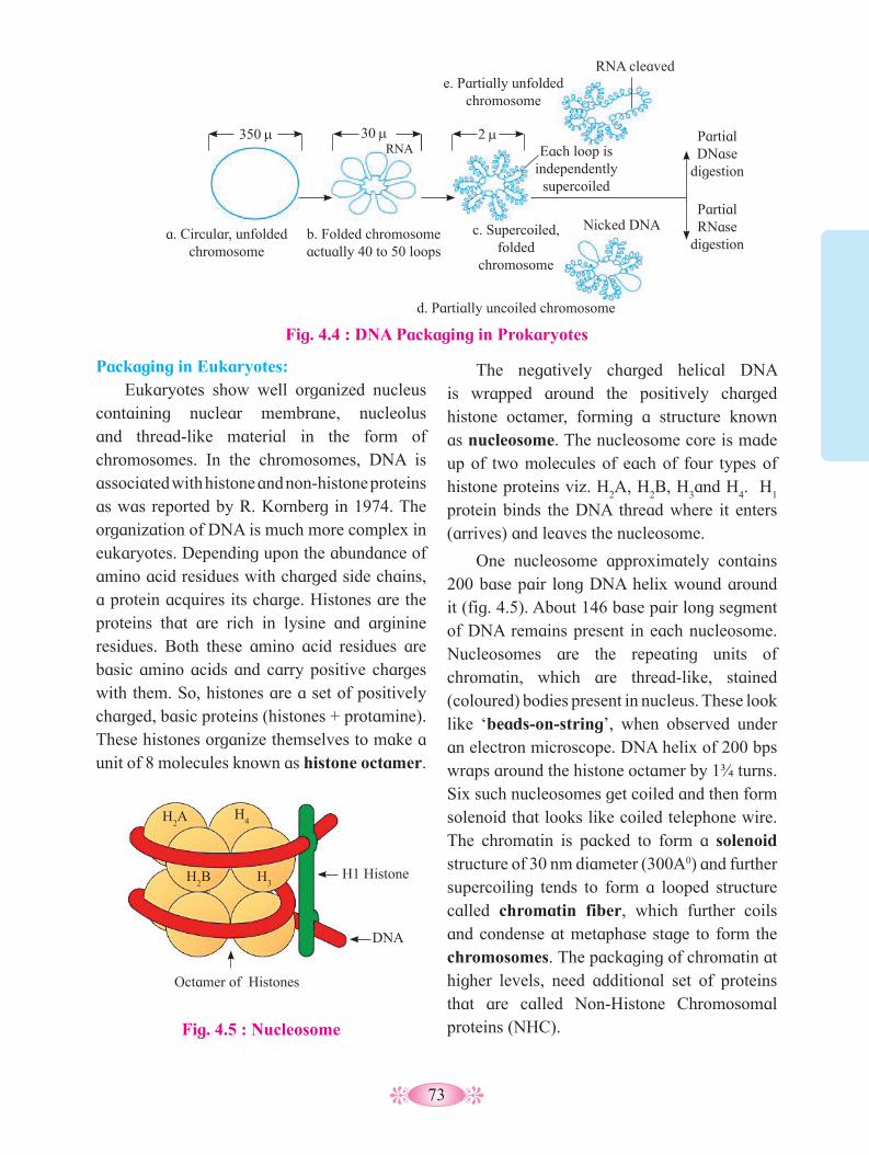
73
Packaging in Eukaryotes:Eukaryotes show well organized nucleus
containing nuclear membrane, nucleolus and thread-like material in the form of chromosomes. In the chromosomes, DNA is associated with histone and non-histone proteins as was reported by R. Kornberg in 1974. The organization of DNA is much more complex in eukaryotes. Depending upon the abundance of amino acid residues with charged side chains, a protein acquires its charge. Histones are the proteins that are rich in lysine and arginine residues. Both these amino acid residues are basic amino acids and carry positive charges with them. So, histones are a set of positively charged, basic proteins (histones + protamine). These histones organize themselves to make a unit of 8 molecules known as histone octamer.
Fig. 4.4 : DNA Packaging in Prokaryotes
The negatively charged helical DNA is wrapped around the positively charged histone octamer, forming a structure known as nucleosome. The nucleosome core is made up of two molecules of each of four types of histone proteins viz. H
2A, H
2B, H
3and H
4. H
1
protein binds the DNA thread where it enters (arrives) and leaves the nucleosome.
One nucleosome approximately contains 200 base pair long DNA helix wound around it (fig. 4.5). About 146 base pair long segment of DNA remains present in each nucleosome. Nucleosomes are the repeating units of chromatin, which are thread-like, stained (coloured) bodies present in nucleus. These look like ‘beads-on-string’, when observed under an electron microscope. DNA helix of 200 bps wraps around the histone octamer by 1¾ turns. Six such nucleosomes get coiled and then form solenoid that looks like coiled telephone wire. The chromatin is packed to form a solenoid structure of 30 nm diameter (300A0) and further supercoiling tends to form a looped structure called chromatin fiber, which further coils and condense at metaphase stage to form the chromosomes. The packaging of chromatin at higher levels, need additional set of proteins that are called Non-Histone Chromosomal proteins (NHC).
350 m 30 m 2 mRNA
a. Circular, unfolded chromosome
Nicked DNA b. Folded chromosome actually 40 to 50 loops
c. Supercoiled, folded
chromosome
e. Partially unfolded chromosome
RNA cleaved
Each loop is independently
supercoiled
Partial RNase
digestion
Partial DNase
digestion
d. Partially uncoiled chromosome
Fig. 4.5 : Nucleosome
H2A
H2B
Octamer of Histones
DNA
H1 HistoneH3
H4

74
1. What is the backbone of the DNA
structure?2. Name the nitrogen bases of DNA.3. What are Nucleoside and Nucleotide?4. Is the double helix right or left handed?
Can you recall?
What is the key difference between DNA in prokaryotic and eukaryotic cells?
Find out
Fig. 4.7 : DNA packaging
Nucleosome
DNA wound around histone proteins
Linker DNA
Non-Histone Chromosomal Proteins (NHC) : These are additional sets of proteins that contribute to the packaging of chromatin at a higher level.Heterochromatin and Euchromatin :1. Heterochromatin: In eukaryotic cells, some
segments of chromonema/ chromosome during interphase and early prophase remain in a condensed state. These region constitute heterochromatin. This term was proposed by Heitz. These regions are localized near centromere, telomeres and are also intercalated. It is genetically mostly inactive. It stains strongly and appears dark. Heterochromatin is 2 to 3 times more rich in DNA than in the euchromatin.
2. Euchromatin: The regions of chromonema which are in non-condensed state, constitute euchromatin. Euchromatic regions stain light. Euchromatin is genetically very much active and fast replicating. Euchromatin is transcriptionally active, while heterochromatin is transcriptionally almost inactive.
Fig. 4.6 : Chromatin showing beads-on-string
B : Chain of nucleosomes forming 10 to 11 nm thick fibre
C : Solenoid forming 30 nm thick fibre
A : Beads-on-string magnified
4.4 DNA Replication :The DNA molecule regulates and controls
all the activities of the cell. Because of its unique structure, it is able to control the synthesis of other molecules of the cell. At the same time when the cell reproduces, the DNA also should duplicate itself to distribute
Nucleosomes
0.5 mm
111 Å
60 Å Nucleosomes
Chromatin fibers “stretched” during preparation for electron
microscopy revealing linker DNA between nucleosome cores
Linker DNA, varying in
length from 8 to 114 nucleotide
pairs
Nucleosome core, 146 nucleotide pairs of DNA wrapped as ¾ turns around an
octamer of histones
300 Å
300Å metaphase
fiber or “solenoid”
110
Å f
iber
110 Å

75
equally to the daughter cells. As a carrier of genetic information, DNA has to perform two important functions :
a. Heterocatalytic function : When DNA directs the synthesis of chemical molecules other than itself, then such functions of DNA are called heterocatalytic functions. Eg. Synthesis of RNA (transcription), synthesis of protein (Translation), etc.
b. Autocatalytic function : When DNA directs the synthesis of DNA itself, then such function of DNA is called autocatalytic function. Eg. Replication.The process by which DNA duplicates
itself is called replication. Through replication, it forms two copies that are identical to it.
In eukaryotic organisms, replication of DNA takes place only once in the cell cycle.It occurs in the S- phase of interphase in the cell cycle.
DNA replicates through Semiconservative mode of replication.
The model for Semiconservative replication was proposed by Watson and Crick, on the basis of antiparallel and complementary nature of DNA strands. The process of semicoservative replication is as below:
1. Activation of Nucleotides:The four types of nucleotides of DNA i.e.
dAMP, dGMP, dCMP and dTMP are present in the nucleoplasm. They are activated by ATP in presence of an enzyme phosphorylase. This results in the formation of deoxyribonucleotide triphosphates i.e. dATP, dGTP, dCTP and dTTP. The process is known as Phosphorylation.
2. Point of Origin or Initiation point:It begins at specific point ‘O’ -origin and
terminates at point ‘T’. Origin is flanked by ‘T’ sites. The unit of DNA in which replication occurs, is called replicon. In prokaryotes, there is noly one replicon however in eukaryotes, there are several replicons in tandem.
At the point ‘O’, enzyme endonuclease nicks one of the strands of DNA, temporarily. The nick occurs in the sugar-phosphate back bone or the phosphodiester bond.
3. Unwinding of DNA molecule:Now enzyme DNA helicase operates by
breaking weak hydrogen bonds in the vicinity of ‘O. The strands of DNA separate and unwind. This unwinding is bidirectional and continues as ‘Y’ shaped replication fork. Each separated strand acts as template.
The two separated strands are prevented from recoiling (rejoining) by SSBP (Single strand binding proteins). SSB proteins remain attached to both the separated strands so as to facilate synthesis of new polynucleotide strands.
Fig. 4.8 : Semiconservative Replication of DNA
5’
3’ 5’
Parental strand
New strands
A=T
T=A
C=G
A=T
G=C
T=A
3’ A=T T=A
C=G A=T
G=C
T=A
A=T
T=A
C=G
A
=T
G=C
T=A
5’
5’ 3’
3’
5’
Lagging template
Replication fork
RNA primer Okazaki
fragments
3’
Leading template
Lagging strand
(discontin
ous)
Leading strand
(continous)
5’
5’
5’
5’
5’ 3’
3’
3’ 3’
Leading template
Lagging template
RNA primer
Okazaki fragments
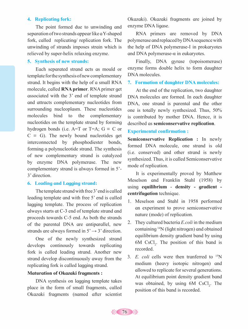
76
4. Replicating fork:
The point formed due to unwinding and separation of two strands appear like a Y-shaped fork, called replicating/ replication fork. The unwinding of strands imposes strain which is relieved by super-helix relaxing enzyme.
5. Synthesis of new strands:
Each separated strand acts as mould or template for the synthesis of new complementary strand. It begins with the help of a small RNA molecule, called RNA primer. RNA primer get associated with the 3’ end of template strand and attracts complementary nucleotides from surrounding nucleoplasm. These nucleotides molecules bind to the complementary nucleotides on the template strand by forming hydrogen bonds (i.e. A=T or T=A; G = C or C = G). The newly bound nucleotides get interconnected by phosphodiester bonds, forming a polynucleotide strand. The synthesis of new complementary strand is catalyzed by enzyme DNA polymerase. The new complementary strand is always formed in 5’-3’ direction.
6. Leading and Lagging strand:
The template strand with free 3’ end is called leading template and with free 5’ end is called lagging template. The process of replication always starts at C-3 end of template strand and proceeds towards C-5 end. As both the strands of the parental DNA are antiparallel, new strands are always formed in 5’ → 3’ direction.
One of the newly synthesized strand develops continously towards replicating fork is called leading strand. Another new strand develop discontinuously away from the replicating fork is called lagging strand.
Maturation of Okazaki fragments :
DNA synthesis on lagging template takes place in the form of small fragments, called Okazaki fragments (named after scientist
Okazaki). Okazaki fragments are joined by enzyme DNA ligase.
RNA primers are removed by DNA polymerase and replaced by DNA sequence with the help of DNA polymerase-I in prokaryotes and DNA polymerase-α in eukaryotes.
Finally, DNA gyrase (topoisomerase) enzyme forms double helix to form daughter DNA molecules.
7. Formation of daughter DNA molecules:
At the end of the replication, two daughter DNA molecules are formed. In each daughter DNA, one strand is parental and the other one is totally newly synthesized. Thus, 50% is contributed by mother DNA. Hence, it is described as semiconservative replication.
Experimental confirmation :
Semiconservative Replication : In newly formed DNA molecule, one strand is old (i.e. conserved) and other strand is newly synthesized. Thus, it is called Semiconservative mode of replication.
It is experimentally proved by Matthew Meselson and Franklin Stahl (1958) by using equilibrium - density - gradient - centrifugation technique.
1. Meselson and Stahl in 1958 performed an experiment to prove semiconservative nature (mode) of replication.
2. They cultured bacteria E.coli in the medium containing 14N (light nitrogen) and obtained equilibrium density gradient band by using 6M CsCl
2. The position of this band is
recorded.
3. E. coli cells were then tranferred to 15N medium (heavy isotopic nitrogen) and allowed to replicate for several generations. At equilibrium point density gradient band was obtained, by using 6M CsCl
2. The
position of this band is recorded.
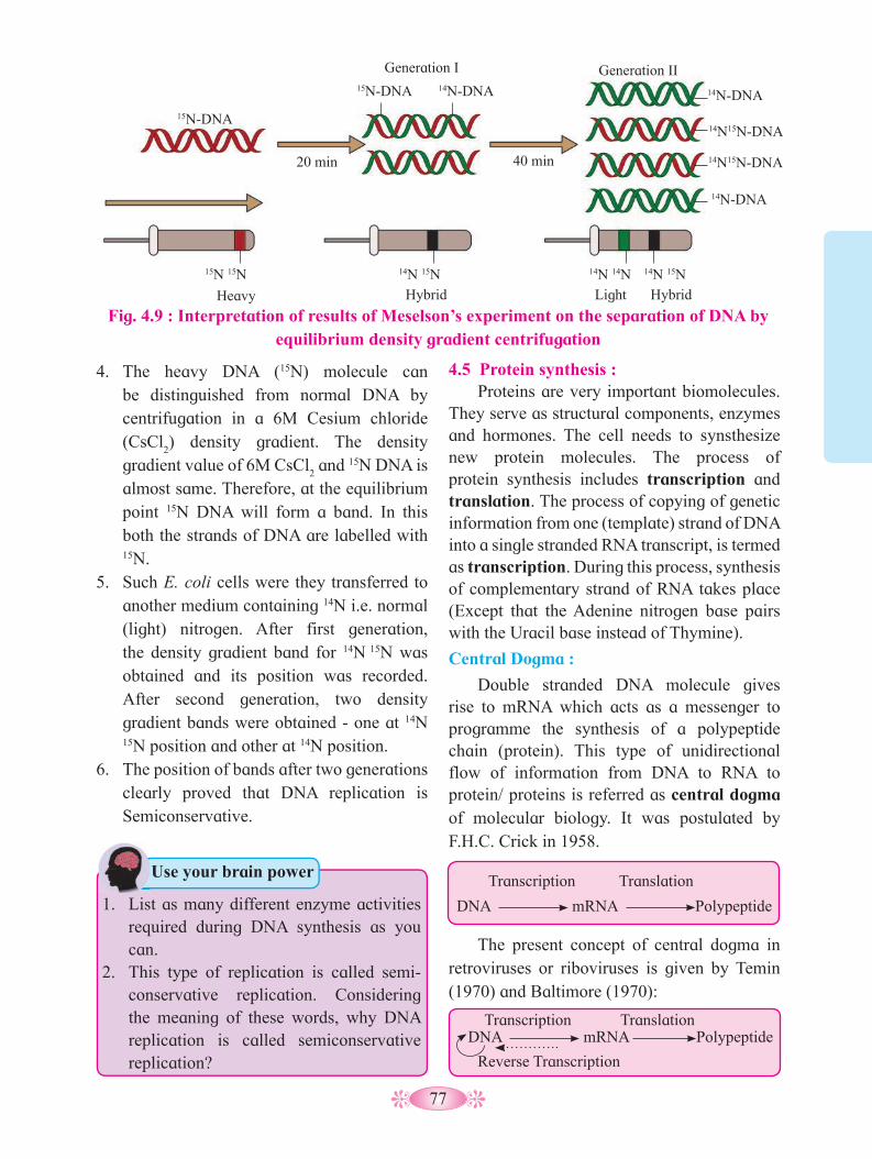
77
4. The heavy DNA (15N) molecule can be distinguished from normal DNA by centrifugation in a 6M Cesium chloride (CsCl
2) density gradient. The density
gradient value of 6M CsCl2 and 15N DNA is
almost same. Therefore, at the equilibrium point 15N DNA will form a band. In this both the strands of DNA are labelled with 15N.
5. Such E. coli cells were they transferred to another medium containing 14N i.e. normal (light) nitrogen. After first generation, the density gradient band for 14N 15N was obtained and its position was recorded. After second generation, two density gradient bands were obtained - one at 14N
15N position and other at 14N position.6. The position of bands after two generations
clearly proved that DNA replication is Semiconservative.
4.5 Protein synthesis : Proteins are very important biomolecules.
They serve as structural components, enzymes and hormones. The cell needs to synsthesize new protein molecules. The process of protein synthesis includes transcription and translation. The process of copying of genetic information from one (template) strand of DNA into a single stranded RNA transcript, is termed as transcription. During this process, synthesis of complementary strand of RNA takes place (Except that the Adenine nitrogen base pairs with the Uracil base instead of Thymine).
Central Dogma :
Double stranded DNA molecule gives rise to mRNA which acts as a messenger to programme the synthesis of a polypeptide chain (protein). This type of unidirectional flow of information from DNA to RNA to protein/ proteins is referred as central dogma of molecular biology. It was postulated by F.H.C. Crick in 1958.
DNA mRNA Polypeptide
Transcription Translation
The present concept of central dogma in retroviruses or riboviruses is given by Temin (1970) and Baltimore (1970):
DNA mRNA PolypeptideTranscription Translation
Reverse Transcription
Fig. 4.9 : Interpretation of results of Meselson’s experiment on the separation of DNA by equilibrium density gradient centrifugation
1. List as many different enzyme activities required during DNA synthesis as you can.
2. This type of replication is called semi-conservative replication. Considering the meaning of these words, why DNA replication is called semiconservative replication?
Use your brain power
15N-DNA
15N-DNA 14N-DNA 14N-DNA
14N15N-DNA
14N15N-DNA
14N-DNA
20 min
Generation I Generation II
15N 15N 14N 15N 14N 14N 14N 15N
Heavy Hybrid Hybrid Light
40 min
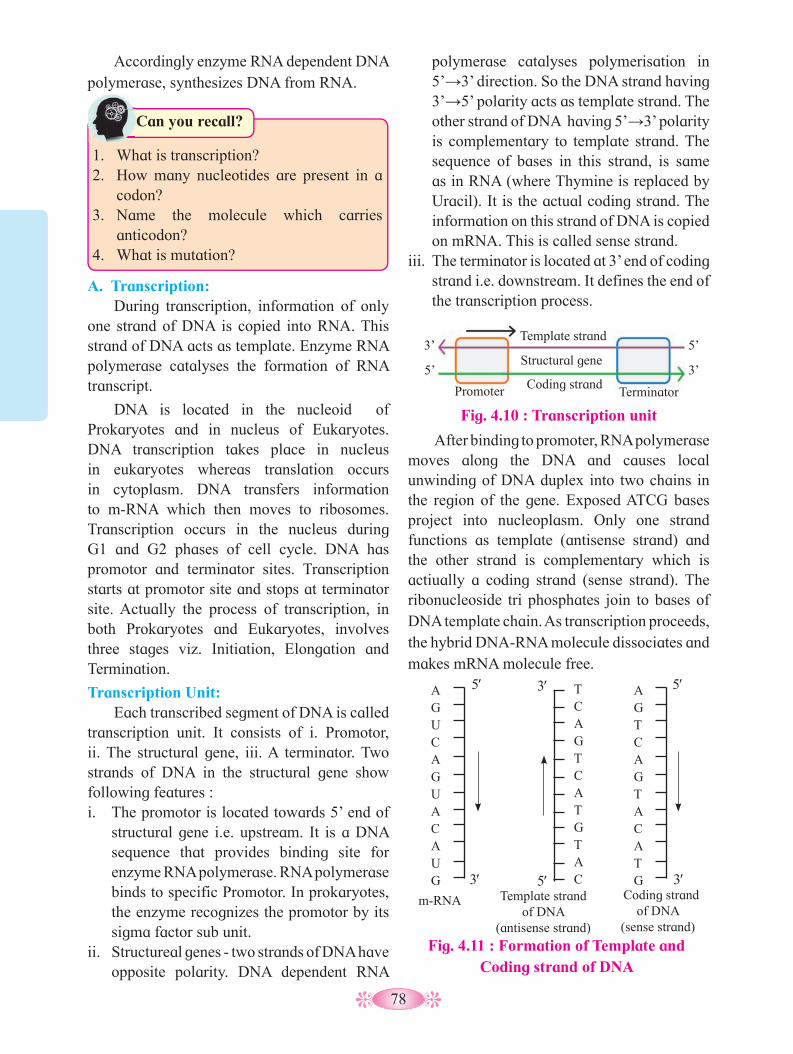
78
1. What is transcription?2. How many nucleotides are present in a
codon?3. Name the molecule which carries
anticodon?4. What is mutation?
Can you recall?
polymerase catalyses polymerisation in 5’→3’ direction. So the DNA strand having 3’→5’ polarity acts as template strand. The other strand of DNA having 5’→3’ polarity is complementary to template strand. The sequence of bases in this strand, is same as in RNA (where Thymine is replaced by Uracil). It is the actual coding strand. The information on this strand of DNA is copied on mRNA. This is called sense strand.
iii. The terminator is located at 3’ end of coding strand i.e. downstream. It defines the end of the transcription process.
A. Transcription: During transcription, information of only
one strand of DNA is copied into RNA. This strand of DNA acts as template. Enzyme RNA polymerase catalyses the formation of RNA transcript.
DNA is located in the nucleoid of Prokaryotes and in nucleus of Eukaryotes. DNA transcription takes place in nucleus in eukaryotes whereas translation occurs in cytoplasm. DNA transfers information to m-RNA which then moves to ribosomes. Transcription occurs in the nucleus during G1 and G2 phases of cell cycle. DNA has promotor and terminator sites. Transcription starts at promotor site and stops at terminator site. Actually the process of transcription, in both Prokaryotes and Eukaryotes, involves three stages viz. Initiation, Elongation and Termination.
Transcription Unit:Each transcribed segment of DNA is called
transcription unit. It consists of i. Promotor, ii. The structural gene, iii. A terminator. Two strands of DNA in the structural gene show following features : i. The promotor is located towards 5’ end of
structural gene i.e. upstream. It is a DNA sequence that provides binding site for enzyme RNA polymerase. RNA polymerase binds to specific Promotor. In prokaryotes, the enzyme recognizes the promotor by its sigma factor sub unit.
ii. Structureal genes - two strands of DNA have opposite polarity. DNA dependent RNA
After binding to promoter, RNA polymerase moves along the DNA and causes local unwinding of DNA duplex into two chains in the region of the gene. Exposed ATCG bases project into nucleoplasm. Only one strand functions as template (antisense strand) and the other strand is complementary which is actiually a coding strand (sense strand). The ribonucleoside tri phosphates join to bases of DNA template chain. As transcription proceeds, the hybrid DNA-RNA molecule dissociates and makes mRNA molecule free.
Fig. 4.11 : Formation of Template and Coding strand of DNA
3′
5′
TCAGTCATGTAC
Template strand of DNA
(antisense strand)
3′
5′A G U C A G U A C A U G 3′
5′A G T C A G T A C A T G
m-RNA Coding strand of DNA
(sense strand)
Accordingly enzyme RNA dependent DNA polymerase, synthesizes DNA from RNA.
Fig. 4.10 : Transcription unit
Template strand
Structural gene
Coding strand Promoter Terminator
3’
3’ 5’
5’
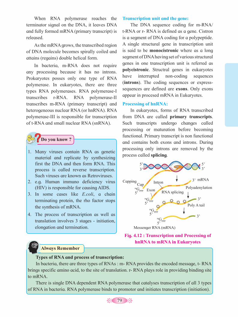
79
When RNA polymerase reaches the terminator signal on the DNA, it leaves DNA and fully formed mRNA (primary transcript) is released.
As the mRNA grows, the transcribed region of DNA molecule becomes spirally coiled and attains (regains) double helical form.
In bacteria, m-RNA does not require any processing because it has no introns. Prokaryotes posses only one type of RNA polymerase. In eukaryotes, there are three types RNA polymerases. RNA polymerase-I transcribes r-RNA. RNA polymerase-II transcribes m-RNA (primary transcript) and heterogeneous nuclear RNA (or hnRNA). RNA polymerase-III is responsible for transcription of t-RNA and small nuclear RNA (snRNA).
Transcription unit and the gene:The DNA sequence coding for m-RNA/
t-RNA or r- RNA is defined as a gene. Cistron is a segment of DNA coding for a polypeptide. A single structural gene in transcription unit is said to be monocistronic where as a long segment of DNA having set of various structural genes in one transcription unit is referred as polycistronic. Structral genes in eukaryotes have interrupted non-coding sequences (introns). The coding sequences or express- sequences are defined are exons. Only exons appear in procesed mRNA in Eukaryotes.
Processing of hnRNA: In eukaryotes, forms of RNA transcribed
from DNA are called primary transcripts. Such transcripts undergo changes called processing or maturation before becoming functional. Primary transcript is non functional and contains both exons and introns. During processing only introns are removed by the process called splicing.
1. Many viruses contain RNA as genetic material and replicate by synthesizing first the DNA and then form RNA. This process is called reverse transcription. Such viruses are known as Retroviruses.
2. e.g. Human immuno deficiency virus (HIV) is responsible for causing AIDS.
3. In some cases like E.coli, a chain terminating protein, the rho factor stops the synthesis of mRNA.
4. The process of transcription as well as translation involves 3 stages - initiation, elongation and termination.
Do you know ?
Fig. 4.12 : Transcription and Processing of hnRNA to mRNA in Eukaryotes
Types of RNA and process of transcription:In bacteria, there are three types of RNAs : m- RNA provides the encoded message, t- RNA
brings specific amino acid, to the site of translation. r- RNA plays role in providing binding site to mRNA.
There is single DNA dependent RNA polymerase that catalyses transcription of all 3 types of RNA in bacteria. RNA polymerase binds to promotor and initiates transcription (initiation).
Always Remember
5’ 5’
5’
5’
5’
5’
3’ 3’
3’
3’
3’
Capping Cap
mGPPP
mGPPP
mGPPP
mGPPP
Intron
Exon RNA splicing
mRNA
Polyadenylation
Poly A tail
Messenger RNA (mRNA)
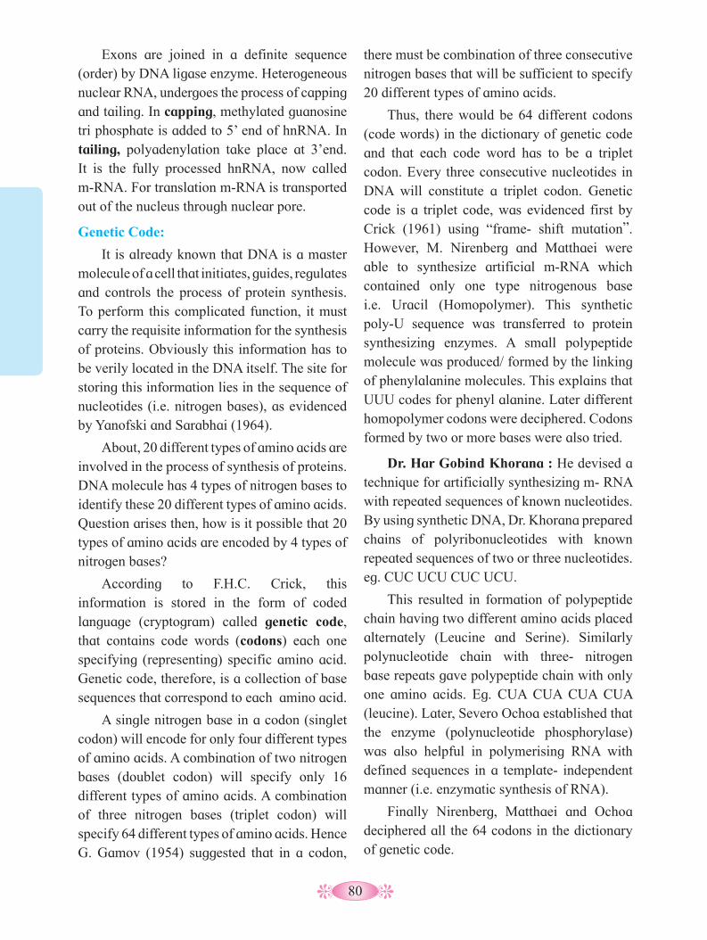
80
there must be combination of three consecutive nitrogen bases that will be sufficient to specify 20 different types of amino acids.
Thus, there would be 64 different codons (code words) in the dictionary of genetic code and that each code word has to be a triplet codon. Every three consecutive nucleotides in DNA will constitute a triplet codon. Genetic code is a triplet code, was evidenced first by Crick (1961) using “frame- shift mutation”. However, M. Nirenberg and Matthaei were able to synthesize artificial m-RNA which contained only one type nitrogenous base i.e. Uracil (Homopolymer). This synthetic poly-U sequence was transferred to protein synthesizing enzymes. A small polypeptide molecule was produced/ formed by the linking of phenylalanine molecules. This explains that UUU codes for phenyl alanine. Later different homopolymer codons were deciphered. Codons formed by two or more bases were also tried.
Dr. Har Gobind Khorana : He devised a technique for artificially synthesizing m- RNA with repeated sequences of known nucleotides.By using synthetic DNA, Dr. Khorana prepared chains of polyribonucleotides with known repeated sequences of two or three nucleotides. eg. CUC UCU CUC UCU.
This resulted in formation of polypeptide chain having two different amino acids placed alternately (Leucine and Serine). Similarly polynucleotide chain with three- nitrogen base repeats gave polypeptide chain with only one amino acids. Eg. CUA CUA CUA CUA (leucine). Later, Severo Ochoa established that the enzyme (polynucleotide phosphorylase) was also helpful in polymerising RNA with defined sequences in a template- independent manner (i.e. enzymatic synthesis of RNA).
Finally Nirenberg, Matthaei and Ochoa deciphered all the 64 codons in the dictionary of genetic code.
Exons are joined in a definite sequence (order) by DNA ligase enzyme. Heterogeneous nuclear RNA, undergoes the process of capping and tailing. In capping, methylated guanosine tri phosphate is added to 5’ end of hnRNA. In tailing, polyadenylation take place at 3’end. It is the fully processed hnRNA, now called m-RNA. For translation m-RNA is transported out of the nucleus through nuclear pore.
Genetic Code:
It is already known that DNA is a master molecule of a cell that initiates, guides, regulates and controls the process of protein synthesis. To perform this complicated function, it must carry the requisite information for the synthesis of proteins. Obviously this information has to be verily located in the DNA itself. The site for storing this information lies in the sequence of nucleotides (i.e. nitrogen bases), as evidenced by Yanofski and Sarabhai (1964).
About, 20 different types of amino acids are involved in the process of synthesis of proteins. DNA molecule has 4 types of nitrogen bases to identify these 20 different types of amino acids. Question arises then, how is it possible that 20 types of amino acids are encoded by 4 types of nitrogen bases?
According to F.H.C. Crick, this information is stored in the form of coded language (cryptogram) called genetic code, that contains code words (codons) each one specifying (representing) specific amino acid. Genetic code, therefore, is a collection of base sequences that correspond to each amino acid.
A single nitrogen base in a codon (singlet codon) will encode for only four different types of amino acids. A combination of two nitrogen bases (doublet codon) will specify only 16 different types of amino acids. A combination of three nitrogen bases (triplet codon) will specify 64 different types of amino acids. Hence G. Gamov (1954) suggested that in a codon,

81
During replication and transcription, a nucleic acid is copied to form another nucleic acid. These two processes are based on complementarity principle. During translation, genetic information is transferred from a polymer of nucleotides to a polymer of amino acids. Here, complementarity principle does not exist.
It is evident that change in nucleic acid (genetic material) results in the change in amino acids of proteins. This clearly explains that genetic code directs the sequence of amino acids during synthesis of proteins.
i. Genetic code is a triplet code: Sequence of three consecutive bases constitute codon, which specifies one particular amino acid. Base sequence in a codon is always in 5’ 3’ direction. In every living organism genetic code is a triplet code.
ii. Genetic code has distinct polarity : Genetic code shows definite polarity i.e. direction. It, therefore, is always read in 5’ 3’ direction and not in 3’ 5’ direction. Otherwise message will change e.g. 5’ AUG 3’.
iii. Genetic code is non-overlapping : Code is non overlapping i.e. each single base is a part of only one codon. Adjacent codons do not overlap. If non-overlapping, then with 6 consequtive bases only two amino acid molecules will be in the chain. Had it been overlapping type, with 6 bases, there would be 4 amino acid molecules in a chain. Experimental evidence is in favour of non-overlapping nature.
U C A G
U
UUUUUCUUAUUG
UCUUCCUCAUCG
UAUUACUAAUAG
UGUUGCUGAUGG
UCAG
C
CUUCUCCUACUG
CCUCCCCCACCG
CAUCACCAACAG
CGUCGCCGACGG
UCAG
A
AUUAUCAUAAUG
ACUACCACAACG
AAUAACAAAAAG
AGUAGCAGAAGG
UCAG
G
GUUGUCGUAGUG
GCUGCCGCAGCG
GAUGACGAAGAG
GGUGGCGGAGGG
UCAG
Phe
LeuSer
Tyr Cys
Trp
His
Gln
Asn
Lys
Asp
GluGly
Arg
Arg
Ser
Stop StopStop
Pro
Thr
Ala
Leu
Ile
Met
Val
Third L
etterFir
st L
ette
r
Second Letter
Fig. 4.13 : Dictionary of genetic code
The What is the amino acid sequence encoded by base sequence UCA UUU UCC GGG AGU of an m- RNA segment?
Find out
Characterestic of Genetic code:
Genetic code of DNA has certain fundamental characteristics –
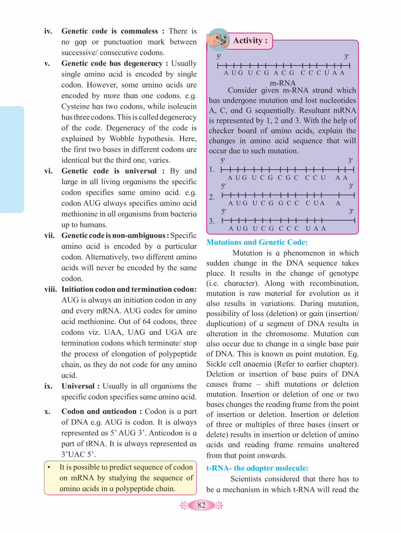
82
Consider given m-RNA strand which has undergone mutation and lost nucleotides A, C, and G sequentially. Resultant mRNA is represented by 1, 2 and 3. With the help of checker board of amino acids, explain the changes in amino acid sequence that will occur due to such mutation.
1.
2.
3.
A U G U C G A C G C C C U A A
5′ 3′
m-RNA
A U G U C G C G C C C U A A
5′ 3′
A U G U C G G C C C U A A
5′ 3′
A U G U C G C C C U A A
5′ 3′
iv. Genetic code is commaless : There is no gap or punctuation mark between successive/ consecutive codons.
v. Genetic code has degeneracy : Usually single amino acid is encoded by single codon. However, some amino acids are encoded by more than one codons. e.g. Cysteine has two codons, while isoleucin has three codons. This is called degeneracy of the code. Degeneracy of the code is explained by Wobble hypothesis. Here, the first two bases in different codons are identical but the third one, varies.
vi. Genetic code is universal : By and large in all living organisms the specific codon specifies same amino acid. e.g. codon AUG always specifies amino acid methionine in all organisms from bacteria up to humans.
vii. Genetic code is non-ambiguous : Specific amino acid is encoded by a particular codon. Alternatively, two different amino acids will never be encoded by the same codon.
viii. Initiation codon and termination codon: AUG is always an initiation codon in any and every mRNA. AUG codes for amino acid methionine. Out of 64 codons, three codons viz. UAA, UAG and UGA are termination codons which terminate/ stop the process of elongation of polypeptide chain, as they do not code for any amino acid.
ix. Universal : Usually in all organisms the specific codon specifies same amino acid.
x. Codon and anticodon : Codon is a part of DNA e.g. AUG is codon. It is always represented as 5’ AUG 3’. Anticodon is a part of tRNA. It is always represented as 3’UAC 5’.
• It is possible to predict sequence of codon on mRNA by studying the sequence of amino acids in a polypeptide chain.
Mutations and Genetic Code: Mutation is a phenomenon in which sudden change in the DNA sequence takes place. It results in the change of genotype (i.e. character). Along with recombination, mutation is raw material for evolution as it also results in variations. During mutation, possibility of loss (deletion) or gain (insertion/ duplication) of a segment of DNA results in alteration in the chromosome. Mutation can also occur due to change in a single base pair of DNA. This is known as point mutation. Eg. Sickle cell anaemia (Refer to earlier chapter). Deletion or insertion of base pairs of DNA causes frame – shift mutations or deletion mutation. Insertion or deletion of one or two bases changes the reading frame from the point of insertion or deletion. Insertion or deletion of three or multiples of three bases (insert or delete) results in insertion or deletion of amino acids and reading frame remains unaltered from that point onwards.
t-RNA- the adapter molecule: Scientists considered that there has to be a mechanism in which t-RNA will read the
Activity :
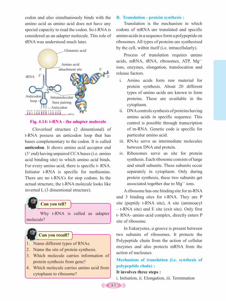
83
Cloverleaf structure (2 dimentional) of t-RNA possess an anticodon loop that has bases complementary to the codon. It is called anticodon. It shows amino acid acceptor end (3’ end) having unpaired CCA bases (i.e. amino acid binding site) to which amino acid binds. For every amino acid, there is specific t- RNA. Initiator t-RNA is specific for methionine. There are no t-RNA’s for stop codons. In the actual structure, the t-RNA molecule looks like inverted L (3 dimentional structure).
B. Translation - protein synthesis : Translation is the mechanism in which
codons of mRNA are translated and specific amino acids in a sequence form a polypeptide on ribosomes. All types of proteins are synthesised by the cell, within itself (i.e. intracellularly).
Process of translation requires amino acids, mRNA, tRNA, ribosomes, ATP, Mg++ ions, enzymes, elongation, translocation and release factors.
i. Amino acids form raw material for protein synthesis. About 20 different types of amino acids are known to form proteins. These are available in the cytoplasm.
ii. DNA controls synthesis of proteins having amino acids in specific sequence. This control is possible through transcription of m-RNA. Genetic code is specific for particular amino acid.
iii. RNAs serve as intermediate molecules between DNA and protein.
iv. Ribosomes serve as site for protein synthesis. Each ribosome consists of large and small subunits. These subunits occur separately in cytoplasm. Only during protein synthesis, these two subunits get associated together due to Mg++ ions.
A ribosome has one binding site for m-RNA and 3 binding sites for t-RNA. They are P site (peptidy t-RNA site), A site (aminoacyl – t-RNA site) and E site (exit site). Only first t- RNA- amino acid complex, directly enters P site of ribosome.
In Eukaryotes, a groove is present between two subunits of ribosomes. It protects the Polypeptide chain from the action of cellular enzymes and also protects mRNA from the action of nucleases.
Mechanism of translation (i.e. synthesis of polypeptide chain) :It involves three steps :i. Initiation, ii. Elongation, iii. Termination
Fig. 4.14: t-RNA - the adapter molecule
Glutamic acid
Amino acid attachment site
Intramolecular base pairing
tRNA
Anticodonloop
Anticodon
3’
3’ 5’
5’
C U CG U C C A G G A G C U A U A G
codon and also simultaniously binds with the amino acid as amino acid does not have any special capacity to read the codon. So t-RNA is considered as an adapter molecule. This role of tRNA was understood much later.
Why t-RNA is called as adapter molecule?
Can you tell?
1. Name different types of RNAs.2. Name the site of protein synthesis.3. Which molecule carries information of
protein synthesis from gene?4. Which molecule carries amino acid from
cytoplasm to ribosome?
Can you recall?
mRNA
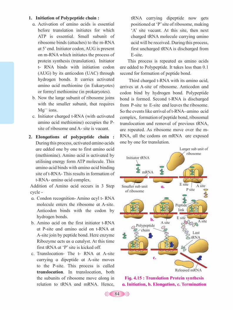
84
Fig. 4.15 : Translation Protein synthesis a. Initiation, b. Elongation, c. Termination
tRNA carrying dipeptide now gets positioned at ‘P’ site of ribosome, making ‘A’ site vacant. At this site, then next charged tRNA molecule carrying amino acid will be received. During this process, first uncharged tRNA is discharged from E-site.
This process is repeated as amino acids are added to Polypeptide. It takes less than 0.1 second for formation of peptide bond.
Third charged t-RNA with its amino acid, arrives at A-site of ribosome. Anticodon and codon bind by hydrogen bond. Polypeptide bond is formed. Second t-RNA is discharged from P-site to E-site and leaves the ribosome. So the events like arrival of t-RNA- amino acid complex, formation of peptide bond, ribosomal translocation and removal of previous tRNA, are repeated. As ribosome move over the m- RNA, all the codons on mRNA are exposed one by one for translation.
1. Initiation of Polypeptide chain :a. Activation of amino acids is essential
before translation initiates for which ATP is essential. Small subunit of ribosome binds (attaches) to the m-RNA at 5’ end. Initiator codon, AUG is present on m-RNA which initiates the process of protein synthesis (translation). Initiator t- RNA binds with initiation codon (AUG) by its anticodon (UAC) through hydrogen bonds. It carries activated amino acid methionine (in Eukaryotes) or formyl methionine (in prokaryotes).
b. Now the large subunit of ribosome joins with the smaller subunit, that requires Mg++ ions.
c. Initiator charged t-RNA (with activated amino acid methionine) occupies the P- site of ribosome and A- site is vacant.
2. Elongations of polypeptide chain : During this process, activated amino acids are added one by one to first amino acid (methionine). Amino acid is activated by utilising energy form ATP molecule. This amino acid binds with amino acid binding site of t-RNA- This results in formation of t-RNA- amino acid complex.
Addition of Amino acid occurs in 3 Step cycle -a. Condon recognition- Amino acyl t- RNA
molecule enters the ribosome at A-site. Anticodon binds with the codon by hydrogen bonds.
b. Amino acid on the first initiator t-RNA at P-site and amino acid on t-RNA at A-site join by peptide bond. Here enzyme Ribozyme acts as a catalyst. At this time first tRNA at ‘P’ site is kicked off.
c. Translocation- The t- RNA at A-site carrying a dipeptide at A-site moves to the P-site. This process is called translocation. In translocation, both the subunits of ribosome move along in relation to tRNA and mRNA. Hence,
a.
b.
c.
Smaller sub unit of ribosome
Larger sub unit of ribosome
mRNA
Released mRNA
Initiator tRNA
Polypeptide chain
Last tRNA
E site A site A site
Peptide link
A site E siteP site
85
3. Termination and release of polypeptide: At the end of m-RNA, there is a stop
codon (UAA/ UAG/ UGA). It is exposed at the A-site. It is not read and joined by anticodon of any t-RNA. The release factor binds to the stop codon, thereby terminating the translation process. The Polypeptide is now released in the cytoplasm.
Two subunits of Ribosome dissociate and last tRNA is set free in the cytoplasm.
m-RNA also has some additional sequences that are not translated and are referred as untranslated regions (UTR). The UTRs are present at both 5’-end (before start codon) and at 3’-end (after stop codon). They are required for efficient translation process.
Finally mRNA is also released in the cytoplasm. It gets denatured by nucleases immidiately. Hence mRNA is short -lived.
Genes of a cell are expressed to perform different functions. For eg. An enzyme beta galactosidase is synthesised by E-coli. It is used for hydrolysis of lactose into galactose and glucose.
Lactose Galactose + Glucose.(b – galactosidase)
+H2O
If E.coli bacteria do not have lactose in the surrounding medium as a source of energy, then enzyme b-galactosidase is not synthesised. So, it is the metabolic or physiological or environmental conditions that regulate expression of genes. The development and differentiation of embryo into an adult organism, is also a result of the coordinated regulation or expression, of several sets of genes.
Now one has to understand and know the mechanism by which the organisms regulate gene expression in response to changes in the environment. If so, whether single mechanism exists for regulation of the expression of different genes/ sets of genes or different genes are regulated by different mechanisms.
Certain bacteria like E.coli adapt to their chemical environment by synthesizing certain enzymes depending upon the substrate present. Such adaptive enzyme is called inducible enzymes. A set of genes will be switched on when there is necessity to metabolise a new substrate. This phenomenon is called induction and small molecule responsible for this, is known as inducer. It is positive control.
1. Enlist different steps of protein synthesis. 2. Name the initiator codon of protein
synthesis.3. Explain in brief the process of initiation
during protein synthesis.4. Name three binding sites of ribosome. 5. What is translocation?
Can you tell?
4.6 Regulation of gene expression:It is the multistep process by which a gene
is regulated and its product is synthesized. Thus, gene expression results in the formation of a Polypeptide. Gene expression process is regulated at different levels.
In eukaryotes, the regulation can be at different levels like-
1. Transcriptional level (formation of primary transcript)
2. Processing level ( regulation of splicing)3. Transport of m-RNA from nucleus to the
cytoplasm.4. Translational level.
Repressible regulation of gene is seen when the end product of a biosynthetic pathway like amino acid, is provided in the medium. At this time, internal biosynthesis of amino acid stops. It is negative control so the metabolite (amino acid) turns off a set of genes involved in producing that metabolite. This is called feedback repression.
Do you know ?
86
4.7 Operon concept :It is a transcriptional control mechanism
of gene regulation. Francois Jacob and Jacques Monod (1961) explained that metabolic pathways are regulated as a unit.
For example in E.coli, when lactose sugar is provided to the culture medium, cell induces production of three enzymes necessary for digestion of lactose. The enzymes are :
i. b-galactosidase : Digests lactose into galactose and glucose.
ii. b-galactoside permease : Permits lactose molecules to enter into the cell.
iii. Transacetylase (b-Galactoside acetyltransferease) : Transfers an acetyl group from acetyl CO-A to galactoside.
Synthesis of these three enzymes, is controlled by a long segment of DNA known as Operon. It consists of an operator site O and three structural genes Z, Y and A .The action of structural genes is regulated by operator site with the help of a repressor protein. Repressor protein is produced by the action of gene i (inhibitor) known as regulator gene.
The gene expression depends on whether operator is switched on or switched off.
If the operator is switched on, the three genes z, y and a are transcribed by RNA Polymerase into a single m-RNA. Each structural gene is generally known as cistron and the transcribed long m-RNA covering various cistrons is known as Polycistronic.
Switching on or switching off of the operator is achieved (acomplished) by a protein called repressor. When this protein is attached to the operator and blocks it, the switch is turned off and structural genes are not expressed.
Lac operon :Lactose or lac operon of E.coli is inducible
operon. The operon is switched on when a chemical inducer- lactose is present in the medium.
Lac operon consists of following components :
1. Regulator gene (repressor gene) 2. Promoter gene3. Operator gene4. Structural genes5. Inducer - It is not a component of
operon.
1. Regulator gene : This gene controls the operator gene in cooperation with an inducer present in the cytoplasm. Regulator gene preceeds the promoter gene. It may not be present immidiately adjacent to operator gene. Regulator gene produces a protein called repressor protein. Repressor binds with operator gene and represses (stops) its action. It is called regulator protein.
2. Promoter gene : This gene preceeds the operator gene. It is present adjacent to operator gene. The promoter gene marks the site at which the RNA Polymerase enzyme binds. When the operator gene is turned on, the enzyme moves over the operator gene and transcription of structural genes starts. Promoter gene base sequence determines which strand of DNA acts a template.
3. Operator gene : It preceeds the structural genes. This controls the functioning of structural genes. It lies adjacent to the Structural genes. When operator gene is turned on by an inducer, the Structural genes produce m-RNA. Operator gene is turned off by a product of repressor gene.
4. Structural gene : When lactose is added to the E.coli culture, the structural genes catalyse (produce) m-RNA which in turn produces polypeptides, on the ribosomes. The polypeptides formed, act as enzymes to caltalyse lactose in the cell. There are 3 structural genes in the sequence lac-Z, lac-Y and lac-A. Enzymes produced are b-galactosidase, b-galactoside permease and transacetylase respectively.
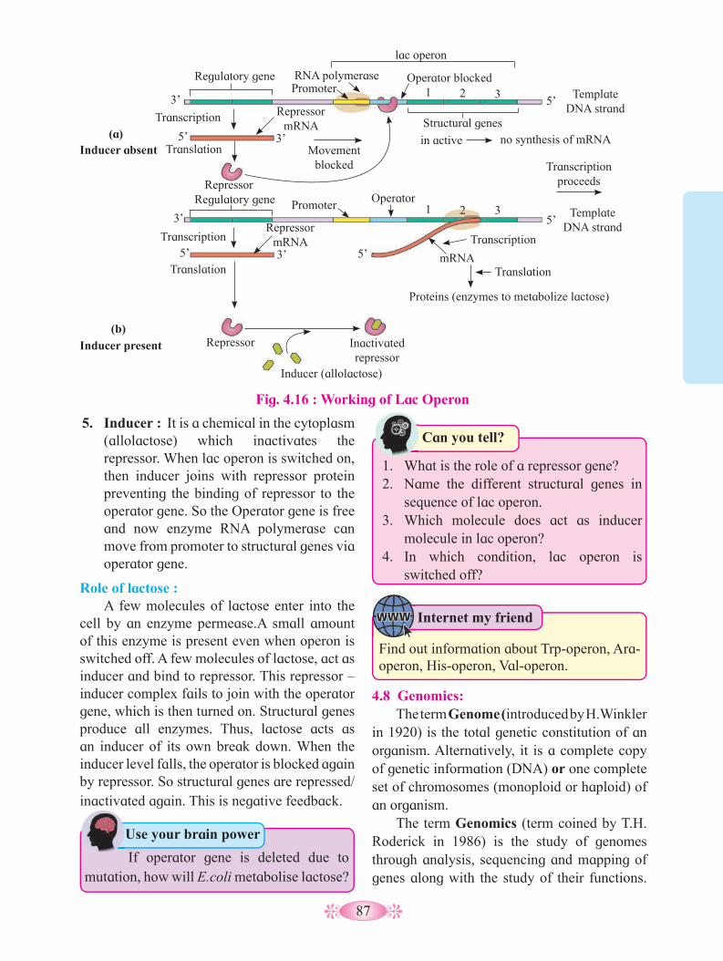
87
5. Inducer : It is a chemical in the cytoplasm (allolactose) which inactivates the repressor. When lac operon is switched on, then inducer joins with repressor protein preventing the binding of repressor to the operator gene. So the Operator gene is free and now enzyme RNA polymerase can move from promoter to structural genes via operator gene.
Role of lactose :A few molecules of lactose enter into the
cell by an enzyme permease.A small amount of this enzyme is present even when operon is switched off. A few molecules of lactose, act as inducer and bind to repressor. This repressor – inducer complex fails to join with the operator gene, which is then turned on. Structural genes produce all enzymes. Thus, lactose acts as an inducer of its own break down. When the inducer level falls, the operator is blocked again by repressor. So structural genes are repressed/ inactivated again. This is negative feedback.
4.8 Genomics:The term Genome (introduced by H.Winkler
in 1920) is the total genetic constitution of an organism. Alternatively, it is a complete copy of genetic information (DNA) or one complete set of chromosomes (monoploid or haploid) of an organism.
The term Genomics (term coined by T.H. Roderick in 1986) is the study of genomes through analysis, sequencing and mapping of genes along with the study of their functions.
If operator gene is deleted due to mutation, how will E.coli metabolise lactose?
Use your brain power
1. What is the role of a repressor gene?2. Name the different structural genes in
sequence of lac operon.3. Which molecule does act as inducer
molecule in lac operon?4. In which condition, lac operon is
switched off?
Can you tell?
Inducer absent
Fig. 4.16 : Working of Lac Operon
Inducer present
lac operon
RNA polymerase Promoter
Promoter
Regulatory gene Operator blocked Template
DNA strand
Template DNA strand
3’
3’
3’
3’
1
1
2
2
3
3
5’
5’
5’
5’
5’
Structural genes
in active no synthesis of mRNA
Transcription
Transcription
Repressor mRNA
mRNA
Proteins (enzymes to metabolize lactose)
Repressor mRNA
Translation
Translation Translation
Movement blocked
Repressor
Repressor
Transcription proceeds
Transcription
Operator
Inactivated repressor
Inducer (allolactose)
(a)
(b)
Regulatory gene
Internet my friend
Find out information about Trp-operon, Ara-operon, His-operon, Val-operon.
88
The sequencing of yeast, Drosophila and mouse genome was done in order to facilitate comparative studies between humans and other organisms commonly used for genetic studies, in laboratory. Several additional genomes are now either actively being sequenced or strongly considered for sequencing. These include several microbes, bee, tomato and other crops.Genomics study may be classified into two types:a. Structural genomics: It involves mapping,
sequencing and analysis of genome.
b. Functional genomics: It deals with the study of functions of all gene sequences and their expression in organisms.
Application of genomics: Structural and functional genomics is used
for different purposes in the improvement of crop plant, human health and livestock. The knowledge and understanding acquired from genomics research can be applied in a number of different sectors, including medicine, biotechnology and social sciences. It helps in the treatment of genetic disorders through gene therapy. • Genomics is used in agriculture to develop
transgenic crops having more desirable characters.
• Genetic markers developed in genomics, have applications in forensic analysis.
• Genomics can lead to introduce new gene in microbes to produce enzymes, therapeutic proteins and even biofuels.
4.9 Human Genome Project : The human genome project was initiated
in 1990 under the International administration of the Human Genome Organization (HUGO).This project was co-ordinated by the US department of Energy and National institute of health. Additional contributors included universities across the United States and international partners in the United Kingdom, France, Germany, Japan, India and China. The
Human Genome Project formally began in 1990 and was completed in 2003.The human genome project is a multinational research project to determine the genomic structure of humans. The main aims of project are –I. Mapping the entire human genome at the
level of nucleotide sequences.II. To store the information collected from the
project in databases.III. To develop tools and techniques for analysis
of the data.IV. Transfer of the related technologies to the
private sectors, such as industries.V. Taking care of the legal, ethical and social
issues which may arise from project.
HGP (Human Genome Project) was closely associated with rapid development of a new area in biology, called Bioinformatics. The work of human genome project has allowed researchers to begin to understand the blueprint in building and constructing the human genome. As researchers learn more about the functions of genes and proteins, this knowledge will have a major impact in the fields like Medicine, Biotechnology and the Life sciences. Therefore HGP is very important.
Human Genome Project was to provide a complete and accurate sequence of the 3 billion DNA base pairs that make up the human genome and to find out the estimated number of human genes. Now about 33000 genes have been estimated to be present in humans.
The project was also aimed to sequence the genomes of several other organisms such as bacteia e.g. E.coli, Caenorhabditis elegans (a free living non-pathogenic nematode), Saccharomyces cerevisiae (yeast), Drosophila (fruit fly), plants (rice and Arabidopsis), Mus musculus (mouse), etc. Complete genome sequences of these model organisms will be useful for comparative studies that will allow researchers to study gene functions in these organisms.
89
The secret of our complexity may lie not in the number of our genes but how we use them. It will lead to the understanding of gene structure and function in other species. Since we possess many of the genes same as these of flies, round worms and mice, such studies will lead to a greater understanding of human evolution.
DNA fingerprinting technique is based on identification of nucleotide sequence present in this wonder molecule. About 99.9% of nucleotide sequence in all persons, is same. Only some short sequences of nucleotides differ from person to person. In the population, every person shows unusual sequences of 20-100 base pairs, which are repeated several times. They are termed as Variable Number of Tandem Repeats (VNTRs).
The length of the regions having VNTRs is different in each individual and hence is the key factor in DNA profiling. Steps involved in DNA finger printing are as follows:
1. Isolation of DNA: The DNA must be recovered from the cells or tissues of the body (host). Only small amount of tissue like blood, hair roots, skin, etc. is required.
2. Restriction digestion: The isolated DNA is treated with restriction enzymes. The restriction enzymes cut the DNA into small fragments having variable lengths. This phenomenon is called Restriction Fragment Length Polymorphism (RFLP).
3. Gel electrophoresis: The DNA samples are loaded for agarose gel electrophoresis under an electric influence. The DNA fragments, which are negatively charged move to the positive pole. The movement of these fragments depends on length of the fragments. This results in formation of bands. dsDNA splits into ssDNA by alkali treatment.
Table 4.17 : Comparative genome sizes of humans and other models organisms.
Organism Chromosome number
Estimated gene number
Estimated size(base pairs)
Human (Homo sapiens) 46 33,000 3 billionMouse (Mus musculus) 40 25,000 2.9 billionFruit fly (Drosophila melanogaster) 8 13,000 165 million
Plant (Arabidopsis thaliana) 10 25,000 157 million
Roundworm (Caenorhabditis elegans) 12 19,000 97 millionYeast (Saccharomyces cerevisiae) 32 6000 12 million
Bacteria (Escherichia coli) 1* 4400 4.6 million
Do different organisms have the same DNA?
Can you tell?
1. What have we learnt from the Human Genome Project?
2. Why is HGP important?
Use your brain power
4.10 DNA Fingerprinting: Genes present on chromosomes are
responsible for determining characters of the organism as well as for inheritance of characters. Due to recombination of paternal and maternal genes, we differ from our parents. Differences also arise due to infrequent mutations that occur during gamete formation (cell division). Due to all these factors, every individual has its unique genetic make-up, which may be called its Fingerprint. The technique developed to identify a person with the help of DNA restriction analysis, is known as DNA profiling or DNA fingerprinting. The technique of finger printing was first given by British geneticist, Dr. Alec Jeffreys in 1984.
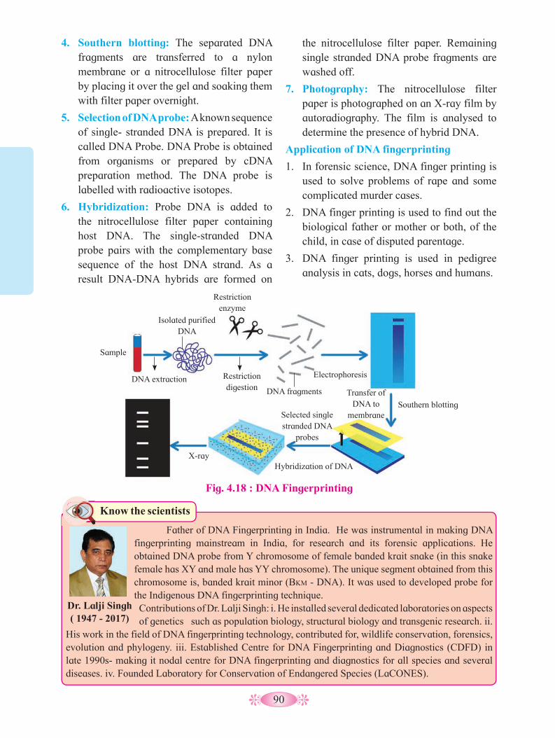
90
Know the scientists
Father of DNA Fingerprinting in India. He was instrumental in making DNA fingerprinting mainstream in India, for research and its forensic applications. He obtained DNA probe from Y chromosome of female banded krait snake (in this snake female has XY and male has YY chromosome). The unique segment obtained from this chromosome is, banded krait minor (BKM - DNA). It was used to developed probe for the Indigenous DNA fingerprinting technique.Contributions of Dr. Lalji Singh: i. He installed several dedicated laboratories on aspects of genetics such as population biology, structural biology and transgenic research. ii.
His work in the field of DNA fingerprinting technology, contributed for, wildlife conservation, forensics, evolution and phylogeny. iii. Established Centre for DNA Fingerprinting and Diagnostics (CDFD) in late 1990s- making it nodal centre for DNA fingerprinting and diagnostics for all species and several diseases. iv. Founded Laboratory for Conservation of Endangered Species (LaCONES).
Dr. Lalji Singh( 1947 - 2017)
4. Southern blotting: The separated DNA fragments are transferred to a nylon membrane or a nitrocellulose filter paper by placing it over the gel and soaking them with filter paper overnight.
5. Selection of DNA probe: A known sequence of single- stranded DNA is prepared. It is called DNA Probe. DNA Probe is obtained from organisms or prepared by cDNA preparation method. The DNA probe is labelled with radioactive isotopes.
6. Hybridization: Probe DNA is added to the nitrocellulose filter paper containing host DNA. The single-stranded DNA probe pairs with the complementary base sequence of the host DNA strand. As a result DNA-DNA hybrids are formed on
the nitrocellulose filter paper. Remaining single stranded DNA probe fragments are washed off.
7. Photography: The nitrocellulose filter paper is photographed on an X-ray film by autoradiography. The film is analysed to determine the presence of hybrid DNA.
Application of DNA fingerprinting
1. In forensic science, DNA finger printing is used to solve problems of rape and some complicated murder cases.
2. DNA finger printing is used to find out the biological father or mother or both, of the child, in case of disputed parentage.
3. DNA finger printing is used in pedigree analysis in cats, dogs, horses and humans.
Fig. 4.18 : DNA Fingerprinting
Sample
DNA extraction
Isolated purified DNA
Restriction digestion
Electrophoresis
DNA fragments
Southern blottingTransfer of
DNA to membrane Selected single
stranded DNA probes
Hybridization of DNA X-ray
Restriction enzyme

91
Activity :
Prepare physical model of DNA molecule (Watson-Crick model)
Requirements :
Labelled Diagram :
Functions :

92
Q. 1 Multiple Choice Questions 1. Griffith worked on .............
a. Bacteriophage b. Drosophila c. Frog eggs c. Streptococci
2. The molecular knives of DNA are ………….. a. Ligases b. Polymerases c. Endonucleases d. Transcriptase
3. Translation occurs in the ............... a. Nucleus b. Cytoplasm c. Nucleolus d. Lysosomes
4. The enzyme required for transcription is ..................
a. DNA polymerase b. RNA polymerase c. Restriction enzyme d. RNAase
5. Transcription is the transfer of genetic information from ..............
a. DNA to RNA b. tRNA to mRNA c. DNA to mRNA d. mRNA to tRNA
6. Which of the following is NOT part of protein synthesis?
a. Replication b. Translation c. Transcription d. All of these
7. In the RNA molecule, which nitrogen base is found in place of thymine?
a. Guanine b. Cytosine c. Thymine d. Uracil
8. How many codons are needed to specify three amino acid?
a. 3 b. 6 c. 9 d. 12
9. Which out of the following is NOT an example of inducible operon?
a. Lactose operon b. Histidine operon c. Arabinose operon d. Tryptophan operon
10. Place the following event of translation in the correct sequencei. Binding of met-tRNA to the start
codon.ii. Covalent bonding between two
amino acids.iii. Binding of second tRNA.iv. Joining of small and large ribosome
subunits. A. iii, iv, i, ii B. i, iv, iii, ii C. iv, iii, ii, i D. ii, iii, iv, i
Q. 2 Very Short Answer Questions: 1. What is the function of an RNA primer
during protein synthesis?2. Why the genetic code is considered as
commaless?3. What is genome?4. Which enzyme does remove supercoils
from replicating DNA?5. Why are Okazaki fragments formed
on lagging strand only?6. When does DNA replication take
place?7. Define term- codon and codogen.8. What is degeneracy of genetic code?9. Which are the nucleosomal 'core'
histones?
Q. 3 Short Answer Questions: 1. Write short note on DNA packaging
in eukaryotic cell.2. Enlist the characteristics of genetic
code.3. Write a note on applications of DNA
finger printing.4. Explain the role of lactose in ‘Lac
Operon’.
Q. 4 Short Answer Questions:1. Write a note on Human genome
project (HGP).
Exercise
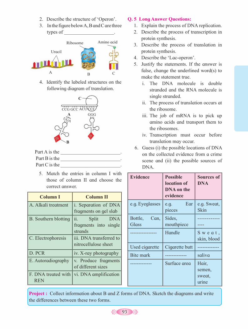
93
2. Describe the structure of ‘Operon’.3. In the figure below A, B and C are three
types of ____________________.
Q. 5 Long Answer Questions: 1. Explain the process of DNA replication.2. Describe the process of transcription in
protein synthesis.3. Describe the process of translation in
protein synthesis.4. Describe the ‘Lac-operon’.5. Justify the statements. If the answer is
false, change the underlined word(s) to make the statement true.i. The DNA molecule is double
stranded and the RNA molecule is single stranded.
ii. The process of translation occurs at the ribosome.
iii. The job of mRNA is to pick up amino acids and transport them to the ribosomes.
iv. Transcription must occur before translation may occur.
6. Guess (i) the possible locations of DNA on the collected evidence from a crime scene and (ii) the possible sources of DNA.
Evidence Possible location of DNA on the evidence
Sources of DNA
e.g. Eyeglasses e.g. Ear pieces
e.g. Sweat, Skin
Bottle, Can, Glass
Sides, mouthpiece
----------------
---------------- Handle S w e a t , skin, blood
Used cigarette Cigarette butt -------------
Bite mark ------------- saliva
------------- Surface area Hair, semen, sweat, urine
Uracil
Ribosome Amino acid
B A C
4. Identify the labeled structures on the following diagram of translation.
C
A
B
CCG GCC ACU CCC
GGG UGA
Part A is the ________________________. Part B is the ________________________. Part C is the ________________________.
5. Match the entries in column I with those of column II and choose the correct answer.
Column I Column II
A. Alkali treatment i. Separation of DNA fragments on gel slab
B. Southern blotting ii. Split DNA fragments into single strands
C. Electrophoresis iii. DNA transferred to nitrocellulose sheet
D. PCR iv. X-ray photography
E. Autoradiography v. Produce fragments of different sizes
F. DNA treated with REN
vi. DNA amplification
Project : Collect information about B and Z forms of DNA. Sketch the diagrams and write the differences between these two forms.
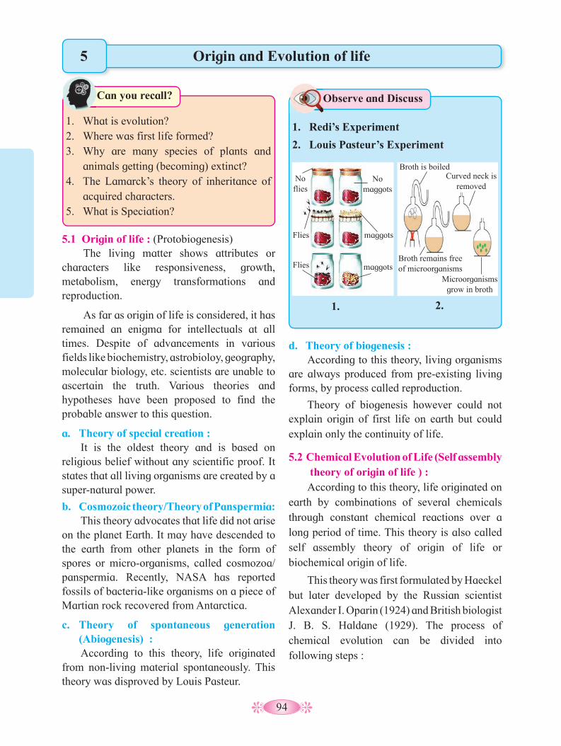
94
Origin and Evolution of life
5.1 Origin of life : (Protobiogenesis)The living matter shows attributes or
characters like responsiveness, growth, metabolism, energy transformations and reproduction.
As far as origin of life is considered, it has remained an enigma for intellectuals at all times. Despite of advancements in various fields like biochemistry, astrobioloy, geography, molecular biology, etc. scientists are unable to ascertain the truth. Various theories and hypotheses have been proposed to find the probable answer to this question.
a. Theory of special creation : It is the oldest theory and is based on
religious belief without any scientific proof. It states that all living organisms are created by a super-natural power.
b. Cosmozoic theory/Theory of Panspermia: This theory advocates that life did not arise
on the planet Earth. It may have descended to the earth from other planets in the form of spores or micro-organisms, called cosmozoa/panspermia. Recently, NASA has reported fossils of bacteria-like organisms on a piece of Martian rock recovered from Antarctica.
c. Theory of spontaneous generation (Abiogenesis) : According to this theory, life originated
from non-living material spontaneously. This theory was disproved by Louis Pasteur.
1. What is evolution?2. Where was first life formed?3. Why are many species of plants and
animals getting (becoming) extinct? 4. The Lamarck’s theory of inheritance of
acquired characters.5. What is Speciation?
Can you recall?
5
1. Redi’s Experiment
2. Louis Pasteur’s Experiment
Observe and Discuss
1. 2.
No flies
Flies
Flies
No maggots
maggots
maggots
Broth is boiled
Broth remains free of microorganisms
Curved neck is removed
Microorganisms grow in broth
d. Theory of biogenesis : According to this theory, living organisms
are always produced from pre-existing living forms, by process called reproduction.
Theory of biogenesis however could not explain origin of first life on earth but could explain only the continuity of life.
5.2 Chemical Evolution of Life (Self assembly theory of origin of life ) :According to this theory, life originated on
earth by combinations of several chemicals through constant chemical reactions over a long period of time. This theory is also called self assembly theory of origin of life or biochemical origin of life.
This theory was first formulated by Haeckel but later developed by the Russian scientist Alexander I. Oparin (1924) and British biologist J. B. S. Haldane (1929). The process of chemical evolution can be divided into following steps :

95
a. Origin of Earth and Primitive atmosphere:The origin of universe was explained by
the Big-Bang theory of Georges Lemaitre (1931). According to this theory the Universe originated about 20 billion years ago by a single huge titanic explosion. As the universe expanded, the temperature decreased and various galaxies of solid objects were formed. Milky Way is one such galaxy of which our solar system is one small part. Earth is one of the planets of solar system and originated about 4.6 billion year ago. When formed, it was a rotating cloud of hot gases and cosmic dust called Nebula. The condensation and cooling resulted in stratification with heavier elements like nickel and iron passing to the core and lighter ones like helium, hydrogen, nitrogen, oxygen, carbon, etc. remaining on the surface. They formed the atmosphere of the earth. The primitive atmosphere of the earth was quite different from the present one and it was of a reducing type, devoid of free oxygen.
b. Formation of ammonia, water and methane:Primitive atmosphere was very hot. As it
slowly cooled, the lighter elements started to react with each other. The early atmosphere was rich in hydrogen, carbon, nitrogen and sulphur of which hydrogen being more active, it reacted with other elements to form chemicals on earth like CH
4, NH
3, H
2O and H
2S.
c. Formation of simple organic molecules :As temperature of the earth decreased,
steam condensed into water that resulted in heavy rain fall and the earth gradually cooled. Rain water got accumulated on the land to form rivers, streams, lakes, seas and oceans. The atmosphere then did not contain ozone layer and thus ultra-violet radiations reached the surface of earth directly. Under the influence of available energy sources such as ultra-violet
rays, radiations, lightening and volcanic activities, the early molecules of hydrocarbons, ammonia, methane and water underwent reactions like condensation, polymerisation, oxidation and reduction. These reactions resulted in formation of simple molecules like monosaccharides, amino acids, purines, pyrimidines, fatty acids, glycerol, etc. All these simple organic molecules accumulated at the bottom of water bodies. Haldane described it as the ‘‘hot dilute soup’’ or ‘‘primitive broth’’. It did not show any degradation due to absence of free oxygen and enzymes.
d. Formation of complex organic molecules:The primitive broth was neutral and free
from oxygen. Polymerisation took place and simple organic molecules aggregated to form new complex organic molecules like polysaccharides, fats, proteins, nucleosides and nucleotides. Polymerisation of amino acids formed protoproteins which later formed proteins. Formation of protein molecules is considered as landmark in the origin of life. Proteins (enzymes) accelerated the rate of other chemical reactions.
e. Formation of Nucleic acids :Nucleotides may have been formed by the
reaction between phosphoric acid, sugar and nitrogenous bases (purines and pyrimidines). Number of nucleotides join together to form nucleic acids (RNA, DNA). Nucleic acids acquired self-replicating ability which is a fundamental property of living form.
f. Formation of Protobionts or Procells :Nucleic acids along with inorganic and
organic molecules formed the first form of life called protobionts. Protobionts are the prebiotic chemical aggregates having some properties of living system.
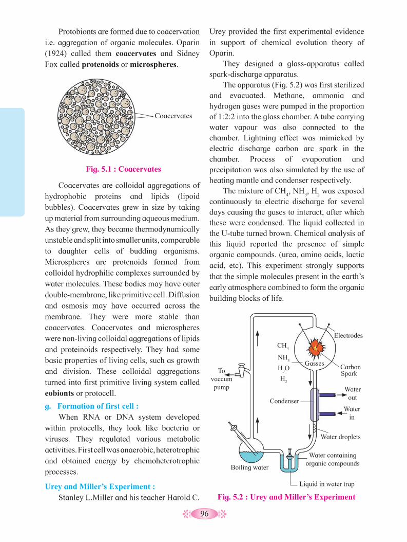
96
Coacervates are colloidal aggregations of hydrophobic proteins and lipids (lipoid bubbles). Coacervates grew in size by taking up material from surrounding aqueous medium.As they grew, they became thermodynamically unstable and split into smaller units, comparable to daughter cells of budding organisms. Microspheres are protenoids formed from colloidal hydrophilic complexes surrounded by water molecules. These bodies may have outer double-membrane, like primitive cell. Diffusion and osmosis may have occurred across the membrane. They were more stable than coacervates. Coacervates and microspheres were non-living colloidal aggregations of lipids and proteinoids respectively. They had some basic properties of living cells, such as growth and division. These colloidal aggregations turned into first primitive living system called eobionts or protocell.
g. Formation of first cell :When RNA or DNA system developed
within protocells, they look like bacteria or viruses. They regulated various metabolic activities. First cell was anaerobic, heterotrophic and obtained energy by chemoheterotrophic processes.
Urey and Miller’s Experiment : Stanley L.Miller and his teacher Harold C.
Urey provided the first experimental evidence in support of chemical evolution theory of Oparin.
They designed a glass-apparatus called spark-discharge apparatus.
The apparatus (Fig. 5.2) was first sterilized and evacuated. Methane, ammonia and hydrogen gases were pumped in the proportion of 1:2:2 into the glass chamber. A tube carrying water vapour was also connected to the chamber. Lightning effect was mimicked by electric discharge carbon arc spark in the chamber. Process of evaporation and precipitation was also simulated by the use of heating mantle and condenser respectively.
The mixture of CH4, NH
3, H
2 was exposed
continuously to electric discharge for several days causing the gases to interact, after which these were condensed. The liquid collected in the U-tube turned brown. Chemical analysis of this liquid reported the presence of simple organic compounds. (urea, amino acids, lactic acid, etc). This experiment strongly supports that the simple molecules present in the earth’s early atmosphere combined to form the organic building blocks of life.
Fig. 5.1 : Coacervates
Protobionts are formed due to coacervation i.e. aggregation of organic molecules. Oparin (1924) called them coacervates and Sidney Fox called protenoids or microspheres.
Coacervates
Fig. 5.2 : Urey and Miller’s Experiment
To vaccum pump
Boiling water
Liquid in water trap
Water containing organic compounds
Water droplets
Condenser Water
in
Water out
Spark Carbon
CH4
NH3
H3O
H2
Electrodes
Gasses

97
RNA World Hypothesis :Oparin Haldane theory and Miller Urey
experiment gives us an understanding that pathway of origin of life on earth goes from non-living to living. ‘Like begets like’ necessitates presence of stable genetic material and cellular machinery to carry out routine activities essential for survival.
We are also aware that certain proteins which we call enzymes, catalyse the chemical reactions in the cell. It was in 1980 that Sidney Altman and Thomas Cech independently found out that RNAs can also act as biocatalysts. These catalytic RNAs are called as Ribozymes. For this discovery, Altman and Cech earned Nobel Prize in chemistry in 1989.
This discovery provided important support for RNA World hypothesis. The hypothesis suggests that early life must have been based exclusively on nucleic acids, most probably RNA. It was first proposed by Carl Woese, Francis Crick and Leslie Orgel in 1960, long before discovery of ribozymes.
Fact is that RNA is found abundantly in all living cells, it is structurally related to DNA and chains of RNA can evolve or undergo mutations, replicate and catalyse reactions, all support this hypothesis.
Besides, biomolecules like Acetyl-Co-A have a nucleotide in their molecular structure. Major evidence is existence of ribosome (the protein assembly unit) in the cell. In ribosomes, translation process is catalysed by RNA. (Refer chapter- Molecular Basis of Inheritance).
These molecules might have undergone repeated replication and mutation forming varieties of RNA molecules with varying sizes and catalytic properties. Eventually they might have developed their own protein coats and machinery to survive the assembly of primitive cell. In due course, a double stranded stable structure, the DNA, might have been formed and thus continued the ongoing journey which resulted in rich biodiversity on earth.
5.3 Organic Evolution :Evolution (Latin word, e = from; volvere =
to roll) means the act of unrolling or unfolding of nature. It brings about orderly changes from one form to another. These changes result in descendants becoming different from the ancestors.
Organic evolution can be defined as slow, gradual, continuous and irreversible changes through which the present day complex forms of the life developed (or evolved) from their simple pre-existing forms.
According to Charles Darwin, evolution is ‘descent with modification’. You have already studied the Lamarck’s theory of inheritance of acquired characters in 10th std. According to this theory, the traits are acquired due to internal force, changes in environment, new needs and the use and disuse of organs. After several generations, it gives rise to new species.
This theory was disproved by a German biologist August Weismann, who cut the tails of many rats for several generations but could not find any change in size of tail even after
Fig. 5.3 : RNA World Hypothesis
RNA self-replicates
(via ribozymes)
RNA forms from inorganic
sources
RNA catalyses protein synthesis
Membrane formation changes internal chemistry allowing new functionality
DNA becomes master template
RNA codes both DNA and
protein
Proteins catalyse cellular activities
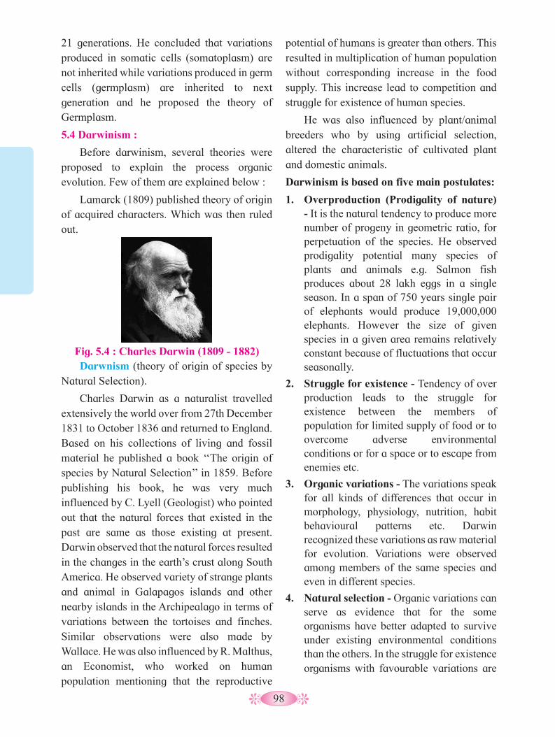
98
21 generations. He concluded that variations produced in somatic cells (somatoplasm) are not inherited while variations produced in germ cells (germplasm) are inherited to next generation and he proposed the theory of Germplasm.
5.4 Darwinism :
Before darwinism, several theories were proposed to explain the process organic evolution. Few of them are explained below :
Lamarck (1809) published theory of origin of acquired characters. Which was then ruled out.
Fig. 5.4 : Charles Darwin (1809 - 1882)
potential of humans is greater than others. This resulted in multiplication of human population without corresponding increase in the food supply. This increase lead to competition and struggle for existence of human species.
He was also influenced by plant/animal breeders who by using artificial selection, altered the characteristic of cultivated plant and domestic animals.
Darwinism is based on five main postulates:
1. Overproduction (Prodigality of nature) - It is the natural tendency to produce more number of progeny in geometric ratio, for perpetuation of the species. He observed prodigality potential many species of plants and animals e.g. Salmon fish produces about 28 lakh eggs in a single season. In a span of 750 years single pair of elephants would produce 19,000,000 elephants. However the size of given species in a given area remains relatively constant because of fluctuations that occur seasonally.
2. Struggle for existence - Tendency of over production leads to the struggle for existence between the members of population for limited supply of food or to overcome adverse environmental conditions or for a space or to escape from enemies etc.
3. Organic variations - The variations speak for all kinds of differences that occur in morphology, physiology, nutrition, habit behavioural patterns etc. Darwin recognized these variations as raw material for evolution. Variations were observed among members of the same species and even in different species.
4. Natural selection - Organic variations can serve as evidence that for the some organisms have better adapted to survive under existing environmental conditions than the others. In the struggle for existence organisms with favourable variations are
Darwnism (theory of origin of species by Natural Selection).
Charles Darwin as a naturalist travelled extensively the world over from 27th December 1831 to October 1836 and returned to England. Based on his collections of living and fossil material he published a book ‘‘The origin of species by Natural Selection’’ in 1859. Before publishing his book, he was very much influenced by C. Lyell (Geologist) who pointed out that the natural forces that existed in the past are same as those existing at present. Darwin observed that the natural forces resulted in the changes in the earth’s crust along South America. He observed variety of strange plants and animal in Galapagos islands and other nearby islands in the Archipealago in terms of variations between the tortoises and finches.Similar observations were also made by Wallace. He was also influenced by R. Malthus, an Economist, who worked on human population mentioning that the reproductive

99
selected by the nature while those with unfavourable variations perish. According to Darwin, the principle by which useful variations are preserved by nature, is called ‘Natural Selection’. H. Spencer named this process as ‘survival of fittest’.
5. Origin of new species (speciation) - As favourable variations are transmitted from generation to generation, successive generations become better adapted to environment. Gradually these adaptation with few new modification become fixed in the life cycle and finally giving rise to a new species.
Evidences Darwinism include - (i) Evolution of long-necked Giraffe to pluck and eat more leaves from tall trees and woody climbers. This adaptation became fixed in the life for survival. The Giraffe borne tall could survive in famine heat areas. This adaptation was transmitted to their offspring. This is how, present long-necked Giraffe came to existence. (ii) Black colour peppered moths evolved gradually as new species. (iii) DDT resistance in mosquitoes-intensive DDT spraying destroyed all types of mosquitoes. However some mosquitoes developed resistance to DDT and survived the on slaught of DDT spray. Such resistant mosquitoes survived and reproduce giving rise to more resistant offspring.
Drawbacks and Objections to Darwnism -
a. He considered minute fluctuating variation as principal factors which are not heritable and not part of evolution.
b. He also did not distinguish somatic and germinal variation and considered all variations are heritable.
c. He did not explain the ‘arrival of the fittest’. d. He also did not explain the cause, origin and inheritance of variations and of vestigial organs, nor he could explain the extinction of species.
e. According to natural selection new species are formed by gradual accumulation of useful variations. If it is so, then their should be intermediate forms. But in most cases intermediate form were not recognised. Moreover, Darwinism also could not explain existence of neutral flowers and the sterility of hybrids.
5.5 Mutation Theory :
This theory was proposed by Hugo de Vries (1901), after the rediscovery of Mendel’s work (1900). He proposed this theory based on his observations on seven generations of the plant-evening primrose (Oenothera Lamarckiana). He found that though most of the offsprings resembled their parents in many characters, some of the offsprings show the appearance of sudden or spontaneous variation clearly different from the phenotypic expression of the parent. These sudden variations were called mutations or discontinuous variations. The variant offsprings produced variants and not normal plants i.e. these changes were inheritable. He also observed that some variants also produced more variations. He noted that these sudden changes are inheritable, and proposed the Mutation theory.
The main features of mutation theory are :
• Mutations are large, sudden and discontinuous variations in a population.
• These changes are inheritable.
• Mutations provide the raw material for organic evolution. ‘
• Mutation may be useful or harmful. Useful mutations are selected by nature.
• Accumulation of these mutations over a period of time leads to the origin and establishment of new species.
• Harmful mutation may persist or get eliminated by nature.
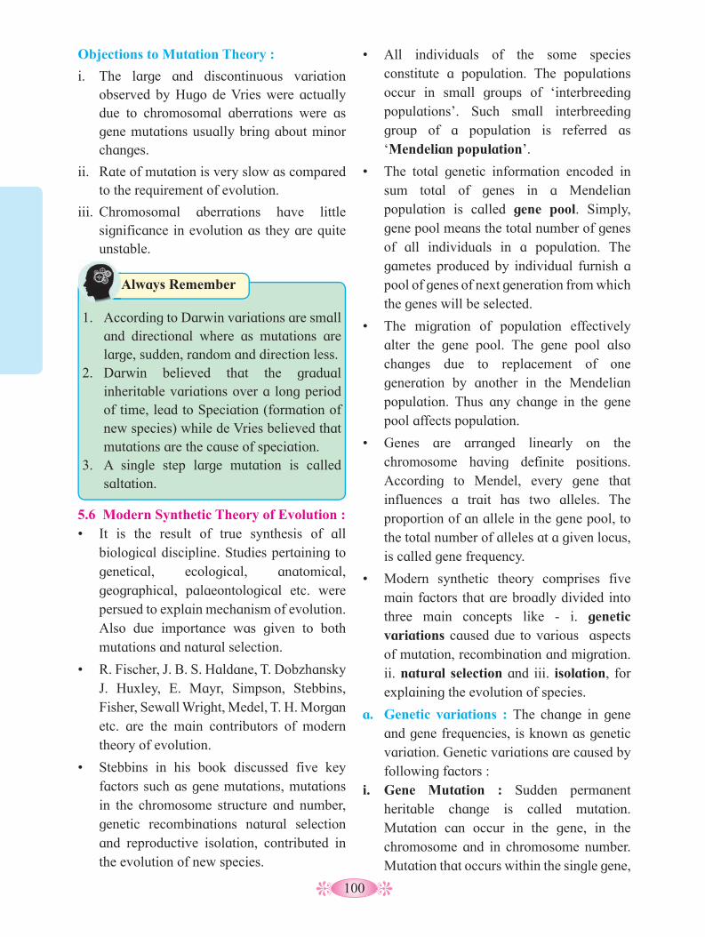
100
• All individuals of the some species constitute a population. The populations occur in small groups of ‘interbreeding populations’. Such small interbreeding group of a population is referred as ‘Mendelian population’.
• The total genetic information encoded in sum total of genes in a Mendelian population is called gene pool. Simply, gene pool means the total number of genes of all individuals in a population. The gametes produced by individual furnish a pool of genes of next generation from which the genes will be selected.
• The migration of population effectively alter the gene pool. The gene pool also changes due to replacement of one generation by another in the Mendelian population. Thus any change in the gene pool affects population.
• Genes are arranged linearly on the chromosome having definite positions. According to Mendel, every gene that influences a trait has two alleles. The proportion of an allele in the gene pool, to the total number of alleles at a given locus, is called gene frequency.
• Modern synthetic theory comprises five main factors that are broadly divided into three main concepts like - i. genetic variations caused due to various aspects of mutation, recombination and migration. ii. natural selection and iii. isolation, for explaining the evolution of species.
a. Genetic variations : The change in gene and gene frequencies, is known as genetic variation. Genetic variations are caused by following factors :
i. Gene Mutation : Sudden permanent heritable change is called mutation. Mutation can occur in the gene, in the chromosome and in chromosome number. Mutation that occurs within the single gene,
1. According to Darwin variations are small
and directional where as mutations are large, sudden, random and direction less.
2. Darwin believed that the gradual inheritable variations over a long period of time, lead to Speciation (formation of new species) while de Vries believed that mutations are the cause of speciation.
3. A single step large mutation is called saltation.
Always Remember
Objections to Mutation Theory :
i. The large and discontinuous variation observed by Hugo de Vries were actually due to chromosomal aberrations were as gene mutations usually bring about minor changes.
ii. Rate of mutation is very slow as compared to the requirement of evolution.
iii. Chromosomal aberrations have little significance in evolution as they are quite unstable.
5.6 Modern Synthetic Theory of Evolution :• It is the result of true synthesis of all
biological discipline. Studies pertaining to genetical, ecological, anatomical, geographical, palaeontological etc. were persued to explain mechanism of evolution. Also due importance was given to both mutations and natural selection.
• R. Fischer, J. B. S. Haldane, T. Dobzhansky J. Huxley, E. Mayr, Simpson, Stebbins, Fisher, Sewall Wright, Medel, T. H. Morgan etc. are the main contributors of modern theory of evolution.
• Stebbins in his book discussed five key factors such as gene mutations, mutations in the chromosome structure and number, genetic recombinations natural selection and reproductive isolation, contributed in the evolution of new species.
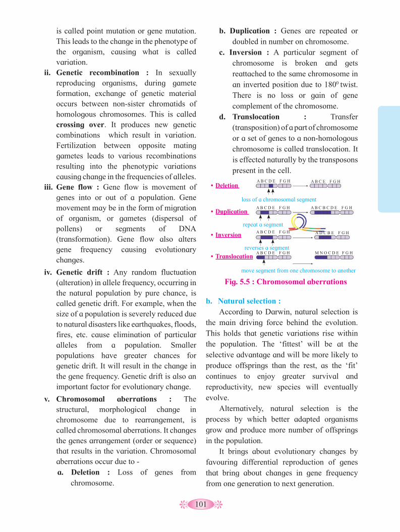
101
b. Duplication : Genes are repeated or doubled in number on chromosome.
c. Inversion : A particular segment of chromosome is broken and gets reattached to the same chromosome in an inverted position due to 1800 twist. There is no loss or gain of gene complement of the chromosome.
d. Translocation : Transfer (transposition) of a part of chromosome or a set of genes to a non-homologous chromosome is called translocation. It is effected naturally by the transposons present in the cell.
is called point mutation or gene mutation. This leads to the change in the phenotype of the organism, causing what is called variation.
ii. Genetic recombination : In sexually reproducing organisms, during gamete formation, exchange of genetic material occurs between non-sister chromatids of homologous chromosomes. This is called crossing over. It produces new genetic combinations which result in variation. Fertilization between opposite mating gametes leads to various recombinations resulting into the phenotypic variations causing change in the frequencies of alleles.
iii. Gene flow : Gene flow is movement of genes into or out of a population. Gene movement may be in the form of migration of organism, or gametes (dispersal of pollens) or segments of DNA (transformation). Gene flow also alters gene frequency causing evolutionary changes.
iv. Genetic drift : Any random fluctuation (alteration) in allele frequency, occurring in the natural population by pure chance, is called genetic drift. For example, when the size of a population is severely reduced due to natural disasters like earthquakes, floods, fires, etc. cause elimination of particular alleles from a population. Smaller populations have greater chances for genetic drift. It will result in the change in the gene frequency. Genetic drift is also an important factor for evolutionary change.
v. Chromosomal aberrations : The structural, morphological change in chromosome due to rearrangement, is called chromosomal aberrations. It changes the genes arrangement (order or sequence) that results in the variation. Chromosomal aberrations occur due to -a. Deletion : Loss of genes from
chromosome.
Fig. 5.5 : Chromosomal aberrations
A B C D E F G H A B C E F G H
A B C B C D E F G HA B C D E F G H
A B C D E F G H
A B C D E F G H
A D C B E F G H
M N O C D E F G H
• Deletion
• Duplication
• Inversion
• Translocation
loss of a chromosomal segment
repeat a segment
reverses a segment
move segment from one chromosome to another
b. Natural selection : According to Darwin, natural selection is
the main driving force behind the evolution. This holds that genetic variations rise within the population. The ‘fittest’ will be at the selective advantage and will be more likely to produce offsprings than the rest, as the ‘fit’ continues to enjoy greater survival and reproductivity, new species will eventually evolve.
Alternatively, natural selection is the process by which better adapted organisms grow and produce more number of offsprings in the population.
It brings about evolutionary changes by favouring differential reproduction of genes that bring about changes in gene frequency from one generation to next generation.
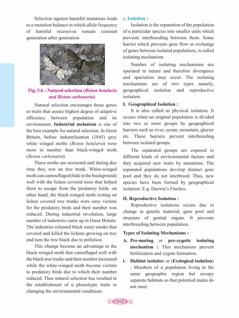
102
Selection against harmful mutations leads to a mutation balance in which allele frequency of harmful recessives remain constant generation after generation.
c. Isolation :Isolation is the separation of the population
of a particular species into smaller units which prevents interbreeding between them. Some barrier which prevents gene flow or exchange of genes between isolated populations, is called isolating mechanism.
Number of isolating mechanisms are operated in nature and therefore divergence and speciation may occur. The isolating mechanisms are of two types namely, geographical isolation and reproductive isolation.
I. Geographical Isolation : It is also called as physical isolation. It
occurs when an original population is divided into two or more groups by geographical barriers such as river, ocean, mountain, glacier etc. These barriers prevent interbreeding between isolated groups.
The separated groups are exposed to different kinds of environmental factors and they acquired new traits by mutations. The separated populations develop distinct gene pool and they do not interbreed. Thus, new species have been formed by geographical isolation. E.g. Darwin’s Finches.
II. Reproductive Isolation :Reproductive isolations occurs due to
change in genetic material, gene pool and structure of genital organs. It prevents interbreeding between population.
Types of Isolating Mechanisms :
A. Pre-mating or pre-zygotic isolating mechanism : This mechanism prevent fertilization and zygote formation.
i. Habitat isolation or (Ecological isolation) : Members of a population living in the same geographic region but occupy separate habitats so that potential mates do not meet.
Fig. 5.6 : Natural selection (Biston betularia and Biston carbonaria)
Natural selection encourages those genes or traits that assure highest degree of adaptive efficiency between population and its environment. Industrial melanism is one of the best example for natural selection. In Great Britain, before industrilisation (1845) grey white winged moths (Biston betularia) were more in number than black-winged moth (Biston carbonaria).
These moths are nocturnal and during day time they rest on tree trunk. White-winged moth can camouflaged (hide in the background) well with the lichen covered trees that helped them to escape from the predatory birds. on other hand, the black-winged moth resting on lichen covered tree trunks were easy victims for the predatory birds and their number was reduced. During industrial revolution, large number of industries came up in Great Britain. The industries released black sooty smoke that covered and killed the lichens growing on tree and turn the tree black due to pollution.
This change become an advantage to the black-winged moth that camoflaged well with the black tree trunks and their number increased while the white-winged moth become victims to predatory birds due to which their number reduced. Thus natural selection has resulted in the establishment of a phenotypic traits in changing the environmental conditions.

103
ii. Seasonal or temporal isolation : Members of a population living in the same geographic region but are sexually mature at different years or different times of the year.
iii. Ethological isolation : Due to specific mating behaviour the members of population do not mate.
iv. Mechanical Isolation : Members of two population have difference in the structure of reproductive organs.
B. Post-mating or Post-zygotic barriers :i. Gamete mortality - Gametes have a
limited life span. Due to one or the other reasons, if union of the two gametes does not occur in the given time, it results in the gamete mortality.
ii. Zygote mortality - Here, egg is fertilized but zygote dies due to one or the other reasons.
iii. Hybrid sterility - Hybrids develop to maturity but become sterile due to failure of proper gametogenesis (meiosis). e.g. Mule is an intergeneric hybrid which is sterile.
5.7 Mechanism of organic evolution :One has to give the importance to the
population while considering the mechanism of evolution. It is the population that evolves and not its individual members. Individual’s role is to pass its genetic variation to its offspring.
The following are the basic processes which bring about evolution viz. Mutations, gene recombination, gene flow (migration), genetic drift, natural selection, isolation and speciation.Mutations - These are permanent heritable changes in the genetic material of an organism.
Mutations are already described earlier in this chapter. Gene mutations produce new alleles which are added to gene pool. Gene recombination - These are variation produce due to coming together of alleles during sexual reproduction. Gene recombinations occur due to random union of gametes, anaphasic separation of chromosomes and crossing over.Gene flow - It is the transfer of gene during interbreeding of populations that are genetically different. As explained earlier in this chapter gene flow is due to emigration and imigration. Its brings about changes in the allele frequency.Genetic drift - Any alternation in allete frequency in the natural population by chance, is called genetic drift. Concept of genetic drift was first given Sewall wright, hence, called as Sewall wright effect. For example, elimination of a particular allele from a population due to events like accidental death prior to mating of an organism. Genetic drifts are random or directionless. The effect of genetic drift is more significant in small population than in large population. Due to genetic drift, some alleles of a population are lost or reduced by chance and some others may be increased. Some time, a few individuals become isolated from the large population and they produce new population in new geographical area. The allele frequency of new population become different. The original drifted population (i.e. colonizing ancestor/pioneer) becomes ‘founders’ and the effect is called founder effect.
A bottle neck effect is seen when much of a population is killed due to a natural disaster and only a few remaining individuals are left to begin a new population.Natural selection - It is a process by which better adapted individuals with useful variations are selected by nature and leave greater or more number of progenies (Differential reproduction).
1. What is variation?2. What is mutation?3. What is gene frequency?4. Why variations are occurs in population?
Can you tell?

104
Type of Natural selection :a. Stabilizing selection : (Balancing selection)
1. Here more individuals of a population acquired a mean character value.
2. It tends to favour the intermediate forms and eliminate both the phenotypic extreme. For e.g. More number of infants with intermediate weight survive better as compare to those who are over-weight or under-weight.
3. It reduces variations.
4. It does not lead to evolutionary change but tend to maintain phenotypic stability within population, therefore, it is described as stabilizing selection.
5. Genetically stabilizing selection represents a situation where a population is adapted to its environment.
b. Directional selection : 1. In this type, more individuals acquired
value other than the mean character value.
2. Naural selection acts to eliminate one of the extremes of the phenotypic range and favour the other. e.g. systematic elimination of homozygous recessives.
3. Directional selection operates for many generations, it results in an evolutionary trend within a population and shifting a peak in one direction.
4. e.g. Industrial melanism, DDT resitant mosquito etc.
c. Disruptive Natural selection :1. Here more number of individuals acquire
peripheral character value at both ends of the distribution curve.
2. Nature select extreme phenotypes and eliminate intermediate. Hence two peaks are formed in distribution of traits.
3. This kind of selection is rare.4. It ensures the effect on the entire genepool
of a population, considering all mating types or systems.
5. Example - It was observed in the different beak size of African seed cracker finches. The birds have different size of beak and they feed on seeds. The avilable seeds were of two kinds small and large sized seeds. Large beak sized birds feeds on large seeds while small beak sized birds feed on small seeds and their number was increased.Intermediate beak sized birds are unable to feed on either type of seeds so their population was decreased gradually and then eliminated by natural selection.
Fig. 5.7 : Stabilising selection
Num
ber
of I
ndiv
idua
ls
Time
Selection against both extremes
Population after selection
Original population
Fig. 5.8 : Directional selection
Num
ber
of I
ndiv
idua
ls
Time
Selection against an extremes
Population after selection
Original population
Fig. 5.9 : Disruptive selection
Num
ber
of I
ndiv
idua
ls
Time
Selection against the intermediates
Population after selection
Original population

105
There are few factors such as gene migration (gene flow), genetic drift, mutation, genetic recombinations, natural selection, non-random mating, etc. which affect or change the Hardy-Weinberg equilibrilium,
If these factors do not occur in the population, then population is genetically stable or non evolving population.
Isolation - It is separation of a single interbreeding population into subunits. Isolation restricts gene flow between descrete (non continuous population due to different barriers like geographical barriers. (This part is already explained in detail earlier in this chapter)
Speciation - The sub units of single interbreeding population due to the geographical barriers like river, mountains, desert, sea etc. become isolated in such a way that their interbreeding is prevented. This will finally lead to origin of new species (i.e. speciation). (Discussed in detail ahead in this chapter).
5.8 Hardy-Weinberg’s principle :
It is also known as Hardy-Weinberg’s equilibrium law. The law states that ‘at equilibrium point both the gene (allele) frequency and geneotypic frequency remain constant from generation to generation’. It occurs only in the diploid, sexually reproducing, large, free interbreeding population in which mating is random and no selection or other factors are present for changing the allele frequency. e.g. A single locus has two alleles (A and a). The frequencies of these allele are p and q respectively. The allele frequency for any locus is always one. i.e. P + Q = 1. The genotypic frequencies of both the alleles are represented by (p + q)2 = 1. The binomial expansion of this is P2 + 2pq + q2 = 1 i.e. AA=P2, aa=q2 and for 2Aa= 2pq.
Hence P2 + 2pq + q2 = 1 This is a binomial expansion of (p + q)2.
This can be explained by punnet square as follow.
Hybrid A a
A (p) a (q)
Hybrid
Aa
A (p) AA (p2) Aa (pq)
a (q) Aa (pq) aa (q2)
Like allele frequency, the genotypic frequencies together are also equal to 1.
In which conditions the gene frequency of a population will remain constant?
Can you tell?
5.9 Adaptive Radiation :The process of evolution which results in
transformation of original species to many different varieties, is called, adaptive radiation.
Darwin’s Finches is one of the best example of adaptive radiation. During his visit to Galpagos Islands. Charls Darwin also noticed a variety of small birds. These birds are called Darwin’s finches.
Fig. 5.10 : Darwin’s finches
Darwin concluded that the American main land species of bird was the original one from which they migrated to the different islands of Galpagos. They adapted to the different environmental conditions of these islands. From original seed eating features many other forms with altered beaks evolved into insectivorous features.
Another example of adaptive radiation is Australian Marsupials. In Australia, there are many marsupial mammals who evolved from common ancestor.

106
2. Moulds : These are the hardened encasements formed in the outer parts of organic remains which later decayed leaving cavities. Body parts of plants or animals later decays but the impression still remains and becomes permanent. For example Foot – prints are formed in this manner.
3. Cast : They are hardened pieces of mineral matter deposited in the cavities of moulds.
4. Compressions : Internal structure is absent but a thin carbon film indicates the outline of external features.
Significance of Palaeontology :
1. It is useful in reconstruction of phylogeny.2. It helps in studying various forms and
structures of extinct animals.3. It provides record of missing link
between two groups of organsims.4. It helps in the study of habits of extinct
organisms.5. Palaeontology provides the following
types of evidences.Connecting link (missing link) :
These are fossil forms transitional or intermediate between two groups of organisms.It shows some characters to both the groups. Thus it indicate the evolutionary line Seymouria (between amphibians and reptiles). e.g. Archaeopteryx (between reptiles and birds).
Archaeopteryx lithographica : It is fossilized crow size toothed bird found
from jurassic rocks in Germany. It is known as missing link between reptiles and birds because it shows characters of both.Reptilian characters :
1. Presence of long tail, claws and scales on the body.
2. Single headed ribs.3. Abdominal ribs are present which look
like ribs of crocodile.4. Jaws with homodont teeth.
5.10 Evidences of organic evolution : The theory of organic evolution states that
the present day complex organisms have originated from earlier simpler forms of life.
The process of evolution is supported by evidences provided by various branches of biology such as : Palaeontology, comparative anatomy, embryology and molecular biology.
A. Palaeontology : The study of ancient life with help of fossils is called palaeontology. Fossils are the dead remains of plants and animals that lived in past in various geological layers.
• The study of fossils provides the most convincing and direct evidence of evolution.
• Fossils are formed in sedimentary rocks, amber (yellowish fossils resin), ice, peat bogs etc.
• During fossilization, the primitive forms of organisms occupy the older, lower layers and the advanced forms occupy the upper, more recent layers of the earth.
Types of fossils :
1. Actual remains : These are most common type of fossils. The plants, animals and human bodies got embedded in permafrost of arctic or alpine snow remain preserved in the actual state, e.g. Wooly Mammoth in Siberia. Amber or hardened resin contains preserved bodies of many insects and arthropods.
1. What is carbon dating and how does it
work ?2. What is fossils ?3. What is Homologous organs ?4. What is Embryology ?
Can you tell?
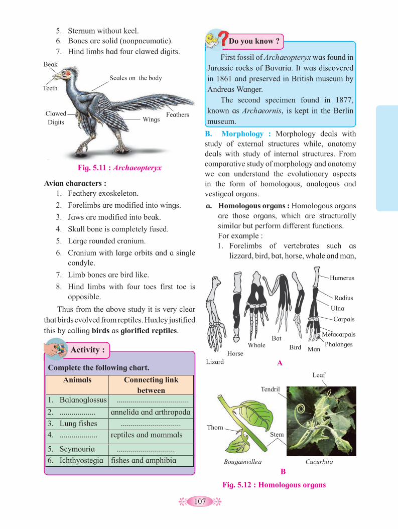
107
5. Sternum without keel.6. Bones are solid (nonpneumatic).7. Hind limbs had four clawed digits.
B. Morphology : Morphology deals with study of external structures while, anatomy deals with study of internal structures. From comparative study of morphology and anatomy we can understand the evolutionary aspects in the form of homologous, analogous and vestigeal organs.
a. Homologous organs : Homologous organs are those organs, which are structurally similar but perform different functions.For example :1. Forelimbs of vertebrates such as
lizzard, bird, bat, horse, whale and man,
Avian characters :1. Feathery exoskeleton.
2. Forelimbs are modified into wings.
3. Jaws are modified into beak.
4. Skull bone is completely fused.
5. Large rounded cranium.
6. Cranium with large orbits and a single condyle.
7. Limb bones are bird like.
8. Hind limbs with four toes first toe is opposible.
Thus from the above study it is very clear that birds evolved from reptiles. Huxley justified this by calling birds as glorified reptiles.
Activity :
Complete the following chart.
Animals Connecting link between
1. Balanoglossus ....................................
2. .................. annelida and arthropoda
3. Lung fishes ..............................
4. ................... reptiles and mammals
5. Seymouria .............................
6. Ichthyostegia fishes and amphibia
First fossil of Archaeopteryx was found in Jurassic rocks of Bavaria. It was discovered in 1861 and preserved in British museum by Andreas Wanger.
The second specimen found in 1877, known as Archaeornis, is kept in the Berlin museum.
Do you know ?
Fig. 5.11 : Archaeopteryx
Feathers
Beak
Teeth
Scales on the body
Clawed Digits Wings
Lizard Horse
Whale Bat
Bird Man
Humerus
Radius
Ulna
Carpals
Metacarpals
Phalanges
Fig. 5.12 : Homologous organs
B
Thorn
Tendril
Leaf
Stem
Bougainvillea Cucurbita
A
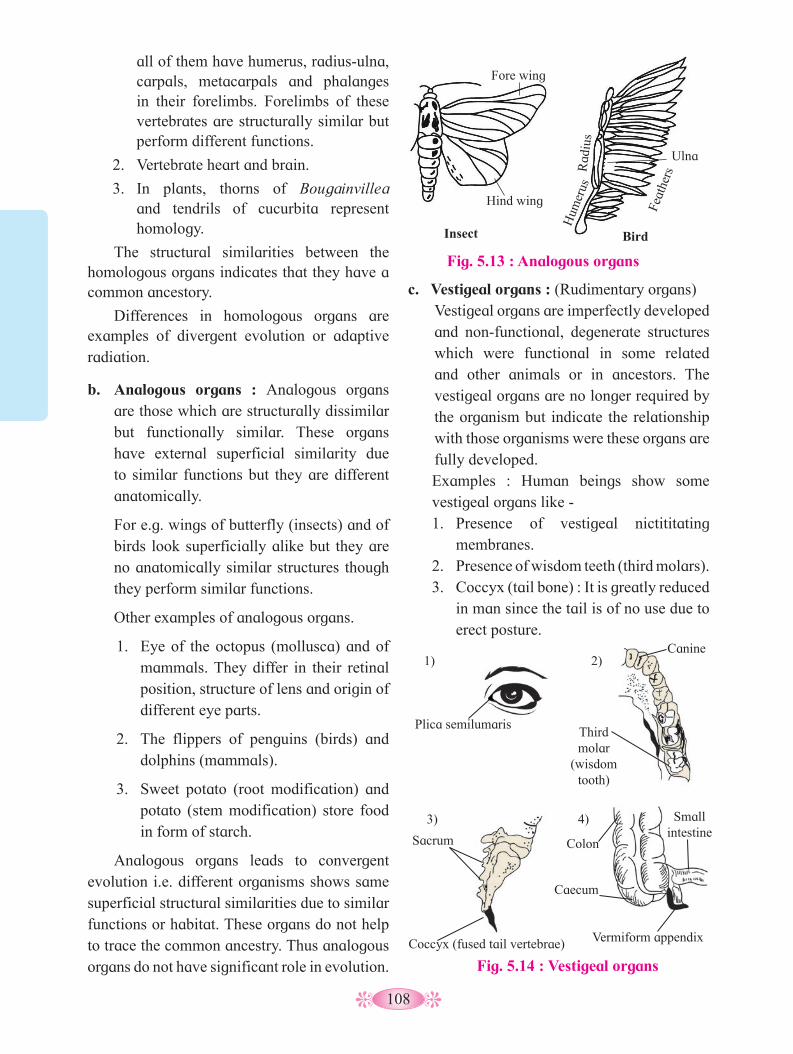
108
all of them have humerus, radius-ulna, carpals, metacarpals and phalanges in their forelimbs. Forelimbs of these vertebrates are structurally similar but perform different functions.
2. Vertebrate heart and brain.
3. In plants, thorns of Bougainvillea and tendrils of cucurbita represent homology.
The structural similarities between the homologous organs indicates that they have a common ancestory.
Differences in homologous organs are examples of divergent evolution or adaptive radiation.
b. Analogous organs : Analogous organs are those which are structurally dissimilar but functionally similar. These organs have external superficial similarity due to similar functions but they are different anatomically.
For e.g. wings of butterfly (insects) and of birds look superficially alike but they are no anatomically similar structures though they perform similar functions.
Other examples of analogous organs.
1. Eye of the octopus (mollusca) and of mammals. They differ in their retinal position, structure of lens and origin of different eye parts.
2. The flippers of penguins (birds) and dolphins (mammals).
3. Sweet potato (root modification) and potato (stem modification) store food in form of starch.
Analogous organs leads to convergent evolution i.e. different organisms shows same superficial structural similarities due to similar functions or habitat. These organs do not help to trace the common ancestry. Thus analogous organs do not have significant role in evolution.
c. Vestigeal organs : (Rudimentary organs)Vestigeal organs are imperfectly developed and non-functional, degenerate structures which were functional in some related and other animals or in ancestors. The vestigeal organs are no longer required by the organism but indicate the relationship with those organisms were these organs are fully developed.Examples : Human beings show some vestigeal organs like -1. Presence of vestigeal nictititating
membranes.2. Presence of wisdom teeth (third molars).3. Coccyx (tail bone) : It is greatly reduced
in man since the tail is of no use due to erect posture.
Fig. 5.13 : Analogous organs
Insect Bird
Fore wing
Hind wing Feat
hers
Hum
erus
R
adiu
s
Ulna
Fig. 5.14 : Vestigeal organs
Plica semilumaris
1) 2)
3) 4)
Canine
Third molar
(wisdom tooth)
Sacrum
Coccyx (fused tail vertebrae)
Caecum
Vermiform appendix
Small intestine
Colon

109
4. Vermiform appendix and the caecum. It is functional in herbivorous mammals for digestion of cellulose. In man due to eating of cooked food it has lost its function.
Presence of these vestigeal organs provide evidence that man has (evolved) descended from simple primates.
d. Molecular Evidences :
1. Cell is the basic structural and functional unit of life in all organisms.
2. Similarities in proteins and genetic material performing a similar function among diverse organisms gives evidence of a common ancestry.
3. Basic metabolic activities also occur in a similar manner in all organisms.
4. ATP is the energy source in all living organisms.
5.11 Speciation :
The process of formation of a new species from the per-existing species is called speciation.
Species is a group of similar organisms that can interbreed and produce a fertile offspring in nature. New species are formed by the following modes.
a. Intraspecific Speciation :
i. Allopatric speciation : Formation of a new species due to separation of a segment of population from the original population by distanced or a geographical barrier cutting across the species range. e.g. creeping glaciers, development of mountains. Migration of individual also causes allopatric speciation. The mode of evolution here is called adaptive radiation
Give examples of embryological evidences studied in lower classes.
Can you recall?
e.g. 14 different species of finches in Galapagos islands and several marsupial species in the Australian continent.
ii. Sympatric speciation: Formation of species within single population without geographical isolation. These are formed due to reproductive isolation. e.g. Cichlid fishes in Lake Victoria. Mutations are helpful in sympatric speciation.
b. Interspecific Speciation : Hybridisation :
Two different species on crossing may give rise to a new species. e.g. Mule is a hybrid produced by interbreeding between a male donkey and a female horse. Hinny is offspring of male horse and female donkey.
1. What are fossils? why should we study
fossils? 2. How do we find age of fossils ?3. Where do we find fossils ?
Can you recall?
5.12 Geological time scale :
The planet earth with its present biodiversity was not so when it was born. Study of fossils tells us that life forms were not the same millions of years ago (MYA). Geological time scale is used to understand the sequence of events that to place on the earth in different ages over a period of time. It is divided into six major ‘Eras’ Eras ended with major environmental changes on earth resulting into extinction and emergence of new species. The eras are further divided into periods and epochs based on minor but landmark events in each era. Table 5.15 shows the geological time scale at a glance.
The first life appeared on the earth some 2000 million years ago. It took billions of years for this process to take place, from protenoids to first cells the transition is still a mystery. Once formed the living forms diversified into various groups. Life began in the sea water and

110
plants were the first living beings to adapt to terrestrial life. Fishes evolved and diversified. The lobefin group of fishes too got diversified. Some developed stout and strong fins and could go to land and come back to water.
The coelecanth was considered a living fossils. It was thought that lobefins are extinct,
but the variety of the lobefin fish, called coelacanth was caught in 1938 in South Africa.
Reptiles evolved from amphibians. They are the first true land vertebrates. They do not have to go to water for reproduction. (Hint : think of amphibian and reptilian eggs). But about 200 million years ago (mya) some reptiles
Table 5.15 : Geological time scale
Era Period Time MYA
Epoch Plant life Animal life
Qua
rter
nary
0.01-2.0
0.1-0.6 Recent (Holocene)
Angiosperms Age of mammals : Development of modern man, birds, fishes and insects. Development of human culture.
0.6 - 2.0 Pleistocene Increase in herbs Extinction of great mammals. Appearance of primitive man.
2-13 Pliocene Hard woody plants conifers, grasslands bryophytes, monocots
Emergence or origin of man. Evolution of ruminants - horse, camel, elephant.
13-26 Miocene Abundance of deciduous trees origin of grasses
Formation of first man like apes. Adaptive radiation or spread of mammals.
26-38 Oligocene Rise of monocots and flowering plants
Extinction of Archiac mammals, Appearance of apes and monkeys. Turtles and crocodiles attained development.
38-54 Eocene Development of angiosperms
Diversification of placental mammals and modern birds.
54-65 Palaeocene Modernisation of flowering plants
Arrival of early or first primates, rise of placental mammals.
Mes
ozoi
c
Cre
tace
ous 65-135 Decline of ferns -
sphenopsids (horsetails) and Gymnosperms Ginkos, Gnetales. 1st appearance of flowering plants.
Extinction of Dinasours and toothed birds. Appearance of placental mammals and first modern birds
Jura
ssic 135-165 Origin of angiospersms
Dominance of lycopods, ferns, conifers, cycads.
Age of reptiles. Dinasaurs dominant, Appearance of toothed birds (Archaeopteryx) Rise of marsupials.
Tri
assi
c 165-225 Dominance of gymnosperms extinction of seed ferns
Appearance and rise of dinosours. Extinction of primitive amphibians. Diversification of reptiles. Rise of oviparous mammals. Therapsids, diversification of flies.
Cen
ozoi
c
Tert
iary
2-6.5
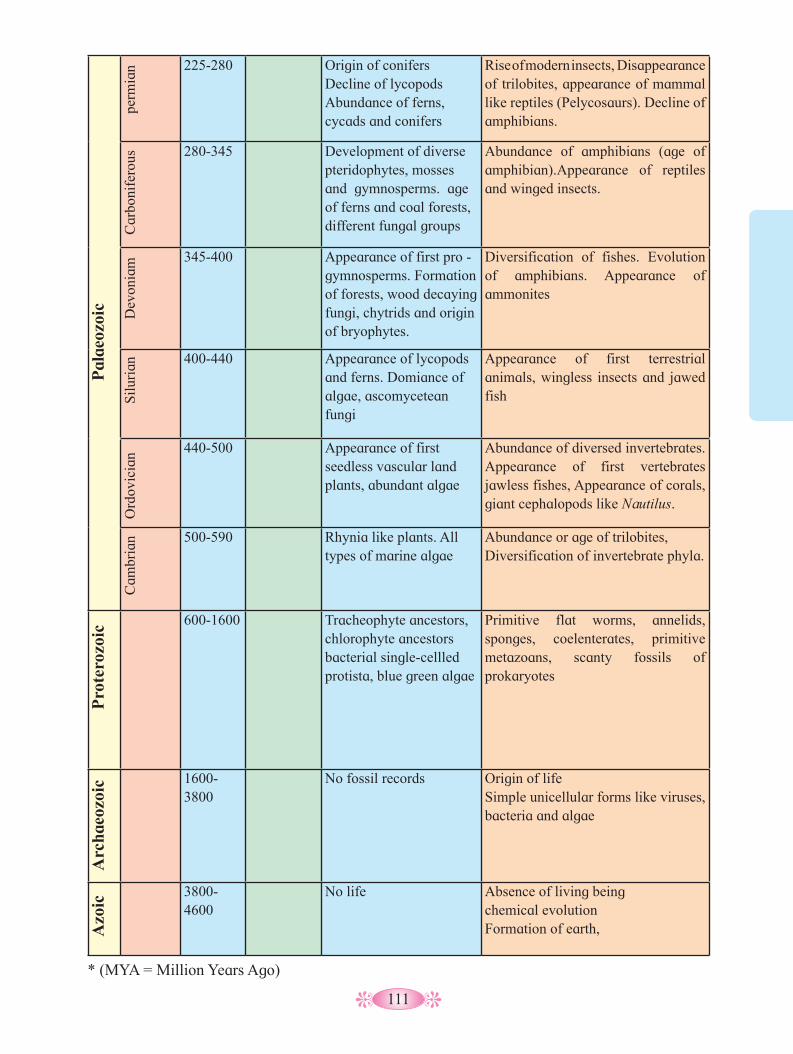
111
Pal
aeoz
oic
perm
ian 225-280 Origin of conifers
Decline of lycopods Abundance of ferns, cycads and conifers
Rise of modern insects, Disappearance of trilobites, appearance of mammal like reptiles (Pelycosaurs). Decline of amphibians.
Car
boni
fero
us 280-345 Development of diverse pteridophytes, mosses and gymnosperms. age of ferns and coal forests, different fungal groups
Abundance of amphibians (age of amphibian).Appearance of reptiles and winged insects.
Dev
onia
m 345-400 Appearance of first pro - gymnosperms. Formation of forests, wood decaying fungi, chytrids and origin of bryophytes.
Diversification of fishes. Evolution of amphibians. Appearance of ammonites
Silu
rian 400-440 Appearance of lycopods
and ferns. Domiance of algae, ascomycetean fungi
Appearance of first terrestrial animals, wingless insects and jawed fish
Ord
ovic
ian 440-500 Appearance of first
seedless vascular land plants, abundant algae
Abundance of diversed invertebrates. Appearance of first vertebrates jawless fishes, Appearance of corals, giant cephalopods like Nautilus.
Cam
bria
n 500-590 Rhynia like plants. All types of marine algae
Abundance or age of trilobites, Diversification of invertebrate phyla.
Pro
tero
zoic
600-1600 Tracheophyte ancestors, chlorophyte ancestors bacterial single-cellled protista, blue green algae
Primitive flat worms, annelids, sponges, coelenterates, primitive metazoans, scanty fossils of prokaryotes
Arc
haeo
zoic 1600-
3800No fossil records Origin of life
Simple unicellular forms like viruses, bacteria and algae
Azo
ic
3800-4600
No life Absence of living beingchemical evolutionFormation of earth,
* (MYA = Million Years Ago)
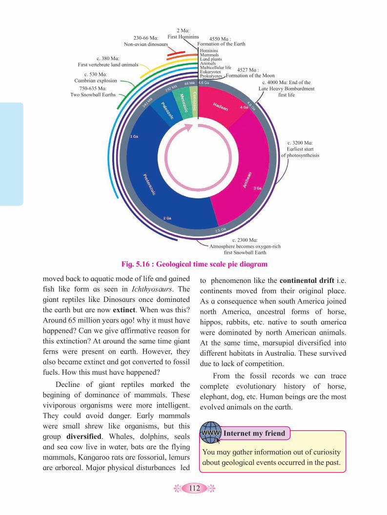
112
moved back to aquatic mode of life and gained fish like form as seen in Ichthyosaurs. The giant reptiles like Dinosaurs once dominated the earth but are now extinct. When was this? Around 65 million years ago! why it must have happened? Can we give affirmative reason for this extinction? At around the same time giant ferns were present on earth. However, they also became extinct and got converted to fossil fuels. How this must have happened?
Decline of giant reptiles marked the begining of dominance of mammals. These viviporous organisms were more intelligent. They could avoid danger. Early mammals were small shrew like organisms, but this group diversified. Whales, dolphins, seals and sea cow live in water, bats are the flying mammals, Kangaroo rats are fossorial, lemurs are arboreal. Major physical disturbances led
to phenomenon like the continental drift i.e. continents moved from their original place. As a consequence when south America joined north America, ancestral forms of horse, hippos, rabbits, etc. native to south america were dominated by north American animals. At the same time, marsupial diversified into different habitats in Australia. These survived due to lack of competition.
From the fossil records we can trace complete evolutionary history of horse, elephant, dog, etc. Human beings are the most evolved animals on the earth.
Internet my friend
You may gather information out of curiosity about geological events occurred in the past.
Fig. 5.16 : Geological time scale pie diagram
750-635 Ma:Two Snowball Earths
c. 530 Ma: Cambrian explosion
c. 380 Ma: First vertebrate land animals
c. 4000 Ma: End of the Late Heavy Bombardment
first life
c. 3200 Ma: Earliest start
of photosyntheisis
c. 2300 Ma: Atmosphere becomes oxygen-rich
first Snowball Earth
230-66 Ma: Non-avian dinosaurs
2 Ma: First Hominins 4550 Ma :
4527 Ma :
Formation of the Earth
Formation of the Moon
HomininsMammalsLand plantsAnimalsMulticellular lifeEukaryotesProkaryotes
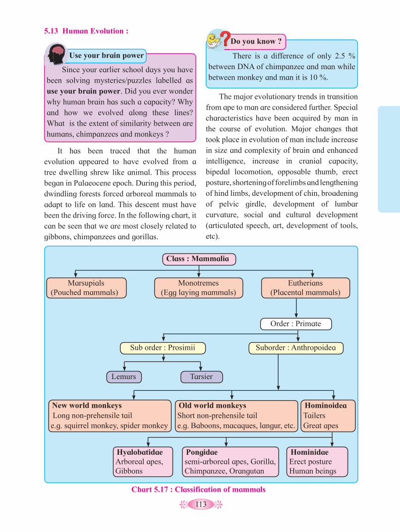
113
Since your earlier school days you have been solving mysteries/puzzles labelled as use your brain power. Did you ever wonder why human brain has such a capacity? Why and how we evolved along these lines? What is the extent of similarity between are humans, chimpanzees and monkeys ?
Use your brain power
Class : Mammalia
Marsupials (Pouched mammals)
Monotremes (Egg laying mammals)
Sub order : Prosimii Suborder : Anthropoidea
Lemurs Tarsier
New world monkeys Long non-prehensile tail e.g. squirrel monkey, spider monkey
HyalobatidaeArboreal apes,Gibbons
Pongidae semi-arboreal apes, Gorilla, Chimpanzee, Orangutan
Hominidae Erect posture Human beings
Old world monkeys Short non-prehensile tail e.g. Baboons, macaques, langur, etc.
HominoideaTailersGreat apes
Order : Primate
Eutherians (Placental mammals)
Chart 5.17 : Classification of mammals
There is a difference of only 2.5 % between DNA of chimpanzee and man while between monkey and man it is 10 %.
Do you know ?5.13 Human Evolution :
It has been traced that the human evolution appeared to have evolved from a tree dwelling shrew like animal. This process began in Palaeocene epoch. During this period, dwindling forests forced arboreal mammals to adapt to life on land. This descent must have been the driving force. In the following chart, it can be seen that we are most closely related to gibbons, chimpanzees and gorillas.
The major evolutionary trends in transition from ape to man are considered further. Special characteristics have been acquired by man in the course of evolution. Major changes that took place in evolution of man include increase in size and complexity of brain and enhanced intelligence, increase in cranial capacity, bipedal locomotion, opposable thumb, erect posture, shortening of forelimbs and lengthening of hind limbs, development of chin, broadening of pelvic girdle, development of lumbar curvature, social and cultural development (articulated speech, art, development of tools, etc).
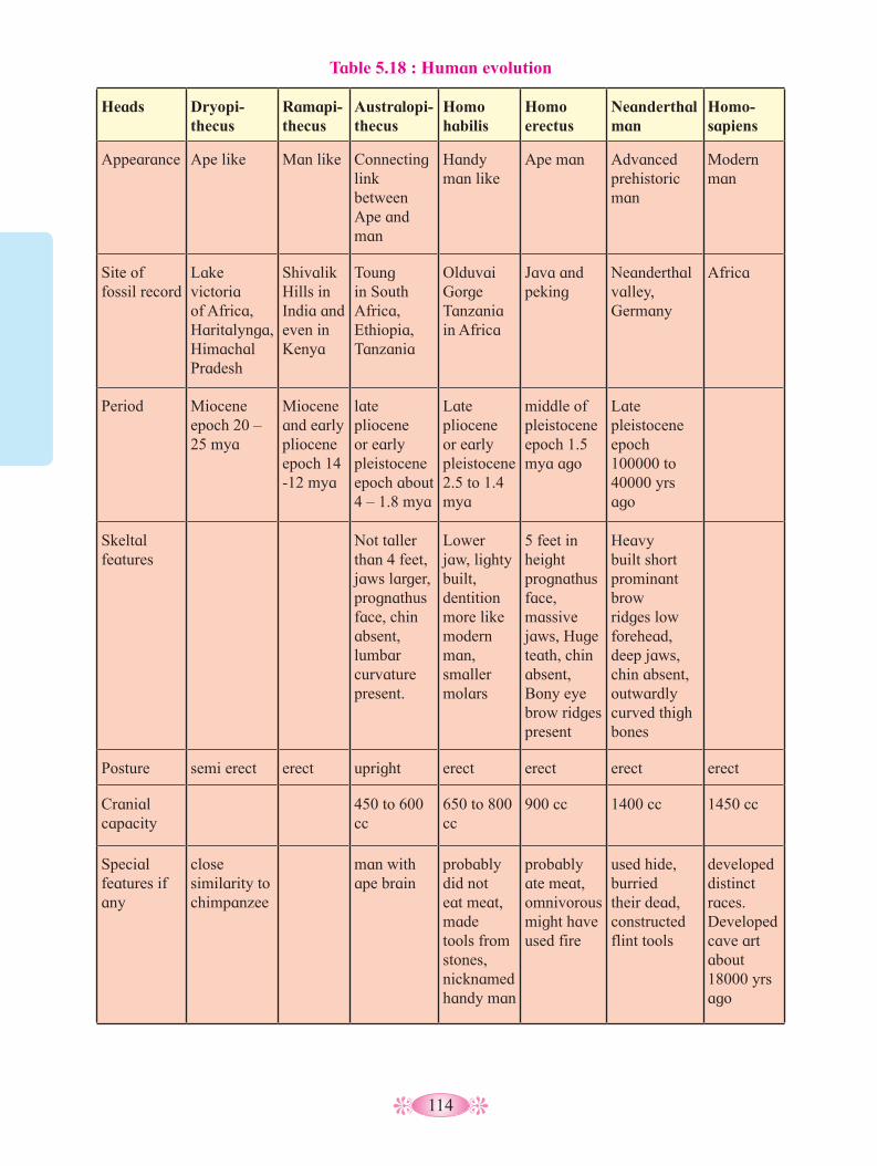
114
Table 5.18 : Human evolution
Heads Dryopi-thecus
Ramapi-thecus
Australopi-thecus
Homo habilis
Homo erectus
Neanderthal man
Homo-sapiens
Appearance Ape like Man like Connecting link between Ape and man
Handy man like
Ape man Advanced prehistoric man
Modern man
Site of fossil record
Lake victoria of Africa, Haritalynga, Himachal Pradesh
Shivalik Hills in India and even in Kenya
Toung in South Africa, Ethiopia, Tanzania
Olduvai Gorge Tanzania in Africa
Java and peking
Neanderthal valley, Germany
Africa
Period Miocene epoch 20 – 25 mya
Miocene and early pliocene epoch 14 -12 mya
late pliocene or early pleistocene epoch about 4 – 1.8 mya
Late pliocene or early pleistocene 2.5 to 1.4 mya
middle of pleistocene epoch 1.5 mya ago
Late pleistocene epoch 100000 to 40000 yrs ago
Skeltal features
Not taller than 4 feet, jaws larger, prognathus face, chin absent, lumbar curvature present.
Lower jaw, lighty built, dentition more like modern man, smaller molars
5 feet in height prognathus face, massive jaws, Huge teath, chin absent, Bony eye brow ridges present
Heavy built short prominant brow ridges low forehead, deep jaws, chin absent, outwardly curved thigh bones
Posture semi erect erect upright erect erect erect erect
Cranial capacity
450 to 600 cc
650 to 800 cc
900 cc 1400 cc 1450 cc
Special features if any
close similarity to chimpanzee
man with ape brain
probably did not eat meat, made tools from stones, nicknamed handy man
probably ate meat, omnivorous might have used fire
used hide, burried their dead, constructed flint tools
developed distinct races. Developed cave art about 18000 yrs ago

115
Human being is said to be most
evolved, intelligent living being. Yet we are
not self sufficient. Think of various aspects
for which we depend on other living beings
for our survival.
Think about it
Fig. 5.19 : Human Evolution
Internet my friend
1. Recently a fossil park has been established in Gadchiroli district of Maharashtra state. Find more information about Wadadham fossil park.
2. Find out information about caves in India. One such place is in Madhya Pradesh. It is at Bhimbetka rock shelter in Raisen district. Here we can see cave paintings by prehistoric humans.
Even though the cranium of elephant is larger than that of man, humans are considered more intelligent than elephant. Why is it so ?
Use your brain powerCranial capacity of human begins increased
over a period of time and large size of frontal lobe helped in development of high forehead.
Increase in intelligence necessitated physical development so that body and brain could be used effectively and productively. Freedom of forelimbs from locomotory function and opposable thumb led to better utilization of hands for holding objects effectively and development of motor skills etc.
Bipedal locomotion, upright posture coupled with stereoscopic vision helped man to move around safely on land.
Evolutionary history of man was traced with the help of fossil remains found over a period of time.
Some of our ancestors and their evolutionary history is shown in the table.
The above table clearly shows the gradual increase in cranial capacity, shape of skull and dentition of the ancestral humans till date.
Our journey of evolution still
continues.....

116
Activity :
Collect the information about the organisams depicted in the following diagrams and write on the same.
A. B.
C. D.
E.

117
Q. 1 Multiple choice questions.
1. Who proposed that the first form of life could have come from per- existing non-living organic molecules? a. Alfred Wallace b. oparin and Haldane c. Charles Darwin d. Louis Pasteur
2. The sequence of origin of life may be- a. Organic materials- inorganic
materials – Eobiont- colloidal aggregates- cell.
b. Inorganic materials – organic materials – colloidal aggregates –Eobiont- cell.
c. Organic materials- inorganic materials – colloidal aggregates - cell
d. Inorganic materials- organic materials – Eobiont- colloidal aggregates - cell
3. In Hardy - Weinberg equation , the frequency of homozygous recessive individual is represented by :-a. P2 b. pq c. q2 d. 2pq
4. Select the analogous organs-a. Forelimbs of whale and batb. Flippers of dolphins and penguinc. Thorn and tendrils of bougainvillea
and cucurbita.d. Vertebrates hearts or brains.
5. Archaeopteryx is known as missing link because it is a fossil and share characters of both-a. Fishes and amphibians b. Annelida and arthropoda.c. Birds and reptiles d. Chordates and nonchordates.
6. Identify the wrong statement regarding evolution.a. Darwin’s variations are small and
directional.b. Mutations are random and non-
directional.c. Adaptive radiations leads to
divergent evolution.d. Mutations are non- radamon and
directional.
7. Gene frequency in a population remain constant due to –a. Mutation b. Migrationc. Random mating d. Non- random mating
8. Which of the following characteristic is not shown by the ape?a. Prognathous face b. tail is present c. Chin is absent d. forelimbs are longer than hind limbs
9. .................. can be considered as conncting link between between ape and man.a. Austalopithecus b. Homo hablis c. Homo erectus d. Neanderthal man.
10. The Cranial capacity of Nanderthal man was a. 600 cc b. 940 ccb. 1400 cc d. 1600 cc
Q. 2 Very short answer question.
1. Define the following terms-a. Gene pool b. gene frequency c. Organic evolution d. Population e. Speciation
Exercise

118
2. What is adaptive radiation?3. If the variation occur in population by
chance alone and not by natural selection and bring change in frequencies of an allele. What is it called?
4. State the Hardy – Weinberg eqnilibrium.
5. What is homologous organs?
6. What is vestigeal organ?
7. What is the scientific name of modern man?
8. What is coacervate?
9. Which period is known as “age of Reptilia”?
10. Name the ancestor of human which is described as man with ape brain.
Q. 3 Short answer question.
1. Write a note on Genetic drift.
2. Enlist the different factors that are responsible for changing gene frequency.
3. Draw a graph to show that natural selection leads to disruptive change.
4. Give the significance of fossils.
5. Write the objections to Mutation theory of Hugo de vries.
6. What is disrruptive selection? Give example.
Columu- I Column – II1. August Weismann a. Mutation theory
2. Hugo de vries b. Germplasm theory
3. Charl Darwin c. Theory of acquired characters
4. Lamark d. Theory of natural selection
Q. 5 Long answer questions.
1. Would you consider wings of butterfly and bat as homologous or analogous and why?
2. What is adaptive radiation? Explain with suitable example.
3. By talking industrial melanism as one example. Explain the concept of natural selection.
4. Describe the Urey and Millers experiment.
5. What is Isolation? Describe the different types of reproductive Isolatons.
6. What is Genetic variations? Explain the different factors responsible for genetic variations.
Q. 6 Complere the chart.
EraDominating group of animal
1. Cenozoic ..................................
2. .................... Reptiles
3. Palaeozic ..................................
4. .................... Invertebrates
Project :
Prepare a diagrammatic chart of the chemical evolution of life.
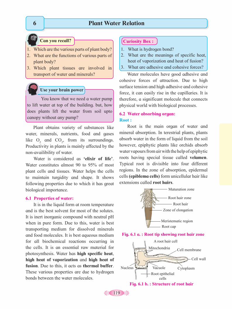
119
Plant obtains variety of substances like water, minerals, nutrients, food and gases like O
2 and CO
2, from its surroundings.
Productivity in plants is mainly affected by the non-availibility of water.
Water is considered as ‘elixir of life’. Water constitutes almost 90 to 95% of most plant cells and tissues. Water helps the cells to maintain turgidity and shape. It shows following properties due to which it has great biological importance.
6.1 Properties of water: It is in the liquid form at room temperature
and is the best solvent for most of the solutes. It is inert inorganic compound with neutral pH when in pure form. Due to this, water is best transporting medium for dissolved minerals and food molecules. It is best aqueous medium for all biochemical reactions occurring in the cells. It is an essential raw material for photosynthesis. Water has high specific heat, high heat of vaporization and high heat of fusion. Due to this, it acts as thermal buffer. These various properties are due to hydrogen bonds between the water molecules.
1. 1. Which are the various parts of plant body?2. What are the functions of various parts of
plant body?3. Which plant tissues are involved in
transport of water and minerals?
Can you recall?1.
Plant Water Relation
Water molecules have good adhesive and cohesive forces of attraction. Due to high surface tension and high adhesive and cohesive force, it can easily rise in the capillaries. It is therefore, a significant molecule that connects physical world with biological processes.
6.2 Water absorbing organ: Root :
Root is the main organ of water and mineral absorption. In terestrial plants, plants absorb water in the form of liquid from the soil however, epiphytic plants like orchids absorb water vapours from air with the help of epiphytic roots having special tissue called velamen. Typical root is divisible into four different regions. In the zone of absorption, epidermal cells (epiblema cells) form unicellular hair like extensions called root hairs.
You know that we need a water pump to lift water at top of the building. but, how does plants lift the water from soil upto canopy without any pump?
Use your brain power
6
Curiosity Box :
1. What is hydrogen bond?2. What are the meanings of specific heat,
heat of vaporization and heat of fusion?3. What are adhesive and cohesive forces?
Fig. 6.1 b. : Structure of root hair
Fig. 6.1 a. : Root tip showing root hair zone
Maturation zone
Root hair zone
Root hair
Zone of elongation
Meristematic region
Root cap
A root hair cell
Mitochondria Cell membrane
Cell wall
Cytoplasm Vacuole
Root epithelial cells
Nucleus

120
a. Imbibition: Imbibition is swelling up of hydrophillic
colloids due to adsorption of water. Substance that adsorbs water / liquid, is called as imbibant and water/ liquid, that gets imbibed is called as imbibate. The root hair cell wall is made up of pectic compounds and cellulose which are hydrophillic colloids. During Imbibition, water molecules get tightly adsorbed without the formation of solution. Imbibition continues till the equilibrium is reached. In other words, water moves along the concentration gradient.
Imbibition is significant in soaking of seeds, swelling up of dried raisins, kneading of flour etc.
Structure of root hair : Root hair is cytoplasmic extension
(prolongation) of epiblema cell. Each root hair may be approximately 1 to 10mm long and tube like structure. It is colourless, unbranched, short-lived (ephemeral) and very delicate. It has a large central vacuole surrounded by thin film of cytoplasm, plasma membrane and thin cell wall, which is two layered. Outer layer is composed of pectin and inner layer is made up of cellulose. Cell wall is freely permeable but plasma membrane is selectively permeable.
6.3 Water available to roots for absorption: Plants absorb water from the rhizosphere
(the microenvironment surrounding the root). Water present in the soil occurs as gravitational (free) water, hygroscopic water, combined water and capillary water. Water percolates deep, due to the gravity, in the soil, is called ‘gravitational water’. This is not available to plants for absorption. Fine soil particles imbibe/ adsorb water and hold it. This is called ‘hygroscopic water’. Roots cannot absorb it. Water present in the form of hydrated oxides of silicon, aluminum, etc., is called ‘combined water’. It is also not available to plants for absorption. Some amount of water is held in pores present between the neighbouring soil particles, due to capillarity. This is called capillary water that is avilable for absorption.
6.4 Absorption of water by roots from soil: Root hair absorbs water by employing three
physical processes that occur sequentially- viz. imbibition, diffusion and osmosis.
Try this at your home.A. Take 10 ml of pure water in a suitable glass vessel and put 2 - 3 raisins in it. Observe the
changes in raisins since the time you put them in water till they become fully swollen i.e. turgid. Why did raisins become turgid?
B. Take 10 ml of pure water and add 5 gms of either sugar or salt to it. Let it dissolve and then put the same turgid raisins in it and observe the changes in raisins. What changes did occur in raisins and why? Discuss your observations with your teachers.
Why do the wooden doors become very hard to close and open in rainy season?
Use your brain power
b. Diffusion: Diffusion means to disperse. Diffusion can be defined as the movement of ions/ atoms/ molecules of a substance from the region of their higher concentration to the region of their lower concentration. The movement is due to the kinetic energy of the molecules. Diffusion continues till an equilibrium is reached. Thus, water passes into the cell by diffusion through a freely permeable cell wall. Water is now at the interface of cell wall and plasma membrane.
Diffusion results in the diffusion pressure (D. P.) which is directly proportional to the number of diffusing particles. Diffusion pressure of pure solvent (pure water) is always
Activity :

121
more than the diffusion pressure of solvent in a solution. The difference in the diffusion pressures of pure solvent and the solvent in a solution is called Diffusion Pressure Deficit (DPD) or Suction Pressure (SP). The term was coined by B.S. Meyer (1938). Now a days, term water potential is used for DPD. In colloquial language, the term DPD is actually the thirst of a cell with which it absorbs water from the surroundings. Water arround cell wall has more diffusion pressure than cell sap. Due to this, water moves in the cell by diffusion. Diffusion is significant in plants in the absorption of water, minerals, conduction of water against the gravity, exchange of gases and transport and distribution of food.
c. Osmosis : It is a process by which water enters into the cytoplasm of the root hair cell. Osmosis is a special type of diffusion of solvent through a semipermeable membrane. The cytoplasm of root hair cell contains minerals, sugars, etc. In other words, solution inside the cell is more concentrated (stronger) than outside the cell (weaker). Therefore, solvent from weaker solution enters into cytoplasm (i.e. to stronger solution) of cell through a semipermiable plasma membrane. This migration of solvent is called Osmosis.
Thus, water at the interface of cell wall and plasma membrane, enters into the cytoplasm of the root hair cell due to osmosis.
With respect to the concentration and osmotic migration, three types of solutions are recognized viz, i. Hypotonic (weak solution or strong
solvent) having low osmotic concentration.ii. Hypertonic (strong solution or weak
solvent) having high osmotic concentration.iii. Isotonic having such a concentration of
solution where there is neither gain nor loss of water in an osmotic system. In other words, concentration outside and inside the cell is same. Osmosis is of two types viz, Exo-osmosis
and Endo-osmosis.Exo-osmosis : It is the migration of solvent from the cell outside. It causes flaccidity of cell.
Endo-osmosis : It is the migration of the solvent into the cell. It causes turgidity of cell i.e. cytoplasm becomes turgid. Turgidity increases the turgor pressure (T. P.) of the cell. T. P. is the pressure exerted by turgid cell sap on to the cell membrane and cell wall. In a fully turgid cell, DPD is zero. Cell wall being thick and rigid, exerts a counter pressure on the cell sap. This is called Wall pressure (W. P.). In a fully turgid cell, T. P. = W. P. but operating in opposite direction.
Osmotic pressure (O. P.) : The pressure exerted due to osmosis is osmotic pressure.
1. When you burn an incense stick in one corner of room, its fragrance spreads all over the room in a short time. How does it happen?
2. How does the water come out through the surface of porous earthen pot?
Use your brain power
Fig. 6.2 : Diffusion of water into plant cell across the plasma membrane
Outside of cell Water molecules Water-selective pore (aquaporin)
Cytoplasm Membrane bilayer
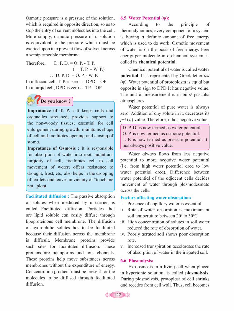
122
6.5 Water Potential (ψ): According to the principle of
thermodynamics, every component of a system is having a definite amount of free energy which is used to do work. Osmotic movement of water is on the basis of free energy. Free energy per molecule in a chemical system, is called its chemical potential.
Chemical potential of water is called water potential. It is represented by Greek letter psi (ψ). Water potential of protoplasm is equal but opposite in sign to DPD It has negative value. The unit of measurement is in bars/ pascals/ atmospheres.
Water potential of pure water is always zero. Addition of any solute in it, decreases its psi (ψ) value. Therefore, it has negative value.
D. P. D. is now termed as water potential.O. P. is now termed as osmotic potential.T. P. is now termed as pressure potential. It has always positive value.
Water always flows from less negative potential to more negative water potential (i.e. from high water potential area to low water potential area). Difference between water potential of the adjacent cells decides movement of water through plasmodesmata across the cells.
Factors affecting water absorption:i. Presence of capillary water is essential.ii. Rate of water absorption is maximum at
soil temperature between 200 to 300C.iii. High concentration of solutes in soil water
reduced the rate of absorption of water.iv. Poorly aerated soil shows poor absorption
rate. v. Increased transpiration accelarates the rate
of absorption of water in the irrigated soil.
6.6 Plasmolysis: Exo-osmosis in a living cell when placed
in hypertonic solution, is called plasmolysis. During plasmolysis, protoplast of cell shrinks and recedes from cell wall. Thus, cell becomes
Facilitated diffusion : The passive absorption of solutes when mediated by a carrier, is called Facilitated diffusion. Particles that are lipid soluble can easily diffuse through lipoproteinous cell membrane. The diffusion of hydrophilic solutes has to be facilitated because their diffusion across the membrane is difficult. Membrane proteins provide such sites for facilitated diffusion. These proteins are aquaporins and ion- channels. These proteins help move substances across membranes without the expenditure of energy. Concentration gradient must be present for the molecules to be diffused through facilitated diffusion.
Osmotic pressure is a pressure of the solution, which is required in opposite direction, so as to stop the entry of solvent molecules into the cell. More simply, osmotic pressure of a solution is equivalant to the pressure which must be exerted upon it to prevent flow of solvent across a semipermeable membrane.
Therefore, D. P. D. = O. P. - T. P. ( ∴ T. P. = W. P.) ∴D. P. D. = O. P. - W. P.In a flaccid cell, T. P. is zero ∴ DPD = OPIn a turgid cell, DPD is zero ∴ TP = OP
Improtance of T. P. : It keeps cells and organelles stretched; provides support to the non-woody tissues; essential for cell enlargement during growth; maintains shape of cell and facilitates opening and closing of stoma.Improtance of Osmosis : It is responsible for absorption of water into root; maintains turgidity of cell; facilitates cell to cell movement of water; offers resistance to drought, frost, etc; also helps in the drooping of leaflets and leaves in vicinity of “touch me not” plant.
Do you know ?
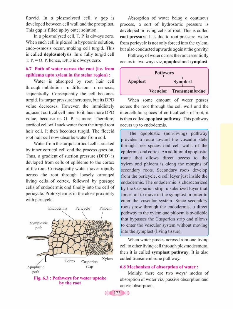
123
Absorption of water being a continous process, a sort of hydrostatic pressure is developed in living cells of root. This is called root pressure. It is due to root pressure, water from pericycle is not only forced into the xylem, but also conducted upwards against the gravity.
Pathway of water across the root essentially occurs in two ways viz, apoplast and symplast.
flaccid. In a plasmolysed cell, a gap is developed between cell wall and the protoplast. This gap is filled up by outer solution.
In a plasmolysed cell, T. P. is always zero. When such cell is placed in hypotonic solution, endo-osmosis occur, making cell turgid. This is called deplasmolysis. In a fully turgid cell T. P. = O. P. hence, DPD is always zero.
6.7 Path of water across the root (i.e. from epiblema upto xylem in the stelar region) :
Water is absorped by root hair cell through imbibition diffusion osmosis, sequentially. Consequently the cell becomes turgid. Its turgar pressure increases, but its DPD value decreases. However, the immidiately adjacent cortical cell inner to it, has more DPD value, because its O. P. is more. Therefore, cortical cell will suck water from the turgid root hair cell. It then becomes turgid. The flaccid root hair cell now absorbs water from soil.
Water from the turgid cortical cell is sucked by inner cortical cell and the process goes on. Thus, a gradient of suction pressure (DPD) is devloped from cells of epiblema to the cortex of the root. Consequently water moves rapidly across the root through loosely arranged living cells of cortex, followed by passage cells of endodermis and finally into the cell of pericycle. Protoxylem is in the close proximity with pericycle.
Fig. 6.3 : Pathways for water uptake by the root
Endodermis Pericycle Phloem
Xylem Casparian
strip Cortex
Apoplastic path
Symplastic path
Pathways
Apoplast Symplast
Vacuolar Transmembrane
When some amount of water passes across the root through the cell wall and the intercellular spaces of cortical cells of root, it is then called apoplast pathway. This pathway occurs up to endodermis.
The apoplastic (non-living) pathway provides a route toward the vascular stele through free spaces and cell walls of the epidermis and cortex. An additional apoplastic route that allows direct access to the xylem and phloem is along the margins of secondary roots. Secondary roots develop from the pericycle, a cell layer just inside the endodermis. The endodermis is characterized by the Casparian strip, a suberized layer that forces all to move in the symplast in order to enter the vascular system. Since secondary roots grow through the endodermis, a direct pathway to the xylem and phloem is available that bypasses the Casparian strip and allows to enter the vascular system without moving into the symplast (living tissue).
When water passes across from one living cell to other living cell through plasmodesmata, then it is called symplast pathway. It is also called transmembrane pathway.
6.8 Mechanism of absorption of water : Mainly, there are two ways/ modes of
absorption of water viz, passive absorption and active absorption.
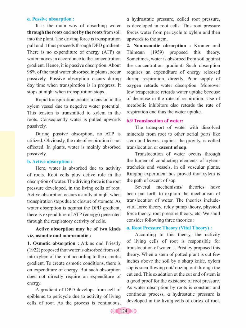
124
a hydrostatic pressure, called root pressure, is developed in root cells. This root pressure forces water from pericycle to xylem and then upwards to the stem.2. Non-osmotic absorption : Kramer and Thimann (1959) proposed this theory. Sometimes, water is absorbed from soil against the concentration gradiant. Such absorption requires an expenditure of energy released during respiration, directly. Poor supply of oxygen retards water absorption. Moreover low temperature retards water uptake because of decrease in the rate of respiration. Use of metabolic inhibitors also retards the rate of respiration and thus the water uptake.
6.9 Translocation of water: The transport of water with dissolved
minerals from root to other aerial parts like stem and leaves, against the gravity, is called translocation or ascent of sap.
Translocation of water occurs through the lumen of conducting elements of xylem- tracheids and vessels, in all vascular plants. Ringing experiment has proved that xylem is the path of ascent of sap.
Several mechanisms/ theories have been put forth to explain the mechanism of translocation of water. The theories include- vital force theory, relay pump theory, physical force theory, root pressure theory, etc. We shall consider following three theories :
a. Root Pressure Theory (Vital Theory) : According to this theory, the activity
of living cells of root is responsible for translocation of water. J. Pristley proposed this theory. When a stem of potted plant is cut few inches above the soil by a sharp knife, xylem sap is seen flowing out/ oozing out through the cut end. This exudation at the cut end of stem is a good proof for the existence of root pressure. As water absorption by roots is constant and continous process, a hydrostatic pressure is developed in the living cells of cortex of root.
a. Passive absorption : It is the main way of absorbing water
through the roots and not by the roots from soil into the plant. The driving force is transpiration pull and it thus proceeds through DPD gradient. There is no expenditure of energy (ATP) as water moves in accordance to the concentration gradient. Hence, it is passive absorption. About 98% of the total water absorbed in plants, occur passively. Passive absorption occurs during day time when transpiration is in progress. It stops at night when transpiration stops.
Rapid transpiration creates a tension in the xylem vessel due to negative water potential. This tension is transmitted to xylem in the roots. Consequently water is pulled upwards passively.
During passive absorption, no ATP is utilized. Obviously, the rate of respiration is not affected. In plants, water is mainly absorbed passively.
b. Active absorption : Here, water is absorbed due to activity
of roots. Root cells play active role in the absorption of water. The driving force is the root pressure developed, in the living cells of root. Active absorption occurs usually at night when transpiration stops due to closure of stomata. As water absorption is against the DPD gradient, there is expenditure of ATP (energy) generated through the respiratory activity of cells.
Active absorption may be of two kinds viz, osmotic and non-osmotic :
1. Osmotic absorption : Atkins and Priestly (1922) proposed that water is absorbed from soil into xylem of the root according to the osmotic gradient. To create osmotic conditions, there is an expenditure of energy. But such absorption does not directly require an expenditure of energy.
A gradient of DPD develops from cell of epiblema to pericycle due to activity of living cells of root. As the process is continuous,

125
b. Capillarity theory (physical force theory):
According to this theory, physical forces and dead cells are responsible for ascent of sap.
This theory was put forth by Bohem in (1863). Wick dipped in an oil lamp, shows capillarity due to which oil is raised upwards. The conduction of water in a straw dipped in water, is raised to a certain height because of capillarity. The height to which water is raised depends on the diameter of the straw.
Capilarity is because of surface tension, and forces of cohesion (attraction between like molecules) and adhesion (attraction between unlike molecules). Xylem vessel/ tracheid with its lumen is comparable with straw. Water column exist because of combined cohesive and adhesive forces of water and xylem wall, due to capillarity. It is because of capillarity water is raised or conducted upwards against the gravity, to few centimeters only.
Objections/ Limitations of capillarity theory:
Few important objections are :
i. Capillary tube (xylem) must be continously and completely hollow from one end to the other end but tracheids in the xylem show closed end-walls.
ii. The lower end of capillary tube i.e. xylem must be in direct contact with soil water. However, there exists a barrier of root cortex between xylem and soil water.
iii. Narrower the capillary tube, greater is the height to which water column is raised. Thus, taller trees should show xylem vessels with very narrow bore (diameter). However, in nature the tall trees show xylem vessels having wider bore.
Hence, to sum up capillarity can not be the sole mechanism to explain ascent of sap in all the plants of varying heights.
c. Cohesion- tension theory (Transpiration pull theory) :
This is presently widely accepted theory explaining ascent of sap in plants. It was put
This is termed as root pressure by S. Hales. It is due to root pressure water along with dissolved minerals is not only forced into xylem but it is also conducted upwards against the gravity.
Root pressure seems to be largely an osmotic phenomenon and its development is an active process. The value of root pressure is +1 to +2 bars which is enough to pump water to a height of 10 to 20 meters. The factors like oxygen, moisture, temperature of soil, salt contents, etc. influence the root pressure.
Fig. 6.4 : Experiment to demonstrate root pressure
Objections/ limitations of root pressure theory:
Although, ascent of sap takes place due to root pressure, there are certain objections raised, such as -
i. It is not applicable to plants taller than 20 meters.
ii. Ascent of sap can also occur even in the absence of root system.
iii. Root pressure value is almost nearly zero in taller gymnosperm trees.
iv. In actively transpiring plants, no root pressure is developed.
v. Xylem sap under normal condition is under tension i.e. it shows negative hydrostatic pressure or high osmotic pressure.
To sum up therefore, root pressure is not the sole mechanism explaining the ascent of sap in all plants of varying heights..
Manometer
Initial level
Final level
Manometer to measure exudation pressure from
cut stump
Stump

126
iii. If plant is smeared with vaseline in order to stop transpiration, even then ascent of sap occurs.
iv. Ascent of sap also occurs in deciduous plants that have shed all of their leaves.
These observations point to the fact that besides physical forces, activity of living cells seems to be necessary for lifting the water column up.
6.10 Transport of mineral ions : Soil serves as main source for minerals.
Minerals constitute most commonly occuring solid, crystalline inorganic materials obtained from earth’s crust. Minerals play an important role in the day to day life of plant. Minerals are absorbed by plants in the ionic (disolved) form, mainly through roots and then transported.
forth by Dixon and Joly (1894). This theory is based on two principles.
Cohesion and adhesion, and transpiration pull :
A strong force of attraction between water molecules, is called cohesive force. While a strong force of attraction between water molecules and lignified wall of lumen of xylem vessel, is called adhesive force.
Due to combined cohesive and adhesive forces a continous water column is developed (formed) in the xylem right from root upto the tip of the topmost leaf in the plant.
Transpiration pull : The transpiration pull developed in the leaf vessel is transmitted down to root and thus accounts for the ascent of sap.
Excess water is lost in the form of vapour, mainly through the stomata found on leaf. This water loss increases D. P. D. of mesophyll cells. These cells withdraw water ultimately from xylem in the leaf. In otherwords, due to continous transpiration, a gradient of suction pressure (i.e. D. P. D.) is developed right from guard cells up to the xylem in the leaf. This will create a tension (called negative pull or transpiration pull) in the xylem. Consequently, water column is pulled out of xylem. Thus, water is pulled upwards passively against the gravity leading to the ascent of sap.
Objections/ Limitations of transpiration pull theory:
i. For transpiration pull to operate, water column should be unbroken and continous. However, due to temperature fluctuations during day and night, gas bubbles may enter in water column breaking the continuity.
ii. This mechanism assumes that tracheids are more efficient than the vessels, as their end walls support water column.
However, vessels are more evolved than tracheids and are more efficient.
• Minerals that play important role in
the day to day life, are called essential elements. About 36 to 40 elements are incorporated in the plant’s life.
• Some minerals like C, H, O, P, N, S, Mg required in large quantity, are called macro elements. While minerals like Cu, Co, Mn, B, Zn required in small quantity, are called micro elements.
Do you know ?
The analysis of plant ash demonstrates that minerals are absorbed by plants from soil and surroundings. Absorption of minerals is independent of that of water.
Absorbed mineral ions are pulled in upward direction along with xylem sap because of transpiration pull. This could be understood when the ascending sap is analysed. Mineral ions are needed in the areas of the plant viz. apical, lateral, young leaves, developing flowers, fruits, seeds and storage organs. Hence, from the source (root), these are pulled and transported ascendingly through the sap and gets unloaded by fine veins through the
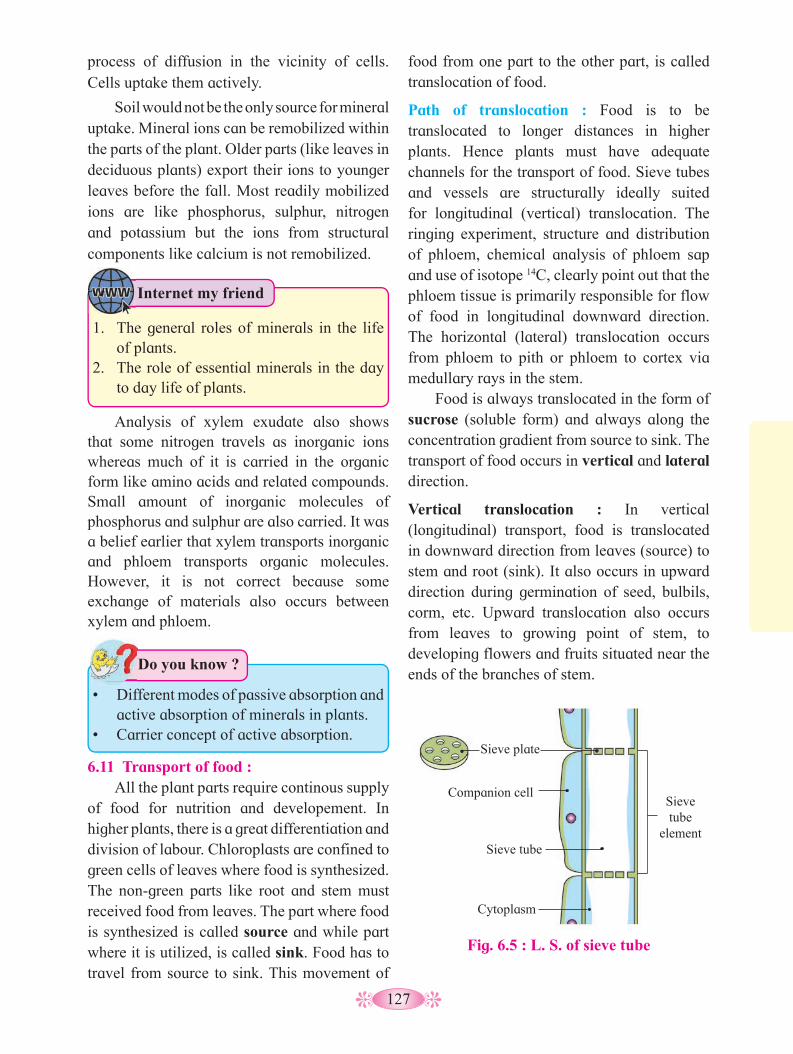
127
Analysis of xylem exudate also shows that some nitrogen travels as inorganic ions whereas much of it is carried in the organic form like amino acids and related compounds. Small amount of inorganic molecules of phosphorus and sulphur are also carried. It was a belief earlier that xylem transports inorganic and phloem transports organic molecules. However, it is not correct because some exchange of materials also occurs between xylem and phloem.
food from one part to the other part, is called translocation of food.
Path of translocation : Food is to be translocated to longer distances in higher plants. Hence plants must have adequate channels for the transport of food. Sieve tubes and vessels are structurally ideally suited for longitudinal (vertical) translocation. The ringing experiment, structure and distribution of phloem, chemical analysis of phloem sap and use of isotope 14C, clearly point out that the phloem tissue is primarily responsible for flow of food in longitudinal downward direction. The horizontal (lateral) translocation occurs from phloem to pith or phloem to cortex via medullary rays in the stem.
Food is always translocated in the form of sucrose (soluble form) and always along the concentration gradient from source to sink. The transport of food occurs in vertical and lateral direction.
Vertical translocation : In vertical (longitudinal) transport, food is translocated in downward direction from leaves (source) to stem and root (sink). It also occurs in upward direction during germination of seed, bulbils, corm, etc. Upward translocation also occurs from leaves to growing point of stem, to developing flowers and fruits situated near the ends of the branches of stem.
Fig. 6.5 : L. S. of sieve tube
process of diffusion in the vicinity of cells. Cells uptake them actively.
Soil would not be the only source for mineral uptake. Mineral ions can be remobilized within the parts of the plant. Older parts (like leaves in deciduous plants) export their ions to younger leaves before the fall. Most readily mobilized ions are like phosphorus, sulphur, nitrogen and potassium but the ions from structural components like calcium is not remobilized.
• Different modes of passive absorption and
active absorption of minerals in plants.• Carrier concept of active absorption.
Do you know ?
6.11 Transport of food : All the plant parts require continous supply
of food for nutrition and developement. In higher plants, there is a great differentiation and division of labour. Chloroplasts are confined to green cells of leaves where food is synthesized. The non-green parts like root and stem must received food from leaves. The part where food is synthesized is called source and while part where it is utilized, is called sink. Food has to travel from source to sink. This movement of
Sieve plate
Companion cell
Sieve tube
Cytoplasm
Sieve tube
element
1. The general roles of minerals in the life of plants.
2. The role of essential minerals in the day to day life of plants.
Internet my friend

128
Lateral translocation : It occurs in the root and stem. When food is translocated from phloem to pith, it is called radial translocation and from phloem to cortex, it is called tangential translocation.
The transport of food through phloem is bidirectional. Phloem sap contains mainly water and food in the form of sucrose. But sugars, amino acids and hormones are also transported through phloem.
Mechanism of sugar transport through phloem :
Several mechanisms/ theories like diffusion, activated diffusion, protoplasmic streaming, electro-osmosis, pressure-flow, etc. are put forth. The most convincing theory is Munch’s pressure flow theory or mass flow hypothesis.
Ernst Munch proposed that photosynthetic cell synthesizes glucose. Hence, its osmotic concentration increases. Due to endo-osmosis water from surrounding cells and xylem, is absorbed. The cell becomes turgid. Due to increase in turgor pressure, sugar from photosynthetic cell is forced ultimately into the sieve tube of the vein. This is called loading of Vein.
At the sink end, root cell utilizes sugar and also polymerizes excess sugar into the starch. Its osmotic concentration is lowered. Exo-osmosis occurs. Water in the root cell is lost to surrounding cells, thereby decreasing the turgidity of cell. Turgor pressure is lowered. Hence, a turgor pressure gradient is developed from sieve tube in the leaf to the root cell. Consequently, food is translocated along the concentration gradient, passively. This is Vein unloading. At the sink end sugar is used and excess water exudes into the xylem.
Main objection to this theory is that this mechanism does not explain bidirectional transport of food. More over, according to Munch, pressure flow is purely a physical process.
6.12 Transpiration :
Plants absorb water constantly and continously. Hardly 5% of the total water absorbed by roots that is utilised for cell expansion and plant growth. Remaining 95% water becomes surplus which is then lost into the atmosphere, through its aerial parts. Hardly 1% of surplus water is lost in the form of liquid and 99% of surplus water, is lost in the form of vapour. The loss of water in the form of liquid is called guttation. It occurs through special structures called water stomata or hydathodes. The loss of water in the form of vapour is called transpiration that occurs through leaves, stem, flowers and fruits. Most of the transpiration occurs through the leaves (called foliar transpiration). The actual water loss during transpiration occurs through three main sites - cuticle, stomata and lenticels. Accordingly, three types of transpiration are recognized viz. cuticular, stomatal and lenticular.
i. Cuticular transpiration: Cuticle is a layer of waxy substance- cutin,
present on outer surface of epidermal cells of leaves and stem. Cuticular transpiration occurs by simple diffusion and contributes 8-10% of the total transpiration. Cuticular transpiration
Fig. 6.6 : Transpiration
Water evaporates from the leaves
Veins carry water into the leaves
Water is drawn up the stem to the leaves
Roots take up water from the soil
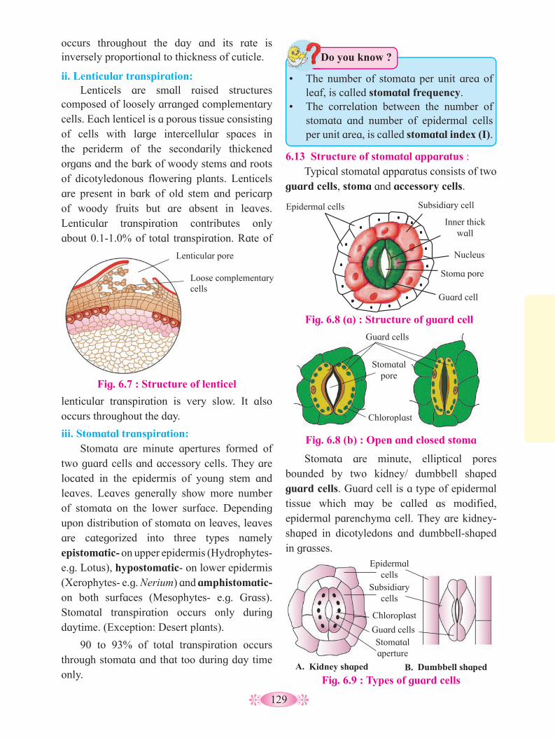
129
Fig. 6.9 : Types of guard cellsKidney shaped Dumbbell shaped
Epidermal cells
Subsidiary cells
Chloroplast
Guard cells Stomatal aperture
A. B.
occurs throughout the day and its rate is inversely proportional to thickness of cuticle.
ii. Lenticular transpiration: Lenticels are small raised structures
composed of loosely arranged complementary cells. Each lenticel is a porous tissue consisting of cells with large intercellular spaces in the periderm of the secondarily thickened organs and the bark of woody stems and roots of dicotyledonous flowering plants. Lenticels are present in bark of old stem and pericarp of woody fruits but are absent in leaves. Lenticular transpiration contributes only about 0.1-1.0% of total transpiration. Rate of
6.13 Structure of stomatal apparatus : Typical stomatal apparatus consists of two
guard cells, stoma and accessory cells.
Fig. 6.8 (a) : Structure of guard cell
Epidermal cells Subsidiary cell
Inner thick wall
Nucleus
Guard cell
Stoma pore
Fig. 6.8 (b) : Open and closed stoma
lenticular transpiration is very slow. It also occurs throughout the day.
iii. Stomatal transpiration: Stomata are minute apertures formed of
two guard cells and accessory cells. They are located in the epidermis of young stem and leaves. Leaves generally show more number of stomata on the lower surface. Depending upon distribution of stomata on leaves, leaves are categorized into three types namely epistomatic- on upper epidermis (Hydrophytes-e.g. Lotus), hypostomatic- on lower epidermis (Xerophytes- e.g. Nerium) and amphistomatic- on both surfaces (Mesophytes- e.g. Grass). Stomatal transpiration occurs only during daytime. (Exception: Desert plants).
90 to 93% of total transpiration occurs through stomata and that too during day time only.
• The number of stomata per unit area of
leaf, is called stomatal frequency.• The correlation between the number of
stomata and number of epidermal cells per unit area, is called stomatal index (I).
Do you know ?
Stomata are minute, elliptical pores bounded by two kidney/ dumbbell shaped guard cells. Guard cell is a type of epidermal tissue which may be called as modified, epidermal parenchyma cell. They are kidney-shaped in dicotyledons and dumbbell-shaped in grasses.
Guard cells
Stomatal pore
Chloroplast
Fig. 6.7 : Structure of lenticel
Lenticular pore
Loose complementary cells

130
According to theory of proton transport (Levitt-1974), stomatal movement occurs due to transport of protons H+ and K+ ions. During daytime, starch is converted into malic acid. Malic acid dissociates to form Malate and protons. Protons are transported to subsidiary cells and K+ ions are imported from them. Potassium malate is formed that increases osmolarity and causes endosmosis. Uptake of K+ ions is always accompanied with Cl¯ ions.
At night, uptake of K+and Cl- ions is prevented by abscissic acid, changing the permeability of guard cells. Due to this guard cells become hypotonic and thereby become flaccid.
Significance of Transpiration :
Advantages:
i. It removes excess of water.
ii. It helps in the passive absorption of water and minerals from soil.
iii. It helps in the ascent of sap.
iv. As stomata are open, gaseous exchange required for photosynthesis and respiration, is facilitated.
v. It maintains turgor of the cells.
vi. Transpiration helps in reducing the temperature of leaf and in imparting cooling effect.
Disadvantages:
Excessive transpiration leads to wilting and injury in the plant. It may also lead to the death of the plant.
Transpiration is ‘A necessary evil’ :
For stomatal transpiration to occur, stoma must remain open, during day time. When stomata are open then only the gaseous exhange needed for respiration and photosynthesis, will take place. If stomatal transpiration stops, it will directly affect productivity of plant through the loss of photosynthetic and respiratory activity. Hence for productivity, stomata must remain open. Consequently transpiration can not be avoided. Hence, Curtis (1926) regarded transpiration as ‘a necessary evil’.
In Cyperus, both kidney- and dumbbell-shaped guard cells are present.
Guard cells are living, nucleated cells with unevenly thick walls. Inner wall (wall facing stoma) of guard cells is thick and inelastic, and its lateral wall is thin and elastic. Guard cells contain few chloroplasts which are capable of poor photosynthesis. Guard cells have ability to change their size and form due to which stoma opens (widens) or closes (narrows).
Stoma is an elliptical pore formed due to specific arrangement of guard cells. It is through the stoma, excess water is lost in the form of vapour. Accessory cells : These are specialized epidermal cells surrounding the guard cells. Their number is variable and are the reservoirs of K+ ions. These are also called subsidiary cells.Opening and Closing of Stoma :
Opening and closing of stoma is controlled by turgor of guard cells. During day time, guard cells become turgid due to endosmosis. Thus turgor pressure is exerted on the thin walls of guard cells. Being elastic and thin, lateral walls are stretched out. Due to kidney or dumb-bell like shape, inner thick walls are pulled apart to open (widen) the stoma. During night time, guard cells become flaccid due to exosmosis. Flaccidity closes the stoma almost completely. Endosmosis and exosmosis occur due to diurnal changes in osmotic potential of guard cells. Different theories are proposed to explain diurnal changes in osmotic potential.
According to starch-sugar inter-conversion theory (Steward 1964), during day time, enzyme phosphorylase converts startch to sugar, thus increasing osmotic potential of guard cells cosing entry of water there by gaurd cells are stretched and stoma widens. The reverse reaction occures at night brining about the closure of stoma.
Starch
(Stoma opens)
Phosphorylase (Day)
NightSugar
(Stoma closes)

131
Activity :
Prepare stomatal frequency chart for any six angiospermic plants in your area.
Plant Name Details
132
Q. 1 Multiple Choice Questions1. In soil, water available for absorption
by root is ................... a. gravitaional water b. capillary water c. hygroscopic water d. combined water
2. The most widely accepted theory for ascent of sap is ..............
a. capillarity theory b. root pressure theory c. diffusion d. transpiration pull theory
3. Water movement between the cells is due to .............
a. T. P. b. W. P. c. DPD d. incipient plasmolysis
4. In guard cells, when sugar is converted into starch, the stomatal pore .............
a. closes almost completely b. opens partially c. opens fully d. remains unchanged
5. Surface tension is due to ..............a. diffusion b. osmosis
c. gravitational force d. cohesion
6. Which of the following type of solution has lower level of solutes than the solution?
a. Isotonic b. Hypotonic c. Hypertonic d. Anisotonic
7. During rainy season wooden doors warp and become difficult to open or to close because of ............... a. plasmolysis b. imbibition
c. osmosis d. diffusion
8. Water absorption takes place through .............
a. lateral roots b. root cap c. root hair d. primary root9. Due to low atmospheric pressure the
rate of transpiration will ............. a. increase b. decrease rapidly c. decrease slowly d. remain unaffected
10. Osmosis is a property of .................. a. solute b. solvent c. solution d. membrane
Q. 2 Very short answer questions.
1. What is osmotic pressure?2. Name the condition in which protoplast
of the plant cell shrinks.3. What happens when a pressure greater
than the atmospheric pressure is applied to pure water or a solution?
4. Which type of solution will bring about deplasmolysis?
5. Which type of plants have negative root pressure?
6. In which conditions transpiration pull will be affected?
7. Mention the shape of guard cells in Cyperus.
8. Why do diurnal changes occur in osmotic potential of guard cells?
9. What is symplast pathway?
Q. 3 Answer the following questions.1. Describe mechanism for absorption of
water. 2. Discuss theories of water translocation. 3. What is transpiration? Describe
mechanism of opening and closing of stomata.
Exercise

133
4. What is transpiration? Explain role of transpiration.
5. What is significance of transpiration? Explain root pressure theory and its limitations.
6. Explain capillarity theory of water translocation.
7. Why is transpiration called ‘a necessary evil’?
8. Explain movement of water in the root.
9. Define and or explain the terms: Osmosis, diffusion, plasmolysis, imbibition, guttation, transpiration, ascent of sap, active absorption, DPD, turgor pressure, water potential, wall pressure, root pressure.
10. Distinguish between a) Osmotic pressure and turgor pressure b) Diffusion and osmosis.
11. Enlist macronutrients and micronutrients required for plant growth.
12. How are the minerals absorbed by the plants?
Project :
1. Prepare powerpoint presentation for different types of transpiration.
Q. 4 Long answer questions.1. Describe structure of root hair.
2. Write on journey of water from soil to xylem in roots.
3. Explain cohesion theory for translocation of water.
4. Write on the mechanism of opening and closing of stoma.
5. What is hydroponics? How is it useful in identifying the role of nutrients?
6. Explain the active absorption of minerals.
7. Write on macro- and micro nutrients required for plant growth.
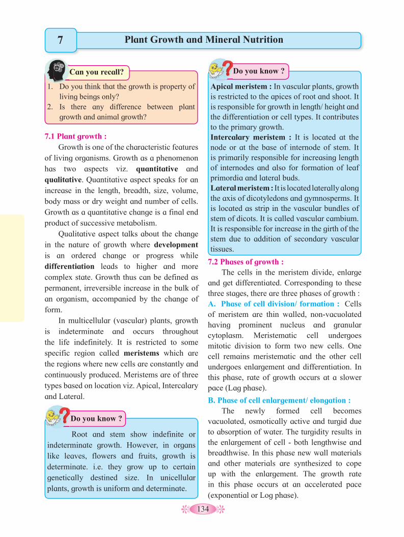
134
Plant Growth and Mineral Nutrition
1. Do you think that the growth is property of
living beings only?2. Is there any difference between plant
growth and animal growth?
Can you recall?
7
7.1 Plant growth : Growth is one of the characteristic features
of living organisms. Growth as a phenomenon has two aspects viz. quantitative and qualitative. Quantitative aspect speaks for an increase in the length, breadth, size, volume, body mass or dry weight and number of cells. Growth as a quantitative change is a final end product of successive metabolism.
Qualitative aspect talks about the change in the nature of growth where development is an ordered change or progress while differentiation leads to higher and more complex state. Growth thus can be defined as permanent, irreversible increase in the bulk of an organism, accompanied by the change of form.
In multicellular (vascular) plants, growth is indeterminate and occurs throughout the life indefinitely. It is restricted to some specific region called meristems which are the regions where new cells are constantly and continuously produced. Meristems are of three types based on location viz. Apical, Intercalary and Lateral.
7.2 Phases of growth : The cells in the meristem divide, enlarge
and get differentiated. Corresponding to these three stages, there are three phases of growth :A. Phase of cell division/ formation : Cells of meristem are thin walled, non-vacuolated having prominent nucleus and granular cytoplasm. Meristematic cell undergoes mitotic division to form two new cells. One cell remains meristematic and the other cell undergoes enlargement and differentiation. In this phase, rate of growth occurs at a slower pace (Lag phase).
B. Phase of cell enlargement/ elongation : The newly formed cell becomes
vacuolated, osmotically active and turgid due to absorption of water. The turgidity results in the enlargement of cell - both lengthwise and breadthwise. In this phase new wall materials and other materials are synthesized to cope up with the enlargement. The growth rate in this phase occurs at an accelerated pace (exponential or Log phase).
Apical meristem : In vascular plants, growth is restricted to the apices of root and shoot. It is responsible for growth in length/ height and the differentiation or cell types. It contributes to the primary growth. Intercalary meristem : It is located at the node or at the base of internode of stem. It is primarily responsible for increasing length of internodes and also for formation of leaf primordia and lateral buds. Lateral meristem : It is located laterally along the axis of dicotyledons and gymnosperms. It is located as strip in the vascular bundles of stem of dicots. It is called vascular cambium. It is responsible for increase in the girth of the stem due to addition of secondary vascular tissues.
Do you know ?
Root and stem show indefinite or indeterminate growth. However, in organs like leaves, flowers and fruits, growth is determinate. i.e. they grow up to certain genetically destined size. In unicellular plants, growth is uniform and determinate.
Do you know ?
135
Fig. 7.1 Phases of growth in root -Position of radicle at the begining (A) and at
the end (B)
Plumule
Zone of mature cells
Zone of cell differentiation
Zone of cell elongation
Zone of cell formation
A
B
C. Phase of Cell maturation/ differentiation : The enlarged cell now becomes specialized
to perform specific fuction and attains maturity - both morphological and physiological. In this phase, rate of growth slows down and comes to a steady state (stationary phase).
Growth in plants can be measured in terms of....1. Increase in the number of cells produced - e.g. single maize root apical mesistem can give rise
to more than 17,500 new cells/Hour.2. Increase in surface area of the leaf - e.g. growth of dorsiventral leaf.3. Increase in length - e.g. growth of pollen tube.4. Increase in volume of a fruit - e.g. In watermelon flower, ovary after fertilization increases in
its size/ volume by upto 3,50,000 times.5. Increase in girth of shoot.6. Increase in dry weight of organ.
Various methods for the measurement of linear growth of stem and radicle are as follows:1. Direct method : It can easily be measured with the help of ordinary measuring scale. It is a
simple method.2. Horizontal microscope : It is used to measure growth in fields.3. Auxanometer : This equipment is used for precise measurement of linear growth of shoot.
There are two types of an auxanometers viz, a) Arc auxanometer b) Pfeffer’s auxanometer4. Crescograph : It records primary growth very accurately. It magnifies growth upto 10,000
times giving information of growth per second. It is developed by sir J. C. Bose.
7.3 Conditions for Growth : The different environmental and
physiological conditions necessary for the growth include - Water, supply of nutrients,
temperature, oxygen, Carbon/ Nitrogen ratio, gravitational force, light and growth hormones. The chief conditions are explained below :
Water is the essential component of protoplasm and maintains turgidity of the cell. It acts as aqueous medium for biochemical reactions. Microelements and Macroelements are nutrients required for the proper growth of the plant. Optimum Temperature ranges between 25-350C. Oxygen is essential for respiration and the release of energy. Light is very much essential for germination of seed and photosynthesis. Gravitational force decides the direction of growth of the shoot and root.
7.4 Growth Rate and types of growth : Growth rate :
It is the increased growth per unit time. It is also called efficiency index. Rate of growth can be measured by an increase in the size and area of different plant organs like leaf, flower and fruits.
The ratio of change in the cell number (dn) over the time interval (dt) is called Absolute growth rate (AGR). Alternatively, it is the measurement and comparison of total growth per unit time.

136
AGR = dn dt
The AGR, when divided by total number of cells present in the medium, gives Relative growth ratio (RGR). Alternatively, RGR refers to the growth of a particular system per unit time, expressed on a common basis or it is the ratio of growth in the given time/ initial growth.
RGR = AGR
n AGR and RGR are useful in describing the
dynamics of cell growth in culture.
Types of growth : There are two types of growth viz,
arithmetic growth and geometric growth.
a. Arithmetic growth : Here, rate of the growth is constant and an increase in the growth occurs in arithmetic progression. i.e. 2, 4, 6, 8 cms etc.In this type of growth, the rate of growth is constant.
After mitosis one of the daughter cell continues to divide and the other cell takes part in the differentiation and maturation. e.g. elongation of root at a constant rate, best explains arithmetic growth. Linear curve is obtained when growth rate is plotted against the time. Arithmetic growth is expressed mathematically by an equation,It is expressed as, Lt = Lo + rt Where Lt = Length at time ‘t’ Lo = Length at time ‘Zero’ r = Growth rate t = Time of growth
When graph of length (L) is plotted against the time (t), a linear curve is obtained as indicated in the diagram.
b. Geometric growth : Cell divides mitotically into two. Here,
both the daughter cells continue to divide and redivide repeatedly. Such growth is called geometric growth. Here, growth rate is slow
W1 = W
o ert
W1= Final size , W
o = initial size
r = growth rate, t = time of growth e = base of natural logarithm
Graph 7.2 : Constant linear growth
Hei
ght
of t
he p
lant
(L
)
Time (t)
a
b
Fig. 7.3 : Growth rate
a. Arithmetic b. Geometric
Zygote divided
Geometric phase: all cells divide
Arithmetic phase :
These cells divide
This cells do not divide
= Cells capable of division
= Cells that have lost capacity to
divide
Digrammatic representation of : a. Arithmetic, b. Geometric grwoth and c. Stages during embryo
development showing geometric and arithmetic phases
c.
initially but later on there is a rapid growth at exponential rate. Geometric growth can be expressed mathematically by an equation.

137
Fig. 7.4 : Growth rate comparison
A1
A
10 cm2
B1
55 cm2
5 cm2 B
50 cm2
The total time (period) required for all phases to occur, is called Grand Period of Growth.
7.6 Differentiation, De-Differentiation, Re- Differentiation :a. Differentiation :
It is maturation of cells derived from apical meristem of root and shoot. Permanent change in structure and function of cells leading to maturation, is called differentiation. During cell differentiation, cell undergoes few to major anatomical and physiological changes e.g. Parenchyma in hydrophytes develops large schizogenous interspaces for mechanical support, buoyancy and aeration. The maturation is at the cost of capacity to divide and redivide.
b. Dedifferentiation : The living differentiated cell which has
lost the capacity to divide, may regain the same as per the need and divide. Thus, permanent (mature) cell undergoes dedifferentiation and becomes meristematic e.g. interfascicular
We can also observe quantitative comparison between the growth of living system in two ways.
Measurement and comparisons of total growth per unit time is called the Absolute growth rate(AGR) whereas the growth of the given system per unit time expressed on a common basis per unit initial parameter is called the Relative growth rate(RGR).
In the above example, two leaves ‘A’ and ‘B’ are of different sizes but show same absolute increase in area in a given time. Both leaves grow and increase their area by 5cm2 to produce ‘A’ and ‘B’ leaves. ‘A’ leaf of 5cm2 in size grows 5cm2/ day then its RGR would be 100%. If the leaf is 50cm2 in size and the growth rate/day is 5cm2 then its RGR would become 10%.
7.5 Growth curve : It is a graphic representation of the total
growth against time. There are three types of curves viz, Linear, Exponential and Sigmoid. Arithmetic growth curve is linear while Geometric growth curve is exponential.
Corresponding to three distinct phases of growth, growth rates differ. In Lag phase, growth rate is slow. In Exponential (Log) phase, growth rate is faster and reaches its maximum. In Stationary phase, growth rate gradually slows down. When a graph of rate of growth against time is plotted for three phases of growth, a sigmoid curve is obtained.
Graph 7.5 : Exponential Growth curve
J-shaped curve
Popu
lati
on s
ize
Year
Graph 7.6 : Sigmoid Growth curveSi
ze/ w
eigh
t of
the
orga
n
Expo
nent
ial p
hase
Lag phase
Stationary phase
Time

138
7.8 Plasticity : It is the capacity of being moulded, formed
or modeled. It is the ability of plants to form different kinds of structures (i.e. to change) in response to different environmental (external) or internal stimuli, in various phases of life.
In many plants, juvenile stage and mature
stage show different forms of leaves in the same plant e.g. heterophylly in cotton, coriander, larkspur (Delphinium). The environmental heterophylly is shown by Ranunculus flabellasis (butter cup). The intrinsic plasticity is found in coriander and cotton. Heterophylly is exhibited in the same plant in different growth phases or under different environmental conditions.
cambium and cork cambium are formed from parenchyma cells between vascular bundles and inner most layer of cortex, respectively.
c. Redifferentiation : The cells produced by dedifferentiation
once again lose the capacity to divide and mature to perform specific function. This is called redifferentiation e.g. secondary xylem and secondary phloem are formed from dedifferentiated cambium present in the vascular bundle.
7.7 Development : It refers to the ordered or progressive
changes in shape, form and degree of complexity. It includes all the changes occurring in sequence from the germination of seed upto the senescence or death during life cycle of plants. Thus development includes growth, morphogenesis, maturation and senescence.
Fig. 7.7 Flow chart of development
Seed germination
Meristem
Cell division
Plasmatic growth
Cell elongation
Cell maturation
Mature cell
Senescence
Death
Fig. 7.8 Heterophylly
31 2
4
Juvenile Adult Terrestrial habitat
Water habitat
7.9 Growth Hormones : The term ‘hormone’ was coined first by
Starling (1906) in animal physiology. The internal factors that influence growth are called growth hormones or growth regulators as they inhibit, promote or modify the growth. Growth promoters are auxins, gibberellins (GA) and cytokinins (CK). Growth inhibitors in plants are ethylene and abscissic acid (ABA). All phytohormones are growth regulators.
1. Is there any relation between phases of
growth and regions of growth curve?2. Which plant organ does show both
arithmetic and geometric growth?3. Differences between arithmetic and
geometric growth.
Can you tell?
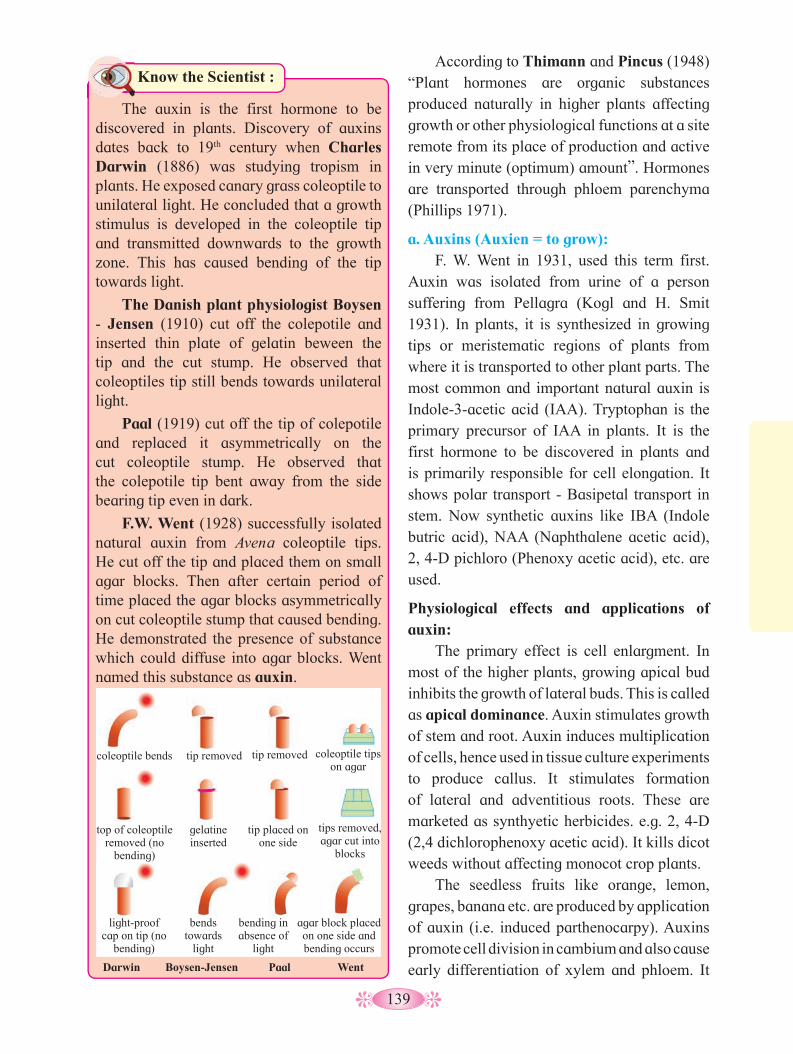
139
According to Thimann and Pincus (1948) “Plant hormones are organic substances produced naturally in higher plants affecting growth or other physiological functions at a site remote from its place of production and active in very minute (optimum) amount”. Hormones are transported through phloem parenchyma (Phillips 1971).
a. Auxins (Auxien = to grow): F. W. Went in 1931, used this term first.
Auxin was isolated from urine of a person suffering from Pellagra (Kogl and H. Smit 1931). In plants, it is synthesized in growing tips or meristematic regions of plants from where it is transported to other plant parts. The most common and important natural auxin is Indole-3-acetic acid (IAA). Tryptophan is the primary precursor of IAA in plants. It is the first hormone to be discovered in plants and is primarily responsible for cell elongation. It shows polar transport - Basipetal transport in stem. Now synthetic auxins like IBA (Indole butric acid), NAA (Naphthalene acetic acid), 2, 4-D pichloro (Phenoxy acetic acid), etc. are used.
Physiological effects and applications of auxin:
The primary effect is cell enlargment. In most of the higher plants, growing apical bud inhibits the growth of lateral buds. This is called as apical dominance. Auxin stimulates growth of stem and root. Auxin induces multiplication of cells, hence used in tissue culture experiments to produce callus. It stimulates formation of lateral and adventitious roots. These are marketed as synthyetic herbicides. e.g. 2, 4-D (2,4 dichlorophenoxy acetic acid). It kills dicot weeds without affecting monocot crop plants.
The seedless fruits like orange, lemon, grapes, banana etc. are produced by application of auxin (i.e. induced parthenocarpy). Auxins promote cell division in cambium and also cause early differentiation of xylem and phloem. It
Know the Scientist :
The auxin is the first hormone to be discovered in plants. Discovery of auxins dates back to 19th century when Charles Darwin (1886) was studying tropism in plants. He exposed canary grass coleoptile to unilateral light. He concluded that a growth stimulus is developed in the coleoptile tip and transmitted downwards to the growth zone. This has caused bending of the tip towards light.
The Danish plant physiologist Boysen - Jensen (1910) cut off the colepotile and inserted thin plate of gelatin beween the tip and the cut stump. He observed that coleoptiles tip still bends towards unilateral light.
Paal (1919) cut off the tip of colepotile and replaced it asymmetrically on the cut coleoptile stump. He observed that the colepotile tip bent away from the side bearing tip even in dark.
F.W. Went (1928) successfully isolated natural auxin from Avena coleoptile tips. He cut off the tip and placed them on small agar blocks. Then after certain period of time placed the agar blocks asymmetrically on cut coleoptile stump that caused bending. He demonstrated the presence of substance which could diffuse into agar blocks. Went named this substance as auxin.
coleoptile bends tip removed tip removed coleoptile tips on agar
top of coleoptile removed (no
bending)
gelatine inserted
tip placed on one side
tips removed, agar cut into
blocks
light-proof cap on tip (no
bending)
bends towards
light
bending in absence of
light
agar block placed on one side and bending occurs
Darwin Boysen-Jensen Paal Went
140
b. Gibberellins :
It is another growth promoting hormone and is abundant in root tip and developing seeds. It shows non-polar transport through vascular tissue.
Gibberellins were first isolated from the fungus Gibberella fujikuroi by Japanese scientist Kurosawa (1926). He observed that when rice plant was infected by fungus Gibberella fujikuroi, it shows extensive stem elongation called‘bakane disease’. The crystalline form of Gibberellins were isolated by Yabuta and Sumiki (1938) from the fungus culture. They named it as gibberellin. It is synthesized in young leaves, seeds, roots and stem tips. These are synthesized from mevalonic acid. More than 150 chemical types are known so far. GA3 is most common and biologically active form. Chemically it contains a gibbeane ring - a cyclic diterpene with four isoprene units.
Physiological effects and application of Gibberellins :
Dormancy of bud can be broken by gibberellin treatment. It can promote seed germination in cereals like barley and wheat
by synthesizing hydrolysing enzyme amylase to produce sugar. The most striking effect of it, is the elongation of stem where internodes increase in length. It also promotes bolting i.e. elongation of internodes just prior to flowering in plants those with rosette habit e.g. beet, cabbage. It causes parthenocarpy in tomato, apple and pear, and flowering in long day plants. It is used to increase the fruit size and bunch length of grapes. When gibberellins are applied on genetically dwarf plants like maize, the stem rapidly elongates and acquires the height of normal tall varities of maize. Application of gibberellins overcomes the requirement of vernalization. Usually, it inhibits growth of root, delays senescence and prevents abscission. It also breaks dormancy of seed and hastens germination. Application of gibberellin causes production of male flowers on female plant.
c. Cytokinin :
It is another growth hormone that promotes cell division. Letham coined the term cytokinin. The first cytokinin was discovered by Skoog and Miller (1954) during investigation of nutritional requirements of callus tissue culture of Nicotiana tabacum (Tobacco).They observed that the callus proliferated when the nutrient medium was supplemented with coconut milk and degraded sample of DNA (obtained from herring sperm). They named it as kinetin. Chemically kinins are 6-furfuryl amino purine. First natural cytokinin was obtained from unripe maize grains by Letham et al. It is known as Zeatin. 6-benzyl adenine is a synthetic cytokinin hormone. Seven different types of cytokinins are recorded from plants. Natural cytokinins are also reported from plants like Banana flowers, apple and tomato fruits, coconut milk, etc.
Physiological effects and applications of cytokinin :
Besides cell division, it also promotes cell enlargment. High cytokinin promotes shooting.
promotes root elongation in low concentration and shooting at higher concentration. It also hastens early rooting in propagation by ‘cutting’.
Foliar spray of NAA and 2,4-D induces flowering in litchi and pineapple. Likewise, it prevents premature fruit drop in apples, pear and oranges, and also prevents formation of abscission layer. Auxins play a role in elongation of cell. It is known to increase rate of respiration. Auxins break dormancy in seed and promote quick germination.
Agent orange : Mixture of two phenoxy herbicides in easter form. 2, 4-D and 2,4, 5-T (dioxin) is known as agent orange used in Vietnam war for defoliation of forests.
Do you know ?
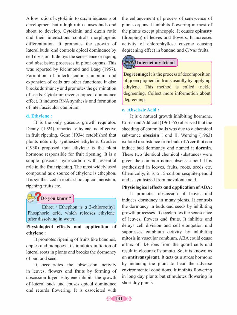
141
A low ratio of cytokinin to auxin induces root development but a high ratio causes buds and shoot to develop. Cytokinin and auxin ratio and their interactions controls morphogenic differentiation. It promotes the growth of lateral buds and controls apical dominance by cell division. It delays the senescence or ageing and abscission processes in plant organs. This was reported by Richmond and Lang (1957). Formation of interfasicular cambium and expansion of cells are other functions. It also breaks dormancy and promotes the germination of seeds. Cytokinin reverses apical dominance effect. It induces RNA synthesis and formation of interfascicular cambium.
d. Ethylene : It is the only gaseous growth regulator.
Denny (1924) reported ethylene is effective in fruit ripening. Gane (1934) established that plants naturally synthesize ethylene. Crocker (1930) proposed that ethylene is the plant hormone responsible for fruit ripening. It is a simple gaseous hydrocarbon with essential role in the fruit ripening. The most widely used compound as a source of ethylene is ethephon. It is synthesized in roots, shoot apical meristem, ripening fruits etc.
e. Abscissic Acid : It is a natural growth inhibiting hormone.
Carns and Addicott (1961-65) observed that the shedding of cotton balls was due to a chemical substance abscisin I and II. Wareing (1963) isolated a substance from buds of Acer that can induce bud dormancy and named it dormin. These two identical chemical substances were given the common name abscissic acid. It is synthesized in leaves, fruits, roots, seeds etc. Chemically, it is a 15-carbon sesquiterpenoid and is synthsized from mevalonic acid.
Physiological effects and application of ABA: It promotes abscission of leaves and
induces dormancy in many plants. It controls the dormancy in buds and seeds by inhibiting growth processes. It accelerates the senescence of leaves, flowers and fruits. It inhibits and delays cell division and cell elongation and suppresses cambium activity by inhibiting mitosis in vascular cambium. ABA could cause efflux of k+ ions from the guard cells and result in closure of stomata. So, it is known as an antitranspirant. It acts as a stress hormone by inducing the plant to bear the adverse environmental conditions. It inhibits flowering in long day plants but stimulates flowering in short day plants.
Physiological effects and application of ethylene :
It promotes ripening of fruits like bananas, apples and mangoes. It stimulates initiation of lateral roots in plants and breaks the dormancy of bud and seed.
It accelerates the abscission activity in leaves, flowers and fruits by forming of abscission layer. Ethylene inhibits the growth of lateral buds and causes apical dominance and retards flowering. It is associated with
Ethret / Ethephon is a 2-chloroethyl Phosphoric acid, which releases ethylene after dissolving in water.
Do you know ?
the enhancement of process of senescence of plants organs. It inhibits flowering in most of the plants except pineapple. It causes epinasty (drooping) of leaves and flowers. It increases activity of chlorophyllase enzyme causing degreening effect in banana and Citrus fruits.
Degreening: It is the process of decomposition of green pigment in fruits usually by applying ethylene. This method is called trickle degreening. Collect more information about degreening.
Internet my friend
142
7.10 Photoperiodism : Higher plants reproduce sexually by
producing special structures called flowers. Plants exhibit transition from vegetative growth to reproductive growth during which flowers are produced. Like vegetative growth, reproductive growth is also influenced by several environmental and nutritional factors. Among the environmental factors - light and temperature exert profound influence on flowering. The influence of light is known as Photoperiodism and that of temperature, is Vernalization.
Light as an environmental factor influences germination of seed, vegetative growth, photosynthesis, etc. Light as a factor as three aspects viz, Quality, Intensity and Duration of light. It is the duration of light that has profound effect on flowering in higher plants.
The term photoperiodism was used by Garner and Allard (1920). They were studying
the flowering behaviour in plants - Soyabean and Meryland mamoth variety of tobacco. They found that soyabean plant flowers during late summer and tobacco variety during winter, irrespective of their germination and growing season. They studied effects of different temperatures, nutrition, soil moisture, etc. in respect of flowering. None of these were found to regulate flowering. However, experimentally they found that the exposure to specific duration of light (i.e. photoperiod) had profound influence on flowering. They examined the effect of day length on flowering by using artificial illumination. They concluded that the relative length of the day was most crucial in the growth and development of flowers to which they coined the term photoperiodism. Based on the photoperiodic response, plants were classified in three categories viz, Short Day Plants (SDP), Long Day Plants (LDP) and Day Neutral Plants (DNP).
Organs for reception of photoperiodic stimulus - Leaf is the chief organ for receiving the photoperiodic stimulus as demonstrated by Knoff (1934). Defoliated plants will not flower even if the plants are exposed to proper duration of light.Photoperiodic stimulus - It is a chemical stimulus transported through phloem and is called florigen which is hormonal in nature. Photochemical receptors in the leaves are the biloproteins (pigments) located in the cell membrane. These are called Phytochromes. Blue wavelengths of light influences flowering.
Do you know ?
a. Short Day Plants (SDP) : These plants usually flower during winter
and late summer when day length is shorter than the critical photoperiod (critical photoperiod is that length of photoperiod above or below which flowering occurs). These are called
Bioassays for :Auxins - Avena curvature test.Gibberellins - Alpha amylase bioassay.Cytokinins - Chlorophyll retention test.Ethylene - Tripple pea test.ABA - Inhibition of alpha amylase test.
Do you know ?
Collect information of synthetic plant hormones.
Internet my friend
1. Why is auxin called a growth regulator?2. Effect of Gibberellin application on apple.3. How can we overcome apical dominance?4. Which is standard bioassay method for
auxins?5. ABA is called as stress hormone. Why?
Can you tell?
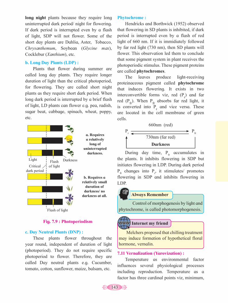
143
long night plants because they require long uninterrupted dark period/ night for flowering. If dark period is interrupted even by a flash of light, SDP will not flower. Some of the short day plants are Dahlia, Aster, Tobacco, Chrysanthemum, Soybean (Glycine max), Cocklebur (Xanthium), etc.
b. Long Day Plants (LDP) : Plants that flower during summer are
called long day plants. They require longer duration of light than the critical photoperiod, for flowering. They are called short night plants as they require short dark period. When long dark period is interrupted by a brief flash of light, LD plants can flower e.g. pea, radish, sugar beat, cabbage, spinach, wheat, poppy, etc.
c. Day Neutral Plants (DNP) : These plants flower throughout the
year round, independent of duration of light (photoperiod). They do not require specific photoperiod to flower. Therefore, they are called Day neutral plants e.g. Cucumber, tomato, cotton, sunflower, maize, balsam, etc.
Phytochrome : Hendricks and Borthwick (1952) observed
that flowering in SD plants is inhibited, if dark period is interrupted even by a flash of red light of 660 nm. If it is immidiately followed by far red light (730 nm), then SD plants will flower. This observation led them to conclude that some pigment system in plant receives the photoperiodic stimulus. These pigment proteins are called phytochromes.
The leaves produce light-receiving proteinaceous pigment called phytochrome that induces flowering. It exists in two interconvertible forms viz, red (P
r) and far
red (Pfr). When P
fr absorbs far red light, it
is converted into Pr and vice versa. These
are located in the cell membrane of green cells.
660nm (red)P
r P
fr
730nm (far red)
Darkness
During day time, Pfr accumulates in
the plants. It inhibits flowering in SDP but initiates flowering in LDP. During dark period P
fr changes into P
r, it stimulates/ promotes
flowering in SDP and inhibits flowering in LDP.
Control of morphogenesis by light and phytochrome, is called photomorphogenesis.
Always Remember
7.11 Vernalization (Yarovization) : Temperature as environmental factor
influences several physiological processes including reproduction. Temperature as a factor has three cardinal points viz, minimum,
Fig. 7.9 : Photoperiodism
a. Requires a relatively
long of uninterrupted
darkness.
b. Requires a relatively small
duration of darkness/ no
darkness at all.
Light
Critical dark period
Flash of light
Darkness
Flash of light
Melchers proposed that chilling treatment may induce formation of hypothetical floral hormone, vernalin.
Internet my friend

144
optimum and maximum temperature. It is a low temperature (chilling) treatment that induces early flowering in plants as was evidenced by Klippart (1918). Chouard (1960) defines vernalization as accelaration of the ability to flower by chilling treatment. The term vernalization was coined by T.D Lysenko (1928) for the effect of low temperature on flowering in plants.
It is an influence of temperature on development and flowering. Many plants such as cereals, crucifers require a period of cold treatment for flowering. It is the method of inducing early flowering in the plants by pretreatment to their seeds/ seedlings at low temperature (1-60C for one to one and half months’ duration). The site of vernalization is believed to be shoot apical meristem. Generally, vernalization is effective at seed stage in annual plants. Vernalization stimulus is also a chemical stimulus named as vernalin. This can be transferred through grafting (Melcher 1939).
Advantages of vernalization :• Crops can be produced earlier.• Crops can be cultivated in regions where
they do not grow naturally.
7.12 Mineral nutrition : Plant absorbs water, gases, mineral,
nutrients, etc. from surroundings. Green plants for the synthesis of their organic food need inorganic substances (elements) which are obtained from soil in the form of minerals. Minerals constitute most commonly occuring solid, inorganic materials obtained from the earth’s crust.
Chemical analysis of plant ash clearly indicates that plant absorbs mineral elements from surroundings (soil, air and water) for its use. About 36 to 40 different elements of periodic table are used as minerals by the plants. These are absorbed in ionic (dissolved) form as PO
4 , CO
3 , SO
4 , etc.,usually through
roots (regions of elongation and growth).
Sources of minerals : Plants derive necessary elements from the
atmosphere, soil and water. Carbon enters the plant as atmospheric carbon-dioxide.
Source of hydrogen is water and oxygen comes from air and water. Carbon, Hydrogen and oxygen are not minerals in origin.
Source of nitrogen is the soil. Plant derives nitrogen from both mineral and non-mineral origin.
Classificaion of minerals : Earlier, on the basis of their requirement
minerals were classified as essential and non-essential. Essential minerals are those that are indispensible without which plants can not complete their life cycle e.g. C, H, O, N, P, etc. These elements play structural and physiological roles. Their absence can produce/ cause major deficiency symptoms. The non-essential elements are not indispensable and they do not produce/ cause any deficiency symptoms. This classification is absolete now.
The reversion of vernalization by high temperature treatment is called devernalization.
Know the Scientist :
The low temperature induces early flowering which was first noticed by Klippart (1957) while working on two varieties of wheat- ‘winter wheat’ and ‘spring wheat’. The winter wheat sown in winter, produces flowers in the following summer. The spring wheat sown in the spring and produces flowers during summer. However, if winnter variety is sown in spring does not produce flowers during summer but does so in the next year’s summer. He observed that if germinated seeds of ‘winter wheat’ are treated with low temperature, it can behave as spring wheat. He concluded that winter wheat requires low - temperature exposure before the onset of flowering.
145
Symptoms of Mineral deficiency in plants : Any visible deviation from the normal
structure and function of the plant, is called symptom or hunger sign. The concentration of the essential elements below which plant growth is retarded, is termed as critical concentration.
The element is said to be deficient when present below the critical concentration. Certain morphological changes are indicative of the deficiency of particular element. Deficiency symptoms also depend on the mobility of the elements in the plants. The deficiency symptoms appear first in young tissues when elements are relatively immobile e.g. sulphur, calcium.
When the elements are actively mobilized within the plants and exported to young developing tissues, the deficiency symptoms are visible first in the older tissues (senescent leaves). e.g. nitrogen, magnesium, potassium.
Some important deficiency symptoms seen in plants are:• Stunting : The growth is retarded. The stem
appears condensed and short.• Chlorosis : It is the loss or non-development
of chlorophyll resulting in the yellowing of leaves
• Necrosis : It is the localized death of tissue of leaves.
• Mottling : Appearance of green and non-green patches on the leaves.
• Abscission : Premature fall of flowers, fruits and leaves.
Based on the quantity requirement, minerals are classified as minor or microelements and major or macroelements. Microelements are required in traces because they function in the catalytic role e.g. Zn, Cu, Al, Si, etc. as co-factors. Macroelements are required in large quantity. They mainly play the nutritive and structural roles e.g. C, H, O, P, Mg, N, K, S, etc. C, H, O are non mineral major elements. This classification is not accepted now.
Table : 7.10 Roles of Mineral Elements in Plants
ElementRegion of plant in
which requiredFunctions
Deficiency symptom
NitrogenNO-
2 or NO-
3
or NH+4
Everywhere particularly in meristematic tissues
Constituent of proteins, nucleic acids, vitamins, hormones, coenzymes, ATP, chlorophyll.
Stunted growth, chlorosis.
Phosporus H
2PO-
4 or
HPO2-4
Younger tissues, obtains from older, metabolically less active cells
Constituent of cell membrane, certain proteins, all nucleic acids and nucleotides required for all phosphorylation reactions.
Poor growth, leaves dull green.
Liebig showed the Essentiality of mineral nutrients and put forth the ‘Law of minimum’. According to this law, the yield of crop plant is determined by the amount of essential element that is present (available) in the minimum quantity.
Do you know ?
Now a days minerals are classified on the basis of their biochemical functions.
Criteria for Essentiality of elements proposed by Arnon and Stout (1939) : The elements must be absolutely necessary for supporting normal growth and reproduction. These elements must be having specific functions and should not be replaceable by another element. The element must be directly involved in the nutrition of the plant. If an element satisfies all above three criteria, it is considered to be an essential element.
Know the Scientist :

146
PotassiumK+
Meristematic tissues, buds, leaves, root tips
Helps in determining anion- cation balance in cells involved in protein synthesis, involved in formation of cell memberane and in opening and closing of stomta; increases hardness; activates enzymes and helps in maintenance of turgidity of cells.
Yellow edges to leaves, premature death.
CalciumCa2+
Meristematic and differentiating tissues, accumulates in older leaves
Involved in selective permeability of cell membranes, activates certain enzymes required for development of stem and root apex and as calcium pectate in the middle lamella of the cell wall.
stunted growth.
Magnesium Mg2+
Leaves, withdrawn from ageing leaves and exported to developing seeds
Activates enzymes in phosphate metabolism, constituent of chlorophyll, maintains ribosome structure.
Chlorosis
Sulphur SO
42-
Stem and root tips; young leaves remobilised during senescence
Constitutent of certain proteins, vitamins (thaimine, biotin CoA) and Ferredoxin.
Chlorosis
Iron Fe3+
Everywhere carries along leaf veins.
Constituents of ferredoxin and cytochrome, activates catalase required for synthesis of chlorophyll.
Chlorosis
Manganese (trace) Mn2+
Leaves and seeds Activates certain enzymes (carboxylases)Chlorosis, grey spots on leaves.
Molybdenum (Trace) MoO
22+
Everywhere, MO3+ particularly in roots
Activates certain enzymes in the nitrogen metabolism.
Slight retardation of growth.
Boron (trace) BO3-
3 or B
4O
72- Leaves and seeds
Required for uptake and utilisation of Ca2+, pollen germination and cell differentiation, carbohydrate translocation.
Brown heart disease.
Copper (trace) Cu2+ Everywhere Activates certain enzymes.
Die-back of shoots.
Zinc (trace) Zn2+ Everywhere
Activates various enzymes especially carboxylases, part of carbonic anhydrase and various dehydrogenases needed for auxin synthesis
Malfomred leaves
ChlorineCl- Everywhere
With Na+ and K+ helps to determine solute concentration and anion-cation balance in cells, essential for oxygen evolution in photosynthesis.
Poor growth of the plant

147
Toxicity of Micronutrients :
Mineral ion concentration which reduces the dry weight of tissues by 10%, is considered as toxic. Toxicity of one element may inhibit the uptake of the other e.g. Mn competes with Fe, Mg for uptake, but inhibits Ca translocation to shoot apex. Thus, Mn toxicity develops deficiency symptoms of Ca, Mg and Fe.
• Constituents of protoplasm and cell
walls : C,H,O,N,P,S are important and permanent constituents of the protoplasm and the cell wall.
• Osmotic potential : The osmotic pressure of cell is the result of mineral salts and organic compounds dissolved in the cell sap.
• Catalytic functions : Many elements like Fe, Cu, Zn, Mg, Mn, Cl, etc. act as catalysts in various enzyme reaction of plant metabolism.
• Permeability of cytoplasmic membrane: Permeability of membrane is influenced by the presence of the various ‘cations’ and ‘anions’ of the mineral elements in the external medium like ca+, k+.
• Components of energy related chemical compounds : Phosphorus in ATP and Mg in chlorophyll.
Do you know ?
The atmospheric CO2 is virtually
the only source of carbon, which is the basic constituent of all the organic compounds.
Do you know ?
Minerals salt absorption : Most minerals in the soil are charged
particles hence, they can not pass across cell membrane. Hence most of the minerals are absorbed actively with the expenditure energy. Minerals can also be absorbed passively without expenditure of energy. Mineral ion absorption is independent of water absorption.
Mineral ion absorption can occur in two ways : a. Passive Absorption : Movement of mineral ions into the root occurs by diffusion. Molecules or ions diffuse from a region of their higher concentration to a region of their lower concentration. The movement of mineral ions into root cells as a result of diffusion is without expenditure of energy is called passive absorption. Passive absorption can take place by direct ion-exchange, in direct ion-exchange mass flow and Donnan equilibrium.
b. Donnan equilibrium : It is based on the assumption that certain negatively charged ions, after their entry into the cell, become fixed on the inner side of the cell membrane and can not diffuse outside through the cell membrane. Therefore, additional mobile cations are required to balance these fixed anions. Obviously concentration of cations become more due to accumulation. This kind of passive absorption of anions/ cations from cell exterior against their own concentration gradient in order to neutralize the effect of cations/ anions, is called Donnan equilibrium.
Active Absorption: Uptake of mineral ions against concentration gradient, is called active absorption, such movement requires an expenditure of energy by the absorbing cell. This energy is derived from respiration and is supplied through ATP. When the roots are deprived of oxygen, they show a sudden drop in active absorption of minerals. The mineral ions accumulated in the root hair pass into the cortex and finally reach the xylem.
Internet my friend
• Collect information about hydroponics and find its use in kitchen garden.
• Collect information on Aeroponics.

148
The minerals in the xylem are then carried along with water to other parts of the plant along the transpiration stream and are subsequently assimilated into organic molecules and then redistributed to other parts of the plant through the phloem.
To explain active absorption of minerals Hoagland and Davis (1923) put forth Carrier hypothesis. Specific proteins in the membrane of root cells actively pump ions into the cytoplasm of epidermal cells of root. These proteins are called carriers that pump both cations and anions from the soil. According to Bennet and Clarke (1956) protein conjugated with lecithin acts as carrier.
Do you know ?
7.13 Nitrogen cycle: It is series of natural processes by which
Nitrogen enters successively from air to organisms through soil and back to environment. Plants use photosynthetic product, the sugars to make proteins. To do this, they need nitrogen. Unfortunately, it is very innert (nonreactive). Plants need nitrogen in a reactive form usually as nitrate ions.
Fig. 7.11 Nitrogen cycle
Dentrifying bacteria in soil
Nitrates, nitrites in the soil
Nitrifying bacteria in the soil
NH3 in soil
Ammonifying bacteria in the soil
Combustion
Nitrogenous organic matter in soil
Excreta Death
Death Animals
Food Plant proteins
Amino acids in plants
NH3 in plants
Nitrates in the soil Commercial fertilisers in
soil
Non biological nitrogen fixation
Biological nitrogen fixation
Atm. N2
Nitrogen is a limiting nutrient in the agricultural ecosystems. It exists as nitrogen atoms with a strong triple covalent bond (N ≡ N). A regular supply of nitrogen to the plants is maintained through biological and physical process.
Nitrogen fixation : Atmosphere is the source of nitrogen. It
can not be used directly. It combines with C, H, N and O to form compounds before being used. Conversion of free nitrogen (N
2) of the
atmosphere into nitrogenous salts to make it available for the plants, is called nitrogen fixation. It is of two types : Physical and Biological fixation.
Physical Nitrogen fixation: It occurs in several steps and starts with combination of atmospheric nitrogen with oxygen under the influence of electric discharge and thunder storm produce nitric oxide.
Electric discharge N
2 + O
2 2 NO
Nitrogen oxygen Thunder storm Nitric oxide
The nitric oxide is then oxidized to nitrogen peroxide in the presence of oxygen.
Oxidation 2NO + O
2 2NO
2
Nitrogen peroxide
During rains, the nitrogen peroxide combines with rain water to form nitrous acid and nitric acid which come to ground along with rains.
2NO2 + Rain water HNO
2 + HNO
3
Nitrous Acid Nitric Acid
On ground, the alkali radicals of soil react with nitric acid to produce nitrites and nitrates.(absorbable form)
HNO3 + Ca or K salts Ca or K nitrates
Industrial nitrogen fixation. It occurs by Haber-Bosch nitrate process at high temperature and pressure.
4500cN
2 + 3H
2 2NH
3 200 atm AmmoniaAmmonia is then converted to urea as it is less toxic.

149
Biological Nitrogen fixation : It is carried out by prokaryotes called as ‘Nitrogen fixers’ or Diazotrophs’. It accounts nearly 70% of natural nitrogen fixation. Nitrogen fixers are either symbiotic or free living. The cyanobacteria fix significant amount of nitrogen in specialized cells called heterocysts.
Nitrogen fixation is high energy requiring process and nitrogen fixers use 16 molecules of ATP to fix each molecule of nitrogen to form ammonia.
N2 + 8H+ +8e- + 16ATP 2NH
3 +H
2 +
16ADP + 16Pi
Ammonia is then converted into amino acids.
Nitrification : Most of the soil bacteria participate in
converting ammonia into nitrate, the form of nitrogen which can be used by plants and animals. This involves two steps performed by two different types of bacteria.
First a soil bacteria convert ammonia into nitrogen-di-oxide (nitrite) eg. Nitrosomonas, Nitrosococcus, etc.
2NH3 + 3O
2
Nitrosomonas
Nitrosococcus 2HNO
2 + 2 H
2O
Then another type of soil bacterium called Nitrobacter adds a third oxygen atoms to create nitrate.
2HNO2 + O
2
Nitrobacter 2HNO
3
These bacteria are chemoautotrophs. By metabolizing nitrogen along with oxygen, they obtain energy to power their own life processes.
Today nearly 80% of nitrogen found in human tissues originate from the Haber-Bosch process.
Always Remember
Symbiotic N2 fixation :
The best known nitrogen fixing symbiotic bacterium is Rhizobium. This soil living/ dwelling bacterium forms root nodules in plants belonging to family Fabaceae e.g. beans, gram, groundnut etc.
Ammonification : After the death of plants and animals,
various fungi, actinomycetes and some ammonifying bacteria decompose the tissues and convert organic nitrogen into amino acid and then to ammonia and back into the ecosystem. Ammonia (NH
4+) is now available
for uptake by plants and other micro-organisms for growth.
Proteins Microbial
decomposition amino acids
Amino acids NH
3 + ROH
Ammonia organic acidNitrogen assimilation :
In soil, nitrogen is present as nitrates, nitrites and ammonia (NH
4+). It is obsorbed by
the green plants and converted to nitrogenous organic compounds like amino acids, DNA, etc. This is known as nitrogen assimilation. From plants, nitrogen as biomolecules like amino acids, enters food chain and moves to animals and then to decomposers through the death of animals.
Nitrates are first converted to ammonia but it is highly toxic and immediately used for conversion into amino acids, which are then transported to other parts of the plants for synthesis of proteins.
NO3- + 2e- + 2H+ NO
2- + H
2O
NO2- +8e- + 8H+ NH
4+ + 2H
2O
Amino Acid synthesis : Amino acids are building blocks of proteins.
The amino acids are synthesized through : Reductive amination : Ammonia reacts with alpha ketoglutaric acid to form glutamic acid (glutamate).
Internet my friend
Enlist names of symbiotic and free living nitrogen fixing bacteria.

150
α Ketoglutaric acid + NH4 + NADPH
2
Glutamate dehydrogenase Glutamate +
H2O+ NADP
Transamination : Amino group of one amino acid (-CHNH
2) is transfered to keto position
(-CO) of other carboxylic acid.
Glutamic acid + oxaloacetic acid Glutamate aspertate
Aminotransferase α - Ketoglutaric acid +
Aspartic acid
Amides : Ammonia may be absorbed by amino
acid to produce amides. The process is called amidation. The amides are the amino acids having two amino groups. Extra amino group is attached to acidic group (-COOH) in presence of ATP.Glutamic acid + NH
4++ ATP alpha
glutamine + ADP
Aspartic acid + NH4
++ ATP Aspargine + ADP
Amides like asparagine and glutamine are formed from glutamic acid and aspartic acid respectively by addition of another amino group to each. Amides are transported to other parts of plants via xylem vessels.
Denitrification : It is the process in which anaerobic bacteria
can convert soil nitrates back into nitrogen gas. Denitrifying bacteria removes fixed nitrogen i.e. nitrates from the ecosystem and return it to the atmosphere in inert form.
Denitrifying bacteria includes Bacillus spp., Paracoccus spp. and Pseudomonas denitrificans. They transform nitrates to nitrous and nitric oxides and ultimately to gaseous nitrogen.
2NO3 2NO
2 2NO N
2
Sedimentation : Nitrates of the soil are washed away to the sea or leached deep into the earth along with percolating water.
1. Soil nitrogen is replenished by excretion of animals, (as ammonia, urea and uric acid)
ammonification and nitrification.2. Plastids contain nitrite reductase which reduces it to ammonia.
Do you know ?
Activity :
Label the following diagrams and identify the types of germination.

151
Q. 1 Multiple choice questions.1. Which of the hormones can replace
vernalization? a. Auxin b. Cytokinin c. Gibberellins d. Ethylene
2. The principle pathway of water translocation in angiosperms is ..........
a. Sieve cells b. Sieve tube elements c. Xylem d. Xylem and phloem
3. Abscisic acid controls ..................a. cell divisionb. leaf fall and dormancyc. shoot elongationd. cell elongation and wall formation
4. Which is employed for artificial ripening of banana fruits?
a. Auxin b. Ethylene c. Cytokinin d. Gibberellin
5. Which of the following is required for stimulation of flowering in the plants?
a. Adequate oxygen b. Definite photoperiod c. Adequate water d. Water and minerals
6. For short day plants, the critical period is .................
a. light b. dark/ night c. uv rays d. both a and c
7. Which of the following is day neutral plant?a. Tomatob. Cottonc. Sunflowerd. Soybean
8. Essential macroelements are ............ a. manufactured during photosynthesis b. produced by enzymes c. absorbed from soil d. produced by growth hormones
9. Function of Zinc is ................a. closing of stomatab. biosynthesis of 3-IAAc. synthesis of chlorophylld. oxidation of carbohydrates
10. Necrosis means ...............a. yellow spots on the leavesb. death of tissuec. darkening of green colour in leaves d. wilting of leaves
11. Conversion of nitrates to nitrogen is called .....................a. ammonificationb. nitrificationc. nitrogen fixationd. denitrification
12. How many molecules of ATP are required to fix one molecule of nitrogen?
a. 12 b. 20 c. 6 d. 16
Q. 2 Very Short Answer Questions :1. Enlist the phases of growth in plants?2. Give the full form of IAA?3. What does it mean by ‘open growth’?4. Which is the plant stress hormone?5. What is denitrification?6. Name the bacteria responsible for
conversion of nitrite to nitrate.7. What is role of gibberellin in rosette
plants? 8. Define vernalization.9. Define photoperiodism.10. What is grand period of growth?
Exercise

152
Q. 3 Short Answer Questions :1. Write a short note on : a. Differentiation b. Redifferentiation2. Differentiate between Arithmetic and
Geometric growth.3. Enlist the role and deficiency symptoms
of : a. Nitrogen b. Phosphorus c. Potassium
4. What is short day plant? Give any two examples.
5. What is vernalization? Give its significance.
Q. 4 Long Answer Questions :1. Explain sigmoid growth curve with the
help of diagram.2. Describe the types of plants on the basis
of photoperiod required, with the help of suitable examples.
3. Explain biological nitrogen fixation with example.
Project :
1. Grow seed in the deep tray on soil medium and study different stages of germination. Prepare powerpoint presentation with the help of pictures/photographs of the same.
2. Prepare chart differentiating the epigeal and hypogeal germination.
3. Collect the information about the viviparous germination in plants growing along seashore.
4. Identify and label the deficiency symptoms in the given diagram.

153
1. Definition of respiration. 2. The types of cellular respiration.3. Why should we respire all the time?
Can you recall?
All living organisms require energy to carry out various life processes. The energy that is stored in the body in the form of complex organic compounds (potential energy) is however not usable by the organisms unless it is converted into usable form. This conversion is achieved through the process of respiration.
Respiration : It is a biochemical process of oxidation of organic compounds in an orderly manner for the liberation of chemical energy in the form of ATP.
C6H
12O
6 + 6O
2 6 CO
2 + 6H
2O + 38 ATP
For this, the process of gaseous exchange takes place between the organism and the environment. The site of gaseous exchange is called the respiratory surface.
Respiration in Animals : As compared to plants, animals show wide
variety of respiratory surfaces or organs. The respiratory surfaces differ in various animals. In animals, depending upon the complexity of organization and the surrounding medium, certain parts of the body have become specialized into different types of respiratory organs. In the higher animals, these respiratory organs are also associated with a transport system.
the cells of stem, leaf and root. These air spaces are continuous. Oxygen diffuses into the air space through stomata (the pores on leaves and young stems), carbon dioxide and water vapour diffuse out. In the aerated soil, the oxygen dissolves in the film of moisture or water around the root tissue and enters it by diffusion.
Woody flowering plants (trees and shrubs) have an external impervious bark. Here, gaseous exchange occurs through small pores in the stem surface, called lenticels.
What would happen if respiration take place in one single step?
Use your brain power
8.1 Organs of Respiratory Exchange : Respiratory exchange is a simple physical
process. For efficient gaseous exchange, the respiratory surface should have the following features :
a. It should have a large surface area. b. It should be thin, highly vascular and
permeable to allow exchange of gases.c. It should be moist.
Gaseous exchange in plants : The shape and structure of plants facilitate
gaseous exchange by diffusion. A terrestrial flowering plant has many air spaces between
Fig. 8.1 : Organs of gaseous exchange in plants
Stomatal pore
Stoma Leaf
Bark
Lenticels
Curiosity Box :
1. What are the main features of respiratory surface?
2. Which are the parts of plant help in the process of gaseous exchange?
Respiration and Circulation8
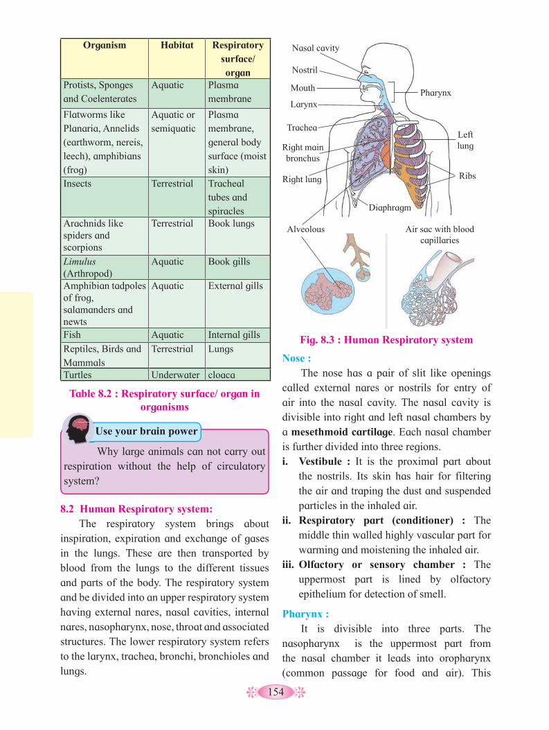
154
Organism Habitat Respiratory surface/ organ
Protists, Sponges and Coelenterates
Aquatic Plasma membrane
Flatworms like Planaria, Annelids (earthworm, nereis, leech), amphibians (frog)
Aquatic or semiquatic
Plasma membrane, general body surface (moist skin)
Insects Terrestrial Tracheal tubes and spiracles
Arachnids like spiders and scorpions
Terrestrial Book lungs
Limulus (Arthropod)
Aquatic Book gills
Amphibian tadpoles of frog,salamanders and newts
Aquatic External gills
Fish Aquatic Internal gills
Reptiles, Birds and Mammals
Terrestrial Lungs
Turtles Underwater cloaca
Table 8.2 : Respiratory surface/ organ in organisms
8.2 Human Respiratory system: The respiratory system brings about
inspiration, expiration and exchange of gases in the lungs. These are then transported by blood from the lungs to the different tissues and parts of the body. The respiratory system and be divided into an upper respiratory system having external nares, nasal cavities, internal nares, nasopharynx, nose, throat and associated structures. The lower respiratory system refers to the larynx, trachea, bronchi, bronchioles and lungs.
Nose : The nose has a pair of slit like openings
called external nares or nostrils for entry of air into the nasal cavity. The nasal cavity is divisible into right and left nasal chambers by a mesethmoid cartilage. Each nasal chamber is further divided into three regions.i. Vestibule : It is the proximal part about
the nostrils. Its skin has hair for filtering the air and traping the dust and suspended particles in the inhaled air.
ii. Respiratory part (conditioner) : The middle thin walled highly vascular part for warming and moistening the inhaled air.
iii. Olfactory or sensory chamber : The uppermost part is lined by olfactory epithelium for detection of smell.
Pharynx : It is divisible into three parts. The
nasopharynx is the uppermost part from the nasal chamber it leads into oropharynx (common passage for food and air). This
Why large animals can not carry out respiration without the help of circulatory system?
Use your brain power
Fig. 8.3 : Human Respiratory system
Pharynx
Left lung
Nasal cavity
Nostril
Mouth
Larynx
Trachea
Right main bronchus
Right lung
Diaphragm
Ribs
Alveolous Air sac with blood capillaries
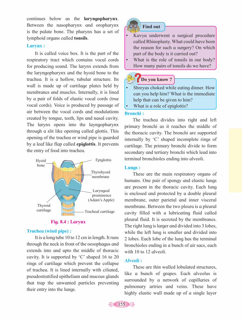
155
• Kavya underwent a surgical procedure
called Rhinoplasty. What could have been the reason for such a surgery? On which part of the body is it carried out?
• What is the role of tonsils in our body? How many pairs of tonsils do we have?
Find out
• Shreyas choked while eating dinner. How
can you help him? What is the immediate help that can be given to him?
• What is a role of epiglottis?
Do you know ?
Trachea (wind pipe) : It is a long tube 10 to 12 cm in length. It runs
through the neck in front of the oesophagus and extends into and upto the middle of thoracic cavity. It is supported by ‘C’ shaped 16 to 20 rings of cartilage which prevent the collapse of trachea. It is lined internally with ciliated, pseudostratified epithelium and mucous glands that trap the unwanted particles preventing their entry into the lungs.
Bronchi : The trachea divides into right and left
primary bronchi as it reaches the middle of the thoracic cavity. The bronchi are supported internally by ‘C’ shaped incomplete rings of cartilage. The primary bronchi divide to form secondary and tertiary bronchi which lead into terminal bronchioles ending into alveoli.
Lungs : These are the main respiratory organs of
humans. One pair of spongy and elastic lungs are present in the thoracic cavity. Each lung is enclosed and protected by a double pleural membrane, outer parietal and inner visceral membrane. Between the two pleura is a pleural cavity filled with a lubricating fluid called pleural fluid. It is secreted by the membranes. The right lung is larger and divided into 3 lobes, while the left lung is smaller and divided into 2 lobes. Each lobe of the lung has the terminal bronchioles ending in a bunch of air sacs, each with 10 to 12 alveoli.
Alveoli : These are thin walled lobulated structures,
like a bunch of grapes. Each alveolus is surrounded by a network of capillaries of pulmonary artries and veins. These have highly elastic wall made up of a single layer
continues below as the laryngopharynx. Between the nasopharynx and oropharynx is the palate bone. The pharynx has a set of lymphoid organs called tonsils.
Larynx :
It is called voice box. It is the part of the respiratory tract which contains vocal cords for producing sound. The larynx extends from the laryngopharynx and the hyoid bone to the trachea. It is a hollow, tubular structure. Its wall is made up of cartilage plates held by membranes and muscles. Internally, it is lined by a pair of folds of elastic vocal cords (true vocal cords). Voice is produced by passage of air between the vocal cords and modulations created by tongue, teeth, lips and nasal cavity. The larynx opens into the layngopharynx through a slit like opening called glottis. This opening of the trachea or wind pipe is guarded by a leaf like flap called epiglottis. It prevents the entry of food into trachea.
Fig. 8.4 : Larynx
Hyoid bone
Epiglottis
Thyrohyoid membrane
Thyroid cartilage Tracheal cartilage
Laryngeal prominence
(Adam’s Apple)
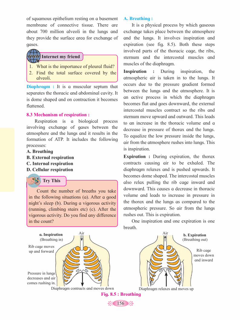
156
Diaphragm : It is a muscular septum that separates the thoracic and abdominal cavity. It is dome shaped and on contraction it becomes flattened.
8.3 Mechanism of respiration : Respiration is a biological process
involving exchange of gases between the atmosphere and the lungs and it results in the formation of ATP. It includes the following processes:A. Breathing B. External respirationC. Internal respirationD. Cellular respiration
A. Breathing : It is a physical process by which gaseous
exchange takes place between the atmosphere and the lungs. It involves inspiration and expiration (see fig. 8.5). Both these steps involved parts of the thoracic cage, the ribs, sternum and the intercostal muscles and muscles of the diaphragm.
Inspiration : During inspiration, the atmospheric air is taken in to the lungs. It occurs due to the pressure gradient formed between the lungs and the atmosphere. It is an active process in which the diaphragm becomes flat and goes downward, the external intercostal muscles contract so the ribs and sternum move upward and outward. This leads to an increase in the thoracic volume and a decrease in pressure of thorax and the lungs. To equalize the low pressure inside the lungs, air from the atmosphere rushes into lungs. This is inspiration.
Expiration : During expiration, the thorax contracts causing air to be exhaled. The diaphragm relaxes and is pushed upwards. It becomes dome shaped. The intercostal muscles also relax pulling the rib cage inward and downward. This causes a decrease in thoracic volume and leads to increase in pressure in the thorax and the lungs as compared to the atmospheric pressure. So air from the lungs rushes out. This is expiration.
One inspiration and one expiration is one breath.
of squamous epithelium resting on a basement membrane of connective tissue. There are about 700 million alveoli in the lungs and they provide the surface area for exchange of gases.
Fig. 8.5 : Breathing
Rib cage moves down and inward
Air Air
Rib cage moves up and forward
Diaphragm contracts and moves down Diaphragm relaxes and moves up
Pressure in lungs decreases and air comes rushing in.
a. Inspiration b. Expiration(Breathing in) (Breathing out)
Try This
Internet my friend
1. What is the importance of pleural fluid?2. Find the total surface covered by the
alveoli.
Count the number of breaths you take in the following situations (a). After a good night’s sleep (b). During a vigorous activity (running, climbing stairs etc) (c). After the vigorous activity. Do you find any difference in the count?

157
Pulmonary volumes and capacities (Normal values)
Lung Volumes :Tidal volume (T.V.) : It is the volume of air inspired or expired during normal breathing. It is 500 ml.Inspiratory reserve volume (IRV) : The maximum volume of air, or the extra volume of air, that is inspired during forced breathing in addition to T.V. Its value is 2000 to 3000ml.Expiratory reserve volume (ERV) : The maximum volume of air that is expired during forced breathing after normal expiration. Its value is 1000 to 1100ml.Dead space (DS) : The volume of air that is present in the respiratory tract (from nose to the terminal bronchioles), but not involved in gaseous exchange. It is 150 ml.Residual volume (RV) : The volume of air that remains in the lungs and the dead space even after maximum expiration. It is 1100 to 1200ml.Lung capacities :Total Lung capacity : The maximum amount of air that the lungs can hold after a maximum forcefull inspiration (5200 to 5800ml).Vital capacity (VC) : The maximum amount of air that can be breathed out after a maximum inspiration. It is the some total of TV, IRV and ERV and is 4100 to 4600ml.
B. External respiration/ Exchange of gases at the alveolar level :
An alveolus consists of a layer of simple squamous epithelium resting on a basement membrane. It is intimately associated with a dense network of capillaries. The capillary wall is also made up of simple squamous epithelium resting on a thin basement membrane. Both the layers have similar structure and are thin walled. Together they make up the respiratory membrane through which gaseous exchange occurs i.e. between the alveolar air and the blood.
Diffusion of gases will take place from an area of higher partial pressure to an area of lower partial pressure until the partial pressure in the two regions reaches equilibrium.
The partial pressure of carbon-dioxide of blood entering the pulmonary capillaries is 45 mmHg while partial pressure of carbon-dioxide in alveolar air is 40 mmHg. Due to this difference, carbon dioxide diffuses from the capillaries into the alveolus.
Similarly, partial pressure of oxygen of blood in pulmonary capillaries is 40 mmHg while in alveolar blood it is 104 mmHg. Due to this difference oxygen diffuses from alveoli to the capillaries.
Fig. 8.6 : Exchange of gases between alveolus and capillary
blood from pulmonary
artery
Capillary
Alveolar membrane
Respiratory membrane
Surface fluid
(Air)
O2
CO2
to pulmonary vein
Oxygen diffuses into red blood cells
Carbon dioxide diffuses into alveolus
Alveolus
C. Internal respiration : The two main components of blood
involved in transport of the respiratory gases- CO
2 and O
2, are the RBCs and the plasma.
i. Transport of oxygen : Of the total oxygen transported only
3% is transported in a dissolved state by the plasma. The remaining 97% is bound to the haemoglobin (Hb) present in the RBCs.
blood goes
Why does gas exchange in the alveolar
region very rapid?
Use your brain power
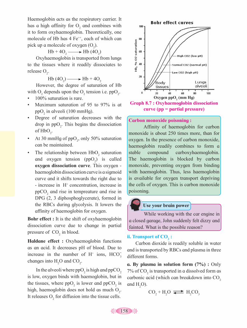
158
While working with the car engine in a closed garage, John suddenly felt dizzy and fainted. What is the possible reason?
Use your brain power
Haemoglobin acts as the respiratory carrier. It has a high affinity for O
2 and combines with
it to form oxyhaemoglobin. Theoretically, one molecule of Hb has 4 Fe++, each of which can pick up a molecule of oxygen (O
2).
Hb + 4O2 Hb (4O
2)
Oxyhaemoglobin is transported from lungs to the tissues where it readily dissociates to release O
2.
Hb (4O2) Hb + 4O
2
However, the degree of saturation of Hb with O
2 depends upon the O
2 tension i.e. ppO
2.
• 100% saturation is rare.• Maximum saturation of 95 to 97% is at
ppO2 in alveoli (100 mmHg).
• Degree of saturation decreases with the drop in ppO
2. This begins the dissociation
of HbO2.
• At 30 mmHg of ppO2, only 50% saturation
can be maintained.
• The relationship between HbO2 saturation
and oxygen tension (ppO2) is called
oxygen dissociation curve. This oxygen - haemoglobin dissociation curve is a sigmoid curve and it shifts towards the right due to - increase in H+ concentration, increase in ppCO
2 and rise in tempreature and rise in
DPG (2, 3 diphosphoglycerate), formed in the RBCs during glycolysis. It lowers the affinity of haemoglobin for oxygen.
Bohr effect : It is the shift of oxyhaemoglobin dissociation curve due to change in partial pressure of CO
2 in blood.
Haldane effect : Oxyhaemoglobin functions as an acid. It decreases pH of blood. Due to increase in the number of H+ ions, HCO-
3
changes into H2O and CO
2.
In the alveoli where ppO2 is high and ppCO
2
is low, oxygen binds with haemoglobin, but in the tissues, where ppO
2 is lower and ppCO
2 is
high, haemoglobin does not hold as much O2.
It releases O2 for diffusion into the tissue cells.
ii. Transport of CO2 :
Carbon dioxide is readily soluble in water and is transported by RBCs and plasma in three different forms.
a. By plasma in solution form (7%) : Only 7% of CO
2 is transported in a dissolved form as
carbonic acid (which can breakdown into CO2
and H2O).
CO2 + H
2O H
2CO
3
Graph 8.7 : Oxyhaemoglobin dissociation curve (pp = partial pressure)
Oxygen ppO2 (mm Hg)
Carbon monoxide poisoning : Affinity of haemoglobin for carbon monoxide is about 250 times more, than for oxygen. In the presence of carbon monoxide, haemoglobin readily combines to form a stable compound carboxyhaemoglobin. The haemoglobin is blocked by carbon monoxide, preventing oxygen from binding with haemoglobin. Thus, less haemoglobin is available for oxygen transport depriving the cells of oxygen. This is carbon monoxide poisoning.

159
b. By bicarbonate ions (70%) : Nearly 70% of carbondioxide released by the tissue cells diffuses into the plasma and then into the RBCs. • In the RBCs, CO
2 combines with water in
the presence of a Zn containing enzyme, carbonic anhydrase to form carbonic acid.
• Carbonic anhydrase enzyme is found in the RBCs and not in the plasma.
• The rate of formation of carbonic acid inside the RBC is very high as compared to its formation in the plasma.
• Carbonic acid being unstable almost immidiately dissociates into HCO-
3
and H+ in the presence of the enzyme carbonic anhydrase (CA) leading to large accumulation of HCO-
3 inside the RBCs.
CO2 + H
2O
CA H
2CO
3
CA H+ + HCO-
3
• It thus moves out of the RBCs. This would bring about inbalance of the charge inside
Fig. 8.8 : Oxygen pickup and carbon dioxide pickup at the tissue
Tissue cell
Interstitial fluid
CO2
CO2
CO2
CO2
CO2
CO2
CO2
O2
O2
CO2+H
2O slow H
2CO
3 HCO
3- + H+
HCO3-
Cl- CO
2+H
2O fast
Carbonic anhydrase
H2CO
3 HCO
3- + H+
CO2
+ Hb HbCO2 (Carbaminohaemoglobin)
Cl-
HHb
HbO3
O2 + Hb Red blood cell
Binds to plasms proteins
Chloride shift (In) via
transport protein
Blood plasma O2 (dissolved in plasma)
Fig. 8.9 : Oxygen pickup and carbon dioxide release in the lungs
Alveolus Fused basement membranes
CO2
CO2
CO2
CO2
O2
O2
CO2+ H
2O
slowH
2CO
3 HCO
3- + H+
HCO3-
Cl- CO
2+H
2O
fast
Carbonic anhydrase
H2CO
3 HCO
3- + H+
CO2
+ Hb HbCO2
Cl-
O2+ HHb HbO
2 + H+Red blood cell
Chloride shift (Out) via transport protein
Blood plasma O2 (dissolved in plasma)
CO2 (dissolved in plasma)
(Carbamino-haemoglobin)
the RBCs. To maintain the ionic balance between the RBCs and the plasma, Cl- diffuses into the RBCs. This movement of chloride ions is known as chloride shift or Hamburger’s phenomenon.
• HCO3- that comes into the plasma joins
to Na+ / K+ forming NaHCO3 / KHCO
3 (to
maintain pH of blood).
HCO-
3 + Na+ NaHCO
3
Sodium bicarbonate • H+ is taken up by protein (haemoglobin).
Hb + H+
HHb (Reduced Hb)
These H+ ions might be expected to lower
blood pH, but they are buffered by haemoglobin by the formation of deoxyhaemoglobin (reduced haemoglobin).• At the level of the lungs in response to
the low partial pressure of carbon dioxide (ppCO
2) of the alveolar air, hydrogen ion

160
and bicarbonate ions recombine to form carbonic acid and under the influence of carbonic anhydrase yields carbon dioxide and water.
H+ + HCO-
3
CA H
2CO
3
CA CO
2 + H
2O
c. By red blood cells (23%) : Carbon dioxide binds with the amino group of the haemoglobin and form a loosely bound compound carbaminohaemoglobin. This molecule readily decomposes in region where the partial pressure of carbon dioxide (ppCO
2) is low (alveolar
region), releasing the carbon dioxide.
Hb + CO2 HbCO
2
1. What is the role of haemoglobin in the
transport of oxygen in the blood?2. Write a note on chloride shift.
Use your brain power
neurons in the medulla (inspiratory center), ventro lateral group of neurons in medulla (inspiratory and expiratory center) and pneumotaxic center located in pons (primarily limits inspiration, slow wave sleep and rapid eye movement sleep). Apneustic center in the medulla is antagonistic to the neumotaxic center. It controls non rapid eye movement sleep and wakefullness.
Fig. 8.10 : Regulation of Breathing
Brain
Breathing control centers
Breathing control centers stimulated by :
a. CO2 increase in
blood (acts directly)
b. Nerve impulse from O
2 sensor
indicating O2
decrease
O2 sensors
in artery (aortic arch)
Diaphragm
Intercostal muscles
Phrenic nerves
Intercostal nerves
Nerve impulses trigger
contraction of muscles
Pons
Medulla
D. Cellular Respiration : It is the last step taking place inside the cell
where food is oxidized and ATP is generated. It can be shown by two steps:
a. Oxidation : Breaking down of complex organic molecules into simple inorganic molecules with release of heat energy.
C6H
12O
6 + 6O
2 6CO
2 + 6H
2O + 686 Kcal
b. Phosphorylation : It involves trapping the heat energy in the form of high energy bond of ATP molecule. ATP is used to carry out vital life processes and so is called as energy currency of the cell.
ADP + iP + 7.3 Kcal ATP
8.4 Regulation of Breathing : Respiration is under dual control : nervous
and chemical. Human adults breathe about 12 times/minute while a new born about 44 times/minute. Normal breathing is an involuntary process. Steady rate of respiration is controlled by neurons located in the pons and medulla and are known as the respiratory centres. It regulates the rate and depth of breathing. It is divided into three groups : dorsal group of
During inspiration when the lungs expand to a critical point, the stretch receptors are stimulated and impulses are sent along the vagus nerves to the expiratory centre. It then sends out inhibitory impulses to the inspiratory center.
The inspiratory muscles relax and expiration follows. As air leaves the lungs during expiration, the lungs are deflated and the stretch receptors are no longer stimulated. Thus, the inspiratory centre is no longer inhibited and a new respiration begins. These

161
events are called the Hering-Breuer reflex. The Hering-Breuer reflex controls the depth and rhythm of respiration. It also prevents the lungs from inflating to the point of bursting.
The respiratory centre has connections with the cerebral cortex which means we can voluntarily change our pattern of breathing. Voluntary control is protective because it enables us to prevent water or irritating gases from entering the lungs. But the ability to stop breathing is also limited by the build up of carbon dioxide in the blood.8.5 Modified Respiratory Movements :
Some respiratory movements are different from the normal movements and help express emotion or clear the air passage. Of these
Disorder Symptoms Cause Treatment
Emphysema Breakdown of alveoli, shortness of breath
Smoking, air pollution
Quit smoking, avoid polluted air, administer oxygen to relieve symptoms
Chronic bronchitis Coughing, shortness of breath Smoking, air
pollution
Quit smoking, avoid polluted air, if possible move to warmer, drier climate
Acute bronchitis
Inflammation of bronchi, shortness of breath, yellow mucous coughed up.
Viruses and bacteria
If bacterial, take antibiotics, cough medicine, use vaporizer
Sinusitis Inflammation of the sinuses, mucous discharge
Viruses and bacteria
If bacterial, take antibiotics and decongestants, use vaporizer
Laryngitis
Inflammation of larynx, vocal cords, sore throat, hoarseness of voice, mucous build up and cough
Viruses and bacteria
If bacterial, take antibiotics, cough medicines, voice rest, avoid irritants like smoke
Pneumonia
Inflammation of lungs ranging from mild to severe, cough and fever, shortness of breath, chills, sweating, chest pain, blood in mucous
Bacteria, virusesConsult physician immediately, antibiotics, cough medicines, stay warm
Asthma
Constriction of bronchioles, mucus build up in bronchioles, periodic wheezing, difficulty in breathing.
Allergy to pollen, some foods, food additives, pet hair, etc.
Use of inhalants to open passage ways, avoid irritants
Occupational Respiratory Disorders- silicosis, asbestosis
Inflammation fibrosis, lung damage.
Long term exposure to dust particles silica and asbestos, particles during occupation
Protective mask and gear during work.
Table 8.11 : Common disorders of respiratory system
movements some may be reflexes, but others can be initiated voluntarily e.g. coughing and yawning.
Internet my friend
1. Find out information about the various modified respiratory movements and write it in a tabular form.
2. What is the significance of such movements?
8.6 Common disorders of respiratory system: The given table shows a list of some
common respiratory disorders, their symptoms, cause and treatment.

162
Artificial ventilation : It is also called artificial respiration. It is the
method of inducing breathing in a person when natural respiration has ceased or is faltering. If used properly and quickly, it can prevent death due to drowning, choking, suffocation, electric shock, etc. The process involves two main steps : establishing and maintaining an open air passage from the upper respiratory tract to the lungs and force inspiration and expiration as in mouth to mouth respiration or by mechanical means like ventilator.
Ventilator : A ventilator is a machine that supports breathing and is used during surgery, treatment for serious lung diseases or other conditions when normal breathing fails. It is mainly used in hospitals as part of life support system. Ventilators do the following,1. Get oxygen into the lungs.2. Remove carbon dioxide from the lungs.3. Help the patient breathe.
Which type of circulation is present in
cockroach? How is it different from that of humans?
Can you recall?
8.7 Transportation in living organisms :All living organisms, whether
unicellular or multicellular show an important property of exchange of material with their surrounding as well as between various parts of the their cell or body.
The wastes generated within are given out into the surrounding.
Transportation in organisms and animals occurs by diffusion and by active transport between the cells. This mechanism is suitable where the surface area of body is large and the distance between parts of the body in the organism is extremely small. Cyclosis is the streaming movement of the cytoplasm shown by almost all living organisms e.g. Paramoecium, Amoeba, root hair cells of many plants and WBCs in animals. It is for transportation within the cell or intracellular transport. In sponges and coelenterates the surrounding water is circulated through the body cavities. In flat worms there is parenchymal circulation. In round worms there are no blood vessels and the body fluid is moved around the viscera by contraction of body wall and muscles. This is extracellular transport.
Observe the diagram and discuss the pro-
cess with your friends.
Observe and Discuss
Observe the diagram and discuss the pro
MouthFood CO
2 O2
Intestine
AnusUnabsorbed
matter (feces)Metabolic waste products (urine)
Digestive system
Respiratory system
Urinary system
Body cells
Intestitial fluid
Circulatory system
Blood
Heart
Nutrients↑
↑
↑↑
↑ ↑
↑↑↑↑
↑
↑↑↑
↑↑
Why is it difficult to hold one’s breath
beyond a limit?
Think about it
Organisms take up oxygen and nutrients from the surrounding, these are circulated within the body for various metabolic activities.
Internet my friend
1. Which is type of circulation present in amphibians and reptiles?
2. Enlist organisms without a proper transport system.

163
Fig. 8.12 : Single circulation
Gill capillaries
Artery
Heart Ventricle
Atrium
Gill
circulation
Vein
Systemic
circulation
Systemic capillaries
What is the co-relationship between activeness of organism and complexity of transport system?
Use your brain power
8.8 Circulation in animals : In higher animals the circulation is carried out by special fluids blood and lymph.Blood vascular system : Higher animals from Annelida to chordata have a special circulating fluid, the blood which is pumped to the tissues by the heart through the blood vessels.
Types of blood vascular system :1. Open circulation : In animals having
an open circulation, blood is circulated through the body cavities (haemocoels). The visceral organs lie in the blood filled body cavity. Exchange of material takes place directly between blood and cells or tissues of the body. The blood flows with low pressure and usually does not contain any respiratory pigment like haemoglobin, so it does not transport respiratory gases. e.g. Arthropods (cockroach, studied in 11th std.) and Molluscs.
2. Closed circulation : In all the vertebrates, higher molluscs and annelids, blood is circulated all over the body through a network of blood vessels. In this type of circulation, blood flows within the blood vessels and does not come in direct contact with cells and body tissues. Exchange of material between blood and body tissues is through an intermediate fluid called lymph. Blood flows with high pressure and contains respiratory pigments like haemoglobin for transportation of respiratory gases. The closed circulation can be divided into
two main types : single and double circulation.
Single circulation : In single circulation, the blood passes through heart only once during each cycle as in fishes. Deoxygenated blood is pumped from heart towards gills, where it undergoes oxygenation. This oxygenated blood moves towards various body parts, gets deoxygenated and returns back to heart for next cycle.
Observe and Discuss
Observe the diagram and give appropriate labels.

164
Fig. 8.13 : Double circulation (Diagrammatic)
Blood Composition and Coagulation : Study of blood is called haematology. An average adult has about 4 to 6 liters of blood. It is a red coloured fluid connective tissue derived from embryonic mesoderm. It is slightly alkaline (pH 7.4), salty and viscous fluid. It is heaviere then water. It has two main components- the fluid plasma (55%) and the formed elements i.e. blood cells (44%). These can be separated by centrifugation.
Plasma : It constitutes 55% of the blood. It is a straw-coloured, slightly alkaline, viscous fluid and consists of following:
Table 8.14 : Composition of plasma
Contents Percentage
1. Water2. Proteins (albumen, globulin,
properdin, prothrombin, fibrinogen)3. Inorganic salts (Na, K, Mg, Ca, Fe,
Mn and Cl-, HCO-3 and PO3-
4)
4. Others : a. Food (glucose, amino acids, fatty
acids, triglycerides)b. Wastes (urea, uric acid and
creatinine) c. Regulators (hormones, enzymes,
vitamins)d. Anticoagulants (heparin)e. Cholesterol and antibodiesf. Dissolved gases (O
2, CO
2, N
2)
90 %7 to 8 %
1 %
1 to 2 %
Since, the heart of fish carries only deoxygenated blood, it also called ‘venous heart’.
Double circulation : In double circulation, blood passes through heart twice during each cycle; it occurs in birds and mammals. In these animals, heart pumps deoxygenated blood to lungs for oxygenation and it returns to heart as oxygenated blood. This is ‘pulmonary circulation’. The oxygenated blood is pumped from the heart towards various body parts (except lungs) and returns back to the heart as deoxygenated blood. This is ‘systemic circulation’. Human heart shows double circulation.
Pulmonaryveins
Superior and inferior vena
cavaHeart
Lungs
Pulmonary artery
Body organs
Dorsal aorta
Right atrium
Left atrium
Left ventricle
Right ventricle
Pulmonary circulation Systemic circulation
8.9 Circulatory System in Human : The human circulatory system is composed
of blood vascular and lymphatic system.
Blood vascular system : In human beings it consists of blood, heart
and blood vessels. It is responsible for various functions like transport, homeostasis and protection.
Formed elements : The blood cells that are produced in the
body are collectively called formed elements. Human blood contains three types of formed elements as red blood corpuscles (RBCs), white blood corpuscles (WBCs) and platelets.
Internet my friend
Internet my friend
1. Find out the difference between coelom and haemocoel?
2. Name respiratory pigments present in the blood of different animals.
1. Find out the percentage and functions of different blood proteins.
2. What is the clinical importance of haematokrit value?
3. Differentiate between plasma and serum.

165
Each erythrocyte approximately contains
270 million molecules of haemoglobin. Normal content of haemoglobin in blood of men is about 14 – 17 gm% and in women it is about 13 – 15 gm%. Condition with less number of RBCs or less amount of haemoglobin or both is called as anaemia.
Each molecule of haemoglobin is a protein-iron complex. It consists of four polypeptide (globin) chains 2 alpha and 2 beta chains. An iron – porphyrin (haem) group is attached to each chain and all four chains are bound together. Each haem group can carry one O
2 molecule and thus
one haemoglobin molecule can carry four O
2 forming oxyhaemoglobin. CO
2 interacts
with amino acid residues of globin chains and forms carbaminohaemoglobin. After haemolysis, haemoglobin is broken down. Its globin part is broken to recycle the amino acids. Iron of heme group is stored as ferritin in the liver and porphyrin group of heme is converted into green pigment biliverdin and then into red-orange coloured bilirubin. These pigments (mainly bilirubin) are added to bile and finally removed out of body along with faeces.
Do you know ?8.10 Red blood corpuscles / Erythrocytes :
Erythrocytes are the most abundant cells in the human body. They are circular, biconcave and enucleated (in camel and llama they are nucleated).
The red colour or RBCs is due to an oxygen carrying pigment, the haemoglobin, in their cytoplasm. In males, their average number is about 5.1-5.8 million/mm3 (per µL) and in females about 4.3-5.2 million/mm3. This is called total RBC count. The average life span of RBCs is 120 days. The process of formation of RBCs is called erythropoiesis.
RBCs are produced from haemocytoblasts / reticulocytes. The erythropoeitic organ of the foetus is the liver and spleen and in the adult, it is mainly the red bone marrow. Vitamin B
12, folic acid and heme protein are required
for production of RBCs The old and worn out RBCs are destroyed in the liver and spleen (graveyard of RBCs). Condition with increase in the number of RBCs is called polycythemia and with decrease in number of RBCs is called as erythrocytopenia. The hormone erythropoietin produced by the kideny cells stimulates the bone marrow for production of RBCs.
Mature erythrocyte is devoid of nucleus, mitochondria or other membrane bound cell organelles. Its cytoplasm (stroma) is rich in haemoglobin and O
2 carrying proteinaceous
pigment that gives red colour to the RBCs and blood. It also contains an enzyme, carbonic anhydrase.
Erythrocytes are responsible for the transport of respiratory gases O
2 and CO
2,
maintaining pH and viscosity of blood. They also contribute in the process of blood clotting. The hematocrit is ratio of the volume of RBCs to total blood volume of blood. It is different for men and women.
8.11 White blood corpuscles / Leucocytes: Leucocytes are colourless, nucleated and
amoeboid cells larger than RBCs.Due to their amoeboid movement they can move out of the capillary walls by a process called diapedesis. A normal adult has on an average, 5000-11000 WBCs per mm3 of blood. Decrease in number of
Internet my friend
1. Compare between myoglobin and haemoglobin.
2. In the erythrocytes, nucleus and mitochondria are absent. Explain?
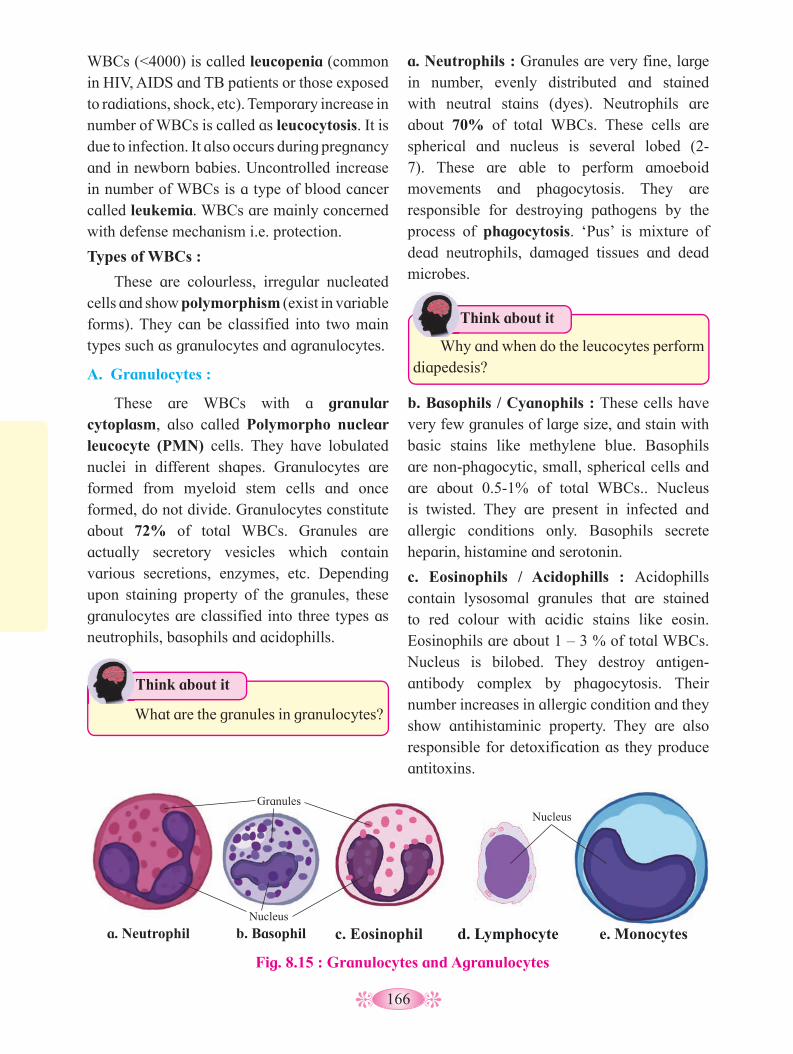
166
a. Neutrophils : Granules are very fine, large in number, evenly distributed and stained with neutral stains (dyes). Neutrophils are about 70% of total WBCs. These cells are spherical and nucleus is several lobed (2-7). These are able to perform amoeboid movements and phagocytosis. They are responsible for destroying pathogens by the process of phagocytosis. ‘Pus’ is mixture of dead neutrophils, damaged tissues and dead microbes.
What are the granules in granulocytes?
Think about it
Fig. 8.15 : Granulocytes and Agranulocytes
a. Neutrophil b. Basophil c. Eosinophil d. Lymphocyte e. MonocytesNucleus
Granules Nucleus
WBCs (<4000) is called leucopenia (common in HIV, AIDS and TB patients or those exposed to radiations, shock, etc). Temporary increase in number of WBCs is called as leucocytosis. It is due to infection. It also occurs during pregnancy and in newborn babies. Uncontrolled increase in number of WBCs is a type of blood cancer called leukemia. WBCs are mainly concerned with defense mechanism i.e. protection.
Types of WBCs :
These are colourless, irregular nucleated cells and show polymorphism (exist in variable forms). They can be classified into two main types such as granulocytes and agranulocytes.
A. Granulocytes :
These are WBCs with a granular cytoplasm, also called Polymorpho nuclear leucocyte (PMN) cells. They have lobulated nuclei in different shapes. Granulocytes are formed from myeloid stem cells and once formed, do not divide. Granulocytes constitute about 72% of total WBCs. Granules are actually secretory vesicles which contain various secretions, enzymes, etc. Depending upon staining property of the granules, these granulocytes are classified into three types as neutrophils, basophils and acidophills.
Why and when do the leucocytes perform
diapedesis?
Think about it
b. Basophils / Cyanophils : These cells have very few granules of large size, and stain with basic stains like methylene blue. Basophils are non-phagocytic, small, spherical cells and are about 0.5-1% of total WBCs.. Nucleus is twisted. They are present in infected and allergic conditions only. Basophils secrete heparin, histamine and serotonin.
c. Eosinophils / Acidophills : Acidophills contain lysosomal granules that are stained to red colour with acidic stains like eosin. Eosinophils are about 1 – 3 % of total WBCs. Nucleus is bilobed. They destroy antigen-antibody complex by phagocytosis. Their number increases in allergic condition and they show antihistaminic property. They are also responsible for detoxification as they produce antitoxins.
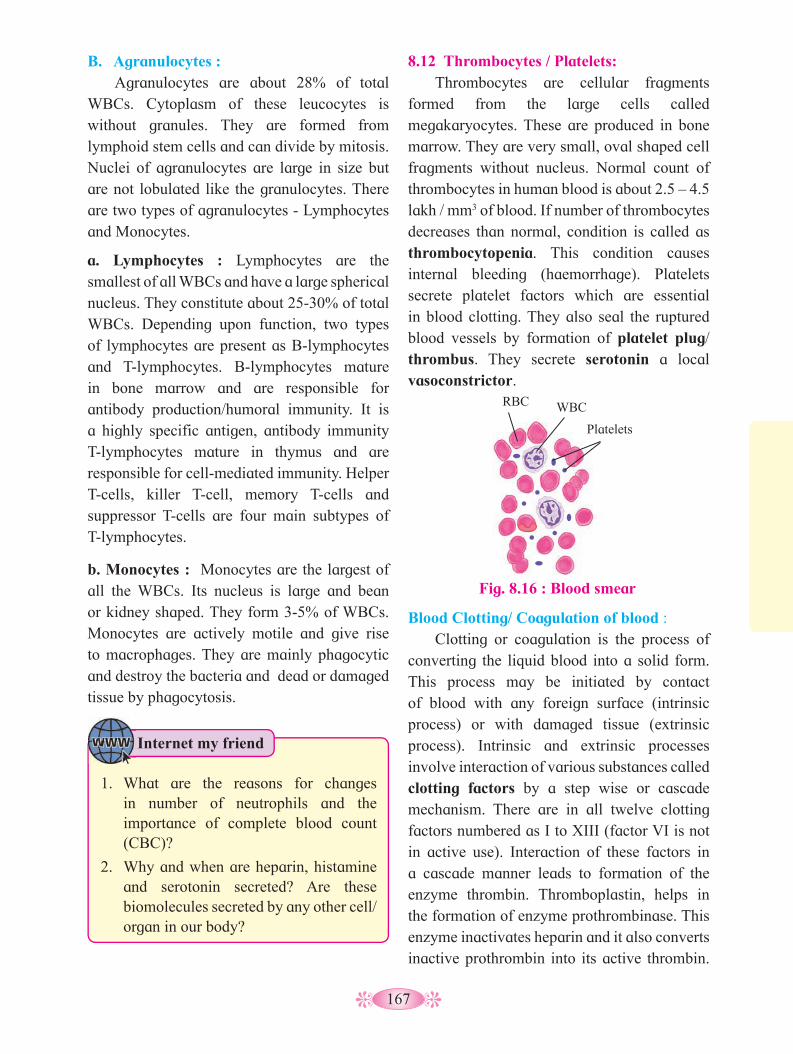
167
B. Agranulocytes : Agranulocytes are about 28% of total
WBCs. Cytoplasm of these leucocytes is without granules. They are formed from lymphoid stem cells and can divide by mitosis. Nuclei of agranulocytes are large in size but are not lobulated like the granulocytes. There are two types of agranulocytes - Lymphocytes and Monocytes.
a. Lymphocytes : Lymphocytes are the smallest of all WBCs and have a large spherical nucleus. They constitute about 25-30% of total WBCs. Depending upon function, two types of lymphocytes are present as B-lymphocytes and T-lymphocytes. B-lymphocytes mature in bone marrow and are responsible for antibody production/humoral immunity. It is a highly specific antigen, antibody immunity T-lymphocytes mature in thymus and are responsible for cell-mediated immunity. Helper T-cells, killer T-cell, memory T-cells and suppressor T-cells are four main subtypes of T-lymphocytes.
b. Monocytes : Monocytes are the largest of all the WBCs. Its nucleus is large and bean or kidney shaped. They form 3-5% of WBCs. Monocytes are actively motile and give rise to macrophages. They are mainly phagocytic and destroy the bacteria and dead or damaged tissue by phagocytosis.
8.12 Thrombocytes / Platelets: Thrombocytes are cellular fragments
formed from the large cells called megakaryocytes. These are produced in bone marrow. They are very small, oval shaped cell fragments without nucleus. Normal count of thrombocytes in human blood is about 2.5 – 4.5 lakh / mm3 of blood. If number of thrombocytes decreases than normal, condition is called as thrombocytopenia. This condition causes internal bleeding (haemorrhage). Platelets secrete platelet factors which are essential in blood clotting. They also seal the ruptured blood vessels by formation of platelet plug/ thrombus. They secrete serotonin a local vasoconstrictor.
Fig. 8.16 : Blood smear
RBC WBC
Platelets
Blood Clotting/ Coagulation of blood : Clotting or coagulation is the process of
converting the liquid blood into a solid form. This process may be initiated by contact of blood with any foreign surface (intrinsic process) or with damaged tissue (extrinsic process). Intrinsic and extrinsic processes involve interaction of various substances called clotting factors by a step wise or cascade mechanism. There are in all twelve clotting factors numbered as I to XIII (factor VI is not in active use). Interaction of these factors in a cascade manner leads to formation of the enzyme thrombin. Thromboplastin, helps in the formation of enzyme prothrombinase. This enzyme inactivates heparin and it also converts inactive prothrombin into its active thrombin.
Internet my friend
1. What are the reasons for changes in number of neutrophils and the importance of complete blood count (CBC)?
2. Why and when are heparin, histamine and serotonin secreted? Are these biomolecules secreted by any other cell/ organ in our body?

168
8.13 Heart : Heart is the main pumping organ of the
circulatory system. It is reddish brown in colour, hollow, muscular organ, roughly the size of one’s fist. Its average weight is about 300gm in males and 250gm in females. It is conical in shape and lies in mediastenum- i.e. the space between two lungs. It is broader at upper end
Serous pericardium is soft, moist and elastic. It is formed of squamous epithelium and is further divisible into two layers as parietal and visceral layer. Parietal and visceral layers of serous pericardium are separated by a pericardial space. This space is filled with pericardial fluid (about 50ml) which acts as a shock absorber and protects the heart from mechanical injuries. It also keeps the heart moist and acts as lubricant.
Heart wall : The heart is mesodermal in origin. Its wall
is formed of three layers, outer epicardium, middle myocardium and inner endocardium. Epicardium is thin and formed of a single layer of flat squamous epithelium resting on basement membrane. Myocardium is the middle thick layer formed of cardiac muscles. Endocardium is a single thin layer formed of squamous epithelium. The epicardium and endocardium are protective in function whereas myocardium is responsible for contraction and relaxation of heart.
1. What is blood clotting? How and when does it occur?
2. What is immunity? Name its types. 3. Why does the platelet count decrease in
dengue patient?4. Why does our immune system fail
against pathogens like Trypanosoma and Plasmodium?
5. What is the relation between immunity and organ transplantation?
6. How do monocytes perform amoeboid movement and phagocytosis?
7. How do monocytes modify into macrophages?
Curiosity
Fig. 8.17 : Heart wall and Pericardium
Thrombin converts soluble blood protein- fibrinogen into insoluble fibrin. Fibrin forms a mesh in which platelets and other blood cells are trapped to form the clot. Blood clotting occurs as shown in the following flowchart.
(base) and conical at lower end (apex). Conical end is slightly tilted to left side and rests above the diaphragm.
Heart is enclosed in a membranous sac called pericardium. Pericardium is formed of two main layers - outer fibrous and inner serous pericardium.
Fibrous pericardium
Parietal layer of serous
pericardium
Pericardial space
Visceral layer of serous
pericardium (Epicardium)
Endocardium
Trabeculae carneae
Myocardium (Cardiac muscles)
Pericardium
Endocardium
Myocardium
Heart wall
Extrinsic Pahway Intrinsic Pahway
Damage to tissue outside the vessel
Tissue Thromboplastin
Damage to the blood vessel
Cascade of clotting factors
Inactive factor X
Activated factor X
Prothrombin Thrombin
Fibrinogen Blood ClotFibrin
Observe and Discuss
Observe the flow chart and discuss the process with your friends.

169
External structure of heart : The human heart is four chambered.
The two superior chambers are called atria (auricles) and inferior two are called ventricles. Externally, the atria are separated from ventricles by a transverse groove called coronary sulcus or atrioventricular groove. The two ventricles are externally separated from each other by two grooves, the anterior and posterior inter-ventricular sulci. Coronary arteries and coronary veins run through these sulci. Pulmonary trunk arising from right ventricle and aorta from left ventricle are present on anterior surface of heart. The
pulmonary trunk bifurcates into right and left pulmonary arteries. Aorta (systemic aorta) is divisible into three regions as ascending aorta, systemic arch /aortic arch and descending aorta. The Ligamentum arteriosum joins pulmonary trunk and aortic arch. It is the remnant of an embryonic duct called ductus arteriosus. The aortic arch gives out three arteries viz. brachiocephalic (innominate) artery, left common carotid and left sub-clavian. The right atrium recieves superior and inferior vena cava along its dorsal surface. Pulmonary veins open into left atrium along the dorsal surface of heart.
Fig. 8.19 : Anterior (ventral) view : External structure of human heart
Superior vena cava
Right pulmonary artery
Right pulmonary veins
Right coronary atery
Right atrium
Right Ventricle
Inferior vena cava
Left subclavian artery
Aorta
Ligamentum arteriosum
Left pulmonary artery
Pulmonary trunk
Left pulmonary veins
Left Atrium
Branch of left coronary artery
Left Ventricle Coronary Sulcus
Fig. 8.18 : Posterior (dorsal) view : External structure of human heart
Left common carotid artery
Aorta
Left pulmonary artery
Left pulmonary vein
Left Atrium
Coronary sinus
Left Ventricle
Apex of heart
Superior vena cava
Right pulmonary artery
Right pulmonary veins
Inferior vena cava
Right Ventricle
Brachiocephalic artery left subclavian artery
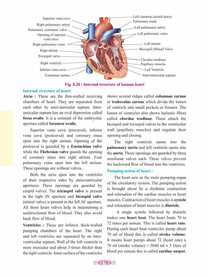
170
Fig. 8.20 : Internal structure of human heart
Internal structure of heart: Atria : These are the thin-walled receiving chambers of heart. They are separated from each other by inter-auricular septum. Inter-auricular septum has an oval depression called fossa ovalis. It is a remnant of the embryonic aperture called foramen ovalis.
Superior vena cava (precaval), inferior vena cava (postcaval) and coronary sinus open into the right atrium. Opening of the postcaval is guarded by a Eustachian valve while the Thebesian valve guards the opening of coronary sinus into right atrium. Four pulmonary veins open into the left atrium. These openings are without valves.
Both the atria open into the ventricles of their respective sides by atrioventricular apertures. These openings are guarded by cuspid valves. The tricuspid valve is present in the right AV aperture and bicuspid valve (mitral valve) is present in the left AV aperture. All these heart valves help in maintaining a unidirectional flow of blood. They also avoid back flow of blood.
Ventricles : These are inferior, thick-walled pumping chambers of the heart. The right and left ventricles are separated by an inter-ventricular septum. Wall of the left ventricle is more muscular and about 3-times thicker than the right ventricle. Inner surface of the ventricles
shows several ridges called columnae carnae or trabeculae carnae which divide the lumen of ventricle into small pockets or fissures. The lumen of ventricles also shows inelastic fibers called chordae tendinae. These attach the bicuspid and tricuspid valves to the ventricular wall (papillary muscles) and regulate their opening and closing.
The right ventricle opens into the pulmonary aorta and left ventricle opens into the aorta. These openings are guarded by three semilunar valves each. These valves prevent the backward flow of blood into the ventricles.
Pumping action of heart :
The heart acts as the main pumping organ of the circulatory system. The pumping action is brought about by a rhythmic contraction and relaxation of the cardiac muscles or heart muscles. Contraction of heart muscles is systole and relaxation of heart muscles is diastole.
A single systole followed by diastole makes one heart beat. The heart beats 70 to 72 times per minute. This is called heart rate. During each heart beat ventricles pump about 70 ml of blood this is called stroke volume. It means heart pumps about 72 (heart rate) x 70 ml (stroke volume) = 5040 ml ≈ 5 liters of blood per minute this is called cardiac output.
Superior vena cava
Right pulmonary artery
Pulmonary semilunar valve
Right pulmonary veins
Opening of superior vena cava
Right atrium
Tricuspid valve
Right ventricle
Inferior vena cava
Columnae carnae
Left common carotid artery
Left pulmonary artery
Pulmonary trunk
Left pulmonary veins
Left atrium
Bicuspid (Mitral) Valve
Chordae tendinae Papillary muscles
Left Ventricle
Interventricular septum

171
These branches form network in ventricular walls and these are called Purkinje fibers. Bundle of His and Purkinje fibers spread impulses in ventricles. As a result both the ventricles contract simultaneously.
Sometimes, valves are defective. Due to
this, abnormal or adventitious sound is heard, called ‘heart murmuring’ or ‘hissing.’
Do you know ?
Conducting tissue of heart: The human heart is myogenic i.e. the heart
is capable of generating a cardiac contraction independent of nervous input. It also shows auto rhythmicity i.e. it can generate its own rhythm by specialized muscles. A specialized cardiac musculature called the nodal tissue is distributed in the heart. A part of this nodal tisse is present in the upper right corner of the right atrium. It is called SA Node or Sino-atrial node. It lies at the base of opening of superior vena cava. Another mass of nodal tissue, the modified muscular fibers also called autorhythmic fibers (conducting tissue) control the beating rate of heart. Conducting (nodal) tissue consists of SA node, AV node, bundle of His and Purkinje fibers.
Conducting system of the heart : SA node (sinu-atrial node) is present in
the right atrium. It acts as pacemaker of heart because it has the power of generating a new wave of contraction and making the pace of contraction. SA node passes the contraction to the left ventricle and also to the AV node. AV node (atrio-ventricular node) is present in the right atrial wall near the base of interatrial septum. It acts as pace setter of heart.
Bundle of His/ Tawara branches start from AV node and pass through interventricular septum. Bundle of His forms two branches, the right and left bundles, one for each ventricle.
Fig. 8.21 : Conducting system of human heart
Right atrium
Sinoatrial (SA) Node
Atrioventricular (AV) Node
Atrioventricular (AV) Bundle (Bundle of his)
Right and Left Bundle Branches
Right ventricle Purkinje fibers
Left ventricle
Left atrium
8.14 Working mechanism of human heart : Cardiac Cycle :
Human heart alternately contracts and relaxes. Contraction is called systole and relaxation is called diastole. Atria and ventricles contract alternately. Consecutive systole and diastole constitutes a single heartbeat or cardiac cycle. It is completed in 0.8 sec. On an average, 72 beats are completed in one minute in an adult, at rest.
a. Atrial systole (AS): Right atrium receives deoxygenated
blood and left atrium receives oxygenated blood. When both the atria are completely filled with blood, pressure is exerted on the wall. In response to this pressure, SA node gets excited and generates cardiac impulse. Due to this, cardiac muscles in the atrial wall contract causing atrial systole. During atrial systole, blood is pumped into ventricles. Blood

172
is prevented from going back to the veins and coronary sinus by Eustachian and Thebesian valve respectively. After completing systole the atria go into diastole.
In normal conditions, atrial systole is for 0.1 sec. and atrial diastole (AD) is for 0.7 sec.
b. Ventricular systole (VS): The impulse which started from SA node
now reaches the AV node and it gets excited. AV node sends impulses to bundle of His and from bundle of His to Purkinje fibers. Purkinje fibers spread impulses all over the wall of ventricles. Due to this, ventricular wall contracts causing ventricular systole. During ventricular systole, right ventricle pumps deoxygenated blood into pulmonary trunk and left ventricle pumps oxygenated blood into aorta. During ventricular systole the cuspid valves close both the atrioventricular apertures preventing blood flow into atria (lubb sound is heard).
In normal conditions, ventricular systole lasts for 0.3 sec. and ventricular diastole (VD) lasts for 0.5 sec. During ventricular diastole, semilunar valves are closed, preventing backflow of blood from pulmonary trunk and systemic aorta into ventricles (dub sound is heard).
For about 0.4 second, both atria and ventricles are in diastole. When all the chambers of heart are in diastole, this condition is called joint diastole or complete diastole. Thus, duration of one cardiac cycle is 0.8 sec.
Regulation of cardiac activity : Though human heart is myogenic,
it is also under dual control, the nervous as well as hormonal. The nervous control includes the part of autonomic nervous system. Sympathetic system (with hormone epinephrine as neurotransmitter) increase the rate of heartbeat during emergency. Parasympathetic system (with acetylcholine as neurotransmitter) reduces rate of heartbeat.
Nervous control includes the part of the autonomous nervous system- its cardiovascular center lies in the medulla oblongata. It controls rate of heart beat in response to inputs from various receptors like proprio-receptors (which monitor the position of limbs and muscles), chemoreceptors (monitoring chemical changes in blood) and baroreceptors (monitoring the stretching of main arteries and veins).
Chemical control of the heart rate includes the conditions like hypoxia, acidosis, alkalosis causing decreased cardiac activity, hormones like epinephrine and norepinephrine enhance the cardiac activity. Besides, concentration of cations like K+, Ca++ and Na+ have major effect on cardiac activity. Cardiac activity decreases with the elevated blood level of K+ and Na+
Right side of heart contains deoxygenated and left contains oxygenated blood. Total volume of blood pumped during one ventricular systole is called stroke volume (SV) and it is it approximately 70 ml.
Cardiac output (CO): It is the volume of blood pumped out per
min. For a normal adult human being it is calculated as follows :
(CO) = SV × HR = 70 × 72 = 5040 ml/min
0.1 0 0.2 0.3 0.4 0.5 0.6 0.7 0.8 0.9 1.0
AS
AS
AD
VS VD
Joint diastole
Time in sec
Fig. 8.22 : Diagramatic representation of cardiac cycle
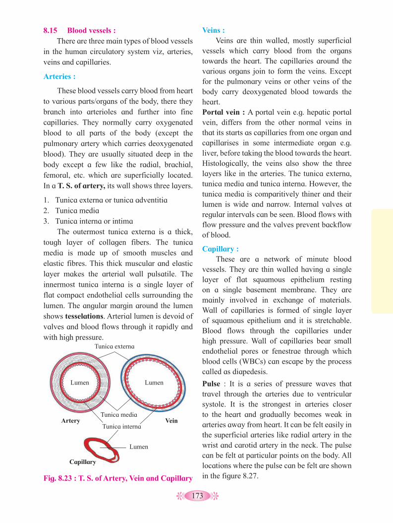
173
8.15 Blood vessels : There are three main types of blood vessels
in the human circulatory system viz, arteries, veins and capillaries.
Arteries :
These blood vessels carry blood from heart to various parts/organs of the body, there they branch into arterioles and further into fine capillaries. They normally carry oxygenated blood to all parts of the body (except the pulmonary artery which carries deoxygenated blood). They are usually situated deep in the body except a few like the radial, brachial, femoral, etc. which are superficially located. In a T. S. of artery, its wall shows three layers.
1. Tunica externa or tunica adventitia2. Tunica media3. Tunica interna or intima
The outermost tunica externa is a thick, tough layer of collagen fibers. The tunica media is made up of smooth muscles and elastic fibres. This thick muscular and elastic layer makes the arterial wall pulsatile. The innermost tunica interna is a single layer of flat compact endothelial cells surrounding the lumen. The angular margin around the lumen shows tesselations. Arterial lumen is devoid of valves and blood flows through it rapidly and with high pressure.
Veins : Veins are thin walled, mostly superficial
vessels which carry blood from the organs towards the heart. The capillaries around the various organs join to form the veins. Except for the pulmonary veins or other veins of the body carry deoxygenated blood towards the heart.Portal vein : A portal vein e.g. hepatic portal vein, differs from the other normal veins in that its starts as capillaries from one organ and capillarises in some intermediate organ e.g. liver, before taking the blood towards the heart. Histologically, the veins also show the three layers like in the arteries. The tunica externa, tunica media and tunica interna. However, the tunica media is comparitively thiner and their lumen is wide and narrow. Internal valves at regular intervals can be seen. Blood flows with flow pressure and the valves prevent backflow of blood.
Capillary : These are a network of minute blood
vessels. They are thin walled having a single layer of flat squamous epithelium resting on a single basement membrane. They are mainly involved in exchange of materials. Wall of capillaries is formed of single layer of squamous epithelium and it is stretchable. Blood flows through the capillaries under high pressure. Wall of capillaries bear small endothelial pores or fenestrae through which blood cells (WBCs) can escape by the process called as diapedesis.
Pulse : It is a series of pressure waves that travel through the arteries due to ventricular systole. It is the strongest in arteries closer to the heart and gradually becomes weak in arteries away from heart. It can be felt easily in the superficial arteries like radial artery in the wrist and carotid artery in the neck. The pulse can be felt at particular points on the body. All locations where the pulse can be felt are shown in the figure 8.27. Fig. 8.23 : T. S. of Artery, Vein and Capillary
Tunica interna
Tunica media
Tunica externa
Lumen Lumen
Lumen
Artery Vein
Capillary

174
(arteriosclerosis), deposition of fats like cholesterol in the arteries (atherosclerosis), renal diseases and emotion induced hormonal changes, obesity, etc. Blood pressure lower than normal i.e. below 90/60 mmHg is called hypotension and blood pressure higher than normal i.e. above 140/90 mmHg is hypertension.
Various factors that affect the blood pressure are cardiac output, peripheral resistance, blood volume, length and diameter of blood vessels, viscosity of blood, age, gender, venous return, sleep, emotions, exercise, anxiety, etc. Pulse rate is equal to heart rate. Pulse rate
higher than normal (above 100 beats/min) is called tachycardia and slower pulse rate (below 60 beats/min) than normal is called bradycardia.
Why do the veins have valves?
Use your brain power
8.16 Blood pressure (B. P.): The pressure exerted by blood on the wall
of the blood vessels is called blood pressure. It is measured by the sphygmomanometer. It is usually measured from the arteries.
Arterial Blood Pressure: Pressure exerted by blood on the wall of
artery is arterial blood pressure. Pressure on arterial wall during ventricular contraction (systole) is systolic pressure (SP). For a normal healthy adult the average value is 120 mmHg.
Pressure on arterial wall during relaxation of ventricles is diastolic pressure (DP). For a normal healthy adult it is 80 mmHg.
B P = SP / DP = 120/80 mmHg
Blood pressure is normally written as 120/80 mmHg. Difference between systolic and diastolic pressure is called pulse pressure. Normally, it is 40 mmHg.
Deviations from normal blood pressure value indicate malfunctioning of heart. It may be due to high or low blood volume, arterial inelasticity or hardening of arteries
Fig. 8.24 : Pulse points
tibial
Facial Temporal Carotid
Brachial
Radial
Femoral
Popliteal Posterior
Dorsalis pedis
Why do obese persons are prone to
hypertension?
Think about it
Normal cardiac output is 5 lit/min. Increase in cardiac output increases systolic pressure. Peripheral resistance depends upon the diameter of blood vessels. Decrease in diameter of arterioles and capillaries under the effect of vasoconstrictors like vasopressin or ADH cause increase in peripheral resistance and thereby increase in blood pressure. Blood loss in accidents decreases blood volume and thus the blood pressure. Blood pressure is directly proportional to Viscosity of blood.
Blood pressure increases with age due to increase in inelasticity of blood vessels. Amount of blood brought to the heart via the veins per unit time is called the venous return and it is directly proportional to blood pressure. Blood pressure is also directly proportional to the total length of the blood vessel. Blood pressure can also be affected by vaso constriction or vaso dilation. Females have slightly lower BP than males her age before menopause. However, the risk of high B. P. increases in the females after menopause sets in.
Internet my friend
Surf the internet for video-clips of angiography, angioplasty and by-pass surgery. Gather more information about these medical procedures.

175
called as Korotkoff sounds. Pressure in the cuff is further lowered till any pulsatile sound cannot be heard due to smooth blood flow. At this moment, pressure indicated in manometer is diastolic pressure. An optimal blood presure (normal) level reads 120/80 mmHg.
Hypertension : Persistently raised blood pressure higher
than the normal is called hypertension. 140/90 mmHg is called as threshold of hypertension and the 180/120 mmHg and higher readings are dangerous to the health. It may damage the heart, brain and kidneys.
Under the condition of hypertension, heart uses more energy for pumping which causes angina pectoris- the chest pains due to lowered blood supply to cardiac muscles and may lead to myocardial infarction. There are more chances of brain hemorrhage due to hypertension as arteries in brain are less protected by surrounding tissues as compared to other organs. In kidney, hypertension may cause kidney failure.
Coronary Artery Disease (CAD) :- It is also known as atherosclerosis. In this, calcium, fat cholesterol and fibrous tissues gets deposited in blood vessels suppling blood to the heart muscles making the lumen narrow.
Angina Pectoris : It is the pain in the chest resulting
from a reduction in the blood supply to the cardiac muscles because of atherosclerosis or arteriosclerosis. It is charactarized by severe pain and heaviness in the chest. The pain may spread to the neck, lower jaw, left arm and left shoulder. The pain usually results from exertion, when there is more demand of oxygen by the heart, but the supply does not meet the requirement.
Angiography : X-ray imaging of the cardiac blood
vessels to locate the position of blockages
1. 1. Why the heart-recipient has to rely upon
life-time supply of immunosupressants?2. Why the transplanted heart beats at higher
rate than normal?
Think about it
Measurement of blood pressure: Blood pressure is measured with the help
of an instrument called sphygmomanometer. This instrument consists of inflatable rubber bag cuff covered by a cotton cloth. It is connected with the help of tubes to a mercury manometer on one side and a rubber bulb on the other side. During measurement, the person is asked to lie in a sleeping position. The instrument is placed at the level of heart and the cuff is tightly wrapped around upper arm. The cuff is inflated till the brachial artery is blocked due to external pressure. Then pressure in the cuff is slowly lowered till the first pulsatile sound is heard.
At this moment, pressure indicated in manometer is systolic pressure. Sounds heard during measurement of blood pressure are
Fig. 8.25 : Sphygmomanometer
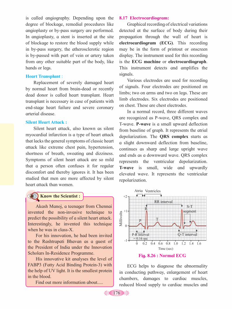
176
is called angiography. Depending upon the degree of blockage, remedial procedures like angioplasty or by-pass surgery are performed. In angioplasty, a stent is inserted at the site of blockage to restore the blood supply while in by-pass surgery, the atherosclerotic region is by-passed with part of vein or artery taken from any other suitable part of the body, like hands or legs.
Heart Transplant : Replacement of severely damaged heart
by normal heart from brain-dead or recently dead donor is called heart transplant. Heart transplant is necessary in case of patients with end-stage heart failure and severe coronary arterial disease.
Silent Heart Attack : Silent heart attack, also known as silent
myocardial infarction is a type of heart attack that lacks the general symptoms of classic heart attack like extreme chest pain, hypertension, shortness of breath, sweating and dizziness. Symptoms of silent heart attack are so mild that a person often confuses it for regular discomfort and thereby ignores it. It has been studied that men are more affected by silent heart attack than women.
8.17 Electrocardiogram: Graphical recording of electrical variations
detected at the surface of body during their propagation through the wall of heart is electrocardiogram (ECG). This recording may be in the form of printout or onscreen display. The instrument used for this recording is the ECG machine or electrocardiograph. This instrument detects and amplifies the signals.
Various electrodes are used for recording of signals. Four electrodes are positioned on limbs; two on arms and two on legs. These are limb electrodes. Six electrodes are positioned on chest. These are chest electrodes.
In a normal record, three different waves are recognized as P-wave, QRS complex and T-wave. P-wave is a small upward deflection from baseline of graph. It represents the atrial depolarization. The QRS complex starts as a slight downward deflection from baseline, continues as sharp and large upright wave and ends as a downward wave. QRS complex represents the ventricular depolarization. T-wave is small, wide and upwardly elevated wave. It represents the ventricular repolarization.
Fig. 8.26 : Normal ECG
Atria Ventricles
RR interval S-T
segment R
P T
Mill
ivol
ts
P-R interval = 0.16 sec
Q-T interval
+2
+1
0
-1 0 0.2 0.4 0.6 0.8 1.0 1.2 1.4 1.6
Time (sec)
ECG helps to diagnose the abnormality in conducting pathway, enlargement of heart chambers, damages to cardiac muscles, reduced blood supply to cardiac muscles and
Know the Scientist :
Akash Manoj, a teenager from Chennai invented the non-invasive technique to predict the possibility of a silent heart attack. Interestingly, he invented this technique when he was in class-X.
For his innovation, he had been invited to the Rashtrapati Bhavan as a guest of the President of India under the Innovation Scholars In-Residence Programme.
His innovative kit analyses the level of FABP3 (Fatty Acid Binding Protein-3) with the help of UV light. It is the smallest protein in the blood.
Find out more information about.....

177
causes of chest pain. A physicial can find out the defect in the heart by examining the wave pattern and the time interval betwen them.
8.18 Lymphatic System : Lymphatic system consists of lymph,
lymphatic vessels, some organs and tissues. The word ‘lymph’ means ‘clear water’ and it is a fluid connective tissue with almost similar composition to the blood except RBCs, platelets and some proteins. Fluid from intercellular spaces of the body tissue enters into the lymphatic vessels, from here it is discharged into the blood vessels (veins) through the thoracic duct and the right lymphatic duct.
Fig. 8.27 : Lymphatic System
1. What is depolarization and repolarization?2. What is the correlation between
depolarization and repolarization as ell as contraction and relaxation of the heart?
3. How are the signals detected and amplified by electrocardiograph?
4. Who discovered ECG?
Curiosity
Internet my friend
Location of lymph nodes in human body.
Subclavian vein
Thymus gland
Thoracic duct
Axillary lymph nodes
Appendix
Tonsil
Cervical lymph nodes
Red bone marrow
Spleen
Popliteal lymph nodes
Inguinal lymph nodes
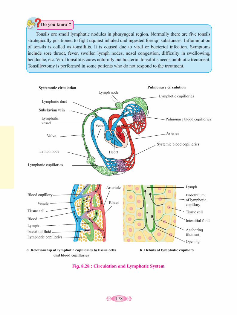
178
Tonsils are small lymphatic nodules in pharyngeal region. Normally there are five tonsils
strategically positioned to fight against inhaled and ingested foreign substances. Inflammation of tonsils is called as tonsillitis. It is caused due to viral or bacterial infection. Symptoms include sore throat, fever, swollen lymph nodes, nasal congestion, difficulty in swallowing, headache, etc. Viral tonsillitis cures naturally but bacterial tonsillitis needs antibiotic treatment. Tonsillectomy is performed in some patients who do not respond to the treatment.
Do you know ?
Fig. 8.28 : Circulation and Lymphatic System
a. Relationship of lymphatic capillaries to tissue cells and blood capillaries
Veins
Heart
Lymphatic capillaries
Pulmonary blood capillaries
Arteries
Systemic blood capillaries
Lymphatic capillaries
Lymph node
Valve
Lymphatic vessel
Subclavian vein
Lymphatic duct
Lymph node Pulmonary circulation Systematic circulation
b. Details of lymphatic capillary
Lymph
Lymph
Lymphatic capillaries
Blood
Blood
Endothlium of lymphatic capillary
Tissue cellTissue cell
Intestitial fluid
Intestitial fluid Anchoring filament
Opening
Arteriole
Blood capillary
Venule

179
Activity :
1. Prepare chart for different types of heart muscles and their functions.
2. Differentiate between the functioning of heart in frog and human.
Human Heart Frog Heart

180
Q. 1 Choose the correct alternatives from those given below and complete the statements.
1. The muscular structure that separates the thoracic and abdominal cavity is _______.
a. pleura b. diaphragm c. trachea d. epithelium
2. What is the minimum number of plasma membrane that oxygen has to diffuse across to pass from air in the alveolus to haemoglobin inside a R.B.C.?
a. Two b. Three c. Four d. Five
3. ________ is a sound producing organ,
a. Larynx b. Pharynx c. Tonsils d. Trachea
4. The maximum volume of gas that is inhale during breathing in addition to T.V is _____.
a. residual volume b. I.R.V. c. G.R.V. d. vital capacity
5. ________ muscles contract when the external intercostal muscles contract a. Internal abdominal b. Jaw c. Muscles in bronchial walls d. Diaphragm
6. Movement of cytoplasm in unicellular organisms is called __________.a. diffusion b. cyclosis c. circulation. d. thrombosis.
7. Which of the following animals do not have closed circulation?a. Earthworm. b. Rabbit c. Butterfly d. Shark
8. Diapedesis is performed by _________.a. erythrocytes b. thrombocytes c. adipocytes d. leucocytes
9. Pacemaker of heart is _________.a. SA node b. AV nodec. His bundle d. Purkinje fibers
10. Which of the following is without nucleus?a. Red blood corpuscle b. Neutrophill c. Basophill d. Lymphocyte
11. Cockroach shows which kind of circulatory system? a. Open b. Closed c. Lymphatic d. Double
12. Diapedesis can be seen in _________ cell.a. RBC b. WBC c. Platelet d. neuron
13. Opening of inferior vena cava is guarded by __________.
a. bicuspid valve b. tricuspid valve c. Eustachian valve d. Thebesian valve
14. ___________ wave in ECG represent atrial depolarization.a. P b. QRS complex c. Q d. T
15. The fluid seen in the intercellular spaces in Human is _________a. blood b. lymph c. interstitial fluid d. water
Q. 2 Match the Respiratory surface to the organism in which it is found.
Respiratory surface Organism Plasma membrane Insect Lungs Salamander External gills Bird Internal Gills Amoeba Trachea Fish
Q. 3 Very short answer questions.1. Why does trachea have ‘C’ shaped rings
of cartilage?2. Why is respiration in insect called direct
respiration?3. Why is gas exchange very rapid at
alveolar level.4. Name the organ which prevents the
following the entry of food into the trachea while eating.
Exercise

181
Q 4. Short answer questions.1. Why is it advantageous to breathe
through the nose than through the mouth?
2. Identify the incorrect statement and correct it,a. A respiratory surface area should
have a large surface area.b. A respiratory surface area should be
kept dry.c. A respiratory surface area should be
thin, may be 1mm or less.3. Given below are the characteristics of
some modified respiratory movement. Identify them.a. Spasmodic contraction of muscles of
expiration and forceful expulsion of air through nose and mouth.
b. An inspiration followed by many short convulsive expiration accompanied by facial expression.
4. Write a note on blood plasma.5. Explain blood clotting in short.6. Describe pericardium.7. Describe valves of human heart.8. What is role of papillary muscles and
chordae tendinae in human heart?9. Explain in brief the factors affecting
blood pressure.
Q. 5 Give scientific reason.1. Closed circulation is more efficient than
open circulation.2. Human heart is called as myogenic and
autorhythmic.3. Person who has undergone heart
transplant needs lifetime supply of immunosupressants.
4. Arteries are thicker than veins.5. Left ventricle is thick than all other
chambers of heart.
Q. 6 Distinguish between :1. Open and closed circulation.2. Artery and vein.3. Blood and lymph.4. Blood capillary and lymph capillary.
5. Intrinsic and extrinsic process of clotting.
Q. 7 Long answer questions.1. Smita was working in a garage with
the doors closed and automobiles engine running. After some time she felt breathless and fainted. What would be the reason? How can she be treated?
2. Shreyas went to a garden on a wintry morning. When he came back, he found it difficult to breath and started wheezing. What could be the possible condition and how can he be treated?
3. Why can you feel a pulse when you keep a finger on the wrist or neck but not when you keep them on a vein?
4. A man’s pulse rate is 68 and cardiac output is 5500 cm3.Find the stroke volume.
5. Which blood vessel of the heart will have the maximum content of Oxygen and why?
6. If the duration of the atrial systole is 0.1 sec and that of complete diastole is 0.4 sec, then how does one cardiac cycle complete in 0.8 sec?
7. How is blood kept moving in the large veins of the legs?
8. Describe histological structure of artery, vein and capillary.
9. What is blood pressure? How is it measured? Explain factors affecting blood pressure.
10. Describe human blood and give its functions.
Project : • Visit pathological laboratory to study
various blood tests like Hb detection, CBC, blood groups.
• Visit hospital to study how to take ECG, stress test, measurement of BP, etc.
• Evaluation of ECG on broad basis.
• Use of stethescope.
• Differential count of WBCs.

182
Unicellular organisms have a simple organisation of their life processes. However a multicellular organisation of the body organs and organ systems required both, a control over their life processes as well as a coordination between the various systems in order to maintain homeostasis of the organism. Plants and animals both show a control and coordination mechanism. In plants this done by sending chemical signals and brining about various types of movements (e.g. phototropic, chemotactic, thingmotactic, etc). Animals, specially the higher vertebrates show a gradual increase in the complexity of their control and coordination by giving both electrical and chemical singles. In this chapter now you will study about development of nervous system in different animal groups and details of the system in humans.
The porifera (sponges) are the most primitive of the animal phyla.
Fig. 9.1 : Nerve net in Hydra
Nerve net
Tentacles
cells form tissues, organs and systems which must work in coordination with each other for smooth internal functioning of the body. Also the organism must be able to respond and coordinate with respect to various stimuli or changes in the external environment.
In the lower animals like Hydra and Planaria the nervous system achieves this function, while in higher more complex animals, it is done by two coordinating systems - the nervous (elctrical) system and endocrine (chemical) control system. These two systems will be studied separately though they work in coordination with each other.
NERVOUS COORDINATION9.1 Nervous System in Hydra :
Hydra, a cnidarian shows the diffused nervous system, which is the most primitive nervous system. The cnidarians are thus the first animal group showing true simple nervous system. It consists of the sensory cells and the nerve cells or neurons along with their fibres. The nerve cells are scattered or distributed throughout the body and inter connected to each other by synapses between their fibres to form the nerve net. There are two nerve nets both in the mesoglea, one connected towards the epidermis and second towards the gastro-dermis. There are sensory cells scattered in the
Control and Coordination 9
Can you recall?
1. What is the need for the control and co-ordination in multicellular animals ?
2. How do plants carry out control and coordination ?
Can you recall?
1. Do sponges have tissues and organs ?2. Can sponges coordinate their various
functions ?
Even though there are different types of cells in sponges for carrying out different functions, a proper nervous system is lacking. However to bring about efficient working of the body these multicellular animals show division of labour among the cells. This leads to specialization of cells for the various activities like digestion, respiration, excretion and others. Later in the higher animals, phyla the different

183
body wall and tentacles, but sense organs are lacking. Neurons have fibres but there are no sensory and motor nerves. The nerve impulse shows no polarity or direction. Thus in Hydra, activation of sensory cells can happen at any point, and from this point impulse can be carried through out the body in any direction, thus bringing movements of the body or tentacles eg. catching of prey during feeding.
The diffused types of nervous system is the first important landmark in the nervous system. It is seen in the ctenophora as well as in the enteric system or gut wall of higher animals including man.
9.2 Nervous System in Planaria (flatworm) :
Planaria is a flatworm belonging to the phylum platyhelminthes. It is the most primitive animal with a central nervous system (CNS) located on the ventral side of body. It consists of a mass of cerebral or cephalic ganglion appearing like an inverted U shaped brain. These lie in the anterior or head region and from each ganglion arise nine branches towards the outer side. Ventrally from below the ganglia arise a pair of Ventral Nerve Cords (VNC) or long nerve cords. These are inter connected to eachother by transfer nerve or commissure in a ladder like manner. The peripheral nerve plexus arising laterally from VNC. The PNS
include sensory cells arranged in lateral cords in the body. A pair of photosensory structure, the eyes are located on dorsal side of the brain. Also there are single sensory cells scattered in the body.
In the above examples of Hydra, Planaria and the earlier studied examples of cockroach and humans, we have seen the gradual evolution or changes of the neural system. There is a high level of specialization in the formation of neurons as electrically signalling cells and also in the entire system, gradually from a diffuse neural system to a centralized nervous system. The expansion into a properly organized system involving the brain, its gradual expansion in size and functions. This has lead to centralization of various sense organs assisting in coordinating the internal environment with that of the external environment. Also there evolution of a complex networking system which efficiently transmits signals between one part or organ of the body and another.
Fig 9.2 Ladder type nervous system in
Flatworms e.g. Planaria
Eyespot
Brain
Nerve cords
Transverse nerve
Activity :
Note the changes taking place in the internal environment of the body, when a person goes from resting state into a state of physical activity.
9.3 Neural tissue The neural tissue consists primarily of two
types of cells viz the neurons and the neuroglia or glial cells. While a nerve is bundle of axons, the word ‘nerve’ is used for a bundle of axons outside the CNS while inside the CNS for the same, word ‘tract’ is used. The nerves may be sensory or motor or mix type i.e. having both the types - sensory and motor fibres.
All these along with nervous organs make up the nervous system of the higher animal and bring about coordination and control of various activities of the body. This is done through the receptors which bring in sensory inputs towards the central nervous system. Processing is carried out in the CNS and then through the

184
motor commands, the response is sent out. The nerves arising from the cytons of the CNS, travel throughout the body transmitting the nerves impulses to or from the CNS.
Neurons/Nerve cells : These are the structural and functional units of the nervous system. (You may recall the structure of a neuron studied in earlier classes). Each multipolar neuron has three parts - cyton or cell body, dendron and axon. The cyton has a distinct central nucleus with a nucleolus
Ependymal cell Oligodendrocytes Satellite cells
Astrocytes Microglia Schwann cells
Fig. 9.3 : Types of Neuroglial cells
Curiosity Box: Find out about the nervous system in
Earthworm.
Internet my friend
1. How many neurons are present in the human body and specifically in the brain?
2. Normally what percentage of cranial capacity is used by an average human ?
3. What is the ratio between neurons and neuroglia ?
and neuroplasm. A clear film of cytoplasm surrounds the nucleus around which there are nurofibrils, Nissl’s granules and other cell organelles Nissl’s granules are riboprotein components. They play an important role in the synthesis of the enzyme required for formation of the neurotransmitter. Neurofibrils play an important role in transmission of nerve impulse. Dendrons are many small conical processes arising from the cyton. These are highly branched into fine dendrites. Nissl’s granules and neurofibrils both can be seen at the base of the dendrons. They transmit message towards the cyton. Axon is a single long, usually unbranched process arising from the cyton at the axon hillock. It consists of a bundle of neurofibrils. Nissl’s granules are absent. Terminally, the axon gives out branches called telodendrons. The axons carry the messages away from the cytons. The axons may give out lateral branches called collaterals. The terminal branches attach to a muscle, gland, skin or telodendrites of another neuron. The interconnection between two neurons or neuron with motor organ is called synapse. It is

185
endonurium are surrounded by connective tissue, called perineurium and a still large bundle of nerves is covered on the outer side by epineurium. Blood is supplied to all the nerves to provide oxygen and nutrients.Neuroglial cells :
The neuroglial cells are far greater in number than the neurons. Most of the supporting cells of the nervous system are derived from the same embryonic tissue layer (ectoderm) that produces neurons. The term neuroglia refers to the supporting cells of the Central Nervous System (CNS) and Peripheral Nervous System (PNS).
usually axo-dendronic or may be axo-axonic, axo-somatic or dendro-dendronic.
The cytons are generally found inside the brain, spinal cord (CNS) and in the ganglia. Small groups of cell bodies inside the white matter of brain are called basal nuclei. A bundle of axons called nerve may be covered only by neurilemma in the non-medullated nerves while in the medullated nerves it is covered both by medullary sheath and on the outside by neurilemma. Conduction of impulse by the medullated nerves is 50 times faster than in the non-medullated nerves. The connective tissue covering around the nerve fascicule is called endoneurium. Few nerve fasciculi with
Table 9.4 : Types of Neuroglial cells and their functions
Location Cell type Functions
CNS (Central Nervous System)
Oligodendrocytes These cells have few branches and mainly form the myelin sheath around the central axons, which form the white matter of CNS. Myelin an isulating sheath is made up of protein and fatty substances. It allows quick transmission of electrical impulses.
Microglia or brain macrophages
Astrocytes
Ependymal cells
Small sized cells with few branches. These are derived from monocytes and act as macrophages. They go to the site of injury, dead neurons and cell debris in the CNS. They mediate immune response in the CNS.Star shaped cells and the most abundant glial cells of CNS. They have varied roles in the brain, secretion and absorption of neural transmitter and maintence of blood-brain barrier BBB. Regulate the transmission of electrical impulses with the brain.Form single layer of squamous or columnar, often ciliated epithelial cells lining the ventricles or brain cavities and central canal of spinal cord. Mainly responsible for production and probably also for circulation of CSF in brain ventricles and central canal.
PNS (Peripheral Nervous System)
Schwann Cells
Satellite cells
These are the most abundant glial cells of PNS. They produce myelin sheath around medullated nerves of PNS.They support the functions of neurons.

186
Fig. 9.5 : Synaptic transmission
Presynaptic neuron Nerve impulse
Ca2+
Ca2+
Ca2+ Synaptic end bulb
Synaptic cleft
Neurotransmitter receptor
Ligand-gated channel closed
Neurotransmitter
Postsynaptic neuron Postsynaptic potential
Nerve impulse
Ligand-gated channel open
Na+
Synaptic vesicles
Voltage-gated Ca2+ channel
Cytoplasm
1
2
3
4
5
6 7
Ca2+
Ca2+
K+
9.4 Synapse : It is a junction between two nerve cells
with a minute gap (synaptic cleft) in between them which allows transmission of impulse by a neurotransmitter bridge. 1. Properties of nerve fibres : a. Excitability/Irritability - The nerve fibres, on account of presence of a polarised membrane, have the ability to perceive stimulus and enter into a state of activity. b. Conductivity - It is ability to transmit the excitation. c. Stimulus - It is any detectable, physical, chemical, electrical change in the external or internal environment which brings about excitation in a nerve/muscle/organ/organism. In order to be effective, the stimulus must have a minimum intensity called threshold stimulus. Subliminal (weak) stimulus will have no effect while supraliminal (strong) stimulus will produce the same degree of impulse as the threshold stimulus. d. Summation effect -A single subliminal stimulus will have no effect but if many such weak stimuli are given in quick succession,
they may produce an impulse due to addition or summation of stimuli.e. All or none law - The nerve will either conduct the impulse along its entire length or will not at all conduct the impulse, as in case of subliminal or weak stimulus. f. Refractory period - It is the time interval (about millisecond) during which a nerve fails to respond to a second stimulus however strong it is.g. Synaptic delay - The impulse requires about 0.3 to 0.5 milliseconds to cross a synapse. This time is required for release of neurotransmitter from the axon terminal and excitation in the dendron of the next neuron. h. Synaptic fatigue - The transmission of nerve impulse across the synapse halts temporarily due to exhaustion of its neurotransmitter.i. Velocity - The rate of transmission of impulse is higher in long and thick nerves. It is higher in homeotherms than in poikilotherms. The velocity of transmission is higher in voluntary fibres (100 - 120 m/second in man) as opposed to autonomic or involuntary nerves (10-20 m/second). Similarly it is faster in medullated
187
nerve, as the impulse has to jump from one node of Ranvier to the next. At the synapse where the neurons communicate with one another. The neuron carrying an impulse to the synapse is the pre-synaptic neuron. The neuron receiving input at the synapse is the post synaptic neuron or generator region (gland or muscle). A synaptic cleft or a small intercellular space lies in between two cells having a width about 20-30 nm between them. .
The process by which the impulse from the pre-synaptic neuron is conducted to the post-synaptic neuron or cell is called synaptic transmission. It is a one way process carried out by neurotransmission. 2. Types of synapses a. Electrical synapse : In this type of synapse gap between the neighbouring neurons is very narrow. The synapse between such closed neurons is mechanical. The electrical conductive link is formed between the pre and post synaptic neurons. At the gap junction, the two cells are within almost 3.8 nm distance of each other. Transmission across the gap is faster but depends on the connection located at the gap junctions between the two neurons. Electrical synapses are found in those places of the body requiring fastest response as in the defence reflexes. also they are bidirectional, allowing transmission in either direction or may be unidirectional. b. Chemical synapse : These are specialized junctions through which cells of the neural system send chemical signal to the other neurons and to non-neuronal cells, such as gland and muscle. Synaptic gap is larger than that in electrical synapse and it is 20-40 nm.
A chemical synapse between a motor neuron and a muscle cell is called a neuromuscular junction. There are three components of a typical chemical synapse. 1. The presynaptic terminal (mostly axonic terminal), 2. The synaptic membrane of the post synaptic cell (usually on the dendrite of the next neuron/
gland cell/ muscle) and 3. The post synaptic neuron.
The impulse travels along the axon of the pre-synaptic neuron to the axon terminal. Most presynaptic neurons or axons have several synaptic knobs at their ends or terminals. These knobs have arrays of membranous sacs, called synaptic vesicles, that contain neurotransmitter molecules.
When an impulse reaches a synaptic knob, voltage sensitive Ca++ channels open and calcium (Ca++) diffuses inward from the extracellular fluid.
The increased calcium concentration inside the cells, initiates a series of events that fuse the synaptic vesicles with the cell membrane of presynaptic neuron, where they release their neurotransmitters by exocytosis. Once the neurotransmitters bind to the receptors of the post-synaptic cell, the action is either excitatory (turning a process on) or inhibitory (turning a process off). This is dependent on the nature of the neurotransmitter involved.
Once the impulse has been transferred across the synapse, the enzyme like cholinesterase destroys the neurotransmitter and the synapse is ready to receive a new impulse.
9.5 Transmission of nerve impulse : The neurons are cells with some specials
features. The cells can be excited. The nerve impulse is a wave of bioelectrical or electrochemical disturbances passing along a neuron. The transmission of the nerve impulse along the long nerve fibre/axon tube is a result of electrical charges across the neuronal membrane during conduction of an excitation. Each neuron has a charged cellular membrane with a voltage which is different on the outer and inner side of the membrane. The plasma membrane separates the outer and inner solutions of different chemical compounds but having approximately the same total number of ions. The external tissue fluid has both Na+

188
and K+ but there is predominance of Na+ and Cl-, while K+ is predominant within the fibre or in the intracellular fluid. This condition of a resting nerve is also called a polarised state and it is established by maintaining an excess of Na+ on the outerside. On the inside there is an excess of K+ along with large negatively charged protein molecules and nucleic acid. Some amount of Na+ and K+ is always leaks across the membrane. The Na+/K+ pump in the membrane actively restores the ions to their appropriate side. Against the concentration and electrochemical gradient, Na+ is being forced out and K+ is being forced inside the membrane. This process is called sodium pump or Na-K exchange pump. This active process requires ATP energy. The difference in distribuiton of Na+ and K+ on the two sides of the membrane produces a potential difference of -50 to -100 millivolts (average is -70 millivolts).
This potential difference seen in a resting nerve is thus called resting potential. (-70 millivolts) and it is mainly due to differential permeability of the resting membrane which is much more permeable to K+ than to Na+. This results in slightly more K+ diffusing out than Na+ moving inside and causing slight difference in polarity. Also ions like negatively charged proteins and nucleic acids inside the cell make the overall charge negative on the inside and positive charge on the outside.
The nerve membrane not only has leakage channels but also has many gated channels for Na+/K+. These are also called voltage gated channels. These channels enable the neuron to change it membrane potential to active potential in response to a stimuli. The Na+/K+ gated channels are separate so transport of both these ions is separately done. However during resting potential, both these gates are closed and the membrane resting potential is maintained.
Generation of nerve impulse :
1. Depolarization : The origin and maintenance of resting potential depends on the original perfect state. Any change or disturbance to the membrane will cause Na+ to enter into the membrane and lower the potential difference (lesser than -70 millivolts). This makes the membrane more permeable to Na+, so there will be rapid influx of Na+. This property is peculiar to a nerve membrane.
The voltage gated Na+/K+ channels are special in 2 ways : They can change the potential difference of the membrane as per the stimulus received and also the gates operate separately and are self closing.
During resting potential, both gates are closed and resting potential is maintained. However during depolarization the Na+
gates open but not the K+ gates. This causes Na+ to rush into the axon and bring about a depolarisation (opposite of polarity). Extra cellular fluid (ECF) becomes electronegative with respect to the inner membrane which becomes electropositive. The value of action potential is +30 millivolts to +60 millivolts. This triggers depolarisation in the next part while it itself starts going to repolarisation.
Fig. 9.6 : Polarisation and Depolarisation along a nerve
Na+ Na+
Na+
Na+
Na+
Na+
Na+
Na+
K+ K+
K+
K+
K+
K+
K+
K+
K+
High Na+ Na+- K+ Pump
Resting Axon
Polarized membrane Stimulus
applied
High K+ Negative organic ions
Polarised
Polarised
Depolarised
Depolarised
Excited Axon
Excited Axon Re-polarised
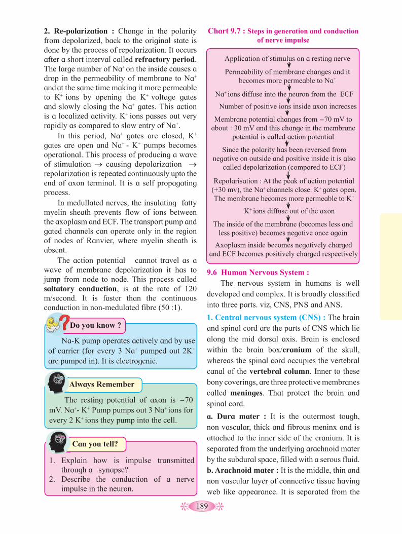
189
9.6 Human Nervous System : The nervous system in humans is well
developed and complex. It is broadly classified into three parts. viz, CNS, PNS and ANS.
1. Central nervous system (CNS) : The brain and spinal cord are the parts of CNS which lie along the mid dorsal axis. Brain is enclosed within the brain box/cranium of the skull, whereas the spinal cord occupies the vertebral canal of the vertebral column. Inner to these bony coverings, are three protective membranes called meninges. That protect the brain and spinal cord.
a. Dura mater : It is the outermost tough, non vascular, thick and fibrous meninx and is attached to the inner side of the cranium. It is separated from the underlying arachnoid mater by the subdural space, filled with a serous fluid.b. Arachnoid mater : It is the middle, thin and non vascular layer of connective tissue having web like appearance. It is separated from the
Chart 9.7 : Steps in generation and conduction of nerve impulse
Permeability of membrane changes and it becomes more permeable to Na+
Since the polarity has been reversed from negative on outside and positive inside it is also
called depolarization (compared to ECF)
Number of positive ions inside axon increases
K+ ions diffuse out of the axon
Na+ ions diffuse into the neuron from the ECF
Repolarisation : At the peak of action potential (+30 mv), the Na+ channels close. K+ gates open. The membrane becomes more permeable to K+
Membrane potential changes from -70 mV to about +30 mV and this change in the membrane
potential is called action potential
The inside of the membrane (becomes less and less positive) becomes negative once again
Axoplasm inside becomes negatively charged and ECF becomes positively charged respectively
Application of stimulus on a resting nerve
2. Re-polarization : Change in the polarity from depolarized, back to the original state is done by the process of repolarization. It occurs after a short interval called refractory period. The large number of Na+ on the inside causes a drop in the permeability of membrane to Na+
and at the same time making it more permeable to K+ ions by opening the K+ voltage gates and slowly closing the Na+ gates. This action is a localized activity. K+ ions passes out very rapidly as compared to slow entry of Na+.
In this period, Na+ gates are closed, K+
gates are open and Na+ - K+ pumps becomes operational. This process of producing a wave of stimulation → causing depolarization → repolarization is repeated continuously upto the end of axon terminal. It is a self propagating process.
In medullated nerves, the insulating fatty myelin sheath prevents flow of ions between the axoplasm and ECF. The transport pump and gated channels can operate only in the region of nodes of Ranvier, where myelin sheath is absent.
The action potential cannot travel as a wave of membrane depolarization it has to jump from node to node. This process called saltatory conduction, is at the rate of 120 m/second. It is faster than the continuous conduction in non-medulated fibre (50 :1).
Can you tell?
1. Explain how is impulse transmitted through a synapse?
2. Describe the conduction of a nerve impulse in the neuron.
Always Remember The resting potential of axon is -70
mV. Na+- K+ Pump pumps out 3 Na+ ions for every 2 K+ ions they pump into the cell.
Do you know ?
Na-K pump operates actively and by use of carrier (for every 3 Na+ pumped out 2K+ are pumped in). It is electrogenic.
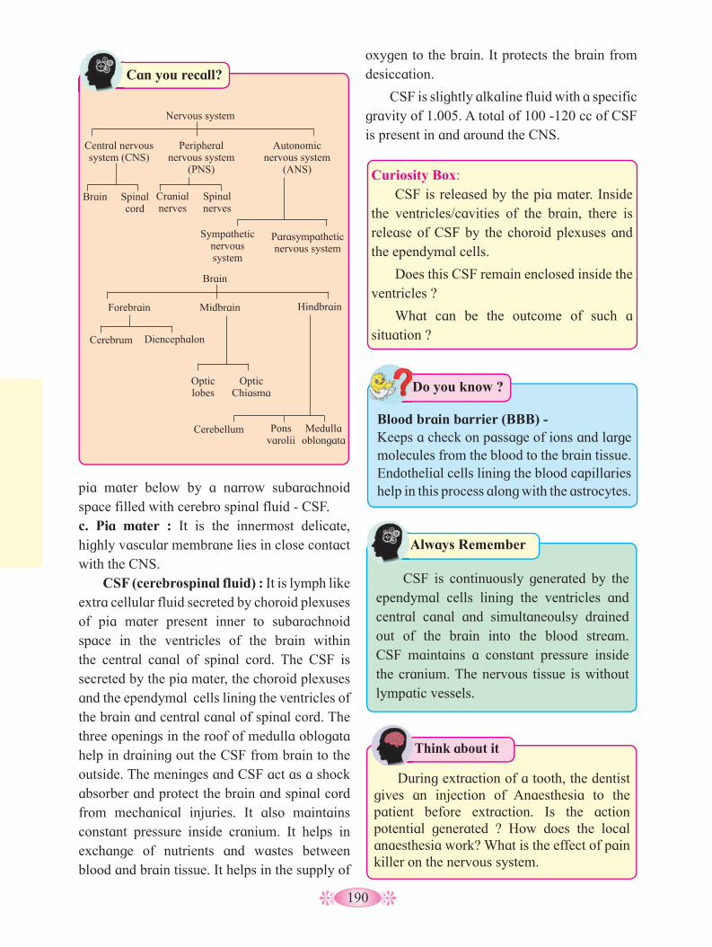
190
Curiosity Box: CSF is released by the pia mater. Inside
the ventricles/cavities of the brain, there is release of CSF by the choroid plexuses and the ependymal cells.
Does this CSF remain enclosed inside the ventricles ?
What can be the outcome of such a situation ?
Can you recall?
Brain
Forebrain Midbrain Hindbrain
Cerebrum
Optic lobes
Cerebellum Pons varolii
Medulla oblongata
Optic Chiasma
Diencephalon
Nervous system
Central nervous system (CNS)
Brain Spinal cord
Cranial nerves
Spinal nerves
Sympathetic nervous system
Parasympathetic nervous system
Peripheral nervous system
(PNS)
Autonomic nervous system
(ANS)
Always Remember
CSF is continuously generated by the ependymal cells lining the ventricles and central canal and simultaneoulsy drained out of the brain into the blood stream. CSF maintains a constant pressure inside the cranium. The nervous tissue is without lympatic vessels.
Do you know ?
Blood brain barrier (BBB) -Keeps a check on passage of ions and large molecules from the blood to the brain tissue. Endothelial cells lining the blood capillaries help in this process along with the astrocytes.pia mater below by a narrow subarachnoid
space filled with cerebro spinal fluid - CSF.c. Pia mater : It is the innermost delicate, highly vascular membrane lies in close contact with the CNS.
CSF (cerebrospinal fluid) : It is lymph like extra cellular fluid secreted by choroid plexuses of pia mater present inner to subarachnoid space in the ventricles of the brain within the central canal of spinal cord. The CSF is secreted by the pia mater, the choroid plexuses and the ependymal cells lining the ventricles of the brain and central canal of spinal cord. The three openings in the roof of medulla oblogata help in draining out the CSF from brain to the outside. The meninges and CSF act as a shock absorber and protect the brain and spinal cord from mechanical injuries. It also maintains constant pressure inside cranium. It helps in exchange of nutrients and wastes between blood and brain tissue. It helps in the supply of
oxygen to the brain. It protects the brain from desiccation.
CSF is slightly alkaline fluid with a specific gravity of 1.005. A total of 100 -120 cc of CSF is present in and around the CNS.
Think about it
During extraction of a tooth, the dentist gives an injection of Anaesthesia to the patient before extraction. Is the action potential generated ? How does the local anaesthesia work? What is the effect of pain killer on the nervous system.
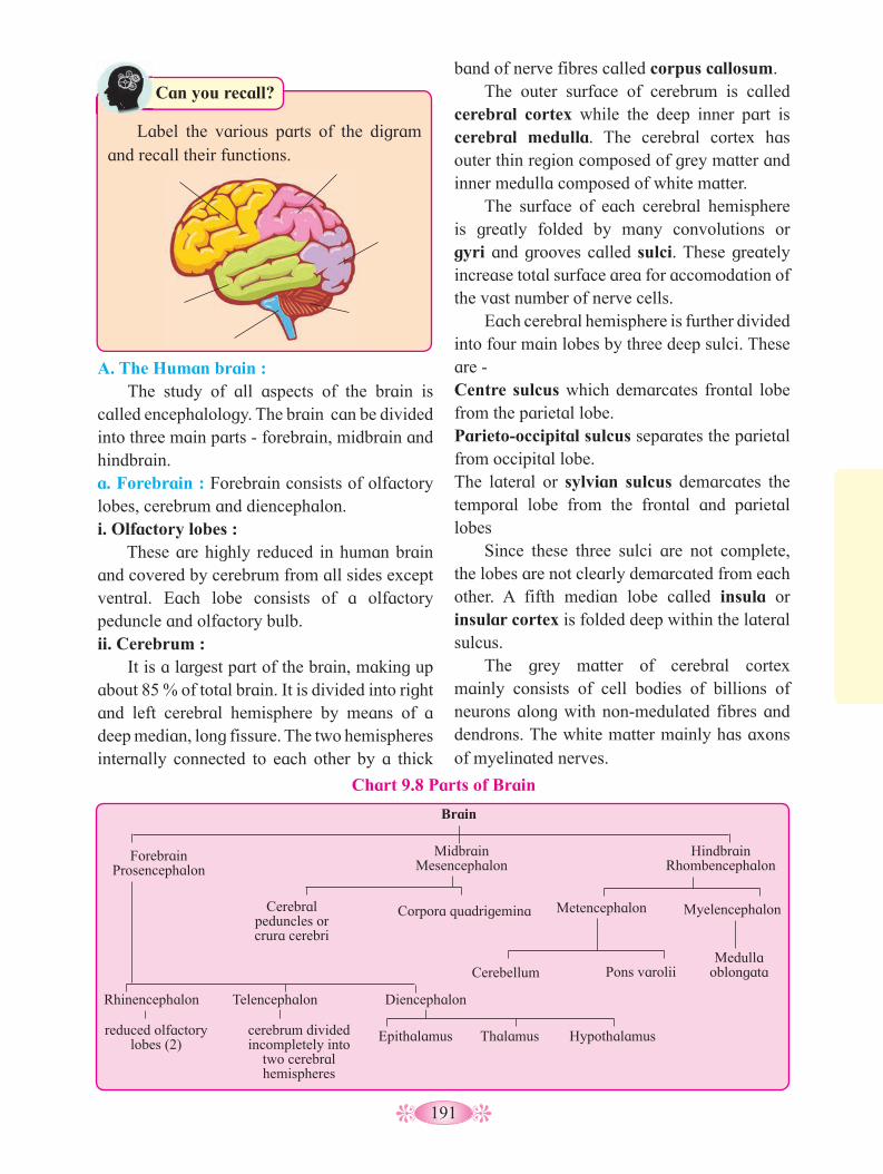
191
A. The Human brain : The study of all aspects of the brain is
called encephalology. The brain can be divided into three main parts - forebrain, midbrain and hindbrain.a. Forebrain : Forebrain consists of olfactory lobes, cerebrum and diencephalon.i. Olfactory lobes :
These are highly reduced in human brain and covered by cerebrum from all sides except ventral. Each lobe consists of a olfactory peduncle and olfactory bulb.ii. Cerebrum :
It is a largest part of the brain, making up about 85 % of total brain. It is divided into right and left cerebral hemisphere by means of a deep median, long fissure. The two hemispheres internally connected to each other by a thick
Brain
ForebrainProsencephalon
MidbrainMesencephalon
Cerebral peduncles or crura cerebri
Metencephalon
Cerebellum
Corpora quadrigemina Myelencephalon
Pons varoliiMedulla
oblongata
HindbrainRhombencephalon
Rhinencephalon
reduced olfactory lobes (2)
Epithalamus Thalamus Hypothalamuscerebrum divided incompletely into
two cerebral hemispheres
Telencephalon Diencephalon
Chart 9.8 Parts of Brain
band of nerve fibres called corpus callosum. The outer surface of cerebrum is called
cerebral cortex while the deep inner part is cerebral medulla. The cerebral cortex has outer thin region composed of grey matter and inner medulla composed of white matter.
The surface of each cerebral hemisphere is greatly folded by many convolutions or gyri and grooves called sulci. These greately increase total surface area for accomodation of the vast number of nerve cells.
Each cerebral hemisphere is further divided into four main lobes by three deep sulci. These are -Centre sulcus which demarcates frontal lobe from the parietal lobe.Parieto-occipital sulcus separates the parietal from occipital lobe.The lateral or sylvian sulcus demarcates the temporal lobe from the frontal and parietal lobes
Since these three sulci are not complete, the lobes are not clearly demarcated from each other. A fifth median lobe called insula or insular cortex is folded deep within the lateral sulcus.
The grey matter of cerebral cortex mainly consists of cell bodies of billions of neurons along with non-medulated fibres and dendrons. The white matter mainly has axons of myelinated nerves.
Can you recall?
Label the various parts of the digram and recall their functions.
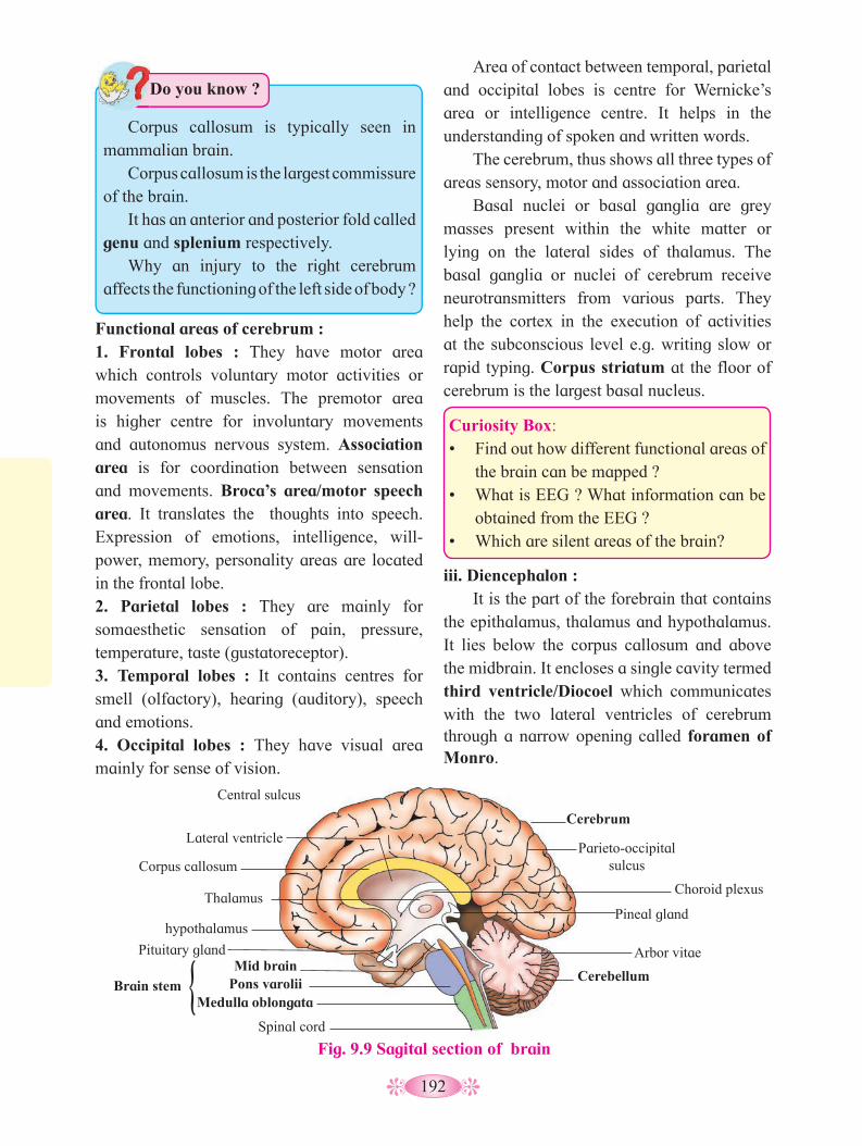
192
Area of contact between temporal, parietal and occipital lobes is centre for Wernicke’s area or intelligence centre. It helps in the understanding of spoken and written words.
The cerebrum, thus shows all three types of areas sensory, motor and association area.
Basal nuclei or basal ganglia are grey masses present within the white matter or lying on the lateral sides of thalamus. The basal ganglia or nuclei of cerebrum receive neurotransmitters from various parts. They help the cortex in the execution of activities at the subconscious level e.g. writing slow or rapid typing. Corpus striatum at the floor of cerebrum is the largest basal nucleus.
Curiosity Box: • Find out how different functional areas of
the brain can be mapped ?• What is EEG ? What information can be
obtained from the EEG ?• Which are silent areas of the brain?
iii. Diencephalon : It is the part of the forebrain that contains
the epithalamus, thalamus and hypothalamus. It lies below the corpus callosum and above the midbrain. It encloses a single cavity termed third ventricle/Diocoel which communicates with the two lateral ventricles of cerebrum through a narrow opening called foramen of Monro.
Functional areas of cerebrum :1. Frontal lobes : They have motor area which controls voluntary motor activities or movements of muscles. The premotor area is higher centre for involuntary movements and autonomus nervous system. Association area is for coordination between sensation and movements. Broca’s area/motor speech area. It translates the thoughts into speech. Expression of emotions, intelligence, will-power, memory, personality areas are located in the frontal lobe.2. Parietal lobes : They are mainly for somaesthetic sensation of pain, pressure, temperature, taste (gustatoreceptor).3. Temporal lobes : It contains centres for smell (olfactory), hearing (auditory), speech and emotions.4. Occipital lobes : They have visual area mainly for sense of vision.
Fig. 9.9 Sagital section of brain
Cerebellum
Pineal gland
Corpus callosum
Cerebrum
Medulla oblongata
Spinal cord
Pons varolii
Pituitary gland
Lateral ventricle
Thalamus
Parieto-occipital sulcus
Choroid plexus
Arbor vitae
Central sulcus
hypothalamus
Mid brain{Brain stem
Do you know ?
Corpus callosum is typically seen in mammalian brain.
Corpus callosum is the largest commissure of the brain.
It has an anterior and posterior fold called genu and splenium respectively.
Why an injury to the right cerebrum affects the functioning of the left side of body ?

193
The epithalamus is the thin non nervous roof of the diencephalon. Anterioly it is fused with the piamater to form the anterior choroid plexus and from its dorsal wall it is connected to pineal gland through a pineal stalk. (Pineal gland is discussed in detail in chemical co-ordination). Earlier this gland was though to be vestigeal, but later it has been found to produce to hormone melatonin (sleep inducing hormone; also related to reproductive behaviour). The lateral thick walls of diencephalon form the thalami. They mainly contain grey matter. The habenculor commissure connects two thalami. Different parts of the brain are interconnected by the RAS (Reticular Activating System)through the thalami. It is called relay centre as it transmitts all sensory impulses except those of olfactory (smell) to the cerebrum (gatekeeper of cerebrum connecting the anterior lateral ventricle to the iter posteriorly. The narrow cavity of diencephalon is called IIIrd ventricle or diocoel. It connects anteriorly to the two lateral ventricles by a single opening called Foramen of Monroe and posteriorly to the IVth ventricle or metacoel through a narrow duct of Sylvius or iter.
Hypothalamus : It forms a floor of the diencephalon. It is richly supplied with blood vessels (Hypothalamo-hypophyseal portal vein) helps in feed back mechanism for hormonal control). It maintains homeostasis, internal equilibrium of the body and
involuntary behaviour control. Like in the cerebrum, the hypothalamus also contains hypothalamic nuclei in its white matter (refer fig. 9.33 pituitary gland) with neuro-secretory cells involved in the production of hormones oxytocin and vasopressin.
The hypothalamus is a link between the nervous and the endocrine system. It has higher centres for endocrine system. It regulates heart rate, respiration, blood pressure (B.P.), body temperature, water and electrolyte balance. It has centres for hunger, thirst, sleep, fatique, satiety centre, secretion of glands of stomach and intestine. It also produces neurohormones that stimulate the pituitary gland. A complex neuronal circuit called the limbic system is formed by the hypothalamus amygdala, parts of epithalamus and thalamus, hippocampus and other areas. It appears to be responsible for emotional reactions, motivational drives and memory. The floor of the hypothalamus continues as a downward projection called hypopyseal stalk or infundibulum which connects it to the hypophysis (pituitary gland) both physically and functionally by secretion of neurotransmitters (details in chemical coordination). The inferior surface of hypothalamus also bears the optic chiasma (crossing of the two optic nerves) and a pair of mammillary bodies (unique to mammmalian brain and responsible for recollective memory).
Fig. 9.10 : Ventricles of brain
Lateral ventricles
Interventricular foramen
Third ventricle
Cerebral aqueduct
Fourth ventricle
Central canal Sagittal PlaneCoraral Plane

194
b. Mid brain : It is located between diencephalon and the
pons varolli. It contains the cerebral aqueduct or iter that connects the third and fourth ventricles. The corpora quadrigemina are four rounded elevations on the dorsal surface of the mid brain. The two superior colliculi are involved in visual reflexes and the two inferior colliculi are relay centres for auditory reflexes that operate when it is necessary to move the head to hear sounds more distinctly.
The mid brain also contains on its inferior surface two thick fibrous tracks called cerebral peduncles or crura cerebri. These tracts of ascending and descending nerve fibres from RAS and connect the cerebrum mid brain. Near the centre of the mid brain is a mass of grey matter scattered within the white matter. It is called the red nucleus. It plays an important role in controlling posture and muscle tone, modifying some motor activities and motor co-ordination.
of grey matter, the cortex. The white matter intermixes with the grey matter and shows a tree-like pattern called arbor vitae.
c. Hind brain : The posterior region of the brain is
called hind brain. It consists of pons varolli, cerebellum and medulla oblongata. The pons varolli appears as a rounded bulge on the underside of the brain stem (brain stem consist of mid brain, pons and medulla and continues upto spinal cord), and contains a cross band of nerve fibres connecting cerebrum, cerebellar lobes, medulla oblongata and spinal cord. It also contains several nuclei. The cerebellum is the second largest part of the brain and consists of two lateral hemispheres and a central vermis. It is composed of white matter with a thin layer
The surface of cerebellum shows convolutions (gyri and sulci) a number of nuclei lie deep within each lateral or cerebellar hemisphere. Over 30 million neurons lie in the cortex. Three pairs of myelinated nerve bundles called cerebullar penduncles connect cerebellum to the other parts of CNS. It is an important centre which maintains equilibrium of body, posture, balancing orientation, moderation of voluntary movements, maintainance of muscle tone. It is a regulatory centre for neuromuscular activities and controls the rapid activities like walking, running, speaking etc. All activitie of cerebellum are involuntanry (though may involve learning in early stages). The medulla oblongata is the posterior conical part of the brain and continues as the spinal cord. It has inner grey matter and outer white matter. It controls involuntary vital functions like heart beat, respiration, vasomotor activities and peristalsis. It also controls non-vital reflex activities like coughing, sneezing, swallowing, vomitting, yawning etc. The cavity of medulla is called IVth ventricle or metacoel. It’s roof has the posterior choroid plexes for secretion of CSF. The posterior choroid plexes also shows 3 openings - a pair of lateral foramen of Luschka and a median foramen of Magendie.
Always Remember
Wernicke's area : It is the sensory speech area responsible for understanding and formulating written and spoken language.
Broca's area : It is the motor speech area and translates thoughts into speech and controls movement of tongue, lips and vocal cords.
Can you tell?
Explain - ‘‘Cerebullum is well developed in humans’’.
• • Find out the role of the foramina
mentioned above.
• What is ataxia ?
Internet my friend•
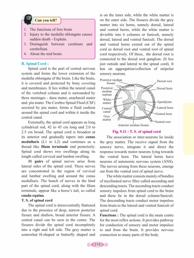
195
B. Spinal Cord : Spinal cord is the part of central nervous
system and forms the lower extension of the medulla oblongata of the brain. Like the brain, it is covered and protected by bony covering and membranes. It lies within the neural canal of the vertebral column and is surrounded by three meninges - dura mater, arachnoid mater and pia mater. The Cerebro Spinal Fluid (CSF) secreted by pia mater, forms a fluid cushion around the spinal cord and within it inside the central canal.
Externally, the spinal cord appears as long cylindrical rod, 42 to 45 cm long and 2.0 to 2.5 cm broad. The spinal cord is broadest at its anterior end gradually tapers into conus medullaris (L1 to L2) and continues as a thread like filum terminale end posteriorly. Spinal cord shows two swellings along its length called cervical and lumbar swelling.
31 pairs of spinal nerves arise from lateral sides of the spinal cord. These nerves are concentrated in the region of cervical and lumbar swelling and around the conus medullaris. The bunch of nerves in the hind part of the spinal cord, along with the filum terminale, appear like a horse’s tail, so called cauda equina. T. S. of spinal cord
The spinal cord is dorsoventrally flattened due to the presence of deep, narrow posterior fissure and shallow, broad anterior fissure. A central canal can be seen in the centre. The fissures divide the spinal cord incompletely into a right and left side. The grey matter is somewhat H-shaped or butterfly shaped and
is on the inner side, while the white matter is on the outer side. The fissures divide the grey matter into six horns, namely dorsal, lateral and ventral horns, while the white matter is divisible into 6 columns or funiculi, namely dorsal, lateral and ventral funiculi. The dorsal and ventral horns extend out of the spinal cord as dorsal root and ventral root of spinal cord respectively. Of these, the dorsal root is connected to the dorsal root ganglion. (It lies just outside and lateral to the spinal cord). It has an aggregation/collection of unipolar sensory neurons.
Can you tell?
1. The functions of fore brain 2. Injury to the medulla oblongata causes
sudden death - Explain.3. Distinguish between cerebrum and
cerebellum.4. About the mid brain.
The association or inter-neurons lie inside the grey matter. The receive signal from the sensory nerve, integrate it and direct the response towards motor neurons lying towards the ventral horn. The lateral horns have neurons of autonomic nervous system (ANS). The nerves arising from these neurons, emerge out from the ventral root of spinal nerve.
The white matter consists mainly of bundles of myelinated nerve fibre called ascending and descending tracts. The ascending tracts conduct sensory impulses from spinal cord to the brain and these lie in the dorsal column/funiculi. The descending tracts conduct motor impulses from brain to the lateral and ventral funiculi of spinal cord.Functions : The spinal cord is the main centre for the most reflex actions. It provides pathway for conduction of sensory and motor impulses to and from the brain. It provides nervous connection to many parts of the body.
Fig. 9.11 : T. S. of spinal cord
Dorsal rootPosterior median fissure
Anterior median fissure
Posterior median septum
White matter
Grey matter
Central canal
Dorsal horn
Lateral horn
Ependyma
Ventral horn
Ventral root

196
No. Name Type Origin Organs Innervated Functions
I. Olfactory Sensory Olfactory bulb Epithelium of Nose Smell
II. Optic Sensory Side of diencephalon
Retina of Eye Vision
III. Occulomotor Motor Floor of mid brain
Eye muscles (4 of 6 eye muscles)
Movement of eye ball
IV. Pathetic Motor Floor of mid brain
Eye muscles (1 of 6 eye muscles, forehead scalp)
Rotation and movement of eye ball
V. Trigeminal (Dentist’s nerve)a. Ophthalmic
b. Maxillary
c. Mandibular
(largest)
Mixed
Sensory
Sensory
Mixed
Ventral side of pons
-
-
-
-
Nasal cavity, Upper eyelids, forehead, scalp, conjunctiva, lacrimal gland, scalpMucosa of nose, palate, upper teeth, upper lip, lower eye lid parts of pharynxLower teeth, skin over mandible cheek, side of head in front ear, muscles of mastication
Sensation of skin touch, taste, jaw movement.
VI. Abducens Motor Pons Muscles of eye ball, lateral rectus muscle
Movement of eye
VII. Facial (bearing geniculate ganglion)
Mixed Pons facial, scalp and neck muscles, lacrimal, sublingual, submandibula, nasal and palatine glands
Facial expression, movement of neck, secretion of tears, taste, salivary secretion.
VIII. Auditory(vestibulo-cochlear)i. Vestibularii. Cochlear
Sensory Pons
--
Internal Ear Hearing and equilibrium
IX. Glossopharyngeal Mixed Side of medulla oblongata
Pharynx, tongue, salivary glands
Taste, salivation and swallowing
Table 9.12 Cranial nerves - nature and functions
2. Peripheral Nervous System (PNS) : The peripheral nervous system connects the central nervous system to the different parts of the body having receptors and effectors. Depending
on the connection to the CNS, the peripheral nerves are classified into two main types -Cranial nerves - connected to the brain.
Spinal nerves - connected to the spinal cord.

197
X. Vagus (Pneumogastric)
Mixed Side of medulla oblongata
Larynx, trachea, pharynx, alimentary canal, heart, lungs, pancreas, blood vessels,
Visceral sensations and visceral movements like breathing cardiac, slowing, gastric and pancreatic secretion, gastrointestinal movements
XI. Spinal accessory Motor Side of medulla oblongata
Neck and shoulder muscles, reflexes of thoracic and abdominal vicera larynx, pharynx
Movements of larynx, pharynx, neck and shoulder
XII. Hypoglossal Motor Side of medulla oblongata
Tongue muscles Movement of tongue
Cranial Nerves : These nerves develop from the brain, in all amniotes (reptiles, birds and mammals). There are 12 pairs of cranial nerves. Roman numbering I to XII is used to denote them. These nerves originate from or terminate into the brain.
According to their function, these are classified as sensory (I, II, VIII), motor (III, IV, VI, XI, XII) and mixed (V, VII, IX, X) nerves.
The details of cranial nerves present in the human body is presented in Table 9.15.Spinal Nerves : Thirty one pairs of spinal nerves originate from the spinal cord. They are mixed nerves and they provide two way communication between the spinal cord and part of the upper and lower limbs, neck and trunk.
Table 9.13 : Number and types of spinal Nerves
Group No. of pairs Region of origin from vertebral column
Cervical 8 (C1-C8) Neck
Thoracis 12 (T1-T12) Thorax
Lumbar 5 (L1-L5) Abdomen
Sacral 5 (S1-S5) Pelvis
Coccygeal 1 (Co1) Coccyx
Formation of a typical spinal nerve : All spinal nerves are of the mixed type i.e. they have some nerve fibre as sensory and some motor. Each spinal nerve is formed inside the neural canal of verterbral column by two roots - the posterior or dorsal sensory root and anterior or ventral root. Anterior root receives the sensory nerve from the dorsal root ganglion (cell bodies of sensory neurons are located in the ganglion), while the anterior/ventral root gives out the motor nerve. The dorsal sensory and the ventral motor nerves together form the mixed spinal nerve. It emerges out from both sides of the spinal cord through the intervertebral foramen. As soon as it emerges out of vertebral column, it shows three branches viz.
Do you know ?
1. Of the 12 pairs of cranial nerves, only the X (vagus) passes into the body and innervates internal organs.
2. Vagus has the maximum number of branches and longest distribution.
3. V/trigeminal/Dentist’s nerve is the largest cranial nerve
4. VI/abducens is the smallest cranial neve.
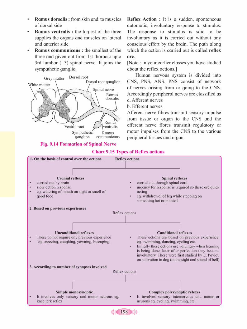
198
3. According to number of synapses involved Reflex actions
Simple monosynaptic • It involves only sensory and motor neurons eg.
knee jerk reflex
Complex polysynaptic refexes • It involves sensory internervous and motor or
neurons eg. cycling, swimming, etc.
Reflex actions
Cranial reflexes • carried out by brain• slow action response• eg. watering of mouth on sight or smell of
good food
Spinal reflexes• carried out through spinal cord• urgency for response is required so these are quick
acting • eg. withdrawal of leg while stepping on
something hot or pointed
2. Based on previous experiencesReflex actions
Unconditional reflexes• These do not require any previous experience• eg. sneezing, coughing, yawning, hiccuping.
Conditional reflexes• These actions are based on previous experience.
eg. swimming, dancing, cycling etc.• Initially these actions are voluntary when learning
is being done, later after perfection they become involuntary. These were first studied by E. Pavlov on salivation in dog (at the sight and sound of bell)
• Ramus dorsalis : from skin and to muscles of dorsal side
• Ramus ventralis : the largest of the three supplies the organs and muscles on lateral and anterior side
• Ramus communicans : the smallest of the three and given out from 1st thoracic upto 3rd lumbar (L3) spinal nerve. It joins the sympathetic ganglia.
Reflex Action : It is a sudden, spontaneous automatic, involuntary response to stimulus. The response to stimulus is said to be involuntary as it is carried out without any conscious effort by the brain. The path along which the action is carried out is called reflex arc.[Note : In your earlier classes you have studied about the reflex actions.]
Human nervous system is divided into CNS, PNS, ANS. PNS consist of network of nerves arising from or going to the CNS. Accordingly peripheral nerves are classified asa. Afferent nerves b. Efferent nervesAfferent nerve fibres transmit sensory impulse from tissue or organ to the CNS and the efferent nerve fibres transmit regulatory or motor impulses from the CNS to the various peripheral tissues and organ.
Chart 9.15 Types of Reflex actions
Fig. 9.14 Formation of Spinal Nerve
1. On the basis of control over the actions.
Dorsal root
Spinal nerveRamus dorsalis
Ramus ventralis
Ramus communicans
Sympathetic ganglion
Ventral root
Grey matter
White matterDorsal root ganglion

199
Fig. 9.16 Autonomic Nervous System
Parasympathetic Nervous System Sympathetic Nervous System
Stimulates flow of saliva Ganglion
Medulla oblongata Slows
heartbeat Constricts bronchi,
Stimulates peristalsis and secretion
Stimulates release of bile
Contracts bladder Chain of
sympathetic ganglia
Inhibits bladder contraction
Stimulates Secretion of
adrenaline and
nonadrenaline Conver-sion of glycogen
to glucose Inhibits peristalsis and secretion
Dilates bronchi
Solar plexus
Accelerates heartbeat
Inhibits flow of saliva
Dilates pupilStimulates secretion of tears
Constricts pupil, inhibits secretion of tears III
VIIIX
Xdilates blood vessels,
lowers B.P.
constricts blood vessels and increases B.P.
However according to recent studies, the extent of PNS has been broadened to incorporate the ANS.
According to this view, the PNS is divided into i. somatic nervous system and ii. autonomic nervous system
The somatic nerous system relays impulses from CNS to the skeletal or voluntary muscles of the body.3. Autonomic Nervous System (ANS) :
Autonomic nervous system transmits impulses from CNS to the involuntary organs and smooth muscles of the body.
ANS consists of a special set of peripheral nerves that regulate the activities of involuntary organs like cardiac muscles, smooth muscles, glands etc. In this, impulses are conducted from the Central Nervous system by an axon that synapses with an autonomous ganglion. It is preganglionic neuron. The second neuron in this ganglionic pathway has an axon that extends from the autonomic ganglion to an effector organ and is known as postganglionic neuron.
Autonomic nervous system consists of sympathetic and parasympathetic nervous system.
a. Sympathetic Nervous System (SNS): It is also called thoraco-lumbar outflow. It
originates in the thoracic and lumbar region of spinal cord (T1 to L3) and consists of 22 pairs of sympathetic ganglia which lie on a pair of sympathetic cords on lateral sides of the spinal cord.
The pre-ganglionic nerve fibres are short and post ganglionic nerve fibres are long. Adrenaline and Noradrenaline is produced at the terminal ends of postganglionic nerve fibres at the effector organ, hence it is also called Adrenergic fibres. Sympathetic nervous system controls body activities during emergencies (fight or flight response). It has excitatory and stimulating effect on most organs of the body except in the digestive and the excretory organ.
Use your brain power
Mr. Sharma suffered from a stroke and the right side of his body was paralysed. However his response was normal for knee jerk reflex with either leg. Explain how and why ?
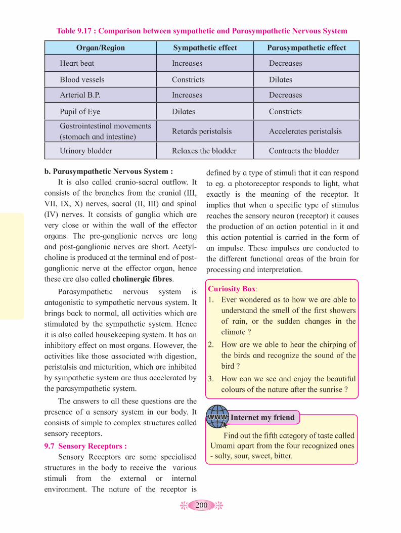
200
b. Parasympathetic Nervous System : It is also called cranio-sacral outflow. It
consists of the branches from the cranial (III, VII, IX, X) nerves, sacral (II, III) and spinal (IV) nerves. It consists of ganglia which are very close or within the wall of the effector organs. The pre-ganglionic nerves are long and post-ganglionic nerves are short. Acetyl-choline is produced at the terminal end of post-ganglionic nerve at the effector organ, hence these are also called cholinergic fibres.
Parasympathetic nervous system is antagonistic to sympathetic nervous system. It brings back to normal, all activities which are stimulated by the sympathetic system. Hence it is also called housekeeping system. It has an inhibitory effect on most organs. However, the activities like those associated with digestion, peristalsis and micturition, which are inhibited by sympathetic system are thus accelerated by the parasympathetic system.
The answers to all these questions are the presence of a sensory system in our body. It consists of simple to complex structures called sensory receptors.
9.7 Sensory Receptors : Sensory Receptors are some specialised
structures in the body to receive the various stimuli from the external or internal environment. The nature of the receptor is
defined by a type of stimuli that it can respond to eg. a photoreceptor responds to light, what exactly is the meaning of the receptor. It implies that when a specific type of stimulus reaches the sensory neuron (receptor) it causes the production of an action potential in it and this action potential is carried in the form of an impulse. These impulses are conducted to the different functional areas of the brain for processing and interpretation.
Organ/Region Sympathetic effect Parasympathetic effect
Heart beat Increases Decreases
Blood vessels Constricts Dilates
Arterial B.P. Increases Decreases
Pupil of Eye Dilates Constricts
Gastrointestinal movements (stomach and intestine)
Retards peristalsis Accelerates peristalsis
Urinary bladder Relaxes the bladder Contracts the bladder
Table 9.17 : Comparison between sympathetic and Parasympathetic Nervous System
Curiosity Box: 1. Ever wondered as to how we are able to
understand the smell of the first showers of rain, or the sudden changes in the climate ?
2. How are we able to hear the chirping of the birds and recognize the sound of the bird ?
3. How can we see and enjoy the beautiful colours of the nature after the sunrise ?
Internet my friend
Find out the fifth category of taste called Umami apart from the four recognized ones - salty, sour, sweet, bitter.

201
Chart 9.18. Types of receptors
No. Name/Type of receptor Location Function
I. Exteroceptors : Receive external stimuli
a. Phonoreceptors Internal Ear - organ of corti
Sound reception
b. Statoreceptors Internal Ear- semicircular canals
Receptors for maintaining balance and equilibrium
c. Photoreceptors Retina of Eye Receives sensory stimuli for vision
d. Thermoreceptors Skin Receives sensory stimuli for heat (caloriceptors) and cold (trigidocetptors)
e. Mechanoreceptors Skin Sensitive to mechanical stimuli like touch, pain,
pressure, deep pressure, etc.
f. Chemoreceptors• Gustatoreceptors• Olfactory receptors
Taste buds of tongueOlfactory Epithelium of Nose
Sensitive to taste of sweet, salt, sour, bitter and umami.
Sensitive to about 10,000 different smells
II. Interoceptors : Receive stimuli coming from within the body
a. Enteroceptors from internal body organs
Sensitive to stimuli coming from internal organs like hunger, thirst, pain, osmotic change
b. Proprioceptors Joints, muscles and tendons
Detect changes in the movements of joints, tendons and muscles; pain, tension and sensitive to vibrations
c. Baroreceptors (*These are also considered as mechanoreceptors, receiving signals from internal organ)
Present in walls of atria, venae cavae, aortic arch, carotid sinus
Sense changes in B.P. so as to restore homeostasis through vasodilation or vasoconstriction
Classification of receptors :Receptors are classified on the basis of their location, function and their sensitivity to specific stimuli. Their classification is given in the following chart.
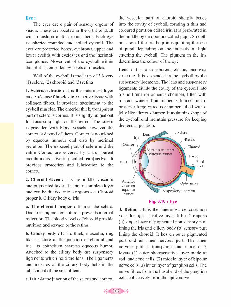
202
Eye : The eyes are a pair of sensory organs of
vision. These are located in the orbit of skull with a cushion of fat around them. Each eye is spherical/rounded and called eyeball. The eyes are protected bones, eyebrows, upper and lower eyelids with eyelashes and the lacrimal/tear glands. Movement of the eyeball within the orbit is controlled by 6 sets of muscles.
Wall of the eyeball is made up of 3 layers (1) sclera, (2) choroid and (3) retina
1. Sclera/scelrotic : It is the outermost layer made of dense fibroelastic connetive tissue with collagen fibres. It provides attachment to the eyeball muscles. The anterior thick, transparent part of sclera is cornea. It is slightly bulged out for focussing light on the retina. The sclera is provided with blood vessels, however the cornea is devoid of them. Cornea is nourished by aqueous humour and also by lacrimal secretion. The exposed part of sclera and the entire Cornea are covered by a transparent membranous covering called conjuctiva. It provides protection and lubrication to the cornea.
2. Choroid /Uvea : It is the middle, vascular and pigmented layer. It is not a complete layer and can be divided into 3 regions - a. Choroid proper b. Ciliary body c. Iris
a. The choroid proper : It lines the sclera. Due to its pigmented nature it prevents internal reflection. The blood vessels of choroid provide nutrition and oxygen to the retina.
b. Ciliary body : It is a thick, muscular, ring like structure at the junction of choroid and iris. Its epithelium secretes aqueous humor. Attached to the ciliary body are suspensory ligaments which hold the lens. The ligaments and muscles of the ciliary body help in the adjustment of the size of lens.
c. Iris : At the junction of the sclera and cornea,
the vascular part of choroid sharply bends into the cavity of eyeball, forming a thin and coloured partition called iris. It is perforated in the middle by an aperture called pupil. Smooth muscles of the iris help in regulating the size of pupil depending on the intensity of light entering the eyeball. The pigment in the iris determines the colour of the eye.
Lens : It is a transparent, elastic, biconvex structure. It is suspended in the eyeball by the suspensory ligaments. The lens and suspensory ligaments divide the cavity of the eyeball into a small anterior aqueous chamber, filled with a clear watery fluid aqueous humor and a posterior large vitreous chamber, filled with a jelly like vitreous humor. It maintains shape of the eyeball and maintain pressure for keeping the lens in position.
Fig. 9.19 : Eye
Pupil Blind spot
Lens Iris
Cornea
Anterior chamber aqueous humor
Suspensory ligament
Optic nerve
Fovea
Choroid
Retina
Sclera
Vitreous chamber vitreous humor
3. Retina : It is the innermost, delicate, non vascular light sensitive layer. It has 2 regions (a) single layer of pigmented non sensory part lining the iris and ciliary body (b) sensory part lining the choroid. It has an outer pigmented part and an inner nervous part. The inner nervous part is transparent and made of 3 layers (1) outer photosensitive layer made of rod and cone cells. (2) middle layer of bipolar nerve cells (3) inner layer of ganglion cells. The nerve fibres from the basal end of the ganglion cells collectively form the optic nerve.
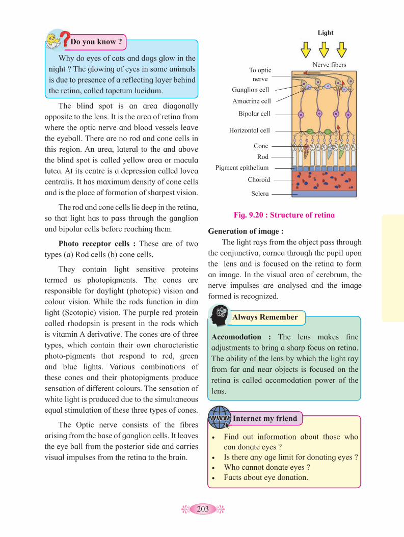
203
Fig. 9.20 : Structure of retina
Light
Nerve fibers To optic
nerve
Ganglion cell
Amacrine cell
Bipolar cell
Horizontal cell
Cone
Rod
Pigment epithelium
Choroid
Sclera
The blind spot is an area diagonally opposite to the lens. It is the area of retina from where the optic nerve and blood vessels leave the eyeball. There are no rod and cone cells in this region. An area, lateral to the and above the blind spot is called yellow area or macula lutea. At its centre is a depression called lovea centralis. It has maximum density of cone cells and is the place of formation of sharpest vision.
The rod and cone cells lie deep in the retina, so that light has to pass through the ganglion and bipolar cells before reaching them.
Photo receptor cells : These are of two types (a) Rod cells (b) cone cells.
They contain light sensitive proteins termed as photopigments. The cones are responsible for daylight (photopic) vision and colour vision. While the rods function in dim light (Scotopic) vision. The purple red protein called rhodopsin is present in the rods which is vitamin A derivative. The cones are of three types, which contain their own characteristic photo-pigments that respond to red, green and blue lights. Various combinations of these cones and their photopigments produce sensation of different colours. The sensation of white light is produced due to the simultaneous equal stimulation of these three types of cones.
The Optic nerve consists of the fibres arising from the base of ganglion cells. It leaves the eye ball from the posterior side and carries visual impulses from the retina to the brain.
Generation of image : The light rays from the object pass through
the conjunctiva, cornea through the pupil upon the lens and is focused on the retina to form an image. In the visual area of cerebrum, the nerve impulses are analysed and the image formed is recognized.
Internet my friend
• Find out information about those who can donate eyes ?
• Is there any age limit for donating eyes ? • Who cannot donate eyes ?• Facts about eye donation.
Always Remember
Accomodation : The lens makes fine adjustments to bring a sharp focus on retina. The ability of the lens by which the light ray from far and near objects is focused on the retina is called accomodation power of the lens.
Do you know ?
Why do eyes of cats and dogs glow in the night ? The glowing of eyes in some animals is due to presence of a reflecting layer behind the retina, called tapetum lucidum.

204
Ear : The human ear is called stato-acoustic
organ and it has two functions - hearing and body equilibrium. Anatomically the ear is made up of three parts : the external ear, middle ear and inner ear.
The external ear consists of ear pinna, auditory canal and tympanic membrane. In humans, the ear pinna is an immovable part, supported by elastic cartilage structure. It leads into an auditory canal. The pinna collects and sends the sound waves into the auditory canal. The auditory canal ends at the ear drum. It transfers the sound waves to the ear drum. There are very fine hair and wax secreting sebaceous glands in the skin of pinna and
auditory canal. The tympanic membrane is a delicate, membranous structure which transmits the sound waves to the middle ear. It is formed of connective tissues covered with skin on the outside and mucous membrane on the inside.
The middle ear consists of chain of three ear ossicles called Malleus (hammer), Incus (anvil) and Stapes (stirrup-the smallest bone). On receiving the vibrations from the tympanic membrane, the ear ossicles amplify the vibrations and transfer these to the cochlea. A short eustachian tube connects the middle ear to the pharynx. It equalises air pressure on both sides of the ear drum.
The internal ear consists of the labyrinth and vestibular apparatus. The labyrinth consists of bony labyrinth and membranous labyrinth. These are filled with perilymph and endolymph respectively. The coiled portion of the labyrinth is cochlea.
The cochlea contains fluid filled three chambers separated by Reissner’s membrane and basilar membrane. The upper chamber towards vestibul is called scala vestibuli and the bottom chamber scala tympani are filled with perilymph. The middle chamber is the scala media. It is filled with endolymph while scala vestibuli and scala tymphnith are filled with perilymph. The organ of Corti is a pea sized structure located on basilar membrane (floor of scala media).
The organ of corti has a sensory epithelium over the basilar membrane. The sensory epithelium is in contact with a gelatinous tectorial membrane. The sensory cells have sensory hair on their free end so also called hair cell. In between the rows of hair cells are present supporting cells.
Hair cells have long stiff microvilli called stereocillia on their apical surfaces. Above these stereocellia, is a jelly like membrane
Changes in retina when light rays fall on it
Light falls on rod and cone cells in Retina
Breaking up of light sensitive pigments by specific wavelength of light
Stimulation of rod and cone cells and generation of nerve impulse
Nerve impulse transmitted to bipolar nerve cells
Nerve impulse transmitted to ganglion cells
Nerve impulse in the axons of ganglion cells converge and leave via the optic nerve
Nerve impulse is transmitted by optic nerve to brain
Perception of image by the brain cells(in the visual area of cerebrum)
Activity :
Collect information about causes and corrections for myopia, hypermetropia, astigmatism, presbyopia, cataract, night blindness.

205
called tectorial membrane. This organ acts as a transducer, converting sound vibrations into nerve impulses.
Inner Ear and the mechanism of balance :
Besides the cochlea, the inner ear also has the vestibular apparatus which is composed of three semi-circular canals and the utriculo saccular region with the otolith organ. All three semi-circular canals lie in different planes at right angle to each other. These canals are filled with endolymph. The base of each of the canal has an ampulla in which there is a sensory spot called crista. The cristae help in maintaining equilibrium. The vestibule has two sensory spots-macula of saccule and utricle the macula consist of hair cells and supporting cells. Tips of the hair and cilium project into a thick gelataneous sheath otolithic membrane. Within this membrane minute particle otoliths or otoconia are secreted. These are made of CaCO
3 and protein. The macula and crista
are the receptors sensitive to the position of the head with respect to gravity. The three semicircular canals are arranged such away that the movement in any plane can be detected by these cells and the balance and posture of the body is maintained. Receptors for dynamic balance lie in the cristae of ampullae while for
Fig. 9.21 : Structure of human ear
Auricle
Earlobe
Ossicles :
Stapes
Incus
Malleus
Temporal bone
Semicircular ducts
Vestibular nerve
Cochlear nerve
Cochlea
Auditory tube Tympanic
cavity Eardrum Auditory
canal
Anterior
Posterior
Lateral
Ampulla of semicircular duct
Oval window
Secondary tympanic membrane in round window
Bony labyrinth Membranous labyrinth
Ampulla of semicircular canal
Endolymphatic sac
Vestibule
Utricle
Saccule
Cochlea
Cochlear duct
Fig. 9.22 : Human Internal Ear
Cochlear duct
Basilar membrane
Bone Auditory
nerve
Vestibular canal
Tympanic canal Organ of corti
Activity :
The auditory centre of the brain analyses the impulses received and the sound is perceived.
Draw flow chart of mechanism of hearing.
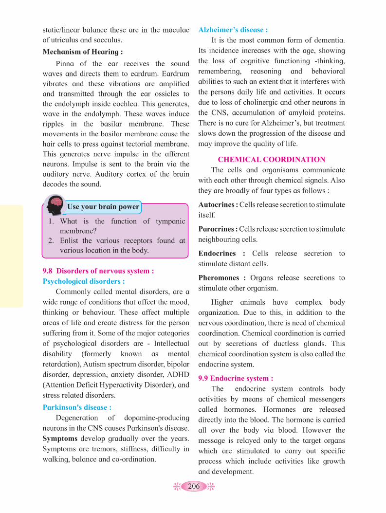
206
static/linear balance these are in the maculae of utriculus and sacculus.
Mechanism of Hearing :
Pinna of the ear receives the sound waves and directs them to eardrum. Eardrum vibrates and these vibrations are amplified and transmitted through the ear ossicles to the endolymph inside cochlea. This generates, wave in the endolymph. These waves induce ripples in the basilar membrane. These movements in the basilar membrane cause the hair cells to press against tectorial membrane. This generates nerve impulse in the afferent neurons. Impulse is sent to the brain via the auditory nerve. Auditory cortex of the brain decodes the sound.
Alzheimer’s disease : It is the most common form of dementia.
Its incidence increases with the age, showing the loss of cognitive functioning -thinking, remembering, reasoning and behavioral abilities to such an extent that it interferes with the persons daily life and activities. It occurs due to loss of cholinergic and other neurons in the CNS, accumulation of amyloid proteins. There is no cure for Alzheimer’s, but treatment slows down the progression of the disease and may improve the quality of life.
CHEMICAL COORDINATIONThe cells and organisams communicate
with each other through chemical signals. Also they are broadly of four types as follows :
Autocrines : Cells release secretion to stimulate itself.
Paracrines : Cells release secretion to stimulate neighbouring cells.
Endocrines : Cells release secretion to stimulate distant cells.
Pheromones : Organs release secretions to stimulate other organism.
Higher animals have complex body organization. Due to this, in addition to the nervous coordination, there is need of chemical coordination. Chemical coordination is carried out by secretions of ductless glands. This chemical coordination system is also called the endocrine system.
9.9 Endocrine system : The endocrine system controls body
activities by means of chemical messengers called hormones. Hormones are released directly into the blood. The hormone is carried all over the body via blood. However the message is relayed only to the target organs which are stimulated to carry out specific process which include activities like growth and development.
9.8 Disorders of nervous system :Psychological disorders :
Commonly called mental disorders, are a wide range of conditions that affect the mood, thinking or behaviour. These affect multiple areas of life and create distress for the person suffering from it. Some of the major categories of psychological disorders are - Intellectual disability (formerly known as mental retardation), Autism spectrum disorder, bipolar disorder, depression, anxiety disorder, ADHD (Attention Deficit Hyperactivity Disorder), and stress related disorders.
Parkinson's disease : Degeneration of dopamine-producing
neurons in the CNS causes Parkinson's disease. Symptoms develop gradually over the years. Symptoms are tremors, stiffness, difficulty in walking, balance and co-ordination.
Use your brain power
1. What is the function of tympanic membrane?
2. Enlist the various receptors found at various location in the body.

207
Chemical nature of hormonesI. Amines : These are simple amines.
Catecholamines secreted by adrenal medulla, epinephrine and non-epinephrine and melatonin from pineal gland. Some are modified from the amino acids. e.g., Thyroxine.
II. Peptide hormones : These hormones consist of long or short chains of amino acids. e.g. Hormones of hypothalamus oxytocin, ADH, GnRH.
III. Protein hormone : Insulin, glucagon TSH, FSH, LTH, GH, relaxin.
IV. Fatty acid derivatives :- ProstaglandinV. Steroid hormones : These hormones are
lipid soluble and derived from cholesterol and other steroids. e.g. estrogen testosterone, aldosterone. Action of these hormones is concerned with long lasting responses.
VI. Gas : NO (Nitric Oxide)
Activity :
Properties of Hormones : They act as chemical messengers and are
effective in very low concentration. Hormones can function as regulators that inhibit or stimulate or modify specific processess. Some hormones interact with receptors present on plasma membrane of target cells where as some enter the necluses to interact with genes. Hypersecretion or Hyposecretion of hormons leads to various disorders.
These are metabolised after their function. Thus cannot be reused. Hormone secretion is regulated by positive or negative feedback mechanism.
Mechanism of hormone action : Hormones are released in a very small
quantity. They produce their effect on the target organs cells by binding to hormone receptors. The hormone receptors may be on the cell membrane or may be intracellular. A hormone
Identify the glands and state their functions.
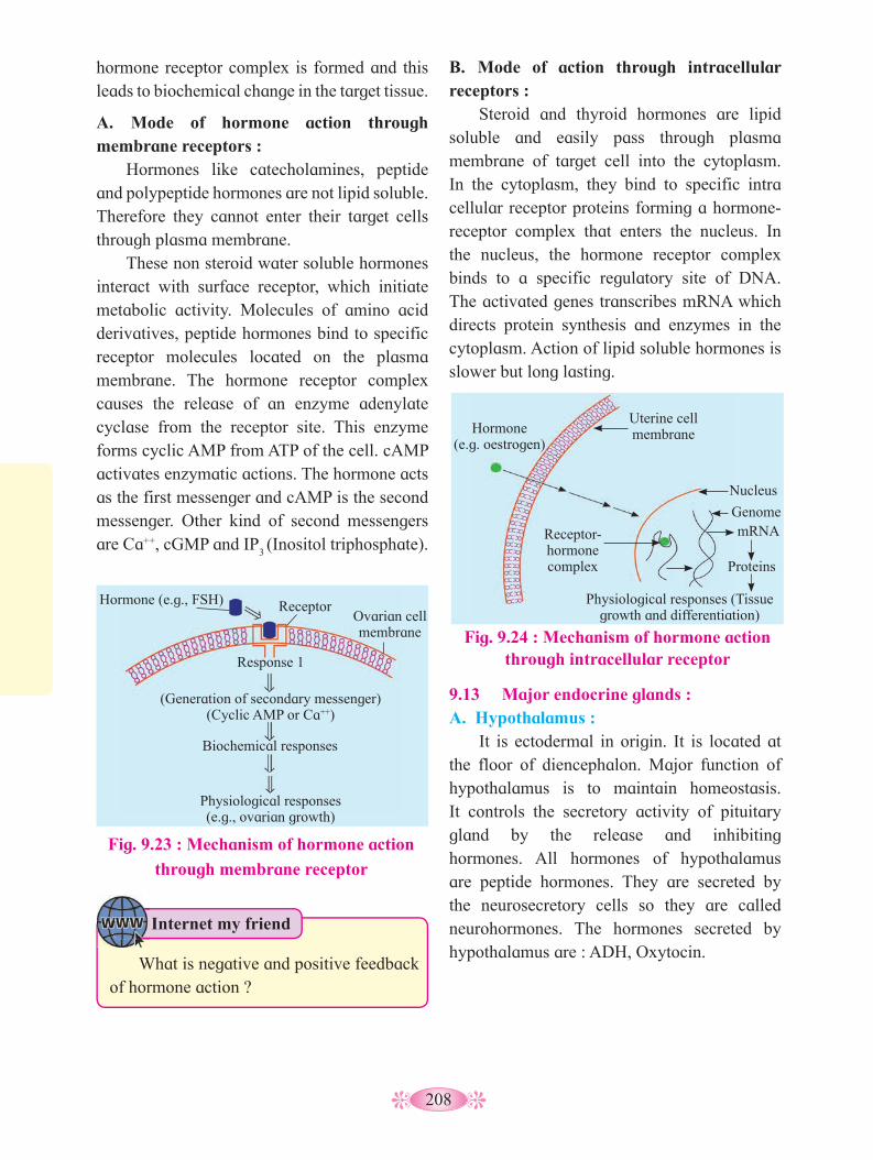
208
hormone receptor complex is formed and this leads to biochemical change in the target tissue.
A. Mode of hormone action through membrane receptors :
Hormones like catecholamines, peptide and polypeptide hormones are not lipid soluble. Therefore they cannot enter their target cells through plasma membrane.
These non steroid water soluble hormones interact with surface receptor, which initiate metabolic activity. Molecules of amino acid derivatives, peptide hormones bind to specific receptor molecules located on the plasma membrane. The hormone receptor complex causes the release of an enzyme adenylate cyclase from the receptor site. This enzyme forms cyclic AMP from ATP of the cell. cAMP activates enzymatic actions. The hormone acts as the first messenger and cAMP is the second messenger. Other kind of second messengers are Ca++, cGMP and IP
3 (Inositol triphosphate).
B. Mode of action through intracellular receptors :
Steroid and thyroid hormones are lipid soluble and easily pass through plasma membrane of target cell into the cytoplasm. In the cytoplasm, they bind to specific intra cellular receptor proteins forming a hormone-receptor complex that enters the nucleus. In the nucleus, the hormone receptor complex binds to a specific regulatory site of DNA. The activated genes transcribes mRNA which directs protein synthesis and enzymes in the cytoplasm. Action of lipid soluble hormones is slower but long lasting.
Fig. 9.24 : Mechanism of hormone action through intracellular receptor
Hormone (e.g. oestrogen)
Uterine cell membrane
Receptor-hormone complex
Physiological responses (Tissue growth and differentiation)
Nucleus
Genome mRNA
Proteins
Fig. 9.23 : Mechanism of hormone action
through membrane receptor
Hormone (e.g., FSH) Receptor Ovarian cell membrane
Response 1
(Generation of secondary messenger) (Cyclic AMP or Ca++)
Biochemical responses
Physiological responses (e.g., ovarian growth)
9.13 Major endocrine glands : A. Hypothalamus :
It is ectodermal in origin. It is located at the floor of diencephalon. Major function of hypothalamus is to maintain homeostasis. It controls the secretory activity of pituitary gland by the release and inhibiting hormones. All hormones of hypothalamus are peptide hormones. They are secreted by the neurosecretory cells so they are called neurohormones. The hormones secreted by hypothalamus are : ADH, Oxytocin.
Internet my friend
What is negative and positive feedback of hormone action ?

209
B. Pituitary gland or hypophysis gland : The pituitary gland is the smallest gland.
Pituitary gland is a pea sized reddish-grey coloured gland. It controls almost all other endocrine glands, hence earlier it was called the master endocrine gland. It is located just below the hypothalamus and is attached to it by a stalk called infundibulum or hypophyseal stalk. Pituitary gland remains lodged in a bony depression called sella turcica of the sphenoid bone. Pituitary gland consists of two lobes called anterior lobe (Adenohypophysis) and posterior lobe (Neurohypophysis). Both the lobes develop from different parts of embryo. Hence it has of dual origin.
Adenohypophysis is an outgrowth from the roof of buccal cavity. This outgrowth is called Rathke’s pouch. It grows upward towards the brain. The neurohypophysis grows as a downward extension of hypothalamus. The two outgrowths together form the pituitary gland. The connection of Rathke’s pouch with pituitray gland is lost in embryo. Intermediate lobe (Pars intermedia) is a small reduced part
lying in the cleft between the anterior and posterior lob.
Neurohypophysis is connected directly to the hypothalamus by axon fibres. Adenohypophysis and intermediate lobes are connected to the hypothalamus through hypophyseal portal system.
1. Adrenocorticotropin Releasing Hormone : It stimulates the release of ACTH by the anterior pituitary gland.
2. Thyrotropin Releasing Hormone : It stimulates the release of TSH by anterior pituitary gland.
3. Gonadotropin Releasing Hormone (GnRH) : It stimulates pituitary to secrete gonadotropins.
4. Prolactin Inhibiting Hormone (Prolactostatin) : It inhibits prolactin release by anterior piturary gland.
5. Somatostatin : It inhibits the release of growth hormone.
6. Somatotropin stimulates release of growth hormone.
7. Gastrin Releasing Peptide (GRP) and 8. Gastric Inhibitory Polypeptide (GIP)
Hypophyseal portal system : Various hormones secreted by hypothalamus reach the pituitary gland through the hypophyseal portal system. The portal vein collects blood from various parts of hypothalamus and opens into anterior lobe of pituitary. From pituitary, the vein finally carries the blood into the superior vena cava.
Adenohypophysis: It is the larger lobe of pituitary gland. It is a highly cellular and vascular part of pituitary gland. It contains various types of epitheloid secretory cells, acidophils, basophils, chromatophores. It is differentiated into three parts – pars distalis, pars intermedia and pars tuberalis. .
Fig. 9.25 : Pituitary gland
Median eminence
Infundibulum
Pars nervosa of neurohypophysis
Pars intermedia
Supraoptic nucleus(ADH)
Hypophyseal portal vein
Hypophyseal vein
Hypophyseal artery
Pars distalis of adenohypophysis
Paraventricular nucleus
(oxytocin)
Optic chiasma
210
The hormones of adenohypophysis are as follows :1. Somatotropin /Somatotropic Hormone /
STH / Growth hormone / GH : This hormone stimulates growth and
development of all tissues by accelerating protein synthesis and cell division. Highest secretion the GH is seen till puberty and then its secretion of becomes low. However, it is continuously secreted through out life for repair and replacement of body tissue or cells.
Improper secretion of growth hormone produces various disorders. Hyposecretion of growth hormone since childhood results in stunted physical growth and condition is called pituitary dwarfism.
Hypersecretion of growth hormone in childhood causes Gigantism, a condition of overgrowth. The individual attains abnormal height. When the pituitary gland produces excess growth hormone in middle aged adults, it results in disproportionate growth causing disfigurement and enlargement of bones of nose, lower jaw, hands, fingers and feet. The condition is called Acromegaly.
2. Thyrotropin / Thyroid stimulating Hormone / TSH : Its primary action is to stimulate the thyroid gland secretion of the hormone thyroxine.
3. Adreno corticotropic hormone / ACTH / Adrenocorticotropin : It stimulates adrenal cortex to produce and secrete its hormones. It maintains functioning of adrenal cortex.
4. Prolactin / Luteotropin / Mammotropin: Prolactin is unique among pituitary hormones as it is under predominant inhibitory control from hypothalamus. Prolactin activates growth of breasts during pregnancy (mammotropin) and stimulates the milk production and secretion of milk (lactogenic) by mammary gland after child birth.
5. Gonadotropin :a. Follicle stimulating hormone/ FSH :
It stimulates growth of ovarian follicles in the females, while in males, it is concerned with the development of seminiferous tubules.
b. Leutinizing Hormone/ LH : In female, the leutinizing hormone helps
in ovulation (discharge of ovum from graafian follicle). FSH and LH are responsible for stimulation of ovaries to produce oestrogen while LH induces the ruptured follicles to develop into corpus luteum and for production of progesterone.
c. Interstitial cell stimulating hormone / ICSH :
In males, it stimulates the testes to produce the androgen called testosterone. Testosterone is responsible for development of secondary sexual characters.
Neurohypophysis: It is differentiated into three parts, 1. Pars nervosa/ neural lobe 2. Infundibulum 3. Median eminence.
The pars nervosa acts as storage area for the secretions of hypothalamus. It stores and releases oxytocin and vasopressin.
1. Oxytocin : It stimulates contraction of uterus during parturition. It also stimulates the contraction of mammary glands to initiate ejection or release of milk. So is called birth hormone or milk ejecting hormone.
2. Antiduretic Hormone (ADH)/ Vasopressin: It stimulates the re-absorption of water in distal convoluted tubule and collecting ducts of uriniferous tubules of the kidneys. It decreases loss of water by reducing the urine quantity. It increases blood pressure by causing vaso constriction.
Internet my friend
Collect the information about Simmond’s disease and Gorilla rib.
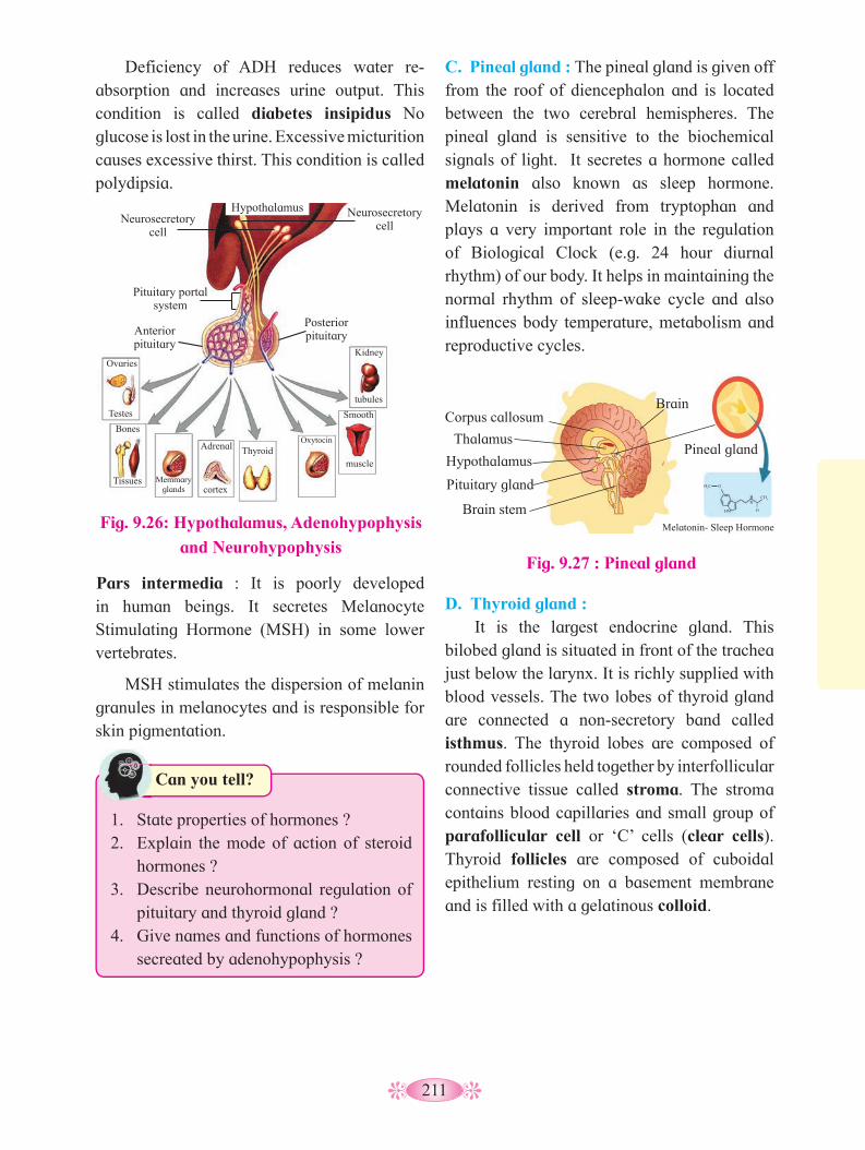
211
Deficiency of ADH reduces water re-absorption and increases urine output. This condition is called diabetes insipidus No glucose is lost in the urine. Excessive micturition causes excessive thirst. This condition is called polydipsia.
C. Pineal gland : The pineal gland is given off from the roof of diencephalon and is located between the two cerebral hemispheres. The pineal gland is sensitive to the biochemical signals of light. It secretes a hormone called melatonin also known as sleep hormone. Melatonin is derived from tryptophan and plays a very important role in the regulation of Biological Clock (e.g. 24 hour diurnal rhythm) of our body. It helps in maintaining the normal rhythm of sleep-wake cycle and also influences body temperature, metabolism and reproductive cycles.
Fig. 9.26: Hypothalamus, Adenohypophysis
and Neurohypophysis
Pituitary portal system
Neurosecretory cell
Anterior pituitary
Ovaries
Testes
Bones
Tissues Memmary glands
Adrenal
cortex
ThyroidOxytocin
Smooth
muscle
Kidney
tubules
Posterior pituitary
Neurosecretory cell
Hypothalamus
Pars intermedia : It is poorly developed in human beings. It secretes Melanocyte Stimulating Hormone (MSH) in some lower vertebrates.
MSH stimulates the dispersion of melanin granules in melanocytes and is responsible for skin pigmentation.
Fig. 9.27 : Pineal gland
Corpus callosum
Thalamus
Hypothalamus
Pituitary gland
Brain stem
Pineal gland
Melatonin- Sleep Hormone
H3C O
HN
HN ||
O
CH3
Brain
D. Thyroid gland : It is the largest endocrine gland. This
bilobed gland is situated in front of the trachea just below the larynx. It is richly supplied with blood vessels. The two lobes of thyroid gland are connected a non-secretory band called isthmus. The thyroid lobes are composed of rounded follicles held together by interfollicular connective tissue called stroma. The stroma contains blood capillaries and small group of parafollicular cell or ‘C’ cells (clear cells). Thyroid follicles are composed of cuboidal epithelium resting on a basement membrane and is filled with a gelatinous colloid.
Can you tell?
1. State properties of hormones ?2. Explain the mode of action of steroid
hormones ?3. Describe neurohormonal regulation of
pituitary and thyroid gland ?4. Give names and functions of hormones
secreated by adenohypophysis ?

212
Thyroid gland is stimulated to secrete its hormones by thyroid stimulating hormone (TSH). The two hormones secreted by the follicular cells are Thyroxine/tetra iodothyronine/ T
4 (four atoms of iodine) and
Triiodothyronine or T3 (three atoms of iodine).
Thyroxine is synthesized by attaching iodine to amino acid tyrosine by enzymatic action. The amino acid tyrosine molecule binds to iodine to produce Monoiodotyronine (T
1) or
2 atoms of iodine to produce Diiodothyronine (T
2). T
1 and
T
2 molecules bind end to end
to make colloidal mass inside the follicle. They are further metabolised to prepare T
3
and T4. Triidothyronine or T
3 is also secreted
in small quantity. It is physiologically more active. Thyroid gland is the only gland that stores its hormones. T
3 and T
4 hormones are
stored before secretion and are regulated by thyrotropin of pituitary gland by negative feed back mechanism.
Thyroxine regulates the basal metabolic rate of body. It also regulates metabolism by stimulating protein synthesis and promotes growth of body tissues. It helps in thermoregulation by increasing heat production. It increases action of neuro transmitters- adrenaline and nor adrenaline. It also supports the process of RBC production and maintenance of water and electrolyte balance. It also regulates reproductive cycles in females.
Parafollicular cells or ‘C’ cells produce calcitonin hormone, which regulates calcium metabolism.
Disorders related to thyroid gland: a. Hyperthyroidism :
It is caused by increase in the levels of thyroid hormones. This increases metabolic rate, sensitivity, sweating, flushing, rapid respiration, bulging of eye balls, and affects various physiological activities.
A
Larynx
Left thyroid gland
Trachea
Right thyroid gland
Fig. 9.28 Thyroid gland
B
Blood capillaries
Follicular cells
Thyroid follicles
Parafollicular cells or C- cells
Fig. 9.29 Bulging eyeballs
Grave’s disease (Exopthalmic goitre) : Hyperthyrodism in adults, is characterised by protruding eyeballs, increased BMR and weight loss. Increased BMR produces a range of effect like increased heart beat, increased BP, higher
Think about it
1. Patient suffering from hypothyroidism shows increased level of TSH. Why ?
2. Why do we use iodized salt?3. In which part thyroid gland stores its
hormones?
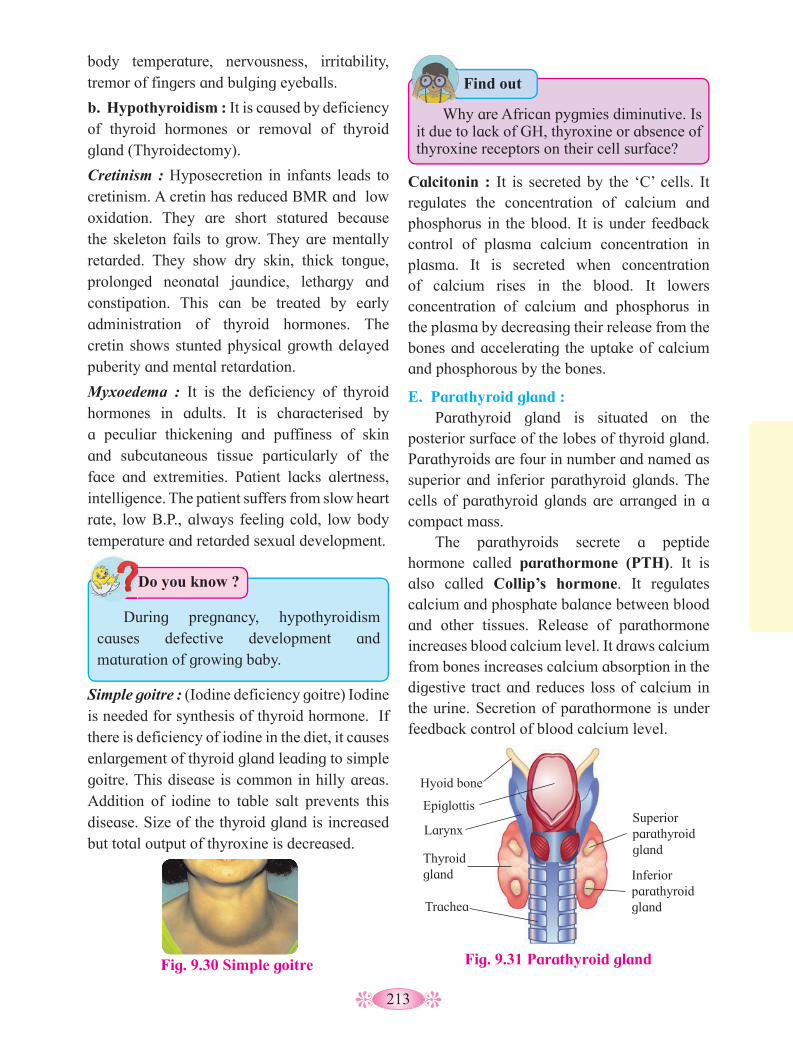
213
body temperature, nervousness, irritability, tremor of fingers and bulging eyeballs.
b. Hypothyroidism : It is caused by deficiency of thyroid hormones or removal of thyroid gland (Thyroidectomy).
Cretinism : Hyposecretion in infants leads to cretinism. A cretin has reduced BMR and low oxidation. They are short statured because the skeleton fails to grow. They are mentally retarded. They show dry skin, thick tongue, prolonged neonatal jaundice, lethargy and constipation. This can be treated by early administration of thyroid hormones. The cretin shows stunted physical growth delayed puberity and mental retardation.
Myxoedema : It is the deficiency of thyroid hormones in adults. It is characterised by a peculiar thickening and puffiness of skin and subcutaneous tissue particularly of the face and extremities. Patient lacks alertness, intelligence. The patient suffers from slow heart rate, low B.P., always feeling cold, low body temperature and retarded sexual development.
Fig. 9.30 Simple goitre
Simple goitre : (Iodine deficiency goitre) Iodine is needed for synthesis of thyroid hormone. If there is deficiency of iodine in the diet, it causes enlargement of thyroid gland leading to simple goitre. This disease is common in hilly areas. Addition of iodine to table salt prevents this disease. Size of the thyroid gland is increased but total output of thyroxine is decreased.
Calcitonin : It is secreted by the ‘C’ cells. It regulates the concentration of calcium and phosphorus in the blood. It is under feedback control of plasma calcium concentration in plasma. It is secreted when concentration of calcium rises in the blood. It lowers concentration of calcium and phosphorus in the plasma by decreasing their release from the bones and accelerating the uptake of calcium and phosphorous by the bones.
E. Parathyroid gland : Parathyroid gland is situated on the
posterior surface of the lobes of thyroid gland. Parathyroids are four in number and named as superior and inferior parathyroid glands. The cells of parathyroid glands are arranged in a compact mass.
The parathyroids secrete a peptide hormone called parathormone (PTH). It is also called Collip’s hormone. It regulates calcium and phosphate balance between blood and other tissues. Release of parathormone increases blood calcium level. It draws calcium from bones increases calcium absorption in the digestive tract and reduces loss of calcium in the urine. Secretion of parathormone is under feedback control of blood calcium level.
Hyoid bone
Fig. 9.31 Parathyroid gland
Epiglottis
Larynx
Thyroid gland
Trachea
Superior parathyroid gland
Inferior parathyroid gland
Do you know ?
During pregnancy, hypothyroidism causes defective development and maturation of growing baby.
Find out
Why are African pygmies diminutive. Is it due to lack of GH, thyroxine or absence of thyroxine receptors on their cell surface?
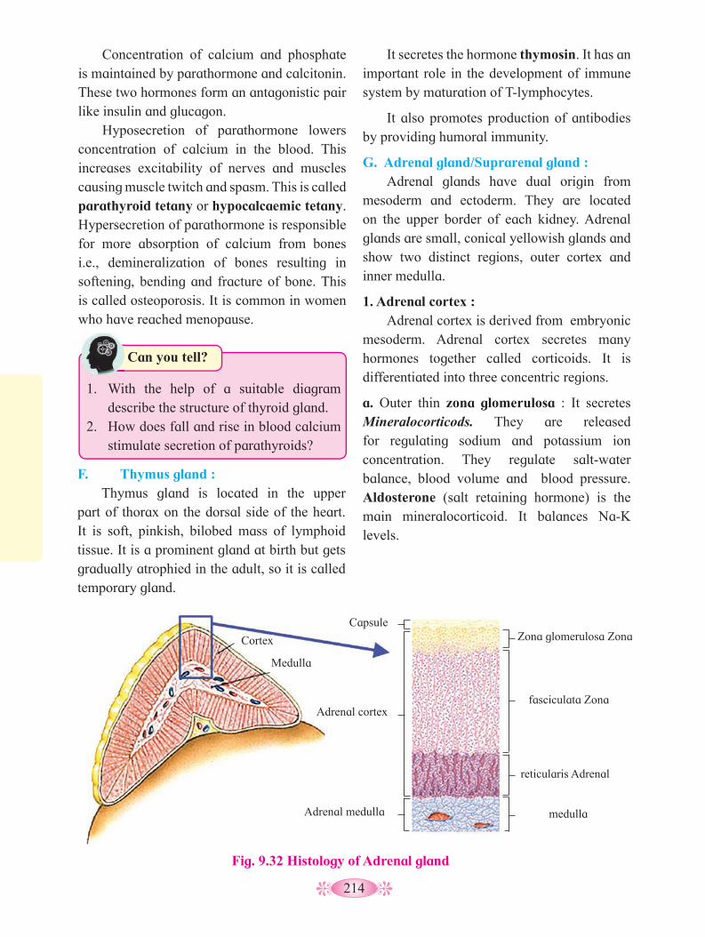
214
Concentration of calcium and phosphate is maintained by parathormone and calcitonin. These two hormones form an antagonistic pair like insulin and glucagon.
Hyposecretion of parathormone lowers concentration of calcium in the blood. This increases excitability of nerves and muscles causing muscle twitch and spasm. This is called parathyroid tetany or hypocalcaemic tetany. Hypersecretion of parathormone is responsible for more absorption of calcium from bones i.e., demineralization of bones resulting in softening, bending and fracture of bone. This is called osteoporosis. It is common in women who have reached menopause.
It secretes the hormone thymosin. It has an important role in the development of immune system by maturation of T-lymphocytes.
It also promotes production of antibodies by providing humoral immunity.
G. Adrenal gland/Suprarenal gland : Adrenal glands have dual origin from
mesoderm and ectoderm. They are located on the upper border of each kidney. Adrenal glands are small, conical yellowish glands and show two distinct regions, outer cortex and inner medulla.
1. Adrenal cortex : Adrenal cortex is derived from embryonic
mesoderm. Adrenal cortex secretes many hormones together called corticoids. It is differentiated into three concentric regions.
a. Outer thin zona glomerulosa : It secretes Mineralocorticods. They are released for regulating sodium and potassium ion concentration. They regulate salt-water balance, blood volume and blood pressure. Aldosterone (salt retaining hormone) is the main mineralocorticoid. It balances Na-K levels.
F. Thymus gland : Thymus gland is located in the upper
part of thorax on the dorsal side of the heart. It is soft, pinkish, bilobed mass of lymphoid tissue. It is a prominent gland at birth but gets gradually atrophied in the adult, so it is called temporary gland.
Fig. 9.32 Histology of Adrenal gland
Can you tell?
1. With the help of a suitable diagram describe the structure of thyroid gland.
2. How does fall and rise in blood calcium stimulate secretion of parathyroids?
Cortex
Medulla
Adrenal cortex
Capsule
Adrenal medulla
Zona glomerulosa Zona
fasciculata Zona
reticularis Adrenal
medulla

215
b. Middle thick zona fasciculata : It is responsible for secretion of Glucocorticoids like cortisol. It regulates metabolism of carbohydrates, proteins and lipids. Cortisol is an important glucocorticoid. It is responsible for increase in blood glucose level. It is also immuno suppressive. It suppresses synthesis of antibodies. So it is used in treatment of allergy. It prepares animals to face emergencies in nature.
c. Inner thin zona reticularis : It is responsible for production of sex corticoids (Gonadocorticoids). In males, they have a role in development and maintenance of external sex characters. Excess sex corticoids in female causes adrenal virilism and hirsutism (excess hair on face) while in males it causes gynaecomastia i.e. enlarged breast. Androgens and estradiols are the produced by the adrenal cortex.
Disorders related to Adrenal cortex :
a. Hyposecretion of mineralocorticoids and glucocorticoids is responsible for Addison’s disease. Characteristic features of this disease are low blood sugar, low Na+ and high K+ concentration in plasma, increased loss of Na+ and water in urine. It leads to weight loss, weakness, nausea, vomiting and diarrhoea.
b. Hyper secretion of glucocorticoids produces Cushing’s disease. It leads to high blood sugar level, excretion of glucose in urine, rise Na+ in blood volume, high blood pressure, obesity and wasting of limb muscles.
Adrenal medulla : It develops from ectoderm. It secretes two hormones adrenaline (epinephrine) and noradrenaline (norepinephrine). Adrenaline is also known as emergency hormone, also called 3F hormone – (fight, flight and fright). Noradrenaline regulates the blood pressure under normal condition. It also acts as vasoconstrictor.
H. Pancreas :
It develops from endoderm. It is both exocrine (studied in Digestive System) and endocrine gland. Endocrine cells of pancreas form groups of cells called Islets of Langehans. There are four kinds of cells in islets of Langerhans which secrete hormones.I. Alpha (a) cells (20%) secrete glucagon.
It stimulates liver for glucogenolysis to increase blood glucose level.
II. Beta (b) cells (70%) secrete insulin. It stimulates liver and muscles for glycogenesis. This lowers blood glucose level.
III. Delta (d) cell (5%) secrete somatostatin which inhibits the secretion of glucagon and insulin. It also decreases the gastric secretions, motility and absorption in digestive tract.
IV. PP cells or F cells (5%) secrete pancreatic polypeptide (PP). It inhibits the release of pancreatic juice.
Disorder related to pancreas :Diabetes mellitus (Hyperglycemia)
This is the most common metabolic endocrine disorder of pancreas. It leads to increase in blood glucose level. This is due to under activity of Beta cells, which results in reduced secretion of insulin. In children, such a condition is called insulin dependent diabetes mellitus/ Type I (IDDM) The other form of diabetes is Non insulin dependent diabetes mellitus/ Type II (NIDDM). It is caused due to failure of insulin to facilitate the movement of glucose into cells. Reduced sensitivity to insulin is called insulin resistance.

216
In both disorders, blood glucose level increases. Some of the glucose is excreted in urine. It also causes excessive urination and dehydration of body tissues. Degradation of fats increases formation of ketone bodies (ketosis). Administration of insulin lowers blood glucose level.
I. Gonads :
Gonads are sex organs (the testes and the ovaries).
i. Ovaries : They produce
1. Estrogen : These are secreted by developing follicle. Estradiol is the main oestrogen. It is responsible for secondary sexual characters in female.
2. Progesterone : It is secreted by corpus luteum of the ovary after ovulation. This hormone is essential for thickening of uterine endometrium, thus preparing the uterus for implantation of fertilized ovum. It is responsible for development of mammary glands during pregnancy. It inhibits uterine contractions during pregnancy.
3. Relaxin : It is secreted by the corpus luteum of the ovary at the end of gestation period. It relaxes the cervix of the pregnant female and ligaments of pelvic girdle for easy birth of young one.
4. Inhibin : It is secreted by the corpus luteum. Inhibin inhibits the FSH and GnRH production.
ii. Testes :
Testes secrete male sex hormones called androgens such as testosterone.
Testosterone : It is secreted from interstitial cells or Leydig cells by the influence of luteinising hormone (LH). Rise in testosterone level in blood above normal inhibits LH secretion.
It is also responsible for appearance of secondary sexual characters such as facial and pubic hair, deepening of voice, broadening of shoulders, male aggressiveness, etc. It also helps in maintenance of testes.
J. Diffuse endocrine glands
Placenta :
It is the intimate connection between foetus and uterine wall of the mother for physiological exchange of the material. Placenta is a temporary endocrine gland.
During pregnancy, placenta secretes hormones such as estrogen, progesterone, hCG (Human Chorionic Gonadotropin) and human placental progesterone. These hormones check the contraction of uterine muscles and also maintain the thickness of uterine endometrium thus they help to maintain pregnancy.
Gastro intestinal tract :
Do you know ?
Presence of HCG in urine sample indicates pregnancy.
Can you tell?
1. Distinguish between glucocorticoids and mineralcorticoids.
2. Name the hormones secreted by the adrenal cortex and state their role.
3. Write a note on Islets of Langerhans.4. Pancreas is both exocrine as well as
endocrine gland. Give reason.
In the gastrointestinal mucosa, certain cells are endocrine in function. These cells produce hormones which play vital role in digestive processes and flow of digestive juices.
1. Gastrin : It stimulates gastric glands to produce gastric juice.
2. Secretin : It is responsible for secretion of pancreatic juice and bile from presence and liver.
3. Cholecystokinin CCK/ Pancreozymin PZ : This hormone stimulates the pancreas to
217
release its enzymes and also stimulates gall bladder to release bile.
4. Entero-gastrone / Gastric inhibitory peptide (GIP) : It slows gastric contractions and inhibits the secretion of gastric juice.
Kidney : It produces renin, erythropoietin and calcitriol (calcitriol is the active form of vitamin cholecalciferol (D
3)).
Heart : Atrial natriuretic hormone /ANF. Increases sodium excretion by kidneys and reduces blood pressure.
Do you know ?
Can you tell?
1. Give significance of relaxin and inhibin.2. Enlist hormones secreted by GI tract and
state their role.3. Mention the role of heart and kidney in
hormone secretion.
Chemicals that operate between members of the same species are social hormones or pheromones. These are commonly also called sex attractants or external hormones.
A pheromone is a volatile substance produced and discharged by an organism, which induces a physiological response in other organism of the same species. Pheromones are produced by many species of insects. Some pheromones enhance the chance of mating between the sexes. These are called signaling pheromones used to induce a behavioral response. Social insects such as ants make use of signaling pheromones to locate food sources and warn of danger.
Worker bees are females maintained in a sterile state by the pheromone called anti– queen factor produced by queen. The factor spreads among the workers preventing maturation of the ovaries of workers as long as the queen is present in the bee hive. Increase in colony size results in dilution of pheromones and second queen may develop.
Hormone therapy/ HT : Hormone therapy is the use of hormones in medical treatment. HT is applied in Pregnancy, Menopause, Osteoprosis, Growth hormone deficiency, Insulin Resistance, Cancer, etc.
Activity :
1. Categorise given activities into appropriate type of reflex action.
2. Prepare concept map of mechanism of hearing.
Swimming
Dancing
Cycling
Salivation
Blinking of eyes
Sneezing

218
Q. 1 Multiple choice questions.1. The nervous system of mammals uses
both electrical and chemical means to send signals via neurons. Which part of the neuron receives impulse?
a. Axon b. Dendron c. Nodes of Ranvier d. Neurilemma
2. ___________ is a neurotransmitter. a. ADH b. Acetyl CoA c. Acetyl choline d. Inositol
3. The supporting cells that produce myelin sheath in the PNS are _________.
a. Oligodendrocytes b. Satellite cells c. Astrocytes d. Schwann cells
4. A collection of neuron cell bodies located outside the CNS is called _________.
a. Tract b. Nucleus c. Nerve d. Ganglionl
5. Receptors for protein hormones are located
a. in cytoplasm b. on cell surface c. in nucleus d. on Golgi complex6. If parathyroid gland of man are removed,
the specific result will be a. onset of aging b. disturbance of Ca++ c. onset of myxoedema d. elevation of blood pressure
7. Hormone thyroxine, adrenaline and non-adrenaline are formed from ----------
a. Glycine b. Arginine c. Ornithine d. Tyrosine
8. Pheromones are chemical messengers produced by animals and released outside the body. The odour of these substance affects
a. skin colour b. excretion c. digestion d. behaviour
9. Which one of the following is a set of discrete endocrine gland
a. Salivary, thyroid, adrenal, ovary b. Adrenal, testis, ovary, liver c. Pituitary, thyroid, adrenal, thymus d. pituitary, pancreas, adrenal, thymus
10. After ovulation, Graafian follicle changes into
a. Corpus luteum b. Corpus albicans c. Corpus spongiosum d. Corpus callosum
11. Which one of the following pair correctly matches a hormone with a disease resulting from its deficiency?
a. Parathyroid hormone - Diabetes insipidus
b. Leutinising hormone - Diabetes mellitus
c. Insulin - Hyperglycemia d. Thyroxine - Tetany
12. ___________ is in direct contact of brain in human
a. Cranium b. Duramater c. Arachnoid d. Piamater
Q. 2 Very very short answer questions.1. What is the function of red nucleus?2. What is the importance of Corpora
quadrigemina?3. What does the cerebellum of brain
control?4. Name the three ossicles of the middle
ear.5. Name the hormone which is anti abortion
hormone6. Name an organ which acts as temporary
Exercise
219
endocrine gland.7. Name the type of hormones binding to
DNA and alter gene expression.8. What is the cause of abnormal elongation
of long bones of arms and legs and of lower jaw.
9. Name the hormone secreted by the pineal gland.
10. Which endocrine gland plays important, role in improving immunity?
Q. 3 Match the organism with the type of nervous system found in them.
1. Neurons a. Earthworm2. Ladder type b. Hydra3. Ganglion c. Flatworm4. Nerve net d. Human
Q. 4 Very short answer questions.1. Describe the endocrine role of islets of
Langerhans.2. Mention the function of testosterone?3. Give symptoms of the disease caused by
hyposecretion of ADH.
Q. 5 Short answer questions1. Rakesh got hurt on his head when he fell
down from his motorbike. Which inner membranes must have protected his brain? What other roles do they have to play?
2. Give reason - Injury to medulla oblongata may prove fatal.
3. Distinguish between the sympathetic and parasympathetic nervous system on the basis of the effect they have on:
a. Heart beat b. Urinary Bladder 4. While holding a tea cup Mr. Kothari’s
hands rattle. Which disorder he may be suffering from and what is the reason for this?
5. List the properties of the nerve fibres.6. How does tongue detect the sensation of
taste?7. State the site of production and function
of Secretin, Gastrin and Cholecystokinin.
8. An adult patient surffers from low heart rate, low metabolic rate and low body temperature. He also lacks alertness, intelligence and initiative. What can be this disease? What can be its cause and care?
9. Where is the pituitary gland located? Enlist the hormones secreted by anterior pituitary.
10. Explain how the adrenal medulla and sympathetic nervous system function as a closely integrated system.
11. Name the secretion of alpha, beta and delta cells of islets of langerhans. Explain their role.
12. Which are the 2 types of goitre? What are their causes?
13. Name the ovarian hormone and give their functions.
Q. 6 Answer the following.1. Complete the table.
Location Cell Type FunctionPNS Produce myelin
sheathPNS Satellite cells.............. Oligodendrocytes Form myelin
sheath around central axon
CNS Phagocytose pathogens
CNS Form the epithelial lining of brain cavities and central canal.
Q. 7 Long answer questions.1. Explain the process of conduction of
nerve impulses upto development of action potential
2. Draw the neat labelled diagrams of.a. Human ear b. Sectional view of human eye c. L. S. of human brain d. Multipolar Neuron
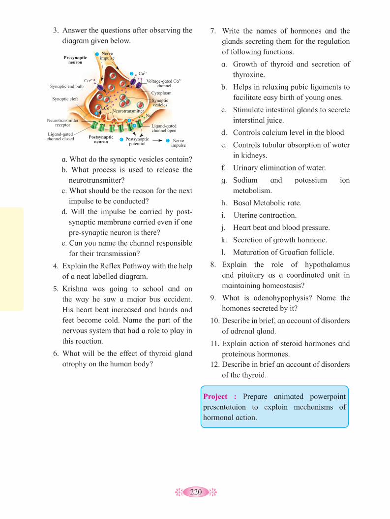
220
3. Answer the questions after observing the diagram given below.
a. What do the synaptic vesicles contain?b. What process is used to release the
neurotransmitter?c. What should be the reason for the next
impulse to be conducted?d. Will the impulse be carried by post-
synaptic membrane carried even if one pre-synaptic neuron is there?
e. Can you name the channel responsible for their transmission?
4. Explain the Reflex Pathway with the help of a neat labelled diagram.
5. Krishna was going to school and on the way he saw a major bus accident. His heart beat increased and hands and feet become cold. Name the part of the nervous system that had a role to play in this reaction.
6. What will be the effect of thyroid gland atrophy on the human body?
7. Write the names of hormones and the glands secreting them for the regulation of following functions.
a. Growth of thyroid and secretion of thyroxine.
b. Helps in relaxing pubic ligaments to facilitate easy birth of young ones.
c. Stimulate intestinal glands to secrete interstinal juice.
d. Controls calcium level in the blood
e. Controls tubular absorption of water in kidneys.
f. Urinary elimination of water.
g. Sodium and potassium ion metabolism.
h. Basal Metabolic rate.
i. Uterine contraction.
j. Heart beat and blood pressure.
k. Secretion of growth hormone.
l. Maturation of Graafian follicle.
8. Explain the role of hypothalamus and pituitary as a coordinated unit in maintaining homeostasis?
9. What is adenohypophysis? Name the homones secreted by it?
10. Describe in brief, an account of disorders of adrenal gland.
11. Explain action of steroid hormones and proteinous hormones.
12. Describe in brief an account of disorders of the thyroid.
Presynaptic neuron
Nerve impulse
Ca2+
Ca2+
Ca2+ Synaptic end bulb
Synaptic cleft
Neurotransmitter receptor
Ligand-gated channel closed
Neurotransmitter
Postsynaptic neuron
Postsynaptic potential
Nerve impulse
Ligand-gated channel open
Na+
Synaptic vesicles
Voltage-gated Ca2+ channel
Cytoplasm
1
2
3
4
5
6 7
Project : Prepare animated powerpoint presentataion to explain mechanisms of hormonal action.
221
Health does not simply mean ‘absence of disease’ or physical fitness. In fact, it is difficult to aptly define health. According to the World Health Organization, health is defined as the state of complete physical, mental and social well-being and not merely the absence of disease or infirmity. In short, health is birth right of humans. Health also reflects the metabolic and functional efficiency of living organisms. Hygiene is a science of rules of health. To achieve good health, it is therefore, necessary that we have hygienic balanced diet, clean drinking water, personal and community hygiene, regular physical exercise, knowledge about diseases and their effect on body, proper disposal of waste and control of vectors.
Everyday we are exposed to various foreign bodies, including infectious agents like bacteria, viruses, etc. Despite constant exposure to variety of pathogens, most of us remain healthy. This is due to fact that the human body has ability to resist almost all type of these foreign bodies. The system which protects us from various infectious agents, is called immune system. Resistance is the ability to prevent the damage or disease, through our defense mechanism.
10.1 Immunity: The term ‘immunity’ has traditionally
referred to as the resistance exhibited by the host towards injury caused by pathogens and their products. However, protection against infectious diseases is only one of the many
consequences of immune response, which is entirely concerned with the reaction of the body against any foreign antigen.
Immunity is in fact the ‘‘freedom’’ or ‘‘exempt’’. The concept of immunity is believed to be started by Edward Jenner in England. He developed cowpox vaccine for the protection against the attack of small pox (virus). Immunology is a branch of science which deals with the study of immune system, immune responses to foreign substances and their role in resisting infection by pathogens.
The most important characteristic of immune system is that it distinguishes self (body’s own cells) and non-self (foreign molecules or invading cells). So, the immune system differentiates between the body cells and the invaders. Any foreign substance invading body and capable of stimulating an immune response, is called an antigen. The protective chemicals produced by immune cells in response to antigens are called antibodies.
A. Types of immunity: There are two types of immunity as Innate
or Inborn (inherited) immunity and Acquired or Adaptive immunity.
i. Innate immunity or Inborn immunity: Innate immunity is the resistance to
infections that an individual possesses by virtue of his or her genetic make-up. It is the natural (inborn) defense system of the body. It is not affected by prior contact with microorganisms or immunization. It is nonspecific, when it indicates a degree of resistance to infection in general, or specific where resistance to a particular pathogen is concerned. One form of innate immunity comprises the various types of barriers which prevent entry of foreign agents into the body.
Human Health and Diseases10
1. Generally individuals are conscious about their health. So define health.
2. Define infectious and non infectious disease? Give their examples.
Can you recall?

222
a. Epithelial surface : The intact skin and mucous covering the
body, protect it considerably against invasion by microorganism(s).
The healthy skin possesses bactericidal activity due to the presence of high concentrations of salt in drying sweat.Sebaceous secretions and long chain of fatty acids have bactericidal and fungicidal properties. The mucosa of the respiratory tract has several innate mechanisms of defense. The nose prevents entry of microorganisms to a large extent, the inhaled particles being arrested through hair at or near the nasal orifices. Those that pass beyond are held by mucus lining the epithelium and are swept back to pharynx where they tend to swallowed or coughed out.
The cough reflex is an important defence mechanism of respiratory tract. The mouth is constantly bathed in saliva which has inhibitory effect on microorganisms. The acidity of gastric secretions in the stomach destroys microorganisms. The flushing action of urine eliminates bacteria from the urethra. Spermine and zinc present in semen are antibacterial.
pathogens activates the ‘Complement cascade’ to eliminate pathogens. The interferons are a class of cytokines (soluble proteins) released by virally cells infected with viruses and certain white blood cells to stimulate other cells to protect themselves from viral infection.
c. Cellular factors in innate immunity : Natural defence against the invasion of
blood and tissues by microorganisms and other foreign particles, is mediated to a large extent by phagocytic cells which ingest and destroy them. Phagocytic cells (discovered by Metchnikoff in 1882) are grouped as microphages and macrophages. They remove foreign particles that enter the body. A class of lymphocytes called Natural killer (NK) cells is important in nonspecific defence against viral infections and tumors.
d. Fever : Increase in the body temperature following
the infection is a natural defense mechanism.It helps to accelerate the physiological processes to destroy the invading pathogens. Fever stimulates the production of interferon and helps in recovery from viral infections.
e. Acute phase proteins (APPs): Infection on injury leads to a sudden
increase in concentration of certain plasma proteins, collectively called acute phase proteins. These include C Reactive Protein (CRP), Mannose binding protein, Alpha-1-acid glycoprotein, Serum Amyloid P, etc. APPs are believed to enhance host resistance, prevent tissue injury and promote repair of inflammatory lesions.
ii. Acquired immunity: The resistance that an individual acquires
during life is known as “Acquired immunity”. It is also known as Adaptive or Specific immunity”. It involves the formation of antibodies in the body, which neutralize the antigens. Acquired or Adaptive immunity has the following unique features.
Knowledge Enhancer: The conjunctiva is freed of foreign particles by the flushing action of lachrymal secretions. Eyes become susceptible to infection when lachrymal secretion is absent. Tears contain the antibacterial substance Lysozyme (Ist described by A. Fleming). This is a thermolabile, low molecular weight, basic protein which acts as a muramidase. Lysozyme is active only against some pathogenic Gram +ve bacteria.
b. Antimicrobial substances in blood and tissues:
The complement system contains more than 30 serum proteins, circulating in the blood in an inactive state. The presence of microbial

223
b. Passive immunity: Passive immunity is acquired when ready-made antibodies are received by the body cells. i.e. Body cells do not take any active part in the production of immunity. Passive immunity can be acquired either naturally or artificially.
1. Natural Acquired Passive immunity: Before birth maternal antibodies are transferred from mother to foetus through placenta. After birth, antibodies are transferred from mother to infant through colostrum (first milk of mother) and continue throughout the period of breast feeding. The antibodies received by baby from mother remain in the body for a short time. Therefore, natural acquired passive immunity is short lived.
2. Artificially Acquired Passive immunity: This immunity is developed by injecting previously prepared antibodies using serum from humans or animals. For e.g. Antibodies obtained from hyper immunised horses are injected to humans against rabies pathogens. It is short lived.
B. Cells of Immune System: There are two main types of cells involved
in the working of Immune system. They are (a) Lymphocytes and (b) Antigen Presenting cells.
a. Lymphocytes: Lymphocytes are the main cells of the
immune system. They, like the other blood corpuscles, arise from the stem cells, the haemocytoblasts, present in liver of the foetus and in the bone marrow in adult. Some of them undergo differentiation in the gut –associated bursal lymphoid tissues (Tonsils, Peyer’s patches) and are called Bursal or B-lymphocytes; others are differentiated in the thymus gland and are termed as T-lymphocytes. The mature lymphocytes pass into body fluids (blood and lymph) and circulate in the body.
a. Specificity: It can produce specific antibody or T-lymphocyte against a particular antigen/pathogen.
b. Diversity: It can recognize a vast variety of diverse pathogens or foreign molecules.
c. Discrimination between self and non-self: It differentiates between own body cells (self) and foreign (non-self) molecules.
d. Memory: When the immune system encounters a specific foreign agent for the first time, it generates an immune response and eliminates the invader. This is called first encounter. The immune system retains the memory of the first encounter. As a result, a second encounter with same pathogen brings about quicker and stronger immune response.
Types of Acquired Immunity: Acquired immunity is of two types Active
and Passive.
a. Active immunity: It is the resistance developed by
individuals as a result of an antigenic stimulus. It also known as “Adaptive immunity”. Active immunity may be natural or artificial.
1. Natural Acquired Active immunity: Immunity acquired due to infection is called natural active immunity. It is developed after entry of pathogens in the body. It is long-lasting immunity. e.g. person who has recovered from attack of measles develops natural acquired active immunity to measles, for the life time.
2. Artificial Acquired Active immunity: It is the resistance induced by vaccines. Vaccine is introduced into the body to stimulate the formation of antibodies by the immune system. e.g. Polio vaccine, BCG vaccine etc. such immunity may be temporary or permanent.
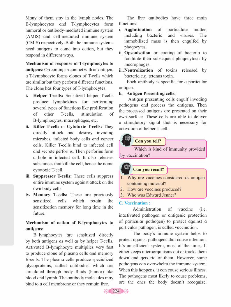
224
Many of them stay in the lymph nodes. The B-lymphocytes and T-lymphocytes form humoral or antibody-mediated immune system (AMIS) and cell-mediated immune system (CMIS) respectively. Both the immune systems need antigens to come into action, but they respond in different ways.
Mechanism of response of T-lymphocytes to antigens: On coming in contact with an antigen, a T-lymphocyte forms clones of T-cells which are similar but they perform different functions. The clone has four types of T-lymphocytes:
i. Helper T-cells: Sensitized helper T-cells produce lymphokines for performing several types of functions like proliferation of other T-cells, stimulation of B-lymphocytes, macrophages, etc.
ii. Killer T-cells or Cytotoxic T-cells: They directly attack and destroy invading microbes, infected body cells and cancer cells. Killer T-cells bind to infected cell and secrete perforins. Then perforins form a hole in infected cell. It also releases substances that kill the cell, hence the name cytotoxic T-cell.
iii. Suppressor T-cells: These cells suppress entire immune system against attack on the own body cells.
iv. Memory T-cells: These are previously sensitized cells which retain the sensitization memory for long time in the future.
Mechanism of action of B-lymphocytes to antigens:
B–lymphocytes are sensitized directly by both antigens as well as by helper T-cells. Activated B-lymphocyte multiplies very fast to produce clone of plasma cells and memory B-cells. The plasma cells produce specialized glycoproteins, called antibodies which are circulated through body fluids (humor) like blood and lymph. The antibody molecules may bind to a cell membrane or they remain free.
1. Which is kind of immunity provided by vaccination?
Can you tell?1.
The free antibodies have three main functions:i. Agglutination of particulate matter,
including bacteria and viruses. The immobilized mass is then engulfed by phagocytes.
ii. Opsonisation or coating of bacteria to facilitate their subsequent phagocytosis by macrophages.
iii. Neutralization of toxins released by bacteria e.g. tetanus toxin. Each antibody is specific for a particular
antigen.b. Antigen Presenting cells:
Antigen presenting cells engulf invading pathogens and process the antigens. Then the processed antigens are presented on their own surface. These cells are able to deliver a stimulatory signal that is necessary for activation of helper T-cell.
1. Why are vaccines considered as antigen
containing material?2. How are vaccines produced?3. Who was Edward Jenner?
Can you recall?
C. Vaccination :Administration of vaccine (i.e.
inactivated pathogen or antigenic protection of particular pathogen) to protect against a particular pathogen, is called vaccination.
The body’s immune system helps to protect against pathogens that cause infection. It’s an efficient system, most of the time,. It either keeps microorganisms out or tracks them down and gets rid of them. However, some pathogens can overwhelm the immune system. When this happens, it can cause serious illness. The pathogens most likely to cause problems, are the ones the body doesn’t recognize.

225
How does vaccination work?Healthy immune system defends against
invaders. Immune system consists of several types of cells. These cells defend against harmful pathogens. However, they have to recognize an invader. Vaccination teaches the body to recognize new pathogens causing diseases. It stimulates the body to make antibodies against antigens of pathogens. It also primes immune cells to remember the types of antigens. This allows a faster response to the pathogen in future.
Vaccines work by exposing you to a safe version of pathogen. Vaccines may be in the form of:• A protein or sugar from the pathogen.• A dead or inactivated form of a pathogen.• A toxoid containing toxin produced by a
pathogen.• A weakened (attenuated) pathogen.
When the body responds to the vaccine, it builds an adaptive immune response. This helps to equip the body to fight off an actual infection.
Do you know ?
Vaccinations are safe:Vaccines are considered to be safe. They
are rigorously tested and go through many rounds of study, examination, and research before they are used for the general public.
Extensive research and evidence shows that vaccines are safe their side effects are rare and typically mild.
Vaccination is a way to “teach” the immune system as to how to recognize and eliminate pathogenic organism. That way, body is always prepared if you are ever exposed.
Vaccination is an important form of primary prevention, that can protect people from getting sick. Vaccination has allowed us to control diseases like measles, polio, tetanus and whooping cough that once threatened many lives. It’s important that as many people as possible get vaccinated. Vaccinations don’t just protect individuals, when enough people are vaccinated. It also helps to protect the society.
Fig. 10.1 : Structure of antibody
Antigen binding site
Light chain
Heavy chain
Hinge
C
C
C
C
Variable region of the heavy chain
Antigen binding site
Variable region of the light chain
Constant region of the light chain
Disulfide bond
Constant region of the heavy chain
-s-s--s-s-
s-ss-ss-ss-s
N NN N
10.2 Structure of Antibody: Antibodies are glycoproteins which are
highly specific to specific antigens. They are also known as Immunoglobulins (Igs), produced in response to antigenic stimulation. Antibodies are produced by plasma cells which in turn are formed by B–lymphocytes. The mature plasma cells produce antibodies at an extremely rapid rate i.e. about 2000 molecules per second.
Internet my friend
Find out different types of vaccines available in the market and their significance.
Internet my friend
1. Whether vaccination can be done during or before pregnancy?
2. Will it be helpful to protect the mother and baby both?
3. Which vaccines can be administered before pregnancy? When can it be?
4. How will you increase awareness in the society on this issue?
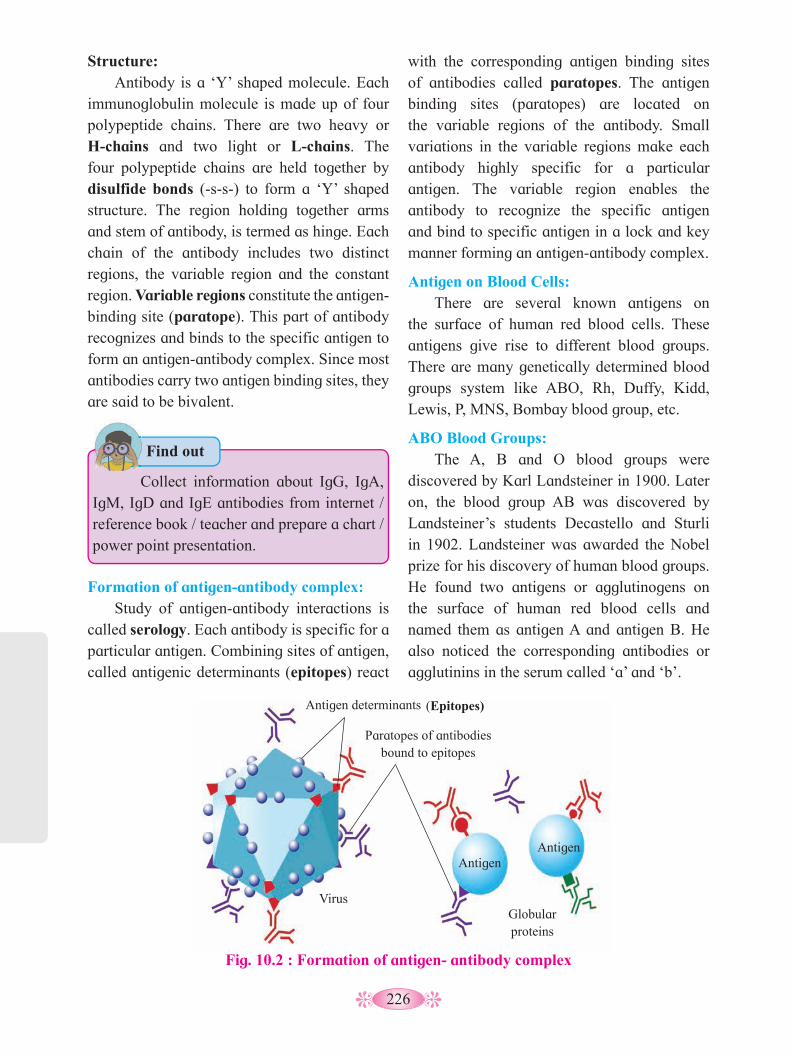
226
with the corresponding antigen binding sites of antibodies called paratopes. The antigen binding sites (paratopes) are located on the variable regions of the antibody. Small variations in the variable regions make each antibody highly specific for a particular antigen. The variable region enables the antibody to recognize the specific antigen and bind to specific antigen in a lock and key manner forming an antigen-antibody complex.
Antigen on Blood Cells:There are several known antigens on
the surface of human red blood cells. These antigens give rise to different blood groups. There are many genetically determined blood groups system like ABO, Rh, Duffy, Kidd, Lewis, P, MNS, Bombay blood group, etc.
ABO Blood Groups:The A, B and O blood groups were
discovered by Karl Landsteiner in 1900. Later on, the blood group AB was discovered by Landsteiner’s students Decastello and Sturli in 1902. Landsteiner was awarded the Nobel prize for his discovery of human blood groups. He found two antigens or agglutinogens on the surface of human red blood cells and named them as antigen A and antigen B. He also noticed the corresponding antibodies or agglutinins in the serum called ‘a’ and ‘b’.
Structure: Antibody is a ‘Y’ shaped molecule. Each
immunoglobulin molecule is made up of four polypeptide chains. There are two heavy or H-chains and two light or L-chains. The four polypeptide chains are held together by disulfide bonds (-s-s-) to form a ‘Y’ shaped structure. The region holding together arms and stem of antibody, is termed as hinge. Each chain of the antibody includes two distinct regions, the variable region and the constant region. Variable regions constitute the antigen-binding site (paratope). This part of antibody recognizes and binds to the specific antigen to form an antigen-antibody complex. Since most antibodies carry two antigen binding sites, they are said to be bivalent.
Fig. 10.2 : Formation of antigen- antibody complex
Antigen determinants (Epitopes)
Paratopes of antibodies bound to epitopes
Virus
Antigen Antigen
Globular proteins
• Collect information about IgG, IgA, IgM, IgD and IgE antibodies from internet / reference book / teacher and prepare a chart / power point presentation.
Find out •
Formation of antigen-antibody complex: Study of antigen-antibody interactions is
called serology. Each antibody is specific for a particular antigen. Combining sites of antigen, called antigenic determinants (epitopes) react

227
Table 10.3 : ABO Blood groups in man
Blood group Genotype Antigen on Surface of RBC
Antibody in Serum
Can donate blood to
Can receive blood from
A IA IA orIA IO
A Antibody b A, AB A, O
B IB IB orIB IO
B Antibody a B, AB B, O
AB (universal acceptor)
IA IB A and B Nil AB A, B, AB, O
O (universal donor)
IO IO Nil Both Antibody a and Antibody b
A, B, AB, O O
1. The blood group of Krutika is O Rh +ve.
What would be the possible blood groups of her parents?
2. Mrunmayi is called as universal blood acceptor. What is her blood group?
Can you tell?
In ABO system, the blood groups are determined by the presence or absence of antigen A and antigen B, the blood group of person is classified into four groups A, B, AB and O.
Blood group A : Individuals, with blood group ‘A’ have the antigen A on the surface of their red blood cells (RBCs) and antibody ‘b’ in their plasma.
Blood group B : Individuals with blood group ‘B’ have the antigen B on the surface of their RBCs and antibodies ‘a’ in their plasma.
Blood group AB : Individuals with blood group ‘AB’ have both antigens A and B on the surface of their RBCs and no antibodies in their plasma.
Blood group O – Individuals with blood group ‘O’ lack both antigens A and B on the surface of their RBCs and show presence of both ‘a’ and ‘b’ antibodies in their plasma.
Rh factor: Rh is the most complex of the blood group
system. Rh –factor is an antigenic protein present on the surface of the red blood cells in the human beings. It was first discovered by Landsteiner and Wiener (1940), on the surface of RBCs of Rhesus monkey, so it is called Rh-factor (also called D antigen). Person having Rh factor (D antigen) are called Rh positive (Rh +ve) and those lacking D antigen are called Rh negative (Rh -ve).
Rh (D) antigen induces a strong immunogenic response when introduced into Rh-ve individuals. Rh blood group is an important factor in blood transfusion and is involved in haemolytic diseases of the newborn (HDN), which is called erythroblastosis foetalis (destruction of the erythrocytes of the foetus). It occurs when an Rh -ve mother conceives Rh+ve foetus.
The Rh +ve RBCs from the foetus may enter the mother’s circulatory system during child birth, causing her to produce anti-Rh antibodies. As a result, subsequent Rh+ve foetuses will be exposed to the anti-Rh antibodies produced by mother, which result in HDN. In order to prevent HDN, Rh -ve mother is injected with the anti-Rh antibody during all pregnancies carrying Rh +ve foetus.
Can a person with blood group O Rh+ve donate blood to a patient with blood group O Rh-ve? Why?
Use your brain power

228
Table 10.4 : Table showing different species of Plasmodium
Species of Plasmodium Incubation period Pattern of high fever P. vivax 14 days High fever after 48 hr interval P. malariae 28 days High fever after 72 hr interval P. ovale 17 days High fever after 48 hr interval P. falciparum 12days High fever at irregular intervals between 22-48 hrs
Many pathogens use another organism, the ‘Vector’ to reach us. These vectors are actually parasites which we regularly come across. Parasite is an organism that lives in or on the body of another organism and derives its nutrition from that of host organism. Parasites are two catagories viz. : 1. Ectoparasite E.g. bedbug 2. Endoparasite. E.g. Plasmodium is a protozoan endoparasite of the mosquito (vector) and human beings.
A. Malaria :
It is a vector (mosquito) borne infectious disease caused by protist - Plasmodium.
There are four species of Plasmodium as P. vivax , P. ovale, P. malariae, P. falciparum. Only P. falciparum causes serious illness while others are rarely fatal. Based on the species, there are 4 - types of malaria.
Signs and symptoms of malaria :-
Symptoms of malaria begin to appear about 7 to 15 days after the bite of infective mosquito.
• Initial symptom are fever, headache, and chills, may be difficult to recognize as malaria.
• Classical symptoms of malaria is cyclic occurrence of high fever followed by sweating and sudden shievering. Such entire episode lasts for four to six hours and recurs every two days or three days.
• Vomiting and convulsions.• Arthralgia ( joint pain), aneamia due to
rupturing of RBCs.• Haemoglobinuria, hepatomegaly (liver
enlargement).• Retinal damage (eye).• Cerebral malaria (brain infection).
10.3 Common Human Diseases:
Disease is defined as condition of disturbed or deranged functioning of one or more organs or organ systems of the body, caused due to infections, defective diet or heredity. All human diseases can be broadly categorized into congenital diseases and acquired diseases.
Congenital diseases are present from birth; may be caused by genetic abnormality or metabolic disorder. They may be permanent and were practically incurable. However, modern research has helped to cure some inborn diseases through gene therapy, enzyme replacement therapy, etc.
Acquired diseases develop after the birth and can be subdivided into (a) Communicable or infectious diseases or (b) Non- Communicable or Non-infectious diseases.
The diseases which are transmitted from infected person to another healthy person either directly or indirectly, are known as Communicable or Infectious diseases. Malaria and other diseases which are to be studied in this chapter, are examples of this type.
The diseases that cannot be transmitted from infected person to another healthy one either directly or indirectly are known as Non- Communicable or Non - Infectious diseases. Cancer and deficiency diseases are examples of this type.
Communicable diseases are caused by pathogens like viruses, bacteria, fungi, helminth worms, etc. All the disease causing organisms are called ‘Pathogens’.

229
Mode of transmission : Plasmodium is transmitted from one
person to other through an insect vector- female Anopheles mosquito.
When infected female Anopheles sucks the human blood, it may transfer sporozoites to human circulation. Sporozoites reproduce asexually through fission (schizogony) in the liver cells or erythrocytes. The cells formed are now called merozoites. The cells formed within erythrocytes function as gametocytes (gamogony). Besides, it forms gametocytes within erythrocytes (gamogony).
Gametocytes if taken up by female Anopheles, fertilization occurs in its gut. Diploid zygote is formed which transforms into oocyst. Oocyst forms large number of haploid sporozoites through meiosis (sporogony). Sporozoites migrate to salivary glands and are ready to infect new human host.
Diagnosis and Treatment : Malaria can diagnosed by microscopic
study of blood smear. Besides, other rapid diagnostic tests based on nucleic acid amplification techniques are also used.
Fig. 10.5 : Stages in the life cycle of Plasmodium in mosquito and human
Cycle in Mosquito Cycle in Human
Sporozoites (to salivary gland)
Bursting oocyst
Oocyst
Mosquito midgut Ookinete
Zygote
Macrogamete
Microgamete
Gametocyte
Newly infected Erythrocyte
Ruptured erythrocyte Erythrocytic
schizont
Trophozoite
Erythrocyte Merozoites
Mature schizont
Hypnozoite (hepatic dormancy)
Hepatic cellHuman skin
Treatment of malaria includes Artemisinin based combination therapies (ACTs). WHO has recommended 5 different ACTs which includes various combinations of artesunate, sulfadoxine, pyrimethamine, etc. In addition, quinine is also used.
Prevention and Control :1. Prevention of mosquito bite by using
mosquito nets and insect repellents.
2. Spraying insecticides and draining stagnant water where mosquito lays eggs.
3. Mosquito larve can be controlled by using Gambusia fresh water fish (biocontrol).
B. Amoebiasis : • Amoebiasis is also known as Amoebic
dysentry. It’s a common infection of human gastro-intestinal tract, which affects 15 % population of India.
Always Remember
The time interval from the invasion of a pathogen to the development of clinical manifestations, is known as Incubation period.
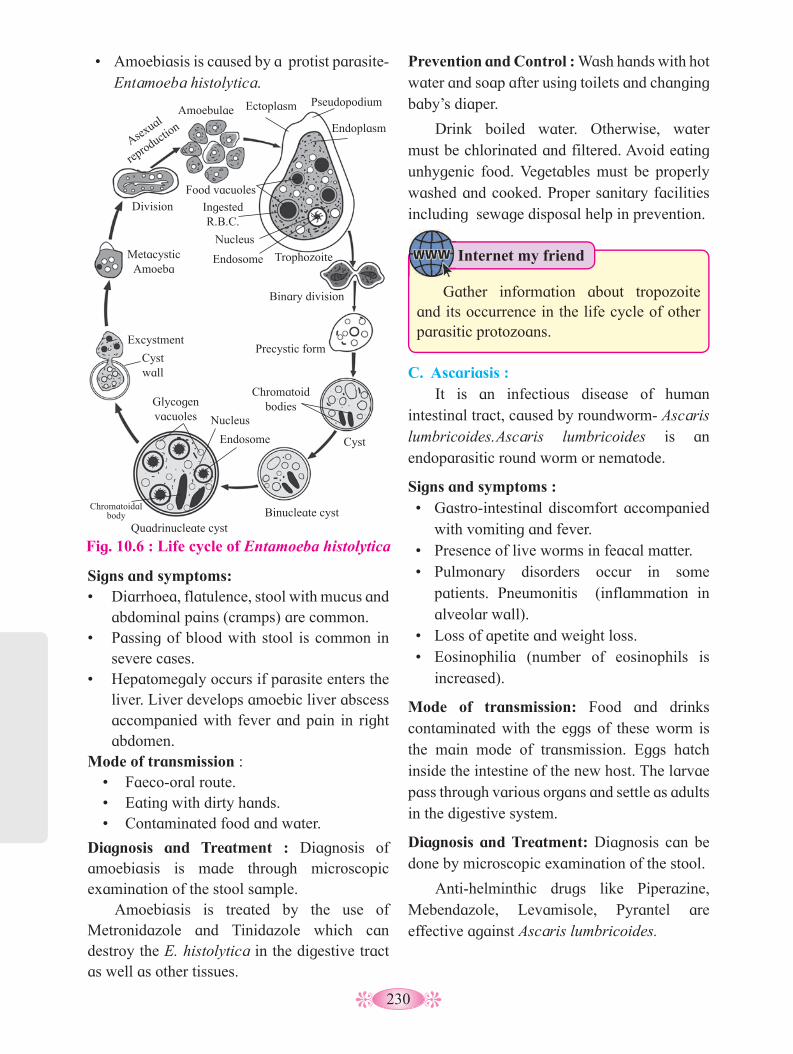
230
Prevention and Control : Wash hands with hot water and soap after using toilets and changing baby’s diaper.
Drink boiled water. Otherwise, water must be chlorinated and filtered. Avoid eating unhygenic food. Vegetables must be properly washed and cooked. Proper sanitary facilities including sewage disposal help in prevention.
Signs and symptoms:• Diarrhoea, flatulence, stool with mucus and
abdominal pains (cramps) are common.• Passing of blood with stool is common in
severe cases.• Hepatomegaly occurs if parasite enters the
liver. Liver develops amoebic liver abscess accompanied with fever and pain in right abdomen.
Mode of transmission :• Faeco-oral route.• Eating with dirty hands.• Contaminated food and water.
Diagnosis and Treatment : Diagnosis of amoebiasis is made through microscopic examination of the stool sample.
Amoebiasis is treated by the use of Metronidazole and Tinidazole which can destroy the E. histolytica in the digestive tract as well as other tissues.
• Amoebiasis is caused by a protist parasite-Entamoeba histolytica.
Fig. 10.6 : Life cycle of Entamoeba histolytica
Division
Metacystic Amoeba
Excystment
Cyst wall
Glycogen vacuoles Nucleus
Endosome
Quadrinucleate cyst Binucleate cyst
Chromatoid bodies
Cyst
Precystic form
Binary division
Trophozoite Endosome
Nucleus
Food vacuoles
Amoebulae
Endoplasm
Pseudopodium Ectoplasm
Ingested R.B.C.
Asexual
reproducti
on
C. Ascariasis : It is an infectious disease of human
intestinal tract, caused by roundworm- Ascaris lumbricoides. Ascaris lumbricoides is an endoparasitic round worm or nematode.
Signs and symptoms :• Gastro-intestinal discomfort accompanied
with vomiting and fever.• Presence of live worms in feacal matter.• Pulmonary disorders occur in some
patients. Pneumonitis (inflammation in alveolar wall).
• Loss of apetite and weight loss.• Eosinophilia (number of eosinophils is
increased).
Mode of transmission: Food and drinks contaminated with the eggs of these worm is the main mode of transmission. Eggs hatch inside the intestine of the new host. The larvae pass through various organs and settle as adults in the digestive system.
Diagnosis and Treatment: Diagnosis can be done by microscopic examination of the stool.
Anti-helminthic drugs like Piperazine, Mebendazole, Levamisole, Pyrantel are effective against Ascaris lumbricoides.
Internet my friend
Gather information about tropozoite and its occurrence in the life cycle of other parasitic protozoans.
Chromatoidal body

231
D. Filariasis/ Elephantiasis : Filariasis is caused by thread like worms-
nematodes. These nematode parasites are transported from person to person via mosquito bite. Filariasis can be divided into 3 subtypes as 1) Lymphatic Filariasis, 2) Subcutaneous Filariasis (e.g. Loa loa, Mansonella spp.) and 3) Serous (abdominal)cavity Filariasis (e.g. Mansonella spp.).
Lymphatic Filariasis (Elephantiasis) is caused by the worms - Wuchereria bancrofti, Brugia malayi, Brugia timori.
Signs and symptoms :-• Edema with thickening of skin and
underlying tissue.• Wuchereria bancrofti affects the legs,
arms, breasts, scrotum, etc.• In lymphatic filariasis, worms infect
lymphatic system and causes enlargement of lymph vessels and nodes. This is elephantiasis i.e. limbs are swollen like legs of elephant.
• Lymphedema i.e. accumulation of lymh fluid in tissue causing swelling.
Prevention and Control : Avoiding defaecation in open space, prevents the spread of Ascaris. Personal hygienic habits like washing hand with water and soap after using toilet are also important. Washing vegetables throughly before cooking and avoiding raw vegetables is important for prevention of ascariasis.
Fig. 10.8 : Mode of transmission - Filariasis (Wuchereria bancrofti)
Mosquito Stages Human Stages Mosquito takes a blood meal
(L3 larvae enter skin)
Adults in lymphatics
Adults produce sheathed microfilariae that migrate into
lymph and blood channels Mosquito takes a blood
meal (ingests microfilariae) Microfilariae, pentrate mosquito’s midgut, and
migrate to thoracic muscles
L1 larvae
L3 larvae
Two moultings
Migrate to head and mosquito’s proboscis
Infective Stage
Diagnostic Stage
1
2
3
4
5
6
7
8
Fig. 10.7 : Life cycle of Ascaris lumbricoides
Swallowing by man
Intestine
Blood
Heart
Lungs
Trachea
PharynxOesophagus Intestine
Adult
Adult worms in intestine
Infective larva develops within the
eggs
Fertilized eggs in faeces
Soil
Contaminated food
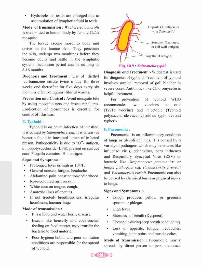
232
Diagnosis and Treatment :- Widal test is used for diagnosis of typhoid. Treatment of typhoid involves surgical removal of gall bladder in severe cases. Antibiotics like Chloromycetin is helpful treatment.
For prevention of typhoid WHO recommendes two vaccines as oral (Ty21a vaccine) and injectable (Typhoid polysaccharide vaccine) sold as- typhim vi and typherix.
F. Pneumonia : Pneumonia is an inflammatory condition
of lungs or alveoli of lungs. It is caused by a variety of pathogens which may be viruses like influenza virus, adenovirus, para influenza and Respiratory Syncytial Virus (RSV) or bacteria like Streptococcus pneumoniae or fungal pathogens e.g. Pneumocystis jirovecii and Pneumocystis carinii. Pneumonia can also be caused by chemical burns or physical injury to lungs.
Signs and Symptoms :-
• Cough produces yellow or greenish sputum or phlegm.
• High fever.
• Shortness of breath (Dyspnea).
• Chest pain during deep breath or coughing.
• Loss of appetite, fatigue, headaches, vomiting, joint pains and muscle aches.
Mode of transmission : Pneumonia mostly spreads by direct person to person contact.
Fig. 10.9 : Salmonella typhi
Capsule (K antigen, or v
i in Salmonella)
Somatic (O antigen, or cell wall antigen)
Flagella (H antigen)
• Hydrocele i.e. testis are enlarged due to accumulation of lymphatic fluid in testis.
Mode of transmission : Wuchereria bancrofti is transmitted to human body by female Culex mosquito.
The larvae escape mosquito body and arrive on the human skin. They penetrate the skin, undergo two moultings before they become adults and settle in the lymphatic system. Incubation period can be as long as 8-16 months.
Diagnosis and Treatment : Use of diethyl -carbamazine citrate twice a day for three weeks and thereafter for five days every six month is effective against filarial worms.
Prevention and Control : Avoid mosquito bite by using mosquito nets and insect repellents. Eradication of mosquitoes is essential for control of filariasis.
E. Typhoid : Typhoid is an acute infection of intestine.
It is caused by Salmonella typhi. It is Gram -ve bacteria found in intestinal lumen of infected preson. Pathogenicity is due to “O”- antigen, a lipopolysaccharide (LPS), present on surface coat. Flagella contains “H”- antigen.
Signs and Symptoms :• Prolonged fever as high as 1040F.• General nausea, fatigue, headache.• Abdominal pain, constipation or diarrhoea.• Rose-coloured rash on skin.• White coat on tongue, cough.• Anorexia (loss of apetite).• If not treated- breathlessness, irregular
heartbeats, haemorrhage.
Mode of transmission :• It is a food and water borne disease.
• Insects like housefly and cockroaches feeding on fecal matter, may transfer the bacteria to food material.
• Poor hygiene habits and poor sanitation conditions are responsible for the spread of typhoid.

233
Why do we suffer from common cold repetatively in our life, but other viral diseases like Influenza or Small pox only once?
Can you tell?
Signs and Symptoms :• Cough , sore throat, running nose and
fever.• Nasal congestion, sneezing.• Conjunctivitis (red eyes)• Muscle rashes, fatigue, headache,
shivering and loss of appetite.
Prevention and Control :• Staying away from person suffering from
common cold.• Washing hands with soap and water.• Use of handkerchief to cover the nose and
mouth during coughing and sneezing.• Alcohol based hand sanitizer can also be
used.
Prevention and Control :-• Vaccination is important prevention in
both children and adults.• Vaccines against Haemophilus influenzae
and Streptococcus pneumoniae in first year of life, help greatly to reduce the chances of causing Pneumonia.
It can also spread via droplets released by infected person or even by using shared clothes and utensils.
Diagnosis and Treatment :- Course of treatment depends upon pathogen leading to the disease. For bacterial pneumonia, antibiotics like Benzyl penicillin, Ampicillin and Chloramphenicol are effective.
G. Common Cold : It is a viral infectious disease of upper
respiratory region. It is also known as nasopharyngitis, acute viral rhinopharyngitis, acute coryza or a cold .
It is caused by a group of viruses known as Rhinoviruses and Coronaviruses.
Fig. 10.10 : Pneumonia -Infectious agents
Adenovirus Legionella
pneumophila
Streptococcus pneumoniae
Aspergillus (mould)
H. Ring Worm (Dermatophytosis) : It is fungal infection of skin. It is caused
by many fungal species belonging to the genera Trichophyton and Microsporum. These fungi feed on keratin in skin, hair and nails.
Signs and Symptoms :• Infected skin shows enlarged, red ring
caused due to ringworm. • Appearance of dry, scaly lesions on various
parts of the body. These red patches cause intense itching.
• Infection to nails is termed as onychomycosis, in which nails become thick, discoloured and disfigured.
• Athlete’s foot is the fungal infection that usually begins between the toes.
Mode of transmission :- Ringworm spreads by sharing of clothes, comb of infected person, etc. Close contact with infected person is another mode of infection.
Diagnosis and Treatment: Diagnosis is by physical examination and treatment uses drugs like nystatin, fluconazole, itraconazole, etc.
Internet my friend
Find out other modes of infection by which a pathogen spreads from person to person.

234
i.e. tumor. When tumor is malignant, it is described as cancer and has ability to invade other tissues.
Cancer harms the body when cancerous cells divide uncontrollably to form new lumps or masses of tissue called neoplasm (except in the case of leukemia). Tumors can grow and interfere with the normal functioning of various organs. They also release secretions which alter body function(s).
It is one of the main killer diseases nowadays. Physicians and researchers who specialize in the study, diagnosis, treatment and prevention of cancer are called oncologists.
Tumors may develop anywhere in the body. However, all tumors are not cancerous. There are two types of tumors: benign or nonmalignant and cancerous or malignant.
1. Benign or Nonmalignant Tumor : It grows slowly, may attain quite a large
size, but it remains restricted to the site of its origin (localized) and does not spread to other part of the body. This does not necessarily mean that the benign tumors are not troublesome. Some benign tumors are harmful and fatal e.g. brain tumor (A brain tumor may cause death because the brain is squeezed against the hard skull). Moreover, the benign tumors may sometimes become malignant. e.g. Adenoma and Fibroid.
2. Malignant tumor or cancer : The growth rate of this tumor is rapid and
mortality rate is comparatively more than benign tumor. Rapid growth of tumors causes overcrowding and disruption of normal cells. The cancerous cells compete with the normal cells for nutrients and finally kill them.
These cells are spread from one organ to other via blood or lymph and form new tumors called secondary tumors. This migratory process is called metastasis.
1. Which are the diseases that can be
avoided by eradication of mosquitoes in your area?
2. What are the various ways in which mosquitoes can be eradicated from any area?
3. What precautions will you take if you are travelling in an area which has lot of mosquitoes?
Can you tell?
Prevention and Control : Avoid close contact, sharing of clothes,
sport equipments. Washing clothes in hot water with fungicidal soap help to destroy fungal pathogen.
I. Dengue : Dengue fever is a painful, debilitating
vector borne disease, caused by any one of four closely related dengue viruses. Dengue virus is transmitted through the bite of female Aedes mosquito. The mosquito takes up the dengue virus when it sucks blood of a person suffering from dengue. It can not spread directly from one person to another person.
Fig. 10.11 : Ring Worm
The fungus lives on dead tissue of the skin
Microscopic ringworm
Ringworm lesion
Fungal hyphae
J. Cancer: Abnormal, uncontrolled and purposeless
division of cells may lead to the formation/development of mass of undifferentiated cells
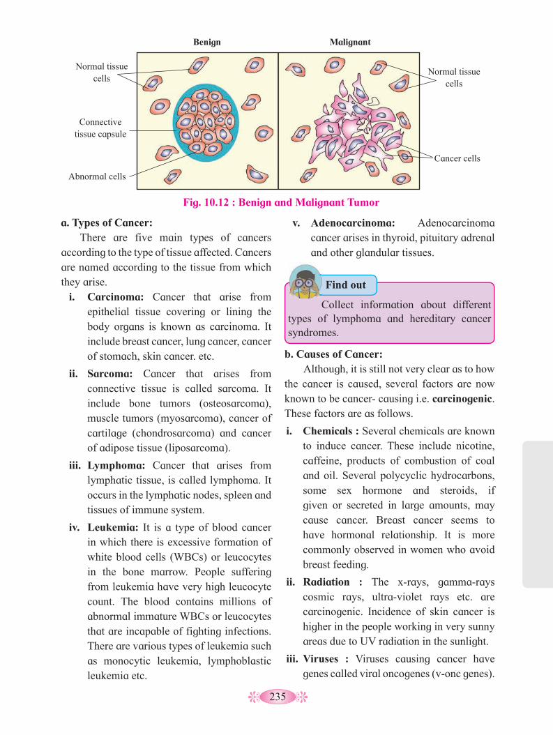
235
v. Adenocarcinoma: Adenocarcinoma cancer arises in thyroid, pituitary adrenal and other glandular tissues.
a. Types of Cancer: There are five main types of cancers
according to the type of tissue affected. Cancers are named according to the tissue from which they arise.
i. Carcinoma: Cancer that arise from epithelial tissue covering or lining the body organs is known as carcinoma. It include breast cancer, lung cancer, cancer of stomach, skin cancer. etc.
ii. Sarcoma: Cancer that arises from connective tissue is called sarcoma. It include bone tumors (osteosarcoma), muscle tumors (myosarcoma), cancer of cartilage (chondrosarcoma) and cancer of adipose tissue (liposarcoma).
iii. Lymphoma: Cancer that arises from lymphatic tissue, is called lymphoma. It occurs in the lymphatic nodes, spleen and tissues of immune system.
iv. Leukemia: It is a type of blood cancer in which there is excessive formation of white blood cells (WBCs) or leucocytes in the bone marrow. People suffering from leukemia have very high leucocyte count. The blood contains millions of abnormal immature WBCs or leucocytes that are incapable of fighting infections. There are various types of leukemia such as monocytic leukemia, lymphoblastic leukemia etc.
• Collect information about different types of lymphoma and hereditary cancer syndromes.
Find out •
Fig. 10.12 : Benign and Malignant Tumor
Benign
Normal tissue cells
Connective tissue capsule
Abnormal cells
Malignant
Normal tissue cells
Cancer cells
b. Causes of Cancer: Although, it is still not very clear as to how
the cancer is caused, several factors are now known to be cancer- causing i.e. carcinogenic. These factors are as follows.
i. Chemicals : Several chemicals are known to induce cancer. These include nicotine, caffeine, products of combustion of coal and oil. Several polycyclic hydrocarbons, some sex hormone and steroids, if given or secreted in large amounts, may cause cancer. Breast cancer seems to have hormonal relationship. It is more commonly observed in women who avoid breast feeding.
ii. Radiation : The x-rays, gamma-rays cosmic rays, ultra-violet rays etc. are carcinogenic. Incidence of skin cancer is higher in the people working in very sunny areas due to UV radiation in the sunlight.
iii. Viruses : Viruses causing cancer have genes called viral oncogenes (v-onc genes).

236
c. Treatment of Cancer: Cancer treatment consists of combination
of a number of therapies which are follows:
i. Chemotherapy: Chemotherapy comprises administration of certain anticancer drugs. The anticancer drugs check cell division by inhibiting DNA synthesis or are more toxic to cancerous cell than to normal cells. Chemotherapy may lead to hair loss or anaemia but both get corrected after the treatment is stopped.
ii. Radiotherapy: It is used in addition to chemotherapy. In radiotherapy, the basic principle is to bombard the cancerous tissue or cells with the rays from radioactive materials. For treatment, the cancer tissue or cells are exposed to radiations from radioactive materials such as cobalt, iridium and iodine. The rays x-rays gamma rays and charge particles are used to destroy the cancerous tissue or cells but cause minimum damage to the surrounding normal tissue or cells.
iii. Surgery: In surgery, the entire cancerous tissue or cells are removed surgically. It has limited utility. In certain cases such as breast tumor or uterine tumor, the surgery is most effective, but other treatments are also given to kill any cancerous cell that may have been escaped in surgery.
iv. Immunotherapy: Tumor cell have been shown to avoid detection and destruction by immune system. Therefore, the patients are given substances called biological response modifiers such as α-interferon which activates their immune system and helps in destroying the tumor.
v. Supportive therapy: Supportive therapy is used to treat symptoms of cancer and side effects of cancer treatments. Objective of this therapy is to improve the quality of life of cancer patient. This therapy varies depending upon condition of individual patient.
These viruses are also called oncogenic viruses. e.g. EBV(Epstein-barr virus), HPV (Human papiloma virus) etc.
iv. Oncogenes : Several genes called cellular oncogenes (c-onc genes) or proto-oncogenes have been identified in normal cells which when activated under certain condition could lead to oncogenic transformation of cells.
v. Addiction : Different types addictions likes smoking, chewing of tobacco lead to cancer of mouth, lips and lungs. Alcohol consumption may result in cancer of oesophagus, stomach, intestine and liver. Drugs also cause cancer e.g. Marijuana, anaerobic steroids etc.
Table 10.13 : Carcinogens and Organ Affected
Carcinogen Organ affected1. Soot
2. Coal tar (3,4 benzopyrene)
3. Cigarette smoke (N-nitrosodimethlene)
4. Cadmium oxide
5. Aflatoxin (a metabolite of Aspergillus flavus, a mould)
6. 2-naphthylamine and 4-aminobiphenyl
7. Mustard gas
8. Nickel and chromium compounds
9. Asbestos
10.Diethylstilbestrol (DES)
11.Vinylchloride (VC)
Skin, lungs
Skin, lungs
Lungs
Prostate gland
Liver
Urinary bladder
Lungs
Lungs
Lungs
Vagina
Liver
Try This
1. Find the Oncocenters nearby your area.
2. Prepare a chart of types of cancer and their preventive measures.
3. Organize a street-play on awareness about cancer and present it in your area.
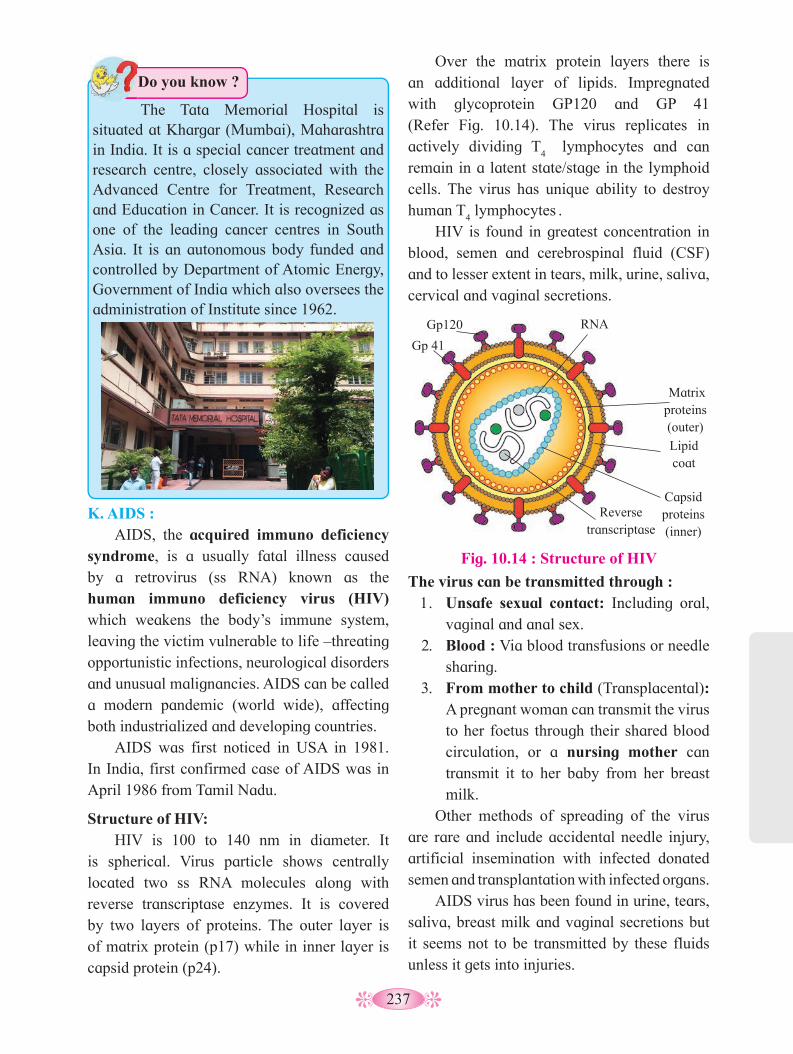
237
Over the matrix protein layers there is an additional layer of lipids. Impregnated with glycoprotein GP120 and GP 41 (Refer Fig. 10.14). The virus replicates in actively dividing T
4 lymphocytes and can
remain in a latent state/stage in the lymphoid cells. The virus has unique ability to destroy human T
4 lymphocytes
.
HIV is found in greatest concentration in blood, semen and cerebrospinal fluid (CSF) and to lesser extent in tears, milk, urine, saliva, cervical and vaginal secretions.
K. AIDS : AIDS, the acquired immuno deficiency
syndrome, is a usually fatal illness caused by a retrovirus (ss RNA) known as the human immuno deficiency virus (HIV) which weakens the body’s immune system, leaving the victim vulnerable to life –threating opportunistic infections, neurological disorders and unusual malignancies. AIDS can be called a modern pandemic (world wide), affecting both industrialized and developing countries.
AIDS was first noticed in USA in 1981. In India, first confirmed case of AIDS was in April 1986 from Tamil Nadu.
Structure of HIV: HIV is 100 to 140 nm in diameter. It
is spherical. Virus particle shows centrally located two ss RNA molecules along with reverse transcriptase enzymes. It is covered by two layers of proteins. The outer layer is of matrix protein (p17) while in inner layer is capsid protein (p24).
The virus can be transmitted through :1. Unsafe sexual contact: Including oral,
vaginal and anal sex.2. Blood : Via blood transfusions or needle
sharing.3. From mother to child (Transplacental):
A pregnant woman can transmit the virus to her foetus through their shared blood circulation, or a nursing mother can transmit it to her baby from her breast milk.
Other methods of spreading of the virus are rare and include accidental needle injury, artificial insemination with infected donated semen and transplantation with infected organs.
AIDS virus has been found in urine, tears, saliva, breast milk and vaginal secretions but it seems not to be transmitted by these fluids unless it gets into injuries.
Fig. 10.14 : Structure of HIV
RNA
Matrix proteins (outer)
Lipid coat
Capsid proteins (inner)
Reverse transcriptase
Gp120
Gp 41
The Tata Memorial Hospital is situated at Khargar (Mumbai), Maharashtra in India. It is a special cancer treatment and research centre, closely associated with the Advanced Centre for Treatment, Research and Education in Cancer. It is recognized as one of the leading cancer centres in South Asia. It is an autonomous body funded and controlled by Department of Atomic Energy, Government of India which also oversees the administration of Institute since 1962.
Do you know ?

238
Preventive measures: AIDS has no cure, hence prevention is
the best choice. The following steps help in preventing this dreadful disease-
i. People, particularly those in high –risk group, should be educated about HIV transmission.
ii. Disposable needles and syringes should be used and disposed off properly and immidiately.
iii. Sexual habits should be changed immediately.
iv. High-risk groups should refrain from donating blood.
v. Tooth brushes, razors, other articles that can become contaminated with blood should not be shared.
vi. Before receiving blood, ensure that it has been screened for not containing HIV infections.
vii. Routine screening must be done for –• Blood donors.• Organ donors (kidney, liver, lung, cornea).• Donors of semen and growth hormone.• Patients undergoing hemodialysis and
females in high risk group who are pregnant or contemplating pregnancy.
Clinical manifestations:The clinical manifestations (symptoms)
of AIDS have been classified into four broad categories.i. Initial infection with the virus and
formation of antibodies, usually 2-8 weeks after initial infection.
ii. Asymptomatic carrier state in which no signs of disease, are seen. Incubation period ranges for 6 months to 10 years.
iii. AIDS related complex (ARC) with one or more of the following clinical signs: recurrent fever for longer than one month, fatigue, unexplained diarrhea, night sweats, shortness of breath, loss of more than 10 per cent body weight, etc.
iv. AIDS is the end stage of HIV infection. It is characterised by life threatening opportunistic infections (like pneumonia, tuberculosis, kaposi sarcoma, etc.).
• Every year, December 1st is observed as World AIDS Day. • India started a National AIDS control Programme in 1987. “A red ribbon” is worn on World AIDS Day as a universal symbol of awareness and support for people living with HIV.• In India, 4 AIDS reference centers have been established-
1. AIIMS – New Delhi.2. National Institute of Virology, Pune.3. Center of advanced research on Virology, Vellore.4. National institute of communicable diseases, New Delhi.
• National AIDS Control Organization (NACO) was set up in year 1992 by the Ministry of Health and Family Welfare. The aim of this organization is to prevent further transmission of HIV, to decrease morbidity and mortality associated with HIV infection and to minimize the socio-economic impact resulting from HIV infection.
Do you know ?
Always Remember
HIV infection is not spread by:Causal contact such as hugging, insect
bite (mosquitoes), participation in sports, touching items previously touched by a person infected with the virus, hand shake, sharing clothes, swimming pools etc.
239
Laboratory diagnosis: At first a test is used to detect the HIV
antibodies, while a second confirmatory test is used to weed out any false positive results. The first test is ELISA (Enzyme-Linked Immunosorbent Assay). The confirmatory test, usually a Western Blot, is a highly specific test. It is based on detecting specific antibody to viral core protein and envelope glycoprotein.
Treatment of AIDS : Although AIDS has no cure, certain
medicines called as Antiretroviral drugs can help in reducing the viral load and prolong the life of HIV patient. Examples of these drugs used in Antiretroviral therapy (ART) are TDF (tenofovir), EFV(Efavirenz), Lamivudine (3TC), etc.
The advancements made in biological sciences have helped us to deal effectively with many infectious diseases. The use of vaccines and immunization programmes have enabled us to eradicate completely the dreadful diseases like small pox. A large number of other infectious diseases like polio, diphtheria, pneumonia and tetanus have been controlled to a large extent by the use of vaccines. Biotechnology is on the verge of making available newer and safer vaccines. Discovery of antibiotics and various drugs has also enabled us to treat effectively infectious diseases like tuberculosis.
10.4 Adolescence : It is the period of begining with the
appearance of secondary sexual characters and the termination with ceassation of somatic i.e. body growth. It can also be regarded as a transitional stage of physical and mental development of child occurring between puberty and the legal adulthood between 10 to 19 years of age where individual is no loger a child but not yet an adult.
Adolescence in fact is a phase rather than fixed time period in the life. It is a phase of
development on many fronts like sexual and reproductive maturity, mental development, adult identity and transition from socio-economic and emotional dependant to relative independence.
Stages of Adolescence :
Adolescents are defined are individual 10 to 19 year age group. The government of India in its National youth policy defines adolescents as 13-19 years. Adolescents is divided in three stage viz. early stage, middle stage and late stage.
i. Early period (10 to 14 years) : The changes include begining of the
appearance of secondary sexual characters ; growth reaches to its peak; rapid physical growth; concrete thinking; defining boundries of dependence/independence; self exploration; developing body image; development of intense friendship; seeking to counter instability and evaluation.
ii. Middle period (15 to 17 years) : It is characterized by almost complete full
development of secondary sexual characteristic; growth slows down, approximately 95 % of the adult stature is attained; thinking is more abstract; concrete thinking under stressful conditions; reestablishing of body image capable of long range thinking; sense of leadership and all powerfulness; preoccupied with romantic fantacy; ability testing to attract opposite sex, peer group help defining behavioral code etc.
iii. Late period (18 to 19 years) : It is characterized by establishment of total
physical maturity established abstract thinking, intellectual and functional identity; peer group recedes in favor of individual relationship, stable relationship and change from childhood to adulthood relationship.

240
Physical changes of adolescence :• Growth spurt occurs in both boys and
girls. In boys muscles develop, skin become oily, broadening of shoulders cracking of voice, development of underarm and chest hair, pubic hair, facial hair, enlargement of penis and testis. In girls development of breast, widening of hip, development of underarm and pubic hair, enlargement of uterus and ovaries.
• Sexual development : Sex organs mature and enlarge, sexual desire, errection of penis in boys, sperm production, ejaculation, ovulation, menstruation and initiation of sexual behaviour.
• Emotional and social changes : This include establishing own identity, fantacy, day dreaming, attention seeking behavior, emotional instability full of energy, sexual attraction. Rapid mood changes conflicts with family, behavioural code (influence by peer group) self exploration and evaluation, formation of new relationship, peer pressure etc.
Mental Health and Adolesence: Many of the emotional and social changes
have implication on the mental health. Most of the mental health issues that people confront as adults begin to appear in adolesence . The mental health implication includes confusion irritation, moodiness, frustration, nausea, less concentration, hyper activities, anger, effects on life styles like obesity, addictions, accidents, leading to ill health etc. The mental illness in fact is in terms of different forms of depression like insomnia and loss of energy.
Thus, mental illness (disorders or unfavourable changes) is associated with psychological or behaviour manifestation. These are broadly classified as either psychoses or neuroses. Psychoses include delusions, hallucinations, distrubance in the thinking process, etc. The psychoses lead to Alzheimer’s disease schizophrenia, depressive psychosis, etc. Amnesia (loss of memory) Bullimia (extreme over indulgence in food) Anxiety (fear or appretension) Anorexia nervosa (emotional aversion to food) depression (sadness, inactivity reduced to enjoy life, etc.) Neuroses include schizophrenia, illusions, hallucination, etc.
Adolescence is thus characterized by number of cognitive, emotional, behavioural, physical and attitudinal changes which may lead to positive personality development depending upon the relation with parents or to other conditions, to conflicts to others side. In fact they view their friends peer group more important and influential than their parents. This may lead to various kinds of addictions like smoking, taking drugs, etc.
Treatment : Treatment of such disorders should be
preferentially with non-pharmacological approch with due respect to rights of children. WHO has recommended evidence based guidelines under mental health Gap Action Programme (mhGAP).
Always Remember
• Avoid looking at your parents as the enemy. Try to understand that your parents are human beings, with their own needs and feelings.
• Listen to your parents with an open mind, and try to see their point of view.
• Share your feelings with your parents so that they can understand you better.
• Live up to your responsibilities at home and in school so that your parents will be more inclined to grant you the kind of independence you want and need.
• Bolster your criticisms of family, school and government with suggestions for practical improvements.
• Be as courteous and considerate to your own parents as you would be to the parents of your friends.

241
disappointments and failures as a part of life. Channelize the energy of child in sports, studies and other constructive activities.
10.6 Drugs Abuse :
Surveys and statistics show that use of drugs and alcohol has been on the rise especially among the youth. This is really a cause of concern as it could result in many harmful effects. Proper education and guidance would enable youth to safeguard themselves against these dangerous behaviour pattern and follow healthy lifestyle. The drugs, which are commonly abused, are opioids, cannabinoids and alkaloids of coca.
a. Opioids :
These drugs binds to opioid receptors present in central nervous system and gastro-intestinal tract. Heroin, otherwise called smack is chemically di-acetyl morphine. It is extracted from latex of poppy plant Papaver somniferum. Heroin is depressent and slows down the activity of body.
b. Cannabinoids :
Interact with receptors present in brain. Inhalation and ingestion of these substances affect the cardiovascular system. These are obtained from inflorescences and the parts of Cannabis sativa. Marijuana, hashish, charas and ganja are other different forms of drugs obtained from this plant.
c. Cocain :
This is an alkaloid obtained from coca plant- Erythroxylum coca. It increases level of neurotransmitter-dopamine. Its excessive dosage causes extreme happiness, irritability, paranoia.
Hallucinogens (mind expanding drugs) :
These are alkaloids causing day-dreaming. Lycergic acid and cannabis are hallucinogenic substances. Atropa bellodona and Datura
10.5 Addiction : This is a complex condition characterised
by compulsive use of substance despite of its harmful consequences. Addiction may involve the use of substances (drugs) such as alcohol, opioids, cocaine, nicotine and others or behaviours such as gambling.
There is scientific evidence that the addictive behaviours share key neurobiological features. They intensely involve brain pathways of reward and reinforcement, affecting motivation, which involve the neurotransmitter dopamine. It is important to know that the neurological changes are reversible after the substance-use or behaviour is discontinued.
Addiction overall result in the impairment of physical, physiological and psychological functions of the body.
Causes of substances abuse during Adolescence
• Insufficent parental supervision and monitoring.
• Lack of communication between child and parents.
• Poorly defined rules.
• Family conflicts.
• Favorable parental attitudes towards alcohol and drug uses.
• Expectations from drugs use.
• Risk taking behavior.
Methods /measures to control drug abuse :-
1. Always remember ‘Prevention is better than cure’.
2. Avoid undue pressure – A child should not be forced to perform beyond his /her capacities in studies, sports and other activities.
3. Education and Counselling of child to face problems and stress, to accept

242
spp. also have hallucinogenic properterties. Hallucination are unreal perceptions of unreal object due to the disorder of nervous system.
Drugs like barbiturates, amphetamine, benzo-diazepins, lysergic acid diethylamide (LSD) are derived from plant Cannabis sativa are used as a medicine. It help the patient in contracting insomnia and depression.
Do you know ?
Excessive doses of drugs may lead to coma and death due to respiratory failure, heart failure or cerebral hemorrhage. A combination of drugs or their intake along with alcohol generally results in overdose and even deaths.
The most common warning signs of drug and alcohol abuse among youth include drop in academic performance, unexplained absence from school/college, lack of interest in personal hygiene, withdrawal, isolation, depression, fatigue, aggressive and rebellious behaviour, deteriorating relationships with family and friends, loss of interest in hobbies, change in sleeping and eating habits, fluctuations in weight, appetite, etc.
There may even be some far-reaching implications of drug/alcohol abuse. If an abuser is unable to get money to buy drugs/alcohol he/she may turn to crime. At times, a drug/alcohol addict becomes the cause of mental and financial distress to his/her entire family and friends.
Those who take drugs intravenously (direct injection into the vein using a needle and syringe) are likely to acquire serious infections like HIV and hepatitis B. Use of alcohol during adolescence may also have long-term effects like loss balance, liver cirrhosis, pancreatitis. It could lead to heavy drinking in adulthood.
Chronic use of drugs and alcohol damages nervous system and liver (cirrhosis). Use of drugs and alcohol during pregnancy adversely affects the foetus.
Another misuse of drugs is that certain sports persons use drugs to enhance performance. They (mis)use narcotic analgesics, anabolic steroids, diuretics and certain hormones to increase muscle strength and bulk and to promote aggressiveness and overall improvement in their performance. Side-effects of the use of anabolic steroids in females include masculinization (features like males), increased aggressiveness, mood
Addiction and Dependence :Because of the perceived benefits,
drugs are used repeatedly. The most important thing, which one fails to realize, is the inherent addictive nature of alcohol and drugs. Addiction is a psychological attachment to certain effects –such as euphoria and a temporary feeling of well-being –associated with drugs and alcohol. These drive people to take them even when these are not needed, or even when their use becomes self-destructive. With repeated use of drugs, the tolerance level of the receptors present in our body increases.
Consequently the receptors respond only to higher doses of drugs or alcohol leading to greater intake and addiction. However, it should be clearly borne in mind that use of these drugs even once, can lead to addiction. In the absence of any guidance or counselling, the person gets addicted and becomes dependent on their use. Dependence is the tendency of the body to manifest a characteristic and unpleasant withdrawal syndrome if regular dose of drugs/ alcohol is abruptly discontinued. This is characterized by anxiety, trembling, nausea and sweating, which may be relieved when use is resumed.
Effects of Drug/ Alcohol Abuse :The immediate adverse effects of drugs
and alcohol abuse are manifested in the form of reckless behaviour, vandalism and violence.

243
swings, depression, abnormal menstrual cycles, excessive hair growth on the face and body, enlargement of clitoris, deepening of voice.
In males it includes acne, increased aggressiveness, mood swings, depression, and reduction of size of the testicles, decreased sperm production, kidney and liver dysfunction, breast enlargement, premature baldness, enlargement of the prostate gland. These effects may be permanent with prolonged use.
Prevention and Control :The age-old adage (i.e. proverb) is
‘prevention is better than cure’ holds true for all addictions. It is also true that habits such as smoking, taking drug or alcohol are more likely to be taken up at a young age, more during adolescence. It is best to identify the situations that push an adolescent towards use of drugs or alcohol, and to take remedial measures well in time. In this regard, the parents and the teachers have a special responsibility.
Deaddiction may be difficult but not impossible. Collect information about NGOs, working in the field of deaddiction.
Use your brain power
Activity :
1. Identify the name of the plant. Enlist different types of drugs derived/obtained from the same.
2. Prepare chart of different stages of adolesence mentioning the changes in growth, sextual development and emotional and social changes.
Stages I II III
Changes Boy Girl Boy Girl Boy Girl
Growth
Sexual development
Emotional and social
1.2.3.
1.2.3.
1.2.3.

244
Q. 1 Multiple Choice Questions1. Which of the following is NOT caused
by unsterilized needles? a. Elephantiasis b. AIDS c. Malaria d. Hepatitis B
2. Opium derivative is .............. a. Codeine b. Caffeine c. Heroin d. Psilocybin
3. The stimulant present in tea is ............. a. tannin b. cocaine c. caffeine d. crack
4. Which of the following is caused by smoking?
a. Liver cirrhosis b. Pulmonary tuberculosis c. Emphysema d. Malaria
5. An antibody is ............... a. molecule that binds specifically an antigen
b. WBC which invades bacteria c. secretion of mammalian RBC d. cellular component of blood
6. The antiviral proteins released by a virus-infected cell are called ............ a. histamines b. interferons
c. pyrogens d. allergens
7. Both B-cells and T-cells are derived from ....................
a. lymph nodes b. thymus glands c. liver d. stem cells in bone marrow
8. Which of the following diseases can be contracted by droplet infection?
a. Malaria b. Chicken pox c. Pneumonia d. Rabies
9. Confirmatory test used for detecting HIV infection is .................
a. ELISA b. Western blot c. Widal test d. Eastern blot
10. Elephantiasis is caused by ............ a. W. bancrofti b. P. vivax c. Bedbug d. Elephant
11. Innate immunity is provided by ........... a. phagocytes b. antibody c. T- Lymphocytes d. B- Lymphocytes
Q. 2 Very Short Answer Questions 1. What is the source of cocaine?2. Name one disease caused by smoking?3. Which cells stimulate B-cells to form
antibodies?4. What does the abbreviation AIDS stand
for?5. Name the causative agent of typhoid
fever?6. What is Rh factor?7. What is schizont?8. Name the addicting component found in
tobacco.9. Name the pathogen causing Malaria.10.Name the vector of Filariasis.11.Give the name of the causative agent of
ringworm.12.Define health.
Q. 3 Short Answer Questions: 1. What are acquired diseases?2. Differentiate between antigen and
antibody.3. Name the infective stage of Plasmodium.
Give any two symptoms of malaria.4. Explain the mode of infection and cause
of elephantiasis.5. Why is smoking a bad habit?
Exercise

245
6. What do the abbreviations AIIMS and CMIS denote?
7. What is a carcinogen? Name one chemical carcinogen with its target tissue.
8. Distinguish between active immunity and passive immunity.
Q. 4 Short Answer Questions 1. Differentiate between B-cells and
T-cells.2. What are the symptoms of malaria?
How does malaria spread?3. Write a short note on AIDS.4. Give the symptoms of cancer.5. Write a note on antigens on blood cells.6. Write a note on antigens-antibody
complex.7. What are the various public health
measures, which you would suggest as safeguard against infectious diseases?
8. How does the transmission of each of the following diseases take place?
a. Amoebiasis b. Malaria c. Ascariasis d. Pneumonia9. What measure would you take to prevent
water-borne diseases?10.Write a short note on typhoid.
Q. 5 Match the following.
Column I Column IIa. AIDS i. Antibody productionb. Lysozyme ii. Activation of B-cellsc. B-cells iii. Immunoglobulind. T-helper cells iv. Tearse. Antibody v. Immuno deficiency
Q. 6 Long Answer Questions1. Describe the structure of antibody.2. Write a note on Vaccination.3. What is cancer? Differentiate between
bening tumor and malignant tumor. Name the main five types of cancer.
4. Describe the different type of immunity.5. Describe the ill –effects of alcoholism on
health. 6. In your view, what motivates the
youngsters to take to alcohol or drugs and how can this be avoided?
7. Do you think that friends can influence one to take alcohol/drugs? If yes, how may one protect himself/herself from such an influence?
Project :
1. Collect information about the, symptoms and preventive measures treatments for Dengue, Swine flu and Tuberculosis (TB).
2. Browse the information about COVID - 19 with respect to
a. Structure
b. Modes of infection
c. Preventive measures
d. Treatment
246
11.1 Improvement In Food Production : Food is one of the basic needs as it gives us
energy for everything. It keeps us alive, strong and healthy. It can be defined as any thing solid or liquid, which when swallowed, digested and assimilated in the body, keeping us well. It is organic, energy rich, non-poisonous, edible and nourishing substance.
Green plants synthesize their own food through photosynthesis. But animals including humans can not synthesize their food on their own, hence are dependent on plants directly.
The famine is responsible for dearth of food, besides the rapid and dramatic increase in world population over the time. To meet the increasing demand for food, there is need for improvement of food production, both quantity wise and quality wise (nutritive). Plant breeding and animal breeding help us to increase the food production.
11.2 Plant breeding : The improvement or purposeful
manipulation in the heredity of crops and the production of new superior varieties of existing crop plants, constitute what is called plant breeding. It is, therefore, an applied branch of botany. It is infact an art and the science of changing and improving the heredity of plants. Plant breeding can be carried out by using the applications of principles of genetics, taxonomy, physiology, pathology, agriculture, rDNA technology, etc.
Enhancement of Food Production
Plant breeding is a method of altering the genetic pattern of plants to increase their value and utility for human welfare. The plant breeding is done to increase crop yield, improve quality, increase tolerance to environmental stresses, make the plants resistant to pathogens and increase tolerance to insect pest. Green Revolution was the result of a sequence of scientific breakthroughs and developmental activities that successfully fought hunger by increasing food production. Seeds with superior quality, use of chemicals - pesticides and fertilizers, and multiple cropping system supported by the use of modern farm machinery and proper irrigation system, helped for the development of high-yielding and disease resistant varieties in wheat, rice, maize, etc.
In fact, plant breeding dates back to about 10,000 years ago. The present day crops are the result of domestication and acclimatization.
11
1. What are hybrid plants?2. Different examples of microbes used in
household production.3. The types or groups of micro-organism
that produce antibiotics.4. What are biofertilizers?5. What is the source of silk thread?
Can you recall?
Primary aim of plant breeding is to obtain a new crop veriety superior to the existing type, in all characters. This purposeful manipulation incorporates various objectives. which differ form plant to plant depending upon its type and the use. Some objectives are common- like yield, quality, resistance, life cycle span, etc., while some objectives will differ on the basis of the type and use of plant.
Do you know ?
Different methods of plant breeding include Introduction, Selection, Hybridization, Mutation breeding, Polyploidy breeding, Molecular plant breeding, Tissue culture, rDNA technology, SCP, etc.
Do you know ?
247
A. Hybridization and its technique : It is the cheif method that offers greater
possibilities in the crop improvement than other methods. The use of this method is the only effective means of combining together the desirable characters of two or more varieties. By this method, one can create new genetic combinations of already existing characters and new genetic variations. It also exploits and utilizes hybrid-vigour.
called wide/ distant crosses. Interspecific and intergeneric hybrids are seldom to occur in the nature.
The main steps of the plant breeding program (Hybridization) are as follows :
1. Collection of Variability : Wild species and relatives of the cultivated
species having desired traits, should be collected and preserved. The entire collection having all the diverse alleles (i.e. variations) for all genes in a given crop, is called germplasm collection. Variations are useful in the selection. Germplasm conservation can be done in following ways-• In situ conservation : It can be done with
the help of forests and Natural Reserves.• Ex situ conservation : It is done through
botanical gardens, seed banks, etc.
2. Evaluation and Selection of Parents : It is an important and essential step. The
collected germplasm is evaluated (screened) to identify plants with desirable characters. The selected parents must be healthy, vigorous and should show desirable but complementary features. The selected parents are selfed for three to four generations to make them pure or homozygous. It is made sure that only pure lines are selected, multiplied and used in the hybridization.
3. Hybridization : The variety showing maximum desirable
features is selected as female (recurrent) parent and the other one as male parent (donor) which lacks good characters found in recurrent parent.
The pollen grains from anthers of male parent are collected and then artificially dusted over stigmas of emasculated flowers of female parent. Pollination is followed by seed and fruit formation in due course. The seed, thus obtained represents the hybrid generation.
The hybrid F1 progeny is selected and
evaluated forthe desired combinations of characters.
Hybridization can be intravarietal, intervarietal (between two varieties of the same species), interspecific (between two species of the same genus) and intergeneric (between two genera of the same family). As parental plants are distantly related, such crosses are also
Know the Scientist :
Dr. Norman E. Borlaug: An American biologist, who has been
called “Father of the Green Revolution”, “Agriculture’s greatest spokesperson” and “The Man Who Saved a Billion Lives. Dr. Borlaug, a 1970 Nobel Laureate, was honoured for his work in the ‘Green Revolution,’ saving millions of lives from famine in India, Mexico, and the Middle East.
Dr. M. S. Swaminathan: He has been called the “Father of Green
Revolution in India” for his role in introducing and further developing high-yielding varieties of wheat in India. He advocated moving India to sustainable development, especially using environmentally sustainable agriculture, sustainable food security and the preservation of biodiversity. He is pioneer in mutation breeding in India. He developed new varieties of wheat like sonora, NP 165 and sarbati.
Internet my friend
Try to know more about the hybrid-vigour.

248
performance is recorded. The selected lines are then grown for three generations at least in natural field, in different agroclimatic zones. Finally variety is released as new variety for use by the farmers.
Many high yielding, hybrid varieties of rice, wheat, sugarcane, millets, developed through hybridization, have helped farmer community to attain record agricultural production in India since 1961. This is called green revolution.
Indian Hybrid Crops :
1. Wheat and Rice : In 1960s, wheat and rice production
increased tremendously. Norman E. Borlaug developed semi-dwarf varieties of wheat. Sonalika and Kalyan Sona are two of the hybrid wheat varieties, grown in India. Semi-dwarf rice varieties were taken from IR−8 (International Rice Research Institute) and Taichung native−I (from Taiwan) and introduced in India. Jaya, Padma and Ratna are the better-yielding, semi-dwarf rice varieties that were developed later.
2. Sugarcane :• Saccharum barberi is a native of North
India and S. officinarum belongs to South India.
• S. officinarum has thicker stem and high sugar contents, but it does not grow well in North India.
• These two varieties were crossed to get the desirable qualities of both (high sugar content, thicker stem and the ability to grow in North India). CO -419, 421, 453 are high yielding and having high sugar contents are developed in India at Coimbatore (Tamilnadu).
3. Millets :• Hybrid maize (Ganga-3), Jowar (CO-12),
and Bajra (Niphad) have been successfully developed in India.
• These varieties are high yielding and resistant to water stress.
4. Selection and Testing of Superior Recombinants :
The F1 hybrid plants showing superiority
over both the parents and having high hybrid-vigour, are selected. Such hybrids are then selfed for few generations to make them homozygous for the said desirable characters till there is a state of uniformity, so that the characters will not segeregate further.
5. Testing, Release and Commercialization of New Cultivars :
The newly selected lines are evaluated for the productivity and other features like disease resistance, pest resistance, quality, etc. Initially, these plants are grown under controlled conditions of water, fertilizers, etc. and their
Collection of variability from germ plasm/ gene bank
Evaluation and selection of parents
Selection of parent plants with different qualities
Selected parents selfed for three to four generations to make them homozygous or true breeding
Identification of parents as male parent (donor) and female parent (recurrent)
Collection of pollen grains from the flowers of male parent
Removal of stamens from the flowers of the female parent (emasculation)
Artificial cross pollination by using pollen grains collected from male parent
Bagging, tagging of the emasculated flower of female parent
Development of fruit and seed representing F1
(hybrid)generation
Selection and testing of F1 hybrid for combination of
desirable characters
Field trials for yield (productivity)
Testing and the release of variety
Chart 11.1 : Steps of Hybridization technique
249
variety of rice, NP 836 variety of wheat (rust resistant), Indore-2 variety of cotton (resistant to bollworm), Regina-II variety of cabbage (resistant to bacterial rot), etc.
Plant Breeding for Developing a Resistance to Insect Pest:
Insects being herbivores, incur heavy loss in the quantity and quality of crops. Resistance in crops can be developed by following ways :• Development of morphological characters
like hairy leaves in cotton and wheat develop vector resistance from jassids and cereal leaf beetle, respectively.
• Solid stem in wheat leads to resistance to stem borers.
• Biochemical characters provide resistance to insects and pests. For example, the high aspartic acid, and low nitrogen and sugar content in maize, lead to resistance against stem borers.
• The nectar-less cotton having smooth leaves develop resistance against bollworms.
Some pest-resistant varieties are-
Crop Variety Insect pest
Brassica Pusa Gaurav Aphids
Flat bean Pusa sem 2Pusa sem 3
Jassids, aphids and fruit borer
Okra Pusa Sawani, Pusa A-4
Shoot and fruit borer
11.3 Tissue culture : It is actually a collection of different
techniques. It is infact, emerged as a technique of plant biotechnology. Here, isolated cells, tissues, organs are grown ‘in vitro’ on a solid/ liquid nutrient medium, under aseptic and controlled conditions of light, humidity and tempreature, for achieving different objectives. The part of plant used in tissue culture is called explant.
Plant tissue culture is based on principle of Totipotency which is an inherent ability of living plant cell to grow, divide, redivide and give rise to a whole plant. Haberlandt (1902)
Plant Breeding for Disease Resistance : Some of the diseases caused in plants are-
Pathogen Plant disease Fungi Brown rust of wheat
Red rot of sugarcane Late blight of potato
Bacteria Black rot of crucifersViruses Tobacco mosaic virus
The basic objective of breeding for disease resistance is to develop inherent quality in the plant to prevent the pathogen from causing the disease. Such varieties of plants are called disease resistant plants. The basic technique used is the same as for normal hybridization process.
Some disease resistant plants developed are :
Crop Variety Resistant to Disease
Wheat Himgiri Leaf and stripe rust, hill bunt
Brassica Pusa Swarnim White rustCauliflower Pusa Shubra Black rot and
Curl blight black rot
Chilli Pusa Sadabahar
Chilli mosaic virus, Tobacco mosaic virus and leaf curl
B. Mutation Breeding : Mutation is sudden heritable change in
the genotype, caused naturally. It can also be induced by application of chemical mutagens.Natural (physical) mutagens are : High tem-perature, high concentration of CO
2, X rays,
UV rays.
Chemical mutagens are : Nitrous acid, EMS (Ethyl- Methyl- Sulphonate), Mustard gas, Colchicine, etc.
Seedlings or seeds are irradiated by CO-60, exposed to UV bulbs, X ray machines, etc.
Mutagens cause gene mutations and chromosomal aberrations. The treated seedlings are then screened for resistance to diseases/ pests, high yield, etc. e.g. Jagannath

250
for the first time conceived this idea and developed the concept of in vitro cell culture (plant morphogenesis).
The plant tissue culture medium contains all essential minerals, sources for carbohydrates, proteins and fats, water, growth hormones, vitamins and agar (for callus culture). The most commonly preferred medium for tissue culture is MS (Murashige and Skoog) medium.
Based on the nature of explant : There are three types viz, cell culture, organ culture and embryo culture.
Based on the type of in vitro growth : There are two tyes viz. Callus culture (solid medium) and Suspension culture (liquid medium).
Maintenance of aseptic conditions : Aseptic condition is essential so as to avoid contamination by other harmful micro-organisms. It is accomplished by sterilization of : glass ware (use of detergents, hot air oven), nutrient medium (by autoclave under constant pressure of 15 lb/sq inch for continous 20 minutes), Explant (by treatment of 20% ethyl alcohol and 0.1% HgCl
2), Inoculation
chamber (Laminar air flow) - by using UV ray tube for 1 hour before performing actual inoculation of explant on the sterilized nutrient medium.
Other conditions maintained are : Temperature - 180C to 200C, pH of nutrient medium 5 to 5.8 and aeration particularly for suspension culture.
In callus culture the solid medium is used. The development and organisation of tissue is lost. Hence, the cells of explant, divide and redivide to form a mass of undifferentiated cells, called callus. It is maintained on solid medium. Callus can be induced to form organs like root (rhizogenesis) and shoot (caulogenesis) and thus the plantlet. No shaker (agitator) is needed.
In suspension culture small groups of cells or a single cell are used as explant in the liquid medium. The liquid medium is constantly agitated by using shakers (agitators) so that there is constant mixing of medium and the explant.
Both the callus and suspension cultures die in due course of time. Therefore, subculturing is neccessary for continuation of the technique.
Micropropagation (Clonal Propagation) : Organogenesis via shoots is considered
as one of the most widely used commercial method of regeneration of plant.
Micropropagation is also known as clonal propagation. It is the only process adopted by Indian plant biotechnologists in
Fig. 11.2 : Steps in plant tissue culture (callus culture)
Hardening
Mother plant
Explant
Callus
Organogenesis
Shoot
Root Multiple plants
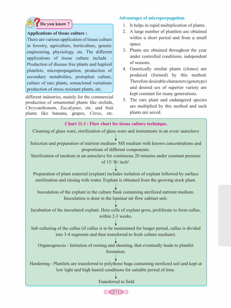
251
Applications of tissue culture :There are various application of tissue culture in forestry, agriculture, horticulture, genetic enginearing, physiology, etc. The different applications of tissue culture include - Production of disease free plants and haploid plantlets, micropropagation, production of secondary metabolites, protoplast culture, culture of rare plants, somaclonal variations production of stress resistant plants, etc.
Do you know ?
different industries, mainly for the commercial production of ornamental plants like orchids, Chrysanthemum, Eucalyptus, etc. and fruit plants like banana, grapes, Citrus, etc.
Advantages of microporpagation:
1. It helps in rapid multiplication of plants.2. A large number of plantlets are obtained
within a short period and from a small space.
3. Plants are obtained throughout the year under controlled conditions, independent of seasons.
4. Genetically similar plants (clones) are produced (formed) by this method. Therefore desirable characters (genotype) and desired sex of superior variety are kept constant for many generations.
5. The rare plant and endangered species are multiplied by this method and such plants are saved.
Chart 11.3 : Flow chart for tissue culture technique.
Cleaning of glass ware, sterilization of glass ware and instruments in an oven/ autoclave
Selection and preparation of nutrient medium- MS medium with known concentrations and proportions of different components.
Sterilization of medium in an autoclave for continuous 20 minutes under constant pressure of 15/ lb/ inch2.
Preparation of plant material (explant) includes isolation of explant followed by surface sterilization and rinsing with water. Explant is obtained from the growing stock plant.
Inoculation of the explant in the culture flask containing sterilized nutrient medium. Inoculation is done in the laminar air flow cabinet unit.
Incubation of the inoculated explant. Here cells of explant grow, proliferate to form callus, within 2-3 weeks.
Sub culturing of the callus (if callus is to be maintained for longer period, callus is divided into 3-4 segments and then transferred to fresh culture medium).
Organogenesis - Initiation of rooting and shooting, that eventually leads to plantlet formation.
Hardening - Plantlets are transferred to polythene bags containing sterilized soil and kept at low light and high humid conditions for suitable period of time.
Transferred to field
252
6. With the help of somatic hybrids (cybrids), we are able to obtain new variety in short time span.
The microorganisms utilize the carbon and nitrogen present in these materials and convert them into high-quality proteins that can be used as a supplement, in both human and animal feed. Besides proteins, SCP is also rich in vitamins, vitamin B complex, minerals and fats. The single-cell proteins can be readily used as fodder for achieving fattening of calves, pigs, in breeding fish and even in poultry and cattle farmimg. The microorganisms used for the production of SCP are as follows :
Fungi : Aspergillus niger, Trichoderma viride Yeast : Saccharomyces cerevisiae, Candida utilisAlgae : Spirulina spp, Chlorella pyrenoidosaBacteria : Methylophilus methylotrophus, Bacillus megasterium.
Advantages of Single-Cell Protein :• Microorganisms have a high rate of
multiplication that means a large quantity of biomass can be produced in a comparatively short duration.
• The microbes can be easily genetically modified to vary the amino acid composition. They have high protein contents- 43% to 85% (W/W basis).
• A broad variety of raw materials, including waste materials, can be used as a substrate for SCP. This also helps in decreasing the number of pollutants.
• SCP serves as a good source of vitamins, amino acids, minerals, crude fibres, etc.
11.5 Biofortification
It is a method in which crops are breed (produced) for having higher levels of vitamins, minerals and fats (i.e. better nutritive value). Due to this, problem of malnutrition can be overcome. Following objectives were considered for the breeding program :
• Protein content and quality• Oil content and quality• Vitamin content• Micronutrient content and quality
High yielding varities of Banana viz. ‘Shrimati’, ‘Basarai’ and G- 9 are mostly used in Maharashtra.
Do you know ?
11.4 Single cell protein (SCP) :
By 2050, the world would need to produce 1,250 million tonnes of meat and dairy products per year, to meet global demand for animal-derived protein at current consumption levels. However, growing demand for protein will not be met sustainably by increasing meat and dairy production because of the low efficiency of converting feed to meat and dairy products.
More over, human population in underdeveloped and even in the developing countries is suffereing from protein malnutrition, resulting into variety of nutritional diseases. To fight with this, efforts are undertaken by conventional methods to increase the food yield by different methods of crop improvement, use of biofertilizers, biopesticides, chemical fertilizers and high yielding varieties (green revolution). The efforts in other direction are also undertaken in nonconventional way. One such way is production of SCP- single cell proteins. Improtance of SCP was realised during World War I.
Single-cell protein refers to the crude, or a refined edible protein, extracted from pure microbial cultures or from dead or dried cell biomass.
Microorganisms like algae, fungi, yeast, and bacteria have very high protein content in their biomass. These microbes can be grown using inexpensive substrates like agricultural waste viz. wood shavings, sawdust, corn cobs, paraffin, N-alkanes, sugarcane molasses, even human and animal wastes.

253
Management of farms and farm animals : Farm management starts from selection of
high yielding breeds, their food requirements, supply of adequate nutritional sources, cleanliness of the environment and maintenance of health. Management of farm animals includes veterinary supervision, vaccination, high yielding cross breed development, production and preservation of products, distribution and marketing.
A. Animal breeding : Breeding of animals is an important aspect
of animal husbandry. Animal breeding aims at increasing the yield of animals and improving the desirable qualities of the products.
Breed: A group of animals related by descent
and similar in most characters like general appearance, features, size, configuration, etc., are said to belong to a breed.
Animal breeding is done for getting improved breeds with desirable qualities of product and to increase yield of animals, Desirable characters such as increased production of milk, quality of product, quality of meat or maximum yield of eggs per year etc., are necessarily achieved through animal breeding.
Biofortification can be achieved through conventional selective- breeding practices and also through r-DNA technology. It focusses on making plant food more nutritive as plants grow or develop.
Some examples of biofortification :• Fortified Maize having twice the amount
of amino acids- lysine and tryptophan.• Wheat -Atlas 66 has a high protein content
and Iron-fortified rice has 5 times more iron, are developed.
• Vegetable crops like carrot and spinach have more vitamin A and minerals.
• Vitamin C enriched bitter gourd, tomato have been developed by IARI.
11.6 Animal husbandry : Animal husbandry is an agricultural
practice of breeding and raising livestock. It is not only a skill of farmers but also is as much a science, as it is an art.
Animal husbandry deals with care and breeding of livestock like buffaloes, cows, pigs, horses, cattles, sheeps, camels, goats, etc. which are useful to humans. It also includes poultry farming, fish farming, bee keeping, sericulture, lac culture, etc. Animals like honey bees, silk worms, prawns, crabs, birds, fishes, pigs, cattles, sheeps and camels have been used by humans for the products like milk, eggs, meat, wool, honey, silk, etc.
During the conventional practices of animal breeding, just taking care is not enough to give maximum yield. Although India and china have 70% of world livestock population, surprisingly the productivity serves only 25% of the world farm produce.
So professional approach is needed to boost the production. It requires management procedures, new technologies to be employed in various farm system to achieve improvement in quality and productivity. Industrial principles of production, processing and marketing are to be employed.
Find out the name of common breeds of cattle and poultry in the farms, found in your area.
Can you tell?
Breeding can be of two main types - inbreeding and outbreeding :a. Inbreeding : It involves breeding of closely related individuals for 4 to 6 generations. Inbreeding increases homozygosity. By inbreeding, pure lines of animals can be obtained. Inbreeding is helpful in the elimination of harmful recessive genes and for the accumulation of superior genes. Inbreeding has the demerit that it usually reduces the fertility and productivity.

254
quality meat yielding bulls have been found to be successful, to increase herd size in a short period.
B. Dairy farm management : Dairy industry involves production,
processing and distribution of milk and milk products. Milk is a valuable food stuff universally consumed by human beings. Milk yield mainly depends on the quality of breeds in the farm. Selection of good breeds having high yielding potential under the climatic conditions of inhabiting area, and disease resistance is the basic requirement. In India, cows and buffaloes are mainly used for dairy farms. Sahiwal, Sindhi, Gir are Indian breeds and Jersy, Brown Swiss, Holstein are exotic breeds, which are used in dairy farming. Buffaloes are restricted to some part of Asia only.
In India, six breeds occur viz, Jaffarabadi, Mehsana, Murrah, Nagpuri, Nili, Surati, which are all good milk producers. Cattles have to be well looked after. Quality and quantity of fodder in proper ratio, should be given. Silage made from legumes and grasses, maize and jowar, makes good feed. Silage is supplemented with oil cakes, minerals, vitamins and salts. Cleanliness and hygiene of the cattles and handlers is of more importance while milking, storage and transport of milk and milk products. In recent years, much of these processes are mechanised, which reduce the chance of direct contact with the product. The shed must be cleaned daily. It should be spacious with adequate facilities for feeding, watering and lighting.
b. Outbreeding: It involves breeding of unrelated animals. The animals may be of the same breed but having no common ancestors for 4 to 6 generations.
Outcrossing involves breeding between the animals of different species. It is also known as interspecific hybridization. Outcrossing helps to remove the inbreeding depression.
Crossbreeding involves the breeding of superior male of one breed with superior female of another breed. By cross-breeding, new animal breeds of desirable characters are developed. e.g. Hisardale is a new breed of sheep developed from crossing of Bikaneri ewe and Marino rams in Punjab.
Interspecific hybridization involves breeding of animals of two different but related species. It result in the formation (production) of animals with desirable characters from both the parents. But such breeding is not always successful e.g. Mule is a breed obtained from horse and donkey.
Artificial insemination technique involves controlled breeding experiments. Semen from selected superior males is collected and preserved in frozen state or injected into the genital tract immediately. It is useful to overcome problem of normal mating and convenience of transportation.
Multiple Ovulation Embryo Transfer (MOET) involves the technology which provides the chances of successful production of hybrids. In this method, cow is administered with FSH like hormone, to induce follicular maturation and then the super ovulation is brought about. In each cycle, 6 to 8 eggs mature simultaneously. The cow is either mated with an elite bull or artificially inseminated. The blastocysts at 8 to 32 cell stage are recovered non-surgically and transferred to surrogate mothers. This technology is successfully used in cattles, sheeps, rabbits, buffaloes, etc. High milk yielding breeds of female and high
Regular visit of veterinary doctor to dairy farm is mandatory, why ?
Do you know ?
Milk processing, marketing and distribution, play an important role in dairy industry. Variety of milk product like curd, cream, butter, ghee, condensed milk, khoa,
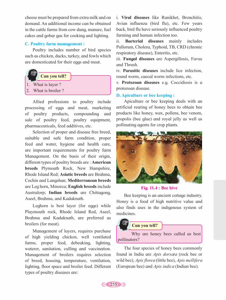
255
1. What is layer ?2. What is broiler ?
Can you tell?
i. Viral diseases like Ranikhet, Bronchitis, Avian influenza (bird flu), etc. Few years back, bird flu have seriously influenced poultry farming and human infection too. ii. Bacterial diseases mainly includes Pullorum, Cholera, Typhoid, TB, CRD (chronic respiratory disease), Enteritis, etc. iii. Fungal diseases are Aspergillosis, Favus and Thrush. iv. Parasitic diseases include lice infection, round worm, caecal worm infections, etc. v. Protozoan diseases e.g. Coccidiosis is a protozoan disease.
D. Apiculture or bee keeping : Apiculture or bee keeping deals with an
artificial rearing of honey bees to obtain bee products like honey, wax, pollens, bee venom, propolis (bee glue) and royal jelly as well as pollinating agents for crop plants.
cheese must be prepared from extra milk and on demand. An additional income can be obtained in the cattle farms from cow dung, manure, fuel cakes and gobar gas for cooking and lighting.
C. Poultry farm management : Poultry includes number of bird species
such as chicken, ducks, turkey, and fowls which are domesticated for their eggs and meat.
Why are honey bees called as best pollinators?
Can you tell?
Bee keeping is an ancient cottage industry. Honey is a food of high nutritive value and also finds uses in the indigenous system of medicines.
Fig. 11.4 : Bee hive
Allied professions to poultry include processing of eggs and meat, marketing of poultry products, compounding and sale of poultry feed, poultry equipment, pharmaceuticals, feed additives, etc.
Selection of proper and disease free breed, suitable and safe farm condition, proper feed and water, hygiene and health care, are important requirements for poultry farm Management. On the basis of their origin, different types of poultry breeds are : American breeds Plymouth Rock, New Hampshire, Rhode Island Red; Asiatic breeds are Brahma, Cochin and Langshan; Mediterranean breeds are Leg horn, Minorca; English breeds include Australorp; Indian breeds are Chittagong, Aseel, Brahma, and Kadaknath.
Leghorn is best layer (for eggs) while Playmouth rock, Rhode Island Red, Aseel, Brahma and Kadaknath, are preferred as broilers (for meat).
Management of layers, requires purchase of high yielding chicken, well ventilated farms, proper feed, debeaking, lighting, waterer, sanitation, culling and vaccination. Management of broilers requires selection of breed, housing, temperature, ventilation, lighting, floor space and broiler feed. Different types of poultry diseases are:
The four species of honey bees commonly found in India are Apis dorsata (rock bee or wild bee), Apis florea (little bee), Apis mellifera (European bee) and Apis indica (Indian bee).
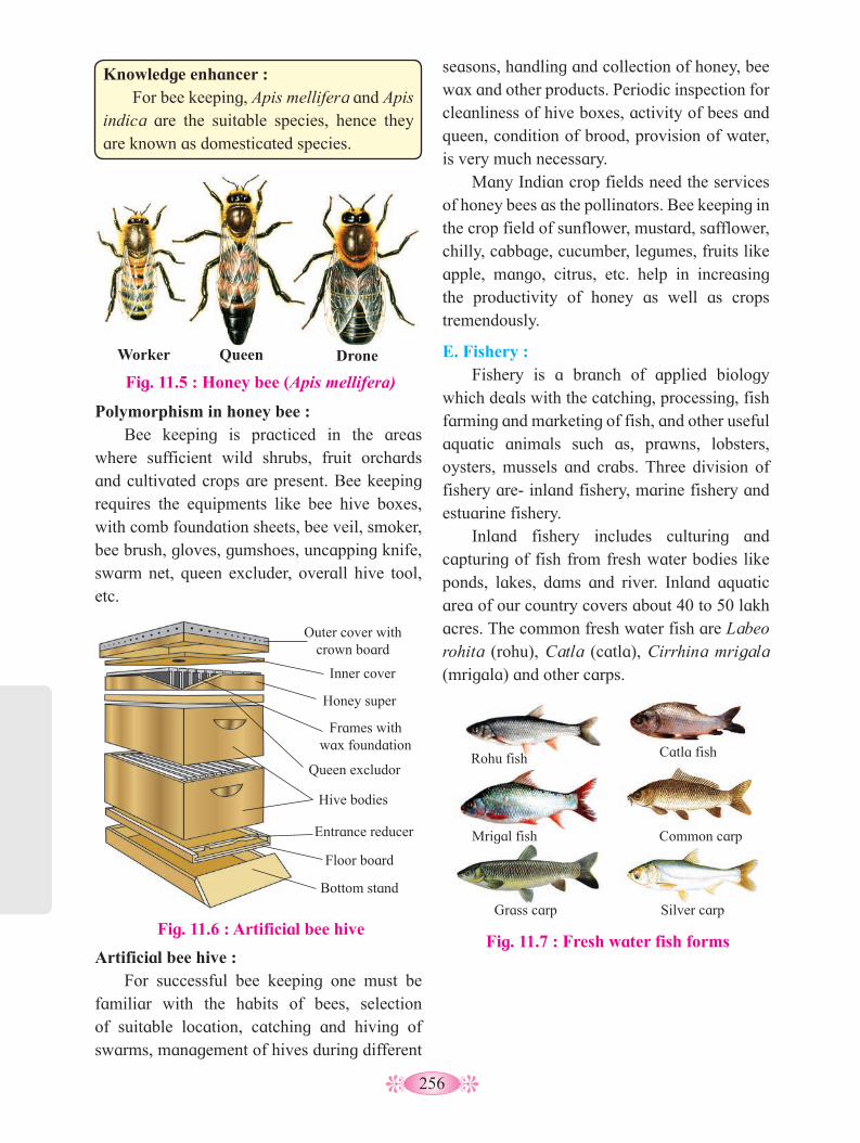
256
seasons, handling and collection of honey, bee wax and other products. Periodic inspection for cleanliness of hive boxes, activity of bees and queen, condition of brood, provision of water, is very much necessary.
Many Indian crop fields need the services of honey bees as the pollinators. Bee keeping in the crop field of sunflower, mustard, safflower, chilly, cabbage, cucumber, legumes, fruits like apple, mango, citrus, etc. help in increasing the productivity of honey as well as crops tremendously.
E. Fishery : Fishery is a branch of applied biology
which deals with the catching, processing, fish farming and marketing of fish, and other useful aquatic animals such as, prawns, lobsters, oysters, mussels and crabs. Three division of fishery are- inland fishery, marine fishery and estuarine fishery.
Inland fishery includes culturing and capturing of fish from fresh water bodies like ponds, lakes, dams and river. Inland aquatic area of our country covers about 40 to 50 lakh acres. The common fresh water fish are Labeo rohita (rohu), Catla (catla), Cirrhina mrigala (mrigala) and other carps.
Knowledge enhancer : For bee keeping, Apis mellifera and Apis
indica are the suitable species, hence they are known as domesticated species.
Polymorphism in honey bee : Bee keeping is practiced in the areas
where sufficient wild shrubs, fruit orchards and cultivated crops are present. Bee keeping requires the equipments like bee hive boxes, with comb foundation sheets, bee veil, smoker, bee brush, gloves, gumshoes, uncapping knife, swarm net, queen excluder, overall hive tool, etc.
Worker Queen Drone
Fig. 11.5 : Honey bee (Apis mellifera)
Fig. 11.6 : Artificial bee hive
Outer cover with crown board
Inner cover
Honey super
Frames with wax foundation
Queen excludor
Hive bodies
Entrance reducer
Floor board
Bottom stand
Fig. 11.7 : Fresh water fish forms
Rohu fish Catla fish
Mrigal fish Common carp
Grass carp Silver carp
Artificial bee hive : For successful bee keeping one must be
familiar with the habits of bees, selection of suitable location, catching and hiving of swarms, management of hives during different

257
1. Give the names of estuaries found in Maharashtra and where these estuaries are located.
2. Enlist the names of different fish found at an estuary.
Can you tell?
Fig. 11.8 : Life cycle of silk moth (Mulberry silk worm)
Adult female Eggs on mulberry leaf
Cocoon
Head
Thoracic true legs Spiracles
Mature caterpillar
Adominal pseudo legs
Dorsal horn
Eggs
The best quality silk called mulberry silk, is produced by silkworm Bombyx mori, while Tussar silk and Eri silk are of inferior quality. The quality and quantity of silk depends on the quality of mulberry leaves on which the larvae feed.
Rearing, development and looking after the silkworms, involve skill and labour for constant watch. A little negligence can spoil the complete industry. Silkworm larvae may be infected by protozoans, viruses and fungi. Besides these, ants, crows, birds, and other predators are ready to attack these insects, hence the cages of these larvae must be managed to prevent predators attack.
Estuarine fishery includes capture of fish from estuary. Estuary is a place where river meets the sea. e.g. Sunderban area in west bengal.
The common factors for the maintenance of fish farm, includes selection of suitable site, excavation of ponds, requirements of hatchery tank, nursery tank, rearing tank, stocking tank or ponds, water source, manures, supplementary feed, etc. The culture fishery may be monoculture (only one species) or polyculture (many species) type.
After catching the fishes, fish spoilage is prevented by different preservation methods like chilling, freezing, freeze drying, sun drying, smoke drying, salting and canning. In addition to the source as nutritious food, fish yield a number of by-products which are of commercial value. They are fish oil, fish meal, fertilizers, fish guano, fish glue and isinglass. These by-products are widely used in paints, soaps, oils, and medicine. Prawns and Lobsters have market value all over the world. Fishery provides good job opportunities and self employment to many people.
F. Sericulture : Sericulture is the branch of applied
zoology which deals with rearing of silkworm and production of silk. Like other farming, sericulture also involves skill and scientific knowledge for rearing and development. It require less investment and can be started in small space. It is the oldest business and large number of families are associated with the production of silk in India. Disabled, older persons, handicapped people can successfully do this job.
Knowledge enhancer : Fish farming or culturing of edible and
commercially important fish is only possible in fresh water bodies.
Marine fishery includes capture of fish from sea water. Indian coastal line is about 7500 km long. The common marine fish are Harpadon (Bombay duck), Sardinella (sardine), Rastrelliger (mackerel) and Stromateus (pomphret).
Internet my friend
Collect information about life cycle of silk moth.
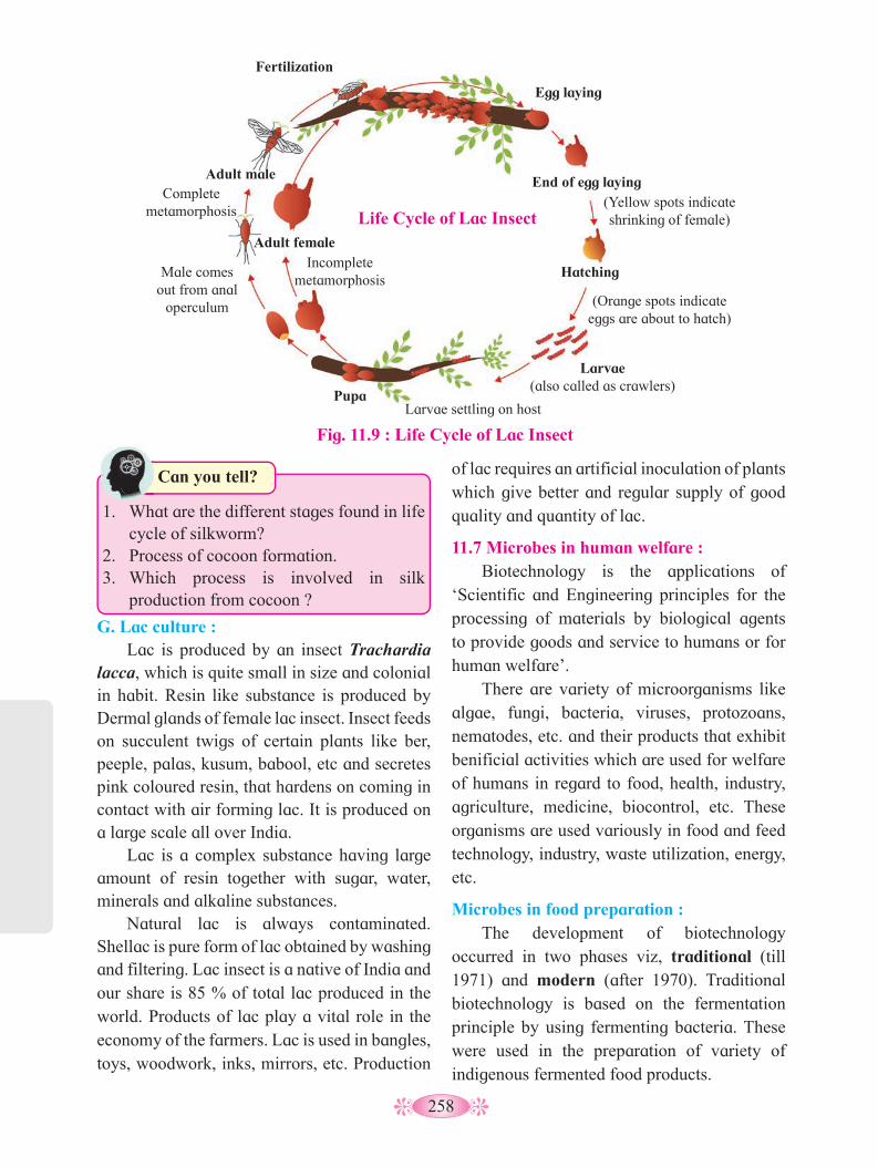
258
Fertilization
Adult male Complete
metamorphosis
Male comes out from anal
operculum
Adult female Incomplete
metamorphosis
Pupa Larvae settling on host
Larvae (also called as crawlers)
Hatching
(Orange spots indicate eggs are about to hatch)
End of egg laying (Yellow spots indicate shrinking of female)
Egg laying
Life Cycle of Lac Insect
Fig. 11.9 : Life Cycle of Lac Insect
of lac requires an artificial inoculation of plants which give better and regular supply of good quality and quantity of lac.
11.7 Microbes in human welfare : Biotechnology is the applications of
‘Scientific and Engineering principles for the processing of materials by biological agents to provide goods and service to humans or for human welfare’.
There are variety of microorganisms like algae, fungi, bacteria, viruses, protozoans, nematodes, etc. and their products that exhibit benificial activities which are used for welfare of humans in regard to food, health, industry, agriculture, medicine, biocontrol, etc. These organisms are used variously in food and feed technology, industry, waste utilization, energy, etc.
Microbes in food preparation : The development of biotechnology
occurred in two phases viz, traditional (till 1971) and modern (after 1970). Traditional biotechnology is based on the fermentation principle by using fermenting bacteria. These were used in the preparation of variety of indigenous fermented food products.
G. Lac culture : Lac is produced by an insect Trachardia
lacca, which is quite small in size and colonial in habit. Resin like substance is produced by Dermal glands of female lac insect. Insect feeds on succulent twigs of certain plants like ber, peeple, palas, kusum, babool, etc and secretes pink coloured resin, that hardens on coming in contact with air forming lac. It is produced on a large scale all over India.
Lac is a complex substance having large amount of resin together with sugar, water, minerals and alkaline substances.
Natural lac is always contaminated. Shellac is pure form of lac obtained by washing and filtering. Lac insect is a native of India and our share is 85 % of total lac produced in the world. Products of lac play a vital role in the economy of the farmers. Lac is used in bangles, toys, woodwork, inks, mirrors, etc. Production
1. What are the different stages found in life cycle of silkworm?
2. Process of cocoon formation.3. Which process is involved in silk
production from cocoon ?
Can you tell?
259
ii. Yoghurt (= yogurt): It is produced by curdling milk with
the help of Streptococcus thermophilus and Lactobacillus bulgaricus.
iii. Butter Milk: The acidulated liquid left after churning of
butter from curd, is called butter milk.
iv. Cheese: The milk is coagulated with lactic acid
bacteria and the curd formed is filtered to separate whey. The solid mass is then ripened with growth of mould that develops flavour in it. Different varieties of cheese are known by their characteristic texture, flavor and taste which are developed by different specific microbes. The ‘Roquefort and Camembert cheese’ are ripened by blue-green molds Penicillium roquefortii and P. camembertii respectively. The large holes in Swiss cheese are developed due to production of a large amount of CO
2
by a bacterium known as Propionibacterium shermanii.
11.8 Role of Microbes in Industrial Production:
During fermentation, variety of products like alcoholic beverages, organic acids, vitamins, growth hormones, enzymes, antibiotics, etc. are produced.
2. Microbes as the Source of Food : Some microbes or their fruiting bodies are
directly used as a source of food, as they are rich in vitamins and proteins. The term “SCP” or “single cell protein” denotes, dead and dried cells of microbes like bacteria, algae, molds and yeasts. Some mushrooms and truffles are directly used as food. They belong to higher fungi. They produce large, fleshy fruting bodies which are edible. Fruting bodies are sugar free, fat free but rich in proteins, vitamins, minerals and amino acids. The food in the fruting body is low caloried.
3. Dairy Products: Lactic acid bacteria (LAB) like
Lactobacillus are added to milk. It ferments lactose sugar of milk into lactic acid. Lactic acid causes coagulation and partial digestion of milk protein casein. Milk is changed into curd, yoghurt and cheese. The starter or inoculum used in preparation of milk products actually contains millions of lactic acid bacteria (LAB).
i. Curd: Indian curd is prepared by inoculating milk
with Lac tobacillus acidophilus. It also checks growth of disease causing microbes.
These are actually the secondary metabolites produced during idio phase and are not required by micro-organisms for their growth. The type of substrate and the type of micro-organism result into the production of particular secondary metabolites.
Do you know ?
1. Dosa, Dhokla and Idli: The dosa, idli and dhokla are fermented
products produced due to activity of bacteria. They are fermented preparation of rice and black Gram with air borne Leuconostoc and Streptococcus species of bacteria. CO
2 produced
during fermentation causes puffing up of the dough.
Name the microbes used in fermentation of dhokla.
Can you tell?
Names of some edible mushrooms and poisonous mushrooms.
Find out
Production on an industrial scale requires growing microbes in very large vessels, called fermenters. The main function of a fermenter is to provide a controlled environment for growth of a microorganism, or a defined mixture of microorganisms, to obtain the desired product.
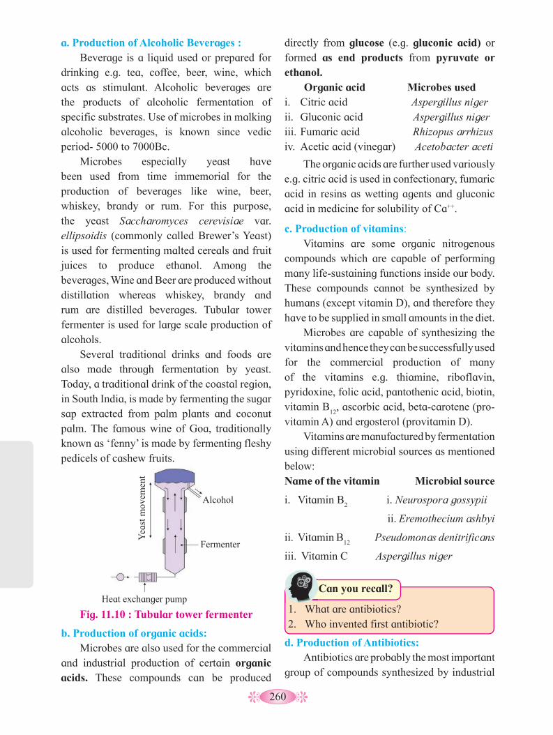
260
b. Production of organic acids: Microbes are also used for the commercial
and industrial production of certain organic acids. These compounds can be produced
directly from glucose (e.g. gluconic acid) or formed as end products from pyruvate or ethanol. Organic acid Microbes usedi. Citric acid Aspergillus nigerii. Gluconic acid Aspergillus nigeriii. Fumaric acid Rhizopus arrhizusiv. Acetic acid (vinegar) Acetobacter aceti
The organic acids are further used variously e.g. citric acid is used in confectionary, fumaric acid in resins as wetting agents and gluconic acid in medicine for solubility of Ca++.
c. Production of vitamins: Vitamins are some organic nitrogenous
compounds which are capable of performing many life-sustaining functions inside our body. These compounds cannot be synthesized by humans (except vitamin D), and therefore they have to be supplied in small amounts in the diet.
Microbes are capable of synthesizing the vitamins and hence they can be successfully used for the commercial production of many of the vitamins e.g. thiamine, riboflavin, pyridoxine, folic acid, pantothenic acid, biotin, vitamin B
12, ascorbic acid, beta-carotene (pro-
vitamin A) and ergosterol (provitamin D). Vitamins are manufactured by fermentation
using different microbial sources as mentioned below:Name of the vitamin Microbial source
i. Vitamin B2 i. Neurospora gossypii
ii. Eremothecium ashbyi
ii. Vitamin B12
Pseudomonas denitrificans
iii. Vitamin C Aspergillus niger
1. What are antibiotics?2. Who invented first antibiotic?
Can you recall?
a. Production of Alcoholic Beverages : Beverage is a liquid used or prepared for
drinking e.g. tea, coffee, beer, wine, which acts as stimulant. Alcoholic beverages are the products of alcoholic fermentation of specific substrates. Use of microbes in malking alcoholic beverages, is known since vedic period- 5000 to 7000Bc.
Microbes especially yeast have been used from time immemorial for the production of beverages like wine, beer, whiskey, brandy or rum. For this purpose, the yeast Saccharomyces cerevisiae var. ellipsoidis (commonly called Brewer’s Yeast) is used for fermenting malted cereals and fruit juices to produce ethanol. Among the beverages, Wine and Beer are produced without distillation whereas whiskey, brandy and rum are distilled beverages. Tubular tower fermenter is used for large scale production of alcohols.
Several traditional drinks and foods are also made through fermentation by yeast. Today, a traditional drink of the coastal region, in South India, is made by fermenting the sugar sap extracted from palm plants and coconut palm. The famous wine of Goa, traditionally known as ‘fenny’ is made by fermenting fleshy pedicels of cashew fruits.
d. Production of Antibiotics: Antibiotics are probably the most important
group of compounds synthesized by industrial
Fig. 11.10 : Tubular tower fermenter
Heat exchanger pump
Yea
st m
ovem
ent
Alcohol
Fermenter
261
Can antibiotics kill viruses?
Use your brain power
microorganisms. Most antibiotics are secondary metabolites. They have therapeutic importance and are used in medical treatment. These are produced in small amounts by certain microbes (like bacteria, fungi and few algae), which inhibit growth of other microbial pathogens. Therefore, they are used in medicine. The antibiotics are antibacterial, antifungal, etc.
Antibiotics have greatly improved our capacity to treat deadly diseases such as plague, whooping cough, diphtheria, leprosy, etc.
Enzymes are proteins known as bio-catalysts. Due to their ability to promote reactions more quickly at body temperature and more efficiently. Many microbes synthesize and excrete large quantities of enzymes into the surrounding medium. Using this feature of these tiny organisms, many enzymes are produced commercially. These enzymes are Amylase, Cellulase, Protease, Lipase, Pectinase, Streptokinase and many others.
Several industrial sectors, use enzymes from microorganisms for specific applications. In the textile industry, enzymes are able to improve the quality of the fabrics. In the pulp and paper industry, they are involved in biomechanical pulping and bleaching, in the food industry, they are used in the fermentation processes for the production of bread and drinks such as wine and beer, also they participate in the extraction of substances, such as carotenoids and olive oil; lipases are used in detergent industry, because they have superior cleaning properties, increasing the brightness and removing of oil stains; they are also used in cosmetics, animal feed and agricultural industries, among others. Following are the few examples of enzymes used in industrial food processing:
Name of the enzyme Microbial source
Invertase Saccharomyces cerevisiae
Pectinase Sclerotinia libertine, Aspergillus niger
Lipase Candida lipolytica
Cellulase Trichoderma konigii
f. Gibberellin production: Gibberellin is a group of growth hormones
mainly produced by higher plants and fungi to promote growth by stem elongation. The first gibberellin was isolated by two Japanese scientists -Yabuta and Sumiki (1938) from rice seedlings infected with the fungus Gibberella fujikouri.
What are Enzymes? How are they classified?
Can you recall?
Some common antibiotics and their microbial sources are listed below :
Table 11.11 : Antibiotic producing microbes
Antibiotic produced Microbial sources
Chloromycetin Streptomyces venezuelae
Erythromycin Streptomyces erythreus
Penicillin Penicillium chrysogenum
Streptomycin Streptomyces griseus
Griseofulvin Penicillium griseofulvum
Bacitracin Bacillus licheniformis
Oxytetracycline /Terramycin
Streptomyces aurifaciens
e. Production of Enzymes: In living beings, enzymes play a key role
in metabolic reactions and therefore, these are essential for the survival of such beings.

262
The oil eating bacteria can clean up crude spills. Collect more information about these bacteria.
Do you know ?
Extra Information : Sewage is also a potential source of pathogenic bacteria, viruses and protozoa. The causative agents of dysentery, cholera, typhoid, polio and infectious hepatitis may occur in sewage. The bacteria from the soil are also present in the sewage. During the course of sewage decomposition, initially aerobic and facultative anaerobic organisms predominate which are then followed by strict anaerobic especially methogenic bacteria that produce methane (CH
4) and CO
2.
About 15 different types of gibberellins have been isolated. Gibberellins have many practical applications. They are used to induce parthenocarpy in apple, pear, etc. They are used in breaking the dormancy of seed and also in inducing flowering in Long Day Plants (LDP). They are also used to enlarge the size of grape fruits.
11.9 Microbes in Sewage Treatment: Sewage is a matter carried off in
drainage. It is a municipal waste containing human excreta, house hold waste, dissolved organic matter and even pathogenic microbes (bacteria, viruses, protozoans, nematodes and microfungi). It also includes discharged water from hospital waste, slaughter house waste, animal dung, etc. Discharge from industrial waste (contains toxic dissolved organic and inorganic chemicals), tannery, pharmaceutical waste, etc. also add to sewage.
Composition of Sewage: Sewage consists of approximately 99.5%
to 99.9% water and 0.1 to 0.5% inorganic and organic matter in suspended and soluble form. Composition of sewage varies depending upon the type of waste discharged into water from different industries. e.g. textile, chemicals, pharmaceuticals, dairy, canning, brewing, meat packing, tannery, oil refineries and meat industries, etc.
Microorganisms in Sewage: Various types of micro-organisms are
also present in sewage. Bacteria, viruses, fungi, protozoa, nematodes, algae, etc. are found in sewage. However, their number and type, fluctuate depending upon the sewage composition and source of sewage. Raw sewage may contain millions of bacteria per ml. These include coliforms, fecal Streptococci, anaerobic spore forming bacilli and other types originating in the intestinal tract of humans.
Before waste water is made available for human use, it has to be treated properly, so as to remove organic matter, inorganic salts and pathogens as well. Sewage treatment process includes four basic steps as follows:
1. Preliminary Treatment: The preliminary treatment includes
Screening and Grit Chamber.i. Screening : Sewage and waste water
contains plenty of suspended, floating materials, coarse and solid particles along with dissolved substances. The suspended objects are filtered and removed. This is done in screening chambers. The sewage is passed through screens or net in the chambers. Larger suspended or floating objects are held back in the screening
Extra information: Streptokinase enzyme (TPA) is produced by the bacterium Streptococcus spp. It has fibrinolytic effect. Hence, it is used as a ‘clot buster’ for clearing blood clots in the blood vessels of patients, which may cause heart attack. Statins produced by the yeast Monascus purpureus have been produced on commercial scale. It is a blood-cholesterol lowering agent. This agent acts as competitive inhibitor of the enzyme responsible for synthesis of cholesterol.

263
chambers. These have to be removed before the biological treatment.
ii. Grit Chamber : After screening, the filtered sewage is then passed into series of grit chambers. These chambers contain large stones (pebbles) and brick-ballast. Coarse particles settle down by gravity. Thus, passage of filtered sewage removes much of the coarse particulate matter.
2. Primary treatment (physical treatment): After the preliminary treatment, the
sewage water is pumped into the primary sedimentation tank. The sedimentation of suspended solid or organic matter occurs in this tank. About 50-70% of the solids settle down. There is reduction of about 30-40% (in number) of coliform organisms. The organic matter which is settled down, is called primary sludge which is removed by mechanically operated devices. The supernatant (effluent) in the primary sedimentation tank still contains large amount of dissolved organic matter and micro-organisms which can then be removed by the secondary treatment.
3. Secondary treatment (biological treatment): The primary effluent is passed into large
aeration tanks. Here it is constantly agitated
mechanically and air is pumped into it. Aerobic bacteria grow vigourously and form flocs. Flocs are the masses of bacteria held together by slime and fungal hyphae to form mesh like masses. These aerobic microbes consume the major part of the organic matter present in the effluent, as they grow. Due to this BOD (Biochemical Oxygen Demand) of the effluent is significantly reduced.
4. Tertiary treatment: Once the BOD of waste water is reduced, it
is passed into a settling tank. Here the bacterial flocs are allowed to sediment. The sediment is now called activated sludge. Small part of this is passed back in to aeration tank and the major part is pumped in to large tanks called anaerobic sludge digesters. In these tanks, anaerobic bacteria grow and digest the bacteria and fungi in the sludge. During this anaerobic digestion, gases such as methane, hydrogen sulphide, CO
2, etc. are produced. Effluents
from these plants (digester) after chlorination, are released in natural water bodies like rivers and streams. Chlorination kills pathogenic bacteria. Digested Sludge is then disposed.
Fig. 11.12 : Diagrammatic representation of various stages in wastewater treatment
Waste water
Screening Grit setting Primary treatment
Preliminary treatment to remove heavy solids
Removal of suspended solid particles
Settled sewage
Secondary Treatment
Aeration tank
Tertiary treatment
Treated Effluent
(Aerobic bacteria)
Activated sludge Water to recycle
Anaerobic bioreactor (digester)
Sludge cake to dispose
(Anaerobic bacteria)
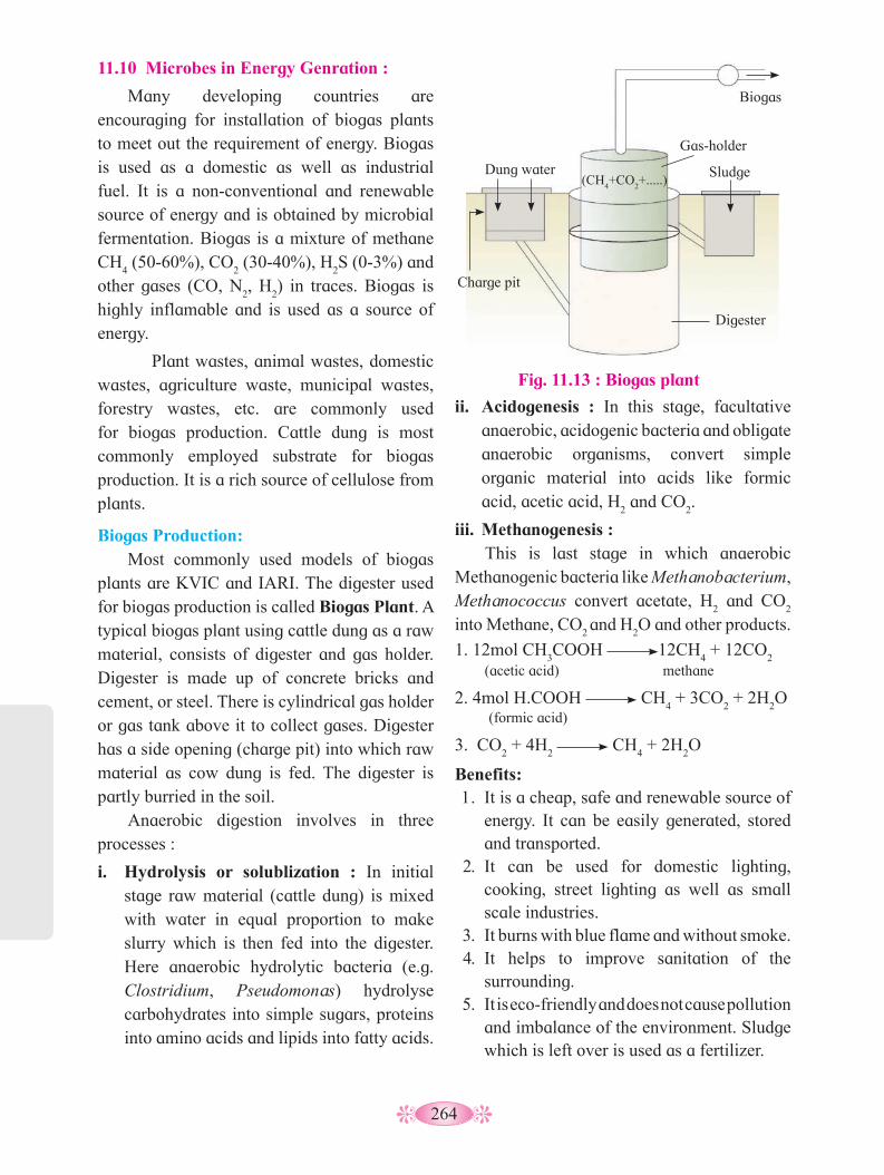
264
11.10 Microbes in Energy Genration :
Many developing countries are encouraging for installation of biogas plants to meet out the requirement of energy. Biogas is used as a domestic as well as industrial fuel. It is a non-conventional and renewable source of energy and is obtained by microbial fermentation. Biogas is a mixture of methane CH
4 (50-60%), CO
2 (30-40%), H
2S (0-3%) and
other gases (CO, N2, H
2) in traces. Biogas is
highly inflamable and is used as a source of energy.
Plant wastes, animal wastes, domestic wastes, agriculture waste, municipal wastes, forestry wastes, etc. are commonly used for biogas production. Cattle dung is most commonly employed substrate for biogas production. It is a rich source of cellulose from plants.
Biogas Production: Most commonly used models of biogas
plants are KVIC and IARI. The digester used for biogas production is called Biogas Plant. A typical biogas plant using cattle dung as a raw material, consists of digester and gas holder. Digester is made up of concrete bricks and cement, or steel. There is cylindrical gas holder or gas tank above it to collect gases. Digester has a side opening (charge pit) into which raw material as cow dung is fed. The digester is partly burried in the soil.
Anaerobic digestion involves in three processes :
i. Hydrolysis or solublization : In initial stage raw material (cattle dung) is mixed with water in equal proportion to make slurry which is then fed into the digester. Here anaerobic hydrolytic bacteria (e.g. Clostridium, Pseudomonas) hydrolyse carbohydrates into simple sugars, proteins into amino acids and lipids into fatty acids.
ii. Acidogenesis : In this stage, facultative anaerobic, acidogenic bacteria and obligate anaerobic organisms, convert simple organic material into acids like formic acid, acetic acid, H
2 and CO
2.
iii. Methanogenesis : This is last stage in which anaerobic
Methanogenic bacteria like Methanobacterium, Methanococcus convert acetate, H
2 and CO
2
into Methane, CO2 and H
2O and other products.
(acetic acid)
(formic acid)
methane1. 12mol CH
3COOH 12CH
4 + 12CO
2
2. 4mol H.COOH CH4 + 3CO
2 + 2H
2O
3. CO2 + 4H
2 CH
4 + 2H
2O
Benefits:1. It is a cheap, safe and renewable source of
energy. It can be easily generated, stored and transported.
2. It can be used for domestic lighting, cooking, street lighting as well as small scale industries.
3. It burns with blue flame and without smoke.4. It helps to improve sanitation of the
surrounding.5. It is eco-friendly and does not cause pollution
and imbalance of the environment. Sludge which is left over is used as a fertilizer.
Fig. 11.13 : Biogas plant
Charge pit
Biogas
Dung water
Gas-holder
Sludge
Digester
(CH4+CO
2+.....)
265
11.11 Role of Microbes as Biocontrol Agents:
The term biocontrol refers to the use of biological methods to control diseases and pests. The natural method of eliminating and controlling insects, pests and other disease-causing agents, is by using their natural, biological enemies. This is called biocontrol or biological control.
The agents which are employed for this are called biocontrol agents. Microbes are one among them. These microbes include bacteria, fungi, viruses and protozoans. Microbes as biocontrol agents act in three ways, either they cause the disease to the pest or compete or kill them. Chemicals, insecticides and pesticides are extremely harmful to human beings and also pollute our environment. Hence, the use of biocontrol measures will greatly reduce our dependence on toxic chemicals and pesticides.
Examples of Microbial bio-control :
i. Bacillus thuringiensis (Bt) is used to get rid of butterfly, caterpillars where dried spores of Bacillus thuringiensis are mixed with water and sprayed onto vulnerable plants such as Brassicas and fruit trees. These spores are then eaten by the insect larvae. In the gut of the larvae, the toxin (cry protein) is released and the larvae get killed eventually.
ii. Trichoderma species are free-living fungi found in the root ecosystem (rhizosphere). These are effective as biocontrol agents of several soil borne fungal plant pathogens. The fungus produces substances like viridin, gliotoxin, gliovirin, etc. that inhibit the other soil borne pathogens attacking root, rhizomes, etc. causing rot disease.
Four groups of biocontrol agents are known. They are bacteria, fungi, viruses and protozoans.
I. Microbial Pesticides and their host :The corelation is depicted as per the
following table :
Table 11.14 : Microbes and their host
Pathogen Host
Bacteria: Bacillus thuringiensis (Bt)B. papilliae and B.lentimorbus
Caterpillars, cabbage worm, adult beetle,etc.
Fungi:Beavueria bassiana, Entomophthora. pallidaroseum, and Zoopthora radicans
Aphid crocci, A. unguiculata, mealy bugs, mites, white flies etc
Protozoans:Nosema lacustae
Grasshopper, caterpillars, crickets
Viruses:Nucleopolyhedrovirus (NPV) and Granulovirus (GV)
Caterpillars and Gypsy moth, ants, wasps and beetles.
Bioherbicides :
Weeds are the unwanted plants that grow in agricultural fields, ponds, lakes, etc. Weeds compete with the main crop in the farm- land for water, space, minerals, light, air, etc. and also act as collateral hosts for several pathogens.
Microbes are also used as herbicides. Many dicot herbs that grow in the field of cereals as weeds, can be killed by certain microbes. For examples :
II. Microbial Herbicides and Their Host :
1. Pathogenic fungi as mycoherbicides:
iii. Phytophthora palmivora - controls milk weed in orchards.
iv. Alternaria crassa - controls water hyacinth.
v. Fusarium spp.- control most of the weeds.
2. Bacterial pathogen as herbicides:
i. Pseudomonas spp.- attacks several weeds
ii. Xanthomonas spp.- attacks several weeds
iii. Agrobacterium spp.- attacks several weeds

266
3. Insects as herbicides :
i. Tyrea moth - controls the weed Senecio jacobeac
ii. Cactoblastis cactorum - controls cacti weeds.
11.12 Role of Microbes as Biofertilizers: Fertilizers are nutrients which are
necessary for the growth of plants and thus for the productivity of cultivated plants. Use of fertilizers for increasing productivity is one of the aspects of green revolution. Fertilizers are classified as inorganic (chemical) and organic (biological). Inorganic fertilizers are synthetic where mineral salts of NPK are mixed in definite proportion and then dusted in the field. Non-judicious or excessive use of such fertilizers lead to pollution of soil, air and ground water. Soil becomes acidic.
Organic fertilizers are biological in origin and include Farm Yard Manure (FYM), compost and green manure. Use of these fertilizers increases the fertility of soil.
Now a days for better and sustainable agricultural production farmers use biofertilizers and practise organic farming. Biofertilizers are mostly N
2 fixing, living
microorganisms which enrich the nutrient quality of soil. They include bacteria, cyanobacteria and fungi.
Biofertilizers are commercial preparation of ready-to-use live bacterial or fungal formulations. Their application to plant, soil or composting pits, helps to enrich the soil fertility due to their biological activity.
Use of Biofertilizers is cost effective and eco-friendly. They play a vital role in maintaining a long term soil fertility and sustainability.
Types of Biofertilizers:
On the basis of nature and function biofertilizers are divided into following groups-
1. N2
fixing Biofertilizers: The nitrogen fixing microorganisms which convert atmospheric nitrogen into nitrogenous compounds like nitrites and nitrates via ammonia. Nitrogen fixing microorganisms, also called diazotrophs, are of two types:iii. Symbiotic N
2 fixing microorganisms:
for eg. Rhizobium, Anabaena, Frankia. These are always associated generally with underground parts i.e. roots of higher plants.
iv. Free-living or Non- Symbiotic N2 fixing
microorganisms: e.g. Azotobacter, Nostoc, Clostridium, Beijerinkia, Klebsiella, etc.
2. Phosphate solubilizing biofertilizers: These are the bacterial species which solubilize the insoluble inorganic phosphate compound, such as rock phosphate. For eg. Pseudomonas striata, Bacillus polymyxa, Agrobacterium, Microccocus, Aspergillus spp., etc.
3. Compost making biofertilizers: Composting is a natural process that turns organic material into a dark rich substance called as compost or humus. The composting process is dependent on microorganisms to break down organic matter into compost. There are many types of microorganisms found in active compost such as bacteria, fungi, actinobacteria, protozoa and rotifers.
4. Cyanobacteria as biofertilizers :Many cyanobacteria are aquatic and terestrial, free-living or symbiotic, aerobic, photosynthetic, N
2 fixing, heterocystous or
non-heterocystous forms. e.g. Anabaena, Nostoc, Plectonema, Oscillatoria, etc. Anabaena, Nostoc and Tolypothrix are associated with lichens while Anabaena is associated with plants like Azolla and Cycas.

267
Classification of Biofertilizers : On the basis of nature or group of organisms, biofertilizers are classified as bacterial fertilizers and fungal fertilizers. Bacterial fertilizers include eubacteria and cyanobacteria. On the basis of function, bacterial fertilizers are further grouped as nitrogen fixing, phosphate solubalizing and compost making biofertilizers. Cynobacterial biofertilizers, on the basis of function, are nitrogen fixing type. Fungal biofertilizers include mycorrhizal fungi. On the basis of function, they are classified as ectomycorrhizae and endomycorrhizae.
Do you know ?
5. Fungal biofertilizers: Mycorrhiza is a fugus. It forms symbiotic
association with the underground parts like rhizomes and root of higher plants occuring in thick humid forests. These are discovered by Frank (1885). Their are two types viz, Ectomycorrhizae and Endomycorrhizae.
I. Ectomycorrhizae: They have well developed mycelium that forms mantle on the outside of the roots. This increases absorptive surface area of roots and accelerates uptake of water and nutrients (N, P, Ca and K). Due to this the plant vigour, growth and yield increase. Some hyphae of mycorrhizal fungus, penetrate into the root and forms hartig-net in the intercellular spaces of root cortex.
II.Endomycorrhizae: They grow in between and within the cortical cells of roots. Fungal hyphae penetrate the cells and form finely branched arbuscules intracellularly and form vesicles mostly in the intercellular speces of cortical cells. Hence they are called Vesiculo Arbuscular Mycorrhizae or VAM. Now a days they are described as AM fungi. The plants with VAM grow luxuriantly in
Benefits of Mycorrhiza :1. Selective absorption of P, Zn, Cu, Ca, N,
Mn, Br and Fe.2. Enhance water uptake.3. Induce growth by secreting hormones.4. Offer protection to host plant from other
microbes, by secreating antibiotics.
less irrigated lands. The association of VAM with crop plants helps in conversion of less productive field into more productive field.
Why are healthy root nodules pink in colour?
Use your brain power
Now a days, mycorrhiza are classified into 8 different types viz, Ectomycorrhizae, Endomycorrhizae, Ectendomycorrhizae, Orchidaceous mycorrhizae, Ericoid mycorrhizae, Arbutoid mycorrhizae, Monotrapoid mycorrhizae and Ophioglossoid mycorrhizae.
Do you know ?
Biofertilizer microorganisms:
1. Rhizobium: Rhizobia are rod shaped, motile, aerobic, gram negative, non spore forming, nitrogen-fixing bacteria containing Nod genes and Nif genes. They form symbiotic association with roots of leguminous plants. They bring about nodule formation on the roots and multiply inside the nodule. They fix atmospheric nitrogen into organic forms, which can be used by plants as nutrients. For eg. R. leguminosarum is specific to pea. and R. phaseoli to beans.
2. Azotobacter: It is the important and well known free living, nitrogen fixing, aerobic, non-photosynthetic, non-nodule forming, bacterium, intimately associated with roots of grasses and certain plants. It is used as a Bio-fertilizer for all non-leguminous plants especially rice, cotton, vegetables, etc.
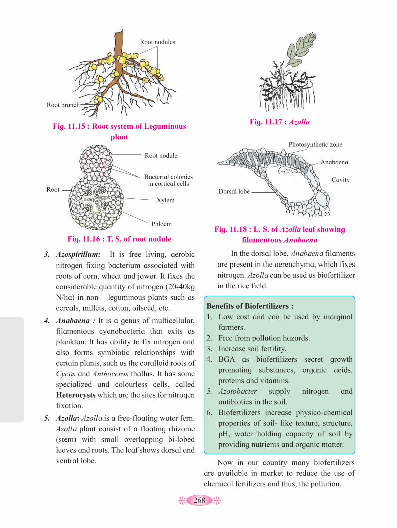
268
3. Azospirillum: It is free living, aerobic nitrogen fixing bacterium associated with roots of corn, wheat and jowar. It fixes the considerable quantity of nitrogen (20-40kg N/ha) in non – leguminous plants such as cereals, millets, cotton, oilseed, etc.
4. Anabaena : It is a genus of multicellular, filamentous cyanobacteria that exits as plankton. It has ability to fix nitrogen and also forms symbiotic relationships with certain plants, such as the coralloid roots of Cycas and Anthoceros thallus. It has some specialized and colourless cells, called Heterocysts which are the sites for nitrogen fixation.
5. Azolla: Azolla is a free-floating water fern. Azolla plant consist of a floating rhizome (stem) with small overlapping bi-lobed leaves and roots. The leaf shows dorsal and ventral lobe.
Fig. 11.17 : Azolla
Fig. 11.18 : L. S. of Azolla leaf showing filamentous Anabaena
Photosynthetic zone
Anabaena
Dorsal lobe
Cavity
Benefits of Biofertilizers :1. Low cost and can be used by marginal
farmers.2. Free from pollution hazards.3. Increase soil fertility.4. BGA as biofertilizers secret growth
promoting substances, organic acids, proteins and vitamins.
5. Azotobacter supply nitrogen and antibiotics in the soil.
6. Biofertilizers increase physico-chemical properties of soil- like texture, structure, pH, water holding capacity of soil by providing nutrients and organic matter.
Fig. 11.15 : Root system of Leguminous plant
Root nodules
Phloem
Xylem
Bacterial colonies in cortical cells
Root nodule
Root branch
Root
Fig. 11.16 : T. S. of root nodule
In the dorsal lobe, Anabaena filaments are present in the aerenchyma, which fixes nitrogen. Azolla can be used as biofertilizer in the rice field.
Now in our country many biofertilizers are available in market to reduce the use of chemical fertilizers and thus, the pollution.

269
Activity :
1. Visit fish market and enlist different types of fresh water and marine water fish mentioning their local names and scientific names.
2. Label the different parts of the fermenter and mention functions of each part.
Type Local Name Scientific Name Detail Information
1.
2.
3.
4.
5.
6.
7.
270
Q. 1 Multiple Choice Questions.
1. Antibiotic Chloromycetin is obtained from .....................
a. Streptomyces erythreus b. Penicillium chrysogenum
c. Streptomyces venezuelae d. Streptomyces griseus
2. Removal of large pieces of floating debris, oily substances, etc. during sewage treatment is called ................
a. primary treatment b. secondary treatment c. final treatment d. amplification
3. Which one of the following is free living bacterial biofertilizer?
a. Azotobacter b. Rhizobium c. Nostoc d. Bacillus thuringiensis
4. Most commonly used substrate for industrial production of beer is .............
a. barley b. wheat c. corn d. sugarcane molasses
5. Ethanol is commercially produced through a particular species of ..............
a. Aspergillus b. Saccharomyces c. Clostridium d. Trichoderma
6. One of the free-living anaerobic nitrogen-fixer is .....................
a. Azotobacter b. Beijerinckia c. Rhodospirillum d. Rhizobium
7. Microorganisms also help in production of food like .................
a. bread b. alcoholic beverages c. vegetables d. pulses
8. MOET technique is used for ............ a. production of hybrids b. inbreeding c. outbreeding d. outcrossing
9. Mule is the outcome of .............. a. inbreeding b. artificial insemination c. interspecific hybridization d. outbreeding
Q. 2 Very Short Answer Questions: 1. What does make idlies puffy?2. Name any two bacterial biofertilizers.3. What is the microbial source of vitamin
B12
?4. What is the microbial source of enzyme
Invertase?5. Milk start to coagulate when Lactic
Acid Bacteria (LAB) is added to warm milk as a starter. Mention any two other benefits of LAB.
6. Name the enzyme produced by Streptococcus bacterium. Explain importance in medical sciences.
7. What is breed?8. Define estuary.9. What is shellac?
Q. 3 Short Answer Questions.1. Many microbes are used at home during
preparation of food items. Comment on such useful ones with examples.
2. What is biogas? Write in brief about the production process.
3. Write a note on biocontrol agents.4. Name any two enzymes and antibiotics
with their microbial source.5. Write priciples of farm management.6. Give economic importance of fishery.7. Enlist the species of honey bee mentioning
their specific uses.
Exercise
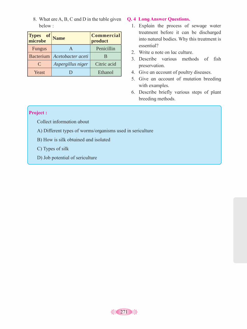
271
8. What are A, B, C and D in the table given below :
Types of microbe Name Commercial
productFungus A Penicillin
Bacterium Acetobacter aceti B C Aspergillus niger Citric acid
Yeast D Ethanol
Q. 4 Long Answer Questions.1. Explain the process of sewage water
treatment before it can be discharged into natural bodies. Why this treatment is essential?
2. Write a note on lac culture.3. Describe various methods of fish
preservation.4. Give an account of poultry diseases.5. Give an account of mutation breeding
with examples.6. Describe briefly various steps of plant
breeding methods.
Project :
Collect information about
A) Different types of worms/organisms used in sericulture
B) How is silk obtained and isolated
C) Types of silk
D) Job potential of sericulture
272
Biotechnology12
vinegar, yogurt, cheese making, wine making, etc. It became an art of kitchen in indian houses. It was more an art than science. Till that time people did not know as to how exactly the process occurs and the organisms causing this process. The contributions made by several chemists, biochemists and microbiologist, over the time, could explain the mechanism of process and also the nature of microorganisms causing the process.
During 1970 a new technique of ‘recombinant DNA technology was developed and then established by Stanley Cohen and Herbert Boyer in 1973. This technique has changed the overall outlook, then. The technique permits to change/ modify genetic (heritable) material for getting new specific products. The combination of biology and production technology based on genetic engineering evolved into modern biotechnology (new biotechnology).
There are two major features of technology that differenciate modern biotechnology from classical or old biotechnology viz,
i. Capablity of science to change the genetic material for getting new specific products through rDNA technology, polymerase chain reaction (PCR), microarrays, cell culture and fusion, and bioprocessing.
ii. Ownership of technology and its socio-political impact.
Now the conventional industries, pharmaceutical industries, agro industries, food industries, etc. are also focussing attention to produce biotechnology- based products.
12.2 Principles and Processes of Biotechnology :
Modern biotechnology is based on two core techniques viz. genetic engineering and chemical engineering.
You are already aware of what biotechnology is. It is the product of interaction between the biological science and technology. It is infact, an applied branch of biology. The term biotechnology was first used by Karl Ereky in 1919 to describe a process for large scale production of pigs.12.1 Biotechnology
It is defined by different organizations in different ways. It has been brodly defined as ‘the development and utilization of biological forms, products or processes for obtaining maximum benifits to man and other forms of life’. According to OECD (Organization for Economic Cooperation and Development, 1981)- ‘It is the application of scientific and engineering principles to the processing of materials by biological agents to provide goods and service to the human welfare’. It uses scientific priciples of microbiology, genetics biochemistry, chemical engineering, mathematics, statistics, computers, industrial processes, etc. Biological agent means plants and animal cells, microorganisms, enzymes or their products.
History of origin of biotechnology is as old as human civilization. Development of biotechnology in terms of its growth, occurred in two phases viz, Traditional biotechnology and Modern biotechnology.
Traditional biotechnology (old biotechnology) was primarily based on fermentation technology using microorganisms as in the preparation of curd, ghee, soma,
1. What is Biotechnology?2. How do genetically modified organisms
are produced?3. Which are the benefits of Biotechnology?
Can you recall?

273
A. Tools and techniques for gene cloning/ rDNA technology :
Before we venture into a procedure of gene cloning, let us know briefly, the basic requirements for the technique. I. Different instruments (devices) :
Macromolecule such as DNA, RNA, proteins, etc. are synthesized in the living cells which vary in their molecular weight, solubility, presence of charges, absorbance of light, etc. Several techniques are used to isolate and charaterize the macromolecules. The size of different types of molecules varies and therefore their molecular weights also vary. The techniques used on the basis of molecular weight, are gel permeation, osmotic pressure, ion exchange chromatography, spectroscopy, mass spectrometry, electrophoresis, etc. Electrophoresis is the separation of charged molecules, applying an electric field. It is applied for the separation of DNA, RNA and proteins. DNA being negatively charged, migrates to anode. Small fragments of DNA molecules, move faster and thus separate faster. Use of Agarose gel electrophoresis, PAGE, SDA PAGE are the different methods of electrophoresis.
Polymerase chain reaction (PCR) : Polymerase chain reaction (PCR) is
another device used for gene cloning or gene multiplication in vitro. It is the amplification of gene of interest, through PCR.
In 1985, Kary Mullis made an important discovery (contribution) in the form of an extremely powerful technique called polymerase chain reaction (PCR). PCR can generate a billion copies of the desired segment of DNA or RNA, with high accuracy and specificity, in a matter of few hours. The process of PCR is completely automated and involves automatic thermal cycles for denaturation and renaturation of double stranded DNA.
Genetic engineering deals with alteration of genetic material (DNA and RNA) while chemical engineering deals with maintaining sterile environment for manufacturing variety of useful products including vaccines, antibodies, enzymes, organic acids, vitamins, therapeutics, etc.
Genetic engineering is defined as the manupulation of genetic material towards a desired end and in a directed and predetermined way, using in vitro process. Manipulation of genes involve repairing of the defective genes or replacing of defective genes by healthy genes or normal genes; artificially synthesizing of a totally new gene; transfer of genes into a new location or into a new organism; introducing an all together new gene; manipulation of genes for improvement of living organisms; combining of genes from two organisms, altering the genotype; gene cloning etc. Therefore, the genetic engineering is alternatively called recombinant DNA technology or gene cloning.
Extra information : John E. Smith (1996) gave definition of
genetic engineering as ‘the formation of new combination of heritable material by the insertion of nucleic acid molecule produced by whatever means outside the cells, into any virus, bacterial plasmid or other vector system so as to allow their incorporation into a host organism in which they do not occur naturally but in which they are capable of continued propagation’.
Technique of gene cloning and rDNA technology :
In gene cloning, a gene of known function can be transferred from its normal location into a cell (that of course does not contain it) via a suitable vector. The transferred gene is replicated normally and is handed over to the next progeny.

274
The device required for PCR is called thermal cycler.
PCR is in vitro amplification of a desired DNA segment, which requires : DNA containing the desired segment to be amplified, several molecules of four deoxyribonuclueoside triphosphates (dNTPs), excess of two primer molecules, heat stable DNA polymerase and appropriate quantities of Mg++ ions.
One cycle takes arround 3 to 4 minutes. To begin second cycle, DNA is again heated to convert double stranded DNA into single strands.
In an automatic thermal cycler, the above three steps are automatically repeated 20-30 times. Thus, at the end of ‘n’ cycles 2n copies of DNA segments, are produed. The machine performs the entire operations automatically and precisely.
Once the desired number of cycles is completed, the amplified DNA segment is purified by gel electrophoresis. After its sequencing, the amplified DNA segment can be inserted into a cloning vector. Desired gene can also be obtained from gene library.
Mechanism of PCR: At the start of PCR, the DNA segment,
and excess of two primer molecules, four deoxyribonucleosides triphosphates and the thermostable DNA polymerase are mixed together in ‘eppendorf tube’ and the following operations are performed sequentially (Figure).
Step i : The reaction mixture is heated to a temperature (90–98 oC) to separate two strands of desired DNA. This is called denaturation.
Step ii : The mixture is allowed to cool (40–60 oC) that permits pairing of the primer to the complementary sequences in DNA. This step is called annealing.
Step iii : The temperature (70–75 oC) allows thermostable Taq DNA polymerase to use single-stranded DNA as template and adds nucleotides. This is called primer extension. It takes arround two minutes duration.
Fig. 12.1 : DNA replication through polymerase chain reaction.
1 copy of desired gene
Denaturation (90-98 0C)
DNA strands separated
Annealling of primers (40-60 0C)
Replicating DNA strands using
Tag polymerase (70-75 0C)
2 copies of desired gene
(renaturation)
Second cycle continues to form 4 copies of desired DNA
Cycle continous to produce 2n copies of DNA
Firs
t cyc
le c
ompl
etes
1
2
3
Internet my friend
Collect the information from internet about PCR machine.

275
II. Biological tools : There are three types of biological tools
used viz, enzymes, cloning vectors (vehicle DNA) and competent host (cloning organisms) for transformation with recombinant DNA.
A. Enzymes : Different enzymes include Lysozymes, Nucleases such as exonucleases endonucleases, restriction endonucleases, DNA ligases, DNA polymerases, alkaline phosphatases, reverse transcriptases, etc.
i. Restriction enzymes : Enzymes that cut the phosphodiester bands
of polynucleotide chains are called nuclease. There of two types - exonuclease and endonuclease. Exonucleases cut nucleotides from the ends of DNA strands whereas endonuclease cut DNA from within. During the 1970s, it was found that bacteria contain nucleases that would recognize short nucleotide sequence with duplex DNA and cut.
The phosphodiester back bone at highly specific sites on both strands of duplex, is cut by these enzymes, called restriction endonucleases or simply restriction enzymes. They were given this name because they are used by the bacteria to destroy various viral DNAs that might enter the cell, thereby restricting the potential growth of the virus.
Table 12.2 : Source and recognition sequences (indicated by arrow) of various restriction enzymes:
Restriction Enzyme
Source(Organism and
strain)
Recognition sequence Product End products produced
Alu I Arthobacter luteus5’ ---A- G- -C -T--- 3’
3’ ---T- C- -G-A--- 5’
5---A-G C-T---3
3---T-C G-A---5
Blunt ends
Bam HIBacillus
amyloliquefaciens H
5’---G- -G-A-T-C-C---3’
3’---C-C-T-A-G- -G---5’
5---G G-A-T-C-C---3
3 ---C-G-T-A-G G---5
Sticky ends
EcoRIEscherichia coli
Ry13
5’ ---G- -A-A-T-T-C---3’
3’ ---C-T-T-A-A- -G--- 5’
5---G A-A-T-T-C---3
3---C-T-T-A-A G---5
Sticky ends
Hind II H. influenzae Rd5’--G-T-C- -G-A-C--3’
3’--C-A-G- -C-T-G--5’
5’--G-T-C G-A-C--3’
3’--C-A-G C-T-G--5’ Blunt ends
Thus, restriction enzymes serve as defence mechanism. The bacteria protect its own DNA from nucleolytic attack by methylating the bases at susceptible sites, a chemical modification that blocks the action of the enzyme.
The restriction enzymes are thus the molecular scissors that are used to recognize and cut DNA at specific sequences. The sites recognized by them, are called recognition sequences or recognition sites. Different restriction enzymes found in different organisms recognize different nucleotide sequences and therefore cut DNA at different sites. Table encloses list of some restriction endonucleases and the site at which they cleave DNA.
ii. Recognition sequences : The sequences recognized by restriction
enzymes are 4 to 8 nucleotides long and characterized by a particular type of internal symmetry. Consider the particular sequence recognized by the enzyme EcoRI .
Activity :
Find out the biological source of following restriction enzymes and discuss the their recognition sequences :
Pst I, Sal I, Taq I, Xer III, Mbo II, Hpa I, BgII, Kpn I, Not I.
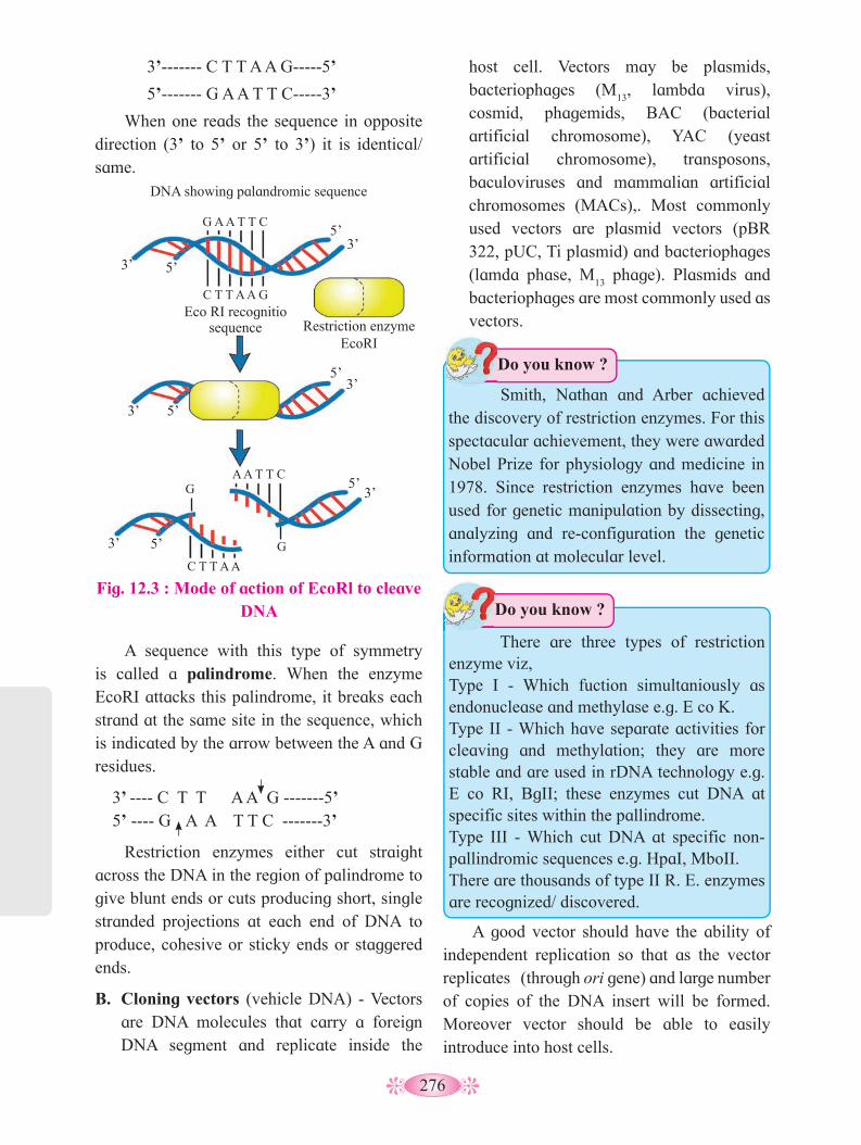
276
host cell. Vectors may be plasmids, bacteriophages (M
13, lambda virus),
cosmid, phagemids, BAC (bacterial artificial chromosome), YAC (yeast artificial chromosome), transposons, baculoviruses and mammalian artificial chromosomes (MACs),. Most commonly used vectors are plasmid vectors (pBR 322, pUC, Ti plasmid) and bacteriophages (lamda phase, M
13 phage). Plasmids and
bacteriophages are most commonly used as vectors.
3’------- C T T A A G-----5’
5’------- G A A T T C-----3’
When one reads the sequence in opposite direction (3’ to 5’ or 5’ to 3’) it is identical/ same.
A sequence with this type of symmetry is called a palindrome. When the enzyme EcoRI attacks this palindrome, it breaks each strand at the same site in the sequence, which is indicated by the arrow between the A and G residues.
3’ ---- C T T A A G -------5’ 5’ ---- G A A T T C -------3’
Restriction enzymes either cut straight across the DNA in the region of palindrome to give blunt ends or cuts producing short, single stranded projections at each end of DNA to produce, cohesive or sticky ends or staggered ends.
B. Cloning vectors (vehicle DNA) - Vectors are DNA molecules that carry a foreign DNA segment and replicate inside the
Fig. 12.3 : Mode of action of EcoRl to cleave DNA
DNA showing palandromic sequence
G A A T T C
A A T T C
C T T A A
C T T A A G
3’
3’
3’
3’
3’
3’
5’
5’
5’
5’
5’
5’
Restriction enzyme EcoRI
Eco RI recognitio sequence
G
G
Smith, Nathan and Arber achieved the discovery of restriction enzymes. For this spectacular achievement, they were awarded Nobel Prize for physiology and medicine in 1978. Since restriction enzymes have been used for genetic manipulation by dissecting, analyzing and re-configuration the genetic information at molecular level.
Do you know ?
There are three types of restriction enzyme viz, Type I - Which fuction simultaniously as endonuclease and methylase e.g. E co K. Type II - Which have separate activities for cleaving and methylation; they are more stable and are used in rDNA technology e.g. E co RI, BgII; these enzymes cut DNA at specific sites within the pallindrome.Type III - Which cut DNA at specific non-pallindromic sequences e.g. HpaI, MboII. There are thousands of type II R. E. enzymes are recognized/ discovered.
Do you know ?
A good vector should have the ability of independent replication so that as the vector replicates (through ori gene) and large number of copies of the DNA insert will be formed.Moreover vector should be able to easily introduce into host cells.
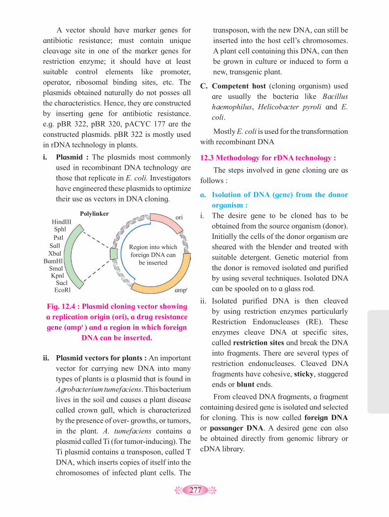
277
transposon, with the new DNA, can still be inserted into the host cell’s chromosomes. A plant cell containing this DNA, can then be grown in culture or induced to form a new, transgenic plant.
C. Competent host (cloning organism) used are usually the bacteria like Bacillus haemophilus, Helicobacter pyroli and E. coli.
Mostly E. coli is used for the transformation with recombinant DNA
12.3 Methodology for rDNA technology :
The steps involved in gene cloning are as follows :
a. Isolation of DNA (gene) from the donor organism :
i. The desire gene to be cloned has to be obtained from the source organism (donor). Initially the cells of the donor organism are sheared with the blender and treated with suitable detergent. Genetic material from the donor is removed isolated and purified by using several techniques. Isolated DNA can be spooled on to a glass rod.
ii. Isolated purified DNA is then cleaved by using restriction enzymes particularly Restriction Endonucleases (RE). These enzymes cleave DNA at specific sites, called restriction sites and break the DNA into fragments. There are several types of restriction endonucleases. Cleaved DNA fragments have cohesive, sticky, staggered ends or blunt ends.
From cleaved DNA fragments, a fragment containing desired gene is isolated and selected for cloning. This is now called foreign DNA or passanger DNA. A desired gene can also be obtained directly from genomic library or cDNA library.
A vector should have marker genes for antibiotic resistance; must contain unique cleavage site in one of the marker genes for restriction enzyme; it should have at least suitable control elements like promoter, operator, ribosomal binding sites, etc. The plasmids obtained naturally do not posses all the characteristics. Hence, they are constructed by inserting gene for antibiotic resistance. e.g. pBR 322, pBR 320, pACYC 177 are the constructed plasmids. pBR 322 is mostly used in rDNA technology in plants.
i. Plasmid : The plasmids most commonly used in recombinant DNA technology are those that replicate in E. coli. Investigators have engineered these plasmids to optimize their use as vectors in DNA cloning.
Fig. 12.4 : Plasmid cloning vector showing a replication origin (ori), a drug resistance gene (ampr ) and a region in which foreign
DNA can be inserted.
ii. Plasmid vectors for plants : An important vector for carrying new DNA into many types of plants is a plasmid that is found in Agrobacterium tumefaciens. This bacterium lives in the soil and causes a plant disease called crown gall, which is characterized by the presence of over- growths, or tumors, in the plant. A. tumefaciens contains a plasmid called Ti (for tumor-inducing). The Ti plasmid contains a transposon, called T DNA, which inserts copies of itself into the chromosomes of infected plant cells. The
HindIII Sphl Pstl
Sall Xbal
BamHl Smal Kpnl
Sacl EcoRl
Region into which foreign DNA can
be inserted
ori
ampr
Polylinker
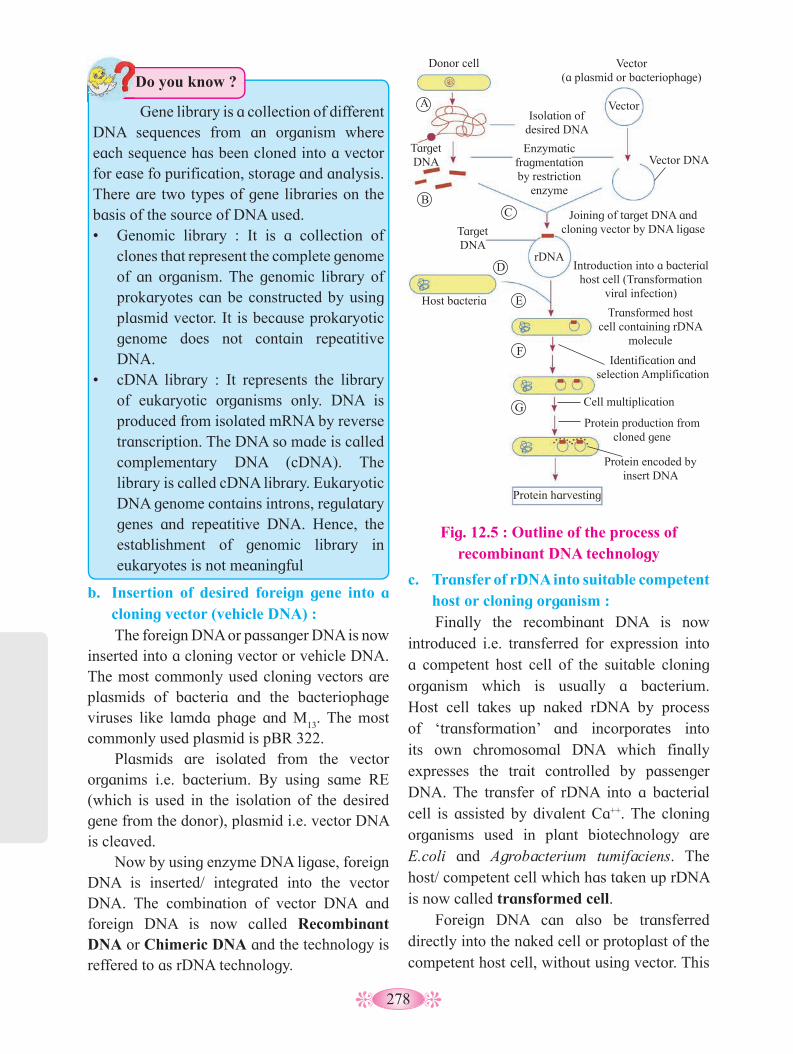
278
b. Insertion of desired foreign gene into a cloning vector (vehicle DNA) : The foreign DNA or passanger DNA is now
inserted into a cloning vector or vehicle DNA. The most commonly used cloning vectors are plasmids of bacteria and the bacteriophage viruses like lamda phage and M
13. The most
commonly used plasmid is pBR 322. Plasmids are isolated from the vector
organims i.e. bacterium. By using same RE (which is used in the isolation of the desired gene from the donor), plasmid i.e. vector DNA is cleaved.
Now by using enzyme DNA ligase, foreign DNA is inserted/ integrated into the vector DNA. The combination of vector DNA and foreign DNA is now called Recombinant DNA or Chimeric DNA and the technology is reffered to as rDNA technology.
c. Transfer of rDNA into suitable competent host or cloning organism : Finally the recombinant DNA is now
introduced i.e. transferred for expression into a competent host cell of the suitable cloning organism which is usually a bacterium. Host cell takes up naked rDNA by process of ‘transformation’ and incorporates into its own chromosomal DNA which finally expresses the trait controlled by passenger DNA. The transfer of rDNA into a bacterial cell is assisted by divalent Ca++. The cloning organisms used in plant biotechnology are E.coli and Agrobacterium tumifaciens. The host/ competent cell which has taken up rDNA is now called transformed cell.
Foreign DNA can also be transferred directly into the naked cell or protoplast of the competent host cell, without using vector. This
Gene library is a collection of different DNA sequences from an organism where each sequence has been cloned into a vector for ease fo purification, storage and analysis. There are two types of gene libraries on the basis of the source of DNA used.• Genomic library : It is a collection of
clones that represent the complete genome of an organism. The genomic library of prokaryotes can be constructed by using plasmid vector. It is because prokaryotic genome does not contain repeatitive DNA.
• cDNA library : It represents the library of eukaryotic organisms only. DNA is produced from isolated mRNA by reverse transcription. The DNA so made is called complementary DNA (cDNA). The library is called cDNA library. Eukaryotic DNA genome contains introns, regulatary genes and repeatitive DNA. Hence, the establishment of genomic library in eukaryotes is not meaningful
Do you know ?
Fig. 12.5 : Outline of the process of recombinant DNA technology
Donor cell Vector (a plasmid or bacteriophage)
Target DNA
Isolation of desired DNA
Enzymatic fragmentation by restriction
enzyme
Vector
Vector DNA
Target DNA
Joining of target DNA and cloning vector by DNA ligase
Introduction into a bacterial host cell (Transformation
viral infection)
rDNA
Host bacteria Transformed host
cell containing rDNA molecule
Identification and selection Amplification
Cell multiplication
Protein production from cloned gene
Protein encoded by insert DNA
Protein harvesting
A
B C
D
E
F
G

279
is done by using techniques like electroporation, microinjection, lipofection, shot gun, ultrasonification, biolistic method, etc. But in plant biotechnology the transformation is through Ti plasmids of A. tumifaciens.
d. Selection of the transformed host cell : The transformation process generates a
mixed population of transformed (recombinant) and non-transformed (non-recombinant) host cells. For isolation of recombinant cell from non-recombinant cell, marker gene of plasmid vector is employed. For example, PBR322 plasmid vector contains different marker gene (Ampicillin resistant gene and Tetracycline resistant gene). When pst1 RE is used, it knocks out Ampicillin resistant gene from the plasmid, so that the recombinant cell become sensitive to Ampicillin.
e. Multiplication of transformed host cell: Once transformed, host cells are separated
by the screening process. In this step the transformed host cells are introduced into fresh culture media.
At this stage the host cells divide and redivide along with the replication of the recom binant DNA carried by them.
The Centre for Cellular and Molecular Biology (CCMB) located in Hyderabad, is a premier research organization in frontier areas of modern biology including DNA fingerprinting and Molecular approaches in animal breeding. The objectives of the Centre are to conduct high quality basic research and training in frontier areas of modern biology, and promote centralized national facilities for new and modern techniques in the inter-disciplinary areas of biology.
Do you know ?
f. Expression of the gene to obtain the desired product:
The next step involves the production of desired products like alcohol, enzymes, antibiotics, etc. Finally the desired product is separated and purified through downstream processing using suitable bioreactor.
Fig. 12.6 : Applications of Biotechnology
Plant biotechnology (plant cell and tissue culture, GM crops, marker-assisted
selection)
Genomics Whole genome sequencing,
gene function, reverse genetics, bioinformatics)
Industrial biotechnology(Enzyme biotechnology,
protein engineering, metabolic engineering, metabolomics)
Animal biotechnology(Tissue culture, generative
meicine, immunotechnology, GM animals, pharmacogenomics,
gene therapy)
Microbial biotechnology(Microbial genomics,
biofertilizers, biopesticides, IPM)
Environmental biotechnology(Pollution control, biofuels,
bioremediation, biodiversity)
12.4 Applications of Biotechnology: Biotechnology is an umbrella term covering
a broad spectrum of scientific applications used in many sectors, such as health and agriculture. Industry, environment and genomics.
Biotechnology

280
Tabel 12.7 : Human proteins produced by rDNA technology to treat human diseases
Disorder/ Diseases/ Health condition
Recombinant protein(s)
1. Anaemia Erythropoeitin2. Asthma Interleukin-1 receptor3. Atherosclerosis Platelet derived
growth factor4. Parturition Relaxin5. Blood clots Tissue plasminogen
Activator (TPA) Urokinase
6. Cancer Interferons, tumour necrosis factor interleukins, macrophage activating factor
7. Diabetes Insulin8. Emphysemo a
1- Antitrypsin
9. Haemophilia A Factor VIII10. Haemophilia B Factor IX11. Hepatitis B Hepatitis B vaccine
a. Healthcare Biotechnology : It refers to a medicinal or diagnostic
product or a vaccine. This technology has a tremendous impact on meeting the needs of patients. Biotechnology offers patients a variety of new solutions such as: unique, targeted and personalized therapeutic and diagnostic solutions for organ transplant, stem cell technology, genetic counselling, forensic medicine, gene probes, genetic fingerprinting and karyotyping are outcomes of biotechnology.
Human insulin : Insulin is a peptide hormone produced by
β-cells of islets of Langerhans of pancreas. It was discovered by sir Edward Sharpey Schafer (1916) while studying Islets of Langerhans.
Insulin is essential for the control of blood sugar levels. Diabetes mellitus is a disease in which some people cannot make insulin themselves. Hakura et al (1977), chemically
synthesized DNA sequence of insulin for two chains A and B and separately inserted into two PBR322 plasmid vector. Insulin production by recombinant DNA technology is designed by Gilbert and Villokomaroff in 1978.
The genes are inserted by the side of β-galactosidase gene of the plasmid. The recombinant plasmids were then separately transformed into E. coli host. The host produced penicillinase + pre-pro insulin. Insulin is later separated by trypsin treatment.
Vaccine production: A vaccine is a biological preparation that
provides active acquired immunity against a certain disease. Usually a vaccine consists of a biological agent that represents the disease-causing microorganism. It is often made from a weakened or killed form of the microorganism, its toxins or one of its surface protein antigens.
Biotechnology has offered modern diagnostic test kits- rickettsial, bacterial and viral vaccines along with radiolabelled biological therapeutics for imaging and analysis.
Vaccines have eliminated small pox, polio and other deadly diseases for the last several decades. Biotechnology has made advancements in vaccination by making recombinant vaccines that have the potential to eradicate non-communicable diseases like cancer. Naked DNA vaccines, viral vector vaccines and plant-derived vaccines are found to be most effective against a number of bacterial and viral disorders.
Oral vaccines: a novel approach : The latest hot spot in the field of vaccine
research is the development of vaccine which can be taken orally. Immunogenic protein of
Internet my friend
Collect the information pertaining to recombinant vaccines and their types - protein vaccines and DNA vaccines.
281
c. Gene therapy: A gene is a stretch of DNA required to
make a functional product such as part or all of a protein. During gene therapy, DNA that codes for specific genes is delivered to individual cells in the body.
Gene therapy is the treatment of disease by replacing, altering or supplementing a gene that is absent or abnormal and whose absence or abnormality is responsible for the disease.
Most, if not all, diseases have a genetic factor. The genetic factor can be wholly or partially responsible for the disease. For example, in disorders such as cystic fibrosis, haemophilia, and muscular dystrophy, changes in a gene directly result in the condition.
In other conditions, such as high cholesterol and high blood pressure, genetic and environmental factors interact to cause disease. There are more than 5000 different human genetic diseases known to be caused by single gene defects e.g. sickle cell anaemia, thalassemia, Tay-sach’s disease, cystic fibrosis, Huntington’s chorea, haemophilia, alkaptonuria, albinism, etc.
Gene therapy is being used in many ways. For example, to: • Replace missing or defective genes; • Deliver genes that speed the destruction of
cancer cells; • Supply genes that cause cancer cells to
revert back to normal cells; • Deliver bacterial or viral genes as a form
of vaccination; • Deliver DNA to antigen expression and
generation of immune response;• Supply of gene for impairing viral
replication;• Provide genes that promote or impede the
growth of new tissue; and • Deliver genes that stimulate the healing of
damaged tissue.
certain pathogens is found to be active when administered orally. The gene corresponding to such proteins is isolated and a gene construct is produced. This is introduced and expressed in a plant genome, which results in production of such immunogenic proteins in the parts of the plant where it is expressed. These when fed into animals or mainly humans, the person becomes vaccinated against certain pathogen. Such vaccines are also known as edible vaccines.
An exciting invention is production of ‘melt in the mouth’ vaccines that can be administered by placing them under your tongue that delivers it into the blood stream. The most important example is the production of flu vaccine by Bacillus which melts in the mouth. The tremendous benefit of such vaccines, is the comfort of administeration, low cost and ease of storage.
b. Agriculture : Application of Biotechnology in Agriculture
involves scientific techniques such as Genetically Modified Organisms, Bt Cotton, Pest Resistant Plants. It helps in modifying plants, animals, and microorganisms and improve their agricultural productivity.
Tissue Culture is used in Micropropagation i.e. large-scale propagation of plants in very short durations. Tissue culture technique is also the best method for storing germplasm and maintaining a specific genetic type (Clone). This technique is used in those plants, which produce recalcitrant seeds or produce highly variable seeds.
Recalcitrant means the reduction in the seed moisture contents below certain levels and freezing drastically reduces the servival and thus present difficulty in storage. Here, subcellular damage of seeds occur accompanied by consequent loss of viability, when dried.
282
disease, familial hypercholesterolemia, haemophilia, phenylketonuria, cystic fibrosis, sickle cell anaemia, Duchenne muscular dystrophy, emphysema, thalassemia etc.
d. Genetically Modified Organisms (GMOs): These are living organisms whose genetic
material has been artificially manipulated in a laboratory through genetic engineering. This creates combinations of plant, animal, bacteria, and virus genes that do not occur in nature or through traditional crossbreeding methods.
Most GMOs have been engineered to withstand the direct application of herbicide and/or to produce an insecticide. However, new technologies are now being used to artificially develop other traits in plants, such as a resistance to browning in apples, and to create new organisms using synthetic biology. Despite biotech industry promises, there is no evidence that any of the GMOs currently on the market, offer increased yield, drought tolerance, enhanced nutrition, or any other consumer benefit.
I. Transgenic Plants : The human race is very dependent on
agriculture and as world populations continue to expand, there must be continuous reassessment of agricultural practices to optimize their efficiency. Since early times human beings have sought to improve the quality and productivity of agriculturally important plants. This was done by selection and traditional breeding procedures that were painstakingly slow and difficult. Traditional breeding programmes involve sexual crosses, which resulted in the high quality of present day food plants such as wheat, rice, corn, potato, etc. More recently, biotechnological approaches have been applied to these plants to create genetic variations that are beneficial for mankind.
First transgenic plant produced was tobacco. More then 60 transgenic dicot plants and several monocot plant like maize, oat,
Forms of gene therapy : a. Germ line gene therapy : In this method healthy genes can be introduced into germ cells like sperms, eggs, early embryos. It allows transmission of the modified genetic information to the next generation. Though it is highly effective in counteracting the genetic disorders, it is not encouraged for application in human beings due to a variety of technical and ethical reasons.
b. Somatic cell gene therapy : In this type the gene is introduced only in somatic cells like bone marrow cells, hepatic cells, fibroblasts endothelium and pulmonary epithelial cells, central nervous system, endocrine cells and smooth muscle cells of blood vessel walls. Modification of somatic cells only affects the person being treated and the modified chromosomes cannot be passed on the future generations. Somatic cell gene therapy is the only feasible option and the clinical trials have already employed for the treatment of acquired disorders such as cancer and rheumatoid arthritis and blood disorders including SCID, Gaucher’s
Delivery of genes into cells :Genes can be delivered by three ways :i. Ex vivo delivery where cells are removed
from the patients and then gene is introduced using viral or non-viral vectors e.g. Parkinsons disease, a neurological disorder.
ii. In vivo delivery where therapeutic genes are directly delivered in the cells at the target sites of the diseased tissue in the patient- like intravenous infusion genes to treat cancer are injected directly into tumor.
iii. Use of virosomes (Liposome + inactivated HIV), bionic chips are the other methods of gene delivery.
Do you know ?
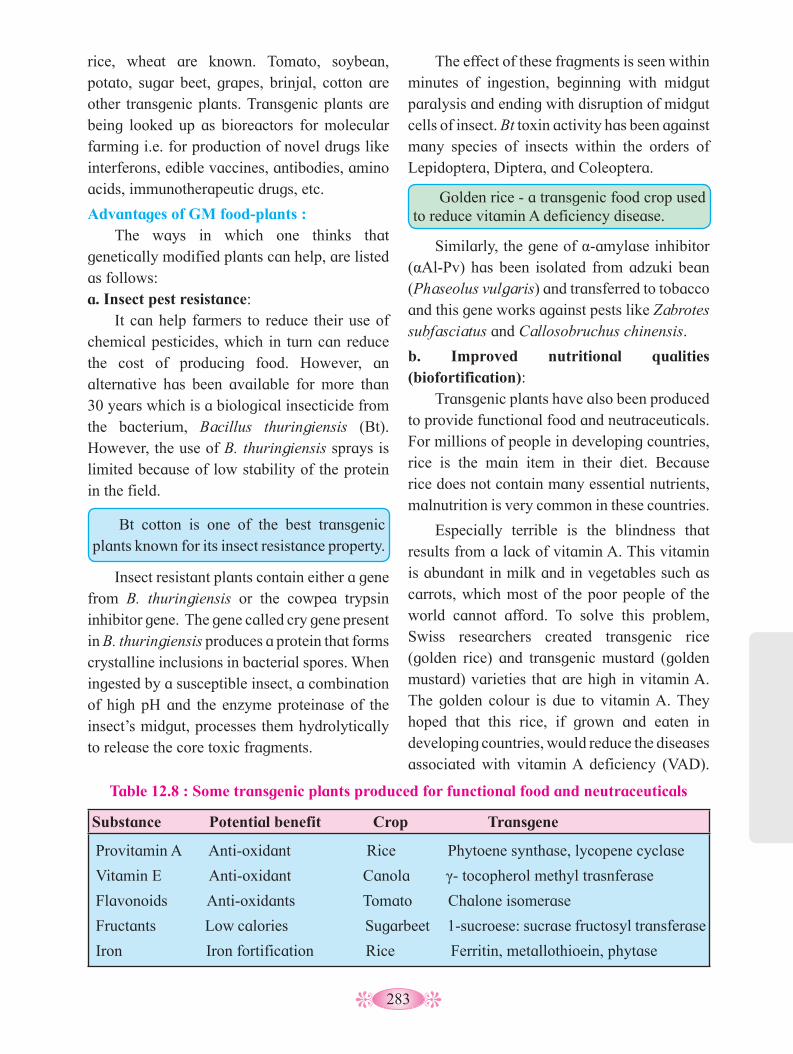
283
rice, wheat are known. Tomato, soybean, potato, sugar beet, grapes, brinjal, cotton are other transgenic plants. Transgenic plants are being looked up as bioreactors for molecular farming i.e. for production of novel drugs like interferons, edible vaccines, antibodies, amino acids, immunotherapeutic drugs, etc.
Advantages of GM food-plants : The ways in which one thinks that
genetically modified plants can help, are listed as follows:a. Insect pest resistance:
It can help farmers to reduce their use of chemical pesticides, which in turn can reduce the cost of producing food. However, an alternative has been available for more than 30 years which is a biological insecticide from the bacterium, Bacillus thuringiensis (Bt). However, the use of B. thuringiensis sprays is limited because of low stability of the protein in the field.
Bt cotton is one of the best transgenic plants known for its insect resistance property.
Insect resistant plants contain either a gene from B. thuringiensis or the cowpea trypsin inhibitor gene. The gene called cry gene present in B. thuringiensis produces a protein that forms crystalline inclusions in bacterial spores. When ingested by a susceptible insect, a combination of high pH and the enzyme proteinase of the insect’s midgut, processes them hydrolytically to release the core toxic fragments.
The effect of these fragments is seen within minutes of ingestion, beginning with midgut paralysis and ending with disruption of midgut cells of insect. Bt toxin activity has been against many species of insects within the orders of Lepidoptera, Diptera, and Coleoptera.
Table 12.8 : Some transgenic plants produced for functional food and neutraceuticals
Substance Potential benefit Crop Transgene
Provitamin A Anti-oxidant Rice Phytoene synthase, lycopene cyclase
Vitamin E Anti-oxidant Canola γ- tocopherol methyl trasnferase
Flavonoids Anti-oxidants Tomato Chalone isomerase
Fructants Low calories Sugarbeet 1-sucroese: sucrase fructosyl transferase
Iron Iron fortification Rice Ferritin, metallothioein, phytase
Golden rice - a transgenic food crop used to reduce vitamin A deficiency disease.
Similarly, the gene of α-amylase inhibitor (αAl-Pv) has been isolated from adzuki bean (Phaseolus vulgaris) and transferred to tobacco and this gene works against pests like Zabrotes subfasciatus and Callosobruchus chinensis.
b. Improved nutritional qualities (biofortification):
Transgenic plants have also been produced to provide functional food and neutraceuticals. For millions of people in developing countries, rice is the main item in their diet. Because rice does not contain many essential nutrients, malnutrition is very common in these countries.
Especially terrible is the blindness that results from a lack of vitamin A. This vitamin is abundant in milk and in vegetables such as carrots, which most of the poor people of the world cannot afford. To solve this problem, Swiss researchers created transgenic rice (golden rice) and transgenic mustard (golden mustard) varieties that are high in vitamin A. The golden colour is due to vitamin A. They hoped that this rice, if grown and eaten in developing countries, would reduce the diseases associated with vitamin A deficiency (VAD).
284
Most of these physiological changes are due to endogenous enzyme activity. Genetic engineering has made it possible to slow down these activities. In the tomato the enzyme polygalacturonase breaks down the cell wall constituent- pectin, leading to softening of fruit during ripening. Thus, the fruits are easily bruised and damaged on shipment. By inhibiting the polygalacturonase by antisense genes, the tomato (genetically modified tomatoes are called Flavr savr tomatoes) can remain on the vine until mature and be transported in a firm solid state.
d. Plants as factories : To produce novel biochemicals and
vaccines (Biopharmaceuticals), plants are potential factories or bioreactors for high value biochemicals like starch, sugar, lipids, proteins, and products like fine chemicals, perfumes and adhesive compounds as well as industrial lubricants, biodegradable plastic and even ‘renewable’ energy crops to replace fossil fuels.
Biopharmaceuticals are proteins, hormones, antibodies, vaccines or enzymes isolated from transgenic plants. Some of the proteins that are being produced by transgenic crop plants:
• Human growth hormone with the gene inserted into the chloroplast DNA of tobacco plants.
• Humanized antibodies against such infectious agents as HIV, Respiratory syncytial virus (RSV), Herpes simplex virus (HSV), the cause of "cold sores"
• Protein antigens to be used in vaccines for e.g. Patient-specific antilymphoma (a cancer) vaccines. B-cell lymphomas are clones of malignant B cells expressing on their surface a unique antibody molecule. Currently many novel products have been
commercially exploited for their products such as:
Improvement in oil content and oil quality of oil crops like soybean, oil palm, rapeseed and sunflower, have been achieved by transfer of ‘Arabidopsis genes’.
Iron deficiency is also a serious nutritional problem, affecting an estimated 30% of the world population. For production of transgenic crops that will produce food rich in iron, an iron storage protein (ferritin) is targeted. Ferrritin is found in many animals, plants and bacteria. The genes for ferritin protein isolated from soybean and Phaseolus have been transferred to rice.
• Genetically engineered herbicide
tolerant plants are developed as in maize, wheat and many other monocot plants.
• Genetically engineered disease resistant plants (against bacterial and viral pathogenes) are also developed in crop plants like tomato, potato and tobacco.
• Plants deficient in amino acids like methionine, lysine and tryptophan are genetically engineered by introducing genes from other sources so as to make the seeds protein rich. e.g. Leguminous plants (pulses), maize, etc.
• Similarly, genetically engineered plants tolerant to abiotic stresses such as high temperature, water, cold, etc. are also developed.
Do you know ?
c. Modification in Post-harvest characteristics:
Diseases and pests, bruising on soft fruits and vegetables, heat and cold storage, over-ripeness, loss of flavours and odours, etc. lead to great deal of losses during storage and transport of crops.

285
there has been a deliberate modification of the genome - the material responsible for inherited characteristics - in contrast to spontaneous mutation. Foreign DNA is introduced into the animal, using recombinant DNA technology, and then must be transmitted through the germ line so that every cell, including germ cells, of the animal contain the same modified genetic material.
A representative, but non-inclusive, list of purposes for which transgenic animals have been used, indicates the wide-ranging application of this biotechnology:
• in medical research, transgenic animals are used to identify the functions of specific factors in complex homeostatic systems through over- or under-expression of a modified gene (the inserted transgene);
• in toxicology: as responsive test animals (detection of toxicants);
• in mammalian developmental genetics;
• in molecular biology, the analysis of the regulation of gene expression makes use of the evaluation of a specific genetic change at the level of the whole animal;
• in the pharmaceutical industry, targeted production of pharmaceutical proteins, drug production and product efficacy testing;
a. A ‘superglue’ produced by tobacco plants with genes encoding for powerful adhesive proteins, enables marine mussels to stick to rocks. It will be especially valuable as a biochemical glue for body repairs during surgery.
b. Transgenic plants, containing oil-encoding gene from marine algae, produce oil that has nutritional value similar to cod- liver oil.
c. Plant that will produce the antimalarial drug, Artemisinin.
d. Genetically engineered opium poppy to produce more powerful painkillers.
e. Transgenic plants producing edible vaccines:
Genetically altered plants can provide protection to infectious diseases. Plant products acting as vaccines would be inexpensive to produce and thus, can easily be made available in developing countries. Potatoes, tomatoes, bananas, soybeans, alfalfa and cereals are the most common foods proposed for edible vaccine delivery.
II. Transgenic animals: Many transgenic animals such as mice, rats,
rabits, pig, sheep, cows, fowls, fish have been produced through rDNA technology. The term transgenic animal refers to an animal in which
Transgenic fowl for meat and eggs
Transgenic fish for protein rich meat
Transgenic mouse as animal model for research
Transgenic cow as bioreactor
Fig. 12.9 : Transgenic animals
286
The main objectives for improved animal breeding programmes coupled with this new technology of gene transfer are given below.• Efficiency of meat production• Improved quality of meat• Milk quality and quantity• Egg production• Wool quality and quantity• Disease resistance in animals• Production of low-cost pharmaceuticals
and biologicals
c. Transgenic cattle for food production : Of the few research reports describing the
use of transgenic technologies in cattle only one is directed towards a food production application. Researchers introduced additional copies of bovine beta or kappa casein into dairy cattle and evaluated the effect on milk production and composition. Transgenic offspring had an 8 to 20% increase in beta casein and a two-fold increase in kappa casein.
d. Transgenic cattle for human therapeutic production :
A second application for genetically modified cattle is the production of human therapeutic proteins. Human proteins that have been expressed in milk include human lactoferrin, human alpha lactalbumin, human serum albumin and human bile salt stimulated lipase. The mammary gland in dairy cows is an excellent protein production factory. On the other hand, one transgenic cow would be more than sufficient for production of annual world supply of factor IX (plasma thromboplastin component) that is used in the treatment of haemophilia.
In 1990 Tracy, the transgenic cow was born in Scotland, and could produce a human protein in her milk for human therapeutics.
Antibodies are currently used for many different human clinical applications; including treatment of infectious disease, cancer,
• in biotechnology: as producers of specific proteins;
• genetically engineered hormones to increase milk yield, meat production; genetic engineering of livestock and in aquaculture affecting modification of animal physiology and/or anatomy; cloning procedures to reproduce specific blood lines; and
• developing animals specially created for use in xenografting.
a. Transgenic mice and cancer research : Through laboratory investigations with
transgenic mice that have been modified using a particular oncogene (cancer causing gene) and thus developed a certain type of cancer, questions concerning the relationship between oncogenes and cancer development could be answered. Theoretically, such animals can also be used for research into cancer treatment and prevention of malignancy.
In the laboratory of Philip Leder in Harvard (USA) the transgenic mouse model for the investigation of the breast cancer was developed. The oncogenes myc and ras were analysed to find out if they lead to breast cancer in mice transformed with these genes.
b. Transgenic farm animals : With the advent of technology, the intention
of researchers is diverted to produce transgenic farm animals from which the mankind can derive greater benefits. Many of the farm animals are improved for their meat production ability while some of them are improved for milk yields and quality, and disease-free status. At the beginning of the century, a dairy cow provided 2,000 to 3,000 liters of milk a year.
Today, Holstein cow provides 6,000 liters on average and up to 8,000 – 10,000 for the best ones. A century ago, a hen laid about 70 eggs a year whereas today the best races lay up to 250 eggs per year. This could be possible because of the advent of biotechnology.

287
h. Transgenic chicken : Also carry and express foreign genes. They
could be used to improve the genetic make-up of existing strains with respect to built-in (in vivo) resistance to viral and coccidial diseases, better feed efficiency, lower fat and cholesterol levels and high protein containing eggs, and better meat quality.
12.5 Bioethics, bio-piracy and bio-patent : Bioethics:
Ethics usually deals with the matters related to socially acceptable moral duty, conduct and judgement. In otherwords, it helps to regulate the behaviour of cummunity by some set of standards. However the concepts differ according to culture and traditions. Moreover concepts change with the time due to shifting of perception of values which are affected by progress in science and technology. Bioethics helps to study moral vision, decision and policies of human behaviour in relation to biological phenomena or events. Ethics deals with ‘Life’ e.g. in vitro fertilization, sperm bank, gene theropy, cloning, gene manipulations, euthanasia, death, maintaining those who are in comatose state, prenatal genetic selection, etc.
The era of biotechnology has brought wide spectrum on new topics like cloning, transgenic, gene therapy, eugenics, rDNA technology, etc. The use of all these has drawn a wide range of reactions in the society. The reactions are based on individual’s own perception and moral. Ethical aspects pertaining to the use of biotechnology seems to be more controversial and frightening. These concerns are broadly summarized below :
Use of animals causes great sufferings to them; voilation of integration of species caused due to transgenosis; transfer of human genes into animals and vice versa; indiscriminate use of biotechnology pose risk to the environment, health and biodiversity.
transplanted organ rejection, autoimmune diseases and for use as antitoxins. To make a human antibody product, the genetically modified cows are immunized with a vaccine containing the disease agent.
e. Transgenic Sheep : Gene transfer technology is applied to
sheep to produce transgenic sheep which are able to achieve better growth and meat production as well as to serve as bioreactors. Human growth hormone gene is introduced in sheep for promoting the growth and meat production.
Bacterial genes, cys E and cys M, are concerned with biosynthesis of cysteine amino acids involved in formation of keratin protein found in wool. Both these genes are identified, cloned and introduced in sheep to increase wool production and to improve the quality of wool.
f. Transgenic pigs : The objective of gene transfer in pigs is to
increase growth and meat production and to act as bioreactors.
Pigs are regarded as the most suitable animals to be bred for heart transplant because a pig’s heart is about the same size as a human heart, and pig heart valves have been used in human heart surgery for over a decade. The pig clone is the first step towards providing animal organs and tissues for human transplants (xenotransplantation).
g. Transgenic fish : The commercially important fish like
Atlantic salmon, catfish, goldfish, Tilapia, zebra-fish, common carp, rainbow trout, etc. are transfected with growth hormone, chicken crystalline protein and E.coli hygromycin resistance gene. Transgenic fish showed increased cold tolerance and improved growth and it is the quantity and quality of fish proteins as well as its preservation, are the factors affecting the economic value of fish.
288
12.7 Effects of Biotechnology on Human Health :a. Allergies :
GMO crops could potentially have negative effects on human health as well. Consumers have developed unexpected allergic reactions. e.g. Researchers used a gene from the Brazil nut to increase the production of Methionine in soya beans. The insertion of this gene inadvertently caused allergic reactions to the soya bean in those with known nut allergies (“Biotech Soybeans”). b. Long-Term Effects :
Because GMO technology has been available for such a short amount of time, there is relatively little research which has been conducted on the long-term effects on health which we cannot anticipate at this point. c. New Proteins :
Proteins that have never been ingested before by humans are now part of the foods that people consume every day. Their potential effects on the human body are as of yet unknown. d. Food Additives :
GMOs also present us with possibilities of introducing additional nutrients into foods, as well as antibiotics and vaccines. This availability of technology can provide nutrition and disease resistance to those countries that don’t have the means to provide these, otherwise.
However, there is possibility of the creation of antibiotic and vaccine-resistant strains of diseases.
This shows that the vast advances in life sciences and our multicultural and pluralistic modern societies create numerous bioethical problems requiring some stringent regulation. In terms of GMOs, the Indian Government has set up the Genetic Engineering Approval Committee (GEAC). This organization makes decisions regarding the validity of research involving GMOs and addresses the safety of GMOs introduced for public use.
The introduction of Genetically Modified Organisms (GMOs) has led to a wider debate on bioethical concerns affecting social, economic and environmental spheres. These include the effects on non-target organisms, insect resistance crops, gene flow and the loss of diversity as well as the issue on interfering with nature in which the modification process itself is disrupting the natural process of biological entities. Ethics in biotechnology also includes the general subject of what should and should not be done in using recombinant DNA techniques
12.6 Effects of Biotechnology on the Environment :a. Herbicide Use and Resistance :
Effects on the environment are a particular concern with regard to GMO crops and food production. One area of development involves adding the ability to produce pesticides and resistance to specific herbicides. These traits are helpful in food production, allowing farmers to use fewer chemicals, and to grow crops in less than ideal conditions. However, herbicide use could be increased, which will have a larger negative effect on the surrounding environment. Also unintended hybrid strains of weeds and other plants can develop resistance to these herbicides through cross-pollination, thus negating the potential benefit of the herbicide. One such herbicide that has already been added is RoundUp. Crops of RoundUp-ready soybeans have already been implemented into agricultural practices, possibly conferring RoundUp resistance to neighboring plants.
b. Effects on Untargeted Species : Bt corn, which produces its own pesticide,
is also in use today. It has adverse effects on Monarch butterfly populations, which are not the original target of the pesticide. It can also have unintentional effects on neutral or even
beneficial species.

289
12.8 Biopatent and Biopiracy :a. Biopatent :
Patent is a special right granted to the inventor by the goverment. Patent is a personal property of inventor. It can be sold like any other property. A patent consists of three parts - grant (aggrement with the inventor), specification (subject matter of invention) and claims (scope of invention to be protected).
Biopatent is a biological patent. Biopatents are awarded for strains of microorganisms, cell lines, genetically modified strains, DNA sequences, biotechnological processes, product processes, product and product applications.
Biopatents are awarded to recognize real innovative contributions made by the inventor to the cause of human welfare. The awards are given to inculcate encouragement and values in developing scientific culture and in emphasizing the role of biology in shaping human society.
Indian patent allows ‘process patent’ and not the ‘product patent’. Biopatent allows the patent holder to exclude others from making, using, selling or importing protected invention for a limited period of time. Duration of biopatents is five years from the date of the grant or seven years from the date of filing the patent application, which ever is less.
First biopatent was patented pertaining to genetically engineered bacterium ‘Pseudomonas’ used for clearing oils spills. Patent under the title ‘control of plant gene expression’ was issued jointly to Delta and Pineland company and U. S. department of agriculture. Patent is based on a gene that produces a protein toxic to plant thus, do not allow seeds to germinate. However, this patent was not granted by Indian goverment. Such a patent is considered morally unacceptable and fundametally unequitable. This is because financially powerful corporations would aquire monopoly over biotechnological process. This in turn would pose a threat to global food security.
b. Biopiracy : Pirates in general terms are those who
steal and kill others to enrich themselves. Biopirates are those who do not kill but steal the patent (misuse the patent). Biopiracy is defined as ‘theft of various natural products and then selling them by getting patent without giving any benefits or compensation back to the host country’. In short, it is unauthorized misappropriation of any biological resource and indigenous knowledge.
Most developed, industrialized and financially rich nations are poor in biodiversity or traditional knowledge whereas developing and underdeveloped nations have ample of biodiversity and they traditionally better know the use of their bio-resources. Traditional Knowledge naturally includes a deep understanding of ecological processes and the ability to sustainably extract useful products from the local habitat. Most Traditional Knowledge is handed down through generations. This helps them to develop modern, commercial applications that save the makers time, money and effort.
Components of Traditional Knowledge that are especially relevant to our global survival include knowledge of:• Food, crop varieties and agricultural/
farming practice• Sustainable management of natural
resources and conservation of biological diversity
• Biologically important medicines
The conservation of species, habitat, and biodiversity are essential to the continued survival of indigenous and rural people. By conserving the customs and habitat of indigenous persons, we concurrently reduce emissions from deforestation and ecosystem degradation. Furthermore, the opportunity for cultural survival is a basic human right. The traditional knowledge is facing a problem of bio-piracy.
290
The act of Piracy is unauthorized publication or reproduction of another person’s work or material. When someone indulges in piracy, the accused is using someone’s work illegally or without taking any permission. The innovations and discovery of the pharmaceutical and agricultural researches are not new as to qualify as invention as they are based on centuries of knowledge of the traditional societies.
Examples of Biopiracy :• Patenting of Neem (Azadirachta indica) :
The people of India in a variety of ways have used neem, since time immemorial. Indians have shared the knowledge of the properties of the neem with the entire world. Pirating this knowledge, the USDA and an American MNC W.R. Grace in the early 90s sought a patent from the European Patent Office (EPO) on the “method for controlling on plants by the aid of hydrophobic extracted neem oil.” The patenting of the fungicidal properties of Neem, was an example of biopiracy.
• Patenting of Basmati : Basmati is a long-grained, aromatic variety
of rice indigenous to the Indian subcontinent.
In 1997 the US Patent and Trademark Office (USPTO) granted a patent to a Texas based American company Rice Tec Inc for “Basmati rice line and grains” having trade
name Texmati. The patent application was based on 20 very broad claims on having “invented” the said rice. Due to peoples movement against Rice Tec in March 2001, the UPSTO has rejected all the claims.
•Haldi (Turmeric) Biopiracy : Two American researchers of Indian
origin of the University of Mississippi Medical Center, put a claim to the US Patent and Trademark Office, maintaining that they had discovered haldi's healing properties. Surprisingly, they were granted a patent in March 1995 for something you had known for years and our ayurvedas for centuries.
It meant they had exclusive rights over any such haldi drug and were in a position to make millions of dollars. The Council of Scientific and Industrial Research (CSIR) applied to the US Patent Office for a reexamination and they realized the mistake and cancelled the patent. This was after Indian scientists shouted from rooftops about how we are losing our traditional knowledge to marauding foreign companies who have started poaching on our ancient healing techniques.
It is the need of hour to launch genetic literacy movement in Indian school and colleges for better understanding of opportunities and risks related to biotechnology and also to promote the safe and meaningful use of technologies of modern life sciences.
Activity :
Collect information on the use of Biotechnology in pollution control.

291
Q. 1 Choose the correct option1. The bacterium which causes a plant
disease called crown gall is ................ b. Helicobacter pylori c. Agrobacterium tumifaciens d. Thermophilus aquaticus e. Bacillus thuringienesis
2. The enzyme nuclease hydrolyses ............... of polynucleotide chain of DNA.
a. hydrogen bonds b. phosphodiester bonds c. glycosidic bonds d. peptide bonds
3. In vitro amplification of DNA or RNA segment is known as ...............
a. chromatography b. southern blotting c. polymerase chain reaction d. gel electrophoresis
4. Which of the following is the correct recognition sequence of restriction enzyme hind III.a. 5’ ---A-A-G-C-T-T---3’
3’ ---T-T-C-G-A-A--- 5’ b. 5’ ---G-A-A-T-T-C---3’
3’ ---C-T-T-A-A-G--- 5’
c. 5’ ---C-G-A-T-T-C---3’
3’ ---G-C-T-A-A-G--- 5’ d. 5’ ---G-G-C-C---3’
3’ ---C-C-G-G--- 5’
5. Recombinant protein .................. is used to dissolve blood clots present in the body.a. insulinb. tissue plasminogen activatorc. relaxind. erythropoietin
6. Recognition sequence of restriction enzymes are generally ............... nucleotide long.a. 2 to 4 b. 4 to 8c. 8 to 10 d. 14 to 18
Q. 2 Very short answer type questions.1. Name the vector which is used in
protuction of human insulin through recombinent DNA technology.
2. Which cells from Langerhans of pancreas do produce a peptide hormone insulin?
3. Give the role of Ca++ ions in the transfer of recombinent vector into bacterial host cell.
4. Expand the following acronyms which are used in the field of protechnology.
i. YAC ii. RE iii. dNTP
iv. PCR v. GMO vi. MAC
5. Fill in the blanks and complete the chart.
GMO Purposei. Bt cotton ....................ii. ................... Delay the softening of
tomato during ripening.ii. Golden rice ....................iv. Holstein cow ....................
Q. 3 Short answer type questions.1. Explain the properties of a good or ideal
cloning vector for rDNA technology.2. A PCR machine can rise temperature
upto 100 0C but after that it is not able to lower the temperature below70 0C automatically. Which step of PCR will be hampered first in this faulty machine? Explain why?
3. In the process of rDNA technology, if two separate restriction enzymes are used to
Exercise
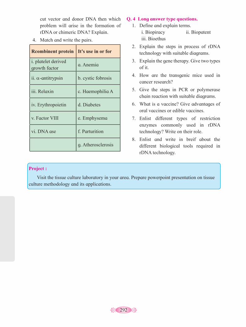
292
cut vector and donor DNA then which problem will arise in the formation of rDNA or chimeric DNA? Explain.
4. Match and write the pairs.
Rcombinent protein It’s use in or for
i. platelet derived growth factor
a. Anemia
ii. a-antitrypsin b. cystic fobrosis
iii. Relaxin c. Haemophilia A
iv. Erythropoietin d. Diabetes
v. Factor VIII e. Emphysema
vi. DNA ase f. Parturition
g. Atherosclerosis
Q. 4 Long answer type questions.1. Define and explain terms. i. Biopiracy ii. Biopatent iii. Bioethus
2. Explain the steps in process of rDNA technology with suitable diagrams.
3. Explain the gene therapy. Give two types of it.
4. How are the transgenic mice used in cancer research?
5. Give the steps in PCR or polymerase chain reaction with suitable diagrams.
6. What is a vaccine? Give advantages of oral vaccines or edible vaccines.
7. Enlist different types of restriction enzymes commonly used in rDNA technology? Write on their role.
8. Enlist and write in breif about the different biological tools required in rDNA technology.
Project :
Visit the tissue culture laboratory in your area. Prepare powerpoint presentation on tissue culture methodology and its applications.

293
Natural world around us shows amazing diversity of forms and complexity of relations. To understand these, we have to study levels of organization in the living world viz. macromolecules, cells, tissues, organs, individual organism, population, communities, ecosystems and biomes.
You have already studied in school, that ecology is a study of the interactions among organisms and between the organisms and their physical (abiotic) environment. Term ecology was first used by Reiter but E. Haeckel gave substance to the term (introduced) ecology.
Ecological grouping of organisms is nothing but ecological hierarchy. There are four sequential levels with increasing complexity of ecological (biological) organizations viz, Organism, Populations, Communities and Biomes. Individual organism is the basic unit of ecological hierarchy. Organisms of same kind inhabiting a geographical area constitute population. Several populations of different species in a particular area constitute community that interact with one another in several ways. Biome constitutes a large regional terrestrial unit delimited by a specific climatic zone having major vegetation zone (plant communities) and the associated fauna. There are six major groups of terrestrial biomes. We shall explore first two levels viz, organism and populations.
13.1 Organisms and the environment around:
Ecology at the level of organism is basically the study of animal or plant physiology which helps us to understand how the organisms
You have studied in earlier classes about the rotation of earth around the Sun and the tilt of its axis, cause seasons. These seasons with annual variation in precipitation in the form of rain and snow, gives rise to formation of major biomes of the earth like desert, rain forest, grassland, tundra, etc.
Regional and local variations within each biome lead to the formation of a variety of habitats. Major biomes of earth are shown in Fig. 13.1. On the Earth, life exists even in extreme and harsh habitats like scorching deserts of Rajasthan, perpetually rain-soaked forests of North Eastern states and high mountain tops of Himalayas.
Organisms and Populations
are adapted to their environments, not only for their survival but also for propagation (multiplication).
13
1. What is an ecosystem?2. What are the abiotic and biotic
components of an ecosystem?
Can you recall?
What are the key differences that make such a great variation in the physical and chemical conditions of different habitats?
Find out
Fig. 13.1 : Distribution of major Biomes with respect to annual temperature and
precipitation
Here, we must remember that it is not only the physico-chemical (abiotic) components that make up the habitat of an organism, but the habitat also includes biotic components like
Mea
n an
nual
tem
pera
ture
(0 C
)
Mean annual precipitation/ rainfall (cm) 50 100 150 200 250 300 350 400 450
30
25
22
15
10
5
0
-5
-10
-15
Desert Grassland
Tropical forest
Temperate forest
Coniferous forest
Arctic and alpine tundra
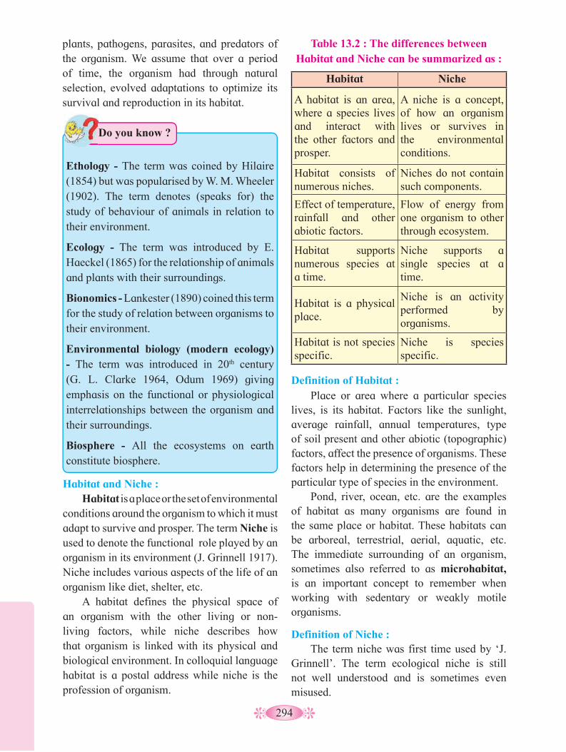
294
Table 13.2 : The differences between Habitat and Niche can be summarized as :
Habitat Niche
A habitat is an area, where a species lives and interact with the other factors and prosper.
A niche is a concept, of how an organism lives or survives in the environmental conditions.
Habitat consists of numerous niches.
Niches do not contain such components.
Effect of temperature, rainfall and other abiotic factors.
Flow of energy from one organism to other through ecosystem.
Habitat supports numerous species at a time.
Niche supports a single species at a time.
Habitat is a physical place.
Niche is an activity performed by organisms.
Habitat is not species specific.
Niche is species specific.
Definition of Habitat : Place or area where a particular species
lives, is its habitat. Factors like the sunlight, average rainfall, annual temperatures, type of soil present and other abiotic (topographic) factors, affect the presence of organisms. These factors help in determining the presence of the particular type of species in the environment.
Pond, river, ocean, etc. are the examples of habitat as many organisms are found in the same place or habitat. These habitats can be arboreal, terrestrial, aerial, aquatic, etc. The immediate surrounding of an organism, sometimes also referred to as microhabitat, is an important concept to remember when working with sedentary or weakly motile organisms.
Definition of Niche : The term niche was first time used by ‘J.
Grinnell’. The term ecological niche is still not well understood and is sometimes even misused.
Habitat and Niche : Habitat is a place or the set of environmental
conditions around the organism to which it must adapt to survive and prosper. The term Niche is used to denote the functional role played by an organism in its environment (J. Grinnell 1917). Niche includes various aspects of the life of an organism like diet, shelter, etc.
A habitat defines the physical space of an organism with the other living or non-living factors, while niche describes how that organism is linked with its physical and biological environment. In colloquial language habitat is a postal address while niche is the profession of organism.
Ethology - The term was coined by Hilaire (1854) but was popularised by W. M. Wheeler (1902). The term denotes (speaks for) the study of behaviour of animals in relation to their environment.
Ecology - The term was introduced by E. Haeckel (1865) for the relationship of animals and plants with their surroundings.
Bionomics - Lankester (1890) coined this term for the study of relation between organisms to their environment.
Environmental biology (modern ecology) - The term was introduced in 20th century (G. L. Clarke 1964, Odum 1969) giving emphasis on the functional or physiological interrelationships between the organism and their surroundings.
Biosphere - All the ecosystems on earth constitute biosphere.
Do you know ?
plants, pathogens, parasites, and predators of the organism. We assume that over a period of time, the organism had through natural selection, evolved adaptations to optimize its survival and reproduction in its habitat.

295
We have seen earlier in the chapter that each habitat type is regulated by a number of abiotic or physico- chemical factors.
Key abiotic factors that influence any habitat are ambient temperature, availability of water, light and type of soil.
13.2 Major Abiotic Factors :Temperature:
It is the most ecologically relevant environmental factor. Average temperature on land varies from subzero levels in polar areas and high altitudes, upwards upto 50oC in tropical deserts in summer. Temperature also varies seasonally. It decreases progressively from the equator towards the poles and from plains to the mountain tops. There are some unique habitats such as hot springs (80 to 1000C) and deep-sea hydrothermal vents where average temperatures usually 400o C. Ambient temperature affects the enzyme kinetics of the cell and thus, the entire metabolism, activity and other physiology of the organism.
Only few organisms can tolerate and thrive in a wide range of temperatures (eurythermal), but, a vast majority of them are restricted to a narrow range of temperatures (stenothermal).
Niche is described as a position of a species in the environment like, what they do for their survival? how they fulfill their needs of shelter, food? etc. Niche deals with the flow of energy from one organism to another and hence, it is important to understand, what an organism eats, how it interacts with other organisms, etc. As soon as the niche is left vacant, other organisms fill that position. The niche is specific to each species, which means no two species can share the same niche.
If the species creates its own unique niche in an ecosystem, it would be helpful in reducing competition for resources among species. By taking an example of a bird, it can be understood that how these birds differ in their eating habits, where some birds eat only insects, some only fruits and some can eat both and anything they come across. So here we can conclude that these birds living in the same habitat differ in their niches because of different eating habits.
Three types of niches are found:
a. Spatial or habitat niche : It deals with the physical space occupied by the organisms.
b. Trophic niche : It is on the basis of trophic level of an organism in a food chain.
c. Multidimensional or hypervolume niche: It considers number of environmental factors (both biotic and abiotic), the resulting space will be a hypervolume; not something that can be perceived by the human mind. This space is called the hypervolume niche. Alternatively, it is the position of an organism in the environmental gradient.
For every species, there is a fundamental niche and a realized niche. Fundamental niche is the niche in the absence of all competitors, this is highly improbable in nature. Hence, realized niche is more realistic approach, in the presence of competition for the resources available in the habitat.
1. Give names of eurythermal and
stenothermal animals and plants?2. What will be the effect of increasing
global temperatures on the different habitats and the organisms found in those habitats?
Find out
Water: Availability of water is an important factor affecting the organisms. As we know, life on earth originated in water, its availability is so limited in deserts that only special adaptations are required to survive there. The productivity and distribution of plants are also heavily dependent on water.
296
Give examples of an animal and plant that can survive in fresh water as well as sea water?
Find out
What is the source of energy for the life in deep ocean trenches where sunlight does not reach?
Find out
Soil: The nature and properties of soil are dependent on the climate, the weathering process.
Various characteristics of the soil such as soil composition, grain size, determine the percolation and water holding capacity of the soil. These characteristics along with pH, mineral composition and topography, determine the vegetation of an area. Vegetation in turn dictates the type of animals.
The abiotic factors that determine the type of habitat, also show considerable diurnal and seasonal variations. The plants and animals must adapt to these changes in order to survive and flourish in the habitat. During the course of their evolution, many species have evolved a relatively constant ‘internal’ environment that permits all biochemical reactions and physiological functions to proceed with optimum rate, and allow the species to flourish. The organisms try to maintain the constancy of its internal environment (homeostasis) despite variations in the external environmental conditions. To survive and flourish in any environment, organisms must adapt to the changes in the environment for which there are following possibilities :
i. Regulate: Some organisms are able to maintain homeostasis by physiological and behavioural changes which ensure constant body temperature, constant osmotic concentration, etc. All birds and mammals are capable of such regulation (thermoregulation and osmoregulation).
ii. Conform: Most of animals and plants cannot maintain a constant internal environment. Their body temperature changes with the ambient temperature. In aquatic animals, the osmotic concentration of the body fluids changes with that of the ambient water osmotic concentration. These animals and plants are simply conformers.
Organisms living in water bodies such as oceans, lakes and rivers, have their own water-related problems. For aquatic organisms the chemical composition and pH of water are important.
The dissolved salt concentration (measured as salinity in parts per thousand), is less than 5ppt in fresh waters of streams , lakes and rivers, and 30-35ppt in the seas and oceans. It may go up to 100ppt in some hypersaline lagoons.
Some organisms are tolerant for a wide range of salinities (euryhaline) but others are restricted to a narrow range (stenohaline). Many fresh water animals cannot live for long in sea water and vice versa because of the osmotic problems, they would face.
Light: Plants use light for photosynthesis, which is only source of energy for the entire ecosystem. Photosynthesis can occur only in presence of sunlight. Many species of small plants (herbs and shrubs) growing on forest floor are adapted to perform photosynthesis optimally under very low light conditions because they are constantly overshadowed by tall trees.
For animals too, diurnal and seasonal variations in light intensity and duration (photoperiod) are clues for timing their foraging, reproductive and migratory activities. The availability of light on land is closely linked with that of temperature, since the sun is the source for both.
297
organism (morphological, physiological, and behavioural) that enables the organism to survive and reproduce in its habitat.
Many desert plants have a thick cuticle on their leaf surfaces and have their stomata in deep pits to minimize loss of water through transpiration. They also have a special photosynthetic pathway (CAM - Crassulacean acid metabolism) that enables their stomata to remain closed during daytime. Some desert plants like Opuntia, have their leaves reduced (modified) to spines and the photosynthetic function is taken over by the flattened stems.
Mammals from colder climates generally have shorter snout, ears, tail and limbs to minimize the loss of body heat (Allen’s Rule.) In the polar seas, aquatic mammals like seals have a thick layer of fat (blubber) below their skin acting as an insulator to reduce loss of body heat.
Some species have evolved the ability to regulate, within a limited range of environmental conditions, beyond which they simply conform. If the stressful environment is localized or only for a short period of time, the organism may migrate or suspend its activities.
iii. Migrate: The organism can move away temporarily from the stressful habitat to a more hospitable area and return when stressful period is over. Many animals, particularly birds, during winter undertake long-distance migrations to more hospitable areas.
iv. Suspend: In plants, seeds serve as means to tide over periods of stress; they germinate to form new plants under favourable moisture and temperature conditions. They do so by reducing their metabolic activity and going into a state of ‘dormancy’. In animals, the organism, if unable to migrate may go into hibernation during winter e.g. polar bear. Some snails and fish go into aestivation to avoid summer heat.
1. What is homeostasis?2. Why do animals need to maintain
homeostasis? 3. What are the adaptations in animals
living under crushing pressure at great depths of ocean?
Can you tell?
13.3 Adaptation : To cope up with extreme variations in
their environment, some organisms respond through physiological adjustments, while others do so behaviourally (like migration). These are their adaptations. Therefore, we can say that adaptation is an attribute of the
Some organisms show behavioural responses to cope with variations in their environment. Desert lizards manage to keep their body temperature fairly constant by behavioural adaptations. They bask in the sun and absorb heat, when their body temperature drops below the comfort zone, but move into shade, when the ambient temperature starts increasing. Some species burrow into the sand to hide and escape from the heat.
13.4 Population : In nature, we rarely find isolated, single
individuals. They live in groups in a well-defined geographical area, share or compete
1. Adaptation of plants for aquatic and desert habitats.
2. Adaptations of animals for aquatic and desert habitats.
Do you know?
Internet my friend
Find out the difference between hibernation and aestivation.

298
It must be remembered, that absolute natality will be always more than realized natality.
Mortality is the death rate of a population. Mortality rate or death rate, is a measure of the number of deaths (in general, or due to a specific cause) in a particular population, in proportion to the size of that population, per unit of time. Mortality rate is typically expressed in deaths per 1,000 individuals per year. Thus, a mortality rate of 9.5 (out of 1,000) in a population of 1,000 would mean 9.5 deaths per year in that entire population, or 0.95% out of the total.
• Absolute Mortality : the number of deaths under ideal conditions (with no competition, abundance of resources such as food and water, etc.).
• Realized Mortality : the number of deaths when environmental pressures come into play. It must be remembered that absolute
mortality will be always less than realized mortality.
Sex ratio: The sex ratio of the population affects and is reciprocally affected by birth, death, immigration, and emigration rates. It is measured as the ratio of the number of individuals of one sex to that of the other sex. The males and females in a ratio of 1:1 is generally the most common evolutionary stable strategy (ESS).
for similar resources, potentially interbreed and thus form a population.
Although, it is an individual organism that has to cope with changes in the environment, it is at the population level that natural selection operates to evolve the desired traits. Population ecology is, therefore, an important area of ecology because it links ecology to population dynamics, genetics and evolution.
A population has certain attributes of its own, which are different from those of an individual. Basic physical characteristics of population are - its size and density. Size speaks for the number of individuals in a population while density tells us number of individuals present per unit space, in a given time. Besides size and density, the other characteristics include natality, mortality, immigration, emigration, age pyramids, expanding population, population growth forms and biotic potential. An individual has birth and death, but a population has birth rate and death rate.
Natality is the birth rate of a population. It has the greatest influence on a population’s growth. Natality is a crude birth rate or specific birth rate. Crude birth rate is used when calculating population size (number of births per 1000 population/year), whereas specific birth rate is used relative to a specific criterion such as age. E.g. If in a pond, there were 200 carp fish last year and through reproduction 800 new fish are added, taking the current population to 1000, we calculate the birth rate as 800/200 = 4 offspring per carp per year.
• Absolute Natality : the number of births under ideal conditions (with no competition, abundance of resources such as food and water, etc.).
• Realized Natality : the number of births when environmental pressures come into play.
What can be the causes of deviation from 1: 1 sex ratio in natural habitat?
Can you tell?
Internet my friend
1. Find out the sex ratio of Indian population and the state of Maharashtra.
2. What are the reasons behind deviation of sex ratio among Indian population?
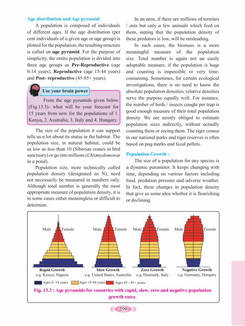
299
In an area, if there are millions of termites / ants but only a few animals which feed on them, stating that the population density of these predators is low, will be misleading.
In such cases, the biomass is a more meaningful measure of the population size. Total number is again not an easily adoptable measure, if the population is huge and counting is impossible or very time-consuming. Sometimes, for certain ecological investigations, there is no need to know the absolute population densities; relative densities serve the purpose equally well. For instance, the number of birds / insects caught per trap is good enough measure of their total population density. We are mostly obliged to estimate population sizes indirectly, without actually counting them or seeing them. The tiger census in our national parks and tiger reserves is often based on pug marks and fecal pellets.
Population Growth : The size of a population for any species is
a dynamic parameter. It keeps changing with time, depending on various factors including food, predation pressure and adverse weather. In fact, these changes in population density that give us some idea whether it is flourishing or declining.
The size of the population it can support tells us a lot about its status in the habitat. The population size, in natural habitat, could be as low as less than 10 (Siberian cranes in bird sanctuary) or go into millions (Chlamydomonas in a pond).
Population size, more technically called population density (designated as N), need not necessarily be measured in numbers only. Although total number is generally the most appropriate measure of population density, it is in some cases either meaningless or difficult to determine.
Age distribution and Age pyramid: A population is composed of individuals
of different ages. If the age distribution (per cent individuals of a given age or age group) is plotted for the population, the resulting structure is called an age pyramid. For the purpose of simplicity, the entire population is divided into three age groups as Pre-Reproductive (age 0-14 years), Reproductive (age 15-44 years) and Post- reproductive (45-85+ years).
From the age pyramids given below (Fig.13.3)- what will be your forecast for 15 years from now for the populations of 1. Kenya, 2. Australia, 3. Italy and 4. Hungary.
Use your brain power
Fig. 13.3 : Age pyramids for countries with rapid, slow, zero and negative population growth rates.
Male Male Male Male Female Female Female Female
Rapid Growth e.g. Kenya, Nigeria
Slow Growth e.g. United States, Australia
Zero Growth e.g. Denmark, Italy
Negative Growth e.g. Germany, Hungary
Ages 0 -14 years Ages 15-44 years Ages 45 - 85+ years

300
Fig. 13.4 : Exponential growth curve of population
Popu
latio
n si
ze
Time
Every species is capable of growing exponentially under unlimited resource conditions, and reach enormous population densities in a short time. Darwin showed how even a slow growing animal like elephant could reach enormous numbers. (provided food and space remain unlimited).
But resources like food and space are not always unlimited. They may be in the beginning; but as the population density increases, so does the competition for those resources, resulting in slowdown in the rate at which the original population was growing. This results in logistic or sigmoid growth curve.
ii. Logistic growth: competition between individuals for limited resources will weed out the ‘weaker’ ones. Only the ‘fittest’ individuals will survive and reproduce. In nature, a given habitat has enough resources to support a maximum possible number, beyond which no further growth is possible. Let us call this limit as nature’s carrying capacity (K) for that species in that habitat.
Density of population in a habitat during a given period, fluctuates due to changes in four basic processes. New births (B) and Immigration contribute to an increase in population density. Deaths (D) and Emigration lead to decrease in population density. Immigration (I) is the number of individuals of the same species that have come into the habitat from elsewhere during the time period under consideration. Emigration (E) is the number of individuals of the population who left the habitat during the time period.
So, if N is the population density at time ‘t’, then its density at time ‘t +1’ can be calculated as,
Nt+1
= Nt + [(B + I) – (D + E)]
Growth Models: Does the growth of a population with time show any specific and predictable pattern? We have been concerned about unbridled human population growth and problems created by it in our country and it is therefore natural for us to be curious if other animal populations in nature behave the same way or show some restraint on growth.
i. Exponential growth: Resources like food, space are essential
for any growth of a population. Ideally, when resources in the habitat are unlimited, each species has the ability to fully realize its innate potential to grow in numbers. Then the population grows in an exponential or geometric proportion.
Fig. 13.5 : Logistic growth curve of population
Gro
wth
(si
ze o
r w
eigh
t of
the
orga
nism
) Diminishing growth phase
Stationary phase
Log or exponential phase
Lag phase
Time
A population growing in a habitat with limited resources show initially a lag phase, followed by phases of acceleration and deceleration and finally an asymptote, when the population density reaches the carrying capacity. A plot of population density (N) in
301
relation to time (t) results in a sigmoid curve. This type of population growth is called Verhulst-Pearl Logistic Growth.
Since resources for growth for most animal populations, are finite and become limiting sooner or later, the logistic growth model is considered a more realistic one.
It is obvious that in nature, animals, plants and microbes do not and cannot live in isolation but interact in myriad ways to form a biological community. Interactions may be intraspecific i.e. existing between organisms of same population, and interspecific -between members of different species. The interspecific interactions occur between minimum two organisms- plants/ animals/ plant and animal. Such interaction may be classified as four types viz, neutralism, negative (harmful), positive (benificial), and both positive and negative interactions. Even in simplest communities, many interactions exist, not all may be easily seen. Interspecific interactions arise from the interaction of populations of two different species. These interactions could be beneficial, detrimental or neutral (neither harm nor benefit) to one of the species or both.
Table 13.6 : Interspecific Interactions
Sr. no.
Type of interactions with subdivisions
Species
A BI
II
III
IV
Neutralism - no significant effectNegative interactionsa. Competition - direct interference typeb. Competition - resource - use typec. AmensalismPositive interactionsa. Symbiosis (Mutualism)b. Commensalismc. ProtocooperationBoth positive and negative interactionsa. Parasitismb. Predation
O
-
-
-
+++
++
O
-
-
O
+O+
--
+ = benifited - = inhibited O = not affected
1. 1. What will happen when carrying capacity
of any habitat is exceeded?2. What could be the reasons behind
enormous increase in human population?
Think about it1.
What can be the reason behind the different reproductive strategies adopted by monocot plants like cereals/ pulses and dicot plants like mango?
Think about it
Under a particular set of selection pressures, organisms evolve towards the most efficient reproductive strategy. Some produce a large number of small-sized offspring (Oysters, pelagic fishes) while others produce a small number of large-sized offspring (birds, mammals).
13.5 Population Interactions : There is no natural habitat, which has
only one species or animals or plants. For any species, the minimal requirement is another species as food. Even a plant species, which has photosynthetic abilities, cannot survive alone; it needs soil microbes to break down the organic matter in soil and release the inorganic nutrients.
Internet my friend
Gather from Census-data the population figures for India for the last 100 years, plot them and check which growth pattern is evident?
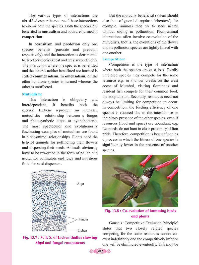
302
Fig. 13.8 : Co-evolution of humming birds and plants
The various types of interactions are classified as per the nature of these interactions to one or both the species. Both the species are benefited in mutualism and both are harmed in competition.
In parasitism and predation only one species benefits (parasite and predator, respectively) and the interaction is detrimental to the other species (host and prey, respectively). The interaction where one species is benefitted and the other is neither benefitted nor harmed is called commensalism. In amensalism, on the other hand one species is harmed whereas the other is unaffected.
Mutualism: This interaction is obligatory and
interdependent. It benefits both the species. Lichens represent an intimate, mutualistic relationship between a fungus and photosynthetic algae or cyanobacteria. The most spectacular and evolutionarily fascinating examples of mutualism are found in plant-animal relationships. Plants need the help of animals for pollinating their flowers and dispersing their seeds. Animals obviously have to be rewarded in the form of pollen and nectar for pollinators and juicy and nutritious fruits for seed dispersers.
Fig. 13.7 : V. T. S. of Lichen thallus showing Algal and fungal components
But the mutually beneficial system should also be safeguarded against ‘cheaters’, for example, animals that try to steal nectar without aiding in pollination. Plant-animal interactions often involve co-evolution of the mutualists, that is, the evolutions of the flower and its pollinator species are tightly linked with one another.
Competition: Competition is the type of interaction
where both the species are at a loss. Totally unrelated species may compete for the same resource e.g. in shallow creeks on the west coast of Mumbai, visiting flamingos and resident fish compete for their common food, the zooplankton. Secondly, resources need not always be limiting for competition to occur. In competition, the feeding efficiency of one species is reduced due to the interference or inhibitory presence of the other species, even if resources (food and space) are abundant, e.g. Leopards do not hunt in close proximity of lion pride. Therefore, competition is best defined as a process in which the fitness of one species is significantly lower in the presence of another species.
Gause’s ‘Competitive Exclusion Principle’ states that two closely related species competing for the same resources cannot co-exist indefinitely and the competitively inferior one will be eliminated eventually. This may be
Alga
Fungus
Lichen

303
Parasites that feed on the external surface of the host organism are called ectoparasites. The most familiar examples of this group are the lice on humans and ticks on dogs. Many marine fish are infested with ectoparasitic copepods. Cuscuta, a parasitic plant that is commonly found growing on hedge plants, has lost its chlorophyll and leaves in the course of evolution. It derives its nutrition from the host plant which it parasitizes.
Brood parasitism in birds is a fascinating example, in which the parasitic bird lays its eggs in the nest of its host bird and lets the host bird incubate them. During the course of evolution, the eggs of the parasitic bird have evolved to resemble the host’s egg in size and colour to reduce the chances of the host bird detecting the foreign eggs and ejecting them from the nest. Eggs of the parasitic bird (Asian koel) hatch before that of its host (Common Indian crow).
true if resources are limiting, but not otherwise. In interspecific competition with sufficient resources, species facing competition will evolve mechanisms that promote co-existence rather than exclusion. One such mechanism is ‘resource partitioning’. If two species compete for the same resource, they could avoid competition by choosing different times for feeding.
A species whose distribution is restricted to a small geographical area because of the presence of a competitively superior species, is found to expand its range when the competing species is removed.
Parasitism: Parasitism has evolved in so many
taxonomic groups from plants to higher vertebrates. Many parasites have evolved to be host-specific (they can parasitize only a single species of host) in such a way that both host and the parasite tend to co-evolve, against each other. In accordance with their life styles, endoparasites evolved special adaptations such as the loss of unnecessary sense organs, presence of adhesive organs or suckers to cling on to the host, loss of digestive system and high reproductive capacity.
The life cycles of parasites are often complex, involving intermediate hosts or vectors to facilitate transfer to the host. The malarial parasite Plasmodium vivax needs a vector (mosquito) to spread to other hosts.
Fig. 13.9 : Lion standing over dead leopard- removing competition
Fig. 13.10 : Crow feeding Asian Koel hatching in its own nest.
1. Should an ideal parasite be able to thrive
within the host without harming it? 2. Why didn’t natural selection lead to
the evolution of such totally harmless parasites?
Use your brain power
Majority of the parasites harm the host. They may reduce the survival, growth and reproduction of the host and may lead to death of the host, thus reducing its population density. They might render the host more vulnerable to predation by making it physically weak.
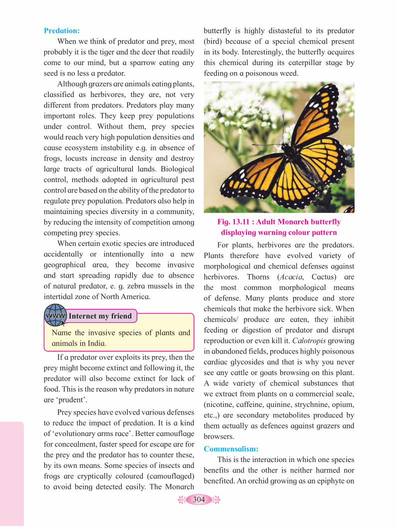
304
Fig. 13.11 : Adult Monarch butterfly displaying warning colour pattern
butterfly is highly distasteful to its predator (bird) because of a special chemical present in its body. Interestingly, the butterfly acquires this chemical during its caterpillar stage by feeding on a poisonous weed.
Predation: When we think of predator and prey, most
probably it is the tiger and the deer that readily come to our mind, but a sparrow eating any seed is no less a predator.
Although grazers are animals eating plants, classified as herbivores, they are, not very different from predators. Predators play many important roles. They keep prey populations under control. Without them, prey species would reach very high population densities and cause ecosystem instability e.g. in absence of frogs, locusts increase in density and destroy large tracts of agricultural lands. Biological control, methods adopted in agricultural pest control are based on the ability of the predator to regulate prey population. Predators also help in maintaining species diversity in a community, by reducing the intensity of competition among competing prey species.
When certain exotic species are introduced accidentally or intentionally into a new geographical area, they become invasive and start spreading rapidly due to absence of natural predator, e. g. zebra mussels in the intertidal zone of North America.
If a predator over exploits its prey, then the prey might become extinct and following it, the predator will also become extinct for lack of food. This is the reason why predators in nature are ‘prudent’.
Prey species have evolved various defenses to reduce the impact of predation. It is a kind of ‘evolutionary arms race’. Better camouflage for concealment, faster speed for escape are for the prey and the predator has to counter these, by its own means. Some species of insects and frogs are cryptically coloured (camouflaged) to avoid being detected easily. The Monarch
For plants, herbivores are the predators. Plants therefore have evolved variety of morphological and chemical defenses against herbivores. Thorns (Acacia, Cactus) are the most common morphological means of defense. Many plants produce and store chemicals that make the herbivore sick. When chemicals/ produce are eaten, they inhibit feeding or digestion of predator and disrupt reproduction or even kill it. Calotropis growing in abandoned fields, produces highly poisonous cardiac glycosides and that is why you never see any cattle or goats browsing on this plant. A wide variety of chemical substances that we extract from plants on a commercial scale, (nicotine, caffeine, quinine, strychnine, opium, etc.,) are secondary metabolites produced by them actually as defences against grazers and browsers.
Commensalism: This is the interaction in which one species
benefits and the other is neither harmed nor benefited. An orchid growing as an epiphyte on
Internet my friend
Name the invasive species of plants and animals in India.
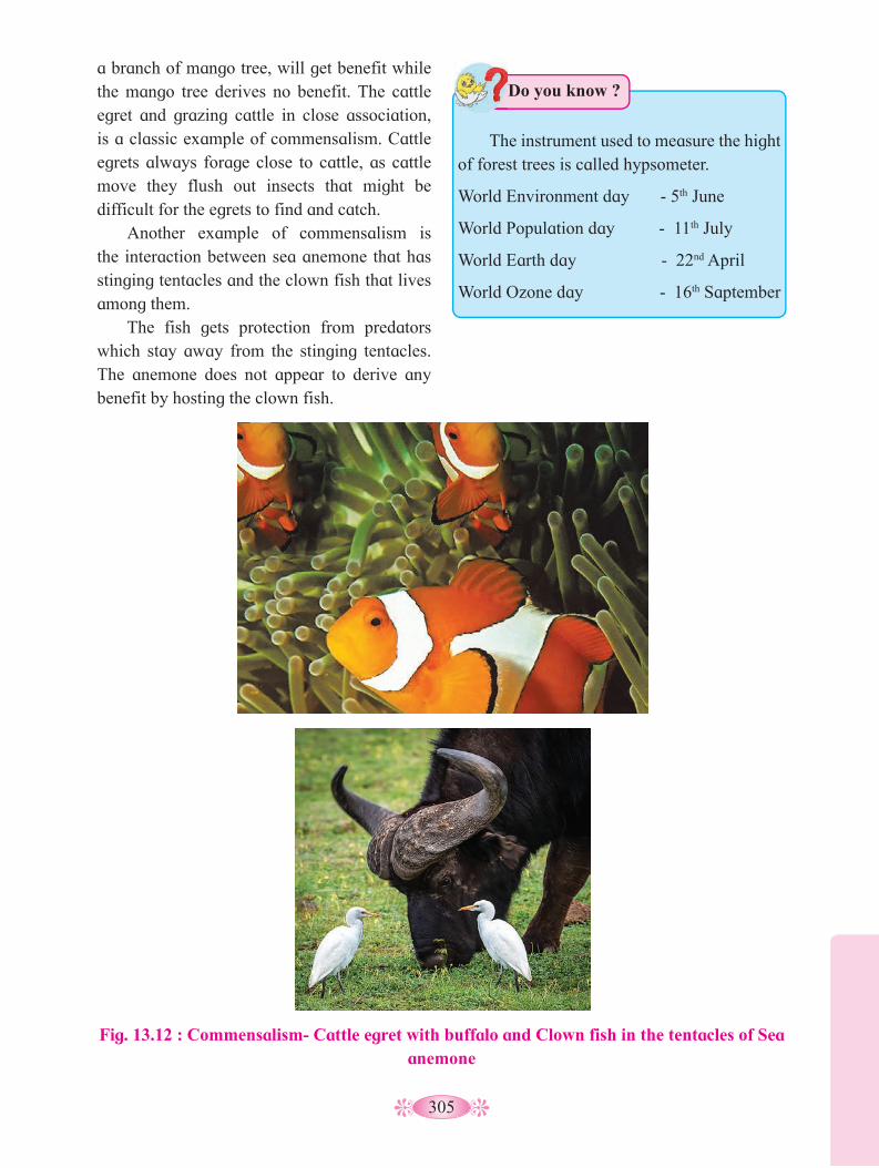
305
Fig. 13.12 : Commensalism- Cattle egret with buffalo and Clown fish in the tentacles of Sea anemone
a branch of mango tree, will get benefit while the mango tree derives no benefit. The cattle egret and grazing cattle in close association, is a classic example of commensalism. Cattle egrets always forage close to cattle, as cattle move they flush out insects that might be difficult for the egrets to find and catch.
Another example of commensalism is the interaction between sea anemone that has stinging tentacles and the clown fish that lives among them.
The fish gets protection from predators which stay away from the stinging tentacles. The anemone does not appear to derive any benefit by hosting the clown fish.
The instrument used to measure the hight of forest trees is called hypsometer.
World Environment day - 5th June
World Population day - 11th July
World Earth day - 22nd April
World Ozone day - 16th Saptember
Do you know ?

306
Activity :
Prepare chart for various interactions existing in the plant community or animal community around you and paste the photographs of the same.

307
Q. 1 Multiple choice questions.1. Which factor of an ecosystem includes
plants, animals and microorganisms? a. Biotic factor b. Abiotic factor c. Direct factor d. Indirect factor
2. An assembladge of individuals of different species living in the same habitat and having functional interactions is ................
a. Biotic community b. Ecological niche c. Population d. Ecosystem
3. Association between sea anemone and Hermit crab in gastropod shell is that of ................
a. Mutualism b. Commensalism c. Parasitism d. Amensalism
4. Select the statement which explains best parasitism.
a. One species is benefited. b. Both the species are benefited.
c. One species is benefited, other is not affected.
d. One species is benefited, other is harmed.
5. Growth of bacteria in a newlly innoculated agar plate shows ................
a. exponential growth b. logistic growth c. Verhulst-Pearl logistic growth d. zero growth
Q. 2 Very short answer questions.1. Define the following terms : a. Commensalism b. Parasitism c. Camouflage
2. Give one example for each : a. Mutualism b. Interspecific competition
3. Name the type of association: a. Clown fish and sea anemone b. Crow feeding the hatchling of Koel c. Humming birds and host flowering
plants 4. What is the ecological process behind the
biological control method of managing with pest insects?
Q. 3 Short answer questions.1. How is the dormancy of seeds different
from hibernation in animals?
2. If a marine fish is placed in a fresh water aquarium, will it be able to survive? Give reason.
3. Name important defense mechanisms in plants against herbivores.
4. An orchid plant is growing on the branch of mango tree. How do you describe this interaction between the orchid and the mango tree?
5. Distinguish between the following: a. Hibernation and Aestivation b. Ectotherms and Endotherms c. Parasitism and Mutualism
6. Write a short note on a. Adaptations of desert animals
b. Adaptations of plants to water scarcity
c. Behavioural adaptations in animals7. Define Population and Community.
Q. 4 Long answer questions.1. With the help of suitable diagram describe
the logistic population growth curve.2. Enlist and explain the important
characteristics of a population.
Exercise
Project : Study the age pyramid of human population in your area

308
An ecosystem is a self regulatory and self sustaining structural and functional unit of nature (biosphere). It contains both biotic and abiotic components. Biotic components interact with each other and also with the surrounding environment. Tansley (1935) coined the term ecosystem. Ecosystems vary greatly in size from a small pond to large oceans or small farmland to village. Entire biosphere can be considered as one global ecosystem, made up of many local ecosystems. Since the earth ecosystem is too big and too complex to be studied, it is divided into two basic categories, viz. terrestrial and aquatic. Forest, grassland and desert are the types of terrestrial ecosystems while lakes, wetlands, rivers and estuaries are the types of aquatic ecosystems. The ecosystems can also be classified as Natural ecosystems and Artificial ecosystems. Natural ecosystems do not require any human inputs, in other words they are self-sustainable. Artificial ecosystems e.g. a farm land, a fish tank or even a large pond used for rearing fish, require constant input in terms of energy or materials.
In this chapter, we will study and analyse the structure of the ecosystem, in order to appreciate the input (productivity), transfer of energy (food chain/web, nutrient cycling) and the output (degradation and energy loss). We will also look at the relationships, chains and webs that are created because of the energy flows within the system.
Ecosystems and Energy flow
14.1 Ecosystem : Structure and Function :
We have already studied the various biotic and abiotic components of the environment. We know that all these biotic and abiotic components influence each other. Let us now look at these components with an integrated approach and see how the flow of energy takes place in ecosystem. Interaction of biotic and abiotic components, results in a physical structure that is characteristic for each type of ecosystem. Identification and enumeration of plant and animal species of an ecosystem gives its species composition.
Fig. 14.1 : Stratification of plants in forest
Emergent layer
Canopy
Understorey
Shrub layer
1. What is environmental biology?2. Meaning of the term habitat.3. Importance of ecology for humans.
Can you recall?
Biotic and abiotic components differ as the locations vary in space and time. The variation due to space results in spatial pattern. There are two types of spatial patterns. viz. Stratification and Zonation.
Vertical distribution of different species of plants and animals occupying different levels, is known as stratification. For example, trees occupy top vertical strata or layer of a forest, shrubs the second and herbs and grasses occupy the bottom layer. Similar stratification is also observed in the open seas as epipelagic, meso-pelagic, bathy-pelagic and benthic zones.
Stratification of animals in amazon rain forest.
Find out
14
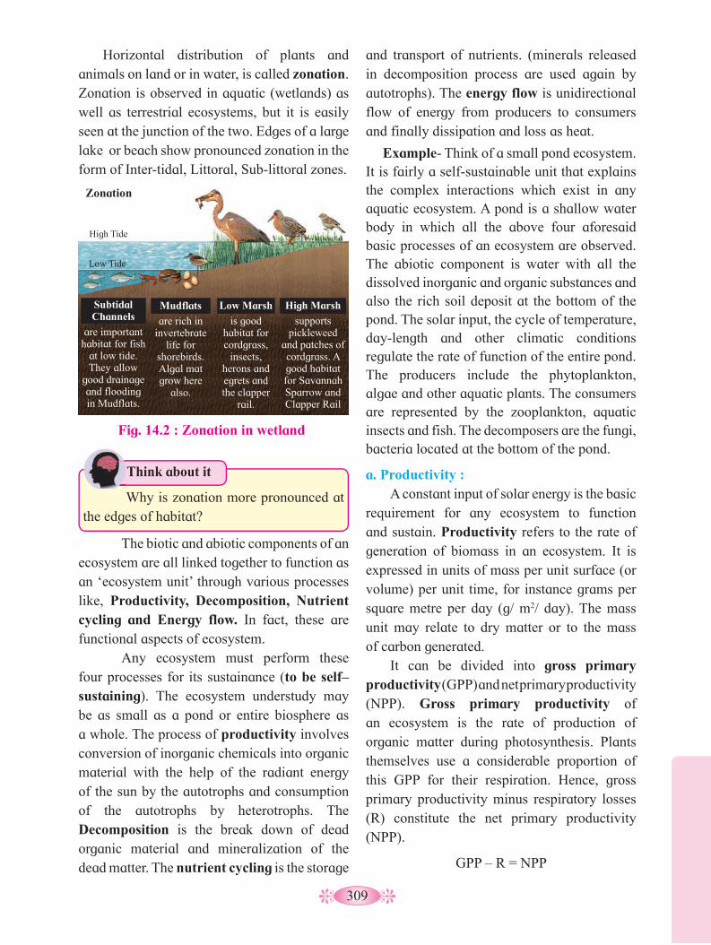
309
The biotic and abiotic components of an ecosystem are all linked together to function as an ‘ecosystem unit’ through various processes like, Productivity, Decomposition, Nutrient cycling and Energy flow. In fact, these are functional aspects of ecosystem.
Any ecosystem must perform these four processes for its sustainance (to be self–sustaining). The ecosystem understudy may be as small as a pond or entire biosphere as a whole. The process of productivity involves conversion of inorganic chemicals into organic material with the help of the radiant energy of the sun by the autotrophs and consumption of the autotrophs by heterotrophs. The Decomposition is the break down of dead organic material and mineralization of the dead matter. The nutrient cycling is the storage
and transport of nutrients. (minerals released in decomposition process are used again by autotrophs). The energy flow is unidirectional flow of energy from producers to consumers and finally dissipation and loss as heat.
Example- Think of a small pond ecosystem. It is fairly a self-sustainable unit that explains the complex interactions which exist in any aquatic ecosystem. A pond is a shallow water body in which all the above four aforesaid basic processes of an ecosystem are observed. The abiotic component is water with all the dissolved inorganic and organic substances and also the rich soil deposit at the bottom of the pond. The solar input, the cycle of temperature, day-length and other climatic conditions regulate the rate of function of the entire pond. The producers include the phytoplankton, algae and other aquatic plants. The consumers are represented by the zooplankton, aquatic insects and fish. The decomposers are the fungi, bacteria located at the bottom of the pond.
a. Productivity : A constant input of solar energy is the basic
requirement for any ecosystem to function and sustain. Productivity refers to the rate of generation of biomass in an ecosystem. It is expressed in units of mass per unit surface (or volume) per unit time, for instance grams per square metre per day (g/ m2/ day). The mass unit may relate to dry matter or to the mass of carbon generated.
It can be divided into gross primary productivity (GPP) and net primary productivity (NPP). Gross primary productivity of an ecosystem is the rate of production of organic matter during photosynthesis. Plants themselves use a considerable proportion of this GPP for their respiration. Hence, gross primary productivity minus respiratory losses (R) constitute the net primary productivity (NPP).
GPP – R = NPP
Horizontal distribution of plants and animals on land or in water, is called zonation. Zonation is observed in aquatic (wetlands) as well as terrestrial ecosystems, but it is easily seen at the junction of the two. Edges of a large lake or beach show pronounced zonation in the form of Inter-tidal, Littoral, Sub-littoral zones.
Fig. 14.2 : Zonation in wetland
Subtidal Channels
are important habitat for fish
at low tide. They allow
good drainage and flooding in Mudflats.
Low Marsh
is good habitat for cordgrass,
insects, herons and egrets and the clapper
rail.
High Marsh
supports pickleweed
and patches of cordgrass. A good habitat for Savannah Sparrow and Clapper Rail
Zonation
High Tide
Low Tide
Mudflats
are rich in invertebrate
life for shorebirds. Algal mat grow here
also.
• Why is zonation more pronounced at the edges of habitat?
Think about it•
310
Primary productivity (GPP) depends on the plant species inhabiting a particular area. It also depends on a variety of environmental factors, availability of nutrients and photosynthetic capacity of plants. Therefore, it varies in different types of ecosystems. Secondary productivity is defined as the rate of formation of new organic matter by consumers. Alternatively, it is the rate of assimilation of food energy at the level of consumers. It is the amount of energy available to consumer for transfer to the next trophic level.
b. Decomposition : Decomposers break down complex organic
matter into inorganic substances like carbon dioxide, water and nutrients, and the process is called decomposition. Dead remains of plants and animals, including fecal matter, constitute detritus, which is the raw material for decomposition. The important steps in the process of decomposition are fragmentation, leaching, catabolism, humification and mineralization.
Detritivores (e.g. earthworm) break down detritus into smaller fragments or particles. This process is called fragmentation.
By the process of leaching, water soluble inorganic nutrients go down (percolate) into the soil horizon and get precipitated as unavailable salts. Bacterial and fungal enzymes degrade detritus into simpler inorganic substances. This process is called as catabolism. It is important to note that all the above steps in decomposition operate simultaneously on the detritus.
What could be the reason for the low productivity of ocean?
Use your brain power
Net primary productivity is the available biomass for the consumption, to heterotrophs (herbivores, carnivores and decomposers). The annual net primary productivity of the whole biosphere is approximately 170 billion tons (dry weight) of organic matter. Of this, the productivity of the oceans is only 55 billion tons. Rest of course, is from land based ecosystems.
Fig. 14.3 : Decomposition cycle
Humification and mineralization occur during decomposition in the soil. Humification leads to accumulation of partially decomposed, a dark coloured, amorphous, colloidal organic substance called humus that is resistant to microbial action and undergoes decomposition at an extremely slow rate. Humus formation changes soil texture and increases water holding capacity of soil.
Being colloidal in nature humus serves as a reservoir of nutrients. The humus is further degraded by some microbes and release of inorganic nutrients occurs by the process known as mineralisation.
Decomposition as a process requires oxygen. Temperature and soil moisture are the most important factors that regulate decomposition indirectly to help soil microbes. Warm and moist environment favours decomposition whereas low temperature and anaerobic conditions inhibit decomposition.
A tree grows in the soil. Some are eaten by herbivors. Nutrients and energy enter food web.
Leaves partially consumed and
A leaf falls to the ground.
begin to lose form and become litter.
Mineralization
Some nutrients
leach into soil by chemical
action.
Soil rich in Organic content
by decomposers. decomposition Further
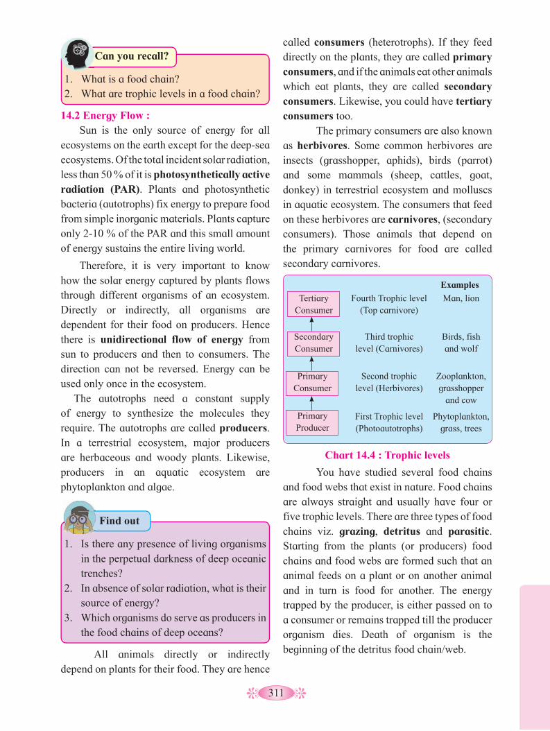
311
14.2 Energy Flow : Sun is the only source of energy for all
ecosystems on the earth except for the deep-sea ecosystems. Of the total incident solar radiation, less than 50 % of it is photosynthetically active radiation (PAR). Plants and photosynthetic bacteria (autotrophs) fix energy to prepare food from simple inorganic materials. Plants capture only 2-10 % of the PAR and this small amount of energy sustains the entire living world.
Therefore, it is very important to know how the solar energy captured by plants flows through different organisms of an ecosystem. Directly or indirectly, all organisms are dependent for their food on producers. Hence there is unidirectional flow of energy from sun to producers and then to consumers. The direction can not be reversed. Energy can be used only once in the ecosystem.
The autotrophs need a constant supply of energy to synthesize the molecules they require. The autotrophs are called producers. In a terrestrial ecosystem, major producers are herbaceous and woody plants. Likewise, producers in an aquatic ecosystem are phytoplankton and algae.
All animals directly or indirectly depend on plants for their food. They are hence
Chart 14.4 : Trophic levels
Tertiary Consumer
Secondary Consumer
Primary Consumer
Primary Producer
Examples
Fourth Trophic level (Top carnivore)
Third trophic level (Carnivores)
Second trophic level (Herbivores)
First Trophic level (Photoautotrophs)
Man, lion
Birds, fish and wolf
Zooplankton, grasshopper
and cow
Phytoplankton, grass, trees
You have studied several food chains and food webs that exist in nature. Food chains are always straight and usually have four or five trophic levels. There are three types of food chains viz. grazing, detritus and parasitic. Starting from the plants (or producers) food chains and food webs are formed such that an animal feeds on a plant or on another animal and in turn is food for another. The energy trapped by the producer, is either passed on to a consumer or remains trapped till the producer organism dies. Death of organism is the beginning of the detritus food chain/web.
1. What is a food chain?2. What are trophic levels in a food chain?
Can you recall?
1. Is there any presence of living organisms
in the perpetual darkness of deep oceanic trenches?
2. In absence of solar radiation, what is their source of energy?
3. Which organisms do serve as producers in the food chains of deep oceans?
Find out
called consumers (heterotrophs). If they feed directly on the plants, they are called primary consumers, and if the animals eat other animals which eat plants, they are called secondary consumers. Likewise, you could have tertiary consumers too.
The primary consumers are also known as herbivores. Some common herbivores are insects (grasshopper, aphids), birds (parrot) and some mammals (sheep, cattles, goat, donkey) in terrestrial ecosystem and molluscs in aquatic ecosystem. The consumers that feed on these herbivores are carnivores, (secondary consumers). Those animals that depend on the primary carnivores for food are called secondary carnivores.
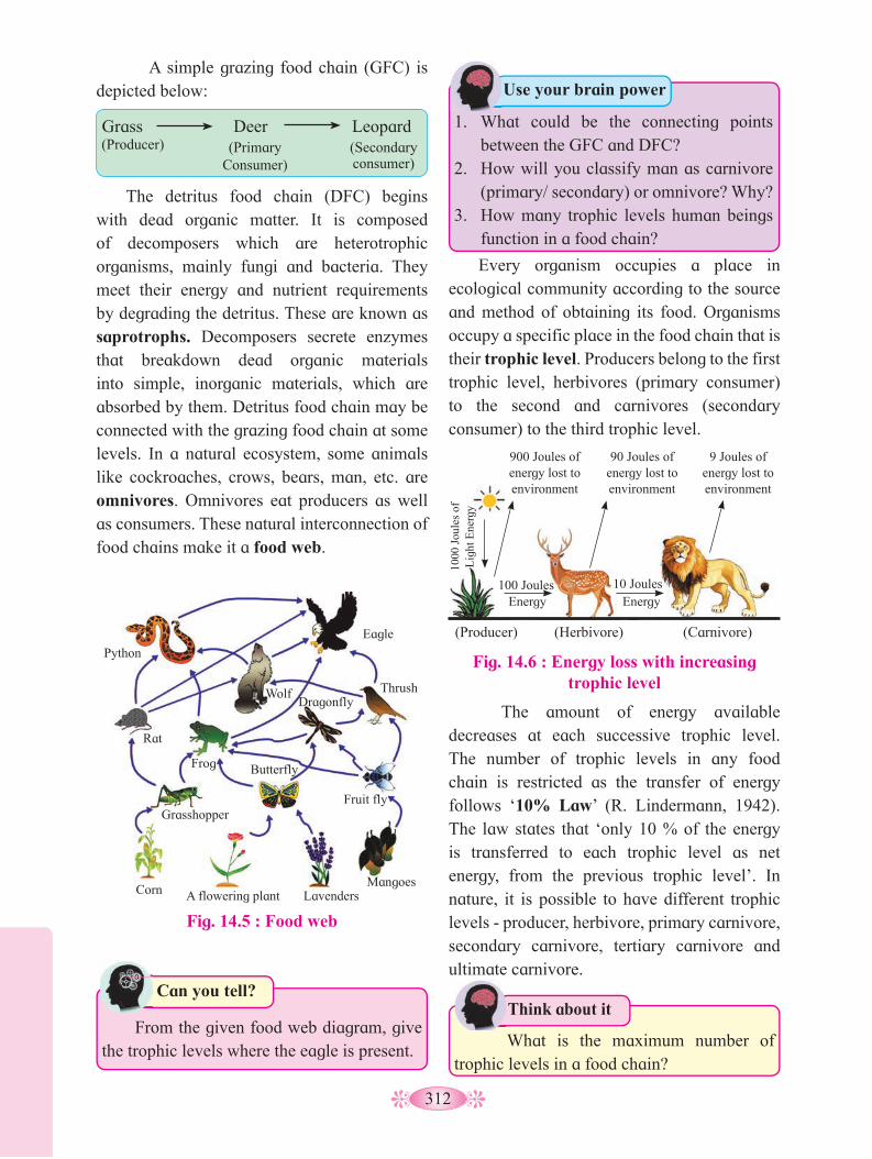
312
A simple grazing food chain (GFC) is depicted below:
The amount of energy available decreases at each successive trophic level. The number of trophic levels in any food chain is restricted as the transfer of energy follows ‘10% Law’ (R. Lindermann, 1942). The law states that ‘only 10 % of the energy is transferred to each trophic level as net energy, from the previous trophic level’. In nature, it is possible to have different trophic levels - producer, herbivore, primary carnivore, secondary carnivore, tertiary carnivore and ultimate carnivore.
The detritus food chain (DFC) begins with dead organic matter. It is composed of decomposers which are heterotrophic organisms, mainly fungi and bacteria. They meet their energy and nutrient requirements by degrading the detritus. These are known as saprotrophs. Decomposers secrete enzymes that breakdown dead organic materials into simple, inorganic materials, which are absorbed by them. Detritus food chain may be connected with the grazing food chain at some levels. In a natural ecosystem, some animals like cockroaches, crows, bears, man, etc. are omnivores. Omnivores eat producers as well as consumers. These natural interconnection of food chains make it a food web.
• What is the maximum number of trophic levels in a food chain?
Think about it•
Fig. 14.5 : Food web
Python
Rat
Frog
Grasshopper
Corn A flowering plant Lavenders
Butterfly
Wolf Dragonfly
Mangoes
Fruit fly
Thrush
Eagle
From the given food web diagram, give the trophic levels where the eagle is present.
Can you tell?
Grass Deer Leopard(Producer) (Primary
Consumer)(Secondary consumer)
Fig. 14.6 : Energy loss with increasing trophic level
1000
Jou
les
of
Lig
ht E
nerg
y
(Producer)
9 Joules of energy lost to environment
90 Joules of energy lost to environment
900 Joules of energy lost to environment
100 Joules Energy
10 Joules Energy
(Herbivore) (Carnivore)
Every organism occupies a place in ecological community according to the source and method of obtaining its food. Organisms occupy a specific place in the food chain that is their trophic level. Producers belong to the first trophic level, herbivores (primary consumer) to the second and carnivores (secondary consumer) to the third trophic level.
1. What could be the connecting points between the GFC and DFC?
2. How will you classify man as carnivore (primary/ secondary) or omnivore? Why?
3. How many trophic levels human beings function in a food chain?
Use your brain power
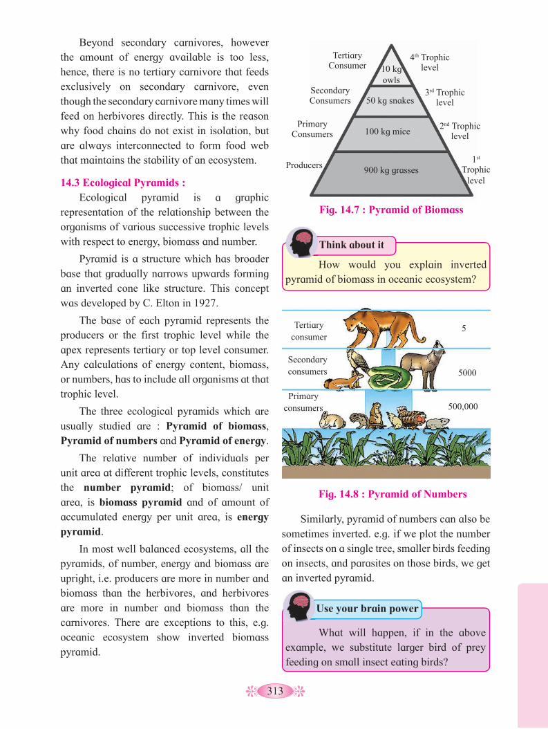
313
Beyond secondary carnivores, however the amount of energy available is too less, hence, there is no tertiary carnivore that feeds exclusively on secondary carnivore, even though the secondary carnivore many times will feed on herbivores directly. This is the reason why food chains do not exist in isolation, but are always interconnected to form food web that maintains the stability of an ecosystem.
14.3 Ecological Pyramids : Ecological pyramid is a graphic
representation of the relationship between the organisms of various successive trophic levels with respect to energy, biomass and number.
Pyramid is a structure which has broader base that gradually narrows upwards forming an inverted cone like structure. This concept was developed by C. Elton in 1927.
The base of each pyramid represents the producers or the first trophic level while the apex represents tertiary or top level consumer. Any calculations of energy content, biomass, or numbers, has to include all organisms at that trophic level.
The three ecological pyramids which are usually studied are : Pyramid of biomass, Pyramid of numbers and Pyramid of energy.
The relative number of individuals per unit area at different trophic levels, constitutes the number pyramid; of biomass/ unit area, is biomass pyramid and of amount of accumulated energy per unit area, is energy pyramid.
In most well balanced ecosystems, all the pyramids, of number, energy and biomass are upright, i.e. producers are more in number and biomass than the herbivores, and herbivores are more in number and biomass than the carnivores. There are exceptions to this, e.g. oceanic ecosystem show inverted biomass pyramid.
Fig. 14.8 : Pyramid of Numbers
Fig. 14.7 : Pyramid of Biomass
Tertiary Consumer
Secondary Consumers
Primary Consumers
Producers
4th Trophic level
3rd Trophic level
2nd Trophic level
1st Trophic
level
10 kg owls
50 kg snakes
100 kg mice
900 kg grasses
• How would you explain inverted pyramid of biomass in oceanic ecosystem?
Think about it•
Tertiary consumer
Secondary consumers
Primary consumers
5
5000
500,000
What will happen, if in the above example, we substitute larger bird of prey feeding on small insect eating birds?
Use your brain power
Similarly, pyramid of numbers can also be sometimes inverted. e.g. if we plot the number of insects on a single tree, smaller birds feeding on insects, and parasites on those birds, we get an inverted pyramid.
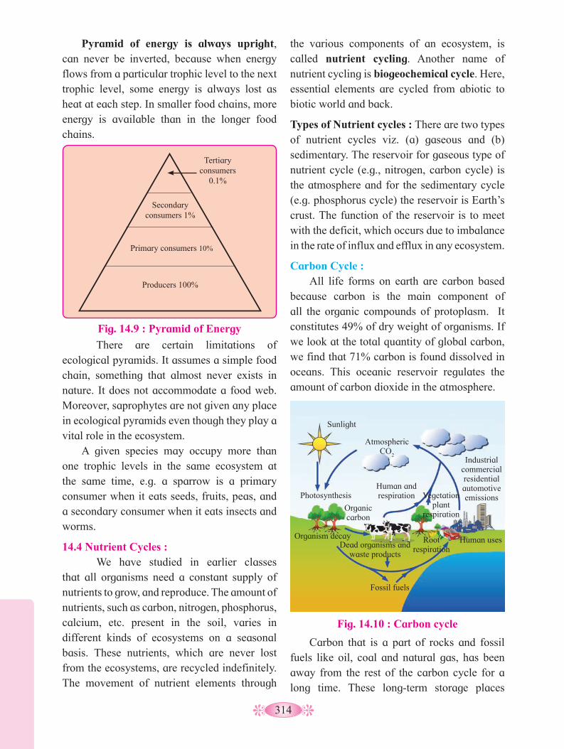
314
Fig. 14.9 : Pyramid of Energy
Producers 100%
Primary consumers 10%
Secondary consumers 1%
Tertiary consumers
0.1%
Pyramid of energy is always upright, can never be inverted, because when energy flows from a particular trophic level to the next trophic level, some energy is always lost as heat at each step. In smaller food chains, more energy is available than in the longer food chains.
the various components of an ecosystem, is called nutrient cycling. Another name of nutrient cycling is biogeochemical cycle. Here, essential elements are cycled from abiotic to biotic world and back.
Types of Nutrient cycles : There are two types of nutrient cycles viz. (a) gaseous and (b) sedimentary. The reservoir for gaseous type of nutrient cycle (e.g., nitrogen, carbon cycle) is the atmosphere and for the sedimentary cycle (e.g. phosphorus cycle) the reservoir is Earth’s crust. The function of the reservoir is to meet with the deficit, which occurs due to imbalance in the rate of influx and efflux in any ecosystem.
Carbon Cycle : All life forms on earth are carbon based
because carbon is the main component of all the organic compounds of protoplasm. It constitutes 49% of dry weight of organisms. If we look at the total quantity of global carbon, we find that 71% carbon is found dissolved in oceans. This oceanic reservoir regulates the amount of carbon dioxide in the atmosphere.
There are certain limitations of ecological pyramids. It assumes a simple food chain, something that almost never exists in nature. It does not accommodate a food web. Moreover, saprophytes are not given any place in ecological pyramids even though they play a vital role in the ecosystem.
A given species may occupy more than one trophic levels in the same ecosystem at the same time, e.g. a sparrow is a primary consumer when it eats seeds, fruits, peas, and a secondary consumer when it eats insects and worms.
14.4 Nutrient Cycles :We have studied in earlier classes
that all organisms need a constant supply of nutrients to grow, and reproduce. The amount of nutrients, such as carbon, nitrogen, phosphorus, calcium, etc. present in the soil, varies in different kinds of ecosystems on a seasonal basis. These nutrients, which are never lost from the ecosystems, are recycled indefinitely. The movement of nutrient elements through
Carbon that is a part of rocks and fossil fuels like oil, coal and natural gas, has been away from the rest of the carbon cycle for a long time. These long-term storage places
Fig. 14.10 : Carbon cycle
Sunlight
Atmospheric CO
2
Photosynthesis
Organic carbon
Human and respiration Vegetation
plant respiration
Industrial commercial residential automotive emissions
Organism decay Dead organisms and
waste products
Root respiration
Human uses
Fossil fuels

315
are known as “sinks”. When fossil fuels are burned, carbon that had been underground is released back into the air as carbon dioxide.
The element carbon is a part of seawater, the atmosphere, rocks such as limestone and coal, soils, as well as all living things. • Carbon as CO
2 moves from the atmosphere
to plants. Through the process of photosynthesis, carbon dioxide is pulled from the air to produce food.
• Carbon moves from plants to animals, through food chains, i.e. the carbon present in plants moves to the animals.
• Carbon moves from living things to the atmosphere. Each time you exhale, you are releasing carbon dioxide gas (CO
2) into the
atmosphere.• Decomposers also contribute substantially
to CO2 in atmosphere, by their processing of
waste materials and dead organic matter of land and oceans.
• When fossil fuels burn to power factories, power plants, motor vehicles, most of the carbon quickly enters the atmosphere as carbon dioxide gas. Each year, 5.5 billion tons of carbon is released through combustion of fossil fuels. Of this massive amount, 3.3 billion tons stays in the atmosphere. Most of the remainder is dissolved in seawater and deposited as calcium or magnesium carbonate compounds which make up shells of marine animals.
• Burning of wood, forest fire and combustion of organic matter, fossil fuel and volcanic activity, are additional sources for releasing CO
2 in the atmosphere.
• Carbon moves from the atmosphere to the oceans. The oceans and other water bodies, absorb some carbon in the form of CO
2 from
the atmosphere. The carbon is dissolved into the oceanic water. Some amount of the fixed carbon is lost to sediments and removed from circulation.
• Fossil fuels represent a reservoir of carbon. Carbon cycling occurs through atmosphere, ocean and through living and dead organisms. Human activities have significantly influenced the carbon cycle.
• Rapid deforestation and massive burning of fossil fuel for energy and transport, have significantly increased the rate of release of carbon dioxide into the atmosphere. Thus, the entire carbon cycle is run
by basic processes viz. Photosynthesis, Respiration, Decomposition, Sedimentation and Combustion.Phosphorus Cycle :
Cyclic movement of phosphorus through hydrosphere, lithosphere and biosphere constitutes phosphorus cycle.
Phosphorus is a major constituent of biological membranes, nucleic acids and cellular energy transfer systems. Many animals also need large quantities of this element to make shells, bones, hooves and teeth.
The natural reservoir of phosphorus is rock, which contains phosphorus in the form of phosphates. When rocks are weathered, minute amounts of these phosphates dissolve in soil solution and are absorbed by the roots of the plants. Herbivores and other animals obtain this element from plants. The waste products and the dead organisms are decomposed by phosphate-solubilizing bacteria releasing phosphorus. Unlike carbon cycle, there is no respiratory release of phosphorus into atmosphere.
There are two major differences between carbon and phosphorus cycle. Atmospheric inputs of phosphorus through rainfall are much smaller (meagure) than carbon inputs, and secondly, exchanges of phosphorus between organism and environment are negligible as compare to carbon.
Unlike carbon and nitrogen, Phosphorus is always in short supply and hence acts as

316
limiting factor for the plant growth. Sudden influx of phosphorus in the form of agricultural runoff or industrial effluents rich in phosphate content, leads to eutrophication in water bodies. Eutrophication is due to overgrowth of algae at the instance of high phosphorus disolved in water. The overgrowth of algae kills or harms the aquatic life.
Internet my friend
What are the long term and short-term effects of eutrophication in water bodies?
Eventually, it leads to climax community. Climax community does not evolve further.
The gradual and predictable change in the species composition of a given area is called ecological succession. The change is sequential and environmentally regulated.
Process of succession involves sequential steps like Nudation, Invasion, Ecesis, Aggregation, Competition and co-action, Reaction and stabilization.
During succession, some species colonize an area and their populations become more numerous, whereas populations of other species decline and even disappear.
The entire sequence of communities that successively change in a given area, constitute what is called sere(s). Alternatively, it is an entire gradient of organisms from pioneer stage to climax stage.The individual transitional
Fig. 14.11 : Phosphorus Cycle
Rain
Plants
Animals
Decomposers
Phosphate in
soil
Leaching
Weathering of
phosphate from
rocks
Runoff
Geological uplifting
Phosphate in solution
Detritus settling
to bottom
Sedimentation = new rocks
14.5 Ecological Succession : Succession : It is a spatial pattern which occurs over the time. Time is a crucial factor. The ecosystem is occupied by the pioneer species. As time passes, species diversity increase giving way to more complex organisms.
317
communities are termed seral communities. In the successive seral stages, there is a change in the diversity of species of organisms, increase in the number of species and organisms as well as an increase in the total biomass.
The present day communities in the world have come to be, because of succession that has occurred over millions of years since life started on earth. Succession is hence a process that starts where no living organisms were present before - like on a newly formed volcanic island. This is called primary succession.
Other examples of areas where primary succession occurs are newly cooled lava, rocks and newly created pond or reservoir. The establishment of a new biotic community is generally very slow. Before a biotic community of diverse organisms can become established, there must be soil. Depending mostly on the climate, it takes natural processes, several hundred to several thousand years to produce fertile soil on bare rock.
Secondary succession begins in areas where natural biotic communities have been destroyed such as in abandoned farm lands, burned or cut forests, lands that have been flooded, etc. Since some soil or sediment is present, succession is faster than in primary succession. Description of ecological succession usually focuses on changes in vegetation. However, these vegetational changes in turn affect food and shelter for various types of animals. Thus, as succession proceeds, the numbers and types of animals and decomposers also change. At any time during primary or secondary succession, natural or human induced disturbances (fire, deforestation, etc.), can convert a particular seral stage of succession to an earlier previous / preceding stage. Also, such disturbances create new conditions that encourage some species and discourage or eliminate other species.
Succession of Plants : Based on the nature of the habitat – whether
it is water (or very wetland areas) or it is on very dry areas – succession of plants, is called
hydrarch (hydrosere) or xerarch (xerosere), respectively.
Hydrarch succession takes place in wetter areas and the successional series progress from hydric to the mesic conditions. As against this, xerarch succession takes place in dry areas and the series progress from xeric to mesic conditions. Hence, both hydrarch and xerarch successions lead to medium water conditions (mesic) – neither too dry (xeric) nor too wet (hydric).
The species that invade a bare area, are called pioneer species. In primary succession on rocks these are usually crustose lichens which are able to secrete acids to dissolve rock, helping in weathering of rocks and soil formation. These pave the way for bryophytes, mosses that are able to take hold in the small amount of soil.
They are, with time, succeeded by herbaceous plants, and after several more stages, ultimately a stable climax forest community is formed. The climax community remains stable as long as the environment remains unchanged.
In primary succession of aquatic habitat, (steps 1 to 7 in fig. 14.13) the pioneers are the small phytoplankton. They are replaced with time by rooted-submerged plants (e.g. Hydrilla), rooted-floating angiosperms (e.g. Lotus) followed by free-floating plants (e.g. Pistia), then reed swamp (e.g. Typha), marsh-meadow (e.g. Cyperus), scrub (e.g. Alnus) and finally the trees (e.g. Quercus). The climax again would be a forest. With passage of time, the water body is converted into land.
In secondary succession, the species that invade depend on the condition of the soil, availability of water, the environment as also the seeds or other propagules present. Since soil is already there, the rate of succession is much faster and hence, climax is also reached more quickly. Figure 14.14 shows the sequence of stages 1 to 8 in a forest ecosystem after fire.

318
Bare rock Lichens Small annual plants, lichens
Perennial herbs,
grasses
Grasses, shrubs, shade-intolerant trees
Shade-tolerant trees
Pioneer stages Intermediate stages Climax community
Fig. 14.12 : Xerarch Succession of Plants
Fig. 14.13 : Hydrarch Succession of Plants
Phytoplankton stage Reed-swamp stage
Submerged plant stage Marsh-meadow stage
Submerged and free floating plant stage
Scrub stage
Climax forest
1
2
3
4
5
6
7
14.6 Ecosystem Services : Healthy ecosystems are the base for a wide
range of economic, environmental and aesthetic goods and services. The products of ecosystem processes are named as ecosystem services, for example, healthy forest ecosystems purify air and water, mitigate droughts and floods.
The Millennium Ecosystem Assessment report 2005 defines Ecosystem services as benefits people obtain from ecosystems and identifies four categories of ecosystem services as follows.
• Supporting services include services such as nutrient cycling, primary production, soil formation, habitat provision and pollination maintaining balance of ecosystem.
• Provisioning services include food (including seafood), raw materials (including timber, skins, fuel wood), genetic resources (including crop improvement genes, and health care), water, medicinal resources (including test and assay organisms) and ornamental resources (furs, feathers, ivory, orchids, butterflies, etc.)
• Regulating services include Carbon sequestration, Predation regulates prey populations, Waste decomposition and detoxification, Purification of water and air, and pest control.
It is important to understand that succession, particularly primary succession, is a very slow process, maybe requiring thousands of years for the climax to be reached.
Another important fact to understand is that all the successions whether taking place in water or on land, proceed to a similar climax community – the mesic.

319
• Cultural services include cultural, spiritual and historical, recreational experiences, science and education, and Therapeutics (including animal assisted therapy) Following are the main ecological
services : Fixation of atmospheric CO
2 and
release
of O2 are the most important services provided
by an ecosystem. Photosynthetic activity of photoautotrophs sequesters carbon (in CO
2
form) from the atmosphere and releases O2 as a
byproduct. O2 not only purifies air but is also
used for respiration by all aerobes. Pollination of plants brought about by
wind, water or other biotic agencies, is also an important ecosystem service, without which there would be no crops and no fruits.
Though the value of all such services of biodiversity is difficult to determine, it seems reasonable to think that biodiversity should carry a hefty price tag.
Activity :
Fig. 14. 14 : Secondary succession in a forest ecosystem after fire
1 2 3 4
5 6 7 8
Forest
Forest stage ShrubsWood land stage
Soil and herbs
Forest fire
Terrestrial plants
Severe forest fire Total nudation of forest land
1. Fill in the banks in the given diagram and identity.
2. Collect information and prepare a chart of the sequential steps involved in the ecological succession, explaining each step.

320
Q. 1 Multiple choice questions 1. Which one of the following has the
largest population in a food chain? a. Producers b. Primary consumers c. Secondary consumers d. Decomposers
2. The second trophic level in a lake is ________________
a. Phytoplankton b. Zooplankton c. Benthos d. Fishes
3. Secondary consumers are __________a. Herbivores b. Producers c. Carnivores d. Autotrophs
4. What is the % of photosynthetically active radiation in the incident solar radiation?
a. 100% b. 50 %c. 1-5% d. 2-10%
6. Give the term used to express a community in its final stage of succession? a. End community
b. Final community c. Climax community d. Dark community
7. After landslide which of the following type of succession occurs? a. Primary b. Secondary
c. Tertiary d. Climax
8. Which of the following is most often a limiting factor of the primary productivity in any ecosystem.
a. Carbon b. Nitrogen c. Phosphorus d. Sulphur
Q. 2 Very short answer question.1. Give an example of ecosystem which
shows inverted pyramid of numbers.
2. Give an example of ecosystem which shows inverted pyramid of biomass.
3. Which mineral acts as limiting factor for productivity in an aquatic ecosystem.
4. Name the reservoir and sink of carbon in carbon cycle.
Q. 3 Short answer questions.1. Distinguish between upright and
inverted pyramid of biomass
2. Distinguish between Food chain and Food web.
Q 4. Long answer questions.1. Define ecological pyramids and describe
with examples, pyramids of number and biomass.
2. What is primary productivity? Give brief description of factors that affect primary productivity.
3. Define decomposition and describe the processes and products of decomposition.
4. Write important features of a sedimentary cycle in an ecosystem.
5. Discribe carbon cycle and add a note on the impact of human activities on carbon cycle.
Exercise
Project : 1. Collect the information on the various
services offered by dense forest ecosystem.
2. Collect the information of various types of pollinators and the impact of human activity on them.

321
Diversity is variety. This variety of life is called biodiversity. Biodiversity includes a vast array of species of microorganisms- viruses, algae, fungi, plants and animals occurring on Earth, either in terrestrial or aquatic habitat and the ecological complexes of which they are part.
The diversity is with respect to size (microscopic to macroscopic), shape, colour, form, mode of nutrition, type of habitat, reproduction, motility, duration of life cycle span, etc. This is actually due to the attempt of living beings to accomodate with different environmental conditions (like climatic, edaphic, topographic, geographic, etc.) or situations, solely for their survival and perpetuation. In doing so, living organisms adapt themselves to overcome different situations and thus develop distinct but different features and that has actually lead to the diversity in them. The diversity in features become infused in the life cycle. In short, these adaptations in different environments serve as basis for diversity.
Definition of Biodiversity : It is the part of nature which includes the differences in the genes among the individuals of a species; the variety and richness of all plants and animal species at different scales in a space - local regions, country and the world; and the types of ecosystem, both terrestrial and aquatic, within a defined area.
The term biodiversity was actually coined by Walter Rosen (1982) but the term was popularised by sociologist Edward Wilson to describe combined diversity at all the levels of biological organisation.
Biodiversity that we see today, is the outcome of over 3.5 billion of years of evolutionary history mainly influenced by natural processes and of late by influence of humans.
In this chapter, we shall study the basic concepts of biodiversity such as levels and patterns of biodiversity, expanse, importance, loss and conservation methods and efforts undertaken.
15.1 Levels of Biodiversity: Diversity of living world can be observed
at various levels, ranging from molecular to ecosystem level. Major hierarchial and interrelated levels are genetic diversity, species diversity (community), and ecosystem diversity (ecological).
Biodiversity, Conservation and Environmental Issues
1. You have learnt about Chipko movement
and efforts of Shri. Jadav Payeng in school. What is the importance of such activities?
2. Why should we have national parks and sanctuaries?
3. We have read about how Indian scientists won the battle for patent of Turmeric and Basmati rice. Why was gaining these patents essential?
4. What is in situ and ex situ conservation?
Can you recall?
15
What can you say about species diversity A and B?
Can you tell?
A B
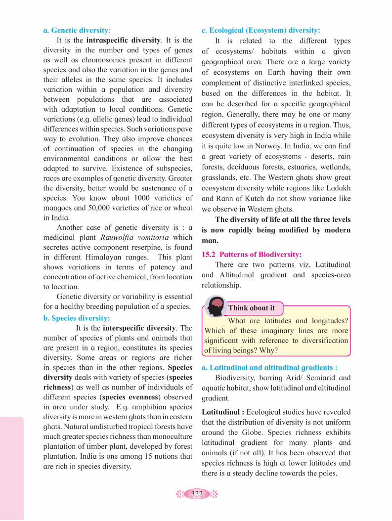
322
c. Ecological (Ecosystem) diversity: It is related to the different types
of ecosystems/ habitats within a given geographical area. There are a large variety of ecosystems on Earth having their own complement of distinctive interlinked species, based on the differences in the habitat. It can be described for a specific geographical region. Generally, there may be one or many different types of ecosystems in a region. Thus, ecosystem diversity is very high in India while it is quite low in Norway. In India, we can find a great variety of ecosystems - deserts, rain forests, deciduous forests, estuaries, wetlands, grasslands, etc. The Western ghats show great ecosystem diversity while regions like Ladakh and Rann of Kutch do not show variance like we observe in Western ghats.
The diversity of life at all the three levels is now rapidly being modified by modern man.
15.2 Patterns of Biodiversity: There are two patterns viz, Latitudinal
and Altitudinal gradient and species-area relationship.
a. Genetic diversity: It is the intraspecific diversity. It is the
diversity in the number and types of genes as well as chromosomes present in different species and also the variation in the genes and their alleles in the same species. It includes variation within a population and diversity between populations that are associated with adaptation to local conditions. Genetic variations (e.g. allelic genes) lead to individual differences within species. Such variations pave way to evolution. They also improve chances of continuation of species in the changing environmental conditions or allow the best adapted to survive. Existence of subspecies, races are examples of genetic diversity. Greater the diversity, better would be sustenance of a species. You know about 1000 varieties of mangoes and 50,000 varieties of rice or wheat in India.
Another case of genetic diversity is : a medicinal plant Rauwolfia vomitoria which secretes active component reserpine, is found in different Himalayan ranges. This plant shows variations in terms of potency and concentration of active chemical, from location to location.
Genetic diversity or variability is essential for a healthy breeding population of a species.
b. Species diversity: It is the interspecific diversity. The
number of species of plants and animals that are present in a region, constitutes its species diversity. Some areas or regions are richer in species than in the other regions. Species diversity deals with variety of species (species richness) as well as number of individuals of different species (species evenness) observed in area under study. E.g. amphibian species diversity is more in western ghats than in eastern ghats. Natural undisturbed tropical forests have much greater species richness than monoculture plantation of timber plant, developed by forest plantation. India is one among 15 nations that are rich in species diversity.
What are latitudes and longitudes? Which of these imaginary lines are more significant with reference to diversification of living beings? Why?
Think about it
a. Latitudinal and altitudinal gradients : Biodiversity, barring Arid/ Semiarid and
aquatic habitat, show latitudinal and altitudinal gradient.
Latitudinal : Ecological studies have revealed that the distribution of diversity is not uniform around the Globe. Species richness exhibits latitudinal gradient for many plants and animals (if not all). It has been observed that species richness is high at lower latitudes and there is a steady decline towards the poles.

323
Humboldt observed that species richness does increase with the increase in area but upto a limit. Observe the graph for species-area relationship. For many species this curve is a rectangular hyperbola. If we consider S to be species richness, A as area under study, C as the Y intercept and Z as the slope of the line, this relationship can be described by the equation,
log S=log C+ Z log A.
On logarithmic scale this relationship is a straight line, as observed in the figure above.
For smaller areas, value of Z ranges between 0.1 to 0.2 regardless of species or region under study.
But for the larger areas like the entire continents, slopes are closer to vertical axis i.e. steeper. This observation indicates that in very large areas, number of species found, increase faster than the area explored.
1. What is biodiversity? Explain genetic
diversity with suitable example.2. Species richness goes on decreasing as
we move from equator to pole. Explain.
Can you tell?
Factors like overall stability of tropical regions for millions of years, lesser climatic changes throughout the year and availability of plenty of sunlight that favoured speciation. Tropical areas have less often experienced drastic disturbances like periodic glaciations observed at poles. Such a stability over millions of years might have favoured speciation. Lesser migrations in tropics might have reduced gene flow between geographically isolated regions and favoured speciation. Scientists also have considered availability of more intense sunlight, warmer temperatures and higher annual rainfall in tropics, as factors responsible for bountifulness of these regions. In more or less constant climatic conditions and abundance of resources, some animals enjoy food preferences. For e.g. fruits being available throughout the year in rain forests, variety of frugivorous organisms is obviously more as compared to the temperate regions.
In short, species richness or diversity for plants and animals decreases as we move away from equator to the poles. It is maximum in tropical rain forests e.g. Amazon rain forest (40,000 plants, 1300 birds, 427 mammals, 3000 species).
Altitudinal : It speaks for the height from mean sea level (MSL) upwards. Species diversity is more at lower altitudes than at the heigher altitudes. It is because at heigher altitudes, change in the climatic conditions and drastic seasonal varitations, lead to the decrease in the species diversity.
b. Species-Area relationships : Scientists have tried to establish relationship
between species diversity and the size of the habitat. It is considered that number of species present is directly proportional to the area. It is understood that larger areas may have more resources that can be distributed amongst the inhabitant species. Does this always hold true?
German naturalist Alexander Von
Fig. 15.1 : Graph showing species area relationship
Importance of species diversity to the ecosystem:
Let us now understand whether we really need all the diversity around us. What if few species are lost permanently?
Log-
S = CAZ
log S = log C + Z log A
log s
cale
Area
Spec
ies
rich
ness
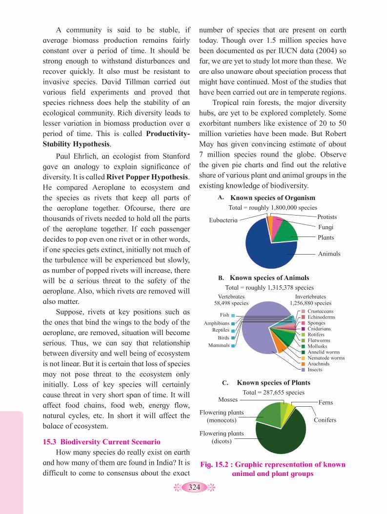
324
number of species that are present on earth today. Though over 1.5 million species have been documented as per IUCN data (2004) so far, we are yet to study lot more than these. We are also unaware about speciation process that might have continued. Most of the studies that have been carried out are in temperate regions.
Tropical rain forests, the major diversity hubs, are yet to be explored completely. Some exorbitant numbers like existence of 20 to 50 million varieties have been made. But Robert May has given convincing estimate of about 7 million species round the globe. Observe the given pie charts and find out the relative share of various plant and animal groups in the existing knowledge of biodiversity.
A community is said to be stable, if average biomass production remains fairly constant over a period of time. It should be strong enough to withstand disturbances and recover quickly. It also must be resistant to invasive species. David Tillman carried out various field experiments and proved that species richness does help the stability of an ecological community. Rich diversity leads to lesser variation in biomass production over a period of time. This is called Productivity-Stability Hypothesis.
Paul Ehrlich, an ecologist from Stanford gave an analogy to explain significance of diversity. It is called Rivet Popper Hypothesis. He compared Aeroplane to ecosystem and the species as rivets that keep all parts of the aeroplane together. Ofcourse, there are thousands of rivets needed to hold all the parts of the aeroplane together. If each passenger decides to pop even one rivet or in other words, if one species gets extinct, initially not much of the turbulence will be experienced but slowly, as number of popped rivets will increase, there will be a serious threat to the safety of the aeroplane. Also, which rivets are removed will also matter.
Suppose, rivets at key positions such as the ones that bind the wings to the body of the aeroplane, are removed, situation will become serious. Thus, we can say that relationship between diversity and well being of ecosystem is not linear. But it is certain that loss of species may not pose threat to the ecosystem only initially. Loss of key species will certainly cause threat in very short span of time. It will affect food chains, food web, energy flow, natural cycles, etc. In short it will affect the balace of ecosystem.
15.3 Biodiversity Current Scenario How many species do really exist on earth
and how many of them are found in India? It is difficult to come to consensus about the exact
Fig. 15.2 : Graphic representation of known animal and plant groups
Known species of OrganismTotal = roughly 1,800,000 species
Eubacteria Protists
Fungi
Plants
Animals
Known species of Animals Total = roughly 1,315,378 species
Known species of Plants Total = 287,655 species
Vertebrates 58,498 species
Fish
Amphibians Reptiles
Birds
Mammals
Invertebrates 1,256,880 species
Crustaceans Echinoderms Sponges Cnidarians Rotifers Flatworms Mollusks Annelid worms Nematode worms Arachnids Insects
Mosses Ferns
Flowering plants (monocots)
Flowering plants (dicots)
Conifers
B.
C.
A.
325
In the diagrams, we do not find any data of prokaryotes. Several moneran species are not cultivable under laboratory conditions. Also, conventional taxonomic methods are not suitable for identification of prokaryotic species.
India boasts a handsome share of 8.1% of total biodiversity wealth of the earth. One of the 12 megadiversity countries of the globe, India has 2.4% of total land area of the world. We have identified around 45000 plant species and nearly double the number of animal varieties from our natural wealth. If we consider May’s estimate of global biodiversity, we have recorded only 22% of our natural wealth. This situation underlines the need of taxonomists to study the biodiversity. But major concern is the possibility of loss of these varieties before we identify them because of activities like reclamation and deforestation.
Recently a group of naturalists proved that a lizard from Amboli ghat of Maharashtra was misidentified. They proved it on the basis of DNA profiling as well as number of glands and scales present on the legs of the lizard. Earlier thought to be Hemidactylus brookii, the lizard is now renamed as Hemidactylus varadgirii in the honour of renowned herpetologist and conservationist, Dr. Varad Giri. They also proved that brookii variety is not found in India.
Do you know ?
15.4 Loss of Biodiversity: Loss of biodiversity leads to the overall
imbalance in the ecosystem. The chief serious aspect of loss of biodiversity is extinction of species. There are three types of extinction viz, natural extinction, mass extinction and man-made (anthropogenic) extinction.
Damage to biodiversity takes place due to both, natural and manmade reasons. Natural reasons include forest fires, earthquakes, volcanic eruptions etc. Manmade reasons are habitat destruction, hunting, settlement, overexploitation and reclamation.
We are aware of five mass extinctions during various stages of history of earth (e.g. ice age). The current loss of biodiversity is considered to be the Sixth extinction which is progressing at an alarming rate which is estimated to be 100 to 1000 times faster than prehuman times. Ecologists blame this to the human intervention in natural habitats. They do not forget to warn that if the current trend continues, we might lose about 50% of diversity. Loss of biodiversity in any area can lead to the decline in plant production, lower resilience to environmental disturbance like flood. It may also lead to alteration in environmental processes like disease cycles, plant productivity etc.
Causes of Biodiversity losses: There are four major causes popularly
known as, ‘The Evil Quartet’.
i. Habitat loss and fragmentation: It is the prime cause of destruction.
Reduction in vast natural habitats and local degradation by pollution, create crisis situation for the living beings. Loss of local habitat due
Internet my friend
1. Find out information about Zoological Survey of India and Botanical Survey of India.
2. You may visit Zoological Survey of India, Pune office and find out how information about natural wealth is conserved.
Major mass extinction events occured between Cretaeous and Coenozoic period; between Triassic and Jurassic period; between Permian and Triassic period; between Devonian and Carbaniferous period; between Ordovician and Silurian period in the geological time scale where plants as well as animal groups underwent mass extinction.

326
Great Indian Bustard alias Maldhok is a critically endangered bird. It is considered as flagship species of grasslands. We lost last Maldhok from Maharashtra in 2018. Now this bird is found in Rajasthan and Gujrat. It is an example of local extinction due to habitat loss and hunting.
Do you know ?
Fig. 15.3 : Examples of extinction due to over-exploitation
b. Stellar sea cow
c. Passenger pigeon
a. Dodo bird
to human activities, creates threat to migratory birds as well as those animals that need larger territories. Tropical rain forests are being lost at an alarming rate. Tropical rain forest cover has reduced from 14% to 6% over the years.
ii. Over-exploitation: Basic difference between human beings
and other animals is that, humans have the tendency to accumulate beyond their needs. This has resulted in the overexploitation of
resources which in turn causes threats to various organisms. Can you corelate this with dirth of fish, we observe now a days? Dodo bird, stellar sea cow and passenger pigeon are few examples of extinction due to overexploitation.
iii. Alien species invasion:
When a new species gets introduced into any ecosystem accidentally or intentionally, there are chances that it proves harmful for existing species. Sometimes, it can lead to extinction of local species. In such a case, it is called as invasive species. E.g. the carrot grass (Parthenium), Lantana and water hyacinth (Eichhornia). Introduction of predator fish - Nile perch in Lake Victoria, proved deleterious for 200 local species of Cichlid fish.
In India, introduction of African catfish Clarias gariepinus for aquaculture purpose has proved harmful to endemic catfish varieties. One of the major reasons of such a harmful effect of alien species is, lack of local predator.
iv. Co-extinctions:
Many a times, organisms are associated with each other in obligatory way. In such cases, extinction of one variety leads to loss of associate variety from the ecosystem. e.g., Extinction of host fish causes extinction of unique parasites. Coevolved plant-pollinator, also will have such a threat.
We are aware of threat to diversity and loss of species from earth. When any species is totally eliminated from earth, it is called extinct. e.g. Dinosaurs. When the number of members of a species starts dwindling, it is said to be endangered. The International Union for Conservation of Nature and Natural Resources (IUCN) maintains a Red Data Book also known as Red List, where conservation status of plant and animal species is recorded.
327
After a given species has been thoroughly evaluated, it is placed into one of following several categories. 1. Extinct (EX), a designation applied to
species in which the last individual has died or is not recorded.
2. Extinct in the Wild (EW), a category containing those species whose members survive only in captivity
3. Critically Endangered (CR), a category containing those species that possess an extremely high risk of extinction with very few surviving members (50).
4. Endangered (EN), a designation applied to species that possess a very high risk of extinction as a result of rapid population decline of 50 to more than 70 percent over the previous 10 years (or three generations).
5. Vulnerable (VU), a category containing those species that possess a very high risk of extinction as a result of rapid population decline of 30 to more than 50 percent over the previous 10 years (or three generations).
6. Near Threatened (NT), a designation applied to species that are close to becoming threatened or may meet the criteria for threatened status in the near future.
7. Least Concern (LC), a category containing species that are pervasive and abundant after careful assessment
8. Data Deficient (DD), a condition applied to species in which the amount of available data related to its risk of extinction, is lacking in some way.
9. Not Evaluated (NE), a category used to include any of the nearly 1.9 million species described by scientists, but not assessed by the IUCN
Many a times, we read about leopard attacks on humans or about elephants from Karnataka destroying agricultural lands and orchards in Sindhudurg region of Maharashtra. With increase in human population, man started encroaching forest land. Animals either out of sheer curiosity (in case of young leopards) or for lack of sufficient resources, venture out from their original place. This results in Man-Animal conflict. Various measures are adapted by forest department to minimise this tussle. e.g., Government not only gives monetary compensation to farmers affected by elephant attacks, but even the forest department conducts community meetings to train locals to face the attack. Also, measures like appointing experts to tame the wild elephants with the help of domesticated elephants, and sensitising people towards wild life are most important part of such activities.
Do you know ?
The IUCN system uses a set of five quantitative criteria to assess the extinction risk of a given species. These criteria are : The rate of population decline; The geographic range; Whether the species already possesses a small population size; Whether the species is very small or lives in a restricted area; and Whether the results of a quantitative analysis indicate a high probability of extinction in the wild.
Do you know ?
1. Explain how loss of species diversity can
harm ecosystem? 2. Give various categories of endangered
species explained by IUCN.3. What do you understand by invasive
species? How does it affect local population?
Can you tell?
Internet my friend
What is ballast water and how can it bring about introduction of alien species into any ecosystem?
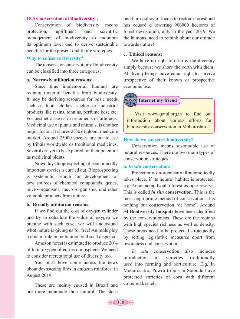
328
and burn policy of locals to reclaim forestland has caused a towering 906000 hectares of forest devastation, only in the year 2019. We the humans, need to rethink about our attitude towards nature!
c. Ethical reasons: We have no right to destroy the diversity
simply because we share the earth with them! All living beings have equal right to survive irrespective of their known or prospective economic use.
15.5 Conservation of Biodiversity : Conservation of biodiversity means
protection, upliftment and scientific management of biodiversity to maintain its optimum level and to derive sustainable benefits for the present and future strategies.
Why to conserve Diversity? The reasons for conservation of biodiversity
can be classified into three categories:
a. Narrowly utilitarian reasons: Since time immemorial, humans are
reaping material benefits from biodiversity. It may be deriving resources for basic needs such as food, clothes, shelter or industrial products like resins, tannins, perfume base etc. For aesthetic use as in ornaments or artefacts. Medicinal use of plants and animals, is another major factor. It shares 25% of global medicine market. Around 25000 species are put to use by tribals worldwide as traditional medicines. Several are yet to be explored for their potential as medicinal plants.
Nowadays bioprospecting of economically important species is carried out. Bioprospecting is systematic search for development of new sources of chemical compounds, genes, micro-organisms, macro-organisms, and other valuable products from nature.
b. Broadly utilitarian reasons: If we find out the cost of oxygen cylinder
and try to calculate the value of oxygen we breathe with such ease; we will understand what nature is giving us for free! Animals play a crucial role in pollination and seed dispersal.
Amazon forest is estimated to produce 20% of total oxygen of earths atmosphere. We need to consider recreational use of diversity too.
You must have come across the news about devastating fires in amazon rainforest in August 2019.
These are mainly caused in Brazil and are more manmade than natural. The slash
How do we conserve biodiversity? Conservation means sustainable use of
natural resources. There are two main types of conservation strategies :
a. In situ conservation: Protection of an organism will automatically
takes place, if its natural habitat is protected. e.g. Announcing Kanha forest as tiger reserve. This is called in situ conservation. This is the most appropriate method of conservation. It is nothing but conservation ‘at home’. Around 34 Biodiversity hotspots have been identified by the conservationists. These are the regions with high species richness as well as density. These areas need to be protected strategically by setting legislative measures apart from awareness and conservation.
In situ conservation also includes introduction of varieties traditionally used into farming and horticulture. E.g. In Maharashtra, Pawra tribals in Satpuda have protected varieties of corn with different coloured kernels.
Internet my friend
Visit www.gotul.org.in to find out information about various efforts for biodiversity conservation in Maharashtra.
329
India has three of world’s biodiversity hotspots (the areas with high density of biodiversity), Western ghats, Indo-Burma and Eastern-Himalayas. It has been estimated that protection of these diversity rich hotspots could reduce extinction rate by almost 30%.
India, at present has 14 biosphere reserves, 90 national parks and 448 wildlife sanctuaries. In Maharashtra, there are 5 national parks and 11 sanctuaries.
What are sacred groves?
Can you recall?
Indian culture and traditions are always connected with nature and rituals are laid down to protect biodiversity. In many cultures, stretches of forests were set aside and protected in the name of Almighty, which are called sacred groves.
Such sacred groves are found in Khasi and Jaintia hills in Meghalaya, Western ghat regions of Maharashtra and Karnataka, Aravali hills of Rajasthan and Bastar, and Chanda and Sarguja areas of Madhya Pradesh.
Dr. Akira Miyawaki studied native forests of Japan especially the old shrine groves and developed a technique of growing dense plantations in short time. It is a technique for restoration of natural vegetation on degraded land. In this technique, after soil testing, the landmass is dugout and soil is mixed with local biomass and humus. Plantation is done in layers and saplings are planted close to each other. Due to this, sunlight doesn’t reach soil retaining the moisture. Close proximity of plants leads to faster vertical growth than lateral. Also, it promotes natural selection. Native varieties are planted and the forest develops at almost ten times faster than the natural way. It requires a caring period of 3 years after which it grows and develops its own diversity naturally. In India, this technique is adapted at several places including remote districts like Chandrapur as well as metro cities like Mumbai and Bengaluru. Though there is debate about feasibility of the technique, it is certainly helping in retaining and recharging groundwater table, supporting local biodiversity and curbing air pollution by adding to green cover.
Do you know ? the old shrine groves and developed a technique of growing dense plantations in short time. It is
Sacred groves serve the only chance of survival for some endangered varieties of animal and plant species. Tribals do not allow to cut even a single branch of tree from sacred grove. But with the increasing lust and greed, are sacred groves safe? We must think about it.
b. Ex situ conservation: Sometimes when a species is critically
endangered, special measures have to be under taken to protect it. It might be protected in captivity, as one of the measures of protection. This is called Ex situ conservation. In this type of conservation, living beings are protected away from their natural habitats in special settings. Wild life safari parks, zoological parks and botanical gardens serve this purpose. Animals which have decreased in number, are allowed to breed in captivity in order to protect them.Eg crocodile bank of Chennai.
Seed banks are established to conserve wild varieties of food grains and vegetables. Now a days, modern techniques like tissue culture, in vitro fertilization of eggs and cryopreservation (preservation at low temperature -1960C) of gametes, are used to protect endangered species.
By now we have, thus, understood the immense importance of biodiversity and dire need to protect it.
Internet my friend
Collect the information about protected areas, biosphere reserves, national parks and wild life sanctuaries.
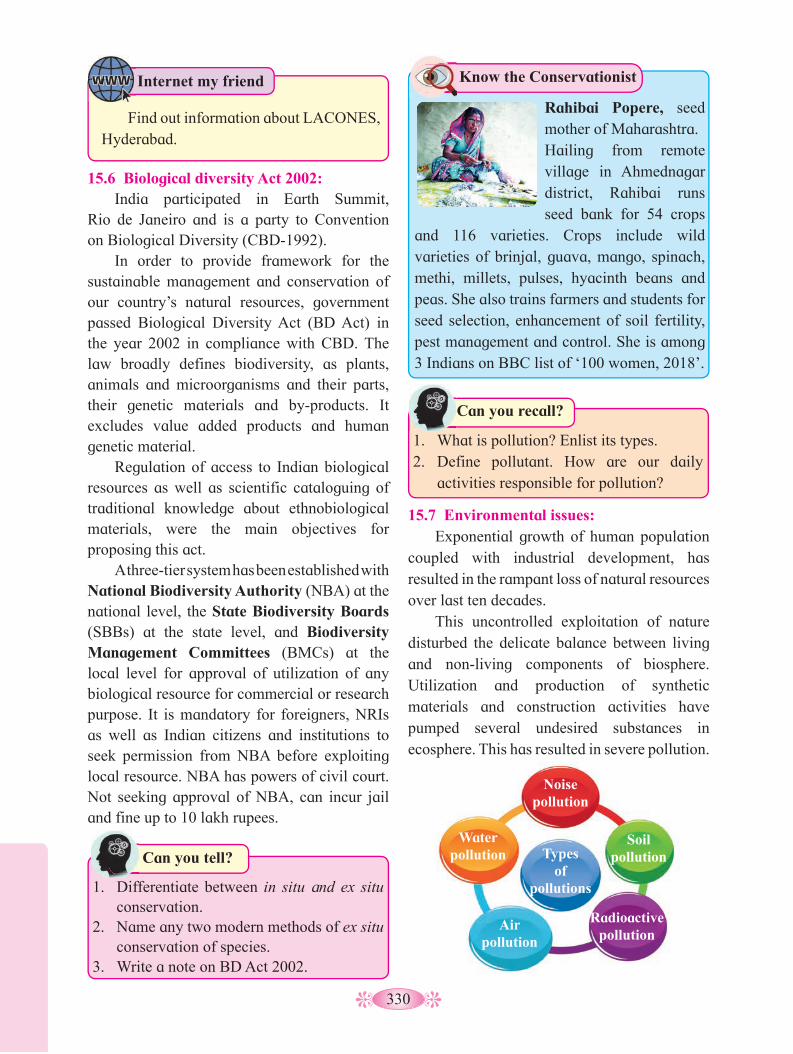
330
1. Differentiate between in situ and ex situ
conservation.2. Name any two modern methods of ex situ
conservation of species.3. Write a note on BD Act 2002.
Can you tell?
15.6 Biological diversity Act 2002: India participated in Earth Summit,
Rio de Janeiro and is a party to Convention on Biological Diversity (CBD-1992).
In order to provide framework for the sustainable management and conservation of our country’s natural resources, government passed Biological Diversity Act (BD Act) in the year 2002 in compliance with CBD. The law broadly defines biodiversity, as plants, animals and microorganisms and their parts, their genetic materials and by-products. It excludes value added products and human genetic material.
Regulation of access to Indian biological resources as well as scientific cataloguing of traditional knowledge about ethnobiological materials, were the main objectives for proposing this act.
A three-tier system has been established with National Biodiversity Authority (NBA) at the national level, the State Biodiversity Boards (SBBs) at the state level, and Biodiversity Management Committees (BMCs) at the local level for approval of utilization of any biological resource for commercial or research purpose. It is mandatory for foreigners, NRIs as well as Indian citizens and institutions to seek permission from NBA before exploiting local resource. NBA has powers of civil court. Not seeking approval of NBA, can incur jail and fine up to 10 lakh rupees.
15.7 Environmental issues: Exponential growth of human population
coupled with industrial development, has resulted in the rampant loss of natural resources over last ten decades.
This uncontrolled exploitation of nature disturbed the delicate balance between living and non-living components of biosphere. Utilization and production of synthetic materials and construction activities have pumped several undesired substances in ecosphere. This has resulted in severe pollution.
1. What is pollution? Enlist its types.2. Define pollutant. How are our daily
activities responsible for pollution?
Can you recall?
Know the Conservationist
Rahibai Popere, seed mother of Maharashtra.
Hailing from remote village in Ahmednagar district, Rahibai runs seed bank for 54 crops
and 116 varieties. Crops include wild varieties of brinjal, guava, mango, spinach, methi, millets, pulses, hyacinth beans and peas. She also trains farmers and students for seed selection, enhancement of soil fertility, pest management and control. She is among 3 Indians on BBC list of ‘100 women, 2018’.
Noise pollution
Soil pollution
Radioactive pollution
Air pollution
Water pollution Types
of pollutions
Internet my friend
Find out information about LACONES, Hyderabad.

331
Gaseous pollutants include CO2, CO, SO
2,
NO, NO2 etc.
Carbon di-oxide is a greenhouse gas. In the past, levels of CO
2 in the atmosphere
remained low. Due to burning of fossil fuels, as well as increasing deforestation the levels of CO
2 are rising at alarming rate. Photosynthesis
process balances CO2
: O2
ratio of the air to a great extent. CO
2 is also removed from the
air by weathering of silicate rocks forming limestone. A jet plane in a single trip across the Atlantic uses 35 tonnes of oxygen and releases 70 tonnes of CO
2. We are going to discuss the
role of CO2
in global warming later in this chapter.
Carbon monoxide (CO) : It is a poisonous gas produced by
incomplete combustion of fuel such as coal or wood. Vehicular exhausts are the largest source of CO.
Any substance that causes pollution, is called Pollutant. In order to protect and improve the quality of our environment, the Government of India has passed the Environment Protection Act 1986.
a. Air Pollution :Effect of air pollution :
Respiratory surfaces of living beings are constantly interacting with air. Any unfavourable alteration in air quality, affects the respiratory system. Severity of damage depends on concentration of pollutant, duration of exposure and the organism. Even in case of plants, air pollution results in poor yield of crops and premature death of plants. Nowadays automobiles are omnipresent. They are major cause for atmospheric (air) pollution. Regular maintenance of vehicles and use of lead-free petrol or diesel can reduce pollutant from exhausts.
Types of air pollutants : Air pollutants are of two types – particulate
pollutants and gaseous pollutants. Particulate air pollutants may be solid
or liquids. Particles with diameter 10 µm may settle in the soil but particles with 1 µm or less remain suspended in the air. According to Central Pollution Control Board (CPCB), particulate matter of size 2.5µm or less in diameter (PM2.5) are responsible for causing the greatest harm to humans.
These fine particulates can be inhaled deep into the lungs and are responsible for irritation, inflammation and damage to lungs. In addition to this, it causes breathing and respiratory disorders and premature deaths.
Smoke, smog, pesticides, heavy metals, dust and radioactive elements are the examples of particulate pollutants.
Does particulate matter help to reduce atmospheric temperature?
Use your brain power
State the effects of CO on human body.
Use your brain power
Nitrogen di oxide (NO2) and nitrogen
monoxide (NO): These are released by automobiles and
chemical industries as waste gases. NO2 when
combines with water vapours forms nitric acid. It causes irritation to eyes and lungs. At high concentration, it causes injury to lungs, liver and kidneys.
Control measures: Various ingenious mechanisms have been
developed to control emission of gaseous and particulate pollutants through vehicles and industries. Few examples are explained below:
Electrostatic precipitator : It is most widely used for removing
particulate matter like soot and dust present in industrial exhaust. It can remove almost 99% particulate matter present in exhaust from a thermal power plant.
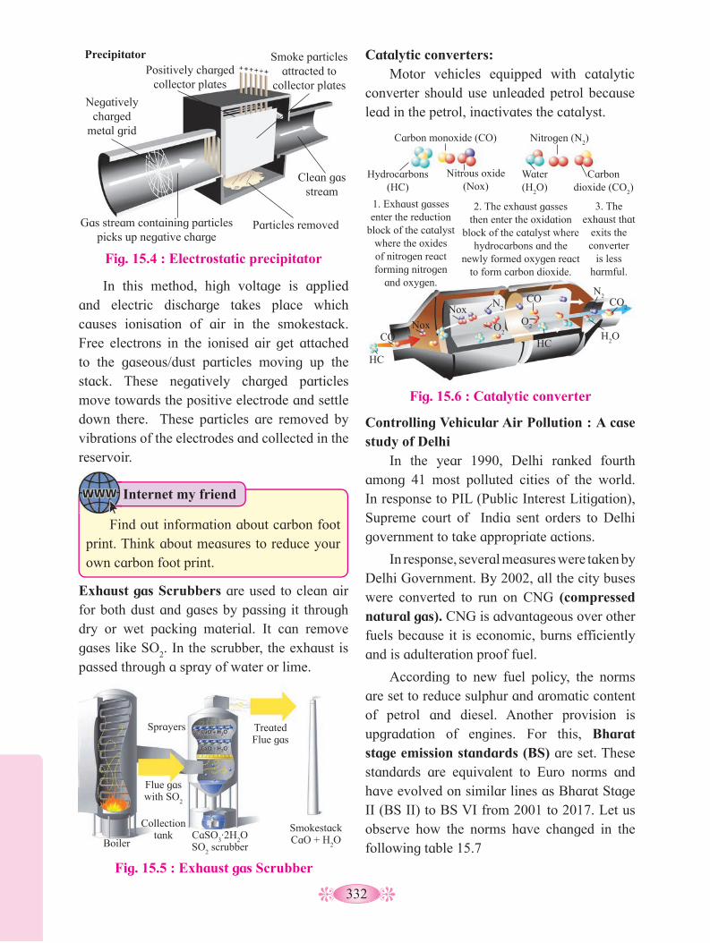
332
Fig. 15.6 : Catalytic converter
Catalytic converters: Motor vehicles equipped with catalytic
converter should use unleaded petrol because lead in the petrol, inactivates the catalyst.
In this method, high voltage is applied and electric discharge takes place which causes ionisation of air in the smokestack. Free electrons in the ionised air get attached to the gaseous/dust particles moving up the stack. These negatively charged particles move towards the positive electrode and settle down there. These particles are removed by vibrations of the electrodes and collected in the reservoir.
Exhaust gas Scrubbers are used to clean air for both dust and gases by passing it through dry or wet packing material. It can remove gases like SO
2. In the scrubber, the exhaust is
passed through a spray of water or lime.
Fig. 15.5 : Exhaust gas Scrubber
Controlling Vehicular Air Pollution : A case study of Delhi
In the year 1990, Delhi ranked fourth among 41 most polluted cities of the world. In response to PIL (Public Interest Litigation), Supreme court of India sent orders to Delhi government to take appropriate actions.
In response, several measures were taken by Delhi Government. By 2002, all the city buses were converted to run on CNG (compressed natural gas). CNG is advantageous over other fuels because it is economic, burns efficiently and is adulteration proof fuel.
According to new fuel policy, the norms are set to reduce sulphur and aromatic content of petrol and diesel. Another provision is upgradation of engines. For this, Bharat stage emission standards (BS) are set. These standards are equivalent to Euro norms and have evolved on similar lines as Bharat Stage II (BS II) to BS VI from 2001 to 2017. Let us observe how the norms have changed in the following table 15.7
Fig. 15.4 : Electrostatic precipitator
Precipitator Positively charged
collector plates
Negatively charged
metal grid
Smoke particles attracted to
collector plates
Clean gas stream
Particles removed Gas stream containing particles picks up negative charge
Flue gas with SO
2
Sprayers
Collection tank
Boiler CaSO
3·2H
2O
SO2 scrubber
Treated Flue gas
SmokestackCaO + H
2O
1. Exhaust gasses enter the reduction
block of the catalyst where the oxides of nitrogen react forming nitrogen
and oxygen.
2. The exhaust gasses then enter the oxidation
block of the catalyst where hydrocarbons and the
newly formed oxygen react to form carbon dioxide.
3. The exhaust that
exits the converter
is less harmful.
Hydrocarbons (HC)
Carbon monoxide (CO)
Nitrous oxide (Nox)
Water (H
2O)
Nitrogen (N2)
Carbon dioxide (CO
2)
CO2
H2O
N2
N2
O2 O
2
Nox
Nox CO
CO
HC
HC
Internet my friend
Find out information about carbon foot print. Think about measures to reduce your own carbon foot print.

333
Table 15.7 :Bharat stage emission standards in cities of India.
Vehicle Norms Cities of Implementation
4 wheelers
Bharat Stage II
All metro cities
4 wheelers
Bharat Stage III
Throughout the country since October 2010
4 wheelers
Bharat Stage IV
13 mega cities (Delhi and NCR, Mumbai, Kolkata, Chennai, Bengaluru, Surat, Kanpur, Agra, Lucknow, Solapur) since April 2010.
2 wheelers
Bharat Stage III
Throughout the country since October 2010
3 wheelers
Bharat Stage III
Throughout the country since October 2010
Pradhan Mantri Ujjwala yojana (Pradhan Mantri clean fuel programme) It was launched by Prime Minister of India, on 1st May 2016 to distribute 50 million LPG connections to women of BPL families. In many BPL families, chullhas are used where incomplete combustion of wood and coal leads to CO poisoning. Use of LPG helps in reducing such household air pollution.
Do you know ?
Colder weather and stagnant winds, trap smoke from various sources like firecrackers, burning crop stubbles, lit garbage and road dust. Citizens suffered breathlessness, chest muscle contraction, irritation in eyes, asthma and allergy. Administration took certain measures like closing educational institutions, suspending of construction or demolition work, undertaking vacuum cleaning of roads etc. Even Badarpur thermal power plant was temporarily closed down. Do you think mere setting standards is not enough? We must encourage means like car pooling and use of public transport.
Inspite of all the measures, New Delhi and adjoining areas witnessed great smog between 1st to 9th November 2016. Air pollution at this time peaked on both 2.5PM and 10PM levels. This is reported as one of the worst levels of air quality since 1999.
Do you know ?
1. Describe any 2 particulate and gaseous
pollutants.2. Explain various technologies used for
removing particulate matter from different sources of air pollution.
3. What are the ill effects of noise pollution on human health?
4. Give any norms for reducing sulphur and aromatic contents of petrol and diesel.
Can you tell?
Have you noticed that BS V is missing in above table? Note that, in 2001, Bharat stage II emission norms were set for CNG and LPG vehicles. It stipulates that emission of sulphur be controlled at 50 ppm in diesel and 150ppm in petrol. Aromatic hydrocarbons should be just 42% in concerned fuel. The aim is to reduce sulphur emission to 50ppm in petrol and diesel along with aromatic hydrocarbons to 35%. Government of India directly adapted BS VI in the year 2018, skipping BS V. These efforts decreased the levels of CO
2 and SO
2 in Delhi.
b. Noise pollution : In India, the Air (Prevention and control
of pollution) Act 1981, amendment 1987, includes noise as an air pollutant. Noise is an undesired high level of sound. Noise causes psychological and physiological changes in human beings. There is dire need of creating awareness about noise pollution caused during festivals and processions in our society.

334
Activity :
1. Define water pollution.2. When will you call a water body polluted?
Can you recall?
Exposure to extremely high sound level (150 decibels or more) generated during a take-off of a jet plane or rocket, may damage ear drums and cause permanent hearing loss.
Noise also can cause sleeplessness, increased heartbeat, altered breathing pattern and psychological stress. Noise may negatively interfere with child’s learning and behaviour pattern. The common sources of noise pollution are machines, transportation, construction sites, industries etc.
Find out different sources of noise pollution in your surrounding.
Increased utilisation and discharge of harmful waste water in water bodies has caused severe pollution.
Most of the water pollution is manmade. Polluted water may be turbid, foul smelling, and may contain number of pathogens, heavy metals, oils etc.
Realising the importance of maintaining the cleanliness of the water bodies, Government of India has passed the Water (prevention and control of pollution) Act 1974 to safe guard our water resources.
1. Where does domestic waste water go in urban and rural areas?
2. What is importance of sewage treatment plant?
Can you recall?
c. Water Pollution and its Control: Water has been a controlling factor in
distribution and density of living beings in an area. Human water consumption increased many folds with increased industrialisation.
Domestic sewage and Industrial Effluents: Even a small quantity of about 0.1%
impurities in water, can make it unfit for human consumption. Solids are relatively easy to separate but dissolved salts such as nitrates, phosphates, other nutrients and toxic metal ions as well as organic compounds, are difficult to remove.
Domestic sewage is one of the most common source of water pollution. It contains biodegradable organic matter. It readily gets decomposed by bacteria and other microorganisms. They use organic matter as substrate and utilise some amount of sewage.
It is possible to estimate biodegradable organic matter in sewage water by measuring Biochemical Oxygen Demand (BOD). It is the amount of dissolved oxygen required by microorganisms for decomposing the organic matter present in water. It is expressed in milligram of oxygen per litre (mg/L) of water. High BOD indicates intense level of microbial pollution.
Internet my friend
We have studied about health effects of noise pollution on humans. How does this pollution affect birds? Does it affect marine life? Find out.
Reduction of noise in our industries can be brought about by use of sound absorbent materials or by muffling the noise. Laws which prohibit blowing horn in the areas of schools and hospitals, need to be implemented strictly to curb decibel levels.
Govt. of India has rules and regulations against firecrackers and loudspeakers. Supreme court of India has banned loudspeakers at public gatherings after 10pm.
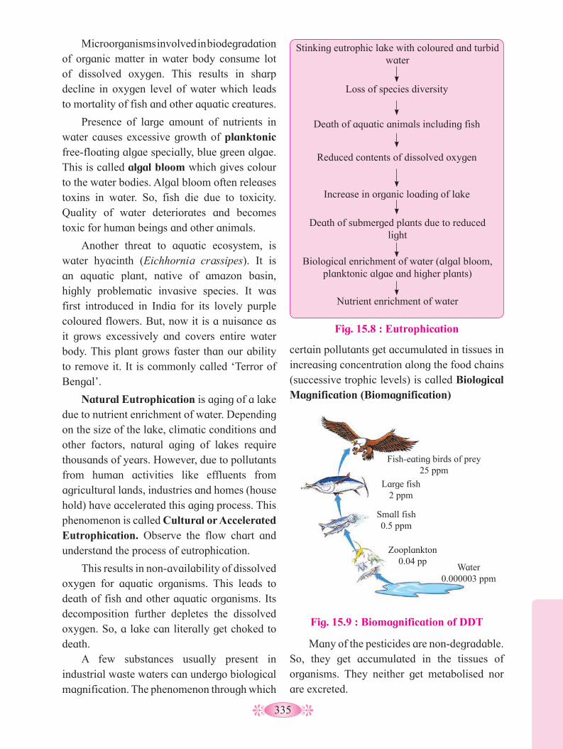
335
certain pollutants get accumulated in tissues in increasing concentration along the food chains (successive trophic levels) is called Biological Magnification (Biomagnification)
Microorganisms involved in biodegradation of organic matter in water body consume lot of dissolved oxygen. This results in sharp decline in oxygen level of water which leads to mortality of fish and other aquatic creatures.
Presence of large amount of nutrients in water causes excessive growth of planktonic free-floating algae specially, blue green algae. This is called algal bloom which gives colour to the water bodies. Algal bloom often releases toxins in water. So, fish die due to toxicity. Quality of water deteriorates and becomes toxic for human beings and other animals.
Another threat to aquatic ecosystem, is water hyacinth (Eichhornia crassipes). It is an aquatic plant, native of amazon basin, highly problematic invasive species. It was first introduced in India for its lovely purple coloured flowers. But, now it is a nuisance as it grows excessively and covers entire water body. This plant grows faster than our ability to remove it. It is commonly called ‘Terror of Bengal’.
Natural Eutrophication is aging of a lake due to nutrient enrichment of water. Depending on the size of the lake, climatic conditions and other factors, natural aging of lakes require thousands of years. However, due to pollutants from human activities like effluents from agricultural lands, industries and homes (house hold) have accelerated this aging process. This phenomenon is called Cultural or Accelerated Eutrophication. Observe the flow chart and understand the process of eutrophication.
This results in non-availability of dissolved oxygen for aquatic organisms. This leads to death of fish and other aquatic organisms. Its decomposition further depletes the dissolved oxygen. So, a lake can literally get choked to death.
A few substances usually present in industrial waste waters can undergo biological magnification. The phenomenon through which
Fig. 15.8 : Eutrophication
Stinking eutrophic lake with coloured and turbid water
Loss of species diversity
Death of aquatic animals including fish
Reduced contents of dissolved oxygen
Increase in organic loading of lake
Death of submerged plants due to reduced light
Biological enrichment of water (algal bloom, planktonic algae and higher plants)
Nutrient enrichment of water
Fig. 15.9 : Biomagnification of DDT
Fish-eating birds of prey 25 ppm
Large fish 2 ppm
Small fish 0.5 ppm
Zooplankton 0.04 pp
Water 0.000003 ppm
Many of the pesticides are non-degradable. So, they get accumulated in the tissues of organisms. They neither get metabolised nor are excreted.

336
1. Explain BOD and its effects on aquatic
ecosystem.2. Ecological sanitation is the need of the
day. Justify.3. Explain the phenomenon of biological
magnification.
Can you tell?
1. Why is it necessary to separate wet and dry garbage?
2. What are the six R’s to combat solid waste?
Can you recall?
When the pit of an ecosan toilet fills up, it is closed and sealed. After about 8-9 months, the faeces are completely composted to organic manure.There are working EcoSan toilets in many areas of Gujrat, Kerala, Tamilnadu and Sri Lanka.
Recycling of sewage water by reverse osmosis will help to solve the problem of not only scarcity of water but also the disposal of sewage water. At Tirumala hills, millions of pilgrims visit. Every building here has R.O. system which solves the problem of huge water demand. Recycling of sewage water is seen in many townships in Mumbai. Rain water harvesting is encouraged and made mandatory for new constructions by Municipal Corporation.
Thermal pollution of water is caused due to rise in temperature of water. The main source of thermal pollution are the thermal and nuclear power plants. The power plants use water as coolant and release hot water. As many organisms are sensitive to temperature, sudden rise in temperature leads to loss of flora and fauna.
Solid Waste Management : You have already studied about solid waste
management in ninth standard.
They are passed on to the next trophic level. This is commonly seen in case of DDT and Mercury. Observe the food chain in the given figure. It shows how biomagnification of DDT takes place.
Why do you think the amount of DDT is maximum in birds?
Use your brain power
Measures to reduce sewage water: In order to conserve water and prevent
creation of sewage, ecosan is a sustainable system for handling human excreta using dry composting toilets. This is a practical, efficient and cost-effective solution for human waste disposal.
Ecological sanitation (Ecosan) is an approach to sanitation provision which safely reuses excreta in agriculture. It reduces the need for chemical fertilizers.
Ecosan toilet is a closed system that does not need water. It is an alternative to leach pit toilets in place where water is scarce or where there is risk of ground water contamination.
It is based on the principle of recovery and recycling of nutrients from excreta to create a valuable resource for agriculture.
Solid waste is everything that goes to trash. It includes wastes from home, offices, stores, schools, hospitals etc. Wastes are collected and disposed by municipality. Municipal solid waste generally consists of paper, food, plastic, glass, metals, rubber, leather, textile etc;
Burning reduces volume of the waste. Generally, it is not burnt to completion. So open dumps become breeding ground for rats and flies.
Internet my friend
1. Find out biomagnification of diclofenac and DDT in birds.
2. Find out various types of agrochemicals which have ill effects on ecosystem.
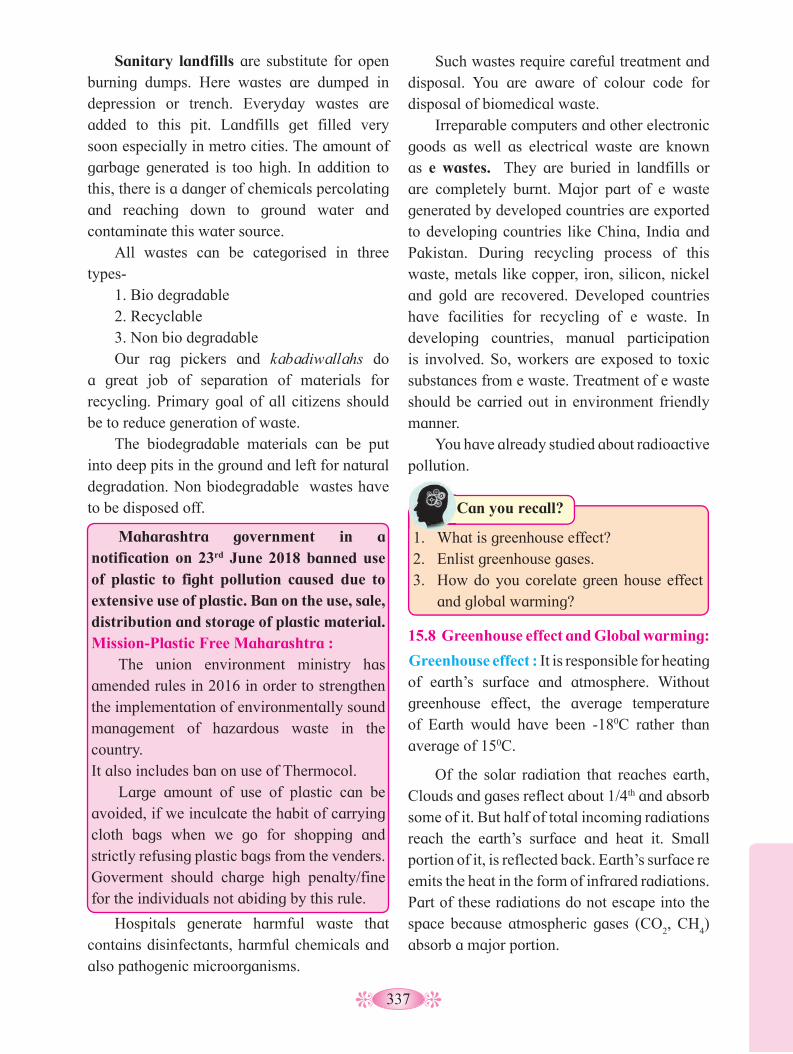
337
Such wastes require careful treatment and disposal. You are aware of colour code for disposal of biomedical waste.
Irreparable computers and other electronic goods as well as electrical waste are known as e wastes. They are buried in landfills or are completely burnt. Major part of e waste generated by developed countries are exported to developing countries like China, India and Pakistan. During recycling process of this waste, metals like copper, iron, silicon, nickel and gold are recovered. Developed countries have facilities for recycling of e waste. In developing countries, manual participation is involved. So, workers are exposed to toxic substances from e waste. Treatment of e waste should be carried out in environment friendly manner.
You have already studied about radioactive pollution.
Sanitary landfills are substitute for open burning dumps. Here wastes are dumped in depression or trench. Everyday wastes are added to this pit. Landfills get filled very soon especially in metro cities. The amount of garbage generated is too high. In addition to this, there is a danger of chemicals percolating and reaching down to ground water and contaminate this water source.
All wastes can be categorised in three types-
1. Bio degradable 2. Recyclable 3. Non bio degradable Our rag pickers and kabadiwallahs do
a great job of separation of materials for recycling. Primary goal of all citizens should be to reduce generation of waste.
The biodegradable materials can be put into deep pits in the ground and left for natural degradation. Non biodegradable wastes have to be disposed off.
Maharashtra government in a notification on 23rd June 2018 banned use of plastic to fight pollution caused due to extensive use of plastic. Ban on the use, sale, distribution and storage of plastic material.Mission-Plastic Free Maharashtra :
The union environment ministry has amended rules in 2016 in order to strengthen the implementation of environmentally sound management of hazardous waste in the country.It also includes ban on use of Thermocol.
Large amount of use of plastic can be avoided, if we inculcate the habit of carrying cloth bags when we go for shopping and strictly refusing plastic bags from the venders. Goverment should charge high penalty/fine for the individuals not abiding by this rule.
Hospitals generate harmful waste that contains disinfectants, harmful chemicals and also pathogenic microorganisms.
1. What is greenhouse effect?2. Enlist greenhouse gases.3. How do you corelate green house effect
and global warming?
Can you recall?
15.8 Greenhouse effect and Global warming:
Greenhouse effect : It is responsible for heating of earth’s surface and atmosphere. Without greenhouse effect, the average temperature of Earth would have been -180C rather than average of 150C.
Of the solar radiation that reaches earth, Clouds and gases reflect about 1/4th and absorb some of it. But half of total incoming radiations reach the earth’s surface and heat it. Small portion of it, is reflected back. Earth’s surface re emits the heat in the form of infrared radiations. Part of these radiations do not escape into the space because atmospheric gases (CO
2, CH
4)
absorb a major portion.
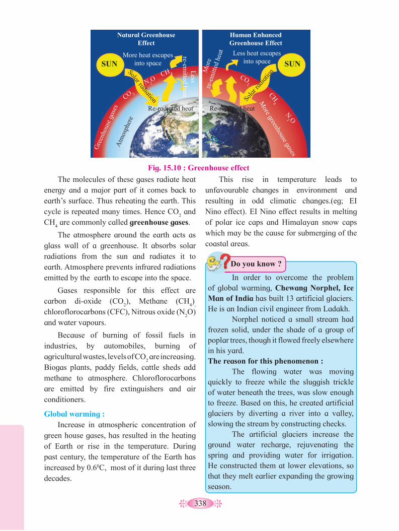
338
Fig. 15.10 : Greenhouse effect
The molecules of these gases radiate heat energy and a major part of it comes back to earth’s surface. Thus reheating the earth. This cycle is repeated many times. Hence CO
2 and
CH4 are commonly called greenhouse gases.
The atmosphere around the earth acts as glass wall of a greenhouse. It absorbs solar radiations from the sun and radiates it to earth. Atmosphere prevents infrared radiations emitted by the earth to escape into the space.
Gases responsible for this effect are carbon di-oxide (CO
2), Methane (CH
4)
,
chloroflorocarbons (CFC), Nitrous oxide (N2O)
and water vapours.
Because of burning of fossil fuels in industries, by automobiles, burning of agricultural wastes, levels of CO
2 are increasing.
Biogas plants, paddy fields, cattle sheds add methane to atmosphere. Chloroflorocarbons are emitted by fire extinguishers and air conditioners.
Global warming : Increase in atmospheric concentration of
green house gases, has resulted in the heating of Earth or rise in the temperature. During past century, the temperature of the Earth has increased by 0.60C, most of it during last three decades.
This rise in temperature leads to unfavourable changes in environment and resulting in odd climatic changes.(eg; EI Nino effect). EI Nino effect results in melting of polar ice caps and Himalayan snow caps which may be the cause for submerging of the coastal areas.
In order to overcome the problem of global warming, Chewang Norphel, Ice Man of India has built 13 artificial glaciers. He is an Indian civil engineer from Ladakh. Norphel noticed a small stream had frozen solid, under the shade of a group of poplar trees, though it flowed freely elsewhere in his yard.The reason for this phenomenon : The flowing water was moving quickly to freeze while the sluggish trickle of water beneath the trees, was slow enough to freeze. Based on this, he created artificial glaciers by diverting a river into a valley, slowing the stream by constructing checks. The artificial glaciers increase the ground water recharge, rejuvenating the spring and providing water for irrigation. He constructed them at lower elevations, so that they melt earlier expanding the growing season.
Do you know ?
Natural Greenhouse Effect
Human Enhanced Greenhouse Effect
More heat escapes into space
Less heat escapes into space
Less
re-emitted heat
Mor
e re
-em
itted
hea
t
Re-radiated heat Re-radiated heat
Solar radiation Sola
r rad
iatio
n
CO2
CO 2 CH
4
CH 4
N2 O
N 2O
Gre
enho
use
gase
s
More greenhouse gases
Atm
osph
ere
SUN SUN
339
The ozone shield has been disturbed due to increased rate of ozone degradation by Chlorofluorocarbon (CFC). CFCs move upwards and reach stratosphere. UV rays act on them and release Cl atoms. Cl degrades ozone releasing molecular oxygen. Cl atoms act as catalyst. So they remain in the stratosphere and continue the effect of ozone degradation. This results in ozone depletion. Although it occurs widely in the stratosphere, the depletion is particularly marked over the Antarctic region. This has resulted in formation of large area of thinned ozone layer, commonly called ozone hole.
UV radiations of wave length shorter than UV-B i.e. 100-280nm are almost completely absorbed by earth’s atmosphere, given that the ozone layer is intact. UV-B (wavelength 280-322nm) damages DNA and mutation may occur. It causes aging of skin, damage to skin cells and various types of skin cancers. In human eye, cornea absorbs UV-B radiations and a high dose of UV-B causes inflammation of cornea called snow blindness, cataract etc; Such exposure may permanently damage cornea.
Recognising the harmful effects of ozone depletion, an international treaty, known as the Montreal Protocol was signed at Montreal (Canada) in 1987 to control emission of ozone depleting substances.
Later many more efforts have been made and protocols have laid down definite roadmaps separately for developing and developed countries for reducing emission of CFCs and other ozone depleting chemicals.
15.9 Ozone depletion : Ozone is a form of oxygen.In the
stratosphere, ozone is photo-dissociated and is generated by absorption of short wave length UV radiations.
O3 →O
2 +[O]
O2+ [O] →UV RAYS →O
3
Generation and dissociation of ozone are in equilibrium leading to steady concentration of ozone in the stratosphere (12 to 15 kilometers from Earth’s surface in the atmosphere).
This ozone layer acts as shield that absorbs UV radiations from the sun and protects all types of life on earth. Absorption of UV radiations by ozone blanket is proportional to its thickness. Thickness is more above the poles than at the equator. UV rays are highly injurious to living organisms since DNA and proteins of living organisms absorb UV rays and its high energy breaks the chemical bonds within these molecules.
Thickness of the ozone in a column of air from the ground to the top of the atmosphere is measured as Dobson units (DU).
Global warming can be controlled by reducing use of fossil fuel, improving efficiency of energy usage, reducing deforestation, planting trees and checking of human population growth.
El Nino effect is a climate cycle in pacific ocean with global impact on weather pattern. The cycle begins when warm water in the western tropical pacific ocean shifts eastwards along the equator towards the coast of America. Normally, this warm water pools near Indonesia and Philippines. La Nina is a climatic pattern that describes the cooling of surface ocean waters along the tropical west coast of south America.
Do you know ?
1. How pollution by domestic garbage can
be controlled?2. Give effect of CFC on ozone shield.3. Give an account of possible effects of
global warming.4. Plastic ban in Maharashtra is an essential
step. Give reason.
Can you tell?

340
What is Chipko movement?
Can you recall?
1. How Jhum cultication has led to
deforestation in recent years?2. Comment on deforestation status of the
world and its major effects.
Can you tell?
15.10 Deforestation : Deforestation is conversion of forest area
into non-forest area. According to an estimate, almost 40% forests are lost in the tropics and only 1% in temperate region. The scenario of deforestation is grim in India. At the beginning of 20th century, forest cover was about 30% in India. By the end of the century, it got reduced to 19.4 %. The National Forest Policy 1988 of India has recommended 33% forest cover for the plains and 67% for the hills.
Deforestation takes place by conversion of forest to agricultural land so as to feed the growing human population.Trees are cut for timber, firewood, for keeping cattle in farm and for other purposes.
Slash and burn agriculture commonly called Jhum Cultivation in north eastern parts of India, where farmers cut down trees of the forest and burn the plant remains. The ash is used as fertilizer and the land is used for farming and cattle grazing. After cultivation, the area is left for several years so as to allow its recovery.
Major Effect of Deforestation is the increased concentration of CO
2 in the
atmosphere because trees hold lot of carbon in their biomass, are lost with deforestation.
It leads to the loss of biodiversity due to habitat destruction, disturbs hydrologic cycle, causes soil erosion and desertification in extreme cases.
Reforestation is the process of restoring a forest that once existed but was distroyed or removed at some time in past. Reforestation occurs naturally in a deforested area. We can speed up this by plantation of tree with due consideration to biodiversity that existed before.
Best example of peoples participation in reforestation is Saalumara Thimmakka, an Indian environmentalist from state of Karnataka noted for her work in planting and tending to 385 banyan trees along a 4km stretch of highway between Hulikal and Kudur. She has also planted nearly 8000 other trees. Her work has been honoured with the National Citizens Award of India. She was also conferred with Padma Shri in 2019.
Moirangthem Loiya from Manipur dedicated 17 years of his life to restore Punshilok forest. He left his job and took over the task of bringing back the lost glory of 300 acres forest land. He planted a variety of trees like, bamboo, oak, ficus, teak, jackfruit and magnolia. Today the forest has over 250 varieties of plants including 25 varieties of bamboo. It is now selected as home by great diversity of animals too.
Case study of people’s participation in conservation of forests.
History of people’s participation in India can be traced back to 1731. The ministers of the king of Jodhpur in Rajasthan tried to cut forest to procure wood for a new palace. Local Bishnoi community known for peaceful co-existence with nature, opposed king’s men.
A Bishnoi woman Amrita Devi hugged the trees and lost her life in an attempt for protecting the forest. Her three daughters and hundreds of other Bishnois too lost their lives.
Internet my friend
1. Find out more about artificial glaciers.2. Collect information about colour code
used for biomedical waste.

341
The Government of India has recently instituted Amrita Devi Bishnoi Wildlife protection Award for individuals or community from rural areas that have shown extraordinary courage and dedication in protecting wildlife.
In 1980s, realising the significance of participation of local communities, Government of India introduced the concept of Joint Forest Management (JFM) so as to work closely with local communities for protecting and managing forests. In return, for their services to the forest, the communities get benefit of various forest products (Fruits, gum, rubber, medicine etc.) and thus the forest can be conserved in a sustainable manner.
15.11 Mission Harit Maharashtra : An ambitious project of planting 50
crore trees in four years was taken up by Government of Maharashtra in the year 2016. Under this project yearly targets were given to each district. The plantation drive is in line with National Forest Policy (NFP) which aims at maintaining 33% forest cover in the country. A 24-hour toll free helpline number 1926 called ‘Hello Forest’ has been set up to provide information regarding plantation, protection and for mass awareness.
The Forest Department has created a mobile application called ‘My Plants’ to record details of the plantation such as numbers, species and location. Authorities are expected to take care of saplings in the first year i.e., year 2016, 2.87 crore saplings were planted. In 2017, 5.17 crore and in 2018, 15.17crore plantation count was achieved! In the year 2019, the government aimed at a phenomenal 33 crore sapling plantation which was launched at Anandwan, Warora.
The government has decided to adapt Japanese Miyawaki method of plantation for this project. State Forest Department and Social Forestry Department have run successful pilot plantation programmes using Miyawaki pattern in various districts like, Beed, Hingoli, Pune, Jalgaon, Aurangabad etc.
Floods in Sangli and Kolhapur in August 2019, were responsible for many problems during and after the floods. Think and enlist different types of problems faced by flood affected areas.
Use your brain power
Activity :
Complete the following chart :
All species
Evaluated
Adequate data
Threatened categories
NE DD LC NT VU EN CR EW EX Not
Evaluated Near
Threatened Endangered Extinct in
the wild Extinct
Increasing risk of extinction

342
Q. 1 Choose the correct option1. Observe the graph and select correct
option.
a. Line A represents, S=CA2 b. Line B represents, log C= log A +
Z log S c. Line A represents, S=CAZ d. Line B represents, log S= log Z + C
log A
2. Select odd one out on the basis of Ex situ conservation.
a. Zoological park b. Tissue culture c. Sacred groves d. Cryopreservation
3. Which of the following factors will favour species diversity?
a. Invasive species b. Glaciation c. Forest canopy d. co extinction
4. The term “terror of Bengal’ is used for ___________.a. algal bloom b. water hyacinthc. increased BOD d. eutrophication
5. CFC are air polluting agents which are produced by ___________.a. Diesel trucksb. Jet planesc. Rice fieldsd. Industries
Q. 2 Very short answer type questions.1. Give two examples of biodegradable
materials released from sugar industry.2. Name any 2 modern techniques of
Sae
cies
ric
hnes
s
Area
A
B
Exercise
Project : Find out at least 2 plant and animal varieties native to Maharashtra which are endangered. Find out their IUCN status and reasons for the same. Suggest conservation measures.
protection of endangered species.3. Where was ozone hole discovered?4. Give one example of natural pollutant.5. What do you understand by EW category
of living being?Q. 3 Short answer type questions.
1. Dandiya raas is not allowed after 10.00 pm. Why?
2. Tropical regions exhibit species richness as compared to polar regions. Justify.
3. How does genetic diversity affect sustenance of a species?
4. Green house effect is boon or bane? Give your opinion.
5. How does CO cause giddiness and exhaustion?
6. Name two types of particulate pollutants found in air. Add a note on ill effects of the same on human health.
Q. 4 Long answer type questions.1. Montreal protocol is an essential step.
Why is it so?2. Name any 2 personalities who have
contributed to control deforestation in our country. Elaborate on importance of their work.
3. How BS emission standards changed over time? Why is it essential?
4. During large public gatherings like Pandharpur vari mobile toilets are deployed by the government. Explain how this organic waste is disposed.
5. How Indian culture and traditions helped in bio-diversity conservation? Give importance of conservation in terms of utilitarian reasons.

343
Notes

344
Notes