OCR A Level Biology B (Advancing Biology) H422 Specification
Upload
khangminh22Category
view
2download
0
CAMPBELL
BIOLOGYReece • Urry • Cain • Wasserman • Minorsky • Jackson
© 2014 Pearson Education, Inc.
TENTH
EDITION
CAMPBELL
BIOLOGYReece • Urry • Cain • Wasserman • Minorsky • Jackson
TENTH
EDITION
16The Molecular
Basis of
Inheritance
Lecture Presentation by
Nicole Tunbridge and
Kathleen Fitzpatrick
© 2014 Pearson Education, Inc.
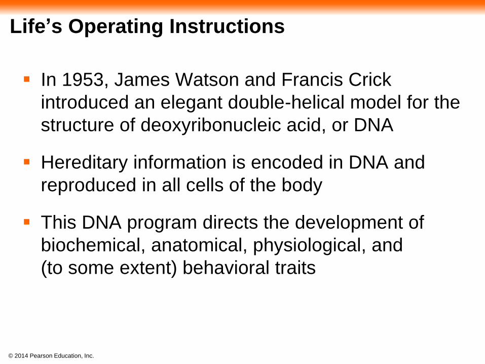
Life’s Operating Instructions
In 1953, James Watson and Francis Crick
introduced an elegant double-helical model for the
structure of deoxyribonucleic acid, or DNA
Hereditary information is encoded in DNA and
reproduced in all cells of the body
This DNA program directs the development of
biochemical, anatomical, physiological, and
(to some extent) behavioral traits
© 2014 Pearson Education, Inc.
Figure 16.1
© 2014 Pearson Education, Inc.
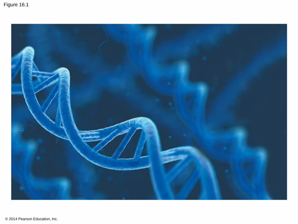
Figure 16.1a
© 2014 Pearson Education, Inc.
DNA is copied during DNA replication, and cells
can repair their DNA

© 2014 Pearson Education, Inc.
Concept 16.1: DNA is the genetic material
Early in the 20th century, the identification of the
molecules of inheritance loomed as a major
challenge to biologists
© 2014 Pearson Education, Inc.
The Search for the Genetic Material: Scientific Inquiry
When T. H. Morgan’s group showed that genes
are located on chromosomes, the two components
of chromosomes—DNA and protein—became
candidates for the genetic material
The role of DNA in heredity was first discovered
by studying bacteria and the viruses that
infect them
© 2014 Pearson Education, Inc.
Evidence That DNA Can Transform Bacteria
The discovery of the genetic role of DNA began
with research by Frederick Griffith in 1928
Griffith worked with two strains of a bacterium, one
pathogenic and one harmless
© 2014 Pearson Education, Inc.
When he mixed heat-killed remains of the
pathogenic strain with living cells of the harmless
strain, some living cells became pathogenic
He called this phenomenon transformation, now
defined as a change in genotype and phenotype
due to assimilation of foreign DNA

© 2014 Pearson Education, Inc.
Figure 16.2
Living S cells(pathogeniccontrol)
Experiment
Results
Living R cells(nonpathogeniccontrol)
Heat-killed S cells(nonpathogeniccontrol)
Mouse dies Mouse healthy Mouse healthy Mouse dies
Mixture of heat-killed S cells andliving R cells
Living S cells
© 2014 Pearson Education, Inc.
In 1944, Oswald Avery, Maclyn McCarty, and
Colin MacLeod announced that the transforming
substance was DNA
Many biologists remained skeptical, mainly
because little was known about DNA
© 2014 Pearson Education, Inc.
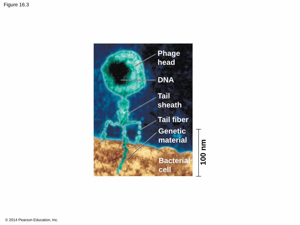
Evidence That Viral DNA Can Program Cells
More evidence for DNA as the genetic material
came from studies of viruses that infect bacteria
Such viruses, called bacteriophages (or phages),
are widely used in molecular genetics research
A virus is DNA (sometimes RNA) enclosed by a
protective coat, often simply protein
© 2014 Pearson Education, Inc.
Figure 16.3
Phage
head
DNA
Tail
sheath
Tail fiber
Genetic
material
Bacterial
cell
100 n
m
© 2014 Pearson Education, Inc.
Animation: Phage T2 Reproductive Cycle
© 2014 Pearson Education, Inc.
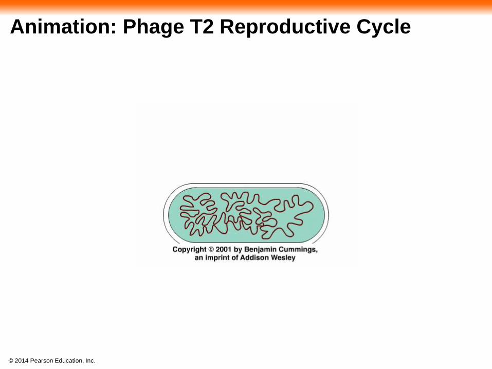
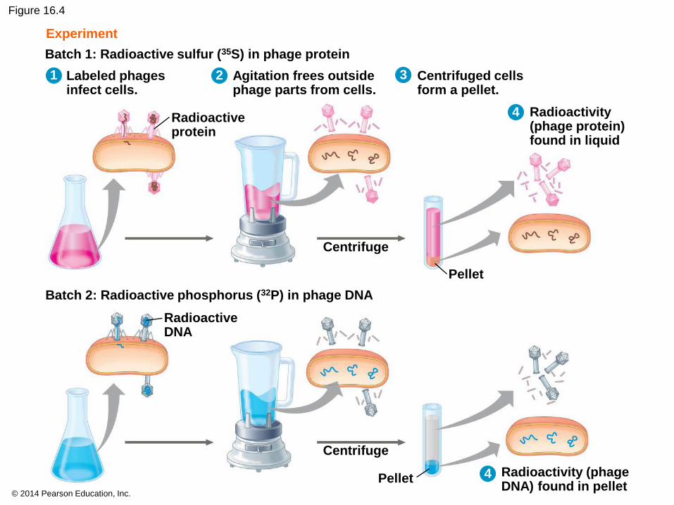
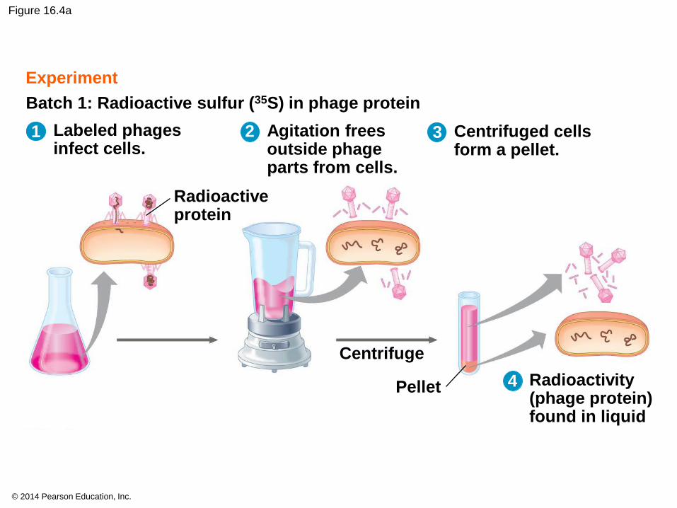
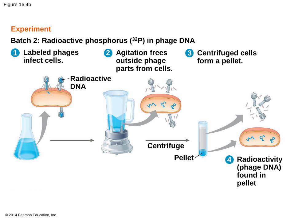
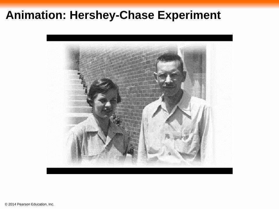
In 1952, Alfred Hershey and Martha Chase
showed that DNA is the genetic material of a
phage known as T2
They designed an experiment showing that only
one of the two components of T2 (DNA or protein)
enters an E. coli cell during infection
They concluded that the injected DNA of the
phage provides the genetic information
© 2014 Pearson Education, Inc.
Figure 16.4
Experiment
Batch 1: Radioactive sulfur (35S) in phage protein
Batch 2: Radioactive phosphorus (32P) in phage DNA
Labeled phagesinfect cells.
Agitation frees outsidephage parts from cells.
Centrifuged cellsform a pellet.
Radioactivity(phage protein)found in liquid
Pellet
Centrifuge
Radioactiveprotein
RadioactiveDNA
Radioactivity (phageDNA) found in pellet
Centrifuge
Pellet
1 2
4
3
4
© 2014 Pearson Education, Inc.
Figure 16.4a
Experiment
Batch 1: Radioactive sulfur (35S) in phage protein
Labeled phagesinfect cells.
Agitation frees outside phage parts from cells.
Centrifuged cellsform a pellet.
Radioactivity(phage protein)found in liquid
Radioactiveprotein
Pellet
Centrifuge
1 2 3
4
© 2014 Pearson Education, Inc.
Figure 16.4b
Pellet
Centrifuge
Batch 2: Radioactive phosphorus (32P) in phage DNA
RadioactiveDNA
Radioactivity (phage DNA) found in pellet
Labeled phagesinfect cells.
Agitation frees outside phage parts from cells.
Centrifuged cellsform a pellet.
Experiment
1 2 3
4
© 2014 Pearson Education, Inc.
Animation: Hershey-Chase Experiment
© 2014 Pearson Education, Inc.
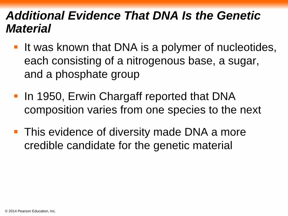
Additional Evidence That DNA Is the Genetic Material
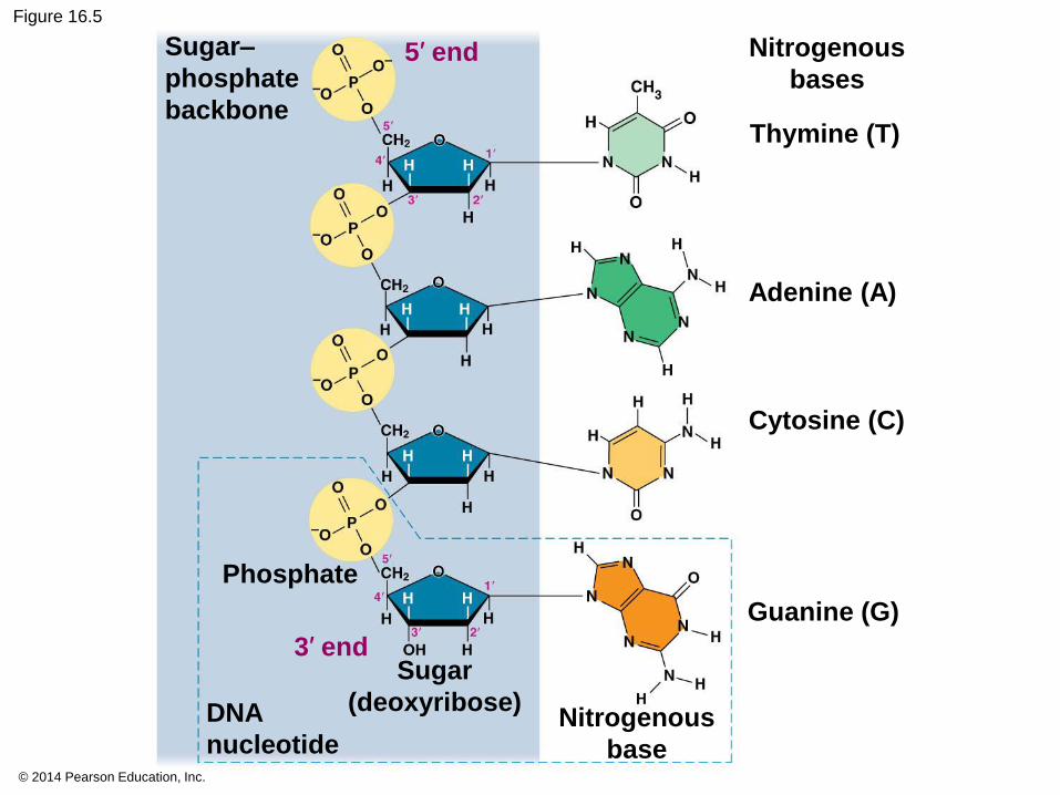
It was known that DNA is a polymer of nucleotides,
each consisting of a nitrogenous base, a sugar,
and a phosphate group
In 1950, Erwin Chargaff reported that DNA
composition varies from one species to the next
This evidence of diversity made DNA a more
credible candidate for the genetic material
© 2014 Pearson Education, Inc.
Figure 16.5
5′ end
Thymine (T)
Adenine (A)
Cytosine (C)
Guanine (G)
Nitrogenous
bases
Sugar–
phosphate
backbone
3′ end
Nitrogenous
base
Sugar
(deoxyribose)DNA
nucleotide
Phosphate
© 2014 Pearson Education, Inc.
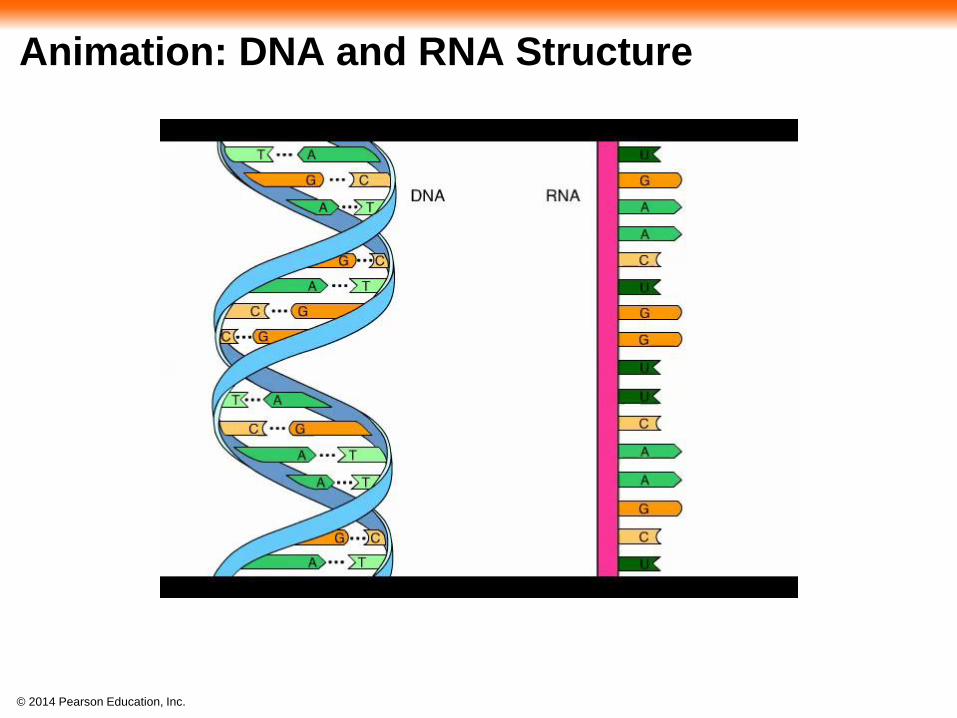
Animation: DNA and RNA Structure
© 2014 Pearson Education, Inc.
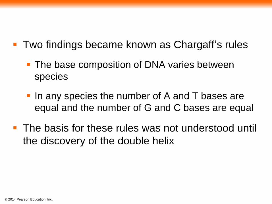
Two findings became known as Chargaff’s rules
The base composition of DNA varies between
species
In any species the number of A and T bases are
equal and the number of G and C bases are equal
The basis for these rules was not understood until
the discovery of the double helix
© 2014 Pearson Education, Inc.
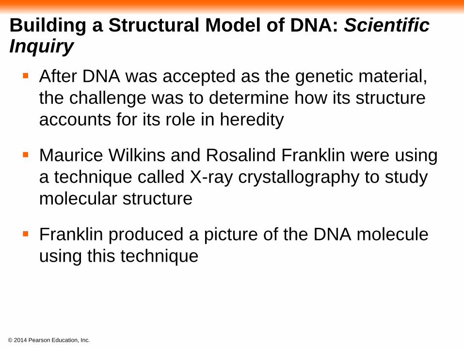
Building a Structural Model of DNA: Scientific Inquiry
After DNA was accepted as the genetic material,
the challenge was to determine how its structure
accounts for its role in heredity
Maurice Wilkins and Rosalind Franklin were using
a technique called X-ray crystallography to study
molecular structure
Franklin produced a picture of the DNA molecule
using this technique

© 2014 Pearson Education, Inc.
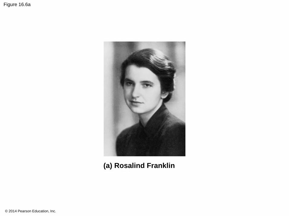
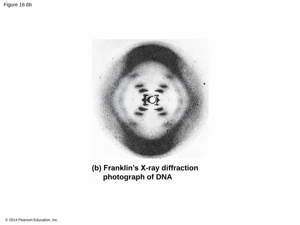
Figure 16.6
(a) Rosalind Franklin (b) Franklin’s X-ray diffraction
photograph of DNA
© 2014 Pearson Education, Inc.
Figure 16.6a
(a) Rosalind Franklin
© 2014 Pearson Education, Inc.
Figure 16.6b
(b) Franklin’s X-ray diffraction
photograph of DNA
© 2014 Pearson Education, Inc.
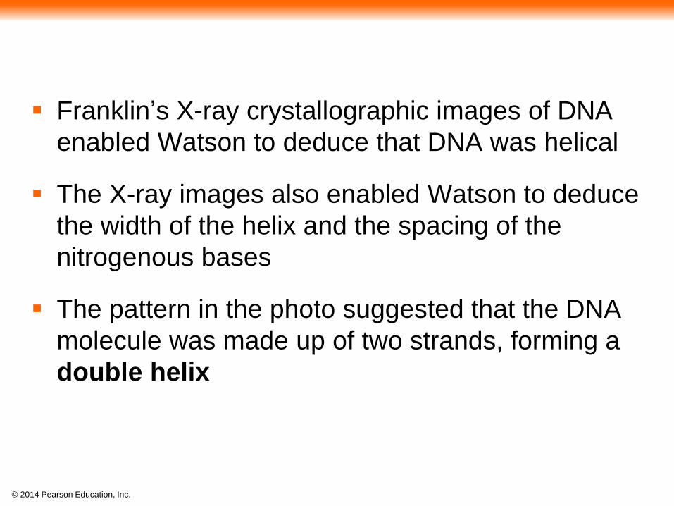
Franklin’s X-ray crystallographic images of DNA
enabled Watson to deduce that DNA was helical
The X-ray images also enabled Watson to deduce
the width of the helix and the spacing of the
nitrogenous bases
The pattern in the photo suggested that the DNA
molecule was made up of two strands, forming a
double helix

© 2014 Pearson Education, Inc.
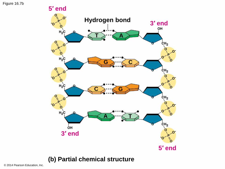
Figure 16.7
(a) Key features of
DNA structure
(b) Partial chemical structure
0.34 nm
3′ end
5′ endT
T
T
A
A
A
C
C
C
G
G
G
AT1 nm
TA
C G
CG
AT3.4 nm
CG
CG
C G
C G
3′ end
5′ end
Hydrogen bond
T A
G C
A T
C G
(c) Space-filling
model

© 2014 Pearson Education, Inc.
Figure 16.7a
(a) Key features of DNA structure
0.34 nm
1 nm
3.4 nm
T
T
T
A
A
A
C
C
C
G
G
G
AT
TA
C G
CG
AT
CG
CG
C G
C G
© 2014 Pearson Education, Inc.
Figure 16.7b
(b) Partial chemical structure
3′ end
5′ end
3′ end
5′ end
Hydrogen bond
T A
G C
A T
C G
© 2014 Pearson Education, Inc.
Figure 16.7c
(c) Space-filling model

© 2014 Pearson Education, Inc.
Animation: DNA Double Helix
© 2014 Pearson Education, Inc.
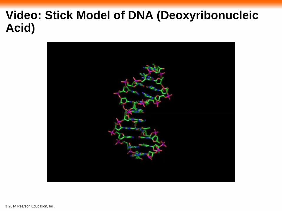
Video: Stick Model of DNA (Deoxyribonucleic Acid)
© 2014 Pearson Education, Inc.
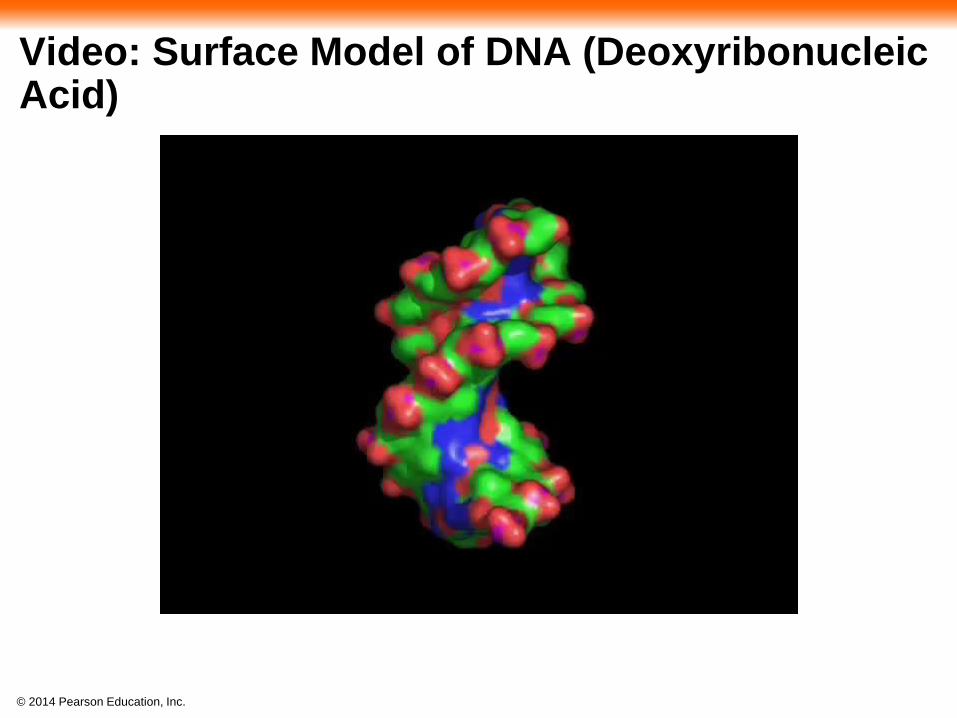
Video: Surface Model of DNA (Deoxyribonucleic Acid)
© 2014 Pearson Education, Inc.
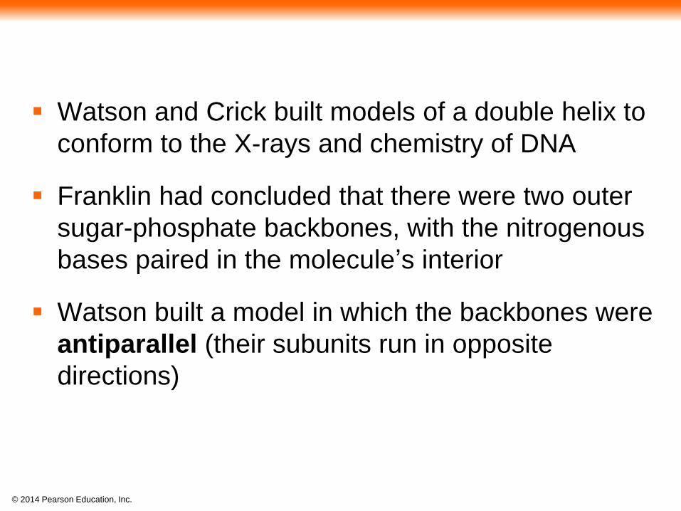
Watson and Crick built models of a double helix to
conform to the X-rays and chemistry of DNA
Franklin had concluded that there were two outer
sugar-phosphate backbones, with the nitrogenous
bases paired in the molecule’s interior
Watson built a model in which the backbones were
antiparallel (their subunits run in opposite
directions)
© 2014 Pearson Education, Inc.
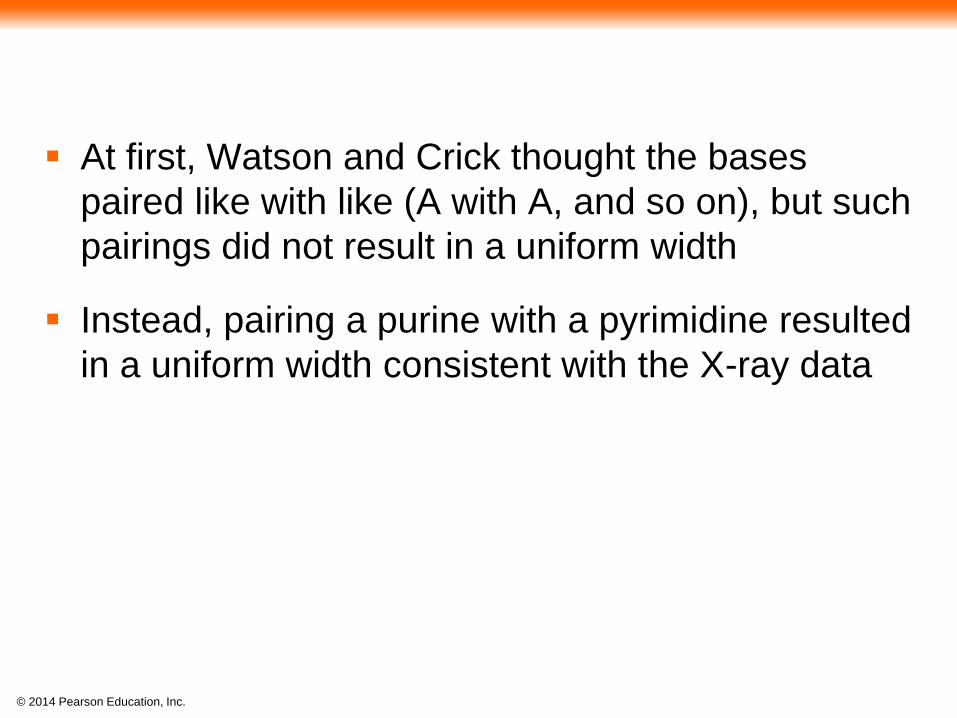
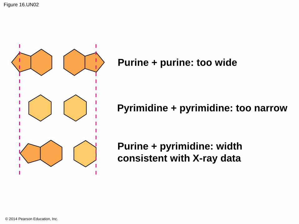
At first, Watson and Crick thought the bases
paired like with like (A with A, and so on), but such
pairings did not result in a uniform width
Instead, pairing a purine with a pyrimidine resulted
in a uniform width consistent with the X-ray data
© 2014 Pearson Education, Inc.
Figure 16.UN02
Purine + purine: too wide
Pyrimidine + pyrimidine: too narrow
Purine + pyrimidine: width
consistent with X-ray data

© 2014 Pearson Education, Inc.
Watson and Crick reasoned that the pairing was
more specific, dictated by the base structures
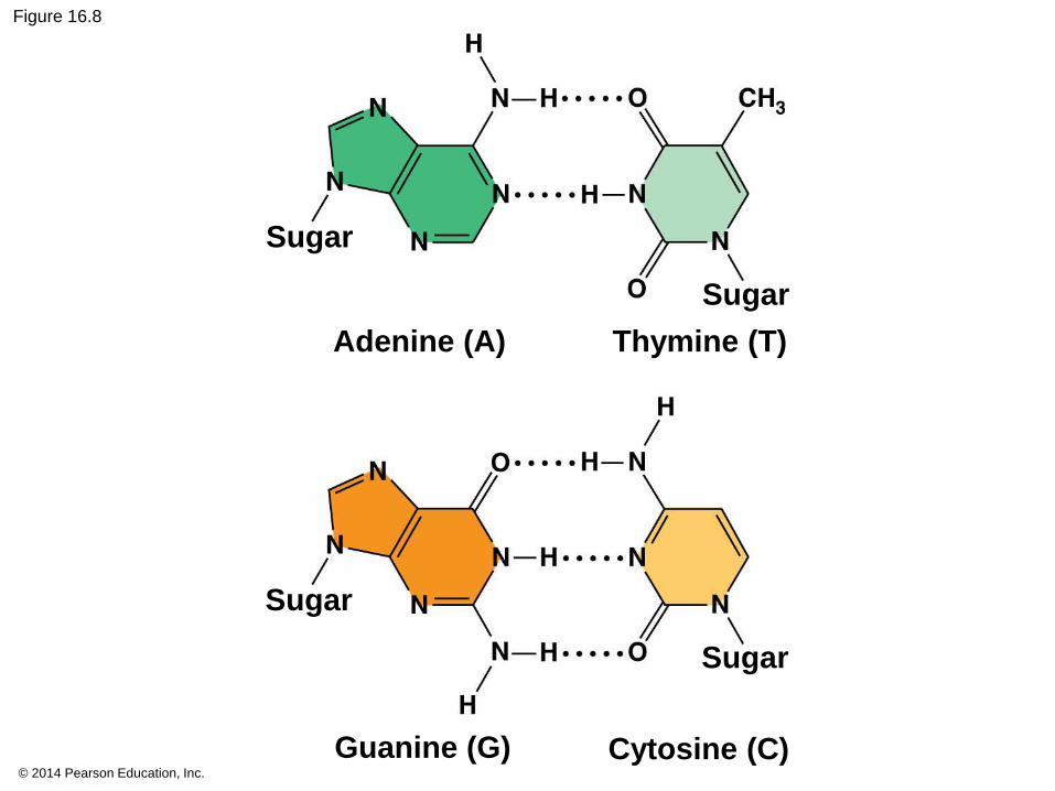
They determined that adenine (A) paired only with
thymine (T), and guanine (G) paired only with
cytosine (C)
The Watson-Crick model explains Chargaff’s
rules: in any organism the amount of A = T, and
the amount of G = C
© 2014 Pearson Education, Inc.
Figure 16.8
Sugar
Sugar
Adenine (A)
Sugar
Sugar
Thymine (T)
Cytosine (C)Guanine (G)
© 2014 Pearson Education, Inc.
Concept 16.2: Many proteins work together in DNA replication and repair
The relationship between structure and function is
manifest in the double helix
Watson and Crick noted that the specific base
pairing suggested a possible copying mechanism
for genetic material
© 2014 Pearson Education, Inc.
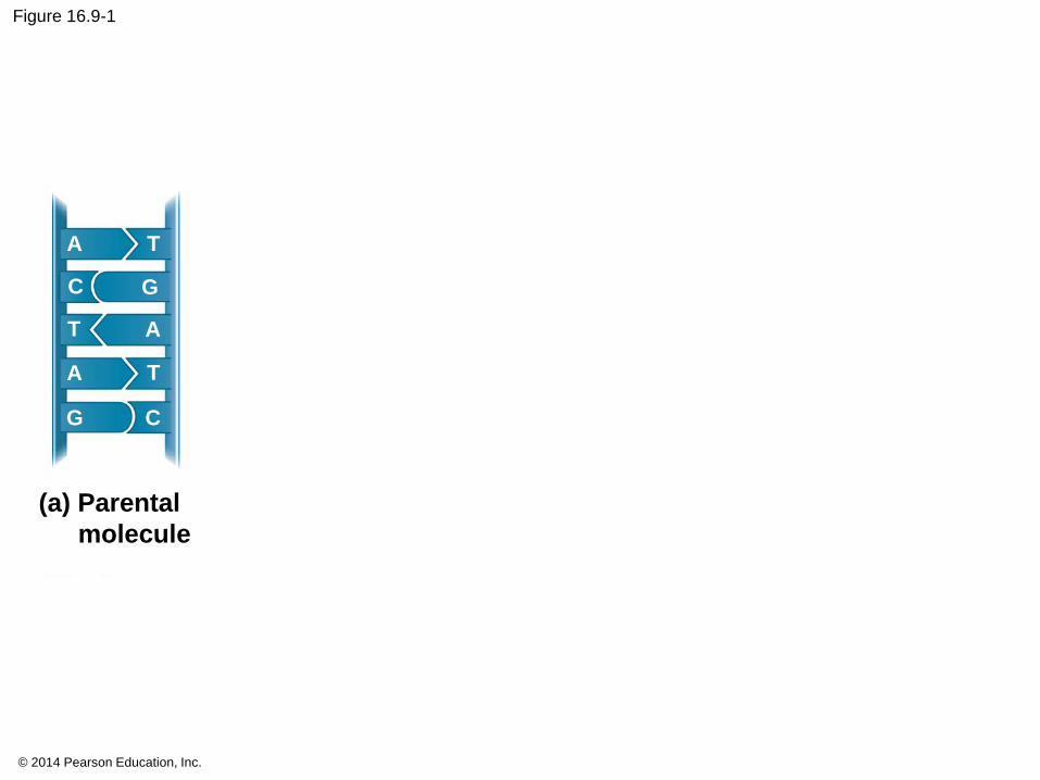
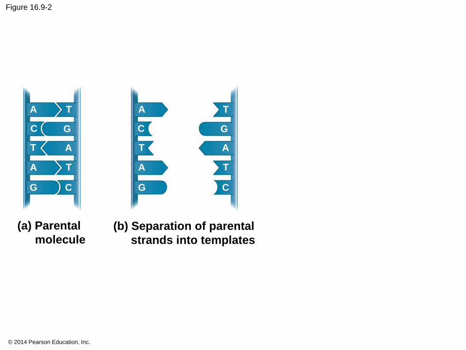
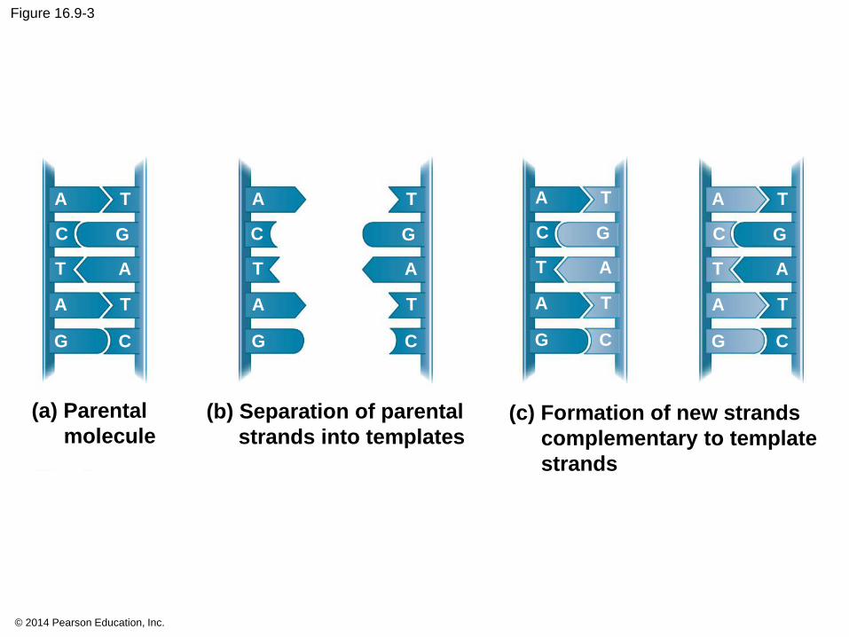
The Basic Principle: Base Pairing to a Template Strand
Since the two strands of DNA are complementary,
each strand acts as a template for building a new
strand in replication
In DNA replication, the parent molecule unwinds,
and two new daughter strands are built based on
base-pairing rules
© 2014 Pearson Education, Inc.
Figure 16.9-1
(a) Parental
molecule
A
A
A
T
T
T
G
G C
C
© 2014 Pearson Education, Inc.
Figure 16.9-2
(a) Parental
molecule(b) Separation of parental
strands into templates
A
A
A
T
T
T
G
G C
C
A
A
A
T
T
T
G
G C
C
© 2014 Pearson Education, Inc.
Figure 16.9-3
(a) Parental
molecule(b) Separation of parental
strands into templates
A
A
A
T
T
T
G
G C
C
A
A
A
T
T
T
G
G C
C
A
A
A
T
T
T
G
G C
C
A
A
A
T
T
T
G
G C
C
(c) Formation of new strands
complementary to template
strands
© 2014 Pearson Education, Inc.
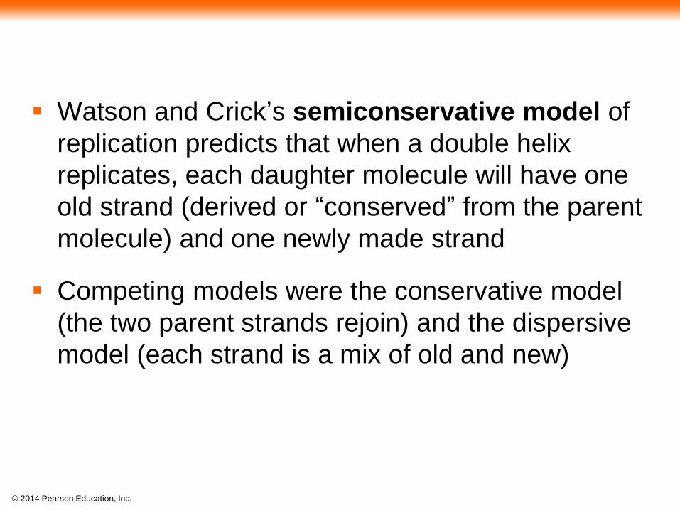
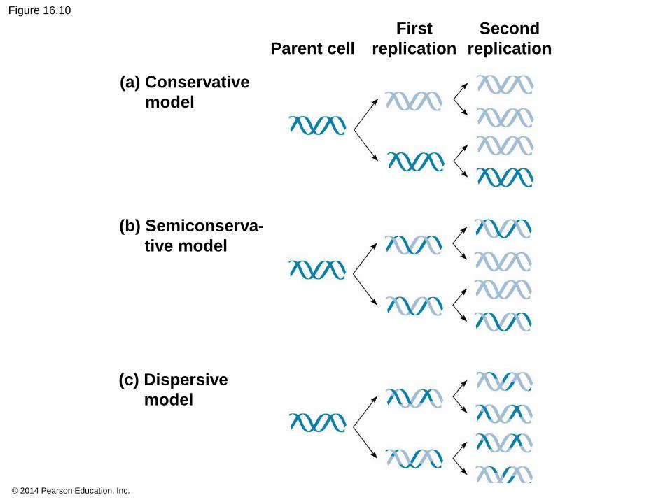
Watson and Crick’s semiconservative model of
replication predicts that when a double helix
replicates, each daughter molecule will have one
old strand (derived or “conserved” from the parent
molecule) and one newly made strand
Competing models were the conservative model
(the two parent strands rejoin) and the dispersive
model (each strand is a mix of old and new)
© 2014 Pearson Education, Inc.
Figure 16.10
(a) Conservative
model
(b) Semiconserva-
tive model
(c) Dispersive
model
Parent cell
First
replication
Second
replication

© 2014 Pearson Education, Inc.
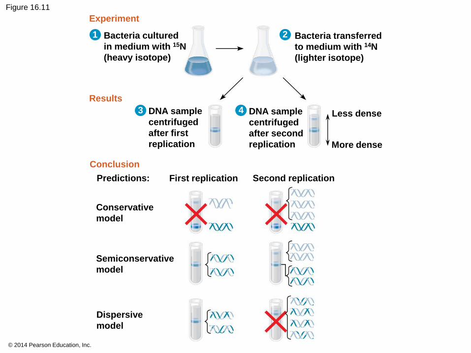
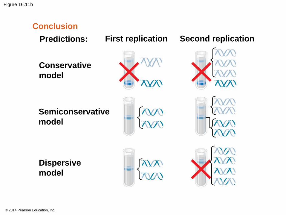
Experiments by Matthew Meselson and Franklin
Stahl supported the semiconservative model
They labeled the nucleotides of the old strands
with a heavy isotope of nitrogen, while any new
nucleotides were labeled with a lighter isotope
© 2014 Pearson Education, Inc.
The first replication produced a band of hybrid
DNA, eliminating the conservative model
A second replication produced both light and
hybrid DNA, eliminating the dispersive model and
supporting the semiconservative model
© 2014 Pearson Education, Inc.
Figure 16.11
Bacteria cultured
in medium with 15N
(heavy isotope)
Experiment
Results
Conclusion
Bacteria transferred
to medium with 14N
(lighter isotope)
DNA sample
centrifuged
after first
replication
DNA sample
centrifuged
after second
replication
Less dense
More dense
Predictions: First replication Second replication
Conservative
model
Semiconservative
model
Dispersive
model
1 2
43

© 2014 Pearson Education, Inc.
Figure 16.11a
Bacteria cultured
in medium with 15N
(heavy isotope)
Experiment
Results
Bacteria transferred
to medium with 14N
(lighter isotope)
DNA samplecentrifugedafter firstreplication
DNA samplecentrifugedafter secondreplication
Less dense
More dense
1 2
3 4
© 2014 Pearson Education, Inc.
Figure 16.11b
Conclusion
Predictions: First replication Second replication
Conservative
model
Semiconservative
model
Dispersive
model
© 2014 Pearson Education, Inc.
DNA Replication: A Closer Look
The copying of DNA is remarkable in its speed
and accuracy
More than a dozen enzymes and other proteins
participate in DNA replication
© 2014 Pearson Education, Inc.
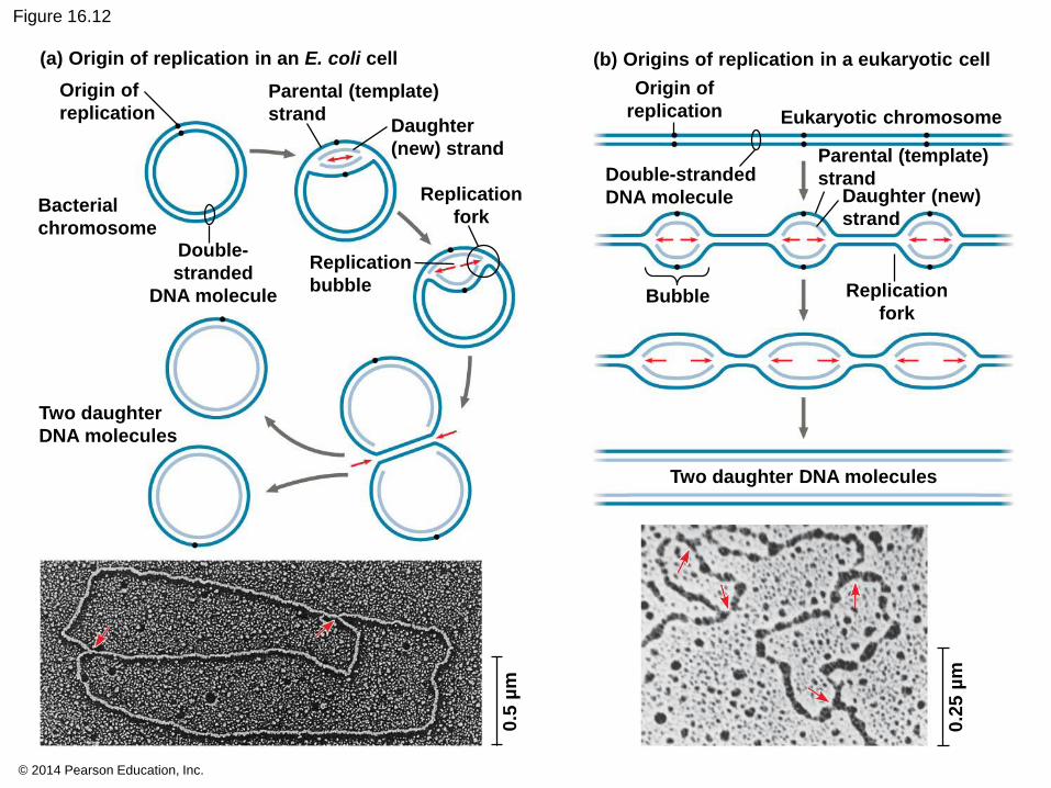
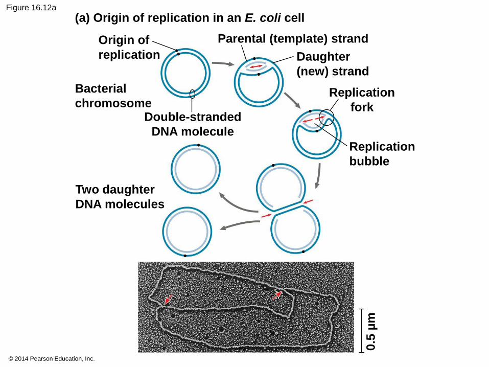
Getting Started
Replication begins at particular sites called
origins of replication, where the two DNA
strands are separated, opening up a replication
“bubble”
A eukaryotic chromosome may have hundreds or
even thousands of origins of replication
Replication proceeds in both directions from each
origin, until the entire molecule is copied
© 2014 Pearson Education, Inc.
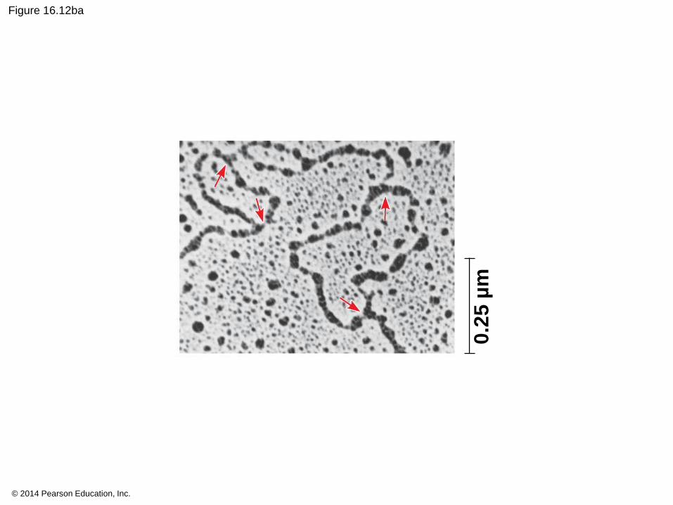
Figure 16.12
Origin of
replication
0.5
µm
0.2
5 µ
m
Bacterial
chromosome
Two daughter
DNA molecules
Replication
bubble
Parental (template)
strandDaughter
(new) strand
Replication
fork
Double-
stranded
DNA molecule
(a) Origin of replication in an E. coli cell (b) Origins of replication in a eukaryotic cell
Origin of
replication Eukaryotic chromosome
Double-stranded
DNA molecule
Parental (template)
strandDaughter (new)
strand
Replication
forkBubble
Two daughter DNA molecules
© 2014 Pearson Education, Inc.
Figure 16.12a
0.5
µm
Origin of
replication
Bacterial
chromosome
Two daughter
DNA molecules
Replication
bubble
Parental (template) strand
Daughter
(new) strand
Replication
forkDouble-stranded
DNA molecule
(a) Origin of replication in an E. coli cell
© 2014 Pearson Education, Inc.
Figure 16.12aa
0.5
µm

© 2014 Pearson Education, Inc.
Figure 16.12b
0.2
5 µ
m
(b) Origins of replication in a eukaryotic cell
Origin of replication Eukaryotic chromosome
Double-stranded
DNA molecule
Parental (template) strand
Daughter (new)
strand
Replication
forkBubble
Two daughter DNA molecules
© 2014 Pearson Education, Inc.
Figure 16.12ba
0.2
5 µ
m
© 2014 Pearson Education, Inc.
Animation: Origins of Replication
© 2014 Pearson Education, Inc.
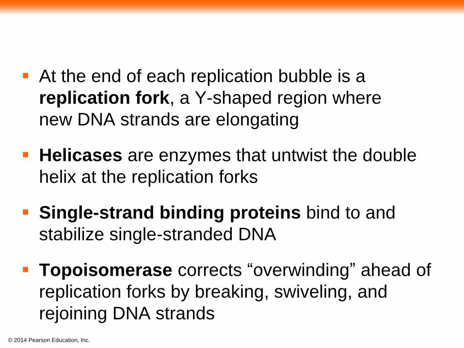
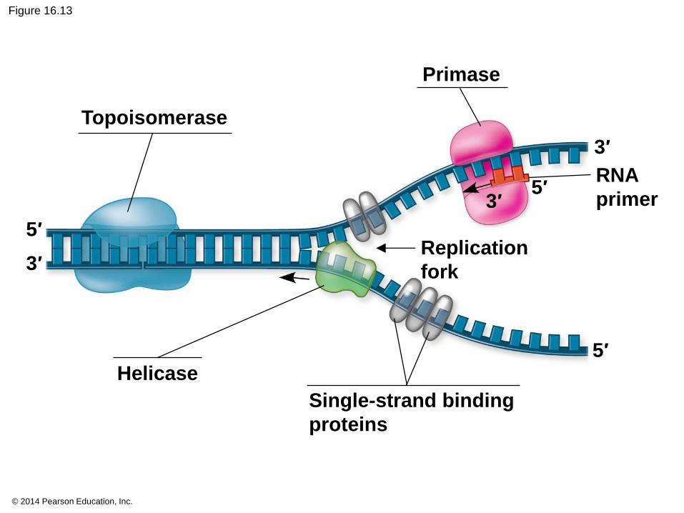
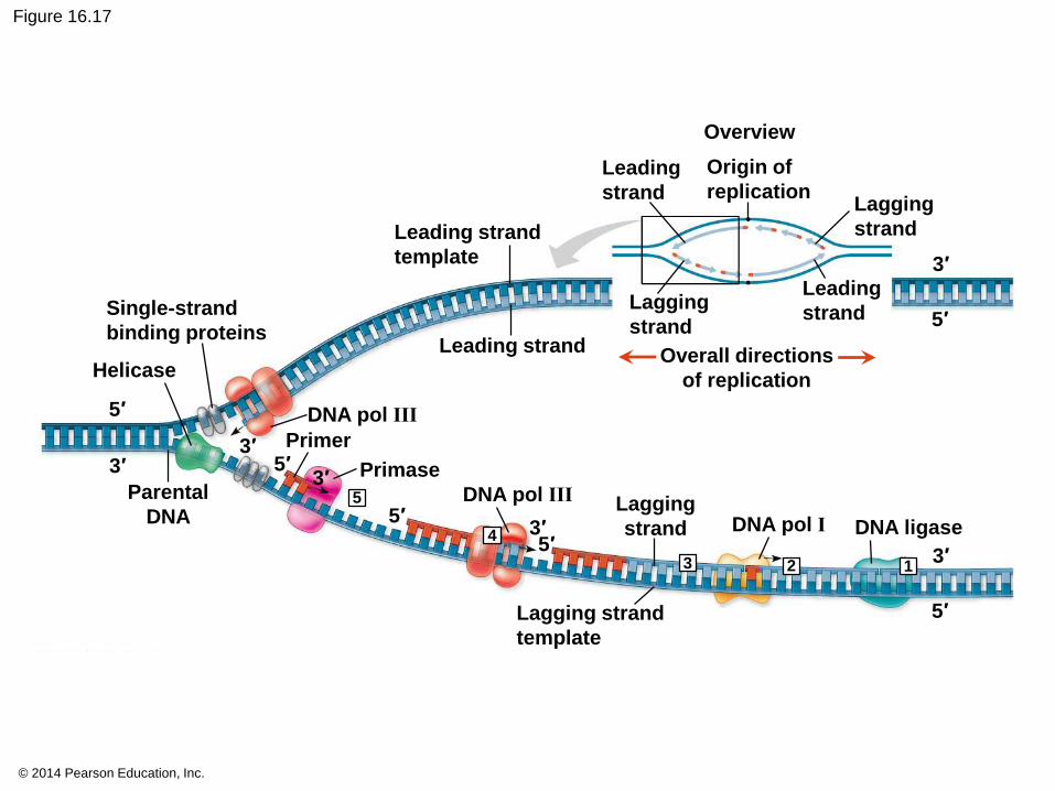
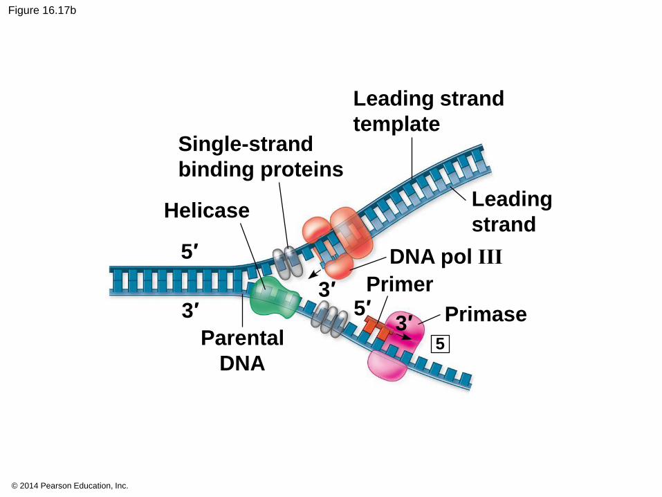
At the end of each replication bubble is a
replication fork, a Y-shaped region where
new DNA strands are elongating
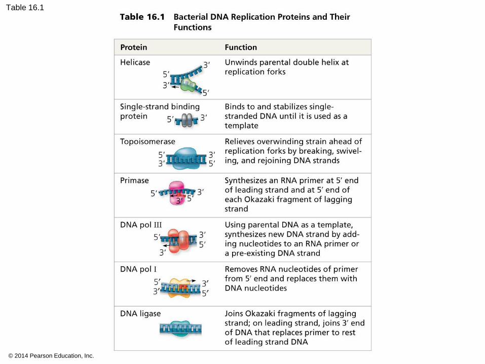
Helicases are enzymes that untwist the double
helix at the replication forks
Single-strand binding proteins bind to and
stabilize single-stranded DNA
Topoisomerase corrects “overwinding” ahead of
replication forks by breaking, swiveling, and
rejoining DNA strands
© 2014 Pearson Education, Inc.
Figure 16.13
Topoisomerase
Primase
RNA
primer
Replication
fork
5′
3′
5′
5′
3′
Helicase
Single-strand binding
proteins
3′
© 2014 Pearson Education, Inc.
DNA polymerases cannot initiate synthesis of a
polynucleotide; they can only add nucleotides to
an existing 3′ end
The initial nucleotide strand is a short RNA primer
© 2014 Pearson Education, Inc.
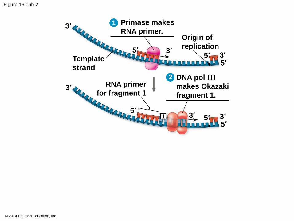
An enzyme called primase can start an RNA
chain from scratch and adds RNA nucleotides one
at a time using the parental DNA as a template
The primer is short (5–10 nucleotides long), and
the 3′ end serves as the starting point for the new
DNA strand
© 2014 Pearson Education, Inc.
Synthesizing a New DNA Strand
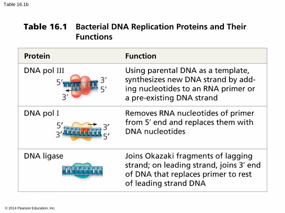
Enzymes called DNA polymerases catalyze the
elongation of new DNA at a replication fork
Most DNA polymerases require a primer and a
DNA template strand
The rate of elongation is about 500 nucleotides
per second in bacteria and 50 per second in
human cells

© 2014 Pearson Education, Inc.
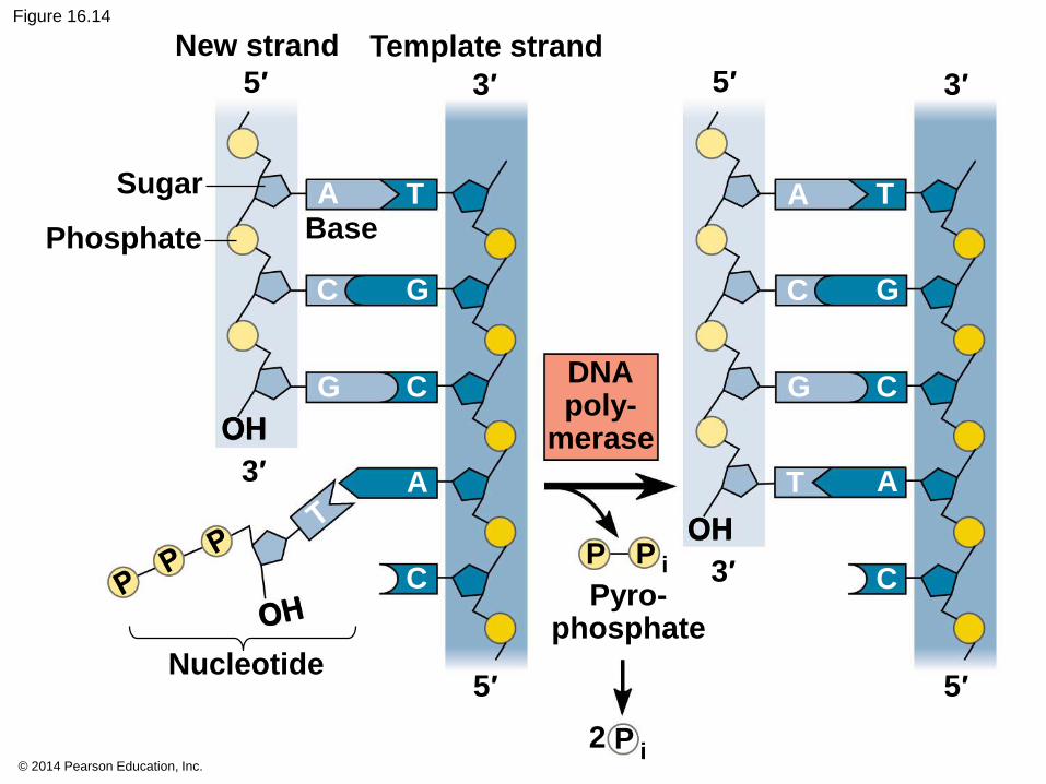
Each nucleotide that is added to a growing DNA
strand is a nucleoside triphosphate
dATP supplies adenine to DNA and is similar to
the ATP of energy metabolism
The difference is in their sugars: dATP has
deoxyribose while ATP has ribose
As each monomer of dATP joins the DNA strand, it
loses two phosphate groups as a molecule of
pyrophosphate
© 2014 Pearson Education, Inc.
Figure 16.14
5′ 5′3′
3′
5′ 5′
New strand Template strand
Sugar
Phosphate Base
OH
A T
C
C
G
G
A
C
Nucleotide
DNApoly-
merase
OH
Pyro-phosphate
2 P i
P iP
3′
3′
A T
C
C
G
G
A
C
T
© 2014 Pearson Education, Inc.
Antiparallel Elongation
The antiparallel structure of the double helix
affects replication
DNA polymerases add nucleotides only to the free
3′ end of a growing strand; therefore, a new DNA
strand can elongate only in the 5′ to 3′ direction
© 2014 Pearson Education, Inc.
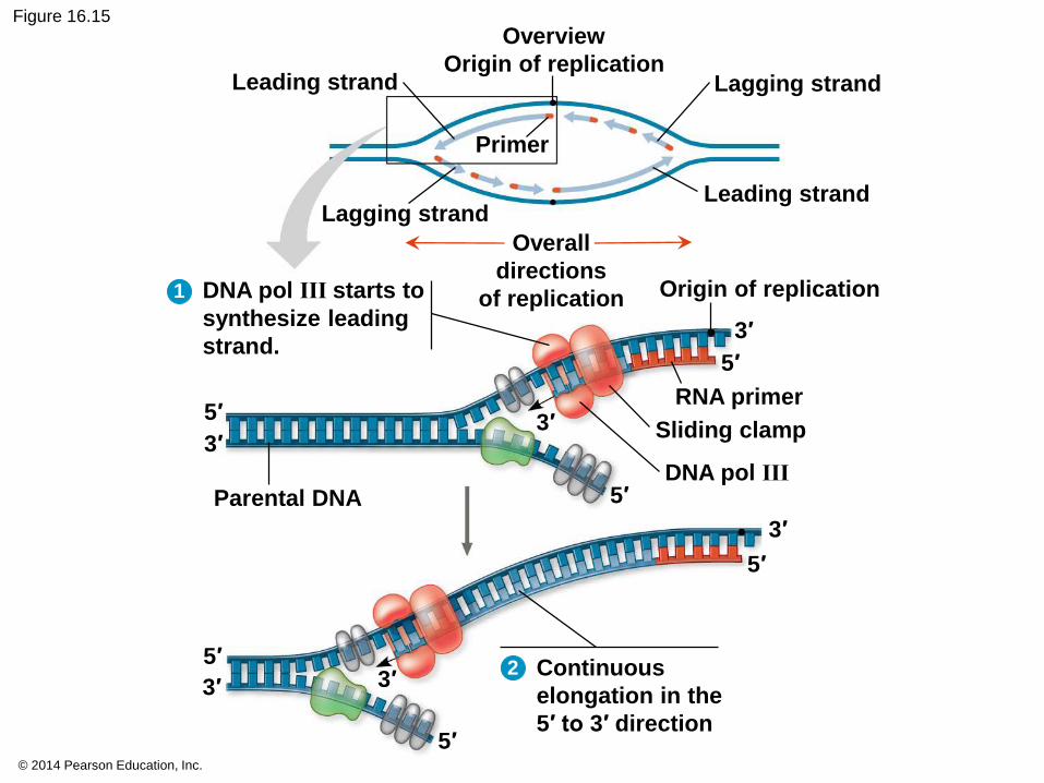
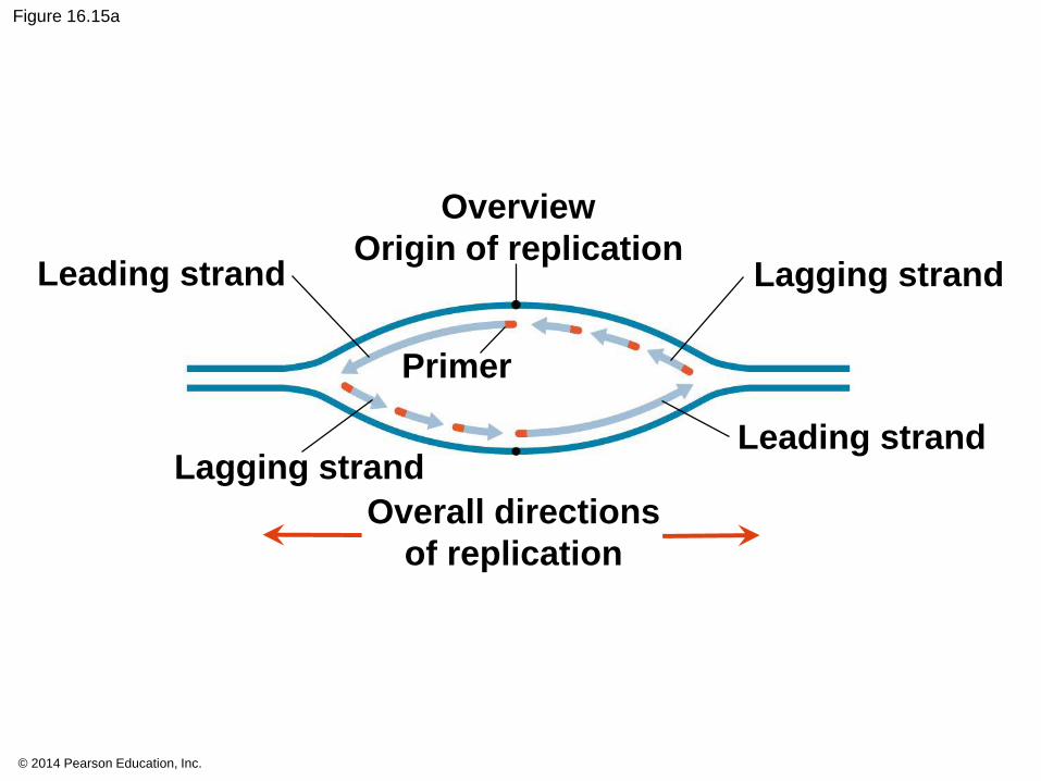
Figure 16.15
Leading strand Lagging strand
Leading strand
Parental DNA
Lagging strand
Primer
Overview
Origin of replication
Overall
directions
of replication Origin of replication
RNA primer
Sliding clamp
DNA pol III
Continuous
elongation in the
5′ to 3′ direction
5′
5′
5′
5′
5′
5′
3′ 3′
3′3′
3′
3′
DNA pol III starts to
synthesize leading
strand.
1
2
© 2014 Pearson Education, Inc.
Figure 16.15a
Leading strand Lagging strand
Leading strandLagging strand
Primer
Overview
Origin of replication
Overall directions
of replication

© 2014 Pearson Education, Inc.
Figure 16.15b
Parental DNA
Origin of replication
RNA primer
Sliding clamp
DNA pol III
5′
5′
5′
3′ 3′
3′
DNA pol III starts to
synthesize leading
strand.
1
5′
3′
5′
3′
5′
3′
2 Continuous
elongation in the
5′ to 3′ direction
© 2014 Pearson Education, Inc.
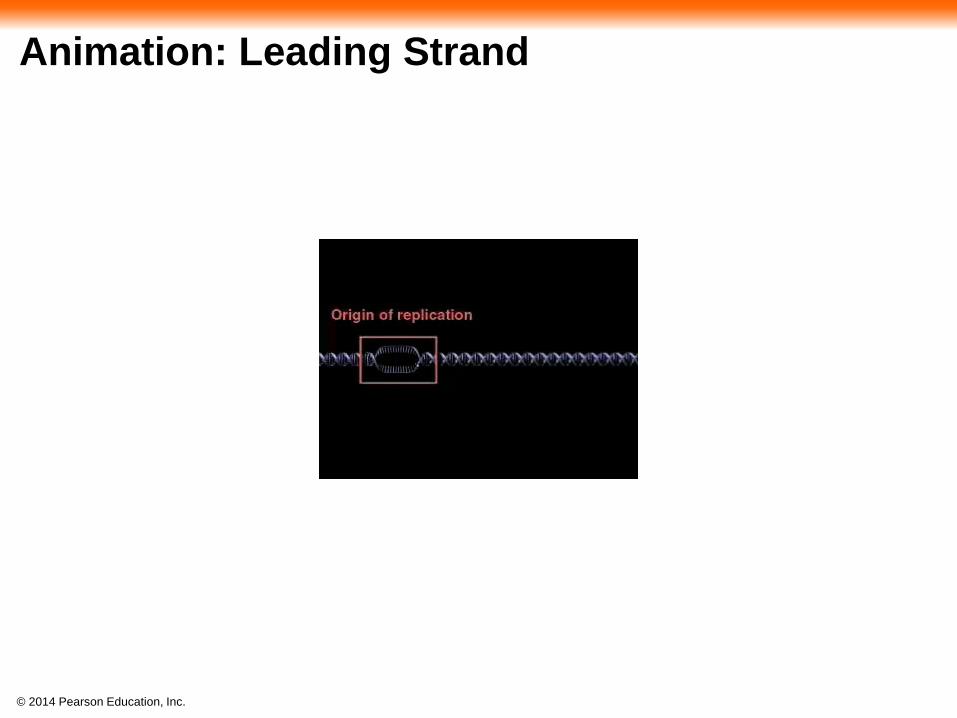
Animation: Leading Strand
© 2014 Pearson Education, Inc.
Along one template strand of DNA, the DNA
polymerase synthesizes a leading strand
continuously, moving toward the replication fork
© 2014 Pearson Education, Inc.
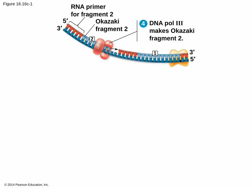
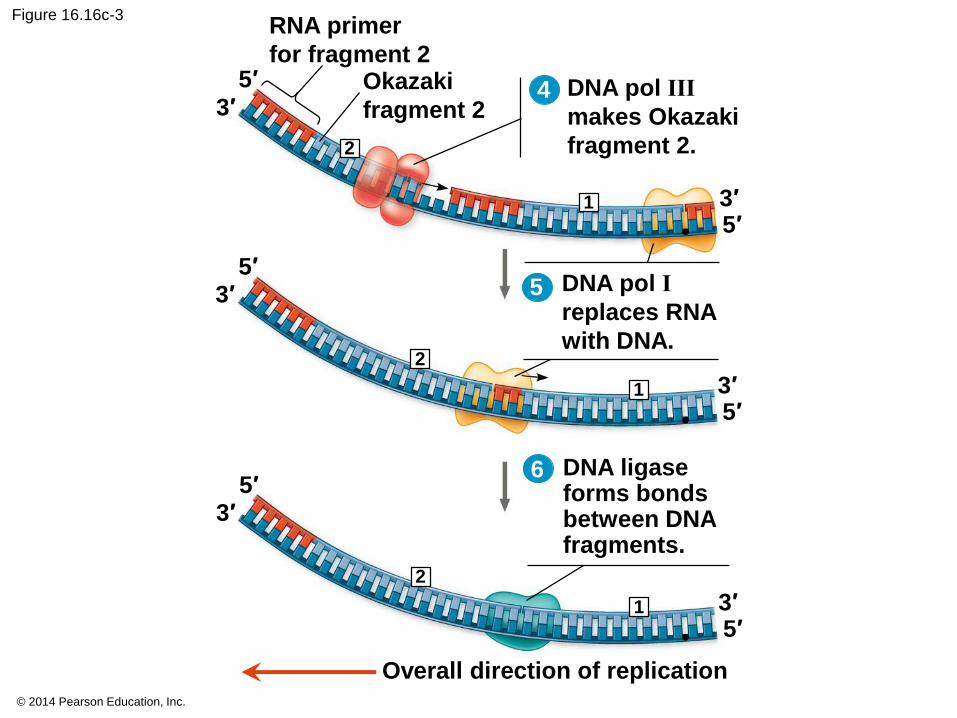
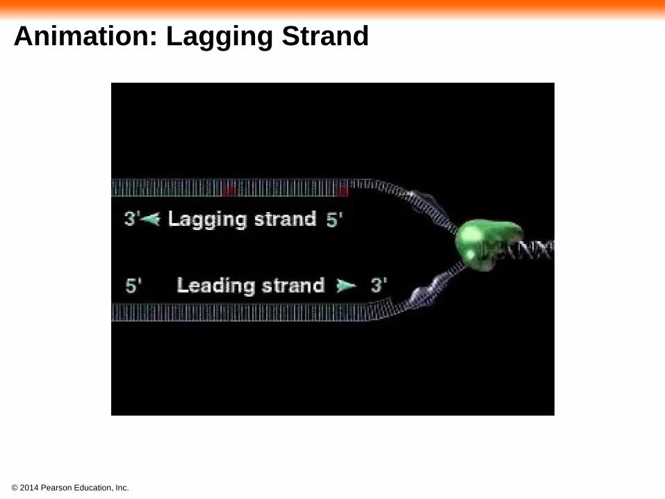
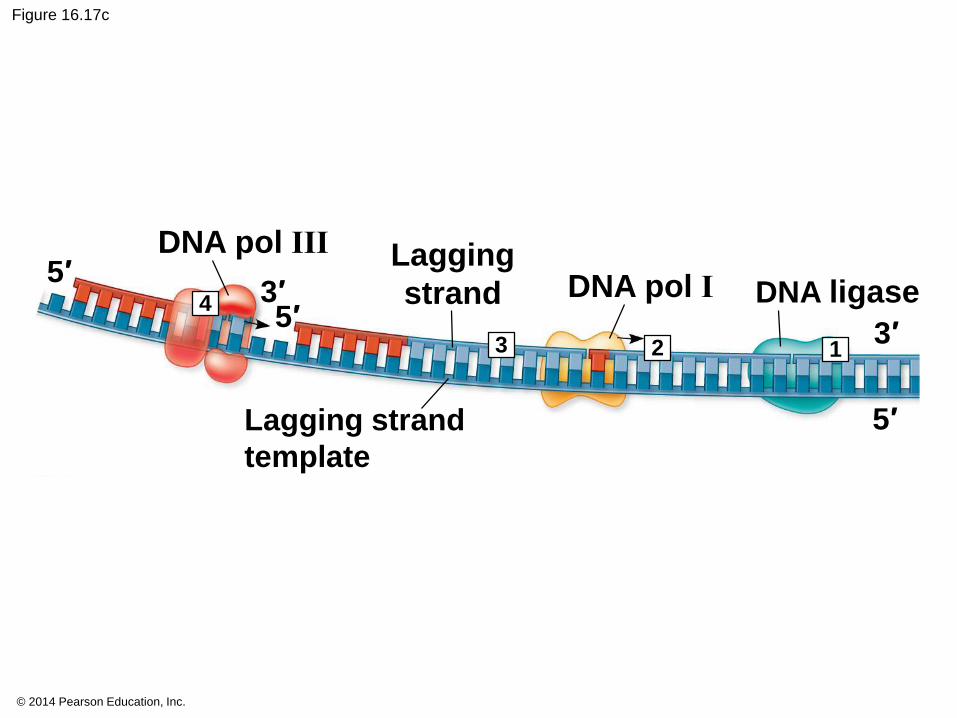
To elongate the other new strand, called the
lagging strand, DNA polymerase must work in
the direction away from the replication fork
The lagging strand is synthesized as a series of
segments called Okazaki fragments, which are
joined together by DNA ligase
© 2014 Pearson Education, Inc.
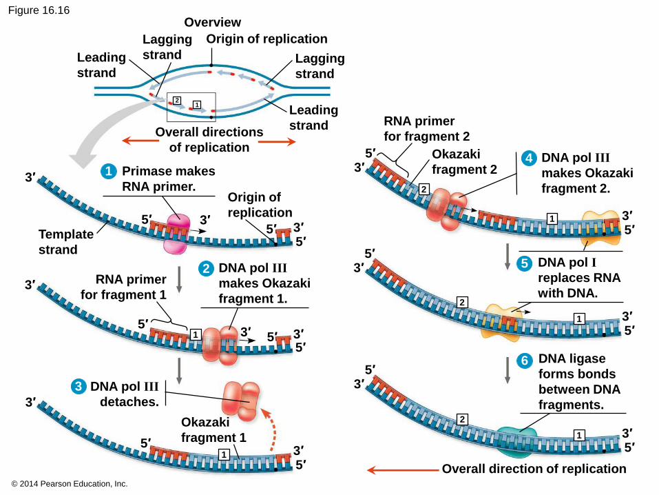
Figure 16.16Overview
12
1
1
2
1
2
1
1
2
5′
3′
3′
5′5′3′
3′
3′ 3′5′
5′5′
5′3′
3′
5′3′
5′3′
5′3′
5′3′
5′3′
5′3′
1
2 5
6
4
3
Origin of replication
Lagging
strand
Leading
strand
Lagging
strand
Leading
strandOverall directions
of replication
RNA primer
for fragment 2
Okazaki
fragment 2DNA pol III
makes Okazaki
fragment 2.
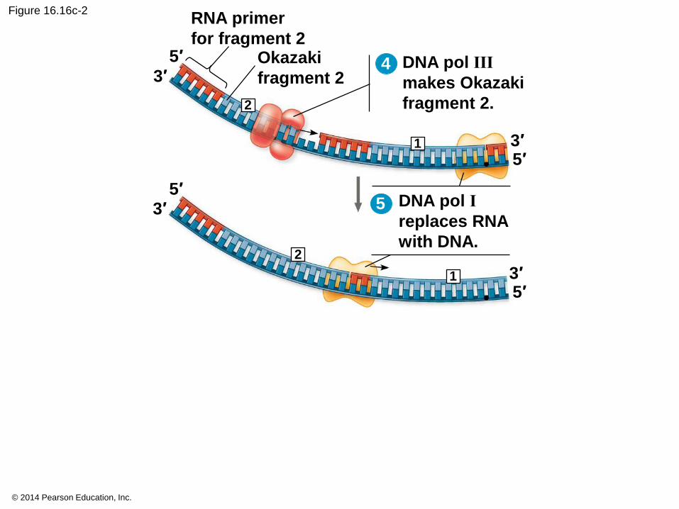
DNA pol I
replaces RNA
with DNA.
DNA ligase
forms bonds
between DNA
fragments.
Overall direction of replication
Okazaki
fragment 1
DNA pol III
detaches.
RNA primer
for fragment 1
Template
strand
DNA pol III
makes Okazaki
fragment 1.
Origin of
replication
Primase makes
RNA primer.
5′

© 2014 Pearson Education, Inc.
Figure 16.16a
12
Overview
Origin of replication
Lagging
strand
Leading
strand
Lagging
strand
Leading
strandOverall directions
of replication
© 2014 Pearson Education, Inc.
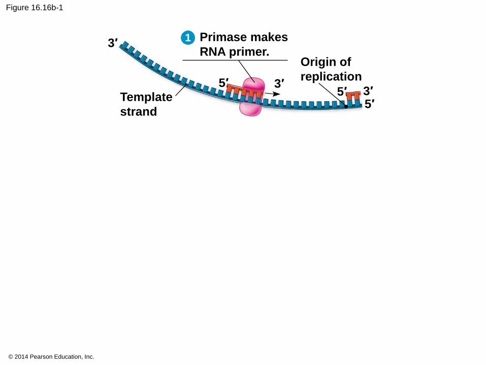
Figure 16.16b-1
5′
3′
5′5′3′
3′Template
strand
Origin of
replication
Primase makes
RNA primer.
1
© 2014 Pearson Education, Inc.
Figure 16.16b-2
5′
3′
5′5′3′
3′Template
strand
Origin of
replication
Primase makes
RNA primer.
1
5′3′
5′
3′
5′3′
RNA primer
for fragment 1
DNA pol III
makes Okazaki
fragment 1.
1
2
© 2014 Pearson Education, Inc.
Figure 16.16b-3
5′
3′
5′5′3′
3′Template
strand
Origin of
replication
Primase makes
RNA primer.
1
5′3′
5′
3′
5′3′
RNA primer
for fragment 1
DNA pol III
makes Okazaki
fragment 1.
1
2
1
3
3′
3′5′
5′
Okazaki
fragment 1
DNA pol III
detaches.
© 2014 Pearson Education, Inc.
DNA pol III
makes Okazaki
fragment 2.
Figure 16.16c-1
2
5′
3′
5′3′
4
RNA primer
for fragment 2Okazaki
fragment 2
1
© 2014 Pearson Education, Inc.
Figure 16.16c-2
DNA pol III
makes Okazaki
fragment 2.2
5′
3′
5′3′
4
RNA primer
for fragment 2Okazaki
fragment 2
1
5′3′
DNA pol I
replaces RNA
with DNA.
5′3′
1
2
5
© 2014 Pearson Education, Inc.
Figure 16.16c-3
DNA ligaseforms bondsbetween DNAfragments.
Overall direction of replication
DNA pol III
makes Okazaki
fragment 2.2
5′
3′
5′3′
4
RNA primer
for fragment 2Okazaki
fragment 2
1
5′3′
DNA pol I
replaces RNA
with DNA.
5′3′
1
2
5
6
5′3′
5′3′
1
2
© 2014 Pearson Education, Inc.
Animation: Lagging Strand
© 2014 Pearson Education, Inc.
Figure 16.17
Overview
5′
3′
Lagging
strand
Leading
strand
Leading
strand
Lagging
strandLeading strand
Leading strand
template
Origin of
replication
Overall directions
of replication
5′
3′
5′3′
5′
5′
3′
3′3′
Single-strand
binding proteins
Helicase
Parental
DNA
DNA pol III
Primer
Primase
Lagging
strand
Lagging strand
template
DNA pol III
DNA pol I5′
DNA ligase
123
4
5

© 2014 Pearson Education, Inc.
Figure 16.17a
Overview
Lagging
strand
Leading
strand
Leading
strand
Lagging
strand
Origin of
replication
Overall directions
of replication
© 2014 Pearson Education, Inc.
Figure 16.17b
5′
5′
3′
3′3′
Single-strand
binding proteins
Helicase
Parental
DNA
DNA pol III
Primer
Primase
Leading
strand
Leading strand
template
5
© 2014 Pearson Education, Inc.
Figure 16.17c
4
3 2 1
5′
5′3′
5′
3′
Lagging
strand
DNA pol III
DNA pol I DNA ligase
Lagging strand
template

© 2014 Pearson Education, Inc.
Animation: DNA Replication Overview
© 2014 Pearson Education, Inc.
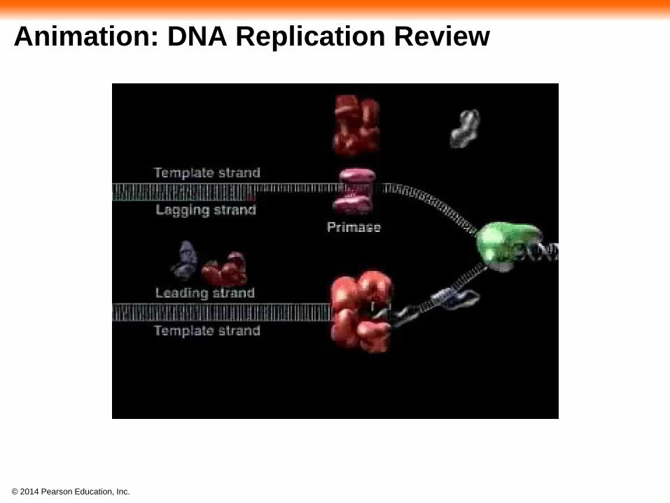
Animation: DNA Replication Review
© 2014 Pearson Education, Inc.
Table 16.1

© 2014 Pearson Education, Inc.
Table 16.1a
© 2014 Pearson Education, Inc.
Table 16.1b
© 2014 Pearson Education, Inc.
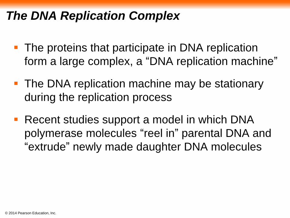
The DNA Replication Complex
The proteins that participate in DNA replication
form a large complex, a “DNA replication machine”
The DNA replication machine may be stationary
during the replication process
Recent studies support a model in which DNA
polymerase molecules “reel in” parental DNA and
“extrude” newly made daughter DNA molecules
© 2014 Pearson Education, Inc.
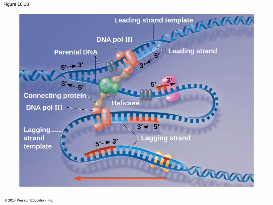
Figure 16.18
Leading strand template
5′
5′
5′
5′3′
3′
3′
3′
3′ 3′
5′
5′
Leading strand
Lagging strand
Lagging
strand
template
DNA pol III
Connecting proteinHelicase
Parental DNA
DNA pol III
© 2014 Pearson Education, Inc.
BioFlix: DNA Replication

© 2014 Pearson Education, Inc.
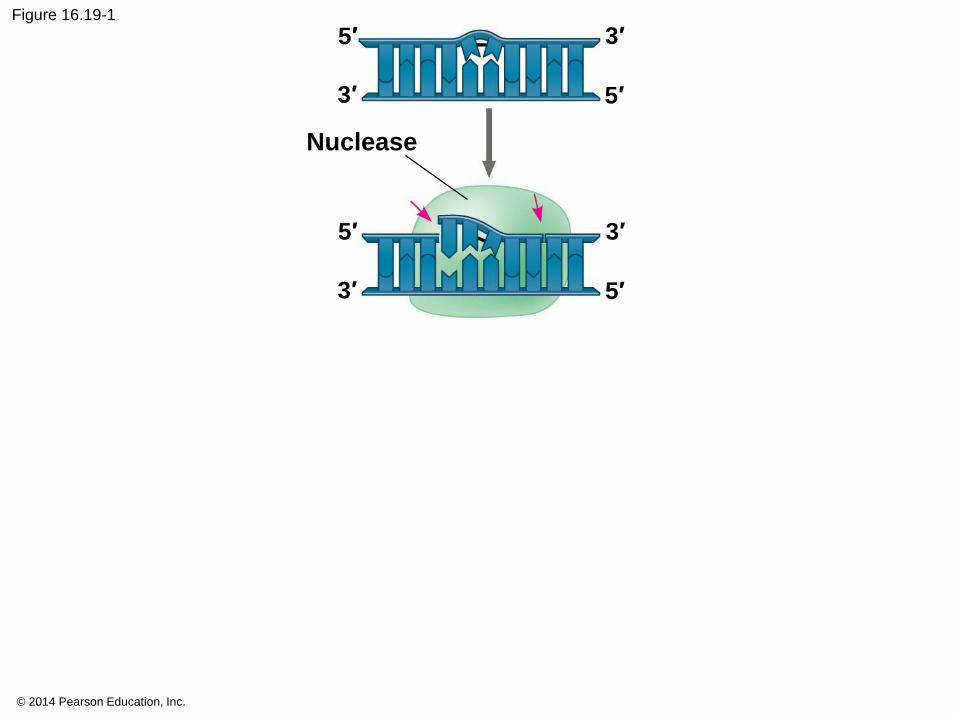
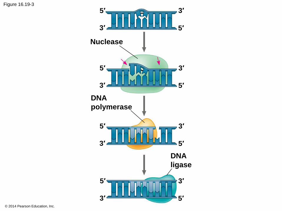
Proofreading and Repairing DNA
DNA polymerases proofread newly made DNA,
replacing any incorrect nucleotides
In mismatch repair of DNA, repair enzymes
correct errors in base pairing
DNA can be damaged by exposure to harmful
chemical or physical agents such as cigarette
smoke and X-rays; it can also undergo
spontaneous changes
In nucleotide excision repair, a nuclease cuts
out and replaces damaged stretches of DNA
© 2014 Pearson Education, Inc.
Figure 16.19-1
Nuclease
5′
5′
5′
5′
3′
3′
3′
3′

© 2014 Pearson Education, Inc.
Figure 16.19-2
Nuclease
5′
5′
5′
5′
3′
3′
3′
3′
5′
5′
3′
3′
DNA
polymerase
© 2014 Pearson Education, Inc.
Figure 16.19-3
Nuclease
5′
5′
5′
5′
3′
3′
3′
3′
5′
5′
3′
3′
DNA
polymerase
DNA
ligase
5′
3′5′
3′
© 2014 Pearson Education, Inc.
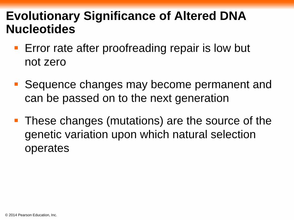
Evolutionary Significance of Altered DNA Nucleotides
Error rate after proofreading repair is low but
not zero
Sequence changes may become permanent and
can be passed on to the next generation
These changes (mutations) are the source of the
genetic variation upon which natural selection
operates
© 2014 Pearson Education, Inc.
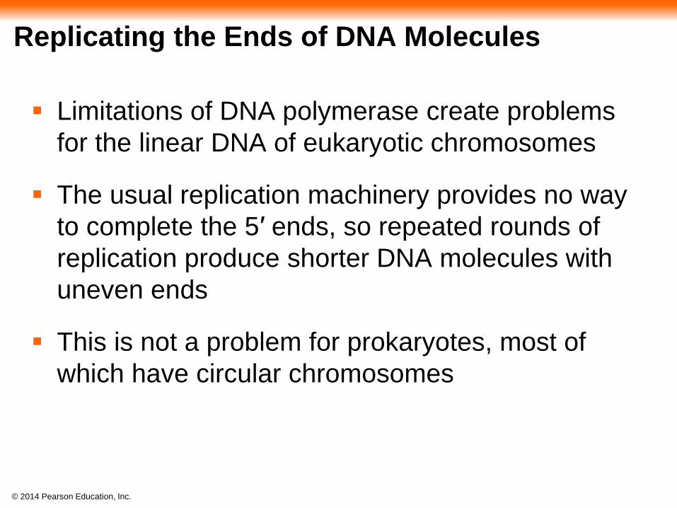
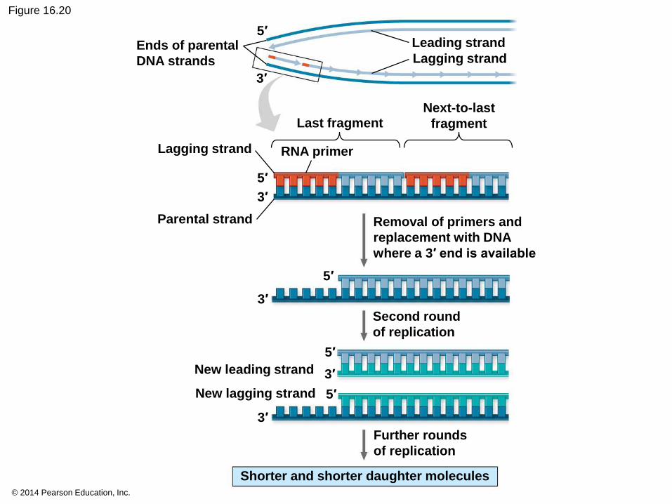
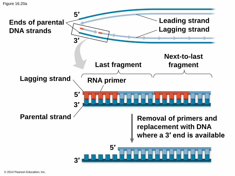
Replicating the Ends of DNA Molecules
Limitations of DNA polymerase create problems
for the linear DNA of eukaryotic chromosomes
The usual replication machinery provides no way
to complete the 5′ ends, so repeated rounds of
replication produce shorter DNA molecules with
uneven ends
This is not a problem for prokaryotes, most of
which have circular chromosomes
© 2014 Pearson Education, Inc.
Figure 16.20
Ends of parental
DNA strands
Lagging strand
Parental strand
RNA primer
Last fragmentNext-to-last
fragment
Lagging strand
Leading strand
Removal of primers and
replacement with DNA
where a 3′ end is available
3′
5′
5′
Second round
of replication
Further rounds
of replication
New lagging strand
New leading strand
Shorter and shorter daughter molecules
5′
3′
3′
5′
3′
5′
3′
© 2014 Pearson Education, Inc.
Figure 16.20a
Ends of parental
DNA strands
Lagging strand
Parental strand
RNA primer
Last fragment
Next-to-last
fragment
Lagging strand
Leading strand
Removal of primers and
replacement with DNA
where a 3′ end is available
3′
5′
5′
3′
5′
3′
© 2014 Pearson Education, Inc.
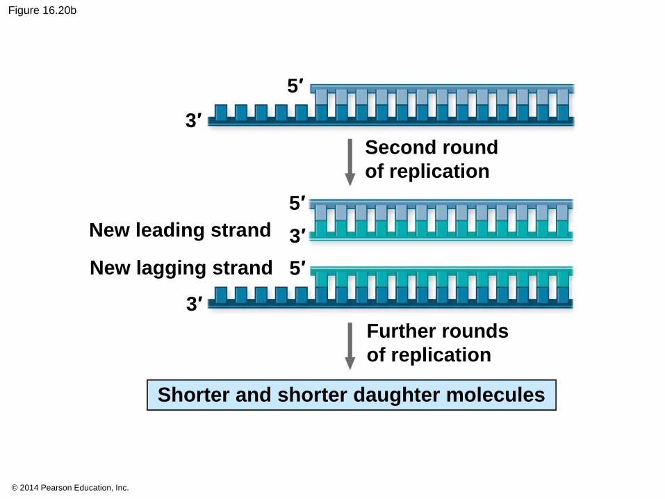
Figure 16.20b
5′
Second round
of replication
Further rounds
of replication
New lagging strand
New leading strand
Shorter and shorter daughter molecules
5′
3′
3′
5′
3′
© 2014 Pearson Education, Inc.
Eukaryotic chromosomal DNA molecules have
special nucleotide sequences at their ends called
telomeres
Telomeres do not prevent the shortening of DNA
molecules, but they do postpone the erosion of
genes near the ends of DNA molecules
It has been proposed that the shortening of
telomeres is connected to aging
© 2014 Pearson Education, Inc.
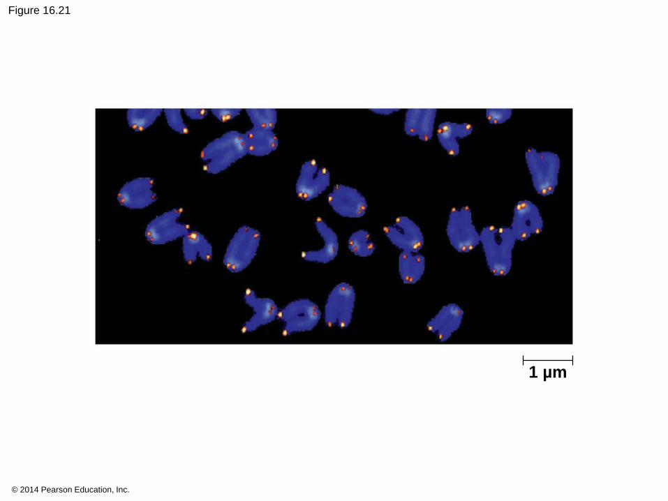
Figure 16.21
1 µm
© 2014 Pearson Education, Inc.
If chromosomes of germ cells became shorter in
every cell cycle, essential genes would eventually
be missing from the gametes they produce
An enzyme called telomerase catalyzes the
lengthening of telomeres in germ cells
© 2014 Pearson Education, Inc.
The shortening of telomeres might protect cells
from cancerous growth by limiting the number of
cell divisions
There is evidence of telomerase activity in cancer
cells, which may allow cancer cells to persist
Copyright © 2022 FDOKUMEN