The International Journal of Biochemistry & Cell Biology
10
The International Journal of Biochemistry & Cell Biology 69 (2015) 75–84 Contents lists available at ScienceDirect The International Journal of Biochemistry & Cell Biology jo ur nal home page: www.elsevier.com/locate/biocel Suppression of AGO2 by miR-132 as a determinant of miRNA-mediated silencing in human primary endothelial cells German Leonov a , Kunal Shah b , Daniel Yee a , Jon Timmis c , Tyson V. Sharp b , Dimitris Lagos a,∗ a Centre for Immunology and Infection, Department of Biology and Hull York Medical School University of York, Wentworth Way, York YO10 5DD, UK b Barts Cancer Institute, John Vane Science Centre, Charterhouse Square, Queen Mary University London, London EC1M 6BQ, UK c Department of Electronics, Wentworth Way, York YO10 5DD, UK a r t i c l e i n f o Article history: Received 5 June 2015 Received in revised form 8 September 2015 Accepted 7 October 2015 Available online 19 October 2015 Keywords: Lymphatic endothelial cells microRNA RNA-binding proteins AGO2 miR-132 a b s t r a c t The abundance of miR-132 ranges from constitutively high in the brain where it is necessary for neu- ronal development and function, to inducible expression in haematopoietic and endothelial cells where it controls angiogenesis and immune activation. We show that expression of AGO2, a protein central to miRNA-mediated gene silencing and miRNA biogenesis, is negatively regulated by miR-132. Using HeLa cells, we demonstrate that miR-132 interacts with the AGO2 mRNA 3 UTR and suppresses AGO2 expres- sion and AGO2-dependent small RNA-mediated silencing. Similarly, miR-132 over-expression leads to AGO2 suppression in primary human dermal lymphatic endothelial cells (HDLECs). During phorbol myris- tate acetate (PMA)-activation of HDLECs, miR-132 is induced in a CREB-dependent manner and inhibition of miR-132 results in increased AGO2 expression. In agreement with the role of AGO2 in maintenance of miRNA expression, AGO2 suppression by miR-132 affects the steady state levels of miR-221 and miR- 146a, two miRNAs involved in angiogenesis and inflammation, respectively. Our data demonstrate that the miRNA-silencing machinery is subject to autoregulation during primary cell activation through direct suppression of AGO2 by miR-132. © 2015 The Authors. Published by Elsevier Ltd. This is an open access article under the CC BY license (http://creativecommons.org/licenses/by/4.0/). 1. Introduction The mature miR-132 (miR-132-3p) is derived from the pri- mary miR-132/miR-212 cluster, found in the intergenic region of chromosome 17p13.3. miR-132 and miR-212 have the same seed sequence and potential targets, although miR-132 is the prefer- entially expressed miRNA from the cluster (Lagos et al., 2010). The transcription of miR-132/miR-212 cluster is dependent on cAMP response element-binding (CREB) protein phosphorylation (Vo et al., 2005), which is inducible in an ERK-1/2 and MSK-1/2 dependent manner (Remenyi et al., 2010). miR-132 has been impli- cated in neuronal function and development (Remenyi et al., 2010; Wayman et al., 2008), circadian rhythm control (Alvarez-Saavedra et al., 2011), angiogenesis (Anand et al., 2010), and regulation of innate immune responses (Lagos et al., 2010; Shaked et al., 2009). In endothelial cells, upregulation of miR-132 positively controls pro- liferation, angiogenesis and tumour growth in response to vascular endothelial growth factor A (VEGF-A) by suppressing p120RasGap ∗ Corresponding author. E-mail address: [email protected] (D. Lagos). (RASA1) (Anand et al., 2010). During viral infection of human cells in vitro, miR-132 acts as a negative regulator of inflammation in endothelial cells and macrophages by modulating EP300 (Lagos et al., 2010). Interestingly, EP300 is a co-transcriptional activator that is required for miR-132 expression, which may contribute to the transient expression of this miRNA, as seen in endothelial cells. Although the expression of miR-132 is low under basal conditions in HDLECs (approximately 50–100 copies per cell), endothelial activation leads to an early upregulation of miR-132 required for endothelial cell proliferation and angiogenesis (Anand et al., 2010; Lagos et al., 2010). The abundance of mature miRNAs can be regulated at multiple steps of miRNA biogenesis by altering the expression or function of miRNA machinery proteins. The knock-down of DICER (Zhang et al., 2012; Schmitter et al., 2006) or AGO2 (Schmitter et al., 2006) has been correlated with a global drop of mature miRNA expres- sion levels. DICER (Bernstein et al., 2003) and AGO2 (Morita et al., 2007) knock-out mice are embryonic lethal and this demonstrates the importance of miRNA-mediated regulation of gene expression during the early stages of embryo development. Reversely, the upregulation of AGO2 has been associated with a global increased expression of miRNAs (Martinez and Gregory, 2013; Winter and http://dx.doi.org/10.1016/j.biocel.2015.10.006 1357-2725/© 2015 The Authors. Published by Elsevier Ltd. This is an open access article under the CC BY license (http://creativecommons.org/licenses/by/4.0/).
-
Upload
khangminh22 -
Category
Documents
-
view
2 -
download
0
Transcript of The International Journal of Biochemistry & Cell Biology
Sm
GDa
b
c
a
ARRAA
KLmRAm
1
mcseTc(dcWeiele
h1
The International Journal of Biochemistry & Cell Biology 69 (2015) 75–84
Contents lists available at ScienceDirect
The International Journal of Biochemistry& Cell Biology
jo ur nal home page: www.elsev ier .com/ locate /b ioce l
uppression of AGO2 by miR-132 as a determinant ofiRNA-mediated silencing in human primary endothelial cells
erman Leonova, Kunal Shahb, Daniel Yeea, Jon Timmisc, Tyson V. Sharpb,imitris Lagosa,∗
Centre for Immunology and Infection, Department of Biology and Hull York Medical School University of York, Wentworth Way, York YO10 5DD, UKBarts Cancer Institute, John Vane Science Centre, Charterhouse Square, Queen Mary University London, London EC1M 6BQ, UKDepartment of Electronics, Wentworth Way, York YO10 5DD, UK
r t i c l e i n f o
rticle history:eceived 5 June 2015eceived in revised form 8 September 2015ccepted 7 October 2015vailable online 19 October 2015
eywords:ymphatic endothelial cellsicroRNA
a b s t r a c t
The abundance of miR-132 ranges from constitutively high in the brain where it is necessary for neu-ronal development and function, to inducible expression in haematopoietic and endothelial cells whereit controls angiogenesis and immune activation. We show that expression of AGO2, a protein central tomiRNA-mediated gene silencing and miRNA biogenesis, is negatively regulated by miR-132. Using HeLacells, we demonstrate that miR-132 interacts with the AGO2 mRNA 3′UTR and suppresses AGO2 expres-sion and AGO2-dependent small RNA-mediated silencing. Similarly, miR-132 over-expression leads toAGO2 suppression in primary human dermal lymphatic endothelial cells (HDLECs). During phorbol myris-tate acetate (PMA)-activation of HDLECs, miR-132 is induced in a CREB-dependent manner and inhibition
NA-binding proteinsGO2iR-132
of miR-132 results in increased AGO2 expression. In agreement with the role of AGO2 in maintenanceof miRNA expression, AGO2 suppression by miR-132 affects the steady state levels of miR-221 and miR-146a, two miRNAs involved in angiogenesis and inflammation, respectively. Our data demonstrate thatthe miRNA-silencing machinery is subject to autoregulation during primary cell activation through directsuppression of AGO2 by miR-132.
© 2015 The Authors. Published by Elsevier Ltd. This is an open access article under the CC BY license
. Introduction
The mature miR-132 (miR-132-3p) is derived from the pri-ary miR-132/miR-212 cluster, found in the intergenic region of
hromosome 17p13.3. miR-132 and miR-212 have the same seedequence and potential targets, although miR-132 is the prefer-ntially expressed miRNA from the cluster (Lagos et al., 2010).he transcription of miR-132/miR-212 cluster is dependent onAMP response element-binding (CREB) protein phosphorylationVo et al., 2005), which is inducible in an ERK-1/2 and MSK-1/2ependent manner (Remenyi et al., 2010). miR-132 has been impli-ated in neuronal function and development (Remenyi et al., 2010;
ayman et al., 2008), circadian rhythm control (Alvarez-Saavedrat al., 2011), angiogenesis (Anand et al., 2010), and regulation ofnnate immune responses (Lagos et al., 2010; Shaked et al., 2009). In
ndothelial cells, upregulation of miR-132 positively controls pro-iferation, angiogenesis and tumour growth in response to vascularndothelial growth factor A (VEGF-A) by suppressing p120RasGap∗ Corresponding author.E-mail address: [email protected] (D. Lagos).
ttp://dx.doi.org/10.1016/j.biocel.2015.10.006357-2725/© 2015 The Authors. Published by Elsevier Ltd. This is an open access article u
(http://creativecommons.org/licenses/by/4.0/).
(RASA1) (Anand et al., 2010). During viral infection of human cellsin vitro, miR-132 acts as a negative regulator of inflammation inendothelial cells and macrophages by modulating EP300 (Lagoset al., 2010). Interestingly, EP300 is a co-transcriptional activatorthat is required for miR-132 expression, which may contribute tothe transient expression of this miRNA, as seen in endothelial cells.Although the expression of miR-132 is low under basal conditionsin HDLECs (approximately 50–100 copies per cell), endothelialactivation leads to an early upregulation of miR-132 required forendothelial cell proliferation and angiogenesis (Anand et al., 2010;Lagos et al., 2010).
The abundance of mature miRNAs can be regulated at multiplesteps of miRNA biogenesis by altering the expression or functionof miRNA machinery proteins. The knock-down of DICER (Zhanget al., 2012; Schmitter et al., 2006) or AGO2 (Schmitter et al., 2006)has been correlated with a global drop of mature miRNA expres-sion levels. DICER (Bernstein et al., 2003) and AGO2 (Morita et al.,2007) knock-out mice are embryonic lethal and this demonstrates
the importance of miRNA-mediated regulation of gene expressionduring the early stages of embryo development. Reversely, theupregulation of AGO2 has been associated with a global increasedexpression of miRNAs (Martinez and Gregory, 2013; Winter andnder the CC BY license (http://creativecommons.org/licenses/by/4.0/).
76 G. Leonov et al. / The International Journal of Biochemistry & Cell Biology 69 (2015) 75–84
F ral ove( AGO2A
Dtt(
t
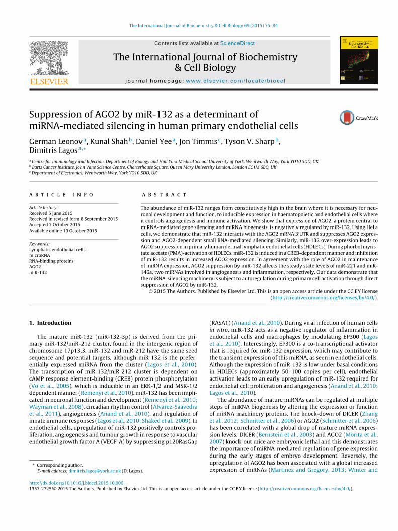
ig. 1. miR-132 interacts with AGO2 3′UTR. (A) mRNA levels of RBPs following lentiviaverage mRNA levels from two independent screens shown). (B) Conservation ofGO2 3′UTR).
iederichs, 2011). Global changes in miRNA expression levels leado disruptions in cellular function, such as poor neovascularisa-ion and vascular deformation reported in DICER knock-out mice
Bernstein et al., 2003).The very nature of miRNA-mediated gene regulation means thathe formation of negative feedback regulatory loops can occur,
rexpression of miR-132 and miR-132 mutant (M) in HDLECs, 48 h post-transduction 3′UTR region targeted by miR-132 (bold font – ‘seed’ site of miR-132 binding on
where a miRNA can be involved in regulating its own biogene-sis by targeting components required for its specific transcription,post-transcriptional processing, stability, or target accessibility.
In the context of miRNA biogenesis, such regulatory loops havebeen identified in the regulation of DICER by let-7 in mammaliancell lines (Tokumaru et al., 2008), AGO1 regulation by miR-168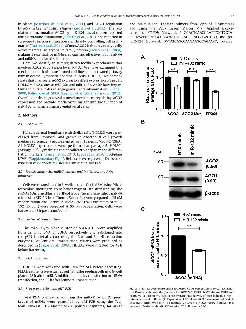
of Bio
ibudreama
imhsHt2Oem
2
2
cmA(tLm
2i
fsmc1h
2
ftedb
2
Ppt
2
LM
tems) for GAPDH (forward: 5′-GGAGTCAACGGATTTGGTCGTA-3′; reverse: 5′-GGCAACAATATCCACTTTACCAGAGT-3′) and pri-miR-126 (forward: 5′-TATCAGCCAAGAAGGCAGAA-3′, reverse:
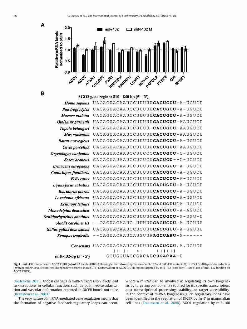
Fig. 2. miR-132 over-expression suppresses AGO2 expression in HeLas. (A) Rela-
G. Leonov et al. / The International Journal
n plants (Martínez de Alba et al., 2011), and ALG-1 regulationy let-7 in Caenorhabditis elegans (Zisoulis et al., 2012). The reg-lation of mammalian AGO2 by miR-184 has also been reporteduring cytokine stimulation (Roberts et al., 2013), and reported inesponse to insulin stimulation and thereby controlling cell prolif-ration (Tattikota et al., 2014). Of note, AGO2 is the only catalyticallyctive mammalian Argonaute family protein (Meister et al., 2004),aking it essential for mRNA cleavage and effective in both siRNA
nd miRNA-mediated silencing.Here, we identify an autoregulatory feedback mechanism that
nvolves AGO2 suppression by miR-132. We have examined thisechanism in both transformed cell lines and activated primary
uman dermal lymphatic endothelial cells (HDLECs). We demon-trate that changes in AGO2 expression affect expression of specificDLEC miRNAs, such as miR-221 and miR-146a, which have impor-
ant and critical roles in angiogenesis and inflammation (Li et al.,009; Poliseno et al., 2006; Taganov et al., 2006; Yang et al., 2012).verall, our findings reveal a novel mechanism regulating AGO2xpression and provide mechanistic insight into the function ofiR-132 in human primary endothelial cells.
. Methods
.1. Cell culture
Human dermal lymphatic endothelial cells (HDLEC) were pur-hased from Promocell and grown in endothelial cell growthedium (Promocell) supplemented with 10 ng/mL VEGF-C (R&D).ll HDLEC experiments were performed at passage 5. HDLECs
passage 5) fully maintain their proliferative capacity and differen-iation markers (Hansen et al., 2010; Lagos et al., 2010), includingYVE1 (Supplementary Fig. 1). HeLa cells were grown in Dulbecco’sodified eagle medium (DMEM) containing 10% FCS.
.2. Transfections with miRNA mimics and inhibitors, and RNAnhibitors
Cells were transfected in 6-well plates in Opti-MEM using Oligo-ectamine (Invitrogen) transfection reagent 18 h after seeding. TheiRNAs (OnTargetPlus SmartPool from Thermo Scientific), miRNAimics (miRIDIAN from Thermo Scientific) were prepared at 25 nM
oncentration and Locked Nucleic Acid (LNA)-inhibitors of miR-32 (Exiqon) were prepared at 50 nM concentration. Cells werearvested 48 h post transfection.
.3. Lentiviral transduction
The miR-132/miR-212 cluster or AGO2-UTR were amplifiedrom genomic DNA or cDNA respectively and subcloned intohe pSIN lentiviral vector using the NotI and BamHI restrictionnzymes. For lentiviral transduction, virions were produced asescribed in (Lagos et al., 2008). HDLECs were infected for 48 hefore harvesting.
.4. PMA treatment
HDLECs were activated with PMA for 24 h before harvesting.MA treatments were carried out 18 h after seeding cells into 6-welllates, 48 h after miRNA inhibition, mimics transfection or siRNAransfection, and 30 h after lentiviral transduction.
.5. RNA preparation and qRT-PCR
Total RNA was extracted using the miRNEasy kit (Qiagen).evels of mRNA were quantified by qRT-PCR using the Taq-an Universal PCR Master Mix (Applied Biosystems) for AGO2
chemistry & Cell Biology 69 (2015) 75–84 77
and pri-miR-132 (TaqMan primers from Applied Biosystems)and using the SYBR Green Master Mix (Applied Biosys-
tive Renilla luciferase (Rluc) activity for AGO2 WT 3′UTR, AGO2 Mutant 3′UTR andEP300 WT 3′UTR, normalised to the average Rluc activity of each individual repli-cate experiment in HeLas. (B) Expression of AGO1 and AGO2 protein in HeLas, 48 hpost-transfection with miR-132 mimics. (C) Levels of AGO2 mRNA in HeLas, 48 hpost-transfection with miR-132 mimics. *** Indicates p < 0.001.
7 of Bio
5cma
2
SS11mwyi
2
aXt3wwoa
FiHp
8 G. Leonov et al. / The International Journal
′-CGTGGCGTCTTCCAGAAT-3′). Primers were used at 300 nM finaloncentration. GAPDH was used as a loading control for AGO2, pri-iR-126 and pri-miR-132. RNU6 was used as a loading control for
ll mature miRNAs (Applied Biosystems).
.6. Immunoblots and antibodies
Primary antibodies were prepared at 1:1000 for AGO1 (Cellignalling, D84G10), AGO2 (Cell Signalling, C34C6), CREB (Cellignalling, 48H2), phospho-CREB at Ser133 (Cell Signalling, 87G3),:500 for EP300 (Abcam, 3G230), RASA1 (Santa Cruz, B4F8), and:5000 for �-actin (Cell Signalling, AC-15). Secondary goat anti-ouse (Dako, P0447) and goat anti-rabbit (Dako, P0448) antibodiesere prepared at 1:5000 conjugated to HRP. Western blot anal-
sis was carried out using ImageJ software for quantifying bandntensities.
.7. Luciferase reporter assay
AGO2 3′UTR and EP300 3′UTR were amplified from HDLEC cDNAnd subcloned into the psiCheck2 vector (Promega) using NotI andhoI enzymes. Mutations were introduced at the AGO2 3′UTR athe miR-132 binding site (WT: 5′-GUACAAUCCUUUUUCACUGUUU-′; Mut: 5′-GUACAAUCCUUUUUCACUAAAU-3′). Luciferase assays
ere performed in HeLa cells. AGO2 and EP300 UTR constructsere transfected for 24 h after the 24 h miR-132 mimics (20 nM)r NTC transfection. Relative light units (RLU) were measured with Fluoroscan Ascent FL luminometer (Thermo Scientific).
ig. 3. miR-132 over-expression suppresses AGO2 expression in HDLECs. (A) Expressionn HDLECs, 48 h post-transduction. (B) Expression of AGO2, RASA1, EP300 protein in HDLECs, 48 h post-transfection with miR-132 mimics. *** Indicates p < 0.001. (D) Expressiost-transfection. (E) AGO2 protein expression of two experiments with a 24 h lentiviral
chemistry & Cell Biology 69 (2015) 75–84
2.8. Let-7 silencing reporter assay
The let-7 silencing reporter assay was carried out in HeLa cellsas previously described by (James et al., 2010) in the presenceof NTC/miR-132 mimics (20 nM) or siNTC/siAGO2 (20 nM). Theluciferase reporter used (40 nM) contained either a single fully com-plementary site (si) or a seven times repeat of a mismatched site(mi7) for let-7 binding.
2.9. Statistical analysis
Experimental results are presented as mean ± S.E. (error bars)and were performed in 3 independent replicates unless other-wise stated in the figure legend. Where appropriate, a two-tailedunpaired Student’s t-test was used to calculate the significance ofthe difference between treatments.
3. Results
3.1. miR-132 interacts with the AGO2 3′UTR
Gene ontology analysis of predicted miR-132 targets, using theEIMMO prediction analysis tool (Hausser et al., 2009), indicatedthat RNA-binding proteins (RBPs) were over-represented amongst
miR-132 predicted targets (Supplementary Table 1). We utilised alentiviral construct containing the miR-132/miR-212 locus (Lagoset al., 2010) to screen for the effect of miR-132 on mRNA levels of 13RBPs that were predicted miR-132 targets by multiple algorithmsof AGO2 protein following lentiviral overexpression of miR-132 and miR-132 MDLECs, 48 h post-transfection with miR-132 mimics. (C) mRNA levels of AGO2 inon of AGO2 and RASA1 protein after a titration of miR-132 mimics in HDLECs, 48 hoverexpression of AGO2−UTR following a miR-132 overexpression in HDLECs.
of Bio
(iam
3wf3l3tssmael
3
detdi
F(ptA
G. Leonov et al. / The International Journal
Supplementary Table 2). Over-expression of miR-132 and miR-212n HDLECs resulted in a drop in AGO2 mRNA (Fig. 1A). The effect wasbolished when using a construct where the seed sequence of bothiR-132 and miR-212 is mutated (Fig. 1A).We identified a potential miR-132 binding site in the AGO2
′UTR. The binding site is conserved across mammalian speciesith the exception of Sorex araneus (Fig. 1B). Based on this, we
urther investigated whether miR-132 interacted with the AGO2′UTR region. The introduction of miR-132 mimic into HeLa cells
ed to a drop in luciferase signal for a reporter containing the AGO2′UTR (Fig. 2A), compared to the non-targeting control (NTC). Ariple consecutive base pair mutation introduced at the start of theeed site of the miR-132 binding in the AGO2 3′UTR reversed theuppression of luciferase activity by miR-132 mimic. The effect ofiR-132 on the AGO2 3′UTR was similar to that on the EP300 3′UTR,
known miR-132 target (Lagos et al., 2010). We should note thatndogenous miR-132 expression in HeLa cells is at similarly lowevels to those observed in HDLEC.
.2. miR-132 over-expression suppresses AGO2 expression
Having shown that miR-132 can directly interact with the pre-icted miR-132-binding site in the AGO2 3′UTR, we studied the
ffect of miR-132 over-expression on AGO2 expression. Transfec-ion of miR-132 synthetic mimics into HeLa cells resulted in aecrease in AGO2 protein and mRNA levels (Fig. 2B and C). Similarly,n HDLECs the observed decrease in AGO2 mRNA levels following
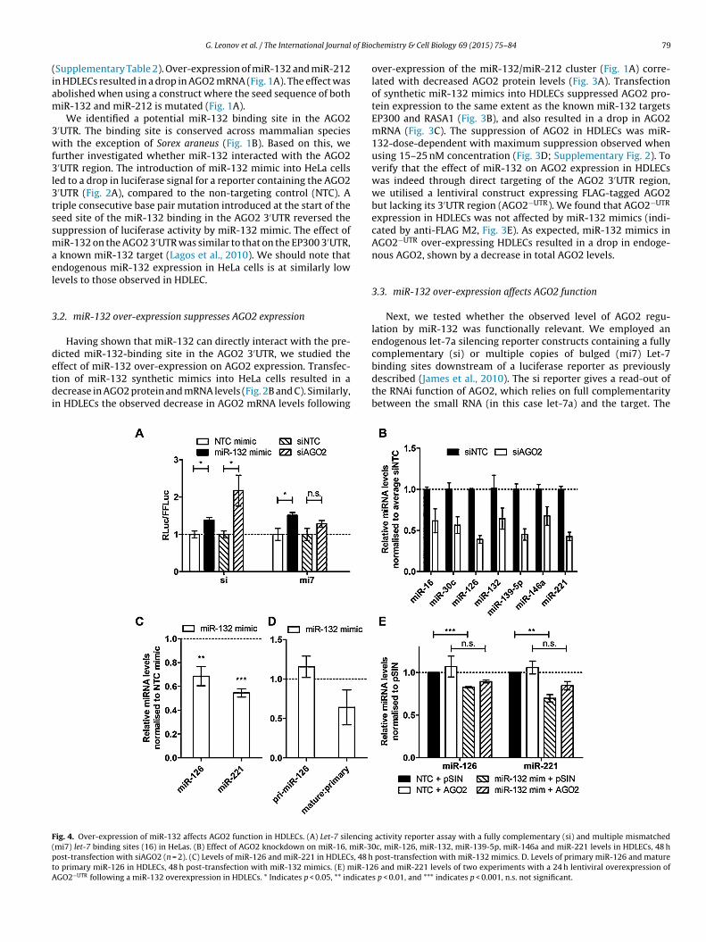
ig. 4. Over-expression of miR-132 affects AGO2 function in HDLECs. (A) Let-7 silencingmi7) let-7 binding sites (16) in HeLas. (B) Effect of AGO2 knockdown on miR-16, miR-30ost-transfection with siAGO2 (n = 2). (C) Levels of miR-126 and miR-221 in HDLECs, 48 ho primary miR-126 in HDLECs, 48 h post-transfection with miR-132 mimics. (E) miR-12GO2−UTR following a miR-132 overexpression in HDLECs. * Indicates p < 0.05, ** indicate
chemistry & Cell Biology 69 (2015) 75–84 79
over-expression of the miR-132/miR-212 cluster (Fig. 1A) corre-lated with decreased AGO2 protein levels (Fig. 3A). Transfectionof synthetic miR-132 mimics into HDLECs suppressed AGO2 pro-tein expression to the same extent as the known miR-132 targetsEP300 and RASA1 (Fig. 3B), and also resulted in a drop in AGO2mRNA (Fig. 3C). The suppression of AGO2 in HDLECs was miR-132-dose-dependent with maximum suppression observed whenusing 15–25 nM concentration (Fig. 3D; Supplementary Fig. 2). Toverify that the effect of miR-132 on AGO2 expression in HDLECswas indeed through direct targeting of the AGO2 3′UTR region,we utilised a lentiviral construct expressing FLAG-tagged AGO2but lacking its 3′UTR region (AGO2−UTR). We found that AGO2−UTR
expression in HDLECs was not affected by miR-132 mimics (indi-cated by anti-FLAG M2, Fig. 3E). As expected, miR-132 mimics inAGO2−UTR over-expressing HDLECs resulted in a drop in endoge-nous AGO2, shown by a decrease in total AGO2 levels.
3.3. miR-132 over-expression affects AGO2 function
Next, we tested whether the observed level of AGO2 regu-lation by miR-132 was functionally relevant. We employed anendogenous let-7a silencing reporter constructs containing a fullycomplementary (si) or multiple copies of bulged (mi7) Let-7
binding sites downstream of a luciferase reporter as previouslydescribed (James et al., 2010). The si reporter gives a read-out ofthe RNAi function of AGO2, which relies on full complementaritybetween the small RNA (in this case let-7a) and the target. Theactivity reporter assay with a fully complementary (si) and multiple mismatchedc, miR-126, miR-132, miR-139-5p, miR-146a and miR-221 levels in HDLECs, 48 h
post-transfection with miR-132 mimics. D. Levels of primary miR-126 and mature6 and miR-221 levels of two experiments with a 24 h lentiviral overexpression ofs p < 0.01, and *** indicates p < 0.001, n.s. not significant.
80 G. Leonov et al. / The International Journal of Biochemistry & Cell Biology 69 (2015) 75–84
F osphom ** Indt
moaor(omo
pl(tmSrmprmotirmrmt1
3
v
ig. 5. PMA induces miR-132 expression by activating CREB. (A) Expression of phature and primary miR-132 in HDLECs, 6 h and 24 h post-PMA treatment (n = 4).
reated with PMA over a 48 h period.
i7 reporter provides a read-out of the miRNA silencing functionf Ago2 as it is not susceptible to cleavage by AGO2 endonucle-se activity. We observed that the introduction of miR-132 mimicsr siAGO2 resulted in derepression of both siRNA and miRNAeporter constructs, as indicated by derepression of the Rluc activityFig. 4A). These findings show that miR-132-mediated regulationf AGO2 is sufficient to negatively impact AGO2 siRNA and miRNA-ediated silencing functions, thus further defining the importance
f this regulatory feedback mechanism.To test the functional significance of miR-132-mediated sup-
ression of AGO2 in HDLECs, we took advantage of the fact thatoss of AGO2 expression results in decreased miRNA expressionSchmitter et al., 2006; Morita et al., 2007). We confirmed thathis was the case in HDLECs by determining expression of several
iRNAs following siRNA mediated knockdown of AGO2 (Fig. 4B).imilarly, miR-132-mediated AGO2 suppression resulted in down-egulation of miR-126 and miR-221 (Fig. 4C). The decrease inature miR-126 levels was due to a reduction in the mature to
rimary miRNA ratio (Fig. 4D), suggesting that post-transcriptionalegulation of miR-126 contributes to the reduction in its levels iniR-132-over-expressing HDLECs, being consistent with the effect
f miR-132 on AGO2 expression. This was further supported byhe fact that physiologically relevant over-expression of AGO2−UTR
n HDLECs transfected with control or miR-132 mimics partiallyestored miR-126 and miR-221 expression. Over-expression ofiR-132 in HDLECs transduced with control lentivirus (pSIN)
esulted in statistically significant down-regulation of miR-126 andiR-221. In contrast, transfection of miR-132 mimics in HDLECs
ransduced with AGO2 lentivirus did not significantly affect miR-26 and miR-221 levels (Fig. 4E).
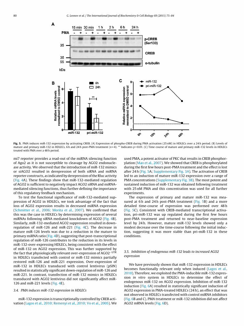
.4. PMA induces miR-132 expression in HDLECs
miR-132 expression is transcriptionally controlled by CREB acti-ation (Lagos et al., 2010; Remenyi et al., 2010; Vo et al., 2005). We
-CREB during PMA activation (25 nM) in HDLECs over a 24 h period. (B) Levels oficates p < 0.01. (C) Time course of mature and primary miR-132 levels in HDLECs
used PMA, a potent activator of PKC that results in CREB phosphor-ylation (Mao et al., 2007). We showed that CREB is phosphorylatedduring the first few hours post-PMA treatment and the effect is lostafter 24 h (Fig. 5A; Supplementary Fig. 3A). The activation of CREBled to an induction of mature miR-132 expression over a range ofPMA concentrations (Supplementary Fig. 3B). The most potent andsustained induction of miR-132 was obtained following treatmentwith 25 nM PMA and this concentration was used for all furtherexperiments.
The expression of primary and mature miR-132 was mea-sured at 6 h and 24 h post-PMA treatment (Fig. 5B) and a moredetailed time-course of expression was performed over 48 h(Fig. 5C). Consistent with CREB-mediated transcriptional activa-tion, pri-miR-132 was up regulated during the first few hourspost-PMA treatment and returned to near-baseline expressionlevel by 24 h. However, mature miR-132 levels showed only amodest decrease over the time-course following the initial induc-tion, suggesting it was more stable than pri-miR-132 in theseconditions.
3.5. Inhibition of endogenous miR-132 leads to increased AGO2expression
We have previously shown that miR-132 expression in HDLECsbecomes functionally relevant only when induced (Lagos et al.,2010). Therefore, we exploited the PMA-inducible miR-132 expres-sion in vitro system in HDLECs to determine the effect ofendogenous miR-132 on AGO2 expression. Inhibition of miR-132induction (Fig. 6A) resulted in statistically significant induction of
AGO2 expression in PMA-treated HDLECs (24 h), an effect that wasnot observed in HDLECs transfected with control miRNA inhibitors(Fig. 6B and C). PMA treatment or miR-132 inhibition did not affectAGO2 mRNA levels (Fig. 6D).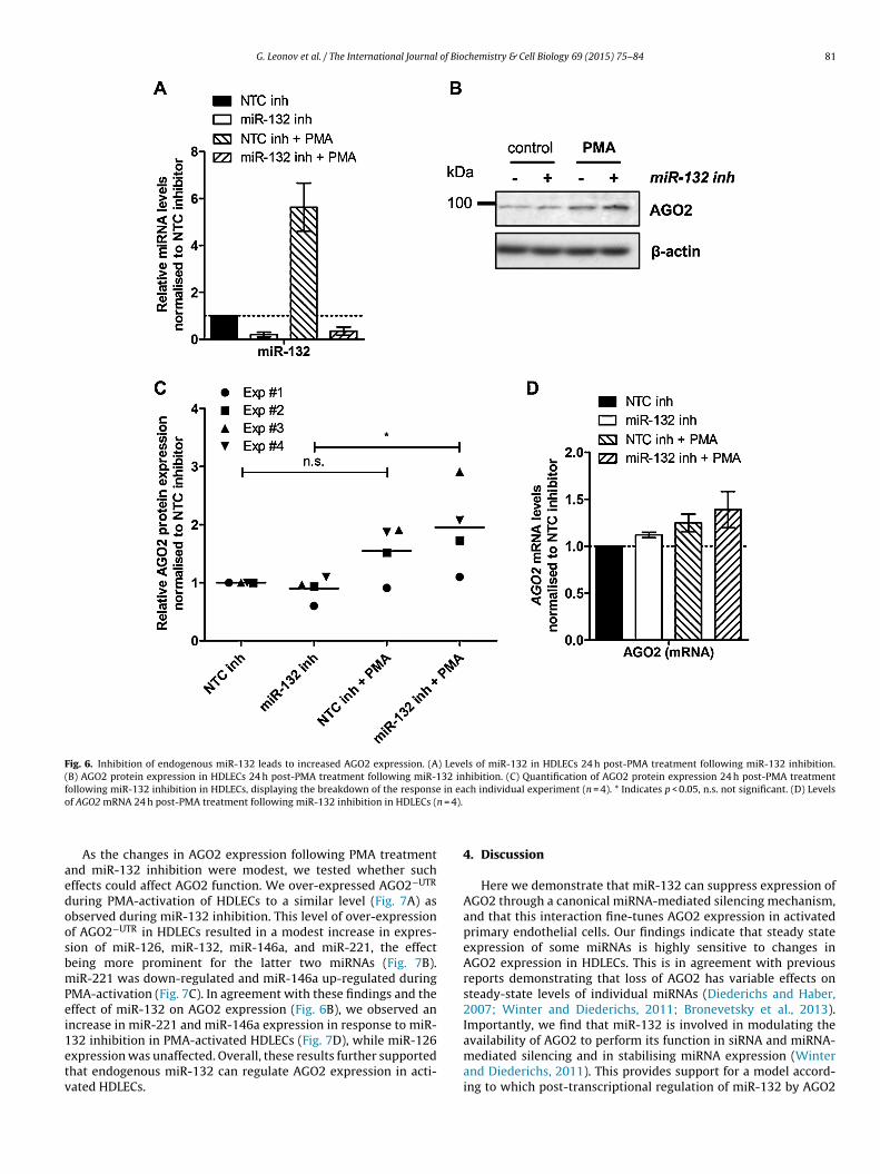
G. Leonov et al. / The International Journal of Biochemistry & Cell Biology 69 (2015) 75–84 81
Fig. 6. Inhibition of endogenous miR-132 leads to increased AGO2 expression. (A) Levels of miR-132 in HDLECs 24 h post-PMA treatment following miR-132 inhibition.(B) AGO2 protein expression in HDLECs 24 h post-PMA treatment following miR-132 inhibition. (C) Quantification of AGO2 protein expression 24 h post-PMA treatmentf in eao n = 4).
aedoosbmPei1etv
ollowing miR-132 inhibition in HDLECs, displaying the breakdown of the responsef AGO2 mRNA 24 h post-PMA treatment following miR-132 inhibition in HDLECs (
As the changes in AGO2 expression following PMA treatmentnd miR-132 inhibition were modest, we tested whether suchffects could affect AGO2 function. We over-expressed AGO2−UTR
uring PMA-activation of HDLECs to a similar level (Fig. 7A) asbserved during miR-132 inhibition. This level of over-expressionf AGO2−UTR in HDLECs resulted in a modest increase in expres-ion of miR-126, miR-132, miR-146a, and miR-221, the effecteing more prominent for the latter two miRNAs (Fig. 7B).iR-221 was down-regulated and miR-146a up-regulated during
MA-activation (Fig. 7C). In agreement with these findings and theffect of miR-132 on AGO2 expression (Fig. 6B), we observed anncrease in miR-221 and miR-146a expression in response to miR-
32 inhibition in PMA-activated HDLECs (Fig. 7D), while miR-126xpression was unaffected. Overall, these results further supportedhat endogenous miR-132 can regulate AGO2 expression in acti-ated HDLECs.ch individual experiment (n = 4). * Indicates p < 0.05, n.s. not significant. (D) Levels
4. Discussion
Here we demonstrate that miR-132 can suppress expression ofAGO2 through a canonical miRNA-mediated silencing mechanism,and that this interaction fine-tunes AGO2 expression in activatedprimary endothelial cells. Our findings indicate that steady stateexpression of some miRNAs is highly sensitive to changes inAGO2 expression in HDLECs. This is in agreement with previousreports demonstrating that loss of AGO2 has variable effects onsteady-state levels of individual miRNAs (Diederichs and Haber,2007; Winter and Diederichs, 2011; Bronevetsky et al., 2013).Importantly, we find that miR-132 is involved in modulating the
availability of AGO2 to perform its function in siRNA and miRNA-mediated silencing and in stabilising miRNA expression (Winterand Diederichs, 2011). This provides support for a model accord-ing to which post-transcriptional regulation of miR-132 by AGO2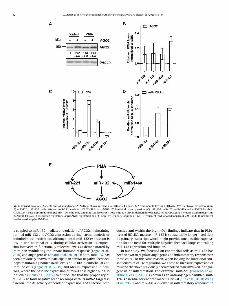
82 G. Leonov et al. / The International Journal of Biochemistry & Cell Biology 69 (2015) 75–84
Fig. 7. Regulation of AGO2 affects miRNA abundance. (A) AGO2 protein expression in HDLECs 24 h post-PMA treatment following a 30 h AGO2−UTR lentiviral overexpression.(B) miR-126, miR-132, miR-146a and miR-221 levels in HDLECs 48 h post-AGO2−UTR lentiviral overexpression. (C) miR-126, miR-132, miR-146a and miR-221 levels inHDLECs 24 h post-PMA treatment. (D) miR-126, miR-146a and miR-221 levels 48 h post-miR-132 LNA inhibition in PMA activated HDLECs. (E) Schematic diagram depictingP e feedf
ioelsi2blirime
MA/miR-132/AGO2-associated regulatory loops: AGO2 regulation by a (1) negativeed forward loop (miR-146a).
s coupled to miR-132 mediated regulation of AGO2, maintainingptimal miR-132 and AGO2 expression during haematopoietic orndothelial cell activation. Although basal miR-132 expression isow in non-neuronal cells, during cellular activation its expres-ion increases to functionally relevant levels as demonstrated byts role in modulating the innate immune response (Lagos et al.,010) and angiogenesis (Anand et al., 2010). Of note, miR-132 haseen previously shown to participate in similar negative feedback
oops maintaining homeostatic levels of EP300 in endothelial andmmune cells (Lagos et al., 2010) and MeCP2 expression in neu-
ons, where the baseline expression of miR-132 is higher but alsonducible (Klein et al., 2007). We speculate that the propensity ofiR-132 to form negative feedback loops with its mRNA targets isssential for its activity-dependent expression and function both
back loop (miR-132), (2) coherent feed forward loop (miR-221), and (3) incoherent
outside and within the brain. Our findings indicate that in PMA-treated HDLECs mature miR-132 is substantially longer-lived thanits primary transcript, which might provide one possible explana-tion for the need for multiple negative feedback loops controllingmiR-132 expression and function.
In our study, we focussed on endothelial cells as miR-132 hasbeen shown to regulate angiogenic and inflammatory responses inthese cells. For the same reason, when looking for functional con-sequences of AGO2 regulation we chose to measure expression ofmiRNAs that have previously been reported to be involved in angio-
genesis or inflammation. For example, miR-221 (Poliseno et al.,2006; Li et al., 2009) is known as an anti-angiogenic miRNA, miR-126 is essential for endothelial cell survival (Sun et al., 2010; Wanget al., 2008), and miR-146a involved in inflammatory responses in
of Bio
eHmsaeieAooittemi2oa
t22iot1iottascech
A
pPM(aoP((
A
i0
R
A
A
G. Leonov et al. / The International Journal
ndothelial cells (Lagos et al., 2010). We show that treatment ofDLECs with PMA results in an induction of the pro-angiogeniciR-132, and a decrease in the anti-angiogenic miR-221 expres-
ion, in agreement with the previously reported pro-angiogenicctivity of PMA (Montesano and Orci, 1985). Interestingly, duringndothelial cell activation, minimal over-expression of AGO2 ornhibition of miR-132 result in increased miR-221 and miR-146axpression, in agreement with the proposed miR-132-mediatedGO2 regulation. We should note that the magnitude of the effectf miR-132 inhibition on miR-221 and miR-146a indicates thatther factors also contribute to the regulation of these miRNAsn activated HDLECs. However, our results suggest that regula-ion of AGO2 due to miR-132 induction is a failsafe mechanismhat ensures maintained suppression of miR-221, and limits thextent of the anti-inflammatory activity miR-146a (Fig. 7E). Indeed,iRNAs are often components of such coherent (e.g. miR-221) or
ncoherent (e.g. miR-146a) feed forward loops (Herranz and Cohen,010). It would be interesting to further investigate the relevancef this intricate miRNA network in in vivo models of angiogenesisnd in cells with high basal miR-132 expression, such as neurons.
Validated miR-132 targets are involved in chromatin modifica-ions (e.g. MeCP2, EP300, Jarid1a, SIRT1) (Alvarez-Saavedra et al.,011; Yamakuchi, 2012) transcription (e.g. EP300) (Hasan et al.,001), and mRNA splicing (e.g. PTBP2) (Smith et al., 2011), indicat-
ng a functional clustering of miR-132 targets around modulatorsf the lifecycle of a mammalian mRNA. We substantially expandhis concept to cover mRNA silencing, by revealing that miR-32 can suppress AGO2. The major implication of these findings
s that a miR-132 can potentially act as a concurrent regulatorf transcription, splicing, and silencing in the cell. Defining howhese fundamental processes communicate is of paramount impor-ance and particularly relevant to cellular stress responses, whichre characterised by highly dynamic, and accurate gene expres-ion programmes. We propose that the potential of miR-132 inoordinating multiple molecular processes that determine genexpression can provide crucial clues towards understanding theomplex and context-specific role of this miRNA in neurons andaematopoietic and vascular cells.
cknowledgements
G.L. is supported by the “Combating Infectious Disease: Com-utational Approaches in Translational Science” Wellcome TrusthD programme (WT095024MA). D.L. is supported by grants by theedical Research Council (MR/L008505/1) and the Wellcome Trust
097829; through the Centre for Chronic Diseases and Disorderst the University of York). D.Y. is supported by the Biotechnol-gy and Biological Sciences Research Council Doctoral Trainingrogramme in “Mechanistic Biology and its Strategic Application”BB/J01113/1). T.V.S. and K.S. are supported by a BBSRC grantBB/I007571/2) to T.V.S.
ppendix A. Supplementary data
Supplementary data associated with this article can be found,n the online version, at http://dx.doi.org/10.1016/j.biocel.2015.10.06.
eferences
lvarez-Saavedra, M., Antoun, G., Yanagiya, A., Oliva-Hernandez, R.,
Cornejo-Palma, D., Perez-Iratxeta, C., Sonenberg, N., Cheng, H.-Y.M., 2011.miRNA-132 orchestrates chromatin remodeling and translational control ofthe circadian clock. Hum. Mol. Genet. 20, 731–751.nand, S., Majeti, B.K., Acevedo, L.M., Murphy, E.A., Mukthavaram, R., Scheppke, L.,Huang, M., Shields, D.J., Lindquist, J.N., Lapinski, P.E., King, P.D., Weis, S.M.,
chemistry & Cell Biology 69 (2015) 75–84 83
Cheresh, D.A., 2010. Micro RNA-132-mediated loss of p120RasGAP activatesthe endothelium to facilitate pathological angiogenesis. Nat. Med. 16, 909–914.
Bernstein, E., Kim, S.Y., Carmell, M.A., Murchison, E.P., Alcorn, H., Li, M.Z., Mills,A.A., Elledge, S.J., Anderson, K.V., Hannon, G.J., 2003. Dicer is essential formouse development. Nat. Genet. 35, 215–217.
Bronevetsky, Y., Villarino, A., Eisley, V.C., Barbeau, J., Barczak, R.A., Heinz, J.G.,Kremmer, A., Heissmeyer, E., McManus, V.M., Erle, T.D., Rao, J., Ansel, A.K.M.,2013. T cell activation induces proteasomal degradation of Argonaute andrapid remodeling of the microRNA repertoire. J. Exp. Med. 210, 417–432.
Diederichs, S., Haber, D.A., 2007. Dual role for argonautes in microRNA processingand posttranscriptional regulation of microRNA expression. Cell 131,1097–1108.
Hansen, A., Henderson, S., Lagos, D., Nikitenko, L., Coulter, E., Roberts, S., Gratrix, F.,Plaisance, K., Renne, R., Bower, M., Kellam, P., Boshoff, C., 2010. KSHV-encodedmiRNAs target MAF to induce endothelial cell reprogramming. Genes Dev. 24,195–205.
Hasan, S., Hassa, P., Imhof, O., Hottiger, R.M.O., 2001. Transcription coactivatorp300 binds PCNA and may have a role in DNA repair synthesis. Nature 410,387–391.
Hausser, J., Berninger, P., Rodak, C., Jantscher, Y., Wirth, S., Zavolan, M., 2009. MirZ:an integrated microRNA expression atlas and target prediction resource.Nucleic Acids Res. 37, 266–272.
Herranz, H., Cohen, S.M., 2010. MicroRNAs and gene regulatory networks:managing the impact of noise in biological systems. Genes Dev. 24, 1339–1344.
James, V., Zhang, Y., Foxler, D.E., de Moor, C.H., Kong, Y.W., Webb, T.M., Self, T.J.,Feng, Y., Lagos, D., Chu, C.-Y., Rana, T.M., Morley, S.J., Longmore, G.D., Bushell,M., Sharp, T.V., 2010. LIM-domain proteins, LIMD1, Ajuba, and WTIP arerequired for microRNA-mediated gene silencing. Proc. Natl. Acad. Sci. U. S. A.107, 12499–12504.
Klein, M., Lioy, E.D., Ma, T., Impey, L., Mandel, S., Goodman, G.R.H., 2007.Homeostatic regulation of MeCP2 expression by a CREB-induced microRNA.Nat. Neurosci. 10, 1513–1514.
Lagos, D., Vart, R.J., Gratrix, F., Westrop, S.J., Emuss, V., Wong, P.-P., Robey, R.,Imami, N., Bower, M., Gotch, F., Boshoff, C., 2008. Toll-like receptor 4 mediatesinnate immunity to Kaposi sarcoma herpesvirus. Cell Host Microbe 4, 470–483.
Lagos, D., Pollara, G., Henderson, S., Gratrix, F., Fabani, M., Milne, R.S.B., Gotch, F.,Boshoff, C., 2010. miR-132 regulates antiviral innate immunity throughsuppression of the p300 transcriptional co-activator. Nat. Cell Biol. 12,513–519.
Li, Y., Song, Y.-H., Li, F., Yang, T., Lu, Y.W., Geng, Y.-J., 2009. MicroRNA-221 regulateshigh glucose-induced endothelial dysfunction. Biochem. Biophys. Res.Commun. 381, 81–83.
Mao, L., Tang, Q., Wang, J.Q., 2007. Protein kinase C-regulated cAMP responseelement-binding protein phosphorylation in cultured rat striatal neurons.Brain Res. Bull. 72, 302–308.
Martínez de Alba, A.E., Jauvion, V., Mallory, A.C., Bouteiller, N., Vaucheret, H., 2011.The miRNA pathway limits AGO1 availability during siRNA-mediated PTGSdefense against exogenous RNA. Nucleic Acids Res. 39, 9339–9344.
Martinez, N.J., Gregory, R.I., 2013. Argonaute2 expression is post-transcriptionallycoupled to microRNA abundance. RNA 19, 605–612.
Meister, G., Landthaler, M., Patkaniowska, A., Dorsett, Y., Teng, G., Tuschl, T., 2004.Human Argonaute2 mediates RNA cleavage targeted by miRNAs and siRNAs.Mol. Cell 15, 185–197.
Montesano, R., Orci, L., 1985. Tumor-promoting phorbol esters induceangiogenesis in vitro. Cell 42, 469–477.
Morita, S., Horii, T., Kimura, M., Goto, Y., Ochiya, T., Hatada, I., 2007. One Argonautefamily member, Eif2c2 (Ago2), is essential for development and appears not tobe involved in DNA methylation. Genomics 89, 687–696.
Poliseno, L., Tuccoli, A., Mariani, L., Evangelista, M., Citti, L., Woods, K., Mercatanti,A., Hammond, S., Rainaldi, G., 2006. MicroRNAs modulate the angiogenicproperties of HUVECs. Blood 108, 3068–3071.
Remenyi, J., Hunter, C.J., Cole, C., Ando, H., Impey, S., Monk, C.E., Martin, K.J., Barton,G.J., Hutvagner, G., Arthur, J.S.C., 2010. Regulation of the miR-212/132 locus byMSK1 and CREB in response to neurotrophins. Biochem. J. 428, 281–291.
Roberts, J.C., Warren, R.B., Griffiths, C.E.M., Ross, K., 2013. Expression ofmicroRNA-184 in keratinocytes represses argonaute 2. J. Cell. Physiol. 221,2314–2323.
Schmitter, D., Filkowski, J., Sewer, A., Pillai, R.S., Oakeley, E.J., Zavolan, M., Svoboda,P., Filipowicz, W., 2006. Effects of Dicer and Argonaute down-regulation onmRNA levels in human HEK293 cells. Nucleic Acids Res. 34, 4801–4815.
Shaked, I., Meerson, A., Wolf, Y., Avni, R., Greenberg, D., Gilboa-Geffen, A., Soreq, H.,2009. MicroRNA-132 potentiates cholinergic anti-inflammatory signaling bytargeting acetylcholinesterase. Immunity 31, 965–973.
Smith, P.Y., Delay, C., Girard, J., Papon, M.A., Planel, E., Sergeant, N., Buée, L., Hébert,S.S., 2011. MicroRNA-132 loss is associated with tau exon 10 inclusion inprogressive supranuclear palsy. Hum. Mol. Genet. 20, 4016–4024.
Sun, Y., Bai, Y., Zhang, F., Wang, Y., Guo, Y., Guo, L., 2010. miR-126 inhibitsnon-small cell lung cancer cells proliferation by targeting EGFL7. Biochem.Biophys. Res. Commun. 391, 1483–1489.
Taganov, K., Boldin, D.M., Chang, P.K., Baltimore, J.D., 2006. NF-kappaB-dependentinduction of microRNA miR-146, an inhibitor targeted to signaling proteins of
innate immune responses. Proc. Natl. Acad. Sci. U. S. A. 103, 12481–12486.Tattikota, S.G., Rathjen, T., McAnulty, S.J., Wessels, H.-H., Akerman, I., van de Bunt,M., Hausser, J., Esguerra, J.L.S., Musahl, A., Pandey, A.K., You, X., Chen, W.,Herrera, P.L., Johnson, P.R., O’Carroll, D., Eliasson, L., Zavolan, M., Gloyn, A.L.,Ferrer, J., Shalom-Feuerstein, R., Aberdam, D., Poy, M.N., 2014. Argonaute2

8 of Bio
T
V
W
W
4 G. Leonov et al. / The International Journal
mediates compensatory expansion of the pancreatic � cell. Cell Metab. 19,122–134.
okumaru, S., Suzuki, M., Yamada, H., Nagino, M., Takahashi, T., 2008. let-7regulates Dicer expression and constitutes a negative feedback loop.Carcinogenesis 29, 2073–2077.
o, N., Klein, M.E., Varlamova, O., Keller, D.M., Yamamoto, T., Goodman, R.H.,Impey, S., 2005. A cAMP-response element binding protein-induced microRNAregulates neuronal morphogenesis. Proc. Natl. Acad. Sci. U. S. A. 102,16426–16431.
ang, S., Aurora, A.B., Johnson, B.A., Qi, X., McAnally, J., Hill, J.A., Richardson, J.A.,Bassel-Duby, R., Olson, E.N., 2008. The endothelial-specific microRNA miR-126governs vascular integrity and angiogenesis. Dev. Cell 15, 261–271.
ayman, G.A., Davare, M., Ando, H., Fortin, D., Varlamova, O., Cheng, H.-Y.M.,Marks, D., Obrietan, K., Soderling, T.R., Goodman, R.H., Impey, S., 2008. An
chemistry & Cell Biology 69 (2015) 75–84
activity-regulated microRNA controls dendritic plasticity by down-regulatingp250GAP. Proc. Natl. Acad. Sci. U. S. A. 105, 9093–9098.
Winter, J., Diederichs, S., 2011. Argonaute proteins regulate microRNA stability:Increased microRNA abundance by Argonaute proteins is due to microRNAstabilization. RNA Biol. 8, 1149–1157.
Yamakuchi, M., 2012. MicroRNA regulation of SIRT1. Front. Physiol. 3, 68.Yang, L., Boldin, M., Yu, P., Liu, Y.C., Ea, S.C., Ramakrishnan, K., Taganov, P.K., Zhao,
D.J., Baltimore, L.D., 2012. miR-146a controls the resolution of T cell responsesin mice. J. Exp. Med. 209, 1655–1670.
Zhang, C., Huys, A., Thibault, P.A., Wilson, J.A., 2012. Requirements for human Dicerand TRBP in microRNA-122 regulation of HCV translation and RNA abundance.Virology 433, 479–488.
Zisoulis, D.G., Kai, Z.S., Chang, R.K., Pasquinelli, A.E., 2012. Autoregulation ofmicroRNA biogenesis by let-7 and Argonaute. Nature 486, 541–544.