Computational Intelligence Methods for Bioinformatics and ...
Upload
vanderbiltCategory
view
0download
0
Article
A Molecular Genetics Laboratory Course ApplyingBioinformatics and Cell Biology in the Context of OriginalResearchCynthia J. Brame,* Wendy M. Pruitt,† and Lucy C. Robinson‡
*Department of Biology, Centenary College of Louisiana, Shreveport, LA 71105; and †Departmentsof Molecular and Cellular Physiology and ‡Biochemistry and Molecular Biology, Louisiana State UniversityHealth Sciences Center, Shreveport, LA 71105
Submitted July 7, 2008; Revised August 31, 2008; Accepted September 18, 2008Monitoring Editor: Robin Wright
Research-based laboratory courses have been shown to stimulate student interest in science andto improve scientific skills. We describe here a project developed for a semester-long research-based laboratory course that accompanies a genetics lecture course. The project was designed toallow students to become familiar with the use of bioinformatics tools and molecular biology andgenetic approaches while carrying out original research. Students were required to present theirhypotheses, experiments, and results in a comprehensive lab report. The lab project concernedthe yeast casein kinase 1 (CK1) protein kinase Yck2. CK1 protein kinases are present in allorganisms and are well conserved in primary structure. These enzymes display sequencefeatures that differ from other protein kinase subfamilies. Students identified such sequenceswithin the CK1 subfamily, chose a sequence to analyze, used available structural data todetermine possible functions for their sequences, and designed mutations within the sequences.After generating the mutant alleles, these were expressed in yeast and tested for function byusing two growth assays. The student response to the project was positive, both in terms ofknowledge and skills increases and interest in research, and several students are continuing theanalysis of mutant alleles as summer projects.
INTRODUCTION
Various organizations examining science education haveconcluded that an inquiry-based approach to learning isessential to understanding science as a process (HowardHughes Medical Institute, 1996; Council on UndergraduateResearch, 1997; National Research Council [NRC], 2003). Asa consequence, many laboratory courses have shifted awayfrom “cookbook” exercises toward an inquiry-based model,allowing students to feel the self-investment and excitementthat comes with discovery of new knowledge (Stukus andLennox, 2001; Eberhardt et al., 2003; Mitchell and Graziano,2006). Multiweek, inquiry- or research-based projects havebeen demonstrated to be an effective means of stimulatingstudent interest and enhancing skills in experimental designand interpretation (Myers and Burgess, 2003; Gammie and
Erdeniz, 2004; Howard and Miskowski, 2005; Mitchell andGraziano, 2006; Goyette and DeLuca, 2007). In addition,there is an increasing need to incorporate the use of bioin-formatics tools into such projects and into undergraduatebiology classes in general (NRC, 2003).
Here, we report a semester-long research project that re-quires students to use bioinformatics tools to design andinterpret a molecular biology-based experiment investigat-ing structural determinants of protein kinase activity. Ourgoals for the course were to allow students to experience theexcitement and challenges of original research while increas-ing their ability to understand and use the tools of themodern molecular biologist. Our specific learning objectiveswere as follows:
1. Students will gain understanding of genetics conceptsand molecular biology techniques through using them inthe context of a multi-step research project;
2. Students will learn to use protein and nucleotide data-DOI: 10.1187/cbe.08–07–0036Address correspondence to: Cynthia J. Brame ([email protected]).
CBE—Life Sciences EducationVol. 7, 410–421, Winter 2008
410 © 2008 by The American Society for Cell Biology
by guest on March 3, 2014http://www.lifescied.org/Downloaded from
http://www.lifescied.org/content/suppl/2008/11/19/7.4.410.DC1.htmlSupplemental Material can be found at:
bases and bioinformatics tools to investigate conservedprotein features; and
3. Students will demonstrate increased ability to analyzeand communicate the results of a multi-step project.
The results from a knowledge survey administered at thebeginning and end of the semester indicate that learningobjectives 1 and 2 were met, whereas assessment of labreports over the course of the semester indicated that objec-tive 3 was met. In addition, student perceptions of theproject indicate that they felt the process facilitated theirlearning.
Background for the Research ProjectThis project focuses on casein kinase 1 (CK1) protein kinases,using the yeast enzyme Yck2 as a model CK1. The CK1subfamily of protein kinases comprises a diverse group ofkinases found in all eukaryotic cells. CK1 protein kinasesfrom multicellular organisms regulate processes, includingsynaptic transmission (Faundez and Kelly, 2000), receptorsignaling (Tobin et al., 1997), circadian rhythm (Lee et al.,2001), DNA repair (Knippschild et al., 1997), nuclear import(Vielhaber et al., 2000), and cell division (Brockman et al.,1992). CK1 activities also have been implicated with roles inneurodegenerative diseases, including Alzheimer’s disease,and could be involved in a variety of cancers (Schwab et al.,2000; Rubinfeld et al., 2001). CK1 enzymes phosphorylate Seror Thr residues C-terminal to acidic residues. The classicalsite for CK1 is Asp/Glu-X-X-Ser/Thr (Tuazon and Traugh,1991), but it has been demonstrated that P�Ser/P�Thr-X-X-Ser/Thr provides high-affinity CK1 recognition for thedownstream Ser/Thr residue (Flotow and Roach, 1989).
Yck2 together with Yck1 forms a pair of essential andredundant kinases in the budding yeast Saccharomyces cer-evisiae (Robinson et al., 1993; Vancura et al., 1993). The Yckproteins are essential for cell division and viability, and inaddition to other functions, they seem to be involved inmembrane protein turnover, bud site selection, polarizationof the actin cytoskeleton, and function of the septin ring thatis essential for cytokinesis in yeast (Robinson et al., 1993;Panek et al., 1997; Robinson et al., 1999; Marchal et al., 2002).In accord with functions at the plasma membrane, Yck2 wasfound to be a peripheral membrane protein of approxi-mately 62 kDa (Vancura et al., 1993). Yck2 is anchored to themembrane via palmitoylation of its two terminal cysteineresidues (Roth et al., 2002; Babu et al., 2004). Targeting to theplasma membrane (as opposed to internal membranes) re-quires the 48 C-terminal residues that seem to be requiredsolely for palmitoylation (Babu et al., 2004).
We used a previously generated green fluorescent protein(GFP)-tagged YCK2 clone and two yeast strains to assess thefunction of the student-generated mutant alleles: a yckts
(yck1::ura3 yck2–2ts) yeast strain in which YCK1 is deletedand the yck2-2ts gene product functions at 24°C but has littleactivity at 37°C (Robinson et al., 1993), and a yck� strain(yck1�::KanMX, yck2�::NatMX) in which both YCK1 andYCK2 have been deleted and Yck activity is provided by aplasmid-borne YCK2 allele (pRS316: YCK2; URA3). Thesetools, available on request, allowed easy manipulation of theYCK2 gene as well as two simple functionality assays. Allother molecular biology supplies we used are commercially
available, and all bioinformatics tools we used are freelyavailable on the World Wide Web.
Course ContextThis project was implemented in Biology 313 (BIOL 313,Genetics) at Centenary College of Louisiana. BIOL 313 is asurvey genetics course that is required for all biology majorsand is taken by most biochemistry and biophysics majors.The course enrolls between 40 and 50 students each spring,and consists of 3 h of lecture and 3 h of lab each week for 14wk. There are typically three lab sections, limited to a max-imum of 18 students per section. Approximately one-third ofthe students taking the course are sophomores, approxi-mately 40% are juniors, and approximately one-fourth areseniors. All students taking the course have completed twosemesters of general chemistry and a one-semester introduc-tory cell biology course; many are concurrently enrolled inthe second semester of organic chemistry. Because the lec-ture section of the course covers a broad range of topics,ranging from classical genetics to molecular genetics, the labproject proceeds independently of the lecture section of thecourse. The instructors refer to the lab project wheneverrelevant topics (e.g., cloning) are covered in lecture, provid-ing students with concrete examples illustrating variouslecture topics.
METHODS
PreparationEach week, students received a document describing the purpose ofthe week’s activity, the techniques used for the activity, and anyother background information needed. Students were expected toread these documents before attending lab. These documents areavailable as Supplementary Material 1.
Identifying Conserved Sequences in CK1 EnzymesThe instructor reviewed conserved features of protein kinases witheach lab section based on three reviews, all of which were madeavailable for students’ reference (Hanks et al., 1988; Hanks andHunter, 1995; Hanks, 2003). After reviewing the concepts of con-served sequences and protein families, students identified con-served amino acid sequences in CK1 enzymes of at least six aminoacids. Specifically, students obtained sequences of CK1 enzymesfrom the National Center for Biotechnology Information (NCBI)protein database (www.ncbi.nlm.nih.gov/sites/entrez), choosing atleast one �, one �, and one � CK1 as well as the yeast enzymes Yck1and Yck2. Students aligned these protein sequences using ClustalW(align.genome.jp; Higgins and Sharp, 1988) and identified con-served sequences. The students consulted a table displaying con-served kinase sequences to determine whether these conservedsequences were specific to CK1 enzymes (Hanks and Hunter, 1988).
To put the CK1-specific conserved sequences in context of CK1structure, the students used Cn3D and the NCBI Structure databaseto highlight the conserved sequences in a model CK1 (CK1 fromSchizosaccharomyces pombe complexed with Mg2�-ATP; www.ncbi.nlm.nih.gov/structure). After identifying the ATP-binding lobe, thesubstrate-binding lobe, and each of the conserved sequences, thestudents formed hypotheses about the function of each conservedsequence. Each lab section then chose a single conserved sequenceon which to focus, and each lab group (composed of two to threestudents) determined the mutation within that sequence that theywished to make. The instructor encouraged the lab groups to worktogether in designing these mutations, suggesting that the muta-
Yeast Mutagenesis Lab Project
Vol. 7, Winter 2008 411
by guest on March 3, 2014http://www.lifescied.org/Downloaded from
tions could be more informative if different groups made comple-mentary mutations. For example, within a lab section focusing inpart on a highly conserved proline, one lab group deleted theproline, one made a conservative mutation, and one made a non-conservative mutation.
Designing PrimersStudents obtained the YCK2 open reading frame (ORF) sequencefrom the Saccharomyces Genome Database (www.yeastgenome-.org) and used this to identify the nucleotides corresponding to theirconserved amino acid sequence. Using rules for primer designprovided with the Stratagene QuikChange kit, students then de-signed primers to introduce their mutation. Specifically, studentsattempted to design primers that were between 25 and 45 nucleo-tides, with a melting temperature of �78°C, a minimum GC contentof 40%, and terminated in one or more C or G bases. The instructorexamined each pair of primers and suggested changes when appro-priate. When it was not possible to design primers that adhered to allthe desired parameters, the instructor often designed a second primerset to increase the probability of a successful mutagenesis reaction. Allprimers were synthesized by Integrated DNA Technologies (Cor-alville, IA) at the 100-nmol level and were used without purification.
Site-directed Mutagenesis
Mutagenesis reactions were performed using the QuikChange kitaccording to manufacturer’s instructions (Stratagene, Cedar Creek,TX). Unless otherwise noted, all reagents were provided with theQuikChange kit. Briefly, each student group performed two mu-tagenesis reactions, using 5 and 10 ng of pLR10 (pUC18:GFP-YCK2;Robinson et al., 1999) as template. The GFP fusion in this plasmidhas native YCK2 flanking sequences upstream and downstream,including the native YCK2 promoter (Robinson et al., 1999). Theforward and reverse primers (125 ng) designed by the students wereused in the reactions; when the instructors identified potentialshortcomings of the students’ primer design, students performedduplicate reactions with a primer set designed by the instructors.The reactions also contained reaction buffer, dNTP mix, PfuTurboDNA polymerase, and water to a final volume of 50 �l. Reactionswere carried out in an Eppendorf thermal cycler as follows: 1 cycle:95°C 30 s; 18 cycles: 95°C 30 s, 55°C 1 min, 68°C 5 min. TemplateDNA was then digested by incubation with DpnI for 1 h at 37°C.XL-1 Blue supercompetent cells were thawed on ice and separatedinto 50-�l aliquots in prechilled 14-ml BD Falcon polypropyleneround-bottomed tubes (Thermo Fisher Scientific, Waltham, MA).Supercompetent cells were incubated with 1 �l of each DpnI-treatedmutagenesis reaction for 30 min. The cells were heat-shocked at42°C for 45 s and then incubated on ice for 2 min. Preheated SOCbroth was added and cells were incubated for 1 h at 37°C withshaking. Half the volume of each transformation reaction was thenplated on Luria-Bertani (LB)-ampicillin medium and incubated at37°C overnight. Each student group picked three colonies withsterile toothpicks and grew these in LB containing 100 �g/ml am-picillin overnight at 37°C with shaking.
Plasmid Preparation
Plasmid minipreps were performed using the Zyppy Prep I mini-prep kit (Zymo Research, Orange, CA) according to manufacturer’sinstructions. Briefly, cells from 3 ml of liquid culture were pelletedby brief centrifugation in a 1.5-ml microfuge tube at full speed. Thesupernatant was discarded, and the cell pellet was resuspended inbuffer P1 and lysed by addition of buffer P2. Buffer P3 was addedto precipitate cell debris. The tube was centrifuged for 3 min at fullspeed, and the supernatant was loaded into the Zymo-spin column,avoiding carrying over any cell debris. The Zymo-spin column andcollection tube were centrifuged at full speed for 30 s, allowing theliquid to flow through the column. The flow through in the collec-tion tube was discarded and the resin was washed with wash buffer.The column was transferred to a sterile 1.5-ml microfuge tube andDNA was eluted with 40 �l of water.
Analytical Digest of Mutagenized DNA before Sequencing
Miniprep DNA was analyzed by restriction enzyme digest and gelelectrophoresis. One microliter of each sample was incubated with2 U of BamHI and 2 U of SalI in buffer D (all from Promega,Madison, WI) for 1 h at 37°C. The digested samples were thencombined with loading buffer (Sambrook et al., 1989), loaded in a1% agarose gel containing 1 �g/ml ethidium bromide, and sub-jected to electrophoresis for approximately 1 h at 120 V. Resultswere visualized with UV light.
Sequencing
Each student group chose three samples for sequence analysis.Approximately 1000 ng of DNA to be sequenced and approximately100 pmol of the appropriate sequencing primer were sent to Retro-gen (San Diego, CA) for automated dideoxy sequencing. Due to thehigh fidelity of PfuTurbo and the cost of multiple sequencing reac-tions for each clone, only the region of interest was sequenced foreach clone for the lab course. Mutations in the LLGPSLED regionused the sequencing primer 5�-GGGCTGCACTATAAGATAG-3�,mutations in the HIPYRE region were analyzed with the sequencingprimer 5�-CTGTTGTACAAGTCG-3�, and mutations in the EQSRRDDregion were analyzed with the sequencing primer 5�-GGAAGAC-CGGGTCAACC-3� (all from Integrated DNA Technologies). Stu-dents analyzed the results of the sequencing using the BLASTalgorithm (Altschul et al., 1990). Specifically, they used BLAST toalign the sequences obtained from Retrogen with YCK2 sequence.The alignment was used to verify that their mutant allele wasidentical to wild-type YCK2 except at the mutation site.
Preparative Digest to Isolate GFP-mYCK2 Fragment
Each student group chose one clone containing the desired muta-tion for further use and subjected it to a preparative digest toremove the GFP-mYCK2 fragment from the vector. Specifically, 2–3�g of DNA was incubated for 2 h at 37°C in buffer H with 5 U eachXbaI and SacI (all from Promega) in a final volume of 25 �l. Sampleswere then mixed with loading buffer, loaded in a 1% agarose gelcontaining 1 �g/ml ethidium bromide, and electrophoresed as de-scribed above. Results were visualized with UV light, and the bandcorresponding to the GFP-mYCK2 fragment was excised with ascalpel and stored at 4°C for purification.
GFP-mYCK2 Fragment Purification
Purification of the GFP-mYCK2 fragments was performed using aWizard SV DNA purification kit (Promega, Madison, WI) accordingto manufacturer’s instructions. Briefly, the agarose gel slice wasmelted in membrane binding solution (MBS), by using 10 �l of MBSfor every 10 mg of agarose, at 60°C. The dissolved gel was loadedinto an SV minicolumn and incubated for 1 min at room tempera-ture. After the incubation, the minicolumn was centrifuged for 1min at full speed. The liquid in the collection tube was discarded,and the column was washed twice with membrane wash solution.The SV minicolumn was transferred to a clean 1.5-ml microfugetube, and 50 �l of water was added. The minicolumn was incubatedat room temperature for 1 min and then centrifuged at full speed for2 min to elute the purified GFP-mYCK2 fragment.
Ligation
To construct low copy plasmid containing GFP-mYCK2 under thecontrol of the native YCK2 promoter, the purified GFP-mYCK2fragment was ligated into pRS315 (CEN, LEU2; Sikorski and Hieter,1989). Ligation was performed using a LigaFast kit (Promega) ac-cording to manufacturer’s instructions. Students incubated 100 ngof GFP-mYCK2 with 70 ng of XbaI/SacI-digested pRS315 shuttlevector in a 16-�l reaction with 3 U of DNA ligase. Reactions wereincubated for 10 min at room temperature and then used to trans-form 100 �l of chemically competent DH5� Escherichia coli cells.Frozen competent cells were thawed on ice, and 100 �l was incu-bated with the ligation mixture on ice for 30 min. The cells were
C. J. Brame et al.
CBE—Life Sciences Education412
by guest on March 3, 2014http://www.lifescied.org/Downloaded from
heat-shocked at 42°C for 3 min and then incubated on ice for 2 min.The cells were then incubated with 0.5 ml of preheated LB for 1 h at37°C with shaking. Next, 250 �l of each reaction was plated onLB-ampicillin plates and incubated at 37°C overnight. After devel-opment of colonies, colonies were inoculated into LB plus 0.1%ampicillin and incubated overnight at 37°C with shaking. Eachstudent group grew four liquid cultures and purified plasmid DNAfrom each using the miniprep procedure described above.
Analytical Digest of Shuttle Vector: GFP-mYCK2 Construct
Miniprep results were analyzed by restriction digest and gel elec-trophoresis. One microliter of each sample was incubated in a totalvolume of 10 �l with 2 U of XbaI and 2 U of SacI in buffer H (allfrom Promega) for 1 h at 37°C. Reactions were then combined withloading buffer and electrophoresed as described above. Resultswere visualized with UV light.
Preparation of Calf Thymus Carrier DNA
Calf thymus DNA (Sigma-Aldrich, St. Louis, MO) was prepared asa 10 mg/ml solution in TE and sheared sequentially with 18-, 22-,and 26-gauge needles. The solution was then autoclaved for 15 minbefore incubating on ice for 10 min. Aliquots were stored at �20°C.Each aliquot was boiled for 10 min and then placed on ice imme-diately before use.
Preparation of Yeast Media and Plates
Media were prepared as described previously (Sherman et al.,1986).
Yeast Transformation and Culture
Students used a modified version of the LiOAc transformationprocedure of Gietz and Schiestl (1991). For each pair of studentgroups, 10 ml YPD cultures were inoculated with yckts yeast(yck1-�1::ura3 yck2-2ts; Robinson et al., 1999) and yck� yeast andgrown overnight at 24°C with shaking. Approximately 3 h beforeuse, each culture was diluted to 100 ml in YPD and grown at 24°Cwith shaking. Cells were then pelleted by low-speed centrifugationand the cell pellet was washed with sterile water and resuspendedin lithium acetate in TE (0.1 M LiOAc/TE). One hundred microliteraliquots of the competent cells were added to prechilled microfugetubes containing 75 �g of calf thymus DNA and 2 �g of pRS315:GFP-mYCK2. Each student group carried out a no DNA control (noplasmid), a positive control (2 �g pRS315:GFP-YCK2), and a nega-tive control (2 �g pRS315). Then, 600 �l of 40% polyethylene glycol/0.1 M lithium acetate in TE was added, followed by gentle mixingby inversion. The tubes were then incubated at room temperaturefor 30 min on a rotating platform. Dimethylsulfoxide (70 �l) wasthen added to each sample, and each sample was mixed by inver-sion and then heat shocked at 42°C for 6 min. After rapid cooling for2 min on ice, cells were pelleted at 3000 rpm for 3 min, the super-natant was discarded, and the pellet was resuspended in 1 ml of�Leu medium. One tenth of each sample was then plated on a�Leu plate by using sterile 4-mm glass beads. The plates wereallowed to develop colonies for several days at room temperature(yckts cells) or 30°C (yck� cells). Four colonies from each reactionwere patched to �Leu plates and grown overnight at 24°C (yckts
cells) or at 30°C (yck� cells). Patches were then replica plated to two�Leu plates incubated at 24 and 37°C (yckts cells) or to �Leu and
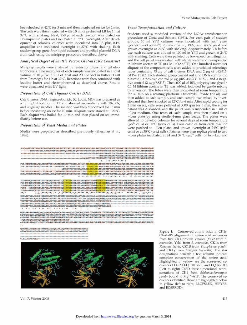
Figure 1. Conserved amino acids in CK1s.ClustalW alignment of amino acid sequencesfrom five CK1 protein kinases (Yck2 from S.cerevisiae, Yck1 from S. cerevisiae, CK1� fromXenopus laevis, CK1� from Toxoplasma gondii,and CK1� from Xenopus tropicalis). The stardesignations beneath a text column indicatecomplete conservation of the amino acid.Highlighted in yellow are the conserved se-quences LLGPSLED, HIPYRE, and EQSRRDD.(Left to right) Cn3D three-dimensional repre-sentations of CK1 from Schizosaccharomycespombe bound to Mg2�-ATP. The conserved se-quences identified above are highlighted belowin yellow (left to right, LLGPSLED, HIPYRE,and EQSRRDD).
Yeast Mutagenesis Lab Project
Vol. 7, Winter 2008 413
by guest on March 3, 2014http://www.lifescied.org/Downloaded from
5-fluoro-orotic acid (5-FOA) plates incubated at 30°C (yck� cells).Function was assessed by comparing growth in permissive andrestrictive conditions.
VisualizationYeast cells grown overnight on agar medium in restrictive condi-tions were scraped from patches, suspended in 2–5 �l of water,placed under a coverslip, and examined by bright field and fluo-rescence microscopy.
AssessmentTo determine whether educational goals 1 and 2 were achieved,student knowledge and skills were assessed with a pretest the firstday that the lab met. The same test also was included on the finalexam as a posttest. To compare responses to individual questions,student responses from each lab section were averaged. A two-tailed paired t test was performed to determine whether the learn-ing gain for each question was significant. To compare individualstudent learning on the entire test, a two-tailed paired t test wasperformed without regard to individual lab sections. The test isprovided in Supplemental Material 2.
To determine whether educational goal 3 was achieved, studentlab reports were assessed using a rubric (provided in SupplementalMaterial 2). Students wrote a lab report at midterm detailing theirresults for the first half of the project. They were then allowed torevise this lab report to replace their initial grade. Students alsowrote a final lab report detailing the results for the second half of theproject, but they were unable to revise this report. Scores on the firstlab report were compared with the revision and to the second lab
report; in both cases, the significance of the change was determinedwith a two-tailed paired t test.
Students also completed an attitudes survey at the end of thecourse assessing their perceptions of the extent to which the labactivities facilitated their learning.
RESULTS
Overview of ProjectStudents worked in groups of two or three to identifyconserved amino acid sequences in CK1 protein kinases,visualize their locations on a CK1 structure by using theCn3D software (www.ncbi.nlm.nih.gov/Structure/CN3D/cn3d.shtml), and form hypotheses about the function of eachconserved sequence (Figure 1). Each lab section (composedof five to six student groups and as many as 18 students)then chose one conserved sequence with which to work anddevised mutations to investigate the function of this se-quence, with each student group focusing on a differentmutation. Each student group designed primers to generatetheir mutation within Yck2, a CK1 from budding yeast (Ta-ble 1). The primers were used with the QuikChange kit(Stratagene) to perform site-directed mutagenesis (Figure 2).The students verified generation of their desired mutantallele by nucleotide sequence analysis of the mutated region.The mutant alleles were then cloned into a yeast/E. colishuttle vector and used to transform two strains of yeast,
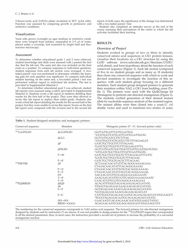
Table 1. Student-designed mutations and mutagenic primers
Conserved sequence Mutation Mutagenic primer (5�33�; forward primer only)
151LLGPSLED �LLGPSLED GGTTATTGATTTATTCGATTGG*ATATTGGTTATTGATTTATTCGATTGGTG
�LLG GGTTATTGATCCTTCTTTAG*ATATTGGTTATTGATCCTTCTTTAGAAGAT
�P GATCTGCTTGGTTCTTTAGAAG*GATCTGCTTGGTTCTTTAGAAGATTTA
�PSLED GGTTATTGATCTGCTTGGTTTATTCGATTGGTGTGG*ATTGATCTGCTTGGTTTATTCGATTGGTG
P3W GATCTGCTTGGTTGGTCTTTAGAAGATTTATTC*GATCTGCTTGGTTGGTCTTTAGAAG
P3E GATCTGCTTGGTGAGTCTTTAGAAG233HIPYRE �P ACTAAACAACATATTTACAGGGAAAAGAAA
CTAAACAACATATTTACAGGGAAAAGP3V CTAAACAACATATTGTGTACAGGGAAAAGP3T CTAAACAACATATTACGTACAGGGAAAAGDY AACAACATATTCCGAGGGAAAAGAAATCY3F AAACAACATATTCCGTTCAGGGAAAAGAAATCY3S CAACATATTCCGTCGAGGGAAAAGAAA
258EQSRRDD �EQS ACACATTTGGGTAGAAGAAGAGACGATATG�S TTGGGTAGAGAACAAAGAAGAGACGATATGS3T GGTAGAGAACAAACGAGAAGAGACGATATG
*GGTAGAGAACAAACGAGAAGAGACGATS3C CATTTGGGTAGAGAACAATGCAGAAGAGACGATATGGAAGCT
*TGGGTAGAGAACAATGCAGAAGAGACGATATGDD3NN GAACAATCGAGAAGAAACAATATGGAAGCTATGGRRDD3GGGG AGAGAACAATCGGGAGGAGGCGGTATGGAAGCTAT
The numbering for the conserved sequences corresponds to Yck2 amino acid sequence. The forward primers for site-directed mutagenesisdesigned by students and by instructors (*) are shown. It was not possible to design primers for the 151LLGPSLED region that correspondedto all the desired parameters; thus, in most cases, the instructors provided a second set of primers to increase the probability of a successfulmutagenesis reaction.
C. J. Brame et al.
CBE—Life Sciences Education414
by guest on March 3, 2014http://www.lifescied.org/Downloaded from

each of which allowed the students to perform a comple-mentation test to determine whether their mutant allele wasfunctional. After performing the assays for function, stu-dents used microscopy to examine yeast expressing theirmutant alleles to assess cellular morphology and Yck2relative abundance and subcellular localization (via theGFP tag). A timeline for the project is given in Table 2.During most phases of the project, students were pro-vided with explicit instructions to help them completeunfamiliar methods. As the students’ familiarity withstandard molecular biology techniques increased, how-ever, they performed procedures such as analytical di-gests and bacterial and yeast transformations withoutspecific guidance (Table 2).
Experimental ResultsStudents identified three amino acid sequences at least sixresidues long that were highly conserved in CK1s but not inother kinases (Figure 1). After examining the locations ofthese sequences within a three-dimensional representationof a CK1 from fission yeast, the students formed hypothesesabout the function of each sequence. For example, onestudent group proposed that the conserved sequenceLLGPSLED stabilizes ATP binding through interaction withthe adenine ring, whereas another group hypothesized that
the conserved sequence EQSRRDD assists in substrate bind-ing. Each lab section then chose a single sequence on whichto focus and devised a different mutation for each studentgroup (Table 1). Each student group found and aligned thepredicted amino acid sequence with the nucleotide sequenceof YCK2 to allow them to design forward and reverse prim-ers to introduce the desired mutation (Table 1). In selectedcases, the primers that students ordered were supplementedwith instructor-designed primers (Table 1). Using theseprimers and the QuikChange site-directed mutagenesisstrategy, students successfully generated 15 mutationswithin the conserved sequences they had identified (Table3). In some cases, the inclusion of the instructor-designedprimers proved to be especially useful. For example, thestudents planning to delete the amino acids LLGPSLEDdesigned primers that had only 11 and 12 nucleotides flank-ing the deletion site. The instructors ordered an additionalprimer set that included 14 and 15 nucleotides flanking thedeletion site (Table 1). The students used both primer sets insite-directed mutagenesis reactions but were successful onlywith the instructor-designed primers. Thus, the inclusionof both primer pairs allowed the experiment to proceed asneeded for progression of the project while also encour-aging students to consider elements of successful primerdesign.
After confirming generation of the desired mutant allelesvia sequence analysis of the relevant region of YCK2, stu-dents spent several lab sessions cloning the mutant alleleinto a yeast/E. coli shuttle vector marked with LEU2. Afteramplifying and purifying their new plasmids, they trans-formed two strains of yeast, yckts and yck� yeast, to performtwo independent assays of their mutant allele’s function. Inthe yckts yeast, YCK1 has been deleted and the protein prod-uct of yck2-2ts functions at 24°C but has little activity at 37°C(Robinson et al., 1993), allowing the strain to grow at 24°Cbut not at 37°C. Thus, expression of the mutant alleles in thisstrain allows the students to perform a complementation testby testing whether their mutant allele sustains growth at37°C. In the yck� strain, both YCK1 and YCK2 have beendeleted, and Yck activity is provided by a plasmid-borneYCK2 allele (pRS316: YCK2; URA3). Thus, expression of themutant alleles in this strain allows the students to perform aplasmid shuffle assay (Elledge and Davis, 1988) to deter-mine whether their mutant gene product can sustain growthin the absence of any other Yck activity. This assay testswhether a resident plasmid that is required for viability canbe replaced by an introduced plasmid, taking advantage ofthe ability to select against cells carrying an intact URA3gene using 5-FOA. 5-FOA is converted to the toxic com-pound 5-fluorouracil in the presence of a functional URA3gene. Because cells cannot grow on 5-FOA medium if theycarry the URA3 plasmid, and they cannot grow in the ab-sence of functional Yck, students were able to observegrowth of the yck� yeast on 5-FOA only if their mutantallele, present on the LEU2 plasmid, encodes a functionalYck2 protein. Growth rate and morphology of the strains on5-FOA medium can provide indicators of the level of activityof the mutant protein, because each is compromised if ac-tivity is low.
For both strains of yeast, the students performed threetransformation controls: one control in which cells wereincubated only with carrier DNA, to demonstrate that no
Figure 2. Site-directed mutagenesis strategy. Students used adouble-stranded plasmid containing GFP-tagged YCK2 as template.The initial steps in site-directed mutagenesis were DNA denatur-ation and primer annealing, followed by primer extension. Afterrepetition of these steps, E. coli produced (methylated) templateDNA (shown in bold) was selectively digested with the methyla-tion-specific restriction enzyme Dpn1 and the mutant plasmid wasintroduced into E. coli for nick repair and amplification. The figureis based on literature provided with the QuikChange kit (Strat-agene).
Yeast Mutagenesis Lab Project
Vol. 7, Winter 2008 415
by guest on March 3, 2014http://www.lifescied.org/Downloaded from
Leu� colony could arise in the absence of LEU2 plasmid; apositive control, in which cells were transformed with aplasmid carrying a wild-type GFP-tagged YCK2 gene; anda negative control, in which cells were transformed with theempty LEU2 vector. In each case, the transformed cells wereinitially grown under conditions that selected for the trans-forming plasmid (�Leu medium) but were otherwise per-
missive (i.e., yckts yeast were grown at 24°C; yck� yeast weregrown in the absence of 5-FOA). After the development ofcolonies, colonies were patched to fresh media and allowedto develop under the same conditions. Patches were thenreplica-plated to media incubated in both permissive andrestrictive conditions, allowing students to determine thefunction of their mutant alleles. Representative student re-
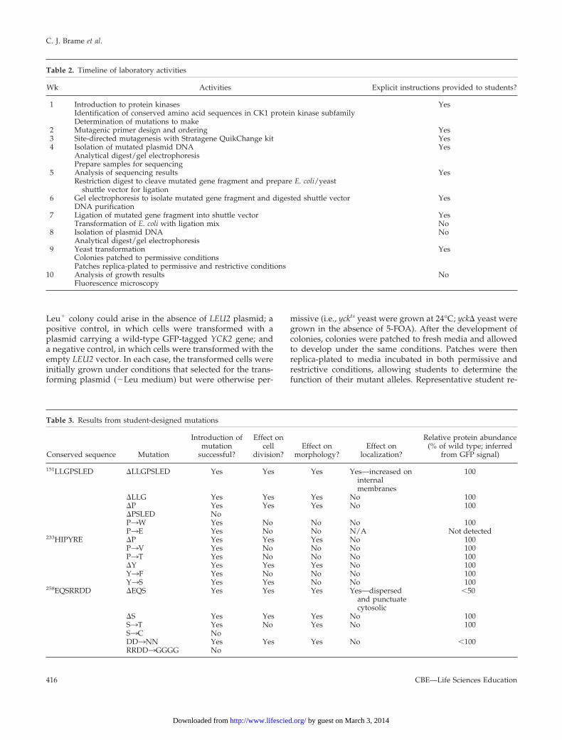
Table 2. Timeline of laboratory activities
Wk Activities Explicit instructions provided to students?
1 Introduction to protein kinases YesIdentification of conserved amino acid sequences in CK1 protein kinase subfamilyDetermination of mutations to make
2 Mutagenic primer design and ordering Yes3 Site-directed mutagenesis with Stratagene QuikChange kit Yes4 Isolation of mutated plasmid DNA Yes
Analytical digest/gel electrophoresisPrepare samples for sequencing
5 Analysis of sequencing results YesRestriction digest to cleave mutated gene fragment and prepare E. coli/yeast
shuttle vector for ligation6 Gel electrophoresis to isolate mutated gene fragment and digested shuttle vector Yes
DNA purification7 Ligation of mutated gene fragment into shuttle vector Yes
Transformation of E. coli with ligation mix No8 Isolation of plasmid DNA No
Analytical digest/gel electrophoresis9 Yeast transformation Yes
Colonies patched to permissive conditionsPatches replica-plated to permissive and restrictive conditions
10 Analysis of growth results NoFluorescence microscopy
Table 3. Results from student-designed mutations
Conserved sequence Mutation
Introduction ofmutation
successful?
Effect oncell
division?Effect on
morphology?Effect on
localization?
Relative protein abundance(% of wild type; inferred
from GFP signal)
151LLGPSLED �LLGPSLED Yes Yes Yes Yes—increased oninternalmembranes
100
�LLG Yes Yes Yes No 100�P Yes Yes Yes No 100�PSLED NoP3W Yes No No No 100P3E Yes No No N/A Not detected
233HIPYRE �P Yes Yes Yes No 100P3V Yes No No No 100P3T Yes No No No 100�Y Yes Yes Yes No 100Y3F Yes No No No 100Y3S Yes Yes No No 100
258EQSRRDD �EQS Yes Yes Yes Yes—dispersedand punctuatecytosolic
�50
�S Yes Yes Yes No 100S3T Yes No Yes No 100S3C NoDD3NN Yes Yes Yes No �100RRDD3GGGG No
C. J. Brame et al.
CBE—Life Sciences Education416
by guest on March 3, 2014http://www.lifescied.org/Downloaded from
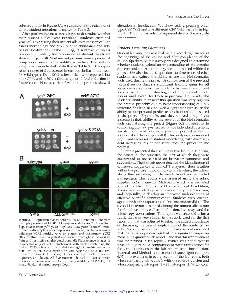
sults are shown in Figure 3A. A summary of the outcomes ofall the student mutations is shown in Table 3.
After performing these two assays to determine whethertheir mutant alleles were functional, students examinedyeast cells expressing their mutant alleles microscopically toassess morphology and Yck2 relative abundance and sub-cellular localization (via the GFP tag). A summary of resultsis shown in Table 3, and representative student results areshown in Figure 3B. Most mutant proteins were expressed atcomparable levels to the wild-type protein. Two notableexceptions are indicated. Note that in Table 3, 100% repre-sents a range of fluorescence intensities similar to that seenfor wild-type cells, �100% is lower than wild-type cells butnot �50%, and �50% indicates up to 10-fold reduction influorescence. Note also that few mutant proteins showed
alteration in localization. We show cells expressing wild-type GFP-Yck2 and five different GFP-Yck2 variants in Fig-ure 3B. The five variants are representative of the majoritywe examined.
Student Learning OutcomesStudent learning was assessed with a knowledge survey atthe beginning of the course and after completion of thecourse. Specifically, this survey was designed to determinewhether students gained an understanding of the geneticsconcepts and molecular biology techniques used within theproject. We also included questions to determine whetherstudents had gained the ability to use the bioinformaticstools used during the project. A comparison of the pre- andposttest results displays significant learning gains for alltested areas except one area. Students displayed a significantincrease in their understanding of all the molecular tech-niques used except for DNA sequencing (Figure 4A); thestudents’ ability to answer this question was very high onthe pretest, probably due to basic understanding of DNAstructure. Students also showed a significant increase in theability to interpret and predict results from techniques usedin the project (Figure 4B), and they showed a significantincrease in their ability to use several of the bioinformaticstools used during the project (Figure 4C). In addition tocomparing pre- and posttest results for individual questions,we also compared composite pre- and posttest scores forindividual students (Figure 4D). This analysis also revealedsignificant increases in student knowledge, with every stu-dent increasing his or her score from the pretest to theposttest.
Students presented their results in two lab reports duringthe course of the semester, the first of which they wereencouraged to revise based on instructor comments andsuggestions. The first lab report detailed the identification ofconserved sequences within CK1 enzymes, their locationwithin the proteins’ three-dimensional structure, the ration-ale for their mutation, and the results from the site-directedmutagenesis. The reports were assessed using the rubricsupplied as Supplemental Material 2, which was providedto students when they received the assignment. In addition,instructors provided extensive commentary to aid revision,and, hopefully, to develop an improved understanding ofeffective scientific communication. Students were encour-aged to revise the report, and all but one student did so. Thesecond lab report described cloning the mutant alleles intothe shuttle vector as well as the functionality assays and themicroscopy observations. This report was assessed using arubric that was very similar to the rubric used for the firstreport but that was adjusted to reflect the added importanceof discussing the overall implications of the students’ re-sults. A comparison of the lab report assessments revealedthat the revision process resulted in a significant improve-ment in the quality of lab report 1 and that this improvementwas maintained in lab report 2 (which was not subject torevision; Figure 5). A comparison of normalized scores forthe various sections of the lab reports (e.g., Introduction,Materials and Methods, and so on) indicated significant (p �0.05) improvements in every section of the lab report, bothwhen comparing lab report 1 with the revised version andwhen comparing lab report 1 with lab report 2. When com-
Figure 3. Representative student results. (A) Deletion of Pro fromthe highly conserved LLGPSLED sequence abolishes Yck2 function.Top, results from yckts yeast (top) and yck� yeast (bottom) trans-formed with empty vector (top rows on plates), vector containingwild-type YCK2 (middle rows on plates), and the mutant YCK2allele (bottom rows on plates) and grown overnight in permissive(left) and restrictive (right) conditions. (B) Fluorescence images ofrepresentative yck� cells transformed with vector containing themutant YCK2 allele and incubated overnight in restrictive condi-tions are shown. Cells expressing wild-type GFP-Yck2 and fivedifferent mutant GFP fusions, at least one from each conservedsequence, are shown. All five mutants showed at least as muchfluorescence on average as cells expressing wild-type GFP-Yck2, butmany display abnormal morphology.
Yeast Mutagenesis Lab Project
Vol. 7, Winter 2008 417
by guest on March 3, 2014http://www.lifescied.org/Downloaded from
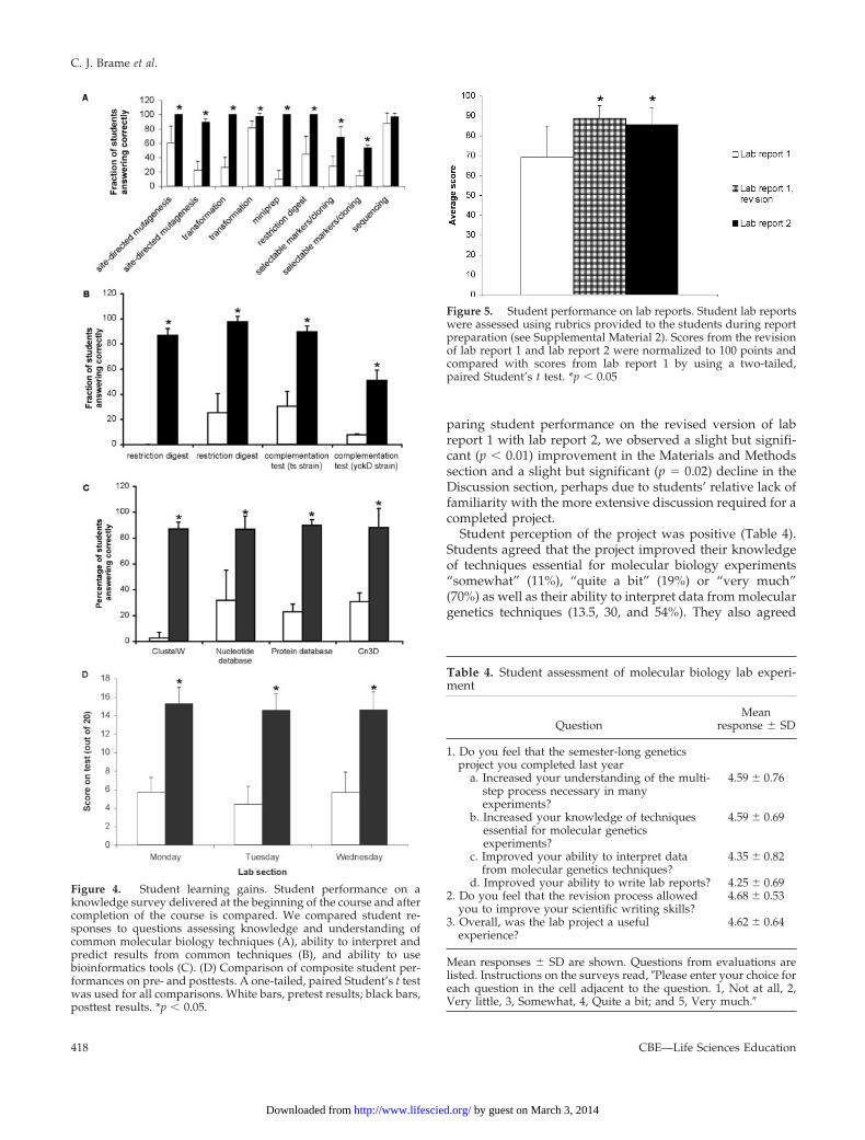
paring student performance on the revised version of labreport 1 with lab report 2, we observed a slight but signifi-cant (p � 0.01) improvement in the Materials and Methodssection and a slight but significant (p � 0.02) decline in theDiscussion section, perhaps due to students’ relative lack offamiliarity with the more extensive discussion required for acompleted project.
Student perception of the project was positive (Table 4).Students agreed that the project improved their knowledgeof techniques essential for molecular biology experiments“somewhat” (11%), “quite a bit” (19%) or “very much”(70%) as well as their ability to interpret data from moleculargenetics techniques (13.5, 30, and 54%). They also agreed
Figure 4. Student learning gains. Student performance on aknowledge survey delivered at the beginning of the course and aftercompletion of the course is compared. We compared student re-sponses to questions assessing knowledge and understanding ofcommon molecular biology techniques (A), ability to interpret andpredict results from common techniques (B), and ability to usebioinformatics tools (C). (D) Comparison of composite student per-formances on pre- and posttests. A one-tailed, paired Student’s t testwas used for all comparisons. White bars, pretest results; black bars,posttest results. *p � 0.05.
Figure 5. Student performance on lab reports. Student lab reportswere assessed using rubrics provided to the students during reportpreparation (see Supplemental Material 2). Scores from the revisionof lab report 1 and lab report 2 were normalized to 100 points andcompared with scores from lab report 1 by using a two-tailed,paired Student’s t test. *p � 0.05
Table 4. Student assessment of molecular biology lab experi-ment
QuestionMean
response � SD
1. Do you feel that the semester-long geneticsproject you completed last year
a. Increased your understanding of the multi-step process necessary in manyexperiments?
4.59 � 0.76
b. Increased your knowledge of techniquesessential for molecular geneticsexperiments?
4.59 � 0.69
c. Improved your ability to interpret datafrom molecular genetics techniques?
4.35 � 0.82
d. Improved your ability to write lab reports? 4.25 � 0.692. Do you feel that the revision process allowed
you to improve your scientific writing skills?4.68 � 0.53
3. Overall, was the lab project a usefulexperience?
4.62 � 0.64
Mean responses � SD are shown. Questions from evaluations arelisted. Instructions on the surveys read, Please enter your choice foreach question in the cell adjacent to the question. 1, Not at all, 2,Very little, 3, Somewhat, 4, Quite a bit; and 5, Very much.
C. J. Brame et al.
CBE—Life Sciences Education418
by guest on March 3, 2014http://www.lifescied.org/Downloaded from
“somewhat” (14%), “quite a bit” (47%), or “very much”(38%) that the project improved their ability to write labreports, and thought that the revision process helped in thisimprovement (3, 27, and 70%).
DISCUSSION
This article describes a laboratory project for an intermedi-ate-level genetics course enrolled with a broad variety ofstudents. During the course of the project, students use avariety of bioinformatics tools to frame a question and de-sign an experiment. They then gain experience with a vari-ety of current laboratory techniques in performing theirexperiment and with scientific writing while reporting theirwork. Thus, the project allows students to pursue originalresearch as part of a required course. It also emphasizes thebenefits of cooperativity in research; while designing theirexperiments, students quickly realized that their under-standing of a particular conserved sequence would begreater if various groups performed complementary muta-tions within the sequence.
Assessment of student learning indicated that studentscompleting the course showed significant gains in their un-derstanding of the genetics concepts and molecular biologytechniques used during the project. They also demonstratedsignificant gains in their ability to use the bioinformaticstools used in their research and showed an increased abilityto communicate the results of their research in the form of alab report. In assessing the course, we noted several changeswe would like to make. Although the pre- and posttest thestudents completed indicated a significant increase inknowledge in almost all categories, discussions with stu-dents completing the course have suggested that it would behelpful to offer the option “I don’t know” or “I have no idea”in the future. Not only would this serve to reduce falsepositives on the pretest (Hood-DeGrenier, 2008), but severalstudents indicated that they would have been more comfort-able providing this more honest answer on the pretest. Inaddition, we would like to include more opportunities forimproving scientific presentation skills in the future. To aidin this goal, we plan to add a weekly homework assignmentin which students complete a figure, figure legend, andaccompanying text for work they completed in lab thatweek. Not only would this practice allow students to receivefeedback on their writing each week but also it would helpstudents construct parts of their lab reports as they progressthrough the project. Because many students found the labreports daunting, we feel this progressive construction ofmaterials for the lab report would be helpful. Although thestudents were somewhat intimidated by the lab reports, theincreased scores between lab report 1 and lab report 2 indi-cate progress not only in learning concepts and techniquesbut also in ability to understand and interpret results. Thedifference in weight given to the discussion between labreports 1 and 2 makes more significant the increased scores,because the discussion was worth a greater fraction of thetotal points (�16 vs. 30%) in lab report 2.
The course enrolls approximately 40–50 students of var-ied backgrounds every spring. The students enrolled areapproximately equally distributed among their sophomore,junior, and senior years, and they are typically biology,
biochemistry, biophysics, chemistry, or neuroscience majors.There is one faculty instructor for each section of 15–18students. The course meets for 3 h/wk for 14 wk, 10 ofwhich are devoted to the project; students are also occasion-ally required to come in outside of scheduled lab time tocomplete small tasks to keep the project progressing. Withthe exception of the two yeast strains described above andthe template DNA used in the mutagenesis reaction, allof the tools used in the project are commercially available orfreely available on the World Wide Web. The template plas-mid used in site-directed mutagenesis and both yeast strainsare available upon request from the authors. As describedhere, the cost of the project is approximately $100/student;the cost could be reduced by decreasing the use of kits,decreasing the number of primers ordered, or decreasing thenumber of sequencing reactions. It is important to note thatall bioinformatics tools used are freely available on theWorld Wide Web and that all are student-friendly and easyto use.
The project can be modified easily to allow students toinvestigate any gene for which there is a relatively simplefunctionality assay. It also can be modified to be a shorterproject if site-directed mutagenesis is performed on templateDNA that allows direct expression in the model organism,allowing the cloning steps performed in this project to beeliminated. These modifications would be most effectivelyaccomplished in collaboration with an active yeast researchlab, but they also could build from collections of yeastdeletion strains (Saccharomyces Genome Deletion Project;sequence-www.stanford.edu/group/yeast_deletion_project/deletions3) and overlapping plasmids that cover the yeastgenome (steelhead.aecom.yu.edu/SystematicLibrary; www.openbiosystems.com/GeneExpression/Yeast/GenomicTiling).
Finally, the project has proved to be a sound starting pointfor independent student research projects. Three productivestudent research projects developed from the initial semes-ter in which this project was implemented. The three stu-dents integrated the mutant alleles into the yeast chromo-some to better assess the ability of each mutant protein tofulfill requirement for Yck1,2 activity. Students also trans-ferred their mutant alleles into vectors for fusion proteinproduction and isolated and purified fusion protein for invitro protein kinase assays. These ongoing projects haveproduced interesting results that the students have pre-sented at a Centenary College research forum. In addition,they will lead to two student presentations at regional ornational meetings and likely will be included in one or moreresearch publications.
In summary, the project described here adds to the grow-ing repertoire of inquiry-based projects students may com-plete in a laboratory course (e.g., Gammie and Erdeniz, 2004;Goyette and DeLuca, 2007). Students use a variety of bioin-formatics tools to frame a question and then use commonmolecular biology techniques to perform their experiment.The project is appropriate for students with a variety ofbackgrounds, and is flexible enough to allow modificationfor labs with different time and budget constraints. Finally,the original research aspect of the project adds motivation tocomplete each step, and it is likely to generate results thatcan serve as the basis for individual student projects in theinstructor’s lab, or even to serve as preliminary data for alarger project involving more students. The positive student
Yeast Mutagenesis Lab Project
Vol. 7, Winter 2008 419
by guest on March 3, 2014http://www.lifescied.org/Downloaded from
response to the project indicates that the approach waseffective for both motivation and learning.
ACKNOWLEDGMENTSWe thank Spencer Saulsbury for help in preparing figures; EmilyDavis and Jessica Miller for example hypotheses; and AllisonCormier, Danielle Crouthers, Danielle Dupree, Derek Fogleman,and Spencer Saulsbury for use of data. This work was supported bya Centenary College Research Professorship in the Natural Sciences(to C.J.B.) and National Science Foundation grant MCB-0517204 (toL.C.R. and C.J.B.).
REFERENCES
Altschul, S. F., Gish, W., Miller, W., Myers, E. W., and Lipman, D. J.(1990). Basic local alignment search tool. J. Mol. Biol. 215, 403–410.
Babu, P., Deschenes, R. J., and Robinson, L. C. (2004). Akr1p-depen-dent palmitoylation of Yck2p yeast casein kinase I is necessary andsufficient for plasma membrane targeting. J. Biol. Chem. 279, 27138–27147.
Brockman, J. L., Gross, S. D., Sussman, M. R., and Anderson, R. A.(1992). Cell cycle-dependent localization of casein kinase I to mitoticspindles. Proc. Natl. Acad. Sci. USA 89, 9454–9458.
Council on Undergraduate Research (1997). Shaping the Future:New Expectations for Undergraduate Education in Science,Mathematics, Engineering, and Technology, Washington, DC. www.cur.org/shaping (accessed 11 March 2007).
Eberhardt, E. S., Hansen, J., Riservato, L., Cole, M. Smaglo, B., andSzaniawski, P. (2003). Preparing undergraduates to participate inthe post-genome era. Biochem. Mol. Biol. Educ. 31, 402–409.
Elledge, S. J., and Davis, R. W. (1988). A family of versatile centro-meric vectors designed for use in the sectoring-shuffle mutagenesisassay in Saccharomyces cerevisiae. Gene 70, 303–312.
Faundez, V. V., and Kelly, R. B. (2000). The AP-3 complex requiredfor endosomal synaptic vesicle biogenesis is associated with a caseinkinase I alpha-like isoform. Mol. Biol. Cell 11, 2591–2604.
Flotow, H., and Roach, P. J. (1989). Synergistic phosphorylation ofrabbit muscle glycogen synthase by cyclic AMP-dependent proteinkinase and casein kinase I: implications for hormonal regulation ofglycogen synthase. J. Biol. Chem. 264, 9126–9128.
Gammie, A. E., and Erdeniz, N. (2004). Characterization of patho-genic human MSH2 missense mutations using yeast as a modelsystem: a laboratory course in molecular biology. Cell Biol. Educ. 3,31–48.
Gietz, R. D., and Schiestl, R. H. (1991). Applications of high effi-ciency lithium acetate transformation of intact yeast cells usingsingle-stranded nucleic acids as carrier. Yeast 7, 253–263.
Goyette, S. R., and DeLuca, J. (2007). A semester-long student-directed research project involving enzyme immunoassay: appro-priate for immunology, endocrinology, or neuroscience courses.CBE Life Sci. Educ. 6, 332–342.
Hanks, S. K. (2003). Genomic analysis of the eukaryotic proteinkinase superfamily: a perspective. Genome Biol. 4, 111–117.
Hanks, S. K., and Hunter, T. (1995). The eukaryotic protein kinasesuperfamily: kinase (catalytic) domain structure and classification.FASEB J. 9, 576–596.
Hanks, S. K., Quinn, A. M., and Hunter, T. (1988). The proteinkinase family: conserved features and deduced phylogeny of thecatalytic domains. Science 241, 42–52.
Higgins, D. G., and Sharp, P. M. (1988). CLUSTAL: a package forperforming multiple sequence alignment on a microcomputer. Gene73, 237–244.
Hood-DeGrenier, J. K. (2008). A Western blot-based investigationof the yeast secretory pathway designed for an intermediate-levelundergraduate cell biology laboratory. CBE Life Sci. Educ. 7,107–117.
Howard, D. R., and Miskowski, J. A. (2005). Using a module-basedlaboratory to incorporate inquiry into a large cell biology course.Cell Biol. Educ. 4, 249–260.
Howard Hughes Medical Institute (1996). Beyond Bio 101, TheTransformation of Undergraduate Biology Education, Chevy Chase,MD: Howard Hughes Medical Institute. http://www.hhmi.org/BeyondBio101 (accessed 11 March 2007).
Lee, C., Etchegaray, J., P., Cagampang, F. R., Loudon, A. S., andReppert, S. M. (2001). Posttranslational mechanisms regulate themammalian circadian clock. Cell 107, 855–867.
Knippschild, U., Milne, D. M., Campbell, L. E., DeMaggio, A. J.,Christenson, E., Hockstra, M. F., and Meek, D. W. (1997). p53 isphosphorylated in vitro and in vivo by the delta and epsilon iso-forms of casein kinase 1 and enhances the level of casein kinase 1delta in response to topoisomerase-directed drugs. Oncogene 15,1727–1736.
Marchal, C., Dupre, S, and Urban-Grimal, D. (2002). Casein kinase Icontrols a late step in the endocytic trafficking of yeast uracil per-mease. J. Cell Sci. 115, 217–226.
Mitchell, B. F., and Graziano, M. R. (2006). From organelle to proteingel: a 6-wk laboratory project on flagellar proteins. CBE Life Sci.Educ. 5, 239–246.
Myers, M. J., and Burgess, A. B. (2003). Inquiry-based laboratorycourse improves students’ ability to design experiments and inter-pret data. Adv. Physiol. Educ. 27, 26–33.
National Research Council (2003). Biology 2010, Transforming Un-dergraduate Education for Future Research Biologists, Washington,DC: National Academies Press.
Panek, H. R., Stepp, J. D., Engle H. M., Marks, K. M., Tan, P. K.,Lemmon, S. K., and Robinson, L. C. (1997). Suppressors of YCK-encoded yeast casein Kinase 1 deficiency define the four subunits ofa novel clathrin AP-like complex. EMBO J. 16, 4194–4204.
Robinson, L. C., Menold, M. M., Garrett, S., and Culbertson, M. R.(1993). Casein kinase I-like protein kinases encoded by YCK1 andYCK2 are required for yeast morphogenesis. Mol. Cell. Biol. 13,2870–2881.
Robinson, L. C., Bradley, C., Bryan, J. D., Jerome, A., Kweon, Y., andPanek, H. R. (1999). The Yck2 yeast casein kinase 1 isoform showscell cycle-specific localization to sites of polarized growth and isrequired for proper septin organization. Mol. Biol. Cell 10, 1077–1092.
Roth, A. F., Feng, Y., Chen, L., and Davis, N. G. (2002). The yeastDHHC cysteine-rich domain protein Akr1p is a palmityl trans-ferase. J. Cell Biol. 159, 23–28.
Rubinfeld, B., Tice, D. A., and Polakis, P. (2001). Axin-dependentphosphorylation of the adenomatous polyposis coli protein medi-ated by casein kinase 1 epsilon. J. Biol. Chem. 276, 39037–39045.
Sambrook, J., Fritsch, E. F., and Maniatis, T. (1989). Molecular Clon-ing: A Laboratory Manual, Plainview, NY: Cold Spring HarborPress.
Schwab, C., DeMaggio, A. J., Ghoshal, N. Binder, L. I., Kuret, J., andMcGeer, P. L. (2000). Casein kinase 1 delta is associated with patho-logical accumulation of tau in several neurodegenerative diseases.Neurobiol. Aging 21, 503–510.
Sherman, F., Fink, G. R., and Hinks, J. B. (1986). Methods in YeastGenetics, Cold Spring Harbor, NY: Cold Spring Harbor Laboratory.
C. J. Brame et al.
CBE—Life Sciences Education420
by guest on March 3, 2014http://www.lifescied.org/Downloaded from
Sikorski, R. A., and Hieter, P. (1989). A system of shuttle vectors andyeast host strains designed for efficient manipulation of DNA inSaccharomyces cerevisiae. Genetics 122, 19–27.
Stukus, P., and Lennox, J. E. (2001). Use of an investigativesemester-length laboratory project in an introductory microbiol-ogy course. In: Practicing Science, Arlington, VA: National Sci-ence Teachers Association.
Tobin, A. B., Totty, N. F., Sterlin, A. E., and Nahorski, S. R. (1997).Stimulus-dependent phosphorylation of G-protein-coupled re-ceptors by casein kinase 1 alpha. J. Biol. Chem. 272, 20844 –20849.
Tuazon, P. T., and Traugh, J. A. (1991). Casein kinase I and II—multipotential serine kinases: structure, function, and regulation.Adv. Second Messenger Phosphoprotein Res. 23, 129–164.
Vancura, A., Sessler, A., Leichus, B., and Kuret, J. (1993). Isolationand properties of YCK2, a Saccharomyces cerevisiae homolog of caseinkinase-1. Arch. Biochem. Biophys. 305, 47–53.
Vielhaber, E., Eide, E., Rivers, A., Gao, Z. H., and Virship, D. A.(2000). Nuclear entry of the circadian regulator mPER1 is controlledby mammalian casein kinase I epsilon. Mol. Cell. Biol. 20, 4888–4899.
Yeast Mutagenesis Lab Project
Vol. 7, Winter 2008 421
by guest on March 3, 2014http://www.lifescied.org/Downloaded from
Copyright © 2022 FDOKUMEN