
Bioinformatics for Vaccinology
314
Bioinformatics for Vaccinology Darren R Flower Edward Jenner Institute for Vaccine Research, Compton, Berkshire, UK A John Wiley & Sons, Ltd., Publication
-
Upload
khangminh22 -
Category
Documents
-
view
5 -
download
0
Transcript of Bioinformatics for Vaccinology

P1: OTE/OTE/SPH P2: OTE
fm JWBK293-Flower September 5, 2008 20:6 Printer Name: Yet to Come
Bioinformatics for Vaccinology
Darren R FlowerEdward Jenner Institute for Vaccine Research, Compton, Berkshire, UK
A John Wiley & Sons, Ltd., Publication

P1: OTE/OTE/SPH P2: OTE
fm JWBK293-Flower September 5, 2008 20:6 Printer Name: Yet to Come

P1: OTE/OTE/SPH P2: OTE
fm JWBK293-Flower September 5, 2008 20:6 Printer Name: Yet to Come
Bioinformatics for Vaccinology

P1: OTE/OTE/SPH P2: OTE
fm JWBK293-Flower September 5, 2008 20:6 Printer Name: Yet to Come

P1: OTE/OTE/SPH P2: OTE
fm JWBK293-Flower September 5, 2008 20:6 Printer Name: Yet to Come
Bioinformatics for Vaccinology
Darren R FlowerEdward Jenner Institute for Vaccine Research, Compton, Berkshire, UK
A John Wiley & Sons, Ltd., Publication

P1: OTE/OTE/SPH P2: OTE
fm JWBK293-Flower September 5, 2008 20:6 Printer Name: Yet to Come
This edition first published 2008c© 2008 by John Wiley & Sons, Ltd
Wiley-Blackwell is an imprint of John Wiley & Sons, formed by the merger of Wiley’s global Scientific,Technical and Medical business with Blackwell Publishing.
Registered office: John Wiley & Sons Ltd, The Atrium, Southern Gate, Chichester, West Sussex, PO198SQ, UK
Other Editorial Offices:9600 Garsington Road, Oxford, OX4 2DQ, UK111 River Street, Hoboken, NJ 07030-5774, USA
For details of our global editorial offices, for customer services and for information about how to apply forpermission to reuse the copyright material in this book please see our website at www.wiley.com/wiley-blackwell
The right of the author to be identified as the author of this work has been asserted in accordance with theCopyright, Designs and Patents Act 1988.
All rights reserved. No part of this publication may be reproduced, stored in a retrieval system, ortransmitted, in any form or by any means, electronic, mechanical, photocopying, recording or otherwise,except as permitted by the UK Copyright, Designs and Patents Act 1988, without the prior permission of thepublisher.
Wiley also publishes its books in a variety of electronic formats. Some content that appears in print may notbe available in electronic books.
Designations used by companies to distinguish their products are often claimed as trademarks. All brandnames and product names used in this book are trade names, service marks, trademarks or registeredtrademarks of their respective owners. The publisher is not associated with any product or vendormentioned in this book. This publication is designed to provide accurate and authoritative information inregard to the subject matter covered. It is sold on the understanding that the publisher is not engaged inrendering professional services. If professional advice or other expert assistance is required, the services ofa competent professional should be sought.
Library of Congress Cataloguing-in-Publication Data
Flower, Darren R.Bioinformatics for vaccinology / Darren R. Flower.
p. ; cm.Includes bibliographical references and index.ISBN 978-0-470-02711-0 (pbk. : alk. paper) 1. Immunoinformatics. 2. Vaccines–Design–Data
processing. I. Title.[DNLM: 1. Computational Biology–methods. 2. Vaccines. 3. Vaccination–methods.
4. Vaccination–trends. QW 805 F644b 2008]QR182.2.I46F56 2008615’.3720285–dc22
2008032154
A catalogue record for this book is available from the British Library.
ISBN 978 0 470 02711 0
Set in 10.5/12.5 pt Times by Aptara, New Delhi, IndiaPrinted in Singapore by Fabulous Printers Pte Ltd.
First printing 2008

P1: OTE/OTE/SPH P2: OTE
fm JWBK293-Flower September 5, 2008 20:6 Printer Name: Yet to Come
This work is dedicated to my wife Christine Jennifer
and to my daughter Isobel Emily Rebecca

P1: OTE/OTE/SPH P2: OTE
fm JWBK293-Flower September 5, 2008 20:6 Printer Name: Yet to Come

P1: OTE/OTE/SPH P2: OTE
fm JWBK293-Flower September 5, 2008 20:6 Printer Name: Yet to Come
Contents
Preface xiii
Acknowledgements xv
Exordium xvii
1 Vaccines: Their place in history 1Smallpox in history 1Variolation 3Variolation in history 5Variolation comes to Britain 6Lady Mary Wortley Montagu 9Variolation and the Sublime Porte 11The royal experiment 13The boston connection 14Variolation takes hold 17The Suttonian method 18Variolation in Europe 19The coming of vaccination 21Edward Jenner 23Cowpox 26Vaccination vindicated 28Louis Pasteur 29Vaccination becomes a science 30Meister, Pasteur and rabies 31A vaccine for every disease 33In the time of cholera 34Haffkine and cholera 36Bubonic plague 37The changing face of disease 39Almroth wright and typhoid 40Tuberculosis, Koch, and Calmette 43Vaccine BCG 44

P1: OTE/OTE/SPH P2: OTE
fm JWBK293-Flower September 5, 2008 20:6 Printer Name: Yet to Come
viii CONTENTS
Poliomyelitis 46Salk and Sabin 47Diphtheria 49Whooping cough 50Many diseases, many vaccines 51Smallpox: Endgame 53Further reading 54
2 Vaccines: Need and opportunity 55Eradication and reservoirs 55The ongoing burden of disease 57Lifespans 57The evolving nature of disease 59Economics, climate and disease 60Three threats 60Tuberculosis in the 21st century 61HIV and AIDS 62Malaria: Then and now 63Influenza 64Bioterrorism 65Vaccines as medicines 67Vaccines and the pharmaceutical industry 68Making vaccines 70The coming of the vaccine industry 70
3 Vaccines: How they work 73Challenging the immune system 73The threat from bacteria: Robust, diverse, and endemic 74Microbes, diversity and metagenomics 75The intrinsic complexity of the bacterial threat 76Microbes and humankind 77The nature of vaccines 78Types of vaccine 80Carbohydrate vaccines 82Epitopic vaccines 82Vaccine delivery 83Emerging immunovaccinology 84The immune system 85Innate immunity 86Adaptive immunity 88The microbiome and mucosal immunity 90Cellular components of immunity 90Cellular immunity 93The T cell repertoire 93

P1: OTE/OTE/SPH P2: OTE
fm JWBK293-Flower September 5, 2008 20:6 Printer Name: Yet to Come
CONTENTS ix
Epitopes: The immunological quantum 94The major histocompatibility complex 95MHC nomenclature 97Peptide binding by the MHC 98The structure of the MHC 99Antigen presentation 101The proteasome 101Transporter associated with antigen processing 103Class II processing 103Seek simplicity and then distrust it 104Cross presentation 105T cell receptor 106T cell activation 108Immunological synapse 109Signal 1, signal 2, immunodominance 109Humoral immunity 110Further reading 112
4 Vaccines: Data and databases 113Making sense of data 113Knowledge in a box 114The science of -omes and -omics 115The proteome 115Systems biology 116The immunome 117Databases and databanks 118The relational database 119The XML database 119The protein universe 120Much data, many databases 122What proteins do 122What proteins are 124The amino acid world 124The chiral nature of amino acids 127Naming the amino acids 130The amino acid alphabet 132Defining amino acid properties 134Size, charge and hydrogen bonding 135Hydrophobicity, lipophilicity and partitioning 136Understanding partitioning 139Charges, ionization, and pka 140Many kinds of property 143Mapping the world of sequences 146Biological sequence databases 147

P1: OTE/OTE/SPH P2: OTE
fm JWBK293-Flower September 5, 2008 20:6 Printer Name: Yet to Come
x CONTENTS
Nucleic acid sequence databases 148Protein sequence databases 149Annotating databases 150Text mining 151Ontologies 153Secondary sequence databases 154Other databases 155Databases in immunology 156Host databases 156Pathogen databases 159Functional immunological databases 161Composite, integrated databases 162Allergen databases 163Further reading 165Reference 165
5 Vaccines: Data driven prediction of binders, epitopesand immunogenicity 167Towards epitope-based vaccines 167T cell epitope prediction 168Predicting MHC binding 169Binding is biology 172Quantifying binding 173Entropy, enthalpy and entropy-enthalpy compensation 174Experimental measurement of binding 175Modern measurement methods 177Isothermal titration calorimetry 178Long and short of peptide binding 179The class I peptide repertoire 180Practicalities of binding prediction 181Binding becomes recognition 182Immunoinformatics lends a hand 183Motif based prediction 184The imperfect motif 185Other approaches to binding prediction 186Representing sequences 187Computer science lends a hand 188Artificial neural networks 188Hidden Markov models 190Support vector machines 190Robust multivariate statistics 191Partial least squares 191Quantitative structure activity relationships 192Other techniques and sequence representations 193

P1: OTE/OTE/SPH P2: OTE
fm JWBK293-Flower September 5, 2008 20:6 Printer Name: Yet to Come
CONTENTS xi
Amino acid properties 194Direct epitope prediction 195Predicting antigen presentation 196Predicting class II MHC binding 197Assessing prediction accuracy 199ROC plots 202Quantitative accuracy 203Prediction assessment protocols 204Comparing predictions 206Prediction versus experiment 207Predicting B cell epitopes 208Peak profiles and smoothing 209Early methods 210Imperfect B cell prediction 211References 212
6 Vaccines: Structural approaches 217Structure and function 217Types of protein structure 219Protein folding 220Ramachandran plots 221Local structures 222Protein families, protein folds 223Comparing structures 223Experimental structure determination 224Structural genomics 226Protein structure databases 227Other databases 228Immunological structural databases 229Small molecule databases 230Protein homology modelling 231Using homology modelling 232Predicting MHC supertypes 233Application to alloreactivity 2353D-QSAR 236Protein docking 238Predicting B cell epitopes with docking 238Virtual screening 240Limitations to virtual screening 241Predicting epitopes with virtual screening 243Virtual screening and adjuvant discovery 244Adjuvants and innate immunity 245Small molecule adjuvants 246Molecular dynamics and immunology 248

P1: OTE/OTE/SPH P2: OTE
fm JWBK293-Flower September 5, 2008 20:6 Printer Name: Yet to Come
xii CONTENTS
Molecular dynamics methodology 249Molecular dynamics and binding 249Immunological applications 250Limitations of molecular dynamics 251Molecular dynamics and high performance computing 252References 253
7 Vaccines: Computational solutions 257Vaccines and the world 257Bioinformatics and the challenge for vaccinology 259Predicting immunogenicity 260Computational vaccinology 261The threat remains 262Beyond empirical vaccinology 262Designing new vaccines 263The perfect vaccine 264Conventional approaches 265Genome sequences 266Size of a genome 267Reverse vaccinology 268Finding antigens 269The success of reverse vaccinology 271Tumour vaccines 273Prediction and personalised medicine 275Imperfect data 276Forecasting and the future of computational vaccinology 277
Index 283

P1: OTE/OTE/SPH P2: OTE
fm JWBK293-Flower September 5, 2008 20:6 Printer Name: Yet to Come
Preface
This book is a gentle but useful introduction to a seemingly recondite subject, buta subject of unrivalled importance nonetheless. It provides an informative overviewand places computational vaccinology into context. It also seeks to proselytise thepotential, and I believe growing importance, of in silico vaccinology.
This book is divided into seven chapters. Chapter One will serve as a historicalintroduction to the remainder of the book. While it must, of necessity, be somewhatsuperficial, Chapter One tries to outline some of key discoveries in, and discov-erers of, vaccinology. Chapter Two deals with the threats addressed, and opportu-nities offered, by vaccine research to a world beset by climate change and bioter-rorism. Chapter Three addresses in a more technical manner the immunologicalbackground to vaccines, with a special emphasis on problems which bioinformaticscan tackle. Chapter Four deals with databases and their role within bioinformat-ics, albeit slanted towards immunology. Chapter Five explores the role for predic-tion methods in bioinformatic applications in vaccinology. Chapter Six deals withstructural bioinformatics albeit within an immunological context. Chapter Sevenaddresses real world applications and draws together some of the dangling threads.The book will largely forego the worst excesses of the academic text, and thus avoidan overabundance of citations and foot-notes.
It is not my intention that this should necessarily be a book which anyone wouldsit down and read from cover to cover. It would be wonderful to think that some-one might. If they did, it would be even more wonderful to think that by doing sothey would find the book edifying and enjoyable. I hope that those who try, find atleast some of it useful. Readers should feel free to choose appropriate chapters forthemselves or to dip in and out of the book, selecting sections at will.
Darren R. Flower

P1: OTE/OTE/SPH P2: OTE
fm JWBK293-Flower September 5, 2008 20:6 Printer Name: Yet to Come

P1: OTE/OTE/SPH P2: OTE
fm JWBK293-Flower September 5, 2008 20:6 Printer Name: Yet to Come
Acknowledgements
In scientific papers, acknowledgements often come at or near the end of an article.In writing a book like this, I feel it appropriate to recognize the contributions madeby a whole plethora of people, and acknowledge their help earlier rather than later.
First, I should like to express my heartfelt thanks to my group at the EdwardJenner Institute and, latterly, the Jenner Institute for Vaccine Research, Universityof Oxford. My group has, over the years, included a host of outrageously gifted andremarkably insightful researchers; they include, in no particular order: MatthewDavies, Channa Hattotuwagama, Pingping Guan, Paul Taylor, Helen Cattan,Valerie Walshe, Helen McSparron, Shunzhou Wan, Martin Blythe, Hannah Tipney,Keiran Raine, Sarah Thompson, Karma Dorje Tarap, Kamna Ramakrishnan,Christopher Toseland, Shelley Hemsley, Debra Clayton, Christianna Zygouri,Kelly Paine and Isabel McDonald.
I should also like to single out one colleague for special mention: Dr Irini ADoytchinova, for her inestimable contribution to the group and to the work I men-tion in this text. I do not suppose I would have done nearly so much without herhelp.
I should also thank my many colleagues for their enlightening and nurturingcomments. Most notable are: Professor Peter Beverley, Dr Persephone Borrow, DrDavid Tough, Dr Tongbin Li, Professor Vladimir Brusic, Professor Terri Attwood,Dr Jon Timmis and Professor Peter Coveney.
There are, of course, a vast number of others particularly, but in no way limited to,my friends from the pharmaceutical industry and the Royal Society of Chemistry,to whom I also owe thanks. These people are too numerous to mention by name butI happily extend a generic thankyou to them all.
Lastly, I should thank everyone at John Wiley & Sons, Ltd, for their efforts inpublishing this work.
Darren R. Flower

P1: OTE/OTE/SPH P2: OTE
fm JWBK293-Flower September 5, 2008 20:6 Printer Name: Yet to Come

P1: OTE/OTE/SPH P2: OTE
fm JWBK293-Flower September 5, 2008 20:6 Printer Name: Yet to Come
Exordium
Vaccines: A Very, Very Short Introduction
Bioinformatics for Vaccinology addresses the newly emergent discipline of im-munoinformatics, specifically in the context of high-throughput immunology, orimmunomics, and the design and discovery of vaccines. As the science of bioinfor-matics, the application of informatics techniques to biological macromolecules, hasgrown, matured and deepened, so novel subdisciplines have begun to emerge withinit. Immunoinformatics, the application of informatics techniques to molecules of theimmune system, is one of the most exciting of these newly emergent disciplines.
One of the principal goals of immunoinformatics is to help develop computer-aided vaccine design, or computational vaccinology, and to apply it to the searchfor new or enhanced vaccines. The identification of novel vaccine targets throughin silico genomic analysis has great potential in the fight against disease. The pivotalchallenge for immunoinformatics, at least as applied to vaccines, is the predictionof immunogenicity, be that at the level of epitope, subunit vaccine or attenuatedpathogen. As the veracity of in silico predictions improves, so subsequent experi-mental work will become ever faster, ever more efficient and, ultimately, ever moresuccessful; and that, in a nutshell, is what this book is about.
Vaccines are a good idea; a really, really good idea. The great French philoso-pher Voltaire (1694–1778) said: ‘If God did not exist, it would be necessary toinvent him.’ We could say something similar about vaccines: they are such a goodthing that if they did not exist we would have to create them. The vaccine-mediatedabrogation of childhood mortality is often considered to be the greatest historicalsuccess of public health. In many developed countries, the annual mortality for avariety of diseases – smallpox, polio, measles and diphtheria – has fallen to trulynugatory levels of no more than 0.1%. It is now generally accepted that mass vacci-nation – taking account, as it does, of the principle of herd immunity – is one of themost effective prophylactic means of controlling and preventing infectious disease.It also offers the best chance of offsetting the burgeoning problem of antibioticresistance, since resistance to vaccines has never been observed. Very successfulvaccines can empty ecological niches, allowing related microorganisms not tackled

P1: OTE/OTE/SPH P2: OTE
fm JWBK293-Flower September 5, 2008 20:6 Printer Name: Yet to Come
xviii EXORDIUM
by the vaccine to occupy them. Many vaccines are also suboptimal requiring cleverstrategies, such as prime/boost approaches, to maximize their efficacy for diseasesthreatening human health, such as tuberculosis or malaria.
In developed countries in particular, vaccines have proved exceptionally effectivein preventing death from diseases which were hitherto major killers. In developingcountries, the efficacy of vaccines is unrivalled and offers the prize of eradicatingendemic diseases of high incidence, morbidity and mortality. The example par ex-cellence is smallpox, followed by polio. Smallpox eradication prevented over 300million infections and saved perhaps 100 million lives. In the developed world, it ispossible to compare systematically the prevalence of a certain infection prior to theintroduction of a vaccine with its occurrence now, long after the advent of vaccina-tion. In such a comparison, we see that the incidence of many diseases has fallento virtually zero: a 100% reduction for polio, a reduction of over 99% for diphthe-ria and measles, mumps and rubella (MMR), and a greater than 97% reduction fortetanus.
Thus in the developed world, economic assessments of vaccination focus on thesocietal impact of paediatric vaccines. However, disentangling the benefits of newvaccine treatments in a paediatric environment must account for the effect of herdimmunity, the different operational logistics of vaccination schedules and for anydecrease of efficacy over time. Health economists tend to concur that if a medicalintervention saves a year of life for less than $50 000, it is cost effective. On this ba-sis, most studies agree that virtually all vaccinations will be both cost effective andcost saving, except where disease incidence is atypically low, and that most soci-eties would benefit from blanket immunization. When compared with other healthinterventions, vaccines are among the best health investments a government canmake. This should be particularly true for developing countries where the cost ofmortality and morbidity is such that potential benefits will easily outweigh extrafinancial cost.
Despite the initial success of antibiotics and early vaccines, threats from infec-tious diseases remain: if not in the developed world then certainly in developingcountries. There are 35 infectious diseases – HIV, SARS and West Nile amongstthem – which have been recognized during the past 25 years. During the com-ing century, many believe new contagious diseases will surface with escalating fre-quency. A number of causes – a growing global population, an increasing disparitybetween the richest and the poorest, burgeoning urbanization, increased ease oftravel, alterations to food production and the inexorable effects of climate changeon the biogeography of disease – will conspire together to foment and foster therise of a whole raft of new threats to human health.
Thus this is a book about how modern computational methods can encouragethe discovery of vaccines; how such methods can help and encourage humanity’scontinuing quest to master disease. The book begins with an introduction to im-munology, progresses to a description of the threats which vaccinology must dealwith today, primarily (but not exclusively) infectious disease, and finally elaborates

P1: OTE/OTE/SPH P2: OTE
fm JWBK293-Flower September 5, 2008 20:6 Printer Name: Yet to Come
EXORDIUM xix
on how a combination of bioinformatics and postgenomic techniques can help real-ize the great potential of vaccines. The intention of Bioinformatics for Vaccinologymay therefore seem prosaic, but it has a hidden subtext. This book is as much aboutpresenting this concept as a principal way forward for vaccinology, as it is aboutexplaining the hows and the whys of immunoinformatics.

P1: OTE/OTE/SPH P2: OTE
fm JWBK293-Flower September 5, 2008 20:6 Printer Name: Yet to Come

P1: OTA
c01 JWBK293-Flower September 5, 2008 18:51 Printer Name: Yet to Come
1Vaccines:Their Place in History
There is nothing new except what has been forgotten.
—Marie Antoinette (1755–1793)
Smallpox in history
For the first hundred years or so, the story of vaccination was the story of smallpox.Smallpox first appeared in remote antiquity, perhaps 10 thousand years ago whenhumankind first embraced settled farming in preference to transhumance. It was,many say, the most feared of all ancient diseases: smallpox killed 20–30% of thosewho contracted it, disfiguring or blinding those that survived.
It has been suggested, primarily on the basis of extant historical evidence, thatvirulent smallpox did not appear in Europe until the early modern era – most prob-ably during the seventeenth century – and gradually replaced an endemic and muchless virulent form of the disease. This transition seems to have occurred during aseries of erratic smallpox outbreaks during the fifteenth and sixteenth centuries.With more certainty we can say that smallpox’s case fatality – the proportion ofthe infected population that dies – escalated over the centuries, peaking during theeighteenth century.
Homo sapiens is the only species susceptible to smallpox; there is no known ani-mal reservoir for the disease. Smallpox is contagious – it is transmitted from personto person. The smallpox virus is usually transmitted via the respiratory tract, pri-marily by inhaling respiratory droplets. While contagious, smallpox is not highlytransmissible; dense populations are necessary to maintain transmission. Prolonged
Bioinformatics for Vaccinology Darren R FlowerC© 2008 John Wiley & Sons, Ltd
1

P1: OTA
c01 JWBK293-Flower September 5, 2008 18:51 Printer Name: Yet to Come
2 CH 1 VACCINES: THEIR PLACE IN HISTORY
close contact foments infection. After a 10 to 14 day incubation period, infectedindividuals develop severe symptoms, including fever, malaise and headache. Amaculopapular rash then develops on the face, trunk and extremities. Smallpox le-sions become deep and pustular over the next 1–2 days, with scabs forming by day10. Patient infectivity is greatest in week one, when viral shedding is at a peak, withmost deaths occurring during the second week.
The discovery of vaccination – though discovery is, as we shall see, altogethertoo simplistic a term – is most often attributed to the work of Gloucestershire physi-cian Edward Jenner (1749–1823). On 14 May 1796, eight-year-old James Phippswas vaccinated with bovine cowpox; days later Phipps survived deliberate infectionwith smallpox. Thus Jenner was, for the first time, able to induce protective im-munity against the disease of smallpox. Later, in 1881, Louis Pasteur (1822–1895)adopted the word ‘vaccination’ – Jenner’s neologism for his treatment – as a gen-eral term for immunization against any disease. In Jenner’s time, vaccination hadinstead been known as ‘cowpox inoculation’ or ‘vaccine inoculation’. In May 1980,the World Health Organization declared that smallpox had been totally eradicatedas the result of a global programme of vaccination. No one has died of naturallyoccurring smallpox since. Thus vaccination, as a concept and as a practical reality,at least as used against smallpox, would seem to be an unqualified success. Yet, noidea is wholly without precedent; no concept is utterly new; no thought is withoutinspiration.
Ge Hong was an eminent physician, who is also credited with making importantcontributions to alchemy and astronomy. He became military adviser to Qi Han,author of Nanfang caumuzhuang (Records of Plants and Trees in the southern re-gion) and one of the greatest botanists in Ancient China. Ge Hong wrote manybooks, including the Baopuzi neipan which recounts the medicinal properties ofmany plants and minerals. He described smallpox, and gave the first therapies forit, in the Zhouhou Jiuzufang (Medical Handbook for Emergencies), a three volumerecension written to supplement the 100 volumes of Jingui yaofang (Prescription inthe Treasury of Medicine), which were composed during the Jin Dynasty (266–317).
In India, smallpox was named, and its essential character described, in earlySanskrit medical treatises such as fourth century texts by Caraka and Susruta.Smallpox was called masurika, which means ‘lentil’ or ‘pulse’, since smallpox pus-tules resemble in shape and colour a local variety of bean. By the twelfth century,Sanskrit texts give other names for the disease, all variants of the word sitala, whichmeans cold or cool.
The first western physician to provide an accurate description of smallpox wasAhrun (610–641), a Greek-speaking Christian Egyptian, who lived in Alexandria.In the tenth century, arguably the best early description of smallpox, and how itdiffered from measles, was given by one of the most outstanding physicians ofthe era: Abu Bakr Muhammad ibn-Zakariyya al-Razi, better known to the Westas Rhazes. He was born in Rai in Persia in about AD 865. Early on he studiedmusic, physics and alchemy, only becoming interested in medicine aged 30. Muchinfluenced by Hippocrates and Galen, he nonetheless demonstrated independence

P1: OTA
c01 JWBK293-Flower September 5, 2008 18:51 Printer Name: Yet to Come
VARIOLATION 3
and originality, introducing much personal experience into his texts. Of al-Razi’s 36surviving texts, the best known is his treatise on smallpox and measles. In AD 910he described smallpox in his book De variolis et morbillis commentaries, notingthe disease was transmitted from person to person. Al-Razi wrongly assumed thatboth smallpox and measles were manifestations of the disease, yet his descriptionsdiffer only marginally from those in the famous 1892 textbook of Sir William Osler(1849–1919): Principles and Practice of Medicine.
Variolation
Smallpox is a disease which cannot properly be treated but can be prevented. Atsome now forgotten point, a connection between infection and subsequent acquiredimmunity was made for smallpox: surviving the disease leads to pock marking andpockmarked individuals rarely, if ever, contracted smallpox again. A similar ob-servation – that smallpox infection which results from a skin scratch has a greatlyreduced severity – led, presumably, to the idea of variolation. It is a frustration thatwe do not know where or when this practice originated, whether it arose once andspread geographically through time or whether it arose independently several timesand in several places.
As a prophylactic approach, variolation is something of a paradox, as it causedthe disease that it was claiming to prevent. Potential benefits of variolation may havemanifested themselves in two ways. Variolation, working at the level of individuals,may have caused a form of the disease much less severe than natural smallpox. Atthe community level, variolation might abrogate a smallpox epidemic. Moreover,by producing an augmented level of local immunity, variolation could decrease theprobability that future outbreaks of smallpox might occur.
Variolation is, or was, the inoculation of uninfected individuals using dried scabsor pustular fluid obtained from smallpox lesions of recovering victims. Eighteenthcentury writers described the process of variolation as cutaneous inoculation withterms such as insertion, transplantation or engrafting (which has its origin in hor-ticulture, where it refers to inserting a bud into a plant) also in common use. Theterm variolation itself was probably not used until much later: the Oxford EnglishDictionary, for example, says that it appeared first in 1792, although it was cer-tainly coined earlier. The word variolation derives from the Latin varus for pim-ple, while inoculation comes from the Latin inoculare, meaning to graft. The term‘vaccination’ has come to mean immunization by vaccine, while ‘variolation’ nowrefers only to inoculation of the normal, virulent, pathogenic agent of an infectiousdisease.
The technique involved first a small scratch of the person being variolated fol-lowed by the transfer of infected matter into the wound. In terms of both its un-derlying (and albeit poorly understood) mechanism of action and associated clini-cal practice, variolation is a practice quite distinct from vaccination. Postinfectionfatality was much reduced for variolation (0.5–2%) when compared to naturally

P1: OTA
c01 JWBK293-Flower September 5, 2008 18:51 Printer Name: Yet to Come
4 CH 1 VACCINES: THEIR PLACE IN HISTORY
acquired smallpox (20–30%). Symptoms of the disease were also different, witha decreased general rash and a lessening of permanent pockmarks. Putative expla-nations for these differences abound. The simplest is also one of the most com-pelling: when variolated, a patient, who has had adequate preparation, is able tomount a full scale response and ultimately becomes immune to subsequent in-fection. The fact that overpreparation, and the use of strange purgative regimes,decreases the effectiveness of variolation would support this view. Likewise, an-other contention is that the transferred virus has been mildly attenuated. AlthoughPasteur-type attenuation through serial passage is generally performed many, manytimes, the arm-to-arm transfer seen during variolation may have afforded the op-portunity for some kind of selection of less virulent virus. The variolated patientmay thus receive less virulent smallpox material and incur a less severe infectioncompared with naturally transmitted smallpox. Another possibility posits that thephysical route of infection into the body was crucial. The argument here runs thus:smallpox spreads with no little celerity through the body when introduced into thelungs; however, when introduced under the skin, as in variolation, progress is ratherless rapid and the body, as manifest by the immune system, has the time to mounta proper response. Yet another hypothesis suggests that the transfer of infected ma-terial also transfers components of the innate and adaptive immune system alreadyprimed to respond to the disease. Such components may include neutralizing an-tibodies; or immune mediators, such as interferons, which act as natural adjuvantsenhancing the immune response to the incipient infection; or Antigen PresentingCells (APCs), such as Dendritic Cells (DCs), already presenting pathogen derivedpeptides. Like many complex phenomena, the relative efficacy of variolation is, inall likelihood, a result of the synergistic combination of several such mechanismsof action.
However, because the causative agent used in variolation was not wholly atten-uated, or weakened, as subsequent vaccines were, the process had the significantdisadvantage that variolated individuals could transmit potentially severe naturalsmallpox to members of the as-yet-uninfected population. Although it may haveprotected those wealthy families able to afford it, variolation, practiced on a spo-radic basis, as it certainly was in the first half of the eighteenth century, was morea public-health hazard than it was anything else. It certainly came with many prob-lems. Some cases were severe, and even fatal. The lancets used to transfer pustularmaterial were seldom sterilized nor were they wielded by sterilized hands in sterilerooms. Moreover, because many inoculators preferred to take inoculum from moremature pustules, wound infections were quite common. Indeed, variolated individ-uals were often not isolated, and because they carried the virus yet were sufficientlywell to be up and about, they were a risk to non-immunes. Indeed, the last smallpoxepidemics in China in 1965, in Afghanistan in 1973, in Pakistan in 1974, and inEthiopia in 1976 are all thought to have been caused, or at least exacerbated, byvariolation. Local practitioners tried to preserve stocks of smallpox virus maintain-ing them by re-injecting material from consenting individuals, typically friends orfamily.

P1: OTA
c01 JWBK293-Flower September 5, 2008 18:51 Printer Name: Yet to Come
VARIOLATION IN HISTORY 5
Moreover, there are two distinct routes of administration used in variolation: thenasal route, which is considered the more risky, and the cutaneous or skin route,which is thought to be safer. However, opinion differs on this point and it is an issuenot easily resolved in the post-eradication era. The variety of variolation used inChina was not the arguably safer and more effective cutaneous version, wherebycrusts from smallpox pustules were inserted into cuts in the skin, which was in useelsewhere, but one enacted via the nasal route, i.e. via the inhalation, or so-calledinsufflation or blowing, of powdered material, obtained from dried scabs, directlyonto the mucosal lining of the nose. Voltaire likened this practice to taking snuff.
Variolation in history
It is thought that smallpox itself first entered China in the mid first century AD, co-inciding with the beginning of the Eastern Han dynasty (25–220). However, it wasnot until the fifteenth century, during the Tang and Song Dynasties, that progressin transportation, and concomitant increases in travel, allowed the disease to reachepidemic proportions. Among the first written reports of variolation found in Chi-nese records are ones that refer to the reign of Jen Tsung (1023–1063). Yu MaoKun, in Ke Jin Jing Fu Ji Jie (Special Golden Mirror and Solutions, published in1727), describes how, in the long Qing period of the Ming dynasty (1567–1572),variolation spread across the whole of China from the Ning Guo district of Anhui.
Many, perhaps rightly, view variolation as a great Chinese innovation, while oth-ers favour the notion that the technique had reached China, via Tibet, from India.While variolation was probably practiced in India since antiquity, there are no at-tested records of the procedure written before the arrival of Europeans in the fif-teenth century. Subsequently, and certainly from the early eighteenth century, manysettlers and merchants from Europe described the cutaneous form of variolation,which was in widespread use among the resident population. Variolation seems tohave been used widely in a number of areas during the eighteenth century, includ-ing Bengal, Assam, Bihar, Orissa and Varanasi, and less commonly in parts of thePunjab, Sindh, Rajasthan and Gujarat, and occasionally in isolated regions of cen-tral India and Maharashtra. Treatments other than variolation are outlined in textswhich date from the fourth century, as well as those from later commentaries, suchas the eighth century Nidana of Madhava-Kara or that by Dalhana in the eleventhcentury, all of which belong to the Ayurvedic system. The remedies found in theseworks comprise both dietary changes and efforts to restore the proper balance be-tween external forces, such as heat and cold. In India, the use of variolation wasassociated with worship of the goddess Sitalam, who was thought to cause small-pox. The Ayurvedic system concerning the disease and the variolation technique,with its implicit understanding of infection, formed, in thought and deed, a unifiedand holistic conceptualization of smallpox.
The earliest description of the practice of variolation in India is an accountwritten in 1731 by Robert Coult. As we have said, it remains uncertain when

P1: OTA
c01 JWBK293-Flower September 5, 2008 18:51 Printer Name: Yet to Come
6 CH 1 VACCINES: THEIR PLACE IN HISTORY
variolation was discovered or introduced into India. Indeed, the Oriya Brahmins,who conducted indigenous variolation in northern India, were themselves unsurewhen the technique had been conceived or introduced into the subcontinent. In1767, John Zephania Holwell (1711–1798), who had studied surgery at London’sGuy’s Hospital and saw service in Bengal from 1732 until 1760, described theAyurvedic system of inoculation against smallpox to the Royal College of Physi-cians in London in a tract called ‘An account of the manner of Inoculating for theSmallpox in the East Indies. With some observations on the Practice and Mode ofTreating that Disease in those parts. Inscribed to the learned, the President andMembers of the College of Physicians in London.’
Arab traders were probably key disseminators of variolation. This was mirroredby the spread of smallpox itself, which occurred in the wake of the rapid territo-rial gains made by the burgeoning Arabic civilization that emerged in the first 150years after the founding of Islam. This helped disseminate the disease through NorthAfrica and Europe during the sixth, seventh and eighth centuries. Traders spreadknowledge of the practice into South East Asia and, as we shall see below, prob-ably also into Africa and Ottoman Turkey. Perhaps the most compelling evidence,however, comes from Patrick Russell (1727–1805), the noted physician and natu-ralist, and his brother Alexander (1715–1768). In December 1768, they publishedan account, in the Philosophical Transactions of the Royal Society, which describedvariolation in Arabia: it summarized the extensive observations that Alexander hadmade while living in Aleppo in Syria, which suggested that inoculation againstsmallpox was practiced extensively amongst the Bedouin tribesmen of the MiddleEast. It seems also to have been in common use in Persia, Georgia and Armenia,among others.
It seems certain that variolation was also widely known throughout many partsof Africa, at least from the late 1600s and, in all probability, much earlier. It isbelieved, for example, that variolation was introduced into Egypt by the MamelukeTurks during the thirteenth century. In 1738, Englishman Thomas Shaw wrote a dis-course on his travels which said that inoculation was well known in North Africa.In 1768, the memoirs of Chais, a protestant priest from Holland, stated that an am-bassador from Morocco had said publicly in 1738 that inoculation against smallpoxwas commonly practiced in North Africa. Also, as we shall see below from the storyof Cotton Mather, cutaneous variolation was unquestionably known in West Africafrom at least the seventeenth century, and was certainly practiced widely during thenineteenth century.
Variolation comes to Britain
Despite some evidence in the Anglo-Saxon Chronicles, which may make refer-ence to the disease, it was only in the eleventh and twelfth centuries that re-turning Crusader Knights, their troops, and civil entourages, coupled to the con-tinuing expansion of trade with the Middle East, brought smallpox to Northern

P1: OTA
c01 JWBK293-Flower September 5, 2008 18:51 Printer Name: Yet to Come
VARIOLATION COMES TO BRITAIN 7
Europe. The disease had become endemic in Northern Europe by the fifteenth cen-tury. By the seventeenth century smallpox had displaced bubonic plague as Europe’smost terrifying and fatal disease; and it is during this century that the first accountsemerge of what can be seen as the seeds of variolation in Europe, although its ori-gins are rooted deeply in superstition. Thomas Bartholin (1616–1680), called it‘transplantation of disease’, and pronounced it ‘a stupendous remedy, by means ofwhich the ailments of this or that person are transferred to a brute animal, or toanother person, or to some inanimate thing’. The practice of variolation had evenreached Central Europe at this time, probably via the Balkans, where it was be-ing used extensively in Slovakia. Indeed, in 1721, Dr Johann Reiman undertook awell-publicized inoculation against smallpox in Bohemia, now part of the CzechRepublic.
By the turn of the eighteenth century, news concerning variolation had even be-gun to penetrate into England. Throughout much of its history, England was a small,underpopulated, geographically-remote island on the edge of a backward and frac-tious continent far removed from the centre of civilization. By the end of the sev-enteenth century this had changed, but not as much as we know it would: within acentury England would have gained and lost one Empire and then gained another.The news of variolation that reached England was news in the loosest sense; whatwas actually circulating was a tiny handful of letters describing the practice.
In 1700, the Royal Society received several reports which described a variety ofChinese intranasal variolation: the first report on Chinese variolation is given in a1700 account by Dr Clopton Havers (who died in 1702). The Chinese method wasalso described in an account (dated 5 January 1700) sent by Joseph Lister, an EastIndian Company trader stationed in China. This letter, addressed to Dr Martin Lister(1638–1712), reported that:
a method of communicating the smallpox involving opening the pustulesof one who has the smallpox ripe upon them and drying the matter witha little cotton and afterwards put it up the nostrils of those they wouldinfect.
Nothing very substantive came of either account; knowledge of the Chinese practiceof sowing the smallpox by nasal insufflation was not to vanish: witness, for exam-ple, the book De Various et Morbillis Uber, written by Richard Mead (1673–1754),where it is mentioned. Moreover, the method is fully described by the Jesuit mis-sionary d’Entrecolles in letters from Jao Tcheon (1715) and Pekin (1725), whichwere published in the Lettres Edifiantes et Curieuses.
In the following decade, reports appeared that described cutaneous variolation,as practiced in Turkey. In 1712, Dr Edward Tarry of Enfield, who had returned toEngland from Pera and Galata, claimed to have observed more than 4000 variolatedpersons. During late 1712 and early 1713, Richard Waller (1646–1715), secretary tothe Royal Society from 1710 to 1714, had initiated a campaign to obtain, on behalfof the Royal Society, more and better information concerning variolation.

P1: OTA
c01 JWBK293-Flower September 5, 2008 18:51 Printer Name: Yet to Come
8 CH 1 VACCINES: THEIR PLACE IN HISTORY
In December 1713, Emmanouil Timonis (1665–1741) sent an extensive eye-witness account of variolation, written in Latin from Constantinople, to the RoyalSociety. On 27 May 1714, this letter was called to the attention of Fellows of theSociety by John Woodward (1665–1728). An English translation of the letter waslater read out to Fellows of the Society on 3 June 1714, and it was formally dis-cussed on 10 June 1714. Timonis described ‘smallpox by incision’: pustular mate-rial was usually only taken from healthy boys with smallpox and applied to personsof all ages, causing them only minor inconvenience. The only required preparationwas to abstain from flesh for 20 days.
Despite Woodward’s unpopularity with other Fellows of the Royal Society – hehad formerly served on the council of the Royal Society but was expelled in 1710for insulting Sir Hans Sloane – extracts of the note by Timonis were published inthe Philosophical Transactions of the Royal Society, during May 1714, as Timoni E.An account, or history, of the procuring of the smallpox by incision or inoculation,as it has for some time been practised at Constantinople. Timonis was a physician,as well as an antiquarian and diplomat and, it was said, a very learned man. Timonishad in his youth obtained medical degrees from Padua and Oxford, and was laterelected a Fellow of the Royal Society in 30 November 1703. At this time, he prac-ticed medicine in Constantinople and served as family physician to various BritishAmbassadors there, including Sir Robert Sutton and, as we shall see, his successorEdward Wortley Montagu.
The report of Timonis generated some discussion and the Royal Society thencommissioned an even more complete investigation, much of which occurredthrough already established English commercial contacts. In 8 July 1714, RichardWaller wrote to the botanist William Sherard (1659–1728), who had become aFellow of the Royal Society in 1720, asking if he had access to more informa-tion concerning variolation. Sherard was at that time the British Consul at Smyrna(modern Izmir in Turkey), and he afterwards contacted his Venetian equivalent,a man called Iakovos Pylarinos (1659–1718), who also practiced as a physician.Pylarinos later described variolation against smallpox in a Latin pamphlet dedi-cated to William Sherard, printed in Venice during 1715, which was entitled Novaet tuta Variolas excitandi per transplantationem methodus nuper inventa, in usumtracta, qua rite per acta immuniaa in posterum praesenvatur ab hujus modi con-tagio corpora or ‘New and safe method to excite smallpox by inoculation, just in-vented and put into use, performed routinely, by which the bodies acquire immunityagainst this infection in later years’. The pamphlet, introduced at the 24 May 1716meeting of the Society, contained personal observations made by Pylarinos whilehe practiced at Smyrna and Constantinople. The pamphlet was later summarizedin the Philosophical Transactions, appearing during 1716 as Pilarino G. Nova ettuta variolas excitandi per transplantationem methodus, nuper inventa et in usumtracta.
On 7 March 1716, Sherard sent his brother James Sherard (1666–1738), anapothecary and also a Fellow in the Royal Society, a letter and the printed pamphlet

P1: OTA
c01 JWBK293-Flower September 5, 2008 18:51 Printer Name: Yet to Come
LADY MARY WORTLEY MONTAGU 9
by Pylarinos. It seems that Pylarinos had undertaken, or at least observed, a series ofthree successful inoculations in or around 1701. Sherard’s letter stated that two sonsof Hefferman, Secretary to Sir Robert Sutton, the British Ambassador to Turkey,had been variolated in Constantinople. Pylarinos had been born on Cephalonia, andobtained degrees in law and medicine from the University of Padua and was also,amongst other things, physician to the princes of Serbia and Moldova and, for atime, chief physician to the Russian Tsar Peter the Great (1672–1725).
In 1715, Peter Kennedy, a Scottish surgeon who had visited Constantinople, alsopublished his observations of variolation or, in his words, engrafting the smallpox,in a book called An Essay on External Remedies. The process he described involvedcollecting pox fluid on day 12 of the infection, keeping it warm and then introducingit to the patient through a scratch in the skin.
Knowledge of variolation was spreading, but the process of gradual dissemina-tion, which moved with only imperceptible slowness, was haphazard and unsys-tematic; it came not in a rush but in an uncoordinated dribble. This drip feedingof information continued with intermittent reports of variolation percolating intoEngland. In 1721, Jacob de Castro Sarmento (1691–1761) was able to publish apamphlet on variolation called Dissertatio in Novam, Tutam, ac Utilem MethodumInoculationis seu Transplantationis Variolorum or A dissertation on the method ofinoculating the smallpox; with critical remarks on the several authors who havetreated of this disease. His work was published first in London and then translatedinto German a year later. A supplement followed in 1731. His dissertation recom-mended that smallpox material taken from an inoculated individual should be usedin preference to that obtained from someone in whom the disease had occurrednaturally.
Lady Mary Wortley Montagu
However, the kind of small-scale, anecdotal evidence, whether first- or second-hand, as provided by Lister, Pylarinos and Kennedy, or even that provided byFellows of the Royal Society such as de Castro or Timonis, did little to alter thefixed and cautious opinions that prevailed amongst English surgeons and physi-cians. They were reluctant to adopt new and untried procedures in a cold northernclimate where smallpox manifested itself as such a severe disease. In the opinion ofmany, these were peculiar foreign medical adventures supported by reportage littlebetter than hearsay; they were seen as virtuoso amusements, as one-off events, asquaint oddities, similar, in many ways, to the cabinets of curiosities, which wereso fashionable at the time. What was needed was a strong and persuasive advocate.This variolation received in the form of Lady Mary Wortley Montagu.
Lady Mary, born Lady Mary Pierrepont, was baptized on 26 May 1689 at CoventGarden in London. Lady Mary is, arguably, best known to history for her letters,particularly for her record of her Ottoman experiences, Turkish Embassy Letters,

P1: OTA
c01 JWBK293-Flower September 5, 2008 18:51 Printer Name: Yet to Come
10 CH 1 VACCINES: THEIR PLACE IN HISTORY
now a notable primary source for historians of the period; but in her own time shewas as renowned as a poet as she was as a witty correspondent and letter writer.
Lady Mary was the eldest daughter of Evelyn Pierrepont, afterwards First Dukeof Kingston. Her mother, who died while Mary was still a child, was the daughter ofWilliam Fielding, Earl of Denbigh. She was cousin to Henry Fielding, the novelist.Lady Mary conducted an animated correspondence with Edward Wortley Montagu,grandson of the first Earl of Sandwich. Lady Mary’s father refused to accept Mon-tagu as a son-in-law as he would not entail his estate on a possible heir. When, in1712, Lady Mary’s father insisted his 23-year-old daughter marry a different man,the pair decided to elope. The couple had two children, Mary and Edward. Thefirst few years of Lady Mary’s marriage were spent in parsimonious rural seclusion.Edward was elected a Whig Member of Parliament for Westminster in 1715, andshortly afterwards became a Treasury Commissioner. When Lady Mary joined himin London her beauty and her wit rapidly brought her to prominence at court.
It was Lady Mary who is now seen as being ultimately responsible for bring-ing smallpox variolation to Britain. While she alone was not wholly responsiblefor introducing the technique per se, she was certainly responsible for fomentingits incipient popularity, first in high circles and subsequently amongst the widerpopulation.
Lady Mary was herself no stranger to the effects and lethality of the disease: in1713, during the first year of her marriage, her beloved 20-year-old brother, alreadya husband and father, had died from smallpox, his promise yet to be fulfilled. Only18 months after this vicarious brush with ‘the speckled monster’, as smallpox wasrather quaintly known during the eighteenth century, Lady Mary survived a bout ofsmallpox; whatever its severity, the bout left her ‘previously exquisite’ face severelypitted and scarred, and also caused the loss of her ‘very fine eyelashes’. The actualextent of her disfigurement and the relative seriousness of her attack remains, how-ever, a point at issue: many have suggested that it was not overly severe, but thetone of her recorded poems of the period suggests otherwise.
We must remember that, for people of the time, death was everywhere: familieswere large, lives were short. Men, women and children, both those rich and poor,famous or obscure, all were as one before the scourge of contagious disease. Thusthe perception of death and personal loss may, for them, have been very different.As Leslie Poles Hartley (1895–1972) says at the start of his most famous book TheGo-Between: ‘The past is another country. They do things differently there.’ Wecannot guarantee that our own sentimentalized views on personal mortality werenecessarily shared by those living in past centuries.
In 1717, a few years after George I (1660–1727) had succeeded Queen Anne(1665–1714) to the British throne, Lady Mary’s husband, Edward Wortley Mon-tagu, was appointed ambassador to the Ottoman Empire in Constantinople. Tenta-tive diplomatic relations had first been established between Britain and the OttomanEmpire in 1579 when Queen Elizabeth I (1533–1603) exchanged letters with thethen Ottoman ruler Sultan Murad III (1546–1595). Earlier, in 1578, two London

P1: OTA
c01 JWBK293-Flower September 5, 2008 18:51 Printer Name: Yet to Come
VARIOLATION AND THE SUBLIME PORTE 11
merchants had sent their agent William Harborne to Turkey; his mission was toobtain the right for English merchants to fly their flag in Ottoman waters, a rightwhich had been granted previously only to the French. Later, in 1580, Harborneobtained for British merchants privileges similar to those enjoyed by the French.Subsequently, the Levant Company was established in London. For the next 200years, Anglo-Turkish relations remained in the hands of the Levant Company: formany years it paid the salaries of English Ambassadors to Turkey; the companyremained a power in the eastern Mediterranean until 1821, when it was assimilatedby the British Government.
Variolation and the sublime porte
When Montagu was appointed ambassador to the Ottoman Empire, Lady Mary ac-companied him to Vienna and from there to Constantinople, via a 2 month stayin Adrianople. The Wortley Montagu’s long and dangerous trans-continental jour-ney, which was undertaken in the dead of winter, was considered something of anachievement at the time. Wortley Montagu and his retinue arrived in Adrianople(present-day Edirne) on 13 March 1717, escorted by some 500 Janissaries. Theyhad come via Sofia and Philippopolis, and would stay for 2 months before movingon to Constantinople, where they arrived towards the end of May 1717. Adrianoplehad been the Ottoman capital until the fall of Constantinople in 1453 and continuedto be much used by the Sultan and the Sublime Porte, as the Ottoman court wasknown.
Unlike so many western visitors to Turkey and the Orient, Lady Mary soughtactively to engage with the upper class Ottoman world. Lady Mary learnt quicklythat the Turks inoculated healthy children with a naturally attenuated, postinfectionform of smallpox in order to confer immunity against the more virulent, contagiousversion of the disease. On April Fool’s Day 1717, soon after her first arrival inAdrianople, Lady Mary wrote back to her friend in England, Mrs Sarah Chiswellof Nottingham, giving a positive yet very graphic description of the practice ofvariolation.
How the Ottoman Turks had learned of the practice is once again lost. One sug-gestion is that it came to them from India, via Arab intermediaries, in the early1600s. Another is that the practice spread slowly from China along the Silk Road.The first extant report in the Turkish literature reports that in 1679, a man cameto Constantinople from Asia Minor and variolated several children. Voltaire relateswhat is most probably an apocryphal tale: how, in 1670, Circassian traders, fromthe shores of the Black Sea north of the Caucuses Mountains, first introduced vario-lation to the Ottoman Court and how women from the Caucasus, who were in greatdemand in the royal harem because of their legendary beauty were, as children, var-iolated in those parts of their bodies where scars would not show. However, latertravellers in the Caucuses could find nothing to corroborate such tales.

P1: OTA
c01 JWBK293-Flower September 5, 2008 18:51 Printer Name: Yet to Come
12 CH 1 VACCINES: THEIR PLACE IN HISTORY
In March 1718, perhaps motivated by the family’s impending return to England,and while her husband was absent, Lady Mary had her five-year-old son Edwardvariolated at Pera, a suburb of Constantinople. The Embassy Chaplain reportedlyopposed the procedure, calling variolation an unchristian operation that could suc-ceed only amongst infidels. Lady Mary ignored his querulous objections. CharlesMaitland (1668–1748), a Scottish physician who had been retained by the EdwardWortley Montagu as surgeon to the Embassy, oversaw the process. He later wrotean account of this event:
The Ambassador’s ingenious Lady, who had been at some pains to satisfyher curiosity in this matter, and had made some useful observations on thepractice, was so thoroughly convinced of the safety of it, that she resolvedto submit her only son to it, a very hopeful boy of about six years ofage: she first of all ordered me to find out a fit subject to take the matterfrom; and then sent for an old . . . woman, who had practised this way agreat many years: after a good deal of trouble and pains, I found a propersubject, and then the good woman went to work; but so awkwardly by theshaking of her Hand, and put the child to so much torture with her bluntand rusty needle, that I pitied his cries, who had ever been of such spiritand courage, that hardly any thing of pain could make him cry before;and therefore inoculated the other arm with my own instrument, and withso little pain to him, that he did not in the least complain of it.
The scars of this variolation later served to identify Edward Wortley Montaguwhen, as a boarder at Westminster School, he became notorious for abscondingwithout sanction. Advertisements offered a £20 reward for his recovery. These no-tices described him as possessing ‘two marks by which he is easily known; viz. inthe back of each arm, about two or three inches above the wrist, a small roundishscar, less than a silver penny, like a large mark of the smallpox.’ Edward grew up tobe a man who almost eclipsed his mother in his eccentricity and shambolic lifestyle.
In total, the Wortley Montagus’ stay in Constantinople lasted no longer than14 months. Stanyan, Montagu’s replacement, had arrived in Adrianople by May1718, and within a few months the former Ambassador and his wife had left Con-staninople, returning to England in July 1718. Within 3 years, London was againgripped by a virulent smallpox epidemic; an epidemic that was to spread to otherparts of Britain during the following 2 years. Thus in April 1721 – and, we may pre-sume, strongly motivated by this new epidemic – Lady Mary persuaded a reluctantCharles Maitland to undertake another variolation.
Maitland had by now also returned from Turkey; he had retired to the country andwas living in Hertford. The variolation procedure was conducted on Lady Mary’sfour-year-old daughter, also called Mary. She had been born in January 1718 inConstantinople, and would, in time, become wife to the Scottish aristocrat JohnStuart (1713–1792), the Third Earl of Bute; he later succeeded Pitt as Prime Minis-ter in 1763, resigning a year later as a consequence of the Seven Years War.

P1: OTA
c01 JWBK293-Flower September 5, 2008 18:51 Printer Name: Yet to Come
THE ROYAL EXPERIMENT 13
The variolation of the younger Mary was to be no private event, however; indeed,and importantly, several outside physicians were present. It is unclear if these initialwitnesses were there at Maitland’s request or, as Lady Mary’s granddaughter LadyLouisa Stuart suggests, were representatives of the government or court. Either wayMaitland seemed happy to be persuaded. Lady Louisa said also that these witnesseswere hostile; so hostile, in fact, that Lady Mary dared not leave her daughter alonewith them, in case they should harm her and thus slander the experiment.
However, they were not all hostile. One of them, James Keith, a Scottish physi-cian and old friend of Maitland, described below, was favourably impressed; he hadhimself lost two sons to smallpox, and asked Maitland to variolate his surviving son,a five-year-old, born 2 months after the death of his brothers. Fortunately, in time,this public demonstration of inoculation proved successful. Little Mary and JamesKeith’s son both did well. Although neither variolation was widely reported, pro-fessional circles, and the rather more elevated circles in which Lady Mary moved,became well aware of these events. Shortly afterwards, the venerable Dr WalterHarris, physician to Queen Anne in her lifetime, addressed the Royal College ofPhysicians on 17 April 1721, subsequently adding an appendix which mentions thevariolation of little Mary Wortley Montagu, and does so with a recommending tone,which was published in London in August 1721.
The royal experiment
Eventually, and in the light of such a demonstration, word of the potential efficacyof variolation spread to members of the royal family, including an erstwhile yet like-minded acquaintance of Lady Mary: the Princess of Wales, Wilhelmina Caroline,daughter of Frederick, Margrave of Brandenburg-Anspach. Caroline was marriedto the heir apparent George Augustus, later George II. They had eight children,not an uncommon number in an era of high infant mortality and no contracep-tion: Fredrick Louis (future father of George III, Frederick predeceasing GeorgeII in 1751), George William, William Augustus, Anne Princess of Orange, AmeliaSophia Eleanora, Caroline Elizabeth, Mary and Louisa. The Princess of Wales wasan intelligent and strong-minded woman, with interests that included theology andphilosophy; Voltaire described her, somewhat flatteringly perhaps, as a philosopheron the throne.
In order to avoid risking young royal lives, on 9 August 1721 Maitland wasgranted a royal license to test variolation on prisoners from Newgate Gaol. Sixprisoners – three male and three female – were granted the King’s favour shouldthey survive the experiment. The variolation of these prisoners was overseen bySir Hans Sloane and John George Steigerthal. About 25 court physicians, surgeonsand apothecaries observed this exercise in experimental variolation. Most of theselearned and worthy medical men were members of either, or both, the Royal Soci-ety or the College of Physicians. Also in attendance was Claude Amyand, personalsurgeon to George I and, later, to George II. Fortunately, all six prisoners survived,

P1: OTA
c01 JWBK293-Flower September 5, 2008 18:51 Printer Name: Yet to Come
14 CH 1 VACCINES: THEIR PLACE IN HISTORY
and those challenged with smallpox proved to be immune. In the months that fol-lowed, Maitland successfully repeated the experiment on five orphaned children.These forerunners to today’s clinical trials, dubiously conducted on orphans andprisoners, raised few, if any, ethical dilemmas at the time.
In the meantime, Princess Caroline consulted Sir Hans Sloane; while he wouldnot advise her to variolate her children, neither would he advise her against it, thusfuelling her intention to act. George I gave permission for the inoculation of hisgranddaughters, 11-year-old Amelia and nine-year-old Caroline. His grandsons, incontrast, being more likely to succeed to the throne, had to wait for several moreyears. Thus, on 17 April 1722, the royal surgeon Claude Amyand variolated thetwo Princesses; he acted under the supervision of Sloan and Steigerthal and wasdirected by Charles Maitland, who also supplied the infectious material. On thesame day, Amyand treated his own two children. A day later he variolated the sixchildren of Lord Bathurst, a friend of Lady Mary. Later the same year, Caroline,Princess of Wales, was inoculated, and in 1724 the King sent Maitland to Hanoverto inoculate his grandson Prince Frederick, afterwards Prince of Wales. George Iwas later inoculated by Amyand and Maitland.
However, on 21 April 1722, within days of these events, news came of the deathof the Earl of Sunderland and his recently variolated two-year-old son, WilliamSpencer, who had been treated by Maitland on 2 April. Moreover, one of LordBathurst’s servants, a 19-year-old footman, who had been variolated by Maitlandon 30 April 1722, after being exposed to Bathurst’s variolated children on 25 or26 April, died on 19 May; this may have resulted from natural smallpox infectionas there was, after all, an epidemic of the disease underway. Several other prominentdeaths followed over time, such as that in 1725 of the niece of Sir John Eyles, sub-governor of the South Sea Company.
Unfortunately for Lady Mary, many of her close friends and family were notpersuaded and did not follow her lead where variolation was concerned. When LadyMary inoculated her daughter, she also invited her sister, Lady Gower, to have herson variolated, but she declined and 2 years later he died of smallpox. Most ironicalof all was, perhaps, the death, in 1726, of Sarah Chiswell, the London friend towhom Lady Mary had first written lauding Turkish variolation.
The Boston connection
Meanwhile, on the other side of the Atlantic Ocean, the city of Boston was also be-ing ravaged by smallpox. On 22 April 1721, HMS Seahorse, commanded by Cap-tain Wentworth Paxon, a British ship out of Barbados, arrived in Boston. It bypassedthe harbour hospital, and docked. Within a day, crewmen displayed clear signs ofsmallpox; soon afterwards cases appeared across Boston. Within days, about 1000people had left the city. By mid-June, the outbreak had become an epidemic. Asthe disease became pervasive, panic became endemic, peaking during September toNovember 1721. This was to be the worst epidemic of smallpox that Boston would

P1: OTA
c01 JWBK293-Flower September 5, 2008 18:51 Printer Name: Yet to Come
THE BOSTON CONNECTION 15
suffer during the eighteenth century, killing 844, of approximately 6000 infected,from a total population of about 11 000. This was the sixth epidemic to overwhelmBoston since it was founded. However, with a fatality rate of 14% of those infectedwith smallpox, the 1721–1722 outbreak was by far the worst.
The Reverend Cotton Mather (1663–1728) was a remarkable, and eccentric,polymath, now much derided as the archetypal Puritan clergyman: bigoted, harsh,and intransigent. He wrote in his diary on 26 May 1721: ‘The grievous calamity ofthe smallpox has now entered the town.’ Mather had also been present in Bostonduring the 1677–1678 and the 1702–1703 smallpox outbreaks, observing for thelater epidemic that ‘more than fourscore people were in this black month of De-cember, carried from this town to their long home.’ Three of Mather’s childrencontracted the disease during the outbreak and fortunately all three survived.
Mather had thought to become a physician but abandoned this for a life as a min-ister. However, his interest in science persisted; his observations of various Amer-ican phenomena, which were published in his Curiosa Americana, eventually al-lowed him to become, on 27 July 1713, the first native-born American to be electeda Fellow of the Royal Society.
As was mentioned above, variolation was practised widely within Africa andMather acquired some knowledge about the practice from his African-born slaveOnesimus, given to him in 1706 by some of his parishioners. Mather calls Ones-imus a Garamantee or Garamante. A Garamantee is a member of a black Africanrace (as opposed to one with a Berber or Arabic origin) from the Sahara, whichis now called the Tabu; they are mostly found in northern Chad, eastern Niger, andsouthern Libya. However, Garamante can also be a classical reference to denizens ofLibya, and Africa more generally, and so this attribution may be erroneous. Matherasked other slaves and slave traders, who corroborated the account of Onesimus.Later, Mather read the account of Timonis in the Philosophical Transactions, whichdescribed Turkish variolation, and corresponded with John Woodward on 12 July1716; he enquired why variolation was yet to be introduced into England. Matheralso told Woodward that he intended to convince Boston physicians to variolatewhen smallpox occurred there again.
Thus, on 6 June 1721, during the 1721 epidemic, Mather, perhaps mindful of theresponse which he might receive, wrote a guarded and conciliatory open letter tothe physicians of Boston. The letter, which also contained brief summaries of thearticles by Timonis and Pylarinos, advocated that said physicians should undertakea vigorous campaign against smallpox using the method of cutaneous variolation.All this was done quite independently, and without knowledge, of Lady Mary’sactivities at the British court. Only one physician responded to Mather’s letter:Zabdiel Boylston. Boylston, who had himself survived smallpox in 1702, was afamily friend of Mather; perhaps this personal association was sufficient encour-agement in itself, either that or Boylston was too intimidated by the formidablepreacher to decline.
Boylston (1679–1766) had been born in Brookline, Massachusetts. Despite re-ceiving no formal medical training, Zabdiel Boylston, like Edward Jenner and many

P1: OTA
c01 JWBK293-Flower September 5, 2008 18:51 Printer Name: Yet to Come
16 CH 1 VACCINES: THEIR PLACE IN HISTORY
others, did receive a solid grounding in practical medicine through several appren-ticeships. These apprenticeships had been undertaken first with his father, ThomasBoylston, and then as preceptor to the eminent Hiram Cutter; both men were atthe time well-known Bostonian physicians. In time, his medical knowledge andsurgical skills would gain for Boylston a not inconsiderable reputation as a man ofmedicine. Nonetheless, he was subsequently alleged by his opponents to be no morethan a stone cutter. During the sixteenth and seventeenth centuries, it was commonfor men to gain what medical expertise they had in this way.
Boylston may have been the first American-born surgeon to undertake a non-trivial surgical procedure in the American colonies, although operations undertakenin North America had previously been performed by English and Spanish surgeons.As reported in the Boston News-Letter, on 24 June 1710, Boylston successfully un-dertook the surgical removal of a large bladder stone from a child named Hill. Thechild recovered within the month. Boylston had already removed stones from other,unnamed patients prior to this. In the late seventeenth and early eighteenth century,the removal of bladder stones – being ‘cut of the stone’, as Samuel Pepys, anothersufferer, famously rendered it – was, like most surgery of any significance, difficultfor the surgeon and traumatic for the patient. There was, of course, no anaesthetic.Hygiene, though often better perhaps than we are now apt to credit, was basic.Contemporary manuals record a procedure that begins with lengthy preparation in-volving bleeding, purges and a series of warm baths. Alcohol was banned, and afixed diet involving herbal concoctions was prescribed. Patients were counselled tohave the operation in the spring when heat and cold could be avoided and therewas an abundance of natural light for the surgeon. The operation itself involved twostages: first, passing a metal implement into the bladder via the urethra, and second,cutting a 3 inch incision between the genitals and the anus. The bladder would thenbe removed with forceps. The wound was not sealed or stitched but bound and leftto heal.
After another personal letter from Mather, dated 24 June 1721, Boylston under-took the first in a long sequence of variolations. On 26 June 1721, he wrote: ‘I in-oculated my son, Thomas, of about six, my Negro-Man, thirty-six, and Jackey, twoand a half Years old.’ Boylston reported, in the Boston Gazette on 17 July 1721, thesuccessful variolation of seven more patients. Later, on 12 August, he undertook thevariolation of Cotton Mather’s son Samuel. Unfortunately, Samuel Mather almostdied, and public disquiet escalated alarmingly.
Boylston was attacked in the street. Professionally, he was opposed by all hiscolleagues. After a threat of hanging, Boylston was obliged to go into hiding for2 weeks. Once, in the early hours of the morning, a small bomb was thrown througha window in Mather’s house into a room where his nephew, the Reverend Walterfrom Roxbury, was recuperating from the after-effects of variolation.
By February 1722, however, Boylston had used variolation on over 247 Bostoni-ans. Two other physicians treated another 39. The danger of death from the proce-dure and the strongly contagious nature of the variolated patient reduced the numberof candidates. Of the 286 individuals variolated, only six had died, and several of

P1: OTA
c01 JWBK293-Flower September 5, 2008 18:51 Printer Name: Yet to Come
VARIOLATION TAKES HOLD 17
these may have already been infected with naturally acquired smallpox; this equatedto a mortality rate of about 2%, compared with over 840 deaths among 5889 casesfor naturally occurring smallpox, or a rate of about 15% for the whole epidemic.This clear differential was no small vindication of Mather and Boylston’s introduc-tion of smallpox inoculation into North America.
Two years after the epidemic of 1721 had run its course Boylston, his reputationfully rehabilitated, was invited to London; he received honours and lectured to theRoyal College of Physicians. Boylston had, perhaps, more direct first-hand knowl-edge of variolation than anyone in the world. As such, he was invited to write afull account of his experiences, which he duly did. Published initially in London in1726, Boylston produced a revised, enlarged and corrected version of the book in1730: An historical Account of the Small-pox inoculated in New England.
Variolation takes hold
By the eighteenth century, with the exception of a few remote places with smallpopulations, smallpox was endemic throughout the world; the annual death ratefrom smallpox across Europe had by this time reached an estimated 400 000. Itkilled between 12% and 20% of its victims; this equates to between 7% and 12%of deaths from all causes. Young children were particularly at risk. Smallpox mighthave accounted for 30% of child mortality. Thus one might imagine that inocula-tion, in the form of variolation, would be warmly welcomed, but not so; for therewere clearly risks to being variolated. Then, as now, inoculation, whether realizedby vaccination or by variolation, was regarded with great scepticism by severalsections of society. Initially, variolation had some success amongst the aristoc-racy, yet this was only a vanishingly-small sliver of the population. The impact onvariolation on other sections of society is harder to headline but the widespreaduse of variolation outside of the court can be seen to have begun early, ifsporadically.
Shortly after the Royal Experiment, the effectiveness of variolation was investi-gated by James Jurin (1684–1750), who was both a physician and a skilled mathe-matician. On the basis of data collected between 1723 and 1727, he concluded thatvariolation protected against smallpox and that the probability of dying from vari-olation was much less than that of dying through natural smallpox: the death ratefrom natural smallpox was 2 in 17, or about 12%, while that in the 1721 epidemichad been in the region of 1 in 5 to 1 in 6, or 17–20%. The risk from variolationranged from 1 in 60, about 2%, for data collected in Massachusetts during 1726, to1 in 91, or just over 1%, in England. Subsequently, mortality rates for variolationdropped to around 1 in 500, as the technique was incrementally refined. Based onJurin’s sound statistical analysis, the Royal Society, together with a tranche of well-known London physicians, which included Sir Hans Sloane, John Arbuthnot, JohnCrawford, Samuel Brady, James Keith and Richard Mead, were happy to endorsepublicly the practice of variolation.

P1: OTA
c01 JWBK293-Flower September 5, 2008 18:51 Printer Name: Yet to Come
18 CH 1 VACCINES: THEIR PLACE IN HISTORY
However, the revival of variolation in Britain had already begun in the 1740s and,during the 1750s, continued to gain pace. Several factors helped foster the practice.One of the most significant factors fomenting renewed interest in variolation wasthe nationwide smallpox epidemic visited upon England during 1751–1753. Thiswas, at the time, probably the most pervasive and the most lethal outbreak of thedisease that England had seen. It had begun in December 1751 in London, and bythe following spring the epidemic had moved out of the capital and across manyother parts of England. The disease was virulent, and casualties high: during 1752,it killed over 3500 in London alone, while in Chelmsford in Essex, 95 died out of290 infected.
Some say that this epidemic was a turning point for the practice of variola-tion, which went from a minor undertaking enacted on a very small scale, mainlyamongst the highest echelons of society – the aristocracy and those associated withit – to a widely practiced medical treatment. Throughout the eighteenth century,much effort was expended on minimizing the inherent dangers and unwanted sideeffects of variolation. Unfortunately, and particularly so for the patients involved,the relatively simple technique brought to England by Maitland and others was,in time, much modified. Patients were prepared for variolation by an overwhelm-ing succession of highly counterproductive treatments – strict and nonsensical di-ets, vigorous purgations and rigorous bleedings – that brought little benefit andprobably did much harm. The results of this overcomplicated procedure were oftendisastrous: patients rapidly became exhausted and extremely weak and, as a conse-quence, many did not retain the strength to fight the virulent viral infection.
The Suttonian method
The threat of death and disfigurement that came with the 1751–1753 epidemics wasresponsible for a significant shift in thinking concerning variolation. While opposi-tion remained, the numbers of people who underwent the process increased appre-ciably. Towns and villages paid for surgeons, physicians and apothecaries to enactthe procedure on their residents. The poor were variolated for the first time. Fortu-nately, with this renewal of interest came beneficial technical enhancements: JohnRanby (1703–1773), for example, achieved remarkably low mortality rates amongsthis variolated subjects. Ranby had become ‘Surgeon to the Person of the King’ inOctober 1740, and remained in post until May 1743. He succeeded Claude Amyand,who had held that curiously titled office from 1715 till 1740. The principal reasonfor Ranby’s success was his use of the arm-to-arm technique. This approach intro-duced pustular matter into a superficial epidermal incision rather than into a deepercut in the skin. Ranby, as others would do later, segregated variolated individu-als and did away with much of the drastic purgings, extensive bleedings and nearstarvation which were typical preparations prior to variolation. These innovationswere to be perfected, if perfected is not too strong a word, by several physicians,who developed enhancements to the method. However, the technique that achieved

P1: OTA
c01 JWBK293-Flower September 5, 2008 18:51 Printer Name: Yet to Come
VARIOLATION IN EUROPE 19
the greatest fame in England was to be the Suttonian System. Robert and DanielSutton improved Kirkpatrick’s technique and were able to report 2514 cases withouta single fatality. The resulting procedure was both less painful and more successful,with a concomitant death rate of about 1 in 2000.
The Sutton family of Suffolk physicians comprised Robert Sutton (1707–1788)and six of his sons. To a large extent, they raised the practice of variolation from asporadic and idiosyncratic undertaking, popular amongst the aristocracy and thoseattending them, to the level of a lucrative medical business, which was taken upwidely by all stratas of society from the gentry down to the poor. The Suttoniansystem included a special treatment regime before and after the variolation: absti-nence from all animal food and alcohol for 2 weeks before inoculation. Afterwardshe had to take exercise in the open air until such time as he developed a fever, whichwas then treated with cold water, warm tea and thin gruel by mouth. Once the erup-tion appeared the patient was persuaded to get up and walk about the garden, regularpurges were given and the secret remedy was used to try and control the symptoms.
Despite several obvious drawbacks – a still appreciable death rate and the con-tagious nature of the newly inoculated – variolation was, following the mitigationof its worst effects, becoming better accepted by the general population, at leastin Great Britain. However, religious opinions of many remained decidedly againstthe practice. In 1763 it was prohibited in Paris, as an official investigation showedclearly that a very virulent epidemic was kept up and increased, if not originated,by the practice. In Germany the practice, owing to the opposition of the medicalprofession, never made good its footing. Goethe says in his Wahrheit und Dich-tung that ‘speculative Englishmen’ visited Germany and received handsome feesfor the inoculation of the children of persons free from prejudice; the people asa whole, however, would have none of it. However, variolation was beginning tospread back into parts of Europe. Voltaire suggested that in order to stay alive andkeep women beautiful the French should adopt the practice. Again, adoption of var-iolation was greatly influenced by Royal patronage. Physicians from various partsof Europe went to London to study variolation and British inoculators travelled allover the European continent. For example, William Baylies, from Bath, was invitedto Berlin in 1775 by Frederick the Great to teach his method of variolation to 14physicians from the German provinces. However, in terms of the European take upof variolation, three names figure highly: Thomas Dimsdale, Jan Igen-hausz, andTheodore Tronchin.
Variolation in Europe
Dr Thomas Dimsdale (1712–1800), who held a medical degree from Aberdeen inScotland, first practiced as a physician in the town of Hertford in 1734. Dimsdalechampioned variolation during the mid-eighteenth century, and helped to popularizeit in England, publishing a book, The Present Method of Inoculating for Smallpox,which described variolation in 1767. His disquisition was quickly brought to the

P1: OTA
c01 JWBK293-Flower September 5, 2008 18:51 Printer Name: Yet to Come
20 CH 1 VACCINES: THEIR PLACE IN HISTORY
attention of Catherine II, the Great (1729–1796), Empress of Russia, at a time whena smallpox epidemic was sweeping through her country. In order to protect theRussian people, and thus set an example, she volunteered for variolation. DrDimsdale, accompanied by his son Nathaniel (1748–1811), was invited to Russiain October 1768 and variolated Catherine the Great and her 14-year-old son, theGrand Duke Paul, later Paul I (1754–1801). It was said that a team of horses waskept in readiness to whisk Dimsdale away should the variolation procedure failand the Russians seek vengeance against him. Fortunately, things went well. TheEmpress was most generous, creating Dimsdale a Baron of the Russian Empire, acouncillor of state, physician to the Empress, and showering him with gifts of fursand diamonds, together with an emolument of £10 000, £2000 in expenses, and alife-long annual stipend of £500. The Empress later bought houses in Moscow andSt Petersburg, which Dimsdale was able to use as vaccination hospitals.
A family tradition holds that Thomas Dimsdale first learnt of variolation fromthe Suttons, although Dimsdale wrote in 1767 that he had been undertaking theprocedure for over 20 years, suggesting his career had begun years before that ofRobert Sutton. Initially, Dimsdale had used a method distinct from that of the Sut-tons: a thread was first drawn through the smallpox pustule and then placed onto anincision in the arm. In his book, The Present Method of Inoculating for Smallpox,Dimsdale described another method: ‘a small incision is in skin for no more thanan eighth of an inch and this small wound was then stretched between thumb andforefinger, so that pustular material could be smeared around its edges.’
Another prime mover, in terms of European variolation, was Jan Ingen-hausz(1730–1799), now best remembered as a pioneering plant physiologist who, to-gether with Jean Senebier (1742–1809) of Geneva, laid the empirical foundationof what we now understand as photosynthesis. Following the death of his father,Ingen-hausz was invited to London in 1765, where he rapidly became an expertexponent of variolation. In 1768, at the suggestion of her personal physician of23 years standing Gerard van Swieten (1700–1772), Ingen-hausz was invited to theAustrian capital Vienna by order of the Empress Maria Theresa (1717–1780), inspite of the opposition of the great clinician Anton de Haen (1704–1776). MariaTheresa had given birth to 16 children (10 of whom survived into adulthood), andhas thus been called ‘the mother-in-law of Europe’; an accolade – if such it was –that was also to be given, a century or so later, to Queen Victoria. She had her firstchild at the age of 19. Her sister, Maria-Anna, had died in childbirth. It was for thisreason that van Swieten had first been called to Vienna. Maria Theresa instructedIngen-hausz to variolate her children, including the 13-year-old Marie Antoinette.Ingen-hausz stayed on in Vienna to serve as an Austrian court physician for morethan a decade, returning to England in 1779. However, as we shall see below, weare not quite finished with the story of Jan Ingen-hausz.
The great Swiss physician Theodore Tronchin was responsible for introducingvariolation into a number of European countries. In 1748, in the Netherlands,Tronchin first used the practice in Amsterdam, immunizing his eldest son afterhis second son had barely survived a serious attack of the disease. Afterwards, in

P1: OTA
c01 JWBK293-Flower September 5, 2008 18:51 Printer Name: Yet to Come
THE COMING OF VACCINATION 21
1749, Tronchin also introduced variolation to Geneva. Samuel-Auguste A.D. Tissot(1728–1797) introduced variolation to Lausanne, Switzerland in 1754. Gerard vanSwieten, also, like Tronchin, an erstwhile pupil of Boerhaave, had the practice in-troduced into Austria, and Sweden and Denmark took up variolation between 1754and 1756. Van Doeveren undertook the first variolation in Groningen in 1759, anddespite Boerhaave already having claimed that variolation was an effective, safe andreliable prophylactic treatment a heated debate took place resulting in a campaignagainst Van Doeveren.
In 1756, Tronchin was invited to Paris to treat two children of the Duc d’Orleans,the Duke de Chartres and Mlle de Montpensier, descendents of the brother of LouisXIV, and second in rank only to the King. As a result of this public demonstrationand the incipient fame attached to treating royalty, a queue of fine carriages formedoutside Tronchin’s door: one observer likened it to the chaos seen preceding a per-formance by the Comedie Francaise. From then on, Tronchin began to introduceand popularize variolation in France, while continuing to practice it in Geneva de-spite popular disapproval there.
The use of variolation as a prophylactic disease countermeasure was notlimited solely to smallpox however. It was also used against a variety of other dis-eases including scarlet fever, plague, syphilis, measles and yellow fever. Variola-tion was also used in animals to address a variety of infectious diseases. These in-cluded sheep-pox, cattle pleuropneumonia, rinderpest, ruminant anthrax and bovineplague, amongst others.
Sheep-pox was endemic in many countries. Since the disease had clinical signssomewhat similar to smallpox, variolation was much used to combat the disease.Cattle pleuropneumonia, a bovine lung disease, was causing havoc, decimating cat-tle throughout Europe. Willems, a young Belgian doctor, developed a technique forvariolating at the end of the animal’s tail. His method was described and discussedthroughout Europe.
The coming of vaccination
By the end of the eighteenth century, variolation was drawing to the end of its longday. Not that anyone realized at the time. Its demise would come at the hand ofyet another eccentric figure; not a fire-and-brimstone Puritan minister nor yet anaristocratic poetess, but instead a quiet country doctor, living in deepest Glouces-tershire: Edward Jenner. Most of us are probably well acquainted, in some formor other, with the textbook story of Edward Jenner and the discovery of vaccina-tion: the quaint but compelling account of Sarah Nelmes, Blossom the cow, andeight-year-old James Phipps. However, rather less well known is that in the yearsfollowing Jenner’s work, many came to claim that they had got there first. Ar-guably, many had. There are many alternative claimants to being the first vaccinator:Fewster (1765), Bose (1769), Jesty (1774), Rabaut-Pommier (1780), Nash (1781),and Platt and Jensen (1791), amongst others.

P1: OTA
c01 JWBK293-Flower September 5, 2008 18:51 Printer Name: Yet to Come
22 CH 1 VACCINES: THEIR PLACE IN HISTORY
Despite variolation, smallpox remained a scourge in the mid-eighteenth cen-tury. Since the 1720s, many had proselytized and lauded the benefits of inocula-tion amongst the aristocracy and the gentry, yet the practice remained unpopularamongst the rural and urban poor. Death tolls were high and the effects of the dis-ease were everywhere. In the eighteenth century, for example, very few passed theirwhole lives without contracting either mild or severe smallpox; indeed, faces so fre-quently bore smallpox scarring that any woman without such marks was straight-way accounted beautiful. It is thought that at this time less than 20% of the Europeanpopulation escaped smallpox altogether.
However, in rural areas of England, Germany, France, Italy, Holland andMexico, there was a common folk wisdom amongst the dairy farming commu-nity: a widespread belief that cowpox conferred immunity against smallpox inhumans. Cowpox, a mild, localized disease acquired traditionally when milkinginfected cows, manifested itself as irregular pustules on cow udders, although cat-tle showed no other signs of disease except for a slight decrease in milk pro-duction. Cowpox was occasionally contracted by humans, particularly dairymaids,who were well known for their flawless complexions. Indeed, the beauty of milk-maids had long been legendary. It had been extolled by the Elizabethan poets,amongst others. This was because few milkmaids exhibited the pockmarks whichwere so characteristic of other women. Milkmaids and others who contracted cow-pox were rendered immune to natural smallpox as well the artificial, variolatedversion.
In 1765, Jon Fewster, an apothecary from Thornbury in Gloucestershire, senta report ‘Cowpox and Its Ability to Prevent Smallpox’ to the Medical Society ofLondon, although subsequently unpublished it did describe how variolation pro-voked no response in those who had once had cowpox. Rolph, another physi-cian from Gloucestershire, later expressed the view that no experienced physicianwas unaware that cowpox induced immunity to smallpox. In 1769, Jobst Bose ofGottingen reported on the protection against smallpox enjoyed by milkmaids.
Of the many who claimed precedence over Jenner as the first to perform vac-cination, perhaps the best documented and most reliable case was that presentedon behalf of Benjamin Jesty (1737–1816). Jesty was a successful and enlight-ened tenant farmer who, in 1774, dwelt in a large stone farmhouse called Up-bury, situated in the village of Yetminster, near Sherborne in Hampshire. Jesty wasprominent in his locality, an overseer of the poor and he attended vestry meetings.He had married Elizabeth, 2 years his junior, in 1770, and they had three chil-dren: Robert (born 1771), young Benjamin (born 1772), and young Elizabeth (born1774). In 1797, Jesty moved his family to Downshay Manor, in Worth Matravers,a village in the Isle of Purbeck, near Swanage. Aged nearly 80, Jesty died on16 April 1816.
Jesty had contracted cowpox, a mild disease in human beings, when he hadworked alongside cattle as a young man. Convinced that cowpox could pro-tect against smallpox, Jesty thought to substitute cowpox material for small-pox as a safer alternative to variolation. He knew, for example, that his two

P1: OTA
c01 JWBK293-Flower September 5, 2008 18:51 Printer Name: Yet to Come
EDWARD JENNER 23
dairymaids – Anne Notley and Mary Reade – had both been infected with cowpox,yet neither woman had since contracted smallpox, even though they had nursedsmallpox victims.
When smallpox visited his locality again, this time in 1774, Jesty resolved toprotect his wife and sons. He took the family on a 2 mile walk in order to reachcowpox-infected cattle which were grazing in the vicinity of Chetnole village. Us-ing a stocking needle, of the type used to knit the knee-length stockings worn atthe time with breeches, Jesty inserted material from a cowpox lesion into the skinof his wife’s arm, just below the elbow. He then repeated the insertion on his sons.The trio of vaccinated Jestys remained free of smallpox, even though they weresubsequently exposed to epidemics of the disease. In 1805, Jesty was invited tothe Original Vaccine Pock Institute in London, who later honoured him with a pairof gold mounted lancets, a testimonial scroll, 15 guineas expenses and had Jesty’sportrait prepared by the painter Michael Sharp.
In 1781, Nash produced another reasonably accurate description of cowpox,which included discussion of how milkers’ hands helped to spread cowpox throughdairy herds, as well as its protective effect against smallpox. However, this ac-count was not published until 1799, a year or so after the promulgation ofJenner’s work.
In 1791 another deliberate cowpox vaccination was made, this time by PeterPlett (born in Klein Rheide in December 1766), a one-time tutor in Schonweide inHolland where he learnt of variolation. He had also learnt from milkmaids thatcowpox protected them against smallpox. Later, as a tutor to another family in Has-selburg, Holstein, Plett vaccinated his employer’s two daughters and another childwith material taken from cows; these children were the only survivors when, 3 yearslater, a smallpox epidemic ravaged the area. Plett and a physician called Heinze latervaccinated over 1000 children and adults in Probstei. However, the hand of one ofthe children became severely inflamed and this dissuaded Plett, like Jesty beforehim, from undertaking further evaluation.
Edward Jenner
Neither Jesty nor Plett ever strove to publicize their work. Thus their isolated andindependent experiments did little or nothing to change medical practice. Instead,it was Edward Jenner who is now generally credited with the discovery of vacci-nation – perhaps rightly so, perhaps not – and the names of Jesty and Plett, andthe names of others like them, have been consigned to the little-read footnotes ofhistory. Jenner was the first to publish on vaccination with cowpox and, through hiscontacts, to bring it before the scientific establishment and the public.
Edward Jenner was in many ways a remarkable man; in others, he was verymuch a contradictory – even a paradoxical – figure. There is much in the life ofEdward Jenner that his enemies and opponents could find and exploit in order tosupport their critical assessment of the man and his work. After a brief period of

P1: OTA
c01 JWBK293-Flower September 5, 2008 18:51 Printer Name: Yet to Come
24 CH 1 VACCINES: THEIR PLACE IN HISTORY
initial medical training in London, Jenner, a native of Gloucestershire, spent hisentire career working as a country doctor. With the possible exception of a tripto Edinburgh, it was said that he never travelled more than 150 miles from hisbirthplace. Yet, one should not dismiss the man, as many have seemingly done, as nomore than a parochial rural doctor. No, indeed not. He was certainly much more thanthat. Above all, perhaps, Jenner was well connected, making both social contactsand scientific contacts of the first water. He was part of the minor landed-gentry inthe area, and he possessed that most useful thing: a modest independent income.His medical status, compounded by his many social connections, guaranteed himclients among both the local gentry and the aristocracy. Contacts made during hisbrief student days in London numbered many amongst the highest echelons of theBritish scientific and medical establishments.
Described by some as awkward, Jenner nonetheless possessed the good fortuneof being able to build and maintain friendships. Indeed, he seems a fully roundedman. He was, apparently, a respected, kindly and approachable physician; indeedthe kind of medical practitioner we would all like to have but seldom find, at leastnot in the early twenty-first century. He also played his part in the fashionable lifeof Cheltenham; but again, Jenner was more than well connected and convivial. Hiswork on the cuckoo, for example, or his cataloguing of specimens from Cook’svoyages, suggests that he was gifted as well as socially successful.
However, apart from his seminal contribution to vaccination, it was Jenner’s or-nithological observations of the cuckoo that are, perhaps, the most interesting andsignificant. Many naturalists dismissed his account, and it remained controversialwell into the era of photography. Although in 1921 he was eventually vindicatedby photographic evidence supporting his explanation, for over a century, those whoopposed vaccination used supposed defects in this study to call his ideas into ques-tion. Published at the instigation of John Hunter in 1788, this work led directly toJenner’s election as a Fellow of the Royal Society in 1789.
Edward Jenner was born the fourth son of the Reverend Stephen Jenner(1702–1754), vicar of Berkeley in Gloucestershire, and his wife Sarah (1709–1754),daughter of Henry and Mary Head of Berkeley. Many biographies of Jenner exist:some are hagiographies, some are critical to the point of contumely, while yet oth-ers are more measured and analytical. We will only precis them here. Jenner’s storybegins in the vicarage at Berkeley where he was born. Edward Jenner’s early life,of which we know relatively little, seemingly passed without note. Named after abrother who died in April 1749, he was the eighth child of nine. Only five of hiseight siblings survived into adulthood. Both of his parents died when he was fiveyears old.
Aged seven, Jenner was sent to Cirencester Grammar School. Here he encoun-tered smallpox for the first time. During the summer of 1757, a smallpox outbreakoccurred in Gloucestershire. Pupils at the school, including Edward Jenner, werevariolated by a local surgeon. The concomitant trauma, it was said, stayed withhim always. The variolations were undertaken by a Mr Holbrook, an apothecary

P1: OTA
c01 JWBK293-Flower September 5, 2008 18:51 Printer Name: Yet to Come
EDWARD JENNER 25
from Wotten-under-Edge. Long after this event, Jenner recalled that during a 6-week preparation period, he was repeatedly purged, subjected to frequent bloodlettings and kept on a diet low in vegetables. The inoculation episode itself nearlykilled him. During these travails, and for many weeks after the variolation, Jennerwas kept in an inoculation stables.
Jenner’s schooling was completed at Reverend Dr Washbourn’s school atCirencester. Here he made a number of lifelong friends, including John Clinch andCaleb H Parry (1755–1822). Clinch was later to bring vaccination to the Ameri-cas. Jenner dedicated the first edition of his Inquiry to Parry. From an early age,Jenner’s family decided that his education should focus on medicine. In 1763,when still only 13, Jenner, as was customary at the time, became apprenticed toDaniel Ludlow, a surgeon apothecary from Chipping Sodbury, near Bristol. Later, inAugust 1764, Jenner became apprenticed to country surgeon George Hardwicke,also of Chipping Sodbury.
Between 1770 and 1772, Edward Jenner received invaluable medical trainingas a private pupil of the great John Hunter (1728–1793) at St George’s Hospital,London. Moving into Hunter’s house on Jermyn Street, Jenner paid him £100 perannum, which included both board and lodging, and hospital fees. Hunter was oneof the foremost surgeons of his age, a skilled comparative anatomist, and a physi-cian who insisted that medicine be based upon evidence and sound scientific methodrather than unsubstantiated theory persisting from remote antiquity. Hunter was toprofoundly influence the life and career of Edward Jenner. Hunter taught Jennerto value observation over received authority. He initiated and fomented many im-portant friendships which Jenner developed during his brief sojourn in London.Amongst the friends and acquaintances that Jenner was to make were such futureluminaries as Joseph Banks, later president of the Royal Society, and Henry Clineand Everard Home. Both lodged with Hunter and both later become president ofthe Royal College of Surgeons.
Even after his return to Berkeley, Hunter continued to mentor Jenner, encourag-ing his interest in natural history. On his recommendation, Jenner involved himselfin cataloguing the varied and various botanical specimens which Joseph Banks hadbrought back to England from the first expedition made by Captain James Cookto the South Pacific. Jenner discharged his duties with sedulousness and flair, andwas even prevailed upon to join Cook’s second expedition. He demurred, however,deciding instead to establish a rural practice back in Berkeley.
On 6 March 1788, Jenner married elegant and accomplished Catherine Kingscote(1760–1815) of Kingscote near Berkeley. She was niece to the Countess of Suffolkand possessed a rich father. However, she had contracted tuberculosis and spentmost of her life as a valetudinarian, eventually becoming virtually a permanent in-valid. Despite this, Edward and Catherine had three children: Edward (1789–1810),Catherine (1794–1833), and Robert Fitzharding (1797–1854). While recoveringfrom typhus in 1794, Jenner established a second medical practice in fashionableCheltenham.

P1: OTA
c01 JWBK293-Flower September 5, 2008 18:51 Printer Name: Yet to Come
26 CH 1 VACCINES: THEIR PLACE IN HISTORY
Cowpox
Ideas relating to the powerful putative prophylactic powers of cowpox may havecome initially to Jenner in 1770 while he was still an apprentice. A dairymaid,treated by Ludlow for a pustular skin infection, was confident that her infectioncould not be smallpox as she had previously had cowpox. Another pleasing, if prob-ably apocryphal, story relates to a supposed encounter in 1778 when Jenner heard aBristol milkmaid boast: ‘I will never have smallpox for I have had cowpox. I shallnever have an ugly pockmarked face.’ Certainly, in The Origin of the Vaccine In-oculation, Jenner says many of his patients who had contracted cowpox throughmilking resisted variolation. This was apparently well known among local farmers;as was discussed above, it is certainly true that this relationship was well knownin the eighteenth century – witness Jesty, Plett and all the rest. Jenner probably didknow of the connection well before he went to London. Certainly, he had begun toexplore a link between smallpox and cowpox early in his career.
By the late 1770s Jenner, already an experienced variolator in his own right, wasactively gathering data with which he hoped to validate the link between smallpoxand cowpox. These data were mostly cases reported retrospectively but also, from1782, new examples as well. Information accumulated steadily, most deriving fromroutine variolations done after 1792 by Jenner and his nephew, and assistant, Henry.They collected 28 cases, each representing instances where cowpox had been previ-ously acquired directly from a cow or horse, and where immunity was subsequentlymeasured through variolation or natural exposure to smallpox. These observationsseemed consistent with the hypothesis that cowpox lesions were protective againstsubsequent smallpox infection.
Jenner eventually sought to test these ideas formally. He reasoned that cowpoxinoculation should and would be safer than variolation, since cowpox in humansseemed benign. Thus on 14 May 1796, Jenner extracted fluid from the cowpox pus-tule on the hand of Sarah Nelmes, a dairymaid, and inoculated this fluid, throughtwo half-inch incisions, into the arm of an eight-year-old child. The child, a boynamed James Phipps, developed local vesicles and a mild fever from which hesoon recovered. About 6 weeks later, on 1 July 1796, Jenner variolated two sites onPhipps’s arm; the boy was unaffected and did not develop any of the symptoms ofsmallpox. Jenner repeated the variolation exercise a few months later; again withouteffect.
Jenner wanted to publish immediately, but was dissuaded by the Royal Soci-ety, who felt more data were needed. How things change. They warned him ‘. . .not [to] promulgate such a wild idea if he valued his reputation’. So, instead,he collected more case histories, and undertook a further sequence of vaccina-tions during March and April 1798. Jenner’s work remained controversial. So,based on these 12 vaccinations, together with the 16 additional case histories hehad collected between the 1770s and 1790s, Jenner published privately a bookof 75 pages in 1798. This book – An Inquiry into the Causes and Effects of the

P1: OTA
c01 JWBK293-Flower September 5, 2008 18:51 Printer Name: Yet to Come
COWPOX 27
Variolae Vaccinae, a Disease, discovered in some of the Western Counties of Eng-land, particularly Gloucestershire, and known by the name of Cow Pox – was tobecome a classic text. Over the next few years, Jenner wrote several more bookswhich sought to develop and expand these ideas, as well as detailing extra exper-iments and more supporting evidence. The books were: Further Observations onthe Variolae Vaccinae (1799), A Continuation of Facts and Observations Relativeto Variolae Vaccinae or Cowpox (1800) and The Origin of the Vaccine Inoculation(1801).
Jenner presented evidence that cowpox material could be transferred through fourgenerations and could provide protection against challenge by variolation; ‘Theseexperiments afforded me much satisfaction, they proved that the matter in passingfrom one human subject to another, through five gradations, lost none of its originalproperties.’ His assertion ‘that the cow-pox protects the human constitution fromthe infection of smallpox’ laid the foundation for modern vaccinology.
The Inquiry was divided into three sections. The first section explored Jenner’sbelief that cowpox was originally a disease of the horse. The second section con-cerned how cowpox infection protected against smallpox. It contained the key ob-servations and case histories supportive of this view. The third section was a lengthypolemic concerning how Jenner’s results related to smallpox.
In the book, Jenner drew some important conclusions: that inoculation withcowpox conferred lifelong protection against smallpox; that this protection couldbe propagated, via arm-to-arm inoculations, from person to person; and thatinoculated cowpox (unlike inoculated smallpox) never induced fatalities, pro-ducing only local lesions (not generalized pustular eruptions) and was not it-self infectious. However, vaccinations resisted challenge by smallpox, thoughthis was only assessed after a matter of a few weeks. Notwithstanding this fi-nal and important caveat, Jenner felt that vaccination was better and safer thanvariolation.
Parliament gave Jenner grants of £10 000.00 in 1802 and £20 000.00 in 1807.The first figure equates to over £660 000.00 in 2005 money and the second fig-ure to more than £1 173 000.00. In part, these grants were meant to reward andhonour the great man, and partly, it has been suggested, it was intended to com-pensate him for making his findings freely available. These are not insubstantialsums yet Parliament, dominated then, as now, by politicians as ignorant of sci-ence and medicine as they are of the dark side of the moon, remained decidedlyambivalent towards Jenner and vaccination. Despite the grants given to Jenner –and the fact that they abolished the practice of variolation by act of Parliamentin 1840 in favour of vaccination – when, in 1858, a statue of Jenner (sculpted bynoted nineteenth century artist, Calder Marshall) was erected in London’s Trafal-gar Square, Parliament took grave exception: ‘Cowpox was a very good thingin its proper place, but it has no place among the naval and military heroes ofthe country,’ The statue was taken down as a result and relocated to KensingtonGardens.

P1: OTA
c01 JWBK293-Flower September 5, 2008 18:51 Printer Name: Yet to Come
28 CH 1 VACCINES: THEIR PLACE IN HISTORY
Vaccination vindicated
However, as we know, time was to prove Parliament wrong; vaccination owing toJenner has raised him, in fact if not in public estimation, far above a Marlboroughor a Nelson or a Wellington – indeed far above any other national hero, military orotherwise. Nonetheless Jenner was, in his own time, and despite the views of cer-tain parliamentarians, praised and feted by gentry and aristocracy alike. He becamesomething of a celebrity in Cheltenham, and a celebrated figure across Europe.Inoculation using the Jennerian method became the cornerstone of burgeoning na-tional health programmes. Governments seized upon mass vaccination as a meansto trumpet their desire for healthy citizens and demonstrate their forward-thinkingattitudes to science and medicine.
In his later years, Jenner, beset by physical ill health and depression, withdrewslowly from public life, spending his final days back in Berkeley. The mountingsorrows of his life had long oppressed him. In 1810, aged 21, Edward, his eldestson, died of tuberculosis. His sister Mary died the same year; his sister Anne 2 yearslater. In 1815, his wife also died from tuberculosis. In 1820, Jenner suffered a stroke.He survived and continued his medical practice, albeit intermittently. On January24, 1823, he visited his last patient, a dying friend. The next morning, Jenner didnot appear at breakfast; he had died as the result of a massive stroke – or apoplexy,to use the equivalent terminology of the time. Without regaining consciousness,Jenner died on Sunday, January 26, 1823. Jenner was interred on 3 February inBerkeley parish church beside his wife, son and his parents. An epitaph to Jennerreads: ‘His glory shines in every fresh and healthy face . . . his monument is not inone cathedral but in every home.’
Much of the remaining history of vaccination in the nineteenth century – at leastuntil the time of Pasteur and his successors – was to be characterized by the po-larization between opposing views: enthusiastic vaccinators on the one hand andpassionate antivaccinators on the other. This mirrors a similar dichotomy betweenvariolators and antivariolators seen in the eighteenth century. The potential loss of ahighly remunerative monopoly engendered trenchant opposition from practitionersof variolation. Jan Ingen-hausz, himself a noted variolator, wrote in October 1798refuting Jenner’s theory that naturally acquired cowpox protected against smallpox.
Within 2 years of the publication of Jenner’s main pamphlets, 100 000 peoplehad been vaccinated across Europe, and vaccination had begun in the United States,spearheaded by Harvard professor Benjamin Waterhouse and President Thomas Jef-ferson. In 1803, King Charles IV sent the Balmis Expedition to the Americas tobegin vaccination in Spain’s colonies. Before disembarking on the so-called RoyalExpedition of the Vaccine, Francisco Xavier de Balmis rounded up five orphansfrom Madrid; they acted as an arm-to-arm transfer chain keeping fresh the vaccineuntil the expedition reached the Americas. Napoleon Bonaparte had the highestregard for Jenner and vaccination. When Jenner wrote to request the release ofan imprisoned British officer, Napoleon’s response was: ‘Anything Jenner wantsshall be granted. He has been my most faithful servant in the European campaigns.’

P1: OTA
c01 JWBK293-Flower September 5, 2008 18:51 Printer Name: Yet to Come
LOUIS PASTEUR 29
Napoleon had all his troops vaccinated in 1805 and all French civilians a year later.By 1810, cowpox vaccine was widely used throughout Europe, the Middle East, theAmericas, India, China and Australia.
Louis Pasteur
The development of vaccinology stalled somewhat after the ground breaking effortsof Jenner and his forebears. Indeed it was almost another century before the devel-opment of the next acknowledged vaccine. However, if we relax the constrainingeye of hindsight, this interregnum seems hardly surprising. While in the two cen-turies that intervene between Jenner and ourselves, a potent and appealing dogmahas arisen – what Plotkin is want to call ‘the doctrine of for each disease, a vac-cine’ – yet to the minds of late eighteenth century scientists such a persuasive andcompelling idea was by no means a foregone conclusion. Jenner’s achievement, andthe achievement of those who preceded him, helped, or partly pre-empted him, is allthe more remarkable because it was achieved without any knowledge of the under-lying immunological basis of disease. This was to change significantly, however, asthe intervening years unfolded. A prime force in the mediating of this change wasthe work of Louis Pasteur and Robert Koch.
Louis Pasteur, a legendary polymath amongst legendary polymaths, was withoutdoubt one of the greatest, and certainly among the most celebrated, scientists of thenineteenth century or, indeed, of any other century. He was born on 27 December1822, the son of a tanner. Pasteur studied chemistry in Paris at the Ecole NormaleSuperieure and received his Doctorate in Crystallographic studies in 1847. He wasappointed to the post of Professor at Strasbourg in 1849. In 1854, he became Pro-fessor of Chemistry at Lille and was simultaneously elected a member of the FrenchAcademy of Medicine. In 1857, Pasteur returned to Paris as Director of ScientificStudies at the Ecole Normale before moving to become Professor of Chemistry atthe Sorbonne in 1867. Later, in 1874, he moved back to the Ecole Normale as Direc-tor of Physiological Chemistry. Despite suffering a stroke aged 46, which partiallyparalysed his left side, he continued his research. Pasteur’s last years, from 1888until his death, were spent as Director of the newly founded Institute Pasteur. In1887, he suffered a second stroke, which affected his speech significantly. He diedon 28 September 1895 at Garches, Seine-et-Oise.
In 1856, while living and working in Lille, Pasteur was asked by brewers andwine manufacturers from Northern France to look at ways to extend the lifetime oftheir products. Thus he began work on the properties of fermentation; this would ul-timately lead Pasteur to make major advances in our understanding of what wouldlater become known as germ theory. During the latter half of the nineteenth cen-tury, many physicians and scientists were becoming interested in how diseases ofhumans and animals might be related to micro-organisms. However, it is clearlywrong to attribute all of the major initial steps in microbiology to the triumvirateof Koch, Pasteur and Lister. Nonetheless these three, together with innumerable if

P1: OTA
c01 JWBK293-Flower September 5, 2008 18:51 Printer Name: Yet to Come
30 CH 1 VACCINES: THEIR PLACE IN HISTORY
unenumerated colleagues and coworkers, made large and abiding contributions tothe fomentation of the discipline, building it into a major science, opening the wayto the acceptance of the germ theory and thus to vaccine development, antisepsisand surgical asepsis.
Pasteur’s work on how sugars were fermented by yeast into alcohol indicatedthat this process was mediated by micro-organisms. Since fermentation displayedseveral similarities to the observed putrefaction seen in wounds, Pasteur came to be-lieve that other specific ferments, or ‘germs’, were responsible for specific diseases.He conjectured that these invisible ‘germs’ travelled about in the air and throughphysical contact, an idea much deprecated and disparaged by his critics. Pasteur’sproof that diseases were caused by air-born ‘germs’ proved to be an epoch-makingdiscovery.
Vaccination becomes a science
Perhaps the greatest, and for us the most relevant, of Pasteur’s many achievementswas his pioneering work in vaccinology. He made vaccinology – if not quite into ascience in its own right – into something which at least used scientific methods; thisis analogous, perhaps, to the way in which linguistics and archaeology, while notquite sciences in themselves, nonetheless make use of scientific techniques to dothe things they do. Pasteur thus made vaccinology into something rather more gen-eral, complete and useful than it had been previously. He discovered that one couldartificially modify, through various means, the intrinsic virulence of an infectiousmicro-organism. The process produced attenuated or ‘weakened’ microbes whichwere capable of inducing subsequent protection against disease. Chickens, for ex-ample, treated with attenuated bacteria survived infection by the virulent form, orsheep, vaccinated with attenuated anthrax bacteria, showed protection versus thevirulent strain and hence the disease.
Following on from his work on fermentation, and the ideas of germ theory thatthey engendered, Pasteur began work on disease in general, and human infectionsin particular, in or around 1877. His initial studies included some on cholera inchickens. In 1880, a piece of stupendous serendipity came his way. An oversightby a technician had resulted in a stock of cholera bacteria being locked away in acupboard for several weeks during hot summer weather. This culture had not beenexposed to light or air during this period. Emile Roux (1853–1933), one of Pasteur’sclosest confidants, chanced on this sample and was intrigued by the qualities of thisaged culture. He had it injected into healthy chickens, which subsequently devel-oped only mild symptoms which passed away quickly. As a control, he then hada new stock of cholera bacteria prepared, which he again had injected in the sametranche of chickens, who survived this second insult unscathed. Fresh, untreatedchickens obtained from a local market succumbed rapidly to the virulent cholerabacteria. Roux and Pasteur reached the conclusion that the attenuated culture of-fered chickens viable protection against cholera.

P1: OTA
c01 JWBK293-Flower September 5, 2008 18:51 Printer Name: Yet to Come
MEISTER, PASTEUR, AND RABIES 31
At this time, the attention of Pasteur and his colleagues was also directed to-wards anthrax, a zoonotic disease caused by Bacillus anthracis, a spore-formingbacterium. Anthrax affects many animal hosts including sheep, cows and humans.It occurs frequently in grazing herbivores, which become infected either by inhalingor ingesting spores from soil. Humans are infected by contact with anthrax-infectedanimals or animal products. Hence ‘wool sorters disease’, a common epithet foranthrax.
Pasteur and their colleagues competed hard with Toussaint, a Professor in theToulouse veterinary school, to be the first to vaccinate against anthrax. Eventu-ally, Toussaint won, being the first to produce a vaccine. However, it proved nearto impossible to industrialize. Meanwhile, Louis Pasteur and his team had devel-oped a live attenuated anthrax vaccine for animals. They found that the virulenceof anthrax-infected blood was contingent upon oxygen and temperature. FollowingKoch’s work on anthrax spores, Pasteur’s group showed that cultures grown at el-evated temperatures had lower virulence. These cultures only induced mild symp-toms in sheep. Pasteur’s anti-anthrax vaccine was demonstrated successfully in awell-publicized experiment conducted at Pouilly-le-Fort on 5 May 1881. Healthysheep were protected against anthrax by inoculating them with attenuated bacteria.The egregious level of publicity which this demonstration garnered both nationallyand internationally helped open up new possibilities in the immunization againstinfectious diseases.
Pasteur, and his young colleague Thuillier, were asked to help find a method ofprotection against swine erysipelas, then a problem in southern France. In March1882, Thuillier discovered the microbe responsible for the disease: the bacteriumErysipelothrix rhusiopathiae. It had been discovered, quite independently, by Det-mers in Chicago. Pasteur and Thuillier attempted to reduce the virulence of theerysipelas bacterium by passage through pigeons. These experiments actually in-creased virulence when tested in pigs and pigeons. However, when they passed thebacteria through rabbits, which were marginally susceptible to the disease, theysaw increased virulence in rabbits but a decreased virulence in pigs. The result-ing vaccine was the first experimental use of repeated passages through weakly ornonsusceptible species to reduce pathogen virulence in its target species.
Meister, Pasteur, and rabies
Pasteur’s most impressive success, and arguably his best known, was his develop-ment of antirabies vaccination. What Pasteur actually created was a rabies antitoxin;it worked as an antidote post-infection primarily because the rabies virus possessessuch a long incubation period. However, whatever the vaccine actually was, Pasteurand his colleagues were able to attack a well-known and much feared disease – ra-bies. The significance of the achievement in itself was matched and reflected in thelead it gave to others. Ideas – and scientific ideas especially – naturally permeateand propagate themselves.

P1: OTA
c01 JWBK293-Flower September 5, 2008 18:51 Printer Name: Yet to Come
32 CH 1 VACCINES: THEIR PLACE IN HISTORY
Working with Chamberland, Roux and Thuillier, Pasteur became interested infinding a rabies vaccine. Initially, Pasteur had tried to decrease the virulence ofthe street rabid virus by serial passages through monkeys. This did not work, andPasteur was obliged to find another approach. He rejected saliva, the commonestmedium for transmitting rabies, as it proved wholly inappropriate and inadequate asa source of virus since it was heavily contaminated with infectious bacteria. Instead,Pasteur and his colleagues decided to use nervous tissues originating in the brain orspinal cord of a rabbit. Material was injected into the brain of other rabbits directlyafter trepanning under ether anaesthesia. In this way, it proved possible to retain aconvenient and uncontaminated supply of virus. Several serial passages later, theygenerated a strain of rabies that had increased virulence in rabbits: death in 8 daysrather than 15 in all vaccinated animals.
Pasteur and his group then changed their choice of experimental animals fromrabbits to dogs. They also passed the virus from dog to monkey and then frommonkey to monkey and found that the resulting virulence was attenuated at eachtransmission. If the attenuated virus was inoculated back into dogs, rabbits or guineapigs, it remained attenuated. However, virulence increased at each passage fromrabbit to rabbit or guinea pig to guinea pig.
The precise details of the next stage of this process are hazy, but resulted inthe attenuation of rabies virus. Pasteur, Roux and colleagues suspended the spinesof rabbits killed by the same laboratory strain of rabies in dry air until they ob-tained material which lacked all virulence. The greater the duration of the dryingprocess the less virulent the resulting virus. After 14 days of air drying, spinalcord material no longer transmitted rabies to other rabbits. They inoculated thedogs with fragments of infected spinal cord, which had been dried for between1 and 14 days. Each day of drying decreased virulence: on day 1 the virus wasfully virulent but by day 14 it had lost all virulence. The result of all this wasthe ability to fine-tune the virulence or degree of attenuation. Thus, Pasteur andhis coworkers had managed to generate an attenuated rabies vaccine, and with itthey successfully immunized 50 dogs. At the end of the treatment, the animalswere totally resistant to rabid bites or the inoculation of virulent rabies virus, evenintracranially.
On Monday 6 July 1885, a shepherd boy from Alsace called Joseph Meister,then aged nine, was brought to see Pasteur. The child had been badly bitten bya rabid dog 2 days earlier. With some reluctance, Pasteur was persuaded by twoexperienced physicians Drs Vulpian and Grancher of the Academie de Medecine –to vaccinate the child with emulsion from rabies-infected rabbit spinal cord whichhad been dried in the air for 2 weeks. The child received another 13 inoculationsover 10 days. Each inoculation was with progressively more virulent extracts. After3 months and 3 days, they could announce that the child was in excellent health.
Meister had become the first person publicly to receive the rabies vaccine. On 20October, Pasteur successfully treated another patient bitten 6 days earlier by a rabiddog. Fortunately, this second case also ended successfully. By 1886, Pasteur and his

P1: OTA
c01 JWBK293-Flower September 5, 2008 18:51 Printer Name: Yet to Come
A VACCINE FOR EVERY DISEASE 33
colleagues had treated 350 patients from Europe, Russia and America. After theseevents, and the ensuing publicity, Pasteur’s approach to treating rabid animal biteswas widely and rapidly adopted. Thousands of vaccinations were made and few ofthese inoculated individuals died of rabies. Success indeed and, ultimately, the kindof success everyone is interested in.
Many thus view his work on rabies as Pasteur’s most significant and remarkablesuccess. With hindsight, it is easy to agree with such arguments. However, it hassubsequently emerged that Pasteur’s published account of these events – the onewe described above – had been subject to significant redrafting. This obscured thefact that Pasteur had violated prevailing ethical standards for human experimenta-tion; these had been established, in part, by Pasteur himself. Despite the polemics,a skill in which Pasteur was to become well versed, the treatment had not beenthoroughly tested on animals before being administered to Meister. Pasteur sug-gested his vaccine had been tested on a ‘large number’ of dogs. Pasteur’s note-books indicate something else entirely. They show that Meister had been treatedusing what was, essentially, a newly devised approach previously untested on ani-mals. The dog was very probably rabid but not certainly. Had the veracity of Pas-teur’s account been greater, or the experiment ultimately less successful, Pasteurwould have been embarrassed, perhaps even disgraced. Instead, his heroic statussimply grew.
Moreover, in light of the want of ethics that Pasteur hid from the world, not leasthis patient, there is a rather melancholy postscript to these events. Later in life,Meister served for many years as commissionaire of the Pasteur Institute. Duringthe dark days of the Second World War, 55 years after his first vaccination, theGermans then occupying Paris ordered Meister to open Pasteur’s crypt. Rather thanacquiesce, Meister is said to have taken his own life.
A Vaccine for every disease
In the years after Pasteur, scientists continued to work assiduously in pursuit ofnew or improve vaccines against a plethora of contagious diseases: plague, typhusand yellow fever, cholera, and tuberculosis being prominent among them. Othersworked with equal diligence to address diseases of cattle, such as foot-and-mouthdisease and bovine pleuropneumonia.
The methods they employed were largely of two kinds. The first set of ap-proaches derived from Pasteur. Pasteur’s vaccines were developed in the absenceof any proper understanding of the immune system or how it works. Moreover,in the case of rabies, he had no direct evidence – other than that provided byits contagious nature – that a micro-organism was actually involved in mediatingthe disease. Nonetheless, the use of an attenuated micro-organism was the corner-stone of Pasteur’s approach to vaccination. He developed four methods which gaverise to attenuated microbes: ageing in the presence of oxygen, which he used to

P1: OTA
c01 JWBK293-Flower September 5, 2008 18:51 Printer Name: Yet to Come
34 CH 1 VACCINES: THEIR PLACE IN HISTORY
develop a vaccine for chicken cholera in or about 1880; prolonged cultivation athigher than normal temperatures, used for anthrax in 1881; passage through differ-ent host species, used for swine erysipelas (1883); and lastly drying, used for rabiesin 1885.
The second set of methods was based on a wholly different concept. In 1886,Smith and Salmon published their work on a dead hog cholera virus vaccine. Atthat time, virus meant a pathogen agent rather than a microbe based around infec-tive DNA. The experimental model they used was salmonella killed by heat which,when injected into pigeons, protected these animals against an attack from the samevirulent bacteria. The idea implicit in this work did not require the use of a weak-ened but still living – and thus potentially pathogenic – microbial organism. No, itwas based on using some nonliving version or component derived from the originalpathogenic organism by some kind of fractionation or chemical inactivation. A pa-per of Roux and Chamberland, in 1887, described the same phenomenon. Pasteurand the growing clique of vaccinologists were greatly interested by these observa-tions, which had opened the door to a new kind of vaccine that might prove easier toproduce and also to quantify. Pasteur was to argue that these observations were firstobtained in the Pasteur Institute, which was palpably untrue, as the initial publica-tion had come from North America. These nonliving or ‘dead’ vaccines were chosenon the basis that they still possessed the capacity to be recognized by the immunesystem and gave rise to protection against the disease mediated by the pathogenin question. Thus these vaccines might comprise immune-active dead pathogens, ortheir products, or their denatured toxins. Killed bacteria were the first to be exploredas tools for potential vaccination.
For either approach, several problems presented themselves. The first was toidentify the specific pathogen or set of pathogens which gave rise to the diseasein question. A clear limitation to vaccine development in the period following Pas-teur was the effective and efficient production of microbial pathogens or their com-ponents. To an extent this remains true today. While bacteria were easily grown,viruses needed to be produced either in live animals or in eggs. Moreover, certainviruses could not be grown in vitro, while protozoan parasites often had intractablycomplex life cycles involving one or more host organisms.
In the time of cholera
In the days before bacteriology, diseases spread by contaminated water supplies –cholera, typhoid and paratyphoid fevers and dysentery – were often confused. How-ever, when microbiology had identified their causative agents, science could beginto identify vaccines effective against them. The first palpable success was cholera.Cholera is an acute disease caused by the bacterium Vibrio cholerae. Cholera is of-ten mild and without symptoms, yet in severe cases, particularly during epidemics,symptoms include major disturbances of the gastrointestinal tract, including pro-fuse watery diarrhoea. Other symptoms include vomiting, terrible muscle cramps
P1: OTA
c01 JWBK293-Flower September 5, 2008 18:51 Printer Name: Yet to Come
IN THE TIME OF CHOLERA 35
and rapid loss of body fluids which, in turn, leads to dehydration and shock. With-out treatment, death can occur in hours.
Cholera was another of the great infectious diseases of the nineteenth century:four pandemics swept outward across the world from cholera’s endemic centrein northern India. The disease had been endemic along India’s Coromandel Coastsince at least the 1770s, and probably long before. The Ganges basin was the likelyinitial reservoir. Progressing by land and sea, the disease propagated itself alongtrade routes into Russia, then into western Europe, and from Europe it crossed theocean to North America. The exact dates of these several pandemics remain pointsat issue, although each was clearly distinct from the other. The first major outbreakof cholera in western records is the Indian epidemic of 1781–1783. Five thousandBritish soldiers travelling through the Ganjam District were struck by the disease;1143 were hospitalized, several hundred succumbed.
The first of the four major cholera pandemics lasted from 1817 to 1823. It be-gan in the town of Jessore near Calcutta in Bengal and spread with rapidity acrossIndia, entering Sri Lanka in December 1818. Eventually, the pandemic reachedas far as China in the east and the Caspian Sea in the west, before receding.The second pandemic lasted from around 1829 till about 1851. It reached Europeduring 1832. In London, it was the worst outbreak in the city’s history, claim-ing 14 137 lives. In Paris, 20 000 died from a total population of 650 000; 100000 died in the whole of France. Cholera first reached North America by seain 1832. It first entered the United States at New York and entered Canada atQuebec. The New York outbreak lasted for 6 weeks with 3000 fatalities. Healthrecords for April through June 1832 were destroyed to hide the fact that Irish im-migrants had brought cholera with them. By June, 300–400 cases per day werebeing reported in Quebec and Montreal, with a mortality rate running at about50%. Cholera struck New Orleans in late October; the disease propagated itselfthrough the city with staggering celerity. The outbreak lasted less than a month, yet5000 died.
The third pandemic (1852–1860) affected Russia most severely, causing a mil-lion deaths. An outbreak in Chicago during 1854 killed about 5.5% of its popu-lation. The fourth pandemic of 1863–1875 mostly affected Europe and Africa. Itarrived in North America in 1866, but was much less severe; a final, and rather lim-ited, cholera outbreak occurred in 1873. Because of rapid advances in public healthsubsequent pandemics had little affect on Europe, North America and other parts ofthe developed world. However, Russia was still badly compromised by cholera.
Working in London, John Snow demonstrated the waterborne transmission ofcholera in 1854. A cholera epidemic had struck the city then claimed 10 738 lives.Snow made a detailed epidemiologic study of the disease and showed that 73 deathsoccurred close to the Broad Street pump. He conjectured that sewage was leakinginto the well. A quaint, and probably apocryphal, story has since grown up concern-ing his removal of the handle from the well pump, thus stopping the epidemic. Infact, reality was slightly different and the epidemic decreased slowly in the face ofmany other factors.

P1: OTA
c01 JWBK293-Flower September 5, 2008 18:51 Printer Name: Yet to Come
36 CH 1 VACCINES: THEIR PLACE IN HISTORY
Haffkine and cholera
Waldemar Mordecai Wolff Haffkine (1860–1930) was born Vladimir AronovichChavkin in Odessa, Russia. He was the third of six children born to Aaron andRosalie Chavkin, part of a family of Jewish merchants. His mother died before hisseventh birthday. In 1872, Haffkine entered the Gymnasium at Berdyansk beforestudying Natural Sciences at Novorossiysk University from 1879 to 1883. In 1886,the Odessa Society of Physicians commissioned him to learn about antirabies vac-cination in Pasteur’s laboratory. When he returned to Russia Haffkine began thefirst antirabies clinic outside of France. He also studied cholera, anthrax and pneu-monia. A sequence of pogroms induced Haffkine to leave for Geneva. In 1889, hejoined Mechnikov at the Institut Pasteur, working as assistant librarian. He enrolledin a course on microbiologic technique run by Emile Roux (1853–1933); in 1890Haffkine became his assistant when Alexandre Yersin sailed away to Indochina asa ship’s physician.
In 1891, Prince Damrouy, the King of Thailand’s brother, asked Pasteur to finda cure for cholera. Pasteur turned to Haffkine. During the 3 year period from 1889to 1892, Haffkine approached the problem systematically and was able to protectvarious laboratory animals against the disease. At that time, it was thought thatlive vaccines generated greater immunity than killed ones. Thus, Haffkine used twodifferent live vaccines: an attenuated strain grown at 39◦C with aeration, and a pas-saged strain with augmented pathogenicity. He published a brief paper on this in1892.
Haffkine vaccinated himself and three politically-likeminded fellow emigres:Georgi Yaveyn, Georgi Tomamshev and Ivan Vilbouchevich. Haffkine experiencedmalaise, elevated temperature and pain at the site of injection, but had no intestinalproblems. Subsequently, all three volunteers were also vaccinated with both vac-cines, and had similarly mild reactions. Haffkine reported his minitrial on 30 July1892. This brought immediate interest, since France was experiencing a choleraepidemic with a death toll of 4542.
News of Haffkine’s work on cholera spread rapidly. Lord Dufferin, a formerviceroy of India, who was then British ambassador at Paris, wrote to the secre-tary of state for India asking that Haffkine be allowed to work in what many sawas the ‘home’ of the disease. Thus, Haffkine arrived in Calcutta in March 1893.Haffkine carried with him a supply of killed vaccine, as this did not require re-frigeration or transfer to fresh culture medium. Haffkine began his vaccinationcampaign in April. After injecting himself and four Indian doctors, he inducedsome villagers from the Bengal cholera belt to volunteer for vaccination. His planwas to inoculate where cholera was endemic and then analyse vaccinated pop-ulations. The British authorities required all vaccinations to be voluntary, rulingout a comparison between protected and unprotected groups. Buoyed by his earlysuccesses, Haffkine travelled through northern India. Thus, from 1893 to 1896,Haffkine’s vaccines were tested on a large scale through a substantial part of thesubcontinent.

P1: OTA
c01 JWBK293-Flower September 5, 2008 18:51 Printer Name: Yet to Come
BUBONIC PLAGUE 37
The efficacy of Haffkine’s vaccine was far from complete, and its success in massvaccination trials in India was limited. Modern statistical analysis indicates that onlysome of his trials were properly efficacious. In Calcutta, during 1894–1896, protec-tion was only seen from day 5 to day 416 post-vaccination. Nor was vaccinationan effective treatment of those already infected. Haffkine’s vaccine was protectivebut not therapeutic. Cholera vaccines available today still only provide short-termprotection. Cholera is treated by replacing lost fluids and electrolytes, and by ad-ministering antibiotics.
Haffkine suffering the debilitating effects of both overwork and malaria con-tracted while in India. In September 1895, Haffkine returned to recuperate back inFrance. He arrived back in India in February 1896, and resumed anticholera vacci-nation. By this time, Haffkine had concluded that his first attenuated vaccine wasnot necessary. Again, he tested this conjecture on himself, vaccinated with threetimes the typical dose of virulent vaccine. Since it was 2 years since his last antic-holera vaccination, he reasoned that any residual immunity against the disease hadlong since lapsed. From the summer of 1896, only Haffkine’s second vaccine wasused on humans. However, in August of the same year, Haffkine was obliged to be-gin investigating a virulent outbreak of plague. The cholera vaccination programmewas now to be managed by the Indian Medical Service.
Cholera is now no longer a significant issue in the developed world. Sanitationhas always been vital to the control of cholera. This resulted from the treatment –filtration and chlorination – of the water supply, and an improved understandingof the etiology and transmission of the disease. As with many diseases, such astuberculosis, there is mounting evidence that cholera is becoming resistant to manyantibiotics.
Bubonic plague
During 1894, Haffkine moved to work on a virulent outbreak of plague which wasthen affecting India. Bubonic plague is a systemic invasive disease, caused by thegram-negative bacterium Yersinia pestis. The plague-causing bacterium has a com-plex life cycle and is transmitted to humans by flea bite, the fleas living in the coatsof animals. Indeed, in excess of 200 different species of mammal have been re-ported to be naturally infected with Yersina pestis. However, of all potential animalreservoirs of the disease, rodents are by far the most important. Indeed other carri-ers are of little importance in the long-term survival of Yersina pestis, other than asagents spreading plague between different rodent populations.
The plague pandemic probably began in the Chinese province of Yunnan inaround 1855. Moving with war traffic spread the disease to the southern Chinesecoast, reaching Hong Kong in 1894 and Bombay in 1898. By 1900 the disease hadswept across the globe, reaching every inhabited continent. By 1903, the plague waskilling a million Indians year; over 12 million inhabitants of the Indian subcontinentmay have died of it from 1898 until the end of the First World War. Improvements

P1: OTA
c01 JWBK293-Flower September 5, 2008 18:51 Printer Name: Yet to Come
38 CH 1 VACCINES: THEIR PLACE IN HISTORY
to public health, coupled to the introduction of antibiotics and vaccines, greatlymitigated the potential effects of bubonic plague in the nineteenth and twentiethcenturies.
At the height of the Hong Kong plague outbreak, during June 1894, AlexandreYersin and Shibasaburo Kitasato announced, more or less simultaneously, separateisolations of the microbial source of bubonic plague: Yersinia pestis. Haffkine wasasked to investigate the plague epidemic which swept across India during the sum-mer of 1896. He set up a plague laboratory at Grant Medical College, Bombay, andthere he worked on an antiplague vaccine utilizing principles he had used success-fully for his successful cholera vaccine. Haffkine quickly realized that protectiveimmunity could be generated using killed rather than attenuated bacteria. By De-cember 1896 a vaccine was ready. Haffkine’s successfully immunized rats againstplague, publishing his results in 1897. By January 1897 Haffkine felt able to test hisvaccine on himself. In the following few months around 60 people – prison inmatesand Indian volunteers – were vaccinated successfully. The vaccine gave substantial,though incomplete, protection, and there seemed to be no major side effects. Haf-fkine now tested his vaccine on a larger scale: over 42 000 were immunized. Hisresults were valuable, yet the protection afforded by Haffkine’s vaccine was short-lived – at most lasting a few months. Sceptics questioned the vaccine’s utility andplague vaccination in general. Was it feasible, they asked, to vaccinate the whole ofIndia in time to slow the disease?
Indian hostility to plague vaccination deepened considerably after November1902 when a tragedy struck the Punjab village of Malkowal, where a cohort of19 (out of 107 vaccinated) developed tetanus post-vaccination and later died. AnIndian Government commission reported that the vaccine had been contaminatedbefore reaching Malkowal. The reaction of many was to blame Haffkine and thestaff of his laboratory. This incident left Haffkine something of a broken fig-ure. He left India in 1904 under a cloud of suspicion. Subsequently, no firm ev-idence against Haffkine or his laboratory could be found, and in 1907 he wasasked back. Although he did return, he was a most unhappy figure. Haffkine re-tired in 1914. He returned to Paris, where he lived until 2 years before his deathin 1930.
During the 1910–1911 Manchurian outbreak, Wu recognized that the epidemicwas the pneumonic form of plague and instituted the use of protective measuresagainst aerosol spread of the disease. The works of Meyer and associates advancedour understanding of vaccine and antibiotic efficacy, animal models and the pathol-ogy of the disease. The studies of M. Baltazard provided early descriptions of therole of resistant or silent enzootic reservoirs in the maintenance and epidemic out-breaks of plague.
Bubonic plague, as opposed to the rather rarer pneumonic and septicemicplagues, is the principal form of the disease. The disease is spread by flea biteor exposure of open wounds. Symptoms of the disease include fever, headache,chills and, within 2 to 6 days post-infection, nausea, vomiting and diarrhoea are alsocommon. However, the single most characteristic symptom is the presence of very

P1: OTA
c01 JWBK293-Flower September 5, 2008 18:51 Printer Name: Yet to Come
THE CHANGING FACE OF DISEASE 39
tender, swollen and discoloured lymph nodes – the so-called buboes from whichthe disease is named. Yersinia pestis is now beginning to show worrying signs ofmultiple drug resistance. Both antibiotics and vaccines have been used to prevent Y.pestis infections. Two types of plague vaccine are in current use. The live vaccinederives from an attenuated strain; and the killed vaccine comes from a formalin-fixed virulent strain. Another killed vaccine was developed in the early 1940s toimmunize American military personnel.
The current vaccine is manufactured from Yersinia pestis 195/P and is injectedinto muscles as a sequence of three prime doses. Two booster shots are given at6-month intervals, followed by an additional boost every 1 to 2 years. Antibody-mediated protection wanes quickly, necessitating subsequent immunizations. Vac-cines are only administered to those at very high risk. Quoted vaccine effective-ness is based on small numbers of confirmed plague cases in individuals vacci-nated during the Second World War and the war in Vietnam. The current vac-cine shows adverse reactions in many; these are usually mild, but are occasionallysevere.
The changing face of disease
By the end of the nineteenth century, there were five human vaccines that had beendeveloped: three killed bacterial vaccines (cholera, plague and typhoid) and twolive attenuated vaccines (smallpox and rabies). We have traced their story above,but as the story of vaccinology enters the twentieth century, it ceases to be a singletale; it is no longer a continuous, unbroken narrative that can be easily followed asif it were a simple, single plotline. Instead, it begins to split into many concurrentstories moving in parallel through succeeding decades.
Though she may not thank me for saying so, my maternal grandmother was bornduring the First World War. As we all know, in the 90 years or so since that war,an unprecedented series of technical advances has all but remade the world. Thecumulative impact of all these individual changes, each with its own dramatic im-pact on some particular aspect of our daily existence, has been to engender an un-matched paradigm-shift in the way we live. During those same 90 years, the natureof prevalent disease has also changed dramatically. In 1900, the primary causes ofhuman mortality included influenza, enteritis, diarrhoea and pneumonia, which to-gether accounted for over 30% of deaths. Yet cancer and heart disease were onlyresponsible for 12% of deaths. Compare that with the final quarter of the seven-teenth century, when average life expectancy was less than 40 years. The principalcause of death was also then infectious disease: smallpox, tuberculosis, malaria,yellow fever and dysentery, which affected adult and children alike. Plagues andpestilence, epidemics and pandemics, ravaged the population contributing greatlyto both morbidity and mortality. Disease struck swiftly, without warning, and seem-ingly at random. Seemingly little had changed in the intervening 150 years. Today,the picture is radically different, with infectious disease accounting for less than

P1: OTA
c01 JWBK293-Flower September 5, 2008 18:51 Printer Name: Yet to Come
40 CH 1 VACCINES: THEIR PLACE IN HISTORY
2% of deaths, and chronic disease now accounting for more than 60% of deaths indeveloped countries.
Patterns of disease have changed over the past 100 years and will change again inthe next 100 years. Some of these changes will be predictable, others not. Nonethe-less, many diseases, at least in the western world, have been beaten (or seeminglybeaten) or at least subdued and kept in check. This is due to many factors whichhave militated against the severity and spread of disease; these include improve-ments to the way that life is lived – precautionary hygiene, nutrition, water quality,reduced overcrowding and improved living conditions – as well as more significant,interventionary measures, such as quarantining, antibiotic therapy and, of course,vaccines.
Looking back from the early years of the twentyfirst century we can see how themethodological advances in microbiology, biochemistry and immunology, amongstothers, have opened the door to a proliferation of disease-targeting vaccines (seeTable 1.1). We will now look at five further diseases: typhoid, tuberculosis, polio,diphtheria and whooping cough. Several vaccines were developed before the Sec-ond World War, including tuberculosis and the first trials of a whooping cough vac-cine in the 1920s. Yellow fever, for example, was isolated initially in 1927, leadingto a vaccine against the French strain of the disease in 1932. A vaccine with fewerside effects, which acted against the 17D strain, soon followed. Two killed vaccinesagainst influenza were developed by 1936. A live and longer-lasting vaccine againstthe disease followed in 1937. Vaccines against typhus and Q fever appeared in 1937,and were heavily used during the Second World War.
Almroth wright and typhoid
Enteric fevers, or fevers of the intestines, include typhoid and paratyphoid. Typhoid,or typhoid fever, is a serious acute infection which is transmitted through contam-ination of water or food by urine and faeces; the causative agent is the bacteriumSalmonella typhi. Typhoid is a disease characterized by prolonged fever, a brightred rash and intestinal inflammation with ulceration. Paratyphoid is a milder formof enteric fever caused by species of Salmonella other than S. typhi. Typhoid shouldnot be confused with typhus, or typhus fever, which is another serious infection ofhigh mortality caused by Rickettsia prowazeki derived from flea bite. During thenineteenth century, typhoid infection proved to be a public health problem of sig-nificant proportions in England and elsewhere.
Soon after the agents which caused both typhoid and paratyphoid were dis-covered by German bacteriologists, a vaccine against typhoid was developed in-dependently, yet almost simultaneously, by Almroth Wright (1861–1947) and bythe German researchers Richard Pfeiffer and Wilhelm Kolle in 1896. In 1867William Budd had reasoned, by analogy to smallpox, that typhoid (which is char-acterized by a lesion on the lining of the intestine) must be contagious, andthat infective material must be present in the lesion and hence excreted with the

P1: OTA
c01 JWBK293-Flower September 5, 2008 18:51 Printer Name: Yet to Come
Tabl
e1.
1In
trod
ucti
onof
maj
orva
ccin
es∗
Liv
eat
tenu
ated
Kill
edw
hole
orga
nism
Subu
nit,
toxo
idor
engi
neer
edC
arbo
hydr
ate
orco
njug
ate
Com
bina
tion
Eig
htee
nth
cent
ury
Smal
lpox
(179
8)N
inet
eent
hce
ntur
yR
abie
s(1
885)
Typh
oid
(189
6)Te
tanu
s(1
890)
Cho
lera
(189
6)Pl
ague
(189
7)T
wen
tiet
hce
ntur
yT
uber
culo
sis
(BC
G)
(192
7)Pe
rtus
sis
(192
6)D
ipht
heri
a(1
923)
Pneu
moc
occu
s(1
977)
DT
Pw(1
957)
Yel
low
feve
r(1
932)
Influ
enza
(193
6)Te
tanu
s(1
927)
Men
ingo
cocc
al(1
980)
DT
IPV
(196
1)A
deno
viru
s(1
957)
Typh
us(1
938)
Stap
hylo
cocc
usA
ureu
s(1
976)
H.i
nflue
nzae
b(1
988)
DT
PIPV
(196
6)Po
lio(O
PV)
(195
8)E
urop
ean
ence
phal
itis
(193
9)H
epat
itis
B(1
981)
Men
ingi
tisC
(199
9)M
MR
(197
1)M
easl
es(1
963)
Japa
nese
ence
phal
itis
(195
4)Pe
rtus
sis
toxi
n(1
981)
DT
PwIP
VH
ib(1
993)
Mum
ps(1
967)
Polio
(IPV
)(1
955)
Tic
k-bo
rne
ence
phal
itis
(198
2)D
TPa
(199
4)R
ubel
la(1
969)
Hat
taan
B(1
989)
Lym
eD
isea
se(1
998)
DT
PwH
B(1
996)
Var
icei
la(1
984)
Hep
atiti
sA
(199
1)D
TPa
Hib
(199
7)Z
oste
r(1
994)
Eco
li(1
995)
DT
PaIP
VH
ib(1
997)
Juni
nV
irus
(199
6)R
otav
irus
(199
8)A
nthr
ax(1
998)
Tw
enty
first
cent
ury
Pert
ussi
s(2
005)
Pneu
moc
occu
s(2
001)
DT
PaH
BIP
V(2
000)
Hum
anpa
pillo
ma
viru
s(2
006)
DT
PaH
BIP
VH
ib(2
000)
DT
PaIP
V(2
004)
Td/
IPV
(200
4)
Lis
t,w
ithda
tes,
ofth
ein
trod
uctio
nof
maj
orva
ccin
es.D
ates
ofpr
imar
yin
trod
uctio
nva
ryby
coun
try.
Tho
sest
ated
equa
teto
the
USA
orU
K.
∗ Par
tiall
ist,
afte
rPl
otki
n(1
999)
.

P1: OTA
c01 JWBK293-Flower September 5, 2008 18:51 Printer Name: Yet to Come
42 CH 1 VACCINES: THEIR PLACE IN HISTORY
intestinal discharge. This speculation led him to demonstrate the waterborne-natureof typhoid.
A prominent figure in Edwardian medicine, Sir Almroth Edward Wright wasknown for a variety of achievements of varying provenance: his 1911 work onpneumonia in South Africa; for developing vaccines against enteric tuberculosis;for advancing a therapy based on vaccines prepared from bacteria drawn from thepatient’s own body; and for his advocacy of scientifically-based medicine. Indeed,he sought yet failed to transform the medical establishment so that diagnosis andprognosis were based not on a physician’s whims and fancies but on scientifically-sound, scientifically-based laboratory medicine. As medical opinion turned its fig-urative back on Wright’s vaccine therapy, so it also turned away from his vision ofwhat medicine should be.
Wright was convinced that vaccination against typhoid would save thousands oflives, and determined to fight for its adoption. Wright considered that using a livebacterial vaccine against the disease was unsafe and so instead developed one basedon using heat-killed Salmonella typhi. He was, in a way, fortunate to be workingwith a vaccine against typhoid fever. Humans are the only reservoir for the typhoidbacteria; it causes no disease in animals and so there was, practically speaking, littlereal need for animal testing. Nor were animals needed for production. Wright wasnot so fortunate when testing his vaccine on humans. Initially, he tried it out onhimself and several volunteers. In February 1897, he published a short account ofhis work in the British Medical Journal. Wright now sought an extended trial. Aftera few months a serious typhoid epidemic in Maidstone in Kent provided him withhis chance. There was an outbreak among staff at the Kent County Asylum andWright was called in. He vaccinated 84 of the 200 staff; none caught typhoid, whilefour of the unvaccinated cohort did succumb.
During the Boer war in South Africa, Wright took the opportunity to trial hisvaccine. Two factors conspired against him. First, his inability (and the inabilityof those around him) to keep adequate records and, secondly, serious problems inmaintaining the viability of stored vaccine. Thereafter the army suspended vacci-nations against typhoid. Yet, during the Boer War, Wright’s typhoid vaccine wasused widely with a mortality of over 16% in nonvaccinated individuals versus 8%in the vaccinated cohort. By the outbreak of the First World War, however, typhoidvaccination had been rehabilitated and the practice was adopted widely in the armyand among certain civilian populations. Wright persuaded the British army to pre-pare 10 million doses of vaccine. At the beginning of the First World War, thismade Britain the only country to have soldiers with immunity to typhoid and thisproved to be the first war where more British soldiers died in combat than fromdisease.
Within a decade of its discovery there were several different variants of typhoidvaccine in production. The French and the Germans both developed their own. InBritain, Wright’s vaccine was the first to receive extensive testing. Pfeiffer and Kollealso showed that one could vaccinate with dead salmonella. The priority of thediscovery was a subject of dispute for a long time.

P1: OTA
c01 JWBK293-Flower September 5, 2008 18:51 Printer Name: Yet to Come
TUBERCULOSIS, KOCH, AND CALMETTE 43
Tuberculosis, Koch, and Calmette
At the turn of the last century, among the greatest of all killers was tuberculosis. Tu-berculosis (or TB from Tubercle bacillus) was, and is, a prevalent bacterial infectioncaused by Mycobacterium tuberculosis. The disease can affect many regions of thebody, but is found most often (80% of cases) in the lungs, where it is known as‘pulmonary tuberculosis’; the TB sufferer develops a persistent ‘dry’ cough, weak-ness and chest pains. TB also affects the central nervous system, as well as thebones, joints and the circulatory, lymphatic and urogenital systems. Like the 300 orso varieties of the common cold, TB is spread between individuals through dropletinfection: sneezing, coughing and spitting.
Without doubt TB was, during recent centuries, amongst the most significantglobal causes of death and disease. As a disease endemic among the urban poor,TB was a major public health issue during the nineteenth and early twentiethcenturies. Considering the prevalence, infection rate and fatalities from the dis-ease, such concern was well founded. Twelve years after his identification of thebacterium that gave rise to anthrax, on 24 March 1882, the great Robert Koch(1843–1910) was able to show that the causative agent of TB was a bacterium:Mycobacterium tuberculosis; proving unequivocally that tuberculosis was indeedan infectious disease. He showed that M. tuberculosis bacteria were present intubercular lesions of both human and animals. Using bacteria grown using co-agulated bovine or ovine serum, Koch was able to inoculate otherwise healthyanimals with live TB bacteria and produce typical tuberculosis. Classical stain-ing did not initially reveal the bacteria, however. Success was later achieved us-ing a serendipitous preparation of methylene blue containing added alkali. Whentreated with Bismarck brown, only the TB colonies stayed blue: the rest ofthe slide turning brown. Koch received the 1905 Nobel Prize for Medicine forthis and, in the process, became arguably the best remembered bacteriologist ofthem all.
In 1890, Robert Koch announced a glycerine extract of M. tuberculosis culturemedium – which he later named tuberculin – which he thought might act as a vac-cine against TB. It is now known that tuberculin actually produces a cell-mediateddelayed-hypersensitivity reaction and, indeed, tuberculin proved ineffective. How-ever, subsequently tuberculin became the basis for a useful skin test for the detec-tion of active presymptomatic tuberculosis. In 1891, Albert Calmette (1863–1933),a former pupil of Louis Pasteur, was appointed to be the founding director of thenewly-opened Pasteur Institute in Saigon. As part of his work there, Calmette stud-ied Koch’s tuberculin treatment for tuberculosis and soon found it to be ineffectiveagainst TB, although he would subsequently investigate its effectiveness againstleprosy, but again without positive result.
In 1893, Calmette was forced by a severe bout of dysentery to return to Paris andthe Pasteur Institute. The city of Lille established its own Pasteur Institute 2 yearslater and Calmette was, at Pasteur’s suggestion, offered the post of director. Amongother things, Calmette recognized that endemic TB posed perhaps the greatest risk

P1: OTA
c01 JWBK293-Flower September 5, 2008 18:51 Printer Name: Yet to Come
44 CH 1 VACCINES: THEIR PLACE IN HISTORY
to Lille’s corporate well-being, since the death rate from TB infection was in theregion of 300 per 100 000. Calmette decided that his new institute should, at leastin part, focus its endeavours on TB. Over a 30-year period, Calmette’s quest totreat TB properly became a veritable obsession. He opened an antiTB dispensary in1901, the first such clinic in continental Europe, and later founded the first publicTB hospital in France during 1905.
Vaccine BCG
As part of his efforts to address the pressing issue of TB, Calmette returned tothe search for a vaccine. He realized that he needed to understand the diseasebetter and, in particular, the route of infection. Prompted by the work of the pi-oneering German bacteriologist Emil von Behring (1854–1917), Calmette inves-tigated whether pulmonary TB could be acquired through intestinal absorption.He was helped in this, and later work, by the veterinarian and vaccine researcherCamille Guerin. Together, they demonstrated that ingestion of virulent M. bovis bygoats led to serious lymph node infection followed by secondary infection in thelungs. Moreover, they also found that young cattle, after intestinal absorption of theless virulent human M. tuberculosis, developed immunity to a subsequent admin-istration of virulent bovine bacteria. This is how, as smallpox variolation so am-ply demonstrates, the prophylactic power of virulent agents is invariably compro-mised on safety grounds. Calmette speculated at the time that heat-modified bacteriamight be effective for oral vaccination of neonates. Although, in time, this proved afruitless avenue of investigation, nonetheless he and his coworkers continued theirsedulous search for a live antigenic strain of mycobacteria for use as a vaccineagainst TB.
Calmette’s quest was ultimately to bear fruit in the form of the vaccine BCG(Bacille Calmette-Guerin), which consists of a live attenuated strain of Mycobac-terium bovis. Today BCG is amongst the most widely used vaccines throughoutthe world. The vaccine was initially used mainly on the continent of Europe, andit was not until 1956, following much work, that its prophylactic effect was ac-cepted and the vaccine became commonly used elsewhere. During its 80 year his-tory, BCG has been given to more than 3 billion people. Even I have had it. BCG iseffective for both animals and humans because bovine and human TB bacteria aresufficiently close genetically that they are cross-protective, to use some immuno-logical jargon. BCG is safe because the live bacteria it contains are poorly viru-lent. It is still not clear why it works, despite rigorous comparisons of the genomesequence of BCG and virulent M. Bovis. Or why it works in some parts of theworld and not others. Currently, BCG is produced by over 40 separate manufactur-ers around the world. However, resistance to BCG use is strong in North America,and it is little used there. Despite its limited use there, it can be beneficial in certainhigh-risk groups and in those communities where the tuberculosis infection rateis high.

P1: OTA
c01 JWBK293-Flower September 5, 2008 18:51 Printer Name: Yet to Come
VACCINE BCG 45
In Calmette’s time, bacteria were routinely cultured using glycerinated potato.Using this medium, M. bovis became tightly clumped and stubbornly difficult tosuspend. However, Calmette and Guerin found that the addition of sterile beef bilegreatly improved its ability to form a fine suspension. It may have been the Nor-wegian physician Kristian Feyer Andvord (1855–1934) who suggested that Cal-mette might be able to produce successively less virulent tuberculosis bacteria ona medium with added ox bile. After a sustained cultivation on media with ox bile,the bacteria had lost the ability to induce disease in cows. Later, an optimal growthmedium was formulated. This consisted of potato slides treated with 5% glyceri-nated beef bile, a highly alkaline medium rich in lipids. Replanted every 21 days,the culture began to change morphology. Although apparent virulence increasedin year 1, subsequent years saw it decrease regularly. Calmette and Guerin be-gan a long and exhaustive series of experiments, involving both continuing pas-sage of the bacterial cultures and animal tests, mostly conducted on cattle, of theattenuated protovaccine. Over a period of 13 years, from 1908 to 1921, Guerinand Calmette produced increasingly avirulent subcultures by repeatedly cultivatingM. tuberculosis.
During the First World War, Lille was occupied by German forces, and this diffi-cult period, which involved Calmette being suspected of spying and his wife beingheld hostage by the Germans, rather interrupted ongoing efforts to develop a vac-cine. After the eventual cessation of hostilities, Calmette became Sub-Director ofthe Pasteur Institute, yet he stayed in Lille for several months reforming his teams.After returning to Paris, he and Guerin were joined by L. Neagre and A. Boquet.After 231 serial passages, made between 1908 and 1924, the vaccine finally of-fered the desired protection in animal models. Calmette and his team believed thatthey had created an attenuated yet immunogenic variant of M. bovis. In 1924, thesuccessful vaccination of infants using BCG began.
The original BCG strain was maintained at the Pasteur Institute in Paris by re-peated passage. Before this original BCG strain was lost, however, samples weresent to laboratories in dozens of countries around the world, each of which made itsown BCG vaccine, which again they maintained by serial passage. Clinical use ofBCG vaccines started during the 1920s. Norwegian tuberculosis experts were anx-ious to test the vaccine. Its use was supported by 1926 trials conducted by Heimbeckamong nursing students at Ulleval in Norway. This demonstrated 80% protection inthose vaccinated with BCG when compared with those who did not receive thevaccine. By the 1940s several other clinical studies had confirmed the evidence ofprotection of BCG vaccines, and it took until then for BCG to received widespreadacceptance in other countries.
After the Second World War, tuberculosis rates increased leading many inter-national health organizations to support BCG vaccination. In the 1960s, the WHOdeveloped recommended routine BCG vaccination. BCG was incorporated into theExpanded Programme on Immunizations infant vaccination programme in 1974and mass vaccination was undertaken in various countries, including Japan, Russia,China, England, France, Canada and several other countries.

P1: OTA
c01 JWBK293-Flower September 5, 2008 18:51 Printer Name: Yet to Come
46 CH 1 VACCINES: THEIR PLACE IN HISTORY
Poliomyelitis
Another great success for vaccination and public health controls has been the neareradication of polio. The words polio, meaning grey, and myelon, meaning mar-row and indicating the spinal cord, are of Greek derivation. It is the effect thatpoliomyelitis virus has on the spinal cord that generates the classic paralysis char-acteristic of polio. In the past, many odd ideas had flourished concerning the pos-sible cause of the disease. These included bad smells from sewage, flour whichhad gone mouldy, poisonous caterpillars, infected milk bottles and, of all things,gooseberries. In 1908, however, the polio virus was finally identified by the Aus-trian immunologist Karl Landsteiner (1868–1943). He produced a primate modelof the disease and showed that observed spinal cord lesions matched those seenin human poliomyelitis. He was later awarded a Nobel Prize, but not for his workon poliomyelitis; he received the 1930 Nobel Prize for Medicine for his contribu-tion to the discovery of human blood groups. By 1912, it had become clear thatpolio was indeed an infectious viral disease with the capacity to give rise to epi-demics. After this, polio became a reportable disease. A major advance, for whichthey were awarded the 1954 Nobel Prize for Medicine, had come when John Enders(1897–1985), Thomas Weller (1915-) and Frederick Robbins (1916–2003) culturedthree polio serotypes in non-neural human tissues.
Two vaccines appeared during the 1930s. Both were highly touted yet at best bothproved decidedly ineffective. During 1935, Maurice Brodie and William H. Park ofthe New York Health Department tried to inactivate poliovirus by treating it withformaldehyde. Using this formalin inactivated vaccine 20 primates were treated;this regime was then extended to include 3000 children. However, results did notprove satisfactory and Brodie’s vaccine was never deployed again. John Kolmerof Temple University in Philadelphia later attempted to use live attenuated virus,yet this also proved inefficacious. Indeed, it would later be derided as a ‘veritablewitches’ brew’, and was suggested to have induced polio many times.
Before 1900, outbreaks of polio had been rare and sporadic. The first Europeanoutbreaks occurred in the early nineteenth century and the first in the United Statesin 1843. For the next 100 years, polio epidemics of increasing severity were re-ported from the Northern Hemisphere during the summer and autumn. Before theeighteenth century, polioviruses probably circulated widely. Initial infections oc-curred in early infancy, when acquired maternal immunity was high. Exposure dur-ing life probably provided recurrent immunity, while paralytic infections were prob-ably rare. The disease altered in the late nineteenth century from a mild and endemicform to a variety that was new and virulent. The age of patients with primary in-fection increased along with disease severity and the number of polio deaths. Thesize and severity of polio epidemics grew continuously through the first half of thetwentieth century.
Polio affected advanced, affluent, hygienic countries disproportionately, and dur-ing the early to mid-twentieth century the United States suffered significantly. Thefirst significant polio outbreak in the United States occurred during 1916, when

P1: OTA
c01 JWBK293-Flower September 5, 2008 18:51 Printer Name: Yet to Come
SALK AND SABIN 47
around 7000 people died and 27 000 were paralysed. In all, it affected 26 states,primarily those in the north west of the country. New York was hit especially hard:2448 died out of about 9000 reported cases, which equates to an incidence of 28.5cases per 100 000 people. The average, non-epidemic rate was about a quarter ofthis figure. Almost all cases were in individuals under the age of 16. The 1916epidemic began in midsummer and continued until late October, when cooler au-tumnal weather arrived. The American poliomyelitis epidemic of 1931, which wasagain centred on the north west, killed a total of 4138 out of around 33 918 re-ported cases. Later, an extended polio epidemic, which lasted from 1943 to 1953,was also visited upon the United States. It proved to be arguably the most severeepidemic of its kind, eventually peaking during 1952 when over 57 000 cases werereported.
Franklin Delano Roosevelt (1882–1945), when aged 39, was paralysed in bothlegs following an attack of poliomyelitis on 10 August 1921. In 1938, after Roo-sevelt had become President, he instigated the formation of the National Foundationfor Infantile Paralysis (NFIP) under the directorship of Basil O’Connor. O’Connorwas a long-term friend and former law partner of the President. The mission of NFIPwas to find a cure for polio, and its aims were to support professional education, pa-tient care and polio prevention, and to foment research into the disease. It acted as afundraising body, with a tenth of its funds being directed at research. In NFIPs hey-day, from 1941 to 1955, the foundation was widely seen as a public institution, andit drew its funding mainly from the middle class. It was said that during the early1950s, NFIP, which was run solely on donations, spent ten times more fighting po-lio than did the US National Institutes of Health. It was a grass-roots organization,with 3000 local chapters staffed by 90 000 volunteers, supervised by five regionaldirectors. Another 2 million volunteers collected money during the annual Januaryfund drive. NFIP raised $1.8 million during its first year; this figure rose to almost$20 million by 1945; by 1954, NFIP was garnering close to $68 million. During theperiod 1938 to 1962, NFIP had received a total of $630 million in donations. Onlythe American Red Cross raised more. NFIP spent about half its income caring forpolio patients and about $55 million on research into polio. Eventually, of course,with progress in the development of effective polio vaccines, the work of the foun-dation was rendered obsolete, and during the 1960s it changed its focus to look atbirth defects.
Salk and Sabin
Following the Second World War, during the late 1940s, the incipient World HealthOrganization created an Expert Committee on polio, which undertook to adum-brate the main areas needed for research into poliovirus and the disease it caused.Attempts to develop a polio vaccine continued. In 1955, Jonas Salk (1914–1995)developed an inactivated poliovirus vaccine and widespread immunization began.Later, in 1960, a live attenuated oral vaccine was developed by Albert Sabin

P1: OTA
c01 JWBK293-Flower September 5, 2008 18:51 Printer Name: Yet to Come
48 CH 1 VACCINES: THEIR PLACE IN HISTORY
(1906–1993). Salk and Sabin have been described as scientists both brilliant andambitious.
Jonas Salk was born a New Yorker and was an outstanding student. He studiedat New York University (NYU) Medical School. Salk’s mentor at NYU was bacte-riologist Thomas Francis, whom he followed later to the University of Michigan.There, Salk spent a year helping to develop inactivated influenza virus vaccines;experience he would use in developing polio vaccine. Later, Salk set up an NFIP-funded laboratory for poliovirus typing in Pittsburgh in 1948.
Albert Bruce Sabin was born to a Jewish family at Bialystok in what is nowPoland. Fleeing racial persecution, his family moved in 1921 to Paterson, NewJersey, in the United States. After graduating in 1928, Sabin spent a year fur-thering his training in London. In 1935, he joined Rockefeller University be-fore moving to the Cincinnati Children’s Hospital in 1939, where he worked onviruses, including polio. Sabin worked for the US army during the Second WorldWar, isolating the virus which caused sandfly fever, studying Japanese encephalitisviruses and helping develop a dengue fever vaccine. After the war, he returned toCincinnati. Sabin showed that poliovirus first invaded the digestive tract and thenthe nervous system. He was also among those who identified the three types ofpoliovirus.
Salk and Sabin took divergent paths towards the ultimate development of safeand effective human polio vaccines, and throughout their separate careers, each keptto his own approach to the development of a vaccine. Salk opted for an approachbased on using killed virus, akin to the one he had seen used to combat influenza.Persuaded by the apparent efficacy of vaccines for vaccinia and yellow fever, Sabinhad become certain that the best hope for an effective vaccine lay in attenuatedlive viruses, believing they had a greater chance of being both immunogenic andprotective. Thus, he began attempts to attenuate the three polio serotypes.
As we have said, America suffered its worst polio epidemic during 1952. Al-though both Salk and Sabin had received considerable funding from NFIP, the di-rector, Basil O’Connor, concluded that in light of the pressing need for a vaccineSalk’s would likely be available sooner. As a consequence, the NFIP backed Salk’swork. When the Salk vaccine became available for field trials, having been firststudied in small groups of children, Basil O’Connor recruited Thomas Francis toorganize and direct it. By 1954, this vaccine had demonstrated its effectivenessagainst the three poliovirus serotypes.
During the 1954 trials, in excess of 1.8 million children were randomly assignedto vaccinated or nonvaccinated groups. Vaccination decreased the incidence of polioto below 50%, and when a vaccinated child did contract the disease, it was usuallynonparalytic. Results of this extraordinary trial were disclosed with much fanfareduring a press conference held on 12 April, 1955, 10 years to the day after thedeath of President Roosevelt. Salk, perhaps rightly, became an overnight globalhero, since the trial had demonstrated rates of protection of about 80% with threedoses of vaccine. He further endeared himself to the public at large by refusing topatent or to profit directly from his vaccine.

P1: OTA
c01 JWBK293-Flower September 5, 2008 18:51 Printer Name: Yet to Come
DIPHTHERIA 49
Salk’s inactivated poliovirus vaccine (IPV) was licensed immediately and wasused widely until the early 1960s. Soon over 4 million children had received IPV.However, as the general acceptance of Salk’s vaccine made it almost impossiblefor Sabin to conduct large trials in the United States, he chose to collaborate withSoviet investigators and vaccinate children in Eastern Europe with oral poliovirusvaccine (OPV). Later, the WHO sent a group to analyse his studies, which broughtback a positive report. The effectiveness was demonstrated in field trials (1958 and1959).
Eventually, it became clear that Sabin’s live attenuated oral vaccine was superiorin many respects: it was cheaper and more cost-effective; as an oral vaccine it didnot require injection and could be delivered by non-expert personnel in both spo-radic and mass vaccination contexts; it provided immunity over a much longer time;the list goes on and on. Arguably, however, the most important factor in its favour,at least in terms of the eradication of polio from the developing world, was that itgenerated mucosal immunity via the intestine, reducing the spread of wild polio.
In 1961, type 1 and 2 monovalent oral poliovirus vaccine (MOPV) became avail-able in the United States followed, in 1962, by type 3 MOPV. In 1963, trivalentOPV began to displace IPV, becoming the prime polio vaccine against polio inthe developed world. Polio vaccination had dramatic effects: reported cases fellfrom 28 000 in 1955 to 15 000 in 1956. In North America, the prevalence of poliodropped by over 90% in the 5 years of IPV, a decline continued by OPV. In 1960,there were 2525 paralytic cases – by 1965, only 61. The last case of paralytic po-liomyelitis caused by endemic wild virus was in 1979, due to an outbreak amongMidwest Amish communities. An enhanced-potency IPV was licensed in late 1987,becoming available in 1988. During the 1980s and the 1990s, 144 cases of vaccine-associated paralytic polio caused by live OPV were reported. Since 2000, OPV hasbeen replaced in North America by IPV; OPV is no longer available. No case ofpolio has been reported since 1999.
Diphtheria
Let us turn to two other pediatric diseases: diphtheria and whooping cough. Diph-theria is an acute, toxin-mediated infection caused by the bacterium Corynebac-terium diphtheriae. The disease was named by French physician Pierre Bretonneauin 1826 and derives from the Greek word diphthera, meaning leather hide. Diph-theria was once a major paediatric disease. During the 1920s, 100 000 to 200 000cases were recorded in the United States, with 13 000 to 15 000 deaths. During the1930s, in England and Wales, diphtheria was among the top three causes of deathfor children under 15.
An early yet effective treatment was discovered in the 1880s by physician JosephO’Dwyer (1841–1898); his tubes could be inserted into the infected throat pre-venting suffocation. In 1888, Roux and Yersin, working in Paris, purified a heat-labile exotoxin in bacterial filtrate. In the 1890s, Emil von Behring developed a

P1: OTA
c01 JWBK293-Flower September 5, 2008 18:51 Printer Name: Yet to Come
50 CH 1 VACCINES: THEIR PLACE IN HISTORY
neutralizing antitoxin which, although not killing the bacteria, did mitigate the dis-ease and its effects. Beginning in 1890, von Behring and Kitasato showed that theresistance of animals immunized against diphtheria toxin could be transferred toother animals. This was used immediately to protect against diphtheria in humanpatients. Von Behring successfully treated the first patient in 1891 in Berlin. How-ever, the first results were not clear-cut because of the quality and quantity of antis-era produced in small animals. For this, and his development of a tetanus antitoxin,von Behring was awarded the first ever Nobel Prize in Medicine in 1901.
Beginning in the early 1900s, prophylaxis was attempted using mixtures of toxinand antitoxin. Glenny and Hopkins prepared diphtheria toxins, using them to vac-cinate horses and obtain antidiphtheria serum. They found that keeping toxin ina container sterilized by formalin reduced its toxicity but not its immunogenicity.They called the product ‘toxoid’. At the same time, Ramon found that the combinedaction of heat and formalin reproducibly produced antitoxin. These vaccines weremuch safer but generated a much reduced immune response.
Toxoid, although developed around 1923, did not come into widespread use un-til the 1930s; combined with tetanus toxoid and pertussis vaccine it became usedroutinely during the 1940s. However, antibiotics effective against diphtheria werenot developed until after the Second World War. In any case, cases of diphtheriahad dwindled in number over time, as a result of improving public health mea-sures, reaching around 19 000 cases in 1945. A more rapid decrease began with thewidespread use of toxoid in the late 1940s. From 1970 to 1979, 196 cases per yearwere, on average, reported in the United States. Diphtheria was frequently seen inlower socioeconomic populations including Native Americans. From 1980 to 2000,53 cases of diphtheria were recorded in North America and only five cases since2000.
Whooping cough
Let us now look at another distressing childhood disease. Pertussis, better known aswhooping cough, is an acute infection caused by the bacterium Bordetella pertus-sis. Pertussis epidemics were first recorded in the sixteenth century. In the twentiethcentury, the disease became one of the great childhood scourges causing the deathof many children in developed countries. Before the 1940s, over 200 000 caseswere reported each year. From 1940 to 1945, over 1 million cases of pertussis wererecorded, averaging 175 000 cases annually. After whooping cough vaccine wasintroduced in the 1940s, incidence of the disease in the United States dropped grad-ually, reaching 15 000 cases in 1960. By 1970, there were less than 5000 cases, andduring the 1980s there were only 2900 cases per year.
Whole-cell pertussis vaccine is a suspension of Bordella pertussis cells inacti-vated by formalin. Developed in the 1930s, it was in widespread use by the 1940s.Efforts to develop an inactivated pertussis vaccine began soon after the causativebacterium was first cultured in 1906. In 1925, Danish physician Thorvald Madsen

P1: OTA
c01 JWBK293-Flower September 5, 2008 18:51 Printer Name: Yet to Come
MANY DISEASES, MANY VACCINES 51
tested a whole-cell vaccine, controlling an epidemic raging in the Faroe Islands. In1942, Pearl Kendrick combined pertussis vaccine with antitoxin ‘toxoids’ againsttetanus and diphtheria to create the first combined DTP vaccine. Controlled trials ofthe 1940s indicated that four doses of whole-cell DTP vaccine were between 70%and 90% effective against whooping cough. Protection dropped with time so thatprotection disappeared 5 to 10 years after the final dose.
The widespread use of pertussis vaccine has decreased incidence of the diseaseby over 80%; yet during the 1970s and 1980s, whooping cough vaccination provedcontroversial. In Great Britain, for example, many feared that whole-cell pertussisvaccine induced brain damage, resulting in levels falling below 30%. This was re-peated in Japan and Sweden. Reactions to the vaccine include redness, swelling,pain at the injection site, fever and other mild systemic events. More severe reac-tions, such as convulsions and hypotonic hyporesponsivity, were uncommon; acuteencephalopathy was rare.
It was known that the pertussis component of DTP was responsible for most sideeffects. Experts disagreed as to whether the vaccine caused brain damage, but didagree that if it did so, it did so very rarely. As a consequence of herd immunity,a dramatic rise in the incidence of whooping cough followed drops in the overallpopulation vaccination level. Two outbreaks of whooping cough in the United King-dom caused more than 30 deaths, and many of the infected children suffered braininjury. Several published studies have shown no causal link between vaccine andbrain damage. However, emotive, highly publicized anecdotal evidence of vaccine-induced disability and death engendered an antiDTP movement, similar to the morerecent MMR controversy. In the United States, many manufacturers worried by theeconomics of litigation stopped producing DTP vaccine during the 1980s. The costof vaccine escalated dramatically leading to a shortage. Only one American manu-facturer remained by the end of 1985.
Safety concerns led to purified acellular pertussis vaccines with a lower incidenceof side effects. Yugi Sato developed an acellular pertussis vaccine consisting of per-tussis toxin and filamentous haemagglutinin, which were secreted into the culturemedium by Bordetella pertussis. Sato’s vaccine was used in Japan from 1981, andwas licensed in the United States in 1992, as part of DTaP. However, whoopingcough remains a significant health problem for children of the Third World; an es-timated 285 000 deaths resulted from the disease in 2001. Although the whole-cellDTP vaccine is not much used in developed countries, the WHO still purchase anddistribute it as it is currently cheaper than the acellular alternative.
Many diseases, many vaccines
As our story moves through the twentieth century it looses, or starts to loose, the ro-mantic veneer which distance in time affords. Now, instead, it takes on the meaner,fragmented, more disagreeable air of the recent past or present. The apparent med-ical heroism of Jenner and Pasteur gives way to the anonymous activities of the

P1: OTA
c01 JWBK293-Flower September 5, 2008 18:51 Printer Name: Yet to Come
52 CH 1 VACCINES: THEIR PLACE IN HISTORY
corporation and the government laboratory. Apart from Sabin, Salk and Hilleman,few vaccine discoverers active in the second half of the twentieth century havegained any prominence or fame. There is no one now to champion and mytholo-gize the modern day vaccinologist. As it progresses towards the present day, thehistory of vaccinology and vaccination becomes ever more splintered and concur-rent. If we are to deal with it, we must quicken our pace. Table 1.1 is, in effect, anoverview of the history of vaccine discovery. A number of technical breakthroughshave driven and directed the course of vaccine development since the Second WorldWar. During the 1940s, tissue culture techniques had a huge impact on vaccine de-velopment and production. As we have seen, it led directly to the development ofpolio vaccines. During the 1960s, vaccines against mumps, measles and Japaneseencephalitis appeared followed, in the 1970s, by the appearance of those whichacted against German measles and chickenpox.
The RA 27/3 vaccine against German measles or rubella is based on a live atten-uated virus. The virus was first isolated in 1965 at the Wistar Institute from infectedfetal material. The virus was later attenuated by 25 to 30 passages through humandiploid fibroblasts. Chickenpox is an acute infection caused by the Varicella zostervirus. In the early 1970s, Takahasi first isolated the virus upon which the initialantichickenpox vaccine was based. Subsequent work led to a live attenuated vac-cine in Japan. Chickenpox vaccine was licensed for general use in Japan and SouthKorea in 1988, and introduced for paediatric and adult use in the United States in1995.
In the 1980s, Merck and GlaxoSmithKline both developed recombinant vaccinesfor hepatitis B. The first such vaccine was licensed in the United States in 1986,and was the first produced by recombinant DNA technology. A second, similarvaccine was licensed in 1989. Earlier, a hepatitis B vaccine had been licensed inthe United States in 1981 but later removed in 1992. It had been produced fromHBsAg particles which had been purified from human plasma. Australia antigen,later known as hepatitis B surface antigen (HBsAg), was first described in 1965and was, in time, expressed in large amounts, forming the immunogen in severaleffective vaccines. Although the vaccine was safe and effective it was not taken up.
A polysaccharide-based vaccine against Haemophilus influenzae Type b was li-censed in 1985, but was not effective in the very young. HbPV was used until 1988,but is no longer licensed in the United States. Hepatitis A vaccines were licensedin 1995 and 1996 and provide long-term protection against the disease. Pediatricpertussis subunit vaccines were licensed in 1991 and 1996.
The first efforts to find pneumococcal vaccines began in 1911, but it was onlyin the late 1960s, that progress was made to develop a polyvalent vaccine. The firstsuch vaccine was licensed in the United States in 1977; it was composed of capsularpolysaccharide antigen from 14 different pneumococcal bacteria. In 1983, a 23-valent polysaccharide vaccine (PPV23) was licensed and replaced the 14-valentvaccine, which is no longer produced. PPV23 consists of polysaccharide antigenfrom 23 pneumococcal bacteria, which cause 88% of human pneumococcal disease.The first conjugate vaccine appeared in 2000.

P1: OTA
c01 JWBK293-Flower September 5, 2008 18:51 Printer Name: Yet to Come
SMALLPOX: ENDGAME 53
The first meningococcal polysaccharide vaccine was licensed in America during1974. The first monovalent (group C) polysaccharide vaccine was licensed in 1974and the current quadrivalent polysaccharide vaccine in 1978. Meningococcal con-jugate vaccine has been licensed in the United Kingdom since 1999 and has beenextremely successful in ameliorating type C meningitis. Menactra, a meningococ-cal conjugate vaccine from Sanofi Pasteur, was licensed in the United States during2005; while the first pneumococcal conjugate vaccine was licensed in the UnitedStates in 2000, and a quadrivalent conjugate vaccine was first licensed in the UnitedStates in 2005.
Smallpox: Endgame
Let us return briefly to where we began: smallpox. Let us remind ourselves throughthe perusal of a few statistics what a horror smallpox was. At its height, small-pox killed 10% of Swedish children within their first year. In London, more than3000 died in a single smallpox epidemic in 1746, and during the period of 1760 to1770, the city lost another 4% of its population to the disease. By the end of theSecond World War, smallpox had receded to the point where it had all but disap-peared from the developed world. It remained, however, a very significant problemof the Third World. Even as recently as the late 1960s, there were 10–12 millioncases in 31 countries, with 2 million deaths annually. Yet, as we know, the diseaseis, apart from a few hopefully well-guarded stockpiles, a thing of the past. Therehave been no cases in the last 25 years. It is the only disease to have been eradi-cated through the use of vaccination. It was realized in the decades after the SecondWorld War that global eradication of the disease was theoretically possible, thoughmany felt it to be an unattainable goal. Nonetheless, in 1958 the World HealthOrganization (WHO) made smallpox eradication a key objective. At that time, aprogramme to eradicate malaria was eating up much of the WHOs time and re-sources; initially, funds committed to work on smallpox was, by comparison, nu-gatory. In 1967, however, the WHO committed itself wholeheartedly to eliminatingsmallpox within a decade. A 10 year effort, costing over $300 million, eventuallysucceeded in completely eradicating smallpox. The campaign succeeded, in part, bymeans of a strategy of containment involving surveillance, isolation and vaccinationof contacts. The WHO was eventually able to declare smallpox officially eradicatedin 1979.
Smallpox is gone – eradicated. To many, eradication – the reduction of incidenceto zero followed by the total elimination of the causative pathogen – has becomethe ideal; a means to greatly reduce, if not eliminate, the economic and humanhealth costs of disease itself. Indeed, the eventual eradication of disease is a tenetconsciously or unconsciously implicit within much of vaccinology. However, theremay also be both significant short- and long-term consequences of eradication. Aswe have seen, this has been achieved for only one disease – smallpox; though theeradication of polio remains tantalizingly close. Time – in the form of 25 long years

P1: OTA
c01 JWBK293-Flower September 5, 2008 18:51 Printer Name: Yet to Come
54 CH 1 VACCINES: THEIR PLACE IN HISTORY
since the eradication of smallpox – has greatly dulled our collective recollection ofthis dread disease. It is now only in the pages of textbooks that we can see terribleimages of human suffering brought about by this scourge. The aim of many is tosee this repeated for scores of diseases across the world and, in many ways, we arenow better placed to see this happen than ever before.
As we shall see in Chapter 2, the current landscape within commercial vac-cine development is one characterized by optimism rather than pessimism. Whilethe development pipelines of new small molecule drugs of many pharmaceuticalcompanies both large and small are fast running dry, the vaccine arena is rathermore buoyant. Moreover, widespread technical breakthroughs in both pure and ap-plied bioscience and medicine make the prospects of developing a whole tranche ofnew vaccines seem very positive indeed. Many strands – many techniques of greatpromise – combine to affect this sea change, not the least of which is immunoinfor-matics and in silico vaccinology.
Further reading
What the story of vaccinology cries out for is a comprehensive yet accessible textable to draw out all the fascination and excitement implicit in the subject. There areno recently published and wholly satisfying examples, yet all the required hallmarksof popularity are are to be found there: drama and danger, eccentricity and character,excitement, importance and controversy. In fact, everything you need to sell books.However, many, many books have been written on the subject of smallpox, nonebetter than that by Hopkins (ISBN 0226351688). Lady Mary Wortley-Montagu isa fascinating topic and several biographies have appeared in the last decade. Myrecommendation is Lady Mary Wortley Montagu: Comet of the Enlightenment byIsobel Grundy (ISBN 0198187653). Pead discourses over much of relevance inhis book Vaccination Rediscovered (ISBN 0955156106). There are few recent, de-cent biographies of Pasteur and Haffkine, though Almroth Wright is well served byDunnill’s book The Plato of Praed Street (ISBN 9781853154775). Polio: An Amer-ican Story by David Oshinsky covers Salk and Sabine in depth.

P1: OTA
c02 JWBK293-Flower September 5, 2008 20:2 Printer Name: Yet to Come
2Vaccines:Need and Opportunity
Inventions reached their limit a long time ago, and I see no hope forfurther development.
—Julius Sextus Frontius (40–103)
Eradication and reservoirs
The reader who worked their way through Chapter 1 will now be familiar, albeit su-perficially, with the context and history of vaccines, vaccination, and vaccinology.More than 80 infectious agents are known to be regularly pathogenic in humans;of these there are now more than 30 licensed individual vaccines targeting 26 in-fectious diseases, most of which are either viral or bacterial in nature. About halfof these 30 vaccines are in common use and are, in the main, employed to preventchildhood infections. The lasting effects of vaccination work to greatly reduce themorbidity and mortality of disease, often conferring lifetime protection.
Smallpox elimination was a remarkable and – regrettably – a unique phenomenonnecessitating an unprecedented, and seldom repeated, level of international cooper-ation. The campaign was effective because of three factors which, when combinedwith effective multinational surveillance and public education, made eradicationfeasible. First, an effective vaccine was made readily available on a large scale, andtechnical problems with transport and storage were solved. Second, since variolavirus produces an acute illness, disease is relatively easy to identify and to differen-tiate from other infections. Moreover, there is no chronic carrier stage. Thirdly, theonly reservoir for smallpox is a human one; there are no known equivalent reservoirs
Bioinformatics for Vaccinology Darren R FlowerC© 2008 John Wiley & Sons, Ltd
55

P1: OTA
c02 JWBK293-Flower September 5, 2008 20:2 Printer Name: Yet to Come
56 CH 2 VACCINES: NEED AND OPPORTUNITY
in animals. Although animals have been infected with the variola virus, this has onlybeen undertaken in the laboratory. Thus, humans are the sole natural host.
As we have seen, it may be justly argued that the eradication of smallpox was thegreatest single achievement of twentieth century medicine; however, attempts to fi-nally put an end to other infectious diseases have not met with the same success.Currently, the only other disease for which elimination is, within a short time frame,a viable objective is polio. Humans are again the only reservoir for poliovirus, thereis no asymptomatic carrier state and disease transmission is mediated by those al-ready infected. Polio has also been the subject of a coordinated worldwide campaignwhich aimed to have eradicated the disease by the turn of the twentyfirst century. Apolio eradication programme conducted by the Pan American Health Organizationled to the elimination of polio in the Western Hemisphere in 1991. The Global PolioEradication Programme has dramatically reduced poliovirus transmission through-out the world. In 2003, only 784 confirmed cases of polio were reported glob-ally and polio was endemic in six countries. Yet today, polio remains endemic inNigeria, Afghanistan, Pakistan and India.
A number of other serious diseases are also restricted solely to human reser-voirs. These include meningitis (approximately 10% of adolescents and adultsare asymptomatic transient carriers of N. meningitidis, and a majority of strainsare not pathogenic), diphtheria (most carriers are also usually asymptomatic – inoutbreaks, most children are transient carriers), Hib (again asymptomatic carri-ers are the only reservoir; Hib cannot survive in the environment), measles (noknown asymptomatic carriers have been documented), S. pneumoniae (its reser-voir is the nasopharynx of asymptomatic human carriers), rubella (although infantsmay shed rubella for long periods, a true carrier state is yet to be described), andeven influenza types B and C, although obviously influenza A also infects animals.Although other primates have been infected in the laboratory, HBV only affectshumans.
Measles is an acute disease mediated by viral infection. The first measles vac-cines were licensed in 1963, when both an inactivated and a live attenuated vac-cine (Edmonston B strain) were introduced. The inactivated type of vaccine waswithdrawn in 1967 as it offered inadequate protection. Recipients of this vaccineoften developed atypical measles, if subsequently infected with the wild measlesvirus. Edmonston B was withdrawn in 1975 since vaccinated individuals often de-veloped fever and rash. Another attenuated vaccine (Schwarz strain) was introducedin 1965, but later withdrawn. Yet another attenuated vaccine (Edmonston–Endersstrain), introduced in 1968, caused fewer reactions than the original. Before the firstintroduction of the vaccine, infection was almost universal during childhood: over90% of the population were immune by 15. However, measles remains a commonand potentially fatal disease in the Third World. The WHO estimates there were30–40 million cases and 745 000 deaths from the disease during 2001.
Rubella is another disease which has been greatly suppressed if not actuallyeradicated. The name derives from Latin, meaning ‘little red’. Rubella was firstdescribed as a separate disease in the German medical literature in 1814, hence

P1: OTA
c02 JWBK293-Flower September 5, 2008 20:2 Printer Name: Yet to Come
LIFESPANS 57
‘German measles’. Three rubella vaccines were licensed in North America in 1969:HPV-77:DE-5, HPV-77:DK-12 and GMK-3:RK53 Cendevax strains. HPV-77:DK-12 was later removed. In January 1979, the RA 27/3 strain was licensed and allother strains were discontinued.
Given appropriate vaccine tools, and the political will, the diseases listed aboveare, on this basis at least, all suitable candidates for eradication. Other diseases,such as insect-borne malaria, are not. Diseases with multiple hosts and complex lifecycles are much harder nuts to crack. Tetanus, for example, is found in soil andthe intestinal tracts of animals, as well as humans. The main reservoirs for anthraxare again soil and infected animals. Moreover, anthrax spores are extremely resis-tant to physical and chemical assault; they can persist in the environment, remainingdormant in certain kinds of soil for many decades.
The ongoing burden of disease
So, if we have had such success, and so many diseases have been dealt with, why isthe development of new approaches in vaccinology still regarded as something ofimportance? Surely, the key work was done long ago? Surely, nothing of any valueremains to be done? Surely, there are no more threats for vaccinology to face? Obvi-ously, we know that the answer to most, if not all, of these questions is a resoundingno. Threats abound in nature: potential new or re-emergent or as-yet-unconquereddiseases or new ways of contracting infections. The rise of chronic noncommu-nicable diseases, the application of vaccination to allergies and so-called lifestylevaccines give rise to new opportunities for vaccinology. Our response to new andcontinuing threats is equally important. The reduction of childhood mortality andmorbidity through vaccination is the greatest public health success that history haswitnessed. However, there remain many uncontrolled targets for vaccination: newand improved vaccines must continue to emerge to meet the challenges of todayand the challenges of tomorrow. History will judge us both by how well we graspour opportunities and by how well we deal with threats.
Lifespans
To a first approximation life expectancy has, on average, escalated consistentlythroughout the last few thousand years. Obviously, a few great epidemic diseases –principally the Black Death – have, on occasion, made not insignificant dents inthis inexorable upward progression. Citizens of the Roman Empire enjoyed a meanlife expectancy at birth of about 22 years. By the Middle Ages, life spans had, inEurope at least, risen to about 33 years. Even the rich and powerful were prone tothe terrible and ineluctable predations of disease. By the middle of the nineteenthcentury general life expectancy had risen to roughly 43 years. In the early 1900s,mean lifespans in more developed countries ranged from 35 to 55 years.

P1: OTA
c02 JWBK293-Flower September 5, 2008 20:2 Printer Name: Yet to Come
58 CH 2 VACCINES: NEED AND OPPORTUNITY
Life expectancy has accelerated over the last hundred years or so and todayover 40 countries have an average life expectancy exceeding 70 years. The av-erage lifespan across the whole human population is somewhat lower, however,estimated at 64.8 years: 63.2 years for men and 66.47 years for women. Icelandheads the most recent 2003 league table with a mean life expectancy of 78.7 years,next is Japan (78.4 years), followed by Sweden (77.9 years), then Australia (77.7years). Next are Israel and Switzerland jointly with 77.6 years, Canada (77.4 years),then Italy (76.9 years), New Zealand and Norway jointly (76.8 years) and thenSingapore with a mean life expectancy of 76.7 years. The United Arab Emirates isin 10th place with 76.4 years, followed by Cyprus (76.1 years), and in joint 12thplace Austria and the United Kingdom (76.0 years). Perversely, perhaps, the richestand most economically successful country on Earth, the United States of America,only manages 20th place (74.6 years); while the newly emergent tiger economies ofChina (69.9 years, 41st place) and India (61.8; 77th place), which threaten to dom-inate the economic and fiscal landscapes of the coming century, come even loweron the list.
The population in many developed countries is now said to be ageing whichputs burgeoning pressure on social welfare systems. However, this so-called age-ing is only comparative, and comparative to the past. In fact, around 27% of theglobal population are aged 14 or under; of this, 0.91 billion are male and 0.87billion are female. The majority of this age range is of school age. An estimated100 million children do attend school; a relatively small yet still significant propor-tion of the total 1.8 billion. The gender imbalance also varies across the world. InHennan province, the most populous region of China, the balance is skewed118:100 in favour of boys, probably due to elective abortion; the average in indus-trialized nations of the First World is 103–108:100. The global average is 104:100.65.2% of the world’s population are between the ages of 15 and 64, with a ratioof 2.15 billion men to 2.10 billion women. Globally, only 7.4% of people are actu-ally 65 and over, with a male to female ratio of about 0.21 billion to 0.27 billion.However, the number of people aged 60 and above will rise from about 600 milliontoday to around 1.9 billion by 2050.
When compared to centuries past, however, this shift is remarkable. One mightcall it a lurch, it has been so rapid. One way in which this shift manifests itself isin the vastly increased longevity of the individual, as well as an increasing propor-tion of the population reaching old age. This phenomenon is partly a by-productof the First World’s more comfortable and more urbanized post-industrial envi-ronment. Coupled to decades of better nutrition, medical advances in both newmedicines and treatment regimes have allowed an ever-larger section of the pop-ulation to exploit their individual genetic predisposition to long life. Demographicestimates in the United States suggest that by 2050 those living beyond the age100 would be well in excess of 100 000. Buoyed by such statistics, many have pre-dicted a future where the average span of human life will be routinely stretchedto 120. However, while reaching 100 is now so commonplace as to lose all news-worthiness, the total global population of so-called supercentenarians – those living

P1: OTA
c02 JWBK293-Flower September 5, 2008 20:2 Printer Name: Yet to Come
THE EVOLVING NATURE OF DISEASE 59
beyond 110 – is roughly 80; and, as is often quoted, only one in 2 billion will livebeyond 116.
The evolving nature of disease
With this growth in life expectancy, has come a concomitant growth in the diseasesof old age. It presents the world with unprecedented epidemics of noncommunica-ble chronic diseases. These include hitherto rare, or poorly understood, neurodegen-erative diseases, such as Parkinson’s or Alzheimer’s disease, which proportionallyaffect the old more, cardiovascular diseases and stroke, the prevalence of whichis also increasing: around 60 000 people die as the result of a stroke annually inEngland and Wales and about 100 000 suffer a nonfatal first stroke. The WHO esti-mates that chronic disease accounts for about 60% of the 57 million annual deathsand roughly half the world’s disease burden. By 2020, the burden resulting fromchronic disease will increase to over 60%.
One may perceive a bias here; an unfair concentration on the West. However, inthe increasingly globalized economy of the twentyfirst century, developing coun-tries, principally India and China, aspire to the egregious economic growth andprosperity that the United States and, to a lesser degree, other countries of the FirstWorld have enjoyed for the last 50 years. Apart from the unsustainable burdenplaced on the Earth’s strained capacity for survival, with this growth will comethe plethora of chronic diseases that characterize overfed, under-exercised, addic-tive western societies. As globalization, and the mad gallop of developing countriesto emulate the economic growth and prosperity enjoyed by the First World, takeshold, chronic diseases suffered in the West today will increasingly become a prob-lem shared, at least in part, by the whole world. So we must look to the world’sgross consumer America, and its affluent allies, for the future trends we shall beseeing throughout the world.
At the other end of the spectrum, life expectancy in what used to be called un-derdeveloped countries has climbed rather more gradually. There are many coun-tries for which reliable data are simply not available. However, of those coun-tries which we do know about, the last 10 places are filled by Swaziland with amean life expectancy of 32.1 years, followed by Lesotho (34.6 years), Botswana(35.9 years), Zimbabwe (37.3), Zambia (37.9), Central African Republic (38.4),Angola (39.3), Sierra Leone (39.4), Malawi (39.8), Mozambique (41.1), andRwanda (42.1). It is no surprise that all these countries are African, since Africa, inparticular, is imperilled by a potent combination of disease and starvation, exac-erbated by war, corruption and economic decline. A heady mixture of diseasesravage the continent: principally AIDS in many of these nations, but also TB,diarrhoea, pneumonia, malaria and measles, among others. These ills are mademanifest in burgeoning rates of infant mortality: misery is weighted out to thoseleast able to resist it. Each year sees almost 6 million children die from hunger ormalnutrition.

P1: OTA
c02 JWBK293-Flower September 5, 2008 20:2 Printer Name: Yet to Come
60 CH 2 VACCINES: NEED AND OPPORTUNITY
Economics, climate, and disease
Hunger and disease have dire and far-reaching economic effects; they prevent poornations from helping themselves. An estimated 852 million people were under-nourished during 2000–2002. In sub-Saharan Africa, the number of malnourishedpeople has grown from 170.4 million to 203.5 million during the last 10 years. Theshare of the global disease burden borne by developing countries is disproportion-ate. The WHOs big three infectious diseases – malaria, HIV/AIDS, tuberculosis –are the most visible part of a complex, minatory background. Potential zoonoticpandemics pose a threat to rich and poor alike. Economic growth, when it doescome, will be patchy at best. It will bring with it chronic noncommunicable dis-eases which will increase suffering in the developing world.
Infectious diseases are to blame for around 25% of global mortality, particularlyin children under five. Expressed as millions of deaths per year, the leading causesof death are 2.9 millions of deaths per year for tuberculosis, 2.5 million for diar-rhoea and related diseases, especially rotaviruses, a rapidly escalating 2.3 millionfor HIV/AIDS and 1.08 millions of deaths for malaria. Many infections caused byviruses have proved stubborn and recalcitrant threats to human health and well-being. 350 million people carry hepatitis B (HBV). 170 million carry hepatitis C(HCV), and 40 million carry human immunodeficiency virus type 1 (HIV-1). Eachyear, 5–15% of the world’s population become infected by a new variant of the in-fluenza virus, causing 250 000–500 000 deaths. Latent bacterial infection can beeven higher: there are, for example, over 2 billion people infected with TB.
Nascent changes to our climate induced by run away carbon emission or thedamming of rivers and lakes or other thoughtless human interventions have broughtwith them potentially dramatic changes to the geographical spread of diseases for-merly thought confined to limited regions of the topics. In a similar way, millennia-old cultures and ways of living are altering at an unprecedented rate, sweeping awaythe accumulated behaviour of centuries. Mechanisms as diverse as the consumptionof bush meat or the close proximity of humans and domesticated or semidomesti-cated animals have opened us up to new threats from zoonotic disease. As a highlyinterdependent global society, we are also threatened in a more direct way by dis-ease used as weapons of ideology. Bioterrorism, though often exaggerated in itscapabilities, is nonetheless a real and potent threat.
Three threats
We face three separate and rapidly burgeoning threats from emerging infectiousdisease. First, the geographical spread of endemic diseases previously restricted toparts of the Third World. The world continues to warm, and with weather patternsaltering and becoming less and less predictable, the geographical spread of manytropical infectious diseases changes also, expanding to include many regions pre-viously too temperate to sustain them. Notable among these threats are West Nile

P1: OTA
c02 JWBK293-Flower September 5, 2008 20:2 Printer Name: Yet to Come
TUBERCULOSIS IN THE 21ST CENTURY 61
Fever, Dengue Fever and Dengue Haemorrhagic Fever, all caused by flaviviruses.As global warming and other abuses of the planet take an ever tighter and tighterhold, the problems of the Third World today will increasingly become the FirstWorld’s problem tomorrow. The coming of a warmer and wetter world will bringnew illness and disease inexorably in its wake.
Secondly, we are faced by the resurgence of long-standing and long-establisheddiseases (previously susceptible to antibiotics), such as tuberculosis. The threatfrom infectious disease which, in the First World at least, has been mostly inabeyance for the last 50 years, is poised to return, bringing with it the need to de-velop powerful new approaches to the process of antimicrobial drug discovery. An-tibiotics were one of the great discoveries of the twentieth century. Drugs like peni-cillin, which are able to destroy pathogenic bacteria without harming their humanhost, or at least to mitigate their effects while they were cleared by the body, havesaved countless lives. Yet microbial organisms are prone to rapid genetic changeand resistant bacteria have evolved quickly to fill the niche created by the profli-gate use of antibiotics by prescription-happy medics and pharmacists. We are nowthreatened by, amongst others, antibiotic-resistant TB and so-called ‘superbugs’,such as MRSA and Clostridum difficile.
Finally, the third threat, that of recently emergent diseases, such as HIV/AIDS,SARS or avian influenza. These dangerous, poorly understood and unpredictablediseases form part of the dauntingly complex jigsaw comprising the global diseaseburden. The world at the beginning of the twentyfirst century is thus threatened bya plethora of diseases that range greatly in lethality. However, arguably, the mostserious long-term threats are represented by the three infectious diseases brandedin 1997 by the WHO as global emergencies: tuberculosis, AIDS and malaria. Tothose we may add the more acute potential threat represented by influenza and otherzoonotic diseases.
Tuberculosis in the 21st century
TB is a chronic bacterial infection of the first magnitude: it probably causes moredeaths around the world than any other disease. It was once thought to be con-signed to the ash heap of history, yet its return has been unequivocal, killing onevictim every 15 seconds. Around 1.7 billion people, between a quarter and a thirdof the world’s population, are infected with latent tuberculosis. In 2004 there were8.9 million new cases of TB; approximately 140 cases per hundred thousand. Al-though most infected individuals never develop active TB, over 14.6 million havedeveloped it and are living with the disease, as the epidemiological euphemism hasit. Again in 2004, 1.69 million died of TB, a case rate of 27 per hundred thou-sand. Hopes that TB might, like smallpox and polio, be largely eliminated havebeen more or less dashed as a consequence of the rise of antibiotic-resistant M. tu-berculosis during the 1980s. Tuberculosis cases in Britain, for example, have risensignificantly from a figure of 5500 in 1987 to around 7000 in 2000. New York

P1: OTA
c02 JWBK293-Flower September 5, 2008 20:2 Printer Name: Yet to Come
62 CH 2 VACCINES: NEED AND OPPORTUNITY
has had to deal with over 20 000 unnecessary TB patients infected with resistantstrains. On-going poverty and homelessness, tourist travel, and economic migrationhave increased TB exposure in developed countries.
The need to thwart once more many diseases thought formerly to pose no threathas become a key imperative in an era of burgeoning antibiotic resistance. Theinfection rates of formerly curable bacterial diseases are now rising again. For ex-ample, there has been a sharp rise in the number of nosocomial infections; up to60% of these in the developed world are now caused by drug-resistant and oftenopportunistic pathogens like Pseudomonas aeruginosa and Staphylococcus aureus.Through the rise of antibiotic resistant bacteria – such as TB and MRSA – the eraof conventional antibiotics seems to be drawing to a close. Several factors havecontributed to this rise in resistance. In the five decades since penicillin becamecommercially available, misdiagnosed illness by health workers, patients failingto adhere to treatment, the widespread misuse of antibiotics with animals and thewrong prescription given for a particular disease, have all contributed to the prob-lem. Within a competitive environment, resistance is able to spread quickly throughthe resident bacterial population. While antibiotics kill most susceptible cells, theresidual resistant bacteria quickly colonize the empty niche, and pathogenic speciescan obtain a significant amount of their genetic diversity this way. Horizontal or‘lateral’ gene transfer between distinctly related species adds to the problem; re-sistance and/or virulence factors can be exchanged between a virulent donor and arecipient avirulent strain to produce new pathogenic varieties.
HIV and AIDS
Much has been written on the topic of HIV and AIDS; it seems more than pointlessto even try to reiterate it here. About 40 million men, women and children are cur-rently infected with the virus, two-thirds of whom live in sub-Saharan Africa. Morewomen than men are now HIV-positive; more than 10 million children in Africahave been orphaned by AIDS. In 2005, there were about 4 million new infectionswith HIV and 2.9 million AIDS-related deaths. This equates to a daily increase of14 000 new HIV infections and 8000 extra deaths from AIDS. Worldwide, AIDSresults in, perhaps, one in every 18 deaths.
Hitherto, most vaccines have induced long-term antibody-mediated responsesthat neutralize a high proportion of invading pathogens, such that any persistingagent can be dealt with by normal immune responses. Unfortunately, such vac-cines have not proved successful in protecting against an ever-increasing list ofdiseases, particularly ones exhibiting significant antigenic variation, such as HIV.Up to 30% of residues in the gp120 envelope antigen can vary. Many scientistswrongly blame all of the recalcitrance of HIV vaccine discovery on this prodi-gious variability. Nonetheless, after over 20 years of intense and continuous effort,there are still few high titre monoclonal neutralizing antibody preparations againstantigenically different HIV isolates. One of these (IgG1b12) interferes with viral

P1: OTA
c02 JWBK293-Flower September 5, 2008 20:2 Printer Name: Yet to Come
MALARIA: THEN AND NOW 63
binding to the CD4 co-receptor. As a vaccine against AIDS is urgently needed,HIV-1 has become a model to see if a vaccine evoking a very strong CTL re-sponse will clear, or control, subsequent infection. If so, then a vaccinated per-son who subsequently becomes infected might survive much longer and be lessinfectious.
AIDS has, perhaps, now become the most studied disease in history. For acomplex variety of contradictory, and even conflicting, reasons, governments havepoured money into research addressing AIDS and HIV. The NIH in America investsaround half a billion dollars a year in studying the disease, for example. This hasbeen directed at two main objectives: first, an enhanced understanding of the diseaseand the host’s immune response to it; second, the search for an as-yet-unrealizedAIDS vaccine. Over 50 different vaccines have entered clinical trials in the last20 years. It does not need me to reiterate that despite the vast sums of money andgoodwill expended on this search, we are not much closer to a viable AIDS vaccine.Nor is there any real chance that there will be any significant breakthroughs in theforeseeable future. Witness Merck’s STEP vaccine.
Malaria: then and now
Malaria is the third disease of the WHO’s big three. Malaria is a palpable threatacross much of the developing world. Previously extremely widespread, the diseaseis now confined to certain tropical and subtropical regions, including parts of Asia,Latin America, and principally sub-Saharan Africa, where 85–90% of malaria fa-talities occur. In 2002, the last year for which we have a reliable estimate, malariadeaths totalled around 1.27 million. Malaria is also a chronic condition and it has in-fected over 408 million people around the world. Children and pregnant women areparticularly vulnerable. According to the World Bank, malaria costs Africa an es-timated $100 billion in lost economic opportunity. The WHO has said that malariaslows African economic growth by 1.3% annually.
Malaria is caused by protozoan parasites, principally Plasmodium falciparumand Plasmodium vivax, although certain other species, such as P. ovale and P.malariae, are also infective. Malaria parasites are transmitted by female anophe-les mosquitoes. The parasites reproduce in erythrocytes, inducing fever, chills andvarious flu-like symptoms, as well as anaemia, followed in certain cases by comaand death. Despite enormous efforts, no effective malaria vaccine yet exists.
In passing it is worth noting the synergy operating amongst these three diseases.Infection with HIV clearly weakens the relative strength of the immune system, in-creasing the probability that people will contract other diseases. The rapid spreadof AIDS, especially in developing countries, has contributed to the sudden increasein TB cases in recent years. In fact, one third of the world’s HIV positive popula-tion is now coinfected with tuberculosis. HIV and malaria also synergize, causingthem both to spread faster. Individuals with AIDS, when they contract malaria, ex-perience a tenfold increase in their HIV viral-load, making them more capable of

P1: OTA
c02 JWBK293-Flower September 5, 2008 20:2 Printer Name: Yet to Come
64 CH 2 VACCINES: NEED AND OPPORTUNITY
transmitting infection to others. It has been estimated that perhaps 5% of total HIVinfections and 10% of malaria infections can be blamed on co-infection.
Influenza
There are, however, many other important diseases beyond the WHOs big three. Themost topical is, of course, that most contagious of viral infections, influenza. Thefirst known worldwide influenza pandemic occurred in 1580; while at least threeinfluenza pandemics occurred in the nineteenth century (1830–1831, 1836–1837and 1889–1891), and three in the twentieth century (1918–1920, 1957–1958 and1968–1969). The greatest of these was the 1918–1919 pandemic. So-called Spanishinfluenza resulted in a huge death toll, which is variously placed between 22 and 50million deaths across the world. This is probably larger than the combined toll fromthe First World War (estimated at 8.5 million) and the Second World War (estimatedat 20 million), yet visions of the trenches, Gallipoli, the Blitz, and the NormandyLandings have all but effaced thoughts of this pandemic from the world’s collectivememory.
Antigenic shifts are significant changes in either or both influenza H or N surfaceantigens probably resulting from genetic recombination between viruses. Influenzaviruses, unlike many of the more easily eradicable diseases, have animal hosts (no-tably birds), as well as humans. Efficient person-to-person transmission is a pre-requisite for a particular influenza virus to become a pandemic. The last pandemicin 1968 occurred when a significant antigenic shift caused H3N2, or Hong Kong,influenza to replace completely the previously prevalent strain: H2N2, or Asian, in-fluenza. Influenza pandemics typically start from some focal point and propagatealong routes used for trade and travel. The extreme celerity and thus popularity ofinexpensive air travel has greatly compounded the problem. The influenza A viruswas isolated from ferrets in 1933; influenza B in 1936, the same year that the viruswas found to grow in chicken eggs. This led to the eventual development of inac-tivated vaccines, whose protective efficacy was determined in the 1950s. Severalinfluenza vaccines are now available.
Trivalent inactivated influenza vaccine, or TIV, has been available since the1940s. It is delivered intramuscularly and is available in adult and paediatric for-mulations. TIV has been manufactured in the United States by three companies:Fluzone made by Sanofi Pasteur; Fluvirin, manufactured by Chiron; and Fluarixfrom GlaxoSmithKline. Because influenza varies considerably, at least in terms ofits major surface antigens, vaccines against it are in something of a state of flux,needing to keep pace with genetic change in the virus. TIV currently comprisesthree inactivated viruses: type A (H1N1), type A (H3N2) and type B.
Current annual production of TIV is close to 290 million doses, or a global cover-age less than 5%. Even in the United States demand often exceeds supply, althoughTIV stocks exceed 100 million doses. In 2003, live attenuated influenza vaccine, or

P1: OTA
c02 JWBK293-Flower September 5, 2008 20:2 Printer Name: Yet to Come
BIOTERRORISM 65
LAIV, was licensed in the United States. It is delivered intranasally and containsthe three TIV viruses. LAIV is temperature-sensitive and does not replicate at bodytemperature, but it does replicate effectively in nasal mucosa.
Many experts on influenza are concerned by the increasingly wide geographicdistribution of the highly pathogenic avian virus (H5N1). Newspaper scare storiesand media hyperbole aside, there is genuine concern this could exacerbate the prob-ablility of another antigenic shift, resulting in an easily transmitted influenza viruslethal to humans. There are 15 types of avian influenza. The most infectious strains,which are usually fatal in birds, are H5 and H7. There are nine different forms ofthe H5 strain; some are pathogenic, while others cause no harm. Avian flu virus wasfirst identified in China during 1996; it killed six people in Hong Kong in 1997. TheH5N1 strain found in Asia has proved to be fatal in humans, infecting 270 peopleand killing 164 worldwide – most in South East Asia – since 2003. Cross infectionto humans is relatively rare, occurring where people live in close contact with in-fected birds. Although H5N1 can infect humans who are in direct and prolongedcontact with infected poultry, the virus is not transmitted efficiently between humanhosts.
The spread of a lethal strain of avian flu during the past few years has ledmany to anticipate a new pandemic: H5N1 transferred by birds migrating toEurope and Africa or the close proximity of human and bird resulting from presentfarming methods might accelerate the spread. Others argue that H5N1 has existedfor so long that it probably does not have the capacity to mutate into a form capableof staging a pandemic. Experience from the past indicates 25–35% of the humanpopulation worldwide may be infected. The estimated death toll may be anywherebetween 2.0 and 7.4 million. However, this reasoning is based on the 1957 flu pan-demic, which was much less virulent than that causing the epidemic of 1918–1919.
Bioterrorism
Finally, in this brief discussion of present and future threats, we come to bioter-rorism. Bioterrorism is an aspect of biological warfare. The utility of biologicalweapons has been clear to military commanders from time immemorial, since dis-ease often kills more men than does the enemy. Biological warfare originated inancient times and has evolved through the centuries. Over time, biological warfarehas grown from the crude, indiscriminate and serendipitous use of pre-existing in-fectious material as weapons to the wilful development and deployment of designedbiological weapons.
Before and during the World Wars the more developed nations explored, and evenused, a variety of biological weapons. During the First World War, the Germanswere alleged to have tried to spread cholera in Italy, plague in St Petersburg, andto have dropped biological bombs from Zeppelins over Britain. Between the wars,the Japanese used biological warfare against China. In active military campaigns

P1: OTA
c02 JWBK293-Flower September 5, 2008 20:2 Printer Name: Yet to Come
66 CH 2 VACCINES: NEED AND OPPORTUNITY
during the Second World War, an estimated several hundred thousand people, pri-marily Chinese civilians, fell victim to biological attacks by the Japanese. Allied sci-entists also experimented with biological agents. For example, in 1941, the Britishgovernment experimented with open-air anthrax weapons on Gruinard Island inScotland; subsequent entry to the island was prohibited for the next 48 years.
After the Second World War, biological warfare was practiced primarily at thestate-sponsored level. The United States Army, for example, undertook researchinto biological agents, developing Camp Dietrick (now Ft Dietrick) in Maryland,while the British developed facilities for research into biological and chemicalweapons at Porton Down in Wiltshire. At the end of 1969, possibly promptedby Vietnam War protests, President Richard Nixon ended the United States of-fensive biological warfare programme and ordered all stockpiled weapons to bedestroyed. From that point, the United States, and its NATO allies, changed toresearch into biological defence countermeasures, including the development ofvaccines.
Today we face threats from biological warfare originating from several sources.Viewed from the perspective of a developed western nation, such as Britain or theUnited States, threats come from three rather broad categories. First, the ratheranomalously-named ‘rogue states’; this also includes former or future superpowers– Russia or China in other words. Many have, for example, suspected the Russiangovernment of using biological weapons in their interminable war in Chechnya.Secondly, threats from so-called failed states. Many of these cluster together inthe Middle East: Iraq, Iran or the Yemen are principal examples. North Korea isanother. Indeed, the Koreans have been suspected of stockpiling FMDV for useagainst livestock and accumulating stocks of influenza virus for use against the hu-man population. Thirdly, threats originating from terrorist organizations. Al-Quiedais a clear and obvious example. This final category also includes the single fanaticworking alone, as well as large scale, well organized, well funded groups. The char-acteristics of each type of threat are different, but all pose a potential risk of no littlesignificance.
The West has feared the Soviet capacity for germ warfare for decades. Todaythe West is perhaps concerned more by the possibility of old stocks falling intothe hands of criminals and terrorists. In this context, the disease which has mostexercised the imagination is smallpox. Smallpox eradication has left much of theworld’s population susceptible to zoonotic orthopoxviruses. This makes smallpox apotent biological weapon. In this sense, the world is as vulnerable to smallpox nowas the New World was in the days of Columbus. Since the disease was eradicatedat the end of the 1970s, the only known stocks of the smallpox virus are held atCDC in Atlanta and at the State Research Centre of Virology and Biotechnology inKoltsovo, Russia. As the Soviet Union’s biological weapons programmes crumbledin the early 1990s, and their scientists became disillusioned, it was thought possiblethat expert virologists and bacteriologists might have been lured to rogue statessuch as Iraq. By the end of the first Gulf War, Iraq was believed to have made

P1: OTA
c02 JWBK293-Flower September 5, 2008 20:2 Printer Name: Yet to Come
VACCINES AS MEDICINES 67
weapons of anthrax, botulism toxin and aflatoxin. In the face of Iraqi denials, UNweapon inspectors spent long years looking to confirm this programme. Certainly,Iraq unleashed chemical weapons during the Iran–Iraq War and against rebel Kurdsin the north of the country. Yet we have no unequivocal evidence that the Iraqi statehas ever successfully engaged in biological warfare.
Nonetheless, it is really only state-sponsored research – be that conducted bya superpower (future or former) or by a rogue state – into biological warfare thathas the capacity to engage the full power of modern molecular biology to developnew bioweapons, and to develop the necessary technological capacity to deliverthem properly. Only they have the means, the knowledge, the experience and theequipment needed to develop novel biological weapons. In the 1970s, the public wasworried about men with suspiciously heavy suitcases bringing to major cities thethreat of home-made nuclear devastation. While much plutonium has gone missingover the years, there is no publicly available evidence to suggest that this was evera realizable danger.
Attention has now turned to biological weapons as tools for terrorists and isolatedfanatics. Examples of terrorist organizations dabbling in biological warfare are di-verse to say the least. For example, during the Vietnam War, the Viet Cong usedspear traps contaminated by human solid waste. Other groups famously involved inbioterrorism have included the Japanese Aum Shinrikyo cult; the Minnesota Patri-ots Council, an extreme right-wing militia group from America; Rajneeshee reli-gious cultists; the Red Army Faction; the Weathermen, a left-wing group opposedto the Vietnam War; and the right-wing ‘Order of the Rising Sun’, amongst oth-ers. There has been some debate, from the media through to the secret servicesand the heart of government, about the possibility that terrorists will use the vastamount of data now publicly available over the internet to create new engineeredweapons of mass destruction. While such a possibility cannot be wholly discounted,such people would find it better to direct their attention to the bewildering arrayof existing virulent infections and zoonotic diseases sitting waiting to be used asbioweapons.
Vaccines as medicines
In the context of the various threats enumerated above, unmet medical need is aconstant stimulus to the discovery of new medicines, be they small molecule drugs,therapeutic antibodies or vaccines. This unmet need arises from infectious, geneticor autoimmune disease, as well as other conditions that impinge deleteriously uponthe quality of life. As we have said, patterns of disease have altered significantlyover the past 100 years and will doubtless change again in the next 100. We canhope to predict some of these changes, but not all. Medical need is ever changingand is always at least one step ahead of us. Thus the challenge to medicine has neverbeen greater.

P1: OTA
c02 JWBK293-Flower September 5, 2008 20:2 Printer Name: Yet to Come
68 CH 2 VACCINES: NEED AND OPPORTUNITY
Fortunately, the power of research technology deployed in pharmaceutical com-panies and academia has never been greater, nor based on a sounder understandingof the problem and its potential solutions. Vaccinology is slowly evolving into im-munovaccinology, a discipline that uses the rapid advances in immunological un-derstanding extant with the last few decades to affect a paradigm shift in thinking.Reverse immunology approaches offer the tantalizing prospect of short cutting theprocess of vaccine discovery and also producing safer and more effective vaccines.
Vaccination has, until relatively recently, been a highly empirical science, rely-ing on poorly understood, nonmechanistic approaches to the development of newvaccines. As a consequence of this, relatively few effective vaccines have been de-veloped and deployed during most of the two centuries that have elapsed sinceJenner’s work. Yet today, literally dozens of vaccine candidates have passed throughphase II clinical trials. During the last decade, the number of new vaccines in activedevelopment has more than tripled to about 150.
In particular, issues of funding have been central to the steady development anddistribution of vaccines, as have concerns with contamination and safety. Further-more, public reactions to vaccines are usually quite strong, and have varied fromawe to scepticism and outright hostility. Beyond the far-reaching microbiologicaland immunological discoveries that have transformed vaccinology over the pastcentury, vaccinology has been shaped increasingly by regulations governing re-search ethics and the enforcement of health and safety standards.
Traditionally, drivers in the First World have been paediatric vaccines – a trancheof vaccines which long-ago defeated an array of childhood diseases – and travelvaccines which protect those visiting regions where tropical disease still reigns. Afew years into the new millennium, we find that the commercial world inhabitedby vaccines has been transformed from that which existed during the early to mid-1990s. This has been prompted by a diverse range of motivating factors, includingworries over the emergence of antibiotic resistance, the threat of pandemics suchas avian influenza, and bioterrorism. The vaccine market, once almost moribund,has grown tenfold over the last decade or so. In 2000, the annual sales of vaccinesstood at approximately $5 billion; the global vaccine marketplace was valued at$10.8 billion in 2006. Of course, such a figure needs to be set against the totalsize of the pharmaceutical market. In 2000, the total sales for all human therapies(small molecules, vaccines, therapeutic antibodies, etc.) were about $350 billion.By 2004, global sales had reached $550 billion. This represented a 7% increase on2003 sales, which in turn was a 9% rise compared with 2002. At the same time,the farm livestock health market was worth around $18 billion and the companionanimal health market was valued at about $3 billion.
Vaccines and the pharmaceutical industry
Established pharmaceutical companies are, in general, all suffering from an incon-venient coincidence: developing product droughts (caused, in the main, by weak

P1: OTA
c02 JWBK293-Flower September 5, 2008 20:2 Printer Name: Yet to Come
VACCINES AND THE PHARMACEUTICAL INDUSTRY 69
or dwindling internal pipelines) coupled to severe earnings pressures which resultfrom the expiry of highly remunerative patents on major flagship products. The in-dustry’s revenue growth for 2003 to 2004 was roughly 7% compared with a meanfor all industry of 10.3%. Over this period pharmaceuticals did not register a growthin profit. Using Forbes figures for 2005, we see Pfizer ranked first (sales of $51billion); followed by GlaxoSmithKline ($37 billion); Sanofi Aventis ($32 billion),which is ranked first in Europe; then Novartis ($30 billion), Roche ($26 billion),Merck & Co more or less equal with Abbott Laboratories and AstraZeneca (all ator about $22 billion), then Wyeth ($18 billion). Unlike, say, the retail sector, thereis no runaway winner amid pharmaceutical companies. No one totally dominatesthe marketplace, since these top companies each control less than 10% of the worlddrug market.
However, after a long and relaxed period of sustained yet unsustainable prof-itability, the pharmaceutical industry is now facing a pressing need to increase ef-ficiency. Competition increases all the time, as do regulatory controls; in the past10 years this has seen a cyclic pattern of mergers, acquisitions and divestments.The emphasis is now on reducing time-to-market. However, at the same time tight-ening by regulatory bodies has increased and companies have uniformly failed toexploit new drug and vaccine targets. The time it takes to approve each new NCE:19 months in 2001 was up from only 13.5 months in 1998. Attrition is the loss ofcompounds at various stages of the development process. This can occur for vari-ous reasons: insufficient in vitro potency during the research phase or insufficientefficacy in humans at a late stage; poor pharmacodynamic properties or unexpectedside effects – the list is long and grows longer every day. Estimates of the rate ofattrition within the pharmaceutical industry vary. Depending on how the calcula-tion was performed, and on what basis, the rate lies somewhere between 0.25 and0.001. Only about one in 12 compounds entering the development phase will even-tually reach market. Even getting to market can be a mixed blessing. If we put asideso-called blockbusters, i.e. drugs grossing over $1 billion annually, then the ‘aver-age’ drug will struggle to recoup its development costs. Moreover, two out of threemarketed drugs fail to yield a positive return on investment.
Currently, then, vaccines form only a very small part of the wider marketplacefor medicines and pharmaceutical therapy. Compared with glamour drugs designedto battle cholesterol, high blood pressure and depression, vaccines have long beenthe poor relations of the pharmaceutical industry, and it is still true that vaccinesremain a neglected corner of the global drug industry. Indeed, at $10.8 billion, vac-cines make less than the $13 billion generated by Lipitor, currently the world’s topseller. Likewise, we see a similar phenomenon if we compare vaccines to the pro-tein therapeutics market, which was valued at $57 billion in 2005. The market fortherapeutic antibodies was worth an estimated $13.6 billion, accounting for morethan 24% of the total biotech market. However, sales of vaccines have been growingat or about 10–12%, compared to a more modest annual figure of 5–6% for small-molecule drugs. Annual growth in the vaccine sector is expected to approach 20%during the next 5 years.
P1: OTA
c02 JWBK293-Flower September 5, 2008 20:2 Printer Name: Yet to Come
70 CH 2 VACCINES: NEED AND OPPORTUNITY
Making vaccines
Viewed commercially, vaccines have many attractive characteristics; compared withsmall-molecule drugs vaccines are more likely to escape the development pipelineand reach market, with 70% of programmes gaining regulatory approval. Vaccinesalso enjoy long product half-lives. About 90% of all vaccines are sold directly togovernments and public health authorities, and so they have much smaller market-ing costs. However, until relatively recently, vaccines were not considered to be anattractive business, with very tight profit margins.
During the 1980s, the vaccine manufacturing landscape seemed highly frac-tured, with over 20 vaccine manufacturers. Today there are only five main players:France’s Sanofi-Aventis, Merck and Wyeth in the United States, Britain’s Glaxo-SmithKline and now Novartis. In 1989, there were less than 10 active research-based biotech vaccine companies, now there are nearly 200. 2005 saw something ofa sea change for the vaccine sector. Frantic commercial activity included significantcorporate consolidation, manifest through a series of acquisitions: ID Biomedicalwas purchased by GlaxoSmithKline, Chiron was snapped up by Novartis and Brunawas acquired by Crucell. Pfizer are now getting into the act, acquiring Powderjectas well as creating in-house expertise.
This partial renaissance has, in part, been fuelled by remarkable growth in themarket for influenza vaccines: double-figure growth has expanded the marketplaceto about $1.6 billion in 2005. Of the 18 manufacturers of influenza vaccine in ninecountries, the 14 largest make up 90% of the total global supply. Sanofi-Aventisis the leading player, with revenues of $835 million in 2005 and a 58% marketshare in 2005. Novartis is now the second-largest flu-vaccine producer, with 11%of the market. Several big players have been galvanized into action, significantlyexpanding their manufacturing capacities.
The coming of the vaccine industry
Another economic driver of growth in the vaccine sector is the rise of the so-calledlife-style vaccines. These target, for example, addiction to nicotine or cocaine, thecontrol of hypertension and blood cholesterol levels, and obesity, as well as morefamiliar diseases such psoriasis or Alzheimer’s disease. As the population ages,these will become ever more important contributors to the vaccine marketplace.Currently, obesity devours 5% of American health expenditure. Even in other coun-tries, it accounts for 2–3.5% of medical expenditure. Projections see this figurerocketing by the middle of this century.
Commercially, prospects within the vaccine market have improved out of allrecognition. The threats and opportunities alluded to in this chapter, coupled tocontinuing scientific advance, altered regulatory policy, and changes in governmen-tal and pan-governmental health care strategy have enormously improved the eco-nomics of the vaccine business. Although economic (profit and cost) and clinical

P1: OTA
c02 JWBK293-Flower September 5, 2008 20:2 Printer Name: Yet to Come
THE COMING OF THE VACCINE INDUSTRY 71
(safety and efficacy) criteria remain priorities, legal reforms have nonetheless re-duced the liability of pharmaceutical companies to acceptable levels, helping tomake the discovery of vaccines more economic.
Yet competing concerns still require reconciliation and this will hopefully emergefrom the interaction of all interested parties: the government and the governed; thepharmaceutical industry and academia; and the companies, charities and govern-mental and nongovernmental organizations that administer and fund vaccine dis-covery, development and delivery. Charities, typified by the Bill and Melinda GatesFoundation, have become increasingly important players in the world of vaccines.As well as the five big players – GlaxoSmithKline, Wyeth, Sanofi Pasteur, Merckand Novartis – there has also been renewed interest from many smaller companies;yet it is the big five that still drive the economics, if not necessarily the innovation,of commercial vaccine discovery. In terms of delivering future drugs from currentpipelines, conventional small molecule drug discovery has been seen to stall, mak-ing vaccines an enticing alternative market and a vital new revenue stream.

P1: OTA
c02 JWBK293-Flower September 5, 2008 20:2 Printer Name: Yet to Come

P1: OTA
c03 JWBK293-Flower September 5, 2008 19:57 Printer Name: Yet to Come
3Vaccines: How They Work
That which does not kill us makes us strong.
—Friedrich Nietzsche (1844–1890)
Challenging the immune system
One of the most widely cited quotations from the works of Friedrich Nietzscheoriginates in his 1878 book, Thus Spake Zarathusra: ‘That which does not kill usmakes us strong.’ Whatever inference the author may have intended, he is unlikelyto have had in mind an explicit reference to vaccinology. However, this nonethelessfits well the notion that we can battle with the threat from infectious disease, andother dangers, by challenging our immune systems. These challenges may beartificial, such as vaccines, or they may be naturally endemic or environmentalin nature. As we have seen, the term vaccine can be applied to all agents, eitherof a molecular or supramolecular nature, used to stimulate specific, protectiveimmunity against pathogenic microbes and the diseases they cause. Vaccines workto militate against the effects of subsequent infection as well as blocking the abilityof a pathogen to kill its host.
Vaccination, of course, pre-dates Nietzsche; it began in the closing years of theeighteenth century. The words ‘vaccination’ and ‘immunization’ are sometimesused interchangeably, especially in nonmedical parlance; the latter is a more in-clusive term because it implies that the administration of an immunologic agentactually results in the development of adequate immunity. As the definitions of‘vaccine’, ‘vaccination’ and ‘immunization’ have changed over time, becomingmore scientifically precise, many of the basic patterns and problems of vaccinol-ogy have remained constant. Infectious disease, of course, predates vaccination,having stalked humanity since the first emergence of the human species. Indeed,
Bioinformatics for Vaccinology Darren R FlowerC© 2008 John Wiley & Sons, Ltd
73

P1: OTA
c03 JWBK293-Flower September 5, 2008 19:57 Printer Name: Yet to Come
74 CH 3 VACCINES: HOW THEY WORK
infection is likely to have been a feature of life since at least the first appearance ofmulticellular organisms, and possibly before.
The threat from bacteria: Robust, diverse, and endemic
Infection is the natural enemy of medicine and medicine is the natural enemy ofinfection. Infection is mediated by obligate and opportunistic pathogens: proto-zoan parasites, fungi, viruses and bacteria. The bacterial genome is perhaps themost directly amenable to immunoinformatic analysis; as such, it has generatedthe greatest interest amongst immunoinformaticians. The wealth of legacy data thathas accumulated within the literature during the last 150 years or so provides aremarkable resource upon which to draw when developing predictive methodolo-gies. More than that, the bacterial cell can seem – in some respects at least – likea simplified version of the eukaryotic cell. It has a conveniently small number ofdistinct membrane-bound compartments – cytoplasm, periplasm and extracellularspace, and membranes separating them – where proteins can be localized, and ituses many of the same mechanisms for transport and secretion. These features canbe utilized to our advantage when attempting to predict antigens. Viruses, on theother hand, seem recondite and mysterious things by comparison, whose close inte-gration with the host cell can be confounding. Protozoa and fungi are likewise morecomplicated both structurally and functionally.
One of the reasons that bacteria remain a potent threat is their innate adaptability.Bacteria have adapted to every potential environment – every climate, every habitat,every exotic ecological niche – that the earth has to offer. Something in the region of109 bacterial species and over 1010 bacterial genes are spread throughout the diverseconsortium of ecological systems comprising the global biosphere. These numbersmay be underestimates. Bacteria live everywhere we live, and many places besides.Deep underground, in the cold of the upper atmosphere, deep beneath the sea –bacteria are everywhere. In the past few decades it has become clear that microbiallife has colonized, evolved and thrived in various deep subterranean ecosystems.Recently, a thermophilic anaerobic bacterium from the genus Bacillus was foundat a depth of 2700 m below the ground. Bacteria found living near hydrothermalvents on the sea floor show unexpected similarities to human pathogens, such asHelicobactor pylori (which cause ulcers) and campylobacter (which gives rise tofood poisoning).
Bacteria – and other microbial life – can survive extremes of ionizing radiation,heat, pressure, salinity, ionic strength and pH. Such extremophiles take many forms.Thermophiles can live at temperatures up to 350K, while hyperthermophiles thriveat temperatures of 393K, such as are found in deep-sea thermal vents. Halophilesneed salt concentrations over 2M to survive. Endoliths are microbes that perpetu-ate themselves within microscopic voids in rock – the spaces between aggregategrains or groundwater-filled fissures or within fissures and aquifers deep under-ground. Lithoautotrophs, such as Nitrosomonas europaea, are bacteria that use

P1: OTA
c03 JWBK293-Flower September 5, 2008 19:57 Printer Name: Yet to Come
MICROBES, DIVERSITY, AND METAGENOMICS 75
exergonic inorganic oxidation to fix carbon from carbon dioxide. These bacteriaaffect geochemical cycling and the weathering of bedrock into soil. Piezophile bac-teria live at high hydrostatic pressure, as is found far underground or deep in theocean. Xerophiles thrive in highly desiccating areas, such as deserts. Cryophilesgrow at or below 15 ◦C, temperatures common in permafrost and snow, polar iceand in the cold oceans. Radioresistant bacteria, as typified by Deinococcus radio-durans the most radioresistant organism known, can tolerate high levels of ion-izing radiation, such as ultraviolet radiation, or resist natural or artificial nuclearradiation.
For the past 109 years – give or take the odd decade or millennia – bacteriahave exerted a significant selective pressure shaping the evolution of eukaryoticorganisms, not least through the frantic quatrain characteristic of host-pathogen in-teractions. Bacteria themselves adapt more rapidly to new selective pressures thanmany eukaryotic organisms because bacteria have relatively small genomes andrelatively short generation times. Moreover, their genomes are fluid, which enablesthem to transfer en masse whole biochemical pathways and thus gain novel bi-ological functionality at a stroke. Horizontal gene transfer allows bacteria to ac-quire new mechanisms of virulence or survival, while others lose whole pathwaysto reduce virulence and become parasitic, and yet others are able to become fullysymbiotic.
Microbes, diversity, and metagenomics
In trying to address diseases mediated by single microbial species we must notlose sight of the rich complexity of microbial ecology. The key player here is thenewly emergent discipline of environmental or meta-genomics, which seeks to usegenomics, proteomics and bioinformatics to measure microbial diversity. Metage-nomics avoids the need to culture the unculturable by recovering genes directlyfrom microbial populations.
Microorganisms embody the largest single component of global biodiversity. Theair we breath, the water we drink, the surfaces we touch, and the ground we walk onare all home to a bewilderingly vast community of interrelated, interacting micro-bial life. Consider the oceans, home to vast numbers of different microorganisms.The bacterium Pelagibacter ubique, which has a tiny genome, accounts for one infour marine organisms: there are perhaps 20 billion billion billion Pelagibacter scat-tered through the oceans of the world. Marine viruses are mostly bacteriophages.They kill marine bacteria and archaea, which dominate the oceans, keeping the con-centration of marine microbes down to approximately 5 × 10⁵ cells per ml of seawater. Phages affect microbial evolution by killing specific microbes. Theory pre-dicts that as one microorganism becomes dominant, a phage kills it, opening up aniche that a phage-resistant variant can use. This may explain the enormous mi-crodiversity seen in microbial communities. Phages also directly affect microbialevolution by transferring genes between hosts.

P1: OTA
c03 JWBK293-Flower September 5, 2008 19:57 Printer Name: Yet to Come
76 CH 3 VACCINES: HOW THEY WORK
A recent viral metagenomic analysis of 184 viral assemblages collected over adecade and representing 68 sites in four oceanic regions (the Arctic, the coast ofBritish Columbia, the Gulf of Mexico and the Sargasso Sea) indicates that mostof the viral sequences were not similar to those in current databases. It also showsthat viruses are widely dispersed and that viral communities vary with geographicregions. Global viral diversity is in the region of 200 000 viral species, but regionaldiversity is also very significant. Certain viral species are endemic and others areubiquitous, with most being widespread and common to several marine environ-ments. Most of the differences between sites are explained by the altered occurrenceof the commonest viruses and not by the absence of particular viral groups.
However, the biggest metagenomic data-set currently available, also oceanic inorigin, focuses on bacteria: J. Craig Venter’s Sorcerer II expedition to the SargassoSea and his more recent global ocean sampling (GOS) expedition. Initially, Ven-ter identified over 1.2 million genes and inferred the existence of more than 1800distinct bacterial species. Combining this with subsequent work, Venter identified acombined data-set comprising 6.3 billion base pairs, roughly two times that of thehuman genome.
The intrinsic complexity of the bacterial threat
Our corporate perceptions of the nature of bacterial interactions have also changeddramatically in recent years; most significant for me at least is the concept of bacte-rial multicellularity. Dismissed or ignored for decade after decade, multicellularityis now a clear and well-established phenomenon shared not by a handful of ob-scure species but by most if not all bacteria. Under appropriate conditions, the moststandard of bacteria will exhibit coordinated multicellularity displaying pattern for-mation and regulated colony growth. When a colony is formed, different cells willbecome specialized and adopt distinct functions.
The word biofilm has become a catch-all watchword for microbial communi-ties. Other than marine microbes, which live unicellular lives in the open ocean,most bacteria exist as part of biofilms: highly organized, cooperative microbialcommunities, often encased by a matrix, exhibiting significant three-dimensionalstructure and morphology. Biofilms can also play an important role in diseasetransmission. Toxic Vibrio cholera exists within the aquatic environment in a pri-marily dormant and unculturable state. During the interregnum between seasonalepidemics, biofilms provide cholera with an additional reservoir of robust and long-term viability.
Many bacteriologists now view bacteria as predominantly social and cooperative,only existing as single cells in, say, batch culture where exhibiting multicellularityis not needed for survival. Key to cooperativity is quorum sensing, a widespreadmechanism linking the environmental state experienced by a microbe to gene ex-pression and function. Quorum sensing is mediated by small messenger molecules,such as peptides and lactones, and has been implicated in regulating biofilm

P1: OTA
c03 JWBK293-Flower September 5, 2008 19:57 Printer Name: Yet to Come
MICROBES AND HUMANKIND 77
formation and swarming motility. Signalling molecules may have evolved asmetabolic by-products and then evolved gradually into a means of communica-tion. Bacterial functions affected by quorum sensing include bioluminescence, ge-netic competence and sporulation. This mechanism is found in hundreds of bacterialspecies and operates both within and between species. Quorum sensing is also a keymediator of infectivity in many pathogenic bacteria – for example Vibrio cholera –where virulence factor production is often orchestrated through this mechanism.
Symbioses, which have coevolved between multicellular organisms and bacteria,are a major aspect of life on earth. The nature of symbiotic relationships differs.Differences can include how transmission occurs between generations, i.e. via thefemale parent or via environmental colonization, or the physical relationship be-tween host and symbiotic partner – is it extracellular or intracellular, for example?Most interestingly of all is whether the symbiotic association is between a host anda single microbe or between a host and a collection of different microbial species.Single cooperative association (or binary symbiosis) is typical in invertebrates. Con-sortial symbiosis – cooperative association between a host and numerous microbialspecies – is common in vertebrates and is of particular importance in humans andother large animals, particularly livestock.
Microbes and humankind
Human symbiosis with our own resident microbes is of the greatest interest andthe most pertinent to the present discussion. Throughout life we happily coexistwith our indigenous microbial populations of bacteria and also fungi, protozoansand viruses. It is the view of many that human and microbial cells are best thoughtof as a single whole: a composite superorganism known as the ‘microbiome’. Ad-herents to this paradigm contend that the balanced relationship between the bodyand its normal flora is crucial to the maintenance of health and proper function.The microbiome undertakes a variety of function roles: control of pathogen growth,they aid the breakdown of food, produce vitamins, confer cross-reactive immunityand aid tissue development. Such an important and exquisite relationship is contin-gent upon a complexity of interactions which occur both within and between thehost, its resident microbial population and the environment. The role of the mi-crobiome in supplementing host defence is most pertinent here. Stable populationsof indigenous microbes provide critical protection against environmental disease-causing pathogens. Since every niche is filled, pathogens cannot out-compete theresident microbes.
The total number of bacteria residing in or on the human body exceeds 100 tril-lion; equating to 10 per human somatic or germ cell. The total number of differentmicrobial genes in the human body is thought to outnumber human genes by upto 1000 to one. The human microbial population is composed of an ever escalat-ing number of species, which now number well in excess of 1000 different species.The reason humans do not resemble biofilms is the enormous difference in size

P1: OTA
c03 JWBK293-Flower September 5, 2008 19:57 Printer Name: Yet to Come
78 CH 3 VACCINES: HOW THEY WORK
between prokaryotic and eukaryotic cells. A large virus will be about 1000 A or100 nm across, while megaviruses range between 400 and 800 nm. Unicellular pro-tozoa, such as amoeba, have a size range between 90 and 80 µm. Archaeal cell sizesrange from that of Thermodiscus (with a diameter of 0.2 µm and a disk thicknessof 0.1–0.2 µm) to the very thermophilic Staphylothermus marinus, whose cell sizecan reach 15 µm in culture. Bacteria exhibit an even greater range: small bacteria,such as mycobacteria, are only 150–250 nm across, and are roughly the size of thelysosomes they infect; E. coli are 2 µm; mitochondria, which are of probable en-dosymbiotic origin, are 3µm, and chloroplasts 5 µm in length. However, there aremany bacteria which greatly exceed such values.
Several extremely large bacteria have been identified in the last 20 years: Epu-lopiscium fishelsoni, Beggiatoa sp. and Thiomargarita namibiensis. Fishelsoni, arod-shaped heterotrophic bacterium found in the gut of fishes, reaches 80 µm indiameter by 600 µm in length. Namibiensis is a chain-forming, spherical sulphurbacterium discovered on the Nambian sea floor. It can reach 750 µm in diameter.Indeed, Namibiensis is visible to the naked eye, forming chains that shine white likepearls on the black mud of the ocean floor. As a comparison, about four namibiensisare needed to cover an average tea leaf.
At the other extreme are so-called nanobacteria. Controversy rages as to the na-ture of these entities – some dismissing them as artifacts, others championing theirstatus as a new form of life. Accepting for a moment that they exist, they are minutewith sizes ranging from 20–500 nm. The smallest nanobacteria can be filtered bymembranes with 100 nm pores. They are observable by electron microscopy, ap-pearing as rods or spheres.
Eukaryotic cells, as typified by human tissue, range from 1 µm (the diameter ofa neuronal axon) to 9 µm for an erythrocyte to 100 µm for a human egg cell. Amegakaryocyte is up to 160 µm. Most animal cells range between 10 and 30 µmand most plant cells range between 10 and 100 µm. A eukaryotic cell may have10 times the cell volume of an average bacterium. However, cells in other animalscan be much larger: the squid giant nerve cell is 1 mm in diameter, while an ostrichegg may reach 120 mm, although certain dinosaurs may have had much larger eggs.
The nature of vaccines
Vaccinology is an empirical science and it can be argued, at least in a utilitariansense, that vaccinology is the foremost amongst all empirical sciences. Until re-cently, vaccinology has lacked a consistent understanding of the molecular phe-nomena of immunology which underpin it. It was more a complex set of triedand tested practices than a cohesive and intellectually unified scientific discipline.Things were done because people had seen that they worked rather than becausethey fully and completely understood why. Until recently, vaccinology has beenbased not on fundamental paradigms about immunology and host–pathogen inter-actions but on working hypotheses and time-honoured, atavistic ways of working.

P1: OTA
c03 JWBK293-Flower September 5, 2008 19:57 Printer Name: Yet to Come
THE NATURE OF VACCINES 79
Nonetheless, vaccination and vaccinology are intimately involved with mattersof life and death and their success has been almost unmatched in their benefitto society.
This chapter will cover vaccinology from a slightly more rigorous biological per-spective. The purpose of this chapter is twofold. First, it will place later discussionin context by outlining a rudimentary view of the general science of vaccinology.Secondly, it will attempt to provide rather more detailed information about specificaspects of molecular immunology, cell biology and biophysics which will serve asbackground for later chapters. As scientists, even computational ones, we shouldstrive hard to understand the basis of vaccinology that resides in the workings ofimmunological mechanisms and the machinations of microbial systems interactingwith host biology. This is what we set out to do in this chapter. Strictures of space,combined with my own partial and inadequate grasp of the subject, will necessarilylimit our success. It is in such knowledge, however, that we shall see how attemptsto integrate computational approaches can be based.
What we are not trying to do is reproduce the full context of any of the manyexcellent immunology textbooks that you can find littering the shelves of academicand university bookshops, since a truly detailed discussion of immunology is bothbeyond the scope of this current text and is also probably unnecessary, and mostcertainly unedifying, in the present context. Instead, we will address some perti-nent basic issues and those which are often underexplored by immunological en-cyclopaedists. That is not to say that reading the great immunology textbooks ofour time is in any way a bad idea; on the contrary, anyone with the time and self-discipline to do so will find that this complements, and indeed supersedes, the cur-rent work.
Nonetheless, when all is said and done, some basic understanding of immunefunction is a fundamental requirement if we are to understand how vaccines workand how informatics can help in their discovery. Thus a card-carrying immunolo-gist would doubtless snort in derision at the facile simplicity of the description thatfollows. Vaccines exploit host immune responses to disease. To vaccinate againstdiseases such as smallpox, diphtheria or polio, physicians give patients a smallamount of a modified disease agent or a key active ingredient to which the bodywill respond. The disease agent in the vaccine is killed or weakened so that it isunlikely to induce disease. Fragments of pathogens also work well in vaccines. Thereactive components of vaccines trick the immune system into acting as if it hadbeen exposed to a bona fide disease-causing agent.
Vaccines can be either prophylactic or therapeutic in their mode of operation.Prophylactic vaccines act by preventing or mitigating the effects of a future in-fection by pathogenic organism. Therapeutic vaccines are a treatment functioningpost-infection or against, say, cancer. Vaccination is the use of a vaccine, in what-ever form, to produce prophylactic active immunity in a host organism. A vaccine isa molecular or supramolecular agent which can elicit specific protective immunityand ultimately mitigate the effect of subsequent infection. Immunity is an outcomeof the proper functioning of the immune system and represents the ability of an

P1: OTA
c03 JWBK293-Flower September 5, 2008 19:57 Printer Name: Yet to Come
80 CH 3 VACCINES: HOW THEY WORK
organism to tolerate endogenous substances (such as proteins, lipids, carbohydrates,etc.) intrinsic to the host and to clear exogenous or foreign matter. This discrimi-nation can provide defence against infectious disease, since most microbes can bereadily identified as foreign by the immune system. By potentiating immune mem-ory, protective immunity is an enhanced adaptive immune response to re-infectionby pathogenic microbes. Protection manifests itself through the survival of the host,the destruction of pathogens and by mitigating the effects of re-infection. Immunitycan be passive or active. Passive immunity is protection through the transfer, fromorganism to organism, of biological products. Passive immunity often generates ef-fective short-term protection, but this can reduce over time, usually over a span ofweeks. Active immunity is, typically, permanent protection generated by a host’sown immune system.
As well as protecting individuals, vaccines can be effective also in protect-ing communities and populations. This is known as ‘herd immunity’. As diseasepasses from one individual to another, it needs both infected groups and suscep-tible groups for transmission to occur. Herd immunity reduces the susceptibilityof a group. When the number of susceptible individuals drops low enough, thedisease will effectively vanish as there are no longer enough individuals to carryon the infection cycle. The greater the proportion of vaccinated members of thecommunity, the more rapidly a disease will ebb away. This is why captive pop-ulations – school children, operational military contingents – are often system-atically vaccinated en masse. Childhood vaccination has resulted in the markeddecrease of many once-common diseases including whooping cough, polio, andothers. An efficacious vaccine strategy should induce strong responses in the hostorganism that are both long-lasting and of sufficient broadness to forestall the ef-fects of escape mechanisms within the pathogen and also to protect against otherpathogens.
Types of vaccine
A live, virulent micro-organism would now be considered a poor vaccine since itwould likely induce the very disease it was intended to prevent. This was one ofthe principal disadvantages of variolation as a preventative measure for smallpox.Vaccines have, until recently, been exclusively attenuated whole pathogen vaccinessuch as BCG (for TB) or Sabin’s vaccine against polio. More recently, issues ofsafety have lead to the development of other strategies for vaccine development.The most successful alternative has focused on the antigen – or subunit – vaccine,such as recombinant hepatitis B vaccine.
The first step in creating a vaccine is thus to tease apart those aspects of an infec-tive organism which cause disease from those which merely evoke a safe, manage-able, non-life threatening immune response. We need to distinguish or discriminatevirulence from immunogenicity or antigenicity. In practice, this will require the iso-lation or creation of an organism, or a part thereof, which is simultaneously unable

P1: OTA
c03 JWBK293-Flower September 5, 2008 19:57 Printer Name: Yet to Come
TYPES OF VACCINE 81
to induce a dangerous disease state, yet retains those antigens which give rise to animmune response.
Vaccines may be living, weakened strains of viruses or bacteria that intentionallygive rise to infections which are either very mild or undetectable. Vaccines may alsobe killed or inactivated organisms or purified products derived from them. Thereare several kinds of traditional vaccine. The first is the live attenuated vaccine, firstdeveloped by Louis Pasteur. Examples include vaccines against measles, rubella,yellow fever, mumps and tuberculosis. This way of making a vaccine does so by‘attenuating’ or ‘weakening’ a pathogen; that is to say it reduces the inherent orintrinsic virulence exhibited by a virulent micro-organism through, say, alteringits growth conditions in some way. These vaccines are thus live micro-organisms,bacteria or viruses, which have been cultured under conditions which reduce theircapacity to cause disease while leaving their immunogenic properties untouched.
Attenuation involves selecting mutated microbes that grow at reduced rates orhave lost virulence factors that attack the host, yet have retained sufficient immuno-genicity to generate an immune response. Live attenuated vaccines are thought toinduce robust, longer lasting immune responses. They are often very successful vac-cines, as they multiply within the host invoking large systemic responses. As theycan mutate back to a virulent form, inducing rather than protecting against disease,live attenuated vaccines are intrinsically risky. In healthy patients, immunity in-duced by attenuated vaccines can be lifelong and they will not require booster shots.However, attenuated vaccines are not recommended for immuno-compromisedpatients.
Another, now seldom used, way of creating vaccines is to do what Jenner did:take an organism similar to but distinct from a virulent organism which does notcause disease. BCG is an example of this, protecting against mycobacterium tuber-culosis. However, BCG was also attenuated; it is part way between a vaccine asJenner might have defined it and a vaccine as understood by Pasteur. Current BCGis a strain of TB-related Mycobacterium bovis weakened by serial passage.
Historically, the second kind of vaccine was a previously virulent micro-organism that has been killed by treating it with chemicals, such as formalin, orheat; they are known as ‘inactivated’ or ‘killed’ vaccines. Examples of killed vac-cines used today include those against poliomyelitis, typhoid, influenza, cholera,bubonic plague and hepatitis A. Such vaccines typically induce incomplete or short-duration immune responses, necessitating booster shots.
Subunit vaccines constitute a third way to make vaccines; they comprise individ-ual pathogenic proteins, often being called ‘acellular vaccines’. Examples includevaccines against HBV, human papillomavirus and Haemophilus influenzae B. Sub-unit vaccines, which comprise highly immunogenic protein or carbohydrate, suchas cell wall components, can stimulate measurable if typically weak immune re-sponses, necessitating booster shots to sustain protection long-term, yet are deemedsafe for immunocompromised patients.
Since they are based on single proteins, vaccines based on toxoids – a toxoidis a treated or inactivated toxin – share similarities with subunit vaccines and as

P1: OTA
c03 JWBK293-Flower September 5, 2008 19:57 Printer Name: Yet to Come
82 CH 3 VACCINES: HOW THEY WORK
such need adjuvant to induce adequate immune responses. Diphtheria and tetanusvaccines are toxoid-based and are usually combined with the pertussis vaccine asadjuvant. Toxoid vaccines require booster shots every decade.
Carbohydrate vaccines
Not all vaccines are protein-based. A key type of vaccine is instead based onpolysaccharides. Carbohydrate vaccines have a long history stretching back, at least,to the observations of Heidelberger and Avery in 1923. However, despite a flurryof early work, culminating in the identification of long-lasting immunity generatedby pneumoccocal capsular polysaccharide vaccines, the subsequent developmentof small molecule drugs, specifically antibiotics, reduced interest in carbohydratevaccines. Several factors, such as nascent antibiotic resistance, have combined torenew interest.
Immunity to carbohydrate vaccines is generally mediated by antibodies, althoughcertain carbohydrate vaccines can activate T cells via major histocompatibility com-plexes (MHCs). As with all vaccines, problems remain: tolerance and the lack ofsignificant immunogenicity necessitating the use of adjuvants. One means of ad-dressing this is to couple carbohydrate moieties directly to an innately immuno-genic adjuvant protein. Such vaccination using artificial glycoproteins offers last-ing protection to both paediatric and adult populations. Mixtures of oligosaccha-rides derived from degraded bacterial cell surface capsular carbohydrates can act asvaccines. Several successful vaccines are based on carbohydrates: targeted bacteriainclude Streptococcus pneumoniae, Neisseria meningitides, Haemophilus influen-zae, Salmonella typhi, Shigella dysenteriae, Group B Streptococcus and Klebsiellapneumoniae. Presently, several injections are needed to administer carbohydrateand conjugate vaccines to infants; this is increasingly being overcome by combin-ing together several vaccines, such as DTaP.
Epitopic vaccines
Carbohydrate vaccines rely on identifying polysaccharide epitopes. In principle,vaccines can also be based on other types of peptide epitope. Vaccines can be basedeither on epitopes or on sets of epitope: the epitope is the minimal structure neces-sary to invoke an immune response. In some senses, it is the immunological quan-tum that lies at the heart of immunity. Epitopes must come from proteins accessi-ble to immune surveillance and to be specific for a particular pathogen or tumour.Targeting amino acid sequences as epitopes, as recognized by neutralizing anti-bodies or which bind MHCs, rather than whole protein antigens has the advantagethat many sequences able to induce autoimmunity are eliminated. Likewise, thesevaccines are potentially much safer: they contain no viable self-replicating micro-organisms and cannot cause microbial disease.

P1: OTA
c03 JWBK293-Flower September 5, 2008 19:57 Printer Name: Yet to Come
VACCINE DELIVERY 83
Amongst others, epitope-based vaccines for rheumatic fever and malaria areundergoing clinical trials, yet single epitope delivered as peptides have not thusfar proved successful as vaccines, although poly-epitope constructs or epitope-cocktails seem more hopeful. Any peptide-based vaccine will need adjuvant toenhance immunogenicity. Whole organisms contain not one or even a handful ofepitopes, but vast numbers of all kinds, as well as abounding with adjuvant-likenatural products and danger signals. Thus it is little surprise that single epitopesare not as immunogenic as, say, live attenuated vaccines. The ability to tailorepitopes in light of our understanding of MHC restriction suggests that epitope-based vaccines will play an important role, particularly in cancer vaccines. More-over, they afford the opportunity to bring computational methods to bear on theproblem.
Vaccine delivery
Molecular vaccines are very much less immunogenic and require adjuvants. Theyhave properties similar to those of protein therapeutics. They can be delivered intothe host in many ways: as naked DNA vaccines, using live viral or bacterial vectors,and via antigen-loaded antigen-presenting cells (APCs).
A relatively recent development, generating much interest, involves vaccinatinga host directly with DNA encoding antigenic material (subunit or epitope vaccines)rather than some complex cocktail of microbial products or recombinant proteins.The approach is sometimes called ‘naked DNA’, in part to distinguish it from vectorbased approaches. The DNA is inserted into a plasmid behind an appropriate pro-moter, and injected into muscle, where it becomes incorporated into host cells andexpressed as protein, which ultimately triggers an immune response. The expressedproduct is recognized as non-self by local dendritic cells (DCs). The immune systemwill hopefully recognize the protein and then respond to it and the cells expressingit. DNA vaccines are easy to produce and to store, and immune responses to themare typically strong and persistent. DNA vaccination does not seem to be harmful,nor does it lead to permanent integration into the host genome.
Despite the success evinced by naked DNA, the goal of many vaccinologistsremains live viral and bacterial vectors capable of expressing antigen in vaccinatedhosts. A recombinant vector, adroitly combining the physiology of one microbewith DNA coding for arbitrary antigens from another, is indeed appealing. Up to10% of the vaccinia genome can be replaced by DNA coding for antigens from otherpathogens. The resulting vector generates strong antibody and T cell responses, andis protective.
Viruses commonly used as vectors include poxviruses, varicella, polio andinfluenza. Bacterial vectors include both Mycobacterium bovis and Salmonella.Adding extra DNA coding for large molecule adjuvants greatly exacerbates anti-body or T cell responses. However, creating vectors with such desirable proper-ties is difficult and their effectiveness may be compromised by their capacity to

P1: OTA
c03 JWBK293-Flower September 5, 2008 19:57 Printer Name: Yet to Come
84 CH 3 VACCINES: HOW THEY WORK
down-regulate other immune responses. The efficient and rational design of effec-tive vaccine vectors is an area where informatic techniques could play a large role.
Antigen-pulsed APCs, in particular DCs, which can ingest and process antigenspresenting them as peptide-MHC complexes to T cells, have also been utilized asvectors for delivering vaccines. They can induce robust immune responses whichvaccination with antigens alone will not achieve. It is over a decade since the firstreport on DC-based vaccines. Monocyte-derived DCs can be prepared ex vivo andthen re-administered to the patient. Such bespoke, personalized vaccination is bothcostly and labour intensive, and thus unsuitable for large-scale immunization.
Many issues remain for APC-based vaccines. What is the ideal source and prepa-ration of APCs? What is the role of DC maturation? What are the optimal vaccina-tion route and the optimal dose? New areas being explored include the discovery ofnovel ways to load APCs, specifically loading antigens in vivo, requiring deliveryof antigen via specific cell surface receptors.
Emerging immunovaccinology
Because of its many successes, the lack of a sound theoretical basis for much thatvaccinology is, or does, has never been perceived as a major problem. However,over time, it has become increasingly of concern. In its place, people had begunto evince immunovaccinology – vaccinology based on our understanding of theimmune system – as necessarily succeeding it. Thus modern vaccinology is, or be-lieves itself to be, routed firmly in such an understanding. It is an understanding thatwe must attempt to share if computational approaches are to deliver their inevitabledividend quickly and efficiently.
With the exception of BCG, which is mediated primarily through cellular mech-anisms, essentially all licensed vaccines induce the production of specific neutral-izing antibodies, which typically act early in infection. This statement can lead totwo competing arguments. One is essentially pessimistic and cynical. It states thatfuture vaccines will be hard to find, as all facile vaccines have been found, sincevaccines based on T cell epitopes are so difficult to discover that they are not worthlooking for. There is another, rather more positive and optimistic view. The opti-mistic view is that uncured diseases still require effective vaccines, and that thesecan be mediated by cellular immunity. This can be addressed through the potentpartnership of T cells and antigen presenting cells rather than via antibodies.
Targets still in need of vaccines include: herpes viruses, papillomaviruses, HIV,tuberculosis and malaria. A proper and complete understanding of T cell biologyand thus T cell mediated vaccines is a major goal for vaccinology. As this bookattempts to show, immunoinformatics can make a major contribution to reachingthis objective.
Obviously, the aim of any vaccination is the induction of an effective and ap-propriate immune response. Yet, for many diseases, what actually constitutes an

P1: OTA
c03 JWBK293-Flower September 5, 2008 19:57 Printer Name: Yet to Come
THE IMMUNE SYSTEM 85
effective immune response remains unclear. We must look for correlates of protec-tion. For vaccines which work against, say, diphtheria or hepatitis, specific antibodytitres are considered to be correlates of protection. For many diseases, correlationsof protection are unknown.
The immune system
The body utilizes the immune system, in both its innate and adaptive guises, tocombat and thwart infection, illness and disease, be it the common cold or diabetesor cancer. The ostensive function of immunity – defence against pathogens – man-ifests itself through a complex system of interacting and cooperating organs, cellsand molecules exhibiting behaviour at several characteristic scales. The manifes-tation of immunology at the whole animal level is, however, an exceedingly com-plex phenomenon. Cells and molecules involved in mediating immune function aremany and varied. The immune system possesses, in totality, a potent combinationof anatomical, cellular and molecular mechanisms which allows the repertoire ofimmune receptors, distributed on cells, to respond to invading pathogens.
The immune system of vertebrates is continually being bombarded at themolecular level by exogenous or foreign material derived from pathogenic micro-organisms – some innocuous, some harmful. Such material may be protein or nu-cleic acid or carbohydrate or lipid or some complex of two or more. This materialwill not always be infectious, since we exist in an environment saturated by ex-ogenous material of all kinds. The primary purpose of the immune system is todifferentiate exogenous, foreign or non-self, molecules, from endogenous or selfmolecules. Non-self is always environmental in origin. It can manifest itself as com-ponents of organisms, be that pathogenic micro-organisms (viruses, bacteria, fungior protozoa), quasi-macroscopic multi-cellular organisms, like nematodes or fullymacroscopic animals, such as ticks and insects. These components can be part of anorganism, as proteins secreted into a host during a bacterial infection for example,or they can be distributed as isolated components, such as air-borne pollen or ani-mal dander. They may also be biological components of manufactured items, suchas allergens in latex. Then again, they may be components of the host’s own body,as seen in autoimmune disease. Whatever its source, the recognition of non-self bythe immune system is a prelude to any response.
Immunity to a pathogen can be inferred from the presence of pathogen-specifichost antibodies. Memory cells are also generated. As the strength of an immune re-sponse increases, the prevalence of infectious agents is progressively reduced untilsymptoms diminish. Memory cells remain in circulation, poised to make a fast pro-tective response to subsequent re-infections. Should such infection occur, memorycells respond with such celerity that the resulting immune reaction would rapidlyinactivate the disease and typically prevent symptoms. Infection will not develop;the host is immune. Such immunity is usually highly specific for a pathogen.

P1: OTA
c03 JWBK293-Flower September 5, 2008 19:57 Printer Name: Yet to Come
86 CH 3 VACCINES: HOW THEY WORK
Since our immune systems constantly face ‘natural’ challenges, the so-called‘hygiene’ or ‘jungle hypothesis’ has suggested that in our urbanized, technologicaland increasingly comfortable world, ostensibly beneficial improvements to personalhygiene and public health have, over decades, led to a widespread decrease in ourexposure to pathogenic organisms. This has led to a decrease in the breadth anddepth of acquired immunity to microbial pathogens and, in turn, to an increase inthe prevalence of atopic disease: the increased tendency for individuals to make im-mediate, and inappropriate, hypersensitivity reactions to otherwise innocuous sub-stances. The exposure to bacterial and viral pathogens early in life plays a significantrole in the regulation of allergen-specific immune responses that underlie atopic al-lergy. Unless we can accept that atopy is the price worth paying for freedom frominfection, we must continually train our immune systems so as to prevent allergy.
Active immunity is permanent protection from disease produced by the host im-mune system, while passive immunity is less permanent or temporary protectionoffered by transference of immune molecules generated by the immune system ofone host into another, different, host organism. Passive immunity is effective, butit is short lived, lasting from a few weeks to a few months. Active immunity is amanifestation of the proper working of the host immune system which is, in turn,comprised of two distinct yet synergistic, sub-systems: the innate immune systemand the adaptive immune system. Innate immunity is shared by most multinucleateorganisms. Adaptive immunity is, on the other hand, a feature possessed solely byvertebrates. It differs from the innate system by dint of its greater specificity and byits greater memory.
Innate immunity
The innate and adaptive immune systems are intimately connected and are highlycooperative. The two halves of the overall immune system manifest a much greaterand more sustained response to infection, by combining and integrating the opti-mal features of both systems. Protective immunity thus results from the interplayof the antigen-specific adaptive immune system with an innate response which isnot directed specifically at particular antigens. The cells and molecules of the in-nate system do not exhibit recognition properties which involve the optimization ofspecificity or selectivity, while the B and T cells characteristic of the adaptive im-mune system employ receptors which undergo processes of refinement that improvetheir ability to recognize and discriminate whole antigens or derived peptides.
Most of the operation of the innate immune system is preprogrammed, mak-ing use of widely distributed receptors which recognize generic targets: conservedstructural motifs or patterns which are characteristic of molecules common topathogens and microbial life. It does this through the recognition, by ‘pattern recog-nition receptors’ or PRRs, of so-called ‘pathogen associated molecular patterns’ orPAMPs. PRRs detect disturbances to the immune microenvironment (including dis-crimination of ‘non-self’) and initiate appropriate innate responses.

P1: OTA
c03 JWBK293-Flower September 5, 2008 19:57 Printer Name: Yet to Come
INNATE IMMUNITY 87
It has been said that the crucial feature of PRRs is that they bind multiple lig-ands by recognizing common PAMPs rather than binding to unique if degenerateepitopes. Attractive though this may appear, it is not wholly accurate. The range ofpeptides bound by antibodies, and particularly by the MHC-TCR system, is much,much larger than is generally supposed. PRRs engagement of lipopolysaccharide(LPS) or other PAMPs, elicits a response; this is typically pro-inflammatory, involv-ing cytokine generation which activates immune cells. Such reactions are crucial todisease management, but must be controlled since excessive responses are typicallypernicious.
Several distinct families of PRRs are known, including the following long list.Arguably the most important, or at least the most prominent, are the so-calledtoll or toll-like receptors (TLRs). Humans have 10 TLRs; they sense both intra-cellular pathogens (viruses) and extracellular pathogens (bacteria and fungi). Somebind particular patterns contained in microbial DNA which are absent from ver-tebrate DNA. More specifically, ssRNA is recognized by TLR7 and TLR8 anddsRNA is recognized by TLR3. TLR2 and TLR6 recognize many ligands: bac-terial lipoproteins, peptidoglycan, Zymosan, GPI anchors from T. cruzi, LPS andlipoarabinomannanm phosphatidylinositol dimannoside. TLR4 likewise recognizesLPS, Taxol, bacterial HSP60, F protein and fibronectin. TLR5 binds flagellin.TLR9, found on DCs and B cells, detects CpG motifs in DNA. An activated TLR-dependent signalling cascade ultimately induces expression of a variety of responsemolecules.
dsRNA is also recognized within the cytoplasm by another PRR – RNA heli-cases such as RIG-I. These are important PRRs, as are the cytosolic NOD-likereceptors (NLRs), which play vital roles in innate immunity as intracellular sen-sors of pathogens and cell damage. This group includes NODs, NALPs, NAIP andIPAF. While TLRs signal from the cell surface or early endosome, NLRs are ac-tivated intracellularly by bacterial molecules, such as peptidoglycan, RNA, toxinsand flagellin. Whole animal and cell-culture models of bacterial infection suggesta pro-inflammatory role for NLRs, including the regulation of cysteine proteaseswithin the so-called inflammasome. The 700 kDa inflammasome is a multi-proteincomplex responsible for processing and secreting pro-inflammatory cytokines. Twotypes of inflammasome are known: NALP1 (comprising NALP1, adaptor proteinASC, caspase 1 and 5) and NALP2/3 (comprising Cardinal protein, ASC andcaspase 1).
Other PRRs include Fcγ Rs, which binds opsinized zymogen and serum amyloidP; CD35 and CD11b-CD18, which bind opsinized microbial cells; C-type lectins,such as the mannose receptor (which binds mannosyl and fucosyl moieties) andscavenger receptors (SRs). SRs bind leipoteichoic acid, degradation products fromapoptotic cells, and Gram +ve bacteria. Other PRRs include MARCO, MER, PSr,CD36 and CD14.
PRRs are all able to recognize PAMPs, yet each receptor has unique bindingproperties, cellular expression and engages with different signalling pathways. Thisdiversity within innate immunity protects us from a diverse spectrum of pathogens.

P1: OTA
c03 JWBK293-Flower September 5, 2008 19:57 Printer Name: Yet to Come
88 CH 3 VACCINES: HOW THEY WORK
PRRs are encoded by germ line genes, whose structures are inherited. Since thestructures of such receptors are inherited, resulting entirely from evolutionary pres-sure, their specificity is fixed. They evolve relatively slowly by the mechanisms ofnatural selection through standard processes of point mutation, gene duplication,and so on. The germ line nature of these receptors necessarily limits the eventualrepertoire of recognition specificity exhibited by the innate immune system; it doesnot permit recognition of previously unknown antigens. Yet over long periods it canevolve to ignore self molecules and thus realize some robust discrimination betweennoninfectious self and infectious non-self.
An advantage to the host of innate versus adaptive immunity is that the innateresponse is very rapid. Innate immunity is able to respond with considerable celerityto initial infection, acting well ahead of adaptive immunity. The receptors whichmediate innate immunity are expressed on a vast array of cells and are thus availablefor immediate recognition. They are a fast and effective primary defence againstinvading pathogens. Innate immunity operates within minutes or hours, while theadaptive immune system requires days or weeks.
Innate immunity comprises various cell types; some detect common bacterialproducts, which then activate the cell causing the release of soluble signallingmolecules, such as cytokines and chemokines, or are molecules, such as perforin,which are directly damage invading microbes. Chemokines and cytokines influencethe movement and behaviour of other cells. Secretion of such factors can be dividedinto three classes of interaction: endocrine, paracrine and autocrine. Cytokines in-volved in long-range interactions can also be divided into three classes: inflamma-tory mediators, specific immune response regulators and growth and differentiationfactors.
Adaptive immunity
Adaptive immunity uses stochastically-encoded and clonally-distributed sets of re-ceptors. These form a repertoire capable of recognizing any target antigen. Thisopen repertoire is a feature pivotal to proper adaptive immune function: it allows thedetection of threats not seen previously as well as responding quickly, effectivelyand specifically to subsequent antigen encounters. Receptors used are not encodedby fixed, germline genes but arise from a semi-random combinatorial process ofprotein optimization. This ultimately results in a vast range of receptors which aremaintained in clonally distributed cell population.
Adaptive immunity is also characterized by immune memory: initial encoun-ters with an antigen enabling rapid subsequent responses to the same or similarantigen. Of course, the randomly generated nature of this repertoire brings withit problems of discrimination between self and non-self. The specificity evincedby an antibody or T cell receptor is not predetermined by the gene segments gen-erating them. Adaptive immunity must ensure that potentially harmful reactivityagainst self is mitigated or eliminated. Many mechanisms operate at many levels

P1: OTA
c03 JWBK293-Flower September 5, 2008 19:57 Printer Name: Yet to Come
ADAPTIVE IMMUNITY 89
to facilitate this: first, by shaping the naıve repertoire; secondly, by controlling cellactivation by innate immunity; and thirdly, through control by peripheral regulatorycells.
The adaptive immune system can recognize and react to non-self, developingand enhancing its specificity. Adaptive responses are mediated by cells: B and Tlymphocytes. B cells produce antibodies able to recognize antigens found on thecell surface or present in the extracellular environment through a variable bindingsite that binds antigen specifically. B cells are also strongly influenced by CD4+helper T cells. CD8+ T cells, also known as cytotoxic T lymphocytes or CTLs,together with some other CD4+ T cells, have a direct cytotoxic effect. T cells rec-ognize antigen via cell surface molecules called T cell receptors or TCRs. T cellrecognition is more complex than a bimolecular antibody-antigen interaction andinvolves the formation of a supramolecular complex between self molecules (MHCand TCR) and nonself (antigen-derived peptides). Activated CTLs secrete perforinand granzymes, key mediators of cell lysis.
At a population level, the MHC is very variable; indeed, it is currently thoughtto be the most variable part of the human genome. Thus different hosts present dif-ferent peptide sequences depending on the peptide-binding specificity of their par-ticular MHC molecules. Each B cell expresses antibodies with a single specificityand each T cell expresses TCR with a single specificity. The frequency of cells pos-sessing any particular antigen specificity is very low. Thus the population of suchantigen-specific cells must be expanded subsequent to any infection or vaccination,which is why adaptive responses can appear slow. It is thought that mainly CTLs,and occasionally helper T cells, clear acute infections. After infection, T and Bcells are activated and replicate. Most of these cells die once pathogens are cleared.Regulatory T cells will appear, followed by CTLs and, finally, antibody-secretingB cells.
Immunity is mediated by pattern recognition receptors and adaptive immunityby antibodies and TCR. These two categories form the basis for distinguishing be-tween innate and adaptive immunity, although such a dichotomy is clearly an over-simplification of a very complex reality. As our understanding has deepened so theimportance of interactions between innate and adaptive immunity has become evermore obvious. Interactions between the two are many and complex, with innatemechanisms inducing and controlling the adaptive response, as well as being usedduring adaptive responses.
Within the immune system, components – molecules and cells – build intowhole systems. One cannot understand one without understanding the other. Atthe highest level the whole animal can be thought of as an engine of immu-nity. At a lower level, the immune system expresses itself through networks oftissues. These include the skin, a largely impermeable barrier to the ingress ofpathogenic invaders, and the many mucosal surfaces of the body, such those inthe nose and intestines. The gut plays host to the microbiome described above.The degree to which different tissues act as immunological battlegrounds variesenormously.

P1: OTA
c03 JWBK293-Flower September 5, 2008 19:57 Printer Name: Yet to Come
90 CH 3 VACCINES: HOW THEY WORK
The microbiome and mucosal immunity
This microbiome-centred view of host-microbe interactions has led many popular-izers of science to make equivocal and ambiguous statements that some might re-gard as misleading. Statements such as ‘are you more microbe than man’, or somemore gender neutral alternative, have suggested that microbes infest every tissue tothe same extent, which is clearly untrue. Muscle, brain, kidneys, fatty tissue and thedeep lung have long been believed to be sterile, while it is clear that many organs –the intestines, the buccal cavity, the skin and the mucosal lining of the respiratorytract, which sheds 10 000 tiny bacteria-laden water droplets every minute – arehome to a significant microbial population, and there is now evidence that manyother tissues – bones, blood, joints and arterial cells – support bacterial populationsof varying types.
Of all these locations, the gut is by far the most significant. The total bacterialpopulation, weighing about 1.5 kg, coats the 300 m2 interior lining of the intestine;this population is a complex and dynamic symbiotic living system, and is com-posed of a large and interacting group of bacterial species. Despite comprising fewdivisions, it is very diverse at the level of strain and subspecies, and is able to helpprotect the host from pathogenic intruders. During our first year, we humans quicklygain significant, stable microbial intestinal communities. Like the guts of ruminantsand termites, our distal intestine (which contains most of our gut microbes) is, in ef-fect, a bacterially programmed anaerobic bioreactor which digests a wide variety ofotherwise resistant polysaccharides, such as pectin, cellulose and certain starches.
Our gut microbiome is composed of different cell lineages and is able to com-municate with itself and with us. It consumes, stores and redistributes energy. Itcan maintain and repair itself through, and it has co-evolved with its host, com-plementing and manipulating human biology for the benefit of both host and mi-crobe. The collective genomes of the microbiome supplement our own genetics andmetabolism, providing functionality, such as harvesting otherwise inaccessible nu-trients, which we have not needed to evolve ourselves. The microbiome may beseen as a microbial organ within the host. We are thus all a composite of not one butmany species. We possess our own meta-genomes comprising genes from our ownhost genome and from that of our diverse microbial passengers. The microbiome,which may comprise over 100 times the number of host genes, imbues us with manyadditional functions. The implications for the human immune system are beginningto emerge, enlarging our picture of self versus non-self to include some or all of thismicrobial population.
Cellular components of immunity
At yet another level, the immune system comprises mobile cells freely diffusingor undergoing chemotaxis. At the lowest and most fundamental level, immunity is

P1: OTA
c03 JWBK293-Flower September 5, 2008 19:57 Printer Name: Yet to Come
CELLULAR COMPONENTS OF IMMUNITY 91
the outcome of billions of interacting molecules of thousands of types. Much of theemergent behaviour characteristic of immune systems at the organismal level arisesfrom this hierarchy of ascending levels.
The bone marrow is the principal source of adult immune cells, since all bloodcells derive from the haematopoietic stem cells present there. Such cells include,amongst many others, B and T lymphocytes, the principal agents of adaptive im-munity. Four main kinds of lymphocyte are now known: T cells, B cells, natu-ral killer (NK) cells and regulatory T cells. Phagocytic mononuclear cells, suchas macrophages and monocyte, also play a part in mediating immunity, ingestingantigens and becoming ‘professional’ APCs.
Macrophages and DCs are the main kinds of APC. Together DCs andmacrophages form a vital link between innate and adaptive immunity. They caningest foreign molecules, process them and then present the degraded or isolatedproducts on the cell surface for inspection by T cells. As components of innateimmunity, these cells organize and transfer information to adaptive immune cells.
Other, rather less prominent, examples of APC include monocytes, thymic en-dothelial cells and B cells. Circulating monocytes have the potential to leave theblood and migrate into tissues, where they are recruited to the site of inflammation,differentiating into DCs or macrophages.
Macrophages are one of the prime effector cells of innate immunity. They en-gulf or phagocytose microbes, secreting TNF alpha to enhance inflammation, de-stroying the invading pathogen and ultimately presenting the degraded microbialcomponents to helper T cell surveillance. Macrophages typically reside in subep-ithelial connective tissue and lymph nodes where encounters with antigen are mostprobable.
Circulating DCs continually sense their environment using several mechanisms:primarily, receptor mediated endocytosis (where ligand bound receptors are inter-nalized via clathrin-coated pits) and macropinocytosis (where surrounding mediumis ingested). When a pathogen is encountered, DCs responds generically, triggeringactivation and maturation. They migrate via afferent lymphatic vessels to lymphnodes where processed microbial components – peptides, carbohydrates and lipids –are presented for immune surveillance by naıve T cells. DCs present degraded ma-terial in many ways, as determined by the kind of antigen encountered and the waythe antigen interacted with PAMP receptors. This, and the number and variety ofcytokines secreted, defines the exact outcome induced by the DC in lymph node Band T cells.
DCs also orchestrate the magnitude and nature of the adaptive response – anti-body, cytotoxic T cell or regulatory T cell. This they do in two ways. First, they up-regulate cell surface co-stimulatory signalling molecules and, secondly by produc-ing cytokines. Antigen presentation by immature or nonactivated DCs will inducetolerance, while presentation by mature, activated DCs initiates antigen-specificadaptive immunity. We will discuss in greater detail the mechanisms of antigenpresentation later.

P1: OTA
c03 JWBK293-Flower September 5, 2008 19:57 Printer Name: Yet to Come
92 CH 3 VACCINES: HOW THEY WORK
B and T cells are the principal cellular protagonists of immunity. B cells arisein the bone marrow, have several subtypes and are activated by antigen. B cellsare also stimulated by T cells. Once activated B cells proliferate, they moveinto the blood and rapidly generate large amounts of antibody. T cells recog-nize protein fragments and other molecules presented by other cells and are ac-tivated, proliferate and attack pathogens, as well as activating macrophages andother cells.
There are two types of T cell with distinct cell surface receptor populations:CD4+ and CD8+. CD8+ T cells or cytotoxic T lymphocytes (CTL) or killer T cellsinduce the lysis of infected cells; they function by seeking, locating and destroyingsuch cells. Helper T cells are induced when naıve CD4+ T cells are stimulated byAPCs, proliferating into functionally distinct T helper 1 (Th-1) or T helper 2 (Th-2)cells. Although helper T cell subsets also include Th-0 and Th-17, which preferen-tially secrete IL-17, the familiar Th-1 versus Th-2 dichotomy remains useful as aparadigm.
Th-1 cells activate macrophages, which are themselves infected or have en-gulfed microbes. They secrete interferon, cytokines and chemokines, attractingother leukocytes chemotactically, including macrophages and neutrophils, induc-ing localized inflammation. Th-1 cells induce B cells to manufacture subclassesof IgG antibodies. Th-2 cells secrete many cytokines, including IL-4 and IL-5.These act as B cell growth factors causing B cells to differentiate and producelarge quantities of different types of antibodies, such as IgA, IgE and most sub-classes of IgG. Th-2 cells also express CD40 ligand, which stimulates B cellproliferation.
Regulatory – or, originally, suppressor – T cells (also called Treg) now constitutean area of intense interest, as their potential importance in the control of immuneresponses has emerged. Tregs lack unequivocal markers making them hard to iden-tify. For example, CD25, which is used to track Tregs, is also expressed on manyother T cell types. Treg control mechanisms are mediated by suppressive cytokinesIL-10 and TGF beta.
Natural killer (NK) cells are mainly found in the blood and spleen. NK cellscan lyse virus-infected or malignant cells expressing aberrant amounts of MHC.They recognize the absence of molecular self by using inhibitory receptors thatdetect normal levels of MHC molecules. NK cells also possess activating recep-tors that recognize molecules that have been up-regulated in other cells. Manypathogens have evolved elegant and sophisticated virulence mechanisms that in-volve factors able to down-regulate MHC expression. This necessitates the evolu-tion of immune mechanism able to monitor overall MHC expression. Other similarmechanisms include HLA-E, an invariant MHC-like molecule, which binds pep-tide derived from other MHC: reducing MHC synthesis will thus reduce HLA-E levels and the signals it delivers to its inhibitory receptor. HLA-E and HLA-Gmay also contact NK cells and inhibit NK-mediated cell lysis. Through recognitionof invariant MHC-like proteins, NK cells effectively bridge innate and adaptiveimmunity.

P1: OTA
c03 JWBK293-Flower September 5, 2008 19:57 Printer Name: Yet to Come
THE T CELL REPERTOIRE 93
Cellular immunity
The binding of peptides presented by MHCs or lipids presented by CD1 by TCRsor the binding of conformational epitopes on whole protein antigens by soluble ormembrane-anchored antibodies are the central recognition processes occurring inthe adaptive immune response. They are not the only events involved but withoutthem our immune systems would be almost ineffective. We will survey them inwhat follows. Yet, as we have said, most vaccines protect against disease throughthe induction of antibodies, yet responses to those target diseases for which we haveas yet no effective vaccine are largely or completely mediated by cellular immunity.Thus our discussion here will address, in the main but certainly not exclusively, thebiology of the cellular immune response as mediated by T cells. We will leave amore detailed discussion of humoral immunity until later.
An immunogen – a moiety exhibiting immunogenicity – is a substance whichcan elicit a specific immune response, while an antigen – a moiety exhibiting anti-genicity – is a substance recognized by an existing immune response and associatedmolecules such as T cells or antibodies in a recall response. Immunogenicity is themost important and interesting property for vaccine design and discovery. Put atits most simple, immunogenicity is that property of a molecular, or supramolecu-lar, moiety that allows it to induce a significant response from the immune system.Here a molecular moiety may be a protein, lipid, carbohydrate or some combinationthereof and a supramolecular moiety may be a virus, bacteria or protozoan parasite.
As has already been said, cellular immunity is mediated by T cells, which patrolthe body constantly seeking proteins originating in pathogens. Their surfaces areenriched by a particular receptor protein: the T cell receptor or TCR. TCRs areable to identify non-self by discriminating size, shape and charge. They functionby binding to MHCs expressed on the surfaces of other cells, particularly APCs.MHCs bind small peptide fragments derived by proteolytic cleavage of both hostand pathogen proteins.
Formation of this ternary complex lies at the heart of cellular immunity, as itleads to activation of the T cell, which then signals to the wider immune system thata pathogen has been encountered. An activated T cell, which will mature, replicateand enter circulation, is said to be ‘primed’ and will respond to the same molecularpattern if it encounters the same pathogen, or a similar one, again. T cells can bedeceived into recognizing disease-causing organisms if this pattern is presented firstas part of a vaccine rather than as part of a pathogen.
The T cell repertoire
The adaptive immune system is diverse on a vast scale. The clearly combinatorialnature of immunology compounds the immanent problems of receptor specificity.There are some 1012 T-cells in a human individual. The potential number of aminoacid epitopes of length 9, is of the order 1011. The number of class I alleles of human

P1: OTA
c03 JWBK293-Flower September 5, 2008 19:57 Printer Name: Yet to Come
94 CH 3 VACCINES: HOW THEY WORK
MHC molecules exceeds 500 thus allowing for a theoretical 1013 different class Ihaplotypes. The number of potentially distinct TCRs within the human T cell reper-toire has been estimated to lie somewhere between 107 and 1015. The vast enormityof the T cell mediated immune system underlies its ability to discriminate betweenso-called self peptides and those originating in pathogenic microorganisms.
The T cell repertoire is very large, diverse and also, in terms of its dominantrecognition properties, highly dependent on environmental factors, particularly in-fection and vaccination histories. Sir MacFarlane Burnet’s 1957 Clonal SelectionTheory introduced clonal expansion through proliferation as the means by whichthe immune system is able to muster vast numbers of diverse antigen reactive cellsfrom a small set of precursors.
T cells have their genesis in the bone marrow and then migrate to the thymus.T cells with receptors recognizing MHC-antigen complexes survive and mature,while T cells without such receptors are destroyed in the thymus. Several mecha-nisms act to remove potentially damaging self-reactive immune cells: naıve T cellsrecognize self-MHC molecules (positive selection) but do not recognize complexesbetween self-peptide and MHC molecules (negative selection). Such mechanisms ofthemselves offer, at best, a partial and incomplete explanation for repertoire selec-tion. Thymic repertoire selection remains the most crucial component in controllingself-reactivity.
Most T cells produced during gestation and infancy are discarded in childhood.About 1% of initial T cells become part of the mature repertoire – the rest are lost.This prevents the immune system from becoming overly aggressive. Faults in thismechanism may contribute to autoimmune disease. Mature T cells concentrate inthe spleen and lymph nodes. Adaptive responses begin in lymph nodes; they are themeeting point for the naıve T and B cell repertoire, which continuously recirculatesthrough the lymphatic system and blood.
Control is also exercised over adaptive immunity by peripheral tolerance, whichentails generating immunosuppressive regulatory T cells. Alternative mechanismsare labelled ‘anergy’; they involve reducing T cell sensitivity to ligand stimula-tion. Anergy arises when T cells are activated without co-stimulation, which canmake them unresponsive to previous active stimuli. Antigen stimulation withoutco-stimulation can also engender T cell death or regulatory T cell formation.
Epitopes: The immunological quantum
Immunologists usually refer to the short peptides which are bound by MHC, andare then recognized by the TCR, as epitopes. As the principal components of bothsubunit and poly-epitopic vaccines, much of immunoinformatics is concerned withthe accurate identification of epitopes, the principal chemical moieties recognizedby the immune system. Although the importance of nonpeptide epitopes, such ascarbohydrates and lipids, is now increasingly well understood, peptidic B cell andT cell epitopes remain the principal goal.

P1: OTA
c03 JWBK293-Flower September 5, 2008 19:57 Printer Name: Yet to Come
THE MAJOR HISTOCOMPATIBILITY COMPLEX 95
T cell epitopes are short peptides bound by major histocompatibility complexes(MHC) and subsequently recognized by T cells. B cell epitopes are regions ofthe surface of a protein, or other biomacromolecule, recognized by soluble ormembrane-bound antibody molecules. It is the recognition of epitopes by T cells, Bcells and soluble antibodies that lies at the heart of the immune response which, inturn, leads to activation of the cellular and humoral immune systems and, ultimately,to the effective destruction of pathogenic organisms. The accurate prediction ofB cell and T cell epitopes is thus the pivotal challenge for immunoinformatics.
The major histocompatibility complex
Epitopes or antigenic peptides are presented by APCs for inspection by T cellsvia antigen-presenting proteins of the MHC. Humans’ MHC are often called HLA,which is commonly assumed to mean human leukocyte antigen, though this attri-bution is probably erroneous. MHC or HLA genes are located within the somewhatconfusingly named MHC region. The MHC region in humans is an approximately2 centimorgan long, 3.6 Mb region of genomic DNA located on the short arm ofchromosome 6 at 6p21.31. The human MHC is believed to be the genomic regionwith currently the highest known gene density. The MHC is divided into three sub-regions: class II (centromeric), class III and class I (telomeric). A total of 224 geneshas been identified in this region, of which 128 were expected to be expressed. Ofthese expressed genes, around 40% appeared to have a clear function within theimmune system. The human MHC region has six loci encoding class I and II HLAalleles. The first three loci – HLA-DP, HLA-DR and HLA-DQ – encode class IIHLA alleles, while the final three loci – HLA-A, HLA-B and HLA-C – encode classI HLA alleles. Six minor loci have also been identified: HLA-E, HLA-F, HLA-Gand HLA-H for class I and HLA-DN and HLA-DO for class II. A similar MHCregion is found on chromosome 17 in mice. Mouse MHC is about 1.5 centimorganlong and is divided into three loci H-2K, D and L.
Each human class I locus is made of eight exons split by seven introns. Exon1 encodes the leading sequence, which is cleaved during post-translation modifi-cation. The three extracellular domains α1, α2 and α3 are encoded by exons 2 to4. Exon 5 encodes the transmembrane helix and exons 6 to 8 encode the cytoplas-mic tail. Genes on the minor loci produce the so-called nonclassical MHC proteins,which are not involved in CD4 and CD8 T cell activation.
The DP, DR and DQ loci of class II MHC are in pairs, which may come fromgene duplication. Each pair encodes one α and one β chain of the class II MHCprotein. Each major locus of class I MHC encodes a single polypeptide (α chain),which binds to β2-microglobulin in the ER and forms the HLA complex. β2-microglobulin is encoded separately on chromosome 15. Both class I and II lociare highly polymorphic, with alleles in the thousands. The most polymorphic re-gion is found in the α chains of both class I and II MHC and β chains of classII MHC. Some non-MHC proteins are encoded in between the class I and class II

P1: OTA
c03 JWBK293-Flower September 5, 2008 19:57 Printer Name: Yet to Come
96 CH 3 VACCINES: HOW THEY WORK
regions, such as proteins of the complement system C2, C4 and factor B. Theseproteins have limited polymorphism and participate in the innate immune systemresponse.
Thus far, the HLA region appears to be the most polymorphic part of the humangenome. However, this may simply result from the tendentious nature of scientificenquiry. We look for the things we look for; we often find what we find. This isa rather gnomic way of saying that unless we look systematically at everythingeverywhere we are likely to miss much of interest and gain a much distorted viewof how things actually are. Other regions of the human genome, such as those whichencode GPCRs, are known to demonstrate significant levels of allelic variation intheir own right. As we shall see, the transplantation of human organs and bonemarrow has lead to the typing of MHC genes on a large scale and this has resultedin the accumulation of large registries containing variant MHC gene sequences oralleles. Within a single species an allele is a member of a set of several different yetvery similar variant genes at one genetic locus: in principle, within a population,every gene will exist as several alleles.
Compared, say, to a TCR or antibody, the selectivity exhibited by an MHCmolecule is determined solely by its inherited structure. As a germ-line gene prod-uct, an MHC molecule undergoes no somatic hyper mutation, affinity maturationor thymic selection and is, as part of a restricted set of alleles, an inherited char-acteristic of individual organisms. However, in humans, as in most other species,the MHC is both polygenic (there are several genetic loci representing MHC classI and class II genes) and polymorphic (there are multiple alleles of each gene). AllMHC loci are co-dominant: both maternally and paternally inherited sets of allelesare expressed. When genes on one chromosome are inherited together, like class Iand class II MHCs, the linked set is called a haplotype.
An individual will inherit one MHC haplotype from each genetic parent, con-taining three class I (HLA-A, B and C) and class II (HLA-DP, DR and DQ) loci.Thus an individual will have a maximum of six different class I specificities. Anindividual heterozygous for all six MHC loci can express 12 HLA molecules percell. The situation is complicated further for class II HLA, since such alleles con-sists of an α chain and a β chain, one from each parent; thus more than six pairscan be expressed per cell. Occasionally a crossover occurs between the parentalchromosomes and this generates new haplotypes with mixed specificity that alsocontributes to the HLA heterogeneity in the population.
MHCs exhibit extreme polymorphism: within the human population there are, ateach genetic locus, a great number of alleles. So far six isoforms of HLA class Imolecules have been identified and those include the classical HLA-A, -B and -C,and the nonclassical HLA-E, -F and -G. The classical class I MHC is highly poly-morphic, whereas the nonclassical MHC exhibits very restricted polymorphism.For the HLA class II molecules five isoforms have been identified, including theclassical HLA-DR, -DQ and -DP, and the nonclassical HLA-DM and -DO. As ofJanuary 2007, there are 1633 alleles for the classical human class I MHC: 506 dif-ferent alleles at the HLA-A locus, 851 alleles for HLA-B and 276 for HLA-C. For

P1: OTA
c03 JWBK293-Flower September 5, 2008 19:57 Printer Name: Yet to Come
MHC NOMENCLATURE 97
the nonclassical MHC: nine HLA-E alleles, 21 alleles for HLA-F and 23 for HLA-G. For the classical human class II MHC there is a total of 826 alleles: three forHLA-DRA, 559 alleles for HLA-DRB, 34 for HLA-DQA1, 81 for HLA-DQB1, 23for HLA-DPA1 and 126 for HLA-DPB1. For the nonclassical human class II MHC,there are 4 four alleles for HLA-DMA, seven for HLA-DMB, 12 for HLA-DOA andnine for HLA-DOB. Many of these alleles are presented at a significant frequency(>1%) within the human population. The four main mechanisms responsible forcreating new MHC alleles are point substitution, allele conversion, gene conversionand recombination.
MHC nomenclature
The WHO Nomenclature Committee gives each new human allele a name, whichis defined using a common template. Each allele will thus have a unique four, six oreight digit number or name. All alleles have at least a four digit name, six and eightdigit tags only being assigned where necessary. At the simplest level, the adoptednomenclature takes the following form: HLA-locus∗allele. HLA is a prefix whichindicates that the gene comes from the HLA region. The locus is one of the restrictedset of human MHC loci, such as HLA-A or HLA-DRB1. The first two digits of theallele identifier correspond to the type, which is typically the HLA serotype. Thethird and fourth digits list the subtype, the individual numbers being assigned inthe order in which DNA sequences was submitted. For HLA-A∗0101: the HLA-Arefers to the HLA locus (the A locus in this case); the first 01 to the serologically-recognized A1 antigen and the final 01 to the individual HLA allele protein se-quence. Similarly for class II: HLA-DRB1∗0701 is the 0701 allele encoded by theDRB locus. Alleles with differences in the first four digits will have one or morenucleotide substitutions that alter the amino acid sequence of the translated proteinproduct. As many alleles are encoded by the same locus, the allele number variesfrom 0101 to 8001. For various reasons, MHC alleles are often reported, at least forthe purposes of epitope identification, as either a two-digit name or serotype (e.g.HLA-A2) or as a four-digit name (HLA-A∗0201). A four-digit HLA name necessar-ily implies the two-digit serological antigen; a two-digit classification clearly doesnot imply a specific allele.
The nomenclature has recently been extended to include subsidiary numbers be-yond the familiar four-digit code which refer to null sequences, synonymous mu-tations and mutations outside the coding region. This makes the resulting MHCnomenclature more complex, and includes information on nucleic acid differencesin both exons and introns which have no effect on the final amino acid sequence.Alleles that have synonymous, silent or noncoding substitutions within the exonsof their coding nucleotide sequence are given an extra fifth and sixth digits, e.g.HLA-DRB1∗130102. Alleles that have sequence changes in their introns or in theuntranslated regions upstream or downstream of the coding region are annotatedthrough the use of seventh and eight digits, e.g. HLA-DRB1∗13010102.

P1: OTA
c03 JWBK293-Flower September 5, 2008 19:57 Printer Name: Yet to Come
98 CH 3 VACCINES: HOW THEY WORK
Additional optional suffixes may be added to an allele name to indicate its ex-pression status. Alleles with alternative expression are labelled: ‘N’, ‘S’, ‘L’, ‘Q’,‘C’ or ‘A’. Null alleles that are known not to be expressed are given the suffix‘N’, e.g. HLA-A∗2409N. ‘S’ denotes an allele which is expressed as a soluble‘secreted’ molecule and is absent from the cell surface, e.g. HLA-B∗44020102S.‘L’ indicates an allele with ‘low’ cell surface expression compared to normal, e.g.HLA-A∗3014L. ‘Q’ indicates an allele where expression is ‘questionable’, since itsmutation usually down-regulates expression levels, e.g. HLA-A∗3211Q. ‘C’ indi-cates an allele present in the ‘cytoplasm’ and not on the cell surface. ‘A’ denotes‘aberrant’ expression where a protein may or may not be produced. No ‘C’ or ‘A’labels have been assigned to date. Data on null sequences and synonymous muta-tions do not change the amino acid sequence of the expressed MHC molecule andthus do not affect peptide binding and so can be omitted for the purposes of epitopediscovery.
Peptide binding by the MHC
The proteins of the MHC are grouped into two classes on the basis of their three-dimensional structure and biological properties. Class I molecules generally, but notexclusively, present endogenous peptides derived from the cytoplasm of cells. ClassI MHCs form a complex population of peptides on the cell surface which is domi-nated by a combination of self and viral peptides. Class II molecules generally, butnot exclusively, present endocytosed exogenous peptides derived from extracellularproteins often of pathogenic origin. However, the overall process leading to the pre-sentation of antigen-derived epitopes on the surface of cells is a long, complicatedand not yet fully understood story. We will say more about the interplay betweenMHC-mediated antigen presentation pathways – so-called cross presentation – be-low. With the notable exceptions of neuronal and red blood cells, Class I MHCs areexpressed by almost all cells in the body. Class I MHCs are recognized by T cellswhose surfaces are rich in CD8 co-receptor protein. Class II MHCs are recognizedby T cells whose surfaces are rich in CD4 co-receptors and they are only expressedon APCs.
MHCs bind peptides, which are themselves derived through the proteolyticdegradation of proteins. MHC class I ligands are derived from endogenous pro-teins and are typically eight to 11 amino acids long. However, there is now ac-cumulating evidence that longer peptides (13 to 15 amino acids and above) arepresented by class I MHCs in many – possibly all – vertebrates including hu-mans, mice, cattle and horses. MHC class II ligands have a more variable lengthof nine to 25 amino acids and are derived from exogenous proteins. Class I MHCmolecules have a binding cleft which is closed at both ends; they are thus con-strained to bind shorter peptides. Class II MHC molecules have, on the other hand, agroove which is open at both ends; this allows much larger peptides of varying – in-deed potentially unlimited – length to bind. MHCs are not, however, indiscriminate

P1: OTA
c03 JWBK293-Flower September 5, 2008 19:57 Printer Name: Yet to Come
THE STRUCTURE OF THE MHC 99
binders, but importantly exhibit a finely tuned yet complex specificity for particularpeptide sequences whose sequences are composed of the 20 commonly occurringamino acids.
MHCs also display a wider specificity which is in itself quite catholic in terms ofthe molecules they can bind; MHCs are not restricted solely to peptides, they alsobind a variety of other molecules. Peptides bearing diverse post-translational mod-ifications (PTMs) can also form pMHCs and be recognized by TCRs. Such PTMsinclude phosphorylation, lipidation and, most importantly, glycosylation. This is es-pecially true for glycopeptides where the carbohydrate chain is short and does notimpede peptide–MHC interaction. O-linked glycans survive class II processing, donot interfere with antigen presentation and are presented effectively to T-cells, withcarbohydrates forming parts of epitopes recognized by T cells. MHCs also bindother molecules such as chemically modified peptides and synthetically derivedpeptide mimetics. Even small molecule drug-like compounds bind MHCs; this canmediate pathological effects and has important implications in behaviour-modifyingodour recognition.
The structure of the MHC
Both classes of MHC molecule have similar three-dimensional structures. ClassI molecules are composed of a heavy chain in complex with β2-microglobulin.Class II molecules consist of two chains (α and β) of similar size (Figure3.1). The MHC peptide-binding site consists of a β-sheet, forming the base,flanked by two α-helices, which together form a narrow cleft or groove ac-commodating bound peptides. The principal difference between the two classesis the shape and size of the peptide-binding groove. MHC alleles may dif-fer by as many as 30 amino acid substitutions. Such an uncommon degreeof polymorphism implies a selective pressure to create and maintain it. Dif-ferent polymorphic MHC alleles, of both class I and class II, have differentpeptide specificities: each allele binds peptides exhibiting particular sequencepatterns.
The major part of class I HLA molecules consists of a heavy chain of 44 KD. Thisis formed from a short intracellular tail of 30–35 amino acids at the C terminus, asingle 25 amino acid membrane crossing region and an extracellular region whichcontains the peptide binding site. In general, MHC crystal structures are producedby expressing soluble constructs that lack the hydrophobic transmembrane regionand cytoplasmic tail. The extracellular region is itself divided into three nominaldomains: α1, α2 and α3, each of which is around 90 amino acids in length. Thisparticular designation is another example of nomenclature derived from somewhatsloppy thinking.
The three domains defined by sequence analysis do not correspond to the twostructural domains apparent in the extracellular region of MHC crystal structures.We are obliged, I am afraid, to retain use of this nomenclature in order to remain

P1: OTA
c03 JWBK293-Flower September 5, 2008 19:57 Printer Name: Yet to Come
100 CH 3 VACCINES: HOW THEY WORK
Figure 3.1 Class I and class II MHC molecules
consistent, not with what we think to be true and independently verifiable, but withthe extant immunological literature. Sequence alignment of MHCs from severalspecies suggests the three α pseudodomains are conserved, but that the transmem-brane region and cytopolasmic tail vary greatly.
MHC crystal structures show the true structural-domain: sequence-defined α1and α2 domains are connected by a short linker to the α3 domain. The heavy chainis attached noncovalently to a light chain, a 12 KD protein called β2-microglobulinor β2m, to form the complete MHC complex. The structures of the α3 domain andβ2m are similar, both possessing an immunoglobulin fold. Both the α3 domain andβ2m consist of two antiparallel β sheets, one with four strands and the other withthree, linked by a disulphide bond.

P1: OTA
c03 JWBK293-Flower September 5, 2008 19:57 Printer Name: Yet to Come
THE PROTEASOME 101
Antigen presentation
Where do MHC-bound peptides originate? This is a simple and straightforwardquestion without a simple, straightforward answer. There are many alternative pro-cessing pathways, but we shall concentrate on the two best-understood types: theclassical class I and the classical class II. Class I peptides are derived from intra-cellular sources, and targeted to the proteasome, which cuts them into short pep-tides. These peptides are then bound by the transmembrane peptide transporter,TAP, which translocates them from the cell cytoplasm to the endoplasmic retic-ulum where they are bound by class I MHCs. In the class II pathway, followingthe receptor-mediated endocytosis of exogenous antigens by APCs, presented pro-teins pass first into endosomes, then into late endosomes, and end up in lysosomes.While in transit, antigens are proteolytically fragmented into peptides by cathep-sins. Before final cell surface presentation, peptides are bound by class II MHCs.Crosspresentation, an endogenous route acting via class II and an exogenous onevia class I, is an important, if complicating, phenomenon and is beginning to bebetter understood.
MHC class I ligands are derived primarily from endogenously expressed pro-teins. Intracellular peptide fragments arise from two sources: self-peptides derivedfrom the host genome and peptides from external sources such as pathogenic mi-crobes. Intracellular proteins, including newly synthesized proteins, are degradedquickly, producing large amounts of short peptides. Nonfunctional proteins, or de-fective ribosomal products (DRiP), result from errors in translation and processing.They form a significant proportion of newly synthesized proteins, which are rapidlydigested by the proteasome. Viruses can invade host cells and generate viral proteinsand bacteria can inject protein into the host cell via the type III secretion system;both are also degraded by the host.
The proteasome
Intracellular protein degradation is mediated by a multiprotein complex called theproteasome. A whole variety of proteins including heat-denatured proteins, incor-rectly assembled, mistranslated or misfolded proteins, as well as regulatory pro-teins with limited half-lives, are targeted by the proteasome. For antigenic proteins,it favours oxidized protein substrates, since about 75% of oxidized intracellularproteins are degraded by proteasomes and the 20S proteasome prefers partially de-natured oxidized proteins.
The proteasome is a multimeric proteinase comprising a core of proteolytic en-zymes flanked by a complex arrangement of regulatory elements able to recognize,amongst other things, a ubiquitin label. The proteosome has a site with trypsin-like activity (cleavage after basic residues), another with chymotrypsin-like activ-ity (cleavage after hydrophobic residues) and yet another with peptidylglutamyl-peptide hydrolytic activity (cleavage after acidic residues).

P1: OTA
c03 JWBK293-Flower September 5, 2008 19:57 Printer Name: Yet to Come
102 CH 3 VACCINES: HOW THEY WORK
There are several different forms of the proteasome found in the cell, includingthe three most crucial: the most basic form, the 20S core proteasome; the ATP-stimulated 26S proteasome; and the so-called immunoproteasome. Vertebrate im-munoproteasomes are assembled from three γ -interferon-inducible subunits thatreplace the constitutive subunits of the 20S version. The immunoproteasome has analtered hierarchy of proteosomal cleavage, enhancing cleavage after basic and hy-drophobic residues and inhibiting cleavage after acidic residues. This is in accordwith C-terminal amino acid preferences for class I MHC binding.
Misfolded, unfolded or short-lived proteins are earmarked for destruction in eu-karyotic cells by the attachment of small polymeric chains of ubiquitin, a small,highly conserved protein. The ubiquitin-proteasome system is an essential compo-nent of the cell’s sophisticated quality control mechanisms necessary for propermaintenance of homeostasis. The ATP driven process of ubiquitination begins byforming a thiol-ester bond between the terminal carboxyl group of ubiquitin and theactivated cysteine of the ubiquitin-activating enzyme, usually known as E1. Ubiq-uitin is then transferred to an ubiquitin-conjugating enzyme, called E2. Ubiquitinprotein ligase, also referred to as E3, facilitates the transfer of ubiquitin to a lysineresidue on the substrate protein. Additional ubiquitins are then added at lysine 48,again by E3, to form a polymeric chain; a length of four appears to be optimal.Ubiquitin protein ligases impose specificity on the process by bringing togethersubstrate protein and the E2 enzyme. Polymeric ubiquitin chains can be linked notonly via lysine 48 but also via other lysine residues. These variant ubiquitins divertprotein substrates to fulfil diverse functions: DNA repair, mitochondrial inheritanceand also for cell uptake by endocytosis.
The mammalian 20S proteasome is a large, supra-molecular complex consistingof 14 copies of the α subunit and 14 copies of the β subunit. This structure is respon-sible for degrading protein originating in both the cytosol and nucleus. Proteasomecrystal structures reveal it to possess a cylindrical structure formed from four rings,one stacked upon another, composed of subunits arranged in an α7β7β7α7 order.The active sites of the proteasome’s proteolytic enzymes are found on the β subuniton the inner face of the cylinder. The rings of α subunits form a barrier throughwhich unfolded polypeptides enter; they also form the binding site of proteasomeactivator 28 (PA28). Access to the proteasome’s inner compartment is controlled bythe 19S regulator, a gatekeeper who only allows ubiquitin-bound protein to enter.
The exact mechanism of proteasome cleavage is unclear, although enzyme-mediated protein cleavage seems to occur via the sliding of unfolded proteinthrough the proteasome. The position of the peptide in the protein and the natureof adjacent sequences determine some of the specificity of proteasome cleavage.In one experiment, a nonameric murine cytomegalovirus epitope was not cleavedwhen inserted into the hepatitis B virus protein, but was cleaved when a polyala-nine peptide was inserted next to it. There is much evidence to suggest that theproteasome is responsible for generating the C terminus but not the N terminus ofthe final presented peptide. The active sites of the proteasome have different speci-ficities for the P1 residue of the peptide. The mammalian proteasome also cleaves

P1: OTA
c03 JWBK293-Flower September 5, 2008 19:57 Printer Name: Yet to Come
CLASS II PROCESSING 103
after small neutral residues and after branched amino acids. Analysis of naturallycleaved peptides indicates that the residues on either side of the C-terminus and upto five residues flanking the N terminus can be related to proteasome cleavage.
Transporter associated with antigen processing
After peptides are degraded by the proteasome, they are transferred into the lumenof the endoplasmic reticulum (ER). The translocation process from cytosol to ERconsumes ATP. The so-called transporter associated with antigen processing (TAP)is required for peptide transit. TAP also has the ability to interact with peptide-free class I HLA molecules in the ER. After peptides associate with class I HLAmolecules, the resulting complexes are released from TAP and are translocated tothe cell surface.
TAP protein is a heterodimer and consists of TAP1 and TAP2. Both proteins arepart of a family known as the ABC transporters. TAP is an example of a proteinin antigen presentation which is encoded in the class II region. TAP1 and TAP2associate in the ER and form the TAP heterodimer. The central region of the TAPprotein is likely to be the binding site as polymorphic residues in rat TAP2 havebeen shown to contact the peptide and influence peptide selection and transport.The binding of peptides to TAP does not require ATP, while transport across the ERmembrane does.
Immunology dogma suggests that newly synthesized class I MHC moleculesare not stable in a peptide-free state and are retained in the ER in a partially foldedform. Several chaperone proteins are needed to complete MHC folding. Newly syn-thesized MHC main chain is associated with a chaperone called calnexin, which isa transmembrane protein. It holds MHC molecules inside the ER. Calnexin may notbe an absolute requirement for the assembly of MHC molecules as HLA moleculescan be expressed in cell lines lacking calnexin. The immunoglobulin binding pro-tein, or BiP, has a related function to that of calnexin, and may indeed replace it inthe cell. When an MHC molecule has bound to β2-microglobulin, it is released fromcalnexin and binds to two other proteins: tapasin and calreticulin (another chaper-one which a function related to that of calnexin). After peptide binding, calreticulinand tapasin dissociate from the fully folded MHC molecule. Once complexed topeptide and β2-microglobulin, the MHC protein leaves the ER and is transportedto the cell surface. The peptide binding process is considered as the rate-limitingstep of antigen presentation as only a fraction of peptides can bind to MHC.
Class II processing
In the MHC class II processing pathway, receptor mediated endocytosis of ex-tracellular protein is followed by targeting to the multi-compartment lysosomal-endosomal apparatus. Within APCs, MHC class II heterodimers are cosynthesized

P1: OTA
c03 JWBK293-Flower September 5, 2008 19:57 Printer Name: Yet to Come
104 CH 3 VACCINES: HOW THEY WORK
with the so-called invariant chain (Ii): this obligate chaperone controls the intracel-lular trafficking of class II MHC molecules and helps regulate the binding of pep-tides by class II MHC. The repertoire of peptides presented by APCs is contingentupon the stepwise proteolytic degradation of Ii to CLIP, the smallest fragment stillable to bind class II-MHC. Another chaperone, HLA-DM, catalyses the exchangebetween CLIP complexed to MHC and degraded peptides.
Peptide display is also dependent on the processing of proteins into peptideswithin the endosomal and lysosomal compartments. Here proteins, including thosederived from pathogens, are degraded by cathepsins, a particular type of protease.Class II MHCs then bind these peptides and are subsequently transported to thecell surface. The peptide specificity of protein cleavage by cathepsins has alsobeen investigated and simple cleavage motifs are now known. However, more pre-cise investigations are required before accurate predictive methods can be real-ized. Much still remains to be discovered in terms of the mechanism of class IIpresentation.
Seek simplicity and then distrust it
However, there are a number of other processing routes that complicate the simplepicture outlined above. It is clear that cleavage by the proteasome is not the onlyproteolytic event in antigen presentation. These include TAP-independent Trojanantigen presentation and the involvement of various other proteases, such as furin.Peptides cleaved by the proteasome are three to 25 amino acids long, while mostclass I MHC epitopes are less than 15 amino acids long. Only about 15% of thosepeptides which are degraded by the proteasome are of the appropriate length forclass I MHC binding. 70% of peptides are too short and 15% are too long. Longpeptides may be trimmed to the correct size by various cellular peptidases. Analysesof peptide generation and T-cell epitopes expression in proteasome-inhibited cellssuggest that cytoplasmic proteases other than proteasomes may also be involved inantigen processing pathway.
Peptides are digested in the cytosol by several peptidases such as leucineaminopeptidase (LAP), tripeptidyl peptidase II (TPPII), thimet oligopeptidease(TOP), bleomycin hydrolase (BH) and puromycin-sensitive aminopeptidease(PSA). Tripeptidylpeptidase II (TPPII) was suggested to be a peptide supplier be-cause of its ability to cleave peptides in vitro and its upregulation in cells surviv-ing partial proteasome inhibition. Leucine aminopeptidase was found to generateantigenic peptides from N-terminally extended precursors. Puromycin sensitive an-imopeptidase and bleomycin hydrolase were shown to trim the N termini of syn-thetic peptides. Recently, an enzyme located in the ER and named ERAAP (ERaminopeptidase associated with antigen processing) or ERAP1, was proven to beresponsible for the final trimming of the N termini of peptides presented by MHCclass I molecules. However, currently there are insufficient quantitative data aboutthe role of these proteases to allow a precise bioinformatic evaluation of their

P1: OTA
c03 JWBK293-Flower September 5, 2008 19:57 Printer Name: Yet to Come
CROSS PRESENTATION 105
Proteasome
Proteases
Proteases
TAP
AntigenCapture
T cell
Sec61
MHC
APCTCR
EndoplasmicReticulum
Figure 3.2 Class I antigen presentation pathway
impact on the antigen processing pathway. Alternatives to TAP are also now com-ing to light, such as Sec61, which also effects retrograde transport back into thecytoplasm from the ER (Figure 3.2).
Cross presentation
Another complicating feature germane to this discussion is the issue of cross pre-sentation. This is again immunology jargon and really refers to the conceptuallyunalarming observation that the class I and class II pathways are not distinct butare actually interconnected or, at least, leaky, allowing peptides from one pathwayaccess to the other. In the MHC class I pathway, peptides are generated by variousflavours of proteosome and other major proteases, like TPII, in the cytosol. Re-cently identified amino-terminal peptidases in both the cytoplasm and the ER thentrim these peptides. This process roughly parallels endosomal processing, in whichendo-proteolysis is followed by amino- and carboxy-terminal trimming. Many pep-tides can escape from the endosome and pass into the cytoplasm where they enterthe familiar class I pathway comprising the proteasome, TAP and MHC. Likewise,endogenously expressed cytosolic and nuclear antigens access MHC class II via anumber of intracellular autophagic pathways, including macroautophagy, microau-tophagy and chaperone-mediated autophagy. Macroautophagy is characterized bythe formation of autophagosomes, double-membrane structures which capture pro-teins from the cytoplasm, processing them via acidic proteases and eventually fuse

P1: OTA
c03 JWBK293-Flower September 5, 2008 19:57 Printer Name: Yet to Come
106 CH 3 VACCINES: HOW THEY WORK
with lysosomes. Microautophagy involves lysosomal invagination which directlysequesters cytoplasmic proteins. During receptor-mediated autophagy, proteases inthe cytosol produce peptides that are translocated into lysosomes. These three pro-cesses all probably deliver cytoplasmic proteins and/or peptides to endosomal com-partments for binding by MHC class II.
T cell receptor
It is now generally accepted that only peptides that bind to MHC at an affinity abovea certain threshold will act as T cell epitopes and that, at least broadly, peptideaffinity for the MHC correlates with T cell response. The recognition process itselfinvolves the formation of a ternary complex between an MHC protein, its peptideligand and a TCR. Like MHCs, TCR molecules are transmembrane glycoproteins.TCR molecules consist of two polypeptide chains, α and β, although a small per-centage of TCRs have γ and δ chains instead. γ δ T cells are mainly expressed inthe intestinal epithelium, the skin and the pulmonary epithelium. γ δ T cells functiondifferently; it remains open to debate if they recognize antigens directly.
αβ T cells can be divided into two groups depending on which glycoproteins –CD8 or CD4 – are expressed on their surface. TCR is associated with the CD3complex, which consists of four invariant polypeptide chains two ε, one δ and oneγ . CD3 is coexpressed with the TCR. CD3 helps transport TCR to the cell surfaceand is involved in intra-cellular signalling when the TCR recognizes MHC-peptidecomplexes.
While the heterodimeric structure of the T cell receptor appears similar to that ofan immunoglobulin Fab fragment, differences in the precise geometry are apparentupon closer inspection. The two variable (V) domains (Vα and Vβ) and two con-stant (C) domains (Cα and Cβ) are similarly situated: each polypeptide chain has avariable (V) region at the N-terminus and a constant (C) region at the C-terminus;the association of the TCR α and β chains being reminiscent of the L and H chainpairing of Fabs. ζ domains are involved in intracellular signal transmission. TheTCR proteins are produced by gene rearrangements which are similar to those thatgenerate the immunoglobulins. The α chain is produced by the rearrangement of thevariable (V) and the joining (J) segment. The β chain is generated by the rearrange-ment of the variable (V), diversity (D) and joining (J) segments. The rearrangedgenes are attached to the constant (C) gene to form the complete α and β chains.
The constant region is composed of the transmembrane region and the cytoplas-mic tail. The two chains are linked by a disulphide bridge at the hinge region con-necting the C region and the transmembrane region. Because of low sequence iden-tity, Cα domain structure deviates from the established immunoglobulin fold in anumber of ways: loss of a conserved tryptophan, different number of amino acidsbetween conserved cysteines, and so on. The net result is that the top strands do nothydrogen-bond into a continuous sheet and may be somewhat pliable facilitatingthe TCR interaction with CD3. Compared with Fabs, for example, the TCR constant

P1: OTA
c03 JWBK293-Flower September 5, 2008 19:57 Printer Name: Yet to Come
T CELL RECEPTOR 107
domains have a larger buried surface area and a much more polar interface: chargesat the interface are acidic for Cα and more basic for Cβ. Perhaps the most inter-esting feature of the Cα domains is the large 14-residue FG loop, which extendsfrom both domains and adopts roughly similar conformations in all of the struc-tures, although its actual position varies. This loop appears relatively rigid, owingto a small hydrophobic core, but its exact function has yet to be defined, althoughit has been proposed to facilitate TCR interaction with co-receptors and CD3. TCRhave V regions similar to the Fab part of the antibody, adopting an immunoglobulinfold with two β sheets packed tightly against each other. TCR variable (V) domainsare highly similar to antibody variable domains. The orientation and buried sur-face area observed for Vα/Vβ interfaces spans a wide range, but correlates wellwith values seen for known Fab V L /V H pairings. The interface is composedof a core of 24 conserved residues similar to those seen in antibodies but is moresymmetrical.
The contact site between the TCR and the MHC complex is in the V re-gion, formed by peptide loops. TCR binding sites for peptide-MHC complexesare formed from three hypervariable regions on α and β chains; these so-calledcomplementarity-determining regions (CDRs) resemble those of immunoglobu-lins. These hypervariable complementarity-determining regions (CDR1, CDR2 andCDR3) form the contact site between TCR and pMHC complex. The combiningsites of TCRs are quite flat, similar to antiprotein antibodies and consistent with theTCR’s function of binding the relatively flat surface of the peptide-MHC complex.CDR3 is the most variable and is considered to be responsible for TCR specificity.CDR3 has contact with P5 to P8 of the peptide in the crystal structure. Mutationson the CDR3 loops can abolish MHC-peptide recognition. CDR1 loops contact theN terminus residues of the peptide and CDR2 loops cover the C terminus of thepeptide. The positions of the CDRs within the TCR binding site are all similar toantibodies except for CDR2, which undergoes a strand switch in Vα and occasion-ally in Vβ. Analysis of the known structures of Vα and Vβ domains indicates thattheir first and second hypervariable regions have one of three or four different main-chain canonical structures: approximately 70 % of Vα segments and 90 % of Vβ
segments have hypervariable regions with a known canonical structure. Six of thesecanonical structures have the same conformation in isolated V domains, the freereceptor or in peptide-MHC complexes. The conformation of the first two hyper-variable regions is one drawn from a small subset of canonical structures. It is welldefined and is conserved on complex formation.
TCRs recognize class I MHC and class II MHCs and form distinct tertiary struc-tures in each case. Currently a wider range of complexes are available for classI while complexes with class II complexes are a more recent development. TCRsmake a diagonal footprint on the surface of the class I peptide-MHC complex. Thisenables the TCR to interact with the relatively conserved MHC molecule and dis-criminate between antigenic peptides. The Vα domain contacts the amino-terminusof the peptide and the Vβ domain contacts the carboxy-terminus of the peptideand certain surrounding MHC residues. Changes in CDR loop conformation are an

P1: OTA
c03 JWBK293-Flower September 5, 2008 19:57 Printer Name: Yet to Come
108 CH 3 VACCINES: HOW THEY WORK
important mechanism expanding TCR specificity. The range of Vα/Vβ pairings isan additional mechanism of expanding the TCR recognition repertoire. For classI complexes, TCR–MHC binding is not parallel, but is about 20–30◦ towards thediagonal. A hydrophobic pocket is formed above the binding site between residue93–104 of the α chain and 95–107 of the β chain, which could accommodate a sidechain of the peptide. The more genetically diverse regions of the TCR – CDRs 3and 4 – interact in a direct manner with the peptide bound by the MHC. The otherCDRs interact with the surface helices of the MHC molecule. The orientation ofthe footprint does vary considerably, but is distinct from class II MHC–TCR, wherethe orientation is more orthogonal and conserved. Bound peptide side chains thatface out of the MHC groove are likely to make similar interactions with TCRs. TheMHC helices dominate the TCR–MHC interface allowing discrimination betweenbound peptides. Vα is predominant in steering the TCR–peptide–MHC orienta-tion, as known structures have very similar locations for the footprint of Vα onthe MHC.
The TCR repertoire is comparable to that of antibodies, at least in terms of thescale of how extensive and diverse it is. Moreover, it is coupled to an MHC poly-morphism that alters the size and diversity of antigenic peptides presented for in-spection by the TCR. However, although assembled by genetic recombination of V,D, J and C segments, TCRs do not undergo the same affinity maturation experiencedby antibodies. Thus with affinities usually in the low micromolar range, TCR–MHCbinding is much weaker than that of antibody complexes, yet great specificity arisesfrom the small differences in peptide structure compared to the relatively conserved,yet polymorphic, surface of the MHC.
T cell activation
The activation of the T cell which results from formation of the MHC–peptide–TCRcomplex is enhanced by other protein–protein interactions between cells. Theseinvolve a whole array of other molecules both on the cell surface and inside thecell, including CD3, CD45, CD28, CD8 or CD4 co-receptors, different lipid rafts,components of the cytoskeleton and a whole tranche of intracellular signallingmolecules. TCR recognition of class I MHC-peptide complexes depends on severalinvariant co-receptors, such as CD8. CD8 increases the affinity of TCR for MHC10-fold by interacting directly with the α3 domain of the MHC. Likewise, classII MHC-TCR interaction is stabilized by CD4. However, all TCR–peptide–MHCcomplex structures are monomeric and there is no evidence of higher orderoligomerization in these structures, despite the importance of clustering in TCRfunction. Moreover, no large-scale conformational changes occur that might medi-ate signal transduction.
TCR-pMHC recognition activates the tyrosine kinase cascade involving variousenzymes such as lck and fyn of the src family. The cascade activates other cellularprocesses leading to cell proliferation and differentiation. For example, lck induces

P1: OTA
c03 JWBK293-Flower September 5, 2008 19:57 Printer Name: Yet to Come
SIGNAL 1, SIGNAL 2, IMMUNODOMINANCE 109
the phosphorylation of immunoreceptor tyrosine-based activation motifs (ITAM)on CD3. Several accessory molecules are involved in signal transduction, such asCD28 and CD45. CD28 molecules interact with signal molecules on the antigenpresenting cell and activates the T cell. CD45 catalyses and activates tyrosine pro-tein kinases by dephosphorylation.
Immunological synapse
The spatio-temporal organization of this wide variety of molecules – TCRs, MHCs,CD8 or CD4, CD3, CD45, and so on – leads to the formation of a supramolecu-lar complex – now known as the immunological synapse (IS) – a communicationnexus between APC and T cell. Here TCR–MHC complexes are greatly concen-trated and other molecules, such as CD45, are excluded. The IS is a specializedcell-to-cell junction characterized by its relative stability, sustained signalling andby the ordered series of complex coordinated events on both cells.
The canonical structure of the IS involves formation of an interface between anAPC and a T cell. Rings form around a central cluster consisting of pMHC–TCRcomplexes and protein kinase C-θ . These rings are composed of talin, together withintercellular complexes formed between Leukocyte function-associated antigen 1(LFA-1) and intercellular adhesion molecule 1 (ICAM-1). LFA-1 is an importantco-receptor which recognizes ICAM-1. Binding of LFA-1 to ICAM-1 stops T cellmigration and initiates TCR engagement. After several minutes, the TCR-pMHCcomplexes translocate to the centre of the interaction area, where they are sur-rounded by LFA-1/ICAM-1 complexes.
The mature IS consists of two functional domains: a central cluster ofTCR–pMHC complexes and a surrounding ring of adhesion molecules. Segregationof adhesion molecules and TCRs is thought to be primarily due to size. The TCRregion is about 15 nm in length and the LFA-1-rich region is much longer, at 30–40nm. The IS is no empty lacuna, but is densely populated by signalling and adhesionmolecules. The differences in length between these regions create a functionallyimportant supramolecular structure on the mesoscale and beyond. Indeed, the IScan be visualized using a light microscope. This dynamic supramolecular complexremains stable for hours, during which time peptide recognition occurs and a signalis transduced, triggering activation of the T cell. The structure of the mature IS ap-pears to differ significantly between different cell types. The previously-describedclassical bull’s eye pattern formed with B cells, or with supported planar bilayers,exhibits a central focus of interacting TCR-pMHC complexes. Dendritic cells, onthe other hand, display an IS with multiple foci of interaction with the T cell.
Signal 1, signal 2, immunodominance
Within naıve T cells, a threshold controls the adaptive response. If this is exceeded,the cell is activated, generating memory and effector T cells. This activation is

P1: OTA
c03 JWBK293-Flower September 5, 2008 19:57 Printer Name: Yet to Come
110 CH 3 VACCINES: HOW THEY WORK
contingent upon the simultaneous stimulation of TCRs by pMHC or lipid-CD1complexes (known as signal 1 amongst the immunological cognoscenti) and otherreceptors triggered by the innate response (known as signal 2 or the co-stimulatorysignal). APCs express co-stimulatory cell-surface molecules able to activate T cellsdirectly. These also secrete cytokines, which induce T cells to proliferate and dif-ferentiate into CTL, killing other infected cells. Secretion of IL-2 down-regulatesTCR, CD8 and CD3, reducing unintended activation of non-specific T cells. SomeAPCs express co-stimulatory molecules weakly; these only activated CD8+ T cellsin the prescence of CD4+ T cells, which activate CD8+ T cells directly or inducetheir proliferation by secreting IL-2.
The focusing of cellular immunity toward one, or just a few, epitopes, even duringan immune response to a complex micro-organism or antigen, is usually referred toas immunodominance. Although described in many systems, the underlying molec-ular mechanisms that determine immunodominance are only now beginning to beproperly appreciated, especially in relation to the interplay between T cells of differ-ing specificities and the interactions between T cells and APCs. It certainly remainsdifficult, if not impossible, to predict which peptides will become immunodominantbased solely on the sequences of potential antigens.
T cell recognition is by no means a simple process, and remains poorly under-stood. Even simple questions, such as how many peptide–MHC complexes are re-quired to trigger T cells is not known with any unequivocal certainty. Likewise,central mechanistic questions regarding the basis of immunogencity have not beengreatly illuminated by the many pMHC–TCR structures that are now extant. Exoticmechanisms, such as proofreading similar to that exhibited by DNA replication,have been invoked to explain both immunogenicity and the initiation of the T cellresponse. The involvement of accessory molecules, although essential, is not prop-erly understood, at least from a quantitative perspective. Ultimately, the accuratemodelling of all these complex processes will be required to gain full and completeinsight into the process of epitope presentation.
Humoral immunity
Having said all that about T cell mediated immunity, and much there is to say onthe subject, it is nonetheless sobering to realize that, with the notable exception ofBCG, the antituberculosis vaccine, the protection offered by all vaccines is medi-ated completely or predominantly through the induction of antibodies, which actmostly in infection at the bacteremic or viremic stage. Humoral immunogenicity,as mediated by soluble or membrane-bound cell surface antibodies through theirbinding of B cell epitopes, is of prime importance for most existing vaccines. It canbe measured in several ways. Methods such as enzyme-linked immunosorbent as-say (ELISA) or competitive inhibition assays yield values for the antibody titre, theconcentration at which the ability of antibodies in the blood to bind an antigen hasreached its half maximal value. This is a well-used measure of antibody mediated

P1: OTA
c03 JWBK293-Flower September 5, 2008 19:57 Printer Name: Yet to Come
HUMORAL IMMUNITY 111
immunity, and it is also possible to measure directly the affinity of antibody andantigen, using, for example, equilibrium dialysis.
Both transmembrane cell-surface expressed and soluble antibodies aremonomeric dimers of dimers, consisting of two identical light (L) chains (25 kDeach) and two identical heavy (H) chains (50–75 kDa). Antibody variable domainsconfer the ability to bind antigen, while heavy chain constant domains confer onit effector functions, which include the capacity to recruit help from complementmolecules and immunological cells. Inter-chain H–L disulphide bonds help giveantibodies a characteristic ‘Y’ shape. Depending on its isotype, heavy chains con-sist of an equal number of three or more constant domains (designated CH1, CH2,CH3, etc.) and one variable domain (VH). Each L chain consists of a variable domain(VL) and a constant (CL) domain. An antibody thus has three functional regions: twovariable regions, one per arm of the molecule, containing identical antigen-bindingsites, and a single constant region, which confers an effecter function allowing otherimmune components to recognize the antibody–antigen complex.
The sequences of VL and VH domains are the basis for antibody–antigen recogni-tion. The antigen-binding site of most antibodies is formed from six ‘hypervariableloops’ or ‘complementarily determining regions’ (CDRs): three from the VL do-main (L1, L2 and L3) and three from the VH domain (V1, V2 and V3). Frameworkregions between CDR loops help determine the overall topology of the antibodyparatope. The lengths of the six CDRs partly define its shape: long CDR-L1, CDR-L2, CDR-H1 and CDR-H2 with short CDR-L3 and CDR-H3 confer a groove, whilea long CDR-L1 and CDR-L2 with a medium-to-long CDR-H2 and short CDR-L3results in a pocket, and CDRs of more equivalent length confer a planar site.
B cell exposure to antigen may initiate isotype switching. This somatic recombi-nation results in permanent alteration of the cell in which specific C domain genesare chosen for expression. After isotype switching, B cells may differentiate intoplasma cells which secrete soluble antibodies. There are five main human antibodyisotypes: IgM, IgD, IgG, IgE and IgA. IgM and IgA form mutimeric complexes.IgM forms pentamers. IgA forms monomers and dimers. IgG is the most numerousantibody isotype in blood.
Before exposure to antigen, during early lymphoid development, gene rearrange-ment generates a diverse repertoire of B-lymphocytes each expressing a differentantigen-binding antibody. C domain genes reside upstream from V, D and J genes,and determine the isotype of the expressed antibody. Naıve B cells express the firsttwo C domain genes producing surface IgM and IgD molecules. Antibodies arethus encoded by segments designated V, D, J and C, deriving from both maternaland paternal chromosomes. DH and JH segments initially undergo rearrangementon both chromosomes of the pre-B cell, followed by recombination of the newlyformed DJH segment and VH segment. If this event results in a viable gene witha correct reading frame and stop-codon, it is repeated. The expressed antibody isa product of VDJH and CH segments. Antibody signalling suppresses further re-arrangement. Gene rearrangement of VL and JL gene segments then begins. If thisfails, rearrangement is redirected again. Transcription of surface IgM now proceeds,

P1: OTA
c03 JWBK293-Flower September 5, 2008 19:57 Printer Name: Yet to Come
112 CH 3 VACCINES: HOW THEY WORK
and further rearrangement is prevented. V, D and J segment joining can be inaccu-rate with extra nucleotides inserted at the edges of V–D–J segments. These manyprocesses combine to increase antibody structural diversity.
The primary antibody repertoire contains a few million distinct structures, result-ing in low-affinity encounters with antigen. The probability of high affinity antibod-ies arising naturally from the initial V, D and J genes is low, yet an effective responseto antigen depends on the repertoire containing specific, high-affinity antibodies.From this initial repertoire, B cells able to recognize antigen with low affinity areselected for colonel expansion. This is an iterative process of directed hypermuta-tion and antigen-mediated selection which can produce antibodies with high affinityand antigen-specificity. This rapid, quasi-evolutionary process can raise affinity by10- to 100-fold over the course of an immune response, with concomitant efficacyenhancements to host defense. Though antigen alone may activate B cells, high-affinity antibodies require CD4+ helper T cells. Antigen presented to the B cellmust be internalized, processed and potential T cell epitopes presented to helper Tcells via the MHC–peptide complex.
There is a relationship between the convexity of an antigen binding site andthe curvature of the corresponding antigen, resulting from the nature of both sur-faces and the observation that the antibody-combining site always buries a signif-icant proportion of the antigen epitope on complex formation. Somatic mutationsof combining-site residues increase affinity by forming extra hydrogen bonds andimproved intermolecular interactions. The maturation process may not enhance vander Waals’ contacts or hydrogen bonds, but improve the network of apolar interac-tions and surface complementarity resulting in augmented affinity, while the over-all interface remains fairly constant. Antibodies in free and bound forms undergoconformational rearrangements in the binding site, and this may be mirrored byconformational change within the antigen.
Further reading
In this chapter we have barely scratched the surface of one of the most complexand demanding disciplines in contemporary science. To dig a little deeper, try oneof the many excellent textbooks currently available: Janeway’s Immunobiology byMurphy, Travers and Walport (ISBN 0815341237); Immunology by Kuby, Kindt,Osborne and Goldsby (ISBN 0716767643); Roitt’s Essential Immunology byDelves, Martin, Burton and Roitt (ISBN 1405136030).

P1: OTA
c04 JWBK293-Flower September 5, 2008 19:8 Printer Name: Yet to Come
4Vaccines: Data and Databases
In science there is only physics, all the rest is stamp collecting.
—Attributed to Lord Rutherford (1871–1937)
Making sense of data
A clear distinction between information and the understanding built upon it hasexisted in science since at least the era of Copernicus and Newton, and probablylong before. Much of science is now based upon acquiring and utilizing data. Inthe high throughput era – be that astronomical, metrological or post-genomic –data generation is no longer the principal and overwhelming bottleneck; instead itis the ability to interpret data usefully that limits us. After a century of empiricalresearch, immunology and vaccinology, amongst a plethora of other disciplines,are poised to reinvent themselves as genome-based, high-throughput sciences. Im-munology, and indeed all biological disciplines in the post-genomic era, must ad-dress a pressing challenge: how best to capitalize on a vast, and potentially over-whelming, inundation of new data; data which is both dazzlingly complex and de-livered on a hitherto inconscionable scale. Though many might disagree, I feel thatimmunology can only do so by embracing fully computation and computationalscience.
However, to be useable and useful, data that we wish to model, understand andpredict must be properly accumulated and archived. Ideally, this accumulated andarchived data should be easily accessible, and transparently so. Once this wouldhave meant card indexes and an immeasurable proliferation of paper; now we canautomate this process using computers. This is the role of the database, and biologi-cal databases are the subject of this chapter. Once we have stored our data, we must
Bioinformatics for Vaccinology Darren R FlowerC© 2008 John Wiley & Sons, Ltd
113

P1: OTA
c04 JWBK293-Flower September 5, 2008 19:8 Printer Name: Yet to Come
114 CH 4 VACCINES: DATA AND DATABASES
analyse it. This is the province of data-mining. We explore this issue separately inChapter 5.
Knowledge in a box
If we look back into history, we find many, many forerunners of the moderndatabase: the library, the single-author history, the multi-author encyclopedia,the museum and many more. Indeed, well before the Industrial Revolution andthe mechanization of human action, the kind of systematic data items found indatabases – often called records – existed as sales receipts, accounting ledgersand other collections of data related to the mercantile endeavour. Thus we cancertainly trace the database, as it is conceived of today, at least as far as the in-ception of double-entry bookkeeping first recorded by Italian mathematician LouisPauli in 1479. Two precursors in particular prefigure the modern database: thecabinet of curiosities (or wonder rooms) and the museums that arose from them,and the idea of an encyclopedia. Indeed, in many important ways databases aresimply the modern reification of a long-cherished ambition: to encompass in asingle place all knowledge of any value or utility. This is indeed a lofty goaland one which began early. Antiquity is littered with so-called universal histo-ries. Probably the best known was the seven volume Historia naturalis written byGaius Plinius Secundus (23–79), better known as Pliny the Elder. The determi-nation to concentrate all knowledge into a single work found its greatest expres-sion during the European enlightenment and the work of the encyclopedists. PierreBayle’s Dictionnaire Historique et Critique (1696), the Encyclopedie (1751–1772)of Jean le Rond d’Alembert (1717–1783) and Denis Diderot (1713–1784) andSmellie’s Encyclopaedia Britannica (1768–1771) are perhaps the three bestknown.
Biological databases, immunological or otherwise, are only starting their jour-neys to completeness. As we shall see below, they need to be expanded and in-tegrated. Many projects are attempting to do these things and much more be-sides, but things are only just beginning. Most notably, the desire to systemati-cally integrate existing data of diverse and divergent kinds which are spread acrosscountless sources both formal (existing archives, peer-reviewed books and jour-nals, etc.) and informal (websites, laboratory notebooks, etc.). Traditionally, thekind of data found in biological database systems was almost exclusively nucleicacid and protein sequences. Today, things are very, very different. Now the datacontained within databases can be anything related to the functioning of biology.This may include, but is in no way limited to, the thermodynamic properties ofisolated enzyme systems, or the binding interactions of proteins, or the expres-sion profiles of genes, or, indeed, anything. As a consequence, biological databasesare now proliferating, necessitating a database of their own just to cataloguethem.

P1: OTA
c04 JWBK293-Flower September 5, 2008 19:8 Printer Name: Yet to Come
THE PROTEOME 115
The science of -omes and -omics
Nonetheless, and no doubt for a long time to come, the most important biologicaldatabases will continue to focus on sequence data. Genomics has seen to that. Ge-nomics is the science of genome analysis. The words genome and genomics havespawned countless other ‘-omes’, and the last 10 years or so has seen an explosionof ‘-omes’ and their corresponding ‘-omics’. There are literally hundreds of differ-ent ‘-omes’. New ones are coined with monotonous regularity. Some of these areabstract or fanciful or are of little merit and interest. Some are germane to the studyof immunology and thus to immunoinformatics. We can isolate two in particular:the genome and the proteome.
The genome is the DNA sequence of an organism. Part of the genome codes di-rectly for genes which make mRNA which make proteins, which in turn do more orless everything in the cell. Part of the genome is involved with regulating and con-trolling the expression of DNA as mRNA and proteins. The number of sequencedgenomes is now large and ever increasing. Yet currently, and despite the enormousquantity of time and money expended in developing the science of genomics, evenquite simple questions remain unanswered. How many genes are there? The an-swer to this should, in the genomic era, be relatively straightforward, at least at theconceptual level, but is it? Let us look at the best characterized complex genome:the human one. The putative size of the human genome has presently been reviseddown from figures in excess of 100 000 first to about 40 000 genes and then downto only 20 000. A recent and reliable estimate from 2006 puts the number of humanprotein coding genes at or about 25 043; while, a 2007 estimate places the value atabout 20 488 with, say, another 100 genes yet to be found.
Remember that this human genome is a composite derived from at least fivedonors. In 2007, the first individual human genomes were sequenced and published.Thus, James Watson and J. Craig Venter became the first of thousands – perhaps intime millions – to know their own DNA. Clearly, the size of the human genomeand the number of immunological molecules remain simply estimates. Both willchange we can be certain. For other genomes, the situation still lags some waybehind in terms of annotation and the identification of immunological molecules.Distinct proteins have different properties and thus different functions in differentcontexts. Thus the genomic identification of genes is therefore the beginning ratherthan the end. As the hype surrounding the sequencing of the human genome beginsto abate, functional genomics is now beginning to take centre stage. Elaboratingthe functions of countless genes, either by high throughput methods or by the hardgraft of traditional biochemistry, will be a much more refractory, but infinitely morerewarding, task.
The proteome
Proteome is a somewhat loose term encompassing the protein complement anal-ogous to the genome and deriving in part from it. The proteome is very complex

P1: OTA
c04 JWBK293-Flower September 5, 2008 19:8 Printer Name: Yet to Come
116 CH 4 VACCINES: DATA AND DATABASES
and involves, at its broadest definition, both degraded and proteolytically processedprotein products and post-translationally modified proteins. The proteome is in parta product of itself, processing and acting on itself. From an immunological perspec-tive, one of the most interesting aspects of the wider proteome is the peptidome: thisis the complex and dynamic set of peptides present in a cell or organism. Unlike thegenome, transcriptome, proteome and metabolome, the peptidome has received lit-tle or no attention until relatively recently. The peptidome can be thought of as akey example of how a single gene can be diversified through the transcriptome andproteome to affect innumerable functionalities at different points in space and time.Given the complexity of the peptidome and its highly dynamic nature there is apressing requirement for improved peptide discovery that seamlessly combines theidentification of peptide sequences with the spatio-temporal profiling of peptides.Standard proteomic techniques are not up to the job, since they are typically in-adequate at low molecular mass. The emergent discipline of peptidomics seeks toanalyse and visualize comprehensively small peptides, and thus bridge the worldsof proteomics and metabolonomics. Apart from its role in discovering biomarkers,peptidomics is a key component of discovery in immunology and vaccinology. Themapping of the peptidome will, for example, have profound implications for theexperimental and computational discovery of T cell epitopes.
Systems biology
Many of these ‘-omes’ are at both a practical and at a conceptual level highly in-terlinked. They are, to some extent at least, layered one upon another. Proteins, act-ing as enzymes, catalyse the creation of the peptidome, metabolome and glycome;while their creation is, in part, regulated by the metabolome’s interaction with thetranscriptome and genome. The apotheosis of post-genomic research is embodiedin the newly emergent discipline of systems biology, which combines molecularand system-level approaches to bioscience. It integrates many kinds of molecularknowledge and employs the synergistic use of both experimental data and math-ematical modelling. Importantly, it works over many so-called length scales fromthe atomic, through the mesoscale, to the level of cells, organs, tissues and wholeorganisms. Depending on the quality and abundance of available data, many differ-ent modelling approaches can be used. In certain respects, systems biology is tryingto wrestle back biology from those happy to describe biological phenomena in aqualitative way and to make it once more a fully quantitative science. Long, longago, when biochemistry was young, this was very much the intention of the disci-pline but the advent and ultimate victory of gene-manipulating molecular biologyvery much effaced these early good intentions. It shares with disciplines as diverseas engineering and cybernetics the view that the ultimate behaviour of a system, bethat biological or mechanical or psychological, is independent of its microscopicstructure with behaviour emerging at various levels of scale and complexity. Im-plicit within systems biology is the tantalizing hope of truly predictive, quantitative

P1: OTA
c04 JWBK293-Flower September 5, 2008 19:8 Printer Name: Yet to Come
THE IMMUNOME 117
biology. Two fundamentally different approaches to systems biology have emerged.The Top-down approach characterizes biological systems by combining mathemat-ical modelling with system-wide data delivered by high-throughput post-genomictechniques. The bottom-up approach begins not with data but rather with a detailedmolecular-scale model of the system. Top-down systems biology is often seen asphenomenological but can give real insights into a system nonetheless.
Immunology can also be studied using system biology techniques. Indeed, formany, immunology is a prime example of systems behaviour in biology. How im-munology behaves and how this behaviour arises from its constituent parts is ofenormous interest to both clinician and computer scientist alike. For system im-munology, however, read immunomics. This is the study of immunology usinggenome-based high-throughput techniques within a conceptual landscape borne ofsystems biology thinking. Clearly, all of the ‘-omes’ and ‘-omics’ we have dis-cussed or alluded to above are of direct relevance to the study of immunology, sincegenes, proteins and glycoproteins, and small molecules too, all play their part in theenumeration and elaboration of the immune response.
The immunome
Finally, though, let us discuss another ‘-ome’ – the immunome. There are severaldefinitions of the immunome. One defines it as the set of antigenic peptides, orpossibly immunogenic proteins, within a micro-organism, be that virus, bacteria,fungus or parasite. There are alternative definitions of the immunome that also in-clude immunological receptors and accessory molecules. It is thus also possible totalk of the self-immunome, the set of potentially antigenic self-peptides. This isclearly important within the context of, for example, cancer (the cancer-immunone)and autoimmunity (the auto-immunome), which affect about 30% and 3% of theglobal population respectively.
The immunone, at least for a particular pathogen, can only be realized in thecontext of a particular, defined host. The nature of the immunome is clearly de-pendent upon the host as much as it is on what we shall, for convenience, call thepathogen. This is implicit in the term antigenic or immunogenic. A peptide is notantigenic if the immune system does not respond to it. A good example of thisis the major histocompatibility complex (MHC) restriction of T cell responses. Aparticular MHC allele will have a peptide specificity that may, or may not, over-lap with other expressed alleles, but the total specificity of an individual’s alleleswill not cover the whole possible sequence space of peptides. Thus peptides thatdo not bind to any of an individual’s allelic MHC variants cannot be antigenicwithin a cellular context. The ability to define the specificity of different MHCscomputationally, which we may call in silico immunomics or in silico immuno-logical proteomics for want of a more succinct term, is an important, but emi-nently realizable, goal of immuninformatics, the application of informatics tech-niques to immunological macromolecules. However, immunomics argues that for

P1: OTA
c04 JWBK293-Flower September 5, 2008 19:8 Printer Name: Yet to Come
118 CH 4 VACCINES: DATA AND DATABASES
immunoinformatics identifying immunological molecules and epitopes is not anend but only a beginning.
In part, bioinformatics’ goal is to catalogue the postgenomic world. It is cru-cial to drawing meaning and understanding from the poorly organized throng ofinformation that still constitutes biology. Immunology has, for example, developedits own obfuscating ways of describing familiar biological events. There has beenmuch reinventing of the wheel. New discoveries in one discipline are merely theindependent reinvention of ideas or tools well known in another. The days are longgone of the renaissance scholars able to hold the whole of human knowledge in theirhead. The most we can now hope to hold in our minds is but a tiny fraction of thewhole. The only way to make full use of this burgeoning diversity of information iscomputational; the best reification of this desire is the database.
Databases and databanks
Within the discipline of computer science a database is defined variously, but auseful and general definition runs along these lines: a database is a structured col-lection of itemized information contained within a computer system, particularlycombining storage with some kind of software – such as a database managementsystem (DBMS) – that can retrieve data in response to specific, user-defined queries.The word database can also mean just the data. Others make a distinction betweenthe organized data, which they refer to as a data bank, and the software-enabledmanifestation of this, which they call a database. Outside the strictures of databasetheory, and often by people without a computer science education, these words areused ambiguously and without precision: a database can then refer to the data, thedata structure and the software system which stores and searches it. I am certainlyguilty of using lax terminology. In doing so, I am motivated by my interest not inthe data structure or the software but the data and what it can tell me about biology.To the biologist, and indeed to many bioinformaticians, a database is no more thana tool. At a fundamental level, we are interested not in what a database is or how itworks, only in what it can do. We are concerned by how best we can use the tooland how to get the most from it.
A database is of a set of pieces of information, often known as ‘records’. Fora particular set of records, within a particular context, there will be a particulardescription of the kind of information held within a database. This is known asa schema, which describes both the nature of the information archived and anyexplicit relationships between such data. There are several ways of organizing aschema, and thus modelling the database structure: these are known as data models.
The simplest form of database is the flat file. This can be parsed using hand-crafted software. The Protein Data Bank was in this form until quite recently.Indeed, my own experience suggests that this is the format in which most bioin-formaticians want their data, rather than being buried and inaccessible within adatabase. A flat file is likewise distinct from a flat data structure. Such a structure –also known as the table model – comprises a two-dimensional array of data items.

P1: OTA
c04 JWBK293-Flower September 5, 2008 19:8 Printer Name: Yet to Come
THE XML DATABASE 119
Each item in a column and each item in a row will be related implicitly to everyother item in their column or row.
Other types of database and data model include inter alia: the hierarchical model,where information is nested into a dependent tree with items linked verticallyto mother and daughter nodes; the distributed model; the functional data model;and the object-orientated model, which enjoys success with complex, intrinsically-structured data. However, two types of database dominate computer science appliedto life science: the relational database and databases based on XML. Of the twocompeting technologies, the relational database is the more mature and is thus themore prevalent.
The relational database
The relational model was first proposed explicitly by the English computer sci-entist Edgar Frank Codd (1923–2003) in his 1970 paper ‘A Relational Model ofData for Large Shared Data Banks’. The first practical implementations of the rela-tional model followed 6 years later with Michael Stonebraker’s Ingres and the Sys-tem R project at IBM. The first commercially successful relational databases wereOracle and DB2, which appeared in or about 1980, while dBASE was the first suc-cessful relational database for personal computers. Today, the principal relationaldatabase management system is ORACLE, which is the commercial standard; itruns the sophisticated query language or structured query language (SQL). In thelast decade or so, ORACLE has increasingly faced competition from open sourcequery languages and database implementations. Notable amongst these are Post-greSQL (www.postgresql.org/) and MySQL (www.mysql.com/), which mimic thefeatures of SQL but, shall we say, in a more cost-effective manner.
Like the flat data model, the basic underlying structure of the relational modelrepresents data items as tables. Such tables are composed of rows (or records) andcolumns (or fields). A single, unique data item sits at the intersection of a recordand a field. An entity is any object about which data can be stored and forms the‘subject’ of a record or a table. A database may contain many, many related tables.Despite decades of activity, relational databases still exhibit certain inherent limi-tations. They can be seen as inflexible, as all data types must be predefined. Oncecreated, revising a database is by no means a trivial undertaking. The database mayneed to unpick its internal structure necessitating significant outage. In response tothese limitations, XML databases – which seek to circumvent some of this by re-moving the traditional divide between information and the documents that store andpresent it – have become fashionable.
The XML database
XML is a data description language designed to foment the exchange of com-plex, nonheterogeneous information. XML is an acronym for extensible markuplanguage. In many ways, XML is a tool for the storage and transmission of

P1: OTA
c04 JWBK293-Flower September 5, 2008 19:8 Printer Name: Yet to Come
120 CH 4 VACCINES: DATA AND DATABASES
information that is independent of hardware and software, much as HTML is a wayto control the display of text and graphics that is ostensibly independent of softwareand hardware. Platform independence is a great strength of HTML and also a greatstrength of XML.
XML is a mark-up language similar to, yet different from, HTML. The mark-uptags used in XML are not predetermined and the creator of an XML document mustdefine their own – XML is thus said to be extensible. XML gives structure to thedata it stores. An XML document may contain a wide variety of data types, yetitself does nothing. It is simply information wrapped by XML tags. Like HTML,software is required to process or display this information. However, XML is notintended to replace HTML, or even to supercede it; the two were devised with quitedifferent goals in mind. XML has certainly developed with considerable celerity,and has been widely adopted. Some say that XML will yet become the commonestmeans of manipulating and transmitting data.
XML is certainly seen by many as the natural choice for biological databases, atleast small ones. XML handles irregularity of data well and is highly suited to de-veloping databases which are likely to change and expand rapidly over time. Speedis an issue, however. When databases are relatively small, any lack of speed prob-ably passes unnoticed. Such a dearth of celerity is anyway often compensated forby a concomitant gain in flexibility and use of use. However, as the quantity ofdata grows, the innate celerity achievable by XML increasingly becomes a concern.Although history suggests that growth in processor speed will always greatly ex-ceed growth in the demand made by applications, this issue may make XML seemunattractive for very large-scale database projects.
There was a time, not so long ago, when the volume of data within biologi-cal sequence databases was considered a challenge, despite the observation thattransactional database systems used for finance and stock trading and the like haddealt with data on a comparable scale for a long time. Generally, and particu-larly in the nascent era of petascale computing, this is no longer a concern. Thehuman genome project and its progeny, not to mention the Hubble Space Tele-scope, medical imaging, satellite weather forecasting and an unrestrained plethoraof other applications, have generated data on a previously unimagined and unimag-inable scale – databases do exist which can deal with this level of information andthen some.
The protein universe
Whatever we can say about the origins of life on earth – distant and totally inac-cessible and unknowable though such origins may be – today we live in a proteinuniverse. To many, such an assertion is tantamount to heresy. Long has biologybeen thrall to the hegemony of the gene, the dominance of genetics and the relega-tion of much else in biology to rather poor also-rans. Clearly, while genetics playsits part, it is the interactions of proteins and lipids and membranes that mediate

P1: OTA
c04 JWBK293-Flower September 5, 2008 19:8 Printer Name: Yet to Come
THE PROTEIN UNIVERSE 121
immunogenicity. Thus we focus, but not exclusively, on proteins here rather thannucleic acids – the engine rather than the blueprint.
The proteins which comprise the immune system and the peptides that are rec-ognized by them are composed of amino acids. The archiving and comparison ofprotein sequences is of vital importance in the postgenomic era. This is particu-larly true of immunology. When dealing with entire genome sequences, the needfor software tools, able to automate the laborious process of scanning millions ofsequences comprising million upon million of nucleotides or amino acids, is bothobvious and essential. One of the aims of bioinformatics is to identify genes de-scended from a common ancestor and to characterize them by identifying similar-ities between them at the global (whole sequence) and local (motif) levels. Suchsimilarities imply a corresponding structural and functional propinquity. The as-sumption underlying this is an evolutionary one: functionally-similar genes havediverged through a process of constrained random mutation resulting in sequencesbeing increasingly dissimilar to each another. Inferences connecting sequencesimilarity to common function are complex, confusing and can be confounding.Successful functional assignment necessitates significant biological context. Suchcontext is provided by databases: implicit context present as archived sequences andexplicit context present as annotation.
Genomes and the databases that seek to encapsulate our knowledge of them arecomposed of sequences of nucleotides. The proteome and the databases that seekto encapsulate our knowledge of them are composed of sequences of amino acids.To a crude first approximation, genomes are subdivided into genes and the ulti-mate products of genes are proteins. Proteins and fragments thereof are, by andlarge, those moieties which the adaptive immune system recognizes and to whichit responds. Peptides within the cell are derived from a variety of sources and asthe result of a multitude of mechanisms. Many peptides are encoded specificallywithin the genome. Some are generated specifically through an enzyme-mediatedmanner. Others still are generated by a more stochastic and less explicitly regulatedprocess through the proteolytic degradation of proteins by a complex network ofover 500 proteases. As is obvious, the peptidome is intimately linked mechanisti-cally to the state of proteome. The peptidome acts both within and beyond the celland is regulated, at least in part, by the subtle interplay of protease inhibitors andproteases.
Much of computational immunology is thus concerned with protein databasesand their contents. T cell epitope prediction methods, which we will describe sub-sequently, attempt to convert the sequences of exogenous or endogenous proteinsinto ordered lists of high-affinity peptide epitopes. Likewise, attempts to predict Bcell epitopes and antigens work primarily with protein sequences. The molecularproducts arising from the metabolome of pathogens, such as carbohydrates, lipidsand exotic nucleotides, which are in turn the recognition targets of PRR etc., arerather less well understood and are, generally speaking, currently not well servedby databases. Thus attempts to predict successfully molecules such as carbohydratesand lipids lag some way behind attempts to predict proteinacious epitopes. It is thus

P1: OTA
c04 JWBK293-Flower September 5, 2008 19:8 Printer Name: Yet to Come
122 CH 4 VACCINES: DATA AND DATABASES
unsurprising that attention has mainly focused on protein rather than nonproteinepitopes, since the corresponding volume of data is vastly greater.
Much data, many databases
There is an interesting distinction to be drawn between linear sequences (nu-cleic acids and proteins), branched sequences (carbohydrates) and discrete smallmolecules (lipids, metabolites). Linear biopolymer sequences are today much easierto deal with, both experimentally and computationally, at least in terms of exper-imental characterization, data storage and searching. Biologically interesting car-bohydrates are seldom linear sequences but rather multiple branched structures ne-cessitating a more complex and ambiguous nomenclature compared with that usedto represent nucleic acid and protein sequences. Small molecule structures, particu-larly synthetically complex natural products, are the most difficult by some distance.As nonpolymers they must be represented explicitly on an atom-by-atom basis andlikewise computational searching of small molecules relies on complex graph the-ory rather than text processing.
From the perspective of immunogenicity and in silico immunology, the mostimportant and prevalent kinds of basic biological molecules are the amino acidsand proteins built from them. As far as we can tell, throughout the whole tree of lifeand with currently very few exceptions, a tiny handful of amino acids form a smallset of components from which are constructed the workhorses of the cell – proteins.Proteins abound in nature and the diversity of function exhibited by proteins is bothextraordinary and confounding.
What proteins do
Arguably, the most fundamental property of a protein is its ability to bind othermolecules with tuneable degrees of specificity. Proteins are responsible for thebinding and transport of otherwise water insoluble compounds, such as retinol, orsmall, indiscriminately reactive molecules such as oxygen or nitric oxide or theions of heavy metals. The consequences of a protein’s ability to form complexesmanifest themselves in many ways, not least when they act as enzymes. Enzymescatalyse most, but not all, naturally occurring chemical reactions within biologicalsystems. Secondary metabolism is littered with reactions which proceed, whollyor partly, without help from enzymes. For example, levuglandins and isolevug-landins (also referred to as neuroketals or isoketals) are generated by rearrange-ments of prostanoid and isoprostanoid endoperoxides which are not catalysed byenzymes. The levuglandin pathway is part of the cyclooxygenase pathway, and theisolevuglandin pathway is part of the isoprostane pathway. In cells, prostaglandinH2 undergoes a nonenzymic rearrangement to form levuglandin even whenprostaglandin binding enzymes are present. Isolevuglandins, on the other hand, are

P1: OTA
c04 JWBK293-Flower September 5, 2008 19:8 Printer Name: Yet to Come
WHAT PROTEINS DO 123
formed through a free radical-mediated autoxidation of unsaturated phospholipidesters.
The other well known example of nonprotein mediated catalysis is the ribozyme.Most natural ribozymes concern themselves with RNA maturation. Ribozymescatalyse the activation of either a water molecule or a ribose hydroxyl group fornucleophilic attack of phosphodiester bonds. Although ribozymes have a limitedrepertoire of functional groups compared to those possessed by protein catalysts,they are able to utilize a variety of mechanisms: general acid-base catalysis andmetal ion-assisted catalysis, amongst others. Several varieties of these RNA cat-alysts are now know: the hammerhead ribozyme, the hairpin ribozyme, hepatitisdelta ribozymes and self-splicing introns. The largest ribozyme currently known,however, is the ribosome; it is also the only naturally occurring RNA catalyst witha synthetic activity. As we all know, the ribosome effects protein synthesis. It hasemerged recently that the principal active site responsible for peptide bond forma-tion (also called the peptidyl-transferase centre) of the bacterial ribosome – and, byinference, that of all ribosomes – is formed solely from rRNA.
Enzymatic catalysis can be largely, if not quite completely, explained in terms ofbinding. The classical view of how enzymes enhance the celerity of reactions withinbiological systems holds that an enzyme binds to the transition state reducing theactivation barrier to kinetic reaction rates in either direction. This enhancement canbe very significant. Enzymes such as catalase (which catalyses the degradation ofhydrogen peroxide H2O2) can enhance reaction rates by as much as 106. The cat-alytic efficiency of catalase is so great that the overall rate of reaction is, essentially,diffusion limited; that is to say, the observed reaction rate is limited by diffusionof the peroxide substrate into the active site. Other than catalase, a few enzymesapproach this thermodynamic perfection, including acetylcholine esterase and car-bonic anhydrase. Compared to the uncatalysed reaction, hydrolysis of phospho-diester and phosphonate esters by a dinuclear aminopeptidase from streptomycesgriseus exhibits a rate enhancement of 4 × 10¹⁰ at neutral pH and room temperature.However, even this vast augmentation of reaction rates pales in comparison to oro-tidine 5′-phosphate decarboxylase. This enzyme, which catalyses the decarboxyla-tion of orotidine 5′-monophosphate to uridine 5′-monophosphate, can enhance therate of this reaction by 1.7 × 10¹⁷ or over a billion billion times. At room temper-ature and neutral pH, uncatalysed orotic acid decarboxylation in aqueous solutionhas a half-life estimated at 78 million years. In order to effect this staggering levelof rate enhancement, orotidine 5′-phosphate decarboxylase is thought to bind itstransition state with a Kd of approximately 5 × 10⁻²⁴ M.
Proteins also act as conformational sensors of altered environmental pH or theconcentrations of cellular metabolites. Proteins are responsible for coordinated mo-tion at both the microscopic scale, as mediated by components of the cytoskele-ton, and at the macroscopic scale where the sliding motion of myosin and actinmediates muscle contraction. Cell surface receptors maintain and marshal inter-cell communication and the interaction between cells and their immediate milieu,effecting signal transduction. Proteins are involved in the regulation and control of

P1: OTA
c04 JWBK293-Flower September 5, 2008 19:8 Printer Name: Yet to Come
124 CH 4 VACCINES: DATA AND DATABASES
growth, cell differentiation, DNA expression and diverse cell functions. The geome-try and structural integrity of cells are maintained by fibrous and globular structuralproteins, such as those involved in forming the cytoskeleton.
What proteins are
Proteins are macromolecular heterobiopolymers composed of linear chains ofamino acids polymerized through the formation of peptide bonds. Short chainsof amino acid polymers are called peptides. Longer chains are sometimes distin-guished as oligopeptides or polypeptides. As epitopes, peptides are exceptionallyimportant in immunology, as they are elsewhere in biology as signalling moleculesor degradation products. Proteins are typically much longer amino acid polymers.As ever, terminology is imprecise; there is no universally accepted, exact demarca-tion between peptide and oligopeptide and protein. How large a polypeptide mustbe to fall into a particular category is largely a matter of personal choice. A chainof between, say, 20 and 100 amino acids, some would call an oligopeptide, otherswould class it as a protein.
The amino acid world
Generally, when we think of biologically important amino acids, we think first of the20 standard amino acids, as listed in Figure 4.1. They consist of at least one acidiccarboxylic acid group (COOH) and one amino group (NH2). These two groups areattached to a central carbon atom, known as the α carbon. This is also attachedto a hydrogen atom and a side chain. In chemistry, an amino acid is any moleculecontaining both amino and carboxyl functional groups. In biochemistry, the term‘amino acid’ is usually reserved for α amino acids, where the amino and carboxylgroups are directly attached to the same carbon. However, as any synthetic organicchemist knows, these 20 amino acids are just the tip of the iceberg. Within thefundamental pattern common to all L-α-amino acids the potential for structural di-versity is enormous. Potentially any organic molecule could be modified to containthe core α amino acid functionality and thus be combined with others to make aprotein.
The 20 amino acids encoded by the standard genetic code are called biogenic orproteinogenic or ‘standard’ amino acids. More complex, rarer amino acids are oftenreferred to as ‘nonstandard’. In what follows, we generally limit our discussion toamino acids that have an α amino group and a free α hydrogen. While these featuresare held in common by all standard amino acids they do not, in themselves, applyconstraints, in terms of protein engineering or evolution, on possible biochemistries.Consider 2,5-diaminopyrrole, an amino acid derivative from the Murchison mete-orite: it contains no free carboxyl group; it and its coevals are thus of limited interestto us here.

P1: OTA
c04 JWBK293-Flower September 5, 2008 19:8 Printer Name: Yet to Come
THE AMINO ACID WORLD 125
H2N
H2N
H2N
H2N
H2N H2N H2N H2N
H2N H2NH2N
H2N H2N
H2N NH2
NH2
NNH NH
NH2
NH2
H2N H2N
H2N H2N H2N
H3CH3C
H3C
H3C
H2NOH
OH
OH
OH OH
OH
OH OH
OH
OH OH OH OH
HO
OH OH
OH OH
HO HO HSN
OH OH
S
OH
CH3
CH3
CH3
CH3
CH3
OH
HN
OH
O O
O
O
O O O O
O O
OO
O O O O
O O
O O
O O O
O
GLY
ILEVAL LEU MET
LYS
GLN
TRPHISTYRPHE
GLUASNASP
ARGCYSTHRSER
ALA PRO
Figure 4.1 The chemical structures of the 20 compound amino acids (arrows indicate rotatablebonds)

P1: OTA
c04 JWBK293-Flower September 5, 2008 19:8 Printer Name: Yet to Come
126 CH 4 VACCINES: DATA AND DATABASES
Although in most organisms only 20 amino acids are coded for genetically, over80 different kinds of amino acids have thus far been discovered in nature. Of thesenaturally occurring amino acids, 20 (or more strictly 22) are currently considered tobe the precursors of proteins. These amino acids are coded for by codons: tripletsof nucleotides within genes in an organism’s genome, which is itself a sequence ofnucleotides segregated into chromosomes and plasmids. All or, more usually, partof the genome will code for protein sequences. The alphabet of DNA is composedof only four letters. These correspond to four different nucleotides: adenine (sym-bolized as A), cytosine (or C), guanine (G) and thymine (T). Three nucleotides areneeded to specify a single amino acid, since one nucleotide (and thus four pos-sibilities) or two nucleotides (giving 16 possibilities) are not enough; only threenucleotides (64 possibilities) can code for 20 amino acids. A group of three succes-sive nucleotides is usually known as a codon. The set of all possible codons is oftencalled the genetic or triplet code. The code is not overlapping nor does it containsystematic punctuations, such as spacer codons. Three of the 64 possible codons –UAA, UAG and UGA – each act as a ‘stop’ signal, terminating protein synthesis.UAG is sometimes called amber, UGA is called opal (or occasionally umber), andUAA is called ochre. The amber codon was so named by its discoverers – CharleySteinberg and Richard Epstein. The name honours their colleague, Harris Bernstein,whose last name is the German rendering of ‘amber’. The remaining 61 codons eachencode one of the 20 different biogenic amino acids. The codon AUG acts as a startsignal, and also codes for methionine.
All other amino acids are not encoded by codons, but are post-translational mod-ifications (PTMs): that is they result from the chemical modification of one or otherof the 20 biogenic amino acids. Such modifications occur in an enzyme-mediatedprocess subsequent to ribosome-mediated protein synthesis. Post-translational mod-ifications are often essential for proper protein function.
The structural – and thus functional – diversity exhibited by amino acidsis exemplified well by two databases which contain information on aminoacids: ResID and AA-QSPR. As of August 2007, the AA-QSPR database(www.evolvingcode.net:8080/AA-QSPR/html/) contained details of 388 aminoacids. These amino acids derive from many sources and comprise both natural bi-ological and synthetic abiotic amino acids. The natural amino acids are composedof the standard 20 biogenic amino acids, plus 108 that are produced by enzymaticpost-translational modification and 177 other amino acids found in natural systems.These 177 include a plethora of amino acids which act as intermediates in mainmetabolic pathways, as well as neurotransmitters and antibiotics. The non-naturalgroup of amino acids includes 69 thought to be synthesized in various abiotic pro-cesses and 58 which have been created specifically by synthetic chemists. Engi-neered artificial amino acids never seen in nature are now commonly incorporatedinto biological systems. Abiotic amino acids included in AA-QSPR include thoseproduced by chemical degradation, or ones which result from chemical simulationsof the prebiotic earth, or have been identified from examination of the Murchisonmeteorite. RESID (www.ebi.ac.uk/RESID/), in its turn, documents the 23 α-amino

P1: OTA
c04 JWBK293-Flower September 5, 2008 19:8 Printer Name: Yet to Come
THE CHIRAL NATURE OF AMINO ACIDS 127
acids known to be genetically encoded – including N-formyl methionine, seleno-cysteine and pyrrolysine – and over 300 other residues which arise through natural,co- or post-translational modification of amino acids. The database includes artifi-cially produced modifications encountered in mass spectrometry.
The most obvious role for amino acids is in the synthesis of proteins: to all in-tents and purposes amino acids make peptides and peptides, as they grow, becomeproteins. Proteins, as they exist within and outside the cell, are not composed solelyof amino acids – post-translational modifications see to that – but without themproteins would simply not exist. Beyond their role in making proteins, and mak-ing proteins work, amino acids also have many important roles to play in diversebiological systems. Some amino acids function as intermediates within metabolicpathways. For example, 1-aminocyclopropane-1-carboxylic acid (ACC) is a smalldisubstituted cyclic amino acid and a key intermediate in the production of the planthormone ethylene. Other amino acids fulfill roles as neurotransmitters (glycine,glutamate and GABA, for example). Other amino acids include carnitine (with itsrole in cellular lipid transport), ornithine, citrulline, homocysteine, hydroxypro-line, hydroxylysine and sarcosine. As natural products or secondary metabolites,plants and micro-organisms, particularly bacteria, can produce very unusual aminoacids. Some form peptidic antibiotics, such as nisin or alamethicin. Lanthionine isa sulphide-bridged alanine dimer which is found together with unsaturated aminoacids in lantibiotics (antibiotic peptides of microbial origin).
The chiral nature of amino acids
These amino acids, or residues as they are often called, at least when incorporatedin proteins, differ in the nature of their side chains, sometimes referred to as an R-group. An amino acid residue is what remains of an amino acid after the removalof a water molecule during the formation of a peptide bond. In principal, the sidechain of an amino acid can have any chemically tractable structure that obeys thelaws and rules of chemistry. The use of the term R-group derives again from organicchemistry, where it is a common convention within the study of structure-activityrelationships in congeneric series: here the terminology usually refers to moleculesbuilt around multiple substitutions of a common core, each such substitution beinga seperate R-group (R1, R2, R3, etc.). The R-group varies between amino acidsand gives each amino acid its distinctive properties. Proline is the only biogenicamino acid with a cyclic side chain that links back to the α-amino group, forming asecondary amino group. In times past, proline was confusingly labelled as an iminoacid.
Chirality is a fundamental and pervasive, yet not necessarily appropriately appre-ciated, characteristic of all biology. Chirality manifests itself at both the molecularand the macroscopic level. The overwhelming preference for one of two possiblemirror image forms is called biological homochirality. It is a puzzling, and not prop-erly understood, phenomenon. Except for glycine, which has no chiral centre, amino

P1: OTA
c04 JWBK293-Flower September 5, 2008 19:8 Printer Name: Yet to Come
128 CH 4 VACCINES: DATA AND DATABASES
acids occur as two possible optical isomers. These are labelled D and L in the rela-tive configuration system of Fischer, and R and S in the Cahn-Ingold-Prelog system.All life on earth is, in the main, comprised of L-amino acids and D-sugars.
D-amino acids are, however, found within proteins produced by exotic marineorganisms, such as cone snails; they can also be found in the cell walls of bacteria.Thus the existence, not to mention the function, of D-amino acids prompts questionsof some note. D-amino acids are thought to be formed primarily by nonenzymaticracemization from L-amino acid during ageing. In vivo enzyme-mediated qualitycontrol will edit out D-amino acids from certain proteins; yet, as tissue ages, andparticular after death, D-amino acids accumulate.
D-aspartic acid, in particular, has been found in numerous human proteins.Sources are all of geriatric origin; they are principally tissues in which metabolism,particularly protein metabolism, is practically inert or, at least, proceeds slowly.Examples of such tissue include the lens of the eye, the brain, bone and the teeth,as well as the skin and the aorta. The Asp-151 and Asp-58 residues in aged lensalpha A-crystallin are particularly stereochemically labile and the ratios of D andL amino acids for these residues are found to be greater than unity. This was theinitial observation that the chiral inversion of amino acids occurs in vivo duringnatural ageing. A particularly well-known example of this phenomenon is the pro-portion of D-aspartic acid in human dentin which is known to rise gradually withage. Somewhat similar to bone, dentin is a hard calcareous component of teeth andplacoid scales. Dentin sits between the pulp chamber and the enamel layer in thecrown or the cementum layer in the root of the tooth. Ivory from elephants is soliddentin. It is a yellow, porous connective tissue with a 70% inorganic componentcomposed mainly of dahllite. Dentin has a complex structure built around a col-lagen matrix: microscopic channels with a diameter of 0.8 to 2.2 µm – known asdentinal tubules – ramify outward from the pulp cavity through the dentin to its ex-terior interface with the cementum or enamel layer, often following a gentle helicalcourse. Dentin exposed by abrasion or gingivitis causes so-called sensitive teeth inhumans; treatments include plugging tubules with strontium.
Racemization observed in fossil bones, teeth and shells allows dating of ancientmaterial comparable to that offered by radiocarbon dating and dendrochronology,since the D to L ratio varies with time. In forensic medicine, for example, D-aspartate in dentin has been used to estimate post-mortem age. Rates of racem-ization vary between different amino acids. L-alanine converts to D-alanine moreslowly than the equivalent transformation of aspartic acid: a half-life (at room tem-perature and pressure and a pH of 7.0) of 3000 years versus 12 000. Amino acidracemization is also very temperature dependent: the half-life for conversion of as-partic acid rises to 430 000 years at 0◦C.
Chiral and chirality, as words, derive from the Greek for handedness; it comesfrom the Greek stem for hand: χειρ∼. Chirality is the asymmetric property of beingone hand or the other: objects both real (snail shells or staircases) and abstract (co-ordinate systems) can – in three dimensions – be either right-handed or left-handed.Something, such as a molecule, is said to be chiral if it cannot be superimposed

P1: OTA
c04 JWBK293-Flower September 5, 2008 19:8 Printer Name: Yet to Come
THE CHIRAL NATURE OF AMINO ACIDS 129
on its mirror image. An object and its mirror image are enantiomorphs (Greek for‘opposite forms’). When referring to molecules, enantiomer is used instead. Enan-tiomers are completely nonsuperimposable mirror images of each other. They have,at least in a symmetric environment, the same physical and chemical properties ex-cept that they rotate plane-polarized light equally but in opposite directions. Objectslacking the property of chirality are termed achiral (or, rarely, as amphichiral).
A single chiral or asymmetric or stereogenic centre or stereocentre always makesa whole molecule chiral. Molecules with two or more stereocentres may or maynot be chiral. Such stereochemical isomers or stereoisomers can be enantiomers ordiastereoisomers. Diastereoisomers (or diastereomers) are stereoisomers which arenot simple mirror images, and have opposite configurations at one or more chiralcentres; they often have distinct physical and chemical properties. If a moleculehas two centres, up to four possible configurations are possible: they cannot all bemirror images of each other. The possibilities continue to multiply as the numberof stereogenic centres increases. Isomers of achiral molecules, possessing two ormore chiral centres, are also sometimes known as meso-isomers or superimposablestereoisomers.
Several elements are common chiral centres. The most prevalent chiral centrein organic chemistry is the carbon atom, which has four different groups bondedto it when sp3 hybridized. Other common chiral centres include atoms of silicon,nitrogen and phosphorous. They may be tetrahedral (with four attached atoms) ortrigonal pyramidal molecules (with a lone pair as one of the different groups).
Three systems describe the chirality of molecules: one based on a molecule’soptical activity, one on the comparison of a molecule with glyceraldehyde and thecurrent system based on the absolute configuration. The relative system is now dep-recated, but is still used to label the amino acids. Why should this be? Apart fromthe understandable desire of the specialist to make his arcane knowledge as recon-dite as possible, and because sloth and lethargy inhibit the change from a familiarsystem to one which is more sensible and logical, there are also good reasons. TheD/L system remains convenient, since it is prudent to have all amino acids labelledsimilarly: i.e. as L (as opposed to D). The so-called ‘CORN’ rule is a simple way todetermine the D/L form of a chiral amino acid. Consider the groups: COOH, R (i.e.the amino acid side chain), NH2 and H arranged around the central chiral carbonatom. If the groups are arranged counter-clockwise then the amino acid is the D butif it is clockwise, it is L.
The current, and most logically self-consistent, system is the R/S or absoluteconfiguration system owing to Cahn–Ingold–Prelog and their priority rules. Thissystem allows for an exact labelling as S or R of a chiral centre using the atomicnumber of its substituents. This system is not related to the D/L classification. Ifa substituent of a chiral centre is converted from a hydroxyl to a thiol, the D/Llabelling would not change, yet the R/S labelling would be inverted. Moleculeswith many chiral centres have a corresponding sequence of R/S letters: for example,natural (+)-α-tocoperol is R,R,R-α-tocoperol. However, in the R/S system mostamino acids are labelled S although cysteine, for example, is labelled R.

P1: OTA
c04 JWBK293-Flower September 5, 2008 19:8 Printer Name: Yet to Come
130 CH 4 VACCINES: DATA AND DATABASES
Naming the amino acids
It is possible to use different nomenclature to identify each amino acid since eachhas many names. The commonly used name for the smallest residue is glycine. TheIUPAC name for glycine is 2-aminoacetic acid. As molecules get bigger and morecomplex, ways of naming them also proliferate. In 1968, the International Union forPure and Applied Chemistry (or IUPAC to its friends) introduced a one-letter codefor the then 20 naturally occurring amino acids, which was complementary to theearlier three-letter code (Table 4.1). The use of this code is now so prevalent as tobe nearly universal; we cannot easily imagine using any other. The IUPAC nomen-clature evolved from an original proposal formulated during the 1950s by FrantisekSorm (1913–1980). When Sorm selected the letters to represent the different aminoacids he chose to omit B, O, U, J, X and Z. At this time, Sorm’s coding was notwidely known, and many – the Chemical Society included – were sceptical that itmight allow the spelling out of obscene words and offensive phrases. Sorm assertedthat the then extant world of proteins contained no obscenities. He inferred fromthis the wholesale wholesomness of nature. Because of the dominance of Englishas the international language of science, Latin letters have been used for both theone- and the three-letter codes.
Mindful, no doubt, of obscenities, a number of authors have nonetheless searchedsequence databases for words formed from the 20 letters – corresponding to thebiogenic amino acids – which exist in languages that make use of Latin letters.Gonnet and Benner searched in English: the longest words they obtained were HI-DALGISM (the practice of a hidalgo) and ENSILISTS (plural of ensilist). Jonesextended this search to include words of other languages, including Esperanto. Heidentified the words ANSVARLIG (Danish for liable), HALETANTE (French forbreathless), SALTSILDA (Norwegian for salted herring), STILLASSI (Italian forto drip), SALASIVAT (Finnish for to keep hidden), and ANNIDAVATE (to nest).Simpson, amongst several others, has also searched the sequence databases in asimilar fashion. He found SSLINKASE in the sequence of oat prolamin and alsoPEGEDE, which is Danish for to point.
However, the present amino acid nomenclature, particularly in its one letterform, is actually fairly arbitrary. The long names given to the standard amino acids,and thus the three- and one-letter codes derived from them, arose as an historicalaccident. Science could easily have chosen a quite different coding. The differentamino acids were discovered during a 130 year period between 1806 and 1935. Thefirst amino acid to be discovered was asparagine. It was isolated in 1806 by Frenchchemist, Louis-Nicolas Vauquelin, from the juice of asparagus shoots, hence thename. In 1935, the American biochemist William Rose, finalized the list when heisolated threonine, the last essential amino acid to be discovered. If we choose,we could map Latin letters to an arbitrary choice of different amino acid symbols.The IUPAC one-letter code offers one alternative but there are more. One mightchoose to compare the frequency of letters in English, or other languages, to thefrequency of the different amino acids.

P1: OTA
c04 JWBK293-Flower September 5, 2008 19:8 Printer Name: Yet to Come
Tabl
e4.
1Am
ino
acid
slis
ted
inal
phab
etic
alor
der
Am
ino
acid
3le
tter
code
1le
tter
code
Cod
ons
enco
ding
amin
oac
idM
WR
ot.b
ond
#O#N
#S#H
BD
#HB
A
Ala
nine
Ala
AG
CA
GC
CG
CG
GC
U89
.10
00
00
0A
rgin
ine
Arg
RC
GA
CG
CC
GG
CG
U17
4.2
40
30
3(4)
1A
spar
agin
eA
snN
AA
CA
AU
132.
12
11
01(
2)1
Asp
arta
teA
spD
GA
CG
AU
133.
12
20
01(
1)2
Cys
tein
eC
ysC
UG
CU
GU
121.
21
00
11(
1)1
Glu
tam
ine
Gln
QC
AA
CA
G14
6.2
31
10
1(2)
1G
luta
mat
eG
luE
GA
AG
AG
147.
13
20
01(
1)2
Gly
cine
Gly
GG
GA
GG
CG
GG
GG
U75
.10
00
00
0H
istid
ine
His
HC
AC
CA
U15
5.2
22
00
1(1)
1Is
oleu
cine
Ile
IA
UA
AU
CA
UU
131.
22
00
00
0L
euci
neL
euL
UU
AU
UG
CU
AC
UC
CU
GC
UU
131.
22
00
00
0Ly
sine
Lys
KA
AA
AA
G14
6.2
40
00
00
Met
hion
ine
Met
MA
UG
149.
23
00
10
0Ph
enyl
alan
ine
Phe
FU
UC
UU
U16
5.2
20
00
00
Prol
ine
Pro
PC
CA
CC
CC
CG
CC
U11
5.1
00
00
00
Seri
neSe
rS
UC
AU
CC
UC
GU
CU
AG
CA
GU
105.
11
10
01(
1)1
Thr
eoni
neT
hrT
AC
AA
CC
AC
GA
CU
119.
11
10
01(
1)1
Try
ptop
han
Trp
WU
GG
204.
22
01
01(
1)0
Tyro
sine
Tyr
YU
AC
UA
U18
1.2
21
00
1(1)
1V
alin
eVa
lV
GU
AG
UC
GU
GG
UU
117.
21
00
00
0
Key
MW
:mol
ecul
arw
eigh
t.R
ot.b
ond:
rota
teab
lebo
nd,s
eeFi
gure
4.1.
#O:n
umbe
rof
oxyg
enat
oms.
#N:n
umbe
rof
nitr
ogen
atom
s.#S
:num
ber
ofsu
lphu
rat
oms.
#HB
D:n
umbe
rof
hydr
ogen
bond
dono
rs.B
rack
eted
num
bers
indi
cate
the
num
ber
ofav
aila
ble
hydr
ogen
s.#H
BA
:num
ber
ofhy
drog
enbo
ndac
cept
ors.

P1: OTA
c04 JWBK293-Flower September 5, 2008 19:8 Printer Name: Yet to Come
132 CH 4 VACCINES: DATA AND DATABASES
There are thus 26!/6! different ways to map the 26 letters in the English alphabetto the 20 chemical distinct amino acids. 26!/6! works out to be 560 127 029 342507 827 200 000, which is approximately 560 127 029 343 trillion. This is clearly arather large number. In fact it is so large a number as to render it almost meaningless.Even a trillion is difficult to comprehend. About 50 000 pennies would fill a cubicfoot, while a trillion pennies would fill a volume greater in capacity than two StPaul’s Cathedrals. A million seconds is about 11.5 days, while a billion secondswould last roughly 32 years; a trillion by comparison is 32 000 years. A 2003 studyin the journal Science estimated that the age of the universe lay somewhere between11.2 billion and 20 billion years. Assuming you could write one of the different26!/6! encodings every second – which is, to say the least, an optimistic assessmentof the celerity of my handwriting – then it would take you 127 000 times longerthan the lower bound and 200 000 times longer than the upper bound on the historyof the universe to fully enumerate the list of all possible mappings between aminoacids and English letters.
If we allow for the free substitution of letters representing the different aminoacids, we can quickly find much longer words than HIDALGISM or ENSILISTS:words such as dichlorodiphenyltrichloroethane or cyclotrimethylenetrinitramine.These words would obviously map to utterly different looking sequences us-ing the conventional coding, but the underlying pattern of permutation would bethe same.
The amino acid alphabet
Two new standard biogenic amino acids other than the standard 20 were discov-ered rather more recently; they are incorporated into proteins during translationrather than as a result of post-translational modification. Selenocysteine, the 21stamino acid, was discovered in 1986 and arises through the modification of serineafter its attachment to tRNA; most other non-standard amino acids result from post-translational modification of standard amino acids with whole protein chains sub-sequent to ribosomal processing. The identification of selenocysteine was followed16 years later by the discovery, reported in May 2002, of the 22nd amino acid:pyrrolysine. Pyrrolysine is encoded directly by the DNA of methanogenic bacteriafound in the alimentary canal of cattle, where it is used catalytically by methane-manufacturing enzymes. It is a modified form of lysine, coded for by the codonUGA, which acts as a stop codon in other species. Like selenocysteine, pyrrolysinehas its own codon, UAG, which it somehow also managed to appropriate from oneof the three standard stop codons. Like other DNA encoded biogenic amino acids,it makes use of a specialized tRNA.
Protein sequences are today stored in the form of their one-letter codes. Thosewho work with such sequences should try to learn, either by rote learning or by as-similation, the one letter codes for each amino acid. As one letter codes, the 20 stan-dard amino acids form an alphabet, from which protein sequences are constructed.

P1: OTA
c04 JWBK293-Flower September 5, 2008 19:8 Printer Name: Yet to Come
THE AMINO ACID ALPHABET 133
The sequences of biological macromolecules – at least those of DNA, RNA andproteins – are linear. There is thus a similarity between protein sequences and textswritten in a language using the Latin alphabet. We saw a moment ago that even realwords can be found buried away in protein sequences. Some people have soughtto extend this analogy to higher levels of abstraction, equating, for example, func-tional domains to words. While this works well at the level of metaphor, like allanalogies it breaks down under close inspection. Nonetheless, it is interesting toexplore amino acid sequences in terms of alphabets.
Most alphabets contain 20–30 symbols, although the varying complexities of dif-ferent sound systems leads to alphabets of different lengths. The shortest seems tobe Rotokas, from the Solomon Islands, with 11 symbols. The longest is Khmerwith 74 letters. Protein sequences written using the one-letter code show clear sim-ilarities to Latin texts. Latin is an Indo-European language, which was particularlyinfluenced by Greek and Etruscan. Originally, Latin had 21 letters; two more, Y andZ, were added during Cicero’s lifetime: these were reserved for loan-words takenfrom Greek. K survives only in the words kalendae and the praenomen kaeso. Thealphabet comprises 20 main letters – A, B, C, D, E, F, G, H, I, L, M, N, O, P, Q,R, S, T, V and X – and three minor letters: K, Y and Z. The vast majority of Latinutilized an alphabet of 20 letters.
Latin texts were written without word separations or punctuation or differentialcapitalization. The grammatical structure implicit within sentences in classical textswas meant to be inferred from context. Having said that, there are from the firstcentury BC onwards a few Latin texts of the classical epoch where words were, likecertain monumental inscriptions, divided by a point after each word. Word divisionin Latin did not become prevalent until the Middle Ages. Latin was written solelyin majuscule – capital or uncial – lettering, with lowercase, or miniscule, letteringonly being introduced in the early medieval period. English began to use a capitalletter to begin a sentence in the thirteenth century, but this practice did not gain nearuniversality until the sixteenth century.
However bald, bare, and bland classical Latin might appear, it still only bearsat best an incomplete resemblance to printed amino acid texts: some letters aredifferent, and the order and prevalence of the common letters is very different. Aswe shall see later, the frequency and usage of different amino acids, although clearlygoverned by rules (which are in themselves rather unclear), are nonetheless verydifferent to those adopted when writing extant written languages.
There have been several attempts to increase and decrease the size of the avail-able amino acid alphabet. Augmenting the alphabet is a current focus of syntheticbiology; it seeks to expand the number and diversity of the encoded amino acids byenlarging the base genetic code though the introduction of extra nucleotides. Othershave tried to answer related questions: what is the fewest amino acid types requiredfor a protein to fold? How does this reduced amino acid alphabet affect the stabilityof the structure and the rate of folding? Many studies demonstrate that stable pro-teins with native, topologically-complex conformations can be coded by sequenceswith markedly less than 20 biogenic amino acids. In an early, landmark study,

P1: OTA
c04 JWBK293-Flower September 5, 2008 19:8 Printer Name: Yet to Come
134 CH 4 VACCINES: DATA AND DATABASES
Riddle et al. showed that the SH3 domain, a compact β-sheet fold, can be coded forby only five amino acids but not by three.
Defining amino acid properties
The properties of the 20 different amino acids differ. The capacity of an enzyme tocatalyse a reaction or for an MHC to recognize a peptide will arise as a consequenceof the structure of these proteins. The structure of a protein is a consequence of itssequence, which is composed of amino acids. Changing the sequence will changethe functional characteristics of a protein. To understand the hows and the whys ofsuch changes we need to gain a proper understanding of the properties of differentamino acids. A tacit assumption underlying much of our thinking about proteinsis that these properties, singly or in combination, determine the structure, and thusthe biological role, of a whole protein sequence. Differences between sequencesmanifest themselves as differences in function exhibited by the native protein. Con-vergent evolution aside, similar sequences will exhibit more similarity at the func-tion level than will greatly divergent or unrelated sequences. Or so we believe. It isthe task of experiment to catalogue these similarities and it is the job of theory andcomputation to give meaning to such data and to predict the effects of differences inprotein sequences. To say this task is challenging is to define understatement. Somesuch properties are important in some contexts and not in others. The functionalitiesimportant when amino acids are buried in the core of a protein are not necessarilythe same as those of amino acids within a binding site.
Amino acids are often classified on the basis of the physico-chemical character-istics of their side chains. One categorization divides them into four groups: acidic,basic, hydrophilic (or polar) and hydrophobic (or nonpolar). This is one amongstmany ways to reduce the common biogenic amino acids into some smaller andmore easily comprehended set of groups. Such classifications are important. Onecan also categorize amino acids into those with aliphatic side chains and those witharomatic side chains. Aliphatic side chains contain saturated carbons, while aro-matic side chains contain delocalized aromatic rings. It is fairly clear cut whichresidues fall into either class, despite the lack of an unequivocal definition of aro-maticity. Most definitions begin with benzene and work from there. To a syntheticchemist, aromaticity implies something about reactivity; to a biophysicist inter-ested in thermodynamics, something about heat of formation; to a spectroscopist,NMR ring currents; to a molecular modeller, geometrical planarity; to a cosmeticchemist, a pleasant, pungent smell. My own definition is similar to that used in theSMILES definition. To qualify as aromatic, rings must be planar – or nearly so –all atoms in a ring must be sp2 hybridized and the number of available ‘shared’p-electrons must satisfy Hueckel’s 4n+2 criterion. Such definitions become impor-tant in discussions of amino acids when we talk about histidine, that most perverseand mercurial residue. Neutral histidine contains an aromatic ring (although, obvi-ously, it is not benzene). The imidazole ring of histidine is a heterocycle having two

P1: OTA
c04 JWBK293-Flower September 5, 2008 19:8 Printer Name: Yet to Come
SIZE, CHARGE, AND HYDROGEN BONDING 135
nitrogen atoms. Only one of the nitrogen’s nonbonding electron pairs partakes inthe aromatic π-electron sextet. The other electron pair has more characteristics incommon with a lone pair. Through the hybridization of nitrogen to a sp2 state, ap-orbital is created which is occupied by a pair of electrons and oriented parallel tothe carbon p-orbitals. The resulting ring is planar and thus meets the initial criteriafor aromaticity. Moreover, the π-system is occupied by six electrons, four from thetwo double bonds and two from the heteroatom, thus satisfying Huckel’s Rule. Aprotonated HIS will behave differently, however, and have different properties.
Size, charge, and hydrogen bonding
As we shall see later, the potential number of different properties associated withthe 20 amino acids is extraordinarily large. There is nonetheless a seeming consen-sus – that is to say at least a partial agreement, which is as close to a consensus asone is likely to reach in such a diverse area – that thinking about amino acids isrightly dominated by a limited number of broadly-defined characteristic properties,among which the most important are hydrophobicity, hydrogen bonding, size andso-called electronic properties. The category of electronic properties is somethingof a catch-all. This category includes things as straightforward as formal charge, aswell quasi-intuitive qualities such as polarity, as well as more recondite attributeslike polarizability, electronegativity or electropositivity.
Size and formal charge are relatively straightforward things to think about. Forsize, we can look at the molecular weight of different amino acids, or their surfacearea or their molecular volume. For charge, the side chains of arginine and lysineare positively charged (or cations) while the side chains of glutamic acid and as-partic acid are negatively charged (or anions); all others are uncharged, except forhistidine. However, even seemingly straightforward properties can be measured orcalculated in many ways, producing subtly different or significantly different scales.
The capacity for hydrogen bonding is another vitally important property. Hydro-gen bonds are believed by many to be the most important and most easily under-stood property, possibly because they can be visualized so easily. Hydrogen bondsare highly directional and, as we shall see in a later chapter, particularly importantin a structural context. As a discriminatory property able to differentiate betweenmolecules, and thought of in its simplest terms, hydrogen bonding is often inter-preted as the count of hydrogen bond donors and hydrogen bond acceptors thatmolecules – amino acids in the present case – possess. A hydrogen bond acceptor isa polar atom – an oxygen or a nitrogen – with a lone pair. A hydrogen bond donoris a polar atom – an oxygen or a nitrogen – with a hydrogen atom it can donate.
Obviously, there are much more rigorous, much more chemically meaningfulways to describe hydrogen bonds. Consider an ester and an amide. A very naıvechemist might look at the two-dimensional structure of an amide and assume thatit contains a carbonyl oxygen which acts as a hydrogen bond acceptor and a ni-trogen atom which is able to both donate and accept a hydrogen bond. However,

P1: OTA
c04 JWBK293-Flower September 5, 2008 19:8 Printer Name: Yet to Come
136 CH 4 VACCINES: DATA AND DATABASES
amides are planar, delocalized structures where the nitrogen acts solely as a donor.Again, a chemist might assume that both oxygen atoms in an ester would be ac-ceptors. Analysis of small molecule crystal structures suggests otherwise. Whilethe ester carbonyl is an effective hydrogen bond acceptor, inductive effects reducethe accepting capacity of the ether oxygen to virtually nothing. Different atoms indifferent chemical environments have very different hydrogen bonding capacities.
Hydrophobicity, lipophilicity, and partitioning
Hydrophobicity is a property of great importance in understanding amino acids, theprotein structures to which they give rise and the interactions a protein makes withmembranes and other molecules. However, in determining the relative hydropho-bicity of different amino acids there is an absolute requirement for assessing theirindividual structures and the interactions they can make with other amino acids (i.e.within a folded protein) or with a bulk phase. By assessing the lipophilicity of anamino acid one may hope to disentangle different and competing types of inter-action. Highly specific and directional interactions dominate in the folded proteinwhere the degrees of freedom for an individual residue are constrained compared tothose seen in a bulk phase.
For small molecules, partitioning between water and some hydrophobic phasehas been measured experimentally. The problem is that bulk partitioning is itself acomplex and involved phenomenon which results from many types of interactionrather than a single, easily understood one. Hydrophobicity is not a property obvi-ously separable from others, such as hydrogen bonding. Some amino acids partitionas fully nonpolar molecules and others as molecules possessing both regions of po-larity and nonpolarity. This has lead many to seek a bioinformatics solution insteadand analyse experimental protein structures for a measure of residue hydrophobic-ity.
The partition coefficient, denoted P, is the ratio of the concentration of a moleculein two phases: one aqueous and one organic. Traditionally, experimental measure-ment involves dissolving a compound within a biphasic system comprising aqueousand organic layers and then determining the molar concentration in each layer:
P = [drug]organic
[drug]aqueous. (4.1)
The value of P can vary by many orders of magnitude: octane, for example, has anethanediol:air partition constant of only 13 yet its hexane:air partition constant is9300. However, for most, but not all, studies the organic solvent used is 1-octanol.P can range over 12 orders of magnitude, and is usually quoted as a logarithm:log10(P) or logP. The partition constant is distinct from the distribution constant,denoted D, which is dependent upon pH. It is usually quoted as logD. The distri-bution or ‘apparent’ partition coefficient results from the partitioning of more than

P1: OTA
c04 JWBK293-Flower September 5, 2008 19:8 Printer Name: Yet to Come
HYDROPHOBICITY, LIPOPHILICITY, AND PARTITIONING 137
one form of a molecule – be that neutral or charged – which alters with pH. Theionization of a molecule in the aqueous phase decreases its unionized form in theorganic phase.
A pKa value defined as −log10(Ka); where Ka is the ionization constant, a mea-sure of a titratable group’s ability to donate a proton:
Ka = [H+][A−]
[HA](4.2)
The pKa value is therefore equal to the pH when there is an equal concentrationof the protonated and deprotonated groups in solution. For an acidic site, if the pHis below the pKa then the hydrogen or proton is on, but if the pH is greater thenthe hydrogen is off. The opposite holds for basic sites. For an amino acid withoutan ionizable side chain, such as glycine or alanine, at high pH (solution is basic) itwill be a carboxylate ion, at low pH it will be an ammonium ion, at an intermediatepoint it will have two equal but opposite charges.
The distribution coefficient of an amino acid when calculated at its isoelectricpoint (or pI) is equal to logP. Each amino acid will have a different isoelectricpoint, hence its partitioning between phases will be different. At the pI, the concen-tration in the organic phase of an amino acid will be greatest, likewise for a wholepeptide. For an amino acid, the isoelectric point is the point where its net chargeis zero. For amino acids lacking an ionizable side chain, this point is midway be-tween the two principal pKa values. For ionizable side chains, the pI approximatesto the average of the two pKa values either side of the electrically-neutral, dipolarspecies.
As we have said, it can generally be assumed that the logP of a neutral specieswill be 2–5 log units greater than that of the ionized form; this is sufficiently largethat the partitioning of the charged molecule into the organic phase can be ne-glected. For singly ionizeable species, logP and logD are related through simplerelations, which correct for the relative molar fractions of charged and unchargedmolecules.
For monoprotic acids:
log DpH = log P − log[1 + 10(pH−pKa )]. (4.3)
For monoprotic bases:
log DpH = log P − log[1 + 10(pKa−pH )]. (4.4)
However, where molecules possess two or more ionizable centres, the equivalentrelationships will become ever more complex. For example, ampholytes, or am-photeric compounds, have both acidic and basic functions; ampholytes fall into twomain groups: ordinary and zwitterionic, which are distinguished by the relative acid-ity of the two centres. In ordinary ampholytes, both groups cannot simultaneously

P1: OTA
c04 JWBK293-Flower September 5, 2008 19:8 Printer Name: Yet to Come
138 CH 4 VACCINES: DATA AND DATABASES
ionize, since the acidic pKa is greater than the basic pKa. However, for zwitterions,the condition that the acidic pKa is less than the basic pKa holds and so both canbe ionized at once. Thus a zwitterion is an electrically-neutral internal salt. Dipolarion is another term used for a zwitterion. The zwitterionic nature of amino acids isconsistent with their salt-like character, since they have relatively low solubilities inorganic solvents and unusually high melting points when crystallized: glycine, forexample, melts at 506 K.
For ordinary ampholytes
log DpH = log P − log[1 + 10(pH−pK 1a ) + 10(pK 2
a −pH )]. (4.5)
For zwitterions, however, the situation becomes complicated. It is most straightfor-ward to express logD formally based on molar fractions:
log DpH = log [ fN PN + fZ PZ + fC PC + f A PA] (4.6)
where P is the partition coefficient and f the molar fraction; subscript N refersto neutral, Z to zwitterion, C to cation, and A to anion. Thus, neglecting themonocharged forms, which are present in negligible amounts, the following is ob-tained
log DpH = log
[PN
(1
1 + KT
)+ PZ
(KT
1 + KT
)], (4.7)
where KT is the tautomeric constant that describes the equibilibrium between thezwitterionic and uncharged forms. It is also possible to express this in terms of thehydrogen ion concentration:
log DpH = log P − log
[1 + 1
k01[H+]
+ k02
k±2
+ k02[H+]
]. (4.8)
For polyprotic molecules with three of more ionizable groups, the situation ismore complex. Consider the protic equilibrium between microstates of a triproticmolecule. For such systems, logD is given by:
log DpH = log P − log
[1 + 1
k01kS
S [H+]2+ kS
S
kss ks
s [H+]+ 1
kss [H+]
+ 1
kss [H+]
+ k02
k±2
+ kss
kss
+ k02
[H+]]
(4.9)
Consideration of ion-pairing leads to even more complex relations. The necessarycorrection due to ionization required for distribution coefficients is thus not trivialin the general case of a multiply protonatable molecule.

P1: OTA
c04 JWBK293-Flower September 5, 2008 19:8 Printer Name: Yet to Come
UNDERSTANDING PARTITIONING 139
Understanding partitioning
Understanding the equilibrium partitioning of a molecule between two distinctphases is not facile. Several empirical rules of thumb (i.e. subjective and intuitive)are available to help understand these phenomena. For example, like dissolves like,and thus polar molecules prefer polar phases and nonpolar molecules prefer nonpo-lar phases. This idea is often misconstrued as like only dissolves like. This is nottrue. Descriptions of molecules as being polar or nonpolar provides an inaccurate,all-or-nothing binary classification which short-changes the more sophisticated andnuanced truth. It is the hydrogen-bond polarity, rather than polarity based on per-manent dipole moments, that control a large part of partitioning. In bulk phases,dipole–dipole interactions are small in magnitude compared to other intermolecu-lar interactions. It is simplistic to discriminate between hydrogen-bonding or polarmolecules and nonhydrogen-bonding or nonpolar molecules. Hydrogen bonds arevery directional and do not occur between the totality of a pair of polar molecules;only between individual hydrogen bond donors and acceptors.
Another explanation of partitioning is that repulsive forces exist between non-polar and polar molecules, such as water; or that attractive interactions occur onlybetween nonpolar molecules and not between polar and nonpolar ones. Similar con-jectures are used to explain many aspects of hydrophobicity either on the bulk,macrosocopic level or on the microscopic, molecular level. The language used todescribe hydrophobicity, and the ideas that such language embodies, is confusingand confused. This arises partly because our understanding of the microscopic –atomic and mesoscale – level is polluted by ideas drawn from macroscopic inter-pretations. Thus intuitive ideas of ‘greasy stuff’ depoting into other ‘greasy stuff’is contrasted with the specificity exhibited by the world of atoms and molecules –a world dominated as much by quantum mechanics as it is by conventional, tradi-tional, large-scale physics.
A hydrophobic force can not be measured; hydrophobicity does not exist in iso-lation: rather it exists as a property of a complex system. No new ‘vital force’ is nec-essary to explain it. Like hydrogen bonds, which can be adequately explained solelyby electrostatics, hydrophobicity is the result of conventional atomic interactions.Ideas of repulsive interactions between polar and nonpolar molecules completelyfail to explain the behaviour seen at interfaces and surfaces. Instead, the hydropho-bic effect is a very complicated, even counter-intuitive, phenomenon which is en-tropic in nature, arising not from direct enthalpic interactions between molecules orgroups, but from the relative energetic preferences of solvent–solute interactions.More specifically, one of the major driving forces is the high free energy requiredfor cavitation within the aqueous phase.
Account must be taken of real interactions – van der Waals and hydrogen bond-ing – and we must consider both cavity formation and interactions between parti-tioning molecules and the bulk phase solvent. Partitioning into an aqueous phaseis unfavourable for nonpolar molecules because they are unable to make interac-tions with water that compensate for the loss of water to water hydrogen bonds.

P1: OTA
c04 JWBK293-Flower September 5, 2008 19:8 Printer Name: Yet to Come
140 CH 4 VACCINES: DATA AND DATABASES
Hydrophobicity results from interactions between water molecules being more at-tractive than those between nonpolar molecules and water, which can be attractive,albeit less so. There are other, as yet unmentioned, factors which complicate mat-ters still further. Hydrophobic and hydrophilic ions exhibit different transfer mech-anisms. 1-octanol is actually a poor choice for a nonpolar phase. It is said to be‘wet’, since it contains much dissolved water. Thus it fails to effectively separatehydrophobic from other intermolecular phenomena. However, the presence of wa-ter in the octanol phase is not necessarily a bad thing. Relatively hydrophobic ionstransfer directly into low-polarity phases via unassisted, one-step reactions. Suchreactions do not need organic electrolytes to be present, and to a great extent arenot dependent on the concentration of water in the organic phase. Hydrophilic ions,on the other hand, will only transfer into clusters of water molecules already dis-persed within the nonpolar phase. Strongly hydrophilic ions also require hydropho-bic counter-ions to be present in the organic phase: a so-called shuttling mechanism.For more polar and hydrophilic ions, therefore, the rate and magnitude of transferwill depend on the relative ‘wetness’ of the organic phase.
While it would be desirable to work with logD rather than logP values, unfortu-nately for amino acids and peptides logP values are often the only data available inany quantity. For several reasons, it is not practical to arbitrarily adjust logP values,and thus generate logD values, unless we have access to reliable pKa values for themolecules in question, which will have two or more protonatable groups. Moreover,the relevance to the definition of hydrophobicity of partitioning into 1-octanol re-mains open to question. Many have suggested that the measurement of partitioninginto phospholipid bilayers or micelles is more appropriate.
A closely related way to assess the hydrophobicity or lipophilicty is to look atchromatographic retention times. The retention times can be used directly or anapproximate partitition coefficient calculated using an equation such as
log D/P = log
[U (tR − to)
Vt − toU
]. (4.10)
In this particular equation U stands for the rate of flow of the mobile phase, tR isthe solute retention time, t0 is the retention time of the solvent front, and V t is thetotal capacity of the column.
Charges, ionization, and pKa
Generally speaking, peptides, as opposed to amino acids, can be multiply chargedpolyprotic ampholytes, with both N- and C-terminal and several side chain charges.While one can measure multiple pKa values using modern spectrophotometric andpotentiometric methods, this has yet to be undertaken systematically for peptides.Thus measured peptide logD values are not widely available. Measured pKa valuesof ionizable groups in both proteins and peptides differ significantly from values

P1: OTA
c04 JWBK293-Flower September 5, 2008 19:8 Printer Name: Yet to Come
CHARGES, IONIZATION, AND PKA 141
measured for model compounds. Why is this? Hydrogen bonds are a key determi-nant of a side chain’s pKa value. If we know the pKa of a particular group thenits protonation state can be determined at a given pH. pKa values determine im-portant properties such as protein solubility, protein folding and catalytic activity.Ionizable groups may be divided into acidic, which are neutral in their protonatedstate, and basic, which are positively charged in their protonated state. The proto-nated and the nonprotonated forms of a residue can be very different chemically.In the case of His, the protonated form is hydrophilic and positively charged whilethe nonprotonated form has a hydrophobic and aromatic character. Consequently,the interactions made by ionizable groups differ significantly above or below thepKa. Each titratable group has a model or ‘intrinsic’ pKa value, defined as the pKa
value when all the other groups are fixed in their neutral state.In real protein-solvent systems, interactions between a residue and its environ-
ment will significantly alter pKa values for a titratable group. The intrinsic pKa
value (pKModel) combined with an environmental perturbation (�pKa) equate to agroup’s real pKa value:
pK a = pK Model + �pK a (4.11)
It can be difficult to quantify pKa shift caused by the environment. This is particu-larly true of ionizable active-site residues which differ markedly from the intrinsicpKa. Three main factors mediate environmental perturbation: intermolecular hy-drogen bonding, desolvation and charge–charge interactions. Hydrogen bonding isthe predominant determinant of altered pKa values. Since the strength of hydro-gen bonding varies with both distance and orientation, the degree of perturbationis contingent on the relative disposition of interacting residues. Desolvation takes aresidue from a fully solvated state to one buried in a protein core. It increases theenergies of negatively-charged, basic forms, thus increasing the pKa value. In thecase of His, Lys and Arg, desolvation increases the energy of positively-chargedacidic forms, decreasing the pKa values. The size of the shift depends on the rel-ative burial of the residue within the protein. The third main factor is coulombicor charge–charge interactions between ionizable groups. The pair-wise interactionsare dependent on the charges of the respective groups, but also on their location asonly residues that are buried produce significant charge–charge interactions.
Table 4.2 is a list of ‘textbook intrinsic’ pKa values and the average values fromthe protein pKa database (PPD) database. Certain residues, such as aspartate or ly-sine, have relatively narrow pKa value distributions, while other residues, such ascysteine, have a wider distribution, though this may only reflect the much reducedquantities of data available for these residues. While the mean values approximateto model values, the corresponding standard deviations are high, reflecting the widedistribution of residue ionization states in proteins. As data for each residue in-creases in volume, trends will become ever more evident.
To a crude first approximation, amino acid properties can be roughly divided intocharacteristic properties and preferences. Characteristic properties, which generally

P1: OTA
c04 JWBK293-Flower September 5, 2008 19:8 Printer Name: Yet to Come
Tabl
e4.
2Am
ino
acid
pKa
valu
es
Nam
epK
a1
pKa
2pI
War
shal
Fors
yth
Edg
ecoo
mbe
Ave
rage
inpr
otei
n(P
PD)
Arg
2.17
9.69
10.8
12.0
--
-A
sp2.
028.
843.
04.
03.
4±
1.0
-3.
60±
1.43
Cys
1.71
10.7
85.
09.
5-
-6.
87±
2.61
Glu
2.19
9.67
3.2
4.4
4.1
±0.
8-
4.29
±1.
05H
is1.
829.
177.
66.
3-
6.6
±0.
96.
33±
1.35
Lys
2.18
8.95
9.7
10.4
--
10.4
5±
1.19
Tyr
2.20
9.11
5.7
10.0
--
9.61
±2.
16N
term
--
-7.
5-
-8.
71±
1.49
Cte
rm-
--
3.8
--
3.19
±0.
76G
ly2.
349.
606.
0A
la2.
349.
696.
0A
sn2.
029.
045.
4G
ln2.
179.
135.
7Il
e2.
369.
686.
0L
eu2.
369.
606.
0M
et2.
289.
215.
7Ph
e1.
839.
135.
5Pr
o1.
9910
.60
6.3
Ser
2.21
9.15
5.7
Thr
2.63
9.10
5.6
Trp
2.38
9.39
5.9
Val
2.32
9.62
6.0
Not
es:F
orsy
thet
alre
view
ed21
2ex
peri
men
talc
arbo
xylp
Ka
valu
es(9
7gl
utam
ate
and
115
aspa
rtat
e)fr
om24
stru
ctur
ally
char
acte
rize
dpr
otei
ns.O
vera
llav
erag
epK
ava
lues
for
ASP
wer
e3.
4±
1.0;
for
basi
c(p
I>
8)pr
otei
ns,t
heav
erag
epK
ava
lue
was
3.9
±1.
0;an
dfo
rac
idic
(pI<
5)pr
otei
ns,a
vera
gepK
aw
as3.
1±
0.9.
Ove
rall
aver
age
pKa
valu
esfo
rG
LU
wer
e4.
1±
0.8,
whi
leav
erag
epK
ava
lues
for
glut
amat
esar
e∼4
.2in
both
acid
ican
dba
sic
prot
eins
.Lik
ewis
e,E
dgco
mbe
and
Mur
phy
rece
ntly
revi
ewed
the
liter
atur
eva
lues
ofpK
a’s
for
titra
tabl
ehi
stid
ines
:ave
rage
pKa
valu
esfo
rtit
rata
ble
HIS
wer
e6.
6±
0.9.

P1: OTA
c04 JWBK293-Flower September 5, 2008 19:8 Printer Name: Yet to Come
MANY KINDS OF PROPERTY 143
correspond to properties of individual residues, can be divided between derivedor calculated values (such as accessible surface area or molecular volume or elec-tronegativity) and measured values (such as partition into membranes or lipid-likesolvent). Preferences can also be measured or calculated, though calculations tendto predominate; these refer, in the main, to statistical tendencies or predilections,such as for forming protein secondary structures or being involved in binding sites.
Many kinds of property
We have described measured properties at length above. The other principal formsof amino acid property are the calculated preference values, which are usually de-fined through computational analysis of large numbers of protein structures. Thedifference between a preference and a measured property is largely semantic. Thedifference can again be illustrated by reference to pKa values. A model or intrinsicpKa value is a measured property, but an average or mean pKa value is a preferencesince it is derived from ensemble properties measured over many, many instances.Other illustrative examples are solvent accessible surface areas or amino acid fre-quencies.
Amino acid frequencies are important. In the current genomic age, it is straight-forward to analyse large numbers of protein sequences. Counting the frequency ofdifferent residues we see that the distribution of letters in the amino acid alphabetis not uniform, any more than the distribution of letters in English or, indeed, anyother language. The most frequent residue is alanine (f = 0.13); the least frequentis trytophan (f = 0.015). The frequency of dipeptides and higher tuples do not re-flect the base frequencies. When compared to what is expected, the tripeptide CWCis five times over-represented while CIM is under-represented by a factor of 10.This indicates that there is structure in the pattern of residues reflecting constraintsimposed by the genetic code, by the need for structural stability, by the need fora protein to fold, by functional constraints imposed by chemistry and the need forsolubility, amongst many others.
In the final analysis, however, individual properties, whether characteristic orpreference values, become little more than a scale or index: a series of numbers,each number associated with an individual amino acid. Let us assume, for the mo-ment at least, that each amino acid has a different value associated with it. Thisgives us 20! different ways of ordering the set of amino acids. As we have saidbefore, this number is large: 2 432 902 008 176 640 000. If we allow an arbitrarynumber of amino acids to have the same value – i.e. to form equivalent nonemptysubsets – we see that the total number of possible combinations of 20 amino acidsis vastly greater.
At this point, we should perhaps ask how many ways are there to partition the20 biogenic amino acids into one or more nonempty subsets? Assuming that thesubsets are unordered, then there is one way to partition 20 amino acids into 20subsets and there is one way of partitioning 20 amino acids into one subset. This

P1: OTA
c04 JWBK293-Flower September 5, 2008 19:8 Printer Name: Yet to Come
144 CH 4 VACCINES: DATA AND DATABASES
is very logical and very simple. For other numbers of subsets, the result is equallylogical but rather more complicated. For example, for 19 subsets we need onlycalculate the number of ways of pairing two amino acids: 20 times 19 divided bytwo, or 190. As the number of groups increases the calculation rapidly becomessomewhat tiresome.
Fortunately these results were worked out long ago and can be conveniently cal-culated using Stirling numbers of the second kind. Stirling numbers of the first kinddescribe the partitioning of sets into sets of cycles or orbits. While interesting intheir own right, they are not germane to the present discussion. However, Stirlingnumbers of the second kind – which are written s(n, k) where n is the size of theset and k the number of partitions – directly address the partitioning of sets into anumber of nonempty subsets. They can be calculated recursively, but there are alsomore explicit formulae, of which the following is the most direct:
s(n, k) = 1
k!
k∑j=1
(−1)(k− j)
(k!
j!(k − j)!
)j n. (4.12)
There are many other ways to enumerate these quantities; indeed, one of the mostpleasing aspects of combinatorics is that correct answers can be arrived at via dif-ferent paths (Table 4.3). Clearly, the number of ways to reduce the amino acid al-phabet or group residues is very large; far too large for us to properly comprehend.To calculate the total number of ordered partitions, such as is required by a scaleof properties, we need only multiply each set of possible partitions by the numberof ways of ordering said partitions. In short, to multiply the number of k possiblesubsets by k! (Table 4.3). The total number of possible scales is thus: 2 677 687796 244 380 000 000. This number is very large indeed. Even if we allow for thesymmetry between a scale and its inverse, this only reduces this large number bya factor of two. If we then factor in the possibility of the same ordering of aminoacids but with variable separations between the values associated with each group –even after normalization of a scale to have a mean of 0.0 and a standard deviation of1.0 – then we can quickly see that the number of combinations is truly astronomic.
Within the enormous number of possibilities are a limited number of scaleswhich correlate well with the observable features exhibited by proteins and theirsequences. It is one of the tasks of the bioinformatician to try to identify and prop-erly use those scales which are useful descriptors of biologically important featuresof amino acid sequences, whether this is exhibited in terms of structure, action orfunction. Putting the multiplicity of possible scales to one side, the usefulness or va-lidity of a scale is found in its utility not in its ability to be rationalized. People wantto explain these data in terms of poorly understood biophysical properties, such ashydrophobicity, but in reality scales are just numbers and they can be understood assuch.
There is an understandable yet vast and perplexing gulf in our knowledge andunderstanding of the plethora of nonstandard amino acids when compared with our

P1: OTA
c04 JWBK293-Flower September 5, 2008 19:8 Printer Name: Yet to Come
Tabl
e4.
3W
ays
ofar
rang
ing
part
itio
nsof
the
20am
ino
acid
sin
toor
dere
dsu
bset
s
K(n
umbe
rof
subs
ets)
Way
sof
divi
ding
20am
ino
acid
sin
toK
subs
ets
Way
sof
arra
ngin
gK
subs
ets
Tota
lnum
ber
ofor
dere
dsu
bset
s
11
11
252
4287
210
4857
43
5806
0644
66
3483
6386
764
4523
2115
901
2410
8557
0781
624
574
9206
0905
0012
089
9047
3086
0000
643
0607
8895
384
720
3100
3768
0467
6480
711
1435
5404
5652
5040
5616
3512
3900
8610
08
1517
0932
6626
7940
320
6116
9200
4959
2170
009
1201
1282
6447
2536
2880
4358
6542
4611
7810
000
1059
1758
4964
655
3628
800
2147
3732
3197
4010
0000
1119
0084
2429
486
3991
6800
7587
5547
0893
0680
0000
1241
1016
6333
9147
9001
600
1968
7762
5020
9020
0000
013
6106
8660
380
6227
0208
0038
0275
8184
1439
6000
000
1463
0252
4580
8717
8291
200
5494
4332
3130
3980
0000
015
4523
2920
013
0767
4368
000
5914
9930
0737
9460
0000
016
2235
0954
2092
2789
8880
0046
7644
3143
3835
3000
000
1774
1285
3556
8742
8096
000
2636
6575
5136
1430
0000
018
1567
564
0237
3705
7280
0010
0357
2078
3728
6000
000
1919
012
1645
1004
0883
2000
2311
2569
0776
7810
0000
201
2432
9020
0817
6640
000
2432
9020
0817
6640
000
Tota
l:51
724
158
235
372
256
132
749
411
182
000
02
677
687
796
244
380
000
000

P1: OTA
c04 JWBK293-Flower September 5, 2008 19:8 Printer Name: Yet to Come
146 CH 4 VACCINES: DATA AND DATABASES
knowledge and understanding of the 20 biogenic amino acids. This gulf is largely, ifnot completely, a consequence of the practical limitations imposed by the logistics –i.e. the cost, labour and time – involved in measuring the chemical and physicalproperties of nonstandard amino acids experimentally.
Mapping the world of sequences
There have now been several decades of experimental sequencing. This effort hasseen an ever-escalating degree of sophistication. This focused first on the sequenc-ing of individual proteins and genes followed by the analysis of whole genomesand proteomes. Beginning with painstaking chemical dissection of proteins – theera of Edman degradation – followed in turn by hand-crafted gene sequencingand now by the full flowering of automated genome sequencing. Protein derivesfrom the Greek word protas, meaning ‘of highest importance’. The word proteinwas first used by the great Swedish chemist Jons Jakob Berzelius (1779–1848),who was also the first to use the words isomerism, polymerization and catalysis.The first and greatest mystery of proteins, their primary structure, was finally re-solved when Fred Sanger (1918–2003) successfully sequenced insulin in 1949.Sanger later received the first of his two Nobel prizes for this work; he later re-ceived a second one for gene sequencing. Sanger’s work was itself a pivotal dis-covery, leading to the development of modern gene manipulation and genomics.Since then sequence data has accrued unceasingly, resulting first in the accumula-tion of vast numbers of text files and then by the staggering growth of a whole hostof databases, each of which is ever larger and ever more sophisticated. These con-tain macromolecular sequences – DNA and protein – represented as strings com-posed of a small set of characters: four for DNA and 20 for protein. This muchwe know.
The accessibility of data is critical for proper characterization and analysis ofhost–pathogen interaction. Vast quantities of sequence information and relateddata have been collated from the literature, and stored in a bewildering variety ofdatabase systems. Yet sequence databases are not overly important in themselves.Nothing is. It is only as part of a wider system able to capitalize on and utilizetheir contents that they gain importance. Meanwhile, genomics moves on apace. In2007, James Watson and J. Craig Venter became the first to know their own DNA.Such self-knowledge will, many hope, be a significant component driving the de-velopment of personalized medicine. At the same time Venter’s ocean survey hasmapped the genomes of thousands of marine bacteria, opening up the era of theenvironmental genomics of biodiversity.
Thus, in the space of a few years the sequencing of a genome has gone from atranscendent achievement capable of stopping the scientific world in its tracks to thealmost mundane, worthy of only a minor mention in a journal of the second rank.In future times, genomic sequencing may simply become a workaday laboratory

P1: OTA
c04 JWBK293-Flower September 5, 2008 19:8 Printer Name: Yet to Come
BIOLOGICAL SEQUENCE DATABASES 147
technique. Within a few years it may become the stuff of a postgraduate student’sthesis. Within a decade an undergraduate might need to sequence a dozen genomesto complete their final year project. Certainly within a short period, the $1000genome will become a medical mainstay. Slightly further off, micro-automationmay render single nucleotide polymorphisms and gene indels in the human genomesequence routine markers much as a dip-stick test is today.
Biological sequence databases
Having said that, however prevalent readouts of the human genome become, andhowever wide the ecological net of the sequencer may become, we will continueto need databases to store and disseminate the information that arises from theirefforts. Likewise, the availability of different kinds of biologically meaningful datameans that the total number of databases is now huge. To say that catalogues whichsimply list biological databases now run into thousands of entries would be anexaggeration, but as exaggerations go, not a huge one. Over 100 new biologicaldatabases are added each year to the Molecular Biology Database Collection, forexample. Having said that, it would also be fair to say that despite the legion ofavailable databases the majority of effort and resource goes into a few major se-quence and structures databases.
Most databases in current use were neither conceived nor designed initially asdatabases, but grew haphazardly to expedite particular pieces of research. Indeed,databases continue to emerge from local research projects in a similar fashion.When such resources grow large enough, they are often made accessible via theWeb. Many databases start by storing data as so-called flat files containing dataas text, but evolve quickly into relational or XML databases. The principal aim ofsuch endeavours is to benefit the scientific community at large. This is easy forsmall databases, as they require only a minimal outlay of resources. Attemptingto create usable, flexible databases of true depth is rather more difficult. Properlymaintaining them over time is even more difficult. Keeping a database operationalis not trivial. Software must be maintained and updated. Databases must evolve asthe areas of knowledge they try to encapsulate evolve, expand and diversify. Suchexpansion must be guided, necessitating input from biological as well as computa-tional perspectives.
How much does this cost and where do the funds come from? A financial study ofseveral biological databases at the end of the last century revealed that they had ap-proximately 2.5–3.5 full-time employees and cost around $200 000. These figuresare little changed today. For an academic database, costs pay for hardware mainte-nance, software upgrades and the salaries of several technicians or students under-taking data entry as well as programmers, postdoctoral annotators and knowledge-domain experts. The vast majority of database funding still originates fromgovernmental research agencies, nonprofit organizations and charities; relatively

P1: OTA
c04 JWBK293-Flower September 5, 2008 19:8 Printer Name: Yet to Come
148 CH 4 VACCINES: DATA AND DATABASES
little comes from industry or directly from users. It is a deep frustration to thosewho maintain and develop databases that such funding covers the initial develop-ment but seldom supports on-going maintenance. Other well-funded databases existonly as an extension of various experimental programmes. They may be a bespokedatabase for a particular genome or microarray experiment. Such databases havelifespans which are unlikely greatly to exceed the projects they serve.
Nucleic acid sequence databases
Beginning during the 1980s and lasting well into the 1990s, bioinformatics was de-pendent upon protein annotation generated through the Sisyphean labours of a smallnumber of enthusiastic, skilled and highly experienced human annotators spreadthrough a small set of key sequence databanks, such as GenBank, PIR and Swiss-Prot. Their work involved scouring the experimental literature, the parsing of in-dividual research papers and the careful analysis of experimental facts, deductionsand hypotheses, all coupled to and supplemented by more systematic, if ultimatelyless reliable, bioinformatics analysis and prediction. All this effort has resulted inthe creation and dissemination of invaluable data-sets. These form the core of cur-rent gene and protein knowledge bases.
It is not a little futile to regale the reader with long and uninteresting accountsof all of these different and competing systems, but it would be equally pointless toignore them altogether. Thus we will content ourselves with a brief review of thereally well-established players.
There are three main nucleic acid databases: EMBL, GenBank and the DNA DataBank of Japan (DDBJ). Long ago, these databases began to cooperate, seeking tocope with burgeoning sequence data being created globally: Genbank and EMBLjoined forces in 1986 to form the International Nucleotide Sequence Database orINSD. DDBJ joined INSD in 1987. The three databases famously synchronizetheir records daily. Each member of INSD feeds – and is fed by – its partners.They all receive data from individual research groups around the world, and frompatent offices, including the European Patent office (EPO), the Japanese Patent Of-fice, and the US Office of Patents and Trademarks (USPTO). However, an increas-ing proportion of sequence data is now submitted directly from research groupsand, increasingly, factory-scale sequencing centres. This component is beginningto dwarf other routes of submission. However, with this come problems. Thus, theultimate responsibility for the verity and veracity of deposited sequences rests withsubmitting authors. Moreover, nucleic acid databases can only provide very basicannotation.
The EMBL database was the world’s first nucleic acid sequence database, cominginto being in 1982 when it comprised 568 entries. Europe’s primary repository ofgene and genomic sequences, EMBL is now maintained by the European Bioinfor-matics Institute or EBI. DDBJ began life in 1986 at the National Institute of Genet-ics at Mishima, Japan. GenBank is a general-purpose nucleotide database, covering

P1: OTA
c04 JWBK293-Flower September 5, 2008 19:8 Printer Name: Yet to Come
PROTEIN SEQUENCE DATABASES 149
coding, genomic, EST and synthetic sequences. GenBank opened for business inDecember 1982 when it contained 606 entries.
GenBank is currently part of the impressive and evolving collection of differentdatabases offered by the National Centre for Biotechnology Information (NCBI).They are the purveyors of PUBMED and PUBCHEM, for example, which are open-access databases addressing the biological literature and chemical structure. Pro-tein sequence databases at NCBI originate as nucleotide database translations, andalso come from PIR, SWISS-PROT, Protein Research Foundation, the Protein DataBank and USPTO.
Protein sequence databases
In addition to these three vast collections of nucleic acid data, there are two mainprotein sequence databases: PIR and Swiss-Prot. The Protein Identification Re-source (or PIR) grew out of the 1965 book by Margaret Dayhoff: Atlas of ProteinSequence and Structure. The atlas expanded during the late 1960s and 1970s and,by 1981, it contained 1660 sequences. In 1984, Dayhoff’s Atlas was released in amachine-readable format renamed the Protein Sequence Database (PSD) of the Pro-tein Identification Resource (PIR). It continues today at the Georgetown UniversityMedical Center. PIR had an initial size of 859 entries. PIR is a low-redundancydatabase of annotated protein sequences. It encompasses structural, functional andevolutionary annotations of proteins and classifies protein sequences into superfam-ilies, families and domains. PIR entries are extensively cross-referenced to othermajor databases.
First appearing in the early 1980s, Swiss-Prot is a protein sequence and knowl-edge database maintained by Amos Bairoch and Rolf Apweiler. Its first officialrelease was 1986, when it contained in excess of 4000 sequences. It is widely re-garded as the key repository of high-quality annotation. Expert, manually-curatedannotations, with minimal redundancy and high integration, are the hallmarks ofSwiss-Prot. Each entry contains two types of database record: fixed and vari-able. Fixed records are always present. Such data includes: protein name, tax-onomic data, citation information, the protein’s amino acid sequence, and soon. Variable records may or may not be present. Such data includes: proteinfunction, enzyme activity, sequence or structural domains, functional sites, post-translation modifications, sub-cellular location, three-dimensional structure, sim-ilarities to other proteins, polymorphisms and disease-associations. Entries areoften cross-referenced to other relevant data sources. The number of sequencesin Swiss-Prot is at best a small fraction of all available protein sequences,due to the prodigious difficulties involved in maintaining quality. TrEMBL (fortranslated EMBL) greatly extends the scope of Swiss-Prot. It contains trans-lations of all EMBL nucleotide sequences not present in Swiss-Prot, and pro-vides automatically derived annotations which propagate Swiss-Prot entries to newsequences.

P1: OTA
c04 JWBK293-Flower September 5, 2008 19:8 Printer Name: Yet to Come
150 CH 4 VACCINES: DATA AND DATABASES
Together, PIR and Swiss-Prot form part of the Universal Protein Resource orUniProt. UniProt is a collaboration between the European Bioinformatics Institute(EBI), the Swiss Institute of Bioinformatics (SIB) and PIR. It is comprised of fourdatabases: the UniProt Knowledgebase or UniProtKB; the UniProt Reference Clus-ters or UniRef; the UniProt Archive or UniParc; and UniProt Metagenomic andEnvironmental Sequences or UniMES. UniProtKB is a compilation of extensivelyannotated protein data, and is built from the Swiss-Prot, TrEMBL and PIR-PSDdatabases. It comprises two parts: a fully manually annotated set of records and an-other containing computationally analysed records awaiting full manual annotation.These sections are known as the Swiss-Prot Knowledgebase and TrEMBL ProteinDatabase, respectively. The UniProt Reference Clusters within UniRef amalgamatesequences (at various levels of similarity) from the UniProt Knowledgebase and se-lected UniParc entires into single records aiming to accelerate sequence searching.Three cluster levels are available: 100% (UniRef100), greater than 90% (UniRef90)and greater than 50% (UniRef50), providing coverage of sequence space at differ-ent resolutions. In UniRef100, identical sequences and subfragments are placed intoa single cluster, hiding redundant sequences but not their annotation. UniParc is acomprehensive protein sequence compendium solely containing unique identifiersand sequences, which reflects the history of all protein sequences. UniMES hasbeen developed to address nascent metagenomic and environmental data.
Annotating databases
More important perhaps than enumerating databases per se is the need to discusssome of the vital and unresolved research issues in maintaining, populating andextending modern sequence databases.
In themselves, sequences arising from genomics and proteomics are all but in-comprehensible and all but useless. To render them comprehensible and useful re-quires associating with them some context in the form of meaningful biologicalfacts. This is the purpose of genomic and proteomic annotation. According to thedictionary, annotation is ‘the action of making or adding or furnishing notes or isa note added to anything written, by way of explanation or comment’. In molec-ular biology databases, such notes typically contain information about the cellularrole and mechanism of action of genes and their products. In the distant past, bio-logical databases simply stored the sequences and structures of genes and proteins.Initially, that was enough. Soon, however, databases such as Swiss-Prot began tosupplement sequence entries with biological context; currently as little as 15% ofSwiss-Prot is sequence, the remainder is annotation: references to the literature andexperimental data and the like.
When confronted by a novel sequence, there are three principal means of obtain-ing information on function. First, based on unequivocal similarity, function, in theform of associated annotation, can be inherited from one or more other sequences.Secondly, a predictive computational technique can ‘forecast’ function. Thirdly, we

P1: OTA
c04 JWBK293-Flower September 5, 2008 19:8 Printer Name: Yet to Come
TEXT MINING 151
can use phylogenetic techniques to ‘infer’ more generic, less specific, functionalinformation such as the identity of function-critical residues. No technique is everabsolutely reliable so it is advisable to combine the results of many strategies. Theinitial stage in function identification using homology is to find the protein groupto which a sequence belongs. Defining such a group involves an iterative procedureof similarity searching in sequence, structure and motif databases to create a se-quence corpus. This corpus is representative of the whole sequence set comprisingthe family.
Annotation is thus often inferred from the observed similarities between se-quences. This can lead to errors, particularly when similarity is ambiguous. Thuswithin commonly-used databases there are now substantial numbers of inaccurateand thus misleading annotations. This problem is further compounded by ‘error per-colation’, whereby annotations of similar proteins may have been acquired throughchains of similarity to other proteins. Such chains are seldom archived explicitly,rendering it impossible to determine how a particular database annotation has beenacquired.
Such a situation leads to an inevitable deterioration of quality, and poses an on-going threat to the reliability of data as a consequence of propagating errors inannotation. Although curators continually strive to address such errors, users mustbe constantly on their guard when inferring function from archived data.
However, rationalizing biological data is today beyond the scope of the individualand requires large-scale effort and some kind of automation. Two views exist, andthese views are strongly polarized. One contends that manual annotation is deadand must be replaced – and replaced with the utmost celerity – by unsupervisedmethods. The other and opposing view holds that only rigorous and highly labour-intensive manual annotation can generate high-quality databases. These are clearlycaricatures, gross simplifications, yet capture something of the essential dialecticdichotomy here.
While both pragmatism and bitter experience support the veracity of the secondview, the first view, despite being couched in pessimistic language, is nonethelessthat of an optimist. In many ways it echoes the desire, and ostensive failure, oftheoreticians to create methods able to predict protein structure from sequence –despite the 30 years of effort, an effective and efficient system still eludes us. Whatseems an easy – almost facile – task is, in reality, difficult to the point of beingconfounding; so too with text mining and the other attempts to automate annotation.Asking a computer truly to understand and manipulate meaning is currently askingthe impossible.
Text mining
This kind of reasoning has led to the development of alternative paradigms provid-ing other directions that database development might follow. Text mining is perhapsthe more obvious avenue, while others place their faith in ontologies. Text mining is,

P1: OTA
c04 JWBK293-Flower September 5, 2008 19:8 Printer Name: Yet to Come
152 CH 4 VACCINES: DATA AND DATABASES
superficially at least, abstracting data from the literature in an automated manner. Aprincipal impediment to effective text mining is variation in terms. This is why textmining often fails to even identify genes or proteins within text: this is, arguably, itssimplest and most mundane task. Term variation can include both morphologicalvariation (‘transcriptional factor SF-1’ versus ‘transcription factor SF-1’) and or-thographic variation (‘TLR-9’ versus ‘TLR9’ versus ‘TLR 9’). This is compoundedby switching between arabic and roman numerals (‘IGFBP-3’ versus ‘IGFBP III’or ‘type 1 interferon’ versus ‘type I interferon’) or between Greek symbols and La-tinized equivalents (‘TNF-α’ versus ‘TNF-alpha’), the haphazard use of acronyms(‘Toll-like receptor 9’ versus ‘TLR-9’), and the use of new versus older nomencla-ture (‘B7’ versus ‘B7.1’). Even the insertion of extra words (e.g. ‘Toll Receptor’versus ‘Toll-like receptor’) or the different possible ordering of words (‘Class IMHC’ versus ‘MHC Class I’) can pose problems.
Even worse, of course, and certainly more confusing, is that many proteins havebeen named independently many times over: consider S100 calcium-binding pro-tein A8 or Protein S100-A8 alias P8 alias Leukocyte L1 complex light chain aliascystic fibrosis antigen or CFAG alias Calgranulin A alias Migration inhibitory factor8 or MRP-8 alias 60B8AG alias CAGA alias Calprotectin L1L subunit alias CGLAalias CP-10 alias L1Ag alias MA387 alias MIF alias NIF alias Urinary stone pro-tein band A. As many readers will know, this is by no means an exceptional case.For example, Neutrophil gelatinase-associated lipocalin or NGAL is also knownas lipocalin 2 and siderocalin and 24p3 protein and human neutrophil lipocalin(HNL) and superinducible protein 24 kD or SIP24 and uterocalin and neu-relatedlipocalin (NRL) and α2-microglobulin associated protein. Such examples are le-gion. It reflects biology’s proclivity to rediscover the same protein in innumerabledifferent contexts. Biochemical nomenclature is generally a mishmash of system-atic nomenclature (such as the CD antigen system), alternate naming conventionsand so-called trivial nomenclature; all of which are applied in a haphazard and al-most random fashion when viewed on a large scale. No wonder mindless computersstruggle.
There are various simple tricks, such as normalizing terms (e.g. deleting hyphens,spaces and other symbols; converting all text to be upper or lower case, etc.), toobviate these problems, yet these are seldom effective. In addition to normalization,soft string-matching, which scores the similarity of text strings, can also be used.This permits nonidentical terms to be associated and provides multiple candidateassociations ranked by similarity.
Much of the data that goes into sequence and structure databases is, due to therequirements imposed by journals and the largesse of publicly-funded genome se-quencing projects, deposited directly by their authors. However, much of interest –the results of tens of thousands of unique experiments stretching back over thedecades – is still inaccessible and hidden away, locked into the hard copy text ofinnumerable papers. As the scientific literature has moved inexorably from paperto a fully electronic and online status, the opportunity to interrogate it automati-cally has likewise arisen. However, and notwithstanding the effort expended and the

P1: OTA
c04 JWBK293-Flower September 5, 2008 19:8 Printer Name: Yet to Come
ONTOLOGIES 153
establishment of text-mining institutes, the results have yet to be impressive. Thegoal is doubtless a noble and enticing one, but so far little of true utility has beenforthcoming. People – indeed people in some number – are still an absolute neces-sity to parse and filter the literature properly.
Ontologies
Research into so-called ontologies is also currently very active. Ontologies can beused to characterize the principal concepts in a particular discipline and how theyrelate one to another. Many people believe they are necessary if database annotationis to be made accessible to both people and software. Others feel it is crucial tofacilitating more effective and efficient data retrieval. Thus a formal ontology can becrucial in database design, helping to catalogue its information and to disseminatethe conceptual structure of the database to its users.
A dictionary might define an ontology as: ‘1. A study of being: specifically, abranch of metaphysics relating to the nature and relations of being. 2. A theory orconception relating to the kinds of entities or abstract entities which can be admit-ted to a language system.’ The well-known ‘Gene Ontology’ consortium, or GO,defines the term ontology as: ‘. . .specifications of a relational vocabulary’. Othersdefine it as ‘the explicit formal specification of terms in a domain and the relation-ships between them’. Thus an ontology is a group of defined terms of the kind foundin dictionaries; terms which are also networked. An ontology will define a commonvocabulary for information sharing which assists separation of operational knowl-edge from domain knowledge. Terms will likely be restricted to those used in agiven domain: in the case of GO, all are biological.
GO is a restricted vocabulary of terms used to annotate gene products. It com-prises three ontologies: one describing proteins in terms of their subcellular loca-tion or as a component of a protein complex (cellular component ontology); onedescribing binding or enzymatic activity (molecular function ontology); and onewhich describes cellular or organismal events undertaken by pathways or orderedbiological processes (biological process ontology). The assignment of terms pro-ceeds based on direct experimental validation or through sequence similarity to anexperimentally validated gene product.
Should one wish to find all major histocompatibility complexes in an annotateddatabase, genome or other sequence set then one could search with software agentsable to recognize proteins labelled ‘MHC’ or ‘HLA’ or ‘major histocompatibilitycomplex’ or even as ‘monotopic transmembrane protein’ as an aid to finding all pos-sible targets. This rather trivial example illustrates both the potential utility and thepotential pitfalls of an ontology. For example, ‘monotopic transmembrane protein’would include all MHCs but many other proteins besides. Synonyms can be usedto identify the same core entity: ‘MHC’ = ‘major histocompatibility complexes’ =‘HLA’ and so on. Relationships within an ontology relate concepts in a hierarchicalfashion: thus ‘HLA-A∗0201’ is a form of ‘MHC’. More serious ontologies require

P1: OTA
c04 JWBK293-Flower September 5, 2008 19:8 Printer Name: Yet to Come
154 CH 4 VACCINES: DATA AND DATABASES
sophisticated semantic relations which form some kind of network specifying howterms are related in meaning.
Thus an ontology should specify a concept explicitly, defining a set of represen-tations which associate named entities (classes or functions) with human-readabletext describing the associated meaning. An ontology is often composed of four com-ponents: classes, a hierarchical structure, relations (other than hierarchical) and ax-ioms. The heart of an ontology is an ‘entity hierarchy’ which groups together enti-ties with similar properties. Its overall structure is a tree or directed acyclic graph.Often, such a hierarchy will comprise terms related by two sorts of relationship:parthood (i.e. ‘part of’) or subsumption (i.e. ‘is a’). A useful ontology should de-scribe the application domain, define all entities, catalogue characteristic propertiesdescribing these entities and allow meaning-rich reasoning based on the relation-ships between terms. Ontologies can be dismissed as simply controlled vocabular-ies, but the point is that an ontology should be either useful or interesting or both.How one distinguishes between a good ontology and a poor ontology is a difficultquestion to answer.
There are now many biological ontologies: FuGO (the Functional Genomics In-vestigation Ontology), which details the key concepts and relations in functionalgenomics experiments and FMA (Foundational Model of Anatomy), which de-tails the ideas and interrelations of vertebrate anatomy, are examples. Ontologieshave even been introduced into immunology. There are three main complemen-tary immunological ontologies available: the IEDB ontology (which addresses epi-topes), the IMGT-Ontology and the Gene Ontology (GO). The IMGT-Ontology,probably the first ontology of its kind, provides an exceptional ontological frame-work for immune receptors (antibodies, T cell receptors, and MHCs). It has a spe-cific immunological content, describing the classification and specification of termsrequired in immunogenetics. What the IMGT ontology lacks is information onepitopes. GO provides broad vocabularies which are controlled and structured vo-cabularies and, as we intimated above, cover several biological knowledge domainsincluding immunololgy. Recently, Diehl et al. [1] have extended existing immuno-logical terms found in GO. GO again does not cover epitopes specifically. IEDBhas developed an ontology framed in terms of immunoinformatics: it is specificallydesigned to capture information on immune epitopes.
Secondary sequence databases
Another, rather more unambiguously useful, area of database development, againintimately connected with annotation, is the so-called secondary sequence database.Also known as motif or protein family databases, such databases, when comparedto primary sequence databases, such as NCBI or Swiss-Prot, are fruitful areas of re-search in bioinformatics. They depend on robust diagnostic sequence analysis tech-niques able to identify and group proteins into meaningful families. Many analyticalapproaches form the basis of such discriminators: regular expressions (PROSITE),

P1: OTA
c04 JWBK293-Flower September 5, 2008 19:8 Printer Name: Yet to Come
OTHER DATABASES 155
aligned sequence blocks (BLOCKS), fingerprints (PRINTS), profiles (ProDom) andhidden Markov models or HMMs (Pfam). Each approach has different strengths andweaknesses, and thus produces databases with very different characters. However,all rely on the presence of characteristic and conserved sequence patterns.
There are many ways to discover such motifs: through human inspection of se-quence patterns, by using software such as PRATT to extract motifs from a multiplealignment or by using a program like MEME to generate motifs directly from un-aligned sequences. The resulting set of one or more motifs becomes the input into amotif database. Motif databases thus contain distilled descriptions of protein fami-lies that can be used to classify other sequences in an automated fashion.
PROSITE (www.expasy.ch/prosite/) is perhaps the first example of such asecondary protein database. It is very much a motif database being composedof a collection of patterns characterizing functional sites (i.e. glycosylationsites) and protein family membership. Other databases include BLOCKS (http://blocks.fhcrc.org/), PRINTS (www.bioinf.manchester.ac.uk/dbbrowser/PRINTS/)and PFAM (www.sanger.ac.uk/Software/Pfam/). These are high quality endeavours:while PRINTS focuses on exceptionally high-quality annotation at the expense ofcoverage, the semi-automated creation of PFAM can be said to have the oppositecharacteristics. Another database – TIGRFAMs (www.tigr.org/TIGRFAMs/) – wasintroduced in 2001 as a group of protein families and associated HMMs. Originallyit included more than 800 families of two classes. There are now 2946 in release6.0 of TIGRFAMS. A derived or combined (or, more accurately, metaprediction)database system, such as SMART (http://smart.embl-heidelberg.de/) or InterPRO(www.ebi.ac.uk/interpro/), can then be built on top of one or more individual motifdatabases.
Other databases
However, bioinformatics is never still and databases, like other aspects of the dis-cipline, have moved on. Biologically focused databases now encompass entitiesof remarkable diversity and they continue to proliferate; indeed they now requiretheir own database just to catalogue them. Again we will explore this area briefly,highlighting the validity and utility of data integration, without any attempt to beexhaustive or encyclopedic in our coverage.
The scientific literature itself is fast becoming a searchable database. The emer-gence of PUBMED – and, to a lesser extent, ISI – coupled to the recent developmentof open- access journals (such as BIOMED CENTRAL and PLOS), time-delayedaccess and user pays open-access scenarios increasingly used by major publishersand accessible over the internet, has created a wholly unprecedented situation com-pared, say, with that which existed only 20 years ago. If one also takes on boardthe fact that Google has scanned a large proportion of all books published in En-glish, the potential for searching all of recorded human knowledge is not too distanta prospect. If we replace searching with text mining then the possibility exists of

P1: OTA
c04 JWBK293-Flower September 5, 2008 19:8 Printer Name: Yet to Come
156 CH 4 VACCINES: DATA AND DATABASES
automatically parsing all that humankind has ever known. These two potent possi-bilities are on a collision course with as yet undreamed potential.
We have already alluded to databases comprising whole genome sequences, tran-scriptomic and proteomic experiments, yet there are also many other kinds of in-formation available within databases that impact data-set creation for the predictionof immunogenicity or the identification of antigens. Another whole area is alsonow emerging: databases that catalogue experimental thermodynamic and kineticmeasurements.
Databases in immunology
Databases in immunology, and thus in vaccinology, have a long history. Suchdatabases, which tend to focus on molecular immunology, do no more than ap-ply standard data warehousing techniques in an immunological context. There isnothing very exceptional about them in terms of what they do or how they do it.What makes them interesting to us is their focus – their focus on immunology andthe immune system (see Table 4.4).
Host databases
We begin by looking at host databases, which have made the analysis of impor-tant immunological macromolecules their focus for many years, and have concen-trated on the compilation and rigorous annotation of host side sequences and struc-tures. Following the sequencing of the first antibody during the mid 1960s, Elvin A.Kabat and Tai Te Wu began compiling and aligning all published complete and par-tial human and mouse immunoglobulin light chain sequences. The Kabat databasebecame properly established in the early 1970s, when it contained 77 sequences. Atits height, the database was the most complete compilation of sequences of proteinsof immunological interest and contained in excess of 19 382 sequence entries – in-cluding immunoglobulins (Ig), TCRs, MHCs and other immunological molecules– from 70 species. The Kabat database has its own nomenclature and analysis toolsincluding keyword searching, sequence alignment and variability analysis. TheKabat database is amongst the oldest of biological databases, and was for some timethe only database containing sequence alignment information. Today, noncommer-cial access to the database is limited.
Another important host database is VBASE2, which stores germline sequencesof human and mouse immunoglobulin variable (V) genes. VBASE2 replaced thenow defunct VBASE, which comprised germline variable regions of human anti-bodies. Established in 1997, it offered the usual search facilities, as well as mapsof the human immunoglobulin loci, numbers of functional segments and restric-tion enzyme cuts in V genes. ABG is another legacy database archiving germlinevariable regions from mouse antibody kappa and heavy chains.

P1: OTA
c04 JWBK293-Flower September 5, 2008 19:8 Printer Name: Yet to Come
Tabl
e4.
4A
list
ofbi
oinf
orm
atic
san
dim
mun
oinf
orm
atic
data
base
s
Dat
abas
eH
ost
IMG
T/T
RA
nnot
ated
Tce
llre
cept
orse
quen
ces
and
sequ
ence
sht
tp://
imgt
.cin
es.f
r/te
xtes
/IM
GT
repe
rtoi
reIM
GT
/HL
AA
nnot
ated
HL
Ase
quen
ces
http
://w
ww
.ebi
.ac.
uk/im
gt/h
la/a
llele
.htm
lIP
DD
atab
ase
Ann
otat
edse
quen
ces
for
non-
hum
anM
HC
http
://w
ww
.ebi
.ac.
uk/ip
d/in
dex.
htm
lV
BA
SE2
Dat
abas
eof
hum
anan
dm
ouse
antib
ody
gene
sht
tp://
ww
w.v
base
2.or
g/V
BA
SED
atab
ase
ofhu
man
antib
ody
gene
sht
tp://
vbas
e.m
rc-c
pe.c
am.a
c.uk
/Pa
thog
enA
PBA
irbo
urne
Path
ogen
Dat
abas
eht
tp://
ww
w.e
ngr.p
su.e
du/a
e/ie
c/ab
e/da
taba
se.a
spA
RS
Dat
abas
eof
sequ
ence
sfr
omvi
ral,
bact
eria
l,an
dfu
ngal
plan
tpat
hoge
nsht
tp://
ww
w.a
rs.u
sda.
gov/
rese
arch
/pro
ject
s/pr
ojec
ts.h
tm?a
ccn
no=4
0651
8B
RO
PR
esou
rce
ofO
ralP
atho
gens
http
://w
ww
.bro
p.or
g/E
DW
IPD
atab
ase
ofth
eW
orld
Inse
ctPa
thog
ens
http
://cr
icke
t.inh
s.ui
uc.e
du/e
dwip
web
/ed
wip
abou
t.htm
FPPD
Fung
alPl
antP
atho
gen
Dat
abas
e(F
PPD
)ht
tp://
fppd
.cbi
o.ps
u.ed
u/O
RA
LG
EN
Dat
abas
eof
bact
eria
land
vira
lora
lpat
hoge
nsht
tp://
ww
w.o
ralg
en.la
nl.g
ov/
ShiB
ASE
Dat
abas
eof
Shig
ella
path
ogen
sht
tp://
ww
w.m
gc.a
c.cn
/Shi
BA
SE/
Vir
ulen
ceFa
ctor
VFD
BR
efer
ence
data
base
for
bact
eria
lvir
ulen
cefa
ctor
sht
tp://
zdsy
s.ch
gb.o
rg.c
n/V
Fs/m
ain.
htm
Can
diV
FD
atab
ase
ofC
.alb
ican
svi
rule
nce
fact
ors
http
://re
sear
ch.i2
r.a-
star
.edu
.sg/
Tem
plar
/DB
/Can
diV
F/T
Vfa
cTo
xin
&V
irul
ence
Fact
orda
taba
seht
tp://
ww
w.tv
fac.
lanl
.gov
/C
linM
alD
B-U
SPD
atab
ase
cont
aini
ngvi
rule
ntde
term
inan
tsht
tp://
mal
aria
db.im
e.us
p.br
/mal
aria
/us/
bioi
nfor
mat
icR
esea
rch.
jsp
Fish
Path
ogen
data
base
Dat
abse
ofvi
rule
nce
gene
sht
tp://
dbsd
b.nu
s.ed
u.sg
/fpd
b/ab
out.h
tml
PHI-
BA
SEIn
tegr
ated
Hos
t-pa
thog
enda
taba
seht
tp://
ww
w.p
hi-b
ase.
org/
Tce
llA
ntiJ
enC
ompr
ehen
sive
Mol
ecul
arIm
mun
eda
taba
seht
tp://
ww
w.je
nner
.ac.
uk/a
ntije
n/aj
tcel
l.htm
EPI
MH
CD
atab
ase
ofM
HC
Lig
ands
http
://bi
o.df
ci.h
arva
rd.e
du/e
pim
hc/
FIM
MIn
tegr
ated
Func
tiona
lIM
Mun
olog
yda
taba
se.
http
://re
sear
ch.i2
r.a-s
tar.e
du.s
g/fim
m/
HL
AL
igan
dD
atab
ase
Leg
acy
Rep
osito
ryof
MH
Cbi
ndin
gda
taht
tp://
hlal
igan
d.ou
hsc.
edu/
inde
x2.
htm
lH
IVIm
mun
olog
yC
D8+
and
CD
4+T
cell
HIV
epito
pes,
prot
eom
eep
itope
map
sht
tp://
ww
w.h
iv.la
nl.g
ov/im
mun
olog
y

P1: OTA
c04 JWBK293-Flower September 5, 2008 19:8 Printer Name: Yet to Come
Tabl
e4.
4(C
onti
nued
)
Dat
abas
eH
CV
Imm
unol
ogy
CD
8+an
dC
D4+
Tce
llH
CV
epito
pes,
prot
eom
eep
itope
map
sht
tp://
hcv.
lanl
.gov
/con
tent
/imm
uno/
imm
uno-
mai
n.ht
ml
IED
BT
cell
epito
peda
taba
seht
tp://
epito
pe2.
imm
unee
pito
pe.o
rg/h
ome.
doJe
nPep
Leg
acy
Rep
osito
ryof
MH
Cbi
ndin
gda
taht
tp://
ww
w.je
nner
.ac.
uk/je
npep
2/M
HC
BN
Rep
osito
ryda
taba
seof
Imm
une
Dat
aht
tp://
ww
w.im
tech
.res
.in/r
agha
va/m
hcbn
MH
CPE
PM
HC
-pre
sent
edep
itope
sht
tp://
weh
ih.w
ehi.e
du.a
u/m
hcpe
pSY
FPE
ITH
IM
HC
-pre
sent
edep
itope
s,M
HC
-spe
cific
anch
oran
dau
xilia
rym
otif
sht
tp://
ww
w.s
yfpe
ithi.d
e
Bce
llA
ntiJ
enQ
uant
itativ
ebi
ndin
gda
tafo
rB
cell
epito
pes
http
://w
ww
.jenn
er.a
c.uk
/ant
ijen/
ajbc
ell.h
tmB
CIP
EP
Bce
llep
itope
data
base
http
://w
ww
.imte
ch.r
es.in
/rag
hava
/bci
pep
CE
DC
onfo
rmat
iona
lEpi
tope
Dat
abas
eht
tp://
web
.kui
cr.k
yoto
-u.a
c.jp
/˜ce
d/E
PIT
OM
ED
atab
ase
ofSt
ruct
ural
lyin
ferr
edA
ntig
enic
Epi
tope
sin
Prot
eins
http
://w
ww
.ros
tlab.
org/
serv
ices
/epi
tom
e/
IED
BB
cell
epito
pere
posi
tory
http
://ep
itope
2.im
mun
eepi
tope
.org
/hom
e.do
Hap
tenD
BD
atab
seof
Hap
tens
http
://w
ww
.imte
ch.r
es.in
/rag
hava
/hap
tend
b/H
IVIm
mun
olog
yB
cell
HIV
epito
pes
http
://w
ww
.hiv
.lanl
.gov
/imm
unol
ogy
HC
VIm
mun
olog
yB
cell
HC
Vep
itope
sht
tp://
hcv.
lanl
.gov
/con
tent
/imm
uno/
imm
uno-
mai
n.ht
ml
Alle
rgen
AL
LA
LL
ER
GY
Dat
abas
eof
spec
ific
alle
rgen
sht
tp://
ww
w.a
llalle
rgy.
net/
AL
LE
RD
BA
llerg
enda
taba
seht
tp://
sdm
c.i2
r.a-s
tar.e
du.s
g/Te
mpl
ar/D
B/A
llerg
en/
Alle
rgen
Dat
abas
eIn
form
atio
non
alle
rgen
san
dep
itope
sht
tp://
alle
rgen
.csl
.gov
.uk/
Alle
rgom
eR
epos
itory
ofal
lerg
enm
olec
ules
http
://w
ww
.alle
rgom
e.or
g/A
llerM
atch
Dat
abas
eof
alle
rgen
icfo
odpr
otei
nsht
tp://
ww
w.a
llerm
atch
.org
/B
IFS
Dat
abas
eof
food
and
food
bour
nepa
thog
ens
http
://w
ww
.iit.e
du/˜
sgen
del/
FAR
RP
Dat
abas
eof
know
nan
dpu
tativ
eal
lerg
ens
http
://w
ww
.alle
rgen
onlin
e.co
m/
IMG
TIn
tern
atio
nalI
mm
unog
enet
ics
Info
rmat
ion
Syst
emht
tp://
imgt
.cin
es.f
r/In
form
AL
LD
atab
ase
ofal
lerg
enic
food
sht
tp://
food
alle
rgen
s.if
r.ac.
uk/
IUIS
Alle
rgen
Nom
encl
atur
eR
epos
itory
ofre
cogn
ised
Alle
rgen
sht
tp://
ww
w.a
llerg
en.o
rg
SDA
PSt
ruct
ural
data
base
ofal
lerg
enic
prot
eins
http
://fe
rmi.u
tmb.
edu/
SDA
P/

P1: OTA
c04 JWBK293-Flower September 5, 2008 19:8 Printer Name: Yet to Come
PATHOGEN DATABASES 159
First established in 1989, the ImMunoGeneTics Database (IMGT), which spe-cializes in vertebrate antigen receptors (immunoglobulins, MHCs and T cell recep-tors), is an international collaboration between groups run by Marie-Paul LeFrancand Steve Marsh. IMGT is really two databases – IMGT/LIGM-DB (a compre-hensive system of databases covering vertebrate antibody and TCR sequences andstructures for over 80 species) and IMGT/HLA (a less ambitious yet definitivedatabase of human MHC). Much of IMGT has a rich, some might say daunting,complexity that separates it from other databases. Data is available for nucleotideand protein sequences, alleles, polymorphisms, gene maps, crystal structure data,primers and disease association. Tools allow for production of sequence alignments,sequence classification and direct sequence submission.
MHCDB, founded in 1994, is a database utilizing ACeDB. Like IMGT/HLA,it comprises a compendium of data concerning human MHC molecules, containingphysical maps of over 100 genes and other markers, the location of YAC and cosmidclones, annotated genomic sequences and cDNA sequences of class I and class IIMHC alleles.
Current databases, including IMGT, omit a murine MHC database with a soundnomenclature. The mouse is, after all, the premier model organism for vaccinedevelopment. However, compilation of such a database, or construction of sucha nomenclature, has made scant and exiguous progress in the last 20 years. TheImmuno Polymorphism Database (IPD) system, a set of databases facilitating theanalysis of polymorphic immune genes, has recently emerged from the long shadowcast by IMGT. IPD focuses on a variety of data and importantly looks at nonhumanspecies, such as nonhuman primates, cattle and sheep, and thus extends work innonprimates beyond laboratory model animals into commercially important farmlivestock. IPD currently comprises four databases: IPD-MHC which contains MHCsequences from different species; IPD-HPA containing human platelet alloantigensantigens; IPD-KIR, which contains alleles of killer-cell immunoglobulin-like recep-tors; and IPD-ESTDAB, a melanoma cell line database.
The Hybridoma Data Bank (HBG), established in 1983, is another legacydatabase. Like IPD-ESTDAB, it comprised information about hybridomas andother cloned cell lines, as well as their immunoreactive products, such as mAbs.The database used standardized terminology to archive and transfer data, and con-tained a wealth of data on each cell line, including: origin, methodological details,reactivity profile, distributors and availability.
Pathogen databases
Host databases are complemented by others which focus on microbial life. Mi-crobial genomes are now legion: 200+ from bacteria, 1200+ from viruses, 600+from plasmids, 30+ eukaryotes and over 500 from organelles – such counts willbe superceded long before this book is published. Pathogens form a small but ex-ceptionally important subset of these genomes, leading to the development of the

P1: OTA
c04 JWBK293-Flower September 5, 2008 19:8 Printer Name: Yet to Come
160 CH 4 VACCINES: DATA AND DATABASES
specialist pathogen database. Apart from databases devoted to HIV and HCV, oralpathogens are particularly well served. Other representative examples are listed inTable 4.4. Currently, there is no question that sequence data should not be madefreely available. Such largesse is seen as a necessity, helping the research commu-nity as a whole, since today molecular biology research is largely, though far fromcompletely, contingent on publicly available data and databases. There are, how-ever, cogent counter arguments to this. Free access to the genomes of pathogenicmicrobes could help facilitate experimental tampering with disease virulence, po-tentially opening the way to the development of bespoke bioweapons of unprece-dented severity. While such fears are doubtless overblown, such possibilities remaina cause of concern. In truth, however, terrorists would find sufficient minatory ca-pabilities in extant zoonotic infections.
Positioned between databases that concentrate on host or pathogens separatelyare resources which focus on host–pathogen interactions. The best example is theso-called virulence factor, which enables pathogens to successfully colonize a hostand cause disease. The analysis of pathogens, such as Streptococcus pyogenes orVibrio cholerae, has elucidated defined ‘systems’ of proteins – toxins and viru-lence factors – which may comprise in excess of 40 distinct proteins. Virulencefactors have been thought of as mainly being secreted or outer membrane proteins.They have been classified as adherence/colonization factors, invasions, exotoxins,transporters, iron-binding siderophores and miscellaneous cell surface factors. An-other definition partitions virulence factoirs into three groups: ‘true’ virulence fac-tor genes; virulence factors associated with the expression of ‘true’ virulence factorgenes; and virulence factor ‘lifestyle’ genes required for microbes to colonize thehost.
There is an interesting commonality between virulence factors and certain natu-ral products or secondary metabolites. Primary metabolites are intermediates – ATPor amino acids – in the key cellular metabolic pathways. At least in the context ofpotentially pathogenic micro-organisms, many secondary metabolites seem to becompounds without an explicit role in the metabolic economy of the microbe cell.Some, but not all, of such compounds have a signalling role, being implicated inquorum sensing and the like. One argument posits an evolutionary rationale for theexistence of many such molecules: secondary metabolites enhance the survival ofthe organisms that produce them by binding specifically to macromolecular recep-tors in competing organisms with a concomitant physiological action. The complex-ity and intrinsic capacity for making specific interaction with biological receptorsmake secondary metabolites generally predisposed to macromolecular complex for-mation. This may, in part, explain why a diversity of PRRs has evolved, as part ofthe host–pathogen arms race, to recognize microbial metabolic products and evokea concomitant immune response.
The Virulence Factors Database (VFDB) contains 16 characterized bacterialgenomes with an emphasis on functional and structural biology and can be searchedusing text, BLAST or functional queries. TVFac (Los Alamos National Labora-tory Toxin and Virulence Factor database) contains genetic information on over

P1: OTA
c04 JWBK293-Flower September 5, 2008 19:8 Printer Name: Yet to Come
FUNCTIONAL IMMUNOLOGICAL DATABASES 161
250 organisms and separate records for thousands of virulence genes and associatedfactors. Candida albicans Virulence Factor (CandiVF) is a small species-specificdatabase that contains VFs which may be searched using BLAST or a HLA-DRHotspot Prediction server. PHI-BASE is a noteworthy development, since it seeksto integrate a wide range of VFs from a variety of pathogens of plants and animals.The Fish Pathogen Database, established by the Bacteriology and Fish DiseasesLaboratory, has identified more than 500 virulence genes. Pathogens studied in-clude Aeromonas hydrophila, Edwardsiella tarda and many Vibrio species.
Functional immunological databases
Functional databases – which focus on the mechanics of cellular and humoral im-munology – are now multiplying (Table 4.4). Historically, databases which lookat cellular immunology came first: these look primarily at data relevant to MHCprocessing, presentation and T cell recognition. Such databases are now becomingincreasingly sophisticated, with a flurry of new and improved databases that dealwith T cell data. B cell epitope data, and thus B cell epitope databases, have alsostarted to proliferate after a long lag period.
A relatively early, extensive and extensively used database is SYFPEITHI,founded in 1999. It contains a current and useful compendium of T cell epitopes.SYFPEITHI also contains much data on MHC peptide ligands, peptides isolatedfrom cell surface MHC proteins ex vivo. SYFPEITHI purposely excludes data onsynthetic peptide ‘binders’, which are often unnatural or are of uncertain prove-nance in regard to cellular processing. Thus their approach reduces our potentialunderstanding of MHC specificity yet avoids clouding our perception of the wholepresentation process. Moreover, it holds hundreds of MHC binding motifs, as ex-tracted from the literature, which covers a diversity of species though it focuses onhuman and mouse. SYFPEITHI has both search tools (including searching by an-chor positions, peptide source or peptide mass) and a prediction component basedon motif scoring.
EPIMHC is a relational database of naturally occurring MHC-binding peptidesand T cell epitopes. Presently, the database includes 4867 distinct peptide sequencesfrom various sources, including 84 tumour antigens. MHCBN is another cellularimmunology database, which contains 18 790 MHC-binding peptides, 3227 MHCnonbinding peptides, 1053 TAP binders and nonbinders and 6548 T cell epitopes.
Several now-defunct databases exist in this area. Probably the first database ofits kind having been established in 1994, MHCPEP archived over 13 000 humanand mouse T cell epitopes and MHC binding peptides in a flat file format. Eachdatabase record contained the peptide sequence, MHC specificity and, if available,experimental details, activity or binding affinity, plus source protein, anchor posi-tions and literature references. Subsequently a full Web version became available,albeit transiently. More recently, Brusic and colleagues developed a more com-plex and sophisticated database: FIMM. This system was an integrated database,

P1: OTA
c04 JWBK293-Flower September 5, 2008 19:8 Printer Name: Yet to Come
162 CH 4 VACCINES: DATA AND DATABASES
similar to ones to be described below. In addition to T cell epitopes and MHC-peptide binding data, FIMM archived numerous other data, including MHC se-quence data together with the disease associations of particular alleles. The HLALigand Database is another system again comprising T cell epitope data. It includedinformation on HLA ligands and binding motifs.
Composite, integrated databases
Three databases in particular warrant special attention, albeit for different reasons:the HIV Molecular Immunological Databases, AntiJen and IEDB. They are so-called composite databases, which seek to integrate a variety of information, in-cluding both B cell and T cell epitope data.
The HIV Molecular Immunology database is one of the most complete of all im-munological databases. It focuses on the sequence and the sequence variations of asingle virus, albeit one of singular medical importance. Nonetheless, the database’sscope is, at least in terms of the type of data it archives, broader than most. Thus itsobvious depth could be argued to be at the expense of breadth and generality. It con-tains information on both cellular immunology (CD4+ and CD8+ T cell epitopesand MHC binding motifs) and humoral immunology (linear and conformationalB cell epitopes). Features of the HIV database include viral protein epitope maps,sequence alignments, drug-resistant viral protein sequences and vaccine-trial data,responses made to the epitope including its impact on long-term survival, com-mon escape mutations, whether an epitope is recognized in early infection and cu-rated alignments summarizing the epitope’s global variability. Currently, its CD8+T cell epitope database contains 3150 entries describing 1600 distinct MHC classI epitope combinations. Perhaps, one day all immunological databases will looklike this.
The same group has recently added the HCV database, which contains 510 en-tries describing 250 distinct MHC class I epitope combinations.
AntiJen is an attempt to integrate a wider range of data than has hithertobeen made available by other databases. Implemented as a relational postgreSQLdatabase, AntiJen is sourced from the literature and contains in excess of 24 000entries. AntiJen, formerly called JenPep, is a recently developed database, whichbrings together a variety of kinetic, thermodynamic, functional and cellular datawithin immunobiology and vaccinology. While it retains a focus on both T cell andB cell epitopes, AntiJen is the first functional database in immunology to containcontinuous quantitative binding data on a variety of immunological molecular in-teractions, rather than other kinds of subjective classifications. AntiJen also holdsover 3500 entries for linear and discontinuous B cell epitopes, and includes thermo-dynamic and kinetic measures of peptide binding to MHCs and the TAP transporterand peptide–MHC complex interactions with TCRs, as well as more diverse im-munological protein–protein interactions, such as the interaction of co-receptors,interactions with superantigens and so on.

P1: OTA
c04 JWBK293-Flower September 5, 2008 19:8 Printer Name: Yet to Come
ALLERGEN DATABASES 163
Data on T cell epitopes is currently limited to an annotated compilation of domi-nant and subdominant T cell epitopes. While there are many different ways to iden-tify T cell epitopes, including T cell killing, proliferation assays such as thymidineuptake and so on, the quantitative data produced by such assays is not consistentenough to be used outside particular experimental conditions. Linear B cell entryfields include the mapped epitope sequence, the name of the epitope’s host protein,information on its respective antibody, including host species where possible, anddetails of the experimental method and immunogenic properties.
Epitope sequences can be submitted to an in-house BLAST search for identifica-tion of similar epitopic sequences in other protein and gene sequences. AntiJen alsocontains quantitative specificity data from position specific peptide libraries, andbiophysical data on MHCs and other immunological molecules, such as cell sur-face copy numbers and diffusion coefficients. For MHC binding, AntiJen records anumber of alternative measures of binding affinity which are currently in commonuse. These include radiolabelled and fluorescent IC50 values, BL50 values calculatedin a peptide binding stabilization and β2-microglobulin dissociation half-lives. Foreach such measurement, it also archives standard experimental details, such as pH,temperature, the concentration range over which the experiment was conducted,the sequence and concentration of the reference radiolabelled peptide competedagainst, together with their standard deviations. As it is rare to find a paper whichrecords all such data in a reliable way, standardization remains a significant issue.Although the breadth, depth and scope of the data archived within AntiJen sets itapart from other databases in immunology, there is still some overlap between itand other databases.
As stated, AntiJen is built upon the remnants of JenPep, which was composedof three relational databases: a compendium of quantitative affinity measures forpeptides binding to class I and class II MHCs; a list of T cell epitopes; and a groupof quantitative data for peptide binding to the TAP peptide transporter. The database,and an HTML graphical user interface (GUI) for its interrogation, remain availablevia the Internet.
The Immune Epitope Database and Analysis Resource or IEDB has recently be-come available. It is an NIH funded database that addresses issues of biodefence,such as potential threats from bioterrorism or emerging infectious diseases. Thedatabase is on a much grander scale than others existing hitherto. It benefits fromthe input of 13 dedicated epitope sequencing products which exist, in part, to pop-ulate the database. IEDB may yet eclipse all other efforts in functional immunedatabases.
Allergen databases
Dedicated allergen databases form another distinct strand among immunologically-orientated database systems. Like antigens, allergens are recognizable and distinctimmunological entities and are thus straightforward things to collect and collate.

P1: OTA
c04 JWBK293-Flower September 5, 2008 19:8 Printer Name: Yet to Come
164 CH 4 VACCINES: DATA AND DATABASES
They are present in general sequence databases – as of June 2007, for example,338 protein allergens are available in Swiss-Prot – and also in specialist, allergen-focused databases. Thus, a number of such focused databases are now available,covering general and food-borne allergens.
Several databases have been developed for food and foodstuffs. The Biotechnol-ogy Information for Food Safety (or BIFS) data collection was probably the firstallergen database. BIFS contains three types of data: food allergens, nonfood al-lergens and wheat gluten proteins. The June 2007 update comprises data for 453food allergens (64 animals and 389 plants), 645 nonfood allergens and 75 wheatgluten proteins. It also contains a nonredundant listing of allergen proteins, whichis designed to help assess the potential allergenicity of foods allergens.
The Central Science Laboratory (CSL) allergen database contains both foodand inhalant and contaminant allergens. The Food Allergy Research and ResourceProgramme (FARRP) Protein Allergen database contains 1251 unique protein se-quences of known, and suspected, food, environmental and contact allergens andgliadins which may induce celiac disease. The InformAll database, formerly thePROTALL database, is maintained by a European consortium and archives infor-mation on plant food allergens involved in IgE-induced hypersensitivity reactions.It contains general, biochemical and clinical information on 248 allergenic foodmaterials of both plant and animal origin. ALLALLERGY is a database that canbe queried by food type and lists all chemical allergens. It contains over 4500 al-lergenic chemicals and proteins as well as information on adverse reactions, cross-reactivity and patient assessment, background information, synonyms and functionsof each archived allergen.
The International Union of Immunological Societies (IUIS) database lists clin-ically relevant allergens and isoallergens. The Allergen Nomenclature database ofIUIS serves as a central resource for ensuring uniformity and consistency of aller-gen designations. To maintain data integrity, the database is curated by committeemembers and includes only allergens able to induce in humans IgE-mediated allergy(reactivity > 5%). IUIS is arguably one of the most widely-used and authoritativesources of allergen data. As of June 2007, IUIS contains more than 779 allergensand isoallergens originating from 150 species.
The Allergome database lists allergen molecules and their biological functions,and additionally contains allergenic substances for which specific allergen proteinshave yet to be identified. It emphasizes allergens causing IgE-mediated disease. Thedatabase currently holds information from 5800 selected scientific literature.
A number of criticisms have been levelled at available allergen databases, par-ticularly concerning the consistency, accuracy and availability of allergen data de-rived from public databases. Difficulties in sequence annotation are further com-pounded when post-translational modifications are the source of allergenicity ratherthan the protein itself. Ultimately, it may not be possible to integrate all avail-able allergens databases completely; yet such a nonredundant set is vitally impor-tant, together with a full annotation of their features, be that structural, functionalor clinical.

P1: OTA
c04 JWBK293-Flower September 5, 2008 19:8 Printer Name: Yet to Come
REFERENCE 165
Databases have come a long way, but they need to go further. As scientists, wemust actively develop databases far beyond their current limitations. Much usefuldata is still locked into the hard-copy literature or is presented in a graphical form,and it remains an on-going challenge to find and extract this data into a machine-readable format. There will come a time when all public data will be publicallyavailable. We must look to the day when all scientists, irrespective of their disciplineand mindset, are obliged to submit all their data to an online archive, much as today,molecular biologists must submit their data to publicly curated sequence databasesor crystallographers must submit their data to the PDB.
Databases should be a tool for knowledge discovery not just a lifeless repository.Biological databases need to incorporate vastly more data of a wholly greater di-versity than they do today. The technology to do this exists but does the will? Atthe same time, different databases should be linked together and their querying berendered facile, thus releasing the creativity of investigators rather than suppressingit. Whatever people believe, future bioscientists will need to work, and to think,both in vitro and in silico, combining computational and experimental techniquesto progress their science and solve real-world problems. If anything lies at the heartof this endeavour it will be the database.
Further reading
Books on bioinformatics abound. Many deal with the issue of databases in muchgreater depth. Bioinformatics and Molecular Evolution by Higgs and Attwood[ISBN 1405138025] is, despite the name, a good general introduction to the subject.
Reference
1. Diehl, A. D., Lee, J. A., Scheuermann, R. H. and Blake, J. A. (2007) Ontology develop-ment for biological systems: immunology. Bioinformatics, 23, 913–915.

P1: OTA
c04 JWBK293-Flower September 5, 2008 19:8 Printer Name: Yet to Come

P1: OTA
c05 JWBK293-Flower September 4, 2008 19:34 Printer Name: Yet to Come
5Vaccines: Data DrivenPrediction of Binders,Epitopes and Immunogenicity
Those who cannot remember the past are condemned to repeat it.—George Santayana (1863–1952)
Towards epitope-based vaccines
The philosopher, poet and novelist George Santayana (1863–1952) wrote famouslyin his book Reason in Common Sense – Volume 1 of The Life of Reason (1905) –that ‘Those who cannot remember the past are condemned to repeat it.’ This state-ment has an assumed veracity well beyond the original intentions of its author.Like many quotations – indeed like many great books, paintings, poems, plays andsymphonies – it has gained a wide currency because one can impose on it any in-terpretation that one chooses and thus make use of it in many contexts. It seems tome to fit my purpose as well as any other: learn from the mistakes of the past and,if you can, also exploit past successes as much as is practical. We review this ideain this chapter.
At the time of writing, there are no commercially produced vaccines that haveever been designed solely, or significantly or perhaps even at all, using compu-tational techniques. Likewise, there are no commercial peptide- or epitope-basedvaccines. Most vaccines are still, by and large, built around killed or attenuatedlive whole pathogens or are subunit vaccines. It has been argued – by some if notby all – that when the first epitope-based vaccines are licensed, then a floodgate of
Bioinformatics for Vaccinology Darren R FlowerC© 2008 John Wiley & Sons, Ltd
167

P1: OTA
c05 JWBK293-Flower September 4, 2008 19:34 Printer Name: Yet to Come
168 CH 5 VACCINES: DATA DRIVEN PREDICTION OF BINDERS, EPITOPES AND IMMUNOGENICITY
interest will open. A floodgate of this kind has, of course, been partly open for sometime, and the literature is dotted with examples of epitope-based vaccines that areimmunogenic and protective, at least in a laboratory setting. Thus, there remainsmuch interest in epitopes and their prediction, not just as laboratory reagents anddiagnostics, but as components of potential vaccines.
T cell epitope prediction
In this chapter, we explore the current state of arguably the foremost area withinimmunoinformatics: epitope prediction and the prediction of peptide–MHC bind-ing. We shall describe prediction from the viewpoint of methods and approachesof long standing and, at the same time, we will explore (albeit in a limited way)how these methods work. Although the importance of nonpeptide epitopes, suchas lipids and carbohydrates, are increasingly well recognized, it is the accurateprediction of proteinacious B cell epitopes (BCE) and T cell epitopes (TCE) –around which epitope-based vaccines are typically constructed – that remains theprimary challenge for immunoinformatics. Although BCE prediction remains prim-itive and problematic, many advanced methods for TCE prediction have arisen.However, the accurate prediction of TCEs, much less immunodominant epitopes,is still a challenge with most work focusing on predicting peptide binding to MHCmolecules.
In the light of X-ray crystallographic data, the mechanism underlyingpMHC–TCR interaction is now well understood. T cell epitopes are short peptides(or peptides modified naturally or artificially) that are, when bound by class I orclass II cell-surface MHCs, recognized by TCRs on the surface of T cells so thatthe T cell becomes activated and some kind of immune response is initiated.
However, such understanding has not always been clear. Early experimental re-sults indicated that T cell epitopes were defined by short peptide sequences, yetthe observation that increased sequence length correlated with increased potencysuggested that immunogenicity depended upon the ability of a peptide to adopt anappropriate secondary structure. In turn, this concept led many to develop directpredictors of T cell immunogenicity based, in the main, on erroneous assumptionsof this kind about the conformation adopted by T cell epitopes.
In a key retrospective analysis of these diverse approaches, Deavin et al. [1]compared numerous T cell epitope prediction methods against databases of mouseand human epitopes, assessing their performance using specificity as a measure ofthe quality of predictions and sensitivity as a measure of the quantity of correctpredictions. Versus human data, Stille’s strip-of-helix algorithm was the only sta-tistically significant model and for the mouse dataset only one method gave twosignificant results. Overall, most of the algorithms were no better than random foreither dataset, indicating again the need to include MHC binding in the definitionof putative T cell epitopes.

P1: OTA
c05 JWBK293-Flower September 4, 2008 19:34 Printer Name: Yet to Come
PREDICTING MHC BINDING 169
Predicting MHC binding
These results are consistent with the diminishing interest in such methods relativeto those which predicts MHC binding. In order to quantify adequately the affinitiesof different MHCs for antigenic peptides, many different methods have been devel-oped. It is possible to group these methods together thematically, based on the kindof underlying techniques they employ, and we shall explore these below.
Different MHC alleles, both class I and class II, have different peptide specifici-ties. One way of looking at this is to say that they bind peptides with particularsequence patterns. A more accurate description of this phenomenon is to say thatMHCs bind peptides with a binding constant dependent on the nature of the boundpeptide’s sequence. The forces behind such binding phenomena are identical tothose which drive drug binding. Major histocompatibility complexes (MHC) bindshort peptides derived from host and pathogen proteins and present them on thecell surface for inspection by T cells. Peptides which are recognized by such cellsare termed T cell epitopes and peptide binding seems the most selective step in therecognition process. There are many sequence-based methods for the prediction ofT cell epitopes, most relying on the prediction of peptide-MHC binding. Success-fully modelling the peptide specificity exhibited by MHCs allows preselection ofcandidate peptides which, in turn, can help identify immunogenic epitopes.
There are two kinds of MHC molecule. Class I MHC alleles have a bindinggroove which is closed at both ends, making it possible to predict exactly whichresidues are positioned in the binding groove. Numerous methods have been ap-plied to the problem of predicting MHC binding. MHC class I binding predictionis regarded as being very successful, with some authors reporting prediction accu-racies up to 95%. Attempts at predicting binding to class II MHCs, which have apeptide groove open at both ends, is much less accurate, despite considerable effortsusing both traditional and novel approaches.
Beginning with the identification of so-called binding motifs in the 1980s therehas been much interest in characterizing the peptide specificity of different classI MHC alleles in terms of dominant anchor positions with a strong preference fora restricted group of amino acids. Motifs have proved very popular within the im-munology community: they are widely exploited, being simple to use and simpleto understand. Fundamental technical problems with motifs – peptides are viewedsimplisticly as either binders or nonbinders – produce many false positives and falsenegatives. It is obvious now that the whole peptide contributes to affinity, not just afew anchor residues, and so to T cell mediated immunogenicity. Effective models ofbinding must employ rather more intricate, complex representations of the biophys-ical phenomena of binding. A succession of ever more sophisticated methods hasbeen applied to the problem: artificial neural networks, hidden Markov models, sup-port vector machines and robust multivariate statistics, such as partial least squares,amongst others. This plethora of approaches has bred a plethora of Web-server im-plementations available via the Internet (Table 5.1). This chapter will explore thedevelopment of, and dilemmas in, immune epitope prediction.

P1: OTA
c05 JWBK293-Flower September 4, 2008 19:34 Printer Name: Yet to Come
Tabl
e5.
1A
list
ofim
mun
oinf
orm
atic
pred
icti
onse
rver
s
Tce
llep
itop
epr
edic
tion
SYFP
EIT
HI
http
://w
ww
.syf
peith
i.de
Pred
icitv
ese
rver
feat
urin
gM
HC
-pre
sent
edep
itope
s,M
HC
-spe
cific
anch
oran
dau
xilia
rym
otif
sB
IMA
Sht
tp://
bim
as.d
crt.n
ih.g
ov/m
olbi
o/hl
abi
nd/
An
HL
Ape
ptid
ebi
ndin
gpr
edic
tor
Im
olec
ules
Epi
Dir
ect
http
://ep
ipre
dict
.de/
inde
x.ht
ml
Pred
ictio
nm
etho
dfo
rM
HC
clas
sII
rest
rict
edT
cell
epito
pes
and
ligan
dsH
IVht
tp://
hiv.
lanl
.gov
/con
tent
/hiv
-db/
AL
AB
AM
A/e
pito
pean
alyz
er.h
tml
An
HIV
epito
pelo
catio
nfin
der
Imte
chht
tp://
imte
ch.r
es.in
/rag
hava
/mhc
/M
atri
xop
timiz
atio
nte
chni
que
for
the
pred
ictio
nof
MH
Cbi
ndin
gL
ibSc
ore
http
://hy
pern
ig.n
ig.a
c.jp
/cgi
-bin
/Lib
-sco
re/r
eque
st.r
b?la
ng=
EPP
SLpr
edic
tion
serv
erM
HC
Ben
chht
tp://
ww
w.im
tech
.res
.in/r
agha
va/m
hcbe
nch/
Eva
luat
ion
ofM
HC
bind
ing
pept
ide
pred
ictiv
eal
gori
thm
sM
HC
Pred
20ht
tp://
ww
w.je
nner
.ac.
uk/M
HC
Pred
/Q
uant
itativ
eT
cell
epito
pepr
edic
tion
serv
erw
ithbo
thhu
man
and
mou
sem
odel
sM
HC
-TH
RE
AD
http
://w
ww
.csd
.abd
n.ac
.uk/
∼gjlk
/MH
C-T
hrea
d/Pr
edic
tor
for
pept
ides
that
are
likel
yto
bind
toM
HC
clas
sII
mol
ecul
esN
etM
HC
http
://w
ww
.cbs
.dtu
.dk/
serv
ices
/Net
MH
C/
Prod
uces
ane
ural
netw
ork
pred
ictio
nof
bind
ing
affin
ities
for
HL
A-A
2an
dH
-2K
kPR
ED
EP
http
://m
arga
lit.h
uji.a
c.il/
MH
Ccl
ass
Iep
itope
pred
ictio
nPr
oPre
dht
tp://
ww
w.im
tech
.res
.in/r
agha
va/p
ropr
ed/
Pred
ictio
nof
MH
Ccl
ass
IIbi
ndin
gre
gula
tion
inan
antig
ense
quen
ceus
ing
quan
titat
ive
mat
rice
sR
AN
KPE
Pht
tp://
ww
w.m
ifou
ndat
ion.
org/
Tool
s/ra
nkpe
p.ht
ml
Pred
ictio
nof
bind
ing
pept
ides
toM
HC
clas
sI
and
clas
sII
mol
ecul
esSM
Mht
tp://
zlab
.bu.
edu/
SMM
/Pr
edic
tion
ofhi
ghaf
finity
HL
A-A
2bi
ndin
gpe
ptid
esSV
MH
Cht
tp://
ww
w.s
bc.s
u.se
/∼pi
erre
/svm
hc/
Apr
edic
tion
tool
for
MH
Ccl
ass
Ibi
ndin
gpe
ptid
es

P1: OTA
c05 JWBK293-Flower September 4, 2008 19:34 Printer Name: Yet to Come
Bce
llep
itop
epr
edic
tion
ePito
peht
tp://
ww
w.e
pito
pe-i
nfor
mat
ics.
com
/E
pito
pepr
edic
tion
serv
erfo
rth
eid
entifi
catio
nan
dta
rget
ing
ofpr
otei
nB
cell
epito
pes
Bce
pred
http
://w
ww
.imte
ch.r
es.in
/rag
hava
/bce
pred
/B
cell
epito
pepr
edic
tion
met
hods
base
don
phys
ico-
chem
ical
prop
ertie
son
ano
n-re
dund
antd
atas
etA
BC
pred
http
://w
ww
.imte
ch.r
es.in
/rag
hava
/abc
pred
/B
cell
epito
pepr
edic
tion
usin
gar
tifici
alne
ural
netw
orks
Ant
igen
icht
tp://
liv.b
mc.
uu.s
e/cg
i-bi
n/em
boss
/ant
igen
icL
ocat
ion
antig
enic
site
sin
prot
eins
Dis
coto
peht
tp://
ww
w.c
bs.d
tu.d
k/se
rvic
es/D
isco
Tope
/Se
rver
pred
ictin
gdi
scon
tinuo
usB
cell
epito
pes
from
thre
e-di
men
sion
alpr
otei
nst
ruct
ures
CE
Pht
tp://
202.
41.7
0.74
:808
0/cg
i-bi
n/ce
p.pl
Serv
erfo
rpr
edic
tion
ofco
nfor
mat
iona
lepi
tope
sV
axiJ
enht
tp://
ww
w.je
nner
.ac.
uk/V
axiJ
en/
Pred
ictio
nof
prot
ectiv
ean
tigen
san
dsu
buni
tvac
cine
s.
Alle
rgen
Dis
cove
ryA
lgPr
edht
tp://
ww
w.im
tech
resi
n/ra
ghav
a/al
gpre
d/Pr
edic
tion
ofal
lerg
ens
base
don
sim
ilari
tyof
know
nep
itope
with
any
regi
onof
prot
ein
Alle
rPre
dict
http
://sd
mci
2ra-
star
edus
g/Te
mpl
ar/D
B/A
llerg
en/
AL
LE
RD
Bis
anal
lerg
enda
taba
sein
tegr
ated
with
anal
ytic
alto
ols
Alle
rMat
chht
tp://
ww
w.a
llerm
atch
.org
/A
min
oac
idse
quen
ceof
apr
otei
nof
inte
rest
with
sequ
ence
sof
alle
rgen
icpr
otei
nsW
ebA
llerg
enht
tp://
web
alle
rgen
.bii.
a-st
ar.e
du.s
g/Se
quen
ceco
mpa
red
agai
nsta
seto
fpr
ebui
ltal
lerg
enic
mot
ifs
DA
SAR
Pht
tp://
ww
w.s
lv.s
e/te
mpl
ates
SLV
/SLV
Page
9343
.asp
Det
ectio
nba
sed
onau
tom
ated
sele
ctio
nof
alle
rgen
-rep
rese
ntat
ive
pept
ide

P1: OTA
c05 JWBK293-Flower September 4, 2008 19:34 Printer Name: Yet to Come
172 CH 5 VACCINES: DATA DRIVEN PREDICTION OF BINDERS, EPITOPES AND IMMUNOGENICITY
Binding is biology
Binding is one of the fundamental molecular cornerstones upon which biology isbuilt; and a proper understanding of the underlying nature of binding as a molecularevent is, in turn, a cornerstone upon which are based many disciplines, includingbiochemistry, biophysics and immunology. While we use the term function through-out this book, it is a word and an idea whose use is more limited than we suppose.There is only binding; function is illusory – a subjective human idea we impose onthe world around us. Enzyme catalysis is largely, though not completely, explicablein terms of binding. The classical idea of enzymatic enhancement of reaction ki-netics is that the enzyme binds to the transition state reducing the activation barrierto rates in either direction. The function of a protein is not to bind another; whatwe have are two proteins which together form a complex with a lower overall freeenergy, and thus exhibit high mutual affinity. Such interactions include, but are notlimited to, small molecule ligands binding to their receptors, substrates binding toenzymes or large molecules interacting together, such as a protein antigen bindingto an antibody.
Although the immune system is extremely complex and exhibits not incon-siderable emergent behaviour at higher levels, at its heart lie conceptually-straightforward molecular recognition events. The driving force behind this bindingis precisely the same as that driving receptor–ligand interaction, and is thus indis-tinguishable, in terms of the chemical physics underlying the phenomena, from anyother type of biomacromolecular interaction, such as that between an enzyme andits substrate. Within the human population there are an enormous number of variantalleles coding for MHC proteins: each distinct MHC will exhibit distinct peptide-binding sequence selectivity. Pairs of MHC proteins will have either very similaror very different selectivities, leading to the possibility of grouping MHCs into so-called supertypes.
Peptide binding to MHCs and TCR binding to peptide–MHC complexes are thekey biomolecular interactions underlying the recognition process which enablesthe immune system to discriminate properly between proteins of benign origin andthose produced by pathogens. T cell receptors, in their turn, also exhibit differentaffinities for pMHC. The MHC and TCR repertoires exhibit a combined selectiv-ity which determines the power of peptide recognition within the immune system,and through this the phenomenon of pathogen recognition. MHC-mediated recog-nition of pathogens thus enables the immune system to do its work. The importanceof these processes cannot be underestimated: the success of pathogen recognitionis often what divides life from death. The study of immunology and vaccinologyremains distinctly empirical in nature, yet such peptide binding phenomena are asamenable to rigorous biophysical characterization as any other biochemical reac-tion. Any biomacromolecular reaction which can be understood in terms of physicalchemistry and biological physics also affords the insight necessary for prediction.So, in order to understand epitopes, we need to understand peptide–MHC bind-ing. In the following we shall take a brief, albeit quantitative, look at the physical

P1: OTA
c05 JWBK293-Flower September 4, 2008 19:34 Printer Name: Yet to Come
QUANTIFYING BINDING 173
biochemistry of ligand binding to a biomolecular target. In the present case we willthink, in the main, about peptides binding to MHC molecules, yet similar argumentsapply to any biomolecular interaction.
Quantifying binding
Many ways exist to measure the binding of molecule with molecule. Forpeptide–MHC binding there are more ways of measuring than most. These differentmeasures form a hierarchy with equilibrium constants, when calculated correctly,being the most reliable and accurate. Equilibrium constants include both association(Ka) and dissociation constants (Kd). Other popular measures include radiolabelledand fluorescent IC50 values, which can approximate equilibrium binding constantsunder certain conditions. Other, rather less rigorous measurements of peptide–MHCbinding include BL50 values (also variously described as SC50, EC50 and C50
values), as calculated in a so-called peptide binding stabilization assay, Tm values(the temperature at which 50% of MHC protein is denatured), and β2-microglobulindissociation half-life. This final value, while strictly a kinetic measurement, is be-lieved by many to correlate well with binding affinity.
Peptide binding to MHC molecules can be quantified as one would quantifyany other reversible receptor–ligand interaction in a closed thermodynamic system.Consider the formation of a general biomolecular receptor–ligand complex fromisolated populations of receptor and ligand:
R + L ↔ RL (5.1)
In this equation, R stands for the receptor, or MHC in this case; L the ligandor peptide; and RL the receptor–ligand (or peptide–MHC) complex. Interactionssuch as these obey the law of mass action. This law states that the rate of reactionis proportional to the concentration of reactants. The rate of the forward reactionis proportional to [L] [R]. The rate of the reverse reaction is proportional to [RL],since there is no other species involved in the dissociation. At equilibrium, the rateof the forward reaction is equal to the rate of the reverse reactions, and so (using k1
and k–1 as the respective proportionality constants)
k1[R][L] = k−1[RL]. (5.2)
Rearranging:
[R][L]
[RL]= k−1
k1= K D = K −1
A , (5.3)
where KA is the equilibrium association constant and KD is the equilibrium disso-ciation constant, which also represents the concentration of ligand which occupies50% of the receptor population at equilibrium.

P1: OTA
c05 JWBK293-Flower September 4, 2008 19:34 Printer Name: Yet to Come
174 CH 5 VACCINES: DATA DRIVEN PREDICTION OF BINDERS, EPITOPES AND IMMUNOGENICITY
In general, the Gibbs free energy change (�G) for formation of a binaryreceptor–ligand complex is related to the equivalent Gibbs free energy change(�G◦) in the standard state. The standard state is defined by a set of standard con-ditions: pH 7.0, 298K, with receptor and ligand at a concentration of 1 M. Underarbitrary conditions, then, the following will hold:
�G = �G0 + RT ln (K A) . (5.4)
Thus at equilibrium, at under standard conditions, the free energy of binding isrelated directly to the equilibrium constants:
�Gbind = �G0 = −RT ln (K A) = RT ln (K D) (5.5)
where �Gbind is the Gibbs free energy of binding, R is the gas constant, and T isthe absolute temperature. The free energy (�G) is a product of enthalpy (�H) andentropy (�S) components related by the Gibbs–Helmhotz equation:
�G = �H − T �S. (5.6)
Energy is that property of a system that invests it with the ability to do work orproduce heat. Formally, enthalpy is defined by:
H = U + PV (5.7)
where U is the total internal energy of a system, P is pressure and V is volume.Under conditions of constant pressure, �H is the heat absorbed by a system fromits surroundings and, for a molecular system, is a function of both its kinetic andpotential energies. Energy has many forms: mechanical, electrical, heat, chemicaland so on. Mechanical energy is of two kinds: potential energy (by virtue of theposition of a body) and kinetic energy (by virtue of the motion of a body). Theenergy of a closed system is constant, hence the law of conservation of energy.Energy is readily converted between the two forms. Heat is the most degenerateform of energy.
Entropy, enthalpy, and entropy-enthalpy compensation
Entropy is often described as a measure of ‘disorder’ within a molecular system.When a thermodynamic system is considered on a microscopic scale, equilibriumis associated with the distribution of molecules that has the greatest chance of oc-curring. Statistical mechanics interprets increasing entropy, within a closed system,reaching a maximum at equilibrium, as the result of moving from a less probable toa more probable state. Increasing entropy is thus better described as the partitioning

P1: OTA
c05 JWBK293-Flower September 4, 2008 19:34 Printer Name: Yet to Come
EXPERIMENTAL MEASUREMENT OF BINDING 175
of the energy of a system into an increasing number of explicit microstates, whichare themselves a function of the position and momentum of each constituent atom.As we are dealing with complex multicomponent systems, it is often difficult tofully disentangle enthalpies and entropies into readily identifiable separate molecu-lar contributions.
Favourable enthalpic contributions to the free energy can include complemen-tary electrostatic energy, such as salt bridges, hydrogen bonds, dipole–dipole inter-actions and interactions with metal ions; and van der Waals interactions betweenligand and receptor atoms. Entropic contributions can include global properties ofthe system, such as the loss of three rotational and three vibrational degrees of free-dom on binding, and local properties, such as conformational effects including theloss of internal flexibility in both protein and ligand. Unfavourable entropic contri-butions from the increased rigidity of backbone and side chain residues on ligandbinding within the binding pocket are, in part, offset by favourable increases inconformational freedom at nearby residues. Strictly, all protein–ligand binding alsoinvolves multiple interactions with the solvent, typically a weakly ionic aqueoussolution, and is a multicomponent process rather than a binary one such as dimer-ization. These solvent interactions lead to solvation, desolvation and hydrophobiceffects, each with both an enthalpic and an entropic component.
As affinity rises the phenomenon of enthalpic co-operativity, or so-calledenthalpy–entropy compensation, becomes more important. Where multiple weaknoncovalent interactions hold a molecular complex together, the enthalpy of all ofthe individual intermolecular bonding interactions is reduced by extensive inter-molecular motion. As additional interaction sites generate a complex which is morestrongly bound, intermolecular motion is dampened, with all individual interactionsbecoming more favourable. The trade-off between intermolecular motion and en-thalpic interactions accounts for the way in which entropy and enthalpy compensatefor each other.
Experimental measurement of binding
Experimentally measurement of equilibrium dissociation constants has in the pastbeen addressed using radioligand binding assays. Saturation analysis measuresequilibrium binding at various radioligand concentrations to determine affinity(KD) and receptor number (usually denoted as Bmax). Competitive binding ex-periments measure receptor–ligand binding at a single concentration of labelledligand and various concentrations of unlabelled ligand. Competition experimentscan be either homologous (where the labelled and unlabelled ligands are thesame) or, more commonly, heterologous (where unlabelled and labelled ligands aredifferent).
IC50 values, as obtained from a competitive radioligand assay, are amongst themost frequently reported affinity measures. The value given is the concentrationrequired for 50% inhibition of a labelled standard by the test ligand. Therefore
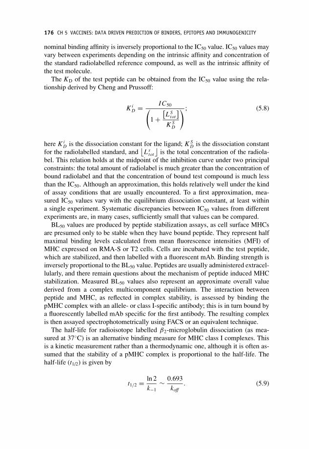
P1: OTA
c05 JWBK293-Flower September 4, 2008 19:34 Printer Name: Yet to Come
176 CH 5 VACCINES: DATA DRIVEN PREDICTION OF BINDERS, EPITOPES AND IMMUNOGENICITY
nominal binding affinity is inversely proportional to the IC50 value. IC50 values mayvary between experiments depending on the intrinsic affinity and concentration ofthe standard radiolabelled reference compound, as well as the intrinsic affinity ofthe test molecule.
The KD of the test peptide can be obtained from the IC50 value using the rela-tionship derived by Cheng and Prussoff:
K iD = I C50(
1 +[L S
tot
]K S
D
) ; (5.8)
here K iD is the dissociation constant for the ligand; K S
D is the dissociation constantfor the radiolabelled standard, and
⌊Ls
tot
⌋is the total concentration of the radiola-
bel. This relation holds at the midpoint of the inhibition curve under two principalconstraints: the total amount of radiolabel is much greater than the concentration ofbound radiolabel and that the concentration of bound test compound is much lessthan the IC50. Although an approximation, this holds relatively well under the kindof assay conditions that are usually encountered. To a first approximation, mea-sured IC50 values vary with the equilibrium dissociation constant, at least withina single experiment. Systematic discrepancies between IC50 values from differentexperiments are, in many cases, sufficiently small that values can be compared.
BL50 values are produced by peptide stabilization assays, as cell surface MHCsare presumed only to be stable when they have bound peptide. They represent halfmaximal binding levels calculated from mean fluorescence intensities (MFI) ofMHC expressed on RMA-S or T2 cells. Cells are incubated with the test peptide,which are stabilized, and then labelled with a fluorescent mAb. Binding strength isinversely proportional to the BL50 value. Peptides are usually administered extracel-lularly, and there remain questions about the mechanism of peptide induced MHCstabilization. Measured BL50 values also represent an approximate overall valuederived from a complex multicomponent equilibrium. The interaction betweenpeptide and MHC, as reflected in complex stability, is assessed by binding thepMHC complex with an allele- or class I-specific antibody; this is in turn bound bya fluorescently labelled mAb specific for the first antibody. The resulting complexis then assayed spectrophotometrically using FACS or an equivalent technique.
The half-life for radioisotope labelled β2-microglobulin dissociation (as mea-sured at 37◦C) is an alternative binding measure for MHC class I complexes. Thisis a kinetic measurement rather than a thermodynamic one, although it is often as-sumed that the stability of a pMHC complex is proportional to the half-life. Thehalf-life (t1/2) is given by
t1/2 = ln 2
k−1∼ 0.693
koff. (5.9)

P1: OTA
c05 JWBK293-Flower September 4, 2008 19:34 Printer Name: Yet to Come
MODERN MEASUREMENT METHODS 177
Here the t1/2 corresponds to the dissociation of the MHC-β2 microglobulin com-plex rather than the kinetics of the protein–ligand interaction.
We may wish to ask the question: which of these measures is best? Unfortunately,there is no simple answer. We have shown that a high degree of concordance existsbetween IC50 and BL50 values [2]. Nonetheless, there is to date no systemic com-parison of different measures, and so the relative verity and provenance of IC50,BL50 and t1/2 remains an open question. This issue also reinforces the imperativeneed to establish effective predictive methodologies that are able to substitute ef-fectively and reliably for experimental assays.
Modern measurement methods
More sophisticated, more convenient and more exact instrumental technology is, inother fields at least, now superseding such assays. In the case of enzyme reactions,enzyme kinetics is often followed by means of a readily detectable physical prop-erty (e.g. absorption, fluorescence or fluorescence polarization of one of the reactionpartners). Such instrumental methods can offer both increased convenience for theoperator and more easily realized experimental tractability; this type of approach isbeginning to be applied to MHCs. Steady state fluorescence spectroscopy is widelyused in the study of biomolecular interactions and kinetics and relies on measur-ing fluorescence change at a given wavelength. Time resolved fluorescence spec-troscopy measures the time dependence of fluorescence intensity after excitation,and is far more sensitive to conformational changes or interactions made by fluores-cent residues – tryptophan, tyrosine and phenylalanine – than steady state intensitymeasurements. Using polarized light allows time resolved fluorescence anisotropymeasurements, which can provide useful information about local residues as well asglobal protein motion. Mass spectrometry (MS) can also offer interesting insightsinto binding. Atomic-force microscopy (AFM) can determine the strength of in-termolecular interactions through controlled mechanical rupture of protein–ligandcomplexes. Other methods, such as densimetric and ultrasonic velocimetric titrationmeasurements, are also important, giving insights into specific molecular events,such as hydration changes.
Two techniques show particular promise: surface plasmon resonance (SPR) andisothermal titration calorimetry (ITC). SPR is an increasingly widely used tech-nique, as it can measure biomolecular interactions in real time in a label free en-vironment. SPR techniques are optical sensors based on the excitation of surfaceplasmons. They belong to a variety of refractometric sensing methods that includesgrating coupler sensors, Mach–Zehnder interferometers, Young interferometers andwhite light interferometers which measure refractive index changes induced bythe sensor’s electromagnetic field. One interaction partner is immobilized to thesensor surface while the other is in solution. It passes over the surface, binding

P1: OTA
c05 JWBK293-Flower September 4, 2008 19:34 Printer Name: Yet to Come
178 CH 5 VACCINES: DATA DRIVEN PREDICTION OF BINDERS, EPITOPES AND IMMUNOGENICITY
occurs, and is detected. Both ‘on’ and ‘off’ rates can also be accessed using thismethod.
Isothermal titration calorimetry
However, if one seeks a single methodology for obtaining relevant thermodynamicproperties of binding reactions, the current leader of the pack is probably ITC, andis rapidly becoming the method of choice. The main reason for this is that ITCsimultaneously generates global values for several parameters: the equilibrium con-stant and thus the free energy of binding (�G), which it computes from the shapeof the titration curve. However, ITC also measures enthalpy (�H), which it derivesfrom the integrated heat of reaction; entropy (�S), which is related to the differencein �G and �H; and also the heat capacity (�Cp) of the system. In ITC, a ligandis added stepwise at constant temperature to a solution of receptor protein and theoverall heat of the reaction is recorded.
In the absence of nonlinear effects, enthalpy and entropy terms can be obtainedusing the van ‘t Hoff relation:
ln K D = �H
RT− �S
R,
d(ln K D)
dT= �H
RT 2. (5.10)
The potential usefulness of this is obvious: plotting ln(KD) vs 1/T should describea straight line with a slope equal to (�H/R) and a y intercept of (�S/R). Van ’tHoff plots only identify part of the binding enthalpy: the part directly related to theobserved measurement signal. This means that only for a direct transformation froma defined initial state to a final state is the extracted enthalpy equivalent to �Hbind,as obtained by other methods, for example ITC. No intermediate steps are tolerated.�G typically has only mild temperature dependence within biological systems soa truly accurate and reliable estimate of enthalpy and entropy is seldom possibleusing van ’t Hoff plots.
What sets ITC apart is its ability to measure both affinity and its thermodynamiccontributions directly from heat changes during the binding process. Since suchchanges are observed during most binding reactions, ITC is broadly applicable,with applications ranging from chemical and biochemical binding to enzyme kinet-ics. ITC is both rapid and sensitive, but above all it is a direct method without theneed for chemical modification or immobilization. It is the only technique that mea-sures enthalpy directly, eliminating the requirement for van ’t Hoff analysis, whichis often time consuming and prone to error. However, interpretation of derived pa-rameters remains a pivotal challenge. Moreover, reliable analysis of the titrationcurve requires dissociation constants less affine than 10−9 M. To measure moreaffine compounds, the detection range must be extended by displacing lower affin-ity ligands. The kinetics of peptide binding – slow ‘on’ rates and slow ‘off’ rates –may also make certain MHC interactions beyond the practical limitations of currentITC technology.

P1: OTA
c05 JWBK293-Flower September 4, 2008 19:34 Printer Name: Yet to Come
LONG AND SHORT OF PEPTIDE BINDING 179
A step forward for the analysis of peptide–MHC binding will come when we de-ploy such methods and return richer and more informative affinity measurements.However, routine ITC again only operates on soluble molecules and is very timeconsuming, labour intensive and relatively expensive to perform. No method read-ily addresses the joint goals of effectively mimicking the in vivo membrane-boundnature of the interaction and the need for accuracy. In an ideal world we wouldlook at a variety of ‘internally rich’ data from ITC, as well as volumetric analy-sis and fluorescence spectroscopy, but to do this on an appropriate scale would beprohibitively time consuming and expensive. Where one might conceive of doingthis for one allele, there are dozens of frequent alleles within the human population.There are also many more which have interesting associations with disease. To pur-sue ‘internally rich’ assays for all ‘interesting’ alleles is clearly beyond the scope ofexisting methodology.
Ultimately, every relevant spectroscopic technique provides, to a greater or lesserdegree, valuable information complementary to that supplied by radioligand assaysor cell surface stabilization or β2m dissociation. It is only through a full and com-plete combination of several such techniques that the intricacies of protein–ligandinteraction may be carefully reconstructed. As yet, however, the application of thesemore sophisticated techniques, at least on a large scale, to peptide–MHC interac-tions, is distinctly limited.
Long and short of peptide binding
The peptides bound by class I MHCs have long been thought to be short. A rangeof eight to 11 residues is regularly quoted. This now seems to reflect our tenden-tious expectation more than unbiased observation. Peptides bound by class II MHCsare not directly constrained in length, which creates particular problems of its own(see below). X-ray crystallography has shown similarly bound peptide conforma-tions within class I MHC complexes, despite distinct peptide sequences. Peptidesbind to class I MHC with their N- and C-termini fixed at either end of the peptidebinding site, and with the centre of the peptide bulging out. Residues lining theMHC binding groove interact with the peptide and thus define the specificity of theHLA–peptide interaction.
This view arose partly from examination of crystal structures, which indicatedthat the MHC binding site most obviously accommodates peptides of eight to 10amino acids, and partly from cell surface elution studies, which also showed a re-stricted length spectrum. Obviously, as time has progressed, people have reinforcedthis view by only using synthetic peptides of eight or nine amino acids in length totest for binding.
The supposed length restriction imposed by class I MHCs is now being chal-lenged. There is increasing experimental evidence that a wide variety of other pep-tides also form stable, high affinity complexes with class I MHC class I, includingshorter peptides (4- to 5-mers), which we shall call ‘shortmers’, and much longer

P1: OTA
c05 JWBK293-Flower September 4, 2008 19:34 Printer Name: Yet to Come
180 CH 5 VACCINES: DATA DRIVEN PREDICTION OF BINDERS, EPITOPES AND IMMUNOGENICITY
peptides (up to 18-mers), which we shall call ‘longmers’. Such observations are out-side the prior norm, and are thus ignored or are subject to greater than usual scrutiny.
There is data to support the binding to MHCs of atypically short peptide se-quences: mouse CTLs can recognize truncated MUC1 epitopes bound to H-2Kb– SAPDTRP (7-mer), SAPDTR (6-mer) and SAPDT (5-mer) [3]. Other poten-tial shortmers binding to class I MHC molecules include: 3-mers (QNH), 4-mers(QNHR, ALDL and PFDL) and 5-mers (RALDL and HFMPT) [4].
Evidence for the binding of atypically long peptides is much stronger and cer-tainly more abundant. So much so that we shall eschew circumstantial results andinstead focus on unequivocal crystal data. Peptides of length eight or nine possessan essentially invariant conformation, while peptides of 10 amino acids ‘bulge’ inthe centre, with this bulging conformation varying considerably between structures.In all cases, N- and C-terminal interactions remain unchanged. Such trends only in-crease as peptide length increases.
Speir and colleagues reported the structure of a complex between the rat MHCallele RT1-Aa and a 13-mer peptide (ILFPSSERLISNR), a naturally eluted pep-tide derived from a mitochondrial ATPase [5]. Probst–Kepper report the structureof HLA-B∗3501 complexed with a 14-mer peptide (LPAVVGLSPGEQEY), whichoriginates from an alternate open reading frame of macrophage colony-stimulatingfactor [6]. The peptide is presented naturally and is recognized by T cells. As isusual for 8-mers and 9-mers, both the N and C termini of the peptide were embed-ded in the A and F pockets of the MHC peptide groove. The centre of this peptidebulges from the binding groove in an unpredictable way. Tynan reports the structureof a 13-mer viral epitope (LPEPLPQGQLTAY) bound to HLA-B∗3508 [7].
These crystal structures find support in functional observations of CTL recog-nition of Longmers. Reports of human 10-mer and 11-mer CD8 epitopes re-stricted by class I MHC alleles are common place, as are 10-mer peptides bind-ing to mouse class I. SYFPEITHI is awash with class I peptides that are upto 15 amino acids in length in humans and up to 16 amino acids in mice.Jiang reports convincing evidence of an H2-Kd-restricted CTL response to a 15-mer epitope (ELQLLMQSTPPTNNR) from the F protein of respiratory syncytialvirus [8]. 12-mer C-terminal variants of VSV8 have been shown to bind H-2Kb(RGYVYQGLKSGN). Thus, the apparent potency of responses to such longmersis indicative that peptides longer than the norm do not represent a major structuralbarrier to recognition by TCRs.
The class I peptide repertoire
Clearly, then, the evidence as outlined above suggests strongly that the natural reper-toire of class I MHC presented peptides is far broader than has generally been sup-posed. We can compound this repertoire in various ways. One is through aberranttranscription and translation of viral and self-proteins. Various mechanisms pertain

P1: OTA
c05 JWBK293-Flower September 4, 2008 19:34 Printer Name: Yet to Come
PRACTICALITIES OF BINDING PREDICTION 181
such as read through, which leads to alternative open reading frames, or alternativesplicing, which generates protein isotypes with different sequences at exon–exonboundaries. Both of these have been shown to generate immunogenic epitopes. Au-toantigen and self-tumour antigen transcripts in particular experience higher rates ofalternative splicing in response to stimuli and these can lead to elevated responses.Alternative splicing is a major factor driving proteomic diversity and provides apartial rationale for the gulf between the small size of genomes and the enormouscomplexity of higher eukaryotes. Most alternative splicing occurs in the coding re-gion. The most commonly observed alternative splicing event is exon skipping inwhich an exon or set of continuous exons are permuted in different mRNAs. Lessfrequent are the use of donor and acceptor sites and intron retention.
We can expand the burgeoning repertoire still further by accounting for the effectof post-translational modifications (PTMs). These can take many forms, includingglycosylation and lipidation. Glycosylated proteins can be targets for binding bycell surface receptors based on sugar binding leptin domains. Glycosylated epi-topes can also be bound by TCRs and antibodies. Lipids can act as epitopes directlythrough their presentation by CD1. PTMs can also be transitory, such as phospho-rylation, or more permanent, such as modified amino acids. Many of these can bepart of functional epitopes recognized by the immune system. Glycosylation of aprotein, for example, is dependent on the presence of sequence patterns or motifs(Ser/Thre-X-Asn for N-linked glycosylation and Ser/Thr for O-linked) but this isnot enough to predict them correctly. If these motifs are present at solvent inac-cessible regions of a protein rather on the surface then they will not be glycosy-lated. Moreover, the other residues which surround these patterns will also affectthe specificity of the glyscosylating enzymes: Pro as the X in the Ser/Thre-X-Asnmotif for N-linked glycosylation will essentially prevent glycosylation. Glycosyla-tion, in particular, is also very dependent on context, and it is thus a system propertyof an organism, and can vary considerably in terms of the nature and extent of thedifferent sugars that can become attached to proteins, at least in eukaryotic systems.Taken together these greatly increase the potential size and diversity of the immuno-genic repertoire – or immunome – of reactive peptides. One can argue, and arguecogently, that the only realistic way to address this potential enormity of the peptiderepertoire is via computational analysis and prediction.
Practicalities of binding prediction
Although there are ways to generate much richer and more explicit data, using ITCfor example, practicalities dictate that the immunoinformatician must instead lookat a very mixed ragbag of quantitative and semi-quantitative measures of greatlyvarying provenance. Unfortunately, the wide variety of quantitative measures ofbinding currently in use (KD, IC50, BL50, t1/2, etc.) has proven confusing in theextreme. This necessitates the adoption of data-fusion strategies and conceptual

P1: OTA
c05 JWBK293-Flower September 4, 2008 19:34 Printer Name: Yet to Come
182 CH 5 VACCINES: DATA DRIVEN PREDICTION OF BINDERS, EPITOPES AND IMMUNOGENICITY
simplifications intended to help combine or streamline this bewildering set of bind-ing measures.
Although IC50 values are amongst the commonest and most reliable measuresreported even they require some kind of data manipulation to make them tractablepractically speaking. In QSAR studies, for example, IC50 values are often convertedinto log[1/IC50] values, since the values typically range over several orders of mag-nitude. For convenience, this is also expressed as –log10[IC50] or pIC50. Such valuesare used as dependent variables in QSAR regression. pIC50 values can be related tochanges in the free energy of binding, through the approximation �Gbind α –RT lnIC50.
Another widely used conceptual simplification involves reclassifying peptides aseither nonbinders or as high binders, medium binders and low binders. For example,the schema introduced by Vladimir Brusic classifies binders using these criteria:nonbinders > 10 µM, 10 µM > low binders > 100 nM, 100 nM > medium binders> 1 nM, high binders < 1 nM. Such broad schemes also allow us to compensatefor the inherent inaccuracy in MHC binding measurements.
Another, albeit underexploited, strategy involves a binary classification of pep-tides into binders and nonbinders. In general, data has not been designed to spanthe whole of available chemical, or rather peptide, space, but emerges in a haphaz-ard fashion from a large collection of experiments designed for other purposes. Theinterests of the immunoinformaticians may be very different to those of the orig-inal experimenters. One may be interested in predicting affinities or in predictingepitopes. So, if one wishes to classify by class, methods can then use alternate datasources equally. Any affinity measure can be used to identify binders, be that anIC50 value or a t1/2 or a BL50 value. Likewise, a cell surface peptide eluted from anMHC must obviously be bound, with reasonable affinity, before being eluted. Anypeptide found from overlapping peptide scanning to be an epitope must, again, havebeen bound before being recognized. Thus all of these conceptually distinct meansof classification yield equivalent definitions of binder versus nonbinder. This allowsus to rationalize a much wider array of data. This is a potentially useful featureof such an approach, though not one which has been much exploited in the past.By combining multiple data sources for binders with multiple data sources for non-binders we can envisage the development of much more robust, much more general,much more complete – and hopefully much more successful – future methods forthe prediction of MHC binders.
Binding becomes recognition
Once a peptide has bound to an MHC to form a pMHC complex, in order to berecognized by the immune system a pMHC complex has to be recognized by oneof the TCRs of the T cell repertoire. Within the human population there are anenormous number of different possible variant genes coding for MHC proteins,each exhibiting distinct peptide binding sequence selectivity. T cell receptors, in

P1: OTA
c05 JWBK293-Flower September 4, 2008 19:34 Printer Name: Yet to Come
IMMUNOINFORMATICS LENDS A HAND 183
their turn, also exhibit different affinities for pMHC. The binding of TCRs to pMHCis weaker (in the micromolar range) than the binding of peptide to MHC (in thenanomolar range). The collective selectivity of the MHC and TCR establishes thepower and flexibility of peptide recognition in the immune system and through thisphenomenon the recognition of foreign proteins and pathogens.
It was long ago realized that logistic restrictions preclude routine yet exhaustivetesting of overlapping peptides as a route to epitope discovery, making computa-tional prediction an attractive alternative. It is generally accepted that a peptidebinding to an MHC may be recognized by a TCR if it binds better with a pIC50
> 6.3, or a half-life > 5 minutes, or some similar figure. Some peptides bindingat these affinities will become immunodominant epitopes, others weaker epitopes,and still others will show no T cell activity. There is some evidence suggesting thatas the MHC binding affinity of a peptide rises, then the greater the probability thatit will be a T cell epitope. The trick – the unsolved trick – is to determine whichwill be recognized by the TCR and thus activate the T cell. Generally, the approachtaken has been to whittle down potential epitopes to a more convenient numberfor experimental evaluation by using prediction of MHC binding to eliminate allnonbinders or by choosing only high binding peptides.
Immunoinformatics lends a hand
Consider a protein of, say, 300 residues. If we are interested in epitope responsesmediated by a class I MHC, then we need to consider peptides of eight to 16 residuesas potential binders and epitopes. That equates to 2592 MHC–peptide binding as-says per allele. We could be interested in one allele or the three or six in a haplotypeor we could be looking at a panel of common alleles within a population, whichcould easily run into dozens. If we include class II responses the number increasesagain. This will generate an unknown number of binding peptides or candidate epi-topes, each of which will need to be tested using one of a great variety of differentmeasures of T cell activation, such as T cell killing or thymidine incorporation,inter alia. From this time and labour intensive, and financially costly, undertaking,will emerge a small handful of peptides possessed of varying degrees of immun-odominance. While this kind of exhaustive empirical study has, in part at least,been attempted in practice, it becomes utterly inconceivable for a whole proteomesay. At the time of writing, a useable HTS approach to this problem has not beenforthcoming, though many continue to work on such methods. Much better wouldbe to convert the laborious cost and time consuming binding assays to a computeralgorithm.
MHC binding is the stage of antigen presentation which we understand best;it is also, in all probability, the most discriminating stage with the presentation-recognition pathway. In what follows, we will focus on predicting class I MHCbinding and deal with class II as a special case.

P1: OTA
c05 JWBK293-Flower September 4, 2008 19:34 Printer Name: Yet to Come
184 CH 5 VACCINES: DATA DRIVEN PREDICTION OF BINDERS, EPITOPES AND IMMUNOGENICITY
Moreover, despite a clear if emerging understanding that the repertoire of pep-tide lengths capable of inducing CTL responses has recently increased considerablefor class I, most data is still focused on 9-mers and HLA-A∗0201. Thus the dataavailable to build models typically consists of blocks of 9-mer sequences and anassociated binding value, such as an IC50. These are essentially pre-aligned, sincethe geometry of the MHC binding pocket prevents binding in several registers. Datais also biased by MHC allele: HLA-A∗0201 is by far the most common allele forbinding data, outnumbering its rivals by a factor of five.
Motif based prediction
Initial attempts to computerize MHC binding peptide identification led to the de-lineation of motifs which supposedly characterize allele-specific peptide speci-ficity. The binding motif is an idea which still commands wide popularity amongstimmunologists. The first real attempt to define allele- and sequence-specific bind-ing motifs for MHC–peptide interaction was undertaken by Alessandro Sette andcoworkers [9]. In the first instance, they defined motifs for the mouse alleles: I-Adand I-Ed. They assayed affinities for a sizeable set of synthetic peptides with di-verse origins and also measured overlapping peptides of staphylococcal nuclease.Accuracies of about 75% were later quoted for these alleles. The delineation andrefinement of motifs has progressed through a body of work embodied in a largeand accumulating literature, with human and mouse dominating the list of investi-gated organisms, although the rat, cow and some primate species have also receivedattention.
Motifs characterize MHC peptide-specificity in terms of dominant anchor posi-tions with a strong preference for certain amino acids: particular positions within apeptide expressing a highly restricted set of tolerated amino acids. Such anchors arethought essential for binding. Human class I allele HLA-A∗0201, which is proba-bly the best studied allele of all, has, for a nonameric peptide, anchor residues atpeptide positions P2 (accepting leucine and methionine) and P9 (accepting valineand leucine).
This simple model has been nuanced over time. Secondary anchors may also bepresent; these are residues that are favoured for binding yet are not essential. Thesequencing of peptides eluted from the cell surface show amino acid preferencesat most positions, though this may be nothing more than bias intrinsic to proteinsequences. Particular residues at particular positions may also have a negative effecton binding.
Notwithstanding the perceived success of binding motifs, many problems persist.While one can combine the contributions made by primary and secondary anchorsinto an approximate measure of binding affinity, the principal problem and mostvexatious feature associated with motifs is that their use is a deterministic method ofprediction: a peptide will bind to an MHC or it will not. Even a perfunctory reading

P1: OTA
c05 JWBK293-Flower September 4, 2008 19:34 Printer Name: Yet to Come
THE IMPERFECT MOTIF 185
of the immunological literature indicates that the use of motif matching producesmany false positives and, in all probability, also generates many false negatives,though such supposed non-binders are seldom assayed. Many peptides are bothaffine and motif-negative, that is they possess noncanonical sets of amino acidslying outside established definitions for an allele. There are many more examplesof motif-positive peptides that do not bind.
Proponents of motifs characterize one or more usually two anchor positions asbeing all important. This conjecture, which is based on observed sequence patternsevident from pool sequencing and from individual epitopes, holds that only pep-tides with a sequence matching such a sparse pattern will bind. This makes littlesense and is not compatible with what we know about molecular binding events.The whole of medicinal chemistry argues against this view. It would be better todescribe this process by saying that MHCs bind peptides with an equilibrium bind-ing constant dependent on the nature of the bound peptide’s sequence.
The imperfect motif
Several experimental analysis studies have addressed this issue directly. Stryhn andcolleagues generated all systematic single mutations of the 8-mer peptide FEST-GNLI and observed that mutations at positions two and eight resulted in the great-est changes in affinity, but that contrary to expectation, few substitutions abolishedactivity [10]. Thus it is possible to abrogate canonical anchors without eradicatingbinding. Likewise, Doytchinova and coworkers introduced all 20 amino acids at an-chor positions two and nine of a super-high affinity 9-mer peptide binding to HLA-A*0201 [2]. Only a few substitutions completely abolished affinity; most did not.About half of these peptides were sufficiently affine to be potential epitopes, withaffinities above a nominal threshold. This study demonstrates that it is possible tomanipulate A2 binding affinity in a rational way, raising resulting peptides to affin-ity levels two orders of magnitude greater than previously reported. The study alsoshowed that optimized nonanchor residues can more than compensate for nonopti-mal substitutions at anchor positions. Clearly, the whole of a peptide contributes tobinding, albeit weighted differently at different positions.
For class I, and in all probability also for class II, through the use of relativelystraightforward prediction methods it is possible to design peptides with high affin-ity for MHC, and affine peptide with noncanonical anchors, at least for well char-acterized alleles, with extra affinity arising from favourable interactions made bynonanchor residues. Such novel, non-natural sequences will act as super agonists,antagonists or blockers of MHC mediated T cell responses.
Notwithstanding, the problems of motifs, many papers continue to report theiruse, and for obvious reasons. The list of already defined motifs is long and coversmany alleles in many species. As a technique, the use of motifs is admirably sim-ple, as it can be implemented by eye or more systematically by computer. A scan

P1: OTA
c05 JWBK293-Flower September 4, 2008 19:34 Printer Name: Yet to Come
186 CH 5 VACCINES: DATA DRIVEN PREDICTION OF BINDERS, EPITOPES AND IMMUNOGENICITY
is performed, searching one or more protein sequences seeking potential bindingpeptides. While binding motifs seem, to me at least, counter-intuitively simplistic,nonetheless, prediction using motifs is preferable to no prediction at all.
Other approaches to binding prediction
However, the inadequacy of motifs has been clear to immunoinformaticians forsome time, and now alternative techniques abound. The different types have, asone might expect, different strengths and different weaknesses. The first step awayfrom deterministic motifs came with the work of Kenneth Parker, which is based onregression analysis. It gives quantitative predictions of the dissociation half-livesof β2-microglobulin from the MHC complex. Apart from its inherent utility, themain contribution made by this approach is that it was the first to be made availableonline (http://bimas.dcrt.nih.gov/molbio/hla bind/). Usually known as BIMAS, thissite is still widely used.
A different strategy has been the direct utilization of data from binding exper-iments, as realized by the use of so-called positional scanning peptide libraries(PSPLs) [11]. There are many more methods of this kind. Though doubtless ofvalue, they nonetheless betray several limitations. They do not constitute a system-atic approach to solving the MHC–peptide binding problem. Rather, as a group theyrepresent a set of distinct and inconsistent solutions to the same problem. The mea-sures of binding are different, as is the degree of quantitation, and they also lacksufficient application to corroborate their predictive utility. Moreover, few, if any,of the papers describing such work make available sufficient detail for others to usetheir methods independently.
Many other examples of diverse empricial approaches have emerged in a piece-meal fashion over the years. While many are useful, they again do not constitutea systematic approach to binder prediction. Instead, we should look to establishedmethods of proven provenance. Indeed, the principal approach to epitope predic-tion has involved the application of validated and well-used methods derived fromcomputer science data mining or robust multivariate statistical techniques.
The goal of such studies is to take a protein or genome sequence and identifywithin it the presence of MHC binding peptides and/or T cell mediated epitopes. Toachieve this, predictive methods require three things: a structural representation ofthe peptides being used to train them; an equivalent list of biological activities cor-responding to the peptides being used in training; and some form of computationalengine capable of generating cohesive relationships between the particular descrip-tion of the peptides and the associated activities. The outcome of this process is apredictive model relating peptide structure to probable activity.
Beyond immunoinformatics, activities can be any sort of physical property or bi-ological activity: enzyme inhibition or whole organism toxicity or melting point orwhatever. Moreover, it could just as easily be some utterly different type of quantity,

P1: OTA
c05 JWBK293-Flower September 4, 2008 19:34 Printer Name: Yet to Come
REPRESENTING SEQUENCES 187
such as stock market prices or psychological status, which can often be expressedin terms of a categorical descriptive, or ontological, language. As we elaborate be-low, activities or properties can be quantitative, monotonic real numbers (IC50s forexample) or a binary, two category decision (epitope versus nonepitope, for exam-ple) or they can take the form of several categories (nonbinder, low binder and highbinder, for example). The rather obvious point we make here is that the approach isa quite general one. Probably the least generic aspect of the process is the structuraldescription used.
Representing sequences
Many different representations of amino acids have been proposed, and many used,to help predict the properties and functions of peptides and proteins. Many predic-tion methods use indicator variables: these are binary strings indicating the presenceor absence of particular amino acids. For example a bit string corresponding to:
00100000000000000000
would, depending on the amino acid order used, represent cysteine, while
10000000000000000000
would equate to, say, alanine. Such a 20-vector is required at each peptide position;which is effectively 180 values for a nonameric peptide. From such a representationa regression equation or other model, such as an ANN, is built.
An alternative way of representing the amino acids within a peptide is to useone or more property scales. These were discussed at length in Chapter 4. Overtime, a sizeable literature has evolved which describes, evaluates and cataloguesthe properties of the 20 common amino acids. Pre-eminent amongst attempts atcompilation is the AAindex, a database of amino acid scales, mutation matrices andfolding potentials.
In a sense, the computational or decision engine described here can be conceivedof as a probabilistic generalization (or specialization, given your point of view) ofan inference or reasoning engine, familiar from that branch of computer sciencewhich concerns artificial intelligence. Such an inference engine can use any form ofcomputational approach to link activity to structure: inductive logic programming,artificial neural network, Bayesian statistics or robust multivariate statistics amongstmany more. From the viewpoint of a biologist this is irrelevant as long as the methodworks. The goal and perhaps key motivation is, of course, to find methods that workas well as possible.

P1: OTA
c05 JWBK293-Flower September 4, 2008 19:34 Printer Name: Yet to Come
188 CH 5 VACCINES: DATA DRIVEN PREDICTION OF BINDERS, EPITOPES AND IMMUNOGENICITY
Computer science lends a hand
A number of groups have used techniques from computer science data mining, suchas artificial neural networks (ANNs) and hidden Markov models (HMMs), to pre-dict peptide–MHC binding affinity. ANNs and HMMs are, for slightly different ap-plications, the particular favourites when bioinformaticians look for tools to buildpredictive models. Latterly, support vector machines (SVMs) and robust multivari-ate statistics (as are typically applied to quantitative structure activity relationships)have been added to the mix. The difference between QSAR and artificial intelli-gence methods is mainly a semantic one: in practice both seek to achieve the samegoal and function in similar ways.
Some simple yet useful rules have been proposed for selecting methods givenavailability of data; they suggest using structural modelling when binding data isabsent, motifs when it is very scanty and AI techniques and the like when datais plentiful. Datasets of 50–100 peptides can be used to build quantitative ma-trices or SVMs, while larger datasets are needed to build HMM or ANN basedmodels.
Artificial neural networks
The artificial neural network – or ANN or often just NN – is a so-called bio-inspiredalgorithm. It is a machine-learning method which purports to model itself on a bi-ological neural system. Like most of bio-inspired computing, its actual inspirationcomes not from a real down-and-dirty piece of biology but from a sanitized theoret-ical conception of such a system: an abstraction of biology rather than the biologyitself. ANNs have for some time been tools of choice for pattern recognition anddata classification, due to their ability to learn and generalize, finding applicationsacross science and engineering.
ANNs have many perceived advantages: a high interpolative capability; they arecapable of dealing with complicated and imprecise data; they are often more effi-cient than developing models from first principles; and they are relatively straight-forward to use. These features allow patterns to be identified and trends to bespotted that may be otherwise obscured from human comprehension by their com-plexity. However, the development of ANNs is often complicated by several ad-justable factors whose optimal values are seldom known initially. These can in-clude, inter alia, the initial distribution of weights between neurons, the numberof hidden neurons, the gradient of the neuron activation function and the trainingtolerance.
Other than chance effects, to which all predictive methods are prone, ANNs havethree main limitations: overfitting, memorization or overtraining and interpretation.In overfitting, well-sampled regions of the training data are learned well and predic-tions are reliable. However, in poorly sampled regions the underlying trend or trends

P1: OTA
c05 JWBK293-Flower September 4, 2008 19:34 Printer Name: Yet to Come
ARTIFICIAL NEURAL NETWORKS 189
in the data are not captured accurately. The result is an ANN which is essentiallyrandom in prediction: the ANN performs well for the training data but poorly undercross validation. Under specified or over specified ANNs both suffer from overfit-ting. As an ANN can always reduce the error by adjusting internal parameters, anetwork runs the risk of simply fitting a more complex and specific model than isnecessary. As a model better fits the training data, it becomes less able to generalize.This is called overtraining (or memorization) as the ANN is able to recall trainingdata well but cannot predict reliably within its interpolative data space, let alone out-side it. As new and better ANNs have been developed, and basic statistics applied tothem, problems of overtraining and overfitting have receded. Interpretation remainsa formidable issue: few people can readily visualize, or properly understand, thecomplex weighting schemes used by ANNs.
There are many types of ANNs: feed-forward networks (or perceptron net-works), competitive networks (or Hamming networks), and recurrent networks (orHopfield networks). Different ANNs behave differently, yet, in general, an ANNcan be thought of as a group of arbitrary processing units (called nodes or neurons)organized into layers and linked by weighted connections. The output of a node iscontingent on several things: the data feed into it via its several connections, theweighting of each connection and the node’s own threshold. Information (one ormore independent variables) passes first into the input layer of an ANN, then prop-agates through one or more hidden layers of neurons, and is ultimately transmittedas one or more dependent variables from the output layer. The number of input andoutput variables specifies the node number in the input and output layers. The num-ber of hidden or internal layers and the nodes these layers contain varies betweenANNs. These, together with the direction of information flow, are key features ofnetwork architecture.
Application of an ANN to a practical problem will typically involve several steps:compiling the training data, choosing the network architectures, training the net-work and validating it. Training minimizes a penalty function, corresponding to thedeviation between observed and predicted values within the training set, through theoptimization of node weights and layer biases. Training data passes into a networkin cycles, one such iteration is called an epoch. Alterations to the network weightingscheme can be applied at the end of an epoch or after each training pattern depend-ing on learning parameters. It may also be necessary to test several architecturesand network types, selecting the most suitable type.
Many immunoinformaticians have adopted the ANN when seeking to predictpeptide–MHC binding. Adams and Koziol [12] were among the first to use ANNsto predict peptide binding to HLA-A∗0201. Using 552 nonamers and 486 decamers,they achieved a predictive accuracy of 0.78 for a two category classification of goodor intermediate binding versus weak or nonbinding peptides. Gulukota and De Lisi[13] used two different methods – ANN and statistical parameter estimation – topredict HLA-A∗0201 binding for 463 9-mer peptides. They found that the ANN wasbetter at rejecting false positives, while statistical parameter estimation eliminatedmore false negatives.

P1: OTA
c05 JWBK293-Flower September 4, 2008 19:34 Printer Name: Yet to Come
190 CH 5 VACCINES: DATA DRIVEN PREDICTION OF BINDERS, EPITOPES AND IMMUNOGENICITY
However, the premier practitioner of ANN-based MHC binding prediction isVladimir Brusic. Over many years, he, his colleagues, and his coworkers, havedeployed several data-mining algorithms including ANN, as well as HMMs andevolutionary algorithms [14]. His work addresses both class I and class II MHCalleles and within his own classification scheme his models are highly predictive.
Hidden Markov models
Another mainstay of bioinformatics prediction is the so-called Hidden MarkovModels or HMM. An HMM is the simplest form of dynamic Bayesian networkand models a system as a Markov process with unknown parameters. A Markovprocess is one where the current status of the system is independent of the trajec-tory traced by past states, being contingent on the previous state and not ones priorto that. When an HMM is constructed, relationships between observed and hiddenparameters are determined. In a ‘hidden’, as opposed to a regular, Markov model,internal states cannot be seen but tokens output by the model are visible.
HMMs have found many applications not least of which are those in bioinfor-matics. Interestingly, however, HMMs have been little used for immunoinformaticproblems. Mamitsuka [15] predicted MHC binding using HMMs. Under crossval-idation, the accuracy of this method was 2–15% better than alternative methods,including ANN.
Support vector machines
The last of the main AI-based data-mining methods is the so-called support vectormachine (SVM). SVMs, which are an example of a kernel-based method, havelargely taken bioinformatics by storm. Their inherent accuracy and their ability torepresent nonlinear relationships is seemingly compelling. A single SVM is at itssimplest a binary classifier which induces a decision boundary between classes (i.e.epitope versus nonepitope) using an appropriate amino acid representation. Trainingdata is embedded in a high-dimensional feature space, and the method creates ahypothesis space of linear functions trained with a learning bias. To find a boundarybetween classes, an SVM maximizes the margin between them, choosing a linearseparation in feature space, by defining a hyperplane through this high-dimensionalspace. This hyperplane should separate the classification classes while maximizingthe distance between the hyperplane and both groups of data. A ‘kernel function’K (xi , x) maps the data from an input space to a feature space, where it is classified.Thus the choice of kernel and its parameters is crucial. Several kernels are available:linear, polynomial, sigmoid and radial basis function (RBF). SVMs are describedwith lucent clarity elsewhere [16].

P1: OTA
c05 JWBK293-Flower September 4, 2008 19:34 Printer Name: Yet to Come
PARTIAL LEAST SQUARES 191
As a result of their success, a whole tranche of SVM-based methods for classI epitope prediction have been developed [17, 18]. Most SVM methods undertakediscriminant analysis, but we have seen encouraging performance with quantitativeprediction using support vector regression (SVR). For SVM regression analysis,regression is performed in a multi-dimensional feature space, but without overlypenalizing small errors. Our SVR method adopts an ‘11-factor encoding’ scheme,which accounts for similarities in peptide properties [19]. Subsequently, this ap-proach has been implemented in the SVRMHC server covering more than 40 classI and class II MHC molecules.
Robust multivariate statistics
Robust multivariate statistics methods address quantitative structure-activity rela-tionship (QSAR) problems, such as are common in drug design. As opposed toAI techniques, QSAR methods are based on different, and possibly more rigorous,types of statistical analyses including, amongst others, multiple linear and contin-uum regression, discriminant analysis and partial least squares (PLS). PLS-basedmethods in particular are predictive tools of wide applicability, and comprise aprincipal cornerstone of modern cheminformatics. Recently, bioinformatics appli-cations of PLS have begun to flourish.
Partial least squares
Partial least squares (PLS) regression, the principal workhorse of QSAR analyses, isan extension of multiple linear regression (MLR). It works by relating one or moredependent variables (usually biological activities) to the values of several indepen-dent variables – usually called descriptors – and uses them as predictors. In MLR,a dependent variable y is predicted from N descriptors {x1, x2, . . ., xN}, combinedwith appropriate weighting as:
y = b0 + b1x1 + b2x2 + . . . + bN xN
Here bi is a regression coefficient, and b0 is the residual or intercept coefficient.Given n observations, the input data comprises an n component linear responsevector y and an n by N covariance matrix.
PLS generalizes and combines features from principal component analysis andmultiple regression. An advantage of PLS is that it can deal with very large sets ofcorrelated independent variables. It seeks to identify new variables constructed fromlinear combinations of the original variables on the assumption that these latentvariables can better explain both the response and covariate space. Thus, for the s

P1: OTA
c05 JWBK293-Flower September 4, 2008 19:34 Printer Name: Yet to Come
192 CH 5 VACCINES: DATA DRIVEN PREDICTION OF BINDERS, EPITOPES AND IMMUNOGENICITY
latent variables and the dependent variable y:
y = t1q1 + t2q2 + . . . tsqs + ys .
Similarly for the covariate descriptor space:
X = t1 p1 + t2 p2 + . . . + ts ps + Es .
Es and ys are residuals, which, for well-conditioned data, is assumed to be small;at least when compared to the variance explained by the latent variables. Once thelatent variables have been found, the pi and qi coefficients can be fitted by MLRusing the latent variables as descriptors. In immunoinformatics applications, thedependent variables are usually binding affinities between MHC and peptide. TheIC50 values (the dependent variable y) are typically represented as negative loga-rithms (-log10IC50 or pIC50). The predictive ability of the model is usually validatedusing ‘leave one out’ cross-validation (LOO-CV).
One may feel justified unease about aspects of the PLS methodology, particularlyfor immunoinformatic datasets. No method is perfect nor, in the present context, isa perfect one ever likely. The failure of different methods is, however, as muchto do with problems that relate to the underlying data, as it is to do with minormethodological flaws. A general criticism of statistical and artificial intelligencebased approaches is overfitting. Usually the covariate matrix is ‘over square’ andhas insufficient degrees of freedom to allow an exhaustive and entirely robust anal-ysis to be undertaken. Many terms in a PLS regression equation or neural networkor SVM kernel model will be poorly populated, with only a handful of observations,inflating the associated errors and reducing the associated reliability of prediction.However, such limitations are common to all approaches and they underline theimportance of training and test data, and its implicit structure, over and above themethodological approaches used to study and predict it.
Quantitative structure activity relationships
A number of groups have applied QSAR to immune problems. Chersi et al. [20]used QSAR to optimize binding of 9-mer peptides to the MHC allele HLA-A∗0201by increasing the flexibility of amino acids at position four. Bologa and others char-acterized the type of amino acid required for high affinity binding to the HLA-A∗0201 allele. In a related study, Rovero et al. [21] analysed peptide binding to thehuman class I allele HLA-B∗2705. The main contribution of the research group atthe Jenner Institute to the immunoinformatic deployment of QSAR techniques is theadditive method. It is a PLS-based technique which leverages the Free–Wilson prin-ciple. This states that chemical subsitituents (or amino acid positions in our case)

P1: OTA
c05 JWBK293-Flower September 4, 2008 19:34 Printer Name: Yet to Come
OTHER TECHNIQUES AND SEQUENCE REPRESENTATIONS 193
are independent and that the presence or absence of such groups is correlated withbiological activity. Thus, for a peptide, the binding affinity is represented by thesum of constant and strictly additive contributions made by different amino acidsat different positions. As a further indication that all predictive methods share com-mon features, the IBS approach, as developed by Parker and others [22] is based ona similar concept. We extended the classic Free–Wilson model with terms account-ing for interactions between positions. Thus, the binding affinity of a peptide willdepend on the contributions of the amino acid side-chains at each position and theinteractions between the adjacent and every second side-chain:
pI C50 = P0 +9∑
i=1
Pi +8∑
i=1
Pi Pi+1 +7∑
i=1
Pi Pi+2 (5.11)
where P0 accounts, albeit nominally, for the contribution of the peptide backbone,∑9i=1 Pi is the position-dependent amino acid contributions,
∑8i=1 Pi Pi+1 is the
contribution of interactions between adjacent positions, and∑7
i=1 Pi Pi+2 is the con-tribution from every second side-chain interaction. We reasoned that more distantposition interactions can be neglected. Initially, this was applied to a set of 340nonamer peptides binding to HLA-A∗0201. Its MLR parameters were r2 = 0.898,number of components (NC) = 5. The ‘leave-one-out’ cross validation (CV-LOO)gives q2 = 0.337, SEP = 0.726, NC = 5, mean |residual| value = 0.573. The addi-tive method lacks the ability to extrapolate beyond the types of amino acid used intraining. However, it does provide a quantitative assessment of any amino acids atall positions in the peptide.
Analyses for a variety of human and mouse class I alleles have now been un-dertaken using this approach. We find that, in practice, and except for very largedatasets such as HLA-A∗0201, the adjacent and one to three interactions can beneglected, with the best model provided by the position-wise coefficients only.
Other techniques and sequence representations
In terms of the number of applications, four techniques – ANNs, HMMS, SVMand PLS – have dominated class I binding prediction. There are many moretechniques which have yet to be applied to immunoinformatic problems. Com-puter science is replete with successful pattern recognition algorithms of allkinds, from Bayesian belief networks to nearest neighbour classifiers. WEKA(http://sourceforge.net/projects/weka/) is a well-known software implementation inJava of many such techniques. Examples of other techniques which have been ap-plied to MHCs include Reche’s work on sequence profiles, boosted metric learningand logistic regression [23].
When the data are sufficiently numerous, and sample the available space ade-quately, most data-mining algorithms will perform well producing predictive mod-els. In twentyfirst century computing, the crux is always the data. This is particularly

P1: OTA
c05 JWBK293-Flower September 4, 2008 19:34 Printer Name: Yet to Come
194 CH 5 VACCINES: DATA DRIVEN PREDICTION OF BINDERS, EPITOPES AND IMMUNOGENICITY
true in immunoinformatics. Much is to be gained by choosing and generating newand improved datasets. Little is now to be gained from quibbling about the relativemerits of one induction engine over that of another. Existing data – in terms of scopeand accuracy – is simply not up to the job.
One reason for this dearth is the large number of alleles which have not beenevaluated. This is not a problem that can be addressed by swapping AI techniques,but necessitates a changed strategy. The definition and analysis of supertypes is oneroute to addressing this problem (Chapter 6). Another route is via methods thatuse relationships at the sequence level between MHC alleles to allow algorithmsto induce multi-allele binding models which combine binding data from distinctbut related alleles. The relative contribution made by data from different alleles ismoderated by some measure of sequence or structural similarity between alleles,which can be defined from supertypes or directly from key binding site residues. Anumber of such methods are now available [24–27]. This is an approach offeringimmense potential, since it seems unlikely that any experimental method, even onebased on HTS, will, in the foreseeable future, deliver sufficient quantities of bindingdata to support more traditional single-allele modelling.
Amino acid properties
Another methodological innovation, this time with the potential to generalize withinpeptides binding to a single allele, is a class of methods that use quantitative de-scriptions of amino acid properties rather than use indicator variables to representpeptide sequences. As we saw in Chapter 4, property scales suitable for this purposeare potentially very numerous. Extant scales are contained in the AAindex database(www.genome.ad.jp/aaindex/ ). AAindex consists of three sections: AAindex1 con-sists of lists of published amino acid scales; AAindex2 contains corresponding listsof published mutation matrices; and AA index3 lists threading potentials. AAin-dex1 contains well over 500 annotated indices. Each entry has an accession code,publication details, correlation scores to similar indices with the database, as wellas the index itself.
Others have tried to summarize or consolidate this enormity of published aminoacid scales in a small set of condensed or consensus indices. The best known, andin our hands the most useful, example is the z scales developed by Svante Wold andcoworkers. A principal component analysis (PCA) was used to convert 29 otherscales into three amino acid scales – z1, z2 and z3 – which describe hydrophilic,steric and electrostatic/polar properties, respectively. These scales were later sup-plemented with two extra scales: z4 and z5. Collectively, the five scales (z1–z5) de-scribe the hydrophilicity, size, polarity and electrostatic properties of amino acids(Table 5.2). A variety of other condensed scales have been proposed in recenttimes [28].
Methods based on amino acid scales seek to escape the limitations imposed bythe lack of all amino acids at all positions in existing peptide binding data. A set of

P1: OTA
c05 JWBK293-Flower September 4, 2008 19:34 Printer Name: Yet to Come
DIRECT EPITOPE PREDICTION 195
Table 5.2 The five z values representative of amino acid properties
z scale
Amino acid z1 z2 z3 z4 z5
A 0.24 −2.32 0.60 −0.14 1.30C 0.84 −1.67 3.71 0.18 −2.65D 3.98 0.93 1.93 −2.46 0.75E 3.11 0.26 −0.11 −3.04 −0.25F −4.22 1.94 1.06 0.54 −0.62G 2.05 −4.06 0.36 −0.82 −0.38H 2.47 1.95 0.26 3.90 0.09I −3.89 −1.73 1.71 −0.84 −0.26K 2.29 0.89 −2.49 1.49 0.31L −4.28 −1.30 −1.49 −0.72 0.84M −2.85 −0.22 0.47 1.94 −0.98N 3.05 1.62 1.04 −1.15 1.61P −1.66 0.27 1.84 0.70 2.00Q 1.75 0.50 −1.44 −1.34 0.66R 3.52 2.50 −3.50 1.99 −0.17S 2.39 −1.07 1.15 −1.39 0.67T 0.75 −2.18 −1.12 −1.46 −0.40W −4.36 3.94 0.59 3.44 −1.59
peptide sequences will exhibit only a few different amino acids at anchor positionsand an incomplete selection of residues at other positions. Since each scale shouldbe a continuous representation of underlying physical properties any model inducedfrom such a representation should have the potential to interpolate the effect onaffinity of an unobserved residue based on its properties. Starting with the work ofGuan [29], many have applied this type of approach to MHC-binding prediction[30–32]. The identity of the scales and the number of such scales varies. It is to behoped that the use of such scales will help address the issue of generality.
Another recent trend is the development of so-called metaprediction. This is awell-known approach which seeks to amalgamate the output of various predictorsin an intelligent way so that the combined results are more accurate than those ofany single predictor. Dai has applied these methods to predicting peptides whichbind to class II MHCs [33], while Trost has used a heuristic method to address classI binding. This approach is another method which may prove of significant futureutility.
Direct epitope prediction
Another whole strand in MHC binding prediction is an attempt to go beyond sim-ply predicting MHC binders and instead to predict epitopes. One approach seeks toaddress this prediction problem directly by differentiating epitopes (which are by

P1: OTA
c05 JWBK293-Flower September 4, 2008 19:34 Printer Name: Yet to Come
196 CH 5 VACCINES: DATA DRIVEN PREDICTION OF BINDERS, EPITOPES AND IMMUNOGENICITY
necessity high binding peptides) from MHC binders which are not immunodomi-nant epitopes. Altuvia and coworkers developed a method which seeks to discrimi-nate between true T cell epitopes and other nonepitopes. Again, they used multiplesequence alignments to generate motifs that are present in epitopes and absent innonepitopes. The Jenner Institute research group have recently explored the utilityof a CoMSIA method [34] designed to distinguish true epitopes from nonepitopeswith high MHC affinity. However, arguably the best way to address the predictionof epitopes is to predict not merely MHC binding and T cell recognition but thewhole presentation pathway for class I.
Predicting antigen presentation
Recently, an attempt has been made to incorporate components of the class I anti-gen presentation pathway, such as proteasome cleavage and TAP binding, into com-posite approaches to T cell epitope prediction; they represent a good first attemptto produce useful, predictive tools for the processing aspect of class I restrictedepitope presentation. Several algorithms are currently available for predicting pro-teasomal cleavage. FragPredict was the first published algorithm and is based onthe compilation of peptide cleavage data. PAProC is based on an evolutionary al-gorithm. NetChop is an ANN-based algorithm. The amount of data studied thus farremains relatively small, and the predictive power of the different methods has yetto be evaluated objectively, although a comparative study of these methods foundthat NetChop performed best, identifying around 70% of C-termini correctly.
Although there were several early attempts to amalgamate predictive meth-ods, integrated methods have only emerged recently. Examples include SMM,NetCTL, WAPP and EpiJen. SMM combines an MHC binding predictor withTAP transport and proteasomal or immunoproteasomal cleavage. NetCTL imple-ments an ANN-based predictor of proteasome cleavage, a TAP binding QM andan ANN-based class I binding predictor. WAPP uses proteasomal cleavage matri-ces, SVM-based TAP and MHC predictions to predict epitopes. EpiJen is a multi-step algorithm which again combines proteasome cleavage, TAP and MHC bindingpredictors.
The complexity of the process of antigen presentation seems horrifying to the av-erage computer scientist, and the accurate simulation of systems such as this oftenproves prohibitively difficult, computationally expensive and prone to cumulativeerrors (Figure 3.2). However, ultimately, the accurate prediction of epitope process-ing will need to rely on a much more comprehensive modelling of the entire process.Many approaches may be applicable to this issue. Flux balance approaches to anal-yse metabolism are increasingly used as a computational analysis tool, particular atthe genome level. However, equations describing metabolism are typically underde-termined, since experimental flux data may be ambiguous or have restricted avail-ability. Another approach is metabolic control theory, long used to model metabolicand other enzyme mediated pathways. Metabolic fluxes can, for example, often be

P1: OTA
c05 JWBK293-Flower September 4, 2008 19:34 Printer Name: Yet to Come
PREDICTING CLASS II MHC BINDING 197
determined by simulation and then compared to what experimental data is available.The use of such mathematical modelling techniques prototyped on reaction kineticswithin multienzyme metabolic pathways, coupled to the bioinformatic modellingof individual peptide sequence-dependent steps of the process, will account forthe complex hierarchy of interrelated dynamic processes that generate presentedpeptides.
Predicting class II MHC binding
In general, assuming one can generalize, the successful prediction of class II epi-topes has proved to be more difficult than the successful prediction of class I epi-topes. Such difficulties arise for several reasons. First, we encounter similar prob-lems to those we find for class I epitopes: quantity and quality and intrinsic bias ofthe available data, and problems associated with the prevalent use of simple and in-complete descriptions of binding, amongst others. However, these general problemsare supplemented by problems specific to class II.
Chief amongst these is the unrestricted length of class II epitopes. Compared withclass I MHCs, which are thought to be limited to at most 18 amino acids, the struc-ture of the open-ended class II binding site does not constrain peptide lengths, al-lowing binding of the full range of peptide lengths – 11 to more than 25 amino acids– produced by Cathepsin-mediated class II proteolytic processing. X-ray structuresof class II MHCs show us, however, that the binding site itself is always occupied bya nine residue subsequence, with the rest of the peptide ‘hanging’ out at either end.Class II model building thus needs to identify, for each sequence, the binding 9-merand then, from a collection of such 9-mers, to develop predictive models. This is atruly confounding issue which has been addressed by visual inspection, with all itsinherent tendentious subjectivity, and through the use of alignment methods whichagain impose an assumed register on the model building stage.
This search is complicated, conceptually at least, by the ability of MHCs to bindin a degenerative manner – that is in one of several registers (i.e. potential align-ments between groove and antigenic peptide). Long peptides, in particular, mightexhibit a hierarchy of multiple binding modes. However, relatively little is knownconcerning the explicit degeneracy of the binding process. Nonetheless, the factthat the binding groove is open at both ends in class II molecules is consistent withthe possibility. Whether this phenomenon actually occurs in reality seems, exceptfor repetitive sequences, unlikely on theoretical grounds. Moreover, our attemptsto account for a possible multiplicity of binding modes (i.e. two or more 9-mersubsequences) has not yet yielded a stable solution or workable algorithm.
Another important issue is the influence of ‘flanking’ residues on affinity andrecognition: Arnold et al. identified residues at +2 or −2, relative to the corenonamer, as important for effective recognition by T cells. Some have addressedthis by increasing the core peptide region identified in our model by two in both

P1: OTA
c05 JWBK293-Flower September 4, 2008 19:34 Printer Name: Yet to Come
198 CH 5 VACCINES: DATA DRIVEN PREDICTION OF BINDERS, EPITOPES AND IMMUNOGENICITY
directions, but again this seldom yields a stable solution, perhaps suggesting thatthis phenomenon is a subsidiary one, at least statistically.
Despite these difficulties, and because of the importance of any successful ap-proach, many methods targeting class II have been developed. Bisset and Fierz [35]were perhaps the first, and trained an ANN to relate binding to the class II alleleHLA-DR1 to peptide structure and reported a correlation coefficient of 0.17 with astatistical significance of p = 0.0001. Hammer and coworkers have developed analternative computational strategy called TEPITOPE.
One of the most interesting aspects of Hammer’s work has been the developmentof so-called virtual matrices which, in principle, provide an elegant solution to theproblem of predicting binding preferences for alleles for which we do not have ex-tant binding data. Within the three-dimensional structure of MHC molecules, bind-ing site pockets are shaped by clusters of polymorphic residues and thus have dis-tinct characteristics in different alleles. Each pocket can be characterized by ‘pocketprofiles’, a representation of all possible amino acid interactions within that pocket.A simplifying assumption is that pocket profiles are, essentially, independent ofthe rest of the binding site. A small database of profiles was sufficient to generate,in a combinatorial fashion, a large number of matrices representing the peptidespecificity of different alleles. This concept has wide applicability and underlies,for example, attempts to use fold prediction methods to identify peptide selectivity.
Ronna Mallios is one of the few long-standing exponents of Class II prediction[36]. She has developed an iterative stepwise discriminant analysis meta-algorithmto derive a quantitative motif for classifying potential peptides as potential epitopes.More recent results have permitted a three-way classification of peptides bindingto HLA-DR1. Earlier work used discriminant analysis to predict whether or not agiven peptide epitope would activate helper T cells.
Increasingly sophisticated pattern recognition methods have been applied to theclass II problem, including ANN, HMMs and SVMs. Typically, a binding coreis first estimated or declared, and subsequently binding affinity is predicted as-suming this chosen core. This two-step procedure restricts the task to a fixed-length formulation, thus avoiding problems of handling variable length peptides.For example, at the Jenner Institute we have developed an iterative approach –an iterative self-consistent (ISC) partial least squares (PLS)-based extension ofour PLS-based additive method. The ISC method assumes that binding of a longpeptide derives from the interaction of a continuous subsequence of amino acidswithin it with the MHC. This approach factors out the contribution of individ-ual amino acids within this subsequence, which is initially identified in an iter-ative manner. The ISC method converges rapidly to the identification of a cen-tral nine residue binding motif given training data comprised of variable-lengthpeptides.
Increasingly sophisticated pattern recognition methods, designed to solve the dy-namic variable-length nature of the class II prediction problem, have been devel-oped and include an iterative ‘metasearch’ algorithm, an ant colony search, a Gibbssampling algorithm and a multi-objective evolutionary algorithm. Some of these

P1: OTA
c05 JWBK293-Flower September 4, 2008 19:34 Printer Name: Yet to Come
ASSESSING PREDICTION ACCURACY 199
novel approaches have significantly outperformed conventional approaches [37].None propose a completely satisfying result, as indicated by their relative lack ofsuccess [38]. The efficiency evinced by these programs is very different for differ-ent class II alleles and there is little overlap between peptide rankings generated bythese methods.
The results of these various methods are consistent with an emerging view ofMHC binding: that motifs are an inadequate representation of the underlying pro-cesses of binding and that motifs for class II are particularly weak. The whole ofa peptide contributes to binding, albeit weighted differently at different positions.At least for class I, it is even possible to generate high affinity peptides using non-canonical anchors, with extra affinity arising from other interactions made by therest of the peptides. This is also likely to be a feature of class II binding. For exam-ple, Liu and coworkers showed that for I-Ab it was possible for a peptide bearingalanines to bind to its four main pockets – which correspond to positions P1, P4,P6 and P9 and which usually bind larger peptide side-chains – with compensatoryinteractions made by residues at other positions in order to maintain overall affinity[39]. Models for class II suggest that the relative contributions of particular residuesto binding are spread more evenly through the peptide than is generally supposed,rather than being concentrated solely in so-called anchor positions. There is alsosome evidence that a sizeable part of peptide binding is mediated by residues thatmake strong hydrophobic interactions with their binding pockets. Both negativeand positive cooperativity is observed during these interactions. Cooperativity isalso seen between hydrophobic pockets and solvent exposed peptide positions, in-dicating the overall complexity of the process.
Assessing prediction accuracy
All of the methods we have adduced focus primarily on the discovery of T cellepitopes which can prove useful, amongst other things, as diagnostic markers ofmicrobial infection and as the potential basis of epitope vaccines. Many workershave, in recent years, used computational methods as part of their strategy for theidentification of both class I and class II restricted T cell epitopes, but it is outsidethe scope of this book to review these studies in detail. However, it is certainlyencouraging that many experimental immunologists are now beginning to see theneed for informatics techniques. Ultimately, the utilitarian value of the many tech-niques described above will need to be demonstrated through their usefulness inexperimental vaccine discovery programmes.
Assessing the accuracy of epitope prediction is very important. Anyone can comeup with methods that fail, so assessing the accuracy and reliability, and thus thepractical utility, of an approach is critical. Generally, people want methods thatwork well and work reliably. However, this section is not intended to be a primeron descriptive statistics, but rather to supply the interested reader with ideas, and

P1: OTA
c05 JWBK293-Flower September 4, 2008 19:34 Printer Name: Yet to Come
200 CH 5 VACCINES: DATA DRIVEN PREDICTION OF BINDERS, EPITOPES AND IMMUNOGENICITY
the language that encapsulates them, with which they can better understand latersections.
Accuracy is about error, and our understanding of error. Even the word error hasseveral context-dependent meanings. In statistics, an error is the difference betweenan observation (i.e. a measured or a computed or an estimated value) and a true orexpected value, which is often derived from a population, which is itself unobservedand unobservable. The concepts of error and residual are often confused. A residualis an observable estimate of the unobservable error and involves random sampling.The average of the sample is an estimate of the population average. The differencebetween the observed property in the sample and the population average is an er-ror, while the difference between the property in the sample and the average of thesample is a residual. Residuals are observables – errors are not. Errors are indepen-dent variables, at least if the reference population is chosen independently. Errorsare typically independent of other errors whereas a residual is often dependent onother residuals. Here we take error to mean a bound on the precision of a measure-ment. Errors are of two types: statistical error and systematic error. Statistical erroris largely due to random fluctuations in the system being measured or the experi-mental apparatus used to undertake the measurement. It is essentially uncertain andunpredictable. Systematic error is caused by an unknown but nonrandom fluctua-tion. If the nature of the systematic error can be identified, then it should be possibleto eliminate the error or at least reduce it in magnitude.
There are many sources of experimental, or more specifically biological, error.To explore this issue briefly, consider a population of cells in culture. There may behundreds, thousands, or millions of cells in such a population. Each cell will be dif-ferent. Each cell will express different numbers and types of receptor in response tosignalling molecules. The concentration of messenger molecules secreted by thesecells into the interstitial space will vary in space and time. The complexity of sucha system is daunting, and likewise our understanding of it is partial. Control exper-iments are attempted in the hope of obviating or avoiding this complexity. Experi-ments are repeated to reduce or average out error. However, ultimately we can neverfully escape or forestall ‘natural’ error – except, possibly, through simulation – sowe are obliged instead to tolerate it, and to progress science in spite of its presence.
As there are, proverbially, many ways to skin a cat, so there are many ways toexplore the accuracy of data. Data can take many forms, necessitating the use ofdifferent techniques. First, we must distinguish between real and discrete data: realdata can take any value to any degree of decimal precision while discrete data islimited to integer values. A vector of real data might be {1.1, 2.0, 7.8, 197.0, . . .}while a vector of discrete data may look like {1, 2, 8, 197, . . .}. Secondly, wemust also distinguish between fully quantitative (real or discrete) versus binary data(where values are either one or zero, on or off, yes or no). Fully quantitative data isessentially unbounded, as above, whereas binary data is a dichotomy between twostates. An extension of binary data is the kind of categorical data where the numberof states is limited number. An example from epitope prediction is where valuescan take on one of several classifications: nonbinder, high binder, medium binder or

P1: OTA
c05 JWBK293-Flower September 4, 2008 19:34 Printer Name: Yet to Come
ASSESSING PREDICTION ACCURACY 201
low binder. Here the data is of like type; the quantity being measured is essentiallythe same. Effectively this is the grouping of like objects into different ranges orclassifying according to scale. Likewise, yet conversely, data can fall into disjointcategories: red, yellow and blue or cat, dog, hedgehog and hippo. Here the data isessentially completely different and it is not clear how one would put it on the samescale. There is much subtlety in trying to make sense of these distinct flavours ofdata, and we will, of need, restrict our analysis here to the bare bones of the subject.
In using binary data, we typically seek to classify predictable states as either‘yes’ or ‘no’ or ‘on’ or ‘off’ as being in one of two states. Usually, this is a desired orpositive state and a negative or undesired state. Such states might be ‘epitope’ versus‘nonepitope’ or ‘antigen’ versus ‘nonantigen’ or any such pairing. We deprecatehow we might go about defining such states here, since the definition of a negativestate is often more difficult than we assume naıvely.
Within such a binary classification, any item will fall into one of these four pos-sibilities: a true positive (often labelled tp or TP or P) – a correctly predicted pos-itive state, say an epitope or antigen; a true negative (tn or TN or N) – a correctlypredicted negative state (i.e. a nonepitope); a false positive (fp or FP or O) – an in-correctly predicted positive state (i.e. a negative item predicted as a positive item);a false negative (fn or FN or U) – an incorrectly predicted negative state (i.e. apositive item predicted as a negative item).
These four numbers are useful measures in themselves and are often quoted.However, it is also useful to combine them into a variety of different summarystatistics. For example, sensitivity and specificity are well-known ways of derivingreasonably understandable single numbers which reflect the accuracy of a predic-tion.
Sensitivity (sometimes referred to as SE or Qobserved) measures the quality ofpositive prediction. It is the proportion of items predicted positive out of those whichcould be predicted positive – the proportion of true positives in the context of posi-tive data only. In terms of TP and FN, sensitivity is defined as:
SE = TP/(TP + FN). (5.12)
SE is given either as a percentage between zero and 100 or as a decimal betweenzero and one. The higher the value of sensitivity, the fewer positive examples willbe incorrectly predicted.
Specificity (sometimes known as SP or Qpredicted) measures the quality of neg-ative prediction. It is the proportion of items predicted negative out of those whichcould be predicted negative: the proportion of true negatives in the context of nega-tive data only. In terms of TN and FP, specificity is defined as:
SP = TN/(TN + FP). (5.13)
The higher the value of specificity, the fewer negative examples will be incor-rectly predicted. Other accuracy measures are known as the positive predictive value

P1: OTA
c05 JWBK293-Flower September 4, 2008 19:34 Printer Name: Yet to Come
202 CH 5 VACCINES: DATA DRIVEN PREDICTION OF BINDERS, EPITOPES AND IMMUNOGENICITY
(PPV) and the negative predictive value (NPV). PPV is also sometimes called pre-cision. In terms of TP and FP, PPV is defined as:
PPV = TP/(TP + FP) (5.14)
In terms of TN and FN, NPV is defined as:
NPV = TN/(TN + FN) (5.15)
The choice of sensitivity versus specificity or PPV and NPV depends on theparticular scenario under examination.
The Mathews correlation coefficient (MCC) is a popular measure of accuracy forbinary data. Its appeal lies in its ability to describe the overall accuracy of predictionusing but a single number. The MCC will range between 1.0 (complete agreementbetween observed and predicted) and −1.0 (complete disagreement between pre-dicted and observed), while a value of 0.0 is a random correlation.
In terms of TP, TN, FP and FN, then MMC is defined as:
MCC = (TP∗TN + FP∗FN)/[(TN + FN)(TN + FP)(TP + FN)(TP + FP)]1/2.
(5.16)
Specificity and sensitivity should be independent: a perfect prediction wouldachieve 100% in both SE and SP. More typically, a trade off is required, since few ifany real-world prediction methods are inherently accurate to that level. Predictionaccuracy will depend upon a threshold, which allows the partitioning of items intopositive and negative states. If a high sensitivity is necessary then a low predictionthreshold might be used, thus identifying many items as positive. True positivestherefore increase which reduces false negatives, which, in turn, increases sensitiv-ity. Conversely, this would increase false positives, reducing specificity.
ROC plots
The receiver operator curve or ROC plot, whose name reflects its origin in the analy-sis of radar, is a visually appealing means of summarizing and assessing a predictionexperiment. It is a plot of sensitivity (SE) on the y axis versus specificity (SP) onthe x axis at a series of different thresholds. SE is plotted against either 1−SP or,more rarely, SP giving a plot which tails to right or left. Depending on the size ofthe dataset, an ROC curve will be most unlike a smooth curve and, for very smalldatasets, even have concave regions within what should be a completely convexline. The area under the curve or AUC is often used as an overall single-value orsummary measure of accuracy or success. An AUC – which takes values between 0and 1.0 – of 0.5 indicates a random prediction, while higher values are indicative ofgreater success. However, there is a degree to which this is a moveable feast, withwidely varying values being associated with successful predictions. There are many

P1: OTA
c05 JWBK293-Flower September 4, 2008 19:34 Printer Name: Yet to Come
QUANTITATIVE ACCURACY 203
other accuracy measures, such as the mutual information coefficient which returnsa value between 0 and 1.
Quantitative accuracy
Let us now turn to quantitative values, and assume we have two variables or setsof paired data points. Can we say that they are different? One of the simplest ormost straightforward ways to measure the overall difference between these sets ofmeasurements is via the root-mean-square deviation (or RMSD) between them:
RMSD =√∑
i
(Xi − Yi )2. (5.17)
Identical data will yield an RMSD value of zero while, in principle at least, theupper limit is infinite. Thus the closer to zero this value is, the higher the quality ofagreement.
Two variables are said to be linearly related when the points of a scatter-plot fallmore or less precisely on a straight line. Most data from the real world is far indeedfrom an ideal linear relationship. Generally, there is a scatter of points about a line ofbest fit. If the fit is tight this is indicative of a strong if imperfect linear relationship.If the scatter is diffuse, this is suggestive of a weak or strongly nonlinear relation-ship. Examination of scatter-plots can reveal what could be called structure in one’sdata. Such structure may represent multiple underlying datasets or be indicative ofimportant nonlinearity.
The simplest measure of linear correlation is perhaps the Pearson’s correlationcoefficient:
r =∑
(xi − x) (yi − y)√n
∑x2
i − (∑xi
)2√
n∑
y2i − (∑
yi)2
. (5.18)
When computed in a sample, the correlation coefficient is denoted by r. It rangesfrom +1.0 to −1.0. A value of +1.0 indicates a perfect positive linear relation-ship between variables, which has large x values associated with large y values.A value of −1.0 indicates a perfect inverse linear relationship, which has large xvalues associated with small y values. A zero value is indicative of the absence oflinearity.
The square of the correlation coefficient, sometimes called the coefficient of de-termination or more usually r2, can indicate, in a quantitative manner, how much ofthe total variance in the dependent variable is explained by modelling.

P1: OTA
c05 JWBK293-Flower September 4, 2008 19:34 Printer Name: Yet to Come
204 CH 5 VACCINES: DATA DRIVEN PREDICTION OF BINDERS, EPITOPES AND IMMUNOGENICITY
Prediction assessment protocols
There are many strategies for evaluating the predictive power of a methodology.A common way is to compare the outcome of a prediction experiment with abenchmark result produced from the same or an equivalent dataset. This is usu-ally done by comparing against the most accurate prediction available or failingthat against some well-documented standard dataset. The practicalities of this willvary with the data under study. Alternatively one can compare it against whatone would expect from statistical distributions. In practice, however, the mostuseful and useable approach is to undertake cross validation, comparison versusa test set, randomization or a combination thereof in order to gain an insightinto the accuracy in prediction of one’s method or the data-driven model beingdeveloped.
Cross validation (CV) is a standard validation approach for assessing the validityand predictivity of models. CV works by dividing the dataset into a set of groups,developing several parallel models from the reduced data, where one or more of thegroups is excluded, and then predicting the values of the excluded items. Perhapsthe commonest approach is to exclude each data item in turn; this is called leave-one-out cross validation (LOO-CV).
The predictive power of the model is assessed using the following parameters.The cross-validated correlation coefficient (q2) is given by:
q2 = 1.0 −∑i=1
(xexp − x pred )2
∑i=1
(xexp − x pred )2(5.19)
q2 = 1.0 − PRESS
SSQ(5.20)
where PRESS is the predictive error sum of squares and SSQ is the sum of squares ofexperimental values corrected for the mean. SEP, or the standard error of prediction,which assesses the distribution of errors between the observed and predicted valuesin the regression models, and is given by:
SEP =√
PRESS
p − 1(5.21)
where p is the number of the items omitted from the dataset. CV can be generalizedto the many-fold case; CV in five or 10 groups is common. In an N-fold crossvalidation the data is randomly divided into N blocks. Each block is left out onceand a model is built from the remaining N−1 blocks. q2(N) is a measure of thepredictive capability of the model as evaluated by N-fold cross validation.

P1: OTA
c05 JWBK293-Flower September 4, 2008 19:34 Printer Name: Yet to Come
PREDICTION ASSESSMENT PROTOCOLS 205
Irrespective of the kind of data we are using, a key means of testing a model is touse an appropriate test set. A model is built from a large training data-set and testedfor accuracy on a smaller test set. A slightly more sophisticated approach, commonin the data-mining community, splits the data into three subsets, rather than two: atraining set, a validation set and a final or external test set. This allows for a moresophisticated iterated form of training, although it imposes a corresponding increasein the size of the required dataset.
On the face of things, the use of an external dataset seems ideal. However, andcertainly when one comes to review the work of others, things can be deceptive.Choice is important, since predicting like examples is somewhat easier than pre-dicting items which are unlike. It is possible, if logistically tedious, to construct orchoose sets of test and training data, and negative data, which will tend to favourgood validation statistics. Consider a slightly different bioinformatics problem:classifying sequences of G-protein coupled receptors or GPCRs. Assuming we havea valid positive set of GPCRs, when we could choose very different sequences –say small globular proteins or sequences with extreme amino acid compositions –and easily classify them into two groups. However, if one chooses similar proteins– membrane proteins of a similar size and with a similar number of transmembranehelices – then the task becomes intrinsically harder. We can propose a cascade ofdifferent negative sets of increasing difficulty as a truer test of a method’s effective-ness. It is always best to also test a model blindly by undertaking novel experiments,though even here we run into issues concerning interpolation versus extrapolationwithin property or structural space.
Rather than undertake crossvalidation or use a test set, an alternative strategyis to use systematic permutations or randomization of the data so as to create adistribution against which to compare the unpermuted prediction. In this case, oneideally seeks a prediction model that scores much more highly than the randomizeddata. A permutation test may, for example, disorder the affinity values in the trainingset. Large numbers of permuations, say a 1000, are typically used.
It is possible to overinterpret or misinterpret any summary statistic. ConsiderAnscombe’s quartet, which compares the normally distributed correlation betweentwo variables. A similar value of r can be obtained by a perfectly correlated yethighly nonlinear parabola of points. Likewise a near pefect linear correlation withone massively discrepant point gives rise to a similar value or r, as does a singleoutlier (or rather a point of leverage) from the pack of points which otherwise varyonly in their y value.
Thus we cannot replace the careful and methodical evaluation of our data by useof one or even several summary statistics. Moreover, it is always possible to overem-phasize the usefulness of cross validation and q2 as measures of performance [40]:high values of leave-one-out q2 are a necessary, but not a sufficient, condition for amodel to possess high predictivity. Likewise, high values of sensitivity or specificityor area under the curve from ROC analysis are useful but flawed measurements ofperformance. External test sets and randomization of training data are also impor-tant criteria for assessing model quality.

P1: OTA
c05 JWBK293-Flower September 4, 2008 19:34 Printer Name: Yet to Come
206 CH 5 VACCINES: DATA DRIVEN PREDICTION OF BINDERS, EPITOPES AND IMMUNOGENICITY
It is clear that there is no single parameter that can give an adequate appraisalof methodological perfection. Combinations of all these criteria are required. How-ever, most groups are certainly aware of most, if not all, such dangers, and mostactively seek to minimize them. Nonetheless, predictive models should always betested before being used using cross validation and preferably test and training sets.Test and training sets should be diverse and representative. They should also be in-dependent – the test set should not overlap the training set. The best strategy is touse prospective experimental validation, predicting values and then testing them ina blind fashion.
Comparing predictions
Recently, several systematic comparative validations have been undertaken thatseek to benchmark the performance of MHC binding and T cell epitope predic-tors [41, 42]. Sette and coworkers used a dataset of 48 828 quantitative affinitymeasurements (as accumulated by his group over the last 20 years) for peptidesbinding to 48 different mouse, human, macaque and chimpanzee MHC class I alle-les to evaluate a neural network method and two matrix-based prediction methodsdeveloped in-house and compared these with predictions made via the Web. Whiledifferences in respective datasets stymie transparent comparison, they concludedthat tools based on combinatorial peptide libraries perform very well. This may bedue to the lack of tendentious self-reinforcement compared to techniques built fromdatasets originally selected by motif.
The Jenner Institute research group have also examined the predictive powerof 10 algorithms by testing their identification of T cell epitopes within wholeprotein sequences, mimicking real-life situations by assessing algorithms as theymight be used in anger by experimentalists. The average success in predictionfor human class I epitopes was greater than for class II predictions for all thealgorithms. Moreover, human epitope predictions were more accurate than thoseof mice.
Recently, four combined epitope prediction methods were tested using 99 re-cently identified epitopes [43]: EpiJen recognized 61 out of 99, SMM 57 from 99,NetCTL identified 49 and WAPP found 33. The positive predictive value (PPV)was low for all methods: 21% for NetCTL, 17% for EpiJen and WAPP and 16% forSMM. Quite stringent criteria, certainly more so than usual, were used. Methods ofthis kind represent the current state of the art in epitope prediction.
Currently, differences in published acceptance criteria still obscure the unequiv-ocal evaluation of comparative performance. Arguably, of course, the most con-vincing validation is a prospective rather than a retrospective one, since predictionused to guide experiments is more likely to impress experimentalists than any num-ber of statistical evaluations using extant data. Whatever ‘political’ optimists mightsay, immunoinformaticians still need to persuade their lab-coated colleagues of theveracity and utility of the in silico approach.

P1: OTA
c05 JWBK293-Flower September 4, 2008 19:34 Printer Name: Yet to Come
PREDICTION VERSUS EXPERIMENT 207
Prediction versus experiment
Lu and Celis [44] used two publicly available prediction algorithms – BIMAS andSYFPEITHI – to identify B7 restricted CTL epitopes within the carcinoembry-onic antigen (CEA), yielding three candidate peptides that were tested for T cellresponses. One CEA peptide: IPQQHTQVL, efficiently induced a CTL response.They concluded that ‘our strategy of identifying MHC Class I-restricted CTL epi-topes without the need of peptide/HLA-binding assays provides a convenient andcost-saving alternative approach to previous methods.’
In contrast to this positive message, Andersen et al. [45] analysed the experimen-tal binding of 84 peptides selected using the presence of allele-dependent peptidebinding motifs. Observed binding was compared with results obtained from thesame two algorithms used by Lu and Celis. The authors concluded that no strongcorrelation exists between actual and predicted binding using these algorithms. An-dersen et al. concluded that ‘the peptide binding assay remains an important step inthe identification of CTL epitopes which can not be substituted by predictive algo-rithms.’ More recently, Sette’s group reported a similar analysis [42]. They evalu-ated the accuracy of predicted class I T cell epitopes derived from vaccinia virus inthe H-2(b) mouse, and found that extant methods predicted the majority of murineresponses.
Looked at objectively, few would doubt that class I predictions work to a reason-able – if allele-dependent – level of accuracy. However, when we analyse the per-formance of models addressing the prediction of class II epitopes we see a differentstory. If for example, we take a protein at random and use methods available overthe Web, we see a remarkable divergence in performance. A recent evaluation used179 peptides and validated six commonly used methods: ProPred, MHC2PRED,RANKPEP, SVMHC, MHCPred and MHC-BPS [38]. Each server returned almostcompletely disjoint sets of predictions for their highest ranked peptides. Class IIprediction is thus unreliable and results are discordant to the point of being al-most random. Such statistical tests as have been done give equally disappoint-ing results. Much work remains to be done if successful methods are ultimatelyto emerge.
When many, perhaps even most, methods are implemented in software, the coreof the deliverable code takes the form of a quantitative matrix: essentially some kindof look-up table containing contributions made by different amino acids at differentpositions. The methods used to derive these contributions can be very complex yettheir ultimate instantiation could not be simpler. The resulting matrix of coefficientscan be derived, and improved, using any kind of optimizer, such as a genetic algo-rithm. The problem lies not in fitting the data but in generating a model which isgeneral and thus generally useful. Most methods fail to do so. Given the numberof possible peptides – a 9-mer has 209 or 512 billion possible sequences – then thenumber that actually bind will be a small proportion. Available approaches, both ex-perimental and computational, give a very poor representation of the nature of thatproportion.

P1: OTA
c05 JWBK293-Flower September 4, 2008 19:34 Printer Name: Yet to Come
208 CH 5 VACCINES: DATA DRIVEN PREDICTION OF BINDERS, EPITOPES AND IMMUNOGENICITY
Assessments of prediction accuracy are relatively straightforward to undertakeand, as we have indicated, there are now several such comparisons. Most such stud-ies tend to favour the method of whomsoever undertook the comparison; and itis always difficult to obtain appropriate, independent datasets with which to con-duct such comparisons. Independent tests need to be conducted in a double blindfashion, since almost invariably when an author is party to an evaluation (and thusinfluences the choice of the test and the way that the test is conducted) then the testis never truly independent. A reason for this is the immanent bias associated withthe process of selecting test data. If the data is not itself used in the assessment, thencertainly the criteria for success used is the same as that employed when selectingdata for the construction of their model, hence there is an unwonted degree of biasalbeit largely unseen and certainly unobserved.
What is needed are datasets designed and engineered using experimental design.Here a proper sampling of the available data space is used to investigate the bindingproperties of peptides. The peptides designed need not be peptides that can be iden-tified within extant proteins, but should possess sufficient diversity at each positionto elaborate a complete model of binding. Available data is not of this kind, beingmostly derived as fragments of natural proteins. Many are very similar and this doeslittle to improve current models.
This in turn necessitates using experiments to inform in silico models. We needto undertake experiments that improve the accuracy and scope of computationalmethods. Improved methods will then attain a level of reliability in prediction thatenables them to substitute their results routinely for those generated by experimen-tation, which are so wasteful of time, labour and financial resources.
Predicting B cell epitopes
All vaccines, with the exception of BCG, are mediated by humoral rather than cellu-lar immunity, principally via the generation of neutralizing antibodies. Conversely,immunoinformatics is best placed to predict T cell epitopes. B cell epitope predic-tion is a challenge of a different order – and is certainly more problematic than mostsuspect – since the identification of B cell epitopes, in silico or in vitro, aboundswith real dilemmas.
B cell epitopes can be linear (also called continuous) or discontinuous. Linearepitopes are single, short, continuous subsequences within an antigen. Discon-tinuous epitopes are groups of individual, isolated residues forming patches onthe surface of the antigen. The verity and exegesis of an epitope depends on thenature of its experimental determination. Linear epitopes are identified usingsome kind of experimental screening procedure, usually PEPSCAN [46], whereby overlapping sequences are assayed against pre-existing ex vivo antibodies.Discontinuous epitopes are usually identified from the structure of an antigen,typically one derived experimentally by X-ray crystallography or multidimensional

P1: OTA
c05 JWBK293-Flower September 4, 2008 19:34 Printer Name: Yet to Come
PEAK PROFILES AND SMOOTHING 209
NMR. Discontinuous epitopes are also identified by making site-directed mutantsof the antigen and testing them for their effect on antibody binding.
Sequence-based B cell epitope prediction methods are limited to the identifi-cation of linear epitopes. If we look back a decade or two, then most predictorsof T cells or B cell epitopes were based on identifying maximally valued re-gions of sequences – essentially looking for peaks, or troughs, in some form ofpropensity plot. This was long ago shown to be inappropriate for T cell epitopesand consequently many advanced methods for T cell epitopes prediction havearisen. However, many – should that be most, if not actually all – B cell epitopeprediction methods continue to rely, wholly or in part, on finding such peaks.However, no single property is known that is able to predict linear or discontinousepitope location with any reliability or accuracy. Most prediction methods useproperties related to surface exposure – such as accessibility, hydrophilicity,flexibility/mobility and loop and turn structures – since it is believed that epitopes,at least for nondenatured proteins, must be solvent-accessible if antibody bindingis to occur.
Amongst the first B cell epitope prediction methods was that of Hopp and Woods[47]. They adopted a sliding-window method, adapting a hydropathy scale owing toMichael Levitt, to identify maximal property peaks. A correctly predicted epitopewill correspond to a peak within two residues of an antigenic residue; the methodidentifies one epitope per protein and does not identify epitope boundaries. Themethod was validated using 12 proteins with known epitope locations. A windowsize of five or more was required to reduce erratic peak values; the optimum valuebeing six residues. Longer window sizes performed less well, perhaps because ofan increasing probability of including hydrophobic residues.
Peak profiles and smoothing
Such so-called sliding or moving windows are popular amongst bioinformaticiansand immunoinformaticians, and form the basis of more-or-less all B cell epitopeprediction approaches and many early T cell prediction methods. The various prop-erties exhibited by the 20 natural amino acids become no more than a means ofnumerically characterizing local sequence properties. Amongst the most obviousways of achieving this is simply to plot amino acid properties along the sequence.However, plotting the individual values of a scale is of little value unless there is anobvious periodicity. More helpful is to average the values using a moving or slid-ing window. Typically, this utilizes a flat, symmetrical, narrow window of no morethan 10–20 amino acids in length. This is a familiar concept that can be graspedintuitively and instantaneously. Its value as a tool is limited but not nonexistent.It works well for, say, predicting transmembrane regions within proteins, since thepresence of such regions usually correlates well with the presence of regions com-posed, in the main, of hydrophobic residues.

P1: OTA
c05 JWBK293-Flower September 4, 2008 19:34 Printer Name: Yet to Come
210 CH 5 VACCINES: DATA DRIVEN PREDICTION OF BINDERS, EPITOPES AND IMMUNOGENICITY
A moving window method can have several potential parameters: first, the scalewe choose; second, the length of the window; third, the shape of the window; fourth,whether we have a symmetric or an unbalanced window; and fifth, how we smooththe values after averaging. There is also the issue of how we deal with terminalresidues that cannot be averaged in the same way. In most cases, windows are short,flat, and symmetrical about the central residue, which takes the averaged value.Over the years, a few different window shapes have been suggested, such as a ‘hat’,where the contribution of residues lessens the further they are from the centre. Oth-ers have used a zigzag type shape to mimic periodicity in a sequence or structure.Of course, a window can take on any arbitrary shape whatsoever. This correspondsto weighting each position independently.
In general, we may assume that a window will have this form:
V pi = wk S p
i +n∑
k=1
wk S pi−k +
m∑l=1
wl Spi+l (5.22)
where a window will have m positions to be averaged upstream of the target residue,n downstream, plus the value for the residue itself; all of the residues within the win-dow will have independent arbitrary weights (w). Independent of the exact param-eters used to define the window function, various kinds of smoothing are available,each with their own properties and characteristics. Each type of smoothing seeksto reduce the random component of the initial spectrum of values generated by thewindowing algorithm.
A popular form of smoothing is that proposed by Savitsky-Golay. Others includedigital filtering, and various kinds of transform (Fourier and Cosine) and wavelets.Using methods of this type, the high-frequency regions of the value spectrum areremoved, leaving only the dominant low-frequency modes. The effects of these ap-proaches can be approximated by using successive rounds of windowing. At eachcycle, high-frequency randomness in the spectrum is reduced; however, the magni-tude of each reduction at successive iterations is much reduced and, at least to theeye, the outcome of the process converges rapidly.
Early methods
As we saw before, most scales are not designed to predict epitopes, rather they havearisen for a multitude of other purposes. A few buck this trend by seeking epitopespecific scales. Welling and colleagues attempted to account for B cell epitopescontaining hydrophilic residues [48], using the frequency of amino acids within Bcell epitopes from 20 different proteins compared to the amino acid composition of314 proteins. Janin and Wodak proposed an accessibility scale [49], while Eminiand colleagues sought to improve the scale’s application in epitope prediction [50].

P1: OTA
c05 JWBK293-Flower September 4, 2008 19:34 Printer Name: Yet to Come
IMPERFECT B CELL PREDICTION 211
The first method that sought to enhance prediction accuracy by merging differ-ent scales was proposed by Jameson and Wolf. Their method, which combined fivedifferent scales (hydrophilicity, accessibility, flexibility and two secondary struc-ture parameters), was evaluated using a group of epitope-mapped proteins; a strongcorrelation between epitope locations and peaks was observed. To effect accurateepitope prediction, Pellequer and Westhof combined 20 amino acid scales in theirprogram PREDITOP; 15 scales were averaged using a sliding window, and fivewere implemented using scale-specific algorithms.
Imperfect B cell prediction
Using datasets representing the most stringent examples of peer-reviewed publica-tions describing linear epitope-mapped protein sequences, Blythe and Flower [51]have explored the validity of B cell epitope prediction using sequence profiles. Us-ing 484 amino acid scales and 50 epitope-mapped protein sequences, as definedusing polyclonal antibodies, the analysis of both single and combined sequenceprofiles indicated in the most categorical terms that the underlining approach wasof little or no practical value: in terms of ROC plot analysis, the best method pro-duced predictions little better than random.
Notwithstanding issues with the frailty of existing approaches, methods continueto be published. Recently, epitope prediction methods based on decision trees, near-est neighbour learning [52], and freed-forward artificial neural networks [53], havebeen proposed. All three algorithms were trained using prodigious amounts of un-published data. This method correctly classified a high proportion of epitopes undercrossvalidation, yet the prediction performance was determined using a small set of10 HIV-1 proteins, thereby potentially limiting its apparent reliability.
ABCpred [54], a form of ANN, was trained on a set of 700 nonredundant 16residue linear epitopes. Nonepitopes were selected at random from Swiss-Prot. Ina blind dataset of 187 epitopes, the algorithm had sensitivity and specificity valuesof 71.66 and 61.50%. BepiPred [55] combines Parker’s hydrophilicity scale withan HMM trained on known linear epitopes and again assessed using HIV proteins.Using ROC plots, BepiPred performed marginally better than the HMM and singlepropensity scales alone, although an AUC of 0.600 is not truly predictive.
The poor performance demonstrated by BCE prediction algorithms is troubling.No explanation seems overly convincing. It is unlikely that available methodologyis to blame, since data-mining techniques have proved much more successful inother areas. The explanation favoured here targets experimental data as the sourceof the problem. The most widely available data derives from PEPSCAN, and thereare reasons to suspect that this is not what it seems or people believe it to be. Exper-imentally derived epitopes are identified by assayed against pre-existing antibodieswith affinity for whole antigens. However, when such ‘epitopes’ are mapped backonto antigen structures their locations are scattered randomly through the protein.They do not form discrete patches as one would expect if they are simple mimics

P1: OTA
c05 JWBK293-Flower September 4, 2008 19:34 Printer Name: Yet to Come
212 CH 5 VACCINES: DATA DRIVEN PREDICTION OF BINDERS, EPITOPES AND IMMUNOGENICITY
of crystallographically identified discontinuous epitopes. These in situ epitopes canbe exposed or completely buried, and thus inaccessible to antibody binding, andalso in every state in between. If we compare the conformation adopted by anti-body bound peptides with those in situ in the intact antigen we see that they aretypically very different. However, if we compare antibodies in intact antigen andin whole antigen–antibody complexes they are very similar. Thus the recognitionof epitopes in a PEPSCAN analysis requires explanations other than the simplestone of a one-to-one correspondence. One explanation could be that the preformedantibody recognizes denatured antigen in vivo. Another explanation is that the iso-lated peptide adopts a conformation that is able to mimic the surface features ofa discontinuous epitope. In this case there is no requirement for the two to shareany sequence features – an extreme example of molecular mimicry, perhaps? Thisproblem is clearly in need of a thorough-going computational and experimentalanalysis.
References
1. Deavin, A. J., Auton, T. R. and Greaney, P. J. (1996) Statistical comparison of establishedT-cell epitope predictors against a large database of human and murine antigens. Mol.Immunol., 33, 145–155.
2. Doytchinova, I. A., Walshe, V. A., Jones, N. A., Gloster, S. E., Borrow, P. and Flower, D.R. (2004) Coupling in silico and in vitro analysis of peptide-MHC binding: a bioinfor-matic approach enabling prediction of superbinding peptides and anchorless epitopes. J.Immunol., 172, 7495–7502.
3. Pietersz, G. A., Li, W. and Apostolopoulos, V. (2001) A 16-mer peptide (RQIKIWFQN-RRMKWKK) from antennapedia preferentially targets the Class I pathway. Vaccine, 19,1397–1405.
4. Gillanders, W. E., Arima, T., Tu, F., Hansen, T. H. and Flye, M. W. (1997) Evidence forclonal deletion and clonal anergy after intrathymic antigen injection in a transplantationmodel. Transplantation, 64, 1159–1166.
5. Speir, J. A., Stevens, J., Joly, E., Butcher, G. W. and Wilson, I. A. (2001) Two different,highly exposed, bulged structures for an unusually long peptide bound to rat MHC classI RT1-Aa. Immunity, 14, 81–92.
6. Probst-Kepper, M., Hecht, H. J., Herrmann, H., Janke, V., Ocklenburg, F., Klempnauer,J., Van Den Eynde, B. J. and Weiss, S. (2004) Conformational restraints and flexibilityof 14-meric peptides in complex with HLA-B∗3501. J. Immunol., 173, 5610–5616.
7. Tynan, F. E., Borg, N. A., Miles, J. J., Beddoe, T., El-Hassen, D., Silins, S. L., van Zuylen,W. J., Purcell, A. W., Kjer-Nielsen, L., McCluskey, J., Burrows, S. R. and Rossjohn,J. (2005)) High resolution structures of highly bulged viral epitopes bound to majorhistocompatibility complex class I. Implications for T-cell receptor engagement and T-cell immunodominance. J. Biol. Chem., 280, 23900–23909.
8. Jiang, S., Borthwick, N. J., Morrison, P., Gao, G. F. and Steward, M. W. (2002) Virus-specific CTL responses induced by an H-2K(d)-restricted, motif-negative 15-mer peptidefrom the fusion protein of respiratory syncytial virus. J. Gen. Virol., 83, 429–438.

P1: OTA
c05 JWBK293-Flower September 4, 2008 19:34 Printer Name: Yet to Come
REFERENCES 213
9. Sette, A., Buus, S., Appella, E., Smith, J. A., Chesnut, R., Miles, C., Colon, S. M. andGrey, H. M. (1989) Prediction of major histocompatibility complex binding regions ofprotein antigens by sequence pattern analysis. Proc. Natl Acad. Sci. USA, 86, 3296–3300.
10. Stryhn, A., Andersen, P. S., Pedersen, L. O., Svejgaard, A., Holm, A., Thorpe, C. J., Fug-ger, L., Buus, S. and Engberg, J. (1996) Shared fine specificity between T-cell receptorsand an antibody recognizing a peptide/major histocompatibility class I complex. Proc.Natl Acad. Sci. USA, 93, 10338–10342.
11. Udaka, K., Wiesmuller, K. H., Kienle, S., Jung, G., Tamamura, H., Yamagishi, H., Oku-mura, K., Walden, P., Suto, T. and Kawasaki, T. (2000) An automated prediction ofMHC class I-binding peptides based on positional scanning with peptide libraries. Im-munogenetics, 51, 816–828.
12. Adams, H. P. and Koziol, J. A. (1995) Prediction of binding to MHC class I molecules.J. Immunol. Methods, 185, 181–190.
13. Gulukota, K. and DeLisi, C. (2001) Neural network method for predicting peptides thatbind major histocompatibility complex molecules. Methods Mol. Biol., 156, :201–209.
14. Zhang, G. L., Khan, A. M., Srinivasan, K. N., August, J. T. and Brusic, V. (2005) Neuralmodels for predicting viral vaccine targets. J. Bioinform. Comput. Biol., 3, 1207–1225.
15. Mamitsuka, H. (1998) Predicting peptides that bind to MHC molecules using supervisedlearning of hidden Markov models. Proteins, 33, 460–474.
16. Scholkopf, B. and Smola, A. J. (2002) Learning with kernels: support vector machines,regularization, optimization, and beyond. MIT Press, Cambridge, Mass., USA.
17. Zhao, Y., Pinilla, C., Valmori, D., Martin, R. and Simon, R. (2003) Application of supportvector machines for T-cell epitopes prediction. Bioinformatics, 19, 1978–1984.
18. Bhasin, M. and Raghava, G. P. (2004) Prediction of CTL epitopes using QM, SVM andANN techniques. Vaccine, 22, 3195–3204.
19. Liu, W., Meng, X., Xu, Q., Flower, D. R. and Li, T. (2006) Quantitative prediction ofmouse class I MHC peptide binding affinity using support vector machine regression(SVR) models. BMC Bioinformatics, 7, 182.
20. Chersi, A., di Modugno, F. and Rosano, L. Flexibility of amino acid residues at po-sition four of nonapeptides enhances their binding to human leucocyte antigen (HLA)molecules. Z. Naturforsch., 55, 109–114.
21. Rovero, P., Riganelli, D., Fruci, D., Vigano, S., Pegoraro, S., Revoltella, R., Greco, G.,Butler, R., Clementi, S. and Tanigaki, N. (1994) The importance of secondary anchorresidue motifs of HLA class I proteins: a chemometric approach. Mol. Immunol., 31,549–554.
22. Parker, K. C., Bednarek, M. A. and Coligan, J. E. (1994) Scheme for ranking poten-tial HLA-A2 binding peptides based on independent binding of individual peptide side-chains. J. Immunol., 152, 163–175.
23. Reche, P. A., Glutting, J. P. and Reinherz, E.L. (2002) Prediction of MHC class I bindingpeptides using profile motifs. Hum. Immunol., 63, 701–709.
24. Zhu, S., Udaka, K., Sidney, J., Sette, A., Aoki-Kinoshita, K. F., Mamitsuka, H. (2006)Improving MHC binding peptide prediction by incorporating binding data of auxiliaryMHC molecules. Bioinformatics, 22, 1648–1655.
25. Hertz, T. and Yanover, C. (2006) PepDist: a new framework for protein-peptide bindingprediction based on learning peptide distance functions. BMC Bioinformatics, 7, S3.
26. Heckerman, D., Kadie, C. and Listgarten, J. (2007) Leveraging information across HLAalleles/supertypes improves epitope prediction. J. Comput. Biol., 14, 736–746.
P1: OTA
c05 JWBK293-Flower September 4, 2008 19:34 Printer Name: Yet to Come
214 CH 5 VACCINES: DATA DRIVEN PREDICTION OF BINDERS, EPITOPES AND IMMUNOGENICITY
27. Jacob, L. and Vert, J. P. (2008) Efficient peptide-MHC-I binding prediction for alleleswith few known binders. Bioinformatics,. 24, 358–366.
28. Liang, G. Z. and Li, S. Z. (2007) A new sequence representation as applied in betterspecificity elucidation for human immunodeficiency virus type 1 protease. Biopolymers,.88, 401–412.
29. Guan, P., Doytchinova, I. A., Walshe, V. A., Borrow, P. and Flower, D. R. (2005) Anal-ysis of peptide-protein binding using amino acid descriptors: prediction and experimen-tal verification for human histocompatibility complex HLA-A0201. J. Med. Chem., 48,7418–7425.
30. Ivanciuc, O. and Braun, W. (2007). Robust quantitative modeling of peptide bindingaffinities for MHC molecules using physical-chemical descriptors. Protein Pept. Lett.,.14, 903–916.
31. Ray, S. and Kepler, T. B. (2007) Amino acid biophysical properties in the statisticalprediction of peptide-MHC class I binding. Immunome Res., 3, 9.
32. Du, Q. S., Wei, Y. T., Pang, Z. W., Chou, K. C. and Huang, R. B. (2007) Predicting theaffinity of epitope-peptides with class I MHC molecule HLA-A∗0201: an application ofamino acid-based peptide prediction. Protein Eng. Des. Sel., 20, 417–423.
33. Karpenko, O., Huang, L. and Dai, Y. (2008) A probabilistic meta-predictor for the MHCclass II binding peptides. Immunogenetics, 60, 25–36.
34. Doytchinova, I. A. and Flower, D. R. (2006) Modeling the peptide-T cell receptor inter-action by the comparative molecular similarity indices analysis-soft independent model-ing of class analogy technique. J. Med. Chem.,,49, 2193–2199.
35. Bisset, L. R. and Fierz, W. (1993) Using a neural network to identify potential HLA-DR1binding sites within proteins. J. Mol. Recognit., 6, 41–48.
36. Mallios, R. R. (2003) A consensus strategy for combining HLA-DR binding algorithms.Hum. Immunol., 64, 852–856.
37. Salomon, J. and Flower, D. R. (2006) Predicting Class II MHC-Peptide binding: a kernelbased approach using similarity scores. BMC Bioinformatics, 7, 501.
38. Gowthaman, U. and Agrewala, J. N. (2008) In silico tools for predicting peptides bind-ing to HLA-class II molecules: more confusion than conclusion. J. Proteome Res., 7,154–163.
39. Liu, X., Dai, S., Crawford, F., Fruge, R., Marrack, P. and Kappler, J. (2002) Alternateinteractions define the binding of peptides to the MHC molecule IA(b). Proc. Natl Acad.Sci. USA, 99, 8820–8825.
40. Golbraikh, A. and Tropsha, A. (2002) Beware of q2! J. Mol. Graph Model, 20,269–276.
41. Peters, B., Bui, H. H., Frankild, S., Nielson, M., Lundegaard, C., Kostem, E., Basch, D.,Lamberth, K., Harndahl, M., Fleri, W., Wilson, S. S., Sidney, J., Lund, O., Buus, S. andSette, A. (2006) A community resource benchmarking predictions of peptide binding toMHC-I molecules. PLoS Comput Biol., 2, e65.
42. . Moutaftsi, M., Peters, B., Pasquetto, V., Tscharke, D. C., Sidney, J., Bui, H. H., Grey,H. and Sette, A. (2006) A consensus epitope prediction approach identifies the breadthof murine T(CD8+)-cell responses to vaccinia virus. Nat. Biotechnol., 24, 817–819.
43. Doytchinova, I. A., Guan, P. and Flower, D. R. (2006) EpiJen: a server for multistep Tcell epitope prediction. BMC Bioinformatics, 7, 131.
44. Lu, J. and Celis, E. (2000) Use of two predictive algorithms of the world wide web forthe identification of tumor-reactive T-cell epitopes. Cancer Res., 60, 5223–5227.

P1: OTA
c05 JWBK293-Flower September 4, 2008 19:34 Printer Name: Yet to Come
REFERENCES 215
45. Andersen, M. H., Tan, L., Søndergaard, I., Zeuthen, J., Elliott, T. and Haurum, J. S.(2000) Poor correspondence between predicted and experimental binding of peptides toclass I MHC molecules. Tissue Antigens, 55, 519–531.
46. Carter, J. M. (1994) Epitope mapping of a protein using the Geysen (PEPSCAN) proce-dure. Methods Mol. Biol., 36, 207–323.
47. Hopp, T. P. and Woods, K. R. (1981) Prediction of protein antigenic determinants fromamino acid sequences. Proc. Natl Acad. Sci. USA, 78, 3824–3828.
48. Welling, G. W., Weijer, W. J., Van Der Zee, R. and Welling-Wester, S. (1985) Predictionof sequential antigenic regions in proteins. FEBS Lett., 188. 215–218.
49. Wodak, S. J. and Janin, J. (1978) Computer analysis of protein-protein interaction. J.Mol. Biol., 124, 323–342.
50. Emini, E. A., Schleif, W. A., Colonno, R. J. and Wimmer, E. (1985) Antigenic conserva-tion and divergence between the viral-specific proteins of poliovirus type 1 and variouspicornaviruses. Virology, 140, 13–20.
51. Blythe, M. J.. and Flower, D. R. (2005) Benchmarking B-cell epitope prediction: under-performance of existing methods. Protein Sci., 14, 246–248.
52. Sollner, J. and Mayer, B. (2006) Machine learning approaches for prediction of linearB-cell epitopes on proteins. J. Mol. Recognit., 19, 200–208.
53. Sollner, J. (2006) Selection and combination of machine learning classifiers for predic-tion of linear B-cell epitopes on proteins. J. Mol. Recognit., 19, 209–214.
54. Saha, S. and Raghava, G. P. (2006) Prediction of continuous B-cell epitopes in an antigenusing recurrent neural network. Proteins, 65, 40–48.
55. Larsen, J. E., Lund, O. and Nielsen, M. (2006) Improved method for predicting linearB-cell epitopes. Immunome Res., 2, 2.

P1: OTA
c05 JWBK293-Flower September 4, 2008 19:34 Printer Name: Yet to Come

P1: OTA
c06 JWBK293-Flower September 5, 2008 19:58 Printer Name: Yet to Come
6Vaccines:Structural approaches
Ich die Baukunst eine erstarrte Musik nenne.
—Johann Wolfgang von Goethe (1749–1832)
Structure and function
The phrase ‘I call architecture frozen music’, attributed to both Goethe andFrederick Schiller (1759–1805), has been used by Arthur Lesk to highlight both theorganizational complexity and aesthetic beauty of protein structures. While manymay gainsay the assertion, it is overwhelmingly true that understanding proteinfunction is not possible without understanding protein structure.
Proteins do not evolve as linear sequences, they evolve as objects in three di-mensions. That is to say that proteins experience evolutionary pressure as physicalobjects and not as abstract representations of molecular sequences. We may under-stand evolution as changes in the lettering of sequences but this is far from howand why it actually occurs. Likewise, we can only conceptualize protein functionsat high organizational levels, where they can be interpreted as directly influencingthe fecundity of the whole organism. At the molecular level we can only reallydiscuss binding not function. Within the neo-Darwinian paradigm, one may ex-plain biological function in terms of its contribution to an organism’s reproductivefitness. However, the idea of fitness reflects the probability of survival in a givenenvironment and can be seen as a basis for selection, although fitness is no morea property of phenotypes than it is of genotypes or the sequences of individual
Bioinformatics for Vaccinology Darren R FlowerC© 2008 John Wiley & Sons, Ltd
217

P1: OTA
c06 JWBK293-Flower September 5, 2008 19:58 Printer Name: Yet to Come
218 CH 6 VACCINES: STRUCTURAL APPROACHES
molecules; rather it is a relationship forged between the environment and individualorganisms.
Biologists favour explanations couched in evolutionary terms. Evolution is apowerful idea; not quite as powerful as some believe, yet powerful nonetheless.A key aspect is retrospective selection: a thing is seen because it survives not be-cause it is perfect. Similar phenomena infest our experience. However, evolutionaryexplanations often lack power because they are seldom predictive and tell us littleabout the contingent constraints that favoured one particular adaptive solution overothers. Evolution is said never to repeat itself, yet the eye has evolved independentlyat least three times. Nonetheless, most attempts to link microscopic change and thetrajectory of evolutionary advantage fail. Organisms experience selective pressurenot isolated proteins and certainly not nucleic acids. Nucleic acids are simultane-ously both sterile carriers of information and dynamic molecules in their own right.All the physical manifestations of genomic information are open to selection but notthe information itself. Nucleic acids experience selective pressures both directly andindirectly.
Evolution can only be thought of properly as a system property. Simple sys-tems evolve rapidly, complex systems demonstrate less celerity. Compare the phe-nomenon of the escape mutants in a five-protein retrovirus such as HIV, to the pon-derous millennial evolution of its host. The virus evolves rather more rapidly inevading our immune system than the immune system evolves to defeat it. Like-wise, a single protein evolves within a complex context. Individual changes in thegenome are reflected in physically manifested changes in protein structure and itsbinding characteristics. Types of change include: altered stability, the temporal na-ture of a protein’s capacity to fold and the metabolic cost of synthesizing one aminoacid versus the cost of another. When taken together with higher-level manifesta-tions of these altered physical properties, such changes in the molecular character-istics of an organism are ultimately reflected in the survival of the whole organ-ism, and beyond that in the altered ecological interactions made by the modifiedindividual.
Putting aside for one moment the path swept out by its evolution, a protein is amacromolecule that can be thought of in coldly chemical terms. A protein structureresults from a set of both intrinsic and extrinsic factors. An intrinsic factor is oneinternal to the covalent structure of the protein, while extrinsic factors are prop-erties of the environment it experiences. The strongest intrinsic factor is clearlythe sequence of the protein, but there are others such as the presence of post-translational modifications. Extrinsic factors include, but are not limited to: solutionproperties including ionic strength, pH and the nature of the solvent (water versusorganic versus ionic); the presence and nature of counterions; protein concentrationand the degree of molecular crowding; and the presence of substrates or bindingpartners, amongst others. All – to a greater or lesser extent – affect the dynamic,time-dependent structure of proteins.

P1: OTA
c06 JWBK293-Flower September 5, 2008 19:58 Printer Name: Yet to Come
TYPES OF PROTEIN STRUCTURE 219
Types of protein structure
At a gross level, there are three distinct types of protein: fibrous proteins, suchas silk or collagen; globular proteins, which can be disordered in the absence ofbinding partners or possess stable folds of varying complexity; and peripheral orintegral transmembrane proteins. The structure and the sequence determinants ofthese three differ considerably.
Fibrous proteins are highly extended or filamentous, inert, water-insoluble struc-tural or storage proteins, which often aggregate when purified or over expressed.Fibrous proteins are more resistant to physical, chemical or enzymatic degrada-tion than are globular proteins, and play important roles in muscle, connectivetissue and tendons, and bone matrix. The sequences of prototypical fibrous pro-teins, such as collagen, keratin and elastin, are highly repetitive and regular withvery low residue complexity. In such cases, the resulting structure is typically heli-cal, adopting unique secondary structures, such as the so-called collagen triple he-lix. Fibrous proteins are often cross linked by disulphides between keratin chains.Other fibrous proteins have more complicated sequences and are constructed frommixed types of repeated structural domain, adopting a flexible, string-of-beadsstructure. Fibrous proteins have an open-ended sequence limit, though there arefew with more than a 1000 residues. This probably represents a constraint onthe accuity of protein translation kinetics. Of course, there are many exceptions.Titin is about the largest protein known with approximately 35 000 residues, al-though a parallel beta-helix protein from Chlorobium chlorochromatii is slightlylonger at about 36 000. Titin is an abundant protein in the striated muscle of verte-brates.
Globular proteins are by comparison unique structures not based on repetitivesequences, which fold into compact units. This is the most prevalent class of pro-teins – most enzymes and most proteins regulating the replication and expressionof genetic material are globular. The richness of function exhibited by globularproteins is mirrored by their richness of structure. This simple picture has be-come more complex with the discovery of disordered proteins, which only formcompact structures when complexed to another macromolecular or small moleculeligand.
As many globular proteins form crystals, their structure can be addressedby X-ray crystallography. For small proteins at least, multi-dimensional nu-clear magnetic resonance (NMR) also yields structural information. The low-resolution tertiary structure of globular proteins can be investigated using hy-drodynamic methods, such as flow birefringence and analytical ultracentrifu-gation. Much can also be learnt from spectroscopic methods such as circu-lar dichroism (CD), fluorescence and ultraviolet and infrared absorbance. Thelargest protein determined by X-ray crystallography so far is a heterodode-cameric fatty acid synthase from Thermomyces lanuginosus solved at 3.1 A res-olution.

P1: OTA
c06 JWBK293-Flower September 5, 2008 19:58 Printer Name: Yet to Come
220 CH 6 VACCINES: STRUCTURAL APPROACHES
A third class of proteins is only found within the membrane environment of thelipid bilayer that forms the surface and internal structures of the cell. Membraneproteins are either integral (proteins fully embedded in the membrane) or peripheral(proteins with a peptide tail or post-translational modification anchoring them to thebilayer). Functional membrane proteins transport materials across bilayers (suchas nutrients or signalling molecules) or act as receptors which react to incomingsignals.
As membrane proteins they exist within a complex environment containing botha lipid and an aqueous phase; such proteins present distinctly different problems forautomation and high throughput crystallography. Integral membrane proteins aregenerally large and often form multimeric complexes. Together with the practicalproblems associated with preparing samples containing biological membranes, ithas not proved possible to study them successfully using multi-dimensional NMR.Consequently, most structural information has come from crystallographic tech-niques: X-ray crystallography for those cases where it has proved possible to pro-duce true three-dimensional crystals – for example the photosynthetic reaction cen-tre or porin – and electron crystallography, which combines image analysis fromelectron microscopy with electron diffraction data to study two-dimensional crys-talline arrays.
An idea which has some currency is that simple and repetitive sequencesgive rise to fibrous proteins and to regions of disorder in globular proteins,while complex sequences give rise to the kind of globular protein familiar fromdecades of protein crystallography. Integral membrane proteins are often char-acterized by 20–30 residue sections of highly hydrophobic residues that formtransmembrane helices or short hydrophobic regions that form transmembranestrands.
Protein folding
Protein structure, and what determines it, are intimately linked to the way proteinsfold. A protein folds in milliseconds, the possible number of conformations it mightadopt is so numerically vast that stochastic sampling alone might not fold it beforethe end of the universe. This observation is known as the Levinthal paradox, afterCyrus Levinthal (1922–1990) who first popularized it. In just the same way thatevolutionary biologists must contend with how an organism develops to maturityas well as the nature and fitness of final phenotypes, so molecular biologists mustseek to explain how a protein folds as well as its final form and function. Presently,the most parsimonious description of folding envisages that the energy landscapeof protein intermediates observed during folding approximates in shape to a funnel,which is fairly rough on an intermediate scale, yet offers a clear difference in energybetween the folded and unfolded state and, at a gross level, an obvious topologicalroute between them. Hydrophobic collapse, where the nonpolar core of a protein is

P1: OTA
c06 JWBK293-Flower September 5, 2008 19:58 Printer Name: Yet to Come
RAMACHANDRAN PLOTS 221
buried on folding, helps drive folding in an entropic manner, via a so-called moltenglobule state, to the folded protein.
Ramachandran plotsBecause the peptide bond which links adjacent amino acids is delocalized, thepolypeptide backbone is charaterized by two repeating dihedral or torsion angles(labelled phi and psi) which determine the conformations adopted by the pep-tide chain. Allowed angles, and hence the conformations adopted by each residue,are determined locally. Using a hard sphere potential, the so-called Ramachandranchart maps stereochemically permitted conformations in terms of paired phi, psi an-gles (Figure 6.1). The chart derives from the work of the Indian biophysicist G.N.Ramachandran (1922–2001), who developed this plot before the advent ofwidespread protein crystallography.
The omega angle about the peptide bond, which, while planar, can adopt either oftwo conformations: cis or trans with respect to the attached atoms. Most residues aretrans, while a strict minority are cis. For L amino acids, this allows for four types
N
CH3
N
OH
CH3
O H
ϕ ψ
ψ
ϕ
CH3
phi psi
all-α
psi
Phi
all-βmixedα/β
α helix
β sheet
recurrentfragments
secondarystructures
Ramachandran?Plot
peptide
180
135
90
45
Psi 0
-45
-180 -135 90 45 0 45 90 135 180Phi
-90
-135
Figure 6.1 The Ramachandran chart

P1: OTA
c06 JWBK293-Flower September 5, 2008 19:58 Printer Name: Yet to Come
222 CH 6 VACCINES: STRUCTURAL APPROACHES
of Ramachandran plot: trans–trans, trans–cis, cis–trans and cis–cis. The numberof available protein structures available is now so large that we can compare theempirically observed distribution of phi, psi angles with the allowed regions of theRamachandran plot. Only the trans–trans plot is highly populated, and this agreeswell with theoretical result. Two residues – glycine and proline – have unusualdistributions of allowed angles. Since it has no side-chain, almost all of the plot isaccessible by glycine. Proline, on the other hand, is a cyclic amino acid with onlyone fully rotatable main-chain angle, thus it only adopts a subset of allowed regions.
Looking at the allowed regions of the plot, we can identify about seven differentconformational states accessible by 18 of the 20 amino acids. Thus, there is a redun-dancy in the amino acid code in relation to the number of available structural states,allowing for the observed degeneracy in observed protein structures compared tothe number of sequences compatible with them (see below). The set of allowed lo-cal structures is almost exclusively determined at the local level. Yet the choice ofwhich possible structure is selected from this set is determined by both local and re-mote factors. Local factors include the interactions made with the local main chainby the residues’ own side-chain and by those of neigbouring residues. Remote fac-tors include interactions made by amino acids distant in sequence but close in space,which create both the overall environment and also make specific interactions.
Local structures
The combination of these seven conformational states gives rise to large sets of lo-cal structures, such as beta-turn amongst many others. Predominant amongst theseare those local structures which, when repeated and combined, form the character-istic protein secondary structures: the α-helix and the β-sheet. These two structureslie more or less at the two extremes of protein structure: an α-helix is very com-pact while the sheet structure is extended. A helix can exist independently, while asheet only exists as part of a cooperative structure. The two main kinds of repeatingsecondary structure – the helix and the sheet – dominate the folding pattern or foldof most but not all proteins. Beyond these structures, the remainder of a protein isdominated by loops or regions of chaotic conformation that arise from the randomarrangement of many different local structures.
The overall native structure of a protein is often referred to as its tertiary struc-ture. When one or more units come together to form dimers, trimers, teramers orwhatever multimeric complex, this is known as a protein’s quaternary structure. Thetertiary structure of a protein will be composed of one or more domains, essentiallydistinct structures that could potentially exist independently as individual proteins.Each domain has its own fold: this is the complex topological pattern of helices andsheets that gives a protein its unique shape. At a high level, it is possible to classifyfolds into five types: all-α, all-β, mixed α and β, α + β, and a catch-all class ofmiscellaneous and difficult to classify proteins.

P1: OTA
c06 JWBK293-Flower September 5, 2008 19:58 Printer Name: Yet to Come
COMPARING STRUCTURES 223
Protein families, protein folds
Within this clunking, ham-fisted classification there is a hierarchy of folding pat-terns that divides into groups of related structures, usually called families. A familywill have a conserved structure and conserved and overt sequence relationships be-tween family members. A superfamily will typically be composed of several fami-lies with similar but not necessarily the same folding patterns and little or no clearsequence similarity. The number of folds in existence remains open to question,with estimates varying between a 1000 and 10 000. However, it is certain that thenumber of sequences vastly outnumbers the number of folds. Whether manifest byconvergent or by highly divergent evolution, the existence of superfamilies showsthat many important structural and functional relationships can only be detectedthrough the analysis of three-dimensional structures, which is typically more con-served than sequence similarity. Because the number of possible sequences greatlyoutnumbers possible folds, quite different sequences can map onto the same struc-ture. This manifests itself in the ‘twilight zone’ phenomenon. Above a certainthreshold, between 25 and 40% sequence identity, protein sequences are clearlyrelated. This is the daylight zone. At intermediate values, above say 18–25%, therelationship is highly equivocal and it is very problematic to differentiate reliablybetween false sequence relationships and ones that are true. This is the ‘twilightzone’. Below this, when sequence similarity is virtually nonexistent, we enter themidnight zone. Thus the comparison of protein structures can be as insightful ascomparing their sequences.
Comparing structures
Various algorithms are available for comparing protein structures in three dimen-sions in order to recognize structurally related proteins. These programs are effi-cient enough to perform rapid searches of entire structural databases such as thePDB. The results of mutual comparisons for all known protein structures are them-selves stored in databases that provide classifications of protein structures, in partwith functional annotations. Likewise, a comparison of pockets on the surface ofdifferent proteins may allow one to detect functional relationships. Accordingly,comparisons based on similarity with well-characterized proteins of known struc-ture and function can help identify binding sites. It is, therefore, often advisable togo beyond the comparison of protein folds or global structural motifs in order tolook at local structural motifs, such as catalytic triads, which are able to capture theessence of biochemical function.
When attempts are made to identify binding sites, one may search for eitherclearly conserved individual sequence motifs – for example the sequence motifswhich characterize dehydrogenase sites – or sets of essentially isolated residues –such as the catalytic triad of proteases. The first of these can be searched for di-rectly, while the second requires subsidiary data in order to properly identify them.

P1: OTA
c06 JWBK293-Flower September 5, 2008 19:58 Printer Name: Yet to Come
224 CH 6 VACCINES: STRUCTURAL APPROACHES
Proteins of related function often share a comparable binding site, and thus the bind-ing site of a new sequence or structure may be detected by comparison with otherproteins with the same function.
Experimental structure determination
Atomic-resolution protein structures were first determined by X-ray crystallographyin the 1960s and by NMR in the 1980s. In more recent times, several other tech-niques – neutron diffraction, cryo-electron microscopy and electron diffraction oflarge macromolecular assemblies and computational protein structure prediction –have begun to emulate the veracity of NMR and crystallography. While these othertechniques are of doubtless importance, X-ray crystallography remains the mostprevalent approach for solving macromolecular structures.
X-ray crystallography is no fleeting parvenu of a discipline. X-rays themselveswere discovered late in the nineteenth century. In the intervening time, the X-rayregion of the electromagnetic spectrum was a source of scientific application andinnovation both fruitful and fecund. Within a year of being discovered by Wilhelmvon Rontgen (1845–1923), X-rays, as realized by the practice of radiography, hadindeed proved their medical worth. In the century that followed, X-rays found manyother applications. Spectroscopy and chemical analysis have been revolutionized,while whole areas of scientific inquiry have been instigated or fomented. Yet, byfar and away the most important technique is crystallography, the analysis of crys-tallized material through X-ray diffraction. Our understanding of X-ray diffractionbegan with the work of Max von Laue (1879–1960) and father and son William(1862–1942) and Lawrence Bragg (1890–1971).
X-rays have a wavelength comparable in scale to the distance between atoms;thus a crystal lattice acts similarly to a diffraction grating, generating a well-defineddiffraction pattern. This contains information on the physical structure of the asym-metric unit, the smallest unique component of the unit cell, from which a crystalis composed. In single-crystal diffraction, the mainstay of protein crystallography,and in contrast to powder diffraction, a crystal of macroscopic size is investigated.The powder in powder diffraction is formed from more microscopic crystals whichare arranged in random orientations. Powder diffraction has found little or no appli-cation in mainstream protein crystallography.
Although technical innovations constantly accelerate the generation of fully re-fined crystal structures, crystallography has historically been perceived as inher-ently slow, difficult and even laborious. X-ray crystallography has traditionallymapped a trajectory through a succession of complex stages, beginning with thevery biochemical and progressing through to the abstractly mathematical. Initially,an experimenter must identify their protein of interest; next, this protein – or in-deed proteins – must be produced in large quantities and be of a sufficient purityto facilitate the search for appropriate crystallization conditions. Once crystals areobtained, X-ray diffraction data is collected from it and the structure is ‘solved’.

P1: OTA
c06 JWBK293-Flower September 5, 2008 19:58 Printer Name: Yet to Come
EXPERIMENTAL STRUCTURE DETERMINATION 225
The final stage involves building a protein model within the electron density, andthen refining this to optimize its ability to recreate the experimental diffractionpattern.
However, an X-ray structure remains a model, albeit one derived directly fromexperimental data. In this sense, it is like a binding model and less like a homologymodel. A protein crystal structure tries to predict the electron density in the asym-metric unit, from which is obtained a predicted diffraction pattern. It does this byusing a model of atomic scattering and the location of atoms within the unit cell togenerate an idealized density map. The quality of the resulting atomic coordinatesis assessed in terms of the agreement between observed and calculated structurefactors which are derived from measured intensities. Crystal structures, which areat best a time and space average of billions of molecules, are greatly limited in theirprecision and exactness by factors such as resolution (a function of the crystal) anderrors in measurement and constraints of the crystallographic methods themselves(such as radiation damage).
X-ray diffraction requires a high X-ray flux. Indeed, for very high-resolutionstudies or time-resolved crystallography, sufficient intensity is only offered by syn-chrotrons. Unfortunately, few synchrotrons are available, limiting the number anddiversity of experiments. The X-ray tube is the more typical source, with high fluxrotating anodes being the commonest. The requirements of high-resolution X-raydetectors – angular resolution, maximum count rate and dynamic range – have allimproved recently, yet the greatest advance is the move from a mechanical pointdetector, or diffractometer, to the much faster and more efficient area detectors,facilitating the rise of high throughput crystallography.
Two problems conspire to make crystallography a chancy and recalcitrant pro-cess liable to inexplicable uncertainty. The first is crystallization, which largelyremains very much a black art. Growing protein crystals remains a poorly un-derstood phenomenon, requiring a time-consuming trial-and-error process to findthe small number of idiosyncratic conditions of pH, ionic strength, precipitant andbuffer concentrations, necessary for diffraction-quality crystals to grow. Yet, eventhis is beginning to yield to high throughput robotics. Gone are the wasted midnighthours with a Linbro tray and Gilson, replaced by robotics and thorough, systematicsearches of crystallization conditions.
The second resolved difficulty is solving the phase problem: the diffraction pat-tern of a crystal is obtained by exposing it to a focused beam of X-rays. Eachspot on the diffraction pattern is an X-ray intensity and has associated with ita phase. When a phase and an intensity are combined, through a Fourier trans-form, an electron density map is created. For crystals of small molecules, phasescan be determined directly from relationships between intensities. Proteins needmore logistically complex solutions. In times past, this required laborious trial-and-error searches for heavy atom derivatives and a clever mathematical trickcalled multiple isomorphous replacement. In more favourable scenarios, molecu-lar replacement using an existing three-dimensional model of a homologous pro-tein, could be used. The most practical solution is the direct incorporation of

P1: OTA
c06 JWBK293-Flower September 5, 2008 19:58 Printer Name: Yet to Come
226 CH 6 VACCINES: STRUCTURAL APPROACHES
selenium into biosynthesized proteins, which allows the resulting diffraction patternto be phased using multiwavelength anomalous diffraction – the so-called MADtechnique.
Other approaches are also being developed. One is using so-called direct meth-ods, which seek probabilistic mathematical relations that mimic those useful insmall molecule crystallography. Another potential innovation able to revolution-ize macromolecular crystallography is a practical, reliable and affordable X-raylaser. Although sources of coherent X-rays are known, it will be some time beforethey become routine experimental equipment. The use of ultrashort, intense X-raypulses to record diffraction data in combination with direct phase retrieval has beendemonstrated. For example, femtosecond bursts of X-rays have been produced byfree electron X-ray lasers with a brilliance which is 108 times that of current syn-chrotons. These have been combined with manipulation of the diffraction data toproduce an accurate, phased and interpretable electron density map for small struc-tures.
Structural genomics
Yet, for all these technical advances, the single most significant advance has beenone of administration, logistics and outlook. Driven by the realization that experi-mental crystallography is not sufficient to determine all structures in a timely andcost-efficient manner, the emergence in the last decade of structural genomics hasseen protein crystallography move from a haphazard discipline, driven equally bythe availability of protein and the desire for high impact publications, to a system-atic one which seeks to solve structures en masse. Key to this idea is the fact thatstructural genomics provides a fine sampling of distinct families of proteins, suchthat there is at least one example of a structure per sequence family. Given this ex-ample, implicit or explicit homology modelling can, to some degree at least, fill inthe gaps populating the remaining members of the family with reliable structuralmodels. Thus, part of any proper structural genomics initiative is the use of bioin-formatics to provide an appropriate sampling of available sequence and structurespace.
Structural genomics seeks automated and robotic solutions to what was previ-ously largely a manual discipline. The production of protein is probably the mostgeneric aspect of biomacromolecular crystallography, although few other experi-mental scenarios will require such pure protein in such large quantities. The devel-opment of a whole plethora of different high throughput protein production systemsis currently underway. These include both in vitro, or cell-free, systems and exam-ples based on well-understood microbial systems, such as Escherichia coli. Manyof the biochemical and biophysical stages of the crystallographic process – proteinproduction and crystallization – are greatly enhanced by automation. Other techni-cal advances have solved or sidestepped seeming intractable problems, such as thephase problem.

P1: OTA
c06 JWBK293-Flower September 5, 2008 19:58 Printer Name: Yet to Come
PROTEIN STRUCTURE DATABASES 227
There are now several initiatives in structural genomics. For funding reasons,most work overtly in the biomedical area. The not-for-profit Structural GenomicsConsortium (www.thesgc.com/), or SGC, aims to determine medically-importantprotein structures, particularly those mediating human diseases as well as pathogenproteins. It operates from the Universities of Toronto and Oxford, and Karolin-ska Institutet, Stockholm. Up till June 2007, the SGC had deposited 450 proteinsstructures and seeks to submit about 200 structures per year. Another example isthe TB structural consortium (www.doe-mbi.ucla.edu/TB/), which has depositedaround 200 protein structures from the TB bacterium, around 40% of known TBstructures. This is a looser grouping comprising 72 institutions in 13 countries. TheStructural Genomics of Pathogenic Protozoa consortium (www.sgpp.org/) is com-posed of groups from five American institutions. It targets proteins from globalpathogenic protozoa, Plasmodium falciparum, Leishmania major, Trypanosomabrucei and Trypanosoma cruzi, which are responsible for a variety of major dis-eases: malaria, leishmaniasis, sleeping sickness and Chagas’ disease. They havedeposited about 50 structures out of a list of 1300. The end result of structuralgenomics, at least in a publicly funded context, is the deposition of protein struc-tures into structural databases, such as the Protein Data Bank. We turn now to thistopic.
Protein structure databases
When one first thinks about structural databases – that is databases which containinformation about structures rather than information about sequences – then onetends to think first about biological structures. When one thinks about biologicalstructures, one again tends to think first about protein structures. The accumulatedoutput of both single protein structure determinations and the assembly-line out-put of systematic structural genomics need to be archived somewhere. The ProteinData Bank – usually referred to as the PDB – is the primary repository for three-dimensional structure data of biological macromolecules. It is dominated by thestructures of proteins, protein ligand complexes and, to a lesser extent, nucleicacid structures. An average of 10 000 scientists worldwide visits the PDB Website every day, and about 130 structures are downloaded each minute. Access tothe PDB is available via the Internet (http://www.pdb.org). Each PDB entry is en-coded as a formatted text document that can be downloaded from the database.This enables the structure to be interpreted by algorithms and manipulated using atext editor.
The PDB contains structures derived by a variety of methods: X-ray crystal-lography, nuclear magnetic resonance (NMR) and cryo-electron microscopy beingthe most prominent approaches. About two out of every three crystal structures inthe PDB have associated experimental structure factors. A similar proportion ofstructures derived by multi-dimensional nuclear magnetic resonance spectroscopyhave experimental NMR restraints. In addition to atomic coordinates and structure

P1: OTA
c06 JWBK293-Flower September 5, 2008 19:58 Printer Name: Yet to Come
228 CH 6 VACCINES: STRUCTURAL APPROACHES
factors, a variety of other information is available including sequence details, crys-tallization conditions, derived geometric data, three-dimensional images and a va-riety of links to other databases.
PDB is an apposite name, since it is more a repository than a fully realizeddatabase. For much of its history, the PDB was little more than an indexed ac-cumulation of flat files containing protein structures, mostly derived from X-raycrystallography. To a certain extent at least, this remains true even today. Yet, thePDB was once a pioneering force at the forefront of developing standards for pre-serving and sharing biological data. Structural biology journals were among the firstto require the submission of data to international databases, such as the PDB, as aprecondition of publication. The 1997 publication of the mmCIF dictionary saw oneof the first internationally-agreed standards for reporting experimental structural re-sults. However, long ago the PDB was overtaken and now – at least when comparedto those long ago days – the PDB languishes in the slow lane of the informationsuperhighway, if that is not itself too outmoded a phrase.
The Protein Data Bank was founded in 1971 at the Brookhaven National Lab-oratory (BNL), and then held only seven structures. The inception of the Pro-tein Data Bank predated by more than 10 years the appearance of the first se-quence databases. After 27 years, the operation and management of the PDB be-came the responsibility of the Research Collaboratory for Structural Bioinformat-ics (RCSB), which is now solely responsible for on-going management of thePDB. The so-called Worldwide Protein Data Bank (wwPDB) is an extension of theProtein Data Bank; it is an attempt to re-engineer the old-style PDB data struc-tures as a ‘relational’ database based on the mmCIF schema. wwPDB is man-aged by several organizations which seek to pool their respective databases andexpertise, coordinating the deposition and data processing of structural data forinclusion in the PDB as well as acting as regional distribution centres. The ww-PDB aims to maintain a single archive of experimental macromolecular structuraldata which is freely, publicly and globally available to the scientific community.Members of the wwPDB consortium include the RCSB, the PDB at Osaka Uni-versity in Japan and the Macromolecular Structure Database (MSD) group at theEuropean Bioinformatics Institute at Hinxton, United Kingdom.
Other databases
Many, many other protein structure databases exist, most built through the auto-matic processing of the PDB. We shall review some of the more important. TheMacromolecular Structure Database Group (www.ebi.ac.uk/msd/) at the EBI is aEuropean initiative for the collection and management of macromolecular struc-ture data. Its contents are partly derived from the PDB. One of the most interestingand potentially useful services offered by the MSD is the Protein Quaternary Struc-ture (PQS) server, which contains symmetry-generated models of probable quater-nary structures – such as dimmers, trimers or higher multimers – corresponding to

P1: OTA
c06 JWBK293-Flower September 5, 2008 19:58 Printer Name: Yet to Come
IMMUNOLOGICAL STRUCTURAL DATABASES 229
crystallographic PDB entries. PQS returns biological units and is accessible viathe European Bioinformatics Institute (EBI) server (http://www.ebi.ac.uk/msd-srv/pqs/).
SCOP (http://scop.mrc-lmb.cam.ac.uk/scop/) and CATH (www.cathdb.info/) aretwo of the most interesting and important current structural databases. They seekto classify all solved structures in a hierarchical fashion, but classify moleculesdifferently. At a high level they use the familiar all-α, all-β, mixed α and β,α + β and so on scheme but deviate at lower levels. CATH clusters proteinsat four levels: class, architecture, topology and homologous superfamily. Classis derived automatically from secondary structure content; architecture, the grossorientation of secondary structure elements, is derived manually; while at thetopology, level structures are clustered according to their topological connectiv-ity. SCOP is a largely manually derived database which isolates protein domainsand then classifies them into species, proteins, families, superfamilies, folds andclasses.
At one time, the PDB accepted theoretical models, but no more. These mod-els were derived by homology modelling or by de novo modelling. These files,which can now be generated automatically in vast numbers, have now beendeleted, and from 2002 onwards no modelled structures have been accepted.Databases that archive modelled structures also exist. These include MODBASE,a database that contains models created by ModPipe, a modelling pipeline basedon the programs PSI-BLAST and Modeller. PMDB is another database of pro-tein models, this time catering for entrants to CASP. Swiss Model Repository(http://swissmodel.expasy.org/repository/) is a database that is in many ways equiv-alent to MODBASE but built using the Swiss Model server.
Immunological structural databases
As noted in previous chapters, many databases exist which cater for immunolog-ical data, including structural data. IMGT contains a wealth of structural data, asdo various other databases. Recently, Kangueane et al. have developed a databasethat focuses solely on X-ray crystal structures of MHC-peptide complexes. T-PID(http://surya.bic.nus.edu.sg/mpidt/) is an extension of this, covering T cell receptor–peptide–MHC interactions and contains structures of all TcR/pMHC and pMHCcomplexes.
Data on so-called B cell epitopes have been incorporated into functional im-munology databases. Antibodies, which mediate B cell epitopes, are an intrinsiccomponent of adaptive immune responses in higher invertebrates. Their binding-sites – or epitopes – are found within protein antigens and can be classified as‘linear’ or ‘discontinuous’. Examples which explicitly and specifically target B celldata include BciPep, CED and Epitome.
Bcipep contains 3031 linear B cell epitope sequences, sourced from the literature,within a Web accessible PostgreSQL database. Epitopes are presented as annotated

P1: OTA
c06 JWBK293-Flower September 5, 2008 19:58 Printer Name: Yet to Come
230 CH 6 VACCINES: STRUCTURAL APPROACHES
sequences with information on the immunogenicity or reactivity, the antibody andhost protein, the experimental method of determination and the classification ofthe antigen. Protein sequences are cross-referenced to the SWISS-PROT and PDBdatabases, and source publications are linked to PubMed.
The Conformational Epitope Database or CED contains information onexperimentally-determined discontinuous epitopes reported in the literature. Thedatabase contains around 200 manually curated entries, each corresponding toa single epitope. Each entry is annotated with details on immunogenicity, theexperimental method used to determine the presence and location of the epi-tope, the antibody and antigen involved, together with cross-references to otherdatabases. Where structural data is available, parent antigens can be viewedgraphically.
Epitome is another database containing structurally-inferred antigenic regionsidentified from X-ray cocrystal structures. It comprises all known antigen–antibodycomplexes, with a description of residues involved in the interactions, andtheir sequence and structural environments. Derived from the PDB, SACS(www.bioinf.org.uk/abs/sacs/) is a database focusing on antibody structures.
An additional database within the arena of humoral immunology is HaptenDB.Currently, it contains 2021 entries for 1087 haptens and 25 carrier proteins. Eachentry details the nature of the hapten, together with its two-dimensional and three-dimensional structures, and carrier proteins. Data is also available for the couplingmethod, antihapten antibody production method, assay method and the specificitiesof antibodies.
The Structural Database of Allergen Proteins (SDAP) contains both allergen se-quences and structures for 887 allergens. It permits homology searches betweenknown allergens and input user sequences. Where possible, an SDAP entry is an-notated with the allergen name, source, sequence, structure, IgE epitopes, literaturereferences and links to major public databases.
Small molecule databases
In addition to protein databases, there are many databases which archive smallmolecule structures. These come in two types: experimental and computational.The Cambridge Structural Database (CSD) is the premier experimental databasefor small molecule crystal structures. Computationally-derived databases con-tain the modelled structures of compounds. Methods such as CORINA enablea three-dimensional structure to be generated automatically from a molecule’schemical graph or connection table. These are used routinely by software tostore the two-dimensional structures of chemical compounds. Computer-deriveddatabases have now begun to proliferate. However, foremost among these is ZINC(http://zinc.docking.org/). Such databases are a key resource for computer-baseddiscovery of small molecule ligands, which has applications in adjuvant discovery(see below).

P1: OTA
c06 JWBK293-Flower September 5, 2008 19:58 Printer Name: Yet to Come
PROTEIN HOMOLOGY MODELLING 231
Protein homology modelling
The principal alternative to the experimental approaches of X-ray crystallogra-phy and multi-dimensional NMR is the much misrepresented discipline of pro-tein homology modelling, whereby the structure of one protein is generated basedon the known structure of another. This presumes that there is a verifiable re-lationship between the two proteins at the sequence level. It is over 35 yearssince the first example of homology modelling was published: Brown et al. mod-elled the then undetermined structure of lactalbumin on the known structure oflysozyme. In the intervening years, as the number of sequences and structures hasincreased exponentially, so extant examples of homology modelling have likewiseproliferated.
Over the years this process has been refined and automated, and from this evo-lution of ideas has now emerged an essential consensus. Such a consensus, whilepossessed of many subtleties and nuances, can be simply stated. One can build ho-mology models from a single structure or from a collection. One requires either apairwise sequence alignment between known and unknown or, better still, a mul-tiple alignment. Although any attempt at homology modelling is open to error, thepossibility of getting things badly wrong are much minimized if one possesses botha reliable multiple alignment and a set of related structures. Likewise, a pairwisealignment and a single structure only multiply one’s chances of inaccuracy.
Initially one would overlay the set of related protein molecules and then identifyregions of structural similarity, which together form a so-called common core. Acommon core is composed of several structurally conserved regions (SCRs), whichshould, as the name suggests, be present in all structures known or unknown. TheseSCRs are linked by regions displaying considerable variability in terms of bothlength and conformation. These variable regions (VR) – sometimes somewhat in-accurately called loops – must be filled as the model grows. Sequences from theSCRs in the multiple structural alignment can be mapped to conserved blocks in themultiple sequence alignment. Thus will a full sequence–structure correspondencebe established from which the implicit structure of all unknown proteins can be in-ferred. It will show the length and sequence, though not the conformation, of theVRs. As the model develops, the variable regions can be found by searching struc-tural databases for protein fragments of similar length that fulfil all the constraintsimposed by the common core, such as the distance between ends and the absenceof steric clashes. Alternatively, such regions can be found using some form of dy-namic optimization to identify putative conformations that match all constraints.Usually, there is more than one solution for each VR. Once the common core andall identified VRs have been combined into a model, the amino acid sequence canbe mutated into that of the target protein. Side-chains can then be added to eachresidue, using one of the many side-chain placement algorithms. The final modelis then often subjected to some form of energy optimization, such as simulated an-nealing. Clearly, these few lines do not do justice to the complexities of the processbut are enough for now.

P1: OTA
c06 JWBK293-Flower September 5, 2008 19:58 Printer Name: Yet to Come
232 CH 6 VACCINES: STRUCTURAL APPROACHES
In the days of structural genomics, one is increasingly likely to stumble acrossa structural target with unknown binding partners and ligands, necessitating acomputational solution. Many approaches have thus developed. Structural analy-sis of binding sites tries to identify similarity of function that is not dependent onany apparent sequence similarity or similarity of folding pattern yet extends beyondthe search for conserved structural and/or sequence motifs. There are several waysto predict the location of a binding site, allowing its position to be inferred. Thedegree of certainty with which a site can be copied or inherited from one protein toanother is a limiting factor in the use of structural genomics to identify biologicaltargets. Much work now suggests that potential energy functions in common useare simply too inaccurate to predict protein–ligand interactions with any certainty.
Using homology modelling
Homology modelling projects now follow a well-established consensus, since manyof the mechanical steps involved in the logistics of building a homology modelhave now been largely devolved to automated or semi-automated programs suchas SwissModel or Modeller. Having said that it always helps to know what oneis doing. Given the sequence of a target protein, and assuming it can be relatedto a known structure by discernible sequence similarity, an initial model can begenerated quickly. Examination of this model should identify the key features ofthe binding site. As time passes, experimental data – the SAR of binding ligands,mutagenesis studies, and so on – can be used to illuminate a putative binding mode.
At the beginning of a homology modelling project, a protein model will typicallybe quite poor, inaccurate and imprecise. As relevant data accumulate, the corre-sponding protein model will, potentially, improve greatly in verity and fidelity. Wecan think of our model possessing a nominal resolution. Initially, model resolutionis low: it is fuzzy and imprecise. As the project progresses, we gain more and morerelevant information. As a consequence, the model becomes ever more accurate andits nominal resolution increases. It is fuzzy no longer, and key interactions can bediscerned with confidence. With enhanced resolution and model quality comes acorresponding improvement in a model’s veracity and predictive power. The infer-ences we draw from our model should match its nominal resolution: qualitative atan early stage, quantitative later on. Using appropriate information, in an appropri-ate way and at an appropriate time, allows us to derive the best from any model. Asever, experimental validation, where possible, drives model refinement, and we seekas much information and insight as we can while minimizing over-interpretation.Over-interpretation at any stage can be misleading and we must be certain to matchthe nominal resolution of a model to its purpose.
Homology modelling can be a profound visualization tool. It enables scientists,whether biologist or chemist, to explore and hopefully understand aspects of a par-ticular protein or protein–ligand complex. While this kind of information is avail-able via crystallography, the use of modelling allows an interactive examination of

P1: OTA
c06 JWBK293-Flower September 5, 2008 19:58 Printer Name: Yet to Come
PREDICTING MHC SUPERTYPES 233
ligand-binding site interactions which consciously or subconsciously underlies theprocess of molecular design. Use of site-directed mutagenesis, in particular, canhighlight the involvement of individual residues in ligand binding. Over time, aug-mentation of the power in prediction and the enhanced explanatory accuracy of amodel engendered by data-driven optimization will greatly improve the usefulnessof a model.
Predicting MHC supertypes
In many ways, to understand binding is to understand the binding site. Likewise,understanding the binding site enables one to understand binding. The binding sitedetermines the structural and physicochemical constraints that must be met by anyputative ligand. Yet from an evolutionary vantage point, it is ligand properties thatdetermine the structure and properties of the binding site. The peptide specifici-ties of MHC molecules illustrate this point very well, since they form a punctuatedspectrum of related binding site structures within the context of overall structuraland sequence similarity. Many HLA alleles have been demonstrated to bind pep-tides with similar anchor residues. A good example, with demonstrable immuno-logical utility, of the potential of homology modelling, and indeed of the analysisof protein structure, is in providing a cohesive grouping of major histocompati-bility complex alleles into so-called supertypes, which exhibit broad supermotifs,based on the commonality of their substrate specificity. Polymorphism confoundsour attempts to study the peptide specificities of MHC molecules in an effectivemanner.
Indeed HLA is, arguably, the best studied of all human polymorphic proteins. TheIMGT/HLA database stores over 1600 different HLA class I and II alleles. Theseallelic variants have arisen through a process of random mutation, filtered and con-strained by evolutionary processes operating in the context of hostile host–pathogeninteractions, themselves constrained by geography and time. Such processes obvi-ously also affect other proteins. Haemoglobin is a good example: mutations leadingto sickle cell anaemia are viewed as a contingent adaptation to malaria. Since MHCsare polymorphic, alterations in the residues comprising the binding site alter peptideselectivity. Insight into the structural basis of peptide binding can come as readilyfrom a thorough analysis of MHC binding site structure as it does from the kind ofdata driven models described in Chapter 5.
Any similarities apparent between binding sites should be mirrored in the pep-tide selectivities exhibited by different MHCs; thus comparison of MHCs will helppredict peptide selectivities by grouping together similar alleles. The speed and ver-ity of vaccine discovery would be greatly improved if one could delineate effectiverules able to group together HLA alleles with similar specificities. Such a classi-fication, if accurate and sufficiently extensive, would greatly reduce experimentalwork as it would no longer be necessary to study every allele, thus making the dis-covery of epitope-based vaccines targeted at multiple alleles more efficient. Some

P1: OTA
c06 JWBK293-Flower September 5, 2008 19:58 Printer Name: Yet to Come
234 CH 6 VACCINES: STRUCTURAL APPROACHES
have sought insights into MHC supertypes from a sequence perspective, others fromstructural data.
One approach to HLA classification has been evolutionary analysis. As chim-panzees and gorillas are closely related to humans, possibly sharing a commonancestor 7–10 million years ago, Lawlor et al. [1] compared gorilla class I MHCsequences with human HLA-A, B and C alleles and chimpanzee MHC. All se-quences were similar but not identical; most polymorphic residues were located inthe same region. HLA-A alleles was divided into five families: A2, A3, A9, A10 andA19. Two divergent groups of HLA-C alleles were found, one containing Cw∗0701and Cw∗0702, the other with Cw∗0101-Cw∗0601 and Cw∗1201. HLA-B was themost polymorphic, and no consensus group was found. In a later study, McKenzieet al. [2] constructed phylogenetic trees and indentified two clusters for HLA-A:one with A1, A3, A9, A11, A36, A8001 and some of the A19, and the other withA2, A10, A28, A4301 and the other A19 members. No consistent clusters werefound for HLA-B or HLA-C. Cano et al. clustered the HLA-A and HLA-B allelesby constructing similarity matrices [3]. The method identified three clusters. Clus-ter 1 includes HLA-A3, HLA-A11, HLA-31 and HLA-33. Cluster 2 includes HLA-B7, HLA-B35, HLA-B51, HLA-B53 and HLA-B54. Cluster 3 includes HLA-A29,HLA-B44 and HLA-B61.
Probably the best known supertype classification is the motif-based approach ofSette and coworkers, and it was they who originally coined the term MHC ‘super-type’ [4]. Four supertypes were defined: A2 (A∗0201-06, A∗6802, A∗6901), A3(A∗0301, A∗1101, A∗3101, A∗3301, A∗6801), B7 (B∗0702-5, B∗3501-3, B∗5101-5, B∗5301, B∗5401, B∗5501-2, B∗5601, B∗6701and B∗7801) and B44 (B37, B41,B44, B45, B47, B49, B50, B60, B61). The same group later added A∗0207 to theA2 supertype and B∗1508 and B∗5602 were added to the B7 supertype, eventuallydefining a total of nine supertypes. Based on this work, Lund et al. [5] classifiedHLA-A and B molecules using nonameric ligands from HLA-A and B molecules,summarizing the frequencies of different amino acids at each peptide position. Theresulting matrices were used to cluster HLA sequences into superfamilies. Theirresults defined a new B8 superfamily and separated A26 alleles from Sette’s A1cluster.
Using structural data, De Lisi and coworkers [6] correlated class I peptide bindingspecificities and polymorphic positions within the MHC binding site. Five familieswere defined for specificities of pocket B, and three families for pocket D. Threemore families were also defined for alleles with a joint specificity of pocket C andD. Chelvanayagam has defined structure-based ‘road maps’ for both class I andclass II [7], based on the sequence composition within pockets. This classificationcan put the same allele into several groups. He found that positions 62, 97 and114 discriminate between families. While the whole site may contribute to peptidebinding, these three amino acids are the most important for supertype specificity.The definition of class II supertypes lags someway behind that of class I. Reche andReinherz used multiple sequence alignment to find important residues in 774 classI and 485 class II HLA molecules [8]. Consensus sequence patterns were obtainedfor the binding sites of HLA-A, B, C, DP, DR and DQ groups.

P1: OTA
c06 JWBK293-Flower September 5, 2008 19:58 Printer Name: Yet to Come
APPLICATION TO ALLOREACTIVITY 235
More recently, Doytchinova and colleagues [9] have addressed supertype def-inition using a GRID/CPCA technique that encompasses COMSIA and hierar-chical clustering to produce a robust grouping of human class I MHC alleles.GRID/CPCA allows one to classify sets of related proteins exhibiting polymor-phism, using molecular interaction fields (MIFs) and consensus PCA (CPCA). Thisclassification does not rely on protein sequence similarities, as descriptors are de-rived directly from three-dimensional binding site information. The method derivesinformation solely from three-dimensional protein structures which are, in turn,generated automatically from sequence data. Binding data are not required. Thisallows peptide specificities to be examined for many MHC alleles. Hierarchicalclustering and principal component analysis were used to identify supertypes for783 HLA class I molecules. The two techniques gave a 77% consensus: 605 HLAclass I alleles were classified in the same supertype by both methods. Overall eightsupertypes were defined: A2, A3, A24, B7, B27, B44, C1 and C4. More recentlythis technique was extended to cover class II MHC supertypes [10].
Most recently, Sette and colleagues have revised their existing supertype defini-tions clustering 750 different HLA-A and -B alleles into the same nine supertypes[11]. All supertypes are, however, theoretically derived, even these ‘experimentally-derived’ supertypes; these definitions are largely based on ‘binding motifs’, whichare an inadequate description of peptide specificity. While they possess a certainverisimilitude, they are, at best, only a partial definition of supertypes, limited bythe lack of available data for most MHC molecules. Structural supertypes, on theother hand, represent an encouraging potential solution to this problem, unencum-bered by limits on binding data. Modern methods in particular, such as that evincedby Doytchinova et al., allow us to propose supertype definitions solely based onsequence and structure, without the need for binding data.
However, problems remain: future structure-based methods will need to addressthe inherent conformational flexibility of protein structures, which can manifest it-self at both the local binding-site level (such as side chain rotameric states) and theglobal level (i.e. large-scale breathing motions or hinge-like domain movements).One might achieve this by averaging over an ensemble of structures or by samplinga normal mode trajectory or by an equivalent approach. At a more mundane method-ological level, existing potential functions such as those used by CoMSIA are animperfect representation of interatomic interactions. Approaches based on sophis-ticated molecular mechanics potentials, or seeding the binding site with fragments,or quantum mechanics (QM), particularly computationally-efficient semi-empiricalQM, or one based on hydrophobicity may prove necessary to rectify extantdeficiencies.
Application to alloreactivity
Beyond the derivation of supertypes, such a structural approach has many po-tential applications. If, for example, we look not at the binding site but morewidely at the surface of an MHC we can conceive of several possibilities.

P1: OTA
c06 JWBK293-Flower September 5, 2008 19:58 Printer Name: Yet to Come
236 CH 6 VACCINES: STRUCTURAL APPROACHES
Alloreactivity, the T cell mediated response to nonself MHCs, is a major prob-lem in transplantation medicine, since it leads to graft and tissue rejection andgraft-versus-host disease. This phenomenon manifests itself at a structural level,the foreign MHC bearing self peptides is recognized directly or indirectly by self Tcells – rather than the sequence level; that is by the presentation of digested MHCproteins via self MHC molecules. Since up to 25% of the naıve T cell repertoirecan be alloreactive, this mechanism can be decisive for transplanted cells, organsor tissues. HLA matching is one approach used to mitigate this pernicious ef-fect and software, such as HLAMatchmaker (www.hlamatchmaker.net/index.php)and HISTOCHECK (www.histocheck.org/), has been developed to best-guess theoptimal choice of appropriate mismatches between haplotypes. Methods, such asthose of Doytchinova, can be applied to this problem. Cocrystal structures ofTCR–MHC–peptide complexes indicate that recognition of pMHC by the TCR oc-curs over a large area composed partly of the peptide and partly of the MHC surface.Since we do not know the precise structure of an individual’s T cell repertoire wecan use the statistical comparison of properties projected from the surface of MHCsto define general similarities between mismatched MHCs. Such results could thenbe used to help guide the choice of transplant donor.
Similarly, the binding of antibodies to the surface of antigens could be addressedin this way. By comparing the projected properties of epitope and nonepitope sur-faces it may be possible to develop predictive metrics for antigenic versus nonanti-genic regions. Our attempts to reify this conjecture have not yet proved successful.This is probably due to a combination of factors. On the one hand, paucity of data;on the other, an inadequacy of methods in terms of both the choice of appropriatespatial properties and lucent statistics capable of readily identifying key causativevariables.
3D-QSAR
One of the most important recent trends in epitope prediction has been a structuralone. Three principal techniques have been apparent: 3D QSAR; docking, with orwithout scoring; and molecular dynamics (MD) simulation.
3D-QSAR techniques make use of robust multivariate statistics, such as PLS,to cope with the large volume of data, and are able to relate binding affinities tomolecular descriptors (projected into the space surrounding a set of aligned lig-ands). 3D-QSAR is a valuable method for correlating ligand structure with ob-served binding affinity. It is a much exploited technique, particularly in preclini-cal drug discovery, where such methods have become pre-eminent because of theirrobustness and ready interpretability. 3D-QSAR focuses on differences in ligandsrather than in their interaction with their receptors; nonetheless its value is enhancedwhen enthalpic changes – van der Waals and electrostatic interactions (and entropicchanges) and conformational and solvent mediated interactions – in ligand bind-ing are compared with structural changes in ligand-macromolecule complexes asvisualized by high-resolution crystallography.

P1: OTA
c06 JWBK293-Flower September 5, 2008 19:58 Printer Name: Yet to Come
3D-QSAR 237
Two techniques dominate the discipline: comparative molecular fields analysis(CoMFA) and comparative molecular similarity indices analysis (CoMSIA). Thereare several similarities and several differences between the techniques. Both usehuge data matrices generated at regularly-spaced grid points using a distance depen-dent function. To obtain high predictive power, both methods require a high qualityalignment of the structures being studied. Both methods allow a given physico-chemical descriptor to be visualized in three dimensions using a map that denotesbinding positions that are either ‘favoured’ or ‘disfavoured’. The explanatory powerof 3D-QSAR methods is considerable, not just in predicting accurate biological ac-tivities or binding affinities, but also in their capacity to visualize advantageous anddisadvantageous interaction potential surrounding the three-dimensional structuresof the molecules being investigated.
The principal differences between CoMFA and CoMSIA are the nature of theprobe and the type of distance dependent function used. In CoMFA, for example,the probe is an sp3 carbon atom with a +1 charge. For each molecule studied, twovalues of the interaction energy are calculated at each grid point: one a van derWaals / Lennard−Jones interaction and one an electrostatic Coulombic interaction.Because of the hyperbolic functional form, both of these potentials obtain verylarge nonsensical values within the van der Waals surface. To avoid these valuesarbitrarily fixed cut-offs are defined. CoMSIA, on the other hand, uses a Gaus-sian functional form with an attenuation factor α = 0.3 so that interactions can becalculated using all grid points both inside and outside the molecules and no arbi-trary threshold is required. Similarity indices are calculated instead of interactionenergies. A probe is used with a radius of 1 A, and charge, hydrophobicity andhydrogen bond properties equal to +1. Five different similarity fields are calcu-lated: steric, electrostatic, hydrophobic, hydrogen bond donor and hydrogen bondacceptor.
Initially, CoMFA and CoMSIA were applied to a set of 102 HLA-A∗0201 bind-ing peptides, and tested using 50 peptides [12]. CoMSIA proved to be more accuratethan CoMFA and indicated a dominant role for hydrophobic interactions in peptidebinding to the MHC molecule. More recently, the research group at the Jenner In-stitute extended their 3D-QSAR analysis by applying the CoMSIA technique to aset of 266 peptides in order to assess the contributions of physicochemical proper-ties other than the hydrophobic field. The best model, based on steric, electrostatic,hydrophobic, hydrogen bond donor and hydrogen bond acceptor fields, had q2 =0.683 and r2 = 0.891. The mean residual value after ‘leave-one-out’ cross valida-tion was 0.489. Subsequently, the CoMSIA methodology was applied to varioushuman MHCs, including HLA-A∗0201 and the HLA-A2 and HLA-A3 supertypes.These models were used to evaluate both the physicochemical requirements forbinding, and to explore and define preferred amino acids within each pocket. Thedata are highly complementary to the sort of very detailed – but peptide specific −information obtained from crystal structures of individual peptide–MHC com-plexes. However, the utility and applicability of 3D-QSAR is severely limited bythe proprietary nature of the software implementing it and by the considerable lo-gistic effort required to build, align and analyse data.

P1: OTA
c06 JWBK293-Flower September 5, 2008 19:58 Printer Name: Yet to Come
238 CH 6 VACCINES: STRUCTURAL APPROACHES
Protein docking
Solutions to the docking problem must take account of the flexibility of both lig-and and protein and, if one is docking against either a homology or an experi-mental model, then one must take account of inherent uncertainties in the bindingsite model. For each of the candidate molecules selected for docking, several ligandconformations will usually be searched against a limited number of receptor confor-mations. This leads to a combinatorial explosion in the number of possible ways ofdocking together two molecules, each of which must be evaluated. Many sophisti-cated methods for conformational searching are known and many are implementedin most major proprietary molecular modelling software packages. Docking is asampling exercise and as such it is necessary to cover as much of the sample spaceas possible in as efficient a manner as one can achieve. It is seldom possible to at-tack this problem in a satisfactory manner, leading to a trade off between time andcombinations of conformation and orientation searched. To deal with this, powerfulcomputational optimization algorithms, such as Monte Carlo or genetic algorithmsare now often employed.
Many current limitations result from simplifications made to keep algorithmstractable given present day computing resources, while others clearly reflect persist-ing problems in understanding and modelling fundamental processes of biomolec-ular recognition. Problems of protein flexibility, solvent interactions, the dynamicsynergy between ligand and receptor or the proper consideration of the cellular en-vironment all remain important stumbling blocks.
Side chain placement is of particular importance when one contemplatesprotein–peptide interaction, something of particular interest within immunology.For example, a preliminary to the application of docking and virtual screening de-scribed below is, for MHC complexes, the fast and accurate modelling of the initialpeptide structure in the binding groove. For example, the ‘dead end elimination’method is often used to place side chains on the main chain peptide backbone usinga rotameric library of allowed side-chain conformations. The structure of these sidechains is largely dictated by the structure of the static MHC molecule, and is notmuch affected by side chain–side chain interactions, largely due to the relativelyextended conformation adopted by the bound peptide.
Predicting B cell epitopes with docking
Static docking is also of use for the analysis of B cell epitopes. One must distin-guish linear epitopes from those defined by antibody–antigen complex X-ray crystalstructures, which are probably the most reliable and least ambiguous representationof epitopes available [13]. Predictive methods relating to discontinuous epitopesrequire three-dimensional structural information, and focus on identifying surfaceregions as surrogate discontinuous epitopes. The first predictive methods were de-veloped by groups led by Thornton [14] and Novotny [15].

P1: OTA
c06 JWBK293-Flower September 5, 2008 19:58 Printer Name: Yet to Come
PREDICTING B CELL EPITOPES WITH DOCKING 239
Thornton and colleagues observed a relationship between surface-protrusion andknown epitope location. They generated an ellipsoid enclosing a percentage of theatoms of the antigen structure, and calculating a protrusion index (PI) for eachresidue, which represents the percentage of enclosed structure. Results for PI, Bfactors, and surface accessibility were comparable, while hydrophilicity was lessdiscriminating.
Novotny and colleagues observed a correlation between the contact points ofa large sphere and the location of antigenic residues. The accessible surface of theprotein using various probe sizes from 1.4–20 A was calculated and smoothed usinga sliding-window of seven amino acids, since it was assumed that epitopes consistedof between six and eight residues. A 10 A probe profile correlated best with knownepitope locations.
More recently, the conformational epitope prediction server or CEP [16] was de-veloped. It identifies potential epitopes by defining surface regions using solventaccessibility. A comparison of predicted regions to epitopes in 21 antibody–antigencomplex X-ray crystal structures, reported a prediction accuracy of 75%. Disco-Tope [17], which predicts discontinuous rather than linear epitopes, is based on theanalysis of 76 antibody–antigen crystal structures. It uses a residue propensity scalecombined with structural protrusion calculations to predict epitope residues. Usingat threshold that selects 95% specificity, DiscoTope detects 15% of epitope residues.The algorithm achieves improved performance compared to existing discontinuousepitope prediction.
Using eight Web-servers for antibody–antigen binding site prediction, Pono-marenko and Bourne [18] analysed two B cell epitope benchmark data-sets, oneof 62 structures and one of 82, derived from the 3-dimensional structures ofantibody–antigen complexes. No method exceeded 40% precision and 46% recall.The most effective approach used a docking protocol but even here AUC values didnot exceed 0.70. Consistent with the poor performance of sequence-based predic-tors, the failure of structural methods may result from the idiosyncratic generality ofantibody-mediated immunogenicity. There may not be any obvious intrinsic proteinproperty able to identify epitope even accounting for structure. However, one wouldexpect a reliable discriminatory function able to differentiate true protein–proteincomplexes from decoys to function well in this scenario. No such discriminatoryfunction currently exists, though it may be possible to develop ones tailored to thespecific needs of antibody–antigen interaction.
Structurally, antibody and antigen are unequal partners; atypical features of theantibody–antigen interaction include the amino acid composition and hydropathyof the antibody’s paratope. While the complex is typical in terms of buried surfacearea, density of hydrogen bonds and complementarity, the amino acid compositionof the paratope is distinct from that of the epitope regions and other nonobligate pro-tein interfaces. The contribution to the paratope of aromatic residues, particularlytyrosine, is significantly greater than that made by other residues. On average, ty-rosine forms a third of the paratope area. The hydrophobic content of the paratopeis also significantly higher than that of other transient protein interfaces, and of

P1: OTA
c06 JWBK293-Flower September 5, 2008 19:58 Printer Name: Yet to Come
240 CH 6 VACCINES: STRUCTURAL APPROACHES
epitopes, which are mostly hydrophilic. Yet by comparing the amino acid compo-sition of epitopes to other similar sized surface regions evidence of their lack ofdistinguishing features, necessary for unequivocal prediction, is clear.
Epitope prediction based on data from single-specificity monoclonal antibodiesmay not facilitate the prediction of surface regions that represent either immun-odominant or neutralizing antigenic sites. This is consistent with the multidetermi-nant regulatory model proposed by Benjamin, in which any part of the accessiblesurface of a globular protein antigen can be recognized by antibodies, and that theentire exposed surface represents a ‘continuum’ of overlapping potential epitopes.
Virtual screening
Virtual screening (VS), especially structure-based static docking, is a techniqueof wide applicability and proven provenance. It offers a solution to problems ofdrug design and of ligand and epitope identification. Several methods seek to applystatic docking methods (scoring functions derived from computational chemistry orthreading methods derived from structural bioinformatics) to identify MHC binders.Generally, the docking of small molecules performs well when distinguishing lig-ands from nonligands, but performs poorly when predicting binding affinity – trendspreserved when dealing with MHC prediction.
There are several key advantages to virtual versus high-throughput screening(HTS). Apart from the pecuniary advantage of computer screening versus multimil-lion dollar robotic assays, its relative speed has driven its use. To evaluate thousandupon thousand of compounds effectively, screening methods tend to be fast andempirically-based, rather than using time consuming methods such as moleculardynamics.
Here, the basic ideas of VS are introduced. VS is a methodology closely relatedto molecular dynamics (see below). Both are based, to a large extent, on molecu-lar mechanics force fields or, at least, VS utilizes analogies drawn from pairwiseatomistic potential energy functions. Two distinct approaches to VS have been ap-plied to the prediction of MHC binding. One derives from computational chemistryand cheminformatics and is based on using empirical molecular mechanics or sim-ilar technology to score host–guest complexes. The other approach originates fromstructural bioinformatics, where an atom-pair threading potential is used to estimatebinding and thus score the complementarity of ligand–receptor interactions.
The expression VS derives from pharmaceutical research: the use ofligand–receptor interactions to rank and/or filter molecules as an alternative to HTS.VS is part of the wider domain of computer-aided drug design. VS can be combinedwith other in silico drug discovery technology: structure-guided enhancement,pharmacophore and similarity-based compound selection, ADME/tox propertyprediction, computer discrimination of drug- and nondrug-like compounds, plusQSAR analysis, such as Free–Wilson analysis or CoMFA/CoMSIA.

P1: OTA
c06 JWBK293-Flower September 5, 2008 19:58 Printer Name: Yet to Come
LIMITATIONS TO VIRTUAL SCREENING 241
Approaches to virtual screening cover a spectrum of methods which vary in com-plexity from molecular descriptors and QSAR variables, through simple scoringfunctions (such as Ludi, FlexX, Gold or Dock), potentials of mean force (PMF)(such as Bleep), force field methods, QM/MM, linear response methods, to free en-ergy perturbations. In this transition from atom counts to full molecular dynamics,we see a tremendous increase in computer time required. Virtual screening can beseen as seeking a pragmatic solution to the accuracy gained versus time taken equa-tion. The point at which one stops on this spectrum is contingent upon the systembeing evaluated, the number of peptides or small molecules being evaluated and thecomputing resources available.
As a sequential process, VS can be divided into five stages. It begins with theX-ray structure or homology model of a biological target molecule, usually a pro-tein. This is combined with potential ligands. These may be peptides derived asoverlapping subsequences from a particular protein or set of proteins, or they maycorrespond to numerous commercially available chemical compounds, which havebeen carefully prescreened for undesirable structural characteristics inconsistentwith those of known drugs.
The resulting set of ligands is then docked into the model of the binding site andscored, quantitatively or qualitatively, for some appropriate measure of binding.This will hopefully discriminate between a small set of ligands with appreciableaffinity for the binding site and the bulk of other molecules that lack the ability tobind. The top few percent of these ranked hits are selected, bought or synthesized,and assayed experimentally.
Limitations to virtual screening
Two linked and as-yet-unsolved problems lie at the heart of VS and continue tothwart the development of truly robust virtual screening methodologies. First, theaccurate automated docking of a limited number of the ‘most different’ conforma-tions of each small molecule or peptide ligand to an ensemble of macromolecu-lar models, and second, the accurate quantitative prediction of ligand affinity. Inaddressing these issues, many virtual screening methodologies and many imple-mentations have emerged, each with its own advantages and disadvantages. Mostattempt to overcome the limitations of computer time by using very simple method-ologies that allow each virtual small molecule structure to be docked and scoredvery quickly. Examples include GOLD [19] and DOCK [20].
Many papers have compared the performance of different approaches and dif-ferent software implementations. No clear consensus has emerged from this massof studies. However, the perceived discrepancies between evaluations of dockingmethods and docking software are probably caused as much by different over-laps between training and evaluation data as much as they are by actual differ-ences in execution. Nonetheless, the exercise is useful, as it highlights not only key

P1: OTA
c06 JWBK293-Flower September 5, 2008 19:58 Printer Name: Yet to Come
242 CH 6 VACCINES: STRUCTURAL APPROACHES
directions in the field but also neglected areas in need of further development.Certain observations do make themselves evident, such as the need to use multi-ple versions of the binding site. Concensus or meta-scoring, where several scoringfunctions are combined, is a practical strategy for improving the quality of the dis-crimination between ligands and nonligands. My own experience suggests that anyimprovement that might come from using data fusion methodologies such as this,are strongly tempered by the tenacity of the problem itself. It may increase the gainof true positives in a particular screening experiment, but has much less success inproducing an improved quantitative correlation with experimental data.
We are presently unable to quantify binding accurately using theory alone. Thus,knowledge-based methods continue to represent the best available approach. Theycan take advantage of the growing volume of experimental data and thus account,at least implicitly, for many effects that remain poorly understood. Clearly, differ-ent methods have different strengths and weaknesses and it is often advisable tocombine several methods when tackling a particular system.
Finally, the last stage of virtual screening is to analyse and postprocess the resultsof the scored and ranked docked candidate molecules. In an ideal world, a virtualscreening effort would produce a tiny handful of extravagantly active moleculesthat can be rapidly optimized for selectivity and bioavailability and other ADMETproperties. Desirable as this situation may be, it is just as unlikely. More often,we have an array of weakly active, yet equipotent, molecules. Clearly the moreresources, in terms of both human and computer time, one is prepared to employin generating and evaluating possible dockings, the more likely one is to obtain agood solution. Likewise, the more sophisticated, and thus time consuming, are ourmethods for evaluating the scoring phase of the virtual screening process, the morelikely we are to screen accurately. If we want to dock a few dozen small moleculestructures, then we can afford to expend a great deal of time on this process, but ifour goal is to dock a large virtual library, then the practical limitations of computertime will reduce this to a minimum.
Nevertheless, many limiting factors still prevent the development of a reliableand fully automated approach leading from target structure to ligand. Careful useof the methods and an equally careful interpretation of the results are an absoluterequirement. Success still depends on the quality of experimental data, be that struc-tural or functional. Despite the clear success of VS, doubters do remain. Some seeVS as simply verifying that molecules match the dimensions of the binding site.Others doubt all attempts to use structure to enlighten the discovery of ligands.While arguments regarding flexibility and large-scale rearrangements seem com-pelling, they merely act as a challenge to the discipline. Inherent uncertainty, suchas the dependence of binding site conformation on the presence of a ligand, can beaddressed using dynamic methods or by trying to build this uncertainty into searchmodels, so that ligands are detected even when the binding site is represented by asuboptimal model.
For the successful identification and docking of new ligands to a protein targetby virtual screening, the essential features of the protein and ligand structure must

P1: OTA
c06 JWBK293-Flower September 5, 2008 19:58 Printer Name: Yet to Come
PREDICTING EPITOPES WITH VIRTUAL SCREENING 243
be captured and distilled in an efficient representation. Much effort continues to bedevoted to the accurate preparation of sets of small molecules for VS, including ac-counting for stereoisomers and tautomers, and in screening out reactive compoundsand other promiscuous binders. However, getting the small molecules right must bematched by getting the binding site right. The relative verity of VS will improveas such representations improve. Improvements will come through technical ad-vances in several areas: better representations of ligand and protein conformation,improved docking methodology, enhanced scoring functions and better modellingof binding site structure, including better protein homology models, improved side-chain placement, better models of protein ionisation, and so on.
The potential of VS is illustrated by examining two applications in immunology:first, the use of VS to predict MHC binding peptides; second, the use of VS to helpidentify small molecule adjuvants.
Predicting epitopes with virtual screening
FRESNO is a VS methodology developed by Rognan and colleagues, which relieson a simple physicochemical model of peptide–MHC interaction [21]. This modelwas trained using binding data and experimentally-derived three-dimensional struc-tures for the MHC alleles HLA-A0201 and H-2Kk. The inability to predict bindingconstants directly from simulations, at least in a short time, has led some to combinecalculation with statistics. Regression-based scoring is predicated on the assump-tion that the total free energy is approximated by the sum of several separately-calculable contributions. Usually, regression coefficients are determined by MLR,PLS or some AI approach. An initial training set of receptor–ligand crystal com-plexes and corresponding experimental binding affinities is used to train the tune-able potential energy function. As with all data driven approaches, the contingentaccuracy and the transferability to new congeneric series depends on the nature ofthe training set.
Rognan and coworkers found that lipophilic interactions contributed the mostto HLA-A0201-peptide interactions, whereas H-bonding predominated in H-2Kkrecognition. Cross-validated models were used to predict the binding affinity of atest set of 26 peptides to HLA-A0204 (an allele closely related to HLA-A0201)and of a series of 16 peptides to H-2Kk. They concluded that their tuned scoringfunction could predict binding free energies using these. More recently, Rognan andcolleagues found that, for predicting the binding affinity of 26 peptides to the class IMHC molecule HLA-B∗2705, FRESNO outperformed six other available methods(Chemscore, Dock, FlexX, Gold, Pmf and Score).
The relative success of FRESNO in the prediction of binding affinities forMHC–peptide interactions suggests that optimization of the screening functionwithin a chemical area or protein family, rather than the use of totally genericscreening functions, may be a generally successful strategy. This confirms our own

P1: OTA
c06 JWBK293-Flower September 5, 2008 19:58 Printer Name: Yet to Come
244 CH 6 VACCINES: STRUCTURAL APPROACHES
experience using commercial and freeware virtual screening approaches for thequantitative assessment of MHC peptide binding.
Kanguene and colleagues reported a 77% success rate using the number ofclashes between the MHC and peptide and the number of exposed hydrophobicpeptide residues to correctly distinguish binders and nonbinders. Margalit andcolleagues have proposed a number of virtual screening methodologies, each ofincreasing complexity. They used a standard amino acid pair potential to evaluatethe interprotein contact complementarity between peptide sequences and MHCbinding site residues. They presented an analysis of peptide binding to four MHCalleles (HLA-A2, HLA-A68, HLA-B27 and H-2Kb), and were successful in pre-dicting peptide binding to MHC molecules with hydrophobic binding pockets butnot when MHC molecules with charged or hydrophilic pockets were investigated.More recently, Davies et al. looked at a large data-set of MHC-structures usingstatic energetic analysis following energy minimization, partitioning interactionswithin the groove into van der Waals, electrostatic and total nonbonded energycontributions [22]. They found that the whole peptide defined specificity, high-lighting the lack of dominant interactions between peptide and MHC as there wasno separation in the performance of a set of disjoint, yet statistically-degenerate,models. More recently, Tong and coworkers have used a virtual screening approachto address peptide binding to the MHC allele HLA-Cw∗0401.
Class II has also been investigated using virtual screening methods. For exam-ple, Swain developed a threading method and applied it to class II MHC peptideinteractions. They found that for class II anchor residues were not restrictive andthat binding pockets can accommodate a wider variety of peptides than might besupposed from motif prediction.
Because of the relative celerity of virtual screening methods compared with MDmethods and its ability to tackle MHC alleles for which no known binding dataare available, this method has huge potential. While both molecular dynamics andrelated methods hold out the greatest hope for true de novo predictions of MHCbinding, their present success rate is very much lower than that of data driven mod-els. However, as with most of science, one must tease the genuinely useful fromthe self-aggrandizing hyperbole. Much work remains to be done on developing,refining and applying this methodology; the potential, however, is undeniable.
Virtual screening and adjuvant discovery
We should mention a point that is under-appreciated by those it may help most.VS is an extremely cost-effective means by which even very small groups, say oneor two people, can address important aspects of the discovery of bioactive smallmolecules. We do not mean that VS can allow tiny groups to challenge the phar-maceutical industry, at least not directly. However, it does allow such groups toidentify receptor antagonists and enzyme inhibitors, which is the first step in theprocess. Not all such molecules need possess the qualities of drugs – the need for

P1: OTA
c06 JWBK293-Flower September 5, 2008 19:58 Printer Name: Yet to Come
ADJUVANTS AND INNATE IMMUNITY 245
active reagents is great for many systems. Inspired by the linux operating system,some have advocated an ‘open source’ approach to drug discovery. Risible thoughthis may seem, VS may allow for part at least of this concept to be realized. Thiscan be exemplified by the potential of VS to help identify small molecule adjuvants.
An adjuvant is a substance that enhances immune responses when delivered witha vaccine. During vaccination, an adjuvant will reduce the quantity and requireddosage of antigen necessary for immune protection. Such properties are pivotal for,say, subunit vaccines since they lack intrinsic immunogenicity. Although many ad-juvants are known, very few are licensed for use in humans; alum, the most com-mon, is suboptimal, inducing Th2-biased immunity. While this may work againstinfections prevented by antibodies, it seldom protects against organisms where Th1-biased cellular immunity is of paramount importance. The development of new andbetter adjuvants, able to potentiate different types of immune response, is thus a keyaim of vaccine research.
The physical form and stability of antigens determine the magnitude and natureof immune responses by influencing their interaction with APCs and the durationof antigen presentation. Adjuvants may enhance vaccination by exacerbating thedepot effect by delaying the spread of the antigen from the site of infection sothat absorption is slowed. Alum-based adjuvants prolong antigen persistence due tothe depot effect, as well as stimulating the production of IgG1 and IgE antibodiesand triggering the secretion of interleukin-4. For example, soluble antigens inducemainly antibody responses while particulate antigens enter the cytosol of APCs andinduce cytotoxic T cell responses via cross presentation. Stable antigens are moreimmunogenic than unstable antigens. Adjuvants, such as alum or water-in-oil emul-sions, also potentiate immune responses by changing the cellular compartments intowhich an antigen enters, thus altering the magnitude and nature of concomitant im-mune responses.
Adjuvants and innate immunity
Until recently adjuvant development was empirical; now new understanding of howadaptive immune responses are initiated has made the rational development of anew generation of adjuvants possible. The innate immune system plays a key rolein controlling adaptive responses and thus activation of the innate immune systemis essential for a strong adaptive response and the development of immune mem-ory. This phenomenon is mediated by the interaction of evolutionarily conservedpathogen associated molecular patterns (PAMPs) with pattern recognition recep-tors (PRRs) on cells of the innate and adaptive systems. PRRs react to molecularstructures found on pathogenic but not normal vertebrate cells. Such PAMPs areusually shared by whole classes of organisms. Lipopolysaccharide (LPS) is, for ex-ample, found as a common component of gram negative bacteria. Some adjuvantsact as immune potentiators, triggering an early innate immune response that en-hances vaccine effectiveness by increasing vaccine uptake. PAMPs can be seen as

P1: OTA
c06 JWBK293-Flower September 5, 2008 19:58 Printer Name: Yet to Come
246 CH 6 VACCINES: STRUCTURAL APPROACHES
a subset of so-called ‘danger signals’ – an idea developed by Polly Matzinger. Sheposits that innate immune receptors evolved to recognize damage to self (danger)not solely to identify pathogens. Examples of danger signals include RNA or DNA,intracellular components released during necrotic cell death. Necrotic cells inducean inflammatory response which fosters and foments adaptive immune responses.
The innate immune system recognizes pathogens because they display evolu-tionarily conserved PAMPs, and purified or synthetic PAMPs exert potent adjuvanteffects mediated by PRRs. These include at least three families of nonphagocyticreceptors. Toll-like receptors (TLRs) bind diverse ligands while nucleotide-bindingoligomerization domain (NOD)-like proteins, detect the muramyl dipeptide com-ponent of peptidoglycan and RIG (retinoic acid-inducible protein)-related proteinsdetect dsRNA. Other PRRs include scavenger receptors and C-type lectin-like re-ceptors such as mannose receptors, which detect mannosylated lipoarabinoman-nans, β-glucan receptors and DC-SIGN (dendritic cell-specific intercellular adhe-sion molecule 3-grabbing nonintegrin), which detects carbohydrate moieties on avariety of proteins. PAMP-stimulated PRRs induce the maturation and migrationof APCs, up-regulation of antigen-loaded class I and class II molecules, cell sur-face expression of co-stimulatory molecules and the production of cytokines andchemokines. Co-stimulatory molecules flag the microbial origin of presented anti-gen, help activate antigen-specific T cells and create an inflammatory environmentthat amplifies adaptive immune responses. Ligation of different PRRs can also mod-ulate the type of immune response invoked by an antigen.
Small molecule adjuvants
Importantly, there are also several small-molecule drug-like adjuvants, such as im-iquimod, resiquimod and other imidazoquinolines. The discovery that PAMPs stim-ulate defined PRRs provides a strong impetus to the development of SMAs butmany existing SMAs were discovered fortuitously. For example, Levamisole, aDNA vaccine adjuvant, was developed as an antihelminthic; Bestatin, a tumouradjuvant, is an inhibitor of aminopeptidase N [CD13]; and Bupivacaine, anotherDNA vaccine adjuvant, was originally a local anaesthetic. Imidazoquinolines (e.g.Imiquimod, Resiquimod, 852A, etc.), which target TLR-7 and-8, were developedas nucleoside analogues for antiviral or antitumour therapy. Other examples of non-macromolecular adjuvants include monophosphoryl-lipid A, muramyl dipeptide,QS21, PLG, Seppic ISA-51 and CpG oligonucleotides. Optimized CpG oligonu-cleotides, which target TLR-9, are now entering late phase trials as adjuvants forthe poorly immunogenic Hepatitis B vaccine. Likewise many proteins have beenidentified as potential adjuvant molecules. These are amenable to detection by bothsequence based approaches and potentially by alignment free techniques. This ‘ad-juvant hunting’ can be likened to reverse vaccinology but with adjuvants rather thanantigens as its quarry.

P1: OTA
c06 JWBK293-Flower September 5, 2008 19:58 Printer Name: Yet to Come
SMALL MOLECULE ADJUVANTS 247
Utilizing the burgeoning wealth of macromolecular structure data for PRRs nowavailable, VS is the perfect starting point for small molecule adjuvant discovery. Aviable alternative to HTS, VS is exceptionally efficient in terms of time, resourceand labour, replacing months of robotic experiments bedevilled by signal-to-noiseissues with weeks of computational analysis complemented by the need for only asmall clutch of reliable, hand-crafted assays. Using the VS approach, hundreds ofthousands of small molecules can be reduced by one or two orders of magnitude innumber via prescreening and the remainder docked against PRR structures yieldinga handful of potent, small molecules. These can be put through a cascade of highspecificity assays in vitro and then active molecules tested for whole system adju-vant properties in vivo. In this way, potential lead molecules can be found withoutrecourse to the unwieldy logistics of HTS.
Since we are interested here in small molecules rather than peptides, prescreeningcomes into its own. Upwards of a million molecules are now available in databasessuch as ZINC. Attempting to develop some of these molecules can be invariably fu-tile, since many can be excluded for one reason or another at an early stage beforedocking need be considered. Screens for reactive groups or promiscuous inhibitorsare now common. Certain screening hits do not act as drugs should: they are poorlyselective, exhibit counterintuitive structure and activity relationships, and act in anoncompetitive manner. Various in silico ADMEt screens are also routinely used.Such screens include those for bioavailability, Cytochrome P450 binding (such as3A4, 2C9 and 2D6) and cytotoxicity. More explicit screens for alternative and in-convenient activities, such as hERG channel inhibiton, α1 adrenergic binding lead-ing to orthostatic hypotension effects, 5-HT2 cholingeric activity leading to obesity,M1 muscarinic binding leading to hallucinations and effects on memory compe-tence, as well as D2 dopaminergic and potentially many others besides. Screensfor drug- or lead-likeness are now also widely used. Of course, applying too manyscreens may ultimately leave you with next to no compounds, or at least the samegreatly reduced set of molecules for all VS exercises.
Another important component of preprocessing compounds for virtual screen-ing is the preparation of multiple candidate molecular structures for each de factochemical species. Each depicted entry in a commercial or in-house database willnot always correspond to the actual substance screened. The structure may con-tain one or more stereo centres, and exist as a racemate, necessitating the con-struction of all stereoisomers. The molecule may exist as a canonical ensembleof tautomers, thus necessitating the explicit construction of all tautomeric forms.Issues of small molecule ionization may require the construction of all so-calledprotomers. While these require logistically complex operations, at least they canall be addressed directly by relatively straightforward rules. Other important is-sues, such as inductive effects on H-bond donor and acceptor propensities, are notamenable to simple rules but rather require complex prediction models in order toassign atom types for VS docking programs successfully. Some experts in the fieldview any attempt to undertake VS with protocols that do not address all of thesemolecule generation steps as hopelessly naıve. However, many examples of naıve

P1: OTA
c06 JWBK293-Flower September 5, 2008 19:58 Printer Name: Yet to Come
248 CH 6 VACCINES: STRUCTURAL APPROACHES
VS have demonstrated that even a simple-minded approach lacking the sophisti-cation alluded to above can be successful: this is, in itself, a great strength of thetechnique.
While these issues are linked, it is important to reflect that in spite of presenttechnical short comings, virtual screening is an undoubted and indisputable suc-cess. The method works. Even when used in a ‘naıve’ manner, VS can identifyligands for biological receptors with unrivalled efficiency and cost-effectiveness.Other than technical improvement, what remains is for academic and commercialmedical researchers of all flavours to adopt VS with stringent alacrity.
Molecular dynamics and immunology
Molecular dynamics (or MD) affords a detailed description of atomic motion; it isone of several means of undertaking atomistic simulations of protein molecules. InMD, and at a particular temperature, molecules – as governed by classical mechan-ics – interact over time; thus MD addresses a dynamic, rather than static, pictureof biomolecular systems. In the context of affinity and epitope prediction, MD hasthe advantage that it is not data-driven, as it attempts the de novo prediction of allrelevant parameters given knowledge of the system’s starting structure, be that anexperimental structure or a convincing homology model of a ligand–receptor com-plex. Unlike other methods, MD can, in principle at least, account for both explicitsolvation and the intrinsic flexibility of both receptor and ligand. Other popularapproaches to structural simulation include the use of normal mode analysis (orNMA), which obtains the simple harmonic motions of a protein about local energyminima, or Monte Carlo sampling.
Molecular dynamics simulation is able to compute the equilibrium position of aclassical multiple-body system where the point-mass atoms of the system are con-strained by a potential energy force field. For large molecular systems comprisingthousands of atoms, many of the more sophisticated modelling techniques, whichoften describe the potential energy surface in terms of quantum mechanics, are toodemanding of computer resources to be useful. The Born–Oppenheimer approxi-mation states that the Schrodinger equation for a molecule can be separated intoa part describing the motions of the electrons and a part describing the motionsof the nuclei, and that these two motions can be studied independently. Informationwithin the system is contained primarily within the potential energy function, whichtakes the form of a simple penalty function for most simulations of biomolecules.Molecules are treated as mechanical assemblies comprising simple elements, suchas balls (atoms), rods or sticks (bonds) and flexible joints (bond and torsion angles).Terms that describe the van der Waals, electrostatic, and possibly hydrogen bond-ing, interactions between atoms supplement molecular mechanics forcefields. Thereare many such force fields in existence: MM3 for small molecules, and GROMOS,AMBER and CHARMM for proteins being notable and widely used examples. Ex-tant force fields are by no means perfect, and there remains a constant need to

P1: OTA
c06 JWBK293-Flower September 5, 2008 19:58 Printer Name: Yet to Come
MOLECULAR DYNAMICS AND BINDING 249
improve the way we describe interatomic forces. There are doubtless many hiddeneffects that they do not currently accommodate, for example salt or pH effects. Oneway to achieve this is to combine quantum mechanical effects into the molecularmechanics treatment, perhaps simulating a particular region within a simulation us-ing QM yet treating the rest using MD.
Molecular dynamics methodology
Each of the N atoms simulated are treated as a point mass and Newton’s equationsof motion are integrated to compute their movement. Thus:
mid2ri
dt2= Fi, (6.1)
where mi is the mass of point i, ri is the Cartesian three-vector of point i, the secondderivative of ri the corresponding acceleration, and Fi is the force vector acting onpoint i. We need to provide the initial configuration of the system at t = 0, that isthe coordinates of all atoms in a six-dimensional hyperspace. Thus, at regular timeintervals, we resolve the classical equation of motion represented by the N equationsimplicit above. The gradient of the potential energy function is used to calculate theforces on the atoms while the initial velocities on the atoms are generated randomly.At this point, new positions and velocities are computed and the atoms are movedto these new positions.
Several accurate methods for integrating the equations of motion for these verylarge swarms of points are in common use, and they include the Verlet algorithm,the velocity-Verlet, and the leapfrog algorithm. Errors vary as the square of the timestep used to propagate the dynamic motion of the point swarm. The time step chosenshould be much less than the fastest simulated events. The vibration of a bond to ahydrogen atom is approximately 10 femtoseconds, and thus 1 fs is used for systemswith explicit solvation and 2–5 fs for implicit solvation. For comparison, significantconformational change, such as even the fastest protein folding event, occurs in themicrosecond range.
Molecular dynamics and binding
Atomistic molecular dynamics has long been used to predict the affinity or bind-ing free energy (�Gbind) of macromolecular-ligand interactions. Historically, theavailability of computing resources necessary for obtaining affinities for systemssuch as peptide–MHC complexes has limited the general deployment of techniques.Nonetheless, it has the benefit that known binding data is not required. All that isneeded is a protein structure, either an experimental structure or a reliable homol-ogy model, of a macromolecule–ligand complex. In statistical mechanics terms,

P1: OTA
c06 JWBK293-Flower September 5, 2008 19:58 Printer Name: Yet to Come
250 CH 6 VACCINES: STRUCTURAL APPROACHES
free energy (�G) is defined in terms of the partition function. However, for mostcalculations such a definition is of limited value. What can be calculated, how-ever, is the free energy difference between two states. Several methods exist whichcan evaluate this, each predicated on different approximations. The semirigorousprediction of �Gbind is approached using either thermodynamic integration (TI) orfree-energy perturbation (FEP) calculations. These compute-intensive approachesuse the ensemble average of an energy function describing a system, which is itselfa function of the coordinates of the particles in configuration space.
Other approaches assume that �Gbind can be reduced to a set of contributions,which may be defined so that cross terms are avoided. Energetic contributions arederived from one or a few generic structures rather than ensemble averages. Elec-trostatic contributions made by water are replaced by a continuum solvent modelsolved using a linear Poisson–Boltzmann equation. Thus, solvent interaction ener-gies can be calculated using the ligand, receptor and the receptor–ligand complexmodelled as regions with a low dielectric constant embedded in a higher dielectricenvironment. A nonpolar contribution to the desolvation energy is obtained fromthe accessible surface area lost on complexation. Entropic contributions arise fromlost molecular motion and intramolecular flexibility.
MM/GBSA (molecular mechanics/generalized Born surface area) is just such awidely used technique. It combines molecular mechanical interaction energies withsolvation terms based on implicit solvation models. Both contributions are aver-ages taken from a sampled MD trajectory, and entropic contributions are calculatedusing a normal mode analysis of the trajectory. Receptor–ligand complexes withsignificant structural variation can be studied using this approach. The free energiesfor the complex, the isolated ligand and the isolated receptor are calculated from anMD trajectory.
Thus, the binding free energy is given by:
�Gb = G (complex) − G ( free receptor) − G ( free ligand) (6.2)
G (molecule) = 〈EMM〉 + 〈Gsol〉 − TS (6.3)
where �Gb is the binding free energy, EMM is the molecular mechanical energy,Gsol is the solvation energy and -TS is an entropy contribution. Angled bracketsdenote averages over an MD trajectory. The molecular mechanics energy EMM iscalculated using an empirical force field. When calculating the binding free-energydifference, it may be assumed that the standard entropy change is similar for bothmodels and can therefore be assumed to cancel.
Immunological applications 1
Delisi and coworkers were among the first to apply molecular dynamics topeptide–MHC binding and they have, subsequently, developed a series of differentmethods. Part of this work has concentrated on accurate docking using

P1: OTA
c06 JWBK293-Flower September 5, 2008 19:58 Printer Name: Yet to Come
LIMITATIONS OF MOLECULAR DYNAMICS 251
molecular dynamics and part on determining free energies from peptide MHC com-plexes. Rognan has, over a long period, also made important contributions to thisarea. In his work, dynamic properties of the solvated protein–peptide complexes,such as atomic fluctuations, solvent accessible surface areas and hydrogen bondingpatterns, correlated well with available binding data. He has been able to discrimi-nate between binders that remain tightly anchored to the MHC molecule from non-binders that are significantly weaker. Other work from Rognan and coworkers hasconcentrated on the design of non-natural ligands for MHC molecules, demonstrat-ing the generality of molecular dynamic approaches to problems of MHC binding.
Other work in this area has come from two directions. First, those interestedin using the methodology to analyse and predict features of peptide–MHC com-plexes. Secondly, those who are more interested in developing novel aspects of MDmethodology, including both simulation methodology and solvation, and who usethe MHC peptide systems as a convenient example.
Limitations of molecular dynamics
The limitations of MD are legion: restricted sampling of configuration space orthe accuracy of current force fields or MD’s dependence on particular simulationprotocols, for example. Long simulation runs are needed and current techniquesallow for only small differences in ligand structure for reliable predictions of energy.Even the problem of calculating the affinity of the peptide–MHC complex, whichis small in biomolecular terms, is nonetheless prohibitively problematic in terms ofcomputing methodologies generally available for its solution.
There are two main routes to addressing the formidable problems associated withMD techniques. One is to introduce approximations. Approximations are of twokinds: simplifying constraints within simulations themselves and more fundamen-tal approximations in the underlying simulation methodology. ‘Constrained simula-tions’ take many forms, but typically use constraints and/or restraints to reduce theeffective degrees of freedom within the system. This is usually known as ‘freezing’atoms, either by dampening their motion or rendering them immobile. Sometimesthese constraints and restraints are combined by creating concentric spheres cen-tred on the unrestrained binding site, outside of which there is a zone where atommovements are restrained, and outside of this is a properly frozen zone where atompositions are fixed.
The problem with such strategies is obvious: how does one constrain a sys-tem appropriately? If one knows the answer in advance, then one may choose,perhaps by a process of trial and error, suitable constraints and restraints to ob-tain such an answer. Developing a truly predictive system using ad hoc con-straints is much more difficult. Unlike other attempts to simulate MHCs, Wanet al. have performed long duration unconstrained simulations and demonstratedbeyond reasonable doubt that only full, rather than truncated, models can returnaccurate dynamic and time averaged properties [23].

P1: OTA
c06 JWBK293-Flower September 5, 2008 19:58 Printer Name: Yet to Come
252 CH 6 VACCINES: STRUCTURAL APPROACHES
A related approach is coarse-graining. Here complex entities, such as secondarystructures, are reduced in complexity – or coarse-grained – to united residues; orsome equivalent simplification – such as the use of dihedral angles rather thanatomic positions – is used. This also necessitates the use of another dynamic simula-tion methodology, such as that built on generalized Lagrange equations of motions.Unlike freezing atoms, the idea is to reduce the complexity without loosing accu-racy by finding an alternative representation of the system under study.
As we have said, there is an imbalance between generally-available computingresources and the size of the task to be evaluated. The other main way to breach thebarrier imposed by this imbalance is simply to use as large a computer as possible.This necessitates the use of tightly coupled supercomputing to massively increasethe computing resources available, as distributed computing of the screensaver typeis not applicable to MD problems.
Molecular dynamics and high performance computing
Increases in computing power seen in the last 25 years have allowed science tostudy biologically interesting systems, such as small and medium-sized proteins,using atomistic MD, yet previous attempts to use MD to investigate peptide–MHCinteractions have foundered on technical limitations. There is a tension betweenwhat we want and what we can have. A race exists by which the bigger the com-puting device we have, the bigger and longer the simulation we wish to run. Sincethe scale of biology is vastly greater than the largest realizable simulation, it is arace which simulation always loses. Our ambition invariably exceeds all possibleachievement. We are at the beginning rather than the end. Many approaches havebeen tried to circumvent these problems, but only with limited success, since al-most any attempt to reach longer timescales will result in more approximations inthe model. While many methods link thermodynamic properties to simulations, theytake an unrealistically long time. Moreover, there is a need for long simulations toaccommodate both the slow and the very fast – protein conformational change andenzymatic transition states – motions seen in biological simulations.
A basic simulation yielding a free energy of binding requires something like 10nanoseconds of simulation. On the average desktop machine, this requires a com-pute time per nanosecond in the region of hundreds of hours of real time. Simulatingonly a few, but flexible peptides might occupy a machine for years. To circumventthis we take advantage of high-performance, massively parallel implementationsof molecular dynamic (MD) codes running on large supercomputers with 512 or1024 nodes. Alternatively, use can be made of so-called ‘Grid computing’. Thisrefers to an ambitious, exciting, if as-yet-unrealized global endeavour to create anenvironment in which users can access computing power simply and transparently,irrespective of where those facilities are to be found.
To this end, Wan and coworkers have used high-performance computing de-ployed via the nascent GRID to power more realistic simulations of a series of

P1: OTA
c06 JWBK293-Flower September 5, 2008 19:58 Printer Name: Yet to Come
REFERENCES 253
system of ascending scale. Initially, they applied supercomputing to simulate thepeptide–MHC complex using an implementation of the AMBER force field withinthe highly-parallelized MD code LAMMPS.
The accuracy and speed of an MD simulation needs to match the challenge of theapplication. We can use coarse-graining to simulate the overall behaviour of verylarge dynamic systems but we must use detailed unconstrained atomistic simula-tions in order to obtain highly accurate ab initio affinities. In future applicationswe need to tackle both kinds of problem. Prediction for relatively simple systemsshould approach the accuracy and veracity of experiment and then exceed it. Like-wise, simulating the properties and behaviour of very large and complex systemsshould allow us, through computational simulation, to observe the unobservable andto measure that which experiment cannot measure.
The work of Wan and coworkers typifies efforts in this area. They have escalatedsimulations from an isolated MHC molecule, through a TCR-pMHC complex, to arecognition complex including CD4 embedded in opposing membranes. This lastsimulation is the first stage in simulating a full immune synapse. The work of Wanet al. chimes with other cutting edge simulations of very large systems. Such simu-lations will be vital to the successful structural characterization of cellular pathwaysand networks. Advances in experimental imaging and labelling are now allowingdata to be produced that allows for simulation on the scale of subcellular organellesrather than single proteins [24].
For example, the ribosome, the work-horse of protein biosynthesis, has beenstudied using MD; here the atom count is approximately 2.64 million [25]. Oth-ers have evaluated a synaptic vesicle as a model. Using specially derived measure-ments of protein and lipid composition, and vesicle size, density and mass, a fullstructural model was constructed and simulated using MD [26]. In another study,photosynthetic units from Rhodobacter sphaeroides, which form inner membraneinvaginations comprising light-harvesting complexes, LH1 and LH2, and reactioncentres, were simulated. There were two models consisting of nine or 18 dimericRC–LH1 complexes and 144 or 101 LH2 complexes, representing a total of 3879or 4464 bacteriochlorophylls, respectively [27].
Eventually, as more sophisticated methods evolve and develop, and MD becomesa widely and habitually used tool, we will, in concert with richer and improvedmeasured data, develop ever more accurate and predictive models. MD will escapefrom limitations imposed by data through its ability to offer us the chance of truede novo prediction of binding affinities and many other thermodynamic properties.
References
1. Lawlor, D. A., Warren, E., Taylor, P., Parham, P. (1991) Gorilla class I major histocom-patability complex alleles: comparison to human and chimpanzee class I. J. Exp. Med.,174, 1491.
2. McKenzie, L.M., Pecon-Slattery, J., Carrington, M. and O’Brien, S.J. (1999) Taxonomichierarchy of HLA class I allele sequences. Genes Immun., 1, 120.

P1: OTA
c06 JWBK293-Flower September 5, 2008 19:58 Printer Name: Yet to Come
254 CH 6 VACCINES: STRUCTURAL APPROACHES
3. Cano, P., Fan, B. and Stass, S. (1998) A geometric study of the amino acid sequence ofclass I HLA molecules. Immunogenetics, 48, 324.
4. Sette, A. and Sidney, J. (1999) Nine major HLA class I supertypes account for the vastpreponderaqnce of HLA-A and –B polymorphism. Immunogenetics, 50, 201.
5. Lund, O., Nielsen, M., Kesmir, C., Petersen, A.G., Lundegaard, C., Worning, P.,Sylvester-Hvid, C., Lamberth, K., Roder, G., Justesen, S., Buus, S. and Brunak, S. (2004)Definition of supertypes for HLA molecules using clustering of specificity matrices. Im-munogenetics, 55, 797.
6. Zhang, C., Anderson, A. and DeLisi, C. (1998) Structural principles that govern thepeptide-binding motifs of class I MHC molecules. J. Mol. Biol., 281, 929.
7. Baas, A., Gao, X. and Chelvanayagam, G. (1999) Peptide binding motifs and specificitiesfor HLA-DQ molecules. Immunogenetics, 50, 8.
8. Reche, P.A. and Reinherz, E.L. (2003) Sequence variability analysis of human class Iand class II MHC molecules: functional and structural correlates of amino acid poly-morphisms. J. Mol. Biol., 331, 623.
9. Doytchinova, I.A., Guan, P. and Flower, D. R. (2004) Identifying human MHC super-types using bioinformatic methods. J. Immunol., 172, 4314.
10. Doytchinova, I. A. and Flower, D. R. (2005) In silico identification of supertypes forclass II MHCs. J Immunol., 174, 7085–7095.
11. Sidney, J., Peters, B., Frahm, N., Brander, C. and Sette, A. (2008) HLA class I super-types: a revised and updated classification. BMC Immunol., 9, 1.
12. Doytchinova, I. A., Guan, P. and Flower, D.R. (2004) Quantitative structure-activity re-lationships and the prediction of MHC supermotifs. Methods, 34, 444–453.
13. Van Regenmortel, M. H. (2006) Immunoinformatics may lead to a reappraisal of thenature of B-cell epitopes and of the feasibility of synthetic peptide vaccines. J. Mol.Recognit., 19, 183–187.
14. Thornton, J. M., Edwards, M. S., Taylor, W. R. and Barlow, D. J. (1986) Location of‘continuous’ antigenic determinants in the protruding regions of proteins. EMBOJ, 5,409–413.
15. Novotny, J., Handschumacher, M., Haber, E., Bruccoleri, R. E., Carlson, W. B., Fanning,D. W., Smith, J. A. and Rose, G. D. (1986) Antigenic determinants in proteins coincidewith surface regions accessible to large probes (antibody domains). Proc. Natl Acad. Sci.USA, 83, 226–230.
16. Kulkarni-Kale, U., Bhosle, S. and Kolaskar, A.S. (2005) CEP: a conformational epitopeprediction server. Nucleic Acids Res., 33 (Web Server issue), W168–171.
17. Haste Andersen, P., Nielsen, M. and Lund, O. (2006) Prediction of residues in discontin-uous B-cell epitopes using protein 3D structures. Protein Sci., 15, 2558–2567.
18. Ponomarenko, J. V. and Bourne, P. E. (2007) Antibody-protein interactions: benchmarkdatasets and prediction tools evaluation. BMC Struct Biol., 7, 64.
19. Jones, G., Willett, P., Glen, R. C., Leach, A. R. and Taylor, R. (1997) Develop-ment and validation of a genetic algorithm for flexible docking. J. Mol. Biol., 267,727–748.
20. Moustakas, D. T., Lang, P. T., Pegg, S., Pettersen, E., Kuntz, I. D., Brooijmans, N. andRizzo, R. C. (2006) Development and validation of a modular, extensible docking pro-gram: DOCK 5. J. Comput. Aided Mol. Des., 20, 601–619.
21. Rognan, D., Lauemoller, S. L., Holm, A., Buus, S. and Tschinke, V. (1999) Predict-ing binding affinities of protein ligands from three-dimensional models: application

P1: OTA
c06 JWBK293-Flower September 5, 2008 19:58 Printer Name: Yet to Come
REFERENCES 255
to peptide binding to class I major histocompatibility proteins. J. Med. Chem., 42,4650–4658.
22. Davies, M. N., Hattotuwagama, C. K., Moss, D. S., Drew, M. G. and Flower, D.R. (2006)Statistical deconvolution of enthalpic energetic contributions to MHC-peptide bindingaffinity. BMC Struct. Biol., 6, 5.
23. Wan, S., Coveney, P. and Flower, D. R. (2004) Large-scale molecular dynamics simula-tions of HLA-A∗0201 complexed with a tumor-specific antigenic peptide: can the alpha3and beta2m domains be neglected? J. Comput. Chem., 25, 1803–1813.
24. Wan, S., Flower, D. R. and Coveney, P. V. (2008) Toward an atomistic understandingof the immune synapse: Large-scale molecular dynamics simulation of a membrane-embedded TCR-pMHC-CD4 complex. Mol. Immunol., 45, 1221–1230.
25. Sanbonmatsu, K. Y. and Tung, C. S. (2007) High performance computing in biology:multimillion atom simulations of nanoscale systems. J. Struct. Biol., 157, 470–480.
26. Takamori, S., Holt, M., Stenius, K., Lemke, E. A., Grønborg, M., Riedel, D., Urlaub, H.,Schenck, S., Brugger, B., Ringler, P., Muller, S. A., Rammner, B., Grater, F., Hub, J. S.,De Groot, B. L., Mieskes, G., Moriyama, Y., Klingauf, J., Grubmuller, H., Heuser, J.,Wieland, F. and Jahn, R. (2006) Molecular anatomy of a trafficking organelle. Cell, 127,831–846.
27. Sener, M. K., Olsen, J. D., Hunter, C. N. and Schulten, K. (2007) Atomic-level structuraland functional model of a bacterial photosynthetic membrane vesicle. Proc. Natl Acad.Sci. USA, 104, 15723–15728.

P1: OTA
c06 JWBK293-Flower September 5, 2008 19:58 Printer Name: Yet to Come

P1: OTA
c07 JWBK293-Flower September 5, 2008 19:59 Printer Name: Yet to Come
7Vaccines:Computational solutions
Among these things, one thing seems certain - that nothing certain existsand that there is nothing more pitiful or more presumptuous than man.
—Pliny the Elder
Vaccines and the world
The global population continues to rise. Average global prosperity is also rising yetmillions still hover at the edge of extinction. As a result the world faces problems asnever before: disease and the economic and societal complexity of health provision;famine and hunger at one extreme, obesity and wanton overindulgence at the other;pollution, global warming and the struggle for scarce natural resources and potablewater. 97% of our water is locked into the world’s oceans. 2% is frozen in glaciers.The remaining 1% is, figuratively at least, in our hands. The world depends onmineral oil: it runs our cars and planes and trains; it is the feed stock of the chemicalindustry and thus of plastics and pharmaceuticals. The economy depends on it andwe fight wars over it. Yet now we face the imminence of ‘peak oil’, after whichcurrent supplies will dwindle in the face of growing demand, fuelled in part bythe burgeoning economies of India and China. They place pressure on global oildelivery systems already labouring under the West’s insatiable appetite for oil; thusextant stocks will fail. New reserves exist but it is highly likely that they will provevastly less tractable than those we are currently exploiting.
Each of these is a robust challenge which the world must face now or in thenear future. They cannot be ignored, as once they were by both governments and
Bioinformatics for Vaccinology Darren R FlowerC© 2008 John Wiley & Sons, Ltd
257

P1: OTA
c07 JWBK293-Flower September 5, 2008 19:59 Printer Name: Yet to Come
258 CH 7 VACCINES: COMPUTATIONAL SOLUTIONS
individuals. They cannot be gainsaid, even though people deny the imperative needto address them. With an average worldwide temperature rise of, say, 2–4 K antici-pated by 2100, all manner of climatic changes can be expected in the next 100 years.Such a change could well be very rapid, geologically speaking – rapid and disrup-tive. The great thaw that followed the last ice age was slow by comparison. In hisgovernment-sponsored report, Sir Nicholas Sterne suggested that spending around1% of annual global GDP would stabilize the emission of carbon dioxide. He furthersuggested that the cost of taking no action would ultimately result in a reduction inthe gross world product (GWP) of between 5% and 20%. The GWP, the rather nom-inal global equivalent to the national GDP or gross domestic product was, for 2005,estimated to be approximately $60.63 trillion. This equates to an economic output of$9500 per head for every man, woman and child on the planet. During 2005, globaloutput rose by 4.4%; this was led by the tiger economies of China (9.3%) and India(7.6%), followed by a resurgent, natural resource-rich Russia (5.9%). 2005 growthrates in the major industrialized countries of the First World varied from zero forItaly to 3.5% for the United States.
Many, including former sceptics, now accept that emissions of so-called green-house gases generated through human activity have contributed significantly to theobserved temperature rise of 0.7 K seen during the twentieth century. It is per-haps ironic that it takes an economic report for politicians and the public at largeto take notice. Professional ecologists, natural historians, environmental lobbyists,and green politicians have been warning of this since at least the days of RachelCarson’s 1962 book Silent Spring. Whatever the view of Lomborg and other envi-ronmental sceptics, the net effect of human activity since 1750 has been to warmthe planet. There is now unambiguous and unequivocal evidence that the burningof fossil fuel, slash and burn agriculture, destruction of tropical rain forests andother large-scale deforestation, industrial pollution and changes in land use, such asescalating urbanization, is affecting, and affecting significantly, the climate of theearth. If the rate of this rise cannot be significantly slowed and ultimately reversed,alterations to the climate will eventually spiral out of control. Global warming andclimate change will be exacerbated by other factors, many still unseen, such as therelease of the potent greenhouse gas methane from thawing permafrost.
Change to the climate will continue for hundreds of years, and sea levels for thou-sands. Hurricanes and typhoons, droughts and famines, will combine with diseaseto endanger lives across the world. It is unlikely that any government, elected or un-elected, will even contemplate reversing industrialization or abolishing economicgrowth. Any who try would need to impose the most draconian measures and pros-ecute the most condign resolve in order so to do. The best that government andsociety can manage is to forestall the spiralling climatic chaos of the next few hun-dred years in the hope that technology can find a lasting solution. Carbon captureand storage, whether chemical (sequestrating carbon dioxide from the air and thenstoring it deep underground) or biological (breeding plants to store carbon dioxideas wood) in nature, is possible, but does not impact the hundreds of environmentalissues which the world faces in the days to come.

P1: OTA
c07 JWBK293-Flower September 5, 2008 19:59 Printer Name: Yet to Come
BIOINFORMATICS AND THE CHALLENGE FOR VACCINOLOGY 259
While science is not solely and directly responsible for any of these problems,it nonetheless holds the key to understanding the consequent dilemmas we face;to reversing these trends; to solving these problems; to meeting these challenges;to forestalling these disasters. But what is the link to vaccinology? Vaccinology isvital to the future stability of the global population. Through systematic mass vac-cination, disease can be forestalled: vaccines are probably the most cost-effectiveform of prophylaxis yet conceived. By reducing the burden of disease within thedeveloping world, we prevent unnecessary and predictable death from taking awayour children, altering the psychology of living and allowing each parent to investmore in each child. Taken together with the wide availability of contraception, thiswill in turn lead to a reduced birth rate. Vaccines and their effects on mortality andmorbidity allow hoped-for improvements in education, health care, and prosperityto stabilize the population both socially and economically, as has been seen in thedeveloped world over the last few centuries. Only such a population, freed from thepernicious vicissitudes of the struggle of life can effectively combat climate change,the consequences of ‘peak oil’, and all the other vexatious exigencies that we arecurrently storing up for our children and our children’s children.
Bioinformatics and the challenge for vaccinology
Viewed from the wider standpoint of society, the science of immunology, and to alesser extent vaccinology, is rightly seen as an important – even a pre-eminent – dis-cipline. Also rightly, perhaps, many immunologists see their discipline as a scienceapart. Immunology is intimately connected with disease: infectious, most obviously,but also autoimmune disease, inherited and multifactorial genetic disease, cancerand allergy. Nonetheless, and notwithstanding this prestige, immunology finds it-self at a tipping point in its history; it is poised to make itself anew – reborn as aquantitative science based upon genomic information. Yet can immunology capi-talize on the explosion of postgenomic information and the concomitant paradigmshift from hypothesis-driven to data-driven research? It will do so, no doubt, butonly when it fully embraces computational science. In a gentle way, this book hasbeen tantamount to a polemic: an argument in favour of a complementary compu-tational approach to some of the difficult or time consuming or financially wastefulaspects of immunological science and vaccinology.
Much of immunoinformatics is ostensibly the application of standard bioinfor-matic techniques, such as microarray analysis or comparative genomics, to im-munology. Core bioinformatics makes a series of synergistic interactions with botha set of client disciplines (computer science, structural chemistry, etc.) and with cus-tomer disciplines, such as genomics, molecular biology and cell biology. Bioinfor-matics is concerned with activities such as the annotation of biological data (genomesequences for example), classification of sequences and structures into meaningfulgroups, and so on. It seeks to solve two main challenges: the prediction of func-tion from sequence and the prediction of structure from sequence. The key role of
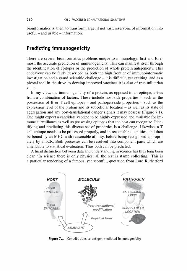
P1: OTA
c07 JWBK293-Flower September 5, 2008 19:59 Printer Name: Yet to Come
260 CH 7 VACCINES: COMPUTATIONAL SOLUTIONS
bioinformatics is, then, to transform large, if not vast, reservoirs of information intouseful – and usable – information.
Predicting immunogenicity
There are several bioinformatics problems unique to immunology: first and fore-most, the accurate prediction of immunogenicity. This can manifest itself throughthe identification of epitopes or the prediction of whole protein antigenicity. Thisendeavour can be fairly described as both the high frontier of immunoinformaticinvestigation and a grand scientific challenge – it is difficult, yet exciting, and as apivotal tool in the drive to develop improved vaccines it is also of true utilitarianvalue.
In my view, the immunogenicity of a protein, as opposed to an epitope, arisesfrom a combination of factors. These include host-side properties – such as thepossession of B or T cell epitopes – and pathogen-side properties – such as theexpression level of the protein and its subcellular location – as well as its state ofaggregation and any post-translational danger signals it may possess (Figure 7.1).One might expect a candidate vaccine to be highly expressed and available for im-mune surveillance as well as possessing epitopes that the host can recognize. Iden-tifying and predicting this diverse set of properties is a challenge. Likewise, a Tcell epitope needs to be processed properly, and in reasonable quantities, and thenbe bound by an MHC with reasonable affinity, before being recognized appropri-ately by a TCR. Both processes can be resolved into component parts which areamendable to statistical evaluation. Thus both can be predicted.
A lucid distinction between data and understanding in science has thus long beenclear. ‘In science there is only physics; all the rest is stamp collecting.’ This isa particular rendering of a famous, yet scornful, quotation from Lord Rutherford
SUBCELLULARLOCATION
EXPRESSIONLEVEL
T cellEPITOPES
B cell EPITOPES
Physical form
Post-translationalmodification
HOST PATHOGEN
ADJUVANT
MOLECULE
Figure 7.1 Contributions to antigen-mediated immunogenicity

P1: OTA
c07 JWBK293-Flower September 5, 2008 19:59 Printer Name: Yet to Come
COMPUTATIONAL VACCINOLOGY 261
(1871–1937). It is also probably misquoted: other versions include ‘All scienceis either physics or stamp collecting.’ The source of this bon mot is apparentlya book of reminiscences by J. B. Birks entitled Rutherford at Manchester, pub-lished in 1962. Rutherford left Manchester aged only 24, implying he formulatedthis viewpoint early in his career. Much of science is now focused on data and whatto do with it. In the high-throughput era – be that astronomical, metrological orpostgenomic – data generation is certainly no longer the bottleneck it once was. In-stead it is our ability usefully to interpret data, and particularly data en masse, thatlimits us.
Computational vaccinology
The importance and potential of computational vaccinology is often not properlyappreciated. Here, we shall redress this imbalance and also break down the artificialcompartmentalization that has arisen within computational disciplines: the divide,for example, between academia and industry, life science and materials modelling,and between cheminformatics and bioinformatics. The areas of application for in-formatics form a continuum stretching from solid-state physics to molecular biol-ogy and beyond. There is so much to learn from each discipline and so much ofvalue to add to experimental science.
When one works in a Cinderella field – and immunoinformatics most certainly isone – then it can be difficult for an outsider properly to assess the relative merits ofcomputational vaccine design compared to more mainstream experimental studies,however transitory and uninformative such experiments may prove. The potentialof immunoinformatics, albeit largely unrealized, is huge, but only if people arewilling to take up the technology and use it appropriately. People’s expectationsof computational work are largely unrealistic and highly tendentious. Some expectperfection, and are soon disappointed, rapidly becoming vehement critics. Othersare highly critical from the start and are nearly impossible to reconcile with in-formatic methods. Neither view stands as a correct exegesis, however. Informaticmethods do not replace, or even seek to replace, experimental work, they only helprationalize experiments, saving time and effort. They are slaves to the data used togenerate them. They require a degree of intellectual effort equivalent in scale, yetdifferent in kind, to that of the experimentalist. The two disciplines – experimentaland informatic – are thus complementary, albeit distinct. Ultimately, as system bi-ology takes hold and insinuates itself into every corner of biology, informatics willfind its place, though that may yet take time.
Immunoinformatics-driven in silico vaccinology can effectively leverage infor-matics techniques to deliver effective and utilitarian advantage in the search fornew vaccines. Immunoinformatics-driven in silico vaccinology works through adynamic and responsive cyclical process of using and refining both models andexperiments, at each stage moving closer towards the goal of the effective, cost-efficient development of vaccines or vaccine components. The allied subjects of

P1: OTA
c07 JWBK293-Flower September 5, 2008 19:59 Printer Name: Yet to Come
262 CH 7 VACCINES: COMPUTATIONAL SOLUTIONS
bioinformatics and computational chemistry have, within the pharmaceutical in-dustry and the wider scientific community, proved their worth time and time againin the search for new drugs and new drug targets. The time is approaching whenimmunoinformatics and allied disciplines will do the same for vaccine design.
The threat remains
As we have seen, vaccines are – at least in comparison to other prophylactic treat-ments – remarkably effective in combating the threat from infectious diseases.There are now in the order of 30 infectious diseases which are effectively targetedby vaccines. Many of the remaining infectious agents, for which there are no effec-tive vaccines, are difficult to manipulate or give rise to persisting infections, suchas hepatitis C. Others, such as influenza, show great antigenic variation. While stillothers, such as most parasitic infections, are very complex organisms, with equallycomplex life cycles, that are genetically closer in structure and metabolism to ver-tebrate hosts and are, for all these reasons and more, much more challenging wherethe discovery and design of vaccines are concerned. Still other diseases, such asAIDS, are mediated by pathogens of great structural simplicity prone to rapid, im-mense and clearly confounding, genetic variation.
The need, then, for new antimicrobial treatments, both therapeutic and prophy-lactic, is obvious. Antibiotic drugs are one approach to realizing such treatments.Vaccines are another. Most successful vaccination strategies have used inactivatedor attenuated microbes or substances derived directly from them. Modern, semisyn-thetic vaccines are composed, wholly or in part, of chemically synthesized or re-combinant peptides, carbohydrates or protein antigens.
Most extant vaccines have arisen in an empirically fashion. However, interest isnow turning to more rationally designed vaccines. At one extreme of the size range,these can be genetically modified pathogens and at the other protein antigens or iso-lated T cell epitopes. Here the modern vaccinologist can make use of genomic datainformation from the many genomic sequencing projects focusing on pathogenicmicro-organisms. Thus, the application of computational sciences to vaccine re-search is a discipline whose time has come. A tranche of techniques, both old andnew, have recently matured into potent weapons in the war against disease. Molec-ular informatics – computational chemistry or molecular modelling, bioinformaticsand cheminformatics – has reached new heights of sophistication and utilitarianvalue within drug and vaccine discovery.
Beyond empirical vaccinology
Let us rehearse some of what we know about vaccinology. Existing vaccines fallinto two broad categories: living vaccines and nonliving vaccines. Living vaccinesuse attenuated forms of pathogens to mimic natural infections without the dangers

P1: OTA
c07 JWBK293-Flower September 5, 2008 19:59 Printer Name: Yet to Come
DESIGNING NEW VACCINES 263
of the natural infection and thereby induce immunity to the natural pathogen. Inseeking to induce protective immunity, nonliving vaccines use either a chemically-inactivated whole microbial pathogen or they use components of such microbesin the form of subunit vaccines. Recently, antigen-based subunit vaccines havegained significantly in popularity and use. The identification of antigenic or im-munogenic proteins as putative whole protein subunit vaccines is a key goal ofimmunovaccinology. It offers the hope of eliciting significant responses from bothhumoral and cellular immune systems, far exceeding the efficacy of peptide vac-cines, while avoiding potential toxicity problems associated with whole microbevaccines.
Traditional vaccine development of human vaccines combines an eclecticrange of immunological, biochemical and microbiological methods. Over the last150 years of vaccinology, much success has been obtained using these conventionalapproaches. Despite this success, it is increasingly clear that these techniques arethemselves flawed. New techniques are necessary to address the whole gamut of po-tentially threatening pathogens. Yet 40% of human disease remains incurable andmany existing therapies are far from ideal. The nature of illness has, at least in theWest, changed out of all recognition over the last century, and can be expected todo so again over the next 100 years. Thus the challenge to immunology and modernmedicine has never been greater, but neither has the technology available to meetthis challenge. The postgenomic science will deliver an unprecedented explosionof information. Only informatic strategies can manage and fully exploit this dataoverload.
Designing new vaccines
It is possible to draw up a list of requirements one would wish any new vaccineto possess. Indeed many have done so. I was presented with such a list when I firstentered the vaccine field; it was drawn up by my then boss Professor Peter Beverley.It read something like this. The ideal vaccine should confer lifelong protection. Itshould be broadly protective against all variants of an organism. It should preventor inhibit disease transmission. It should bring about effective immunity quickly. Itshould be effective in subjects, irrespective of age, thus being equally effective ininfants and the elderly. It should transmit maternal protection. It should need onlyone immunization. It should not require injection. Above all, it should be cheap,safe and stable. Some of these criteria can, at least in part, be addressed through therealization of the ideas presented above. Some can be addressed empirically, othersrationally and intelligently, others not at all.
How can informatics help to design new and improved vaccines? It depends onwhat kind of vaccine you need. Let us restrict ourselves to the three main concep-tual types of vaccine: attenuated micro-organisms, subunit vaccines and vaccinesbased on epitopes. One should be able to use computational tools to contribute tothe design of attenuated vaccines. Potentially, it is possible to alter an organism to

P1: OTA
c07 JWBK293-Flower September 5, 2008 19:59 Printer Name: Yet to Come
264 CH 7 VACCINES: COMPUTATIONAL SOLUTIONS
either gain immunogenicity or to lose virulence and thus generate an attenuated orat least a non-lethal vaccine moiety. However, they must be adequately attenuatedin their growth to minimize the possibility of vaccine-induced pathogenic effectswhile retaining their immunogenicity. Attenuation of live viruses has traditionallybeen achieved using the serial passage of viruses through tissue or cell culture. Forreasons not wholly understood, these processes tend to reduce microbial replicativeability and their apparent virulence in humans or in animal models. Such attenua-tion has historically been a highly empirical process, where underlying mechanismsare often not wholly known nor properly elucidated. Designed genetic changes to abacterium, a virus or other microbe can reduce the rate and efficiency of its abilityto replicate, reducing the speed at which it grows. This could allow the host thetime necessary to clear the infection while at the same time acquiring immunity toit and its genetic brethren. During the last decade the emergence of reverse geneticstechniques has created unprecedented opportunities to better control viral attenua-tion. Genetics manipulation allows for the production of RNA viruses from clonedcDNA, permitting site-specific mutations to be introduced easily into viruses. Thechallenge is now to engineer attenuated viruses. A step change is needed from cre-ating variants to predicting how genetic changes affect the measurable growth ratesof viruses.
It should also be possible to reduce the number of virulence factors in the mi-crobial genome or to mutate it so that certain virulence factors, and any associatedimmunogenicity, remain yet at the same time removing from these molecules theirability to harm the host. This requires us to distinguish virulence factors (proteinsor other biomolecules which attack or facilitate attack upon the host) from othermicrobial proteins. Likewise we can take a benign micro-organism and add to ithighly antigenic proteins producing a microbe with the immunogenicity of anothermicrobe but without its arsenal of virulence factors. This is, in its essentials, muchthe same idea as generating a viral or microbial vector for the delivery of subunitvaccines.
The perfect vaccine
If we turn our attention to synthetic vaccines based on antigens or epitopes then wesee distinct ways in which immunoinformatics can aid the search for new vaccinemoieties. A generally useful – sometimes called a ‘perfect’ – vaccine may containone or more T cell epitopes, one or more B cell epitopes, plus nonproteinacous‘danger signals’, such as carbohydrate or lipid. Such a perfect vaccine may be anartificial polyepitope vaccine or a natural antigen, delivered as protein or peptide,via live viral or bacterial vectors, or as raw DNA. Such a vaccine would also proba-bly be accompanied by the administration of an adjuvant, a molecule or preparationthat exacerbates immune responses.
Conventional experimental discovery of subunit vaccines cultivates pathogensunder laboratory conditions and then attempts to partition them into a set of

P1: OTA
c07 JWBK293-Flower September 5, 2008 19:59 Printer Name: Yet to Come
CONVENTIONAL APPROACHES 265
component proteins. Antigens which confer protective immunity are then identi-fied by testing individual components or screening expression libraries against pa-tients’ antibodies. Undertaken systematical this process is intrinsically inefficient,involving an exhaustive, brute-force search of potentially thousands of putative tar-gets. Unlike this traditional process of antigen discovery, reverse vaccinology tar-gets the whole potential spectrum of potential antigens irrespective of their abun-dance, analysing the entire genome of a pathogen in order to identify all potentiallyantigenic proteins accessible to surveillance by the immune system. This technol-ogy allows vaccine discovery scientists to obtain candidate vaccines including thoseantigens which might be missed because a particular pathogen cannot be success-fully cultured in the laboratory or because such antigenic proteins may be absent,or at least apparently so, in in vitro culture.
Conventional approaches
Conventional, experiment-only approaches to candidate subunit vaccine selectionare replete with problems. First, it may not be possible to cultivate a particularpathogen in the laboratory. Pathogens must be capable of being cultured in a labo-ratory in conditions which are often very different to those offered by their naturalhosts. Culture methods are time-consuming and development times can run intodecades. Only the most abundant antigens (which can be purified in the quantitiesrequired for vaccine development) are identified. Often, abundant antigens do notmake viable vaccine candidates. For example, there will be many, many proteinsthat are only transiently expressed during infection or are not necessarily expressedat all in vitro. This can mean that interesting candidate antigens are overlooked.The characterization of antigens will also require them to be produced on a largescale. Taken together, this makes the overall process extremely consuming of time,person-power and financial resources.
Such an experimentally based approach proceeds as follows: first, a pathogenis cultured under laboratory conditions; secondly, the pathogen is then dissectedinto its component parts. Putative protective antigens are then identified by testingthe immanent immunogenicity of each component using a complex cascade of invitro assays and in vivo experiments which target so-called correlates of protec-tion. Cloning the gene coding for the antigen is often necessary to better charac-terize and produce the identified antigens. Finally, after this process is complete,the candidate vaccine can enter development. It is an imperfect process. It has of-ten failed to find suitable candidates for pathogens without obvious immunodom-inant protective antigens and it has failed to deal effectively with noncultivablemicro-organisms.
One of the key drivers for the development and deployment of immunoinfor-matics and computational vaccinology is the widespread availability of pathogengenomes and, to a lesser extent, of microbial genomes generally. In the quest tosequence genomes, massive efforts and resources have been deployed, are being

P1: OTA
c07 JWBK293-Flower September 5, 2008 19:59 Printer Name: Yet to Come
266 CH 7 VACCINES: COMPUTATIONAL SOLUTIONS
deployed, and will continue to be deployed for the foreseeable future. This is espe-cially true for bacteria and other microbes. Many, many human pathogens now havefull sequences available, including many important pathogenic bacteria and viruses,together with a smaller number of key protozoan parasites. Genome sequencing hasbeen one of the great success stories of modern science: in the last decade or so wehave gone from a complete dearth of information to the current climate where wehave more genome data than we can comfortably deal with. In the short to mediumterm, the situation will only get worse.
Genome sequences
The genomic sequencing of TB reported in 1998 or the sequencing of the plaguebacteria Yersinia Pestis in 2001 did not readily disgorge the secrets implicit withineither genome; likewise for an ever increasing number of other pathogens. Malaria,influenza and HIV: all are loathe to reveal their secrets. Genome sequencing is notan end in itself, but merely a step on a longer journey whose destination is thedevelopment of vaccines.
In this regard, the view taken is a simple one. To a first approximation, thegenome of a pathogenic bacterium contains within it all the information necessaryfor the pathogen to survive, to live and to thrive and for it thus to infect, disableand destroy. Since the genome contains all this information, it should be possibleto decipher this code for destruction, thus identifying the bacterium’s weaknessesand vulnerabilities, and allowing us to disable it in our turn. However, as with manythings, the initial sequencing of pathogen genomes proved disappointing. Initial op-timism hoped that the nature of virulence and infectivity would be readily apparentfrom analysis of microbial genomes. Such was not the case. A case in point was thecomparative analysis of the TB and BCG genomes. The expectation, voiced or un-voiced, was simple. The differences would at best identify one or several bacterialantigens present in TB that were absent in BCG, and at worst identify a larger hostof proteins where significant mutations had weakened virulent TB into attenuatedBCG. It was, and continues to be, a disappointment to many that no obvious differ-ences clearly above the mutational noise were apparent. Instead, a spectrum of mu-tations spread through the genome seems to be causative: reducing enzyme reactionenhancements, altering genetic regulation, as well as even more subtle alterations,all combining to reduce virulence and reproductive capacities of the organism andincrease its visibility to the immune system.
Size of a genome
Immunoinformatics has at its disposal an array of potential weapons – genome anal-ysis, epitope prediction, antigen identification, and so on – that can be deployed

P1: OTA
c07 JWBK293-Flower September 5, 2008 19:59 Printer Name: Yet to Come
SIZE OF A GENOME 267
adroitly in the fight against a particular microbial disease. Each variety of microbe –bacteria, viruses, protozoa or fungi – offers a different challenge.
Of particular interest in this regard is the nature of the genome: its size and com-plexity. Different types of microbe offer genomes with different characteristics andsizes. Bacterial species have genomes in the range of say 2000 to 5000 gene prod-ucts, viruses have genomes comprising anywhere between five and 1000 proteinencoding genes, while protozoa can have over 10 000 genes. Such figures are, atbest, rough-and-ready ballpark figures and will, no doubt, be both outdated andoutmoded by the time this book finally reaches the shelves. They give our chal-lenge a scale. We can, and we will, be more specific in a moment, but it is worthremembering these numbers as, in totality, they clearly demonstrate that the prob-lem of making predictions is in the confounding and discombobulating complexityinvested in the many components of the genome and not in its overall size, which iswell within the scope of modern computing.
One of the smallest, if not the smallest, bacterial genomes yet discovered is thatof the marine bacterium Pelagibacter ubique. Pelagibacter ubique has only 1354genes and is largely free of genetic clutter: it contains little or no duplication ofgenes, few pseudogenes, no so-called junk DNA and also no genes of viral origin.It is very much a bacterial species figuratively pared to the bone – a bacteriumslimmed to its bare essentials. There are smaller bacterial genomes, but most ofthese are obligate symbionts which outsource much of their metabolism to theirhost organisms; examples of such symbionts include Mycoplasma genitalium whichhas, give or take, just 400 genes. Pelagibacter ubique is, by contrast, a free-livingorganism with a full complement of metabolic pathways.
At the other extreme, the largest bacterial genomes to be properly sequenced in-clude those of Burkholderia xenovorans LB400 and Rhodococcus sp. RHA1, whichis about 9.70 Mb in size. RHA1 was first isolated from contaminated soil and isfamed for its ability to degrade polychlorinated biphenyls (or PCBs) and an extraor-dinarily wide range of other persistent toxic pollutants and organic compounds, togrow effectively on bewilderingly numerous substrates, and to even desulpherizecoal. RHA1’s DNA is packaged into a linear chromosome and three linear plasmidsand codes for about 9200 genes. The genome is unsurprisingly rich in oxygenases,ligases and other metabolic enzymes, such as nonribosomal peptide and polyketidesynthases. However, the RHA1 genome contains, when compared to most otherbacterial species, few recent gene duplications and few genes acquired by horizontaltransfer, especially when contrasted with LB400 which is much more conventional.However, bacteria with even larger genomes still await sequencing. The myxobac-terium Sorangium cellulosum has an extraordinarily large bacterial genome of over13 Mb. This is twice the size of, say, Pseudomonas aeruginosa, which has in theregion of 6 Mb and about 5500 genes. In comparison, E. coli has approximately 4.6Mb and approximately 4200 genes.
Viruses, like bacteria, span a wide range of genomic sizes. At one extreme isthe apparent simplicity of a retrovirus – HIV has five protein encoding genes forexample – while at the other are the pox viruses. Canarypox, for example, has a

P1: OTA
c07 JWBK293-Flower September 5, 2008 19:59 Printer Name: Yet to Come
268 CH 7 VACCINES: COMPUTATIONAL SOLUTIONS
genome of 0.35Mb, and codes for 328 proteins, while fowlpox (0.28Mb) encodes261 genes; many of these have been acquired laterally from eukaryotic hosts. How-ever, pox viruses pale by comparison to what is the current largest known virus:Mimivirus. This megavirus or girus, discovered in a Yorkshire water-cooling tower,infects amoebas and, some have argued, possibly humans as well. The genomeof Mimivirus is about 1.18Mb in size and codes for over 900 proteins. While itpossesses the coat characteristic of a virus, Mimivirus is about 600 nm across,making it larger than many bacteria. Again, sequencing of the genome has posedmore questions than it has answered. In many ways it seems to transgress thesharp distinction between viruses and cellular life. Viruses, in any case, presentmany conceptual difficulties to the biologist trained on higher life: speciation isparticularly problematic as is the question ‘are viruses even alive’? We conceiveof viruses as parasitic and depending totally upon the subcellular machinery ofinvaded cells. Mimivirus, however, must carry out many synthetic and metabolicfunctions itself, suggesting that it is much closer to other microbial life, suchas bacteria.
Reverse vaccinology
Increasingly, reverse vaccinology is seen as a standard way of discovering vaccines.It is a postgenomic discipline leveraging computational sequence searching tech-niques to identify systematically all potential vaccine candidates within sequencedpathogen genomes or proteomes. Initially, it was seen as a way to escape from themajor bottlenecks and limitations of experimental vaccine discovery, and increas-ingly as a means of conceptualizing the genomic identification of vaccine candi-dates as a wholly in silico process: given whole or part pathogen genomes, reversevaccinology uses data mining to identify and characterize surface or secreted targetproteins. Taken to its logical extreme, reverse vaccinology may allow the devel-opment of safe and efficacious future vaccines against as-yet-unknown infectiousdisease on a timescale of undreamed-of celerity.
Reverse vaccinology starts with the genome of a pathogen rather than thepathogen itself, and works in silico as much as it does in vitro or in vivo. Givenraw genomic sequences, there are a multitude of computational systems that predictopen reading frames (or ORFs). Using reverse vaccinology, these ORFs (number-ing anywhere from a hundred to several thousand) will quickly be reduced to a listof candidate vaccines of manageable size, perhaps numbering no more than a fewdozen.
Reverse vaccinology is, however, not a computation-only discipline, but rather apotent combination of genomic, proteomic and transcriptomic approaches, closelycoupled to bioinformatic analysis. The pruned list of target vaccines generatedthrough bioinformatics analysis requires subsequent channelling through an exten-sive tranche of downstream processes including recombinant expression and purifi-cation, as well as testing for immunogenicity and, ultimately, protective efficacy.

P1: OTA
c07 JWBK293-Flower September 5, 2008 19:59 Printer Name: Yet to Come
FINDING ANTIGENS 269
In silico methods often identify as much as 25% of a genome as vaccine can-didates, thus requiring methods able to clone and express large numbers of geneproducts. Approaches include high-throughput methods utilizing robotics and thepolymerase chain reaction (PCR). PCR products are cloned and screened for ex-pression. The purified recombinant proteins need testing for immunogenicity, whichis typically performed in vivo using vaccination of mice, though other animal mod-els including both small (rats, guinea pigs, etc.) and large (dogs, primates, cows,etc.) mammals have been used. Correlates of protection appropriate to the diseaseorganism in question are screened. Such correlates include antibody titres. Thequestion posed is this: after immunization does the serum of vaccinated animalscontain antibodies generated by the vaccine and do these antibodies recognize boththe recombinant and original protein? For an animal model to be effective, protec-tion should be mediated by similar mechanisms to those observed in humans. Thelack of reliable animal models has often stymied vaccine and drug development.In vitro assays, such as bactericidal activity and opsonophagocytosis, also correlatewith human vaccine efficacy.
Clearly, the overall veracity and reliability of any reverse vaccinology exerciseis strongly dependent on the accuracy of prediction, which in turn necessitates arobust definition of which proteins are potential antigens and which are not. Such adefinition is not readily forthcoming. Instead, it has been necessary to devise moreapproximate definitions capturing some, but not all, necessary information to dis-criminate properly between antigens and nonantigens. We will explore this subjectbriefly.
Finding antigens
What one is seeking is some measure of the intrinsic capacity of a protein to be anantigen: ‘antigen-ness’ shall we say. However, while the creation of such a mea-sure has been attempted, no perfectly predictive method is yet available. Com-pared to the relatively straightforward task of identifying ORFs, selecting pro-teins liable to immune system surveillance remains a decided challenging. At theJenner Institute we have developed VaxiJen (www.jenner.ac.uk/VaxiJen/) that im-plements a statistical model able to discriminate between candidate vaccines andnonantigens, using an alignment-free representation of the protein sequence. Sucha method is an imperfect beginning; future research will yield significantly moreinsight as the number of known protective antigens increases. The developmentof the AntigenDB database (www.imtech.res.in/raghava/antigendb/) will aid in thisendeavour.
Cell surface location is thought to be a prime determinant of immunogenicity.Secreted proteins – particularly bacterial proteins excreted into the extracellularspace – are likely mediators of pathogen virulence including adhesion, invasion, se-cretion, signalling, annulling host responses, toxicity and motility, and so such pro-teins have significant potential as candidate subunit vaccines. They are also liable to

P1: OTA
c07 JWBK293-Flower September 5, 2008 19:59 Printer Name: Yet to Come
270 CH 7 VACCINES: COMPUTATIONAL SOLUTIONS
surveillance by the host immune system during and after pathogen invasion. Otherextracellular proteins are cell surface proteins, lipoproteins or membrane proteins.Some classes of surface protein, such as fimbriae, are common to many pathogens,while others are more species-specific.
Arguably, the most useful, and thus the best studied, of what we might broadlyterm system approaches to the identification of immunogens, has been the predic-tion of subcellular location. There are two basic types of prediction method: themanual construction of rules based on knowledge of what determines subcellularlocation, and the application of data-driven machine learning methods, which au-tomatically identify factors determining subcellular location by discriminating be-tween proteins from different known locations.
Accuracy differs markedly between different methods and different compart-ments, due to a paucity of data or the inherent complexity that determines proteinlocation. Such methods are often classified according to the input data requiredand how the prediction rules are constructed. Data used to discriminate betweencompartments include: the amino acid composition of the whole protein; sequencederived features of the protein, such as hydrophobic regions; the presence of cer-tain specific motifs; or a combination thereof. Phylogenetic profiles can also beuse, as it is assumed that the subcellular location of close protein homologuesis similar.
Prediction methods will produce binary or multicategory classifications. A binarymethod predicts whether a protein is located in one category or not. A multicate-gory predictor will identify which of several possible locations is the most likely. Acommon problem with binary predictors is that they often overpredict the number ofproteins that belong to a particular category. Multicategory prediction methods of-ten exhibit reduced accuracy because certain locations lack sufficient training data.As ever, the more complete the data the more accurate the prediction.
Different organisms evinced different locations. PSORT is a knowledge-based,multicategory prediction method, composed of several programs, for subcellularlocation; it is often regarded as a gold standard. PSORT I predicts 17 different sub-cellular compartments and was trained on 295 different proteins, while PSORT IIpredicts 10 locations and was trained on 1080 yeast proteins. Using a test set of 940plant proteins and 2738 nonplant proteins, the accuracies of PSORT I and II were69.8% and 83.2% respectively. There are several specialized versions of PSORT.iPSORT deals specifically with secreted, mitochondrial and chloroplast locations;its accuracy is 83.4% for plants and 88.5% for nonplants. PSORT-B only pre-dicts bacterial subcellular locations. It reports precision values of 96.5% and recallvalues of 74.8%. PSORT-B is a multicategory method which combines six algo-rithms using a Bayesian network.
Among binary approaches, arguable the best method is SignalP, which employsneural networks and predicts N-terminal Spase-I-cleaved secretion signal sequencesand their cleavage sites. The signal predicted is the type-II signal peptide commonto both eukaryotic and prokaryotic organisms, for which there is a wealth of data,in terms of both quality and quantity. A recent enhancement of SignalP is a hidden

P1: OTA
c07 JWBK293-Flower September 5, 2008 19:59 Printer Name: Yet to Come
THE SUCCESS OF REVERSE VACCINOLOGY 271
Markov model version able to discriminate uncleaved signal anchors from cleavedsignal peptides.
One of the limitations of SignalP is overprediction, as it is unable to discriminatebetween several very similar signal sequences, regularly predicting membrane pro-teins and lipopteins as type-II signals. Many other kinds of signal sequence exist. Anumber of methods have been developed to predict lipoproteins, for example. Theprediction of proteins that are translocated via the TAT-dependent pathway is alsoimportant but is not addressed yet in any depth.
Other contributing factors determining immunogenicity such as the level of geneexpression and the dissimilarity of proteins to the human genome, at both the wholeprotein level and at the level of the epitope, are also being explored. Again, suchattempts have met with mixed success. The G+C content of genes and of large geneclusters, known as pathogenicity islands, is usually characteristic of genes acquiredby horizontal transfer. Structural analysis can indicate which proteins are likely tobe membrane bound and partly exposed to the environment.
The most successful approach by far is the use of sequence searching to iden-tify proteins with high sequence similarity to known antigens and virulence fac-tors. Global homology searching systems, such as BLAST, are typically used forthis purpose. Certain ORFs which code for known cytoplasmic protein or for anti-gens previously identified within the pathogen of interest can be excluded at thispoint. Of particular interest is the identification in the proteome of putative viru-lence factors with significant similarity to those that have been previously char-acterized in other organisms. Alternative approaches use motif-based protein fam-ily identification, such as Pfam, PROSITE and PRINTS, which can identify motifscharacteristic of protein families containing virulence factors and, to a lesser degree,antigens.
The success of reverse vaccinology
Reverse vaccinology is typically more effective for prokaryotic than eukaryotic or-ganisms, since the latter tends to have larger, more complicated genomes. There arenow several examples of the successful application of reverse vaccinology to thediscovery of subunit vaccines, and a representative selection of these examples arelisted below. They represent combined approaches, as well as sequence comparison.
Neisseria meningitides is a bacterial pathogen responsible for sepsis andMeningococcal meningitis. It has five major subgroups (A, B, C, Y and W135) de-fined by their capsular polysaccharides. It remains a major cause of human mortal-ity and morbidity. Vaccines have not been developed for subgroup B, called MenB.This is due to the variable sequence of surface proteins and to the cross reactivityof the MenB capsid with human tissue. An anti-MenB vaccine would exhibit poorimmunogenicity and generate autoantibodies.
The pioneering application of reverse vaccinology targeted MenB. The incom-plete MenB genome was scanned for ORFs potentially corresponding to surface

P1: OTA
c07 JWBK293-Flower September 5, 2008 19:59 Printer Name: Yet to Come
272 CH 7 VACCINES: COMPUTATIONAL SOLUTIONS
proteins with similarity to virulence factors. Of the 570 proteins identified, 350could be cloned and expressed in vitro. 85 of the 350 were confirmed as exposed,and of those 22 were immunogenic and induced a complement-mediated bacterici-dal antibody response. This is consistent with these proteins being protective. Im-munogenic proteins were tested for their ability to confer protection against heterol-ogous strains. Each was evaluated for gene presence, phase variation and sequenceconservation in 22 genetically distinct strains representative of MenB global di-versity. Seven proteins induced cross protection, conferring immunity across manystrains.
Streptococcus pneumoniae is a major cause of sepsis, pneumonia and meningi-tis; it is a common cause of life-threatening infection in newborn children. Currentvaccines are only poorly efficacious in infants, and offer little cross protection withother strains. After mining the Group B Streptococcus (GFB) genome, 130 potentialORFs showed similarity to surface proteins and virulence factors from other bacte-ria. 108 of theses could be expressed and purified. They were evaluated as vaccinecandidates in mice. Six antigens induced protective antibodies against pneumococ-cal in a mouse challenge model. All six candidates were cross reactive against mostcapsular antigens that are expressed.
Porphyromonas gingivalis is an anaerobic bacterium present in subgingivalplaques in chronic adult periodontitis, an inflammatory disease of the gums. To-gether with depression, conditions affecting the buccol cavity, such as periodontitisand cavities, are the commonest human diseases. Shotgun sequencing identified370 ORFS, 74 of which exhibited significant similarity to known bacterial surfaceor virulence proteins. 49 proteins were predicted to be surface proteins. This gen-erated 120 unique protein sequences. 107 of these were expressed in E. coli andanalysed by western blotting using sera from human periodontitis patients and ani-mal antisera. 40 candidates were shown to be positive for one or more sera. Thesecandidates were used in a mouse challenge model. Two antigens showed signif-icant protection. Both were similar to Pseudomonas aeruginosa OprF, a vaccinecomponent undergoing clinical trials.
Chlamydia pneumoniae is an obligate intracellular bacterium implicated in res-piratory infections, cardiovascular and atherosclerotic disease. The life cycle ofC. pneumoniae involves two stages: a spore-like infectious elementary body (Ebs)form and an intracellular reticulate body (RBs) form. Using in silico selection, 140proteins were identified, expressed in E. coli, purified, and used to immunize mice.FACS analysis detected 53 surface proteins. 28 antigens were verified using west-ern blot and by proteomic analysis of EB total proteins using two-dimensional gelelectrophoresis and mass spectrometry.
Adhesins are a class of virulence factor that mediates initial interactions betweenpathogen and host. Experimental analaysis of adhesins is often problematic prompt-ing efforts to develop in silico approaches. Acellular pertussis vaccine contains theadhesin haemagglutinin; several more are being evaluated, for example HpaA fromHelicobacter pylori, the RgpA-Kgp proteinase-adhesin complex of Porphyromonasgingivalis, P97R1 adhesin from Mycoplasma hyopneumoniae and FimH adhesin of

P1: OTA
c07 JWBK293-Flower September 5, 2008 19:59 Printer Name: Yet to Come
TUMOUR VACCINES 273
E. coli. Several machine-learning techniques have been devised for adhesion pre-diction using sequence properties. An example is SPAAN, based on ANN. It pre-dicted several adhesins in Mycobacterium tuberculosis, which were subsequentlyconfirmed experimentally. For example, Rv1818c was shown to display adhesin-like characteristics.
The message of all this work is clear, if not quite completely lacking in ambi-guity; and the message is this: if reverse vaccinology is applied in an appropriateway then vaccine design and discovery can be rendered much more efficient and ef-fective, with a concomitantly large saving in money, time and wasted labour. Manypositive results have been obtained with reverse vaccinology for key pathogens,such as MenB. This is consistent with the view that reverse vaccinology – andin silico methods in general – provides a solution suitable to the many problemsencountered by traditional vaccine development. Validation is obviously requiredin vivo to discover the efficacy of candidate antigens using genomic, proteomic,genetic, biochemical and animal model approaches. The strongest advantage of re-verse vaccinology is that there is no limitation imposed by growth conditions ordetection methods for gene expression. Identifying a small number of promisingcandidates enables their rapid development at a reasonable cost compared with tra-ditional strategies. One of the aims of the computational component of reverse vac-cinology is to minimize the cost in human and financial resources, without losingefficiency, by producing more and better information which in turn allows for a finerand better informed discrimination of suitable antigens and vaccine candidates.
Immunoinformatics can, as part of reverse vaccinology, make an important con-tribution to the successful discovery of subunit vaccines. As improved techniquesfor subcellular location prediction and statistical identification of antigen versusnonantigen become available, the immunoinformatic dividend can but improve.Likewise, immunoinformatics can help in the discovery of epitope-based vaccines.These can be delivered as peptides, as naked DNA, via viral vectors, expressed ondendritic cell vaccines, and so on.
Tumour vaccines
Vaccines against malaria and other important pathogens have been epitopic, but thegreatest potential success for vaccines based on epitopes is in the field of cancer;the greatest cause of death in the First World after cardiovascular disease. In manyways, cancer or tumour vaccines offer the best opportunity for immunoinformaticsto have the kind of direct input that it so justly warrants. Cancer is usually treatedusing chemotherapy, radiotherapy, surgery or a combination thereof. While primarytumours can be dealt with effectively using such approaches, treatment of metasta-sis has been much less successful. Use of prodigious quantities of tumour-specificmonoclonal antibodies is the current principal form of passive cancer immunother-apy, but there is both hope and expectation that cancer vaccines, which seek toexploit the immune system’s ability to recognize and destroy tumour cells through

P1: OTA
c07 JWBK293-Flower September 5, 2008 19:59 Printer Name: Yet to Come
274 CH 7 VACCINES: COMPUTATIONAL SOLUTIONS
mechanisms that combine both tumour-specific humoral and cytotoxic T cell re-sponses, will prove the long-term way forward. T cells have a key role in protectingagainst cancer.
Tumour cells express different or mutated antigens that act as labels allowing theimmune system to distinguish malignant from nonmalignant cells. Recently, manystrategies to identify tumour antigens have emerged, leading to the characterizationof various types of cancer antigens: self-antigens overexpressed in tumours, dif-ferentiation antigens, germline antigens, point mutations of normal genes and viralantigens. Example tumour antigens include the MAGE, BAGE and GAGE familyof genes, hTERT, NYESO-1 and survivin, among a growing band. Cancer vaccines,be they protein subunits or based on T cell epitopes, typically target tumour-specificantigens to elicit immune responses.
The literature abounds with reports of clinical trials with tumour vaccines. Vac-cines for prostate cancer are, for example, starting to show clinical promise. Prostatecancer is among the commonest, noncutaneous cancers seen in men in the devel-oped world. Several vaccines are being developed to treat it. Provenge, from Den-dreon Corp., is a vaccine based on dendritic cells pulsed ex vivo with a recom-binant fusion protein of prostatic acid phosphatase and granulocyte macrophagecolony-stimulating factor. Phase II trials indicated decreased prostate-specific anti-gen (PSA) and prolonged PSA doubling time. Phase III trials showed enhanced sur-vival in men compared with those administered a placebo. BiovaxID, produced byAccentia BioPharmaceuticals, is a patient-specific therapeutic cancer vaccine com-prising tumour idiotype conjugated to keyhole limpet hemocyanin. Phase II trialsindicated prolonged survival.
Host immune responses to pathogens are strong, yet responses to cancers are of-ten weak, as most tumour-responsive T cells have been eliminated from the reper-toire. Such responses are seldom therapeutic, necessitating the need for adjuvants.Adjuvant is the name given to a whole heterogeneous class of molecules that ex-acerbate immune responses. We saw, in Chapter 6, how structural bioinformaticscan help discover small molecule adjuvants. Many class I MHC-binding epitopesderived from tumour antigens are recognized by CD8+ T cells. Likewise, class IIcancer-derived epitopes have been shown to be recognized by CD4+ T cells. Theuse of one or more epitopes from one or more tumour antigens makes epitope-basedvaccines an attractive alternative to subunit vaccines. Combining epitope-basedvaccines with appropriate adjuvants makes immunotherapy an appealing cancertreatment, as well as one that naturally benefits from computational assistance dur-ing the discovery process.
Prediction and personalised medicine
However, genetic differences between patient immune systems may mean that per-sonalized cancer vaccines will be necessary. Each patient expresses different tu-mour antigens and thus the nature of immune recognition will vary from patient to

P1: OTA
c07 JWBK293-Flower September 5, 2008 19:59 Printer Name: Yet to Come
PREDICTION AND PERSONALISED MEDICINE 275
patient. T cell responses have also been linked to tumour regression, extending thepotential for personalized or boutique medicine. In this context, the idea of identi-fying epitopes for single alleles becomes of great importance. One can conceive ofinitially HLA-typing a patient suffering from a known cancer and using computa-tionall techniques to create a personal, or at least personalized, therapeutic vaccinewith which they would be treated. The strategy might include using immunoinfor-matic techniques to identify potential epitopes, which can then be combined withadjuvants designed by structural bioinformatics, and delivered using a viral vectordesigned using gene bioinformatics.
Prediction methods are slaves to the data used to generate them. To be use-ful, data, which we wish to model and predict, must be properly accumulated andarchived. This is the role of the database. Once we have stored our data we mustanalyse it. This is the province of data mining. Unfortunately, except in rare cases,data is usually multi-dimensional, and each dimension will typically be correlated,to a greater or lesser extent, with each other dimension. Such a dimension may be astructural feature such as, in the case of peptides, the presence of a particular aminoacid at a particular position; or a dimension may be a property, such as hydropho-bicity. Together, these dimensions map out a space – a space of structural variationor variation of properties. If our data is itself of sufficient quality and provides agood enough coverage of the space, then straightforward methods drawn from, say,computer science – of which there are indeed very many – are now able to generatemodels of high predictive accuracy. Such models are tools of true utility replete withpractical real-world applications. The problem comes when there are clear inade-quacies within the available data and thus one will strive, vainly, for methods withthe hoped-for, quasi-magical ability to compensate for the deficiencies inherent inthe data itself. This can never be achieved, but it does not stop people trying.
Bias within the data places strict limitations upon the interpretability and general-ity of models. In general, for MHC–peptide binding experiments, the sequences ofpeptides studied are very biased in terms of amino acid composition, often favour-ing hydrophobic sequences. This arises, in part, from preselection processes thatresult in self-reinforcement. Binding motifs are often used to reduce the experi-mental burden of epitope discovery. Very sparse sequence patterns are matchedand the corresponding subset of peptides tested, with an enormous reduction insequence diversity. Nonetheless, the peptide sets which are analysed by immunoin-formaticans are still much larger than those typical in the pharmaceutical litera-ture. The peptides are physically large in themselves, and their physical proper-ties are extreme. They can be multiply charged, zwitterionic and/or exhibit a hugerange in hydrophobicity. Affinity data itself is often of an inherently inferior qual-ity: multiple measurements of the same peptide may vary by several orders ofmagnitude, some values are clearly wrong, a mix of different standard peptidesare used in radioligand competition assays, experiments are conducted at differ-ent temperatures and over different concentration ranges. We are also performing a‘meta-analysis’ – almost certainly forcing many distinct binding modes into a singleQSAR model.

P1: OTA
c07 JWBK293-Flower September 5, 2008 19:59 Printer Name: Yet to Come
276 CH 7 VACCINES: COMPUTATIONAL SOLUTIONS
Imperfect data
What are needed are properly designed data-sets which can properly sample and ex-plore the multi-dimensional space accessed by all congeneric molecules under ex-amination, in this case peptides. The number of different peptides which 20 aminoacids can make is 20N, where N is the length of the sequence. Assuming we areinterested in peptides of length eight to 16 residues, as is the case for class I MHCbinding peptides, the resulting space must contain 6.90 × 10²⁰ different peptides;this is indeed a formidable number. Although the specificity of any MHC will re-strict the number of bound peptides to but a tiny fraction of this, it is no wonder thatmethods struggle to deal with such a space.
Ultimately, however, we are limited by the data itself. This is undoubtedly theoverriding issue. With properly designed training sets many issues would be re-solved, but such sets are not subsets of available data. Rather we require new datato be produced and that necessitates close, if often elusive, synergistic interactionsbetween theoreticians and experimentalists. No data-driven method can go beyondthe training data: all methods are better at interpolating than extrapolating. It is onlyby having excellent and general data that we can hope for general and excellentmodels.
We can readily perceive a meaningful hierarchy with utilitarian value centring onthe assimilation of data, information and knowledge. At the base of our hierarchy isthe raw data: single measurements, such as individual, unaveraged points in a titra-tion curve. Data, when it is processed, becomes information; an IC50 value for ex-ample, which has, unlike the raw data point, identity and meaning in itself. Beyondinformation, there is knowledge. In this context, knowledge may be a quantitativerelationship between measured affinities and a structural description of the corre-sponding molecules. Such knowledge is interpolative, able to describe adequatelythe space bound by the information it is built upon. The mental world we, as scien-tists, inhabit is bounded by ignorance not certainty. To an extent, it answers ‘how’questions. Understanding is built on knowledge and goes beyond the examinedproperty space; it is able to make true extrapolative predictions about radically dif-ferent unobserved phenomena. It may not always get such predictions correct, but italways comes with insight. Knowledge identifies trends which go beyond what weknow to what we don’t know, and thus it can answer ‘why’ rather than just ‘how’questions. An example might be molecular dynamics simulation which can, in prin-ciple, address the prediction of MHC–peptide binding affinity of any molecule.With true understanding comes the ability to engineer and manipulate the prop-erties of our system: not simply charting a course through our property space oreven mapping it out fully but, through the leveraging of scientific imagination andcreativity, redefining and extending the space itself.
Within immunoinformatics, meaningful and quantitative molecular and func-tional data underlies all attempts to predict those aspects of immunological systemswhich are capable of being predicted. All knowledge is partial; yet even a partialanswer can be useful. There are many properties that can be predicted and many

P1: OTA
c07 JWBK293-Flower September 5, 2008 19:59 Printer Name: Yet to Come
FORECASTING AND THE FUTURE OF COMPUTATIONAL VACCINOLOGY 277
that cannot. Immunoinformatic computation can identify undiscovered links withindata-sets yet currently it is unable to identify unknown components of pathways oruncover protein function, other than by establishing sequence or structural similar-ity to extant protein exemplars.
Thus there are limits to what computational vaccinology can achieve as well asimmense opportunities to exploit its potential. It is important to realize what canbe done and what cannot be done, what is useful and what is not. What immunoin-formatics can offer is tools and methods that form part of a wider experimentaland clinical endeavour. It offers a set of techniques replete with utilitarian valuewhich can be leveraged by computational vaccinology to facilitate the design anddiscovery of vaccines. Many prediction methods remain problematic, particularlythe prediction of B cell epitopes, or are only starting to be developed, such as theprediction of whole antigen immunogenicity. Yet, T cell epitope prediction in par-ticular has been shown to work time and time again, at least for well characterizedalleles. If, and this is eminently achievable, all distinct alleles were examined to thesame thoroughness as, say, HLA-A∗0201, then an adequate battery of predictiontools spanning the space of MHC alleles would be available.
Forecasting and the future of computational vaccinology
Accuracy is vitally important to immunoinformatics and to computer science ingeneral. We may not always acknowledge it, yet it is true. It is also true of all otheraspects of science. I have always found it strange that experimental immunologistsare happy to accept immense uncertainty in a wet-lab experiment yet expect perfec-tion in computer-based methods. They imagine that because they undertake whatthey take to be controlled experiments, and do repeats, that they have countered allrandom and systematic error. Perhaps they have, but they have certainly not reacheda definitive, biophysical understanding of the system they are studying.
More than once, I have experienced the following – when I say that compu-tational methods are not 100% accurate I am told: what use are they then? Thecorollary of this would seem to be that all their work is 100% accurate 100% ofthe time and is incapable of ever being wrong. Perhaps I am being unfair. Theymay see things I do not or cannot or will not see. What seems to me to be slackand inconclusive experiments with hundreds of parameters undefined, may, in theirknowledgeable eyes, appear utterly robust and reliable. Of course, they wear thewhite coats and wield the pipettes, so I must assume, as they clearly do, that theyare right and I am wrong. Perhaps I am wrong to hanker for biophysical rigour andaccuracy. Perhaps immunology is different from other areas of science. Signal-to-noise ratio is a problem in many disciplines. The biologists’ N = 3 is not enough tosurmount it.
Prediction, as well as analysis, is also important. Conceptually, the difference isclear, but is seldom properly appreciated. Risk is associated with predictions, butthere should not be any significant risk associated with an analysis. To put it rather

P1: OTA
c07 JWBK293-Flower September 5, 2008 19:59 Printer Name: Yet to Come
278 CH 7 VACCINES: COMPUTATIONAL SOLUTIONS
simply: prediction is about making informed, educated guesses about uncertain,untested events, while analysis is about identifying relationships amongst known,certain data. However, despite the steady increase in studies reporting the real worlduse of prediction algorithms, there is still an on-going need for truly convincing val-idations of the underlying approach. Why should this be? Prediction, like all formsof forecasting, is prone to error and is seldom foolproof. The same, however, isalso true of all human activities, experimental science included. Predictions madeby informatics are seldom perfect, but neither are predictions about the weatheror stock market forecasts. People live happily with inaccuracies in both, but manydog-in-the-manger scientists will have nothing to do with theoretical or informaticspredictions. ‘It’s not perfect. It’s therefore trash! How can I trust it?’ they say, yetthey trust implicitly their own inherently error prone and discombobulating experi-ments, and expect others to trust them also.
Much of what an informatician does can be seen as forecasting – that is,predicting to a purpose. Echoing the words of economist J. K. Galbraith, it ispossible to understand the present in terms of the past, since the now contains muchof the then. Likewise, the future is the contingent product of the present and thusalso of the past. The undiscovered resides – partly at least – in the discovered; theunknown partly in the known. Unperformed experiments share characteristics withwhat we already know. If not, how could hypothesis-driven research ever prosper?When we can identify certain pertinent similarities we can use them to makereliable, even pivotal, forecasts or prediction. Particularly acute comparisons canbe drawn between prediction in biology and other types of forecasting, specificallyforetelling the weather. In the average north European country, which may expe-rience a year’s seasons in a day, forecasting the weather is of significant economicimportance. Britain’s Meteorological Office quotes accuracies around 86%. Goodthough this appears, it still means, on average, that one day a week the predictionsare wrong. Likewise, the computational chemist cannot guarantee every singleprediction will be correct. If they make 50 predictions, 48 of which are correct,this is a significant achievement. However, if the most interesting molecules werepart of the two rather than the 48, then this achievement will seem to be lessened.Weather forecasting has seen an immense improvement on a few decades ago,when forecasting was only accurate for, at best, a day ahead. Today, in no smallpart due to the potent combination of informatic and experimental disciplines, inthis case computer simulation and satellite images, we can be confident of accurateweather forecasts up to 5 days in advance. In time series analysis such as this, as inbioinformatics, extreme extrapolation is rarely successful.
As in all fields of science, it is important to realize the limitations of our tech-nology, and not to seek answers to impossible questions. In this way, the benefitsof informatics research can be maximized. Forecasting in other disciplines is notmet with the blatant skepticism that bioinformaticians and computational chemistsencounter: weather forecasting or predictive economic modelling or computermodelling of climate change are treated with respect; while as a society we are

P1: OTA
c07 JWBK293-Flower September 5, 2008 19:59 Printer Name: Yet to Come
FORECASTING AND THE FUTURE OF COMPUTATIONAL VACCINOLOGY 279
happy to mortgage our global economic futures on computer predictions of verylarge systems, as individuals and as businesses we are wary of doing the same overpredictions of the very small.
Donald Knuth is quoted as saying: ‘Science is what we understand well enoughto explain to a computer; art is everything else we do.’ How true this is I leave toyour own judgement, yet whatever our view of its veracity this statement nonethe-less retains a clear appeal: to me, its heart is definitely and definitively in the rightplace. The human mind – a product of the macroscopic world trained to understandmacroscopic laws and predict macroscopic behaviour – cannot easily or completelyunderstand (and certainly possesses no intuitive grasp of) the world of atoms andmolecules; a world which exists at the intersection of the quantum world and theworld of conventional, traditional, large-scale physics.
In seeking to do science we should be seeking to do synthetic reductionism: dis-secting phenomena and understanding the nature and behaviour of the componentsof our physical world – the molecules comprising the biological systems we studyin our case – and then building mathematical models capable of predicting the rathermore complex behaviour of the systems that are built, or emerge, from these com-ponents. Thus prediction becomes not a tool of nugatory value but instead the high-est goal of science. It is only by being able to predict, and predict accurately, thatwe can be sure that we have attained any degree of true understanding. Informa-tion is built from data; from the analysis of information comes knowledge; fromknowledge emerges understanding; with true understanding we can manipulate andengineer nature to our own ends.
In physics, accurate and insightful prediction is the goal, and people are gen-uinely excited by the convergence of observation and theory. The use of predic-tion in biosciences should indeed be managed appropriately, yet healthy skepticismis one thing, but mean-spirited polemics are quite another. There is no doubt thatbioinformatics has delivered, perhaps not what its early proponents had promised oreven what they privately envisaged, but delivered it certainly has. Yet, it is as wellto remember that atavistic attitudes still persist and the assertions made by bioinfor-matics will continue to be contested. While it is clear that more accurate predictionalgorithms are still required, for such new techniques to be useful routinely theymust be tested rigorously for a sufficiently large number of cases that their accuracycan be shown to work to statistical significance.
The problem is that our knowledge of the cellular basis of adaptive immunity, atleast in humans, is largely compiled from indirect sources. Such sources include invitro experiments, which are performed in controlled, but under often highly artifi-cial and even hostile conditions. The interpretation of cellular function is based toa large extent on the use of flow cytometric detection of surface markers or the cy-totoxic or proliferative behaviour of a bulk population of cells. The operative wordhere is indirect. All experimental results ultimately originate from indirect sources.This is a fundamental limitation common to all scientific enquiry; but biology, inparticular, suffers more than most. The world we inhabit is a world created inside

P1: OTA
c07 JWBK293-Flower September 5, 2008 19:59 Printer Name: Yet to Come
280 CH 7 VACCINES: COMPUTATIONAL SOLUTIONS
our heads. We create it cooperatively with others, guided by observations we makeof the independent, but ultimately unknowable, physical world around us.
The key difference is one of interpretation: biologists and immunologists, as op-posed to quantitatively minded chemists, biochemists and biophysicists, are inter-ested in outcomes: they are happy with partial predictions, whereas physical scien-tists, in the broadest sense, require rigour. An immunologist wants to find an epitopeand make a vaccine. However, a chemist needs to understand the molecular interac-tions leading to affinity. Immunology is particularly rich in the richness of its con-cepts and also rich in the complexity of its detail. At one level, then, immunology isimbued with an intellectually satisfying conceptual framework, replete with noveland enticing ideas. As such, it offers much to other disciplines, such as computerscience, which is keen to exploit it as a source of inspiration. Indeed, bio-inspiredcomputing, in the form of artificial immune system (AIS) research, has found a richvein of metaphors here, and from it they have fashioned many a novel and effectivealgorithm.
This immunological framework of ideas has, to some extent at least, been a prod-uct of the tenacity and ingenuity of so-called theoretical immunologists: scientiststrained as mathematicians who construct mathematical models of abstract immunesystems in order to answer the kind of scientific questions that cannot readily be in-vestigated experimentally. At the other extreme, immunology is not so much a thingof beauty as a thing of detail. Because of its complexity, detail is everywhere in im-munology; it is inherent in every aspect of the subject. In this sense immunology isa paradigm for the potential, and the challenges, of systems biology.
Immunology is the cooperative outcome of the interaction between individualatoms and molecules interacting with other atoms and molecules, as well as cellscovered in molecules interacting with other cells covered in molecules; but it isalso a product of a hierarchy of cells, tissues, culminating in an organism-wide phe-nomenon. Hundreds, running into thousands, of different components interactingon many different scales; like much of biology in fact, only worse; and less clearlyunderstood.
Operationally, the immune system is hierarchical and displays some remark-able emergent behaviour; its complexity confounds at all levels. Yet despite thiscomplexity, immunoinformaticains have managed to produce useful, if imperfect,models. Progress will continue to be made. We can be confident that the power-ful synergy arising from these disciplines will be deeply beneficial to immunology,with a concomitant improvement in vaccine candidates, diagnostics and laboratoryreagents.
Optimization is the key – a key to unlock the future potential of designer vac-cines and designer vaccination. We can now optimize epitopes in a rational manner;we can do this in terms of both MHC binding and TCR recognition, tuning theintermolecular interactions made by peptides to raise or reduce binding affinity orenhance immunogenicity or the protective qualities of epitope-based vaccines. Wecan design-in or design-out T cell reactivity thus generating antagonist peptides.These can act as blockers in various contexts, such as autoimmunity. We can use

P1: OTA
c07 JWBK293-Flower September 5, 2008 19:59 Printer Name: Yet to Come
FORECASTING AND THE FUTURE OF COMPUTATIONAL VACCINOLOGY 281
computer-based screening to help identify promiscuous peptides, able to bind manydifferent alleles; this is important, particularly for epitopes presented by class IIMHCs. We can also address the problem of antigenic variation by carefully de-signing chimeric antigens able to induce a range of responses which no naturallyoccurring antigen could generate.
The accurate prediction of epitopes is one weapon that can be used to combatthe impending flood of new post-genomic information, but there are many more towhich we have, in passing, alluded. These include prediction of B cell epitopes;of whole protein, glycoprotein or lipoprotein antigens; or clever strategies for pre-senting epitopes in vectors. There is much that informatic techniques can offer,including solutions or partial solutions to many outstanding problems.
Meaningful and quantitative molecular and functional data underlies attempts topredict those aspects of immunological systems that are capable of being predicted.Immunoinformatic computation can identify undiscovered links within data-setsyet currently it is not able, say, to identify unknown components of pathways or un-cover protein function, other than by establishing sequence or structural similarityto extant protein exemplars. Thus there are limits to what computational vaccinol-ogy can achieve, and it is important to realize what can be done and what cannot;what is useful and what is not. Yet there are also immense opportunities for exploit-ing the potential of immunoinformatics. What immunoinformatics offers are tools,methods and unifying concepts that form part of a wider experimental and clinicalendeavour. The techniques on offer are replete with utilitarian value which can beleveraged within computational vaccinology to facilitate the design and discoveryof vaccines.

P1: OTA
c07 JWBK293-Flower September 5, 2008 19:59 Printer Name: Yet to Come

P1: OTE/OTE/SPH P2: OTE
ind JWBK293-Flower September 1, 2008 22:24 Printer Name: Yet to Come
Index
Note: Figures and Tables are denoted by italic page numbers
3D-QSAR techniques 236–7see also quantitative structure activity
relationships
AA-QSPR database 126AAindex 187, 194ABC transporters 103ABCpred method 171, 211accessibility scale(s), in epitope prediction
211accuracy of epitope prediction, assessing
199–202acellular vaccines 51, 81, 273acquired immune deficiency syndrome
(AIDS) 62, 63active immunity 80, 86adaptive immunity 86, 88–9adhesins 273adjuvants 83, 274
innate immunity and 245–6virtual screening and 244–5, 246–8
ADMET screens 247affinity measures 175–7Africa, variolation used in 6, 15al-Razi, Abu Bakr Muhammad ibn-Zakariyya
2–3allele(s) 96
nomenclature 97–8allergen databases 150, 163–4allergen prediction servers 171alloreactivity 235–6
alphabets, amino acid sequences in terms of130, 133
alternative splicing 181alum-based adjuvants 245amino acids 124–35
charge(s) 135chemical structure 125chirality 127–9classification 134codons encoding 126, 131as components of peptides and proteins
121, 122, 124, 126databases 126–7functions 127hydrogen bonding and 135–6newly discovered 132nomenclature 130–4pKa values 141, 142properties 131, 134–8, 141–6, 187, 194–5
preference values 143z scales 194, 195
representation in peptides 187size (MW) 131, 135
ampholytes 137–8Amyand, Claude 13, 14analysis, compared with prediction 278anergy 94annotation of databases 152–3anthrax
reservoirs 57vaccine 31, 41
Bioinformatics for Vaccinology Darren R FlowerC© 2008 John Wiley & Sons, Ltd
283

P1: OTE/OTE/SPH P2: OTE
ind JWBK293-Flower September 1, 2008 22:24 Printer Name: Yet to Come
284 INDEX
antibiotic-resistance bacteria 61, 62antibody–antigen binding sites prediction,
servers 239antibody–antigen recognition 111antigen presentation 101
pathway 105predicting 196–7
antigen-mediated immunogenicity,contributions 260
antigen-presenting cells (APCs) 91antigen-pulsed APCs 84AntigenDB database 270antigen(s)
capacity of protein to be 269–71characterization of 265
AntiJen database 150, 162, 163artificial immune system (AIS) research
280artificial neural networks (ANNs) 188–90
application to peptide–MHC bindingprediction 189–90
limitations 188–9methodology 189types 189
attenuated vaccines 30, 31, 32, 41, 49, 52,64–5, 81, 262–3
computational tools in design of263–4
methods of preparation 33–4, 81, 264attenuation 30, 31, 32, 33–4, 52, 264
as explanation for variolation 4methods used 33–4, 81, 264
autophagic pathways 105–6avian influenza 65Ayurvedic system of medicine 5, 6
B cell epitopes 95databases 150, 161, 229predicting 168, 208–11, 238–40
early methods 210–11immunoinformatic servers (listed) 171poor performance of methods 211–12sequence-based methods 209sliding-window methods 209–10smoothing methods used 210
B cells 89, 92bacteria
adaptability 74extremophiles 74–5genome sizes 267–8
bacterial interactions 76
Balmis Expedition to the Americas 28BCG (vaccine) 44–5, 81BCIPEP database 150, 229–30Behring, Emil (Adolf) von 44, 49–50BIMAS prediction tool 170, 186, 207binary prediction methods 270, 271binding
in biology 172–3experimental measurement of 175–9quantifying 173–4see also peptide–MHC binding
binding motifs 169, 184, 235, 275biodiversity 75biofilms 76bioinformatics
aims 121, 144databases (listed) 149–50key roles 259–60
biological sequence databases 147–8biological warfare 65–7bioterrorism 65, 67BL50 values 176Born–Oppenheimer approximation 248Boston (USA), smallpox in 14–17Boylston, Zabdiel 15–17branched biopolymer sequences 122Britain, variolation in 12–14, 17–19bubonic plague 37–9
vaccine 38, 39
Cahn–Ingold–Prelog absolute configurationsystem 129
Calmette, Albert 43–4, 45calnexin 103Cambridge Structural Database (CSD) 230cancer vaccines 273–5carbohydrate vaccines 41, 52–3, 82CATH protein structure database 229causes of death 17, 43, 59, 60, 61, 62, 63cellular immunity 93charges, amino acids compared with peptides
135, 140chickenpox vaccine 52China, smallpox and variolation in 2, 5, 7chiral centres 129chirality 127–9Chlamydia pneumoniae 272–3cholera
pandemics 35symptoms 34–5vaccine 30, 36–7, 41

P1: OTE/OTE/SPH P2: OTE
ind JWBK293-Flower September 1, 2008 22:24 Printer Name: Yet to Come
INDEX 285
chromatographic retention times 140chronic diseases 59circulating DCs 91class I antigen presentation pathway,
components 105, 196class I MHC binding
predicting 169, 183–7compared with experiment 207
class I MHC presented peptidesbinding measures 175–7repertoire 180–1
class II MHC binding, predicting 197–9, 207climate change 258
threat of infectious diseases affected by60–1
coarse-graining technique (in moleculardynamics) 252
codons 126combination vaccines 41combinatorics 144comparative molecular fields analysis
(CoMFA) 237comparative molecular similarity indices
analysis (CoMSIA) 196, 235, 237complementarity-determining regions (CDRs)
107, 111composite integrated databases 162–3computational immunology 117, 121computational vaccinology 261–2
future 277–81limitations 277
computer science techniques 188–91Conformational Epitope Database (CED) 150,
230Conformational Epitope Prediction (CEP)
server 171, 239conjugate vaccines 41, 53Constantinople (modern Istanbul) 8, 9, 11, 12correlates of protection 265coxpox, as vaccine against smallpox 22–3,
26–7, 29cross presentation 98, 105–6cross validation (CV) 204cytotoxic T lymphocytes (CTLs) 89, 92
predictive vs experimental data 207
‘danger signals’ 246, 264data
distinction from understanding 260–1,276–7
limitations 275, 276–7
data-mining techniques 188–91databases 118–19
annotating 152–3future development 165historical background 114(listed) 149–50see also allergen. . . ; B cell epitopes. . . ;
biological sequence. . . ; compositeintegrated. . . ; MHC-bindingpeptides. . . ; motif. . . ; nucleic acidsequence. . . ; pathogen. . . ; secondarysequence. . . ; T cell epitopes. . . ;virulent factors databases
de Castro Sarmento, Jacob 9dentin 128Dimsdale, Thomas 19–20diphtheria 49
reservoir 56treatment of 49–50vaccine 41, 50
discontinuous epitopes 208identifying 209
DiscoTope server 171, 239disease patterns, changes in 39–40, 59distribution coefficient(s) 136–7, 138DNA Data Bank of Japan (DDBJ) 148DNA vaccines 83, 273docking 238
predicting B cell epitopes with 238–40DTP (diphtheria–tetanus–pertussis) vaccine
51
electron crystallography 220EMBL database 148enantiomorphs 129encyclopedias 114enthalpy 174, 175enthalpy–entropy compensation 175entropy 174–5enzymes 122, 123EPIHMC database 149, 161EpiJen epitope prediction method 196,
206Epitome database 150, 230epitope-based vaccines 82–3, 167–8, 263,
273, 274epitopes 82, 94–5
predictingassessing prediction accuracy 199–202B cell epitopes 168, 208–9, 238–40comparing predictions 206

P1: OTE/OTE/SPH P2: OTE
ind JWBK293-Flower September 1, 2008 22:24 Printer Name: Yet to Come
286 INDEX
epitopes (continued)comparison with experimental results
207–8direct approach 195–6by integrated methods 196T cell epitopes 168, 169by virtual screening 243–4
equilibrium dissociation constants 173experimental measurement of 175–6
eradicationof poliomyelitis 49, 56of smallpox 2, 53–4
errors 200eukaryotic cells 78Europe
cholera pandemics 35polio epidemics 46variolation in 7, 19–21
European encephalitis vaccine 41evolution 218evolutionary analysis, HLA classification
using 234exon skipping 181experimental design 208experimental results, vs predictive results,
class I epitopes 207–8extremophiles 74–5
false negatives 201false positives 201fibrous proteins 219FIMM database 149, 161–2Fish Pathogen Database 149, 161flat file form of database 118–19forecasting 278–9Free–Wilson analysis 192–3, 241‘freezing’ atoms approach (in molecular
dynamics) 252FRESNO virtual screening tool 243–4functional immunological databases 149–50,
161–2
GenBank database 148, 151Gene Ontology (GO) 155, 156genetic engineering 264genome(s) 115
sizes 267–8genomic sequencing 146–7, 266–7genomics 115germ theory 29–30
German measles see rubellaGibbs free energy change 174Gibbs–Helmholtz equation 174global warming 258globular proteins 219–20glycosylation of proteins 181gorilla/human/chimpanzee MHC sequences
compared 234GRID computing 252–3GRID/CPA technique 235Guerin, Camille 44gut microbiome 90
Haemophilus influenzae type b (Hib)reservoir 56vaccine 41, 52
Haffkine, Waldemar Mordecai Wolff 36–7, 38half-life measurements 176–7haplotype 96HaptenDB database 150, 230hepatitis B 60
vaccine 41, 52hepatitis C 60herd immunity 80hidden Markov models (HMMs) 190high-performance computing, in molecular
dynamics 252–3high-throughput screening (HTS) 183
compared with virtual screening 240, 247history
databases 114vaccines 1–54
HIV see human immunodeficiency virusHIV Molecular Immunology database 149,
162HLA alleles 95, 233
classification of 234HLA-A* 0201 184, 189
HLA matching 236homology modelling 232–3
proteins 231–2host databases 149, 158–9human genome 115human immunodeficiency virus (HIV) 60,
62–3vaccine 62, 63
humoral immunity 110–12, 208Hunter, John 25Hybridoma Data Bank (HBG) 159hydrogen bonding 135–6, 139

P1: OTE/OTE/SPH P2: OTE
ind JWBK293-Flower September 1, 2008 22:24 Printer Name: Yet to Come
INDEX 287
hydrophilicity scale, in epitope prediction211
hydrophobicity 136, 139, 140‘hygiene’ hypothesis 86
IC50 values 175–6, 182IEDB database 150, 162IEDB ontology 156IMGT databases 149, 150, 159, 229, 233IMGT ontology 156immune system 85–6, 89
molecular recognition in 172immunity 79–80
cellular components 90–2see also active immunity; adaptive
immunity; cellular immunity; humoralimmunity; innate immunity; passiveimmunity
immunization, meaning of term 73immunodominance 109–10immunogenicity
cell surface location as determinant 270predicting 260–1
immunogens 93immunoinformatics 261, 277, 281
main goals 94, 95, 117–18, 168, 259–60,277
MHC binding prediction using 183–7immunoinformatics-driven in silico
vaccinology 261–2immunological ontologies 156immunological structural databases 229–30immunological synapse 109immunology 280immunome 117–18immunomics 117–18immunovaccinology 68, 84–5
goal(s) 263in silico immunomics 117inactivated vaccines 34, 36, 39, 40, 48, 49, 81,
263India
cholera and vaccination in 35, 36–7smallpox and variolation in 2, 5–6
infectious diseases, death due to 60inference engine 187inflammasome 87influenza 64
death rate 60, 64, 65pandemics 64
reservoirs 56vaccines 40, 41, 64–5
Ingen-hausz, Jan 20, 28innate immunity 86–8
adjuvants and 245–6danger signals in 246
International Union of ImmunologicalSocieties (IUIS) database 150, 164
IPD databases 149, 159isothermal titration calorimetery (ITC) 178–9isotype switching 111iterative self-consistent (ISC) partial least
squares method 198
Japanese encephalitis vaccine 41, 52Jenner, Edward 2, 21, 23–8
case studies on cowpox 26death 28education 24–5marriage 25Parliamentary grants 27publications 26–7
Jesty, Benjamin 22–3Jurin, James 17
Kabat database 158Kennedy, Peter 9killed vaccines 34, 36, 39, 40, 48, 49, 81, 263Kitasato, Shibasaburo 38, 50Koch, Robert 43
Landsteiner, Karl 46large bacteria 78latent bacterial infections 60, 61law of mass action 173‘leave-one-out’ cross-validation method 193,
194, 204Levant Company 11Levinthal paradox 220life expectancy 57–8, 59linear biopolymer sequences 122linear correlation measure(s) 203linear epitopes, identifying 208, 209lipophilicity, amino acids 136literature, as searchable database 157–8live vaccines 30, 31, 32, 41, 49, 52, 64–5, 81,
262–3methods of preparation 33–4, 81, 264see also attenuated vaccines
longmers (long peptide sequences) 179, 180

P1: OTE/OTE/SPH P2: OTE
ind JWBK293-Flower September 1, 2008 22:24 Printer Name: Yet to Come
288 INDEX
Macromolecular Structure Database (MSD)Group 228–9
macrophages 91, 92Maitland, Charles 12, 13, 14major histocompatibility complex (MHC)
95–7class I 98
binding cleft/groove in 98, 169pathway 105predicting binding 169, 183–7
class II 98binding groove in 98, 197predicting binding 197–9processing pathway 103–4
nomenclature 97–8peptide binding 98–9, 172
databases 159, 160, 161predicting 169quantifying 173–4
recognition by ontology 155structure 99–100supertypes, predicting 233–5see also class I MHC presented peptides;
peptide–MHC bindingmalaria 60, 63–4marine microbes 75–6Markov process 190
see also hidden Markov modelsMather, Cotton 15Mathews correlation coefficient (MCC) 202measles vaccine 41, 52, 56megavirus 268Meister, Joseph 32, 33membrane proteins 220MenB vaccine 272meningitis
reservoir 56vaccine 41, 53
meningococcal meningitis 272meningococcal vaccines 41, 53metagenomics 76metaprediction 195MHC-binding peptides, databases 149, 150,
161microbes
diversity 75relationship with human 77–8sizes 78
microbiome 77, 90Mimivirus 268
MM/GBSA technique 250Molecular Biology Database Collection 147molecular dynamics (MD) 248–9
binding and 249–50high-performance computing used 252–3immunological applications 250–1limitations 251–2methodology 249
molecular recognition, immune system 172monovalent oral poliovirus vaccine (MOPV)
49Montagu, (Lady) Mary Wortley 9–10, 11,
12–13motif-based prediction
limitations 185–6peptide–MHC binding 184–5
motif databases 156mucosal immunity 90multicategory prediction methods 270–1multiple linear regression (MLR) 191
see also partial least squares (PLS)regression
mumps vaccine 41, 52Mycobacterium tuberculosis 43, 273
nanobacteria 78National Centre for Biotechnology
Information (NCBI) 151National Foundation for Infantile Paralysis
(NFIP) 47natural killer (NK) cells 91, 92negative predictive value (NPV) 202Neisseria meningitides 272NetCL epitope prediction method 196, 206neural networks see artificial neural networksnomenclature, MHC 97–8nosocomial infections 62nucleic acid sequence databases 148, 151nucleic acids 218nucleotides 126
O’Dwyer, Joseph 49oligopeptides 124ontologies 155–6
examples of biological ontologies 156open reading frames (ORFs) 268opportunities and threats 57optimization 281ORACLE database management system 119oral poliovirus vaccine (OPV) 49

P1: OTE/OTE/SPH P2: OTE
ind JWBK293-Flower September 1, 2008 22:24 Printer Name: Yet to Come
INDEX 289
paratyphoid 40partial least squares (PLS) regression
191–2iterative self-consistent (ISC) method 198
partition coefficient 136partitioning 139–40
of sets into ordered subsets 144, 145passive immunity 80, 86Pasteur, Louis 2, 29–33pathogen associated molecular patterns
(PAMPs), recognition of 86, 87, 245,246
pathogen databases 149, 159–60pattern recognition algorithms and methods
193, 198pattern recognition receptors (PRRs) 86–8, 89,
245, 246Pearson’s correlation coefficient 203pediatric diseases see diphtheria; measles;
mumps; pertussis (whooping cough);poliomyelitis; rubella
Pelagibacter ubique 267PEPSCAN analysis 208–9, 211–12peptidases 104peptide–MHC binding 98–9, 172
classification of binders 182databases 159, 160, 161experimental measurement of 175–9with long peptides 179, 180practicalities of prediction 181–2predicting 169
for class II MHCs 197–9by data-mining techniques 188–91empirical approaches 186by molecular dynamics 249–50by motif-based model 184–5by virtual screening 243–4
quantifying 173–4recognition by T cell receptors 182–3with short peptides 179, 180
peptides 121, 124length constriction imposed by class I
MHCs 179–80, 184peptidome 116, 121perfect vaccines 264periodontitis 272peripheral tolerance 94personalized medicine 275–6pertussis vaccine 41, 50–1, 52, 273phages 75
pharmaceutical industry 68–9mergers and acquisitions 70
pKa value(s) 137amino acids 141, 142proteins and peptides 140–1
factors affecting 141plague vaccine(s) 38, 39, 41Plett, Peter 23pneumococcal vaccines 41, 52‘pocket profiles’ 198poliomyelitis 46–7
eradication of 49, 56vaccine(s) 41, 47–9
see also inactivated poliovirus vaccine;oral poliovirus vaccine
polymerase chain reaction (PCR) 269polymorphisms 96polypeptides 124Porphyromonas gingivalis 272, 273positional scanning peptide libraries (PSPLs)
186positive predictive value (PPV) 201–2post-translational modifications (PTMs) 99,
126, 181prediction, compared with analysis 278prediction assessment
bias of comparative tests 208protocols 204–6
PROSITE database 157prostate cancer vaccine 274proteases 104–5proteasome 101–3Protein Data Bank (PDB) 118, 227–8Protein Identification Resource (PIR) 151protein–peptide interaction 238protein sequence databases 151–2proteins 120–2
crystal structure 225docking 238families 223folding of 220–1, 223functions 122–4, 218glycosylation of 181homology modelling 231–2local structures 222origin of name 146reactions 122–3structure(s) 124
comparing 223–4databases 227–9

P1: OTE/OTE/SPH P2: OTE
ind JWBK293-Flower September 1, 2008 22:24 Printer Name: Yet to Come
290 INDEX
proteins (continued)experimental determination of 224–6factors affecting 218–19families 223secondary structure 222tertiary structure 222theoretical models 229types 219–20
proteome 115–16, 121PSORT prediction method 270–1PUBMED 151, 157Pylarinos, Iakovos 8, 9
QSAR regression 182quantitative accuracy 203quantitative structure–activity relationships
(QSARs) 192–3analytical methods used 191
quorum sensing 76–7
rabies vaccine 31–3, 41Ramachandran plots 221–2Ranby, John 18receiver operator curve (ROC) plots 202–3receptor–ligand binding 173
experimental measurement of 175–9see also peptide–MHC binding
recombinant vaccines 41, 52regulatory T cells 91, 92relational database 119reservoirs for diseases 56RESID amino acid database 126–7residuals 200reverse genetics, attenuation of viruses using
264reverse vaccinology 268–9
advantages 273subunit vaccines discovered using 271–3
Rhazes see al-Razi. . .Rhodococcus sp. RHA1 267ribosome 123, 253ribozymes (RNA catalysts) 123robust multivariate statistics 191
see also quantitative structure–activityrelationships
Roosevelt, Franklin Delano (President ofUSA) 47
root-mean-square deviation (RMSD)203
Roux, (Pierre Paul) Emile 30, 32, 36, 49
rubella 56–7reservoir 56vaccines 41, 52, 57
Russell, Andrew and Patrick 6Russia
cholera pandemics 35variolation in 20
Rutherford, (Lord) Ernest 260–1
Sabin, Albert Bruce 48, 49Salk, Jonas 47, 48Sato, Yugi 51SCOP protein structure database 229secondary sequence databases 156–7sensitivity 201sequence representations
alleles 194amino acids 187
sequencing of proteins and genes 146–7serial passage, attenuation by 4, 32, 81, 264Sherard, William 8, 9shortmers (short peptide sequences) 179, 180signal 1 110signal 2 110SignalP prediction method 271Sloane, Sir Hans 13, 14small-molecule adjuvants (SMAs) 246, 274small-molecule databases 230small molecules 122smallpox 1–29
clinical signs 2death rate 17early descriptions 2–3endemic in Europe 1, 22eradication of 2, 53–4inoculation (variolation) against 3–21stocks of virus 66transmission of 1–2vaccine 21–9, 41virulent form 1, 18
SMM epitope prediction method 196, 206Snow, John 35socioeconomic effects of vaccines 259Sorangium cellulosum 267–8specificity 201spectroscopic methods, pepitide–MHC
interactions measured by 177statistical error 200Sterne Report 258Stirling numbers 144

P1: OTE/OTE/SPH P2: OTE
ind JWBK293-Flower September 1, 2008 22:24 Printer Name: Yet to Come
INDEX 291
Streptococcus pneumoniae 272Structural Database of Allergen Proteins
(SDAP) 150, 230structural genomics 226–7, 231–2Structural Genomics Consortium (SGC) 227Structural Genomics of Pathogenic Protozoa
consortium 227structurally conserved regions (SCRs, in
homology modelling) 231subcellular location, prediction of 270subunit vaccines 41, 81
conventional experimental discovery of264–6
listed 41reverse vaccinology and 271–3
summary statistics, misinterpretation of 205supercomputers, use in molecular dynamics
252–3support vector machines (SVMs) 190–1support vector regression (SVR) 191surface plasmon resonance (SPR) technique
177–8surface proteins 270Sutton, Robert and Daniel 19swine erysipelas vaccine 31, 34Swiss-Prot database 151, 152SYFPEITHI database 150, 161SYFPEITHI prediction tool 161, 170, 207symbiosis 77systematic error 200systems biology 116–17
T cell epitopes 95, 169databases 149–50, 161–2predicting 168, 169
immunoinformatic servers (listed) 170vaccines based on 84
T cell receptors (TCRs) 89, 93, 106–8recognition of peptide–MHC complex by
182–3T cells 89, 91, 92, 93–4
activation of 108–9recognition of 110
term variation 154tetanus
reservoirs 57vaccine 41
text mining 153–5factors limiting 154
theoretical immunologists 280
TIGRFAMS database 157Timonis, Emmanouil 8toll-like receptors (TLRs) 87, 246toxoid-based vaccines 41, 81–2training dataset 205transporter associated with antigen processing
(TAP) 103trivalent inactivated influenza vaccine (TIV)
64Trochin, Theodore 20–1true negatives 201true positives 201tuberculosis 43–4, 60, 61–2
vaccine 40, 41, 44–5, 81tumour vaccines 273–5Turkey, variolation in 6, 7–8typhoid 40
vaccine 41, 42typhus 40
vaccine 40, 41
ubiquitin–proteasome system 102understanding, distinction from data 260–1,
276–7Universal Protein Resource (UniProt) 152USA
cholera epidemics 35polio epidemics 46–7smallpox epidemics 14–15variolation in 14–17
vaccinationdiscovery of 2, 21–3, 26–8meaning of term 3, 73, 79worldwide expansion of technique 28–9
vaccine(s)delivery systems 83design of new 263–4future developments 262history of development 1–54listed 41manufacturers 70mechanism of action 79as medicines 67–8perfect 264pharmaceutical industry and 68–9requirements 263socioeconomic effects 259traditional methods of development 263types 80–2

P1: OTE/OTE/SPH P2: OTE
ind JWBK293-Flower September 1, 2008 22:24 Printer Name: Yet to Come
292 INDEX
vaccinologycomputational 261–2Pasteur’s contribution(s) 30–3traditional/empirical approach 78–80,
262–3validation dataset 205van der Waals forces 139variable regions (VRs, in homology
modelling) 231variola virus 55
see also smallpoxvariolation 3–5
in Britain and Europe 6–9, 17–21disadvantages 4, 80diseases treated using 21explanation(s) of action 4growth in acceptance of technique 17–18history 5–6meaning of term 3modification of technique 18origins of technique 5, 6risks 4, 16–17routes of administration 5, 7royal family (1700s) 13–14Suttonian System 19in USA 14–17
VaxiJen method 171, 269–70VBASE2 database 149, 158vectors, vaccine-delivery 83–4Verlet algorithms 249virtual matrices 198virtual screening (VS) 240–1
adjuvants and 244–5, 246–8basics 240–1
compared with high-throughput screening240, 247
limitations 241–3methods used 241predicting epitopes by 243–4
virulence factors 160, 273databases 149, 160–1in design of new vaccines 264, 273
virusesattenuation of 32, 264genome sizes 267, 268
WAPP epitope prediction method 196,206
weather forecasting 278–9whooping cough 40, 50–1
see also pertussis vaccineWorldwide Protein Data Bank (wwPDB)
228Wortley Montagu see Montagu, Lady Mary
WortleyWright, (Sir) Almroth Edward 40, 42
X-ray crystallography 219, 220, 224–6crystallization problem 225phase problem 225–6
X-ray lasers 226XML database 119–20
yellow fever vaccine 40, 41Yersin, Alexandre 36, 38, 49
ZINC database 230, 247zwitterions 138
Index compiled by Paul Nash