Practical Plant Genetics
62
Transcript of Practical Plant Genetics
CAIRO UNIVERSITY FACULTY OF SCIENCE
NEW PROGRAM
Biotechnology/BioMolecular Chemistry
COURSE TITLE:
PLANT AND ANIMAL GENETICS
COURSE CODE:
BIO Bi 112
INSTRUCTOR:
Dr. Tarek Kapiel
[Plant Genetics]
LAB: Sunday: 11:00 – 14:00
COURSE WEBPAGE ADDRESSES:
http://www.dnai.org/lesson/go/19619/http://groups.yahoo.com/group/CU-BIOTECH
LEVEL:
1 (2nd semester)
CREDIT: [Practical 1 (1X3 hr/wk)]
SPRING 2007
Dr. Tarek Kapiel Practical Plant Genetics
DNA & DNA Extraction LAB 1
1
DNA and DNA Extraction
See and touch the hereditary molecules
Overview
This lab discusses what DNA is and how it relates to genes
and chromosomes. How and why DNA is extracted in the
genetic engineering process is also covered.
At the completion of this lesson, you should be able to:
Visualize the relationship between DNA, genes, and
chromosomes.
Compare the roles that DNA and proteins play in a cell.
Contrast DNA and proteins in their chemical make up.
Explain why DNA extraction is important in genetic
engineering and how it is done.
Understand why genes can be transferred between
organisms and still work.
Objectives:
1- To isolate nucleic acids (DNA and RNA).
2- To understand the importance of DNA.
Background:
Deoxyribonucleic acid (DNA) is the genetic material present
in all organisms, from bacteria to humans. DNA is the
material that makes up genes.
DNA is the instruction manual for living things. Within the
relatively simple double helix structure, DNA holds the coded
Dr. Tarek Kapiel Practical Plant Genetics
DNA & DNA Extraction LAB 1
2
information for how to make every protein a living organism
might need throughout its entire life.
Very pure DNA can be easily extracted from cells in a
research laboratory. DNA is a non-living stable molecule. The
DNA code is universal allowing it to work the same in all
living things. This is a critical fact that makes genetic
engineering possible. Genetic engineering is the directed
addition of new DNA to an organism's genetic makeup, its
genome. Once scientists understood the properties of DNA
and how it functions as genetic material, they could envision
and invent techniques for genetic engineering.
DNA extraction is necessary because genetic engineers need
to be able to work with the DNA, cut it, locate and clone a
single gene, and modify the gene before they insert it into
another organism.
DNA is composed of nucleotides bonded to a sugar-
phosphate backbone. Double stranded DNA forms a double
helix structure.
The DNA double helix coils up into compact structures called
chromosomes. Small segments of the chromosome that
encode a single protein are called genes.
Chromosomes are microscopic. There are thousands of genes
on each chromosome and hundreds of nucleotides in the
DNA sequence of each gene.
The role of DNA is to store and pass on genetic information.
The sequence of DNA subunits determines an organism's
traits.
Dr. Tarek Kapiel Practical Plant Genetics
DNA & DNA Extraction LAB 1
3
Dna Structure
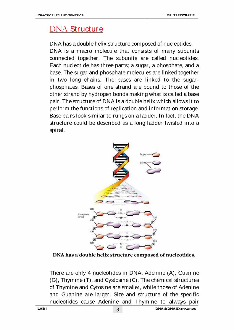
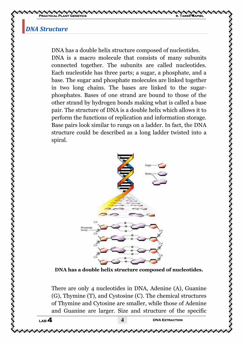
DNA has a double helix structure composed of nucleotides.
DNA is a macro molecule that consists of many subunits
connected together. The subunits are called nucleotides.
Each nucleotide has three parts; a sugar, a phosphate, and a
base. The sugar and phosphate molecules are linked together
in two long chains. The bases are linked to the sugar-
phosphates. Bases of one strand are bound to those of the
other strand by hydrogen bonds making what is called a base
pair. The structure of DNA is a double helix which allows it to
perform the functions of replication and information storage.
Base pairs look similar to rungs on a ladder. In fact, the DNA
structure could be described as a long ladder twisted into a
spiral.
DNA has a double helix structure composed of nucleotides.
There are only 4 nucleotides in DNA, Adenine (A), Guanine
(G), Thymine (T), and Cystosine (C). The chemical structures
of Thymine and Cytosine are smaller, while those of Adenine
and Guanine are larger. Size and structure of the specific
nucleotides cause Adenine and Thymine to always pair
Dr. Tarek Kapiel Practical Plant Genetics
DNA & DNA Extraction LAB 1
4
together while Cytosine and Guanine always pair together.
Therefore the two strands of DNA are considered
complimentary.
Simple Extraction of DNA from Peas
The plant tissue must be ground with a mortar and pestle to
break the plant cells open allowing the DNA to freely leave
the cell.
Why is it necessary to extract the DNA out of a cell in genetic
engineering? As mentioned above, DNA is found in the
nucleus of a cell. In order for genetic engineers to be able to
work with and transfer DNA into another organism, it must
be first taken out of the cell. Fortunately, a DNA molecule
remains somewhat stable outside of a living cell allowing
scientists the opportunity to work with and study it without
destroying it. To extract DNA, tissue samples are taken from
plants, and crushed to break open the cells.
Next, a buffered salt water solution is added into which DNA
dissolves easily. The DNA is purified by adding an organic
solution into which other molecules, such as fats and
proteins dissolve. The purified DNA solution is separated off
and the DNA is precipitated out of the solution by adding
alcohol. The DNA is now a solid string that can be spooled
out with a hook. The extracted DNA is placed into test tubes
with a weak buffer solution and can be stored almost
indefinitely.
During DNA extractions, the entire cells' DNA is extracted at
the same time. Extracted DNA has usually been sheared into
smaller pieces due to the leaf tissue grinding process. This
makes it difficult to isolate an entire chromosome in once
continuous strand. However, large pieces that contain several
to dozens of genes can be extracted intact with this method.
Dr. Tarek Kapiel Practical Plant Genetics
DNA & DNA Extraction LAB 1
5
After isolating the DNA, researchers can use it for further
laboratory studies including genetic engineering.
Materials:
1. Peas, about 50 g (frozen ones are suitable, but thaw them
first)
2. SDS solution (sodium dodecyl sulfate) or washing-up liquid,
(dishwashing liquid solution) 10 cm3 (ml).
3. NaCl (Table Salt), 3 g.
4. Distilled water, 90 cm3.
5. Very cold ethanol, about 10 cm3, straight from the freezer.
6. Novozymes Neutrase® (a protease), 2–3 drops.
7. Ice with cold water.
8. Filter paper.
9. Large funnel.
Procedure:
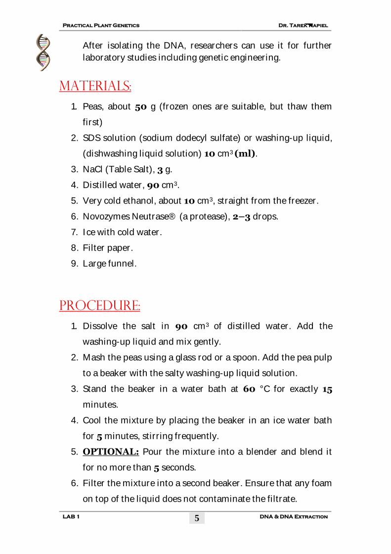
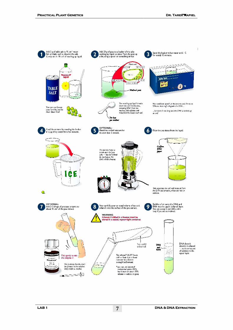
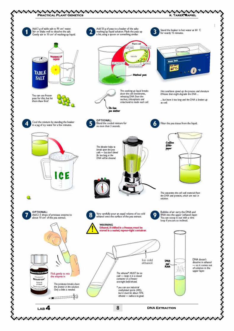
1. Dissolve the salt in 90 cm3 of distilled water. Add the
washing-up liquid and mix gently.
2. Mash the peas using a glass rod or a spoon. Add the pea pulp
to a beaker with the salty washing-up liquid solution.
3. Stand the beaker in a water bath at 60 °C for exactly 15
minutes.
4. Cool the mixture by placing the beaker in an ice water bath
for 5 minutes, stirring frequently.
5. OPTIONAL: Pour the mixture into a blender and blend it
for no more than 5 seconds.
6. Filter the mixture into a second beaker. Ensure that any foam
on top of the liquid does not contaminate the filtrate.
Dr. Tarek Kapiel Practical Plant Genetics
DNA & DNA Extraction LAB 1
6
7. OPTIONAL: Add 2–3 drops of protease to about 10 cm3 of
the pea extract in a boiling tube and mix well.
8. Very carefully pour ice cold ethanol down the side of the
boiling tube, to form a layer on top of the pea extract.
9. Leave the tube, undisturbed, for a few minutes.
10. Nucleic acids (DNA and RNA) will precipitate into the upper
(ethanol) layer
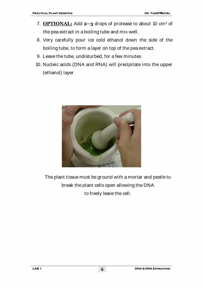
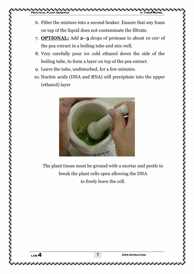
The plant tissue must be ground with a mortar and pestle to
break the plant cells open allowing the DNA
to freely leave the cell.
Dr. Tarek Kapiel Practical Plant Genetics
DNA & DNA Extraction LAB 1
7
Dr. Tarek Kapiel Practical Plant Genetics
DNA & DNA Extraction LAB 1
8
Expected Results
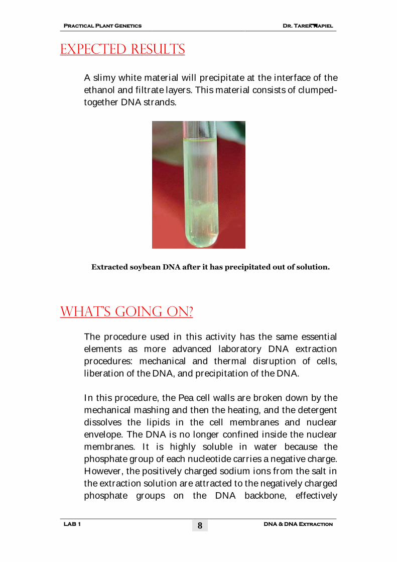
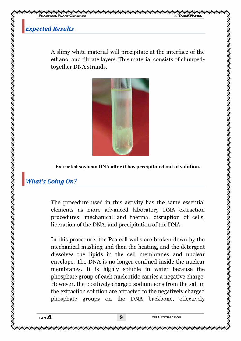
A slimy white material will precipitate at the interface of the
ethanol and filtrate layers. This material consists of clumped-
together DNA strands.
Extracted soybean DNA after it has precipitated out of solution.
What's Going On?
The procedure used in this activity has the same essential
elements as more advanced laboratory DNA extraction
procedures: mechanical and thermal disruption of cells,
liberation of the DNA, and precipitation of the DNA.
In this procedure, the Pea cell walls are broken down by the
mechanical mashing and then the heating, and the detergent
dissolves the lipids in the cell membranes and nuclear
envelope. The DNA is no longer confined inside the nuclear
membranes. It is highly soluble in water because the
phosphate group of each nucleotide carries a negative charge.
However, the positively charged sodium ions from the salt in
the extraction solution are attracted to the negatively charged
phosphate groups on the DNA backbone, effectively
Dr. Tarek Kapiel Practical Plant Genetics
DNA & DNA Extraction LAB 1
9
neutralizing the DNA's electric charge. This neutralization
allows the DNA molecules to aggregate with one another.
When the ethanol is added, the DNA clumps together and
precipitates at the water/ethanol interface because the DNA
is not soluble in ethanol.
Each glob of material in the precipitate will contain millions
of DNA strands clumped together, along with some of the
protein that is normally associated with DNA. Fortunately, a
DNA molecule remains somewhat stable outside of a living
cell allowing scientists the opportunity to work with and
study it without destroying it.
DNA Facts
All DNA is folded and packed into the nucleus of a human
cell. The diameter of the nucleus is about 0.005 mm or 1/500
the width of a dime.
There are 6 billion bits of information coded by DNA in each
of our nucleated cells (a bit is a measure of information).
Each human cell contains twenty-one times the information
that is found in the Encyclopedia Britannica, which is
thought to have about 280 million letters.
There are about 3 x 10⁹ nucleotide base pairs in the human
genome (the complete set of genes in one cell). If you lay all
the DNA strands from a single human cell end to end, it
would be about 2 meters long!
Calculate how many times to the moon and back a human's
DNA would reach if it was removed from each cell and each
strand laid end-to-end. Here is the information you need:
Each cell nucleus in a human holds about 2 meters of DNA
and a typical adult human is composed of 60 trillion cells.
The distance from the earth to the moon is 380,000
kilometers.
Dr. Tarek Kapiel Practical Plant Genetics
DNA & DNA Extraction LAB 1
10
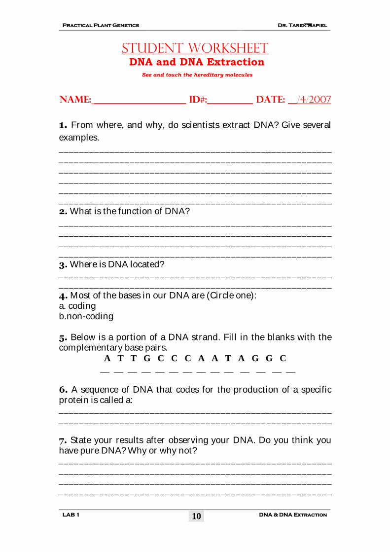
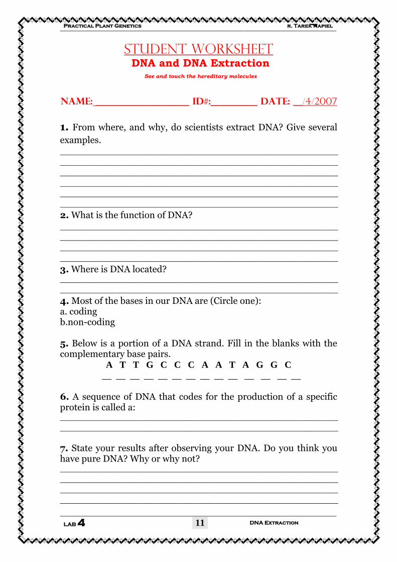
Student Worksheet DNA and DNA Extraction
See and touch the hereditary molecules
Name: ____________________ ID#:__________ DATE: __/4/2007
1. From where, and why, do scientists extract DNA? Give several
examples. ______________________________________________________
______________________________________________________
______________________________________________________
______________________________________________________
______________________________________________________
______________________________________________________
2. What is the function of DNA? ______________________________________________________
______________________________________________________
______________________________________________________
______________________________________________________
3. Where is DNA located?
______________________________________________________
______________________________________________________
4. Most of the bases in our DNA are (Circle one): a. coding b.non-coding 5. Below is a portion of a DNA strand. Fill in the blanks with the complementary base pairs.
A T T G C C C A A T A G G C
__ __ __ __ __ __ __ __ __ __ __ __ __ __
6. A sequence of DNA that codes for the production of a specific protein is called a: ______________________________________________________
______________________________________________________
7. State your results after observing your DNA. Do you think you have pure DNA? Why or why not? ______________________________________________________
______________________________________________________
______________________________________________________
______________________________________________________
Dr. Tarek Kapiel Practical Plant Genetics
DNA & DNA Extraction LAB 1
11
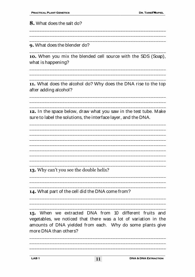
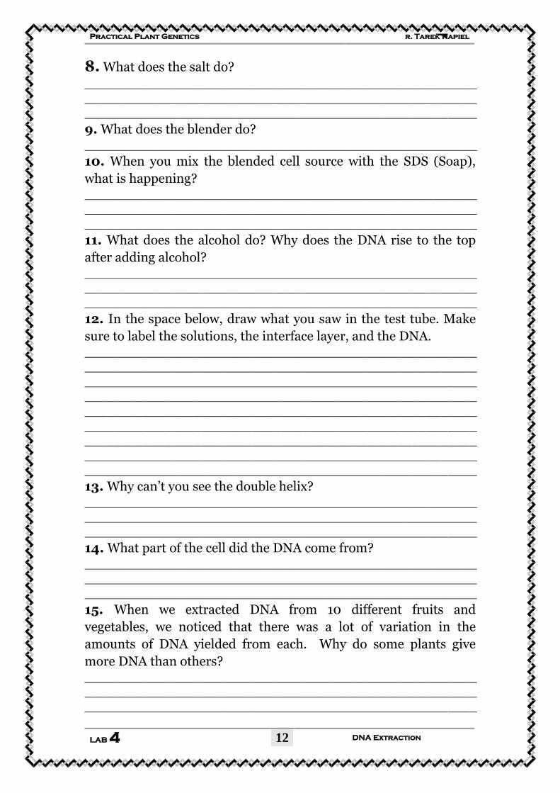
8. What does the salt do?
______________________________________________________
______________________________________________________
______________________________________________________
9. What does the blender do? ______________________________________________________
10. When you mix the blended cell source with the SDS (Soap),
what is happening? ______________________________________________________
______________________________________________________
______________________________________________________
11. What does the alcohol do? Why does the DNA rise to the top
after adding alcohol? ______________________________________________________
______________________________________________________
______________________________________________________
12. In the space below, draw what you saw in the test tube. Make
sure to label the solutions, the interface layer, and the DNA. ______________________________________________________
______________________________________________________
______________________________________________________
______________________________________________________
______________________________________________________
______________________________________________________
______________________________________________________
______________________________________________________
______________________________________________________
13. Why can’t you see the double helix? ______________________________________________________
______________________________________________________
______________________________________________________
14. What part of the cell did the DNA come from? ______________________________________________________
______________________________________________________
______________________________________________________
15. When we extracted DNA from 10 different fruits and
vegetables, we noticed that there was a lot of variation in the
amounts of DNA yielded from each. Why do some plants give
more DNA than others? ______________________________________________________
______________________________________________________
______________________________________________________
Dr. Tarek Kapiel Practical Plant Genetics
DNA & DNA Extraction LAB 1
12
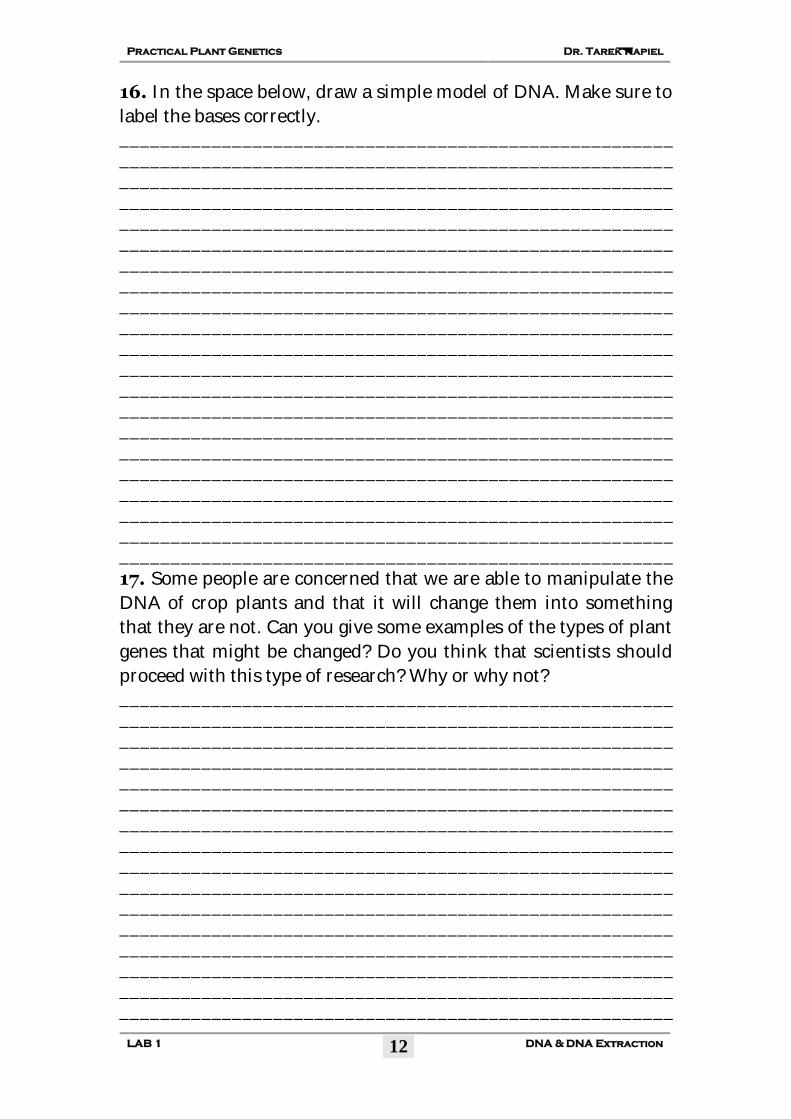
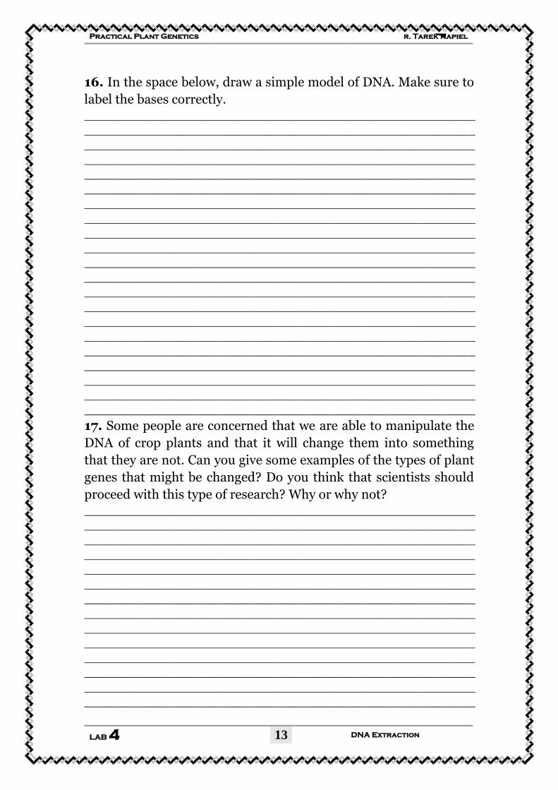
16. In the space below, draw a simple model of DNA. Make sure to
label the bases correctly. ______________________________________________________
______________________________________________________
______________________________________________________
______________________________________________________
______________________________________________________
______________________________________________________
______________________________________________________
______________________________________________________
______________________________________________________
______________________________________________________
______________________________________________________
______________________________________________________
______________________________________________________
______________________________________________________
______________________________________________________
______________________________________________________
______________________________________________________
______________________________________________________
______________________________________________________
______________________________________________________
______________________________________________________
17. Some people are concerned that we are able to manipulate the
DNA of crop plants and that it will change them into something
that they are not. Can you give some examples of the types of plant
genes that might be changed? Do you think that scientists should
proceed with this type of research? Why or why not? ______________________________________________________
______________________________________________________
______________________________________________________
______________________________________________________
______________________________________________________
______________________________________________________
______________________________________________________
______________________________________________________
______________________________________________________
______________________________________________________
______________________________________________________
______________________________________________________
______________________________________________________
______________________________________________________
______________________________________________________
______________________________________________________
Practical
Plant Genetics
By:
Dr. Tarek Kapiel, B.Sc. M.Sc., Ph.D. Associate Professor (Lecturer)
Cytology and Genetics Division Botany Department,
Faculty of Science, Cairo University.
©2009 All rights reserved
Dr. Tarek Kapiel Practical Plant Genetics
MITOSIS in Plant Cells LAB 2
2
Eukaryotic Cell Division
MITOSIS in Plant Cells
Objectives:
1. Students will prepare r oot t ip s quashes t o o bserve mitosis in onion roots. 2. Students will identify the various stages of mitosis in onion root tips. 3. Students w ill dr aw e ach phase of mitosis a nd t he main characteristics of each stage. 4. Students w ill observe h ow sa lt ( NaCl) a nd c olchicine a ffects mitosis in onion roots and they will study the effect of salt NaCl on the duration of each stage of mitosis.
Background:
Eukaryotes are diploid, which means they have two sets of chromosomes; o ne se t o f c hromosomes i s i nherited f rom each p arent. Eukaryotic DNA is enclosed by a nuclear envelope. The p roper s orting a nd distribution o f multiple chromosomes during cell division i s a complex p rocess t hat requires the temporary dissolution of the nuclear envelope. There are t wo t ypes o f cell d ivision i n eukaryotes, M ITOSIS and ME IOSIS, a nd they ha ve v ery different f unctions. Mitosis i s d esigned t o i ncrease t he n umber of c ells, a nd t o ensure that each of the daughter cells is genetically identical, containing t he di ploid ( 2n) number of c hromosomes. B y contrast, meiosis i s responsible for c onverting a di ploid c ell
Dr. Tarek Kapiel Practical Plant Genetics
MITOSIS in Plant Cells LAB 2
3
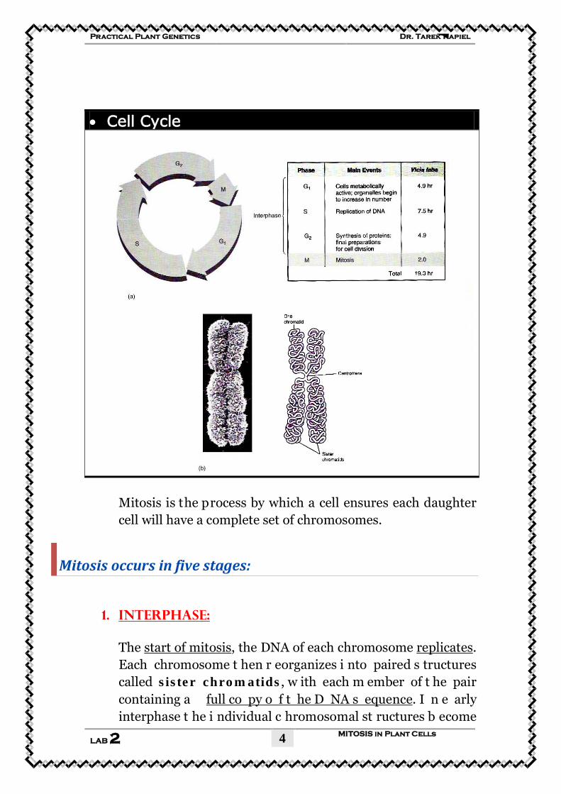
(i.e a cell c arrying t wo complete sets of chromosomes) into four h aploid (1n) c ells, e ach c arrying onl y one s et of chromosomes. Eukaryotic organisms carry out mitosis throughout t heir entire l ife t o g row and t o r eplace ol d or damaged cells. S ome eukaryotic organisms u se m itosis to reproduce asexually. The d aughter c ells produced b y mitosis a re diploid and genetically i dentical to each other and t he parent cells that produced them. The g rowth of multicellular or ganisms depends on Uprecise replicationU of i ndividual c ells f ollowed by Ucell d ivisionU. During r eplication, e ukaryotic ce lls g o t hrough Uan or dered series o f c yclical e ventsU. T he t ime f rom o ne c ell d ivision t o the next is called a Ucell cycleU. The c ell c ycle h as t wo m ain p hases, UinterphaseU and UmitosisU. Interphase i s f urther di vided i nto t hree distinct phases: G1 (gap 1 ), S (DNA s ynthesis, l asting 6 –8 ho urs i n eukaryotic c ells, a t t he e nd of w hich t he chromosomes h ave been duplicated), and G2 (gap 2, lasting about 4 hours) and Mitosis (M) Uis the phase of actual divisionU.
CELL CYCLE = INTERPHASE + MITOSIS
Cells only spend a small part of their l ife dividing. The time between consecutive mitotic divisions i s r eferred t o a s interphase. Eucaryotic c ells sp end most o f t heir t ime i n interphase. During interphase the cell’s genetic material is in the form of chromatin (uncoiled DNA), nucleoli are present, and the n uclear e nvelope i s clearly v isible. S hortly b efore mitosis, the cell duplicates its DNA during the S (synthesis) phase of interphase.
Dr. Tarek Kapiel Practical Plant Genetics
MITOSIS in Plant Cells LAB 2
4
• Cell Cycle
Mitosis is the process by which a cell ensures each daughter cell will have a complete set of chromosomes.
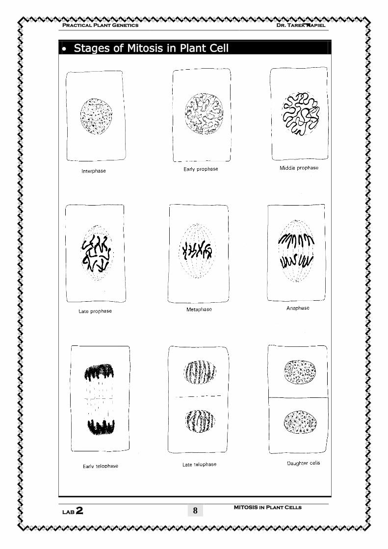
Mitosis occurs in five stages:
1. UInterphase:
The Ustart of mitosisU, the DNA of each chromosome UreplicatesU. Each chromosome t hen r eorganizes i nto paired s tructures called sister chromatids, w ith each m ember of t he pair containing a Ufull co py o f t he D NA s equenceU. I n e arly interphase t he i ndividual c hromosomal st ructures b ecome
Dr. Tarek Kapiel Practical Plant Genetics
MITOSIS in Plant Cells LAB 2
5
invisible. I nterphase c hromosomes a re c alled Uchromatin U (Flemming 1 879), i .e., n uclear st ructures st ainable b y b asic dyes. Nuclear material is surrounded by a nuclear envelope. Dark-staining b odies, n ucleoli, are visible. C hromosomes a ppear only as dark granules within the nucleus. The chromosomes are n ot i ndividually d istinguishable b ecause t hey a re uncoiled into long, thin strands.
2. UProphase:
Nuclear e nvelope a nd nucleoli d isappear. Chromatin condenses i nto c hromosomes, w hich b egin t o c oil a nd become distinguishable thin, threadlike structures. They are made up o f tw o id entical s ister chromatids. The si ster chromatids c ondense, thickening u ntil they appear joined a t a single site, known as the centromere. The mitotic spindle forms and begins t o move chromosomes t owards t he center of the cell.
3. UMetaphase: The si ster chromatids l ine up i n t he m iddle (equatorial plane) of the cell. A metaphase chromosome consists of two chromatids ( sister chromatids) and t he centromere, which holds them together. The centromere may divide each of th e chromatids i nto tw o chromosome arms. Th e regions at both ends of the chromosome are the telomeres. The m itotic spindle i s f ully f ormed. S pindle f ibers a re attached to centromeres and extend to the poles of the cell. The p oint o f attachment to the m itotic s pindle fibers is th e kinetochore. During m etaphase, c hromosomes c an be visualized u nder th e l ight m icroscope a s d iscrete e longated structures, 3–7 μm long.
4. UAnaphase:
Dr. Tarek Kapiel Practical Plant Genetics
MITOSIS in Plant Cells LAB 2
6
The chromatid pairs (Sister chromatids) split apart at the centromere, begin t o s eparate, becoming i ndividual chromosomes, ( Each f ormer c hromatid i s n ow a chromosome) whi ch begin t o m igrate t o toward o pposite poles (ends) of the cells.
5. UTelophase:
The final stage of mitosis, a full set of chromosomes reaches each pole of the cell. The mitotic spindle begins to disappear. The n ucleus and n ucleoli b egin t o r eappear. A n uclear membrane f orms around the chromosomes a t each p ole o f the cell. Chromosomes begin to unravel into chromatin.
Mitosis ends with the formation of two new cells, each with a matching full set of chromosomes.
UCytokinesis
Cytoplasmic division usually occurs a t t he e nd of t elophase. The c ytoplasm d ivides; t he c ell m embrane p inches i nward ultimately producing two daughter cells (Cytokinesis). Eukaryotic cells g o t hrough c ell d ivision cycles ( cell c ycle). Mitosis (M) in eukaryotic cells lasts about an h our. This is followed b y a p hase, G1 (interphase), o f e xtremely v aried duration. In plant cells cytokinesis is accomplished by the formation of a Ucell plateU.
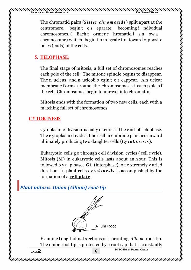
Plant mitosis. Onion (Allium) root-tip
Allium Root
Examine l ongitudinal s ections of s prouting Allium root-tip. The onion root tip is protected by a root cap that is constantly
Dr. Tarek Kapiel Practical Plant Genetics
MITOSIS in Plant Cells LAB 2
7
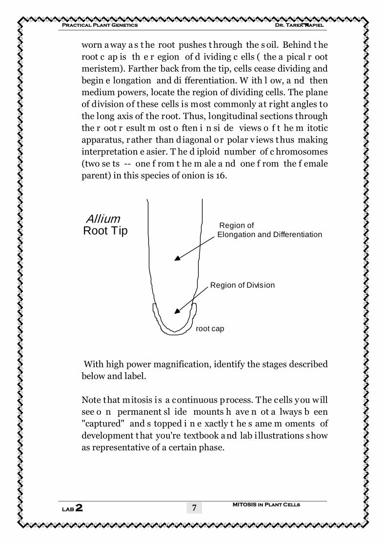
worn a way a s t he root pushes t hrough the s oil. Behind t he root c ap is th e r egion of d ividing c ells ( the a pical r oot meristem). Farther back from the tip, cells cease dividing and begin e longation and di fferentiation. W ith l ow, a nd then medium powers, locate the region of dividing cells. The plane of division of these cells is most commonly at right angles to the long axis of the root. Thus, longitudinal sections through the r oot r esult m ost o ften i n si de views o f t he m itotic apparatus, r ather than d iagonal or polar v iews t hus making interpretation e asier. T he d iploid number of c hromosomes (two se ts -- one f rom t he m ale a nd one f rom the f emale parent) in this species of onion is 16.
root cap
Region of Division
Region of Elongation and Differentiation
Allium Root Tip
With high power magnification, identify the stages described below and label. Note that mitosis is a continuous process. The cells you will see o n permanent sl ide mounts h ave n ot a lways b een "captured" and s topped i n e xactly t he s ame m oments of development t hat you're textbook a nd lab i llustrations show as representative of a certain phase.
Dr. Tarek Kapiel Practical Plant Genetics
MITOSIS in Plant Cells LAB 2
8
• Stages of Mitosis in Plant Cell
Dr. Tarek Kapiel Practical Plant Genetics
MITOSIS in Plant Cells LAB 2
9
Materials:
1. Students will set up their own slides.
2. Compound light microscope
3. Prepared onion or broad bean root tip slides.
4. Microscope slides and coverslips
5. 45% acetic acid
6. Glass rods
7. Razor blades
8. Paper towels
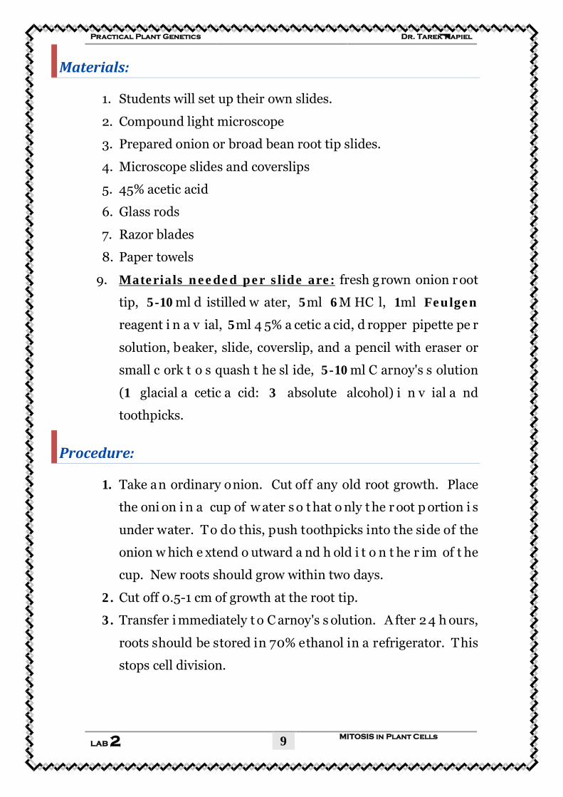
9. UMaterials needed per slide are:U fresh grown onion r oot
tip, 5-10ml d istilled w ater, 5ml 6M HC l, 1ml Feulgen
reagent i n a v ial, 5ml 4 5% a cetic a cid, d ropper pipette pe r
solution, beaker, slide, coverslip, and a pencil with eraser or
small c ork t o s quash t he sl ide, 5-10ml C arnoy's s olution
(1 glacial a cetic a cid: 3 absolute alcohol) i n v ial a nd
toothpicks.
Procedure:
1. Take an ordinary onion. Cut off any old root growth. Place
the oni on i n a cup of w ater s o t hat o nly t he r oot p ortion i s
under water. T o do this, push toothpicks into the side of the
onion w hich e xtend o utward a nd h old i t o n t he r im of t he
cup. New roots should grow within two days.
2. Cut off 0.5-1 cm of growth at the root tip.
3. Transfer i mmediately t o C arnoy's s olution. A fter 2 4 h ours,
roots should be stored in 70% ethanol in a refrigerator. This
stops cell division.
Dr. Tarek Kapiel Practical Plant Genetics
MITOSIS in Plant Cells LAB 2
10
4. After obtaining the root tip, pour off the fixative and replace
it with 2-5 ml distilled water. Solutions may be poured into a
beaker or down the drain.
5. After 1 minute remove the w ater with a pipette and add 2-5
ml 6M HCl.
6. After 3 minutes carefully remove the acid and wash tissue off
with distilled water. Agitate the vial for 1-2 minutes. Discard
the water.
7. Use forceps to transfer the tissue to a vial containing 1-2 ml
Feulgen r eagent. T he r eagent m ay be a dded t o t his v ial i f
desired. ( UCAUTION:U this dye w ill s tain h ands and clothes
permanently).
8. After 2 0-30 m inutes u se f orceps t o t ransfer t he tissue t o a
vial containing 5 ml 45% acetic acid.
9. Place 1 -2 drops of acetic a cid ont o a m icroscope s lide a nd
transfer t he t issue t o t he drop. U sing d issecting p ins a nd
razor blades macerate the tissue into tiny pieces.
10. Place a coverslip over the m acerated tissue trying not t o get
air bubbles under the coverslip. Press down firmly onto the
coverslip with a small cork or pencil eraser to spread the cells
in a very thin layer. P ush down in a perpendicular direction
and the coverslip should not break.
11. Examine u nder l ow p ower. Scan u nder l ow p ower (10 x
objective) to look for areas with cells undergoing mitosis, and
then examine under high power (40 x objective). Try to find
all the stages of mitosis.
12. Draw a ll t he d ifferent v iews o f c ells p resent under h igh
power.
13. After y ou h ave s tudied the section, y our in structor w ill test
your ability to recognize the stages of mitosis.
Dr. Tarek Kapiel Practical Plant Genetics
MITOSIS in Plant Cells LAB 2
11
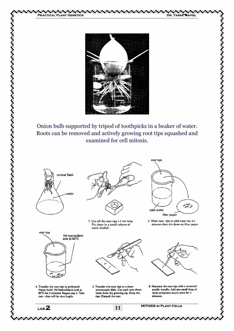
Onion bulb supported by tripod of toothpicks in a beaker of water. Roots can be removed and actively growing root tips squashed and
examined for cell mitosis.
Dr. Tarek Kapiel Practical Plant Genetics
MITOSIS in Plant Cells LAB 2
12
UStudent Worksheet [LAB 2]
Eukaryotic Cell Division MITOSIS in Plant Cells
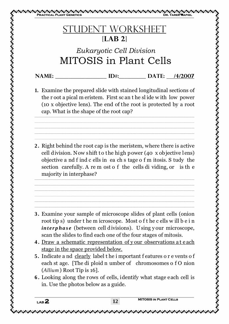
Name: ___________________ ID#:__________ DATE: U /4/2007 1. Examine the prepared slide with stained longitudinal sections of
the r oot a pical m eristem. First sc an t he sl ide w ith low power (10 x objective lens). The end of the root is protected by a root cap. What is the shape of the root cap?
_________________________________________________________________________________________________________________________________
_________________________________________________________________________________________________________________________________
_________________________________________________________________________________________________________________________________
_________________________________________________________________________________________________________________________________
_________________________________________________________________________________________________________________________________
2. Right behind the root cap is the meristem, where there is active cell d ivision. Now shift t o the high power (40 x objective l ens) objective a nd f ind c ells in ea ch s tage o f m itosis. S tudy the section carefully. A re m ost o f the cells di viding, or is th e majority in interphase?
_________________________________________________________________________________________________________________________________
_________________________________________________________________________________________________________________________________
_________________________________________________________________________________________________________________________________
_________________________________________________________________________________________________________________________________
_________________________________________________________________________________________________________________________________
_________________________________________________________________________________________________________________________________
3. Examine your sample of microscope slides of plant cells (onion root tip s) under t he m icroscope. Most o f t he c ells w ill b e i n interphase (between cell d ivisions). U sing y our microscope, scan the slides to find each one of the four stages of mitosis.
4. UDraw a schematic representation of y our observations a t e ach stage in the space provided below.
5. Indicate a nd UclearlyU label t he i mportant f eatures o r e vents o f each st age. [The di ploid n umber of chromosomes o f O nion (Allium) Root Tip is 16].
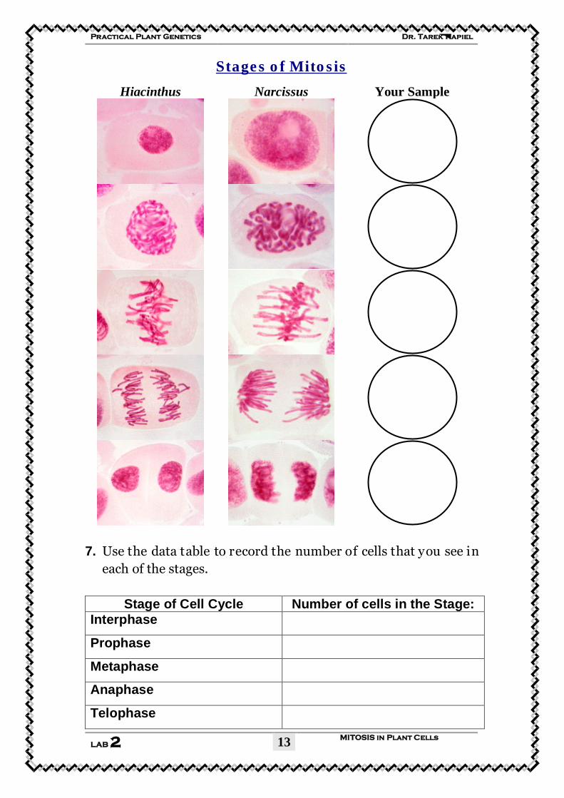
6. Looking along the rows of cells, identify what stage each cell is in. Use the photos below as a guide.
Dr. Tarek Kapiel Practical Plant Genetics
MITOSIS in Plant Cells LAB 2
13
UStages of Mitosis
Hiacinthus Narcissus Your Sample
7. Use the data table to record the number of cells that you see in each of the stages.
Stage of Cell Cycle Number of cells in the Stage: Interphase
Prophase
Metaphase
Anaphase
Telophase
Dr. Tarek Kapiel Practical Plant Genetics
MITOSIS in Plant Cells LAB 2
14
Low Power Magnification: ______X High Power Magnification: ______X
Interphase
Prophase
Metaphase
Anaphase
Telophase
Dr. Tarek Kapiel Practical Plant Genetics
MITOSIS in Plant Cells LAB 2
15
Observing Mitosis in Onion Root Tip cells
1. Before this lab, Onion bulbs were transferred to either an aerated solution of salt or an aerated solution of water for three or more days. Stain the root tips will stain the DNA of the Onion root tips. Your job is to prepare a “squash” of the Salt-treated and Water-treated root tips and observe the differences.
2. Place a water-treated root tip on a clean slide. Separate the intensely pink terminal end from the relatively unstained portion leaving 0.25-0.5 cm of intensely pink root tip on your slide.
3. Carefully macerate the root tip by tapping up and down with the
rounded end of a glass rod. Place a folded paper towel on top of the coverslip. Squeeze out the excess moisture by gently drawing the index finger of one hand across the paper towel, while holding onto the edge with your other hand. When the tissue has been broken into small fragments, lower a cover slip onto the preparation.
4. Flatten the tissue on the slide by placing your thumb on the paper
towel over the coverslip and gradually increasing pressure. Roll your thumb from side to side gently increasing the pressure with each roll. Avoid sliding the coverslip from side to side. Press down with the eraser end of your pencil to complete the squash.
5. Remove the paper towel and examine your root tip under the microscope. A well-made squash will have a monolayer of well-separated cells, with only a few larger chunks of tissue.
6. Find a cell in metaphase, in which the chromosomes are clearly
separated, using either your slide or another person’s slide. Using high power, observe the cell.
7. Using high power, count the number of nuclei visible in a high power field and record your answer.
8. In the same field, count the number of cells in each of these phases: prophase, metaphase, anaphase and telophase. Record your results.
9. Like a snapshot, the prepared onion root tip slide catches cells in
various stages of mitosis.
Dr. Tarek Kapiel Practical Plant Genetics
MITOSIS in Plant Cells LAB 2
16
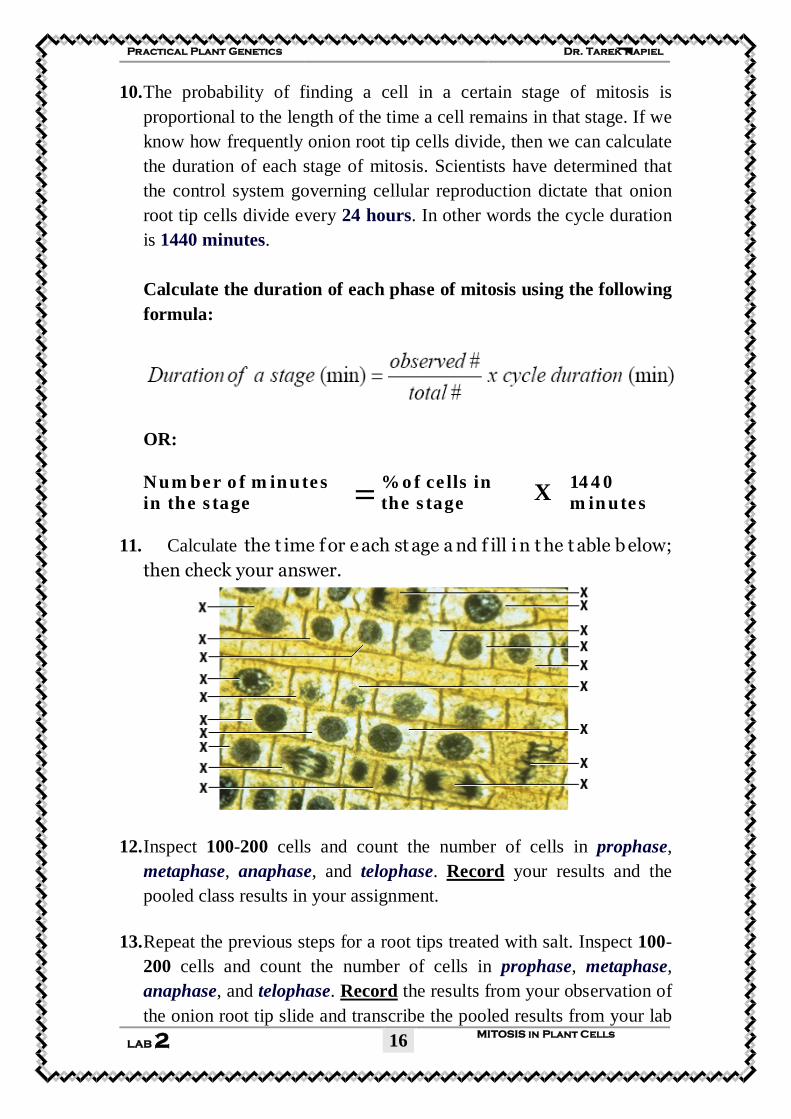
10. The probability of finding a cell in a certain stage of mitosis is proportional to the length of the time a cell remains in that stage. If we know how frequently onion root tip cells divide, then we can calculate the duration of each stage of mitosis. Scientists have determined that the control system governing cellular reproduction dictate that onion root tip cells divide every 24 hours. In other words the cycle duration is 1440 minutes. Calculate the duration of each phase of mitosis using the following formula:
OR: Number of minutes in the stage =
% of cells in the stage X
1440 minutes
11. Calculate the t ime f or e ach st age a nd f ill i n t he t able b elow;
then check your answer.
12. Inspect 100-200 cells and count the number of cells in prophase, metaphase, anaphase, and telophase. URecordU your results and the pooled class results in your assignment.
13. Repeat the previous steps for a root tips treated with salt. Inspect 100-
200 cells and count the number of cells in prophase, metaphase, anaphase, and telophase. URecordU the results from your observation of the onion root tip slide and transcribe the pooled results from your lab
Dr. Tarek Kapiel Practical Plant Genetics
MITOSIS in Plant Cells LAB 2
17
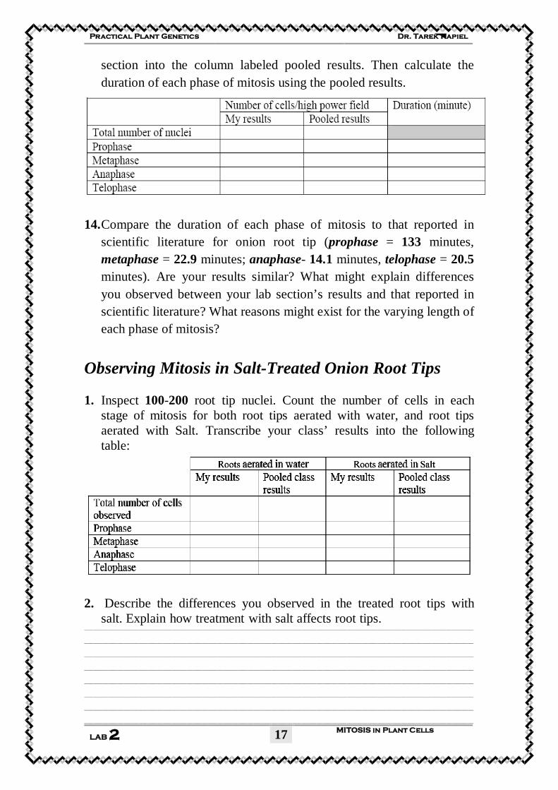
section into the column labeled pooled results. Then calculate the duration of each phase of mitosis using the pooled results.
14. Compare the duration of each phase of mitosis to that reported in
scientific literature for onion root tip (prophase = 133 minutes, metaphase = 22.9 minutes; anaphase- 14.1 minutes, telophase = 20.5 minutes). Are your results similar? What might explain differences you observed between your lab section’s results and that reported in scientific literature? What reasons might exist for the varying length of each phase of mitosis?
Observing Mitosis in Salt-Treated Onion Root Tips
1. Inspect 100-200 root tip nuclei. Count the number of cells in each
stage of mitosis for both root tips aerated with water, and root tips aerated with Salt. Transcribe your class’ results into the following table:
2. Describe the differences you observed in the treated root tips with
salt. Explain how treatment with salt affects root tips. _________________________________________________________________________________________________________________________________
_________________________________________________________________________________________________________________________________
_________________________________________________________________________________________________________________________________
_________________________________________________________________________________________________________________________________
_________________________________________________________________________________________________________________________________
_________________________________________________________________________________________________________________________________
_________________________________________________________________________________________________________________________________
_________________________________________________________________________________________________________________________________
Dr. Tarek Kapiel Practical Plant Genetics
MITOSIS in Plant Cells LAB 2
18
Analysis & Conclusions: 1. What stage were the majority of the cells in? _________________________________________________________________________________________________________________________________
_________________________________________________________________________________________________________________________________
2. The onion plant began as a single cell. That cell had X number of chromosomes. How many chromosomes are in each of the cells that you observed? (Give the answer in terms of X.) How do you know?
_________________________________________________________________________________________________________________________________
_________________________________________________________________________________________________________________________________
3. If this onion would reproduce sexually, it would need to produce sperm and/or eggs by the process of meiosis. After meiosis, how many chromosomes would be in each sex cell (in terms of X)?
_________________________________________________________________________________________________________________________________
_________________________________________________________________________________________________________________________________
4. If this onion would complete the process of sexual reproduction (fertilizing an egg cell), how many chromosomes would be in the zygotes that are produced (in terms of X)?
_________________________________________________________________________________________________________________________________
_________________________________________________________________________________________________________________________________
Practical
Plant Genetics
By:
Dr. Tarek Kapiel, B.Sc. M.Sc., Ph.D. Associate Professor (Lecturer)
Cytology and Genetics Division Botany Department,
Faculty of Science, Cairo University.
©2009 All rights reserved
Dr. Tarek Kapiel Practical Plant Genetics
Meiosis in Plant Cells
LAB 3
2
Eukaryotic Cell Division
MEIOSIS in Plant Cells
Objectives:
1. To identify cells in all stages of meiosis on prepared
microscope slides, diagrams, and photographs.
2. To describe the behavior of chromosomes during all
phases of meiosis.
3. To be able to define the following terms: anaphase,
autosome, centromere, chromatid, metaphase, nucleus,
prophase, gametogenesis, somatic, spindle, synapsis,
telophase, tetrad, karyokinesis, cytokinesis.
Background:
In most species, it is very important that the offspring
produced by fertilization have the same number of
chromosomes as the parents. Meiosis is a special type of cell
division that produces haploid gametes. Meiosis involves two
cell divisions and ultimately produces four haploid gametes.
Meiosis involves two consecutive divisions of a particular cell
type, which results in four daughter cells that have half of the
chromosome number of their mother cell. The daughter cells
are thus genetically different from the mother cell, and
due to independent, random assortment of chromosomes
and crossing over, they are genetically different from one
another as well.
Dr. Tarek Kapiel Practical Plant Genetics
Meiosis in Plant Cells
LAB 3
3
The first division is called the reduction division, since
this is the one during which chromosome number in the cells
is reduced.
Only certain types of cells divide by meiosis that occurs in the
archegonium/antheridium in plants, and in the gonads in
animals. Cells with an odd number of chromosome sets
normally cannot carry out meiosis, as chromosome pairing is
messed up.
The haploid gametes produced by meiosis are different from
each other as well as from the parent cells due to the
crossing over of genetic material between homologous
chromosomes and the random distribution of
homologous chromosomes.
In meiosis, one homologue of each chromosome type is
distributed to each of the resulting "daughter" nuclei. The
two resulting nuclei are not identical, and have one-half the
original chromosome complement. Meiosis occurs during
the process of gametogenesis (the production of gametes),
where haploid spores are produced that divide mitotically
to form the gametophyte. The somatic chromosome
complement is restored when the haploid sperm nucleus
fuses with the egg nucleus during fertilization.
The behavior of the chromosomes during meiosis is known to
be responsible for the segregation and independent
assortment of alleles.
Dr. Tarek Kapiel Practical Plant Genetics
Meiosis in Plant Cells
LAB 3
4
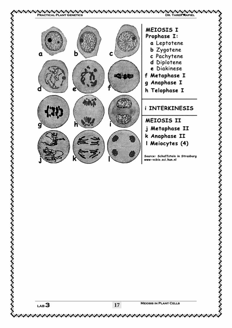
STAGES OF MEIOSIS
MEIOSIS I: Reductive Division (Diploid Haploid)
Prophase I:
Similar to prophase of mitosis with one important difference:
Crossing Over: Pairs of homologous chromosomes
synapse together to form tetrads and exchange genetic
information (DNA). Crossing over creates new, recombinant
chromosomes.
Homologous chromosomes code for the same genetic
information and are of the same size, but are different
because one comes from an individual’s mother, while the
other comes from the individual’s father.
Metaphase I:
Brief stage in which tetrads line up in the equatorial plane
of the cell.
Anaphase I:
Homologous chromosomes separate and migrate to opposite
ends of cell.
Telophase I:
A full set of chromosomes reaches each pole of the cell.
The cells produced are haploid, only contain half of the
original number of chromosomes.
Interphase
May be very brief or absent between meiosis I and meiosis II.
MEIOSIS II:
Very similar to mitosis.
Dr. Tarek Kapiel Practical Plant Genetics
Meiosis in Plant Cells
LAB 3
5
Prophase II:
DNA condenses into chromosomes. No crossing over occurs.
Metaphase II:
Individual chromosomes line up in the equatorial plane of
the cell.
Anaphase II:
Chromatids separate and begin to migrate to opposite poles
of the cell.
Telophase II and Cytokinesis:
A full set of chromosomes reaches each pole of the cell. Four
different gametes are produced.
Dr. Tarek Kapiel Practical Plant Genetics
Meiosis in Plant Cells
LAB 3
6
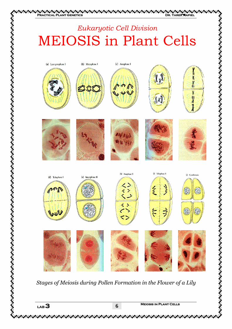
Eukaryotic Cell Division
MEIOSIS in Plant Cells
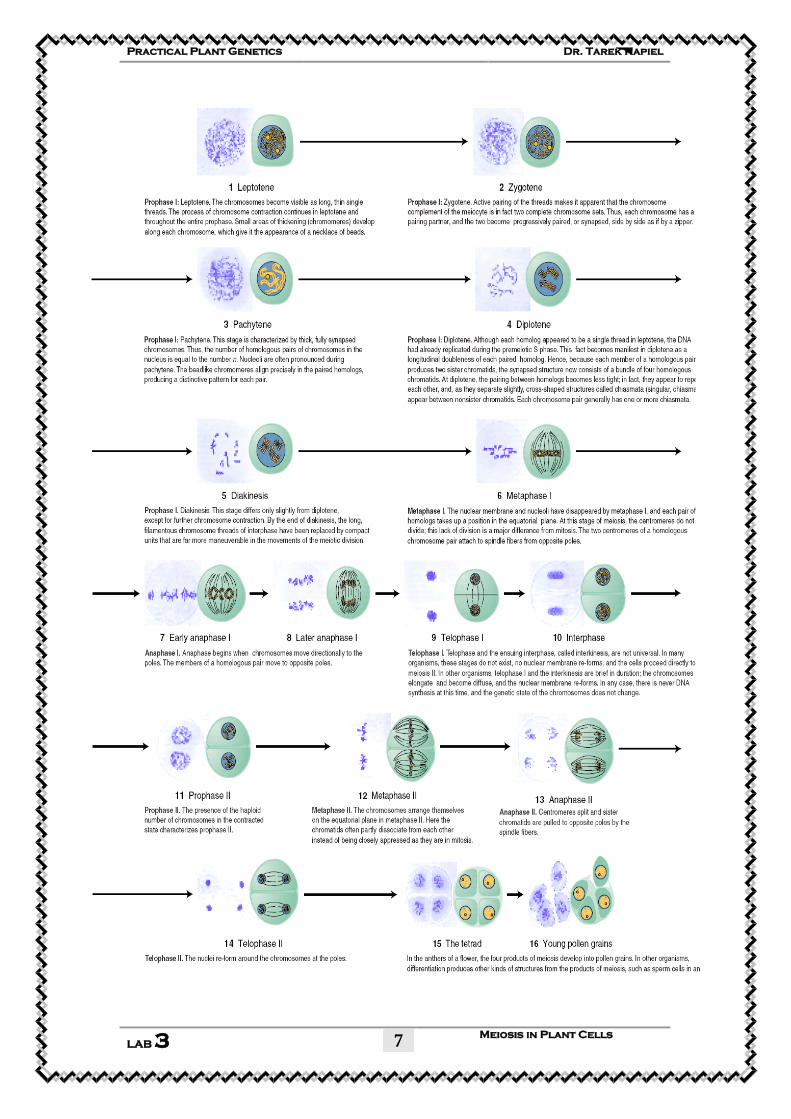
Stages of Meiosis during Pollen Formation in the Flower of a Lily
Dr. Tarek Kapiel Practical Plant Genetics
Meiosis in Plant Cells
LAB 3
7
Dr. Tarek Kapiel Practical Plant Genetics
Meiosis in Plant Cells
LAB 3
8
Materials:
1. Plants with immature inflorescences or flowers
2. Compound microscope
3. Forceps with curved tip
4. Dissecting needles (bent end preferred)
5. Microscope slides
6. Cover glasses
7. Stains (acetoorcein and/or Wittmann's hematoxylin acetic
acid)
8. Paper towels
9. Lens paper
Procedure:
1. Obtain an unopened inflorescence from the material
provided, and remove the paperlike covering to expose the
developing buds.
2. Transfer a few (three or four) of these buds to a microscope
slide.
3. Add a drop of stain (either aceto orcein or Wittmann's
hematoxylin). If too much stain is added, simply blot some of
it away. Be sure to include one or more very small buds.
Larger buds will probably contain only pollen grains because
the sporogenous tissue will have completed meiosis.
4. Microspores and pollen grains should be seen, but it is likely
they will be found even in some of the smaller buds, so it is
best to start with the smaller buds and include larger buds as
needed.
5. Use a dissecting needle or forceps to crush the buds in the
stain. This will release microspore mother cells from the
Dr. Tarek Kapiel Practical Plant Genetics
Meiosis in Plant Cells
LAB 3
9
anthers. Repeated tapping of the buds with the needle may
be necessary to release sufficient numbers of microspore
mother cells from the anthers.
6. Use the forceps and/or dissecting needles to remove as much
of the debris as possible. Even small pieces of petals prevent
the cover glass from lying flat and will lead to a distorted
image when viewed with the microscope.
7. If hematoxylin is used, a drop of 45% acetic acid should be
added to destain the cells.
8. Add a cover glass. It may be necessary to add more stain or a
drop of 45% acetic acid to the preparation in order to make
the cover glass lie flat.
9. Blot away the excess liquid, but be careful to avoid adding
very much pressure to the cover glass because the
chromosomes are somewhat fragile.
10. Observe the slide at low and high dry magnifications. Do not
try to use oil immersion at this time. Once the preparation is
scanned with the compound microscope, it may be obvious
that some pressure on the cover glass is needed in order to
spread the arms of the chromosomes.
11. Use oil immersion microscopy for detailed observation of
good representative stages.
Observations
A wide range of cell types may be observed. Some non-
meiotic cells will be seen. Non-meiotic cells will resemble
onion root tip cells or other undifferentiated cells. Cells
undergoing meiosis will be larger than the cells of the
supportive tissue and will contain prominent nuclei or
obvious chromosomes. If very small buds are used,
premeiotic mitotic figures will be observed in what will
Dr. Tarek Kapiel Practical Plant Genetics
Meiosis in Plant Cells
LAB 3
10
become microspore mother cells. Meiosis is typically divided
into the following stages:
Meiosis I
Leptotene:
Chromosomes become visible as threads. Keep in mind that
what appears to be a single thread is actually two chromatids
because replication took place in the interphase prior to
meiosis.
It may be necessary to force the cover glass down a bit in
order to make individual chromosomes visible.
Zygotene:
Pairing of homologous chromosomes occurs. The
chromosomes appear thicker and less numerous because
pairing occurs. Again, it may be necessary to apply some
pressure to the cover glass in order to observe the paired
nature of the chromosomes.
Pachytene:
The chromosomes become shorter and thicker. The amount
of chromosomal material appears to be considerably less
because the chromosomes have become much shorter. The
fact that the chromosomes are paired should be obvious.
Diplotene:
The characteristic feature of diplotene is the presence of
chiasmata. The chromosomes are also shorter
than in pachytene.
Diakinesis:
Repulsion of the centromeres and terminalization of the
chiasmata mark this stage. The chromosomes are very short
and thick, but they have not yet lined up in a characteristic
metaphase figure.
Metaphase I: The chromosomes are lined up on the spindle. The staining
procedure used here does not reveal the spindle. A polar view
of metaphase I reveals the chromosomes to be organized at
one plane in the cell. A nonpolar view of metaphase I makes
it appear as though the chromosomes are lined up across the
middle of the cell.
Dr. Tarek Kapiel Practical Plant Genetics
Meiosis in Plant Cells
LAB 3
11
Anaphase I: Separation of the paired homologs. It should be noted that
the chromosomes now appear thinner.
Comparisons with metaphase I make it obvious there has
been a halving of the genetic material with half going to each
pole. Early and late stages of anaphase I will be seen.
Telophase I: Once the chromosomes have moved to the poles in anaphase,
they begin to lose their identity and become reorganized into
the nucleus. This marks telophase.
Interphase: There is a brief period when the microspore mother cells
appear to contain two interphase nuclei. Because this is a
brief part of meiosis this interphase may not be seen by every
laboratory class. In some species, the chromosomes seem to
go directly from telophase I into prophase II.
Meiosis II
Prophase II:
Chromosomes become visible as long threads. Note that this
occurs in both of the nuclei of the microspore mother cell.
Metaphase II: The chromosomes become aligned on the spindle apparatus.
Note that different cells present different configurations
depending on the angles from which they are observed.
Anaphase II: Separation of the replicated chromatids. Compare the
number of chromosomes and the thickness of the
chromosomes with what was observed in anaphase I.
Telophase II: The chromosomes lose their distinctiveness and become
organized into nuclei. Note that there will be four haploid
nuclei within the confines of what started out as a single
diploid cell.
Tetrad:
Dr. Tarek Kapiel Practical Plant Genetics
Meiosis in Plant Cells
LAB 3
12
The original microspore mother cell has been divided into
four cells. A polar view of these cells resembles an orange
with four sections which has been cut through the equator. A
non-polar view of the tetrad may be difficult to focus because
the “orange sections” appear to be piled on top of each other.
Microspores: The uninucleate haploid tetrads are released, and at this
stage are called microspores.
Pollen grains: These result from a mitotic division within the microspores.
Stages typical of mitosis can be seen including prophase,
metaphase, anaphase, and telophase. Cytokinesis may also be
seen. Because of the shape of the microspores, views of
metaphase are more often polar than nonpolar, making it
possible to count all eight chromosomes, but the same shape
makes it difficult to distinguish anaphase from telophase
since one generally finds polar views of these stages as well.
The resulting pollen grains are bicellular and have distinctive
wall markings.
This lab lends itself very well to encouraging students to
share what they have found with others. If they have found
something worth showing to others, they should announce
what they have to share and go see the stages others have
found. In this way, a few preparations per student can result
in a whole array of meiotic figures for all to see. Students
should also compare the stages they have observed with the
figures on display.
Dr. Tarek Kapiel Practical Plant Genetics
Meiosis in Plant Cells
LAB 3
13
Student Worksheet [LAB 3]
Eukaryotic Cell Division
MEIOSIS in Plant Cells
Name: ___________________ ID#:__________ DATE: /4/2007
1. Examine the prepared slide with stained sections of lily anthers.
First scan the slide with low power (10 x objective lens), the by
using the high power (40-100 x objective lens).
2. Stages of Meiosis:
Meiosis I
Low Power
Magnification: ______X
High Power
Magnification: ______X
1. Leptotene:
2. Zygotene:
3. Pachytene:
Dr. Tarek Kapiel Practical Plant Genetics
Meiosis in Plant Cells
LAB 3
14
4. Diplotene:
5. Diakinesis:
6. Metaphase I:
7. Anaphase I:
8. Telophase I:
Dr. Tarek Kapiel Practical Plant Genetics
Meiosis in Plant Cells
LAB 3
15
9. Interphase:
Meiosis II
10. Prophase:
II
11. Metaphase:
II
12. Anaphase:
II
13. Telophase:
II
Dr. Tarek Kapiel Practical Plant Genetics
Meiosis in Plant Cells
LAB 3
16
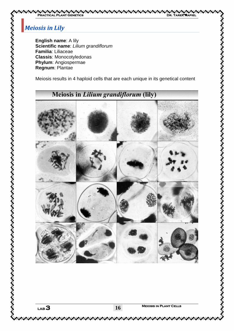
Meiosis in Lily
English name: A lily Scientific name: Lilium grandiflorum Familia: Liliaceae Classis: Monocotyledonas Phylum: Angiospermae Regnum: Plantae
Meiosis results in 4 haploid cells that are each unique in its genetical content
Dr. Tarek Kapiel Practical Plant Genetics
Meiosis in Plant Cells
LAB 3
17
Practical
Plant Genetics
By:
Dr. Tarek Kapiel, B.Sc. M.Sc., Ph.D. Associate Professor (Lecturer)
Cytology and Genetics Division Botany Department,
Faculty of Science, Cairo University.
©2009 All rights reserved
r. Tarek Kapiel Practical Plant Genetics
DNA Extraction LAB 4
2
DNA Extraction
See and touch the hereditary molecules
overview
This lab discusses what DNA is and how it relates to genes
and chromosomes. How and why DNA is extracted in the
genetic engineering process is also covered.
At the completion of this lesson, you should be able to:
Visualize the relationship between DNA, genes, and
chromosomes.
Compare the roles that DNA and proteins play in a cell.
Contrast DNA and proteins in their chemical make up.
Explain why DNA extraction is important in genetic
engineering and how it is done.
Understand why genes can be transferred between
organisms and still work.
Objectives:
1- To isolate nucleic acids (DNA and RNA).
2- To understand the importance of DNA.
Background:
Deoxyribonucleic acid (DNA) is the genetic material present
in all organisms, from bacteria to humans. DNA is the
material that makes up genes.
DNA is the instruction manual for living things. Within the
relatively simple double helix structure, DNA holds the coded
r. Tarek Kapiel Practical Plant Genetics
DNA Extraction LAB 4
3
information for how to make every protein a living organism
might need throughout its entire life.
Very pure DNA can be easily extracted from cells in a
research laboratory. DNA is a non-living stable molecule. The
DNA code is universal allowing it to work the same in all
living things. This is a critical fact that makes genetic
engineering possible. Genetic engineering is the directed
addition of new DNA to an organism's genetic makeup, its
genome. Once scientists understood the properties of DNA
and how it functions as genetic material, they could envision
and invent techniques for genetic engineering.
DNA extraction is necessary because genetic engineers need
to be able to work with the DNA, cut it, locate and clone a
single gene, and modify the gene before they insert it into
another organism.
DNA is composed of nucleotides bonded to a sugar-
phosphate backbone. Double stranded DNA forms a double
helix structure.
The DNA double helix coils up into compact structures called
chromosomes. Small segments of the chromosome that
encode a single protein are called genes.
Chromosomes are microscopic. There are thousands of genes
on each chromosome and hundreds of nucleotides in the
DNA sequence of each gene.
The role of DNA is to store and pass on genetic information.
The sequence of DNA subunits determines an organism's
traits.
r. Tarek Kapiel Practical Plant Genetics
DNA Extraction LAB 4
4
DNA Structure
DNA has a double helix structure composed of nucleotides.
DNA is a macro molecule that consists of many subunits
connected together. The subunits are called nucleotides.
Each nucleotide has three parts; a sugar, a phosphate, and a
base. The sugar and phosphate molecules are linked together
in two long chains. The bases are linked to the sugar-
phosphates. Bases of one strand are bound to those of the
other strand by hydrogen bonds making what is called a base
pair. The structure of DNA is a double helix which allows it to
perform the functions of replication and information storage.
Base pairs look similar to rungs on a ladder. In fact, the DNA
structure could be described as a long ladder twisted into a
spiral.
DNA has a double helix structure composed of nucleotides.
There are only 4 nucleotides in DNA, Adenine (A), Guanine
(G), Thymine (T), and Cystosine (C). The chemical structures
of Thymine and Cytosine are smaller, while those of Adenine
and Guanine are larger. Size and structure of the specific
r. Tarek Kapiel Practical Plant Genetics
DNA Extraction LAB 4
5
nucleotides cause Adenine and Thymine to always pair
together while Cytosine and Guanine always pair together.
Therefore the two strands of DNA are considered
complimentary.
Simple Extraction of DNA from Peas
The plant tissue must be ground with a mortar and pestle to
break the plant cells open allowing the DNA to freely leave
the cell.
Why is it necessary to extract the DNA out of a cell in genetic
engineering? As mentioned above, DNA is found in the
nucleus of a cell. In order for genetic engineers to be able to
work with and transfer DNA into another organism, it must
be first taken out of the cell. Fortunately, a DNA molecule
remains somewhat stable outside of a living cell allowing
scientists the opportunity to work with and study it without
destroying it. To extract DNA, tissue samples are taken from
plants, and crushed to break open the cells.
Next, a buffered salt water solution is added into which DNA
dissolves easily. The DNA is purified by adding an organic
solution into which other molecules, such as fats and
proteins dissolve. The purified DNA solution is separated off
and the DNA is precipitated out of the solution by adding
alcohol. The DNA is now a solid string that can be spooled
out with a hook. The extracted DNA is placed into test tubes
with a weak buffer solution and can be stored almost
indefinitely.
During DNA extractions, the entire cells' DNA is extracted at
the same time. Extracted DNA has usually been sheared into
smaller pieces due to the leaf tissue grinding process. This
makes it difficult to isolate an entire chromosome in once
r. Tarek Kapiel Practical Plant Genetics
DNA Extraction LAB 4
6
continuous strand. However, large pieces that contain several
to dozens of genes can be extracted intact with this method.
After isolating the DNA, researchers can use it for further
laboratory studies including genetic engineering.
Materials:
1. Peas, about 50 g (frozen ones are suitable, but thaw them
first)
2. SDS solution (sodium dodecyl sulfate) or washing-up liquid,
(dishwashing liquid solution) 10 cm3 (ml).
3. NaCl (Table Salt), 3 g.
4. Distilled water, 90 cm3.
5. Very cold ethanol, about 10 cm3, straight from the freezer.
6. Novozymes Neutrase® (a protease), 2–3 drops.
7. Ice with cold water.
8. Filter paper.
9. Large funnel.
Procedure:
1. Dissolve the salt in 90 cm3 of distilled water. Add the
washing-up liquid and mix gently.
2. Mash the peas using a glass rod or a spoon. Add the pea pulp
to a beaker with the salty washing-up liquid solution.
3. Stand the beaker in a water bath at 60 °C for exactly 15
minutes.
4. Cool the mixture by placing the beaker in an ice water bath
for 5 minutes, stirring frequently.
5. OPTIONAL: Pour the mixture into a blender and blend it
for no more than 5 seconds.
r. Tarek Kapiel Practical Plant Genetics
DNA Extraction LAB 4
7
6. Filter the mixture into a second beaker. Ensure that any foam
on top of the liquid does not contaminate the filtrate.
7. OPTIONAL: Add 2–3 drops of protease to about 10 cm3 of
the pea extract in a boiling tube and mix well.
8. Very carefully pour ice cold ethanol down the side of the
boiling tube, to form a layer on top of the pea extract.
9. Leave the tube, undisturbed, for a few minutes.
10. Nucleic acids (DNA and RNA) will precipitate into the upper
(ethanol) layer
The plant tissue must be ground with a mortar and pestle to
break the plant cells open allowing the DNA
to freely leave the cell.
r. Tarek Kapiel Practical Plant Genetics
DNA Extraction LAB 4
8
r. Tarek Kapiel Practical Plant Genetics
DNA Extraction LAB 4
9
Expected Results
A slimy white material will precipitate at the interface of the
ethanol and filtrate layers. This material consists of clumped-
together DNA strands.
Extracted soybean DNA after it has precipitated out of solution.
What's Going On?
The procedure used in this activity has the same essential
elements as more advanced laboratory DNA extraction
procedures: mechanical and thermal disruption of cells,
liberation of the DNA, and precipitation of the DNA.
In this procedure, the Pea cell walls are broken down by the
mechanical mashing and then the heating, and the detergent
dissolves the lipids in the cell membranes and nuclear
envelope. The DNA is no longer confined inside the nuclear
membranes. It is highly soluble in water because the
phosphate group of each nucleotide carries a negative charge.
However, the positively charged sodium ions from the salt in
the extraction solution are attracted to the negatively charged
phosphate groups on the DNA backbone, effectively
r. Tarek Kapiel Practical Plant Genetics
DNA Extraction LAB 4
10
neutralizing the DNA's electric charge. This neutralization
allows the DNA molecules to aggregate with one another.
When the ethanol is added, the DNA clumps together and
precipitates at the water/ethanol interface because the DNA
is not soluble in ethanol.
Each glob of material in the precipitate will contain millions
of DNA strands clumped together, along with some of the
protein that is normally associated with DNA. Fortunately, a
DNA molecule remains somewhat stable outside of a living
cell allowing scientists the opportunity to work with and
study it without destroying it.
DNA Facts
All DNA is folded and packed into the nucleus of a human
cell. The diameter of the nucleus is about 0.005 mm or 1/500
the width of a dime.
There are 6 billion bits of information coded by DNA in each
of our nucleated cells (a bit is a measure of information).
Each human cell contains twenty-one times the information
that is found in the Encyclopedia Britannica, which is
thought to have about 280 million letters.
There are about 3 x 10⁹ nucleotide base pairs in the human
genome (the complete set of genes in one cell). If you lay all
the DNA strands from a single human cell end to end, it
would be about 2 meters long!
Calculate how many times to the moon and back a human's
DNA would reach if it was removed from each cell and each
strand laid end-to-end. Here is the information you need:
Each cell nucleus in a human holds about 2 meters of DNA
and a typical adult human is composed of 60 trillion cells.
The distance from the earth to the moon is 380,000
kilometers.
r. Tarek Kapiel Practical Plant Genetics
DNA Extraction LAB 4
11
Student Worksheet DNA and DNA Extraction
See and touch the hereditary molecules
Name: ____________________ ID#:__________ DATE: __/4/2007
1. From where, and why, do scientists extract DNA? Give several
examples. ______________________________________________________
______________________________________________________
______________________________________________________
______________________________________________________
______________________________________________________
______________________________________________________
2. What is the function of DNA? ______________________________________________________
______________________________________________________
______________________________________________________
______________________________________________________
3. Where is DNA located?
______________________________________________________
______________________________________________________
4. Most of the bases in our DNA are (Circle one): a. coding b.non-coding 5. Below is a portion of a DNA strand. Fill in the blanks with the complementary base pairs.
A T T G C C C A A T A G G C
__ __ __ __ __ __ __ __ __ __ __ __ __ __
6. A sequence of DNA that codes for the production of a specific protein is called a: ______________________________________________________
______________________________________________________
7. State your results after observing your DNA. Do you think you have pure DNA? Why or why not? ______________________________________________________
______________________________________________________
______________________________________________________
______________________________________________________
r. Tarek Kapiel Practical Plant Genetics
DNA Extraction LAB 4
12
8. What does the salt do?
______________________________________________________
______________________________________________________
______________________________________________________
9. What does the blender do? ______________________________________________________
10. When you mix the blended cell source with the SDS (Soap),
what is happening? ______________________________________________________
______________________________________________________
______________________________________________________
11. What does the alcohol do? Why does the DNA rise to the top
after adding alcohol? ______________________________________________________
______________________________________________________
______________________________________________________
12. In the space below, draw what you saw in the test tube. Make
sure to label the solutions, the interface layer, and the DNA. ______________________________________________________
______________________________________________________
______________________________________________________
______________________________________________________
______________________________________________________
______________________________________________________
______________________________________________________
______________________________________________________
______________________________________________________
13. Why can’t you see the double helix? ______________________________________________________
______________________________________________________
______________________________________________________
14. What part of the cell did the DNA come from? ______________________________________________________
______________________________________________________
______________________________________________________
15. When we extracted DNA from 10 different fruits and
vegetables, we noticed that there was a lot of variation in the
amounts of DNA yielded from each. Why do some plants give
more DNA than others? ______________________________________________________
______________________________________________________
______________________________________________________
r. Tarek Kapiel Practical Plant Genetics
DNA Extraction LAB 4
13
16. In the space below, draw a simple model of DNA. Make sure to
label the bases correctly. ______________________________________________________
______________________________________________________
______________________________________________________
______________________________________________________
______________________________________________________
______________________________________________________
______________________________________________________
______________________________________________________
______________________________________________________
______________________________________________________
______________________________________________________
______________________________________________________
______________________________________________________
______________________________________________________
______________________________________________________
______________________________________________________
______________________________________________________
______________________________________________________
______________________________________________________
______________________________________________________
______________________________________________________
17. Some people are concerned that we are able to manipulate the
DNA of crop plants and that it will change them into something
that they are not. Can you give some examples of the types of plant
genes that might be changed? Do you think that scientists should
proceed with this type of research? Why or why not? ______________________________________________________
______________________________________________________
______________________________________________________
______________________________________________________
______________________________________________________
______________________________________________________
______________________________________________________
______________________________________________________
______________________________________________________
______________________________________________________
______________________________________________________
______________________________________________________
______________________________________________________
______________________________________________________