department of genetics and plant breeding - RVSKVV
160
DEPARTMENT OF GENETICS AND PLANT BRE EDING 1. Course No. : GPBR 111 2. Course Title : Principles of Genetics 3. Credit Hours : 3 (2+1) 4. General Objective : To impart knowledge to the students on the ultrastructure of cell and cell organelles, principles of genetics and their applications in plant breeding for improving agricultural productivity 5. Specific Objectives Theory By the end of the course, the students will be able to i. understand the basic concepts of the ultrastructure of cell, cell organelles, chromosomes and nucleic acids ii. apply the principles of inheritance to plant breeding iii. acquaint with the fundamentals of chromosomal and cytoplasmic inheritance, sex determination, mutations and chromosomal aberrations Theory Lecture Outlines 1. Introduction and definitions of cytolo gy, genetics and cytogenetics – interrelationships among cytology, genetics, plant breeding and also with other branches of science – history – historical developments – cell theory and protoplasm theory 2. Cell – differences between plant cell and animal cell – differences between prokaryotic and eukaryotic cell ; Ultrastructure of cell and cell organelles – cell wall – plasma membrane – cytoplasm – endoplasmic reticulum – ribosomes 3. Ultrastructure of cell and cell organelles – golgi complex – lysosomes – cytoplasmic vacuoles – microbodies – microtubules and microfilaments – centrosomes – basal granules – sphaerosomes – microbodies – cilia and flagella 4. Ultrastructure of cell and cell organelles – plastids – classification of plastids – structure of chloroplast – mitochondria – nucleus – nucleolus, nuclear membrane and nucleoplasm 5. Chromosomes – morphology of chromosomes – shape, size and number of chromosomes – structure of chromosome – composition of chromosome – euchromatin and heterochromatin – karyotype and ideogram
-
Upload
khangminh22 -
Category
Documents
-
view
0 -
download
0
Transcript of department of genetics and plant breeding - RVSKVV

DEPARTMENT OF GENETICS AND PLANT BREEDING
1. Course No. : GPBR 111
2. Course Title : Principles of Genetics
3. Credit Hours : 3 (2+1)
4. General Objective : To impart knowledge to the students on theultrastructure of cell and cell organelles,principles of genetics and their applications in plant breeding for improving agriculturalproductivity
5. Specific Objectives
Theory
By the end of the course, the students will be able to
i. understand the basic concepts of the ultrastructure of cell, cell organelles,chromosomes and nucleic acids
ii. apply the principles of inheritance to plant breeding
iii. acquaint with the fundamentals of chromosomal and cytoplasmic inheritance,
sex determination, mutations and chromosomal aberrations
Theory Lecture Outlines
1. Introduction and definitions of cytology, genetics and cytogenetics –interrelationships among cytology, genetics, plant breeding and also with other branches of science – history – historical developments – cell theory andprotoplasm theory
2. Cell – differences between plant cell and animal cell – differences between prokaryotic and eukaryotic cell ; Ultrastructure of cell and cell organelles – cell wall – plasma membrane – cytoplasm – endoplasmic reticulum – ribosomes
3. Ultrastructure of cell and cell organelles – golgi complex – lysosomes –cytoplasmic vacuoles – microbodies – microtubules and microfilaments –centrosomes – basal granules – sphaerosomes – microbodies – cilia and flagella
4. Ultrastructure of cell and cell organelles – plastids – classification of plastids –
structure of chloroplast – mitochondria – nucleus – nucleolus, nuclear membrane and nucleoplasm
5. Chromosomes – morphology of chromosomes – shape, size and number ofchromosomes – structure of chromosome – composition of chromosome –euchromatin and heterochromatin – karyotype and ideogram

6. Chromosomes – special types of chromosomes – lamp brush chromosomes,
salivary gland chromosomes, supernumerary chromosomes, iso -chromosomesand sex chromosomes
7. Deoxyribo Nucleic Acid (DNA) and its structure – Watson and Crick model –functions and types of DNA
8. Modes of DNA replication – semi-conservative DNA replication – experimental proof; Ribo Nucleic Acid (RNA) – structure, function and types – messenger RNA (mRNA), ribosomal RNA (rRNA) and transfer RNA (tRNA) – differences between DNA and RNA
9. Genetic code – properties of genetic code – central dogma – outline of protein
synthesis – transcription and translation
10. Gene expression and differential gene activation – Operon concept – Lac Operon
11. Mitosis – definition – process of mitosis – mitotic cycle – significance in plant
breeding
12. Meiosis – definition – process – differences between mitosis and meiosis –significance in plant breeding
13. Arrangement of genes on chromosomes – linkage – definition – linkage groups –
coupling phase and repulsion phase – types of linkage – distinction between linkage and pleiotropism
14. Theories of linkage – estimation of linkage – Morgan’s work in Drosophila –importance of test cross in linkage studies – significance in plant breeding
15. Crossing over – mechanism of crossing over – types of crossing over – factors
effecting crossing over – crossing over at four strand stage – cytological proof of crossing over in Drosophila – significance of crossing over in plant breeding –coincidence – interference
16. Chromosome mapping – two-point and three-point test cross – cytological maps and genetic maps – importance of linkage and chromosome maps in plant breeding
17. Mendelian genetics – terminology – Mendel’s experiments – reasons for selection
of pea as experimental material – characters studied – reasons for Mendel’ssuccess
18. Mendel’s Laws – Law of segregation – Law of independent assortment –Principleof dominance – Principle of unit characters – exceptions to Mendel’s Laws
19. Monohybrid and dihybrid ratios – modifications of F2 ratio in monohybrid and
dihybrid crosses and lethal factors

20. Gene action – types of gene action – pleiotropism – alleles – characteristic
features of alleles – multiple alleles (b lood groups in human beings, fur / coat colour in rabbits and self incompatibility alleles in plants) – characteristic features of multiple alleles – pseudo-alleles – penetrance (complete penetrance and incomplete penetrance) and expressivity (uniform expressivity and variableexpressivity)
21. Qualitative and quantitative characters – definition – monogenic and polygenic
inheritance and their differences – multiple factor hypothesis
22. Sex determination – various mechanisms of sex determination – genic balance
theory of sex determination in Drosophila melanogaster – sex linked (colourblindness and hemophilia in human beings) sex influenced (horns in some breeds of sheep and baldness in men) and sex limited characters (p lumage of male fowls, milk production in female cattle and appearance of beard in men) – pseudo-hermaphrodites – gynandromorphs
23. Cytoplasmic inheritance – definition – chloroplast inheritance (leaf variegation in Mirabilis jalapa and iojap in maize) – mitochondrial inheritance (cytoplasmicmale sterility in maize and pokyness in neurospora) – characteristic features of cytoplasmic inheritance – differences between chromosomal andextrachromosomal inheritance
24. Gene mutations – introduction – definition – brief history – terminology –classification of mutations – characteristic features of mutations – spontaneousmutations and induced mutations
25. Gene mutations – artificial induction of mutations – physical and chemical
mutagens – molecular basis of mutations – detection of sex linked lethals in Drosophila by CLB technique – detection of mutations in plants – importance of mutation in plant breeding programmes – chimeras – xenia and metaxenia
26. Structural chromosomal aberrations – breakage-fusion-bridge cycle – deletions (deficiencies), duplications and their significance in plant breeding
27. Structural chromosomal aberrations – inversions – pericentric inversions and
paracentric inversions – inversions as cross over suppressors – translocations –simple and reciprocal translocations – meiotic behaviour – their role in plantbreeding
28. Numerical chromosomal aberrations – terminology – classification – euploidy and aneuploidy – kinds of polyploids – autopolyploids, allopolyploids and segmental allopolyploids
29. Numerical chromosomal aberrations – euploidy – monoploids – haploids –differences between monoploids and haploids – diploidy – polyploidy – origin of polyploidy – induction of polyploidy – triploids – tetraploids – cytological behaviour and their significance in plant breeding

30. Numerical chromosomal aberrations – polyploidy and evolution of crop species –
wheat, cotton, tobacco, Triticale, Brassica etc.
31. Numerical chromosomal aberrations – aneuploidy – types of aneuploids –
monosomics, double monosomics, nullisomics, double nullisomics,trisomics (primary, secondary and tertiary trisomics) and tetrasomics – their cytological behaviour and significance in plant breeding – effects of polyploidy
32. Genomic approaches in agriculture – definitions of genomics, structural genomics and functional genomics – Human Genome Project – genome size – brief outline
References
Gupta, P.K. 1985. Cytology, Genetics and Cytogenetics. Rastogi Publications, Meerut.
Gupta, P.K. 2007. Genetics. Rastogi Publications, Meerut.
Pundhan Singh, 2000. Elements of Genetics. Kalyani Publishers, Ludhiana.
Singh, B.D. 2007. Fundamentals of Genetics. Kalyani Publishers, Ludhiana.
Strickberger, M.W. 2004. Genetics. Prentice – Hall of India Pvt. Ltd., New Delhi.
Verma, P.S. and Agarwal, V.K. 2005. Cell Biology, Genetics, Molecular Biology, Evolution
and Ecology. S. Chand and Co., New Delhi.

Lecture No.: 1
INTRODUCTION
Cytology (Greek words, Kytos = hollow vessel or cell; logous = to discourse) or
cell biology is the biological sc ience which deals with the study of structure,
function, molecular organization, growth, reproduction and genetics of the cells.
Genetics is the biological science which deals with the mechanism of heredity
and causes of variations in living beings. The word genetics was derived from the
Greek root gen which means to become or to grow into and it was coined by
Bateson in 1906.
Cytogenetics is a branch of genetics that correlates the structure, number and
behaviour of chromosomes with heredity and variation.

HISTORICAL DEVELOPMENTS IN CYTOLOGY, GENETICS AND CYTOGENETICS
Year Scientist Contribution
1485 L. da Vinci Recommended the use of lenses for viewing small objects
1590 Z. Janssen andH. Janssen
Produced the first operational microscope.
1665 R. Hooke Introduced the term “ cell ” and described cork cells.
1668 F. Redi Disproved the theory of spontaneous generation of maggots.
1672 Malphigi Classified the tissues.
1674 A.van Leeuwenhoek Improved lens system of microscope by grinding.
1682 N. Crew Described bladders and pores in wood and pith.
1694 J.R. Camerarius Conducted early experiments on pollination and reported the existence of sex in plants.
1700 R. Linnaeus Classified the biological organisms.
1761 J.C. Kolreuter Hybridized various species of tobacco and concluded that each parent contributed equally to the characteristics of the progeny.
1779 C.F Wolff Founder of embryology.
1809 J.B. Lamarck Coined the word “ biology ” and stressed the importance of cell in living organisms. He put forth the theory of inheritance of acquired characters.
1824 Dutrochet Showed that all plants and animals are composed of cells.
1825 F.V. Raspail Developed the frozen-section technique and used iodine for detection of starch.
1835 H. von Mohli Emphasized the importance of protoplasm and described cell division.
1837 R. Brown Discovered the nucleus in cells of flowering plants.

Year Scientist Contribution
1838 M.J. Schleiden and T. Schwann
Formulated the cell theory in plants and animals.
1840 J.E. Purkinj Gave the term “ protoplasm ”.
1845 A. Donne Used photomicroscopy for the first time.
1846 K. Nageli Showed that plant cells arise from the division of pre-existing cells.
1846 G.B. Amici Showed that egg in the ovary is stimulated to develop into an embryo by the entrance of pollen tube.
1858 R. Virchow Showed that animal cells arise from the division of pre-existing cells.
1859 C. Darwin Put forth the theory of natural selection.
1862 Kolliker Used the term “ cytoplasm ”for the living material surrounding the nucleus.
1865 G. Mendel Developed the fundamental principles of heredity.
1870 W. His Invented the microtome.
1871 F. Meischer Isolated nucleic acids from pus cells.
1873 H. Fol Described spindle and astral rays.
1875 O. Hertwig Studied reproduction in sea urchins and concluded that fertilization involves the union of sperm and egg nucleus.
1875 E. Strasburger Discovered cell division in plants and gave the terms “ cytoplasm ” and “ nucleoplasm ”.
1879 W. Flemming Introduced the term “ chromatin ”.
1879 H. Fol Showed that only one sperm enters the egg during fertilization.
1881 E.G. Balbiani Discovered giant chromosomes in salivary glands of Drosophila.
1882 W. Flemming Coined the term “ mitosis ” .
1883 W. Rouse Proposed that chromosomes contain genes which are the units of heredity.

Year Scientist Contribution
1885 A.F.W. Schimper Introduced the term “ plastids ”.
1888 Th. Boveri Coined the term “ centrosomes ”.
1888 W. Waldeyer Coined the term “ chromosomes ”.
1892 O. Hertwig Proposed the protoplasm theory of inheritance.
1892 J. Ruckert Described lamp brush chromosomes in oocytes of shark.
1892 W. Weisman Stated that chromosomes are the most important part of the nucleus.
1892 Th. Boveri Described meiosis in Ascaris.
1898 C. Golgi Described the golgi apparatus in nerve cells.
1898 C. Benda Discovered mitochondria in spermatozoa and other cells.
1899 S. Altman Introduced the term “ nucleic acid ”.
1900 C.E. Correns,H. de Vries and E. Tschermak
Re-discovered Mendel’s laws of inheritance.
1901 E. Strasburger Introduced the term “ plasmodesmata ”.
1902 C.E. McClung Identified sex chromosomes in bugs.
1902 H. de Vries Coined the term “ mutation ”.
1902 W.S. Sutton
Th. Boveri
Proposed the chromosome theory of heredity and identified chromosomes as carriers of genetic material.
1903 W. Waldeyer Proved centromeres are the chromosomal regions with which the spindle fibres become associated during mitosis
1905 L.Cuenot Discovered lethal genes affecting coat colour in mice.
1905 J.B. Farmer and J.E. Moore
Coined the term “ meiosis ”.
1906 W. Bateson Coined the term “ Genetics ”and proposed the concept of allele.
1906 W. Bateson and R.C. Punnet
Discovered genetic linkage in sweet pea.

Year Scientist Contribution
1906 W.L. Johannsen Coined the terms “gene”, “genotype” and “phenotype”.
1909 W. Bateson Coined the term “ epitasis ”.
1909 C. Correns Reported cytoplasmic inheritance in Mirabilis jalapa.
1909 F.A. Janssens Indicated that chiasmata are produced by exchanges between non-sister chromatids of homologous chromosomes.
1910 T.H. Morgan Studied crossing over and recombination in Drosophila and coined the term “ crossing over ”.
1910 H. Nilsson-Ehle Proposed the multiple factor hypothesis.
1911 A.H. Sturtevant Constructed the first linkage map in Drosophila.
1912 Vejdovsky Coined the term “ chromonema ”.
1915 T.H. Morgan Correlated genetic studies with cytological studies. He put forth the theory of linkage and studied sex linked inheritance in Drosophila melanogaster.
1917 C.E. Allen Discovered sex determination in plants.
1921 F.G. Banting
C.H. Best
Isolated insulin.
1922 C.B. Bridges Put forth the genic balance theory of sex determination.
1923 C.B. Bridges Discovered duplications, deletions and translocations in chromosomes.
1923 Crew Reported complete reversal of sex in hens.
1924 A.F. Blakeslee and J. Belling
Studied trisomics in Jimson weed (Datura stromonium).
1924 R. Feulgen Described a test to confirm the presence of DNA.
1926 A.H. Sturtevant Discovered inversions in chromosomes.
1927 G.K. Karpechenko Synthesized Raphano brassica.
1927 H.J. Muller Induced mutations in Drosophila melanogaster by X-rays
1928 L.J. Stadler Induced mutations in maize and barley by X-rays.

Year Scientist Contribution
1928 F. Griffith Conducted experiments on transformations in Diplococcuspneumonia.
1931 C. Stern Gave cytological proof for crossing over in Drosophila.
1931 H. Creighton and B. McClintock
Gave cytological proof for crossing over in maize.
1932 M. Knoll and E. Ruska
Developed the electron microscope.
1933 M. Rhodes Reported cytoplasmic male sterility in corn.
1935 F. Zernicke Developed the phase contrast microscope.
1935 R.B. Goldschmidt Coined the term “ phenocopy ”.
1939 R.A. Steinberg Induced mutations in Aspergillus sp. with chemicals.
1944 O.T. Avery,C.M. MacLeodand M. McCarty
Explained the significance of DNA and proved that it is the genetic material.
1946 C. Auerbach and J.M. Robson
Induced mutations in Drosophila melanogaster using chemicals.
1946 E.S. McFadden,E.R. Sears and H. Kihara
Synthesized Triticum spelta in the laboratory.
1948 K.R. Porter Described the endoplasmic reticulum.
1950 B. McClintock Discovered jumping genes in maize.
1951 A. Muntzing Synthesized Triticale.
1952 A.D. Hershey and M.J. Chase
Provided experimental proof of DNA as genetic material.
1953 Robinson and Brown
Observed ribosomes in plant cells.
1953 J.D. Watson,F.H.C. Crick and M.H.F. Wilkins
Proposed the double helix model for DNA molecule.
1954 E.R. Sears Produced monosomic series of “Chinese Spring ” variety of wheat.
1955 S. Benzer Described the fine structure of gene–Cistron, Recon and Muton.

Year Scientist Contribution
1955 C. DeDuve Coined the term “ lysosomes ”.
1955 G.E. Palade Observed ribosomes in animal cells.
1955 L. Pauling Studied the relationship between the structure of the DNA molecule and protein synthesis.
1958 G.W. Beadle,E.L. Tatum and J. Lederberg
Put forth the one gene – one enzyme hypothesis.
1958 F.H.C. Crick Explained the central dogma of molecular biology.
1958 M.S. Meselsonand F.W. Stahl
Proved experimentally that DNA replicates by semi-conservative mechanism.
1959 A. Kornberg and S. Ochoa
Synthesized the DNA molecule in vitro.
1961 A.E. Jacob and J. Monod
Explained the genetic regulatory mechanism in protein synthesis – Operon concept.
1968 N.W. Nirenberg ,H.G. Khorana and H. Holley
Deciphered the genetic code and polynucleotide synthesis.
1968 Woodcock and Fernandez
Isolated DNA from chloroplasts.
1974 Clande,G.E. Palade and C. DeDuve
Re-discovered a number of cell organelles by electron microscope.
1975 R. Dulbecco,H. Temin and D. Baltimore
Discovered the mechanism of reverse transcription – Teminism.
1975 N. Borlaug Responsible for development of dwarf wheat and green revolution.
1978 D. Nathans ,H.O. Smith and W. Arber
Isolated restriction enzymes.
1985 Potrykus Used electroporation technique for direct gene transfer in plants.
1986 Helentzaris Developed the RFLP map in maize and tomato.

Year Scientist Contribution
1986 Ow Transferred and studied the expression of gene for enzyme lucifersase (causes fire flies to glow) in tobacco cells.
1987 Fischoff Developed insect resistant transgenic tomato plants with Bt gene.
1987 K.B. Mullis Developed polymerase chain reaction technique.
1988 Ouozzo Developed transgenic tobacco with CMV coat protein.
1991 Oeller Developed transgenic tomato with an antisense gene.
1992 Vasil Developed herbicide resistant transgenic wheat.
1993 Sharp
Roberts
Proposed the split gene concept.
1993 Smith Studied site directed mutagenesis.
1994 Gilman and Rodbell Studied G proteins and their role in turning external signals into action within cells.
1995 Lewis, Volard and
Wieschaus
Studied the role of genes in organ differentiation.
1997 I. Wilmut Cloned sheep – Dolly.
1997 Prusiner Studied prions – Mad cow disease.
1998 Delta & Pine Co. Developed the terminator gene technology.
1998 Monsanto Co. Developed bollguard variety of cotton.
1998 T. Wakayama and R. Yanagimachi
Created the first cloned mice.
2000 Roslin Institute Created the first cloned pigs.
2001 Advanced Cell Technology
Birth of first cloned Asian ox called “Gaur”.
2002 Natl. Institute of AgronomicResearch, France.
Created the first cloned rabbit
2002 Texas A & M Univ.U.S.A.
Created the first cloned cat called “Cc”.

Lecture No.: 2, 3 & 4
CELL
Cell is the basic unit of organization or structure of all living matter. It was first
discovered by Robert Hook in 1665 in cork tissue. Loewy and Sickevitz in 1963, defined a
cell as “a unit of biological activity de-limited by a semi-permeable membrane and
capable of self-reproduction in a medium free of other living system”. The cell has also
been defined as “a unit of life that is the smallest unit which can carry on the activities
indispensable to life to grow, to synthesize new living material and to produce new cells”.
The cell theory or cell doctrine was formulated by independently by M.J. Schleiden
and Theodor Schwann in 1838-39. It states that the animals and plants differ from each
other superficially. But they have same pattern of organization and construction. It further
states that the bodies of both animals and plants are composed of cells and that each cell
can not only act independently but also function as an integral part of complete organism.
Thus the cell is considered as morphological and physiological unit of living organisms or
in words of Schwann and Schleiden as “functional and biological unit”. Cells reproduce,
assimilate, respire, respond to changes in the environment and absorb water and other
materials from internal or external courses.
Purkinje in 1840 coined the term protoplasm. (Greek words, protos = first; plasm =
organization) for the juicy living substance of animals. The protoplasm theory states that
all living matter, out of which animals and plants are formed is protoplasm. Further, the
cell is an accumulation of living substance or protoplasm, which is limited in space by
another membrane and possesses a nucleus. The protoplasm occurs everywhere in the
cell i.e. the plasma membrane, the nucleus and the portion in between the plasma
membrane and the nucleus. The portion of the protoplasm which occurs between the
plasma membrane and the nucleus is named as cytoplasm and the portion of the
protoplasm occurring in the nucleus is named as nucleoplasm.
Classification of cells or cellular organisms
The body of all living organisms (blue green algae, bacteria, plants and animals)
except viruses and certain plants such as Rhizopus, Vaucheria, etc. has cellular
organization and may contain one or many cells. The organisms with only one cell in their
body are called unicellular organisms (Eg : Blue green algae, bacteria, protozoa etc.).
The organisms having many cells in their body are called multicullular organisms (Eg :
Most plants and animals). The cellular organisms may have only one kind of cell from the
following two major types of cells. (a) Prokaryotic cells and (b) Eukaryotic cells. The major
differences between prokaryotic and eukaryotic cells are mentioned below:

DIFFERENCES BETWEEN PROKARYOTIC AND EUKARYOTIC CELLS
PROKARYOTIC CELL EUKARYOTIC CELL
1. Prokaryotes are primitive organisms(Pro = primitive; Karyon = nucleus)
1. Eukaryotes are higher organisms (Eu = good or true; Karyon = nucleus)
2. They are generally uni-cellular 2. They are generally multi-cellular
3. The average diameter of prokaryotic cellranges from 5 - 10µm
3. The average diameter of eukaryotic cellranges from 10–100 µm
4. Posses only one envelope system 4. Posses two envelope system
5. Don’t posses well defined cytoplasmic organelles
5. Posses well defined cytoplasmic organelles like endosplasmic reticulum., golgi bodies, chloroplast, mitochondria
6. They lack nucleus and chromosomes 6. Posses well developed nucleus and chromosomes
7. DNA is circular and lies free in the cytoplasm 7. DNA is linear and lies within the nucleus
8. Cell division is by amitosis (binary fission) 8. Cell division is by mitosis and meiosis
9. Posses ribosomes of 70 S type 9. Posses ribosomes of 80 S type
10. Nucleolus is absent 10. Nucleolus is present
11. Spindle fibres are absent 11. Spindle fibres are present
12. Cell wall is made up of polysaccharides Eg: Muramic acid
12. Cell wall is made up of cellulose, hemicellulose and pectins
13. Histone proteins are absent 13. Histone proteins are present
14. Pigments are distributed throughout the cytoplasm
14. Pigments are present in plastids
15. Nuclear membrane is absent 15. Nuclear membrane is present
16. Mesosomes support respiration 16. Mitochondria support respiration
17. Eg: Bacteria, blue green algae, E. coli, PPLOs (Pleuropheumonia like organisms)
17. Eg: Plant and animal cells
Shape: The plant and animal cells exhibit various forms and shapes. But the shape of the
cell may be irregular, triangular, tubular, cuboidal, polygonal, cylindrical, oval, rounded or
elongated in different animals and vary from organ to organ. Even the cells of the same
organ may display variations in the shape. Generally, the shape of the cell remain
correlated with its functions. For example, the epithelial cells have flat shape and the
muscle cells are elongated. Moreover, external or internal environment may also cause
shape variations in the cell due to internal or mechanical stress or pressure, surface
tension etc.
Size: Mostly the eukaryotic cells are microscopic in size, but definitely they are larger in
size than the bacterial cells. The size of cells varies from 1µ to 175 mm. The ostrich egg
cell is usually considered as largest cell (with 175 mm diameter). But certain longest
nurve cells have been found to have a length of 3 to 3.5 feet.

Number: The body of unicellular or acellular organisms (Protozoa and Protophyta)
consists of single cell. Most of the animals and plants are multicellular and may have
many cells. The number of cells in the multicellular organisms usually remains correlated
with the size of the organisms. Small-sized organisms have less number of cells in
comparison to large-sized organisms. The major differences between plant cell and
animal cell are:
PLANT CELL ANIMAL CELL
1. Plant cell has a rigid wall on the out side 1. The cell wall is absent
2. Usually larger in size 2. Comparatively smaller in size
3. Can not change its shape 3. Can often change its shape
4. Plastids are found 4. Plastids are usually absent. Chromatophores are present
5. Posses chlorophyll containing plastids called chloroplasts
5. Chloroplasts are absent
6. A mature plant cell contains a large central vacuole
6. Vacuoles are numerous and very small
7. Nucleus lies on one side in the peripheral cytoplasm
7. Nucleus usually lies in the centre
8. Mitochondria are comparatively fewer 8. Mitochondria are generally more numerous
9. Cristae are tubular in plant mitochondria 9. Cristae are plate like in animal mitochondria
10. Plant cells do not burst if placed inhypotonic solution due to presence of cellwall
10. Animal cells burst if placed in hypotonicsolution unless and until it possescontractile vacuole
11. Centrioles are usually absent in lowerplants
11. Centrioles are found in animal cell
12. Spindle fibres formed during nucleardivision are anastral
12. Spindle fibres formed during nucleardivision are amphiastral
13. Golgi apparatus consists of a number ofdistinct / unconnected units calleddictyosomes
13. Golgi apparatus is either localized orconsists of a well connected single complex
14. Cytoskeleton does not con tain intermediate fibres
14. Cytoskeleton contains intermediate fibres
15. Lysosomes are rare and their activity isperformed by specialized vacuoles
15. Typical lysosomes occur in animal cell
16. Glyoxysomes may be present 16. Glyoxysomes are absent
17. Crystals of inorganic substances may occur inside the cells
17. Crystals usually do not occur in animalcells
18. Reserve food is generally starch and fat 18. Reserve food is usually glycogen and fat
19. A tissue fluid does not bathe the individual cells
19. A tissue fluid contain in a NaCl bathes the cells
20. Adjacent cells may be connected through plasmadesmata
20. Adjacent cells are connected through anumber of junctions

Cell wall
In plants (including bacteria) a cell is always surrounded by a cell wall lined
throughout with plasma lemma. The cell wall is found in plants and is absent in animals.
In case of animal cells, the outermost layer of cell is plasma lemma, which is also
occasionally called ‘cell membrane’ or ‘plasma membrane’.
Cell wall is the outermost part of the cell and is always non-living, though
produced and maintained by living protoplasm. It is a rigid structure and protects the inner
parts of a cell. It maintains the shape of the cell and provides mechanical support to the
tissues. It originates from the phragmoplast (phragma = fence, separation). Endoplasmic
reticulum, golgi complex, mitochondria and microtubules play an important role in the
formation of the cell wall. It is mainly composed of cellulose. However it may also contain
hemicellulose, pectin, chitin, cutin and lignins. The composition of these substances
varies from cell to cell.
The cell wall is complex in nature and is differentiated into middle lamella, primary
cell wall and secondary cell wall.
1. Middle lamella: It is the outmost layer of plant cell wall and connects the two
adjacent cells. It is composed of calcium and magnesium pectate and does not
contain any cellulose. Some consider middle lamella as intercellular substance or
intercellular matrix.
2. Primary cell wall: It is thin, elastic and lies between middle lamella and
secondary cell wall. It is mainly composed of cellulose. It develops after middle
lamella by deposition of hemicellulose, cellulose and pectin substances.
3. Secondary cell wall: It is the inner most layer of cell wall and lies between
primary cell wall and plasma membrane. It is relatively thick and is primarily
composed of microfibrils of cellulose. In some tissues, besides cellulose, lignin
and suberin are also found in the secondary cell wall.
The cell wall has minute apertures through which the cells of a tissue are
interconnected. These apertures of cell wall are known as plasmadesmata. They are also
referred to as canals of the cell wall.
The main functions of cell wall are
1. It determines the shape and size of a cell
2. It provides protection to the inner parts of a cell from the attack by pathogens.
3. It provides mechanical support to the tissues and act as a skeletal framework of plants.
4. It helps in transport of substances between two cells.

Plasma lemma or plasma membrane
The term was coined by J.Q. Plower in 1931. This membrane is present just
beneath the cell wall in plant cells, while it is the outer membrane in animal cell. In plants,
it lies between the cytoplasm and the cell wall. It is a living, ultra thin, elastic, porous,
semi-permeable membrane covering of cell. The plasma membrane is about 75-100
angstroms thick. In most of the cells, it is trilaminar (three layered) and made up of protein
and lipids. The outer protein layer is 25 angstroms thick, the middle lipid layer is 25 to 30
angstroms thick and the inner protein layer is 25 to 30 angstroms thick. The three -layered
protein-lipid-protein membrane is called a unit membrane. The outer and inner layers are
made up of proteins and the middle layer is made up of lipids. Structure can be best
explained by fluid mosaic theory. It is found to contain many pores through which
exchange of molecules may occur.
The main functions of plasma membrane are
1. Primarily the plasma membrane provides mechanical support and external form to the protoplasm (cytoplasm and nucleus) and it also delimits the protoplasm from the exterior.
2. It checks the entry and exit of undesirable substances.
3. Due to its semipermeability, it transmits necessary materials to and from the cell (selective permeability).
4. Moreover, it permits only one way passage for molecules like minerals into the cell and restricts their outward movement.
Cytoplasm
The plasma membrane is followed by cytoplasm which is distinguished into (a)
Cytoplasmic matrix / hyaloplasm and (b) Cytoplasmic structures
a) Cytoplasmic matrix: The space between the plasma membrane and the nucleus
is filled by amorphous, translucent, homogeneous colloidal liquid known as
hyaloplasm or cytoplasmic matrix. The portion of cytoplasm other than cell
organelles is known as hyaloplasm. When the cell is active, the cytoplasm is in
fluid state. The cytoplasm is in gel condition, when the cell is dormant. The
cytoplasmic matrix consists of various inorganic molecules such as water, salts of
sodium and other metals and various organic compounds viz., carbohybrates,
lipids, nucleoproteins, nucleic acids (RNA and DNA) and variety of enzymes. The
peripheral layer of cytoplasmic matrix is relatively nongranular, viscous, clear and
rigid and is known as ectoplasm. The inner portion of cytoplasmic matrix is
granular, less viscous and is known as endoplasm.

b) Cytoplasmic structures: In the cytoplasmic matrix certain non-living and living
structures remain suspended. The living structures or cytoplasmic organoids are
membrane bound and are called organelles or organoids. These living structures
include plastids, mitochondria, endoplasmic reticulum, golgi complex, lysosomes,
ribosomes, microtubules, microfilaments, centrosome, basal granules,
sphaerosomes, microbodies, cilia and flagella etc. The non living structures or
cytoplasmic inclusions called paraplasm or deutoplasm include ergastic
substances, crystals, fats, oil droplets, starch granules glycogen granules, vacuole
etc.
Nucleus
Robert Brown first observed a cell nucleus in flowering plants in 1837. Generally a
cell contains single nucleus. However there are a number of exceptions in which more
than one nucleus is present. Plant cells with more than one nucleus are called
coenocytes. Eg: Certain algae, fungi, Vaucharia, Rhizopus, where as animal cells with
this character are called syncytia. Eg: striated muscle cells of higher animals.
The position of the nucleus in the cell varies according to cell type, although it is
often in the centre of the cell. The nucleus is surrounded on all sides by cytoplasm from
which it is separated by the nuclear envelope or nuclear membrane.
Morphology: The shape of the nucleus varies according to the species or cell type. The
range of variation is limited, although in addition to the common spherical nuclei, ellipsoid
or flattened nuclei occur. In majority of cells, the margin of the nucleus is quite regular,
but some cells like leukocytes contain nuclei with lobes or infoldings of the margins.
Nuclear size is a function of chromosome number. Size of the nucleus varies with
ploidy level. The size of the nucleus is also correlated with the DNA content. Variation in
the nuclear size is observed at different times during the cycle of cellular activities.
The nucleus includes (a) Nuclear envelop / membrane , (b) Nucleoplasm or
karyoplasms, (c) Nucleolus and (d) Chromatin
1. Nuclear envelop / nuclear membrane : It is a double membrane,
semipermeable structure broken at numerous intervals by pores or openings.
Under light microscope, it appears as a thin line between nucleus and cytoplasm.
The space between the inner and outer membrane is known as the perinuclear
space. In many places the nuclear membrane joins the membrane of endoplasmic
reticulum. The main function of nuclear membrane is to provide a pathway for the
transport of materials between the nucleus and cytoplasm
2. Nucleoplasm / Karyolymph: It is a fluid substance which escapes, if the nucleus
is punctured. It fills the nuclear space around the chromosomes and the
nucleolus. The karyolymph is composed primarily of protein materials and is rich
in acidic proteins and RNA rich in bases, adenine and uracil. It is the site of certain
enzymes in the nucleus.
3. Nucleolus: Fontana first described the nucleolus in 1871. it is a relatively large,
generally spherical body present within the nucleus. The number of nucleoli
present in each nucleus depends upon the species and the number of the
chromosomes or sets of chromosomes. In many plant and animal cells there is
one nucleolus for each haploid set of chromosomes.
Heterochromatic portions of specific chromosomes are found to be in contact with
the nucleolus during interphase. These are called nucleolar organizing regions of
the chromosomes and are responsible for producing much of nucleolar RNA.
Generally, the nucleolus disappears during cell division and reappears in daughter
cells at the end of cell division in each daughter nucleus. However, a persistent
nucleolus is found to present in Spirogyra and Euglena. The important functions of
nucleolus are formation of ribosomes and synthesis of RNA.
4. Chromatin: The nucleus contain a darkly stained material called chromatin
(Greek word, chromatin = colour), which is a combination of DNA, histone and
other proteins that make up chromosomes. During interphase, the chromatin
material is organized into a number of long, loosely coiled, irregular strands or
threads called chromatin reticulum. When the cell begins to divide, the chromatin
bodies condense to form shorter and thicker threads, which were termed
chromosomes (Greek word, soma = body) by W. Waldeyer.
The main functions of chromatin are
to package DNA into a smaller volume to fit in the cell,
to strengthen the DNA to allow mitosis and meiosis and
to control gene expression and DNA replication.
Plastids
Plastids are the cytoplasmic organelles of the cells of plants and some protozoans
such as Euglena. Where as the cells of the bacteria, fungi and animals contain
chromatophores instead of plastids. Plastids perform most important biological activities
such as the synthesis of food and storage of carbohydrates, lipids and proteins.
The term plastid is derived from the greek word “plastikas” means formed or
moulded and was used by A.P.W. Schimper in 1885. He classified the plastids into the
following types based on their structure, pigments and function.
1. Chromoplasts (coloured) 2. Leucoplasts (colourless)
1. Chromoplasts: (Greek words, chroma = colour; plast = living) These are the coloured
plastids of plant cells. They contain a variety of pigments and synthesize the food

through photosynthesis. Based on the type of pigment present in them, the
chromoplasts of microorganisms and plant cells are as follows :
a) Chloroplasts: (Greek words, chlor = green; p last = living) These are most
widely occurring chromoplasts of the plants. They occur mostly in the green algae
and higher plants. The chloroplasts contain the pigments chlorophyll A and
chlorophyll B. They also contain DNA and RNA.
b) Phaeoplasts: (Greek words, phaeo = dark brown; plast = living) These contain
the pigment “Fucoxanthin”, which absorbs the light. They occur in the diatoms,
dinoflagellates and brown algae.
c) Rhodoplast: (Greek words, rhodo = red; plast = living)The rhodoplast contains
the pigment phycoerythrin which absorbs light. The rhodoplast occur in red algae.
2. Leucoplasts: (Greek words, leuco = white; plast = living) These are the colorless
plastids which store the food material such as carbohydrates, lipids and proteins. The
leucoplasts are rod like or spheroid in shape and occur in the embryonic cells, sex
cells and meristematic cells. The most common leucoplasts of the plants cells are as
follows:
a) Amyloplasts: (Greek word, amyl = starch) These synthesize and store starch
and occur in those cells which store starch.
b) Elaioplasts: These store lipids and occur in seeds of monocotyledons and
dicotyledons.
c) Proteinoplasts or proteoplasts: These are the protein storing plastids which
mostly occur in seed and contain few thylakoids.
Chloroplasts: These are the most common plastids of many plant cells and perform the
function of photosynthesis.
Distribution: The chloroplasts remain distributed homogeneously by in the cytoplasm of
plant cells. But in certain cells, the chloroplasts become concentrated around the nucleus
or just beneath the plasma membrane.
Shape: Higher plant chloroplasts are generally biconvex or plano-convex. However in
different plant cells, chloroplasts may have various shapes viz., filamentous, saucer
shape, spheroid, ovoid, discoid or club-shaped. They are vesicular and have a colourless
centre.
Size: Generally 2-3 µ in thickness and 5-10 µ in diameter. Polyploid plant cells have
larger chloroplasts than diploid plant cells.
Number: The number of chloroplasts varies from cell to cell and from species to species
and is related with the physiological state of the cell. But it usually remains constant for a
particular plant cell. The algae usually have a single huge chloroplast. The cells of higher
plants have 20 to 40 chloroplasts.
Ultra structure: Chloroplasts are bound by two unit membranes. Each membrane is
trilaminar, lipoproteinaceous. Both unit membranes are separated from each other by a
distinct space known as periplastidial space. The inner contents of the chloroplasts are
heterogeneous and composed of (a) matrix or stroma and (b) grana
(a) Matrix or stroma: The inner periplastidial space of the chloroplasts is filled with a
watery, proteinaceous and transparent substance known as the matrix or stroma. The
dark reaction of photosynthesis occurs in the matrix or stroma of chloroplasts and the
stroma contains the multienzyme complex for the dark reactions. The grana and
intergrana connecting membrane remain embedded in the matrix of chloroplasts.
(b) Grana: Chloroplasts consists of many lamellar or membranous, granular and
chlorophyll bearing bodies known as the grana, where the light reaction of
photosynthesis takes place. The size of the grana may range from 0.3 to 2.7 µ. The
chloroplasts may contain 40 to 60 grana in their matrix. Each granum of the
chloroplast of a higher plant cell is composed of 10-100 disc like super imposed,
membranous compartments known as thylakoids. In a granum, these thylakoids are
arranged parallely to form a stack. Each thylakoid is separated from the stroma by its
unit membrane. Within the thylakoid membrane, 4 sub units appear to be arranged as
a functional entity called quantasomes. Mechanism of energy transfer known as
photophosphorylation occurs within quantasomes. Grana are interconnected by
network of membranous tubules called stroma lamella or F ret’s channels.
The chloroplasts contain the ribosomes which are smaller than the cytoplasmic
ribosomes. The ribosomes of the chloroplasts are 70s type and resemble the bacterial
ribosomes. Woodcodk and Fernandez (1968) isolated segments of the DNA molecules
from the chloroplasts. DNA of chloroplasts is present in stroma and plays an important
role in cytoplasmic inheritance. The DNA of chloroplasts differs from the nuclear DNA in
many aspects and it resembles closely the bacterial DNA.
The most important and fundamental function of chloroplasts is the
photosynthesis.
Mitochondria: (Greek word s, mitos = thread; chondrion = granule ) These are first
observed by Kolliker in 1880 who named them as granules. In 1882, Flemming named
them as files, while in 1898, C. Benda gave the name mitochondria. In the cytoplasm of
most cells occur many large-sized, round or rod-like structures called mitochondria. The
mitochondria occur singly or in groups and their shape and size vary from cell to cell.
They are filamentous in shape and bound by two membranes composed of lipids and
proteins. Each membrane trilamellar in nature with two protein layers sandwiching a

bimolecular layer of lipid. The outer membrane forms a bag like structure around the inner
membrane, which gives out many finger like folds into the lumen of the mitochondria.
These folds are known as cristae. The space between the outer and inner mitochondrial
membrane as well as the central space is filled up by a viscous mitochondrial matrix. The
matrix, outer and inner membranes are found to contain many oxidative enzymes and co -
enzymes.
Functions :
Mitochondria are the sites of cell respiration
Oxidation of carbohydrates, lipids and proteins occurs in mitochondria.
Dehydrogenation
Oxidative phosphorylation
The mitochondria also contain some amount of DNA within the mitochondrial
matrix and are thus associated with cytoplasmic inheritance.
Mitochondria contain ribosomes and are capable of synthesis of certain proteins
Oxidative decarboxylation and kreb’s cycle takes place in the matrix of
mitochondria, while respiratory chain and oxidative phosphorylation occurs in
cristae of mitochondria.
Since the major function of mitochondria is energy metabolism, during which ATP
is synthesized, the mitochondria are also called power houses of the cell.
Endoplasmic Reticulum (ER): The endoplasmic reticulum was first observed by Porter
in 1945 in liver cells of rats. Cytoplasmic matrix is transversed by vast reticulum or
network of interconnecting tubules and vesicles known as endoplasmic reticulum. The
endoplasmic reticulum is having a single vast interconnecting cavity which remains bound
by a single unit membrane. The membrane of endoplasmic reticulum is supposed to have
originated by impushing of plasma membrane in the matrix because it has an outer and
inner layer of protein molecules sandwiching the middle layer of lipid molecules similar to
plasma membrane.
The membrane of endoplasmic reticulum may be either smooth when they do not
have attached ribosomes or rough when they have ribosomes attached with it. Rough
endoplasmic reticulum is present abundantly in pancreatic cells. One of the important
functions of smooth endoplasmic reticulum is the synthesis of lipids and glycogen. Rough
endoplasmic reticulum is associated with the synthesis of proteins. The membrane of
endoplasmic reticulum is found to be continuous with the nuclear membrane and plasma
membrane.
The three principle forms of endoplasmic reticulum are :

1. Cisternae : Long, flattened, sac like, unbranched tubules arranged paralelly in
bundles 40-50 mµ in diameter.
2. Vesicles : Oval, membrane bound, vacular structures, 25-230 mµ in diameter.
3. Tubules : Branched structures forming reticulum system along with cisternae and
vesicles 50-190 mµ in diameter.
All three forms of endoplasmic reticulum are bound by a 50 A0 thick single unit
membrane of lipoproteinaceous nature.
Functions:
1. The endoplasmic reticulum forms the ultra structural skeletal frame work of
cytoplasmic matrix and it provides mechanical support to it.
2. It also acts as an intracellular circulatory system and it circulates various
substances into and out of the cell by the membrane flow mechanisms.
3. The endoplasmic reticulum acts as a storage and synthetic organ. For example: It
synthesizes lipids, glycogen, cholesterol, glyserides, hormones etc.
4. It acts as a source of nuclear membrane’s material during cell division.
5. It protects cell from toxic effects by de -toxification.
6. In certain cases, it transmits impulses intracellularly. In such cases it is known as
sarcoplasmic reticulum.
Endoplasmic reticulum which is of specialized nature and present in muscle cells
is known as sarcoplasmic reticulum.
Golgi complex: It occurs in all cells except prokaryotic cells. In plant cells, they are
called dictyosomes, which secrete necessary material for the formation of new cell wall
during cell division. First reported by C. Golgi in 1898. It is a polymorphic structure having
cisternae, vesicles and vacuoles. It is disc shaped and consists of central flattened plate -
like compartments / cisternae with a peripheral network of interconnecting tubules and
peripherally occurring vesicles and golgian vacuoles. The membranes of golgi complex
are lipoproteinaceous and originate from membranes of endoplasmic reticulum.
Functions :
1. Storage of proteins and enzymes which are secreted by ribosomes and
transported by endoplasmic reticulum.
2. Secretory in function
3. The dictyosomes secretes necessary material for cell wall formation during cell
division.
4. It has a role in the formation of plasma membrane.

5. It activates mitochondria to produce ATP, which is later utilized in respiratory
cycle.
Lysosomes: The cytoplasm of animal cells contain many speroid or irregular shaped
membrane bound vesicles known as lysosomes. The lysosomes originate from golgi
complex and contain many digestive enzymes. Their function is the digestion of food
material which comes into the cell by pinocytosis and phagocytosis. The lysosomes of
plant cells are membrane bound storage organs containing hydrolytic digestive enzymes
and are comprised of sphaerosomes, aleuron grains and vacuoles. Lysosomes are useful
in the process of fertilization. They are also useful in autodissolution of cells.
Ribosomes: Robinson and Brown in 1953 first observed ribosomes in plant cells, while
Palade in 1955 first observed them in animal cells. They are small, dense, round and
granular particles occurring either freely in mitochondrial matrix, cytoplasm, chloroplasts
or remain attached to membrane of endoplasmic reticulum forming the rough
endoplasmic reticulum. They occur in all prokaryotic and eukaryotic cells and are hence
called “universal components of all biological organisms”. They originate in the nucleus
and consist of mainly RNA and proteins. Each ribosome is composed of two structural
sub units viz., larger sub unit and smaller sub unit. The ribosomes are 70 S type in
prokaryotes containing 50 S and 30 S subunits, while in eukryotes, they are 80 S type
consisting of 60 S and 40 S subunits. The ribosome remains attached with the
membranes of endoplasmic reticulum by larger subunit. The smaller subunit of ribosome
is placed onto the larger subunit like a cap on the head. The ribosomes are essential for
protein synthesis.
Micro tubules: The cytoplasm of plant and animal cells is transversed by numerous
ultrafine tubules composed of tubulin protein and are called microtubules. The main
function of microtubules is transportation of water, cytoplasmic streaming, formation of
fibres or asters of the mitotic or meotic spindle during cell division. They form the
structural units of centrio les, basal granules, cilia and flagella. They determine the shape
of the cell.
Microfilaments (or Micro fibrils): The cytoplasm of most animal cells also contain many
ultrafine, proteinaceous, solid microfilaments which maintain the structure of cell and form
contractile components of muscle cells.

Centrosome : The centrosomes contain dense cytoplasm and is located near the nucleus
of animal cells. During the cell division, the centrosome is found to contain two rod
shaped granules known as centrioles. Each centriole consists of nine microfibrillar units
and each microfibrillary unit is found to contain three microtubules. During cell division,
microtubules help in the sepa ration and movement of chromosomes.
Basal granules: The animal or plant cells which are having locomotary organelles such
as cilia or flagella contain spherical bodies known as basal granules at the base of the
cilia and are composed of nine fibrils. Each fibril consists of three microtubules, out of
which two enter into the cilia or flagella. The basal granules may contain both DNA and
RNA.
Sphacrosomes: These are organelles having a single membrane and a matrix which
contains triglycerides. These are abundant in cell sin which lipids are stored and contain
the hydrolytic enzyme, lipase, which probably has a role in mobilization of stored lipids
when required in cell metabolism.
Microbodies: The cytoplasmic matrix of many kinds of cells viz., yeast, protozoa, higher
plant cells, hepatocytes (liver cells) and kidney cells contain certain rough spherical
membrane bound particles. They have a central granular crystalloid core containing some
enzymes and occur in intimate relation with endoplasmic reticulum, mitochondria and
chloroplasts.
The main functions of microbodies are:
1. Utilization of molecular oxygen
2. They contain enzymes for hydrogen peroxide metabolism, purine metabolism,
gluconogenesis (conversion of fat into carbohydrates) and photorespiration.
Cilia and flagella: These are the cytoplasmic projections which are hair like and present
on the outer surface of the cells. They help in locomotion of the cells. The cilia and
flagella consists of nine outer fibrils around two large central fibrils. Each outer fibril
consists of two microtubules. The cilia and flagella are originated from the basal granules
and chemically consist of tubulin and dynein proteins and ATP (Adenosine Tri-
Phosphate).
Cytoplasmic vacuoles: The cytoplasm of many plant cells and some animal cells
contain numerous small or large sized, hollow liquid filled structures known as vacuoles.
The vacuoles of plant cells are bound by a single semi-permeable membrane known as

tonoplast. These vacuoles contain water, phenols, anthocyanins, alkaloids and storage
products such as sugars and proteins.
The cytoplasm without mitochondria and chloroplasts is known as cytosol.
Lecture No.: 5
CHROMOSOMES
E. Strasburger in 1875 first discovered thread-like structures which
appeared during cell division. These thread like structures were called
chromosomes due to their affinity for basic dyes. The term chromosome is derived
from two Greek words; chrom = colour, soma=body. This term was first used by
Waldeyer in 1888. Of all components of cell, the chromosomes have been studied
most extensively and perhaps more is known about them than any other cell
organelle. The chromosome has greater constancy than any other cell component
and it maintains it special qualities from one cell generation to another.
Chromosomes contributed to the division of cells and they are of prime
importance as they carry the genes which are the hereditary material.
Chromosome number: The number of chromosomes in a given species is
generally constant. All the members of the species ordinarily have definite and
generally a constant somatic and gametic chromosome number. Somatic
chromosome number is the number of chromosomes found in somatic cells of a
species and is represented by 2n. Generally somatic cells contain two copies of
each chromosome except the sex chromosomes. Both the copies are ordinarily
identical in morphology, gene content and gene order and hence known as
homologous chromosomes. Gametic chromosome number is exactly half of
somatic chromosome number and is represented by n. it denotes the number of
chromosomes found in gametes of a species. The number of chromosomes varies
greatly from 2n = 4 (n = 2) in Haplopappus gracilis (Compositae) to 2n = > 1200 in
some pteridophytes.

Name of the organism
Chromosomenumber (2n)
Name o f the organism
Chromosomenumber (2n)
Rice 24 Tomato 24
Wheat 42 Onion 16
Maize 20 Garden pea 14
Upland cotton 52
Human beings 46
Evening primrose
(Oenothera)
14
Drosophila 8
Size: The size of the chromosome shows a remarkable variation depending upon
the stage of cell division. The chromosomes are the longest and thinnest during
interphase (resting stage) and hence not visible under light microscope.
Chromosomes are the smallest and thickest during mitotic metaphase.
In general, plants have longer chromosomes than animals and species
having lower chromosome number have longer chromosomes than those having a
higher chromosome number. Among plants, dicots in general have shorter and
higher number of chromosomes than monocots.
Among the higher plants, the longest mitotic chromosomes are those of
Trillium sps., which may reach 32 µ in size. In most fungi all chromosomes are
extremely minute. Chromosome size is not proportional to the number of genes
present on the chromosome.
Morphology: The outer covering or sheath of a chromosome is known as pellicle,
which encloses the matrix. Within the matrix lies the chromatin. Flemming
introduced the term chromatin in 1879. The term chromatin refers to the Feulgen
positive materials observed in interphase nucleus and later during nuclear
division. Chromatin readily stains with basic dyes especially Basic Fuchsin, which
is specific for DNA which in turn is a major constituent of chromosomes.
The chromosome morphology changes during cell division and mitotic
metaphase is the most suitable stage for studies on chromosome morphology. In
mitotic metaphase chromosomes, the following structural features can be seen
under the light microscope.

1. Chromatid: Each metaphase chromosome appears to be longitudinally
divided into two identical parts each of which is called chromatid. Both the
chromatids of a chromosome appear to be joined together at a point known as
centromere. The two chromatids of chromosome separate from each other
during mitotic anaphase (and during anaphase II of meiosis) and move
towards opposite poles.
Since the two chromatids making up a chromosome are produced
through replication of a single chromatid during synthesis (S) phase of
interphase, they are referred to as sister chromatids. In contrast, the
chromatids of homologous chromosomes are known as non-sister chromatids.
2. Centromere: Centromere and telomere are the most stable parts of
chromosomes. The region where two sister chromatids appear to be joined
during mitotic metaphase is known as centromere. It generally appears as
constriction and hence called primary constriction. Centromere is a localized
and easily detectable morphological region of the chromosomes which helps in
the movement of the chromosomes to opposite poles during anaphase of cell
division. The centromere divides the chromosomes into two transverse parts
called arms. The centromere consists of two disk shaped bodies called
kinetochores. The kinetochores do not form part of the chromatid but lie one
on each side of the chromosome such that each chromatid is having its own
kinetochore. One kinetochore is attached to the spindle fibres towards one
pole and the other similarly towards the other pole.
Depending on position of the centromeres, chromosomes can be grouped
as:
a) Metacentric: Centromere is located exactly at the centre of
chromosome, i.e. both arms are equal in size. Such chromosomes
assume ‘V’ shape at anaphase.
b) Submetacentric: The centromere is located on one side of the centre
point such that one arm is longer than the other. These chromosomes
become ‘J’ or ‘L’ shaped at anaphase.
c) Acrocentric: Centromere is located close to one end of the
chromosome and thus giving a very chort arm and a very long arm.
These chromosomes acquire ‘ J’ shape or rod shape during anaphase.

d) Telocentric: Centromere is located at one end of the chromosome so
that the chromosome has only one arm. These chromosomes are ‘I”
shaped or rod shaped.
Normally chromosomes are monocentric having one centromere each.
Acentric (without centromere) and dicentric (with two centromeres)
chromosomes, if produced due to chromosomal aberrations, cannot orient
properly on the equatorial plate and lag behind other chromosomes during
anaphase movements.
In certain organisms, centromere does not occupy a specific position, but is
diffused through out the body of chromosome. Such chromosomes, which
do not have a localized centromere, are found in Luzula sps. and insects
belonging to the order Hemiptera.
3. Telomere: The two ends of chromosomes are known as telomeres. They are
highly stable and do not fuse or unite with telomeres of other chromosomes
due to polarity effect. Any broken end of a chromosome is unstable and can
join with a piece of any other chromosome. But the telomeres impart stability to
the chromosome, which retains its identity and individuality through cell cycle
and for many cell generations.
4. Secondary constriction: The constricted or narrow region other than that of
centromere is called secondary constriction and the chromosomes having
secondary constriction are known as satellite chromosomes or sat
chromosomes. Chromosome may possess secondary constriction in one or
both arms of it. Chromosomal end distal to the secondary constriction is known
as satellite. Production of nucleolus is associated with secondary constriction
and therefore it is also called nucleolus organizer region and satellite
chromosomes are often referred to as nucleolus organizer chromosomes.
5. Chromomere: In some species like maize, rye etc. chromosomes in
pachytene stage of meiosis show small bead like structures called
chromomeres. Chromomeres are visible during meiotic prophase (pachytene)
and invisible in mitotic metaphase chromosomes. The distribution of
chromomeres in chromosomes is highly characteristic and constant. The
pattern of distribution being different for different chromosomes. They are
clearly visible as dark staining bands in the giant salivary gland chromosomes.
Chromomeres are regions of tightly folded DNA. Chromomeres of single

chromosome show considerable variation in size. They may differ in size as in
the case of maize or they may be of uniform size as in the case of rye.
6. Chromonema: A chromosome consists of two chromatids and each chromatid
consists of thread like coiled structures called chromonema (plural
chromonemata). The term chromonema was coined by Vejdovsky in 1912.
The chromonemata form the gene bearing portion of chromosomes.
7. Matrix: The mass of acromatic material which surrounds the chromonemata is
called matrix. The matrix is enclosed in a sheath which is known as pellicle.
Both matrix and pellicle are non genetic materials and appear only at
metaphase, when the nucleolus disappears.
Composition of chromosomes: The material of which chromosomes are
composed is called chromatin. N.Fleming introduced the term chromatin in 1879.
Chromatin was classified into two groups by cytologists on the basis of its affinity
to basic dyes like acetocarmine or feulgen (basic fuchsin) reagent at prophase.
The darkly stained regions were called heterochromatin, while lightly stained
regions were called euchromatin. This differential staining capacity of different
parts of a chromosomes is known as ‘heteropycnosis’. In general heterochromatin
is found in centromeric and telomeric regions and these regions of chromosome
generally replicate later than the euchromatic regions of chromosomes. The
genes within the heterochromatic regions are usually inactive. Most of the genome
of an active cell is euchromatic and the genes with in this euchromatic region are
expressed.
Heterochromatin is further classified into two groups: a) Constitutive and b)
Facultative
a) Constitutive heterochromatin: It is present in all cells at identical positions
on both homologous chromosomes of a pair.
b) Facultative heterochromatin: It varies in state in different cell types, at
different stages or sometimes, from one homologous chromosome to
another. A well known example of facultative heterochromatin is the Barr
body , an inactivated X chromosome in somatic cells of mammalian
female(XX).

Differences between Heterochromatin and euchromatin:
Heterochromatin Euchromatin
1.Represent darkly stained regions 1.Lightly stained regions
2.Contains few inactive genes 2.Contains lot of active genes
3.Covers small region ofchromosome
3.Larger region of chromosome
4.Usually found near centromereand telomere
4.Found in the middle ofchromosome betweencentromere and telomere
5.Two types – Constitutive andfacultative
5.Only one type
6.Late replicating 6.Normal replicating
7.Usually no active part intranscription
7.Plays active role in transcription
8.30 nm fibre 8.3-8 nm fibre
Karyotype and Ideogram: The general morphology (size of chromosomes,
position of centromere, presence of secondary constriction and size of satellite
bodies) of somatic chromosomal complement of an individual constitutes its
karyotype. It can be defined as “the characteristic features by which a set of
chromosomes of a species is identified”. Generally, karyotype is represented by
arranging the chromosomes in descending order of size, keeping their
centromeres in the same line. Thus the largest chromosome is placed on extreme
left and the shortest on extreme right. The karyotype of a species can be
represented diagrammatically showing all the morphological features of
chromosomes. Such a diagram is known as ideogram or ideotype.

Lecture No: 6
SPECIAL TYPES OF CHROMOSOMES
Some tissues of certain organisms contain chromosomes which differ
significantly from normal chromosomes in terms of either morphology or function.
Such chromosomes are referred to as special chromosomes. The following are
included under this category:
1. Giant chromosomes or polytene chromosomes: These were first
discovered by E. G. Balbiani in 1882 in Dipteran salivary glands and hence
commonly called salivary gland chromosomes. These chromosomes
replicate repeatedly but the daughter chromatids do not separate from one
another and the cell also does not divide. This phenomenon is known as
endomitosis or endoreduplication. It results in the formation of many
stranded giant chromosomes known as polytene chromosomes and the
condition is known as polyteny. Their size is 200 times or more than the
normal somatic chromosomes (autosomes) and very thick. Hence they are
known as giant chromosomes. These chromosomes are somatically paired
and their number in the salivary gland cells always appear to be half of that
in the normal somatic cells. Along the length of chromosomes, a series of
dark bands are present alternate with clear bands known as interbands.
These bands have greatly helped in mapping of the chromosomes in
cytogenetic studies. In the dark band region, the DNA is tightly coiled while
in the interband region, DNA is less tightly coiled. The morphological
expression of such sites is represented by local enlargements of certain
regions called puffs. These puffs are also known as balbiani rings. Puffs
are the sites of active RNA synthesis.
2. Lamp brush chromosomes: These were first observed by W. Flemming
in 1882 and were described in detail in oocytes of sharks by Rukert in
1892. They occur at diplotene stage of meiotic prophase in oocytes of all
animal species. Since they are found in meiotic prophase, they are present
in the form of bivalents in which the maternal and paternal chromosomes
are held together by chiasmata at those sites where crossing over has
previously occurred. Each bivalent has four chromatids, two in each
homologue. The axis of each homologue consists of a row of granules or
chromomeres, each of which have two loop like lateral extensions, one fo r

each chromatid. Thus each loop represents one chromatid of a
chromosome and is composed of one DNA double helix. One end of each
loop is thinner than other which is known as thickend. There is extensive
RNA synthesis at thin ends of the loop while there is little or no RNA
synthesis at the thick ends.
3. Accessory chromosomes: In many species some chromosomes are
found in addition to normal somatic chromosomes. These extra
chromosomes are called accessory chromosomes or B-chromosomes or
supernumerary chromos omes. These chromosomes are broadly similar to
normal somatic chromosomes in their morphology, but have some peculiar
functional aspects. For instance, presence of several such chromosomes
often leads to reduction in vigour and fertility in males. These
chromosomes are generally smaller in size than the normal somatic
complement. They are believed to be generally inactive genetically.
However they may not be completely devoid of genes. Origin of these
chromosomes in most species is unknown.
4. Isochromosomes: An isochromosome is the one in which two arms are
identical with each other in gene content and morphology. Such a
chromosome is in assense a reverse duplication with entromeres
separating the two arms. Every isochromosome is metacentric. The
attached ‘x’ chromosome of Drosophila is a classical example of an
isochromosome. However its origin is uncertain. There is no evidence that
isochromosomes had any evolutionary significane.
5. Allosomes / sex chromosomes: Chromosomes differing in morphology
and number in male and female are called allosomes. They are responsible
for determination of sex. Eg: X and Y chromosomes in human beings and
Drosophila. Chromosomes which have no relation with determination of sex
and contain genes which determine somatic characters of individuals are
called autosomes and are represented by letter ‘A’.

Lecture No.: 7
DEOXY RIBOSE NUCLEIC ACID (DNA)
In 1869, Friedrich Meischer was the first person who separated cell nuclei
from the cytoplasm and extracted an acidic material, nuclein, from the nuclei of
pus cells. He found that the acidic material contained unusually large amounts of
phosphorous and no sulphur. Later on in 1889, Richard Altmann used the term
nucleic acid in place of nuclein. Nucleic acids were found to be associated with
various proteins called nucleoproteins. There are two types of nucleic acids viz.,
Deoxy ribose Nucleic acid (DNA) and Ribose Nucleic acid (RNA). DNA is the
genetic material in most of the organisms. RNA acts as genetic material only in
some viruses. DNA is mainly found in the chromosomes in the nucleus, while
RNA is mostly found in the ribosomes in the cytoplasm.
Levene showed that nucleic acid can be broken into smaller molecules
called nucleotides. Each nucleotide consists of a sugar, phosphate group and a
nitrogenous base. The combination of nitrogenous base and sugar with out the
phosphate group is called nucleoside (riboside and deoxyriboside) where as the
combination of nitrogenous base, sugar and the phosphate group is called
nucleotide (ribotide and deoxyribotide) (nucleotide = nucleoside + phosphate).
The 5-carbon (pentose) sugar could be either ribose as in case of RNA or
deoxyribose in case of DNA. Associated with each sugar is a nitrogenous base
with one or two carbon–nitrogen rings. Bases containing one carbon–nitrogen ring
are called pyrimidines. The common pyrimidines present in DNA are thymine(T)
and cytosine (C), while in case of RNA pyrimidine base thymine is replaced by
uracil(U). Bases containing two carbon-nitrogen rings are called purines. The
common purines present in nucleic acids are adenine (A) and guanine(G).
Differences between pyrimidines and purines
Pyrimidines Purines
These are single ring (six member)compounds.
These are double ring (nine member)compounds.
They are of three types, viz., cytosine, thymine and uracil.
They are of two types, viz., adenine and guanine.
They occupy less space in DNAstructure.
They occupy more space in DNAstructure.
Deoxyribose is linked at position 3 ofpyrimidine.
Deoxyribose is linked at position 9 ofpurine.

Levene proposed that each of the deoxy-ribonucleotides was present in
equal amounts and connected together in chains in which each of the four
different nucleotides was regularly repeated in a tetranucleotide sequence (AGCT,
AGCT etc.). In 1940 Erwin Chargaff and other biochemists showed that all the
nucleotide bases were not present in equal amounts and that the ratio of different
bases changed between different species. It was also shown by Chargaff that the
number of purine bases (A + G) is equal to the number of pyrimidine bases (C +
T) i.e. A + G = C + T. It was also shown that the ratios of adenine to thymine and
guanine to cytosine are constant and close to one in various eukaryotic species.
By the early 1950’s X – ray studies of DNA by Wilkins, Franklin and others
indicated a well organized multiple stranded fibre of about 220A in diameter that
was also characterized by the presence of groups or bases spaced, 3.40A apart
along the fibre and occurrence of a repeating unit at every 340A.
Taking into account the facts known at that time Watson and Crick in 1953
proposed a “double helix” structure of DNA which quickly gained wide acceptance.
The salient features of double helix structure of DNA are:
The DNA molecule consists of two polynucleotide chains wound around
each other in a right-handed double helix.
The two strands of a DNA molecule are oriented anti-parallel to each other
i.e. the 5’ end of one strand is located with the 3’ end of the other strand at
the same end of a DNA molecule.
Each polydeoxyribonucleotide strand is composed of many
deoxyribonucleotides joined together by phosphodiester linkage between
their sugar and phosphate residues and the sugar phosphate backbones
are on the outsides of the double helix with the nitrogen bases oriented
toward the central axis.
The half steps of one strand extend to meet half steps of the other strand
and the base pairs are called complementary base pairs. The adenine
present in one stand of a DNA molecule is linked by two hydrogen bonds
with the thymine located opposite to it in the second strand, and vice-versa.
Similarly, guanine located in one strand forms three hydrogen bonds with
the cytosine present opposite to it in the second strand, and vice-versa.

The pairing of one purine and one pyrimidine maintains the constant width
of the DNA double helix.
The bases are connected by hydrogen bonds. Although the hydrogen
bonds are weaker, the fact that so many of them occur along the length of
DNA double helix provides a high degree of stability and rigidity to the
molecule.
The diameter of this helix is 200A, while its pitch (the length of helix
required to complete one turn) is 340A. In each DNA strand, the bases
occur at a regular interval of 3.40A so that about 10 base pairs are present
in one pitch of a DNA double helix.
The helix has two external grooves, a deep wide one, called major groove
and a shallow narrow one, called minor groove. Both these groves are
large enough to allow protein molecules to come in contact with the bases.
This DNA structure offers a ready explanation of how a molecule could
form perfect copies of itself. During replication, the two strands of a DNA
molecule unwind and the unpaired bases in the single-stranded regions of
the two strands by hydrogen bonds with their complementary bases
present in the cytoplasm as free nucleotides. These nucleotides become
joined by phospho-diester linkages generating complementary strands of
the old ones with the help of appropriate enzymes.
The DNA molecule satisfies the requirement of genetic material in the following
ways:-
1. It can replicate itself accurately during cell growth and division.
2. Its structure is sufficiently stable so that heritable charges i.e., mutations
can occur only very rarely.
3. It has a potential to carry all kinds of necessary biological information.
4. It transmits all the biological information to the daughter cells.
Thus the essential functions of DNA are the storage and transmission of genetic
information and the expression of this information in the form of synthesis of
cellular proteins.

Types of DNA
The double helix described by Watson and Crick has right handed helical
coiling and is called B-DNA. It is a biologically important form of DNA that is
commonly and naturally found in most living systems. This double helical structure
of DNA exist in other alternate forms such as A-form, C-form etc. which differ in
features such as the number of nucleotide base pairs per turn of the helix. The B-
form contains ~ 10 (range 10.0 – 10.6) base pairs per turn. The B-DNA is the
most stable form and it can change to another form depending upon the humidity
and salt concentration of the sample. The A- form is also a right-handed helix, but
it has 11 base pairs per turn. The C-form of DNA has 9.3 base pairs per turn,
while the D-form of DNA, which is rare form, has 8 base pairs per turn. Another
form of DNA, in which the helix is left-handed, called Z-DNA was discovered by
Rich. In Z-DNA sugar and phosphate linkages follow a zigzag pattern. Z-DNA
plays a role in the regulation of the gene activity.
Comparison of B-DNA and Z-DNA
Characteristic B-DNA Z-DNA
Coiling Right handed Left-handed
Pitch 340A 450A
Base pairs / pitch 10.4 12
Diameter ~ 200A ~ 180 A
Rise per base pair 3.40 A 3.70A
Sugar – phosphate backbone Regular Zigzag
(Pitch – The length of the helix required to complete one turn)
Denaturation: The hydrogen bonds between the DNA strands break on heating
the DNA to high temperature (nearly 100oC). The process of separation of DNA
strands is known as denaturation.
Renaturation: Reunion of the separated or denatured DNA strands on cooling is
called renaturation or annealing. The optimum temperature for renaturation is 20 –
25oC.

Lecture No.: 8
DNA REPLICATION
The process by which a DNA molecule makes its identical copies is called DNA
replication.
Modes of DNA Replication:
There are three possible modes of DNA replication.
1. Dispersive: In dispersive mode of replication, the old DNA molecule would
break into several pieces, each fragment would replicate and the old and new
segments would recombine randomly to yield the progeny DNA molecule.
Each progeny molecule would have both old and new segments along its
length.
2. Conservative: According to conservative scheme, the two newly synthesized
strands following the replication of a DNA molecule would associate to form
one double helix, while the other two old strands would remain together as one
double helix.
3. Semi conservative:- In this model of DNA replication, each newly synthesized
strand of DNA would remain associated with old strand against which it was
synthesized. Thus, each progeny DNA molecule would consist of one old and
one newly synthesized strand.
Evidence for Semi conservative replication: The experiment conducted by
Matthew Meselson and Franklin Stahl in 1958 on E. coli provided a conclusive
proof that replication of DNA is by semi conservative model. Meselson and Stahl
labeled DNA of E. coli bacteria with heavy nitrogen i.e. 15N by growing them on a
medium containing 15N for many generations to replace the normal nitrogen (14N).
The density of normal and heavy nitrogen differs. The 14N is lighter (1.710 g/cm3)
than 15N (1.724 g/cm3). It is possible to detect such minute differences in density
through density gradient centrifugation. Distinct bands are formed in the centrifuge
tube for different density DNA. DNA extracts of E. coli with 15N gave a
characteristic heavy band at one end of a tube that had been centrifuged at a high
speed in an ultra-centrifuge. These labeled cells were then grown on a normal
unlabelled media containing 14N for one generation. DNA was again extracted and

processed and it was found to consist of a hybrid DNA containing both 14N and 15N at the same time. This indicated that the DNA had not replicated in two
separate labeled and unlabelled forms. The next generation of growth on
unlabelled DNA was found to be in amounts equal to the partially labeled hybrid
DNA. Additional generation of growth on unlabelled media gave a relative
increase in the amount of unlabelled DNA. After two generations half the DNA
was with intermediate density and half with light bands which further confirm semi-
conservative mode of DNA replication. After third generation, ¾ DNA was found
with 14N and ¼ with hybrid nitrogen (14N+15N). When the hybrid DNA was
denatured by heating upto 100oC it was found to produce two separate single
strands and in the ultracentrifuge density gradient, it was observed to form two
separate bands; one band containing 15N and the other 14N. Thus it was
concluded that DNA replication was by semi-conservative mode.
The major objection put forth for the semi-conservative replication was that the
DNA molecule must unwind a number of times (1/10 of the total number of
nucleotides) which cannot be accomplished without breaking with in a short span
of time say two minutes. Cairns (1968) provided evidence for this in his
experiments with radio -active labeled E. coli chromosomes. The E. coli
chromosome is a double stranded circular chromosome. It was shown that the two
circular component strands separate during replication with each strand
duplicating individually producing a shaped structures during replication. This
indicates that unwinding and replication proceed simultaneously.
Method of semi-conservative replication: DNA replication was found to begin
at various initiation points , called origin of replication, and proceed bi-directionally.
Two enzymes, DNA gyrase and DNA helicase induce unwinding of
complementary strands of DNA. Single-strand DNA binding (SSB) proteins bind to
the single-stranded DNA, stabilizing it and preventing it from reanneaing. An
enzyme, primase, initiates replication by synthesizing the primer. DNA
polymerases synthesize the complementary strand by progressively adding
deoxyribonucleotides. The DNA replication always proceeds in 5’ 3’ direction.
During replication one strand of DNA can replicate continuously and the other
strand discontinuously or in pieces. The continuously replicating strand is known
as leading strand and the discontinuously replicating strand is known as lagging

strand. The replication of lagging strand generates small polynucleotide fragments
called ‘Okazaki fragments’ which are later joined together by the enzyme, DNA
ligase.

RNA (Ribose nucleic acid)
Structure of RNA: RNA like DNA is a polynucleotide. RNA nucleotides have
ribose sugar, which participate in the formation of sugar phosphate backbone of
RNA. Thymine is absent and is replaced by Uracil. Usually RNA is a single
stranded structure. Single stranded RNA is the genetic material in most plant
viruses Eg. TMV. Double stranded RNA is also found to be the genetic material in
some organisms. Eg. : Plant wound and tumour viruses. RNA performs non-
genetic function. There are three main types or forms of RNA.
1. messenger RNA (m-RNA): It constitutes about 5-10% of the total cellular
RNA. It is a single stranded base for base complementary copy of one of the
DNA strands of a gene. It provides the information for the amino acid
sequence of the polypeptide specified by that gene. Generally a single
prokaryotic mRNA molecule codes for more than one polypeptide ; such a
mRNA is known as polycistronic mRNA. All eukaryotic mRNAs are
monocistronic i.e. coding for a single polypeptide specified by a single cistron.
2. ribosomal RNA (r – RNA): Occur in association with proteins and is
organized into special bodies of about 200A diameter called ribosomes. The
size of ribosomes is expressed in terms of ‘S’ units, based on the rate of
sedimentation in an ultracentrifuge. It constitutes about 80% of the total cellular
RNA. The function of rRNA is binding of mRNA and tRNA to ribosomes.
3. transfer RNA (t – RNA): It is also known as soluble RNA(sRNA). It constitutes
about 10-15% of total RNA of the cell. It is a class of RNA which is of small
size of 3S type and generally have 70 – 80 nucleotides. The longest t – RNA
has 87 nucleotides. It’s main function is to carry various types of amino acids
and attach them to mRNA template for synthesis of protein. Each t – RNA
species has a specific anticodon which base pairs with the appropriate m –
RNA codon.
. The nucleotide sequence of the first tRNA (yeast alanyl tRNA) was
determined by Robert Holley (1965), who proposed the clover leaf model of
secondary structure of tRNA. Each tRNA is specific for each aminoacid. tRNA

molecule contains the sequence of CCA at the 3’end, which is called amino
acid attachment site. At the CCA end, it is joined to the single amino acid
molecule for which that tRNA is specific. For example, a tRNA molecule
specific for lysine cannot bind to the arginine. tRNA consists of three loops;
(a) DHU-loop or D-loop (aminoacyl recognition region), (b) antic odon loop and
(c) thymine loop (ribosome attachment region). Anticodon loop contains a
short sequence of bases, which permits temporary complementary pairing with
the codons of mRNA.

IMPORTANT DIFFERENCES BETWEEN DNA AND RNA
Particulars DNA RNA
1. Strands. Usually two, rarely one. Usually one, rarely two.
2. Sugar. Deoxyribose. Ribose.
3. Bases. Adenine, guanine, cytosineand thymine.
Adenine, guanine, cytosineand uracil.
4. Pairing. AT and GC. AU and GC.
5. Location. Mostly in chromosomes,some in mitochondria andchloroplasts.
In chromosomes andribosomes.
6. Replication. Self replicating. Formed from DNA. Selfreplication only in someviruses.
7. Size. Contains up to 4.3 millionnucleotides.
Contains up to 12,000nucleotides.
8. Function, Genetic role. Protein synthesis, genetic in some viruses.
9. Types. There are several forms ofDNA.
Three types, viz., mRNA,tRNA and rRNA.
CENTRAL DOGMA
Dogma = Principle
In 1958 Crick proposed that the information present in DNA (in the form of
base sequences) is transferred to RNA and then from RNA it is transferred to
proteins in the form of aminoacid sequence and that this information does not flow
in the reverse direction i.e. from protein to RNA to DNA. This relationship between
DNA, RNA and protein is known as central dogma. DNA molecules however
provide the information for their own replication.
Transcription translationDNA RNA Protein
However in cells infected by certain RNA viruses Eg : TMV, the viral RNA
produces new copies of itself with the help of RNA replicase. In some viruses, the
DNA is synthesized from RNA in the presence of an enzyme, reverse
transcriptase. This process is known as reverse transcription. This was first
reported by Temin and Baltimore in 1970.
Lecture No.: 9
GENETIC CODE
There are 20 amino acids involved in protein synthesis and there are only 4
bases in the DNA coding for all the amino acids. Nirenberg who has found that
three bases code for one amino acid and thus it is possible to code for all the
amino acids.
The number and the sequence of bases in mRNA specifying an amino acid
is known as a codon. While the set of bases in tRNA which base pair with a codon
of mRNA is known as anti-codon. The sequence of bases in an anti-codon is
exactly the opposite of that present in the codon. Since the codon and anticodon
segments run anti-parallel to each other when they base pair, Codons are written
in 5’ 3’ direction, where as anticodons are usually written in 3’ 5’ direction.
The set of all the codons that specify the 20 amino acids is termed as the genetic
code.
It is clear from various experiments that a sequence of three nucleotides in
mRNA code for an amino acid and hence the genetic code is called the triplet
code. Three codons, UAA, UAG and UGA do not code for any amino acid and
hence they are called nonsense codons. These codons are also known as stop
codons or termination codons as they provide stop signal for termination. The
codon i.e. AUG is know as start codon or initiation codon as it starts the synthesis
of polypeptide chain. This codon also codes for amino acid methionine. In
eukaryotes, the starting amino acid is methionine, while in prokaryotes it is N-
formyl methionine.
Nature / Properties of Genetic code; There are several important features or
properties of a genetic code.
1. The code is triplet: The triplet code was first suggested by Gammow in 1954.
In a triplet code, three RNA bases code for one amino acid.
2. The code is universal: The same genetic code is applicable to all forms of
organisms from microbes to human beings.
3. The code is comma less: The genetic code is without a comma or break. The
codons are continuous and there are no breaks between the codons. A
change or deletion of a single base in the code will alter the entire sequence of
aminoacid to be synthesized.

4. The code is non-overlapping: Three nucleotides or bases code for one
aminoacid and six bases will code for two amino acids. In a non – overlapping
code, one base or letter is read only once.
5. The code is non – ambiguous: Out of the 64 codons, 61 code for 20 different
aminoacid, while 3 are nonsense codons. None of the codons code for more
than one aminoacid.
6. The code is degenerate: In most cases, several codons code for the same
aminoacid. Only two amino acids, viz., tryptophan and methionine are coded
by one codon each. Nine amino acids are coded by two codons each, one
amino acid (isoleucine) by three codons each, five amino acids by four codons
each and three amino acids by six codons each. This type of redundancy of
genetic code is called degeneracy of genetic code. Such a system provides
protection to the organisms against many harmful mutations. If one base of
codon is mutated, there are other codons, which will code for the same amino
acid and thus there will be no alteration in polypeptide chain.
7. The code has polarity: The code has a definite direction for reading of
message, which is referred to as polarity. Reading of code in opposite direction
will naturally specify for another amino acid example: GUC codes for valine, if
reversed, CUG codes for Leucine.
Crick postulated the wobble hypothesis in 1966 to account for the degeneracy of
genetic code.
Wobble hypothesis: The first two bases on the codon are usually the important
ones, the third base may be in some cases one of the four bases and the triplet
would still code for the same amino acid as long as the first two bases are the
same and have the same sequence. This concept is known as the Wobble
hypothesis.
Exceptions to the Universality of the genetic code: Some exceptions to the
universality of the genetic code are known in ciliates (unicellular protozoa) and
mitochondria. These exceptions generally involve, initiation or termination codons
(and result from either production or the absence of tRNAs representing certain
codons). For example, ciliates read UAA and UAG as glutamine instead of
termination signals. Similarly in mitochondria, UGA does not specify termination, it
means the same as UGG and codes for tryptophan. This change is found in yeast,
invertebrates and vertebrates but not in plants.

PROTEIN SYNTHESIS
The genetic information for the synthesis of proteins and other enzymes
resides in DNA and the actual synthesis takes place in cytoplasm. Two French
scientists of pasture institute, F. Jacob and J. Monod proposed about the
existence of a messenger for communication between DNA and the protein
synthesizing machinery in the cytoplasm and later this was called the mRNA
(messenger RNA).
A particular segment of DNA, that is a gene, which becomes active, in a
manner similar to the replication of DNA during cell division, serves as a template
for the formation of RNA, which contains a sequence bases of that is
complementary to one strand of DNA (antisense strand, which serves as its
template) and identical to the other strand of DNA (sense strand). This process of
production of mRNA from DNA is called transcription. The RNA polymerase
which catalyses the synthesis of RNA from the DNA template is known as
transcriptase. Actually in eukaryotic cells, it was found that the RNA molecule
immediately after transcription is many times longer than the RNA that enters
cytoplasm. Hence, this RNA was named as heterogeneous RNA (hnRNA). This
hnRNA is processed in the nucleus itself by deleting intervening sequences called
introns and the expressed sequences that are retained are known as exons. This
hnRNA is further processed by capping with methyl guanosine at the 5’ end and
by addition of a number of adenines (poly -A) at 3’ end. This capping and addition
of poly A tail is supposed to protect the mRNA from degradation by cytoplasmic
enzymes.
The process of protein synthesis where amino acids are brought by the
tRNA and sequentially arranged based on the codons of the mRNA is known as
translation (i. e. the nucleotide sequence is translated into the amino acid
sequence). The translation process requires mRNA, rRNA, ribosomes, 20 kinds of
aminoacids and their specific tRNAs and many translation factors. The process of
translation (protein synthesis) consists of five major steps viz., (1) activation of
aminoacids (2) transfer of aminoacids to tRNA (3) chain initiation (4) chain
elongation and (5) chain termination. Each step is governed by specific enzymes
and cofactors.
In the cytoplasm, the amino acids are activated in the presence of ATP and
linked to their respective tRNAs by a process called charging of tRNA in the

presence of an enzyme aminoacyl synthetase. Thus a number of tRNA molecules,
pick up aminoacids freely floating in the cytoplasm and forms aminoacyl-tRNAs.
The processed mRNA enters the cytoplasm and binds to ribosomes, which
serve as work benches for protein synthesis. The ribosome consists of rRNAs
and different proteins. Ribosome contains two subunits; the large subunit and the
small subunit. The process of translation starts when an initiating aminoacylated
tRNA base pairs with an initation codon of an mRNA molecule that has been
located by the small subunit of ribosome. Then the larger subunit joins. Two
separate and distinct sites are available in the ribosome to which the tRNAs can
bind; A (acceptor or aminoacyl attachment) site and P (peptidyl) site. An
aminoacyl-tRNA first attaches to site A (acceptor site or aminoacyl attachment
site) the kind of aminoacyl t-RNA being determined by the sequence of mRNA
(codon) attached to site A. The peptide bonds are formed between the aminoacids
which is catalysed by the enzyme peptidyl transferase. The peptidyl tRNA along
with the mRNA codon moves to the P (peptidyl) site making the A site available
for the attachment of a new aminoacyl-tRNA. Thus the translation proceeds and at
the end a releasing factor binds to the stop codon terminating the translation. The
ribosome releases the polypeptide and mRNA and subsequently dissociates into
two subunits. Further processing of polypeptide chain into proteins and enzymes
is done in the cytoplasm itself and depends upon the bonding properties of the
amino acids joined in them.
Most of the mRNA molecules are unstable and degraded after the release
of polypeptide chain, but some mRNAs such as those coding for hemoglobin may
be stable. When the cell needs large quantities of a particular enzyme or protein,
more number of mRNA molecules coding for the same protein are produced to
meet the demand.
Polyribosomes or polysomes: Many ribosomes read one strand of mRNA
simultaneously, helping to synthesize the same protein at different spots on the
mRNA.
Cistron : A sub division of gene which acts as a unit of function with a gene.
Muton : A sub division of gene which is the site of mutation.
Recon : The smallest subunit of gene capable of undergoing recombination or a
sub unit of gene which is the site of recombination.

Lecture No.: 10
GENE REGULATION
Genes that encode a product required in the maintenance of basic cellular
processes or cell architecture are called housekeeping genes or constitutive
genes. A constructive gene is an unregulated gene, whose expression is
uninterrupted, in contrast to the regulated expression of a gene. The studies of
bacterial genetics indicate that all genes not only specify the structure of an
enzyme but some of them also regulate the expression of other genes. These
genes are called regulator genes. This concept of gene regulation has been
studied by F. Jacob and J. Monod in 1961 in E. coli, who proposed the operon
concept.
According to the operon concept, gene regulation in prokaryotes and
bacteriophages involves structural genes, the operator, the promoter, the
regulator genes, repressor proteins and an inducer.
Lac Operon of E. coli
A genetic unit that consists of one or more “structural genes” (cistrons that
code for polypeptides) and an adjacent “operator – promoter” region that controls
the transcriptional activity is called operon. Operator and promoter are up steam
to the structural genes. Thus an operon refers to a group of closely linked genes
which act together and code for various enzymes required for a particular
biochemical pathway. Lac operon consists of several components which are
briefly described below:
Structural genes
The lactose operon of E. coli is composed of three structural genes z, y and
a the ‘z’ gene codes for an enzyme ß-galactosidase, which converts lactose into
glucose and galactose. The ‘y’ gene codes for an enzyme permease, which
facilitates the entry of lactose into the cell. The ‘a’ gene specifies the enzyme
thiogalactoside transacetylase, which transfers an acetyl group from acetyl co-A to
ß-galactoside. Hence all the three gene products in lac operon are required for the
metabolism of lactose. Such genes, which are sequential and transcribed as a
single m-RNA from a single promoter are called structural genes. The m-RNA
synthesized is the polycistronic mRNA. Only the last cistron has the signals for the
termination of transcription.

The Lac operon - showing its genes and its binding sites
The operator region
Operator lies immediately upstream to the structural genes between the
promoter and structural genes. Operator is the target site for the attachment of
repressor protein produced by the regulator gene. Binding of repressor with
operator prevents initiation of transcription by RNA polymerase. When operator is
free, the RNA polymerase can bind to the promoter to initiate the mRNA
synthesis.
The promoter region
The actual site of transcription initiation is known as promoter region. It also
lies upstream to the structural genes next to the operator region. mRNA
transcription by the structural gene is catalysed by an enzyme RNA polymerase.
This enzyme first binds to the promoter region and then moves along the operator
region and structural genes.
Regulator gene
Regulator gene (i) specifies a repressor protein, which in the absence of
the inducer (lactose), bound to the operator (o), thereby inactivating the operator
and preventing transcription of the three structural genes by RNA polymerase. In
the presence of an inducer (lactose), the repressor is inactivated by interaction
with the inducer. This allows the RNA polymerase to bind to the promoter allowing
the transcription of the adjacent structural genes.

Repressor
Repressor is a protein molecule specified by the regulator gene. Repressor
may be in active form or inactive form. In the active form, repressor binds to the
operator region and prevents transcription. When the repressor is in inactivate
form, the transcription takes place.
Inducer
The inducer binds to the repressor making it inactive such that it cannot
blind to the operator. RNA polymerase path way is cleared allowing the
expression of structural genes. A few molecules of lactose present in the
cytoplasm of E. coli are metabolized into allolacatose, which is an isomer of
lactose. Such molecules that induce the expression of any operon by binding to
the repressor are called inducers and s uch operons are inducible operons.
Regulation of Lac Operon
In an uninduced E. coli, repressor protein blinds to the operator. Hence,
expression of structural genes is not induced. E. coli initially contains a few
molecules of ß-galactosidase enzyme. A few molecules of lactose slowly diffuse
into cytoplasm. ß-galactosidase present in cytoplasm metabolises lactose into
allolactose which acts as an inducer. In an induced E. coli, allolactose blinds to
repressor protein. The repressor protein is detached from the operator. RNA
polymerase allows the transcription of structural genes to synthesize a
polycistronic mRNA. Permease synthesized from mRNA allows the rapid uptake
of lactose. Large number of ß-galactosidase molecules in the cytoplasm
metabolise lactose into galactose and glucose.

In the "repressed or uninduced" state, the repressor bound to the operator
In the "induced" state, the lac repressor can not bound to the operator site

Lecture No.: 11
CELL CYCLE
Cell cycle can be defined as the entire sequence of events happening from the
end of one nuclear division to the beginning of the next. A cell cycle consists of
two phases, viz., 1) interphase and 2) the cell division proper. The time required
for the completion of cell cycle differs from species to species.
Interphase
Interphase is generally known as DNA systhesis phase. Interphase consists of
G1, S and G2 sub phases. G1 is the resting phase, S is the period of DNA
replication and G2 again is a resting stage after DNA replication.
G1 Phase: It is a pre-DNA replication phase. Thus, this is a phase between
telophase and S phase. This is the longest phase which takes 12 hours in Vicia
faba. It is the most variable period of cell cycle. Synthesis of proteins and RNA
take place during this phase.
S (Synthetic) Phase: This phase comes after G1and takes lesser time than
G1phase. In Vicia faba, it takes six hours. The chromosome and DNA replications
take place during this phase.
G2 Phase: This is the post-DNA replication phase and last sub stage of
interphase. This phase also takes 12 hours in Vicia faba. Synthesis of protein and
RNA occur during this stage.
CELL DIVISION
All the cells are produced by division of pre-existing cells. Continuity of life
depends on cell division. In the cell division, the division of nucleus is called

karyokinesis and division of cytoplasm is called cytokinesis. The cell division is of
two types. 1) Mitosis and 2) Meiosis
MITOSIS
The term mitosis was coined by Flemming in 1882. Mitosis occurs in somatic
organs like root tip, stem tip and leaf base etc. Hence it is also known as somatic
cell division. The daughter cells are similar to the mother cell in shape, size and
chromosome complement. Since the chromosome number is same in the
daughter cells as compared to that of mother cell, this is also known as homotypic
or equational division.
Mitotic cell cycle includes the following stages:
Interphase : This is the period between two successive divisions. Cells in
interphase are characterized by deeply stained nucleus that shows a definite
number of nucleoli. The chromosomes are not individually distinguishable but
appear as extremely thin coiled threads forming a faintly staining network. The cell
is quite active metabolically during interphase.
Mitosis consist of four stage, viz., (a) Prophase, (b) Metaphase, (c)
Anaphase and (d) Telophase
a) Prophase: The nucleus takes a dark colour with nuclear specific stains
and also with acetocarmine / orcein. The size of the nucleus is
comparatively big and the chromosomes that are thin in the initial
stages slowly thicken and shorten by a specific process of coiling.
The two chromatids of a chromosome are distinct with matrix coating
and relational coiling. The disinte gration of nuclear membrane
denotes the end of prophase.
b) Metaphase: After the disintegration of nuclear membrane, the shorter
and thicker chromosomes will spread all over the cytoplasm. Later,
the size of the chromosomes is further reduced and thickened. The
distinct centromere of each chromosome is connected to the poles
through spindle fibres. The chromosomes move towards equator and
the centromere of each chromosome is arranged on the equator. This
type of orientation of centromeres on the equator is known as auto -
orientation. The chromosomes at this stage are shortest and thickest.
The chromatids of a chromosome are held together at the point of
centromeres and the relational coils are at its minimum.

c) Anaphase: The centromere of each chromosome separates first and
moves to towards the poles. Depending on the position of the
centromeres (metacentric, acrocentric and telocentric), the
chromosomes show ` V `, ` L ` and ` I ` shapes respectively as the
anaphase progresses. The sister chromatids move to the poles. The
chromosome number is constant but the quantity of each
chromosome is reduced to half.
d) Telophase: Chromosomes loose their identity and become a mass of
chromatin. The nucleus will be re-organized from the chromatin. At
late telophase stage, the cell plate will divide the cell into two
daughter cells.
Cytokinesis : The division of cytoplasm usually occurs between late anaphase
and end of telophase . In plants, cytokinesis takes place through the
formation of cell plate, which begins in the centre of the cell and moves
towards the periphery in both sides dividing the cytoplasm into two
daughter cells. In animal cells, cytokinesis occurs by a process known as
cleavage, forming a cleavage furrow.
Significance of Mitosis
Mitosis plays an important role in the life of living organisms in various
ways as given below:
1. Mitosis is responsible for development of a zygote into adult organism after
the fusion of male and female gametes.
2. Mitosis is essential for normal growth and development of living organisms.
It gives a definite shape to a specific organism.
3. In plants, mitosis leads to formation of new organs like roots, leaves, stems
and branches. It also helps in repairing of damaged parts.
4. It acts as a repair mechanism by replacing the old, decayed and dead cells
and thus it helps to overcome ageing of the cells.
5. It helps in asexual propagation of vegetatively propagated crops like
sugarcane, banana, sweet potato, potato, etc. mitosis leads to production
of identical progeny in such crops.
6. Mitosis is useful in maintaining the purity of types because it leads to
production of identical daughter cells and does not allow segregation and
recombination to occur.

7. In animals, it helps in continuous replacement of old tissue with new ones,
such as gut epithelium and blood cells.
MITOSIS


Lecture No: 12
MEIOSIS
The term meiosis was coined by J.B. Farmer in 1905. This type of division
is found in organisms in which there is sexual reproduction. The term has been
derived from Greek word; Meioum = diminish or reduce. The cells that undergo
meiosis are called meiocytes. Three important processes that occur during
meiosis are:
1. Pairing of homologous chromosomes (synapsis)
2. Formation of chiasmata and crossing over
3. Segregation of homologous chromosomes
The first division of meiosis results in reduction of chromosome number to half
and is called reduction division. The first meiotic division is also called heterotypic
division. Two haploid cells are produced at the end of first meiotic division and in
the second meiotic division, the haploid cells divide mitotically and results in the
production of four daughter cells (tetrad), each with haploid number of
chromosomes. In a tetrad, two daughter cells will be of parental types and the
remaining two will be recombinant types. The second meiotic division is also
known as homotypic division. Both the meiotic divisions occur continuously and
each includes the usual stages viz., prophase, metaphase, anaphase and
telophase.
Meiotic cell cycle involves the following stages:
Interphase : Meiosis starts after an interphase which is not very different from that
of an intermitotic interphase. During the premeiotic interphase DNA duplication
occurs during the S phase
I. Meiosis-I:
(1) Prophase -I: It is of a very long duration and is also very complex. It has been
divided into the following sub-stages:
a) Leptotene or Leptonema: Chromosomes at this stage appear as long
thread like structures that are loosely interwoven. In some species, on
these chromosomes, bead-like structures called chromomeres are found all
along the length of the chromosomes.
b) Zygotene or Zygonema: It is characterized by pairing of homologous
chromosomes (synapsis ), which form bivalents. The paired homologous

chromosomes are joined by a protein containing frame work known as
synaptonemal complex. The bivalents have four strands
c) Pachytene or Pachynema: The chromosomes appear as thickened
thread-like structures. At this stage, exchange of segments between non-
sister chromatids of homologous chromosomes known as crossing over
occurs. During crossing over, only one chromatid from each of the two
homologous chromosomes takes part. The nucleolus still persists.
d) Diplotene or Diplonema: At this stage further thickening and shortening
of chromosomes takes place. Homologous chromosomes start
separating from one another. Separation starts at the centromere and
travels towards the ends (terminalization). Homologous
chromosomes are held together only at certain points along the
length. Such points of contact are known as chiasmata and represent
the places of crossing over. The process of terminalization is
completed at this stage.
e ) Diakinesis: Chromosomes continue to undergo further contraction.
The bivalents appear as round darkly stained bodies and they are
evenly distributed throughout the cell. The nuclear membrane and
nucleolus disappear.
2) Metaphase-I: The chromosomes are most condensed and have smooth
outlines. The centromeres of a bivalent are connected to the poles
through the spindle fibres. The bivalents will migrate to the equator
before they disperse to the poles. The centromeres of the bivalents are
arranged on either side of the equator and this type of orientation is
called co-orientation.
3) Anaphase-I: The chromosomes in a bivalent move to opposite poles
(disjunction). Each chromosome possess two chromatids. The
centromere is the first to move to the pole. Each pole has a haploid
number of chromosomes
4) Telophase-I: Nuclear membranes are formed around the groups of
chromosomes at the two poles. The nucleus and nucleolus are re -
organized.
II. Meiosis-II: The second meiotic division is similar to the mitotic division and it
includes the following four stages:

1) Prophase-II: The chromosomes condense again. The nucleolus and
nuclear membrane disappear. The chromosomes with two chromatids each
become short and thick
2) Metaphase -II: Spindle fibres appear and the chromosomes get arranged
on the equatorial plane(auto-orientation). This plane is at right angle to the
equatorial plane of the first meiotic division.
3) Anaphase-II: Each centromere divides and separates the two chromtids,
which move towards the opposite poles.
4) Telophase-II: The chromatids move to the opposite poles The nuclear
envelope and the nucleolus reappears. Thus at each pole, there is re -
organization of haploid nucleus.
Cytokinesis: The division of cytoplasm takes place by cell plate method in plants
and by furrow method in animals. The cytokinesis may take place after meiosis I
and meiosis II separately or sometimes may take place at the end of meiosis II
only.
Significance of Meiosis
Meiosis plays a very important role in the biological populations in various
ways as given below:
1. It helps in maintaining a definite and constant number of chromosomes in a
species.
2. Meiosis results in production of gametes with haploid (half) chromosome
number. Union of male and female gametes leads to formation of zygote
which receives half chromosome number from male gamete and half from
the female gamete and thus the original somatic chromosome number is
restored.
3. Meiosis facilitates segregation and independent assortment of
chromosomes and genes.
4. It provides an opportunity for the exchange of genes through the process of
crossing over. Recombination of genes results in generation of variability in
a biological population which is important from evolution points of view.
5. In sexually reproducing species, meiosis is essential for the continuity of
generation. Because meiosis results in the formation of male and female

gametes and union of such gametes leads to the development of zygotes
and thereby new individual.
MEIOSIS


Differences between mitosis and meiosis
Mitosis Meiosis
1. Consists of one nuclear division 1. Consists of two nuclear divisions
2. One cell cycle results in production of two daughter cells
2. One cell cycle results in production of four daughter cells
3. The chromosome number of daughter cells is the same as that of mother cell (2n)
3. Daughter cells contain half thechromosome number of mother cell (n)
4. Daughter cells are identical withmother cell in structure andchromosome composition
4. Daughter cells are different frommother cell in chromosome numberand composition
5. It occurs in somatic cells 5. It occurs in reproductive cells
6. Total DNA of nucleus replicatesduring S phase
6. About 0.3% of the DNA is notreplicated during S phase and it occurs during the zygotene stage.
7. The prophase is not divided into sub stages
7. The prophase I is divided into five sub stages
8. There is no pairing betweenhomologous chromosomes
8. Homologous chromosomes pair during pachytene
9. Segregation and recombination donot occur
9. Crossing over takes place duringpachytene
10. Chromosomes are in the form of dyad at metaphase
10. Chromosomes are in the form of tetrad at metaphase
11. The centromeres of all thechromosomes lie on the equatorialplate (auto orientation) duringmetaphase
11. The centromeres of all thechromosomes lie on either side of the equatorial plate (co-orientation) during metaphase I
12. At metaphase, centromere of eachbivalent divides longitudinally
12. The centromere does not divide atmetaphase I
13. One member of sister chromatidsmoves to opposite pole duringanaphase
13. One member of homologouschromosomes moves to opposite poles during the anaphase I
14. Maintains purity due to lack of segregation and recombination
14. Generates variability due tosegregation and recombination

Lecture No.: 13 & 14
LINKAGE
Sutton and Boveri proposed the chromosome theory of inheritance.
According to chromosome theory of inheritance, it is well established that many
genes are located in each chromosome in a linear fashion. It may therefore be
expected that all genes located in same chromosome would move to same pole
during cell division. As a consequence, such genes will fail to show independent
segregation and would tend to be inherited together. This tendency of genes to
remain together in their original combination during inheritance is called linkage.
Mendel’s law of independent assortment is applicable only when the genes are
located in different chromosomes while linkage refers to the genes located in the
same chromosome.
The phenomenon of linkage was first reported by Bateson and Punnet in
1906. They studied flower colour and pollen shape in sweet pea involving two
varieties / races.
Phases of linkage: In the two races experimented one parent has purple flowers
with long pollen grains. The other parent has red flowers and round pollen grains.
The character purple (P) is a simple monogenic dominant to red (p); while long (L)
pollen is dominant to round (l) pollen, when these two plants were crossed the F1
(PL/pl) was purple flowered with long pollen. But in F2, the ratio of four types of
plants deviated from the normal dihybrid ratio of 9:3:3:1 expected on the principle
of independent assortment of flower colour and pollen shape.
PARENTS Purple Long x Red Round
PL/PL pI/pI
F1 Purple Long PL/pl
F2 Purplelong
Purpleround
Redlong
Redround
Total
Actual numbers 4831 390 393 1338 6952
Expected numbers 3910.5 1303.5 1303.5 434.5 6952
Expected Ratio 9/16 3/16 3/16 1/16

The plants with the character combination as in the original parents are
known as parental forms or parental combinations, i.e., (i) Plants with purple
flowers and long pollen and (ii) Plants with red flowers and round pollen. Plants
possessing one character from one parent and another character from the second
parent are known as recombined types or recombinations or new combinations or
cross-overs, i.e. (i) plants with red flowers and long pollen and (ii) plants with
purple flowers and round pollen.
From the above table the following features can be noted that in F2, there
are both parental forms and recombinations. The chief peculiarity of the results in
the above table was that parental forms are in far excess of the expected number
while the recombinations were fewer. This deviation from the expected ratio is
due to linkage of the character pairs, viz., 1. Purple flowers (P) and long pollen (L)
are linked because both the genes are located on the same chromosome.
Similarly red flowers (p) and round pollen (l) are linked together because the
genes are located in the same chromosome (which is the homologue of the
previous one). The recombinations are obtained due to crossing over between the
two concerned genes in some of the spore mother cells.
Another cross was made in sweet peas with the following combinations:
PARENTS Purple Round x Red Long
PI/PI pL/pLPurple Long Pl/pL
F2 Purplelong
Purpleround
Redlong
Redround
Total
Actual numbers 225 95 97 2 419
Expected numbers 235.7 78.5 78.5 26.2 419
Expected Ratio 9/16 3/16 3/16 1/16
Here again the parental types are more while the recombinant types are
less than expected on the basis of independent assortment, viz., 9:3:3:1. This
deviation is also due to linkage.

In the above two examples, it can be seen that in one cross the two
dominant factors (PL) are linked in one parent and two recessive factors (pl) are
linked in the other. Linkage in such crosses is said to be in coupling phase. In the
second cross, dominant allele of one character pair (P) and the recessive allele of
another character pair (I) are linked together in one parent, while in the second
parent the other recessive (p) and dominant alleles (L) are linked. Linkage in such
crosses is said to be in repulsion phase.
Later, T H Morgan put forth the theory of linkage and concluded that coupling and
repulsion were two phases of single phenomenon, linkage.
Types of Linkage: Linkage is generally classified on the basis of three criteria
viz., (i) Crossing over, (ii) Genes involved and (iii) Chromosomes involved
(i) Based on crossing over: Linkage may be classified into (a) complete and
(b) incomplete / partial depending up on absence or presence of recombinant
phenotypes in test cross progeny.
(a) Complete linkage: It is known in case of males of Drosophila and females
of silkworms, where there is complete absence of recombinant types due to
absence of crossing over.
(b) Incomplete / partial linkage: If some frequency of crossing over also
occurs between the linked genes, it is known as incomplete / partial
linkage. Recombinant types are also observed besides parental
combinations in the test cross progeny. Incomplete linkage has been
observed in maize, pea, Drosophila female and several other organisms.
(ii) Based on genes involved : Depending on whether all dominant or some
dominant and some recessive alleles are linked together, linkage can be
categorized into (a) Coupling phase and (b) Repulsion phase
(a) Coupling phase: All dominant alleles are present on the same
chromosome or all recessive alleles are present on same chromosome.
TR tr----- --- Coupling phaseTR tr

(b) Repulsion phase: Dominant alleles of some genes are linked with
recessive alleles of other genes on same chromosome.
Tr tR----- --- Repulsion phase Tr tR
(iii) Based on chromosomes involved: Based on the location of genes on the
chromosomes, linkage can be categorized into (a) autosomal linkage and (b)
X-chromosomal linkage / allosomal linkage / sex linkage
(a) Autosomal linkage: It refers to linkage of those genes which are located
in autosomes (other than sex chromosomes).
(b) X-chromosomal linkage / allosomal linkage / sex linkage: It refers to
linkage of genes which are located in sex chromosomes i.e. either ‘X’ or ‘Y’
(generally ‘X’)
Characteristic features of Linkage:
1. Linkage involves two or more genes which are located in same
chromosome in a linear fashion.
2. Linkage reduces variability.
3. Linkage may involve either dominant or recessive alleles (coupling phase)
or some dominant and some recessive alleles (repulsion phase).
4. It may involve either all desirable traits or all undesirable traits or some
desirable and some undesirable traits.
5. It is observed for oligo-genic traits as well as polygenic traits.
6. Linkage usually involves those genes which are located close to each
other.
7. The strength of linkage depends on the distance between the linked genes.
Lesser the distance, higher the strength and vice versa.
8. Presence of linkage leads to higher frequency of parental types than
recombinants in test cross. When two genes are linked the segregation
ratio of dihybrid test cross progeny deviates significantly from 1:1:1:1 ratio.
9. Linkage can be determined from test cross progeny data.

10. If crossing over does not occur, all genes located on one chromosome are
expected to be inherited together. Thus the maximum number of linkage
groups possible in an organism is equal to the haploid chromosome
number.
Eg. Onion 2n = 16 n = 8 maximum linkage groups possible = 8
Maize 2n = 20 n = 10 maximum linkage groups possible = 10
11.Linkage can be broken by repeated intermating of randomly selected plants
in segregating population for several generations or by mutagenic
treatment.
12.Besides pleiotropy, linkage is an important cause of genetic correlation
between various plant characters.
Linkage and pleiotropy: A close association between two or more characters
may result either due to linkage or pleiotropy or both. Pleiotropy refers to the
control of two or more characters by a single gene. A tight linkage between two
loci can be often confused with pleiotropy. The only way to distinguish between
linkage and pleiotropy is to find out a crossover product between linked
characters. Intermating in segregating populations may break a tight linkage, but a
huge population has to be raised to find out the crossover product. If a cross over
product is not found in spite of repeated intermatings, there seems to be the case
of pleiotropy rather than linkage.
Linkage groups : Linkage group refers to a group of genes which are present in
one chromosome. In other words, all those genes which are located in one
chromosome constitute one linkage group. The number of linkage groups is
limited in each individual. The maximum number of linkage groups is equal to the
haploid chromosome number of an organism. For example there are ten linkage
groups in corn (2n = 20), seven in garden pea (2n = 14), seven in barley (2n =
14), four in Drosophila melanogaster (2n = 8) and 23 in man (2n = 46).
Detection of linkage: Test cross is the most common method of detecting the
linkage. In this method, the F, heterozygous at two loci (AB/ab) is crossed to a
double recessive parent (ab/ab) and the phenotypic ratio of test cross progeny is
examined. If the phenotypic ratio of test cross progeny shows 1:1:1:1 ratio of
parental and recombinant genotypes, it indicates absence of linkage. If the

frequency of parental types and recombinant types deviate significantly from the
normal dihybrid test cross ratio of 1:1:1:1, it reveals presence of linkage between
two genes under study.
Another way to detect the presence or absence of linkage is to self
pollinate the individual heterozygous at two loci. If there is complete dominance at
each locus and no epistasis, the segregation ratio of the progeny will be 9:3:3:1.
Presence of linkage either in coupling or repulsion phase will lead to significant
deviation from 9:3:3:1 ratio. The deviation of observed values from the expected
ratio is tested with the help of x2 test.
Significance of Linkage in Plant Breeding :
1. Linkage limits the variability among the individuals.
2. Linkage between two or more loci controlling different desirable characters is
advantageous for a plant breeder. A linkage between genes controlling two
different desirable characters will help in simultaneous improvement of both
the characters.
3. Linkage is undesirable when desirable and undesirable genes are linked
together.
4. The estimates of genetic variances for quantitative characters are greatly
influenced by the presence of linkage
Lecture No.: 15
CROSSING OVER
The term crossing over was first used by Morgan and Cattell in 1912. The
exchange of precisely homologous segments between non-sister chromatids of
homologous chromosomes is called crossing over.
Mechanism of crossing over: It is responsible for recombination between linked
genes and takes place during pachytene stage of meiosis i.e. after the
homologous chromosomes have undergone pairing and before they begin to
separate. It occurs through the process of breakage and reunion of chromatids.
During pachytene, each chromosome of a bivalent (chromosome pair) has two
chromatids so that each bivalent has four chromatids or strands (four-strand
stage). Generally one chromatid from each of the two homologues of a bivalent is

involved in crossing over. In this process, a segment of one of the chromatids
becomes attached in place of the homologous segment of the nonsister chromatid
and vice-versa. It is assumed that breaks occur at precisely homologous points in
the two nonsister chromatids involved in crossing over; this is followed by reunion
of the acentric segments. This produces a cross (x) like figure at the point of
exchange of the chromatid segments. This figure is called chiasma (which is seen
in diplotene stage of meiosis) (plural-chiasmata).
Obviously, each event of crossing over produces two recombinant
chromatids (involved in the crossing over) called cross over chromatids and two
original chromatids (not involved in crossing over) referred to as noncrossover
chromatids. The crossover chromatids will have new combinations of the linked
genes, i.e. will be recombinant; gametes carrying them will produce the
recombinant phenotypes in test-crosses, which are called crossover types.
Similarly, the noncrossover chromatids will contain the parental gene
combinations and the gametes carrying them will give rise to the parental
phenotypes or noncrossover types. Therefore the frequency of crossing over
between two genes can be estimated as the frequency of recombinant progeny
from a test-cross for these genes. This frequency is usually expressed as percent.
Thus, the frequency of crossing over (%) can be calculated using the formula;
No. of recombinant progeny from a test crossFrequency of crossing over(%) = ---------------------------------------------------------------- x 100
Total number of progenyTypes of crossing over: Depending upon the number of chiasmata involved,
crossing over is of three types.
1. Single crossing over: It refers to the formation of single chiasma between
non-sister chromatids of homologous chromosomes. It involves two linked
genes (Two point test cross).
2. Double crossing over: It refers to the formation of two chiasmata between
non-sister chromatids of homologous chromosomes. It involves three linked
genes (Three point test cross).
3. Multiple crossing over: Occurrence of more than two crossing overs between
non-sister chromatids of homologous chromosomes is known as multiple
crossing over. However, the frequency of such type of crossing over is
extremely low.

Factors affecting crossing over: The frequency of crossing over between the
linked genes is affected by several factors.
1. Distance between the genes: The frequency of crossing over between the
two genes is positively associated with the distance between their location in
the chromosome. Crossing over between the two genes would increase with
an increase in distance between them.
2. sex: The frequency of recombination is markedly influenced by the sex of
heterozygotes for linked genes. In general, the heterogametic sex shows
relatively lower recombination frequencies than the homogametic sex of the
same species. Eg: No crossing over occurs between linked genes in
Drosophila males and females of silkworm.
3. Age of female: The frequency of crossing over shows a progressive decline
with the advancing age of Drosophila females.
4. Temperature: In Drosophila , the lowest frequency of crossing over is
observed when females are cultured at 220C. The frequency of recombination
tends to increase both at the lower and higher temperatures than 220C.
5. Nutrition: The frequency of crossing over in Drosophila is affected by the
presence of metallic ions Eg: Ca+2 and Mg+2 in its food. Higher the amount,
lower will be the crossing over frequenc y and vice-versa.
6. Chemicals: Treatment of Drosophila females with certain antibiotics like
mitomycin D and actinomycin D and certain alkylating agents such as
ethylmethane sulphonate promotes crossing over.
7. Radiations: An increase in frequency of crossing over is observed when
Drosophila females are irradiated with x-rays and -rays.
8. Plasmagenes: In some species, plasma genes reduce the frequency of
crossing over. Eg: The Tifton male sterile cytoplasm reduces the frequency of
crossing over in bajra.
9. Genotype: Many genes are known to affect the occurrence as well as the rate
of crossing over. For example C3G gene of Drosophila located in chromosome
3 prevents crossing over when present in homozygous state while it promotes
crossing over in the heterozygous state.

10.Chromosomal aberrations: In Drosophila , some chromosomal aberrations
Eg: paracentric inversions, reduce recombination between the genes located
with in the inverted segment.
11.Distance from centromere: Centromere tends to suppress recombination.
Therefore genes located in the vicinity of centromeres show a relatively lower
frequency of crossing over than those located away from them.
Significance of crossing over in Plant Breeding:
1. It increases variability
2. It helps to break linkages
3. It makes possible to construct chromosome maps
Cytological proof of crossing over: The first cytological evidence in support of
genetic crossing over was provided by Curt stern in 1931 on the basis of his
experiments with Drosophila by using cytological markers. He used a Drosophila
female fly in which one X-chromosome was broken into two segments and out of
these two segments, one behaved as X-chromosome. This chromosome had one
recessive mutant allele car (carnation) for eye colour and another dominant allele
B (Bar) for eye shape. The other X-chromosome had small portion of Y-
chromosome attached to its one end. This chromosome had the dominant allele +
(wild type allele of car) producing dull red eye colour and a recessive allele + (wild
type allele of B) producing normal ovate eye shape. Thus the phenotype of
female is barred (since B is dominant to +) with normal eye colour (since car is
recessive to +) and both the X-chromosomes in the female had distinct
morphology and could be easily identified under microscope. Such females were
crossed with male flies having recessive alleles for both genes (car +). As a result
of crossing over female flies produce four types of gametes viz., two parental
types or non crossover types (car B and + +) and two recombinant types or cross
over types (car + and + B). The male flies produce only two types of gametes (car
+ and Y), because crossing over does not occur in Drosophila male. A random
union of two types of male gametes with four types of female gametes will
produce males and females in equal number of four each.
Stern cytologically examined the chromosomes of recombinant types i. e.
carnation with normal eye shape and barred with normal eye colour. It was found
that carnation flies did not have any fragmented X-chromosome but rather had
normal X-chromosome. On the other hand barred flies had a fragmented

X-chromosome with a segment of Y-chromosome attached to one of the two
fragments of X-chromosome. Such chromosome combination in barred is possible
only through exchange of segments between non-sister chromatids of
homologous chromosomes. This has proved that genetic crossing over was
accompanied with an actual exchange of chromosome segments.
Similar proof of cytological crossing over was provided by Creighton and
McClintock in maize.
Coincidence: It refers to the occurrence of two or more distinct crossing overs at
the same time in the same region of a pair of homologous chromosomes and as a
result, a double cross over product is obtained. Coefficient of coincidence is
estimated by using the formula:
Observed frequency of double cross overCoefficient of coincidence = -------------------------------------------------------
Expected frequency of double cross over
(The ratio between the observed and the expected frequencies of double
crossovers is called coefficient of coincidence)
Interference: The occurrence of crossing over in one region of a chromosome
interferes with its occurrence in the neighbouring segment. This is known as
interference. The term interference was coined by Muller. It may also be defined
as the tendency of one crossing over to prevent another crossing over from
occurring in its vicinity. This is called positive interference. Sometimes, one
crossing over enhances the chance of another crossing over in the adjacent
region. This is termed as negative interference. Eg: Aspergillus, bacteriophages.
The effect of interference reduces as the distance from the first crossing over
increases. The intensity of interference may be estimated as coefficient of
interference.
Coefficient of interference = 1 - coefficient of coincidence
Differences between crossing over and linkage
Crossing over Linkage
1. It leads to separation of linked genes 1. It keeps the genes together
2. It involves exchange of segmentsbetween non-sister chromatids ofhomologous chromosomes
2. It involves individual chromosomes
3. The frequency of crossing over can 3. The number of linkage groups can
never exceed 50 % never be more than haploidchromosome number
4. It increases variability by formingnew gene combinations
4. It reduces variability
5. It provides equal frequency ofparental and recombinant types intest cross progeny
5. It produces higher frequency ofparental types than recombinanttypes in test cross progeny

Lecture No.: 16
CHROMOSOME MAPSChromosome maps can be prepared by genetical or cytological methods
1. Genetical method: This is the general method and is based upon cross over
data. The resulting map is the linkage map. Linkage map (cross over map or
genetical map) map be defined as a line on which the relative positions of genes
proportional to the amount of crossing over between them is represented.
A rule widely followed in plotting genes is that if genes A and B are known
to be linked and if a particular gene is found by experiment to be linked with gene
A it must also be linked with gene B. This principle follows from the fact that two
linked genes are on the same chromosome. The genes, which are linked together
on the same chromosomes are called syntenic genes.
Genetic mapping of chromosomes is based on the following assumptions:
a) The genes are arranged in a linear order.
b) Crossing over is due to breaks in the chromatids
c) Crossing over occurs by chance and is at random
d) The percentage of crossing over between the genes is an index of their distance apart.
Map distance: Recombination frequencies between the linked genes are
determined from appropriate testcrosses. These percent frequencies are used as
map units for preparing linkage maps. A map unit is that distance in a
chromosome, which permits one percent recombination (crossing over) between
two linked genes. A map unit is also called a centri-Morgan, after the name of the
scientist Morgan, who first constructed the linkage map in Drosophila. Thus 5 %
crossing over between genes A and B is taken to mean that they are situated 5
map units of distance apart on the same chromosome. If a third gene C with 7 %
crossing over between A and C is included the relationship of linkage between the
three genes A, B and C is indicated as below:
B 5 cM A 7 cM C12 cM
or
A 5 cM B 7 cM C7cM

To choose the correct one between these two alternatives, one more
information i.e. either the order of arrangement of the three genes or the cross
over value between B and C is required. Eg: If the crossover value between B and
C is found to be 2 % by actual experiments, the second arrangement is the correct
one. Therefore, for preparing a chromosome map of three genes either the map
distances (cross over frequencies) between all three gene pairs must be known or
the cross over frequencies between any two gene pairs plus the order or
sequence of these three genes in the chromosome must be known.
In obtaining cross over value care should be taken about the occurrence of
double crossing over between the concerned genes. If two genes A and B are
rather far apart in a chromosome and if two crossing overs (i.e. double cross over)
occur between A and B, the chromatids involved do not show recombination of
marker genes. If double crossing over occurs frequently, the recombination value
will be less and gives a false impression that the distance between the concerned
two genes is less. To overcome this difficulty, data for chromosome mapping
should be taken from linked gene pairs that are quite close together. Usually
double crossing over does not occur within distances less than 5 map units or for
certain chromosome segments within distances upto 15 or 20 map units.
2. Cytological maps: By cytological studies of chromosomal aberrations and by
their behaviour in genetical experiments, it is possible to construct map of
chromosomes showing the actual physical location of gene in a chromosome.
Such maps are called cytological maps of chromosomes. The work on cytological
maps also confirm the theory of linear arrangement of genes in chromosomes.
Comparison between linkage maps and cytological maps: The relative
distances between the genes on linkage map and cytological map do not always
correspond. The discrepancies are greatest in the vicinity of the centromere where
one cross over unit corresponds to a relatively much greater physical distance on
the chromosome than in other regions of the same chromosome.
These discrepancies may be explained on the basis that different
chromosomes and various regions in the same chromosome may also show
variations in frequency of crossing over. Eg: In Drosophila , frequency of crossing
over seems to be affected by temperature of the mother flies and by
environmental factors.

Importance of linkage and chromosome maps in plant breeding:
1. They give an idea whether particular genes are linked or segregate
independently.
2. Linkage intensities can be known and the probability of obtaining a given
combination of genes can be assessed. If linkage between two genes is close,
it is difficult to obtain recombination. In such cases, linkage can be broken
artificially by irradiating with x-rays etc. and the desired combinations may be
obtained. However, close linkage is useful to preserve desirable gene
combinations.
3. Help the geneticist to plan how large the experimental population should be to
obtain plants with the desired gene combination.
4. If an easily identifiable qualitative character is found to be linked with the
quantitative character, the qualitative character can be used to easily identify
the recombinants. This means that when a particular qualitative character is
observed in a recombinant plant, it can be understood that the associated
linked quantitative character is also present. Eg: Anthocyanin pigment and
yield in rice
5. Linkage limits the variability among the individuals.
Lecture No.: 17, 18 & 19
MENDELIAN GENETICS
The term genetics was first used by W. Bateson in 1906. It is often
described as a biological science which deals with heredity and variation. Heredity
includes those characters which are transmitted from generation to generation
and is therefore fixed for a particular individual. The differences among the
individual of a species for a character constitutes the variation for that character.
Variation on the other hand can be of two types.
1. Hereditary variation: refers to the differences in the inherited traits. such
variations are found not only in the progeny of different parents but also among
the progeny from the same parents. Eg: Differences in pattern of stripes in
zebra, differences in length of neck in giraffes.
Identical twins however are examples where there is no hereditary variation.

2. Environmental Variation: It is entirely due to environment.
Eg: 1. Difference in skin colour. 2. Under inadequate supply of water and N, a
tall plant does not grow properly. So it may become dwarf.
The study of genetics, therefore, enables us to differentiate between hereditary
and environmental variation. Questions pertaining to nature and the basis of
heredity have occupied the thoughts of man for many centuries. But systematic
attempts to seek answers to these questions began only in 18th century when
several scientists began studies on plant hybridization. One of the notable workers
is J. Koelreuter, who conducted extensive study on hybridization in tobacco. He
noted uniformity and heterosis in F1 generation and appearance of variation in F2
generation. Gaertner used a backcross programme to convert one species into
another. Other scientists like Naudin and Darwin also hybridized plants and
studied the subsequent generations. However, they could not give an explanation
for their results. Gregor Johann Mendel has first offered necessary explanation
and hence he is known as “Father of Genetics”. With the help of experiments on
garden pea, he was able to formulate laws which explained the manner of
inheritance of characters. He presented his findings before the Natural History
Society of Brunn in 1865. This paper entitled “Experiments in Plant Hybridization”
was presented in German language and published in the annual proceedings of
the society in 1866. Although Mendel described his results in 1866, his work was
recognized only in 1900 when his laws were re -discovered by Hugo devries, E.V.
Tschermark and C.correns, Mendel’s original paper was republished in 1901 in
the journal ‘Flora’ Vol. 89. Page 364.
Mendel’s experiments: Mendel chose garden pea (Pisum sativum) as the plant
material for his experiments, since it has the following advantages.
1. Convenience of handling: Peas could be grown easily either in field or in
pots and each plant occupies only a small space.
2. Controlled mating: The flower structure of pea ensures self pollination, which
was experimentally verified by Mendel. Individual pea plants are highly inbred
displaying little if any genetic variation from one generation to the next.
However since the pea flowers are relatively large, emasculation and
pollination is quite easy. Therefore, crossing could be carried out easily.

3. Short life cycle: Peas complete their life cycle from seed to seed within 70
days, thus producing many generations in rapid succession.
4. Large number of fertile off-springs: Hybrids resulted from crossing two pure
strains (true breeding) were perfectly fertile and more in number. Pea seeds
are large in size and do not have any problem in germination.
5. Presence of variation: Peas have many sharply defined inherited differences
like plant height (Tall vs. dwarf), seed shape (round vs wrinkled) etc. In the
available varieties, several characters had two contrasting forms, which were
easily distinguishable from each other. This permitted an easy classification of
F2 and F3 progeny from various crosses into clear-cut classes.
Although hybridization experiments were conducted by earlier workers
also, they considered the individual as a whole complex of characters. Mendel’s
success was based on the fact that he considered a single character at one time.
Mendel chose seven pairs of contrasting characters for his study. In all the above
crosses he obtained a definite ratio of 3:1 in F2 generation. The determining agent
responsible for each trait was called a factor. Since recessive character was not
seen in F1 generation, but reappeared in F2, Mendel could predict the results to be
expected in F3 generation .
Characters chosen by Mendel for his study
Character Dominant form Recessive form
1. Plant height Tall Dwarf
2. Seed texture Round Wrinkled
3. Seed colour Yellow Green
4. Flower colour Violet White
5. Pod colour Green Yellow
6. Pod shape Inflated Constricted
7. Position of flowers Axial Terminal
Reasons for success of Mendel:
1. The experiments were very well designed and conducted with great care and
skill.

2. The choice of his experimental material, the common garden pea.
3. Mendel studied the inheritance of only one pair of contrasting characters at a
time.
4. The characters he chose were well defined and simple; each with only two
contrasting forms Eg: Seed coat colour of peas is either green or yellow, with
no intermediate types.
5. The seven characters selected by Mendel showed qualitative inheritance.
6. The contrasting forms of each of the seven characters were governed by a
single gene and in each case one form was completely dominant over other.
7. Of the seven characters studied by Mendel, the genes for two were located in
one chromosome, while three other were present in another chromosome.
8. His greatest innovation was to count the number of progeny in each category
to emerge from a given cross for every generation.
9. His knowledge on mathematics was a definite asset for the interpretation of
his findings after subjecting the results to more refined mathematical analysis.
10.He maintained particulars of pedigree records, which gave him the exact
ancestry of any given plant.
Mendelism / Laws of Mendel: Mendel’s cross breeding experiments with garden
peas showed certain numerical relations among the progeny. The relationship
between the character pairs and the kinds and the ratio of the progeny is known
as Mendelism. Mendelism was not a theory of hereditary origin, but it was a theory
of the manner in which inheritance had taken place. Mendelism can be
summarized in the following laws which are called Mendel’s laws of inheritance.
1. The law of segregation or the law of purity of gametes: It states that when
a pair of alleles / allelomorphs is brought together in a hybrid (F 1) they remain
together without contaminating each other and they separate or segregate
from each other into a gamete in a complete and pure form during the
formation of gametes. The law is universal in its application and it has been
found to occur in plants as well as animals.

2. Law of independent assortment: The factors in an allelomorphic pair
separates independently to the separation of factors in the other allelomorphic
pair.
3. Law of Dominance: States that in a hybrid one factor of the allelomorphic pair
expresses itself completely over the other.
4. Law of unit characters: State that each factor (gene) controls the inheritance
of single character. These factors occur in pairs in each diploid organism.
Explanation of Laws of Mendel:
Monohybrid ratio: The Mendel’s first law i.e. Law of segregation or purity of
gametes can be explained by considering the monohybrid ratio i.e. by studying
inheritance of only one character. For example: In pea, round seed shape is
dominant over wrinkled seed shape.
Generation Parental
Parents Female X Male
Phenotype Round X Wrinkled
Genotype RR X rr
Gametes
Generation F1 Rr (Heterozygous) Round
On selfing:
Parents Female X Male
Phenotype Round X round
Genotype Rr X Rr
Gametes
Generation F2
??
R r
R RRRound
RrRound
r RrRound
rrWrinkled
R r
R rRr

Phenotype ratio: 3 round : 1 wrinkled Genotypic ratio: 1 RR : 2R : 1rr
Two different alleles of the same gene i.e. ‘R’ and ‘r’ were brought together
in the hybrid (F1). Even though the hybrid was round seeded in the next
generation (F2) it produced both round and wrinkled seeded progeny. Thus both
the alleles for round shape (R) and wrinkled shape (r) remained together in the
hybrid without contaminating each other. In F2 generation (selfing of (F1) hybrid),
the different phenotypes could be recovered because the two alleles in F1
remained pure and did not contaminate each other thus producing two types of
gametes from F1 i.e. (R) and (r). The separation of homologous chromosomes
during anaphase I of meiosis may be regarded as the reason for segregation of
the two alleles of a gene. This is because the alleles of a gene are located in an
identical position in the two homologous chromosomes.
Dihybrid Ratio:
Mendel’s second law i.e. Law of independent assortment can be explained by
studying the inheritance of two characters at a time, simultaneously.
Independent segregation for two genes can be explained by assuming that the two
genes are located in two different chromosomes. The two alleles of a gene will be located
in the two homologues of the concerned chromosome. Independent separation of these two
pairs of chromosomes at anaphase I of meiosis will lead to the independent segregation of
the genes located in them. Thus any allele of one gene is equally likely to combine with
any allele of the other gene and pass into the same gamete. Independent segregation of two
genes produces four different types of gametes in equal proportion. A random union
among these gametes gives rise to sixteen possible zygotes. These zygotes yield a 9:3:3:1
phenotypic ratio, which is known as the typical dihybrid ratio.
When two pairs of independent alleles enter into F1 combination, both of them
have their independent dominant effect. These alleles segregate when gametes
are formed But the assortment occurs independently at random and quite freely.
Example: when plants of garden pea with yellow round seeds (Y Y RR) were
crossed with plants having green wrinkled seeds (yyrr), yellow round seed plants
(YyRr) were obtained in F1. Thus yellow colour of seed exhibited dominance over
green and round seed shape over wrinkled seed shape independently. The F1
produces four types of gametes YR, Yr, yR and yr. Selfing of F1 gives rise to
yellow round, yellow wrinkled, green round and green wrinkled individuals in

9:3:3:1 ratio. This is possible only when the alleles of two genes controlling the
two characters assort independently to one another.
Parents Female X Male
Phenotype Yellow round X Green wrinkled
Genotype YYRR X yyrr
Gametes
Generation F1 YyRr (Yellow round)
(Heterozygous)
On selfing/ intermating
Parents Female X Male
Phenotype Yellow round X Yellow round
Genotype YyRr X YyRr
Generation F2
Generation F2
?
?YR Yr yR yr
YR YYRRYellow round
YYRrYellow round
YyRRYellow round
YyRrYellow round
Yr YYRr
Yellow round
YYrr
Yellow wrinkled
YyRr
Yellow round
Yyrr
Yellow wrinkled
yR YyRRYellow round
YyRrYellow round
yyRRgreen round
yyRrgreen round
yr YyRr
Yellow round
Yyrr
Yellow wrinkled
yyRr
green round
yyrr
green wrinkled
Phenotypic ratio:9 Yellow round : 3 Yellow wrinkled : 3 green round : 1 green wrinkled
Genotypic ratio:
1 YYRR : 2 YYRr : 1 YYrr : 2 YyRR : 4 YyRr : 2 Yyrr : 1 yyRR : 2 yyRr : 1 yyrr
YR yr
YrYR YR Yr
yR yr yR yr

Exceptions to Mendel’s laws:
1. Paramutations and polyploidy are exceptions to the law of segregation or law
of purity of gametes.
2. Linkage is an exception to Mendel’s second law i.e. law of independent
assortment.
3. Incomplete dominance is an exception to the principle of dominance.
4. Pleiotropism is an exception to the principle of unit characters.
5. Modification of F2 ratios due to incomplete -dominance, co-dominance, lethal
factors, interaction of factors, epistatic factors are all exceptions.
However, Mendel failed to confirm his results when he worked with hawk
weed, (Hieraceum sp.) because of the formation of embryo from the ovule without
fertilization (diploid parthenogenesis) in this plant.
Lecture No.: 20
GENE ACTION
Gene action refers to the manner in which genes control the phenotypic
expression of various characters in an organism. Alleles of the gene may interact
with one another in a number of ways to produce variability in their phenotypic
expression. The dominant and recessive relationship is fundamental and is
essentially constant with each pair of alleles.
Gene action can be of the following types:
1. Based on the dominance effect:
a) Complete dominance
b) Incomplete dominance
c) Co-dominance
d) Over dominance
e) Pseudo-dominance
2. Based on lethal effects :
a) Dominant lethals
b) Recessive lethals
3. Based on epistatic action :
a) Epistatic factors
b) Supplementary factors

c) Duplicate factors
d) Complementary factors
e) Additive factors
f) Inhibitory factors
4. Based on number of genes involved:
a) Monogenic
b) Digenic
c) Oligogenic
d) Polygenic
5. Based on pleiotropism / pleiotropic gene action
1. Based on dominance effect: It was noted by many workers that many
characters of F1 hybrid were exactly the same as those of one or the other
parent of a hybrid. The phenomenon of F1 hybrid being identical to one of
its parents for a character is termed as dominance. The form of a character
that is seen in F1 hybrid is called dominant, while that form of the
concerned trait which does not appear in F1 is known as recessive.
Dominance relationship is of the following types:
a) Complete dominance: The phenotype produced by a heterozygote is
identical to that produced by the homozygotes for the concerned
dominant allele. The dominant allele in such a situation is said to be
completely or fully dominant. Eg: In garden pea, round seed shape is
completely dominant over wrinkled.
Round x Wrinkled
RR rr
F1
?
? r
R Rr
Round
R r

b) Incomplete dominance: In many cases, the intensity of phenotype
produced by heterozygote is less than that produced by the
homozygote for the concerned dominant allele. Therefore the
phenotype of heterozygote falls between those of the homozygotes for
the two concerned alleles. Such a situation is known as Incomplete or
partial dominance and the dominant allele is called incompletely
dominant or partially dominant. Eg : In Mirabilis jalapa (Four ‘O’ clock
plant) a partially dominant allele ‘R’ produces red flowers in
homozygous state, while its recessive allele ‘r’ produces white flowers in
homozygous state. When a red (RR) flower type plant is crossed with
white (rr) flower type plant, the hybrid (Rr) has pink flowers. Thus the
intensity of flower colour in F1 is intermediate between the intensities of
flower colour produced by two homozygotes. This phenomenon is also
called blending inheritance.
Red x White
RR x rr
F1
?
? r
R Rr
Pink
c) Co-dominance: Both the alleles of a gene express themselves in
heterozygotes. As a result, heterozygotes for such genes possess the
phenotypes produced by both the concerned alleles. The coat colour of
short horned breed of cattle presents an excellent example of co-
dominance.
Red x White
CR C R x Cr C r
R r
CR Cr

?
?
Cr
CR CR C r
Roan
Roan colour is that which has patches of red and white colours.
d) Over dominance: In case of some genes, the intensity of charac ter
governed by them is greater in heterozygotes than in the two concerned
homozygotes. This situation is known as over-dominance. True over
dominance is known in case of very few genes. Over-dominance is not
the property of an allele but is the consequence of heterozygous state
of concerned gene. Eg: white eye gene (W) of Drosophila exhibits over-
dominance for some of the eye pigments such as sepiapteridine and
Himmel blaus. These two eye pigments are present in low concentration
in the recessive homozygotes (ww), while the dominant homozygotes
(WW) have relatively higher concentrations of these pigments.
However, the flies heterozygous for this gene (Ww) have an appreciably
higher concentration of these two pigments than the two homozygotes.
Transgressive segregation: The appearance of individuals in F2 or
subsequent generation which exceed the parental types with reference
to one or more characters is known as transgressive segregation.
(or)
The segregants which fall outside the range of both the parents are
called transgressive segregants and the phenomenon is called
transgresive segregation.
e) Pseudo-dominance: Expression of recessive allele of the gene in the
hemizygous state / condition either due to sex linkage (Eg: colour
blindness in human beings) or chromosomal aberrations (deletion in
heterozygotes) is known as pseudo-dominance.
T t t
Pseudo dominance

Heterozygous Hemizygous
condition condition
2. Based on lethal effect: One of the most important assumptions for inheritance
of any trait is the equal survival of all gametes and zygotes produced as a result of
segregation. The assumptions are true for a vast majority of genes. However,
some genes affect the survival of those zygotes or individuals in which they are
present in the appropriate genotype. Such genes are known as lethal gene. A
lethal gene causes death of all the individuals carrying the gene in the appropriate
genotype before they reach the adult stage. Most of the lethal genes express their
lethal effect only when they are in homozygous state while the survival of
heterozygotes remains unaffected. The stage of development at which a lethal
gene produces a lethal effect varies considerably from one gene to another.
Some genes cause the death of embryo very early in developmental stage.
(Eg: ‘Y’ gene in mice); while others allow survival for a certain period of time and
then produce lethal effect (Eg: ‘g’ gene producing albino seedlings in crop plants
like rice, barley etc.
a) Dominant Lethal gene action: A lethal gene affecting coat colour in mice
was discovered by French geneticist Cuenot in 1905. He found that yellow
coat colour in mice was produced by a dominant gene ‘Y’ while its
recessive allele ‘y’ determines the normal black / grey coat colour. Further,
he found that all the mice with yellow coat colour were heterozygous Yy
and he was unable to found a mouse homozygous for ‘Y’ allele (YY). The
dominant allele ‘Y’ is lethal and hence it causes death of homozygous ‘YY’
embryos at an early stage of development.
Generation Parental
Parents Female x Male
Phenotype Yellow x Black / grey
Genotype Yy yy
Gametes
Generation F1
??
y
Y Yy
Y y y

Yellow
y YyBlack / grey
On intermating of yellow progeny
Generation Parental
Parents Female x Male
Phenotype Yellow x Yellow
Genotype Yy Yy
Gametes
Generation F2
??
y y
Y YYDies
Yyyellow
y YyYellow
yyBlack / grey
Phenotypic ratio : 2 Yellow ; 1 Black / Grey
Genotypic ratio : 2 Yy : 1 yy
b) Recessive lethals: Albino seedling character in plants such as rice and
barley is governed by recessive alleles. Whenever these alleles are in the
homozygous state the seedlings are near white or almost white and totally
devoid of chlorophyll. Albino seedlings survive only as long as the food
material stored in the seeds is available to them because they are not able
to carry out photosynthesis. The heterozygotes, however are normal green
and are identical with the dominant homozygotes in their phenotype as well
as their survival. Segregation of such genes produces 3 green : 1 albino
seedling if they are counted within a week from germination. However, if
the plants are counted at maturity, there will be only green plants in the
progeny.
Female x male
Green x Green
GG Gg
YyY y

F1
?
?
G g
G GG
Green
Gg
Green
G G g
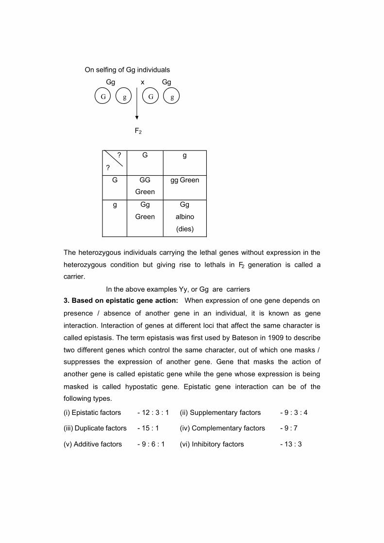
On selfing of Gg individuals
Gg x Gg
F2
The heterozygous individuals carrying the lethal genes without expression in the
heterozygous condition but giving rise to lethals in F2 generation is called a
carrier.
In the above examples Yy, or Gg are carriers
3. Based on epistatic gene action: When expression of one gene depends on
presence / absence of another gene in an individual, it is known as gene
interaction. Interaction of genes at different loci that affect the same character is
called epistasis. The term epistasis was first used by Bateson in 1909 to describe
two different genes which control the same character, out of which one masks /
suppresses the expression of another gene. Gene that masks the action of
another gene is called epistatic gene while the gene whose expression is being
masked is called hypostatic gene. Epistatic gene interaction can be of the
following types.
(i) Epistatic factors - 12 : 3 : 1 (ii) Supplementary factors - 9 : 3 : 4
(iii) Duplicate factors - 15 : 1 (iv) Complementary factors - 9 : 7
(v) Additive factors - 9 : 6 : 1 (vi) Inhibitory factors - 13 : 3
?
?
G g
G GG
Green
gg Green
g Gg
Green
Gg
albino
(dies)
G g G g

4. Based on number of genes involved:
a) Monogenic - Each character is controlled by one gene
b) Digenic - Each character is controlled by two genes
c) Oligogenic - Each character is controlled by few genes
d) Polygenic - Each character is controlled by many genes
Polygenic or polymerism: In general, one gene controls or affects a single
character. But some characters are known to be controlled by more number of
genes. Such genes are called poly genes and the phenomenon is called
polymerism.
Eg : Yield in plants.
5. Based on pleiotropism / pleiotropic gene action: In general, one gene
affects a single character. But some of the genes are known to affect or control
more than one character. Such genes are called pleiotropic genes and the
phenomenon is known as pleiotropism. Many fold phenotypic expressions of a
single gene is called pleiotropism or pleiotropic gene effects. Eg: White eye gene
effects the shape of sperm storage organs and other structures in Drosophila.
These genes are found in all crop plants. Good example of pleiotropism has been
reported in wheat. A gene governing awns in Ona’s variety of wheat also
increases the yield as well as seed weight.

ALLELES
Alternate forms of a gene is known as allele. Alleles are of two types viz.,
either dominant and recessive or wild type and mutant type.
Characteristic features of alleles:
1. They occupy the same locus on a particular chromosome.
2. They govern the same character of an individual. (T and t – control plant
height)
3. A haploid cell has a single copy of an allele for a character. A diploid cell
has two copies of an allele for a character, while a polyploidy cell has more
than two copies of an allele for a character.
4. An individual may have identical alleles at the corresponding locus of
homologous chromosomes in the homozygote or two different alleles in the
heterozygote.
5. The alleles may be dominant and recessive or wild and mutant types.
Multiple alleles
Generally a gene has two alternative forms called alleles. Usually one of
them is dominant over the other. The two alleles of a gene determine the two
contrasting forms of a single character. Ex. Tall (T) and dwarf (t) plant height in
garden pea. But in many cases, several alleles of a single gene are known to
exist and each one of them governs a distinct form of the concerned character or
trait. Such a situation is known as multiple allelism and all the alleles of a single
gene are called multiple alleles. Many genes in both animals and plants exhibit
multiple alleles. Ex: Blood group in human beings, fur colour / coat colour in
rabbits and self-incompatibility alleles in plants.
1. Blood groups in human beings: On the basis of presence / absence of
certain antigens, four blood groups in human beings have been established by
Karl Landsteiner in 1900. The blood group system in human beings is believed to
be controlled by a single gene generally designated as “ I.” The gene “ I” has
three alleles. – IA, IB and i. Allele IA controls the production of antigen A, IB
controls the production of antigen B and i does not produce any antigen.

Individuals with the genotype IAIA or IAi produces antigen ‘A’ and are classified in
blood group A. individuals with genotype IBIB or IBi are classified in blood group B.
Individuals with genotype ii are grouped in ‘O’ blood group and such individuals
produce neither antigen A nor antigen B. individuals with genotype IAIB produce
both antigens A and B and hence classified as ‘AB’ blood group.
Human blood groups, their antigen, antibody and compatible blood groups for transfusion:
Blood group Genotypes Antigenfound
Antibodypresent
Compatible blood group
A IA IA or IA i A B A and O
B IB IB or IB i B A B and O
AB IA IB AB None A, B, AB and O
Universal recipient
O ii None AB O
Universal donor
2. Fur or coat colour in rabbits: The fur colour in rabbits is a well known
example of multiple alleles. In rabbits, the fur colour is of four types viz., agouti,
chinchilla, himalayan and albino. It is due to multiple alleles of a single gene ‘C’.
Phenotype Gene symbol Genotype
1. Agouti C CC, Ccch, Cch, Cc
2. Chinchilla Cch cchcch, cch ch, cchc
3. Himalayan Ch ch ch, chc
4. Albino c cc
The order of dominance for fur colour in rabbits can be represented as follows :
C > cch > ch > c
Agouti > Chinchilla > Himalayan > Albino
Full colour or wild type
Mixture of coloured and white hairs
over the body
Main body is white, while the
tips of ears, feet, tail and snout
are black
Nopigmentand with
purewhite fur colour

3. Self incompatibility alleles in plants: A series of self incompatibility alleles
insures cross pollination in many plants. Such alleles were described first in
toabacco and later they were found in several other plant species like Brassica,
radish, tomato, potato etc. In these species, self incompatibility is governed by a
single gene ‘S’ which has multiple alleles viz., S1, S2, S 3, S4 and so on.
Characteristic features of multiple alleles:
1. Multiple alleles always belong to the same locus in a chromosome.
2. One allele is present at a locus at a time in a chromosome.
3. Multiple alleles always control the same character of an individual. However,
the phenotypic expression of the character will differ depending on the alleles
present.
4. There is no crossing over in a multiple allelic series.
5. In a multiple allelic series, wild type is almost always dominant over the mutant
type.
6. A cross between two strains homozygous for mutant alleles will always
produce a mutant phenotype and never a wild phenotype. In other words,
multiple alleles do not show complementation.
7. Further, F2 generations from such crosses show typical monohybrid ratio for
the concerned trait.
Pseudo alleles: Two alleles each of two or more tightly linked genes affecting the
same function as a result of which, they appear as multiple alleles. (or) Alleles,
which have two separate gene loci, but often inherit together due to close linkage
and have very rare chance of crossing over are called pseudo alleles. Eg:
Lozenge eye in Drosophila.
Iso allele: An allele which is similar in its phenotypic expression to that of other
independently occurring allele is called isoallele.
Penetrance; It is the ability of a gene to express itself in all the individuals, which
carry it in the appropriate genotype. (or) The frequency with which a gene
produces a phenotypic or visible effect in the individuals which carry it in the
appropriate genotype is known as penetrance. It refers to the proportion of
individuals which exhibit phenotypic effect of a specific gene carried by them.

1. Penetrance of some genes is limited to one sex only. For example : Milk
production in cattle or human beings.
2. It may be affected by environmental conditions
3. Penetrance is expressed in percentage
It is of two types
1. Complete penetrance: When all the individuals that carry a particular
gene exhibit its phenotypic effect, it is known as complete penetrance. In
this case, all homozygous dominant individuals will exhibit one phenotype,
while all homozygous recessives will exhibit another phenotype. Recessive
alleles have no or zero penetrance in heterozygous condition.
Tall x Dwarf
TT x tt
(100%) (100%)
F1 Tt Tall
[T exhibits Complete (100%) penetrance while t has no(0%) penetrance]
2. Incomplete penetrance: When specific gene does not express their effect
in all the individuals which carry them in appropriate genotype, it is known
as incomplete penetrance. For example : The recessive gene producing
partial chlorophyll deficiency in the cotyledonary leaves of lima bean shows
incomplete penetrance as it expresses itself only in 10% of the individuals.
Almost all the genes showing incomplete penetrance exhibit variable
expressivity as well
Expressivity: The degree of phenotypic expression of a gene in different
individuals is called expressivity. It is also influenced by environmental conditions
in some cases. Expressivity is a lso two types.
1. Uniform expressivity: When the phenotypic expression of a gene is
identical or similar in all the individuals, which carry such a gene, it is
known as uniform expressivity. Most of the qualitative characters exhibit
uniform expressivity. Eg: Seed shape in pea rr genotypes have wrinkled
seed shape, while RR or Rr genotypes exhibit round seed shape.

2. Variable expressivity: When the phenotypic expression differs in different
carriers of a gene, it is known as variable expressivity. Eg: Recessive gene
producing partial chlorophyll deficiency in cotyledonary leaves of lima
beans.
Lecture No.: 21
QUALITATIVE AND QUANTITATIVE CHARACTERS
The phenotype of any individual can be classified into two types :
1) Qualitative characters and 2) Quantitative characters
1. Qualitative characters : The characters that show discontinuous variation
and which can not be measured easily are known as qualitative characters.
These are also known as classical mendelian traits.
Eg : Corolla colour – Red white or pink no continuous variation
Seed shape – Round wrinkled variation is not continuous
2. Quantitative characters are those showing continuous variation and
which can be measured easily. These characters are also known as metric
traits. The data obtained from such characters is known as quantitative
data. This data can be subjected to statistical analysis and the branch of
science which deals with such analysis is known as quantitative genetics or
biometrical genetics. Eg : Yield, Plant height
Differences between quantitative and qualitative characters
Quantitative characters Qualitative characters
Deals with Traits of degree
Eg : Plant height, seedweight, yield etc.
Traits of kind
Eg : Corolla colour, seed shape, appearance etc.
Variation Continuous Discontinuous
Effect of individual gene Small and undetectable Large and detectable
No. of genes involved Several (polygenic) one or few (mono /oligogenic)
Grouping into distinctclasses
Not possible Possible
Effect of environment High Low
Metric measurement Possible Not possible
Statistical analysis Based on mean, variance, standard deviation etc.
Based on ratios andfrequencies
Stability Low High

Transgressivesegregation of F 2
Yes No
Dominance effect No Yes
Cumulative effect ofeach gene
Yes No
MULTIPLE FACTOR HYPOTHESIS
In early days of mendelian genetics it was thought that there was a
fundamental difference in inheritance pattern of quantitative and qualitative traits.
One of the examples which helped to bridge the gap between these two kinds of
traits is “multiple gene model” developed by Swedish Geneticist H. Nilsson Ehle in
1910 to explain inheritance of kernel colour in wheat. In studies on inheritance of
kernel colour in wheat and oats, he obtained 3:1, 15:1 and 63:1 ratio between
coloured and white seeds from different crosses. It is clear from these ratios that
the seed colour was governed by one (3:1 ratio in F2), two (15:1 ratio in F2) or
three (63:1 ratio in F2) genes. Nilsson-Ehle found that in crosses showing 15:1
ratio in F2 kernel colour is governed by two genes. However, on a closer
examination of the coloured seeds, he found that there was a marked difference in
the intensity of their colour. When he crossed a dark red strain to a white strain (or
variety), he observed that all the F1s were medium red (intermediate between the
parental types). But in F2 generation only one out of the sixteen were of parental
types. He interpreted the results in terms of two genes, each with a pair of alleles
exhibiting cumulative effects. Each of the dominant alleles R1 and R2 adds some
red colour to the phenotype, while the recessive alleles r1 and r2 add no colour to
the phenotype. Thus dark red genotypes contain only R1 and R2 alleles while the
white genotype contains none of these alleles.
Parents Female x Male
Phenotype Dark red x White
Genotype R1 R1 R2 R2 x r1 r1 r2 r2
Gametes
Generation F1
R1R2 r1 r2

? ? r1 r2
R1 R2R1 r1 R2 r2
Medium Red
On selfing
Parents Female x Male
Phenotype Medium Red x Medium Red
Genotype R1 r1 R2 r2 x R1 r1 R2 r2
gametes R1 R2 R1 R2 R1 R2 R1 R2
r2 R1 r2 r2 R1 r2
r1 R2 r1 R2 r1 R2 r1 R2
r2 r1 r2 r2 r1 r2
Generation F2
? ? R1 R2 R1 r2 r1 R2 r1 r2
R1 R2 R1 R1 R2 R2
Dark RedR1 R1 R2 r2
RedR1 r1 R2 R2
RedR1 r1 R2 r2
Medium Red
R1 r2 R1 R1 R2 r2
RedR1 R1 r2 r2
Medium RedR1 r1 R2 r2
Medium RedR1 r1 r2 r2
Light Red
r1 R2 R1 r1 R2 R2
RedR1 r1 R2 r2
Medium Redr1 r1 R2 R2
Medium Redr1 r1 R2 r2
Light Red
r1 r2 R1 r1 R2 r2
Medium RedR1 r1 r2 r2
Light Redr1 r1 R2 r2Light Red
r1 r1 r2 r2
White
Phenotypic ratio: 1 Dark Red : 4 Red : 6 Medium Red : 4 Light red : 1 white
Genotypic ratio: 1 R1 R1 R2 R2 : 2 R1 R1 R2 r2 : 1 R1 R1 r2 r2
: 2 R1 r1 R2 R2 : 4 R1 r1 R2 r2 : 2 R1 r1 r2 r2
: 1 r1 r1 R2 R2 : 2 r1 r1 R2 r2 : 1 r1 r1 r2 r2

S.No.
No. of allelesfor red colour
Phenotype Frequency Genotype Frequency
1. 4 Dark Red 1 R1 R1 R2 R2 1
R1 R1 R2 r2 22. 3 Red 4
R1 r1 R2 R2 2
R1 R1 r2 r2 1
R1 r1 R2 r2 4
3. 2 Medium Red 6
r1 r1 R2 R2 1
R1 r1 r2 r2 24. 1 Light Red 4
r1 r1 R2 r2 2
5. 0 white 1 r1 r1 r2 r2 1
Lecture No: 22
SEX DETERMINATION
Sex refers to the contrasting features of male and female individuals of the
same species. Thus sex is usually of two types viz., male and female. Sex
determination is a process of sex differentiation which utilizes various genetical
concepts to decide whether a particular individual will develop into male or female.
Plants also have sex as there are male and female parts in flowers. All organisms,
however do not possess only two sexes. Some of the protozoa may have as many
as eight sexes. In most higher organisms, the number of sexes has been reduced
to just two. The sexes may reside in different individuals or within the same
individual. An animal possessing both male and female reproductive organs is
usually referred to as hermaphrodite. In plants where staminate and pistillate
flowers occur in the same plant, the term of preference is monoecious Eg. maize,
castor, coconut etc. However, most of the flowering plants have both male and
female parts within the same flower (perfect flower). Relatively few angiosperms
are dioecious i.e. having male and female elements in different individuals Eg:
cucumber, papaya, asparagus, date palm, hemp and spinach. The sex cells and

reproductive organs form the primary sexual characters of male and female
sexes. Besides these primary sexual characters, the male and female sexes differ
from each other in many somatic characters known as secondary sexual
characters.
Whether or not there are two or more sexes in the same or different
individuals is relatively unimportant. The importance of sex itself is that it is a
mechanism, which provides for the great amount of genetic variability
characterizing most natural populations.
The various mechanisms of sex determination are:
1. Chromosomal sex determination
2. Genic balance mechanism
3. Male haploidy or Haplodiploidy mechanism
4. Single gene effects (or) monofactorial mechanism of sex determination
5. Metabolically controlled mechanism
6. Hormonally controlled mechanism
7. Sex determination in Coccinia indica and Melandrium album,
8. Sex determination due to environmental factors
I. Chromosomal sex determination: The chromosomes, which have no relation
with sex and contain genes, which determine the somatic characters of an
individual are known as autosomes. These chromosomes do not differ in
morphology and number in male and female sex. Those chromosomes, which
differ in morphology and number in male and female sex and contain genes
responsible for the determination of sex are known as allosomes or sex
chromosomes.
Differences between Autosomes and Allosomes
Autosomes Allosomes or Sex chromosomes
1. Refer to other than sexchromosomes.
1. These are sex chromosomes.
2. Morphology is similar in male andfemale sex.
2. Morphology is different in male and female sex.
3. The number is same in both thesexes.
3. The number is sometimes different in male and female sex.
4. Generally control traits other than 4. Usually determine sex of an

sex. individual.
5. Number of autosomes differs from species to species.
5. Each diploid organism usually has two allosomes.
6. Do not exhibit sex linkage. 6. Exhibit sex linkage.
The chromosomal influence on sex, in certain insects, has been shown for
the first time by McClung in 1902 to be associated with a special sex determining
‘X’ chromosome. McClung proposed that a male had one ‘X’ chromosome per cell
(XO) and a female has two ‘X’ chromosomes (XX). Later Stevens and Wilson
(1905) found same number of chromosomes in both sexes of milk weed bug. In
females all chromosomes were paired and the homologues were equal in size
(homomorphic). In the male, all the chromosomes were paired, but the
chromosome identified as homologous to the “ X” Chromosome was distinctly
smaller and was called the “Y” Chromosome (Heteromorphic).
Thus, allosomes are generally of X and Y types, but in birds they are of Z
and W types. Sex with similar type of sex chromosomes (XX or ZZ) is known as
homogametic sex and with dissimilar type of sex chromosomes (XY or ZW) as
heterogametic sex. These are two types: a) Heterogametic male and
b) Heterogametic female
a) Heterogametic male: In this mechanism, the female sex has two ‘X’
chromosomes, while the male sex has only a single ‘X’ chromosome. As the male
lacks a ‘X’ chromosome during meiosis, 50% of lthe gametes carry ‘X’
chromosome, while the rest do not have the ‘X’ chromosome. Such a mechanism,
which produc es two different types of gametes in terms of sex chromosome is
called heterogametic sex. The female sex here is called homogametic sex
because it produces similar type of gametes. The heterogametic male may be of
the following two types.
i) XX – XO ii) XX – XY
i) XX - XO: In certain insects belonging to orders Hemiptera (true bugs),
Orthoptera (grass hoppers) and Dictyoptera (cockroaches), female has two ‘X’
chromosomes (XX) and are, thus homogametic, while male has only single ‘X’
chromosome (XO). This mechanism was found by C.E. McClung in 1902. The
presence of an unpaired X chromosome determines the masculine sex. The

male being heterogametic sex produces two types of sperms, half with X
chromosome and half without X chromosome in equal proportions. The sex of
the offspring depends upon the sperm that fertilizes the egg, each of which
carries a single X chromosome. Thus fertilization between male and female
gametes always produced zygotes with one ‘X’ Chromosome from the female,
but only 50% of the zygotes have an additional X Chromosome from the male.
In this way, ‘XO’ and ‘XX’ types would be formed in equal proportions, the
former being males and the latter being females.
Parents Female X Male
2A + XX X 2A + XO
Gametes
A + X A + O
A + X 2A + XX
Female
2A + XO
Male
ii) XX – XY: In man, other mammals, certain insects including Drosophila, certain
angiospermic plants including Melandrium, the females possess two X
chromosomes (XX) and are thus homogametic and homomorphic, while the
males possess one X and one Y chromosome (XY) and are hence
heterogametic and heteromorphic. When an egg is fertilized by ‘Y’ bearing
sperm, a male is produced.
Parents Female X Male
2A + XX X 2A + XY
Gametes
A + X A + Y
A + X 2A + XX
Female
2A + XY
Male
A + X A + OA + X
A + X A + YA + X

b) Heterogametic female: In this mechanism the male possess two
homomorphic sex chromosomes and are thus homogametic, while the female
possesses either a single ‘X’ chromosome or one ‘X’ and one ‘Y’ chromosome
and are hence heterogametic. To avoid confusion with earlier types, instead of
X and Y, the alphabets Z and W are used. This mechanism of sex
determination is also known as “Abraxas mechanism of sex determination”
(Kuspira and Walker, 1973) The heterogametic females may be of following
two types. i) ZO – ZZ ii) ZW – ZZ
i) ZO – ZZ: This mechanism is found in certain moths and butterflies. In this case,
female possess one single ‘Z’ chromosome and hence is heterogametic. Male
possesses two Z chromosomes and thus homogametic.
Parents Female X Male
2A + ZO X 2A + ZZ
Gametes
A + Z
A + Z 2A + ZZ Male
A + O 2A + ZO FemaleThe sex of the offspring depends on the kind of egg.
ii) ZW – ZZ: This system is found in certain insects (gypsy moth) and vertebrates
such as fishes, reptiles and birds. In this system, the female is heterogametic
and produces two types of gametes, one with ’Z’ chromosome and the other
with ‘W’ chromosome. On the other hand, male is homogametic and produces
all sperms of same type carrying one ‘Z’ chromosome. The sex of the
offspring depends on the kind of egg being fertilized. The ‘Z’ chromosome
bearing eggs produce males, but the ‘W’ chromosome bearing eggs produce
females.
Parents Female X Male
2A + ZW X 2A + ZZ
Gametes
A + Z
A + ZA + OA + Z
A + ZA + WA + Z

A + Z 2A + ZZ Male
A + W 2A + ZW Female
II) Genic balance mechanism: By studying the sex chromosomal mechanism of
sex determination, it may appear at first glance that some genes carried by sex
chromosomes i.e. X and Y are entirely responsible for determining sex. But
this may not always be true. Extensive experiments on different organisms by
different workers have revealed the fact that most organisms generally have
inherent potentialities for both sexes and each individual is found to be more or
less intermediate between male and female. Hence may be referred to as inter
sex. There seems to exist a delicate balance of masculine and feminine
tendency in the hereditary compliment of an individual. Such a genic balance
mechanism of determination of sex was first observed and studied by C.B.
Bridges in 1921 while working with Drosophila for the inheritance of vermillion
eye colour. According to this mechanism, the sex of an individual in Drosophila
melanogaster is determined by a balance between the genes for femaleness
located in the X-chromosome and those for maleness located in autosomes.
Hence, the sex of an individual is determined by the ratio of number of its X
chromosomes and that of its autosomal sets, the ‘Y’ chromosome being
unimportant. The ratio is termed as sex index and is expressed as follows.
No. of X chromosomes XSex index = No. of autosomal sets = A
Different doses of X – Chromosomes and autosome sets and their effect on sex determination
S.No. Ploidylevel
X-Chromo-somes
Sets of autosomes
Sex index(X/A ratio)
SexExpression
1. Diploid 3(xxx) 2(AA) 1.50
2. Triploid 4(xxxx) 3(AAA) 1.33
}Super female } or meta female }
3. Haploid 1(x) 1(A)
4. Diploid 2(xx) 2(AA)
5. Triploid 3(xxx) 3(AAA)
6. Tetraploid 4(xxxx) 4(AAAA)
}}}1.00}}
Female
7. Triploid 2(xxy) 3(AAA) 0.67 }

8. Tetraploid 3(xxxy) 4(AAAA) 0.75 } Inter sex}
9. Diploid 1(xy) 2(AA)
10. Tetraploid 2(xxyy) 4(AAAA)
}} 0.5 Male
11. Triploid 1(xyy) 3(AAA) 0.33 Super male or meta male
Individuals with sex index of 0.5 develop into normal males and those with
sex index of 1 into normal females. If the sex index is between 0.5 and 1, the
resulting individuals will be neither a female nor a male, but have an intermediate
sex expression and is called inter sex. Such individuals are sterile. Some flies
have sex index of >1, such flies have more pronounced female characteristics
than normal fermales and are called super females or metafemales. These are
generally weak, sterile and non-viable. Super male flies have a sex index value of
<0.5 and are also weak, sterile and non-viable.
Bridges drew the observation by crossing triploid females (3A + XXX) with
normal diploid males (2A + XY). From such a cross he obtained normal diploid
females, males, triploid females, intersexes, super males and super females. The
occurrence of triploid intersexes from such a cross clearly established that
autosomes also carry genes for sex determination. Triploid individuals, which had
two ‘X’ Chromosomes as in the case of normal female, here were inter sexes as
they had an extra set of autosomes indicating that the autosomes play a definite
role in the determination of sex.
Results obtained from a cross of a triploid (3A+XXX) female fly with a diploid (2A+XY) male fly in Drosophila
Parents Female X Male
Phenotype Triploid X Diploid
Genotype 3A + XXX X 2A + XY
Gametes
A + X A + Y
2A + XX 3A + XXXTriploid female
3A + XXYTriploid inter sex
2A + XX
2A + X
A + X
A + XX
A + X A + Y

A + X 2A + XXDiploid female
2A + XYDiploid male
2A + X 3A + XXTriploid inter sex
3A + XYSuper male
A + XX 2A + XXXSuper female
2A + XXYDiploid female
III. Male haploidy or haplodiploid mechanism or arrhenotokous
parthenogenesis: It is common in hymenopterous insects (ants, bees, wasps). In
honey bees, queens usually mates only once during its life time and the sex ratio
of offspring is under the control of queen. Fertilized eggs develop into diploid
female and those eggs which the queen chooses not to be fertilized develop
parthenogenetically into haploid but fertile males (drones). This phenomenon is
known as arrhenotoky and is a form of reproduction as well as a means of sex
determination. Meiosis is normal during oogenesis in case of females and
produces all haploid eggs. But crossing over and reduction division fails to occur
during spermatogenesis in males due to their haploid nature. Thus arrhenotokous
parthenogenesis determines the sex in hymenopterans and sex chromosomes
have no identity here (unlike Drosophila). It seems that heterozygosity for specific
genes induces femaleness. The haploid can never be heterozygous. Most of the
eggs laid in the hive will be fertilized and developed into worker females. Further
during investigation, it has been found that the quantity and quality of food
available to the diploid larvae determines whether that female will become a
sterile worker or a fertile queen. The diploid larva, which feed on royal jelly,
develop into fertile female called queen and the remaining larvae give rise to
workers, which are sterile females. Thus, environment here determines sterility or
fertility but does not alter the genetically determined sex.
IV. Single gene effect or monofactorial mechanism of sex determination: In
Neurospora , Asparagas, Drosophila , maize and Asparagus, sex determination is
influenced by the differential action of a single autosomal gene, which over rules
the effect of sex chromosomes present in the individual.
Autosomal recessive transformer (tra) gene of Drosophila; when it is
present in the homozygous recessive state, it transforms the female (XX) zygote
to develop into males which are sterile. The gene tra is recessive and hence does

not have any effect in heterozygous condition (Tratra ) on either sex i.e male or
female. In homozygous condition, (tratra), this gene has no effect in male. When a
heterozygous (XX Tratra) female is mated with a homozygous recessive male (XY
tratra ), only 25% of the progeny are females (heterozygous) while remaining 75%
are males. Among the males, 1/3 are sterile (XX individuals homozygous
recessive for tra gene) as they are transformed to maleness by the tra.
Parents Female X Male
Genotype XX Tratra X XY tratra
Gametes
Progeny F1
X tra Y tra
X Tra XX TratraFemale fertile
XY TratraMale fertile
X tra XX tratraMale sterile
(Transformed)
XY tratraMale fertile
Ratio: 3 Males : 1 Female
Monogenic control of sex has also been reported in some plants like
Asparagus, maize, papaya, spinach, etc. Asparagus is a dioecious plant.
However, rarely female flowers bear rudimentary anthers and male flowers bear
rudimentary pistils. Occasionally, male flowers with poorly developed pistils set
seed on selfing and segregate in 3 : 1 ratio of male and female.
Segregation of sex in seed obtained from a rare bisexual flower in Asparagus showing monogenic control
Rare male plant when selfed
Pp X Pp
P p
P PPMale
PpMale
p Pp pp
X Tra X tra X tra Y tra
P p P p

Male Female
Ratio: 3 Males : 1 Female
Maize being a monoecious plant bears both female (silk) and male (tassel)
inflorescences on the same plant. A recessive gene ba (barren cob) in
homozygous condition (baba) makes the cobs barren or non-functional. Similarly,
a recessive gene ts in homozygous condition (tsts) converts the male flowers of
tassel into female flowers. Thus, homozygous state of gene ba (baba) converts
the monoecious plant into male. Similarly, gene ts in homozygous condition (tsts )
converts the monoecious plant into female. The plants with both dominant genes
(Ba_Ts_) are monoecious, with babaTs _ normal male, with Ba_tsts female, and
with babatsts rudimentary females.
In papaya, the sex is postulated to be governed by three alleles, viz., m, M1
and M2 of a single gene. Homozygous recessive (mm) produces female plants,
heterozygous, viz., M1m and M2m produce male and hermaphrodite plants,
respectively. However, combination of both dominant alleles (M1 and M2)
produces inviable plants both in homozygous (M1M1 and M2M2) and heterozygous
conditions (M1M2). Crosses between female (mm) and male (M1m) produce
females (mm) and males (M1m) in 1 : 1 ratio. Similarly, crosses between female
(mm) and hermaphrodite (M2m ) will produce females (mm) and hermaphrodite
(M2m) in 1 : 1 ratio. Selfing of hermaphrodite (M2m) plants produce hermaphrodite
(M2m) and female (mm) progeny in 2 : 1 ratio and about ¼ of the zygotes (M2M2)
do not survive.
V. Metabolically controlled mechanism: Riddle found that metabolism has
some definite role in the determination of sex in pigeon and dove because
increased rate of metabolism lead to the development of maleness while
decreased rate of metabolism caused femaleness.
VI. Hormonally controlled mechanism: Crew in 1923 reported a complete
reversal of sex in hen. Female chicken that have laid eggs been known to
undergo not only a reversal of the secondary sexual characteristics such as
development of cock feathering, spurs and crewing, but also the development of
testis and even the production of sperm cells (primary sexual characteristics). This
might have occurred when a disease destroyed the ovarian tissue and in absence

of female sex hormones, rudimentary testicular tissue present in center of ovary
began to proliferate or multiply and secrete male hormones, which lead to the
development of maleness.
Sexual differentiation in man is influenced by hormones. When the testis of
the male is removed before puberty, female characteristics of body form, voice
and hair pattern develop in the adult. Tumors of adrenals in women are
associated with the development of masculine characters such as low pitched
voice and increased growth of hair.
VII. Sex determination in Coccinia indica and Melandrium album: In these
Ploidylevel
Chromosome Constitution (Autosomes ) + (Allosomes)
SexExpression
X/A ratio
Diploid AA + XX Female 1.00
Diploid AA + XY Male 0.50 (1/2)
Diploid AA + XYY Male 0.50 (1/2)
Triploid AAA + XXX Female 1.00
Triploid AAA + XXY Male 0.67 (2/3)
Tetraploid AAAA + XXXX Female 1.00
Tetraploid AAAA + XXXY Male 0.75 (3/4)
two organisms, studies have revealed that irrespective of number of ‘X’
chromosomes, and / or autosomal sets, presence of a single ‘Y’ chromosome is
essential to produce male flowers in diploid, triploid and tetraploid species. The
pistillate plants are XX and the staminate plants are XY.
VIII. Sex determination due to environmental Factors: In many reptiles, sex is
determined by environmental factors like temperature. In most turtle species only
females are produced at high temperature (30 – 35ºC) while only males are
produced at low temperatures (23 – 28ºC). Sex ratio changes suddenly from all
males to all females with just change in temperature of 2ºC during the incubation.
In Crocodiles and Lizards, (reverse is the case) the males are produced at high
temperature while females are produced at low temperature. In Bonellia viridis , a
marine worm, all larvae are genetically and cytologically similar. If a particular
larva settles near proboscis of adult female, it becomes a male individual. If larva
develops freely in water, it becomes a female. In some plants, sex determination
depends on day length, temperature and hormones. For example, in cucumber

(Cucumis sativa) and muskmelon, treatment with ethylene enhances production of
female flowers.
Sex linked inheritance
The characters for which genes are located on sex or ‘X’ or analogous ‘Z’
chromosomes are known as sex linked traits. Such genes are called sex linked
genes and linkage of such genes is referred to as sex linkage. Inheritance of such
genes or characters is known as sex linked inheritance. The sex linkage was first
discovered by T.H. Morgan in Drosophila and the first sex linked gene found in
Drosophila was recessive gene ‘w’ responsible for white eye colour.
Parents Female X Male
Phenotype White eye X Red eye
Genotype Xw Xw X X+Y
Gametes
X+ Y
Xw X+Xw
Red eye femaleXwY
White eye male
When white eyed females are crossed with wild type (red eye) males, all the male
offspring have white eyes like the mother and all the female offspring have red eyes like
their father.
In human beings, colour blindness and haemophilia (bleeder’s disease) are well
known hereditary characters showing a peculiar relationship to sex.
Colour blindness : A marriage between colour blind woman and a normal man gives rise
to all normal dauthers and colour blind sons.
Parents Mother X Father
Phenotype Colour blind X Normal
Genotype Xc Xc X X+Y
Gametes
X+ Y
Xc X+Xc
Normal daughterXcY
Colourblind son
XcX+ Y
Xw X+ Y

The allele for colour blindness (c) is found on both ‘X’ chromosomes of mother
and therefore she is colour blind. The only one ‘X’ chromosome of father in this marriage
carries a wild type (+) allele and hence he has normal vision. The ‘Y’ chromosome lacks
both the alleles (+ and c). The reduction division produces one kind of egg in contrast to
two kinds of sperms. Fertilization results in the usual sex ratio of 1 male : 1 female. All
the daughters have normal vision since they receive dominant allele ‘+’ from their father.
All the sons are colour blind, because their single ‘X’ chromosome derived from mother
carries the allele ‘c’ for colour blindness. This result is known as crisscross inheritance
because daughters are normal like father and sons have colour blindness like mother.
However, the daughters are heterozygous carriers. This crisscross method of inheritance is
characteristic of sex-linked genes. This peculiar type of inheritance is due to the fact that
Y chromosome carries no alleles homologous to those on the X chromosome. Thus males
carry only one allele for sex linked traits. This one allelic condition is termed as
hemizygous in contrast to homozygous and heterozygous possibilities in female. The
expression of recessive gene in hemizygous condition is termed as pseudo-dominance.
The inheritance of colour blindness can be studied in the following three other
possible types of marriages:
a) Marriage between normal woman and colour blind man:
Parents Mother X Father
Phenotype Normal X Colour blind
Genotype X+ X+ X Xc Y
Gametes
Xc Y
X+ X+ Xc
Normal daughter(Carrier)
X+YNormal son
b) Marriage between normal (carrier) woman and normal man:Parents Mother X Father
Phenotype Normal X Normal
Genotype X+ Xc X X+ Y
Gametes
X+ Xc Y
X+ X+YXc

X+ Y
X+ X+X+
Normal daughterX+Y
Normal son
Xc X+Xc
Normal daughterXcY
Colour blind son
c) Marriage between normal (carrier) woman and colour blind man:
Parents Mother X Father
Phenotype Normal X Colour blind
Genotype X+ Xc X XcY
Gametes
Xc Y
X+ X+Xc
Normal daughterX+Y
Normal son
Xc XcXc
Colour blind daughter
XcY
Colour blind son
Results of possible four marriages make it clear why there are more colour blind
males than females in the population. In three marriages colour blind sons were produced
where as in only one of the marriages, colour blind daughters were observed, where the
mother is heterozygous (carrier) and the father is colour blind. Nearly all colour blind
women must come from the last type of marriage, since the only other possible source of
colour blind females is mating between two colour blind persons – naturally a rare
occurrence.
Haemophilia is a recessive sex linked disease and the inheritance pattern of
haemophilia is similar to that of colour blindness in human beings.
Genes present in the non-homologous region of the Y chromosome pass directly from
male to male. In man, the genes present on Y chromosome (holandric genes) such as the
gene causing hypertrichosis (causing excessive development of hairs on the pinna of ear)
are transmitted directly from father to son.
Sex influenced inheritance
Sex influenced ge nes are the autosomal genes present in both males and females,
whose phenotypic expression is different in different sexes in such a way that they act as
X+ Xc YXc

dominant in one sex and recessive in the other i.e. in a pair of alleles one seems to be
dominant in males while the other in females.
Eg.: Pattern baldness in human beings and horns in sheep. Pattern baldness in human
beings is a condition in which a low fringe of hair is present on the head in human beings.
It is a genetically inherited condition, where the allele for baldness B is dominant in males
and recessive in females. In heterozygous condition, males are bald and females are non-
bald. If a woman heterozygous for this gene marries a heterozygous bald man, in the
offspring, the ratio of bald to non-bald in males is 3 : 1, while in females it is 1 : 3.
Inheritance of pattern baldness in human being
Parents Female X Male
Phenotype Bald X nonbald
Genotype b+b+ X bb
Gametes
F1
b+b Female (Nonbald) Male (Bald)
Phenotypic expression of pattern baldness in man and woman
PhenotypeGenotype
Female Male
b+ b+ Nonbald Nonbald
b+b Nonbald Bald
bb Bald Bald
Sex limited characters or Secondary Sexual characters
Sex limited genes are autosomal genes, whose phenotypic expression is limited to
one sex only. Their phenotypic expression is influenced by the sex hormones. The sex
limited genes are mainly responsible for secondary sex characters in cattle, human beings
and fowl. Eg.: milk production in cattle, beard development in human beings, plumage in
male fowls etc.
Milk production in cattle : Just as the cow, the bull carries genes for milk production, but
the bull obviously cannot express this trait. Bull may however transmit these genes for
b+ b

high milk production to the female progeny and the male progeny are unable to express
this trait. Some bulls are so well andowed with such genes that they are known to breed
calves, which always yield greater milk than their mothers.
However, in plants no secondary sexual characters are known except the absence of one or
the other sporangia.
Differences between sex linked and sex limited characters
Sex Linked characters Sex Limited characters
1. They are located on sex or Xchromosome
1. They are located on sex chromosomes or autosomes
2. They can express in both the sexes 2. They can express in one sex only
3. Include characters not related to sex 3. Include primary and secondary sexcharacters
4. Examples: White eye in Drosophila ,haemophilia and colour blindness inhuman beings
4. Examples: milk production in cattle,beard development in human beings,plumage in male fowls etc

Inter sex
In a few rare cases, various mixtures of male and female characteristics may occur in
animals, which normally have separate sexes because of var ious abnormalities of
chromosomes or hormonal deficiencies. Such individuals are called inter sex. These can
be of two types.
1. Pseudo -hermaphrodites: In mammals there are rare cases in which, both sexes are
well developed in one body, these are abnormal and hence sterile.
2. Gynandromorphs or gynanders: Among animals and insects that do not have sex
hormones, there may be sex intergrades with distinct areas of the body showing male
and female tissues. For example: Drosophila gynander, a bilateral sex mosaic, is male
on one side and female on the other.
Lecture No: 23
CYTOPLASMIC INHERITANCE
Inheritance due to genes located in cytoplasm (plasmagenes) is called
cytoplasmic inheritance. Since genes govering traits showing cytoplasmic
inheritance are located outside the nucleus and in the cytoplasm, they are refered
to as plasmagenes. The sum total of genes present in the cytoplasm of a cell or
an individual is known as plasmon. The plasmagenes are located in DNA present
in mitochondria (mt DNA) and in chloroplasts (cp DNA). Together both the DNAs
are called organelle DNA. Therefore, this type of inheritance is often referred to as
organellar inheritance, plastid inheritance or mitochondrial inheritance. In this,
generally, the character of only one of the two parents (usually female) is
transmitted to the progeny. Hence such inheritance is usually referred to as extra -
nuclear or extra-chromosomal or maternal or uniparental inheritance.
The cytoplasmic inheritance is of two types: 1) Plastid inheritance and 2)
mitochondrial inheritance.
1. Plastidial or chloroplast inheritance: Plastids self duplicated and have
some amount of DNA and plays an important role in cytoplasmic
inheritance. Plastids have green pigments called chloroplasts. chloroplasts
contain a unique circular DNA (cp DNA) in the stroma that is completely
different from the nuclear genome. Some examples of plastid inheritance
are given below:

a) Leaf variegation in Mirabilis jalapa : The conclusive evidence for
cytoplasmic inheritance was first presented by C. Correns in Mirabilis
jalapa (Four ‘O’ clock plant) in 1909. He studied inheritance of leaf
variegation in M. jalapa. Variegation refers to the presence of white or
yellow spots of variable size on the green background of leaves. In M.
jalapa, leaves may be green, white or variegated. Some branches may
have only green, only white or only variegated leaves. Correns made
crosses in all possible combinations among the flowers produced on these
three types of branches. When flowers from green branch were used as
female parent, all the progeny were green irrespective of the phenotype
(green, white or variegated) of male parent. Similarly, progeny from crosses
involving flowers bloomed on white branches as female parent were all
white irrespective of the phenotype of male parent. But in progeny from all
crosses involving flowers born on variegated branches as female parent, all
the three types i.e. green, white and variegated individuals were recovered
in variable proportions.
Female Parent x Male Parent
Green xxx
GreenWhiteVariegated
Green
White xxx
GreenWhiteVariegated
Pale green
Variegated xxx
GreenWhiteVariegated
Green, white and variegated in variable ratio in each of the cases.
The green leaf branches have normal chloroplasts, white branches have
mutant chloroplasts and variegated have a mixture of both normal and
mutant chloroplasts. The above results indicated that the inheritance is
governed by chloroplasts. Since the cytoplasm is contributed to the zygote
mainly by female parent, the plastids are transmitted to the zygote from the
female parent. Thus the plastids are responsible for variation in the crosses
of green, white and variegated leaves.

b) Iojap in maize : In maize, there are three types of leaves i.e. green,
iojap (green and white stripes) and white. The green leaves have normal
plastids. Iojap leaves have a mixture of normal and mutant plastids and
white leaves have only mutant plastids. In a cross between green female
and iojap male, only green individuals are produced in F1 generation. But in
the reciprocal cross (iojap female and green male) all the three kinds of
progeny are obtained in variable proportions in F1.
Parents Female x Male
Phenotype Green x Iojap
Generation F1
Green
Reciprocal cross
Parents Female x Male
Phenotype Iojap x Green
Generation F1
Green, Iojap, White in variable proportion
These reciprocal differences can be attributed to the type of plastids in the
egg cell since only female parent is contributing cytoplasm to the zygote.
2. Mitochondrial inheritance: The inheritance of some characters, such as
cytoplasmic male sterility in plants, pokyness in Neurospora etc., is governed by
mitochondrial DNA (mtDNA).
a) Cytoplasmic Male Sterility (CMS) in maize : In several crops,
cytoplasmic control of male sterility is known. In maize, cytoplasmic male
sterility (CMS) is governed by mitochondrial DNA. In such cases, if female
parent is male sterile, F1 progeny also will be male sterile, because
cytoplasm is mainly derived from female parent.
? ?
Male sterile x Male fertile

Male sterile (CMS)
b) Pokyness in Neurospora: Neurospora, which is a breadmold has two
strains i.e. wild and poky. The wild strain has normal growth. While the
poky which is a mutant has very slow growth. A cross between a poky
female and a wild male produce only poky progeny. In reciprocal cross (a
cross between wild female and poky male) all the progeny would be wild.
This suggests the presence of cytoplasmic inheritance because only
difference between the reciprocal crosses is in the main contributor of
cytoplasm.

Reciprocal cross
? ? ? ?
Poky x Wild Wild x Poky
Poky Wild
Characteristic features of cytoplasmic inheritance
1. Reciprocal difference : Reciprocal crosses show marked differences for
characters governed by plasmagenes. In most cases, plasmagenes from only
female parent are transmitted and hence this phenomenon is also called
uniparental inheritance.
2. Lack of segregation : In general, F1, F2, F3 and subsequent generations do
not show segregation for a cytoplasmically inherited trait, as F1 individuals
receive plasmagenes from female parent only.
3. Somatic segregation : Plasmagenes generally show the features in somatic
tissues such as leaf variegation features which is of rare occurrence in case of
nuclear genes.
4. Association with organelle DNA: Several plasmagenes have been shown to
be associated either with chloroplast or mitochondrial DNA. For example:
Cytoplasmic Male Sterility (CMS) in sorghum and maize is associated with
mitochondrial DNA.
5. Nuclear transplantation: Nuclear transplantation means nucleus of a cell is
removed and replaced by nucleus of another genotype from a different cell. If
nuclear transplantation reveals a trait to be governed by genotype of
cytoplasm and not by that of nucleus, it clearly indicates that the trait or
character is governed by cytoplasmic inheritance.

6. Mutagenesis: Some mutagens are highly specific mutagens which act only on
the plasmagenes and do not affect nuclear genes Eg; ethidium bromide,
Induction of mutations by such agents or chemicals in a gene clearly indicates
that it is a plasmagene.
7. Lack of chromosomal location: In many organisms extensive linkage maps
of nuclear genes are available. If a gene is shown to be located in one of these
linkage groups, obviously it cannot be a plasmagene.
8. Transfer of nuclear genome through back crosses: Nucleus of a variety or
species may be transferred into cytoplasm of another variety or species
through repeated back crossing with former, which is used as recurrent male
parent. Lines produced in this way are called alloplasmic lines, since they have
cytoplasm and nucleus from different species.
9. Lack of association with a parasite or symbiont or virus : Only those
cytoplasmically inherited traits which are not associated with parasites,
symbionts or viruses can be regarded to be governed by plasmagenes.

Differences between chromosomal (nuclear) and extra-chromosomal(cytoplasmic or extra-nuclear or maternal) inheritance
S.No. Character Chromosomalinheritance
Extra-chromosomalinheritance
1 Location of hereditary factors
Nucleus Cytoplasm
2. Associated with Chromosomes Chloroplasts and mitochondria
3. Pattern of Inheritance Can be explained by mendelism
Cannot be explained by mendelism
4. Individual hereditary factors are known as
Genes Plasmagenes
5. Hereditary factors a recollectively known as
Genome Plasmon
6. Characters of F1
progenyMay show dominance or may be intermediate between the parents
Exhibits only the characteristic of the female parent
7. Reciprocal differences Not observed Observed
8. Segregation of factorsand recombination
Present Absent
9. Attributes of progeny Under the control of their own genes
Under the control of cytoplasm of female parent
10. Action of mutagen Non-specific Very specific
11. Frequency of occurrence Most common Rare
12. Gene mapping Easy Difficult
Lecture No.: 24 & 25
MUTATIONS
Mutation in a broad sense include all those heritable changes which alter
the phenotype of an individual. Thus mutation can be defined as a sudden
heritable change in the character of an organism which is not due to either
segregation or recombination.

The term mutation was first used by Hugo de Vries to describe the sudden
phenotypic changes which were heritable, while working with Oenothera
lamarckiana.
But the earliest record of mutations dates back to 1791 when Seth Wright
noticed a male lamb with unusually short legs in his flock of sheep. This
lamb served as a source of short leg trait for the development of Ancon
breed of sheep.
However the systematic studies on mutations were started in 1910 by T.H.
Morgan who used Drosophila melanogaster for his studies.
In 1927, H.J. Muller demonstrated for the first time the artificial induction of
mutations by using x-rays in Drosophila.
Similarly in 1928, L.J. Stadler demonstrated an increase in the rate of
mutations due to x-rays in barley and maize.
Induction of mutations by chemicals in fungus Aspergillus was
demonstrated by R.A. Steinberg in 1939.
C. Auerbach and J.N. Robson in 1946 used chemicals to induce mutations
in Drosophila.
The first plant breeding programme using mutations (mutation breeding)
was initiated in 1929 in Sweden, Germany and Russia.
In India it was initiated in early 1930s.
Terminology
Muton: The smallest unit of gene capable of undergoing mutation and it is
represented by a nucleotide.
Mutator gene: A gene which causes another gene or genes to undergo
spontaneous mutation.
Mutable genes: Genes which show very high rates of mutation as compared to
other genes.
Mutant: An organism or cell showing a mutant phenotype due to mutant allele of
a gene.

Mutagen: A physical or chemical agent which induces mutation.
Hot spots: Highly mutable sites with in a gene.
Gene mutations or point mutations: The changes which alter the chemical
structure of a gene at molecular level.
Classification of mutations: Mutations can be classified in several ways.
1. Based on direction of mutations :
a) Forward mutation : Any change from wild type allele to mutant allele
b) Backward mutation or reverse mutation: A change from mutant allele to
wild type
2. Based on source / cause of mutations :
a) Spontaneous mutation: Mutation that occur naturally
b) Induced mutation: Mutation that originates in response to mutagenic
treatment
3. Based on tissue of origin :
a) Somatic mutation: A mutation in somatic tissue
b) Germinal mutation: A mutation in germline cells or in reproductive tissues
4. Based on effect on survival :
a) Lethal mutation: Mutation which kills the individual that carries it. (survival
0%)
b) Sub-lethal mutation: When mortality is more than 50% of individuals that
carry mutation
c) Sub-vital mutation: When mortality is less than 50% of individual that
carry mutation.
d) Vital mutation: When all the mutant individuals survive (survival-100%)
5. Based on trait or character effected :
a) Morphological mutation: A mutation that alters the morphological
features of an individual
b) Biochemical mutation: A mutation that alters the biochemical function of
an individual.
6. Based on visibility or quantum of morphological effect produced :
a) Macro-mutations: Produce a distinct morphological change in phenotype
(which can be detected easily with out any confusion due to environmental
effects) Generally found in qualitative characters. Eg : colour of flowers,
height of plant etc.
b) Micro-mutations: Mutations with invisible phenotypic changes, (which can
be easily confused with effects produced due to environment). Generally
observed in quantitative characters.
7. Based on the site of mutation or on cytological basis :
a) Chromosomal mutations: Mutations associated with detectable changes
in either chromosome number or structure.
b) Gene or point mutations: Mutations produced by alterations in base
sequences of concerned genes.
c) Cytoplasmic mutations: Mutations associated with the changes in
chloroplast DNA (cpDNA) and mitochondrial DNA (mtDNA).
Characteristic features of mutations:
1. Mutations are mostly recessive and very rarely dominant.
2. Most mutations have harmful effects and very few (less than 0.1 %) are
beneficial.
3. Mutations may be due to a change in a gene, a group of genes or in entire
chromosome.
4. If gene mutations are not lethal, the mutant individuals may survive.
However, chromosomal mutations are generally lethal and such mutants do
not survive.
5. If mutation occur at both loci simultaneously, the mutants can be identified
in M1 generation. However, if it is restricted to one locus only, (dominant to
recessive) the effect can be seen only in M2 generation.

6. Macro-mutations are visible and can be easily identified, while micro -
mutations can not be seen with naked eye and need special statistical tests
(or statistical analysis).
7. Many of the mutants show sterility.
8. Most mutants are of negative selection value.
9. Mutation for altogether new character generally does not occur.
10.Mutations are random i.e. they can occur in any tissue or cell of an
organism. However some genes show higher mutation rate than others.
11.Mutations can be sectorial. The branches arising from mutated sector show
mutant characters.
12.Mutations are recurrent i.e. the same mutation may occur again and again.
13. Induced mutations commonly show pleiotropy often due mutation in closely
linked genes.
I. Spontaneous mutations: Spontaneous mutations occur naturally without any
apparent cause. There are two possible sources of origin of these mutations.
1. Due to error during DNA replication.
2. Due to mutagenic effect of natural environment Eg : UV rays from sunlight
The rate of spontaneous mutations is very low. 1 in 10 lakhs i.e. 10-6. But different
genes may show considerably different mutation rates. In crop plants some
varieties were developed through spontaneous mutations. They are

Crop Variety
1. Rice GEB-24, Dee-Geo-Woo-Gen
2. Wheat Norin
3. Groundnut TMV-10
4. Sorghum Co-4 (coimbatore 4)
II. Induced mutations: Mutations can be induced artificially through treatment
with either physical or chemical mutagens. The exploitation of induced
mutations for crop improvement is called mutation breeding. The rate of
induced mutations is very high. The induced mutations did not differ from
spontaneous mutations in expression.
Examples of popular induced mutants in crop plants are:
Crop Mutant variety Original variety Mutagen
1. Rice Jagannath
Mahsuri mutant
T-141
Mahsuri,
X-rays
-rays
2. Wheat Sharbati sonara
NP-836
Sonara 64
NP-799
UV rays
x rays
3. French Beans Pusa Parvati
Pusa Lal Meeruti
Wax podded
Meeruti
x-rays
x-rays
4. Tomato S-12 Sioux -rays
5. Castor Aruna HC-6 Thermalneutrons
6. Cotton MCU 7
MCU 10
1143 EE
MCU 4
x-rays
-rays

Artificial induction of mutations: Mutations can be induced artificially using
1. Physical mutagens or radiations
2. Chemical agents
1. Physical mutagens: Include various types of radiations, viz., x-rays, -rays,
a-rays, ß-rays, fast neutrons, thermal or slow neutrons, UV rays etc. The
physical mutagens are classified into
a) Ionizing radiations: They work through the release of ions. They have
deep penetrating capacity. Eg : x-rays, -rays, a -particles etc.
b) Non-ionizing radiations : They function through excitation and have a
very low penetrating capacity. They are used for studies on bacteria and
viruses. Eg : UV rays.
Sources of physical mutagens:
Gamma garden
Gamma green house
Vertical gamma irradiation facility
Horizontal gamma irradiation facility
X-ray machine
Isotopes
Small portable irradiators, accelerators and cyclotrons
Nuclear reactors
2. Chemical mutagens : These can be divided into four groups.
a) Alkylating agents: This is the most powerful group of mutagens. These
are the chemicals which are mainly used to induce mutations in cultivated
plants. They induce mutations especially transitions and transversions by
adding an alkyl group (either ethyl or methyl) at various positions in DNA.
Alkylation produces mutation by changing hydrogen bonding in various
ways. Eg: Dimethyl sulphonate (DMS), Ethyl methane sulphonate (EMS),
Nitrosomethyl Urea (NMU), Nitrosoethyl Urea (NEU), Methyl methane
sulphonate (MMS).
b) Base analogues: These are chemicals which are very similar to DNA
bases, such chemicals are sometimes incorporated in DNA in place of
normal bases during replication. Thus they can cause mutation by wrong

base pairing. An incorrect base pairing results in transitions or
transversions after DNA replication. Eg: 5– bromouracil, 3-bromodeoxy
uridine, 2 -amino purine.
c) Antibiotics: A number of antibiotics like mitomycin and streptomycin have
been found to possess chromosome breaking properties. Their usefulness
for practical purposes is very limited.
d) Acridine dyes: Acridine dyes Eg: proflavin, acriflavin, acridine orange, etc.
are very effective mutagens. These are positively charged and they insert
themselves between two base pairs of DNA. This is known as intercalation.
Replication of intercalated DNA molecules results in addition or deletion of
one or few base pairs which produces frame shift mutations.
e) Miscellaneous: Hydoxyl amine produce chromosomal aberrations. Nitrous
acid (deaminating agent) has strong mutagenic activity in a variety of
viruses and micro organisms. But not useful in higher plants.
Materials used for treating with mutagens:
Seeds, pollen, vegetative buds, whole plants, bulbils, tubers, suckers etc.
Molecular basis of mutations:
The term mutation is presently used to cover only those changes which
alter the chemical structure of the gene at molecular level. Such changes are
commonly referred to as “point mutations”. Point mutations involve a change in
the base sequence of a gene which results in the production of a mutant
phenotype. Point mutations can be subdivided into the following three classes on
the basis of molecular change associated with them.
1. Base substitution
2. Base deletion
3. Base addition
1. Base substitution: When a single base in a DNA molecule is replaced by
another base it is known as base substitution. This can be of two types.
a) Transition: Replacement of a purine by another purine or a pyrimidine by
another pyrimidine. (or) The substitution of a purine by another purine or of

a pyrimidine by another pyrimidine base in DNA or RNA is known as
transition.
(A G or C T)
b) Transversion: Replacement of a purine by a purimidine and vice versa.
(or) The substitution of a purine by a pyrimidine or of a pyrimidine by a
purine in DNA or RNA is known as transversion.
(A or G C or T or U)
2. Base deletion : In base deletion, one or more bases are altogether deleted
3. Base addition: There is insertion of one or more bases.
If the number of bases added or deleted is not a multiple of three, a
frameshift mutation is obtained, as the reading frame in such case is shifted from
the point of addition or deletion onwards. Hence, in a frameshift mutation, all the
aminoacids of a polypeptide chain located beyond the site of mutation are
substituted / altered.
Frameshift mutations: The mutations which arise due to addition or deletion of
nucleotides in mRNA are known as frameshift mutations, because the reading
frame of base triplets (codons) beyond the point of addition or deletion is altered
as a consequence of such mutations.
Detection of sex linked lethal mutations in Drosophila by Muller’s ClBtechnique
Muller developed a system, ClB technique for detecting recessive sex-
linked lethal mutations induced by X-ray treatment in Drosophila. He used a
heterozygous (ClB) stock of Drosophila, which has a special X-chromosome, a
large part of which is inverted (paracentric inversion). This acts as a crossover
suppressor in the inverted region and is designated by C. A recessive lethal (l)
gene and the dominant gene for bar (B) eye are located within the inverted
segment, as a result, the l and B are always inherited together in the same
chromosome. The other X-chromosome was normal. Consequently all ClB
females identified by the bar eye shape, are heterozygous for this chromosome,
while the males having the ClB chromosome do not survive [because of the l
(lethal) gene].

The male flies were irradiated with X-rays for the induction of sex-linked
recessive lethal mutations. Such males are crossed with ClB females. In the F1,
half of the females will have the ClB chromosome, which are easily identified by
the bar-shaped eyes. The remaining half of the females will not have the ClB
chromosome and are rejected. All the surviving F1 males will have the normal
chromosome from the ClB females, while those receiving the ClB chromosome
will die due to the lethal gene ‘l’. Each F1 ClB females is mated to a normal male.
Progeny from each such mating is kept in separate culture bottles. Each F1 ClB
female will have one ClB X-chromosome and one X-chromosome from the
mutagen treated male parent. So, half of the male progeny receiving ClB
X-chromosome will die. The remaining half male progeny will receive their
X-chromosome from their mutagen treated grand father which may or may not
carry the induced mutation. In case lethal mutation was induced, no males will be
observed. On the other hand, if no lethal mutation was induced, half of the males
will survive. Thus the ClB method was the simple, rapid and most efficient method
for detecting sex-linked lethal mutations.
Detection of mutations in plants :
Generally the seeds of a variety or strain are treated with the mutagens and
grown to obtain M1 plants. The M1 plants are selfed to avoid out crossing due to
partial male sterility in M1 plants. The seeds thus obtained represent the M2
generation. The M2 plants are grown and the plants having mutant features are
counted. Then the frequency of a given mutation is estimated as per cent ratio
between the number of plants exhibiting a mutant phenotype in M2 and the total
number of plants in M2.
Number of plants exhibiting mutant phenotype in M2
Mutation frequency (%) = -------------------------------------------------------------------- x 100 Total number of plants in M2
Significance of mutations in Plant Breeding:
1. When a variety is exceptionally good except for one or few characters.
2. When a recessive character is desirable and transfer of that character from
wild species is difficult.
3. When a desirable character is linked with an undesirable character.

4. If there is no known source of resistance gene in the available germplasm
5. To create variability
6. To develop male sterile lines
7. To create variations in vegetatively propagated plants
Chimeras
Chimera is defined as a mixture of genetically diverse tissues in the same
shoot. These tissues frequently form mosaic pattern. The most common and
easily observed chimeras are leaf variegations observed in horticultural plants like
Codium and Acalypha. The leaf variegations or chlorophyll variegations are due to
plastid mutations, cons isting of green and white or green and yellow patterning as
also the patterning of anthocyanin distribution. These chimeras can be of three
types.
1. Sectorial: The tissues possess the characteristics which closely resemble
the parents from which these tissues are originally derived.
2. Periclinal: The core is of one plant and the epidermal region is of another
plant. As a result, the leaves may be of one plant type and the flowers and
fruits are of another plant.
3. Chromosomal chimera: The most frequent being the ploid-chimera. It has
been a very common experience to meet with cells especially in the root
tips, with higher chromosomal number in an otherwise diploid tissue. Plants
under experimental control have been sometimes observed to give rise
suddenly to sectors or whole branches differing in chromosome number.
Xenia
Effect of the genotype of pollen grain on the phenotype of seed tissues
(embryo and endosperm) or the genetic effect of pollen parent upon the embryo
and endosperm of seeds in some plants.
Lecture No.: 26 & 2 7
CYTOGENETICS

Cytogenetics is branch of biology devoted to the study of chromosomes and their
implications in genetics.
CHROMOSOMAL ABERRATIONS
Occasionally, spontaneous (without any known causal factor) variations in the structure
and number of chromosomes have been observed in nature. These variations are called
chromosomal aberrations and can be due to either (a) structural changes or (b) numerical
changes
Origin of structural aberration
Chromosomes are structures with definite organization. However, through various
means they may be broken and their normal structure disrupted. X-rays, atomic
radiations and various chemicals are among the agents that can cause breaks in
chromosomes. Breaks also sometimes occur under natural conditions, where in most
instances the reason for breakage is not known. An initially single deviation from the
normal can give rise to a whole series of unusual cytological events.
Breakage-fusion-bridge Cycle : In the gametophyte and endosperm of corn, ends of
chromosomes that have recently been broken behave as though they were “sticky,”
as is shown by their tendency to adhere to one another. Extensive studies of broken
chromosomes in corn have been made by Barbara Mc Clintock. She found that
following reduplication of a broken chromosome the two sister chromatids may adhere
at the point of previous breakage. The fused sister chromatids would be unable to
separate readily. In effect, they constitute a single chromatid with two centromeres, a
dicentric chromatid. As the centromeres move to opposite poles at anaphase, the
dicentric chromatid stretches out, forming a chromatin bridge from one pole toward
the other. This bridge eventually breaks, but the break does not always occur at the
point of previous fusion. Therefore, chromosomes may be formed that show
duplications or deficiencies if compared with an original type.
Thus, if the original chromosome is
C B A
the type C B A A
is a duplication type, since region A is represented twice. The type
C B
is a deficiency, since region A is absent.

When a chromosome bridge breaks, perhaps as a the result of tension caused by the
movement of two centromeres of the dicentric chromatid two new broken ends are
formed. Each of these has the same qualities of adhesiveness that gave rise to the
original fusion. This situation permits repetition of more similar events as described
above, in cyclic series. Spontaneous production of chromosome aberrations through
breakage-fusion-bridge cycles may occur in this manner for some time. But when a
broken chromosome is introduced into the sporophytic generation such cycles cease,
as the broken ends heal in the zygote.
Structural chromosomal aberrations: Structural chromosomal changes include those
chromosomal aberrations which alter the chromosome structure i.e. the number of genes,
the sequence or kind of genes present in the chromosome(s) and do not involve a
change in chromosome number.
Types of structural chromosomal aberrations: These aberrations may be confined to
a single chromosome or more than one chromosome. Hence they are of two types.
1. Intra-chromosomal aberrations
2. Inter-chromosomal aberrations
I. Intra -chromosomal aberrations: When aberrations remain confined to a single
chromosome of a homologous pair, they are called intra -chromosomal aberrations.
II. Inter-chromosomal aberrations : When breaks occur in non -homologous
chromosomes and the resulting fragments are inter-changed by both the non -
homologous chromosomes, they are known as inter-chromosomal aberrations.
I. Intra-chromosomal aberrations: They may be of the following types :
1. Deletions or deficiencies: Deletion is due to the loss of a part of a chromosome. In a
deletion, a chromosome lacks either a terminal or an interstitial segment which may
include only a single gene or a part of a gene. Hence it is of two types.
i) Terminal deletion : If a break occurs near the end of chromosome and a small piece
of terminal chromosome is lost, it is called terminal deletion.
ii) Interstitial or intercalary deletion: Sometimes two breaks may occur at any two
points and the broken ends of the original chromosome get fused or reunited and as
a result, an interstitial deletion is formed. If the chromosome has a centromere, it will
persist. Otherwise it will be lo st during cell division.
Both types of deletions can be observed during pachytene stage of meiosis or in the
polytene chromosomes. Deficiencies can be artificially induced using radiations.
However, in majority of the cases, deficiencies are intercalary because, when a
terminal part of the chromosome is lost, it can not be repaired unless it unites with
another broken end.

Genetic significance / e ffects of deletions:
1. Organisms with homozygous deletion do not survive to an adult stage because a
complete set of genes is lacking (lethal effect).
2. Small deficiencies, if present in heterozygous condition (deficiency heterozygote) can
be tolerated by the organism. In such individuals during pachytene stage of meiosis
the unpaired segment of the normal chromosome of an intercalary deletion
heterozygote produces a characteristic loop in a bivalent. In case of terminal deletion
heterozygotes, the segment towards one end of the normal chromosome remains
unpaired. Loops can be observed in salivary gland chromosomes of Drosophila or
giant chromosomes, which are found in a permanent state of pairing. Therefore even
small deficiencies could be detected in these chromosomes.
3. Deficiencies have an effect on the inheritance also. In presence of deficiency a
recessive allele will behave like a dominant allele. When an organism heterozygous
for a pair of alleles Aa loses a portion of the chromosome bearing the dominant allele
‘A’, the recessive allele being in the hemizygous condition will be expressed
phenotypically. This phenomenon is known as pseudo-dominance.
4. This principle of pseudo -dominance has been utilized for location of genes on specific
chromosomes in Drosophila, maize and other organisms. Thus, deletions are
important cytological tools for mapping genes.
5. Deletions play an important role in species formation and creating variability through
chromosomal mutations.
In Drosophila , deletions were recorded on X-chromosome in the regions containing
genes-w (for white eye), fa (for facet eye) and v (for vermilion eye colour).
2. Duplications or Repeats: Duplication occurs when a segment of chromosome is
represented two or more times in a chromosome of a homologous pair. The extra
segment may be a free fragment with a centromere or a chromosomal segment of the
normal complement. As a result, in one chromosome of the homologous pair, there will
be deletion, while in other there will be a duplication. Duplication was first reported in
Drosophila by C.B. Bridges in 1919.
Duplications are of four types.
1. Tandem: The extra chromosome segment may be located immediately after the
normal segment in precisely the same orientation (i.e. having the same gene
sequence)
2. Reverse tandem: The gene sequence in the extra segment of a tandem duplication is
in the reverse order i.e. is inverted. (eg . cb in place of bc)

3. Displaced: The extra segment may be located in the same chromosome but away
from the normal segment.
4. Reverse displaced: The gene sequence in the extra segment of a displaced
duplication is in the reverse order i.e. is inverted (eg. ed in place of de)
If duplication is present in only one of the two homologous chromosomes, at pachytene
stage of meiosis, cytological observations characteristic of deficiency will be obtained in
duplication also.
Genetic significance / effects of duplications:
1. Duplications are not as harmful as deletions.
2. Some duplications are useful in the evolution of new genetic material.
3. Large duplications can reduce fertility.
4. The phenotype may be altered.
One of the classical examples of duplication in Drosophila is that of bar eye. Bar eye is a
character where the eyes are narrower as compared to normal eye shape. This
phenotypic character is due to the duplication for a part of chromosome. By the study of
giant salivary gland chromosomes of Drosophila melanogaster, it could be demonstrated
that ‘Bar’ character was due to duplication in the region 16A of x chromosome. Barred
individuals (16A 16A) gave rise to ultra bar (16A 16A 16A) and normal wild type (16A)
due to unequal crossing over.
3. Inversions: When a segment o f a chromosome is oriented in reverse direction, such a
segment is said to be inverted and the phenomenon is termed as inversion. Gene
sequence in an inverted segment is exactly the opposite of that in its normal homologous
pair. Inversions would involve two breaks followed by reunion of interstitial segment in a
reverse order i. e . the segment rotates by 1800.
(Let us imagine that a chromosome 1-2-3-4-5-6-7-8 gives rise to another chromosome
having the order 1-2-6-5-4-3-7-8. the segment 3-4-5-6 has rotated here at 180 0 giving an
inverted order of genes
6-5-4-3)
Inversions can be of two types depending upon whether centromere is involved or not in
inversions.
(a) Paracentric inversions and (b) Pericentric inversions.
(a) Paracentric inversions: Paracentric inversions are those inversions, where the
inverted segment does not include centromere.
(b) Pericentric inversions: The inverted segment includes the centromere in
pericentric inversions. (pericentric means surrounding the centromere or on the
periphery of centromere).

Cytology of inversions: When both the members of a homologous pair have similar
type of inversion, it is called inversion homozygote. Meiosis is normal in inversion
homozygotes. When only one chromosome of a homologous pair has inversion it is called
inversion heterozygote. Due to an inverted segment in one of the two homologous
chromosomes, the normal kind of pairing is not possible in an inversion heterozygote. In
order to enable pairing of homologous segments, a loop is formed by each of the two
chromosomes. This kind of configuration will be observed in paracentric as well as
pericentric inversions. However, the products of crossing over and the subsequent stages
of meiosis will differ in these two kinds of inversions.
1. Paracentric inversion: A single crossing over or an odd number of crossovers in an
inverted region will result into the formation of a dicentric chromosome (having two
centromeres) and an acentric chromatid (without centromere) when two chromatids
are involved in the crossing over. These dicentric chromatid and acentric chromatid
will be observed at anaphase I in the form of a bridge and a fragment.
2. Pericentric inversion: In a pericentric inversion, although at pachytene, the
configuration observed is similar to that described above for paracentric inversion, the
products of crossing over and the configurations of subsequent stages of meiosis
differ. In this case, two of the four chromatids resulting after meiosis will have
deficiencies and duplications. Unlike paracentric inversion, no dicentric bridge or
acentric fragment will be observed at anaphase I.
However, in pericentric inversion, if the two breaks are not situated equidistant
from the centromere, this will result in the change in shape of the chromosome. For
instance, a metacentric chromosome may become sub-metacentric and vice -versa.
Genetic consequences of inversions
1. Simple inversions do not have primary effects other than change in chromosome
shape.
2. A peculiar kind of position effect occurs due to suppression of the transcription of
gene.
3. Normal linear pairing of homologous chromosomes is not possible.
4. Heterozygosity will be maintained from generation to generation.
5. Among the four chromatids resulting after crossing over, two chromatids would have
deficiencies and duplications. The gametes having these chromosomes will not
function normally and lead to high sterility. Therefore there should be considerable
gametic or zygotic lethality. In plants there will be sufficient pollen sterility.
However, since the products of single crossing over will not function and the only
crossover products recovered will be double cross overs, the observed frequency of
recombination between any two genes of interest will be considerably reduced. Due to

this reason, inversions (especially paracentric inversions) are often called crossover
suppressors. This reduction in crossing over is not the actual reduction in cytological
crossing over, but is the result of lack of recovery of the products of single cross
overs. This property of inversion has been utilized in the production of ClB stock used
by Muller for the detection of sex-linked lethal mutations in Drosophila melanogaster.
4. Shifts: Shifts are altered forms of inversions. In this type of aberrations, the genes are
in the right order but a segment is shifted either to the right or to the left.
5. Isochromosome: Isochromosome is the one in which both the arms are identical in
both gene content and morphology. It arises when the centromere divides in wrong
plane yielding two daughter chromosomes each of which carries the information of
one arm only but present twice.
II. Interchromosomal aberrations : These are of the following types.
1. Translocation : Integration of a chromosome segment into a non-homologous
chromosome is called translocation. It involves shifting of one part of chromosome to
another non-homologous chromosome. The phenomenon of translocation was
discovered by C.B. Bridges in 1923 in Drosophila and by Hugo de Vries in Oenothera
lamarckiana.
Translocation is of two types
1. Simple translocation : In simple translocation, the terminal segment of
chromosomes is integrated at one end of a non -homologous chromosome. However,
they are rare.
2. Reciprocal translocation : If two non -homologous chromosome exchange segments
which need not be of same size, it results in reciprocal translocation. Production of
reciprocal translocation requires a single break in each of the two non -homologous
chromosomes followed by reunion of the chromosome segments thus produced. An
individual having reciprocal translocation may be either a translocation homozygote or
a translocation heterozygote. When both the chromosomes from each pair are
involved, it produces translocation homozygote and when only one chromosome from
each pair of two homologues is involved, it gives rise to translocation heterozygote.
In a translocation homozygote, the two homologues of each of the two translocated
chromosomes are identical in their gene content. As a result, they form normal bivalent
and there is no detectable cytogenetic aberration (peculiarity).

In a translocation heterozygote, one member from each of two homologous pairs
is involved in reciprocal translocation, while the remaining chromosomes of the two
concerned pairs are normal. Due to the pairing between homologous segments of
chromosomes, a cross-shaped (+) figure involving four chromosomes will be observed at
pachytene. These four chromosomes at metaphase I will form a quadrivalent, which may
exhibit any one of the following three orientations.
1. Alternate 2. Adjacent I 3. Adjacent II
1. Alternate: In this case, the centromeres lying alternate to each other in the cross
shaped figure move to the same pole. In otherwords, the adjacent chromosomes will
orient towards opposite poles. As a result, the two normal ch romosomes move to one
pole, while the two translocated chromosomes move to the opposite pole. Such a
segregation can take place only when the cross shaped figure of four chromosomes is
twisted to form a figure of ‘oo’.
2. Adjacent – I: In adjacent I orientation, adjacent chromosomes having non -
homologous centromeres will orient towards the same pole. In other words, the
chromosomes having homologous centromeres will orient towards the opposite poles.
Thus a ring of four chromosomes will be observed.
3. Adjacent – II: In adjacent II orientation, the adjacent chromosomes having
homologous centromeres will orient towards the same pole. In this case also a ring of
four chromosomes will be observed.
In both Adjacent-I and adjacent-II disjunctions, one normal and one translocated
chromosome move to the opposite poles. Adjacent-I and adjacent-II disjuctions will form
gametes which would carry duplications or deficiencies and as a result would be non -
functional or sterile. Therefore, in a plant having translocation in heterozygous condition,
there will be considerable pollen sterility.
Genetic significance of translocation heterozygotes:
1. They produce semi sterile plants with low seed set.
2. Some genes which earlier assorted independently tend to exhibit linkage
relationship.
3. The phenotypic expression of a gene may be modified when it is translocated to a
new position in the genome.
4. The presence of translocation heterozygosity can be detected by the occurrence
of semi-sterility and low seed set. This can then be confirmed at me iosis by
quadrivalent formation. Functional gametes will be formed only from alternate
disjunction, which will give rise to three kinds of progeny viz., normal, translocation
heterozygotes and translocation homozygotes in 1:2:1 ratio.

Role of structural chromosomal aberrations in plant breeding
1. They are useful in the identification of chromosomes
2. Utilization of vigour as in case of duplication.
3. Useful in genome analysis.
4. Useful for the transfer of desirable characters through translocation.
5. They have evolutionary significance.
Lecture No.: 28, 29 & 30
NUMERICAL CHROMOSOMAL ABERRATIONS
Each species of micro -organisms, plants and animals is characterized by
particular chromosome complement or set of genome, represented once in
gametic (haploid) cell i.e. n and twice in somatic (diploid) cells i.e. 2n. The term
genome refers to a complete set of chromosomes of a diploid species. All the
members of a genome are distinct from each other in gene content and often in
morphology. Members of a genome do not pair. Possession of such sets of
chromosomes or genomes, gives a specific chromosome number to each species.
But sometimes, some irregularities may occur during mitosis, meiosis or
fertilization and may produce cells with variant chromosome number. A deviation
from the diploid state represents a numerical chromosomal aberration which is
often referred to as heteroploidy. Individuals possessing variant chromosome
number are known as heteroploids. Variation in chromosome number (ploidy) may
occur through the addition or loss of complete chromosome set or genome
(euploidy) or of one or few chromosomes (aneuploidy).
Thus numerical changes in chromosomes (heteroploidy) can be mainly of
two types: 1. Euploidy and 2. Aneuploidy.
Euploidy: (Greek word; Eu = true or even; ploidy = unit)
The term euploidy designates a change in chromosome number which
involves entire set of chromosomes. Euploids have one or more complete
genomes, which may be identical with or distinct from each other. The somatic
chromosome number of a euploid individual is exact multiple of basic
chromosome number of that species. The basic chromosome number refers to the
haploid or gametic chromosome number of a diploid species and in case of

polyploidy species, the haploid chromosome number of parental diploid species;
represented by x. Euploidy includes monoploids, diploids and polyploids.
Monoploids: Monoploids contain a single chromosome set and are
characteristically sterile. In other words monoploids have the basic chromosome
number (x) of a spec ies. Monoploids (x) differ from haploids (n) which carry half or
gametic chromosome number. In a true diploid species, both monoploid and
haploid chromosome number are same (i. e. x=n).
Haploid: Haploid is a general term used to designate the individuals or tissues
with a gametic chromosome number i.e. n.
Differences between monoploids and haploids:
Monoploids Haploids1. Represent gametic chromosome
number of a diploid species1. Represent gametic chromosome
number of any species2. Denoted by ‘x’ 2. Denoted by ‘n’3. Monoploids are always haploids 3. Haploids cannot always be
monoploids4. Contain single set of genome 4. May contain one or more copies of
genome.
Eg : Maize 2n = 20 x = 10 n = 10
Wheat 2n = 6x = 42 x = 7 n = 21
Haploids can be of two types
1. Monohaploids: Individuals that arise from a normal diploid species.
Eg. : Maize 2n = 20 and n = 10
2. Polyhaploids: Individuals that arise from any polyploid species.
Eg : Wheat 2n = 6x = 42 and n=3x=21
Haploids can arise spontaneously or can be induced. Spontaneous
haploids have been obtained in plants like tomato, cotton, barley, coffee, pearl
millet and wheat. The first induced haploid was produced by Jorgensen in 1928 by
crossing Solanum nigrum x Solanum luteum
Guha and Maheswari in 1964 obtained haploid plants of Datura innoxia
directly from the pollen by culturing the anthers. The main reason for plant
breeders’ interest to obtain the haploids has been to develop a new and rapid
method of breeding homozygous diploids or polyploids through diploidization
using colchicine. In a haploid, every gene is present only once. Doubling of

chromosomes should theoretically result in complete homozygosity. Repeated
inbreeding for homozygosity in plants requires many (8-9) generations. But
doubling of the chromosome number of haploids results in immediate
homozygosity. Diploids obtained through the chromosome doubling of haploids
are known as dihaploids.
Characteristic features of haploid plants:
Haploids are smaller, less vigorous than their diploid phenotypes. Haploids
are sterile, as the chromosomes have no regular pairing partner homologous
chromosomes during meiosis and they are found as univalents at metaphase I of
meiosis. The meiotic products are deficient in one or more chromosomes. For
instance, a haploid in maize, (2n=20) will have 10 chromosomes and the number
of chromosomes in gametes can range from 0-10. Consequently considerable
sterility will be found in a haploid maize.
Haploids can be produced through anther culture, parthenocarpy, delayed
pollination etc.
Diploidy: Diploidy is characterized by presence of two genomes in each somatic
cell of the diploid organism. Most animals and plants are diploids. The diploidy is
related with fertility, balanced growth, vigour, adaptability and survival of diploid
organisms.
Polyploidy: The organisms with more than two genomes are called polyploids.
Among plants, polyploidy occurs in multiple series of 3, 4, 5, 6, 7, 8 etc. of the
basic chromosome number and thus resulting in triploids, tetraploids, pentaploids,
hexaploids, heptaploids, octaploids etc., respectively. Generally ploidy levels
higher than tetraploid are not commonly encountered in the natural population.
However there are some exceptions. Eg: hexaploid (6x) wheat, octaploids (8x)
straw berries, many commercial fruits and ornamental plants, liver cells of man
etc.
Origin of polyploidy: Different degrees of ploidy are originated by different
means. However, two basic irregular processes have been discovered by which
polyploids may evolve from diploid plants and become established in nature.

1. Somatic doubling: Cells sometimes undergo irregularities in mitosis and
give rise to meristematic cells that perpetuate these irregularities in new
generations of plants.
2. Reproductive process: Reproductive cells may have an irregular
reduction division in which the sets of chromosomes fail to separate
completely to the poles at anaphase. Both the sets thus become
incorporated in the same nucleus resulting in the doubling of chromosome
number in the gamete. Thus a triploid originates by irregularities during
meiosis (i.e. by union of diploid gamete with haploid gamete) Likewise a
tetraploid may originate by the somatic doubling of the chromosome
number or by union of unreduced diploid gametes.
Induction of polyploidy: Polyploidy can be induced by two methods. 1. By
physical agents and 2. By chemical agents
1. By physical agents:
a) Temperature shocks: Extreme changes in temperature results in a higher
frequency of polyploid cells.
b) Centrifugation: Centrifugation of seedlings or plants causes polyploidy in
their cells.
c) X-rays: X- rays can also induce polyploidy
2. By chemical agents: Some chemicals like colchicine, chlorohydrate, mercuric
chloride have been found to induce polyploidy in plants. Colchicine treatment
is the most effective and most widely used treatment for chromosome
doubling. The chromosome doubling effect of colchicine was first described by
Blakeslee and Nebel independently. Colchicine interferes or disturbs the
formation of spindle fibres during cell division and thus inhibits the movement
of sister chromatids to the opposite poles. Colchicine is a poisonous chemical
isolated from the seeds and bulbs of autumn crocus (Colchicum autumnale).
Pure colchicine is C 22H25O6N.
Kinds of polyploids: Polyploids are distinguished on the basis of source of
chromosomes into three main kinds. 1. Auto polyploids, 2. Allopolyploids and
3. Segmental allopolyploids

1. Autopolyploids: In a plant, when same set of chromosomes of a genome are
increased in number, autopolyploids are obtained. The prefix “auto” indicates
that the ploidy involves homologous chromosome sets. For example, if a
diploid species has two similar sets of chromosomes / genomes designated as
AA, an autotriploid will have three similar (AAA) genomes and autotetraploid
will have four similar (AAAA) genomes.
Genetical and morphological characters expressed by autopolyploid
depend on the genetic constitution of parent plant. In general, expression is
exaggerated either in positive / negative direction. For example: vegetative
growth may be more vigourous. Leaves may be more broader or dark green in
colour. The floral parts, fruits and seeds may be bigger. However,
autopolyploids occur rarely in natural populations. Eg: Auto triploids – Cynodon
dactylon (doob grass).
a) Auto triploids: Auto triploids have three complete sets of genomes of the
same species in somatic cell. Triploids can arise in several ways. Generally, in
nature they originate by the fusion of a haploid gamete with a diploid gamete
(unreduced gamete). Diploid gametes occur sporadically as unreduced germ
cell in a diploid organism. They are also produced by meiosis in tetraploid
organism or in segments of otherwise diploid organisms where doubling of the
somatic chromosome number has taken place. Triploids can be produced
artificially by crossing between autotetraploid and diploid species.
Triploids are generally highly sterile. In an autotriploid, there are three sets
of homologous chromosomes. If these sets are normally paired, trivalents (as
observed in primary trisomics) will be observed. The trivalents can not disjoin
normally and will either disjoin 2:1 chromosomes to two poles or will disjoin 1:1
leaving one chromosome as a laggard. The number of chromosomes in the
gametes of a triploid organism therefore, will vary from n to 2n. Most of these
gametes are unbalanced leading to high degree of sterility. Examples of
triploidy in animals are rather rare. Triploids are useful only in those plant
species which propagate asexually like banana, sugarcane, apple, sugarbeet,
watermelon, tomato, Cynodon dactylon (doob grass).
Seedless watermelons are grown commercially in Japan. They are
produced by crossing tetraploid (4x, used as female) and diploid (2x, used as
male) lines, since the reciprocal cross (diploid female X tetraploid male) is not
successful. The triploid plants do not produce true seeds. Almost all the seeds
are small, white rudimentary structures like cucumber seeds. For good fruit
setting, diploid plants are planted in the ratio of 1 diploid : 5 triploid plants.
b) Auto tetraploids: In autotetraploids, four copies of the genome of same
species (AAAA or BBBB) are present. They may arise spontaneously or can
be induced artificially by doubling the chromosomes of a diploid species with
colchicine treatment. In autotetraploids, since there are four sets of
chromosomes, quadrivalents are formed, which disjoin in a normal 2:2 manner
giving diploid gametes. Rarely, a quadrivalent may disjoin in 3:1 or may leave
one or more chromosomes as laggards at anaphase I. Therefore
autotetraploids also have a certain degree of sterility, although it will not be as
high as in autotriploids. Autotetraploids are usually larger and more vigorous
than the diploid species. Eg: Rye, alfalfa, grapes, groundnut, potato, coffee,
Oenothera lamarckiana.
In an autotetraploid, four chromosomes are homologous to each other,
hence each gene has four copies. A simplex individual has one dominant and
three recessive alleles (Aaaa), a duplex has two dominant and two recessive
alleles (AAaa), a triplex has three dominant and one recessive alleles (AAAa),
a quadruplex has all dominant alleles (AAAA), while a nulliplex has no
dominant alleles (aaaa).
2. Allopolyploids: A polyploid containing genetically different chromosome sets
from two or more species is known as allopolyploid. The prefix “allo” indicates
the involvement of non-homologous sets of chromosomes.
Origin of allopolyploids: Natural allopolyploids most likely originate through
chromosome doubling of F1 hybrid produced by chance through natural
hybridization between two distinct species of the same genus or from different
genera. Experimental production of allopolyploids is achieved through
chromosome doubling of F1 hybrid with the help of colchicine. Such
allopolyploids are often called synthetic allopolyploids. The synthesis of
allopolyploids involves two steps. 1. Production of F1 hybrids by crossing two
distinct species and 2. Chromosome doubling of such F 1 hybrids.
The man made cereal Triticale is an example of synthetic allopolyploid.
Amphidiploid: It is an allopolyploid (allotetraploid) which arises by combining
genomes of two different species.

Species x X Species yAA X BB
AB F1 diploid sterile
Diploidization (colchicines treatment)AABB
(Fertile allotetraploid)
The amphidiploids are fertile due to the presence of homologous
chromosomes and behave as a diploid during meiosis. The term amphidiploid
was proposed by Nawashin.
Natural allopolyploids: Inter-specific crossing followed by chromosome
doubling in nature have resulted in origin of some natural allopolyploid crops
like cotton, tobacco, mustard, wheat, etc. The origin of some natural
allopolyploid crops is briefly presented below:
(i) Cotton: The new world cotton (Gossypium hirsutum) is an interesting example
of allopolyploidy. J.O. Beasley crossed old world cotton (Gossypium
herbaceum ) with American cotton (Gossypium raimondii) and doubled the
chromosome number in F1 hybrids. The allopolyploid thus produced resembled
the cultivated new world cotton (Gossypium hirsutum) and when crossed with
it gave fertile F1 hybrids. These results thus suggested that tetraploid cotton
(Gossypium hirsutum) originated from two diploid species namely Gossypium
herbaceum (2n = 26) and Gossypium raimondii (2n = 26).
A B

Gossypium herbaceum X Gossypium raimondiiVar. africanum(Old world cotton) (American cotton)2n = 26 (diploid) 2n = 26 (diploid)13 small bivalents 13 large bivalents
AA DD
F1 hybrids (highly sterile)2n = 2626 (13 small + 13 large) univalents (diploid)
AD
diploidization(Treated with colchicine)
Gossypium hirsutum,(New world cotton) 2n = 52 (tetraploid)
26 (13 small + 13 large) bivalentsAADD
(ii)Tobacco: There are two cultivated species of tobacco. i. e. Nicotiana tabacum
and Nicotiana rustica.
a) Nicotiana tabacum is an allotetraploid and available evidence suggests
that it is derived from a cross between Nicotiana sylvestris x Nicotiana
tomentosa
Nicotiana sylvestris X Nicotiana tomentosa
2n = 24 (diploid) 2n = 24(diploid)AA BB
F1 Hybrid
2n = 24 (diploid) AB (sterile)
doubling of chromosomes
Nicotiana tabacum
2n = 48 (Amphidiploid)AA BB

b) Nicotiana rustica is believed to be an amphidiploid obtained from a cross
between Nicotiana paniculata and Nicotiana undulata
Nicotiana paniculata X Nicotiana undulata
2n = 24(diploid) 2n = 24(diploid)CC DD
F1 Hybrid
2n = 24 (diploid)CD (sterile)
doubling of chromosomes
Nicotiana rustica
2n = 48 (amphidiploid)CC DD
(ii) Brassica: Several of Brassica species like Brassica juncea, Brassica napus
and Brassica carinata are allotetraploids (amphidiploids). It is believed that
Brassica juncea is an amphidiploid derived from a cross between Brassica
nigra and Brassicacampestris; Brassica napus is an amphidiploid derived from
a cross between Brassica oleracea and Brassica campestris and Brassica
carinata is an amphidiploid derived from a cross between Brassica nigra and
Brassica oleracea.
Brassica Triangle
Brassica nigra2n = 16 (diploid)
BBBrassica carinata Brassica juncea2n = 34 (amphidiploid) 2n = 36 (amphidiploid)BBCC AABB
Brassica oleracea Brassica napus Brassica campestris2n = 18 (diploid) 2n = 38 (amphidiploid) 2n = 20 (diploid)CC AACC AA
(iv) Wheat: The common or bread wheat, Triticum aestivum (formerly Triticum
spelta ) is an allohexaploid. It was artificially synthesized in 1946 by Mc Fadden
and Sears. It has two copies each of the genomes A, B and D and its somatic
complement is represented as AA BB DD. The sources of A and D genomes
are more or less unanimously accepted as Triticum monococcum (AA) and

Triticum tauschii (DD) (formerly Aegilops squarrosa –goat grass), respectively.
There is considerable doubt about the source of B genome. According to one
hypothesis, Aegilops speltoides may be the source of this genome. But recent
evidences do not support this idea. Most likely, the donor of B genome is now
extinct and its identity is still not clear. Most likely, the amphidiploid AABB was
produced initially. This gave rise to a tetraploid wheat, Triticum turgidum
(formerly, Triticum dicoccum – emmer wheat). This amphidiploid (AABB) was
subsequently outcrossed with Triticum tauschii (formerly Aegilops squarrosa –
goat grass) to ultimately yield the hexaploid wheat, Triticum aestivum
(AABBDD)
Triticum monococcum X Unknown diploid species
2n = 14(diploid) 2n = 14(diploid)AA BB
Spontaneous chromosome doubling of F1
Triticum turgidum X Triticum tauschii
2n = 28 (allotetraploid) 2n = 14(diploid)AABB DD
Spontaneous chromosomedoubling of F 1
Triticum aestivum
2n = 42 (allohexaploid)AABBDD
Artificial allopolyploids: Artificial allopolyploids have been synthesized in
some crops either to study the origin of naturally available allopolyploids or to
explore the possibilities of creating new species. Some examples of artificial
allopolyploids are given below:
i) Triticale: Triticale, a man made cereal, is first produced by Muntzing. Triticale is
a new crop species synthesized by crossing wheat and rye (Secale cereale).
a) Some triticales are hexaploids and are developed from a cross between
tetraploid wheat (Triticum turgidum) and rye.

Wheat X Rye
Triticum turgidum X Secale cereale
2n = 28 (Tetraploid) 2n = 14 (diploid)AABB RR
F1 Hybrid
2n = 21 (allotriploid)ABR (sterile)
doubling of chromosomes
Triticale hexaploide
2n = 42 (allohexaploid)AA BB RR
b) Octaploid triticales are produced from a cross between hexaploid wheat
(Triticum aestivum) and rye.
Wheat X Rye
Triticum aestivum X Secale cereale
2n = 42 (hexaploid) 2n = 14 (diploid)AA BB DD RR
F1 Hybrid
2n = 28 (allotetraploid)ABDR (sterile)
doubling of chromosomes
Triticale octaploide2n = 56 (allooctaploid)AA BB DD RR
(iii) Raphanobrassica: Russian geneticist G.D. Karpechenko in 1927
synthesized Raphanobrassica, which is an allopolyploid resulting from a cross
between Radish and cabbage. He wanted to develop a fertile hybrid between
these two species with roots of radish and leaves of cabbage. However, the F1
hybrids, he got, were diploid having roots of cabbage and shoots of radish.
They were highly sterile because of failure of each set of chromosomes to
provide sufficient genetic homology to effect pairing. Among these sterile F1
hybrids, he found certain fertile allotetraploids which contained 36

chromosomes due to spontaneous doubling and were named as
Raphanobrassica.
Radish X Cabbage
Raphanus sativus X Brassica oleracea
2n = 18 (diploid) 2n = 18 (diploid)
F1 Hybrid
2n = 18 (allotetraploid) (sterile)
doubling of chromosomes
Raphano brassica
2n = 36 (amphidiploid)
3. Segmental allopolyploids: In some allopolyploids, different genomes present
are not quite different from one another. Consequently, in these polyploids,
chromosomes from different genomes do pair together to some extent and
mulitvalents are formed. This means that certain segments of chromosomes
and not the entire chromosomes are homologous (Homeologous
chromosomes). Therefore such allopolyploids are called segmental
allopolyploids according to Stebbins (1943 – 1950). These are intermediate
between autopolyploids and allopolyploids and can be identified by their
meiotic behaviour. The common hexaploid bread wheat is also regarded as a
segmental allopolyploid, because the three diploid genomes i.e. A, B and D
are related (homoeologous) to each other.
Effects of polyploidy:
1. Genetical effects: The polyploidy often results in sterility. For example, an
extra set of chromosomes in case of triploids is distributed in various
combinations resulting in genetically imbalanced gametes.
2. Phenotypic effects: Most usual phenotypic effect of polyploidy is
gigantism in morphology of plants. Eg: The tetraploid plants may have large
sized pollen grains, cells of leaves, stomata, xylem etc. than a normal

diploid plant. They are also more vigorous. As a result, large sized fruits,
seeds and flowers are obtained from economically important plants.
3. Physiological effects: The ascorbic acid content has been reported to be
higher in tetraploid cabbage and tomato than in corresponding normal
diploid species. Corn flour of tetraploid maize has been found to contain
40% more vitamin A than that of normal diploid species.

Lecture No.: 31
Aneuploidy
It is any deviation from a euploid condition. This condition can be
expressed either as an addition of one or more entire chromosome or as a loss of
such chromosomes to a genomic number.
Aneuploidy can be due to
1. Loss of chromosomes in mitotic or meiotic cells due to laggards (lagging
chromosomes), which are characterized by retarded movement during
anaphase.
2. Irregularities of chromosome distribution during meiosis of polyploids with
uneven number of basic genomes like triploids and pentaploids.
3. The occurrence of multipolar mitosis resulting in irregular chromosome
distribution during anaphase.
Aneuploids can be of the fo llowing types:
1. Monosomy: The diploid organism which lacks one chromosome of a single
homologous pair is called monosomic with genomic formula 2n-1. A
monosomic produces two types of gametes n and n-1 because single
chromosome without a pairing partner may go to either of poles during
meiosis. The monosomics are usually weaker than normal diploids.
Monosomics are normally found in polyploids and the diploids cannot
tolerate them. The polyploids have several chromosomes of same type and
therefore, this loss can be easily balanced by homologous or partially
homologous chromosomes from other genomes. The number of possible
monosomics in an organism will be equal to the haploid chromosome
number. In common wheat, since 21 pairs of chromosomes are present, 21
possible monosomics are known. These 21 monosomics in wheat were
produced by Sears in 1954 in the variety Chinese spring and are being
used for genetic studies all over the world. Monosomics have been used
extensively in wheat breeding fo r the purpose of chromosome substitution.

Double monosomics (2n-1-1) or triple monosomics (2n-1-1-1) could also be
produced in polyploids like wheat. In double monosomics the missing
chromosomes are non-homologous.
2. Nullisomy: Diploid organisms which have lost a pair of homologous
chromosomes are called nullisomics with genomic formula 2n-2. In double
monosomy and nullisomy, the chromosome number is same but the
genomic formula differs. In nullisomy (2n-2), a complete homologous
chromosome pair is missing. In double monosomy, (2n-1-1), two
chromosomes of different chromosome pairs (one each from two different
chromosome pairs) are missing. Nullisomics are not usually found in
natural populations, but have to be obtained by intercrossing or selfing of
monosomics (2n-1). These can occur by fusion of two gametes that are
lacking in the same chromosome.
2n – 1 x 2n – 1
??
n n-1
n 2nDiploid
2n-1Monosomic
n-1 2n-1Monosomic
2n-2Nullisomic
Nullisomic series are not of great agronomic importance, but used for
genetic studies. They exhibit reduced vigour, fertility and survival.
Double nullisomy (2n-2-2) involves loss of two pairs of homologous
chromosomes.
Monosomics and nullisomics are together known as hypoploids, which
refers to loss of one or two chromosomes from the normal diploid.
3. Trisomy: Trisomics are those organisms which have one extra
chromosome (2n+1). Since the extra chromosome may belong to any one
of the different chromosome pairs, the number of possible trisomics in an
organism will be equal to the haploid chromosome number. For instance, in
n n-1 n n-1

barley (2n = 14) the haploid chromosome number is n = 7. Consequently
seven trisomics are possible, in a trisomic, one of the pairs of
chromosomes has an extra member and forms a trivalent during anaphase
I of meiosis. Two chromosomes will go to one pole and one chromosome
will go to other pole. As a result, two types of gametes are formed i.e. n and
n+1. This is very common in plants and has variable effects on phenotype.
In plants, the first case of trisomy was investigated in Jimson weed i.e.
Datura stramonium by A.F. Blakeslee and J.Belling in 1924. Datura (2n =
24) normally has 12 pairs of chromosomes in somatic cells. But in an
individual, they discovered 25 chromosomes. The size, shape and spine
characteristics of seed capsule of this trisomic plant were different from
seed capsule of wild type species. Through experimental breeding,
Blakeslee and his associates succeeded in producing all 12 possible
trisomics. When these were grown, each was found to have a
distinguishable phenotype that was attributed to extra set of genes present
on the extra chromosome contained in each of the 12 pairs of
chromosomes.
An individual having two extra chromosomes each belonging to a different
chromosome pair is called double trisomic (2n + 1 + 1).
Depending on the nature of extra chromosome, simple trisomics are of
three types.
a) Primary trisomics: The additional chromosome is normal one in primary
trisomics.
b) Secondary trisomics: Trisomics having isochromosome as additional
chromosome.
c) Tertiary trisomics: When additional chromosome in a trisomic is
translocated one, it is known as tertiary trisomic.
The first human trisomic syndrome discovered was the one involving ‘G’
group of chromosomes called Mongolism or Down’s syndrome.
4. Tetrasomy: Tetrasomics have a particular chromosome represented four
times. Therefore the general chromosome formula for tetrasomics is 2n+2.
All the 21 possible tetrasomics in wheat are viable.

Tetrasomics often behave more regularly than the aneuploids with odd
number of chromosomes. The four homologues tend to form a quadrivalent
at meiosis and disjunction often proceeds fairly regularly, two by two.
Trisomics and tetrasomics are together known as hyperploids or polysomics,
which refers to addition of one or two chromosomes to a single or two different
homologous pairs.
Applications of aneuploids: Aneuploids are useful in crop improvement and
genetic studies as detailed below:
1) Aneuploids have been used to determine the phenotypic effects of loss or
gain of different chromosomes.
2) They are used to produce chromosome substitution lines. Such lines
provide information on the effect of different chromosomes of a variety in
the same genetic background.
3) They are also used to produce alien addition and alien substitution lines.
4) Monosomics are also used in transferring chromosomes with desirable
genes from one species to another.
5) Aneuploid analysis permits the location of a gene as well as of a linkage
group on to a specific chromosome. Monosomics and nullisomics are used
for this purpose.
6) Studies on nullisomic and tetrasomic combinations made it possible to
establish homoeology among the chromosomes of A, B and D genomes of
wheat.
7) Aneuploids are also useful in identifying the chromosomes involved in
translocations (tertiary trisomics).
8) Aneuploids are also useful in the preparation of molecular maps.
9) They may be used for obtaining chromosome specific probes.
(Probe is a DNA sequence that is used to detect the presence of the same
DNA sequence in a test DNA sample).
A summary of terms used o describe heteroploidy (variation in chromosome number):
Term Type of change Symbol*

Heteroploid A change from diploidA. Euploid Number of genomes or copies of a genome is more or less
than twoa) Monoploid One copy of a single genome xb) Haploid Gametic chromosome
complementn
i) Monohaploid Haploid individuals that arise from a normal diploid
ii) Polyhaploid Haploid individuals that arise from a polyploid
iii) Dihaploid Diploids obtained through the chromosome doubling ofhaploids
Diploid Two copies of genome 2xd) Polyploidy More than two copies of one genome or two copies each of
two or more genomes**1. Autoployploid Genomes are identical with each otheri. Autotriploid Three copies of one genome 3xii. Autotetraploid Four copies of one genome 4xiii Autopentaploid Five copies of one genome 5xiv Autohexaploid Six copies of one genome 6x
Autoheptaploid Seven copies of one gnome 7xv Autooctaploid Eight copies of one genome 8x2. Allopolyploid Two or more distinct genomes
(Generally each genome has two copies)i Allotetraploid Two copies each of two distinct
genomes(2x1 + 2x2)** or (AA BB)
ii Allohexaploid Two copies each of threedistinct genomes
(2x1 + 22 + 2x3)*** or (AA BB CC)
iii Allooctaploid Two copies each of fourdistinct genomes
(2x1 + 22 + 2x3 + 2x4)*** or (AA BB CC DD)
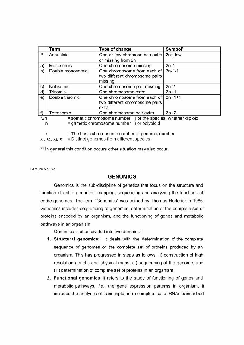
Term Type of change Symbol*B. Aneuploid One or few chromosomes extra
or missing from 2n2n+ few
a) Monosomic One chromosome missing 2n-1b) Double monosomic One chromosome from each of
two different chromosome pairs missing
2n-1-1
c) Nullisomic One chromosome pair missing 2n-2d) Trisomic One chromosome extra 2n+1e) Double trisomic One chromosome from each of
two different chromosome pairs extra
2n+1+1
f) Tetrasomic One chromosome pair extra 2n+2*2n = somatic chromosome number } of the species, whether diploid n = gametic chromosome number } or polyploid
x = The basic chromosome number or genomic number x1, x2, x3, x4 = Distinct genomes from different species.
** In general this condition occurs other situation may also occur.
Lecture No: 32
GENOMICS
Genomics is the sub -discipline of genetics that focus on the structure and
function of entire genomes, mapping, sequencing and analyzing the functions of
entire genomes. The term “Genomics” was coined by Thomas Roderick in 1986.
Genomics includes sequencing of genomes, determination of the complete set of
proteins encoded by an organism, and the functioning of genes and metabolic
pathways in an organism.
Genomics is often divided into two domains :
1. Structural genomics: It deals with the determination of the complete
sequence of genomes or the complete set of proteins produced by an
organism. This has progressed in steps as follows: (i) construction of high
resolution genetic and physical maps, (ii) sequencing of the genome, and
(iii) determination of complete set of proteins in an organism
2. Functional genomics: It refers to the study of functioning of genes and
metabolic pathways, i.e., the gene expression patterns in organism. It
includes the analyses of transcriptome (a complete set of RNAs transcribed

from a genome) and proteome (the complete set of proteins encoded by a
genome).
Human Genome Project:
The human genome project was launched in 1990, with the goal of
sequencing the entire human genome by 2005 at an estimated cost of $3 billion.
Some of the important goals of HGP were as follows.
i) Identify all the genes in human DNA
ii) Determine the sequences of the 3 billion chemical base pairs that make up
human DNA
iii) Store this information in public databases
iv) Develop tools for data analysis
v) Transfer related technologies to the private sectors and
vi) Address the ethical, legal, and social issues (ELSIs) that may arise from
the project
Sequencing of human genome was carried out by the public -funded Human
Genome Sequencing Consortium and the private company Celera genomics
established by Craig Venter. A nearly complete sequence of the human genome
comprising 99 percent of the euchromatic DNA was released in October, 2004.
The first draft of human genome sequence, which is over 3 billion nucleotide long,
consists of 24 chapters, viz., 22 autosomes and X and Y sex chromosomes.
Salient Features of Human Genome:
i) The human genome contains over 3 billion nucleotide pairs
ii) On an average there is one gene per 145 kb in the human genome.
iii) Human genome is estimated to have about 20,000 – 25,000 genes that
encode protein products.
iv) Average gene consists of 3000 bases. But sizes of genes vary greatly, with
the largest known human gene encoding dystrophin containing 2.5 million
base pairs.
v) Only about 1.1%of the genome encodes amino acid sequences of
polypeptides
vi) The functions are unknown for over 50% of the discovered genes

vii) The repetitive sequences makeup very large portion of human genome.
Repetitive sequences have no direct coding function but they shed light on
the chromosome structure, dynamics and evolution.
viii) Chromosome 1 has most genes (2968) and Y chromosome has the
lowest (231)
ix) Almost all nucleotide bases are exactly the same in all people. Genome
sequences of different individuals differ for less than 0.2% of base pairs.
Most of these differences occur in the form of single base differences in the
sequence. These single base differences are called single nucleotide
polymorphisms (SNPs). One SNP occurs at every ~ 1,000 bp of human
genome. About 85% of all differences in human DNAs are due to SNPs.