VEGF signaling mediates bladder ... - BMC Physiology
20
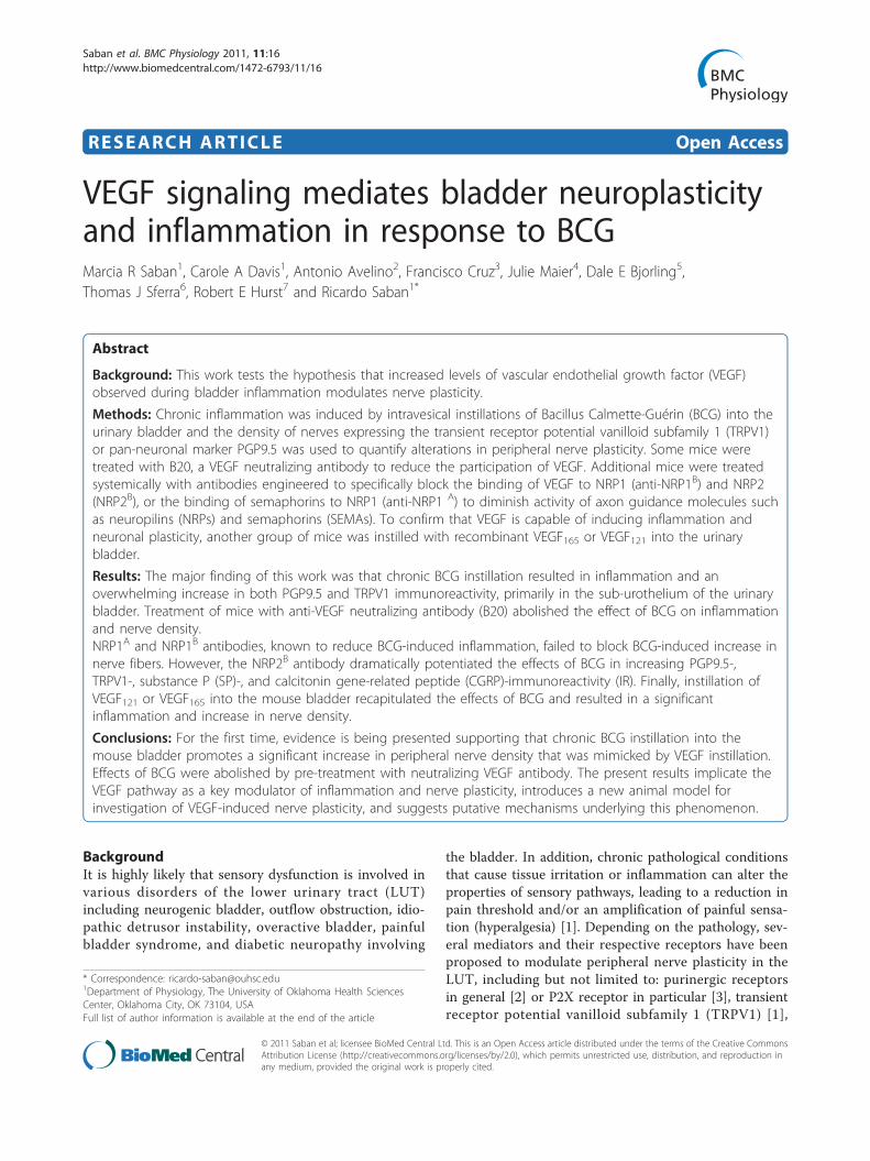
RESEARCH ARTICLE Open Access VEGF signaling mediates bladder neuroplasticity and inflammation in response to BCG Marcia R Saban 1 , Carole A Davis 1 , Antonio Avelino 2 , Francisco Cruz 3 , Julie Maier 4 , Dale E Bjorling 5 , Thomas J Sferra 6 , Robert E Hurst 7 and Ricardo Saban 1* Abstract Background: This work tests the hypothesis that increased levels of vascular endothelial growth factor (VEGF) observed during bladder inflammation modulates nerve plasticity. Methods: Chronic inflammation was induced by intravesical instillations of Bacillus Calmette-Guérin (BCG) into the urinary bladder and the density of nerves expressing the transient receptor potential vanilloid subfamily 1 (TRPV1) or pan-neuronal marker PGP9.5 was used to quantify alterations in peripheral nerve plasticity. Some mice were treated with B20, a VEGF neutralizing antibody to reduce the participation of VEGF. Additional mice were treated systemically with antibodies engineered to specifically block the binding of VEGF to NRP1 (anti-NRP1 B ) and NRP2 (NRP2 B ), or the binding of semaphorins to NRP1 (anti-NRP1 A ) to diminish activity of axon guidance molecules such as neuropilins (NRPs) and semaphorins (SEMAs). To confirm that VEGF is capable of inducing inflammation and neuronal plasticity, another group of mice was instilled with recombinant VEGF 165 or VEGF 121 into the urinary bladder. Results: The major finding of this work was that chronic BCG instillation resulted in inflammation and an overwhelming increase in both PGP9.5 and TRPV1 immunoreactivity, primarily in the sub-urothelium of the urinary bladder. Treatment of mice with anti-VEGF neutralizing antibody (B20) abolished the effect of BCG on inflammation and nerve density. NRP1 A and NRP1 B antibodies, known to reduce BCG-induced inflammation, failed to block BCG-induced increase in nerve fibers. However, the NRP2 B antibody dramatically potentiated the effects of BCG in increasing PGP9.5-, TRPV1-, substance P (SP)-, and calcitonin gene-related peptide (CGRP)-immunoreactivity (IR). Finally, instillation of VEGF 121 or VEGF 165 into the mouse bladder recapitulated the effects of BCG and resulted in a significant inflammation and increase in nerve density. Conclusions: For the first time, evidence is being presented supporting that chronic BCG instillation into the mouse bladder promotes a significant increase in peripheral nerve density that was mimicked by VEGF instillation. Effects of BCG were abolished by pre-treatment with neutralizing VEGF antibody. The present results implicate the VEGF pathway as a key modulator of inflammation and nerve plasticity, introduces a new animal model for investigation of VEGF-induced nerve plasticity, and suggests putative mechanisms underlying this phenomenon. Background It is highly likely that sensory dysfunction is involved in various disorders of the lower urinary tract (LUT) including neurogenic bladder, outflow obstruction, idio- pathic detrusor instability, overactive bladder, painful bladder syndrome, and diabetic neuropathy involving the bladder. In addition, chronic pathological conditions that cause tissue irritation or inflammation can alter the properties of sensory pathways, leading to a reduction in pain threshold and/or an amplification of painful sensa- tion (hyperalgesia) [1]. Depending on the pathology, sev- eral mediators and their respective receptors have been proposed to modulate peripheral nerve plasticity in the LUT, including but not limited to: purinergic receptors in general [2] or P2X receptor in particular [3], transient receptor potential vanilloid subfamily 1 (TRPV1) [1], * Correspondence: [email protected] 1 Department of Physiology, The University of Oklahoma Health Sciences Center, Oklahoma City, OK 73104, USA Full list of author information is available at the end of the article Saban et al. BMC Physiology 2011, 11:16 http://www.biomedcentral.com/1472-6793/11/16 © 2011 Saban et al; licensee BioMed Central Ltd. This is an Open Access article distributed under the terms of the Creative Commons Attribution License (http://creativecommons.org/licenses/by/2.0), which permits unrestricted use, distribution, and reproduction in any medium, provided the original work is properly cited.
-
Upload
khangminh22 -
Category
Documents
-
view
5 -
download
0
Transcript of VEGF signaling mediates bladder ... - BMC Physiology
RESEARCH ARTICLE Open Access
VEGF signaling mediates bladder neuroplasticityand inflammation in response to BCGMarcia R Saban1, Carole A Davis1, Antonio Avelino2, Francisco Cruz3, Julie Maier4, Dale E Bjorling5,Thomas J Sferra6, Robert E Hurst7 and Ricardo Saban1*
Abstract
Background: This work tests the hypothesis that increased levels of vascular endothelial growth factor (VEGF)observed during bladder inflammation modulates nerve plasticity.
Methods: Chronic inflammation was induced by intravesical instillations of Bacillus Calmette-Guérin (BCG) into theurinary bladder and the density of nerves expressing the transient receptor potential vanilloid subfamily 1 (TRPV1)or pan-neuronal marker PGP9.5 was used to quantify alterations in peripheral nerve plasticity. Some mice weretreated with B20, a VEGF neutralizing antibody to reduce the participation of VEGF. Additional mice were treatedsystemically with antibodies engineered to specifically block the binding of VEGF to NRP1 (anti-NRP1B) and NRP2(NRP2B), or the binding of semaphorins to NRP1 (anti-NRP1 A) to diminish activity of axon guidance molecules suchas neuropilins (NRPs) and semaphorins (SEMAs). To confirm that VEGF is capable of inducing inflammation andneuronal plasticity, another group of mice was instilled with recombinant VEGF165 or VEGF121 into the urinarybladder.
Results: The major finding of this work was that chronic BCG instillation resulted in inflammation and anoverwhelming increase in both PGP9.5 and TRPV1 immunoreactivity, primarily in the sub-urothelium of the urinarybladder. Treatment of mice with anti-VEGF neutralizing antibody (B20) abolished the effect of BCG on inflammationand nerve density.NRP1A and NRP1B antibodies, known to reduce BCG-induced inflammation, failed to block BCG-induced increase innerve fibers. However, the NRP2B antibody dramatically potentiated the effects of BCG in increasing PGP9.5-,TRPV1-, substance P (SP)-, and calcitonin gene-related peptide (CGRP)-immunoreactivity (IR). Finally, instillation ofVEGF121 or VEGF165 into the mouse bladder recapitulated the effects of BCG and resulted in a significantinflammation and increase in nerve density.
Conclusions: For the first time, evidence is being presented supporting that chronic BCG instillation into themouse bladder promotes a significant increase in peripheral nerve density that was mimicked by VEGF instillation.Effects of BCG were abolished by pre-treatment with neutralizing VEGF antibody. The present results implicate theVEGF pathway as a key modulator of inflammation and nerve plasticity, introduces a new animal model forinvestigation of VEGF-induced nerve plasticity, and suggests putative mechanisms underlying this phenomenon.
BackgroundIt is highly likely that sensory dysfunction is involved invarious disorders of the lower urinary tract (LUT)including neurogenic bladder, outflow obstruction, idio-pathic detrusor instability, overactive bladder, painfulbladder syndrome, and diabetic neuropathy involving
the bladder. In addition, chronic pathological conditionsthat cause tissue irritation or inflammation can alter theproperties of sensory pathways, leading to a reduction inpain threshold and/or an amplification of painful sensa-tion (hyperalgesia) [1]. Depending on the pathology, sev-eral mediators and their respective receptors have beenproposed to modulate peripheral nerve plasticity in theLUT, including but not limited to: purinergic receptorsin general [2] or P2X receptor in particular [3], transientreceptor potential vanilloid subfamily 1 (TRPV1) [1],
* Correspondence: [email protected] of Physiology, The University of Oklahoma Health SciencesCenter, Oklahoma City, OK 73104, USAFull list of author information is available at the end of the article
Saban et al. BMC Physiology 2011, 11:16http://www.biomedcentral.com/1472-6793/11/16
© 2011 Saban et al; licensee BioMed Central Ltd. This is an Open Access article distributed under the terms of the Creative CommonsAttribution License (http://creativecommons.org/licenses/by/2.0), which permits unrestricted use, distribution, and reproduction inany medium, provided the original work is properly cited.
substance P acting on NK1 receptors [4], protease acti-vated receptors [5], and nerve growth factor and itsreceptors [6].The new hypothesis being tested in this manuscript is
that increased levels of VEGF observed during bladderinflammation provoke nerve plasticity. This hypothesisis based on evidence indicating that nerves and bloodvessels are associated, follow a common molecular path-way during development, and key molecules responsiblefor their development may continue to control theirplasticity in adulthood [7]. The finding that mutantmice (neurogenin1/neurogenin2 double knockoutembryos) lacking sensory nerves also present disorga-nized blood vessel branching [8], suggests that local sig-nals such as VEGF supplied by nerve fibers, mayprovide a cue that determines blood vessel patterning.In contrast, administration of VEGF can support andenhance the growth of regenerating nerve fibers, prob-ably through a combination of angiogenic, neurotrophic,and neuroprotective effects [9].In this context, many proteins that were originally dis-
covered to be required for axon guidance have recentlybeen implicated in the development of the vascular [10]and lymphatic systems [11]. Perhaps the most strikingobservation is that angiogenic factors, when deregulated,contribute to various neurological disorders, such asneurodegeneration. The prototypic example of thiscross-talk between nerves and vessels is the vascularendothelial growth factor, VEGF [12]. Although origin-ally described as a key angiogenic factor, it is now wellestablished that VEGF also plays a crucial role in devel-opment of the nervous system [12].Among the neuronal guidance molecules, neuropilins
(NRPs) and plexins, and their ligands, semaphorins andVEGF have been extensively studied in the central ner-vous system. They represent large families of moleculesthat can transduce signals essential for the regulation ofneuronal repulsion and attraction, cell shape, motility,and cell-cell interactions [13-15].Plexins are similar to the Toll-like receptors (TLRs) in
their evolutionary conservation from flies to mammals.In particular, plexin A4 has been shown to be requiredfor bacteria and LPS to engage TLR and trigger thedownstream signal transduction pathway including acti-vation of Rac1, c-Jun N-terminal kinase, NF-kB and AP-1 [16]. In addition, plexin-A4 in macrophages isrequired for optimal cytokine production, includingTNFa and IL-6, upon bacterial challenge [16].NRPs are transmembrane glycoproteins that were
initially identified as co-receptors for plexin that mediatethe effects of class-3 semaphorins on axon guidance[17]. NRP-1 has high affinity for Sema-3A, whilst NRP-2homodimers have high affinity for Sema-3F [18]. Thediversity of function of these guidance molecules resides
in their capacity to also function as co-receptors forVEGF enhancing its binding to VEGF receptors [19]. Ithas become clear that in the adult organism, NRPs par-ticipate in many processes, such as angiogenesis andimmune response [20]. NRP1 is associated with bloodvessel development, whereas NRP2 was initially identi-fied as a semaphorin receptor, and mediator of axonguidance [17] and lymphatic vessel development [21].Of relevance to the present study, NRPs are highly
expressed in the human [22] and mouse bladder urothe-lium and intramural ganglia in close association withVEGF receptors [23]. Moreover, urothelial-related dis-eases and BCG-induced inflammation alter NRP expres-sion and the accessibility of VEGF to these receptors[22,23]. In addition, NRPs also regulate neuronal plasti-city as indicated by mutant mouse studies, showing thatperipheral nerve regeneration is delayed in neuropilin 2-deficient mice [24]. The latter observation raises thequestion of whether upregulation of VEGF and guidancemolecule expression during inflammation would alsolead to altered nerve plasticity.Little information is available regarding SEMA con-
centrations in non-malignant bladders. Semaphorins area large family of signaling proteins that are bothsecreted and membrane bound. A common theme inthe mechanisms of semaphorin function is that theyalter the cytoskeleton and organization of actin fila-ments and the microtubule network [25]. Class 3 sema-phorins (Sema3A-G) are the only secreted forms invertebrates. Among the class 3 semaphorins, Sema3Ahas been most intensively studied in relation to axonguidance [26] and intrathecally administered Sema3Aprotein attenuates neuropathic pain behavior in ratswith chronic constriction injury of the sciatic nerve [27].Sema3A shows repulsive activity toward a variety ofneuronal types [28]. Sema3A and its receptors (NRP1,NRP2, plexin A1, plexin A2, and plexin A3) were foundto be significantly increased during M-CSF-mediateddifferentiation of monocytes into M2 macrophages ofthe inflammatory phenotype [29].Relative to cross-talk among signaling molecules, it is
interesting that VEGF and semaphorins have oppositeeffects on the filopodia of both endothelial cells (ECs)and axons expressing neuropilins. Sema3F, the sema-phorin ligand of NRP2, is known to repel nerves [30]and endothelial cells [31], whereas VEGF165 attracts thefilopodia, which drives the ECs or axons to move in thedirection of the VEGF gradient [30].For these studies, BCG was chosen to induce cystitis
because it is known to: a) provoke a significant increasein VEGF expression in the urinary bladder [32], b)cause profound inflammation that is dependent on theVEGF pathway [33], c) up-regulate the urothelial expres-sion of VEGF receptors and NRPs [23], and d) induce
Saban et al. BMC Physiology 2011, 11:16http://www.biomedcentral.com/1472-6793/11/16
Page 2 of 20

IL-17 up-regulation [33] and its receptors that arenecessary for nerve regeneration [34].The innovative results of this research provide evi-
dence that chronic inflammation induces alterations ofbladder peripheral nerve density that express: a) thetransient receptor potential vanilloid subfamily 1(TRPV1) [35], b) protein gene product (PGP9.5)[36], c)substance P, and d) calcitonin gene-related peptide(CGRP). Results obtained with potent and specificallyengineered neutralizing antibodies against VEGF, NRP1,and NRP2 further suggest a putative mechanism under-lying inflammation-induced increase in peripheral nervedensity. Furthermore, instillation of VEGF into the blad-der recapitulated the effects of BCG on inflammationand nerve plasticity.
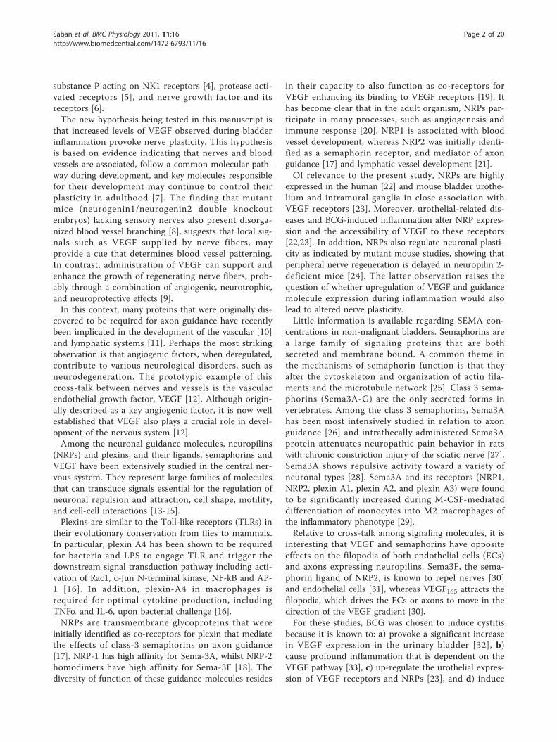
ResultsChronic instillation of BCG, VEGF165, and VEGF121 result inbladder inflammationAs BCG [33] and VEGF121 [22] once instilled into themouse bladder are absorbed across apparently intacturothelium and systemically distributed [22], we soughtto compare the degree of inflammatory cell infiltrate inresponse to these stimuli. Because VEGF165 containsamino acid residues that bind to heparin and heparansulfate proteoglycans, it is partly diffusible and partlybound to the pericellular matrix [12] which may explainits poor diffusion into deeper bladder layers. Therefore,we decided to also test the VEGF121 isoform that isfreely diffusible [12], since it lacks the basic amino acidresidues responsible for heparin binding and, therefore,does not, or only minimally, binds to the extracellularmatrix (ECM). Figure 1 illustrates inflammation inresponse to chronic instillations of BCG, VEGF165 andVEGF121. This illustration shows a predominant cellularinfiltrate in response to chronic BCG instillation,whereas VEGF165 and VEGF121 have a more profoundimpact in the bladder vascular system leading to dilationof both arteries and veins. Higher magnification insertsare amplification of dashed square areas of the respec-tive figures and illustrate inflammatory cells and perivas-cular infiltrates (Figure 1).Next, double immunofluorescence was used to further
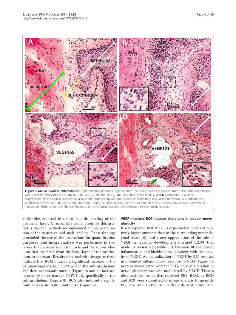
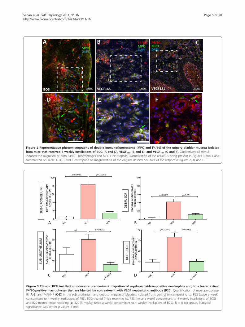
clarify whether the inflammatory infiltrate in responseof BCG and VEGF was composed of MPO+ and F4/80+cells (Figure 2). Quantitative analysis of MPO+ and F4/80+ cells indicate that chronic BCG instillation inducesboth neutrophils and macrophages infiltrations (Figure3). In the sub-urothelium, BCG induced predominantlyneutrophils (Figure 3A and 3B) that were almost doublethe number of macrophage (Figure 3C and 3D). Theseresults are summarized on Table 1.As bladder instillation with BCG is known to provoke
a significant increase in VEGF levels [32,33]
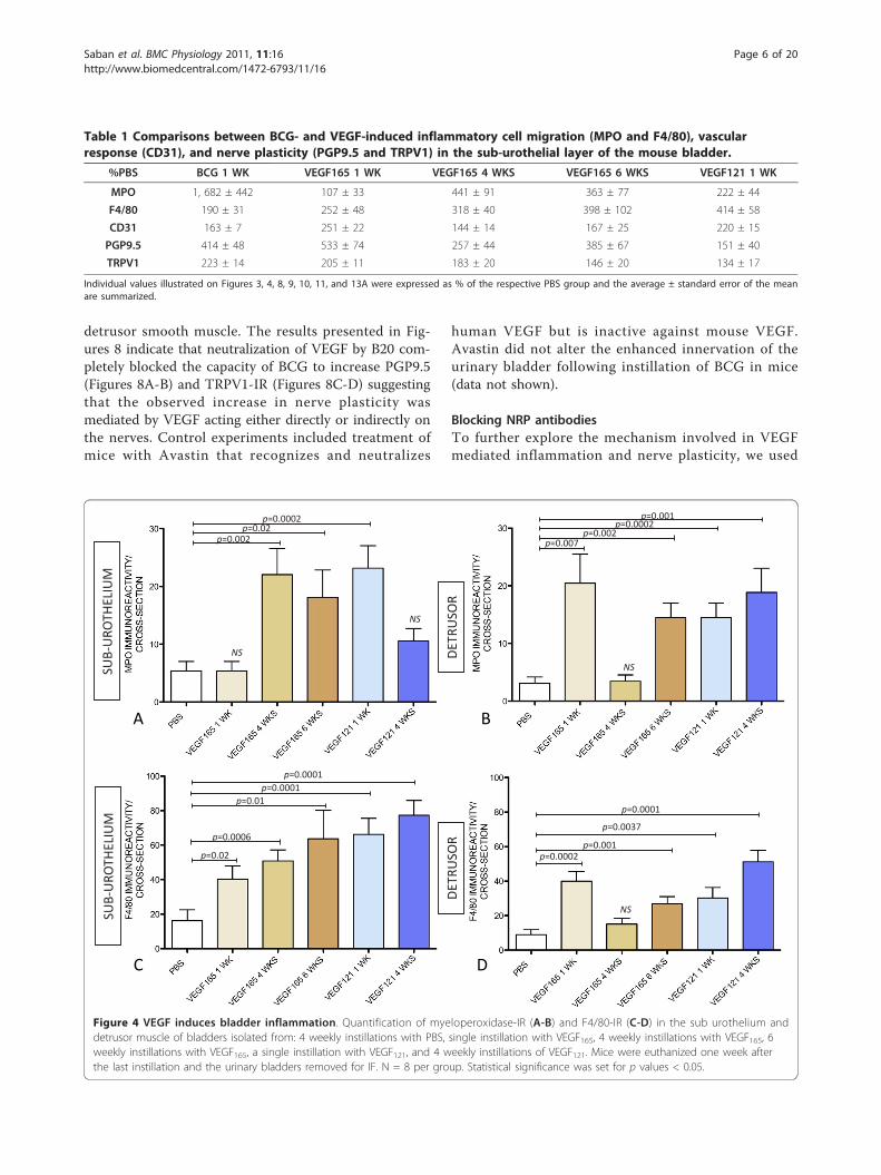
concomitant to up-regulation of VEGF receptors andco-receptors (NRPs) [23], we tested whether BCG-induced inflammation was dependent on VEGF. Toremove the influence of VEGF, an additional group ofmice received systemic treatment with neutralizingVEGF antibody twice a week, concomitant to fourweekly instillations of BCG. The results presented inFigure 3 indicate that neutralization of VEGF by B20resulted in a blunted inflammatory response character-ized by a reduction of both neutrophils and macro-phages towards baseline levels. As these results suggestthat VEGF participates in bladder inflammation, addi-tional groups of mice received instillations of VEGF165and VEGF121. Interestingly, both forms of VEGFinduced a predominant infiltration of macrophages inthe sub-urothelial layer (Figure 4C and table 1) and, to alesser extent when compared to BCG (Figure 3A andTable 1), some neutrophil infiltration (Figure 4A andTable 1).
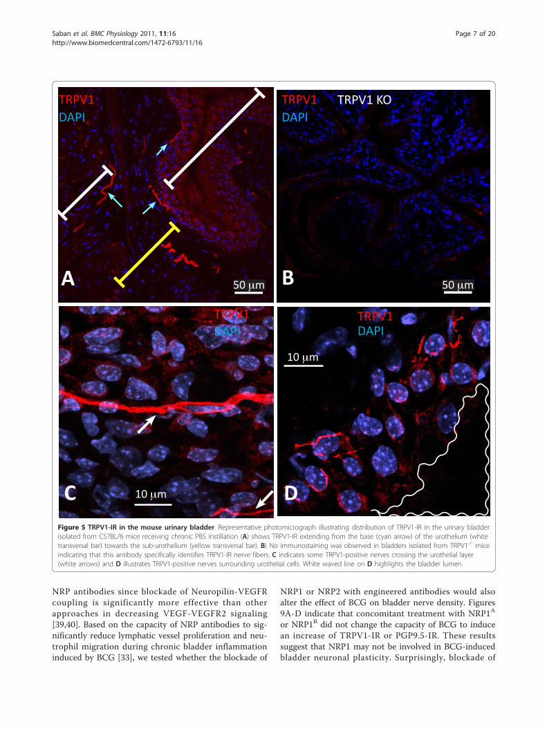
BCG-induced inflammation results in bladder nerveplasticityIn order to determine whether chronic bladder inflam-mation results in alteration of nerve density, we firstdetermined the distribution of sensory nerves expressingthe nociceptive transducer vanilloid type 1 transientreceptor potential receptor (TRPV1) in the urinary blad-der isolated from control mice. The results illustrated inFigure 5A confirmed the reported distribution ofTRPV1 immunoreactivity (IR) in the rat bladder [1] andextend the results to nerves crossing the urothelial layer(Figure 5C) or surrounding urothelial cells (Figure 5D).TRPV1 antibody specificity was verified by comparingthe labeling of bladders isolated from C57BL/6 (Figure5A) used as a positive control and TRPV1-/- mice [37]used as a negative control (Figure 5B) to bladders fromWT mice (TRPV1+/+; Figures 5C-D). No immunostain-ing was observed in bladders isolated from TRPV1-/-
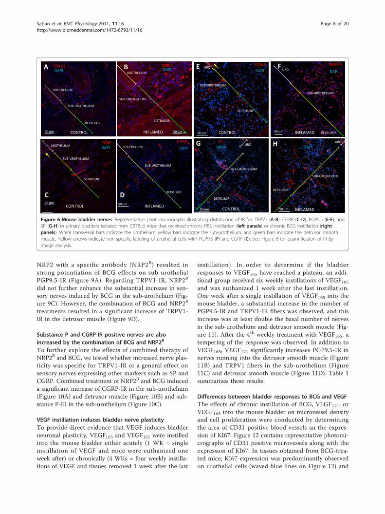
mice (Figure 5B), indicating that this antibody specifi-cally identifies TRPV1-IR in the urothelium, sub-urothe-lium, and detrusor nerve fibers (Figures 6A-B). Figures6A and 6B are low magnification micrographs in orderto illustrate the distribution of TRPV1-positive fibers inseveral layers of the urinary bladder isolated from con-trol (PBS) and inflamed bladders (BCG), respectively.Regardless of magnification, Figures 6A-B illustrate thatinflammation induced a higher degree of TRPV1-IR inthe sub-urothelium of bladders isolated from BCG-trea-ted mice that was confirmed by image analysis (Figure7). Next, we compared TRPV1-IR with other nerve mar-kers. For this purpose, tissues isolated from the samegroups were also stained with antibodies identifyingCGRP (Figures 6C-D), PGP9.5 (Figures 5E-F), and SP(Figures 6G-H). It was noted that PGP9.5 and CGRP
Saban et al. BMC Physiology 2011, 11:16http://www.biomedcentral.com/1472-6793/11/16
Page 3 of 20
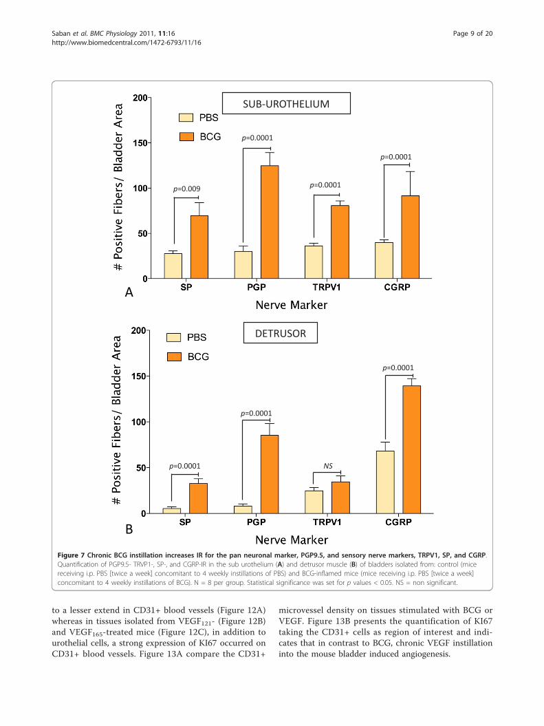
antibodies resulted in a non-specific labeling of theurothelial layer. A reasonable explanation for this arti-fact is that the methods recommended for permeabiliza-tion of the tissues caused such labeling. These findingsprecluded the use of the urothelium for quantificationpurposes, and image analysis was performed in twolayers: the detrusor smooth muscle and the sub-urothe-lium that extended from the basal layer of the urothe-lium to detrusor. Results obtained with image analysisindicate that BCG induced a significant increase in thepan-neuronal marker PGP9.5-IR in the sub-urotheliumand detrusor smooth muscle (Figure 8) and an increasein sensory nerve marker TRPV1-IR, specifically in thesub-urothelium (Figure 8). BCG also induced a signifi-cant increase in CGRP- and SP-IR (Figure 7).
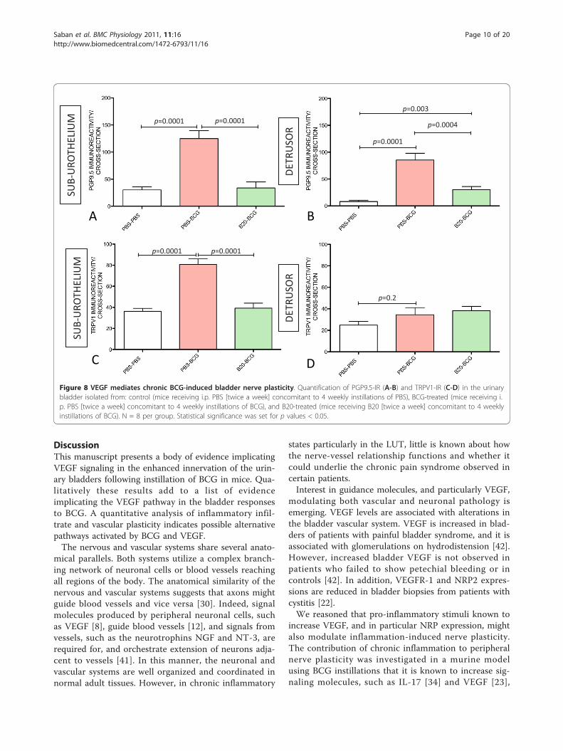
VEGF mediates BCG-induced alterations in bladder nerveplasticityIt was reported that VEGF is expressed in nerves at rela-tively higher amounts than in the surrounding mesench-ymal tissue [8], and a new appreciation of the role ofVEGF in neuronal development emerged [12,38] thatmade us review a possible link between BCG-inducedinflammation and bladder nerve plasticity with the activ-ity of VEGF. As neutralization of VEGF by B20 resultedin a blunted inflammatory response to BCG (Figure 3),next we investigated whether BCG-induced alteration innerve plasticity was also modulated by VEGF. Tissuesobtained from mice that received PBS, BCG, or BCGand B20 were submitted to image analysis to quantifyPGP9.5- and TRPV1-IR in the sub-urothelium and
100 m
SUB-UROTHELIUM
UROTHELIUM
DETRUSOR
VEGF121
PBS
100 m
A
VEGF165
100 m
C
BCG
100 m
B
D
Figure 1 Mouse bladder inflammation. Representative photomicrographs from the urinary bladders isolated from mice chronically treatedwith 4 weekly instillations of PBS (A), BCG (B), VEGF165 (C), and VEGF121 (D). Black box inserts on B, C, and D correspond to a 200%magnification of the original dashed box area of the respective figures and illustrate inflammatory cells. White transversal bars indicate theurothelium, yellow bars indicate the sub-urothelium, and green bars indicate the detrusor smooth muscle. Green arrow indicates perivascularinfiltrate of inflammatory cells (D). See Figures 2 and 3 for quantification of inflammatory cells by image analysis.
Saban et al. BMC Physiology 2011, 11:16http://www.biomedcentral.com/1472-6793/11/16
Page 4 of 20
1851 VEGF165 CHRONIC F4-80-MPO 40X e.
VEGF165 VEGF121BCG
F4/80
DAPIMPO
50 m
F4/80
DAPIMPO
F4/80
DAPIMPO
50 m 50 m
50 m50 m50 m
A B C
D E F
Figure 2 Representative photomicrographs of double immunofluorescence (MPO and F4/80) of the urinary bladder mucosa isolatedfrom mice that received 4 weekly instillations of BCG (A and D), VEGF165 (B and E), and VEGF121 (C and F). Qualitatively all stimuliinduced the migration of both F4/80+ macrophages and MPO+ neutrophils. Quantification of the results is being present in Figures 3 and 4 andsummarized on Table 1. D, E, and F correspond to magnification of the original dashed box area of the respective figures A, B, and C.
A B
C D
p=0.0045 p=0.0098
p=0.0003 p=0.001
NS p=0.0002 p=0.0001 p=0.0001
DETR
USO
R
SUB-
URO
THEL
IUM
DETR
USO
R
SUB-
URO
THEL
IUM
Figure 3 Chronic BCG instillation induces a predominant migration of myeloperoxidase-positive neutrophils and, to a lesser extent,F4/80-positive macrophages that are blunted by co-treatment with VEGF neutralizing antibody (B20). Quantification of myeloperoxidase-IR (A-B) and F4/80-IR (C-D) in the sub urothelium and detrusor muscle of bladders isolated from: control (mice receiving i.p. PBS [twice a week]concomitant to 4 weekly instillations of PBS), BCG-treated (mice receiving i.p. PBS [twice a week] concomitant to 4 weekly instillations of BCG),and B20-treated (mice receiving i.p. B20 [5 mg/kg; twice a week] concomitant to 4 weekly instillations of BCG). N = 8 per group. Statisticalsignificance was set for p values < 0.05.
Saban et al. BMC Physiology 2011, 11:16http://www.biomedcentral.com/1472-6793/11/16
Page 5 of 20
detrusor smooth muscle. The results presented in Fig-ures 8 indicate that neutralization of VEGF by B20 com-pletely blocked the capacity of BCG to increase PGP9.5(Figures 8A-B) and TRPV1-IR (Figures 8C-D) suggestingthat the observed increase in nerve plasticity wasmediated by VEGF acting either directly or indirectly onthe nerves. Control experiments included treatment ofmice with Avastin that recognizes and neutralizes
human VEGF but is inactive against mouse VEGF.Avastin did not alter the enhanced innervation of theurinary bladder following instillation of BCG in mice(data not shown).
Blocking NRP antibodiesTo further explore the mechanism involved in VEGFmediated inflammation and nerve plasticity, we used
Table 1 Comparisons between BCG- and VEGF-induced inflammatory cell migration (MPO and F4/80), vascularresponse (CD31), and nerve plasticity (PGP9.5 and TRPV1) in the sub-urothelial layer of the mouse bladder.
%PBS BCG 1 WK VEGF165 1 WK VEGF165 4 WKS VEGF165 6 WKS VEGF121 1 WK
MPO 1, 682 ± 442 107 ± 33 441 ± 91 363 ± 77 222 ± 44
F4/80 190 ± 31 252 ± 48 318 ± 40 398 ± 102 414 ± 58
CD31 163 ± 7 251 ± 22 144 ± 14 167 ± 25 220 ± 15
PGP9.5 414 ± 48 533 ± 74 257 ± 44 385 ± 67 151 ± 40
TRPV1 223 ± 14 205 ± 11 183 ± 20 146 ± 20 134 ± 17
Individual values illustrated on Figures 3, 4, 8, 9, 10, 11, and 13A were expressed as % of the respective PBS group and the average ± standard error of the meanare summarized.
DETR
USO
R
SUB-
URO
THEL
IUM
DETR
USO
R
SUB-
URO
THEL
IUM
A B
C D
p=0.0002
NS
p=0.002p=0.02
NS
p=0.007
NS
p=0.002p=0.0002
p=0.001
p=0.02
p=0.0006
p=0.01p=0.0001
p=0.0001
p=0.0002
NS
p=0.001
p=0.0037
p=0.0001
Figure 4 VEGF induces bladder inflammation. Quantification of myeloperoxidase-IR (A-B) and F4/80-IR (C-D) in the sub urothelium anddetrusor muscle of bladders isolated from: 4 weekly instillations with PBS, single instillation with VEGF165, 4 weekly instillations with VEGF165, 6weekly instillations with VEGF165, a single instillation with VEGF121, and 4 weekly instillations of VEGF121. Mice were euthanized one week afterthe last instillation and the urinary bladders removed for IF. N = 8 per group. Statistical significance was set for p values < 0.05.
Saban et al. BMC Physiology 2011, 11:16http://www.biomedcentral.com/1472-6793/11/16
Page 6 of 20
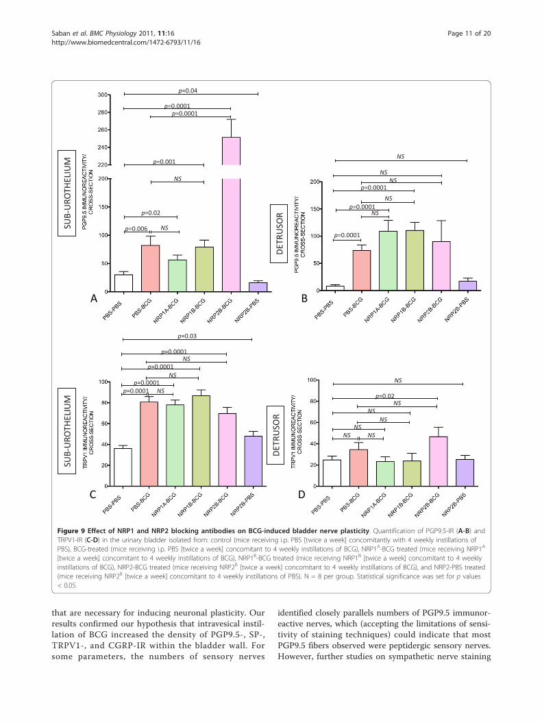
NRP antibodies since blockade of Neuropilin-VEGFRcoupling is significantly more effective than otherapproaches in decreasing VEGF-VEGFR2 signaling[39,40]. Based on the capacity of NRP antibodies to sig-nificantly reduce lymphatic vessel proliferation and neu-trophil migration during chronic bladder inflammationinduced by BCG [33], we tested whether the blockade of
NRP1 or NRP2 with engineered antibodies would alsoalter the effect of BCG on bladder nerve density. Figures9A-D indicate that concomitant treatment with NRP1A
or NRP1B did not change the capacity of BCG to inducean increase of TRPV1-IR or PGP9.5-IR. These resultssuggest that NRP1 may not be involved in BCG-inducedbladder neuronal plasticity. Surprisingly, blockade of
TRPV1DAPI
TRPV1DAPI
TRPV1DAPI
TRPV1DAPI
TRPV1 KO
50 m 50 m
10 m
10 m
A B
C DFigure 5 TRPV1-IR in the mouse urinary bladder. Representative photomicrograph illustrating distribution of TRPV1-IR in the urinary bladderisolated from C57BL/6 mice receiving chronic PBS instillation (A) shows TRPV1-IR extending from the base (cyan arrow) of the urothelium (whitetransversal bar) towards the sub-urothelium (yellow transversal bar). B) No immunostaining was observed in bladders isolated from TRPV1-/- miceindicating that this antibody specifically identifies TRPV1-IR nerve fibers. C indicates some TRPV1-positive nerves crossing the urothelial layer(white arrows) and D illustrates TRPV1-positive nerves surrounding urothelial cells. White waved line on D highlights the bladder lumen.
Saban et al. BMC Physiology 2011, 11:16http://www.biomedcentral.com/1472-6793/11/16
Page 7 of 20
NRP2 with a specific antibody (NRP2B) resulted instrong potentiation of BCG effects on sub-urothelialPGP9.5-IR (Figure 9A). Regarding TRPV1-IR, NRP2B
did not further enhance the substantial increase in sen-sory nerves induced by BCG in the sub-urothelium (Fig-ure 9C). However, the combination of BCG and NRP2B
treatments resulted in a significant increase of TRPV1-IR in the detrusor muscle (Figure 9D).
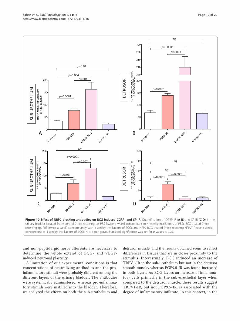
Substance P and CGRP-IR positive nerves are alsoincreased by the combination of BCG and NRP2B
To further explore the effects of combined therapy ofNRP2B and BCG, we tested whether increased nerve plas-ticity was specific for TRPV1-IR or a general effect onsensory nerves expressing other markers such as SP andCGRP. Combined treatment of NRP2B and BCG induceda significant increase of CGRP-IR in the sub-urothelium(Figure 10A) and detrusor muscle (Figure 10B) and sub-stance P-IR in the sub-urothelium (Figure 10C).
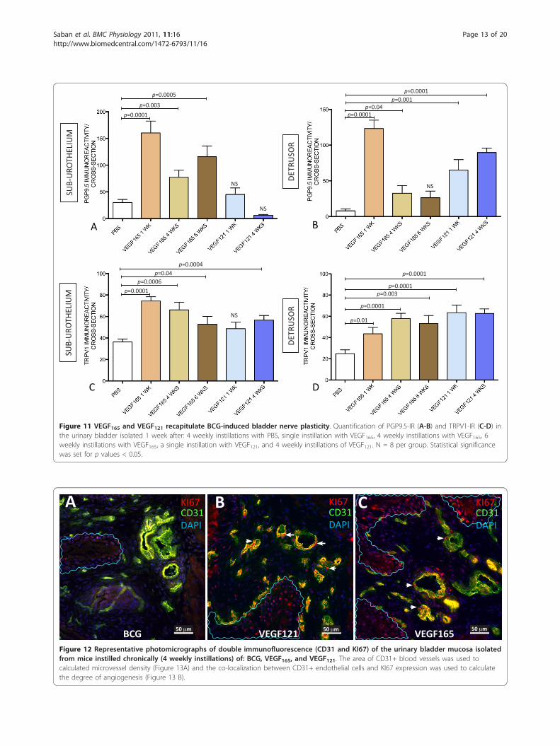
VEGF instillation induces bladder nerve plasticityTo provide direct evidence that VEGF induces bladderneuronal plasticity, VEGF165 and VEGF121 were instilledinto the mouse bladder either acutely (1 WK = singleinstillation of VEGF and mice were euthanized oneweek after) or chronically (4 WKs = four weekly instilla-tions of VEGF and tissues removed 1 week after the last
instillation). In order to determine if the bladderresponses to VEGF165 have reached a plateau, an addi-tional group received six weekly instillations of VEGF165and was euthanized 1 week after the last instillation.One week after a single instillation of VEGF165 into themouse bladder, a substantial increase in the number ofPGP9.5-IR and TRPV1-IR fibers was observed, and thisincrease was at least double the basal number of nervesin the sub-urothelium and detrusor smooth muscle (Fig-ure 11). After the 4th weekly treatment with VEGF165, atempering of the response was observed. In addition toVEGF165, VEGF121 significantly increases PGP9.5-IR innerves running into the detrusor smooth muscle (Figure11B) and TRPV1 fibers in the sub-urothelium (Figure11C) and detrusor smooth muscle (Figure 11D). Table 1summarizes these results.
Differences between bladder responses to BCG and VEGFThe effects of chronic instillation of BCG, VEGF121, orVEGF165 into the mouse bladder on microvessel densityand cell proliferation were conducted by determiningthe area of CD31-positive blood vessels an the expres-sion of KI67. Figure 12 contains representative photomi-crographs of CD31 positive microvessels along with theexpression of KI67. In tissues obtained from BCG-trea-ted mice, KI67 expression was predominantly observedon urothelial cells (waved blue lines on Figure 12) and
TRPV1DAPI
50 m
SUB-UROTHELIUM
UROTHELIUM
DETRUSOR
UROTHELIUM
SUB-UROTHELIUM
DETRUSOR
TRPV1DAPI
CGRPDAPI
SUB-UROTHELIUM
UROTHELIUM
DETRUSOR
50 m
CGRPDAPI
SUB-UROTHELIUM
UROTHELIUM
DETRUSOR
50 m
50 m CONTROL
CONTROL
INFLAMED
INFLAMED
A B
C D
PGP9.5DAPI
SUB-UROTHELIUM
URO
DETRUSOR
50 m
PGP9.5DAPI
SPDAPI
SUB-UROTHELIUM
URO
DETRUSOR
50 m
SPDAPI
SUB-UROTHELIUM
URO
DETRUSOR
50 m
URO
50 mCONTROL
CONTROL
INFLAMED
INFLAMED
SUB-UROTHELIUM
DETRUSOR
E F
G H
Figure 6 Mouse bladder nerves. Representative photomicrographs illustrating distribution of IR for: TRPV1 (A-B), CGRP (C-D), PGP9.5 (E-F), andSP (G-H) in urinary bladders isolated from C57BL6 mice that received chronic PBS instillation (left panels) or chronic BCG instillation (rightpanels). White transversal bars indicate the urothelium, yellow bars indicate the sub-urothelium, and green bars indicate the detrusor smoothmuscle. Yellow arrows indicate non-specific labeling of urothelial cells with PGP9.5 (F) and CGRP (C). See Figure 6 for quantification of IR byimage analysis.
Saban et al. BMC Physiology 2011, 11:16http://www.biomedcentral.com/1472-6793/11/16
Page 8 of 20
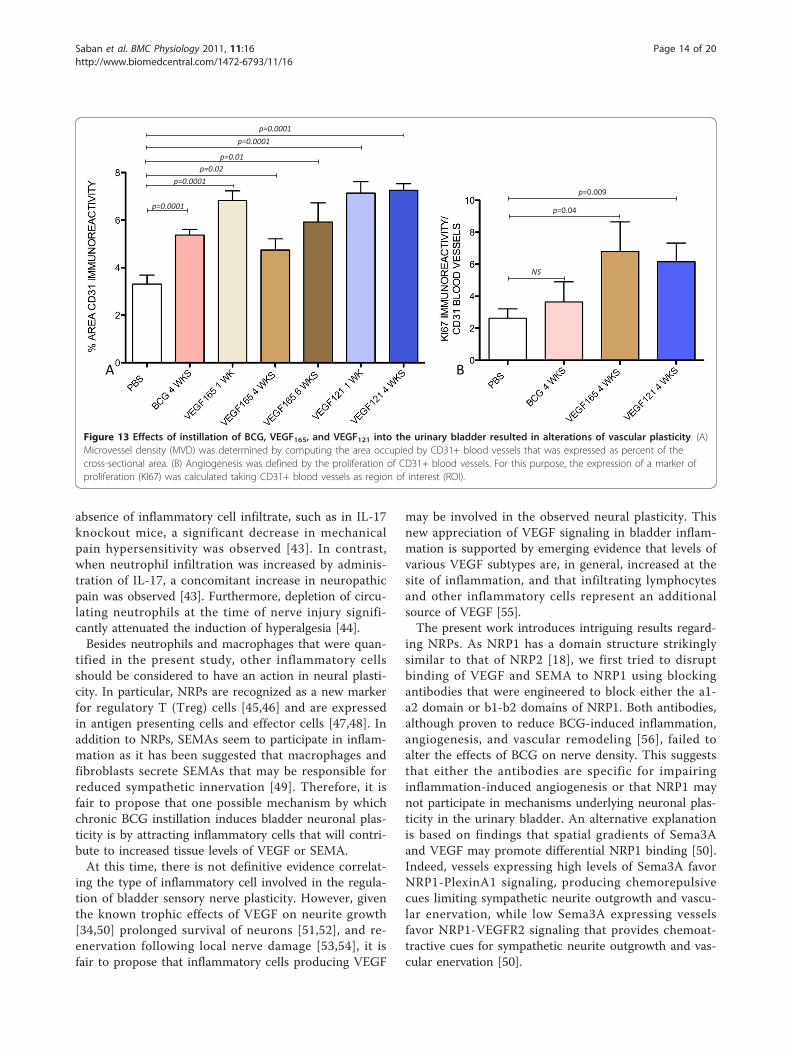
to a lesser extend in CD31+ blood vessels (Figure 12A)whereas in tissues isolated from VEGF121- (Figure 12B)and VEGF165-treated mice (Figure 12C), in addition tourothelial cells, a strong expression of KI67 occurred onCD31+ blood vessels. Figure 13A compare the CD31+
microvessel density on tissues stimulated with BCG orVEGF. Figure 13B presents the quantification of KI67taking the CD31+ cells as region of interest and indi-cates that in contrast to BCG, chronic VEGF instillationinto the mouse bladder induced angiogenesis.
DETRUSOR
SUB-UROTHELIUM
A
B
p=0.009
p=0.0001
p=0.0001
p=0.0001
p=0.0001
NS
p=0.0001
p=0.0001
Figure 7 Chronic BCG instillation increases IR for the pan neuronal marker, PGP9.5, and sensory nerve markers, TRPV1, SP, and CGRP.Quantification of PGP9.5- TRVP1-, SP-, and CGRP-IR in the sub urothelium (A) and detrusor muscle (B) of bladders isolated from: control (micereceiving i.p. PBS [twice a week] concomitant to 4 weekly instillations of PBS) and BCG-inflamed mice (mice receiving i.p. PBS [twice a week]concomitant to 4 weekly instillations of BCG). N = 8 per group. Statistical significance was set for p values < 0.05. NS = non significant.
Saban et al. BMC Physiology 2011, 11:16http://www.biomedcentral.com/1472-6793/11/16
Page 9 of 20
DiscussionThis manuscript presents a body of evidence implicatingVEGF signaling in the enhanced innervation of the urin-ary bladders following instillation of BCG in mice. Qua-litatively these results add to a list of evidenceimplicating the VEGF pathway in the bladder responsesto BCG. A quantitative analysis of inflammatory infil-trate and vascular plasticity indicates possible alternativepathways activated by BCG and VEGF.The nervous and vascular systems share several anato-
mical parallels. Both systems utilize a complex branch-ing network of neuronal cells or blood vessels reachingall regions of the body. The anatomical similarity of thenervous and vascular systems suggests that axons mightguide blood vessels and vice versa [30]. Indeed, signalmolecules produced by peripheral neuronal cells, suchas VEGF [8], guide blood vessels [12], and signals fromvessels, such as the neurotrophins NGF and NT-3, arerequired for, and orchestrate extension of neurons adja-cent to vessels [41]. In this manner, the neuronal andvascular systems are well organized and coordinated innormal adult tissues. However, in chronic inflammatory
states particularly in the LUT, little is known about howthe nerve-vessel relationship functions and whether itcould underlie the chronic pain syndrome observed incertain patients.Interest in guidance molecules, and particularly VEGF,
modulating both vascular and neuronal pathology isemerging. VEGF levels are associated with alterations inthe bladder vascular system. VEGF is increased in blad-ders of patients with painful bladder syndrome, and it isassociated with glomerulations on hydrodistension [42].However, increased bladder VEGF is not observed inpatients who failed to show petechial bleeding or incontrols [42]. In addition, VEGFR-1 and NRP2 expres-sions are reduced in bladder biopsies from patients withcystitis [22].We reasoned that pro-inflammatory stimuli known to
increase VEGF, and in particular NRP expression, mightalso modulate inflammation-induced nerve plasticity.The contribution of chronic inflammation to peripheralnerve plasticity was investigated in a murine modelusing BCG instillations that it is known to increase sig-naling molecules, such as IL-17 [34] and VEGF [23],
A B
C D
p=0.0001
p=0.0001
p=0.003
p=0.0004
p=0.0001
p=0.2DE
TRU
SOR
p=0.0001
p=0.0001
SUB-
URO
THEL
IUM
SU
B-U
ROTH
ELIU
M
DETR
USO
R
Figure 8 VEGF mediates chronic BCG-induced bladder nerve plasticity. Quantification of PGP9.5-IR (A-B) and TRPV1-IR (C-D) in the urinarybladder isolated from: control (mice receiving i.p. PBS [twice a week] concomitant to 4 weekly instillations of PBS), BCG-treated (mice receiving i.p. PBS [twice a week] concomitant to 4 weekly instillations of BCG), and B20-treated (mice receiving B20 [twice a week] concomitant to 4 weeklyinstillations of BCG). N = 8 per group. Statistical significance was set for p values < 0.05.
Saban et al. BMC Physiology 2011, 11:16http://www.biomedcentral.com/1472-6793/11/16
Page 10 of 20
that are necessary for inducing neuronal plasticity. Ourresults confirmed our hypothesis that intravesical instil-lation of BCG increased the density of PGP9.5-, SP-,TRPV1-, and CGRP-IR within the bladder wall. Forsome parameters, the numbers of sensory nerves
identified closely parallels numbers of PGP9.5 immunor-eactive nerves, which (accepting the limitations of sensi-tivity of staining techniques) could indicate that mostPGP9.5 fibers observed were peptidergic sensory nerves.However, further studies on sympathetic nerve staining
A B
p=0.02
p=0.0001
p=0.006
p=0.001
p=0.04
NS
p=0.0001NS
NSNS
NS
DETR
USO
R
SUB-
URO
THEL
IUM
NS
NS
p=0.0001
p=0.0001
p=0.0001
C D
SUB-
URO
THEL
IUM
DETR
USO
R
p=0.0001 NSp=0.0001
NSp=0.0001
NSp=0.0001
p=0.03
NS NSNS
NSNS
NSp=0.02
NS
Figure 9 Effect of NRP1 and NRP2 blocking antibodies on BCG-induced bladder nerve plasticity. Quantification of PGP9.5-IR (A-B) andTRPV1-IR (C-D) in the urinary bladder isolated from: control (mice receiving i.p. PBS [twice a week] concomitantly with 4 weekly instillations ofPBS), BCG-treated (mice receiving i.p. PBS [twice a week] concomitant to 4 weekly instillations of BCG), NRP1A-BCG treated (mice receiving NRP1A
[twice a week] concomitant to 4 weekly instillations of BCG), NRP1B-BCG treated (mice receiving NRP1B [twice a week] concomitant to 4 weeklyinstillations of BCG), NRP2-BCG treated (mice receiving NRP2B [twice a week] concomitant to 4 weekly instillations of BCG), and NRP2-PBS treated(mice receiving NRP2B [twice a week] concomitant to 4 weekly instillations of PBS). N = 8 per group. Statistical significance was set for p values< 0.05.
Saban et al. BMC Physiology 2011, 11:16http://www.biomedcentral.com/1472-6793/11/16
Page 11 of 20
and non-peptidergic nerve afferents are necessary todetermine the whole extend of BCG- and VEGF-induced neuronal plasticity.A limitation of our experimental conditions is that
concentrations of neutralizing antibodies and the pro-inflammatory stimuli were probably different among thedifferent layers of the urinary bladder. The antibodieswere systemically administered, whereas pro-inflamma-tory stimuli were instilled into the bladder. Therefore,we analyzed the effects on both the sub-urothelium and
detrusor muscle, and the results obtained seem to reflectdifferences in tissues that are in closer proximity to thestimulus. Interestingly, BCG induced an increase ofTRPV1-IR in the sub-urothelium but not in the detrusorsmooth muscle, whereas PGP9.5-IR was found increasedin both layers. As BCG favors an increase of inflamma-tory cells primarily in the sub-urothelial layer whencompared to the detrusor muscle, these results suggestTRPV1-IR, but not PGP9.5-IR, is associated with thedegree of inflammatory infiltrate. In this context, in the
p=0.004
p=0.0001
A B
p=0.0001
p=0.0001
p=0.003
p=0.01
DETR
USO
R
SUB-
URO
THEL
IUM
p=0.01
NS
p=0.0001
p=0.0001
C D
p=0.009
p=0.007
SUB-
URO
THEL
IUM
DETR
USO
R
NS
NS
NS
p=0.0001
Figure 10 Effect of NRP2 blocking antibodies on BCG-induced CGRP- and SP-IR. Quantification of CGRP-IR (A-B) and SP-IR (C-D) in theurinary bladder isolated from: control (mice receiving i.p. PBS [twice a week] concomitant to 4 weekly instillations of PBS), BCG-treated (micereceiving i.p. PBS [twice a week] concomitantly with 4 weekly instillations of BCG), and NRP2-BCG treated (mice receiving NRP2B [twice a week]concomitant to 4 weekly instillations of BCG). N = 8 per group. Statistical significance was set for p values < 0.05.
Saban et al. BMC Physiology 2011, 11:16http://www.biomedcentral.com/1472-6793/11/16
Page 12 of 20
A B
p=0.003p=0.0001
p=0.04
NS
NS
p=0.0005
p=0.0001
NS
p=0.001p=0.0001
DETR
USO
R
SUB-
URO
THEL
IUM
C D
p=0.0001p=0.0006
p=0.04
NS
p=0.0004
p=0.01
p=0.0001
p=0.003p=0.0001
p=0.0001
SUB-
URO
THEL
IUM
DETR
USO
R
Figure 11 VEGF165 and VEGF121 recapitulate BCG-induced bladder nerve plasticity. Quantification of PGP9.5-IR (A-B) and TRPV1-IR (C-D) inthe urinary bladder isolated 1 week after: 4 weekly instillations with PBS, single instillation with VEGF165, 4 weekly instillations with VEGF165, 6weekly instillations with VEGF165, a single instillation with VEGF121, and 4 weekly instillations of VEGF121. N = 8 per group. Statistical significancewas set for p values < 0.05.
VEGF165VEGF121BCG
KI67
DAPICD31
KI67
DAPICD31
KI67
DAPICD31
50 m 50 m 50 m
A B C
Figure 12 Representative photomicrographs of double immunofluorescence (CD31 and KI67) of the urinary bladder mucosa isolatedfrom mice instilled chronically (4 weekly instillations) of: BCG, VEGF165, and VEGF121. The area of CD31+ blood vessels was used tocalculated microvessel density (Figure 13A) and the co-localization between CD31+ endothelial cells and KI67 expression was used to calculatethe degree of angiogenesis (Figure 13 B).
Saban et al. BMC Physiology 2011, 11:16http://www.biomedcentral.com/1472-6793/11/16
Page 13 of 20
absence of inflammatory cell infiltrate, such as in IL-17knockout mice, a significant decrease in mechanicalpain hypersensitivity was observed [43]. In contrast,when neutrophil infiltration was increased by adminis-tration of IL-17, a concomitant increase in neuropathicpain was observed [43]. Furthermore, depletion of circu-lating neutrophils at the time of nerve injury signifi-cantly attenuated the induction of hyperalgesia [44].Besides neutrophils and macrophages that were quan-
tified in the present study, other inflammatory cellsshould be considered to have an action in neural plasti-city. In particular, NRPs are recognized as a new markerfor regulatory T (Treg) cells [45,46] and are expressedin antigen presenting cells and effector cells [47,48]. Inaddition to NRPs, SEMAs seem to participate in inflam-mation as it has been suggested that macrophages andfibroblasts secrete SEMAs that may be responsible forreduced sympathetic innervation [49]. Therefore, it isfair to propose that one possible mechanism by whichchronic BCG instillation induces bladder neuronal plas-ticity is by attracting inflammatory cells that will contri-bute to increased tissue levels of VEGF or SEMA.At this time, there is not definitive evidence correlat-
ing the type of inflammatory cell involved in the regula-tion of bladder sensory nerve plasticity. However, giventhe known trophic effects of VEGF on neurite growth[34,50] prolonged survival of neurons [51,52], and re-enervation following local nerve damage [53,54], it isfair to propose that inflammatory cells producing VEGF
may be involved in the observed neural plasticity. Thisnew appreciation of VEGF signaling in bladder inflam-mation is supported by emerging evidence that levels ofvarious VEGF subtypes are, in general, increased at thesite of inflammation, and that infiltrating lymphocytesand other inflammatory cells represent an additionalsource of VEGF [55].The present work introduces intriguing results regard-
ing NRPs. As NRP1 has a domain structure strikinglysimilar to that of NRP2 [18], we first tried to disruptbinding of VEGF and SEMA to NRP1 using blockingantibodies that were engineered to block either the a1-a2 domain or b1-b2 domains of NRP1. Both antibodies,although proven to reduce BCG-induced inflammation,angiogenesis, and vascular remodeling [56], failed toalter the effects of BCG on nerve density. This suggeststhat either the antibodies are specific for impairinginflammation-induced angiogenesis or that NRP1 maynot participate in mechanisms underlying neuronal plas-ticity in the urinary bladder. An alternative explanationis based on findings that spatial gradients of Sema3Aand VEGF may promote differential NRP1 binding [50].Indeed, vessels expressing high levels of Sema3A favorNRP1-PlexinA1 signaling, producing chemorepulsivecues limiting sympathetic neurite outgrowth and vascu-lar enervation, while low Sema3A expressing vesselsfavor NRP1-VEGFR2 signaling that provides chemoat-tractive cues for sympathetic neurite outgrowth and vas-cular enervation [50].
NS
p=0.04
p=0.009
p=0.0001
p=0.0001p=0.02
p=0.01
p=0.0001p=0.0001
A B
Figure 13 Effects of instillation of BCG, VEGF165, and VEGF121 into the urinary bladder resulted in alterations of vascular plasticity. (A)Microvessel density (MVD) was determined by computing the area occupied by CD31+ blood vessels that was expressed as percent of thecross-sectional area. (B) Angiogenesis was defined by the proliferation of CD31+ blood vessels. For this purpose, the expression of a marker ofproliferation (KI67) was calculated taking CD31+ blood vessels as region of interest (ROI).
Saban et al. BMC Physiology 2011, 11:16http://www.biomedcentral.com/1472-6793/11/16
Page 14 of 20
The unexpected increase in nerve density after NRP2B
administration deserves further investigation and studiesare underway in our laboratory to define the mechanism/s involved in bladder responses to this antibody. It is alsointeresting that NRP2B alone did not induce alteration ofnerve density. However, when given concomitantly withBCG, NRP2B induced an overwhelming increase in blad-der nerve fibers. One possible explanation is that NRP2B
might potentiate nerve plasticity by activating angiogen-esis or increasing the migration of inflammatory cellssuch as neutrophils and macrophages. Our preliminaryresults indicate that NRP2B alone does not alter bladderblood vessel density or the number of inflammatory cells(data not shown). However, NRP2B does increase bothF4/80+ macrophages and MPO+ neutrophils in responseto BCG (data not shown).It seems that in addition to activate the VEGF signal-
ing pathway, BCG may have induced an increase inSEMAs. This would explain the finding that NRP2B
further increased the number of nerves when adminis-tered concomitant to BCG. As SEMAs are known forstrong chemorepulsion, these results suggest that block-ade of NRP2 reduces SEMA participation and the con-sequent repulsion of neurons. Although NRP2B antibodywas originally developed to target the coagulation V/VIIfactor (b1-b2) domains of NRP2 which are required forVEGF-C binding to NRPs [21], and the b1-b2 domainsdo not directly engage SEMA, this antibody coulddecrease SEMA binding by preventing NRP dimerizationthat is necessary for accommodating SEMA domainsthat pack tightly together at this interface [57].It is possible that other secreted semaphorins binding
to NRP2 alone are responsible for the chemorepulsionand the consequent effect of NRP2B antibody on neuralplasticity. NRP2 is known to be required for mediatingrepulsive actions of Sema3B, 3C, and 3F, whereas NRP1is known to be required for Sema3A function. In fact,Sema3B and Sema3F seem to require only NRP2, notNRP1, to elicit their effects, whereas Sema3C mayrequire both NRP1 and NRP2 [58]. Another piece ofevidence indicating that NRP2 may play a role in alterednerve plasticity is the finding that peripheral nerveregeneration is delayed in NRP2-deficient mice [24],indicating that this guidance molecule facilitates periph-eral-nerve axonal regeneration.We also found evidence that target-derived VEGF165
or VEGF121 plays a previously unrecognized role in pro-moting growth of bladder nerves and inflammation. Arole for VEGF in inflammation has been postulated.However, instillation of VEGF into the mouse bladderrepresents direct evidence that VEGF induces inflamma-tion and that this new animal model can be used toinvestigate the effects of elevated levels of VEGF onbladder neuronal and vascular plasticity.
A fundamental question raised by the present findingsis whether the increased innervation occurred as a con-sequence of angiogenesis or inflammation. It is knownthat innervation typically accompanies blood vessels [10]which made it important to determine whether theobserved effects on nerves are direct, or mediatedthrough effects on angiogenesis. This was a difficulttopic to address facing the evidence indicating an over-lap between inflammation and angiogenesis. We foundthat increased microvessel density (MVD) was part ofthe bladder responses to chronic instillations of BCG,VEGF165, and VEGF121. However, BCG did not induce asignificant endothelial cell proliferations as indicated byKI67 and therefore, the increased MVD observed inresponse to BCG was probably related to the intensevasodilation known to be part of the inflammatoryresponse. In contrast, proliferating endothelial cells wereonly observed in VEGF-treated tissues and, therefore, itseems that angiogenesis underlies the bladder responsesto VEGF, as indicated by an increased KI67 expressionon CD31+ endothelial cells [59].Although an increase in nerve density, particularly
those expressing TRPV1-IR, has been proposed tounderline pain sensation and neurogenic detrusor over-activity based on findings that desensitization of affer-ents with capsaicin and resiniferatoxin decrease painand detrusor instability [60], the present work did notexplore whether the increased nerve density corre-sponded to an increased function of the sensory system.The primary goal of the present work was to exploreputative mechanisms involved in inflammation-inducedneural plasticity before conducting a detailed study onnerve function. Our results were focused on the targetorgan, and future studies should include the conse-quences of VEGF instillation on more central neuronssuch as those of the dorsal root ganglia. In this context,it should be recognized that while plasticity of the cen-tral nervous system (in response to stimuli) and regen-eration (in response to injury) are mainly based onadaptive changes in neural circuits and synaptic reorga-nization, plasticity of the peripheral nervous system ispredominantly based on axonal re-growth and neuronaddition [61].
ConclusionsVEGF is not only involved in angiogenesis, but also inan inflammatory reaction and neuronal plasticity.Chronic BCG administration into the mouse urinarybladder resulted in pronounced inflammation accompa-nied by increased density of PGP9.5, TRPV1, SP, andCGRP nerves. BCG effects on inflammatory cells andnerves were nullified by neutralizing VEGF antibody andqualitatively reproduced by intravesical administration ofVEGF165 or VEGF121. However, VEGF-induced neural
Saban et al. BMC Physiology 2011, 11:16http://www.biomedcentral.com/1472-6793/11/16
Page 15 of 20
plasticity seems to associate with angiogenesis whereasBCG-induced neural plasticity seems to be a conse-quence of inflammation. Concomitant administration ofthe VEGF-inducer, BCG, along with neutralizing anti-NRP2 antibodies resulted in a surprising potentiation ofneuronal plasticity, whereas NRP1 antibodies did not.The present work suggests the novel concept that bothinflammation and angiogenesis can lead to increasedbladder nerve plasticity.
MethodsExperimental Bladder InflammationAll animal experimentation conformed to the APS’sGuiding Principles in the Care and Use of Animals andwas approved by the OUHSC Animal Care & Use Com-mittee (protocol #08-105). Urinary bladders isolatedfrom TRPV1-/- (B6.129S4- Trpv1tm1Jul/J; Jackson Labora-tory) and age-matched control mice (TRPV1 +/+; WT)were generously provided by Dr. Avelino (Faculty ofMedicine of Porto) and used for confirmation of TRPV1antibody specificity. For all other studies, ten- to 12-week old C57BL/6 female mice (Jackson Laboratory, BarHarbor, ME) were used. Female mice (n = 8 per group)were anesthetized with ketamine HCl (40 mg/kg i.m.)and xylazine (2.5 mg/kg i.m.), transurethrally catheter-ized with a polypropylene catheter (24 gauge; ¾ in.;Angiocath, Becton-Dickinson, Sandy, UT), and test com-pounds were instilled at a slow rate to avoid trauma andvesicoureteral reflux, 150 μl of one of the following sub-stances were instilled: pyrogen-free saline (PBS; con-trols), BCG (TheraCys-Aventis-Pasteur; total dose of1.35 mg [33], mouse recombinant VEGF165 (15 nM), orVEGF121 (15 nM). To ensure consistent contact of sub-stances with the bladder and to avoid reflux or leakage,the catheter was occluded and left in place for 30 min-utes. Groups of mice were euthanized 24 hours postinstillation, and the urinary bladders were removed forhistology (H&E staining) and immunofluorescence stu-dies. The concentrations of BCG, VEGF165, andVEGF121 used in this study were selected because theyinduce bladder inflammation when instilled into themouse bladder.
Systemic administration of neutralizing antibodiesThe following blocking antibodies were administeredsystemically to C57BL/6 female mice (n = 8 per group):B20, anti-NRP1A, anti-NRP1B, and anti-NRP2B. Syn-thetic phage antibody libraries were used by Genentech,incorporation to produce the synthetic blocking antibo-dies: B20, NRP1A, NRP1B, and NRP2B. The librarieswere generated using a single human framework with atemplate containing human consensus complementarity-determining regions (CDRs) [62] and the antibodies thatcross-react with both human and murine NRP were
generated. B20 was isolated from synthetic antibodyphage libraries [62,63], and this neutralizing antibodybinds and blocks both human and murine VEGF [63].Anti-NRP1A was developed to neutralize the a1-a2domain of NRP1 responsible for binding semaphoring[64], and anti-NRP1B neutralizes the b1-b2 domainresponsible for binding of VEGF [64]. Anti-NRP2B wasdeveloped to specifically target the coagulation V/VIIfactor (b1-b2) domains of NRP2 responsible for thebinding of VEGF165 and VEGF-C and, thereby decreas-ing lymphangiogenesis [21]. Mice were randomlyassigned to one of the following groups that receivedintraperitoneal injections of 150 μl of: PBS, B20 (5 mg/kg), control antibody (Avastin, bevacizumab, 5 mg/kg[64]), anti-NRP1A [10 mg/kg [64], anti-NRP1B [10 mg/kg [64], anti-NRP2B (40 mg/kg). Mice were treated onday 0, and then twice a week for 5 weeks. Mice conco-mitantly received weekly intravesical instillations of 150μl of PBS or BCG (TheraCys; total dose 1.35 mg,Sanofi-Pasteur [33]) for 4 consecutive weeks.
Immunofluorescence (IF) of mouse tissuesUrinary bladders were processed for IF according topublished methods [56] to determine the effects of BCGand VEGF on the density of nerves. Briefly, mice wereexposed to isofluorane, and when anesthesia wasobtained, mice were euthanized by cervical dislocation,bladders were removed, urine was drained, and tissueswere frozen immediately in 1:1 tissue freezing media(TFM, Triangle Biomed Sciences) and OCT (TissueTek®). For all tissues, appropriate cross-section mor-phology was confirmed by H&E staining and examina-tion by light microscopy prior to preparing slides for IFlabeling. Frozen sections (10 μm thick) were post-fixedin 1% MeOH-free formaldehyde for all staining exceptfor the CGRP antibody as subsequently described.Briefly, slides were blocked for 35 minutes with 5% nor-mal donkey serum (NDS; Jackson Immunolabs), thenco-incubated with primary antibodies in 0.5% NDS for90 minutes in a humidified chamber or overnight at 4°C. When double IF was used, following brief rinses withPBS, slides were co-incubated with both secondary anti-bodies at the same time. All secondary antibodies wereused at a 1:400 dilution and included donkey anti-rabbitIgG Alexafluor 488 and 546 conjugate (MolecularProbes), donkey anti-goat IgG Alexafluor 546, donkeyanti-rat IgG Alexafluor 488, and goat anti-guinea pig546. Slides were washed, counterstained with 4’, 6-dia-midino-2-phenylindole (DAPI), and coverslipped. Con-trols included slides labeled only with individual primaryand secondary antibodies, as well as secondary antibo-dies only. CGRP labeling was performed by fixing tissueswith 4% p-formaldehyde for 90 minutes at 4°C, thenblocking with 10% NDS/0.3% Triton/0.1% BSA for 1
Saban et al. BMC Physiology 2011, 11:16http://www.biomedcentral.com/1472-6793/11/16
Page 16 of 20
hour at room temperature. Slides were incubated withthe CGRP antibody in PBS/0.3% Triton/0.1% BSA over-night at 4°C in a humidified chamber. After brief washesthe slides were stained with the appropriate secondaryantibody and counterstained with DAPI andcoverslipped.
Primary antibodiesTRPV1 antibody (1:10, 000) was raised in rabbits againstthe 15 C-terminal amino acids of the rat TRPV1sequence [37]. Commercially available antibodiesincluded: rabbit anti-human protein gene product 9.5[PGP9.5] (Neuromics; catalog # RB12103, 1:1500 dilu-tion), rabbit anti-mouse substance P (Millipore; catalogAB1566; 1:250 dilution), guinea pig anti-rat substance P(Millipore; catalog AB15810; 1:1000 dilution), rabbitanti-rat CGRP (Immunostar; catalog # 24112; 1:1000dilution), rabbit anti-human myeloperoxidase (Dako;catalog #A0398; 1:600 dilution), rat anti-mouse Pecam(CD31; BD Pharmingen; catalog 550274; 1:50 dilution),goat anti-mouse KI-67 (Santa Cruz; catalog # sc-7846;1:75 dilution), and goat anti-mouse F4/80 (Santa Cruz;catalog# sc-26642; 1:100 dilution).
Image analysisSections were viewed and photographed using a ZeissLSM-510META scanning confocal microscope (Thorn-wood, NY) using a Zeiss axiocam HRm high-resolutionCCD camera, driven by Axiovision 4.6 software. Forimage analysis, all tissue cross-sections were viewedwith a Nikon Eclipse TE 2000-S inverted fluorescentmicroscope and imaged at room temperature using adigital CCD camera (Roper Scientific; Sarasota, Florida34240) driven by NIS-Elements AR 3.0 Imaging soft-ware. DAPI staining was viewed using a DAPI filter set(340-380 nm ex, 435-485 nm em). Imaging of Alexafluor488 utilized an excitation filter of 465-495 nm and anemission filter of 515-555 nm. Alexafluor 546 wasimaged with an excitation of 528-553 nm and 590-650nm emission range. A control slide stained only withsecondary antibody was used to determine exposuretime and to set minimum background fluorescencelevels for each fluorophore imaged. Once set, exposuretimes were not changed during acquisition of eachrespective fluorophore in the staining series. Stainingwas considered positive only when the acquired signalexceeded the established background. Absence of signalbleed-through was determined using previously opti-mized multi-acquisition settings on single fluorophorestained slides.
Inflammatory cellsFor image analysis of MPO+ and F4/80+ positiveinflammatory cells, NIS-Elements AR 3.0 Imaging
software was set to include signals with an equivalentdiameter between 1-10.9 μm2 [The equivalent diameteris a size feature derived from the area. It determines thediameter of a circle with the same area as the measuredobject: Eqdia = sqrt (4*Area/p)] and to exclude any sig-nal with an area smaller than 1 μm2.
Nerve fibersHistologically nerve fibers undulate in and out of theplane of the section, sometimes appearing as linearstructures, and sometimes punctate staining, presumedindicating nerves in cross-section. Therefore, the follow-ing parameters were used in order to exclude structuresabove or below a certain size as being potentially non-neuronal and to exclude inflammatory cells since mono-cytes and macrophages have been reported to expressTRPV1 [65]. In this context, for image analysis ofnerves, the NIS-Elements AR 3.0 Imaging software wasset to count only structures with length between 0.19-500 μm and width 0.19-2.5 μm. [Length is a derived fea-ture appropriate for elongated or thin structures. Length= (Perimeter + sqrt (Perimeter2 - 16*Area))/4]; andWidth is a derived feature appropriate for elongated orthin structures. It is based on the rod model and is cal-culated according to the following formula: Width =Area/Length].
Blood vessels and AngiogenesisMicrovessel density (MVD) was determined by comput-ing the area occupied by CD31+ blood vessels that wasexpressed as percent of the cross-sectional area. Angio-genesis was defined by the proliferation of CD31+ bloodvessels. For this purpose, the expression a marker ofproliferation (KI67) was calculated taking CD31+ bloodvessels as region of interest (ROI).To meet the independent randomized sampling
assumption required for our statistical test(s), the fol-lowing measures were taken: blinding the reader totreatment groups and picking a random starting positionand proceeding clockwise with 6-10 non-overlappingimages. As 12-20 fields are necessary to view the wholebladder cross-section at 400× magnification, the sam-pling of 6-10 non-overlapping images represented halfof the entire bladder cross-section. The area occupiedby cells stained positively by a particular antibody wascalculated as percent of the total area of the region ofinterest (ROI), as indicated in the individual figurelegend and the results of 6-10 fields were averaged. Thisprocedure was repeated for all 8 bladders used per treat-ment group and the results are expressed as mean ±SEM of 8 cross-sections. The data were examined todetermine if the distributions were homoscedastic andGaussian. As these conditions were met, groups werecompared through a two-sample Student’s t test. An
Saban et al. BMC Physiology 2011, 11:16http://www.biomedcentral.com/1472-6793/11/16
Page 17 of 20
alpha of 0.05 was considered statistically significant. P-values were adjusted for multiple comparisons througha Bonferroni correction.This manuscript reports the results obtained with 13
experimental groups (PBS i.p. and 4 weekly instilla-tions of BCG; PBS i.p. and 4 weekly instillations PBS;single instillation of VEGF165, 4 weekly instillations ofVEGF165, 6 weekly instillations of VEGF165; singleinstillation of VEGF121; 4 weekly instillations ofVEGF121; avastin i.p. and 4 weekly instillations of BCG;B20 i.p. and 4 weekly instillations of BCG; NRP1A i.p.4 weekly instillations of BCG; NRP1B i.p. 4 weeklyinstillations of BCG; NRP2B i.p 4 weekly instillations ofBCG; and NRP2B i.p. 4 weekly instillations of PBS),containing 8 mice per group, for a total of 104 mice.In addition to H&E, image analysis was performed inleast 6-10 non-overlapping images of all bladder cross-sections stained with 8 different antibodies (CD31,Ki67, PGP, TRPV1, CGRP, SP, MPO, and F4/80). Therationale for performing in vitro image analysis ofnerves, blood vessels, and inflammatory cells using thesame cross-sections obtained from animals submittedto 13 different treatments was to permit the propercomparisons. In order to better organize the resultsand to improve the manuscript readability the resultswere separated in different sections. The latter doesnot imply that experiments were performed in additionto the ones described above.
ReagentsMouse recombinant VEGF165 (catalog # MGC70609)and VEGF121 (catalog # CYT-574) were purchased fromProSpec-Tany TechnoGene Ltd (Rehovot 76124, Israel).TheraCys© BCG was purchased from Sanofi-Pasteur.Anti-NRP1A, anti-NRP1B, anti-NRP2B, B20, and Avastinwere obtained from Genentech Corp.
Acknowledgements and fundingThis entire research was supported by the Department of Defense MedicalResearch Program (PRMRP) under award number PR080981 (RS). Views andopinions of, and endorsements by the author(s) do not reflect those of theUS Army or the Department of Defense. Preliminary results were support inpart by award number 2-R56-DK66101 to RS. We thank Marissa K. Warma forhelping edit this manuscript.
Author details1Department of Physiology, The University of Oklahoma Health SciencesCenter, Oklahoma City, OK 73104, USA. 2Department of ExperimentalBiology, Faculty of Medicine of Porto and IBMC, 4200-319 Porto, Portugal.3Department of Urology, Hospital São João, Faculty of Medicine of Portoand IBMC, 4200-076 Porto, Portugal. 4Oklahoma Medical ResearchFoundation (OMRF), Imaging Core Facility, Oklahoma City, Oklahoma 73104,USA. 5Department of Surgical Sciences, School of Veterinary Medicine,University of Wisconsin-Madison, Madison, WI 53706, USA. 6Departments ofPediatrics and Biochemistry and Molecular Biology, University of OklahomaHealth Sciences Center, Oklahoma City, Oklahoma 73104, USA. 7Departmentsof Urology, Biochemistry, and Molecular Biology, University of OklahomaHealth Sciences Center, Oklahoma City, Oklahoma 73104, USA.
Authors’ contributionsAll authors read and approved the final manuscript. MRS designed thestudy, performed the in vivo experiments, and drafted the manuscript, CADperformed IF imaging and analysis, AA participated in the experimentaldesign, provided TRPV1 tissues and antibody, FC participated in theexperimental design, JM performed tissue section, advised with IFtechniques, and helped CAD with confocal photomicrographs, DEBparticipated in the experimental design and helped draft the manuscript,TJS participate in the experimental design, interpretation of results, and draftof the manuscript, REH participated in the experimental design, RSconceived the study.
Competing interestsThe authors declare that they have no competing interests.
Received: 23 June 2011 Accepted: 7 November 2011Published: 7 November 2011
References1. Avelino A, Cruz F: TRPV1 (vanilloid receptor) in the urinary tract:
expression, function and clinical applications. Naunyn Schmiedebergs ArchPharmacol 2006, 373(4):287-299.
2. Andersson KE, Hedlund P: Pharmacologic perspective on the physiologyof the lower urinary tract. Urology 2002, 60(5 Suppl 1):13-20, discussion20-11.
3. Ford AP, Cockayne DA: ATP and P2X purinoceptors in urinary tractdisorders. Handb Exp Pharmacol 2011, , 202: 485-526.
4. Saban R, Saban MR, Nguyen NB, Lu B, Gerard C, Gerard NP, Hammond TG:Neurokinin-1 (NK-1) receptor is required in antigen-induced cystitis. AmJ Pathol 2000, 156(3):775-780.
5. Saban R, D’Andrea MR, Andrade-Gordon P, Derian CK, Dozmorov I,Ihnat MA, Hurst RE, Davis CA, Simpson C, Saban MR: Mandatory role ofproteinase-activated receptor 1 in experimental bladder inflammation.BMC Physiol 2007, 7:4.
6. Schnegelsberg B, Sun TT, Cain G, Bhattacharya A, Nunn PA, Ford AP,Vizzard MA, Cockayne DA: Overexpression of NGF in mouse urotheliumleads to neuronal hyperinnervation, pelvic sensitivity, and changes inurinary bladder function. Am J Physiol Regul Integr Comp Physiol 2010,298(3):R534-547.
7. Martin P, Lewis J: Origins of the neurovascular bundle: interactionsbetween developing nerves and blood vessels in embryonic chick skin.Int J Dev Biol 1989, 33(3):379-387.
8. Mukouyama YS, Shin D, Britsch S, Taniguchi M, Anderson DJ: Sensorynerves determine the pattern of arterial differentiation and blood vesselbranching in the skin. Cell 2002, 109(6):693-705.
9. Pereira Lopes FR, Lisboa BC, Frattini F, Almeida FM, Tomaz MA,Matsumoto PK, Langone F, Lora S, Melo PA, Borojevic R, Han SW,Martinez AM: Enhancement of sciatic-nerve regeneration after VEGFgene therapy. Neuropathol Appl Neurobiol 2011.
10. Carmeliet P, Tessier-Lavigne M: Common mechanisms of nerve and bloodvessel wiring. Nature 2005, 436(7048):193-200.
11. Yuan L, Moyon D, Pardanaud L, Breant C, Karkkainen MJ, Alitalo K,Eichmann A: Abnormal lymphatic vessel development in neuropilin 2mutant mice. Development 2002, 129(20):4797-4806.
12. Ruiz de Almodovar C, Lambrechts D, Mazzone M, Carmeliet P: Role andtherapeutic potential of VEGF in the nervous system. Physiol Rev 2009,89(2):607-648.
13. Bielenberg DR, Pettaway CA, Takashima S, Klagsbrun M: Neuropilins inneoplasms: expression, regulation, and function. Exp Cell Res 2006,312(5):584-593.
14. Suzuki K, Kumanogoh A, Kikutani H: Semaphorins and their receptors inimmune cell interactions. Nat Immunol 2008, 9(1):17-23.
15. Adams RH, Eichmann A: Axon guidance molecules in vascular patterning.Cold Spring Harb Perspect Biol 2010, 2(5):a001875.
16. Wen H, Lei Y, Eun SY, Ting JP: Plexin-A4-semaphorin 3A signaling isrequired for Toll-like receptor- and sepsis-induced cytokine storm. J ExpMed 2010, 207(13):2943-2957.
17. Chen H, Chedotal A, He Z, Goodman CS, Tessier-Lavigne M: Neuropilin-2, anovel member of the neuropilin family, is a high affinity receptor for thesemaphorins Sema E and Sema IV but not Sema III. Neuron 1997,19(3):547-559.
Saban et al. BMC Physiology 2011, 11:16http://www.biomedcentral.com/1472-6793/11/16
Page 18 of 20
18. Giger RJ, Urquhart ER, Gillespie SK, Levengood DV, Ginty DD, Kolodkin AL:Neuropilin-2 is a receptor for semaphorin IV: insight into the structuralbasis of receptor function and specificity. Neuron 1998, 21(5):1079-1092.
19. Soker S, Takashima S, Miao HQ, Neufeld G, Klagsbrun M: Neuropilin-1 isexpressed by endothelial and tumor cells as an isoform-specific receptorfor vascular endothelial growth factor. Cell 1998, 92(6):735-745.
20. Takamatsu H, Takegahara N, Nakagawa Y, Tomura M, Taniguchi M,Friedel RH, Rayburn H, Tessier-Lavigne M, Yoshida Y, Okuno T, Mizui M,Kang S, Nojima S, Tsujimura T, Nakatsuji Y, Katayama I, Toyofuku T,Kikutani H, Kumanogoh A: Semaphorins guide the entry of dendritic cellsinto the lymphatics by activating myosin II. Nat Immunol 2010,11(7):594-600.
21. Xu Y, Yuan L, Mak J, Pardanaud L, Caunt M, Kasman I, Larrivee B, DelToro R, Suchting S, Medvinsky A, Silva J, Yang J, Thomas JL, Koch AW,Alitalo K, Eichmann A, Bagri A: Neuropilin-2 mediates VEGF-C-inducedlymphatic sprouting together with VEGFR3. J Cell Biol 2010,188(1):115-130.
22. Saban R, Saban MR, Maier J, Fowler B, Tengowski M, Davis CA, Wu XR,Culkin DJ, Hauser P, Backer J, Hurst RE: Urothelial expression ofneuropilins and VEGF receptors in control and interstitial cystitispatients. Am J Physiol Renal Physiol 2008, 295(6):F1613-1623.
23. Saban MR, Backer JM, Backer MV, Maier J, Fowler B, Davis CA, Simpson C,Wu XR, Birder L, Freeman MR, Soker S, Hurst RE, Saban R: VEGF receptorsand neuropilins are expressed in the urothelial and neuronal cells innormal mouse urinary bladder and are upregulated in inflammation. AmJ Physiol Renal Physiol 2008, 295(1):F60-72.
24. Bannerman P, Ara J, Hahn A, Hong L, McCauley E, Friesen K, Pleasure D:Peripheral nerve regeneration is delayed in neuropilin 2-deficient mice. JNeurosci Res 2008, 86(14):3163-3169.
25. Yazdani U, Terman JR: The semaphorins. Genome Biol 2006, 7(3):211.26. Tanelian DL, Barry MA, Johnston SA, Le T, Smith GM: Semaphorin III can
repulse and inhibit adult sensory afferents in vivo. Nat Med 1997,3(12):1398-1401.
27. Hayashi M, Kamiya Y, Itoh H, Higashi T, Miyazaki T, Funakoshi K,Yamashita N, Goshima Y, Andoh T, Yamada Y, Goto T: Intrathecallyadministered Sema3A protein attenuates neuropathic pain behavior inrats with chronic constriction injury of the sciatic nerve. Neurosci Res2011, 69(1):17-24.
28. Messersmith EK, Leonardo ED, Shatz CJ, Tessier-Lavigne M, Goodman CS,Kolodkin AL: Semaphorin III can function as a selective chemorepellentto pattern sensory projections in the spinal cord. Neuron 1995,14(5):949-959.
29. Ji JD, Park-Min KH, Ivashkiv LB: Expression and function of semaphorin 3Aand its receptors in human monocyte-derived macrophages. HumImmunol 2009, 70(4):211-217.
30. Carmeliet P: Blood vessels and nerves: common signals, pathways anddiseases. Nat Rev Genet 2003, 4(9):710-720.
31. Favier B, Alam A, Barron P, Bonnin J, Laboudie P, Fons P, Mandron M,Herault JP, Neufeld G, Savi P, Herbert JM, Bono F: Neuropilin-2 interactswith VEGFR-2 and VEGFR-3 and promotes human endothelial cellsurvival and migration. Blood 2006, 108(4):1243-1250.
32. Pavlovich CP, Kraling BM, Stewart RJ, Chen X, Bochner BH, Luster AD,Poppas DP, O’Donnell MA: BCG-induced urinary cytokines inhibitmicrovascular endothelial cell proliferation. J Urol 2000, 163(6):2014-2021.
33. Saban MR, Simpson C, Davis C, Wallis G, Knowlton N, Frank MB, Centola M,Gallucci RM, Saban R: Discriminators of mouse bladder response tointravesical Bacillus Calmette-Guerin (BCG). BMC immunology 2007, 8:6.
34. Li Z, Burns AR, Han L, Rumbaut RE, Smith CW: IL-17 and VEGF arenecessary for efficient corneal nerve regeneration. Am J Pathol 2011,178(3):1106-1116.
35. Charrua A, Reguenga C, Cordeiro JM, Correiade-Sa P, Paule C, Nagy I,Cruz F, Avelino A: Functional transient receptor potential vanilloid 1 isexpressed in human urothelial cells. J Urol 2009, 182(6):2944-2950.
36. Doran JF, Jackson P, Kynoch PA, Thompson RJ: Isolation of PGP 9.5, a newhuman neurone-specific protein detected by high-resolution two-dimensional electrophoresis. J Neurochem 1983, 40(6):1542-1547.
37. Charrua A, Cruz CD, Narayanan S, Gharat L, Gullapalli S, Cruz F, Avelino A:GRC-6211, a new oral specific TRPV1 antagonist, decreases bladderoveractivity and noxious bladder input in cystitis animal models. J Urol2009, 181(1):379-386.
38. Carmeliet P: Neuro-vascular link: from genetic insights to therapeuticperspectives. Bull Mem Acad R Med Belg 2008, 163(10-12):445-451,discussion 451-442.
39. Mac Gabhann F, Popel AS: Targeting neuropilin-1 to inhibit VEGFsignaling in cancer: Comparison of therapeutic approaches. PLoS ComputBiol 2006, 2(12):e180.
40. Mac Gabhann F, Popel AS: Interactions of VEGF isoforms with VEGFR-1,VEGFR-2, and neuropilin in vivo: a computational model of humanskeletal muscle. Am J Physiol Heart Circ Physiol 2007, 292(1):H459-474.
41. Kuruvilla R, Zweifel LS, Glebova NO, Lonze BE, Valdez G, Ye H, Ginty DD: Aneurotrophin signaling cascade coordinates sympathetic neurondevelopment through differential control of TrkA trafficking andretrograde signaling. Cell 2004, 118(2):243-255.
42. Tamaki M, Saito R, Ogawa O, Yoshimura N, Ueda T: Possible mechanismsinducing glomerulations in interstitial cystitis: relationship betweenendoscopic findings and expression of angiogenic growth factors. J Urol2004, 172(3):945-948.
43. Kim CF, Moalem-Taylor G: Interleukin-17 contributes toneuroinflammation and neuropathic pain following peripheral nerveinjury in mice. J Pain 2011, 12(3):370-383.
44. Perkins NM, Tracey DJ: Hyperalgesia due to nerve injury: role ofneutrophils. Neuroscience 2000, 101(3):745-757.
45. Sarris M, Andersen KG, Randow F, Mayr L, Betz AG: Neuropilin-1 expressionon regulatory T cells enhances their interactions with dendritic cellsduring antigen recognition. Immunity 2008, 28(3):402-413.
46. Mizui M, Kikutani H: Neuropilin-1: the glue between regulatory T cellsand dendritic cells? Immunity 2008, 28(3):302-303.
47. Bruder D, Probst-Kepper M, Westendorf AM, Geffers R, Beissert S, Loser K,von Boehmer H, Buer J, Hansen W: Neuropilin-1: a surface marker ofregulatory T cells. Eur J Immunol 2004, 34(3):623-630.
48. Battaglia A, Buzzonetti A, Monego G, Peri L, Ferrandina G, Fanfani F,Scambia G, Fattorossi A: Neuropilin-1 expression identifies a subset ofregulatory T cells in human lymph nodes that is modulated bypreoperative chemoradiation therapy in cervical cancer. Immunology2008, 123(1):129-138.
49. Muller MW, Giese NA, Swiercz JM, Ceyhan GO, Esposito I, Hinz U, Buchler P,Giese T, Buchler MW, Offermanns S, Friess H: Association of axon guidancefactor semaphorin 3A with poor outcome in pancreatic cancer. Int JCancer 2007, 121(11):2421-2433.
50. Long JB, Jay SM, Segal SS, Madri JA: VEGF-A and Semaphorin3A:modulators of vascular sympathetic innervation. Dev Biol 2009,334(1):119-132.
51. Brockington A, Wharton SB, Fernando M, Gelsthorpe CH, Baxter L, Ince PG,Lewis CE, Shaw PJ: Expression of vascular endothelial growth factor andits receptors in the central nervous system in amyotrophic lateralsclerosis. J Neuropathol Exp Neurol 2006, 65(1):26-36.
52. Ogunshola OO, Antic A, Donoghue MJ, Fan SY, Kim H, Stewart WB,Madri JA, Ment LR: Paracrine and autocrine functions of neuronalvascular endothelial growth factor (VEGF) in the central nervous system.J Biol Chem 2002, 277(13):11410-11415.
53. Damon DH: Vascular endothelial-derived semaphorin 3 inhibitssympathetic axon growth. Am J Physiol Heart Circ Physiol 2006, 290(3):H1220-1225.
54. Marko SB, Damon DH: VEGF promotes vascular sympathetic innervation.Am J Physiol Heart Circ Physiol 2008, 294(6):H2646-2652.
55. Freeman MR, Schneck FX, Gagnon ML, Corless C, Soker S, Niknejad K,Peoples GE, Klagsbrun M: Peripheral blood T lymphocytes andlymphocytes infiltrating human cancers express vascular endothelialgrowth factor: a potential role for T cells in angiogenesis. Cancer Res1995, 55(18):4140-4145.
56. Saban MR, Sferra TJ, Davis CA, Simpson C, Allen A, Maier J, Fowler B,Knowlton N, Birder L, Wu XR, Saban R: Neuropilin-VEGF signaling pathwayacts as a key modulator of vascular, lymphatic, and inflammatory cellresponses of the bladder to intravesical BCG treatment. Am J PhysiolRenal Physiol 2010, 299(6):F1245-1256.
57. Appleton BA, Wu P, Maloney J, Yin J, Liang WC, Stawicki S, Mortara K,Bowman KK, Elliott JM, Desmarais W, Bazan JF, Bagri A, Tessier-Lavigne M,Koch AW, Wu Y, Watts RJ, Wiesmann C: Structural studies of neuropilin/antibody complexes provide insights into semaphorin and VEGFbinding. Embo J 2007, 26(23):4902-4912.
Saban et al. BMC Physiology 2011, 11:16http://www.biomedcentral.com/1472-6793/11/16
Page 19 of 20
58. Chen H, He Z, Bagri A, Tessier-Lavigne M: Semaphorin-neuropilininteractions underlying sympathetic axon responses to class IIIsemaphorins. Neuron 1998, 21(6):1283-1290.
59. Hillen F, van de Winkel A, Creytens D, Vermeulen AH, Griffioen AW:Proliferating endothelial cells, but not microvessel density, are aprognostic parameter in human cutaneous melanoma. Melanoma Res2006, 16(5):453-457.
60. Silva C, Avelino A, Souto-Moura C, Cruz F: A light- and electron-microscopic histopathological study of human bladder mucosa afterintravesical resiniferatoxin application. BJU Int 2001, 88(4):355-360.
61. Geuna S, Fornaro M, Raimondo S, Giacobini-Robecchi MG: Plasticity andregeneration in the peripheral nervous system. Ital J Anat Embryol 2010,115(1-2):91-94.
62. Liang WC, Dennis MS, Stawicki S, Chanthery Y, Pan Q, Chen Y, Eigenbrot C,Yin J, Koch AW, Wu X, Ferrara N, Bagri A, Tessier-Lavigne M, Watts RJ, Wu Y:Function blocking antibodies to neuropilin-1 generated from a designedhuman synthetic antibody phage library. J Mol Biol 2007, 366(3):815-829.
63. Fuh G, Wu P, Liang WC, Ultsch M, Lee CV, Moffat B, Wiesmann C: Structure-function studies of two synthetic anti-vascular endothelial growth factorFabs and comparison with the Avastin Fab. J Biol Chem 2006,281(10):6625-6631.
64. Pan Q, Chanthery Y, Liang WC, Stawicki S, Mak J, Rathore N, Tong RK,Kowalski J, Yee SF, Pacheco G, Ross S, Cheng Z, Le Couter J, Plowman G,Peale F, Koch AW, Wu Y, Bagri A, Tessier-Lavigne M, Watts RJ: Blockingneuropilin-1 function has an additive effect with anti-VEGF to inhibittumor growth. Cancer Cell 2007, 11(1):53-67.
65. Finney-Hayward TK, Popa MO, Bahra P, Li S, Poll CT, Gosling M,Nicholson AG, Russell RE, Kon OM, Jarai G, Westwick J, Barnes PJ,Donnelly LE: Expression of transient receptor potential c6 channels inhuman lung macrophages. American journal of respiratory cell andmolecular biology 2010, 43(3):296-304.
doi:10.1186/1472-6793-11-16Cite this article as: Saban et al.: VEGF signaling mediates bladderneuroplasticity and inflammation in response to BCG. BMC Physiology2011 11:16.
Submit your next manuscript to BioMed Centraland take full advantage of:
• Convenient online submission
• Thorough peer review
• No space constraints or color figure charges
• Immediate publication on acceptance
• Inclusion in PubMed, CAS, Scopus and Google Scholar
• Research which is freely available for redistribution
Submit your manuscript at www.biomedcentral.com/submit
Saban et al. BMC Physiology 2011, 11:16http://www.biomedcentral.com/1472-6793/11/16
Page 20 of 20