BMC Microbiol 2010
14
RESEARCH ARTICLE Open Access New structural and functional defects in polyphosphate deficient bacteria: A cellular and proteomic study Cristian Varela 1 , Cecilia Mauriaca 1 , Alberto Paradela 2 , Juan P Albar 2 , Carlos A Jerez 1 , Francisco P Chávez 1* Abstract Background: Inorganic polyphosphate (polyP), a polymer of tens or hundreds of phosphate residues linked by ATP-like bonds, is found in all organisms and performs a wide variety of functions. PolyP is synthesized in bacterial cells by the actions of polyphosphate kinases (PPK1 and PPK2) and degraded by exopolyphosphatase (PPX). Bacterial cells with polyP deficiencies due to knocking out the ppk1 gene are affected in many structural and important cellular functions such as motility, quorum sensing, biofilm formation and virulence among others. The cause of this pleiotropy is not entirely understood. Results: The overexpression of exopolyphosphatase in bacteria mimicked some pleitropic defects found in ppk1 mutants. By using this approach we found new structural and functional defects in the polyP-accumulating bacteria Pseudomonas sp. B4, which are most likely due to differences in the polyP-removal strategy. Colony morphology phenotype, lipopolysaccharide (LPS) structure changes and cellular division malfunction were observed. Finally, we used comparative proteomics in order to elucidate the cellular adjustments that occurred during polyP deficiency in this bacterium and found some clues that helped to understand the structural and functional defects observed. Conclusions: The results obtained suggest that during polyP deficiency energy metabolism and particularly nucleoside triphosphate (NTP) formation were affected and that bacterial cells overcame this problem by increasing the flux of energy-generating metabolic pathways such as tricarboxilic acid (TCA) cycle, b-oxidation and oxidative phosphorylation and by reducing energy-consuming ones such as active transporters and amino acid biosynthesis. Furthermore, our results suggest that a general stress response also took place in the cell during polyP deficiency. Background Polyphosphate (polyP) is a ubiquitous linear polymer of hundreds of orthophosphate residues (Pi) linked by phosphoanhydride bonds. PolyP has been found in all tree domains of life (Archaea, Bacteria and Eukarya). In bacteria, the main enzymes involved in the metabolism of polyP are the polyphosphate kinases (PPK1 and PPK2) that catalyze the reversible conversion of the terminal phosphate of ATP (or GTP) into polyP and the exopolyphosphatase (PPX) that processively hydrolyzes the terminal residues of polyP to liberate Pi [1,2]. PolyP is a reservoir of phosphate and, as in ATP, of high-energy phosphate bonds. Furthermore, biochem- ical experiments and studies with ppk1 mutants in many bacteria have indicated additional roles for polyP. These include inhibition of RNA degradation [3], activation of Lon protease during stringent response [4,5], involvement in membrane channel structure [6,7], and contribution to the resistance to stress generated by heat, oxidants, osmotic challenge, antibiotics and UV [8-12]. Particularly, a ppk1 mutant of Pseudomonas aeruginosa PAO1 was impaired in motility, biofilm development, quorum sensing and virulence [13-15]. * Correspondence: [email protected] 1 Laboratory of Molecular Microbiology and Biotechnology & Millennium Institute of Cell Dynamics and Biotechnology (ICDB), Department of Biology, Faculty of Sciences, University of Chile, Las Palmeras 3425, Ñuñoa, Santiago, Chile Varela et al. BMC Microbiology 2010, 10:7 http://www.biomedcentral.com/1471-2180/10/7 © 2010 Varela et al; licensee BioMed Central Ltd. This is an Open Access article distributed under the terms of the Creative Commons Attribution License (http://creativecommons.org/licenses/by/2.0), which permits unrestricted use, distribution, and reproduction in any medium, provided the original work is properly cited.
Transcript of BMC Microbiol 2010
RESEARCH ARTICLE Open Access
New structural and functional defects inpolyphosphate deficient bacteria: A cellular andproteomic studyCristian Varela1, Cecilia Mauriaca1, Alberto Paradela2, Juan P Albar2, Carlos A Jerez1, Francisco P Chávez1*
Abstract
Background: Inorganic polyphosphate (polyP), a polymer of tens or hundreds of phosphate residues linked byATP-like bonds, is found in all organisms and performs a wide variety of functions. PolyP is synthesized in bacterialcells by the actions of polyphosphate kinases (PPK1 and PPK2) and degraded by exopolyphosphatase (PPX).Bacterial cells with polyP deficiencies due to knocking out the ppk1 gene are affected in many structural andimportant cellular functions such as motility, quorum sensing, biofilm formation and virulence among others. Thecause of this pleiotropy is not entirely understood.
Results: The overexpression of exopolyphosphatase in bacteria mimicked some pleitropic defects found in ppk1mutants. By using this approach we found new structural and functional defects in the polyP-accumulatingbacteria Pseudomonas sp. B4, which are most likely due to differences in the polyP-removal strategy. Colonymorphology phenotype, lipopolysaccharide (LPS) structure changes and cellular division malfunction wereobserved. Finally, we used comparative proteomics in order to elucidate the cellular adjustments that occurredduring polyP deficiency in this bacterium and found some clues that helped to understand the structural andfunctional defects observed.
Conclusions: The results obtained suggest that during polyP deficiency energy metabolism and particularlynucleoside triphosphate (NTP) formation were affected and that bacterial cells overcame this problem byincreasing the flux of energy-generating metabolic pathways such as tricarboxilic acid (TCA) cycle, b-oxidation andoxidative phosphorylation and by reducing energy-consuming ones such as active transporters and amino acidbiosynthesis. Furthermore, our results suggest that a general stress response also took place in the cell duringpolyP deficiency.
BackgroundPolyphosphate (polyP) is a ubiquitous linear polymer ofhundreds of orthophosphate residues (Pi) linked byphosphoanhydride bonds. PolyP has been found in alltree domains of life (Archaea, Bacteria and Eukarya). Inbacteria, the main enzymes involved in the metabolismof polyP are the polyphosphate kinases (PPK1 andPPK2) that catalyze the reversible conversion of theterminal phosphate of ATP (or GTP) into polyP and the
exopolyphosphatase (PPX) that processively hydrolyzesthe terminal residues of polyP to liberate Pi [1,2].PolyP is a reservoir of phosphate and, as in ATP, of
high-energy phosphate bonds. Furthermore, biochem-ical experiments and studies with ppk1 mutants inmany bacteria have indicated additional roles forpolyP. These include inhibition of RNA degradation[3], activation of Lon protease during stringentresponse [4,5], involvement in membrane channelstructure [6,7], and contribution to the resistance tostress generated by heat, oxidants, osmotic challenge,antibiotics and UV [8-12]. Particularly, a ppk1 mutantof Pseudomonas aeruginosa PAO1 was impaired inmotility, biofilm development, quorum sensing andvirulence [13-15].
* Correspondence: [email protected] of Molecular Microbiology and Biotechnology & MillenniumInstitute of Cell Dynamics and Biotechnology (ICDB), Department of Biology,Faculty of Sciences, University of Chile, Las Palmeras 3425, Ñuñoa, Santiago,Chile
Varela et al. BMC Microbiology 2010, 10:7http://www.biomedcentral.com/1471-2180/10/7
© 2010 Varela et al; licensee BioMed Central Ltd. This is an Open Access article distributed under the terms of the Creative CommonsAttribution License (http://creativecommons.org/licenses/by/2.0), which permits unrestricted use, distribution, and reproduction inany medium, provided the original work is properly cited.

In addition to PPK1, another widely conserved polyPenzyme is PPK2 [16,17]. In contrast to the ATP-depen-dent polyP synthetic activity of PPK1, PPK2 preferen-tially catalyses the polyP-driven synthesis of GTP fromGDP. Orthologs to both proteins have been found inmany bacterial genomes and curiously there are manybacteria with orthologs of either PPK1 or PPK2, or both,or neither [17].PolyP in bacteria is localized predominantly in volutin
granules, also called polyP granules, or in acidocalci-somes [18]. Many biochemical pathways are connectedand a given metabolite such as polyP can be generatedand/or consumed by several enzymes or cellular pro-cesses. The genetic background, culture conditions andenvironmental factors can influence polyP levels. Itsabsence, as mentioned above, causes many structuraland functional defects. The link between genotypes andphenotypes observed during polyP deficiency can be theresult of complex networks of interaction that can beelucidated by using OMICS technology [19,20].Recombinant Pseudomonas sp. B4 that overexpressed
yeast exopolyphosphatase also showed the functionaldeficiencies in motility and biofilm developmentreported for ppk1 mutants from P. aeruginosa PAO1[21]. In addition, new structural and functional defectssuch as changes in colony morphology, LPS structureand cellular division are reported in this communica-tion. Finally, to study the proteomic changes thatoccurred during polyP deficiency recombinant strainswere compared under different growth conditions andphases of growth. Interesting proteins related to ener-getic metabolism were overexpressed during polyP scar-city, such as three enzymes from the tricarboxylic acid(TCA) cycle, and one ATP synthase subunit. Proteinfolding, fatty acid catabolism and amino acid biosynth-esis were other gene onthology (GO) categories overre-presented during polyP deficit. On the other hand,motility and transport proteins were the only categoriesunderrepresented in this condition.The proteomics results suggest a link between polyP
and central metabolism that can be further explored toclarify the multiple structural and functional defectsfound during the lack of polyP in bacteria.
ResultsStructural and functional defects in polyphosphatedeficient bacteriaOverexpression of PPX resembled the functional defectsfound in motility and biofilm formation in a ppk1mutant from P. aeruginosa PAO [21]. Despite severalfunctional and structural defects have been reported inP. aeruginosa PAO1 ppk1 mutant [15,21,22], our polyPdeficient cells showed new functional and structuralphenotypes not previously reported. PPK1 is essential
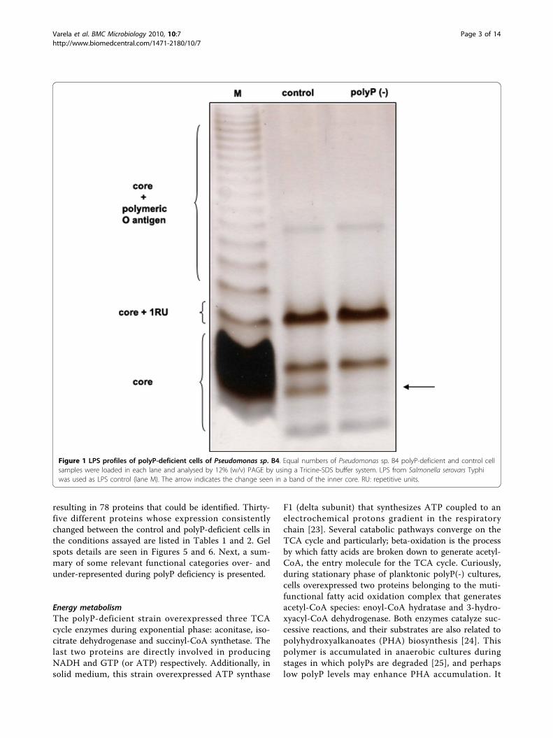
for biofilm development and virulence of P. aeruginosaPAO1. Considering that lipopolysaccharide (LPS) is alsovery important in both cellular processes; the electro-phoretic profile of LPS from recombinants Pseudomonassp. B4 were analyzed. Interestingly, changes in the coreof the LPS were observed in Tricine/SDS-polyacrylamidegel electrophoresis (Figure 1). To our knowledge, thestructure of the LPS core from Pseudomonas sp. B4 hasnot yet been elucidated and consequently it is difficultto determine the structural nature of the changefound in the LPS core. It would be interesting to deter-mine the structure of LPS in both strains [control andpolyP(-)] to reveal the change in the LPS and its prob-able link with polyP.It was found that inorganic polyP influences not only
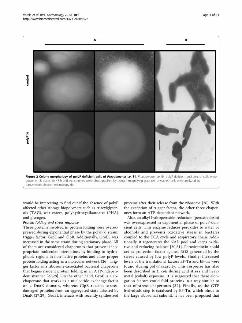
biofilm formation but also colony morphology pheno-type. Changes were seen when the colony phenotype ofcontrol cells was compared with that of polyP-deficientones. Wild type and control cells were highly motileforming a rough colony with an irregular border (Figure2A). In contrast, polyP-deficient cells displayed a roundregular smooth colony (Figure 2A). The changeobserved in colony morphology could be directly a con-sequence of the absence of exopolymer productionobserved in the cells (Figure 2B) and in a P. aeruginosaPAO1 ppk1 mutant [22] but also due to the variation inthe LPS core reported here. Altogether, the results sug-gest that biofilm formation capabilities of polyP-deficientmutants, may not only be attributed to the defect inexopolymer formation, but also to their altered LPSstructure.Finally, during the entrance in stationary phase of
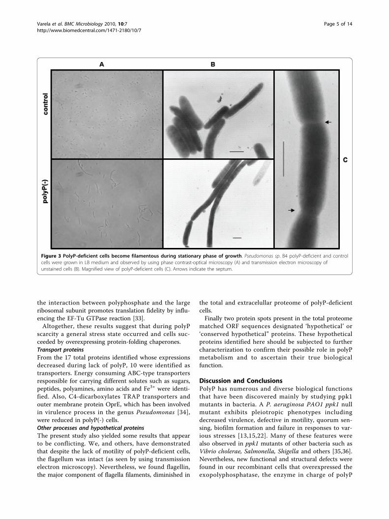
growth in rich medium (LB) it was observed that polyP-deficient cells became highly filamentous compared tocontrol cells most likely reflecting a cell division mal-function (Figure 3). Different defined media supplemen-ted with various carbon sources were tested and thisbehaviour was found only during the entry into the sta-tionary phase of growth in LB medium.Differential proteomics of polyP-deficient Pseudomonassp. B4To gain insight into the effect of polyP deficiency andthe metabolic adjustments taking place during the cellu-lar response, the proteomes of Pseudomonas sp. B4polyP-deficient and control cells were compared by two-dimensional gel electrophoresis (2D-PAGE) (Figure 4).We analyzed extracellular and total cell-free proteomesfrom planctonic cells grown in LB medium during expo-nential and stationary phase of growth and also analyzedthe total cell-free proteome of the colony biofilm. These8 samples were analyzed by using biological and experi-mental duplicates. This procedure yielded 81 spots ofinterest (proteins differentially expressed under polyP-deficiency) that were analysed by mass spectrometry
Varela et al. BMC Microbiology 2010, 10:7http://www.biomedcentral.com/1471-2180/10/7
Page 2 of 14
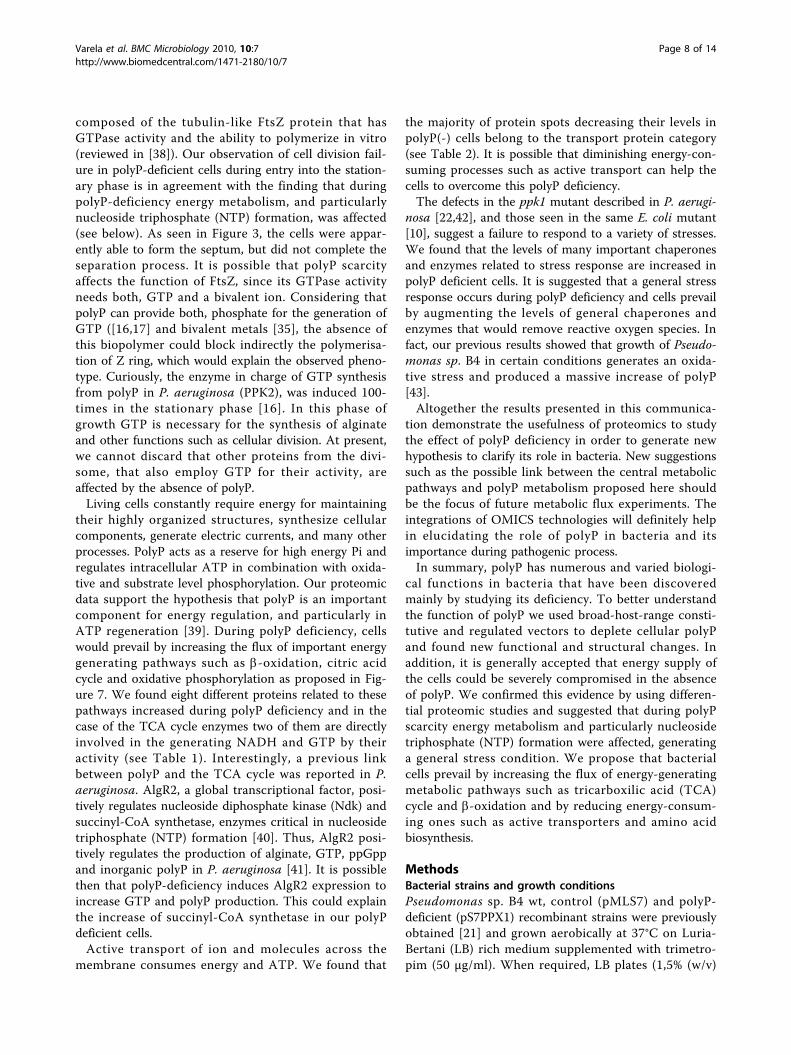
resulting in 78 proteins that could be identified. Thirty-five different proteins whose expression consistentlychanged between the control and polyP-deficient cells inthe conditions assayed are listed in Tables 1 and 2. Gelspots details are seen in Figures 5 and 6. Next, a sum-mary of some relevant functional categories over- andunder-represented during polyP deficiency is presented.
Energy metabolismThe polyP-deficient strain overexpressed three TCAcycle enzymes during exponential phase: aconitase, iso-citrate dehydrogenase and succinyl-CoA synthetase. Thelast two proteins are directly involved in producingNADH and GTP (or ATP) respectively. Additionally, insolid medium, this strain overexpressed ATP synthase
F1 (delta subunit) that synthesizes ATP coupled to anelectrochemical protons gradient in the respiratorychain [23]. Several catabolic pathways converge on theTCA cycle and particularly; beta-oxidation is the processby which fatty acids are broken down to generate acetyl-CoA, the entry molecule for the TCA cycle. Curiously,during stationary phase of planktonic polyP(-) cultures,cells overexpressed two proteins belonging to the muti-functional fatty acid oxidation complex that generatesacetyl-CoA species: enoyl-CoA hydratase and 3-hydro-xyacyl-CoA dehydrogenase. Both enzymes catalyze suc-cessive reactions, and their substrates are also related topolyhydroxyalkanoates (PHA) biosynthesis [24]. Thispolymer is accumulated in anaerobic cultures duringstages in which polyPs are degraded [25], and perhapslow polyP levels may enhance PHA accumulation. It
Figure 1 LPS profiles of polyP-deficient cells of Pseudomonas sp. B4. Equal numbers of Pseudomonas sp. B4 polyP-deficient and control cellsamples were loaded in each lane and analysed by 12% (w/v) PAGE by using a Tricine-SDS buffer system. LPS from Salmonella serovars Typhiwas used as LPS control (lane M). The arrow indicates the change seen in a band of the inner core. RU: repetitive units.
Varela et al. BMC Microbiology 2010, 10:7http://www.biomedcentral.com/1471-2180/10/7
Page 3 of 14
would be interesting to find out if the absence of polyPaffected other storage biopolymers such as triacylglycer-ols (TAG), wax esters, polyhydroxyalkanoates (PHA)and glycogen.Protein folding and stress responseThree proteins involved in protein folding were overex-pressed during exponential phase by the polyP(-) strain:trigger factor, GrpE and ClpB. Additionally, GroEL wasincreased in the same strain during stationary phase. Allof them are considered chaperones that prevent inap-propriate molecular interactions by binding to hydro-phobic regions in non-native proteins and allow properprotein folding acting as a molecular network [26]. Trig-ger factor is a ribosome-associated bacterial chaperonethat begins nascent protein folding in an ATP-indepen-dent manner [27,28]. On the other hand, GrpE is a co-chaperone that works as a nucleotide exchange factoron a DnaK domain, whereas ClpB rescues stress-damaged proteins from an aggregated state asissted byDnaK [27,29]. GroEL interacts with recently synthesized
proteins after their release from the ribosome [26]. Withthe exception of trigger factor, the other three chaper-ones form an ATP-dependent network.Also, an alkyl hydroperoxide reductase (peroxiredoxin)
was overexpressed in exponential phase of polyP-defi-cient cells. This enzyme reduces peroxides to water oralcohols and prevents oxidative stress in bacteriacoupled to the TCA cycle and respiratory chain. Addi-tionally, it regenerates the NAD pool and keeps oxida-tive and reducing balance [30,31]. Peroxiredoxin couldact as protection factor against ROS generated by thestress caused by low polyP levels. Finally, increasedlevels of the translational factors EF-Tu and EF-Ts werefound during polyP scarcity. This response has alsobeen described in E. coli during acid stress and heavymetal (cobalt) exposure. It is suggested that these elon-gation factors could fold proteins in a way similar tothat of stress chaperones [32]. Finally, as the GTPhydrolysis step is catalysed by EF-Tu, which binds tothe large ribosomal subunit, it has been proposed that
Figure 2 Colony morphology of polyP-deficient cells of Pseudomonas sp. B4. Pseudomonas sp. B4 polyP-deficient and control cells weregrown in LB plates for 48 h and the colonies were photographed by using a magnifying glass (A). Unstained cells were analyzed bytransmission electron microscopy (B).
Varela et al. BMC Microbiology 2010, 10:7http://www.biomedcentral.com/1471-2180/10/7
Page 4 of 14
the interaction between polyphosphate and the largeribosomal subunit promotes translation fidelity by influ-encing the EF-Tu GTPase reaction [33].Altogether, these results suggest that during polyP
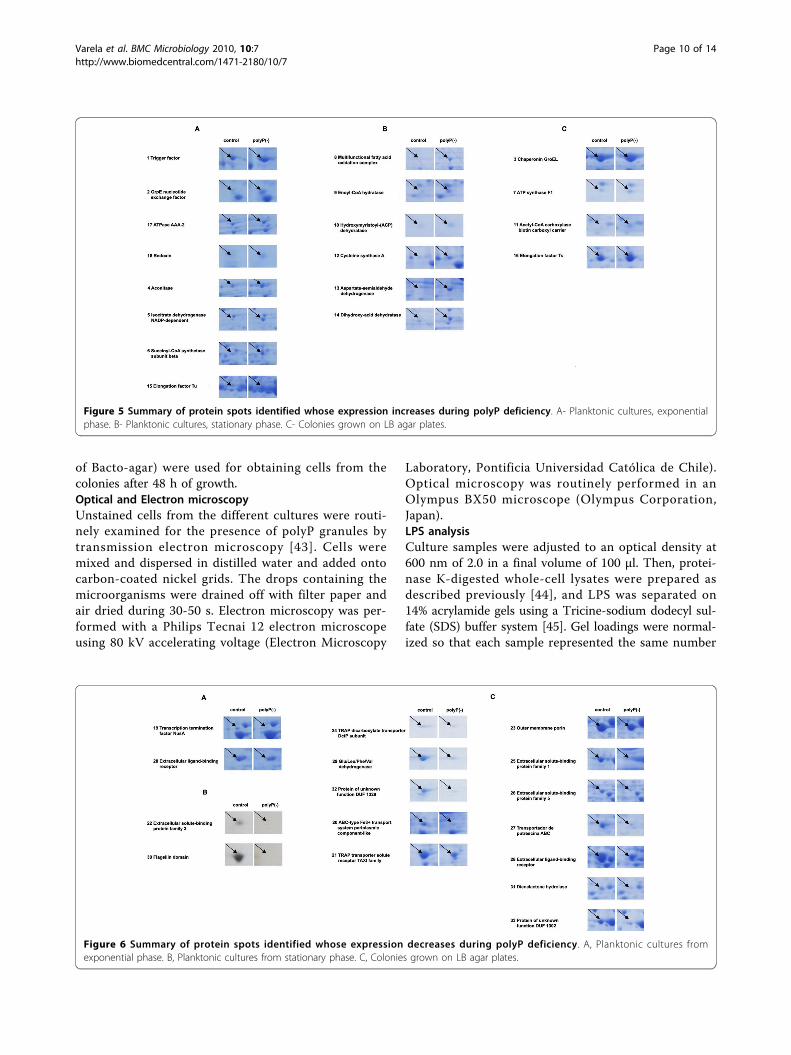
scarcity a general stress state occurred and cells suc-ceeded by overexpressing protein-folding chaperones.Transport proteinsFrom the 17 total proteins identified whose expressionsdecreased during lack of polyP, 10 were identified astransporters. Energy consuming ABC-type transportersresponsible for carrying different solutes such as sugars,peptides, polyamines, amino acids and Fe3+ were identi-fied. Also, C4-dicarboxylates TRAP transporters andouter membrane protein OprE, which has been involvedin virulence process in the genus Pseudomonas [34],were reduced in polyP(-) cells.Other processes and hypothetical proteinsThe present study also yielded some results that appearto be conflicting. We, and others, have demonstratedthat despite the lack of motility of polyP-deficient cells,the flagellum was intact (as seen by using transmissionelectron microscopy). Nevertheless, we found flagellin,the major component of flagella filaments, diminished in
the total and extracelullar proteome of polyP-deficientcells.Finally two protein spots present in the total proteome
matched ORF sequences designated ‘hypothetical’ or‘conserved hypothetical” proteins. These hypotheticalproteins identified here should be subjected to furthercharacterization to confirm their possible role in polyPmetabolism and to ascertain their true biologicalfunction.
Discussion and ConclusionsPolyP has numerous and diverse biological functionsthat have been discovered mainly by studying ppk1mutants in bacteria. A P. aeruginosa PAO1 ppk1 nullmutant exhibits pleiotropic phenotypes includingdecreased virulence, defective in motility, quorum sen-sing, biofilm formation and failure in responses to var-ious stresses [13,15,22]. Many of these features werealso observed in ppk1 mutants of other bacteria such asVibrio cholerae, Salmonella, Shigella and others [35,36].Nevertheless, new functional and structural defects werefound in our recombinant cells that overexpressed theexopolyphosphatase, the enzyme in charge of polyP
Figure 3 PolyP-deficient cells become filamentous during stationary phase of growth. Pseudomonas sp. B4 polyP-deficient and controlcells were grown in LB medium and observed by using phase contrast-optical microscopy (A) and transmission electron microscopy ofunstained cells (B). Magnified view of polyP-deficient cells (C). Arrows indicate the septum.
Varela et al. BMC Microbiology 2010, 10:7http://www.biomedcentral.com/1471-2180/10/7
Page 5 of 14

degradation to Pi. These results might be explained bythe higher extent of polyP depletion when using thisapproach. In the genus Pseudomonas, despite the lack ofdetectable PPK1 activity (<1% of wild type), thesemutants still possess as much as 20% of the wild-typelevels of poly P as is the case of P. aeruginosa PAO1[22]. We previously reported that the overexpression ofexopolyphosphatase removed more than 95% of cellularpolyP [21].The changes observed in the colony morphology are
not surprising taking into account that polyP deficientP. aeruginosa PAO1 cells fails to produce extracellularpolysaccharide [22]. Similar results and an additionalchange in the LPS profile were seen in our polyP-defi-cient cells. Although, the LPS structure of Pseudomonas
sp. B4 is not known in detail it can be speculated thatthe change seen in the LPS could be due to an altera-tion in the phosphate moiety of the LPS core or thatpolyP regulates some enzyme able to modify the LPS.Further experiments should be done to clarify this find-ing but it will be interesting to find out if some of theLPS kinases reported in the genus Pseudomonas (suchas WaaP [37]) could use polyP instead of ATP duringphosphorylation of Heptose I in the inner core of LPS.Furthermore, taking into account the role of LPS duringpathogenesis development in many bacteria, this changemight explain some dysfunction during virulence ofpolyP-deficient bacteria.Bacterial cell division occurs through the formation of
an FtsZ ring (Z ring) at the site of division. The ring is
Figure 4 2D-PAGE gel electrophoresis of polyP-deficient and control Pseudomonas sp. B4 cell. Colonies from Pseudomonas sp. B4 polyP-deficient and control cells were grown in LB medium for 48 h. Samples were prepared and analyzed as described in Methods. The upper panelsshow the separation of proteins in the 5-8 pH range. To have a better resolution of some protein spots a 4.7-5.9 pH range was used (lowerpanels). Numbers with arrows indicate the spot numbers used for MS/MS analyses (Tables 1 and 2).
Varela et al. BMC Microbiology 2010, 10:7http://www.biomedcentral.com/1471-2180/10/7
Page 6 of 14
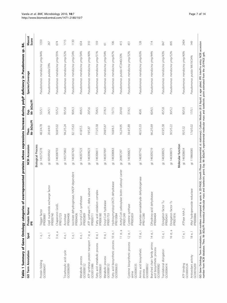
Table1Su
mmaryof
Gen
eOntolog
ycatego
ries
ofov
errepresen
tedproteins
who
seex
pression
sincrea
sedu
ring
polyPde
ficienc
yin
Pseu
domon
assp.B
4.GO
Term
Ann
otation
Spot
ProteinNam
eIPR
NCB
IAccession
Theo
.Mr(kDa)/PI
Exp.
Mr(kDa)/PI
Species/Co
verage
Masco
tScore
Biolog
ical
Process
Proteinfolding
GO:0006457
1e,l
Trigge
rfactor
IPR008881
gi:145575278
48.3/4.78
55/5.1
Pseudo
mon
asmendocina
ymp/44%
1359
2e,l
GrpEnu
cleo
tideexchange
factor
IPR000740
gi:60549562
20.4/4.9
24/5.1
Pseudo
mon
aspu
tida/29%
267
3st,a
Chaperon
inGroEL
IPR012723
gi:146308703
56.8/5.02
55/5.2
Pseudo
mon
asmendocina
ymp/35%
674
Tricarbo
xylic
acid
cycle
GO:0006099
4e,l
Acon
itase
IPR004406
gi:145575802
94.2/5.24
95/5.8
Pseudo
mon
asmendocina
ymp/32%
1715
5e,l
Isocitratede
hydrog
enase,NAD
P-de
pend
ent
IPR004436
gi:146307420
82.1/5.63
90/6.3
Pseudo
mon
asmendocina
ymp/24%
1130
Metabolicprocess
GO:0008152
6e,l
Succinyl-CoA
synthe
tase
IPR005809
gi:146307523
41.8/5.5
49/6.5
Pseudo
mon
asmendocina
ymp/34%
654
ATPsynthe
sisproton
transport
GO:0015986
7st,a
ATPsynthase
F1,d
elta
subu
nit
IPR000711
gi:146309623
19/5.87
20/5.6
Pseudo
mon
asmendocina
ymp/40%
310
Fattyacid
metabolicprocess
GO:0006631
8st,l
Fattyacid
oxidationcomplex
IPR006180
gi:146306611
77.5/5.58
70/6.5
Pseudo
mon
asmendocina
ymp/51%
159
Metabolicprocess
GO:0008152
9st,l
Enoyl-C
oAhydratase
IPR001753
gi:146307097
29.8/5.67
27/6.3
Pseudo
mon
asmendocina
ymp/54%
61
Fattyacid
biosyntheticprocess
GO:0006633
10st,l
Hydroxymyristoyl-(AC
P)de
hydratase
IPR010084
gi:146308063
16.8/6.3
15/7.5
Pseudo
mon
asmendocina
ymp/67%
106
11st,a
Acetyl-CoA
carboxylasebiotin
carboxylcarrier
IPR001249
gi:26987297
16.2/4.95
20/4.8
Pseudo
mon
aspu
tidaKT2440/20%
415
Cysteine
biosyntheticprocess
serin
eGO:0006535
12st,l
Cysteine
synthase
IPR005859
gi:146306821
34.4/5.89
37/6.5
Pseudo
mon
asmendocina
ymp/32%
451
Aminoacid
biosynthetic
process
GO:0008652
13st,l
Aspartate-semialdeh
ydede
hydrog
enase
IPR012280
gi:146307742
40.5/5.33
40/6
Pseudo
mon
asmendocina
ymp/63%
128
Branched
chainfamily
amino
acid
biosyntheticprocess
GO:0009082
14st,l
Dihydroxy-acidde
hydratase
IPR004404
gi:146309219
66.2/5.69
60/6.5
Pseudo
mon
asmendocina
ymp/44%
114
Transla
tionale
long
ation
GO:0006414
15e,l
Elon
gatio
nfactor
TuIPR004541
gi:146308925
43.9/5.38
45/5.8
Pseudo
mon
asmendocina
ymp/43%
847
16st,a
Elon
gatio
nfactor
TsIPR001816
gi:146308073
30.5/5.22
30/5.2
Pseudo
mon
asmendocina
ymp/52%
895
Molecular
functio
n
ATPbind
ing
GO:0005524
17e,l
ATPase
AAA-2
gi:146308654
95/5.32
90/5.9
Pseudo
mon
asmendocina
ymp/40%
2404
Antio
xidant
activity
GO:0016209
18e,l
Alkylh
ydrope
roxide
redu
ctase
IPR000866
gi:119860085
17.6/5.02
17/5.1
Pseudo
mon
aspu
tidaW619/24%
149
GO:G
eneOntolog
yTerm
Ann
otation;
Spot
numbe
rscorrespo
ndto
spotsin
2D-PAGE;
Growth
Phase(e:exp
onen
tial;st:statio
nary);Cu
lture
Med
ium
LB(l:
liquid;
a:ag
arplate);IPR
:InterProen
try;
NCB
Iaccession
numbe
rfrom
NCB
Idatab
ase;
Theo
.Mr(kDa)/PI:theo
retical
molecular
massan
disoe
lectric
point;Exp.
Mr(kDa)/PI,expe
rimen
talm
olecular
massan
disoe
lectric
pointestim
ated
from
the2D
-PAGEge
ls.
Varela et al. BMC Microbiology 2010, 10:7http://www.biomedcentral.com/1471-2180/10/7
Page 7 of 14
composed of the tubulin-like FtsZ protein that hasGTPase activity and the ability to polymerize in vitro(reviewed in [38]). Our observation of cell division fail-ure in polyP-deficient cells during entry into the station-ary phase is in agreement with the finding that duringpolyP-deficiency energy metabolism, and particularlynucleoside triphosphate (NTP) formation, was affected(see below). As seen in Figure 3, the cells were appar-ently able to form the septum, but did not complete theseparation process. It is possible that polyP scarcityaffects the function of FtsZ, since its GTPase activityneeds both, GTP and a bivalent ion. Considering thatpolyP can provide both, phosphate for the generation ofGTP ([16,17] and bivalent metals [35], the absence ofthis biopolymer could block indirectly the polymerisa-tion of Z ring, which would explain the observed pheno-type. Curiously, the enzyme in charge of GTP synthesisfrom polyP in P. aeruginosa (PPK2), was induced 100-times in the stationary phase [16]. In this phase ofgrowth GTP is necessary for the synthesis of alginateand other functions such as cellular division. At present,we cannot discard that other proteins from the divi-some, that also employ GTP for their activity, areaffected by the absence of polyP.Living cells constantly require energy for maintaining
their highly organized structures, synthesize cellularcomponents, generate electric currents, and many otherprocesses. PolyP acts as a reserve for high energy Pi andregulates intracellular ATP in combination with oxida-tive and substrate level phosphorylation. Our proteomicdata support the hypothesis that polyP is an importantcomponent for energy regulation, and particularly inATP regeneration [39]. During polyP deficiency, cellswould prevail by increasing the flux of important energygenerating pathways such as b-oxidation, citric acidcycle and oxidative phosphorylation as proposed in Fig-ure 7. We found eight different proteins related to thesepathways increased during polyP deficiency and in thecase of the TCA cycle enzymes two of them are directlyinvolved in the generating NADH and GTP by theiractivity (see Table 1). Interestingly, a previous linkbetween polyP and the TCA cycle was reported in P.aeruginosa. AlgR2, a global transcriptional factor, posi-tively regulates nucleoside diphosphate kinase (Ndk) andsuccinyl-CoA synthetase, enzymes critical in nucleosidetriphosphate (NTP) formation [40]. Thus, AlgR2 posi-tively regulates the production of alginate, GTP, ppGppand inorganic polyP in P. aeruginosa [41]. It is possiblethen that polyP-deficiency induces AlgR2 expression toincrease GTP and polyP production. This could explainthe increase of succinyl-CoA synthetase in our polyPdeficient cells.Active transport of ion and molecules across the
membrane consumes energy and ATP. We found that
the majority of protein spots decreasing their levels inpolyP(-) cells belong to the transport protein category(see Table 2). It is possible that diminishing energy-con-suming processes such as active transport can help thecells to overcome this polyP deficiency.The defects in the ppk1 mutant described in P. aerugi-
nosa [22,42], and those seen in the same E. coli mutant[10], suggest a failure to respond to a variety of stresses.We found that the levels of many important chaperonesand enzymes related to stress response are increased inpolyP deficient cells. It is suggested that a general stressresponse occurs during polyP deficiency and cells prevailby augmenting the levels of general chaperones andenzymes that would remove reactive oxygen species. Infact, our previous results showed that growth of Pseudo-monas sp. B4 in certain conditions generates an oxida-tive stress and produced a massive increase of polyP[43].Altogether the results presented in this communica-
tion demonstrate the usefulness of proteomics to studythe effect of polyP deficiency in order to generate newhypothesis to clarify its role in bacteria. New suggestionssuch as the possible link between the central metabolicpathways and polyP metabolism proposed here shouldbe the focus of future metabolic flux experiments. Theintegrations of OMICS technologies will definitely helpin elucidating the role of polyP in bacteria and itsimportance during pathogenic process.In summary, polyP has numerous and varied biologi-
cal functions in bacteria that have been discoveredmainly by studying its deficiency. To better understandthe function of polyP we used broad-host-range consti-tutive and regulated vectors to deplete cellular polyPand found new functional and structural changes. Inaddition, it is generally accepted that energy supply ofthe cells could be severely compromised in the absenceof polyP. We confirmed this evidence by using differen-tial proteomic studies and suggested that during polyPscarcity energy metabolism and particularly nucleosidetriphosphate (NTP) formation were affected, generatinga general stress condition. We propose that bacterialcells prevail by increasing the flux of energy-generatingmetabolic pathways such as tricarboxilic acid (TCA)cycle and b-oxidation and by reducing energy-consum-ing ones such as active transporters and amino acidbiosynthesis.
MethodsBacterial strains and growth conditionsPseudomonas sp. B4 wt, control (pMLS7) and polyP-deficient (pS7PPX1) recombinant strains were previouslyobtained [21] and grown aerobically at 37°C on Luria-Bertani (LB) rich medium supplemented with trimetro-pim (50 !g/ml). When required, LB plates (1,5% (w/v)
Varela et al. BMC Microbiology 2010, 10:7http://www.biomedcentral.com/1471-2180/10/7
Page 8 of 14

Table2Su
mmaryof
Gen
eOntolog
ycatego
ries
ofov
errepresen
tedproteins
who
seex
pression
sde
crea
sedu
ring
polyPde
ficienc
yin
Pseu
domon
assp.B
4.GO
Term
Ann
otation
Spot
ProteinNam
eIPR
NCB
IAccession
Theo
.Mr(kDa)/PI
Exp.
Mr(kDa)/PI
Species/Co
verage
Masco
tScore
Biolog
ical
Proc
ess
Regu
latio
nof
transcrip
tion
term
ination
GO:0031554
19e,l
Transcrip
tionterm
ination
factor
NusA
IPR010213
gi:146308624
54.6/4.52
70/5.0
Pseudo
mon
asmendocina
ymp/16%
508
Transport
GO:0006810
20st,a
ABC-type
Fe3+
transport
system
perip
lasm
iccompo
nent-like
IPR011587
gi:146306364
38.1/5.27
38/5.3
Pseudo
mon
asmendocina
ymp/50%
627
21st,a
TRAP
transportersolute
receptor,TAX
Ifam
ilyIPR011852
gi:146309574
33.3/5.74
35/6
Pseudo
mon
asmendocina
ymp/26%
808
22st,l
Extracellularsolute-binding
protein,
family
3IPR001638
gi:146309284
27.6/4.79
27/5
Pseudo
mon
asmendocina
ymp/66%
545
23st,a
Outer
mem
branepo
rinIPR005318
gi:146309320
46.6/6.03
45/5.2
Pseudo
mon
asmendocina
ymp/22%
411
24st,a
TRAP
dicarboxylate
transporter,DctPsubu
nit
IPR004682
gi:146307449
37.6/7.04
35/7.5
Pseudo
mon
asmendocina
ymp/30%
292
25st,a
Extracellularsolute-binding
protein,
family
1IPR006059
gi:146307075
64.8/4.98
60/5
Pseudo
mon
asmendocina
ymp/44%
1080
26st,a
Extracellularsolute-binding
protein,
family
5IPR000914
gi:146305880
59.3/5.72
55/5.3
Pseudo
mon
asmendocina
ymp/16%
354
Polyam
inetransport
GO:0015846
27st,a
Transportado
rde
putrescina
ABC
IPR005893
gi:70730588
42/6.67
40/5.4
Pseudo
mon
asfluorescens
Pf-5/14%
122
Transport
GO:0006810
28 e/l,st/a
Extracellularligand-bind
ing
receptor
IPR001828
gi:146306419
39.4/5.12
40/5.3
Pseudo
mon
asmendocina
ymp/20%
585
Aminoacid
metabolicprocess
GO:0006520
29st,a
Glu/Leu/Phe
/Val
dehydrog
enase
IPR006097
gi:146307897
37.1/5.85
40/7.5
Pseudo
mon
asmendocina
ymp/21%
366
Ciliary
orflage
llarmotility
GO:0001539
30st,l
Flagellin
domain
IPR001492
gi:146307857
49.9/5.04
50/5
Pseudo
mon
asmendocina
ymp/6%
280
Molecular
functio
n
Hydrolase
activity
GO:0016787
31st,a
Diene
lacton
ehydrolase
IPR002925
gi:146307513
27.8/5.45
30/5.3
Pseudo
mon
asmendocina
ymp/24%
411
Unkno
wnfunctio
n32
st,a
Proteinof
unknow
nfunctio
nDUF1329
gi:146308674
51.4/8.3
50/7.8
Pseudo
mon
asmendocina
ymp/50%
1200
33st,a
Proteinof
unknow
nfunctio
nDUF1302
gi:77457132
64.1/5.15
65/4.9
Pseudo
mon
asfluorescens
PfO/13%
340
Cond
ition
san
dab
breviatio
nsarethesameas
thosein
Table1
.
Varela et al. BMC Microbiology 2010, 10:7http://www.biomedcentral.com/1471-2180/10/7
Page 9 of 14
of Bacto-agar) were used for obtaining cells from thecolonies after 48 h of growth.Optical and Electron microscopyUnstained cells from the different cultures were routi-nely examined for the presence of polyP granules bytransmission electron microscopy [43]. Cells weremixed and dispersed in distilled water and added ontocarbon-coated nickel grids. The drops containing themicroorganisms were drained off with filter paper andair dried during 30-50 s. Electron microscopy was per-formed with a Philips Tecnai 12 electron microscopeusing 80 kV accelerating voltage (Electron Microscopy
Laboratory, Pontificia Universidad Católica de Chile).Optical microscopy was routinely performed in anOlympus BX50 microscope (Olympus Corporation,Japan).LPS analysisCulture samples were adjusted to an optical density at600 nm of 2.0 in a final volume of 100 !l. Then, protei-nase K-digested whole-cell lysates were prepared asdescribed previously [44], and LPS was separated on14% acrylamide gels using a Tricine-sodium dodecyl sul-fate (SDS) buffer system [45]. Gel loadings were normal-ized so that each sample represented the same number
Figure 5 Summary of protein spots identified whose expression increases during polyP deficiency. A- Planktonic cultures, exponentialphase. B- Planktonic cultures, stationary phase. C- Colonies grown on LB agar plates.
Figure 6 Summary of protein spots identified whose expression decreases during polyP deficiency. A, Planktonic cultures fromexponential phase. B, Planktonic cultures from stationary phase. C, Colonies grown on LB agar plates.
Varela et al. BMC Microbiology 2010, 10:7http://www.biomedcentral.com/1471-2180/10/7
Page 10 of 14
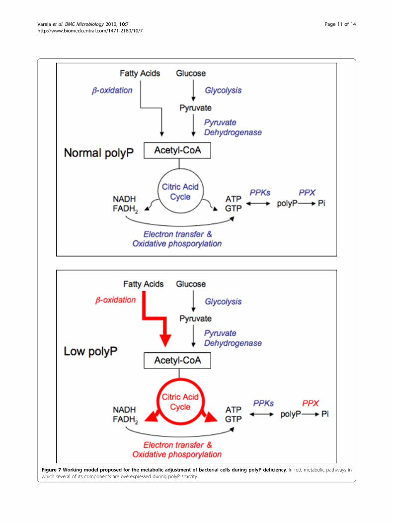
Figure 7 Working model proposed for the metabolic adjustment of bacterial cells during polyP deficiency. In red, metabolic pathways inwhich several of its components are overexpressed during polyP scarcity.
Varela et al. BMC Microbiology 2010, 10:7http://www.biomedcentral.com/1471-2180/10/7
Page 11 of 14
of cells. Gels were silver stained by a modification of theprocedure of Tsai and Frasch [46,47].Samples preparation and 2D-PAGECells (200 mg) were harvested by centrifugatio n (7,000! g for 15 min at 25°C) from liquid cultures or werecollected with an inoculation loop from agar plates. Pel-lets were washed four times in sonication buffer (40mM Tris pH 8.15; 1 mM PMSF). Cells were disruptedby sonication (Misonix XL2020) and the sample wasincubated on ice for 10 min with 50 !g/ml of DNAase.Cell debris was removed by centrifugation and then thesample was washed and concentrated (to half of thetotal volume) by using a Microcon® YM-3 filter unit(10,000 ! g, 4°C). Protein quantitation was performedusing Bio-Rad Protein Assay® system. A total of 500 !gof proteins was precipitated through Ready-Prep 2DCleanup Bio-Rad® kit.Precipitated proteins were resuspended in 300 !L IEF
buffer (7 M urea, 2 M thiourea, 4% CHAPS, 0.0002%bromophenol blue) followed by the addition of DTT to100 mM and 0.2% Bio-Rad ampholytes and the samplemix was incubated for 1 h at 25°C. The entire volumewas loaded in the Protean® IEF focusing tray (17 cm)using the following strips pH ranges: 4.7-5.9/5-8/3-10NL(ReadyStrip™ IPG) that were actively rehydrated at 50 Vfor 12 h. The focusing step was performed at 250 V for15 min; 2,000 V for 2 h; 8,000 V for 4 h and finally10,000 V for 11 h, all the steps at 20°C. Focused pro-teins in the strip were then incubated at 25°C with gen-tle agitation for 15 min in equilibrium buffer (6 M urea;2% SDS; 0,05 M Tris/Cl pH 8.8; 20% glycerol) contain-ing 2% DTT and then 15 min in equilibrium buffer con-taining 2.5% iodoacetamide. Finally, the strip was placedonto a 12.5% polyacrilamide gel for the second dimen-sion in Protean® II (Bio-Rad) system at 50 V for 23 h.The gels were fixed for 1 h (50% ethanol; 2% phosphoricacid), stained for 3 h (0,12% CBB G-250; 10% phospho-ric acid; 10% ammonium sulphate; 20% methanol) andthen washed three times with 15% methanol.Digital images of the gels were analyzed and spots
quantified using Delta2D v.3.6 software. Spot volumewas normalized as a percentage of the total volume ofall spots on the corresponding gel and also manuallyconfirmed. The threshold for accepting a meaningfulvariation was a factor of 2.0 (p < 0,05). A total of 81proteins spots showing differences in the expression pat-tern between control and polyP(-) strains (three inde-pendent replicates) were selected for further MSanalysis.In-gel protein digestion and sample preparationSpots of interest from Coomassie blue-stained 2D gelswere excised manually, deposited in 96-well plates andprocessed automatically in a Proteineer DP (Bruker Dal-tonics, Bremen, Germany). The digestion protocol used
was based on Schevchenko et al. [48] with minor varia-tions: gel plugs were washed firstly with 50 mM ammo-nium bicarbonate and secondly with acetonitrile (ACN)prior to reduction with 10 mM DTT in 25 mM ammo-nium bicarbonate solution, and alkylation was carriedout with 55 mM IAA in 50 mM ammonium bicarbonatesolution. Gel pieces were then rinsed with 50 mMammonium bicarbonate and with ACN, and then driedunder a stream of nitrogen. Modified porcine trypsin(sequencing grade; Promega, Madison WI) was added ata final concentration of 16 ng/!l in 25% ACN/50 mMammonium bicarbonate solution and the digestion tookplace at 37°C for 6 h. The reaction was stopped by add-ing 0.5% TFA for peptide extraction. Tryptic peptideswere dried by speed-vacuum centrifugation and resus-pended in 4 !l of MALDI solution. A 0.8 !l aliquot ofeach peptide mixture was deposited onto a 386-wellOptiTOF™ Plate (Applied Biosystems, Framingham, MA,USA) and allowed to dry at room temperature. A 0.8 !laliquot of matrix solution (3 mg/mL CHCA in MALDIsolution) was then added onto dried digest and allowedto dry at room temperature.MALDI peptide mass fingerprinting, MS/MS analysis anddatabase searchingFor MALDI-TOF/TOF analysis, samples were automati-cally acquired in an ABi 4800 MALDI TOF/TOF massspectrometer (Applied Biosystems, Framingham, MA,USA) in positive ion reflector mode (ion accelerationvoltage was 25 kV for MS acquisition and 1 kV forMSMS) and the spectra were stored into the ABi 4000Series Explorer Spot Set Manager. PMF and MSMSfragment ion spectra were smoothed and corrected tozero baseline using routines embedded in ABi 4000 Ser-ies Explorer Software v3.6. Each PMF spectrum wasinternally calibrated with the mass signals of trypsinautolysis ions to reach a typical mass measurementaccuracy of <25 ppm. Known trypsin and keratin masssignals, as well as potential sodium and potassiumadducts (+21 Da and +39 Da) were removed from thepeak list. To submit the combined PMF and MS/MSdata to MASCOT software v.2.1 (Matrix Science, Lon-don, UK), GPS Explorer v4.9 was used, searching in thenon-redundant NCBI protein database.LC-ESI MS/MS analysisIn some specific cases, alternative proteomic techniqueswere employed to confirm and improve protein identifi-cations. For this purpose, we made use of liquid chro-matography coupled to electrospray ion-trap massspectrometry tandem MS (LC ESI-MS/MS). This wasdone using an Ultimate 3000 nano LC (Dionex, Amster-dam, The Netherland) and a 75 micrometer I.D, 100mm reversed-phase column, at a 300 nL/min flow,coupled to a Bruker HCT Ultra ion-trap mass
Varela et al. BMC Microbiology 2010, 10:7http://www.biomedcentral.com/1471-2180/10/7
Page 12 of 14
spectrometer (Bruker Daltonics, Bremen, Germany)working in dynamic exclusion mode.Database SearchFor protein identification, LC ESI MS/MS spectra weretransferred to BioTools 2.0 interface (Bruker Daltonics)to search in the NCBInr database using a licensed ver-sion of Mascot v.2.2.04 search engine (http://www.matrixscience.com; Matrix Science, London, UK). Searchparameters were set as follows: carbamidomethyl cysteinas fixed modification by the treatment with iodoaceta-mide, oxidized methionines as variable modification,peptide mass tolerance of 0.5 Da for the parental massand fragment masses and 1 missed cleavage site. In allprotein identifications, the probability Mowse scoreswere greater than the minimum score fixed as signifi-cant with a p-value minor than 0.05. Selected proteinswere based on that who exhibited higher Mascot scoreand sequence coverage. A total of thirty-three differentproteins showing differential expression pattern betweenpolyP+ and polyP- strains (three independent replicates)were selected. Furthermore, theoretical isoelectric pointsand molecular masses were compared to experimentalvalues of thirty-three proteins were grouped by geneontology (GO) categories [49].
AcknowledgementsThis research was supported by Grants 1070986 and 11070180 fromFondecyt and ICM P05-001-F from MIDEPLAN.
Author details1Laboratory of Molecular Microbiology and Biotechnology MillenniumInstitute of Cell Dynamics and Biotechnology (ICDB), Department of Biology,Faculty of Sciences, University of Chile, Las Palmeras 3425, Ñuñoa, Santiago,Chile. 2Servicio de Proteómica, Centro Nacional de Biotecnología. CSIC.Darwin 3, 28049, Madrid, España.
Authors’ contributionsFPC y CAJ conceived and designed the study; FPC performed someexperiments and wrote the manuscript. CV performed proteomicexperiments. CM carried out cellular experiments. AP y JPA carried out MS/MS protein identification. CAJ participated in coordination and criticalevaluation of the manuscript. All authors read and approved the finalmanuscript.
Received: 10 August 2009Accepted: 12 January 2010 Published: 12 January 2010
References1. Brown M, Kornberg A: The long and short of it - polyphosphate, PPK and
bacterial survival. Trends Biochem Sci 2008, 33(6):284-290.2. Kornberg A: Inorganic polyphosphate: a molecule of many functions.
Prog Mol Subcell Biol 1999, 23:1-18.3. Blum J: Changes in orthophosphate, pyrophosphate and long-chain
polyphosphate levels in Leishmania major promastigotes incubated withand without glucose. J Protozool 1989, 36(3):254-257.
4. Kuroda A, Tanaka S, Ikeda T, Kato J, Takiguchi N, Ohtake H: Inorganicpolyphosphate kinase is required to stimulate protein degradation andfor adaptation to amino acid starvation in Escherichia coli. Proc Natl AcadSci USA 1999, 96(25):14264-14269.
5. Kuroda A, Nomura K, Ohtomo R, Kato J, Ikeda T, Takiguchi N, Ohtake H,Kornberg A: Role of inorganic polyphosphate in promoting ribosomal
protein degradation by the Lon protease in **E. coli. Science 2001,293(5530):705-708.
6. Reusch R: Polyphosphate/poly-(R)-3-hydroxybutyrate) ion channels in cellmembranes. Prog Mol Subcell Biol 1999, 23:151-182.
7. Reusch R: Transmembrane ion transport by polyphosphate/poly-(R)-3-hydroxybutyrate complexes. Biochemistry (Mosc) 2000, 65(3):280-295.
8. Crooke E, Akiyama M, Rao N, Kornberg A: Genetically altered levels ofinorganic polyphosphate in Escherichia coli. J Biol Chem 1994,269(9):6290-6295.
9. Kim K, Rao N, Fraley C, Kornberg A: Inorganic polyphosphate is essentialfor long-term survival and virulence factors in Shigella and Salmonellaspp. Proc Natl Acad Sci USA 2002, 99(11):7675-7680.
10. Rao N, Kornberg A: Inorganic polyphosphate supports resistance andsurvival of stationary-phase Escherichia coli. J Bacteriol 1996, 178(5):1394-1400.
11. Rao N, Liu S, Kornberg A: Inorganic polyphosphate in Escherichia coli: thephosphate regulon and the stringent response. J Bacteriol 1998,180(8):2186-2193.
12. Rao N, Kornberg A: Inorganic polyphosphate regulates responses ofEscherichia coli to nutritional stringencies, environmental stresses andsurvival in the stationary phase. Prog Mol Subcell Biol 1999, 23:183-195.
13. Rashid M, Kornberg A: Inorganic polyphosphate is needed for swimming,swarming, and twitching motilities of Pseudomonas aeruginosa. Proc NatlAcad Sci USA 2000, 97(9):4885-4890.
14. Rashid M, Rao N, Kornberg A: Inorganic polyphosphate is required formotility of bacterial pathogens. J Bacteriol 2000, 182(1):225-227.
15. Rashid M, Rumbaugh K, Passador L, Davies D, Hamood A, Iglewski B,Kornberg A: Polyphosphate kinase is essential for biofilm development,quorum sensing, and virulence of Pseudomonas aeruginosa. Proc NatlAcad Sci USA 2000, 97(17):9636-9641.
16. Ishige K, Zhang H, Kornberg A: Polyphosphate kinase (PPK2), a potent,polyphosphate-driven generator of GTP. Proc Natl Acad Sci USA 2002,99(26):16684-16688.
17. Zhang H, Ishige K, Kornberg A: A polyphosphate kinase (PPK2) widelyconserved in bacteria. Proc Natl Acad Sci USA 2002, 99(26):16678-16683.
18. Seufferheld M, Alvarez H, Farias M: Role of polyphosphates in microbialadaptation to extreme environments. Appl Environ Microbiol 2008,74(19):5867-5874.
19. Kell D: Metabolomics and systems biology: making sense of the soup.Curr Opin Microbiol 2004, 7(3):296-307.
20. Joyce A, Palsson B: The model organism as a system: integrating ‘omics’data sets. Nat Rev Mol Cell Biol 2006, 7(3):198-210.
21. Chávez F, Mauriaca C, Jerez C: Constitutive and regulated expressionvectors to construct polyphosphate deficient bacteria. BMC Res Notes2009, 2(1):50.
22. Fraley C, Rashid M, Lee S, Gottschalk R, Harrison J, Wood P, Brown M,Kornberg A: A polyphosphate kinase 1 (ppk1) mutant of Pseudomonasaeruginosa exhibits multiple ultrastructural and functional defects. ProcNatl Acad Sci USA 2007, 104(9):3526-3531.
23. Nakanishi-Matsui M, Kashiwagi S, Ubukata T, Iwamoto-Kihara A, Wada Y,Futai M: Rotational catalysis of Escherichia coli ATP synthase F1 sector.Stochastic fluctuation and a key domain of the beta subunit. J Biol Chem2007, 282(28):20698-20704.
24. Aldor I, Keasling J: Process design for microbial plastic factories:metabolic engineering of polyhydroxyalkanoates. Curr Opin Biotechnol2003, 14(5):475-483.
25. Wilmes P, Wexler M, Bond P: Metaproteomics provides functional insightinto activated sludge wastewater treatment. PLoS ONE 2008, 3(3):e1778.
26. Deuerling E, Bukau B: Chaperone-assisted folding of newly synthesizedproteins in the cytosol. Crit Rev Biochem Mol Biol 39(5-6):261-277.
27. Lee S, Choi J, Tsai F: Visualizing the ATPase cycle in a proteindisaggregating machine: structural basis for substrate binding by ClpB.Mol Cell 2007, 25(2):261-271.
28. Merz F, Boehringer D, Schaffitzel C, Preissler S, Hoffmann A, Maier T,Rutkowska A, Lozza J, Ban N, Bukau B, et al: Molecular mechanism andstructure of Trigger Factor bound to the translating ribosome. EMBO J2008, 27(11):1622-1632.
29. Parsell D, Kowal A, Singer M, Lindquist S: Protein disaggregation mediatedby heat-shock protein Hsp104. Nature 1994, 372(6505):475-478.
Varela et al. BMC Microbiology 2010, 10:7http://www.biomedcentral.com/1471-2180/10/7
Page 13 of 14
30. Nishiyama Y, Yamamoto H, Allakhverdiev S, Inaba M, Yokota A, Murata N:Oxidative stress inhibits the repair of photodamage to thephotosynthetic machinery. EMBO J 2001, 20(20):5587-5594.
31. Seib K, Wu H, Kidd S, Apicella M, Jennings M, McEwan A: Defenses againstoxidative stress in Neisseria gonorrhoeae: a system tailored for achallenging environment. Microbiol Mol Biol Rev 2006, 70(2):344-361.
32. Sharma S, Sundaram C, Luthra P, Singh Y, Sirdeshmukh R, Gade W: Role ofproteins in resistance mechanism of Pseudomonas fluorescens againstheavy metal induced stress with proteomics approach. J Biotechnol 2006,126(3):374-382.
33. McInerney P, Mizutani T, Shiba T: Inorganic polyphosphate interacts withribosomes and promotes translation fidelity in vitro and in vivo. MolMicrobiol 2006, 60(2):438-447.
34. Jaouen T, Coquet L, Marvin-Guy L, Orange N, Chevalier S, Dé E: Functionalcharacterization of Pseudomonas fluorescens OprE and OprQ membraneproteins. Biochem Biophys Res Commun 2006, 346(3):1048-1052.
35. Kornberg A: Inorganic polyphosphate: toward making a forgottenpolymer unforgettable. J Bacteriol 1995, 177(3):491-496.
36. Kornberg A, Rao N, Ault-Riché D: Inorganic polyphosphate: a molecule ofmany functions. Annu Rev Biochem 1999, 68:89-125.
37. Zhao X, Lam J: WaaP of Pseudomonas aeruginosa is a novel eukaryotictype protein-tyrosine kinase as well as a sugar kinase essential for thebiosynthesis of core lipopolysaccharide. J Biol Chem 2002, 277(7):4722-4730.
38. Lutkenhaus J, Addinall S: Bacterial cell division and the Z ring. Annu RevBiochem 1997, 66:93-116.
39. Harold F: Inorganic polyphosphates in biology: structure, metabolism,and function. Bacteriol Rev 1966, 30(4):772-794.
40. Ledgham F, Soscia C, Chakrabarty A, Lazdunski A, Foglino M: Globalregulation in Pseudomonas aeruginosa: the regulatory protein AlgR2(AlgQ) acts as a modulator of quorum sensing. Res Microbiol 2003,154(3):207-213.
41. Kim H, Schlictman D, Shankar S, Xie Z, Chakrabarty A, Kornberg A: Alginate,inorganic polyphosphate, GTP and ppGpp synthesis co-regulated inPseudomonas aeruginosa: implications for stationary phase survival andsynthesis of RNA/DNA precursors. Mol Microbiol 1998, 27(4):717-725.
42. Parks Q, Hobden J: Polyphosphate kinase 1 and the ocular virulence ofPseudomonas aeruginosa. Invest Ophthalmol Vis Sci 2005, 46(1):248-251.
43. Chávez F, Lünsdorf H, Jerez C: Growth of polychlorinated-biphenyl-degrading bacteria in the presence of biphenyl and chlorobiphenylsgenerates oxidative stress and massive accumulation of inorganicpolyphosphate. Appl Environ Microbiol 2004, 70(5):3064-3072.
44. Hitchcock P, Brown T: Morphological heterogeneity among Salmonellalipopolysaccharide chemotypes in silver-stained polyacrylamide gels. JBacteriol 1983, 154(1):269-277.
45. Lesse A, Campagnari A, Bittner W, Apicella M: Increased resolution oflipopolysaccharides and lipooligosaccharides utilizing tricine-sodiumdodecyl sulfate-polyacrylamide gel electrophoresis. J Immunol Methods1990, 126(1):109-117.
46. Tsai C, Frasch C: A sensitive silver stain for detecting lipopolysaccharidesin polyacrylamide gels. Anal Biochem 1982, 119(1):115-119.
47. Marolda C, Lahiry P, Vinés E, Saldías S, Valvano M: Micromethods for thecharacterization of lipid A-core and O-antigen lipopolysaccharide.Methods Mol Biol 2006, 347:237-252.
48. Shevchenko A, Wilm M, Vorm O, Mann M: Mass spectrometric sequencingof proteins silver-stained polyacrylamide gels. Anal Chem 1996, 68(5):850-858.
49. Ashburner M, Ball C, Blake J, Botstein D, Butler H, Cherry J, Davis A,Dolinski K, Dwight S, Eppig J, et al: Gene ontology: tool for the unificationof biology. The Gene Ontology Consortium. Nat Genet 2000, 25(1):25-29.
doi:10.1186/1471-2180-10-7Cite this article as: Varela et al.: New structural and functional defects inpolyphosphate deficient bacteria: A cellular and proteomic study. BMCMicrobiology 2010 10:7.
Publish with BioMed Central and every scientist can read your work free of charge
"BioMed Central will be the most significant development for disseminating the results of biomedical research in our lifetime."
Sir Paul Nurse, Cancer Research UK
Your research papers will be:available free of charge to the entire biomedical community
peer reviewed and published immediately upon acceptance
cited in PubMed and archived on PubMed Central
yours — you keep the copyright
Submit your manuscript here:http://www.biomedcentral.com/info/publishing_adv.asp
BioMedcentral
Varela et al. BMC Microbiology 2010, 10:7http://www.biomedcentral.com/1471-2180/10/7
Page 14 of 14