BMC Immunology
19
BioMed Central Page 1 of 19 (page number not for citation purposes) BMC Immunology Open Access Research article Gut health immunomodulatory and anti-inflammatory functions of gut enzyme digested high protein micro-nutrient dietary supplement-Enprocal Jagat R Kanwar* and Rupinder K Kanwar Address: BioDeakin, Institute for Technology & Research Innovation (ITRI), Deakin University, Geelong Technology Precinct (GTP), Pigdons Road, Waurn Ponds, Geelong, Victoria 3217, Australia Email: Jagat R Kanwar* - [email protected]; Rupinder K Kanwar - [email protected] * Corresponding author Abstract Background: Enprocal is a high-protein micro-nutrient rich formulated supplementary food designed to meet the nutritional needs of the frail elderly and be delivered to them in every day foods. We studied the potential of Enprocal to improve gut and immune health using simple and robust bioassays for gut cell proliferation, intestinal integrity/permeability, immunomodulatory, anti-inflammatory and anti-oxidative activities. Effects of Enprocal were compared with whey protein concentrate 80 (WPC), heat treated skim milk powder, and other commercially available milk derived products. Results: Enprocal (undigested) and digested (Enprocal D) selectively enhanced cell proliferation in normal human intestinal epithelial cells (FHs74-Int) and showed no cytotoxicity. In a dose dependent manner Enprocal induced cell death in Caco-2 cells (human colon adencarcinoma epithelial cells). Digested Enprocal (Enprocal D: gut enzyme cocktail treated) maintained the intestinal integrity in transepithelial resistance (TEER) assay, increased the permeability of horseradish peroxidase (HRP) and did not induce oxidative stress to the gut epithelial cells. Enprocal D upregulated the surface expression of co-stimulatory (CD40, CD86, CD80), MHC I and MHC II molecules on PMA differentiated THP-1 macrophages in coculture transwell model, and inhibited the monocyte/lymphocyte (THP-1/Jurkat E6-1 cells)-epithelial cell adhesion. In cytokine secretion analyses, Enprocal D down-regulated the secretion of proinflammatory cytokines (IL-1β and TNF-α) and up-regulated IFN-γ, IL-2 and IL-10. Conclusion: Our results indicate that Enprocal creates neither oxidative injury nor cytotoxicity, stimulates normal gut cell proliferation, up regulates immune cell activation markers and may aid in the production of antibodies. Furthermore, through downregulation of proinflammatory cytokines, Enprocal appears to be beneficial in reducing the effects of chronic gut inflammatory diseases such as inflammatory bowel disease (IBD). Stimulation of normal human fetal intestinal cell proliferation without cell cytotoxicity indicates it may also be given as infant food particularly for premature babies. Published: 31 January 2009 BMC Immunology 2009, 10:7 doi:10.1186/1471-2172-10-7 Received: 22 July 2008 Accepted: 31 January 2009 This article is available from: http://www.biomedcentral.com/1471-2172/10/7 © 2009 Kanwar and Kanwar; licensee BioMed Central Ltd. This is an Open Access article distributed under the terms of the Creative Commons Attribution License (http://creativecommons.org/licenses/by/2.0 ), which permits unrestricted use, distribution, and reproduction in any medium, provided the original work is properly cited.
-
Upload
khangminh22 -
Category
Documents
-
view
0 -
download
0
Transcript of BMC Immunology
BioMed CentralBMC Immunology
ss
Open AcceResearch articleGut health immunomodulatory and anti-inflammatory functions of gut enzyme digested high protein micro-nutrient dietary supplement-EnprocalJagat R Kanwar* and Rupinder K KanwarAddress: BioDeakin, Institute for Technology & Research Innovation (ITRI), Deakin University, Geelong Technology Precinct (GTP), Pigdons Road, Waurn Ponds, Geelong, Victoria 3217, Australia
Email: Jagat R Kanwar* - [email protected]; Rupinder K Kanwar - [email protected]
* Corresponding author
AbstractBackground: Enprocal is a high-protein micro-nutrient rich formulated supplementary fooddesigned to meet the nutritional needs of the frail elderly and be delivered to them in every dayfoods. We studied the potential of Enprocal to improve gut and immune health using simple androbust bioassays for gut cell proliferation, intestinal integrity/permeability, immunomodulatory,anti-inflammatory and anti-oxidative activities. Effects of Enprocal were compared with wheyprotein concentrate 80 (WPC), heat treated skim milk powder, and other commercially availablemilk derived products.
Results: Enprocal (undigested) and digested (Enprocal D) selectively enhanced cell proliferation innormal human intestinal epithelial cells (FHs74-Int) and showed no cytotoxicity. In a dosedependent manner Enprocal induced cell death in Caco-2 cells (human colon adencarcinomaepithelial cells). Digested Enprocal (Enprocal D: gut enzyme cocktail treated) maintained theintestinal integrity in transepithelial resistance (TEER) assay, increased the permeability ofhorseradish peroxidase (HRP) and did not induce oxidative stress to the gut epithelial cells.Enprocal D upregulated the surface expression of co-stimulatory (CD40, CD86, CD80), MHC Iand MHC II molecules on PMA differentiated THP-1 macrophages in coculture transwell model,and inhibited the monocyte/lymphocyte (THP-1/Jurkat E6-1 cells)-epithelial cell adhesion. Incytokine secretion analyses, Enprocal D down-regulated the secretion of proinflammatorycytokines (IL-1β and TNF-α) and up-regulated IFN-γ, IL-2 and IL-10.
Conclusion: Our results indicate that Enprocal creates neither oxidative injury nor cytotoxicity,stimulates normal gut cell proliferation, up regulates immune cell activation markers and may aid inthe production of antibodies. Furthermore, through downregulation of proinflammatory cytokines,Enprocal appears to be beneficial in reducing the effects of chronic gut inflammatory diseases suchas inflammatory bowel disease (IBD). Stimulation of normal human fetal intestinal cell proliferationwithout cell cytotoxicity indicates it may also be given as infant food particularly for prematurebabies.
Published: 31 January 2009
BMC Immunology 2009, 10:7 doi:10.1186/1471-2172-10-7
Received: 22 July 2008Accepted: 31 January 2009
This article is available from: http://www.biomedcentral.com/1471-2172/10/7
© 2009 Kanwar and Kanwar; licensee BioMed Central Ltd. This is an Open Access article distributed under the terms of the Creative Commons Attribution License (http://creativecommons.org/licenses/by/2.0), which permits unrestricted use, distribution, and reproduction in any medium, provided the original work is properly cited.
Page 1 of 19(page number not for citation purposes)
BMC Immunology 2009, 10:7 http://www.biomedcentral.com/1471-2172/10/7
BackgroundThe ability to eat sufficient amounts orally is inverselyassociated with the extent of frailty and studies have indi-cated that nutritional intake is often compromised in theelderly [1]. Poor gut health and secondary health issues asa consequence are matters of nutritional attention for theelderly, infants and immune compromised persons. Dietis a factor that can be extremely varied in terms of quan-tity, quality, frequency and duration of meals in individu-als, but that can also be modified with targeted dietaryinterventions to produce the best results for any individ-ual [2]. Diets of the elderly are generally low in protein,low in energy and deficient in a range of micro-nutrients[1,3,4]. High protein diets, in particular those based onwhey proteins, are being marketed to improve the healthstatus of the elderly [4] as well as other target groups e.g.athletes and babies. Furthermore micro-nutrients includ-ing anti-oxidants e.g. Vitamins A, C and zinc are alsobeing used for fortification or as dietary supplements forhealthy older patients [5]. There are growing number ofcompositional foods under development, being patentedor emerging in the market that target specific nutritionaldeficiencies beyond the traditional infant formula market.Recent examples include WO2007/070754 a high proteindietary supplement for treating various diseases associ-ated with protein deficiency (Novalife); and WO2007/022991 compositions for enhancing vascular integrity,cellular survival and reducing apoptosis in ischema(Nestec).
Enprocal, a recently formulated supplementary food isdesigned to meet the nutritional needs of the frail elderlyand to be delivered to them in common every day foods.Enprocal's formulation was developed by specialised agedcare dieticians and a food scientist experienced in powderbased food supplements. The ingredients of Enprocalsuch as dairy-based proteins (whey protein concentrate,skim milk powder, whole milk powder), vitamins andminerals (including calcium, zinc and vitamins C, D, Bgroup and A), vegetable oils and inulin (fibre) wereselected to meet the nutritional profile of elderly specifiedthough dietetic input (Table 1). With whey protein con-centrate (WPC) (80% protein) as its key ingredient it pro-vides i) the highest-quality protein (as measured byprotein efficiency ratio (PER), ii) rapid protein digestion,iii) essential amino acids, in particular cysteine, threonineand leucine that may be required in increased levels in theelderly [6,7], iv) cysteine to improve glutathione statusand muscle protein synthesis, v) high concentrations ofbranched chain amino acids, and vi) minor bioactivecomponents such as immuno-globulins, growth factors,and anti-microbial proteins and peptides to assist in effec-tive gut function [6,8-12]. Today, whey is a popular die-tary protein supplement purported to provideantimicrobial activity, immune modulation, improved
muscle strength and body composition, and to preventcardiovascular disease and osteoporosis [reviewed in[8,11,12]]. Enprocal with WPC as its key ingredient there-fore contains high levels of constituent proteins includingbeta-lactoglobulin, alpha-lactalbumin, lactoferrin, lac-toperoxidase and glycomacropeptide which have beendemonstrated a range of immune-enhancing properties[11,12]. In addition, whey has the ability to act as an anti-oxidant, antihypertensive, antitumor, hypolipidemic,antiviral, antibacterial, and chelating agent [6,8,11,12].Whey proteins have been reported to enhance digestionand gut function [8,11-13] as well as glutathione produc-tion and immune function, hence increasing dietary avail-ability may promote general health in a variety of ways[14,15].
Developing and maintaining a healthy intestinal tract is apre-requisite for general health and aiding disease preven-tion. The gut epithelial cells lining of the intestinal lumen
Table 1: Composition of Enprocal.
Ingredients Composition per 100 g
Whey protein concentrate 41.4 g
Vegetable fibre (inulin) 10.2 g
Vegetable oil 8.2 g
Vitamin A 548 μg
Thiamin (Vitamin B1) 0.8 mg
Riboflavin (Vitamin B2) 1.9 mg
Folate 173 μg
Vitamin B6 1.0 mg
Vitamin B12 2.9 μg
Vitamin C 57 mg
Vitamin D 9 μg
Vitamin E 4.2 mg
Calcium 1142 mg
Iron 3.9 mg
Magnesium 232 mg
Phosphorus 780 mg
Zinc 10.7 mg
Page 2 of 19(page number not for citation purposes)

BMC Immunology 2009, 10:7 http://www.biomedcentral.com/1471-2172/10/7
are the first point of contact between intestinal contentsand the rest of the body. It is at this interface that nutri-tion, environment and genetics come together to deter-mine gut health. The intestinal mucosa is lined by amonolayer of intestinal epithelial cells joined together attheir apical poles by tight junctions, and supported by anextensive and complex immune system and transcellularpathways for nutrient absorption [16]. The mucosal sur-face of the gastrointestinal tract forms a barrier that sepa-rates the luminal contents from the effector immune cellsunderneath. With the emergence of the concept of immu-nophysiology, it is clear that the function of the intestinalepithelium is affected by many immune cell types [17].Any abnormal activation of such immune cells (macro-phages) overproduces the inflammatory cytokines e.g.,tumor necrosis factor (TNF)-α, interleukin (IL)-1β and IL-6 that cause destructive cell damage to the intestinal epi-thelial monolayers and subsequent mucosal inflamma-tion leading to inflammatory bowel disease (IBD) such asCrohn's disease (CD) and ulcerative colitis (UC) [18,19].Cellular injury from reactive oxygen species (ROS) isimplicated in a wide variety of gut diseases and pathologicconditions [20]. There is a growing body of evidence thatoxidative stress may contribute to gut inflammation,ulcers, inflammatory bowel disease, and leaky gut syn-drome. Newborn and the elderly are more prone to oxi-dant injury [20-22].
The present study was aimed to assess the potential ofEnprocal to improve gut and immune health functions.We investigated specifically whether Enprocal, after diges-tion with a cocktail of gut enzymes maintained gut cellproliferation and mucosal integrity, possessed immu-nomodulatory (monocyte/macrophage and T cell activa-tion and cytokine production), anti-inflammatory(immune-gut cell adhesion) and anti-oxidative proper-ties. We sought to achieve conditions as close as possibleto those of the human digestive tract by employing invitro, most widely used human gut epithelial cells alone(Caco-2 and FHS Int 74) or in coculture with humanimmune THP-1 (monocytes) and Jurkat (T lymphocytes)cells in transwell system.
FHs 74 Int and Caco-2 cell lines used worldwide by theresearchers were employed as the models of human intes-tinal epithelial cells in the study. FHs 74-Int cells, used inthe study were although derived from normal human fetalintestine but have been reported to show mature epithe-lial-like characteristics [23]. The growth-promoting activi-ties of EGF, TGF-α IGF-I and insulin were examined usingthe same intestinal cells [24]. Caco-2 cells (derived fromhuman colon adenocarcinoma) have gained enormouspopularity as a reliable and high-throughput in vitromodel system for the study of human intestinal permea-bility and evaluation of a large number of drug candidates
for their intestinal absorption [25,26]. For orally adminis-tered compounds, permeability through Caco-2 cell mon-olayers correlates well with human in vivo absorption [26-28]. Caco-2 monolayers can display electrical propertiestypical of either small intestinal or colonic enterocytes[27]. Caco-2 cells are able to differentiate spontaneouslyin culture and display a small intestine-like (villus) phe-notype. The differentiated cells polarise, form microvilliand secrete enzymes associated with the enterocyte brushborder such as sucrase-isomaltase, alkaline phosphatase,alkaline phosphatase, lactase and aminopeptidase [29].This cell line thus represents an appropriate model for thestudy of transport mechanisms related to the intestinalbarrier and for investigating diverse problems of nutrientsbioavailability and absorption without concern for differ-ences between human and animals [30].
Our study, provides the first report on the bioactivity ofdigested Enprocal components which may be releasedthrough enzymatic action from its key ingredients such aswhey protein concentrate for gut epithelial cell growth,integrity, absorptive, immunomodulatory, anti-inflam-matory and anti-oxidative properties.
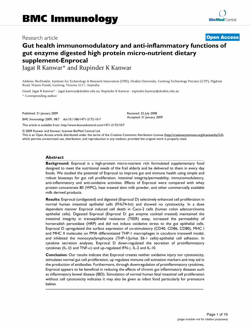
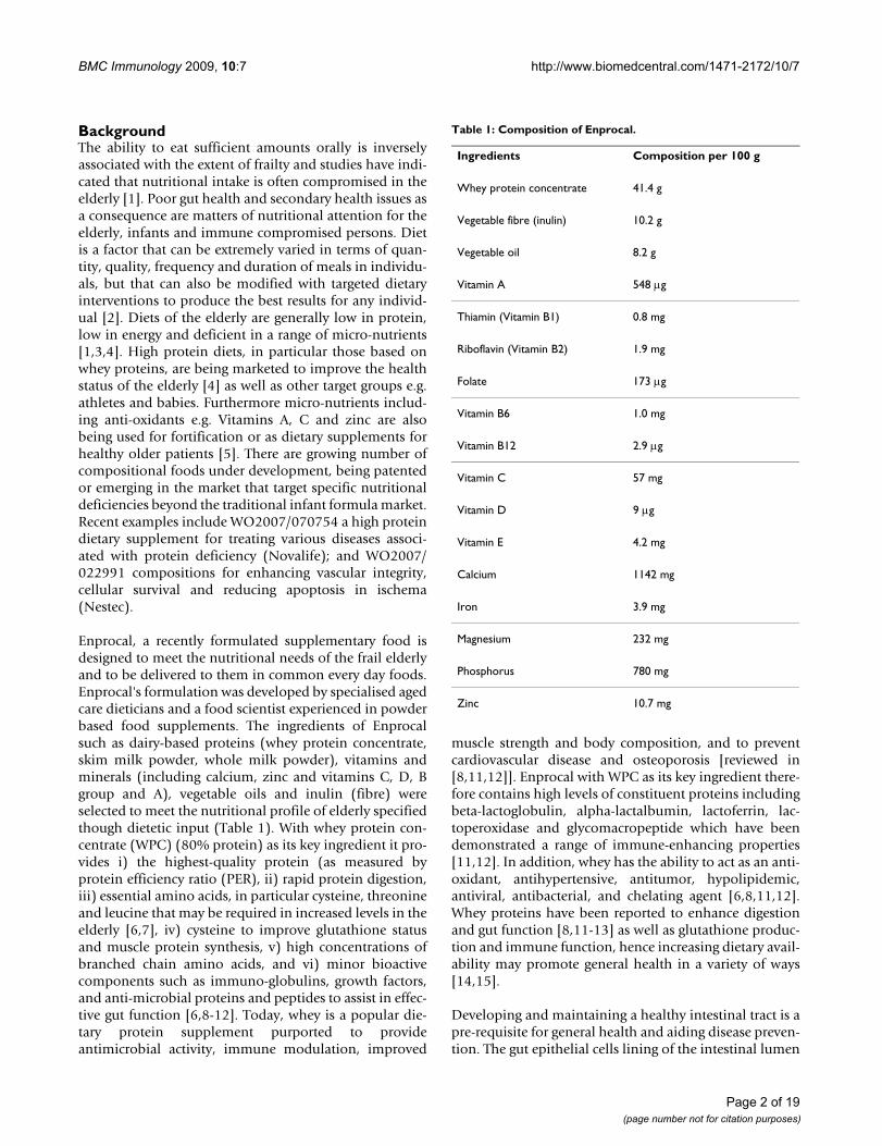
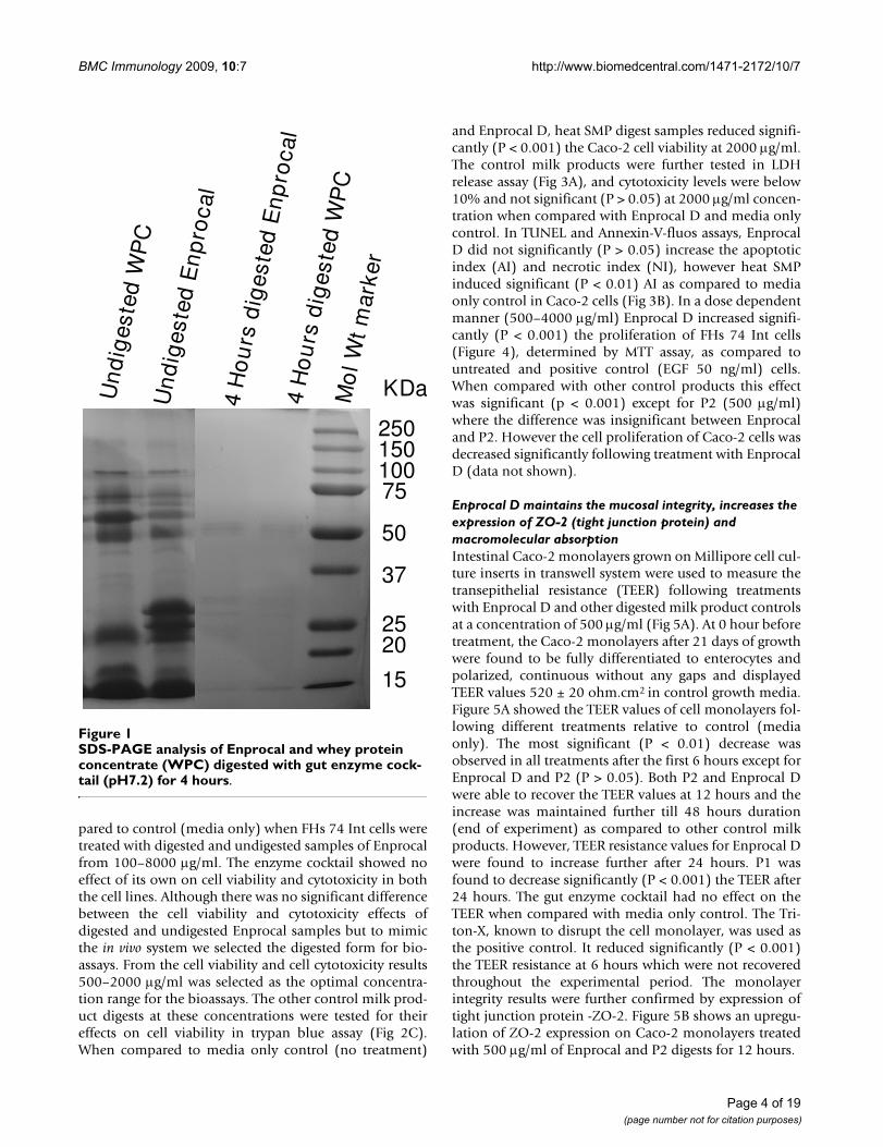
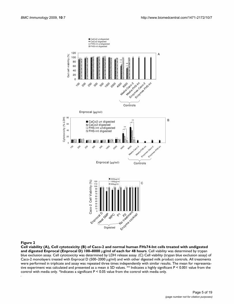
ResultsDigested Enprocal (Enprocal D) maintains cell growth, enhances cell proliferation and shows no cytotoxicity in normal human intestinal epithelial cellsNear complete digestion of Enprocal and other digestedmilk product controls was achieved after 4 hours incuba-tion with gut enzyme cocktail mixture (Fig 1). In our pre-liminary experiments we treated Enprocal with the gutenzyme cocktail at different enzyme cocktail: proteinratios at pH 7.0–7.4. A 1:50 ratio of enzyme cocktail: pro-tein at pH 7.2 was found to be most optimal and completedigestion was achieved after 6 hours. Further it wasdeemed necessary to find a concentration range whichwould not kill/disrupt the cell monolayers needed to testin bioassays on permeability, immunomodulation, celladhesion, and oxidative stress. We treated Caco-2 and FHs74 Int cells with digested and undigested samples ofEnprocal at different concentrations (100–8000 μg/ml) todetermine the effect on cell viability and growth deter-mined by trypan blue exclusion assay (Fig 2A). With bothEnprocal undigested and digested samples, the Caco-2cell viability as compared to that of FHs 74 Int was signif-icantly reduced in (P < 0.001) at concentrations 4000 and8000 μg/ml. There was visible reduction in Caco-2 viabil-ity with Enprocal D compared to undigested Enprocal.However, these levels did not reach any significance.There was no effect of all concentrations tested in therange of 100–8000 μg/ml on the cell viability of normalintestinal FHs 74 Int cells. This was further confirmed inthe LDH released cytotoxicity assay (Fig 2B). There was nosignificant increase (P > 0.05) in the cytotoxicity as com-
Page 3 of 19(page number not for citation purposes)
BMC Immunology 2009, 10:7 http://www.biomedcentral.com/1471-2172/10/7
pared to control (media only) when FHs 74 Int cells weretreated with digested and undigested samples of Enprocalfrom 100–8000 μg/ml. The enzyme cocktail showed noeffect of its own on cell viability and cytotoxicity in boththe cell lines. Although there was no significant differencebetween the cell viability and cytotoxicity effects ofdigested and undigested Enprocal samples but to mimicthe in vivo system we selected the digested form for bio-assays. From the cell viability and cell cytotoxicity results500–2000 μg/ml was selected as the optimal concentra-tion range for the bioassays. The other control milk prod-uct digests at these concentrations were tested for theireffects on cell viability in trypan blue assay (Fig 2C).When compared to media only control (no treatment)
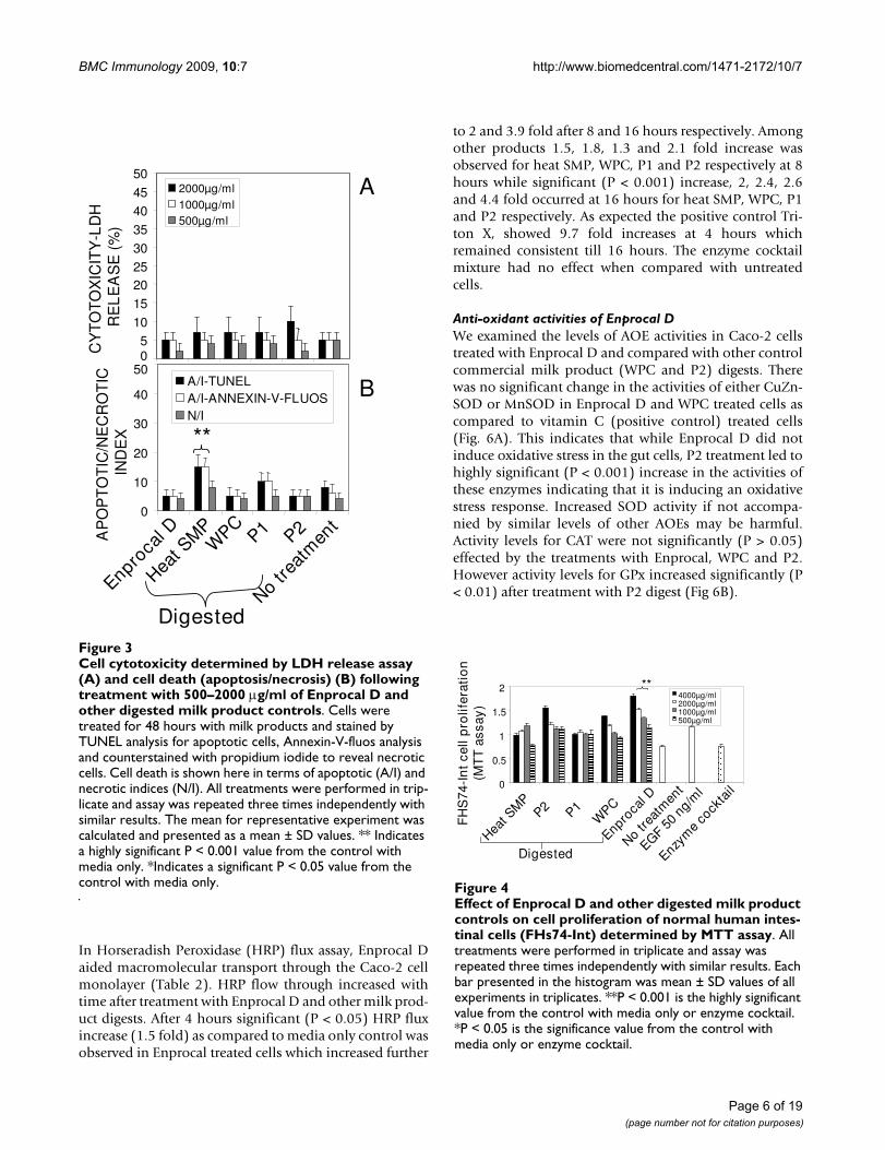
and Enprocal D, heat SMP digest samples reduced signifi-cantly (P < 0.001) the Caco-2 cell viability at 2000 μg/ml.The control milk products were further tested in LDHrelease assay (Fig 3A), and cytotoxicity levels were below10% and not significant (P > 0.05) at 2000 μg/ml concen-tration when compared with Enprocal D and media onlycontrol. In TUNEL and Annexin-V-fluos assays, EnprocalD did not significantly (P > 0.05) increase the apoptoticindex (AI) and necrotic index (NI), however heat SMPinduced significant (P < 0.01) AI as compared to mediaonly control in Caco-2 cells (Fig 3B). In a dose dependentmanner (500–4000 μg/ml) Enprocal D increased signifi-cantly (P < 0.001) the proliferation of FHs 74 Int cells(Figure 4), determined by MTT assay, as compared tountreated and positive control (EGF 50 ng/ml) cells.When compared with other control products this effectwas significant (p < 0.001) except for P2 (500 μg/ml)where the difference was insignificant between Enprocaland P2. However the cell proliferation of Caco-2 cells wasdecreased significantly following treatment with EnprocalD (data not shown).
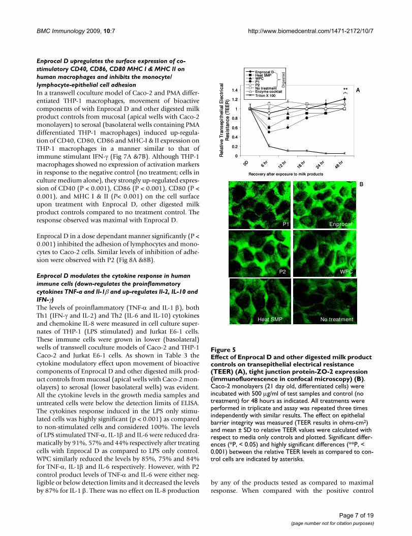
Enprocal D maintains the mucosal integrity, increases the expression of ZO-2 (tight junction protein) and macromolecular absorptionIntestinal Caco-2 monolayers grown on Millipore cell cul-ture inserts in transwell system were used to measure thetransepithelial resistance (TEER) following treatmentswith Enprocal D and other digested milk product controlsat a concentration of 500 μg/ml (Fig 5A). At 0 hour beforetreatment, the Caco-2 monolayers after 21 days of growthwere found to be fully differentiated to enterocytes andpolarized, continuous without any gaps and displayedTEER values 520 ± 20 ohm.cm2 in control growth media.Figure 5A showed the TEER values of cell monolayers fol-lowing different treatments relative to control (mediaonly). The most significant (P < 0.01) decrease wasobserved in all treatments after the first 6 hours except forEnprocal D and P2 (P > 0.05). Both P2 and Enprocal Dwere able to recover the TEER values at 12 hours and theincrease was maintained further till 48 hours duration(end of experiment) as compared to other control milkproducts. However, TEER resistance values for Enprocal Dwere found to increase further after 24 hours. P1 wasfound to decrease significantly (P < 0.001) the TEER after24 hours. The gut enzyme cocktail had no effect on theTEER when compared with media only control. The Tri-ton-X, known to disrupt the cell monolayer, was used asthe positive control. It reduced significantly (P < 0.001)the TEER resistance at 6 hours which were not recoveredthroughout the experimental period. The monolayerintegrity results were further confirmed by expression oftight junction protein -ZO-2. Figure 5B shows an upregu-lation of ZO-2 expression on Caco-2 monolayers treatedwith 500 μg/ml of Enprocal and P2 digests for 12 hours.
SDS-PAGE analysis of Enprocal and whey protein concen-trate (WPC) digested with gut enzyme cocktail (pH7.2) for 4 hoursFigure 1SDS-PAGE analysis of Enprocal and whey protein concentrate (WPC) digested with gut enzyme cock-tail (pH7.2) for 4 hours.
4 H
ou
rs d
iges
ted
En
pro
cal
Mo
l Wt
mar
ker
4 H
ou
rsd
iges
ted
WP
C
Un
dig
este
d E
np
roca
l
Un
dig
este
d W
PC
KDa
25015010075
50
37
2520
15
Page 4 of 19(page number not for citation purposes)
BMC Immunology 2009, 10:7 http://www.biomedcentral.com/1471-2172/10/7
Page 5 of 19(page number not for citation purposes)
Cell viability (A), Cell cytotoxicity (B) of Caco-2 and normal human FHs74-Int cells treated with undigested and digested Enprocal (Enprocal D) 100–8000 μg/ml of each for 48 hoursFigure 2Cell viability (A), Cell cytotoxicity (B) of Caco-2 and normal human FHs74-Int cells treated with undigested and digested Enprocal (Enprocal D) 100–8000 μg/ml of each for 48 hours. Cell viability was determined by trypan blue exclusion assay. Cell cytotoxicity was determined by LDH release assay. (C) Cell viability (trypan blue exclusion assay) of Caco-2 monolayers treated with Enprocal D (500–2000 μg/ml) and with other digested milk product controls. All treatments were performed in triplicate and assay was repeated three times independently with similar results. The mean for representa-tive experiment was calculated and presented as a mean ± SD values. ** Indicates a highly significant P < 0.001 value from the control with media only. *Indicates a significant P < 0.05 value from the control with media only.
0
20
40
60
80
100
120
100
200
250
300
500
1000
2000
4000
8000
Med
ia-C
aco-2
Med
ia-F
HS-Int
Enzym
es-C
aco-
2
Enzym
es-F
HS-Int
Gu
t ce
ll v
iab
ilit
y (%
)
CaCo2 undigestedCaCo2 digestedFHS-int undigestedFHS-int digested
Enprocal ( g/ml)
Controls
****
A
0
20
40
60
80
100 200 250 300 500100
0200
0400
0800
0
Media-C
aco-2
Media-F
HS-Int
Enzym
es-Caco-2
Enzym
es-FHS-In
t
Cyt
oto
xici
ty (
% L
DH
)
CaCo2 un digestedCaCo2 digestedFHS-int undigestedFHS-int digested
Enprocal ( g/ml) Controls
**
**
B
0102030405060708090
100110120
Cac
o-2
Cel
l Via
bili
ty (
%) 2000μg/ml
1000μg/ml500μg/ml
Enproca
l D
Enzym
e co
ckta
il
No trea
tmen
t
Heat S
MP
WPC P1
P2
C
*
Digested
BMC Immunology 2009, 10:7 http://www.biomedcentral.com/1471-2172/10/7
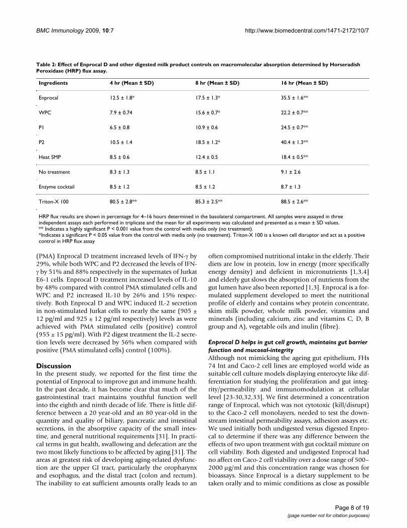
In Horseradish Peroxidase (HRP) flux assay, Enprocal Daided macromolecular transport through the Caco-2 cellmonolayer (Table 2). HRP flow through increased withtime after treatment with Enprocal D and other milk prod-uct digests. After 4 hours significant (P < 0.05) HRP fluxincrease (1.5 fold) as compared to media only control wasobserved in Enprocal treated cells which increased further
to 2 and 3.9 fold after 8 and 16 hours respectively. Amongother products 1.5, 1.8, 1.3 and 2.1 fold increase wasobserved for heat SMP, WPC, P1 and P2 respectively at 8hours while significant (P < 0.001) increase, 2, 2.4, 2.6and 4.4 fold occurred at 16 hours for heat SMP, WPC, P1and P2 respectively. As expected the positive control Tri-ton X, showed 9.7 fold increases at 4 hours whichremained consistent till 16 hours. The enzyme cocktailmixture had no effect when compared with untreatedcells.
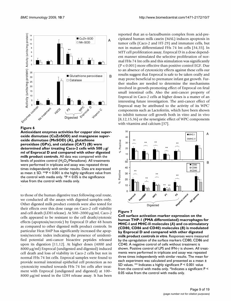
Anti-oxidant activities of Enprocal DWe examined the levels of AOE activities in Caco-2 cellstreated with Enprocal D and compared with other controlcommercial milk product (WPC and P2) digests. Therewas no significant change in the activities of either CuZn-SOD or MnSOD in Enprocal D and WPC treated cells ascompared to vitamin C (positive control) treated cells(Fig. 6A). This indicates that while Enprocal D did notinduce oxidative stress in the gut cells, P2 treatment led tohighly significant (P < 0.001) increase in the activities ofthese enzymes indicating that it is inducing an oxidativestress response. Increased SOD activity if not accompa-nied by similar levels of other AOEs may be harmful.Activity levels for CAT were not significantly (P > 0.05)effected by the treatments with Enprocal, WPC and P2.However activity levels for GPx increased significantly (P< 0.01) after treatment with P2 digest (Fig 6B).
Cell cytotoxicity determined by LDH release assay (A) and cell death (apoptosis/necrosis) (B) following treatment with 500–2000 μg/ml of Enprocal D and other digested milk prod-uct controlsFigure 3Cell cytotoxicity determined by LDH release assay (A) and cell death (apoptosis/necrosis) (B) following treatment with 500–2000 μg/ml of Enprocal D and other digested milk product controls. Cells were treated for 48 hours with milk products and stained by TUNEL analysis for apoptotic cells, Annexin-V-fluos analysis and counterstained with propidium iodide to reveal necrotic cells. Cell death is shown here in terms of apoptotic (A/I) and necrotic indices (N/I). All treatments were performed in trip-licate and assay was repeated three times independently with similar results. The mean for representative experiment was calculated and presented as a mean ± SD values. ** Indicates a highly significant P < 0.001 value from the control with media only. *Indicates a significant P < 0.05 value from the control with media only.
0
10
20
30
40
50
AP
OP
TO
TIC
/NE
CR
OT
IC
IND
EX
A/I-TUNELA/I-ANNEXIN-V-FLUOSN/I
CY
TO
TO
XIC
ITY
-LD
HR
EL
EA
SE
(%
)
05
10
15
2025
30
35
40
45
502000μg/ml1000μg/ml500μg/ml
**
Enproca
l D
No trea
tmen
t
Heat S
MPW
PCP1 P2
A
B
Digested
Effect of Enprocal D and other digested milk product con-trols on cell proliferation of normal human intestinal cells (FHs74-Int) determined by MTT assayFigure 4Effect of Enprocal D and other digested milk product controls on cell proliferation of normal human intes-tinal cells (FHs74-Int) determined by MTT assay. All treatments were performed in triplicate and assay was repeated three times independently with similar results. Each bar presented in the histogram was mean ± SD values of all experiments in triplicates. **P < 0.001 is the highly significant value from the control with media only or enzyme cocktail. *P < 0.05 is the significance value from the control with media only or enzyme cocktail.
Enproca
l D
Enzym
e co
ckta
il
No trea
tmen
t0
0.5
1
1.5
2
FH
S74
-In
tce
ll p
rolif
erat
ion
(M
TT
ass
ay)
4000μg/ml2000μg/ml1000μg/ml500μg/ml
EGF 50
ng/ml
**
Heat S
MP
WPC
P1P2
Digested
Page 6 of 19(page number not for citation purposes)
BMC Immunology 2009, 10:7 http://www.biomedcentral.com/1471-2172/10/7
Enprocal D upregulates the surface expression of co-stimulatory CD40, CD86, CD80 MHC I & MHC II on human macrophages and inhibits the monocyte/lymphocyte-epithelial cell adhesionIn a transwell coculture model of Caco-2 and PMA differ-entiated THP-1 macrophages, movement of bioactivecomponents of with Enprocal D and other digested milkproduct controls from mucosal (apical wells with Caco-2monolayers) to serosal (basolateral wells containing PMAdifferentiated THP-1 macrophages) induced up-regula-tion of CD40, CD80, CD86 and MHC-I & II expression onTHP-1 macrophages in a manner similar to that ofimmune stimulant IFN-γ (Fig 7A &7B). Although THP-1macrophages showed no expression of activation markersin response to the negative control (no treatment; cells inculture medium alone), they strongly up-regulated expres-sion of CD40 (P < 0.001), CD86 (P < 0.001), CD80 (P <0.001), and MHC I & II (P< 0.001) on the cell surfaceupon treatment with Enprocal D, other digested milkproduct controls compared to no treatment control. Theresponse observed was maximal with Enprocal D.
Enprocal D in a dose dependant manner significantly (P <0.001) inhibited the adhesion of lymphocytes and mono-cytes to Caco-2 cells. Similar levels of inhibition of adhe-sion were observed with P2 (Fig 8A &8B).
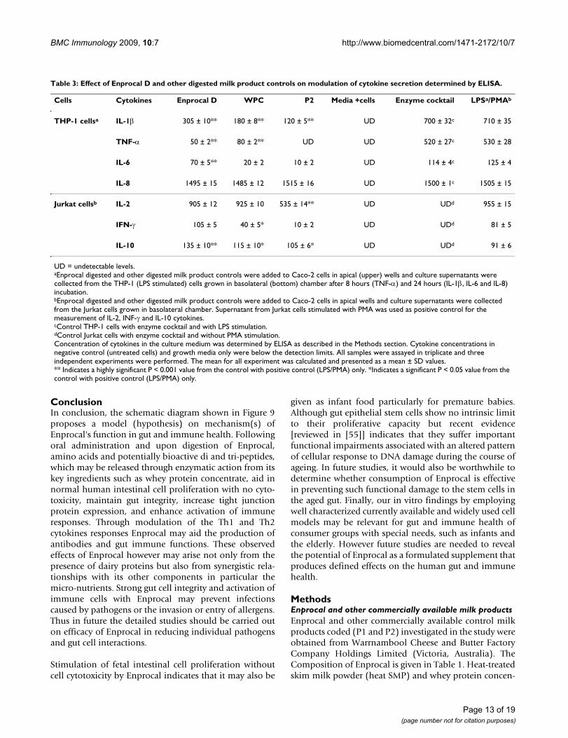
Enprocal D modulates the cytokine response in human immune cells (down-regulates the proinflammatory cytokines TNF-a and Il-1β and up-regulates Il-2, IL-10 and IFN-γ)The levels of proinflammatory (TNF-α and IL-1 β), bothTh1 (IFN-γ and IL-2) and Th2 (IL-6 and IL-10) cytokinesand chemokine IL-8 were measured in cell culture super-nates of THP-1 (LPS stimulated) and Jurkat E6-1 cells.These immune cells were grown in lower (basolateral)wells of transwell coculture models of Caco-2 and THP-1Caco-2 and Jurkat E6-1 cells. As shown in Table 3 thecytokine modulatory effect upon movement of bioactivecomponents of Enprocal D and other digested milk prod-uct controls from mucosal (apical wells with Caco-2 mon-olayers) to serosal (lower basolateral wells) was evident.All the cytokine levels in the growth media samples anduntreated cells were below the detection limits of ELISA.The cytokines response induced in the LPS only stimu-lated cells was highly significant (p < 0.001) as comparedto non-stimulated cells and considered 100%. The levelsof LPS stimulated TNF-α, IL-1β and IL-6 were reduced dra-matically by 91%, 57% and 44% respectively after treatingcells with Enprocal D as compared to LPS only control.WPC similarly reduced the levels by 85%, 75% and 84%for TNF-α, IL-1β and IL-6 respectively. However, with P2control product levels of TNF-α and IL-6 were either neg-ligible or below detection limits and it decreased the levelsby 87% for IL-1 β. There was no effect on IL-8 production
by any of the products tested as compared to maximalresponse. When compared with the positive control
Effect of Enprocal D and other digested milk product con-trols on transepithelial electrical resistance (TEER) (A), tight junction protein-ZO-2 expression (immunofluorescence in confocal microscopy) (B)Figure 5Effect of Enprocal D and other digested milk product controls on transepithelial electrical resistance (TEER) (A), tight junction protein-ZO-2 expression (immunofluorescence in confocal microscopy) (B). Caco-2 monolayers (21 day old, differentiated cells) were incubated with 500 μg/ml of test samples and control (no treatment) for 48 hours as indicated. All treatments were performed in triplicate and assay was repeated three times independently with similar results. The effect on epithelial barrier integrity was measured (TEER results in ohms-cm2) and mean ± SD to relative TEER values were calculated with respect to media only controls and plotted. Significant differ-ences (*P, < 0.05) and highly significant differences (**P, < 0.001) between the relative TEER levels as compared to con-trol cells are indicated by asterisks.
0
0.2
0.4
0.6
0.8
1
1.2
1.4
0D 6 hr
12 h
r18
hr
24 h
r48
hr
Recovery after exposure to milk products
Rel
ativ
e T
ran
sep
ith
elia
l Ele
ctri
cal
Res
ista
nce
(T
EE
R)
Enprocal DHeat SMPWPCP1P2No treatmentEnzyme cocktailTriton X 100
** A
Dig
este
d
0
0.2
0.4
0.6
0.8
1
1.2
1.4
0D 6 hr
12 h
r18
hr
24 h
r48
hr
Recovery after exposure to milk products
Rel
ativ
e T
ran
sep
ith
elia
l Ele
ctri
cal
Res
ista
nce
(T
EE
R)
Enprocal DHeat SMPWPCP1P2No treatmentEnzyme cocktailTriton X 100
Enprocal DHeat SMPWPCP1P2No treatmentEnzyme cocktailTriton X 100
Enprocal DHeat SMPWPCP1P2No treatmentEnzyme cocktailTriton X 100
**** A
Dig
este
d
p1
Enprocal
P2
P1
No treatmentHeat SMP
WPC
p1
Enprocal
P2
P1
No treatmentHeat SMP
WPC
B
p1
Enprocal
P2
P1
No treatmentHeat SMP
WPC
p1
Enprocal
P2
P1
No treatmentHeat SMP
WPC
B
Page 7 of 19(page number not for citation purposes)
BMC Immunology 2009, 10:7 http://www.biomedcentral.com/1471-2172/10/7
(PMA) Enprocal D treatment increased levels of IFN-γ by29%, while both WPC and P2 decreased the levels of IFN-γ by 51% and 88% respectively in the supernates of JurkatE6-1 cells. Enprocal D treatment increased levels of IL-10by 48% compared with control PMA stimulated cells andWPC and P2 increased IL-10 by 26% and 15% respec-tively. Both Enprocal D and WPC induced IL-2 secretionin non-stimulated Jurkat cells to nearly the same (905 ±12 pg/ml and 925 ± 12 pg/ml respectively) levels as wereachieved with PMA stimulated cells (positive) control(955 ± 15 pg/ml). With P2 digest treatment the IL-2 secre-tion levels were decreased by 56% when compared withpositive (PMA stimulated cells) control (100%).
DiscussionIn the present study, we reported for the first time thepotential of Enprocal to improve gut and immune health.In the past decade, it has become clear that much of thegastrointestinal tract maintains youthful function wellinto the eighth and ninth decade of life. There is little dif-ference between a 20 year-old and an 80 year-old in thequantity and quality of biliary, pancreatic and intestinalsecretions, in the absorptive capacity of the small intes-tine, and general nutritional requirements [31]. In practi-cal terms in gut health, swallowing and defecation are thetwo most likely functions to be affected by aging [31]. Theareas at greatest risk of developing aging-related dysfunc-tion are the upper GI tract, particularly the oropharynxand esophagus, and the distal tract (colon and rectum).The inability to eat sufficient amounts orally leads to an
often compromised nutritional intake in the elderly. Theirdiets are low in protein, low in energy (more specificallyenergy density) and deficient in micronutrients [1,3,4]and elderly gut slows the absorption of nutrients from thegut lumen have also been reported [1,3]. Enprocal is a for-mulated supplement developed to meet the nutritionalprofile of elderly and contains whey protein concentrate,skim milk powder, whole milk powder, vitamins andminerals (including calcium, zinc and vitamins C, D, Bgroup and A), vegetable oils and inulin (fibre).
Enprocal D helps in gut cell growth, maintains gut barrier function and mucosal-integrityAlthough not mimicking the ageing gut epithelium, FHs74 Int and Caco-2 cell lines are employed world wide assuitable cell culture models displaying enterocyte like dif-ferentiation for studying the proliferation and gut integ-rity/permeability and immunomodulation at cellularlevel [23-30,32,33]. We first determined a concentrationrange of Enprocal, which was not cytotoxic (kill/disrupt)to the Caco-2 cell monolayers, needed to test the down-stream intestinal permeability assays, adhesion assays etc.We used initially both undigested versus digested Enpro-cal to determine if there was any difference between theeffects of two upon treatment with gut cocktail mixture oncell viability. Both digested and undigested Enprocal hadno affect on Caco-2 cell viability over a dose range of 500–2000 μg/ml and this concentration range was chosen forbioassays. Since Enprocal is a dietary supplement to betaken orally and to mimic conditions as close as possible
Table 2: Effect of Enprocal D and other digested milk product controls on macromolecular absorption determined by Horseradish Peroxidase (HRP) flux assay.
Ingredients 4 hr (Mean ± SD) 8 hr (Mean ± SD) 16 hr (Mean ± SD)
Enprocal 12.5 ± 1.8* 17.5 ± 1.3* 35.5 ± 1.6**
WPC 7.9 ± 0.74 15.6 ± 0.7* 22.2 ± 0.7**
P1 6.5 ± 0.8 10.9 ± 0.6 24.5 ± 0.7**
P2 10.5 ± 1.4 18.5 ± 1.2* 40.4 ± 1.3**
Heat SMP 8.5 ± 0.6 12.4 ± 0.5 18.4 ± 0.5**
No treatment 8.3 ± 1.3 8.5 ± 1.1 9.1 ± 2.6
Enzyme cocktail 8.5 ± 1.2 8.5 ± 1.2 8.7 ± 1.3
Triton-X 100 80.5 ± 2.8** 85.3 ± 2.5** 88.5 ± 2.6**
HRP flux results are shown in percentage for 4–16 hours determined in the basolateral compartment. All samples were assayed in three independent assays each performed in triplicate and the mean for all experiments was calculated and presented as a mean ± SD values.** Indicates a highly significant P < 0.001 value from the control with media only (no treatment).*Indicates a significant P < 0.05 value from the control with media only (no treatment). Triton-X 100 is a known cell disruptor and act as a positive control in HRP flux assay
Page 8 of 19(page number not for citation purposes)
BMC Immunology 2009, 10:7 http://www.biomedcentral.com/1471-2172/10/7
to those of the human digestive tract following oral route,we conducted all the assays with digested samples only.Other digested milk product controls were also tested fortheir effects over this dose range on Caco-2 cell viabilityand cell death (LDH release). At 500–2000 μg/ml, Caco-2cells appeared to be resistant to the cell death/cytotoxiceffects (apoptosis/necrotic) by Enprocal D after 48 hoursas compared to other digested milk product controls. Inparticular Heat SMP has significantly increased the apop-tosis/necrotic index indicating the presence of unidenti-fied potential anti-cancer bioactive peptides releasedupon its digestion [11,12]. At higher doses (4000 and8000 μg/ml) Enprocal (undigested and digested) inducedcell death and loss of viability in Caco-2 cells but not innormal FHs 74 Int cells. Enprocal samples were found toprovide normal intestinal epithelial cell protection as nocytotoxicity resulted towards FHs 74 Int cells after treat-ment with Enprocal (undigested and digested) at 100–8000 μg/ml tested in the LDH release assay. It has been
reported that an α-lactoalbumin complex from acid-pre-cipitated human milk casein (MAL) induces apoptosis intumor cells (Caco-2 and HT-29) and immature cells, butnot in mature differentiated FHs 74 Int cells [34,35]. InMTT cell proliferation assay, Enprocal D in a dose depend-ent manner stimulated the selective proliferation of nor-mal FHs 74 Int cells and this stimulation was significantly(P < 0.001) more effective than positive control EGF. Dueto an absence of cytotoxicity effects against these cells ourresults suggest that Enprocal is safe to be taken orally andmay prove beneficial to premature infant gut growth. Fur-ther studies are needed to determine the mechanismsinvolved in growth-promoting effect of Enprocal on fetalsmall intestinal cells. Also the anti-cancer property ofEnprocal in Caco-2 cells at higher doses is a matter of aninteresting future investigation. The anti-cancer effect ofEnprocal may be attributed to the activity of its WPC'components such as Lactoferrin, which have been shownto inhibit tumour cell growth both in vitro and in vivo[8,12,15,36] or the synergistic effect of WPC componentswith vitamins and calcium [37].
Cell surface activation marker expression on the human THP-1 (PMA differentiated) macrophages for MHC-I and MHC-II molecules (A) and co-stimulatory (CD80, CD86 and CD40) molecules (B) is modulated by Enprocal D and com-pared with other digested milk product controls in vitroFigure 7Cell surface activation marker expression on the human THP-1 (PMA differentiated) macrophages for MHC-I and MHC-II molecules (A) and co-stimulatory (CD80, CD86 and CD40) molecules (B) is modulated by Enprocal D and compared with other digested milk product controls in vitro. Responses were measured by the upregulation of the surface markers CD80, CD86 and CD40. A negative control of cells without treatment is shown. Positive control of LPS and IFN-γ is shown. All treat-ments were performed in triplicate and assay was repeated three times independently with similar results. The mean for each experiment was calculated and presented as a mean ± SD values. ** Indicates a highly significant P < 0.001 value from the control with media only. *Indicates a significant P < 0.05 value from the control with media only.
0
10
20
30
40
50
% P
osi
tive
CD80CD86CD40
0
10
20
30
40
50
% P
osi
tive
MHC-I
MHC-II
A
B
****
**** **
**
**** **
** ***
Digested
Antioxidant enzymes activities for copper zinc superoxide dismutase (CuZnSOD) and manganese superoxide dismutase (MnSOD) (A), glutathione peroxidase (GPx), and catalase (CAT) (B) were determined after treating Caco-2 cells with 500 μg/ml of Enprocal D and compared with other digested milk product controlsFigure 6Antioxidant enzymes activities for copper zinc super-oxide dismutase (CuZnSOD) and manganese super-oxide dismutase (MnSOD) (A), glutathione peroxidase (GPx), and catalase (CAT) (B) were determined after treating Caco-2 cells with 500 μg/ml of Enprocal D and compared with other digested milk product controls. All data was compared with the levels of positive control (H2O2/Menadione). All treatments were performed in triplicate and assay was repeated three times independently with similar results. Data are expressed as mean ± SD. **P < 0.001 is the highly significant value from the control with media only. *P < 0.05 is the significance value from the control with media only.
Enzym
es c
ockta
il
No trea
tmen
t
H2O2 P2 W
PC
Enproca
lD
Vitam
in C
0
2
4
6
8
10
12
14
Act
ivit
y (u
nit
s/m
g p
rote
in)
CuZn-SODMn-SOD
0
5
10
15
20
25
30
35
Act
ivit
y (u
nit
s/m
g p
rote
in)
Glutathione peroxidaseCatalase
A
B
**
Digested
Page 9 of 19(page number not for citation purposes)

BMC Immunology 2009, 10:7 http://www.biomedcentral.com/1471-2172/10/7
We further demonstrated that Enprocal improved gut cellwall integrity, aided in macromolecular absorption. Dis-ruption of the epithelial barrier in the gut is a hallmark ofgut inflammation and diseases. Failure of barrier functionin the gut epithelium is a key feature in the pathology ofdiseases, such as leaky gut syndrome, IBD, cancer etc. [38].Healthy intestinal tract acts a barrier against colonizationand/or translocation of pathogens and toxic compounds.Diarrhoeal diseases as a result of gut infection caused byEscherichia coli, Salmonella, Shigella, Campylobacterjejuni, Entamoeba histolytica and rotavirus are one of theimportant causes of infant mortality and morbidity indeveloped and underdeveloped countries [39-41]. Toensure monolayer integrity, TEER of Caco-2 polarisedmonolayers grown on millicell cell culture inserts (Tran-swell system) was measured using Millicell-ERS systemvoltohm meter which measured the monolayer resistanceunder aseptic conditions. Compared with other controlproducts, high TEER values were observed in monolayers'treated with Enprocal D and P2 after 6 hours of treatment.These values returned to baseline after 12 hours and after24 hours TEER values went further up with only EnprocalD treatment and which were maintained till 48 hours.These observations indicate that there was no differencebetween Enprocal D and P2 with regards to the functionalactivities to gut cells following treatment at till 24 hours.The gut cell integrity results were again confirmed byexpression of tight junction protein Zona Occludin (ZO-2) on treated Caco-2 monolayers. Enprocal D and P2 ascompared to control products increased the expression oftight junction protein -ZO-2.
In the gut permeability assay, Enprocal D aided macromo-lecular transport studied by HRP-flux through the Caco-2monolayer. The transport of nutrients across the intestinalepithelium may occur by one or more of 4 differentroutes: the passive transcellular and paracellular routes,the carrier mediated route or by transcytosis. Caco-2 mon-olayers have been world widely used to study drug trans-port by all 4 pathways [26,27] and permeability throughCaco-2 cell monolayers correlates well with human invivo absorption [27]. The results of the HRP flux indicatedthat HRP flow through increases with time after treatmentwith Enprocal D and other digested control milk prod-ucts. This was reflected in the increased flux of HRP acrossthe monolayers by the 4 hour point. All these results ofTEER, HRP-flux and ZO-2 expression clearly indicates thatEnprocal maintains the gut cell integrity even after diges-tion and showed no side effect on its absorption and per-meability through Caco-2 monolayer.
Enprocal treatment does not induce oxidative stress/injuryIncreases in the levels of reactive oxygen species (ROS)that may occur during periods of oxidative stress, appearto be detected by redox-sensitive regulatory molecules in
the cell, triggering homeostatic responses to prevent cellu-lar injury [21]. Among those responses is the regulation ofAOE, as the levels and balance of AOE modulate the sus-ceptibility of the cell to oxidative injury. Highly reactivefree radicals formed by exogenous chemicals or endog-enous metabolic processes in the human body or in foodsystems are capable of oxidizing biomolecules, resulting
Inhibition of lymphocyte-epithelial (A) and monocyte-epithe-lial (B) cell adhesion by Enprocal D and other digested milk product controlsFigure 8Inhibition of lymphocyte-epithelial (A) and mono-cyte-epithelial (B) cell adhesion by Enprocal D and other digested milk product controls. For adhesion assays, Caco-2 cell monolayers, grown in 96-well plates, were treated for 18 h with Enprocal D and other digested milk product controls. CMFDA-labeled Jurkat (A) or THP-1 (B) cells were then added. After incubation for 60 min at 37°C, nonadherent immune cells were removed by washing with HBSS and the monolayer-associated Jurkat cells or THP-1 was counted. Values are showed as the percent inhi-bition. All treatments were performed in triplicate and assay was repeated three times independently with similar results. The mean for representative experiment was calculated and presented as a mean ± SD values. ** Indicates a highly signifi-cant P < 0.001 value from the control with media only. *Indi-cates a significant P < 0.05 value from the control with media only.
BCaco-2
Caco-2
0
20
40
60
80
100
Inh
ibit
ion
Lym
ph
ocy
teE
pit
hel
ial a
dh
esio
n (
%)
2000μg/ml1000μg/ml500μg/ml
0
10
20
30
40
50
60
70
80
90
100
Inh
ibit
ion
AP
C-E
pit
hel
ial
adh
esio
n (
%)
2000μg/ml1000μg/ml500μg/ml
**
***
****
**
****
**
**
Enproca
l D
No trea
tmen
t
Heat S
MP
WPC P1
P2
A
Digested
Page 10 of 19(page number not for citation purposes)
BMC Immunology 2009, 10:7 http://www.biomedcentral.com/1471-2172/10/7
in cell death and tissue damage. In the gut, variations inthe activities of AOE have been reported under differentinflammatory/pathologic conditions associated with freeradical injury [31]. We examined the specific AOEs such asCuZnSOD, MnSOD, GPx, and CAT activities in Caco-2epithelial cell cultures treated with Enprocal D and otherdigested milk product controls. With Enprocal D therewere no significant changes in the induction of AOE activ-ity levels and were similar in effect to natural anti-oxidantvitamin C used as a positive control. These results indi-cated that no oxidative stress or free radical injuryoccurred to the cells and these findings can be explainedon the basis of synergistic effects of Enprocal product for-mulated with vitamin C, A, and Zn, the known anti-oxi-dants.
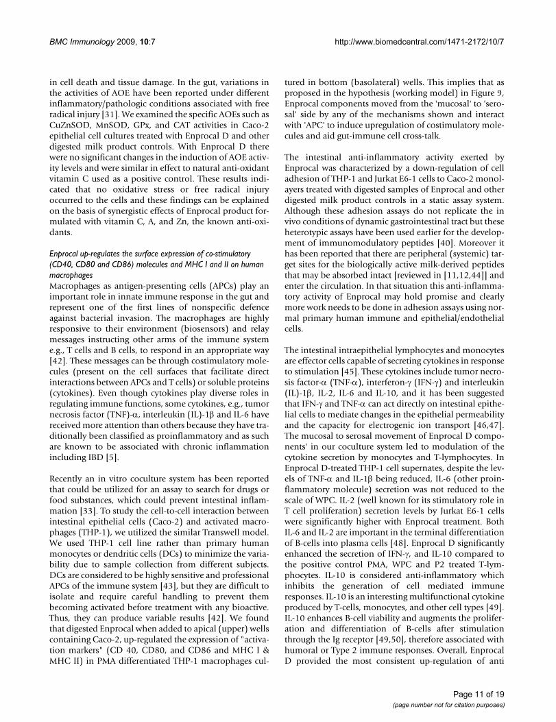
Enprocal up-regulates the surface expression of co-stimulatory (CD40, CD80 and CD86) molecules and MHC I and II on human macrophagesMacrophages as antigen-presenting cells (APCs) play animportant role in innate immune response in the gut andrepresent one of the first lines of nonspecific defenceagainst bacterial invasion. The macrophages are highlyresponsive to their environment (biosensors) and relaymessages instructing other arms of the immune systeme.g., T cells and B cells, to respond in an appropriate way[42]. These messages can be through costimulatory mole-cules (present on the cell surfaces that facilitate directinteractions between APCs and T cells) or soluble proteins(cytokines). Even though cytokines play diverse roles inregulating immune functions, some cytokines, e.g., tumornecrosis factor (TNF)-α, interleukin (IL)-1β and IL-6 havereceived more attention than others because they have tra-ditionally been classified as proinflammatory and as suchare known to be associated with chronic inflammationincluding IBD [5].
Recently an in vitro coculture system has been reportedthat could be utilized for an assay to search for drugs orfood substances, which could prevent intestinal inflam-mation [33]. To study the cell-to-cell interaction betweenintestinal epithelial cells (Caco-2) and activated macro-phages (THP-1), we utilized the similar Transwell model.We used THP-1 cell line rather than primary humanmonocytes or dendritic cells (DCs) to minimize the varia-bility due to sample collection from different subjects.DCs are considered to be highly sensitive and professionalAPCs of the immune system [43], but they are difficult toisolate and require careful handling to prevent thembecoming activated before treatment with any bioactive.Thus, they can produce variable results [42]. We foundthat digested Enprocal when added to apical (upper) wellscontaining Caco-2, up-regulated the expression of "activa-tion markers" (CD 40, CD80, and CD86 and MHC I &MHC II) in PMA differentiated THP-1 macrophages cul-
tured in bottom (basolateral) wells. This implies that asproposed in the hypothesis (working model) in Figure 9,Enprocal components moved from the 'mucosal' to 'sero-sal' side by any of the mechanisms shown and interactwith 'APC' to induce upregulation of costimulatory mole-cules and aid gut-immune cell cross-talk.
The intestinal anti-inflammatory activity exerted byEnprocal was characterized by a down-regulation of celladhesion of THP-1 and Jurkat E6-1 cells to Caco-2 monol-ayers treated with digested samples of Enprocal and otherdigested milk product controls in a static assay system.Although these adhesion assays do not replicate the invivo conditions of dynamic gastrointestinal tract but theseheterotypic assays have been used earlier for the develop-ment of immunomodulatory peptides [40]. Moreover ithas been reported that there are peripheral (systemic) tar-get sites for the biologically active milk-derived peptidesthat may be absorbed intact [reviewed in [11,12,44]] andenter the circulation. In that situation this anti-inflamma-tory activity of Enprocal may hold promise and clearlymore work needs to be done in adhesion assays using nor-mal primary human immune and epithelial/endothelialcells.
The intestinal intraepithelial lymphocytes and monocytesare effector cells capable of secreting cytokines in responseto stimulation [45]. These cytokines include tumor necro-sis factor-α (TNF-α), interferon-γ (IFN-γ) and interleukin(IL)-1β, IL-2, IL-6 and IL-10, and it has been suggestedthat IFN-γ and TNF-α can act directly on intestinal epithe-lial cells to mediate changes in the epithelial permeabilityand the capacity for electrogenic ion transport [46,47].The mucosal to serosal movement of Enprocal D compo-nents' in our coculture system led to modulation of thecytokine secretion by monocytes and T-lymphocytes. InEnprocal D-treated THP-1 cell supernates, despite the lev-els of TNF-α and IL-1β being reduced, IL-6 (other proin-flammatory molecule) secretion was not reduced to thescale of WPC. IL-2 (well known for its stimulatory role inT cell proliferation) secretion levels by Jurkat E6-1 cellswere significantly higher with Enprocal treatment. BothIL-6 and IL-2 are important in the terminal differentiationof B-cells into plasma cells [48]. Enprocal D significantlyenhanced the secretion of IFN-γ, and IL-10 compared tothe positive control PMA, WPC and P2 treated T-lym-phocytes. IL-10 is considered anti-inflammatory whichinhibits the generation of cell mediated immuneresponses. IL-10 is an interesting multifunctional cytokineproduced by T-cells, monocytes, and other cell types [49].IL-10 enhances B-cell viability and augments the prolifer-ation and differentiation of B-cells after stimulationthrough the Ig receptor [49,50], therefore associated withhumoral or Type 2 immune responses. Overall, EnprocalD provided the most consistent up-regulation of anti
Page 11 of 19(page number not for citation purposes)
BMC Immunology 2009, 10:7 http://www.biomedcentral.com/1471-2172/10/7
inflammatory cytokines and was the second most prolificdown-regulator of pro inflammatory cytokines. Enprocal,through down-regulation of proinflammatory cytokinessuch as TNF-α and IL-1β (both known to play an impor-tant role in gut chronic inflammation), may prove benefi-cial in IBD, CD & UC. TNF-α, in particular has beenrecognized as playing a central role in the pathogenesis ofCrohn disease. Nevertheless, reducing the production or
effect of TNF-α in Crohn disease patients is believed to bebeneficial [51]. In fact, different drugs capable of interfer-ing with the activity of TNF-α cytokine have been success-fully developed for IBD therapy [52,53]. However, despiteearlier hopes, the results from studies using TNF-α antag-onists were disappointing, and there were some reports ofsevere complications [54].
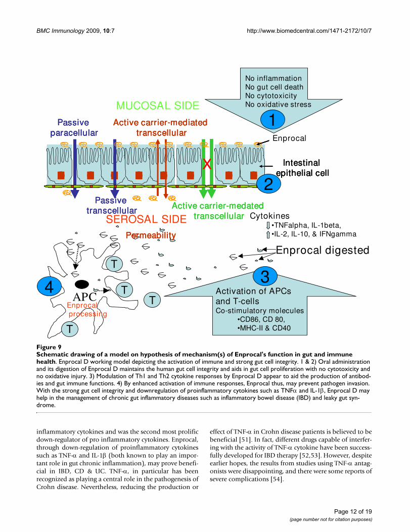
Schematic drawing of a model on hypothesis of mechanism(s) of Enprocal's function in gut and immune healthFigure 9Schematic drawing of a model on hypothesis of mechanism(s) of Enprocal's function in gut and immune health. Enprocal D working model depicting the activation of immune and strong gut cell integrity. 1 & 2) Oral administration and its digestion of Enprocal D maintains the human gut cell integrity and aids in gut cell proliferation with no cytotoxicity and no oxidative injury. 3) Modulation of Th1 and Th2 cytokine responses by Enprocal D appear to aid the production of antibod-ies and gut immune functions. 4) By enhanced activation of immune responses, Enprocal thus, may prevent pathogen invasion. With the strong gut cell integrity and downregulation of proinflammatory cytokines such as TNFα and IL-1β, Enprocal D may help in the management of chronic gut inflammatory diseases such as inflammatory bowel disease (IBD) and leaky gut syn-drome.
MUCOSAL SIDE
T
APCT
T
T
Enprocal digested
Cytokines•TNFalpha, IL-1beta,•IL-2, IL-10, & IFNgamma
SEROSAL SIDE
Enprocalprocessing
Passive paracellular
Passive transcellular
Active carrier-mediated transcellular
Active carrier-medatedtranscellular
Se-bLf
Intestinal epithelial cell
Permeability
Passive paracellular
Passive transcellular
Active carrier-mediated transcellular
Active carrier-medatedtranscellular
Se-bLf
Intestinal epithelial cell
Permeability
Enprocal
X
Passive paracellular
Passive transcellular
Active carrier-mediated transcellular
Active carrier-medatedtranscellular
Se-bLf
Intestinal epithelial cell
Permeability
Passive paracellular
Passive transcellular
Active carrier-mediated transcellular
Active carrier-medatedtranscellular
Se-bLf
Intestinal epithelial cell
Permeability
Enprocal
X
No inflammationNo gut cell deathNo cytotoxicityNo oxidative stress
Activation of APCs and T-cellsCo-stimulatory molecules
•CD86, CD 80, •MHC-II & CD40
1
2
34
Page 12 of 19(page number not for citation purposes)
BMC Immunology 2009, 10:7 http://www.biomedcentral.com/1471-2172/10/7
ConclusionIn conclusion, the schematic diagram shown in Figure 9proposes a model (hypothesis) on mechanism(s) ofEnprocal's function in gut and immune health. Followingoral administration and upon digestion of Enprocal,amino acids and potentially bioactive di and tri-peptides,which may be released through enzymatic action from itskey ingredients such as whey protein concentrate, aid innormal human intestinal cell proliferation with no cyto-toxicity, maintain gut integrity, increase tight junctionprotein expression, and enhance activation of immuneresponses. Through modulation of the Th1 and Th2cytokines responses Enprocal may aid the production ofantibodies and gut immune functions. These observedeffects of Enprocal however may arise not only from thepresence of dairy proteins but also from synergistic rela-tionships with its other components in particular themicro-nutrients. Strong gut cell integrity and activation ofimmune cells with Enprocal may prevent infectionscaused by pathogens or the invasion or entry of allergens.Thus in future the detailed studies should be carried outon efficacy of Enprocal in reducing individual pathogensand gut cell interactions.
Stimulation of fetal intestinal cell proliferation withoutcell cytotoxicity by Enprocal indicates that it may also be
given as infant food particularly for premature babies.Although gut epithelial stem cells show no intrinsic limitto their proliferative capacity but recent evidence[reviewed in [55]] indicates that they suffer importantfunctional impairments associated with an altered patternof cellular response to DNA damage during the course ofageing. In future studies, it would also be worthwhile todetermine whether consumption of Enprocal is effectivein preventing such functional damage to the stem cells inthe aged gut. Finally, our in vitro findings by employingwell characterized currently available and widely used cellmodels may be relevant for gut and immune health ofconsumer groups with special needs, such as infants andthe elderly. However future studies are needed to revealthe potential of Enprocal as a formulated supplement thatproduces defined effects on the human gut and immunehealth.
MethodsEnprocal and other commercially available milk productsEnprocal and other commercially available control milkproducts coded (P1 and P2) investigated in the study wereobtained from Warrnambool Cheese and Butter FactoryCompany Holdings Limited (Victoria, Australia). TheComposition of Enprocal is given in Table 1. Heat-treatedskim milk powder (heat SMP) and whey protein concen-
Table 3: Effect of Enprocal D and other digested milk product controls on modulation of cytokine secretion determined by ELISA.
Cells Cytokines Enprocal D WPC P2 Media +cells Enzyme cocktail LPSa/PMAb
THP-1 cellsa IL-1β 305 ± 10** 180 ± 8** 120 ± 5** UD 700 ± 32c 710 ± 35
TNF-α 50 ± 2** 80 ± 2** UD UD 520 ± 27c 530 ± 28
IL-6 70 ± 5** 20 ± 2 10 ± 2 UD 114 ± 4c 125 ± 4
IL-8 1495 ± 15 1485 ± 12 1515 ± 16 UD 1500 ± 1c 1505 ± 15
Jurkat cellsb IL-2 905 ± 12 925 ± 10 535 ± 14** UD UDd 955 ± 15
IFN-γ 105 ± 5 40 ± 5* 10 ± 2 UD UDd 81 ± 5
IL-10 135 ± 10** 115 ± 10* 105 ± 6* UD UDd 91 ± 6
UD = undetectable levels.aEnprocal digested and other digested milk product controls were added to Caco-2 cells in apical (upper) wells and culture supernatants were collected from the THP-1 (LPS stimulated) cells grown in basolateral (bottom) chamber after 8 hours (TNF-α) and 24 hours (IL-1β, IL-6 and IL-8) incubation.bEnprocal digested and other digested milk product controls were added to Caco-2 cells in apical wells and culture supernatants were collected from the Jurkat cells grown in basolateral chamber. Supernatant from Jurkat cells stimulated with PMA was used as positive control for the measurement of IL-2, INF-γ and IL-10 cytokines.cControl THP-1 cells with enzyme cocktail and with LPS stimulation.dControl Jurkat cells with enzyme cocktail and without PMA stimulation.Concentration of cytokines in the culture medium was determined by ELISA as described in the Methods section. Cytokine concentrations in negative control (untreated cells) and growth media only were below the detection limits. All samples were assayed in triplicate and three independent experiments were performed. The mean for all experiment was calculated and presented as a mean ± SD values.** Indicates a highly significant P < 0.001 value from the control with positive control (LPS/PMA) only. *Indicates a significant P < 0.05 value from the control with positive control (LPS/PMA) only.
Page 13 of 19(page number not for citation purposes)

BMC Immunology 2009, 10:7 http://www.biomedcentral.com/1471-2172/10/7
trate 80 (WPC) were included as control milk products.We cannot reveal the identity of other milk products P1and P2 due to commercial reasons. Protein concentra-tions were measured by Coomassie Plus-The Better Brad-ford Assay kit (Endogen). Products were assayed forlipopolysaccharide (LPS) contamination and the concen-tration of endotoxin was < 0.13 μg/kg.
In vitro digestionEnprocal and the control milk products were digestedwith digestive enzymes cocktail mixture using a modifiedprotocol [56]. The enzyme concentrations in the commer-cially available enzyme tablet are shown in Table 4. Aftergastric phase digestion the solutions of protein fractionsand enzyme tablet were made for small intestinal phase in0.1 M NaHCO3 solution pH 7.2. The reaction mixture wasset in 1:50 (enzyme: protein) ratio (standardized after pre-liminary observations) and incubated at 37°C in a shaker(250 rpm). Enzyme digests were collected after 1, 2, 4 and6 hours incubation. The reaction was stopped by inacti-vating the enzymes at 42°C and aliquots were stored at -20°C till further use in cell culture studies. Digests wereanalyzed by SDS-PAGE (reducing & denaturing) electro-phoresis (Figure 1).
Intestinal epithelial cell cultureCaco-2 (human colon adenocarcinoma), and FHs 74 Int(an adherent human primary fetal small intestinal) celllines were obtained from American Type Culture Collec-tion (ATCC, Rockville) and cultured in their respectivemedia from ATCC as per its guidlines. Caco-2 cells werecultured routinely in Eagle's Minimum Essential Medium(EMEM) purchased from ATCC supplemented with 20%(v/v) heat-inactivated fetal calf serum (Sigma). FHs 74 Intcells were routinely grown in Hybri-Care Medium (ATCC:46-X) supplemented with 30 ng/ml human recombinantepidermal growth factor (EGF; Affinity Bioreagents) with10% fetal bovine serum. All cell lines were grown at 37°C,in an atmosphere of 5% CO2 and 95% relative humidity.
Cell Viability assaysCaco-2 and FHs 74 Int cells were incubated with variousconcentrations of Enprocal (undigested) & digested(Enprocal D) and other digested milk product controls for48 hours. Cells were trypsinized to obtain a cell suspen-sion and all adherent and floating cells were collected forcounting the live and dead cells under microscope follow-ing trypan blue staining.
Lactate dehydrogenase (LDH) cytotoxicity assayThe cytotoxic effect of different test treatments on Caco-2and FHs 74 Int was studied by a colorimetric LDH releaseassay using a commercially available kit (Roche AppliedScience). This assay quantifies cell death and lysis basedon measuring the release of lactate dehydrogenase (LDH)
which is present within the supernatant following the lossof membrane integrity. The assay was carried out as perkit's instructions. Briefly, 2 × 10³ cells per well (total vol-ume 100 μL) were plated into a 96 well plate and incu-bated for 24 hours at 37°C in 5% CO2. Followingincubation, growth media was replaced with fresh mediacontaining different concentrations of Enprocal (undi-gested), Enprocal D (digested) and other digested milkproduct controls, and cells incubated further for 48 hours.Cytotoxicity measurements were tested at 48 hours aftertreatment with Enprocal and other digested milk productcontrols. Triton-X100 (1%V/V) was used as a high (posi-tive) control and cells in media only was the low (nega-tive) control. The amounts of lactate dehydrogenase(LDH) in the supernatant were determined and calculatedas per kit instructions. All tests were performed in tripli-cate and assay was repeated three times independentlywith similar results.
Apoptosis versus necrosisIn order to detect the nature of cell death following treat-ment with different test samples as well as to differentiatebetween apoptosis and necrosis at single cell level, Caco-2 and FHs 74 Int cells were plated at a density of 1 × 10⁴
cells on 8 well slides and grown for two days at 37°C with5% CO2. Following media removal, the cells were thentreated with different concentrations of Enprocal D andother digested milk product controls (in triplicates) inserum free media for 48 hours. As a positive control,apoptosis inducing anti-cancer drug Paclitaxel (BioVi-sion), was added to the cells (final concentration of 20nm). After 48 hours the media was removed and apop-totic cells were determined by TUNEL staining using an insitu apoptosis detection kit from Boehringer Mannheimas per kit instructions [57]. Slides were also stained withpropidium iodide (Sigma-Aldrich) to distinguish necroticcells from those undergoing apoptosis. Same slides werecounterstained with methylene blue-staining and
Table 4: Digestive enzyme cocktail ingredients.
Pancreatin (4×) 1250 mg
Papain 150 mg
Bromelain 150 mg
Trypsin 125 mg
α-Chymotrypsin 3 mg
Lipase 50 mg
Amylase 50 mg
Rutin 100 mg
Page 14 of 19(page number not for citation purposes)

BMC Immunology 2009, 10:7 http://www.biomedcentral.com/1471-2172/10/7
mounted. The total number of apoptotic or necrotic cellswas counted. The apoptotic and the necrotic indices werecalculated as follows: Apoptotic index (A/I) or necroticindex (N/I) = number of apoptotic or necrotic cells × 100/total number of nucleated cells. Following treatments,another kit, Annexin-VFLUOS staining (Roche AppliedScience), was also used to differentiate apoptotic cellsfrom necrotic cells, as per manufacturer's instructions.This assay is based on principle that phosphatidyl serine,located in the inner leaflet of the cell membrane, isexposed at the cell surface in the early stage of apoptosis.Annexin V shows high-affinity for phosphatidyl serine-binding that makes it a useful selective and powerful toolfor detection of apoptotic cells [58].
Cell proliferation (MTT) assayHuman intestinal cell lines Caco-2 and FHs 74 Int wereseeded each into 96 well plates, at a density of 2 × 10³ cellsper well (total volume 100 μL) and incubated for 24 hoursat 37°C in 5% CO2. After removal of media, the cells wereincubated with fresh growth media containing differentconcentrations of Enprocal and other digested milk prod-uct controls, for 48 hours. As a positive control EGF 50 ng/ml was used. The effect of different treatments on cell pro-liferation of Caco-2 and FHs 74 Int cells was compared intreated versus untreated cells with the use of commerciallyavailable 3-(4,5-dimethylthiazol-2-yl)-2,5-diphenyltetra-zolium bromide (MTT) assay Kit (R&D systems). Theassay was carried out as per manufacturer's instructions.All tests were performed in triplicate and assay wasrepeated three times independently with similar results.
Measurement of transepithelial resistance (TEER)Mucosal integrity of Caco-2 cells seeded onto Milicell cellculture inserts (Millipore Australia) in 24-well plates wasstudied by measuring the transepithelial electrical resist-ance (TEER) with a Millicell-ERS voltohm meter (Milli-pore Australia). The device contained a pair of chopstickelectrodes (which facilitated the measurements) andmeasures membrane potential and resistance of epithelialcells in culture. It does this by qualitatively measuring cellmonolayer health and cell confluency [59]. Caco-2 cellswere seeded at a density of 4 × 10³ per well in the volumeof 200 μl growth medium (EMEM plus 20% fetal bovineserum) added to the apical well of insert and 800 μl ofgrowth medium was added to the basolateral well in 24-well tissue culture plates for incubation. To obtain mon-olayer, cells were grown for 21 days and medium wasreplaced every third day. After 21 days the monolayerswere thoroughly checked for any gaps under the micro-scope in order to prevent any false TEER readings. The dif-ferentiation was characterized by a polarization of themonolayer with formation of domes. These filter-grownmonolayers were first equilibrated with serum free growthmedium (EMEM containing GIBCO's antibiotic-antimy-
cotic supplement for 2 hours under cell culture condi-tions. TEER was measured at the start of experiment i.e.before the addition of Enprocal D and other digested milkproduct controls. Each preparation was added to the api-cal compartment in serum free growth medium, and Tri-ton-X, was used as a positive control. All test and controlsamples were added in triplicate. Cell monolayers wereincubated at 37°C, 5% CO2 and 90% humidity and TEERwas measured at specific intervals (6, 12, 18 and 24 hours)and at the end of experiments (48 hour). The monolayerswere incubated at 37°C in 5% CO2 and 90% humiditybetween measurements.
Horseradish peroxidase (HRP) fluxCaco-2 cells were seeded on to Millicell cell culture insertsat a density of 4 × 10³ per well and grown as monolayersfor up to 21 days as described above. Following mediumremoval, the cells were equilibrated for 2 hours withHanks Balanced Salt Solution (HBSS) without phenol red(Invitrogen) and then treated with Enprocal D and otherdigested milk product controls. Horseradish peroxidase(HRP-Sigma-Aldrich) was added to the apical side of themonolayer at a final concentration of 0.15 mg/ml [60].Following incubation at 37°C in 5% CO2 and 90%humidity, the flux of HRP was measured at 4, 8 and16hours incubation, by taking 25 μl from the apical com-partment and 300 μl from the basolateral compartment.The HRP activities were then measured in aliquots of thesesamples by treating the samples with 3,3',5,5'-tetrameth-ylbenzidine (TMB; Sigma-Aldrich) which is an HRP sub-strate and then measuring the absorbance of the samplesusing an ASYS Expert plus UV ELISA plate reader at 450nm. All treatments were performed in triplicate and assaywas repeated three times independently with similarresults.
Immunofluorescence staining for ZO-2 tight junction proteinCaco-2 cells were grown on sterile 8 chamber slides (BDSciences) with seeding density of 1 × 10⁴ cells/ml. After 5days of growth monolayers, were equilibrated with serumfree medium and then treated with Enprocal D and otherdigested milk product controls as well as 1% Triton-X(positive control) for 12 hours. Monolayers were washedwith PBS (pH 7.2), fixed in 4% paraformaldehyde (inPBS; pH 7.2) for 15 minutes at room temperature (RT)and blocked for 30 minutes in 2% normal rabbit serum(Vector labs) in PBS. After 3 PBS washes monolayers wereincubated with mouse monoclonal anti-ZO-2 (Invitrogendiluted 1:1000 in PBS containing 1% BSA) overnight at4°C. Secondary anti-mouse IgG-FITC antibody (diluted1:100 in PBS; Sigma-Aldrich) was used to detect the pri-mary antibody staining (1 hour incubation at RT). After 3PBS washes, chambers were removed from the slide andmounted with one drop of Vectashield mounting
Page 15 of 19(page number not for citation purposes)
BMC Immunology 2009, 10:7 http://www.biomedcentral.com/1471-2172/10/7
medium with DAPI (Vector Labs). For primary and sec-ondary antibody controls, cells were stained as aboveexcept incubation with PBS containing 1% BSA was car-ried out at appropriate staining steps. The slides were thenviewed with Leica TCS SP5 confocal system fitted withinverted DMI 6000 microscope. Images were collectedwith LAS AF software. These images were then processedin Adobe Photoshop CS3.
Antioxidant enzyme (AOE activity assaysFor each assay Caco-2, 2 × 10⁴ cells/well were grown in 6well plates for 24 hours and then treated with Enprocal Dand other digested milk product controls for 48 hours.Following incubation, cells were isolated in assay bufferand lysed by a freeze-thaw cycle. The debris was removedby centrifugation at 8000 × g for 5 min at 4°C, and theprotein content of each sample was determined using theCoomassie Plus – The Better Bradford™ Assay Kit(Endogen). AOE activity assays were carried out spectro-photometrically by the methods as described earlier forsuperoxide dismutase (SOD) [61], glutathione peroxidase(GPx) [62] and catalase (CAT) [63]. All treatments wereperformed in triplicate and assays were repeated threetimes independently with similar results.
Superoxide dismutase (SOD) activity assayTo the assay mixture containing 0.1-mM cytochrome c,0.05-mM xanthine, and KCN (10 μM for total SOD activ-ity, 3 mM for MnSOD activity) in phosphate buffer, 150-μg protein (for each sample) was added in a final volumeof 3 ml. After zeroing (blanking), xanthine oxidase wasadded to a final concentration of 0.6 U/ml, and thechange in absorbance at OD at 550 nm was recorded atregular intervals over 4 min. A standard curve for SOD wasgenerated and enzyme activity was determined by com-paring samples to a standard curve of known SOD activ-ity. Cu,Zn-superoxide dismutase (CuZnSOD), activity wascalculated as the difference between total SOD activityand Mn-superoxide dismutase (MnSOD) activity [61].
Glutathione peroxidase (GPx) activity assayFor GPx enzyme activity measurements aliquots of pro-tein samples (140 μg of protein) were added to the assaymixture containing 1 U/ml glutathione reductase and 2mM glutathione in 1 ml of phosphate buffer after 30 minincubated at 37°C for 30 min. Nicotinamide adeninedinucleotide phosphate and tert-butylhydroperoxidewere added to final concentrations of 580 μM, respec-tively, and the change in absorbance at OD at 340 nm wasrecorded at regular intervals over 4 min. The amount ofGPx required to oxidize 1 μmol nicotinamide adeninedinucleotide phosphate in 1 min at 25°C was defined asenzyme units and calculated on the basis of a molarabsorptivity for nicotinamide adenine dinucleotide phos-phate at 340 nm of 6.22 × 10⁻⁶ [62].
Catalase (CAT) activity assayCAT enzyme activity was measured by monitoring thedecrease in H2O2 concentration spectrophotometricallyover time. For each sample, 130 μg protein was added to50-mM phosphate buffer in a quartz cuvette and, afterblanking, H2O2 was added to a final concentration of 10mM in 0.9 ml, and the absorbance at OD at 240 nm wasrecorded at regular intervals over 4 min. The specific activ-ity of each sample was calculated based on activity of pureCAT as described earlier [63].
Immune cell cultureTHP-1 and Jurkat clone E6-1 obtained from AmericanType Culture Collection (ATCC Rockville, MD) wereemployed in immune assays. THP-1 cells are human mye-lomonocytic cell line that is widely used to study mono-cyte/macrophage biology in cell culture systems andJurkat cells are human T lymphocyte (T cell leukemia).THP-1 cells were cultured and maintained in RPMI 1640(Gibco-BRL), supplemented with 10% heat-inactivatedfetal calf serum, 2 mM L-glutamine, 0.05 mM 2-mercap-toethanol,100 U/ml penicillin, and 100 μg/ml streptomy-cin (complete RPMI 1640). Jurkat E6-1 cells wereroutinely grown in suspension in RPMI-1640 mediumsupplemented with 10% heat-inactivated fetal bovineserum (FBS), 2 mM L-glutamine, 100 U/ml penicillin, and100 μg/ml streptomycin.
Monocyte/lymphocyte-epithelial adhesion assayAdhesion assays were performed with THP-1 and JurkatE6-1 cells under static conditions by the methods asdescribed earlier [32,64] with modifications. For staticadhesion assays Caco-2 cells were seeded at a density of 2× 10⁴ cells per well (total volume 100 μL) and grown asconfluent monolayers in 96-well plates and preactivatedwith IL-1β 10 ng/ml overnight before adhesion assay.Washed monolayers were then treated for 18 h withEnprocal D and other digested milk product controls. Epi-thelial cells were washed twice with culture mediumbefore addition of 100 μl (1 × 10⁵) labeled THP-1 or Jur-kat E6-1 cells per well. Both THP-1 and Jurkat E6-1 cellswere labeled with the fluorescent dye chloromethyl fluo-rescein diacetate (CMFDA; Molecular Probes, Oregon) at37°C for 30 min [64]. The plates were incubated for 60min at 37°C. After incubation, the monolayer was gentlywashed three times with PBS to remove non adherentcells. Fluorescence-labeled adherent cells were countedunder fluorescent microscope in minimum of 5 fields.
Cell surface marker stainingWe employed a coculture model to examine the effect ofEnprocal D treatment on the interaction between gut-immune cells. For the coculture experiments THP-1monocytic cells were grown in 24-well plates at a densityof 5 × 10⁵ cells/well and differentiated into macrophage-
Page 16 of 19(page number not for citation purposes)
BMC Immunology 2009, 10:7 http://www.biomedcentral.com/1471-2172/10/7
like cells, by treatment with 200 nM phorbol myristrateacetate (PMA; Sigma-Aldrich) for 4 days [33]. The differ-entiated status was confirmed on the basis of morphologyand staining for the expression of leukocyte integrinCD11b (macrophage-1 antigen) (data not shown). Thesemipermeable support membrane (Milicell cell cultureinserts from Millipore) on which the intact Caco-2 cellmonolayers had been cultured for 21 days was placed onthe differentiated THP-1 cells that had been cultured onthe 24-well plates. After starting the coculture for 18hours, apical media was removed and Caco-2 cells weretreated with fresh growth media containing Enprocal Dand other digested milk product controls. Control treat-ments included cells in medium only (negative control),enzyme cocktail. Interferon-γ (IFN-γ) at a concentration of5 ng/ml was added to the positive control wells on thebasolateral compartment containing differentiated THP-1cells. Cells were incubated at 37°C with 5% CO2 for 24 hand cell surface marker staining was determined by directimmunofluorescence in paraformaldehyde fixed (4% PFin PBS pH 7.2; 15 minutes at RT) cells. Cells were incu-bated for 1 hour at room temperature with FITC labeledantibodies to anti-human CD40, CD86, CD80, major his-tocompatibility complex (MHC) I & II (PharMingen andR&D Systems). Following washing with PBS buffer wellswere mounted with one drop of Vectashield mountingmedium with DAPI (Vector Labs). For primary and sec-ondary antibody controls, cells were stained with PBScontaining 1% BSA (no primary antibody). The presenceof labeled cell surface markers was counted under the flu-orescence microscope and percentage of positive cells wascalculated from total number of cells (DAPI positive).
Cytokine ELISAIn the Caco-2 THP-1 coculture model, cytokine levelswere measured in the supernatant of LPS stimulated THP-1 cells grown in the lower chamber for 8 and 24 hoursafter the addition of Enprocal D and other digested milkproduct controls to Caco-2 cell monolayers in the upperchamber [33]. Briefly, THP-1 cells were grown and main-tained in endotoxin free complete RPMI-1640 at the expo-nential phase. To increase the sensitivity to LPS, cells werestimulated with 5 ng/ml human recombinant IFN-γ (Bio-Vision >98% with endotoxin level < 0.1 ng per μg of IFN-γ) for 16 hours [65,66]. Following incubation cells werecentrifuged, re-resuspended in serum free medium andseeded in 24 well plates with final cell concentration of 8× 10⁵ cells per well. Caco-2 monolayers (21 days old)grown and differentiated on the Milicell cell cultureinserts (Millipore) were placed on the wells containingTHP-1 cells in 24-well plates. All cells were allowed to set-tle for 1 h and following removal of apical media, Enpro-cal D and other digested milk product controls wereadded to the Caco-2 cells in fresh growth media 30 min-utes before LPS (50 ng/ml; E. coli, Sigma-Aldrich) was
added to the THP-1 cells (lower basolateral chamber).Supernatants were collected after 8 (for TNF-a) and 24hours for other cytokines at 37°C with 5% CO2 Supern-ants of Jurkat E6-1 cells grown in coculture with Caco-2were used to determine the protein levels of IL-2, IL-10and IFN-γ cytokines. Jurkat E6-1 cells were kept in theexponential growth phase by passages at 2–3 days inter-vals. As described above, Caco-2 monolayers (21 daysold) grown and differentiated on the Milicell cell cultureinserts (Millipore) were placed on the wells containing2.5 × 105cells/ml Jurkat E6-1 cells grown overnight. Afterone hour incubation, apical media was replaced withmedia containing Enprocal D and other digested milkproduct controls and incubated for 24 hours. PMA (20ng/ml) was used a positive control. Jurkat cell superna-tants were obtained via centrifugation at 4°C and storedat -80°C until cytokine analysis.
Amounts of the cytokines TNF-a, IL-1β, and IL-6 and IL-8,IL-2, IL-10 and IFN-γ in cell culture supernatants weredetermined by using Biotrak Easy ELISA kits (AmershamBiosciences) for human cytokines. The limits of detectionof the assays were 2.5 pg/ml for TNF-a, 1.1 pg/ml for IL-1β, 1.4 pg/ml for IL-6, < 7.26 pg/ml for IL-8, 3.5 pg/ml forIL-2, 1 pg/ml for both IFN-γ and IL-10. Each assay proto-col was carried out as per kit instructions using cytokinestandards supplied with kit. Samples were run in triplicateand experiments were performed three times. The concen-tration of cytokines was determined by the standard curveusing linear regression curve fit method.
Data interpretation and statistical analysisFor the statistical evaluation of numerical data, one wayANOVA and Students t-test were used to compute the dif-ferences. In all cases, P < 0.05 and P < 0.001 were taken toidentify levels of significance.
AbbreviationsAPC: antigen-presenting cell; AOE: Antioxidant enzyme;CD: cluster designation; CMI: cell-mediated immunity;DC: dendritic cell; Enprocal D: digested Enprocal; HRP:horseradish peroxidase; IL: interleukin; IFN-γ: interferon-γ; LPS: lipopolysaccharide; MHC: major histocompatibil-ity complex; TGF-β: transforming growth factor-β; TNF-α:tumor necrosis factor-α; TEER: transepithelial resistance;SOD: superoxide dismutase; GPx: glutathione peroxidase;CAT: catalase; AI: apoptotic index; NI: necrotic index;LDH: lactate dehydrogenase
Authors' contributionsJRK designed and carried-out immunomodulatory, celladhesion and anti-oxidant experiments. RKK designedand performed the, cell integrity, permeability assays,confocal microscopy and cytokine measurements. JRK
Page 17 of 19(page number not for citation purposes)
BMC Immunology 2009, 10:7 http://www.biomedcentral.com/1471-2172/10/7
and RKK contributed equally to the manuscript. Authorsread and approved the final manuscript for publication.
AcknowledgementsThe authors would like to thank Professor Andrew Parratt, Executive Director, ITRI, Deakin University, Dr Leanne Fitzpatrick, Bruce Storey for their valuable comments and Elizabeth Laidlaw, Project and Technical Officer (ITRI) for her support in laboratory matters and ordering chemicals on time. Thanks to Mr Shawn Todd for maintaining the cell lines. The authors are grateful to Dr Ken Walder, Deputy Head, and Helen Barry, Administrative Officer, of the Metabolic Research Unit, Deakin University, for their support and use of facilities. This study was funded by The Institute of Biotechnology (BioDeakin), Deakin University and Warrnambool Cheese & Butter Factory Company Limited, Victoria, Australia.
References1. Volkert D, Berner YN, Berry E, Cederholm T, Coti BP, Milne A, Palm-
blad J, Schneider S, Sobotka L, Stanga Z, Lenzen-Grossimlinghaus R,Krys U, Pirlich M, Herbst B, Schütz T, Schröer W, Weinrebe W, Ock-enga J, Lochs H: ESPEN Guidelines on Enteral Nutrition: Ger-iatrics. Clin Nutr 2006, 25:330-60.
2. Staples K: "Gut health and whey products". In Nutrition Mono-graph Gut health Dairy Management Inc; 2006:1-12.
3. Lasheras C, González C, García A, Patterson AM, Fernández S: Die-tary intake and biochemical indicators of nutritional stress inan elderly institutionalised and non-institutionalised popula-tion. Nutr Res 1999, 19:1299-12.
4. Kreider RB: "Whey proteins and seniors nutrition". In Applica-tions Monograph Seniors Nutrition Dairy Management Inc; 2005:1-8.
5. McKay DL, Perrone G, Rasmussen H, Dallal G, Hartman W, Cao G,Prior RL, Roubenoff R, Blumberg JB: The effects of a multivita-min/mineral supplement on micronutrient status, antioxi-dant capacity and cytokine production in healthy olderadults consuming a fortified diet. J Am Coll Nutr 2000,19(5):613-621.
6. Ha E, Zemmel MB: Functional properties of whey, whey com-ponents and essential amino acids: mechanisms underlyinghealth benefits for active people. J Nutr Biochem 2003,14:251-58.
7. Rieu I, Balage M, Sornet C, Debras E, Ripes S, Rochon-Bonhomme C,Pouyet C, Grizard J, Dardevet D: Increased availability of leucinewith leucine-rich whey proteins improves postprandial mus-cle protein synthesis in aging rats. Nutrition 2007,23(4):323-331.
8. Marshall K: Therapeutic applications of whey protein. AlternMed Rev 2004, 9:136-156.
9. Zimecki M, Kruzel ML: Milk-derived proteins and peptides ofpotential therapeutic and nutritive value. J Exp Ther Oncol2007, 6:89-06.
10. Walzem RL, Dillard CJ, German JB: Whey components: Milleniaof evolution creates functionalities for mammalian nutrition:What we know and what we may be overlooking. Crit Rev FoodSc Nutr 2002, 42:353-75.
11. Kanwar JR, Kanwar RK, Xueying Sun, Punj V, Matta H, Morley SM,Parratt A, Puri M, Sehgal R: Molecular and biotechnologicaladvances in milk proteins in relation to human health. CurrProtein Pept Sc in press.
12. Kanwar JR, Kanwar R, Puri M: Milk proteins and human health.In Food Biotechnology: Principles and practices Volume Chapter 7. Editedby: Joshi VK, Singh RS. Ane Publisher, India; 2007.
13. Korhonen H, Pihlanto A: Food-derived bioactive peptidesopportunities for designing future foods. Curr Pharm Des 2003,9:1297-08.
14. Bounous G, Batist G, Gold P: Immuno-enhancing property ofdietary whey protein in mice: role of glutathione. Clin InvestMed 1989, 12:154-61.
15. Bounous G: Whey proteins concentrate (WPC) and glutath-ione modulation in cancer treatment. Anticancer Res 2000,20:4785-92.
16. Heyman M: How dietary antigens access the mucosal immunesystem. Proc Nutr Soc 2001, 60:419-426.
17. Perdue MH, McKay DM: Integrative immunophysiology in theintestinal mucosa. Am J Physiol 1994, 267(2 Pt 1):G151-G165.
18. Shanahan F: Inflammatory bowel disease: immunodiagnostics,immunotherapeutics, and ecotherapeutics. Gastroenterology2001, 120(3):622-635.
19. Podolsky DK: Inflammatory bowel disease. N Engl J Med 2002,347:417-29.
20. Bauer V, Bauer F: Reactive oxygen species as mediators of tis-sue protection and injury. Gen Physiol Biophys 1999, 18:7-14.
21. Finkel T, Holbrook NJ: Oxidants, oxidative stress and the biol-ogy of ageing. Nature 2000, 408:239-47.
22. Clark DA, Fornabaio , McNeill H, Mullane KM, Caravella SJ, Miller MJ:Contribution of oxygen-derived free radicals to experimen-tal necrotizing enterocolitis. Am J Pathol 1988, 130:537-42.
23. Wagner CL, Forsythe DW, Wagner MT: The effect of recom-binant TGF alpha, human milk, and human milk macro-phage media on gut epithelial proliferation is decreased inthe presence of a neutralizing TGF alpha antibody. BiolNeonate 1998, 74:363-71.
24. Wagner CL, Forsythe DW: Effect of human milk and recom-binant EGF, TGF alpha, and IGF-I on small intestinal cell pro-liferation. Adv Exp Med Biol 2000, 478:373-374.
25. Markowska M, Oberle R, Juzwin S, Hsu CP, Gryszkiewicz M, StreeterAJ: Optimizing Caco-2 cell monolayers to increase through-put in drug intestinal absorption analysis. J Pharmacol ToxicolMethods 2001, 46:51-5.
26. Artursson P, Palm K, Luthman K: Caco-2 monolayers in experi-mental and theoretical predictions of drug transport. AdvDrug Deliv Rev 2001, 46:27-43.
27. Yee S: In vitro permeability across Caco-2 cells (colonic) canpredict in vivo (small intestinal) absorption in man – fact ormyth. Pharmaceut Res 1997, 14(6):763-766.
28. Artursson P, Karlsson J: Correlation between oral drug absorp-tion in humans and apparent drug permeability coefficientsin human intestinal epithelial (Caco-2) cells. Biochem BiophysRes Commun 1991, 175(3):880-885.
29. Rousset M: The human colon carcinoma cell lines HT 29 andCaco-2: two in vitro models for the study of intestinal differ-entiation. Biochimie 1986, 68:1035-1040.
30. Ismail M: The use of Caco-2 cells as an in vitro method tostudy bioavailability of iron. Mal J Nutr 1999, 5:31-45.
31. Hall KE: Aging and neural control of the GI tract. II. Neuralcontrol of the aging gut: can an old dog learn new tricks? AmJ Physiol Gastrointest Liver Physiol 2002, 283:G827-32.
32. Liu J, Ying J, Chow VT, Hruby VJ, Satyanarayanajois SD: Structure-activity studies of peptides from the "hot-spot" region ofhuman CD2 protein: development of peptides for immu-nomodulation. J Med Chem 2005, 48(20):6236-49.
33. Watanabe F, Satsu H, Mochizuki T, Nakano T, Shimizu M: Develop-ment of the method for evaluating protective effect of foodfactors on THP-1-induced damage to human intestinalCaco-2 monolayers. Biofactors 2004, 21(1–4):145-7.
34. Hakansson A, Zhivotovsky B, Orrenius S, Sabharwal H, Svanborg C:Apoptosis induced by a human milk protein. Proc Natl Acad SciUSA 1995, 92:8064-68.
35. Takeda T, Sakata M, Minekawa R, Yamamoto T, Hayashi M, Tasaka K,Murata Y: Human milk induces fetal small intestinal cell pro-liferation – involvement of a different tyrosine kinase signal-ing pathway from epidermal growth factor receptor. JEndocrinol 2004, 181:449-57.
36. Kanwar JR, Palmano KP, Sun X, Kanwar RK, Gupta R, Haggarty N,Rowan A, Ram S, Krissansen GW: 'Iron-saturated' lactoferrin isa potent natural adjuvant for augmenting cancer chemo-therapy. Immunol Cell Biol 2008, 86:277-88.
37. Cascinu S, Ligi M, Del Ferro E, Foglietti G, Cioccolini P, Staccioli MP,Carnevali A, Luigi Rocchi MB, Alessandroni P, Giordani P, Catalano V,Polizzi V, Agostinelli R, Muretto P, Catalano G: Effects of calciumand vitamin supplementation on colon cell proliferation incolorectal cancer. Cancer Invest 2000, 18:411-6.
38. Soler AP, Miller RD, Laughlin KV, Carp NZ, Klurfeld DM, Mullin JM:Increased tight junctional permeability is associated with thedevelopment of colon cancer. Carcinogenesis 1999,20(8):1425-1431.
39. Thapar N, Sanderson IR: Diarrhoea in children: an interfacebetween developing and developed countries. Lancet 2004,363:641-53.
Page 18 of 19(page number not for citation purposes)
http://www.ncbi.nlm.nih.gov/entrez/query.fcgi?cmd=Retrieve&db=PubMed&dopt=Abstract&list_uids=2743633
http://www.ncbi.nlm.nih.gov/entrez/query.fcgi?cmd=Retrieve&db=PubMed&dopt=Abstract&list_uids=2743633
http://www.ncbi.nlm.nih.gov/entrez/query.fcgi?cmd=Retrieve&db=PubMed&dopt=Abstract&list_uids=8074215
http://www.ncbi.nlm.nih.gov/entrez/query.fcgi?cmd=Retrieve&db=PubMed&dopt=Abstract&list_uids=8074215
http://www.ncbi.nlm.nih.gov/entrez/query.fcgi?cmd=Retrieve&db=PubMed&dopt=Abstract&list_uids=3348358
http://www.ncbi.nlm.nih.gov/entrez/query.fcgi?cmd=Retrieve&db=PubMed&dopt=Abstract&list_uids=3348358
http://www.ncbi.nlm.nih.gov/entrez/query.fcgi?cmd=Retrieve&db=PubMed&dopt=Abstract&list_uids=3348358
http://www.ncbi.nlm.nih.gov/entrez/query.fcgi?cmd=Retrieve&db=PubMed&dopt=Abstract&list_uids=9742265
http://www.ncbi.nlm.nih.gov/entrez/query.fcgi?cmd=Retrieve&db=PubMed&dopt=Abstract&list_uids=9742265
http://www.ncbi.nlm.nih.gov/entrez/query.fcgi?cmd=Retrieve&db=PubMed&dopt=Abstract&list_uids=9742265
http://www.ncbi.nlm.nih.gov/entrez/query.fcgi?cmd=Retrieve&db=PubMed&dopt=Abstract&list_uids=9210194
http://www.ncbi.nlm.nih.gov/entrez/query.fcgi?cmd=Retrieve&db=PubMed&dopt=Abstract&list_uids=9210194
http://www.ncbi.nlm.nih.gov/entrez/query.fcgi?cmd=Retrieve&db=PubMed&dopt=Abstract&list_uids=9210194
http://www.ncbi.nlm.nih.gov/entrez/query.fcgi?cmd=Retrieve&db=PubMed&dopt=Abstract&list_uids=1673839
http://www.ncbi.nlm.nih.gov/entrez/query.fcgi?cmd=Retrieve&db=PubMed&dopt=Abstract&list_uids=1673839
http://www.ncbi.nlm.nih.gov/entrez/query.fcgi?cmd=Retrieve&db=PubMed&dopt=Abstract&list_uids=1673839
http://www.ncbi.nlm.nih.gov/entrez/query.fcgi?cmd=Retrieve&db=PubMed&dopt=Abstract&list_uids=3096381
http://www.ncbi.nlm.nih.gov/entrez/query.fcgi?cmd=Retrieve&db=PubMed&dopt=Abstract&list_uids=3096381
http://www.ncbi.nlm.nih.gov/entrez/query.fcgi?cmd=Retrieve&db=PubMed&dopt=Abstract&list_uids=3096381
http://www.ncbi.nlm.nih.gov/entrez/query.fcgi?cmd=Retrieve&db=PubMed&dopt=Abstract&list_uids=7644538
BMC Immunology 2009, 10:7 http://www.biomedcentral.com/1471-2172/10/7
Publish with BioMed Central and every scientist can read your work free of charge
"BioMed Central will be the most significant development for disseminating the results of biomedical research in our lifetime."
Sir Paul Nurse, Cancer Research UK
Your research papers will be:
available free of charge to the entire biomedical community
peer reviewed and published immediately upon acceptance
cited in PubMed and archived on PubMed Central
yours — you keep the copyright
Submit your manuscript here:http://www.biomedcentral.com/info/publishing_adv.asp
BioMedcentral
40. Kanwar JR, Vinayak VK: A comparative efficacy of plate ELISAand dot ELISA to detect antiamoebic antibodies in clinicalpatients. Trop Geogr Med 1991, 43(3):261-265.
41. Kanwar RK, Ganguly NK, Kumar L, Rakesh J, Panigrahi D, Walia BNS:Calcium and protein kinase C play an important role inCampylobacter jejuni-induced changes in Na+ and Cl- trans-port in rat ileum in vitro. Biochim Biophys Acta 1995, 1270(2-3):179-192.
42. Davies C, McConnell M, Slobbe L, Haggarty N, Buchan G: Murineantigen-presenting cells are multifunctional in vitro biosen-sors for detecting the immunoactive potential of bovine milkproducts. J Nutr 2005, 135:2651-56.
43. Bancherau J, Steinman R: Dendritic cells and the control ofimmunity. Nature 1998, 392:245-52.
44. Meisel H: Biochemical properties of peptides encrypted inbovine milk proteins. Curr Med Chem 2005, 12:1905-19.
45. Cheroutre H: IELs: enforcing law and order in the court of theintestinal epithelium. Immunol Rev 2006, 206:114-131.
46. Mckay DM, Singh PK: Superantigen activation of immune cellsevokes epithelial (T84) transport and barrier abnormalitiesvia IFN-gamma and TNF-alpha: inhibition of increased per-meability, but not diminished secretory responses by TGF-beta2. J Immunol 1997, 159:2382-2390.
47. Marano CW, Lewis SA, Garulacan LA, Soler AP, Mullin JM: Tumornecrosis factor-α increases sodium and chloride conduct-ance across the tight junction of CACO-2 BBE, a humanintestinal epithelial cell line. J Membr Biol 161(1998):263-274.
48. Jelinek D, Lipsky P: Regulation of human B lymphocytes activa-tion, proliferation, and differentiation. Adv Immunol 1987,40:1-59.
49. Howard M, O'Garra A, Ishida H, de Waal-Malefyt R, de Vries J: Bio-logical properties of Interleukin 10. J Clin Immunol 1992,12:239-247.
50. Rousset F, Garcia E, Defrance T, Peronne C, Vezzio N, Hsu D-H, etal.: IL-10 is a potent growth and differentiation factor for acti-vated human B lymphocytes. Proc Natl Acad Sci USA 1992,89:1890-1893.
51. Adolfsson O, Meydani SN, Russell RM: Yogurt and gut function.Am J Clin Nutr 2004, 80:245-56.
52. Rutgeerts P, D'Haens G, Targan S, et al.: Efficacy and safety ofretreatment with anti-tumor necrosis factor antibody (inflix-imab) to maintain remission in Crohn's disease. Gastroenterol-ogy 1999, 117:761-779.
53. Chey WY: Infliximab for patients with refractory ulcerativecolitis. Inflamm Bowel Dis 2001, 7 Suppl 1:S30-S33.
54. Kwon HJ, Cote TR, Cuffe MS, Kramer JM, Braun MM: Case reportsof heart failure after therapy with a tumor necrosis factorantagonist. Ann Intern Med 2003, 138:807-11.
55. Kirkwood TB: Intrinsic ageing of gut epithelial stem cells. MechAgeing Dev 2004, 125:911-5.
56. Green RJ, Murphy AS, Schulz B, Watkins BA, Ferruzzi MG: Commontea formulations modulate in vitro digestive recovery ofgreen tea catechins. Mol Nutr Food Res 2007, 51:1152-62.
57. Kanwar JR, Shen WP, Kanwar RK, Berg RW, Krissansen GW: Effectsof survivin antagonists on growth of established tumors andB7-1 immunogene therapy. J Natl Cancer Inst 2001, 93:1541-52.
58. Vermes I, Haanen C, Steffens-Nakken H, Reutelingsperger C: Anovel assay for apoptosis. Flow cytometric detection ofphosphatidylserine expression on early apoptotic cells usingfluorescein labelled Annexin V. J Immunol Methods 1995,184:39-51.
59. Wanderli-allenspach H: Pharmacokinetic optimization in drug research:biological, physicochemical, and computational strategies Edited by: TestaB, Waterbeemed H, Folkers G, Guy R. Wiley-VCH, Weinheim; Cam-bridge; 2001:99-115.
60. Rosella O, Sinclair A, Gibson PR: Polyunsaturated fatty acidsreduce non-receptor-mediated transcellular permeation ofprotein across a model of intestinal epithelium in vitro. J Gas-troenterol Hepatol 2000, 15:626-31.
61. Crapo JD, McCord JM, Fridovich I: Preparation and assay ofsuperoxide dismutase. Methods Enzymol 1978, 53:382-93.
62. Del Maestro RF, McDonald W: Oxidative enzymes in tissuehomogenates. In CRC handbook of methods for oxygen radicalresearch Edited by: Greenwald RA. Boca Raton FL: CRC Press Inc;1985:294-96.
63. Aebi H: Catalase in vitro. Methods Enzymol 1984, 105:121-26.
64. Krissansen GW, Singh J, Kanwar RK, Leung E, Lehnert KB, Kanwar JR,Yang Y: A pseudosymmetric cell adhesion regulatory domainin the β7 tail of the integrin α4β7. Euro J Immunol 2006,36(8):2203-2214.
65. Singh U, Tabibian J, Venugopal SK, Devaraj S, Jialal I: Developmentof an in vitro screening assay to test the antiinflammatoryproperties of dietary supplements and pharmacologicagents. Clin Chem 2005, 51:2252-56.
66. Håversen L, Ohlsson BG, Hahn-Zoric M, Hanson LA, Mattsby-BaltzerI: Lactoferrin down-regulates the LPS-induced cytokine pro-duction in monocytic cells via NF-kappa B. Cell Immunol 2002,220:83-95.
Page 19 of 19(page number not for citation purposes)
http://www.ncbi.nlm.nih.gov/entrez/query.fcgi?cmd=Retrieve&db=PubMed&dopt=Abstract&list_uids=1816660
http://www.ncbi.nlm.nih.gov/entrez/query.fcgi?cmd=Retrieve&db=PubMed&dopt=Abstract&list_uids=1816660
http://www.ncbi.nlm.nih.gov/entrez/query.fcgi?cmd=Retrieve&db=PubMed&dopt=Abstract&list_uids=1816660
http://www.ncbi.nlm.nih.gov/entrez/query.fcgi?cmd=Retrieve&db=PubMed&dopt=Abstract&list_uids=7727542
http://www.ncbi.nlm.nih.gov/entrez/query.fcgi?cmd=Retrieve&db=PubMed&dopt=Abstract&list_uids=7727542
http://www.ncbi.nlm.nih.gov/entrez/query.fcgi?cmd=Retrieve&db=PubMed&dopt=Abstract&list_uids=9521319
http://www.ncbi.nlm.nih.gov/entrez/query.fcgi?cmd=Retrieve&db=PubMed&dopt=Abstract&list_uids=9521319
http://www.ncbi.nlm.nih.gov/entrez/query.fcgi?cmd=Retrieve&db=PubMed&dopt=Abstract&list_uids=9278329
http://www.ncbi.nlm.nih.gov/entrez/query.fcgi?cmd=Retrieve&db=PubMed&dopt=Abstract&list_uids=9278329
http://www.ncbi.nlm.nih.gov/entrez/query.fcgi?cmd=Retrieve&db=PubMed&dopt=Abstract&list_uids=9278329
http://www.ncbi.nlm.nih.gov/entrez/query.fcgi?cmd=Retrieve&db=PubMed&dopt=Abstract&list_uids=9493132
http://www.ncbi.nlm.nih.gov/entrez/query.fcgi?cmd=Retrieve&db=PubMed&dopt=Abstract&list_uids=9493132
http://www.ncbi.nlm.nih.gov/entrez/query.fcgi?cmd=Retrieve&db=PubMed&dopt=Abstract&list_uids=9493132
http://www.ncbi.nlm.nih.gov/entrez/query.fcgi?cmd=Retrieve&db=PubMed&dopt=Abstract&list_uids=3109220
http://www.ncbi.nlm.nih.gov/entrez/query.fcgi?cmd=Retrieve&db=PubMed&dopt=Abstract&list_uids=3109220
http://www.ncbi.nlm.nih.gov/entrez/query.fcgi?cmd=Retrieve&db=PubMed&dopt=Abstract&list_uids=1512298
http://www.ncbi.nlm.nih.gov/entrez/query.fcgi?cmd=Retrieve&db=PubMed&dopt=Abstract&list_uids=1512298
http://www.ncbi.nlm.nih.gov/entrez/query.fcgi?cmd=Retrieve&db=PubMed&dopt=Abstract&list_uids=1371884
http://www.ncbi.nlm.nih.gov/entrez/query.fcgi?cmd=Retrieve&db=PubMed&dopt=Abstract&list_uids=1371884
http://www.ncbi.nlm.nih.gov/entrez/query.fcgi?cmd=Retrieve&db=PubMed&dopt=Abstract&list_uids=7622868
http://www.ncbi.nlm.nih.gov/entrez/query.fcgi?cmd=Retrieve&db=PubMed&dopt=Abstract&list_uids=7622868
http://www.ncbi.nlm.nih.gov/entrez/query.fcgi?cmd=Retrieve&db=PubMed&dopt=Abstract&list_uids=7622868