BMC Biochemistry
12
BioMed Central Page 1 of 12 (page number not for citation purposes) BMC Biochemistry Open Access Research article Redox biology of Mycobacterium tuberculosis H37Rv: protein-protein interaction between GlgB and WhiB1 involves exchange of thiol-disulfide Saurabh Garg †1 , Md Suhail Alam †2 , Richa Bajpai 2 , KV Radha Kishan 3 and Pushpa Agrawal* 2 Address: 1 Department of Environmental and Biomolecular Systems, OGI School of Science and Engineering, Oregon Health and Science University, 20000 NW Walker Road, Beaverton, Oregon, 97006, USA, 2 Institute of Microbial Technology, CSIR, Sector-39A, Chandigarh, 160036, India and 3 Informatics, GVK Biosciences, Hyderabad, 500037, India Email: Saurabh Garg - [email protected]; Md Suhail Alam - [email protected]; Richa Bajpai - [email protected]; KV Radha Kishan - [email protected]; Pushpa Agrawal* - [email protected] * Corresponding author †Equal contributors Abstract Background: Mycobacterium tuberculosis, an intracellular pathogen encounters redox stress throughout its life inside the host. In order to protect itself from the redox onslaughts of host immune system, M. tuberculosis appears to have developed accessory thioredoxin-like proteins which are represented by ORFs encoding WhiB-like proteins. We have earlier reported that WhiB1/Rv3219 is a thioredoxin like protein of M. tuberculosis and functions as a protein disulfide reductase. Generally thioredoxins have many substrate proteins. The current study aims to identify the substrate protein(s) of M. tuberculosis WhiB1. Results: Using yeast two-hybrid screen, we identified alpha (1,4)-glucan branching enzyme (GlgB) of M. tuberculosis as a interaction partner of WhiB1. In vitro GST pull down assay confirmed the direct physical interaction between GlgB and WhiB1. Both mass spectrometry data of tryptic digests and in vitro labeling of cysteine residues with 4-acetamido-4' maleimidyl-stilbene-2, 2'- disulfonic acid showed that in GlgB, C 95 and C 658 are free but C 193 and C 617 form an intra-molecular disulfide bond. WhiB1 has a C 37 XXC 40 motif thus a C 40 S mutation renders C 37 to exist as a free thiol to form a hetero-disulfide bond with the cysteine residue of substrate protein. A disulfide mediated binary complex formation between GlgB and WhiB1C 40 S was shown by both in-solution protein-protein interaction and thioredoxin affinity chromatography. Finally, transfer of reducing equivalent from WhiB1 to GlgB disulfide was confirmed by 4-acetamido-4' maleimidyl-stilbene-2, 2'-disulfonic acid trapping by the reduced disulfide of GlgB. Two different thioredoxins, TrxB/ Rv1471 and TrxC/Rv3914 of M. tuberculosis could not perform this reaction suggesting that the reduction of GlgB by WhiB1 is specific. Conclusion: We conclude that M. tuberculosis GlgB has one intra-molecular disulfide bond which is formed between C 193 and C 617 . WhiB1, a thioredoxin like protein interacts with GlgB and transfers its electrons to the disulfide thus reduces the intra-molecular disulfide bond of GlgB. For the first time, we report that GlgB is one of the in vivo substrate of M. tuberculosis WhiB1. Published: 5 January 2009 BMC Biochemistry 2009, 10:1 doi:10.1186/1471-2091-10-1 Received: 1 October 2008 Accepted: 5 January 2009 This article is available from: http://www.biomedcentral.com/1471-2091/10/1 © 2009 Garg et al; licensee BioMed Central Ltd. This is an Open Access article distributed under the terms of the Creative Commons Attribution License (http://creativecommons.org/licenses/by/2.0 ), which permits unrestricted use, distribution, and reproduction in any medium, provided the original work is properly cited.
-
Upload
khangminh22 -
Category
Documents
-
view
1 -
download
0
Transcript of BMC Biochemistry
BioMed CentralBMC Biochemistry
ss
Open AcceResearch articleRedox biology of Mycobacterium tuberculosis H37Rv: protein-protein interaction between GlgB and WhiB1 involves exchange of thiol-disulfideSaurabh Garg†1, Md Suhail Alam†2, Richa Bajpai2, KV Radha Kishan3 and Pushpa Agrawal*2Address: 1Department of Environmental and Biomolecular Systems, OGI School of Science and Engineering, Oregon Health and Science University, 20000 NW Walker Road, Beaverton, Oregon, 97006, USA, 2Institute of Microbial Technology, CSIR, Sector-39A, Chandigarh, 160036, India and 3Informatics, GVK Biosciences, Hyderabad, 500037, India
Email: Saurabh Garg - [email protected]; Md Suhail Alam - [email protected]; Richa Bajpai - [email protected]; KV Radha Kishan - [email protected]; Pushpa Agrawal* - [email protected]
* Corresponding author †Equal contributors
AbstractBackground: Mycobacterium tuberculosis, an intracellular pathogen encounters redox stressthroughout its life inside the host. In order to protect itself from the redox onslaughts of hostimmune system, M. tuberculosis appears to have developed accessory thioredoxin-like proteinswhich are represented by ORFs encoding WhiB-like proteins. We have earlier reported thatWhiB1/Rv3219 is a thioredoxin like protein of M. tuberculosis and functions as a protein disulfidereductase. Generally thioredoxins have many substrate proteins. The current study aims to identifythe substrate protein(s) of M. tuberculosis WhiB1.
Results: Using yeast two-hybrid screen, we identified alpha (1,4)-glucan branching enzyme (GlgB)of M. tuberculosis as a interaction partner of WhiB1. In vitro GST pull down assay confirmed thedirect physical interaction between GlgB and WhiB1. Both mass spectrometry data of trypticdigests and in vitro labeling of cysteine residues with 4-acetamido-4' maleimidyl-stilbene-2, 2'-disulfonic acid showed that in GlgB, C95 and C658 are free but C193 and C617 form an intra-moleculardisulfide bond. WhiB1 has a C37XXC40 motif thus a C40S mutation renders C37 to exist as a freethiol to form a hetero-disulfide bond with the cysteine residue of substrate protein. A disulfidemediated binary complex formation between GlgB and WhiB1C40S was shown by both in-solutionprotein-protein interaction and thioredoxin affinity chromatography. Finally, transfer of reducingequivalent from WhiB1 to GlgB disulfide was confirmed by 4-acetamido-4' maleimidyl-stilbene-2,2'-disulfonic acid trapping by the reduced disulfide of GlgB. Two different thioredoxins, TrxB/Rv1471 and TrxC/Rv3914 of M. tuberculosis could not perform this reaction suggesting that thereduction of GlgB by WhiB1 is specific.
Conclusion: We conclude that M. tuberculosis GlgB has one intra-molecular disulfide bond whichis formed between C193 and C617. WhiB1, a thioredoxin like protein interacts with GlgB andtransfers its electrons to the disulfide thus reduces the intra-molecular disulfide bond of GlgB. Forthe first time, we report that GlgB is one of the in vivo substrate of M. tuberculosis WhiB1.
Published: 5 January 2009
BMC Biochemistry 2009, 10:1 doi:10.1186/1471-2091-10-1
Received: 1 October 2008Accepted: 5 January 2009
This article is available from: http://www.biomedcentral.com/1471-2091/10/1
© 2009 Garg et al; licensee BioMed Central Ltd. This is an Open Access article distributed under the terms of the Creative Commons Attribution License (http://creativecommons.org/licenses/by/2.0), which permits unrestricted use, distribution, and reproduction in any medium, provided the original work is properly cited.
Page 1 of 12(page number not for citation purposes)
BMC Biochemistry 2009, 10:1 http://www.biomedcentral.com/1471-2091/10/1
BackgroundA large number of cellular processes are mediated throughprotein-protein interactions. In general, these interactionsare non-covalent and are outcome of hydrophobic orionic or both interactions. In case of thioredoxin (Trx), aprotein-protein interaction is followed by the exchange ofdisulfide from thioredoxin to the substrate protein. Trx isa major protein disulfide reductase responsible for main-taining the redox state of cytosol. They are involved inlarge number of cell processes and regulate the activity ofmany proteins through reversible reduction of theirdisulfide bonds. The specificity of a Trx towards its targetis determined by the local environment around its activesite [1]. Thioredoxins (Trxs) take electrons for their reduc-tion from NADPH via thioredoxin reductase and thentransfer them to target disulfide [2]; hence, they play a keyrole in a cell's defense against oxidative stress. Trxs have auniversal folding pattern known as 'thioredoxin fold' anda conserved but solvent exposed CXXC motif, as an activesite [3]. In E.coli, Trxs are also involved in the reduction ofdisulfide bonds of OxyR, Hsp33 etc. and regulate theirbiochemical function [4]. They often regenerate cellularproteins by reducing non-specific disulfides formed dur-ing oxidative stress thus also function as an importantantioxidant system. In eukaryotes, Trxs regulate activity oftranscription factors NF-κB and AP-1 [5,6]. The activationof peroxiredoxins, which facilitates reduction of reactiveoxygen species, is dependent on Trxs [7].
Alpha (1,4)-glucan branching enzyme (GlgB) of Mycobac-terium tuberculosis (Mtb) is encoded by Rv1326c [8]. Ear-lier, we have reported that the recombinant proteinproduced by Rv1326c is functionally active [9]. Aminoacids sequence analysis showed that both Mtb and M.bovis GlgB has four cysteine residues (in Mtb these are C95,C193, C617and C658) but are absent in other mycobacteria.We indicated that cysteine residues of Mtb GlgB may formintra-molecular disulfide bond(s) and its reduction leadsto a conformational change of the protein [9]. High den-sity transposon mutagenesis [10] showed that glgB gene isessential for the optimal growth of Mtb in vitro, suggestingthat it is an important gene. However, its importance inphysiology of Mtb has not yet been established. Mtb hasthree different pathways for trehalose biosynthesis [11]where GlgB is likely to be a part of one of the pathways.Glycogen is a mixture of glucose polymers, thus GlgB mayhelp in the synthesis of a precursor for trehalose biosyn-thesis. Both in mycobacteria and corynebacteria, trehaloseis a basic component of a number of cell wall glycolipids[12], cord factor (trehalose 6,6'-dimycolate), sulpholipids(acetylated trehalose-2'-sulphate derivatives) and treha-lose containing lipo-oligosaccharides [13]. Lately,enzymes of trehalose biosynthetic pathways have gainedmajor attention as drug targets especially in mycobacteria,as capsular polysaccharides of Mtb have been found tomodulate the host immune response [14].
In Streptomyces coelicolor A3(2), whiB gene was shown tobe associated with sporulation [15]. Mtb does not sporu-late but has seven ORFs which are orthologues of whiB ofS. coelicolor [8]. The presence of multiple whiB genes in M.leprae [16] and the growing body of evidence that thesegenes are involved in cell division, oxidative stress, antibi-otic resistance, pathogenesis etc. [17-22], suggests thatthese genes are likely to have functional significance inmycobacteria. Amino acid sequence analysis of WhiB pro-teins showed that they all have four conserved cysteines.Except in WhiB5/Rv0022c of Mtb, which has a CXXXCmotif, all other WhiB proteins have a CXXC motif whichis similar to thioredoxins and other oxidoreductases. Inmycobacteria, ORFs showing sequence similarities towhiB are annotated as putative transcription factors [8].However, so far the molecular function of WhiB proteinsas a DNA binding proteins has not been established. Wehave reported that WhiB proteins, except WhiB2, haveproperties similar to thioredoxin [23-26] suggesting thatthese proteins would function via protein-protein interac-tion. Interaction of thioredoxins with substrate proteininvolves exchange of thiol-disulfide. However, occasion-ally thioredoxins also regulate the activity of the cognateprotein independent of disulfide exchange [27,28].
In the present study, we show that WhiB1 and GlgB of Mtbexchange their thiol disulfide. Initially, GlgB was identi-fied as a possible substrate protein of WhiB1 in a yeasttwo-hybrid screen. Physical interaction between the twoproteins was confirmed by GST pull-down assay. The dataobtained from the mass spectrometry and 4-acetamido-4'maleimidyl-stilbene-2, 2'-disulfonic acid (AMS) trappingconfirmed that an intra-molecular disulfide bond isformed between C193 and C617 of Mtb GlgB. Using a seriesof biochemical methods; we suggest that GlgB is a sub-strate of WhiB1.
Results and discussionWhiB1 interacts with GlgBWhiB1, a thioredoxin like protein is likely to function viaprotein-protein interaction therefore, yeast two-hybridsystem was employed to identify the potential interactingpartner/s of WhiB1. The suitability of yeast two-hybridsystem to study mycobacterial protein-protein interactionhas been well demonstrated [21,29].
The expression of whiB1 (fused to DNA binding domainof Gal4p, pDBD-B1) was studied by repression assaywhere the β-galactosidase (β-gal) activity of S. cerevisiaecells transformed with vector pJK101 and pDBD-B1 wasmeasured. In yeast two-hybrid analysis, when bait isexpressed and binds to the LexA operator region, theexpression of downstream reporter gene (lacZ in thisstudy) is repressed. The β-gal expression was significantlyrepressed in the cells transformed with pDBD-B1 whereas,the transformants lacking bait showed increased β-gal
Page 2 of 12(page number not for citation purposes)
BMC Biochemistry 2009, 10:1 http://www.biomedcentral.com/1471-2091/10/1
expression (Additional file 1, Figure 1A). The data clearlysuggested that the WhiB1 expressed in the yeast cell andwas able to bind to the operator region. The intrinsic tran-scription activation of bait was ruled out by testing theexpression of β-gal of S. cerevisiae transformed by con-structs: pSH18-34 + pJG4-5 + pDBD-B1. Transformantscontaining: pSH18-34 + pJG4-5 + pSH17-4 and pSH18-34 + pJG4-5+ pRFHM1 were used as positive and negativecontrols, respectively. Absence of blue color in the testtransformants clones (Additional file 1, Figure 1B, panel1) similar to the negative control clones (Additional file 1,Figure 1B, panel 3) confirmed that WhiB1 lack intrinsictranscription activation property and was suitable to beused in yeast two-hybrid analysis.
To search for an interaction partner, S. cerevisiae cells weretransformed with pDBD-B1 (bait) and the genomiclibrary of Mtb H37Rv, which was created as a fusion to theactivation domain (pADMtb) of Gal4P. The system usedin this study is based upon the transcription of a reportergene which occurs when the two separately expresseddomains of Gal4p physically interact. Therefore, when thetwo proteins are expressed as a fusion to two differentdomains of Gal4p, their physical interaction leads to func-tional reconstitution of Gal4p thereby leading to the acti-vation of expression of reporter genes, LEU2 or lacZ, whengrown in a medium containing galactose but not glucose.
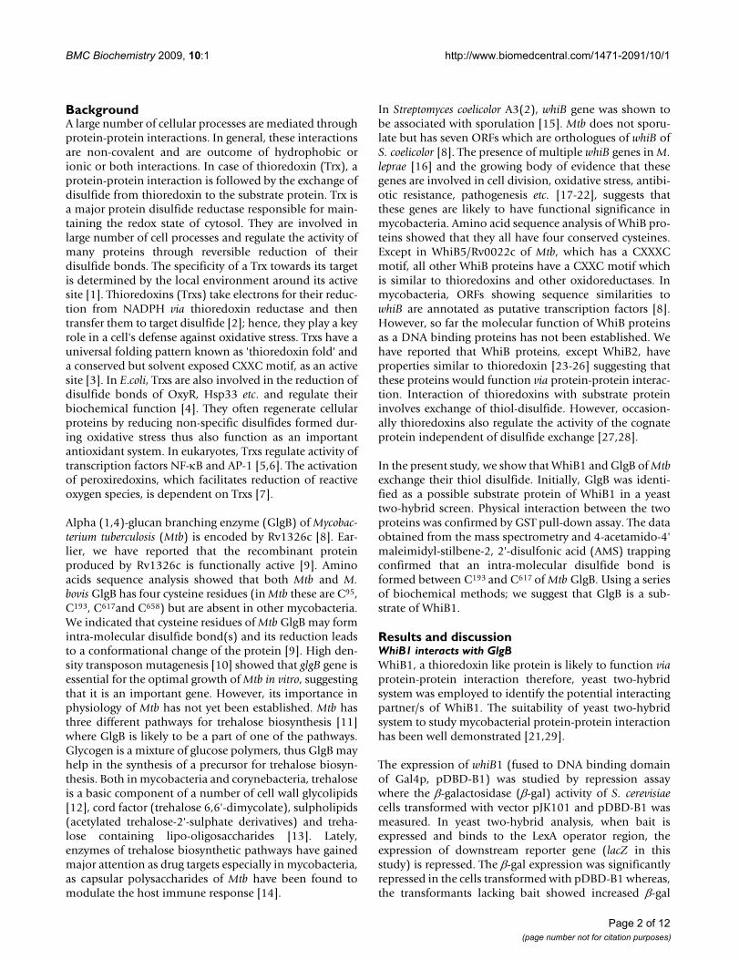
At the end of the screening process, we obtained sixteenclones exclusively growing on galactose medium in theabsence of leucine (Figure. 1). Among all, 15 clones hadan in-frame fusion with 596 bp 3' end of ORF Rv1326c/glgB whereas one was false positive (vector sequencealone).
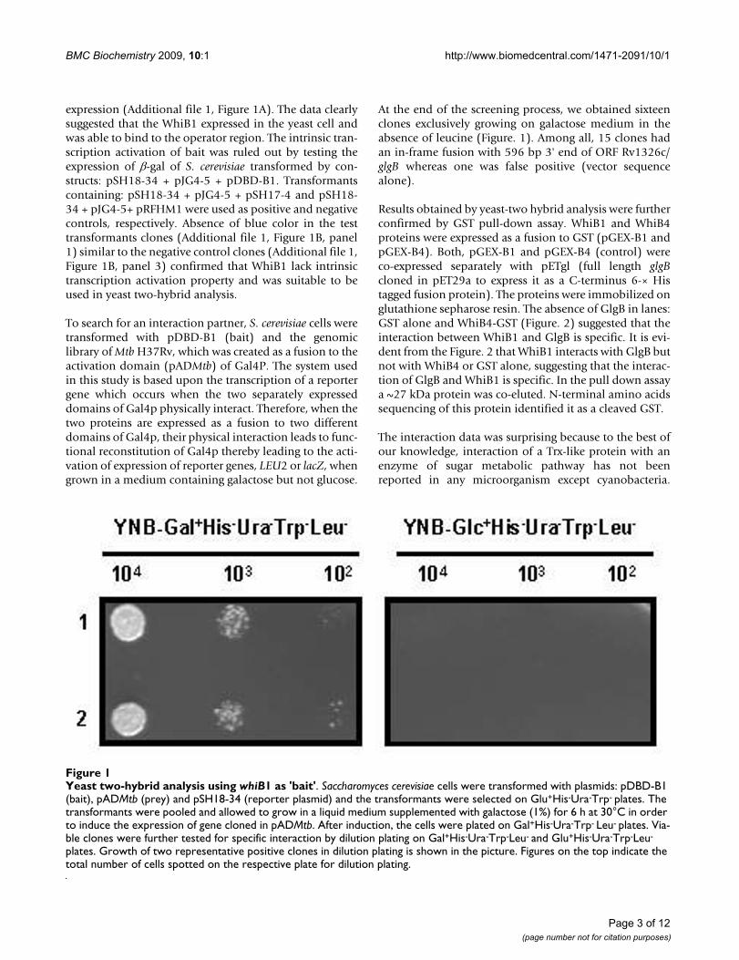
Results obtained by yeast-two hybrid analysis were furtherconfirmed by GST pull-down assay. WhiB1 and WhiB4proteins were expressed as a fusion to GST (pGEX-B1 andpGEX-B4). Both, pGEX-B1 and pGEX-B4 (control) wereco-expressed separately with pETgl (full length glgBcloned in pET29a to express it as a C-terminus 6-× Histagged fusion protein). The proteins were immobilized onglutathione sepharose resin. The absence of GlgB in lanes:GST alone and WhiB4-GST (Figure. 2) suggested that theinteraction between WhiB1 and GlgB is specific. It is evi-dent from the Figure. 2 that WhiB1 interacts with GlgB butnot with WhiB4 or GST alone, suggesting that the interac-tion of GlgB and WhiB1 is specific. In the pull down assaya ~27 kDa protein was co-eluted. N-terminal amino acidssequencing of this protein identified it as a cleaved GST.
The interaction data was surprising because to the best ofour knowledge, interaction of a Trx-like protein with anenzyme of sugar metabolic pathway has not beenreported in any microorganism except cyanobacteria.
Yeast two-hybrid analysis using whiB1 as 'bait'Figure 1Yeast two-hybrid analysis using whiB1 as 'bait'. Saccharomyces cerevisiae cells were transformed with plasmids: pDBD-B1 (bait), pADMtb (prey) and pSH18-34 (reporter plasmid) and the transformants were selected on Glu+His-Ura-Trp- plates. The transformants were pooled and allowed to grow in a liquid medium supplemented with galactose (1%) for 6 h at 30°C in order to induce the expression of gene cloned in pADMtb. After induction, the cells were plated on Gal+His-Ura-Trp- Leu- plates. Via-ble clones were further tested for specific interaction by dilution plating on Gal+His-Ura-Trp-Leu- and Glu+His-Ura-Trp-Leu-
plates. Growth of two representative positive clones in dilution plating is shown in the picture. Figures on the top indicate the total number of cells spotted on the respective plate for dilution plating.
Page 3 of 12(page number not for citation purposes)
BMC Biochemistry 2009, 10:1 http://www.biomedcentral.com/1471-2091/10/1
Amino acids sequence analysis of GlgBs from differentorganisms showed that except Mtb and M. bovis, no otherGlgB has four cysteine residues. Earlier, we have reportedthat Mtb GlgB has at least one intra-molecular disulfidebond [9]. Therefore, we investigated the number andarrangement of intra-molecular disulfide bond(s) inGlgB.
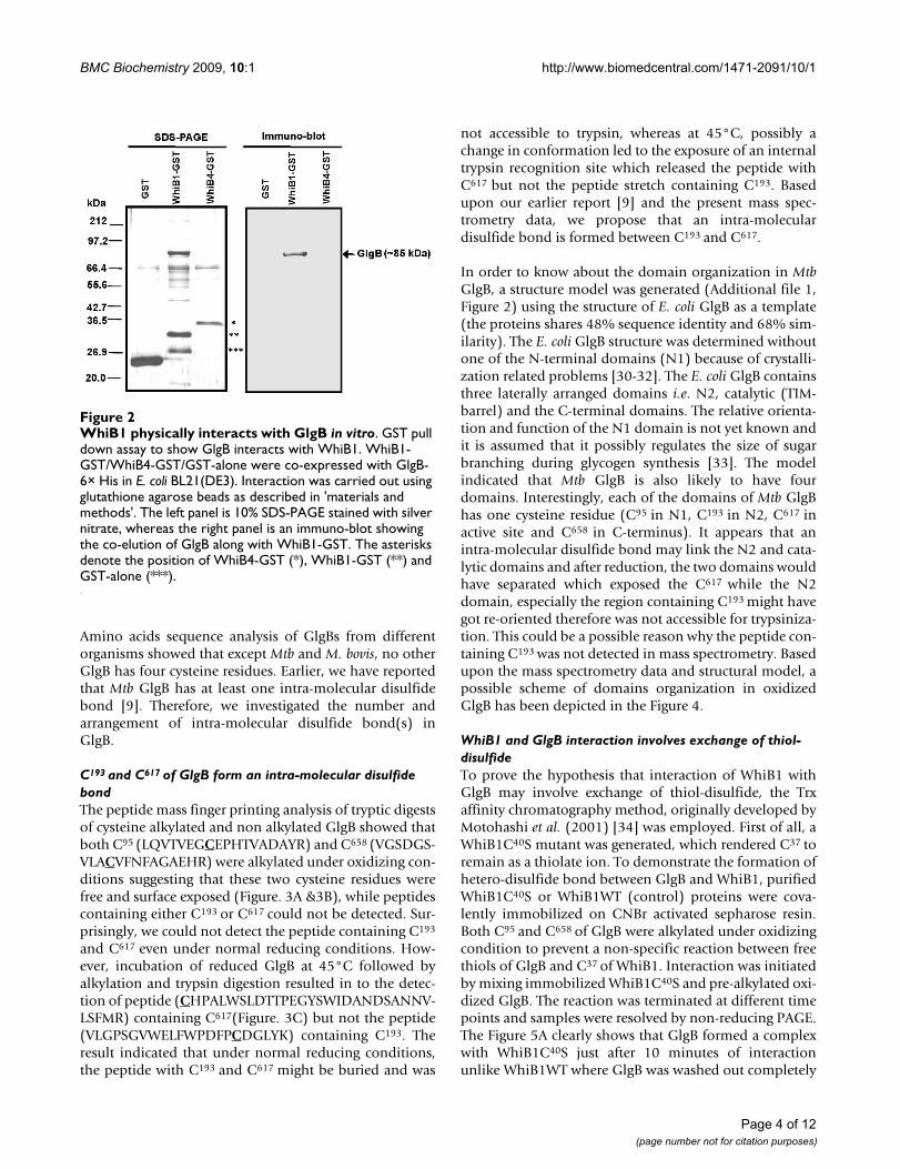
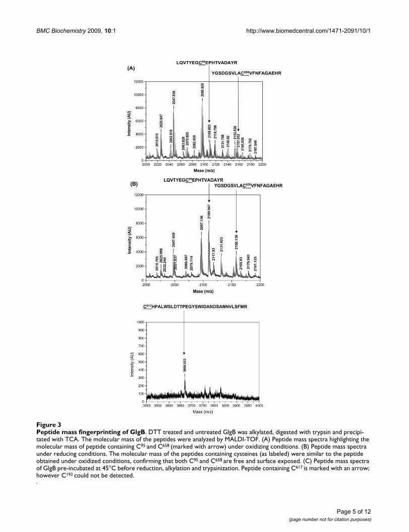
C193 and C617 of GlgB form an intra-molecular disulfide bondThe peptide mass finger printing analysis of tryptic digestsof cysteine alkylated and non alkylated GlgB showed thatboth C95 (LQVTVEGCEPHTVADAYR) and C658 (VGSDGS-VLACVFNFAGAEHR) were alkylated under oxidizing con-ditions suggesting that these two cysteine residues werefree and surface exposed (Figure. 3A &3B), while peptidescontaining either C193 or C617 could not be detected. Sur-prisingly, we could not detect the peptide containing C193
and C617 even under normal reducing conditions. How-ever, incubation of reduced GlgB at 45°C followed byalkylation and trypsin digestion resulted in to the detec-tion of peptide (CHPALWSLDTTPEGYSWIDANDSANNV-LSFMR) containing C617(Figure. 3C) but not the peptide(VLGPSGVWELFWPDFPCDGLYK) containing C193. Theresult indicated that under normal reducing conditions,the peptide with C193 and C617 might be buried and was
not accessible to trypsin, whereas at 45°C, possibly achange in conformation led to the exposure of an internaltrypsin recognition site which released the peptide withC617 but not the peptide stretch containing C193. Basedupon our earlier report [9] and the present mass spec-trometry data, we propose that an intra-moleculardisulfide bond is formed between C193 and C617.
In order to know about the domain organization in MtbGlgB, a structure model was generated (Additional file 1,Figure 2) using the structure of E. coli GlgB as a template(the proteins shares 48% sequence identity and 68% sim-ilarity). The E. coli GlgB structure was determined withoutone of the N-terminal domains (N1) because of crystalli-zation related problems [30-32]. The E. coli GlgB containsthree laterally arranged domains i.e. N2, catalytic (TIM-barrel) and the C-terminal domains. The relative orienta-tion and function of the N1 domain is not yet known andit is assumed that it possibly regulates the size of sugarbranching during glycogen synthesis [33]. The modelindicated that Mtb GlgB is also likely to have fourdomains. Interestingly, each of the domains of Mtb GlgBhas one cysteine residue (C95 in N1, C193 in N2, C617 inactive site and C658 in C-terminus). It appears that anintra-molecular disulfide bond may link the N2 and cata-lytic domains and after reduction, the two domains wouldhave separated which exposed the C617 while the N2domain, especially the region containing C193 might havegot re-oriented therefore was not accessible for trypsiniza-tion. This could be a possible reason why the peptide con-taining C193 was not detected in mass spectrometry. Basedupon the mass spectrometry data and structural model, apossible scheme of domains organization in oxidizedGlgB has been depicted in the Figure 4.
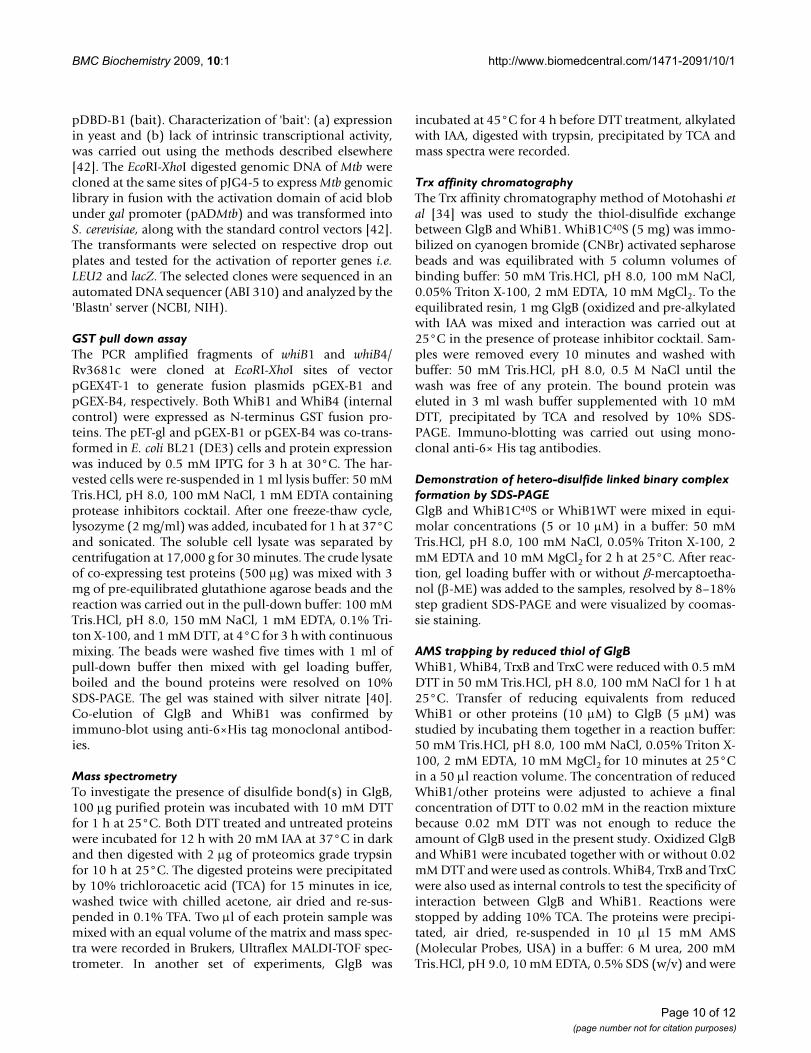
WhiB1 and GlgB interaction involves exchange of thiol-disulfideTo prove the hypothesis that interaction of WhiB1 withGlgB may involve exchange of thiol-disulfide, the Trxaffinity chromatography method, originally developed byMotohashi et al. (2001) [34] was employed. First of all, aWhiB1C40S mutant was generated, which rendered C37 toremain as a thiolate ion. To demonstrate the formation ofhetero-disulfide bond between GlgB and WhiB1, purifiedWhiB1C40S or WhiB1WT (control) proteins were cova-lently immobilized on CNBr activated sepharose resin.Both C95 and C658 of GlgB were alkylated under oxidizingcondition to prevent a non-specific reaction between freethiols of GlgB and C37 of WhiB1. Interaction was initiatedby mixing immobilized WhiB1C40S and pre-alkylated oxi-dized GlgB. The reaction was terminated at different timepoints and samples were resolved by non-reducing PAGE.The Figure 5A clearly shows that GlgB formed a complexwith WhiB1C40S just after 10 minutes of interactionunlike WhiB1WT where GlgB was washed out completely
WhiB1 physically interacts with GlgB in vitroFigure 2WhiB1 physically interacts with GlgB in vitro. GST pull down assay to show GlgB interacts with WhiB1. WhiB1-GST/WhiB4-GST/GST-alone were co-expressed with GlgB-6× His in E. coli BL21(DE3). Interaction was carried out using glutathione agarose beads as described in 'materials and methods'. The left panel is 10% SDS-PAGE stained with silver nitrate, whereas the right panel is an immuno-blot showing the co-elution of GlgB along with WhiB1-GST. The asterisks denote the position of WhiB4-GST (*), WhiB1-GST (**) and GST-alone (***).
Page 4 of 12(page number not for citation purposes)
BMC Biochemistry 2009, 10:1 http://www.biomedcentral.com/1471-2091/10/1
Page 5 of 12(page number not for citation purposes)
Peptide mass fingerprinting of GlgBFigure 3Peptide mass fingerprinting of GlgB. DTT treated and untreated GlgB was alkylated, digested with trypsin and precipi-tated with TCA. The molecular mass of the peptides were analyzed by MALDI-TOF. (A) Peptide mass spectra highlighting the molecular mass of peptide containing C95 and C658 (marked with arrow) under oxidizing conditions. (B) Peptide mass spectra under reducing conditions. The molecular mass of the peptides containing cysteines (as labeled) were similar to the peptide obtained under oxidized conditions, confirming that both C95 and C658 are free and surface exposed. (C) Peptide mass spectra of GlgB pre-incubated at 45°C before reduction, alkylation and trypsinization. Peptide containing C617 is marked with an arrow; however C193 could not be detected.

BMC Biochemistry 2009, 10:1 http://www.biomedcentral.com/1471-2091/10/1
in high salt buffer. The result indicated that a binary com-plex formed between GlgB and WhiB1C40S was stabilizedby a hetero-disulfide bond.
In Trx affinity chromatography, the binary complexformed between the two proteins is not released becauseWhiB1/WhiB1C40S is covalently bound to the resin. Inorder to demonstrate that GlgB and WhiB1 are indeedlinked by a hetero-disulfide bond, purified pre-alkylatedoxidized GlgB and WhiB1C40S were incubated together inthe interaction buffer. The reaction was terminated at dif-ferent time points and the samples were resolved by SDS-PAGE. A higher molecular weight protein band (~97 kDa)along with the two input bands of GlgB (~85 kDa) andWhiB1C40S (~14 kDa), was clearly visible (Figure. 5B,lanes 3 & 4). The ~97 kDa protein band corresponded tothe combined sizes of GlgB: WhiB1C40S binary complexwhich disappeared when 1% β-ME was added to the sam-ple (Figure. 5B, lanes 12 & 13), thus confirming the for-mation of an inter-molecular hetero-disulfide bondbetween GlgB and WhiB1C40S. As expected, the ~97 kDaband was missing in the GlgB: WhiB1WT control sample(Figure. 5B, lanes 7 & 8) because the intact CXXC motif ofWhiB1WT could not form a stable inter-molecular hetero-disulfide bond with GlgB.
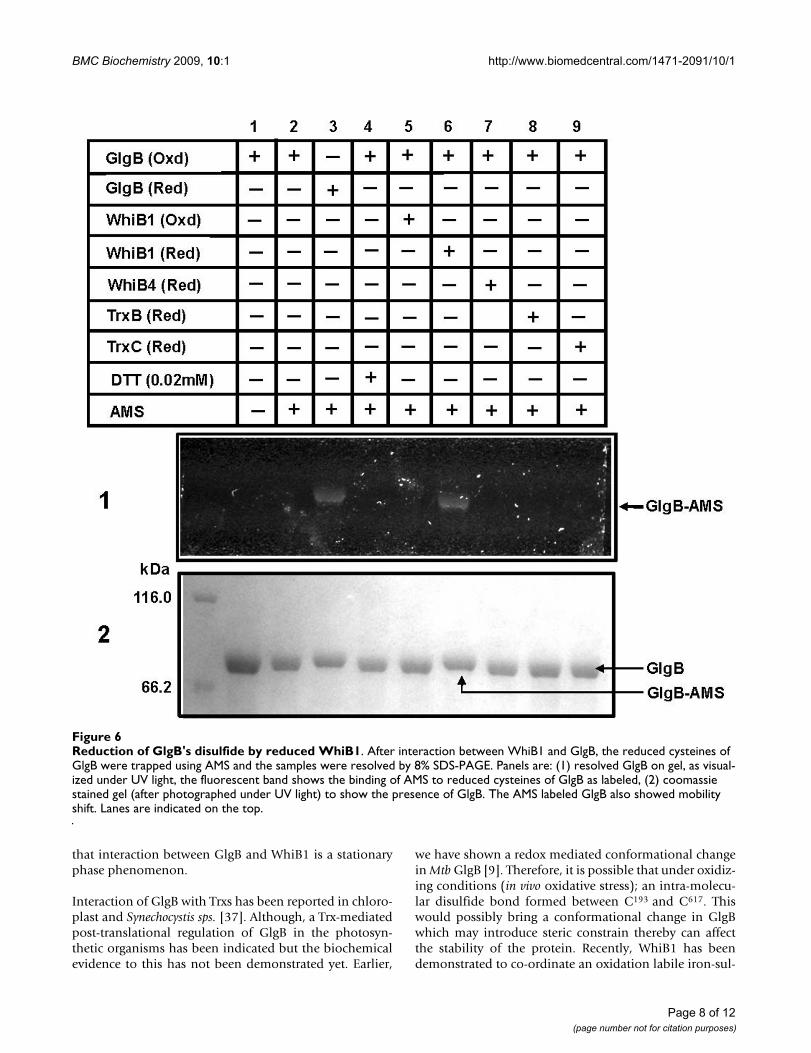
The direct evidence for the reduction of GlgB disulfide byWhiB1 was shown using AMS trapping method. The fluo-rescent dye, AMS, absorbs light at 322 nm and emits at
411 nm. Binding of each molecule of AMS also increasesthe mass of a protein by ~500 Da. Thus, AMS labeled pro-teins fluoresce under UV light and show reduced mobilityon SDS-PAGE. Purified Mtb GlgB was pre-alkylated underoxidizing condition with iodoacetamide (IAA) to blockthe free cysteines thus AMS binding would occur if C193
and C617 are free either due to the reduction by WhiB1 orDTT or by other Trx-like proteins. The AMS treatment ofoxidized and pre-alkylated GlgB did not show any fluores-cence or mobility shift (Figure. 6, lane 2), suggesting thatthe free cysteines were successfully alkylated. Pre-alkylated GlgB when reduced by DTT and then labeledwith AMS fluoresced under UV light and also showedmobility shift (Figure. 6, lane 3). Similarly, when pre-alkylated GlgB was incubated with pre-reduced WhiB1and labeled with AMS, it fluoresced under UV light andshowed mobility shift on the gel (Figure. 6, lane 6). How-ever, pre-alkylated GlgB incubated with oxidized WhiB1and then labeled with AMS failed to fluoresce and also didnot show any mobility shift (Figure. 6, lane 5). Together,these data suggested that WhiB1 converted the disulfide ofGlgB into reduced thiol, to which AMS reacted covalently.In order to know whether GlgB's disulfide could bereduced by other Trxs of Mtb; TrxB and TrxC of Mtb werecloned, expressed and proteins were purified (Additionalfile 1, Figure 3). Both, TrxB and TrxC catalyzed the reduc-tion of insulin disulfides by DTT suggesting that the pro-teins were in native conformation and enzymaticallyactive (Additional file 2, Table 1). However, incubation ofGlgB with either reduced WhiB4 or TrxB or TrxC did notshow AMS labeling of GlgB (Figure. 6, lane 7–9). The dataclearly suggested that the transfer of reducing equivalentsfrom WhiB1 to GlgB is a highly specific reaction, as itsabsence could not be compensated either by known Trxsor WhiB4 of Mtb. The result also confirmed that C193 andC617 of GlgB form an intra-molecular disulfide bond.
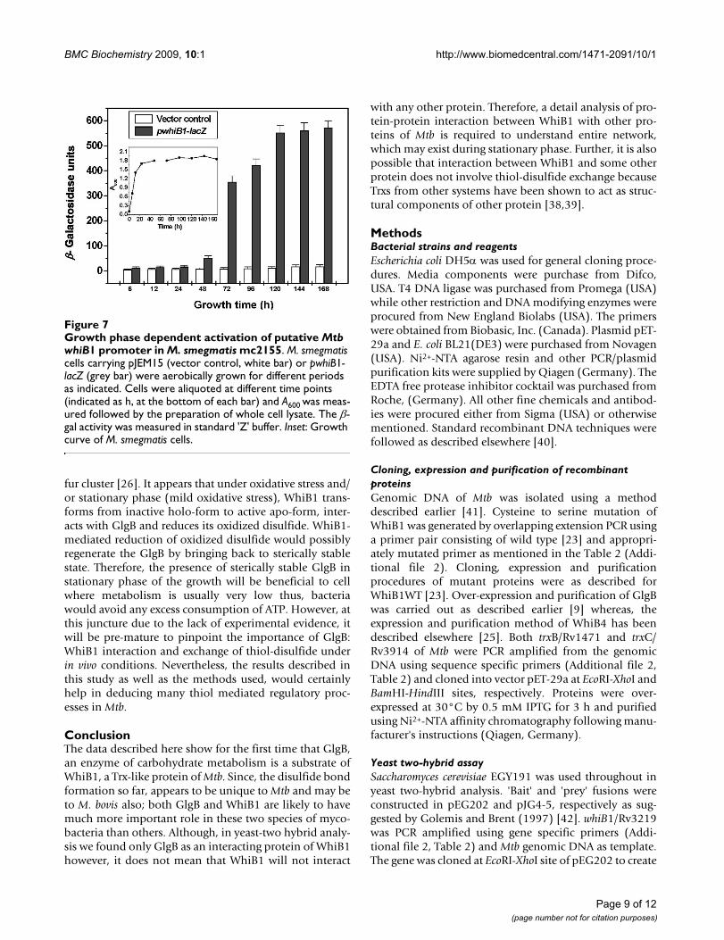
WhiB1 promoter activates at late stationary phaseFor a successful interaction between two proteins, it isessential that both are present in the cytosol at the sametime. The glycogen synthesis in S. coelicolor A3(2) hasbeen shown to be associated with sporulation which rep-resent stationary phase of the growth [35]. In order todetermine the stage when whiB1 is expressed, theupstream regulatory sequence of whiB1 was clonedupstream to the lacZ of a promoter less vector pJEM15[36]. The expression of β-gal was monitored at differentstages of growth. For the first 24 h, the expression was atthe basal level. However, it induced after 48 h of growthand increased suddenly by 72 h (Figure. 7). The increasein β-gal activity continued up to 120 h and saturated after-wards, suggesting that the whiB1 expression is likely to beat the late stationary phase of the growth. Increased levelof whiB1 transcripts in stationary phase has also beendemonstrated by RT-PCR [19]. Therefore, it is possible
Possible domain organization of Mtb GlgBFigure 4Possible domain organization of Mtb GlgB. The sche-matic arrangement of the four domains of Mtb GlgB is based on mass spectrometric data and structure model. Cysteines (C193 and C617) of GlgB which form an intra-molecular disulfide bond are indicated.
Page 6 of 12(page number not for citation purposes)

BMC Biochemistry 2009, 10:1 http://www.biomedcentral.com/1471-2091/10/1
Page 7 of 12(page number not for citation purposes)
Thioredoxin affinity chromatographyFigure 5Thioredoxin affinity chromatography. (A) Immuno-blot after thioredoxin affinity chromatography to show that the binary complex between GlgB and WhiB1C40S was destabilized by DTT. Panels: (1) interaction of GlgB with WhiB1C40S, (2) interac-tion of GlgB with WhiB1WT. (B) In-solution protein-protein interaction to show the formation of a binary complex between GlgB and WhiB1C40S. After reactions, the samples were resolved by 8–18% step gradient SDS-PAGE and stained with coomas-sie blue. A binary complex between WhiB1C40S and GlgB at ~97 kDa marker position (lanes 3 & 4) disappeared when β-ME was added (lanes 12 & 13). Rests of the lanes are controls as labeled. The triangles represent the interaction carried out using two different concentrations of WhiB1 and GlgB (5 or 10 μM of each protein).
BMC Biochemistry 2009, 10:1 http://www.biomedcentral.com/1471-2091/10/1
that interaction between GlgB and WhiB1 is a stationaryphase phenomenon.
Interaction of GlgB with Trxs has been reported in chloro-plast and Synechocystis sps. [37]. Although, a Trx-mediatedpost-translational regulation of GlgB in the photosyn-thetic organisms has been indicated but the biochemicalevidence to this has not been demonstrated yet. Earlier,
we have shown a redox mediated conformational changein Mtb GlgB [9]. Therefore, it is possible that under oxidiz-ing conditions (in vivo oxidative stress); an intra-molecu-lar disulfide bond formed between C193 and C617. Thiswould possibly bring a conformational change in GlgBwhich may introduce steric constrain thereby can affectthe stability of the protein. Recently, WhiB1 has beendemonstrated to co-ordinate an oxidation labile iron-sul-
Reduction of GlgB's disulfide by reduced WhiB1Figure 6Reduction of GlgB's disulfide by reduced WhiB1. After interaction between WhiB1 and GlgB, the reduced cysteines of GlgB were trapped using AMS and the samples were resolved by 8% SDS-PAGE. Panels are: (1) resolved GlgB on gel, as visual-ized under UV light, the fluorescent band shows the binding of AMS to reduced cysteines of GlgB as labeled, (2) coomassie stained gel (after photographed under UV light) to show the presence of GlgB. The AMS labeled GlgB also showed mobility shift. Lanes are indicated on the top.
Page 8 of 12(page number not for citation purposes)
BMC Biochemistry 2009, 10:1 http://www.biomedcentral.com/1471-2091/10/1
fur cluster [26]. It appears that under oxidative stress and/or stationary phase (mild oxidative stress), WhiB1 trans-forms from inactive holo-form to active apo-form, inter-acts with GlgB and reduces its oxidized disulfide. WhiB1-mediated reduction of oxidized disulfide would possiblyregenerate the GlgB by bringing back to sterically stablestate. Therefore, the presence of sterically stable GlgB instationary phase of the growth will be beneficial to cellwhere metabolism is usually very low thus, bacteriawould avoid any excess consumption of ATP. However, atthis juncture due to the lack of experimental evidence, itwill be pre-mature to pinpoint the importance of GlgB:WhiB1 interaction and exchange of thiol-disulfide underin vivo conditions. Nevertheless, the results described inthis study as well as the methods used, would certainlyhelp in deducing many thiol mediated regulatory proc-esses in Mtb.
ConclusionThe data described here show for the first time that GlgB,an enzyme of carbohydrate metabolism is a substrate ofWhiB1, a Trx-like protein of Mtb. Since, the disulfide bondformation so far, appears to be unique to Mtb and may beto M. bovis also; both GlgB and WhiB1 are likely to havemuch more important role in these two species of myco-bacteria than others. Although, in yeast-two hybrid analy-sis we found only GlgB as an interacting protein of WhiB1however, it does not mean that WhiB1 will not interact
with any other protein. Therefore, a detail analysis of pro-tein-protein interaction between WhiB1 with other pro-teins of Mtb is required to understand entire network,which may exist during stationary phase. Further, it is alsopossible that interaction between WhiB1 and some otherprotein does not involve thiol-disulfide exchange becauseTrxs from other systems have been shown to act as struc-tural components of other protein [38,39].
MethodsBacterial strains and reagentsEscherichia coli DH5α was used for general cloning proce-dures. Media components were purchase from Difco,USA. T4 DNA ligase was purchased from Promega (USA)while other restriction and DNA modifying enzymes wereprocured from New England Biolabs (USA). The primerswere obtained from Biobasic, Inc. (Canada). Plasmid pET-29a and E. coli BL21(DE3) were purchased from Novagen(USA). Ni2+-NTA agarose resin and other PCR/plasmidpurification kits were supplied by Qiagen (Germany). TheEDTA free protease inhibitor cocktail was purchased fromRoche, (Germany). All other fine chemicals and antibod-ies were procured either from Sigma (USA) or otherwisementioned. Standard recombinant DNA techniques werefollowed as described elsewhere [40].
Cloning, expression and purification of recombinant proteinsGenomic DNA of Mtb was isolated using a methoddescribed earlier [41]. Cysteine to serine mutation ofWhiB1 was generated by overlapping extension PCR usinga primer pair consisting of wild type [23] and appropri-ately mutated primer as mentioned in the Table 2 (Addi-tional file 2). Cloning, expression and purificationprocedures of mutant proteins were as described forWhiB1WT [23]. Over-expression and purification of GlgBwas carried out as described earlier [9] whereas, theexpression and purification method of WhiB4 has beendescribed elsewhere [25]. Both trxB/Rv1471 and trxC/Rv3914 of Mtb were PCR amplified from the genomicDNA using sequence specific primers (Additional file 2,Table 2) and cloned into vector pET-29a at EcoRI-XhoI andBamHI-HindIII sites, respectively. Proteins were over-expressed at 30°C by 0.5 mM IPTG for 3 h and purifiedusing Ni2+-NTA affinity chromatography following manu-facturer's instructions (Qiagen, Germany).
Yeast two-hybrid assaySaccharomyces cerevisiae EGY191 was used throughout inyeast two-hybrid analysis. 'Bait' and 'prey' fusions wereconstructed in pEG202 and pJG4-5, respectively as sug-gested by Golemis and Brent (1997) [42]. whiB1/Rv3219was PCR amplified using gene specific primers (Addi-tional file 2, Table 2) and Mtb genomic DNA as template.The gene was cloned at EcoRI-XhoI site of pEG202 to create
Growth phase dependent activation of putative Mtb whiB1 promoter in M. smegmatis mc2155Figure 7Growth phase dependent activation of putative Mtb whiB1 promoter in M. smegmatis mc2155. M. smegmatis cells carrying pJEM15 (vector control, white bar) or pwhiB1-lacZ (grey bar) were aerobically grown for different periods as indicated. Cells were aliquoted at different time points (indicated as h, at the bottom of each bar) and A600 was meas-ured followed by the preparation of whole cell lysate. The β-gal activity was measured in standard 'Z' buffer. Inset: Growth curve of M. smegmatis cells.
Page 9 of 12(page number not for citation purposes)
BMC Biochemistry 2009, 10:1 http://www.biomedcentral.com/1471-2091/10/1
pDBD-B1 (bait). Characterization of 'bait': (a) expressionin yeast and (b) lack of intrinsic transcriptional activity,was carried out using the methods described elsewhere[42]. The EcoRI-XhoI digested genomic DNA of Mtb werecloned at the same sites of pJG4-5 to express Mtb genomiclibrary in fusion with the activation domain of acid blobunder gal promoter (pADMtb) and was transformed intoS. cerevisiae, along with the standard control vectors [42].The transformants were selected on respective drop outplates and tested for the activation of reporter genes i.e.LEU2 and lacZ. The selected clones were sequenced in anautomated DNA sequencer (ABI 310) and analyzed by the'Blastn' server (NCBI, NIH).
GST pull down assayThe PCR amplified fragments of whiB1 and whiB4/Rv3681c were cloned at EcoRI-XhoI sites of vectorpGEX4T-1 to generate fusion plasmids pGEX-B1 andpGEX-B4, respectively. Both WhiB1 and WhiB4 (internalcontrol) were expressed as N-terminus GST fusion pro-teins. The pET-gl and pGEX-B1 or pGEX-B4 was co-trans-formed in E. coli BL21 (DE3) cells and protein expressionwas induced by 0.5 mM IPTG for 3 h at 30°C. The har-vested cells were re-suspended in 1 ml lysis buffer: 50 mMTris.HCl, pH 8.0, 100 mM NaCl, 1 mM EDTA containingprotease inhibitors cocktail. After one freeze-thaw cycle,lysozyme (2 mg/ml) was added, incubated for 1 h at 37°Cand sonicated. The soluble cell lysate was separated bycentrifugation at 17,000 g for 30 minutes. The crude lysateof co-expressing test proteins (500 μg) was mixed with 3mg of pre-equilibrated glutathione agarose beads and thereaction was carried out in the pull-down buffer: 100 mMTris.HCl, pH 8.0, 150 mM NaCl, 1 mM EDTA, 0.1% Tri-ton X-100, and 1 mM DTT, at 4°C for 3 h with continuousmixing. The beads were washed five times with 1 ml ofpull-down buffer then mixed with gel loading buffer,boiled and the bound proteins were resolved on 10%SDS-PAGE. The gel was stained with silver nitrate [40].Co-elution of GlgB and WhiB1 was confirmed byimmuno-blot using anti-6×His tag monoclonal antibod-ies.
Mass spectrometryTo investigate the presence of disulfide bond(s) in GlgB,100 μg purified protein was incubated with 10 mM DTTfor 1 h at 25°C. Both DTT treated and untreated proteinswere incubated for 12 h with 20 mM IAA at 37°C in darkand then digested with 2 μg of proteomics grade trypsinfor 10 h at 25°C. The digested proteins were precipitatedby 10% trichloroacetic acid (TCA) for 15 minutes in ice,washed twice with chilled acetone, air dried and re-sus-pended in 0.1% TFA. Two μl of each protein sample wasmixed with an equal volume of the matrix and mass spec-tra were recorded in Brukers, Ultraflex MALDI-TOF spec-trometer. In another set of experiments, GlgB was
incubated at 45°C for 4 h before DTT treatment, alkylatedwith IAA, digested with trypsin, precipitated by TCA andmass spectra were recorded.
Trx affinity chromatographyThe Trx affinity chromatography method of Motohashi etal [34] was used to study the thiol-disulfide exchangebetween GlgB and WhiB1. WhiB1C40S (5 mg) was immo-bilized on cyanogen bromide (CNBr) activated sepharosebeads and was equilibrated with 5 column volumes ofbinding buffer: 50 mM Tris.HCl, pH 8.0, 100 mM NaCl,0.05% Triton X-100, 2 mM EDTA, 10 mM MgCl2. To theequilibrated resin, 1 mg GlgB (oxidized and pre-alkylatedwith IAA was mixed and interaction was carried out at25°C in the presence of protease inhibitor cocktail. Sam-ples were removed every 10 minutes and washed withbuffer: 50 mM Tris.HCl, pH 8.0, 0.5 M NaCl until thewash was free of any protein. The bound protein waseluted in 3 ml wash buffer supplemented with 10 mMDTT, precipitated by TCA and resolved by 10% SDS-PAGE. Immuno-blotting was carried out using mono-clonal anti-6× His tag antibodies.
Demonstration of hetero-disulfide linked binary complex formation by SDS-PAGEGlgB and WhiB1C40S or WhiB1WT were mixed in equi-molar concentrations (5 or 10 μM) in a buffer: 50 mMTris.HCl, pH 8.0, 100 mM NaCl, 0.05% Triton X-100, 2mM EDTA and 10 mM MgCl2 for 2 h at 25°C. After reac-tion, gel loading buffer with or without β-mercaptoetha-nol (β-ME) was added to the samples, resolved by 8–18%step gradient SDS-PAGE and were visualized by coomas-sie staining.
AMS trapping by reduced thiol of GlgBWhiB1, WhiB4, TrxB and TrxC were reduced with 0.5 mMDTT in 50 mM Tris.HCl, pH 8.0, 100 mM NaCl for 1 h at25°C. Transfer of reducing equivalents from reducedWhiB1 or other proteins (10 μM) to GlgB (5 μM) wasstudied by incubating them together in a reaction buffer:50 mM Tris.HCl, pH 8.0, 100 mM NaCl, 0.05% Triton X-100, 2 mM EDTA, 10 mM MgCl2 for 10 minutes at 25°Cin a 50 μl reaction volume. The concentration of reducedWhiB1/other proteins were adjusted to achieve a finalconcentration of DTT to 0.02 mM in the reaction mixturebecause 0.02 mM DTT was not enough to reduce theamount of GlgB used in the present study. Oxidized GlgBand WhiB1 were incubated together with or without 0.02mM DTT and were used as controls. WhiB4, TrxB and TrxCwere also used as internal controls to test the specificity ofinteraction between GlgB and WhiB1. Reactions werestopped by adding 10% TCA. The proteins were precipi-tated, air dried, re-suspended in 10 μl 15 mM AMS(Molecular Probes, USA) in a buffer: 6 M urea, 200 mMTris.HCl, pH 9.0, 10 mM EDTA, 0.5% SDS (w/v) and were
Page 10 of 12(page number not for citation purposes)
BMC Biochemistry 2009, 10:1 http://www.biomedcentral.com/1471-2091/10/1
incubated at 37°C for 1 h in dark. Labeled proteins wereresolved in 8% non-reducing SDS-PAGE and visualizedunder UV light followed by coomassie staining.
Promoter activation assayA 279 bp region between whiB1/Rv3219 start codon andthe end of Rv3218 was cloned into the blunt ended site ofSacII (upstream to the lacZ gene) of the vector pJEM15.The recombinant construct was named pwhiB1-lacZ. Afterconfirming the sequence and orientation of the clonedfragment, it was transformed into M. smegmatis mc2155.Vector alone was used as a control. Three independentclones were inoculated into 20 ml Middlebrook 7H9medium supplemented with OADC for 24 h (A600 = ~1.8).From this, a fresh medium was inoculated (1%) andgrown at 37°C, cells were collected at different timepoints: 2, 3, 4, 5 and 6 days of growth and lysates wereprepared. An equal amount of protein in triplicate wasassayed for β-gal activity using Miller's method [43]. Theassay was set in a 96 well plate in a 100 μl reaction volumewhich had 1 mg/ml protein and 0.8 mg/ml of ONPG dis-solved in 'Z buffer': 60 mM Na2HPO4, 40 mM NaH2PO4,(pH 7.0), 10 mM KCl, 1 mM MgSO4 and 2.7 μl/ml β-ME.As yellow color started to appear, the reaction was stoppedby adding 50 μl of 1 M Na2CO3 and A420 was measured.The β-gal activity was calculated in Miller's units by usingthe formula A420/protein concentration (mg/ml) × time inminutes.
MiscellaneousWhiB1 concentration was estimated using molar extinc-tion coefficient (∈280 nm = 17550 M-1cm-1). Protein con-centration of cell lysate and GlgB was estimated byBradford's method using BSA as a standard. The nucle-otide sequence of all recombinant plasmids were authen-ticated by sequencing both strands of DNA in an ABI 321automated DNA sequencer.
Except in GST pull down assay, throughout the interactionstudies, pre-alkylated GlgB was used. Briefly, free cysteines(C95 and C658) of GlgB were blocked by incubating theprotein in 50 mM Tris.HCl, pH 8.0, 100 mM NaCl and 50mM IAA for 12 h at 37°C in dark. The unbound IAA wasremoved by extensive dialysis against buffer: 50 mMTris.HCl, pH 8.0, 100 mM NaCl.
AbbreviationsAMS: 4-acetamido-4' maleimidyl-stilbene-2: 2'-disulfonicacid; GlgB: α-(1,4)-glucan branching enzyme; Mtb: Myco-bacterium tuberculosis; IAA: iodoacetamide; Trx: thiore-doxin; GST: glutathione S-transferase; CNBr: cyanogenbromide; DTT: dithiothreitol; β-ME: β mercaptoethanol
Authors' contributionsSKG and MSA carried out all the expression; purification;mutagenesis and biochemical analysis together. KVRK car-
ried out the molecular modeling of Mtb GlgB. RB helpedin carrying out some of the biochemical studies. SKG,MSA and PA designed the experiments. MSA and PAtogether wrote the manuscript. PA oversaw the study. Allauthors read and approved the final manuscript.
Additional material
AcknowledgementsSKG, MSA and RB thank CSIR, India for a Senior Research Fellowship. Sin-cere thanks to Dr. A. K. Bachhawat for providing the strains and vectors for yeast two-hybrid analysis. Plasmid, pJEM15, was a kind gift from Dr. Brigitte Gicquel, Institut Pasteur, Paris, France. Financial support from DBT and CSIR, Govt. of India is acknowledged. The report is IMTECH commu-nication No. 018/2007.
References1. Holmgren A: Thioredoxin. Annu Rev Biochem 1985, 54:237-271.2. Holmgren A: Thioredoxin and glutaredoxin systems. J Biol
Chem 1989, 264:13963-13966.3. Martin JL: Thioredoxin – a fold for all reasons. Structure 1995,
3:245-250.4. Paget MS, Buttner MJ: Thiol-based regulatory switches. Annu Rev
Genet 2003, 37:91-121.5. Schenk H, Klein M, Erdbrugger W, Droge W, Schulze-Osthoff K: Dis-
tinct effects of thioredoxin and antioxidants on the activa-
Additional file 1Figure 1 – Characterization of WhiB1 as a bait. (A) Repression assay to asses the expression and binding of 'bait' to the LexA operator. Four independent transformants of each of the clones were assayed for β-gal expression. Saccharomyces cerevisiae cells (~2 × 10⁷) containing con-structs (labeled on the x-axis), were permeabilized with 3 drops of chloro-form and 2 drops of 0.1% SDS by vigorous vortexing. Reaction was initiated by adding 0.2 ml ONPG (4 mg/ml) in 'Z buffer' for 3.5 minutes at 28°C. The β-gal activity is expressed as units, on y-axis. (B) WhiB1 lacks intrinsic transcription activity. Saccharomyces cerevisiae cells were transformed with pDBD-B1 (bait), pSH18-34 which contains β-gal as a reporter gene and library plasmid pJG4-5. The β-gal expression was mon-itored on the Gal+Raff+Ura-X-gal plate. The panels indicate S. cerevisiae containing (1) pSH18-34 + pJG4-5 + pDBD-B1, test (2) pSH18-34 + pJG4-5+ pSH17-4, positive control (3) pSH18-34 + pJG4-5 + pRFHM1, negative control. Figure 2 – Homology model of Mtb GlgB. Three domains are colored differently: N2 domain-orange; TIM-barrel domain-magenta; C-terminal domain-blue. The linker regions between the domains are shown in red. The position of the cysteine residues (C193, C617and C658) on the individual domains is shown as green sticks encir-cled in dotted boundaries. Figure 3 – Over-expression and purification of TrxB/Rv1471 (1) and TrxC/3914 (2) of Mtb. The proteins were overex-pressed in E. coli BL21 (DE3) and were purified by Ni2+-NTA affinity chromatography. Lanes are indicated on the top.Click here for file[http://www.biomedcentral.com/content/supplementary/1471-2091-10-1-S1.pdf]
Additional file 2Table 1 – Protein disulfide reductase activity of Mtb TrxB and TrxC. Table 2 – List of primers used in this studyClick here for file[http://www.biomedcentral.com/content/supplementary/1471-2091-10-1-S2.pdf]
Page 11 of 12(page number not for citation purposes)
http://www.ncbi.nlm.nih.gov/entrez/query.fcgi?cmd=Retrieve&db=PubMed&dopt=Abstract&list_uids=3896121
http://www.ncbi.nlm.nih.gov/entrez/query.fcgi?cmd=Retrieve&db=PubMed&dopt=Abstract&list_uids=2668278
http://www.ncbi.nlm.nih.gov/entrez/query.fcgi?cmd=Retrieve&db=PubMed&dopt=Abstract&list_uids=7788290
BMC Biochemistry 2009, 10:1 http://www.biomedcentral.com/1471-2091/10/1
tion of transcription factors NF-kappa B and AP-1. Proc NatlAcad Sci USA 1994, 91:1672-1676.
6. Hayashi T, Ueno Y, Okamoto T: Oxidoreductive regulation ofnuclear factor kappa B. Involvement of a cellular reducingcatalyst thioredoxin. J Biol Chem 1993, 268:11380-11388.
7. Hosoya-Matsuda N, Motohashi K, Yoshimura H, Nozaki A, Inoue K,Ohmori M, Hisabori T: Anti-oxidative stress system in cyano-bacteria. Significance of type II peroxiredoxin and the role of1-Cys peroxiredoxin in Synechocystis sp. strain PCC 6803. JBiol Chem 2005, 280:840-846.
8. Cole ST, Brosch R, Parkhill J, Garnier T, Churcher C, Harris D, Gor-don SV, Eiglmeier K, Gas S, Barry CE 3rd, Tekaia F, Badcock K,Basham D, Brown D, Chillingworth T, Connor R, Davies R, Devlin K,Feltwell T, Gentles S, Hamlin N, Holroyd S, Hornsby T, Jagels K,Krogh A, McLean J, Moule S, Murphy L, Oliver K, Osborne J, QuailMA, Rajandream MA, Rogers J, Rutter S, Seeger K, Skelton J, SquaresR, Squares S, Sulston JE, Taylor K, Whitehead S, Barrell BG: Deci-phering the biology of Mycobacterium tuberculosis from thecomplete genome sequence. Nature 1998, 393:537-544.
9. Garg SK, Alam MS, Kishan KVR, Agrawal P: Expression and char-acterization of α-(1,4)-glucan branching enzyme of Mycobac-terium tuberculosis H37Rv. Protein Expr Purif 2007, 51:198-208.
10. Sassetti CM, Boyd DH, Rubin EJ: Genes required for mycobacte-rial growth by high density mutagenesis. Mol Microbiol 2003,48:77-84.
11. De Smet KAL, Weston A, Brown IN, Young DB, Robertson BD:Three pathways for trehalose biosynthesis in Mycobacteria.Microbiology 2000, 146:199-208.
12. Lederer E: Cord factor and related trehalose esters. Chem PhysLipids 1976, 16:91-106.
13. Besra GS, Chatterjee D: Lipids and carbohydrates of Mycobac-terium tuberculosis. In Tuberculosis: Pathogenesis, protection and con-trol Edited by: Bloom BR. American Society for Microbiology,Washington, D. C; 1994:285-306.
14. Cywes C, Hoppe H, Daffe C, Ehlers MR: Nonopsonic binding ofMycobacterium tuberculosis to complement receptor type 3is mediated by capsular polysaccharides and is straindependent. Infect Immun 1997, 65:4258-4266.
15. Davis NK, Chater KF: The Streptomyces coelicolor whiB geneencodes a small transcription factor-like protein dispensablefor growth but essential for sporulation. Mol Gen Genet 1992,232:351-358.
16. Cole ST, Eiglmeier K, Parkhill J, James KD, Thomson NR, WheelerPR, Honoré N, Garnier T, Churcher C, Harris D, Mungall K, BashamD, Brown D, Chillingworth T, Connor R, Davies RM, Devlin K,Duthoy S, Feltwell T, Fraser A, Hamlin N, Holroyd S, Hornsby T, Jag-els K, Lacroix C, Maclean J, Moule S, Murphy L, Oliver K, Quail MA,Rajandream MA, Rutherford KM, Rutter S, Seeger K, Simon S, Sim-monds M, Skelton J, Squares R, Squares S, Stevens K, Taylor K, White-head S, Woodward JR, Barrell BG: Massive gene decay in theleprosy bacillus. Nature 2001, 409:1007-1001.
17. Raghunand TR, Bishai WR: Mycobacterium smegmatis whmD andits homologue Mycobacterium tuberculosis whiB2 are func-tionally equivalent. Microbiology 2006, 152:2735-2747.
18. Kim TH, Park JS, Kim HJ, Kim Y, Kim P, Lee HS: The whcE gene ofCorynebacterium glutamicum is important for survival follow-ing heat and oxidative stress. Biochem Biophys Res Commun 2005,337:757-764.
19. Geiman DE, Raghunand TR, Agarwal N, Bishai WR: Differentialgene expression in response to exposure to antimycobacte-rial agents and other stress conditions among seven Myco-bacterium tuberculosis whiB-like genes. Antimicrob AgentsChemother 2006, 50:2836-2841.
20. Morris RP, Nguyen L, Gatfield J, Visconti K, Nguyen K, SchnappingerD, Ehrt S, Liu Y, Heifets L, Pieters J, Schoolnik G, Thompson CJ:Ancestral antibiotic resistance in Mycobacterium tuberculosis. Proc Natl Acad Sci USA 2005, 102:12200-12205.
21. Steyn AJ, Collins DM, Hondalus MK, Jacobs WRJ, Kawakami RP,Bloom BR: Mycobacterium tuberculosis WhiB3 interacts withRpoV to affect host survival but is dispensable for in vivogrowth. Proc Natl Acad Sci USA 2002, 99:3147-3152.
22. Singh A, Guidry L, Narasinhulu KV, Mai D, Trombly J, Redding KE,Giles GL, Lancaster JR Jr, Steyn AJC: Mycobacterium tuberculosisWhiB3 responds to O2 and nitric oxide via its [4Fe-4S] clus-ter and is essential for nutrient starvation survival. Proc NatlAcad Sci USA 2007, 104:11562-11567.
23. Garg SK, Alam MS, Soni V, Kishan KVR, Agrawal P: Characteriza-tion of Mycobacterium tuberculosis WhiB1/Rv3219 as a pro-tein disulfide reductase. Protein Expr Purif 2007, 52:422-432.
24. Alam MS, Garg SK, Agrawal P: Molecular function of WhiB4/Rv3681c of Mycobacterium tuberculosis H37Rv: a [4Fe-4S]cluster coordinating protein disulfide reductase. Mol Microbiol2007, 63:1414-1431.
25. Alam MS, Agrawal P: Matrix-assisted refolding and redox prop-erties of WhiB3/Rv3416 of Mycobacterium tuberculosisH37Rv. Protein Expr Purif 2008, 61:83-91.
26. Alam MS, Garg SK, Agrawal P: Studies on structural and func-tional divergence among seven WhiB proteins of Mycobacte-rium tuberculosis H37Rv. FEBS J 2009, 276(1):76-93.
27. Saitoh M, Nishitoh H, Fujii M, Takeda K, Tobiume K, Sawada Y, Kawa-bata M, Miyazono K, Ichijo H: Mammalian thioredoxin is a directinhibitor of apoptosis signal-regulating kinase (ASK) 1. EMBOJ 1998, 17:2596-2606.
28. Liu H, Nishitoh H, Ichijo H, Kyriakis JM: Activation of ApoptosisSignal-Regulating Kinase 1 (ASK1) by Tumor Necrosis Fac-tor Receptor-Associated Factor 2 Requires Prior Dissocia-tion of the ASK1 Inhibitor Thioredoxin. Mol and Cell Biol 2000,20:2198-2208.
29. Parida BK, Douglas T, Nino C, Dhandayuthapani S: Interactions ofanti-sigma factor antagonists of Mycobacterium tuberculosisin the yeast two-hybrid system. Tuberculosis (Edinb.) 2005,85:347-355.
30. Hilden I, Leggio LL, Larsen S, Poulsen P: Characterization andcrystallization of an active N-terminally truncated form ofthe Escherichia coli glycogen branching enzyme. Eur J Biochem2000, 267:2150-2155.
31. Abad MC, Binderup K, Rios-Steiner J, Arni RK, Preiss J, Geiger JH:The X-ray crystallographic structure of Escherichia colibranching enzyme. J Biol Chem 2002, 277:42164-42170.
32. Binderup K, Mikkelsen R, Preiss J: Truncation of the amino ter-minus of branching enzyme changes its chain transfer pat-tern. Arch Biochem Biophys 2002, 397:279-285.
33. Leggio LL, Ernst HA, Hilden I, Larsen S: A structural model for theN-terminal N1 module of E. coli glycogen branching enzyme.Biologia Bratislava 2002, 57:109-118.
34. Motohashi K, Kondoh A, Stumpp MT, Hisabori T: Comprehensivesurvey of proteins targeted by chloroplast thioredoxin. ProcNatl Acad Sci USA 2001, 98:11224-11229.
35. Burton CJ, Plaskitt KA, Chater KF: Tissue-specific glycogenbranching isoenzymes in a multicellular prokaryote, Strepto-myces coelicolor A3(2). Mol Microbiol 1995, 18:89-99.
36. Timm J, Lim EM, Gicquel B: Escherichia coli-mycobacteria shuttlevectors for operon and gene fusions to lacZ: the pJEM series.J Bacteriol 1994, 176:6749-6753.
37. Lindahl M, Florencio FJ: Thioredoxin-linked processes in cyano-bacteria are as numerous as in chloroplasts, but targets aredifferent. Proc Natl Acad Sci USA 2003, 100:16107-16112.
38. Huber HE, Tabor S, Richardson CC: Escherichia coli thioredoxinstabilizes complexes of bacteriophage T7 DNA polymeraseand primed templates. J Biol Chem 1987, 262:16224-16232.
39. Saitoh M, Nishitoh H, Fujii M, Takeda K, Tobiume K, Sawada Y, Kawa-bata M, Miyazono K, Ichijo H: Mammalian thioredoxin is a directinhibitor of apoptosis signal-regulating kinase (ASK) 1. EMBOJ 1998, 17:2596-2606.
40. Sambrook J, Russell DW: Molecular cloning: A laboratory man-ual. 3rd edition. Cold Spring Harbor Laboratory Press, Cold springHarbor, New York; 2001.
41. Raghava GPS, Solanki RJ, Soni V, Agrawal P: Fingerprintingmethod for phylogenetic classification and identification ofmicroorganisms based on variation in 16S rRNA genesequences. Biotechniques 2000, 29:108-116.
42. Golemis EA, Brent R: Searching for interacting proteins withthe two-hybrid system III. The Two-Hybrid Screen. In Barteland Stanley Fields Edited by: Paul L. Oxford Univ. Press; 1997:43-72.
43. Miller JH: Experiments in Molecular Genetics. Cold Spring Har-bor Laboratory. Cold Spring Harbor, New York; 1972.
Page 12 of 12(page number not for citation purposes)
http://www.ncbi.nlm.nih.gov/entrez/query.fcgi?cmd=Retrieve&db=PubMed&dopt=Abstract&list_uids=8127864
http://www.ncbi.nlm.nih.gov/entrez/query.fcgi?cmd=Retrieve&db=PubMed&dopt=Abstract&list_uids=8496188
http://www.ncbi.nlm.nih.gov/entrez/query.fcgi?cmd=Retrieve&db=PubMed&dopt=Abstract&list_uids=8496188
http://www.ncbi.nlm.nih.gov/entrez/query.fcgi?cmd=Retrieve&db=PubMed&dopt=Abstract&list_uids=8496188
http://www.ncbi.nlm.nih.gov/entrez/query.fcgi?cmd=Retrieve&db=PubMed&dopt=Abstract&list_uids=9634230
http://www.ncbi.nlm.nih.gov/entrez/query.fcgi?cmd=Retrieve&db=PubMed&dopt=Abstract&list_uids=9634230
http://www.ncbi.nlm.nih.gov/entrez/query.fcgi?cmd=Retrieve&db=PubMed&dopt=Abstract&list_uids=1269070
http://www.ncbi.nlm.nih.gov/entrez/query.fcgi?cmd=Retrieve&db=PubMed&dopt=Abstract&list_uids=9317035
http://www.ncbi.nlm.nih.gov/entrez/query.fcgi?cmd=Retrieve&db=PubMed&dopt=Abstract&list_uids=9317035
http://www.ncbi.nlm.nih.gov/entrez/query.fcgi?cmd=Retrieve&db=PubMed&dopt=Abstract&list_uids=9317035
http://www.ncbi.nlm.nih.gov/entrez/query.fcgi?cmd=Retrieve&db=PubMed&dopt=Abstract&list_uids=1316997
http://www.ncbi.nlm.nih.gov/entrez/query.fcgi?cmd=Retrieve&db=PubMed&dopt=Abstract&list_uids=1316997
http://www.ncbi.nlm.nih.gov/entrez/query.fcgi?cmd=Retrieve&db=PubMed&dopt=Abstract&list_uids=1316997
http://www.ncbi.nlm.nih.gov/entrez/query.fcgi?cmd=Retrieve&db=PubMed&dopt=Abstract&list_uids=9564042
http://www.ncbi.nlm.nih.gov/entrez/query.fcgi?cmd=Retrieve&db=PubMed&dopt=Abstract&list_uids=9564042
http://www.ncbi.nlm.nih.gov/entrez/query.fcgi?cmd=Retrieve&db=PubMed&dopt=Abstract&list_uids=8596463
http://www.ncbi.nlm.nih.gov/entrez/query.fcgi?cmd=Retrieve&db=PubMed&dopt=Abstract&list_uids=7961429
http://www.ncbi.nlm.nih.gov/entrez/query.fcgi?cmd=Retrieve&db=PubMed&dopt=Abstract&list_uids=7961429
http://www.ncbi.nlm.nih.gov/entrez/query.fcgi?cmd=Retrieve&db=PubMed&dopt=Abstract&list_uids=3316215
http://www.ncbi.nlm.nih.gov/entrez/query.fcgi?cmd=Retrieve&db=PubMed&dopt=Abstract&list_uids=3316215
http://www.ncbi.nlm.nih.gov/entrez/query.fcgi?cmd=Retrieve&db=PubMed&dopt=Abstract&list_uids=3316215
http://www.ncbi.nlm.nih.gov/entrez/query.fcgi?cmd=Retrieve&db=PubMed&dopt=Abstract&list_uids=9564042