The biochemistry of semen
280
-
Upload
khangminh22 -
Category
Documents
-
view
0 -
download
0
Transcript of The biochemistry of semen


isa^^^^^^^j
Marine Biological Laboratory Library
Woods Hole, Mass.
cX^V^^^V,
Presented by
John Wiley and Sons Inc»New York City
IBS^^^^^^^^^^^^^ES


METHUEN'SMONOGRAPHS ON
BIOCHEMICAL SUBJECTS
General Editors: sir rudolph peters, f.r.s.
and F. G. YOUNG, f.r.s.
THE BIOCHEMISTRY OF SEMEN


The Biochemistry
of Semen
T. MANNM.D., Sc.D., Ph.D., F.R.S.
Reader in Physiology ofAnimal Reproduction,
in the University of Cambridge
WITH 7 PLATES AND 16 TEXT FIGURES
LONDON: METHUEN & CO. LTDNEW YORK: JOHN WILEY & SONS, INC.

First published in 1954
1.1
CATALOGUE NO. 4140 U (NfETHUEN)
Printed and Bound in Great Britain by Butler & Tanner Ltd., Fiome and London
^"^-^ y^LIBS #.
VPREFACE
"^
When I took up my studies on semen in 1944, on behalf of the
Agricultural Research Council, I became painfully aware of the
fact that information on the physiology of semen, its chemical
aspects in particular, is rather difficult to come by; the older
observations and records being hidden away in books and
journals not readily accessible in any but the best equipped
libraries, and moreover, scattered throughout an exceptionally
wide range of publications, which embrace disciplines as far
apart as say, agriculture, urology and cytology. Judging from
numerous requests for information, received from fellow
workers in the field, biochemists, clinicians, zoologists and
veterinary officers alike, the absence of a fairly comprehensive
and up-to-date treatise on the chemical physiology of semen
must have proved a serious handicap to many in their scientific
and practical pursuits. Therefore, I accepted gladly the invita-
tion to write this book; having agreed to produce but a 'little
book', I have often found it rather irksome to condense the
vast mass of data into the allotted space; had it not been for
the encouragement and ready help of colleagues—my wife not
least among them, the task would have been even more
burdensome.
Biochemistry of semen is a relatively modern, but rapidly
expanding, field of physiology; consequently, many of our
present views, particularly as regards the biological significance
of various chemical constituents of semen, may have to be
revised or modified in the near future. That being so, I like to
look upon this book, or at any rate, those parts of it which
deal with the newer, still fluid concepts, as something in the
nature of an Interim Report, designed to furnish information
and to convey ideas emerging from the state of knowledge as
available at the time of writing, however imperfect that may be.
In presenting the recently acquired evidence, I have tried to
render justice to developments in the sphere of mammalian as

vi The Biochemistry of Semen
well as non-mammalian physiology, selecting examples from
species as far apart as man and the sea-urchins, and occasion-
ally, introducing plants as well. I have done my best to dis-
tinguish between established fact and tentative hypothesis, and,
as far as possible, have refrained from the tendency, currently
prevalent among workers in this field, to assign to every newly
discovered chemical constituent of semen a major role in the
process of fertilization.
I wish to acknowledge gratefully the help of those who gave
me permission to reproduce plates and figures. In particular 1
wish to extend my thanks to Dr. C. R. Austin (Sydney), Dr.
J. L. Hancock (Cambridge) and the Cambridge University
Press for Plate I, to Prof. L. H. Bretschneider and Dr. Wouteravan Iterson (Utrecht) and the Nederland Academy of Science
for Plate II, to the Royal Society for Plate III, to Lord Roth-
schild (Cambridge) for Plate IV and for reading the manuscript,
to the Royal Society of Edinburgh for Fig. 2, to Dr. E. Blom(Copenhagen) and the Skandinavisk Veterinartidskrift for Fig. 3,
to Dr. C. Huggins (Chicago) and the Harvey Society of NewYork for Fig. 5, to Dr. L. Jacobsson (Goteborg) and the Acta
Physiologica Scandinavica for Fig. 11, and to the Cambridge
University Press, Messrs. Churchill and Messrs. Macmillan for
permission to reproduce Figs. 6-10, 12-14 and 16, from the
Biochemical Journal, the Journal of Agricultural Science, and
Nature, and Plate IV, from theCiba Foundation Symposium on
Mammalian Germ Cells. I should also like to thank Miss P. A.
Northrop for helping me in the preparation of the typescript.

CONTENTSCHAP. PAGE
PREFACE V
INTRODUCTION xiii
I THE TWO COMPONENTS OF SEMEN: SPERMATOZOA ANDSEMINAL PLASMA 1
Spermatozoa. Spermatogenesis and sperm 'ripening'. Spermtransport in the female reproductive tract and 'capacitation'.
Structural and chemical characteristics of the sperm-head,middle-piece and tail.
Seminal plasma. Secretory function of male accessory
glands. Prostatic secretion. Seminal vesicle secretion. Physio-
logical significance of seminal plasma. Prostaglandin, vesi-
glandin, and certain other pharmacodynamically active sub-
stances. Coagulation and liquefaction.
II CHEMICAL AND PHYSICAL PROPERTIES OF WHOLEEJACULATED SEMEN 30
Species and individual variations in the composition ofsemen. Pre-sperm, sperm-containing, and post-sperm fractions
in the ejaculate. Criteria for the rating of semen quality.
Optical and electrical properties of semen. Viscosity, specific
gravity, osmotic pressure, and ionic equilibrium. Hydrogenion concentration and buffering capacity. Metabolism ofsemen and its relation to sperm density and motility; glycolysis;
methylene-blue reduction test; respiration.
Ill THE INFLUENCE OF EXTRANEOUS FACTORS, HORMONES,AND ENVIRONMENTAL CONDITIONS 54
Sperm inhibitors and spermicidal substances. Chemicalaspects of short-wave radiation. Variations in hydrogen ion
concentration and tonicity. Influence of heat and cold; spermvitrification and 'la vie latente'. Role of hormones. Sperm-egginteracting substances and chemotaxis. 'Dilution effect' andchemical changes associated with senescence. The use ofartificial diluents in the storage of semen.
vu
69983

viii The Biochemistry of Semen
CHAP. PAGE
IV INTRACELLULAR ENZYMES, METALLOPROTEINS, NUCLEO-
PROTEINS, AND OTHER PROTEIN CONSTITUENTS OF
SPERMATOZOA 83
Mechanical separation of sperm from seminal plasma;
release of intracellular proteins from damaged spermatozoa.
Removal of the sperm nucleus from the cytoplasm. Protein-
bound iron, zinc, and copper. Cytochrome. Catalase. Hyal-
uronidase and other 'lytic' agents. Sperm nucleoproteins.
Deoxyribonucleic acid. The basic nuclear proteins; protamines
and histones. The non-basic nuclear proteins; karyogen and
chromosomin. Keratin-like protein of the sperm membrane.
V PROTEIN CONSTITUENTS AND ENZYMES OF THE SEMINAL
PLASMA 1 1
1
Proteoses and free amino acids. Fibrinolysin and fibrino-
genase. Pepsinogen. Ammonia formation. Amino acid oxidase.
Seminal phosphatases; 'acid' and 'alkaline' phosphatase;
5-nucleotidase; pyrophosphatase. Enzymic hydrolysis of
adenosine triphosphate.
VI LIPIDS AND THEIR ROLE IN THE METABOLISM OF SEMEN 124
Lipids in spermatozoa. The lipid capsule. Acetal phospho-
lipids or plasmalogens. Role of lipids in sperm metabolism.
Lipids in the seminal plasma and male accessory gland
secretions. 'Lipid bodies' and prostatic calculi.
VII FRUCTOSE AND FRUCTOLYSIS 134
Fructose as a normal constituent of semen. Species differ-
ences. Site of formation. Seminal fructose as an indicator of
male sex hormone activity; the 'fructose test' and its applica-
tion to certain problems of sex endocrinology. Role of hypo-
physis. The relationship between blood glucose and seminal
fructose. Effect of malnutrition. The enzymic mechanism of
fructose formation. Anaerobic and aerobic utilization of
carbohydrate by spermatozoa. Pasteur effect and the 'meta-
bolic regulator'. Intermediary reactions in sperm fructolysis
and the role of phosphorus-containing coenzymes.
VIII SPERMINE, CHOLINE, ERGOTHIONEINE, AND CERTAIN
OTHER BASES IN SEMEN 160
Spermine. Occurrence of crystalline spermine in humansemen; its chemical nature and properties. Derivatives of
spermine and their use in forensic medicine. Synthesis of
Contents ix
HAP. PAGE
spermine. Spermidine. Oxidation of spermine and spermidine
by diamine oxidase. State of spermine in semen.
Choline. The Florence reaction in semen. Enzymic libera-
tion of choline from precursors in semen. Phosphorylcholine
and glycerylphosphorylcholine. Physiological function of free
and bound choUne. Choline esterase.
Ergothioneine. Isolation of ergothioneine from the boarseminal vesicle secretion. The function of seminal ergothio-
neine and its behaviour towards sulphydryl-binding sub-
stances. Biogenesis of ergothioneine.
Creatine and creatinine. Occurrence in mammalian semenand in the sperm and gonads ofinvertebrates. Phosphocreatine
and phosphoarginine.
Adrenaline and noradrenaline. Occurrence in semen andaccessory organs. Enzymic oxidation. Pharmacodynamicproperties.
IX CITRIC ACID AND INOSITOL 183
Citric acid. Occurrence and distribution. Influence of malesex hormone. Citric acid in the female prostate. Metabolismand role of seminal citric acid.
Inositol. Occurrence and distribution, m^^olnositol as a
major constituent of the seminal vesicle secretion in the boar.
Physiological function. Relation to other seminal constituents.
CONCLUDING REMARKS 193
REFERENCES 195
INDEX 223


PLATES
I PHOTOMICROGRAPHS OF SPERMATOZOA
between pages 1 and 1
1
II ELECTRON MICROGRAPH OF SPERM-TAIL
facing page 14
III REPRODUCTIVE TRACT OF THE BOAR 16
IV TRACK OF THE SPERM-HEAD 42
V PURINE AND PYRIMIDINE BASES IN SPERM DEOXYRIBO-
NUCLEIC ACID 102
VI EFFECT OF CASTRATION AND TESTOSTERONE ON THE
SEMINAL VESICLES OF BULL-CALVES 142
VII SPERMINE PHOSPHATE 161
XI

TEXT FIGURESPAGE
1 Diagrammatic representation of a spermatozoon 4
2 'Ripening' process in the epididymis of the mouse 7
3 Schematic representation of the head of a bull spermato-
zoon before and after detachment of the galea capitis 13
4 Diagrammatic outline of male accessory organs to illustrate
the localization of fructose 16
5 Diagram of osmotically active substances in prostatic
fluid 18
6 Relation between volume of ejaculate and content of
fructose and citric acid in rabbit semen 31
7 Composition of boar semen fractions collected by the
'split-ejaculate method' at half-a-minute intervals 37
8 Fructolysis in bull semen incubated at 37° 47
9 Effect of fluoride on the respiration and aerobic fructolysis
of ram semen 51
10 Effect of fructose, glucose and lactate on the respiration
of washed ram spermatozoa 53
11 Increase of non-protein nitrogen and amino-nitrogen
content in human semen on incubation at 37° 112
12 Post-castrate fall and testosterone-induced rise of seminal
fructose in rabbit 140
13 Dosage-response curves of testosterone propionate, using
the coagulating glands of the rat 141
14 Effect of alloxan diabetes and insulin on seminal fructose
in rabbit 147
15 Diagrammatic representation of fructolysis in semen 156
16 Effect of ergothioneine on boar spermatozoa 177
xu

INTRODUCTION
Before I decided to embark upon the business of studying the
metabolism of semen, my interest used to centre on very different
biochemical problems; earlier on, in the laboratory of J. K. Parnas,
I was youthfully grappling with the intricacies of intermediary
carbohydrate metabolism in muscle, blood and yeast; later on, at
the Molteno Institute, in happy association with D. Keilin, we were
investigating the nature and function of metalloprotein enzymes in
plant and animal tissues. When confronted with the opportunity of
an extensive study of spermatozoa, I did not hesitate to give up myformer pursuits in order to devote myself to experiments involving
biological material which offers the investigator a chance, almost
unique so far as mammalian tissues are concerned, of correlating
chemical and metabolic findings with clearly defined and highly
specific criteria of physiological activity, such as the motility and
fertilizing capacity of the spermatozoa. Among other peculiarities
which make semen such a fascinating and attractive object of study
is that it represents an animal tissue with but a single type of cells,
the spermatozoa, freely suspended in a fluid medium of some com-
plexity, the seminal plasma, and not subject to cellular growth,
division or multiplication; thus, making it feasible to express all
one's metabolic measurements directly in terms of cell numbers,
without recourse to cumbersome and often unreliable standards such
as dry weight of tissue, nitrogen content, or indeed, any other of the
commonly used metabolic indices. From the purely practical point
of view, which matters greatly, the ability of spermatozoa to 'survive',
i.e. retain their remarkable properties under conditions of long-term
storage in vitro, is of great importance. This in turn, gives one a
chance of exploring at will and under well-defined conditions in
vitro, the intricate chemical mechanism underlying the viability, and
ultimately, the senescence, of living animal cells.
So far as the nutrition of spermatozoa is concerned, semen
resembles more a suspension of microorganisms in a nutrient
medium, than other animal tissues which rely for their nutrients
xiii

xiv The Biochemistry of Semen
on the blood supply. Nature has endowed the spermatozoa with the
means of very efficient utilization of extraneous sources of energy,
such as are accessible to the sperm cells either in their natural
environment, the seminal plasma, or in the artificial storage media.
As will be evident from what follows later, the present century
has witnessed much that is new in the field of semen biochemistry.
By and large, however, the situation is not very different from what
it was two centuries ago, when Charles Bonnet addressed the follow-
ing remarks about spermatozoa to Spallanzani:
'They are, of all animalculi of liquids, those which have most
excited my curiosity: the element in which they live, the place of
their abode, their figure, motion, their secret properties; all, in a
word, should interest us in so singular a kind of minute animated
beings. How are they found there, how are they propagated, howare they developed, how are they fed, and what is their motion?
What becomes of them when the liquid they inhabit is reabsorbed
by the vessels and returned to the blood? Why do they appear only
at the age of puberty; where did they exist before this period? Dothey serve no purpose but to people the fluid where they are so
largely scattered? How far are we from being able to answer any of
these questions! And how probable it is, that future age will be as
ignorant of the whole, as our own!'

CHAPTER I
77?^ Two Components of Semen:
Spermatozoa and Seminal Plasma
Spermatozoa. Spermatogenesis and sperm 'ripening'. Sperm transport in
the female reproductive tract and 'capacitation'. Structural and chemical
characteristics of the sperm-head, middle-piece and tail.
Seminal plasma. Secretory function of male accessory glands. Prostatic
secretion. Seminal vesicle secretion. Physiological significance of seminal
plasma. Prostaglandin, vesiglandin, and certain other pharmacodynamic-ally active substances. Coagulation and liquefaction.
'Whole semen' as ejaculated, generally appears as a viscous, creamy,
slightly yellowish or greyish fluid, and consists of spermatozoa or
'sperm', suspended in the fluid medium, called seminal plasma; its
composition depends in the first place, on the proportion of sperm
and plasma, and is further determined by the size, storage capacity,
and secretory output of several different organs which comprise the
male reproductive tract. The volume of the ejaculate and the con-
centration of spermatozoa or the 'sperm density' in ejaculated semen,
vary widely from one species to another, as seen from Table 1. Asingle ram ejaculate for instance, amounts to 0-7-2 ml. only, but is
distinguished by a very high sperm density, 2-5 million per f.i\.
semen; when subjected to high-speed centrifugation, ram semen
separates, on the average, into about two-thirds of seminal plasma
and one-third of firmly packed sperm. Boar semen ejaculates on the
other hand, may reach a volume of as much as 500 ml; this is not
due to spermatozoa, but to the seminal plasma generated in very
capacious accessory organs (Plate III); a sperm density not exceed-
ing 100,000 cells //^l. is quite usual for boars, and even lower sperm
densities would still be regarded as normal. In man, the average
volume of a single ejaculate is about 3 ml., but the sperm density
is frequently less than 100,000 cells///!., so that only a small portion
of the ejaculate, much less than 10%, is represented by the sperm
and the rest is seminal plasma.
1

The Biochemistry of Semen
Table 1 . Species differences in volume and sperm density ofejaculated semen

77?^ Two Components of Semen 3
the present century, research on semen was, on the whole, confined
to fish and generally to animals in which fertilization takes place
externally, and which provide the experimental material in con-
veniently large quantities. The tardy progress of research on the
spermatozoa and seminal plasma of birds and mammals was due
in the main to the difficulty of securing enough material for experi-
mental purposes; however, more rapid advances were made soon
after Elie Ivanov (1907) and several other pioneers in the field of
artificial insemination, perfected the technique of semen collection
from domestic animals. The widening practice of artificial insemina-
tion for breeding purposes on a large scale, early revealed the need
for improved standards of sperm evaluation and in this way pro-
vided a powerful stimulus for morphological as well as chemical
investigations on semen. At the same time, clinical enquiries into the
causative and diagnostic aspects of human infertility also pointed
to serious gaps and deficiencies in the knowledge of the physiology
of human semen.
The last two decades have witnessed rapid advances in the applica-
tion of laboratory methods of semen analysis to the study of the
manifold causes underlying male sterility and subfertility, and there
is a steadily increasing number of publications on this subject, which
has been comprehensively reviewed on several occasions. Some of
these articles and monographs refer specifically to man (Joel, 1942;
Hammen, 1944; Hotchkiss, 1945; Hinglais and Hinglais, 1947;
Farris, 1950; Lane-Roberts, Sharman, Walker, Wiesner and Barton,
1948; Bayle and Gouygou, 1953; Longo, 1953; Williams, 1953), while
others deal with various animals (Gunn, 1936; Burrows and Quinn,
1939; Anderson, 1945; Bonadonna, 1945; Perry, 1945; Walton, 1945;
Milovanov and Sokolovskaya, 1947; Van Drimmelen, 1951; Millar
and Ras, 1952). In addition, much valuable information on sperm
physiology in general, indispensable alike to those engaged in humanand in animal research, will be found in the writings of Marshall
(1922), Hartman (1939), Chang and Pincus (1951) and Walton (1954),
as well as in the published records of various symposia and con-
ferences held under the auspices of such bodies as the Biochemical
Society {Biochemistry of Fertilization and the Gametes, 1951), the
New York Academy of Sciences {Biology of the Testes, 1952), the
Ciba Foundation {Mammalian Germ Cells, 1953), the National
2

4 The Biochemistry of Semen
Committee on Maternal Health {Diagnosis in Sterility, 1946; The
Problem of Fertility, 1946; Studies on Testis and Ovary, Eggs and
Sperm, 1952), the American Society for the Study of Sterility
(official journal: Fertility and Sterility) and the British Society for the
Study of Fertility {Proceedings).
SPERMATOZOA
Spermatogenesis and sperm 'ripening''
Spermatozoa (Plate I and Fig. 1) originate in the testis from the
germ or spermatogenic cells of the seminiferous epithelium in the
Galea capitis
HEAD<
NECK
MIDDLE,PIECE \
TAIL
Acrosome
Nucleus (poscerior part)
Centrosome
Axial filament
Fig, 1. Diagrammatic representation of a spermatozoon.
course of spermatogenesis, a process of stepwise proliferation and
transformation, distinguished by the successive stages of sperma-
togonia, primary spermatocytes, secondary spermatocytes, and
spermatids. The present knowledge concerning the chemical changes

The Two Components of Semen 5
which take place during spermatogenesis is defective and rests almost
entirely on histochemical observations.
In several species so far investigated, spermatogonia and sperma-
tocytes have been shown to have a cytoplasm which is basophilic,
in distinction to mature spermatozoa which exhibit only a faint
coloration of the flagellum. Cytochemical studies carried out by
Brachet (1944, 1947) have shown that the affinity of the sperma-
togonia and spermatocytes for basic dyes is due to ribonucleic acid,
and cytochemical as well as spectrophotometric studies (Caspersson,
1939) point to the fact that spermatogenesis involves a progressive
disappearance of ribonucleic acid from the developing sperm cell.
In ejaculated spermatozoa of the bull, Vendrely and Vendrely (1948)
using the analytical methods of Schmidt and Thannhauser (1945)
and Schneider (1945), found a content of 0-2x1 0~^ mg. ribonucleic
acid per sperm cell, that is fifteen times less than the corresponding
value for deoxyribonucleic acid. An analysis of mature ram sperma-
tozoa carried out in our laboratory with the Markham-Smith
chromatographic procedure (1949) which is based on the identifica-
tion of uridylic acid in an acid hydrolysate of ribonucleic acid,
failed to reveal the presence of uridylic acid. As to the origin of
ribonucleic acid in the spermatogonia and spermatocytes, a study of
the spermatogenesis in Asellus aquaticus (Vitagliano and de Nicola,
1948) suggests that ribonucleic acid is not elaborated in the develop-
ing gametes themselves but is secreted by the surrounding cells and
then absorbed and utilized by the germ cells.
Two other processes associated with spermatocytic development
are: the progressive decline of alkaline and acid phosphatase activity
(assessed histochemically) in the nuclei (Krugelis, 1942; Wolf, Kabatand Newman, 1943), and a simultaneous disappearance of glycogen.
Both the Sertoli cells and the spermatogonia abound in glyco-
gen, which also occurs, although in a smaller concentration, in the
primary spermatocytes (Montagna and Hamilton, 1951; Elftman,
1952; Long and Engle, 1952; Mancini, Nolazco and Baize, 1952).
But the secondary spermatocytes and the spermatids give practically
no cytochemical reactions for glycogen, and in the mature sperma-
tozoa the glycogen content is exceedingly low: in ejaculated ramsemen 'glycogen' content, i.e. the alkali-resistant polysaccharide
which behaves like glycogen on ethanol-precipitation and which

6 The Biochemistry of Semen
yields on hydrolysis glucose (as determined by glucose oxidase)
seldom exceeds 01%, and may be as little as 0019% (Mann, \9A6b).
Similarly, in sea-urchin sperm {Echinus esculentus), the ethanol-
precipitable, glycogen-like material separated from sperm and
analysed after acid hydrolysis by means of glucose oxidase, repre-
sents no more than 004% on a wet-weight basis (Rothschild and
Mann, 1950). Even oysters, in which as much as one-third of the dry
body weight may consist of glycogen, produce spermatozoa which
when ripe, contain no more than 1% glycogen on a dry-weight basis
(Humphrey, 1950).
Yet another phenomenon accompanying spermatogenesis is a
significant change in the distribution of lipids. In the deer (Wislocki,
1949) and in the rat (Lynch and Scott, 1951), sudanophilic material
is concentrated chiefly in the Sertoli cells, but in man (Montagna,
1952) a high content of lipids is characteristic alike of the Sertoli
cells as well as the cytoplasm of spermatogonia and of primary
spermatocytes. Similarly, in certain invertebrates, as for example,
Lithobiusforficatiis (Monne, 1942), lipids form a highly characteristic
component of the cytoplasm in spermatocytes. These cytoplasmic
lipids are usually birefringent and give positive reactions for steroids.
In ejaculated spermatozoa, at any rate those of mammals, the lipids
are confined largely to certain definite regions such as the 'mito-
chondrial sheath' of the middle-piece and the so-called lipid capsule;
these will be described in more detail later.
The changes initiated by spermatogenesis continue during the stay
of spermatozoa in the epididymis, and form a part of the 'ripening'
process. The metabolism of epididymal spermatozoa which are often
immotile, but capable of long survival, is as yet only poorly under-
stood. Guinea-pigs and rabbits, for example, can remain fertile
for some weeks after the ligation of the ductuli efferentes, and in bats
spermatozoa have been detected in the cauda epididymis as late as
seven months after the cessation of spermatogenesis.
A striking change associated with the process of sperm ripening
in the epididymis is the migration of a drop-like swelling of sperm
cytoplasm called the 'kinoplasmic droplet' and believed to contain
some lipid material; when one examines spermatozoa from the caput
epididymidis of a mouse for example, the kinoplasmic droplet is
usually situated close to the proximal (anterior) end of the middle-

The Two Components of Semen 1
piece, but by the time the spermatozoa have reached the caudaepididymidis and are nearing the vas deferens, the droplets take upa position at the distal (posterior) end of the middle-piece (Merton,
1939; Fig. 2). Finally, they tend to disappear altogether and are
seldom found in ejaculated sperm, except m certain abnormal cases
(Plate I). Some authors regard the kinoplasmic droplet as no morethan a remnant of spermatid cytoplasm devoid of special signi-
ficance, but there are those who believe that it plays an important
role by nourishing the spermatozoon during the passage through the
epididymis, before the sperm cells establish contact with an extra-
cellular source of nutrient material, in the form of seminal plasma.
The disappearance of the kinoplasmic droplet is but the final
stage in the process of gradual shrinkage and 'dehydration' of proto-
plasm which accompanies both spermatogenesis and ripening, andfrom which the 'ripe' spermatozoon ultimately emerges as a cell
with a highly condensed nucleus and very little cytoplasm. Associated
with the diminution of protoplasm is a progressive loss of water
and a corresponding increase in the specific gravity of the sperm cell.
Lindahl and Kihlstrom (1952) suspended equal numbers of bull
spermatozoa in a series of aqueous solutions of
the methylglucamine salt of 'umbradil' (2 : 5-di-
iodine-4-pyridone-A^-acetic acid), the lightest of
which (sp. g. 10918, osmotic pressure 18 atm.)
had a lower specific gravity than any of the
spermatozoa, the heaviest (sp. g. 1-3519, osmotic
pressure 220 atm.) being of about the same
specific gravity as the 'densest' spermatozoa;
these sperm suspensions were centrifuged in
haematocrit tubes so that all spermatozoa with
a specific gravity exceeding that of the medium,
formed a sediment in the graded capillary part of
haematocrit tubes. The specific gravity of bull
Fig. 2. ''Ripening'' process in the epididymis of the
mouse; (a) spermatozoon from the caput epi-
didymis with proximal kinoplasmic droplet;
(b) spermatozoon from the cauda epididymis
with distal kinoplasmic droplet.
(Merton, 1939)
(i

8 The Biochemistry of Semen
spermatozoa determined in this manner ranged from 1-240 to 1-334;
there was a negative correlation between the mean specific gravity
and the percentage of unripe spermatozoa, that is those which
still possessed the kinoplasmic droplet; in each experiment, the
concentration of unripe spermatozoa was significantly higher in
the 'floating', than in the sedimenting, fraction, the specific gravity
of the unripe sperm cells being less than that of the ripe ones.
To some extent, the specific gravity of spermatozoa may be
accounted for by the high concentration of deoxyribonucleoprotein
in the sperm nucleus, but in a large measure it is also due to the state
of 'dehydration' which is characteristic of the sperm protoplasm
and its protein constituents. Hand in hand with the high specific
gravity goes a remarkably high refractive index and light-reflection
power of the spermatozoa. In general, the refractive index of most
living animal cells lies between 1-350 and 1-367, corresponding to a
10-20% concentration of solids; but in human spermatozoa exam-
ined by the immersion method, Barer, Ross and Tkaczyk (1953)
obtained values corresponding to a content of almost 50% solids.
Nephelometric measurements of light reflection carried out with
bull semen samples containing a varying percentage of 'unripe'
spermatozoa, showed that the capacity of the sperm cell to reflect
light increases with ripening (Lindahl, Kihlstrom and Strom, 1952);
there appears to be a close relationship between the light-reflecting
power of sperm and the characteristic 'luminosity' of the surface
of spermatozoa under dark-field illumination, which, in all prob-
ability, is due to the 'waterlessness' of the lipid capsule surrounding
the ripe sperm cell.
Sperm transport in the female reproductive tract and 'capacitation'
There is evidence that the process of sperm ripening is not halted
at ejaculation but proceeds in the female reproductive tract, where
the sperm cell undergoes a definite change, called capacitation,
before it becomes capable of penetrating the egg surface (Austin,
1951; Chang, 1951; Austin and Braden, 1952; Thibault, 1952). It is
quite likely that the success which some early investigators had in
achieving fertilization with artificially inseminated epididymal sper-
matozoa, was due to the continuation of sperm ripening processes
in the female reproductive tract.

The Two Components of Semen 9
Whether the semen is ejaculated into the uterus (sow), or into the
cervix or vagina (cow, rabbit), a certain time is always required for
the passage of spermatozoa to the oviducts and for their accumula-
tion in adequate numbers at the site of fertilization. The time needed
for some of the spermatozoa at any rate, to arrive at their goal maybe relatively short; a quarter of an hour or less, in the rat (Blandau
and Money, 1944), cow (VanDemark and Moeller, 1951) and ewe
(Starke, 1949; Dauzier and Wintenberger, 1952); a matter of a few
minutes in the rabbit (Lutwak-Mann, unpublished). This indicates
that in these animals the spermatozoa are conveyed to their final
destination thanks to certain concomitant movements of the
female tract and do not depend exclusively upon their own motility.
However, from the moment of arrival in the ovarian tube, time
must elapse before the sperm cell is capable of fertilizing the egg.
In the rabbit, ovulation takes place about ten hours after copulation,
and presumably, spermatozoa require this period of time to undergo
complete 'capacitation'. As Chang (1951) has shown, rabbit sperma-
tozoa placed in the Fallopian tubes soon after ovulation, penetrate
a larger proportion of eggs if they had been previously kept for about
five hours in the uterus of another doe. According to Austin (1951),
rat spermatozoa injected into the periovarian sac of the rat after
ovulation, do not begin to enter the eggs until some five hours later.
The processes of sperm maturation and capacitation are linked in
some as yet not fully understood manner, with the survival of sperm
in the female tract. In higher mammals this period is usually limited
to one or two days, but the 'longevity' of bird sperm is remarkable,
and in bats and the terrestrial isopode Armadillidium vulgare the
spermatozoa are said to survive in the female tract for many months,
in certain insects even for years. In insects, however, this striking
behaviour of spermatozoa is probably bound up closely with certain
other peculiarities of sperm transport: in many instances, the sperma-
tozoa are conveyed to the female not in a free fluid medium, but are
enclosed in a sac or 'spermatophore' which is deposited in the 'bursa
copulatrix' or in the vagina; from there, after the sac has been
emptied, they move on to the 'spermatheca', a pouch which serves
as a special storage organ for the spermatozoa, where they remain
till the time of fertilization.
It must also be remembered that not all spermatozoa present in

10 The Biochemistry of Semen
a given ejaculate survive for the same length of time. In higher
mammals for instance, of the many hundreds of millions of sperm
cells, only a minute fraction, not more than a few thousand, reach
the site of fertilization, and ultimately only a single spermatozoon is
responsible for the fertilization of the ovum.
Structural and chemical characteristics of the sperm-head, middle-
piece, and tail
In the majority of species, including man, mature spermatozoa
have a filiform structure owing to the presence of a flagellate append-
age, although non-flagellar forms of sperm cells are not uncommonin certain lower animals, for example, among Crustacea and nema-
todes. This peculiar filiform structure determines to a considerable
extent, the remarkable permeability of the sperm cell, which is per-
haps best illustrated by the so-called 'leakage' phenomenon, that is,
the remarkable ease with which even large molecules such as cyto-
chrome c or hyaluronidase can detach themselves from the sperm
structure and pass into the extracellular environment. The high
degree of permeability explains the speed with which exchange re-
actions can take place between the spermatozoa and the surrounding
medium, whether this be the seminal plasma or an artificial pabulum;
moreover it makes it possible for certain intermediary enzymic
reactions such as those involved in the phosphorylative breakdown
of carbohydrate, to be demonstrated directly in intact spermatozoa,
without cell disintegration which is an unavoidable prerequisite in
studies on the intermediary enzymes of other animal tissues. This
does not necessarily apply to all enzymes and the failure to demon-
strate an enzyme in intact sperm cells must not be taken as evidence
of its absence, particularly so in the case of mammalian sperma-
tozoa which are resistant to the action of most plasmolysing agents,
including water.
The principal morphological features of spermatozoa have been
established largely in the last century with the help of the ordinary
light microscope, by pioneers such as Ballowitz, Jensen, Meves,
Retzius and others, but many more details have emerged since as a
result of the application of new techniques, particularly those of
histochemistry (Marza, 1930; Popa and Marza, 1931; Brachet, 1944;
Leblond, Clermont and Cimon, 1950; Leuchtenberger and


PLATE
yPHOTOMICROGRAPHS
a. Normal bull semen; photographed in ultraviolet light at 2750 A.
Mag. X 2700.
h. Semen from an infertile bull; the spermatozoa are 'unripe' and show
kinoplasmic droplets at the anterior ends of the middle-pieces;
nigrosin-eosin stain.
OF SPERMATOZOA
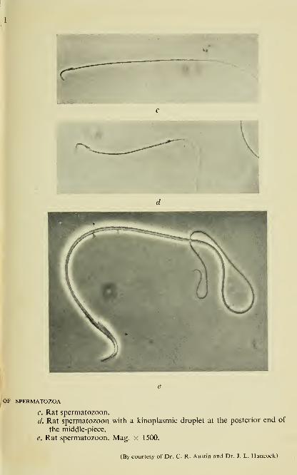
c. Rat spermatozoon.
d. Rat spermatozoon with a kinoplasmic droplet at the posterior end of
the middle-piece.
e. Rat spermatozoon. Mag. x 1500.
(By courtesy of Dr. C. R. Austin and Dr. J. L. Hancock)


The Two Components of Semen 11
Schrader, 1950; Wislocki, 1950; Friedlaender and Fraser, 1952;
Hancock, 1952; Melampy, Cavazos and Porter, 1952) and electron
microscopy (Seymour and Benmosche, 1941; Harvey and Anderson,
1943; Schmitt, 1944; Bretschneider and Iterson, 1947; Bretschneider,
1949^, b\ Grigg and Hodge, 1949; Hodge, 1949; Randall and Fried-
laender, 1950; Bayle and Bessis, 1951; Friedlaender, 1952; Challice,
1953; Bradfield, 1954).
In a typical flagellar spermatozoon (Plate I and Fig. 1) it is usually
possible to distinguish three regions, viz. sperm-head, middle-piece
and tail, but even among closely related species, one encounters an
extraordinary diversity of form, size and structure. Moreover, on
examining the semen from a single individual, one often finds in
addition to the normally shaped spermatozoa, a variety of 'degener-
ate', 'abnormal' or 'immature' forms which represent every con-
ceivable deviation from the normal cell structure, from 'tapering'
and 'double' cells with a double head or tail, to 'giant' and 'monster'
cells containing several nuclei and several tails in a mass of cyto-
plasm. Although a high degree of sperm abnormality is undoubtedly
associated with subfertility, normal semen is seldom completely
uniform, and human semen for example, is reckoned to contain
as a rule, at least 20% of abnormal forms (Pollak and Joel, 1939;
Harvey and Jackson, 1945; Hotchkiss, 1945; Lane-Roberts et al.,
1948; Williams, 1950). In the bull (Williams and Savage, 1927;
Lagerlof, 1934; Bishop, Campbell, Hancock and Walton, 1954)
and stallion (Bielanski, 1951), the percentage of abnormal forms in
semen is similarly high, in the ram on the other hand, it appears to
be much less.
The shape of the head in a normal spermatozoon varies greatly;
it is ovoid in the bull, ram, boar, and rabbit, it resembles an elong-
ated cylinder in fowl and has the form of a hook in the mouse and
rat; in the human species, the sperm-head appears as a flattened, oval
body, about 4-6 ^ long, 2-6 /< wide, and 1-5 f-i thick, which is com-
pressed at the anterior pole into a thin edge.
The main part of the head is occupied by the nucleus, filled by
closely-packed chromatin which consists largely of deoxyribonucleo-
protein and gives a positive Feulgen (nucleal) reaction with Schiff"'s
fuchsin-sulphurous acid reagent. The anterior part of the nucleus
is covered by a cap-like structure known as the acrosome. The

12 The Biochemistry of Semen
latter gives no positive Feulgen reaction but stains with the Schiff
reagent after exposure to the oxidizing action of periodic acid as
demonstrated in the sperm of the hemipteran insect, Arvelius albo-
punctatus (Leuchtenberger and Schrader, 1950) and in bull sperm
(Hancock, 1952). According to McManus (1946) and Hotchkiss
(1948), the 'periodic acid Schiff reaction' (PAS) is due to the pre-
sence of carbohydrates, and the chemical groups which react with
fuchsin-sulphurous acid are the aldehydes formed from 1 : 2 glycol
groupings by oxidation with periodic acid:
OH OHI I
R—
C
C—R + HIO4 -> 2R—CHO
The acrosomal material is not glycogen as it does not react with
iodine and is not affected by treatment with amylase. It cannot be
hyaluronic acid because it resists the action of hyaluronidase. The
possibility that it may be related to hyaluronidase itself still remains
to be investigated. There has also been a tendency to regard it as a
mucopolysaccharide, without however, sufficient evidence. Special
precautions are called for in the preparation of spermatozoa for the
PAS reaction. Structural changes in sperm cells, such as occur for
example, after rapid cooling ('temperature shock'), may render the
acrosomal material unresponsive to the periodic acid-Schiff reagent.
It is not improbable that the acrosomal 'polysaccharide' is either
decomposed or detached from the head of a mature spermatozoon;
this is borne out by some microscopic observations on changes which
take place in the acrosome during the period of senescence and death
of the sperm cell. Several investigators have described in sperma-
tozoa yet another cap, a loose protoplasmic structure, named 'galea
capitis' (also 'acrosome cap', 'Kopfkappe' or 'capuchon cephalique')
which envelops the apical part of the sperm-head and can break
away spontaneously to form a so-called 'spermatic veil' or 'floating
cap' (Williams and Savage, 1925; Blom, 1945). However, whereas
most authors including Williams (1950) regard the acrosome proper
and the galea capitis as two distinct structural entities, some con-
sider them to be identical, and Hancock (1952) for instance, is con-
vinced that there is only one acrosomal structure and, that the de-
tachable cap arises through post-mortem changes, and is the result
of swelling and loosening of the acrosome itself. The separation of
The Two Components of Semen 1
3
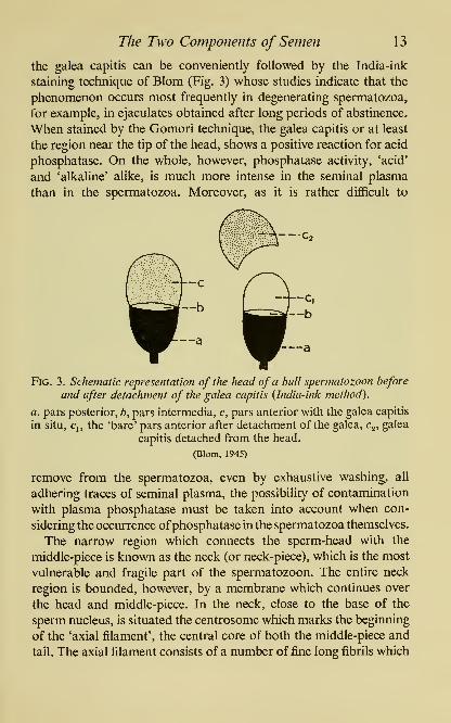
the galea capitis can be conveniently followed by the India-ink
staining technique of Blom (Fig. 3) whose studies indicate that the
phenomenon occurs most frequently in degenerating spermatozoa,
for example, in ejaculates obtained after long periods of abstinence.
When stained by the Gomori technique, the galea capitis or at least
the region near the tip of the head, shows a positive reaction for acid
phosphatase. On the whole, however, phosphatase activity, 'acid'
and 'alkaline' alike, is much more intense in the seminal plasma
than in the spermatozoa. Moreover, as it is rather difficult to
Fig. 3. Schematic representation of the head of a bull spermatozoon before
and after detachment of the galea capitis {India-ink method).
a, pars posterior, b, pars intermedia, c, pars anterior with the galea capitis
in situ, q, the 'bare' pars anterior after detachment of the galea, Cg, galea
capitis detached from the head.
(Blom, 1945)
remove from the spermatozoa, even by exhaustive washing, all
adhering traces of seminal plasma, the possibility of contamination
with plasma phosphatase must be taken into account when con-
sidering the occurrence ofphosphatase in the spermatozoa themselves.
The narrow region which connects the sperm-head with the
middle-piece is known as the neck (or neck-piece), which is the most
vulnerable and fragile part of the spermatozoon. The entire neck
region is bounded, however, by a membrane which continues over
the head and middle-piece. In the neck, close to the base of the
sperm nucleus, is situated the centrosome which marks the beginning
of the 'axial filament', the central core of both the middle-piece and
tail. The axial filament consists of a number of fine long fibrils which

14 The Biochemistry of Semen
run uninterruptedly through the whole length of the middle-piece
and tail. These fibrils probably represent the main contractile ele-
ment of the sperm cell, responsible for the whip-like lashing of the
tail. In most species investigated so far by means of the electron
microscope, eleven fibrils have been identified; two of these, which
occupy the central position, are sensitive to the action of water and
digestive proteolytic enzymes, whereas the remaining nine fibrils
which form an 'outer cylinder' around the 'central pair', are remark-
ably resistant to the action of plasmolysing and digestive agents,
and even prolonged proteolysis with pepsin or trypsin fails to disrupt
them; these fibrils also resist effectively attempts at solubilization by
means of salt solutions, acids and weak bases.
The finer structure of the individual fibrils is still a matter of active
investigation. In the case of mammalian spermatozoa, doubling of
fibrils has been observed, at any rate in the middle-piece, and in
addition to the outer cylinder of nine fibrils, another, so-called
inner cylinder has been described, consisting of nine, much thinner
fibrils. The precise chemical nature of the fibrillar protein is un-
known. A certain resemblance to muscular contraction prompted
Engelhardt (1946) to ascribe to the contractile substance of sperma-
tozoa myosin-like properties, and to sperm adenosine-triphos-
phatase the role of 'spermosin'. However, this c^aim remains at
present unsubstantiated since it was not accompanied by satisfac-
tory evidence that the spermatozoa used for the experiments, were
really free from phosphatases, especially the powerful adenosine-
triphosphatase, of seminal plasma.
In the middle-piece (or midpiece) which in the human sperma-
tozoon is about the length of the sperm-head though only one-tenth
as wide, the axial filament is surrounded by the 'broad helix', also
called 'spiral body' or 'mitochondrial sheath'. This lipid-rich struc-
ture, which is believed to be derived from mitochondria, has the
shape of a broad paired thread, wound helicoidally round the
'outer cylinder' of sperm fibrils. It is here that the cytochrome-
cytochrome oxidase system of spermatozoa is believed to be con-
centrated. The junction between the middle-piece and tail is marked
by the presence of a ring centriole.
The tail or 'flagellum' in the human spermatozoon is about ten
times the length of the middle-piece and lacks the 'broad helix'

PLATE II
ELECTRON MICROGRAPH OF SPERM-TAIL
Broken end of tail from a bull spermatozoon, showing the tuft of fibrils
of the axial filament, and the helical structure of the tail sheath;
I1indicates 1 /x.
(By courtesy of Prof. L. H. Bretschneider and Dr. Woutera van Iterson)


The Two Components of Semen 15
but has instead the much thinner 'tail sheath' or 'cortical helix*
which terminates a short distance before the end of the tail, exposing
the terminal portion of the axial filament, that is the end-piece. In
mammalian spermatozoa, the tail sheath appears as a helicoidally
wound cord; when the tail of the spermatozoon is broken, one can
see, protruding from the cortical sheath, the brush-like fibrils of the
axial filament, and at this point it is also possible to distinguish the
helical structure of the tail sheath (Plate II). In fowl spermatozoa on
the other hand, there is no evidence of a 'cortical helix', and the axial
filament is encased in an amorphous sheath which is easily dis-
rupted by distilled water, causing the axial filament to fray into
fibrils. In addition to the various fibrous cortical systems, the sperm
cell of many species, including man and the higher mammals, is
protected externally by a lipid layer or capsule ('manteau lipidique')
evident especially around the tail, and composed of a layer of
liproprotein.
SEMINAL PLASMA
Seminal plasma, the extracellular fluid which provides the mediumand vehicle for spermatozoa, originates in the accessory organs of
reproduction and varies in composition according to species. In
lower animals it may be so scarce that the emitted semen takes the
form of a very thick lump of spermatozoa, closely packed together.
There is little seminal plasma in bird semen and even among some
of the mammals, but on the whole, the higher mammals, including
man, produce a relatively dilute semen with a considerable propor-
tion of seminal plasma.
Secretory function of male accessory glands
The seminal plasma is a composite mixture of fluids secreted by
organs which in the higher species comprise the epididymides, the
seminal ducts or vasa deferentia, ampullae, prostate, seminal vesicles
(or seminal glands), Cowper's glands and certain other glands
located in the wall of the urethral canal. Until a little while ago, the
secretory function of the male accessory organs remained obscure
chiefly owing to lack of information about the chemical nature of
the various secretions. More recently, however, several substances

16 The Biochemistry of Semen
have been discovered and identified in the accessory secretions, such
as citric acid by Schersten in 1929, prostatic phosphatase by Kutcher
and Wolbergs in 1935, fructose by Mann in 1945, phosphoryl-
choline by Lundquist in 1946, ergothioneine by Leone and Mann,
and inositol by Mann in 1951, and glycerylphosphorylcholine by
Rabbit Rat
Fig. 4. Diagrammatic outline of male accessory organs to illustrate the
localization offructose {shaded areas).
Am, ampullae. SV, seminal vesicle. Pr, prostate. VP, ventral prostate.
DLP, dorsolateral prostate. Pp, glandulae paraprostaticae. GV, glandula
vesicularis. CG, coagulating gland.
Diament, Kahane and Levy in 1952 (for details concerning the
secretory function of male accessory organs see: Mann and Lutwak-
Mann, 1951ft; Lutwak-Mann, 1951).
Owing to the complex nature of the seminal plasma the physio-
logist or biochemist is forced to adopt a distinct approach when
investigating any one of the accessory gland secretions. There are
several instances where male accessory organs which, though pre-
viously believed on the basis of similar embryonic origin or related
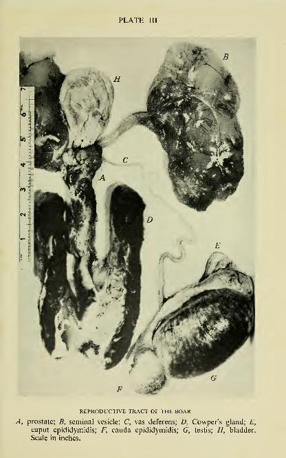
PLATE III
REPRODUCTIVE TRACT OF THE BOAR
A, prostate; B, seminal vesicle; C, vas deferens; D, Cowper's gland; E,
caput epididymidis; F, cauda epididymidis; G, testis; H, bladder.
Scale in inches.


The Two Components of Semen 17
morphological structure, to be anatomically and even functionally
'homologous', were later shown to differ greatly in their chemical
secretory activity. This is particularly true of the secretions of the
prostate and the seminal vesicle, two organs which in the majority
of higher species provide the bulk of the seminal plasma; their
localization within the reproductive tract of several species is illus-
trated in Plate III and Fig. 4.
The prostatic secretion
This differs in many ways from other secretions of the mammalianbody, and its composition shows considerable species variations.
Much study has been devoted to the human and canine prostatic
fluids; both are colourless and usually slightly acid, about pH 6-5
(Huggins, 1947; Zagami, 1940) and both are remarkable for the
almost complete absence of reducing sugar. They abound, however,
in several strong proteolytic enzymes; the human prostatic fluid
contains a fibrinolysin so powerful, that 2 ml. of prostatic fluid can
liquefy 100 ml. clotted human blood in 18 hr. at 37°; dog prostatic
fluid is distinguished by its ability to destroy fibrinogen, but it is
relatively inactive towards clotted blood (Huggins and Neal, 1942).
The prostate secretes a diastase (Karassik, 1927), and a /3-glucuroni-
dase which is more active in man than in dog (Talalay, Fishman and
Huggins, 1946; Huggins, 1947).
The prostatic secretion represents the main source of citric acid
and of acid phosphatase for whole human semen; and the analysis
of these two constituents provides a most convenient 'chemical
indicator test' for the assessment of the functional state of the
human prostate. There is much more citric acid and acid phospha-
tase in the human, than in the canine, secretion; thus, the citric acid
content is less than 30 mg./lOO ml. in dog, as against 480-2680
mg./lOO ml. in the human fluid; acid phosphatase activity in dog
corresponds to about 28 King-Armstrong units/100 ml. in the
'resting' or spontaneously voided prostatic secretion, and 104
units/100 ml. in the 'stimulated' secretion obtained by parasym-
pathetic stimulation, whereas the prostatic secretion of a normal
adult man may contain up to 3950 units/1 ml. (Gutman and
Gutman, 1941; Huggins, 1947).
The concentration of osmotically-active substances in the

1
8
The Biochemistry of Semen
prostatic fluids of man and dog is shown in Fig. 5. In the human
secretion (Huggins, Scott and Heinen, 1942), the average values for
cations, expressed in m-equiv./l. water, are: sodium 156, potassium
30, and calcium 30; for anions: citrate 156, chloride 38, bicarbonate
8, and phosphate 1. In a pilocarpine-stimulated dog prostatic fluid
(Huggins, Masina, Eichelberger and Wharton, 1939) the base con-
Ca"

The Two Components of Semen 19
rather high content of zinc. The first to observe this, and to commenton the possible role of zinc in reproduction, was Gabriel Bertrand;
in the two analyses of human prostate carried out by Bertrand and
Vladesco (1921) there was found 9-4 and 11-3 mg. Zn per 100 g.
fresh tissue, or 49 1 and 53 1 mg. Zn per 100 g. dry weight. Morerecently, Mawson and Fischer (1951, 1952, 1953) in Canada, found
that the mean zinc content of the human prostate gland was 68-2 mg.
Zn/100 g. dry wt., which is in considerable excess of zinc content
in human liver, muscle, brain, testis or blood. These investigators
state that the zinc present in the human seminal plasma is derived
chiefly from the prostatic secretion.
A considerable number of studies have been carried out with the
rat prostate (Fig. 4). In the rat, there is a distinct anatomical and
functional difference between the so-called ventral prostate which
secretes only citric acid, and the dorso-lateral prostate which pro-
duces both citric acid and fructose (Humphrey and Mann, 1948,
1949). In the dorso-lateral prostate itself, however, it is possible
to distinguish three smaller regions, the dorsal or median portion
which does not contribute citric acid, and two lateral lobes which
are rich in citric acid (Price, Mann and Lutwak-Mann, 1949). It is
the dorso-lateral prostate, and more specifically, its two lateral lobes
which contain much more zinc than any other soft tissue of the rat,
and which at the same time exhibit carbonic anhydrase activity
almost equal to that of blood (Mawson and Fischer, 1952); whereas
however, the carbonic anhydrase in the rat lateral prostate accounts
for no more than one-tenth of the total zinc content, in blood
erythrocytes this enzyme is well known to correspond closely to
the bulk of the zinc content (KeiUn and Mann, 1940).
The protein content of the prostatic secretion is low, less than 1%in man, and a certain proportion of the protein-like material present
in the secretory fluid is composed of 'proteoses' which are not preci-
pitated by trichloroacetic acid. Another feature of the prostatic
secretion is its elevated content of certain free amino acids, the
presence of which is probably the outcome of a combined action
of proteolytic and transaminating enzymes in the glandular tissue
(Barron and Huggins, 1946a; Awapara, \952a, b). Human prostatic
adenoma contains in 100 g. tissue 50 to 200 mg. glutamic acid in
addition to several other amino acids. The average content of amino3
20 The Biochemistry of Semen
acids in protein-free filtrates of ground prostatic adenoma or dog
prostate, expressed in terms of millimoles/100 g. tissue, is 34 and
39, respectively. The ventral lobe of the rat prostate contains in a
free state nearly all known amino acids, and in addition phosphoryl-
ethanolamine, taurine, glutathione, and glutamine. The dorso-lateral
lobe on the other hand, in contrast to the ventral prostate, has a
much lower content of most amino acids and lacks completely iso-
leucine and threonine; it may be added here that it also responds
differently to castration and to hormones.
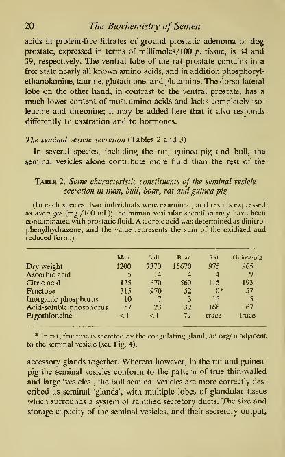
The seminal vesicle secretion (Tables 2 and 3)
In several species, including the rat, guinea-pig and bull, the
seminal vesicles alone contribute more fluid than the rest of the
Table 2. Some characteristic constituents of the seminal vesicle
secretion in man, bull, boar, rat and guinea-pig
(In each species, two individuals were examined, and results expressed
as averages (mg./lOO ml.); the human vesicular secretion may have been
contaminated with prostatic fluid. Ascorbic acid was determined as dinitro-
phenylhydrazone, and the value represents the sum of the oxidized andreduced form.)
The Two Components of Semen 21
are subject to individual variations, which are particularly conspicu-
ous in man. But the storage capacity of the human vesicles is small
indeed in comparison with that of the bull or boar. In certain mam-mals such as the dog or cat, the seminal vesicles are altogether
absent. In the rabbit, a combined anatomical and biochemical study
of the reproductive system has shown that the two organs known as
glandula seminalis and glandula vesicularis, develop from the same
diverticulum of the Wolffian duct, and possess a common urethral
outlet, so that both these glands together may be regarded as homo-logous to the seminal vesicles proper of other mammals (Davies and
Mann, 1947«, b; Mann, 1947).
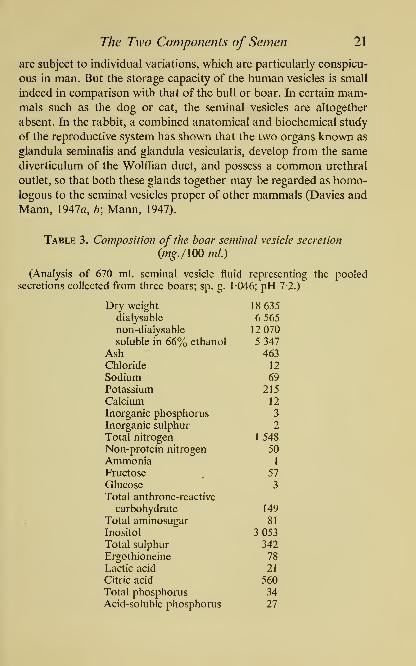
Table 3. Composition of the boar seminal vesicle secretion
(m^./lOO ml.)
(Analysis of 670 ml. seminal vesicle fluid representing the pooledsecretions collected from three boars; sp. g. 1046; pH 7-2.)
Dry weight
dialysable
non-dialysable
soluble in 66% ethanol
Ash

22 The Biochemistry of Semen
Compared with the prostatic fluid, the seminal vesicle secretion
is usually less acid, sometimes distinctly alkaline, has a higher dry
weight and contains more potassium, bicarbonate, acid-soluble
phosphate and protein; the latter is to a large extent precipitable by
trichloroacetic acid but there is also some 'proteose' as shown for
example, by the study of the seminal vesicle proteins in the goby
Gillichthys mirabiUs (Young and Fox, 1937). But the most remark-
able feature of the seminal vesicle secretion is its unusually high
content of reducing substances.
The normal seminal vesicle secretion is usually slightly yellowish
but occasionally, especially in man and bull, it can be deeply pig-
mented. The yellow pigmentation is probably of composite origin
but much of it is due to flavins which cause the vesicular secretion and
seminal plasma to fluoresce strongly in ultraviolet light. Brochart
(1952) observed that when strongly yellow coloured samples of bull
seminal plasma are exposed to sunHght, the colour tends to dis-
appear within a short time and lumiflavin is formed. Leone (1953)
has shown that at least part of the flavin content of the bull seminal
vesicle secretion is due to adenine-isoalloxazine dinucleotide,
associated with xanthine oxidase. The highest content of total flavin
which I was able to record in the bull seminal vesicle secretion, was
750 /^g./lOO ml.; in eight samples of bull seminal plasma there was
from 47 yf^g./lOO ml. (in an almost colourless specimen) to 480
^g./lOO ml. flavin (in a particularly deeply pigmented specimen).
There can be little doubt that the flavin associated with the strongly
yellow-coloured specimens of bull semen is due principally to the
seminal plasma, and not to the spermatozoa. The sperm cells them-
selves, however, contain also some flavin. In washed bull sperma-
tozoa, there is some 30 [ig. riboflavin/g. dry weight (Lardy and
Phillips, 1941c), and in whole bull semen, particularly in the less
coloured samples, a substantial portion of flavin may be derived
from the spermatozoa (VanDemark and Salisbury, 1944).
In addition to the yellow pigment, the seminal plasma sometimes
contains a brownish haematin pigment; this occurs in cases of
'chronic haemospermia', a condition occasionally met with in man
and attributed to haemorrhagic changes in the seminal vesicles
(McDonald, 1946).
Potassium in a high concentration occurs in the vesicular secretion

The Two Components of Semen 23
of several species, including man (20 mM), bull (100 mM) and
boar (300 mM). In the latter, the ionic equilibrium on the cationic
side is set up chiefly by potassium, with citric acid as the main anion;
the concentration of sodium is much less than that of potassium;
chlorides are conspicuous by their almost total absence, a phe-
nomenon infrequently encountered in other normal body fluids
(Table 3). Another unusual feature of the boar vesicular secretion is
its high content of inositol which varies from 2 to 3% and accounts
for something like one-third of the total dialysable material; inositol
together with citrate, contributes substantially to the osmotic pres-
sure of the vesicular secretion (Mann, 1954).
The reducing power of the vesicular secretion is one of its most
characteristic chemical properties. Two kinds of reducing sub-
stances are present. One group is made up of substances which
are capable of reducing silver nitrate, iodine and 2 : 6-dichlorophenol
indophenol in the cold. They are always present in the protein-free
filtrate from the vesicular secretion and seminal plasma but
their chemical nature varies from one species to another. At
one time, the reducing property was generally attributed to
ascorbic acid, in the secretions of the guinea-pig (Zimmet and
Sauser-Hall, 1936; Zimmet, 1939), bull (Phillips, Lardy, Heiser and
Ruppel, 1940; Jacquet, Cassou, Plessis and Briere, 1950) and man(Nespor, 1939; Berg, Huggins and Hodges, 1941). According to
Phillips and his co-workers, bulls of high fertility produce semen
containing 3 to 8 mg. ascorbic acid per 100 ml., whereas low-fertility
bulls may have less than 2 mg./lOO ml.; these workers have also
claimed that in certain bulls it was possible to enhance the fertility
by parenteral administration of ascorbic acid. More recent studies,
however, based upon chemical methods of purification and identi-
fication, have proved that ascorbic acid rarely accounts for the total
reducing power of semen towards dichlorophenol indophenol. In
the boar especially, ascorbic acid has been shown by Mann and
Leone (1953) to account but for a small fraction of seminal reducing
power and the bulk of the reducing material was found to consist
or ergothioneine, a substance which owes its reducing power to a
sulphydryl group (Leone and Mann, 1951; Mann and Leone, 1953).
The properties and functions of ergothioneine will be discussed
later (p. 174).

24 The Biochemistry of Semen
The presence of the other kind of reducing substances can be
detected in protein-free filtrates from semen and seminal vesicle
fluid, by heating with sugar reagents, such as for example, cupric
hydroxide. In this category belongs fructose, the physiological sugar
of semen.
Huggins and Johnson (1933) were the first to observe that the
reducing sugar of human semen is derived from the secretion of the
seminal vesicles but is absent in the prostate. Similar findings were
made with the bull (Bernstein, 1937), boar (McKenzie, Miller and
Bauguess, 1938) and ram (Moore and Mayer, 1941). The identifi-
cation of the seminal sugar as fructose (Mann, 1946a, b, c) opened
the way for detailed studies of the fructose-generating capacity of
the accessory tissues (Fig. 4). It was shown that in several species
fructose is secreted either by the seminal vesicles or by functionally
related organs (Mann, 1946c; 1947; l94Sa, b). This made it possible
to use the chemical assay of fructose in semen as an indicator of the
relative contribution made by the seminal vesicles towards the make-
up of the whole semen. It must be pointed out, however, that a
certain small amount of fructose is also produced by the ampullar
glands and in some species, by certain other accessory organs (see
p. 138).
The physiologicalfunction of the seminal plasma
From time to time doubt is expressed as to whether the individual
accessory gland secretions or even the entire seminal plasma, have
any essential role to fulfil in the process of reproduction; the more
so, since in some anmials such as the guinea-pig or rabbit, it is
possible to induce pregnancy by the artificial insemination of epi-
didymal spermatozoa. It is however, arguable as to how much sig-
nificance may be ascribed to such experiments, and it is certain that
the natural mating process could scarcely be expected to function
smoothly and efficiently without the provision of seminal plasma
as a normal diluent and vehicle for the thick mass of closely packed
epididymal spermatozoa; no more could the blood corpuscles act
as oxygen carriers in vivo, without the blood plasma.
Furthermore, the seminal plasma exerts a distinct stimulating
effect on sperm motility. In part, this is due simply to the 'dilution
effect', a phenomenon which is described fully elsewhere (see p. 73).

The Two Components of Semen 25
To a considerable extent, however, the activation by seminal plasma
has been shown to be linked with the occurrence of specific sub-
stances in the different accessory gland secretions. In certain insects,
Bombyx mori for example, the spermatozoa are believed to acquire
their full fertilizing capacity only after activation by the secretion
of the lower portion of the male tract, the so-called glandula pros-
tatica (Wigglesworth, 1950). Many investigators have studied the
activating influence of the prostatic secretion on mammalian sper-
matozoa (Steinach, 1894; Hirokawa, 1909; Ivanov, 1929; Sergijewski
and Bachromejew, 1930), and in some instances, e.g. the dog, have
claimed it to be species-specific (Ivanov and Kassavina, 1946), Byno means all of these experiments, however, have been carried out
under satisfactorily controlled conditions and, as Huggins (1945)
thinks, in many cases it is impossible to exclude the action of non-
specific factors such as acceleration of sperm motility by certain ions
or by changes in the gas tension.
Much discrimination is equally needed in appraising certain
results obtained with the epididymal secretion. Here, however, the
problem involves not so much the 'activation', as the 'ripening' and
'life-prolonging', effects on the spermatozoa (Tournade and Mer-
land, 1913; Stigler, 1918; Braus and Redenz, 1924; Hammond and
Asdell, 1926; Young, 1929, 1930; Young and Simeone, 1930; Lanz,
1931, 1936; Gunn, 1936; Gunn, Sanders and Granger, 1942). Mostinvestigators agree that in some special way the epididymis is
adapted for the long-term storage of spermatozoa; but whereas somelike Braus and Redenz (1924), attribute to the epididymal secretion
a specific role in the 'ripening' process, others deny it such a func-
tion. In the seminal vesicle fluid, we seem to be confronted with an
activating effect on spermatozoa different from that exerted by the
prostatic and epididymal secretions, one which is specifically boundup with the presence therein of fructose which provides a source of
nutrient material for the sperm cells.
Another facet of the physiological function of the seminal plasma
concerns certain characteristic pharmacological effects of the acces-
sory gland secretions and the role of seminal plasma in the remark-
able process of semen coagulation and liquefaction.

26 The Biochemistry of Semen
Prostaglandin y vesigiandin, and certain other pharmacodynamically
active substances
Among the more striking pharmacological effects of seminal
plasma are a depressor action on blood vessels, and a stimulation
of isolated smooth-muscle organs such as the uterus and the intes-
tines. Both these effects which have been studied in great detail by
von Euler (1934«, b\ 1935, 1937, 1939, 1949) and Goldblatt (1933,
1935^?) are due in all probability not to a single substance but to the
combined action of several constituents of seminal plasma, including
choline and two substances which Euler has named 'prostaglandin*
and 'vesigiandin'. So far, only prostaglandin has been purified.
The purest preparation obtained from ram prostate glands has been
found by Bergstrom (1949) to be nitrogen-free, and to contain an
unsaturated acidic substance which absorbs strongly ultraviolet light
with a maximum at 280 m//. When assayed on the isolated intestine
of rabbit, 1/^g. of this substance exhibited the same activity as the
crude extract from 500 mg. prostatic tissue. Assuming that the sub-
stance is pure, the total content of prostaglandin in 100 kg. prostate
glands must be of the order of 25-50 mg. To prepare this quantity,
one would require the glands from 20,000 rams.
The physiological significance of prostaglandin and vesigiandin in
reproduction processes is unknown but it has been suggested that
they represent some sort of 'automatic regulator' which controls
the voiding of the secretions from the prostate and the seminal
vesicle, respectively. The idea of chemical stimulation by secretory
products is based among others, on observations that the emptying
of the prostate and seminal vesicle leads to a decreased ability of
these glands to contract which persists until enough of freshly
formed secretion has accumulated.
A pharmacodynamic influence of the seminal plasma upon some
parts of the female reproductive tract has also been envisaged but
the evidence is derived mainly from experiments on isolated organs.
It remains questionable whether any of the effects exerted by the
seminal plasma on the uterus and oviduct in vitro, also occur in the
female reproductive tract in vivo (Barnes, 1939; Asplund, 1947).
Kurzrok and Lieb (1931) found on adding 1 ml. of human seminal
fluid to a strip of human uterus suspended in a 100 ml. bath, either
The Two Components of Semen 27
an increase or a decrease in spontaneous contractions. Cockrill,
Miller and Kurzrok (1935) observed that those specimens of humansemen which were capable of enhancing uterine contraction, caused
an inhibition after having been exposed for half an hour to pH 10;
at pH 11 all specimens became inactive. Moreover, the effect was
potentiated by eserine and suppressed by atropine. The observed
action corresponded to that of about 100 ^g. acetylcholine per ml.
semen. About that time, the occurrence of a powerful oxytocic
substance in the human seminal plasma was demonstrated by Gold-
blatt {\92>5b) who also found that the activity was destroyed by
short boiling of the seminal plasma with either OlN-NaOH or
OlN-HCl. When assayed on the guinea-pig's uterus, 1 ml. of humanseminal plasma showed approximately the same oxytocic activity
as 0-4-0-6 mg. of histamine. Euler (1937) believes that the oxytocic
activity is due to prostaglandin which in his experiments stimulated
strips of human uterus and also isolated uterus as well as uterine
strips from several species including the cow, rabbit, guinea-pig and
rat. Asplund (1947) determined the total content of 'contractive sub-
stance' in 155 specimens of human semen which he assayed on the
rabbit intestine in vitro, with purified prostaglandin as the standard.
He came to the conclusion that the effect of semen on the isolated
intestine must be attributed to a combined action of prostaglandin,
choline, and at least one other substance which produces a very
rapid increase in tonus and is unaffected by atropine. There was no
correlation between the total content of 'contractive substance' in
seminal plasma and the motility and viability of spermatozoa.
In addition to the remarkable vaso-dilation and contraction of
plain muscle, the seminal plasma and the accessory gland secretions
exhibit a characteristic pressor activity towards blood vessels. In
1906 Jappelli and Scafa found that parenterally administered extracts
of the canine prostate produced an increased blood pressure in the
dog and stimulated the respiration. A similar effect was observed by
Thaon (1907) after the intravenous injection of prostatic extracts into
rabbits but in his experiments the rise in blood pressure was usually
followed by a fall. There are indications that the pressor action of
the prostatic extracts is due to adrenaline-like substances demon-
strated in semen and accessory glands by CoUip (1929), v. Euler
(1934Z>), Bacq and Fischer (1947), and Brochart (1948fl) (see p. 181).

28 The Biochemistry of Semen
Coagulation and liquefaction
Semen is ejaculated in a liquid or semi-liquid form. In some animal
species such as the bull and the dog, it remains liquid, but in others
it tends to gelate or coagulate shortly after ejaculation. Humansemen clots immediately after ejaculation only to liquefy again a
little later; until that happens the spermatozoa do not become fully
motile. For this reason the examination of human spermatozoa
should be postponed until at least twenty minutes after the emission
of semen (McLeod, 1946<2; Sunmons, 1946). Quite fresh boar semen
usually contains only small lumps of gelatinous material somewhat
resembling tapioca; on standing, however, the lumps increase and
merge into a semi-solid gelatinous mass which may take up half or
more of the entire ejaculate. Gelation of semen can also be observed
in the stallion. Even more striking is the clotting phenomenon in
the rodents. The major part of a rabbit ejaculate collected by means
of an artificial vagina often consists of a colourless transparent gel.
In rats and guinea-pigs, semen coagulation leads to the formation
after mating, of the so-called bouchon vaginal or vaginal plug, which
probably prevents the back-flow of semen from the vagina and
assists the passage of spermatozoa through the cervix into the uterus.
In a study of sperm transport in the rat, Blandau (1945) has shown
that the ejaculate fails to pass through the uterine cervix if the
coagulation of semen is aboUshed by ligation of the seminal vesicle
and coagulating gland ducts. The copulatory plug has also been
described in Insectivora (mole and hedgehog), Chiroptera {Rhinolo-
phidae and Vespertilionidae) and in Marsupialia (Camus and Gley,
1899; Courrier, 1927; Eadie, 1939, 1948a, b; Engle, 1926; Rollinat
and Troussart, 1897; Stockard and Papanicolaou, 1919). In most
animals the vaginal plug is due to the clotting of the semen itself but
in some, namely in the opossum (Hartman, 1924) and in the bat
Vesperuga noctula (Courrier, 1925; Grosser, 1903), its occurrence
involves the coagulation of female secretions by the seminal plasma.
In the honey bee, the escape of semen from the female reproductive
tract is prevented by the so-called mucus plug; this is formed by the
material ejected from the mucus glands of the drone towards the end
of ejaculation (Laidlaw, 1944).
In most species the substrate for gel formation consists of protein-

The Two Components of Semen 29
like material secreted by the seminal vesicles (Bergmann and Leuc-
kart, 1855; Bischoff, 1852; Hensen, 1876; Landwehr, 1880; Lataste,
1888; Pittard, 1852; Stockard and Papanicolaou, 1919), the enzymehowever, responsible for the coagulation is absent in the vesicles
themselves and comes into contact with the protein substrate only
in the course of ejaculation. The first to recognize the enzymic
nature of the coagulating agent were Camus and Gley (1896,
1899, 1907, 1921, 1922) who named it 'vesiculase'. It was shown by
Walker (1910) that the source of the coagulating enzyme in small
rodents is the so-called 'coagulating gland', which lies adjacent to
the seminal vesicle. The clotting power of the secretion from the
coagulating gland is such that 1 part is sufficient to clot 20,000 parts
of the seminal vesicle secretion. This quantitative relationship can
be demonstrated in vitro by collecting the two fluids separately and
then mixing them after suitable dilution (Moore and Gallagher,
1930).
The liquefaction of human semen which follows its coagulation, is
also an enzymic process. Fibrinolysin and fibrinogenase, two pro-
teolytic enzymes of semen (see p. 1 14), are believed to play a part in
this process.

CHAPTER II
Chemical and Physical Properties
of Whole Ejaculated Semen
Species and individual variations in the composition of semen. Pre-
sperm, sperm-containing, and post-sperm fractions in the ejaculate.
Criteria for the rating of semen quality. Optical and electrical properties
of semen. Viscosity, specific gravity, osmotic pressure, and ionic equi-
librium. Hydrogen ion concentration and buffering capacity. Metabohsmof semen and its relation to sperm density and motility; glycolysis;
methylene-blue reduction test; respiration.
Species and individual variations in the composition of semen
A CHARACTERISTIC feature of whole semen is the variability of its
composition not only among different species but also between in-
dividuals of the same species (Table 4). Somewhat less pronounced
but still significant are the variations in the concentration of some
of the semen constituents in the same individual (Table 5). All this
is not altogether unexpected since both the spermatogenic activity
of the testes and the secretory function of the male accessory organs
are subject to considerable physiological fluctuations of hormonal
origin and are influenced by factors such as light, temperature,
season, state of nutrition etc. The variability of semen composition
is the reason why repeated examination of whole semen, even if
restricted to a single experimental subject, and carried out under
identical experimental conditions, need not yield the same quanti-
tative results, as might, for example, an analogous series of blood
analyses in the same individual. Therefore, an accurate estimate of
semen quality in any one individual cannot be formed on the basis
of a single analysis and involves several examinations.
In Table 5 are shown individual fructose variations in the semen
of man, bull, ram and rabbit. The analytical results are given in
terms of concentration (mg./lOO ml. semen) and of absolute content
30

Chemical and Physical Properties of Semen 3
1
(mg, /ejaculate). This method of expressing the findings of semenanalysis is very useful particularly in the evaluation of pathological
abnormalities in the composition of semen. The data in Table 5,
especially those relating to rabbit semen, show that the variations
in the absolute content of fructose per single ejaculate are muchless than those in concentration; the reason being that a rabbit
u12
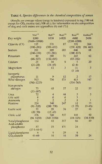
Table 4. Species differences in the chemical composition of semen
(Results are average values (range in brackets) expressed in mg./lOO ml.
except for COg content (ml./100 ml.); for information on the composition
of dog and cock semen see appendices (6) and (7).)

(1) Human semen. Data on dry weight, total nitrogen, and electrolytes
(excluding inorganic phosphate): Huggins, Scott and Heinen (1942); COg:Sheldovsky et al. (1940); urea: Goldblatt (1935^); Lactic acid: Lundquist
(1949Z>). Remaining data are our own; the results on non-protein nitrogen,
fructose, and citric acid content are based on analysis of semen from ten
individuals. The ash content of human semen is about 0-9% (Slovtzov,
1902, 1916); the ash contains 3% sulphur (Albu, 1908). According to
Infantellina (1945) human semen contains 30 mg./lOO ml. glutathione.
The values reported for ascorbic acid are 12-8 mg./lOO ml. (Berg, Huggins
and Hodges, 1941) and 2-6-3-4 mg./lOO ml. (Nespor, 1939); using Roe's
method, we found 10-2 and 12-4 mg./lOO ml. in two specimens.
(2) Bull semen. Data on electrolytes (except magnesium and inorganic
phosphate): Bernstein (1933); COg: Shergin (1935); remaining data are our
own, based on analysis of ejaculates from ten bulls (average volume of
ejaculate 4-5 ml.; sperm concentration 985,000/jul.). Other constituents of
bull semen include (mg./lOO ml.): thiamine 0-028-0-152, riboflavin 0-152-
0-306, pantothenic acid 0-230-0-466, and niacin 0-248-0-554 (VanDemarkand Sahsbury, 1944). Ehlers et al. (1953) reported the following meanvalues based on the analysis of 663 samples of semen (mg./lOO ml.):
fructose 552 (S.D. 169), citric acid 724 (S.D. 192). For additional data see
Rothschild and Barnes (1954).
(3) Ram semen. With the exception of the CO2 content (Shergin, 1935),
the data are our own, based on analysis of material pooled from ten
ejaculates (average volume of single ejaculate 1-2 ml.; 2,940,000 sperm/jitl.).
(4) Boar semen. Data on dry weight, electrolytes and total nitrogen:
McKenzie, MUler and Bauguess (1938); remaining data our own. Addi-
tional information in Table 7.
(5) Stallion semen. Data on dry weight, electrolytes, total phosphorus
and CO2 content from Slovtzov (1916), Bernstein (1933), Shergin (1935)
and Milovanov (1938); remaining data our own.
(6) Dog semen. Much work has been done on the dog prostatic secretion
(see text). Whole semen has been analysed by Slovtzov (1916) and the
following composition found (mg./lOO ml.): dry weight 2450, albumin,
globulin and nucleoprotein 866, mucoprotein 57, and lipid 182. Electro-
lytes have been examined by Bernstein (1933); dog semen is distinguished
by a high content of chloride (620-657 mg./lOO ml.). Fructose and citric
acid occur only in traces.
(7) Cock semen. In material from six pooled ejaculates, 5,100,000
sperm//il., we found (mg./lOO ml.): 57 total anthrone-reactive carbo-
hydrate, of which 4 was fructose and 41 glucose (determined by glucose
oxidase); 44 total phosphorus, of which 27 was acid soluble; 2 ammonia.In six individual specimens of cock semen we found from 7-7 to 81
mg./lOO ml. glucose but never more than 4 mg./lOO ml. fructose.
33

34 The Biochemistry of Semen
Table 5. Individual variations in the level offructose in semen
(Ejaculates collected at weekly intervals.)
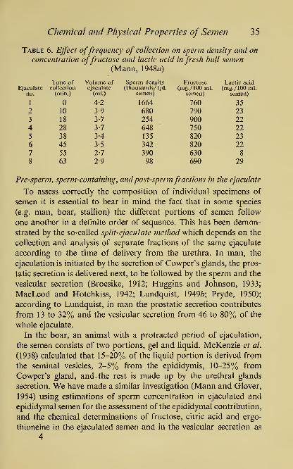
Chemical and Physical Properties of Semen 35
Table 6. Ejfect offrequency of collection on sperm density and on
concentration offructose and lactic acid in fresh bull semen(Mann, 1948a)
Ejaculateno.

36 The Biochemistry of Semen
a means for the evaluation of the contribution of the seminal vesicles.
From the results of this study (Table 7) it can be seen that in
ejaculated semen (liquid portion), the sperm concentration was 3%
Table 7. Composition of ejaculated semen, epididymal semen, andseminal vesicle secretion of the same boar
(Four ejaculates were collected from the same boar, at weekly intervals.
The boar was then killed, and the epididymal semen and vesicular secretion
collected. For chemical analysis, the fluid portions were used, separated
from sperm and gel by centrifugation. In the case of ejaculated semen, the
means (and standard deviation) are given per single ejaculate.)

Chemical and Physical Properties of Semen 37
fraction comprises 5-20%, the sperm-rich fraction 30-50%, and the
post-sperm fraction 40-60% of the total ejaculate. The pre-sperm
fraction which is ejaculated first, consists of a watery, often dis-
coloured, more or less sperm-free secretion, probably of urethral
-45

38 The Biochemistry of Semen
origin. Within a few minutes, that is followed by a sperm-containing
fraction which may also contain some gel-like material. As a rule
however, the gel forms a distinct fraction and is voided shortly after
the sperm fraction. Moreover, these three fractions, which complete
an ejaculation 'wave', may be succeeded by a second 'wave', also
fractionated. The two waves together may last up to 30 min., andrepresent in fact, two successive ejaculates. By the estimation of
sperm concentration coupled with the chemical determination of
fructose, citric acid, and ergothioneine in the various fractions, it is
possible to show that during the ejaculation the seminal vesicle
secretion follows immediately upon the delivery of the spermatozoa,
and is found mainly in the sperm-containing fraction (Glover and
Mann, 1954; Fig. 7). Occasionally, however, the ejaculation of
semen is incomplete and the semen does not include the later
fractions. When this happens, for example in the stallion, certain
normal constituents such as fructose or citric acid, may be missing
altogether. Obviously, such incomplete ejaculations create a further
complication in the assessment of analytical results obtained with
semen, at any rate in those species in which the ejaculation is a
fractionated one. But even in the bull where under physiological
conditions ejaculation appears to be instantaneous, the 'split-
ejaculate method' applied by Lutwak-Mann and Rowson (1953)
with the aid of electric stimulation, demonstrated the occurrence in
electrically-induced ejaculates, of at least two distinct fractions; of
these, the first was a copious sperm-free fraction, slightly viscous,
colourless, and of urethral origin, in which there was no fructose or
citric acid and very little protein; the next was a creamy sperm-
containing fraction, followed by, or more often mixed with, a yel-
lowish-coloured post-sperm fraction which represented an almost
pure secretion of the seminal vesicles, with a characteristically high
content of fructose, citric acid, and 5-nucleotidase.
Criteria for the rating of semen quality
General medical and veterinary experience indicates that the suc-
cessful fertihzation of the ovum and initiation of pregnancy, while
it is brought about primarily by the spermatozoa present in an
ejaculate, demands nevertheless the attainment of certain qualita-
tive and quantitative standards by the semen. We should therefore,

Chemical and Physical Properties of Semen 39
be able to define what constitutes normal fertile semen, and what
criteria if any, can be applied in the appraisal of ejaculated semen.
It may be stated at once that in spite of the wealth of information
gained by past and present students of semen, there is as yet no single
seminal characteristic known, which alone could serve as the means
ofjudging 'male fertility'. The best criterion of the fertilizing capacity
of spermatozoa is of course, the actual ability to fertilize the ovum.
This however, cannot be regarded as a laboratory test, until the
in vitro fertilization of the ovum has actually been accomplished
and the quantitative aspects of the process developed.
In the practice of artificial insemination of cattle, male fertility
continues to be assessed on the basis of the 'conception rate'. At
the artificial insemination centres, inseminated cows which have
not been 'returned' by the farmers for re-insemination within three
months or so, are presumed to have conceived, and the proportion
of presumed pregnancies, expressed as the percentage of the total
of the first inseminations, is referred to as 'conception rate'. Nearly
one-third of the cow population of England is now bred by artificial
insemination, and among these the conception rate averages at least
60%. There is considerable evidence, however, that a substantial
proportion of cows 'returned' for re-insemination, may have also
conceived but that pregnancy terminated at an early stage through
faulty ovum implantation or embryonic death.
Apart from the test based upon accomplished fertilization, the
means available at present for the evaluation of semen quality
include the histological and the physico-chemical methods.
Histological examination of semen involves procedures such as
the determination of sperm concentration or 'density' (number of
spermatozoa per \fj\. or 1 ml. of semen) with a cytometer (Walton,
1927; Weisman, 1942); differential count of abnormal forms of
spermatozoa (Lagerlof, 1934; Harvey and Jackson, 1945; Lane
Roberts et al., 1948; Williams, 1950); bacteriological examination
(Gunsalus, Salisbury and Willett, 1941; Kelly, 1947; Foote and
Salisbury, 1948; Almquist, Prince and Reid, 1949; Wu, Elliker and
McKenzie, 1952-3); determination of the incidence of dead sperma-
tozoa by means of 'live-dead staining' methods (Lasley, Easley and
McKenzie, 1942; Lasley and Bogart, 1943; Madden, Herman and
Berousek, 1947; Crooke and Mandl, 1949; Blom, 1950; Mayer,

40 The Biochemistry of Semen
Squiers, Bogart and Oloufa, 1951; Ortavant, Dupont, Pauthe and
Roussel, 1952; Campbell, Hancock and Rothschild, 1953); micro-
scopic assessment of the degree of motility, either directly in semen
(Harvey and Jackson, 1945; McLeod, \9A6a; Emmens, 1947; Farris,
1950) or by the 'cervical mucus penetration test', in which a drop of
semen is placed on the microscope slide next to cervical mucus and
the passage of spermatozoa through the mucus is followed by micro-
scopic observation (Barton and Wiesner, 1946; Harvey and Jackson,
1948). The determination of the concentration of motile spermatozoa
in a semen sample is generally held to be the criterion most clearly
correlated with the actual fertility rate; but even this relationship
occasionally fails to give a true picture, and motile spermatozoa
are by no means always fertile.
Physico-chemical methods of semen analysis depend on the deter-
mination of a wide range of physical, chemical and metabolic
characteristics of semen, related to the physiological function of the
sperm and the seminal plasma. Here belong methods for the
measurement of certain optical properties of semen such as the
light-scattering and light-absorption power, electrical conductivity
and impedance changes; specific gravity, osmotic pressure; hydrogen
ion concentration, buffering capacity; occurrence of semen-specific
metabolites such as fructose, citric acid, ergothioneine, and inositol,
and various enzymes such as hyaluronidase and certain phospha-
tases; and finally, the rate of anaerobic and aerobic metabolic
processes in semen expressed in terms of fructolysis index, respira-
tion rate or methylene-blue reduction time.
Optical and electrical properties of semen
An optical property closely related to sperm concentration is the
turbidity of semen. In the past, determinations of sperm concen-
tration were based on microscopic sperm-counts but more recently
these have been partly replaced by turbidimetric measurements,
which can be carried out quickly with suitably diluted samples either
in a visual comparator by direct comparison with opacity standards,
or in a photoelectric absorptiometer, to provide values for the rela-
tive light transmission of semen (Burbank, 1935; Comstock and
Green, 1939; Henle and Zittle, 1942; Salisbury et al. 1943; Roth-
schild, 19506). The turbidimetric methods, however, it must be

Chemical and Physical Properties of Semen 41
remembered, rest on the assumption that although light is absorbed
both by sperm and seminal plasma, the scattering of light is due
exclusively to the former, so that only light-scattering as such, is
related directly to sperm density. In practice, the light-scattering andlight-absorption due to the spermatozoa predominates so muchover the light-absorption of the seminal plasma that under properly
controlled conditions the error due to the presence of the latter can
be neglected. This applies certainly to the semen of sea-urchin, bull
and ram, but need not necessarily be true of other species, in which
the seminal plasma itself shows a considerable degree of opaqueness.
Furthermore, the spermatozoa present in a given ejaculate maydiffer in their light-reflecting capacity, apparently in proportion to
the degree of sperm 'ripeness' and thereby, of fertility (Lindahl
et al, 1952). The light-reflecting capacity of the sperm cell mayalso be related to another optical change associated with sperm
ripening, namely the increase in 'luminosity' of the sperm surface
in dark-field illumination.
It has been claimed that the spermatozoon possesses at the head
and tail small, but directly opposite, electrical charges. However,
all that can be claimed with certainty is that an electric charge is
associated with the sperm cell but that its sign and magnitude depend
largely on the concentration of the various positively and negatively
charged ions in the surrounding medium. The following values for
electro-conductivity in semen at 25°, expressed in reciprocal
ohmsxlO"*, were given by Bernstein and Shergin (1936): bull
89-5-116-3, ram 48-5-80-5, stallion 111-3-129-5, boar 123-3-134-6,
rabbit 85-5-101 -4; and by Zagami (1939): man 88-107 (at 20°), and
dog 129-138.
Much scientific interest and practical importance in the rating of
semen quality attaches to the characteristic periodic changes in
electrical impedance which occur in semen samples with high sperm
density and motility, and which have been shown by Rothschild
(1948^, 1949, 1950a) to be associated closely with the so-called
'wave motion' of the spermatozoa. When a drop of ram or bull
semen of high density is placed on a microscope slide and examined
at 37° under low magnification, a characteristic phenomenon can
be observed in the form of slow, periodically appearing bands of
high opacity or 'waves'. Measurements by means of the impedance

42 The Biochemistry of Semen
bridge make it possible to assess this characteristic sperm movement
in a more objective and quantitative manner than by visual esti-
mates; but they can only be made with semen samples which are
sufficiently dense to show the 'wave motion'. Assays carried out
with the electrical method in the semen of bulls at several British
Artificial Insemination Centres revealed an interesting correlation
of impedance change frequency with the conception rate (Bishop
et ai, 1954).
Distinct from the wave-motion or locomotion 'en masse' (Walton,
1952) is the movement of individual spermatozoa which in highly
motile semen takes the form of so-called 'forward' or 'progressive'
motility, but which in poor specimens is confined to side-to-side
'oscillatory' movements (Plate IV). Recently, there have been several
attempts to replace the subjective and semi-quantitative microscope
appraisals of motility by more clearly defined methods, of which
those by Bosselaar and Spronk (1952) and Rothschild (1953a, b)
deserve special mention.
Viscosity, specific gravity, osmotic pressure, and ionic equilibrium
The viscosity of whole semen depends largely upon the concen-
tration of spermatozoa. Thus for example, the viscosity of bull
semen (relative to that of pure water which is taken as unity) can
vary from 1-76 in a specimen containing 80,000 sperm/ywl. to 10-52
in a sample with 2,260,000 sperm //^l. (Szumowski, 1948). Seminal
plasma itself, seldom exceeds in bulls a relative viscosity value of 2
but higher values have been recorded in other species, especially
those which exhibit the phenomenon of gelation.
The average specific gravity of whole semen is 1028 in man,
1-011 in dog, and 1-035 in bull, with fluctuations due in the first
place to the variable ratio between sperm and seminal plasma. The
latter is so much lighter than the spermatozoa that in practice the
specific gravity of semen is often found to be directly proportional
to sperm concentration. In bull semen, low specific gravity is usually
associated with low sperm concentration and poor 'quality', whereas
high values accompany good density and good 'quality' (Anderson,
1946(7). This is not surprising in view of the fact that the specific
gravity of bull seminal plasma is not greater than that of blood
plasma, whereas the average specific gravity of bull spermatozoa

PLATE IV
TRACK OF THE SPERM-HEAD
Cinematograph of a bull spermatozoon (in semen diluted 1 : 450) movingforward at a speed of about 0-15 mm. /sec. Photographic plate
exposed for 1 sec, using dark ground illumination. Mag. x 673. Onlythe projection of the movement of the sperm-head is seen, the tail
leaving no track. As the sperm-head is shaped like an elliptical disc,
intense light scattering occurs only when the thin edge of the headis visible; during 1 sec. exposure ten images of the head were recorded
which means that it rotated or oscillated backwards and forwards
ten times.
(By courtesy of Lord Rothschild)

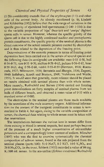
Chemical and Physical Properties of Semen 43
(1-28) considerably exceeds that of the erythrocytes (11) and other
cells of the animal body. As already mentioned (p. 8), Lindahl
and Kihlstrom (1952) believe that the wide range of variations in the
specific gravity of ejaculated bull spermatozoa (1-240-1 -334) is due
to the variable proportion of 'ripe' (heavier) and 'unripe' (lighter)
sperm cells in semen. However, whereas the specific gravity of the
sperm cell is due to the highly condensed nuclear and protoplasmic
protein constituents, the specific gravity of the seminal plasma is the
direct outcome of the actual osmotic pressure exerted by electrolytes
and is thus related to the depression of the freezing point.
Determinations of the osmotic pressure in terms of freezing point
depression have been carried out in the semen of several species and
the following data (in centigrade) are available: man 0-55-0-58, bull
0-54-0-73, ram 0-55-0-70, stallion 0-58-0-62, jackass 0-56-0-62, boar
0-59-0-63, dog 0-58-0-60, rabbit 0-55-0-59 (Slovtzov, 1916; Roem-
mele, 1927; Milovanov, 1934; Bernstein and Shergin, 1936; Zagami,
1940; Salisbury, Knodt and Bratton, 1948; Nishikawa and Waide,
1951). It would seem that generally, more reliance should be placed
on results obtained with seminal plasma than with whole semen.
More recently, Rothschild and Barnes (1954) carried out freezing
point determinations on forty samples of seminal plasma from ten
bulls of different breeds, and obtained a mean value of 0-53 with a
standard error of 0-005.
The electrolytes in the seminal plasma are those made available
by the secretions of the male accessory organs. Additional informa-
tion on the content of the inorganic constituents in semen is sum-
marized in Table 4, but again, owing to the variable composition of
semen, the chemical data relating to whole semen must be taken with
due reservations.
The interrelations between the various ions in semen differ from
those existing in blood in several respects, but most perhaps because
of the presence of a much higher concentration of extracellular
potassium and a correspondingly lower content of sodium. Miescher
(1897) who was the first to examine systematically the chemical
composition of salmon semen, found in the ash prepared from the
seminal plasma (parts/100): 51-0 NaCl, 8-2 KCl, 140 K2SO4, and
26-8 Na.COa. In the trout, Schlenk (1933) recorded a value of 80 mg.
K/lOO ml. semen. In the sea-urchin {Echinus esculentus) Rothschild
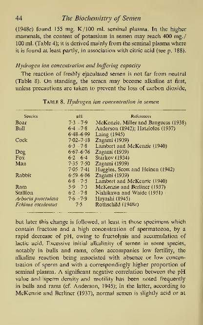
44 The Biochemistry of Semen
(1948c) found 155 mg. K/lOO ml. seminal plasma. In the higher
mammals, the content of potassium in semen may reach 400 mg./
100 ml. (Table 4); it is derived mainly from the seminal plasma where
it is found at least partly, in association with citric acid (see p. 188).
Hydrogen ion concentration and buffering capacity
The reaction of freshly ejaculated semen is not far from neutral
(Table 8). On standing, the semen may become alkaline at first,
unless precautions are taken to prevent the loss of carbon dioxide,
Table 8. Hydrogen ion concentration in semen
Species

Chemical and Physical Properties of Semen 45
any rate never more alkaline than pH 7-3, whereas in sterile rams the
pH value may reach 8-6. However, the rate at which acidity increases
after ejaculation is much more significant for the assessment of
semen than the initial pH value, because it is related directly to the
actual glycolytic activity of the spermatozoa, and indirectly to
sperm density and motility. In bull and ram semen this correlation
is said to be so close that the decrease in pH on incubation can
serve as an additional indicator of semen quality (cf. Anderson,
1945; Laing, 1945; Reid, Ward and Salsbury, 1948Z)). As a matter
of fact, however, the significance of this method is limited because
as the pH value falls, the semen becomes too acid for spermatozoa
to maintain their motility and metabolism.
Some of the effects which are due to variations in hydrogen ion
concentration in semen will be discussed in the next chapter; here
we shall concern ourselves with the buffering capacity of semen
which has been the subject of some interesting work, especially in
bull and human semen (Shergin, 1935; Smith and Asdell, 1940;
Easley, Mayer and Bogart, 1942; Sheldovsky, Belcher and Leven-
stein, 1942; Willett and Salisbury, 1942; Anderson, 1946«). On the
whole, bull semen is more highly buffered on the acid than on the
alkaline side, and its normal buffering capacity depends mainly
upon citrate and bicarbonate, but not phosphate. Anderson used
107 specimens of seminal plasma from normal bulls and measured
their buffering power by adding 005 ml. OIn-HCI to 01 5 ml.
plasma, and determining the pH with the help of a glass electrode,
before and after acid addition; the decrease in pH value obtained
in this way was l-84±0038. He also investigated the relationship
between buffering capacity and the period for which sperm motility
of 70% and over, was maintained; semen specimens which kept up
this motility for 24 hr. and upward, all had a good and fairly uniform
buffering capacity (decrease in pH=l-74), but those with a smaller
degree of motility at 24 hr. had a poorer buffering capacity (decrease
in pH=200).
Metabolism of semen, and its relation to sperm density and motility
The two chief metabolic processes of semen, namely fructolysis
and respiration, are both a direct outcome of the metabolic activity
of the sperm cells; their rate is determined largely by the number

46 The Biochemistry of Semen
of spermatozoa in the semen and the degree of sperm motility
(Mann, 1949). The more chemical aspects of semen metabolism
will be discussed fully in conjunction with specific groups of sub-
stances metabolized by spermatozoa, such as sugars, lipids and
amino acids. Here, only the general outline of sperm metabolism
will be given, in so far as it helps to bring out the relationship
between the metabolic processes and other characteristics of semen,
in particular, sperm concentration and motility.
Glycolysis
In the absence of oxygen, for example under the conditions of
semen storage for artificial insemination, the spermatozoa rely on
carbohydrate metabolism as the chief source of energy. Even before
the identity of the seminal sugar was revealed, the rate of lactic acid
production or 'glycolysis' was used as a method for semen appraisal
(Comstock, 1939; Webster, 1939; MacLeod, 1941a, b, 1943Z); Moore
and Mayer, 1941; Comstock, Green, Winters and Nordskog, 1943;
Laing, 1945; Salisbury, 1946; Westgren, 1946.) However, the dis-
covery of fructose and the work on fructolysis made available a
chemical approach to several practical problems of male fertility
(Mann, 19466, 1948«, b, 1949; Mann, Davies and Humphrey, 1949;
Mann and Lutwak-Mann, \95\a, b; Mann and Parsons, 1950;
Mann and Walton, 1953).
The anaerobic incubation of freshly ejaculated semen is accom-
panied by a progressive decline in the content of fructose with a
simultaneous accumulation of lactic acid; in the presence of suitable
buffer, the process of fructolysis in semen with good sperm motility
can be shown to progress almost linearly until practically all of the
sugar is used up (Fig. 8). On this basis a photometric method has
been worked out for the measurement of sperm fructolysis, and the
'index of fructolysis' has been defined as the amount of fructose (in
mg.) utilized by 10^ spermatozoa in 1 hr. at 37° (Mann, 1948^, b).
In normal bull semen, the index of fructolysis is about 1 -4-2 but it
varies, and is significantly correlated with both the concentration
and the motility of spermatozoa. Fructose is not utilized by either
azoospermic semen, i.e. ejaculates devoid of sperm, or by necro-
spermic semen, containing immotile spermatozoa. The existence
of a positive correlation between the rate of fructolysis and the

Chemical and Physical Properties of Semen 47
concentration of motile spermatozoa has been amply confirmed by
studies on both bull and human semen (Anderson, 1946b; Eichen-
berger and Goossens, 1950; Bishop et al., 1954) but the existence of
a similar correlation between the rate of fructolysis and fertility
100 -
Control with buffered semeninactivated by heating
30 60 90 120 150 180
Minutes of incubation at 37° C210 240
Fig. 8. Fructolysis in bull semen (920,000 sperm/fil.) incubated at 37°;
the disappearance of fructose was measured in (i) bull semen which
has been diluted with half a volume 0-25m phosphate buffer pH 7-4,
(ii) unbuffered semen, and (iii) buffered semen inactivated by heating.
(Mann, 1948a)
is still a matter of dispute; it has been claimed for bull semen by
some authors (Gassner, Hill and Sulzberger, 1952) but was denied
by others (Bishop et al., 1954). In both cases, however, the material
used for the survey consisted mainly of bulls kept for breeding
purposes at Artificial Insemination Centres. More insight into the
relationship between fructolysis and fertility could probably be
gained by studies on semen from bulls of subnormal fertility.
48 The Biochemistry of Semen
A lowered rate of fructolysis has been observed in certain cases
of human subfertiHty (Davis and McCune, 1950; Birnberg, Sherber
and Kurzrok, 1952). However, in human semen with its physio-
logically low sperm density but high fructose content, it is actually
more convenient to assess the rate of fructose utilization by measur-
ing the formation of lactic acid (chemically or manometrically)
rather than the disappearance of sugar. In any case, if one were to
measure fructolysis in human semen by the disappearance of sugar,
it would be essential to use a photometric method specific for
fructose, and not base the results merely upon the changes in 'reduc-
ing value', because human semen contains a fair amount of reducing
substances other than carbohydrate, which represent a substantial
proportion of the total 'reducing value' towards reagents such as
cupric hydroxide, ferricyanide etc. Moreover, the content of these
reducing substances often increases during the incubation of semen,
thus rendering unreliable, not to say senseless, determinations of
fructose based upon reduction measurements.
Methylene-blue reduction test
A method often used in the evaluation of semen quality is the
'methylene-blue reduction test' which is the outcome of dehydro-
genase activity of the semen and depends on the determination of
the time which it takes a semen sample to decolorize a certain
amount of methylene blue, under standard conditions of incuba-
tion in vitro (S^rensen, 1942; Beck and Salisbury, 1943; VanDemark,
Mercier and Salisbury, 1945; Boenner, 1947). In S0rensen's original
method the incubation was carried out in a Thunberg tube, but in
Russia (Milovanov and Sokolovskaya, 1947) and in France (Brochart,
1948Z)), the test was later performed by introducing a drop of semen
mixed with methylene blue into a capillary tube and by observing
the decolorization in the central portion of the column; in good-
quality bull semen, with high density and motility, decolorization
usually takes place within less than 10 min. at 20°; if it extends
beyond 30 min., it signifies poor semen quality. However, though
useful, the test is of limited scope, not least because as long ago as
1941, Lardy and Phillips showed that the reduction of methylene
blue by sperm suspensions may be markedly delayed by a variety
of substances including glucose, lactate and citrate.

Chemical and Physical Properties of Semen 49
Respiration
In the presence of oxygen, semen shows a considerable respiratory
activity which is correlated both with concentration and motility
of spermatozoa. It is usual to express sperm respiration in terms of
Z02, a coefficient introduced by Redenz (1933) to denote ^1. O2 taken
up by 10^ sperm cells during 1 hr. at 37°; Z02 values reported by
Lardy and Phillips (1943a) for bull, cock, rabbit and ram semen, are
21, 7, 11 and 22, respectively. The use of Z02 is more convenient
than Q02 since the latter involves centrifugation of semen and
washing of sperm with an unphysiological fluid such as distilled
water; the average Q02 of ram sperm based on the dry weight of
washed sperm is about 8. Further references to the subject of sperm
respiration measurements, classified according to species are listed
in Table 9.
Chang and Walton (1940) found a close relationship between
motility and respiratory activity in ram sperm. Walton (1938) sug-
gested measurements of oxygen uptake in bull semen as a supporting
method for the assessment of semen quality. Walton and Edwards
(1938) compared the breeding records of thirteen bulls, taking as a
measure of their fertility the number of matings required to produce
pregnancy in cows; when they analysed ten different samples of
semen from each of these bulls, they found that there is a close cor-
relation between the respiratory activity of semen and fertility
assessed on the natural service records. But Ghosh, Casida and
Lardy (1949), and Bishop et at., (1954) failed to establish a corre-
lation between the respiratory activity of bull semen and fertility,
as assessed on the basis of artificial insemination records.
Further study will probably clear up these uncertainties but in
general, when we consider the significance of sperm respiration
measurements, there are certain points which must be taken into
account. Unlike fructolysis, the respiratory activity of semen is not
entirely exogenous since it involves, in addition to the oxidative
removal of products of fructolysis (chiefly lactic acid), the endo-
genous respiration, i.e. oxidation of some intracellular reserve
material, most probably a lipid. Moreover, it is possible to create,
experimentally at any rate, conditions under which sperm respira-
tion can be dissociated from motility. For example, in the presence

50 The Biochemistry of Semen
Table 9. List of some references to work on sperm respiration
BOARWinchester and McKenzie, 1941.
BULLRedenz, 1933; Windstosser, 1935; Henle and Zittle, 1941, 1942; Lardyand Phillips, 1943a, b, 1944; Lardy, Hansen and Phillips, 1945; Tosic
and Walton, 1950; Schultze and Mahler, 1952; Bishop and Salisbury,
1954; Melrose and Terner, 1953; Bishop, Campbell, Hancock andWalton, 1954.
COCKWinberg, 1939; Lardy and Phillips, 1943a; Kosin, 1944.
DOGIvanov, 1931; Bishop, 1942.
FOX
Bishop, 1942.
MANMcLeod, 1939, 1943a, b; Shettles, 1940.
RABBIT
Lardy and Phillips, 1943a; White, 1953.
RAMIvanov, 1936; Comstock, 1939; Chang and Walton, 1940; Comstock,Green, Winters and Nordskog, 1943; Lardy, Winchester and Phillips,
1945; Mann, 1945a; Mann and Lutwak-Mann, 1948; White, 1953.
OYSTER, Saxostrea commercialis
Humphrey, 1950.
SEA-URCHINS
Warburg, 1915; Gray, 1928, 1931; Barron et ai, 1941, 1948, 1949;
Hayashi, 1946; Rothschild, 1948a, c, d, 1950^/, I95\b; Spikes, 1949.
of a suitable concentration of fluoride, one can abolish both motil-
ity and fructolysis in ram spermatozoa without greatly suppressing
the respiration (Mann and Lutwak-Mann, 1948) (Fig. 9). Another
example is provided by the response of ram sperm to succinate.
Thus, whereas the oxygen consumption of intact ram spermatozoa
is not enhanced markedly by the addition of succinate, sperm cells
treated with spermicidal detergents such as cetyltrimethylammonium
bromide, 2-phenoxyethanol, sodium dodecylsulphate and similar
surface-active agents, show in the presence of succinate a high rate
of oxygen uptake although of course, the motility and the fructolysis
have been completely abolished (Koefoed-Johnsen and Mann, 1954).
The effect of fluoride on fructolysis is due chiefly to the inhibition
of enolase; the addition of pyruvate to fluoride-treated spermatozoa

Chemical and Physical Properties of Semen 5
1
enables the lactic acid formation to continue (Ivanov, 1943; Lardy
and Phillips, 1943c; Mann, \9A5b\ Melrose and Terner, 1952, 1953).
But according to the last-named authors, washed bull spermatozoa
52 The Biochemistry of Semen
Aerobically, in the presence of both fluoride (0 02m) and pyruvate
(001m), bull spermatozoa consume less oxygen than without
fluoride; this low rate of oxygen uptake can be enhanced by the
addition of 2 : 4-dinitrophenol (10~*m), which brings about a more
complete oxidation of that fraction of pyruvate which is not reduced
to lactic acid. Melrose and Terner (1952, 1953) claim that bull
semen can be graded according to the respiratory response of washed
spermatozoa to a system made up of fluoride, pyruvate and dini-
trophenol, and that in highly fertile samples the oxygen consumption
is low in the presence of pyruvate and fluoride, but is increased two-
fold or more, by the addition of dinitrophenol.
Among substances which can provide exogenous material for
sperm respiration are glycolysable sugars and lower fatty acids such
as lactic, pyruvic and acetic acid (Fig. 10) but the species differences
in this respect are very marked. Thus, while in mammalian sperma-
tozoa for example, it is possible to prolong the respiratory activity
for a considerable length of time with glucose, fructose, mannose,
L(+)-lactate, pyruvate, propionate, butyrate, and oxaloacetate
(Lardy and Phillips, 1944, 1945; Mann and Lutwak-Mann, 1948;
Humphrey and Mann, 1949; Tosic and Walton, 1950; Melrose and
Terner, 1953), the respiration of oyster spermatozoa is increased by
a-oxoglutarate and oxaloacetate, decreased by acetate, propionate
and butyrate, and remains unaffected by lactate, glucose, fructose
and mannose (Humphrey, 1950). It appears also that the mode of
action of several organic acids on the respiration of mammalian
spermatozoa differs fundamentally from the influence exerted by the
same substances on the sperm of lower animals. This is illustrated
best by the example of the peculiar response of sea-urchin sperm
to malonate (003m), which was reported by Barron and Goldinger
(1941/)) to increase both the O2 uptake and the aerobic CO2 output
of sperm by as much as 200%. Succinic acid is another example of a
substance which was found to be highly effective in the respiration
of sea-urchin sperm (Barron and Goldinger, \94\a; Goldinger and
Barron, 1946) but has little effect on the oxygen uptake of intact
mammalian spermatozoa. Another characteristic difference in be-
haviour between sea-urchin and mammalian sperm concerns the
effect of the fatty acids on the initial rate of sperm respiration. In
the case of sea-urchin spermatozoa, the addition of a fatty acid salt

Chemical and Physical Properties of Semen500
53
400
300
O
200
100
-• Fructose
o o Glucose
X X Na-lactate
180 210 24090 120 150
Incubation (min.)
Fig. 10. Effect offructose, glucose and lactate on the respiration of washedram spermatozoa (the same effect was obtained also with acetate andpyruvate); oxygen uptake of 3 ml. buffered suspension of 0-45x10^
spermatozoa.(Mann & Lutwak-Mann, 1948)
often produces a prompt and marked rise in the initial rate of O2
uptake. In our experience, however, the addition of organic acid
salts to washed suspensions of mammalian spermatozoa, does not
lead necessarily to an actual increase in the initial O2 uptake but
instead, these substances act by maintaining and prolonging the
initial rate of respiration, thereby delaying the decline in Oo con-
sumption which would set in otherwise (Mann, 1949). Furthermore,
by adding the same substances to respiring suspensions of washed
mammalian sperm at a stage when the respiration had already begun
to decline, one can prevent a further deterioration in the rate of O2
consumption.

CHAPTER III
77?^ Influence of Extraneous Factors,
Hormones, and Environmental Conditions
Sperm inhibitors and spermicidal substances. Cliemical aspects of short-
wave radiation. Variations in hydrogen ion concentration and tonicity.
Influence of heat and cold; sperm vitrification and 'la vie latente'. Role of
hormones. Sperm-egg interacting substances and chemotaxis. 'Dilution
effect' and chemical changes associated with senescence. The use of arti-
ficial diluents in the storage of semen.
The list of agents, both physical and chemical, which affect sperma-
tozoa, includes among others, changes in temperature, visible light,
short-wave radiation, atmospheric pressure, ionic strength, and a
host of pharmacologically active substances. The vast literature on
the subject of sperm activation and inhibition goes back as far as
Leeuwenhoek's observation that dilution with rain water deprives
the canine 'animalculi' of motion, and a report by his learned friend
Johan Ham of Arnhem, on the loss of sperm motility in a patient
dosed with turpentine. Among Spallanzani's contributions in this
field is the discovery that freezing in snow does not necessarily kill
the 'spermatic vermiculi' but reduces them to a state of 'lethargy'
from which they recover when returned to higher temperature. The
XlXth century abounds in studies on the effect of changes in the
medium on sperm motility and survival. Prevost and Dumas (1824)
extended Spallanzani's observations on the lethal effect of electric
shock and certain poisons; Donne (1837) investigated the influence
of milk, urine, and the vaginal and cervical secretions; de Quatre-
fages (1850, 1853) described in great detail the marked toxicity of
copper, lead and mercuric salts. Newport (1853) studied the nar-
cotizing influence of chloroform vapours on the amphibian sperma-
tozoa, and concluded 'that the spermatozoon does not impregnate
when entirely deprived of its power of motion by narcotization and
disenabled to penetrate into the envelopes of the egg'. Both Newport54
The Influence of Extraneous Factors 55
and de Quatrefages were fully aware of the fact that sodium chloride
and various other sodium and potassium salts, are able to stimulate
or inhibit sperm motility, according to concentration and specific
experimental conditions. Their results were soon confirmed andextended by others, including Koelliker (1856) whose paper on'Physiologische Studien liber die Samenfllissigkeit' still remains the
most comprehensive survey of its kind, and includes observations
on spermatozoa of the bull, stallion, dog, rabbit, pigeon, frog andfish. Koelliker pointed out that spermatozoa rendered motionless by
dilution with water can be revived by prompt addition of salts or
concentrated solutions of certain organic substances such as sucrose,
glucose, lactose, glycerol, urea and various proteins. He investi-
gated in some detail the activating influence of blood serum, male
accessory gland secretions, and of a variety of inorganic and organic
substances on sperm motility. It was he who found that cyanide is
not an inhibitor of sperm motility and established that acids are,
on the whole, more harmful to the sperm than alkalies.
Furthermore, Koelliker noticed that if a drop of a fairly concen-
trated solution of potassium hydroxide is mixed with a drop of
semen on a microscopic slide, there is usually a sudden outburst of
activity before the spermatozoa are rendered motionless. Such a
period of short-lived stimulation which precedes the terminal loss
of activity, is rather characteristic of various sperm-paralysing
agents including distilled water. Schlenk (1933) aptly named the
phenomenon 'Todeszuckung'. Not all investigators, however, seemto have realized the fundamental difference between short and pro-
longed activation phenomena, and many of them tended to confuse
a transient increase in initial motility with the state of activity essen-
tial for the maintenance of continuous motility and for sperm sur-
vival. Only too often substances have been pronounced as beneficial
to spermatozoa merely because they were observed to stimulate
motility and metabolism, no heed being paid to the fact that this
very stimulation may have shortened, rather than prolonged, the
life of spermatozoa. Similarly, many a substance has been declared
detrimental to spermatozoa solely because it appeared to reduce the
speed of movement and metabolic rate. However, quite often the
lowering of activity tends to prolong the life of spermatozoa, andfavours, rather than hinders, their survival.

56 The Biochemistry of Semen
Sperm inhibitors and spermicidal substances
So far, detailed studies on the mechanism which underlies the
action of sperm inhibitors, have been relatively limited in scope and
concerned largely with chemical compounds which affect respiration
and glycolysis, among them cyanide, azide, dinitrophenol, and
fluoride, or which combine with sulphydryl groups, e.g. iodoacetate,
iodoacetamide, <?-iodosobenzoate, and /7-chloromercuribenzoate.
Even these studies, however, have clearly indicated the existence of
remarkable species differences in sperm behaviour. Thus for instance,
iodoacetate which is one of the strongest inhibitors of sperm activity
in higher animals, has a pronounced stimulating action on the
oxygen uptake of sea-urchin spermatozoa; this peculiar effect of
iodoacetate is shared by other sulphydryl-binding compounds, as
well as by malonate and nitrogen mustard (Barron and Goldinger,
\9A\b', Barron, Nelson and Ardao, 1948; Barron, Seegmiller,
Mendes and Narahara, 1948). It is interesting and important to
note that widely divergent results may be attained with a given
substance according to a particular set of experimental conditions:
rabbit spermatozoa, washed and resuspended in a sugar-free isotonic
medium, are immobilized completely by 00001m 2 : 3 : 5-triphenyl-
tetrazolium chloride, but a 200 times higher concentration of this
substance is ineffective towards sperm suspended in a glucose-con-
taining medium (Bishop and Mathews, 1952). It is equally salutary
to bear in mind that a substance which does not increase the initial
rate of sperm activity, may nevertheless be utilized by the sperma-
tozoa as an essential nutrient. For instance, most of the sugars
and fatty acids which are oxidized by spermatozoa, do not act by
increasing the initial rate of respiration but by maintaining it.
Surprisingly enough, many substances endowed with pronounced
pharmocological action in the whole animal, such as the alkaloids,
appear to exert little or no effect upon spermatozoa in vitro. Sperm
cells are also remarkably resistant to ethanol. Ivanov (1913) observed
excellent motility in dog sperm to which he added 2-5% ethanol,
and he managed to obtain live and normal offspring from an animal
inseminated with semen mixed with 10% ethanol. It may be men-
tioned here that ethanol is one of the substances which are definitely
known to pass into semen after ingestion by the animal (Farrell,

The Influence of Extraneous Factors 57
1938); sulphonamides provide a similar example (Farrell, Lymanand Youmans, 1938; Kuehnau, 1939; Hug, 1940).
Marked spermicidal power is characteristic of a great many sur-
face-active agents, quinones and heavy metal compounds. A tech-
nique for assessing the spermicidal activity of pure substances has
been developed by Baker (1931, 1932, 1935) whose so-called 'killing
concentration' is the lowest one capable of killing all spermatozoa
suspended in a buffered glucose-saline solution (Baker's solution)
within half an hour, at body temperature, under standard conditions
in vitro. Of the many substances examined by Baker, the most
highly spermicidal was phenylmercuric acetate (killing concentra-
tion 0-001%). Various quinones such as toluquinone, butylquinone,
methoxyquinone, parabenzoquinone, ethylquinone and paraxylo-
quinone were also strongly spermicidal. Lower down the scale were
mercuric chloride, methoxyhydroquinone, formaldehyde, methyl-
hydroquinone, saponin, and hexylresorcinol. A critical account of
the existing methods for testing the efficiency of spermicidal com-
pounds is given by Millman (1952), Davidson (1953) and Gamble
(1953).
As to the mode of action of surface-active agents, such as e.g.
cetyltrimethylammonium bromide, cetyldimethylbenzylammonium
chloride, /7-tri/5opropyIphenoxypolyethoxyethanol, dodecyl sulphate,
various condensation products of long-chain fatty alcohols with
ethylene oxide, and the numerous other ionic and non-ionic deter-
gents, there is some evidence to show that these substances act
directly on the constituents of the so-called lipid capsule, i.e. the
lipid-containing outer layer which protects the surface of the sperma-
tozoon. The mechanism of the spermicidal activity of detergents on
the sperm may be likened to the haemolytic action of surface-active
compounds on the erythrocytes, or the bactericidal effects of these
substances on various microorganisms. The changes brought about
by detergents manifest themselves in a loss of motility and fructo lytic
power, and in a grossly altered permeability of the sperm cells as
indicated by the leakage of cytochrome c. In contrast to intact
spermatozoa, the respiration of sperm cells treated with suitable
concentrations of detergents is markedly increased by succinate
(Koefoed-Johnsen and Mann, 1954).
As to the action of some at any rate, of the many other

58 The Biochemistry of Semen
spermicidal substances, including organo-metallic compounds, there
are indications that it is due to the blockage of vital sulphydryl
groups in the spermatozoa. Thus for example, the immobilizing effect
of iodoacetate on bovine spermatozoa, which has been studied by
Lardy and Phillips (19436), is most probably due to the sulphydryl
group-binding capacity of this substance. In a war-time study
MacLeod (1946Z)) showed that the inhibition of the metabolism and
motility of human spermatozoa by organic arsenicals can be over-
come by 1:2: 3-trithiolpropane, and that the inhibitory effect of
cupric ions can be prevented by the addition of cysteine or gluta-
thione (MacLeod, 1951). Researches by Mann and Leone (1953)
have shown that both motility and fructolysis are abolished in
mammalian spermatozoa by several thiol reagents, including cupric
ions, hydrogen peroxide and o-iodobenzoate but that ergothioneine, a
normal constituent of boar seminal plasma, can efficiently counteract
the paralysing action of these reagents. The oxidation of sulphydryl
groups probably explains also the spermicidal action of hydro-
gen peroxide. The toxicity of hydrogen peroxide to spermatozoa,
noted already by Guenther (1907), is of particular interest since this
substance can actually be formed under certain conditions by the
spermatozoa themselves, in the course of their aerobic metabolism,
and is responsible for the so-called oxygen damage which occurs
as a result of oxygenation of semen (MacLeod, 1943^; Tosic and
Walton, 1946o, b; VanDemark, Salisbury and Bratton, 1949). Yet
another aspect of the damaging action of hydrogen peroxide has
been revealed by studies on the effect of X-rays on spermatozoa.
When sea-water heavily irradiated with X-rays was used as a diluent
for sea-urchin semen, there was a great reduction in the survival
period of spermatozoa and a considerable delay in the cleavage of
the eggs fertilized with these spermatozoa. The responsible toxic
agent present in the irradiated sea-water has been identified by
Evans (1947) as hydrogen peroxide. It is only fair to add, however,
that not all investigators agree with the conclusion that the toxicity
of irradiated media is due exclusively to hydrogen peroxide.
Chemical aspects of short-wave radiation
The chemical changes underlying the action of X-rays on sperma-
tozoa still remain largely unexplored, and the precise targets of this
The Influence of Extraneous Factors 59
and other forms of radiation in the sperm cell are by no means
established. The amount of information on this subject, however, is
steadily mounting, ever since Bohn's (1903) and Hertwig's (1911)
fundamental observations on the abnormal development of sea-
urchin and frog ova inseminated with spermatozoa previously ex-
posed to radium emanation. The exposure of the testes to relatively
small doses of X-ray results in sterility because of the extreme
sensitivity of the seminiferous epithelium and complete breakdown
of spermatogenesis; but direct irradiation of ripe, ejaculated sperma-
tozoa, in which the nuclei are in the resting state, has usually little
or no effect on motility, longevity, morphology or metabolism of
sperm. Nevertheless, irradiated spermatozoa are either altogether
infertile or, if they retain the power to penetrate the ovum and effect
syngamy, they are incapable of inducing normal development of the
ovum owing to damaged chromatin. This conclusion is the outcome
of extensive investigations on irradiated spermatozoa of several
species, including sea-urchins (Henshaw, 1940; Barron, Gasvoda
and Flood, 1949; Blum, 1951), frogs (Bardeen, 1907; Dalcq and
Simon, 1931; Rugh, 1939), insects (Barth, 1929; Eker, 1937), rats
(Henson, 1942; Fogg and Cowing, 1952), mice (Snell, 1935), rabbits
(Asdell and Warren, 1931; Amoroso and Parkes, 1947; Murphree,
Whitaker, Wilding and Rust, 1952), and fowl (Kosin, 1944). The
irradiation of cock semen in vitro with X-ray doses up to 10,000 r
has been shown by Kosin to have no detectable effect on the motility,
respiration and anaerobic glycolysis, but the fertilizing capacity of
these spermatozoa was markedly reduced already after exposure to
200 r, and was altogether destroyed after a dose of 5500 r. These
results serve to underline the fact that sperm fertility may react to
extraneous factors in a different manner from sperm motility and
metabolism. In our experience, ram semen irradiated with 100,000 r
and examined immediately after exposure, has normal motility,
fructolysis and adenosine triphosphate content; evidently, the X-ray
injury inflicted upon the spermatozoa as reflected in their diminished
fertilizing capacity, must be the result ofsome other chemical change,
possibly in the state of polymerization of the deoxyribonucleic acid
in the sperm chromatin.
The sensitivity of the spermatogenic tissue to short-wave radiation
is in marked contrast to the apparent resistance of the male accessory

60 The Biochemistry of Semen
glands which elaborate the seminal plasma. In rats after total body
exposure to 500 r, there was occasionally a small but transient de-
crease in the level of fructose and citric acid secreted by the acces-
sory organs but in surviving animals the activity was restored to
normal within a few weeks after irradiation. This was a stage when
the spermatozoa were mostly immotile and a large proportion of
them severely damaged (Lutwak-Mann and Mann, 19506).
Variations in hydrogen ion concentration and tonicity
Hydrogen ion concentration is undoubtedly one of the most
important factors which influence the motility, viability and meta-
bolism of spermatozoa in all species from sea-urchin to man (Cohn,
1917, 1918; Wolf, 1921; Gellhorn, 1920, 1927; Healy and Anderson,
1922; Mettenleiter, 1925; Barthelemy, 1926; Yamane and Kato,
1928; Komatsu, 1929; Schlenk, 1933; Grodzinski and Marchlewski,
1935). Most authors agree that a value just above pH 7 provides
the optimum for the survival of spermatozoa. Sperm respiration
is stated to be optimal at the following pH values, boar 7 •2-7- 5,
ram, 70-7-2, bull 6-9-70, cock 7-25, rabbit 6-8 (Winchester and
McKenzie, 1941; Lardy and Phillips, 1943«). Below the optimum,
motility and metabolism alike decline progressively. Alkalinity on
the other hand, up to pH 8-5 and above, has frequently been ob-
served to enhance the movement, particularly ofhuman spermatozoa.
Whereas in some species including several fishes, the spermatozoa
are known to be extremely sensitive to changes in pH, in others, e.g.
in the frog, certain birds and mammals, they exhibit a remarkable
degree of resistance (Gellhorn, 1920, 1922, 1927). In the case of
rabbit sperm partially motile spermatozoa have been found within
the range of pH 5-0-8-8 (Cole, Waletzky and Shackelford, 1940).
More recently Emmens (1947) has shown that even at pH 9-5-10-0
rabbit spermatozoa retain partial motility for several hours, but
they become immotile and die rapidly at pH values below 5-8.
According to this author, the point at which the progressive move-
ment is abolished and motility reduced to a condition where heads
become completely stationary but tails still retain feeble motion,
coincides with a state when about 50% of the sperm cells can be
shown to be dead by the differential staining method of Lasley,
Easley and McKenzie (1942); in this method, dead spermatozoa take

The Influence of Extraneous Factors 61
up eosin whereas live cells remain unstained. The time required to
reach the 50% mortality level was stated to be 6 hr. at pH 6-4-6-5,
29 hr. at pH 7-2-7-9, 15 hr. at pH 8-5-9-5, 7 hr. at pH 9-7-9-8, and
4 hr. 30 min. at pH 10-2.
However, even after spermatozoa have been rendered immotile
by excessive acidity they can still be resuscitated by alkalinization,
always provided that the exposure to acid has not been unduly
long(Engelmann, 1868; Lillie, 1913, 1919; Gray, 1915; Muschat, 1926;
Schlenk, 1933). Under conditions in vitro, the time intervals at which
the sperm can be revived, correlate well with the mortality rate
(Emmens, 1947); no reactivation was seen when the death rate of
rabbit sperm exceeded 80%.The slowing down effect of weak acids and the reactivating in-
fluence of weak alkalis may well be of some importance for the
activity of spermatozoa in vivo, in the various parts of the male and
female reproductive tract. There are, however, several other im-
portant factors which influence sperm motility and survival in vivo,
such as the concentration of various ions and nutrients, dilution,
and the tension of oxygen. Thus, in the epididymis, and partly also
in the vas deferens, the spermatozoa are immotile at a pH which
approximates neutrahty (Lanz, 1929; Bishop and Mathews, 1952);
here, presumably, the combination of the very low oxygen tension,
deficiency of carbohydrate and limitation of space acquires greater
significance than the hydrogen ion concentration.
Tonicity as a sperm-affecting factor ranks equal in importance
with the hydrogen ion concentration. Most investigators of semen,
including Yamane (1920), Gellhorn (1922, 1924, 1927), and Milo-
vanov (1934), agree that on the whole, spermatozoa seem to be
immobilized much more readily by hypotonic than hypertonic,
diluents. However, it must be remembered that the ultimate effect of
tonicity depends upon certain other prevailing conditions. Thus,
for example, Emmens (1948) who studied the motility of rabbit
spermatozoa at various pH values with diluents of different chemical
composition and tonicity, has shown that at pH 5 •8-6-6, the sperma-
tozoa were more sensitive to hypotonicity than to hypertonicity, but
in an alkaline medium the situation was reversed. In an analogous
study ofram, bull and human spermatozoa (Blackshaw and Emmens,1951) it was established that at all pH levels, hypertonic solutions

62 The Biochemistry of Semen
were less harmful to motility than hypotonic media, and that the
relatively slight adverse effect of hypertonicity could be diminished
by partial replacement of sodium chloride with glucose. Further-
more, the extent to which a hypotonic or hypertonic medium can
affect the spermatozoa, very much depends on the degree of sperm
dilution. Highly concentrated solutions of sodium chloride incor-
porated in so-called salt-jellies, have been shown to possess marked
spermicidal properties (Gamble, 1953).
Influence of heat and cold; sperm vitrification and 'la vie latente''
Temperature has long been known to exert a powerful influence in
determining the onset of spermatogenesis and breeding activity in
animals. The effect of increased temperature on the male reproduc-
tive organs presents many intriguing questions. Hyperpyrexia fre-
quently causes a temporary azoospermia in man, and a hot climate
is believed to be the principal cause of certain forms of subfertility
among domestic animals in tropical countries. Mammalian testes
removed from the scrotum and placed in the abdominal cavity,
where the temperature is a few degrees higher, cease to produce
spermatozoa; degeneration of the spermatogenic tissue sets in and
spermatogenic function is not resumed while 'experimental crypt-
orchidism' prevails. In the guinea-pig, a complete cessation of sper-
matogenesis can be brought about experimentally by scrotal appli-
cation of heat (6° above the normal body temperature) for a period
of 10 min. (Moore, 1924, 1951). A similar effect can be produced
in rams; semen collected from such animals a week or two later
contains only a small number of spermatozoa, mostly dead or
degenerate; the seminal plasma on the other hand, retains its
normal composition or shows even a slightly elevated content of
fructose (Glover, 1954). In bulls, heat-induced azoospermia is said
to be associated with an increased excretion of neutral steroids in
the urine (Meschaks, 1953). Cold, like heat, has an adverse effect on
sperm cells in vivo. An ice-pack applied for 10 min. to the testes
of a rabbit invariably results in disintegration of spermatozoa in
the epididymides (Chang, 1943).
No less dramatic but different in kind are the in vitro effects of
heat and cold on spermatozoa. Dog and rabbit spermatozoa although
capable of survival for several hours in vitro at 40°, soon lose their

The Influence of Extraneous Factors 63
motility at 45° (Amantea and Krzyszkowsky, 1921; Walton, 1930).
Cooling to a temperature just above 0° is not harmful to ejaculated
semen in vitro, provided however, that the temperature of the
ejaculate has been lowered gradually, preferably by successive
stages of 5°, with an interval of 2 hr. or so, between each. Sudden
cooling of ejaculated semen produces so-called temperature shock
and involves rapid and irreversible loss of motility and fertilizing
power (Gladcinova, 1937; Chang and Walton, 1940; Easley, Mayer
and Bogart, 1942). The decline of respiration and fructolysis in
'temperature-shocked' samples of bull semen was shown to be cor-
related with a proportionate increase of dead, that is eosin-staining,
sperm cells (Hancock, 1952).
Provided that strict precautions are observed, semen can be cooled
well below 0° without destroying the sperm. Some of the earliest
observations concerning the effect of low temperature on sperm
(human, bull, stallion, and frog) were made by Spallanzani (1776,
1799). On subjecting stallion spermatozoa 'to the cold of freezing,
by putting the glass in which they were, among snow' he made the
following observations:
'The same effect was produced by snow, as by the winter's cold;
that is, in fourteen minutes, it made the spermatozoa motionless;
although when exposed to the heat of the atmosphere, they con-
tinued to move seven hours and a half. But an accident that hap-
pened in this experiment, executed during summer, afforded newintelligence, and divested me of a prejudice. Observing that the
vermiculi had become motionless, I took the glass from the snow,
and left it exposed to the air, when the heat was 27°. An hour after,
by chance observing this semen with the microscope, I was astonished
to find all the vermiculi reanimated, and in such a manner, as if
they had just come from the seminal vessels. I then saw, that the
cold had not killed them, but had reduced them to a state of com-plete inaction. I replaced them in the snow, and in three quarters of
an hour took them away. These are the phenomena I observed. In
a few minutes, their vivacity relaxed, and the diminution increased,
until they lost the progressive motion, and retained only that of oscil-
lation, which likewise ended in a few minutes more. Exactly the
reverse was observed, when they passed from the cold of the snow to
the heat of the atmosphere. The first motion that appeared, wasthat of oscillation; the body and the tail begun to vibrate languidly

64 The Biochemistry of Semen
from right to left; then the motion was communicated to the whole
vermicule; and, in a short time, the progressive motion begun.'
In the XlXth century, Prevost (1840), de Quatrefages (1853),
Mantegazza (1866), Schenk (1870) and others, experimented with
sperm exposed to temperatures ranging from 0° to -17°, but it
was not until 1938 when Jahnel proved that human spermatozoa
can resist the temperature of solid carbon dioxide (-79"), and Luyet
and Hodapp demonstrated that frog spermatozoa can survive the
temperature of liquid air (-192°), provided that they are mixed with
a concentrated solution of sucrose before immersion in liquid air.
These and subsequent studies by other authors, including Shettles
(1940), Shaffner (1942), Hoagland and Pincus (1942) and Parkes
(1945) have provided further strong support for the general con-
clusion, elaborated in detail by Luyet and Gehenio (1940) in their
treatise on Life and Death at Low Temperatures, namely that sperma-
tozoa, not unlike certain bacteria and some flagellates, are remark-
ably resistant to low temperatures and on vitrification pass into a
reversible condition of complete inactivity and quiescence. This
was described by Becquerel as ia vie latente' and has been compared
to the behaviour of a watch which, though well wound, can be
brought to a sudden standstill by some braking mechanism; such a
watch will start of its own accord as soon as the brake is removed.
The main principle underlying Luyet's thesis is that cells such as
spermatozoa manage to survive at low temperatures if cooling is
effected so as to by-pass the crystallization zone, by carrying the cells
straight into the range of sub-freezing temperatures known as the
vitrification zone, where they assume the non-crystalline, glass-like,
or vitreous state. The passage on thawing from the vitreous state
equally deserves attention and is best achieved by rapid warming,
again to avoid the crystallization zone. When these precautions are
maintained, it is possible to prevent colloidal changes ordinarily
associated with freezing and ice-crystal formation, such as dena-
turation and coagulation of proteins, protoplasmic precipitation,
release of enzymes and structural disarrangement. In the opinion
of Becquerel (1936, 1938), the principal danger to the 'latent life'
of cells at low temperatures is the damage to cellular structure
which occurs in the freezing zone, caused by the separation of water

The Influence of Extraneous Factors 65
and electrolytes from the colloidal particles. This damage can be
substantially reduced if freezing is carried out in the presence of
certain organic substances such as sucrose, glucose, fructose,
glycerol, ethylene glycol, gelatin, albumin and various gums, all of
which have been used extensively in the past in freezing and freeze-
drying experiments on bacteria, yeasts, protozoa and various other
cells and tissues. The application of these substances, however, to
sperm is comparatively recent. In 1938, Luyet and Hodapp ob-
served that frog spermatozoa fail to survive the temperature of
liquid air, but if the cooling is carried out in the presence of 40%sucrose at least 20% of them revive. Shaffner, Henderson and Card
(1941) were able to keep alive 30% of fowl spermatozoa by freezing
them to -79°, after treatment with fructose. An attempt to use
glycerol in connection with survival experiments on frog sperma-
tozoa was made by Rostand (1946) but it was not until 1949 when
the remarkable properties of glycerol were brought into prominence
thanks to Polge, Smith and Parkes (1949) as a result of their studies
on the low temperature resistance of glycerol-treated fowl semen. In
fowl semen diluted with an equal volume of Ringer solution and
vitrified at -79° for 20 min. and then rapidly thawed, there was no
significant revival of spermatozoa. On the other hand, when the
dilution was carried out with Ringer solution containing 40%glycerol, the spermatozoa resumed full motility on thawing. This
observation was soon extended to the semen of other animals,
including the bull (Smith and Polge, 1950). A large number of cows
have been inseminated by Polge and Rowson (1952) with glycerol-
treated bull semen which had been stored at -79° for periods of
many months, and the excellent fertilizing capacity of such 'deep-
frozen' semen was proved when pregnancy occurred in 66% of the
inseminated animals. Glycerol-frozen and thawed human semen has
been reported to contain motile and fertile spermatozoa (Sherman
and Bunge, 1953; Bunge and Sherman, 1953).
As yet, there is no adequate explanation for the effect of glycerol
on semen. The suggestion that it acts by supporting some sort of
residual metabolism in the frozen spermatozoa is difficult to reconcile
with our own observation that glycerol, unlike sugars and fatty acids,
is not oxidized by bull or ram spermatozoa. More probably, glycerol
exerts a protective influence on spermatozoa, preventing denaturation
66 The Biochemistry of Semen
changes during freezing. Glycerol has long been recognized in
protein and enzyme chemistry as a convenient, 'stabilizing' agent
which combines the properties of a protein solvent with the ability
to protect the protein from denaturation caused by temperature
changes. It has been shown to prevent the heat coagulation of serum
and egg albumin (Beilinsson, 1929), and is in use in the cold storage
of egg yolk owing to its solubilizing action on lipoproteins (cf.
McFarlane and Hall, 1943). Lavin, Northrop and Taylor (1935)
used glycerol in their study of pepsin at -100°; Keilin and Hartree
(1949) discovered that the presence of glycerol at very low tempera-
tures intensifies the absorption spectra of haemoproteins some
twenty-five times. This in turn, made it possible to demonstrate for
the first time, the spectrum of cytochrome in human spermatozoa
(Mann, 1951^). The protecting influence of glycerol on sperm col-
loids may well be linked with the electrolyte- and water-binding
properties of this substance. In this connection, an observation by
Luyet deserves to be mentioned, on the existence of a definite rela-
tionship between the water-binding capacity of different solutes
used for vitrification, and the temperature at which devitrification
takes place on thawing; the devitrification temperature of glycerol
is in the neighbourhood of -70° (Miner and Dalton, 1953) which is
below that of sucrose, fructose, gelatin, various gums and most
other solutes.
Role of hormones
Among factors which influence the production of semen in manand animals, hormones rank paramount in importance. The forma-
tion, output, and composition of ejaculated semen are the outcome
of a concerted action of several endocrine organs, with the pituitary
gland and the testicular interstitial tissue in dominant positions.
Apart from the direct gonadotrophic activity due to the game-
togenic and interstitial-cell stimulating hormones, the anterior lobe
of the pituitary gland exerts an indirect influence upon the male
reproductive organs, through interaction with the thyroid gland and
the adrenal cortex. The anterior hypophysis itself depends on stimuli
from the central nervous system and responds in a particularly sen-
sitive manner to impulses transmitted through the optic nerves.
The seasonal fluctuations in the intensity of light impulses, relayed

The Influence of Extraneous Factors 67
through the optic nerves, are probably responsible for the changes
associated with the so-called male sex cycle; many lower vertebrates
produce spermatozoa only during a brief period, once a year, and
even among mammals, there are many species in which male sexual
activity is restricted to definite season. Sperm survival in the epidi-
dymis and sperm transport in the female reproductive tract, are also
two phenomena which probably depend on pituitary function; the
former on the normal gonadotrophic activity of the anterior lobe,
the latter on impulses transmitted to the uterus by the oxytocic
hormone secreted in the posterior lobe.
The function of testicular hormone, in so far as semen is con-
cerned, is to provide the stimulus necessary for the elaboration of
seminal plasma and for ejaculation. The male sex hormone is
intrinsically linked with the production of seminal plasma by the
accessory organs; it regulates the secretory activity of the accessory
organs, and thus determines not only the output of the seminal
plasma as a whole, but also the relative contribution of each indi-
vidual gland towards the ultimate make-up of semen.
The earlier morphological studies have provided much funda-
mental information which has helped to build up our knowledge of
the relationship between the functional state of the male accessory
organs and androgenic activity. These investigations have shown
that postcastrate retrogressive changes in the gross appearance and
in the microscopic structure of the accessory organs can be pre-
vented, or reversed, by the administration of testicular hormone. Onthis basis several so-called 'hormone indicator tests' have been
elaborated for the detection of male sex hormone activity (cf. Moore,
1937; Price, 1947; Dorfman, 1950). Two such tests also involved
semen examination: the so-called 'clotting test' was based on the
observation that the formation of a clot in the electrically-induced
seminal discharge of a guinea-pig depends on the presence of male
sex hormone, and the 'spermatozoon motility test' was derived
from the observation that in the epididymis severed from the testis,
spermatozoa survive longer if the testis is not removed from the
body; presumably, the testicular hormone is capable of stimulating
the epididymal cells to secrete some substance necessary for the
preservation of epididymal spermatozoa (Moore, 1935; Parsons,
1950).
6

68 The Biochemistry of Semen
Of recent years the introduction of biochemical methods made it
practicable to follow up and to assess androgenic or gonadotrophic
activity, or changes due to hormonal deficiency, by means of quanti-
tative chemical analysis of the seminal plasma. Determinations of
fructose, citric acid and phosphatase activity in semen provide excel-
lent evidence of the functional state of the male accessory organs of
reproduction. These methods which will be discussed in detail later,
are particularly useful in studies of progressive hormonal deficiency,
such as is brought about for instance, by defective nutrition. The
great advantage of the chemical approach is that it enables the
assay of accessory gland function to be carried out in live animals,
at selected intervals, and over long periods of time.
So far, there has been little progress in investigations concerning
the influence of hormones on semen in vitro. Several hormones have
been variously credited with beneficial effects upon the survival,
motility and metabolism of sperm in vitro (cf. Tschumi, 1946), but
in actual fact, apart from isolated observations such as those
concerning the stimulating effect of thyroxine on spermatozoa
(Carter, 1931, 1932; Lardy and Phillips, 1943^; Schultze and Davis,
1948, 1949; Maqsood, 1952), the evidence at hand requires muchstrengthening before the various claims are accepted as valid. The
same is true of studies on the content of hormones in semen itself.
There are indications that semen contains some oestrogenic sub-
stances (Green-Armytage et al., 1947; McCullagh and Schaffenburg,
1951; Mukherjee et al., 1951) which is not improbable since oestro-
genic hormones occur elsewhere in the male body, notably in the
testis and in the urine. Diczfalusy (1954) examined by counter-
current distribution and fluorimetric analysis an alcoholic extract
from a litre of human semen and found in this material 10 fig. of
oestradiol-lT/S, 30 jug. oestriol, and 60 jug. oestrone, all in a free
non-conjugated form.
With respect to androgens, the Dirscherl-Zilliken colour reaction
for dehydro/56>androsterone is strongly positive in extracts from
hydrolysed spermatozoa. According to Dirscherl and Kniichel
(1950), the content of the colour-yielding material corresponds to
about 5-5 mg. 'dehydroandrosterone' in 100 ml. human semen, as
compared with 0-1 mg./lOO ml. in human urine; for bull and stallion
semen the values are given as 4-3 and 1-6 mg./lOO ml., respectively.

The Influence of Extraneous Factors 69
The bulk of the androgenic material appears to occur in a bound
form and can be set free by hydrolysis with hydrochloric acid.
cI
c
QIX 13C \18CH3
1
I
leC
C C 14C //^\ /.\ / \ia/
Q2 QIO 8C CIIIC3 5C 7C
c c
The tetracyclic carbon skeleton
in androgens
OHI
I
O)
Testosterone
(A4-androsten-17-ol-3-one)
O
HODehydrowoandrosterone
(A5-androstan-3(i3)-ol-17-one)
Sperm-egg interacting substances and chemotaxis
The so-called sperm-egg interacting substances comprise a group
of agents which have received considerable attention from pioneers
in the field of sex physiology such as Lillie (1919) and Loeb (1913),

70 The Biochemistry of Semen
but have acquired even more prominence and a wider significance in
recent times, chiefly as the outcome of investigations by the schools
of Hartmann in Germany, Runnstrom in Sweden, and Tyler in the
United States. So far, research on these substances has been Umited
largely to invertebrates; it has been thoroughly reviewed by Tyler
(1948), Bielig and von Medem (1949), Runnstrom (1949, 1951),
Brachet (1950) and Rothschild (1951^, b) and will therefore be
mentioned here only briefly.
The fundamental observations on sperm-egg interacting substances
were made by Frank Lillie who discovered that the so-called 'egg-
water', that is sea-water with which sea-urchin eggs have remained in
contact for a while, is enriched with some substances derived from
the eggs, and capable of inducing the agglutination of homologous
spermatozoa. Lillie was convinced that the sperm-agglutinating
agent plays a significant role in fertilization and called it 'fertilizin'.
Another effect produced by egg-water, which he noted in certain
species of marine animals, was an increase in the motility of sperma-
tozoa. This phenomenon he also ascribed to 'fertilizin'; the sub-
stance in the sperm, with which fertilizin was believed to combine,
was named 'antifertilizin'.
The sperm-egg interacting substances, mostly studied in sea-
urchins, are sometimes referred to as 'gamones'; those derived from
eggs being called 'gynogamones', as opposed to 'androgamones' in
sperm.
Gynogamone I is the name given to the agent responsible for
the activating influence of egg-water on spermatozoa. Hartmann,
Schartau, Kuhn and Wallenfels (1939) thought that in the case of
Arbacia pustulosa the sperm-activating substance is chemically re-
lated to echinochrome, the pigment discovered in 1885 by MacMunn.They found that pure echinochrome A (2-ethyl-3 : 5 : 6 : 7 : 8-penta-
hydroxynaphtoquinone) isolated from ripe eggs oi Arbaciapustulosa,
exerts a stimulating effect on the movements of sea-urchin sperm,
even in a 1 : 2,500,000,000 dilution. This claim, however, has been
seriously challenged by Tyler (1939) and Cornman (1940, 1941).
At this point it may be relevant to recall two observations, one
by Clowes and Bachman (1921^, b) who noted that a sperm-
stimulating agent can be separated from egg-water by distilla-
tion, and the other a finding made by Carter (1931) that the

The Influence of Extraneous Factors 71
activation effect of egg-water on spermatozoa can be reproduced by
thyroxine.
Gynogamone II, also called 'isoagglutinin', is the name applied
to the fertilizin responsible for the agglutinating action of egg-water
upon spermatozoa of the same species. This substance originates in
the gelatinous material or 'jelly coat' surrounding the eggs, and
passes therefrom into the sea-water; chemically it is a mucoprotein
which differs in composition according to species; the polysac-
charide component has been reported to contain sulphuric acid in
addition to galactose, fucose, glucose, or fructose (Vasseur, 1947,
1949; Tyler, 1948; Bishop and Metz, 1952).
The group of androgamones comprises three substances. Andro-
gamone I, to which further reference will be made later in connec-
tion with its alleged role as a sperm-immobilizing agent, antagonizes
the action of gynogamone I (Hartmann, Schartau and Wallenfels,
1940). It is a heat-stable, alcohol-soluble factor which can be ex-
tracted from sea-urchin spermatozoa by sharp centrifugation or
with methanol, but it is still uncertain whether or not it actually
diffuses out of intact sperm cells. Androgamone II is the antifer-
tilizin which reacts with the sperm-agglutinating gynagamone 11.
It is an alcohol-insoluble, protein-like substance extracted by Hart-
mann and his associates from sea-urchin sperm, and believed to
function as a jelly-coat dissolving or precipitating factor. Andro-
gamone III, also known as the 'egg-surface liquefying agent' or
'lysin', is an alcohol-soluble substance found by Runnstrom, Lind-
vall and Tiselius (1944) in sea-urchin and salmon spermatozoa, with
a lytic action towards the cortical layer of eggs. It is probably a
fatty acid; in its effect on sea-urchin eggs it resembles bee venomand certain detergents (Runnstrom and Lindvall, 1946).
Much confusion in the past has been caused by conflicting reports
which ascribed to gynogamones, apart from their activating or
agglutinating action, also a definite attracting or chemotactic influ-
ence on spermatozoa. It is doubtful whether in animals chemotaxis
plays any serious role in guiding the spermatozoa to the eggs.
Plants, on the other hand, provide several excellent examples for
the existence of chemotaxis (Cook, 1945; Hawker, 1951). In mosses
and ferns, the spermatozoids are well known to be attracted towards
various substances, for example malic acid, sucrose, certain salts

72 The Biochemistry of Semen
and alkaloids; in a few instances, some of these substances have
actually been claimed to occur in the archegonia (Pfeffer, 1884;
Shibata, 1911). Bracken spermatozoa are attracted by the cis but
not the trans, configuration of organic acids; thus, they can be
shown to move towards maleic but not fumaric, and towards citra-
conic but not mesaconic, acid (Rothschild, 1952). Several organic
compounds, including some simple hydrocarbons, ethers and esters,
have been shown to possess chemotropic activity for the sperm of
certain Fucaceae (Cook, Elvidge and Heilbron, 1948; Cook and
Elvidge, 1951).
Cross-poUination between two flowering plants of Forsythia is
brought about by an exchange, followed by enzymic breakdown, of
two glycosides of the natural flavonol pigment quercetin, carried
with the pollen, namely rutin (quercetin rutinoside) and quercitrin
(quercetin rhamnoside) (Kuhn and Low, 1949; Moewus, 1950).
Plants also provide several instances of difierential distribution of
pigments in the male and female gametes. Among the fungi, the
small motile male gamete of some species of Allomyces is distin-
guished from the larger female gamete by the presence of an orange-
coloured globule in which Emerson and Fox (1940) have found
y-carotene along with traces of isomers. In the family Fucaceae,
chemical resolution of the pigments from the male and female
exudates of several species has shown that the predominant colour-
ing matter of the orange-coloured male gametes consists of /^-caro-
tene, whereas the olive-green pigmented eggs contain chlorophyll
and fucoxanthin (Carter, Cross, Heilbron and Jones, 1948). The
participation of carotenoids in the reproduction of algae is indi-
cated by studies on the unicellular flagellate alga, Chlamydomonas,
where picrocrocin, crocin, and cis-trans-crocQtm dimethyl esters
have been shown to play a role in the conjugation of the male and
female gametes as well as in sex determination (Kuhn, Moewus and
Jerchel, 1938; Kuhn and Moewus, 1940).
In animals, the position of carotenoids as sperm-active sub-
stances remains uncertain, but there is the significant fact that
remarkably large amounts of carotenoid pigments occur in male
gonads and accessory glands of reproduction of many animals,
including mammals (Goodwin, 1950). The importance of vitamin Aand carotene in developing and maintaining the normal germinal

The Influence ofExtraneous Factors 73
epithelium in bulls has been stressed repeatedly by many investi-
gators (cf. Bratton et ai, 1948).
'Dilution effect' and chemical changes associated with sperm senescence
The changes which result from dilution of semen have been the
subject of much study; two distinct lines of research are recognizable.
The chief endeavour of the mammaUan semen investigators was to
solve the practical problem of the composition of artificial diluents,
whereas the workers interested in the sperm of lower forms, such
as the sea-urchin, were trying to establish the cause, rather than the
remedy, for the effect of dilution. It may be said at once that as yet,
there is no perfect semen diluent, and the precise mechanism of the
'dilution effect' still remains to be solved but much progress has
been made in both directions.
Some of the early experiments on semen dilution, by Koelliker
(1856), Ankermann (1857), Engelmann (1868), and others, were
carried out with frog spermatozoa; in semen pressed out directly
from the frog testis, the spermatozoa were found to be motionless
but when mixed with a few parts of water, they became intensely
motile. This activity, however, was of short duration; it began to
decline already after a few minutes and seldom extended beyond one
hour. Prolonged motility was obtained when water was replaced
with 0-25 to 0-5% NaCl solutions. In the presence of higher concen-
trations of NaCl the spermatozoa remained motionless but could be
'revived', even after a relatively long time, by further addition of
water. It was also shown that the presence of oxygen is not absolutely
essential for the motihty of frog spermatozoa. This was first demon-
strated in 1868 by Engelmann at Utrecht who found that frog sperm
motility could be maintained for several hours in diluted semen, in
an atmosphere of hydrogen or carbon monoxide; however, when this
'anaerobic' motility began to decline, it was restored by pure oxygen
or air.
The response of fish and sea-urchin spermatozoa to dilution with
water or salt solutions is not very different from that of frog sperm.
The addition of water or dilute salt solutions to trout semen pro-
vokes a shortlived burst of activity followed by gradual exhaustion
and death of the spermatozoa; oxygen has an activating effect on

74 The Biochemistry of Semen
the motility and prolongs the life of the spermatozoa (Scheuring,
1928). Trout spermatozoa have been a favourite object for investi-
gations on the action of various cations and anions on sperm moti-
lity (Scheuring, 1928; Gaschott, 1928; Schlenk, 1933; Schlenk and
Kahmann, 1938). Among the many facts brought to light by these
investigations, the effect of potassium ions merits particular atten-
tion. In contrast to sodium chloride, a diluent containing 01 5%potassium chloride was found to have no activating effect on sperm
motility, and moreover, the addition of potassium ions to a suspen-
sion of motile sperm in sodium chloride, rendered them motionless.
This inhibition by potassium ions, however, was shown to be
reversible since even after prolonged storage of sperm in the pre-
sence of potassium ions, it was still possible to restore their motility
by dilution with water or sodium chloride solutions. The fact that
trout spermatozoa show great activity upon dilution with water or
sodium chloride solution but not with trout seminal plasma, has
been attributed by Schlenk to the high potassium content of the
latter (80 mg./lOO ml.); in his view, the rapid increase of motility
after dilution with water should be ascribed to the decrease of potas-
sium concentration in the seminal plasma and the passage of potas-
sium ions from the sperm cells into the surrounding medium.
According to this interpretation, potassium ions fulfil a double
function in semen: they preserve the sperm energy by inducmg
quiescence, but at the same time, they engender, as it were, a state
of preparedness ('Bewegungsbereitschaft').
Much information on the effect of dilution has been gathered
from experiments with sea-urchin semen. The spermatozoa of sea-
urchins, unlike those of man and higher animals, are immotile in
the absence of oxygen (Harvey, 1930; Barron, 1932). In sea-urchin
semen which generally has a high sperm density (there are some
2x10^° cells/ml. in Echinus esculentus), the spermatozoa are
motionless so long as they remain undiluted, but when shed into
or artificially diluted with, sea-water, they become intensely motile
and with increasing dilution their oxygen uptake rises as well (Gray,
1928, 1931). The lack of sperm movement in undiluted sea-urchin
semen has been regarded by Gray as the outcome of mechanical
overcrowding; each cell exercising a restraining or allelostatic effect
on the activity of its neighbours. Other authors believed the lack

77?^ Influence of Extraneous Factors 75
of movement in undiluted semen to be due not so much to mutual
restraint, as to the presence of a specific sperm-immobilizing sub-
stance either in the sperm or in the seminal plasma. A suggestion has
been put forward that the main cause for the lack of motility is due
to the sperm-paralysing influence of androgamone I. Another line
of thought was that sperm quiescence is due to the seminal plasma
acting by virtue of its high potassium content or, alternately, its
low hydrogen ion concentration.
The various theories concerned with the lack of sperm movement
in undiluted semen have been reviewed and analysed by Rothschild
(1948c, 1950^, \95\a,b). There is, he argues, no conclusive evidence
that the spermatozoa remain motionless in undiluted sea-urchin
semen because of any one factor such as allelostasis, sperm-immo-
bilization, pH, carbon dioxide or potassium ions. It is particularly
difficult, he points out, to reconcile the observations on the immo-
bihzing action of seminal plasma with the fact that seminal plasma
obtained by gentle centrifugation and used as a diluent for fresh
semen, renders the spermatozoa as active as sea-water. He found
that the seminal plasma of Echinus esculentiis acquires sperm-
immobilizing properties only after prolonged centrifugation of
semen, presumably as the result of leakage of an inhibitory sub-
stance from the cells; the rate at which the immobilizing substance
diffuses from the spermatozoa into the seminal plasma seems to vary
and this probably explains the conflicting reports concerning the
effect of centrifuged seminal plasma on sperm motility. Rothschild's
own experiments indicate that the main cause for the lack of sperm
movement in undiluted sea-urchin semen is deficiency of oxygen;
in fact, spermatozoa can be mobilized even in undiluted semen by
increased oxygen tension, and deprived of motility through oxygen
withdrawal. According to this author, the sudden outburst of
activity upon dilution of semen, is simply due to an improved
access of the spermatozoa to oxygen.
As in frog and fish, the marked motility evoked in sea-urchin
sperm by dilution is only of limited duration and is subject to pro-
gressive decline in spite of the presence of oxygen. If the dilution
with sea-water is very excessive, the decline may almost immediately
follow the onset of activity. Gray (1928, 1931) thought that the
progressive loss of activity in dilute sperm suspensions could be
76 The Biochemistry of Semen
explained by spontaneous and irreversible senile decay due to the
gradual disruption of cellular organization, exhaustion of food
reserve, depletion of energy, and autointoxication with the reaction
products accumulated during the period of activity. He showed that
it is possible to delay the decline of activity by replacing the sea-
water with egg-water; a similar effect has been later demonstrated
with solutions of the egg-surrounding jelly. In this respect, however,
egg-water cannot be looked upon as a very specific agent. Both
sperm motility and respiration can be extended, for instance, by the
addition of seminal plasma, and there are several indications that
this is due to proteins and their breakdown products in the seminal
plasma. Hayashi (1945, 1946) experimenting with Arbacia punctu-
lata, demonstrated the occurrence in the seminal plasma of a non-
dialysable constituent beneficial to the viability and fertilizing
capacity of spermatozoa. Wicklund (1949, 1952) demonstrated a
favourable influence of serum albumin on the fertiUzing capacity of
sea-urchin spermatozoa; she found that the fertilizing power of
washed or aged sperm of Psammechinus miliaris was retained muchlonger following dilution with albumin solutions than with sea-
water. Tyler (1950) and Tyler and Atkinson (1950) found that the
life-span of sea-urchin sperm can be considerably extended by the
addition of certain peptides and amino acids. Tyler and Rothschild
(1951) examined the sperm metabolism of Arbacia punctulata and
Lytechinus in sea-water enriched with amino acids and noted that
under such experimental conditions the initial increase of respira-
tion characteristic of the 'dilution effect', was less pronounced but
the subsequent decline in activity .was considerably delayed, and the
total amount of oxygen consumed greatly increased. These facts,
coupled with evidence of non-utilization of the added amino acids,
indicated that the amino acids enabled the spermatozoa to make
fuller use of their endogenous substrate, probably by inducing the
formation of complexes with copper and other toxic heavy metals
commonly present in sea-water. This hypothesis has gained addi-
tional support from the results of further work on the detoxicating
effect of metal-chelating agents such as ethylenediamine tetra-
acetate (versene), diethyldithiocarbamate, a-benzoinoxime (cupron)
and 8-hydroxyquinoline (Tyler, 1953). Perhaps the beneficial action
of proteins (Metz, 1945; Wicklund, 1949) and of seminal plasma

The Influence ofExtraneous Factors 11
(Hayashi, 1945, 1946; Chang, 1949) on spermatozoa is also, partly
at any rate, due to similar processes.
The mechanism of inactivation and senescence induced in sea-
urchin spermatozoa by prolonged dilution remains obscure but there
is no reason to suppose that it differs intrinsically from senescence
in mammalian spermatozoa. In the latter, senescence is known to be
associated with certain definite chemical and physical changes, such
as oxidation of intracellular sulphydryl groups which are essential
for normal motiUty, decrease in the content of adenosine triphos-
phate, and increased sperm permeability, which leads to the extra-
cellular appearance of intracellular sperm-proteins; it is also probable
that an early change in senescence causes swelHng or some other
degeneration in the lipoprotein-containing 'lipid capsule' which
normally surrounds the sperm cell (see p. 126). It is by no meansunlikely that some upset in the progress of ionic exchange reactions
involving particularly potassium ions, is also linked with senescence.
The effect of dilution on mammalian spermatozoa is essentially
the same as in the sperm of lower animals. Dilution with small
volumes of saline solution produces 'activation' or excitation, a
phenomenon well known to Koelliker and other investigators of
the XlXth century. The extent of this activation depends of course,
on concomitant factors such as pH, temperature, oxygen, and the
presence of certain substances. For example, to obtain optimal
motility and metabolism in diluted bull sperm. Lardy and Phillips
(1943^) advise the addition of at least 0'005m potassium and 0-012m
magnesium, with simultaneous omission of calcium ions. The need
for potassium ions has been re-emphasized by Blackshaw (1953).
The inclusion of phosphate, mainly as a buffer, has been advocated
repeatedly by several authors, even though in higher concentrations
it depresses motility and respiration of bull sperm (Bishop andSalisbury, 1954). Sulphate ions have been recommended by Milo-
vanov on the ground that they prevent the swelling of spermatozoa
and protect the 'lipid capsule' from the action of sodium chloride.
In the writer's laboratory, the following salt solution, similar
in composition to the Krebs-Henseleit-Ringer solution, is used
routinely as a diluent for ram and bull spermatozoa, 100 ml.
0-9% NaCl, 4 ml. 1-15% KCl, 1 ml. 2-11% KH2PO4, 1 ml. 3-82%
MgSO.-VHaO, 2 ml. of a 1-3% solution of NaHCOg saturated with

78 The Biochemistry of Semen
CO2. In some experiments this is supplemented with (i) 0-5 g.
fructose/ 100 ml. ('Ringer-fructose'), (ii) 20 ml. 0-25 M-phosphate
buffer pH 7-4, the latter made up by mixing 19 ml. of an aqueous
solution containing 0-71 g. Na2HP04 (or 1-79 g. NasHPO^.UHaO)with 1 ml. N-HCl ('Ringer-phosphate') or (iii) both fructose and
phosphate ('Ringer-fructose-phosphate'). Glass-distilled water and
analytical grade reagents are used throughout (Mann, 1946^).
If the dilution of mammalian sperm with saline is excessive it
leads after a short spell of increased activity to a permanent loss
of motility, metabolic activity and fertilizing capacity. According
to Milovanov (1934), the resistance (R) of sperm to the immobiliz-
ing effect of dilution with large volumes of 1% NaCl, varies in
different animals; he believes that specimens of semen capable of
high resistance possess at the same time high fertilizing capacity.
VThe formula R=— used by Milovanov in the 'resistance test', is cal-
V
culated from the volume of 1% NaCl (V) required to arrest in a
given volume of semen (v) all progressive movement of sperm-heads.
In bull semen the test is carried out with 002 ml. semen, at 17-24°,
and sperm motility is assessed by microscopic examination after the
addition of successive lots of 10 ml. 1% NaCl solution (Nagornyi
and Smimov, 1939). The R values given by Milovanov are: bull
300-20,000, ram 100-5000, stallion 100-1500, boar 60-1000, dog
200-600. Some doubts however, have arisen as to the significance of
Milovanov's test in the assessment of male fertility, as well as his
statement that the immobilizing action is due to the toxicity of
sodium chloride as such (Emmens and Swyer, 1947, 1948; Cheng,
Casida and Barrett, 1949). But there is general agreement that
excessive dilution is invariably harmful to spermatozoa. This is
reflected, among others, in Chang's (1946) finding that a constant
number of rabbit spermatozoa has a greater fertilizing capacity
in a small, as opposed to a large, volume of salt diluent. Chang insem-
inated superovulated does with saline suspensions of spermatozoa,
removed the ova 38^2 hr. later, and counted the cleaved ova.
Results showed that the insemination of a total of 30-40 thousand
spermatozoa in 1 ml. 0-9^o NaCl was followed by the cleavage of
0-6% of ova, whereas the same number of cells suspended in 0- 1 ml.
produced cleavage in 17-42% of ova. Another observation which

The Influence of Extraneous Factors 79
confirms the adverse effect of dilution is, that although the addition
of fructose to an isotonic salt diluent prolongs the metabolic activity
of bull sperm suspensions, excessive dilution with 'Ringer-fructose'
solution leads to a decline in the rate of fructolysis (Mann and
Lutwak-Mann, 1948). Up to a point, the changes due to the 'dilution
effect' resemble those produced by extensive washing of spermatozoa.
Thus for example, whereas one careful washing of centrifuged ramsperm with several volumes of Ringer solution causes but negligible
damage, repeated washing results in a progressive decline in motility
and metabolism (Mann, 1945^; White, 1953).
The use of artificial diluents in the storage of semen
It has been noticed repeatedly that the deleterious effect of salt
diluents on spermatozoa can be at least partly overcome by the
inclusion of certain organic substances, mainly of colloidal nature.
These observations stimulated the development of the so-called
'media', 'pabula', 'dilutors' and 'extenders', for use in the storage
of semen for artificial insemination.
The use of diluents in the storage of semen has its origin in
certain experiments made by Donne, de Quatrefages, and Koelliker,
who examined the effect of blood plasma, milk, various proteins
and sugars, on the spermatozoa. Early in this century, Hirokawa
(1909), Champy (1913), and others, experimented with blood plasma
and serum. Particularly illuminating results were obtained by
Grodzinski and Marchlewski (1938) who stored cock semen diluted
ten times with chicken serum at 2° for periods up to eight days,
and found, on increasing the temperature to 37°, that the sperma-
tozoa were motile. In addition, however, these authors also found,
in agreement with Bernstein and Lazarev (1933), that it is advisable
to use blood serum which has been pre-treated at 55°, as otherwise
the spermatozoa tend to agglutinate. An agglutinating and spermi-
cidal factor, present in fresh serum but inactivated by a few days'
storage at 4°, or 10 minutes' heating at 56°, was found by Chang(1947a) in human, bovine, rabbit, guinea pig, and rat serum. It is
mainly because of its agglutinating properties that blood serum has
not found a wider application as a semen diluent. On the other
hand, as a direct result of immunological studies on the reactions
between blood serum and spermatozoa, rapid progress has been

80 The Biochemistry of Semen
made in studies concerning spermatozoal antigens and antibodies,
culminating in the development of several antispermatozoal sera,
active not only tov^ards heterologous but also homologous, sperma-
tozoa. So far, most attempts to immunize a female against sperma-
tozoa of her own species, as a means of 'serological contraception',
have been failures. A wealth of data on sperm immunology and
sperm-serum interaction can be found in articles by Metchnikoff
(1900), Moxter (1900), Godlewski (1912), Kato (1936), Henle and
Henle (1940), Parkes (1944), Snell (1944), Tyler (1948), Smith (1949),
Docton et al. (1952), and Kibrick et al. (1952).
One of the first diluents used in veterinary practice for the in-
semination of cattle in Russia, was Milovanov's 'SGC-2 dilutor'
(13-6 g. Na2S04, 12 g. glucose, and 5 g. Witte's peptone, in 1 1.
water). The same investigator experimented also with so-called
gelatinized diluents which contained apart from salts and glucose,
some gelatin, so as to endow the fluid with a jelly-like consistency.
One such diluent developed for the storage of bull semen, has been
the 'GPC-3-G dilutor' (1-7 g. Na2HP04, 007g. KH0PO4, 008 g.
Na2S04, 2-85 g. glucose, 5 00 g. gelatin, 1 1. water.) A method
adopted by the Russian workers was to use the diluted semen for
insemination in the form of gelatin capsules. A major advance in
the technique of semen storage has been the introduction by
Phillips and Lardy (1939, 1940) of the 'egg-yolk-phosphate diluent'
which became widely established both in America and in Europe
for the purpose of preservation, transportation and insemination of
bull semen. It is prepared by mixing freshly separated egg yolk with
an equal volume of phosphate buffer, pH 7-4 (2 g. NagHPOi- OHgO^0-2 g. KH2PO4, made up to 100 ml. with water). For actual storage,
|
and subsequent insemination, bull semen is diluted up to 100 times,
or more, with the egg-yolk phosphate dilutor. In addition to pre-
serving sperm viability, the dilutor protects the spermatozoa effi-
ciently from 'temperature shock', that is from the rapid immo-
bilization induced by sudden cooling of semen to 5-10°, the usual
temperature for storage of semen (Chang and Walton, 1940; Easley,
Mayer and Bogart, 1942). The chemical nature of the protecting
substance is unknown but there are indications that it is an acetone-
soluble but ether-insoluble, compound (Mayer and Lasley, 1944).
Many recommendations have been made to improve the egg-yolk-

The Influence of Extraneous Factors 81
buffer diluent by the inclusion of various additives such as gela-
tine (Knoop, 1941), glycine (Knoop and Krauss, 1944; Tyler and
Tanabe, 1952), sodium citrate (Willett and Salisbury, 1942; Salis-
bury, Knodt and Bratton, 1948), bicarbonate and glucose (Kamp-
schmidt, Mayer, Herman and Dickerson, 1951), dialysed yolk
(Tosic and Walton, \9A6a) and liquid whole egg (Dunn and Bratton,
1950); to counteract the danger from bacterial contamination, cer-
tain antibiotics are sometimes added such as penicillin, strepto-
mycin, polymixin, aureomycin or sulphonamide drugs. On this sub-
ject alone there is a vast number of publications of which only a
few can be quoted here (Knodt and Salisbury, 1946; Bayley, Cobbs
and Barrett, 1950; Branton, James, Patrick and Newsom, 1951;
Foote and Bratton, 1949, 1950; Hennaux, Dimitropoulos and
Cordiez, 1947; Pursley and Herman, 1950; VanDemark, Salisbury
and Bratton, 1949; VanDemark, Bratton and Foote, 1950; Willett,
1950; Dunn, Bratton and Henderson, 1953).
Some attempts were made a while ago to replace the egg-yolk
buffer diluent by a chemically more clearly defined, artificial
medium. Thus, Phillips and Spitzer (1946) developed the so-called
*L.G.B.-pabulum' which contained as essential ingredients 1-2% of
a lipid fraction (L), made up of lipositol (an inositol-containing
phospholipid), 0-6% glucose (G), phosphate buffer (B), pH 7-4
(0-2% KH2PO4 and 2% Na2HP04-12H20), with 0-2% galactose,
003% of sulphasuxidine or streptomycin, and lastly 3% gum acacia,
added to provide 'sufficient body to prevent the settling out of sperm
upon standing in storage'. Other substitutes for egg-yolk which
have been suggested at various times, include milk, glycerol, paraffin,
arachis oil and synthetic plasma-substitutes such as 'periston'
(Laplaud, Bruneel and Galland, 1951; Koch and Robillard, 1945;
Rostand, 1946, 1952; Asher and Kaemmerer, 1950; Thacker and
Almquist, 1953).
It remains for future investigations to invent an ideal diluent.
Such a diluent would be expected to combine the following features,
isotonicity, efficient buffering capacity, nutrient value, antibacterial
potency, stabilizing action of a 'protective colloid', anti-oxidant
ability, and above all, good keeping quality in a ready-to-use form.
Furthermore, it should protect semen from the effects of sudden
changes of temperature and preserve its full fertilizing capacity for a

82 The Biochemistry of Semen
reasonable period at the low temperatures of storage in vitro. There
is also the problem as to whether semen should be stored in a
diluted form or whether it would be better to dilute it just before
actual use. It would appear that on the whole, under natural in vivo
conditions, spermatozoa survive best in a highly concentrated state
when their motility is reduced to a minimum; the prolonged life-span
of sperm in the epididymis certainly points in that direction. Another
even more suggestive example is the behaviour of ejaculated bat
sperm: the density of sperm as found in the bat uterus after copula-
tion is very high, about 6 million cells //^l. (Schwab, 1952); in this
condition the spermatozoa seem to be largely immotile, but are
nevertheless capable of survival for several months. Such spermato-
zoa respond to artificial dilution by becoming intensely motile, but
then they survive for not longer than a few days.

CHAPTER IV
Intracellular Enzymes, Metalloproteins,
Nucleoproteins, and other Protein Constituents
of Spermatozoa
Mechanical separation of sperm from seminal plasma; release of intra-
cellular proteins from damaged spermatozoa. Removal of the sperm
nucleus from the cytoplasm. Protein-bound iron, zinc, and copper. Cyto-
chrome. Catalase. Hyaluronidase and other 'lytic' agents. Sperm nucleo-
proteins. Deoxyribonucleic acid. The basic nuclear proteins; protamines
and histones. The non-basic nuclear proteins; karyogen and chromosomin.
Keratin-like protein of the sperm membrane.
Mechanical separation ofspermfrom seminalplasma; release ofintra-
cellular proteins from damaged spermatozoa
A STUDY of the proteins present in the spermatozoa themselves calls
for an efficient separation of the sperm from the seminal plasma, by
centrifugation and washing. However, the spermatozoa are filiform
structures, highly vulnerable to mechanical damage. Centrifugation,
dilution and washing may inflict an injury upon the sperm cell which,
even if not apparent upon ordinary microscopic examination, never-
theless results in a leakage of certain proteins from the spermatozoa
into the surrounding medium. Thus, for instance, cytochrome c is
easily detached from the sperm structure as a result of cellular
damage or prolonged storage of spermatozoa (Mann, 1951a); be-
cause of that, the spectroscopic detection of extracellular cyto-
chrome c provides a sensitive indicator of 'senescence' changes in
spermatozoa. Another example is the release of hyaluronidase by
the spermatozoa (see p. 94). At one time, this phenomenon was
ascribed to a true secretory function of the normal sperm cells
but more recent evidence suggests that the liberation of hyaluroni-
dase takes place in an ageing or moribund cell population. Yet
another phenomenon in this category is the loss of lipoprotein
from the 'lipid capsule' of the sperm cell which may easily occur as
7 83

84 The Biochemistry of Semen
a result of extensive washing. However, even if the separation of
sperm from the seminal plasma has been carried out with due care
and attention, there is no certainty that a loss of intracellular pro-
tein has not been incurred. For this reason, one cannot but view
with suspicion the results of protein analyses in sperm, if they have
been performed with spermatozoa centrifuged at high speed, or
washed extensively with large volumes of diluents, some of them
anisotonic or unbuffered.
The data at present available indicate that spermatozoa have
a much higher concentration of proteins than the seminal plasma.
Friedrich Miescher (1870, 1878, 1897) whose fundamental studies
provided the earliest information on the chemical nature of some
of the sperm proteins, was also the first to point out that in salmon,
for instance, the high dry weight and protein content of semen was
almost entirely due to the spermatozoa, whereas the seminal plasma
gave practically no precipitate with 2 vol. of acidified ethanol, and
contained no more than 0-78% dry matter, of this 0-65% mineral,
and only 013% organic, material. However, a more recent analysis
of Sabno fontinalis has shown a content of 1 -76% nitrogen and
0-43% phosphorus in the seminal plasma (Felix, Fischer, Krekels
and Mohr, 1951). Sea-urchin {Arbacla punctulata) seminal plasma
has about 0-25% protein (Hayashi, 1945).
Table 10. Protein composition of bull semen(Sarkar et al, 1947)
In dried material, ash and lipid-free
(mg./lOOg.)

Protein Constituents of Spermatozoa 85
On the mammalian side, bull semen has received much attention
from protein analysts. Zittle and O'Dell (1941«, 6) investigated the
nature of the sulphur in bull sperm and found that over two-thirds of
the 1-6% S present in lipid-free dry material is accounted for by
cystine and cysteine, and the remainder by methionine. Sarkar,
Luecke and Duncan (1947) whose results are shown in Table 10,
analysed separately frozen-dried bull spermatozoa (20 g. dry material
from 100 g. fresh washed sperm) and seminal plasma (1-4 g. dry
material from 100 ml.) for total nitrogen and amino acid content.
The amino acids were assayed by microbiological methods in protein
hydrolysates; however, with the exception of arginine, and to a small
extent leucine and tryptophan, the result of the amino acid analysis
failed to reflect the different physical character and physiological
function of proteins in spermatozoa and seminal plasma. The con-
spicuously high content of arginine in the spermatozoa is, of course,
due to the presence of this amino acid in the nucleoprotein, but even
in the seminal plasma the proportion of arginine exceeds consider-
ably that of any other amino acid, with the possible exception of
glutamic acid. Further data on the composition of bull sperm pro-
tein have been presented by Porter, Shankman and Melampy (1951)
who found in extensively washed, lipid-free and dried spermatozoa
16-7% nitrogen; m addition to the amino acids recorded previously,
they identified aspartic acid (5%), glycine (1-7%), proHne (3-1%),
serine (4-5%) and tyrosine (4-3%).
There is but little information apart from some immunological
studies, on the chemical differences between the sperm proteins of
various species. An early attempt in this direction was made by
Faure-Fremiet (1913) who purified 'ascaridine', a protein peculiar
to the testicular tissue, and probably also to spermatozoa, of
Ascaris megalocephala; an interesing account of this and other
unusual characteristics of Ascaris sperm is given by Panijel (1951).
Removal of the sperm mwleus from the cytoplasm
Special techniques are required to sever the sperm-head from the
tail, as a preliminary to protein analysis in these two morphological
components of the sperm cell. Miescher, who pioneered in this field,
selected for his studies fish spermatozoa where a separation can be
accomplished relatively easily with water or dilute organic acids,

86 The Biochemistry of Semen
which plasmolyse the tails (together with the middle-pieces), but not
the heads. In this way he obtained by centrifugation two portions, a
supernatant fluid representing the cytoplasm, and a deposit con-
sisting of sperm-heads which could be further purified by washing
with water.
According to Miescher's calculations, in salmon spermatozoa the
heads and tails contribute 76 and 24% of fresh material, and 87 and
13% of the lipid-free material, respectively. Suspensions of fish
sperm-heads obtained by plasmolysis, centrifugation and washing,
are largely sperm nuclei and that is why they have been used exten-
sively for the study of nucleoproteins by Miescher and others whofollowed in his footsteps. It is, however, rather uncertain what pro-
portion of cytoplasm defies aqueous extraction and how much
protein is lost from the sperm-heads in the course of washing. It is
quite likely that losses of varying magnitude occur, which would
account for the discrepancies in analytical results obtained by
different authors, particularly as regards the content of cytoplasmic
and non-basic nuclear proteins of fish spermatozoa.
The supernatant fluid obtained by centrifugation of plasmolysed
salmon spermatozoa was found by Miescher to be rich in soluble
proteins and lipids. On addition of ethanol he obtained two frac-
tions, one which was ethanol-insoluble, accounted for 41-9% of dry
material and contained mainly protein (C 51-85, H 7-10, N 14-94,
S 1-37), and the other ethanol-soluble, equal to 51-8% of dry
material and made up of lecithin, fat and cholesterol. Salmon
sperm cytoplasm is known to contain phosphatases active, amongst
others, towards adenosine triphosphate; it is devoid of deoxynucleo-
proteins but contains apparently some ribonucleic acid and several
free amino acids, namely alanine, valine, isoleucine, tyrosine, aspar-
tic acid, and glutamic acid (Felix et al., 1951).
Mammalian spermatozoa, in contrast to those of fishes, cannot be
plasmolysed, and their heads do not come off" in water or acid. Toovercome this obstacle, Zittle and O'Dell (\94la, b) exposed bovine
epididymal spermatozoa to ultrasonic waves and in this way dis-
sociated the sperm-heads from the middle-pieces and tails. On slow
centrifugation of the disintegrated sperm suspensions, the heads
settled out first; at increasing speed, the middle-pieces also formed
a sediment, leaving in the supernatant fluid most of the fragmented
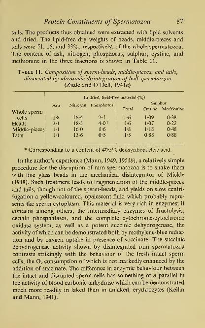
Protein Constituents of Spermatozoa 87
tails. The products thus obtained were extracted with lipid solvents
and dried. The lipid-free dry weights of heads, middle-pieces and
tails were 51, 16, and 33%, respectively, of the whole spermatozoa.
The content of ash, nitrogen, phosphorus, sulphur, cystine, and
methionine in the three fractions is shown in Table 11.
Table 11. Composition of sperm-heads, middle-pieces, and tails,
dissociated by ultrasonic disintegration of bull spermatozoa
(Zittle and O'Dell, 1941«)
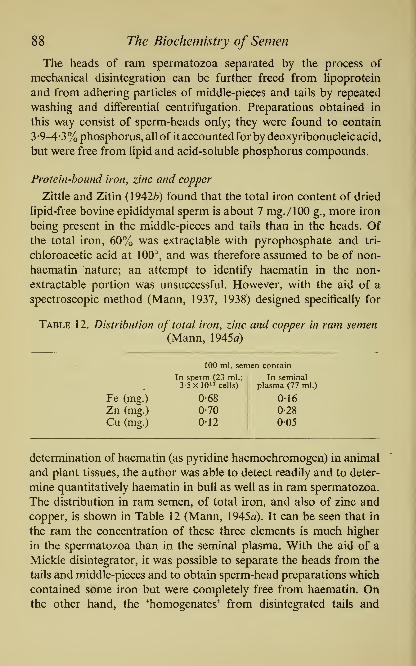
88 The Biochemistry of Semen
The heads of ram spermatozoa separated by the process of
mechanical disintegration can be further freed from hpoprotein
and from adhering particles of middle-pieces and tails by repeated
washing and differential centrifugation. Preparations obtained in
this way consist of sperm-heads only; they were found to contain
3 •9-4-3% phosphorus, all of it accounted for by deoxyribonucleic acid,
but were free from lipid and acid-soluble phosphorus compounds.
Protein-bound iron, zinc and copper
Zittle and Zitin (1942^) found that the total iron content of dried
lipid-free bovine epididymal sperm is about 7 mg./lOO g., more iron
being present in the middle-pieces and tails than in the heads. Ofthe total iron, 60% was extractable with pyrophosphate and tri-
chloroacetic acid at 100^, and was therefore assumed to be of non-
haematin nature; an attempt to identify haematin in the non-
extractable portion was unsuccessful. However, with the aid of a
spectroscopic method (Mann, 1937, 1938) designed specifically for
Table 12. Distribution of total iron, zinc and copper in ram semen(Mann, 1945a)
.
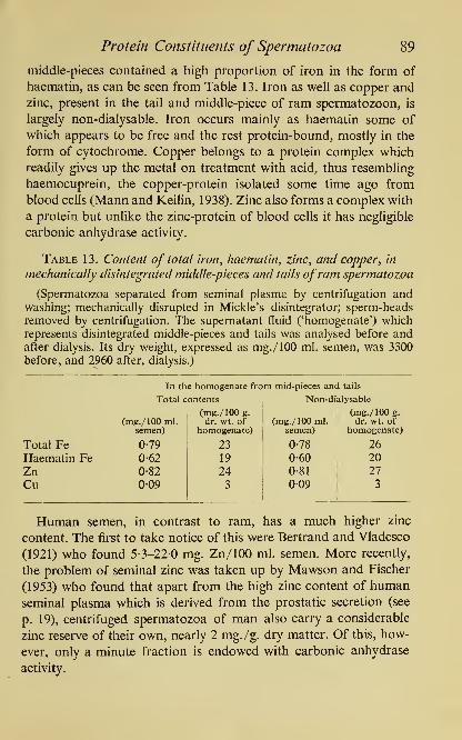
Protein Constituents of Spermatozoa 89
middle-pieces contained a high proportion of iron in the form of
haematin, as can be seen from Table 13. Iron as well as copper and
zinc, present in the tail and middle-piece of ram spermatozoon, is
largely non-dialysable. Iron occurs mainly as haematin some of
which appears to be free and the rest protein-bound, mostly in the
form of cytochrome. Copper belongs to a protein complex which
readily gives up the metal on treatment with acid, thus resembling
haemocuprein, the copper-protein isolated some time ago from
blood cells (Mann and Keilin, 1938). Zinc also forms a complex with
a protein but unlike the zinc-protein of blood cells it has negligible
carbonic anhydrase activity.
Table 13. Content of total iron, haematin, zinc, and copper^ in
mechanically disintegrated middle-pieces and tails ofram spermatozoa
(Spermatozoa separated from seminal plasma by centrifugation andwashing; mechanically disrupted in Mickle's disintegrator; sperm-headsremoved by centrifugation. The supernatant fluid ('homogenate') which
represents disintegrated middle-pieces and tails was analysed before andafter dialysis. Its dry weight, expressed as mg./lOO ml. semen, was 3300
before, and 2960 after, dialysis.)

90 The Biochemistry of Semen
Sea-urchin semen has approximately the same concentration
of copper as ram semen, with a similar distribution of the metal
between sperm and seminal plasma (Barnes and Rothschild, 1950).
Cytochrome
Early investigators of semen were well aware of the fact that
spermatozoa give a positive indophenol reaction with the 'Nadi*
reagent, particularly marked in the regions of the acrosome and
middle-piece (Herwerden, 1913). Ostwald (1907), Voss (1922) and
Sereni (1929), among others, made important contributions to the
subject of sperm indophenol oxidase and noted that the intensity of
the reaction increased towards the final stages of sperm maturation
and after ejaculation.
When in 1925 Keilin discovered cytochrome and identified
indophenol oxidase with cytochrome oxidase, he noticed that of all
the organs of a perfused frog, the heart muscle and the testicular
tissue exhibited the strongest absorption spectrum of cytochrome.
A little later, Brachet (1934) reported the presence of cytochrome in
frog sperm, and Ball and Meyerhof (1940) in sea-urchin spermatozoa.
In spite of that, attempts by several workers to detect the spectrum
of cytochrome in mammalian spermatozoa met with failure and the
functioning of cytochrome in mammalian semen continued to be
deduced only indirectly from the evidence based on the oxidation
of succinate and phenylenediamine (Shergin, 1940; Lardy and
Phillips, 1941c; Zittle and Zitin, 1942^; MacLeod, 1943a). This led
to some speculation, particularly in the case of human semen, about
the mechanism of respiration, the more so, since it has been asserted
that the oxygen consumption of human semen is associated pre-
dominantly with the seminal plasma and not with the spermatozoa
themselves (MacLeod, 1941a; Ross, Miller and Kurzrok, 1941;
Zeller, 1941).
In an efTor^ to re-examine the whole problem, the author made a
study of the cytochrome content of mammalian spermatozoa (Mann,
1945a, c). With the aid of the microspectroscope, an instrument
eminently suitable for direct observation of absorption bands in
tissues, no difficulty was experienced in the detection of the com-
plete cytochrome spectrum in both ram and bull semen. Humansemen has a sperm density at least ten times lower than bull semen,

Protein Constituents of Spermatozoa 91
but even here, the demonstration of the absorption bands of cyto-
chrome became possible (Mann, \95\d) by the application of the
technique of Keilin and Hartree (1949, 1950), whereby manifold
intensification of absorption bands is brought about by means of
liquid air.
Our evidence for the occurrence and active participation of the
cytochrome system in the oxidative metabolism of mammalian
spermatozoa can be briefly summarized as follows. Whole fresh
semen examined a little while after ejaculation shows the diffuse
spectrum of oxidized cytochrome as well as, weakly, the absorption
bands of reduced cytochrome a, b, and c. However, after the addi-
tion of a reducing agent or on anaerobic incubation of the semen,
the bands of the reduced cytochromes become much more pro-
nounced, the cytochrome a band being more distinct than c, and
the latter stronger than cytochrome b. The picture is similar with
washed sperm suspensions in fructose-Ringer-phosphate solution;
freshly prepared sperm suspensions show mainly the spectrum of
oxidized cytochrome, which becomes reduced in the course of
anaerobic incubation; on aeration of the incubated suspension
cytochrome reverts to the oxidized form. The band of cytochrome a
can be shown to undergo a typical change under the influence of
carbon monoxide; the reaction product thus formed in the sperm
resembling closely the carbon monoxide compound of cytochrome
oxidase or cytochrome a^, originally described by Keilin and
Hartree (1939) in heart muscle preparations. Carbon monoxide,
cyanide, azide, hydroxylamine, and other typical inhibitors of the
cytochrome system in respiring tissues, all inhibit also sperm
respiration.
An elegant experiment on the behaviour of cytochrome in sperma-
tozoa was performed by Rothschild (1948a, d) who demonstrated
that the oxygen uptake of sea-urchin sperm is inhibited by carbon
monoxide and that the inhibition can be completely reversed by
white light but not by light of the 548 m^^ wavelength; the non-
reversal at that particular wavelength being due to lack of absorp-
tion by cytochrome oxidase in this region of the spectrum. Byinterposing between the source of light and the microscope a colour
filter transmitting light of the 548 m/n wavelength, Rothschild was
able to observe spermatozoa microscopically, in the presence of

92 The Biochemistry of Semen
carbon monoxide, as if they were in the dark. Under these condi-
tions, he found that carbon monoxide depressed the respiration
without a corresponding decrease in sperm motiUty. A similar con-
clusion that respiration can be dissociated from motility was reached
by Robbie (1948) from his study of the effect of cyanide on the
spermatozoa of the sand-dollar {Echinarachnius parma).
The cytochrome system of sea-urchin spermatozoa includes cyto-
chrome e and in this respect, it differs from mammalian sperm
(Keilin and Hartree, 1949). Starfish {Asterias forbesii) spermatozoa
on the other hand, exhibit a spectroscopic pattern of cytochrome
very similar to sea-urchins (Borei and Metz, 1951). There is also
some evidence that cytochrome occurs in plant sperm. This follows
from the observation by Rothschild (1951c) that the movements of
bracken spermatozoids {Pteridium aquilinum (L.) Kuhn) are photo-
reversibly inhibited by carbon monoxide.
The information gained by Zittle and Zitin (1942a) from experi-
ments on the oxidation of /7-phenylenediamine by spermatozoa
disintegrated by sonic treatment, coupled with earlier observations
on the indophenol colour reaction, indicated that the cytochrome
system is located in the cytoplasm of the middle-piece and tail,
rather than in the sperm-head. Our own spectroscopic studies led us
to the same conclusion; the examination of the disintegrated middle-
pieces and tails revealed the presence of all three cytochromes with
cytochrome a predominating; these 'homogenates' oxidized rapidly
both /7-phenylenediamine and succinic acid, and the rate of oxygen
consumption could be substantially increased by the addition of
cytochrome c.
Catalase
It did not escape Miescher's notice that salmon spermatozoa differ
from other cells by their restricted ability to decompose hydrogen per-
oxide. Some slight catalase activity has been reported in mammaUansemen (Shergin, 1940) but it is questionable whether this was due
to the spermatozoa themselves or to some accidental contamination
of semen with blood, pus or bacteria. The deficiency of catalase in
normal and cleanly collected bull semen is in fact, so typical that
Blom and Christensen (1944, 1947) base on it a method for rating
the 'hygienic quality' of bull semen; the test is carried out in Denmark
Protein Constituents of Spermatozoa 93
in special 'catalase tubes' in whichi hydrogen peroxide is added to
semen and the volume of evolved oxygen recorded. In ram sperma-
tozoa, even after mechanical disintegration, we were able to detect
only a very weak catalase activity: an extract from 0-2 g. sperm (wet
weight) required 20 min. at 18° to decompose a quantity of hydrogen
peroxide which would have been decomposed in 2 min. by 0001 ml.
blood. Sea-urchin semen on the other hand, contains much more
catalase (Evans, 1947; Rothschild, 1948c, 1950c; Barron, Gasvoda
and Flood, 1949; Rybak and Gustafson, 1952).
The lack of catalase in mammalian semen explains the harmful
effects of hydrogen peroxide and pure oxygen on spermatozoa (see
p. 58). It is also of considerable physiological interest for another
reason, inasmuch as the spermatozoa themselves produce hydrogen
peroxide in vitro during the oxidation of certain amino-acids (see
p. 117).
Hyaliironidase and other 'lytic' agents
The term 'hyaluronidase' in its widest sense, designates the muco-
lytic enzyme, or rather a group of enzymes, which bring about the
depolymerization and hydrolysis of hyaluronic acid. The muco-
polysaccharide called hyaluronic acid is a polymer of the disac-
charide hyalobiuronic acid which consists of A^-acetylglucosamine
and D-glucuronic acid; its enzymic degradation, that is depoly-
merization and hydrolysis, is believed by Meyer and his school
(1937, 1952) to be due to the opening of the A^-acetylglucosaminidic
bonds. Thus it should be possible to assess the activity of hyalu-
ronidase by the determination of the reducing groups liberated by
the enzymic process. In actual practice, however, this is only possible
with the use of purified hyaluronidase since crude enzyme prepara-
tions often liberate additional reducing groups through the forma-
tion of free glucuronic acid and A^-acetylglucosamine by i^-glu-
curonidase and /S-glucosaminidase, respectively. Apart from the
'reductimetric' method, however, there are several other ways in
which the activity of hyaluronidase can be measured; among those
in use is the 'mucin clot prevention (m.c.p.) test' in which the preci-
pitation by acetic acid of the clot-like protein-hyaluronic acid
complex is prevented by the enzyme; the 'turbidimetric' method is
based on the observation that purified hyaluronate at pH 4-2, gives

94 The Biochemistry of Semen
a fairly stable colloidal suspension with dilute serum, whereas
depolymerized hyaluronate under identical conditions remains clear;
the 'viscosity reduction (v.r.)' method measures the decline in vis-
cosity caused by depolymerization; in the so-called 'Spinnbarkeit'-
method the stringiness of hyaluronic acid is assessed by means of
a special device, before and after enzymic treatment. Each of the
above methods, however, is open to criticism and limited in its
scope (cf. Lundquist, 1949a; Swyer and Emmens, 1947; Meyer and
Rapport, 1952).
The mammalian testis and sperm are the richest animal sources
of hyaluronidase. The existence in testes and spermatozoa of a
'spreading' or 'diffusing' factor which, when injected intradermally,
increases the permeability of the skin to fluids, was established by
Hoff'man and Duran-Reynals (1931) and McClean (1930, 1931); but
Chain and Duthie (1939, 1940) deserve the credit for being the first
to show that purified preparations of the testicular spreading factor
possess strong hyaluronidase activity. Their finding was soon con-
firmed by other workers who made several attempts to purify the
enzyme (Hahn, 1943; Freeman, Anderson, Webster and Dorfman,
1950; Tint and Bogash, 1950). The best preparations of bovine
testicular hyaluronidase so far available are over ten thousand times
more active than the testicular tissue itself, but as yet, even the most
highly purified enzyme does not appear to be a homogeneous protein.
Hyaluronidase originates in the seminiferous epithelium of the
mature testis, and in semen it is associated with the spermatozoa and
not with the seminal plasma (Werthessen, Berman, Greenburg and
Gargill, 1945; Joel and Eichenberger, 1945; Kurzrok, Leonard and
Conrad, 1946; Swyer, 1947^; Jacquet, Plessis and Cassou, 1951).
The content of hyaluronidase per sperm cell is highest in rabbit and
bull; there is less of it in human and boar sperm, very little in dog,
and practically none in birds and reptiles.
Although it is actually a part of the sperm cell, hyaluronidase is
nevertheless so readily released by spermatozoa into the surround-
ing medium that it must be assumed to be located somewhere very
close to the cell surface, possibly on the sperm-head (Hechter and
Hadidian, 1947; Johnston and Mixner, 1947; Perlman, Leonard
and Kurzrok, 1948). A few hours' freezing of an aqueous sperm
suspension at -10^, or 24 hours' standing at 0°, has been found by

Protein Constituents of Spermatozoa 95
Swyer (1947«, b) to be quite sufficient for hyaluronidase to pass
completely into solution. In an isotonic medium spermatozoa also
tend to liberate hyaluronidase but when the concentration of the
enzyme outside the cell reaches a certain level it seems to prevent
further leakage. Indeed, under certain experimental conditions,
spermatozoa depleted of hyaluronidase have been found to be
capable of reabsorbing the enzyme from a hyaluronidase-rich
medium (Emmens and Swyer, 1948; Swyer, 1951).
The physiological role of sperm hyaluronidase is far from clear
at present. It may be related in some as yet unknown manner,
to the spsrmiogenic function of the testis, but there is also somsindication that in certain mammals at any rate, hyaluronidase acts
by facilitating the contact between the male and female gametes
through a direct liquefying action on the viscous gel which cements
the follicle cells around freshly ovulated eggs.
Much thought has been devoted in the past to the problem of the
participation of 'lytic' agents in the process of ovum fertilization,
and to what at one time used to be called the 'ovulase' activity of
spermatozoa. However, the early investigators of this problem were
in the main concerned with lower animals. In many molluscs,
fishes and amphibia, the unfertilized egg is normally surrounded by
a viscous 'jelly coat' and a membrane, which the spermatozoon
must penetrate before fertiUzation can be effected. To explain this
process, several investigators postulated the presence of lytic agents
in the spermatozoa, capable of mediating the fusion of the gametes,
but there has been little evidence that these agents are in fact enzymic
in nature, until Tyler's (1939, 1942) discovery of the 'egg-membrane
lysin', a heat-labile protein-enzyme which he extracted from the
sperm of two molluscs, the key-hole limpet Megathura cremilata
and the abalone Haliotis cracherodii. With sperm extracts of these
species, the disappearance of the egg membrane could be demon-strated within about 3 min., if the gelatinous coat of the egg
was present, and in less than 30 sec, if the coat has been first
removed.
The occurrence of similar lytic enzymes in the sperm of other
lower animals is still under discussion (cf. Tyler, 1948; Berg, 1950;
Runnstrom, 1951). Several lytic agents have been described in sea-
urchin spermatozoa. One of them is the previously mentioned

96 The Biochemistry of Semen
androgamone III ('egg-surface liquefying agent', 'sperm lysin') dis-
covered by Runnstrom and his co-workers and shown to be a heat-
stable alcohol-soluble substance, probably a fatty acid (p. 71). The
other is the protein-like jelly-coat 'dissolving' or 'precipitating'
factor, identical with Hartmann's androgamone II; the disappearance
of the egg-jelly under the influence of this protein-factor was origin-
ally described by Hartmann and his colleagues in Arbacia pustulosa,
but a similar phenomenon was later observed in other sea-urchin
species as well (Tyler and O'Melveny, 1941; Monroy and Ruff'o,
1947; Kraus, 1950; Vasseur, 1951; Monroy and Tosi, 1952). The
suggestion has been put forward that the agent which helps the sea-
urchin sperm to penetrate the jelly-coat, is a mucopolysaccharase
similar even though not identical, with hyaluronidase. This hypo-
thesis, however, is in want of experimental support. It is also difficult
as yet, to assign any definite role in fertilization to the proteolytic
gelatm-liquefying enzyme which Lundblad (1950) extracted from
the sperm of Arbacia lixula and Paracentrotus lividus.
In mammals, the existence of an enzymic 'cumulus-dispersing
factor' was first brought to light by Yamane (1935), Pincus (1935)
and Pincus and Enzmann (1936) who showed that both sperm
suspensions and extracts from rabbit spermatozoa, brought in
contact with unfertilized rabbit ova, can disperse within a short
time the follicle cells of the cumulus oophorus. In 1942, McClean
and Rowlands discovered that hyaluronidase which they obtained
not only from testes or spermatozoa, but also from snake venom
and bacteria, can act as a cumulus-dispersing factor by liquefying
the viscous gel which cements the follicle cells around freshly ovu-
lated rat ova. Similar results on the mouse were reported by Fekete
and Duran-Reynals (1943) who also noted that the gel of the
cumulus responds to metachromatic staining with toluidine blue
like hyaluronic acid.
It remains one of the unsolved mysteries in the phenomenon of
fertilization that although the actual fertilization consists ultimately
of the fusion of a single spermatozoon with the ovum, this can take
place apparently only after a multitude of spermatozoa have reached
the site of fertilization. Moreover, the denudation of the ovum from
follicular cells has also been claimed to require the presence of
numerous spermatozoa. According to McClean and Rowlands

Protein Constituents of Spermatozoa 97
(1942) they are needed to create and keep up a sufficiently high con-
centration of hyaluronidase to permit the denudation of the egg.
This hypothesis was put to the test by Rowlands (1944) who found
that it is possible to increase the fertilizing capacity of a subnormal
number of rabbit spermatozoa by the addition of hyaluronidase in
the form of extracts from whole dilute rabbit semen. Similar results
were reported by Leonard and Kurzrok (1945, 1946).
In view of all this, little wonder that many investigators became
attracted by the possibility of the therapeutic application of hyalu-
ronidase in infertility. In fact, several enthusiastic reports appeared
of success in human infertility of oligospermic origin, achieved by
the addition of bovine testicular hyaluronidase to human semen.
However, subsequent investigations failed to bear out the claim
that hyaluronidase can enhance the fertilizing capacity of sperma-
tozoa. According to Chang (1947Z), 1949) the earlier positive results
obtained with extracts from whole semen should be attributed not
to hyaluronidase but to the effect of the seminal plasma as such.
Similarly, the concept that a high sperm concentration in the vicinity
of the egg is needed to denude the ovum from its cumulus, has been
questioned by Leonard, Perlman and Kurzrok (1947), Austin (1948),
and Austin and Smiles (1948) who demonstrated clearly that sperma-
tozoa can in fact penetrate rat ova which are still enclosed in the
cumulus. Presumably, the hyaluronidase charge carried by the sper-
matozoa makes it possible for the individual sperm to 'burrow' its
way through the viscous gel which cements the follicular cells.
This, however, need not necessarily involve the dispersion of the
cumulus, which process is probably aided by the mechanical action
of the ciUa or by some other tubal factor.
Another development in the field of sperm hyaluronidase per-
taining to the role of this enzyme in fertilization, has been the
attempt to use certain inhibitors of hyaluronidase as systemic con-
traceptives. Among the inhibitors of hyaluronidase, presumably
competitive in nature, are several derivatives of hyaluronic acid
obtained by acetylation or nitration, also heparin, and a number
of other anticoagulants and mucopolysaccharides, including a sub-
stance present in blood serum (Ferraro, Costa and Pelegrini, 1948;
Hadidian and Pirie, 1948; Pincus, Pirie and Chang, 1948; Meyer
and Rapport, 1952). Another two groups of inhibitors, some of

98 The Biochemistry of Semen
which act irreversibly, consist of heavy metals and quinones, includ-
ing certain quinoid compounds derived from flavonoids (Beiler and
Martin, 1947, 1948; Rodney et ai, 1950). Much interest was aroused
some time ago by a report that phosphorylated hesperidine, a
potent in vitro inhibitor of hyaluronidase, can act as an 'anti-
fertility factor' when administered to mice and human beings;
however, attempts to substantiate this claim have, so far, been un-
successful (Martin and Beiler, 1952; Sieve, 1952; Martin, 1953;
Chang and Pincus, 1953; Thompson, Sturtevant and Bird, 1953).
Sperm nucleoproteins
In the mature sperm cell, the sperm nucleus fills the head almost
completely, the surrounding cytoplasm being very scanty. This
nucleus consists of closely packed chromatin embedded in a rela-
tively small amount of nuclear sap. The first to investigate the
chemistry of the sperm nucleus was Miescher (1878, 1897) whose
pioneer studies laid a foundation for the modern developments in
the chemistry and physiology of the cell nucleus, the nucleoproteins
and the nucleic acids. As a result of Miescher's brilliant researches,
continued and extended by such investigators as Kossel, Schmiede-
berg, Burian, Levene, Steudel, Lynch, Hammarsten, Rasmussen and
Linderstr0m-Lang, and many others, it is now generally recognized
that the chief component of sperm chromatin, one which confers
upon the paternal (haploid) chromosomes their functions as trans-
mitters of inheritance, is by its chemical nature, a deoxyribonucleo-
protein^ and consists of deoxyribonucleic acid conjugated with cer-
tain basic nuclear proteins such as protamines and histones.
Miescher used for his work chiefly salmon spermatozoa which he
obtained mostly by stripping the live fish. A considerable advantage
of this method is that the material thus obtained consists entirely of
ripe spermatozoa and is therefore of uniform composition. This
useful material, however, is not always procurable and is sometimes
replaced by whole excised fish testes which are less suitable as they
may contain some immature spermatozoa even during the breeding
season. Miescher's routine procedure was to remove first the
sperm-tails by plasmolysis with water or weak acetic acid and then
to treat the washed suspensions of sperm-heads with ethanol and
ether, to remove the lipids. In order to separate sperm nucleic

Protein Constituents of Spermatozoa 99
acid from the nuclear protein, Miescher treated the Hpid-free
material first with cold mineral acid (e.g. 0-25-0-5°/o hydrochloric
acid) to remove the protein, and then with sodium hydroxide 'until
the mixture tasted distinctly caustic to the tongue', to extract nucleic
acid from the residue.
The deoxyribonucleoproteins in the spermatozoa of a great manyfishes, including salmon, belong to the group of micleoprotamines
which can be extracted from the sperm nuclei with salt solutions. Aconvenient method for such an extraction and purification has been
described by Pollister and Mirsky (1946). Spermatozoa of the brown
trout, Salmo fario, were 'homogenized' with 1m solution of sodium
chloride in a Waring mixer and the very viscous extract poured into
six volumes of water; this caused the precipitation of the nucleo-
protamine in the form of long strands, so fibrous that they could
be wound around a glass rod and transferred in this way to another
container. The fibrous material was dissolved in M-NaCl and re-
precipitated with water, and then washed successively with 65%ethanol, hot 95% ethanol, and finally with ether; at this stage it
contained about 6% phosphorus and 18% nitrogen, and consisted
of deoxyribonucleic acid and protamine in a 6 : 4 ratio. When this
material was dissolved in M-NaCl and dialysed against M-NaCl, the
protamine slowly passed through the cellophane membrane leaving
behind the solution of deoxyribonucleic acid. On pouring this solu-
tion into five volumes of ethanol, a fibrous precipitate of the nucleic
acid was obtained, which, after drying, had a content of 8-97%
phosphorus and 14-47% nitrogen. This composition approaches
the theoretical value for the sodium salt of deoxyribonucleic acid,
9-28% phosphorus and 15-58% nitrogen.
The removal of protamine from nucleoprotamine solutions in-
duces no significant change in viscosity. The high viscosity of nucleo-
protamine solutions is due entirely to the highly polymerized nucleic
acid. Deoxyribonucleic acid prepared, for instance, from herring
spermatozoa, has a molecular weight of 800,000; each molecule of
it requires some 100 molecules of protamine to form a molecule
of nucleoprotamine. According to some calculations by Felix (1952),
a single fish sperm nucleus contains 4-5x10^ nucleoprotamine
molecules, enough to provide about 190,000 molecules per each
chromosome, or several thousand per each gene.

100 The Biochemistry of Semen
Unfortunately however, the extraction with M-NaCl is not a
universal means for the separation of sperm nucleoproteins. In the
key-hole limpet or freshwater clam, the sperm nucleoprotein
resists extraction with M-NaCl, but can be brought into solution
with a 2m salt solution, whereas no nucleoprotein can be extracted
with NaCl of either concentration from the sperm of man, bull,
boar or ram. Moreover, dialysis against M-NaCl or extraction with
dilute mineral acids both prove inadequate for the removal of
nuclear proteins from mammalian spermatozoa. In such cases, the
separation of protein can be brought about with a chloroform-
octanol mixture (Sevag, Lackman and Smolens, 1938), but before
this is applied it is necessary to separate the sperm nucleus from the
remainder of the sperm cell by ultrasonic or mechanical treatment.
Deoxyribonucleic acid
This when freed from nuclear protein, is composed of mono-
nucleotides, each consisting of one molecule of phosphoric acid,
one molecule of the sugar D(-)2-deoxyribose, and one molecule of
a purine or pyrimidine base: adenine, guanine, cytosine or thymine.
A small amount of yet another base, 5-methylcytosine (Wyatt, 1950,
1951), has been found so far in the sperm deoxyribonucleic acid of
man, bull, ram, herring and sea-urchin {Echinus esculentus), but
probably it occurs also in other species.
In all species, deoxyribonucleic acid is confined entirely to the
sperm nucleus as can be demonstrated by various staining methods,
and particularly by the 'Feulgen nucleal reaction'. This reaction was
described by Feulgen (1914, 1917) at first as a colour test for thymo-
nucleic acid, but later it was adapted for the staining of cell nuclei
(Feulgen and Rosenbeck, 1924).
With thymonucleic acid itself, the test is carried out best on a
solution of sodium nucleinate (01 g./l ml.) prepared in a boiling
tube on the water-bath. The solution is treated with 1 ml. 2N-H2SO4,
and left at 100° for 3 minutes, then cooled and neutralized. When a
drop of the hydrolysate is mixed with a few ml. of Schiff^'s fuchsin-
sulphurous acid reagent (a 0-5% solution of fuchsin decolorized
with SO2 and the excess of SO2 removed by suction), an intense
purple colour develops. The chemistry of the Feulgen colour re-
action is as yet only partly understood but is believed to involve the

Protein Constituents of Spermatozoa 101
NHo OH NH2
N C X N C X HN CHI II
CHI
IICH
III
HC C / H.,N—
C
C / OC CH
^N^ ^NH ^N^ ^NH ^NHAdenine Guanine Cytosine
(6-aminopurine) (2-amino-6-hydroxy- (2-hydroxy-6-
purine) aminopyrimidine)
O NH2
II I
/^\ ^^\HN C—CH3 N C—CH3
I
III
II
OC CH OC CH
^NH ^NHThymine 5-Methyl-cytosine
(5-methyl-2 : 6-dihydroxy-
pyrimidine)
following steps: acid hydrolysis which splits off purine and exposes
deoxyribose; transformation of deoxyribose into co-laevuUnic alde-
hyde; lastly, formation of a purple-coloured reaction product of
this labile aldehyde with fuchsin-sulphurous acid. The mechanism
of the Feulgen reaction as exhibited by the cell nuclei in histological
preparations, appears to be even more involved (Danielli, 1947;
Davidson, 1950). In cellular material, however carefully carried out,
the Feulgen reaction cannot be expected to yield results as accurate
and specific as the colour test with pure deoxyribonucleic acid.
Some of the difficulties inherent in the application of the reaction
as a staining method for sperm nuclei, have been pointed out by
Feulgen and Rosenbeck themselves, who noted that when fresh
smears ofhuman semen were treated with Schiff 's fuchsin-sulphurous
acid reagent, the sperm-heads stained rather weakly, whereas
the middle-pieces and tails, though devoid of nuclear material,
stained strongly. This observation was followed by a demonstration
that the fuchsin-staining material present in the middle-piece and
tail is a lipid, 'plasmal', which can be distinguished from the

102 The Biochemistry of Semen
nucleoprotein of the sperm-head by its solubiUty in ethanol. Similar
observations with animal sperm were later reported by other investi-
gators. There is no doubt, however, that if carried out properly and
under conditions which eliminate interference from other fuchsin-
staining substances, the Feulgen nucleal reaction can be madespecific for the sperm nucleus. Several authors have stressed the
fact that the base and the posterior region of the sperm-head stains
particularly strongly (Marza, 1930; Wislocki, 1950; Friedlaender
and Fraser, 1952).
Although present in the sperm nuclei of all species, the composi-
tion of deoxyribonucleic acid varies somewhat from one species to
another, with regard to the proportion of the various purine and
pyrimidine bases. But within any given species, all body cells, includ-
ing the spermatozoa, seem to yield on purification the same nucleic
acid, that is one with the same molar ratio of adenine, guanine,
cytosine and thymine. In the species so far examined, the ratio of
adenine to thymine, of guanine to cytosine, and of total purines to
total pyrimidines, was shown to equal unity.
The results of the analysis of bases in deoxyribonucleic acid from
ram spermatozoa are shown in Plate V. The spermatozoa were dis-
integrated mechanically and suspensions of washed, tail-free sperm-
heads were prepared (Mann, \95\b). Nucleic acid was separated
from the nuclear proteins of the sperm-heads by the chloroform-
octanol treatment, hydrolysed with formic acid, and the liberated
bases separated on paper chromatograms by Wyatt's method, using
as solvent an aqueous solution containing 65% /^(^propanol and
2N-HC1. The bases set free by acid hydrolysis and determined
spectrophotometrically according to Markham and Smith (1949)
were: guanine, adenine, cytosine and thymine in a molar ratio of
0-91 : M3 : 0-86 : 110. In addition, there was a small amount of
methylcytosine which in Plate V is only just visible as a faint ultra-
violet-absorbing band below cytosine; the molar ratio of cytosine
to methylcytosine was 1 : 005.
Deoxyribonucleic acid (Na-salt) of human sperm was studied by
Chargaff and his colleagues (Chargaff, Zamenhof and Green, 1950;
ChargafF, 1951a, b)\ it contains 16% nitrogen and 8-9% phosphorus
(Zamenhof, Shettles and Chargaff, 1950); the specific viscosity of a
0-135% solution in water equals 7-0, and the sedimentation constant

PLATE V
SPERM HEADS (RAM) : DRNA
m^-w
Guanine 0.9I
Adenine |.|3
Cytosjne 0.86
^^f*
Thymine t.lO
PURINE AND PYRIMIDINE BASES IN SPERM DEOXYRIBONUCLEIC ACID
Contact print, taken with ultraviolet light, of a paper chromatogram
from the acid hydrolysate of ram sperm-heads. The figures indicate
the molar ratios.


Protein Constituents of Spermatozoa 1 03
(Soo) for a 0-22% solution in 0-2M-NaCl is 5-7, a value which re-
sembles closely that given by an undegraded specimen of calf
thymus nucleic acid. According to Elmes, Smith and White (1952),
the molar proportions of the purine and pyrmidine bases in deoxy-
ribonucleic acid from human sperm and tissues are: guanine 0-92
(standard error of observations 0-036), adenine 1-23 (0-068), cyto-
sine 0-84 (0077), thymine 1-01 (0-09) and 5-methylcytosine 0-03.
Table 14. Salmon sperm deoxyribonucleic acid (Chargaff et al, 1951)
(Molar ratios between the bases.)
Purines to pyrimidines 1-02
Adenine to thymine 1-02
Guanine to cytosine 1-02
Adenine to guanine 1-43
Thymine to cytosine 1 -43
The analysis of the nucleic acid obtained from the sperm nucleo-
protamine of salmon, Salmo salar (Chargaff, Lipschitz, Green and
Hodes, 1951) gives a good illustration of the regularity in the com-
position of deoxyribonucleic acid. The nucleoprotamine was pre-
pared by extraction with a salt solution, the nucleic acid set free by
chloroform-octanol treatment, and after some further purification,
recovered as the sodium salt. Two specimens were isolated in this
way, containing 14-3% nitrogen and 8-9% phosphorus and 14-8%
nitrogen and 8-9% phosphorus, respectively. The ultraviolet absorp-
tion spectrum measured in M-phosphate buffer pH 7-1, exhibited
a maximum at 260 mi-i. The specific viscosity in distilled water at
30-3°, was found to be 29-6 for a 0-22% solution, and 5-6 and 20for 0-11 and 0-055% solutions, respectively. The bases were set
free by acid hydrolysis and analysed chromatographically and
spectroscopically. The ratios of the purine to the pyrimidine bases,
of adenine to thymine, and of guanine to cytosine equalled unity;
adenine and thymine exceeded guanine and cytosine by about 40%(Table 14).
Not only the composition but also the content of deoxyribonucleic
acid in the cell nucleus appears to be fairly constant and characteris-
tic for each animal species. The haploid nucleus of the sperm cell,
however, differs from the diploid nuclei of the somatic cells in that
it contains a reduced amount of chromatin and consequently it is

104 The Biochemistry of Semen
assumed to possess only one-half of the amount of deoxyribonucleic
acid present in the somatic nuclei of a given species (Boivin, Vendrely
and Vendrely, 1948; Vendrely and Vendrely, 1948, 1949, 1952;
Mirsky and Ris, 1949, 1951; a review by Colette Vendrely, 1952).
To illustrate this point, Table 1 5 gives data on the content of deoxy-
ribonucleic acid in the sperm (haploid) nuclei and in the somatic
(diploid or polyploid) cell nuclei of various species. The values
range from 0- 1 1 x 10~^ mg. for the diploid nucleus of a sponge cell to
168x10"^ for a diploid cell nucleus of Amphiuma, a urodele; the
nucleic acid in the spermatozoa of the carp, trout, pike, tench, toad,
cock and bull, is seen to be approximately one-half the content of
the somatic cell nuclei. A high proportion of the data listed in
Table 15, were obtained by means of the analytical procedures
Table 15. Deoxyribonucleic acid content of single nuclei in
somatic cells and spermatozoa
(Contents expressed in mg. x 10~^ deoxyribonucleic acid per cell nucleus;
figures in brackets refer to authors.)
Animal

Protein Constituents of Spermatozoa 105
Trout, Salmo irideas Gibb.
Salmo fario
Pike, Esox lucius
Tench, Tinea tinea
AMPHIBIANS
AmphiumaFrogToad
REPTILES
Green turtle
BIRDS
Domestic fowl
Duck
MAMMALS
Cattle
Pig
Sheep
Dog
Man
(1) Davidson et al. (1950); (2) Mirsky and Ris (1949); (3) Mirsky andRis (1951); (4) Vendrely and Vendrely (1948); (5) Vendrely and Vendrely
(1949); (6) Vendrely and Vendrely (1952); (7) Vendrely (1952); (8) Vendrelyand Vendrely (1953).
Erythrocyte

106 The Biochemistry of Semen
developed by Schmidt and Thannhauser (1945) and Schneider
(1945, 1946) which are based largely on determinations of phos-
phorus and involve the removal of (i) the 'acid-soluble phosphorus
compounds' (extraction with cold trichloroacetic acid), and (ii) the
phospholipids (extraction with ethanol and ether), prior to the
analysis of nucleic acid (see also Table 16).
The basic nuclear proteins, protamines and histones
The proteins conjugated with deoxyribonucleic acid are of the
basic type and have been shown to be either protamines or histones
in most instances so far examined. Protamines have been isolated
from fish spermatozoa of several species. Of the various protamines,
the best known are salmine from salmon and trout sperm, and
clupeine from herring sperm. Much less is known about the other
protamines, such as scombrine (mackerel), cyclopterine (lump-
sucker), esocine (pike), thynnine (tunny fish), percine (perch),
cyprinine (carp) and sturine (sturgeon).
Judging from the molecular weight and amino acid composition,
salmine, clupeine and scombrine have a relatively simple structure,
there being approximately two molecules of arginine to one molecule
of monoamino acid. Thus, for example, the analysis of salmine
sulphate prepared from the spermatozoa of the Spring or Chumsalmon (Tristram, 1947, 1949) suggests a molecular weight of about
8000, with a total of 58 amino acid residues: 40 arginine, 8 isoleu-
cine, 2 valine, 4 proline, 3 glycine, 1 alanine and 7 serine; this sal-
mine sulphate contains 19-85% sulphuric acid, i.e. 40 equivalents
per molecule, sufficient to combine with all arginine residues.
Another salmine sulphate, one prepared from the sperm of the
Columbia River salmon (Block and Boiling, 1945) is said to con-
tain 67 amino acid residues: 47 arginine, 1 isoleucine, 3 valine,
6 proline, 4 alanine and 6 serine (see also: Corfield and Robson,
1953).
The results of formol titration indicate that salmine contains one
free amino or imino group per molecule of 8000, and the end group
assay carried out by means of the dinitrofluorobenzene method
(Sanger, 1952) suggests that the N-terminal position is occupied by
the imino group of proline. In clupeine, the A^-terminal position is
also taken up by proline, whereas at the other end of the amino acid

Table 16. Distribution ofphosphorus compounds in ram semen
(Results based on analysis of 16-8 ml. ram semen, representing 14
ejaculates from 7 rams; average volume of single ejaculate 1-2 ml.;
3,050,000 spermatozoa/yal. Semen separated by centrifugation for 20 min.
at 10,000^, into sperm, 26% v/v, and seminal plasma, 74% v/v.)
mg. P/100 ml. semen
In whole In sperm In seminalsemen plasma
Total phosphorus 328-5 186-7 141-8
Acid-soluble phosphorus (in trichloro-
acetic acid extract)
Pjnorg (orthophosphate determined as
MgNH4P04) 10-3 3-1 7-2
Po (phosphate reacting directly with
molybdate) 11-9 4-7 7-2
P7 (phosphorus which appears as ortho-
phosphate after 7 min. hydrolysis
withN-HCl) 15-4 8-1 7-3
P30 (phosphorus which appears as
orthophosphate after 30 min. hydro-
lysis with N-HCl) 15-9 8-6 7-3
Ptot. ac. sol. (total acid-soluble phosphate
determined after incineration) 159-4 27-4 132-0
Patp (labile phosphate of adeno-
sine triphosphate determined in the
Ba-salt by the method of Parnas
and Lutwak-Mann, 1935) 4-3 4-3 00Phexose (phosphate of 6-phosphohexose
determined in the supernatant fromBa-ATP) 6-2 60 0-2
Phospholipidphosphorus (extracted with
ethanol and ether from the residue
insoluble in trichloroacetic acid) 30-8 27-9 2-9
Deoxyribonucleic acid-phosphorus (ex-
tracted with KOH from the residue
left after removal of acid-soluble Pand phospholipid; precipitated fromthe KOH extract by acid) 1110 1110* 00
Residual phosphorus (left after removal
of acid-soluble compounds, phos-
pholipids and deoxyribonucleic acid) 27-3 20-4 6-9
* Corresponding to 3-2 x 10~^ mg. deoxyribonucleic acid/sperm cell.
107
108 The Biochemistry of Semen
chain, the C-terminal position is filled by arginyl-arginine (Dirr and
Felix, 1932). Arginine-arginine linkages occur also with great fre-
quency in the amino acid chain itself (Felix and Schuberth, 1942).
The following peptides have been identified as breakdown products
of a partial hydrolysis of clupeine: arginyl-arginine, triarginyl-
arginine, alanyl-arginyl-arginine, seryl-arginyl-arginine, and alanyl-
alanine; on the basis of the available evidence, Felix, Fischer and
Krekels (1952) suggested the following sequence of proline (Prol.),
arginine (Arg.) and monoamino acids (M.) in clupeine,
Prol. (Arg. Arg. Arg. Arg. M. M.)^ Arg. Arg.
Salmine and clupeine, irrespective of their origin, both contain
arginine as the sole basic amino acid; in the corresponding nucleo-
protamines the ratio between the arginine residues of the protamines
and the phosphoric acid equivalents of nucleic acid is not far from
unity, usually about 0-95 (Felix, 1951). In other protamines, on the
other hand, e.g. percine and sturine, a certain proportion of the
basic units is present in the form of histidine and lysine.
On close inspection of analytical data relating to the various pro-
tamines, there stands out a considerable degree of variability in the
amino acid composition, even in closely related species. The situa-
tion is even more complicated in sperm nucleoproteins which con-
tain histones instead of protamines. The histones such as occur for
instance, in the sperm nucleoproteins of sea-urchins {arbacine of
Mathews, 1897) and cod-fishes (gadushistone and lotahistone) are
characterized by a wider range of amino acids, including tyrosine.
It is not improbable that some of the observed variations in the
amino acid composition of protamines and histones, represent dis-
tinct species characteristics analogous to those which are encoun-
tered in other proteins, e.g. in the globins of various haemoglobins.
On the other hand, however, the standards for the assessment of
chemical purity of nuclear proteins are open to criticism, and it is un-
certain whether the examined protamines and histones were always
really pure. Quite likely a sperm nucleus may contain in some
cases more than one basic protein, that is, a main protamine or
histone, together with a smaller amount of a 'subsidiary' product
(Stedman and Stedman, 1951). Furthermore, there is also the pos-
sibility that some of the reported differences in the composition of

Protein Constituents of Spermatozoa 109
nuclear proteins are simply due to the use of material containing
variable proportions of mature and immature spermatozoa. Sperma-
tocytic development is well known to be associated with characteris-
tic changes in the amino acid composition of the nuclear proteins.
Immature spermatozoa of salmon, for instance, obtained directly
from excised testes, contain a histone instead of the salmine. Simi-
larly, the testes of the mackerel yield on extraction with dilute
hydrochloric acid a histone, 'scombron', instead of the protamine
'scombrine'. This and similar observations, prompted Kossel (1928)
to acclaim the histones as 'intermediary stages' in the transformation
of complex proteins into protamines.
77?^ non-basic nuclear proteins; karyogen and chromosomin
In addition to the basic proteins, the sperm nucleus always con-
tains some non-basic or so-called residual proteins. In lipid-free
preparations from salmon sperm-heads, Miescher found 19-78%
protamine (extracted with 0-25-0-5% HCl), 2-94% acid-soluble pro-
tein material other than protamine, 60-50% of nucleic acid (ex-
tracted with NaOH) and 16-78% of an iron-containing residue
which was insoluble either in acid or in alkali and which he believed
to contain 'karyogen', the 'inner-space protein substance' {Innen-
raumsubstanz), of the sperm nucleus.
It was found later that the residual or non-basic nuclear proteins,
unlike the protamines and histones, contain tryptophan as a
characteristic component. Opinions are divided, however, on the
problem of the actual ratio of non-basic to basic proteins in the
sperm nucleus. According to the Stedmans (1943, 1947), the nucleo-
protamine present in the sperm-heads of salmon accounts for no
more than 70% of the dry, lipid-free material, whereas the remainder
is made up largely of a non-basic protein 'chromosomin', which
contains tryptophan.* On the other hand, PoUister and Mirsky
(1946) state that the nucleoprotamine present in trout spermatozoa
* The Stedmans' chromosomin must not be confused with chromosin,
a name given by Mirsky and Pollister (1946) to a complex extracted with
M-NaCl from isolated cell nuclei of various organs, including thymus,
liver, spleen, pancreas, brain, frog testes and bacteria. This complex
is composed of deoxyribonucleic acid, histone, and a tryptophan-contain-
ing protein.

1 10 The Biochemistry of Semen
accounts for as much as 91% of the dry, lipid-free mass of the head
nuclei. These authors, however, also find in the nuclei a charac-
teristic tryptophan-containing residual protein.
Keratin-like protein of the sperm membrane
Within the category of 'residual' sperm proteins are also certain
highly insoluble sulphur-rich proteins, obtained from mammalianspermatozoa and derived probably from the sperm membrane. Thefirst mention of a 'sulphur-rich substance, containing more than
4% S in the heads of bull spermatozoa', was made in 1878 by
Miescher, who did not, however, investigate its origin and compo-
sition. Green (1940) extracted ram spermatozoa successively with
dilute acid and alkali, and obtained a residue containing 19-3%
nitrogen and 11-4% cystine, which he believed to represent the
sperm membrane. The possibility of a keratin-like protein present
in the sperm membrane is strengthened by the observations of Zittle
and O'Dell (1941a) on the solubilizing action of thioglycolic acid
and trimethylbenzylammonium hydroxide on bull spermatozoa. In
boar sperm, the portion which remains undissolved after prolonged
treatment with N-NaOH, consists of 'ghost' sperm-heads which
resemble in shape the sperm membranes (Thomas and Mayer, 1949).
A remarkable property of the sperm membrane which can be demon-
strated microscopically, is the extraordinary elasticity of the sperm-
head structure; this was convincingly demonstrated in the experi-
ments of Moench and Holt (1929-32), who were able to hook the
head of a human spermatozoon with a microsurgical needle and
to stretch it very considerably.

CHAPTER V
Protein Constituents and Enzymes
of the Seminal Plasma
Proteoses and free amino acids. Fibrinolysin and fibrinogenase. Pepsino-
gen. Ammonia formation. Amino acid oxidase. Seminal phosphatases;
*acid' and 'alkaline' phosphatase; 5-nucleotidase; pyrophosphatase.
Enzymic hydrolysis of adenosine triphosphate.
Proteoses andfree amino acids
A DISCUSSION of the nature of extracellular protein constituents of
semen demands the recognition of certain circumstances which are
peculiar to the seminal plasma. An important, though sadly ne-
glected fact is that the protein content of the seminal plasma does
not remain constant after ejaculation but undergoes rapid changes
of enzymic character which manifest themselves in a progressive
decrease in the concentration of non-dialysable protein-nitrogen
and a simultaneous accumulation of non-protein nitrogen, free
amino acids and, at a late stage, of free ammonia. Unless this is
fully taken into account, results of protein and amino acid analysis
in semen are but of little significance and yield no information on the
initial distribution of nitrogenous compounds. This applies especi-
ally to human semen as was convincingly demonstrated by Lund-
quist (1949c, 1952), and also by Jacobsson (1950, Fig. 11).
Even in freshly collected seminal plasma a large proportion of
total nitrogen is found partly as a protein-like material which passes
readily through semi-permeable membranes but is not heat-coagu-
lable and not precipitated by trichloroacetic acid; accordingly, this
has been classified as propeptone, hemialbumose, primary proteose,
and secondary proteose (thioalbumose and synalbumose) (Posner,
1888, 1892; Marshall, 1922; Goldblatt, 1935^). In human semmal
plasma, out of a total content of about 3-5 to 5-5 g. protein-like
material per 100 ml., no more than 18%, usually much less, is
coagulated by heat, and about 60% passes through cellulose
111

112 The Biochemistry of Semen
membranes which are impermeable to blood serum proteins
(Huggins, Scott and Heinen, 1942). The electrophoretic pattern of
the non-dialysable portion from five different specimens of humanseminal plasma has been examined by Gray and Huggins (1942) whoobserved four distinct components which corresponded to serum
albumin (17-7-22-7%), a-globulin (19-8-27-8%), i5-globulin (34-3-
44-5%), and y-globulin (11 -4-21 0%). Ross, Moore and Miller
iUU
Protein Constituents and Enzymes of Seminal Plasma 113
for human seminal plasma, is probably due to this mucoid sub-
stance (Wislocki, 1950).
The proteoses in trichloroacetic acid extracts from semen, which
one encounters not only in man, but also in the ram, bull, boar,
and other species, occasionally interfere with chemical analyses of
certain non-protein constituents. To overcome this difficulty, it is
advisable to replace trichloroacetic acid with other deproteinizing
agents such as zinc hydroxide, tungstic acid, phosphotungstic acid
or ethanol. Another source of trouble encountered in analytical
work with semen and due to the mucinous substance in seminal
plasma, human in particular, is that on centrifugation the mucushas a tendency to form a stringy mass which firmly adheres to the
sperm cells. Caution must therefore be exercised in attributing to
spermatozoa as such, analytical results obtained with centrifuged
human semen.
Several assays of free amino acids in mammalian seminal plasma
have been carried out, mostly however, by chromatographic or
microbiological methods, and not by chemical isolation. A notable
exception is the work of Wagner-Jauregg (1941) who isolated
crystalline tyrosine from an ethanolic extract of human semen. Thefollowing free amino acids were found to occur in the seminal plasma
of man, glycine, threonine, alanine, valine, leucine, isoleucine,
cystine, proline, tyrosine, phenylalanine, lysine, arginine, aspartic
acid and glutamic acid (Jacobsson, 1950; Lundquist, 1952). In
bovine seminal plasma serine, glycine, alanine, aspartic acid and
glutamic acid were found (Gassner and Hopwood, 1952), and a
similar pattern was also observed in the seminal vesicle secretion
and in the ampullar fluid, the latter containing in addition a trace of
tyrosine. According to Gassner, the free amino acids in bull seminal
plasma disappear after castration, like seminal fructose and citric
acid, but their content is not restored by testosterone administra-
tion; furthermore, vasectomy alone, which is without effect on the
content of fructose, causes a disappearance of amino acids from bull
seminal plasma.
Free amino acids occur also in fish semen; as long ago as 1923,
leucine, lysine and alanine have been isolated in pure form from
protein-free extracts of herring testicles (Steudel and Suzuki, 1923).
There are indications that the amino acids and proteoses present

114 The Biochemistry of Semen
in the seminal plasma may be of some importance to the sperma-
tozoa. It may be recalled that excessive dilution of semen exerts a
deleterious effect on spermatozoa and that this can be counteracted,
partly at least, by the inclusion in the diluting media of amino acids
such as glycine, alanine, valine, leucine, lysine, and glutamic acid
(p. 76). The beneficial action of these amino acids is believed to
depend primarily on their metal-binding capacity (Tyler and Roth-
schild, 1951). Several other effects of amino acids have been observed
with the sperm of lower animals. Giese and Wells (1952) found
that glycine (005m) protected the spermatozoa of Strongylocentrotus
piirpwatus from the detrimental effect of light. Metz and Donovan
(1950) demonstrated that in the starfish certain amino acids promote
the agglutination of spermatozoa by egg-water of this species; in
the absence of these amino acids agglutination does not take place.
Fibrinolysin andfibrinogenase
The seminal proteoses and amino acids are presumably the pro-
ducts of proteolytic activity which in the seminal plasma is derived
mainly from the prostatic secretion, but partly also from the
seminal vesicle fluid. The two powerful proteolytic agents of the
prostatic secretion are 'fibrinolysin' and 'fibrinogenase' (see also
pp. 17 and 29).
The coagulation of human semen is followed by liquefaction, a
process which is catalysed by a proteolytic agent present in the
prostatic secretion. Its discoverers, Huggins and Neal (1942) named
it fibrinolysin because of its ability to digest blood fibrin, and its
resemblance to the fibrinolytic agent in haemolytic streptococci
(Tillet and Garner, 1933). However, the fibrinolytic system present
in blood has now been resolved into several distinct components,
whereas the streptococcal fibrinolysin has been defined as a kinase,
i.e. an activator of the fibrinolytic enzyme preformed in the blood.
Consequently, the name 'fibrinolysin' has been abandoned with
reference to the streptococcal agent in favour of 'streptokinase'.
Furthermore, it proved impossible to replace streptokinase by pro-
static fibrinolysin as an activator of the blood enzyme (Oettle,
1950).
The fibrinolytic activity can be assayed in human semen by the
method of Harvey (1949), which consists in mixing a constant volume

Protein Constituents and Enzymes ofSeminal Plasma 1 1
5
of oxalated blood plasma with varying volumes of semen, inducing
clotting by the addition of 1-5% calcium chloride, and noting the
time required for the clot to liquefy. Owing to the inhibitory effect
of blood plasma on fibrinolysis, the plasma must not constitute
more than one-tenth of the reacting system. Harvey states that the
degree of fibrinolytic activity in semen varies with the individual
but she found no correlation between this activity and either the
volume of ejaculates or any characteristics of spermatozoa. Simi-
larly, there was no positive relationship between the lysin content
and semen viscosity. However, specimens which were exceptionally
viscous, usually also had low fibrinolytic power.
The precise nature of seminal fibrinolysin and its relation to
plasmin, the fibrin-splitting agent of the blood, will not be knownuntil the enzyme has been purified. Moreover, experiments by Kaulla
and Shettles (1953) indicate that in addition to the plasmin-like
enzyme proper, the human seminal plasma contains at least three
other agents, (i) fibrinolysokinase, an activator of blood profibrinoly-
sin, (ii) a small amount of profibrinolysin itself, i.e. of material
which can be activated by streptokinase, and (iii) antifibrinolysiny
an inhibitor of plasmin, which, however, is present in a much lower
concentration in the seminal plasma than in blood serum.
Fibrinogenase is the name given by Huggins and Neal (1942)
to the proteolytic agent, highly active in canine prostatic secretion,
but less so in human prostatic fluid, which destroys blood plasma
fibrinogen. Huggins and his co-workers (1942, 1943) also share the
credit for having recognized the similarity between certain other
proteolytic properties of the prostatic secretion and those of pan-
creatic trypsin. More recently, the 'tryptic' enzyme of human semen
has been partially purified by Lundquist (1952), who defined as a
unit the amount of enzyme which in 1 hr., at pH 7-6, and 37°,
liberates from added casein a quantity of chromogen corresponding
to 01 mg. free tyrosin; he achieved an activity of about 1 unit per
mg. of protein-nitrogen, i.e. an approximately tenfold purification.
The purified enzyme was active also towards haemoglobin and both
human and bovine blood plasma fibrinogen. In an attempt to
purify from human semen the natural substrate for the proteolytic
activity, Lundquist obtained a protein fraction, 'seminal fibrin',
which was readily digested by enzyme preparations both from9

1 1
6
The Biochemistry of Semen
semen and the prostate gland, and yielded the same amino acids
which appear normally in human semen.
Pepsinogen
Apart from the above mentioned protease which acts optimally
at pH 7-6, two more proteolytic enzymes have been found in
human semen by Lundquist and his co-workers (1951, 1952, 1953).
One is an amino peptidase which hydrolyses leucine amide, glycyl-
glycine, triglycine and glycyl- leucine, with an optimum around pH7-5. The other has been identified as pepsinogen. According to
Lundquist and Seedorff (1952), the activity of pepsinogen in semen
corresponds to 2 //g. pepsin/ml., which is of the same order of
magnitude as that found in gastric juice. But unlike the trypsin-
like enzyme, seminal pepsinogen seems to originate in the seminal
vesicles and not in the prostate. Its specific function is not fully
understood as it is difficult to envisage in semen the high hydrogen
ion concentration required for the conversion of pepsinogen to
pepsin.
Ammonia formation
A phenomenon probably associated with the enzymic degrada-
tion of proteins, is the progressive accumulation of free ammonia
which takes place in whole semen and in seminal plasma, on anaero-
bic as well as aerobic incubation. This has been observed in several
species (Shergin, 1933; Mann, 1945^). In ram semen for example,
the content of free ammonia (estimated by vacuum-steam distilla-
tion in the Parnas-Heller apparatus) was found to increase from
1-3 mg. NH3-N/IOO ml. in fresh semen to 9-7 mg. NH3-N/IOO ml.,
after 7 hr. incubation at 37°, under sterile conditions (Mann,
\9A5a). Ammonia formed in semen is in considerable excess of the
amount which could be derived from adenyl derivatives; the total
adenine amino-A'^ content of the semen as assessed enzymically
with heart muscle deaminase being only about 4 mg./lOO ml. in
fresh, and 3 mg./lOO ml. in incubated, semen. It is also unlikely to
originate from urea, since the content of urea in semen is not sig-
nificantly affected by incubation.

Protein Constituents and Enzymes of Seminal Plasma 1 1
7
Amino acid oxidase
In addition to sugars and fatty acids, spermatozoa are capable
of oxidizing a number of amines and amino acids. The oxidative
deamination of amino acids by bull spermatozoa has been the subject
of a study by Tosicand Walton (1945; 1946«, b, 1950). The starting
point of this study has been the observation that the addition of
egg-yolk to bull sperm causes an increased oxygen uptake, which,
however, gradually declines in about an hour's time. Egg-yolk
fractionation led to a dialysable, nitrogen-containing fraction which
was oxidized by spermatozoa. The oxidation was accompanied by
accumulation of ammonia and formation of hydrogen peroxide.
Evidence was obtained which pointed to peroxide being responsible
for the gradual decline in the oxygen uptake by spermatozoa.
In the course of their study, Tosic and Walton examined several
pure amino acids and found that spermatozoa oxidize three natur-
ally occurring amino acids, namely L-tyrosine, L-phenylalanine and
L-tryptophan. According to Tosic (1947, 1951), the hydrogen-
peroxide-forming aerobic process in bull semen is an oxidative
deamination catalysed by the L-amino acid oxidase of spermatozoa,
which differs from the analogous enzyme of other animal tissues
by having its range of activity restricted to only three aromatic
amino acids; the activity of the enzyme can be expressed by the
equation
R CH2CHCOOH+O2+H2O —> R CH, C COOH+H2O2+NH3I
"II
NH2 O
Seminal phosphatases
Semen owes its powerful phosphatase activity mainly to the
seminal plasma which carries several different dephosphorylating
enzymes derived from the male accessory organs of reproduction.
Among the most active and best known enzymes in this group are the
so-called 'acid phosphatase' and 'alkaline phosphatase'. In addition
to these two phosphomonoesterases, the seminal plasma contains
'5-nucleotidase', a pyrophosphatase, and several adenosinetri-
phosphatases.
In early studies on phosphatases, the substrates commonly

118 The Biochemistry of Semen
used were a- and /3-phosphoglycerol, and phosphohexoses, chiefly
6-phosphoglucose and 6-phosphofructose, but also 1 : 6-diphospho-
fructose. More recently, however, other organic phosphoric acid
derivatives came into use, including various nucleotides and inter-
mediary phosphorylated compounds of glycolysis, as well as two
synthetic substances: phenylphosphate (King and Armstrong, 1934)
and phenolphthaleine phosphate (King, 1943; Huggins and Talalay,
1945). The introduction of histochemical techniques marked another
important development in studies on phosphatases (Gomori, 1939,
\94la, b, 1953). The histochemical investigations have thrown muchlight upon the pattern of phosphatase distribution in the male
accessory organs and have helped to establish the existence of
'secretory' phosphatases, localized in the secretory epithelia and
secretions of accessory glands, as distinct from the 'stromal' phos-
phatases which are present only in the stroma (Dempsey, 1948;
Bern, 1949; RoUinson, 1954).
''Acid' and 'alkaline' phosphatase
An observation that the phosphatase activity of male urine is
usually higher than in women, led Kutscher and Wolbergs (1935)
to examine the phosphatase in semen and in the prostate gland.
They soon found that semen and prostate are among the richest
sources of acid phosphatase in the human body, the enzyme being
optimally active at pH 5-6, equally well towards a- and ^S-phospho-
glycerol, but largely inactive towards diphosphofructose and pyro-
phosphate (Kutscher and co-workers, 1936, 1938). Subsequent
investigations confirmed and extended these findings; the demon-
stration by Scott and Huggins (1942) that, while the voided urine
of man is rich in the enzyme, urine collected directly from the renal
pelvis shows only little enzymic activity, was a convincing proof
that the content of acid phosphatase in normally voided male urine
is due largely to the admixture of prostatic secretion.
Acid phosphatase is an important secondary male sex charac-
teristic. Investigations by Gutman and Gutman (1938Z)) have shown
that the level of the enzyme in the human prostate is low in child-
hood but increases rapidly at puberty; thus the activity, expressed in
King-Armstrong units per gram prostate tissue, was H units at four
years of age, 73 units at puberty, and 522 to 2284 units in adult men.

Protein Constituents and Enzymes of Seminal Plasma 119
A similar relation to age was observed in monkeys and dogs; in
both these species administration of androgenic hormones to imma-
ture males stimulates considerably the output of the enzyme from
the prostate gland (Gutman and Gutman, 1939; Huggins and
Russell, 1946). A certain correlation appears to exist in adult menbetween the level of acid phosphatase in semen and androgenic
activity (Gutman and Gutman, 1940; Gutman, 1942; Engberg,
Anderson, Sury and Raft, 1947). However, like other constituents
of semen, the level of prostatic phosphatase activity varies from one
species to another, as well as between individuals within the same
species.
Under physiological conditions, acid phosphatase does not pass
from the prostate into the blood stream. However, significant
amounts of it appear in the blood plasma as a result of malignant
growth in the prostate and metastases of prostatic cancer in the
bones; injections of androgen still further increase the level of
enzyme in blood plasma, whereas castration or treatment with
oestrogens lead to a spectacular decrease. The determination of
prostatic phosphatase activity in blood has been utilized as a valuable
diagnostic aid in prostatic carcinoma and in the course of clinical
treatment (Gutman and Gutman, 1938a; Huggins, Scott and Hodges,
1941; Watkinson, Delory, King and Haddow, 1944).
An important addition to our knowledge of the physiological
functions of acid phosphatase in seminal plasma, has been the dis-
covery made by Lundquist (1946) that freshly ejaculated humansemen contains phosphorylcholine which on ejaculation is rapidly
dephosphorylated by the acid phosphatase to free choline andorthophosphate. This phenomenon is more fully discussed else-
where (p. 170). But there is evidence that apart from the acid phos-
phatase which acts on phosphorylcholine optimally at pH 6-3 (in
acetate buffer), human seminal plasma contains another phosphatase
which acts on the same substrate at a higher pH (Hudson and Butler,
1950). Of considerable interest is also the finding that the acid
seminal phosphatase exhibits in vitro a distinct transferase activity
(Green and Meyerhof, 1952); partially purified acid phosphatase
from the human prostate has been shown to catalyse at pH 5-5
the transfer of phosphate from both ^S-phosphoglycerol and phospho-
creatine to glucose; the product in each case was 6-phosphoglucose

120 The Biochemistry of Semen
(Morton, 1953). Methods for the purification of acid phosphatase
from human prostate glands and semen have been described by
London and Hudson (1953) and Boman (1954).
Alkaline phosphatase, like the 'acid' enzyme, is widely distributed
in male accessory organs but its localization in cells and concentra-
tion in accessory gland secretions is different. Human semen with
its conspicuously high level of acid phosphatase, has a low concen-
tration of the alkaline phosphatase. Bull semen on the other hand,
has only slight acid phosphatase activity but contains more of the
alkaline phosphatase (Haq and Mullen, 1948; Reid, Ward and
Salsbury, 1948^). This difference between the human and bovine
semen is not altogether unexpected, since the bulk of bull seminal
plasma is derived not from the prostate but from the seminal
vesicles. In the rat both phosphatases are of low activity; with the
exception of the ventral prostate which may contain up to 20 units
of alkaline phosphatase per g. tissue, the level of either enzyme
seldom exceeds 4 units per g. in any one of the other accessory
organs. After castration the activity of both these enzymes diminishes
first in the rat seminal vesicles, and a little later in the prostate; but
the percentage decrease of enzymic activity and of organ weight is
roughly equal (Huggins and Webster, 1948; Stafford, Rubinstein
and Meyer, 1949).
Table 17. Phosphatase activity of ram seminalplasma onphosphohexoses (Mann and Lutwak-Mann, 19516)
(The liberation of sugars was examined by incubating 5 mg. substrate
(Na salt) with 1 ml. dialysed seminal plasma at pH 7 or 0-2 ml. dialysed
seminal plasma at pH 9, for 1 hr., 37°, in the presence of OOOSM-MgCla.The sugars were determined after deproteinization with ZnS04 andBa(OH)2; glucose was estimated by means of glucose oxidase (Mann,1944; \9A6b).)

Protein Constituents and Enzymes of Seminal Plasma 121
Alkaline phosphatase has an optimum at about pH 9, and is
capable of hydrolysing among others, 1-phosphofructose, 6-phos-
phofructose and 1 : 6-diphosphofructose. Our researches indicate
that this activity may represent an essential step in the process of
fructose formation and secretion by the accessory organs (Mannand Lutwak-Mann, 1951«, b) (see also p. 150). Ram seminal plasma
is particularly rich in alkaline phosphatase which, though it acts
optimally at pH 9, also shows appreciable activity towards phos-
phohexoses at pH 7. This can be seen from Table 17 which gives
rates of dephosphorylation for various compounds. In the case of
6-phosphofructose and 1 : 6-diphosphofructose, some glucose is
formed in addition to fructose, owing to the presence of phospho-
hexose isomerase in the seminal plasma, as a result of which a part
of 6-phosphofructose is converted to 6-phosphoglucose before
dephosphorylation. Supporting evidence for the conclusion that
monophosphofructose rather than diphosphofructose, is the sub-
strate immediately responsible for the liberation of seminal fructose,
has been provided by Bouchilloux and Menager (1952) who found
that the semen of both ram and bull contains two phosphomono-
esterases, with pH optima at 9-4 and 4-8, respectively, but that it
lacks a specific fructosediphosphatase.
5-Nucleotidase
This enzyme was discovered in human semen by Reis (1937,
1938, 1940) and shown to dephosphorylate muscle adenylic acid
(adenosine-5'-phosphoric acid) and inosinic acid (inosine-5'-phos-
phoric acid) but not adenosine-3'-phosphoric acid or adenosine
triphosphoric acid. Bull seminal plasma is particularly rich in 5-
nucleotidase, the seminal vesicles being the main source of the
enzyme (Mann, 1945a, 1947). Bull seminal plasma or the vesicular
secretion itself, act several hundred times more efficiently on muscle
adenylic acid than on /S-phosphoglycerol; from 160 ^g. P added as
sodium adenylate to 0001 ml. bull seminal plasma, up to 140 /^g.
P are liberated as orthophosphate during 1 hour's incubation at 37°.
The 5-nucleotidase of bull seminal plasma has been purified about
fifty-fold by Heppel and Hilmoe (195 la). The purified enzyme
has a pH optimum at 8-5, and its activity is enhanced by the addi-
tion of magnesium ions but inhibited by fluoride (00 1m) and by

122 The Biochemistry of Semen
borate buffer (0-08m) to the extent of 73 and 85% respectively. Onthe basis of tests with numerous phosphorylated compounds, it
may be safely concluded that 5-nucleotidase is an enzyme which acts
specifically on substrates containing the ribose-5-phosphate moiety.
The purified enzyme splits rapidly ribose-5-phosphate but not
ribose-3-phosphate; it is active towards adenosine-5'-phosphate,
inosine-5'-phosphate, uridine-5'-phosphate and cytidine-5'-phos-
phate but inactive towards both adenosine-3'-phosphate and
adenosine-2'-phosphate. It also dephosphorylates nicotinamide
mononucleotide (nicotinamide ribose-5'-phosphate); this inciden-
tally explains an early observation of ours, that bull seminal plasma
decomposes cozymase, with slow liberation of inorganic phosphate.
Pyrophosphatase
Bull seminal plasma contains an enzyme which hydrolyses in-
organic pyrophosphate to orthophosphate but differs from the
pyrophosphatase of yeast. Seminal pyrophosphatase can exert its
maximal activity in the absence of magnesium ions and is not
inhibited by increased substrate concentration; it has a sharp
optimum at pH 8-6 (Heppel and Hilmoe, \95\b),
Enzymic hydrolysis of adenosine triphosphate
In addition to adenosine-triphosphatase (ATP-ase) in the sperma-
tozoa there are also ATP-splitting enzymes in the seminal plasma
(Mann, 1945o; MacLeod and Summerson, 1946). When adenosine
triphosphate is acted upon by bull or human seminal plasma, all
three phosphate groups are set free as orthophosphate. The mechan-
ism of this reaction has been investigated by Heppel and Hilmoe
(1953) who by fractionation procedures obtained three distinct
ATP-ases, none of them, however, completely free from 5-nucleo-
tidase.
One of the enzymes, named the 'pyrophosphate-forming ATP-ase', catalyses the reaction
Adenosine triphosphate+H2O —>-
Pyrophosphate+Adenosine-5'-phosphate
It is relatively heat-stable, has a pH optimum at 8 -4-8 -6 andrequires neither calcium nor magnesium ions for activation. The

Protein Constituents and Enzymes of Seminal Plasma 123
remaining two ATP-ases, designated respectively as 'acid' and
'alkaline', produce orthophosphate; the 'acid' ATP-ase has a pHoptimum at 5-7-60, requires magnesium, is inhibited by calcium,
and can be inactivated completely by heating for 20 min. at 60°; it
does not act on i5-phosphoglycerol; the 'alkaline' ATP-ase is more
heat-resistant, has a pH optimum at 8 -4-8 -8, and is stimulated by
calcium and also by magnesium. Both these enzymes are active not
only tov^ards adenosine triphosphate but adenosine diphosphate
as v^ell.
Bull seminal plasma contains in 1 ml. about 80 units of acid
ATP-ase, 130 units of alkaline ATP-ase, 40 units of the pyrophos-
phate-forming ATP-ase, and 2900 units of 5-nucleotidase. It re-
mains for future studies to define the physiological significance
of all these enzymes, particularly the 5-nucleotidase which is so
characteristic of semen. Possibly, there is some link between them
and other nucleolytic enzymes and they may well play a role in the
metabolism of purine compounds in semen and reproductive organs.
The occurrence of nucleases in human and sea-urchin semen
(Zamenhof, Shettles and Chargaff, 1950; Mazia, 1949), the cozy-
mase-destroying activity of bull seminal plasma (Mann, 1945a), the
interesting findings on the presence of uric acid in bull semen
(Barron and Haq, 1948; Leone, 1952), and the more recent demon-
stration of xanthine oxidase in the bull vesicular secretion (Leone,
1953), are but a few examples of problems in this field, which await
further and more detailed study.
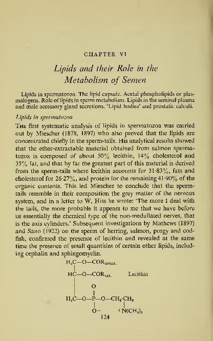
CHAPTER VI
Lipids and their Role in the
Metabolism of Semen
Lipids in spermatozoa. The lipid capsule. Acetal phospholipids or plas-
malogens. Role of lipids in sperm metabolism. Lipids in the seminal plasma
and male accessory gland secretions. 'Lipid bodies' and prostatic calculi.
Lipids in spermatozoa
The first systematic analysis of lipids in spermatozoa was carried
out by Miescher (1878, 1897) who also proved that the lipids are
concentrated chiefly in the sperm-tails. His analytical results showed
that the ether-extractable material obtained from salmon sperma-
tozoa is composed of about 50% lecithin, 14% cholesterol and
35% fat, and that by far the greatest part of this material is derived
from the sperm-tails where lecithin accounts for 31-83%, fats and
cholesterol for 26-27%, and protein for the remaining 41 -90% of the
organic contents. This led Miescher to conclude that the sperm-
tails resemble in their composition the grey matter of the nervous
system, and in a letter to W. Hiss he wrote: 'The more I deal with
the tails, the more probable it appears to me that we have before
us essentially the chemical type of the non-medullated nerves, that
is the axis cylmders.' Subsequent investigations by Mathews (1897)
and Sano (1922) on the sperm of herring, salmon, porgy and cod-
fish, confirmed the presence of lecithin and revealed at the same
time the presence of small quantities of certain other lipids, includ-
ing cephalin and sphingomyelin.
H2C—O—CORunsat.
I
HC—O—CORsat. Lecithin
OII
H2C—O—P—O—CH2-CH2I I
O- +N(CH3)3124

Lipids and their Role in the Metabolism of Semen 125
When air-dried salmon roe is ground and extracted with pentane
in a Soxhlet apparatus, a yellow oil is obtained which is practically
free from phospholipids; it requires further treatment with ethanol
or methanol for the phospholipids to be extracted from the sperm.
In this way, for example, 12-5% glyceride in the oily fraction, and
6-2% phospholipid in the alcoholic fraction, was obtained from the
roe of the sockeye salmon, Oncorhynchus nerka (Halpern, 1945). Onthe basis of this observation, it has been suggested that the phos-
pholipids occur in the spermatozoa in a firmly bound state, pre-
sumably in the form of lipoproteins. A substantial portion of the
unsaponifiable material extracted from fish sperm by fat solvents
consists of cholesterol which accounts, on the average, for 2-2%
of dried fish spermatozoa (Schmidt-Nielsen and Sundsvold, 1943).
The high content of lipids in spermatozoa is equally characteristic
for fishes as for other animals. Sea-urchin spermatozoa are well
known to contain a large reserve of lipid material, shown by Mathews
(1897) to include both lecithin and neutral fat. The content of phos-
pholipids in the sperm of Echinus esculentus is about 5-5% of the
dry weight of spermatozoa (Rothschild and Cleland, 1952). The
seminal lipids of E. esculentus have been analysed more recently by
Cardin and Meara (1953). The material obtained by extraction of
1-2 1. of semen with acetone and light petroleum, consisted of 13-6%
neutral fat, 32-9% free fatty acids, 260% phospholipids, 9-2%
sterols and 18-3% of other unsaponifiable matter. The component
fatty acids of the non-phospholipid fraction included a low propor-
tion (10-1%) of saturated acids and a high proportion of unsatur-
ated acids with 18C (30-4%) 20C (451%) and 22C (12-3%). The
phospholipid fraction had a ratio of N : P=l-4 : 1, and must have
therefore, consisted of a mixture of monoaminophosphatides and
diaminophosphatides
.
Early analyses of lipids in bovine epididymal sperm were carried
out by Koelliker (1856) who found that over 12% of the dried
material is ether-extractable. About half of this content was later
shown by Miescher (1878) to consist of lecithin. In a study of the
lipid content of bull sperm, Zittle and O'Dell (1941«) have extracted
washed epididymal spermatozoa successively with ethanol, ether,
acetone and petroleum ether, and found 13% of lipid material; when
the procedure was repeated with spermatozoa disintegrated by sonic

126 The Biochemistry of Semen
treatment, more lipid was found in the tails (23%) than in the mid-
pieces (6%) or heads (7%).
The lipid capsule
It is probable that the high lipid content of spermatozoa is due
largely to the lipid 'sheath' or 'capsule' which encloses the sperm
cell. So it would seem at any rate, from histochemical studies,
including the extensive investigation of Popa and Marza (1931) whodescribed the so-called manteau Upidique in the spermatozoon of
man, dog, bull, ram, boar, rabbit, guinea-pig and cock. The lipid
capsule is presumably of importance to the spermatozoa in their
function, perhaps to ward off the effects of the acid vaginal milieu as
has been suggested by Redenz (1924). It appears to consist largely
of a lipoprotein complex which is fairly soluble in aqueous solvents.
In the case of mammalian spermatozoa, this complex has been
extracted with a 014m solution of sodium chloride at pH 9 (Dallam
and Thomas, 1952). In the middle-piece lipids were shown to be
associated with the 'spiral body' which surrounds the axial proto-
plasmic thread, and is derived from the mitochondria of the sperma-
tids (Wislocki, 1950; Brown, 1952).
Acetal phospholipids or plasmalogens
An interesting feature of the sperm cell is a characteristically high
content of acetal phospholipids or plasmalogens. Feulgen and
Rosenbeck (1924) while studying the 'nucleal' reaction of humanspermatozoa, noted that when fresh smears of human semen were
treated with Schiff's fuchsin-sulphurous acid reagent, the middle-
pieces and tails, though devoid ojf nuclear material, stained strongly.
This observation was followed by a demonstration that cells in
general contain in their protoplasm some material which stains
diffusely with Schiff's reagent, but differs from nucleoproteins by its
solubility in ethanol. The name 'plasmal' was bestowed upon this
material, which was shown in subsequent investigations by Feulgen
and his co-workers to arise from 'plasmalogen', a group of peculiar
phospholipids widely distributed in tissues, and distinguished by the
presence of higher fatty aldehydes in place of the usual fatty acids.
The plasmalogen isolated by Feulgen and Bersin (1939) from beef
muscle was identified as an acetal of glycerylphosphorylcolamine.

Lipids and their Role in the Metabolism of Semen 127
The two principal fatty aldehydes in plasmalogens are palmitic and
stearic aldehydes but other fatty aldehydes were also reported
(Feulgen, Boguth and Andresen, 1951). Crystalline acetal phos-
pholipids were prepared from beef brain, and shown to belong to
the a-series (Thannhauser, Boncoddo and Schmidt, 1951).
H.C—
O
\CHR
HaC—
O
Acetal a-phospholipid
I OI
II
H,C—O—P—O—CH,I 1
OH NH2
The plasmalogen content of bull semen as determined by Boguth
(1952) was found to vary from 30 to 90 mg./lOO ml.; of this about
two-thirds is present in the sperm and one-third in the seminal
plasma. The volume taken up by sperm in bull semen is compara-
tively small, about 10%; it would seem therefore, that the concentra-
tion of the acetal phospholipids in the spermatozoa themselves must
be of the order of 200-600 mg./lOO g. fresh weight, or 3 x lO-^" mg.
per cell.
Heptacosane
The list of interesting chemical substances which occur in semen
was extended again when in 1941 Wagner-Jauregg reported on the
isolation of the hydrocarbon heptacosane from human semen. Analcoholic extract obtained from 18 litters of semen formed, upon
concentration in vacuo, a solid residue which was extracted first
with 1-5 1. acetone, and next with 2 1. of a mixture of equal amounts
of ethanol and ether. On purification, the acetone-soluble fraction
yielded some crystalline material which melted at 57-60° and con-
sisted, in all probability, of palmitic and stearic acid. The ethanol-
ether soluble fraction formed on standing a crystalline precipitate
containing 1 g. of heptacosane, CH3(CH2)25CH3, which on recrystal-
lization showed the required melting point, 59-5°. The isolation of
heptacosane has previously been achieved from plant material. It is
also known to be associated in a characteristic manner with beeswax.

128 The Biochemistry of Semen
So far, however, the only instance other than semen, where hepta-
cosane has been shown to occur in the human body, is in the urine
of pregnant women. It is absent from the urine of men and of non-
pregnant women, and is devoid of oestrogenic activity. Nothing
is known about the origin or function of seminal heptacosane.
Should future investigations, however, show that heptacosane in
semen is involved in the metabolism of fatty acids or aldehydes, then
its fate would be analogous to that of plant hydrocarbons which
are well known to be associated with the metabolism of fatty acids,
aldehydes and alcohols in plants.
Role of lipids in sperm jnetabolism
The functional aspects of lipid metabolism in spermatozoa have
been the subject of investigations by Lardy and Phillips (1941«, b\
1945). To begin with, these authors confirmed the observation
originally made by Redenz (1933) that, in contrast to whole semen
which can be stored successfully both anaerobically and aerobically
owing to the presence of glycolysable carbohydrate in the seminal
plasma, bull spermatozoa separated from the seminal plasma by
centrifugation and washing, can survive only in the presence of
oxygen. From this they inferred that when the spermatozoa are
deprived of sugar, they begin to oxidize aerobically some of their ownintracellular constituents as a source of energy for motility. Todetect the oxidizable substrate, sperm samples were analysed when
fresh and after periods of storage; it was then found that a period
of aerobic incubation of bull spermatozoa caused a significant de-
crease in the content of lipid phosphorus accompanied by an increase
in the acid-soluble phosphorus. But when glucose was added to the
washed sperm, the decrease in the phospholipid content of sperma-
tozoa was very slight, an indication perhaps, of a preference by
spermatozoa for the glycolytic mechanism as a source of energy
(Table 18). It was also found that certain phospholipids prepared
from egg-yolk, liver and soya bean, effectively maintained the
oxygen uptake and motility of washed sperm suspensions under
aerobic conditions; however, on the addition of sugar to the sperm
suspension, the phospholipids no longer produced an effect on
either respiration or glycolysis.
In bull semen as ejaculated, with its large reserve of readily

Lipids and their Role in the Metabolism of Semen 129
Table 18. Changes in the phospholipid content of bull spermatozoa
in presence and absence of sugar (Lardy & Phillips, \9A\a)
(Bull spermatozoa freed from seminal plasma by centrifugation, then
diluted with Ringer-phosphate solution to the original volume of semen,
and incubated at room temperature.)
Medium Phospholipid content
After 10 hr.
Original incubation(mg. P/ml.) (mg. P/ml.)
Ringer-phosphate 0-38 0-24
Ringer-phosphate+0-04M-glucose 0-39 0-37
glycolysable material in the form of fructose, the share of phos-
pholipids in sperm metabolism is probably small. But in the epi-
didymis, where glycolysable sugar is unavailable, Lardy and Phillips
ascribe great importance to the phospholipids as a source of oxida-
tive energy. The mechanism of utilization of this reserve is held to
involve hydrolytic cleavage of phospholipids foUov^ed by an oxida-
tion of the fatty acid portion via the citric acid cycle, and coupled
with aerobic phosphorylations. According to Lardy, Hansen and
Phillips (1945), the aerobic metabolism of phospholipids in the
bovine epididymal spermatozoa is accompanied by an uptake of
inorganic phosphate, and the formation of a phosphate ester which
is hydrolysed in 7 min. by N-HCl at 100°, and thus resembles
adenosine triphosphate.
The ability to utilize phospholipids as a source of aerobic energy
extends to the spermatozoa of lower animals, notably those of the
sea-urchin. One of the main differences between mammalian and
sea-urchin semen is that the latter contains no glycolysable material
in the seminal plasma. The possibility that sea-urchin spermatozoa
which have been shed into sea-water, survive at the expense of
energy derived from the oxidation of intracellular carbohydrate such
as glycogen, also appears remote, in view of the very low content
of glycogen-like material in the sperm cells (Stott, 1930; Rothschild
and Mann, 1950). On the other hand, according to Rothschild and
Cleland (1952), the content of intracellular phospholipids which in
fresh sperm of Echinus esculentus is 5-5% of the dry weight or 4-14
mg. (0165 mg. P) per 10^*^ sperm cells decreases in the course of
aerobic incubation of sperm suspensions in sea-water, at an average

130 The Biochemistry of Semen
rate of 0-787 mg./lO^" sperm cells/7 hr. The oxygen uptake recorded
during the same period is 1-45 ml. Oa/lO^" sperm, which if sustained
exclusively by phospholipids, would require the disappearance of
0-906 mg. of phospholipid. On the basis of these observations
Rothschild and Cleland conclude that the principal source of energy
required for the movement of sea-urchin spermatozoa is derived
from the oxidative breakdown of phosphilipids located mainly in
the middle-piece of the sperm cell.
Lipids in the seminal plasma and male accessory gland secretions
Apart from the lipids which form a part of the sperm structure,
there is also some lipid material in the seminal plasma. The bulk of
the 'bound choline', however, does not consist of phospholipids
but occurs in the form of acid-soluble phosphorylated derivatives of
choline (see p. 170). The lipid of the human seminal plasma origin-
Table 19. Lipids of the human prostatic fluid and seminalplasma
(w^./lOO ml.) (Scott, 1945)
(No. indicates the number of studied specimens.)

Lipids and their Role in the Metabolism of Semen 131
and 185-5 mg./lOO ml. of 'total Upid' and 83-5 mg./lOO ml. of
phospholipid, in the seminal plasma. However, he was unable to
detect lecithin either in the prostatic fluid or in the seminal plasma.
In both instances, two-thirds of the phospholipid consisted of an
ether-soluble choline-free phosphatide, probably identical with cepha-
line, the rest being some other, ether-insoluble material. Scott
found little neutral fat in either the prostatic secretion or the seminal
plasma, the sum of phospholipids and cholesterol accounting for
practically the entire 'total lipid'. The content of 70-120 mg. total
cholesterol/1 00 ml. seminal plasma recorded by Scott, is below the
cholesterol value for human blood plasma; a similar figure, 80 mg./
100 ml., has been reported earlier by Goldblatt (1935a).
'Lipid bodies'' and prostatic calculi
In many species, the seminal plasma contains small globules,
droplets or granules, sometimes called the 'lipid bodies'. In man,
dog, cat, and rabbit, they are derived chiefly from the prostatic
secretion but in certain species they occur also in the seminal vesicle
secretion (Prevost and Dumas, 1824; Pittard, 1852). The globules of
the human prostatic secretion are referred to by Sir Henry Thompsonin his famous prize essay on the Diseases of the Prostate (1861),
as 'small yellowish bodies, in appearance sometimes granular, some-
times homogeneous, about the size of red blood corpuscles, but not
so uniform, being from about 1/5000 to 1/2500 of an inch in
diameter' and exhibiting 'considerable refractive power nearly so
much as to give them a resemblance to oil globules'. The occurrence
of similar elements in the prostatic secretion was later observed by
Fuerbringer (1881, 1886) who coined for them the name Leclthin-
kdrnchen\ These 'lecithin granules' or 'lecithin bodies' which Fuer-
bringer regarded as responsible for the normal opalescence and
milky appearance of the prostatic fluid, have since been re-examined
on several occasions, mostly by means of histological methods.
Chemical analysis however, failed to corroborate the presence of
lecithin in these particles. Other curious structures which according
to some authors are closely linked with the appearance of 'lipid
bodies' in the human seminal plasma, are certain larger bodies
known as 'colostrum corpuscles', 'corpora amylacea' and 'pros-
tatic calculi'. The colostrum corpuscles, frequently met with in the
10
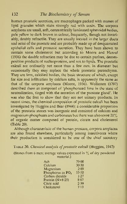
1 32 The Biochemistry of Semen
human prostatic secretion, are macrophages packed with masses of
lipid granules which stain strongly red with eosin. The corpora
amylacea are small, soft, concentrically laminated spheroidal bodies,
pale yellow to dark brown in colour, frequently, though not invari-
ably, doubly refractile. They are usually located in the larger ducts
and acini of the prostate and are probably made up of desquamated
epithelial cells and prostatic secretion. They have been shown to
contain some cholesterol but according to Moore and Hanzel
(1936) the double refraction may be due to certain purines, decom-
position products of nucleoproteins, and not to lipids. The prostatic
calculi are ordinarily not more than a few mm. in diameter but
occasionally they may replace the whole prostatic parenchyma.
They are firm, calcified bodies, the basic structure of which, except
for size and infiltration by calcium salts, is apparently the same as
that of the corpora amylacea (Moore, 1936). Wollaston (1797)
described them as composed of 'phosphorated lime in the state of
neutralization, tinged with the secretion of the prostate gland'. Hewas also the first to show that they are not urinary products. In
recent times, the chemical composition of prostatic calculi has been
investigated by Huggins and Bear (1944); a considerable proportion
of the prostatic stones was inorganic and consisted of calcium and
magnesium phosphates and carbonates but there was also some 21%of organic matter composed of protein, citrate and cholesterol
(Table 20).
Although characteristic of the human prostate, corpora amylacea
are also found elsewhere, particularly among insectivores where
their production is considered to be one of the chief secretory
Table 20. Chemical analysis ofprostatic calculi (Huggins, 1947)
(Stones from 6 men; average values expressed in % of dry powdered

Lipids and their Role in the Metabolism of Semen 133
functions of the prostate gland (Hopkins, 1911; Eadie, 1948^, b).
They do not seem to occur either in the dog or the rat. The total
lipid content of the dog prostatic fluid ranges from 30 to 40
mg./lOO ml. (Huggins, 1947) and the lipid phosphorus from Mto 2-2 mg. P/100 ml. (Moore et al, 1941). There exists a condition
known as the 'benign prostatic hypertrophy', which is common to
dog and man. In the dog, however, this condition is not associated
with the occurrence of corpora amylacea or any other spheroidal
nodules but consists of cystic hyperplasia (Huggins and Clark,
1940). In the bull, lipid-laden cells form a highly characteristic
component of the seminal vesicle epithelium, and the cavities of
the tubules in the seminal vesicles contain an abundance of eosino-
philic granular secretion (Mann, Davies and Humphrey, 1949). The
extent to which organs other than the seminal vesicle and prostate,
contribute to the lipid or sterol content of semen, has not been
hitherto studied in much detail. In this connection, however, an
interesting observation of Ward and Moore (1953) deserves to be
mentioned, concerning the occurrence of 7-dehydrocholesterol in
the preputial gland and epididymis of rat.

CHAPTER VII
Fructose and Fructolysis
Fructose as a normal constituent of semen. Species differences. Site of
formation. Seminal fructose as an indicator of male sex hormone activity;
the 'fructose test' and its application to certain problems of sex endocrin-
ology. Role of hypophysis. The relationship between blood glucose andseminal fructose. Effect of malnutrition. The enzymic mechanism of fruc-
tose formation. Anaerobic and aerobic utilization of carbohydrate byspermatozoa. Pasteur effect and the 'metabolic regulator'. Intermediary
reactions in sperm fructolysis and the role of phosphorus-containing
coenzymes.
There has been little precise knowledge about fructose (laevulose)
in man and higher animals except the evidence of its occurrence in
certain embryonic fluids and in metabolic dysfunctions like fruc-
tosuria. The presence of a laevorotatory constitutent in foetal fluids
was first noted by Claude Bernard (1855) but its chemical identity
was not recognized until some time later when it was shown that
fructose was a normal constituent of allantoic and amniotic fluid,
foetal blood and the urine of new-born animals (Majewski, 1858;
Griiber and Griinbaum, 1904; Paton, Watson and Kerr, 1907;
Langstein and Neuberg, 1907; Orr, 1924; Cole and Hitchcock,
1946; Bacon and Bell, 1948; Hitchcock, 1949). More recently, the
source of foetal fructose was traced to the placenta (Huggett,
Warren and Warren, 1951). So far as adult man is concerned, it
was believed that in general the occurrence of fructose is restricted
to pathological conditions; fructose has been demonstrated in
transsudates, and in the urine of diabetics and persons suffering
from the peculiar metabolic disorder known as 'spontaneous fruc-
tosuria', the aetiology of which remains obscure. In the normal
human or animal organism, fructose has been found to be utilized
chiefly after enzymic conversion to glucose and glycogen; liver,
kidney and the gastro-intestinal tract were shown to be the main
sites of this process (Oppel, 1930; Bollman and Mann, 1931;
Stewart and Thompson, 1941; Deuel, 1936; Reinecke, 1944).
134

Fructose and Fructolysis 135
Thus, in the light of the evidence available until relatively recently,
it seemed rather improbable that in the normal, fully-developed
mammalian organism, fructose could occupy a place on the list
of 'animal carbohydrates', or that any specific function could be
assigned to this sugar.
Fructose as a normal constituent of semen
Since the early researches on mammalian semen by McCarthy,
Stepita, Johnston and Killian (1928), Ivanov (1931), Huggins and
Johnson (1933) and other pioneers in the field of semen biochemistry,
it was known that in several species, including man, a reducing and
yeast-fermentable sugar is normally present in semen, the concen-
tration of this sugar exceeding by far that of glucose in blood.
However, up to 1945, in the extensive literature dealing with the
subject of seminal sugar, this substance has been described either as
glucose or simply as the reducing sugar of semen (Killian, 1933;
Bernstein, 1933; Goldblatt, 1935«; Shergin, 1937; McKenzie, Miller
and Bauguess, 1938; Davis and Cole, 1939; Moore and Mayer,
1941; MacLeod and Hotchkiss, 1942; Salisbury and VanDemark,
1945), and the only reference to a probable occurrence of fructose
in semen is found in an early paper by Yamada (1933) who in a
general survey of human tissues and body fluids carried out numer-
ous fructose determinations by means of a colour reaction with the
drug 'cryogenine'; of course, like so many colour tests, this reaction
by itself cannot be regarded as specific for fructose, since it gives
a positive result not only with fructose but also with other ketoses,
nor does it distinguish between free fructose, that is D(-)-fructo-
pyranose (formula in Fig. 14), and bound fructose, i.e. fructofura-
nose, such as occurs for example, in the various phosphofructoses
.
In 1945, in the course of studies on the metabolism of semen, the
seminal sugar was isolated for the first time and identified by chemi-
cal methods as free D(-)-fructose (Mann, \9A5b\ \946a, b, c). The
actual final isolation was accomplished with a 120 ml. sample of bull
semen representing some thirty pooled bull ejaculates, which were
collected within a twelve-hour period by the various Centres for
Artificial Insemination of Cattle in England, and immediately des-
patched to Cambridge. The chemical procedure involved the fol-
lowing steps: (a) the preparation of methylphenyl-fructosazone, a

136 The Biochemistry of Semen
crystalline compound which has been shown by Neuberg (Neuberg,
1902, 1904; Neuberg and Strauss, 1902; Langstein and Neuberg,
1907; Neuberg and Mandl, 1946) to be one of the few chemical
derivatives by means of which fructose can be identified and dis-
tinguished from glucose and from other closely related sugars; {b) the
purification of seminal fructose up to the point when it reached the
specific optical activity of pure crystalline fructose: [aP°°= -92-2°;
(c) the demonstration that fructose occurs in the semen in free form
and that it accounts for the whole of the yeast-fermentable carbo-
hydrate which yields 'ketose reactions' with resorcinol (Seliwanoff,
1887; Roe, 1934), diphenylamine (Ihl, 1885) and similar colour-
producing substances (Pinoff, 1905; Thomas and Maftei, 1927;
Pryde, 1946); {d) proof obtained with the highly specific enzyme,
glucose oxidase, that in semen glucose is either absent or present in
mere traces.
On the basis of the above findings, which excluded the presence
of glucose, bound fructose, and other ketoses, a rapid colorimetric
method has been developed by means of which it is possible to
determine accurately the fructose content of semen; 05-0 1 ml.
suffices for analysis of human, bull, ram or rabbit semen (Mann,
1948a, b\ 1952).
Species differences
The following mammalian species have been found to contain
fructose in semen: man, bull, ram, boar, stallion, goat, opossum,
rabbit, guinea-pig, rat, mouse, hamster (Mann, 1949). Among the
lower animals, fructose was found in the semen of an elasmobranch
(the dogfish, Scylliorhinus caniculus) and in the reproductive organs
of the male (but not female) grasshopper, Locusta migratoria
(Humphrey and Mann, 1948; Humphrey, 1949). In this connection
it is worthwhile to recall the occurrence of fructose in the haemo-
lymph of the larvae of another insect, Gastrophiliis intestinalis
(Levenbook, 1947).
There are, however, considerable quantitative differences between
the various species. In the bull and goat, for example, the con-
centration of fructose in semen sometimes reaches a level of
1000 mg./lOO ml., but in the boar and stallion it seldom exceeds
50 mg./lOO ml. Human semen occupies an intermediate position as
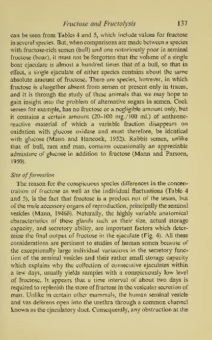
Fructose and Fructolysis 137
can be seen from Tables 4 and 5, which include values for fructose
in several species. But, when comparisons are made between a species
with fructose-rich semen (bull) and one notoriously poor in seminal
fructose (boar), it must not be forgotten that the volume of a single
boar ejaculate is almost a hundred times that of a bull, so that in
effect, a single ejaculate of either species contains about the same
absolute amount of fructose. There are species, however, in which
fructose is altogether absent from semen or present only in traces,
and it is through the study of these animals that we may hope to
gain insight into the problem of alternative sugars in semen. Cock
semen for example, has no fructose or a negligible amount only, but
it contains a certain amount (20-100 mg./lOO ml.) of anthrone-
reactive material of which a variable fraction disappears on
oxidation with glucose oxidase and must therefore, be identical
with glucose (Mann and Hancock, 1952). Rabbit semen, unlike
that of bull, ram and man, contains occasionally an appreciable
admixture of glucose in addition to fructose (Mann and Parsons,
1950).
Site offormation
The reason for the conspicuous species differences in the concen-
tration of fructose as well as the individual fluctuations (Table 4
and 5), is the fact that fructose is a product not of the testes, but
of the male accessory organs of reproduction, principally the seminal
vesicles (Mann, \9A6b). Naturally, the highly variable anatomical
characteristics of these glands such as their size, actual storage
capacity, and secretory ability, are important factors which deter-
mine the final output of fructose in the ejaculate (Fig. 4). All these
considerations are pertinent to studies of human semen because of
the exceptionally large individual variations in the secretory func-
tion of the seminal vesicles and their rather small storage capacity
which explains why the collection of consecutive ejaculates within
a few days, usually yields samples with a conspicuously low level
of fructose. It appears that a time interval of about two days is
required to replenish the store of fructose in the vesicular secretion of
man. Unlike in certain other mammals, the human seminal vesicle
and vas deferens open into the urethra through a common channel
known as the ejaculatory duct. Consequently, any obstruction at the

138 The Biochemistry of Semen
level of the ejaculatory ducts will prevent both fructose and sperma-
tozoa from reaching the urethral canal. This fact has been aptly
chosen as an aid to medical diagnosis by Young (1948, 1949)
who described the case of a man in whom repeated semen analysis
failed to detect fructose or sperm, although testicular biopsy re-
vealed normal spermatogenesis; the case has been diagnosed as
congenital bilateral aplasia of the vasa deferentia.
It must be also mentioned that though the seminal vesicles are
the main source of fructose in the higher mammals, yet an addi-
tional small amount of this seminal sugar is derived from the
ampullar glands (Mann, 1948^), and in some animals also from
certain other glands. Thus, in the rabbit, fructose was located both
in the glandula vesicularis (a structure corresponding to seminal
vesicles) and in the ampullae, as well as in the prostate (Davies and
Mann, \9Alb). The rat provides an instance of particular interest,
as in this rodent the seminal vesicles are free from fructose alto-
gether; instead, fructose is found in the dorso-lateral prostate and
in the so-called coagulating gland, a small organ immediately
adjacent to the seminal vesicles proper, with which it shares a
common peritoneal sheath (Humphrey and Mann, 1948, 1949).
Since fructose is produced by the accessory glands, and not the
testes, it is not surprising that in whole fresh semen there is no
direct proportion between fructose concentration and sperm density.
On the contrary, both in man and in domestic animals, an inverse
ratio between fructose and sperm concentration in semen is fre-
quently met with; the simplest interpretation is that in a dense
sample of semen the space occupied by the sperm cells is relatively
larger, and the volume taken up by the fluid portion, i.e. the
fructose-containing seminal plasma, correspondingly less. This
factor has a direct bearing on the interpretation of laboratory
examinations concerned with semen and male fertility or sterility.
It explains, for instance, why a semen sample with a high content
of fructose need not necessarily be one of good sperm quality, and
furthermore, why it is possible to come across samples with a high
fructose content but of low sperm density. In fact, some of our
highest values for fructose so far recorded, were encountered in
the semen of vasectomized, and thus completely azoospermic,
individuals.
Fructose and Fructolysis 139
Seminal fructose as an indicator of male sex hormone activity; the
'fructose test^ and its application to certain problems of sex
endocrinology
The 'fructose test', originally described by Mann and Parsons
(1947) and subsequently developed by Mann, Davies and Hum-phrey (1949), Mann, Lutwak-Mann and Price (1948) and Mannand Parsons (1950), is founded on the observation that the capacity
of the accessory organs to produce fructose and, thereby, the actual
level of fructose in the seminal plasma, reflects in a faithful manner
the degree of testicular hormone activity in the male, and in this
way provides an accurate indicator of endocrine testicular function.
In experiments on rats and rabbits it was shown that seminal fruc-
tose disappears almost completely within two weeks after castration
and also that the postcastrate fall in the level of fructose can be
prevented, or, if already developed, fully restored, by the implan-
tation of testosterone (Fig. 12). The effect is not limited to labora-
tory animals and analogous results were obtained with domestic
animals such as the bull.
The test can be carried out in two ways, by the chemical analy-
sis of the seminal fluid collected from an intact animal by means
of an artificial vagina, or by the analysis of accessory organs of
reproduction obtained from the experimental animal by dissection.
The first method gives an opportunity to observe in the same animal
the time-sequence of changes brought about by castration and hor-
monal treatment, and eliminates the sacrifice of the experimental
animal. In the second procedure, on the other hand, the test can
be used for a quantitative assay of male sex hormone activity in the
whole body, isolated tissues, body fluids and hormone preparations;
as an illustration. Fig. 13 gives a dosage-response curve obtained
with coagulating glands of castrated rats which were injected for
three weeks with known doses of testosterone propionate; following
the last day of injections the rats were sacrificed, the coagulating
glands dissected and used for fructose analysis.
Below are discussed some of the endocrinological problems to
which an approach was made in recent years with the aid of the
'fructose test', applied either alone or in conjunction with the 'citric
acid test', which depends on the relationship between the secretion

140 The Biochemistry of Semen
of citric acid by some of the accesory organs and the male sex
hormone activity.
600
Pellet implanted
Pellet removed
2 3 4 5 6 7 8
Weeks after castration
Fig. 12. Post-castrate fall and testosterone-induced rise of seminalfructose
in rabbit; pellet: 100 mg. testosterone.
(Mann & Parsons, 1947)
{a) Time relationship between the onset ofsecretory activity in male
accessory glands and spermatogenesis. In young rabbits (Davies and
Mann, 1941b), rats (Mann, Lutwak-Mann and Price, 1948), bull-
calves (Mann, Davies and Humphrey, 1949) and boars (Mann, 1954),

Fructose and Fructolysis 141
fructose and citric acid appear in the accessory glands at an early
age, before there is any evidence of active spermatogenesis; since
the secretion of both these substances depends upon the presence of
the male sex hormone, it must be concluded that the hormonebegins to function in the male body well in advance of the actual
spermatogenesis. Thus, for instance, in bull-calves appreciable
5 25 50
Daily dose of testosterone propionate (/ig.)
Fig. 13. Dosage-response curves of testosterone propionate, using the coagu-
lating glands of the rat; -O, fructose (/^tg.); • •,weight of organs (mg.).
(Mann & Parsons, 1950)
amounts of fructose are found in the vesicular secretion already at
the age of about four months, whereas the first mature spermatozoa
appear nearly eight months later. One cannot, of course, rule out
the possibility that the testicular hormone is active in the bull-calf
even before the age of four months, but if so, then either its con-
centration is too small to produce a distinct response in the accessory
organs, or else its action is countered by some other factors.
ib) Effect of testosterone on the appearance offructose in castrated
142 The Biochemistry of Semen
animals. The following experiment was carried out by Mann, Davies
and Humphrey (1949) at the Agricultural Research Council Field
Station at Compton, in Berkshire. Six bull-calves were used. These
were castrated when one to two weeks old, i.e. at an age prior to the
appearance of fructose in the seminal glands. Seven months later
two of the castrated calves received subcutaneous implants of 0-5 g.
pellets of pure testosterone, whereas the remaining four were left
untreated. After another four weeks all six animals were sacrificed
and their seminal glands dissected out, weighed, and examined both
chemically and histologically. The unused portions of the hormonepellets were recovered from the subcutaneous tissue of the two
hormone-treated calves; their weights were 0-344 and 0-338 g.
respectively, showing that the quantities of testosterone absorbed
per month per animal were 0- 1 56 and 0- 1 62 g., respectively. Chemical
analysis revealed the presence of considerable amounts of fructose
in the seminal glands in response to the four weeks' hormone treat-
ment (51 mg. fructose per 100 g. tissue or 5-3 mg. fructose per
total gland), as against a negligible fructose content in the untreated
castrates (8 mg. per 100 g. or 0-25 mg. per total gland). In compari-
son with and in contrast to the marked chemical difference, the
evidence for the functional recovery in the seminal glands, as
assessed by the histological examination, was practically impercep-
tible (Plate VI). In this way we were able to provide evidence that
the early effects of testosterone treatment can be established far
more convincingly by the large percentage-increase in the fructose
content of the seminal gland secretion, than by means of histological
methods which at this stage failed to show significant changes in the
glandular tissue.
An investigation concerned with the response to testosterone was
also made by Rudolph and Samuels (1949) on rats, and by Gassner
and his co-workers (1952) on bulls. In castrated rats, a significant
increase in the fructose content of accessory organs was noticed
already ten hours after the injection of 1 mg. testosterone pro-
pionate. In bulls, fructose disappeared from ejaculates within two
weeks after castration but injections of testosterone propionate, if
given within four weeks after castration, led to a rapid return of
fructose production to the pre-castrate level; yet, in spite of the fully
restored fructose level, such seminal plasma, when added to washed

PLATE VI
'*H^
v.^


Fructose and Fructolysis 143
spermatozoa obtained from a normal bull, was unable to support
sperm metabolism to the same extent as plasma from normal i.e.
non-castrated animals.
An interesting example of the application of the fructose test to
problems of infertility in man has been provided by a study of four
eunuchoid patients who responded to androgenic treatment with a
highly significant elevation of fructose in semen (Landau and
Longhead, 1951).
It seems probable that the fluctuations of fructose level in the
semen of normal individuals may also be due, in part at least, to
some periodic changes in the activity of the testicular hormone in
the male body. Normal rats, injected with large doses of the male
hormone invariably react by an increased level of fructose formation,
well above that of non-treated controls. The effect is particularly
striking with breeds of animals which exhibit a relatively low physio-
logical level of fructose formation. In this connection, however, it is
interesting to note that when injections of large doses of testosterone
propionate into normal rats are continued to excess, e.g. 200 i-ig.
daily for forty days, the state of overstimulation in the accessory
organs is accompanied by a marked decline in the size of the testes;
after seven weeks of such treatment the reduction in the weight of
the testes is nearly 50% (Mann and Parsons, 1950). Injections of
excessive doses of androgens are well known to produce harmful
effects on the spermatogenesis in animals and in man (Moore, 1939;
McCullagh and McGurl, 1939; Meckel, 1951).
In normal bulls, a dose of 100 mg. testosterone propionate,
repeated three times weekly for six weeks, appears to produce only
a very slight increase in the level of fructose in semen (Gassner,
Hill and Sulzberger, 1952). However, according to another report,
sexual excitation prior to service has a stimulating effect on the out-
put of fructose in bull semen (Branton, D'Arensbourg and Johnston,
1952).
(c) Hormone-induced formation offructose in subcutaneous trans-
plants from accessory organs. Once the dependence of seminal fruc-
tose upon the activity of the male sex hormone had been established
it was possible to enquire into the mechanism of this hormonal rela-
tionship. One of the problems to settle was the extent to which the
process of fructose generation in accessory glands depends upon the

144 The Biochemistry of Semen
preservation of intact vascular and neural links. Insight into this
matter was gained by the technique of subcutaneous transplantation,
when it was demonstrated that small fragments of rat coagulating
gland, about 1 mg. in weight, transplanted subcutaneously into
normal adult male hosts, grew well and showed after some weeks
of subcutaneous development a high content of fructose. Follow-
ing castration of the hosts, the transplants lost their ability to
form fructose but this was promptly restored by treatment with
testosterone propionate. Perhaps the most remarkable fact in these
experiments was that the growth of the grafts and their chemical
secretory function occurred not only in male, but also in female
hosts provided that the latter were treated with testosterone
(Lutwak-Mann, Mann and Price, 1949).
Thus, for the first time the effect of the male sex hormone on
fructose secretion was demonstrated in tissue fragments dissected
from the male accessory organs and developing in complete isola-
tion from the rest of the male generative system. Actually, the trans-
plants had an even higher fructose content than the corresponding
intact glands of the graft-bearing hosts, because unlike intact glands,
the grafts lack a secretory outlet.
In another study, Price, Mann and Lutwak-Mann (1949, 1954)
applied the transplantation technique, coupled with the chemical
methods, to the problem of the androgenic activity of ovarian
hormones in the female rat. Subcutaneous transplants of rat coagu-
lating gland in female hosts were shown to secrete large quantities
of fructose in response to injections of pregnant mare serum gona-
dotrophin. A series of thirty injections of twenty international units
of equine gonadotrophin was given daily; at autopsy the ovaries of
the female hosts were enlarged at least tenfold and covered with
numerous follicles and corpora lutea. In these rats, gonadotrophin,
through a stimulating action on the ovaries, raised the output of
ovarian androgens to an extent which induced the secretion of fruc-
tose in transplants from the coagulating gland.
{d) Effects ofprogesterone, stilboestrol and oestradiol. The nature
of the ovarian androgen responsible for the formation of fructose
is unknown, but there are indications that it may be related to
progesterone or to a product of progesterone metabolism. Aninquiry into the androgenic value of progesterone showed that large

Fructose and Fructolysis 145
doses of progesterone injected into castrated male rats have a
definite androgenic effect; it was calculated that the androgenic
value of 25 mg. pure progesterone is slightly more than that of
0005 mg. testosterone propionate (Price, Mann and Lutwak-Mann,
1949, 1954).
Whereas progesterone exhibits some androgenic activity, stil-
boestrol is endowed with the properties of an androgen-antagonist.
The testosterone-catalysed secretion of fructose in the male accessory
gland secretions of a rabbit can be suppressed very effectively by the
subcutaneous implantation of 25 mg. stilboestrol (Parsons, 1950).
In experiments in which a castrated rabbit received simultaneously
implants of testosterone and stilboestrol, the latter prevented com-
pletely the production of fructose by the accessory organs.
It appears that in castrated bulls, small amounts of oestradiol
dipropionate used together with testosterone, have a small but
definite synergistic effect on the seminal vesicles and lead to a higher
output of fructose in semen (Gassner, Hill and Sulzberger, 1952).
Role of hypophysis
The endocrine influence of the testes on the formation of fructose
in accessory organs is integrated closely with the functioning of the
anterior pituitary gland. Hypophysectomy, like gonadectomy, invari-
ably results in a rapid decline in the level of fructose in the seminal
plasma (Mann and Parsons, 1950). In the rabbit, for instance, a
three to four weeks' period after castration or hypophysectomy alike,
usually leads to complete disappearance of fructose so that an
ejaculate collected by means of an artificial vagina three weeks
Table 21. Ejfect of testosterone and pregnant mare serum
gonadotrophin on the formation offructose in rabbit prostate
Weight of Fructoseprostate content
Rabbit (mg.) (//g./organ)
1. Normal 770 935
2. 3 weeks after castration 280 20
3. 6 weeks after castration and simultaneous
implantation of testosterone (100 mg.) 1000 1220
4. 4 weeks after hypophysectomy 149 10
5. 6 weeks after hypophysectomy; for the last
4 weeks injected 200 I.U. PMS-gonado-trophin 210 395

146 The Biochemistry of Semen
after the operation contains no more than 20 ^g. fructose, as com-pared with 500 to 1000 /^g., before the operation. Both castrated as
well as hypophysectomized animals promptly respond to the sub-
cutaneous implantation or injection of testosterone with renewed
secretion of fructose. The same happens if instead of testosterone
pregnant mare serum gonadotrophin is injected into a hypophy-
sectomized animal (Table 21).
The relationship between blood glucose and seminalfructose
In addition to the hormones of the testis and the pituitary gland,
yet another organ, the pancreas, exerts a profound influence upon
the level of fructose in semen. The effect is an indirect one, and is
brought about by the action of insulin on the level of blood glucose
which in turn governs the level of fructose in semen. The existence
of a causal link between the blood sugar level and seminal fructose
was studied at first in animals with experimental diabetes; later,
however, it was also shown in diabetic man (Mann and Parsons,
1949, 1950).
In rabbits, experimental diabetes can be produced with alloxan;
best results are obtained by injecting intravenously into a rabbit
75 mg. alloxan per kg. body weight, and repeating this dose one or
two days later. Fig. 14 illustrates the course of such an experiment
with a rabbit in which analyses of blood and semen were carried
out regularly during a period of four months. At the outset of the
experiment this animal had a blood glucose content of 100 mg. per
100 ml., and about 600 i-ig. fructose per ejaculate or 70 mg. per
100 ml. semen (fluid portion). However, following alloxan treatment,
the rabbit developed severe glucosuria within two days and its blood
glucose level rose to 350 mg./lOO ml.; at the same time, the level of
fructose in semen began to increase, until three weeks later there
was 500 mg. glucose per 100 ml. blood, and 3500 /^g. fructose per
ejaculate or 320 mg. fructose per 100 ml. semen. When it was estab-
lished that hyperglycaemia is followed by an increased concentra-
tion of fructose in semen, the effect of insulin was examined and it
was found that the insuUn-induced fall in blood glucose was followed
by a reduction in the fructose content of semen; moreover, once the
effect of insulin on blood glucose in the diabetic animal wore off,
there was again an increase in seminal fructose.
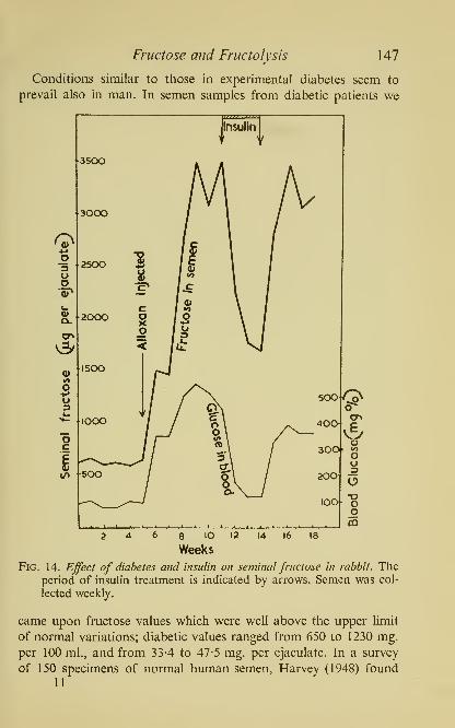
Fructose and Fructolysis 147
Conditions similar to those in experimental diabetes seem to
prevail also in man. In semen samples from diabetic patients we
8 lO 12
Weeks
Fig. 14. Effect of diabetes and insulin on seminal fructose in rabbit. Theperiod of insulin treatment is indicated by arrows. Semen was col-
lected weekly.
came upon fructose values which were well above the upper limit
of normal variations; diabetic values ranged from 650 to 1230 mg.
per 100 ml., and from 33-4 to 47-5 mg. per ejaculate. In a survey
of 150 specimens of normal human semen, Harvey (1948) found11

148 The Biochemistry of Semen
640 mg. per 100 ml. or 31-6 mg. per ejaculate to be the highest
value. It is interesting to recall here that years ago Goldblatt (1935a)
noticed a high reducing sugar value in human diabetic semen but
attributed this to urinary glucose.
Effect of malnutrition
It has long been known that defective nutrition has a deleterious
influence upon the male reproductive system. One of the earliest
surveys of this problem is found in the monograph by Jackson (1 925);
this was followed by the work on degenerative changes in testes and
sterility associated with vitamin A and E deficiency and in later
years, by many other nutritional studies which helped to accumulate
a wealth of information on this subject, fully reviewed on several
occasions (Asdell, 1949; Burrows, 1949; Lutwak-Mann, 1951;
Mason, 1949; Reid, 1949; Russell, 1948; Samuels, 1948; Walton,
1949). Most investigators in this field, however, particularly those
concerned with problems of human fertility, were much more in-
terested in the spermatogenic activity of the testicular tissue than
in the function of the accessory organs of reproduction. It was,
therefore, something of a departure when Moore and Samuels
(1931) came forward with the demonstration that a few weeks of a
diet deficient in vitamin B, or a quantitatively inadequate diet con-
taining vitamin B, caused in male rats regressive changes in the
accessory organs which, however, could be counteracted by the
administration of testicular hormone or anterior pituitary extracts.
They concluded that the primary lesion due to inadequate feeding
was located in the pituitary gland and that as a result of the di-
minished hypophyseal activity the testes received insufficient gona-
dotrophic stimulus and were consequently, unable to produce the
male sex hormone required for normal functioning of the accessory
glands. A similar state of 'pseudo-hypophysectomy' was described
by Mulinos and Pomerantz (1941) in rats as the result of a diet
which was qualitatively adequate but halved in quantity; further
supporting evidence was later provided by several groups of
investigators (Pazos and Huggins, 1945; Goldsmith and Nigrelli,
1950; Grayhack and Scott, 1952). In certain animal species sperma-
togenesis was also shown to be affected by a vitamin B-deficient

Fructose and Fructolysis 149
diet (Marrian and Parkes, 1928; Dunn, Morris and Dubnik, 1947;
Elson and Koller, 1948).
Lutwak-Mann and Mann (1950a, b, 1951) applied chemical
methods to the study of changes brought about in the secretory
function of rat accesory organs by vitamin B-deficiency and inani-
tion, and found that in rats maintained for four weeks on a deficient
diet the content of fructose and citric acid in the accessory glands
was reduced to a castrate level. By treatment with testosterone pro-
pionate (0-2 mg. daily for one week) or with chorionic gonadotro-
phin (200 units every other day for two weeks), the secretory activity
of the glands could be completely restored. A further example of the
effect of an unbalanced diet on the process of fructose secretion was
provided by Lutwak-Mann (1951) who found that a diet with exces-
sive fat content, even though not protein- or vitamin-deficient, also
caused regression in rat accessory organs. Mann and Walton (1953)
made a study of the effect of underfeeding on the genital functions
in the bull and found that, in contrast to the testes, the secretory
function of the male accessory glands was markedly affected by
underfeeding. The concentration of fructose and citric acid in the
semen of the underfed bull decreased by 30% and 60% respectively,
of the original levels. In the bull, however, unlike in the rat, the
effects of malnutrition as well as the recovery after the animal has
been transferred back to its normal diet, developed comparatively
slowly.
The enzymic mechanism offructose formation
The experimental evidence available at present brought out the
essential, though as yet not fully understood, role of the testicular
hormone in the formation of fructose by the secretory apparatus
of the male accessory glands, and indicated that blood glucose is
the precursor of seminal fructose. Further details of the mechanism
whereby glucose is converted in the accessory gland tissue to fruc-
tose, were obtained from in vitro experiments; these showed that
small amounts of fructose are formed as a result of incubation of
minced accessory gland tissues with glucose, and that these tissues
possess the entire enzymic system which can convert glucose to
fructose (Mann and Lutwak-Mann, 1948, \95\a, b).
It is an estabUshed fact that certain phosphorylated derivatives of

1 50 The Biochemistry of Semen
fructose, such as 6-phosphofructofuranose (Neuberg ester) and 1 : 6-
diphosphofructofuranose (Harden-Young ester), are formed as in-
termediary substances in the normal carbohydrate metaboUsm of
muscle, liver and other animal organs. However, in the majority
of animal tissues these phosphofructoses do not yield free fructose
but are metabolized further to form pyruvic acid and lactic acid.
In the semen, however, there are present in high concentration
enzymes which belong to the group of phosphatases and include
the 'alkaline' phosphatase; the latter capable of splitting a number
of phosphohexoses, including 6-phosphofructose, 1-phosphofructose
and 1 : 6-diphosphofructose, to phosphoric acid and free fructose.
The alkaline phosphatase found in semen is derived from several
accessory organs of reproduction but its principal source in higher
animals is the seminal vesicle. Owing to this fact, the usual channels
of carbohydrate metabolism are diverted in the vesicular tissue:
phosphofructoses are not metabolized to lactic acid, as would be
the case e.g. in muscle, but are dephosphorylated instead, so that
free fructose is formed.
In extracts made from bull seminal vesicle tissue it is possible to
demonstrate the following reactions. When 1-phosphoglucose is
incubated, 6-phosphoglucose is formed through the action of phos-
phoglucomutase; next, part of 6-phosphoglucose is converted by
phosphohexose isomerase into 6-phosphofructose. The equilibrium
mixture of the two 6-phosphohexoses is acted upon by phosphatase
and yields a mixture of free glucose and free fructose. Phosphohexose
isomerase, together with alkaline phosphatase is also present in the
seminal vesicle secretion and seminal plasma. Ram seminal plasma
in particular, is a rich source of phosphatase active towards phos-
phorylated sugars (Table 17, p. 120).
The fact that whereas a mixture of glucose and fructose is the
result of the phosphatase activity in the glandular tissue, yet, only
one sugar, that is fructose, accumulates in the secretion, may have
its explanation in a more effective re-utilization of glucose than
fructose, by the glandular tissue itself. Thus, we have found that
tissue slices from the rat coagulating gland can glycolyse anaero-
bically glucose at a much higher rate than fructose; this, in
turn, may be due to the ability of the tissue to re-phosphorylate
more effectively glucose to 6-phosphoglucose, than fructose to

Fructose and Fructolysis 151
6-phosphofructose. Such evidence as is at present available, derived
from both in vivo and /// vitro experiments, indicates that the
enzymic reactions involved in the conversion of blood glucose to
seminal fructose are as follows.
Blood glucose
\Glycogen
^ Phosphorylase
1-Phosphoglucose
\ Phosphoglucomutase Phosphohexose6-Phosphoglucose
\>'6-Phosphofructose
f \Glucokinase Alkaline phosphatase
isomerase
Alkalinephosphatase
Glucose ^^ Seminal fructose
Anaerobic and aerobic utilization of carbohydrate by spermatozoa
The spermatozoa of the sea-urchin and certain other animals
derive their energy for movement chiefly from respiratory processes;
in contrast, the survival and motility of sperm ejaculated by animals
with internal fertilization, such as mammals, is possible for most
of them also in absence of oxygen, provided that the sperm cells
remain in contact with seminal plasma. Mammalian spermatozoa
possess only a negligible reserve of intracellular glycogen and depend
therefore, under anaerobic conditions, on an extracellular source of
energy. In species which contain fructose as a normal constituent
of the seminal plasma, anaerobic fructolysis is the metabolic process
which enables the spermatozoa to survive without oxygen. Should,
however, the spermatozoa become separated from the seminal
plasma by centrifugation and washing, they could not carry on
anaerobically unless the seminal plasma were restored or replaced
by glycolysable carbohydrate.
The stimulating effect of pure sugars on sperm motility has been
noticed by some of the early investigators of semen. In 1931 Ivanov
observed that dog spermatozoa suspended in an isotonic solution of
glucose and phosphate retained their motility when the respiration
had been abolished either by poisoning with cyanide or by replace-
ment of oxygen with hydrogen. This observation is of particular
interest in view of the fact that the dog has no seminal vesicles and
152 The Biochemistry of Semen
no fructose in the seminal plasma; the possibility, of course, must
not be overlooked that there may be in dog semen some other sub-
stance of nutrient value to the spermatozoa. Redenz (1933) has
shown that bull spermatozoa glycolyse glucose, fructose, and man-
nose to lactic acid, and that the presence of these sugars, but not
that of sucrose, lactose, or glycogen, is beneficial to sperm motility.
His findings were confirmed by others and it has since become
an established fact that the metabolism of spermatozoa in several
mammalian species including man, ram and bull, is predominantly
of a glycolytic character (Ivanov, 1935; Shergin, 1937; Comstock,
1939; MacLeod, 1939, 19436; Lardy and Phillips, \9A\a\ Moore and
Mayer, 1941; Henle and Zittle, 1942; Ross, Miller and Kurzrok,
1941; Salisbury, 1946).
Sperrnatozoa obtained directly from the epididymis of a bull,
ram, or boar, resemble washed ejaculated sperm in that they are
incapable of survival under purely anaerobic conditions. While in
the epididymis, the spermatozoa have no access to fructose and are
immotile; the onset of motiUty coincides with their passage along
the male genital tract and contact with the seminal plasma. The acti-
vating influence of fructose on previously immotile spermatozoa
can be convincingly demonstrated in a simple manner. Fresh epi-
didymal spermatozoa are suspended in bicarbonate-Ringer solution;
two 'hanging drops', one of the suspension and another, a little
further away, containing a 1% solution of fructose in bicarbonate-
Ringer solution, are placed on the underside of a cover-sUp; to
observe the motility of the sperm under the microscope, the cover-
slip is fixed to the top of a small glass chamber in which one can
create anaerobic or aerobic conditions by passing through the cham-
ber a gas mixture of 95% N2-5% CO2 or 95% 02-5% CO2. In the
absence of oxygen, the spermatozoa can be seen to be almost com-
pletely immotile, but when the two drops are brought together the
sperm movement begins and continues for a long time. Aerobically,
the effect of fructose is less striking because oxygen induces endo-
genous respiration and this in itself provokes motility in epididymal
spermatozoa. However, even in the presence of oxygen, fructose
still has some influence owing to the process of aerobic fructolysis.
Under anaerobic conditions, the final product of sperm fructolysis,
lactic acid, cannot be oxidized further. In the presence of oxygen,
Fructose and Fructofysis 153
however, the situation differs in that the rate of fructose utiliza-
tion becomes smaller, and moreover, lactic acid undergoes further
oxidation, thus providing an additional source of metabolic energy.
It remains for further study to ascertain what type of carbohydrate
metabolism predominates in spermatozoa during their existence in
either the male or female genital tract. However, so far as in vitro
studies are concerned, they show that lactic acid can be efficiently
oxidized by spermatozoa even when the partial pressure of oxygen
has been reduced to a level as low as that which normally prevails
in animal tissues (Mann, 1951Z)); suitably diluted suspensions of ramspermatozoa show in presence of 1% O2 a respiratory rate as high
as in air, and lactate is capable of maintaining the oxygen uptake
equally well in 1% as in 20%, oxygen.
Pasteur effect and the ^metabolic regulator''
It was said earher that the spermatozoa obtained directly from
the epididymis in some ways behave like suspensions of washed
ejaculated sperm; as a matter of fact, however, these two types of
sperm cells possess distinct characteristics (Henle and Zittle, 1942;
Lardy, Hansen and Phillips, 1945). Washed epididymal bovine
spermatozoa have a lower endogenous respiration than those in
ejaculated bull semen. But if sugar is added or if the spermatozoa
are removed from the epididymis after a period of storage in the
refrigerator, then their oxygen uptake is distinctly higher. More-
over, on addition of sugar, epididymal spermatozoa produce lactic
acid much more rapidly under anaerobic than aerobic conditions,
whereas in ejaculated sperm the rate of glycolysis is not muchhigher in the presence than in the absence of oxygen. To account
for these differences, the Wisconsin workers determined the rate of
anaerobic and aerobic glycolysis as well as of oxygen uptake, in
epididymal spermatozoa to which glucose was added, and calcu-
lated the 'Meyerhof oxidation quotient' which measures the Pasteur
effect, that is the extent to which glycolysis is inhibited by oxygen.
The average value for the Meyerhof quotient calculated from twelve
experiments on bull epididymal sperm was 9-6, as against 5 recorded
for ejaculated sperm (Lardy, 1952). This difference, according to
Lardy, Ghosh and Plant (1949), is due to the presence in bull sper-
matozoa of a 'metabolic regulator' which occurs in the epididymal

1 54 The Biochemistry of Semen
sperm in a 'bound form' but is released in an 'active form' after
ejaculation. Continuing their study, these workers observed that
heat-inactivated bull semen, or semen and testicular extracts heated
with sodium hydroxide, increased the rate of aerobic fermentation
of sugars by baker's yeast, without affecting markedly yeast respi-
ration or anaerobic fermentation. The yeast-stimulating factor was
extracted with carbon tetrachloride from alkaline hydrolysates of
hog testes and obtained in the form of yellow coloured crystals
which proved to be elementary sulphur (Ghosh and Lardy, 1952).
Yeast reduces sulphur to HgS which is probably the agent ultimately
responsible for the stimulation of the aerobic fermentation. The
identity of the yeast factor with sulphur was verified by reproducing
the stimulating effect on yeast with pure rhombic sulphur. Sulphur
as such, however, cannot be the sperm 'regulator' since it is without
influence on the Pasteur effect in epididymal spermatozoa. On the
other hand, a number of sulphydryl compounds such as cysteine,
reduced glutathione and hydrogen sulphide have been found to
stimulate the respiration and aerobic glycolysis of epididymal
sperm and the possibility remains, that the 'metabolic regulator'
is, in fact, a sulphydryl compound, liberated during ejaculation
from the spermatozoa, with a sulphydryl group in labile form, which
can be easily removed and oxidized to sulphur by alkaline hydrolysis.
The peculiar changes in the metabolic properties of spermatozoa
during cold-storage of the epididymis, are equally in need of eluci-
dation. A problem which also deserves further biochemical study
is the 'ripening' phenomenon which takes place in the spermatozoa
while they remain in the epididymis. Presumably, the metabolism
of sperm in the epididymis is related in some as yet unknown
manner to the structural changes associated with sperm maturation
processes, such as the migration of the 'kinoplasmic droplet'.
Intermediary reactions in spermfructolysis and the role ofphosphorus-
containing coenzymes
The ability of washed spermatozoa to convert into lactic acid
equally well added fructose, glucose and mannose is due in all
probability to the fact that the metabolic degradation of these
three sugars is initiated by the sam.e hexokinase-catalysed reaction
with adenosine triphosphate.
Fructose and Fructolysis 155
Adenosine triphosphate (ATP, formula in Fig. 15) represents an
intracellular constituent and a coenzyme of considerable importance
in the economy of the sperm cell. An observation that a considerable
proportion of the acid-soluble phosphorus in bull spermatozoa
yields orthophosphate after 7 min. hydrolysis with n-HCI first
suggested the presence of ATP (Lardy and Phillips, 1945). In the
same year, the readily-hydrolysable phosphorus compound was
isolated from ram spermatozoa and its identity with ATP estab-
lished by chemical analysis (Mann, 1945«, c); the content of ATPin ram spermatozoa is 2-6-6-6 mg. labile phosphorus or 0-6-1 -5 mg.
of adenine amino-nitrogen per 100 ml. semen (see also Table 16).
The occurrence of ATP in ram and boar spermatozoa has also been
confirmed by Ivanov, Kassavina and Fomenko (1946) who found
that the phosphorus compound which they purified from sperm
induced contractions of muscle actomyosin threads in the same
manner as ATP isolated from skeletal muscle. ATP was also found
in sea-urchin spermatozoa (Rothschild and Mann, 1950), the con-
centration of ATP in the semen of Echinus escidentus resembling
that found in the ram.
Spermatozoa, even after they have been repeatedly washed so as
to remove the phosphatases present in seminal plasma, continue to
exhibit a high phosphatase activity against ATP, and all evidence
available at present points to sperm ATP-ase as the enzyme which
is directly responsible for the supply of energy essential for normal
motihty and survival of the sperm cell. The losses due to utilization
of ATP are made good by re-synthesis which takes place during the
normal metabolism of spermatozoa, and any interference with inter-
mediary enzymic reactions which renders the sperm cell incapable
of breaking down or building up ATP, leads to a decrease in both
metabolism and motility. Using ram spermatozoa as experimental
material under a variety of conditions, we have found that a diminu-
tion in the content of ATP invariably coincides with impaired sperm
motility (Mann, 1945^7, b, c). Thus, for instance, in ram spermatozoa
deprived of the fructose-containing seminal plasma by washing,
ATP content as well as motility went down simultaneously on
anaerobic incubation, but both ATP and motility could be
preserved anaerobically in sperm suspensions provided with
glycolysable material.

156 The Biochemistry of Semen
The activity of hexokinase, the enzyme which brings about the
initial reaction between ATP and glycolysable sugar, can be demon-
strated directly in washed spermatozoa (Mann, 1945/)). If we add
to a ram sperm suspension sugar (glucose, fructose or mannose),

Fructose and Fructolysis 1 57
If either glucose -alone or fructose alone is used as substrate, the
rate of lactic acid production is the same (Mann and Lutwak-Mann,
1948), but if washed spermatozoa are made to act on a 1 : 1 mixture
of glucose and fructose, then the rate of fructose utihzation becomesmuch less than 50% (Mann, \95\b). This 'sparing effect' of glucose
on the utilization of fructose is probably due to the competitive
inhibition of sperm hexokinase. Slein, Cori and Cori (1950) have
shown that when brain or yeast hexokinase acts upon ATP and onan equimolar mixture of glucose and fructose, the aldosugar is phos-
phorylated much more rapidly than the ketosugar. Under natural
conditions only fructose is present in whole semen, but not glucose,
so that the possibility of the latter interfering with fructolysis does
not arise. But there is preferential utilization of glucose in the acces-
sory glands, directly responsible for the accumulation of fructose,
and this may be due to a stronger affinity of hexokinase for glucose
than for fructose. A competition for hexokinase, between glucose
and fructose, is also consistent with the observation that in bull
semen incubated with an egg-yolk-diluent, the initial rate of fructo-
lysis is temporarily retarded (Vantienhoven, Salisbury, VanDemarkand Hansen, 1952), as is also the case in semen incubated with cowfollicular fluid (Lutwak-Mann, 1954); both egg-yolk and follicular
fluid contain glucose.
The phosphohexose formed from fructose as a result of hexo-
kinase activity is 6-phosphofructofuranose; in the case of glucose
the product is 6-phosphoglucopyranose. The latter, however, is
readily converted to 6-phosphofructose by phosphohexose iso-
merase, and from this stage onwards, the enzymic degradation of
glucose and fructose is identical. The chain of events which in whole
semen leads from fructose to lactic acid, is diagrammatically depicted
in Fig. 15. In the normal course of fructolysis, 6-phosphofructose is
phosphorylated by ATP in a reaction catalysed by phosphofruc-
tokinase, to yield 1 : 6-diphosphofructose and ADP; disphospho-
fructose is next split by zymohexase into two molecules of phospho-
triose. Like the action of phosphofructokinase, that of zymohexase
was demonstrated directly in spermatozoa (Mann, 1945^).
The subsequent steps in sperm fructolysis are analogous to the
corresponding phases in muscle glycogenolysis and blood glucolysis,
and involve the participation of cozymase (diphosphopyridine

158 The Biochemistry of Semen
nucleotide) which as Winberg (1941) showed, is a characteristic
intracellular constituent of spermatozoa. The cozymase-catalysed
phase of fructolysis consists of two closely interwoven oxidoreduc-
tion processes (Mann, 19456; Mann and Lutwak-Mann, 1947).
H3PO4 ATP
_,, , ^ . (, Phosphotriose dehydrogenase ) t^, i , • • jPhosphotriose—^^
. ^-^ Phosphoglyceric acid
YH2 NaF inhibits
\ YCozymase Phosphopyruvic acid
\Ha cozymase
iLactic acid^ ; Pyruvic acid
Lactic dehydrogenase -^
The first oxidoreduction involves the oxidation of phosphotriose
to phosphoglyceric acid by phosphotriose dehydrogenase, and a
simultaneous reduction of cozymase to dihydrocozymase; the oxida-
tion of phosphotriose is coupled with an esterification of inorganic
phosphate and the synthesis of ATP; the oxidation product, phos-
phoglyceric acid, is converted by enolase to phosphopyruvic acid,
the phosphate of which is transferred to ADP, thus producing
pyruvic acid and ATP. The second oxidoreduction is between
dihydrocozymase and pyruvic acid: dihydrocozymase is oxidized to
cozymase, and pyruvic acid is reduced by lactic dehydrogenase to
L(+)-lactic acid. When washed ram spermatozoa are treated with
fluoride (to inhibit enolase), and incubated with added phospho-
triose and pyruvate, the two oxidoreduction processes continue as
usual but in addition to lactic acid there is an accumulation of
phosphoglyceric acid. lodoacetate, on the other hand, abolishes the
oxidoreductions in washed sperm and thus deprives them of the
ability to produce lactic acid.
The not unimpressive array of facts available from the outlined
studies on spermatozoa strengthens the belief that ATP is the car-
dinal link between the activity of spermatozoa on the one hand,
and carbohydrate metabolism on the other. In whole semen, ATPacts continually as phosphate-donor and acceptor in the course of
fructolysis. The content of ATP and with it the motility of ejaculated

Fructose and Fructolysis 1 59
spermatozoa, both depend on the maintenance of the normal
metaboHsm of fructose.
We still remain confronted with two questions to which, it is
confidently hoped, further research will bring answers. One involves
the as yet obscure position in the semen of animal species which lack
fructose. Secondly, one wonders why nature should have chosen
fructose and not glucose, as the natural substrate for sperm meta-
bolism. At this point, conditions in another body fluid, the milk,
come to mind; there, the occurrence of lactose poses a somewhat
similar question. But in considering the matter, several facts must
be taken into account. To begin with, if glucose and not fructose
were present in semen, its concentration could hardly be expected
to exceed that of blood and other body fluids. Thus, it might not
be sufficient to satisfy the metabolic requirements of spermatozoa
which, unlike most other animal cells, are capable of utilizing fruc-
tose anaerobically; it is worth noting that on the whole, yeasts and
bacteria are also unable to consume fructose at the same rate as
glucose. Presumably, this enables the spermatozoa to draw freely
upon seminal fructose without, as it were, competition from other
tissues. Lastly, the intimate relationship between seminal fructose
and the male sex hormone must not be lost sight of; it would be
rather difficult to envisage a similar dependence in the case of
glucose, bearing in mind the ubiquitous occurrence and physio-
logical function of this sugar.

CHAPTER VIII
Spermine, Choline, Ergothioneine , and
certain other Bases in Semen
Spermine. Occurrence of crystalline spermine in human semen; its
chemical nature and properties. Derivatives of spermine and their use
in forensic medicine. Synthesis of sperrnine. Spermidine. Oxidation of
spermine and spermidine by diamine oxidase. State of spermine in semen.
Choline. The Florence reaction in semen. Enzymic liberation of choline
from precursors in semen. Phosphorylcholine and glycerylphosphoryl-
choHne. Physiological function of free and bound choline. Choline
esterase.
Ergothioneine. Isolation of ergothioneine from the boar seminal vesicle
secretion. The function of seminal ergothioneine and its behaviour towards
sulphydryl-binding substances. Biogenesis of ergothioneine.
Creatine and creatinine. Occurrence in mammalian semen, and in the
sperm and gonads of invertebrates. Phosphocreatine and phosphoarginine.
Adrenaline and noradrenaline . Occurrence in semen and accessory organs.
Enzymic oxidation. Pharmacodynamic properties.
Among the chemical characteristics which distinguish semen from
other tissues and body fluids is the occurrence of certain nitrogenous
bases, largely betaines, which are rarely found elsewhere in the
animal body. Of these, spermine is the oldest-known, and ergo-
thioneine the most recently discovered.
SPERMINE
Occurrence of crystalline spermine; its chemical nature andproperties
When human semen 'had stood a little while, some three-sided
bodies were seen in it, terminating at either end in a point; some
were of the length of the smallest grain of sand, and some were a
little bigger, as in Fig. A. They were further as bright and clear as if
they had been crystals.' Thus, in a letter of November 1677, addressed
to the Royal Society, Antoni van Leeuwenhoek reported the dis-
covery of the crystalline substance in semen which later became160

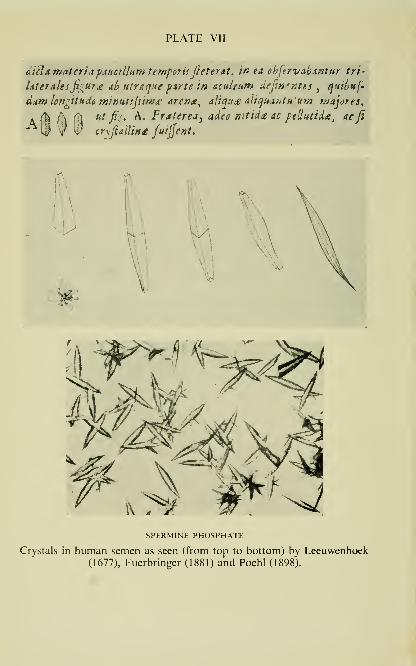
PLATE VII
ditld materia pauallumtempoTiiJieterAt^ in ea, ohjtrvabsrjtur tri-
idteraiesfi^ur^ ^b utraque pirte tn acttieum defin'r:tis^
quthuf-
dim icngitudfi minuttfiiwd' arena^ aliqu£ ^ltqt4ihtH urn wajores^
A f^ l\ % «^ fij* A. Frateredy adeo ntttd£ &o ptUucida^ acfi" i| V ^' cr\fiallin£ juijjent.
SPERMINE PHOSPHATE
Crystals in human semen as seen (from top to bottom) by Leeuwenhoek(1677), Fuerbringer (1881) and Poehl (1898).

Spermine, Choline, Ergothioneine 161
known as spermine. This was actually the letter in which Leeuwen-
hoek also communicated for the first time the discovery of living
spermatozoa and their movement in fresh semen; it was published
the following year in the Philosophical Transactions (Plate VII).
During the 200 years which followed, the same crystalline substance
was rediscovered by several investigators, most of whom were
apparently unaware of either Leeuwenhoek's original, or of the
others' later observations.
Vauquelin(1791) observed in a semen samplewhich he left standing
for four days, the deposition of 'cristaux transparens, d'environ
une ligned de long, tres-minces, & qui se croisent souvent de maniere
a representer les rayons d'une roue. Ces cristaux isoles nous ont
offert, a Faide d'un verre grossissant, la forme d'un solide a quatre
pans, termines par des pyramides tres-allongees, a quatre faces.'
After having studied the properties and behaviour of these crystals
towards different solvents, Vauquelin concluded that 'la nature de
ces cristaux est analogue a celle du phosphate de chaux ou la bas
des os'. The belief that the sperm crystals consist of ordinary phos-
phate persisted throughout the best part of the next century. In the
meantime, however, the same crystalline substance was found out-
side the semen in other tissues and body fluids, including sputum,
leucaemic blood, liver, spleen and old pathological-anatomical pre-
parations, so that towards the close of the XlXth century spermine
was already known by no less than ten names of various distin-
guished clinicians, anatomists and physiologists, including, in
chronological order, Charcot, 1853; Foerster, 1859; Harting, 1859;
White, 1861 ('leucosine'); Friedrich, 1864; Huppert, 1864; Boettcher,
1865; Neumann, 1866; Eberth, 1869; Leyden, 1872; and Zenker,
1876. But in the end, the medical world at large restricted itself
largely to the use of two names, 'Charcot-Leyden crystals' with
reference to organs and sputum ('asthma crystals'), and 'Boettcher
crystals' with reference to semen. Boettcher himself preferred to
call the substance 'Spermatin', and regarded it as a protein; he
published his paper 'Farblose Krystalle eines eiweissartigen Korpers
aus dem menschlichen Sperma dargestellt' in 1865, without however,
taking the trouble to mention the previous investigators.
The credit for having been the first to recognize spermine as the
phosphate of a new organic base, is due to Schreiner (1878) who

162 The Biochemistry of Semen
succeeded in preparing a number of derivatives of spermine includ-
ing the hydrochloride, but who unfortunately deduced from his
analyses the wrong formula for the base, C2H5N. In consequence of
this, spermine was confused with ethyleneamine, CoH^NH, and with
piperazine. For years to follow, piperazine was offered by a large
pharmaceutical firm in Berlin under the trade name of 'Spermin',
and as late as 1903 the formula of piperazine appeared under the
name of spermine in Thierfelder's Hoppe-Seyler's Handbuch der
chemischen Analyse. A great advocate of the manifold curative pro-
perties of 'real' spermine, i.e. as isolated from human semen, bull
testes, or other organs, was Alexander von Poehl, who believed in the
'action of spermine as a physiological tonic on auto-intoxications'
(1893), and who is best known for the monograph Die physiologisch-
chemischen Grundlagen der Spermintheorie which he published in
St. Petersburg in 1898. Poehl's book contains the records of numer-
ous cases ranging from scurvy to syphilis, treated, apparently suc-
cessfully, with the 'Sperminum Poehl'. His pharmacological and
clinical work aroused much controversy, was subjected to severe
criticism, and was finally altogether rejected. Yet, it is not entirely
improbable that there is some justification for Poehl's 'spermine
theory'. Apart from its general pharmacodynamic properties similar
to those of other biological polyamines (Guggenheim, 1940), sper-
mine may well possess some other, more specific pharmacological
activity. Administered parenterally, spermine is known to be
toxic to mice, rats and rabbits (Rosenthal, Fisher and Stohlman,
1952). It has also been shown to possess bacteriostatic properties.
The inhibition of the growth of Staphylococcus aureus by humanseminal plasma can be attributed, according to Gurevitch and his
colleagues (1951), to the high content of spermine in human semen.
Another striking example of the growth-inhibiting action of sper-
mine has been provided by Hirsch and Dubos (1952); following up
an observation that the extraction of animal tissues with mixtures of
water and ethanol yields material with tuberculostatic activity in
vitro, these authors isolated from tissue extracts a crystalline anti-
mycobacterial substance which they found to be identical with
spermine phosphate.
Leaving aside Poehl's pharmacological observations, one must
nevertheless appreciate his contribution to the chemistry of spermine.

Spermine, Choline, Ergothioneine 163
Not only was he able to refute the mistaken belief in the identity
of spermine and diethylenediamine but he was also the first
to analyse correctly the gold salt and the chloroplatinate of sper-
mine and to establish that 'the organic base which is at the bottomof these double salts would have the composition, C5H14N2 (1898,
1906).
However, not until 1924 was conclusive chemical and crystallo-
graphic evidence brought forward to prove the identity of spermine
isolated from semen with the base obtained by similar methods from
various animal organs and also from yeast. Credit for this is due to
Otto Rosenheim (1924). In 1924, Dudley, Mary Rosenheim and O.
Rosenheim in England, and Wrede in Germany, concluded from
the molecular weight estimations of benzoylspermine and m-mixo-
benzoylspermine, respectively, that the molecular formula of sper-
mine is C10H26N4, and not as formerly assumed C5H14N2.
Rosenheim and his colleagues obtained spermine in the free state
as a crystalline, optically inactive substance which melts between55° and 60°, and distills at about 150° in vacuo without decomposi-
tion. They also found that the base is stable in hot concentrated
alkali and in boiling hydrochloric acid. Their relatively simple
method of isolation depends on the steam-distillation of spermine
from a strongly alkaline solution.
Derivatives of spermine and their use in forensic medicine
There are several well-defined compounds of spermine, a list of
which is given in Table 22. Apart from the highly characteristic
insoluble phosphate, spermine can be identified particularly easily
as a picrate, which can be prepared from the free base, the phos-
phate, or directly from semen. Spermine picrate, like the phosphate,
is extremely insoluble in water. Crystallographic analysis has shown
that it is identical with the substance responsible for the so-called
Barberio reaction, a chemical test of diagnostic value in forensic
medicine. Barberio's (1905) test consists in the addition to semen,
or to an aqueous extract from the seminal stain, of picric acid in
concentrated aqueous or ethanolic solution; in the presence of picric
acid, there follows within a few minutes, the formation of abundant
yellow crystals, resembling in shape the crystals of spermine phos-
phate. The statement by Barberio that the reaction appears to be
12

164 The Biochemistry of Semen
specific for human semen as distinct from animal semen, has been
corroborated by other ItaUan investigators, particularly by Baecchi
(1912). In the experience of Littlejohn and Pirie (1908), Barberio's
reaction is best carried out as follows; a small piece of the stained
fabric is placed upon a glass slide and macerated in a drop or two
Table 22. Chemicalproperties of spermine and its derivatives
Compound Formula
Free base C10H26N4
Phosphate CioH26N4-2H3P04-6H20
Hydro- CioH2eN4-4HC]chloride
Picrate CioH26N4-4C6H307Na
Chloro- CioH26N4-4HCl-4AuClsaurate
Chloro- CioH26N4-2H2PtCl6platinate
Picrolonate
Benzoyl CioHaeNi^COCeHsderivative
Tetra- CioH26N4-4CioH608N2Sflavianate
Properties
Needle-shaped, colourless, odour-
less crystals; easily soluble in
water, ethanol and butanol, in-
soluble in ether, benzene andligroin, m.p. 55-60°, m.w. 202
Lenticular crystals from water,
long needles from ethanol. Insol.
in cold water, sol. 1 : 100 in boil-
ing water, easily soluble in dilute
acid or alkali, m.p. 240°, m.w.504. Identical with 'Boettcher's
crystals' and 'Charcot-Leyden
crystals'
Short prismatic needles, extremely
soluble in water, insol. in
acetone, ether and chloroform,
m.p. 300-310°
Yellow needles, become black at
242°, melt sharply with decom-position at 248-250°
Golden-yellow, lustrous leaflets,
m.p. 225° (Wrede 218°)
Orange-yellow, well-formed cry-
stals, m.p. 242-245°
Lemon-yellow crystals, m.p. 288-289°
Crystallizes from a solution in hot
acetone on addition of ligroin, in
woolly balls of fine needles, m.p.
155°, m.w. 618
CrystalliLes if flavianic acid is used
in excess. On recrystallization
from water the diflavianate is
formed

Spermine, Choline, Ergothioneine 165
of distilled water; to the extract thus obtained (concentrated by
evaporation, if necessary), a very small drop of an aqueous saturated
solution of picric acid is added, by means of a platinum loop; after
a minute or two a cover slip is applied, and the preparation examined
under the microscope; 'when fully developed, the crystals have the
form either of obtuse or sharp-ended needles, or of rhombic prisms
frequently crossed by a refrangent line at their equator. Sometimes
crosses are formed, and more rarely stars.' (For further particulars
concerning Barberio's test see Harrison, 1932.) Another deriva-
tive of spermine used in medico-legal laboratories is spermine
flavianate; this crystalline compound forms the basis of Puranen's
reaction (Puranen, 1936; Berg, 1948).
Synthesis of spermine
The final elucidation of the chemical structure of spermine was
achieved in 1926. The existence of two chains A^—C—C
—
C—A'^
and of one chain N—C—C—C—C—A^ in the spermine molecule
was inferred by Dudley, Rosenheim and Starling from the identi-
fication of pyrrolidine and tetramethyltrimethylene diamine as
degradation products of spermine hydrochloride and decamethyl
spermine, respectively. In Wrede's laboratory, the presence of two
3C chains and one AC chain was established the same year as a
result of studies on split products obtained from spermine by oxidation
with molecular oxygen in presence of copper. The final proof was
provided by the English investigators when they accomplished the
synthesis of spermine and showed it to be, a,(5-bis [y'-amino-
propylamino]-butane:
CHaCH.CHa CH.CH2CH2I I IINH2 NHCH2CH0 CH2CH2 NH NH2
Spermine
The oxidation of spermine gives rise to a volatile base associated
with the characteristic odour of semen. The appearance of the
semen-like odour during treatment of spermine chloroaurate with
metallic magnesium, first described by Schreiner (1878), probably
involves also an oxidation. The base is volatile in steam, and forms
a crystalline hydrochloride and chloroaurate (m.p. 204-206°); it is
probably identical with A^-y-aminopropylpyrroline.

166 The Biochemistry of Semen
Spermidine
Dudley, Rosenheim and Starling (1927) also succeeded in the
isolation of spermidine, a base present in the mother-liquor after
separation of spermine phosphate; spermidine phosphate is muchmore soluble than spermine phosphate and crystallizes from the
25% ethanolic mother-liquor, after the removal of spermine phos-
phate, when the concentration of alcohol is increased to 50%. The
properties of spermidine are similar to those of spermine. It gives
the same pyrrole reaction and behaves in an identical manner
towards precipitating reagents including phosphotungstic acid.
Spermidine is optically inactive, and yields like spermine, the semen-
like odour when a solution of its chloroaurate is treated with
magnesium. The structural formula of spermidine, proved by syn-
thesis, is that of a-[y'-aminopropylamino]-(5-aminobutane:
CH2*CH2*CH2*NH'CH2'CH2Cri2'CH2
I I
NHa NH2Spermidine
The close chemical relationship of spermine and spermidine
suggests that the two bases may be related metabolically. Little,
however, is as yet known about the biogenesis and metabolism of
either of these two substances. So far as spermine in human semen
is concerned, there can be little doubt that its high concentration
which is of the order of 50-250 mg./lOO ml., is due chiefly to the
prostatic secretion. Fuerbringer showed in 1881 that the prostatic
gland contributes by far the greatest part of seminal spermine; on
addition of two drops of 1% solution of (NH4)2HP04 to ten drops
of freshly collected prostatic fluid, Fuerbringer observed an almost
instantaneous formation of Boettcher's crystals; the examination of
secretions of the other accessory organs gave a negative result. Theconcentration of spermine in the human prostate is subject to varia-
tions but exceeds that of any other organ. This follows both from
Harrison's findings (1931) as well as from the survey carried out by
Hamalainen (1947) who determined spermine as flavianate after pre-
cipitation from trichloroacetic acid extracts; the highest values
obtained by the Finnish investigator in the different organs (ex-
pressed as mg. spermine phosphate per 100 g. tissue, wet weight)

Spermine, Choline, Ergothioneine 167
were, prostate 456, pancreas 77, adrenal 58, liver 43, spleen 40,
testis 29, ovary 9. It is doubtful if Fuerbringer's (1886) belief in the
'vitalizing' effect of the prostatic secretion upon spermatozoa could
be applied to spermine as such; Harrison (1931, 1933) was unable to
detect any activating influence of spermine phosphate on humanspermatozoa. In contrast to human, bull semen contains no sper-
mine; this is not surprising in view of the absence of a true functional
prostate in the latter species.
Oxidation of spermine and spermidine by diamine oxidase
Spermine and spermidine, both undergo oxidation in the presence
of diamine oxidase, an enzyme of which there is about a hundred
times more in human seminal plasma than in blood serum (Zeller,
1941); this finding together with observations by earlier investigators
who found that the oxygen uptake of human semen is linked with
the seminal plasma rather than the spermatozoa, led Zeller and
Joel (1941) to suggest that the oxygen consumption in human semen
is mediated chiefly by the spermine-diamine oxidase system. This
requires further experimental proof.
State of spermine in semen
An interesting but as yet unsolved problem relates to the state
of spermine in freshly voided semen. Some investigators envisaged
the possibility that spermine occurs already in fresh semen as a
phosphate salt which being poorly soluble, separates from the semen
in the characteristic shape of Boettcher's crystals. However, in
freshly ejaculated human semen there is not enough inorganic
phosphate to combine with all the spermine and the content of
inorganic phosphate increases on standing owing to the breakdown
of phosphorylcholine (see p. 170). Furthermore, it has been the
experience of all those who tried to obtain crystalline spermine
phosphate from semen, that a successful crystallization can best be
achieved with semen which has been allowed to stand for at least a
few hours after ejaculation or by following Fuerbringer's recom-
mendation and treating it with additional phosphate. Fuerbringer's
(1881) interpretation of his own findings was that the basic com-
ponent of Boettcher's crystals in ejaculated semen originates in the

1 68 The Biochemistry of Semen
prostatic secretion, whereas the phosphoric acid is derived from
some other source. Recent advances in this field favour this hypo-
thesis and indicate that the formation of spermine phosphate takes
place only after the ejaculation, as the outcome of a reaction be-
tween spermine which is contributed by the prostatic secretion, and
phosphoric acid, which accumulates gradually through the action
of the seminal phosphatases upon phosphorylcholine and perhaps
also upon some other organic phosphorus compounds.
CHOLINE
The Florence reaction in semen
Florence, working in the laboratory of forensic medicine in Lyons,
made the following observation in 1895; if material stained with
] luman semen is extracted with water, and a drop of this extract is
mixed on a microscopic slide with a strong solution of iodine in
potassium iodide (2-54 g. I2, 1-65 g. KI, 30 ml. water), the micro-
scopic field is quickly filled with a mass of brown crystals which
]-esemble closely Teichmann's crystals of haemin. Florence's treatise
'Du sperme et des taches de sperme en medecine legal' (1895/96)
created much interest in forensic medicine and led promptly to the
lecognition of his test as a useful means for the identification of
seminal stains. At first, a hypothetical substrate called 'virispermine'
was held responsible for the formation of 'iodospermine' in the
Florence reaction, but later on other substances came under investi-
gation, including choline. All doubts concerning the nature of the
Florence's reaction product were finally dispelled when Bocarius
(1902) succeeded in converting 'iodospermine' preparations ob-
tained from human and stallion semen, into a crystalline platinum
compound which contained 31-62% Pt and was identical in every
way with pure choline platinum chloride (31-64% Pt). Stanek's
work in Prague (1905, 1906) had shown that the iodine compound
formed in Florence's reaction was a water-insoluble periodide of the
composition of an enneaiodide, corresponding to the formula
QHuNOFIg. The method developed by Stanek for the quantitative
determination of choline depended on the analysis of nitrogen
(Kjeldahl) in the periodide precipitate; the more recent quantitative

Spermine, Choline, Ergothioneine 169
method of Roman (1930) is based on the same principle but involves
an analysis of iodine instead of nitrogen
HOCH2CH2N+
CH3I CH3CH3
Choline
Compared with other animal tissues and body fluids, semen ranks
as one of the richest sources of choline. It owes its high choline
content to the seminal plasma and not to spermatozoa as such.
In rat, Fletcher, Best and Solandt (1935) found the following dis-
tribution of total choline (mg./lOO g.): seminal fluid 514, brain 325,
liver 260, pancreas 232, stomach 152, uterus 74, fat 23, blood 22.
The composition is similar in other species, including man, where
values exceeding 2000 mg./lOO ml. semen have been observed. This
may explain a statement by Marcille (1931) that a positive Florence
reaction can be obtained with dried human semen even when it is
diluted with 1000 parts of water. However, there is no general
agreement about the sensitivity of Florence's reaction. In fact, manyinvestigators have criticized the reaction, mainly because the same
specimen of semen will occasionally give a negative reaction at first,
and a positive result later. This peculiar behaviour of human semen
was elucidated by Kahane and Levy (1936, 1937) who discovered
that human semen examined immediately after ejaculation contains
practically no free choline, but that choline accumulates in semen
gradually on standing, as illustrated by the following experiment:
from 3-5 ml. semen mixed with 20 ml. water, consecutive 2 ml.
samples were withdrawn and deproteinized by boiling with 9 ml.
ethanol for 2 minutes; the quantitatively collected filtrates were
evaporated, the residues extracted with dry ether and redissolved in
water; choline was precipitated from the aqueous extracts with the
Reinecke reagent and determined bromometrically. Results, which
Table 23. Choline in human semen (Kahane and Levy, 1937)
Time after
ejaculation 2 min. 10 min. 1 hr. 6 hr. 22 hr. 48 hr. 120 hr.
Choline liberated
(mg. per 100
ml. semen) 70 860 1600 2120 2030 2500 530

170 The Biochemistry of Semen
are given in Table 23, show a sharp increase in the choline content
of human semen during the first hour of incubation, and the rela-
tively slow accumulation during the next 47 hours; the terminal
decline is probably due to bacterial contamination.
Enzymic liberation of choline froin precursors in semen
Following up their observation that choline accumulates in semen
only after the ejaculation, Kahane and Levy demonstrated the pre-
sence in fresh semen of a 'precurseur de la choline' which yields free
choline as a result of hydrolysis which takes place in semen on
standing. Apart from the seminal plasma itself, they found the
choline precursor in various reproductive organs, including the
testis of bull, boar, ram, stallion, rabbit and guinea-pig, the seminal
vesicle of stallion and guinea-pig, and the epididymis of boar and
ram, but not the prostate of dog, stallion or ram. However, the
prostate, particularly that of dog, was found to be rich in the enzymewhich splits off choline from the precursor. In a series of studies,
Kahane and Levy (1938, 1945, 1949) have shown that the precursor
is a water-soluble compound ('choline hydrosoluble combinee')
which behaves like glycerylphosphorylcholine, and yields on incu-
bation with prostatic extracts a mixture of free choline and in-
organic phosphate; the quantity, however, of liberated choline wasfound to be far in excess of the simultaneously appearing inorganic
phosphate.
Phosphorylcholine and glycerylphosphorylcholine
The nature of the phosphorus compounds in semen which yield
choline after ejaculation, was investigated by Lundquist (1946,
1947(7, b) and by Diament, Kahane and Levy (1952).
In human semen deproteinized freshly with trichloroacetic acid,
the Danish investigator found 110 mg. acid-soluble P/100 ml,including 10 mg./lOO ml. of inorganic phosphate. On neutraliza-
tion with barium hydroxide and precipitation with 2 vol. of ethanol,
he recovered 60-70% of the phosphorus in the filtrate and from this
he obtained by precipitation with mercuric chloride a fraction
containing nitrogen and phosphorus in a ratio of approximately 1:1.
The phosphorus compound thus separated was found to be very

Spermine, Choline, Ergothioneine 171
resistant to acid hydrolysis and no choline was set free from it after
an hour's hydrolysis with N-H2SO4 (100^), long enough for glyceryl-
phosphorylcholine, to release all its choline in a free form. On the
other hand, under the influence of the prostatic secretion the com-
pound yielded equivalent amounts of choline and inorganic phos-
phate. All these facts pointed to the identity of the compound with
phosphorylcholine, a substance previously isolated from beef liver
(Inukai and Nakahara, 1935). Lundquist sought to obtain proof by
preparing the calcium salt; this he found to be identical with the
calcium salt of pure phosphorylcholine, C5Hi304NPClCa-4H20,
obtained synthetically by the method of Plimmer and Burch (1937).
O
HO—P—O -CHoCHa-N^I
" /|\O ChJ CHp
CHPhosphorylcholine
The distribution of phosphorylcholine in the human reproductive
organs has not been investigated in detail, but Huggins and Johnson
(1933) have good evidence that the bulk of the phosphorus present
in the human seminal plasma is derived from the vesicular secretion.
From this Lundquist infers that phosphorylcholine is formed in the
seminal vesicles, and that the dephosphorylation is initiated at
ejaculation, as a result of contact between the prostatic secretion
which contributes the 'acid' phosphatase, and the vesicular secretion
which provides the substrate; the optimum pH for the dephos-
phorylation of phosphorylcholine by the prostatic phosphatase
measured in acetate buffer solutions is about 6-3 (Lundquist, 1947a,
b). It is of some interest to recall here the claim put forward by
Kutscher and Sieg (1950) that preparations of both the 'acid' and
the 'alkaline' phosphatases contain pyrophosphorylcholine as a
characteristic constituent. However, Roche and his colleagues (1952)
were unable to detect any cophosphatase activity in pure, synthe-
tically prepared pyrophosphorylcholine.
The possibility that compounds other than phosphorylcholine
may act as precursors of free choline in semen was indicated already
by the earlier observation of Kahane and Levy that the quantity of
1 72 The Biochemistry of Semen
choline liberated after ejaculation exceeds considerably the simul-
taneously formed inorganic phosphate. Following up this observa-
tion, the French investigators accomplished in 1952 the isolation of
a second natural precursor of choline, namely glycerylphosphoryl-
choline, from the seminal vesicle secretion of rats; the isolation and
identification was performed as a ferric chloride compound (Dia-
ment, Kahane and Levy, 1952, 1953). A similar result was obtained
by Lundquist (1953) from his studies on the secretions of the seminal
vesicles in rat and guinea-pig and the glandula vesicularis of rabbit.
H,C—OH
HC—OH O
HgC—O—P—O—CH2 CH2N+
O CH3I CH3CH3
Glycerylphosphorylcholine
Physiologicalfunction offree and bound choline
The occurrence of choline, phosphorylcholine and glycerylphos-
phorylcholine in semen and in the accessory secretions naturally
raises the problem of their physiological function. One possibility
which merits serious attention, is that these compounds may be
bound up specifically with the metabolism of phospholipids in
either the male accessory organs or in the spermatozoa.
The general importance of choline in the lipid metabolism of
animals was first brought to light in 1932 when Best and his co-
workers demonstrated that the appearance of the 'fatty livers' in
rats fed a choline-deficient, high-fat diet, could be prevented by
dietary supplements of choline. Researches which followed estab-
lished two principal functions of choline, the lipotropic activity and
the stimulating action on the turnover of phospholipids. In 1939, du
Vigneaud and his co-workers discovered that choline is an important
dietary source of methyl groups for the living animal, and this led
to the recognition of choline as a participant in transmethylation
processes. These three fundamental functions probably represent
the clue to the understanding of the manifold symptoms associ-
ated with choline deficiency. Among the various manifestations of

Spermine, Choline, Ergothioneine 173
choline deficiency those concerned with reproduction are particu-
larly striking; choline is known, for example, to be essential for egg
production in the chicken, as well as for normal lactation andnutrition in rats.
The role of choline in transmethylations is linked with the
presence of the trimethyl quaternary nitrogen. It is worth noting, how-
ever, that while the phenomenon of transmethylation is commonto a whole group of compounds bearing labile methyl groups,
the lipotropic activity is restricted to choline and a few closely
related derivatives. One of the lipotropically active derivatives is
phosphorylcholine (Welch and Welch, 1938), and there is someevidence that the incorporation of choline into phospholipids pro-
ceeds via phosphorylcholine (Wittenberg and Kornberg, 1953).
A further possibility regarding the function of choline in semen
comes to mind; choline and its derivatives belong to a group of sub-
stances endowed with well-defined pharmacological properties, and
it is not improbable that the base itself or one of its compounds mayexert some pharmacodynamic effects either on the spermatozoa or,
perhaps, on some parts of the male or female reproductive tract.
When assayed by Goldblatt (1935^) on the m. rectus abdominis of
the frog, 1 ml. human seminal plasma exhibited roughly the same
activity as l^g. acetylcholine. There is, however, no chemical evi-
dence to show that the substance in seminal plasma, responsible
for this activity is in fact, acetylcholine.
Choline esterase
It has been claimed that sperm motility is somewhat increased
by acetylcholine, and depressed by eserine, but this effect has never
been analysed quantitatively and requires confirmation. There is,
on the other hand, sufficient evidence to show that semen contains
choline esterase as a normal constituent. In human semen, the con-
centration of choline esterase was found to be low. Zeller and Joel
(1941) using the manometric method, and employing a rather high
concentration of acetylcholine as substrate, found that the quantity
of acetic acid liberated by 1 ml. semen in 1 hr. is equivalent to not
more than 70 [A. CO2, as compared with 3600 /td. in blood serum
and 38000 /tl. in brain; moreover, the bulk of activity was derived
from the seminal plasma and not from the spermatozoa. Boar

1 74 The Biochemistry of Semen
semen, on the other hand, has been found by Sekine (1951) to be
highly active, the activity being more concentrated in the sperma-
tozoa than in the seminal plasma. According to this author, boar
spermatozoa, both epididymal and ejaculated, possessed choline
esterase activity as high as that of brain, whereas the seminal plasma
was only one-third as active as human blood serum. Boar sperma-
tozoa, although highly active against acetylcholine, were found at
the same time to be completely inactive against benzoylcholine
which suggests that their choline esterase is of the 'true' or 'specific'
type. Results obtained on ram semen (Legge and Mann, unpublished
data) lead to a similar conclusion; ram spermatozoa exhibited a high
activity at low concentrations of acetylcholine but were poorly
active at high substrate concentrations, and hydrolysed efficiently
acetyl-i9-methylcholine but were ineffective against benzoylcholine.
A study was also made at the same time of the distribution of the
enzyme between the sperm-heads and -tails, using ram spermatozoa
disintegrated with glass beads in the Mickle mechanical shaker (see
p. 87). Choline esterase occurred mainly in the tail fraction.
ERGOTHIONEINE
Ergothioneine was first discovered by Tanret (1909) who isolated
it from rye ergot. Two years later, Barger and Ewins (1911) identi-
fied the new substance as a betaine of thiolhistidine (^-2-thiolglyoxa-
line-4(5)-propiobetaine). The final confirmation of the structure was
provided by Heath, Lawson and Rimington (1950, 1951) who suc-
ceeded in synthesizing ergothioneine from 2-thiolhistidine; the latter
is an amino acid which so far has never been found in nature
but was prepared synthetically by Harington and his co-workers
(Ashley and Harington, 1930; Harington and Overhoff, 1933), and
shown by Neuberger and Webster (1946) to be unable to replace
histidine as a growth-promoting factor in animals.
/-NH/ \CH
HS—
C
C—CH,CHCOON^ +i(CH3)3Ergothioneine

Spermine, Choline, Ergothioneine 175
Ergot from which ergothioneine has been obtained in yields
varying from 65 to 260 mg./lOO g., remained the only natural source
of this base until Hunter and Eagles (1925, 1927) isolated from pig
blood a crystalline substance, named at first 'sympectothion', which
gave with phosphotungstic and arsenophosphotungstic acid reagents
the same blue colour as uric acid. Quite independently, a blood
constituent with similar properties, named 'thiasine', was obtained
by Benedict, Newton and Behre (1926). Somewhat later, both
sympectothion and thiasine were shown to be identical with ergo-
thioneine (Newton, Benedict and Dakin, 1926; Eagles and Johnson,
1927). Blood ergothioneine, or 'thioneine' as it is sometimes called,
occurs only in the erythrocytes and is not found in the plasma. In
human blood there is no more than about 2 mg./lOO ml., but in the
pig there may be as much as 26 mg./lOO ml. ergothioneine (Hunter,
1951). Of the existing methods for the determination of ergothioneine
that of Hunter (1928, 1949), based on the diazo reaction, is the most
sensitive, specific and accurate.
Isolation of ergothioneine from the boar seminal vesicle secretion
It has been known for quite a while that protein-free extracts
from semen exhibit a marked reducing power towards iodine,
silver nitrate, 2 : 6-dichlorophenol-indophenol, and potassium per-
manganate in the cold, and that this property is due to substances
secreted in the seminal vesicle fluid. It has been mostly taken for
granted however, that the reducing power of semen is due to ascorbic
acid, particularly in the case of bovine and human semen (see p. 23)
and no attempt was made to strengthen this assumption by a chemi-
cal identification. In 1951, Leone and Mann undertook to purify
the reducing substance from the boar seminal vesicle secretion,
which being available in relatively large quantities, appeared to
offer a convenient source of starting material. It was noticed in the
course of the purification procedure that the reducing power went
parallel with three other chemical properties of the boar vesicular
secretion, (i) ability to reduce phosphotungstic acid to a blue
reaction product, (ii) a strongly positive diazo reaction, and (iii)
the occurrence of organically-bound sulphur which, however, unlike
that present in glutathione, cysteine or methionine, could be oxidized
and readily split off" as inorganic sulphate, by the addition of mild

176 The Biochemistry of Semen
oxidizing agents such as ferric chloride or bromine water. These
facts suggested that the reducing substance under investigation maybe the imidazole base ergothioneine. Further purification led to the
isolation from 1300 ml. of boar vesicular secretion of 0-48 g. crystal-
line material which was finally identified by analysis of sulphur
(140%), nitrogen (18-3%), carbon (471%) and hydrogen (6-6%),
and by other chemical means, as pure ergothioneine, C9H15N3O2S.
With the isolation of ergothioneine from the boar vesicular secre-
tion and boar semen, a rather unsuspected and abundant source
of this sulphur-containing base in nature has been discovered. Un-
like in blood, however, ergothioneine in the vesicular secretion is an
extracellular constituent. Moreover, the concentration of ergothio-
neine in this accessory secretion is much higher than in blood. In
samples from twenty boars of the Large White and Essex variety,
we found from 29 to 256 mg./lOO ml.; the average was 79 mg./lOO
ml.; in boar semen itself the concentration is about 15 to 20 mg./lOO
ml., but pig urine (boar and sow), and the foetal fluids contain
practically no ergothioneine (Mann and Leone, 1953).
The function of seminal ergothioneine and its behaviour towards
sulphydryl-binding substances
If ergothioneine possesses a specific physiological role in boar
semen, this may well be linked, through its reducing sulphydryl
groups, with a protective influence on spermatozoa. Boar semen,
it must be remembered, differs from that of most other domestic
animals by its exceptionally large volume and, at the same time,
very low concentration of spermatozoa. Moreover, the period of
time required for the completion of ejaculation is much longer in
the boar than in other animals. Under storage conditions in vitro,
the survival period of ejaculated boar spermatozoa compares on
the whole unfavourably with that of ram and bull sperm.
The results of investigations by Brachet (1944) and MacLeod(1951) have brought into prominence the importance of reduced
sulphydryl groups for sperm motility, and, as previously mentioned
(p. 58), substances with sulphydryl groups in a reduced form, such
as cysteine or reduced glutathione, protect spermatozoa in vitro
from the inhibitory action of SH-binding reagents. It is probable
that glutathione plays actually a role in vivo since it has been shown

Spermine, Choline, Ergothioneine 111
to occur normally in spermatozoa (Infantellina, 1945; Tesoriere
and Infantellina, 1946). Our researches (Mann and Leone, 1953)
demonstrated that ergothioneine, which is a natural constituent of
the seminal plasma, can counteract most efficiently the sperm-
paralysing action of various thiol-reagents, including not only the
mercaptide-forming and alkylating reagents but also substances such
as o-iodosobenzoate which act by oxidizing compounds with SH-
groups to the corresponding S-S derivatives. In fact, we were able
to demonstrate the mutually antagonistic action of ergothioneine
and o-iodosobenzoic acid in experiments with boar sperm taken
T
60 90
Incubation (min.)
Fig. 16. Effect of ergothioneine on boar spermatozoa; anaerobic fructolysis
at 37° in boar epididymal spermatozoa to which fructose was added,
2-5 mg. fructose/10^ sperm; O O, no additions; x x,
iodosobenzoate (IO^^m); • •, iodosobenzoate (10-^m) +ergothioneine (2 x 10" ^m).
(Mann & Leone, 1953)

178 The Biochemistry of Semen
directly from the epididymis, in which unlike in the seminal vesicle,
ergothioneine is absent. The epididymal spermatozoa were diluted
with Ringer-phosphate-fructose, and the suspension divided in
three equal portions; in one, serving as a control, fructolysis was
measured directly, in another the reaction was allowed to proceed
in the presence of lO^^^.iodosobenzoate, and in the third after the
addition of the same amount of inhibitor, but together with ergo-
thioneine, the latter in a concentration of the same order of magni-
tude as actually found in vivo in the boar vesicular secretion. It
can be seen from Fig. 16, that whereas the presence of iodoso-
benzoate alone checked the process of fructolysis, the inhibition was
prevented by the simultaneous addition of ergothioneine so that
in effect, the spermatozoa were able to proceed with the normal
utilization of fructose.
Biogenesis of ergothioneine
The mechanism of ergothioneine formation in the boar was
studied by pursuing the fate of certain orally administered com-
pounds labelled with radioactive sulphur, 253 (Heath, Rimington,
Glover, Mann and Leone, 1953). It was found that inorganic sul-
phate or thiolhistidine failed to provide a source of sulphur for
ergothioneine in the boar; in this respect, the behaviour of thiol-
histidine is of particular interest, since it demonstrates again that
physiologically occurring substances need not necessarily arise from
compounds to which they bear a close, though purely structural,
chemical resemblance. Methionine, the amino acid pivotal in bio-
logical transmethylations, was capable of supplying the sulphur for
the biosynthesis of seminal ergothioneine. The spermatozoa them-
selves also incorporated sulphur from labelled methionine but here,
the maximum radioactivity appeared several weeks later than in
the seminal plasma; this time-lag is presumably due to the fact that
the processes of spermatogenesis, sperm maturation, and transport
through the epididymis, require substantially more time than is
needed for the formation and secretion of seminal plasma in the
accessory organs. By administering to a living animal a labelled
compound like methionine one might be actually able to deter-
mine the time interval required for the processes of sperm formation
and transport in the male reproductive organs. When synthetic

Creatine and Creatinine 179
35S-labelled ergothioneine was fed to the boar, some of it was ex-
creted, unchanged, in the semen. This provides interesting evidence
of the passage into semen of a substance absorbed from the ali-
mentary tract.
Since ordinary fodder contains no ergothioneine, there remained
the possibiUty of its microbial formation in the digestive tract.
This, however, was not borne out by an experiment in which
aureomycin was fed to a boar, 1 g. daily for 24 days, but did not
affect in any way the level of ergothioneine in semen.
Human semen, and that of certain other mammals so far inves-
tigated, was found to contain only a trace or no ergothioneine. In
the bull, ram, and in man, the considerable reducing power of the
seminal plasma towards dichlorophenol-indophenol is derived partly
from ascorbic acid, but partly also from other reducing substances
which await proper identification (Mann and Leone, 1953). Aninteresting approach in this direction was made by Larson and
SaHsbury (1952, 1953) who reported on the presence in bull semen
of an as yet unidentified reducing substance characterized by a
positive reaction with sodium nitroprusside, and of sulphite.
CREATINE AND CREATININE
Occurrence in mammalian semen, and in the sperm and gonads ofinvertebrates
One of the earliest references to the presence of creatine and
creatinine in male reproductive organs is to be found in a paper by
Treskin who in Hoppe-Seyler's laboratory in 1872, isolated 016 g.
pure creatinine from two pairs of bull testes. In 1923, Steudel and
Suzuki isolated large quantities of crystalline creatinine, together
with another nitrogenous base, namely agmatine, from ripe, fresh
testicles of herring. Ilyasov (1933), using the colorimetric method
/NHx/NH. / \
HN=:C COOH HN=C C=0
I
"
I
CH3 CH3
Creatine Creatinine
13

1 80 The Biochemistry of Semen
based on Jaffe's reaction, determined the creatine and creatinine
content in bull and stallion semen. The mean values which he
reported for creatine and creatinine were (mg./lOO ml.), 3 and 12-1
in the bull, and 6-2 and 3-7 in the stallion, respectively. In the boar,
the apparent creatinine content has been stated to be in blood
plasma 2-4, in whole semen 0-3, and in the seminal vesicle secretion
5-3 mg./lOO ml. (McKenzie et al., 1938).
The generally held belief that in invertebrate animals arginine
occurs in place of creatine is not supported by results of chemical
analyses of gonads and sperm. Greenwald (1946) found in the
sperm-laden nephridia of Echiurus 144 mg./lOO g. of apparent crea-
tine, and 189 and 270 mg./lOO g. in the testes of Arbacia and
Strongylocentrotus, respectively. He succeeded in preparing sub-
stantial quantities of pure creatine and creatinine, from the testes
and sperm of several invertebrates, including the sea-urchin (Stron-
gylocentrotus), Urechis caupo, Holothwia tubiilosa, and Cuciimaria
frondosa. In the case of the gonads of two ascidia, Microcosmus
sulcatus and Boltenia, which contain chromogenic material, no
creatine or creatinine could be isolated but 019 g. of pure betaine
picrate was obtained from 150 g. of mixed gonads of Boltenia,
indicating a concentration of at least 44 mg. of betaine per 100 g.
of tissue. In the testes of arthropods, molluscs, and of a nematode
(Ascaris), the amount of chromogenic material was so low as to
indicate absence of creatine.
Phosphocreatine and phosphoarginine
The possibility that spermatozoa may contain creatine in the
form of phosphocreatine was envisaged by Eggleton and Eggleton
(1929) who found that the testes contain, next to skeletal muscle,
the second largest concentration of phosphagen. Soon after the dis-
covery by Parnas, Ostern and Mann (1934o, b) that extracts from
skeletal muscles can synthesize phosphocreatine from creatine and
phosphopyruvic acid, the same enzymic reaction was investigated
in bovine epididymal spermatozoa by Torres (1935) who claimed
that bull spermatozoa are definitely capable of such a synthesis.
Her claim, however, has been refuted by Ivanov (1937) who failed
to detect any synthesis of phosphocreatine in sperm, although he
experienced no difficulty in confirming our results on muscles.

Adrenaline and Noradrenaline 181
More recently, Wajzer and Brochart (1947) reported on the isola-
tion from boar sperm of a barium-precipitable fraction containing a
mixture of two phosphagens, phosphocreatine and phosphoarginine.
The distribution of the two phosphagens in the gonads and in
sperm remains open to further investigations. An important contri-
bution in this field was made by Greenwald (1946) who isolated
phosphocreatine in the form of a calcium salt, from the testes of the
carp.
ADRENALINE AND NORADRENALINE
Occurrence in semen and accessory organs
Using 66% ethanol for the extraction of various tissues v. Euler
found that a substance closely resembling adrenaline is present in a
particularly high concentration in the prostate gland of man, dog,
rabbit and guinea-pig, in the seminal vesicle of bull and ram, and
in the ampulla ductus deferentis of dog, bull and ram; the amount
of the active substance corresponded to 1-5 //g. of adrenaline per
g. of fresh tissue.
Results obtained by Brochart (1948«) with the colorimetric method
strengthened the view that adrenaline occurs as a normal consti-
tuent in the semen of bull (1 /<g./ml.), goat (l-5-l-7/ig./ml.) and man(10-21 /ig./ml.); but later, Beauvallet and Brochart (1949) came to
the conclusion that in the bull at any rate, the pressor activity of
semen is due partly to adrenaline, and partly to noradrenaline.
H HI I
HO—
C
C CHaCH., HO—
C
C CH^CHgII
I II III I I
HO—
C
C—H OH NH CHg HO—
C
C—H OH NHg
I I
H HAdrenaline (epinephrine) Noradrenaline
Enzymic oxidation
When adrenaline or noradrenaline are added to bull semen in
relatively high concentrations (10-100 /^g./ml.), the aerobic but
182 The Biochemistry of Semen
not the anaerobic, fructolysis is gradually inhibited. Brochart (1951)
attributed this effect to adrenochrome and noradrenochrome which
are formed aerobically through the catalytic action of the cyto-
chrome system of bull spermatozoa upon adrenaline and noradren-
aline, respectively. Adrenochrome itself, added to bull semen in
amounts of 01-100 ^ag./ml, produces an instantaneous inhibition
of lactic acid formation but only so long as it remains in the oxidized
form; in the course of incubation with semen it becomes gradually
reduced and inactive.
However, an alternative mechanism for the oxidation of seminal
adrenaline may well exist since Zeller and Joel (1941) have found
in extracts from the human prostate and seminal vesicle, but not in
the seminal plasma, a highly active monoamine oxidase (adrenaline
oxidase). The reaction catalysed by this enzyme follows the equation
RCH^NR +O2+H2O -^ RCHO+NHR'+HaOaand leads to the formation of hydrogen peroxide as one of the
reaction products.
Pharmacodynamic properties
An interesting synergistic relationship between adrenaline and
seminal plasma was reported by Goldblatt (1935Z)) who used
isolated seminal vesicles of the guinea-pig as his test object. Heobserved that When he added 0-5-1 ml. human seminal plasma to the
medium (30 ml. oxygenated Tyrode solution) there was usually
no response from the vesicles; if, however, a small amount of
adrenaline was added first and a considerable interval of time
allowed to elapse until the only activity of the vesicle was an occa-
sional contraction, then the addition of the seminal plasma pro-
voked a succession of strong contractions. Adrenaline in doses so
small as to be entirely devoid of activity alone, nevertheless induced
in the vesicles a condition in which the seminal plasma itself or
material obtained from it by ethanolic or acetone extraction, were
able to develop to the full their pharmacological activity. This
behaviour of adrenaline led Goldblatt to suggest that there may be
a sort of synergism between adrenaline and the seminal plasma.
But it is not clear as yet, whether effects of this kind are significantly
related to the function of either the male or the female reproductive
system.

CHAPTER IX
Citric Acid and Inositol
Citric acid. Occurrence and distribution. Influence of male sex hormone.Citric acid in the female prostate. Metabolism and role of seminal citric
acid.
Inositol. Occurrence and distribution. mesolnosiioX as a major con-
stituent of the seminal vesicle secretion in the boar. Physiological function.
Relation to other seminal constituents.
Citric acid and inositol which will be considered jointly in this
chapter, are both macro-constituents of the seminal plasma. In the
past these two chemical substances, much like fructose, have re-
ceived attention chiefly from plant biochemists, not unnaturally,
since they occur in plants in much larger quantities and more
commonly than in the animal kingdom. Similarly, ergothioneine was
at first associated only with the fungi, until at a much later date
small amounts of it have been detected in red blood cells and more
recently, it was found to be a normal constituent of boar semen.
CITRIC ACID
Occurrence and distribution
More than a century passed after Scheele's (1784) isolation of
crystalline citric acid from lemon juice before this tricarboxylic acid
was discovered in the animal body and identified as a major chemical
constituent of milk, urine, bone and semen. The discovery in semen
was made in Thunberg's laboratory at Lund, by Schersten (1929,
1936), who noted that semen rapidly decolorizes methylene blue on
addition of 'citrico-dehydrogenase', an enzyme prepared by Thun-
berg from cucumber seeds. This observation was strengthened by
chemical identification based on isolation of crystalline citric acid
and the preparation of pentabromoacetone, a derivative formed
from citric acid on oxidation with permanganate and bromine, in
183

184 The Biochemistry of Semen
a reaction described in 1897 by another Swedisli investigator,
Stahre.
CH2COOHI
C(OH)COOHi
CH2COOH
Citric acid
Schersten enlarged his original finding by noting that citric acid
in semen is derived from the male accessory organs of reproduction;
in man, from the prostatic secretion, in the boar and bull, from the
vesicular secretion. His findings have since been confirmed and
extended by several investigators. In nine samples of human pros-
tatic secretion Huggins and Neal (1942) recorded values ranging
from 480 to 2688 mg. citric acid /100 ml., while two analyses of
human seminal vesicle secretion gave 15 and 22 mg./lOO ml; in fif-
teen specimens of human semen, the values ranged from 140 to
637 mg./lOO ml. A survey by Harvey (1951), which covered 725
specimens of human semen from 371 donors, revealed contents
ranging from to 2340 mg./lOO ml.; the mean value of citric acid
for the whole group was 479 mg./lOO ml. and 12-6 mg. /ejaculate.
Citric acid also occurs normally in the semen of other mammalian
species; a high concentration is characteristic for the bull (510-
1100 mg./lOO ml.), boar (130 mg./lOO ml.), ram (110-260 mg./lOO
ml.), and rabbit (110-550 mg./lOO ml.); rather lower concentrations
are found in stallions (Humphrey and Mann, 1948, 1949). In some
animals, e.g. the bull, ram, boar, and stallion, citric acid originates
in the seminal vesicle, the same organ which also secretes fructose.
In other species, however, the two substances are secreted in dif-
ferent parts of the male reproductive system (Table 24). In the
rabbit citric acid is limited largely to the gel-portion of semen, and it
is produced by the glandula vesicularis, whereas fructose, it will be
remembered, is secreted also in the prostate. An even clearer separa-
tion occurs in the rat where fructose is found in the coagulating
glands and in the dorso-lateral prostate, whereas citric acid is
produced by the ventral prostate and the lateral lobes of the dorso-
lateral prostate (Fig. 4). It is however, probable that even in species
such as the bull, where citric acid and fructose are found side by

Citric Acid and Inositol 185
Table 24. Distribution of citric acid in male reproductive organs
(Humphrey and Mann, 1949)

186 The Biochemistry of Semen
Influence of male sex hormone
There is a close relationship between the formation of citric acid
in the male accessory organs and the activity of the testicular
hormone (Humphrey and Mann, 1948, 1949). Following castration,
citric acid gradually disappears from the accessory gland secretions
but reappears on implantation or injection of testosterone. In this
respect, it behaves like seminal fructose, except, however, that in
some animals (e.g. rabbit) the postcastrate disappearance and the
hormone-induced reappearance of citric acid in the seminal plasma
is not as prompt as that of fructose. The 'citric acid test' which
depends on the relationship between the formation of citric acid and
androgenic activity, has been successfully used in conjunction with
the 'fructose test', for the study of certain endocrinological prob-
lems, such as the time relationship between spermatogenesis and
the onset of secretory function in male accessory organs (Mann,
Lutwak-Mann and Price, 1948; Mann, Davies and Humphrey,
1949; Mann, 1954); formation of citric acid in subcutaneous trans-
plants from accessory gland tissues (Lutwak-Mann, Mann and
Price, 1949); and determination of androgenic activity in ovarian
hormones (Price, Mann and Lutwak-Mann, 1949, 1954).
In castrated rats, a direct relationship exists between the dose of
injected testosterone and the response of the seminal vesicle to
produce citric acid (Mann and Parsons, 1950). This makes it pos-
sible to utilize the determination of citric acid, like that of fruc-
tose, as a sensitive and quantitative assay of androgen (Mann and
Parsons, 1950). Removal of the hypophysis produces the same end-
result as castration and again, the secretion of citric acid by the
glandula vesicularis of a hypophysectomized rabbit can be restored,
in this case, either by testosterone or by gonadotrophin (Mann and
Parsons, 1950). The 'citric acid test' was also applied in studies
concerned with the influence of malnutrition on the composition
of semen (Lutwak-Mann and Mann, 1950^^, b, 1951; Mann and
Walton, 1953). The effect of malnutrition manifests itself in a pro-
gressive decline of the citric acid level in semen and accessory gland
secretion and is due to a state of so-called pseudo-hypophysectomy
(see p. 148).
Citric Acid and Inositol 187
Citric acid in the female prostate
A gland corresponding in structure to the male prostate gland
develops occasionally in the female body. It has been described in
women but most studies concerning the so-called female prostate
have been done with rats (cf. Price, 1944; Huggins, 1945; Mann and
Lutwak-Mann, 1951^). In the rat this organ is located in a position
similar to that of the male ventral prostate which it also resembles
histologically. Ordinarily the incidence of prostate gland in the
female rat is very low but by inbreeding it is possible to increase it
to 80% or more. With rats from such a colony, Price, Mann and
Lutwak-Mann (1949) have shown that the analogy between the
female prostate and the male ventral prostate extends to the chemical
character of the secretion and that, like its male counterpart, the
female prostate produces citric acid, but no fructose. Injections of
testosterone brought about a rapid growth of the gland, and a sharp
increase in the output of citric acid. In response to daily adminis-
tration of 200 /ig. testosterone propionate continued for three weeks,
the average weight of the female prostate rose from 2 mg. to 1 12 mg.,
and the average content of citric acid in the gland from 2 yt^g. to
125 /*g.; in male rats of comparable age the average citric acid
content of the ventral prostate was 121 /^g. per organ.
Metabolism and role of seminal citric acid
It is still largely a matter of conjecture how citric acid is formed
in the accessory organs. The rat seminal vesicle, which is a citric
acid-producing organ is at the same time remarkable for its low
content of aconitase (Humphrey and Mann, 1949), and on this
ground one may be inclined to assume that perhaps citric acid
accumulates because its further breakdown is prevented by the
absence of this enzyme. On the other hand, however, the human
prostate, which is also a citric acid-producing organ, has been found
to contain aconitase (Barron and Huggins, \9A6b). A circumstance
which may bear some relation to the mechanism of citric acid accu-
mulation in the bull seminal vesicle, concerns the presence in this
gland, and in its secretion, of a heat-labile factor which inhibits
the enzymic breakdown of citrate by liver tissue (Humphrey and
Mann, 1949). Yet another fact, mentioned briefly in conjunction

188 The Biochemistry of Semen
with the general chemical properties of accessory gland secretions
(p. 19) concerns the high transaminase activity in the human pros-
tate as well as in the rat ventral prostate, both of which secrete
citric acid. The considerable transaminase activity together with the
occurrence of free amino acids, including glutamic acid, in these
two glands, point to the possibility of citric acid being formed from
oxaloacetic acid which arises from glutamic acid as a result of
transamination (Barron and Huggins, 1946o; Awapara, 1952a, b\
Awapara and Scale, 1952).
As to its physiological role in semen, the available evidence does
not support the view that citric acid influences markedly the aerobic
or anaerobic metabolism of spermatozoa (Humphrey and Mann,
1949); thus the beneficial effect of citrate on sperm motility ob-
served by Lardy and Phillips (1945) may be due to a cause other
than direct utilization by sperm. It is conceivable that citric acid
is connected with the coagulation and liquefaction of semen and
with the calcium-binding capacity of seminal plasma. In this con-
nection one may recall the finding of Huggins and Neal (1942)
that citrate in human semen causes prolonged coagulation of mix-
tures of blood and seminal plasma, and that this delay in clotting
can be effectively counteracted by calcium ions. The function of
citric acid as a binding substance for calcium has been envisaged
both by Schersten (1936) and Huggins (1945) and it is certainly sig-
nificant that milk and bones, both rich in citrate, also have a high
calcium content. Perhaps in the absence of citric acid in the prostatic
secretion, there would be an even higher incidence of calculi and
stones. The possibility of a link with the hyaluronidase activity
cannot be excluded, as indicated by Baumberger and Fried (1948)
who found that citrate exerts a protective action against so-called
antinvasin in vitro. Lundquist (1947Z?), however, believes that citrate
may act as an activator of the prostatic 'acid' phosphatase. Lastly,
let it be remembered that citric acid, in combination with potassium
and sodium ions, may play a part in maintaining the osmotic
equilibrium in semen. Our own studies on the boar vesicular secre-
tion (Mann, 1954) point in this direction.

Citric Acid and Inositol 189
INOSITOL
Occurrence and distribution
Inositol was first discovered in 1850 by Scherer, at Wiirzburg,
who isolated it from the mother-liquor remaining after the separa-
tion of creatine from beef meat, as a crystalline, colourless and dis-
tinctly sweet-tasting substance. He named it 'inosit' to underline its
origin from muscle, and showed that its composition and properties,
except for lack of reducing capacity are similar to those of a hexose.
In 1887 Maquenne proved that this non-reducing and optically-
inactive compound is not a sugar in the strict sense but a hexitol
derived from cjc/ohexane. In the years which followed, Bouveault
(1894) and others brought forward evidence for the existence of
several cyclitols derived from cjc/ohexane; since then it became
customary to define the compound from muscle as m^^oinositol, in
distinction to the other isomers. The configuration of meso'mosiioX
was finally established in 1942, by Dangschat in Fischer's laboratory,
and by Theodore Posternak, in Switzerland.
H H1 I
C CH /
1
I \ OH1/ OH 0H\|c c
I V OH H / I
I I
H OHmesolnosiXoX
Apart from muscle, inositol has also been isolated from urine
(Cloetta, 1856), and from green beans (Vohl, 1856). Later, several
plants were found to yield on extraction such very large amounts
of this cyclitol, that like citric acid, me^oinositol came to be
regarded generally as a typical plant constituent, a view strengthened
by the discovery of phytic acid (inositol hexaphosphate) in grain,
and lipositol (a monophosphoinositol-containing phosphatide) in
soya-bean. The interesting history of these and later developments
in the biochemistry of inositol will be found in the monograph by

190 The Biochemistry of Semen
Fleury and Balatre (1947) and in the reports by Courtois, Fleury,
Posternak, and Schopfer, forming part of a (1951) symposium.
Inositol as a major constituent of the seminal vesicle secretion in the
boar
In 1951, in the course of investigations on ergothioneine, the
author noticed that a large ethanol-precipitable fraction could be
separated from the boar vesicular secretion, containing no ergo-
thioneine, citric acid, or fructose, and almost free from sulphur,
nitrogen, or phosphorus. On further purification, from 1 litre of
the vesicular secretion, 18 g. of crystalline material was obtained
which had a pronounced sweet taste, but was non-reducing and
optically inactive. The substance had a m.p. of 225° which is that
of pure m^'^cinositol, and contained 40-28 °o carbon and 6-79%
hydrogen, as against 40-ll°o carbon and 6-66% hydrogen, theoreti-
cally expected from inositol. The Scherer-Salkowski reaction per-
formed with 01 mg. was strongly positive, and on oxidation with
periodic acid the substance isolated from the seminal vesicle secretion
showed a titration curve identical with that of pure mesomo?,\Xo\.
This and subsequent experiments (Mann, 1951c, 1954) showed
that the boar vesicular secretion is the richest source of free
inositol in nature, and that between 40 and 70% of the dialysable
contents of this biological fluid is made up of inositol. Table 25
shows the results of chemical analyses of the vesicular secretion
from five boars, carried out in each instance on fluids collected
separately from the left and right gland. As can be seen, the
inositol content of these fluids was 208 to 2-64%, the variations
being much smaller than they would be in the case of fructose or
citric acid. It is also of some interest to note that the left and the
right seminal vesicle produced secretory fluids which were alike in
quantitative composition. The average values (mg./lOO ml.) based
on the analyses of five pairs of secretions were: fructose 65, citric
acid 381, ergothioneine 91, inositol 2414.
Inositol is restricted in its distribution to the seminal vesicle, and
is not present, at any rate in appreciable quantities, in the secretions
from the boar epididymis or Cowper's gland. Furthermore, it
appears to be rather specific for the boar, so far as can be judged
from preliminary experiments carried out with the semen of other

Citric Acid and Inositol 191
Table 25. Individual variations in the composition of boar vesicular
secretion (Mann, 1954)
(Analyses carried out separately on the fluids collected from the left
(L) and right (R) seminal vesicle of five boars, nos. I-V.)
I II III V V
Total volume of secretion

192 The Biochemistry of Semen
function of inositol became apparent when Chargaff and his co-
workers (1948) reported that in tissue cultures inositol protects
dividing fibroblasts from the toxic effect of colchicine and other
mitotic poisons.
It remains for future studies to determine more fully the function
of inositol in boar semen. Judging from the author's own experi-
ments there is little evidence that inositol is metabolized directly by
spermatozoa. One is inclined to assume for inositol a role in the
maintenance of the osmotic equilibrium in boar seminal plasma,
seeing that the seminal vesicle secretion, unlike other body fluids
of the pig, is almost completely devoid of sodium chloride (Mann,
1953, 1954).
Relation to other seminal constituents
The mechanism by which inositol is formed in mammals is
obscure but two tentative hypotheses have been put forward in the
past. One involves its formation from a derivative of phosphoinositol
such as for instance, lipositol. Fischer (1945) however, believes that
inositol acts in the animal body as a sort of chemical intermediary
between the sugars and certain aromatic substances, or alternatively,
as a reserve carbohydrate for hexoses. The close structural similarity
of inositol to glucose as well as to fructose, is certainly a point
which must be considered in future investigations on the origin
of seminal inositol.

CONCLUDING REMARKS
A STRIKING feature of semen which did not escape Leeuwenhoek,
and which has been abundantly and repeatedly confirmed since, is
the extraordinary diversity of shape and structure, encountered
among the spermatozoa of different species. It even led Wagner and
Leuckart (1852) to state 'that one may often safely venture to infer
from the specific shape of these elements the systematic position
and the name of the animals investigated'. Similarly, anatomists and
physiologists aUke, have long accepted as natural the existence of
remarkable species variations in the form and size of the male
accessory glands, the organs responsible for the elaboration of that
apparently indispensable adjunct of spermatozoa, the seminal
plasma. It behoves us, I feel, to adopt a similarly enlightened atti-
tude of mind towards the chemistry of semen. Is it not rather un-
reasonable to expect that chemical findings made with the semen of
one species must needs extend to that of others? The fact that a
given substance is found in substantial amounts in the semen of one
species, but is missing in others, by no means detracts from its
physiological value: on the contrary, it is highly probable that such
species-restricted occurrence is intimately linked with some other
biological characteristics, peculiar to certain, but not necessarily
all, animal species.
A critical approach, free from bias, is also called for in the
comparative evaluation of the morphological and chemical findings
in semen. To expect, as has been done, the existence of a strict cor-
relation between say, the fructose level in seminal plasma and sperm
density in semen, is no more justifiable than to look for a relation-
ship between, for instance, the glucose level in blood plasma and
the number of red cells in blood. Similarly, although the secretion
of fructose in the accessory organs depends closely upon the activity
of the male sex hormone, it would be mistaken to attribute the level
of fructose in semen to the influence of this hormone alone, because
in reality it is conditioned by a multitude of other factors, including
the general nutritional state of the body, size and storage capacity
193
194 The Biochemistry of Semen
of the accessory glands, frequency of ejaculation, volume of semen»
ratio between sperm and seminal plasma, and last but not least, the
blood glucose level. Above all, it is essential to bear in mind that
profound changes in the composition of semen, elicited in response
to drastic experimental procedures like castration or hypophy-
sectomy, are unlikely to be equalled in extent by those encountered
in hormonally deficient humans or in large domestic animals.
Having thus come to the end of my discourse, I would like to
leave the last word with Leeuwenhoek; when reporting in 1677 to the
Rt. Hon. the Viscount Brouncker, President of the Royal Society,
upon the progress of his researches on semen, he felt it incum-
bent upon him to add: .'If your Lordship should consider that these
observations may disgust or scandalize the learned, I earnestly beg
your Lordship to regard them as private and publish or destroy
them, as your Lordship thinks fit.'
REFERENCESALBU, A. (1908). Z. exp. Path. u. Therap. 5, 17.
ALMQUiST, J. o., PRINCE, P. w. and REID, J. J. (1949). /. Dairy Sci. 32, 543.AMANTEA, G. and KRZYSZKOWSKY, K. (1921). Riv. cii Biol. 3, 569.
AMOROSO, E. c. and parkes, a. s. (1947). Proc. Roy. Soc. B, 134, 57.
ANDERSON, J, (1942). /. Agfic. Sci. 32, 298.
ANDERSON, J. (1945). The Semen of Animals and its use for Artificial In-
semination. Edinburgh: Imp. Bureau of Anim. Breeding andGenetics.
ANDERSON, J. (1946a). /. Agric. Sci. 36, 258.
ANDERSON, J. (1946/)). /. Agric. Sci. 36, 260.
ANKERMANN (1857), Z. wiss. Zool. 8, 129.
ASDELL, s. A. (1949). /. Dairy Sci. 32, 60.
ASDELL, s. A. and WARREN, s. L. (1931). Amer. J. Roentgenol. 25, 81.
ASHER, T. and KAEMMERER, K. (1950). Z. TierzUclitg. 58, 351.
ASHLEY, J. N. and HARiNGTON, c. R. (1930). /. Chem. Soc. 2586.
ASPLUND, J. (1947). Acta Physiol. Scand. 13, 103, 109.
AUSTIN, c. R. (1948). Nature, Lond., 162, 63.
AUSTIN, c. R. (1951). Aust. J. Sci. Res., B, 4, 581.
AUSTIN, c. R. and braden, a. w. h. (1952). Nature, Lond., 170, 919.
AUSTIN, c. R. and smiles, j. (1948). J. Roy. Micr. Soc. 63, 13.
AWAPARA, J. (1952«). Endocrinology, 51, 75.
AWAPARA, J. (1952Z)). Texas Rep. Biol. Med. 10, 22.
AWAPARA, J. and SEALE, B. (1952). /. Biol. Chem. 194, 497.
BACON, J. s. D. and bell, d. j. (1948). Biochem. J. 42, 397.
BACQ, z. M. and fischer, p. (1947). Arch. int. Physiol. 55, 73.
BAECCHi, B. (1912). Arch. Farmacol. sper. 14, 517, 527.
BAKER, J. R. (1931). J. Hygiene, 31, 189, 309.
BAKER, J. R. (1932). /. Hygiene, 32, 171.
BAKER, J. R. (1935). The Chemical Control of Conception. London:Chapman & Hall.
BALL, E. G. and MEYERHOF, B. (1940). /. Biol. Chem. 134, 483.
BARBERio, M. (1905). R.C. Accad. Napoli, Fasc. 4.
BARDEEN, C. R. (1907). /. Exp. Zool. 4, 1.
BARER, R., ROSS, K. F. A. and TKACZYK, s. (1953). Nature, Lond., Ill, 720.
BARGER, G. and EWiNS, A. J. (1911). /. chcm. Soc. 99, 2336.
BARNES, H. (1939). J. Urol. 42, 1207.
BARNES, H. and ROTHSCHILD, LORD (1950). /. Exp. Biol. 27, 123.
BARRON, E. s. G. (1932). Biol. Bull. 62, 46.
BARRON, E. s. G., GASVODA, B. and FLOOD, V. (1949). Biol. Bull. Woods Holey
97, 44, 51.
14 195

196 The Biochemistry of Semen
BARRON, E. s. G. and GOLDiNGER, J. M. (1941fl). Bwl. Bull. Woods Hole, 81,
289.
BARRON, E. S. G. and GOLDINGER, J. M. (19416). Pioc. Soc. Exp. BioL, N.Y.,
48, 570.
BARRON, E. s. G. and MUGGINS, c. (1946o). /. Urol. 55, 385.
BARRON, E. s. G. and MUGGINS, c. (19466). Froc. Soc. Exp. Biol., N.Y., 62,
195.
BARRON, E. s. G., NELSON, L. and ARDAO, M. I. (1948). /. Gen. Physiol. 32,
179.
BARRON, E. S. G., SEEGMILLER, J. E., MENDES, E. G. and NARAHARA, M. T.
(1948). Biol. Bull. Woods Hole, 94, 267.
BARRON, N. s. and HAQ, I. (1948). Vet. Record, 60, 155.
BARTM, L. G. (1929). Physiol. Zool., 2, 172.
BARTHELEMY, H. (1926). C.R. Acad. Sci., Paris, 183, 675.
BARTON, M. and wiESNER, B. P. (1946). Brit. Med. J. 2, 606.
BAUMBERGER, J. P. and FRIED, N. (1948). /. Biol. Chem. Ill, 347.
BAYLE, H. and BESSis, M. (1951). La Presse Medicale, 59, 1770.
BAYLE, H. and GOUYGOU, c. (1953). La sterilite masculine, Paris: Assoc.
Frangaise d'Urologie.
BAYLEY, N. D., COBBS, M. V. and BARRETT, G. R. (1950). /. Dairy Sci. 33, 24.
BEAUVALLET, M. and BROCMART, M. (1949). C.R. Soc. Biol., Paris, 143, 237.
BECK, G. H. and SALISBURY, G. w. (1943). J. Dairy Sci. 26, 483.
BECQUEREL, P. (1936). C.R. Acad. Sci., Paris, 202, 978.
BECQUEREL, P. (1938). C.R. Acad. Sci., Paris, 206, 1587.
BEiLER, J. M. and MARTIN, G. L. (1947). /. Biol. Chem. 171, 507.
BEiLER, J. M. and MARTIN, G. L. (1948). /. Biol. Chem. 174, 31.
BEILINSSON, A. (1929). Biochcm. Z. 213, 339.
BENEDICT, s. R., NEWTON, E. B. and BEMRE, J. A. (1926). /. Biol. Chcm. 67,
267.
BERG, o. c, MUGGINS, c. and MODGES, c. V. (1941). Amer. J. Physiol. 133, 82.
BERG, s. P. (1948). Dtsch. Z. ges. gerichtl. Med. 39, 283.
BERG, w. E. (1950). Biol. Bull. Woods Hole, 98, 128.
BERGMANN, c. and LEUCKART, R. (1855). Anatomisch-physlologiscke
Vbersicht des Tierreichs. Stuttgart: Miiller., p. 567.
BERGSTROM, s. (1949). Nordisk Medicin, 42, 1465.
BERN, H. A. (1949). Anat. Rec. 104, 361.
BERNARD, c. (1855). Legons de physiologie experimentale , Vol. 1., Paris:
Bailliere, p. 397.
BERNSTEIN, A. D. (1933). Trans. vet. Path., Orenburg vet. Inst., 1, 9, 63, 116;
Anim. Breed. Abstr. 3, 478.
BERNSTEIN, A. D. (1937). Bull. Mol. et med. exper. URSS. 4, 483.
BERNSTEIN, A. D. and LAZAREV, G. (1933). Traus. vet. Path., Orenburg vet. Inst.,
1, 73; Anim. Breed. Abstr. 3, 479.
BERNSTEIN, A. D. and SMERGiN, N. (1936). Usp. zootcch. Nauk. 2, 5; Anim.
Breed. Abstr. 5, 1.
BERTRAND, G. and VLADESCO, R. (1921). C.R. Acad. Sci., Paris, 173, 176.

References 197
BEST, c. H. and huntsman, m. e. (1932). /. Physiol. 75, 405.
BEST, c. H., HERSHEY, J. M. and HUNTSMAN, M. E. (1932). Amer. J. Physiol.
101, 7.
BiELAisiSKi, w. (1951). Mem. Acad. Polon. Sci. B, no. 16.
BiELiG, H.-j. and MEDEM, F. V. (1949). Expeiientia, 5, 11.
BiRNBERG, c. H., SHERBER, D. A. and KURZROK, R. L. (1952). Amer. J. Obst.
Gynecol. 63, 877.
BiscHOFF, T. L. w. (1852). Entwicklungsgeschiclite des Meerschweinchens.
Giessen: Ricker, p. 12.
BISHOP, D. w. (1942). Biol. Bull. Woods Hole, 83, 353.
BISHOP, D. w. and mathews, h. p. (1952). Science, 115, 209, 211.
BISHOP, D. w. and metz, c. b. (1952). Nature, Lond., 169, 548.
BISHOP, M., CAMPBELL, R. C, HANCOCK, J. L. and WALTON, A. (1954). /. Agric.
Sci., 44, 227.
BISHOP, M. and Salisbury, g. w. (1954). In the press.
BLACKSHAW, A. w. (1953). /. Physiol. 120, 465.
blackshaw, a. w. and emmens, c. w. (1951). /. Physiol. 114, 16.
BLANDAU, R. J. (1945). Amer. J. Anat. 11, 253.
BLANDAU, R. J. and money, w. l. (1944). Anat. Rec. 90, 255.
BLOCK, R. J. and bolling, d. (1945). Arch. Biochem. 6, 419.
BLOM, E. (1945). Skand. Vet. Tid. 35, 779.
BLOM, E. (1950). Pert. Ster. 1, 176.
BLOM, E. and CHRiSTENSEN, N. o. (1944). Dansk Dyrlaegeforening Medlems-blad, Copenhagen, 21, 537.
BLOM, E. and CHRISTENSEN, N. o. (1947). Skand. Vet. Tid, 37, 1.
BLUM, H. F. (1951). /. Gen. Physiol. 35, 323.
BOCARius, N. (1902). Z. physiol. Chem. 34, 339.
BOENNER, G. (1947). Klin. Wschr. 24, 756.
BOETTCHER, A. (1865). Arch. path. Anat. 32, 525.
BOGUTH, w. (1952). Naturwissenschaften, 39, 432.
BOHN, G. (1903). C.R. Acad. Sci., Paris, 136, 1085.
BOiviN, A., vendrely, r. and vendrely, c. (1948). C.R. Acad. Sci., Paris,
226, 106.
BOLLMAN, J. L. and MANN, F. c. (1931). Amer. J. Physiol. 96, 683.
BOMAN, H. G. (1954). Nature, Lond., 173, 447.
BONADONNA, T. (1945). Nozioni di Tecnica della Fecondazione Artificiale
degli Animali. Milano-Varese: Istituto Editoriale Cisalpino.
borei, h. and metz, c. h. (1951). Biol. Bull. Woods Hole, 101, 216.
BOSSELAAR, c. A. and SPRONK, N. (1952). Nature, Lond., 169, 18.
bouchilloux, s. and menager, j. (1952). C.R. Soc. Biol, Paris, 146, 223.
BOUVEAULT, L. (1894). Bull. Soc. Chim. France, 11, 144.
brachet, j. (1934). Arch. biol. Liege, 45, 611.
BRACKET, J. (1944). Embryologie chimique. Paris-Liege: Masson-Desoer.
brachet, j. (1947). Symp. Soc. Exp. Biol. 1, 207. Cambridge: Univ. Press.
BRACHET, J. (1950). Chemical Embryology. New York: Interscience Pub-
lishers.

198 The Biochemistry of Semen
BRADFIELD, J. R. G. (1954). Quart. J. Mia: Sci. 94, 351.
BRANTON, c, d'arensbourg, g. and JOHNSTON, J. E. (1952). /. Dairy Sci.
35, 801.
BRANTON, C, JAMES, C. B., PATRICK, T. E. and NEWSOM, M. H. (1951).
/. Dairy Sci. 34, 310.
BRATTON, R. W., SALISBURY, G. W., TANABE, T., BRANTON, C, MERCIER, E.
and LOOSLi, j. k. (1948). /. Dairy Sci. 31, 779.
BRAUS, H. and redenz, e. (1924). Anat. Anz. 58, (Erg. H.), p. 121.
BRETSCHNEIDER, L. H. (1949fl). Proc. Koninkl. Nederland. Akad. Weten-
schap.Sl, 301.
BRETSCHNEIDER, L. H. (1949Z)). Proc. Kotiinkl. Nederland. Akad. Weten-
schap. 52, 526.
BRETSCHNEIDER, L. H. and ITERSON, w. V. (1947). Proc. Koniukl. Nederland.
Akad. Wetenschap. 50, 88.
BROCHART, M. (1948a). C.R. Soc. Biol. 142, 646.
BROCHART, M. (1948Z)). Rcc. Med. Vet., 124, (2), 64.
BROCHART, M. (1951). Bull. Soc. Chim. Biol. 33, 1777.
BROCHART, M. (1952). C.R. Soc. BioL, Paris, 146, 556.
BROESIKE, G. (1912). Arch. mikr. Anat. 78, 128.
BROWN, R. L. (1952). J. Urol. 67, 557.
BUNGE, R. G. and SHERMAN, j. K. (1953), Nature, Lond. 112, 767.
BURBANK, R. c. (1935). Quart. J. Exp. Physiol. 25, 393.
BURIAN, R. (1904). Ergebn. Physiol. 3, Abt. I, 48.
BURIAN, R. (1906). Ergebn. Physiol. 5, Abt. I, 768.
BURROWS, H. (1949). Biological Actions of Sex Hormones (2nd ed.).
London-Cambridge: Cambridge Univ. Press.
BURROWS, w. H. and quinn, j. p. (1939). Artificial Insemination of Chickensand Turkeys. U.S. Dept. Agr. Circ. 525.
CAMPBELL, R. C, HANCOCK, J. L. and ROTHSCHILD, LORD (1953). /. Exp.
Biol. 30, 44.
CAMUS, L. and gley, e. (1896). C.R. Soc. Biol, Paris, 48, 787.
CAMUS, l. and gley, e. (1899). C.R. Soc. Biol., Paris, 51, 462, 726.
CAMUS, L. and gley, e. (1907). C.R. Soc. BioL, Paris, 63, 204.
CAMUS, l. and gley, e. (1921). C.R. Soc. BioL, Paris, 84, 250.
CAMUS, l. and gley, e. (1922). C.R. Soc. BioL, Paris, 87, 207.
CARDIN, A. and MEARA, M. L. (1953). /. Exp. Biol. 30, 561.
CARTER, G. s. (1931). /. Exp. Biol. 8, 176.
CARTER, G. s. (1932). /. Exp. Biol. 9, 264, 378.
CARTER, P. W., CROSS, L. C, HEILBRON, I. M. and JONES, E. R. H. (1948).
Biochem. J. 43, 349.
CASPERSSON, T. (1939-1940). Chromosoma, 1, 147, 562, 605.
chain, e. and duthie, e. s. (1939). Nature, Lond., 144, 977.
CHAIN, E. and duthie, e. s. (1940). Brit. J. Exp. Path. 21, 324.
CHALLiCE, c. E. (1953). Proc. Soc. Study of Fertility, 4, 21; /. Roy. Micr.
Soc. 73, 115.
References 199
CHAMPY, c. (1913). C.R. Soc. Biol., Paris, 74, 72.
CHANG, M. c. (1943). /. Exp. Biol. 20, 16.
CHANG, M. c. (1946). Science, 104, 361.
CHANG, M. c. (1947«). /. Gen. Physiol. 30, 321.
CHANG, M. c. (19476). Proc. Soc. Exp. Biol., MF., 66, 51.
CHANG, M. c. (1949). Proc. Soc. Exp. Biol., N.Y., 70, 32.
CHANG, M. c. (1951). Nature, Lond., 168, 697.
CHANG, M. c. and pincus, g. (1951). Physiol. Rev. 31, 1.
CHANG, M. c. and pincus, g. (1953). Science, 111, 274.
CHANG, M. c. and walton, a. (1940). Proc. Roy. Soc. B, 129, 517.
CHARGAFF, E. (1951o). /. Cell. Comp. Physiol. 38, suppl. 1.
CHARGAFF, E. {\95\b). Fed. Proc. 10, 654.
CHARGAFF, E., LIPSHITZ, R., GREEN, C. and HODES, M. E. (1951). /. Biol.
Chem. 192, 223.
CHARGAFF, E., STEWART, R. N. and MAGASANiK, B. (1948). Science, 108, 556.
CHARGAFF, E., ZAMENHOF, s. and GREEN, c. (1950). Nature, Lond., 165, 756.
CHENG, P. L., CASIDA, L. E. and BARRETT, G. R. (1949). /. Anim. Sci. 8, 81.
CLOETTA, A. (1856). Liebifs Ann. 99, 289.
CLOWES, G. H. A. and BACHMAN, E. (1921 «). /. Biol. Chem. 46, 31.
CLOWES, G. H. A. and BACHMAN, E. (19216). Proc. Soc. Exp. Bid., N.Y.,
18, 120.
COCKRILL, J. R., MILLER, E. G. and KURZROK, R. (1935). Amer. J. Physiol.
112, 577.
COHN, E. J. (1917). Anat. Rec. 11, 530.
COHN, E. J. (1918). Biol. Bull. Woods Hole, 34, 167.
COLE, L. J., WALETZKY, E. and SHACKELFORD, M. (1940). /. Hered. 31, 501.
COLE, s. w. and hitchcock, m. w. s. (1946). Biochem. J. 40, li.
COLLIP, J. B. (1929). Trans. Roy. Soc. Canad. Ser. 3, 23, sect. V.
COMSTOCK, R. E. (1939). /. Exp. Zool. 81, 147.
COMSTOCK, R. E. and GREEN, w. w. (1939). Proc. Amer. Soc. Anim. Prod.
32,213.
COMSTOCK, R. E., GREEN, W. W., WINTERS, L. M. and NORDSKOG, A. W. (1943).
Bull. Univ. Minn. Agr. Exp. Sta. no. 162.
COOK, A. H. (1945). Biol. Rev. 20, 115.
COOK, A. H. and elvidge, j. a. (1951). Proc. Roy. Soc. B, 138, 97.
COOK, A. H., ELVIDGE, J. A. and HEiLBRON, I. (1948). Proc. Roy. Soc. B, 135,
293.
CORFIELD, M. c. and ROBSON, A. (1953). Biochem. J. 55, 517.
CORNMAN, I. (1940). Biol. Bull. Woods Hole, 79, 365.
CORNMAN, I. (1941). Biol. Bull. Woods Hole, 80, 202.
COURRIER, R. (1925). Arch, biol., Paris, 34, 369.
COURRIER, R. (1927). Arch, biol., Paris, 37, 173.
couRTOis, J. (1951). Bull. Soc. Chim. Biol. 33, 1075.
CROOKE, A. c. and mandl, a. m. (1949). /. Hygiene, 47, 297.
DALCQ, A. and SIMON, s. (1931). Arch, biol., Liege, 42, 107.

200 The Biochemistry of Semen
DALLAM, R. D. and THOMAS, L. E. (1952). Nature, Land., 170, 377.
DANGSCHAT, G. (1942). Naturwissenschaften, 30, 146.
DANiELLi, J. F. (1947). Symp. Soc. Exp. Biol., 1, 101. Cambridge: Univ.
Press.
DAUZiER, L. and wintenberger, s. (1952). C.R. Soc. Biol, Paris, 146, 67,
660, 663; Ann. Zootech. 1, 13.
DAVIDSON, H. A. (1953). Lancet, 265, 432.
DAVIDSON, J. N. (1950). The Biochemistry of Nucleic Acids. London:Methuen.
DAVIDSON, J. N., LESLIE, I., SMELLIE, R. M. S. and THOMSON, R. Y. (1950).
Biochem. J. 46, xl.
DAViES, D. V. and mann, t. (1947«). /. Anat. 81, 385.
DAViES, D. V. and mann, t. (19476). Nature, Lond., 160, 295.
DAVIS, g. k. and cole, c. l. (1939). Proc. Amer. Soc. Anim. Prod., p. 81.
DAVIS, M. e. and mccune, w. w. (1950). Pert. Ster. 1, 362.
DEMPSEY, e. w. (1948). Recent Progress in Hormone Research (Laur. Conf.).
New York: Academic Press, 3, 127.
DEUEL, H. J. Jr. (1936). Physiol. Rev. 16, 173.
diament, m., kahane, e. and levy, j. (1952). C.R. Acad. Sci., Paris, 235,
1058.
diament, m., kahane, e. and levy, j. (1953). Arch. Sci. Physiol. 7, 45.
DiczFALUSY, E. (1954). Acta Endocrinol. 15, 317.
dirr, k. and felix, k. (1932). Z. physiol. Chem. 205, 83.
DiRSCHERL, w. and KNUCHEL, w. (1950). Biochem. Z. 320, 253.
DOCTON, f. l., FERGUSON, L. c, LAZEAR, E. J. and ELY, F. (1952). /. Dairy Sci.
35, 706.
DONNE, A. (1837). Nouvelles experiences sur les animalcules spermatiques et
sur quelques une des causes de la sterilite chez la femme. Paris: LesLibraires de Medecine.
DORFMAN, R. I. (1950). in emmens, c. w. (ed.). Hormone Assay. New York:
Academic Press.
DUDLEY, H. w., ROSENHEIM, M. c. and ROSENHEIM, o. (1924). Biochcm. J. 18,
1263.
DUDLEY, H. w., ROSENHEIM, o. and STARLING, w. w. (1926). Biochcm. J. 20,
1082.
DUDLEY, H. w., ROSENHEIM, o. and STARLING, w. w. (1927). Bioclicm. J. 21,
97.
DUNN, H. o. and bratton, r. w. (1950). /. Dairy Sci. 33, 430.
DUNN, H. o., bratton, r. w. and Henderson, c. r. (1953). /. Dairy Sci. 36,
524.
DUNN, T. B., MORRIS, H. P. and DUBNIK, c. s. (1947). /. Nat. Cancer, 8, 139.
DU VIGNEAUD, V., CHANDLER, J. P., MOYER, A. W. and KEPPEL, D. M. (1939).
/. Biol. Chem. 131, 57.
EADiE, w. R. (1939). /. Mammal. 20, 150.
EADiE, w. R. (1948a). Anat. Rec. 101, 59.
References 201
EADiE, w. p. (1948^). Anat. Rec. 102, 259.
EAGLES, B. A. and JOHNSON, T. B. (1927). /. Anwr. Chem. Soc. 49, 575.
EASLEY, G. T., MAYER, D. T, and BOGART, R. (1942). Amer. J. Vet. Research,
3, 358.
EGGLETON, G. P. and EGGLETON, P. (1929). J. Physiol. 68, 193.
EHLERS, M. H., FLERCHiNGER, F. H. and ERB, R. E. (1953). /. Dairy Sci. 36,
1021.
EiCHENBERGER, E. and GOOSSENS, o. (1950). Schweiz. Med. Wschr. 80, 1073.
EKER, R. (1937). Studies on the Effect ofRoentgen Rays upon the Male GermCells in Tachycines asynamonis. Dissertation, Oslo.
ELFTMAN, H. (1952). in E. T. ENGLE (ed.). Studies on Testis and Ovary, Eggs
and Sperm. Springfield, III.: Thomas.ELMES, P. c, SMITH, J. D. and WHITE, J. c. (1952). Ind Intern. Congr. Biochem.,
Paris, Proc. p. 7.
ELSON, L. A. and koller, p. c. (1948). Biochem. J. 43, xxx.
EMERSON, R. and FOX, D. L. (1940). Proc. Roy. Soc. B, 128, 275.
emmens, c. w. (1947). /. Physiol. 106, 471.
EMMENS, c. w. (1948). /. Physiol. 107, 129.
emmens, c. w. and swyer, g. i. m. (1947). Nature, Lond., 160, 718.
EMMENS, c. w. and swyer, g. i. m. (1948). /. Gen. Physiol. 32, 121.
engberg, h., ANDERSON, E., SURY, B, and RAFT, J. (1947). /. Endocrinol. 5,
42.
ENGELHARDT, V. A. (1946). Advances in Enzymology, 6, 147.
ENGELMANN, T. w. (1868). Jcnaische Z. Med. u. Naturwiss. 4, 321.
ENGLE, E. T. (1926). /. Mammal. 7, 119.
EULER, u. S. V. (1934a). Arch. exp. Path. Pharmak. 175, 78.
EULER, u. s. V. (19346). J. Physiol. 81, 102.
EULER, u. s. V. (1935). /. Physiol. 84, 21P.
EULER, u. s. V. (1937). /. Physiol. 88, 213.
EULER, u. s. V. (1939). Skand. Arch. Physiol. 81, 65.
EULER, u. s. V. (1949). Nordisk Medicin, 42, 1463.
EVANS, T. c. (1947). Biol. Bull. Woods Hole, 92, 99.
FARRELL, J. I. (1938). /. Urol. 40, 62.
FARRELL, J. I., LYMAN, Y. and YOUMANS, G. P. (1938). /. Amcr. Med. Assoc,
110, 1176.
FARRis, E. J. (1950). Human Fertility and Problems of the Male. White
Plains, New York: Author's Press.
FAURE-FREMiET, E. (1913). C.R. Soc. Biol., Paris, 74, 1407.
FEKETE, E. and DURAN-REYNALS, F, (1943). Proc. Soc. Exp. Biol., N.Y., 52,
119.
FELIX, K. (1951). 2es Colloquium d. dtsch. Ges. physiol. Chemie, p. 48.
FELIX, K. (1952). Experientia, 8, 312.
FELIX, K., FISCHER, H. and KREKELS, A. (1952). Z. physiol. Chem. 289, 127.
FELIX, K., FISCHER, H., KREKELS, A. and MOHR, R. (1951). Z. physiol. Chem,
289, 10.

202 The Biochemistry of Semen
FELIX, K. and scHUBERTH, H. (1942). Z. physiol. Chem. 273, 97.
FERRARO, w., COSTA, E. and PELEGRiNi, P. (1948). Boll. Soc. Ital. Biol. Sper.
24, 808.
FEULGEN, R. (1914). Z. physiol. Chem. 92, 154.
FEULGEN, R. (1917). Z. physiol. Chem. 100, 241.
FEULGEN, R. and BERSiN, T. (1939). Z. physiol. Chem. 260, 217.
FEULGEN, R., BOGUTH, w. and ANDRESEN, G. (1951). Z.physiol. Chem. 287,
90.
FEULGEN, R. and ROSENBECK, H. (1924). Z. physiol. Chem. 135, 203.
FISCHER, H. o. L. (1945). Harvey Lect. 40, 156.
FLETCHER, J. P., BEST, c. H. and SOLANDT, o. MCK. (1935). Biochem. J. 29,
2278.
FLEURY, P. (1951). Bull. Soc. Chim. Biol. 33, 1061.
FLEURY, P. and BALATRE, P. (1947). Les inositoles, chimie et biochimie,
Paris: Masson.
FLORENCE, A. (1895/1896). Arch. d'Antrop. aim. 10, 417, 520.
FLORENCE, A. (1896). Arch. d'Antrop. crim. 11, 37, 146, 249.
FOGG, L. c. and cowing, r. f. (1952). Exp. Cell Res. 3, 19, 245.
FOOTE, R. H. and bratton, r. w. (1949). /. Dairy Sci. 32, 856.
FOOTE, R. H. and bratton, r. w. (1950). /. Dairy Sci. 33, 539, 544, 842.
FOOTE, R. H. and Salisbury, g. w. (1948). /. Dairy Sci. 31, 763.
freeman, M. E., ANDERSON, P., WEBSTER, M. E. and DORFMAN, A. (1950).
/. Biol. Chem. 186,201.
FRIEDLAENDER, M. H. G. (1952). Proc. Roy. Soc. B, 140, 60.
FRiEDLAENDER, M. H. G. and ERASER, M. J. (1952). Exp. Cell Rcs. 3, 462.
FUERBRiNGER, P. (1881). Z. Klin. Med. 3, 287.
fuerbringer, p. (1886). Berl. med. Wschr. 23, 476.
GAMBLE, c. J. (1953). /. Amer. Med. Assoc. 152, 1037.
GASCHOTT, o. (1928). Arch. f. Hydrobiol. Suppl. 4, 321.
gassner, f. X., HILL, H. J. and Sulzberger, l. (1952). Fert. Ster. 3, 121.
GASSNER, F. X. and HOPWOOD, M. L. (1952). Proc. Soc. Exp. Biol., N.Y., 81,
37.
GASSNER, F. X., RUTHEFORD, E. R., HOPWOOD, M. L. and HILL, H. J. (1952).
Proc. Soc. Exp. Biol., N.Y., 81, 43.
GAVIN, G. and MCHENRY, E. w. (1941). /. Biol. Chem. 139, 485.
GELLHORN, E. (1920). P/Hig. Arch. ges. Physiol. 185, 262.
GELLHORN, E. (1922). PflUg. Arch. ges. Physiol. 193, 555, 576.
GELLHORN, E. (1924). Arch. mikr. Anat. u. Ent. mech. 101, 437.
GELLHORN, E. (1927). PfiHg. Arch. ges. Physiol. 216, 181, 198, 220.
GHOSH, D., CASiDA, L. E. and LARDY, H. A. (1949). /. Anim. Sci. 8, 265.
GHOSH, D. and lardy, h. a. (1952). /. Anim. Sci. 11, 545.
GiESE, A. c. and wells, p. h. (1952). Science, 115, 239.
GLADCINOVA, E. F. (1937). Usp. zootcch. Nauk, 4, 56; Anitn. Breed. Abstr.
7, 373.
GLOVER, T. (1954). In preparation.

References 203
GLOVER, T. and mann, 1. (1954). /. Agric. Sci. 44, 355.
GODLEWSKi, E. (1912). Afcli.f.
Entw. Mech. 32, 233.
GOLDBLATT, M. w. (1933). C/ietu. Inciust. 52, 1056.
GOLDBLATT, M. w. (1935a). Biochem. J. 29, 1346.
GOLDBLATT, M. w. (19356), /. Physiol. 84, 208, 216.
GOLDiNGER, J. M. and BARRON, E. s. G. (1946). /. Gen. Physiol. 30, 73.
GOLDSMITH, E. D. and NiGRELLi, R. F. (1950). Traus. New York Acad. Sci.
s. II, 12, 236.
GOMORi, G. (1939). Pwc. Soc. Exp. Biol., N.Y., 42, 23.
GOMORi, G. (1941 a). Arch. Path. 32, 189.
GOMORI, G. (19416). /. Cell. Comp. Physiol. 17, 71.
GOMORI, G. (1953). Microscopic Histochemistry, Chicago: Chicago Univ.
Press.
GOODWIN, T. w. (1950). Biol. Rev. 25, 391.
GRAY, J. (1915). Quart. J. Micr. Sci. 61, 119.
GRAY, J. (1928). /. Exp. Biol. 5, 337, 345, 362.
GRAY, J. (1931). /. Exp. Biol. 8, 202.
GRAY, s. and HUGGINS, c. (1942). Proc. Soc. Exp. Biol., N.Y., 50, 351.
GRAYHACK, J. T. and SCOTT, w. w. (1952). Endocrinology, 50, 406.
GREEN, H. and MEYERHOF, o. (1952). /. Biol. Chem. 197, 347.
GREEN, w. w. (1940). Anat. Rec. 76, 467.
GREEN-ARMYTAGE, V. B., SILBERSTEIN, F. and WACHTEL, G. E. (1947). /. Obst.
Gynaec. 54, 324.
GREENWALD, I. (1946). /. Biol. Chem. 162, 239.
GRiGG, G. v^. and HODGE, A. J. (1949). Austral. J. Sci. 2, 271.
GRODZiNSKi, z. and MARCHLEWSKI, J. (1935). Bull. Acad. Sci. Cracovie,
BII, 347.
GRODZINSKI, z. and MARCHLEWSKI, J. (1938). Bull. Acad. Sci. Cracovie,
BII, 55.
GROSSER, o. (1903). Verhandl. anat. Gesellsch. 17, 129.
GRUBER, A. and GRUNBAUM, D. (1904). MUnch. Med. Wochschr. 51, 377.
GUENTHER, G. (1907). Pflug. Arch. gcs. Physiol. 118, 551.
GUGGENHEIM, M. (1940). Die biogcuen Amine. 3rd ed., Basel: Karger.
GUNN, R. M. c. (1936). Bull. Counc. sci. industr. res. Australia, no. 94.
GUNN, R. M. c, SANDERS, R. N. and GRANGER, w. (1942). Bull. Counc. sci.
indust. res. Australia, no. 148.
GUNSALUS, I. c, SALISBURY, G. w. and wiLLETT, E. L. (1941). /. Dairy Sci.
24,911.
GUREVITCH, J., ROZANSKY, R., WEBER, D., BRZEZINSKY, A. and ECKERLING, B.
(1951). J. Clin. Path. 4, 360.
GUTMAN, A. B. (1942). /. Amcr. Med. Assoc. 120, 1112.
GUTMAN, A. B. and GUTMAN, E. B. (1938fl). /. CHn. Invest. 17, 473.
GUTMAN, A. B. and GUTMAN, E. B. (19386). Proc. Soc. Exp. Biol., N.Y., 39,
529.
GUTMAN, A. B. and GUTMAN, E. B. (1939). Proc. Soc. Exp. Biol., N.Y., 41,
277.

204 The Biochemistry of Semen
GUTMAN, A. B. and GUTMAN, E. B. (1941). Endocrinology, 28, 115.
GUTMAN, E. B. and GUTMAN, A. B. (1940). /. Biol. Chem. 136, 201.
HADiDiAN, z. and PiRiE, N. w. (1948). Biochem. J. 42, 266.
HAHN, L. (1943). Biochem. Z. 315, 83.
HALPERN, G. R. (1945). Nature, Lond., 155, 110.
HAMALAINEN, R. (1947). Actu Soc. med. Duodecim {Helsinki), ser. A, 23, 97.
HAMILTON, J. w. and HOGAN, A. G. (1944). /. Nutrition, 21, 213.
HAMMARSTEN, E. (1924). Biochem. Z. 144, 383.
HAMMEN, R. (1944). Acta Obst. Gynec. Scand. 24, suppl. 3.
HAMMOND, J. and ASDELL, s. A. (1926). /. Exp. Biol. 4, 155.
HANCOCK, J. L. (1952). /. Exp. Biol. 29, 445.
HAQ, I. and MULLEN, J. E. c. (1948). Vet. Record. 61, 145.
HARiNGTON, c. R. and OVERHOFF, J. (1933). Biochem. J. 11, 338.
HARRISON, G. A. (1931). Biochem. J. 25, 1885.
HARRISON, G. A. (1932). Lancet, 112>, 940.
HARRISON, G. A. (1933). Biochem. J. 11, 1152.
HARTMAN, c. G. (1924). Anat. Rec. 11, 293.
HARTMAN, c. G. (1939). in ALLEN, E. (ed.), Sex and Internal Secretions.
Baltimore: Wilkins, p. 630.
HARTMANN, M., SCHARTAU, O., KUHN, R. and WALLENFELS, K. (1939).
Naturwissenschaften, 11, 433.
HARTMANN, M., SCHARTAU, O. and WALLENFELS, K. (1940). Biol. Zbl. 60, 398.
HARVEY, c. (1948). Nature, Lond., 162, 812.
HARVEY, c. (1949). Proc. Soc. Study of Fertility, 1, 11.
HARVEY, c. (1951). Proc. Soc. Study of Fertility, 3, 56.
HARVEY, c. and jackson, m. h. (1945). Lancet, 249, 99, 134.
HARVEY, c. and jackson, m. h. (1948). Lancet, 255, 723.
HARVEY, E. B. (1930). Biol. Bull. Woods Hole, 58, 288.
HARVEY, E. B. and ANDERSON, T. F. (1943). Biol. Bull. Woods Hole, 85, 151.
HATZiOLOS, B. (1937). Z. Zuclit. B, 38, 199.
HAWKER, L. E. (1951). Biochcm. Soc. Symp. 1, 52.
HAYASHi, T. (1945). Biol. Bull. Woods Hole, 89, 162.
HAYASHi, T. (1946). Biol. Bull. Woods Hole, 90, 177.
HEALY, D. J. and ANDERSON, w. s. (1922). Cornell Vet. 12, 63.
HEATH, H., LAWSON, A. and RiMiNGTON, c. (1950). Nature, Lond., 166, 106.
HEATH, H., LAWSON, A. and RIMINGTON, c. (1951). /. Chem. Soc, p. 2215.
HEATH, H., RIMINGTON, C, GLOVER, T., MANN, T. and LEONE, E. (1953).
Biochem. J. 54, 606.
HECHTER, o. and HADIDIAN, z. (1947). Endocrinology, 41, 204.
HECKEL, N. J. (1951). The Effect ofHormones upon the Testis and Accessory
Sex Organs. Springfield, 111.: Thomas.HENLE, G. and ziTTLE, c. A. (1941). Proc. Soc. Exp. Biol., N.Y., 47, 193.
HENLE, G. and ZITTLE, c. A. (1942). Amer. J. Physiol. 136, 70.
HENLE, w. and HENLE, G. (1940). /. Immunol. 38, 97, 105.

References 205
HENNAUX, L., DIMITROPOULOS, E. and CORDIEZ, E. (1947). C.R. Soc. Biol.^
Pahs, 141, 1272.
HENSEN, V. (1876). Z. Anat. Entwcklngsgesch. 1, 213.
HENSHAW, p. s. (1940). Amer. J. Roentgenol. Radium Therapy, 43, 899, 907,
923.
HENSON, M. (1942). /. Exp. Zool. 91, 405.
HEPPEL, L. A. and HiLMOE, R. J. (1951^). J. Biol. Chem. 188, 665.
HEPPEL, L. A. and HILMOE, R. J. (1951^). /. Biol. Chem. 192, 87.
HEPPEL, L. A. and HILMOE, R. J. (1953). /. Biol. Chem. 200, 217.
HERTWiG, o. (1911). Arch. mikr. Anat. 11 ^ 1.
HERWERDEN, M. A. V. (1913). Arch. int. Physiol. 13, 359.
HiNGLAis, H. and HiNGLAis, M. (1947). Semaine des Hopitaiix de Paris,
no. 41.
HiROKAWA, w. (1909). Biochem. Z. 19, 291.
HiRSCH, J. G. and dubos, r. j. (1952). /. Exp. Med. 95, 191.
HITCHCOCK, M. w. s. (1949). /. Physiol. 108, 117.
HOAGLAND, H. and piNCUS, G. (1942). /. Gen. Physiol. 25, 337.
HODGE, A. L. (1949). Aust. Sci. Res. B, 2, 368.
HOFFMAN, D. c. and duran-reynals, f. (1931). /. Exp. Med. 53, 387.
HOPKINS, F. G. (1911). /. Physiol. 43, 259.
HOTCHKiss, R. D. (1948). Arch. Biochem. 16, 131.
HOTCHKiss, R. s. (1945). Fertility in Men. London: Heinemann.HUDSON, p. E. and butler, w. w. s. (1950). /. Urol. 63, 223.
HUG, E. (1940). C.R. Soc. Biol., Paris, 134, 163.
HUGGETT, A. ST. G., WARREN, F. L. and WARREN, N. V. (1951). /. Physiol.
113, 258.
HUGGINS, c. (1945). Physiol. Rev. 25, 281.
HUGGINS, c. (1947). Harvey Lect. 42, 148.
HUGGINS, c. and bear, r. s. (1944). /. Urol. 51, 37.
HUGGINS, c. and clark, p. j. (1940). /. Exp. Med. 11, 141.
HUGGINS, c. and johnson, a. a. (1933). Amer. J. Physiol. 103, 574.
HUGGINS, C, MASINA, M. H., EICHELBERGER, L. and WHARTON, J. D. (1939).
/. Exp. Med. 70, 543.
HUGGINS, c. and neal, w. (1942). /. Exp. Med. 76, 527.
HUGGINS, c. and russell, p. s. (1946). Endocrinology, 39, 1.
HUGGINS, c, SCOTT, w. w. and heinen, j. h. (1942). Amer. J. Physiol. 136,
467.
HUGGINS, c, SCOTT, w. w. and HODGES, c. V. (1941). /. Urol. 46, 997.
HUGGINS, c. and talalay, p. (1945). /. Biol. Chem. 159, 399.
HUGGINS, c. and vail, v. c. (1943). Amer. J. Physiol. 139, 129.
HUGGINS, c. and webster, w. o. (1948). /. Urol. 59, 258.
HUMPHREY, G. F. (1949). Aust. J. Sci. 12, 29.
HUMPHREY, G. F. (1950). Aust. J. Exp. Biol. Med. Sci. 28, 1.
HUMPHREY, G. F. and MANN, T. (1948). Nature, Lond., 161, 352.
HUMPHREY, G. F. and MANN, T. (1949). Biochcm. J. 44, 97.
HUNTER, G. (1928). Biochem. J. 22, 4.

206 The Biochemistry of Semen
HUNTER, G. (1949). Canad. J. Res. E, 27, 230.
HUNTER, G. (1951). Biochem. J. 48, 265.
HUNTER, G. and EAGLES, B. A. (1925). /. Biol. Chem. 65, 623.
HUNTER, G. and EAGLES, B. A. (1927). /. Biol. Chem. 11^ 123.
IHL, A. (1885). Chem. Ztg. 9, 451.
ILYASOV, L (1933). Trans, vet. Path., Orenburg vet. Inst., 1, 48; cf. Anim.
Breed Abstr. 3, 479.
INFANTELLINA, F. (1945). Boll. Soc. Ital. Biol. Sper. 20, 322.
iNUKAi, F. and NAKAHARA, w. (1935). Proc. Imp. Acad. Tokyo 11, 260.
IVANOV, ELIE (1907). De la fecondation artificielle chez les mammifires.
Archives des Sciences biologiques, Inst. Imp. Med. Exp. St. Peters-
bourg, 12, 377.
IVANOV, E. E. (1929). C.R. Soc. Biol., Paris, 102, 363.
IVANOV, E. E. (1931). Z. Ziicht. B, 20, 404.
IVANOV, E. E. (1935). Biochem. Z. 278, 101.
IVANOV, E. E. (1936). Bull. Soc. Chim. biol. 18, 1613.
IVANOV, I. I. (1937). Biokhimiya, 2, 926; Chem. Abstr. 32, 2584.
IVANOV, I. I. (1943). Usp. sovr. Biol. 16, 627; cf. Anim. Breed. Abstr. 13, 4.
IVANOV, I. I. and kassavina, b. s. (1946). Nature, Lond., 158, 624.
IVANOV, I. I., kassavina, b. s. and fomenko, l. d. (1946). Nature^ Lond.,
158, 624.
IVANOV, J. (1913). C.R. Soc. Biol., Paris, 74, 480, 482.
JACKSON, c. M. (1925). The Effects of Inanition and Malnutrition upon
Growth and Structure. London: Churchill.
jacobsson, l. (1950). Acta Physiol. Scand. 20, 88.
JACQUET, J., cassou, r., plessis, y. and briere, j. (1950). C.R. Acad. Agric.
Fr. 36, 446.
JACQUET, J., PLESSIS, Y. and CASSOU, R. (1951). C.R. Acad. Sci., Paris, 232,
252.
jahnel, f. (1938). Klin. Wschr. 17, 1273.
JAPPELLI, G. and SCAFA, G. M. (1906). Arch. ital. Biol. 45, 165.
JOEL, c. A. (1942). Studien am menschlichen Sperma. Basel: Schwabe.JOEL, c. A. and eichenberger, e. e. (1945). Schweiz. med. Wschr. 27, 601.
JOHNSTON, J. E. and mixner, j. p. (1947). /. Anim. Sci. 6, 498.
kahane, e. and levy, j. (1936). C.R. Acad. Sci., Paris, 202, 2186.
KAHANE, E. and levy, j. (1937). Bull. Soc. Chim. biol. 19, 959.
kahane, e. and levy, j. (1938). C.R. Acad. Sci., Paris, 207, 747.
KAHANE, E. and levy, j. (1945). Bull. Soc. Chim. biol. 27, 544, 553.
kahane, e. and levy, j. (1949). /. de physiol. 41, 183.
KAMPSCHMIDT, R. F., MAYER, D. T., HERMAN, H. A. and DICKERSON, G. E.
(1951). /. Dairy Sci. 34, 45.
KARASSIK, w. M. (1927). Z. ges. exp. Med. 53, 734.

References 207
KATO, K. (1936). Mem. Fact. ofSci. andAgr. Taihokii Imp. Univ., Formosa,Jap., 19, no. 1.
KAULLA, K. N. V. and SHETTLES, L. B. (1953). Pwc. Soc. Exp. BioL, N.Y.,
83, 692.
KFILIN, D. (1925). Proc. Roy. Soc. B, 104, 206.
KEILIN, D. and HARTREE, E. F. (1939). Proc. Roy. Soc. B, 127, 167.
KEILIN, D. and HARTREE, E. F. (1949). Nature, Lond., 164, 254.
KEILIN, D. and HARTREE, E. F. (1950). Nature, Lond., 165, 504.
KEILIN, D. and MANN, T. (1940). Bicchem. J. 34, 1163.
KEILIN, D. and MANN, T. (1941). Nature, Lond., 148, 493.
KELLY, w. R. (1947). Some Observations on the Bacteriology of BovineSemen, in Conference on Infertility. Family Planning Association,
London, p. 22.
KiBRiCK, s., BELDiNG, D. L. and MERRILL, B. (1952). Fcrt. Stcr. 3, 419, 430.
KiLLiAN, J. A. (1933). Amer. J. Surg. 19, 76.
KING, E. J. (1943). /. Path. Bact. 55, 311.
KING, E. J. and ARMSTRONG, A. R. (1934). Canad. M.A.J. 31, 376.
KNODT, c. B. and Salisbury, g. w. (1946). /. Dairy Sci. 29, 285.
KNOOP, c. E. (1941). /. Dairy Sci. 24, 891.
KNOOP, c. E. and krauss, w. e. (1944). /. Dairy Sci. 27, 657.
KOCH, p. and robillard, e. (1945). Rev. Canad. Biol. 4, 163.
KOEFOED-JOHNSEN, H. H. and MANN, T. (1954). Biocheui. J. 57, 406.
koelliker, a. (1856). Z. wiss. Zool. 7, 201.
KOMATSU, I. (1929). Mitt. med. Akad. Kyoto, 3, 157.
KOSIN, I. L. (1944). Physiol. Zool. 17, 289.
KOSSEL, A. (1928). The Protamines and Histones, London: Longmans,Green & Co.
KRAUS, M. (1950). Science, 112, 759.
krugelis, e. j. (1942). /. Cell. Comp. Physiol. 19, 376.
KUEHNAU, w. w. (1939). Med. Klin. 35, 883.
KUHN, R. and LOW, I. (1949). Ber. dtsch. chem. Ges. 82, 474.
KUHN, R. and MOEWUS, F. (1940). Ber. dtsch. chem. Ges. 73, 547.
KUHN, R., MOEWUS, F. and JERCHEL, D. (1938). Ber. dtsch. chem. Ges. 71,
1541.
KURZROK, R., LEONARD, s. L. and CONRAD, H. (1946). Amcr. J. Med. 1, 491.
KURZROK, R. and lieb, c. (1931). Proc. Soc. Exp. Biol., NY., 28, 268.
KUTSCHER, w. and PANY, J. (1938). Z. physiol. Chem. 255, 169.
KUTSCHER, w. and siEG, H. (1950). Naturwissenschaften, 37, 451.
KUTSCHER, w. and woLBERGS, H. (1935). Z. physiol. Chem. 236, 237.
KUTSCHER, w. and woRNER, A. (1936). Z. physiol. Chem. 239, 109.
LAGERLOF, N. (1934). Acta path. Microbiol. Scand. Suppl. 19.
LAiDLAW, H. H. (1944). /. Morph. 74, 429.
LAING, J. A. (1945). /. Agric. Sci. 35, 1.
LAMBERT, w. V. and MCKENZIE, F. F. (1940). Circ. U.S. Dep. Agric. no. 567.
LANDAU, R. L. and LOUGHEAD, R. (1951). /. CUn. Endocrinol. 11, 1411.

208 The Biochemistry of Semen
LANDWEHR, H. A. (1880). Pfiug. Afch. ges. Physiol. 23, 538.
LANE-ROBERTS, C, SHARMAN, A., WALKER, K., WIESNER, B. P. and BARTON, M.
(1948). Sterility and Impaired Fertility. London: Hamish HamiltonMedical Books.
LANGSTEiN, L. and NEUBERG, c. (1907). Biochem. Z. 4, 292.
LANZ, T. V. (1929). PJlug. Arch. ges. Physiol. 222, 181.
LANZ, T. V. (1931). 2nd Intern. Congr. Sex Res. {London), Proceedings, p. 85.
LANZ, T. V. (1936). Klin. Wschr. 15, 993.
LAPLAUD, M., BRUNEEL, R. and GALLAND, H. (1951). C.R. Acad. Sci., Paris,
233, 762.
LARDY, H. A. (1952). in E. T. ENGLE (ed.), Studies on Testis and Ovary, Eggs
and Sperm. Springfield, III.: Thomas, p. 111.
LARDY, H. A., GHOSH, D. and PLAUT, G. w. E. (1949). Science, 109, 365.
LARDY, H. A., HANSEN, R. G. and PHILLIPS, P. H. (1945). ArcH. Biochem. 6, 41
.
LARDY, H. A. and PHILLIPS, P. H. (1941«). Amer. J. Physiol. 133, 602.
LARDY, H. A. and PHILLIPS, P. H. (19416). Amcr. J. Physiol. 134, 542.
LARDY, H. A. and PHILLIPS, P. H. (1941c). /. Biol. Chem. 138, 195.
LARDY, H. A. and PHILLIPS, P. H. (1943fl). Amer. J. Physiol. 138, 741.
LARDY, H. A. and PHILLIPS, P. H. (19436). /. Biol. Chem. 148, 333.
LARDY, H. A. and PHILLIPS, P. H. (1943c). J. Biol. Chem. 148, 343.
LARDY, H. A. and PHILLIPS, P. H. (1943J). /. Biol. Chem. 149, 177.
LARDY, H. A. and PHILLIPS, P. H. (1944). Nature, Lond., 153, 168.
LARDY, H. A. and PHILLIPS, P. H. (1945). Arch. Biochem. 6, 53.
LARDY, H. A., WINCHESTER, B. and PHILLIPS, P. H. (1945). Arch. Biochem.
6,33.
LARSON, B. L. and SALISBURY, G. w. (1952). /. Dairy Sci. 35, 490.
LARSON, B. L. and SALISBURY, G. w. (1953) J. Biol. Chem. 201, 601.
LASLEY, J. F. and BOGART, R. (1943). Res. Bull. Mo. agric. Exp. Sta. no. 376.
LASLEY, J. F., EASLEY, G. T. and MCKENZIE, F. F. (1942). Anat. Rec. 82, 167.
LATASTE, F. (1888). C.R. Soc. BioL, Paris, 40, 817.
LAVIN, G. I., NORTHROP, J. M. and TAYLOR, H. s, (1935). /. Amer. Chem. Soc.
57, 874.
LEBLOND, c. P., CLERMONT, Y. and ciMON, L. (1950). Anat. Rec. 105, 140.
LEEUWENHOEK, A. VAN (1677). Observationcs D. Anthonii Lewenhoeck, de
Natis e semine genitali Animalculis. Letter dated November 1677,
and published in 1678, in Phil. Trans. Roy. Soc. 12, 1040. Enghshtranslation in 'The Collected Letters of Antoni van Leeuwenhoek\
edited by a Committee of Dutch Scientists. Amsterdam: Sweets &Zeitlinger, vol. 2 (1941), p. 277. French translation in Collection
Academique Partie etrangere, vol. 2 (1775), p. 490.
LEONARD, s. L. and KURZROK, R. (1945). Endocrifiology, 37, 171.
LEONARD, s. L. and KURZROK, R. (1946). Endocrinology , 39, 85.
LEONARD, S. L., PERLMAN, P. L. and KURZROK, R. (1947). ProC. SoC. Exp.
Biol., N.Y., 66, 511.
LEONE, E. (1952). 2nd Int. Congr. Biochem., Paris, Proc. p. 281.
LEONE, E. (1953). Boll. Soc. Ital. Biol. Sper. 29, 513.

References 209
LEONE, E. and MANN, T. (1951). Nature, Lond., 168, 205.
LEUCHTENBERGER, c. and SCHRADER, F. (1950). Pioc. Nat. Acad. Sci., N.Y.,36, 677.
LEVENBOOK, L. (1947). Nature, Lond., 160, 465.
LEVENE, p. A. (1901). Z. physio/. Chem. 32, 541.
LEVENE, p. A. (1902). Z. physio/. Chem. 37, 402.
LEVENE, p. A. (1905). Z. physio/. C/iem. 45, 370.
LiLLiE, F. R. (1913). J. exp. Zoo/. 14, 515.
LiLLiE, F. R. (1919). Prob/ems ofFerti/ization. Chicago: Chicago Univ. Press.
LiNDAHL, p. E. and KiHLSTROM, J. E. (1952). /. Dairy Sci. 35, 393.
LiNDAHL, P. E., KIHLSTROM, J. E. and STROM, B. (1952), /. Agric. Sci. 42, 184;
Ark. Zoo/. 4, 303.
LiTTLEJOHN, H. and PiRiE, J. H. H. (1908). Edinb. Med. J. 23, 410.
LOEB, J. (1913). Artificia/ Parthenogenesis and Ferti/ization. Chicago:Chicago Univ. Press.
LONDON, M. and hudson, p. b. (1953). Arch. Biochem. 46, 141.
LONG, M. E. and ENGLE, E. T. (1952). Ann. New Yor/c Acad. Sci. 55, 619.
LONGO, G. (1953). La Steri/ita ne/ Mascliio. Diffusion Scientifique Inter-
nationale (Salerno).
LUNDBLAD, G. (1950). Exp. Cc// Res. 1, 264.
LUNDQUIST, F. (1946). Nature, Lond., 158, 710.
LUNDQUIST, F. (1947fl). Acta Physio/. Scand. 13, 322.
LUNDQUIST, F. (1947Z)). Acta Physio/. Scand. 14, 263.
LUNDQUIST, F. (1949«). Acta Physic/. Scand. 17, 44.
LUNDQUIST, F. (1949Z)). Acta Physio/. Scand. 19, Suppl. 66.
LUNDQUIST, F. (1949c). 1st Intern. Congress of Biochemistry, Cambridge,Abst. no. 180/5.
LUNDQUIST, F. (1952). Acta Physio/. Scand. 25, 178.
LUNDQUIST, F. (1953a). Ciba Found. Symp. on Ma//a/ian Germ Ce//s,
London: Churchill, p. 71.
LUNDQUIST, F. (1953Z)). Nature, Lond., Ill, 587.
LUNDQUIST, F. and Buus, o. (1951). Acta Physio/. Scand. 25, Suppl. 89, 58.
LUNDQUIST, F. and SEEDORFF, H. H. (1952). Nature, Lond., 170, 1115.
LUTWAK-MANN, c. (1951). Biochem. Soc. Symp. 7, 24.
LUTWAK-MANN, c. (1954). /. Agric. Sci., in the press.
LUTWAK-MANN, c. and MANN, T. (1950a). Biochem. J. 48, 26.
LUTWAK-MANN, c. and MANN, T. (19506). Nature, Lond., 165, 556.
LUTWAK-MANN, c, and MANN, T. (1951). Biochem. J. 48, xxvi.
LUTWAK-MANN, c, MANN, T. and PRICE, D. (1949). Proc. Roy. Soc. B, 136,
461.
LUTWAK-MANN, c. and ROWSON, L. E. A. (1953). /. Agric. Sci. 43, 131.
LUYET, B. J. and GEHENio, P. M. (1940). Life and Death at Low Temperatures.
Normandy, Missouri: Biodynamica.
LUYET, B. J. and HODAPP, E. L. (1938). Proc. Soc. Exp. Bio/., NY., 39, 433.
LYNCH, K. M. JR. and SCOTT, w. w. (1951). Endocrino/ogy, 49, 8.
LYNCH, V. (1920). J. Bio/. Chem. 44, 319.
210 The Biochemistry of Semen
MCCARTHY, J. F., STEPITA, C. T., JOHNSTON, M. B. and KILLIAN, J. A. (1928).
/. Urol. 19, 43.
MCCLEAN, D. (1930). /. Path. Bact. 33, 1045.
MCCLEAN, D. (1931). /. Path. Bact. 34, 459.
MCCLEAN, D. and ROWLANDS, L w. (1942). Nature, Lond., 150, 627.
MCCULLAGH, E. P. and MCGURL, F. J. (1939). /. Urol. 42, 1265.
MccuLLAGH, E. P. and SCHAFFENBURG, c. A. (1951). /. C///7. Endocrinol. 11,
1403.
MCDONALD, D. F. (1946). in ENGLE, E. T. (cd.). Diagnosis in Sterility.
Springfield (111.): Thomas, p. 76.
MACFARLAND, M. L. and MCHENRY, E. w. (1948). /. Biol. Chem. 176, 429.
MCFARLANE, V. H. and HALL, H. H. (1943). U.S. Egg and Poultry Magaz. 49,
224.
MCKENZIE, F. F. and BERLINER, V. (1937). Res. Bull. Mo. Agric. Exp. Sta.
no. 265.
MCKENZIE, F. F., MILLER, J. C. and BAUGUESS, L. c. (1938). Res. Bull. Mo.Agric. Exp. Sta. no. 279.
MACLEOD, J. (1939). Proc. Soc. Exp. Biol., N.Y., 42, 153.
MACLEOD, J. (1941o). Amer. J. Physiol., 132, 193.
MACLEOD, J. (1941^). Endocrinology, 29, 583.
MACLEOD, J. (1943fl). Amer. J. Physiol. 138, 512.
MACLEOD, J. (1943Z)). Ann. Rev. Physiol. 5, 399.
MACLEOD, J. (1946fl). In ENGLE, E. T. (cd.), Diagnosis in Sterility. Springfield
(111.): Thomas, p. 3.
MACLEOD, J. (19466). In ENGLE, E. T. (ed.), The Problem of Fertility. Prince-
ton: Princeton Univ. Press, p. 154.
MACLEOD, J. (1951). /. Gen. Physiol. 34, 705.
MACLEOD, J. and hotchkiss, r. s. (1942). /. Urol. 48, 225.
MACLEOD, J. and summerson, w. h. (1946). J. Biol. Chem. 165, 533.
MCMANUS, J. F. A. (1946). Nature, Lond., 158, 202.
MACMUNN, c. R. (1885). Quart. J. Micr. Sci. 25, 469.
madden, f. w., HERMAN, H. A. and berousek, e. r. (1947). Res. Bull. Mo.Agric. Exp. Sta. no. 407.
majewski, (1858). Doctoral Dissertation, Dorpat University.
MANCiNi, R. E., NOLAZCO, J. and DE LA BALZE, F. A. (1952). Anat. Rcc. 114, 127.
MANN, T. (1937). Doctoral Thesis, Cambridge University.
MANN, T. (1938). Archiwum Towarzystwa Naukowego we Lwowie, 9, 421.
MANN, T. (1944). Biochem. J. 38, 339, 345.
MANN, T. (1945«). Biochem. J. 39, 451.
MANN, T. (1945Z>). Biochem. J. 39, 458.
MANN, T. (1945c). Nature, Lond., 156, 80.
MANN, T. (1946a). Biochem. J. 40, xxix.
MANN, T. (19466). Biochem. J. 40, 481.
MANN, T. (1946c). Nature, Lond., 157, 79.
MANN, T. (1947). Secretory Function of Seminal Vesicles, in Conference on
Infertility. London: Family Planning Assn. p. 38.

References 211
MANN, T. (1948a). /. Agric. ScL 38, 323.
MANN, T. (19486). Lancet, 254, 446.
MANN, T. (1949). Advances in Enzymology, 9, 329.
MANN, T. (1951«). Biochem. /., 48, 386.
MANN, T. (19516). Biochem. Soc. Symp. 7, 11.
MANN, T. (1951c). Nature, Lond., 168, 1043.
MANN, T. (1952). Pwc. Soc. Study of Fertility, 3, 50.
MANN, T. (1953). Ciba Found. Symp. on Mammalian Germ Cells, London:Churchill, p. 1.
MANN, T. (1954). Proc. Roy. Soc. B, 142, 21.
MANN, T., DAViES, D. V. and HUMPHREY, G. F. (1949). /. Endocrinol. 6,
75.
MANN, T. and GLOVER, T. (1954). /. Endocrinol. 10, iv.
MANN, T. and HANCOCK, J. (1952). Unpublished observations.
MANN, T. and KEILIN, D. (1938). Proc. Roy. Soc. B, 126, 303.
MANN, T. and LEONE, E. (1953). Biochem. J. 53, 140.
MANN, T. and LUTWAK-MANN, c. (1947). In Actualites biochimiques, no. 10.
Masson-Desoer, Paris-Li^ge, p. 65.
MANN, T. and LUTWAK-MANN, c. (1948). Biochcm. J. 43, 266.
MANN, T, and LUTWAK-MANN, c. (1951«). Biochcm. J. 48, 16.
MANN, T. and LUTWAK-MANN, c. (19516). Physiol. Rev. 31, 27.
MANN, T., LUTWAK-MANN, c. and PRICE, D. (1948). Proc. Soc. Exp. Biol.,
TV.K, 68, 413.
MANN, T. and PARSONS, u. (1947). Nature, Lond., 160, 294.
MANN, T. and PARSONS, u. (1949). \st Internat. Congress Biochem., Abstract
No. 43/9.
MANN, T. and PARSONS, u. (1950). Biochem. J. 46, 440.
MANN, T. and WALTON, A. (1953). /. Agric. Sci. 43, 343.
MANTEGAZZA, P. (1866). Rendic. R. 1st. Lomb. (Matem. Nat.) 3, 183.
MAQSOOD, M. (1952). Biol. Rev. 27, 281.
MAQUENNE, L. (1887). Ann. Chim. Phys. 12, 80.
MARCiLLE, R. (1931). Ann. Chim. Anal. 13, 193.
MARKHAM, R. and SMITH, J. D. (1949). Biochcm. J. 45, 294.
MARRiAN, G. F. and PARKES, A. s. (1928). /. Roy. Micr. Soc. 48, 257.
MARSHALL, F. H. A. (1922). The Physiology of Reproduction (2nd ed.).
London: Longmans, Green & Co.
MARTIN, G. L. (1953). Science, 111, 363.
MARTIN, G. L. and BEiLER, J. M. (1952). Science, 115, 402.
MARZA, V. D, (1930). Histochimie du Spermatozoide, in Revista Medico-
Chirurgicala a Sec. de Medici si Naturalisti lasi, 41 y No. 5.
MASON, K. E. (1949). In Survey ofBiological Progress. New York: AcademicPress, 1, 89.
MATHEWS, A. (1897). Z. physiol. Chem. 23, 399.
MAWSON, c. A. and fischer, m. i. (1951). Nature, Lond., 167, 859.
MAWSON, c. A. and fischer, m. i. (1952). Arch. Biochem. 36, 485; Canad.
J. Med. Sci. 30, 336.
15

212 The Biochemistry of Semen
MAWSON, c. A. and fischer, m. i, (1953). Biochem. J. 55, 696.
MAYER, D. T. and LASLEY, J. F. (1944). /. Anim. Sci. 3, 433.
MAYER, D. T., SQUIERS, C. D., BOGART, R. and OLOUFA, M. M. (1951). /. Anim.
Sci. 10, 226.
MAZiA, D. (1949). /. Cell. Comp. Physiol. 34, 17.
MELAMPY, R. M., CAVAZOS, L. F. and PORTER, J. c. (1952). /. Dairy Sci. 35,
140.
MELROSE, D. R. and TERNER, c. (1952). Proc. Soc. Exp. Biol., N.Y., 80, 298.
MELROSE, D. R. and TERNER, c. (1953). Biochcm. J. 53, 296.
MERTON, H. (1939). Proc. Roy. Soc. Edin. 59, 145.
MESCHAKS, P. (1953). Ciba Symp. on Mammalian Germ Cells, London:Churchill, p. 28.
METCHNIKOFF, E. (1900). Ann. Inst. Pasteur, 14, 1.
METTENLEiTER, M. (1925). Arch.f.
Gynak. 126, 250.
METZ, c. B. (1945). Biol. Bull. Woods Hole, 89, 84.
METZ, c. B. and donovan, j. e. (1950). Science, 112, 755.
MEYER, K., DUBOS, R., and SMYTHE, E. M. (1937). /. Biol. Chcm. 118, 71.
MEYER, K. and RAPPORT, M. M. (1952). Advances in Enzymology, 13, 199.
MiCKLE, H. (1948). /. Roy. Micr. Soc. 68, 10.
MiESCHER, F. (1870). In Medizinisch-chemische Untersuchungen, Vol. IV,
Hoppe-Seyler, F. (ed.), Berlin: Hirschwald, p. 441.
MIESCHER, F. (1878). Verhaudl. naturforsch. Ges. Basel, 6, 128.
MIESCHER, F. (1897). Histochcmische und physiologische Arbeiten. Leipzig:
Vogel.
MILLAR, P. G. and RAS, N. P. (1952). Manual of Infertility and Artificial
Insemination. London: Bailliere, Tindall & Cox.
MiLLMAN, N. (1952). Ann. N.Y. Acad. Sci. 54, 806.
MiLOVANOV, V. K. (1934). Osnovy iskusstvennogo osemenenija {Principles ofartificial insemination). Moscow-Leningrad: State Publishing House.
MILOVANOV, V. K. (1938). Iskusstvennoe osemenenie seljskohozjaistvennyh
zhivotnyh. 4th edition. Moscow: Seljhozgiz.
MILOVANOV, V. K. and soKOLOVSKAYA, I. I. (1947). Stockbreeding and the
Artificial Insemination of Livestock. London: Hutchinson.
MINER, c. s. and dalton, n. n. (1953). Glycerol. New York: Reinhold,
p. 227.
MiRSKY, A. E. and pollister, a. w. (1946). /. Gen. Physiol. 30, 117.
MiRSKY, A. E. and Ris, H. (1949). Nature, Lond., 163, 666.
MIRSKY, A. E. and RIS, H. (1951). /. Gen. Physiol. 34, 451.
MOENCH, G. L. and HOLT, H. (1929). Zentr. Gynakol. 48, 1300.
moench, g. l. and holt, h. (1931). A?ner. J. Obstet. 22, 199.
MOENCH, G. L. and holt, h. (1932). /. Lab. Clin. Med. 17, 297.
moewus, f. (1950). Biol. Zbl. 69, 181.
MONNE, L. (1942). Ark. Zool. 34B, nos. 1, 2, 4 and 5.
MONROY, A. and ruffo, a. (1947). Nature, Lond., 159, 603.
MONROY, A. and tosi, l. (1952). Experientia, 8, 393.
montagna, w. (1952). Ann. NY. Acad. Sci. 55, 629.

References 213
MONTAGNA, w. and HAMILTON, J. B. (1951). Aiiat. Rec. 109, 635.
MOORE, B. H. and mayer, d. t. (1941). Univ. Mo. Res. Bull. no. 338.
MOORE, c. R. (1924). Amer. J. Anat. 34, 269, 337.
MOORE, c. R. (1935). The Testis Hormone, in Glandular Physiology andTherapy. Chicago: Amer. Med. Assoc.
MOORE, c. R. (1937). Cold Spring Harbor Symp. Quant. Biol. 5, 115.
MOORE, c. R. (1939). In e. allen (ed.), Sex and Internal Secretions. Balti-
more: Williams & Wilkins, p. 353.
MOORE, c. R. (1951). /. Urol. 65, 497.
MOORE, c. R. and Gallagher, t. f. (1930). Amer. J. Anat. 45, 39.
MOORE, c, R. and samuels, l. t. (1931). Amer. J. Physiol. 96, 278.
MOORE, R. A. (1936). Arch. Path. 11, 24.
MOORE, R. A. and hanzel, r. f. (1936). Arch. Path. 11, 41.
MOORE, R. A., miller, m. l. and mclellan, a. (1941). /. Urol. 46, 132.
MORTON, R. K. (1953). Nature, Lond., Ill, 65.
moxter, m. (1900). Dtsch. med. Wschr. 26, 61.
mukherjee, d. p., johari, m. p. and bhattacharya, p. (1951). Nature,
Lond., 168, 422.
mulinos, m. g. and pomerantz, l. (1941). Endocrinology, 29, 267.
murphree, r. l., whitaker, w. h., wilding, j. l. and rust, j. r. (1952).
Science, 115, 709.
muschat, m. (1926). Surg. Gynecol. Obstet. 42, 778.
NAGORNYi, e. P. and SMiRNOv, I. V. (1939). Dokl. Akad. Seljskohoz. Nauk,no. 19; Anim. Breed. Abstr. 8, 422.
NESPOR, E. (1939). Klin. Wschr. 18, 135.
neuberg, c. (1902). Ber. dtsch. chem. Ges. 35, 959, 2626.
neuberg, c. (1904). Ber. dtsch. chem. Ges. 2)1, 4616.
NEUBERG, c. and MANDL, I. (1946). Arch. Biochem. 11, 451.
neuberg, c. and strauss, h. (1902). Z. physiol. Chem. 36, 227.
NEUBERGER, A. and WEBSTER, T. A. (1946). Biochem. J. 40, 576.
NEWPORT, G. (1853). Phil. Trans. Roy. Soc. 143, 233.
NEWTON, E. B., BENEDICT, s. R. and DAKiN, H. D. (1926). Science, 64, 602.
NiEDERLAND, w. (1931). Mediz- Welt, 5, 149.
NiEDERLAND, w. (1935). Mimch. med. Wschr. 82, 1608.
NiSHiKAWA, Y. and WAiDE, Y. (1951). Nat. Inst. Agric. Sci. (Japan), Gl, 13.
NIXON, D. A. (1952). M.Sc. Thesis, London University.
OETTLE, A. G. (1950). Proc. Soc. Study of Fertility, 2, 1.
OPPEL, w. w. (1930). Biochem. Z. 229, 85.
ORR, A. P. (1924). Biochem. J. 18, 171.
ORTAVANT, R., DUPONT, s., PAUTHE, H. and ROUSSEL, G. (1952). Ann.Zootech. 1, 5.
OSTWALD, w. (1907). Biochem. Z. 7, 409.
PANIJEL, J. (1951). Metabolisme des nucleoproteines dans la game'togenese et
la fecondation. Paris: Hermann & Cie.

214 The Biochemistry of Semen
PARKES, A. s. (1944). Ann. Rev. Physiol. 6, 483.
PARKES, A. s. (1945). Brit. Med. J. ii, 212.
PARNAS, J. K. and LUTWAK-MANN, c. (1935). Biocliem. Z. 278, 11.
PARNAS, J. K., OSTERN, P. and MANN, T. (1934«). Biocliem. Z. 272, 64; 275,
74, 163.
PARNAS, J. K., OSTERN, P. and MANN, T. (1934Zj). Nature, Land., 134, 1007.
PARSONS, u. (1950). /. Endocrinol. 6, 412.
PATON, D. N., WATSON, B. P. and KERR, J. (1907). Trans. Roy. Soc. Edin.,
46,71.
PAZOS, R., JR. and huggins, c. (1945). Endocrinology, 36, 416.
PERLMAN, P. L., LEONARD, s. L. and KURZROK, s. (1948). Endocrinology, 42,
26.
PERRY, E. J. (ed.) (1945). The Artificial Insemination ofFarm Animals. NewBrunswick: Rutgers Univ. Press.
PFEFFER, w. (1884). Unters. bot. Inst. Tuebingen, 1, 363.
PHILLIPS, p. H. (1939). /. Biol. Chem. 130, 415.
PHILLIPS, p. H. and LARDY, H. A. (1940). /. Dairy Sci. 23, 399, 873.
PHILLIPS, p. H., LARDY, H. A., REISER, E. E. and RUPPEL, I. w. (1940). /. Dairy
Sci. 23, 873.
PHILLIPS, P. H. and SPiTZER, R. R. (1946). /. Dairy Sci. 29, 407.
PiNCUS, G. (1935). The Eggs of Mammals. New York: Macmillan.
PiNCUS, G. and enzmann, e. v. (1936). /. Exp. Zool. 73, 195.
PINCUS, G., PiRiE, N. w. and CHANG, M. c. (1948). Arch. Biochem. 19, 388.
pinoff, e. (1905). Ber. dtsch. chem. Ges. 38, 3308.
PiTTARD, s. R. (1852). In TODD, R. B. (ed.). Cyclopaedia of Anatomy andPhysiology. London: Longman, 4, 1429.
PLiMMER, R. H. and BURCH, w. J. N. (1937). Biochem. J. 31, 398.
POEHL, A. VON (1893). Berl. Kli. Wschr. no. 36.
POEHL, A. VON (1898). Die physiologisch-chemischen Gnmdlagen der Sper-
mintheorie nebst klinischem Material zur therapeutischem Verwendimg
des Spermium-Poehl. Translated from Russian. St. Petersburg:
Wienecke.
POEHL, A. VON, TARCHANOFF, PRINCE J. VON, POEHL, ALF. VON, and WACHS,P. (1906). Rational Organotherapy with reference to Urosemiology.
Translated from Russian. London: Churchill.
POLGE, c. and rowson, l. e. a. (1952). Vet. Record, 64, 851.
POLGE, c, SMITH, A. u. and PARKES, A. s. (1949). Nature, Lond., 164, 666.
POLLACK, o. J. and joel, c. a. (1939). /. Amer. med. Ass. 113, 395.
POLLiSTER, A. w. and MiRSKY, A. E. (1946). /. Gen. Physiol. 30, 101.
POPA, G. T. and MARZA, V. D. (1931). Arch. roum. Path, exper. et microbial.
4, 301.
PORTER, J. C, SHANKMAN, S. and MELAMPY, R. M. (1951). PrOC. SoC. Exp.
Biol., N.Y., 77, 53.
POSNER, c. (1888). Berl. klin. Wschr. 25, 417.
POSNER, c. (1892). Zentr. mikr. Wiss. 13, 225.
POSTERNAK, T. (1942). Hclv. Chim. Acta, 25, 746.

References 215
POSTERNAK, T. (1951). Bull. Soc. Chim. Biol. 33, 1041.
PREVOST (1840). C.R. Acad. Sci., Paris, 11, 907.
PREVOST and dumas (1824). Ann. des Sci. Nat. 1, 1, 167, 274, 288.
PRICE, D. (1944). Amcr. J. Anat. 75, 207.
PRICE, D. (1947). Physiol. Zool. 20, 213.
PRICE, D., MANN, T. and LUTWAK-MANN, c. (1949). Nature, Lond., 164, 950.
PRICE, D., MANN, T, and LUTWAK-MANN, c. (1954). In preparation.
PRYDE, J. (1946). Nature, Lond., 157, 660.
PRYDE, J. (1950). Biochem. J. 47, xv.
PURANEN, V. H. (1936). Dtsch. Z. ges. gerichtl. Med. 26, 366.
PURSLEY, G. R. and HERMAN, H. A. (1950). /. Dairy Sci. 33, 220.
QUATREFAGES, A. DE (1850). Ann. d. Sci. nat. 3eme ser,, 13, 111.
QUATREFAGES, A. DE (1853). Ann. d. Sci. nat. 3eme ser., 19, 341.
RANDALL, J. T. and FRIEDLAENDER, M. H. G. (1950). Exp. Cell. Res. 1, 1.
RASMUSSEN, K. E. and LiNDERSTRjSM-LANG, K. (1935). Compt. rend. trav. lab.
Carlsberg, Ser. chim., 20, No. 10.
REDENZ, E. (1924). Arch. mikr. Anat. 103, 593.
REDENZ, E. (1933). Biochem. Z. 151, 234.
REID, J. T. (1949). /. Amer. Vet. M.A. 114, 158, 242.
REID, J. T., WARD, G. M. and SALSBURY, R. L. (1948a). Amer. J. Physiol.
153, 235.
REID, J. T., WARD, G. M. and SALSBURY, R. L. (19486). /. Dairy Sci. 31, 383.
REINECKE, R. M. (1944). Amer. J. Physiol. 141, 669.
REis, J. (1937). Enzymologia, 2, 183.
REis, J. (1938). Enzymologia, 5, 251.
REis, J. (1940). Bull. Soc. Chim. Biol. 22, 36.
ROBBIE, w. A. (1948). /. Gen. Physiol. 31, 217.
ROCHE, J., THOAi, N. V. and THiEN, N. V. (1952). Bull. Soc. Chim. Biol. 34,
486.
RODNEY, G., SWANSON, A. L., WHEELER, L. M., SMITH, G. N. and WORREL, C. £.
(1950). /. Biol. Chem. 183, 739.
ROE, J. H. (1934). /. Biol. Chem. 107, 15.
ROEMMELE, o. (1927). Zool. Jhrb. {Physiol.) 44, 85.
ROLLINAT, R. and TROUSSART, E. (1897). Mem. Soc. Zool. d. France, 10, 114.
ROLLINSON, D. H. L. (1954). /. Agric. Sci., in the press.
ROMAN, w. (1930). Biochem. Z. 219, 218.
ROSENHEIM, o. (1924). Biochem. J. 18, 1253.
ROSENTHAL, S. M., FISHER, E. R. and STOHLMAN, E. F. (1952). PrOC. Soc. Exp.
Biol., A^.r., 80, 432.
ROSS, v., MILLER, E. G. and KURZROK, R. (1941). Endocrinology, 28, 885.
ROSS, v., MOORE, D. H. and MILLER, E. G. (1942). /. Biol. Chem. 144, 667.
ROSTAND, J. (1946). C.R. Acad. Sci., Paris, 222, 1524.
ROSTAND, J. (1952). C.R. Soc. Biol., Paris, 146, 1013.
ROTHSCHILD, LORD (1948fl). /. Exp. Biol. 25, 15.

216 The Biochemistry of Semen
ROTHSCHILD, LORD (1948^). /. Exp. Biol. 25, 219.
ROTHSCHILD, LORD (1948c). /. Exp. Biol. 25, 344.
ROTHSCHILD, LORD (1948af). /, Exp. Biol. 25, 353.
ROTHSCHILD, LORD (1949). Natwe, Lond., 163, 358.
ROTHSCHILD, LORD (1950fl). /. Agr'ic. ScL 40, 82.
ROTHSCHILD, LORD (19506). /. Exp. Biol. 26, 388.
ROTHSCHILD, LORD (1950c). /. Exp. Biol. 26, 396.
ROTHSCHILD, LORD (1950^). /. Exp. Biol. 27, 420.
ROTHSCHILD, LORD (1951«). Biochetu. Soc. Symp. 7, 40.
ROTHSCHILD, LORD (19516). Biol. Rev. 26, 1.
ROTHSCHILD, LORD (1951c). Pfoc. Roy. Soc. B, 138, 272.
ROTHSCHILD, LORD (1952). Science Progress, 40, no. 157.
ROTHSCHILD, LORD (1953iz). /. Exp. Biol. 30, 178.
ROTHSCHILD, LORD (19536). Nature, Lond., Ill, 512.
ROTHSCHILD, LORD and BARNES, H. (1954). Nature, Lond., 173, 636;
/. Exp. Biol., in the press.
ROTHSCHILD, LORD and CLELAND, K. w. (1952). /. Exp. Biol. 29, 66.
ROTHSCHILD, LORD and MANN, T. (1950). Nature, Lond., 166, 781.
ROWLANDS, I. w. (1944). Nature, Lond., 154, 332.
RUDOLPH, G. G. and SAMUELS, L. T. (1949). Endocrinology, 44, 190.
RUGH, R. (1939). Proc. Amer. Phil. Soc. 81, 447.
RUNNSTROM, J. (1949). Advances in Enzymology, 9, 241.
RUNNSTROM, J. (1951). Harvcy Lect. 46, 116.
RUNNSTROM, J. and LINDVALL, s. (1946). Ark. Zool. 38A, no. I.
RUNNSTROM, J., LINDVALL, s. and TiSELius, A. (1944). Nature, Lond., 153, 28.
RUSSELL, F. c. (1948). Commonwealth Bur. Anim. Nutrition, Techn. Commun.16, 98.
RYBAK, B. and GUsrAFSON, T. (1952). Ark. Zool. ser. 2, 3, no. 17, 189.
SALISBURY, G. w. (1946). in ENGLE, E. T. (ed.). The Problem of Fertility.
Princeton: Princeton Univ. Press, p. 134.
SALISBURY, G. W., BECK, G. H., ELLIOTT, I. and WILLETT, E. L. (1943). /.
Dairy Sci. 26, 69.
SALISBURY, G. w., KNODT, c. B. and BRATTON, R. w. (1948). J. Anim. Sci.
7, 283.
SALISBURY, G. w. and VANDEMARK, N. L. (1945). Amer. J. Physiol. 143, 692.
SAMUELS, L. T. (1948). Nutrition and Hormones. Springfield, 111.: Thomas.SANGER, F. (1952). Advances in Protein Chem. 7, 1.
SANO, M. (1922). /. Biochem. 1, 1.
SARKAR, B. c. R., LUECKE, R. w. and DUNCAN, c. w. (1947). /. Biol. Chem.Ill, 463.
SCHEELE, c. w. (1784). Kongl. Vetenskaps-academiens nya Landl. 5, 105.
SCHENK, s. L. (1870). Sitzungsber. Wien. Akad. 60, 25.
SCHERER, J. (1850). Liebig's Ann. 73, 322; J. prakt. Chem. 50, 32.
SCHERSTEN, B. (1929). Skand. Arch. Physiol. 58, 90.
SCHERSTEN, B. (1936). Skand. Arch. Physiol. 74, Suppl. 7.
References 217
SCHEURING, L. (1928). Aicli. f. Hydiobiol. Siippl. 4, 181.
SCHLENK, w., JR. (1933). Biochein. Z. 265, 29.
SCHLENK, w., JR., and KAHMANN, H. (1938). Bioc/wm. Z. 295, 283.
SCHMIDT, G. and thannhauser, s. j. (1945). J. Biol. Chem. 161, 83.
SCHMIDT-NIELSEN, s. and SUNDSVOLD, o. c. (1943). Koiig. norske Vidensk.
Selks., Fork. 16, 59; Chem. Zcntr. 114, (2), 2019.
SCHMIEDEBERG, o. (1900). Afch. exp. Path. Pharmakol. 43, 57.
SCHMITT, F. o. (1944). Advances in Protein Chem. 1, 25.
SCHNEIDER, w. c. (1945). /. Biol. Chem. 161, 293.
SCHNEIDER, w. c. (1946). J. Biol. Chem. 164, 747.
SCHOPFER, w. H. (1951). Bull. Soc. Chim. Biol. 33, 1113.
SCHREINER, P. (1878). Liebig's Ann. 194, 68.
SCHULTZE, A. B. and DAVIS, H. P. (1948). /. Dairy Sci. 31, 946.
SCHULTZE, A. B. and DAVIS, H. P. (1949). /. Dairy Sci. 32, 322.
SCHULTZE, A. B. and MAHLER, D. (1952). /. Dairy Sci. 35, 906.
SCHWAB, H. (1952). Doctoral Dissertation, Miinchen University.
SCOTT, w. w. (1945). /. Urol. 53, 712.
SCOTT, w. w..and huggins, c. (1942). Endocrinology, 30, 107.
SEKiNE, T. (1951). /. Biochem. 38, 171.
SELIWANOFF, T. (1887). Ber. dtsch. chem. Ges. 1, 181.
SERENi, E. (1929). Boll. Soc. Biol. Sper. 4, 230.
SERGijEWSKY, M. w. and BACHROMEJEW, j. R. (1930). Z. ges. exp. Med. 71,
303.
SEVAG, M. G., LACKMAN, D. B. and SMOLENS, J. (1938). /. Biol. Chem. 124,
425.
SEYMOUR, H. I. and BENMOSCHE, M. (1941). /. Amcr. Med. Assoc. 116, 2489.
SHAFFNER, c. s. (1942). Science, 96, 337.
SHAFFNER, C. S., HENDERSON, E. W. and CARD, C. G. (1941). Poult. Sci. 20,
259.
SHELDOVSKY, L., BELCHER, D. and LEVENSTEIN, J. (1940). PrOC. Conf. OH
Biology of Spermatozoa. Cttee. on Maternal Health, U.S.A.
SHELDOVSKY, L., BELCHER, D. and LEVENSTEIN, J. (1942). Amer. J. Physiol.
136, 535.
SHERGIN, N. P. (1933). Trans, vet. Path., Orenburg vet. Inst. 1, 57; Anim.
Breed. Abstr. 4, 270.
SHERGIN, N. p. (1935). Probl. Zhivotn. 12, 100; Anim. Breed. Abstr. 4, 270.
SHERGIN, N. p. (1937). Probl. Zhivotn. 12, 126; cf. Anim. Breed. Abstr. 7,
181.
SHERGIN, N. p. (1940). Trudy Lab. iskusst. Osemen. Zhivotn. {Moscow), 7,
220; Anim. Breed. Abstr. 13, 6.
SHERMAN, J. K. and BUNGE, R. G. (1953). Proc. Soc. Exp. Biol., N.Y., 82, 686.
SHETTLES, L. B. (1940). Amer. J. Physiol. 128, 408.
SHIBATA, K. (1911). Jb. wiss. Bot. 49, 1.
SIEVE, B. F. (1952). Science, 116, 373.
SIMMONS, F. A. (1946). in engle, e. t. (ed.). Diagnosis in Sterility. Spring-
field, 111.: Thomas, p. 16.

218 The Biochemistry of Semen
SLEiN, M. w., coRi, G. T. and com, c. F. (1950). /. Biol. Chem. 186, 763.
SLOVTZOV, B. (1902). Z. physiol. Chem. 35, 358.
SLOVTZOV, B. (1916). C.R. Soc. Biol., Paris, 79, 208.
SMITH, A. u. (1949). Proc. Roy. Soc. B, 136, 46.
SMITH, A. u. and polge, c. (1950). Vet. Record, 62, 115.
SMITH, s. E. and asdell, s. a. (1940). Cornell Vet. 30, 499.
SNELL, G. D. (1935). /. Exp. Zool. 65, 421.
SNELL, G. D. (1944). Science, 100, 359.
SORENSEN, E. (1942). Skmid. Vet. Tid. 32, 358.
SPALLANZANI, LAZZARO (1776). OpuscoU di Fisicu Animale, e Vegetabile.
2 vols., Opuscolo II: Osservazioni, e Sperienze intorno ai Vermirelli
Spermatid deW Uomo e degli Animali. Modena: Presso la Societa'
Tipographica.
SPALLANZAM, L. (1799). Tracts on the Nature of Animals and Vegetables,
English translation. 2 vols., Edinburgh: Creech & White.
SPIKES, J. D. (1949). Afner. Nat. 83, 285.
STAFFORD, R. O., RUBINSTEIN, I. N. and MEYER, R. K. (1949). PrOC. SoC. Exp.
Biol., N.Y.,n, 353.
STAHRE, L. (1897). Z. anal. Chem. 36, 196: Nordisk Farmaceutisk Tidskrift,
2, 141.
STANEK, V. (1905). Z. physiol. Chem. 46, 280.
stanMk, v. (1906). Z. physiol. Chem. 47, 83.
STARKE, N. c. (1949). Ondcrstcpoort J. Vet. Sci. Anim. Indust. 22, 415.
starkov, I. D. (1934). Itogi. rah. 1-ego iisesojuz. sovesc. iskusst. osem. 122;
Anim. Breed. Abstr. 3, 287.
STEDMAN, E. and STEDMAN, E. (1943). Nature, Lond., 152, 267.
STEDMAN, E. and STEDMAN, E. (1947). Symp. Soc. Exp. Biol. 1, 232. Cam-bridge: Univ. Press.
STEDMAN, E. and STEDMAN, E. (1951). Phil. Trans. Roy. Soc. B, 235, 565.
STEINACH, E. (1894). Pfliig. Arch. gcs. Physiol. 56, 304.
STEUDEL, H. (1911). Z. physiol. Chem., 72, 305; 73, 471.
STEUDEL, H. (1913). Z. physiol. Chem. 83, 72.
STEUDEL, H. and SUZUKI, K. (1923). Z. physiol. Chem. 127, 1.
STEWART, c. p. and THOMPSON, J. c. (1941). Biochem. J. 35, 245.
STIGLER, R. (1918). Pflug. Arch. gcs. Physiol. Ill, 273.
STOCKARD, c. R. and PAPANICOLAOU, G. N. (1919). Biol. Bull. Woods Hole,
2,1, 111.
STOTT, F. c. (1930). /. Exp. Biol. 8, 133.
SWYER, G. I. M. (1947a). Biochem. J. 41, 409, 413.
SWYER, G. I. M. (19476). Nature, Lond., 159, 873.
SWYER, G. I. M. (1951). Biochem. Soc. Symp. 1, 34.
SWYER, G. I. M. and emmens, c. w. (1947). Biochem. J. 41, 29.
szuMOWSKi, p. (1948). Rec. Med. Vet. 124, 124.
TALALAY, P., FiSHMAN, w. H. and HUGGiNS, c. (1946). /. Biol. Chem. 166,
757.

References 219
TANRET, CH. (1909). /. Pharm. Chim., Paris, 30, 145.
TESORIERE, A. and INFANTELLINA, F. (1946). BolL Soc. Ital. Biol. Sper. 22,
865.
THACKER, D, L. and ALMQUiST, J. o. (1953). /. Dairy Sci. 36, 173.
THANNHAUSER, S. J., BONCODDO, N. F. and SCHMIDT, G. (1951). /. Biol.
Chem. 188, 417, 427.
THAON, p. (1907). C. R. Soc. Biol., Paris, 63, 111.
THIBAULT, c. (1952). 2nd Int. Congr. Physiol. Anini. Repr., Copenhagen.THOMAS, L. E. and MAYER, D. T. (1949). Science, 110, 393.
THOMAS, p. and maftei, e. (1927). Chem. Zentr. 98, 779.
THOMPSON, H. (1861). The Diseases of the Prostate, their Pathology andTreatment. London: Churchill, p. 317.
THOMPSON, R. Q., STURTEVANT, M. and BIRD, o. o. (1953). Scicncc, 118, 657.
TiLLET, w. s. and garner, r. l. (1933). /. Exp. Med. 58, 485.
TINT, H. and BOGASH, R. (1950). /. Biol. Chem. 184, 501.
TORRES, I. (1935). Biochem. Z. 283, 128.
TOSic, J. (1947). Nature, Lond., 159, 544.
TOSic, J. (1951). Symp. Biochem. Soc. 7, 22.
TOSIC, J. and walton, a. (1945). Nature, Lond., 156, 507.
TOSIC, J. and walton, a. (1946fl). /. Agric. Sci. 37, 69.
TOSIC, J. and walton, a. (19466). Nature, Lond., 158, 485.
TOSIC, J. and walton, a. (1950). Biochem. J. 47, 199.
TOURNADE, A. and MERLAND, L. (1913). C.R. Soc. Biol, Paris, 74, 739.
TRESKIN (1872). Pflug. Arch. ges. Physiol. 5, 122.
TRISTRAM, G. R. (1947). Nature, Lond., 160, 637.
TRISTRAM, G. R. (1949). Advances in Protein Chem. 5, 83.
TSCHUMi, R. (1946). Inaug. Dissertation, Basel: Karger.
TYLER, A. (1939). Proc. nat. Acad. Sci., Wash., 25, 317, 523.
TYLER, A. (1942). Western J. Surg. Obstet. Gynecol. 50, 126.
TYLER, A. (1948). Physiol. Rev. 28, 180.
TYLER, A. (1950). Biol. Bull. Woods Hole, 99, 324.
TYLER, A. (1953). Biol. Bull. Woods Hole, 104, 224.
TYLER, A. and ATKINSON, E. (1950). Science, 111, 783.
TYLER, A. and o'melveny, k. (1941). Biol. Bull. Woods Hole, 81, 364.
TYLER, A. and ROTHSCHILD, LORD (1951). Proc. Soc. Exp. Biol., N. Y., 76, 52.
TYLER, A. and TANABE, T. Y. (1952). Proc. Soc. Exp. Biol., NY., 81, 367.
VANDEMARK, N. L., BRATTON, R. w. and FOOTE, R. H. (1950). /. Dairy Sci.
33,661.
VANDEMARK, N. L., MERCIER, E. and SALISBURY, G. w. (1945). /. Dairy Sci.
28, 121.
VANDEMARK, N. L. and MOELLER, A. N. (1951). Amcr. J. Physiol. 165, 674.
VANDEMARK, N. L. and SALISBURY, G. w. (1944). /. Biol. Chem. 156, 289.
VANDEMARK, N. L., SALISBURY, G. w. and BRATTON, R. w. (1949). /. Dairy
Sci. 32, 353.
VAN DRIMMELEN, G. c. 1951). Onderstepoort J, Vet. Res. suppl. no. 1.

220 The Biochemistry of Semen
VANTIENHOVEN. A., SALISBURY, G. W., VANDEMARK, N. L. and HANSEN, R. G.
(1952). /. Dairy Sci. 35, 637.
VASSEUR, E. (1947). Ark. Kemi, Mineral. Geol. 25B, no. 6.
VASSEUR, E. (1949). Acta Chem. Scand. 2, 900.
VASSEUR, E. (1951). Exp. Cell Res. 2, 144.
VAUQUELIN, L. N. (1791). .4/7/7. C/7/>77. 9, 64.
VENDRELY, c. (1952). Biol. Bull., Paris, 86, 1.
VENDRELY, c. and VENDRELY, R. (1949). C.R. Soc. BioL, Paris, 143, 1386.
VENDRELY, R. and VENDRELY, c. (1948). Experiefitia, 4, 434.
VENDRELY, R. and VENDRELY, c. (1949). Experientia, 5, 327.
VENDRELY, R. and VENDRELY, c. (1952). C.R. Acad. Sci., Paris, 235, 395,
444.
VENDRELY, R. and VENDRELY, c. (1953). Nature, Lond., 172, 30.
viTAGLiANO, G. and DE NICOLA, M. (1948). Nature, Land., 162, 965.
VOHL, H. (1856). Liebig's Ann. 99, 125.
voss, H. (1922). Arch, niikr. Anat. 96, 77.
WAGNER-JAUREGG, T. (1941). Z. pliysiol. Chem. 269, 56.
WAGNER, R. and LEUCKART, R. (1852). In TODD, R. B. (ed.). Cyclopaedia ofAnatomy, London: Longman, vol. 4, p. 472.
WAJZER, J. and brochart, m. (1947). C.R. Acad. Sci., Paris, 225, 965.
WALKER, G. (1910). Bull. Jolvis Hopkins Hosp. 21, 182, 185.
WALTON, A. (1927). Proc. Roy. Soc. London, B, 101, 303.
WALTON, A. (1930). /. Exp. Biol. 7, 201.
WALTON, A. (1938). Folia Morphologica (Warsaw), 8, 270.
WALTON, A. (1945). Notes on Artificial Insemination of Sheep, Cattle and
Horses. London: Holborn Surgical Instrument Co.
WALTON, A. (1949). Brit. J. Nutrition, 3, 83.
WALTON, A. (1952). /. Exp. Biol. 29, 520.
WALTON, A. (1954). In PARKES, A. s. (ed.). Marshall's Physiology of Repro-
duction, 3rd ed. London: Longmans, Green & Co., vol. 1.
WALTON, A. and edwards, j. (1938). Proc. Amer. Soc. Anim. Prod. p. 254.
WARBURG, o. (1915). Pfliig. Arch. ges. Physiol. 160, 324.
ward, r. j. and moore, t. (1953). Biochem. J. 55, 295.
WATKINSON, J. M., DELORY, G. E., KING, E. J. and HADDOW, A. (1944). Brit.
Med. J. 2, 492.
WEBSTER, w. M. (1939). Proc. Sth Ann. Meeting of Sheep Farmers, MasseyAgr. Coll., New Zealand.
WEiSMAN, A. I. (1942). /. Lab. din. Med. 27, 669.
WELCH, A. DE M. and WELCH, M. J. (1938). Proc. Soc. Exp. Biol., N.Y., 39, 7.
WERTHESSEN, N. T., BERMAN, S., GREENBURG, B. E. and GARGILL, S. L. (1945).
Amer. J. Urol. 54, 565. .
WESTGREN, A. (1946). Acta Physiol. Scand. 12, suppl. 39.
WHITE, I. G. (1953). /. Exp. bIoI. 30, 200.
wiCKLUND, E. (1949). Ark. Zool. 42A, no. 11.
wiCKLUND, E. (1952). Ark. Zool. Ser. 2, 3, no. 24.

References 221
WIGGLESWORTH, V. B. (1950). The Principles of Insect Physiology. London:Methuen.
wiLLETT, E. L. (1950). /. Dairy Sci. 33, 43.
wiLLETT, E. L. and SALISBURY, G. w. (1942). Cornell Univ. Agr. Exp. Sta.
Mem. no. 249.
WILLIAMS, w. w. (1950). Pert. Ster. 1, 199.
WILLIAMS, w. w. (1953). Sterility. Springfield (Mass.): Published by the
Author.
WILLIAMS, w. w. and savage, a. (1925). Cornell Vet. 15, 353.
WILLIAMS, w. w. and savage, a. (1927). Cornell Vet. 17, 374.
winberg, h. (1939). Ark. Zool. 32A, no. 7.
winberg, h. (1941). Ark. Kemi, Mineral. Geol., 15B, 1.
WINCHESTER, c. F. and MCKENZIE, F. F. (1941). Proc. Soc. Exp. Biol., N.Y.,
46, 455; 48, 648, 654.
wiNDSTOSSER, K. (1935). Klin. Wschr. 14, 193.
wiSLOCKi, G. B. (1949). Endocrinology, 44, 167.
wiSLOCKi, G. (1950). Anat. Rec. 108, 645.
WITTENBERG, J. and KORNBERG, A. (1953). /. Biol. Cliem. 20, 431.
WOLF, A., KABAT, E. A. and NEWMAN, w. (1943). Amer. J. Path. 19, 423.
WOLF, c. G. L. (1921). /. Physiol. 55, 246; /. Agric. Sci. 11, 310.
woLLASTON, w. H. (1797). Phil. Trans. Roy. Soc, p. 386.
wooLLEY, D. w. (1944). /. Nutrition, 28, 305.
WREDE, F. (1924). Z.physiol. Chem. 138, 119.
WREDE, F., FANSELOW, H. and STRACK, E. (1926). Z. pliysiol. Chem. 161, 66.
wu, s.-H., ELLiKER, P. R. and MCKENZIE, F. F. (1952/3). Oregon State College,
Agric. Sta. Bull. nos. 24 and 27.
WYATT, G. R. (1950). Nature, Lond., 166, 237.
WYATT, G. R. (1951). Biochem. J. 48, 584.
YAMADA, K. (1933). Japan. J. Med. Sci., II, Biochem., 2, 93, 245.
YAMANE, J. (1920). /. Coll. Agric. Sapporo, 9, 161.
YAMANE, J. (1935). Cytologia, 6, 474.
YAMANE, J. and KATO, K. (1928). Z. Tierziicht. 12, 347.
YOUNG, D. (1948). Congenital Absence of the Vas, in Conference on Infer-
tility, Family Planning Association, London, p. 38.
YOUNG, D. (1949). Brit. J. Surg. 36, 417.
YOUNG, w. c. (1929). /. Morphol. Physiol. 47, 479.
YOUNG, w. c. (1930). /. Morphol. Physiol. 48, 475.
YOUNG, w. c. and simeone, f. a. (1930). Proc. Soc. Exp. Biol., N. Y., 27, 838.
YOUNG, R. T. and fox, d. l. (1937). Proc. Nat. Acad. Sci. 23, 461.
zagami, v. (1939). Arch, di sci. biol. 25, 208.
zagami, v. (1940). Arch, di sci. biol. 26, 1.
zamenhof, s., shettles, l. b. and chargaff, e. (1950). /. Biol. Chem. 140,
899.
zeller, e. a. (1941). Helv. chim. Acta, 24, 117.

222 The Biochemistry of Semen
ZELLER, E. A. and JOEL, c. A. (1941). Helv. chim. Acta, 24, 968.
ZIEMKE, E. (1931). Dtsch. Z. ges. gerichtl. Med. 18, 367.
ziMMET, D. (1939). C.R. Soc. Biol., Paris, 130, 1476.
ziMMET, D. and SAUSER-HALL, P. (1936). C.R. Soc. Biol., Paris, 123, 548.
ziTTLE, c. A. and o'dell, r. a. (1941«). /. Biol. Chem. 140, 899.
ziTTLE, c. A. and o'dell, r. a. (1941Z>). /. Biol. Chem. 141, 239.
ZITTLE, c. A. and zitin, b. (1942o). /. Biol. Chem. 144, 99.
ZITTLE, c. A. and zitin, b. (1942^). /. Biol. Chem. 144, 105.

INDEX
Abalone, egg-membrane lysin, 95
Abnormalities in semen, 11, 39
Absorption spectrum of cyto-
chrome, 90; intensification by
liquid air, 65, 91
Accessory glands, structure, 15-16;
secretory function, 15-29, 137-
50, 167, 172, 176-7, 184-8,
191-4; onset of activity, 140-1,
186; action of secretions onsperm, 24, 55; dependence onandrogens, 67, 139^6, 186-7;
pharmacological effects, 25-7;
after irradiation, 59-60; lipids,
130-3; utilization of sugars, 150;
fructose, 137-8; citric acid, 184-8;
changes in malnutrition, 148, 186
Acetal phospholipids, 126-7
Acetic acid, formation, 51; as sub-
strate, 52
Acetylcholine, 27, 173
Acidity, effect on sperm, 60-1
Acid phosphatase, in semen andaccessory secretions, 16-17, 118-
20; during spermatogenesis, 5; in
galea capitis, 13; histochemical
detection, 13; dependence onandrogens, 68, 118-20; purifica-
tion, 119-20; role in choline
formation, 119, 168; role in
spermine phosphate formation,
167-8; activation by citric acid,
188
Acid-soluble phosphorus, 20-2,
32-3, 36, 107, 128, 191
Aconitase, 187
Acrosome, 12; PAS-reaction, 12;
indophenol reaction, 90
Adenine, 100, 101, 116, 155
Adenine-isoalloxazinedinucleotide,
22
Adenosine diphosphate, 156
Adenosinetriphosphatases, 86, 122-
3; and sperm activity, 14, 155-8
Adenosine triphosphate, distribu-
tion and function in semen, 107,
129, 155-8; after irradiation, 59;
during senescence, 77
Adenylic acid, dephosphorylation,
121
Adrenaline in semen and accessory
organs, 27, 181-2
Adrenaline oxidase, 182
Adrenochrome, effect on sperm,
182
Age of sperm {see also ripening),
178
Agglutination of sperm, 70, 79, 114
Agmatine, 179
Alanine, in semen and reproductive
organs, 86, 106, 113; in diluents,
114
Albumin, in semen, 112; effect on
sperm, 76
Albumose in semen, 1 1
1
Algae, carotenoids in reproduction,
72
Alkaline phosphatase, 120-1, 150;
during spermatogenesis, 5; role
in fructose formation, 150-1
Alkalinity, effect on sperm, 61
Alkaloids, effect on sperm, 56; in
chemotaxis, 71-2
AUelostasis, 74
AUomyces, carotenoids in gametes,
72
Alloxan diabetes, effect on seminal
fructose, 146
Amino acid oxidase in sperm, 117
Amino acids, free in semen and
prostate, 19-20, 86, 111-13, 188;
in seminal proteins, 84-7, 106-10;
223

224 The Biochemistry of Semen
Amino acids (contd.)—in diluents, 76, 114; effect onsperm, 76, 114; after castration,
113
Aminopeptidase, 1 1
6
Aminosugar, 21, 36, 112
Ammonia, 21, 32-3; formation,
111, 116-17
Amphibia {see also frog), sperm-
affecting agents, 54, 95; nucleic
acid, 105
Ampulla, secretory function, 15,
24, 113, 138, 181, 185
Androgamones, 70-1; 95-6
Androgens, in semen, 67-9; in
ovaries, 144; indicator tests based
on semen analysis, 67-8, 118-19,
139-45, 186-7; effect on acces-
sory organs, 67-8
Antibiotics, use in semen diluents,
81
Antibodies in semen, 79-80
Anti-fertility factor, 98
Antifertilizin, 70
Antifibrinolysin, 115
Antigens in semen, 79-80
Arbacia {see also sea-urchin), pHof semen, 44; sperm nucleic acid,
104; creatine, 180; gelatin-liquefy-
ing enzyme and mucopoly-saccharase, 96; echinochrome,
70; protein, 84; effect of seminal
plasma and amino acids onsperm, 76
Arbacine, 108
Archegonia, chemotropic sub-
stances, 72
Arginine, free, 113; protein-bound,
84, 106-8
Arniadillidium vulgare, sperm sur-
vival in the female tract, 9
Arsenicals, effect on sperm,
58
Artificial diluents, 55, 73, 76, 79-82
Artificial insemination, 3, 38-40,
79-82
Arvelius albopunctatus, PAS-reac-
tion in the acrosome, 11-12
Ascaridine in Ascaris megaloce-
phala, 85
Ascidia, betaine in gonads, 1 80
Ascorbic acid, 20, 23, 32-3, 175,
179
Asellus aquaticus, spermatogenesis
and ribonucleic acid, 5
Ash content of semen, 32-3, 43, 87
Aspartic acid, free, 86, 113; in
proteins, 85
Ass semen, 2, 43
Aureomycin, in diluents, 81; effect
on biosynthesis of ergothioneine,
179
Axial filament, 13
Azide, effect on sperm, 56, 91
Azoospermia, due to hyperpyrexia,
62; urinary excretion of steroids,
62; fructose and fructolysis, 46,
138
Bacteria in semen, 39, 81
Bacteriostatic properties of sper-
mine, 162
Barberio reaction, 163^Bat semen, volume, 2; sperm dens-
ity, 2, 82; survival of spermato-
zoa, 6, 9, 82; clotting, 28;
dilution effect, 82
Bee, clotting of semen, 28
a-Benzoinoxime (cupron), as a
metal-binding agent, 76
Benzoquinone, effect on sperm, 57
Betaine, in gonads of ascidia, 180
Bicarbonate, in semen and acces-
sory secretions, 18, 22, 32-3; in
diluents, 81
Blood glucose, as precursor of fruc-
tose, 149
Blood plasma and serum, effect onsperm, 55, 79
Boar accessory organs, 16, 35-6
Boar semen, volume and density, 2,
35-6; dry weight, 32; physico-
Index 225
chemical properties, 41-4, 60;
sperm structure, 1 1 ;gelation, 28,
35; protracted period of ejacula-
tion, and fractions, 28, 35-7;
nitrogenous compounds, 32; cit-
ric acid, 36, 184-8; creatinine,
phosphocreatine and phospho-arginine, 180-1; ergothioneine,
36-7, 175-9; fructose, 32, 36-7,
136; lactic acid, 36; lipids, 32,
126; mineral constituents, 32, 36;
protein and proteose, 32, 113;
choline esterase, 173-4; hyalu-
ronidase, 94; respiration, 50, 60;
resistance to dilution, 78; time
required for spermatogenesis andripening, 178-9
Boar vesicular secretion, contribu-
tion to semen, 35-7; composi-
tion, 20-4, 35-7, 175-6, 185, 191;
individual variations, 191; fruc-
tose, 35-6, 191; inositol, 190-2;
ergothioneine, 175-6; citric acid,
184-8; creatinine, 180; osmotic
equilibrium, 188, 192
Boettcher crystals, 161
Bombyx mori, sperm and accessory
secretions, 25
Bouchon vaginal, 28-9
Bracken sperm, 72, 92
Broad helix, see mitochondrial
sheath
Buffering capacity of semen, 40, 45
Bull ampullar secretion, adrena-
line, 181; citric acid, 185; fruc-
tose, 16
Bull semen, volume and density, 2;
dry weight, 32, 85; ash, 87;
physico-chemical properties, 7,
8, 41-5, 60; nucleic acid, 5, 105;
metabolism and its relation to
motility and fertihty, 45-53, 58,
77, 128, 152-4; seminal fractions,
38; time required for sperm to
reach the ovum, 9; sperm struc-
ture, 10-15; separation of sperm
parts, 85-7; PAS-reaction, 12;
spermatogenesis and vitamin A,
72; malnutrition effects, 149; cas-
tration and hormone effects,
142-9; vasectomy effect, 113, 138;
androgen content, 68-9; diluents,
77-8, 80-2; composition, 32-5,
84-7; proteins and proteose, 32,
84-7, 110; sulphur, 85-7, 179;
amino acids, 85-7, 113; aminoacid oxidase, 117; phosphorus
compounds, 32, 125-8, 155, 180;
phosphatases, 120-3; hyaluroni-
dase, 94; iron, 88, 109; haematin,
88; catalase, 92; cytochrome, 90;
nitrogenous bases, 180-2; uric
acid, 32, 123; citric acid, 32-3,
184-8; inositol, 191; lipids, 125-
8; reducing substances, 23-4, 179;
fructose and fructolysis, 32-5,
46-8, 135-9, 152-4
Bull seminal vesicles, secretory
epithelium, 185; secretory func-
tion 16, 20, 34, 38, 137; hormonalaspects, 137-42; role in fructose
formation, 16, 137-8, 142, 149-51
Bull vesicular secretion, composi-
tion, 20^; lipid, 133, 185; adrena-
line, 181; fructose, 138, 142; citric
acid, 184-8; flavin, 22; reducing
substances, 23-4; phosphatases,
118-23; xanthine oxidase, 123
Bursa copulatrix, role in insects, 9
Butylquinone, effect on sperm, 57
Butyric acid, as substrate, 52
Calcium, 18, 21, 32-3, 36, 77, 132,
188
Cap (sperm-cap), 12-13
Capacitation, 8-9
Carbohydrate {see also individual
sugars), in gynogamone, 71; in
semen, 21-4, 30-8, 135-7; meta-
bolism, 45-53, 134-59
Carbon dioxide, see bicarbonate
Carbonic anhydrase, 19, 87, 89
226 The Biochemistry of Semen
Carbon monoxide, effect on sperm,
73, 91-2; reaction with cyto-
chrome oxidase, 91
Carotenoids, associated with
gametes and reproduction, 72-3
Carp sperm, 104, 106, 181
Castration effects, 113, 119, 139-
45, 186
Cat, 21; lipid bodies, 131
Catalase, 92-3
Centrifugation, effect on sperm, 83
Centriole, 14
Centrosome, 13
Cephalin, 124, 130
Cervical mucus, penetration test,
40; as medium for sperm, 40, 54
Cetyldimethylbenzylammonium
chloride, effect on sperm, 57
Cetyltrimethylammonium bromide,
(CTAB), spermicidal properties,
50, 57; effect on succinate oxida-
tion, 50
Charcot-Leyden crystals, 161
Chemotaxis, 71-2
Chiroptera, copulatory plug, 28
Chloride, 18,21,23,32-3, 191
Chloroform, effect on sperm, 54
Cholesterol, 86, 124-5, 130-2
Choline, 119, 130, 168-74
Choline esterase, 173^Chromatin, in sperm nucleus, 11,
59,98Chromosin, 109
Chromosomin, 109
Citraconic acid and chemotaxis, 72
Citric acid, in semen and reproduc-
tive organs, 16-24, 31-3, 132,
183-8, 191; relation to andro-
gens, 68, 139, 186; metabolism
and function, 187-8; association
with gel, 31, 34, 184, 188; in
diluents, 81; in prostatic calculi,
132
Citric acid test, for androgens, 68,
186
Clam, nucleoprotein, 100
Clotting, of semen, 28-9, 114, 188
Clotting test, for androgens, 67
Clupeine, 106-8
Coagulating gland in rat, 16, 29;
fructose secretion, 16, 138^5;transplantation, 144
Coagulation of semen, 28-9, 114,
188
Cock semen {see also fowl), volumeand density, 2; sperm structure,
11; composition, 33; pH, 44, 60;
respiration, 50; irradiation effect,
59; diluents, 79-81; nucleic acid,
105; lipid capsule, 126; sugar, 137
Cod-fish, gadushistone, 108; lipids,
124
Coelenterate, sperm nucleic acid,
104
Cold effect on sperm, 62-6
Colostrum corpuscles, 131-2
Colour {see also pigments), 1, 17,
22
Conception rate, and quality of
semen, 39, 42Contraception {see also spermicidal
substances), 55-8, 79-80, 97-8
Contractive substances, in sperm,
14; in seminal plasma, 27
Copper, 88-9; toxicity, 54, 58, 76
Copulatory plug, 28-9
Corpora amylacea, 131-3
Cortical helix, 15
Cowper's gland, 15, 35, 185
Cozymase, in sperm, 157-8; dephos-
phorylation, 122
Creatine, 179-81
Creatinine, 179-81
Crocetin, dimethyl esters, in Chlamy-
domonas, 72
Crocin, in Chlamydomonas, 72
Crustaceans, nucleic acid, 104
Cryptorchidism, 62
Cumulus-dispersing factor, andhyaluronidase, 96
Cyanide, effect on sperm, 55, 56, 91,
151

Index 111
Cyclopterin, 106
Cyprinine, 106
Cysteine, effect on sperm, 58, 154,
176
Cystine, in semen, 85, 87, 110,
113
Cytochrome, in sperm, 14, 66, 87,
90-2; leakage from sperm, 10,
57, 83; role in metabolism, 90-2,
182
Cytochrome oxidase, 14, 87, 90-2
Cytometer, 39
Cytoplasm of sperm, 85-7; baso-
philic character, 5; dehydration
changes, 7
Cytosine, 100-1
Deer, sperm lipids, 6
Dehydration changes in sperm, 7-8
7-Dehydrocholesterol, 133
Dehydrogenase activity, 48, 87, 92Dehydro/j'oandrosterone, 68-9
Deoxyribonucleic acid, in sperm,
87-8, 98-107; Feulgen reaction,
11, 100-1
Deoxyribonucleoproteins of sperm,
5, 8, 11, 86, 98-100, 106-9
Deoxyribose, 100
Deproteinizing agents, 1 1
3
Detergents, spermicidal properties,
50, 57; mode of action, 57
Diabetes, effect on seminal fruc-
tose, 146-8
Diamine oxidase, 167
Diastase, 17
Diazo reaction, 175
Dichlorophenol indophenol, reduc-
tion, 23, 175, 179
Diethyldithiocarbamate, as metal-
binding agent, 76
Differential (live-dead) staining, 39,
60,63Diluents, 55, 73, 76, 79-82
Dilution effect, on sperm, 54, 73-9
2 : 4-Dinitrophenol, effect on sperm,
52, 56
16
1 : 6-Diphosphofructose, dephos-phorylation, 36, 118-21, 150; as
intermediary in glycolysis, 38-9,
150, 157
Dirscherl-Zilliken reaction, 68
Disintegration of sperm, 86-7,
100-2
Dodecylsulphate, as spermicidal
agent, 50, 57
Dog ampullar secretion, adrenaline,
181
Dog prostate, adrenahne, 181;
choline-liberating enzyme, 170;
free amino acids, 20; hyper-
trophy, 20, 133; corpora amyl-acea, 131-3
Dog prostatic secretion, composi-tion and properties, 17-18;
enzymes, 17, 115; lipids and lipid
bodies, 126, 131-3; effect onsperm, 25
Dog semen, volume and density, 2
physico-chemical properties, 28
41-44; composition, 33; temperature effect on sperm, 62, 63
ethanol effect on sperm and fer
tihty, 56; resistance to dilution
78; hyaluronidase, 94; fibrino
genase, 28; acid phosphatase, 29
lipids, 126, 131; fructose andcitric acid, 33; respiration, 50:
effect of sugars on motility, 151-2
Dogfish, fructose, 136
Dry weight, of semen, 21, 32-3, 36,
84-5
Echinarachniusparma (ssind-doWar),
cyanide effect on sperm, 92
Echinochrome, 70
Echinoderms, (see also sea-urchin
and sea-cucumber), sperm nu-
cleic acid, 104
Echinus esculentiis {see also sea-
urchin) semen, density, 74; pH44; dilution effect, 74; oxygen
effect. 74; glycogen, 5; nucleic

228 The Biochemistry of Semen
Echinus esculentus (contd).—acid, 104; lipids and phospho-
lipids, 125, 129; sterols, 125;
fatty acids, 125; adenosine tri-
phosphate, 155
Echiurus, creatine, 180
Egg-membrane lysin in molluscs, 95
Egg-surface liquefying agent, andandrogamone, 23, 71
Egg-water, agglutinating and ac-
tivating effects, 70-6, 114
Egg-yolk, diluents, 80; eflfect onsperm respiration, 117; glucose
content, 157
Ejaculate, species characteristics, 2;
fractions, 9, 28, 35-8; frequency,
34
Ejaculatory duct, obstruction, 137-
8
Electrical impedance changes, 41-2
Electric stimulation of ejaculation,
38,67Electro-conductivity, 41
Electrolytes, 18, 23, 25, 32-3; inter-
relations, 42-4, 73-7, 188, 192
Electron microscopy, application
to sperm, 11, 14-15
Electrophoresis of seminal pro-
teins, 112
End-piece, characteristics, 15
Enolase, 158
Enzymes, in seminal plasma, 15-29,
114^23, 167, 170-3; in sperm, 5,
13, 83-9, 117, 151-9, 173-4, 181-
2; release from damaged sperm,
10, 64, 83, 94-5; in metabolism,
43-53, 128-30, 149-59, 187-8;
aflfecting sperm-fibrils, 14
Epididymal spermatozoa, 6-8, 24-
5, 82, 86-8, 125, 129, 152-4,
177-80
Epididymis, as storage organ for
sperm, 6, 15,25, 35-7, 82, 1 54, 1 78,
185; secretion, 25, 35-6; ripening
effect, 25, 154; dependence onandrogen, 67; oxygen tension, 61;
citric acid, 185; dehydrocholes-
terol, 133
Epinephrine (adrenalme), 181-2
Ergothioneine, in semen and acces-
sory secretions, 15, 20-3, 36-7,
175-8, 191; counteraction of
thiol-reagents, 58, 176-8; role,
176-8; biogenesis, 178
Esocine, 106
Esox lucius (pike), nucleic acid, 105
Ethanol, effect on sperm, 56;
excretion in semen, 56
Ethylenediamine tetra-acetate, as
metal-binding agent, 76
Ethylquinone, effect on sperm, 57
Evaluation of semen quaUty, 38-53,
78, 92-3, 138
Exhaustion effect, 34
Extraneous factors, influence onsperm, 54-82
Fatty acids, in semen, 125-7;
utilization, 52
Fatty aldehydes, in acetal phos-
pholipids, 126
Female prostate, 187
Female reproductive tract, role in
sperm transport, 8-9
Fertility, evaluation through semenanalysis, 3, 38-53, 78-82, 92-5,
138; after irradiation, 58-60;
temperature effects, 62-66; of
epididymal sperm, 6-8; andartificial diluents, 79-82; relation
to 'lytic' agents and hyaluroni-
dase, 95-7; in malnutrition, 148-
9, 172-3, 191; contraception and'anti-fertihty factor', 55-8, 79-
80, 97-8
Fertilization, 8-10, 69-73, 95-8
Fertilizin, 70-1
Feulgen reaction, 11, 100-1, 126-7
Fibrils, in spermatozoa, 13-14
Fibrin, seminal, 114
Fibrinogenase, 17, 29, 114-15
Fibrinolysin, 17, 29, 114-15
Index 229
Fish semen {see also individual
species), pH and dilution effects,
60, 74-5; proteins, 85-6, 106-9;
amino acids, 113; lipids, 124-5;
cholesterol, 124; nucleic acid,
104-5
Fisswella barbadensis (limpet),
sperm nucleic acid, 104
Flagellum, see sperm-tail
Flavin, in semen, 22, 33
Flavonol pigments, in plant gam-etes, 72; as inhibitors of hyalur-
onidase, 98
Florence's reaction, 168
Fluoride, effects on sperm andenzymes, 50-2, 56, 121, 156, 158
Foetal fluids, fructose, 134
Formaldehyde, effect on sperm, 57
Forsythia, cross-pollination, 72Fowl semen {see also cock and
turkey), 9, 11, 49, 59, 60, 65, 105
Fox semen, 2, 44, 50
Fractions in semen ejaculate, 35-8
Freezing of sperm, 54, 63-5, 94Freezing point, and osmotic pres-
sure of semen, 34-5
Frequency of ejaculation, effect onsemen, 34-5
Frog sperm, irradiation effects, 59;
pH and dilution effects, 60, 73;
survival after freezing, 63-4;
cytochrome, 90; nucleic acid, 105
Fructolysis, 46-53, 151-9; index,
46; relation to sperm density,
motility and survival, 46-7, 151-
3; after irradiation, 59; after
temperature-shock, 63; inter-
mediary enzymes and coenzymes,
153-9; effect of adrenaline, 182;
effect of thiol reagents and ergo-
thioneine, 176-8
Fructose, in semen and accessory
organs, 16-24, 30-7, 135-8, 191;
site and mechanism of formation,
16, 121, 137-8, 149-151; utiliza-
tion, 37, 46-7, 52-3, 150-9; in
accessory gland transplants, 143-
4; in gynogamone mucoprotein,
71; in diluents, 65, 78, 79; rela-
tion to quality of semen, 138,
143, 193; relation to glucose,
146-51; relation to inositol, 192;
relation to androgens, 139^9;after castration, and hypophy-sectomy, 139-46; after scrotal
application of heat, 62; after
vasectomy, 138; in diabetes,
146-8; relation to insulin, 146-8
Fructosediphosphatase, 121
Fructose test, for androgens, 68,
139^6, 186
Fucaceae, chemotropic substances,
72
Fucose, in gynogamone mucopro-tein, 71
Fumaric acid, and chemotaxis, 72
Fungi, pigments in gametes, 72
Gadushistone, 108
Galactose, in gynogamone muco-protein, 71
Galea capitis, 12-13
Gamones, 69-73
Gastrophilus intestinalis, fructose in
haemolymph, 136
Gelatin, in semen diluents, 80, 81;
effect on sperm, 65
Gelatin-liquefying enzyme, 96
Gelation of semen, 28-31, 35, 184
Gillichthys mirabilis (goby), protein
in the vesicular secretion, 22
Glandula vesicularis in rabbit, 21;
fructose, 138; citric acid, 185
Globulins, seminal, 112
Glucose, in semen and accessory
secretions, 21, 33, 137; utiliza-
tion, 52-3, 150^-9; in gynogamonemucoprotein, 71; in diluents, 55-
7, 65, 80-1; relation to fructose,
121, 146-51, 157; in egg yolk
and follicular fluid, 157; relation
to inositol, 192

230 The Biochemistry of Semen
Glucose oxidase, use in semenanalysis, 136
Glucuronidase, in the prostatic
secretion, 17
Glutamic acid, in the prostate, 19,
188; in mammalian semen, 84,
113; in fish semen, 86; in semendiluents, 114; role in transamina-
tions and citric acid formation,
188
Glutamine, in the prostate, 20
Glutathione, in semen and acces-
sory glands, 20, 33, 176-7;
effect on sperm, 58, 154, 176-7
Glycerol, effect on sperm, 55; use
in freezing, 64-6; in diluents, 81
Glycerylphosphorylcholine, 16,
170-2
Glycerylphosphorylcolamine, 126
Glycine, in semen, 81, 85, 106,
113-14; protection from light
and dilution effect, 114; in
diluents, 81, 114
Glycogen, 5, 12, 129, 151-2
Glycolysis, 46-8, 59, 151-4; in
semen appraisal, 46-8; after
irradiation, 59; in epididymal
and ejaculated sperm, 1 53-4
Goat, semen, fructose, 136; adrena-
line, 181
Goby, proteins of the vesicular
secretion, 22
Gomori's method, 13, 118
Gonadectomy, effects, 113, 119,
139-45, 186
Gonadotrophins, 66, 68; influence
on semen, 66, 68, 145-6, 148-9,
186
Grasshopper, fructose in reproduc-
tive organs, 136
Guanine, 100-1
Guinea-pig, accessory secretions
and semen, 20, 23, 28, 126, 136,
170, 172, 181-2; fertility, 6, 62
Gum, in diluents, 81
Gynogamones, 70-1
Haematin, in seminal plasma, 22;
in sperm, 88-93
Haemocuprein, 89
Haemospermia, 22
Haliotis cracherodii (abalone), egg-
membrane lysin, 95
Hamster, fructose, 136; inositol
deficiency, 191
Heat, effect on sperm, 62
Heavy metal compounds, effect on
sperm, 57-8, 76, 98
Hedgehog, copulatory plug, 28
Heptacosane, 127-8
Herring semen, nucleic acid, 99
nuclear proteins, 99, 106-7:
amino acids, 113; lipids, 124:
agmatine and creatinine, 179
Hesperidine, 98
Hexokinase, 154-7
Hexylresorcinol, effect on sperm,
57
Histidine, 84, 108
Histochemistry of sperm, 5, 10-13,
118, 126
Histological examination of semen,
39-40
Histones, 98, 106-9
Holothuria tubulosa, creatine andcreatinine, 180
Hormones {see also individual hor-
mones), effect in vivo and in vitro,
66-9, 139-45
Human semen, see man, semenHyaluronic acid, 93, 96
Hyaluronidase, 93-8; release fromsperm, 10, 83, 94-5; role, 96-8;
inhibitors, 97-8; relation to
citric acid, 1 88
Hydrocarbons, chemotropic acti-
vity, 72; in semen, 127-8
Hydrogen ion concentration, in
semen, 44—5; changes due to
glycolysis, 44; relation to den-
sity, motihty, and metabolism,
44-5, 60-1; in accessory secre-
tions, 17, 22

Index 231
Hydrogen peroxide, toxicity, 58, 92,
93; formation by sperm, 58, 117,
182; and X-ray action, 58; andoxygen damage, 58
Hydrogen sulphide, effect on sperm,
154
Hydroxylamine, effect on sperm, 91
8-Hydroxyquinoline, as metal-bind-
ing agent, 76
Hyperpyrexia, relation to azoo-
spermia, 62
Hypertonicity, as sperm-affecting
factor, 61-2
Hypophysectomy, effect on semen,
145-6, 186
Hypophysis, role in semen forma-
tion, 66, 145-9, 186
Hypotonicity, as sperm-affecting
factor, 61
Immunology of semen, 79-80
Impedance changes, frequency in
semen, 41; relation to 'wave
motion' and conception rate,
41-2
India-ink staining method, appli-
cation to sperm, 13
Individual variations in the
composition of semen, 32-5, 137,
184, 191
Indophenol reaction, 90-2
Inhibitors of sperm motility andmetabolism {see also spermicidal
substances), 55-8, 91-2, 97-8
Inner cylinder in the axial filament,
14
Inosinic acid, dephosphorylation,
121
Inositol, in semen and accessory
secretions, 16, 21, 189-92; role,
191-2
Insectivora, copulatory plug, 28;
corpora amylacea, 132
Insect sperm, transport, and sur-
vival in the female tract, 9; PASreaction, 12; activation by semi-
nal plasma, 25; irradiation effects,
59; fructose, 136
Insulin, effect on seminal fructose,
146-8
lodoacetamide and iodoacetate,
effect on sperm, 56, 58, 158
lodosobenzoate, effect on sperm56, 58, 177-8
lodospermine, 168
Ions and ionic equilibrium, 18, 23,
25, 32-3, 42-4, 73-7, 188, 192
Iron, protein-bound in sperm, 88-
93; relation to haematin andcytochrome, 89; in karyogen, 109
Irradiation effects, 58-60
Isoagglutinin, 71
Isoleucine, in mammalian semen,
84, 113; in fish semen, 86, 106
Jaffe's reaction, 1 79-80
Jelly-coat of eggs, role in fertiliza-
tion, 71; dissolving-factor, 71,
95-6
Jelly fish, sperm nucleic acid, 104
Karyogen, in salmon sperm, 109
Keratin-like protein, in sperm-
membrane, 110
Key-hole limpet (Megathura crenu-
lata), egg-membrane lysin, 95;
nucleoprotein, 100
Kinoplasmic droplet, 6-7, 154
Lactic acid, content in semen andaccessory secretions, 21, 32-3,
36; as product of fructolysis, 46,
152-8; as substrate for sperm,
52-3, 153
Lactic dehydrogenase, 158
Lactose, and sperm metabohsm, 55,
152
Lead salts, toxicity, 54
Leakage of proteins from sperm,
10, 57, 83, 94^5
Lecithin, in semen and accessory
secretions, 84, 124-5, 130-2

232 The Biochemistry of Semen
Leucine, in semen, 84, 113; in
herring testes, 113; in diluents,
114
Light effects, 66-7, 91, 114
Light-reflection power of sperm, 8,
41
Limpet, nucleic acid, 104
Lipid, 124-33; redistribution dur-
ing spermatogenesis, 6; in semen,
32-3, 129-33; in sperm, 6, 86,
124-7; in seminal plasma andaccessory secretions, 130-3; meta-
bolism, 49, 128-30, 172
Lipid 'bodies', in semen, 131-2
Lipid capsule, 6, 126; and deter-
gents, 57; and senescence, 77, 83
Lipoprotein, in sperm, 15, 125-6;
loss due to damage and senes-
cence, 77, 83, 84
Lipositol, in L. G. B.-pabulum, 81;
relation to inositol, 192
Liquefaction, of semen, 28-9, 114;
relation to citric acid, 188
Lithobius forficatus, cytoplasmic
lipids and steroids, 6
Live-dead (differential) staining,
39, 60, 63
Locusta migratoria (grasshopper),
fructose in reproductive organs,
136
Longevity of sperm {see also
survival), 9
Lotahistone, 108
Luminosity of sperm, 8, 41
Lump-sucicer, cyclopterin, 106
Lysine, in seminal proteins, 84, 108;
free in human semen, 113; in
herring testes, 113; in diluents, 114
Lytechinus, dilution effect andamino acids, 76; sperm nucleic
acid, 104
Lytic agents, 92-8; relation to
fertilization, 95-6
Mackerel, scombrine, 106; scom-bron, 109
Magnesium, in semen, 32-3; in
prostatic calculi, 132; relation to
motility, and metabolism, 77;
effect on phosphatases, 121, 123
Male accessory organs, see acces-
sory organs
Maleic acid, and chemotaxis in
plants, 72
Male sex hormone, see androgens,
and testosterone
Malic acid, role in chemotaxis, 71
Malnutrition, effect on accessory
organs and semen, 148-9
Malonic acid, effect on sperm, 52
Man, prostate, 16, 19, 181-88
Man, prostatic secretion, physico-
chemical properties, 17-18; con-
tribution to semen, 35; enzymes,
17, 114-15, 118-19, 170-1; elec-
trolytes, 17-18; protein, 19; lipids
and lipid bodies, 130-2; choles-
terol, 130; spermine, 166-7; citric
acid, 18, 183-8; zinc, 19
Man, semen, volume and density,
2; sperm structure, 11, 14-15;
physico-chemical properties, 8,
41-5, 60; composition, 32-3;
fractions, 35; mineral constitu-
tents, 18-19, 32-3, 89, 188; zinc
and carbonic anhydrase, 19, 89;
reducing substances and ascorbic
acid, 33, 48, 179; fructose andfructolysis, 32-4, 48, 136, 147-
48; coagulation and liquefaction,
28, 188; proteins and proteose in
seminal plasma, 111-12; mucoidsubstance, 112; amino acids,
111-13; spermine, and spermi-
dine, 160-8; choline and phos-
phorylcholine, 130, 168-73; adren-
aline, 181; sulphur and gluta-
thione, 33; lipids and lipid bodies,
101, 126, 130-2; cholesterol, 130;
oestrogen and androgen content,
68-9; heptacosane and fatty
acids, 127-8; citric acid, 17,
Index 233
183-8; inositol, 191; nucleic
acid, 102; Feulgen reaction, 101;
PAS reaction, 112; nuclease, 123;
phosphatases, 117-23; proteoly-
tic enzymes, 114-16; cytochrome,
66, 90-1; hyaluronidase, 94;
amine oxidases, 167, 182; respira-
tion, 41, 50, 90, 167; choline
esterase, 173; hormonal aspects,
143-8; freezing effect, 63-5;
effect of arsenicals, 58
Man, seminal vesicle, 16, 35; com-position of secretion, 20, 23^,137; monamine oxidase, 182;
phosphorylcholine, 171; fructose,
23^, 137
Mannose, as substrate for sperm,
52, 152, 154
Manteau lipidique, 15, 126
Marsupialia, copulatory plug,
28Maturation of sperm, see ripening
Megathura cremdata (key-hole lim-
pet), egg-membrane lysin, 95
Mercury salts, toxicity, 54, 57
Mesaconic acid, and chemotaxis, 72
Metabolic regulator, 153-4
Metabolism, chief characteristics,
45; of carbohydrate, 45-53, 151-
9; of lipid, 128-30; of protein,
114-17; ofnitrogenous bases, 1 67,
169-73, 176-8, 180-2; of citric
acid, 187-8; extraneous factors,
55-9, 73, 79; role of adenosine
triphosphate, 129, 155-9
Metal-chelating agents, and sperm,
76, 114
Metalloproteins in semen, 88-93
Methionine, in semen, 84, 87; as
substrate for ergothioneine for-
mation, 178
Methoxyquinone, effect on sperm,
57
5-Methylcytosine, 100-1
Methylene-blue reduction test, 48,
87
Methylhydroquinone, effect onsperm, 57
Meyerhof oxidation quotient, 153
Middle-piece, structure and com-position, 10, 13-14, 87; separa-
tion from sperm-head and tail,
86-87; indophenol reaction, 90-2;
lipids, 126-30
Milk, as diluent, 79, 81
Milovanov's diluent, 80
Milovanov's resistance test, 78
Mitochondrial sheath, 6, 14, 126
Mole, copulatory plug, 28
Molluscs, lytic agents, 95; sperm
nucleic acid, 104
Monkey, prostatic phosphatase,
119
Monoamine oxidase, 182
Motility of sperm, types, 41-2;
role of fibrils, 14; assessment and
relation to fertility, 39-53; rela-
tion to metabolism, 45-50, 91-
2, 151-4; activators and inhibi-
tors, 25, 55-8, 92; after irradia-
tion, 59; dilution effect, 72-82;
role of, ions, 74, 77, sulphydryl
groups, 77, 176-8, gamones, 69-
72, adenosine triphosphate, 154-
9, spermine, 167, acetylcholine,
173, ergothioneine, 176-8, citric
acid, 188
Motility test, for androgen, 67
Mouse sperm, structure, 6, 7; irra-
diation effects, 59; fructose, 136
Mucopolysaccharase, 96
Mucopolysaccharides, 12, 97
Mucoprotein, in semen, 28, 33; in
sea-urchin 'jelly coat', 71
Mucus plug, in the honey-bee, 28
Neck-piece (neck, sperm-neck), 13
Necrospermy, effect on fructolysis,
46
Niacin (nicotinic acid), 33
Nicotinamide nucleotide, dephos-
phorylation, 122

234 The Biochemistry of Semen
Niederland reaction, 18
Nitrogen, total and non-protein,
21,32-3,84-7, 111-13
Nitrogen mustard, effect on sperm,
56
Nitrogenous bases, in semen,
160-82
Noradrenaline, in semen, 181-2
Nucleases, 123
Nucleic acid {see also deoxyri-
bonucleic and ribonucleic acid),
in developing and mature sperm
cells, 5; detection and analysis, 5,
86-8, 99-106; species charac-
teristics, 101-6
Nucleohistone, see nucleoproteins
Nucleoprotamine, see nucleopro-
teins
Nucleoproteins, 5, 8, 11, 86, 98-
100, 106-9
5-Nucleotidase, 38, 121-2
Nucleus, as part of sperm-head,
7-12; removal from sperm, 85;
constituents, 98-109
Nutrition, effect on the com-position of semen, 148-9; nutrient
requirements of spermatozoa,
xiii-xiv, 46-53, 128-9, 151-9
Odour of semen, relation to sper-
mine and spermidine, 165-6
Oestrogens, content in semen, 68;
effect on phosphatases, 119;
effect on seminal fructose, 144-5
Opossum, clotting of semen, 28;
fructose, 136
Optical properties of semen, 40-1
Osmotic properties of semen andaccessory secretions, 7, 17-18,
23, 43; freezing point depression,
43; role of citric acid, 188; role of
inositol, 192
Outer cylinder in the axial filament,
14
Ovarian androgens, 144-5, 186
Ovulase, 95
Ovulation, 9
Oxaloacetic acid, conversion to
citric acid, 188; as substrate, 52
Oxygen damage, 58
Oxygen tension, in vas deferens and
epididymis, 61; effect on sperm
motility, 73, 75, 151-2; effect onmetabolism, 153
Oxytocic, hormone, 67; properties
of seminal plasma, 27
Oyster sperm, glycogen, 6; respira-
tion, 50, 52
Pantothenic acid in semen, 33
Paracentrotus {see also sea-urchin),
sperm nucleic acid, 104; gelatin-
liquefying enzyme, 96
PAS reaction, 12, 112-13
Pasteur effect, 153-4
Penicillin, in diluents, 81
Pepsinogen, 116
Peptides, life-prolonging effect onsperm, 76; as products of pro-
teolysis, 108, 111
Perch, 106
Percine, 106, 108
Periodic acid-Schiff reaction, 12,
112-13
Permeability of sperm, 10; changes
due to senescence and damage,
57,77pH, see hydrogen ion concentration
Pharmacological effects, of seminal
plasma and accessory gland
secretions, 26-8; of alkaloids, 56;
of spermine, 162; of choline, 173;
of adrenaline and noradrenaline,
182
Phenolphthalein phosphate, as sub-
strate for phosphatase, 1 1
8
2-Phenoxyethanol, spermicidal ac-
tion, 50
Phenylalanine, in semen, 84, 113;
oxidation, 117
p-Phenylenediamine, oxidation,
92

Index 235
Phenylmercuric acetate, effect on
sperm, 57
Phenylphosphate, as substrate for
phosphatase, 118
Phosphatases, 1 17-23; see also acid
and alkaline phosphatase, 5-
nucleotidase, adenosinetriphos-
phatases and pyrophosphatase
Phosphate {see also phosphorus
compounds), in semen and ac-
cessory secretions, 18, 21, 32-3,
107; in prostatic calculi, 132; in
diluents, 77-8, 80-1; effect on
sperm, 77; formation by phos-
phatases, 117-23, 167, 171-2;
esterification, 158
Phosphoarginine, in sperm, 181
Phosphocreatine, in sperm andtestes, 180-1
Phosphofructokinase, 157
1-Phosphofructose, 120, 121,
150
6-Phosphofructose, 118, 120, 121,
150, 157
Phosphoglucomutase, 150-1
1-Phosphoglucose, 120, 150-1
6-Phosphoglucose, 118, 120, 150-1,
157
Phosphoglyceric acid, 158
Phosphoglycerol, 117
Phosphohexose isomerase, 121,
150-1, 157
Phospholipids, in mammalian se-
men and accessory secretions,
32-3, 107, 125-3; in fish sperm,
124-5; metabolism, 127-30, 172
6-Phosphomannose, 120
Phosphopyruvic acid, 1 58
Phosphorus compounds in semen
and accessory secretions, 20-1,
32-3, 36, 84-8, 99, 103, 107,
129-30, 155-9, 167, 170-3; see
also under individual compounds
Phosphorylations, in lipid meta-
bolism, 129; in carbohydrate
metabolism, 10, 149-59
Phosphorylcholine, in semen andaccessory secretions, 16, 119, 168,
170-3
Phosphorylethanolamine, in pros-
tate, 20
Phosphorylhesperidine, 98
Phosphotriose, oxidation by sperm,
157-8
Phosphotriose dehydrogenase, 158
Physico-chemical properties of se-
men, 40-5
Picrocrocin, in Chlamydomonas, 72
Pigments, 22, 70, 72, 90-2
Pike, nucleic acid, 105; esocine, 106
Pilocarpine, effect on prostate, 18
Pituitary gland, role in semen for-
mation, 66, 145-9
Plants, chemotaxis and sperm-egg
interacting substances, 71-2;
sperm cytochrome, 92
Plasmal, 101-2, 126-7
Plasmalogens, 126-7
Plasmin, 115
Plasmolysis of spermatozoa, by
enzymes, 14; by chemical agents,
14, 85-6, 98
Poisons, effect on sperm, 55-8; see
also inhibitors and spermicidal
substances
Polymixin, in diluents, 81
Porgy, sperm lipids, 124
Post-sperm fraction, 35-8
Potassium, in accessory secretions,
18, 21-3; in mammalian semen,
32-3, 36; in fish and sea-urchin
semen, 43^; relation to citric
acid, 44, 188; effect on sperm, 55,
74-7
Preputial glands, 7-dehydrocholes-
terol, 133
Pre-sperm fraction, 33-8
Progesterone, androgenic effect,
144-5
Prohn, in semen, 85, 106, 113
Propionic acid, as substrate, 52
Prostaglandin, 26-7

236 The Biochemistry of Semen
Prostate gland, secretory function,
16-20, 130-3, 168-73, 181, 184-8,
zinc, 19; phosphatases, 117-19;
corpora amylacea, 131-3; aminoacids and transaminases, 19-20,
188; in the female, 187
Prostatic calculi, 131
Prostatic cancer, 119
Prostatic phosphatase, see acid
phosphatase
Prostatic secretion, species charac-
teristics, 16-20, 130, 184-5; pH,17; effect on sperm, 25; enzymes,
17, 114-19; mineral constituents,
18; fructose, 16, 138; citric acid,
16-19, 183-8; lipids, 130-3;
amino acids, 19, spermine, 166-7
Protamines, 98-9, 106-9
Proteins, extracellular, 19, 22, 76,
84, 111-23; intracellular, 14, 43,
83-110, 124; release from sperm,
10, 57, 74, 83; in coagulation andliquefaction, 28-34, 114-15; effect
on sperm, 55, 64, 76, 79
Proteolytic enzymes, in semen andreproductive organs, 17-19, 29,
96, 114-16; effect on sperm, 14
Proteose, seminal, 19, 22, 111-14
Psammechinus {see also sea-urchin),
76
Pseudo-hypophysectomy, effects,
148-9, 186
Pteridium aquilinum, motility andcytochrome, 92
Puranen reaction, 165
Pyrophosphatase, 122
Pyrophosphorylcholine, 171
Pyruvic acid, metabolism, 50, 51,
158; as substrate, 50-2, 158
Quality of semen, relation to other
properties of semen, metabolismand fertility, 38-53, 78, 92-3, 138
Quercetin glycosides in pollen, 72Quinones, spermicidal properties,
57, effect on hyaluronidase, 98
Rabbit ampullar secretion, 138, 185
Rabbit glandula seminalis, 21
Rabbit glandula vesicularis, 16, 21,
138, 172, 185
Rabbit prostate, 16; fructose, 16,
138, 144-5; adrenaline, 181;
citric acid, 185
Rabbit semen, volume and density,
2, 31; sperm structure, 11;
motility and fertility, in relation
to extraneous factors, 6, 9, 56,
59-62, 78, 97; physico-chemical
properties, 41-^4, 60-2; gel, 28,
34; fructose, 32, 34, 136-7, 140-7;
citric acid, 184-6; lipids, 126,
131; hyaluronidase, 94; respira-
tion, 49-50; castration and hor-
mone effects, 140-6
Rabbit testis, choline, 170
Radiation effects, 58-60
Radioactive compounds, 178
Ram semen, volume, density andcomposition, 2, 32-3, 107;
sperm structure and disintegra-
tion, 11, 87, 110, 126; time re-
quired to reach the ovum, 9;
nucleic acid, 55, 88, 102, 107;
proteins, 88-110, 113; ammonia,32, 116; glycogen, 5; fructose andfructolysis, 32-4, 136, 152-9;
enzymes, 87, 93, 120-2, 150-9,
174; lipids, 107, 126; respiration
and cytochrome, 49-53, 87, 90-1;
phosphorus compounds, 107,
155; citric acid, 184; temperature
effect, 62; dilution effect, 77-9;
irradiation effect, 59
Rat coagulating gland, fructose
formation, 16, 138-44; role in
coagulation, 29
Rat preputial gland, dehydrocho-
lesterol, 133
Rat prostate, structure and func-
tion, 16, 19, 138, 184-5; in the
female, 187; fructose, 19, 138-44;
citric acid, 19, 185-7; zinc and

Index 237
carbonic anhydrase, 19, 20;
amino acids and amines, 20;
phosphatase, 120; transaminase,
188; castration and hormoneeffects, 120, 141-9, 186-7; trans-
plantation, 143-4
Rat semen, sperm structure, 11;
lipids, 6; copulatory plug, 28;
choline, 1 69-70; irradiation effect,
59
Rat seminal vesicle, 16, 187; com-position of secretion, 20, 172,
185-7; hormone effects and trans-
plantation, 120, 186
Reducing substances, 20-4, 33, 48,
135, 174-9; see also ascorbic
acid, fructose, ergothioneine
Refractive index of sperm, 8
Reptiles, 94, 105
Resistance to dilution, 78
Respiration of sperm, 49-53, 91-2,
117, 129-31, 152-4, 167; sub-
strates, 50-3 ; inhibitors, 49-51,
56; after irradiation, 59; pH-optimum, 60; after temperature-
shock, 63; in presence of 'egg-
water', 76
Rhinolophidae, copulatory plug,
28
Riboflavin, 22, 33
Ribonucleic acid, 5, 86
Ring centriole, 14
Ringer solution, as semen diluent,
77-8
Ripening of sperm, 4-8, 25, 41-3,
90, 153-4, 178
Rodents {see also guinea-pig, rab-
bit, rat), clotting phenomenon in
semen, 28
Rutin, in pollen, 72
Salmine, 106, 108
Salmofario {see also trout), nucleo-
protein and nucleic acid, 99, 105
Salmo fontinalis, constituents of
seminal plasma, 84
Salmon semen, mineral consti-
tuents, 43, 84; proteins, 84-6,
98-9, 106-9, 125; nucleic acid,
86, 103; lipids and cholesterol, 86,
124-5; free amino acids, 86; cata-
lase, 92; phosphatases, 86; andro-gamones, 71
Salt effects on sperm, 55, 62Sand-dollar, cyanide effect on
sperm, 92
Saponin, effect on sperm, 57Saxostrea commercialis (oyster),
respiration of sperm, 50Scherer-Salkowski reaction, 190Schiff's reagent, 11, 126
Scombrine, 106
Scombrone, 109
Scylliorhinus caniculus (dogfish),
fructose, 136
Sea-cucumber, sperm nucleic acid,
104
Sea-urchin semen (see also Arbacia,
Echinus, Lytechinus, Paracentro-
tus, Psammechinus, Strongylocen-
trotus), density, 74; pH, 44; potas-
sium, 44; glycogen, 6, 129; nucleic
acid, 100, 104; nuclease, 123;
proteins, 84, 108; proteolytic
enzymes and 'lytic agents', 95-6;
sperm-egg interacting substances,
69-71, 95-6; lipids and sterols,
125, 129; adenosine triphosphate,
155; creatine and creatinine, 180;
cytochrome, 90-2; catalase, 93;
copper, 90; effects of radiation,
oxygen, dilution and other ex-
traneous factors, 52-9, 74-7,
91-2, 114; metabolism, 50, 52,
56, 74, 76, 91-2, 129-31, 151
Semen composition, tables, 2, 20,
21, 32-6, 84-90, 103-7, 130, 169,
185, 191
Seminal duct, 7, 15, 61, 138
Seminal plasma, origin, composi-tion and function, 15-29; separa-
tion from sperm, 83; proteins,

238 The Biochemistry of Semen
Seminal plasma {contd.)—amino acids and enzymes, lll-
23; lipids, 130-3; fructose, 135-59;
nitrogenous bases, 160-82; citric
acid, 183-8; inositol, 189-92
Seminal stains, detection, 163-8
Seminal vesicles, secretory func-
tion, 15-24, 35-8, 191; aminoacids, 113; phosphatases, 120-2;
lipid, 131-3; fructose, 137-8,
142; choline, 167-73; ergothio-
neine, 175-6; citric acid, 184-8;
inositol, 190-2; role in coagula-
tion, 29
Seminiferous epithelium {see also
spermatogenesis), 4, 59, 94
Senescence, 12, 73-9, 83
Serine, 85, 106, 113
Sertoli cells, glycogen, 5; lipids, 6
Snail, sperm nucleic acid, 104
Sodium, 18, 21, 32-3, 36, 43, 188
Specific gravity, 7-8, 42-3
Spermatheca, 9
Spermatic veil ('floating cap'), 12
Spermatids, origin, 1-4; glycogen, 5
Spermatin (spermine), 161
Spermatocytes, basophilic cyto-
plasm, 1-4; glycogen, 5; lipid, 6
Spermatogenesis, accompanyingchemical phenomena, 4-8, 109;
onset, 62, 140-1, 186; length,
178-9; dependence on hormones,
nutrition and other factors, 62,
66-7, 72, 143, 148-9; after
irradiation, 59
Spermatogonia, 4-6
Spermatophore, 9
Spermatozoa, 1-15, 38-45; separa-
tion and disintegration, 83-8;
metabolism, 45-54, 117, 128-33,
151-9, 177-8; nucleic acid andproteins, 83-123; lipids, 125-30
Spermatozoids in mosses and ferns,
71-2
Sperm-density, species characteris-
tics, 1-2, 31-6, 82; relation to
other properties and metabolism
of semen, 39-53, 138, 193
Sperm-egg interacting substances,
69-72
Sperm-head, 10-13, 85-6, 94, 98-
110, 126
Spermicidal substances, 50, 54-8,
79-80, 97-8
Spermidine, 166-7
Spermine, 2, 160-8
Sperm-lysin, 71, 95-6
Sperm-maturation, see ripening
Sperm-membrane, 13, 110
Sperm middle-piece,5eemiddle-piece
Sperm-rich fraction, 35-8
Sperm-ripening, see ripening
Sperm-tail, 10, 14-15, 85-7, 124-6
Spermosin, 14
Sphingomyelin, 124
Spiral body, 14, 126
Split-ejaculate method, use in
semen analysis, 35-8
Staining methods, 12-13, 39, 60, 63,
100-1, 126
Stallion reproductive organs, cho-
line, 168; citric acid, 184
Stallion semen, volume and dens-
ity, 2; abnormalities, 11; physico-
chemical properties, 41-4; frac-
tions, 38; gelation, 28; composi-
tion, 32-3; androgen content, 69;
citric acid, 184; fructose, 32, 136;
choline, 170; creatine and creati-
nine, 180; freezing and dilution
effects, 63, 78
Starfish sperm, 92, 114
Sterility, see fertility
Steroids in semen and reproductive
organs, 6, 67-9, 86, 124-5, 130-3;
urinary excretion, 62
Stichopus diabole (sea-cucumber),
sperm nucleic acid, 104
Stilboestrol, 144-5
Storage of sperm, in the epididymis,
6-7; in the female tract, 8-9; with
diluents, 79-82

Index 239
Streptomycin, in diluents, 81
Strongylocentrotiis, (see also sea-
urchin), 114, 180
Sturgeon sperm, 104, 106, 108
Sturine, 106, 108
Succinic acid, oxidation, 50, 52, 57,
87, 92
Succinic dehydrogenase, 87, 92
Sucrose, effect on sperm, 55, 152;
use in freezing, 64, 65; in
chemotaxis, 71
Sulphate, 21, 77, 175-8
Sulphite, 179
Sulphonamides, excretion in semen,
57; in diluents, 81
Sulphur, elementary, 154; com-pounds, 21-3, 33, 56-8, 71, 77,
84-7, 110, 154, 176-9
Sulphydryl-binding substances, 56-
8, 176-8
Sulphydryl groups, function, 23,
56-8, 77, 154, 176-9
Surface-active substances, spermici-
dal properties, 50, 57
Survival of sperm, 6-9, 24-5, 60-1,
66-7, 78-82, 151-4
Tail-sheath (see also sperm-tail),
15
Taurine, in prostate, 20
Temperature, effects on sperm, 12,
62-6
Temperature shock, 12; preven-
tion, 80
Tench, nucleic acid, 105
Testis, role in sperm formation,
4-6; spermine, 162; choline, 170;
creatinine, 179; cytochrome, 90;
hyaluronidase, 94; phosphocrea-
tine, 180-1
Testosterone, 69; effect on the
composition of semen, 113, 139-
45, 185-7
Thiamine, 33
Thioglycolic acid, effect on sperm-
membrane, 110
Thiol-groups, function, 23, 56-8,
77, 154, 176-9
Thiolhistidine, relation to ergo-
thioneine, 174, 178
Thiol-reagents, 56-8, 176-8
Threonine, 84, 113
Thymine, 100, 101
Thynnine, 106
Thyroxine, effect on sperm, 68, 71
Toluquinone, effect on sperm, 57
Tonicity, as sperm-affecting factor,
61-2
Transaminase, 19, 186
Transferase activity of phospha-
tase, 119
Transmethylations, 172, 178
Transplants of accessory organs,
secretory function, 143-4, 186
Transport of spermatozoa, in the
male and female, 6-9, 178-9
Triwopropylphenoxypolyethoxy-
ethanol, effect on sperm, 57
Trimethylbenzylammonium chlor-
ide, effect on sperm-membrane,
110
Triphenyltetrazolium chloride,
effect on sperm, 56
Trithiolpropane, effect on sperm,
58
Trout semen, nucleic acid andnuclear proteins, 99-109; potas-
sium, 43, 74; effects of oxygen,
dilution and ions, 73-4
Tryptic enzyme, 1 1
5
Tryptophan, in semen, 84, 109;
oxidation, 117
Tunny fish, thynnine, 106
Turbidity of semen, 40-1
Turkey semen, volume and density,
2
Turpentine, effect on sperm, 54
Tyrosine, in semen, 85, 86, 108, 113;
oxidation, 117
Ultrasonic waves, use for sperm
disintegration, 86, 100

240 The Biochemistry of Semen
Urea, in semen, 32, 116; effect on
sperm, 55
Urechis caupo, creatine and creati-
nine, 180
Urethral glands, 15, 37-8
Uric acid in semen, 32, 123
Vitamins, role in reproduction
and semen formation, 23, 72,
148-9
Vitrification, 64-6
Volume of semen, 1-2, 31-6
Washing, effect on sperm, 79-84
Wave motion in semen, 41
Witte's peptone, in semen diluent,
80
Wolffian duct, 21
Xanthine oxidase, 22, 123
X-rays, effect on sperm, 58-9
Zinc, in semen and prostate, 19,
88-9
Zymohexase, 157