Cryopreservation of Holm Oak Embryogenic Cultures for Long ...
Upload
khangminh22Category
view
0download
0
UNIVERSITY OF MOLISE
Department of Agricultural, Environmental and Food Sciences
INTERNATIONAL Ph.D. in “WELFARE, BIOTECHNOLOGY AND QUALITY OF ANIMAL
PRODUCTION”
(XXVI CYCLE)
Related disciplinary scientific section: 07/G1 (Scienze e Tecnologie Animali)
General Coordinator: Prof. Giuseppe Maiorano
Doctorate Thesis Title:
Cryopreservation of rabbit semen: effectiveness of
different permeable and non-permeable cryoprotectants
on post-thaw sperm quality and
reproductive performances
Supervisor: Ph.D. Candidate:
Prof. Nicolaia Iaffaldano Dr. Michele Di Iorio
143442
ACADEMIC YEAR 2013/2014

to my family

ACKNOWLEDGEMENTS
The realization of this thesis was only possible due to the several people's
collaboration, to which I desire to express my gratefulness.
I would like to express my deepest gratitude to my supervisor, Prof. Nicolaia
Iaffaldano for her guidance, patience, motivation, caring, and providing me with an
excellent atmosphere for doing research.
I would like to thank in a special way Dr. Maria Pina Rosato, for her help,
encouragement, teaching and advice.
I would like to thank also my Slovak advisor Prof. Peter Chrenek for giving me the
possibility to work in his laboratory, at the Animal Production Research Centre
(APRC Nitra).
I would like to thank the General Coordinator Prof. Giuseppe Maiorano for his
accurate organization of the Ph.D. course.
My grateful appreciation also for Mr. Innocenzo Gentile, who made possible my
research activity in his farm.
Many thanks to the president (Dr. Sergio Pompa) and Director (Dr. Mario
Giovannoli) of Italian National Rabbit Breeders (ANCI-AIA) for allowing me to
use the Central Breeding Farm. I also thank Drs. Paola Principe and Michele
Schiavitto for the technical assistance with semen collection and artificial
insemination.
I would like to thank also Prof. Luca Romagnoli for help with the statistical
analysis of data, and Ana Burton and Amber Burchell for the English revision.
Moreover, I would like also to thank all my PhD colleagues.
I also thank all my friends, who supported me during these years.
Finally, but not the least, I would like to thank my family, for their love, constant
support and encouragement.

I
CONTENTS
Abstract (English) ………………………………………………………………… V
Riassunto (Italiano) ………………………………………………………………. IX
PART 1. INTRODUCTION ……………………………………………………. 1
Chapter 1. Rabbit: History, taxonomy and meat production ………………... 1
1.1 History, taxonomy and domestication of the rabbit ………………… 1
1.2 Rabbit meat production in the World ……………………………….. 2
1.3 Rabbit meat production in Italy……………………………………… 3
1.4 Rabbit meat features………………………………………………… 4
Chapter 2. Rabbit reproduction ………………………………………………... 7
2.1 The male …………………………………………………………….. 7
2.1.1 Anatomy of the male rabbit reproductive system ……………… 7
2.1.2 Rabbit Spermatozoa …………………………………………… 10
2.1.3 Spermatogenesis and sperm maturation ………………………. 13
2.1.4 Reproductive activity of the buck ……………………………… 15
2.2 The female …………………………………………………………... 15
2.2.1 Anatomy of the female rabbit reproductive system ……………. 15
2.2.2 Estrus and ovulation …………………………………………… 17
2.2.3 Fertilization and pregnancy …………………………………… 19
2.2.4 Parturition ……………………………………………………... 20
2.2.5 Lactation ……………………………………………………….. 20
Chapter 3. Artificial insemination in rabbit …………………………………... 22
3.1 Rabbit semen collection …………………………………………….. 23
3.2 Assessment of semen quality ……………………………………….. 24
3.2.1 Macroscopic parameters ………………………………………. 24
3.2.2 Microscopic parameters ……………………………………….. 25
3.2.2.1 Sperm concentration ……………………………………. 25
3.2.2.2 Sperm motility …………………………………………... 25
3.2.2.3 Sperm viability ………………………………………….. 28
3.2.2.4 Sperm osmotic resistance ……………………………….. 29
3.2.2.5 Acrosome integrity ……………………………………… 30
3.2.2.6 DNA integrity …………………………………………… 31

II
3.2.2.7 Apoptosis ………………………………………………... 32
3.3 Semen dilution and artificial insemination in does ............................. 33
Chapter 4. Storage of rabbit semen ..................................................................... 34
4.1 Hypothermic-liquid storage ................................................................ 34
4.2 Sperm cryopreservation ...................................................................... 37
4.3 Cryopreservation of rabbit semen ....................................................... 38
4.3.1 Extenders ..................................................................................... 39
4.3.2 Cryoprotectants ........................................................................... 39
4.3.3 Freezing protocols ....................................................................... 43
4.3.4 Thawing ....................................................................................... 45
4.3.5 Devices to preserve rabbit sperm ................................................ 45
PART 2. RESEARCH WORKS ........................................................................... 48
Chapter 5. Research n°1 ....................................................................................... 48
The cryoprotectant used, its concentration and the equilibration time are
critical for the successful cryopreservation of rabbit sperm:
dimethylacetamide versus dimethylsulfoxide………………..............................
48
5.1 Aim ...................................................................................................... 48
5.2 Materials and methods ……………………………………………… 49
5.2.1 Chemicals .................................................................................... 49
5.2.2 Animals ........................................................................................ 49
5.2.3 Semen collection ……………………………………………….. 50
5.2.4 Study 1. Effects of CPA concentrations and equilibration times
on post-thaw semen quality .........................................................
51
5.2.4.1 Semen processing .............................................................. 51
5.2.4.2 Sperm quality .................................................................... 52
5.2.5 Study 2. Comparing the in vivo performance of the DMA and
DMSO protocols ……………………………………………….
58
5.2.6 Statistical analysis ....................................................................... 58
5.3 Results ................................................................................................. 59
5.3.1 Effects of CPA concentrations and equilibration times on post-
thaw semen quality ……………………………………………..
59
5.3.2 Comparing the in vivo performance of the DMA and DMSO
protocols ……………………………………………………......
60
5.4 Discussion ........................................................................................... 64
III
Chapter 6. Research n°2 ....................................................................................... 68
Cryopreservation of rabbit semen using non-permeable cryoprotectants:
effectiveness of different concentrations of low-density lipoproteins (LDL)
from egg yolk versus egg yolk or sucrose ………………………………………
68
6.1 Aim ...................................................................................................... 68
6.2 Materials and methods ……………………………………………… 69
6.2.1 Chemicals .................................................................................... 69
6.2.2 Animals ........................................................................................ 69
6.2.3 Semen collection ……………………………………………….. 70
6.2.4 Experiment 1. Effects of different LDL concentrations, egg yolk
and sucrose on postthaw semen quality ……………………......
71
6.2.4.1 LDL extraction .................................................................. 71
6.2.4.2 Extender preparation ........................................................ 72
6.2.4.3 Semen processing .............................................................. 72
6.2.4.4 Sperm quality .................................................................... 72
6.2.5 Experiment 2. Comparing the in vivo reproductive performance
of rabbit does inseminated with semen cryopreserved using the
most effective CPAs identified in Experiment 1 ………………..
72
6.2.6 Statistical analysis ....................................................................... 74
6.3 Results ................................................................................................. 74
6.3.1 Effects of different LDL concentrations, egg yolk and sucrose
on post-thaw semen quality .........................................................
74
6.3.2 Comparing the in vivo reproductive performance of rabbit does
inseminated with 10% LDL versus sucrose ……………………
79
6.4 Discussion ........................................................................................... 80
Chapter 7. Research n°3 ....................................................................................... 83
The cryoprotective effect of Ficoll on the rabbit spermatozoa quality ……… 83
7.1 Aim ...................................................................................................... 83
7.2 Materials and methods ........................................................................ 83
7.2.1 Animals ........................................................................................ 83
7.2.2 Semen collection .......................................................................... 84
7.2.3 Freezing of the semen .................................................................. 85
7.2.4 Sperm quality ............................................................................... 85
7.2.4.1 Motility analysis ................................................................ 85

IV
7.2.4.2 Analysis of plasma membrane integrity ............................ 86
7.2.4.3 Ultrastructure analysis ..................................................... 88
7.2.5 Fertility analysis .......................................................................... 89
7.2.6 Statistical analysis ....................................................................... 90
7.3 Results ................................................................................................. 90
7.3.1 Semen analysis ............................................................................. 90
7.3.2 Fertility analysis .......................................................................... 93
7.4 Discussion ........................................................................................... 93
PART 3.
Chapter 8. CONCLUSION ................................................................................... 97
REFERENCES ........................................................................................ 99
List of figures ........................................................................................... 124
List of tables ............................................................................................. 127
List of abbreviations ............................................................................... 128
List of publications .................................................................................. 130

V
ABSTRACT
Rabbit breeding for meat production is based mainly on artificial insemination (AI)
programs. In order to obtain many of potential advantages of AI, improvement of the
storage of rabbit semen is necessary. Therefore, meat rabbit farming would greatly
benefit if semen could be stored after collection and subsequently used for AI without
affecting fertility. The rabbit sperm can be stored by refrigeration (in liquid or solid
extenders) or freezing. The use of frozen semen would provide more practical
advantages for the commercial rabbit industry, as cryopreserved semen could
potentially maintain the functionality of the sperm cells for months or even years by
facilitating the samples transport in time and space.
Cryopreservation has been widely used in the cattle industry, less used in other
livestock species, such as pigs, sheep, poultry and rabbit. Therefore, a large number of
protocols aimed to cryopreserve the rabbit semen have been developed by many
researchers. Each of these protocols has involved in the study of some aspect that can
affect the success of the rabbit sperm cryopreservation, such as the composition of the
freezing medium, nature of cryoprotectant (CPA) and its concentration, freezing
conditions and cooling and warming temperatures. The choice of the CPA is certainly
one of the most important aspect for an effective freezing protocol for rabbit semen. The
CPAs are chemical compounds included in freezing extenders to reduce the physical
and chemical stresses resulting from the cooling, freezing, and thawing of sperm cells.
The main biophysical factor that can cause cell destruction during the cryopreservation
process is the formation of intracellular ice crystals that can be avoided by increasing
the cellular dehydration process, by means of a CPA in the freezing solution. Hence, the
addition of the permeable CPAs is a necessary step in the freezing protocol of rabbit
sperm, because they minimize cellular injury during cryopreservation. Once CPA has
been added to a sperm suspension, a period of time is required for the CPA to permeate
the cells. This is called the equilibration time, which varies according to the nature and
concentration of the CPA. So the efficacy of the CPAs depends on their type, their
concentrations and the exposure time of sperm cells before freezing (equilibration time).
Nevertheless, the paradox is that the permeable CPAs themselves could have a toxic
effect on sperm (membrane destabilization, protein and enzymes denaturation) related
directly to the concentration used and the time of cell exposure. Therefore, it is

VI
necessary the addition of a non-permeable cryoprotectant to offset the cryodamage
caused by permeable CPA. Different combination of permeable and non-permeable
CPAs have been tested for the cryopreservation of rabbit semen, but none of them has
given optimal results to consider one of these as the suitable CPA for freezing rabbit
semen. Therefore the process of rabbit sperm cryopreservation still suffers for a lack of
standardized freezing protocols. In this thesis were conducted three different researches,
that have as common denominator to find an effective freezing protocol for rabbit
semen, by studying of the effects of different permeable and non-permeable CPAs.
The first study was designed to identify a suitable freezing protocol for rabbit semen by
comparing the effects of different CPA concentrations and equilibration times of
dimethylacetamide (DMA) and dimethylsulfoxide (DMSO) on the semen post-thaw
quality. After establishing the best protocols for each CPA, their efficacy was compared
by examining the in vivo fertilizing capacity of the semen samples. The animals used for
this study, were 32 rabbit bucks and 342 does. Semen was collected using an artificial
vagina and the ejaculates were pooled (4 ejaculates/pool). Pooled semen samples were
diluted to a ratio 1:1 (v:v) with a freezing medium composed of Tris-citrate-glucose
(TCG) containing 8, 12, or 16% DMA or DMSO (all combined with 2% sucrose as a
non-permeating CPA) to give final concentrations of 1% sucrose and 4%, 6% or 8%
DMA or DMSO. The diluted semen was loaded in 0.25 mL plastic straws and
equilibrated for 5, 15 or 45 min before freezing in liquid nitrogen vapor (5 cm above the
liquid nitrogen surface). The variables assessed after thawing were sperm motility,
viability, osmotic-resistance, and acrosome and DNA integrity. Marked effects on these
variables were shown by the CPA concentration and equilibration time, with best results
obtained using DMA 6% or DMSO 8% and equilibration times of 45 minutes. These
freezing protocols were selected to compare the two CPAs in an insemination trial.
Three groups of 114 rabbit does (28 nulliparous and 86 multiparous in each group),
were inseminated with fresh semen or with semen frozen using the optimized DMA or
DMSO protocols. Conception rates and numbers of total born were similar respectively
for the DMSO-frozen (79.8% and 7.7±0.3 young per kindling) and fresh semen (81.6%
and 8.6±0.3) yet higher (P≤0.05) than the rates returned using the DMA-frozen semen
(47.4% and 6.7±0.4). Moreover, the numbers of rabbits born alive when DMSO was
used in the freezing protocol, despite being lower than those recorded using fresh
semen, were higher than when DMA was used as the CPA (P < 0.05). The physiological
status of the does (nulliparous or multiparous) had no influence on the fertility and

VII
prolificacy results. Our findings indicate that the cryosurvival of rabbit sperm frozen
using DMSO or DMA as the CPA is highly influenced by the concentration of CPA
used and the time the semen is exposed to the agent before freezing. According to our in
vivo fertility and prolificacy data, DMSO emerged as more effective than DMA for the
cryopreservation of rabbit sperm.
The second study was designed to identify the most effective non-permeable CPA for
the cryopreservation of rabbit semen by comparing the effects of different
concentrations of low-density lipoproteins (LDL) on post-thaw sperm quality with those
of whole egg yolk or sucrose. The performance of the non-permeable CPAs identified
as the most effective was assessed in vivo by determining fertility and prolificacy rates.
In this study 32 rabbit bucks and 90 does were used. Pooled semen samples were
diluted to a ratio of 1:1 (v:v) in freezing extender (TCG and 16% DMSO as permeable
CPA) containing as non-permeable CPAs 6, 8, 10 or 15% LDL from egg yolk, 0.1 M
sucrose, or 15% egg yolk. The semen was loaded in 0.25 mL straws and frozen in liquid
nitrogen vapor. After thawing, we determined sperm motility, viability, osmotic
resistance, and acrosome and DNA integrity. Our results clearly revealed a significant
effect of LDL concentration on semen quality. Also, at an optimal concentration of
10%, motility and acrosome integrity were improved over the values recorded for egg
yolk (P<0.05). Based on the in vitro data, 3 groups of does (n = 30 each) were
inseminated with fresh semen or semen frozen using sucrose or 10% LDL. Sucrose led
to a significantly higher conception rate (86.7%) than LDL (66.7%) and reproductive
performance was similar to that observed for fresh semen. Our findings indicate the
markedly better performance of sucrose in vivo as a non-permeable CPA for the
cryopreservation of rabbit semen.
The aim of the third study was to evaluate the effect of addition of Ficoll 70 into the
cryopreservation medium containing sucrose and DMSO on rabbit spermatozoa
characteristics following freezing/thawing. This large molecular weight polymer
elevates the viscosity of medium and, therefore, could better protect spermatozoa during
the freezing process. Only ejaculates of good initial motility (> 80%) were used in these
experiments. Heterospermic pools were diluted in a freezing medium composed of
commercial diluent, 16% DMSO and 2% sucrose (control) or in the same medium
enriched with 4% of Ficoll 70 (Ficoll) and frozen in liquid nitrogen vapours for 10 min
before being plunged into liquid nitrogen.

VIII
The quality of fresh and frozen/thawed spermatozoa samples was evaluated in vitro
using Computer Assisted Semen Analysis (CASA) system, fluorescent probes (peanut
agglutinin (PNA)-Alexa Fluor®; annexin V-Fluos) and by electron microscopy. Better
cryoprotective effect was observed when the Ficoll 70 was added, compared to the
semen cryopreserved with sucrose and DMSO only. The higher values (P˂0.05) of
motile and progressively moving spermatozoa immediately after thawing and at 30 min
following incubation at 37°C were obtained in the Ficoll group. Moreover, the higher
number (P˂0.05) of acrosome intact sperm was found in the Ficoll compared to control
group. Furthermore, no significant differences in kindling rates and number of pups
born between frozen/thawed and fresh semen group were found. In conclusion, this
study showed that the addition of the Ficoll 70 might improve several characteristic of
rabbit spermatozoa measured in vitro following freezing/thawing. These researches
were important because we have been able to find effective cryopreservation protocols,
that showed similar reproductive performances compared to use of fresh semen.
These findings provide direction for future studies designed to address the possibility of
using doses of frozen semen for the artificial insemination of rabbits in commercial
farms. Moreover, the use of frozen semen will be attractive to the establishment of a
gene bank from national or endangered rabbit breeds as gene resources.

IX
RIASSUNTO
Attualmente, i sistemi di allevamento intensivi per la produzione di carne di coniglio si
basano principalmente su programmi di inseminazione artificiale (IA). Pertanto,
l’allevamento intensivo del coniglio da carne potrebbe usufruire di molti vantaggi se il
seme potesse essere conservato dopo la raccolta e successivamente utilizzato per l’IA
senza compromettere la fertilità. Due sono le tecnologie disponibili per la conservazione
del seme di coniglio: la refrigerazione (in diluenti liquidi o solidi) e il congelamento.
L'uso di seme congelato fornirebbe vantaggi più pratici per l'allevamento del coniglio da
carne, in quanto, esso potrebbe potenzialmente mantenere la funzionalità delle cellule
spermatiche per mesi o anni, facilitando il trasporto dei campioni nel tempo e nello
spazio. La crioconservazione del seme è stata ampiamente utilizzata nel settore bovino,
meno in altre specie animali, come i suini, ovini, polli e conigli. Pertanto, un gran
numero di protocolli di congelamento per il seme di coniglio sono stati sviluppati da
molti ricercatori. Diversi sono i fattori che possono influenzare il successo del
congelamento del seme, quali la composizione del diluente di congelamento, la natura
del crioprotettore (CPA) e la sua concentrazione, le condizioni di congelamento, e le
temperature di raffreddamento e scongelamento. La scelta del CPA è sicuramente uno
degli aspetti più importanti per ottenere un efficace protocollo di congelamento per il
seme di coniglio. I CPA sono molecole, che vengono aggiunte nel diluente di
congelamento, per ridurre gli stress fisici e chimici derivanti dal raffreddamento,
congelamento e scongelamento degli spermatozoi. Infatti, il principale fattore biofisico
che può causare la distruzione cellulare, durante il processo di crioconservazione, è la
formazione dei cristalli di ghiaccio intracellulari. La loro formazione può essere evitata
aumentando il processo di disidratazione cellulare, mediante l’aggiunta di un CPA nella
soluzione di congelamento. Il CPA richiede un periodo di tempo necessario affinché
esso penetri nello spermatozoo. Questo tempo è definito tempo di equilibratura, che
varia in funzione della natura e della concentrazione del CPA. Quindi l'efficacia di un
CPA dipende dalla natura chimica, dalla concentrazione e dal tempo ottimale con cui le
cellule spermatiche si espongono ad esso prima del congelamento. Tuttavia, il
paradosso è che i CPA permeabili potrebbero avere un effetto tossico sugli spermatozoi
(causando la destabilizzazione della membrana e/o la denaturazione delle proteine e
degli enzimi). Quest’effetto tossico dipende dalla concentrazione del CPA utilizzata e il

X
tempo di esposizione delle cellule spermatiche con esso. Pertanto, per mitigare questi
effetti tossici, è necessaria l'aggiunta di un CPA non-permeabile. Per la
crioconservazione del seme di coniglio sebbene, siano stati studiati diversi CPA
permeabili e non-permeabili, nessuno di essi ha riportato risultati soddisfacenti. Pertanto
il congelamento del seme di coniglio è ancora carente di uno specifico ed efficace
protocollo. In questa tesi sono state condotte tre ricerche, che hanno avuto come
comune denominatore quello di trovare un protocollo di congelamento efficace per il
seme di coniglio, studiando gli effetti di diversi CPA permeabili e non-permeabili.
Il primo studio ha avuto come scopo quello di identificare un protocollo di
congelamento adatto per il seme di coniglio confrontando gli effetti di diverse
concentrazioni di due CPA permeabili (dimetilacetammide (DMA) e dimetilsolfossido
(DMSO)) e diversi tempi di equilibratura sulla qualità post-scongelamento del seme.
Dopo aver stabilito i migliori protocolli per ogni CPA, la loro efficacia è stata
confrontata in vivo, esaminando la capacità fertilizzante del seme congelato con quello
fresco. In questa ricerca sono stati utilizzati 32 maschi e 342 femmine. Il seme è stato
raccolto mediante una vagina artificiale e costituiti pool di seme (4 eiaculati/pool). I
pool sono stati diluiti nel rapporto 1:1 (v:v), con un diluente di congelamento composto
da TCG (Tris-acido citrico-glucosio) contenente l’8, il 12 o il 16% di DMA o DMSO,
rispettivamente (tutti contenenti il 2% di saccarosio come CPA non-permeabile),
ottenendo una concentrazione finale del 4, 6 o 8% di DMA o DMSO, e l’1% di
saccarosio. Il seme diluito è stato aspirato in straws da 0.25 mL. Le straws sono state
equilibrate a 5°C per 5, 15 o 45 minuti prima di essere congelate sui vapori di azoto (a 5
cm dal livello di azoto). Le variabili valutate dopo lo scongelamento sono state:
motilità, vitalità, resistenza osmotica, integrità acrosomiale e integrità del DNA. La
concentrazione del CPA e il tempo di equilibratura hanno influenzato le variabili
considerate. I migliori risultati sulla qualità post-scongelamento sono stati ottenuti
utilizzando il 6% di DMA o l’8% di DMSO e un tempo di equilibratura di 45 minuti.
Questi protocolli di congelamento sono stati selezionati per comparare i due CPA in una
prova di inseminazione artificiale. Tre gruppi da 114 coniglie (28 nullipare e 86
pluripare in ciascun gruppo) sono state inseminate con seme fresco e seme congelato
utilizzando i due migliori protocolli. Il tasso di concepimento e il numero di coniglietti
nati sono stati simili tra il seme congelato con il DMSO (79.8% e 7.7±0.3) e il seme
fresco (81.6% e 8.6±0.3) e sono stati significativamente più alti rispetto al seme
congelato utilizzando la DMA (47.4% e 6.7±0.4). Inoltre, il numero di coniglietti nati

XI
vivi ottenuti da seme congelato con DMSO, pur essendo inferiore rispetto a quello
registrato con il seme fresco, è stato più alto rispetto a quello ottenuto utilizzando la
DMA (P<0.05). Lo stato fisiologico delle coniglie (nullipare o pluripare) non ha avuto
alcuna influenza sui risultati di fertilità e di prolificità. I nostri risultati indicano che la
sopravvivenza degli spermatozoi congelati di coniglio, con DMSO o DMA
rispettivamente, è stata fortemente influenzata dalla concentrazione del CPA utilizzato e
dal tempo di equilibratura. Secondo i nostri dati di fertilità e prolificità in vivo, emerge
che il DMSO è più efficace rispetto alla DMA per la crioconservazione degli
spermatozoi di coniglio.
Il secondo studio ha avuto come obiettivo quello di identificare il CPA non-permeabile
più efficace per il congelamento del seme di coniglio, comparando gli effetti di diverse
concentrazioni di lipoproteine a bassa densità (LDL), del tuorlo d’uovo intero e del
saccarosio sulla qualità post-scongelamento del seme. I CPA non-permeabili che sono
risultati più efficaci nel preservare la qualità post-scongelamento in vitro sono stati
successivamente valutati in vivo, determinando le performance riproduttive (tasso di
concepimento, fertilità, numero di coniglietti nati e numero di coniglietti nati vivi) e
comparandole con il seme fresco.
In questo studio sono stati utilizzati 32 maschi e 90 femmine. I pool di seme sono stati
diluiti con un rapporto di 1:1 (v:v) usando un diluente di congelamento composto da
TCG, contenente il 16% di DMSO come CPA permeabile e il 6, 8, 10 o 15% di LDL
estratte dal tuorlo d’uovo, 0.1 M di saccarosio o il 15% di tuorlo d’uovo come CPA
non-permeabile. Il seme diluito è stato aspirato in straws da 0.25 mL e congelato sui
vapori d’azoto. Dopo lo scongelamento sono state valutate le seguenti variabili:
motilità, vitalità, resistenza-osmotica, integrità dell’acrosoma e del DNA. I risultati
ottenuti in vitro hanno rivelato un effetto significativo della concentrazione di LDL
sulla qualità post-scongelamento del seme. La concentrazione ottimale è stata quella del
10%, mostrando valori di motilità e integrità dell’acrosoma migliori rispetto a quelli del
tuorlo d’uovo (P<0.05). Sulla base dei risultati ottenuti in vitro il 10% di LDL e il
saccarosio sono stati scelti come i migliori CPA non-permeabili, e utilizzati per una
prova in vivo. Tre gruppi da 30 coniglie sono stati inseminati con seme fresco o con
seme congelato utilizzando il saccarosio e il 10% di LDL, rispettivamente. Il tasso di
concepimento ottenuto con il seme congelato con il saccarosio (86.7%) è stato
significativamente più alto (P<0.05) rispetto a quello del seme congelato con le LDL
(66.7%). Le performance riproduttive ottenute con il saccarosio sono state simili a

XII
quelle ottenute con il seme fresco. I nostri risultati indicano che, marcatamente in vivo,
il saccarosio è il miglior CPA non-permeabile per il congelamento del seme di coniglio.
Lo scopo del terzo studio è stato quello di valutare l’effetto dell’aggiunta del Ficoll 70,
nel diluente di congelamento che conteneva saccarosio e DMSO, sulla qualità del seme
di coniglio post-scongelamento. Il Ficoll, polimero di elevato peso molecolare, aumenta
la viscosità del mezzo e per questo potrebbe proteggere meglio gli spermatozoi durante
il processo di congelamento. Per questa sperimentazione sono stati utilizzati 4 maschi e
86 femmine. Sono stati utilizzati solo quegli eiaculati che hanno mostrato una buona
motilità iniziale (>80%). I pool di seme sono stati diluiti con i seguenti medium di
congelamento: 1) diluente commerciale (DMRS; Minitube, Germany) contenente il
16% di DMSO e il 2% di saccarosio (controllo), 2) Minitube contenente il 16% di
DMSO, il 2% di saccarosio e il 4% di Ficoll 70 (Ficoll). Il seme diluito è stato aspirato
in straws da 0.25 mL e congelato sui vapori di azoto liquido. La qualità del seme fresco
e scongelato è stata valutata in vitro usando il Computer Assisted Semen Analysis
system (CASA), sonde fluorescenti (peanut agglutinin (PNA)-Alexa Fluor®; annexin V-
FLOUS) e mediante microscopia elettronica. Un migliore effetto crioprotettivo è stato
osservato dopo l’aggiunta del Ficoll 70 rispetto al seme congelato con il solo saccarosio
e DMSO. I maggiori valori (P<0.05) di motilità totale e progressiva degli spermatozoi
subito dopo lo scongelamento e 30 minuti di incubazione a 37°C sono ottenuti nel seme
congelato con il Ficoll. Inoltre, il numero maggiore (P<0.05) di spermatozoi con
acrosoma integro sono stati ritrovati nel gruppo trattato con il Ficoll rispetto al
controllo. Nessuna differenza significativa è stata osservata per il tasso di fertilità e
numero di nati tra il seme congelato e il seme fresco. Questo studio ha dimostrato che
l’aggiunta del Ficoll 70 potrebbe migliorare la qualità del seme di coniglio post-
scongelamento. Queste ricerche sono state importanti, in quanto siamo stati in grado di
trovare protocolli di crioconservazione efficaci, che hanno mostrato performance
riproduttive simili a quelle del seme fresco.
Queste scoperte forniscono un contributo fondamentale per l’utilizzo di dosi di seme
congelato da destinare all’IA negli allevamenti intensivi del coniglio da carne. Inoltre,
l’utilizzo del seme congelato potrebbe essere di grande importanza per la creazione di
una banca genetica per razze di coniglio nazionali o in via d’estinzione.

1
PART 1. INTRODUCTION
Chapter 1
RABBIT: History, taxonomy and meat production
1.1 History, taxonomy and domestication of the rabbit
Rabbits belong to the order of Lagomorpha, which is divided into two families:
Leporidae (hares and rabbit) and Ochotonidae (pikas). Leporidae has 11 genera, they
range from the highly successful hares and rabbits of the Lepus, Oryctolagus, and
Sylvilagus genera to several endangered genera and species. The Bunolagus genus,
which is one species, the riverine rabbit, it is restricted to Karoo floodplain vegetation.
Other rare and endangered lagomorphs include the Sumatran hare (Nesolagus netscheri)
in Indonesia, the Amami rabbit (Pentalagus furnessi) in Japan, and the volcano rabbit
(Romerolagus diazi) in Mexico. The two main genera of rabbits are the true rabbits
(Oryctolagus) and the cottontail rabbits (Sylvilagus). Sylvilagus includes a number of
North American cottontails, such as the Eastern, Desert, Brush, Marsh, and Swamp
cottontail rabbits. Oryctolagus includes the wild European rabbit and its domesticated
descendants, which include all the breeds of domestic rabbits (Table 1.1), (McNitt et al.,
2013).
Table 1.1. Scientific classification of rabbits
Kingdom: Animalia
Phylum: Chordata
Subphylum: Vertebrata
Class: Mammalia
Order: Lagomorpha
Family: Leporidae
Genera: Oryctolagus (European wild rabbit and all domestic species)
Lepus (hare)
Sylvilagus (swamp rabbit, cottontail)
Fossil records suggest that Lagomorpha evolved in Asia at least 40 million years ago,
during the Eocene period. There are currently more than 60 recognised breeds of

2
domestic rabbit in Europe and America, all of them descended from the European rabbit
(Oryctolagus cuniculus), the only species of rabbit to have been widely domesticated. It
is a seperate species from other native rabbits such as the North American jackrabbits
and cottontail rabbits and all species of hares (McNitt et al., 2013).
The European wild rabbit evolved around 4,000 years ago on the Iberian Peninsula.
When the Romans arrived in Spain around 200 BC, they began to farm the native
rabbits for their meat and fur. The spread of the Roman empire, along with increasing
trade between countries, helped to introduce the European rabbit into many parts of
Europe and Asia.
With their rapid reproduction rate, and the increase cultivation of land provided ideal
habitat, rabbits soon established large populations in the wild. The European rabbit
continued to be introduced to new countries as they were explored, or colonised by
European adventurers and pioneers. Wild rabbits thrived in many new locations, and
populations grew rapidly in countries with suitable habitat and few natural predators.
Wild rabbits are said to have been first domesticated in the 5th
Century by the monks of
the Champagne Region in France. Monks were almost certainly the first to keep rabbits
in cages as a readily available food source, and the first to experiment with selective
breeding for traits such as weight or fur colour. Rabbits were introduced to Britain
during the 12th
Century, and during the Middle Ages, the breeding and farming of
rabbits for meat and fur became widespread throughout Europe. British colonial
expansion was responsible for a new wave of diffusion of the rabbit to many islands and
continents (Dalle Zotte, 2014).
1.2 Rabbit meat production in the World
Nowadays the rabbits are bred systematically on a vast scale, with global rabbit meat
production reaching 1,739 million metric tonnes in 2013. Such production is, in
decreasing order, concentrated in Asia (50.1%), Europe (27.4%), Americas (17%), and
Africa (5.5%; Figure 1.1; FAOSTAT, 2013). China is the major rabbit meat producer
(723,975 tonnes/year), followed by Venezuela, Italy, Spain, Egypt, and France (Table
1.2; FAOSTAT, 2013). Italy represents the 55% of the rabbit meat production in
Europe, followed by Spain with the 13%, France with the 11%, Germany with the 8%,
the productions of other countries then, contribute all together at 12%.

3
Figure 1.1. Distribution of rabbit meat production in the World
Table 1.2. Rabbit meat production in the world (FAOSTAT, 2013)
Country Production (tonnes)
China 723,975
Venezuela 275,000
Italy 262,332
Korea 149,500
Spain 63,577
Egypt 54,499
France 51,839
Germany 35,225
Rest of the World 128,822
Total 1,739,780
1.3 Rabbit meat production in Italy
At the beginning of the 70’s the Italian rabbit-rearing was characterized by a big
number of small and middle dimension farms, only a few of them with a professional
character, that produced about 940 thousands quintals of rabbit meat, every year. The
increasing consumption drived this field that has fast developed to satisfy the increasing
50.1%
27.4%
17%
5.5%
ASIA EUROPE AMERICANS AFRICA

4
demand of rabbit meat that in the 1990 arrived to be of 4 kg/person/year. Nowadays, the
consumption per person is around 4.5 kg/year. The number of breedings is valued by
Avitalia to be more than 30,000 units, but this number includes also the “micro”
breedings whose production is addressed to the domestic consumption. The number of
intensive breedings is extremly inferior and it is estimated to be 8,000 units (from 100
to 400 does) among which about 1,700 are professional breedings (>400 does).
The rabbit breeding with 262,332 tons of products in the 2013 that correspond to
175,000,000 heads per year (FAOSTAT, 2013), represents the fourth zootechnical field,
after the bovines, the swines and the bird-rearing, with the 9% of the saleable gross
production.
The production of rabbit used for meat is characterized by significative differences
between North, Centre and South of the peninsula. This is due partly to a climatic
condition; while in the North the rabbit produced has a weight at the slaughter of 2.6-2.8
kg with peaks of 3 kg in Piemonte, in the Centre of Italy the weight decreases to 2.4-2.5
kg while in the South rabbits are slaughtered at around 2 kg of weight (Avitalia, 2010).
But the most significative element that distinguishes the South from the North, consists
in the lack of integration of the rabbit-rearing chain with the feed-factories that are
absent, the few slaughter-houses (the most of them with a farm character) and the
almost absence of big breedings with an industrial feature, typical of the North of Italy,
in which is possible to solve every kind of climatic problem due to the advanced
technology and to obtain heavier animals.
1.4 Rabbit meat features
Rabbit meat has several advantages that support the increase of its use for human
consumption. Rabbit meat offers excellent nutritive and dietetic properties (Dalle Zotte,
2002, 2004; Hernàndez and Gondret, 2006; Dalle Zotte and Szendrő, 2011). Its
proximate composition demonstrates its protein rich (about 22% when considering the
loin ˗ m. Longissimus dorsi ˗ and hindleg meat) (Table 1.3). In addition to high protein
content, rabbit meat also contains high levels of essential amino-acid (EAA). In fact, in
comparison with other meats, rabbit meat is the richest in lysine (2.12 g/100 g),
threonine (2.01 g/100 g), leucine (1.73 g/100 g), valine (1.19 g/100 g), isoleucine (1.15
g/100 g), and phenylalanine (1.04 g/100 g; Dalle Zotte, 2004). The lean meat portion
(water and protein contents) is relatively constant (73.0 ± 2.3 g water and 21.5 ± 1.4 g
protein/100 g meat) with a decreasing trend from the mid part (loin) to the hind part and

5
then to the fore part of the carcass. Mineral content ranged around to 1.2–1.3 g/100 g
meat. The leanest cut of meat in the rabbit carcass is the loin, with an average lipid
content of 1.8 g/100 g meat, whereas the fattest portion is the fore leg, with an average
lipid content of 8.8 g/100 g (Table 1.3). The most quantitatively important cut is the
hindleg, and its lipid content is quite low (on average 3.4 g/100 g) compared to the other
meats. Lipid content depends highly on the portion considered, and on feeding (Dalle
Zotte, 2002). Based on fatty acid (FA) composition, rabbit meat is highly suited to
human consumption. In rabbit meat, unsaturated fatty acids (UFA) represents
approximately 60% of the total FA, and the quantity of polyunsaturated fatty acids
(PUFA) that accounts for 27 to 33% of total FA is greater than that of other meats,
including poultry.
Moreover, rabbit meat contains significant proportions of long-chain PUFA (C20–22),
which are formed from the linoleic and α-linolenic acids. Other important products
include arachidonic acid (20:4 n-6), eicosapentaenoic acid (EPA, 20:5 n-3) and
docosahexaenoic acid (DHA, 22:6 n-3).
In regards to cholesterol content, rabbit meat contains the lowest levels (47.0 and 61.2
mg/100 g), for loin ˗ m. Longissimus dorsi ˗ and hindlegs, respectively (Dalle Zotte and
Szendrő, 2011) (Table 1.3) of all the popular meats. Differences in cholesterol content
between muscle types have been reported in other species and seem to be related to the
differences in fiber type (Chizzolini et al., 1999). To a certain extent, feeding can also
influence rabbit meat cholesterol content. Considering the potential human health
implications of cholesterol intake, this aspect is relevant and all feeding strategies must
be directed to achieving the lowest cholesterol content.
Rabbit meat confers moderately high energy values (from 603 kJ/100 g in the loin to
899 kJ/100 g in the fore legs , Table 1.3). Like other white meats, rabbit meat contains
low levels of iron (1.3 and 1.1 mg/100 g for hindleg and loin, respectively; Parigi Bini
et al., 1992) and zinc (0.55 mg/100 g in the whole carcass; Lombardi-Boccia et al.,
2005; and 1.1 mg/100 g in the hindleg, Hermida et al., 2006).
The organoleptic properties of rabbit meat are: tenderness, juiciness and flavour, like
those of other species. Rabbit meat does not have a very strong flavour. It is comparable
to chicken. Tenderness varies with muscle age and depends on changes in the
proportion and type of conjunctive tissue supporting the muscle fibres. The younger the
rabbit is slaughtered, the more tender the meat will be. On the other hand, flavour tends
to develop with age (Lebas et al., 1997).

6
Although little research has been done on this, it is known that flavour improves with
the quantity of internal fat in the muscle. In the same way, juiciness depends largely on
the fat content of the carcass. The fattier the carcass the lower its water content, but the
better it retains what juice it does have. Slaughter conditions, especially the onset of
rigor mortis, can modify the tenderness and juiciness of rabbit carcasses.
Selection for growth rate combined with confined rearing favour the anaerobic
metabolism of rabbit muscle tissue. Animals raised in rational rabbitries therefore have
a higher portion of white muscle fibre, which gives the meat a lighter colour (Lebas et
al., 1997).
Table 1.3. Proximate composition (g/100 g), cholesterol content (mg/100 g) and energy
value (kJ/100 g) of cuts of rabbit meat
Fore legs Loins* Hind legs Whole carcass
Average ± SD Average ± SD Average ± SD Average ± SD
Water 69.5 ± 1.3 74.6 ± 1.4 73.8 ± 0.8 69.7 ± 2.6
Protein 18.6 ± 0.4 22.4 ± 1.3 21.7 ± 0.7 20.3 ± 1.6
Lipids 8.8 ± 2.5 1.8 ± 1.5 3.4 ± 1.1 8.4 ± 2.3
Ash ˗ 1.2 ± 0.1 1.2 ± 0.05 1.8 ± 1.3
Energy 899 ± 47 603 658 ± 17 789 ± 106
Cholesterol ˗ 47.0 ± 7.9 61.2 ± 5.2 55.3 ± 18.5
*Lions, M. L. thoracis et lumborum
Source: Hernández and Dalle Zotte, 2010
7
Chapter 2
Rabbit reproduction
2.1 The male
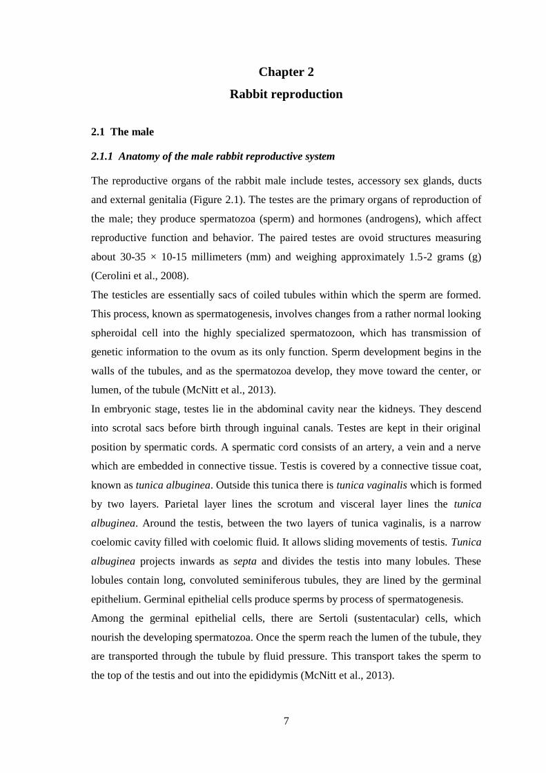
2.1.1 Anatomy of the male rabbit reproductive system
The reproductive organs of the rabbit male include testes, accessory sex glands, ducts
and external genitalia (Figure 2.1). The testes are the primary organs of reproduction of
the male; they produce spermatozoa (sperm) and hormones (androgens), which affect
reproductive function and behavior. The paired testes are ovoid structures measuring
about 30-35 × 10-15 millimeters (mm) and weighing approximately 1.5-2 grams (g)
(Cerolini et al., 2008).
The testicles are essentially sacs of coiled tubules within which the sperm are formed.
This process, known as spermatogenesis, involves changes from a rather normal looking
spheroidal cell into the highly specialized spermatozoon, which has transmission of
genetic information to the ovum as its only function. Sperm development begins in the
walls of the tubules, and as the spermatozoa develop, they move toward the center, or
lumen, of the tubule (McNitt et al., 2013).
In embryonic stage, testes lie in the abdominal cavity near the kidneys. They descend
into scrotal sacs before birth through inguinal canals. Testes are kept in their original
position by spermatic cords. A spermatic cord consists of an artery, a vein and a nerve
which are embedded in connective tissue. Testis is covered by a connective tissue coat,
known as tunica albuginea. Outside this tunica there is tunica vaginalis which is formed
by two layers. Parietal layer lines the scrotum and visceral layer lines the tunica
albuginea. Around the testis, between the two layers of tunica vaginalis, is a narrow
coelomic cavity filled with coelomic fluid. It allows sliding movements of testis. Tunica
albuginea projects inwards as septa and divides the testis into many lobules. These
lobules contain long, convoluted seminiferous tubules, they are lined by the germinal
epithelium. Germinal epithelial cells produce sperms by process of spermatogenesis.
Among the germinal epithelial cells, there are Sertoli (sustentacular) cells, which
nourish the developing spermatozoa. Once the sperm reach the lumen of the tubule, they
are transported through the tubule by fluid pressure. This transport takes the sperm to
the top of the testis and out into the epididymis (McNitt et al., 2013).

8
Connective tissue among seminiferous tubules contain Leydig (interstitial) cells. They
secrete testosterone (male sex hormone) which controls secondary sexual characters.
The Leyding cells are under the control of hormones from the anterior pituitary, which
is located at the base of the brain. These controlling hormones, regulate the levels of the
androgens in the blood, which, in turn, control spermatogenesis and sexual activity of
the buck (McNitt et al., 2013). The seminiferous tubules of a testis open into a network
called rete testis, it opens into many fine ducts, called vasa efferentia, these vasa open
into the epididymis.
The ducts through which the sperm move after leaving the testes proper include the
epididymides, deferent ducts, and urethra. The epididymides lie close to the top of the
testes and function as a place for maturation of the spermatozoa. Spermatozoa that have
not undergone a period of maturation in the epididymides are incapable of fertilizing
eggs. The epididymides also serve as a place for storage of spermatozoa; fertile
spermatozoa have been recovered from epididymides after eight weeks of storage. The
normal time required for movement of sperm through the epididymides is 8 to 10 days.
The epididymis is a long, narrow and highly convoluted tubule, it is present along the
inner surface of the testis and is divided into three distinct parts:
1) caput epididymis: the anterior part of epididymis, is connected to testis through
vasa efferentia. Caput epididymis is also connected to the dorsal abdominal wall
by a spermatic cord;
2) corpus epididymis: the middle part of the epididymis, connecting caput
epididymis and cauda epididymis;
3) cauda epididymis: the posterior part of the epididymis. The cauda epididymis is
joined to scrotal sac by a short, thick, elastic cord known as gubernaculum.
The deferent ducts convey the spermatozoa from the epididymis to the urethra and also
function to some extent in sperm storage. The accessory sex glands normally add their
secretions to the semen at or near the junction of the deferent ducts and the urethra. The
urethra is the common passage for both semen and urine. It carries semen from the
junction with the deferent ducts to the end of the penis, from which the semen is
ejaculated into the female vagina. The bladder empties into the urethra just beyond the
point of junction of the urethra and deferent ducts. The external genitalia of the male
include the penis, the scrotum and the prepuce. The penis (copulatory organ) is an
erectile organ that is used for insertion of the ejaculate into the female tract. It is

9
normally flaccid and rests in the prepuce. It becomes rigid from constriction of the
penile veins at the time of breeding. Since arterial blood continues to flow into the
organ, it becomes turgid and can thus penetrate the vulva and vagina of the doe. The
erect penis is held forward along the abdomen. There is a sensitive tip on the penis,
known as the glans penis. Stimulation of the glans penis by the vagina of the female (or
by a properly prepared artificial vagina) results in ejaculation. This is due to a reflex
contraction of the duct system that forces out spermatozoa stored in the deferent ducts
and the last third of the epididymis. Fluids from the accessory glands are also released
into the deferent ducts and the urethra at the time of ejaculation. After subsidence of
erection, the penis is pulled back into the prepuce by muscular contraction. The penis is
covered by a loose sheath of skin, the skin that hangs over the tip of the penis is known
as prepuce. The tip of the penis covered by prepuce is called glans penis. The penis is
composed of three longitudinal columns, namely, two columns of corpora cavernosa
(upper) and one column of corpus spongiosum (lower) made by spongy tissue. The
scrotum consists of two relatively hairless sacs that contain the testicles. These function
to protect the testes and to provide an area with a lower temperature than that of the
body cavity, because spermatogenesis cannot occur at normal body temperature. The
testes of the rabbit can move freely in and out of the abdomen and so are not always
found in the scrotal sacs (McNitt et al., 2013).
Associated with the male reproductive system of rabbit, there are the accessory sex
glands, they include the prostate gland, bulbo-urethral glands (Cowper’s glands),
perineal glands and rectal glands. Their secretion are responsible for the production of
seminal plasma. The functions of these secretions include adding fluid volume to the
ejaculate to facilitate movement of semen through the male and female reproductive
tracts, providing nutrients and buffers for the spermatozoa, providing a gelatinous plug
to seal the female tract, and providing substances that stimulate contractions of the
vagina and uterus of the female to enhance movement of spermatozoa through the tract.
Around the base of uterus masculinus, there is prostate gland, it opens into the urethra
through many ducts, it secretes an alkaline fluid which activates the spermatozoa and
also contributes to the main bulk of semen. Moreover, prostate gland secretes citric acid
that enters in the kreb’s cycle to produce ATP. Posterior to the prostate gland, there is a
pair of Cowper’s glands, their secretions neutralize the urinary residue and vaginal
acidity. Behind the Cowper’s glands, there are two perineal glands, they open into
hairless perineal depressions, one on either side of the anus. Their secretions give a

10
characteristics smell to rabbit. Two rectal glands are situated at the sides of the rectum.
Their function is not yet known.
Figure 2.1. The reproductive system of the male rabbit. (McNitt et al., 2013)
2.1.2 Rabbit Spermatozoa
Each spermatozoon has a head that carries the genetic information and a tail that
provides propulsion by its whip-like movements. The rabbit spermatozoa is about 50-60
µm long, it has a wide, flattened head and a long, thin and cylindrical segment
composed by a neck, an intermediate part and a flagellum or protoplasmic tail (Figure
2.2, 2.3). The whole sperm cell is enveloped by the plasma membrane, whose function
is to contain organelles and intracellular components. Thanks to its semi-permeability, it
maintains the chemical gradient of ions and of the other soluble components. Moreover,
specific membrane proteins facilitate the transport of fructose and glucose from the
extracellular environment to the inside of the cell, thus providing it with the energetic
substrates it requires for the cell metabolism.
The head includes two structures of fundamental importance: the acrosome (Figure 2.4)
and the nucleus. The acrosome is a structure derived from the Golgi apparatus and it is
delimited by a trilaminar membrane: it covers two thirds of the front part of the sperm
head. Above the acrosome, another structure covers the remaining front part of the
head: it is the head cap, composed by sulphur- and fibrous proteins which confer it great
resistance. Its biological role, in effect, is to protect the acrosomal content: this includes
enzymes with a high proteolytic and glycolytic power, which allow the spermatozoon to

11
penetrate through the zona pellucida during the fecundation of the egg cell. The most
important enzyme is acrosin, followed by hyaluronidase, collagenase, acid phosphatase,
phospholipase A, arylsulfatase. Inside the head, in the central part, it is possible to find
the nucleus, whose function is to transport and transmit the chromosomes, i.e. the
genetic information. The nucleus of a ripe spermatozoon is very dense, as it is
composed by tightly packed chromatin. The segment adjacent to the head is called neck:
this region contains centrioles, which give the impulse for the meiotic cell division. This
is also the starting point of the axoneme, a thigh bundle of axial contractile fibres, which
continues in the following parts as well.
The segment after the neck, in rabbits, is about 8.8 µm long. It is called intermediate
piece and it contains mitochondria, organized in a layer surrounding the axoneme. This
part, therefore, constitutes the main energy reservoir for the cell. The long tail segment
(40 µm) following the intermediate piece is called main piece: it is not surrounded by
any coat of mitochondria but it contains arginine, leucine and other essential amino
acids. This segment, too, is composed by bundles of parallel contractile fibres, which
end in thin fibrils constituting the final segment. These filaments are projected in the
liquid medium like a helix, thus determining the helical movement of the spermatozoa.
Moreover, they are able to change their descent energy into ascent energy, thereby
advancing against the gravity force following the liquid currents of the female genital
apparatus.
Figure 2.2. Structure of rabbit spermatozoa

12
Figure 2.3. Picture of rabbit spermatozoa obtained by scanning electron microscopy
(magnification × 2000). Provided by Microscopy Interdepartmental Center, University
of Molise
Figure 2.4. Picture of rabbit spermatozoa head obtained by scanning electron
microscopy (magnification × 10000). The arrow highlights the “equatorial zone”, which
is the end of acrosomal cap. Provided by Microscopy Interdepartmental Center,
University of Molise

13
2.1.3 Spermatogenesis and sperm maturation
Spermatogenesis is a process of division and differentiation of cells from diploid
germinal cells termed spermatogonia, to haploid, mature spermatozoa in seminiferous
tubules of the testis. The duration of spermatogenesis in rabbit is 53 days (Swierstra and
Foote, 1965). The 53-day period may be divided into three phases, including:
spermatocytogenesis, where stem cell spermatogonia divide by mitosis to produce other
stem cells in order to continue the lineage throughout the adult life of the male, they
also divide cyclically to produce committed spermatogonia and primary spermatocytes;
meiosis, the period of replication (primary spermatocytes) and then reduction of genetic
material into haploid spermatids; and spermiogenesis which is a Sertoli cell aided
differentiation of spermatids (Figure 2.5).
Spermiation is the release of spermatids as spermatozoa into seminiferous tubule lumen.
Transit of sperm through the efferent ductules and the epididymis is associated with
significant maturation changes such as obtaining the capacity of progressive motility;
final condensation of the nucleus and further modification of the form of the acrosome;
alteration of plasma membrane surface. The most important functional changes in
sperm occur in the efferent ductules, caput and corpus of epididymis. Sperm taken from
the distal part of corpus and cauda epididymis already have the potential to fertilize.
Approximately 8 days are required for transportation of spermatozoa through the
excurrent duct system (efferent duct, epididymis and vase deferents). Therefore, a new
population of spermatozoa is ejaculated every 50–54 days (Swierstra and Foote, 1965).
The cauda epididymis has properties that allow the sperm to be stored for several
weeks. Seminal fluids secreted by accessory sex glands added during ejaculation serve
as a vehicle, stimulate the metabolism of sperm and provide the energy requirements for
passage through the uterus (Gadella et al., 2001).
Most of the spermatozoa are stored in the epididymal tail, but also a lower number of
spermatozoa are present in the testis and in the other efferent ducts (Table 2.1).
The spermatozoa in the female reproductive tract, particularly in the isthmus oviduct
undergoes certain change called ‘‘capacitation’’, which takes about 5-6 hours in rabbit
spermatozoa (Cerolini et al., 2008).
The mechanism of capacitation involves many biochemical changes. These include the
removal of adsorbed components from the sperm surface, the reorganization of
membrane proteins, a change in membrane lipid composition, increased permeability to

14
certain ions such as Ca2+
, a change in internal pH, and an increase in plasma membrane
fluidity and in metabolism (Parrish et al., 1993). There is also an increased
hyperactivation that is believed to result in the redistribution of membrane components
during capacitation (Parrish et al., 1993). In addition, several studies show that there is a
decrease in the membrane cholesterol phospholipid ratio during capacitation. All of
these changes allow the spermatozoa to undergo the acrosome reaction (AR) following
interaction with the zona pellucida, the extracellular matrix of egg cells (Miller et al.,
1990). The rabbit acrosome reaction, which involves membrane breakdown and release
of enzymes for penetration of egg envelopes, is induced by a specific stimulus in
follicular fluid at the site of fertilization.
Figure 2.5. Process of spermatogenesis

15
Table 2.1. Number of spermatozoa in different reproductive system portions
Reproductive system portion Sperm concentration (× 106)
testis 130180/g
caput and corpus of epididymis 150
cauda of epididymis 12001500
deferent duct 50
2.1.4 Reproductive activity of the buck
Bucks reach sexual maturity at the age between five and eight months, depending on
breed and level of nutrition. In a study of New Zealand White bucks, daily sperm output
was found to increase from 20 weeks to a mature level at about 31 weeks (McNitt et al.,
2013).
The normal ejaculate volume for mature bucks ranged from 0.39 to 1.19 mL, with an
average of about 0.6 mL. Sperm per ejaculate is highly variable among bucks as well as
among successive ejaculates from the same buck and varies from 146 to 738 million
spermatozoa per mL (Bencheikh, 1995; Brun et al., 2006; Castellini et al., 2006b;
García-Tomás et al., 2006a,b; Safaa et al., 2008; Lavara et al., 2010). The number of
spermatozoa for each ejaculate depends on the breed of the buck, recent use, and level
of stimulation.
2.2 The female
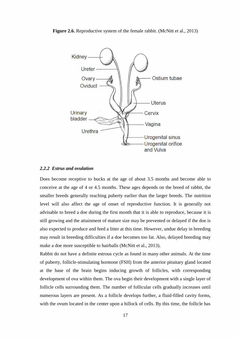
2.2.1 Anatomy of the female rabbit reproductive system
The organs of female reproduction system include: the ovaries, oviducts, uterus,
cervices, vagina, and external genitalia (Figure 2.6). The ovaries, the primary organs of
reproduction, produce eggs, or ova, and hormones (primarily estrogens and progestins).
They lie within the abdominal cavity, with one on each side, near the kidneys. The
ovaries are ovoid structures with an extent of about 15 × 10 mm and a weight between
300 and 500 mg (Cerolini et al., 2008), depending on the activity of the ovarian
components. The middle portion, or medulla, of each ovary, is composed of connective
tissue containing nerves and blood vessels. The outer layer, or cortex, contains the ova
in various stages of development, as well as other types of tissue, including blood

16
vessels, nerves, and muscle fibers. At the time of a doe’s birth, thousands of
undeveloped ova are contained in the germinal epithelium layer of the cortex. From the
time of puberty until death or the end of the reproductive life of the female, groups of
these undeveloped ova undergo development and are shed (ovulated), or they
degenerate. The oviduct is the site of fertilization, functions in a maturation process of
spermatozoa known as capacitation, and is the location where early embryonic
development occurs. Oviducts are thin tubes, characterized by willowy trend, of about
10-16 cm in length (Cerolini et al., 2008).
The upper end of the oviduct is spread into the ostium tubae, which partially surrounds
the ovary. On the edges of the ostium tubae are numerous small projections known as
the fimbria. These nearly cover the ovary. Beating of the fimbria causes waves of
movement of fluid toward the opening of the oviduct and, at the time of ovulation,
sweeps the ova into the oviduct (McNitt et al., 2013).
The uterus is the organ in which the embryo and foetus normally develop and grow. It
also provides muscular force for expulsion of the fetuses at birth. The uterus of the
rabbit is formed of two distinct horns, which are not connected to form entire body.
Each horn of the uterus connects into an individual cervical canal, which opens into the
common vagina. The cervices function as muscular plugs to keep the uterine horns
closed except at the time of mating and parturition (birth, or kindling) (McNitt et al.,
2013).
The vagina is the site of sperm deposition at mating and acts as a channel for the young
at parturition. It is long between 6-8 cm and presents longitudinal mucosal folds
(Cerolini et al., 2008).
The external genitalia of the doe include the urogenital sinus, which is continuous with
the vagina and is the chamber into which the urethra empties urine. The external lips of
the urogenital sinus form the vulva, which can be used as an indicator of sexual
receptivity of the doe. A doe with a moist red or pink vulva is much more likely to
accept service than a doe with a pale, dry vulva. The clitoris is within the urogenital
sinus, with the sensitive portion, the glans clitoris, projecting into the urogenital
opening. Because the urethra opens into vaginal sinus posterior toward the place where
sperm are deposited, urination by the doe following breeding does not necessarily
interfere with fertilization.
17
Figure 2.6. Reproductive system of the female rabbit. (McNitt et al., 2013)
2.2.2 Estrus and ovulation
Does become receptive to bucks at the age of about 3.5 months and become able to
conceive at the age of 4 or 4.5 months. These ages depends on the breed of rabbit, the
smaller breeds generally reaching puberty earlier than the larger breeds. The nutrition
level will also affect the age of onset of reproductive function. It is generally not
advisable to breed a doe during the first month that it is able to reproduce, because it is
still growing and the attainment of mature size may be prevented or delayed if the doe is
also expected to produce and feed a litter at this time. However, undue delay in breeding
may result in breeding difficulties if a doe becomes too fat. Also, delayed breeding may
make a doe more susceptible to hairballs (McNitt et al., 2013).
Rabbit do not have a definite estrous cycle as found in many other animals. At the time
of puberty, follicle-stimulating hormone (FSH) from the anterior pituitary gland located
at the base of the brain begins inducing growth of follicles, with corresponding
development of ova within them. The ova begin their development with a single layer of
follicle cells surrounding them. The number of follicular cells gradually increases until
numerous layers are present. As a follicle develops further, a fluid-filled cavity forms,
with the ovum located in the center upon a hillock of cells. By this time, the follicle has

18
enlarged to such an extent that it bulges from the surface of the ovary. At ovulation the
outer layer of the follicle ruptures, and the ovum is expelled along with the fluid
(McNitt et al., 2013).
The follicles produce estrogens (the main female sex hormones), that make female
receptive for the male (Patton, 1994). Follicular development generally occurs in waves,
with 5 to 10 follicles on each ovary at the same stage of development at any one time.
Follicles start continuously development, so follicles at several stages of development
are always present (Patton, 1994). When follicles reach mature size, they produce
estrogens for about 12 to 14 days. After this period, if ovulation has not occurred, these
follicles degenerate, with a corresponding reduction in blood concentration of estrogen
level and receptivity. After about 4 days a new follicles will begin producing estrogen
and the doe becomes receptive again. The doe therefore, has a cycle of 16-18 days,
during which she is receptive for about 12-14 days followed by 2-4 days when the doe
refuses to mate (Patton, 1994). The receptivity of the does is extremely variable because
of individual differences, sexual stimulation and environmental factors, such as
nutrition, light and temperature (Cheeke et al., 1987).
Ovulation in the doe occurs only after induction by an external stimulus, such as
mating, and occurs 10-13 h after coitus (Patton, 1994). This includes stimulation of the
anterior vagina by the penis as well as pressure on the hindquarters from mounting by
the buck. Intense sexual excitement or mounting of the doe by other rabbits may also
induce ovulation. This may result in a condition known as pseudopregnancy. An
ovulatory stimulus results in the release of luteinizing hormone (LH) from the anterior
pituitary. This hormone causes rupture of a number of mature follicles on one or both
ovaries approximately 10 hours after the stimulus occurs. The number of ova shed from
each ovary is a factor that determines the litter size. Other factors include the number of
shed ova that are fertilized by the sperm and the number of fertilized eggs that follow
the entire intrauterine development process (McNitt et al., 2013). When the ovum has
been shed from the ovary, LH stimulates changes in the follicular cells that rapidly
develop into a corpus luteum (yellow body), which produces hormones known as
progestins. These are necessary throughout pregnancy for development of the embryo.
The primary action of the progestins is to stop muscular contractions of the uterus and
to stimulate production of nutrients for the embryo. The corpus luteum begins actively
secreting within three days after ovulation and continues throughout pregnancy. The
hormone output increases until about the fifteenth day of pregnancy and remains at a

19
high level until the last week, when the hormone level begins to fall. The progestins
control uterine function, especially inhibition of muscular activity so that the embryo
can remain in the uterus and be nourished throughout pregnancy. Moreover, the
progestins inhibit sexual receptivity in pregnant doe, although follicles continue to
develop and produce estrogens throughout pregnancy. It has been shown that estrogens
are necessary for the progestins secretion, occurring in corpus luteum. Since follicles
are present at the end of pregnancy, the doe is sexually receptive and able of ovulation
immediately after parturition (McNitt et al., 2013).
2.2.3 Fertilization and pregnancy
At the moment of mating the buck deposits several million sperm in the doe’s vagina.
The spermatozoa move by contractions of the female tract and by swimming to the
middle portion of the oviduct, where fertilization occurs. The first sperm are found in
the oviduct within 20 to 30 minutes of mating, although the majority are not found there
for several hours. Of the millions deposited into the vagina, only a few thousand reach
the site of fertilization (McNitt et al., 2013).
When the eggs are released from the follicle, they are swept into the oviduct and move
to the middle third of the oviduct. Fertilization refers to the entry of a sperm cell into the
ova and fusion between male and female genetic material (syngamy). Cellular division
and development of the embryo begin almost immediately after syngamy. The
developing embryo remains in the oviduct until the 8- or 16-cell stage is reached. This
takes 48-52 hours, after which the embryo migrates to the uterus, where it floats in the
uterine fluid and is nourished by it (Cerolini et al., 2008). During this phase, nutrients
enter the embryo by diffusion through the cell membranes. After seven days the embryo
becomes too large to be properly nourished solely by diffusion, so it is attached to the
wall of the uterus, and the placenta (afterbirth) begins to form. This process is known as
implantation. The placenta supplies protection for the embryo and a close connection
between the embryonic and maternal circulatory systems. There is no direct connection
between these two systems, although the two blood supplies pass very close to each
other in their respective vessels. In this way, oxygen and nutrients can diffuse through
the vessel walls from the doe to the young, and the wastes from the young can diffuse
out to the circulatory system of the doe. Transport of nutrients and oxygen within the
embryo is carried out through embryonic circulatory system. Pregnancy in rabbits lasts
an average of 30 to 32 days but may be as short as 29 days or as long as 35 days.

20
Embryonic growth is not constant. At day 14 of pregnancy, the embryos of New
Zealand White does average 0.5 to 1 g; at day 20, about 5 g; and at birth, about 50-60 g
(Cerolini et al., 2008). Birth weights vary from 25 to 90 g, depending on the age and
breed of the doe and the number of young in the litter.
2.2.4 Parturition
Parturition in the rabbit normally occurs in the early morning, taking about 10-30
minutes, with individual kits born at intervals of 1 to 5 minutes. The doe crouches in the
nest and licks each of the young as they are delivered. This behavior helps to dry the
kits, removes blood and tissue debris, and stimulates blood circulation. When
parturition is complete, the doe eats the placenta and dead kits (Cerolini et al., 2008).
The number of young in the litter depends on the breed of the doe, her nutritional status,
her age, and her environment. The New Zealand White generally produces 8-10 kits in
each litter. European workers have selected for what they call hyperprolific does that
regularly produce 12 to 15 young (McNitt et al., 2013).
The doe normally nurses the litter only once a day for about four or five minutes. The
kits change nipples very frequently during the early part of suckling, remain relatively
quiet through the middle of the period and then, begin moving rapidly toward the end.
2.2.5 Lactation
The growth of the young during the suckling period, especially during the first three
weeks, greatly affects their later performance. The mammary glands of the doe
generally consist of eight physiologically distinct sections, four along each side of the
abdomen. The number may vary from 6 to 10. The glandular tissue of the rabbit is
similar to that of other species and consists of hollow, ball-shaped structures known as
alveoli, where the milk is induced. At the time of parturition the release of prolactin, is
responsible for the beginning of milk production and during gestation its action is
inhibited by estrogens. Prolactin is responsible for continued milk production during the
lactation period. Although milk production is a continuous process, the milk is
unavailable to the young until a stimulus causes the release of oxytocin. The quantity
and composition of the milk produced by the doe vary throughout lactation, as shown in
Table 2.2. The quantity of milk produced increases until the end of the third week of
lactation and declines thereafter. At that point the kits generally start eating solid food,

21
and the milk of the doe becomes less important to the performance of the young
(McNitt et al., 2013).
Table 2.2. Quantity and composition of milk produced at various stages of lactation
Days after birth
1 7 14 21 24
Milk (g/day) 50 160 200 300 220
Water (%) 68 74 75 73 67
Protein (%) 14 14 13 13 16
Lipid (%) 15 9 9 10.6 13.7
Lactose (%) 1.4 1 1 1 0.7
Ash (%) 1.6 2 2 2.4 2.6

22
Chapter 3
Artificial insemination in rabbit
Artificial insemination (AI) has been known and practiced in the rabbit breedings for
over 80 years, since Bonadonna in the 30's perfected the first glass model of artificial
vagina (Bonadonna, 1974). AI was originally used in intensive rabbit production to
obtain positive effects in the breeding management, this led to the development of a
new system of production, called cyclic production system (Facchin et al., 1988). This
system consists of grouping animals in the same physiological phase and carrying out
breeding operations on fixed days of the week (Castellini and Lattaioli, 1999). Cycled
production permits better production planning, reduces the needed of manpower (about
10-15%) and facilitates the planning of ‘‘all-in all-out’’. The use of this technique
requires some punctual considerations. Cycled production requires a fixed reproductive
rhythm: inseminating every 5-6 weeks non-pregnant does follow the same rhythm of
insemination as pregnant ones. Comparing the cycled production with the traditional
system, cycled production increases the unproductive period of non-pregnant does.
Consequently, when pregnancy rate is low many does must wait until the next cycle of
insemination, thus increasing the kindling interval (Castellini, 1996). In the last decade
the diffusion of the cyclic breeding system opened the way for its industrial application,
therefore AI has become a routine practice in rabbit production (Alvariño, 2000).
Nowadays, AI is used routinely in many large rabbit farms in a number of European
countries including Italy, France, Spain, Germany, and Hungary.
The AI offers the following advantages:
Sanitary advantages
- absence of physical contact between the animals during the mating;
- possibility of cyclic production, with periodical gaps for sanitary reasons.
Management advantages
- more efficient organisation of the work, the production and the transport;
- better organization and consequent execution of the nutrition programs;
- simpler data collection.
Economic advantages
- reduction of the duration and cost of the work;
- reduction of the number of males needed in the rabbit farm;

23
- increase of the number of productive does;
- possibility to increase the production in favourable moments of the market.
Selection and genetic improvement
- better possibilities to identify the good breeders and the characters to be
selected;
- simpler and more precise testing of the breeders;
- easier implementation of cross breeding programs;
- possibility to keep a genetic inheritance over time.
The use of the AI in rabbit breeding led to a growing interest in the study of the
reproductive physiology of rabbits with a consequent improvement of this practice. This
is proven by the fact that the currently AI made with fresh semen within maximum 6
hours from its collection provides fertility and prolificacy rates similar to those of
natural mating.
In detail, the AI involves different steps: 1) the semen collection, 2) assessment of
sperm quality, 3) semen dilution in an appropriate extender that has to guarantee the
sperm survival as well as to increase the number of inseminating semen doses and 4) the
insemination of does.
3.1 Rabbit semen collection
Semen collection from rabbit male occurs through specific artificial vagina (Morrell,
1995). The artificial vagina is composed of a rubber or latex tube, open on both sides,
which is placed in a glass or plastic container (Figure 3.1). It must be filled with a
warmed water at about 50°C in order to create the vaginal natural environment that it
results be of 40°C. One side of the tube is needed for the ejaculation of the male, while
the other one is connected to a graduated test tube for the collection of the semen. The
test tube has to be maintained at about 38°C before its use, and changed after every
collection. If the temperature of artificial vagina decreases below 40°C, the male refuses
to ejaculate. During the collection, it is necessary with one hand to hold the artificial
vagina, placed under the abdomen of the fake female rabbit, while the other hand holds
the fake rabbit female in front of the male. At the moment of the jump of the male, the
opening of the artificial vagina is held between a thumb and an index finger to facilitate
the insertion of the erect penis. The ejaculation is then helped with a light push
backwards of the artificial vagina. Ejaculation usually takes place immediately

24
following the appearance of the doe. During the collection phase, a good hygienic level
of manipulation is essential to minimize the contamination of the semen sample.
Figure 3.1. Basic structure of artificial vagina. The artificial vagina is assembled by the
appropriate size of outer case (a) and inner rubber (b) which lines over the top and
bottom edges of inner case. The test tube (c) is connected to the top hole of the case,
and the space between inner case and lined rubber is filled with hot water (d). (Rabbit
Biotechnology: Rabbit genomics, transgenesis, cloning and models, Houdebine, Louis-
Marie, Fan, Jianglin (Eds.). 2009)
3.2 Assessment of semen quality
Semen evaluation is made immediately after collection, this provides useful information
about the sperm fertilizing ability. Moreover, results of the semen quality evaluation is
important to keep in breeding only males that have a good fertilizing ability.
Seminal characteristics are affected by several factors such as the rearing condition and
management (López et al., 1996; Alvariño, 2000; Roca et al., 2005), age (Gogol et al.,
2002), feeding strategies (Yousef et al., 2003; Castellini et al., 2001, 2003, 2006a),
season (Nizza et al., 2003), photoperiod (Theau-Clément et al., 1994; Roca et al., 2005)
and frequency of collection (Nizza et al., 2003; Castellini et al., 2006b). The semen
evaluation relies on macroscopic and microscopic analysis.
3.2.1 Macroscopic parameters
Macroscopic parameters include: volume, color, smell, density, presence or absence of
gel plug and pH. A normal sperm sample has a homogeneous white opalescent
appearance. The presence of red blood cells (reddish) or urine (yellow) is easily
detected. Dark, yellow or other abnormal semen samples are generally discarded. If
present, gel plug should be removed immediately after collection. Ejaculate volume can
be determined by different techniques. Although it may be determined with graduated

25
collection tubes, a more accurate measurement can be taken with the use of a calibrated
micropipette. The ejaculate volume usually ranges from 0.19 to 1.19 mL, but on average
is about 0.6 mL; the quantity and quality of ejaculate improve until the age of 8-9
months of breeding and then remain constant during the reproductive career. The pH
should be measured immediately after collection since it is modified by metabolism, it
must be neutral.
3.2.2 Microscopic parameters
Microscopic parameters are the following: sperm concentration, motility, viability,
membrane integrity (osmotic resistance), acrosome integrity, DNA integrity and
apoptosis.
3.2.2.1 Sperm concentration
Sperm concentration refers to the number of spermatozoa per millilitre of semen.
Determination of the concentration in a semen sample is important, because this
parameter is used to determine the number of spermatozoa that will be used if the
quality of the ejaculate is good enough. Sperm concentration can be determined using a
hemacytometer (Neubauer chamber, Thoma or Bürker). This method is commonly
employed to determine the sperm concentration in rabbit semen at the beginning of AI
procedure (Walton, 1927). Moreover, new technologies are developed to measure sperm
concentration, and spectrophotometry can be used (first described by Salisbury et al.,
1943), but this methodology fails to provide accurate measurements for samples
containing non-sperm particles in the medium (as prostatic vesicles and cells).
Unfortunately, in rabbit ejaculates there is a high content of prostatic vesicles and other
cells (Farrel et al., 1992; Castellini et al., 2006b) therefore, this technique does not
provide accurate data. However, these ejaculates can be evaluated using fluorescent
stains that label only sperm (Riedy et al., 1991). Using these fluorescent stains, the
sperm concentration can be evaluated using fluorometry (Theau-Clément and Faliéres;
2005) and flow cytometry (Purdy and Graham, 2004). The main drawback of these
techniques is the high cost.
3.2.2.2 Sperm motility
Motility is certainly one of the most important features associated with semen fertilizing
capacity. It is an important attribute, because it is readily identifiable and reflects
several structural, and functional competence, as well as essential aspects of

26
spermatozoa metabolism. Sperm motility was the first, and continues to be the most
widely used indicator of sperm function (Partyka et al., 2012).
Usually total motility (any type of motility) and progressive motility (spermatozoa
moving actively forward) are estimated as percentages. The percentage of progressively
motile sperm in the ejaculate is critical to ensure adequate sperm transport and
fertilization. Sperm motility is usually assessed by a subjective visual examination
under a contrast phase microscope at 37°C using low objectives (10, 20 or 40×). Light
microscopic evaluation does not require expensive equipment, it is a simple and rapid
method for assessment of sperm quality, however, it is a highly subjective and not
reliable assay for the prediction of fertility (Peña Martínez, 2004).
Alternatively to the subjective visual motility evaluation, photographic analysis or
computer assisted semen analysis (CASA) can be used (Verstegen et al., 2002;
Rijsselaere et al., 2003). This technique assures objective semen assessment, whereas
the main disadvantage of conventional semen evaluation is the variability of results
obtained. Subjectivity of traditional semen analysis is mainly associated with the
experience and skill of the observer, the method of specimen preparation, staining
technique and the number of cells evaluated. Variations in the results of conventional
evaluation of the same semen samples by different observers and laboratories may
achieve up to 30-60% (Davis and Katz, 1992; Coetzee et al., 1999). Computer assisted
sperm analysers allow to calculate of several motility parameters, which characterize
movement of individual sperm cells. They include the measure of the distance between
each head point for a given sperm during the acquisition period (curvilinear velocity,
VCL, μm/s), the distance between first and last head points divided by the acquisition
time (straight line velocity, VSL, μm/s), and the measure of sperm head oscillation
(amplitude of lateral head displacement, ALH, μm) (Figure 3.2). Linearity (LIN, %)
measures the departure from linear progression and is calculated as VSL/VCL × 100,
while the average path velocity (VAP, μm/s) is a smoothed path constructed by
averaging several positions on the sperm track (Verstegen et al., 2002). Selected
characteristics of sperm motility parameters measured by CASA systems are reported in
Table 3.1. The important advantage of CASA system is the immediate measurement of
sperm concentration, total number of sperms in ejaculate and the automated calculation
of number of insemination units which could be prepared from one ejaculate (Partyka et
al., 2012). However, CASA system needs standardization and validation before its use
and therefore, image settings have been standardized (Davis and Katz, 1992; Iguer-

27
Ouada and Verstegen, 2002; Rijsselaere et al., 2003). Moreover, other factors as the
type and depth of the used chamber, number of fields analysed, temperature during
analysis and protocol of semen sample preparation, affect results (Partyka et al., 2012).
Figure 3.2. Scheme of different velocities and parameters of sperm movement
measured by CASA systems http://www.mibio.org/en/metrics-used
Table 3.1. Parameters of sperm motility measured by CASA system
Parameter Unit Description
MOT % Motility: population of cells that are moving at or above a
minimum speed as determined by values defined under setup.
PMOT % Progressive motility: the population of cells that are moving
actively forward.
VCL μm/s Track speed: average velocity measured over the actual point-
to-point track followed by the cell.
VAP μm/s Path velocity: average velocity over smoothed average
position of the cell.
VSL μm/s Progressive velocity: measured in the straight line distance
between the beginning and the end of the track.
ALH μm Amplitude of Lateral Head Displacement: mean width of the
head oscillation as the cell moves.
BCF Hz Beat Cross Frequency: frequency with which the sperm head
moves back and forth in its track across the cell path.
STR % Straightness: measure of VCL side to side movement
calculated by the ratio VSL/VAP.
LIN % Linearity: measure of the departure of the cell track from a
straight line. It is the ratio VSL/VCL.
VCL
ALH
VSL
Curvilinear
path
Average
path
Straight-line path

28
3.2.2.3 Sperm viability
The viability of spermatozoa is a key determinant of sperm quality and prerequisite for
successful fertilization. Sperm viability is a traditional method for assessing whether the
sperm membrane is intact or disrupted and involves examining of a percentage of viable
sperm by a stain exclusion assay (Partyka et al., 2012). Live-dead stains as aniline-
eosin, eosin-nigrosin, eosin-fast green are widely used for the evaluation of cell
viability. Integrity of the plasma membrane is shown by the ability of a viable cell to
exclude the dye, whereas the dye will diffuse passively into sperm cells with damaged
plasma membranes (Colenbrander et al., 1992). In eosin-nigrosin stain under the
microscope, live spermatozoa appear white, unstained against the purple background of
nigrosin. Dead and damaged spermatozoa which have a permeable plasma membrane
are pink. Although the eosin/nigrosin technique is still used in many routine laboratories
as a means of plasma membrane assessment, it is being superseded by fluorescent tests
(Johnson et al., 1996).
Viability of spermatozoa may be assessed by using many fluorescent probes
combinations including: carboxyfluorescein diacetate (CFDA) in combination with
propidium iodide (PI), (Carboxy-SNARF) with PI, calcein-AM with ethidium
homodimer (EthD-1), SYBR-14 with PI. The fluorescence of these compounds may be
evaluated by fluorescent microscopy or flow cytometry. Flow cytometry enables the
observation of a great number of spermatozoa (>10 000 in a small volume of samples in
a short time) than the total of 100-200 cells generally observed by microscopic analysis,
however the equipment is more expensive.
SYBR-14/PI fluorochromes have been found to be more sensitive in comparison with
the conventional method of live-dead cell assessment. SYBR-14, a membrane-permeant
DNA stain, stained only living spermatozoa, producing green fluorescence of the nuclei.
Instead propidium iodide stained the nuclei of membrane-damaged cells red, therefore
the spermatozoa that show green fluorescence are considered alive and those that
exhibit red fluorescence are considered dead. This procedure has been successfully
applied in different livestock species (Garner et al., 1996; Rodríguez-Martínez, 2007)
and also in rabbits (Garner and Johnson, 1995).

29
Figure 3.3. Discrimination between intact and plasma membrane damaged sperm using
DNA affinity probes. DNA of life sperm can be stained with SYBR-14. This membrane
permeable DNA binding probe is deacylated by intracellular esterases. The deacylated
probe cannot diffuse back over the plasma membrane out of sperm. The entrapped
fluorescent probe stains the sperm nucleus as it has weak affinity for DNA. In sperm
with deteriorated plasma membranes SYBR-14 will leave the cell. DNA of deteriorated
sperm can be stained with membrane-impermeable fluorescent DNA staining probes.
These probes do not enter life cells but diffuse into the sperm nucleus to bind DNA
once the plasma membrane has become deteriorated. (Silva and Gadella, 2006)
3.2.2.4 Sperm osmotic resistance
The integrity and functional competence of the rabbit sperm membrane (osmotic
resistance) is assessed by studying the swelling reaction of sperm tail when suspended
in a medium of distilled water (Hypo-osmotic swelling test; HOST). This test is based
on the assumption that when spermatozoa are exposed to hypo-osmotic media, such as
distilled water, sperm with normal membrane function permits passage of fluid into the
cytoplasmic space causing swelling produced by a hyposmotic shock and the pressure
generated leads to the curling of tail. Conversely, the damaged or chemically inactive
sperm membrane, allows fluid to pass across the membrane without any accumulation
and accordingly no cytoplasmic swelling and curling of the tail occur.
This test has the advantage of indicating not only whether the membrane is intact, but
also whether it is osmotically active (Partyka et al., 2012).

30
The HOST is a simple, inexpensive and easily applicable technique, which has been
adapted to assess spermatozoa of several species (Correa and Zavos, 1994; Neild et al.,
1999; Pérez-Llano et al., 2001; Santiago-Moreno et al., 2009). The HOST seems to be
more appropriate for predicting the fertilizing capacity of frozen-thawed than fresh
semen, because membrane damage is a more important limiting factor than in the
former (Colenbrander et al., 2003).
3.2.2.5 Acrosome integrity
The acrosome is a membrane enclosed structure covering the anterior part of the sperm
nucleus. Powerful hydrolyzing enzymes belongs to that structure, a basic feature of the
sperm head of all mammals (Yanagimachi, 1994). The acrosome reaction (AR) of rabbit
sperm is to be observed before fertilization, as it is necessary for sperm penetration of
the zona pellucida (ZP) and for fusion with plasma membrane. As a prerequisite of
fertilization the content of the acrosome is released into its surroundings during the
acrosome reaction. It is assumed that the AR serves at least dual functions by
facilitating the ability of the sperm to penetrate the ZP, and subsequently by aiding in
the oocyte-sperm fusion process (Yanagimachi, 1994).
Therefore, assessment of the acrosome integrity is a very important part of semen
evaluation, in the view of the role of this structure in the maintenance of spermatozoa
ability to penetrate the zona pellucida of egg cell (Partyka et al., 2012). Acrosomal
status may be evaluated with many different fluorescent probes, some of which can be
used on living, non-permeabilized spermatozoa, while the others require that the
spermatozoa are permeabilized before labelling. Some of these probes contain lectins
and antibodies which bind with high affinities to intracellular saccharide molecules and
acrosomal antigens respectively. Lectins are more popular than antibodies and a large
variety of lectins are available for use in laboratory assays. The lectins that are used
more frequently are peanut agglutinin from Arachis hypogaea (PNA) or Pisum Sativum
agglutinin (PSA), conjugated with different fluorescent probes like fluorescein
isothiocyanate (FITC), phycoerythryn (PE) or Alexa Fluor®, (Nagy et al., 2004;
Rijsselaere et al., 2005; Partyka et al., 2010, 2012). The PNA lectin is specific for the
outer acrosomal membrane and it binds to β-galactose moieties. The absence of the
fluorescence on the living sperm, is indicative for an intact acrosome, and fluorescence
is indicative for acrosome disruption or acrosome reaction (Silva and Gadella, 2006).
Whereas the PSA is labelling α-mannose and α-galactose moieties of the acrosomal
31
matrix. After permeabilization in methanol, the sperm with intact acrosoma show
uniform green fluorescence in the acrosomal region, while acrosome-damaged
spermatozoa show little or no green fluorescence (Rosato and Iaffaldano, 2011). Since
PNA agglutinin displays less non-specific binding to other areas of the spermatozoa, it
leads some researchers to favour this over PSA (Graham, 2001).
3.2.2.6 DNA integrity
DNA integrity has been considered as an important parameter in the determination of
sperm quality. DNA integrity is defined as the absence of both single strand (ss) or
double strand (ds) and breaks absence of nucleotide modifications in the DNA (Shamsi
et al., 2009). Sperm DNA is organized in a specific manner that keeps the chromatin in
the nucleus compact and stable, in an almost crystalline status (Carrell et al., 2003).
DNA damage can be evaluated at different levels. One of the more popular methods,
developed for detecting changes in the chromatin structure of DNA integrity, is the
sperm chromatin structure assay (SCSA) (Chohan et al., 2006). The SCSA is a flow
cytometric method for identification of changes in the DNA status. It is based on the
assumption that a structurally abnormal sperm chromatin shows a higher susceptibility
to acid denaturation (Evenson et al., 2002). The SCSA method utilizes the
metachromatic properties of Acridine Orange (AO). AO is a nucleic acid specific,
fluorescent, cationic dyer. It interacts with DNA by intercalated and by electrostatic
interaction with RNA or single stranded DNA. When bound to DNA ds it occurs an
excitation maximum at 502 nm and an emission maximum at 525 nm (green). When it
associates with RNA or single stranded DNA produced by single stranded DNA breaks,
the maximum excitation shifts to 460 nm (blue) and the maximum emission shifts to
650 nm (red). This stain develops green fluorescence when it intercalates into native
DNA (double-stranded and normal) as a monomer and orange-red when it binds to
denatured ss DNA as an aggregate. Spermatozoa displaying green fluorescence are
considered with normal DNA content, whereas sperm displaying a spectrum of yellow–
orange to red fluorescence are considered with damaged DNA. Another method to
detect DNA defragmentation is Terminal deoxy nucleotidyl transferase mediated dUTP
nick end labeling (TUNEL) assay, which allows to incorporate fluorescent nucleotide
analogs by a terminal nucleotide transferase into single stranded DNA areas at the 3-OH
termini (Chohan et al., 2006). The alternative method for detecting the DNA damage at
the level of individual cells is the single-cell DNA gel electrophoresis assay (COMET).

32
3.2.2.7 Apoptosis
Apoptosis is physiologically programmed cell death that affects single cells without any
associated inflammation in the surrounding tissues (Wyllie et al., 1980). Apoptosis is
characterized by distinct ultrastructural and biochemical changes in cells, including
chromatin aggregation, cytoplasmic condensation, and indentation of nuclear and
cytoplasmic membranes (Williams and Smith, 1993). Apoptosis comprises of a
complex phenomenon that includes three stages: induction, execution and degradation.
During early apoptosis, cell loses its membrane asymmetry. Phosphatidylserine (PS),
normally present on the inner cytoplasmic leaflet of the plasma membrane of healthy
cells, is translocated and exposed to the outer leaflet (Desagher and Martinou, 2000).
The externalization of PS tags apoptotic cells to promote their phagocytosis by
neighboring healthy cells. It is known that the freezing-thawing process usually causes
sublethal cryodamage to spermatozoa, decreasing post-thaw cell viability and induces
membrane PS translocation, demonstrating that cryopreservation leads to apoptosis
(Glander and Schaller, 1999; Peña et al., 2003; Martin et al., 2004; Ortega Ferrusola et
al., 2008; Kim et al., 2010). Therefore, detecting early phases of membrane dysfunction,
or initial phases of apoptosis of viable spermatozoa, would be important when
evaluating stressed spermatozoa, such as those subjected to freezing and thawing, and
would be useful for controlling freezing procedures in semen.
Different fluorescent stains have been used to evaluate apoptotic- like changes.
Fluorescent assays, such as Yo-Pro-1 and Annexin V-FITC, are commonly used to
assess apoptotic-like changes in mammalian spermatozoa (Idziorek et al., 1995; Anzar
et al., 2002; Penã et al., 2003, 2005). The fluorochrome Yo-Pro-1 is a semi-permeable
DNA-binding probe, which penetrates the cellular membranes after destabilization,
unlike of PI or EthD (Penã et al., 2005). Annexin V is a Ca2+
-dependent, phospholipid-
binding protein (35-36 kDa) that has high affinity for PS and binds to cells with exposed
PS. Annexin V conjugated to fluorescein isothiocynate (FITC) fluorochrome retains its
high affinity for PS and, therefore, serves as a sensitive probe that can be used for
detection of cell death (apoptotic or necrotic) characterized by the loss of membrane
asymmetry. Use of Annexin V in combination with propidium iodide (PI; a vital dye)
allows the detection of apoptotic and necrotic cells distinct from viable cells.

33
3.3 Semen dilution and artificial insemination in does
AI in rabbits is generally performed with 0.5 mL of fresh diluted semen. Theoretically,
it is possible to obtain 30-40 doses per ejaculate, but for daily practice it is preferable to
have a dilution rate from 1:5 to 1:10, meaning approximately 10-15 doses/ejaculate, to
ensure at least 4-20 million viable and non damaged spermatozoa (Viudes de Castro and
Vicente 1997; Castellini and Lattaioli, 1999; Viudes de Castro et al., 1999; Brun et al.,
2002).
The AI is carried out by inserting the semen deeply into the rabbit vagina, by using a
pipette of about 25 cm in length with a slightly curved and blunted tip, in order to avoid
traumas and wounds. The pipette must be sterilized and is connected through a thin
rubber tube to a syringe, which sucks the semen from the test tubes and releases the
semen into the rabbit vagina during the insemination.
In the rabbit doe, ovulation does not occur spontaneously, but it has to be induced
through a neurohormonal reflex, which is initiated during mating. When using AI, in the
absence of a male, ovulation has to be induced by artificial stimulation. The ovulation
inducing method most frequently used is an intramuscular application of GnRH
(Gonadotropin Releasing Hormone) (Quintela et al., 2004).
Rabbit does ready to be inseminated are kept in single cages and identified by a card. At
the moment of the insemination the doe is taken from their cage and placed on top of
the cage itself, or on a cart. For the insemination it is necessary to lift the tail with the
index and middle finger, whilst the thumb enlarges the opening of the vulva, in order to
facilitate the insertion of the pipette. The pipette is introduced with its tip up, towards
the back of the female rabbit; at a brief distance from the penetration site (vulva) the
pipette has to be turned (180°), in order to avoid the urinary meatus. Then the pipette is
inserted up to a length of 15 cm for a deep insemination. In breeding farms, the
operation of insemination is usually carried out by two people: one removes the doe out
the cage and places it on the cart for the GnRH injection to induce the ovulation; while
the other one performs the insemination. This method allows to inseminate up to 150
female rabbits per hour.
34
Chapter 4
Storage of rabbit semen
Rabbit breeding for meat production mainly relies on artificial insemination (AI)
(Daniel and Renard, 2010). The AI technique offers significant benefits, including
genetic selection, prolonged fertility even during unfavorable periods of the year, the
possibility to use a cycled production system with a better organization of farms and
health monitoring (Theau-Clément, 2007). Although AI yields fertility rates is similar to
those of natural mate when freshly collected semen is used, this practice requires that
bucks are kept on the same farm as does increasing the number of animals that need to
be managed (Theau-Clément, 2007).
The availability of semen which can be stored for longer periods would allow an
extension of the interval between semen collection and the insemination of females,
thus enhancing AI performance, for example in farms without males and located so far
from semen collection centres (López and Alvariño, 1998; Gogol, 2013). Therefore,
meat rabbit farming would greatly benefit if semen could be stored after collection and
subsequently used for AI without affecting fertility. Unfortunately, the stored semen
quality worsened consequently also the fertility.
Thus, in order to obtain many of potential advantages of AI, improvement of the storage
of rabbit semen is necessary. The semen storage technologies available for rabbit semen
are essentially:
1) hypothermic-liquid storage (refrigeration), which enables the storage of semen
for up to 24 or 48 h at chilling temperatures around 515°C;
2) cryopreservation, whereby semen can be long-term stored at -196°C (the
temperature of liquid nitrogen).
4.1 Hypothermic-liquid storage
The storage in liquid phase consists of keeping the diluted semen at specific
temperatures (between 4 and 15°C), which are significantly lower than that ones
occurring at the time of collection. The semen is placed in a tube containing the
extender, previously incubated at 37-38°C. Subsequently the diluted semen is
transferred in a refrigerator set at specific temperature (see above), in order to induce its
gradual decrease, and to avoid thermic shock which would affect the sperm survival.

35
After this procedure, resulting semen (chilled semen) has to be applied for artificial
insemination within 24-36 hours. The key factors for the success of the semen storage in
the liquid phase are the choice of extender, the dilution ratio and the storage
temperature.
To perform its function, the extender should supply the nutrients needed for the
metabolic maintenance of the sperm cell, control the pH and osmotic pressure of the
medium, and inhibit microbial growth (antibiotics) (Gadea, 2003).
The extenders should have an osmotic pressure similar to seminal plasma
(approximately 300-320 mOsm/kg). Hypotonic or hypertonic solutions may cause
transient or permanent loss of sperm motility, although it seems that hypertonic
solutions are better tolerated in rabbit sperm than hypotonic ones (Costantini, 1989).
The diluent must also contain buffer substances that allow to maintain the pH neutral
and invariable. The optimum pH value is 7, but considerable fluctuations are tolerated
(Watson, 1990). Generally, motility and metabolic processes are suppressed at low pH
and stimulated at higher values (Brun et al., 2002). The buffer substances mainly used
for the preparation of diluents are citrates, phosphates, sulfates, organic molecules such
as the zwitterionic Tris-hydroxymethyl amino-methane (Tris) or Tes and electrolytic
solutions of sodium or magnesium (Foote, 1972). In particular, the buffer-based organic
Tris seems more appropriate for storage in the liquid phase (Maertens and Luzi, 1995;
Castellini, 1996; Roca et al., 2000).
To avoid bacterial contamination of the semen and the influence of pathogens that can
affect fertility in the female, the addition of antibiotics in the semen is required. The
most used are kanamycin, penicillin, streptomycin or gentamicin, alone or in each other
combination. The sources of energy most commonly used in rabbit semen extenders are
glucose or fructose (Watson, 1990; Roca et al., 2000), although it has been observed
that the viability of rabbit sperm is higher when glucose is used (Costantini, 1989; Roca
et al., 2000).
The dilution ratio for semen storage is related to the sperm concentration, viability and
motility. Generally, it is preferred to use a dilution rate of 1:10, in order to obtain a
number of sperm at least 20-30 million/dose (Alvariño et al., 1996). Some authors have
also shown that high dilution rates (greater than 1: 100) exert a detrimental effect on
motility due to high dilution of seminal plasma, which appears to play a key role in
preserving the quality of spermatozoa (Castellini et al., 2000; Minelli et al., 2001).

36
The storage temperature is another important factor that affects the semen quality,
although the optimum temperature may depend on the used extender (International
Rabbit Reproduction Group, 2005). Studies conducted on extenders based on the use of
Tris showed a strong decline of sperm motility after 48 h of storage at 5°C (El-Gaafary,
1994), while Roca et al. (2000) at a temperature of 15°C, it has been observed a motility
still acceptable at the same time of conservation. According to the guidelines of the
International Rabbit Reproduction Group (2005), 1518°C is an optimal range of
temperature for rabbit semen storage up to 48 h. Rosato and Iaffaldano (2011) reported
that a temperature of 5°C proved to be more beneficial than 15°C in retaining the
overall semen quality during long-term storage.
Different studies were made to identify a suitable extender for long-term survival of
rabbit spermatozoa (Castellini, 1996; Roca et al., 2000; Carluccio et al., 2004; El-
Kelawy et al., 2012).
However, there are still limited in vivo studies conducted to assess the levels of fertility
achieved with chilled semen. The most encouraging results were observed by Alvariño
et al. (1996), who observed a fertility rate of 77% in multiparous rabbit does using
semen stored for 24 h at 18°C with a commercial diluent (MA 24, Laboratorios
Ovejero, Spain) and Roca et al. (2000), reported a 78% fertility in multiparous does
inseminated with semen diluted in a TCG (Tris-citrate-glucose) and stored for 48 h at
15°C.
Recently, in our study (Di Iorio et al., 2014) we tested Cortalap® extender with TCG,
Merk III® and Lepus
® on in vitro preservability of rabbit sperm stored for 72 h at 5°C,
we observed that Cortalap®
preserved semen quality better than the other extenders
during 72 h of storage. Thus the Cortalap®
was used for an AI trial to be compared with
fresh semen. Fertility and prolificacy obtained with chilled semen were significantly
lower with respect to those of fresh semen (20% and 2.2 vs. 93.3% and 10.1), thus the in
vivo results showed that its use is not recommended in AI programs.
Moreover, recent advances in semen storage technology are focusing on the use of solid
extenders (solid storage). This practice is possible by means of gel-supplemented
extenders that solidify when temperatures slow down. Different authors have reported a
successful storage of rabbit sperm as solid state, longer than 34 days (Nagy et al.,
2002; LópezGatius et al., 2005; Rosato and Iaffaldano, 2011).

37
4.2 Sperm cryopreservation
Cryopreservation is the freezing of sperm at subzero temperatures, typically -196ºC.
This temperature is the boiling point of liquid nitrogen, a common agent using in the
freezing and storage process. At this temperature, all biological activity is stopped or
paused until it is thawed. Sperm cryopreservation represents an important tool for the
long-term storage of sperm, and can provide a year-round supply of paternal genetics
from seasonal breeders.
This technique is useful for freezing semen from genetically superior males for later use
in AI of a large number of females, to transport semen for long distances, and for
breeding programs such as cross breeding, genetic studies or conserving endangered
animal species. Sperm cryopreservation is considered a secure method for the ex situ
preservation of biodiversity in different animal species (Blesbois et al., 2007; Leroy et
al., 2011; Iaffaldano et al., 2011; Zaniboni et al., 2014) by facilitating the storage of
their gametes in a gene bank.
It can also be used to assist reproduction in infertile men and testicular tumors or
prostate cancer patients (Marmar, 1998; Pesch and Bergmann, 2006). Semen
cryopreservation has been widely used in the cattle industry, less used in other livestock
species, such as pigs or sheep. The rabbit industry has been unable to take advantage of
the opportunities offered by frozen semen because of both the high cost in the
preparation, storage and use of frozen ejaculates and the low fertility levels achievable
with frozen/thawed spermatozoa that are likely to have poor fertilizing potential (Mocé
and Vicente, 2009).
The most important stage in sperm cryopreservation, was the discovery of the
effectiveness of glycerol as cryoprotectant agent on sperm from different species (Polge
et al., 1949). Subsequently, Bunge and Sherman (1953) proved successful
cryopreservation of human spermatozoa both in dry ice (-78°C), and on liquid nitrogen
vapors. Perloff et al. (1964), obtained the first pregnancy in human with frozen semen
using glycerol as cryoprotectant (CPA).
Since then there have been many developments in the cryopreservation process of
human sperm and different livestock species in particular in cattle (Massip et al., 2004).

38
4.3 Cryopreservation of rabbit semen
The first documented reports on rabbit sperm cryopreservation date since 1942, when
Hoagland and Pincus (1942) observed that rabbit sperm could scarcely survive
following immersion in liquid nitrogen, contrary to human sperm, which conserved its
high activity after freezing and thawing. Subsequent studies conducted by Smith and
Polge (1950), showed that rabbit sperm presented some peculiarities, because rabbit
sperm cryosurvival was very low in glycerol extender, which became toxic when its
concentration exceeded 5%. Rabbit sperm frozen with glycerol showed impaired
fertility, and only 2% of the oocytes were fertilized (Smith and Polge, 1950). On the
other hand, Emmens and Blackshaw (1950) tested different combinations of other CPAs
(7.5% of ethylene glycol and 1.25% of various sugars) observing that these
combinations of alcohol and sugars seemed to be more toxic for rabbit than for bull or
ram sperm.
During the cryopreservation process, spermatozoa suffer many stresses derived from the
decrease in temperature (cold shock), the addition of CPAs, ice formation and exposure
to increased osmolarity of the medium while being frozen (Watson, 2000). The semen
cryopreservation process imposes numerous damages related to cell membrane (plasma
and mitochondrial), acrosome and, in some cases to the nucleus also with devastating
consequences for sperm survival (Graham and Mocé, 2005; Long, 2006; Mocé and
Vicente, 2009). Moreover, during cryopreservation some metabolic changes affect
sperm capacity to maintain ATP level, which is essential to support sperm motility
(Long, 2006). In particular, these damage include loss of membrane selective
permeability and changes in head plasma membrane fluidity (Buhr et al., 1989; Canvin
and Buhr, 1989), swelling and breakage (Hammadeh et al., 1999), and severe
impairment of sperm motility and viability (Bailey and Buhr, 1994; Peris et al., 2004,
2007). The plasma membrane is the primary site of freezing injury to sperm and the
principal damage that takes place during the freeze/thaw cycle (Hammerstedt et al.,
1990; Parks and Graham, 1992). As the temperature falls below 0°C, ice crystals form
in the extracellular medium. Solute aggregates in the fluid portion and elevates the
osmolarity of this unfrozen solution. Consequently, once the sperm are subjected to this
osmotic gradient, intracellular water diffuses out of the sperm, thus dehydrating the
sperm or undergoing intracellular ice formation. The intensity of dehydration depends
on many factors such as the cooling velocity and membrane water permeability. At

39
thawing, these processes occur in reverse as the extracellular ice crystals melt and water
diffuses into the cell (Watson, 1995; Devireddy et al., 2000; Bailey et al., 2003; Morris
et al., 2006). Cells which have a severe loss of intracellular water and/or undergoing
intracellular ice formation, are rendered osmotically inactive, due to the loss of cell
membrane integrity.
Since 60’s a large number of protocols for the cryopreservation of rabbit semen have
been developed by many researchers and their results have been reviewed (Mocé and
Vicente, 2009). Each of these protocols has involved in the study of some aspects that
can affect the success of the rabbit sperm cryopreservation, such as the composition of
the freezing medium, nature of CPA and its concentration, freezing conditions and
cooling and warming temperatures (Mocé and Vicente, 2009).
4.3.1 Extenders
In general, Tris-based extenders (Tris, citric acid and fructose or glucose) are the base of
the extenders commonly used for rabbit sperm cryopreservation (Mocé and Vicente,
2009). Moreover, other extenders for rabbit sperm cryopreservation such as sodium
citrate, Illinois Variable Temperature (IVT), MIII have been tested. None of them
offered better results than the Tris-based extender (Rohloff and Laiblin, 1976; Cortell
and Viudes de Castro, 2008).
4.3.2 Cryoprotectants
The CPAs are chemical compounds included in freezing extenders to reduce the
physical and chemical stresses resulting from the cooling, freezing, and thawing of
sperm cells (Gao et al., 1997; Purdy, 2006). CPAs can be divided into two main groups:
1. permeable CPAs: glycerol, dimethylsulfoxide (DMSO), dimethylacetamide
(DMA), ethylene glycol (EG), acetamide, lactamide;
2. non-permeable CPAs: sucrose, trehalose, glucose, lactose, mannose, proteins,
egg yolk, lipoproteins, Ficoll, polyethylene glycol (PEG).
Permeable CPAs are small molecules with high solubility in water and readily permeate
the membranes of the sperm cells. They act through different mechanisms: (1) they
cause dehydration of spermatozoa due to the osmotically driven flow of water; (2) they
form hydrogen bonds with water molecules, lower the freezing point of the solution,
and prevent ice crystallization; (3) they interact with the membrane modifications
occurring during the cryopreservation process (from a relatively fluid state to a

40
relatively rigid state); (4) they prevent the exposition of the spermatozoa to high
concentrations of both intra-and extracellular electrolytes (because they link to them and
partially substitute the water). The permeable CPAs also cause membrane lipid and
protein rearrangement, which results in increased membrane fluidity, greater
dehydration at lower temperatures, and therefore an increased ability to survive
cryopreservation (Holt, 2000). Moreover, penetrating CPAs are solvents that dissolve
sugars and salts in the cryopreservation medium (Purdy, 2006).
The non-permeable CPAs are represented by large molecules that cannot cross the
sperm plasma membrane and therefore, act only extracellularly. They stabilize the
sperm cell membrane (Nakagata and Takeshima, 1992; Koshimoto et al., 2000;
Koshimoto and Mazur, 2002), or act as solutes, lowering the freezing temperature of the
medium (Amann, 1999). In particular, they increase the concentration of extra cellular
solutes, generating an osmotic gradient. Such phenomenon induces cellular dehydration
by water extrusion before the freezing procedure, decreasing the development of
intracellular ice crystals.
So, cryoprotective agents are essential for sperm defense against freezing damage, but
nowadays there is no a universal CPA or an appropriate CPA concentration for
cryopreservation of rabbit sperm.
In general, results of in vitro quality and fertility obtained with rabbit sperm frozen with
glycerol, are lower than those obtained with sperm frozen with other CPAs (ethylene
glycol, DMSO or amides) (Mocé and Vicente, 2009). Therefore, glycerol is not the
CPA of choice for rabbit sperm. Fertility problems associated with rabbit sperm frozen
with glycerol are due to its effect on sperm after cryopreservation, and not to a
contraceptive effect of this CPA on the female reproductive tract or on the fresh sperm
(Smith and Polge, 1950; Griffin et al., 1974). Unlike sperm from other species, rabbit
sperm present a low water permeability coefficient and a high activation energy (Curry
et al., 1995). This low water permeability value is consistent with the need to use CPAs
with lower molecular weight and higher permeability (such as DMSO or amides) than
glycerol for rabbit sperm cryopreservation (Curry et al., 1995).
Most of the freezing extenders used for rabbit sperm cryopreservation include one
permeable CPA in combination (or not) with non-permeable CPAs (Mocé and Vicente,
2009). However, two permeable CPAs (usually DMSO and glycerol) were used for
rabbit sperm cryopreservation too (Rohloff and Laiblin, 1976; Weitze et al., 1976;
Götze and Paufler, 1976; Bamba and Adams, 1990).

41
The reduction in the concentration of CPAs has, in general, a beneficial effect on sperm
quality after cryopreservation process. As reported from Fox and Burdick (1963), they
observed that sperm quality improved when the concentration of glycerol or ethylene-
glycol in the extender was lowered to 4% (instead of 8%), which was also confirmed in
later studies (Hsieh, 1996). Nevertheless, high DMSO concentrations can be used in
some extenders (17.5% Sawada and Chang, 1964; 12.4% Vicente and Viudes de Castro,
1996; Mocé and Vicente, 2002; Mocé et al., 2003a,b,c; 2005), avoiding the inclusion of
egg yolk or skim milk, and possible toxic effects of these high DMSO levels on the
sperm have been discarded (Viudes de Castro and Vicente, 1996; Gogol, 1999; Mocé
and Vicente, 2002). However, a detrimental effect of DMSO on sperm acrosomes
(Hellemann et al., 1979a; Martín-Bilbao, 1993) and in vivo fertility (Hellemann et al.,
1979b) has been confirmed in some studies as the concentration of this CPA increased
above 4.5–5% in extenders containing egg yolk, although sperm motility increased as
DMSO level increased.
Since 1980, amides have also been used for rabbit sperm cryopreservation, thanks to the
studies performed by Hanada and Nagase (1980). Amides present lower molecular
weight than glycerol, and cause less osmotic damage to the sperm cells. These authors
reported that CPAs containing hydroxyl groups seemed to be less effective
cryoprotective agents for rabbit sperm than those containing amide or methyl groups.
From all the permeable CPAs they tested (different amides, alcohols and DMSO), the
ones that offered the best results were lactamide, acetamide or DMSO at a concentration
1 M in the extender. These results were confirmed in later studies conducted by
Kashiwazaki et al. (2006), they comparing in vitro 1 M glycerol, lactamide, acetamide
and DMSO. Glycerol offered the worst results, lactamide and acetamide offered the best
results and DMSO gave intermediate results. These results were also observed when
lower concentrations of CPAs were used (2% acetamide vs. 2% glycerol; Okuda et al.,
2007). Other studies revealed that both sperm transport and fertility decreased when
acetamide exceeded 0.83 M in the final mixture (Arriola and Foote, 2001).
Some studies were performed to compare DMSO and acetamide as permeable CPAs,
but none of them, were conclusive about the optimal CPA for rabbit sperm (Castellini et
al., 1992; Martín- Bilbao, 1993; Dalimata and Graham, 1997). Acetamide remains as
one of the CPAs of choice for rabbit sperm cryopreservation (Parrish and Foote, 1986;
Chen et al., 1989a; Chen and Foote, 1994; Courtens, 1995; Fargeas, 1995; Dalimata and
Graham, 1997). Unfortunately, neither extenders containing a mixture glycerol-DMSO

42
nor extenders containing DMSO as the only CPA provided repeatable results or results
similar to those obtained with fresh sperm.
Although the combination of permeable CPAs most commonly used is DMSO-glycerol,
other combinations have also been tested. Thus, Castellini et al. (1992) tested the
combination 1 M acetamide with 0%, 2% or 5% glycerol. However, the inclusion of
glycerol in the extenders did not improve the results after cryopreservation.
In the light of what it has been said, it is evident that two types of permeable CPAs
seem to offer the best results for rabbit sperm cryopreservation. These are DMSO (in
combination or not with glycerol) and acetamide. Unfortunately, none of these
extenders provided repeatable results so, none of them has excelled as the permeable
CPA of choice for rabbit semen.
The most common non-permeable CPAs used, for cryopreservation of rabbit semen are
egg yolk, sugar (disaccharides) and skin milk.
Egg yolk is usually used in extenders for rabbit sperm freezing at concentration varying
from 10% to 20% (Fox, 1961; Stranzinger et al., 1971; Weitze et al., 1976; Götze and
Paufler, 1976; Theau˗Clément et al., 1996; Si et al., 2006; Liu et al., 2007). Skim milk
(at a final concentration of 8-10%) has also been used in some extenders for rabbit
sperm (Wales and O’Shea, 1968; O’Shea and Wales, 1969), although its use is less
common than the use of egg yolk. Some authors even observed that skim milk or
Laciphos (commercial skim-milk based extender) did not offer such good results as egg
yolk for rabbit sperm (Rohloff and Laiblin, 1976).
The main disaccharides used as non-permeable CPAs are lactose, sucrose, maltose,
raffinose or trehalose (Hanada and Nagase, 1980; Liu, 1985; Vicente and Viudes de
Castro, 1996; Dalimata and Graham, 1997). They have been used in combination with
permeable CPAs (DMSO, glycerol or acetamide) in rabbit cryopreservation extenders
and their concentrations varying from 0.05 to 0.08 M. These disaccharides in general
interact with the polar head groups of membrane phospholipids, and stabilize the
membrane during cryopreservation process (Dalimata and Graham, 1997).
Moreover, some macromolecules (methyl-cellulose, gelatin, surfactants) have been
tested, as non-permeable CPAs for rabbit sperm cryopreservation. The inclusion of
methyl-cellulose (0.5%) to the freezing extender (Dalimata and Graham, 1997)
increased the percentage of motile sperm and live sperm after cryopreservation.
Gelatin provides protection to sperm during freezing by modifying or inhibiting ice
crystal formation, due to its property to form gel with water. Nevertheless, addition of

43
gelatin (1% final concentration, w/v) to the extender did not improve rabbit sperm
quality or sperm fertility (Olivares et al., 2005; Cortell and Viudes de Castro, 2008).
Detergent inclusion (triethanolamine lauryl sulfate or sodium lauryl sulfate) has been
tested in several works. Surfactants act as emulsifiers of egg yolk granules and favor the
interaction between egg yolk and sperm (Arriola and Foote, 2001), offering acrosome
protection (Hellemann et al., 1979a; Hellemann and Gigoux, 1988).
4.3.3 Freezing protocols
There are two methods of cryopreservation, slow freezing (conventional freezing) and
vitrification.
Slow freezing: This method involves a brief pre-equilibration of cells in CPA solutions
followed by slow, gradual, controlled cooling at rates optimized for the type of cells
being cryopreserved. The whole process is carried out with the use of special
programmable cell freezing equipment or exposure at different heights above liquid
nitrogen level (between 2 and 10 cm) (Mocé and Vicente, 2009) and requires 3-6 hours
to complete. CPAs are used to protect the cells from damage due to intracellular ice
crystal formation. The temperature of the cells is lowered to a super cooled state and ice
crystal growth is initiated within the extra-cellular solution by a process called seeding.
During ice crystals extension, water in the solution is converted from liquid state to
solid state. This increases the concentration of solute in the extracellular medium which
draws water out of the cell. As a result the cell dehydrates with resulting increase in
intracellular solute concentration, which further lowers the freezing point of the cell to
approximately -35°C. The cell is almost devoid of any water at this point and therefore
ice crystal formation is negligible when the cell ultimately freezes at this temperature.
The rate at which water leaves the cell, depends on the rate of cooling. When the cells
are cooled at rapid rate, water present inside the cell, will not be able to move out fast
enough, leading to the formation of intracellular ice crystals which are lethal for the
cell. If the cells are cooled too slowly, then there will be severe volume shrinkage
leading to high intracellular solute concentration, which has deleterious effects on the
lipid-protein complexes of cell membranes. In addition the cells that are cooled slowly
are potentially affected by chilling injury. Hence the rate of cooling and CPA
concentration employed in the protocol should be optimized to avoid the intracellular
ice crystallization and high solute concentration, the two main events involved in
cellular injury during cryopreservation. The success of slow cooling depends on

44
achieving this optimal balance between the rate at which water can leave the cell and
the rate at which it is cooled before it is converted into ice.
Vitrification: This method of cryopreservation was developed to overcome the
shortcomings of slow freezing protocol. It is the solidification of a solution at low
temperature, not by ice crystallization, but by extreme elevation in its viscosity using
high cooling rates of 15,000 to 30,000°C/min. The cooling of sperm cells at this ultra-
high rate of freezing creates a glass like state without intracellular ice formation. Thus,
the term vitrification, which means ‘turned into glass’ was first proposed by Luyet
(1937). During vitrification, the viscosity of the cytosol becomes greater and greater
until the molecules become immobilized and it is no longer a liquid, but rather has the
properties of a solid (Fahy et al., 1986).
Vitrification involves exposure of the cell to high concentration of CPAs for a brief
period at room temperature followed by rapid cooling in liquid nitrogen. The cells are
initially pre-equilibrated in a CPA solution of lower strength (usually 10 %) resulting in
dehydration of the cell and its permeation with CPA. This is followed by a very short
incubation (<30 seconds) in higher concentration of CPA solution (40%) followed by
rapid plunging into liquid nitrogen. The high osmolarity of the CPAs results in complete
dehydration of the cell. Since the cells are almost devoid of any water by the time they
are immersed in liquid nitrogen, the remaining intracellular water, if any, does not form
ice crystals. During warming the entire process of vitrification of the cell is reversed.
Cells are exposed in a step wise manner to hypotonic solutions of decreasing strengths
of sucrose to remove the CPA and gradually rehydrate.
Therefore, velocity of cooling is crucial, in fact inaccurate cooling rates can negatively
affect sperm survival, motility, plasma membrane integrity and mitochondrial function
(Henry et al., 1993). When cooling is slow enough, there is sufficient time for
intracellular water efflux and balanced dehydration. If cooling is too slow, damage may
occur because of cell exposure to high concentrations of intracellular solutes. Extreme
cellular dehydration, leads to cell shrinkage below the minimum cell volume necessary
to maintain its cytoskeleton, genome related structures, and ultimately cellular viability
(Mazur, 1984). On the other hand, if cooling rates are too fast, external ice can induce
intracellular ice formation and potential rupture of the plasma membrane and damage
intracellular organelles. In addition, mechanical damage of cells possibly due to
extracellular ice compression and close proximity of frozen cells, can result in cellular
deformation and membrane damage (Fujikawa and Miura, 1986). In contrast, with ultra

45
rapid cooling, the ice amount is insignificant and the entire cell suspension undergoes
vitrification. At this stage water transitions, and ice formation slow, molecular diffusion
and aging stop, while liquids turn into a glass-like condition (Katkov et al., 2006).
Usually, rabbit semen is cryopreserved using conventional slow freezing in liquid
nitrogen vapor (Mocé and Vicente, 2009). There are only a few reports regarding the
use of ultrarapid freezing techniques (vitrification) to cryopreserve rabbit semen
(Hoagland and Pincus, 1942; Li et al., 2010; Rosato and Iaffaldano, 2013).
4.3.4 Thawing
The thawing procedure (temperature and time) is also crucial for the post-freezing
quality of semen. When frozen samples are returned to ambient temperature, a reversal
of the freezing process takes place. The rate of thawing depends on the freezing rate.
During slow thawing (low temperature, long time), the small ice crystals formed during
freezing start to melt, turning into large crystals (recrystallization) that are harmful to
the spermatozoa (Watson, 1995). During fast thawing (high temperature, short time) the
time for recrystallization to occur, is limited and this increases the survivability of
spermatozoa.
Thawing, in general, is performed in water baths at 37–39°C or at 50°C for 10˗12 s
(Mocé and Vicente, 2009). Chen and Foote (1994) compared different thawing rates for
0.5mL straws (25°C, 1 min vs. 45°C, 30 s vs. 65°C, 7 s) and observed that sperm quality
improved when the slow thawing rate was used.
Mocé et al. (2003b) compared two thawing rates for 0.5 mL straws (50 or 70°C, 10–12
s) and observed similar kindling rate for both of the thawing protocols (67% and 68%),
although sperm prolificacy was higher for sperm thawed at 50°C (7.1 live born) than
sperm thawed at 70°C (5 live born).
4.3.5 Devices to preserve rabbit sperm
Although in recent years, the most widely used devices are the straws (0.25 or 0.5 mL
capacity) (Mocé and Vicente, 2009), some other devices (such as glass ampoules) were
used in the first studies (O’Shea and Wales, 1969). Other ways of sperm freezing have
included pellets, plastic ampoules, or polyvinylchloride tubing (Stranzinger et al.,
1971).
Recently a new device which allows freezing large volumes of sperm (2 mL) was used
for rabbit sperm cryopreservation (Si et al., 2006).

46
As previously described, it is well known that there are many factors which have an
impact on results obtained after cryopreservation. In addition, all of these factors
interact each other’s (for example, the device has a profound effect on the freezing and
thawing rates), and this should be taken into account.
Therefore, we can sustain that the cryopreservation of rabbit semen depends on the
combination of different factors, that affect the success of cryopreservation process
(type of extender, nature of CPA, CPA concentration, freezing protocol, thawing and
packaging; Mocé and Vicente, 2009) as also reported by other authors in different
animal species (Tselutin et al., 1999; Cooter et al., 2005; Andrabi, 2007; Clulow et al.,
2008).

47
Table 4.1. Results of fertility and prolificacy obtained after artificial insemination with
cryopreserved rabbit semen using different permeable and non-permeable CPA
Authors Permeable
CPA
Non-permeable
CPA
Fertility
(%)
Prolificacy
(n)
Smith and Polge, 1950 gly 20% 1.9
Fox, 1961 gly 8% ey 20% 27 8.3
O’Shea and Wales, 1969 DMSO 14% skim milk 8.5% 7886 4.65.3
Stranzinger et al., 1971 DMSO 10%
gly 4.8%
ey 13.3% 12.5 6
Weitze et al., 1976 DMSO 4.5%
gly 1%
ey 11.5% 7495 5.37.4
Weitze et al., 1982 DMSO 4.5%
gly 1%
ey 17% 5971 6.3
Liu, 1985 DMSO 5% lactose 55.2 4.5
Parrish and Foote, 1986 Acetamide
0.67 M
ey 13.3% 20
Chen, 1989 a Acetamide
0.83 M
ey 16.7% 67 5.6
Fargeas, 1995 Acetamide
0.83 M
ey 16.7% 3349 6.67.7
Vicente and Viudes de
Castro, 1996
DMSO 12.4% sucrose 0.05 M 91 8.9
Viudes de Castro and
Vicente, 1996
DMSO 12.4% sucrose 0.05 M 79 8
Theau-Clément et al.,
1996
DMSO 5%
gly 1.3%
ey 20% 6990 8.910.3
Gogol, 1999 DMSO 12.4% sucrose 0.05 M 5077 5.37.1
Arriola and Foote, 2001 Acetamide
0.83 M
ey 16.7% 63 5
Mocé and Vicente, 2002;
Mocé et al., 2003 a,b,c;
Viudes de Castro et al.,
2005;
Mocé et al., 2005;
Cortell and Viudes de
Castro, 2008
DMSO 12.4% sucrose 0.05 M 61
1677
2264
56
8186
3.8
4.15.2
3.45.3
7.2
7.98.5
Mocé et al., 2010 DMSO 1.75 M sucrose 0.05 M 57 5.7
gly: glycerol; DMSO: dimethylsulfoxide; ey: egg yolk

48
PART. 2 RESEARCH WORKS
Chapter 5
Research n°1
The cryoprotectant used, its concentration and the equilibration time are critical
for the successful cryopreservation of rabbit sperm: dimethylacetamide versus
dimethylsulfoxide
http://dx.doi.org/10.1016/j.theriogenology.2012.06.009
5.1 Aim
The use of cryopreserved rabbit semen for AI, assessed in several studies, has been
unsatisfactory due to a lower fertility and prolificacy than fresh semen and results have
also been highly variable (Mocé and Vicente, 2009). The factors potentially affecting
the success of cryopreservation assessed in the different studies were the composition of
the freezing medium, the CPA used and its concentration, the freezing conditions and
cooling and warming temperatures (Mocé and Vicente, 2009). Among various factors,
the choice of CPA is certainly one of the most important, to obtain an effective freezing
protocol for rabbit semen.
The formation of intracellular ice crystals during the cryopreservation process causes
cell destruction and this can be avoided by dehydrating the cells using a permeable CPA
in the freezing solution (Fuller et al., 2004). Thus, it has been widely established that
permeable CPAs minimize the physical and chemical stresses of cooling, freezing, and
thawing of sperm cells. In effect, most extenders developed for rabbit sperm
cryopreservation include a permeable CPA, along with non-permeant CPAs (lactose,
sucrose, maltose, raffinose or trehalose) to stabilize the sperm membrane during
cryopreservation (Mocé and Vicente, 2009). Once CPA has been added to a sperm
suspension, a period of time is needed for the CPA to permeate the cells. This is called
the equilibration time, which varies according to the nature and concentration of the
CPA and to the animal species (Fuller et al., 2004).
The results of varying the CPA and freezing method during the cryopreservation of
rabbit semen (Mocé and Vicente, 2009), have indicated that, while for other mammalian
species glycerol is the CPA of choice, this agent is not the most suitable for use with
rabbit sperm. In fact most promising results have been obtained for freezing media

49
containing CPAs bearing amides or methyl groups, such as dimethylsulfoxide (DMSO),
lactamide or acetamide, as permeable CPA, alone or in combination with non-
permeable CPAs. However, none of these diluents has proved sufficiently effective for
freezing of rabbit semen (Mocé and Vicente, 2009), perhaps because few studies have
directly compared the use of both different CPA concentrations and equilibration times
in rabbit sperm freezing protocols.
Among the available permeable CPAs that contain amide groups, dimethylacetamide
(DMA) has shown promising cryoprotective effects on avian (Tselutin et al., 1999), fish
(Morris et al., 2003), stallion (Squires et al., 2004), boar (Bianchi et al., 2008) and koala
(Zee et al., 2008) sperm, but to the best of our knowledge, this CPA has not yet been
tested in rabbit sperm.
This study was designed: 1) to assess the effects of DMA and DMSO used at different
concentrations and equilibration times on the post-thaw quality of rabbit sperm; and 2)
to compare the best DMA and DMSO protocols identified in this way, by determining
fertility and prolificacy rates in vivo using semen samples cryopreserved using each
protocol.
5.2 Materials and methods
5.2.1 Chemicals
The LIVE/DEAD Sperm Viability Kit was purchased from Molecular Probes Inc.
(Eugene, OR, USA). Acridine orange (AO), Pisum sativum agglutinin FITC conjugate
(FITC-PSA) and all the other chemicals utilized in this study were purchased from
Sigma Chemical Co. (Milan, Italy).
5.2.2 Animals
The animals used for this study were 32 adult hybrid rabbit bucks and 342 does of the
line Centro Genetica Martini (Figure 5.1) kept at a private breeding facility (Azienda
Gentile, Colle Sannita, BN, Italy; Figure 5.2). The rabbits were housed in individual
flat-deck cages, subjected to a 16 h light/8 h dark photoperiod and were fed a
commercial standard diet with water given ad libitum.

50
Figure 5.1. Hybrid rabbits of the line Centro Genetica Martini
Figure 5.2. Private rabbit farm (Gentile; Colle Sannita, BN, Italy)
5.2.3 Semen collection
Semen samples were collected twice a week over the period September to October 2011
using an artificial vagina (Figure 5.3), and the ejaculates pooled (4 ejaculates/pool; 8
pools/treatment; 144 pools in total) to avoid the effects of individual differences among
males. Ejaculates exhibiting a white color only were used in these experiments.
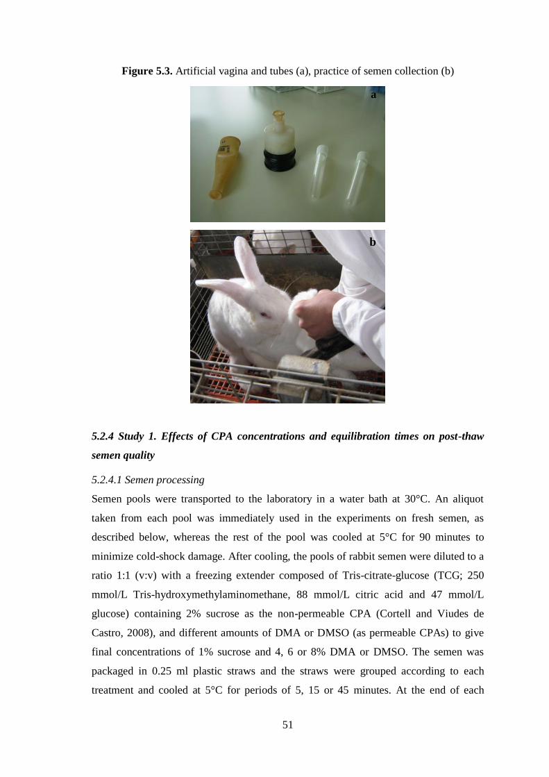
51
Figure 5.3. Artificial vagina and tubes (a), practice of semen collection (b)
5.2.4 Study 1. Effects of CPA concentrations and equilibration times on post-thaw
semen quality
5.2.4.1 Semen processing
Semen pools were transported to the laboratory in a water bath at 30°C. An aliquot
taken from each pool was immediately used in the experiments on fresh semen, as
described below, whereas the rest of the pool was cooled at 5°C for 90 minutes to
minimize cold-shock damage. After cooling, the pools of rabbit semen were diluted to a
ratio 1:1 (v:v) with a freezing extender composed of Tris-citrate-glucose (TCG; 250
mmol/L Tris-hydroxymethylaminomethane, 88 mmol/L citric acid and 47 mmol/L
glucose) containing 2% sucrose as the non-permeable CPA (Cortell and Viudes de
Castro, 2008), and different amounts of DMA or DMSO (as permeable CPAs) to give
final concentrations of 1% sucrose and 4, 6 or 8% DMA or DMSO. The semen was
packaged in 0.25 ml plastic straws and the straws were grouped according to each
treatment and cooled at 5°C for periods of 5, 15 or 45 minutes. At the end of each
a
b

52
equilibration period, the semen was frozen by exposure to liquid nitrogen vapor (5 cm
above the liquid nitrogen surface) for 10 minutes at -125/-130 °C, and the straws were
then plunged into liquid nitrogen for storage at -196°C (Figure 5.4). Sperm samples
were thawed by immersing the straws in a water bath at 50°C for 10 seconds. In total,
18 different combinations, or treatments, were assessed (two cryoprotectants × three
cryoprotectant concentrations × three equilibration times). Eight replicates of pooled
semen were used for each treatment to give a total number of 144 frozen/thawed
samples.
Figure 5.4. Different steps of semen cryopreservation process
5.2.4.2 Sperm quality
Sperm motility, viability, osmotic-resistance, acrosome and DNA integrity were
determined in duplicate in both the fresh and thawed semen samples. Moreover, sperm
concentration was evaluated on fresh semen.

53
Sperm concentration was evaluated using a Neubauer hemocytometer (Figure 5.5).
Semen was diluted 1:100 (v:v) with 3% NaCl (w:v) solution and spermatozoa were
counted in the central grid of the hemacytometer. Both chambers of the hemocytometer
were counted and averaged. Sperm concentration was expressed in million per milliliter.
Figure 5.5. Neubauer hemocytometer chamber
Rabbit sperm motility was subjectively assessed by visual estimation (Rosato and
Iaffaldano, 2011). A drop of 10 μL of semen was deposited on a clean glass slide pre-
warmed at 37°C and covered with a coverslip. The slides were then examined on a
warm-plate at 400 × total magnification using a phase-contrast microscope (Leica
Aristoplan; Leitz Wetzlar, Heidelberg, Germany, Figure 5.6). Percentages of total
motility (spermatozoa showing any type of sperm head movement) and forward
progressive motility (spermatozoa showing linear movement) in five microscopy fields.
Figure 5.6. Phase-contrast microscope (Leica Aristoplan; Leitz Wetzlar, Heidelberg,
Germany)

54
Sperm viability was measured as previously described (Iaffaldano et al., 2010) using
the LIVE/DEAD Sperm Viability Kit containing the fluorescent stains SYBR-14 and
propidium iodide (PI). This procedure was performed on 5 μL of semen, which were
added to 39 μL of extender containing 1 μL SYBR-14 (diluted 1:100 in DMSO),
incubated at 37°C for 10 minutes, and then 5 μL PI added (diluted 1:100 in the TCG
diluent) followed by incubation at 37°C for a further 5 minutes. 10 µL of this
suspension were placed on microscopic slides, covered with coverslips and examined at
1000× total magnification using an 100× oil immersion objective under epifluorescence
microscopy. About 200 spermatozoa were examined in duplicate aliquots for every
sample. SYBR-14, a membrane-permeant DNA stain, only stains live spermatozoa
producing green fluorescence of the nuclei. Propidium iodide stains the nuclei of
membrane-damaged cells red. Thus, spermatozoa showing green fluorescence are
scored as alive and those showing red fluorescence as dead (Figure 5.7). The percentage
of viable spermatozoa was calculated as: number of green cells × 100 divided by the
total number of sperm counted.
Figure 5.7. Epifluorescence photomicrographs of rabbit sperm cells stained with
SYBR-14 and PI to assess viability. In viable spermatozoa SYBR-14, a membrane-
permeant DNA stain, stains only living spermatozoa, producing bright green
fluorescence of the nuclei, whereas PI stains the nuclei of membrane damaged cells red,
so spermatozoa that exhibit green fluorescence are considered viable (V), those that
exhibit red fluorescence are considered dead (D)

55
To determine sperm osmotic resistance, a hypo-osmotic swelling test (HOST) was
used. The test was performed by mixing 10 μL of semen with 40 μL of distilled water in
an Eppendorf tube and incubating for 5 minutes at 38°C. 10 μL of the mixture was
deposited on a clean glass-slide, covered with a thin cover slip and examined under a
phase-contrast microscope. The typical sperm osmotic “coiled tail” reaction was easily
detected and the number of HOST-positive cells (Figure 5.8), was recorded by counting
spermatozoa showing this feature among 200 cells examined at a total magnification of
800 × in at least 5 microscopic fields (Rosato and Iaffaldano, 2011).
Figure 5.8. Phase-contrast photomicrographs of rabbit sperm cells exposed to the
HOST to assess the functional competence of sperm tail membrane. In HOST-positive
spermatozoa, the tails appear coiled (Coiled Tail) because the undamaged sperm
membrane permits passage of water into the cytoplasmatic space of tail membrane
causing swelling produced by a hyposmotic shock and the pressure so generated leads
to the curling of tail fibres. In damaged or chemically inactive sperm, instead, the
disrupted membrane allows water efflux without any accumulation within cell, while
tail curling doesn’t occur
Coiled
No coiled

56
To determine acrosome integrity, duplicate smears were prepared using a drop of
semen from each sample and air-dried. After fixation in methanol for 20 min, the slides
were washed with water and air-dried. After incubation with the Pisum sativum
agglutinin FITC conjugate (PSA-FITC) for 30 min at room temperature, slides were
mounted with 50% glycerol (v/v) and cover slipped (Mendoza et al., 1992). In each
sample, assessment was made of 200 sperm at a total magnification of 1000 × using an
oil immersion objective under epifluorescence illumination. This stain intensely labels
the acrosomal region of acrosome-intact sperm, which emit a uniform applegreen
fluorescence, while acrosome-damaged spermatozoa show scarce or no green
fluorescence in the anterior part of the head (Figure 5.9). The percentage of acrosome-
intact spermatozoa was calculated as a fraction of the total.
Figure 5.9. Epifluorescence photomicrographs of rabbit sperm cells stained with the
PSA–FITC to assess acrosome integrity. In the acrosome-intact spermatozoa, the
acrosomal region of the sperm head exhibits a uniform applegreen fluorescence
whereas, the acrosome damaged or reacted spermatozoa, show almost no fluorescence
over the whole head or a thin fluorescence band present only along the equatorial
segment

57
DNA integrity was evaluated according to the acridine orange (AO) test as described
by Gandini and coworkers (Gandini et al., 2006). AO is a selective metachromatic
nucleic acid stain that intercalates into double-stranded DNA (native DNA) as a
monomer and binds to single-stranded DNA (denatured DNA) as an aggregate. It can be
used to detect intact chromatin, because when excited with a 488-490 nm light source,
AO intercalated with native DNA emits green fluorescence while AO associated with
single-denatured DNA emits orange-red fluorescence. A drop of semen from each
sample was smeared on a slide, air-dried, fixed overnight in a 3:1 methanol:glacial
acetic acid solution and air dried once again. Slides were then rinsed several times in
distilled water and stained with an AO solution (0.2 mg/ml in water) in the dark. After 5
min, each smear was washed with distilled water and protected with a coverslip for their
observation using a fluorescence microscope with a 490 nm excitation light and 530
nm barrier filter. Nuclei in at least 200 spermatozoa/slide were examined and scored as
green- or yellow-orange-red fluorescing (intact DNA or damaged DNA respectively)
(Figure 5.10) and the percentage of normal and abnormal chromatin condensation
calculated.
Figure 5.10. Epifluorescence photomicrographs of rabbit sperm cells stained with the
Acridine Orange to assess DNA integrity. The spermatozoa with intact DNA exhibit
green fluorescence, whereas the spermatozoa with damaged DNA show yellow or red
fluorescence

58
5.2.5 Study 2. Comparing the in vivo performance of the DMA and DMSO protocols
Based on the results obtained in study 1, we selected the best treatment protocol for
DMSO and DMA and compared their efficacy in vivo in an artificial insemination trial.
In December 2011, 84 nulliparous and 258 multiparous (32 days post-partum) receptive
rabbit does were randomly assigned to three treatment groups of 114 does, each
consisting of 28 nulliparous does and 86 multiparous does to establish the following
experimental groups: 1) 114 does inseminated with fresh semen (Control group); 2) 114
does inseminated with semen subjected to the selected DMSO protocol (DMSO group);
and 3) 114 does inseminated with semen subjected to the selected DMA protocol. Does
were treated with cyclogonine i.m. (20 IU/doe) for estrous synchronization 48 h before
insemination. Animals in the control group received a 0.5 mL dose of fresh semen
diluted 1:10 in TCG (containing approximately 20 million sperm). For the DMA and
DMSO groups, 0.25 mL of thawed semen (containing approximately 40 million sperm)
were used to inseminate the animals within 10 min of thawing. At the time of
insemination, buserelin acetate by i.m. injection was administered to each female, to
induce ovulation (1 µg/doe).
Conception rate (number of pregnant does/number of inseminations), was determined
by abdominal palpation performed in each doe 12 days after AI. The kindling rate
(number of does giving birth/number of inseminations), total number of kids born (total
born/kindling) and the number of young born alive (live born/kindling) were determined
at parturition.
5.2.6 Statistical analysis
To compare the different treatments, we used a randomized block design in a 3 × 3
factorial arrangement (3 cryoprotectant concentrations × 3 equilibration times), with 8
replicates per treatment. Sperm variables among the treatments were compared by
ANOVA, followed by Duncan’s comparison test. A generalized linear model (GLM)
procedure was then used to determine the fixed effects of CPA concentration,
equilibration time and their interactions on the sperm quality variables. A Chi-squared
test was used to analyze the effects of treatment, physiological condition of the does and
their interaction on conception and kindling rates. The level of significance was set at
P<0.05. All statistical tests were performed using SPSS software (SPSS 15.0 for
Windows, 2006; SPSS, Chicago, Ill).

59
5.3 Results
5.3.1 Effects of CPA concentrations and equilibration times on post-thaw semen
quality
The semen quality variables determined for the fresh semen (Table 5.1), indicate the
good initial quality of the semen: motility, acrosome integrity and DNA integrity were
recorded in over 90% of the sperm population and approximately 80% showed forward
progressive movement and a plasma membrane that was viable, intact and osmotic
resistant. The average sperm concentration was 340 ± 25 × 106 sperm/mL.
The semen quality variables recorded in the DMA and DMSO groups are provided in
Tables 5.2 and 5.3 respectively. These data indicate a significant effect of the DMA
concentration used (P<0.05) on all the sperm quality variables, and a significant effect
of the equilibration time on sperm motility, viability and osmotic-resistance. No
significant interaction effect of DMA concentration and equilibration time was observed
for all the sperm quality variables except viability. Higher total and progressive sperm
motility rates were recorded in semen frozen in the presence of 6% DMA and
equilibrated for 15 or 45 min, compared to semen exposed to 6% or 4% DMA and
equilibrated for 5 min and compared to semen exposed to 8% DMA and equilibrated for
5, 15 or 45 min (P<0.05). A similar trend was observed for viability, which was higher
in sperm frozen with 6% DMA and equilibrated for 15 or 45 min, compared to 6%
DMA and an equilibration time of 5 min and to all the equilibration times using 4 and
8% DMA (P<0.05). Lower osmotic resistance values were found for the treatments 8%
DMA for 5, 15 or 45 min and 4% DMA for 5 min, compared to 6% DMA for 45 min
(P<0.05). This last treatment also yielded the highest acrosome integrity rates compared
to the use of both 4% and 8% DMA (P<0.05). A higher DNA integrity was found for
the treatments 4% DMA for 5 min and 6% DMA for 15, or 45 min compared with the
treatment 8% DMA for 5, or 15 min and 6% DMA for 5 min (P<0.05).
Following the DMSO freezing protocols, we observed a significant effect of the CPA
concentration on all the sperm quality variables examined except DNA integrity. In
addition, the time of equilibration was found to significantly affect both motility (total
and forward progressive) and osmotic resistance. Interactions between the CPAs and
equilibration time also showed significant effects on total motility, progressive motility,
viability and osmotic resistance. Best post-thaw total and forward motility rates were
recorded for semen frozen in the presence of 8% DMSO and equilibrated for 45 min,

60
with respect to all the other DMSO concentration and equilibration time combinations
tested (P<0.05). Semen frozen in the presence of 8% DMSO also showed higher
viability rates than those recorded for both 4% and 6% DMSO regardless of the
equilibration time (P<0.05). Osmotic resistance for the treatment 8% DMSO for 45 min
was similar to that observed for 8% DMSO for 5 min but better than that observed for
the remaining treatments (P<0.05). In response to 8% DMSO for 45 min, the sperm
preserved around 33% of their acrosome integrity, a significantly higher rate than the
percentages returned by the combinations 4% DMSO for 5, or 15 min and 6% DMSO
for 15 min (P<0.05). No differences in DNA integrity were observed for the different
DMSO freezing protocols. Based on these findings, we then selected the best DMA (6%
for 45 min) and DMSO protocols (8% for 45 min) and compared their in vivo
performance in the insemination trial described below.
Table 5.1. Sperm quality variables (%) (means ± SEM) recorded in freshly collected
rabbit semen (n=8)
5.3.2 Comparing the in vivo performance of the DMA and DMSO protocols
Table 5.4 provides reproductive performances recorded after the artificial insemination
of does with fresh semen or semen frozen using the optimized DMSO and DMA
protocols. After confirming no significant effects of the physiological status of the does
on any of reproductive variables tested, we pooled together the data recorded in
nulliparous and multiparous does for the same treatment. Similar conception rates,
kindling rates and prolificacy (total number born) were recorded in the DMSO and fresh
semen groups, which were higher than those recorded in the DMA group (P<0.05). In
contrast, the number of kids born alive in the DMSO group, despite being lower than in
the control (fresh semen) group, was significantly higher than the number recorded in
the DMA treatment group. In short, conception rates, kindling rates and the number of
live newborns born were influenced by treatment, whereas the total number of rabbits
born was scarcely affected by treatment or doe reproductive status. No significant
interaction effect between treatment and doe status was observed on reproductive
performances.
Total
motility
Progressive
motility
Viability Osmotic
resistance
Acrosome
integrity
DNA
integrity
93.75±0.66 77.81 ± 2.23 83.17 ± 1.44 76.18 ± 1.77 94.18 ± 0.73 97.78±0.40
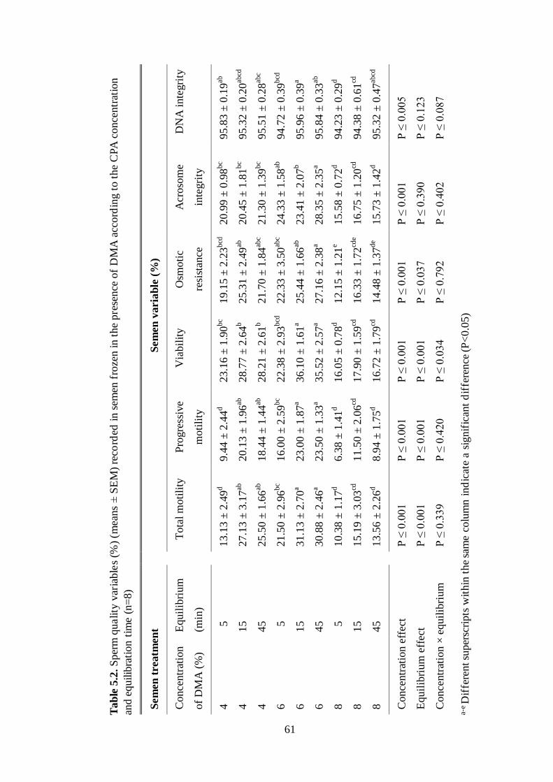
61
Tab
le5.2
.S
per
mquality
var
iable
s(%
)(m
eans
±S
EM
)re
cord
edin
sem
enfr
oze
nin
the
pre
sence
of
DM
Aac
cord
ing
toth
eC
PA
conce
ntr
atio
n
and
equilib
rati
on
tim
e(n
=8)
Sem
en
trea
tmen
t
Sem
en
va
ria
ble
(%
)
Co
nce
ntr
atio
n
of
DM
A (
%)
Eq
uil
ibri
um
(min
)
T
ota
l m
oti
lity
P
rogre
ssiv
e
mo
tili
ty
Via
bil
ity
Osm
oti
c
resi
stan
ce
Acr
oso
me
inte
gri
ty
DN
A i
nte
gri
ty
4
5
13
.13
± 2
.49
d
9.4
4 ±
2.4
4d
23
.16
± 1
.90
bc
19
.15
± 2
.23
bcd
2
0.9
9 ±
0.9
8bc
95
.83
± 0
.19
ab
4
15
2
7.1
3 ±
3.1
7ab
2
0.1
3 ±
1.9
6ab
2
8.7
7 ±
2.6
4b
25
.31
± 2
.49
ab
20
.45
± 1
.81
bc
95
.32
± 0
.20
abcd
4
45
2
5.5
0 ±
1.6
6ab
1
8.4
4 ±
1.4
4ab
2
8.2
1 ±
2.6
1b
21
.70
± 1
.84
abc
21
.30
± 1
.39
bc
95
.51
± 0
.28
abc
6
5
21
.50
± 2
.96
bc
16
.00
± 2
.59
bc
22
.38
± 2
.93
bcd
2
2.3
3 ±
3.5
0ab
c 2
4.3
3 ±
1.5
8ab
9
4.7
2 ±
0.3
9bcd
6
15
3
1.1
3 ±
2.7
0a
23
.00
± 1
.87
a 3
6.1
0 ±
1.6
1a
25
.44
± 1
.66
ab
23
.41
± 2
.07
b
95
.96
± 0
.39
a
6
45
3
0.8
8 ±
2.4
6a
23
.50
± 1
.33
a 3
5.5
2 ±
2.5
7a
27
.16
± 2
.38
a 2
8.3
5 ±
2.3
5a
95
.84
± 0
.33
ab
8
5
10
.38
± 1
.17
d
6.3
8 ±
1.4
1d
16
.05
± 0
.78
d
12
.15
± 1
.21
e 1
5.5
8 ±
0.7
2d
94
.23
± 0
.29
d
8
15
1
5.1
9 ±
3.0
3cd
1
1.5
0 ±
2.0
6cd
1
7.9
0 ±
1.5
9cd
1
6.3
3 ±
1.7
2cd
e 1
6.7
5 ±
1.2
0cd
9
4.3
8 ±
0.6
1cd
8
45
1
3.5
6 ±
2.2
6d
8.9
4 ±
1.7
5d
16
.72
± 1
.79
cd
14
.48
± 1
.37
de
15
.73
± 1
.42
d
95
.32
± 0
.47
abcd
Co
nce
ntr
atio
n e
ffec
t
P ≤
0.0
01
P
≤ 0
.00
1
P ≤
0.0
01
P
≤ 0
.00
1
P ≤
0.0
01
P
≤ 0
.00
5
Eq
uil
ibri
um
eff
ect
P
≤ 0
.00
1
P ≤
0.0
01
P
≤ 0
.00
1
P ≤
0.0
37
P
≤ 0
.39
0
P ≤
0.1
23
Co
nce
ntr
atio
n ×
eq
uil
ibri
um
P ≤
0.3
39
P
≤ 0
.42
0
P ≤
0.0
34
P
≤ 0
.79
2
P ≤
0.4
02
P
≤ 0
.08
7
a-e
Dif
fere
nt su
per
scri
pts
wit
hin
the
sam
e co
lum
n i
nd
icat
e a
signif
ican
t d
iffe
rence
(P
<0.0
5)

62
Ta
ble
5.3
.S
per
mq
ual
ity
var
iab
les
(%)
(mea
ns
±S
EM
)re
cord
edin
sem
enfr
oze
nin
the
pre
sen
ceof
DM
SO
acco
rdin
gto
the
CPA
con
cen
trat
ion
and
equ
ilib
rati
on
tim
e(n
=8)
Sem
en
trea
tmen
t
Sem
en
va
ria
ble
s (%
)
Co
nce
ntr
atio
n
of
DM
SO
(%
)
Eq
uil
ibri
um
(min
)
T
ota
l m
oti
lity
P
rogre
ssiv
e
mo
tili
ty
Via
bil
ity
Osm
oti
c
resi
stan
ce
Acr
oso
me
inte
gri
ty
DN
A i
nte
gri
ty
4
5
17
.81
± 1
.22
d
13
.25
± 1
.30
c 2
9.6
7 ±
1.2
7cd
24
.60
± 0
.84
f
26
.91
± 1
.34
bc
95
.89
± 0
.64
4
15
2
0.7
5 ±
1.2
2cd
1
5.4
4 ±
1.2
1c
3
1.3
1 ±
1.7
5cd
27
.03
± 0
.97
ef
2
6.1
8 ±
1.3
5c
96
.37
± 0
.22
4
45
3
2.0
6 ±
3.0
1b
25
.25
± 2
.81
b
38
.89
± 2
.08
b
31
.34
± 1
.47
cd
30
.50
± 1
.98
abc
9
6.1
5 ±
0.4
1
6
5
24
.19
± 1
.47
c
18
.13
± 1
.78
c
33
.31
± 1
.05
c
27
.50
± 1
.62
ef
2
8.7
7 ±
3.0
5ab
c
96
.07
± 0
.34
6
15
2
2.4
4 ±
1.5
9cd
1
6.1
3 ±
1.3
3c
3
2.5
2 ±
1.2
7cd
28
.29
± 1
.59
de
25
.79
± 1
.62
c 9
6.1
2 ±
0.4
7
6
45
1
8.3
8 ±
0.6
6d
13
.13
± 0
.69
c
28
.15
± 0
.85
d
26
.57
± 0
.45
ef
2
9.5
1 ±
2.0
8ab
c 9
6.0
9 ±
0.4
5
8
5
34
.69
± 1
.91
b
2
8.0
0 ±
2.0
3b
44
.00
± 1
.45
a
36
.55
± 1
.32
ab
3
2.5
8 ±
1.9
4ab
9
6.2
5 ±
0.3
0
8
15
3
5.1
3 ±
1.1
2b
2
8.3
8 ±
1.2
8b
42
.98
± 1
.03
a
33
.90
± 0
.76
bc
3
1.0
4 ±
1.0
5ab
c 9
6.9
6 ±
0.4
4
8
45
4
2.6
3 ±
2.0
7a
35
.38
± 1
.87
a 4
7.0
9 ±
1.7
8a
3
8.8
5 ±
0.7
5a
33
.32
± 1
.09
a 9
7.0
2 ±
0.3
4
Co
nce
ntr
atio
n e
ffec
t
P ≤
0.0
01
P
≤ 0
.00
1
P ≤
0.0
01
P
≤ 0
.00
1
P ≤
0.0
05
P
≤ 0
.11
2
Eq
uil
ibri
um
eff
ect
P
≤ 0
.00
1
P ≤
0.0
01
P
≤ 0
.06
9
P ≤
0.0
09
P
≤ 0
.07
7
P ≤
0.4
31
Co
nce
ntr
atio
n ×
eq
uil
ibri
um
P ≤
0.0
01
P
≤ 0
.00
1
P ≤
0.0
01
P
≤ 0
.00
4
P ≤
0.9
05
P
≤ 0
.89
6
a-f D
iffe
ren
t su
per
scri
pts
wit
hin
th
e sa
me
colu
mn
in
dic
ate
a si
gn
ific
ant
dif
fere
nce
(P
<0.0
5)

63
Tab
le5
.3.S
per
mquality
vari
able
s(%
)(m
eans
±S
EM
)re
cord
edin
sem
enfr
oze
nin
the
pre
sence
of
DM
SO
acco
rdin
gto
the
CPA
conce
ntr
atio
n
and
equilib
rati
on
tim
e(n
=8
)
S
em
en
treatm
en
t T
reat
men
t
effe
ct
Does
sta
tus
effe
ct
Tre
atm
ent
×
does
sta
tus
F
resh
Sem
en
DM
SO
D
MA
% C
once
pti
on r
ate
(n)
81.6
a (9
3)
79.8
a (9
1)
47.4
b (5
4)
P ≤
0.0
01
P ≤
0.6
75
P ≤
0.7
78
% K
indli
ng r
ate
(n)
81.6
a (9
3)
77.2
a (8
8)
47.4
b (5
4)
P ≤
0.0
01
P ≤
0.6
25
P ≤
0.8
37
Tota
l born
(m
ean ±
SE
M)
8.6
± 0
.3a
7.7
± 0
.3a
6.7
± 0
.4b
P ≤
0.0
59
P ≤
0.0
70
P ≤
0.3
43
Liv
e born
(m
ean
± S
EM
) 8.3
± 0
.3a
7.2
± 0
.3b
5.9
± 0
.4c
P ≤
0.0
06
P ≤
0.0
55
P ≤
0.6
19
a-
cV
alues
wit
h d
iffe
rent su
per
scri
pts
wit
hin
tre
atm
ents
in t
he
sam
e ro
w d
iffe
r si
gnif
ican
tly (
P<
0.0
5)

64
5.4 Discussion
The results of this research, indicate that the concentration of the CPA used and the
equilibration time clearly affected the post-thaw quality of rabbit semen. Thus, a DMA
concentration of 6% or a DMSO concentration of 8% gave rise to an overall high post-
thaw semen quality compared to the other concentrations tested, and using both CPAs,
the longer equilibration time tested (45 min) resulted in efficient sperm protection. Both
DMA and DMSO are penetrating CPAs. Such CPAs are membrane-permeable solutes
that act intra- and extracellularly, causing the dehydration of spermatozoa due to an
osmotically driven flow of water, which varies according to CPA composition (Purdy,
2006). Spermatozoa are normally equilibrated in a permeable CPA so that the sperm are
penetrated by the agent before freezing. After a variable period of time, the CPA and
water equilibrate to reach similar intracellular and extracellular concentrations (Amann,
1999). As the sperm cell has less intracellular water, the freezing point of the cell is
decreased such that intracellular ice formation is reduced along with cell damage and
cell death, and, in turn, the fertility of the semen sample is better preserved (Purdy,
2006). Penetrating CPAs also cause membrane lipid and protein reorganization. This
improves membrane fluidity causing greater dehydration at lower temperatures, and
thus an increased ability to survive cryopreservation (Holt, 2000). Being solvents,
penetrating CPAs dissolve also sugars and salts in the cryopreservation medium (Purdy,
2006). The CPA concentration seems also to define its specific mode of action on lipid
membranes. In an in vitro study of the effects of DMSO on lipid membranes,
Gurtovenko and Anwar (2007) observed that this CPA showed different modes of
action depending on its concentration. Thus, at low concentrations, it induced
membrane thinning and increased the fluidity of the membrane’s hydrophobic core; at
higher concentrations, it induced transient formation of water pores in the membrane
and at still higher concentrations, individual lipid molecules were desorbed from the
membrane followed by disintegration of the bilayer structure. Accordingly, the optimal
final concentration (v/v) of a penetrating CPA in a medium will vary, because the
toxicity of the chemical has to be balanced against its beneficial cryoprotective effect on
the spermatozoa. Although there is no consensus as for the lesser or greater toxicity of
penetrating CPAs for rabbit spermatozoa (Mocé and Vicente, 2009), it is generally
accepted that the toxicity of penetrating CPAs increases with its concentration, exposure
time and temperature (Holt, 2000). However, a certain period of time is required for a

65
CPA to permeate the sperm cells and for equilibrium to be reached between intracellular
and extracellular environments (Fuller et al., 2004). Thus, the optimal equilibration time
depends on the CPA concentration in the freezing medium, its characteristics (i.e.
molecular weight, affinity of chemical groups towards the sperm membrane), the
temperature of the sperm when the CPA is added, as well as the structural composition
of the sperm membrane, which influences sperm permeability and fluidity (Fuller et al.,
2004). In effect, water and solutes move across the plasma membrane, and rates of
movement are determined by membrane structure and composition. When the sperm
membrane permeability is considered high, the equilibration period is kept as short as
possible to avoid the toxic effects of permeable CPAs (Fuller et al., 2004). Because
rabbit spermatozoa show a high cholesterol:phospholipid ratio and a low ratio of
polyunsaturated to saturated fatty acids in phospholipids (Gliozzi et al., 2009), they
have intermediate fluidity and permeability (Curry et al., 1995; Mocé and Vicente,
2009). Consequently, if the equilibration period before freezing is too short, water has
little time to move out of the spermatozoon and hence large intracellular ice crystals
form, causing physical damage to cell membranes and other intracellular components.
This rationale is consistent with our observation of low sperm cryosurvival when semen
samples were equilibrated for 5 min in DMA or DMSO compared to 45 min. Hence,
just a few minutes of equilibration seem insufficient for complete equilibrium to be
reached between the CPA and water in the rabbit sperm.
When we compared the performance of these two CPAs in vivo, DMSO resulted more
effective than DMA. In fact, semen preserved and frozen in the presence of DMSO,
showed a fertility potential similar to that of fresh semen, whereas lower fertility and
prolificacy rates were recorded for the DMA frozen semen.
DMSO, alone or in combination with egg yolk or with non-permeable CPAs, has been
widely used in experimental protocols for freezing rabbit sperm, most often at
concentrations in the ranges 1-5% or 10-17% (Mocé and Vicente, 2009). In our study,
we tested intermediate concentrations of CPA relative to those used in previous studies
on the cryopreservation of rabbit semen, in combination with 1% sucrose as a non-
permeable CPA. The fertility and prolificacy rates obtained in our study for rabbit
semen exposed for 45 minutes to 8% DMSO before freezing, were similar to the rates
we recorded for the use of fresh semen and higher than the rates reported in studies in
which lower or higher concentrations of DMSO were used (Mocé and Vicente, 2009).

66
Only a few reports exist of a similar prolificacy related to the use of fresh and frozen
sperm (Theau-Clément, 1996; Vicente and Viudes-de-Castro, 1996), and fertility and
prolificacy rates comparable to those recorded here (80% fertility and close to 8
newborn rabbits per doe), have been reported for semen frozen with DMSO as the CPA
(Theau-Clément, 1996; Vicente and Viudes de Castro, 1996; Cortell and Viudes de
Castro, 2008). Effectively, Theau-Clément and coworkers (1996) reported a fertility of
69 to 90% and a prolificacy of 8.9 to 10.3, and Vicente and Viudes de Castro (1996)
reported 91% fertility and 8.9 normal embryos, whereas the fertility and prolificacy data
obtained by Cortell and Viudes de Castro (2008) were respectively 81 to 86% and 7.9 to
8.5. Our fertility and prolificacy results for DMSO were even better than those related
to the use of lactamide or acetamide for freezing rabbit sperm (Hanada and Nagase,
1980; Parrish and Foote, 1986; Chen and Foote, 1994; Arriola and Foote, 2001; Liu et
al., 2007; Okuda et al., 2007). In studies addressing the in vitro post-thaw quality of
rabbit semen, amides have been considered superior to DMSO or glycerol (Dalimata
and Graham, 1997; Kashiwazaki et al., 2006). Among the CPAs featuring amide
groups, DMA has never been investigated for the cryopreservation of rabbit semen,
despite being considered effective for freezing sperm in other species (Tselutin et al.,
1999; Morris et al., 2003; Squires et al., 2004; Bianchi et al., 2008; Zee et al., 2008).
Remarkably, we observed that DMA was no better than DMSO for freezing rabbit
semen. Although we are unaware of the reason why the rabbit sperm cells were able to
better withstand DMSO than DMA during the cryopreservation process, several
possible explanations may be proposed. DMA and DMSO have many common
physical-chemical properties: both have two hydrophobic methyl groups but can create
three hydrogen bonds with water, suggesting their capacity for a similar cryoprotective
mechanism of action. However, these compounds have a different molecular weight
(DMA 87.12 and DMSO 78.13 gmol-1
) and DMA bears amide groups whereas DMSO
has a hydrophilic sulfoxide group. These properties are likely to confer to the
compounds a different permeability in a given phospholipid bilayer. In turn, this might
lead to variations in the relative permeability of these CPAs in the rabbit sperm
membrane and thus in their relative cryoprotection efficiency.
Another interesting point emerging from our study was that the physiological status of
the rabbit doe (nulliparous versus multiparous) had no effect on reproductive
performance. This finding is consistent with the results of studies that have shown that
nulliparous rabbit does generally exhibit a higher reproductive efficiency than

67
multiparous females, when subjected to an intensive reproductive rhythm, and lower or
similar efficiency when under a semi-intensive or extensive reproductive rhythm
(Theau-Clément, 2007; Rebollar et al., 2009; Dimitrova et al., 2009). Hence, under our
extensive reproductive rhythm (insemination 32 days after parturition), we obtained
similar fertility and prolificacy outcomes in multiparous and nulliparous does.
hence, our results indicate that the cryosurvival of rabbit sperm frozen in the presence of
the CPAs DMSO or DMA, is determined by their concentration and equilibrium time.
According to our in vivo results, DMSO seems more effective than DMA for the
cryopreservation of rabbit sperm. The DMSO freezing protocol optimized here gave
rise to a reproductive efficacy of the semen comparable to that of fresh semen. In
further studies, the satisfactory results obtained for DMSO and DMA in intensive rabbit
rearing systems, will need to be confirmed and further optimization work should seek to
address other stages of the freezing process and other CPAs. Indeed, if the efficacy of
rabbit semen cryopreservation is improved, rabbit breeding facilities could start to take
advantage of the opportunities offered by the extensive use of doses of frozen semen.

68
Chapter 6
Research n°2
Cryopreservation of rabbit semen using non-permeable cryoprotectants:
effectiveness of different concentrations of low-density lipoproteins (LDL) from
egg yolk versus egg yolk or sucrose
http://dx.doi.org/10.1016/j.anireprosci.2014.10.020
6.1 Aim
As it has been previously reported, the factors that may affect the success of
cryopreservation assessed in the different studies, have been the composition of the
freezing medium, the CPA and its concentration, the freezing conditions and cooling
and warming temperatures, as well as individual animal variation (Mocé and Vicente,
2009). The choice of CPA plays an important role to obtain an effective rabbit semen
freezing protocol. CPAs that permeate in the sperm cell increase membrane fluidity and
partially dehydrate the cell, lowering the freezing point and thus reducing the formation
of intracellular ice crystals, which cause physical and chemical stress (Purdy, 2006).
However, permeable CPAs themselves can have a toxic effect on sperm causing
membrane destabilization and protein and enzyme denaturation. This toxicity is directly
related to the concentration used and the time of cell exposure (Swain and Smith, 2010).
The addition of non-permeable CPAs to the freezing medium serves to offset the
cryodamage caused by permeating CPAs. At similar concentrations, these substances
are less toxic than permeable CPAs and they have multiple protective roles such as
inhibiting ice crystal growth and helping the sperm to stabilize internal solute
concentrations under osmotic stress. This reduces the amount of penetrating CPAs
needed (Swain and Smith, 2010).
According to these observations, current research interests, lie in addressing the use of
non-permeable CPAs in combination with permeable CPAs in freezing protocols. Non-
penetrating CPA are generally large molecules such as polymers, sugars, proteins or
amino acids. Among the disaccharides used as non-permeable CPAs, sucrose has shown
a better cryoprotective effect (Mocé and Vicente, 2009). In the first research and in our
previous study (Rosato and Iaffaldano, 2013), we developed an effective freezing

69
protocol for rabbit semen based on the use of DMSO in the freezing extender as the
permeable CPA in combination with sucrose.
Egg yolk has also been used as a non-permeable CPA since it contains large molecules
(Holt, 2000). Egg yolk is widely used in semen freezing extenders and has proved
effective for protecting mammalian spermatozoa against cold shock during the freeze-
thaw process. For the cryopreservation of rabbit semen, egg yolk is usually added to the
freezing extender at a concentration of 10-20% (Mocé and Vicente, 2009). However, its
protective action is largely thought to be due to low-density lipoproteins (LDL), as the
main constituents of egg yolk (2/3 of dry yolk matter). Many investigations have
confirmed these findings in bull (Hu et al., 2011), boar (Hu et al., 2006), dog (Bencharif
et al., 2008), buffalo (Akhter et al., 2011), ram (Moustacas et al., 2011) and horse
(Moreno et al., 2013). However, to the best of our knowledge the use of LDL as a non-
permeable CPA, has not yet been tested in the rabbit. The present study was designed
by two experiments: in the first, we assessed the effects of different egg yolk LDL
concentrations on post-thaw rabbit sperm quality in relation to the effects of whole egg
yolk or sucrose; and in the second experiment, the behavior of the most effective non-
permeable CPAs identified, was compared by determining fertility and prolificacy rates
in vivo.
6.2 Materials and methods
6.2.1 Chemicals
The LIVE/DEAD Sperm Viability Kit was purchased from Molecular Probes, Inc.
(Eugene, OR, USA). Acridine orange (AO), Pisum sativum agglutinin fluorescein
isothiocyanate (FITC) conjugate (FITC-PSA) and all other chemicals used in this study
were purchased from Sigma, Chemical Co. (Milan, Italy).
6.2.2 Animals
The animals used for this study were 32 adult rabbit bucks and 90 does of Bianca
Italiana breed (Figure 6.1) from the central breeding farm of the Italian Rabbit Breeders
Association (ANCI-AIA, Volturara Appula (FG), Italy; Figure 6.2). Rabbits were
housed in individual flat-deck cages, subjected to a 16 h light/8 h dark photoperiod and
were fed a commercial standard diet. Water was given ad libitum.
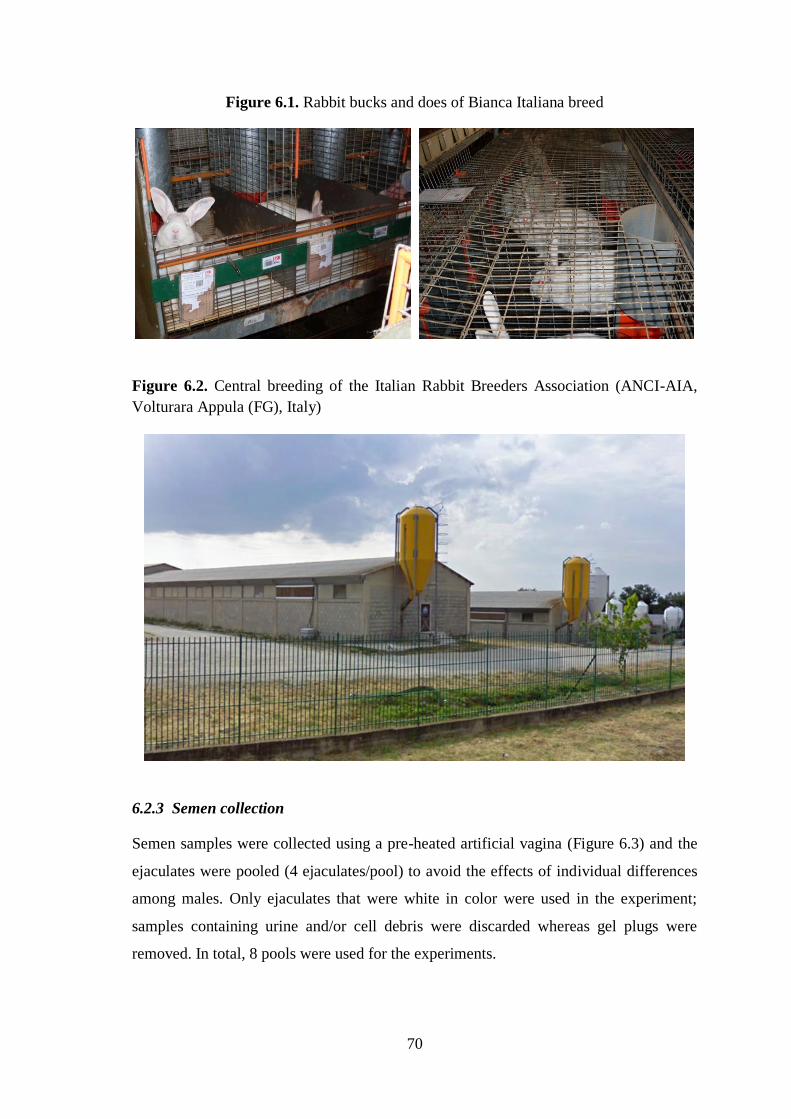
70
Figure 6.1. Rabbit bucks and does of Bianca Italiana breed
Figure 6.2. Central breeding of the Italian Rabbit Breeders Association (ANCI-AIA,
Volturara Appula (FG), Italy)
6.2.3 Semen collection
Semen samples were collected using a pre-heated artificial vagina (Figure 6.3) and the
ejaculates were pooled (4 ejaculates/pool) to avoid the effects of individual differences
among males. Only ejaculates that were white in color were used in the experiment;
samples containing urine and/or cell debris were discarded whereas gel plugs were
removed. In total, 8 pools were used for the experiments.

71
Figure 6.3. Artificial vagina with temperature maintaining
6.2.4 Experiment 1. Effects of different LDL concentrations, egg yolk and sucrose on
post-thaw semen quality
6.2.4.1 LDL extraction
LDLs were extracted from egg yolk using the method developed by Moussa et al.
(2002) providing a purity of 97%. Eggs were manually broken and yolks separated from
the albumen. Each yolk was carefully rolled on a filter paper to remove chalazae and
traces of albumen adhering to the vitellin membrane.
The vitellin membrane was then disrupted with a scalpel blade and the yolk collected in
a beaker, which was cooled in iced water. The egg yolk was diluted twice (w/w) in an
isotonic saline solution (0.17 M NaCl) and stirred for 1 h at 4°C. After stirring, the
solution was centrifuged at 10,000 × g for 45 min at 4°C and the supernatant (plasma)
separated from the sediment (granules). The supernatant was centrifuged again under
the same conditions to remove all traces of sediment. The plasma was then mixed with
40% ammonium sulfate to precipitate livetins. After 1 h of stirring at 4°C, the mixture
was centrifuged at 10,000 × g for 45 min at 4°C to separate the supernatant from
sediment. The precipitate was discarded and the supernatant dialyzed with distilled
water for 12 h at 4°C in order to eliminate ammonium sulfate. After complete
ammonium sulfate elimination, the solution was centrifuged again (10,000 × g for 45
min at 4°C) and the floating residue, rich in LDL, was collected.

72
6.2.4.2 Extender preparation
A basic freezing extender prepared at our laboratory was used. This extender is
composed of TCG containing 16% DMSO (v:v) as the permeable CPA and antibiotics
(25 mg of gentamicin and 50,000 IU of penicillin/100 mL). To this extender we added
the non-permeable CPAs at final concentrations of 6, 8, 10 or 15% (w:v) LDL, 15%
whole egg yolk (w:v), or 0.1 M sucrose to yield six different freezing extenders.
6.2.4.3 Semen processing
Pools were transported from the farm to the laboratory in a water bath at 30°C. An
aliquot taken from each pool was instantly used for the evaluation of fresh semen
quality, as described below, and the remaining pooled semen was cooled at 5°C for 90
min before freezing to minimize cold-shock damage.
After the cooling phase, each pool was splitted into six equal aliquots, and each of them
was diluted to a ratio 1:1 (v:v) with the six different freezing extenders.
The diluted semen was aspirated into 0.25 mL plastic straws. The straws were grouped
according to each treatment and equilibrated at 5°C for 45 min (equilibration time).
Semen was frozen by exposure to liquid nitrogen vapor 5 cm above the liquid nitrogen
surface (temperature was approximately -125/-130 °C) for 10 min. Next the straws were
plunged into liquid nitrogen for storage at -196°C. Sperm samples were thawed by
immersing the straws in a water bath at 50°C for 10 seconds.
6.2.4.4 Sperm quality
In both the fresh and thawed semen samples, sperm motility, viability, osmotic
resistance, acrosome integrity and DNA integrity were determined as described in the
previous research. Moreover, sperm concentration was evaluated on fresh semen.
6.2.5 Experiment 2. Comparing the in vivo reproductive performance of rabbit does
inseminated with semen cryopreserved using the most effective CPAs identified in
Experiment 1
In this experiment, we compared the efficacy of the non-permeable CPAs showing best
performance in Experiment 1 (10% LDL and sucrose) in an artificial insemination trial.
In January 2014, 90 multiparous (31 days postpartum) receptive rabbit does were
randomly assigned to three treatment groups: (1) 30 does inseminated with fresh semen

73
(control group); (2) 30 does inseminated with semen frozen using 0.1 M sucrose as the
CPA; and (3), 30 does inseminated with semen frozen using 10% LDL as the CPA.
For estrus synchronization, all does were subjected to the following biostimulation
protocol: flushing (3 days before insemination), changing cage (3 days before
insemination), and increasing the photoperiod from 16 to 24 hours of light (2 days
before insemination). Animals in the control group received a 0.5 mL dose of fresh
semen diluted 1:10 (approximately 35 million sperm) in a commercial extender. For the
LDL and sucrose treatment groups, 0.25 mL of thawed semen (approximately 75
million sperm) were used to inseminate the animals within 10 min of thawing. At the
time of insemination, each female received an intramuscular injection of buserelin
acetate to induce ovulation (1 µg/doe).
Conception rate, kindling rate, total number of kids born and number of kids born alive
were the reproductive performances considered.
Conception rate (number of pregnant does/number of inseminations) was determined by
abdominal palpation performed in each doe 17 days after artificial insemination. At
parturition, kindling rate (number of does giving birth/number of inseminations), total
born (total born/kindling), and the number of kids born alive (total live-born/kindling)
were determined.
Figure 6.4. Artificial insemination of rabbit does

74
6.2.6 Statistical analysis
Sperm quality (motility, viability, sperm membrane integrity, acrosome and DNA
integrity), and reproductive performances (conception rate, kindling rate, total born and
the number of kids born alive) were compared among treatments by ANOVA, followed
by Duncan’s comparison test. All statistical tests were performed using the software
package SPSS (SPSS 15.0 for Windows, 2006; SPSS, Chicago, IL, USA). Significance
was set at P<0.05.
6.3 Results
6.3.1 Effects of different LDL concentrations, egg yolk and sucrose on post-thaw
semen quality
The semen quality variables recorded for the fresh semen (Table 6.1) indicated its good
initial quality. Over 85% of the sperm population showed motility, viability, and
acrosome and DNA integrity, and approximately 75% featured forward progressive
movement and plasma membrane integrity. The average sperm concentration was 610 ±
22 × 106 sperm/mL.
The effects of different concentrations of LDL, and of sucrose and egg yolk on the
motility of the frozen-thawed rabbit sperm are provided in Figure 6.5 A significant
effect of the LDL concentration was observed on sperm motility, that the highest
motility was recorded when the semen was frozen with 10% LDL compared with the
other concentrations or egg yolk (P<0.05). The lowest rate of motile sperm was
observed for the 15% LDL. However, sperm motility recorded for the semen frozen in
the presence of 0.1 M sucrose was significantly higher versus each of the LDL
concentrations or egg yolk (P<0.05).
No significant differences in forward progressive motility were detected for the use of
10% LDL or sucrose; both CPAs being able to better protect the semen from
cryodamage in comparison with the egg yolk or other LDL concentrations (P<0.05)
(Figure 6.6).
The use of 10% LDL also led to improved sperm viability compared with the other LDL
concentrations (Figure 6.7). However, once again the cryopreservation of semen in the
presence of sucrose rendered more viable sperm than the other treatments considered
(P<0.05), with similar viability values recorded for egg yolk and 10% LDL.

75
When sperm membrane integrity percentages were compared, 10% LDL returned
higher values than the remaining concentrations (P<0.05) (Figure 6.8). These values
were similar to those recorded for the use of sucrose as the CPA.
We also observed greater percentages of acrosome-intact spermatozoa for 10% LDL
compared to the remaining treatments, though differences were only significant for egg
yolk, 6% LDL and 15% LDL (P<0.05) (Figure 6.9). No differences in DNA integrity
were detected among the different treatments tested (Figure 6.10).
Based on these findings, 10% LDL and sucrose were used in the in vivo insemination
trial as the most effective CPAs for freezing rabbit semen.
Table 6.1. Sperm quality variables (%) (means ± SEM) recorded in freshly collected
rabbit semen (n = 8)
Total
motility
Progressive
motility
Viability Osmotic
resistance
Acrosome
integrity
DNA
integrity
85.06±1.27 73.31 ± 1.13 85.64 ± 0.85 74.98 ± 1.29 87.81± 0.95 98.72 ± 0.11

76
Figure 6.5. Total motility of rabbit semen after freezing-thawing in the presence of
different LDL concentrations, sucrose or whole egg yolk. Bars represent means ± SEM.
Different lowercase letters on bars indicate a significant difference (P<0.05)
Figure 6.6. Forward progressive motility of rabbit semen after freezing-thawing in the
presence of different LDL concentrations, sucrose or whole egg yolk. Bars represent
means ± SEM. Different lowercase letters on bars indicate a significant difference
(P<0.05)
d
c
b
d
a
c
0
10
20
30
40
50
To
tal s
per
m m
oti
lity
(%
)
Treatment
c
b
a
c
a
b
0
10
20
30
40
50
Fo
rwa
rd p
rog
ress
ive
mo
tili
ty (%
)
Treatment

77
Figure 6.7. Viability of rabbit semen after freezing-thawing in the presence of different
LDL concentrations, sucrose or whole egg yolk. Bars represent means ± SEM. Different
lowercase letters on bars indicate a significant difference (P<0.05)
Figure 6.8. Sperm membrane integrity of rabbit semen after freezing-thawing in the
presence of different LDL concentrations, sucrose or whole egg yolk. Bars represent
means ± SEM. Different lowercase letters on bars indicate a significant difference
(P<0.05)
d
c
b
e
a
b
0
10
20
30
40
50
Sp
erm
via
bil
ity
(%
)
Treatment
cd cd
ab
d
a
bc
0
10
20
30
40
50
Mem
bra
ne
inta
ct s
per
m (
%)
Treatment

78
Figure 6.9. Acrosome integrity of rabbit semen after freezing-thawing in the presence
of different LDL concentrations, sucrose or whole egg yolk. Bars represent means ±
SEM. Different lowercase letters on bars indicate a significant difference (P<0.05)
Figure 6.10. DNA integrity of rabbit semen after freezing-thawing in the presence of
different LDL concentrations, sucrose or whole egg yolk. Bars represent means ± SEM.
Different lowercase letters on bars indicate a significant difference (P<0.05)
d
ab
a
cd
ab
bc
0
10
20
30
40
50
Sp
erm
acr
oso
me
inte
gri
ty (%
)
Treatment
a
a a
aa a
94
96
98
100
DN
A in
tact
sp
erm
(%
)
Treatment

79
6.3.2 Comparing the in vivo reproductive performance of rabbit does inseminated
with 10% LDL versus sucrose
Table 6.2 provides the reproductive performances recorded after the artificial
insemination of does with fresh semen or semen exposed to 10% LDL or sucrose during
freezing. Conception rates were significantly better when semen was cryopreserved in
the presence of sucrose (86.7%) than LDL (66.7%) and no significant differences were
found with respect to fresh semen (93.3%). In addition, kindling rates were higher for
the use of sucrose (80%) than LDL (60%), though differences were not significant and
neither did they differ compared to the use of fresh semen (86.7%).
The total number of kids born was higher for does in the control group (10.1) than the
LDL group (8.2) (P<0.05), with no significant differences detected between the control
and sucrose groups (9.2).
No significant differences emerged in the number of kids born alive when we compared
the use of fresh and frozen semen, although the lower number was recorded for LDL.
Table 6.2. Reproductive performances outcomes recorded in rabbit does after
insemination with fresh semen or semen frozen in sucrose or 10% LDL as the non-
permeable CPA
Values with different superscript letters within treatments on the same row are
significantly different (P<0.05)
Reproductive performances Semen treatment
Fresh semen Sucrose 10% LDL
% Conception rate (n) 93.3a (28) 86.7
a (26) 66.7
b (20)
% Kindling rate (n) 86.7a (26) 80
ab (24) 60
b (18)
Total born (mean ± SEM) 10.1 ± 0.5a
9.2 ± 0.5
ab 8.2 ± 0.6
b
Live born (mean ± SEM) 9 ± 0.6a
8.2 ± 0.6a
7.7 ± 0.5a

80
6.4 Discussion
This study sought to identify the most effective non-permeable CPA for the
cryopreservation of rabbit semen. To date, there are no reports on the use of LDL, as a
non-permeable CPA for this specific purpose. Our results clearly revealed a significant
effect of LDL concentration on the quality of the cryopreserved semen and the 10%
concentration was better able to protect the sperm cell from cryodamage compared with
the other LDL concentrations tested or with egg yolk. In effect, egg yolk has been
widely used in semen freezing extenders and proven to offer resistance against cold
shock during the sperm freeze-thaw process in several animal species (Holt, 2000;
Moussa et al., 2002; Mocé and Vicente, 2009). However, the use of egg yolk as a CPA
has several drawbacks besides being a source of bacterial contamination (Moreno et al.,
2013) and some of its constituents could have detrimental effects on spermatozoa.
Accordingly, the granules found in egg yolk were observed to reduce the respiration and
motility of bull spermatozoa (Amirat-Briand et al., 2004), and progesterone in egg yolk
has been described as potentially responsible for the capacitation of spermatozoa and
thus harmful for the preservation of spermatozoa during freezing (Bowden et al., 2001;
Moreno et al., 2013).
The presence of these detrimental substances in yolk could explain why in vitro post-
thaw sperm quality was not as good as that recorded for the other CPAs. This
determines that outcomes can be improved by adding extracted LDL to the extender
rather than whole egg yolk. Our results are consistent with those obtained in bull
(Moussa et al., 2002; Vera-Munoz et al., 2009; Amirat-Briand et al., 2004, 2010; Hu et
al., 2010, 2011), boar (Hu et al., 2006, 2008; Jiang et al., 2007), dog (Bencharif et al.,
2008, 2010), buffalo (Akhter et al., 2011), ram (Moustacas et al., 2011) and horse
(Moreno et al., 2013). Low-density lipoproteins (0.982 g/mL) appear in the soluble
fraction of egg yolk called plasma. They are spherical molecules of about 35 nm in
diameter and consist of a lipid core of triglycerides and cholesterol esters surrounded by
a layer of phospholipid and apoprotein film (Moussa et al., 2002) giving them a
composition of about 87% lipids, 12% proteins (Anton et al., 2003). The lipid
component of LDL comprises some 69% triglycerides, 26% phospholipids and 5%
cholesterol (Anton et al., 1997).
The precise mechanism whereby LDL protects spermatozoa against freeze-thaw
damage has not yet been clearly established. It is known that the cryopreservation

81
process affects the plasma membrane’s chemical composition and lipid organization
(Amann and Pickett, 1987), LDL could directly or indirectly limit these sperm
modifications protecting the spermatozoa (Manjunath et al., 2002; Moussa et al., 2002;
Bergeron et al., 2004). This idea is consistent with our observation of the beneficial
effects of 10% LDL as opposed to other concentrations on semen quality characteristics,
especially acrosome integrity. Several hypotheses have been used to explain the
mechanism of action of LDL.
Previous researches suggested that phospholipids released in the extender, following the
disruption of LDL during the freeze-thawing process, could form a gel-like protective
film on the spermatozoa, which protects the lipid–protein complex of cell membranes
and thereby safeguards the spermatozoa (Quinn and Chow, 1980; Hu et al., 2011).
Other authors have proposed that phospholipids from LDL could replace some of the
sperm membrane’s phospholipids, thereby decreasing their phase transition temperature
and thus reducing the formation of ice crystals (Graham and Foote, 1987; Trimeche et
al., 1996; Moussa et al., 2002).
Manjunath et al. (2002) and Bergeron et al. (2004) hypothesized that the main
mechanism whereby LDL was able to protect bull spermatozoa was via the
sequestration of BSP proteins in seminal plasma, considering that the major proteins of
bull seminal plasma (BSP-A1/A2, BSPA3 and BSP-30-kDa) bind to the sperm surface
at ejaculation, triggering cholesterol and phospholipids efflux from the sperm
membrane. This hypothesis was later proposed by Vera-Munoz et al. (2009) to explain
the beneficial effects of LDL on bovine sperm. Another interesting point emerging from
our study was the significant effect of LDL concentration. Thus, the concentration of
LDL that offered the best protection of 10% (w/v) is consistent with the findings of
Akhter et al. (2011) in buffalo, while concentrations of 9% and 8% respectively have
been reported for boar (Hu et al., 2006) and bull (Moussa et al., 2002; Hu et al., 2011).
The optimal LDL concentration therefore seems to be related to the animal species.
When the LDL concentration was increased from 6% to 10%, an improvement was
noted in rabbit sperm cryosurvival. In contrast, the use of 15% LDL led to reduced
postthaw sperm quality. We attribute this finding to a drop in the osmotic pressure of
the extender when the LDL concentration is increased. Moussa et al. (2002) attributed a
decline in bull spermatozoa performance after freeze-thawing in the presence of an LDL
concentration above 10% to the precipitation of fructose and salts in the extender or to
LDL aggregation with consequent neutralization of its cryoprotective effect.

82
Another finding of our study was that sucrose improved the in vitro cryosurvival of
rabbit semen over the other treatments, although when compared with the 10% LDL
treatment, significantly improved values were only obtained for total motility and
viability. However, in vivo sucrose as a non-permeable CPA was better at preserving the
fertilization potential of the cryopreserved rabbit sperm than 10% LDL. In fact, semen
preserved and frozen in the presence of sucrose showed a fertility potential similar to
that of fresh semen, whereas lower fertility and prolificacy rates were recorded when
semen was exposed to 10% LDL during freezing. This is consistent with the lower total
motility and sperm viability observed in vitro for the use of LDL. Some authors have
addressed the relationship between rabbit semen characteristics and fertility with the
general objective of predicting fertility from semen traits (Brun et al., 2002).
The fertility and prolificacy rates obtained in our study using sucrose as a non-
permeable CPA are similar to those observed in the first research and in our previous
work (Rosato and Iaffaldano, 2013). Only a few reports exist of similar fertility and
prolificacy rates related to the use of fresh and frozen sperm in the rabbit (Theau-
Clément et al., 1996; Vicente and Viudes de Castro, 1996).
This finding is important since it confirms the effectiveness of sucrose as a non-
permeable CPA for the cryopreservation of rabbit spermatozoa. This good performance
of sucrose can be attributed to its ability to dehydrate cells at high subfreezing
temperatures, thereby inhibiting ice crystal growth and allowing for rapid cooling
before intracellular solute concentrations reach critical levels. In addition, sucrose has a
high glass transition temperature compared with conventional permeable CPAs,
allowing the long-term storage of cells at high subzero and even suprazero temperatures
(Woelders et al., 1997; Gómez et al., 2012).
Our results indicate that when LDL was used as a CPA, its concentration was critical
for the protection of rabbit sperm during freezing and thawing. In our working
conditions, the optimum LDL concentration in the freezing extender was 10%. This
research also suggests that the use of 10% LDL extracted from egg yolk in the extender
improves the cryopreservation of rabbit sperm over the use of whole egg yolk.
Moreover, the use of sucrose as a non-permeable CPA for the freezing of rabbit semen
was extremely effective in vivo, resulting in similar reproductive performance as with
fresh semen.

83
Chapter 7
Research n°3
The cryoprotective effect of Ficoll on the rabbit spermatozoa quality
http://dx.doi.org/10.1017/S0967199414000471
7.1 Aim
Also the third research was designed to optimize the freezing protocol for rabbit semen
by studying other non-permeable CPA. In fact, researchers have also focused their
attention on the cryoprotective effects of non-permeable compounds with a larger
molecular weight than disaccharides and trisaccharides for spermatozoa and embryo
cryopreservation (Dalimata and Graham, 1997; Shaw et al., 1997; Kuleshowa et al.,
2001). In this study we hypothesized that the addition of Ficoll 70 into cryopreservation
media might elevate the viscosity of the solution and, therefore, better preserve the
rabbit sperm from cryoinjury.
Ficoll is a highly soluble and relatively inert polymer (Suarez et al., 1991), which acts
as a non-permeable CPA as it affects the viscosity of vitrification solution and preserve
the cells by reducing mechanical stress, coating the cells, protecting the cell membrane
and also prevent crystalization during freezing/thawing (Dumoulin et al., 1994; Shaw et
al., 1997; Kuleshowa et al., 2001).
The cryoprotective action of Ficoll was studied in mouse frozen/thawed sperm (An et
al., 2000), however, these authors found it marginally effective. On the other hand,
Ficoll 70 was successfuly used for cryopreservation of rabbit morula and blastocyst
stage embryos (Kasai et al., 1992; Papis et al., 2005; Makarevich et al., 2008a).
To our knowledge, studies on the cryoprotective effect of Ficoll 70 on the quality of
frozen rabbit sperm have not been reported. The goal of this study, therefore, was to
investigate if the addition of Ficoll 70 as a second non-permeable CPA into standard
freezing medium containing sucrose as a non-permeable CPA and DMSO as a
permeable CPA can improve the quality of frozen/thawed rabbit semen.
7.2. Materials and methods
7.2.1 Animals
Clinically healthy New Zealand White breed rabbits were used in this experiment
(Figure 7.1). Animals were housed individually, fed with a commercial diet (KV;

84
TEKRO Nitra, s.r.o., Slovakia) and watered ad libitum. The photoperiod used was a
ratio of 14 h light:10 h dark. The temperature and humidity in the area were kept at
17˗20°C and 60˗65 %, respectively.
7.2.2 Semen collection
Semen was collected from four sexually mature male rabbits with a pre-heated artificial
vagina once a week during three months. The semen was transported to the laboratory
in a water bath at 37 °C. Only ejaculates that exhibited a white color were used in the
experiment. Samples containing urine and cell debris were discarded, whereas gel plugs
were removed. The samples were pooled to avoid the effects of individual differences
among males.
Figure 7.1. Rabbit breeding of Animal Production Research Centre (APRC, Nitra)

85
7.2.3 Freezing of the semen
Semen samples (average concentration 0.8 × 10⁹/mL) were cooled down to 5°C for 90
min in a fridge to minimize cold-shock damage. After cooling, an aliquote of rabbit
semen was diluted: (i) in a freezing medium (5°C) consisting of a commercial diluent
(DMRS; Minitube, Germany) dissolved in a Milli-Q water (MilliPore; Lambda Life a.s.,
Slovakia) mixed with 16% DMSO (Sigma-Aldrich, Germany) and 2% sucrose (Sigma-
Aldrich, Germany) to a ratio 1:1 (v:v), to give a final concentration of 8% DMSO and 1
% sucrose (control); or (ii) in the control medium enriched with 4% Ficoll 70 (Sigma-
Aldrich, Germany) to give a final concentration of 8% DMSO, 1% sucrose and 2%
Ficoll 70 (Ficoll). Each sample of the ejaculate used was subdivided and cryopreserved
using both the control and the Ficoll protocol. After the dilution, the semen was
packaged in 0.25 mL plastic straws and equilibrated at 5°C for 45 min. The straws were
suspended horizontally in liquid nitrogen vapours (5 cm above the liquid nitrogen level)
for 10 min (-125 to -130°C) before being plunged into the liquid phase (-196 °C) for
storage. Following 2-3 days of storage in liquid nitrogen, the straws were thawed in a
water bath at 50°C for 10-13 s.
7.2.4 Sperm quality
7.2.4.1 Motility analysis
An aliquote taken from each pool of fresh rabbit semen was used for motility analysis
immediately after collection and following 30, 60, 120, 180 and 240 min incubation at
37°C.
The rest of the pool was frozen as described above. The fresh and frozen/thawed semen
was diluted in a saline solution (0.9% NaCl; Braun, Germany) up to the concentration
of 125 × 106/mL, immediately placed into Standard Count Analysis Chamber Leja
(depth of 20 microns) (MiniTüb, Tiefenbach, Germany) and evaluated under a Zeiss
Axio Scope A1 microscope using the CASA system (Sperm Vision™; MiniTübe,
Tiefenbach, Germany) (Figure 7.2). The rest of the semen from each straw was
equilibrated at 37°C for 30, 60 and 120 min before the dilution and motility analysis.
For each sample, seven microscopic view fields were analyzed for percentage of totally
motile (motility > 5 μm/s) and progressively moving spermatozoa (motility > 20 μm/s).

86
Figure 7.2. CASA system (Sperm Vision™; Mini Tübe, Tiefenbach, Germany)
7.2.4.2 Analysis of plasma membrane integrity
Fluorescence assays were performed as described previously by Makarevich et al.
(2011). The evaluation of plasma membrane damage was accomplished using a
fluorescein-labelled lectin (Alexa Fluor®) from the peanut agglutinin (Arachis hypogea;
PNA). The staining solution contained 20 μmol.l-1
of PNA-Alexa Fluor® (Molecular
Probes, Lucerne, Switzerland) in a saline (0.9 % NaCl; Braun, Germany) enriched with
1% fecal calf serum (FCS) (Sigma-Aldrich, Germany).
Fresh and frozen/thawed semen samples were washed and centrifugated two times in a
saline enriched with 1% FCS at 600 g for 7 min. The supernatant was discarded and the
semen pellet was resuspended. The semen suspension (20 μL) was mixed with 50 μL of
staining solution and incubated for 20 min at room temperature in the dark. Afterwards,
the samples were washed in a saline and centrifugated at 600 g for 6 min. Subsequently,
the supernatant was discarded and 3 μL of the stained sample were placed on a
microslide into 3 μL of the Vectashield anti-fade medium containing 4,6-diamidino-2-
phenylindole (DAPI) fluorescent dye (H-1200, Vector Laboratories Inc., Burlingame,
CA, USA). The staining with PNA-Alexa Fluor® and DAPI was checked under the

87
Leica fluorescence microscope (Leica Microsystem, Germany) at ×400 magnification
using 488 nm and 460 nm wave-length filter, respectively. The fluorochrome DAPI is a
DNA-specific probe that forms a fluorescent complex by attaching in the minor grove
of A-T rich sequences of DNA, emitting blue fluorescence (Kapucinski, 1995). This
fluorochrome binds to the DNA of both intact and damaged cells, and hence, the DAPI-
positive cells were counted as the total number of cells.
PNA binding is limited to the acrosomal cap of the spermatozoa and confined to the
outer acrosomal membrane (Cheng et al., 1996). The samples were not fixed, allowing
PNA-Alexa Fluor® green labelling only in the spermatozoa with damaged plasma
membrane and exposed acrosome, whilst the sperm cells with intact plasma membrane
remained unstained. Sperm membrane architecture and early destabilization was
detected with fluorescently stained Annexin V (Annexin V-Fluos staining kit, Roche
Diagnostics, Germany). Semen samples were washed in a binding buffer (supplied with
a kit) and following centrifugation (600 g × 7 min) the supernatant was discarded and
the semen pellet was resuspended. The semen suspension (20 μL) was mixed with 100
μL of working solution of Annexin V-Fluos (4 μL of Annexin V in a 200 μL of binding
buffer) and incubated for 20 min at room temperature in the dark. Following staining,
the sperm cells were washed in the binding buffer and centrifuged at 600 g for 6 min.
Subsequently, the supernatant was discarded and 3 μL of the stained sample were
placed on a microslide into 3 μL of the Vectashield anti-fade medium containing DAPI
fluorescent dye.
The staining was immediately checked under the Leica fluorescence microscope using
488 nm wave-length filter and ×400 magnification. Fluorescence-labelled Annexin V
binds to the plasma membrane phospholipid phosphatidylserine (PS). The transition of
the inner cell membrane PS to its outer parts is a primary feature of plasma membrane
destabilization (Martin et al., 1995). The spermatozoa with disordered PS asymmetry
exhibited green fluorescence whilst intact spermatozoa remained unstained (Chrenek et
al., 2010). In each semen sample at least 6 view fields were analysed so that at least 200
sperm cells per one experiment were counted. Experiments were performed in 3 repeats.
Totally, more than 600 cells per each group and fluorochrome (PNA-Alexa Fluor;
Annexin V-Fluos) were analyzed.

88
7.2.4.3 Ultrastructure analysis
The fresh and frozen/thawed semen samples were prepared according to the protocol
described by Pivko et al. (2009). The sperm cells were washed in a 0.15 M cacodylate
buffer (sodium cacodylate in distilled water; pH 7.1-7.3) and embedded into 4% agar
(DIFCO 214 010, Becton Dickinson, USA) at the bottom of an Eppendorf to form
pellets for electron microscopy. Agar pellets of rabbit semen were fixed in a mixture of
aldehydes (2.5% glutaraldehyde and 2% paraformaldehyde in 0.15 M cacodylate buffer)
for 1 h on a watch glass and then washed in a cacodylate buffer. Afterwards samples
were post-fixed in 1% osmium tetroxide in 0.15 M cacodylate buffer and washed in
distilled water 4 times for 15 minutes. Washed pellets were gradually dehydrated in 50,
70, 95 and 100% of acetone for 2, 10, 30 and 60 minutes, respectively. Finally, pellets
were embedded into Durcupan ACM (Fluka Analytical Sigma-Aldrich; Switzerland).
The blocks of semen were cut into semithin sections (1-2 μm) using UC 6 Leica
ultramicrotome (Leica Microsystems, MIKRO spol. s.r.o., Bratislava, Slovakia) and
subsequently stained with 1 % toluidine blue. Ultrathin sections (70-90 nm) were
collected on copper grids, contrasted with 5 % uranyl acetate and 3% lead citrate, and
examined on a transmission electron microscope (JEM 100 CX-II, Jeol, Japan)
operating at 80 kV accelerating voltage. Sperm heads were classified into four grades
according to the extent of morphological changes in the sperm head membranes and the
acrosome: grade I – sperm with intact plasma membrane of the head and intact
acrosome; grade II – sperm with waved plasma membrane; grade III – sperm with
swollen or damaged acrosome; grade IV – sperm with pseudo-acrosomal reaction
formed by vesicles and with loss of acrosomal content (Figure 7.3) (Pivko et al., 2009).

89
Figure 7.3. Rabbit sperm heads classified into four grades according to the state of the
membranes and acrosome. (A) Grade I: sperm with intact plasma membrane of the head
and intact acrosome (×10,000 magnification). (B) Grade II: sperm with waved plasma
membrane (×10,000 magnification). (C) Grade III: sperm with swollen or damaged
acrosome (×7200 magnification). (D) Grade IV: Sperm with pseudo-acrosomal reaction
formed by vesicles and with loss of acrosomal content (×7200 magnification)
(A) (B) (C) (D)
7.2.5 Fertility analysis
In total, 86 multiparous rabbit does were used for AI from June to October 2013. All the
does had red and softened vulva during the insemination time. Fresh and frozen/thawed
semen (Ficoll group) was diluted in a commercial diluent (DMRS; Minitube, Germany)
up to the concentration of 25 and 40 × 106 spermatozoa per 0.5 mL, respectively. The
does were treated with 25 IU pregnant mare’s serum gonadotropin (PMSG; Sergon,
Bioveta, Czech Republic) 48 h before insemination. Artificial insemination was
performed either with fresh, or frozen-thawed semen sample (0.5 mL per doe), applied
with an insemination plastic pipette simultaneously with i.m. treatment with 100 μL of
syntetic gonadotropin-releasing hormone (GnRH; Supergestran 36, Ferring
Pharmaceuticals, Czech Republic) to trigger ovulation. Pregnancy diagnosis

90
(conception rate) was determined by abdominal palpation on day 15 after AI. The
kindling rates were recorded 30-32 days after AI.
7.2.6 Statistical analysis
The statistical analysis was performed with One Way ANOVA (Dunn's Method) for
comparison of mean values (motility and membrane integrity analysis) using the
SigmaPlot software (Systat Software Inc., Germany) and with chi-squared test for
comparison of percentages (ultrastructure and fertilization ability analysis) using an
MS®
Excel software. P-values at P<0.05 were considered as statistically significant.
7.3 Results
7.3.1 Semen analysis
Figure 7.4 and 7.5 show the percentage of motile and progressively moving
spermatozoa of fresh (n = 43) and frozen/thawed (n = 86) semen samples at different
time points after thawing.
Immediately after thawing and following 30 min of incubation at 37°C, higher numbers
(P˂0.05) of motile and progressively moving spermatozoa were noted in the semen
frozen in Ficoll medium when compared to the control group. A higher (P˂0.05)
proportion of cells with damaged plasma membrane and exposed acrosome was found
in the control when compared to the Ficoll group (27.47 ± 8.660 versus 21.05 ± 6.49).
Also, a slight but not significant difference in the proportion of sperm with destabilized
membranes (Annexin-V labelling) between the control and Ficoll group was revealed
(Figure 7.6).
For a more comprehensive overview of changes in the plasma membrane and acrosomal
part of sperm caused by freezing, the electron microscopy assay of sperm samples was
performed. Sperm heads were classified into four grades according to the extent of
morphological changes in the sperm head membranes and the acrosome. The results
obtained on ultrastructural categorization of rabbit sperm heads (Table 7.1) show that
69.5 % of fresh spermatozoa classify to the grades I and II and only 30.5 % of sperm
had alterations in sperm head, thus corresponding to the grades III and IV. In the
frozen/thawed semen we have found increase in the ratio of sperm belonging to
categories III and IV (control: 67.6%; Ficoll: 61.1%) and decrease in the percentage of
sperm in categories I and II (control: 32.4%; Ficoll: 38.9%), when compared to the fresh
semen. Slightly better results were found in the samples frozen in Ficoll media

91
compared to those frozen in control. Based on the findings of in vitro analysis in this
experiment, the best freezing protocol, which contained Ficoll 70, was used for
insemination trial.
Figure 7.4. Total motility of the fresh and frozen/thawed semen immediately after
thawing and after different incubation times at 37°C. *Control and Ficoll groups within
the same time interval are statistically different (P<0.05)
83.685.2
83.477
64.3
18.7
*
45.6
*
53.9
41.6
25.1
*
50.9
*
60.2
46.3
26.9
0
10
20
30
40
50
60
70
80
90
100
0 30 60 120 180 240
To
tal
mo
tili
ty (
%)
Incubation at 37 C (min)
Fresh semen Control Ficoll

92
Figure 7.5. Progressive movement of the fresh and frozen/thawed semen immediately
after thawing and after different times of incubation at 37°C. *Control and Ficoll groups
within the same time interval are statistically different (P<0.05)
Figure 7.6. Percentage of Annexin-V-labelled cells (destabilized membrane) and PNA
Alexa Fluor®
-labelled cells (damaged plasma membrane) in fresh and frozen/thawed
semen samples. Different superscripts within the same fluorochrome are statistically
different a versus
b = (P < 0.01);
c versus
d = (P<0.05)
72.176.7
72.6
60.6
44.4
8.2
*
36.2
*
44.5
32.1
15.7*
31.1
*
38.327.6
14.3
0
10
20
30
40
50
60
70
80
90
100
0 30 60 120 180 240
Pro
gre
ssiv
e m
ov
emen
t (%
)
Incubation at 37 C (min)
Fresh semen Ficoll Control
a
5,66
a
6,06
b
26,12
b,c
27,47
b
23,39 b,d
21,05
0
5
10
15
20
25
30
Annexin V-Fluos PNA-Alexa Fluor
Flu
ore
scen
t-la
bel
led
cel
ls (
%)
Fresh semen Control Ficoll

93
Table 7.1. Ultrastructure characterization of the fresh and frozen/thawed rabbit sperm
heads according to the membrane status
Group grade I
n/%
grade II
n/%
grade III
n/%
grade IV
n/%
Fresh 114/19a 302/50.3
a 156/26
a 28/4.7
a
Control 46/7.6c 148/24.67
d 274/45.67
d 132/22
d
Ficoll 32/5.33c 202/33.67
b 266/44.33
d 100/16.67
d
n = number of spermatozoa; different superscripts per column are statistically different;
a vs.
b = (p ˂ 0.05);
a vs.
c = (p ˂ 0.01);
a vs.
d = (p ˂ 0.001)
7.3.2 Fertility analysis
No statistically significant difference was found in the fertilization ability between fresh
and frozen/thawed rabbit semen (Table 7.2). However, slightly lower conceptional rate
and kindling rate was found in the group of does inseminated with frozen semen.
Differences in conceptional and kindling rate per group may be affected by possible
diagnostic inaccuracy.
Table 7.2. Fertility traits for the fresh and frozen/thawed rabbit semen
Group Does
inseminated (n)
Conception
rate % (n)
Kindling rate
% (n)
Total born
(mean ± SD)
Live born
(mean ± SD)
Fresh 43 88.37 (38) 79.07 (34) 7.38 ± 2.02 7.13 ± 2.11
Frozen 43 86.05 (37) 74.42 (32) 8.05 ±2.85 7.32 ± 2.87
n = number of does. SD = standard deviation.
7.4 Discussion
Cryopreservation of rabbit semen drastically affects the number of viable spermatozoa
(Mocé and Vicente, 2009) and hence reduces the fertilizing ability of surviving
spermatozoa. This can be attributed to ineffectiveness of many cryopreservation
protocols for maintenance of semen quality (Siqueira et al., 2011). Therefore, we tried

94
to improve the spermatozoa characteristics after thawing by adding Ficoll 70 as a
second non-permeable CPA to the freezing medium.
In the present study, motility results obtained using CASA immediately after thawing
and after 30 min of incubation at 37°C showed, that Ficoll 70 in combination with
sucrose and DMSO maintained better progressive movement and total motility of
spermatozoa during cryopreservation when compared to the semen frozen with sucrose
and DMSO only. The higher percentage of progressively moving sperm might be
important when semen is used for AI. For example, in dogs, these sperm are likely to be
the source of the fertilizing population because of high resistance to in vitro incubation
in capacitating conditions and to osmotic stress (Peña et al., 2012). Furthermore, Del
Olmo et al. (2013) demonstrated a relationship between some motility parameters
assessed by CASA and fertility of cryopreserved ram sperm. Such relationship was
found also by Hirano et al. (2001) for the human sperm. In the case of rabbits, Mocé et
al. (2003b) observed that the equilibration of straws at -30°C before storage in liquid
nitrogen, maintained better total motility, however, fertility and prolificacy was higher
in the straws frozen in liquid nitrogen vapor. Nevertheless, no published data are
available confirming directly the correlation between the frozen/thawed progressively
moving rabbit sperm and fertilization rate. On the other hand, in fresh rabbit semen,
Hagen et al. (2003) obtained correlation between the motile spermatozoa and fertility
measured as a percentage of fertilized oocytes 42 h after AI. The correlation was also
obtained between the kindling rate and the percentage of total motile fresh rabbit sperm
(Lavara et al., 2005).
In rabbits, the storage of fresh semen at 15 to 20°C preserve fertility for more than 24h
(Roca et al., 2000; López-Gatius et al., 2005). In contrast, incubation at 37 to 39°C
caused faster depletion in sperm viability, resulting in lower motility with increasing
preservation time (Rigau et al., 2001). It is also known that decrease in the sperm
motility is faster after freezing/thawing, compared to the semen stored in the cold (Peña
et al., 2006). These statements agree with our finding that motility of fresh semen
incubated for 4 h at 37°C decreased from 80 to 20%. When frozen/thawed semen was
incubated at 37°C, we noted a drastic reduction in progressive movement and total
motility in both experimental groups already after two hours. Thus, frozen/thawed
semen should be used for artificial insemination as quickly as possible. It was also
assumed, that due to the fact that frozen sperm are partially capacitated and present
lowered longevity, fertility rate might be improved after an asynchrony between the

95
ovulation and AI (Parrish and Foote, 1986). However, when frozen rabbit semen is
used, effects of asynchronies are variable between works. In the study of Parrish and
Foote (1986), fertility rate was improved after 5 h of asynchrony but Chen et al.
(1989b), did not observe differences between females induced to ovulate 5 h before or
at the same time of AI. Moreover, in the study of Mocé et al. (2003a) the induction of
an 8 h asynchrony between ovulation and insemination either with fresh and
frozen/thawed rabbit semen, decreased the fertility and kindling rate.
Membrane changes, including sperm capacitation or rapid acrosomal reaction, are some
of the main forms of damage brought out by cryopreservation (Martin et al., 2004).
Damage of plasma/acrosomal membranes has been indicated as a major cause of
functional loss, due to leakage of cellular components and inactivation of crucial
proteins (Valcárcel et al., 1997). The negative effect of freezing procedure and CPA on
sperm acrosomes could be minimized if disaccharides such as sucrose are included in
the extenders (Vicente and Viudes de Castro, 1996). Ficoll 70 is a non-ionic synthetic
polymere and it could also be grouped as a polysaccharide. It is known to have higher
solubility than other compounds with high molecular weight (Kasai et al., 1992). A
number of protocols involve Ficoll 70 as a macromolecule additive to cryopreservation
media used for freezing of preimplantation embryos including rabbit morula and
blastocyst stage embryos (Kasai et al., 1992; Papis et al., 2005; Makarevich et al.,
2008a). Kasai et al. (1992) found that when a rabbit morula was suspended in EFS
solution [ethylene glycol, Ficoll and sucrose dissolved in modified phosphate-buffered
saline (PBS)], not only blastomeres but also the zona pellucida and the mucin coat
shrank. After thawing, no over-swelling of blastomeres was observed, whereas the two
investments regained their original volume. Authors suggested that Ficoll dehydrates
the zona and mucin coat, and hence it might promotes vitrification of the investments.
Its addition to cryopreservation media reduced zona pellucida cracking (Swain and
Smith, 2010). Moreover, Kuleshowa et al. (2001) studied the toxicity of different
polymeres added into media for embryo freezing and observed that Ficoll was not toxic
under any of the conditions tested. Conversely, in the field of sperm freezing, An et al.
(2000) examined the protective action of compounds with a molecular weight larger
than the disaccharides and trisaccharides, including Ficoll, on mouse spermatozoa
survival post-thawing. However, they have found Ficoll marginally effective in the
survival rate of mouse frozen/thawed spermatozoa. Nevertheless, no published data are
available with Ficoll 70 included into the protocol for rabbit sperm freezing. In our

96
study we have used detection reagents targeting the acrosomal region of the sperm head
(PNA-Alexa Fluor®) and Annexin V-staining in accordance to Peña et al. (2003) and
Makarevich et al. (2008b), who showed that Annexin V-labelling technique is very
sensitive method to detect changes in the sperm membrane. In numerous reports
Annexin V binding has been considered as an indicator of apoptotic changes in sperm
(Peña et al., 2003; Martin et al., 2004; Makarevich et al., 2008b; Chrenek et al., 2010;
Lukac et al., 2011). Hovewer, there is a finding that the Annexin V test may indicate
capacitation-like, rather than apoptotic membrane changes in sperm cells (Gadella and
Harrison, 2002). Our study confirmed significantly higher incidence of membrane-
altered cells after cryopreservation. The addition of Ficoll 70 as second non-permeable
CPA into freezing extender improved the post/thaw quality of rabbit semen. Significant
difference in the proportion of sperm with damaged plasma membrane and exposed
acrosome was found between the control and Ficoll group. Thus, freezing medium
enriched with Ficoll 70 seem to reduced the damage of plasma membrane brought by
freezing and thawing. We postulate that the effectiveness of Ficoll 70 to protect sperm
cells during cryopreservation process might be probably attributed to affected viscosity
of the freezing solution ensuring a greater stability of the sperm membrane, reducing
mechanical strain and ice crystals formation, therefore, increasing ability to survive
cryopreservation. Our suggestion is in agreement with positive results of other authors
with the use of polymeres in the cryopreservation media (Dumoulin et al., 1994;
Dalimata and Graham, 1997; Shaw et al., 1997; Kuleshowa et al., 2001).
The use of frozen/thawed semen in AI trials showed that there were no differences in
the conceptional rate, kindling rate, total number of pups and liveborn pups between the
fresh and frozen/thawed semen. These results are similar to observations of first
research. However, at rabbit semen freezing, besides the type of CPA also sample size,
semen processing methodologies applied, the different evaluation criteria and the
differences among breeds and lines should be taken into account (Castellini et al., 1992;
Chen and Foote, 1994; Mocé et al., 2003b; Polgár et al., 2004; Safaa et al., 2008). This
study demonstrates positive effect of Ficoll 70 as a non-permeable CPA on the several
characteristics of rabbit spermatozoa evaluated in vitro following freezing/thawing.
Furthermore, fertility potential of the semen samples frozen in this study is similar to
that of fresh semen.

97
PART 3
Chapter 8
CONCLUSION
The results of the researches reported in my PhD thesis provide a valid contribution to
the scientific community, that is turning its attention in finding effective protocols for
cryopreservation of rabbit semen.
In this thesis were conducted three different researches, that have as common
denominator to find an effective freezing protocol for rabbit semen, by studying of the
effects of different permeable and non-permeable cryoprotectants.
In fact, among the various factors, the choice of permeable cryoprotectant (CPA) is
certainly one of the most important for an effective freezing protocol for rabbit semen.
However, permeable CPAs themselves could also have a toxic effect on sperm. This
toxicity is directly related to the CPA concentration used and the exposure time between
spermatozoa and CPA. To mitigate this toxic effect non-permeable CPA could be added
to the freezing medium.
During my doctoral period we tested different permeable and non-permeable CPAs, in
order to develop effective freezing protocols for rabbit semen, that assured reproductive
performances with frozen semen, similar to those obtained with fresh semen, using
traditional artificial insemination.
In particular, in the first research we found that, as permeable CPA, DMSO results to be
more effective than DMA for the cryopreservation of rabbit sperm. Moreover, the
cryosurvival of rabbit sperm frozen using DMSO was affected by its concentration and
equilibrium time, the best resulting to be 8% and 45 min. The results of first research
were used in the second study, in which we also identified the most effective non-
permeable CPA for cryopreservation of rabbit semen comparing the effects of whole
egg yolk, sucrose and different concentrations of LDL extracted from egg yolk. Our
findings indicate that the sucrose was better than others, above all in vivo. This research
also suggests that when LDL was used as non-permeable CPA its concentration was
critical for the protection of rabbit spermatozoa during freezing and thawing. In our
working conditions, the optimum LDL concentration in the freezing extender providing
such effect was 10%. In addition, this study shows that the use of 10% LDL in the

98
freezing extender improves the post-thaw quality in vitro of rabbit sperm over the use of
whole egg yolk.
The results of third study demonstrated the efficacy of Ficoll 70 as second non-
permeable CPA, in combination with sucrose, in the semen cryopreservation protocol
obtaining reproductive performances of frozen semen similar to those of fresh semen.
Moreover, this study revealed a positive effect of combined use of Ficoll 70 and
sucrose, in comparison with the use of sucrose as only non-permeable CPA, on the post-
thaw semen quality in vitro.
Our findings provide direction for future studies designed to address the possibility of
using doses of frozen semen for the artificial insemination of rabbits in commercial
farms. Moreover, the use of frozen semen will be attractive to the establishment of a
gene bank from national or endangered rabbit breeds as gene resources.
Further works are needed to improve even more the quality of cryopreserved rabbit
semen, in order to reduce sperm numbers per inseminating dose.

99
REFERENCES
Akhter S., Ansari M.S., Rakha B.A., Andrabi S.M.H., Khalid M., Ullah N. 2011. Effect
of low density lipoproteins in extender on freezability and fertility of buffalo
(Bubalus bubalis) bull semen. Theriogenology 76, 759–764.
Alvariño J.M.R., Lopez F.J., Del Arco J.A., Delgado F. 1996. Effect of semen
concentration on rabbit artificial insemination with fresh or 24 hours stored semen.
In Proceedings 6th
World Rabbit Congress. Toulouse, Vol 2, 3335.
Alvariño J.R.M. 2000. Reproductive performance of male rabbits. In Proceedings of 7th
World Rabbit Congress. Valencia, Spain. ACAF, pp.1335.
Amann R.P., Pickett B.W. 1987. Principles of cryopreservation and a review of
cryopreservation of stallion spermatozoa. Equine Vet. Sci. 7, 145–173.
Amann R.P. 1999. Cryopreservation of sperm. In: Knobil E, Neill JD (eds)
Encyclopedia of reproduction. Academic Press, Burlington, MA, pp 773–783.
Amirat-Briand L., Tainturier D., Jeanneau L., Thorin C., Gèrard O., Courtens J.L.,
Anton M. 2004. Bull semen in vitro fertility after cryopreservation using egg yolk
LDL: a comparison with Optidyl®
, a commercial egg yolk extender. Theriogenology
61, 895–907.
Amirat-Briand L., Bencharif D., Vera-Munoz O., Pineau S., Thorin C., Destrumelle S.,
Desherces S., Anton M., Jouan M., Shmit E., Tainturier D. 2010. In vivo fertility of
bull semen following cryopreservation with an LDL (low density lipoprotein)
extender: Preliminary results of artificial inseminations. Anim. Reprod. Sci. 122,
282–287.
An T.Z., Iwakiri M., Edashige K., Sakurai T., Kasai M. 2000. Factors affecting survival
of frozen-thawed mouse spermatozoa. Cryobiology 40, 237–249.
Andrabi S.M.H. 2007. Fundamental principles of cryopreservation of Bos taurus and
Bos indicus bull spermatozoa. Mini review. Int. J. Agri. and Biol. 9, 367369.
Anton M., Gandemer G. 1997. Composition, solubility and emulsifying properties of
granules and plasma egg yolk. J. Food Sci. 62, 484–487.

100
Anton M., Martinet V., Dalgalarrondo M., Beaumal V., David-Briand E., Rabesona H.
2003. Chemical and structural characterization of low-density lipoproteins purified
from hen egg yolk. Food Chem. 83, 175–183.
Anzar M., He L., Buhr M.M., Kroetsch T.G., Pauls K.P. 2002. Sperm apoptosis in fresh
and cryopreserved bull semen detected by flow cytometry and its relationship with
fertility. Biol. Reprod. 66, 354360.
Arriola J., Foote R.H. 2001. Accessory sperm as an indication of fertilizing ability of
rabbit spermatozoa frozen in egg yolk acetamide with detergent. J. Androl. 22, 458–
463.
Avitalia. 2010. http://urlm.it/www.avitalia.it.
Bailey J.L., Buhr M.M. 1994. Cryopreservation alters the Ca2+
flux of bovine
spermatozoa. Can. J. Anim. Sci. 74, 5358.
Bailey J.L., Morrier A., Cormier N. 2003. Semen cryopreservation: successes and
persistent problems in farm species. Can. J. Anim. Sci. 83, 393401.
Bamba K., Adams C.E. 1990. Freezing rabbit semen by the use of BF5 diluent. Lab.
Anim. 24, 172–174.
Bencharif D., Amirat L., Anton M., Schmitt E., Desherces S., Delhomme G., Langlois
M.-L., Barriere P., Larrat, M. Tainturier D., 2008. The advantages of LDL (Low
Density Lipoproteins) in the cryopreservation of canine semen. Theriogenology 70,
1478–1488.
Bencharif D., Amirat L., Garand A., Anton M., Schmitt E., Desherces S., Delhomme
G., Langlois M.-L., Barriere P., Destrumelle S., Vera Munoz O., Tainturier D.,
2010. Freezing canine sperm: Comparison of semen extenders containing Equex®
and LDL (Low Density Lipoproteins). Anim. Reprod. Sci. 119, 305–313.
Bencheikh N. 1995. The effect of frequency of ejaculation on semen characteristics and
sperm output in the rabbit. Ann. Zootech. 44, 263279.
Bergeron A., Crete M.H., Brindle Y., Manjunath P. 2004. Low-density lipoprotein
fraction from hen’s egg yolk decreases the binding of the major proteins of bovine
seminal plasma to sperm and prevents lipid efflux from the sperm membrane. Biol.
Reprod. 70, 708–717.

101
Bianchi Calderam K., Maschio E.F., Madeira E.M., Da Rosa Ulgeuim R., Corcini C.D.,
Bongalhardo D.C., Correa E.K., Lucia Jr T., Deschamps J.C., Correa M. 2008.
Evolution of amides and centrifugation temperature in boar semen cryopreservation.
Theriogenology 69, 632638.
Blesbois E., 2007. Current status in avian semen cryopreservation. Worlds Poult. Sci.
Assoc. 63, 213–222.
Bonadonna T. 1974. Riproduzione animale e fecondazione artificiale. Ed. UTET XIV.
Bowden R.M., Ewert M.A., Lipar J.L., Nelson C.E. 2001. Concentrations of steroid
hormones in layers and biopsies of chelonian egg yolks. Gen. Comp. Endocrinol.
121, 95–103.
Brun J.M., Theau-Clément M., Bolet G., 2002. The relationship between rabbit semen
characteristics and reproductive performance after artificial insemination. Anim.
Reprod. Sci. 70, 139–149.
Brun J.M., Theau-Clément M., Esparbié J., Falières J., Saleil G., Larzul C. 2006. Semen
production in two rabbit lines divergently selected for 63-d body weight.
Theriogenology 66, 21652172.
Buhr M.M., Canvin A.T., Bailey J.L. 1989. Effects of semen preservation on boar
spermatozoa membranes. Gamete Res. 23, 441449.
Bunge R.G., Sherman J.K. 1953. Fertilizing capacity of frozen human spermatozoa.
Nature 172, 767–768.
Canvin A.T., Buhr M.M. 1989. Effect of temperature on the fluidity of boar
spermatozoa membranes. J. Reprod. Fertil. 85, 533540.
Carluccio A., Robbe D., De Amicis I., Contri A., Russo F., Paoletti M. 2004. Artificial
insemination in rabbits: laboratory and field trial with three different semen
extenders. World Rabbit Sci. 12, 6579.
Carrell D.T., Liu L., Peterson C.M., Jones K.P., Hatasaka H.H., Erickson L., Campbell
B. 2003. Sperm DNA fragmentation is increased in couples with unexplained
recurrent pregnancy loss. Arch. Androl. 49, 49–55.
Castellini C, Battaglini M., Lattaioli P. 1992. Effects of cryoprotectants and freezing on
rabbit semen quality. J. Appl. Rabbit Res. 15, 431–438.

102
Castellini, C. 1996. Recent advances in rabbit artificial insemination. In: Lebas, F. Ed.,
INRA, Proc. 6th
World Rabbit Congress. 2, 13–28.
Castellini C., Lattaioli P. 1999. Effect of number of motile sperms inseminated on
reproductive performance of rabbit does. Anim. Reprod. Sci. 57, 111–120.
Castellini C., Lattaioli P., Minelli A., Moroni M. 2000. Effect of seminal plasma on the
characteristics and fertility of rabbit spermatozoa. Anim. Reprod. Sci. 63, 275282.
Castellini C., Dal Bosco A., Lattaioli P., Allegrucci C., Bernardini M. 2001. Lipid
profile and oxidative status of spermatozoa of rabbit fed high levels of n-3 fatty
acids. Atti XIV CONGR. ASPA, Firenze. 424426.
Castellini C., Lattaioli P., Dal Bosco A., Minelli A., Mugnai C. 2003. Oxidative status
and semen characteristics of rabbit buck as affected by dietary vitamin E, C and n-3
fatty acids. Reprod. Nutr. Dev. 43, 91103.
Castellini C., Cardinali R., Dal Bosco A., Minelli A., Camici O. 2006a. Lipid
composition of the main fractions of rabbit semen. Theriogenology 65(4), 703–712.
Castellini C., Pizzi F., Theau-Clément M., Lattaioli P. 2006b. Effect of different number
of frozen spermatozoa inseminated. on the reproductive performance of rabbit does.
Theriogenology 66(9), 21822187.
Cerolini S., Marzoni Fecia di Cossato M., Romboli I., Schiavone A., Zaniboni L. 2008.
Avicoltura e Coniglicoltura. Cap: Apparato riproduttore pp. 412439. Le Point
Vétérinaire Italie. Milano Italy.
Cheeke P.R. 1987. Rabbit Feeding and Nutrition. Academic Press, New York.
Chen Y., Yang X., Foote R.H. 1989a. Timed breeding in rabbits with fresh and frozen
semen and evidence of acrosome alteration following freezing and thawing. Anim.
Reprod. Sci. 18, 35–41.
Chen Y., Li J., Simkin M.E., Yang X., Foote R.H. 1989b. Fertility of fresh and frozen
rabbit semen inseminated at different times is indicative of male differences in
capacitation time. Biol. Reprod. 41, 848–853.
Chen Y., Foote R.H. 1994. Survival of rabbit spermatozoa frozen and thawed at
different rates with and without seeding. Anim. Reprod. Sci. 35, 131–143.

103
Cheng F.P., Fazeli A., Voorhout W.F., Marks A., Bevers M.M., Colenbrander B. 1996.
Use of peanut agglutinin to assess the acrosomal status and the zona pellucida
induced acrosome reaction in stallion spermatozoa. J. Androl. 17, 674–682.
Chizzolini R., Zanardi E., Dorigoni V., Ghidini S. 1999. Calorific value and cholesterol
content of normal and low-fat meat and meat products. Trends in Food Science and
Technology 10, 119−128.
Chohan K.R., Griffin J.T., Lafromboise M., De Jonge C.J. 2006. Comparison of
chromatin assays for DNA fragmentation evaluation in human sperm. J. Androl. 27,
5359.
Chrenek P., Makarevich A.V., Simon M. 2010. Viability and apoptosis in spermatozoa
of transgenic rabbits. Zygote 20, 33–37.
Clulow J.R., Mansfield L.J., Morris L.H.A., Evans G., Maxwell W.M.C. 2008. A
comparison between freezing methods for the cryopreservation of stallion
spermatozoa. Anim. Reprod. Sci. 108, 298308.
Coetzee K., Kruger T.F., Lombard C.J., Shaughnessy D., Oehninger S., Ozgür K.,
Pomeroy K.O., Muller C.H. 1999. Assessment of interlaboratory and intralaboratory
sperm morphology readings with the use of a Hamilton Thorne Research integrated
visual optical system semen analyzer. Fertil. Steril. 71, 80–84.
Colenbrander B., Puyk H., Zandee A.R., Parlevliet J. 1992. Evaluation of the stallion
for breeding. Acta Vet. Scand. 88, 29–38.
Colenbrander B., Gadella B., Stout T. 2003. The Predictive Value of Semen Analysis in
the Evaluation of Stallion Fertility. Reprod. Domest. Anim. 38, 305–311.
Cooter P.Z., Goolsby H.A., Prien S.D. 2005. Preliminary evaluation of a unique
freezing technology for bovine spermatozoa cryopreservation. Reprod. Dom.
Animal. 40, 9899.
Correa J.R., Zavos P.M. 1994. The hypoosmotic swelling test: its employment as an
assay to evaluate the functional integrity of the frozen-thawed bovine sperm
membrane. Theriogenology 42, 351–360.
Cortell C., Viudes de Castro M.P. 2008. Effect of gelatin addition to freezing extender
on rabbit semen parameters and reproductive performance. In: Xicatto G, Trocino A,

104
Lukefahr SD editors. Proceedings of the 9th World Rabbit Congress, Verona, Italy.
Fondazione Iniziative Zooprofilattiche e Zootecniche-Brescia; pp. 327–332.
Costantini F. 1989. F.A. nel coniglio, sistemi di conservazione dello sperma. Rivista di
coniglicoltura 4, 14-18.
Courtens J.L. 1995. La congélation de la semence de lapin. Cuniculture 123, 101–102.
Curry M.R., Redding B.J., Watson P.F. 1995. Determination of water permeability
coefficient and its activation energy for rabbit spermatozoa. Cryobiology 32, 175–
181.
Dalimata A.M., Graham J.K. 1997. Cryopreservation of rabbit spermatozoa using
acetamide in combination with trehalose and methyl cellulose. Theriogenology 48,
831–841.
Dalle Zotte A. 2002. Perception of rabbit meat quality and major factors influencing the
rabbit carcass and meat quality. Livest. Prod. Sci. 75, 11−32.
Dalle Zotte A. 2004. Avantage diététiques. Le lapin doit apprivoiser le consommateur.
Viandes Produits Carnés 23(6), 1−7.
Dalle Zotte A., Szendrő Z. 2011. The role of rabbit meat as functional food. Meat
Science 88, 319−331.
Dalle Zotte A. 2014. Rabbit farming for meat purposes. Anim. Front. 4(4), 62–67.
Daniel N., Renard J.P. 2010. Artificial insemination in rabbits. Cold Spring Harb
Protoc: pdb.prot5358.
Davis R.O., Katz D.F. 1992. Standardization and comparability of CASA instruments.
J. Androl. 13, 8186.
Del Olmo E., Bisbal A., Maroto-Morales A., García-Alvarez O., Ramon M., Jimenez-
Rabadan P., Martínez-Pastor F., Soler A.J., Garde J.J., Fernandez-Santes M.R. 2013.
Fertility of cryopreserved ovine semen is determined by sperm velocity. Anim.
Reprod. Sci. 138, 102–109.
Desagher S., Martinou J.C. 2000. Mitochondria as the central control point of apoptosis.
Trends Cell. Biol. 10, 369–377.

105
Devireddy R.V., Swanlund D.J., Roberts K.P., Pryor J.L., Bischof J.C. 2000. The effect
of extracellular ice and cryoprotective agents on the water permeability parameters
of human sperm plasma membrane during freezing. Hum. Reprod. 15, 11251135.
Di Iorio M., Manchisi A., Rocco M., Chrenek P., Iaffaldano N. 2014. Comparison of
different extenders on the preservability of rabbit semen stored at 5°C for 72 hours.
Ital. J. Anim. Sci. 13, 710714.
Dimitrova I., Angelov G., Teneva A., Uzev P. 2009. Artificial insemination of rabbits.
Biotechnol. Anim. Husb. 25, 12491253.
Dumoulin J.C., Janssen J.M.B., Pieters M.H., Enginsu M.E., Geraedt J.P., Evers J.L.
1994. The protective effects of polymers in the cryopreservation of human and
mouse zonae pellucidae and embryos. Fertil. Steril. 62, 793–798.
El-Gaafary M.N. 1994. Quality aid fertility of cooled rabbit semen supplemented with
cyclic-AMP stimulators. Anim. Reprod. Sci. 34, 307313.
El-Kelawy H.M., Tawfeek M.I., El-Gaafary M.N., Ibrahim H. 2012. Viability and
fertilizing ability of extended rabbit semen stored at 5°C. in Proc. 10th
World Rabbit
Congress, Sharm El-Sheikh, Egypt. pp 285289.
Emmens C.W., Blackshaw A.W. 1950. The low temperature storage of ram, bull and
rabbit spermatozoa. Austral. Vet. J. 26, 226–228.
Evenson D.P., Larson K.L., Jost L.K. 2002. Sperm chromatin structure assay: its
clinical use for detecting sperm DNA fragmentation in male infertility and
comparisons with other techniques. J. Androl. 23, 2543.
Facchin E., Zanirato M.G., Gualterio L., Valentini A. 1988. A.I. in rabbit breeding: note
1: A.I. service for meat rabbit breedings. Proc. 4th
World Rabbit Congress, Budapest,
Vol. 2, pp. 121–130.
Fahy G.M. 1986. Vitrification: a new approach to organ cryopreservation. In: Meryman
HT (ed.), Transplantation: approaches to graft rejection, Alan R Liss, New York, pp.
305335.
FAOSTAT, 2013. Food and Agriculture Organization Corporate Statistical Database.
http://faostat3.fao.org/home/E.

106
Fargeas E. 1995. Quelques résultats obtenus en insémination artificielle avec de la
semence congelée. Cuniculture 123, 103–106.
Farrell P.B., Foote R.H., Loomis P.R. 1992. Elimination of granule interference
facilitating accurate casa analysis of rabbit sperm. J. Androl. abstract 108.
Foote R.H. 1972. Tris and organic buffers for the conservation of semen of various
species. In: Animal Reproduction and Artificial Insemination, edited by A. C. Deda,
pp 99105, Edagricole.
Fox R.R. 1961. Preservation of rabbit spermatozoa: fertility results from frozen semen.
Proc. Soc. Exp. Biol. Med. 108, 663–665.
Fox R.R., Burdick J.F. 1963. Preservation of rabbit spermatozoa: ethylene glycol vs.
glycerol for frozen semen. Proc. Soc. Exp. Biol. Med. 113, 853–856.
Fujikawa S., Miura K. 1986. Plasma membrane ultrastructural changes caused by
mechanical stress in the formation of extracellular ice as a primary cause of slow
freezing injury in fruit-bodies of basidomycetes (Lyophyllum ulmariuam).
Cryobiology 23, 371382.
Fuller B., Sharon J., Paynter Watson P. 2004. Cryopreservation of human gametes and
embryos. In: Barry J Fuller, Nick Lane Erica E Benson, editors. Life in the frozen
state, New York: Crc Press; p.505539.
Gadea, J. 2003. Review: Semen extenders used in the artificial insemination of swine.
Span. J. Agric. Res. 1, 1727.
Gadella B.M., Rathi R., Brouwers J.F.H.M., Stout T.A.E., Colenbrander B. 2001.
Capacitation and the acrosome reaction in equine sperm. Anim. Reprod. Sci. 68,
249–265.
Gadella B.M., Harrison R.A.P. 2002. Capacitation induces cyclic adenosine 3–5-
monophosphate dependent, but apoptosis unrelated, exposure of amino
phospholipids at the apical head plasma membrane of boar sperm cells. Biol.
Reprod. 6, 640–650.
Gandini L., Lombardo F., Lenzi A., Spano M., Dondero F. 2006. Cryopreservation and
sperm DNA integrity. Cell Tissue Bank 7(2), 9198.
107
Gao D.Y., Mazur P., Critser J.K. 1997. Fundamental cryobiology of mammalian
spermatozoa. In: Karow AM, Critser JK (eds) Reproductive tissue banking.
Academic press, San Diego pp 263–327.
García-Tomás M., Sánchez J., Rafel O., Ramón J., Plies M. 2006a. Heterosis, direct and
maternal genetic effects on semen quality traits of rabbits. Livest. Sci. 100, 111120.
García-Tomás M., Sánchez J., Rafel O., Ramón J., Piles M. 2006b. Variability,
repeatability and phenotypic relationships of several characteristics of production
and semen quality in rabbit. Anim. Reprod. Sci. 93, 88100.
Garner D.L., Johnson L.A. 1995. Viability assessment of mammalian sperm using
SYBR-14 and propidium iodide. Biol. Reprod. 53, 276–284.
Garner D., Dobrinsky J., Welch G., Johnson L. 1996. Porcine sperm viability, oocyte
fertilization and embryo development after staining spermatozoa with SYBR-14.
Theriogenology 45, 11031113.
Glander H.J., Schaller J. 1999. Binding of annexin V to plasma membranes of human
spermatozoa: a rapid assay for detection of membrane changes after cryostorage.
Mol. Hum. Reprod. 5, 109–115.
Gliozzi T.M., Zaniboni L., Maldjian A., Luzi F., Maertens L., Cerolini S. 2009. Quality
and lipid composition of spermatozoa in rabbits fed DHA and vitamin E rich diets.
Theriogenology 71, 910-919.
Gogol P. 1999. Cryopreservation of plasma-deprived rabbit semen. Ann. Anim. Sci. 26,
85–92.
Gogol P., Bochenek M., Smorag Z. 2002. Effect of rabbit age on sperm chromatin
structure. Reprod Domest Anim. 37(2), 9295.
Gogol P. 2013. Motility parameters and intracellular ATP content of rabbit spermatozoa
stored for 3 days at 15°C. Folia Biol.-Krakow 61, 8791.
Gómez-Fernández J., Gómez-Izquierdo E., Tomás C., Mocé E., de Mercado E. 2012.
Effects of different monosaccharaides and disaccharides on boar sperm quality after
cryopreservation. Anim. Reprod. Sci. 133, 109–116.

108
Götze V.G., Paufler S. 1976. Fertility results and prenatal mortality following artificial
insemination with fresh and deep frozen semen in rabbits after normal induction of
ovulation and superovulation. Zuchthyg 11, 169–174.
Graham J.K., Foote R.H. 1987. Effect of several lipids fatty acyl chain length and
degree of unsaturation on the motility of bull spermatozoa after cold shock and
freezing. Cryobiology 24, 42–52.
Graham J.K. 2001. Assessment of sperm quality: a flow cytometric approach. Anim.
Reprod. Sci. 68, 239–247.
Graham J.K., Mocé E. 2005. Fertility evaluation of frozen/thawed semen.
Theriogenology 64, 492–504.
Griffin J.F.T., Hartigan P.J., McGilligan C.A., Nunn W.R. 1974. Fertility of rabbits to
artificial insemination with semen diluted in Nagase egg-yolk semen diluent.
Theriogenology 1, 51–54.
Gurtovenko A.A., Anwar J. 2007. Modulating the structure and properties of cell
membranes: the molecular mechanism of action of dimethyl sulfoxide. J. Phys.
Chem. B. 111, 10453–10460.
Hagen D.R., Gilkey A.L., Foote R.H. 2003. Spermatozoal velocity and motility and its
relationship to fertility in rabbit inseminated with low sperm numbers. World Rabbit
Sci. 10, 135–140.
Hammadeh M.E., Askari A.S., Georg T., Rosenbaum P., Schmidt W. 1999. Effect of
freeze-thawing procedure on chromatin stability, morphological alteration and
membrane integrity of human spermatozoa in fertile and subfertile men. Int. J.
Androl. 22, 155162.
Hammerstedt R.H., Graham J.K., Nolan J.P. 1990. Cryopreservation of mammalian
sperm: what we ask them to survive. J. Androl. 11, 7388.
Hanada A., Nagase H. 1980. Cryoprotective effects of some amides on rabbit
spermatozoa. J. Reprod. Fertil. 60, 247–252.
Hellemann C., Krause D.,Weitze K.F. 1979a. Congelación de semen de conejo. I.
Efecto de la glicerina, el DMSO y un surfactante sobre los acrosomas y la motilidad.
Arch. Med. Vet. 11, 27–31.
109
Hellemann C., Krause D., Weitze K.F. 1979b. Congelación de semen de conejo. II.
Efecto de la cantidad, motilidad espermática e integridad de acrosomas sobre
fertilidad. Arch. Med. Vet. 11, 32–36.
Hellemann C., Gigoux E. 1988. Deep freezing of rabbit. Effect of a surfactant on
fertilizing capacity. Zuchthyg 23, 33–37.
Hermida M., Gonzalez M., Miranda M., Rodrìguez-Otero J.L. 2006. Mineral analysis in
rabbit meat from Galicia (NW Spain). Meat Science 73, 635−639.
Henry M.A., Noiles E.E., Gao D.Y., Mazur P., Crister J.K. 1993. Cryopreservation of
human spermatozoa. IV. The effects of cooling rate and warming rate on the
maintenance of motility, plasma membrane integrity and mitochondrial function.
Fertil. Steril. 60, 911–918.
Hernández P., Gondret F. 2006. Rabbit meat quality. In L. Maertens, P. Coudert (Eds.),
Recent advances in rabbit sciences. Melle, Belgium: ILVO. pp. 269−290.
Hernández P., Dalle Zotte A. 2010. Influence of diet on rabbit meat quality. In: The
nutrition of the rabbit. Carlos de Blas and Julian Wiseman, editors, CABI
Publishing, Oxon, UK. pp. 163–178.
Hirano Y., Shibahara H., Obara H., Suzuki T., Takamizawa S., Jamaguchi C., Tsunoda
H., Sato I. 2001. Relationship between sperm motility characteristics assessed by the
computer–aided sperm analysis (CASA) and fertilization rates in vitro. J. Assist.
Reprod. Genet. 18, 213–218.
Hoagland H., Pincus G. 1942. Revival of mammalian sperm after immersion in liquid
nitrogen. J. Gen. Physiol. 25, 337–344.
Holt W.V. 2000. Basic aspects of frozen storage of semen. Anim. Reprod. Sci. 62, 3–22.
Hsieh L. 1996. Influence of extender, glycerol concentration and detergent on post-thaw
motility of rabbit spermatozoa. J. Chin. Soc. Anim. Sci. 25, 175–181.
Hu J.H., Li Q.W., Li G., Chen X.Y., Yang H., Zhang S.S., Wang L.Q. 2006. The
cryoprotective effect on frozen–thawed boar semen of egg yolk low density
lipoproteins. Asian-Aust. J. Anim. Sci. 19, 486–494.

110
Hu J.H., Li Q.W., Jiang Z.L., Li W.Y. 2008. Effects of different extenders on DNA
integrity of boar spermatozoa following freezing–thawing. Cryobiology 57, 257–
262.
Hu J.H., Li Q.W., Zan L.S., Jiang Z.L., An J.H., Wang L.Q., Jia Y.H. 2010. The
cryoprotective effect of low-density lipoproteins in extenders on bull spermatozoa
following freezing–thawing. Anim. Reprod. Sci. 117, 11–17.
Hu J.H., Jiang Z.L., Lv R.K, Li Q.W., Zhang S.S., Zan L.S., Li Y.K., Li X. 2011. The
advantages of low-density lipoproteins in the cryopreservation of bull semen.
Cryobiology 62, 83–87.
Iaffaldano N., Rosato M.P., Paventi G., Pizzuto R., Gambacorta M., Manchisi A. 2010.
The irradiation of rabbit sperm cells with He-Ne laser prevents their in vitro liquid
storage dependent damage. Anim. Reprod. Sci. 119, 123–129.
Iaffaldano N., Romagnoli L., Manchisi A., Rosato M.P. 2011. Cryopreservation of
turkey semen by the pellet method: Effects of variables such as the extender,
cryoprotectant concentration, cooling time and warming temperature on sperm
quality determined through principal components analysis. Theriogenology 76, 794–
801.
Idziorek T., Estaquier J., De Bels F., Ameisen J.C. 1995. YOPRO-1 permits
cytofluorometric analysis of programmed cell death (apoptosis) without interfering
with cell viability. J. Immunol. Methods. 185, 249258.
Iguer-Ouada M., Verstegen J.P. 2002. Validation of the Sperm Quality Analyzer (SQA)
for dog sperm analysis. Theriogenology 55, 11431158.
International Rabbit Reproduction Group. 2005. Guidelines for the handling of rabbit
bucks and semen. World Rabbit Sci. 13(2), 7191.
Jiang Z.L., Li Q.W., Li W.Y., Hu J.H., Zhao H.W., Zhang S.S. 2007. Effect of low
density lipoprotein on DNA integrity of freezing–thawing boar sperm by neutral
comet assay. Anim. Reprod. Sci. 99, 401–407.
Johnson L.A., Maxwell W.M.C., Dobrinsky J.R., Welch G.R. 1996. Staining sperm for
viability assessment. Reprod. Domest. Anim. 31, 3747.

111
Kapucinski J. 1995. DAPI: a DNA-specific fluorescence probe. Biotech. Histochem. 70,
220–233.
Kasai M., Hamaguchi Y., Zhu S.E., Miyake T., Sakurai T., Machida T. 1992. High
survival of rabbit morulae after vitrification in an ethylene glycol-based solution by
a simple method. Biol. Reprod. 46, 1042–1046.
Kashiwazaki N., Okuda Y., Seita Y., Hisamatsu S., Sonoki S., Shino M., Masaoka T.,
Inomata T. 2006. Comparison of glycerol, lactamide, acetamide and
dimethylsulfoxide as cryoprotectants of Japanese white rabbit spermatozoa. J.
Reprod. Dev. 52, 511–516.
Katkov I.I., Isachenko V., Isachenko E., Kim M.S., Lulat A.G-M.I., Mackay A.M.,
Levine F. 2006. Low and high temperature vitrification as a new approach to
biostabilization of reproductive and progenitor cells. Inter. J. Refrig. 29, 346–357.
Kim S-H., Yu D-H., Kim Y.J. 2010. Effects of cryopreservation on phosphatidylserine
translocation, intracellular hydrogen peroxide, and DNA integrity in canine sperm.
Theriogenology 73, 282–292.
Koshimoto C., Gamliel E., Mazur P. 2000. Effect of osmolality and oxygen tension on
the survival of mouse sperm frozen to various temperatures in various
concentrations of glycerol and raffinose. Cryobiology 41(3), 204231.
Koshimoto C., Mazur P. 2002. The effect of the osmolality of sugar-containing media,
the type of sugar, and the mass and molar concentration of sugar on the survival of
frozen-thawed mouse sperm. Cryobiology 45(1), 8090.
Kuleshowa L.L., Shaw J.M., Trounson A.O. 2001. Studies on replacing most of the
penetrating cryoprotectant by polymers for embryo cryopreservation. Cryobiology
43, 21–31.
Lavara R., Mocé E., Lavara F., Viudes de Castro M.P., Vicente J.S. 2005. Do
parameters of seminal quality correlate with the results of on-farm inseminations in
rabbits? Theriogenology 64, 1130–1141.
Lavara R., Baselga M., Vicente J.S. 2010. Aneuploidy in rabbit males: semen traits and
fertility. Theriogenology 74, 105-110.

112
Lebas F., Coudert P., de Rochambeau H., Thébault R.G. 1997. The rabbit. Husbandry,
health and production. FAO Animal Production and Health Series. Rabbit meat
quality pp 1318.
Leroy G., Danchin-Burge C., Verrier E. 2011. Impact of the use of cryobank samples in
a selected cattle breed: a simulation study. Genet. Sel. Evol. 43(1), 36.
Li Q.Y., Hou J., Chen Y.F., An X.R. 2010. Full-term development of rabbit embryos
produced by ICSI with sperm frozen in liquid nitrogen without cryoprotectants.
Reprod. Domest. Anim. 45, 717–722.
Liu H.J. 1985. Influence of different kinds of carbohydrate and various amount of
DMSO in media on the quality of frozen rabbit semen. Chin. J. Rabbit Farm. 2, 39–
42.
Liu E., Kitajima S., Wiese E., Reifenberg K., Morimoto M., Watanabe T., Fan J. 2007.
Re-establishment of complement C6-deficient rabbit colony by cryopreserved sperm
transported from abroad. Exp. Anim. 56, 167–171.
Lombardi-Boccia G., Lanzi S., Aguzzi A. 2005. Aspects of meat quality: Trace
elements and B vitamins in raw and cooked meats. Journal of Food Composition
and Analysis 18(1), 39−46.
Long J.A. 2006. Avian semen cryopreservation: what are the biological challenges?
Poult. Sci. 85, 232–236.
López J., Alvariño J.M.R., Del Arco J.A., Bueno A., Sanz C. 1996. Effect of male
rabbit management on semen production. In Proc.: 6th World Rabbit Congr.,
Toulouse, 2, 8386.
López, F.J., Alvariño, J.M.R. 1998. Artificial insemination of rabbits with diluted
semen stored up to 96 hours. World Rabbit Sci. 6, 251253.
López-Gatius F., Sances G., Sancho M., Yániz J., Santolaria P., Gutiérrez R., Núñez M.,
Núñez J., Soler C. 2005. Effect of solid storage at 15°C on the subsequent motility
and fertility of rabbit semen. Theriogenology 64, 252–260.
Luyet B.J. 1937. The ultrarapid freezing of organic colloids and of protoplasm.
Biodynamica 1, 114.

113
Lukac N., Bardos L., Stawarz R., Roychoudhury S., Makarevich A.V., Chrenek P.
2011. In vitro effect of nickel on bovine spermatozoa motility and annexin-V
labeled membrane changes. J. Appl. Toxicol. 31, 144–149.
Maertens L., Luzy F. 1995. Note concerning the effect of PMSG stimulating on the
mortality rate at birth and the distribution of litter size in artificially inseminated
does. World Rabbit Sci. 3, 5761.
Makarevich A.V., Chrenek P., Olexikova L., Popelkova M., Turanova Z., Ostro A.,
Pivko J. 2008a. Postthaw survival, cell death and actin cytoskeleton in gene-
microinjected rabbit embryos after vitrification. Theriogenology 70, 675–681.
Makarevich A.V., Parkanyi V., Ondruska Ľ., Kubovicova E., Flak P., Slezakova M.,
Pivko J., Rafay J. 2008b. Evaluation of fertilizing capacity of rabbit sperm on the
basis of annexin-V-labelled membrane changes. Slovak. J. Anim. Sci. 41, 1–5.
Makarevich A.V., Spalekova E., Olexikova L., Lukac N., Kubovicova E., Hegedusova
Z. 2011. Functional characteristics or ram cooling-stored spermatozoa under the
influence of epidermal growth factor. Gen. Physiol. Biophys. 30, S36–43.
Manjunath P., Nauc V., Bergeron A., Menard M. 2002. Major proteins of bovine
seminal plasma bind to the low-density lipoprotein fraction of hen’s egg yolk. Biol.
Reprod. 67, 1250–1258.
Marmar J.L. 1998. The emergence of specialized procedures for the acquisition,
processing, and cryopreservation of epididymal and testicular sperm in connection
with intracytoplasmic sperm injection. J. Androl. 19, 517526.
Martín-Bilbao M. 1993. Congelación de semen de conejo. Efecto de algunos agentes
crioprotectores sobre la viabilidad espermática, V Jornadas sobre Producción
Animal. ITEA 12 (Suppl. II), 486–488.
Martin G., Sabido O., Durand P., Levy R. 2004. Cryopreservation induces an apoptosis-
like mechanism in bull sperm. Biol. Reprod. 71, 28–37.
Martin S.J., Reutelingsperger C.P., McGahon A.J., Rader J.A., van Schie R.C., LaFace
D.M., Green D.R. 1995. Early redistribution of plasma membrane
phosphatidylserine is a general feature of apoptosis regardless of the initiating
stimulus: inhibition by overexpression of Bcl-2 and Abl. J. Exp. Med. 182, 1545–
1556.

114
Massip A., Leibo S.P., Blesbois E. 2004. In: Life in the Frozen State; Benson, E.; Fuller
B., Lane N. (ed.).Taylor and Francis Group, London (GBR). Chap 12, 371392.
Mazur P., 1984. Freezing of living cells: mechanisms and applications. Am. J. Physiol.
247, 125142.
McNitt J.I., Lukefahr S.D., Cheeke P.R., Patton N.M. 2013. Rabbit Production, 9th
Edition. CABI Publishing.
Mendoza C., Correras A., Moos J., Tesarik J. 1992. Distinction between true acrosome
reaction and degenerative acrosome loss by a one–step staining method using Pisum
sativum agglutinin. J. Reprod. Fertil. 95, 755–763.
Miller D.J., Winer M.A., Ax R.L. 1990. Heparin-binding proteins from seminal plasma
bind to bovine spermatozoa and modulate capacitation by heparin. Biol. Reprod. 42,
899–915.
Minelli A., Moroni M., Castellini C. 2001. Isolation of the IGF-I protein complex from
rabbit seminal plasma: effects on sperm mobility and viability. J. Exp. Zool. 290,
279290.
Mocé E., Vicente J.S. 2002. Effect of cooling and freezing, the two first steps of a
freezing protocol, on the fertilizing ability of the rabbit sperm. Reprod. Nutr. Dev.
42, 189–196.
Mocé E., Lavara R., Vicente J.S. 2003a. Effect of an asynchrony between ovulation and
insemination on the results obtained after insemination with fresh or frozen sperm in
rabbits. Anim. Reprod. Sci. 75, 107–118.
Mocé E., Vicente J.S., Lavara R. 2003b. Effect of freezing–thawing protocols on the
performance of semen from three rabbit lines after artificial insemination.
Theriogenology 60, 115–123.
Mocé E., Vicente J.S., Lavara R. 2003c. Diferencias en la congelabilidad del semen de
dos lineas de conejo X Jornadas sobre Produccion Animal. ITEA 24 (Suppl. I), 336–
338.
Mocé E., Graham J.K. 2005. Effect of egg yolk, cooling/thawing rates and cholesterol
on cryosurvival of rabbit sperm. In: Bremmer,W.J. (Ed.), 30th Annual Meeting of
the ASA. J. Androl. Supplement, 79, abstract 121.

115
Mocé E., Lavara R., Vicente J.S. 2005. Influence of the donor male on the fertility of
frozen–thawed rabbit sperm after artificial insemination of females of different
genotypes. Reprod. Domest. Anim. 40, 516–521.
Mocé E., Vicente J.S. 2009. Rabbit sperm cryopreservation: a review. Anim. Reprod.
Sci. 110, 1–24.
Mocé E., Lavara R., Vicente J.S. 2010. Effect of cooling rate to 5°C, straw size and
farm on fertilizing ability of cryopreserved rabbit sperm. Reprod. Domest. Anim. 45,
e1–7.
Moreno D., Bencharif D., Amirat-Briand L., Neira A., Destrumelle S., Tainturier D.
2013. Preliminary Results: the advantages of low-density lipoproteins for the
cryopreservation of equine semen. Equine Vet. Sci. 33, 1068–1075.
Morrell J.M. 1995. Artificial insemination in rabbits. British Veterinary Journal 151(5),
477488.
Morris G.J., Goodrich M., Acton E., Fonseca F. 2006. The high viscosity encountered
during freezing in glycerol solutions: effects on cryopreservation. Cryobiology 52,
323334.
Morris J.P., Berghmans S., Zahrieh D., Neuberg D.S., Kanki J.P., Look A.T. 2003.
Zebrafish sperm cryopreservation with N, N-dimethylacetamide. Biotechniques 35,
956968.
Moussa M., Martinet V., Trimèche A., Tainturier D., Anton M. 2002. Low density
lipoproteins extracted from hen egg yolk by an easy method: cryoprotective effect
on frozen–thawed bull semen. Theriogenology 57, 1695–1706.
Moustacas V.S., Zaffalon F.G., Lagares M.A., Loaiza-Eccheverri A.M., Varago F.C.,
Neves M.M., Heneine L.G.D., Arruda R.P. 2011. Natural, but not lyophilized, low
density lypoproteins were an acceptable alternative to egg yolk for cryopreservation
of ram semen. Theriogenology 75, 300–307.
Nagy S.Z., Sinkovics G.Y., Kovács A. 2002. Viability and acrosome integrity of rabbit
spermatozoa processed in a gelatin-supplemented extender. Anim. Reprod. Sci. 70,
283-286.
Nagy S., Hallap T., Johannisson A., Rodriguez-Martinez H. 2004. Changes in plasma
membrane and acrosome integrity of frozen-thawed bovine spermatozoa during a 4h
116
incubation as measured by multicolor flow cytometry. Anim. Reprod. Sci. 80, 225–
235.
Nakagata N., Takeshima T. 1992. High fertilizing ability of mouse spermatozoa diluted
slowly after cryopreservation. Theriogenology 37, 1283–1291.
Neild D., Chaves G., Flores M., Mora N., Beconi M., Agüero A. 1999. Hypoosmotic
test in equine spermatozoa. Theriogenology 51, 721–727.
Nizza A., Di Meo C., Taranto S. 2003. Effect of Collection Rhythms and Season on
Rabbit Semen Production. Repr. Dom. Anim. 38, 436439.
O’Shea T., Wales R.G. 1969. Further studies of the deep freezing of rabbit spermatozoa
in reconstituted skim milk powder. Aust. J. Biol. Sci. 22, 709–719.
Okuda Y., Seita Y., Hisamatsu S., Sonoki S., Shino M., Masaoka T., Inomata T.,
Kamijo S.I., Kashiwazaki N. 2007. Fertility of spermatozoa cryopreserved with 2%
acetamide or glycerol through artificial insemination in the Japanese white rabbit.
Exp. Anim. 56, 29–34.
Olivares M., Viudes de Castro M.P., Lavara F., Lavara R., Vicente J.S. 2005. Effect of
gelatine or acetamide in post-thawing rabbit sperm parameters. In: Vazquez, J.M.
(Ed.), 9th
Annual Conference ESDAR. Reprod. Domest. Anim. 40, vol. 382, abstract
P164.
Ortega Ferrusola C., Sotillo Galán Y., Varela Fernández E., Gallardo Bolaños J.M.,
González Fernández L., Tapia J.A., Peña F.J. 2008. Detection of apoptosis like
changes during the cryopreservation process in equine sperm. J. Androl. 29, 213–
221.
Papis K., Sypecka J., Korwin-Kossakowski M., Wenta-Muchalska E., Bilska B. 2005.
Banking of embryos of mutated, paralytic tremor rabbits bymeans of vitrification.
Lab. Anim. 39, 284–289.
Parigi Bini R., Xiccato G., Cinetto M., Dalle Zotte A. 1992. Effetto dell'età, del peso di
macellazione e del sesso sulla qualità della carcassa e della carne cunicola.
Zootecnica e Nutrizione Animale 18, 173−190.
Parks J.E., Graham J.K. 1992. Effects of cryopreservation procedures on sperm
membranes. Theriogenology 38, 209222.

117
Parrish J.J., Foote R.H. 1986. Fertility of cooled and frozen rabbit sperm measured by
competitive fertilization. Biol. Reprod. 35, 253–257.
Parrish J.J., Vrendenburgh W.L., Lavin C.A. 1993. Increases in bovine sperm
intracellular calcium (Cai) and pH (pHi) during capacitation. Biol. Reprod. 48 (suppl
1), 106.
Partyka A., Niżański W., Łukaszewicz E. 2010. Evaluation of fresh and frozen-thawed
fowl semen by flow cytometry. Theriogenology 74, 1019–1027.
Partyka A., Niżański W., Ochota M. 2012. Methods of assessment of cryopreserved
semen. In: Katkov II (ed) Current Frontiers in Cryobiology. In Tech Open Access
Publisher, Rijeka, Croatia, pp 547574.
Patton N.M. 1994. Colony husbandry. In: The Biology of the Laboratory Rabbit 2nd
ed.
Eds. Manning P.J., Ringler D.H., Newcomer C.E. Academic Press, New York. Pp
27-45.
Peña A.I., Barrio M., Becerra J.J., Quintela L.A., Herradón P.G. 2012. Motile sperm
subpopulation in frozen-thawed dog semen: Changes after incubation in capacitating
conditions and relationship with sperm survival after osmotic stress. Anim. Reprod.
Sci. 133, 214–223.
Peña F.J., Johannisson A., Wallgren M., Rodríguez-Martínez H. 2003. Assessment of
fresh and frozen-thawed boar semen using an Annexin-V assay: a new method to
evaluate sperm membrane integrity. Theriogenology 60, 677689.
Peña F.J., Saravia F., Johannisson A., Walgren M., Rodríguez-Martínez H. 2005. A new
and simple method to evaluate early membrane changes in frozen-thawed boar
spermatozoa. Int. J. Androl. 28, 107114.
Peña F.J., Núñez-Martínez J.M., Morán J.M. 2006. Semen technologies in dog
breeding: an update. Reprod. Domest. Anim. 41, 21–29.
Peña Martínez A.I. 2004. Canine fresh and cryopreserved semen evaluation. Anim.
Reprod. Sci. 82-83, 209224.
Pérez-Llano B., Lorenzo J.L., Yenes P., Trejo A., Garcia-Casado P. 2001. A short
hypoosmotic swelling test for the prediction of boar sperm fertility. Theriogenology
56, 387–398.

118
Perloff W.H., Steinberger E., Sherman J.K. 1964. Conception with human spermatozoa
frozen by nitrogen vapor technic. Fertil. Steril. 15, 501–504.
Péris S.I., Morrier A., Dufour M., Bailey J.L. 2004. Cryopreservation of ram semen
facilitâtes sperm DNA damage: relationship between sperm andrological parameters
and the sperm chromatin structure assay. J. Androl. 25, 224233.
Péris S.I., Bilodeau J.F., Dufour M., Bailey J.L. 2007. Impact of cryopreservation and
reactive oxygen species on DNA integrity, lipid peroxidation, and functional
parameters in ram sperm. Mol. Reprod. Dev. 74, 878892.
Pesch S., Bergmann M. 2006. Structure of mammalian spermatozoa in respect to
viability, fertility and cryopreservation. Micron. 37, 597-612.
Pivko J., Makarevich A.V., Kubovicova E., Riha L., Sirotkin A.V., Matejasakova E.
2009. Ultrastructural alternations in sperm heads under influence of several
implementors to ram semen. Slovak J. Anim. Sci. 42, 149–154.
Polgár Z.S., Virag G.Y., Baranyai B., Bodó S.Z., Kovács A., Gócza E. 2004. Evaluation
of effects of cryopreservation on rabbit spermatozoa membranes with trypan blue-
giemsa staining. Proceedings of the 8th
World Rabbit Congress. 7–10 September
2004; Puebla, Mexico.
Polge C., Smith A.U., Parkes A.S. 1949. Revival of spermatozoa after vitrification and
dehydration at low temperatures. Nature 164, 666.
Purdy P.H., Graham J.K. 2004. Effect of colesterol-loaded cyclodextrin on the
cryosurvival of bull sperm. Cryobiology 48, 3645.
Purdy P.H. 2006. A review on goat sperm cryopreservation. Small. Rum. Res. 6, 215–
225.
Quinn P.J., Chow P.Y.W. 1980. Evidence that phospholipids protects spermatozoa from
cold shock at a plasma membrane site. J. Reprod. Fertil. 60, 403–407.
Quintela L.A., Peña A.I., Vega M.D., Gullón J., Prieto M.C., Barrio M., Becerra J.J.,
Maseda F., Herradón P.G. 2004. Ovulation induction in rabbit does submitted to
artificial insemination by adding buserelin to the seminal dose. Reprod. Nut. Dev.
44, 1–10.

119
Rebollar P.G., Pérez-Cabal M.A., Pereda N., Lorenzo P.L., AriasÁlvarez M., García-
Rebollar P. 2009. Effects of parity order and reproductive management on the
efficiency of rabbit productive systems. Livest. Sci. 121, 227–233.
Riedy M., Muirhead K., Jensen C., Stewart C. 1991. Use of photolabeling technique to
identify nonviable cells in fixed homologous or heterologous cell populations.
Cytometry 12, 133–139.
Rigau T., Farré M., Balleste J., Mogas T., Peña A., Rodríguez-Gil J.E. 2001. Effects of
glucose and fructose on motility patterns of dog spermatozoa from fresh ejaculates.
Theriogenology 56, 801–815.
Rijsselaere T., van Soom A., Maes D., de Kruif A. 2003. Effect of technical settings on
canine semen motility parameters measured by the Hamilton-Thorne analyzer,
Theriogenology 60, 1553–1568.
Rijsselaere T., Van Soom A., Tanghe S., Coryn M., Maes D., de Kruif A. 2005. New
techniques for the assessment of canine semen quality: A review. Theriogenology
64, 706–719.
Roca J., Martínez S., Vázquez J.M., Lucas X., Parrilla I., Martínez E.A. 2000. Viability
and fertility of rabbit spermatozoa diluted in Tris-buffer extenders and stored at
15°C. Anim. Reprod. Sci. 64, 103–112.
Roca J., Martìnez S., Orengo J., Parrilla I., Vàzquez J.M., Martìnez E.A. 2005.
Influence of constant long days on ejaculate parameters of rabbits reared under
natural environment conditions of Mediterranean area. Livest. Prod. Sci. 94, 169–
177.
Rodríguez-Martínez H. 2007. State of the art in farm animal sperm evaluation. Reprod.
Fertil. Dev. 19, 91101.
Rohloff D., Laiblin C. 1976. Deep-freezing of rabbit spermatozoa. Berl. Munch.
Tierarztl. Wochenschr. 89, 181–183.
Rosato M.P., Iaffaldano N. 2011. Effect of Chilling Temperature on the Long-Term
Survival of Rabbit Spermatozoa held Either in a Tris-Based or a Jellified Extender.
Reprod. Dom. Anim. 46, 301–308.
Rosato M.P., Iaffaldano N. 2013. Cryopreservation of rabbit semen: comparing the
effects of different cryoprotectants, cryoprotectant-free vitrification, and the use of

120
albumin plus osmoprotectants on sperm survival and fertility after standard vapor
freezing and vitrification. Theriogenology 79, 508–516.
Safaa H.M., Vicente J.S., Lavara R., Viudes de Castro M.P. 2008. Semen evaluation of
two selected lines of rabbit bucks. World Rabbit Sci. 16, 141–148.
Salisbury G., Beck G., Cupps P.T., Elliott I. 1943. The effect of dilution rate on the
livability and the fertility of bull spermatozoa used for artificial insemination. J.
Dairy Sci. 26, 10571069.
Santiago-Moreno J., Castano C., Coloma M.A., Gomez-Brunet A., Toledano-Diaz A.,
Lopez-Sebastian A., Campo J.L. 2009. Use of the hypo-osmotic swelling test and
aniline blue staining to improve the evaluation of seasonal sperm variation in native
Spanish free-range poultry. Poult. Sci. 88, 2661–2669.
Sawada Y., Chang M.C. 1964. Motility and fertilizing capacity of rabbit spermatozoa
after freezing in a medium containing dimethylsulfoxide. Fertil. Steril. 15, 222–229.
Shamsi M.B., Venkatesh S., Tanwar M., Talwar P., Sharma R.K., Dhawan A., Kumar
R., Gupta N.P., Malhotra N., Singh N., Mittal S., Dada R. 2009. DNA integrity and
semen quality in men with low seminal antioxidant level. Mutat. Res. Fundam. Mol.
Mech. Mutagen. 665(1–2), 29–36.
Shaw J.M., Kuleshowa L.L., MacFarlane D.R., Trounson, A.O. 1997. Vitrification
properties of solutions of ethylene glycol in saline containing PVP, Ficoll, or
dextran. Cryobiology 35, 219–229.
Si W., Hildebrandt T.B., Reid C., Krieg R., Ji W., Fassbender M., Hermes R. 2006. The
successful double cryopreservation of rabbit (Orictolagus cuniculus) semen in large
volume using the directional freezing technique with reduced concentration of
cryoprotectant. Theriogenology 65, 788–798.
Silva P.F., Gadella B.M. 2006. Detection of damage in mammalian sperm cells.
Theriogenology 65, 958–978.
Siqueira A.P., Wallgren M., Hossain M.S., Johannisson A., Sanz L., Calvete J.J.,
Rodríguez-Martínez H. 2011. Quality of boar spermatozoa from the sperm peak
portion of the ejaculate after simplified freezing in Mini Flatpacks compared to the
remaining spermatozoa of the sperm-rich fraction. Theriogenology 75, 1175–1184.

121
Smith A.U., Polge C. 1950. Survival of spermatozoa at low temperatures. Nature 166,
668–669.
SPSS. 2006. SPSS 15.0 for Windows; SPSS, Chicago, Ill.
Squires E.L., Keith S.L., Graham J.K. 2004. Evaluation of alternative cryoprotectants
for preserving stallion spermatozoa. Theriogenology 62, 1056–1065.
Stranzinger G.F., Maurer R.R., Paufler S.K. 1971. Fertility of frozen rabbit semen. J.
Reprod. Fertil. 24, 111–113.
Suarez S.S., Katz D.F., Owen D.H., Andrew J.B., Powel R.L. 1991. Evidence for the
function of hyperactivated motility in sperm. Biol. Reprod. 44, 375–381.
Swain J.E., Smith G.D. 2010. Cryoprotectants, in: Chian R.C., Quinn P., (Eds.),
Fertility cryopreservation. Cambridge University Press, New York, pp. 24–38.
Swierstra E.E., Foote R.H. 1965. Duration of Spermatogenesis and Spermatozoan
transport in the rabbit based on cytological changes, DNA synthesis and labeling
with tritiated thymidine. Am. J. Anat. 116, 401412
Theau-Clément M., Michel N., Esparbié J., Bolet G. 1994. Effect of artificial
photoperiods on sexual behaviour and sperm output in the rabbit. Anim. Sci. 60,
143149.
Theau-Clément M., Poujardieu B., Tartié V., Bencheikh N., Mercier P., Bellereaud J.,
1996. Utilisation de semence congelée pour l’insemination artificielle. Cuniculture
131, 222–226.
Theau-Clément M., Falieres J. 2005. Evaluation de la concentration de semence de
lapins selon 2 méthodes: Hématimètre et NucleoCounter SP100. In Proc.: 11èmes
Journées de la Recherche Cunicole pp. 9599.
Theau-Clément M. 2007. Preparation of the rabbit doe to insemination: a review. World
Rabbit Sci. 15, 6180.
Trimeche A., Anton M., Renard P., Gandemer G., Tainturier D. 1996. Quail egg yolk: a
novel cryoprotectant for the freeze preservation of Poitou jackass sperm.
Cryobiology 34, 385–393.
Tselutin K., Seigneurin F., Blesbois E. 1999. Comparison of cryoprotectants and
methods of cryopreservation of fowl spermatozoa. Poult. Sci. 78, 586590.

122
Valcárcel A., De las Heras M.A., Pérez L., Moses D.F., Baldassarre H. 1997.
Assessment of the acrosomal status of membrane-intact ram-spermatozoa after
freezing and thawing, by simultaneous lectin/Hoechst 33258 staining. Anim. Reprod.
Sci. 45, 299–309.
Vera Munoz O., Amirat-Briand L., Diaz T., Vasquez L., Schmidt E., Desherces S.,
Anton M., Bencharif D., Tainturier D. 2009. Effect of semen dilution to low-sperm
number per dose on motility and functionality of cryopreserved bovine spermatozoa
using low-density lipoproteins (LDL) extender: comparison to Triladyl® and
Bioxcell®. Theriogenology 71, 895–900.
Verstegen J., Iguer-ouada M., Onclin K. 2002. Computer assisted semen analyzers in
andrology research and veterinary practice. Theriogenology 57, 149–179.
Vicente J.S., Viudes de Castro M.P. 1996. A sucrose-DMSO extender for freezing
rabbit semen. Reprod. Nutr. Dev. 36, 485–492.
Viudes de Castro M.P., Vicente J.S. 1996. A simple method for freezing rabbit semen
with successful results on fertility and prolificity. Anim. Reprod. Sci. 44, 195–201.
Viudes de Castro M.P., Vicente J.S. 1997. Effect of sperm count on the fertility and
prolificity rates of meat rabbits. Anim. Repr. Sci. 46, 313319.
Viudes de Castro M.P., Vicente J.S., Lavara R. 1999. Effet du nombe de
spermatozoïdes sur la fertilité de la semence conservée 24 heures chez le lapin. Ann.
Zootech. 48, 407412.
Viudes de Castro, M.P., Mocé E., Vicente J.S., Marco-Jimenez F., Lavara R. 2005. In
vitro evaluation of in vivo fertilizing ability of frozen rabbit semen. Reprod. Domest.
Anim. 40, 136–140.
Wales R.G., O’Shea T. 1968. The deep freezing of rabbit spermatozoa. Aust. J. Biol.
Sci. 21, 831–833.
Walton A. 1927. The relation between “density” of sperm-suspension and fertility: as
determined by artificial insemination of rabbits. Proc Roy Soc B;101-303.
Watson P.F. 1990. Artificial insemination and the preservation of semen. In: Lamming
G.E. (Ed.), Marshall’s Physiology of Reproduction. Vol. 2. Reproduction in the
Male. Churchill Livingstone Editorial, Edinburgh, pp. 747869.

123
Watson P.F. 1995. Recent developments and concepts in the cryopreservation of
spermatozoa and the assessment of their post-thawing function. Reprod. Fertil. 7,
871891.
Watson P.F. 2000. The causes of reduced fertility with cryopreserved semen. Anim.
Reprod. Sci. 60–61, 481–492.
Weitze K.F., Helleman C., Krause D. 1976. Insemination with rabbit semen frozen in
plastic straws. In: Tischner, Pilch, J. (Eds.), Proceedings of the 8th
Intl. Congr.
Anim. Reprod. A.I., vol. 4, pp. 1100–1103.
Weitze K.F., Scharnhölz A., Bader H. 1982. Deep freezing of rabbit semen II. Effect of
long term storage on fertilizing capacity. Zuchthyg 17, 172–177.
Williams G.T., Smith C.A. 1993. Molecular regulation of apoptosis: genetic control on
cell death. Cell 74, 777–779.
Woelders H., Matthijs A., Engel B. 1997. Effects of trehalose and sucrose, osmolality of
the freezing medium, and cooling rate on viability and intactness of bull sperm after
freezing and thawing. Cryobiology 35, 93–105.
Wyllie A.H., Kerr J.F.R., Currie A.R. 1980. Cell death: the significance of apoptosis.
Int. Rev. Cytol. 68, 251–306.
Yanagimachi R. 1994. Mammalian fertilization. In: Knobil, E., Neill, J.D. (Eds.), The
Physiology of Reproduction. Raven Press Ltd., New York, pp. 189–317.
Yousef, M.I., Abdallah, G.A., Kamel, K.I. 2003. Effect of ascorbic acid and vitamin E
supplementation on semen quality and biochemical parameters of male rabbits.
Anim. Reprod. Sci. 76, 99–111.
Zaniboni L., Cassinelli C., Mangiagalli M.G., Gliozzi T.M., Cerolini S. 2014. Pellet
cryopreservation for chicken semen: effects of sperm working concentration,
cryoprotectant concentration, and equilibration time during in vitro processing.
Theriogenology 82(2), 251258.
Zee Y.P., Holt W.V., Gosalves J., Allen C.D., Nicolson V., Pyne M., Burridge M.,
Carrick F.N., Johnstom S.D. 2008. Dimethylacetamide can be used as an alternative
to glycerol for the successful cryopreservation of Koala (Phascolarctoscinerus)
spermatozoa. Reprod. Fertil. Dev. 20, 724733.

124
LIST OF FIGURES
Page
Figure 1.1. Distribution of rabbit meat production in the World ……………… 3
Figure 2.1. The reproductive system of the male rabbit. (McNitt et al., 2013) … 10
Figure 2.2. Structure of rabbit spermatozoa ……………………………………. 11
Figure 2.3. Picture of rabbit spermatozoa obtained by scanning electron
microscopy (magnification × 2000). Provided by Microscopy Interdepartmental
Center, University of Molise …………………………………………………….
12
Figure 2.4. Picture of rabbit spermatozoa head obtained by scanning electron
microscopy (magnification × 10000). The arrow highlights the “equatorial
zone”, which is the end of acrosomal cap. Provided by Microscopy
Interdepartmental Center, University of Molise …………………………………
12
Figure 2.5. Process of spermatogenesis ………………………………………... 14
Figure 2.6. Reproductive system of the female rabbit. (McNitt et al., 2013) ….. 17
Figure 3.1. Basic structure of artificial vagina. The artificial vagina is
assembled by the appropriate size of outer case (a) and inner rubber (b) which
lines over the top and bottom edges of inner case. The test tube (c) is connected
to the top hole of the case, and the space between inner case and lined rubber is
filled with hot water (d). (Rabbit Biotechnology: Rabbit genomics, transgenesis,
cloning and models, Houdebine, Louis-Marie, Fan, Jianglin (Eds.). 2009) ……..
24
Figure 3.2. Scheme of different velocities and parameters of sperm movement
measured by CASA systems (http://www.mibio.org/en/metrics-used) …………
27
Figure 3.3. Discrimination between intact and plasma membrane damaged
sperm using DNA affinity probes. DNA of life sperm can be stained with
SYBR-14. This membrane permeable DNA binding probe is deacylated by
intracellular esterases. The deacylated probe cannot diffuse back over the
plasma membrane out of sperm. The entrapped fluorescent probe stains the
sperm nucleus as it has weak affinity for DNA. In sperm with deteriorated
plasma membranes SYBR-14 will leave the cell. DNA of deteriorated sperm
can be stained with membrane-impermeable fluorescent DNA staining probes.
These probes do not enter life cells but diffuse into the sperm nucleus to bind
DNA once the plasma membrane has become deteriorated (Silva and Gadella,
2006) ………………………..................................................................................
29
Figure 5.1. Hybrid rabbits of the line Centro Genetica Martini ………………... 50
Figure 5.2. Private rabbit farm (Gentile; Colle Sannita, BN, Italy) …………… 50
Figure 5.3. Artificial vagina and tubes (a), practice of semen collection (b) ….. 51
Figure 5.4. Different steps of semen cryopreservation process ………………... 52
Figure 5.5. Neubauer hemocytometer chamber ………………………………... 53
Figure 5.6. Phase-contrast microscope (Leica Aristoplan; Leitz Wetzlar,
Heidelberg, Germany) …………………………………………………………...
53

125
Figure 5.7. Epifluorescence photomicrographs of rabbit sperm cells stained
with SYBR-14 and PI to assess viability. In viable spermatozoa SYBR-14, a
membrane-permeant DNA stain, stains only living spermatozoa, producing
bright green fluorescence of the nuclei, whereas PI stains the nuclei of
membrane damaged cells red, so spermatozoa that exhibit green fluorescence
are considered viable (V), those that exhibit red fluorescence are considered
dead (D) …….........................................................................................................
54
Figure 5.8. Phase-contrast photomicrographs of rabbit sperm cells exposed to
the HOST to assess the functional competence of sperm tail membrane. In
HOST-positive spermatozoa, the tails appear coiled (Coiled Tail) because the
undamaged sperm membrane permits passage of water into the cytoplasmatic
space of tail membrane causing swelling produced by a hyposmotic shock and
the pressure so generated leads to the curling of tail fibres. In damaged or
chemically inactive sperm, instead, the disrupted membrane allows water efflux
without any accumulation within cell, while tail curling doesn’t occur ………....
55
Figure 5.9. Epifluorescence photomicrographs of rabbit sperm cells stained
PSA–FITC to assess acrosome integrity. In the acrosome-intact spermatozoa,
the acrosomal region of the sperm head exhibits a uniform applegreen
fluorescence whereas, the acrosome damaged or reacted spermatozoa, show
almost no fluorescence over the whole head or a thin fluorescence band present
only along the equatorial segment ……………………………………………….
56
Figure 5.10. Epifluorescence photomicrographs of rabbit sperm cells stained
with the Acridine Orange to assess DNA integrity. The spermatozoa with intact
DNA exhibit green fluorescence, whereas the spermatozoa with damaged DNA
show yellow or red fluorescence ………………………………………………...
57
Figure 6.1. Rabbit bucks and does of Bianca Italiana breed ……………............ 70
Figure 6.2. Central breeding of the Italian Rabbit Breeders Association (ANCI-
AIA, Volturara Appula (FG), Italy) ……………………………………..............
70
Figure 6.3. Artificial vagina with temperature maintaining ……………………. 71
Figure 6.4. Artificial insemination of rabbit does …………………………….... 73
Figure 6.5. Total motility of rabbit semen after freezing-thawing in the
presence of different LDL concentrations, sucrose or whole egg yolk. Bars
represent means ± SEM. Different lowercase letters on bars indicate a
significant difference (P<0.05) …………………………………………………..
76
Figure 6.6. Forward progressive motility of rabbit semen after freezing-thawing
in the presence of different LDL concentrations, sucrose or whole egg yolk.
Bars represent means ± SEM. Different lowercase letters on bars indicate a
significant difference (P<0.05) …………………………………………………..
76
Figure 6.7. Viability of rabbit semen after freezing-thawing in the presence of
different LDL concentrations, sucrose or whole egg yolk. Bars represent means
± SEM. Different lowercase letters on bars indicate a significant difference
(P<0.05) ………………………………………………………………………….
77
Figure 6.8. Sperm membrane integrity of rabbit semen after freezing-thawing
in the presence of different LDL concentrations, sucrose or whole egg yolk.
Bars represent means ± SEM. Different lowercase letters on bars indicate a

126
significant difference (P<0.05) ………………………………………………….. 77
Figure 6.9. Acrosome integrity of rabbit semen after freezing-thawing in the
presence of different LDL concentrations, sucrose or whole egg yolk. Bars
represent means ± SEM. Different lowercase letters on bars indicate a
significant difference (P<0.05) …………………………………………………..
78
Figure 6.10. DNA integrity of rabbit semen after freezing-thawing in the
presence of different LDL concentrations, sucrose or whole egg yolk. Bars
represent means ± SEM. Different lowercase letters on bars indicate a
significant difference (P<0.05) …………………………………………………..
78
Figure 7.1. Rabbit breeding of Animal Production Research Centre (APRC,
Nitra) …………………………………………………………………………….
84
Figure 7.2. CASA system (Sperm Vision™; Mini Tübe, Tiefenbach, Germany) 86
Figure 7.3. Rabbit sperm heads classified into four grades according to the state
of the membranes and acrosome. (A) Grade I: sperm with intact plasma
membrane of the head and intact acrosome (×10,000 magnification). (B) Grade
II: sperm with waved plasma membrane (×10,000 magnification). (C) Grade
III: sperm with swollen or damaged acrosome (×7200 magnification). (D)
Grade IV: Sperm with pseudo-acrosomal reaction formed by vesicles and with
loss of acrosomal content (×7200 magnification) ……………………………….
89
Figure 7.4. Total motility of the fresh and frozen/thawed semen immediately
after thawing and after different incubation times at 37°C. *Control and Ficoll
groups within the same time interval are statistically different (P<0.05) ……….
91
Figure 7.5. Progressive movement of the fresh and frozen/thawed semen
immediately after thawing and after different times of incubation at 37°C.
*Control and Ficoll groups within the same time interval are statistically
different (P<0.05) ………………………………………………………………..
92
Figure 7.6. Percentage of Annexin-V-labelled cells (destabilized membrane)
and PNA Alexa Fluor®-labelled cells (damaged plasma membrane) in fresh
and frozen/thawed semen samples. Different superscripts within the same
fluorochrome are statistically different a versus b = (P<0.01); c versus d =
(P<0.05) ………………………………………………………………………….
92

127
LIST OF TABLES
Page
Table 1.1. Scientific classification of rabbits …………………………………... 1
Table 1.2. Rabbit meat production in the world (FAOSTAT, 2013) …………... 3
Table 1.3. Proximate composition (g/100 g), cholesterol content (mg/100 g)
and energy value (kJ/100 g) of cuts of rabbit meat ……………………………...
6
Table 2.1. Number of spermatozoa in different reproductive system portions ... 15
Table 2.2. Quantity and composition of milk produced at various stages of
lactation ………………………………………………………………………….
21
Table 3.1. Parameters of sperm motility measured by CASA system …………. 27
Table 4.1. Results of fertility and prolificacy obtained after artificial
insemination with cryopreserved rabbit semen using different permeable and
non-permeable CPA ……………………………………………………………..
47
Table 5.1. Sperm quality variables (%) (means ± SEM) recorded in freshly
collected rabbit semen (n=8) …………………………………………………….
60
Table 5.2. Sperm quality variables (%) (means ± SEM) recorded in semen
frozen in the presence of DMA according to the CPA concentration and
equilibration time (n=8) ………………………………………………………….
61
Table 5.3. Sperm quality variables (%) (means ± SEM) recorded in semen
frozen in the presence of DMSO according to the CPA concentration and
equilibration time (n=8) ………………………………………………………….
62
Table 5.4. Reproductive performances outcomes recorded in rabbit does after
insemination with fresh semen or semen frozen in DMSO or DMA as the
cryoprotectant ……………………………………………………………………
63
Table 6.1. Sperm quality variables (%) (means ± SEM) recorded in freshly
collected rabbit semen (n = 8) …………………………………………………...
75
Table 6.2. Reproductive performances outcomes recorded in rabbit does after
insemination with fresh semen or semen frozen in sucrose or 10% LDL as the
non-permeable CPA ……………………………………………………………..
79
Table 7.1. Ultrastructure characterization of the fresh and frozen/thawed rabbit
sperm heads according to the membrane status …………………………………
93
Table 7.2. Fertility traits for the fresh and frozen/thawed rabbit semen ………... 93

128
LIST OF ABBREVIATIONS
Abbreviation
AI
Meaning
artificial insemination
ALH amplitude of lateral head displacement
ANOVA analysis of variance
AO Acridine Orange
AR acrosome reaction
ATP adenosine triphosphate
BCF beat cross frequency
BSP bull seminal plasma
Calcein-AM acetomethoxy derivate of calcein
Carboxy-SNARF carboxy-seminaphthorhodafluor
CASA computer assisted semen analysis
CFDA carboxyfluorescein diacetate
COMET single-cell DNA gel electrophoresis assay
CPA cryoprotectant
DAPI 4,6-diamidino-2-phenylindole
DHA docosahexaenoic acid
DMA dimethylacetamide
DMSO dimethylsulfoxide
DNA deoxyribonucleic acid
ds double strand
dUTP deoxy nucleotidyl transferase
EAA essential amino acids
EG ethylene glycole
EPA eicosapentanoic acid
EthD-1 ethidium homodimer-1
FA fatty acid
FCS fecal calf serum
FITC fluorescein isothiocyanate
FSH follicle-stimulating hormone
GLM general linear model
GnRH gonadotropin releasing hormone
129
HOST Hypo-osmotic swelling test
i.m. intramuscular
IU international unit
IVT illinois variable temperature
LDL low density lipoporoteins
LH luteinizing hormone
LIN
LN2
linearity
liquid nitrogen
MOT motility
PBS phosphate- buffered saline
PE phycoerythryn
PEG polyethylene glycole
PI propidium iodide
PMOT progressive motility
PMSG pregnant mare’s serum gonadotropin
PNA peanut agglutinin (Arachis hypogaea)
PS phosphatidylserine
PSA Pisum sativum agglutinin
PUFA polyunsaturated fatty acid
RNA ribonucleic acid
SCSA sperm chromatin structure assay
SD standard deviation
SEM standard error of mean
ss single strand
STR straightness
TCG Tris-citrate-glucose
TRIS Tris-hydroxymethylaminomethane
TUNEL Terminal deoxynucleotidyl transferase mediated d-UTP nick
and labeling
UFA unsaturated fatty acid
VAP average path velocity
VCL curvilinear velocity
VSL straight line velocity
ZP zona pellucida

130
LIST OF PUBLICATIONS
Publications at Conference/Congress
Rosato M.P., Manchisi A., Di Iorio M., Iaffaldano N. 2011. Does aging of turkey males
affect the cryosurvival of spermatozoa? XIX National Congress ASPA Cremona
(Italia), 7–10 Giugno 2011, Italian Journal of Animal Science. 10(1):34.
Di Iorio M., Rosato M.P., Iaffaldano N. 2011. Effects of different concentrations of
dimethylacetamide on the quality of cryopreserved rabbit semen. International Ph.D.
Workshop on “Welfare, Biotechnology and Quality of Animal Production”,
Rackova Dolina (Slovak Republic), 30th
-31th
August, 2011, p.8.
Di Iorio M., Rosato M.P., Iaffaldano N. 2012. Effects of semen frozen with DMA and
DMSO as cryoprotectants on rabbit reproductive performances. International Ph.D.
Workshop on “Welfare, Biotechnology and Quality of Animal Production”,
Zielonka, (Poland), 5th
-8th
September, 2012, p.3.
Iaffaldano N., Di Iorio M., Cerolini S., Rosato M.P. 2012. Lycopene supplementation
improves the survival of turkey semen after liquid storage. IFRG (Incubation and
fertility research group) meeting. Pisa, 12th
-13th
June 2012. Abstract p. 168.
Iaffaldano N., Di Iorio M., Rocco M., Manchisi A., Rosato M.P. 2013. Effects of two
different concentration of dimethylsulfoxide on the quality of turkey semen
cryopreserved in straws over liquid nitrogen vapor. XX National Congress ASPA
Bologna (Italia), 11-13 Giugno 2013, Italian Journal of Animal Science, 12(1):44.
Iaffaldano N., Di Iorio M., Rocco M., Manchisi A., Rosato M.P., Paventi G., Pizzuto
R., Passarella S. 2013. He-Ne laser irradiation effects on cryopreserved ram semen.
XX National Congress ASPA Bologna (Italia), 11-13 Giugno 2013, Italian Journal
of Animal Science, 12(1):76.

131
Iaffaldano N., Di Iorio M., Rocco M., Manchisi A., Rosato M.P. 2013. Cryosurvival of
turkey semen at different freezing rates. XX National Congress ASPA Bologna
(Italia), 11-13 Giugno 2013, Italian Journal of Animal Science, 12(1):132.
Iaffaldano N., Di Iorio M., Paventi G., Pizzuto R., Passarella S., Manchisi A. 2014.
Effetto della dose di energia nell’irraggiamento con He-Ne laser sulla qualità e
carica energetica di spermatozoi crioconservati di ariete. In: XXI Congresso
Nazionale della Società Italiana di Patologia e Allevamento degli Ovini e Caprini
(S.I.P.A.O.C.). Foggia, 9-12 Settembre 2014 (Poster).
Publications in International Journals with I.F.
Iaffaldano N., Manchisi A., Gambacorta M., Di Iorio M., Rosato M.P. 2009. Effect of
different sperm concentrations on the post-thaw viability and motility of turkey
spermatozoa cryopreserved by the pellet method. Italian Journal of Animal
Science, 8, 760-762.
Iaffaldano N., Reale A., Sorrentino E., Coppola R., Di Iorio M., Rosato M.P. 2010.
Risk of Salmonella transmission via cryopreserved semen in turkey flocks. Poultry
Science, 89, 1975-1980.
Iaffaldano N., Di Iorio M., Rosato M.P. 2012. The cryoprotectant used, its
concentration, and the equilibration time are critical for the successful
cryopreservation of rabbit sperm: dimethylacetamide versus dimethylsulfoxide.
Theriogenology, 78, 13811389.
Rosato M.P., Di Iorio M., Manchisi A., Gambacorta M., Petrosino G., Centoducati G.,
Santacroce M.P., Iaffaldano N. 2012. In vitro survival and lipid peroxidation status
of rabbit spermatozoa after both chilled and frozen storage in lycopene enriched
extenders. Livestock Science, 146, 199202.

132
Di Iorio M., Manchisi A., Rocco M., Chrenek P., Iaffaldano N. 2014. Comparison of
different extenders on the preservability of rabbit semen stored at 5°C for 72 hours.
Italian Journal of Animal Science, 13, 710714.
Iaffaldano N., Di Iorio M., Rosato M.P., Manchisi A. 2014. Cryopreservation of rabbit
semen using non-permeable cryoprotectants: effectiveness of different
concentrations of low-density lipoproteins (LDL) from egg yolk versus egg yolk or
sucrose. Animal Reproduction Science, 151, 220228.
Kulikova B., Di Iorio M., Kubovicova E., Kuzelova L., Iaffaldano N., Chrenek P. 2014.
The cryoprotective effect of Ficoll on the rabbit spermatozoa quality. Zygote, vol.
sep 26, 1-10.
Copyright © 2022 FDOKUMEN