
Anti-viral immune response in the semen of cynomolgus ...
226
Anti-viral immune response in the semen of cynomolgus macaques and inhibition of cell to cell transmission by broadly neutralizing antibodies in an SIV/SHIV model of infection Thèse de doctorat de l'Université Paris-Saclay préparée à l'Université Paris-Sud École doctorale n°569 Innovation thérapeutique : du fondamental à l'appliqué (ITFA) Spécialité de doctorat: immunologie Thèse présentée et soutenue à Fontenay-aux-Roses, le 17 Décembre 2019, par Karunasinee SUPHAPHIPHAT Composition du Jury: Christiane MOOG Directrice de recherche INSERM U1109/ FHU OMICARE/FMTS/Université de Strasbourg Présidente Carolina HERRERA Chargée de recherche, St. Mary's Campus Imperial College, Department of Medicine Rapportrice Deborah ANDERSON Professeure, Boston University, School of Medicine, Department of Obstetrics & Gynecology Examinatrice Michel LEONETTI Chargé de recherche, CEA/Institut de Biologie et Technologies de Saclay/SPI/LERI Examinateur Roger LE GRAND Directeur de recherche, CEA/ INSERM U1184/Université Paris-Saclay, IDMIT/IMVA Directeur de thèse Mariangela CAVARELLI Chargée de recherche, CEA/ INSERM U1184/Université Paris-Saclay, IDMIT/IMVA Co-Directrice de thèse NNT: 2019SACLS599
-
Upload
khangminh22 -
Category
Documents
-
view
7 -
download
0
Transcript of Anti-viral immune response in the semen of cynomolgus ...
Anti-viral immune response in the semen of cynomolgus macaques and inhibition of cell to cell transmission by broadly neutralizing antibodies in an SIV/SHIV model of infection
Thèse de doctorat de l'Université Paris-Saclay préparée à l'Université Paris-Sud
École doctorale n°569 Innovation thérapeutique : du fondamental à l'appliqué (ITFA)
Spécialité de doctorat: immunologie
Thèse présentée et soutenue à Fontenay-aux-Roses, le 17 Décembre 2019, par
Karunasinee SUPHAPHIPHAT Composition du Jury: Christiane MOOG Directrice de recherche INSERM U1109/ FHU OMICARE/FMTS/Université de Strasbourg Présidente Carolina HERRERA Chargée de recherche, St. Mary's Campus Imperial College, Department of Medicine Rapportrice Deborah ANDERSON Professeure, Boston University, School of Medicine, Department of Obstetrics & Gynecology Examinatrice Michel LEONETTI Chargé de recherche, CEA/Institut de Biologie et Technologies de Saclay/SPI/LERI Examinateur Roger LE GRAND Directeur de recherche, CEA/ INSERM U1184/Université Paris-Saclay, IDMIT/IMVA Directeur de thèse Mariangela CAVARELLI Chargée de recherche, CEA/ INSERM U1184/Université Paris-Saclay, IDMIT/IMVA Co-Directrice de thèse
NN
T: 2
019S
ACLS
599

Acknowledgement
Firstly, I would like to express my deeply gratitude to my advisor - Dr.
Mariangela CAVARELLI for her support throughout my PhD. I have been under her
supervision for 5 years since my Master 2 internship. Her guidance helped me in
doing experiments and writing of this thesis. Her patience, encouragement, and
knowledge lead me to reach my goal today.
My sincere thanks also goes to my thesis director - Dr. Roger LE GRAND. He
provided me an opportunity to join the team as Master 2 internship then PhD
student. Throughout my PhD, I have received a great deal of his scientific support and
guidance.
Beside my advisors, I would like to sincerely thank to Dr. Christiane MOOG,
from University of Strasbourg, and Dr. Carolina HERRERA, from Imperial College
London, for spending their valuable time to review my thesis.
I would like to acknowledge Prof. Deborah ANDERSON, from Boston
University, and Dr. Michel LEONETTI, from CEA Saclay, for kindly accepting to join the
thesis Jury.
I would like to express my sincere gratitude to Dr. Elizabeth MENU for her
warm supports and thoughtfulness. It was my precious and valuable time to be a part
of MuTI team.
I am indebted to Dr. Céline GOMMET and Dr. Sibylle BERNARD-STOECKLIN,
who started to work on the project. Without their beginning, my project would not
be progress and success.
I would like to express my appreciation to Dr. Delphine DESJARDINS, Dr.
Pauline MAISSONNASSE and Dr. Nathalie DEREUDDRE-BOSQUET for their
organization and cooperation to assist me on the project.

In addition, a thank you to ASW team (Raphael, Benoit, Sébastien, Maxime,
Jean-Marie, Nina and Emma), L2i team (Laetitia, Marco), FlowCytech team (Anne-
Sophie GALLOUET, Mario) and Oncodesign team (Julien and Mylinda) for their aids
throughout my PhD.
I owe Cindy, Stéphane and André a great debt of gratitude for their kindness,
care and support. No matter what and when I want. Thank you for standing by my
side. Thank you very much for being my best friends.
Not only them but also the MuTI members: Louis, Natalia, Marie-Thérèse,
Claude, Soumaya and previous members (Naima, Doina, Laetitia and Anna). They give
me a warm hospitality and precious friendship (and also nice french desserts and
bonbons!). Merci beaucoup.
I would like to thanks to my dear labmates : Nela, Hadjer, Candie, Samuel,
Pierre, Fanny, Julien, Jean-Louis, Margaux and Sixtine, for their generosity and for all
fun times we had have together throughout the years.
Enfin et surtout, je voudrais vous remercie vraiment à tous les membres
d’IDMIT. Pendant 5 ans dans le labo, j'ai passées furent agréables, enrichissante et
passionnante en vos compagnies.
Lastly, thanks to my Thai friends in here (Bird, Mim, P’Sa and P’Lek) and in
Thailand (P’Nok, P’Wan, Ly, Beau, Pan, Goy, Tae, Nong and Peace) to encourage me
to finish my studying in France. Also I am extremely grateful to my beloved family
(คณุพอ่, คณุแม,่ ตอ้ม). Their love and care come accros the continent. Thank you so
much for constantly pushing me following my dream. Without their encouragement,
my achivement would not be possible. ขอบคณุมากคะ่
The thesis was supported by the the French Agence Nationale de Recherches sur le
Sida et les Hépatites Virales (ANRS) and by the Franco-Thai fellowship.

1
Table of contents
List of figures ............................................................................. 5
List of tables .............................................................................. 7
Abbreviation list ........................................................................ 8
Chapter 1 Introduction ............................................................ 12
1.1 HIV-1 sexual transmission ............................................................ 12
1.1.1 Current situation ............................................................................. 12
1.1.2 Structure of the female reproductive tract and related impact on
HIV-1 mucosal transmission ................................................................................. 13
1.1.3 Mode of HIV-1 sexual transmission ................................................ 15
a) Mechanism of HIV-1 sexual transmission at the mucosa ............... 15
b) Source of transmitted virus: cell-free vs cell-associated ................ 21
1.2 The role of semen in HIV-1 mucosal transmission ......................... 24
1.2.1 Composition of semen .................................................................... 25
a) Physical and cellular characteristics ................................................ 25
b) Seminal plasma ................................................................................ 25
c) Semen cells ....................................................................................... 27
1.2.2 Semen during HIV infection ............................................................ 30
a) Presence of HIV in semen: what’s its origin .................................... 30
b) Modulation of semen parameters in HIV-infected patients ........... 33
1.2.3 Role of semen in HIV-1 transmission .............................................. 34
a) Facilitation of HIV-1 transmission by semen ................................... 35
b) Inhibition of HIV-1 transmission by semen ..................................... 36
1.2.4 Immune responses affected by semen during HIV-1 infection ...... 37

2
1.3 Antibodies against HIV-1 .............................................................. 38
1.3.1 Mechanism of protection mediated by antibodies: neutralization
versus Fc- mediated effector functions ............................................................... 38
1.3.2 Development and structure of anti-HIV broadly neutralizing
antibody (bNAbs) ................................................................................................. 42
a) Development of bNAbs in HIV-1 infected patients ......................... 42
b) HIV-1 epitopes that are target of bNAbs ......................................... 43
1.3.3 Antibody protection studies in animal models against cell-free
virus challenge ...................................................................................................... 45
1.3.4 bNAbs used in human clinical trials ................................................ 47
1.3.5 Role of bNAbs in prevention against cell-associated virus
transmission ......................................................................................................... 49
1.4 Macaque models: similarities and differences to humans ............ 52
Chapter 2 Objectives ............................................................... 55
Chapter 3 Experimental approach ........................................... 56
3.1 The cynomolgus macaque model for HIV research: use of the
pathogenic strains SIVmac251 and SHIV162P3 ....................................................... 56
3.2 Semen sampling by electro-ejaculation .............................................. 58
3.3. Establishment of in vitro neutralization assays to predict the in vivo
protective potential of bNAbs ............................................................................. 59
Chapter 4 Results .................................................................... 60
4.1 First article (Submitted): Innate and adaptive anti-SIV responses in
macaque semen: implications for infectivity and risk of transmission ................. 60
4.1.1 Summary ......................................................................................... 60
4.1.2 Manuscript ...................................................................................... 61

3
4.2 Second article (In preparation): SHIV transmission by semen leukocytes
is efficiently inhibited by broadly neutralizing antibodies ................................... 62
4.2.1 Summary ......................................................................................... 62
4.2.2 Manuscript ...................................................................................... 63
4.3 Complementary results ...................................................................... 64
4.3.1 Establishment of an in vivo SHIV model of CAV transmission in
cynomolgus macaques for assessment of bNAbs-conferred protection ............ 64
a) Assessment of the in vivo infectivity of SHIV162P3 infected spleen
cells following vaginal challenge of cynomolgus macaques ........................... 64
b) Validation of the infectivity of the newly produced stock(s) of
SHIV162P3 infected splenocytes: in vitro and in vivo studies ........................... 67
Chapter 5 Validation of the experimental approach ................ 81
5.1 Characteristics of macaque semen and comparison with human semen
and related semen parameters ........................................................................... 81
5.2 Characteristics of infected splenocytes and comparison with infected
semen cells ......................................................................................................... 82
5.2 Target cells used in in vitro assays of cell-to-cell transmission ............ 83
5.3 Implication of progesterone treatment of cynomolgus macaques to
facilitate cell-to-cell transmission ....................................................................... 83
Chapter 6 Discussion and perspectives .................................... 85
6.1 General discussion ............................................................................. 85
6.1.1 Determination of the immunological response in semen during
SIV/HIV-1 infection .............................................................................................. 85
a) Seminal cytokine/chemokine modulation during infection ........... 85
b) Cellular response in semen .............................................................. 86
c) Humoral response in semen ............................................................ 88

4
d) Potential of seminal HIV-1/SIV infected leukocytes to transmit
infection ............................................................................................................ 89
e) Plausible impact of HIV-1/SIV+ semen exposure on female
reproductive tract ............................................................................................ 92
6.1.2 Potential of bNAbs to prevent cell-to-cell heterosexual
transmission ......................................................................................................... 94
6.1.3 In vivo inoculation of splenocytes collected from SHIV162P3 infected
macaques ............................................................................................................. 95
6.2 Perspectives ...................................................................................... 97
6.2.1 Short-term perspectives ................................................................. 97
a) 10-1074 bNAb efficacy trial against SHIV162P3 cell-to-cell
transmission (intravaginal challenge) in cynomolgus macaques ................... 97
b) Development of the in vivo model of rectal CAV transmission ...... 98
c) Development of ex-vivo model for CAV transmission .................... 98
d) Improvement of CAV transmission model by preparing infected
splenocytes from various SHIV strains ............................................................ 99
6.2.2 Long-term perspectives ................................................................... 99
a) Deciphering mucosal CAV transmission mechanisms: in vivo
inoculation of sorted semen leukocytes ......................................................... 99
b) Impact of the vaginal dysbiosis on the protective efficacy of a
bNAbs-containing gel ..................................................................................... 100
Synthèse de thèse ................................................................. 102
Bibliography .......................................................................... 107

5
List of figures
Figure 1: Female reproductive tract. ................................................................... 14
Figure 2: Mechanism of HIV-1 mucosal transmission. ......................................... 15
Figure 3: HIV-1 dissemination mediated by DCs. ................................................ 19
Figure 4: HIV-1 transmission through virological synapse formation. .................. 24
Figure 5: Human semen smear. ........................................................................... 24
Figure 6: Phenotype of seminal leukocytes. ........................................................ 30
Figure 7: Antibody action during the HIV-1 replication cycle. .............................. 39
Figure 8: Antibody mechanism of action against viral infection. .......................... 40
Figure 9: Facilitation of infected cell clearance mediated by anti-HIV-1 bNAbs
through Fc dependent. ................................................................................ 41
Figure 10: Structure and bNAb recognition of HIV-1 envelope spike. ................... 43
Figure 11: Second generation bNAbs tested in clinical trials. ............................... 47
Figure 12: Potential mechanisms explaining the increased resistance of cell-
associated HIV-1 to bNAbs-mediated neutralization. ................................... 50
Figure 13: Genetic difference between HIV-1, SIVmac and Env-SHIV strains. ....... 57
Figure 14: Dynamic of blood plasma viral load of macaque AX450 till day 10 post-
infection (corresponding to the time of euthanasia). ................................... 65
Figure 15: Phenotypic characterization of splenocytes collected from macaque
AX450. ........................................................................................................ 66
Figure 16: Blood plasma viral load of macaques CE468 and CE470 following
intravaginal exposure to SHIV162P3 infected splenocytes. ............................. 67
Figure 17: Kinetics of blood plasma viral load of seven macaque intravenously
infected with 5000 AID50 SHIV162P3 virus. .................................................... 68
Figure 18: Splenocytes titration on PBMC (left panel) and on TZM-bl (right panel).
................................................................................................................... 70
Figure 19: Gating strategy used to phenotype T and B lymphocytes among
SHIV162P3 infected splenocytes from macaque CB790B (left panel) and CDJ067
macaque (right panel). ................................................................................ 74

6
Figure 20: Gating strategy used to phenotype NK cells, monocytes, macrophages,
DCs and neutrophils among SHIV162P3 infected splenocytes from macaque
CB790B (left panel) and CDJ067 macaque (right panel). ............................... 75
Figure 21: Study plan to assay in vivo infectivity of SHIV162P3 infected splenocytes
after vaginal challenge of two cynomolgus macaques. ................................ 78
Figure 22: Blood plasma viral load of macaque CFC037 and CFF007 following
intravaginal exposure with SHIV162P3 infected splenocytes. ......................... 79

7
List of tables
Table 1: Estimated per-act probability of acquiring HIV-1 from an infected source,
by exposure routes. .................................................................................... 13
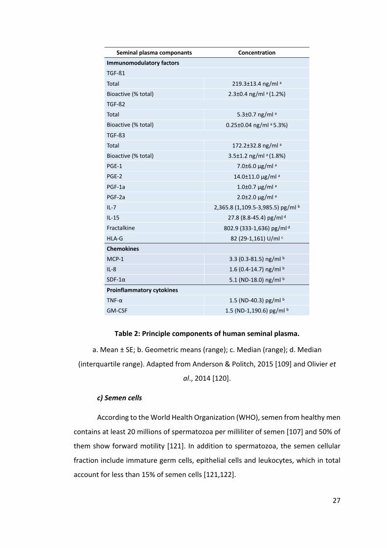
Table 2: Principle components of human seminal plasma. .................................. 27
Table 3: MHC haplotype of the macaques used in the study. .............................. 66
Table 4: SIV DNA contents of SHIV162P3-infected spleen cells from seven macaques.
................................................................................................................... 69
Table 5: List of the antibodies used for splenocyte characterization. ................... 71
Table 6: Proportion of leukocytes presence in splenocytes from macaque CDJ067
and CB790B. ................................................................................................ 73
Table 7: Inhibitory concentration (IC) 50, 75 and 90 of 10-1074 in a PBMC-based
neutralization assay using three stocks of spleen cells. ................................ 76
Table 8: MHC haplotype of the “donor” and the “receiver” macaques for pilot
study. .......................................................................................................... 77
Table 9: Splenocytes preparation for the intravaginal challenge. ........................ 79
Table 10: Comparison of semen cell populations between human and macaque. 82
Table 11: Correlation between viral loads and seminal inflammatory molecules. 85
Table 12: Seminal plasma viral load (SVL), blood viral load (BVL) and capability of
CD45+ semen cells from SHIV162P3 infected cynomolgus macaques to transfer
infection in vitro .......................................................................................... 91

8
Abbreviation list
HIV Human immunodeficiency virus
FRT Female reproductive tract
DCs Dendritic cells
CAV Cell-associated virus
GalCer Galactosylceramide
ZO-1 Zonula occludens
TJ Tight junction
LC Langerhans cells
SIV Simian immunodeficiency virus
HSPG Heparan sulfate proteoglycans
MR Mannose receptor
DCIR DC immunoreceptor
PBMCs Peripheral blood mononuclear cells
LNs Lymph-nodes
ICAMs Intercellular adhesion molecules
JAMs Junctional adhesion molecules
LEEP-CAMs Lymphocyte endothelial-epithelial cell adhesion molecules
E-cadherin Epithelial-cadherin
IFN-γ Interferon-γ
NK cells Natural killer cells
RNA Ribonucleic acid
DNA Deoxyribonucleic acid

9
AIDS Acquired immune deficiency syndrome
SP Seminal plasma
SEM Semenogelin proteins
PAP Prostatic acid phosphatase
PSA Prostate specific antigen
PGE Prostaglandin E
Tregs Regulatory T cells
WHO World Health Organization
NSC Non-spermatozoal cells
PMNs Polymorphonuclear cells
HS Heparin sulfate
MGT Male genital tract
CMV Cytomegalovirus
HTLV-I Human T-lymphotropic virus type I
SEVI Semen-derived enhancer of viral infection
WBC White blood cell
ART Antiretroviral therapy
Ab(s) Antibody/Antibodies
SHIV Simian human immunodeficiency virus
ADCC Antibody-dependent cellular cytotoxicity
NAbs Neutralizing antibodies
FcR Fc receptor
ADCP Antibody-dependent cellular phagocytosis

10
ADCVI Antibody-dependent cellular viral inhibition
non-NAbs Non-neutralizing antibodies
NHP Non-human primate
bNAbs Broadly neutralizing antibodies
CD4bs CD4 binding sites
MPER Membrane proximal external region
V1/V2 Variable region 1 and 2
mAbs Monoclonal antibodies
CoRbs Coreceptor-binding sites
CD4i CD4-induced
ATI ART interruption
MOI Multiplicity of infection
APCs Antigen presenting cells
SAMHD1 Enzyme SAM domain and HD domain 1
MHC Major histocompatibility complex
IgG Immunoglobulin G
PrEP Pre-exposure prophylaxis
PBS Phosphate buffer saline
IC Inhibitory concentration
AID50 Infect 50% of the animals
p.i. Post-infection
BVL Blood viral load
SVL Seminal viral load

11
CVF Cervico-vaginal fluids
ESN Exposed-non infected
BV Bacterial vaginosis

12
Chapter 1 Introduction
1.1 HIV-1 sexual transmission
1.1.1 Current situation
More than 30 years since the first reported case in the early of 1980s [1],
Human Immunodeficiency virus (HIV)-1 infection has continue to be one of the major
public health issue. In 2017, an estimated 37 million people were living with HIV
worldwide and 53% of them resided in eastern and southern Africa regions. Despite
the downward trend of new HIV-1 infections in 2017, the number of new cases was
estimated to be 1.8 million and almost half of them were women [2,3].
Unprotected sexual contact represents the main HIV-1 transmission route [4–
6], with the efficiency of transmission varying based on the type of exposure. The
expected range of per-contact act is shown in Table 1. Among sexual exposure, a
receptive anal intercourse has the highest risk per act of HIV transmission [7].
However, in developing countries, the primary route of HIV-1 dissemination is
through heterosexual coitus [8]. In general, male to female through penile-vaginal
transmission is greater than female to male transmission. Oral intercourse is less
likely to result in HIV-1 transmission.

13
Table 1: Estimated per-act probability of acquiring HIV-1 from an infected source,
by exposure routes.
Adapted from Patel et al., 2014 [7].
1.1.2 Structure of the female reproductive tract and related impact on HIV-
1 mucosal transmission
The female reproductive tract (FRT) separates into 2 compartments including
the upper FRT (endocervix, uterus and fallopian tubes) and the lower FRT (vagina and
ectocervix) (Figure 1) [9]. The exposed region during sexual intercourse is the lower
FRT [6]. However, a multilayered stratified squamous epithelium of vagina and
ectocervix provides better physical barrier for the incoming virus than endocervix
that lined with a single layered of columnar epithelium [6,10]. Endocervix and
conjunction zone between ectocervix and endocervix are considered as the
vulnerable site for HIV-1 heterosexual transmission due to their mucosa structure
allowing close contact to intraepithelial leukocytes and leukocytes at the submucosa
[4,5,11,12].
At the mucosa level, several immune cells including CD4+ T cells, macrophages
and dendritic cells (DCs) which express CD4 and CCR5 co-receptors are considered as

14
target cells for HIV-1 infection [4,13]. Either HIV-1 cell-free virus or cell-associated
virus (CAV) can migrate through the mucosal epithelium, by paracellular passage or
transcytosis, through micro-abrasion or laceration occurring during coitus [4]. After
penetration, HIV-1 encounters and directly infects susceptible CD4+ T cells,
macrophages and DC [14]. Additionally, DC, Langerhans cells (LC) and fibroblasts
mediate the viral transmission by transferring the virus to CD4+ T cells; chiefly in
trans-infection mode [4].
The susceptible cells that are present throughout the lower FRT are CD4+ T
cells, DC and macrophages, whereas the target cells that populate within the
epithelium of the uterine lumen are CD4+ CCR5+ T cells, DC-SIGN+ dendritic cells,
Langerhans cells and macrophages [15,16]. The resident immune cells can promote
HIV-1 acquisition by up taking of virions across the epithelial barrier [11]. Simian
immunodeficiency virus (SIV) intravaginal challenge in macaque have demonstrated
that SIV can pass the vaginal mucosa within one hour and after 18 hours post
infection, the infected cells can be found in the draining lymph nodes [17]. Moreover,
an ex vivo models of both ectocervical and endocervical tissues of human have shown
successful HIV-1 infection [18,19].
Figure 1: Female reproductive tract.
Adapted from Arien et al., 2011 [5]

15
1.1.3 Mode of HIV-1 sexual transmission
Numerous in vitro, ex vivo, and in vivo studies have contributed to the
understanding of the mechanisms by which HIV-1 can overcome the physical and
chemical barriers of the genital, and to a lesser extent, the anal and colorectal
mucosa, so that the virus reaches its target cells present in the submucosa. Several
mechanisms for the passage of HIV-1 through the mucous membranes have thus
been proposed depending on the site of the infection and the nature of the virus, i.e.
whether it is in the form of free particles (Cell-free virus) or whether it is present
inside infected cells (Cell-Associated virus) (Figure 2).
Figure 2: Mechanism of HIV-1 mucosal transmission.
Adapted from Gonzalez et al., 2019 and Shattock & Moore, 2003 [4,6]
a) Mechanism of HIV-1 sexual transmission at the mucosa
i) Paracellular passage
Paracellular passage is the process that causes a loss or disruption of tight
junctions between epithelial cells. The resulting gap formation that occurs allows the
passage of the virus to reach the submucosa (Figure 2) [20–22]. This process is able
to arise in almost all mucosal types [4]. The appearance of breaches at mucosal level
might be due to a mechanical cause during the sexual act, especially at the level of
the colonic mucosa [23–25]. However, several other factors may promote or cause
the appearance of breaches, including inflammation of the mucosa associated with

16
a pre-existing infection, the virus itself or the use of lubricants, microbicides or
spermicides on phase I trials [23,26–28].
Inflammation of the mucosa induced by bacterial or viral infection results in
an influx of immune cells and pro-inflammatory cytokines, which can alter the
epithelial barrier and thus make the sub-epithelial mucosal cells accessible to other
pathogens, including HIV-1 [23,24].
The virus itself is also capable of altering the colorectal epithelial barrier as
well as the endometrial and cervico-vaginal barriers. Nazli et al., using both in vitro
and ex vivo models have shown that exposure to HIV-1 particles alters the integrity
of a colorectal epithelial monolayer and of the primary epithelial monolayer models
of endometrium and cervix [29]. Either the HIV-1 co-receptors CCR5, CXCR4 and the
galactosylceramide (GalCer) expressed on the epithelial cells were able to bind the
HIV-1 gp120. This resulted in decreased transepithelial resistance, a decrease in
occluding, claudins and the cytoplasmic protein zonula occludins (ZO-1) expression
and an augmentation of Ca2a+ and MAPK and P13K pathways which led to ZO-1 and
claudins/occludin destabilization, tight junction (TJ) disruption, surface protein
internalization and disruption of the epithelial monolayer [21,30–32]. The disruption
of mucosal epithelium could be triggered by host antiviral mediators. Neutralization
experiments have shown that the envelope protein of the virus (gp120) generates an
alteration of the epithelial barrier by the secretion of TNF by epithelial cells [33].
Moreover, the activation of the apoptosis pathway could disrupt the epithelial cells
by the addition of caspases resulting from degradation of claudins, occludin and ZO-
1 proteins [33].
Several studies on phase I clinical trials have shown that lubricant gels,
microbicides, or spermicides normally recommended for vaginal use can cause an
alteration of colorectal epithelium [26,34], a loss of adherent junctions and TJ
between epithelial cells [28], and consequently an increase in mucosal permeability
[27].
Despite the fact that infection with HIV-1 through a break is the easiest,
fastest, and most efficient mode of passage of the virus since free virions or infected

17
cells pass directly through the mucosa to contact the resident target cells in the
lamina propria, the impact of this process in vivo remained unidentified.
However, not all mucosa presents the same susceptibility to the formation
of breaches and the virus can use various other mechanisms of passage to cross an
intact epithelial barrier.
ii) Transcytosis
Transcytosis is the process that makes use of the vesicular/endosomal
transportation of the cells to carry the viral particle into the intracellular
compartment (Figure 2). The process is characterized by the polarized transport of
viruses within epithelial cells, from the apical to the basal pole, without productive
infection of the host cell. After translocation, the virus is delivered to the external
basal space where their target cells reside. The virus can directly infect susceptible
lymphocytes or be carried and transferred by DC and Langerhans cells (LC) [4]. The
process is found mainly in columnar epithelial cells [35–37] and squamous epithelial
cells in pseudo stratified epithelium [38–40] at oral, intestinal, vaginal and
endometrial epithelial cells [39,41]. Indeed, in the genital and colorectal mucosa this
mechanism was described for the first time in vitro (I407, HT-29, CaCo2 and
endometrial HEC-1 colorectal lines) for HIV-1 by Bomsel et al. in 1997 [37].
Transcytosis of HIV-1 has since been demonstrated and confirmed in various models
of polarized cultures of immortalized (I407), colon (HT29, Caco2, T84), endometrial
(HEC1) and monolithic cell monolayers of primary vaginal and endocervical cells
[40,42–46]. Moreover, transcytosis has also been observed in ex vivo colorectal
explants exposed to HIV-1 free viral particles or to infected leukocyte cells [46,47].
The mechanisms have not been fully defined and the role of the viral envelope is still
debated. Some studies mention a necessary binding of gp120 to HSPG receptors
(Heparan sulfate proteoglycans) [40] and to the gp340 receptor [48] known to be
expressed by vaginal epithelial cells, endocervical and colorectal mucosa. Conversely,
other studies propose an independent mechanism of the viral envelope [49] since
occurring even with virus devoid of viral envelope, in models of vaginal and colorectal
epithelial lineages. In the case of the passage of viral particles from infected cells, this

18
mechanism seems to depend on the establishment of a virological synapse.
Interestingly, transcytosis between infected cells and mucosal epithelial cells was
found to be more potent than transcytosis between free viral particles and mucosal
epithelial cells due to the formation of virological synapse between HIV infected CD4+
T cells and the epithelial cells [50].
Not only paracellular passage but also transcytosis could be activated by the
interaction with viral protein and pro-inflammatory cytokines [4]. Combination of
paracellular passage and transcytosis could facilitate viral transmigration in vivo,
however transcytosis in vivo has not been formally demonstrated.
iii) Capture by immune cells
The capture of viral particles by the cells of the immune system present in
submucosal and / or intraepithelial is a potential mechanism for passage of the virus
through the different types of mucosae (Figure 2). Indeed, a mechanism used by the
virus for accessing subepithelial target cells is capture by DCs or LCs, which are a
subtype of DCs that are found specifically in the pluristratified epithelia. LCs differ
from DCs in that they specifically express Langerin (CD207) and reside in the
epidermis and in most mucosal epithelia, while DC-SIGN+ DCs mainly reside in
submucosa [51]. Although CD4+ T cells are the main HIV-1 target cells, at the genital
epithelium DCs are thought to be one of the first immune cells encountering the virus
at the submucosa as they are located within outermost genital epithelial layers
compared to the CD4+ T cells [52]. DCs have cytoplasmic extensions called dendrites
that allow them to capture viral particles within the mucosa but also at the level of
the lumen. Indeed, our group previously showed that intestinal lamina propria
CD11c+ DCs extend transepithelial dendrites and/or migrate across a tight epithelium
to capture luminal HIV-1 virions both in vitro and ex vivo [46]. Interestingly, DCs
migration was dependent by the interaction between the viral envelope and the
CCR5 expressed by DC. Consequently, X4-tropic viruses cannot reach the sub-mucosa
using this mechanism [53]. This may in part explain the preferential transmission of
R5-viruses at mucosal level.

19
Two main mechanisms have been described for DC's role in viral
dissemination (Figure 3) : (i) cis-infection: the virus binds to the cell membrane
molecules CD4 and CCR5, resulting in productive infection of DCs [54]; (ii) trans-
infection: HIV-1 particles can be internalized by DC or can be bound to DC surface
receptors like DC-SIGN without being productively infected. Besides DC-SIGN other
receptors can, to a lesser extent, bind HIV-1 and mediate the trans-infection pathway
such as the Mannose receptor (MR), DC immunoreceptor (DCIR) or GalCer which are
predominantly expressed on immature DC [54,55] or to Siglec-1 molecule, expressed
by mature DC [56]. The virions bound to the surface of DCs maintain their infectious
capacity for several days until reaching a susceptible cell, like the CD4+ T cells
[46,57,58]. HIV-1 was shown to replicate in vaginal DC more efficiency than in skin LC
or blood-derived DC [53,59–61]. This is possibly due to the lack of Birbeck granules
in DCs, whereas in LC HIV-1 captured by langerin is internalized into Birbeck granules
and partially degraded [51]. Moreover, vaginal DCs preferentially support the
replication of R5-tropic strain than X4-tropic strain [53]. This event may explain why
R5-tropic strain are preferentially transmitted during mucosal acquisition.
Similar to DC, HIV-1 was shown to bind to Siglec-1 in macrophages leading to
the formation of compartments containing viral particles that could subsequently
trans-infect CD4+ T cells [62].
Figure 3: HIV-1 dissemination mediated by DCs.
a. and b. trans-infection; c. cis-infection. Adapted from Wu & KewalRamani, 2006 [63].

20
iv) Transmigration of infected cells
HIV-1 infected leukocytes can migrate through epithelial cells without any
damage of the mucosal barrier to propagate HIV-1 infection (Figure 2) [64]. When
the transmigration occurs, the infected leucocytes transfer infection to permissive
cells such as activated CD4+ T cells and macrophages. In vivo and in vitro experiments
have proved that leukocytes infected with SIV or HIV-1 are able to migrate across the
genital epithelium. Using in vitro models of polarized monostratified epithelial cells,
the transmigration of monocytes and to a lesser extent of CD4+ T cells has been
shown [65,66]. The same is true for macrophages (derived from blood monocytes)
and peripheral blood mononuclear cells (PBMCs) [67]. Using explant culture models
of the human cervix, Maher et al. microscopically observed the infiltration of seminal
leucocytes in the outermost layers of the pluristratified epithelium of the ectocervix,
but the absence of infiltration into the endocervical epithelium [68]. Kolodkin et al.,
demonstrated the transmigration of PBMC through the colorectal barrier into
explants of human colon within 1 hour of exposure [69]. These observations have
been confirmed in vivo in humans where radio-labeled autologous cells have been
introduced intra-rectally and their distribution in the colon has been followed by
imaging up to 24 hours post inoculation [70]. Moreover, our group showed in
macaque that after intravaginal inoculation of labeled leukocytes, cells were found
in the peripheral lymph-nodes (LNs) 2 days post inoculation [71].
The underlying mechanism of apical-basal transmigration and the adhesion
molecules involved are still undefined. Various adhesion molecules present on
genital macrophage and T cells are an indispensable factor allowing leukocyte
transmigration from apical-to-basal epithelium. Integrins of the ß2 family, which
correspond to the molecules comprising the subunit CD18: LFA-1 (CD18 / CD11a),
Mac-1 (CD18/CD11b) and ITGAX (CD18/CD11c) are adhesion molecules
predominantly present on the surface of macrophages, monocytes and T cells, and
which participate to their transmigration through mucosal epithelia [72]. In semen
collected from SIV infected macaques, LFA-1 on macrophages and T cells [73]
enabled to counteract with intercellular adhesion molecules (ICAMs) on mucosal

21
epithelium which resulted in triggering the viral infected cells crossing epithelium
[74]. The Junctional adhesion molecules (JAMs) is also involved in the transmigration
mechanism. In TJs, JAM-A bound to LFA-1 [75], whereas JAM-C bound to Mac-1 in
desmosomes [76]. Both molecules have been found in vaginal and cervical
epithelium [72]. Thus, they are probably associated with an infiltration of
macrophages in genital mucosa. Moreover, the cell trafficking could be influenced
by the Lymphocyte endothelial-epithelial cell adhesion molecules (LEEP-CAMs). This
adhesion protein is expressed in the vaginal mucosa, bound to T cells and play a role
in maintaining intraepithelial lymphocytes [77]. Additionally, epithelial-cadherin (E-
cadherin) is a molecule driving lymphocyte adhesion and transmigration. It’s a
junctional structure protein of the vaginal, cervical and intestinal epithelium and
contribute to maintain the epithelial integrity [72].
The mechanism is also influenced by chemokine and inflammatory mediators
which are highly expressed in vaginal mucosa and submucosa [12,78]. The SDF-1
chemokine, which is highly present in the semen of HIV-1 infected subject [79],
potentially activate LFA-1 on semen leukocytes [80]. Furthermore, under
inflammation, vaginal and cervical mucosa generate not only high level of cytokines
such as SDF-1α and IL-8 but also RANTES, MIP-1α and MIP-1ß [81], which could
recruit leukocytes [82]. In inflammatory conditions, ICAM-1 expression could be
upregulated by interferon-γ (IFN-γ) generated by T cells and natural killer cells (NK
cells) [83].
b) Source of transmitted virus: cell-free vs cell-associated
HIV-1 is present in female genital secretions and semen not only as cell free
virions but also as infected leukocytes (semen composition will be further discussed
in section 1.2.1) [4,5,84]. While several studies based on in vivo and in vitro models
demonstrated that CAV is more potent to transmit the infection than cell-free virus
[5,85,86], CAV has been largely overlooked. There is still very little comparative data
between transmission by infected cells versus that with free virus in humans, and
their specific contribution is still debated. Thanks to a mathematical model, it is
22
estimated that cell-to-cell transmission is 1.4 times more effective than free virus
transmission and contributes to 60% of new viral infections [87].
Several studies have sought to determine the source of the transmitted virus
by analyzing the sequences of viral ribonucleic acid (RNA) and deoxyribonucleic acid
(DNA), both in donor genital secretions and in the blood of newly infected individuals.
These studies have shown that the virus found in the blood of newly infected persons
was in some cases closer in sequence to the viral DNA found in the infected cells of
the donor's genital secretions, and in others closer to the viral RNA derived from the
free viral particles [85,88,89]. The simplest interpretation of these observations is
that the source of the virus may be variable from one transmission to another, and
that both the free virus and the infected cells play a role in the transmission of HIV-
1.
In humans, in vivo inoculation of colloidal particles of HIV-1 size and
leukocytes showed that they co-localized after several hours in the sigmoid colon and
in the vagina, according to the route of rectal or vaginal inoculation, respectively [70].
Despite these similar migration capabilities, in vivo macaque studies have shown that
cell-to-cell transmission is the primary means of transmitting SIV at the vaginal and
colorectal level [69,71]. Our group has demonstrated indeed, that the inoculation of
infected leukocytes could establish systemic infection without any mucosal abrasion.
Cynomolgus macaques treated with depo-provera were intravaginally inoculated
with SIVmac251 infected splenocytes labeled with CFSE. Interestingly, using in situ
hybridization, the labeled cells were detected in the tissue of vagina and iliac LN after
21 hours of inoculation and in axillary LN after 45 hours of inoculation, indicating the
rapid dissemination of the infected cells [71]. There is no report, up to date, of
transmission initiated at mucosal level by semen cells, which would be more
physiologically relevant.
In vitro, HIV-1 transcytosis through different epithelial cell lines (I407, HT-29,
Caco- 2, HEC-1, ME-180) is much more efficient when initiated by infected cells than
by free virus particles [65,69]. The observation of transcytosis of free virus requires
an inoculum (in units of p24) from 100 to 1,000 times greater than with infected cells

23
[45], if one wants to have a number of viral particles having crossed the barrier
enough to generate a new infection. Infected cells also demonstrated greater ability
to induce infection following transmigration through an epithelial barrier compared
to free virus, as demonstrated by Van Herrewege et al [67].
In conclusion, it is now well established thanks to in vitro, ex vivo and in vivo
models, that HIV-1 transmission by infected cells is more effective in initiating a new
infection than cell-free virus and, according to the models used, it can be 10 to 1,000
times more effective [90,91].
The facilitation of HIV-1 dissemination by CAV may result from the close
contact between infected cells and susceptible target cells via the process of
membrane protrusion and cell engulfment or virological synapse (Figure 4) [92].
Indeed, it has been demonstrated in different cellular models that contact between
cells increases transmission of the virus by concentrating receptors implicated in
transmission at contact points between cells [93]. At the level of these contact zones,
a larger quantity of viral particles than that found naturally around a target cell is
released in a directional manner, thus increasing the multiplicity of infection to which
the target cell is subjected [94]. Upon contact with a target or epithelial cell, the
membrane of the infected cell is re-arranged, with on one side the recruitment of
receptors and molecules necessary for endocytosis or transcytosis of viral particles,
and on the other side the recruitment of membrane’s molecules that will be carried
away by the virus during its budding, and which will facilitate its future entry into
new target cells [95,96].
Finally, the vast majority of preventive strategies used to date have been
developed to target free viral particles only. The omission of strategies targeting
infected cell transmission probably reflects the lack of knowledge of the scientific
community on this subject, but also the technical difficulties of studying this mode of
transmission. However, it is vital to downscale the mechanisms that underlie this
mode of transmission in order to define new and effective prevention approaches
that do not target HIV-1 in its free form. Approaches to block cell-to-cell transmission
will be further discussed in chapter 1.3.5.

24
Figure 4: HIV-1 transmission through virological synapse formation.
Adapted from Cicala et al., 2010 [97].
1.2 The role of semen in HIV-1 mucosal transmission
The main route for HIV-1 dissemination is during unprotected sexual
intercourse. Consequently, semen represents the main vector of HIV-1 dissemination
worldwide [8,98] Considering the importance of semen in HIV-1 infection, the
comprehensive understanding of this viral carrier can possibly contribute to the end
of the Acquired immune deficiency syndrome (AIDS) pandemic.
Semen is a very rich biological fluid whose primary function is to ensure the
reproduction of the species. It is composed of an acellular fraction, the seminal
plasma (SP), which represents approximately 95-98% of the total volume, and a cell
fraction composed mostly of spermatozoa (Figure 5). In addition to its role in the
protection, transport and survival of spermatozoa, SP is able to modulate the
immune response of the FRT for fertilization and embryo implantation [99].
Figure 5: Human semen smear.
Seminal leukocytes were detected by immunohistochemistry (a) CD4+ T cells; (b)
macrophages. Adapted from Politch et al., 2014 [100].

25
1.2.1 Composition of semen
a) Physical and cellular characteristics
As mentioned, semen is a complex combination of semen cells and seminal
fluid. SP contains a wide range of compounds such as inorganic ions, organic acids,
sugars, lipids, steroids, amino acids, polyamines and proteins with superior buffering
capacity than most other body fluids [101]. The classic pH of a healthy man's sperm
is between 7.4 and 8.4 [102], which constitute a more alkaline environment than
those present in the FRT [103]. This property of SP allows to neutralize the acidity of
the FRT [104], which allows the survival of spermatozoa in this environment for
fertilization. Unfortunately, one of the undesirable side effects of this buffering
capacity is to allow free virus particles and infected cells contained in the sperm to
survive, thus promoting the risk of infection [105]. These aspects will be further
explained in chapter 1.2.3.
Compared to early puberty, the adolescent semen is characterized by an
increased volume and number of spermatozoa and an improved ability to liquefy
[106]. In healthy men, the volume of semen is between 2-5 mL per ejaculation,
containing approximately 25-100 millions of viable and motile spermatozoa per a
milliliter of semen [107]. The rheological properties of sperm are changed drastically
after ejaculation. In fact, very quickly the gelatinous coagulum is liquefied, under the
action of coagulation factors originating from the vesicles. The main components of
the coagulum are semenogelin proteins 1 and 2 (SEM1 and SEM2) very abundant in
sperm (20 mg/mL) and fibronectin. During liquefaction, the coagulum is transformed
into a fluid material by proteolytic cleavage of specific sites of the seminogens by the
so-called liquefaction factors: Prostatic Acid Phosphatase (PAP), Prostate Specific
Antigen (PSA), originally synthesized at the level of the prostate. Liquefaction occurs
5 minutes after ejaculation in vivo and 20 to 30 minutes in vitro (reviewed in [108]).
b) Seminal plasma
Seminal plasma is not just a transporter of spermatozoa to the FRT but it
encompasses various signaling molecules that temporally modulate FRT status [8].

26
This fraction of the ejaculate contains various bioactive reagents (listed in Table 2),
including immunomodulatory, proinflammatory and growth factors that can
contribute to a successful implantation in healthy couples [109]. This protein-rich
fraction contains 25-55 mg/mL of proteins, including enzymes such as protease,
esterase, phosphatase, but also prostaglandin E (PGE), fibronectin, polyamine and
proteins playing a role in the immune system such as complement molecules and
immunoglobulins (Table 2) [11,106]. The different components originated from
testis, epididymis and accessory glands [98,106]. Moreover, a wide array of cytokine
expressions in SP constitute a unique environment that is different from other
mucosa and blood. Namely, TGF-β ( ̴100 µg/mL) and PGE2 ( ̴1 – 80 ng/mL) are the
main cytokines present in semen [98,110–112]. Both molecules are the effective
immunosuppressive cytokines that could suppress leukocyte activation (e.g. NK cells,
macrophages and DCs) [113,114]. TGF-β is present in 3 isoforms (TGF-β1, TGF-β2 and
TGF-β3) and could be activated from latent to active forms by proteases and acid pH
in the vagina [110]. The cytokine has been demonstrated to be involved in inducing
regulatory T cells (Tregs) differentiation and downregulating NK cells activity,
resulting in immune tolerance of the FRT [110].
Other types of cytokines present in the semen have inflammatory,
regulatory, adaptive and hematopoietic properties. Presence of soluble HLA-G in
semen possibly suppressed NK cells from entering cytotrophoblast [115]. A sperm
coating glycoprotein - CD52g may be able to prevent anti-sperm immunity and
infertility [116]. SDF-1 may play a role in leukocyte recruitment involved in the
immune defense of the vaginal mucosa following insemination [109]. Also MCP-1, IL-
8, Fractalkine, GMCSF, IL-7 and IL-15 cytokines at inflammatory sites are associated
with the recruitment, maturation and proliferation of immune cells including
monocytes, T-cells, B-cells, DCs and NK cells [117–119]. In healthy subjects,
concentration of these cytokines were 5-fold higher in semen than in plasma [120].
27
Table 2: Principle components of human seminal plasma.
a. Mean ± SE; b. Geometric means (range); c. Median (range); d. Median
(interquartile range). Adapted from Anderson & Politch, 2015 [109] and Olivier et
al., 2014 [120].
c) Semen cells
According to the World Health Organization (WHO), semen from healthy men
contains at least 20 millions of spermatozoa per milliliter of semen [107] and 50% of
them show forward motility [121]. In addition to spermatozoa, the semen cellular
fraction include immature germ cells, epithelial cells and leukocytes, which in total
account for less than 15% of semen cells [121,122].
Seminal plasma componants ConcentrationImmunomodulatory factorsTGF-ß1
Total 219.3±13.4 ng/ml a
Bioactive (% total) 2.3±0.4 ng/ml a (1.2%)
TGF-ß2
Total 5.3±0.7 ng/ml a
Bioactive (% total) 0.25±0.04 ng/ml a 5.3%)
TGF-ß3
Total 172.2±32.8 ng/ml a
Bioactive (% total) 3.5±1.2 ng/ml a (1.8%)
PGE-1 7.0±6.0 µg/ml a
PGE-2 14.0±11.0 µg/ml a
PGF-1a 1.0±0.7 µg/ml a
PGF-2a 2.0±2.0 µg/ml a
IL-7 2,365.8 (1,109.5-3,985.5) pg/ml b
IL-15 27.8 (8.8-45.4) pg/ml d
Fractalkine 802.9 (333-1,636) pg/ml d
HLA-G 82 (29-1,161) U/ml c
ChemokinesMCP-1 3.3 (0.3-81.5) ng/ml b
IL-8 1.6 (0.4-14.7) ng/ml b
SDF-1α 5.1 (ND-18.0) ng/ml b
Proinflammatory cytokinesTNF-α 1.5 (ND-40.3) pg/ml b
GM-CSF 1.5 (ND-1,190.6) pg/ml b

28
Immature germ cells are the major population of non-spermatozoal cells
(NSC). They have been categorized as spermatogonia, primary spermatocytes,
secondary spermatocytes and spermatids. The number of immature germ cells
increase in case of men who have spermatozoa less than 6 million per milliliter. Two
types of epithelial cells are found in semen. Squamous epithelial cells, originated
from excretory duct, possibly indicators of bacterial infection or inflammation.
Another type of epithelial cells arise from seminal vesicles and are associated with
inflammation of seminal vesicles [121].
Leukocytes are normally present in semen, where they represent about 13%
of the NSC. They are probably involved in the elimination of abnormal and
degenerating spermatozoa. Among leukocytes, granulocytes (or polymorphonuclear
cells (PMNs)) are by far the most abundant cells since they are estimated to represent
between 50 and 60% of the total population. Monocytes / macrophages come in
second place and represent 20 to 30% of the semen leucocytes. Cytotoxic T
lymphocytes CD4 and CD8 [122,123], each half present and accounting for about 5%
of leukocytes, are also found [124]. Some studies have reported the minute presence
of a B cell population [125,126], as well as dendritic cells in the sperm of macaques
[127]. In humans, the presence of this population is more anecdotal because it
concerns almost exclusively men suffering from chronic inflammation [128].
Semen leukocyte concentration varies significantly between individuals but
should not exceed 1x10⁶ /mL as recommended by WHO [107]. Otherwise, we talk of
“leukocytospermia”. This condition occurs often during infection or genital
inflammation, mostly asymptomatic, and affects 5 to 10% of the healthy male
population [129]. This prevalence can be as high as 24% in men with HIV-1 infection
[72,100]. In these leukocytospermic men, the increase in leucocyte concentration
affects all populations, namely granulocytes, monocytes/ macrophages and T cells
[130]. This increase in the number of seminal leukocytes is probably due to the
exacerbated release of these cells from the epithelium, an alteration of the integrity
of the epithelial barrier and an attraction of these cells at the site of inflammation
[122].

29
Very few studies have investigated the phenotype of semen leukocytes
(Figure 6). A few studies in humans and a previous study of our group in macaques,
have reported the expression on CD4 T lymphocytes of activation markers such as
the IL-2 receptor (CD25), CD69, as early activation marker and HLA-DR, as a late
activation marker [73,100,126,131]. Also the expression of various other surface
markers, such as CD103 (mucosal marker), has been found in CD4 cells, but in a more
heterogeneous manner, suggesting that only a fraction of the proliferating
lymphocytes have a classical mucosal profile [122]. CD103 is a marker expressed by
a vast majority of intraepithelial lymphocytes (95%), whereas it is present on less
than 2% of blood cells. On the other hand, in the macaque, CD4 T cells express the
HIV coreceptors CCR5 and CXCR4 [73]. In general, among the CD4+ T lymphocytes,
there are naive and memory (effector and central) populations that can be identified
by expression profiles (CD4+ CD45RA+ and CD4+ CD45RO+, respectively). In sperm, the
majority of CD4 T cells have a memory phenotype [73,132]. It should be noted that
these proportions are different from those found in the blood since lymphocytes with
a naive phenotype constitute about half of the cell population. This means that these
lymphocyte populations will not have the same ability to respond to antigens and
from the point of view of HIV infection, they do not exhibit the same sensitivity.
Memory lymphocytes are more susceptible to infection than naive lymphocytes
[133]. Also CD8+ T cells exhibits an activated phenotype, demonstrated by the
expression of the TIA-1 activation marker, a granule-associated protein found in
cytotoxic CD8 lymphocytes [122].
The phenotype of monocytes / macrophages and dendritic cells has so far
been poorly documented. Dendritic cells in human sperm have an immature
phenotype (CD80-CD86+ CD83low CCR6+ CCR7-CD14+) [128]. Although monocytes
and macrophages represent the second most abundant leukocyte population behind
granulocytes, no study in humans has finely characterized them. Characterization of
semen monocytes and macrophages in the macaque model has been instead
performed by our group [73]. These cells strongly express CCR5 and are additionally
endowed with migration and adhesion factors such as the LFA-1 and Mac-1
molecules.

30
Although the origin of leukocyte is uncertain, it has been reported that, in
normal semen, the epididymis and rete testis are the sources for lymphocytes and
macrophages, whereas prostate and seminal vesicles are the sources for PMNs. On
the other hand, in leukocytospermia, the increased number of leukocytes was
associated with the genital tract inflammation and their origin is possibly from
prostate [134–136].
Semen is obviously not a simple carrier of spermatozoa but a complex
biological fluid, involving immunomodulatory and pro-inflammatory molecules and
containing target cells of HIV-1. Not surprisingly, semen can contribute to affecting
HIV-1 transmission. This will be precisely the subject of Chapter 4.1 of this manuscript
Figure 6: Phenotype of seminal leukocytes.
a) in human semen and b) in cynomolgus macaque semen. Adapted from Basu et
al., 2002 and Bernard-Stoecklin et al., 2013 [73,126].
1.2.2 Semen during HIV infection
a) Presence of HIV in semen: what’s its origin
As previously mentioned, HIV-1 is present in semen both as cell-free virions
and infected cells. Many studies have reported that heparin sulfate (HS) served as an
ancillary attachment factor for HIV-1 in DCs, macrophages and epithelial and
endothelial cells [137–141]. HS could recognize HIV-1 depending on four HS binding
domains, including the V2 and V3 loops, the C-terminal domain and the CD4-induced
bridging sheet of the gp120 [142,143]. Spermatozoa could bind to virions through HS
and transfer infection to macrophages, CD4+ T cells and DCs [98,144]. In spite of this,
% o
f CD4
5+%
0
10
20
30
40
Lymphocyte Monocyte Granulocyte
a) b)

31
spermatozoa-associated virus was speculative as a source of HIV-1 in semen because
several studies failed to observe viral DNA contents integrated into spermatozoa,
indicating inability to support viral replication [122,145–148]. The major source of
seminal HIV-1 infected leukocytes are possibly T cells and macrophages. HIV provirus
was detected more frequently in T cells than in macrophages [122], however, no
correlation was observed between the number of seminal T cells and viral DNA in
semen [149]. The association between leukocytospermia and the presence of HIV in
semen remained to be investigated.
The shedding of virus in semen arose from the male genital tract (MGT). MGT
organs, that are releasing HIV-1 virions and infected leukocytes, are mainly the
accessory glands, including epididymis, prostate and seminal vesicles (review by
[149]). The accessory gland may be the main contributor of HIV-1 virions and infected
cells (review by [135]). Analysis of the HIV RNA from male genital fluids, such as
secretions from urethra and prostate, displayed that prostate and epididymis were
the main source of virus [150]. As the vasectomy had little influence on the level of
HIV RNA, the epididymis may not serve as a source of virus [151]. The prostate and
seminal vesicle may play a role as viral source [149] as they are the main source of
seminal plasma [152], accounting for 60% by seminal vesicles and 30% by prostate
[136]. Additionally, they are more susceptible to infection than other accessory
glands (review by [152]).
Although testis is unlikely to be the main origin of the virus, it could not be
excluded [135]. Testis is the least HIV-1 infected area compared to other parts of the
MGT due to the blood-testis barrier [153]. According to the availability of susceptible
cells in testis, the infected testis leukocytes possibly migrate across the epithelium of
rete testis to seminal lumen then the virus may be release via Sertoli cells (review by
[135]). Model of SIV infected macaques revealed that testis could be productively
infected during primary infection and asymptomatic chronic infection [153]. The
testis’s infected cells were macrophages and T cells, as reported in men [134,154]
and in macaques [155]. Moreover, testis act as a viral sanctuary, due to a limited
access to drugs by the blood barrier and the drug efflux pumps (such as ABC

32
transporter) [156,157]. Consequently, testis must be taken into consideration for
effective HIV-1 therapies.
Several evidences indicate that viral strains and infected cells present in
semen do not originate solely from blood but would originate at least in part within
the MGT. During the acute phase of infection, viral RNA (corresponding to free virus)
and viral DNA (corresponding to infected leucocytes) in semen have sequences
extremely similar to those from virus present in the blood (review in [135]). However,
during the chronic phase genetic differences between HIV strains in the blood and in
the sperm emerge and free viral particles present in the semen constitute a
population distinct from that found in the blood [134,158]. This indicates the
existence of an independent local viral replication, as well as a restricted exchange
of virions and infected cells between the two compartments, allowing the parallel
evolution of various virus populations within the body. In addition, the sequences of
DNA and viral RNA in infected sperm cells may differ from those of virions, suggesting
a different origin of virions and infected cells (review in [135]). Whittney et al. (2011)
reported that in SIV infected rhesus macaques the viruses in blood and semen were
similar during early infection then were distinguished after the peak of viremia,
indicating that anatomic compartmentalization occurred at early time point [159].
Anderson et al., proposes a number of non-exclusive and variable mechanisms
allowing sperm contamination by HIV over time, namely: (i) direct importation of
virus from blood; (ii) clonal amplification of viral blood strains in infected cells
infiltrating the male genital tract; (iii) local replication in cells resident in the MGT
leading to a distinct viral evolution [72].
At early stage of infection, semen containing high level of HIV-1 RNA was
potentially infectious in parallel with leukocytospermia and elevated inflammation
markers, which led to leukocyte recruitments [72,73,152]. Semen at first week of
HIV-1 infection could efficiently transmit infection through sexual intercourse [149].
At chronic phase, a lower risk of HIV-1 transmission was observed due to decreasing
not only blood viral load but also seminal viral load [135]. The shedding of both
virions and infected cells continued to be detectable for months to years after

33
starting ART [160–162]. Halfon et al.(2010) have reported that ,under ART, HIV-1 RNA
in semen could be detected in only 3-5% of HIV-1 seronegative patient indicating the
lower risk of sexual transmission [163].
The presence of HIV-1 in semen was commonly found in chronic stage of
infection and leukocytospermia but it could be isolated either chronic infection or
leukocytospermia [164,165]. The HIV-1 persistence in semen was irrelevant to the
number of CD4+ or CD8+ T cells [166–168] and possibly intermittent, which was
unrelated with plasma viral load [169–171]. The level of persistent virus in semen
may be influenced by co-infection with sexually transmitted diseases, such as
cytomegalovirus (CMV), chancroid, syphilis, gonorrhea and Chlamydia infections
[172–174]. The large regions of membrane protein on CMV and human T-
lymphotropic virus type I (HTLV-I) were similar to CD4. This resemblance may
contribute to the higher susceptibility to HIV for the CMV and HTLV-1 infected
leukocytes [175].
b) Modulation of semen parameters in HIV-infected patients
HIV-1 infection has an effect on several physical and cellular parameters of
semen. These effects are mostly detected during the chronic phase of infection, in
which not only spermatozoa are affected (reduced motility, lower number of
spermatozoa, and/or increased abnormal morphology) but also physical
characteristics are changed (decreased ejaculated semen volume, increased seminal
pH and round cell amount). Some of the semen alternations may result from ART.
Indeed, ART may impact on several metabolic and endocrine function of testis and
MGT, resulting in anomalies of spermatozoa [176].
In comparison with blood, several immunomodulatory mediators, including
cytokines (IL-1a, IL-7, IL-8, MIP-3a, MCP-1, MIG, IP-10) and chemokines (SDFb1 and
TGF-b), are enriched in semen not only in healthy men but also in HIV-1 infected
men [129,177–179]. The acute phase of the infection is characterized by a higher
level of pro-inflammatory cytokines and chemokines compared to non-infected or
chronic HIV-1 infected subjects (for review see [180]). Overexpression of pro-
inflammatory cytokines / chemokines in the seminal plasma of infected men alters

34
the cytokine network and may impair the ability of the immune system to respond
to HIV-1 infection [120,178]. It should also to be taken into account that this cytokine
network evolves dynamically according to the different stages of viral infection, as
described by Vanpouille et al. [181]. A correlation between pro-inflammatory
cytokine levels and viral load in semen has been reported in several studies. Thus, a
positive correlation between a seminal viral load and levels of IL-1β [182] RANTES
[183], IL-6, IL-8, IFNγ [184] and IL-17 [185] was described in HIV-1+ patients. In
addition, these variations in cytokine concentrations could have numerous
consequences since, for example, it has been shown that high concentrations of pro-
inflammatory cytokines promote the expansion and activation of the immune cells
of the exposed mucosa. For instance, endometrial epithelial cells exposed to SP from
acutely HIV-infected men, produced higher level of pro-inflammatory cytokines (IL-
1a, IL-6, and TNF-a) , which increased HIV-1 replication in CD4+ T lymphocytes [186].
Consequently, the modulation of semen factors possibly has an effect on viral
propagation during HIV-1 sexual transmission in the FRT.
Finally, HIV-1 infection may modulate the phenotype of semen cells, such as
macrophages, CD4+ and CD8+ T cells. In a previous study our group has characterized
semen macrophages and CD4+ T cells in SIV-infected macaques [73]. Infection
determined an increase in the number of CD69+ CD4+ T lymphocytes which presented
an activated phenotype. Moreover, we reported a decrease in the number of cells
positive for LFA-1 and an increase in the number of cells positive for Mac-1,
suggesting that the virus may modify the adhesion capacity of the cells that it infects.
The modification of the dynamics and activation state of semen CD8+ T cells in SIV
infected macaques will be the argument of the chapter 4.1 of this thesis.
1.2.3 Role of semen in HIV-1 transmission
Although in vivo studies show that SP is not necessary for the establishment
of infection [71,187], a number of studies have demonstrated that the various
components of SP may affect, facilitate or inhibit, HIV-1 transmission.

35
a) Facilitation of HIV-1 transmission by semen
Semen may favor viral propagation in the FRT by changing the vaginal pH. HIV-
1 infectivity is stable at pH 5-8 but is reduced by 25% at pH 4.5 [188]. The pH of the
SP is 7.4-8.4 with buffering properties to bring the acidic vaginal pH back to a very
fast neutral pH (30 seconds) and maintain this neutral or even basic pH for 2 hours
[102,189]. Because free HIV-1 particles and infected leukocytes are sensitive to acid
pH, they are therefore likely to be inactivated by vaginal acidity. This buffering action
of the SP, allowing the protection of spermatozoa, has the undesirable side effect of
granting some protection to the virus, thus favoring infection [190]. Moreover, as
mentioned in the previous paragraph, semen exposure induces a strong
inflammatory response of the FRT mucosa, resulting in the disruption of mucosal
barrier and the infiltration of HIV-1 key targets, as well as recruitment of immune
cells (neutrophils, macrophages and T-lymphocytes) to cervix [191–193]. For
instance, the migration of LCs, stimulated by CCL20 secretion via NFƙB signaling
pathway in vaginal epithelium was under the influence of SP [193].
Semen-derived enhancer of viral infection (SEVI) is an amyloid fibril found in
semen that help the virus to attach to the surface of susceptible target cells [194].
The potential role of SEVI to facilitate HIV-1 transmission has been reported in in vitro
and ex vivo assays and in rat models [194–199]. SEVI enhancing effect responded in
a time- and dose- dependent manner and was not strain-specific. Indeed, in vitro
experiments demonstrated that the infection of PBMCs, macrophages and DCs with
R5-, X4- and dual tropic HIV-1 strains was increased by SEVI. Productive HIV-1
infection could start with 1-3 virions in the presence of SEVI [194]. hCD4/hCCR5
transgenic mice treated with SEVI before challenge showed 5- time increase in HIV-1
YU2 cDNA in the spleen compared to non-SEVI treated group [194].
The function of semen to suppress immune responses in the FRT has an
impact not only for successful fertilization but also for HIV-1 transmission facilitation.
However, the underlying mechanisms driven by SP are not well determined. TGF-ß
and PGE2, which are highly expressed in semen, have a role to suppress immune cells

36
activation (e.g. neutrophils, NK cells and macrophages) and to promote DCs
differentiation and T regulatory response [200].
SP contains complement molecules that can be activated by HIV-1 particles
(regardless of the tropism of the strain). This activation leads to the cleavage of the
protein of complement C3. The opsonization of viral particles by C3a fragments
formed as a result of cleavage may favor the infection of cells expressing the
complement receptor such as epithelial cells, macrophages, DCs and CD4+ T cells
[201]. SP of HIV-positive individuals also contains antibodies against the viral
envelope that can opsonize free virus particles. The formation of these complexes
promotes transcytosis of the virus via interaction with the neonatal Fc receptor
expressed by epithelial cells by concentrating the virus at the cell membrane [202].
This phenomenon is also observed with neutralizing antibodies supposed to have a
protective role against infection [203,204].
Finally, seminal plasma may abrogate the activity of HIV-1 microbicides by
electrostatic interaction. In the presence of SP anionic polymer microbicides (such as
cellulose sulfate and carrageenan) displayed 4-73 times reduction of antiviral activity
[205]. The cationic polyamines and polyanions of SP may be involved in the
electrostatic interaction with the drugs [206,207].
b) Inhibition of HIV-1 transmission by semen
Semen has been reported to be involved in inhibition of HIV-transmission as
well. SP could hamper the capture of HIV-1 mediated by DC-SIGN expressed in DCs
and prevent transfer of infection to T cells [208]. The clusterin, present at high level
(0.4-15 mg/mL) in SP, was identified as the main ligand of DC-SIGN [98]. In addition,
SP reduced the expression of CD4 and CXCR4 on lymphocytes, despite an increase of
CCR5 expression was observed [209].
Cationic polypeptides in SP, including 52 discovered peptides such as
seminogelins, fibronectine etc, have shown an anti-HIV-1 activity. The interaction
was observed even using very low concentration (3,200 folds) of SP but the action
was temporarily (< 24 hours) [210]. Also reactive oxygen species generated by

37
seminal leukocytes and spermatozoa [211] possibly play a role in intrinsic antiviral
activity. SP could also enhance the integrity of FRT epithelium, resulting in better
function of the physical barrier [8]. In the context of cell-to-cell transmission,
Lawrence et al. showed in a polarized model of HEC-1 cells that the SP has an
inhibitory effect on the transmigration of infected leukocytes across the epithelial
barrier. This effect is associated with increased expression of the ZO-1 junction
protein between epithelial cells and an increase in leukocyte adhesion to epithelial
cells [66]. An earlier study had already shown an increase in the adhesion capacity of
a lymphocyte line on the cervical and intestinal epithelial barriers (MT180 and I407
non-polarized lines respectively) exposed to SP [212]. Thus, SP seems to be able to
play an inhibitory or promoting role to cell-to-cell adhesion depending on the cell
types involved.
1.2.4 Immune responses affected by semen during HIV-1 infection
The factors in semen that influenced HIV-1 progression were associated with
co-infection and other invading pathogens and with high level of leukocytes,
indicative of genital tract inflammation. Leukocytospermia in semen could be found
not only in HIV-1 seronegative individuals with genital tract inflammation but also in
HIV-1 seropositive subjects [122]. White blood cell (WBC) in semen of HIV-1 infected
men (1.0E+05 cells per milliliter of semen) are generally present in lower number
than in uninfected men (2.4E+05 cells per milliliter of semen). Normally, CD4+ and
CD8+ cells are present in semen (5.7E+03 CD4+T cells and 1.9E+03 CD8+ T cells per
milliliter of semen) [131]. In the semen of infected men, the number of macrophages
remained stable but the number of CD4+ and CD8+ T cells was undetectable [131].
After treatment with antiretroviral drugs, the seminal leukocyte population was
brought back to be the same level as non-infected individuals [131]. Leukocytes
played a role not only as immunomodulator but also as a viral carrier, particularly
macrophages and T cells that have been shown to contain HIV-1 provirus [122]. As a
consequence of antiretroviral therapy (ART), the restored immune cells in semen
may result in increasing the risk of HIV-1 transmission [131].

38
In addition to the changes on an array of cytokines/chemokines and
leukocytes, already discussed in the previous sections, HIV-1/SIV-specific antibodies
are present in abundant quantities and high frequencies (81-100%) in the semen of
HIV+ men and SIV+ macaques, and are mostly IgG [213–215]. Those antibodies (Abs)
may exert different antiviral activities. Neutralizing HIV-specific IgA are present in the
semen and vaginal washes from HIV-1 exposed and seronegative sex-workers
[203,216–218]. Moreover, several studies have shown that neutralizing IgG could
prevent macaques infection following intravenous or vaginal inoculation of simian
human immunodeficiency virus (SHIV) [219–222]. Furthermore, Parsons et al. (2016)
previously demonstrated that HIV-1-specific antibodies in SP could mediate
antibody-dependent cellular cytotoxicity (ADCC) responses in vitro [223]. Although
HIV-specific Abs may prevent or partially control mucosal HIV-1/SIV entry [213,224],
the ability of HIV-1 specific antibodies in semen to modulate HIV-1 transmission
remain inconclusive.
1.3 Antibodies against HIV-1
1.3.1 Mechanism of protection mediated by antibodies: neutralization
versus Fc- mediated effector functions
The Ab response may be exploited for HIV-prophylaxis or therapy. Abs may
act at different stages of the viral cycle, as shown in Figure 7 [225–227]. Indeed,
binding of Abs to a free virus results in: a) aggregation of virions which decreases the
size of the virus inoculum; b) blocking by steric hindrance of the interaction between
the viral receptor and the cellular receptors; c) blocking the fusion step between the
viral envelope and the cell membrane; d) inhibition of post-attachment events such
as decapsidation, assembly and budding/release of virions.

39
Figure 7: Antibody action during the HIV-1 replication cycle.
Adapted from Zolla-Pazner, 2005 [226].
Viral entry into target cells is triggered by the interaction between the
trimeric envelope glycoprotein and the CD4 receptor expressed by target cells
(review in [228,229]). Neutralizing antibodies (NAbs) are directed against conserved
regions of the HIV-1 envelope gp120 and gp41 [227] and hamper the entry of the
virus (Figure 8). The development and mode of action of NAbs will be discussed more
in details in the next paragraph.
The mechanisms that contribute to protection and control of viremia by Abs
are not limited to neutralization of free virions, but also by Fc-mediated effector
functions. Abs can engage the Fc receptor (FcR) present on the surface of innate
effector cells (such as NK cells and phagocytes, among others) to stimulate antibody-
dependent cellular cytotoxicity (ADCC), antibody-dependent cellular phagocytosis
(ADCP), and antibody-dependent cellular viral inhibition (ADCVI) [230,231].
Additionally, Abs can induce complement-dependent cytotoxicity through their Fc
domain (Figure 8). In humans, five FcRs have been identified: FcRI (CD64), FcRIIa and
b (CD32), FcRIIIa and b (CD16) [232]. These different receptors are characterized
according to their affinity for IgG (review in [233]).

40
Figure 8: Antibody mechanism of action against viral infection.
Adapted from Jaworski & Cahn, 2018 [234].
ADCC against HIV-1 infection is a mechanism by which effector cells such as
NK cells, monocytes, or neutrophils are able to lyse infected cells (review in
[235,236]). This activity requires the interaction between the infected cell and the
effector cell via specific anti-HIV-1 antibodies. The antibodies must therefore bind to
the envelope epitopes expressed on the surface of the infected cell via its Fab part
and to the effector cells through the Fc portion [232,237]. This double interaction
results in the release of cytotoxic granules (granzymes and perforins) responsible for
the lysis and destruction of the infected cells [238–240] (Figure 9). NK cells are the
main effector immune cells that mediated the lysis of Ab-bound infected cells. The
potential protection by ADCC depends on NK cell distribution, maturation and
activation in each individual (review in [241]). Due to the highly dynamic
conformational change of the HIV-1 envelope, the window of opportunity of Ab to
mediate protection through ADCC function is restricted to only few hours (review in
[242]).
e726 www.thelancet.com/hiv Vol 5 December 2018
Review
In another series of studies, Caskey and colleagues73 intravenously infused 3BNC117 antibody into 17 participants with HIV-1 who were untreated and viraemic with a dose range between 1 mg/kg and 30 mg/kg of antibody. A single infusion of 3BNC117 e!ectively reduced viraemia by 0·8–2·5 log10 in eight participants receiving the highest dose (30 mg/kg) of antibody and the reduction of viraemia remained statistically significant for an average of 28 days; by 56 days, three of eight individuals had not yet fully returned to baseline viral load. The authors observed the emergence of resistant viral strains in some individuals. However, in a subset of individuals rebound occurred only after 3BNC117 plasma concentration decreased.
A second study (phase 2a) with 3BNC117 assessed people with HIV-1 during analytic treatment inter ruption.76 For this study, investigators enrolled participants with only 3BNC117 sensitive virus in their latent reservoir.
From 63 individuals tested, 65% were sensitive to 3BNC117 (half maximal inhibitory concentration 2 "g per mL). 13 participants (<50 copies per mL for over 1 year) with neutrali sation sensitive viruses received two or four intravenous infusions of 30 mg/kg of 3BNC117; ART was discontinued 2 days after the first infusion of antibody. The trial showed that 3BNC117 delayed HIV-1 rebound for an average of 6·7 weeks (two infusions) and 9·9 weeks (four infusions), and this result was statistically significantly di!erent from matched historical controls (2·6 weeks). The authors noted that rebounding viruses were very closely related, consistent with the clonal expansion of a single recrudescent virus. In most cases, rebounding viruses showed increased resistance to 3BNC117, indicating strong selection pressure by this antibody and escape by the recrudescent virus. In four (30%) individuals, suppression continued until the
Neutralisation
Antibody Viral antigen Complement Avoid
Natural killer cell
Phagocyte
Infected cell
CD4 cell
TCR MHC II Perforins InduceFc receptor
Complement fragment-receptor (eg, c3b-R)
Virus Proviral DNA
Antibody-dependentcell-mediated cytotoxicity
Antibody-dependentcomplement-mediatedlysis
Opsonisation andantibody-dependentphagocytosis
Antigen presentation
Blockade of infection and virus suppression• Avoid infection of target cell• Control virus replication and dissemination• Induce killing of infected cells
Virus eradication• Induce killing of persistently infected cells
Vaccine e!ect• Protection of T-cell and B-cell compartments• Promote virus diversification• Enhance antigen presentation
Figure: Antibody mechanisms of action against viral infectionPhagocytes include dendritic cells or macrophages. TCR=T-cell receptor.
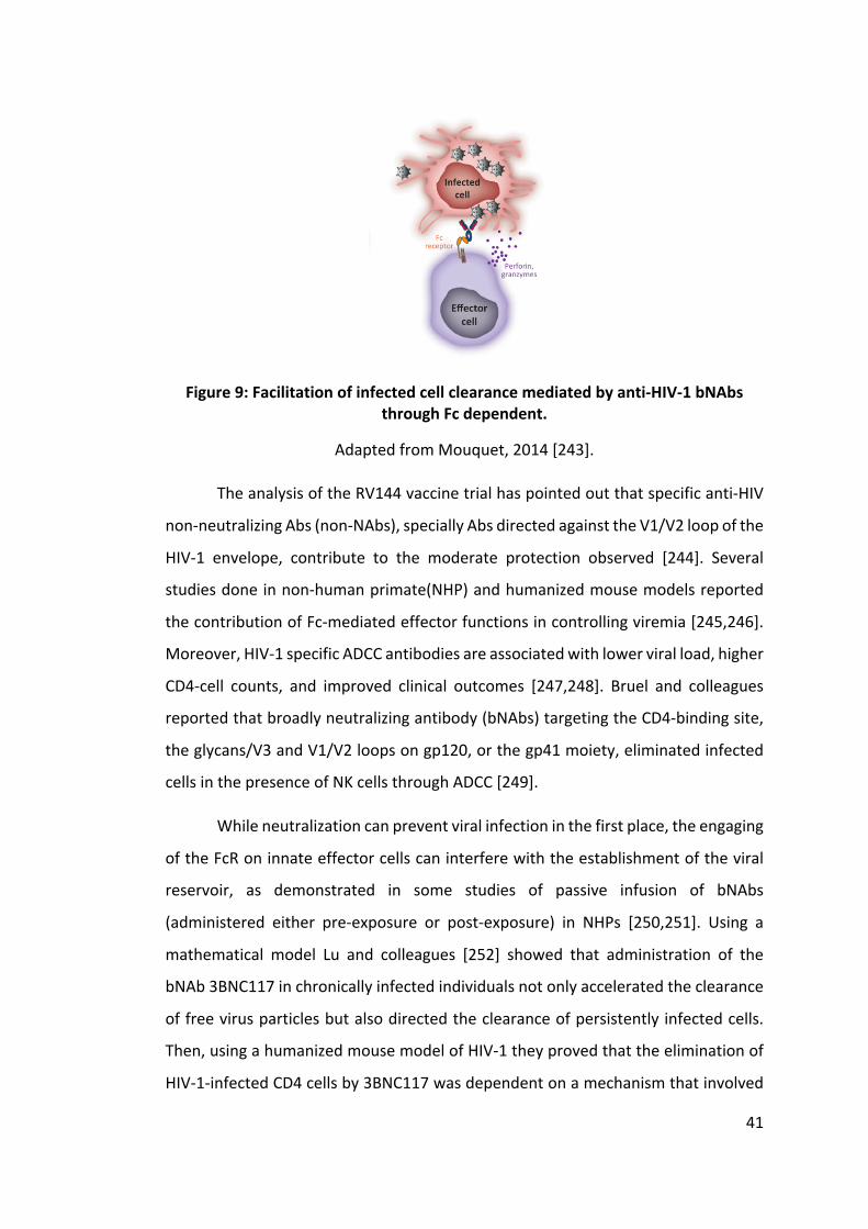
41
Figure 9: Facilitation of infected cell clearance mediated by anti-HIV-1 bNAbs through Fc dependent.
Adapted from Mouquet, 2014 [243].
The analysis of the RV144 vaccine trial has pointed out that specific anti-HIV
non-neutralizing Abs (non-NAbs), specially Abs directed against the V1/V2 loop of the
HIV-1 envelope, contribute to the moderate protection observed [244]. Several
studies done in non-human primate(NHP) and humanized mouse models reported
the contribution of Fc-mediated effector functions in controlling viremia [245,246].
Moreover, HIV-1 specific ADCC antibodies are associated with lower viral load, higher
CD4-cell counts, and improved clinical outcomes [247,248]. Bruel and colleagues
reported that broadly neutralizing antibody (bNAbs) targeting the CD4-binding site,
the glycans/V3 and V1/V2 loops on gp120, or the gp41 moiety, eliminated infected
cells in the presence of NK cells through ADCC [249].
While neutralization can prevent viral infection in the first place, the engaging
of the FcR on innate effector cells can interfere with the establishment of the viral
reservoir, as demonstrated in some studies of passive infusion of bNAbs
(administered either pre-exposure or post-exposure) in NHPs [250,251]. Using a
mathematical model Lu and colleagues [252] showed that administration of the
bNAb 3BNC117 in chronically infected individuals not only accelerated the clearance
of free virus particles but also directed the clearance of persistently infected cells.
Then, using a humanized mouse model of HIV-1 they proved that the elimination of
HIV-1-infected CD4 cells by 3BNC117 was dependent on a mechanism that involved

42
Fc receptor engagement [252]. These results shown that, besides preventing viral
acquisition by killing HIV-1-infected cells, bNAbs might be more successful in
suppressing viremia during chronic infection and have the potential to induce viral
eradication.
1.3.2 Development and structure of anti-HIV broadly neutralizing antibody
(bNAbs)
a) Development of bNAbs in HIV-1 infected patients
During HIV-1 infection, the humoral immune response develops within one
week after the detectable viremia and is characterized by the appearance over time
of different types of Abs [243,253,254]. Initially, it is detected as antigen-antibody
complexes. A few days later, free Abs against gp41 are observed, followed, a few
weeks later by Abs against gp120. These Abs mainly target the non-neutralizing side,
so they have no effect on viremia and do not exert selective pressure on the virus
[253]. They may be produced by pre-existing memory B cells that cross-react with
gp41 epitopes. Nevertheless, these Abs, via their Fc fragment, can contribute to
antiviral activity by inducing virion phagocytosis or lysing infected cells via an ADCC
mechanism [255–257]. The first neutralizing Abs against "autologous" virus appear
after two to three months. The selection pressure induced by these Abs lead to the
emergence of escape variants, resistant to neutralization [235]. These early
autologous neutralizing Abs are unable to neutralize virus variants isolated from
other HIV-infected patients [258–263]. Heterologous neutralizing Abs appears 2-4
years after primary infection in about 20% of chronic HIV-1 infected subjects [264].
Of note, approximately 1% of them (called “elite neutralizers”) develops neutralizing
Abs that neutralized cross-clade HIV-1 virus, called bNAbs [265]. Although the virus
envelope shows extraordinary diversity, we now know that there are highly
conserved epitopes, which are the target of largely neutralizing antibodies, and
which constitute sites of vulnerability to the humoral response [235,257].
bNAbs have unique features including the changes on heavy chain domain,
sulfate tyrosines, long and hydrophobic CDR-H3’s and extensive hypermutation
[266,267]. A hammer like structure created by the long CDR3 regions on

43
immunoglobulin heavy chain could promote the binding efficacy to HIV-1 envelope
[268–270]. Moreover, the hypermutation, especially in HCDR3 region, could be
detected in up to 32% of bNAbs, indicating the complex maturation required to
generate bNAbs [271–273]. The reason of the ability in some individuals to develop
bNAbs is not well defined but it may be related to the duration and the level of viral
infection. Due to the fact that not all subject could generate bNAbs, the underlying
factor is possibly related to both virus and host [235]. Interestingly, it has been
reported that viral subtype could affect the breadth and potency of humoral immune
response. HIV-1 positive subjects who are infected with subtype C or A possibly
generated bNAbs with higher breadth and potency than the subjected infected with
subtype B [274–277].
b) HIV-1 epitopes that are target of bNAbs
By the effort of many researchers worldwide, the characteristics and the
target epitopes of potent bNAbs have been disclosed. bNAbs target few conserved
regions of the HIV-1 envelope spike, called “sites of vulnerability” including the CD4
binding site (CD4bs) on gp120, glycan shield regions - V1/V2 loop and V3 loop on
gp120, the membrane proximal external region (MPER) on gp41 and the bridging
region between gp120 and gp41 (Figure 10) [243,278].
Figure 10: Structure and bNAb recognition of HIV-1 envelope spike.
Adapted from Mouquet, 2014 [243].

44
(A) A number of bNAbs recognize the CD4-binding sites (CD4bs) on gp120,
which is the conserved domain of gp120 concealed among glycan shield. To attain
broaden potency, bNAbs need to develop specifically to fit in this restrict access. The
category of the anti-CD4bs bNAbs is divided into two groups – VH gene restricted
Abs, such as VRC01, 3BNC117, 8ANC131 and IOMA, and CDR-H3 loop dominant Abs,
such as HJ16 and CH103. Of note, VRC01 class bNAbs (e.g. VR01 Ab) that used CD4
mimicry has astonishingly efficacy, neutralizing 98% of HIV-1 isolated strains in vitro
[279–284];
(B) the variable region 1 and 2 (V1/V2) has been proven to be another site of
the spike that is vulnerable to neutralizing antibodies. More precisely, the Ab could
recognize a quaternary epitope at the trimer apex of V1/V2 and N-linked glycans at
position N156-160. PG9, PG16, and PGDM1004 are representatives of V1V2-directed
neutralizing monoclonal antibodies (mAbs) that generally compete with one another
for antigen [285–287];
(C) N-linked glycan-containing epitopes form a site of spike vulnerability,
including the gp120 V3 loop, and several nearby glycans, 2G12, PGT128, and
PGT121/10-1074 represent the bNAbs that recognize this region [288–290];
(D) there are also bNAbs that target the coreceptor-binding sites (CoRbs) on
gp120. The exposure of this site, also known as the CD4-induced (CD4i) site, is (as its
name suggests) induced by conformational changes that occur as a result of the
binding of CD4 to gp120. Antibodies targeting this region are called CD4-induced
antibodies, and include 17b, X5, m36, 48d, and E51 [291–293]. Based on the length
of the IgG heavy chain CDR, most full-size CD4i Abs do not have high antiviral potency
because full-size Abs targeting CD4i-specific epitope is sterically restricted during
viral entry [294].
(E) The membrane-proximal external region (MPER) of gp41 contains highly
conserved residues that also play a critical role in the virus fusion process. Ab in this
group used their long CDR H3 loop to bind a linear sequence, that is located in the
highly conserved region of MPER, and some of them could bind to a lipid component
from viral membrane. Only 671–683 AA as a linear epitopes of this MPER are

45
recognized by potent neutralizing mAbs [295,296]. These bNAbs include 4E10, 2F5,
Z13, and 10E8 [297–299]. Although previous studies show that combination of 2F5
and 10E8 protects rhesus against SIV infection, 10E8 still exhibits most potent and
broad neutralizing activity compare with other MPER active antibody targeting gp41
of HIV-1. It can neutralize about 92% of circulating HIV-1 isolates in vitro (review in
[300]).
(F) Finally, the most recent group of bNAbs is directed against the bridging
region of gp120-gp41. The main target epitopes were trimer-specific, ranged
between gp120 and gp41 including glycan (review in [301]). However, their targets
were not always overlapping. One Ab in the group – 8ANC195 was not only capable
to bind gp120 monomer but also recognize various stage of conformational changes
of pre-fusion trimer [302]. Ab members of the group include 35O22, PGT151,
8ANC195, 3BC315/3BC176, CAP248-2B [303–307].
1.3.3 Antibody protection studies in animal models against cell-free virus
challenge
Initial studies have tested the prophylactic passive transfer of first generation
bNAbs in macaque models to protect them from SHIV infection. For instance,
combination of 2F5 (anti-MPER Ab) and 2G12 (anti-gp120 glycan Ab) bNAbs together
with HIVIG showed that protection in macaques against a pathogenic SHIV virus is
possible via both intravenous and mucosal routes [308,309]. Similarly, the
combination of 2F5, 2G12 and F105 (anti-CD4bs) NAbs displayed effective protection
against both systemic SHIV challenge in mature animals and oral SHIV challenge in
neonate animals [221]. Our group evaluated the efficacy of a triple combination of
2F5, 4E10 and 2G12 bNAbs combined in a microbicide gel against vaginal SHIV
challenge. The Ab combination, prepared at a concentration of 20 mg/g of HEC gel
for each Ab (60 mg in total) protected 70% (10 out from 15) animals [222]. The use
of single Ab was also protective. The anti-CD4bs Ab b12 has been used in multiple
SHIV protection studies that have helped in understanding both the mechanism as
well as durability of protection provided by bNAbs [310]. Following studies using
individual Abs, including 2F5, 4E10 and 2G12 showed protection against mucosal

46
SHIV challenge, which further supported the concept of antibody-mediated
protection [230,311]. While the first generation bNAbs were efficacious to inhibit
cell-free infection against either intravenous or mucosal SHIV challenge, nevertheless
the amount of bNAb that was needed to get effective protection was high (infusion
dose >20 mg/mL and Ab concentration in plasma higher than 100 μg/mL). Those
concerns together with the lack of information on lifelong protection did not support
their usage in human population.
The identification of newer generation bNAbs with potencies 10 to 100 folds
greater than earlier bNAbs has renewed interest in their potential clinical use [312].
Indeed, the new generation of highly potent bNAbs display better efficiency to
suppress viremia for longer time by using lower concentration of Ab than the first
generation of bNAbs [243]. For example, the anti-V3 glycan Ab PGT121 conferred
sterilizing immunity against mucosal SHIV162P3 virus in all animals when administered
5 and 1 mg/Kg, with corresponding average Ab serum concentrations of 95 μg/mL
and 15 μg/mL, respectively. Infusion with 0.2 mg/Kg of Ab resulted in 60 % protection
[313]. Following studies using other second-generation bNAbs, including anti-CD4bs
- VRC01, VRCO7-53, 3BNC117 and anti V3 glycan 10-1074 have shown successful
protection in vivo [313–319].
Besides being used to prevent viral acquisition, bNAbs have been evaluated
also for therapeutic properties. Ab-mediated immunotherapy demonstrated
promising results in animal models using individual bNAbs or Ab combinations in
presence and absence of ART [243]. The administration of PGT121 in SHIV162P3
infected rhesus macaques at chronic stage of infection suppressed the viremia to
undetectable level in 18 macaques for 35 to >100 days. Three out of eighteen
macaques displayed no emergent viral rebound. Moreover, reduction of HIV-DNA
was observed in PBMCs, lymph nodes and the mucosa of gastrointestinal tract [320].
Single use or combination of 10-1074 and 3BNC117 in chronically SHIV-AD8 infected
macaques temporary suppressed blood viremia (4-7 days) but viruses resistant to 10-
1074 emerged. The treatment with Ab combination at 36 months post-infection
exhibited a better outcome demonstrated with only one out of five macaques

47
showing viral rebound [321]. The combination therapy of bNAbs (single infusion with
VRC07-523 and PGT121 at day10 post-infection) with ART (at day 21 post-infection)
in macaques infected with SHIV162P3 showed efficacy comparable with ART alone
[322].
Moreover, bNAbs immunotherapy have been evaluated in acute infected
macaques. The combination of PGT121 and VRC07-523 bNAbs was used to orally
treat neonatal macaques 1 day after SHIV162P3 oral inoculation. In the treated group,
viral DNA was detected at low level in mucosa and lymph nodes after 24 hours of
treatment. On day 14 post-infection, no detectable viremia was observed in treated
macaques [250]. In another study using adult macaques infected with SHIV-AD8 via
intrarectal or intravenous routes, combination of 10-1074 and 3BNC117 was given at
day 3 post-infection. This treatment inhibited virus replication for 48-177 days after
which viral rebound was observed [323]. Based on the success of protection studies
in animal models, bNAbs have been explored for their efficacy in human clinical trials.
1.3.4 bNAbs used in human clinical trials
Most of the earlier studies have been focused on Ab treatment of HIV-1
infected individuals with some limited success. 2G12, 4E10 and 2F5 bNAbs used
either singularly or in combination before ART in infected individuals have failed to
control viral rebound, resulting from the fast emergence of escape variant [324,325].
However, preclinical data with second-generation bNAbs re-invigorated this area of
research and several bNAbs are currently investigated in clinical trials (Figure 11).
Figure 11: Second generation bNAbs tested in clinical trials.
Adapted from Caskey et al., 2019 [326].
REVIEW ARTICLE NATURE MEDICINE
high-dose mucosal challenge35,36. These results were confirmed and further explored when six different antibodies targeting the CD4bs or the V3-glycan patch were compared in experiments assessing protection against high-dose mucosal challenge in 60 macaques. Protection was directly related to antibody concentration and neutralizing activity. Probit regression analysis of the combined dataset revealed that plasma infectious dose 50 (ID50) neutraliza-tion titers of 1:100 were sufficient to prevent acquisition in 50% of the macaques37,38.
In contrast to high-dose challenge in macaques, mucosal infec-tion with HIV-1 in humans typically requires numerous exposures39. Human infection was modeled in macaques by low-dose weekly mucosal challenges with SHIVAD8 (ref. 40–42). A single bNAb infusion provided protection for up to 23 weeks in this model. When differ-ent bNAbs were compared, protective activity was directly related to antibody potency. Whereas 3BNC117, a CD4bs antibody, provided protection for a median of 13 weeks, VRC01, which targets the same site but is less potent, only provided 8 weeks of protection41. The protective activity of all bNAbs tested was extended by increasing their half-life with the introduction of two amino acid substitutions in the antibody’s Fc domain (p.M428L and p.N434S, called the LS mutation). These substitutions increase antibody binding affinity to the neonatal Fc receptor (FcRn), a recycling receptor that plays an important role in regulating antibody half-life in vivo41,43–45. For example, protection from low-dose infection with SHIVAD8 by a
single injection of 10-1074 was extended from a median of 13 to 27 weeks by the LS mutation44. Thus, antibodies can be highly effective and provide protection against infection for long periods of time. Altogether, these results suggest that the clinical potential of a bNAb or combination of bNAbs for prophylaxis against highly diverse cir-culating HIV-1 strains will depend on their breadth, potency and half-life in vivo.
Therapy in preclinical animal modelsHumanized mice and macaques are the best available preclinical models for testing the efficacy of passive immunotherapy against HIV-1. The advantage of using humanized mouse models is that they are reconstituted with human cells that can be infected with HIV-1. Although the humanized mouse is an important model, the potential disadvantages of this model organism are: (i) infection is sustained for relatively short times owing to limited life span of the host and graft-versus-host reactions; (ii) absence of a robust adap-tive immune response; and (iii) a relatively small number of infected cells and latent reservoir compared to humans. The advantages of macaques infected with SHIV are that they have an intact immune system and, after infection with certain, but not all, SHIV strains, they develop long-lasting infection that leads to AIDS-like disease. However, SHIV is a chimeric virus that is biologically and molecu-larly distinct from HIV-1, and infections with some SHIVs are far less robust than HIV-1 infection is in humans. In addition, macaque anti-human antibody responses interfere with bNAb antiviral activ-ity. Each of these caveats must be considered when evaluating the results obtained in either humanized mice or macaques.
The therapeutic activity of monoclonal antibodies against HIV-1 was initially tested in humanized mice two decades ago46. The data showed little or no effect on viremia by individual antibodies or cocktails of antibodies, leading to the conclusion that antibodies would not play a significant role in controlling HIV-1 infection in humans46. As a result, this line of preclinical research was nearly abandoned and was only re-initiated over a dozen years later, when far more potent second-generation bNAbs became available13,47. The new antibodies reversed the established dogma in the field by sig-nificantly reducing viral load in HIV-1-infected humanized mice47. The degree to which viremia was suppressed and the amount of time it remained suppressed were dependent on the potency of the antibody and the initial level of viremia. For example, when viremia was first controlled by ART, monotherapy with bNAbs maintained suppression48. In contrast, there was rapid viral rebound by selected escape mutants during monotherapy in animals with higher initial viral loads47,49–51.
Whereas bNAb monotherapy produced only transient effects on viremia, a combination of antibodies targeting non-overlapping sites on Env completely suppressed HIV-1YU2 infection in most of the animals. Due to the long half-life of antibodies, suppression was maintained for an average of 60 d after therapy was terminated47. Finally, when antibody levels were no longer high enough to main-tain viral suppression, the rebounding virus remained susceptible to bNAbs. Therefore, HIV-1YU2 was unable to escape combination antibody therapy in humanized mice.
Similar experiments were subsequently performed in Rhesus macaques that were chronically infected with SHIVSF162.P3 or SHIVAD8(ref. 52,53). In both cases, monotherapy with bNAbs sup-pressed infection. However, the two SHIV strains differed in that bNAb monotherapy controlled SHIVSF162.P3 for longer periods of time than SHIVAD8. In addition, whereas SHIVSF162.P3 was unable to escape from monotherapy with PGT121, SHIVAD8 developed resis-tance to 10-1074, which targets the same site and is very closely related to PGT121. The discrepancy could be due to subtle differ-ences in the mechanism of antigen binding by the two bNAbs, or it could simply be that SHIVAD8 leads to a more robust infection than SHIVSF162.P3.
New approvals per year
Total mAbs approved
a
b
1986
1990
1995
2000
2005
2010
2015
2018
0
20
40
60
80
70
Num
ber
of E
MA
- an
d F
DA
-app
rove
d m
Abs
Year
CD4bs
V3 stem
10-1074PGT121
10E8VMPER
CAP256PGDM1400
3BNC117VRC01
N6VRC07-523
V1/V2
Fig. 1 | Approved monoclonal antibodies and bNAbs tested in clinical trials. a, Number of monoclonal antibodies (mAbs) approved by the European Medicines Agency and the US Food and Drug Administration up to the end of 2018. Red columns indicate the number of newly approved monoclonal antibodies in a given year. Blue columns show the number of total monoclonal antibodies approved2. b, Second-generation bNAbs target various epitopes on the HIV-1 envelope trimer (grey). Antibodies against the CD4bs (orange; 3BNC117, VRC01, VRC07-523, N6), the V1/V2 loop (blue; PDGM1400, CAP256), the V3-stem (green; 10-1074, PGT121) and the MPER (purple; 10E8V) are currently being investigated in clinical studies.
NATURE MEDICINE | VOL 25 | APRIL 2019 | 547–553 | www.nature.com/naturemedicine548

48
Indeed, studies using second-generation bNAbs, including 3BNC117, VRC01
and 10-1074 reported better outcomes [327–330]. Primarily, 3BNC117 monotherapy
was assessed in 11 infected patients receiving a single Ab administration. The treated
subjects (10 out from 11) showed a reduction of plasma viral load (mean = 1.48 log10
copies/mL) and a viremia suppression for 28 days after treatment [327]. In addition,
infected cell clearance was observed [252,331]. Interestingly, 10-1074 bNAbs
displayed similar potency to what reported for 3BNC117 with the exception that the
incidence of viral rebound was higher [326]. The clinical trial with VRC01, which
target the same site as 3BNC117, was less efficacious than those using 3BNC117
[329,332]. The adapted bNAbs monotherapy regimen used in ART interruption (ATI)
have shown interesting outcomes. 3BNC117 treatment undergoing ATI displayed low
level of viral rebound and of emergence of resistant variants. Few participants could
maintain the viral suppression until the Ab concentration went below 20 µg/mL
[333]. In VRC01 clinical trial, repeated administrations of VRC01 in ATI contributed to
viremia suppression for 4 weeks [332]. Interestingly, the clinical potential of bNAbs
corresponds with their efficacy against primary HIV-1 isolates and is directly related
to the protection in animal models [326].
The trials of bNAbs monotherapy have exhibited the drug-liked effect of
bNAbs. The current combination of ART that are directed against different target are
more efficient and led to lower emergent resistant variants. Applying the similar idea
of using a combination of ART, bNAbs immunotherapy has been further used in
combination [326]. Although, the clinical trial on combination of 10-1074 and
3BNC117 showed better potential than the monotherapy did, the treatment could
not completely suppress viremia in the participants with high viral load at starting
treatment [334]. In the following trial, the similar bNAbs combination was infused in
the infected subjected at 2 days before ATI then 3 and 6 weeks after ATI. The results
showed successful viremia suppression for long period [335].
However, as a threat, viral resistance is still present in HIV-1 clinical
therapeutics, and HIV-1 has been shown to develop resistance to NAbs as well.

49
In conclusion, bNAbs have proven their protective potential against cell-free
infection. However, as HIV transmission occurs also through infected cells present in
biological fluids, it is extremely important to define the role and the protective
efficacy of bNAbs in the context of cell-associated transmission.
1.3.5 Role of bNAbs in prevention against cell-associated virus transmission
Although it could represent the predominant mode of viral spread in vivo,
bNAbs prevention of CAV transmission has not been studied the same depth as cell-
free infection. Indeed, almost all in vitro neutralization assays and in vivo protection
experiments have been performed using cell-free virus inoculum. The in vitro studies
performed have produced conflicting results regarding the potency of bNAbs to block
cell-free versus CAV transmission [336–344], possibly due to varied experimental
system used by different laboratories [341]. Differences were seen across virus
strains and Abs epitopes, and a substantial variability can be attributable to whether
the assay system used acutely transfected or chronically infected donor cells, cell
lines or primary cells, lab-adapted strains or transmitter/founder viruses [341,345].
However, there is today a general agreement that bNAbs exert a reduced efficacy
against CAV, evidenced by the much higher concentration or bNAbs combination
required compared to cell-free virus inhibition. This might be due to several
mechanisms (Figure 12), such as: steric hindrance at the virological synapse, the
increased multiplicity of infection (MOI) observed in CAV transmission, the different
conformation of the viral envelope during cell-free and CAV transmission (and this
might affect some epitopes more than others as well as vary between genetically
diverse env) and to the stability of viral envelope-Ab complexes (for a recent review
see [346]). Finally, the exposition of some neutralizing epitopes may be limited if
membrane fusion occurs within endosomal compartments in the target cell [347].

50
Figure 12: Potential mechanisms explaining the increased resistance of cell-associated HIV-1 to bNAbs-mediated neutralization.
Adapted from Dufloo et al., 2018 [346].
Experiments performed using the “first generation bNAbs”, such as 2F5, 4E10,
b12 and 257-D, produced conflicting results and no clear pattern could be
determined [348–350]. As for cell-free viral inhibition, the development of “second
generation bNAbs” has permitted a more comprehensive examination of the mode
of cell-to-cell virus inhibition by bNAbs. A study by Abela et al. indicated that the
targeted epitope may influence the efficacy of a given Ab [336] and that CD4bs Abs
lost efficacy during CAV transmission. The relative neutralization resistance in
intercellular assay systems was confirmed by other studies [339,340,344], however
in one of them [339] Abs directed against the CD4bs and the V3 loop were the most
active to inhibit transmission between T cells. A recent study by Li et al [338] showed
that a function motif in the gp41 appears to contribute to the loss of potency and
magnitude of multiple bNAbs during cell-to-cell transmission. In the case of DCs/T
cells transmission, the group of Moog showed that bNAbs inhibit HIV-1 transfer from
Page 8 of 14Dufloo et al. Retrovirology (2018) 15:51
Local variations at membrane subdomains, depending on the subcellular environment or the presence of lipids and cellular proteins, may also modify Env epitope exposure. !ese di"erent dynamic parameters impact the acces-sibility of cell-surface Env to bNAbs. For example, the engagement of Env by CD4 exposes epitopes targeted by non-neutralizing antibodies (nnAbs) [134, 135]. Since the creation of the VS involves interaction of Env with CD4, the conformation of Env at the synapse may be di"er-ent from that at other regions of the membrane. We also
reported that some antibodies, such as 8ANC195, which recognizes a gp120/41 bridging epitope and neutralizes cell-free virions, does not e#ciently bind to infected cells [17], confirming the existence of di"erent conformations of Env on virions and cells.
Viral proteins may also modify epitope accessibility. Nef and Vpu modify the levels of Env at the cell surface [136, 137] and Nef decreases Env susceptibility to anti-MPER antibodies [138]. !e Env cytoplasmic tail (CT) also regulates the exposure of Env epitopes, through
T cell T cell
Cell freevirus
Steric hindrance
Different composition of cell-free and cell-associated particles
CT
Env
Endocytosis
CT deletion or mutation
High MOI during cell-to-cell transmission
+HIV
HIV+
HIV+ Binding
Detachment
DC/Macrophage
Infected cell
Env conformation and stability of Env/bNAb complexes at the cell surface
Virion
a b
c d
Fig. 3 Potential mechanisms explaining the increased resistance of cell-associated HIV-1 to bNAbs-mediated neutralization. a bNAbs may poorly access virions present at the VS because of the physical proximity of donor and target cell membranes. b VS-mediated HIV-1 is associated with high MOIs. c Viruses budding at the VS may incorporate cellular proteins di!erently than cell-free virions, possibly leading to di!erent susceptibilities to bNAbs. d Env conformation and stability of Env-bNAb complexes at the cell surface. Env conformation may be di!erent at the surface of cell-free virus and at the plasma membrane. The stability of Env-bNAb complexes at the cell surface depends on the antibody and the viral strain. Donor cells are in brown and uninfected cells in blue

51
primary DCs and pDCs to autologous CD4+ T cells [61,351]. Antibody-conferred
inhibition via Fc has been observed in case of transferring HIV-1 infection from
antigen presenting cells (APCs) to surrounding T cells, which may be related to the
FcRs presence on the surface of DCs and macrophages. The FcR-conferred protection
required the binding of FcRs to Abs [61,341]. Interestingly, in comparison with anti-
gp41 bNAbs, the anti-gp-120 bNAbs were not only more potent in Fc-mediated
protection but also more efficient in preventing infection transmission from either
macrophages or DCs to T cells [61,337]. An increasing number of observations are
also pointing to the fact that possibly a combination of bNAbs is necessary to
efficiently inhibit CAV transmission. In in vitro studies no single Ab was able to inhibit
all strained tested [340] and a combination of PG9 and VRC01 was more effective
during cell-to-cell transmission compared to single Abs [344].
It is important to note that the relevance of in vitro studies to the in vivo
efficacy of the same Ab to inhibit cell-to-cell versus cell-free transmission is an
understudied area. Up to date only one study evaluated the efficacy of bNAbs to
inhibit CAV transmission in vivo in macaques. The authors used SHIV162P3-infected
splenocytes to intravenously challenge pigtail macaques and infused the animals
with the anti-V3 bNAb PGT121 [352]. Partial protection from infection was observed
and a delay in peak viremia or a delayed viremia was reported for not protected
macaques [352].
Finally, bNAbs could mediate Fc effector functions that may block HIV-1 CAV
transmission. The Fc domain presented on Ab is recognized by FcR receptor on
various immune cell types to trigger the mechanism of antibody-mediated inhibition
which possibly occurred in case of bNAbs and inhibited also the CAV transmission.
For instance, bNAbs were able to recruit NK cell via ADCC to kill HIV-1 infected cells
[249,353,354]. Moreover, bNAbs could activate ADCP and the complement pathway.
For CAV transmission, bNAbs and non-NAbs mediated ADCC function relied on the
accessibility of viral envelope protein on the cell surface. There are evidences that to
obtain optimal protection in vivo, bNAbs need Fc-mediated immune responses to be
involved [245,246,355]. Although, bNAbs may not provide completely neutralization

52
activity against CAV, bNAbs mediated Fc function behaves like the additional
mechanism to direct against infected cells. Lu et al. (2016) demonstrated that bNAbs
could eliminate HIV-1 infected cell and trigger the Fc mediated protection in
humanized mice. Infusion with either 3BNC117 alone or the combination of 3BNC117
and 10-1074, performed 12 hours before the transfer of infected cell, could reduce
not only the percentage of infected cells but also the level of CAV HIV-1 DNA
compared to the control mice [356]. Similar results were observed when using CD4+
T cells infected with primary HIV-1 isolated strains. Furthermore, the clearance of
HIV-infected cells in vivo possibly depended on the interaction of FcgR on effector
cell and Fc domain on 3BNC117 Ab [356]. Their observations highlight the important
function of FcgR mechanism mediated by bNAbs to eliminate HIV-1 infected cells in
vivo.
In addition, bNAbs may be related to the removal of latent infected cells. In
humanized mice the administration of a triple combination of 3BNC117, PG16 and10-
1074 early after infection prevented the establishment of latent reservoir [357].
Additional studies are necessary to understand why some bNAbs are less
efficient against CAV infection and why this varies with different viral strains and cell
types. At the present there is limited evidences regarding which in vitro assay better
predicts in vivo results. Moreover, all the in vitro/ex vivo studies published have made
use of in vitro infected cells to evaluate the efficacy of bNAbs to inhibit cell-to-cell
transmission. Data making use of the relevant donor cells found in the semen of
infected subjects are missing. The ability of bNAbs to inhibit cell-to-cell transmission
initiated by semen cells will be the subject of the chapter 4.2 of this manuscript.
1.4 Macaque models: similarities and differences to humans
NHP species including African monkeys and apes are natural SIV host which
typically do not develop disease following the infection [358,359]. On the other hand,
macaque species which are non-natural SIV host, can develop the infection with
certain strains of SIV, resulting in high viral load, progressive CD4+ T cells depletion
and progression to AIDS [358,359]. As the disease progression in macaques

53
resembles to HIV-1 infection in humans [358,360], the macaque models are routinely
used as animal models for HIV-1/AIDS research [358–361].
Interestingly, the key pathogenic features of HIV-1 infection in human can be
observed in SIV infected macaques [358,359,361,362]. Both HIV-1 and SIV have
similar key features of virus persistence including: integration of viral DNA into the
target cell genome [363,364] with similar preference integration sites [365],
response to interferons favoring HIV/SIV DNA persistence [366], inducing latent
HIV/SIV without co-engagement of T-cell receptors by costimulatory signals [367].
Both the infected human and macaque distribute the infected leukocytes containing
viral DNA and RNA sequences to peripheral blood, LNs and at mucosal sites [368–
370]. Cytotoxic T lymphocytes are unable to clear infected cells in long term because
of resistance mutation in HIV-1/SIV [371,372]. Those similarities highlight the sharing
of reservoir dynamics between HIV-1 infected patients and SIV infected macaques
[361].
While sharing some key pathogenic aspects, there are however some
differences between HIV-1 infection in humans and SIV infection in macaques. For
instance, SIVmac is more virulent than HIV-1 [373]. In SIV+ macaques, the set point
of viral load is 10 to 100 folds higher than in HIV+ subjects. The disease progression
to AIDS generally occurs within 1-2 years and up to 40% of macaques develop AIDS
more quickly (< 1 year) [373,374]. Moreover, another difference between HIV-1 and
SIV infection regards their interaction with the enzyme SAM domain and HD domain
1 (SAMHD1) [361]. SAMHD1 is a HIV-1 restriction factor [375] which antagonize with
the viral accessory protein Vpx present in some SIVs (particularly those that are used
in the macaques) [376,377]. As SAMHD1 may play a role in blocking viral infection by
interfering with a specific step of viral replication [358,361], it also displays a
dissimilarity between HIV-1 infection of human and SIV infection in macaque models
[361].
The immune response to antigens in NHPs, for both adaptive and innate
immune response, are very close to those in humans [358,360]. However, there are
interspecies differences. While the major histocompatibility complex (MHC) region

54
maps to chromosome 6 of humans which is composed of one copy of HLA-A, HLA-B
and HLA-C per haplotype, the MHC class I region in macaques is more expanded and
complex, containing multiple MHC-A and MHC-B genes per chromosome [360]. The
number and variability of antigen specific T cells that recognize individual MHC-
peptide complexes are quite similar in NHPs and humans [360]. Moreover,
immunoglobulin loci in macaques are more diverse than in humans [360]. Although
both humans and macaques have four IgG subclasses, including IgG1, IgG2, IgG3 and
IgG4, the macaque IgG subclasses lack the sequence diversity regions that are
important for the FcγR interaction in human IgG1 [378]. Indeed, the subclasses of
macaque IgG are much less structurally and functionally diverse group of proteins
than those found in humans [379]. For examples, IgG3 of human has a more
elongated hinge than those of macaque [378,379]. As a consequence, a shorter hinge
in the macaque probably renders it less flexible than its human counterpart [378].
This increased flexibility of the human hinge region is thought to confer the
enhancement of HIV-1 neutralizing potency [380,381]. Members of the FcγR family
between humans and macaques share several common traits [378,379]. However,
the main key difference has been recognized for FcγRIII which is not present on
macaque neutrophils [382–384] and for FcγRII which has a higher level of expression
on macaque neutrophils [382,384]. Due to the importance of Fc-dependent antibody
function, the insight on the interspecies variability of IgG and its receptors between
humans and NHPs may increase the accuracy of preclinical observation to convert to
success of clinical therapeutics.
Although the macaque models have some limitations, they can be used to
establish “proof of concepts” for mucosal transmission, physiopathology and many
therapeutic approaches of HIV-1/AIDS such as Pre-exposure prophylaxis (PrEP), ART
and microbicides [359,362]. These animals are still suitable for HIV-1 research as
there is no ideal animal model available.

55
Chapter 2 Objectives
As emphasized in the introduction, the empirical evidences and interests on
CAV transmission mediated by semen from HIV seropositive subjects are growing.
Due to the infectious potential of CAV, it is important to better understand the
dynamic modulation of semen during HIV-1 infection and to find the optimum
strategies to prevent viral dissemination. In this thesis project, I used in vitro assays
and the NHP model of infection of cynomolgus macaques to answer to specific
questions. In specific, the aims of the project are to investigate the immune
responses in semen during SIV infection and to evaluate the potency of bNAbs to
block CAV transmission. The two aims are divided in 2 studies.
In the first study, we assessed dynamic changes of the immune response in
the semen of SIVmac251 infected cynomolgus macaques. We investigated SIV-
specific innate and adaptive responses in semen and compared to systemic
responses, including CD8+ T cells responses and SIV specific humoral responses (in
vitro neutralization and FcγRIIIa binding activity), to evaluate their potential role in
controlling viral shedding. Moreover, we measured levels of cytokines, chemokines
and growth factors in seminal plasma in order to define the association between the
inflammatory state of semen, cell activation and potential risk of transmission.
In the second study, we assessed the efficacy of a panel of bNAbs against CAV
transmission. We developed an in vitro CAV transmission assay using either TZM-bl
or human PBMC as target cells and SHIV162P3-infected splenocytes and CD45+ semen
leukocytes as donor cells. The panel of bNAbs was selected to target different
epitopes of HIV envelope, including 1st generation bNAbs (2F5, 4E10, 2G12 and b12)
and 2nd generation bNAbs (10-1074, PGT128, PGT151, 3BNC117, N6, PGDM1400,
PG16 and 10E8). A first screening of bNAbs efficacy to inhibit SHIV162P3 transmission
was done using splenocytes and a reduced panel was confirmed with semen cells. On
the base of the results obtained in vitro, we have selected an anti-V3 directed bNAb
to be used in a future study to block CAV transmission in vivo in cynomolgus
macaques.

56
Chapter 3 Experimental approach
3.1 The cynomolgus macaque model for HIV research: use of the pathogenic
strains SIVmac251 and SHIV162P3
Three species of macaques, including rhesus macaques (Macaca mulatta),
pig-tailed macaques (Macaca nemestrina) and cynomolgus macaques (Macaca
fascicularis), have become widely used and accepted models for AIDS research [358–
361].
In this work we used cynomolgus macaques imported from Mauritius. The
macaques used descend from a small founder population with only 7 MHC
haplotypes identified, indicating a limited MHC diversity and contributing to a certain
homogeneity in infection profiles, particularly during primary stages [385–387].
Nevertheless, some studies have reported heterogenic profiles in viral replication
control in chronic infected Mauritius cynomolgus macaques, which was similarly
reported in HIV+ men [388–390]. SIV infection in cynomolgus macaques is generally
characterized by lower set point viral loads and slower progression of CD4+ T cells
decrease than Indian and Chinese rhesus macaques [358]. The slower disease
progression of cynomolgus macaques compared to rhesus macaques may be
preferable as it mimics a closer situation compared to HIV infection and AIDS in
humans.
The SIV genome is slightly different from the HIV-1 genome. For instance,
SIVmac presents an accessory gene vpu instead of vpx found in HIV-1 (Figure 13).
SIVmac251, an uncloned strain displaying high genetic diversity like HIV-1, is widely
used for mucosal challenges, with rectal, vaginal or intravenous exposure and to
study the pathogenesis of infection [358,391]. The virus stocks, commonly used in
our laboratory, have been titrated in vivo and used many times for mucosal
challenges [389,391–393]. These stocks have been obtained in 1989 from a
homogenate of splenocytes and PBMCs of a SIV-infected rhesus macaque from the
New England Primate Research Center [391].

57
Figure 13: Genetic difference between HIV-1, SIVmac and Env-SHIV strains.
Adapted from Hatziioannou & Evans, 2012 [358].
Thus, Mauritius cynomolgus macaques infected with SIVmac251 represent a
valuable animal model that allowed us to study the immune response in semen
during SIV infection. Animals were at different stages of infection and displayed a
large diversity of profiles in viral shedding. We also performed a longitudinal follow-
up of SIV-infected macaques, from the first week after infection to the chronic stage.
Although SIV virus is useful to study pathogenesis of AIDS, the difference
between the SIV env protein and the HIV-1 env protein contributed to the non-cross
neutralization activity of anti-HIV-1 bNAbs to SIV [312,394]. Thus, chimeric strains of
SHIV were constructed. Generation of SHIV (Figure 1) is mostly created by replacing
the SIVmac239 genes of rev, tat and env with the HIV-1 genes of rev, tat, vpu and
env. A case in point is SHIV163P3 – R5 tropic SHIV – that is a useful strain to evaluate
treatments aiming to prevent HIV-1 transmission such as NAbs [358].
Indeed, we used SHIV162P3 virus to evaluate the efficacy of bNAbs against CAV
transmission. Progressive examination of bNAb candidates in clinical trials require
pre-clinical studies thus, it is important to investigate their efficacy in vigorous animal
model like NHP. The bNAbs potency in preventing cell-free infection has been
evaluated often in NHP models. However, their protective potential in the context
of CAV transmission is not well defined and only one study has been published so far
[352].

58
Despite of the fact that a few studies have evaluated the bNAbs potency
against CAV, some research teams have successfully established CAV transmission
models in NHP. Early efforts have shown successful CAV transmission in rhesus
macaques after intravenous inoculation. However, the initiation of infection
following mucosal inoculation was more difficult [395]. Despite this, using SIV+ CAV
as challenge stocks, Kaizu et al. and our group have achieved to transmit infection
following vaginal inoculation in cynomolgus macaques [71,396]. Kolodkin-Gal et al.
demonstrated rectal transmission following SIV+ CAV inoculation in rhesus macaques
[69]. It is interesting to note that the viral inoculum used in the studies of Kaizu et al.,
Sallé et al. and Kolodkin-Gal et al. were in a range of the expected virus per
ejaculation of HIV-1 infected men, indicating the main implication of CAV model.
Those studies supported our project to evaluate the efficacy of bNAbs in CAV
mucosal transmission model (vaginal route) in cynomolgus macaques using
splenocytes as infected cells, a model that has been successfully set up in our
laboratory.
3.2 Semen sampling by electro-ejaculation
The collection of semen samples by sexually mature animals has been
performed using a technique called electro-ejaculation [397], which consists in an
electronic stimulation of the prostate. It is a non-invasive method which is largely
considered as reliable, reproducible and safe. However, it differs from the electro-
stimulation technique, which consists in stimulating the penis with electrodes,
without anesthesia [397]. The sampled volume and the concentration in
spermatozoa might be reduced by electro-ejaculation compared to electro-
stimulation.
In the lab we have successfully set up the protocol to collect semen by
intrarectal electrostimulation of the prostate using a probe (12.7 mm diameter)
lubricated with a conductor gel and an AC-1 electro-ejaculator (Beltron Instrument,
Longmont, USA. Sequential stimulation was performed with a pattern of six cycles,
nine stimulations of 2 s for each cycle, followed by a tenth stimulation lasting 10 s.
The voltage was increased every two cycles (1-3 V for the first two, 2-4 V volts for

59
series 3-4, 6-8 V for series 5-6). If a complete ejaculum was not obtained after six
cycles of stimulation, a seventh cycle at 7 to 10 V was performed. Complete ejaculum
(volume ranging between 0.1 and 0.5 mL) was immediately diluted in 1.2 mL
phosphate buffer saline (PBS) and centrifuged.
3.3. Establishment of in vitro neutralization assays to predict the in vivo
protective potential of bNAbs
Neutralization activity is regarded as a crucial contributor for HIV-1 protection
[312]. There are growing evidences that strategies to optimize bNAbs-mediated
protection from CAV transmission are needed. NHP models represents valuable
systems for testing such strategies [395]. In addition, complementary in vitro assays
that accurately predict the capacity of bNAbs to inhibit cell-to-cell spread in vivo, can
contribute to optimize those strategies. As discussed earlier in this manuscript, many
groups established in vitro experimental systems of CAV transmission, which
however were highly variable in terms of target cells and donor cells used and viruses
utilized [345,398]. Consequently, divergent results across studies were obtained
[336–344]. There is no clear evidence displaying which in vitro assay closely reflect
the CAV transmission in vivo and confer protection by bNAbs [395]. To reduce this
variation and better predict the in vivo protection conferred by bNAbs on the base of
in vitro results, one approach might be to use a common CAV stock to screen bNAbs
in vitro and in parallel in vivo in NHP models.
Therefore, we set up an in vitro experimental system to assess bNAbs-
mediated inhibition of CAV transmission, using splenocytes collected from SHIV162P3
infected macaques at the peak of infection. This same stock of cells will be employed
in in vivo challenge studies in cynomolgus macaques.

60
Chapter 4 Results
4.1 First article (Submitted): Innate and adaptive anti-SIV responses in
macaque semen: implications for infectivity and risk of transmission
4.1.1 Summary
In the first article, we aimed to investigate the anti-SIV innate and adaptive
immune response in semen of cynomolgus macaque during SIVmac251 infection.
We conducted a comprehensive study on a total of 53 animals. We analyzed viral
shedding and seminal cytokine/chemokine profiles. Moreover, we characterized the
phenotype of semen CD8+ T leukocytes and the SIV specific CD8+ T cell response using
ex vivo stimulation of semen cells with SIV gag peptides and the intracellular staining
with antibodies targeting CD45RA, IL-2, IFN-γ, TNF and MIP-1β. To determine Ab
response, we quantified SIV-specific Ab titers in the semen of macaques at different
stages of infection and performed an in vitro neutralization assay and ELISA based
FcγRIIIa binding assay to evaluate the potential function of SIV-specific Ab.
In macaque semen, we observed that many cytokines were affected by SIV
infection. Several pro-inflammatory (IL-8, IL-15, IL-18, IFN-γ, IP-10 and RANTES) and
the immunomodulatory molecule IL-13 were found significantly increased during the
primary infection compared to uninfected macaques and several of them predicted
seminal and /or blood viral load. This inflammatory environment might recruit target
cells and modulate immune cells. Indeed, we provide evidence of a high level of
preexisting activated CD69+ CD8+ T cells in the MGT, which is further elevated upon
SIV infection. In parallel, we observed upregulation of CCR5+ CXCR3+ CD8+ T cells,
typical of a Tc1 phenotype, which is consistent with the capacity of those cells to
migrate to inflammatory tissues. Neither SIV-specific CD8+ T-cell responses nor
humoral responses controlled seminal viral shedding. The neutralization activity was
generally weak but specific, as assessed using a VSV-pseudotyped virus as control. In
67 % of the infected macaques, we were able to detect FcγRIIIa-binding Abs in semen,
which is indicative of ADCC. Interestingly, FcγRIIIa-binding activity in semen positively
correlated with SIV-specific Ab titers and seminal viral load.

61
Thus, failure to control viral replication in SIV-infected semen is related to a
general inflammation and immune activation, which possibly mirrors what happen
in the MGT and which could lead to enhanced HIV-1/SIV transmission
Overall, our study highlights the complex interplay between immune
activation and inflammation and the role might play in modulating semen infectivity
and risk of transmission. These observations are relevant to our understanding of
natural immunity to HIV-1. A better comprehension of the mechanisms that regulate
immunity in the MGT may contribute to improving the efficacy of currently available
prevention measures and lead to the identification of new determinants of HIV-1
transmission.
4.1.2 Manuscript

Innate and adaptive anti-SIV responses inmacaque semen: implications forinfectivity and risk of transmission
Karunasinee Suphaphiphat1, Sibylle Bernard-Stoecklin1, Céline Gommet1, 2, Benoit Delache1,
Nathalie Dereuddre-Bosquet1, Stephen Kent3, 4, 5, Bruce D. Wines6, 7, 8, Phillip M. Hogarth6, 7,
8, Roger Le Grand1, Mariangela Cavarelli1*
1CEA-Université Paris Sud-INSERM U1184, “Immunology of viral infections and auto-immune diseases”,IDMIT department, IBFJ, Fontenay-aux-Roses, France, Commissariat à l'Energie Atomique et aux
Energies Alternatives (CEA), France, 2Sanofi (France), France, 3Department of Microbiology andImmunology, Peter Doherty Institute for Infection and Immunity, The University of Melbourne,
Australia, 4Melbourne Sexual Health Centre (MSHC), Australia, 5ARC Centre for Excellence in Convergent
Bio-Nano Science and Technology, Monash University, Australia, 6Immune therapies group, Burnet
Institute, Australia, 7Department of Clinical Pathology, The University of Melbourne, Australia,8Department of Immunology and Pathology, Monash University, Australia
Submitted to Journal:
Frontiers in Immunology
Specialty Section:
Mucosal Immunity
Article type:
Original Research Article
Manuscript ID:
510740
Received on:
07 Nov 2019
Frontiers website link: www.frontiersin.org
In review

Conflict of interest statement
The authors declare that the research was conducted in the absence of any commercial or financialrelationships that could be construed as a potential conflict of interest
Author contribution statement
Study conception and design: RL and MC. Acquisition of data: KS, SS, CG, and BD. Analysis and interpretation of data: KS, SS, CG, NBand MC. Drafting of the manuscript: KS and MC. Critical revisions: KS, SK, BW, MH, RL and MC.
Keywords
SIV, Cytokines, Antibodies, ADCC, CD8+ T cells
Abstract
Word count: 195
HIV-1 infection is transmitted primarily by sexual exposure, with semen being the principal contaminated fluid. However,HIV-specific immune response in semen has been understudied. We investigated specific parameters of the innate, cellular andhumoral immune response that may affect semen infectivity in macaques infected with SIVmac251.Serial semen levels of cytokines and chemokines, SIV‐specific antibodies, neutralization and FcγR‐mediated functions and SIV‐specificT-cell responses were assessed and compared to systemic responses across 53 cynomolgus macaques.SIV infection induced an overall inflammatory state in the semen. Several pro-inflammatory molecules correlated with SIV viruslevels. Effector CD8+ T cells were expanded in semen upon infection. SIV-specific CD8+ T-cells that expressed multiple effectormolecules (IFN‐γ+MIP‐1β+TNF+/‐) were induced in the semen of a subset of SIV‐infected macaques, but this did not correlate withlocal viral control. SIV‐specific IgG, commonly capable of engaging the FcγRIIIa receptor, was detected in most semen samplesalthough this positively correlated with seminal viral load.Several inflammatory immune responses in semen develop in the context of higher levels of SIV seminal plasma viremia. Theseinflammatory immune responses could play a role in viral transmission and should be considered in the development of preventiveand prophylactic vaccines.
Contribution to the field
Sexual HIV-1 transmission occurs primarily by contaminated semen, however little is known about the innate and adaptiveanti-viral immune response taking place in the semen, that may limit or enhance viral transmission. We show in a non-humanprimate model that acute SIV infection is associated with increased levels of pro-inflammatory and immunomodulatory molecules(IL‐8, IL‐13, IL‐15, IL‐18, IFN‐γ, RANTES and IP‐10) which predicted viral load and potentially recruit target cells and modulateimmune cells. In parallel, we observed upregulation of CCR5+ CX3CR1+ CD8+ T cells, typical of a Tc1 phenotype, which is consistentwith the capacity of those cells to migrate to inflammatory tissues. Although a subset of infected macaques developed SIV-specificCD8+ T‐cells that expressed multiple effector molecules and antibodies capable of engaging the FcγRIIIa receptor, neither SIV‐specificCD8+ T-cell responses nor humoral responses controlled seminal viral shedding. Thus, failure to control viral replication inSIV-infected semen is related to a general inflammation and immune activation, which possibly mirrors what happen in the malegenital tract and which could lead to enhanced HIV/SIV transmission. Overall, our study highlights the complex interplay betweenimmune activation and inflammation and the role it might play in modulating semen infectivity and risk of transmission.
Funding statement
This work was supported by the “Programme Investissements d’Avenir” (PIA), managed by the ANR under reference ANR-11-INBS-0008, funding the Infectious Disease Models and Innovative Therapies (IDMIT, Fontenay-aux-Roses, France) infrastructure, andANR-10-EQPX-02-01, funding the FlowCyTech facility (IDMIT, Fontenay-aux-Roses, France). Studies were supported by the ANRS(n°14415) research program and the Sidaction “Fond de dotation Pierre Bergé 17-2-AEQ-11545. Part of this program was alsofunded by EAVI2020 (n°681137) European project of the H2020 framework. MC was a beneficiary of a Marie Curie Individualfellowship (658277). KS was supported by fellowships from the ANRS (n°2016-313) and from the Franco-Thai Program (n° 849249B).SBS was supported by fellowships from EU CHAARM project (242135). CG was supported by fellowships from Sidaction “Fonds dedotation Pierre Bergé” (N° AP-FPB-2011-03).
In review
Ethics statements
Studies involving animal subjectsGenerated Statement: The animal study was reviewed and approved by Comité d’Ethique en Experimentation Animale de laDirection des Sciences du Vivant au CEA.
Studies involving human subjectsGenerated Statement: No human studies are presented in this manuscript.
Inclusion of identifiable human dataGenerated Statement: No potentially identifiable human images or data is presented in this study.
In review

Data availability statement
Generated Statement: The datasets generated for this study are available on request to the corresponding author.
In review

1
Innate and adaptive anti-SIV responses in macaque semen: 1
implications for infectivity and risk of transmission 2
Karunasinee SUPHAPHIPHAT 1, Sibylle BERNARD-STOECKLIN 1*, Céline GOMMET 31,**, Benoit DELACHE1, Nathalie DEREUDDRE-BOSQUET 1, Stephen J. KENT2,3,4, 4Bruce D. WINES 5,6,7., P. Mark HOGARTH 5,6,7, Roger LE GRAND 1 and Mariangela 5CAVARELLI 1 6
1. CEA-Université Paris Sud-INSERM U1184, “Immunology of viral infections and auto-7immune diseases”, IDMIT department, IBFJ, Fontenay-aux-Roses, France. 8
2. Department of Microbiology and Immunology, Peter Doherty Institute for Infection and 9Immunity, University of Melbourne, Melbourne, Victoria, Australia 10
3. Melbourne Sexual Health Centre and Department of Infectious Diseases, Alfred 11Hospital and Central Clinical School, Monash University, Melbourne, Victoria 3004, 12Australia. 13
4. RC Centre for Excellence in Convergent Bio-Nano Science and Technology, University 14of Melbourne, Parkville, Victoria 3010, Australia. 15
5. Immune therapies group, Burnet Institute, Melbourne, Australia; 166. Department of Clinical Pathology, University of Melbourne, Australia; 177. Department of Immunology and Pathology, Monash University, Melbourne, Australia. 18* Current affiliation: Direction of Infectious Diseases, Santé publique France, Saint-19Maurice 94410, France. 20** Current affiliation: Sanofi R&D, Translational In vivo Models/In Vivo Research Center 21France/Veterinary Services, Centre de Recherche de Vitry/Alfortville. 22 23
Corresponding author: 24
Dr. Mariangela Cavarelli 25
CEA, Institut de Biologie François Jacob, IDMIT Department 26
18, route du Panorama 27
92265, Fontenay-aux-Roses, France 28
Tel: +33-1-46 54 80 27; Fax: +33-1-46 54 77 26 29
Key words: SIV, cytokines, antibodies, ADCC, CD8+ T cells 31
32
In review

2
Abstract 33
HIV-1 infection is transmitted primarily by sexual exposure, with semen being the principal 34contaminated fluid. However, HIV-specific immune response in semen has been understudied. 35We investigated specific parameters of the innate, cellular and humoral immune response that 36may affect semen infectivity in macaques infected with SIVmac251. 37Serial semen levels of cytokines and chemokines, SIV-specific antibodies, neutralization and 38FcγR-mediated functions and SIV-specific T-cell responses were assessed and compared to 39systemic responses across 53 cynomolgus macaques. 40SIV infection induced an overall inflammatory state in the semen. Several pro-inflammatory 41molecules correlated with SIV virus levels. Effector CD8+ T cells were expanded in semen 42upon infection. SIV-specific CD8+ T-cells that expressed multiple effector molecules (IFN-43γ+MIP-1β+TNF+/-) were induced in the semen of a subset of SIV-infected macaques, but this 44did not correlate with local viral control. SIV-specific IgG, commonly capable of engaging the 45FcγRIIIa receptor, was detected in most semen samples although this positively correlated with 46seminal viral load. 47Several inflammatory immune responses in semen develop in the context of higher levels of 48SIV seminal plasma viremia. These inflammatory immune responses could play a role in viral 49transmission and should be considered in the development of preventive and prophylactic 50vaccines. 51 52 53 54
In review

3
Introduction 55
More than 80% of new HIV-1 infections worldwide occur during sexual intercourse, 56involving mucosal transmission of the virus [1–3]. Mucosal and genital tract tissues constitute 57the major source of HIV-1 contaminating fluids. Semen, which contains both cell-free particles 58and infected cells [4–6], represents the main vector of HIV-1 dissemination, illustrated in part 59by transmission occurring more frequently from men to men and women than women to men 60[7]. Transmission of HIV-1 has been correlated with seminal viral load (SVL) and stage of 61infection [8,9], but the mechanisms of transmission have not been fully addressed. To establish 62an infection via the genital mucosa, HIV-1 in semen must overcome substantial immunological 63and physiological barriers to cross the epithelium and infect underlying target cells. Several 64factors are known to affect semen infectivity and enhance the risk of HIV-1 transmission to a 65partner, such as concomitant genital infections and inflammation, natural antimicrobial 66molecules (β-defensins, secretory leukocytes peptidase inhibitor –SLPI-, lysozyme, 67lactoferrin), semen-derived amyloid fibrils [10,11], cytokines (IL-7) [12], and male 68circumcision [13–17]. However, very little is known about the actors of the innate and HIV-69specific adaptive immune response present in semen that may limit or enhance viral 70transmission. Indeed, most studies on anti-HIV-1 responses in the genital tract have focused on 71women, whereas knowledge about male genital tract (MGT) immunity against HIV-1 is still 72limited. Improved knowledge of the role of immunoglobulins (IgG) and cellular responses 73present in semen may help in the development of more pertinent strategies in the field of HIV-741 transmission prevention. 75
Although the role of cell-mediated immunity in reducing HIV-1 replication and killing 76infected cells has been fully established [18], very few studies have assessed the presence of 77HIV-specific CD8+ T cells and their functionality in semen [19–22]. Sheth et al. reported that 78HIV-specific CD8+ T cells in semen do not correlate with reduced local virus shedding [23]. 79However, only a subset of HIV-specific CD8+ T cells (polyfunctional cells) are most important 80in controlling viremia [24] and, it is still unclear whether the presence of HIV-specific 81polyfunctional cytotoxic cells in semen have an impact on the local shedding of viral particles 82or infected cells and HIV-1 transmission. 83
HIV-specific antibodies (Abs) may prevent or partially control mucosal HIV-1/SIV entry 84[25,26]. In macaques, systemic and mucosal application of neutralizing IgG prevents infection 85after intravenous or vaginal inoculation of SHIV [27–30]. HIV-specific non-neutralizing Abs 86may also play a protective role through antibody-dependent cell-mediated cytotoxicity 87(ADCC), induction of viral aggregation, or prevention of viral uptake [31–34]. HIV/SIV-88specific Abs, mostly IgG, have been reported to be present in abundant quantities and at high 89frequencies (81-100%) in the semen of HIV+ men and SIV+ macaques [26,35,36]. However, 90their ability to neutralize the virus and induce ADCC has been little investigated [37,38]. 91
Here, we focused on the innate and adaptive immune response in the semen of SIV-infected 92macaques that we used as a model of HIV-1 infection and AIDS. We defined the dynamics of 93SVL, cytokine levels, the SIV-specific Ab response, and CD8+ T cell levels during acute and 94chronic infection. An understanding of the humoral and cellular immune function in semen and 95its effect on local viral replication in the cynomolgus macaque model should provide a 96framework for developing immunological interventions to prevent sexual transmission of HIV-971. 98
In review

4
Materials and Methods 99 100
Ethical statement and animals housing 101Cynomolgus macaques (Macaca fascicularis), weighing 5 to 11 kg, were imported from 102
Mauritius and housed according to European guidelines for animal care (Journal Officiel des 103Communautés Européennes, L 358, December 18, 1986 and new directive 63/2010). All work 104related to animals was conducted in compliance with institutional guidelines and protocols 105approved by the local ethics committee (Comité d’Ethique en Experimentation Animale de la 106Direction des Sciences du Vivant au CEA under numbers: 2015032511332650(APAFIS#373); 10710-062; 12-103). 108
Infection and sample collection 109
Adult males were infected by intravenous or intrarectal inoculation of 50-5,000 animal 110infectious doses 50% (AID50) of SIVmac251 isolate [39]. Semen and blood were collected 111from sedated animals following 5 mg/kg intra-muscular injection of Zoletil®100 (Virbac, 112Carros, France). 113
Electroejaculation was performed by intrarectal electrostimulation of the prostate using a 114probe (12·7 mm diameter) lubricated with a conductor gel and an AC-1 electro-ejaculator 115(Beltron Instrument, Longmont, USA), as previously described [40]. Sequential stimulation 116was performed with a pattern of six cycles, nine stimulations of 2 s for each cycle, followed by 117a tenth stimulation lasting 10 s. The voltage was increased every two cycles (1-3 V for the first 118two, 2-4 V volts for series 3-4, 6-8 V for series 5-6). If a complete ejaculum was not obtained 119after six cycles of stimulation, a seventh cycle at 7 to 10 V was performed. Complete ejaculum 120(volume ranging between 0·1 and 0·5 ml) was immediately diluted in 1·2 ml phosphate buffer 121saline (PBS) and centrifuged. 122
Blood samples were collected into BD Vacutainer® Plus Plastic K3EDTA tubes for plasma 123viral load quantification and BD Vacutainer® Plus Plastic SST tubes for serum preparation (BD 124Biosciences, Le Pont de Claix, France). 125
Seminal plasma and cell preparation 126 Seminal plasma samples were isolated from total semen immediately after collection by 127spinning 15 min at 775 x g. After separation from seminal plasma, seminal cells were diluted 128in 14 ml complete medium, consisting of RPMI-1640 Glutamax medium (Invitrogen, Carlsbad, 129USA) supplemented with a mixture of penicillin, streptomycin, and neomycin (Invitrogen) and 13010% FCS (Lonza, Allendale, USA), and kept at room temperature for a maximum of 1 h. Cells 131were then centrifuged 10 min at 1,500 x g, filtered through a 70-µM sieve and washed with 5 132ml PBS supplemented with 10% FCS. 133
Quantification of blood and semen RNA viral load 134 Blood plasma was isolated from EDTA blood samples by centrifugation for 10 min at 1,500 135x g and cryopreserved at -80°C. Seminal plasma was maintained on ice for a maximum of 1 h 136and cryopreserved at -80°C. Blood viral RNA was prepared from 250 µL cell-free plasma using 137the Nucleospin 96 RNA kit (Macherey Nagel GmbH&Co KG, Düren, Germany), according to 138the manufacturer’s instructions. Retro-transcription and cDNA amplification and quantification 139were performed in duplicate by RT-qPCR using the Superscript III Platinum one-step 140quantitative RT-PCR system (Invitrogen, Carlsbad, USA). RT-PCR was performed as 141previously described [39]. The quantification limit (QL) was estimated to be 111 copies/ml and 142the detection limit (DL) 37 copies/ml. Semen viral RNA was prepared from 500 µL seminal 143plasma using the QIAamp Ultrasens Virus kit (Qiagen, Courtaboeuf, France), according to the 144manufacturer’s instructions. Quantitative RT-PCR was performed under the same conditions as 145above, with a QL of 37 copies/ml and a DL of 12·3 copies/ml. 146
In review

5
Cytokine quantification in semen 147 The concentration of IL-1β, IL-1RA, IL-2, IL-4, IL-5, IL-6, IL-8, IL-10, IL-12/23, IL-13, 148IL-15, IL-17, IL-18, sCD40L, GM-CSF, G-CSF, TGFα, IFNγ, MIP-1α, MIP-1β, MCP-1, TNF 149and VEGF were measured in seminal plasma using the Milliplex® Map Non-Human Primate 150Cytokine Magnetic Bead Panel - Premixed 23-plex (Merck Millipore, Darmstadt, Germany). 151Levels of RANTES and TGF-β1, 2, and 3 were measured using monoplex and a 3-plex 152Milliplex kits. Assays were performed in duplicate using 25 µL seminal fluid. CXCL10/IP-10 153was quantified using Quantikine ELISA human IP-10 immunoassay kit (R&D Systems, MN, 154USA). Samples were thawed at room temperature and centrifuged for 10 min at 1,500 x g to 155harvest all cellular components. Immunoassays were performed according to the 156manufacturer’s instructions. Data were acquired using a Bio-Plex 200 instrument and analyzed 157using Bio-Plex Manager Software, version 6·1 (Bio-Rad, Hercules, USA). 158
Phenotypic characterization of semen leukocytes 159 Staining was performed in parallel on whole blood samples. All staining, except that of 160whole blood assays, were performed after saturation of Fc receptors in healthy macaque serum 161(in-house production) for 1 h at 4°C. Amine-reactive dye Live/dead® Fixable Blue (Life 162Technologies) was used to assess cell viability and exclude dead cells from the analysis. Cells 163were stained with monoclonal Abs for 30 min at 4°C, washed in PBS/10% FCS and fixed in 164CellFIXTM (BD Biosciences). Three different antibody panels were used. Panel 1, targeting 165semen leukocyte subpopulations and T-cell activation, consisted of anti-CD45 PerCp (clone 166B058-1283), anti-CD3 V500 (clone SP34-2), anti-CD4 PE-Cy7 (clone L200), anti-CD11b 167Alexa Fluor 700 (clone ICRF44), anti-HLA-DR APC-H7 (clone G46-6), anti-CD69 FITC 168(clone FN50), and anti-CD95 APC (clone DX2) (all BD Biosciences, Franklin Lakes, USA); 169anti-CD8 V450 (clone BW138/80; Miltenyi Biotec GmbH, Bergisch Gladbach Germany); and 170anti-CD28 (clone 25-0289-73; CliniSciences, Nanterre, France). Panel 2, targeting CCR5 and 171CXCR3 expression on T cells, consisted of the same Abs used in panel 1 plus anti-CCR5 APC 172and anti-CXCR3 FITC (IgG1, clone 1C6) (both BD Biosciences). Panel 3, targeting LFA-1 and 173Mac-1 expression on T cells, consisted of the same Abs used in panel 1 plus anti-CD11a PE 174(IgG1, clone HI111) and anti-CD18 APC (IgG1, clone 6·7) (both BD Biosciences). 175Corresponding isotype controls of CCR5, CXCR3 CD11a and CD18 were used at the same 176concentrations as the reference antibody. Acquisition was performed on a BD LSRII equipped 177with four lasers (355, 405, 488 and 633nm) and analyzed using Flowjo v7·6 (Tree Star, 178Ashland, OR). 179
Ex vivo stimulation and intracellular CD45RA, IFN-γ, TNF, MIP-1β, and IL-2 staining 180of peripheral blood and semen mononuclear cells 181
Briefly, 1x10⁶ peripheral blood mononuclear cells (PBMCs) were incubated for 1 h at 37°C 182in 5% CO2 with medium alone, staphylococcus enterotoxin B (2 µg/100 µL), or a commercial 183pool of SIV gag peptides (89 peptides from p15 and p27, ProteoGenix, Schiltigheim, France) 184at a concentration of 0·2 µg/100 µL, in the presence of the co-stimulatory Abs CD28 (clone 185L293, IgG1) and CD49d (clone L25, IgG2b). Semen cells were split into two vials and 186incubated for 1 h with medium alone or the SIV gag peptide pool/co-stimulatory Abs. Brefeldin 187A (BD Biosciences) was then added (1 µg/100 µL) and the samples were incubated for 4 h, 188permeabilized, and stained with combinations of CD45-PerCp (clone D058-1283, IgG1), CD3-189APC-Cy7 (clone SP34-2, IgG1), CD8-V500 (clone RPA-T8, IgG1), CD45RA-PE-Cy7 (clone 190L48, IgG1), CD154-FITC (clone TRAP1, IgG1), IL-2-APC (MQ1-17H12, IgG2a), MIP-1β-PE 191(clone D21-1351, IgG1), TNF Alexa Fluor 700 (clone Mab11, IgG1), and IFN-γ-V450 (clone 192B27, IgG1) (BD Biosciences). All Abs used in this panel were from BD Bioscience. A positive 193response by PBMCs was considered to be SIV-specific if: 1) the response by Gag-stimulated 194cells was at least two-fold higher than that of the unstimulated control and 2) the frequency of 195
In review

6
the CD8+ SIV-specific response was > 0·1%. A positive response by semen was considered to 196be specific if: 1) more than 500 CD8+ T cells were acquired and 2) the frequency of positive 197cells was of > 1%. 198
Quantification of SIV-specific Ab titers in blood and semen 199Blood serum was isolated from SST blood samples by centrifugation for 10 min at 1,500 x 200
g and cryopreserved at -80°C. Seminal plasma was isolated as described above. ELISA was 201performed using the Genscreen HIV-1/2 kit, Version 2 (Biorad, Marnes-la-Coquette, France), 202according to the manufacturer’s instructions, which detect IgG, IgM and IgA. A reference 203positive serum from a SIVmac251-infected cynomolgus macaque was used as a standard. The 204titer was calculated based on a cut-off calculated separately for blood and semen samples using 205the mean value of samples from uninfected or immunized monkeys. The maximal titer was 206fixed at 8,000, extrapolated from the standard scale. 207
TZM-bl neutralization assay 208Viral titrations were performed in TZM-bl cells as previously described [41]. We used a cut-209
off value of 2·5-times the background relative luminescence units (RLUs) when quantifying 210positive infections in the TCID assays, according to the guidelines for the TZM-bl assay. The 211TCID50 was defined as the reciprocal of the viral dilution resulting in 50% positive wells (Reed-212Muench calculation). A standard inoculum, corresponding to a virus dilution that yields 213approximatively 300,000-500,000 RLU equivalents (+/- 15,000 RLUs), was used for the 214neutralization assay to minimize virus-induced cytopathic effects while maintaining the ability 215to measure a 2-log reduction in virus infectivity. Both plasma and seminal plasma samples were 216heat-inactivated at 56 °C for 30 min before use in the neutralization assay. Samples were 217prepared by serial two-fold dilution, starting from a concentration of 1/40 and 1/160 for plasma 218and seminal plasma, respectively. The diluted samples were incubated with viral supernatant 219(SIVmac251) for 1 h. Then 10,000 TZM-bl cells were added to each well. The plate was 220incubated for 48 h and the luciferase activity measured. Infections were carried out in culture 221medium containing 15 µg/ml DEAE dextran (diethylaminoethyl; Amersham Biosciences, 222Fairfield, Connecticut, USA). Additionally, we used HIV-1-based vesicular stomatitis envelope 223glycoprotein G (VSV-G)-pseudotyped virus as a control for specificity of the neutralization 224activity, as the virus enters cells in a CD4 receptor-independent manner. The viral vector 225expressing luciferase was constructed by substituting the nef gene sequence of the HIV-1NL4-3 226genome with the firefly luciferase gene [42]. A similar neutralization assay protocol was used 227for the experiment with the VSV-G pseudotyped virus as that used for the experiment with the 228SIVmac251 virus, but in the absence of DEAE dextran. The viral input was 8·75 ng/ml, yielding 229a comparable RLU value to that obtained with SIV mac251. Neutralization titers are the 230antibody concentration at which RLUs are reduced by 50% relative to the RLUs in the virus 231control wells after subtraction of the background RLUs in the control wells with only cells 232(IC50). By analogy, the IC75 and IC90 were defined as the concentration of Abs able to decrease 233the percentage of infected cells by 75% and 90%, respectively. The IC50, IC75, and IC90 were 234calculated using a linear interpolation method [43]. For calculating the mean, a value of greater 235than the highest Ab concentration used was recorded if 50% inhibition was not achieved. In 236contrast, if 50% inhibition was not achieved at the lowest Ab concentration used, a two-fold 237lower concentration than that value was recorded. 238
ELISA-based rsFcγRIIIa dimer-binding assay 239The protocol of the ELISA-based IgG assay using soluble FcγR dimer has been previously 240
described [44]. Briefly, ELISA plates (MaxiSorp plates; Nalgene Nunc, Rochester, NY) were 241coated overnight at 4°C with SIVmac251 gp130 recombinant protein (NIBSC, England) diluted 242to 1 or 5 µg/ml in PBS (to test blood plasma and seminal plasma Abs, respectively), as well as 243
In review

7
no Ag as a negative control. HIVIG (#3957; National Institutes of Health AIDS Reagent) was 244used at 5 µg/ml to normalize the results across all plates. Coated plates were subsequently 245washed with PBS containing 0·05% Tween 20 (Sigma Aldrich) and blocked with blocking 246buffer, consisting of PBS containing 1 mM EDTA and 1% BSA (both from Sigma- Aldrich), 247for 1 h at 37˚C. SIV+ serum and seminal plasma samples were diluted 1/50 in blocking buffer 248and incubated in the gp130-coated wells for 1 h at 37°C. Then, purified dimeric macaque 249rsFcγRIIIa-biotin (I158 allele) was added to the plate at a concentration of 0·1 µg/ml and the 250plates incubated for 1 h at 37˚C. Horseradish peroxidase (HRP)-conjugated streptavidin 251(Thermo Fisher Scientific) was added and the plates incubated for another 1 h at 37°C. After 252washing, the color was developed using 3,3', 5,5-tetramethylbenzidine (TMB) (Life 253Technologies), followed by the addition of 1 M HCl stop solution. The absorbance at a 254wavelength of 450 nm was recorded as OD. The no-Ag values were subtracted from each Ag 255sample and the resulting values normalized to 5 µg/ml HIVIG. A positive signal was defined 256as an OD higher than the mean + 3 x SD of the OD obtained using sera and seminal plasma 257from macaques before infection. 258
Data visualization and statistical analysis 259All data visualization and statistical analysis were carried out using GraphPad Prism 8·1 260
software (GraphPad software, La Jolla, USA), except for the pie chart of the SIV-specific CD8 261T-cell response, which was generated using Spice software (NIH, Bethesda, USA). The 262nonparametric Spearman rank correlation test was used to investigate the relationship between 263parameters. The nonparametric Mann-Whitney test was used to compare different groups of 264macaques and the nonparametric Wilcoxon rank sum test and paired t-test were used to compare 265data from the same macaques at various time points before and after SIV infection. P values of 2660·05 or lower in two-tailed tests were considered significant, *p < 0·05, **p < 0·01, ***p < 2670·001, ****p < 0·0001. 268
269 In review

8
Results 270 271SIV infection induces pro-inflammatory and immunoregulatory cytokines in semen 272
Seminal plasma levels of inflammatory cytokines may have a major impact on viral shedding 273in semen, both of cell-free and cell-associated virus and affect the state of the recipient’s cells 274in the FRT and rectum, increasing the risk of mucosal HIV-1 transmission. We measured these 275parameters in seminal plasma samples of macaques intravenously infected with the high dose 276of 5,000 animal infectious dose 50% (AID50) of SIVmac251 virus to define the relationship 277between cytokine composition and viral seeding of the MGT. 278
Blood viral load (BVL) strongly correlated with seminal viral load (SVL) (r = 0·671, p < 2790·0001, n = 25, Figure 1A), confirming our previously published results [40], as well as those 280reported for HIV-1-infected patients [6,45–47]. Viral load peaked at 10 days post infection 281(dpi), followed by a rapid decrease and stabilization in both compartments at three months post-282infection (Figure 1D-E). 283
We measured the seminal plasma levels of 28 cytokines and chemokines in a transverse 284study on three independent groups of macaques at various stages of infection: before infection 285(n = 12), acute infection (10-14 dpi, n = 10), and chronic infection (> 1 year pi, n = 16). VEGF, 286the active form of TGF-β1-3, MCP-1, and IL-8 were present at high levels (mean > 1,000 pg/ml) 287in the seminal plasma of uninfected macaques (Figure 1B). The levels of several pro-288inflammatory cytokines (IL-8, IL-15, IL-18, IFN- γ, and RANTES) and the immunomodulatory 289molecule IL-13 were significantly higher in the seminal plasma of macaques during the primary 290infection (Mann-Whitney test, p = 0·0015, 0·0006, 0·0138, 0·0002, 0·0206, and 0·0358, 291respectively) than that of uninfected macaques (Figure 1C and Table 1). These observations 292were confirmed in 10 macaques longitudinally followed until 170 dpi (Figure 1 D, E). Cytokine 293levels were elevated at 10 to 14 dpi, with a peak at 28 dpi, followed by a reduction. We observed 294the same profile for IL-4 and IL-1β. Accordingly, seminal plasma levels of IL-1β, IL-4, IL-8, 295IL-15, IL-18, and RANTES positively predicted SVL and BVL (Table 1). Neither IL-13 nor 296IFN- γ levels predicted viral shedding (Table 1). Indeed, following the peak during the acute 297phase, IL-13 levels progressively increased from three months pi and IFN- γ did not disappear 298during the follow-up period (Figure 1C, E). Finally, there was a positive correlation between 299TGF-β1 levels and SVL and BLV, whereas TGF-β2 and 3 levels predicted only BVL (Table 3001). Interestingly, semen concentration of IP-10 steadily increased during the first month of 301infection and levels were not stabilized even in the chronic phase (Mann-Whitney test, p = 3020·0120, 0·003, 0·0005, 0·0022, 0·0201 and <0·0001 for 10, 28, 29, 35, 160 and 315 dpi, 303respectively. Figure 1F), suggesting a role for IP-10 in lymphocytes trafficking to mucosal 304tissues during HIV/SIV infection. 305
In conclusion, SIV infection was associated with inflammation in seminal plasma, especially 306during the first weeks of infection, with pro-inflammatory molecule levels correlating with 307seminal viral shedding. 308
309Semen CD8+ T cells have a mucosal-like phenotype and are highly activated following SIV 310infection 311
We then performed an in-depth characterization of the phenotype and dynamics of T-cell 312populations in blood and seminal plasma during SIV infection. The proportion of semen T cells 313was strongly affected by SIV infection, with rapid, significant, and durable depletion of CD4+ 314T cells (p = 0·0047 at 14 dpi, Mann-Whitney test), which was paralleled by an increase in CD8+ 315T cells among total CD45+ leucocytes (Figure 2A). As expected, CD4+ T-cell depletion also 316occurred in peripheral blood (Figure 2B). These data confirm that macaques infected with 317SIVmac251 recapitulate the observations reported for HIV-1+ patients [35]. 318
In review

9
Most of the cytokines which showed higher levels in the seminal plasma of SIV+ macaques 319are known to induce T-cell activation. We thus phenotyped the semen CD8 T cells. 320
In macaques, CD95 and CD28 are commonly used in flow cytometry analysis to 321discriminate naïve (Tnv, CD95-CD28+) from central memory (Tcm, CD95+CD28+) and effector 322memory (Tem, CD95+CD28-) T cells [40]. In uninfected macaques, semen CD8+ T cells were 323all of the memory phenotype (CD95+), with a mean of 59·4% ± 12·9 Tcm cells, 38·2% ± 11·2 324Tem cells and 2·4% ± 2·9 Tnv, whereas in the blood naïve cells represented 25·6 %± 15·9 of 325total CD8+ T cells (Figure 2C). The proportion of Tcm cells was higher in the semen than 326blood, whereas the proportion of Tem cells was slightly higher in the blood (Mann Whitney 327test, p <0·0001 and 0·0173 respectively, for Tcm and Tem) (Figure 2C). There was no 328difference between non-infected and SIV+ macaques in the proportion of each CD8+ T cell 329subset for either seminal plasma (Figure 2D) or blood (Figure 2E). 330
The expression of CD45RA by memory cells (Figure 2F) is considered to be a marker of 331end stage differentiation, as reviewed in [48]. We observed a lower proportion of CD45RA+ 332CD8+ T cells in the semen than blood of uninfected macaques (mean of 8·81% ± 6·39 vs 33386·00% ± 3·70, Figure 2F). In SIV+ macaques, the proportion of semen CD45RA+ cells did 334not significantly change in either compartment (mean of 5·6% ± 2·6 and 70·1% ± 31·1, Figure 3352F). 336
We then analyzed the expression of the chemokine receptors CCR5 and CXCR3, which are 337effector cell markers. Cells expressing such markers may originate from the MGT rather than 338the blood. CCR5+CXCR3+ T cells have been associated with IFN-γ production and a Tc1 profile 339[49,50]. Moreover, these molecules likely facilitate the highly-directed migration of effector T 340cells to tissues expressing a gradient of their ligand(s), such as cervico-vaginal tissues and the 341gut mucosa [51–53]. At steady state, 22·0% ± 11·7 of semen CD8+ T cells were CCR5+CXCR3-342, 73·0% ± 11·2 CCR5-CXCR3+, and 17·8% ± 9·9 CCR5+CXCR3+ (Figure 2G). SIV infection 343induced a significant increase in the proportion of CCR5+CXCR3- and CCR5+ CXCR3+ cells 344(Mann-Whitney test, p = 0·0022 for both subsets, mean of 67·2% ± 12·1 CCR5+CXCR3 and 34553·7% ± 11·4 CCR5+CXCR3+ cells; Figure 2G). This suggests that most semen CD8+ T cells 346in SIV+ macaques display a Tc1 profile. This is consistent with the capacity of these T cells to 347home to inflammatory tissues [54]. Then, we analyzed the expression of LFA-1 integrin, formed 348by the α-chain CD11a and the β-chain CD18, which is a major marker of adhesion and 349transmigration of T cells [55]. Semen CD8+ T cells displayed high expression of this integrin, 350with 100% of the cells positive for CD11a and a mean of 40·33% ± 24·56 LFA-1+ cells. 351Although not significant, SIV infection slightly increased the proportion of LFA-1+ cells (mean 352of 54·50 ± 24·61, Mann-Whitney test, p = 0·3823) but not that of CD11a+CD18- cells (Figure 3532H), which is consistent with a previously activated memory phenotype. 354
We further characterized Tcm and Tem CD8+ T cells by analyzing the expression of CD69 355and HLA-DR, two T-cell activation markers that are commonly elevated on CD8+ T cells during 356HIV/SIV infection. In uninfected macaques, the percentage of CD69+ CD8+ T cells was much 357higher in the semen than blood (12·22- and of 5·85-fold higher for in the Tcm and Tem subsets, 358respectively; Figure 3A), indicating a preexisting high level of activated T cells in the MGT. 359During infection, the proportion of CD69+ cells increased in the semen of SIV+ animals for both 360the Tcm (Mann-Whitney test, p = 0·0022) and Tem (p = 0·0001) subsets (Figure 3B), whereas 361it was higher in the blood for only Tem cells (Mann-Whitney test, p = 0·0061, Figure 3C). 362Such a durable increase in the proportion of CD69+ cells was confirmed in eight infected 363macaques that we followed longitudinally up to 150 dpi (Figure 3D). Moreover, the proportion 364of CD69+ cells in both Tcm and Tem CD8+ cells correlated between blood and semen (Figure 3653E-F). The percentage of HLA-DR+ CD8+ T cells was also higher in the semen than blood 366(3·56- and 2·58-fold for the Tcm and Tem subsets, respectively; Figure 3G), confirming the 367activation state of semen cells. There was a trend with SIV infection to increased percentage of 368
In review

10
Tcm HLA-DR+ CD8+ cells in semen (Figure 3H), that was significant in blood (Mann-Whitney 369test, p = 0·0052, Figure 3I). 370
Thus, at steady state, semen CD8+ T cells display an activated memory profile typical of 371mucosal T cells [56,57]. SIV infection induces considerable changes in their phenotype, as well 372as in their dynamics. We then studied the SIV-specific CD8+ T-cell response to obtain a better 373insight into their function. 374
To analyze the subset of T cells that recognized SIV Gag, we performed a five-hour ex vivo 375stimulation experiment with a pool of SIV Gag peptides on semen cells and PBMCs to assess 376and quantify the presence of SIV-specific CD8+ T cells and assessed the expression of IFN-γ, 377TNF, IL-2, and MIP-1β (Figure 4A, D). There was no significant expression of IL-2, neither 378in cells from the blood or semen (not shown). There was a clear SIV-specific CD8+ T-cell 379response at 128 dpi for three of seven macaques studied (#31044, #30690, and #BC461) in both 380seminal plasma (Figure 4B, C) and blood (Figure 4E, F). In seminal plasma, we observed four 381types of SIV-specific cytokine-producing CD8+ T cells: mostly highly polyfunctional IFN-382γ+MIP-1β+TNF+ cells (6%, 1·04%, and 0·63% positive cells for #30690, #31044, and #BC461, 383respectively), followed by IFN-γ-MIP-1β+TNF- (1·2%, 1·92%, and 0·44% positive cells for 384#30690, #31044, and #BC461, respectively), IFN-γ+MIP-1β+TNF- (0·69%, 2·47%, and 0·87% 385positive cells for #30690, #31044, and #BC461, respectively), and IFN-γ+MIP-1β-TNF- cells 386(0·82%, 0·59%, and 0·6% positive cells for #30690, #31044, and #BC461, respectively) 387(Figure 4B, C). The SIV-specific response of all these macaques displayed the same dominant 388profiles in blood (Figure 4E, F): mostly IFN-γ+MIP-1β+TNF- cells (0·3%, 0·19%, and 0·2% 389positive cells for #30690, #31044, and #BC461, respectively), followed by IFN-γ-MIP-1β+TNF- 390(0·24%, 0·21%, and 0·04% positive cells for #30690, #31044, and #BC461, respectively), and 391highly polyfunctional IFN-γ+MIP-1β+TNF+ cells (0·16%, 0·13%, and 0·05% positive cells for 392#30690, #31044, and #BC461, respectively). The proportion of these SIV-specific cells was 393much lower in blood than in semen. We still detected a polyfunctional SIV-specific response 394in semen CD8+ T cells of animals #31044 and #BC461 at a late stage of infection (413 dpi), 395although at a lower proportion (Figure 4C). The proportion of IFN-γ+, TNF+, and MIP-1β+ 396cells in neither compartment (blood or semen) correlated with the control of viral RNA loads 397(data not shown). 398
In conclusion, most of the SIV-specific cells of both compartments displayed high 399polyfunctionality, with concomitant expression of at least two of the three cytokines IFN-γ, 400MIP-1β, and TNF. The SIV-specific response we observed in semen CD8+ T cells was not 401associated with reduced SIV shedding. However, in the semen, the macaques with the highest 402SIV-specific responses in CD8+ T cells displayed the highest SVL (data not shown). The same 403trend was observed in blood, with the exception of one macaque (#30742), which had the 404highest proportion of cells positive for the three cytokines and the lowest BVL (data not shown). 405
Analysis of SIV-specific antibody titer and functions in the semen reveals a role for ADCC 406in the modulation of semen infectivity 407
The presence of Abs with neutralization activity or those able to activate Fc-mediated 408functions could have a large impact on the infectivity of semen. We therefore analyzed these 409functions in paired blood and semen samples from six macaques longitudinally followed up to 410148 dpi (Figure 5A). Interestingly, a strong positive correlation was observed between SIV-411specific Ab titers and viral load in semen (r = 0·8218, p = 0·0056) but not blood (Figure 5B 412and data not shown), suggesting that there may be local Ab response in response to virus 413replication. The profile of SIV-specific Ab titers among individuals was highly diverse (Figure 4145C). Ab titers were detected at low levels 51 dpi in blood and semen (mean titer of 57·15 ± 41542·17 and 41·08 ± 56·34, respectively). At the chronic stage (148 dpi), Ab titers were 416significantly higher (Mann-Whitney test, p=0·0317) in blood (mean titer of 3,920·22 ± 4173,378·05) than in seminal plasma (1,500·75 ± 3,195·91; Figure 5C). During the chronic stage, 418
In review

11
the most elevated titers in the seminal plasma were observed for two macaques (#30717 and 419#21362R) that displayed constant high viral shedding in blood and semen (“high shedder”) 420(Figure 5A-C). However, the sensitivity of the ELISA we used to measure antibody titers was 421higher in blood serum than in seminal plasma, which induced a higher background and possibly 422an underestimation of the level of semen immunoglobulins. We did not observe a correlation 423between anti-SIV specific Ab titers in the two compartments (r = 0·5106, p = 0·1351, data not 424shown), consistent with observations in HIV+ subjects, suggesting that the origin of semen IgG 425is different from that of blood IgG, being possibly produced in the MGT [26]. It is possible that 426such specific responses can neutralize SIV, since neutralizing Abs (NAbs) produced in the 427MGT may modulate the per-act risk of sexual transmission. We assessed the NAb response 428against the challenge virus SIVmac251, a tier 2 virus, using blood serum and seminal plasma 429at 148 dpi. Seminal plasma is known to have a toxic effect on cell. Thus, we performed 430preliminary experiments to assess semen toxicity on TZM-bl cells (data not shown). Based on 431the results obtained, we used semen at a starting dilution of 1/160 (versus 1/40 used to assay 432blood serum) to avoid nonspecific inhibition and/or cell death. We detected NAbs in the blood 433of five of the six macaques and in the seminal plasma of only one (#30742) (Figure 5D). The 434responses were generally weak in both compartments, but specific, as assessed using a VSV-435pseudotyped virus as a control (data not shown). 436
As the NAb response was very weak, we then sought to evaluate whether Abs with ADCC 437functions were present in the seminal plasma samples. The cross-linking of FcgRs by Abs is a 438process essential to initiate ADCC responses. A novel ELISA-based IgG assay, using a soluble 439recombinant FcγRIIIa dimeric protein, recapitulates this process in vitro. As previously 440described, recombinant FcγR engagement by Abs correlates with measures of functional ADCC 441using cell-based assays [44,58]. Thus, we assessed the ability of SIVmac gp130-bound Abs 442present in the blood and semen of infected macaques to engage soluble recombinant FcγRIIIa 443as a surrogate of ADCC activity. Abs able to engage FcγRIIIa emerged at 51 dpi in the blood 444of six/six macaques relative to baseline values (mean OD ± SD = 11 ± 5·89) (paired t test, p = 4450·04) and significantly increased by 148 dpi (p = 0·0044) (Figure 6A). FcγRIIIa-binding Abs 446were present in the semen of three/six animals at 51 dpi, relative to baseline values (mean OD 447± SD = 1·5 ± 1·29), at high titers in macaque #30717 and at low titers in animals #21362R and 448#29965. By 148 dpi, the titers increased in the three macaques and FcγRIIIa-binding Abs 449became detectable in a fourth animal (#30569) (Figure 6A). FcγRIIIa-binding Abs correlated 450with total anti-SIV Ab titers in both blood and semen (r = 0·8546, p = 0·0007 in blood and r = 4510·8333, p = 0·0083 in semen; Figure 6B). Unexpectedly, viral load and FcγRIIIa-binding Ab 452titers positively correlated in the semen, although the correlation was borderline significant (r 453= 0·6093, p = 0·0669) (Figure 6C). On the contrary, there was no correlation in the systemic 454compartment (r = -0·2308, p = 0·4708 data not shown). Indeed, the two “high shedder” animals 455(#30717 and #21362R) also had the highest semen FcγRIIIa-binding Ab titers (Figure 5A and 4566A). This suggests that continuous antigen stimulation is necessary to evoke Abs with a 457potential ADCC function. 458
459
In review

12
Discussion 460
HIV/SIV infection is associated with increased level of cytokines in the periphery and some 461of them are even more elevated in the seminal plasma of infected individuals [23,40,59–64]. 462Here, we report the first extensive study evaluating the effect of SIV infection on the seminal 463cytokine/chemokine network. Many molecules correlate with BVL and/or SVL, including pro-464inflammatory molecules (Il-1β, IL-8, IL-15, IL-18, RANTES) and immunomodulatory 465cytokines (IL-13 and TGF-β). These molecules are known to modulate immune-cell activation 466and migration profiles, as well as to initiate T-cell effector functions [65]. They may both 467influence the leukocytes that are present in the donor’s semen and the partner’s mucosal tissue, 468thereby influencing the efficacy of viral transmission. For example, the immunosuppressive 469properties of TGF-β could be detrimental in the context of sexual transmission of HIV-1 470because it would divert cell-mediated immune responses. When deposited and activated in the 471female reproductive tract (FRT), TGF-β present in semen elicits a transient pro-inflammatory 472response, with the consequent influx and activation of macrophages, dendritic cells, and 473neutrophils [66]. Our results further confirm and extend previous reports [60,61,67] that SIV 474infection in the MGT has a profound effect on semen composition, leading to altered cytokine 475profiles that can modulate HIV/SIV replication, promote viral shedding, and cause local target-476cell activation. Moreover, these observations highlight the need of HIV-1 vaccine studies to 477consider the potential impact of semen exposure on the mucosal immune system. 478
The highly inflammatory environment we describe in the semen of SIV-infected macaques 479possibly mirrors what happens in the MGT and can potentially modulate the activation status 480of immune cells. Indeed, semen CD8+ T cells displayed an activation profile, with a higher 481proportion of CD69+ and HLA-DR+ cells than in peripheral blood. In addition, the proportion 482of CD69+ cells was higher in SIV-infected macaques in both compartments, particularly in 483effector cells, consistent with the results of previous studies conducted on HIV+ individuals 484[68] and SIV+ macaques [69]. Furthermore, in this study, the proportion of CD69+ central 485memory and effector memory CD8+ T cells in the blood correlated with that in the semen of 486SIV+ macaques, indicative of generalized immune activation in SIV-infected macaques and 487suggesting that systemic immune activation could be used to predict levels in the genital tract. 488SIV-infected macaque CD8+ T cells displayed high proportions of CCR5+ CXCR3- and 489CCR5+CXCR3+ cells, a phenotype associated with the inflammatory homing Tc1 effector 490profile [51,52]. The increased expression of CXCR3+ is paralleled by high levels of it’s ligand 491IP-10, supporting a role for the chemokine in effector cells recruitment to mucosal sites. 492Altogether, these observations strongly suggest that semen CD8+ T cells are of mucosal origin 493and undergo substantial modifications in their activation profile and effector function in 494infected animals. The role of such effector T cells in the semen and whether their excretion is 495active or passive are still unclear. Interestingly, activated, cytotoxic (TIA-1+) CD3+ T-cell 496infiltrates have been reported along the MGT (mostly in the prostate, seminal vesicles, and 497epididymis) of SIV-infected macaques displaying high viremia [70]. Semen CD8+ T cells may 498originate from these cellular infiltrates of the various semen-producing organs and therefore 499reflect the inflammatory status within the MGT. 500
Cell-mediated immunity plays a major role in controlling HIV-1 replication in humans and 501SIV replication in macaques [18,40]. Moreover, the presence of highly polyfunctional T cells 502has been reported in elite controllers and long-term nonprogressor HIV patients [71,72]. Here, 503we investigated the SIV-specific CD8+ T-cell response in semen and its potential role in 504HIV/SIV transmission. The three animals that displayed a detectable SIV-specific T-cell 505response had a high proportion of IFN-γ, TNF and MIP-1β producing cells and mostly 506polyfunctional cells. Remarkably, these responses were approximately 10-fold higher in semen 507than peripheral blood. Importantly, such a high SIV-specific response in semen was associated 508with elevated local viral shedding. Although the numbers of animals in this study with SIV 509
In review
13
Gag-specific T cell responses is small, this suggests that the SIV-specific T cells in semen do 510not control replication of the virus. Moreover, although the modest numbers did not allow 511statistical analysis nor the possibility to draw specific conclusions, some points warrant 512discussion. The CD8+ T cell response in the blood during acute HIV or SIV infection increased 513following the increase in viral load and there was an inverse correlation between viral load and 514the CD8+ T cell response during primary infection [73–75]. High CD4+ T-cell and CD8+ T cell 515IFN-γ responses during early infection in Cynomolgus macaques were associated with better 516long-term viral control [39]. During chronic infection, polyfunctional HIV-specific CD8+ T-517cell responses were maintained in nonprogressors [71]. The importance of CD8+ T-cell 518responses in controlling viral replication was shown a long time ago by in vivo depletion of 519CD8+ T cells in SIV-infected macaques [76,77]. In our cross-sectional study, we did not observe 520the same profile in the semen. On the contrary, the CD8+ T-cell effector function appeared to 521have no effect on local viral shedding. Although these studies are technically demanding due 522to accessing semen and the modest numbers of cells recovered, future studies could analyze 523responses to a broader range of SIV proteins to understand the breadth of the T cell immunity. 524
Humoral immunity in semen is likely to be important in HIV/SIV transmission. We found 525that the dynamics of viral RNA loads and SIV-specific Abs in the blood and semen of 526SIVmac251-infected macaques were similar to those reported for humans [6,47,78]. SIV-527specific Abs were detected in all semen samples, generally at lower levels than in serum, 528confirming the observations previously reported for HIV-1 infected subjects [79]. However, 529viral RNA loads did not correlate with SIV-specific Ab titers, neither in the blood nor semen. 530In both cases, these results suggest that the local humoral response failed to control viral 531shedding. This is consistent with observations made in human patients, whose systemic 532antibody response to HIV following infection is mostly ineffective, with an early B-cell 533response being largely directed against non-neutralizing epitopes of the HIV envelope. The 534response during late stages is impaired by aberrant B-cell hyperactivation [80–82]. Here, we 535report the presence of Abs that neutralize the autologous virus in five of six serum samples and 536in one of six semen samples from macaques at 148 dpi. Thus, NAbs seem to develop more 537easily at the systemic level than at the mucosal site. The only macaque (#30742) with a weak 538NAbs response in semen also had the strongest neutralizing response in serum. Both serum and 539semen from these macaques did not inhibit infection by a VSV-pseudotyped virus, indicating 540that although weak, the observed response was specific. SIVmac251 is a “difficult to neutralize” 541virus, so the lack of neutralization observed is not surprising. However, we cannot exclude that 542the detection of NAbs in the semen may have been underestimated and that semen dilutions 543below 1/160 could inhibit infection. 544
Although a protective role against HIV-1 acquisition has been reported for Abs present in 545genital secretions, env-specific IgG present in semen may instead facilitate mucosal 546transmission of HIV-1. Indeed, a proportion of HIV virions may be coated with IgG in semen 547and form an immune complex that can cross the mucosal epithelium, as previously reported for 548anti-HIV broadly NAbs [83]. The presence of Abs in genital secretions, such as semen, should 549be considered in the design of prevention strategies, as it could impede attempts to provide 550immune-based prophylactics and/or vaccines. However, we cannot exclude that the efficiency 551of vaccine-induced or passively transferred Abs might be reduced in prophylactic approaches 552by the presence of virion-Ig complexes. 553
Studies have reported the presence of Abs with ADCC activity in cervico-vaginal fluids from 554HIV-1 infected women with lower genital viral load [84,85], in HIV controllers [86] and in 555chronically HIV-1-infected individuals who did not transmit infection to their heterosexual 556ESN partners [87]. However, little is known about ADCC activity in the MGT and Fc-mediated 557antibody functions have been mostly overlooked in semen. Only one study reported the 558presence of ADCC Abs in the semen of HIV-1-infected individuals [38]. The presence of 559
In review

14
ADCC-mediating Abs has not been previously evaluated in the semen of macaques infected 560with SIV. Therefore, we evaluated the presence of FcγRIIIa dimeric protein-binding semen Abs 561in infected macaques as a surrogate of ADCC function and investigated whether it may be 562associated with local viral shedding. We detected Abs that engage the FcγRIIIa in the blood of 563all of the studied macaques. The responses were weaker in the semen, but detectable in four of 564six animals. Although we did not find a correlation between the presence of Abs that bind the 565dimeric FcγRIIIa and BVL, there was a positive correlation in the semen. Indeed, the two 566macaques with the highest FcγR-binding titers were those with the highest SVL at five months 567post-infection. These results need to be confirmed in a larger cohort. Although our results 568suggest that the ADCC response does not protect against viral shedding nor disease progression, 569they do suggest that Abs with a potential to mediate ADCC could modulate semen infectivity 570and viral transmission. As reported [38], the presence of ADCC Abs in semen may, at least in 571part, explain the relatively low rate of sexual transmission during sexual intercourse. 572
The limited amount of available seminal plasma did not allow us to investigate other Fc-573mediated antibody functions, such as antibody-mediated viral inhibition (ADVI) and antibody-574mediated phagocytosis (ADP), which may also contribute to protection from HIV infection. 575Future studies should investigate these properties, as ADCVI has been observed with mucosal 576anti-HIV-1 IgG in the genital tract of HIV-infected women [88] and because it can explain, at 577least in part, the protective effect of a vaccine against the acquisition of neutralization-resistant 578SIV challenge in Rhesus macaques [89]. Furthermore, studies investigating the contribution of 579the biophysical properties of ADCC Abs present in semen, such as epitope avidity, epitope 580specificity, and IgG subclass distribution, will be of interest, as these functions have been shown 581to be associated with the control of disease and protection in vaccine studies [90,91]. 582
Overall, this comprehensive study found that semen from SIV infected macaques contained 583high levels of pro-inflammatory cytokines and chemokines and, in contrast to the blood, was 584enriched for activated CD8+ memory T cells. Thus, responses observed in the periphery do not 585reflect the local immune response and inflammatory features of the MGT response, which could 586enhance transmission via effects on the functional status of either the seminal leukocytes or the 587recipient mucosa. High seminal viremia was associated with mucosal IgG responses with 588potential ADCC function. Identifying and optimizing the protective efficacy of immune 589responses in the MGT may be key to achieving vaccine-induced immunity to sexually 590transmitted HIV-1 infection.591
In review

15
Acknowledgments 592We thank all members of the FlowCyTech, Animal Science and Welfare, and Infectiology 593Immunology Laboratory core facilities of the IDMIT infrastructure for their excellent expertise 594and outstanding contribution. We sincerely thankall members of the ASW and L2I groups of 595the IDMIT research infrastructure. We acknowledge the generosity of the National Institute for 596Biological Standards and Control (NIBSC) AIDS reagents program, which provided the 597SIVmac gp130 protein used in this study. 598This manuscript has been released as Pre-print at https://tel.archives-ouvertes.fr/tel-59901059796/file/VD_BERNARD-STOECKLIN_SIBYLLE_15052013.pdf [92]. 600 601Author Contributions Statement 602Study conception and design: RL and MC. Acquisition of data: KS, SS, CG, and BD. Analysis 603and interpretation of data: KS, SS, CG, NB and MC. Drafting of the manuscript: KS and MC. 604Critical revisions: KS, SK, BW, MH, RL and MC. 605 606
Conflict of Interest Statement 607The authors declare no competing interests. 608
Funding sources 609This work was supported by the “Programme Investissements d’Avenir” (PIA), managed by 610the ANR under reference ANR-11-INBS-0008, funding the Infectious Disease Models and 611Innovative Therapies (IDMIT, Fontenay-aux-Roses, France) infrastructure, and ANR-10-612EQPX-02-01, funding the FlowCyTech facility (IDMIT, Fontenay-aux-Roses, France). Studies 613were supported by the ANRS (n°14415) research program and the Sidaction “Fond de dotation 614Pierre Bergé 17-2-AEQ-11545. Part of this program was also funded by EAVI2020 (n°681137) 615European project of the H2020 framework. MC was a beneficiary of a Marie Curie Individual 616fellowship (658277). KS was supported by fellowships from the ANRS (n°2016-313) and from 617the Franco-Thai Program (n° 849249B). SBS was supported by fellowships from EU 618CHAARM project (242135). CG was supported by fellowships from Sidaction “Fonds de 619dotation Pierre Bergé” (N° AP-FPB-2011-03). 620
621
622
In review

16
References 623 6241. Alexander NJ. Sexual transmission of human immunodeficiency virus: virus entry into 625
the male and female genital tract. Fertil Steril. 1990;54(1):1–18. 626
2. Joint United Nations Programme on HIV/AIDS (UNAIDS). UNAIDS Data 2018 627[Internet]. 2018. Available from: 628http://www.unaids.org/sites/default/files/media_asset/unaids-data-2018_en.pdf 629
3. Shattock RJ, Moore JP. Inhibiting sexual transmission of HIV-1 infection. Nat Rev 630Microbiol [Internet]. 2003;1(1):25–34. Available from: 631http://www.nature.com/doifinder/10.1038/nrmicro729 632
4. Anderson DJ, Politch JA, Nadolski AM, Blaskewicz CD, Pudney J, Mayer KH. 633Targeting trojan horse leukocytes for HIV prevention. AIIDS. 2010;24(2):163–87. 634
5. Quayle AJ, Xu C, Mayer KH, Anderson DJ. T Lymphocytes and Macrophages, but Not 635Motile Spermatozoa, Are a Significant Source of Human Immunodeficiency Virus in 636Semen. J Infect Dis. 1997;176(4):960–8. 637
6. Tachet A, Dulioust E, Salmon D, De Almeida M, Rivalland S, Finkielsztejn L, et al. 638Detection and quantification of HIV-1 in semen: Identification of a subpopulation of 639men at high potential risk of viral sexual transmission. AIDS. 1999;13(7):823–31. 640
7. Royce RA, Seña A, Cates W, Cohen MS. Sexual Transmission of HIV. N Engl J Med 641[Internet]. 1997;336(15):1072–8. Available from: 642https://doi.org/10.1056/NEJM199704103361507 643
8. Krieger JN, Coombs RW, Collier AC, Ho DD, Ross SO, Zeh JE, et al. Intermittent 644Shedding of Human Immunodeficiency Virus in Semen. J Urol. 1995;154:1035–40. 645
9. Vernazza PL. HIV in semen: Still more to be learned. AIDS Res Ther. 2005;2(11). 646
10. Münch J, Rücker E, Ständker L, Adermann K, Goffinet C, Schindler M, et al. Semen-647derived amyloid fibrils drastically enhance HIV infection. Cell [Internet]. 6482007;131(6):1059–71. Available from: 649http://www.ncbi.nlm.nih.gov/pubmed/18083097 650
11. Kim K-A, Yolamanova M, Zirafi O, Roan NR, Standker L, Forssmann W-G, et al. 651Semen-mediated enhancement of HIV infection is donor-dependent and correlates with 652the levels of SEVI. Retrovirology. 2010;7(55). 653
12. Introini A, Vanpouille C, Lisco A, Grivel JC, Margolis L. Interleukin-7 Facilitates 654HIV-1 Transmission to Cervico-Vaginal Tissue ex vivo. PLoS Pathog. 2013;9(2):1–10. 655
13. Mayer KH, Anderson DJ. Heterosexual HIV transmission. Infect Agents Dis [Internet]. 6561995;20(4):273—284. Available from: http://europepmc.org/abstract/MED/8665090 657
14. Hollingsworth TD, Anderson RM, Fraser C. HIV-1 Transmission, by Stage of 658Infection. J Infect Dis. 2008;198(5):687–93. 659
15. Wolff H, Anderson DJ. Male Genital Tract Inflammation Associated with Increased 660Numbers of Potential Human Immunodeficiency Virus Host Cells in 661Semen/Entzündung im Bereich des männlichen Genitaltraktes assoziiert mit erhöhten 662
In review

17
Zahlen von potentiellen HIV- Wirtszellen im Sperma. Andrologia. 1988;20(5):404–10. 663
16. Weiss HA, Quigley MA, Hayes RJ. Male circumcision and risk of HIV infection in 664sub-Saharan Africa: a systematic review and meta-analysis. AIIDS. 2000;14(15):2361–66570. 666
17. Pollakis G, Stax MJ, Paxton WA. Innate immune factors associated with HIV-1 667transmission. Curr Opin HIV AIDS. 2011;6(5):341–7. 668
18. McDermott AB, Koup RA. CD8 + T cells in preventing HIV infection and disease. 669AIIDS. 2012;26:1281–92. 670
19. Quayle AJ, Coston WMP, Trocha AK, Kalams SA, Mayer KH, Anderson DJ. 671Detection of HIV-1-specific CTLs in the semen of HIV-infected individuals. J 672Immunol [Internet]. 1998;161(8):4406–10. Available from: 673http://www.ncbi.nlm.nih.gov/pubmed/9780219 674
20. Huang X-L, Fan Z, Gupta P, Rinaldo JR. CR. Activation of HIV Type 1 Specific 675Cytotoxic T Lymphocytes from Semen by HIV Type 1 Antigen-Presenting Dendritic 676Cells and IL-12. AIDS Res Hum Retroviruses. 2006;22(1):93–8. 677
21. Musey L, Cao J, Lee J, Galloway C, Yuen A, Jerome KR, et al. Ontogeny and Speci 678cities of Mucosal and Blood Human Immunode ciency Virus Type 1-Speci c CD8. J 679Virol. 2003;77(1):291–300. 680
22. Letvin NL, Schmitz JE, Jordan HL, Seth A, Hirsch VM, Reimann KA, et al. Cytotoxic 681T Lymphocytes Specific for Influenza Virus. Immunol Rev. 1999;170:127–34. 682
23. Sheth PM, Danesh A, Shahabi K, Rebbapragada A, Kovacs C, Dimayuga R, et al. HIV-683Specific CD8+ Lymphocytes in Semen Are Not Associated with Reduced HIV 684Shedding. J Immunol. 2005;175:4789–96. 685
24. Betts MR, Nason MC, West SM, Rosa SC De, Migueles SA, Abraham J, et al. HIV 686nonprogressors preferentially maintain highly functional HIV-1 specific CD8+ T cells. 687Blood. 2006;107(12):4781–90. 688
25. Shacklett BL, Critchfield JW, Ferre AL, Hayes TL. Mucosal T-cell responses to HIV: 689Responding at the front lines. J Intern Med. 2009;265:58–66. 690
26. Mestecky J, Moldoveanu Z, Smith PD, Hel Z, Alexander RC. Mucosal immunology of 691the genital and gastrointestinal tracts and HIV-1 infection. J Reprod Immunol. 6922009;83(1–2):196–200. 693
27. Veazey RS, Shattock RJ, Pope M, Kirijan JC, Jones J, Hu Q, et al. Prevention of virus 694transmission to macaque monkeys by a vaginally applied monoclonal antibody to HIV-6951 gp120. Nat Med [Internet]. 2003;9(5):548–53. Available from: 696http://www.ncbi.nlm.nih.gov/entrez/query.fcgi?cmd=Retrieve&db=PubMed&dopt=Cit697ation&list_uids=12692541 698
28. Mascola JR, Stiegler G, VanCott TC, Katinger H, Carpenter CB, Hanson CE, et al. 699Protection of macaques against vaginal transmission of a pathogenic HIV-1 / SIV 700chimeric virus by passive infusion of neutralizing antibodies. Nat Med. 2000;6(2):207–70110. 702
In review

18
29. Baba TW, Liska V, Hofmann-Lehmann R, Vlasak J, Xu W, Ayehunie S, et al. Human 703neutralizing monoclonal antibodies of the IgG1 subtype protect against mucosal 704simian-human immunodeficiency virus infection. Nat Med [Internet]. 2000;6(2):200–6. 705Available from: 706http://ovidsp.ovid.com/ovidweb.cgi?T=JS&PAGE=reference&D=emed8&NEWS=N&707AN=30082366 708
30. Moog C, Dereuddre-Bosquet N, Teillaud J-L, Biedma ME, Holl V, Van Ham G, et al. 709Protective effect of vaginal application of neutralizing and nonneutralizing inhibitory 710antibodies against vaginal SHIV challenge in macaques. Mucosal Immunol [Internet]. 7112014;7(1):46–56. Available from: http://www.ncbi.nlm.nih.gov/pubmed/23591718 712
31. Burns JW, Siadat-Pajouh M, Krishnaney AA, Greenberg HB. Protective effect of 713rotavirus VP6-specific IgA monoclonal antibodies that lack neutralizing activity. 714Science (80- ). 1996;272:104–7. 715
32. Williams KL, Cortez V, Dingens AS, Gach JS, Rainwater S, Weis JF, et al. HIV-716specific CD4-induced Antibodies Mediate Broad and Potent Antibody-dependent 717Cellular Cytotoxicity Activity and are Commonly Detected in Plasma from HIV-718infected Humans. EBioMedicine [Internet]. 2015;2(10):1464–77. Available from: 719http://dx.doi.org/10.1016/j.ebiom.2015.09.001 720
33. Milligan C, Richardson BA, John-Stewart G, Nduati R, Overbaugh J. Passively 721Acquired Antibody-Dependent Cellular Cytotoxicity (ADCC) activity in HIV-infected 722infants is associated with reduced mortality. Cell Host Microbe [Internet]. 7232015;17(4):500–6. Available from: http://dx.doi.org/10.1016/j.chom.2015.03.002 724
34. Mabuka J, Nduati R, Odem-Davis K, Peterson D, Overbaugh J. HIV-specific 725antibodies capable of ADCC are common in breastmilk and are associated with 726reduced risk of transmission in women with high viral loads. PLoS Pathog. 2012;8(6). 727
35. Mestecky J. Humoral immune responses to the human immunodeficiency virus type-1 728(HIV-1) in the genital tract compared to other mucosal sites. J Reprod Immunol. 7292007;73:86–97. 730
36. Schäfer F, Kewenig S, Stolte N, Stahl-Hennig C, Stallmach A, Kaup FJ, et al. Lack of 731simian immunodeficiency virus (SIV) specific IgA response in the intestine of SIV 732infected rhesus macaques. Gut. 2002;50:608–14. 733
37. Burton DR, Desrosiers RC, Doms RW, Koff WC, Kwong PD, Moore JP, et al. HIV 734vaccine design and the neutralizing antibody problem. Nat Immunol. 2004;5(3):233–6. 735
38. Parsons MS, Madhavi V, Ana-Sosa-Batiz F, Center RJ, Wilson KM, Bunupuradah T, 736et al. Seminal plasma anti-HIV antibodies trigger antibody-dependent cellular 737cytotoxicity. J Acquir Immune Defic Syndr. 2016;71(1):17–23. 738
39. Karlsson I, Malleret B, Brochard P, Delache B, Calvo J, Le Grand R, et al. Dynamics 739of T-Cell Responses and Memory T Cells during Primary Simian Immunodeficiency 740Virus Infection in Cynomolgus Macaques. J Virol. 2007;81(24):13456–68. 741
40. Bernard-Stoecklin S, Gommet C, Corneau AB, Guenounou S, Torres C, Dejucq-742Rainsford N, et al. Semen CD4+ T cells and macrophages are productively infected at 743all stages of SIV infection in macaques. PLoS Pathog [Internet]. 2013;9(12):e1003810. 744
In review
19
Available from: 745http://www.pubmedcentral.nih.gov/articlerender.fcgi?artid=3861532&tool=pmcentrez746&rendertype=abstract 747
41. Montefiori DC. Measuring HIV neutralization in a Luciferase Reporter Gene assay. In: 748Prasad VR, Kalpana G V., Walker JM, editors. METHODS IN MOLECULAR 749BIOLOGY: HIV Protocols. second. Humana Press; 2009. p. 359–74. 750
42. Akkina RK, Walton RM, Chen ML, Li QX, Planelles V, Chen IS. High-efficiency gene 751transfer into CD34+ cells with a human immunodeficiency virus type 1-based 752retroviral vector pseudotyped with vesicular stomatitis virus envelope glycoprotein G. J 753Virol [Internet]. 1996;70(4):2581–5. Available from: 754http://www.ncbi.nlm.nih.gov/pubmed/8642689%0Ahttp://www.pubmedcentral.nih.gov755/articlerender.fcgi?artid=PMC190105 756
43. Heyndrickx L, Heath A, Sheik-Khalil E, Alcami J, Bongertz V, Jansson M, et al. 757International Network for Comparison of HIV Neutralization Assays: The NeutNet 758Report. PLoS One [Internet]. 2012;7(5). Available from: 759http://dx.plos.org/10.1371/journal.pone.0004505 760
44. Kristensen AB, Lay WN, Ana-Sosa-Batiz F, Vanderven HA, Madhavi V, Laurie KL, et 761al. Antibody Responses with Fc-Mediated Functions after Vaccination of HIV-Infected 762Subjects with Trivalent Influenza Vaccine. J Virol. 2016;90(12):5724–34. 763
45. Baeten JM, Kahle E, Lingappa JR, Coombs RW, Delany-Moretlwe S, Nakku-Joloba E, 764et al. Genital HIV-1 RNA Quantity Predicts Risk of Heterosexual HIV-1 Transmission. 765Sci Transl Med. 2011;3(77). 766
46. Coombs RW, Speck CE, Hughes JP, Lee W, Sampoleo R, Ross SO, et al. Association 767between Culturable Human Immunodeficiency Virus Type 1 (HIV- 1) in Semen and 768HIV- 1 RNA Levels in Semen and Blood: Evidence for Compartmentalization of 769HIV- 1 between Semen and Blood. J Infect Dis. 1998;177(2):320–30. 770
47. Vernazza P. ., Dyer J. ., Fiscus S. ., Eron J. ., Cohen, M S. HIV-1 viral load in blood, 771semen and saliva. AIDS. 1997;11(8):1058–9. 772
48. Henson SM, Riddell NE, Akbar AN. Properties of end-stage human T cells defined by 773CD45RA re-expression. Curr Opin Immunol [Internet]. 2012;24:476–81. Available 774from: http://dx.doi.org/10.1016/j.coi.2012.04.001 775
49. Thomsen AR, Nansen A, Madsen AN, Bartholdy C, Christensen JP. Regulation of T 776cell migration during viral infection: Role of adhesion molecules and chemokines. 777Immunol Lett. 2003;85(2):119–27. 778
50. Shacklett BL, Cox CA, Wilkens DT, Karl Karlsson R, Nilsson A, Nixon DF, et al. 779Increased Adhesion Molecule and Chemokine Receptor Expression on CD8 + T Cells 780Trafficking to Cerebrospinal Fluid in HIV- 1 Infection . J Infect Dis. 7812004;189(12):2202–12. 782
51. Appay V, Van Lier RAW, Sallusto F, Roederer M. Phenotype and function of human T 783lymphocyte subsets: Consensus and issues. Cytometry A. 2008;73A:975–83. 784
52. Gosselin A, Monteiro P, Chomont N, Diaz-Griffero F, Said EA, Fonseca S, et al. 785Peripheral Blood CCR4+CCR6+ and CXCR3+CCR6+ CD4+ T Cells Are Highly 786
In review

20
Permissive to HIV-1 Infection. J Immunol. 2010;184:1604–16. 787
53. Sallusto F, Lanzavecchia A, MacKay CR. Chemokines and chemokine receptors in T-788cell priming and Th1/Th2- mediated responses. Immunol Today. 1998;19(12):568–74. 789
54. Austrup F, Vestwebert D, Borgest E, Lohning M, Brauer R, Herzll U, et al. P- and E-790selectin mediate recruitment of T-helper-1 but not T-helper-2 cells into inflammed 791tissues. Nature. 1997;385:81–3. 792
55. Springer TA, Dustin ML. Integrin inside-out signaling and the immunological synapse. 793Curr Opin Cell Biol [Internet]. 2012;24:107–15. Available from: 794http://dx.doi.org/10.1016/j.ceb.2011.10.004 795
56. von Andrian UH., Mackay CR. T-cell function and migration. Two sides of the same 796coin. New Engl J Med. 2000;343(14):1020–34. 797
57. Cicala C, Arthos J, Fauci AS. HIV-1 envelope, integrins and co-receptor use in 798mucosal transmission of HIV. J Transl Med. 2010;9(SUPPL. 1):1–10. 799
58. McLean MR, Madhavi V, Wines BD, Hogarth PM, Chung AW, Kent SJ. Dimeric Fcγ 800Receptor Enzyme-Linked Immunosorbent Assay To Study HIV-Specific Antibodies: A 801New Look into Breadth of Fcγ Receptor Antibodies Induced by the RV144 Vaccine 802Trial. J Immunol. 2017;199(2):816–26. 803
59. Olivier AJ, Masson L, Ronacher K, Walzl G, Coetzee D, Lewis DA, et al. Distinct 804cytokine patterns in semen influence local HIV shedding and HIV target cell activation. 805J Infect Dis. 2014;209(8):1174–84. 806
60. Politch JA, Mayer KH, Anderson DJ. Depletion of CD4+ T cells in semen during HIV 807infection and their restoration following antiretroviral therapy. J Acquir Immune Defic 808Syndr [Internet]. 2009;50(3):283–9. Available from: 809http://www.pubmedcentral.nih.gov/articlerender.fcgi?artid=2680744&tool=pmcentrez810&rendertype=abstract 811
61. Anderson JA, Ping L-H, Dibben O, Jabara CB, Arney L, Kincer L, et al. HIV-1 812Populations in Semen Arise through Multiple Mechanisms. PLoS Pathog [Internet]. 8132010;6(8):e1001053. Available from: 814https://journals.plos.org/plospathogens/article/file?id=10.1371/journal.ppat.1001053&t815ype=printable 816
62. Lisco A, Introini A, Munawwar A, Vanpouille C, Grivel J, Blank P, et al. HIV-1 817Imposes Rigidity on Blood and Semen Cytokine Networks. Am J Reprod Immunol. 8182012;1–7. 819
63. Storey DF, Dolan MJ, Anderson SA, Meier PA, Walter EA. Seminal plasma RANTES 820levels positively correlate with seminal plasma HIV-1 RNA levels. AIDS [Internet]. 8211999;13(15). Available from: 822https://journals.lww.com/aidsonline/Fulltext/1999/10220/Seminal_plasma_RANTES_l823evels_positively_correlate.23.aspx 824
64. Berlier W, Bourlet T, Lévy R, Lucht F, Pozzetto B, Delézay O. Amount of seminal IL-8251β positively correlates to HIV-1 load in the semen of infected patients. J Clin Virol. 8262006;36:204–7. 827
In review

21
65. Politch JA, Tucker L, Bowman FP, Anderson DJ. Concentrations and significance of 828cytokines and other immunologic factors in semen of healthy fertile men. Hum Reprod. 8292007;22(11):2928–35. 830
66. Robertson SA, Prins JR, Sharkey DJ, Moldenhauer LM. Seminal Fluid and the 831Generation of Regulatory T Cells for Embryo Implantation. Am J Reprod Immunol. 8322013;69(4):315–30. 833
67. Olivier AJ, Masson L, Ronacher K, Walzl G, Coetzee D, Lewis DA, et al. Distinct 834cytokine patterns in semen influence local HIV shedding and HIV target cell activation. 835J Infect Dis. 2014;209(8):1174–84. 836
68. Sheth PM, Yi TJ, Kovacs C, Kemal KS, Jones RB, Osborne B, et al. Mucosal 837correlates of isolated HIV semen shedding during effective antiretroviral therapy. 838Mucosal Immunol. 2012;5(3):248–57. 839
69. Wang X, Xu H, Alvarez X, Pahar B, Moroney-Rasmussen T, Lackner AA, et al. 840Distinct expression patterns of CD69 in mucosal and systemic lymphoid tissues in 841primary SIV infection of rhesus macaques. PLoS One. 2011;6(11). 842
70. Le Tortorec A, Le Grand R, Denis H, Satie AP, Mannioui K, Roques P, et al. Infection 843of semen-producing organs by SIV during the acute and chronic stages of the disease. 844PLoS One. 2008;3(3). 845
71. Betts MR, Nason MC, West SM, Rosa SC De, Migueles SA, Abraham J, et al. HIV 846nonprogressors preferentially maintain highly functional. Blood. 2006;107(12):4781–84790. 848
72. Dembek CJ, Kutscher S, Allgayer S, Russo C, Bauer T, Hoffmann D, et al. 849Longitudinal changes in HIV-1-specific T-cell quality associated with viral load 850dynamic. J Clin Virol [Internet]. 2012;55:114–20. Available from: 851http://dx.doi.org/10.1016/j.jcv.2012.06.017 852
73. Borrow P, Lewicki H, Hahn BH, Shaw GM, Oldstone MB. Virus-specific CD8+ 853cytotoxic T-lymphocyte activity associated with control of viremia in primary human 854immunodeficiency virus type 1 infection. J Virol [Internet]. 1994;68(9):6103–10. 855Available from: 856http://www.ncbi.nlm.nih.gov/pubmed/8057491%0Ahttp://www.pubmedcentral.nih.gov857/articlerender.fcgi?artid=PMC237022 858
74. Koup RA, Safrit JT, Cao Y, Andrews CA, McLeod G, Borkowsky W, et al. Temporal 859association of cellular immune responses with the initial control of viremia in primary 860human immunodeficiency virus type 1 syndrome. J Virol [Internet]. 1994;68(7):4650–8615. Available from: 862http://www.ncbi.nlm.nih.gov/pubmed/8207839%0Ahttp://www.pubmedcentral.nih.gov863/articlerender.fcgi?artid=PMC236393 864
75. Musey L, Hughes J, Schacker T, Shea T, Corey L, McElrath MJ. Cytotoxic-T-Cell 865Responses, Viral Load, and Disease Progression in Early Human Immunodeficiency 866Virus Type 1 Infection. N Engl J Med [Internet]. 1997;337(18):1267–74. Available 867from: https://doi.org/10.1056/NEJM199710303371803 868
76. Jin X, Bauer DE, Tuttleton SE, Lewin S, Gettie A, Blanchard J, et al. Dramatic Rise in 869
In review

22
Plasma Viremia after CD8 + T Cell Depletion in Simian Immunodeficiency Virus–870infected Macaques. J Exp Med. 1999;189(6):991–8. 871
77. Schmitz JE, Kuroda MJ, Santra S, Sasseville VG, Simon MA, Lifton MA, et al. 872Control of Viremia in Simian Immunodeficiency Virus Infection by CD8 � 873Lymphocytes. Science (80- ). 1999;283:857–60. 874
78. Baeten JM, Kahle E, Lingappa JR, Coombs RW, Delany-Moretlwe S, Nakku-Joloba E, 875et al. Genital HIV-1 RNA Predicts Risk of Heterosexual HIV-1 Transmission. Sci 876Transl Med [Internet]. 2011;3(77). Available from: 877http://stm.sciencemag.org/content/scitransmed/3/77/77ra29.full.pdf 878
79. Wolff H, Mayer K, Seage G, Politch J, Horsburgh CR, Anderson D. A comparison of 879HIV-1 antibody classes, titers, and specificities in paired semen and blood samples 880from HIV-1 seropositive men. J Acquir Immune Defic Syndr [Internet]. 8811992;5(1):65—69. Available from: http://europepmc.org/abstract/MED/1738088 882
80. Moir S, Fauci AS. B cells in HIV infection and disease. Nat Rev Immunol. 8832009;9(4):235–45. 884
81. Wei X, Decker JM, Wang S, Hui H, Kappes JC, Wu X, et al. Antibody neutralization 885and escape by HIV-1. Nature. 2003;422:307–12. 886
82. Richman DD, Wrin T, Little SJ, Petropoulos CJ. Rapid evolution of the neutralizing 887antibody response to HIV type 1 infection. Proc Natl Acad Sci. 2003;100(7):4144–9. 888
83. Gupta S, Gach JS, Becerra JC, Phan TB, Pudney J, Moldoveanu Z, et al. The Neonatal 889Fc Receptor (FcRn) Enhances Human Immunodeficiency Virus Type 1 (HIV-1) 890Transcytosis across Epithelial Cells. PLoS Pathog. 2013;9(11). 891
84. Battle- Miller K, Eby CA, Landay AL, Cohen MH, Sha BE, Baum LL. Antibody-892Dependent Cell- Mediated Cytotoxicity in Cervical Lavage Fluids of Human 893Immunodeficiency Virus Type 1–Infected Women. J Infect Dis. 2002;185(4):439–47. 894
85. Nag P, Kim J, Sapiega V, Landay AL, Bremer JW, Mestecky J, et al. Women with 895Cervicovaginal Antibody-Dependent Cell-Mediated Cytotoxicity Have Lower Genital 896HIV-1 RNA Loads. J Infect Dis. 2004;190:1970–8. 897
86. Lambotte O, Ferrari G, Moog C, Yates NL, Liao HX, Parks RJ, et al. Heterogeneous 898neutralizing antibody and antibody-dependent cell cytotoxicity responses in HIV-1 elite 899controllers. AIDS. 2009;23(8):897–906. 900
87. Ruiz MJ, Salido J, Abusamra L, Ghiglione Y, Cevallos C, Damilano G, et al. 901Evaluation of Different Parameters of Humoral and Cellular Immune Responses in 902HIV Serodiscordant Heterosexual Couples: Humoral Response Potentially Implicated 903in Modulating Transmission Rates. EBioMedicine. 2017;26:25–37. 904
88. Tay MZ, Liu P, Williams LTD, McRaven MD, Sawant S, Gurley TC, et al. Antibody-905Mediated Internalization of Infectious HIV-1 Virions Differs among Antibody Isotypes 906and Subclasses. PLoS Pathog. 2016;12(8):1–25. 907
89. Barouch DH, Liu J, Li H, Maxfield LF, Abbink P, Lynch DM, et al. Vaccine protection 908against acquisition of neutralization-resistant SIV challenges in rhesus monkeys. 909Nature [Internet]. 2012;482:89–93. Available from: 910
In review

23
http://dx.doi.org/10.1038/nature10766 911
90. Lu LL, Chung AW, Rosebrock TR, Ghebremichael M, Yu WH, Grace PS, et al. A 912Functional Role for Antibodies in Tuberculosis. Cell [Internet]. 2016;167(2):433-913443.e14. Available from: http://dx.doi.org/10.1016/j.cell.2016.08.072 914
91. Haynes BF, Gilbert PB, McElrath MJ, Zolla-Pazner S, Tomaras GD, Alam M, et al. 915Immune-Correlates Analysis of an HIV-1 Vaccine Efficacy Trial. N Engl J Med. 9162012;366(14):1275–86. 917
92. Bernard-stoecklin S. Role of semen infected leukocytes in HIV mucosal transmission : 918Experimental model of SIVmac251 infection in Macaca fascicularis [Internet]. 919University Paris-Sud 11; 2014. Available from: https://tel.archives-ouvertes.fr/tel-92001059796/file/VD_BERNARD-STOECKLIN_SIBYLLE_15052013.pdf 921
922
923
924
In review

24
Figure legends 925
Fig. 1. Cytokines and chemokines affected by SIV infection in macaques 926(A) Spearman correlation between SIV viral load in blood and semen. (B) Mean and standard 927error of the mean (SEM) of 28 cytokine and chemokine levels in the seminal plasma of 12 928uninfected macaques. (C) Mean and SEM of five pro-inflammatory molecules and the 929immunoregulatory cytokine IL-13 in SIV- macaques (n = 12, green dots) and SIV+ macaques 930at the primary stage (10 or 14 dpi, n = 10, blue dots), and the chronic stage (n = 16, purple dots). 931(D-E) Longitudinal follow-up of seminal plasma levels of cytokines and chemokines and BVL 932(red) and SVL (blue) (both are represented by dotted lines) in SIV-infected macaques (n = 10 933at baseline and at 14 dpi, seven at 28, 35, and 87 dpi, and six at 148 and 171 dpi). The mean 934and SEM are shown for each time point. (F) Mean and SEM of IP-10 concentrations in SIV- 935macaques (n = 19) and SIV+ macaques at 10 (n = 6), 28 (n = 6), 29 (n=4), 35 dpi (n = 7), 160 936(n=4) and 315 (n=5). Significant differences between groups are shown (Mann-Whitney test, 937*p < 0·05, **p < 0·01, and ***p < 0·001). 938
Fig. 2. Dynamics and phenotype of semen CD8+ T cells and comparison with blood 939(A, B) Percentage of CD4+ and CD8+ T cells of total CD45+ cells in semen (A) and peripheral 940blood (B) in uninfected (n = 12) and SIV+ macaques at 14 dpi (n = 6) and at the chronic stage 941(> 1 year pi, n = 12 or 8). The mean and SEM is shown. (C) Percentage of Tnv (orchid), Tcm 942(green) and Tem (lavender) CD8+ T cells in blood (round dots) and semen (squared dots) of 12 943uninfected macaques. (D-E) Percentage of each CD8+ T cell subset in the semen (D) and blood 944(E) of SIV- (open symbols) and chronic SIV+ macaques (filled symbols). (F) CD45RA 945expression of semen CD8+ T cells: percentage of CD45RA+CD8+ T cells in the blood (red 946circles) and semen (blue squares) of SIV- (open symbols, n = 3) and SIV+ (filled symbols, n = 9475) macaques is shown. (G) CCR5 and CXCR3 expression on semen CD8+ T cells. Percentage 948of CCR5+ (green squares), CXCR3+ (rose squares) and CCR5+ CXCR3+ (blue squares) cells in 949SIV- (open squares, n = 6) and SIV+ (filled squares, n = 6) macaques. (H) LFA-1 integrin 950(CD11a+CD18+) expression on semen CD8+ T cells. % of LFA-1+ (orange squares) and 951CD11a+CD18- cells (violet squares) in SIV- (open squares, n = 8) and SIV+ (filled squares, n= 9528) macaques. The mean and SEM are shown. * indicates significant differences between groups 953(Mann-Whitney test, *p < 0·05 and **p < 0·01). 954
Fig. 3. Activation profile of CD8+ T cells in semen and blood 955(A) Comparison of CD69 expression on CD8+ T cells (Tcm: dark pink symbols, Tem: lavender 956symbols) in the blood (circles, n = 12) and semen (squares, n = 12) of uninfected macaques. 957(B-C) percentage of Tcm and Tem CD69+ CD8+ T cells in SIV- (open symbols) and SIV+ (at > 9581 year post-infection, filled symbols) macaques in semen (B) and blood (C). (D) Longitudinal 959follow-up of the percentage of Tcm (dark pink line) and Tem (lavender line) CD69+ CD8+ T 960cells and of BVL (red dashed line) and SVL (blue dotted line) in eight macaques infected with 9615,000 AID50 IV. Significant differences from baseline are shown (Wilcoxon matched-pairs 962signed rank test, *p < 0·05). (E, F) Spearman correlation between the percentage of Tcm (E) 963and Tem (F) CD69+ CD8+ T cells in blood and semen of 15 chronically infected macaques. (G) 964Comparison of HLA-DR (B) expression on CD8+ T cells (Tcm: brown symbols, Tem: blue 965symbols) in the blood (circles, n = 12) and semen (squares, n = 12) of uninfected macaques. 966(H-I) The percentage of Tcm (brown symbols) and Tem (blue symbols) HLA-DR+ CD8+ T cells 967in SIV- (open symbols) and SIV+ (filled symbols) macaques at > 1 year post-infection in semen 968(H) and blood (I). The mean and SEM are shown. * indicates significant differences between 969groups (Mann-Whitney test, *p < 0·05 and **p < 0·01). 970
Fig. 4. SIV-specific CD8+ T cells response in semen of SIVmac251-infected macaques. 971
In review

25
Intracellular staining for Th1 cytokines in semen CD8+ T cells in response to a 5-h stimulation 972with SIV gag peptide or without stimulation (mock). (A and D) IFN-γ and MIP-ß expression 973in seminal plasma (A) and blood (D) CD8+ T cells of all studied animals (n = 7), with (lower 974panels) or without (upper panels) stimulation with gag. (B and E) Polyfunctionality of semen 975(B) and blood (E) CD8+ T cells in seven chronically infected macaques. IL-2 was not included 976because it was not produced in sufficient amounts. Each color represents one animal and the 977mean is shown. (C and F) Pie charts showing the polyfunctionality of semen CD8+ T cells in 978seminal plasma (C) and blood (F) of chronically-infected macaques with SIV-specific CD8 T 979cells (#30690, #31044, and #BC461) at steady state (128 dpi, upper panel) and the late chronic 980stage (413 dpi, lower panel). 981
Fig. 5. Viral loads, anti-SIV antibody titers and neutralizing antibody response in semen 982and blood of SIVmac251 infected macaques. 983Longitudinal follow-up of viral loads, Ab targeting SIV/HIV-2, and neutralizing antibody 984response in six infected macaques. Each color represents a different animal. (A) Blood and 985seminal plasma viral RNA loads (BVL: solid lines, SVL: dotted lines). (B) Spearman 986correlation between semen SIV-specific Ab titers and SVL. (C) Blood (left panel) and seminal 987plasma (right panel) titers of SIV-specific Ab for each macaque (the 2 titer values are connected 988by a line) at baseline and 51 and 148 dpi. (D) Neutralizing antibody response in the blood (left 989panel) and seminal plasma (right panel) of six infected macaques at 148 dpi. The dotted line 990represents 50% of viral inhibition. The mean and SEM are shown. 991
Fig. 6. SIV-specific dimeric rsFcγRIIIa binding antibodies in paired blood and semen 992samples of SIVmac251 infected macaques. 993(A) FcγRIIIa-V158 binding Ab response in blood (left panel) and seminal plasma (right panel) 994to SIVmac251 gp130 protein. Results are presented as optical density (OD) at 450 nm. The 995mean and SEM are shown, and each color represents a different animal. The responses at 51 996and 148 dpi were compared using the paired t test. *p < 0·05 was considered significant. (B) 997Spearman correlation between SIV-specific Ab titers and the FcγRIIIa -V158 binding Ab 998response in blood (left panel) and seminal plasma (right panel). (C) Spearman correlation 999between FcγRIIIa -V158 binding Ab response and SVL. 1000
In review

26
Tables 1001
Table 1. Seminal levels of cytokines and chemokines affected by SIV infection. 1002
Levels of cytokines/chemokines in the seminal plasma of uninfected macaques (n = 12) and 1003during primary (10-14 dpi, n = 10) and chronic (> 1-year pi, n = 16) infection. The correlation 1004with blood viral load (BVL) and seminal viral load (SVL) is shown. 1005
No infection (Mean (pg/ml)
± SD)
Primary infection
(Mean (pg/ml) ± SD)
Chronic infection
(Mean (pg/ml) ± SD)
Correlation with BVL
Correlation with SVL
Molecules P value r value P value r value
IL-1β 4±4 13±20 1±2 0·009 0·6225 0·0001 0·7117 IL-4 39±35 57±39 7±16 < 0·0001 0·7312 0·0003 0·6923 IL-8 2066±2392 16271±25874 806±1097 < 0·0001 0·7106 < 0·0001 0·7494 IL-13 10±6 19±11 5±9 0·0156 0·4783 0·0031 0·5889 IL-15 255±173 1073±7467 131±1450 < 0·0001 0·7514 0·0061 0·5541 IL-18 78±76 185±114 31±50 < 0·0001 0·7157 <0·0001 0·8214 IFNγ 14±8 27±14 18±19 0·367 0·1884 0·7802 0·0616
RANTES 158±147 863±640 164±303 0·0006 0·6744 0·0022 0·6297 TGF-β1 7571±7924 19544±23463 2144±4403 0·0036 0·6961 0·0083 0·6924 TGF-β2 43017±39647 40763±32440 6768±8162 0·0129 0·6557 0·0423 0·5562 TGF-β3 828±813 1352±964 291±154 0·0055 0·7129 0·0332 0·5797
1006In review

Figure 1.TIFF
In review

Figure 2.TIFF
In review

Figure 3.TIFF
In review

Figure 4.TIFF
In review

Figure 5.TIFF
In review

Figure 6.TIFF
In review

62
4.2 Second article (In preparation): SHIV transmission by semen leukocytes
is efficiently inhibited by broadly neutralizing antibodies
4.2.1 Summary
In the second article, we assessed the efficacy of bNAbs against SHIV162P3 cell-
to-cell transmission in comparison with cell-free infection. We developed in vitro cell-
to-cell assays using either TZM-bl or human PBMC as target cells. The donor cells
used in the assay were semen leukocytes or splenocytes collected from SHIV162P3-
infected cynomolgus macaque at acute phase of infection. The panel of bNAbs,
including 1st generation and 2nd generation Abs, was selected to target different
epitopes of HIV envelope-V1/V2 loop, V3 loop, CD4 binding site, MPER of gp41 and
bridging region of gp120-gp41. The ability of bNAbs to protect from SHIV162P3 cell-to-
cell transfer by splenocytes was tested in vitro in a TZM-bl assay and PBMC assay. To
confirm the data, neutralizing activity was assessed using CD45+ semen leukocytes.
Inhibition of cell-free infection was achieved with 1 log lower inhibitory
concentration (IC)50 values compared to cell-associated transmission, whatever
bNAb used. Both types of donor cells in the assay displayed their infectiousness
which could be inhibited by bNAb candidates. A combination of 1st generation bNAbs
2F5+2G12+4E10 inhibited cell-to-cell transmission with equal efficiency when both
semen leukocytes and splenocytes were used as donor cells in a TZM-bl assay.
Protection was highly reduced when the same bNAbs were used in double
combination or individually. Interestingly, 2nd generation bNAbs (10-1074, PGT128,
PGT151 and N6) even when used individually, revealed to be as efficient against cell-
to-cell transmission as the triple combination of the 1st generation bNAbs. Moreover,
the anti-V3 bNAbs 10-1074 was able to inhibit cell-to-cell transmission mediated by
semen leukocytes and splenocytes with the highest efficiency.
In conclusion, we demonstrated that a subset of bNAbs could efficiently
prevent SHIV162P3 cell-to-cell transmission in vitro. On the base on these results, 10-
1074 was selected to evaluate its potential to inhibit in vivo SHIV162P3cell-to-cell
transmission following intravaginal challenge of cynomolgus macaques.

63
4.2.2 Manuscript

1
SHIV162P3 transmission by semen leukocytes is efficiently 1
inhibited by broadly neutralizing antibodies 2
3
Karunasinee Suphaphiphat1, Monica Tolazzi2, Delphine Desjardins1, Stéphane Hua1, 4
Hugo Mouquet3, Nathalie Bosquet 1, Gabriella Scarlatti2, Roger Le Grand1, Mariangela 5
Cavarelli1 6
7
1. CEA-Université Paris Sud-INSERM U1184, “Immunology of viral infections and auto-8
immune diseases”, IDMIT department, IBFJ, Fontenay-aux-Roses, France. 9
2. San Raffaele Scientific Institute, Viral Evolution and Transmission Unit, Milan, Italy. 10
3. Laboratory of Humoral Response to Pathogens, Department of Immunology, Institut 11
Pasteur, Paris, France 12
13
Corresponding author: 14
Dr. Mariangela Cavarelli 15
Institute of Emerging Diseases and Innovative Therapies 16
18, route du Panorama 17
92265, Fontenay-aux-Roses, France 18
Tel: +33-1-46 54 80 27; Fax: +33-1-46 54 77 26 19
Running title: bNAbs inhibit semen cell-to-cell transmission 21
Key words: cell-to-cell transmission, semen cells, bNAbs 22

2
Abstract 23
HIV-1 sexual transmission occurs mostly through contaminated semen, which contains 24
both free virions and infected leukocytes. The importance of transmission initiated by infected 25
cells have been shown by several in vitro and in vivo studies and a reduced capability of broadly 26
neutralizing antibodies (bNAbs) against cell-to-cell transmission has been reported. At present, 27
there is no indication that bNAbs may prevent transmission mediated by semen leukocytes due 28
to the limitation of available experimental models. 29
We developed in vitro cell-to-cell assays using as donor cells either splenocytes or 30
semen leukocytes collected from SHIV162P3-infected cynomolgus macaques at acute phase of 31
infection. bNAbs targeting different epitopes of the HIV-1 envelope were used either alone or 32
in combination to assess their inhibitory potential against both cell-free and cell-to-cell 33
transmission. 34
At variance with 1st generation bNAbs , newer generation of bNAbs, including 10-1074, 35
PGT128 and PGT151, even when used individually, were highly effective against cell-36
associated transmission mediated by in vivo infected spleen cells. Interestingly, bNAbs potency 37
was maintained when transmission was mediated by infected semen cells. These results support 38
the use of bNAbs in preventative or therapeutic studies aiming to block transmission events 39
mediated not only by free viral particles but also by infected cells. Antibodies targeting the V3 40
loop and the BR of the envelope were the most potent to prevent SHIV162P3 cell-to-cell 41
transmission. Our experimental system may be used to predict in vivo efficacy of bNAbs. 42
43
44

3
Introduction 45
HIV-1 infection continues to be one of the major public health issue, with semen being 46
responsible of more than 60% of new transmission events (Joint United Nations Programme on 47
HIV/AIDS (UNAIDS), 2018; UNAIDS, 2018). The virus is present in the semen not only as 48
cell free virions but also as infected lymphocytes (Arien, Jespers and Vanham, 2011; Moir, 49
Chun and Fauci, 2011; Gonzalez et al., 2019). Which is the form of virus that initiate infection 50
in vivo is unclear. Various in vitro and ex vivo studies demonstrated that cell-associated virus 51
(CAV) is 10 to 100 fold more potent to transmit infection than cell-free virus (Sagar, 2010; 52
Arien, Jespers and Vanham, 2011; Kolodkin-Gal et al., 2013; Ronen, Sharma and Overbaugh, 53
2015). In addition, we and others have shown that systemic infection can be initiated in vivo in 54
macaques following either intravaginal, intrarectal or intravenous inoculation of SIV infected 55
cells (Sallé et al., 2010; Kolodkin-Gal et al., 2013; Parsons et al., 2017). Indeed, semen 56
leucocytes are productively infected during all stages of SIVmac infection in cynomolgus 57
macaques (Bernard-Stoecklin et al., 2013), similarly to what is known in HIV-1 infected people 58
(Quayle et al., 1997; Zhang et al., 1998). Lastly, several clinical studies point towards a role 59
for infected cells during sexual HIV-1 transmission. 60
An increasing number of studies reported that broadly neutralizing antibodies (bNAbs) 61
efficiently prevented in vivo infection by cell-free HIV/SHIV (Moldt et al., 2012, 2016; Pegu 62
et al., 2014; Rudicell et al., 2014; Shingai et al., 2014; Saunders et al., 2015; Gautam et al., 63
2016). However, bNAb mediated inhibition of CAV transmission has been largely overlooked 64
and the few in vitro studies performed up to date exhibited controversial results, possibly due 65
to the different experimental systems used (Martin et al., 2010; Abela et al., 2012; Malbec et 66
al., 2013; Schiffner, Sattentau and Duncan, 2013; Duncan et al., 2014; McCoy et al., 2014; 67
Gombos et al., 2015; Reh et al., 2015; Li et al., 2017). Despite of this, there is a large consensus 68
in attesting that the majority of bNAbs lost potency in vitro against cell-to-cell transmission 69
compared to cell-free viral infection (Abela et al., 2012; Malbec et al., 2013; Gombos et al., 70
2015; Reh et al., 2015). More importantly, studies performed so far never made use of in vivo 71
infected cells and whether bNAbs prevented CAV transmission mediated by semen leucocytes 72
has never been addressed. 73
At present, there is little evidence regarding which in vitro assay could accurately predict 74
the capacity of bNAbs to inhibit cell-to-cell viral spread in vivo. In this study, we assessed the 75
efficacy of a panel of bNAb to reduce SHIV162P3 cell-to-cell transmission. The source of cell-76
associated virus used was semen leukocytes or splenocytes collected from SHIV162P3-infected 77

4
cynomolgus macaques. We demonstrated that bNAbs directed against the V3 loop and the 78
bringing region (BR) of the envelope spike are highly effective against cell-associated 79
transmission mediated by in vivo infected spleen cells. Interestingly, bNAbs potency was 80
maintained when transmission was mediated by infected semen cells. 81
Our findings are of high relevance to planned studies of preventative or therapeutic use 82
of bNAbs, particularly those directed to the V3 loop and the BR. 83
84
85

5
Materials and methods 86
87
Ethics statement 88
The study we described used nonhuman primate models of HIV/AIDS in accordance 89
with European Union guidelines for animal care (European Union Directive 63/2010), and 90
protocols were approved by the Ethical Committee “Ile-De-France SUD”, as requested by our 91
institution (CEA). 92
93
Animals, infection and sample collection 94
Adult cynomolgus macaques (Macaca fascicularis) weighing 5 to 11 kg, were imported 95
from Mauritius and housed at CEA/IDMIT infrastructure facilities according to European 96
guidelines for animal care (Journal Officiel des Communautés Européennes, L 358, December 97
18, 1986 and new directive 63/2010). Four male cynomolgus macaques (#AX450, #BA865F, 98
#AX454 and #CI901) were infected by intravenous inoculation with a single dose of 1,000 50% 99
animal infectious dose (AID50) of SHIV162P3. #MF1414, #1103075 and #CA149F were 100
infected by intrarectal inoculation with a single dose of 3 AID50 of SHIV162P3. 101
Semen and blood were collected from sedated animals following 5 mg/kg intra-102
muscular injection of Zoletil®100 (Virbac, Carros, France). Blood samples were taken in BD 103
Vacutainer® Plus Plastic K3EDTA tubes (for plasma viral load quantification) and in BD 104
Vacutainer® Plus Plastic SST tubes (for serum preparation) (BD Biosciences, Le Pont de Claix, 105
France). 106
107
Semen collection 108
Semen was collected at day 12, 13 and 14 post-infection from macaques #1103075 109
#CA147F and #MF1414, respectively. The protocol of semen collection was previously 110
described (Bernard-Stoecklin et al., 2013). Briefly, animals were sedated by a 5 mg/kg 111
intramuscular injection of chlorhydrate Tiletamine (50 mg) combined with chlorhydrate 112
Zolazepan (50 mg) (Virbac, Carros, France) during semen collection. Ejaculation was 113
performed by intrarectal electrostimulation of nervous centers near the prostate, with a probe 114
(12.7 mm diameter) lubrified with conductor gel, and an AC-1 electro-ejaculator (Beltron 115
Instrument, Longmont, USA). Sequential stimulations were performed, with a pattern of 6 116
cycles, each cycle consisting of nine two-second stimulations followed by a tenth stimulation 117
lasting 10 seconds. The voltage was increased every two cycles (1–3 V for the first two cycles, 118

6
2–4 V volts for the third and fourth cycles and 6–8 V for the last two cycles). If a complete 119
ejaculate had not been obtained after six cycles of stimulation, a 7th cycle of stimulation at 7–120
10 V was performed. The complete ejaculate was immediately diluted in 1.2 ml of phosphate-121
buffered saline (PBS). 122
123
Cells, antibodies and viruses 124
TZM-bl cells were obtained from the NIH AIDS Research and Reference Reagent 125
Program (NIH ARRRP) and cultivated in DMEM containing 10% heat inactivated FCS and 126
antibiotics. Human PBMC from healthy donors were prepared from buffy coat and then 127
cultivated in RPMI containing 10%FCS, 1% PSN, 5µg/ml PHA (Sigma-Aldrich) and 5 units/ml 128
IL-2 (Roche) for 3-4 days before using in the experiment. Buffy coat from healthy human 129
donors was purchased from Établissement français du sang (EFS). PBMCs were isolated and 130
collected with Lympholyte cell separation media (Cedarlane) by centrifugation, aliquoted and 131
frozen at -80°C to be used in neutralization assay. bNAbs 2G12, 2F5, 4E10 and b12, and 5F3 132
(anti-gp41non-neutralizing antibody) were obtained from Polymun Scientific, GmbH 133
(Klosterneuburg, Austria) and whereas bNAbs 10-1074, PGT128, PGT151, N6, 3BNC117, 134
10E8, PG16 and PGDM1400 were provided by Dr. H. MOUQUET, Pasteur Institute, Paris, 135
France. For in vitro studies of Ab inhibitory activities, the plasmid to produce the infectious 136
molecular clone (IMC) of SHIV162P3 (pNL-LucR.T2A-SHIV-SF162P3.5.ecto) virus was kindly 137
provided by Dr. C. Ochsenbauer, University of Alabama at Birmingham. 138
139
Semen cells preparation 140
Seminal cells were separated from seminal plasma immediately after collection by 141
spinning 15 minutes at 775 g, then resuspended with 1 ml of cold PBS supplemented with 10% 142
FC and 2mM EDTA and kept in ice for 1 hour maximum. Cells were then spun 10 minutes at 143
1,500g, filtered through a 70 µM sieve and washed with 5 ml of cold PBS supplemented with 144
10% FC and 2mM EDTA. 145
CD45+ semen cells from animals #1103075 #CA147F and #MF1414 were enriched by 146
CD45 magnetic bead and cell sorting by BD FACS Aria Flow Cytometer. Total semen cells 147
were incubated for 15 minutes at 4°C with 20 µl of anti-CD45 magnetic beads (Miltyni Biotec) 148
and washed once with 2 ml of cold PBS supplemented with 0.5% BSA and 2 mM EDTA 149
(sorting buffer). The CD45+ cell fraction was then enriched by magnetic bead sorting, with LS 150
columns (Miltenyi Biotec), used according to the manufacturer’s instructions. Cells were eluted 151
in 4 ml of sorting buffer. Following the magnetic bead-based enrichment process, cells were 152

7
stained, using amine-reactive blue dye (Life Technologies) to identify the dead cells, and anti-153
CD45 (clone D058-1283, BD Pharmingen). Cells were washed twice and stored at 4°C in PBS/ 154
10% FCS. CD45+ cells were then sorted by simultaneous four-way sorting on a FACS Aria 155
flow cytometer (BD Biosciences). The enriched cell fraction was used in the neutralization 156
assay. 157
158
Splenocytes preparation 159
Spleen cells were mechanically dissociated and isolated using density gradient 160
separation with Lympholyte cell separation media (Cedarlane) from four animals (#AX450, 161
#BA865F, #AX454 and #CI901) euthanized at peak of viremia (between day 10 and 14 post-162
infection). Aliquots of cells were frozen in 10% of dimethyl sulfoxide (DMSO). 163
164
Phenotypic characterization of splenocytes and semen leukocytes 165
Staining was performed on either thawed splenocytes or fresh semen. Amine-reactive 166
blue dye (Live/dead Fixable, Life Technologies) was used to assess cell viability and to exclude 167
dead cell from the analysis. Cells were stained with monoclonal antibodies by incubation for 168
30 minutes at 4°C, washed in PBS/10% FCS and fixed in commercial fixation solution 169
(CellFIX, BD Biosciences). List of antibodies used for splenocytes and semen leukocytes is 170
showed in Supplementary Table 1 and Supplementary Table 2, respectively. Data were 171
acquired on a FORTESSA instrument (BD Biosciences) using Diva software (BD Biosciences) 172
and analyzed with Flowjo v10 (BD Biosciences). 173
174
Cell-free virus titration and neutralization in the TZM-bl assay 175
Viral titrations were performed in TZM-bl cells as previously described (Montefiori, 176
2009). The Tissue Culture Infectious Dose (TCID) 50 was defined as the reciprocal of the viral 177
dilution resulting in 50% positive wells (Reed-Muench calculation). A cut-off value of 2.5 times 178
background relative luminescence units (RLU) was used in the TCID and neutralization assays. 179
A standard inoculum corresponding to a virus dilution that yields approximatively 300,000-180
500,000 RLU equivalents (+/- 15,000 RLU) was chosen for the neutralization assay to minimize 181
virus-induced cytopathic effects while maintaining an ability to measure a 2-log reduction in 182
virus infectivity. NAbs were prepared by serial four steps of 3-fold dilution, starting from a 183
concentration of 8.32 µg/ml of a pool of 3 NAbs (2G12+2F5+4E10) and from 10 µg/ml for the 184
singles NAbs 2F5, 2G12 and b12. After 1 hour of Ab incubation with the viral supernatant 185
(IMCs SHIV162P3), 104 TZM-bl cells were added to each well and plates were incubated for 48 186

8
hours before luciferase activity was measured. Cell-free virus infections were carried out in 187
culture medium containing 15 µg/ml of the polycation DEAE dextran (diethylaminoethyl; 188
Amersham Biosciences, Fairfield, Connecticut, USA). Neutralization titers are the Abs 189
concentration at which RLU were reduced by 50% compared to RLU in virus control wells 190
after subtraction of the background RLUs in the control wells with only cells (IC50). By 191
analogy, the IC75 and IC90 were defined as the concentration of Abs able to decrease the 192
percentage of infected cells by 75% and 90%, respectively. The IC50, IC75, and IC90 were 193
calculated using a linear interpolation method (Heyndrickx et al., 2012). For calculating the 194
mean, a value of greater than the highest Ab concentration used was recorded if 50% inhibition 195
was not achieved. In contrast, if 50% inhibition was not achieved at the lowest Ab concentration 196
used, a two-fold lower concentration than that value was recorded. 197
198
Assessment of cell-cell transmission and neutralization in the TZM-bl assay 199
To assess cell-cell transmission and inhibition thereafter, splenocytes or CD45+ semen 200
leukocytes were co-cultured with TZM-bl, in presence/absence of NAbs. A titration of infected 201
splenocytes (input 10,000 – 200,000 cells) was performed in order to ensuring the infection of 202
target cells comparable with the free virus infection corresponding to 150,000-200,000 RLU. 203
Cells were pre-incubated with serial 2-fold dilution of bNAbs starting from a concentration of 204
132 µg/ml for 2F5, 4E10 and 2G12 and of 50 µg/ml for b12 for 1 hour at 37 ºC before co-205
culturing with 10,000 TZM-bl per well. 40,000 sorted semen cells were co-cultured with 10,000 206
TZM-bl in presence/absence of 132 µg/ml of a pool of 2F5+2G12+4E10. 207
The co-culture condition in 96 flatted well plate was performed in DMEM medium 208
containing 10% heat inactivated FCS and antibiotics for 48 hours at 37 ºC in absence of DEAE 209
dextran. To estimate the inhibition activity, cells were lysed and luciferase reporter gene 210
production in TZM-bl measured upon addition of firefly luciferase substrate (Promega, 211
Madison Wisconsin, USA). IC50, IC75 and IC90 were calculated as previously described. 212
213
Cell-free viral titration and neutralization in the PBMC assay 214
Analogously to what described for the TZM-bl assay, the TCID50 was firstly defined. 215
Then, six steps of 2-fold dilution of NAbs, starting from 10 µg/ml, was incubated with 20 and 216
40 TCID50 of viral supernatant (IMCs SHIV162P3) for 1 hour. Thereafter, 100,000 PHA-IL2 217
stimulated PBMCs from two donors were added to each well in 96 round bottom well plate 218
using RPMI medium containing 10% heat inactivated FCS, antibiotics and 20 units/ml IL2 at 219
37 ºC. At days 1 and 3 cells were washed and at day 7 the supernatant from each well was 220

9
collected to detect SIVp27 Ag by ELISA, using the RETRO-TEK SIV p27 Antigen ELISA 221
(Helvetica Health Care, Geneva, Switzerland) according to manufacturer’s recommendations. 222
IC50, IC75 and IC90 were calculated as mentioned for the TZM-bl assay. 223
224
Assessment of cell-cell transmission and neutralization in the PBMC assay 225
The protocol has been adapted from the cell-free neutralization assays. SHIV162P3 226
infected splenocytes or CD45+ semen leucocytes were used as donor cells. A titration of 227
splenocytes (input 40,000 – 160,000 cells) was performed for each stock of splenocytes to 228
define the number of cells allowing efficient cell-cell transmission. bNAbs were prepared and 229
used as previously in the TZM-bl assay at starting concentration of 132 µg/ml and 15 µg/ml for 230
1st and 2nd generation bNAbs, respectively. 40,000 semen CD45+ cells were used and four steps 231
of 3-fold dilution starting from 5 µg/ml of 10-1074 bNAbs were prepared. Cells were incubated 232
1 hour with the Ab before co-culturing with 120,000 PHA-IL2 activated PBMC from 2 donors. 233
The co-culture in 96 round bottom well plate was performed in RPMI medium 234
containing 10% heat inactivated FCS, antibiotics and 20 units/ml IL2 at 37 ºC. After 7 days of 235
culture, the supernatant was collected to detect viral replication by either RT-qPCR (Superscript 236
III platinum one-step qPCR system) or p27-Ag detection (RETRO-TEK SIV p27 Antigen 237
ELISA, Helvetica Health Care, Geneva, Switzerland) according to manufacturer’s 238
recommendations. Cells were instead collected and stained for p27 Ag. IC50, IC75 and IC90 239
were calculated as mention in the TZM-bl assay. 240
241
Detection of SHIV infected cell by intracellular p27 Ag staining 242
Staining was performed after 7 days of splenocytes-PBMC co-culture. Dead cells were 243
excluded from the analysis using the amine-reactive blue dye (Live/dead Fixable, Life 244
Technologies). Anti-CD45 antibody (clone D058-1283, BD Pharmingen), that do not cross-245
react with human cells, and anti-CD3 antibody (clone UCHT1, BD Horizon), that do not cross-246
react with macaque cells, were used in order to discriminate human PBMC from macaque 247
splenocytes. To estimate the transmission and inhibition activity, PE conjugated anti-p24 248
antibody (clone KC57 displaying cross-reactivity with the p-27 antigen) was used. Cells were 249
incubated with monoclonal antibodies for 15 minutes at 4°C, then washed in PBS/10% FCS, 250
permeabilized with Fixation/Permeabilization solution (BD bioscience) for 20 minutes at 4°C 251
and finally washed twice with Perm/Wash (BD Biosciences). The cells were next stained with 252
KC57 antibody for 15 minutes at room temperature. Samples were washed twice with 253
Perm/Wash and fixed (CellFIX, BD Biosciences). Data were acquired on a FORTESSA 254

10
instrument (BD Biosciences) using Diva software (BD Biosciences) and analyzed with Flowjo 255
v10 software (BD Biosciences). 256
257
Quantification of viral load in plasma, seminal plasma and culture supernatants 258
Blood plasma was isolated from EDTA-treated blood samples by centrifugation for 10 259
minutes at 1,5006 g and stored frozen at -80 °C. Viral RNA (vRNA) was prepared from 200 260
µL of cell-free plasma or culture supernatants, using Nucleospin 96 virus core kit (Machery-261
Nagel). Retro-transcription and cDNA amplification and quantification were performed in 262
duplicate by RT-qPCR using the Superscript III Platinum one-step quantitative RT-PCR system 263
(Invitrogen, Carlsbad, USA). RT-PCR was performed as previously described (Karlsson et al., 264
2007). The quantification limit (QL) was estimated to be 111 copies/ml and the detection limit 265
(DL) 37 copies/ml. Semen viral RNA was prepared from 500 µL seminal plasma using the 266
QIAamp Ultrasens Virus kit (Qiagen, Courtaboeuf, France), according to the manufacturer’s 267
instructions. Quantitative RT-PCR was performed under the same conditions as above, with a 268
QL of 37 copies/ml and a DL of 12.3 copies/ml. 269
270
Viral DNA extraction from splenocytes and semen leucocytes, quantification and 271
normalization 272
DNA was extracted from frozen cells using the NucleoSpin Tissue XS kit (Machery-273
Nagel) according to the manufacturer’s instructions. DNA was analyzed in duplicate by real-274
time PCR Taqman assay using the Platinum qPCR SuperMix-UDG kit (Thermo Fisher 275
Scientific) with SIV-gag primers and probe, as described elsewhere (Mannioui et al., 2009). 276
The SIVmac251 gag complementary DNA sequence, ligated into pCR4-TOPO (Invitrogen) 277
plasmid and purified with the HiSpeed Maxiprep Kit (Invitrogen), was used as standard dilute 278
in 5,000 cells from uninfected macaques (serial 10-fold dilutions). The detection limit was of 279
30 copies per 106 cells. The reaction, data acquisition, and analysis were performed with the 280
iCycler real-time thermocycler (Bio-Rad). 281
Thus, we verified cell numbers in each unknown sample. DNA from 5,000 cells from 282
uninfected macaques was serially diluted 1 in 10 (up to 104). The glyceraldehyde-3-phosphate 283
dehydrogenase (GAPDH) gene was then amplified simultaneously using a primer set and probe, 284
as described elsewhere (Mannioui et al., 2009). GAPDH-DNA levels in unknown samples were 285
inferred by comparing threshold cycle (Ct) value against a calibration curve. Unknown samples 286
displayed levels of amplifiable complementary DNA or DNA equivalent to the levels obtained 287

11
from 5,000 cells. Absolute numbers of viral DNA were normalized to 5,000 cells. Results were 288
expressed as SIV DNA copy numbers per 106 cells. 289
290
Data visualization and statistical analysis 291
All data visualization and statistics analysis were performed using GraphPad Prism 292
version 8.1 software (GraphPad Software, La Jolla, USA). Dose-response inhibition curves 293
were plotted using sigmoid dose-response curves (variable slope). Parametric Welch’s t-test 294
was used to compare infectivity of the different input of splenocytes in TZM-bl assay. P values 295
of 0.05 or lower were considered significant, *p < 0.05, **p < 0.01, ***p < 0.001, ****p < 296
0.0001. 297
298

12
Results 299
300
Splenocytes from infected macaques are phenotypically similar to semen leucocytes and 301
transmit infection to target cells in vitro 302
We aimed to develop a robust system allowing direct quantification of cell-cell 303
transmission using in vivo infected cells. Isolation of semen cells is highly challenging, and the 304
modest number of recovered cells doesn’t allow to perform extensive set-up experiments. In 305
order to define a surrogate model, we investigated whether SHIVI62P3 spleen cells resembled in 306
vivo infected semen cells and could represent a valuable model in cell-to-cell transmission 307
experiments. 308
We initially compared immune cells composition of spleen cells with those of semen 309
cells from three SHIVI62P3 infected animals in order to validate the choice of the experimental 310
model (Table 1 and Supplementary Figure 1). Splenocytes were composed by 1.7±2.5 % of 311
CD14+ monocytes/macrophages, 6.8±1.3 % of CD3+CD4+CD8- T cells, 7.5±2.2 % of 312
CD3+CD4-CD8+ T cells and 29.1±7.9 % of CD20+ cells. Most of the CD4+ T cells were 313
CD28+CD95+ central memory (CM) cells (62.1±8.7%). CD28-CD95+ effector memory (EM) 314
and CD28+CD95- naive cells were present in smaller proportions (13.8±3.2% and 18.2±14.1%, 315
respectively). Proportion of CD4+ T cells in semen was similar to those in spleen, representing 316
9.1% ± 2.49% of total lymphocytes. Similarities were also observed in the amount of naïve, 317
CM and EM cells, whereas a lower proportion of monocytes/macrophages and a higher amount 318
of B cells was detected in semen compared to spleen. Interestingly central memory (CM) and 319
effector memory (EM) spleen CD4+ T cells were the major infected populations with a DNA 320
viral load 219-fold and 500-fold higher than in monocytes/macrophages (7 x 10⁵, 1.6 x 10⁶ and 321
3.2 x 10⁴ SIV DNA copies per 106 cells in CM, EM and monocytes/macrophages, respectively; 322
data from one donor). This is in agreement with our previous observation that, while both CD4+ 323
T cells and macrophages were infected in the semen of macaques, lymphocytes constituted the 324
major source of CAV (Bernard-Stoecklin et al., 2013).These results together with the 325
observation that infected splenocytes initiated systemic infection in macaques in vivo (Sallé et 326
al., 2010; Parsons et al., 2017) prompted us to use splenocytes in cell-to-cell transmission 327
experiments and related bNAb-inhibition. 328
Stocks of splenocytes were prepared from four macaques (#AX450, #AX454, #BA865F and 329
#CI901) intravenously infected with SHIVI62P3 and sacrificed between day 10 and day 13 p.i. 330
when the blood viral load (BVL) ranged between 105 and 107 copies/ml (Figure 1A). The BVL 331

13
was mirrored by the pro-viral load detected in the splenocytes, with cells from #AX454 332
containing the highest amount of integrated DNA (4x10⁵, 3.8x10³, 3.29x10³ and 7.91x10² viral 333
DNA copies per 106 cells for #AX450, #BA865F, #AX454, and #CI901, respectively) (Figure 334
1B). In order to identify the best experimental conditions to successfully achieve cell-cell 335
transmission, a titration of splenocytes was performed using both TZM-bL (Figure 1C) and 336
PBMCs (Figure 1D) as target cells. A linear increase in the transfer of cell-cell SHIV162P3 337
infection was observed when the number of spleen cells was progressively increased. #AX450 338
splenocytes were the most efficient to transmit infection in both experimental systems, which 339
possibly reflected the amount of integrated viral DNA. When co-cultured with PBMCs, 40,000 340
splenocytes from #AX450 produced 2.2x10⁸±2.15x10⁸ RNA copies/ml, whereas at least a 341
double amount of cells from the other three donors was needed to reach similar amount of 342
infection. On the base of these results, we decided to use spleen cells from #AX450 to assess 343
bNAbs-mediated inhibition of SHIV cell-to-cell transmission. 344
345
Inhibitory activity of a mix of bNAbs against cell-free and cell-cell SHIV162P3 transmission 346
In order to compare the potency of NAb in neutralization assay against cell-free versus 347
cell-associated SHIV transmission, we initially determined the Tissue Culture Infectious Dose 348
50 (TCID50) of cell-free SHIV162P3 in TZM-bl cells. DEAE-dextran was used to distinguish 349
cell-free from cell-associated virus infection (Abela et al., 2012; Gombos et al., 2015). A 350
standard inoculum corresponding to about 150,000-200,000 RLU equivalents, and an input of 351
105 spleen cells which yield a comparable virus infectivity, was chosen for the neutralization 352
assay. This allowed us to minimize virus-induced cytopathic effects while comparing the same 353
input of virus in the two infection models (data not shown). 354
Previous work of the group showed the success to inhibit in vitro and in vivo cell-free 355
transmission of SHIV162P3 by the pool of 2F5, 2G12 and 4E10 NAbs (Moog et al., 2014). Thus, 356
the same panel of NAbs was employed to evaluate their inhibitory activity against cell-to-cell 357
SHIV162P3 transmission. Coculture experiments were run in presence of increasing amount of a 358
pool of 2F5, 2G12 and 4E10 NAbs until 100% viral infectivity was regained. The NAbs pool 359
inhibited SHIV162P3 cell-cell transmission with a mean IC50 value of 78.09 ± 36.85 µg/ml in 360
the TZM-bl assay (Figure 2A and Table 2) and a mean IC90 value of 53.45 ± 12.65 µg/ml in 361
the PBMC assay (Figure 2B and Table 2). The comparison of the inhibitory activity under the 362
two transmission modes revealed that the Ab mix lost considerable potency when cell-cell 363
transmission occurred (IC decrease of 132 and 189 folds compared to cell-free infection in 364
TZM-bl and PBMC, respectively) (Table 2). It is still under discussion whether it is preferable 365

14
to use combination of bNAbs targeting different sites of vulnerability of the envelope. To 366
compare efficiency of triple versus double Ab pool, we combined the Abs in order to target 367
only the membrane proximal external region (MPER) of the gp41 (2F5+4E10) or both the 368
gp120 and the MPER (2G12 + 4E10 and 2F5 + 2G12). Double Ab combinations were still 369
active although with some variance between the two assays employed. While in the TZM-bl 370
assay Abs targeting both the gp120 and the MPER revealed as effective as using the triple 371
combination of Ab (IC50= 48.93 ± 25.65 µg/ml for 2G12 + 4E10 and 36.64 ± 32.30 µg/ml for 372
2F5 + 2G12), in the PBMC assay protection from cell-to-cell transmission was lost when using 373
2G12 + 4E10 combination (IC90 > 132 µg/ml). In general, however, not significant differences 374
were observed, and the triple and double combinations displayed comparable ICs values 375
(Figure 2 and Table 2). As expected, individual Abs completely lost efficacy against cell-to-376
cell transmission. Among the Ab tested, the IgG1b12 was the most active against SHIV162P3 377
transmission when used individually, with an IC50 of 3.21 ± 1.07 µg/ml in the TZM-bl assay. 378
However, it’s efficiency was considerable reduced when considering 75 and 90 endpoint titers, 379
in both TZM-bl and PBMC assays (IC75 = 8.46 ± 4.74 µg/ml and 11.03 ± 5.35 µg/ml and IC90 380
= 26.64±1.95 µg/ml and 25.98 ± 13.08 in TZM-bl and PBMC, respectively, Table 2 and data 381
not shown). 382
383
Antibodies targeting the V3-loop and the bringing region of the viral envelope are better 384
candidates to inhibit SHIV162P3 cell-to-cell transmission 385
The identification of newer generation bNAbs with potencies 10 to 100 folds greater 386
than earlier bNAbs has renewed interest in their potential clinical use. We investigated whether 387
protection from cell-mediated transmission could be improved employing second generation 388
bNAbs. SHIV162P3 infection of PBMC was determined in the presence or absence of 8 different 389
monoclonal Abs to evaluate their relative neutralization efficiencies when used alone or in 390
double and triple combinations. Ab used had different targets on the virus or the target cells. 391
PGDM1400 was predicted to be nonneutralizing before any experiment was performed. A 392
maximum Ab concentration of 5 µg/ml was used, compared to 132 µg/ml used when assaying 393
1st generation bNAbs. Individual Abs were also used against cell-free transmission. The ICs of 394
the bNAbs are shown in Table 3. Among individual Abs used, the anti V3 loop 10-1074 and 395
PGT128 and the anti-bridging region (BR) Ab PGT151 had similar inhibitory potency against 396
cell-to-cell transmission, with IC90s of 1.56 ± 1.09, 1.93 ± 0.9 and 1.71 ± 1.17 µg/ml, and 397
IC50s of 0.44 ± 0.19, 0.33 ± 0.11 and 0.46 ± 0.13 µg/ml, respectively (Figure 3A). Those titers 398
were > 50-fold lower compared to the 2F5+4E10+2G12 combination. Anti CD4bs Ab N6 and 399

15
3BNC117 were instead less effective, with IC90 values of 4.45 ± 4.44 and > 15 µg/ml, 400
respectively. 10E8 and PG16, analogously to PGDM1400, were not neutralizing SHIV162P3 401
(Figure 3A). Cell-free SHIV transmission was blocked at Ab concentration 11 - 33 folds lower 402
compared to CAV transmission (Table 3). We then combined Abs in order to target different 403
epitopes. Seven double combinations (Figure 3B) and three triple combinations (Figure 3C) 404
were tested. An additive effect of Ab combination was however, not observed. Indeed, IC90 405
values remained in the same order of magnitude as single Abs. 10-1074, which maintained 406
considerable potency when used alone, resulted particularly interesting and was selected to test 407
its potential to block SHIV transmission mediated by semen leucocytes. 408
To further confirm the specific inhibition of cell-to-cell transmission by bNAbs, we 409
differentiated splenocytes from PBMC in the coculture system by flow cytometry and assessed 410
viral transfer to PBMCs by intracellular p27 Ag staining. This strategy allowed us to 411
discriminate CD3-CD45+ splenocytes and three populations of cells among PBMCs: 412
CD3lowCD45-, CD3highCD45- and CD3-CD45+ (Figure 4A). Interestingly, both CD3lowCD45- 413
and CD3highCD45- cells were infected with values ranging from 1.08±0.05% to 5.41±1.11% for 414
the CD3lowCD45- and from 0.36±0.03% to 0.39±0.08% for the CD3highCD45- cells. In presence 415
of 2nd generation bNAbs the % of infected cells was consistently reduced, with greatest potency 416
compared to the 2F5+2G12+4EE10 pool (Table 4). Among the different Ab/Ab combinations 417
used, 10-1074 was one of the most potent, showing a reduction of > 96% of infected p27+ cells 418
when used at 5 µg/ml (Figure 4B and Table 4). As expected, no inhibition was observed in 419
presence of the NNAb 5F3 (Figure 4C) or PDGM 1400 (Table 4). 420
421
bNAbs inhibit transmission by SHIV162P3 infected semen cells in vitro 422
We sought to determine whether Abs could still inhibit cell-mediated transmission when 423
CD45+ semen leukocytes were used as source of virus. Semen was collected from three 424
macaques (#1103075 #CA147F and #MF1414) at day 12, 13 and 14 p.i, respectively when the 425
blood viral load (BVL) ranged between 1.77E+06 and 1.58E+05 copies/ml and the seminal 426
viral load (SVL) ranged between 3.03E+03 and 2.9E+05 copies/ml (Figure 5A). 427
A total of 5.0 x10⁵, 1.8 x10⁶ and 3.6 x10⁵CD45+ leucocytes were isolated from 84 µl, 428
315 µl and 52 µl of semen from #1103075, #CA147F and #MF1414, respectively. Semen 429
CD45+ cells transferred infection to PBMC (mean of 1.15x10⁸ ± 1.21x10⁸ RNA copies/ml at 430
day 7 post-coculture) and the anti-V3 bNAbs 10-1074 inhibited transmission of infection up to 431
99% when used at concentration as low as 0.062 μg/ml (Figure 5B). Thus, 10-1074 Ab strongly 432

16
inhibited transmission mediated by semen cells, with IC90 value (<0.062 μg/ml) lower to those 433
obtained when splenocytes were used as donor cells (Table 3). 434
Our results support the potential use of this Ab in vivo against cell-associated SHIV162P3 435
challenge. 436

17
Discussion 437
In this study we have evaluated for the first time the capability of a panel of bNAbs to 438
inhibit cell-to-cell transmission of SHIV162P3 in a setting in which in vivo infected cells, notably 439
semen cells, were used as source of virus. HIV-1 transmission from infected to uninfected cells 440
is likely playing a predominant role in infecting individuals (Murooka et al., 2012) and several 441
studies have shown that this mode of viral spread may represent a mechanism through which 442
the virus can evade antibody-based immunity (Ganesh et al., 2004; Abela et al., 2012; Malbec 443
et al., 2013; Gombos et al., 2015). Previous studies reported inability of bNAbs to interfere 444
with cell-to-cell transmission but also demonstrated that these observations depend on the cell 445
type and the Ab used. Moreover, those studies primarily focus on in vitro infected cells. Due to 446
the diversity between cell lines or even CD4+ T cells derived from blood and semen T cells; it 447
is thus of utmost relevance to establish more physiologically relevant in vitro systems. The cell-448
to-cell transmission system used here allowed us to evaluate bNAbs-mediated inhibition in a 449
setting in which both the percentage of infected cells and the proviral level reflect those found 450
in vivo. 451
In both human and macaques, infected cells in the semen are mostly represented by T 452
lymphocytes and macrophages (Quayle et al., 1997; Zhang et al., 1998; Bernard-Stoecklin et 453
al., 2013). Here we used spleen cells collected at a time of high plasma viremia of an acutely 454
SHIV162P3-infected macaque, as surrogate of semen cells. We demonstrated that the major 455
difference between the two cells types is represented by the percentage of macrophages, which 456
are present in higher amount in the spleen of infected macaques. However, the proportion of 457
CD4+ T lymphocytes, both total and subsets of naïve, central memory and infected memory 458
cells, were comparable. While the more relative abundance of macrophages may favor 459
transmission of the R5 tropic SHIV, our results that semen cells more efficiently transmitted 460
infection to TZM-bl indicated that possibly macrophages were not the predominant transmitting 461
cells. This is confirmed by proviral load results we obtained, demonstrating that lymphocytes 462
were the predominant infected cells among splenocytes. 463
Our results also suggested that cells carrying a high proviral content provided proof for 464
high local MOI, as previously reported (Del Portillo et al., 2011). Indeed, cell-to-cell 465
transmission was more effective when the DNA content was above 104 copies per million of 466
cells. 467
The splenocytes/target cells transmission assay allowed us to evaluate whether the mode 468
of HIV transfer has an influence on the potency of neutralizing antibodies. Indeed, it is 469

18
recognized that only a subset of potent HIV-1 broadly NAbs are capable of inhibiting most of 470
the steps implicated in the lymphocyte-to-lymphocyte spread of HIV-1 including the formation 471
of virological synapses and the transfer of viral material from infected cells to the targets 472
(Malbec et al., 2013). Here we shown that many of the second generation bNAbs tested were 473
effective in SHIV162P3 cell-to-cell transmission, with potency more than 50 folds higher when 474
used individually compared to the triple combination of first generation 2F5+4E10+2G12. 475
Interestingly, similar results were obtained when comparing the capability of the 476
2F5+2G12+4E10 pool and the individual 10-1074 Ab to prevent cell-cell transmission 477
mediated by seminal leukocytes. 478
Those results confirmed again that PBMC were more susceptible to cell-to-cell 479
transmission compared to TZM-bl. They also indicated that the possibility to predict 480
transmissibility on the base of the SVL is linked to the experimental system used. Indeed, while 481
at the time of semen collection #MF1414 and #CI901 had similar SVL, only semen cells from 482
#MF1414 could transmit infection to target cells. 483
In conclusion, our results confirmed previous observations that cell-to-cell transmission 484
is more difficult to neutralize compared to cell-free virus and indicated that 1st generation 485
bNAbs, when used alone, might not inhibit SHIV cell-to-cell transmission. It was not the case 486
for some of the newer bNAbs, including the anti-V3 glycans 10-1074, which maintained 487
efficacy even when used individually against SHIV transmission mediated by both spleen and 488
semen cells. We believe that the cell-to-cell transmission model described here may provide a 489
more reliable method for predicting the potency of bNAbs in vivo. 490

19
Acknowledgments 491
We thank all members of the FlowCyTech, Animal Science and Welfare, and Infectiology 492
Immunology Laboratory core facilities of the IDMIT infrastructure for their excellent expertise 493
and outstanding contribution. We sincerely thankall members of the ASW and L2I groups of 494
the IDMIT research infrastructure and Dr. C. Gommet for the preparation of the AX450 spleen 495
cells. We acknowledge the generosity of Polymuns Scientific, which provided the antibodies 496
used in this study and of Dr. C. Ochsenbauer which provided the SHIV162P3 plasmid. The 497
following reagent was obtained through the NIH AIDS Reagent Program, NIAID, NIH: SHIV 498
SF162P3 Virus from Drs. Janet Harouse, Cecilia Cheng-Mayer, Ranajit Pal and the DAIDS, 499
NIAID (cat# 6526) 500
Author Contributions Statement 501
Study conception and design: RLG and MC. Acquisition of data: KS, MT, SH. Management 502
of animal: DD. Analysis and interpretation of data: KS, MT, NDB and MC. Draft of manuscript: 503
KS and MC. Critical revisions: HM, GS, RLG and MC. 504
Conflict of Interest Statement 505
The authors declare that they have no competing interests. 506
Funding 507
This work was funded by the French Agence Nationale de Recherches sur le Sida et les 508
Hépatites Virales (ANRS). MC was a beneficiary of a Marie Curie Individual fellowship (n° 509
658277). KS was supported by fellowships from the ANRS (n° 2016-313) and from the Franco-510
Thai Program (n° 849249B). 511

20
References: 512
513
Abela, I. a et al. (2012) ‘Cell-cell transmission enables HIV-1 to evade inhibition by potent 514
CD4bs directed antibodies.’, PLoS pathogens, 8(4), p. e1002634. doi: 515
10.1371/journal.ppat.1002634. 516
Arien, K. K., Jespers, V. and Vanham, G. (2011) ‘HIV sexual transmission and microbicides’, 517
Reviews in medical virology, 17(1), pp. 115–131. doi: 10.1002/rmv. 518
Bernard-Stoecklin, S. et al. (2013) ‘Semen CD4+ T cells and macrophages are productively 519
infected at all stages of SIV infection in macaques.’, PLoS pathogens, 9(12), p. e1003810. 520
doi: 10.1371/journal.ppat.1003810. 521
Duncan, C. J. A. et al. (2014) ‘High-multiplicity HIV-1 infection and neutralizing antibody 522
evasion mediated by the macrophage-T cell virological synapse’, Journal of Virology, 88(4), 523
pp. 2025–2034. doi: 10.1128/JVI.03245-13. 524
Ganesh, L. et al. (2004) ‘Infection of Specific Dendritic Cells by CCR5-Tropic Human 525
Immunodeficiency Virus Type 1 Promotes Cell-Mediated Transmission of Virus Resistant to 526
Broadly Neutralizing Antibodies’, Journal of Virology, 78(21), pp. 11980–11987. doi: 527
10.1128/jvi.78.21.11980-11987.2004. 528
Gautam, R. et al. (2016) ‘A single injection of anti-HIV-1 antibodies protects against repeated 529
SHIV challenges.’, Nature. Nature Publishing Group, 533(7601), pp. 105–9. doi: 530
10.1038/nature17677. 531
Gombos, R. B. et al. (2015) ‘Inhibitory Effect of Individual or Combinations of Broadly 532
Neutralizing Antibodies and Antiviral Reagents against Cell-Free and Cell-to-Cell HIV-1 533
Transmission.’, Journal of virology, 89(15), pp. 7813–28. doi: 10.1128/JVI.00783-15. 534
Gonzalez, S. M. et al. (2019) ‘Mucosa: key interactions determining sexual transmission of 535
the HIV infection.’, Frontiers in Immunology, 10(February), p. 144. doi: 536
10.3389/FIMMU.2019.00144. 537
Heyndrickx, L. et al. (2012) ‘International Network for Comparison of HIV Neutralization 538
Assays: The NeutNet Report’, PLoS ONE, 7(5). doi: 10.1371/journal.pone.0004505. 539
Joint United Nations Programme on HIV/AIDS (UNAIDS) (2018) UNAIDS Data 2018. doi: 540
10.15713/ins.mmj.3. 541
Karlsson, I. et al. (2007) ‘Dynamics of T-Cell Responses and Memory T Cells during Primary 542
Simian Immunodeficiency Virus Infection in Cynomolgus Macaques’, Journal of Virology, 543
81(24), pp. 13456–13468. doi: 10.1128/jvi.01619-07. 544

21
Kolodkin-Gal, D. et al. (2013) ‘Efficiency of Cell-Free and Cell-Associated Virus in Mucosal 545
Transmission of Human Immunodeficiency Virus Type 1 and Simian Immunodeficiency 546
Virus’, Journal of Virology, 87(24), pp. 13589–13597. doi: 10.1128/jvi.03108-12. 547
Li, H. et al. (2017) ‘Reduced Potency and Incomplete Neutralization of Broadly Neutralizing 548
Antibodies against Cell-to-Cell Transmission of HIV-1 with Transmitted Founder Envs’, 549
Journal of Virology, 91(9). doi: 10.1128/jvi.02425-16. 550
Malbec, M. et al. (2013) ‘Broadly neutralizing antibodies that inhibit HIV-1 cell to cell 551
transmission’, The Journal of Experimental Medicine, 210(13), pp. 2813–2821. doi: 552
10.1084/jem.20131244. 553
Mannioui, A. et al. (2009) ‘Dynamics of viral replication in blood and lymphoid tissues 554
during SIVmac251 infection of macaques’, Retrovirology, 6(106), pp. 1–15. doi: 555
10.1186/1742-4690-6-106. 556
Martin, N. et al. (2010) ‘Virological synapse-mediated spread of human immunodeficiency 557
virus type 1 between T cells is sensitive to entry inhibition’, Journal of Virology, 84(7), pp. 558
3516–3527. doi: 10.1128/JVI.02651-09. 559
McCoy, L. E. et al. (2014) ‘Neutralisation of HIV-1 cell-cell spread by human and llama 560
antibodies.’, Retrovirology, 11, p. 83. doi: 10.1186/s12977-014-0083-y. 561
Moir, S., Chun, T.-W. and Fauci, A. S. (2011) ‘Pathogenic Mechanisms of HIV Disease’, 562
Annual Review of Pathology: Mechanisms of Disease, 6(1), pp. 223–248. doi: 563
10.1146/annurev-pathol-011110-130254. 564
Moldt, B. et al. (2012) ‘Highly potent HIV-specific antibody neutralization in vitro translates 565
into effective protection against mucosal SHIV challenge in vivo’, Proceedings of the 566
National Academy of Sciences, 109(46), pp. 18921–18925. doi: 10.1073/pnas.1214785109. 567
Moldt, B. et al. (2016) ‘Neutralizing antibody affords comparable protection against vaginal 568
and rectal SHIV challenge in macaques’, AIDS (London, England), 30(10), pp. 1543–1551. 569
doi: 10.4172/2157-7633.1000305.Improved. 570
Montefiori, D. C. (2009) ‘Measuring HIV neutralization in a Luciferase Reporter Gene 571
assay’, in Prasad, V. R., Kalpana, G. V., and Walker, J. M. (eds) METHODS IN 572
MOLECULAR BIOLOGY: HIV Protocols. second. Humana Press, pp. 359–374. doi: 573
10.1007/978-1-59745-170-3_24. 574
Moog, C. et al. (2014) ‘Protective effect of vaginal application of neutralizing and 575
nonneutralizing inhibitory antibodies against vaginal SHIV challenge in macaques.’, Mucosal 576
immunology. Nature Publishing Group, 7(1), pp. 46–56. doi: 10.1038/mi.2013.23. 577
Murooka, T. T. et al. (2012) ‘HIV-infected T cells are migratory vehicles for viral 578

22
dissemination’, Nature, 490(7419), pp. 283–287. doi: 10.1038/nature11398. 579
Parsons, M. S. et al. (2017) ‘Partial efficacy of a broadly neutralizing antibody against cell-580
associated SHIV infection’, Science Translational Medicine, 9(402), p. eaaf1483. doi: 581
10.1126/scitranslmed.aaf1483. 582
Pegu, A. et al. (2014) ‘Neutralizing antibodies to HIV-1 envelope protect more effectively in 583
vivo than those to the CD4 receptor’, Science Translational Medicine, 6(243). doi: 584
10.1126/scitranslmed.3008992. 585
Del Portillo, A. et al. (2011) ‘Multiploid Inheritance of HIV-1 during Cell-to-Cell Infection’, 586
Journal of Virology, 85(14), pp. 7169–7176. doi: 10.1128/jvi.00231-11. 587
Quayle, A. J. et al. (1997) ‘T lymphocytes and macrophages, but not motile spermatozoa, are 588
a significant source of human immunodeficiency virus in semen’, Journal of Infectious 589
Diseases, 176(4), pp. 960–968. 590
Reh, L. et al. (2015) ‘Capacity of Broadly Neutralizing Antibodies to Inhibit HIV-1 Cell-Cell 591
Transmission Is Strain- and Epitope-Dependent’, PLoS Pathogens, 11(7), pp. 1–34. doi: 592
10.1371/journal.ppat.1004966. 593
Ronen, K., Sharma, A. and Overbaugh, J. (2015) ‘HIV transmission biology: Translation for 594
HIV prevention’, AIDS, 29(17), pp. 2219–2227. doi: 10.1097/QAD.0000000000000845. 595
Rudicell, R. S. et al. (2014) ‘Enhanced Potency of a Broadly Neutralizing HIV-1 Antibody In 596
Vitro Improves Protection against Lentiviral Infection In Vivo’, Journal of Virology, 88(21), 597
pp. 12669–12682. doi: 10.1128/JVI.02213-14. 598
Sagar, M. (2010) ‘HIV‐1 Transmission Biology: Selection and Characteristics of Infecting 599
Viruses’, The Journal of Infectious Diseases, 202(S2), pp. S289–S296. doi: 10.1086/655656. 600
Sallé, B. et al. (2010) ‘Infection of Macaques after Vaginal Exposure to Cell‐Associated 601
Simian Immunodeficiency Virus’, The Journal of Infectious Diseases, 202(3), pp. 337–344. 602
doi: 10.1086/653619. 603
Saunders, K. O. et al. (2015) ‘Sustained Delivery of a Broadly Neutralizing Antibody in 604
Nonhuman Primates Confers Long-Term Protection against Simian/Human 605
Immunodeficiency Virus Infection’, Journal of Virology, 89(11), pp. 5895–5903. doi: 606
10.1128/jvi.00210-15. 607
Schiffner, T., Sattentau, Q. J. and Duncan, C. J. a (2013) ‘Cell-to-cell spread of HIV-1 and 608
evasion of neutralizing antibodies’, Vaccine. Elsevier Ltd, 31(49), pp. 5789–5797. doi: 609
10.1016/j.vaccine.2013.10.020. 610
Shingai, M. et al. (2014) ‘Passive transfer of modest titers of potent and broadly neutralizing 611
anti-HIV monoclonal antibodies block SHIV infection in macaques’, The Journal of 612

23
Experimental Medicine, 211(10), pp. 2061–2074. doi: 10.1084/jem.20132494. 613
UNAIDS (2018) ‘2018 Fact sheet - Global HIV Statistics’, p. 6. Available at: 614
http://www.unaids.org/en/resources/fact-sheet. 615
Zhang, H. et al. (1998) ‘Human immunodeficiency virus type 1 in the semen of men receiving 616
highly active antiretroviral therapy’, New England Journal of Medicine, 339(25), pp. 1803–617
1809. doi: 10.1056/NEJM199812173392502. 618
619
24
Figure legends 620
Figure 1. In vitro infectiousness of SHIV162P3 infected splenocytes from four different 621
macaques and related viral content 622
(A) Follow-up of blood viral RNA load in macaques up to the day of euthanasia. (B) Proviral 623
SIV DNA content of SHIV162P3 infected spleen cells. (C) Titration of splenocytes in coculture 624
with TZM-bl cells. Infection was recorded as Relative Light Unit (RLU) at 48 hours post-625
coculture after subtraction of control well’s RLU (TZM-bl alone). (D) Titration of splenocytes 626
in coculture with PBMC. Viral production was detected by RT-qPCR 7 days post-coculture. 627
Each bar represents mean and standard error of the mean (SEM) of four experiments performed 628
in triplicate except for #AX450 titration in the TZM-bl that was performed in triplicate. Each 629
color represents a different animal. 630
631
Figure 2. Neutralization of SHIV162P3 cell-cell transmission by 2F5, 4E10 and 2G12 bNAbs 632
used in single, double and triple combination. 633
(A) TZM-bl neutralization assay. Results after 48 hours of splenocytes/TZM-bl coculture are 634
shown. (B) PBMC neutralization assay. Infection was analyzed by SIV p27 Ag ELISA 7 days 635
post-coculture. Mean and SEM of three to four independent experiments is shown. Dot lines 636
indicate 0, 50 and 90 % of viral inhibition. 637
638
Figure 3. Neutralization of SHIV162P3 cell-cell transmission by a panel of 2nd generation 639
bNAbs in a PBMC assay. 640
bNAbs were used singularly (A) and in combination (B, C). Infection was analyzed by SIV p27 641
Ag ELISA 7 days post-coculture. Mean and SEM of three to four independent experiments is 642
shown. Dot lines indicate 0, 50 and 90 % of viral inhibition. 643
644
Figure 4. Assessment of target cell infection in the PBMC neutralization assay by 645
intracellular p27 staining. 646
(A) Gating strategy used to discriminate macaques splenocytes from human PBMC. After 647
gating on live cells, macaques splenocytes were identified as CD45+CD3- cells (the anti-CD3 648
antibody that do not cross-react with macaque cells) and human PBMC as CD45- cells (the anti-649
CD45 antibody do not cross-react with human cells). (B) Dose-dependent inhibition of cell-to-650
cell transmission mediated by 10-1074 used at a starting concentration of 5 µg/ml, in the 651
population of CD3+/CD45- cells (upper panel) and CD3-/CD45- cells (lower panel) in the 652

25
presence of serial dilution of bNAbs. Infected cells were identified by intracellular p-27 Ag 653
staining. (C) Dose-dependent inhibition of cell-to-cell transmission mediated by the NNAb 5F3 654
used as negative control at a starting concentration of 132 µg/ml, in the population of 655
CD3+/CD45- cells (upper panel) and CD3-/CD45- cells (lower panel). Dot plots from one 656
representative experiment are shown. 657
658
Figure 5. bNAb-mediated inhibition of semen leukocytes SHIV162P3 transmission to target 659
cells 660
(A) Follow-up of viral RNA loads in blood (plain line) and in semen (dashed line) of three 661
macaques. Each color represents a different macaque. (B) 10-1074 mediated inhibition of 662
transmission from CD45+ semen cells to PBMC. Infection was analyzed by RT-qPCR 7 days 663
post-coculture. Each condition was assessed in duplicate. Mean and SEM of three independent 664
experiments is shown. 665
666

26
Supplementary figure legends 667
Supplementary Figure 1. Gating strategy used to characterize the phenotype of SHIV162P3 668
infected cells from #AX450 spleen and #MF1414 semen. 669
After gating on live cells, among CD45+ cells we identified: monocytes/macrophages (CD3-670
CD8-) and B lymphocytes (HLADR+CD20+). T lymphocytes (HLADR+CD20-) were further 671
classified as CD4+ T cells (CD3+CD8-CD4+) and CD8+ T cells (CD3+CD8+CD4-). 672
673

27
Tables 674
Table 1. Comparison of immune cell populations from the spleen and the semen of SHIV162P3 infected macaques. 675
676
Number
of
animals
Days
post
infection
Origin Monocyte/
macrophage
B
lymphocytes
CD8+ T
lymphocytes
CD4+ T
lymphocytes
CD4+ T lymphocytes
Central
Memory
Effector
Memory Naive
3 10 Spleen 1.7 ± 2.5 % 29.1 ± 7.9% 7.5 ± 2.2% 6.8 ± 1.3% 62.1 ± 8.7% 13.8 ± 3.2% 18.2 ± 14.1%
3 13-28 Semen 0.8 ± 0.7% 0.7 ± 0.7% 23.4 ± 11.1% 9.1 ± 2.5% 73.2 ± 5.7% 4.2 ± 1.8% 17.9 ± 5.6%
677 678

28
Table 2. IC values of a panel of 1st generation bNAbs against SHIV162P3 transmission. 679
680
Mean ± standard deviation from three (a) or from three to four (b) independent experiments is shown. ND=Not Done 681
682
Neutralizing antibody
IC 50 (µg/ml)a IC 90 (µg/ml)b
TZM-bl assay hPBMC assay
Cell-free Cell-cell Cell-free Cell-cell
2F5+2G12+4E10 0.59±0.25 78.09±36.85 0.28±0.23 53.45±12.65
2F5+4E10 ND >132 ND 95.06±25.23
2G12+4E10 ND 48.93±25.65 ND >132
2F5+2G12 ND 36.65±32.30 ND 87.64±9.27
2F5 1.6±1.17 >132 1.34±1.16 >132
4E10 9.18±9.41 >132 5.01±2.13 >132
2G12 >10 >132 >10 >132
b12 0.95±0.9 3.21±1.07 0.3±0.2 25.98±13.08
5F3 ND >132 >10 >132

29
Table 3. IC values of a panel of 2nd generation bNAbs against SHIV162P3 transmission. 683
Neutralizing antibody hPBMC assaya
Cell-free Cell-cell
IC 90 (µg/ml) IC 50 (µg/ml) IC 75 (µg/ml) IC 90 (µg/ml)
10-1074+N6+
PGDM1400
ND 0.17±0.13 0.36±0.35 1.10±1.10
10-1074+N6+
PGT151
ND 0.15±0.25 0.53±0.31 0.83±0.47
10-1074+PGT128+
PGT151b
ND < 0.062 < 0.062 0.28
10-1074+N6 ND 0.36±0.17 0.70±0.16 1.18±0.10
N6+PGT151 ND 0.61±0.07 1.08±0.77 1.41±0.10
10-1074+PGT151 ND 0.26±0.06 0.45±0.06 0.69±0.30
10-1074+PGT128 ND 0.27±0.12 0.58±0.07 1.11±0.08
N6+PGDM1400 ND 1.73±0.55 >5 >5
10-1074+PGDM1400 ND 0.49±0.07 0.90±0.10 1.34±0.07
PGT128+PGT151b ND < 0.062 0.12 0.29
10-1074 014±0.03 0.44±0.19 0.85±0.50 1.56±1.09
PGT128 0.06±0.04 0.33±0.11 0.87±0.42 1.93±0.90
PGT151 0.15±0.08 0.46±0.13 0.95±0.70 1.71±1.17
N6 0.23±0.15 1.01±0.83 2.57±2.56 4.45±4.44
3BNC117 1.02±1.03 8.48±14.40 >15 >15
10E8 1.07±0.69 >15 >15 >15
PG16 >3.3 >15 >15 >15
PGDM1400 >3.3 >15 >15 >15
Mean ± standard deviation from three to four (a) or from one (b) independent experiments is 684
shown. ND=Not Done 685

30
Table 4 Percentage of inhibition of p27+ cells in presence of bNAbs 686
687
688
689 bNAbs
[Ab]
(µg/ml)
Percentage of inhibition on infected cells: CD3low
/CD45- ; CD3high/CD45- bNAbs
[b12]
(µg/ml)
2F5+2G12
+4E10
2F5+4E10
2G12+
4E10
2F5+
2G12
2F5 4E10 2G12 b12
132 98.9; 94.4 88; 75 54.3; 39.8 97.6; 95.3 77.3; 66.3 47.1; 54.9 neg; 7.5 77.8; 66.6 50
66 88.4; 79.1 49.7; 7.3 3.9; neg 60.2; 39.8 43.3; 39.3 29.3; 48.8 neg; neg 27.5; 10.4 16.67
33 39.6; 23.2 33.5; neg 1.9; neg 12.7; neg 42.5; 27.9 20.1; 28.6 1.7; 24.6 17.4; neg 5.56
16.5 12.8; neg 30; neg 7.2; neg neg; neg 6.7; 0.9 neg; 12.3 neg; neg neg; neg 1.85
8.25 neg; neg 18.3; neg neg; neg neg; neg 6.7; 4.6 neg; 33.8 5.8; neg neg; neg 0.62
bNAbs
[Ab]
(µg/ml)
N6+
10-1074+
PGDM
1400
N6+
10-1074+
PGT151
10-1074+
PGT151
10-1074 N6
PGDM
1400
PGT151 5F3
bNAbs
[Ab]
(µg/ml)
5 99.5; 98.3 99.5; 97.7 99.7; 97.2 97.9; 96.4 97.5; 94.8 neg; neg 95; 94.8 16.3; 30.1 5
1.67 99.5; 98.4 99.5; 97.7 99.7; 97.5 97; 96.3 44.1; 62.3 1.1; 12.5 97.5; 96.6 8.6; 20.3 1.67
0.56 83.4; 86.5 99.3; 98.1 99.3; 98.4 83; 85 30.9; 48.5 0.18; 15.3 89.9; 81.6 1.3; 12.3 0.56
0.19 30.7; 47.9 75.4; 78.5 69.8; 74.5 14.1; 21.8 neg; 17.2 neg; 16.7 20.4; neg 15.2; 32.5 0.19
0.063 10.2; 30.4 28.6; 42.9 11.1; 16.7 neg; neg neg; neg 1.8; 17.2 15.4; 13.8 neg; 19.3 0.063

Suphaphiphat et al.
31
Supplementary Tables 690
Supplementary Table 1: List of monoclonal antibodies used to characterize the phenotype 691
of infected splenocytes 692
693
Antibody Clone Company
CD45 D058-1283 BD Pharmingen
CD3 SP34-2 BD Pharmingen
CD4 L200 BD Pharmingen
CD8 Bw135/80 Miltenyi
CD95 DXk2 BD Pharmingen
CD28 CD28.2 Beckman Coulter
CD69 FN50 BD Pharmingen
HLA-DR L243 (G46-6) BD Pharmingen
CD20 2H7 Ebioscience
CD27 MT-271 BD Pharmingen
IgD FITC Polyclonal AbDserotec
694
695

Suphaphiphat et al.
32
Supplementary Table 2: List of monoclonal antibodies used to characterize the phenotype 696
of infected semen cells 697
698
Antibody Clone Company
CD45 D058-1283 BD Pharmingen
CD3 SP34-2 BD Pharmingen
CD4 L200 BD Pharmingen
CD8 RPA-T8 BD Horizon
CD20 2H7 BD Horizon
HLA-DR G46-6 BD Pharmingen
CD16 3G8 BD Horizon
NKG2A Z199 Beckman Coulter
CD123 7G3 BD Pharmingen
CD14 M5E2 BD Pharmingen
CD66 TET2 Miltenyi
CD11c S-HCL-3 BD Pharmingen
NkP44 2.29 Miltenyi
699

40,000
60,000
02×1034×103
1.50×1062.15×1074.15×1076.15×107
4.0×1088.0×1081.2×1091.6×109
40,000
80,000
160,000
02×1034×103
1.50×1062.15×1074.15×1076.15×107
4.0×1088.0×1081.2×1091.6×109
40,000
80,000
160,000
02×1034×103
1.50×1062.15×1074.15×1076.15×107
4.0×1088.0×1081.2×1091.6×109
40,000
80,000
160,000
02×1034×103
1.50×1062.15×1074.15×1076.15×107
4.0×1088.0×1081.2×1091.6×109
100,000
150,000
200,000
0
1×10⁴
3×10⁴
4×10⁴
5×1041.0×1051.5×1052.0×1052.5×105
2x10⁴
100,000
150,000
200,000
0
1×10⁴
3×10⁴
4×10⁴
5×1041.0×1051.5×1052.0×1052.5×105
2x10⁴
100,000
150,000
200,000
0
1×10⁴
2×10⁴
3×10⁴
4×10⁴
5×1041.0×1051.5×1052.0×1052.5×105
10,00
0
50,00
0
100,0
000
1×10⁴
2×10⁴
3×10⁴
4×10⁴
5×1041.0×1051.5×1052.0×1052.5×105
*
***
#AX45
0
#BA86
5F
#AX45
4
#CI90
1100
101
102
103
104
105
106
Spl
enoc
yte
SIV
DN
A c
onte
nt
(Cop
ies/
106 c
ells
)
Days post infection
Bloo
d pl
asm
a vi
ral l
oad
(RNA
copi
es/m
l)
0 3 4 5 7 8 9 10 11 12 13100
101
102
103
104
105
106
107
108
AX450BA865F
CI901AX454
(B)
(C)
(D)
#BA865F#AX450
#CI901#AX454
Infe
ctio
n on
PB
MC
(RN
A c
opie
s/m
l)
No. of splenocytes
Infe
ctio
n on
TZM
-bl
(RLU
)
#BA865F#AX450
#CI901#AX454
(A)
Figure 1.

(B)(A)
Figure 2.
2F54E102G122F5+2G122G12+4E102F5+4E102F5+2G12+4E10 %
Inhi
bitio
n
[antibody] µg/ml
0 50 100 150-50
-25
0
25
50
75
100
antibody (µg/ml)
% In
hibi
tion
2F5+4E102G12+4E102F5+2G122F5+2G12+4E102F52G124E10
%In
hibi
tion
[antibody] µg/ml
0 50 100 150-50
-25
0
25
50
75
100
[antibody] µg/ml
% In
hibi
tion
2F5+4E10
2G12+4E10
2F5+2G12
2F5+2G12+4E10
2F5
2G124E10
PBMC assay TZM-bl assay

0 1 2 3 4 50
25
50
75
100
[antibody] µg/ml
% In
hibi
tion 10-1074+N6+PGDM1400
10-1074+N6+PGT151
10-1074+PGT128+PGT151
%In
hibi
tion
[antibody] µg/ml
0 1 2 3 4 50
25
50
75
100
[antibody] µg/ml
% In
hibi
tion
10-1074+N6
N6+PGT151
10-1074+PGT15110-1074+PGT128
N6+PGDM1400
10-1074+PGDM1400
PGT128+PGT151
%In
hibi
tion
[antibody] µg/ml
%In
hibi
tion
[antibody] µg/ml0 1 2 3 4 5
0
25
50
75
100
[antibody] µg/ml
Inhi
bitio
n (%
)
10-1074
PGT128
PG16PGDM1400
10E83BNC117
N6
PGT151
(B)(A)10-1074PGT128PGT151N63BNC11710E8PGDM1400PG16
10-1074+PGT15110-1074+PGT12810-1074+N610-1074+PGDM1400N6+PGT151N6+PGDM1400PGT128+PGT151
10-1074+N6+PGT15110-1074+N6+PGDM140010-1074+PGT128+PGT151
(C)
Figure 3.

CD
3low/C
D45
-C
D3
high/CD
45-
SSC-
A
p27
Decreasing concentration of the 10-1074+PGT151 combination (µg/ml)
0.02% 0.02% 0.04% 1.69% 4.98%
0.09% 0.08% 0.05% 0.83% 2.71%
5.6%
3.26%
CD
3low/C
D45
-C
D3
high/CD
45-
SSC
-A
p27
Decreasing concentration of PGDM1400 antibody (µg/ml)
6.56% 5.54% 5.59% 5.84% 5.5%
3.59% 3.18% 2.76% 2.72% 3.02%
5.6%
3.26%
(B)
(C)
Figure 4.
(A)
hPBMC
Macaque splenocyte
p27
SSC-A
CD3
CD45
p27
SSC-A
BlueVid
SSC-A
1.2% 0.55%
CD3low/CD45- CD3high/CD45- CD3high/CD45+

Day post infections
Vir
al lo
ad(R
NA
cop
ies/
ml)
0 3 4 5 7 10 11 12 13 14100
101
102
103
104
105
106
107
108
#MF1414 (SVL)#MF1414 (BVL)#1103075 (BVL) 1103075 (SVL)
#CA149F (BVL) #CA149F (SVL)
Figure 5.
(A) (B)
0
0.062
0.185
0.5
6 1.6
7 5 3
4
5
6
7
8
9
10-1074 (µg/ml)
Infe
ctio
n in
PB
MC
(L
og10
RN
A c
opie
s/m
l)

CD3
HLAD
R
SSC-
A
CD14 FITCCD45
CD20
HLAD
R
CD4 CD
27
CD3
CD8
CD4
CD8
CD95
CD28
CD69
HLA
DR
CD95
CD28
CD69
HLA
DR
Gated on live cells Gated on CD45+ Gated on CD3-CD8-
monocytes/macrophages
Gated on CD45+ cells
B lymphocytes
Gated CD20-
Gate on CD4+
Gated CD3+
T lym-phocytes
CD8+ T cells
CD4+ T cells
Gate on CD8+
CMNaïve
EM
CMNaïve
EM
CD8
Supplementary Figure 1.
#AX450 splenocytes(A)
CD3
HLAD
R
SSC-
A
CD8 CD14 CD45
CD20
HLA-
DR
CD8
CD3
CD4
CD8
Gated on live cells Gated on CD45+ Gated on CD3-CD8-
monocytes/macrophages
Gated on CD45+
B lymphocytes
Gated CD20- Gated CD3+
T lym-phocytes
CD8+ T cells
CD4+ T cells
#MF1414 semen cells(B)

64
4.3 Complementary results
4.3.1 Establishment of an in vivo SHIV model of CAV transmission in
cynomolgus macaques for assessment of bNAbs-conferred protection
The project aims to access the efficacy of the anti-V3 10-1074 bNAb, which
was selected through the in vitro screening of a panel of 11 bNAbs (see Chapter 4.2,
second paper), to prevent SHIV162P3 CAV transmission in cynomolgus macaques. To
achieve this purpose, the first step to accomplish is to validate the infectiousness of
SHIV162P3 infected splenocytes following vaginal inoculation of animals.
a) Assessment of the in vivo infectivity of SHIV162P3 infected spleen cells
following vaginal challenge of cynomolgus macaques
Our group has previously reported that SIVmac251 infected spleen cells
efficiently transmit infection after vaginal exposure of cynomolgus macaques. The
dose needed to infect 50% of the animals (AID50) was 6.69+E05 ± 2.08+E05 viral DNA
copies [71].
The assessment of anti-HIV-1 bNAb-mediated protection from infection
following exposure to infected cells requires viruses expressing the HIV-1 envelope,
such as SHIV. Thus, we investigated whether systemic infection could be initiated by
inoculating female macaques intravaginally with SHIV162P3 infected spleen cells. As
source of infected cells, we used the same stock of splenocytes from animal AX450
used in the in vitro study of bNAb inhibition of CAV transmission (see Chapter 4.2,
second article). This challenge stock was generated by harvesting the spleen of a
macaque (AX450) intravenously infected with 5000 AID50 of SHIV162P3 virus. At day
10 post-infection, when plasma viral RNA load was 1.2E+07 copies per mL (Figure 14)
the animal was euthanized and splenocytes were cryopreserved. Spleen cells were
phenotypically characterized (Figure 15) and the SIV DNA was quantified in spleen
cells (4X10⁵ copies/million of cells), PBMCs (3X10⁵ copies/million of cells) and
peripheral LNs (1X10⁶ copies/million of cells).

65
Figure 14: Dynamic of blood plasma viral load of macaque AX450 till day 10 post-infection (corresponding to the time of euthanasia).
0 3 4 5 7 8 9 10100
101
102
103
104
105
106
107
108
Day post infections
Blo
od p
lasm
a vi
ral l
oad
(RN
Aco
pies
/ml)
CD3V5
00
HLADRAP
CH7
SSC-A
CD8VioBlue CD14FITCCD45PERCp
CD20A700
HLADRAP
CH7
CD4IgD FITC
CD27
PE
CD3V500
CD8VioB
lue
CD4IgD FITC
CD8VioB
lue
CD95APC
CD28
ECD
CD69PC7
HLADRAP
CH7
CD95APC
CD28
ECD
CD69PC7
HLADRAP
CH7
Gatedonlivecells GatedonCD45+ cells GatedonCD3-CD8- cells
monocytes/macrophages
GatedonCD45+ cells
Blymphocytes
GatedCD20-HLADR+
GateonCD4+ cells
GatedCD3+ cells
Tlymphocytes
CD8+ Tcells
CD4+ Tcells
GateonCD8+ cells
CMNaïve
EM
CMNaïve
EM

66
Figure 15: Phenotypic characterization of splenocytes collected from macaque AX450.
Proportions of macrophages and lymphocytes were determined from the live,
CD45+ cells. CD3+CD4+ and CD3+CD8+ cells were determined for the lymphocyte
gate. Naive, central memory, and effector memory T cells were defined as
CD28+CD95-, CD28+CD95+, and CD28-CD95+, respectively.
Next, in order to establish the infectiousness of the CAV challenge stock, 10 x
106 live splenocytes, corresponding to a dose of 4X10⁶ viral DNA copies, were thawed,
washed twice and resuspended in medium to be administered to two animals (CE468
and CE470; age 2.8 years). Animals were treated with 30 mg/Kg of
medroxyprogesterone acetate (Depo-Promone®) one month prior the first challenge
in order to thin the vagina epithelium and facilitate transmission. Splenocytes
administration was continued once a week until detectable viral load was measured.
Table 3 reports the MHC haplotypes of the “donor” (AX450) and the “receivers”
(CE468 and CE470) macaques. Haplotypes known to be associated to protection from
infection [390,399,400] were avoided.
Table 3: MHC haplotype of the macaques used in the study.
Both CE470 and CE468 macaques become infected following one and seven
inoculations of cells, respectively (Figure 16). Indeed, CE470 displayed detectable
blood viral RNA load (166.89 copies/mL) one week after the 1st splenocytes
inoculation followed by a peak of infection (3.45E+06 copies/mL) at day 14 and then
a decrease of viremia. CE468 displayed instead a delayed infection, blood viremia
was detectable after the 6th challenge (1.65E+04 RNA copies/mL) and the peak of
Macaque MHC Haplotype
Donor AX450 H2/H2
Receiver CE468 H6/recH4H3
CE470 H3/H2

67
infection (1.0E+06 RNA copies/mL) was measured one week later. Viral load follow-
up was discontinued at day 91 after the first challenge.
Figure 16: Blood plasma viral load of macaques CE468 and CE470 following intravaginal exposure to SHIV162P3 infected splenocytes.
Thus, we provided the proof that systemic infection of macaques can be
induced following mucosal vaginal challenge of SHIV-infected cells. However, as most
of the stock of AX450 cells was used to perform the in vitro study described in chapter
4.2, we were in the need to produce a new stock(s) of splenocytes for the assessment
of bNAbs-mediated protection of macaques from CAV transmission.
b) Validation of the infectivity of the newly produced stock(s) of SHIV162P3
infected splenocytes: in vitro and in vivo studies
i) Constitution of the stocks of cells and determination of their
infectivity in vitro
The results of the in vivo challenge study described in the previous paragraph
shows that a viral inoculum corresponding to 4X10⁵ DNA copies/million of cells could
transmit infection to female macaques. To reproduce these conditions, we aimed to
produce a new stock of splenocytes with a viral DNA content of at least 1X10⁵
copies/million of spleen cells. To meet this goal, seven male cynomolgus macaques
were infected with 5000 AID50 SHIV162P3 virus through the intravenous route.
Viremia was measured every 3 days and the animals were euthanized at a time point
expected to correspond to the peak of blood viremia, i.e. between day 10 and day
13. More precisely, three animals were sacrificed at day 10 post-infection (p.i.), one
at day 11, one at day 12 and two at day 13 p.i. (Figure 17).

68
Figure 17: Kinetics of blood plasma viral load of seven macaque intravenously infected with 5000 AID50 SHIV162P3 virus.
The macaques BA865F was the only one that reached a viremia level
comparable to macaque AX450 (2.1E+07 and 1.2E+07 copies/mL, respectively).
However, the peak of viral load was detected unusually early, at day 7 p.i. and we
“missed” it because the animal was euthanized at day 13 p.i., when the viral load was
already declined. Two out of seven macaques (CEB071 and CI901) exhibited the
lowest viral load, corresponding to 7.91x10⁴ and 3.1x10⁴ viral RNA copies/mL,
respectively. The remaining macaques had a blood viral RNA content of 5.72x10⁶,
4.0x10⁶, 1.48x10⁶, 4.93x10⁵, 2.65x10⁵ and for BA521M, CDJ067, CB790B, BA865F and
AX454 respectively. Analogously to the previous study, at euthanasia we collected
the spleen and cells were frozen in FCS supplemented with 10% DMSO. Viral DNA
content of the different stock of spleen cells is reported in Table 4. Despite our effort,
all the stock of splenocytes had SIV DNA contents below 1X10⁵ copies/million of cells.
The highest viral DNA was found in 3 stocks (CB790B, CDJ067 and CEB071) with
values of 8.30X10⁴, 4.74X10⁴ and 1.15X10⁴ copies/million of cells for macaque
CB790B, CDJ067 and CEB071, respectively.
0 3 4 5 7 8 9 10 11 12 13100101102103104105106107108
Day post infections
Blo
od p
lasm
a vi
ral l
oad
(RN
Aco
pies
/ml)
BA521M
AX454 BA865F
CI901
CEB071CDJ076CB790B

69
Table 4: SIV DNA contents of SHIV162P3-infected spleen cells from seven macaques.
Next, we tested the infectivity of each stock of spleen cells using the in vitro
cell-to-cell transmission assays we developed, which make use of either TZM-bl or
PBMC as target cells. Methodological details of the assays have been described in the
Chapter 4.2, second paper of this thesis. Briefly, serial dilutions of splenocytes,
ranging between 100,000 to 200,000 cells for the TZM-bl assay and between 40,000
to 160,000 cells for the PBMC assay, were used. Splenocytes were incubated with a
fix amount of 10,000 TZM-bl or 120,000 PBMCs (activated with PHA and IL-2). The
read-out of the infection was the luciferase production after 48h for the TZM-bl assay
and viral load quantification by RT-qPCR after 7 days of culture for the PBMC assay.
In general, the PBMC assay revealed to be more sensitive to detect cell-to-cell
transmission compared to the TZM-bl assay (Figure 18). In agreement with the SIV
DNA content of the cells, spleen cells from macaques CB790B, CDJ067 and CEB071
were the most efficient to transmit infection in vitro to both TZM-bl and PBMC target
cells. Thus, these three stocks of splenocytes were selected as CAV challenge stocks.
Macaque
code
SplenocytesSIVDNA
(copies/106cells)
CB790B 8.30X10⁴
CDJ067 4.74X10⁴
CEB071 1.15X10⁴
BA521M 8.5X10³
BA865F 3.80X103
AX454 3.29X10³
CI901 7.91X10²

70
Figure 18: Splenocytes titration on PBMC (left panel) and on TZM-bl (right panel).
The dotted line represents the cut-off of infection.
40,00
0
80,00
0
160,0
000
2×10⁷
4×10⁷
6×10⁷
8×1072.0×108
1.0×10⁹
1.8×10⁹
2.6×10⁹
3.4×10⁹
4.2×10⁹
5.0×10⁹
40,00
0
80,00
0
160,0
000
2×10⁷
4×10⁷
6×10⁷
8×1072.0×108
1.0×10⁹
1.8×10⁹
2.6×10⁹
3.4×10⁹
4.2×10⁹
5.0×10⁹
40,00
0
80,00
0
160,0
000
1×10⁷
2×10⁷
3×10⁷
4×1072.0×108
1.0×10⁹
1.8×10⁹
2.6×10⁹
3.4×10⁹
4.2×10⁹
5.0×10⁹
40,00
0
80,00
0
160,0
000
1×10⁷
2×10⁷
3×10⁷
4×1071.0×108
9.0×10⁸
1.7×10⁹
2.5×10⁹
3.3×10⁹
4.1×10⁹
4.9×10⁹
40,00
0
80,00
0
160,0
000
2×10⁷
4×10⁷
6×10⁷
8×1072.0×108
1.0×10⁹
1.8×10⁹
2.6×10⁹
3.4×10⁹
4.2×10⁹
5.0×10⁹
40,00
0
80,00
0
160,0
000
2×10⁷
4×10⁷
6×10⁷
8×1072.0×108
1.0×10⁹
1.8×10⁹
2.6×10⁹
3.4×10⁹
4.2×10⁹
5.0×10⁹
40,00
0
80,00
0
160,0
000
1×10⁷
2×10⁷
3×10⁷
4×1071.0×108
9.0×10⁸
1.7×10⁹
2.5×10⁹
3.3×10⁹
4.1×10⁹
4.9×10⁹
100,0
00
150,0
00
200,0
000
10000
20000
30000
40000
50000
60000
70000
80000
100,0
00
150,0
00
200,0
000
10000
20000
30000
40000
50000
60000
70000
80000
100,0
00
150,0
00
200,0
000
10000
20000
30000
40000
50000
60000
70000
80000
100,0
00
150,0
00
200,0
000
10000
20000
30000
40000
50000
60000
70000
80000
100,0
00
150,0
00
200,0
000
10000
20000
30000
40000
50000
60000
70000
80000
[antibodies pool] µg/ml
RLU
100,0
00
150,0
00
200,0
000
10000
20000
30000
40000
50000
60000
70000
80000
100,0
00
150,0
00
200,0
000
10000
20000
30000
40000
50000
60000
70000
80000
RN
A co
pies
/ml
No. of splenocytes No. of splenocytesR
LUU
#CB790B#CDJ067#CEB071#BA521M#BA865F#AX454#CI901
Titration on PBMC Titration on TZM-bl

71
ii) Phenotypic characterization of CAV challenge stocks of SHIV162P3
infected splenocytes
We analyzed the proportion of different cell types on splenocytes from
macaques CB790B and CDJ067. Phenotype characterization was performed using
cells left over of the time of the in vivo challenge described in section iv) of this
chapter. No left over of cells remained from macaque CEB071, thus they were not
analyzed. Two-different antibody panels were used: panel 1, targeting lymphocyte
populations and T cell activation markers and panel 2, targeting NK cells, monocytes
and neutrophils. The list of the antibodies used is provided in Table 5.
Table 5: List of the antibodies used for splenocyte characterization.
Similar proportion of cell types were observed in the two stock of splenocytes
(Table 6 and Figure 19-20). The major cells were T lymphocytes (32.38% and 27.58%
for macaque CDJ067 and CB790B respectively) and B lymphocytes (35.48% and
32.7% for macaque CDJ067 and CB790B respectively). Regarding T lymphocyte
subsets, we observed the highest proportion of central memory T cells for macaque
CDJ067, whereas for macaque CB790B CD4+ T cells mostly had an effector memory
phenotype and CD8+ T cells were mostly naïve cells. Neutrophil (5.12% and 6.66% for
macaque CDJ067 and CB790B respectively) and NK cells (5.24% and 4.53% for
macaque CDJ067 and CB790B respectively) were also present. Very small proportion
Panel 1Antibody Fluorochrome CloneCD45 PerCp D058-1283CD3 V500 SP34-2
CD4 V450 L200CD8 BV650 RPA-T8CD20 PE-CF594 2H7
CD27 PE MT-271CD45RA PC7 L48HLA-DR APC-H7 G46-6CD69 Alexa-700 FN50
CD21 BV711 B-Ly4CD28 FITC CD28,2CD95 APC DX2
Panel 2Antibody Fluorochrome CloneCD45 PerCp D058-1283CD3 V500 SP34-2CD8 BV650 RPA-T8CD20 BV711 2H7CD16 PE-CF594 3G8NKG2A PE Z199HLA-DR APC-H7 G46-6CD11c APC S-HCL-3CD14 Alexa-700 M5E2CD69 V450 FN50CD66 FITC TET2CD123 PC7 7G3NKp44 Pure 2.29

72
of macrophages (0.0024 % and 0.053% for macaque CDJ067 and CB790B
respectively), of mDC (0.174% and 0.14% for macaque CDJ067 and CB790B
respectively) and of DC (0.19% and 0.069% for macaque CDJ067 and CB790B
respectively) were observed.
In the future we plan to determine the SIV DNA content of each cellular
population to define the main source of CAV.

73
Table 6: Proportion of leukocytes presence in splenocytes from macaque CDJ067 and CB790B.
Percentage, among CD45+ cells, of CD4+ and CD8+ T lymphocyte subsets, B
cells, NK cells, monocytes/macrophages, dendritic cells and neutrophils of the stock
of spleen cells from macaques CDJ067 and CB709B.
Macaque
CDJ067
Macaque
CB790B
TotalTlymphocytes 32.38% 27.58%
CD4+Tcells
- Centralmemory
- Effectormemory
- Naïve
7.17%
50.9%
10.6%
35.6%
8.04%
13.2%
68.6%
14.2%
CD8+Tcells
- Centralmemory
- Effectormemory
- Naïve
4.75%
46.5%
11.1%
39.4%
8.35%
11.9%
30.1%
50.2%
Bcells 35.48% 32.7%
NKcells 5.24% 4.53%
Monocytes/Macrophages 0.025% 0.05%
mDC(CD11c+/CD123-) 0.14% 0.14%
pDC(CD11c-/CD123+) 0.19% 0.07%
Neutrophils 5.12% 6.66%
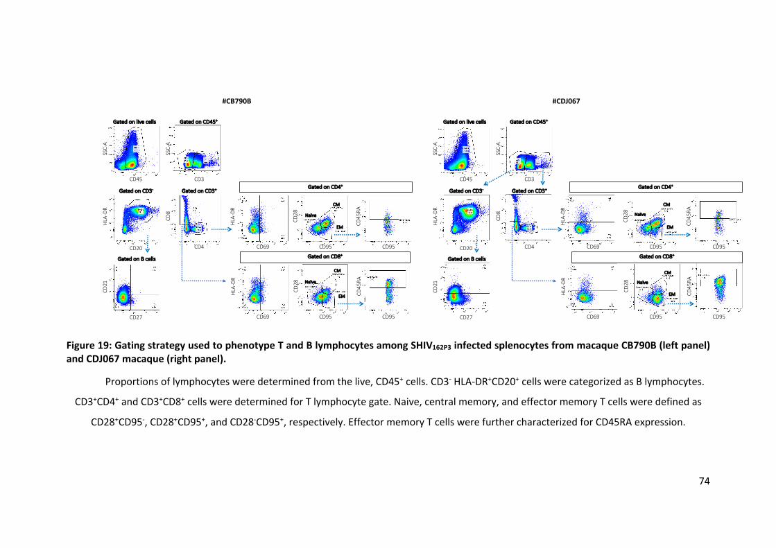
74
Figure 19: Gating strategy used to phenotype T and B lymphocytes among SHIV162P3 infected splenocytes from macaque CB790B (left panel) and CDJ067 macaque (right panel).
Proportions of lymphocytes were determined from the live, CD45+ cells. CD3- HLA-DR+CD20+ cells were categorized as B lymphocytes.
CD3+CD4+ and CD3+CD8+ cells were determined for T lymphocyte gate. Naive, central memory, and effector memory T cells were defined as
CD28+CD95-, CD28+CD95+, and CD28-CD95+, respectively. Effector memory T cells were further characterized for CD45RA expression.
SSC-
A
CD45
Gated on live cells Gated on CD45+
CD3
SSC-
A
Gated on CD4+
Gated on CD8+
Gated on CD3- Gated on CD3+
CD20
HLA
-DR
CD4
CD8
CD27
CD21
Gated on B cells
CD69
HLA
-DR
CD95
CD28
CD95
CD45
RA
CD69
HLA
-DR
CD95
CD28
CD95
CD45
RA
#CB790B
CM
EM
Naïve
Naïve
CM
EM
SSC-
A
CD45
Gated on live cells Gated on CD45+
CD3
SSC-
A
Gated on CD4+
Gated on CD8+
Gated on CD3- Gated on CD3+
CD20
HLA
-DR
CD4
CD8
CD27
CD21
Gated on B cells
CD69
HLA
-DR
CD95
CD28
CD95
CD45
RA
CD69
HLA
-DR
CD95
CD28
CD95
CD45
RA
#CDJ067
CM
EM
Naïve
Naïve
CM
EM

75
Figure 20: Gating strategy used to phenotype NK cells, monocytes, macrophages, DCs and neutrophils among SHIV162P3 infected splenocytes from macaque CB790B (left panel) and CDJ067 macaque (right panel).
Proportions of leukocytes were determined from the live, CD45+ cells. CD66+NKP44+ cells were categorized as neutrophils. CD3-NKG2A-
cells were classified as monocytes/macrophages. CD3-CD14-CD20- were determined for pDC and mDC as CD11c-CD123+ and CD11c+CD123-
CD16-, respectively. CD3-NKG2A+ or CD3-CD20-CD16+CD8+CD11c- cells were categorized as NK cells.
SSC-
A
CD45
Gated on live cells Gated on CD45+
CD66 NKP44
SSC-
A
CD3
SSC-
A
Gated on CD66-NKP44-
Gated on CD3-
CD20
HLA
-DR
Gated on CD20-
Gated on CD20-
CD3
NKG
2A
CD3
CD16
Gated on CD16+
CD11c
CD8
Gated on NK cells
CD16
NKG
2A
SSC-
A
CD69
Gated on CD3-NKG2A-
CD16
CD14
HLA-DR
CD20
Gated on CD14- Gated on CD20-
pDC
mDC
CD11c
CD12
3
CD16
CD11c
Gated on CD11c+
Neutrophil
monocytes/macrophages
#CB790B
SSC-
A
CD45
Gated on live cells Gated on CD45+
CD66 NKP44
SSC-
A
CD3
SSC-
A
Gated on CD66-NKP44-
Gated on CD3-
CD20
HLA
-DR
Gated on CD20-
Gated on CD20-
CD3
NKG
2A
CD3
CD16
Gated on CD16+
CD11c
CD8
Gated on NK cells
CD16
NKG
2A
SSC-
A
CD69
Gated on CD3-NKG2A-
CD16
CD14
HLA-DR
CD20
Gated on CD14- Gated on CD20-
pDC
mDC
CD11c
CD12
3
CD16
CD11c
Gated on CD11c+
Neutrophil
monocytes/macrophages
#CDJ067

76
iii) In vitro neutralizing activity of the bNAb 10-1074 against SHIV162P3
CAV transmission
We next determined the ability of 10-1074 bNAb to inhibit infection of PBMCs
when splenocytes from CB790B, CDJ067 and CEB071 were used as source of virus.
40,000 spleen cells, either from one single stock or mixing the splenocytes of two
stocks (at 1:1 ratio) were co-cultured with 120,000 PBMCs (activated with IL-2 and
PHA) in presence/absence of the Ab. The infection and neutralizing activity were
measured by RT-qPCR 7 days post-coculture.
Of note, whatever it was the stock of cells used, 10-1074 inhibited CAV
transmission with similar efficacy, as evidenced by the IC values reported in Table 7.
We expect that the in vitro results might be predictive of the in vivo potency of 10-
1074.
Table 7: Inhibitory concentration (IC) 50, 75 and 90 of 10-1074 in a PBMC-based neutralization assay using three stocks of spleen cells.
*Results with AX450 splenocytes are derived from previous experiments and
reported here as comparison.
iv) In vivo challenge study
We performed a pilot study to confirm in vivo the infectivity of the stocks.
Two juvenile female cynomolgus macaques (Macaca fascicularis) (animal codes;
CFF007 and CFC037) with an age of 2.6 years and 2.8 years, respectively, imported
from Mauritius, were involved in the study. The females were treated with 30 mg of
medroxyprogesterone acetate (Depo-Promone®) one month prior the first challenge.
10-1074 bNAbs (µg/ml) Splenocytes IC50 IC75 IC90
CB790B+CDJ067 0.42 0.54 1.03
CB790B+CEB071 < 0.062 0.3 0.43
CB790B 0.18±0.04 0.26±0.12 0.32±0.2
CDJ067 0.2 0.59 1.1
CEB071 1.07 1.33 1.52
AX450* 0.40±0.23 0.85±0.49 1.61±1.02

77
The in vivo challenge study described in section 4.3.1 a showed that the animal with
an MHC haplotype that did not matched the one of the donor cells, get infected after
one inoculation of cells. On the contrary the second macaque, who get infected after
seven inoculations, had an MHC haplotype that partially matched the one of the
“donor” (see Table 3). This observation prompted us to select animals with an MHC
haplotype that did not match those of the stock of spleen cell used for the challenge
(Table 8).
Table 8: MHC haplotype of the “donor” and the “receiver” macaques for pilot study.
We planned to challenge the two females intravaginal every week for up to 7
weeks (8 challenges maximum in total). Challenges will discontinue when the animal
will have a measurable viremia (>100 viral RNA copies per milliliter of blood plasma
by RT-qPCR) for two consecutive time points. During the study, blood and vaginal
secretion were collected to measure viral load and anti-SHIV antibody response. The
study plan is depicted in Figure 21.
Macaques MHC Haplotype
Donor CEB071 recH3H1/H7
CDJ067 recH3H5/H7
CB790B H3/H1
Receiver CFF007 H6/recH4H7
CFC037 H2/H5

78
Figure 21: Study plan to assay in vivo infectivity of SHIV162P3 infected splenocytes after vaginal challenge of two cynomolgus macaques.
We also decided to mix, at each challenge, splenocytes from two donors at a
ratio dilution 1:1 in order to induce an allogenic reaction and thus, facilitate the
infection. Moreover, this choice was justified by a practical reason, as we made an
estimation and the cells from one stock would not be sufficient to perform eight
challenges of 12 macaques in the future study we intend to perform (6 animals in the
control group and six animals treated with the antibody containing gel). Thus,
receivers were challenged with 1 x 10⁷ live splenocytes (mixed from two out of three
stocks) thawed 1 hour before inoculation, washed three times and resuspended in
RPMI medium. Table 9 shows the splenocytes combination we decided to use for
each challenge. The inoculation dose for each challenge was 6.16E+05 viral DNA
when combining cells from CB790B and CDJ067 and 4.73E+05 viral DNA when
combining cells from CB790B and CEB071.
Blood collection
Femalen=2
dpi 1rst IVag-41 0 7 14 21 28 35 42 49 56 63 70 1050 7 14 21 28 35 56 dpi last IVag
Depopromone®treatment
Viremia follow-up
77 84-3
21 3 4 5 6 7 8
Intravaginal inoculation10x10e6 cellsn=8 in total
Challenges will be stopped when the animal will have measurable viremia for 2 consecutive time points
Vaginal fluid will be collected as soon as animals will become infected. Vaginal secretion will be collected before the next challenge and will continue every weeks for the next 4 weeks, then every 2-weeks *Here, sampling are described for animals that will have 2 consecutives positive PCR after the first inoculation
Vaginal Fluid collection

79
Table 9: Splenocytes preparation for the intravaginal challenge.
Splenocyte preparation for each challenge (5x10⁶ living cells/stock) 1st 2nd 3rd 4th 5th 6th 7th 8th
Week 0 pi Week 1 pi Week 2 pi Week 3 pi Week 4 pi Week 5 pi Week 6 pi Week 7 pi CEB071 - CEB071 - CEB071 - CEB071 -
- CDJ067 - CDJ067 - CDJ067 - CDJ067
CB790B CB790B CB790B CB790B CB790B CB790B CB790B CB790B
The results of the challenge are shown in Figure 22: CFC037 and CFF007
displayed detectable viremia 1 and 2 weeks after the first inoculation, respectively.
CFC037 demonstrated a rapid infection with a detectable blood viral RNA load (200
copies/mL) at one week after the 1st inoculation, followed by a peak of infection at
day 14 and then a decrease and a stabilization until the end of study (84 days post
first inoculation). A similar kinetics was also observed in macaque CFF007 but the
animal had a detectable viremia (10 copies/mL) after the second challenge and the
peak of infection was reached after three challenges. Interestingly, the peak of
viremia was comparable in both macaques, with values of 2.80E+06 and 2.82E+06
RNA copies/mL for CFC037 and CFF007, respectively. In summary, CFC037 received
three inoculations and CFF007 received four inoculations.
Figure 22: Blood plasma viral load of macaque CFC037 and CFF007 following intravaginal exposure with SHIV162P3 infected splenocytes.
Finally, to confirm that infection of macaques was initiated by infected cells
and not by free virus present in the inoculum, we collected the supernatant of the

80
last wash of the cells stock CB790B + CEB971 and CB790B + CDJ067 before
inoculation of cells to macaques and we measured the residual viral RNA by RT-qPCR.
We detected residual amount of viral RNA in the last wash supernatants at an
average of 2.07x10⁴±1.27x10⁴ copies/mL for the combination CB790B + CEB971 and
an average of 6.51x10⁴±2.16x10⁴ copies/mL for the combination of CB790B + CDJ067.
Comparing our data with the previous publication by Sallé et al (2013), the last wash
supernatant of SIV infected cells of K833 stock, containing 3.51x10⁴ viral RNA
copies/mL, did not initiate the infection in macaques following vagina inoculation
[71]. As the last wash supernatants of our study have comparable residual viral RNA
content to the supernatant from the splenocytes K833, we expect that free viral
particles contaminating the inoculum were not responsible of the infection of
macaques we observed. We will confirm in vivo that the last wash supernatant is not
transmitting infection.
In conclusion, we have successfully established an in vivo model of SHIV CAV
transmission that will be further exploited to assess protection conferred by a gel
containing the anti-V3 bNAb 10-1074.

81
Chapter 5 Validation of the experimental approach
5.1 Characteristics of macaque semen and comparison with human semen
and related semen parameters
Using electroejaculation method, the volume of cynomolgus macaque semen
we could collect ranked from 100 to 500 μL, which is similar to what has been
described in rhesus macaques [397]. On the other hand, the volume of a human
ejaculation ranked between 2 to 5 mL [11]. The pH of cynomolgus macaque semen
was slightly alkaline, ranking from 7 to 9, similarly to human semen which pH rank
between 7.2-7.8 [106].
The major leukocytes in human semen are polymorphonuclear cells,
macrophages and T cells [72,130]. We previously showed that those populations are
also found in cynomolgus macaque semen, although with differences in the relative
proportions of each population [73] (Table 10). The cause of this discrepancy may be
due to the different techniques used. Whereas the technique usually used to quantify
leukocytes populations in human semen is numeration from semen cells smears, we
used multi-parameters flow cytometry to categorize the semen cell population in
cynomolgus macaques. Although variability between human and cynomolgus
macaque semen may really exist, cynomolgus macaque semen shared many
similarities with human semen. Notably, the major HIV-1/SIV target cells (CD4+ T cells
and macrophages) that are found in human semen are also present in macaque
semen [73]. They remain present at all stages of infection, despite a profound
depletion of CD4+ T cells. Analogously to what reported in HIV+ patients, the total
leukocytes count correlated with semen viral shedding [164]. Moreover, during acute
infection a “cytokine storm” is observed, with increased levels of pro-inflammatory
cytokines and chemokines. This inflammatory state may favor the transmission
events by cell-associated virus. Thus, the use of semen cells could be considered as a
pertinent model for studying cell-associated virus in HIV-1 mucosal transmission.

82
Table 10: Comparison of semen cell populations between human and macaque.
a. Anderson et al., 2010 and Wolff & Anderson, 1988 [72,130]; b. Bernard-Stoecklin
et al., 2013 [73].
5.2 Characteristics of infected splenocytes and comparison with infected
semen cells
The collection of semen cells to perform in vitro cell-to-cell transmission
assays and related experiments of bNAbs inhibition is challenging, due to the high
number of cells requested. Moreover, semen should be collected from animals at the
acute phase of infection, i.e. between day 10 and day 14 post-infection, otherwise
the amount of integrated viral DNA is not enough to induce infection, not in vitro
neither in vivo (as showed by our results). For all those reasons, the screening of
bNAbs with the potential to inhibit SHIV162P3 cell-to-cell transmission was performed
using splenocytes as surrogate of semen cells. In the majority of reported cases,
infected cells in the human semen mostly consist of T lymphocytes (75% of patients)
and macrophages (38% of patients) [122]. We confirmed that in macaques infected
with SIVmac251 strain, T lymphocytes and, to a lesser extent, macrophages also
represent the predominant infected cells in the male genital tract, which may seed
the semen [153]. When comparing semen cells and splenocytes from SHIV162P3
infected macaques, a lower number of macrophages and B cells was found in the
semen compared to spleen cells, however no major differences were observed in the
lymphocyte’s population (reported in chapter 4.2). Moreover, the infectiousness of
splenocytes was demonstrated following intravaginal inoculation using either
SIVmac251 infected splenocytes [71] or SHIV162P3 infected splenocytes (unpublished
results reported in chapter 4.3). Taken together, we consider that infected
splenocytes shares enough similarities with macaque and human semen and
Population Human semena Cynomolgus semenb
Polymorphonuclear 60-70% 26%+6.7%
Macrophages 20-30% 22.22%+5.06%
Lymphocyes 4-5% 15.95%+4.29%

83
represent a valuable tool for the development of CAV mucosal transmission models
and related inhibition by bNAbs.
5.2 Target cells used in in vitro assays of cell-to-cell transmission
TZM-bl and PBMC-based neutralization assays have been widely used by the
scientific community [274,401].
TZM-bl is a cell line, containing a tat-inducible luciferase reporter gene. TZM-
bl based neutralization assay is a standardized, reproducible and rapid method,
which is suitable for screening tests [274]. However, the cells express 100 times
higher level of CCR5 coreceptor than activated or non-activated human PBMC, which
is less physiological and possibly affects interaction of virus-antibody-target cell.
The PBMC-based assay is regarded as a gold standard assay as the target cell
is a natural target cell of HIV [274]. The assay accesses not only the viral attachment
and entry but also cell-to-cell transmission [401]. Moreover, the outcomes from the
PBMC assay may be more predictive of in vivo results [274]. We have noticed that
the assay displayed higher sensitivity than the TZM-bl assay for detection of
neutralization activity to block cell-to-cell transmission. Despite of the advantages,
the assay is time consuming and less consistent compared to the TZM-bl assay.
Considering the pros and cons, we have decided to use the PBMC assay as a
standard neutralization assay for bNAbs evaluation of inhibition of cell-to-cell
transmission. As we will further evaluate the bNAbs efficacy in vivo in macaques, the
use of macaque PBMC rather than human PBMC would have, however, represented
a more physiologically relevant system.
5.3 Implication of progesterone treatment of cynomolgus macaques to
facilitate cell-to-cell transmission
In the study performed to evaluate in vivo cell-mediated transmission (see
chapter 4.3), the inoculated female macaques have been treated with
Medroxyprogesterone acetate (DPMA, Depo-Provera®) 1 month before the
challenge. It has been reported that DPMA treatment in women and female

84
macaques induced a thinning of the vaginal epithelium, imitating the luteal phase of
the menstrual cycle [402,403]. Certainly, this does not represent the general
situation of individuals exposed to HIV-1. Moreover, progesterone implants not only
increased the susceptibility to SIV infection in macaques following vaginal challenge
[404], but also induces changes in mucosal immune system, particularly on NK and T
cells activation [405]. The genetic diversity of the transmitter/founder virus
population was higher in treated female macaques, suggesting an increase of the
number of variants that crossed the epithelial barrier. Although, progesterone
treatment possibly introduced a potential bias, we considered that it was not a major
concern for our study. However, future work will be needed to address the influence
of the menstrual cycle in transmission of infection after exposure of the cervix or
vagina to cell-associated SIV.

85
Chapter 6 Discussion and perspectives
6.1 General discussion
6.1.1 Determination of the immunological response in semen during
SIV/HIV-1 infection
a) Seminal cytokine/chemokine modulation during infection
Many studies have reported that HIV-1 infection induced a dysregulation of
cytokine profiles in the periphery and in the semen [178,406–408]. To understand
what the impact of SIV infection on the seminal cytokine network is, we determined
the level of 29 cytokines, chemokines and growth factors in semen.
We identified 7 molecules (IL-8, IL-13, IL-15, IL-18, IFN-γ, RANTES and IP-10)
that were affected by SIV infection: significantly increased in SIV+ animals compared
to SIV- animals. Among them, IL-8, IL-13, IL-15, IL-18 and RANTES positively associated
with blood viral load (BVL) and seminal viral load (SVL). Additionally, IL-1β, IL-4 and
TGF β1-3 also correlated to BVL and SVL. The correlation of SVL/BVL and seminal
cytokines/chemokines is depicted in Table 11.
Table 11: Correlation between viral loads and seminal inflammatory molecules.
Colors indicate classification: pro-inflammatory, chemokines, Th-1 cytokines, Th-2
cytokines and immunomodulators.
This inflammatory status induced by SIV infection may favor mucosal
dissemination, especially through cell-mediated transmission. Pro-inflammatory
cytokines, like IL-8, IL-15 and IL-18, trigger infected cells activation. Consequently,
the increase production of them can boost viral replication [409]. Due to the fact that
Correlated to BVL and/or SVL No correlation to VL
Upregulated during acuteinfection
IL-8, IL-15, IL-18, RANTES, IL-13, IFN-γ
Unchanged during acute infection IL-1β, IL-4, TGF β1-3 TNF-α, IL-2, IL-5
86
TGF-β displays strong immunosuppressive activity, the increased levels of TGF-β may
modulate the innate immune response to the pathogen [110,111,129,410], which
affects the host immune response. Also high levels of chemokines, like RANTES, may
induce a migration of mucosal resident virus target cells towards the epithelial
surface along a chemokine gradient [411]. Although, RANTES has potent anti-viral
properties, notably by inhibiting by competition the fixation by viral particles to their
co-receptor CCR5 [412–414], this activity may not affect cell-to-cell transmission.
While IFN-γ displays inhibitory effect on HIV-1, including antiviral, anti-proliferation,
immunomodulation, Th2 cytokines (IL-4, IL-5 and IL-13) exhibited both stimulatory
and inhibitory effect on HIV-1 replication [409]. As many upregulated molecules have
a stimulatory effect on HIV-1 replication, the inhibitory activity of IFN-γ and Th2
cytokines may be inefficient.
Many studies have used different experimental approaches to investigate
cytokine productions by cells from HIV+ subjects or from in vitro HIV-1 culture [409].
According to the experimental design used, the observed cytokine effects in vitro
may be unable to predict the effect mediated by cytokines/chemokines presence in
seminal plasma in vivo. The modulation of cytokines levels in semen that we
observed could possibly influence the recruitment and activation of viral target cells
and generates an environment that may facilitate HIV-1/SIV sexual transmission. The
answer of this open question remains to be explored.
b) Cellular response in semen
i) Phenotype of seminal CD8+ T cells
Systemic CD8+ T cell responses are involved in host HIV-1 immune control by
controlling viral replication and delaying disease onset [415–418]. Because of the
plausible capacity of CD8+ T cells to modulate HIV-1 infectivity, we provide here an
extensive characterization of seminal CD8+ T cells. We studied their expression of
differentiation, activation, migration and adhesion markers.
Based on our analysis, seminal CD8+ T cells exhibited a phenotype that is
typically found in mucosal resident cells. They displayed a memory phenotype

87
(CD95+) with large proportion of cells expressing activation markers (CD69 and HLA-
DR), the migration marker CXCR3 and the integrin LFA-1. Of note, the differentiation
profile and expression of HLA-DR and LFA-1 was not influenced by SIV infection. On
the contrary, the proportion of CD69+, CCR5+ and CXCR3+ cells were elevated in SIV+
animals. Following the infection, we observed a significantly durable increase of
CD69+ cells, which could be a marker of the generalized immune activation induced
by HIV-1/SIV infection. In HIV+ men, the activation of seminal T cells and a correlation
between the proportion of CD69+ T cells and SVL has been described [419]. Although,
we did not find a correlation because of the modest number of studied macaques,
we observed a correlation between the percentage of CD69+ CD8+ T cells in blood
and in seminal plasma. Similar observations have been done in human but regarding
the proportion of CD45RA-CD38+CD8+ T cells [420]. Additionally, the percentage of
CCR5+CXCR3+ cells significantly increased in infected macaques. These cells tend to
migrate towards tissues in which inflammation occurs [421].
Our result suggests that the activation of seminal CD8+ T cells occurs in the
male genital tract, implying T cell compartmentalization between blood and semen.
ii) SIV specific CD8+ T cell response
It has been reported that elite controllers and long-term non-progressor HIV-
1+ patients displayed highly polyfunctional T cells [422,423]. Moreover, SIV infected
rhesus macaques that were depleted of CD8+ T cells exhibited the rising of plasma
viral load, indicating that CD8+ T cells may function in controlling viral replication
[415]. According to the prospective role of CD8+ T cells, we evaluated SIV specific
CD8+ T cells response in macaques during SIV infection.
We showed that 3 out of 7 macaques showed a detectable systemic and
semen SIV-specific T cell response. They had a high proportion of IFN-g/MIP-1b/TNF
responses, which were about 10-fold higher in semen than in blood. Despite the
detection of SIV-specific response in semen, this activity was not correlated with
control of viral shedding, either in blood or in semen, which is similar to what has
been reported in chronic HIV+ patients [184]. The profile of HIV specific CD8+ T cell

88
response in blood of progressor is less functional compared to non-progressor [423].
It would be interesting to elucidate the profile of semen SIV-specific T cell responses
in progressor and elite controller macaques. Although our results suggest that the
SIV- specific T cell response did not affect viral replication, the immune activation
mediated by CD8+ T cells probably has an influence on the mucosa of semen receiver.
Due to the limited number of studied macaques, we were unable to observe
a significant correlation between specific T cell responses and local viral shedding. It
would be interesting to increase the number of animals in future studies. Moreover,
it remains to assess whether semen SIV-specific T cells are able to kill semen infected
cells, which possibly influences semen infectivity.
c) Humoral response in semen
HIV-1 specific antibodies could prevent or partially control mucosal HIV-1/SIV
entry [213,224]. Using western blots, HIV-specific antibodies were detected in semen
[424]. The Ab may form an immune complex with HIV cell free virus, which was
possibly an explanation of the low sexual transmission rate and semen infectivity in
vitro [164,173]. As seminal HIV-1 specific antibody activity remains poorly described,
our study explored SIV-specific Abs in terms of quantity and function.
We measured the level of SIV-specific Ab titers in blood and semen. The level
of total Abs (IgG, IgM and IgA) at day 51 p.i. was lower than at chronic stage (day 148
p.i.). Moreover, Ab level at chronic phase was significant higher in blood than semen.
A positive correlation between semen SIV-specific Ab titers and semen viral RNA load
was observed, suggesting that the local humoral response fail to control viral
shedding. We further investigated the neutralizing activity of seminal SIV-specific
Abs. We observed a weak neutralizing activity in blood and semen at chronic stage
of infection.
As reported by others [425], we observed a toxic effect at low seminal plasma
dilution. We were unable however to purify IgGs from semen samples because of the
low amount available. For the same reason, we did not analyze the presence of IgA
in seminal plasma in our study. We cannot exclude the presence of anti-SIV-IgA that

89
mediates a protective effect (through inhibition of viral transcytosis or a neutralizing
effect). However, semen has been repeatedly reported to contain mostly IgG [213–
215]. Nevertheless, these aspects warrant further study.
In the RV144 vaccine trial, the presence of Abs that mediate ADCC was
reported to correlate with protection [426]. Moreover, ADCC activity has been
demonstrated in cervico-vaginal fluids (CVF) from HIV-1 infected women and shown
to be associated with lower genital viral load [248,427]. ADCC mediated by IgA in CVF
from exposed-noninfected (ESN) women and breastmilk is associated with a lower
risk of sexual and mother-to-child transmission, respectively [428,429]. Ruiz et al
[430] recently reported that the magnitude of ADCC responses was elevated within
chronically HIV-1-infected individuals who did not transmit infection to their
heterosexual ESN partners. Whether these responses were also elevated at the most
relevant sites of heterosexual transmission, such as genitorectal mucosal sites,
warrants further investigation. Additionally, studies on HIV controllers have reported
a higher level of Abs with ADCC activity than in progressor patients [431]. However,
little is known about ADCC activity in the MGT and Fc-mediated antibody functions
have been mostly overlooked in semen. Only one study reported the presence of
ADCC Abs in the semen of HIV-1-infected individuals [223]. The presence of ADCC-
mediating Abs has not been previously evaluated in the semen of macaques infected
with SIV.
Thus, we evaluated ADCC function of semen SIV-specific IgG by ELISA based
FcgRIIIa binding assay. In semen at 51 dpi, half of macaques had Ab that were able to
engage the FcgRIIIa. Their activity increased and was detected in 4 out of 6 macaques
in the samples at chronic stage. We also observed a positive correlation between
FcgRIIIa binding activity and the level of Ab titers in both blood and semen.
d) Potential of seminal HIV-1/SIV infected leukocytes to transmit infection
We demonstrated that seminal leukocytes collected from SHIV+ macaques
were able to transfer infection in vitro to susceptible target cells (PBMC and TZM-bl).
Although the purification method used for semen leukocyte sorting may not get rid
of 100% spermatozoa, we consider that it may not be a major concern as

90
spermatozoa are not widely supported as a source of virus [11,122,149]. Our results
supported a prospective role of seminal leukocytes on HIV-1/SIV CAV transmission.
As we used the totality of cells to perform the in vitro neutralization assay,
we could not systematically determine either proviral DNA or the phenotype of
seminal leukocytes. Seminal leukocytes carry HIV-1/SIV proviral DNA and have the
potential to transmit infection. The prevalence of proviral DNA ranged from 21% to
65% in semen of HIV+ subjects [72]. Although the levels of HIV RNA and DNA were
decreased following ART treatment, proviral DNA as CAV was present in semen for
several months [432,433] and exhibited in vitro infectivity [434]. Moreover, our
group reported the presence of infected lymphocytes and macrophages in the semen
of SIVmac215-infected macaques during progressive infection [73]. According to
these results, we could imply that the transfer of infection in our in vitro assay was
mediated by infected seminal leukocytes. Moreover, we observed that seminal viral
load possibly predicted the capability of seminal leucocytes to transmit infection in
vitro (Table 12).
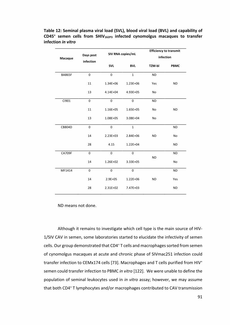
91
Table 12: Seminal plasma viral load (SVL), blood viral load (BVL) and capability of CD45+ semen cells from SHIV162P3 infected cynomolgus macaques to transfer infection in vitro
ND means not done.
Although it remains to investigate which cell type is the main source of HIV-
1/SIV CAV in semen, some laboratories started to elucidate the infectivity of semen
cells. Our group demonstrated that CD4+ T cells and macrophages sorted from semen
of cynomolgus macaques at acute and chronic phase of SIVmac251 infection could
transfer infection to CEMx174 cells [73]. Macrophages and T cells purified from HIV+
semen could transfer infection to PBMC in vitro [122]. We were unable to define the
population of seminal leukocytes used in in vitro assay; however, we may assume
that both CD4+ T lymphocytes and/or macrophages contributed to CAV transmission
Macaque Days post
infection
SIV RNA copies/mL Efficiency to transmit
infection
SVL BVL TZM-bl PBMC
BA865F 0
11
13
0
1.34E+06
4.14E+04
1
1.23E+06
4.93E+05
ND
Yes
No
ND
CI901 0
11
13
0
1.16E+05
1.08E+05
0
1.65E+05
3.08E+04
ND
No
No
ND
CB804D 0
14
28
0
2.23E+03
4.15
1
2.84E+06
1.22E+04
ND
ND
No
ND
CA709F 0
14
0
1.26E+02
0
3.33E+05 ND
ND
No
MF1414 0
14
28
0
2.9E+05
2.31E+02
0
1.22E+06
7.47E+03
ND
ND
Yes
ND

92
to TZM-bl and PBMC. On the base of the phenotype characterization and seminal
leukocyte infectivity in vitro, we hypothesised that T lymphocytes and macrophages
may play a role in CAV transmission during sexual intercourse as both cells endowed
with adhesion and migration markers [73]. In spite of the fact that there is no report
of the infectivity of human seminal DCs, our laboratory reported that DCs were also
present in the semen of SIV+ macaques at all stage of infection [73]. According to this
observation, we should not exclude a prospective role of DCs in CAV transmission.
Seminal cells derived from HIV+ subjects demonstrated in ex vivo culture that
they were able to penetrate ectocervical epithelium but remained trapped in cervical
mucus at endocervical surface [435]. It remains to be investigated whether similar
phenomenon of trapped seminal cells could be displayed in vivo. As there is no
empirical evidence that seminal leukocyte can transmit HIV-1 CAV across mucosal
barrier in vivo up to date, it would be our main interest to provide evidences of semen
cells crossing the mucosal barrier on the FRT or of the rectum in macaque models.
e) Plausible impact of HIV-1/SIV+ semen exposure on female reproductive
tract
Our observations together with human studies performed by others shown
that during HIV-1/SIV infection the seminal compartment is highly affected, with
modifications in terms of cytokine/chemokine, antibodies and leukocytes phenotype
and function. None of the immune responses we analyzed could control viral
shedding. In the presence of seminal fluid, the innate antiviral activity of cervico-
vaginal lavage fluids declined, possibly indicating that factors in the seminal fluid
affected the FRT immune response [436]. All these observations suggest that
immunological modifications of semen during SIV infection might influence the
efficiency of transmission across the FRT during sexual intercourse. It remains to be
deciphered whether and/or how HIV-1 transmission within the FRT could be affected
by changes in the content of semen immunomodulatory factors.
Seminal plasma contained many immunological factors that could favor or
inhibit HIV-1 transmission [105,437,438]. In the context of cell free virus
transmission, many studies reported that factors present in the seminal plasma could

93
inactivate HIV-1, including anti-HIV antibodies [439,440], X4/R5 chemokines [129],
SLPI, lactoferrin, and defensins [441]. Despite of these studies implying a viral
inhibitory effect of seminal plasma, controversial results have been reported about
the impact of seminal plasma on SIV infection. Using NHP models, some studies have
shown that in low dose SIV cell-free inoculations, the presence of seminal plasma
could favor mucosal transmission [187,391]. In contrast, other NHP studies reported
that the presence of semen during SIV vaginal inoculation had no effect on the
infection potency [442,443]. Additionally, a recent study has shown that 20 weeks
repeated semen exposure of rhesus macaques determined a reduction of the
transmission rate of SIVmac251 cell free infection [444]. In spite of those studies
focusing on cell-free virus, the influence of seminal plasma on the infectivity of semen
leukocytes should also be taken into consideration. More works need to be done to
clarify the effect of seminal plasma not only on HIV-1/SIV cell free but also on HIV-
1/SIV CAV transmission.
In addition, leukocytes may be affected by the presence of semen. The slightly
basic pH of semen in humans and macaques should not impact the vitality of
leukocytes [108] but other factors in semen possibly have a cytotoxic effect on the
cells [105]. Moreover, it has been reported that seminal plasma of HIV negative
donors could suppress CD4 expression by blood T cells, whereas blood T cells
exposed to HIV- seminal plasma increased CCR5 expression, which could favor the
transmission of R5-tropic strains [209]. Furthermore, seminal leukocytes infectivity is
probably raised by the strong seminal inflammation occurring during the acute phase
of HIV-1/SIV infection. As we still do not have a conclusive information of the impact
of seminal plasma on seminal leukocytes, more work needs to be done to investigate
the activity of seminal plasma on virus-infected cells in semen and on CAV HIV-1/ SIV
infectivity in vivo.
In vitro and in vivo studies demonstrated that semen was able to shape the
composition of immune cell populations within the FRT [445]. Whether similar
effects could be observed in HIV-1/SIV sexual transmission need to be further
evaluated. If seminal plasma affects semen infectivity, this could be an issue to
94
consider when adding seminal plasma to infected cells in in vivo models of male-to-
female transmission.
6.1.2 Potential of bNAbs to prevent cell-to-cell heterosexual transmission
bNAbs gained interest as a promising tool for HIV-1 treatment as being able
to potently prevent cell-free infection in in vivo models [314–319]. In spite of the
achievement to control cell-free infection, bNAbs in the context of CAV transmission
were poorly investigated.
Our in vitro results shown that a subset of bNAbs could efficiently prevent
cell-to-cell transmission of SHIV162P3, which is a “neutralization-resistant”, tier 2 virus.
Similarly to what has been reported by others, we observed that bNAbs could inhibit
cell-free infection easier than cell-associated transmission [337–339,344]. While the
improvement of protection against cell-to-cell transmission by 1st generation bNAbs
(2F5, 4E10 and 2G12) was clear when the bNAbs were used in combination, this
phenomenon was not so clearly observed when we used 2nd generation bNAbs. This
additive potency may be explained by the fact that combination of bNAbs targeting
different epitopes of the virus could possibly inhibit the contact between the infected
cells and susceptible cells better than the individual antibody did. Indeed, it was
described that stabilization of the blocking by Abs needed many molecules of Abs to
prevent a contact between the virus and the target cells [446]. Moreover, bNAbs
targeting distinct epitopes displayed an independent binding [304,447], implying that
possibly no cross-competition between those bNAbs occurred [448]. This may
explain why we did not observe an antagonized effect when combining 2nd
generation bNAbs.
Despite of the non-additive effect observed when combination of 2nd
generation bNAbs were used (10-1074, PGT128, PGT151 and N6), the combination
of bNAbs may give more protection coverage and potency against diversity of HIV-1
strains. To prove this hypothesis, we would need to prepare stocks of splenocytes
from macaques infected with different SHIVs and use them as CAV source in in vitro
and in vivo assays.

95
The in vitro experiments we performed allowed us to identify 10-1074 as the
most potent bNAbs inhibiting cell-to-cell transmission of SHIV162P3. Thus, this Ab has
been selected to further evaluate its efficacy to block cell-to-cell transmission in vivo
using SHIV162P3-infected splenocytes as a viral challenge stock. 10-1074 displayed
high breadth and potency against various viral strains in cell-free assays. The Ab was
able to neutralize 77.7% of primary HIV-1 isolates from 179 HIV-1+ individuals, with a
mean IC80 of 0.67 µg/mL [328]. In TZM-bl neutralization assay, the Ab could
neutralize 60.5 % of 306 tested HIV-1 pseudoviruses, composing of 13 subtypes, at
an average IC80 of 0.18 µg/mL and neutralize up to 88.5% of 26 clade B strains with
a mean IC80 of 0.13 µg/mL. As the SHIV162P3 virus that we used is also a clade B strain,
it is not surprising that this Ab was able to inhibit SHIV162P3 CAV transmission. In
addition to its efficacy in vitro, the potency of 10-1074 against cell free viral challenge
has proven in NHP studies. The Ab could protect rhesus macaques from SHIVAD8EO
rectal challenge [316]. When used in combination with 3BNC117 and PGT121, the Ab
cocktail was able to suppress viremia to undetectable level in SHIV162P3 or SHIVAD8
chronically infected rhesus macaques [320,321]. In human clinical trials of 10-1074,
the Ab displayed a favorable potency by inducing a rapid decrease of viremia in 10-
1074-sensitive patients, however, the emergence of escaped virus occurred after the
first weeks of infusion [328].
Interestingly, a combination of 1st generation bNAbs (2F5+2G12+4E10) and
the individual Ab 10-1074 were capable to prevent cell-to-cell transmission mediated
by seminal leukocytes in TZM-bl and PBMC assay respectively. Although our results
need to be confirmed in a larger cohort, they proof the potential of bNAb to
prevention HIV-1 sexual transmission.
6.1.3 In vivo inoculation of splenocytes collected from SHIV162P3 infected
macaques
In the last part of this work, we assessed the capacity of splenocytes to
transmit infection in vivo. The previous work of the laboratory proved CAV mucosal
transmission using SIVmac251 infected splenocytes as a viral challenge stock. The
numbers of DNA copies to successfully transmit infection by vaginal exposure was

96
estimated at 6.69 x 10⁵ ± 2.08 copies of SIV DNA [71]. The inoculum used in the case
of SHIV-infection contained 4.73E+05 or 6.15E+05 copies of SIV DNA. At each
experiment, 107 lived SHIV162P3-infected splenocytes obtained from 2 different stocks
were inoculated. We attained establishment of systemic infection following
intravaginal inoculations in two female cynomolgus macaques, treated with
progesterone 30 days before challenge. The inoculated macaques became infected
1 and 2 weeks after the first splenocytes challenge.
For the challenge stocks, we pooled the cells from two out of three different
splenocyte stocks in order to assure the completion of the future challenge study
using the same splenocyte stocks. Putting together the immune cells of different
animals may represent a hindrance of our experiments. It may induce cellular stress,
aggregation and mortality, thereby limiting the infectivity of the inoculated cells.
However, when used in vitro the pooled splenocytes transmitted infection with an
efficiency compared to splenocytes from animal AX450, possibly implying that mixing
different splenocytes had no impact on their infectivity in vivo.
Indeed, HIV-1 mucosal transmission is a rare event [85], and when it happens,
the transmitter founder virus population is very small [14,449,450]. According to this
fact, the high dose inoculum used in cell-free challenge is generally high to ensure
successful establishment of viral infection upon the viral challenge. In contrast, our
model of CAV mucosal transmission highlighted that AID50 dose used to infect
animal models by mucosal exposure do not need to be high. Our results are
concordant with the fact that HIV-1 CAV transmission is more potent compared to
cell-free virus transmission [5,85,86], implying that possibly lower inoculation doses
could establish systemic infection. To confirm that cell free virus is less potent in
transmitting infection than CAV transmission via mucosal route, it would be
interesting to intravaginally inoculate female macaques with cell-free SHIV162P3 using
similar AID50 dose as CAV challenge.
Another interesting evolution of the project might be to inoculate
splenocytes in the presence of seminal plasma in order to better mimic what happen
during natural transmission of the virus. Moreover, challenging the macaques with

97
stocks of splenocytes expressing other strains of SHIV, such as SHIV 325C [451], SHIV-
327C [452] and/or SHIV encoding transmitted/founder HIV-1 envelope such as SHIV-
AE6 [453], SHIV 375 mutation [454], will make our model more relevant as it would
reflect the variety of HIV-1 strains found in natural infection. Finally, as HIV-1
infection via the rectal route represented the highest estimated risk of sexual
transmission [7], it will be interesting to further develop models of rectal challenge
using splenocytes as a CAV challenge stock and test the inhibitory efficacy of bNAbs.
6.2 Perspectives
In this study we have characterized the anti-viral immune response that take
place against SIV infection in the semen compartment and provided evidences of
factors that can potentially modulate semen infectivity. Moreover, we have
demonstrated that infected semen cells can transmit infection in vitro and that in
vivo transmission can be initiated by donor cells (splenocytes) in the context of
SHIV162P3 transmission. Together with previous results obtained by our group [71,73],
we provided strong evidences that semen leukocytes may play a role in HIV-1 sexual
transmission, that seminal plasma and the inflammatory environment of the MGT
during infection have an impact on transmission, and finally that bNAbs represent a
valuable tool to prevent CAV transmission.
However, more work needs to be done to fully demonstrate our hypothesis.
6.2.1 Short-term perspectives
a) 10-1074 bNAb efficacy trial against SHIV162P3 cell-to-cell transmission
(intravaginal challenge) in cynomolgus macaques
The success of the pilot study we conducted to induce systemic infection of
macaques intravaginally inoculated with SHIV162P3 infected splenocytes (see chapter
4.3) and the in vitro results of bNAbs-mediated protection against cell-to-cell SHIV
transmission, prompt us to evaluate the ability of 10-1074 bNAb to inhibit cell-to-cell
transmission in vivo.

98
Female cynomolgus macaque will be treated with progesterone at least 30
days before the challenge. The animals will be divided into 2 groups (treatment and
control group), including 6 macaques per group. 10-1074 bNAb will be prepared as
an HEC gel formulation to be vaginally applied at a concentration of 5 mg/g of gel.
The gel will be applied one hour before challenge. We will use the same challenge
strategy used in the pilot study, i.e. mixing splenocytes from macaques CDJ067,
CEB071 and CB790B. During the in vivo study, blood and vaginal fluid will be collected
from the animals during the follow up to monitor viral loads and define their immune
response and neutralization activity. The pharmacokinetic study to evaluate the
stability of the 10-1074-gel is currently ongoing.
b) Development of the in vivo model of rectal CAV transmission
Due to the fact that HIV-1 infection via rectal route represent the highest
estimated risk of sexual transmission [7], our CAV model using splenocytes could be
further implemented to study rectal transmission.
As SHIV162P3 infected splenocytes could transmit infection following
intravaginal challenge, the same approach could be used for rectal inoculation. If we
achieve to set up the CAV rectal transmission model using infected splenocytes, it
will be interesting to test the efficacy of the bNAbs panel in this model.
c) Development of ex-vivo model for CAV transmission
We demonstrated here that HIV-1/SIV infection in vitro could be mediated by
a source of CAV like splenocytes and semen leukocytes. It would be interesting to
investigate the mechanisms underlying CAV transmission using ex vivo explant
models. Those kind of models would for instance allow to investigate if the formation
of a virological synapse with either epithelial cells, followed by the transcytosis of
viral particles through the epithelial surface, or with mucosal HIV/SIV target cells
(DCs, macrophages or CD4+ T cells), or transmigration through the mucosa, could
initiate and establish cell-to-cell transmission.
To achieve this purpose, we might develop an ex vivo model using tissue
explants (from macaque colo-rectal or cervico-vaginal tissues). The source of cell-

99
associated virus will be SHIV162P3 infected cells, either splenocytes or semen
leukocytes collected from cynomolgus macaque. This kind of experimental model
allow also to study the interaction between bNAbs and infected cells and to
determine the ability of bNAbs to prevent cell-to-cell transmission. Moreover, it
allows to consider the buffering effect of seminal plasma.
d) Improvement of CAV transmission model by preparing infected
splenocytes from various SHIV strains
In this study we have used SHIV162P3 infected splenocyte stocks. SHIV162P3 is a
clade B strain which is an appropriate strain, as HIV-1 clade B is the major circulating
strain in North America and Western Europe [274]. However, using individual viral
strain did not represent the diversity of HIV-1 infection. In HIV pandemic, not only
clade B virus but also clade A, C, D, CRF01_AE and CRF01_AE are responsible for the
epidemic [274]. Moreover, a recent study demonstrated that single bNAbs decreased
their protection activity when double strains of SHIV162P3 and SHIV-325c virus were
present in the viral inoculum in rhesus macaques [455]. According to this, we need
to improve the infected cell stocks to better represent the diversity of natural
infection.
To reach this goal, we need to produce stock of splenocytes collected from
cynomolgus macaques infected with other strains of SHIV, such as SHIV-325C [451],
SHIV-327C [452] and/or SHIV encoding transmitted/founder HIV-1 envelope such as
SHIV-AE6 [453], SHIV 375 mutation [454]. This will allow us to assess the coverage
potency of the bNAb panel. Furthermore, this type of CAV transmission model will
closely mimic the diversity of cell-to-cell HIV transmission and allow us to further
investigate the underlying mechanisms.
6.2.2 Long-term perspectives
a) Deciphering mucosal CAV transmission mechanisms: in vivo inoculation
of sorted semen leukocytes
We have evidences that SHIV infected splenocytes were able to initiate
infection in macaques after vaginal exposure. However, semen cell composition is

100
different compared to the inoculum of splenocytes we used, among which a majority
of lymphocytes and very few macrophages are found. A more relevant model would
assess the capacity of semen leucocytes to transmit infection in vivo. Moreover, if we
could confirm that semen leukocytes are able to transmit infection in vivo after
mucosal exposure, it would be interesting to assess the migratory and dissemination
properties of the cells and to determine which type of cells are able to transmit
infection: CD4+ T cells and/or macrophages. To investigate the migration and
dissemination of seminal leukocytes, we could label the cells with radioactive
elements (18Fluor or 111In for example) and track them in vivo by imaging using
Positron Emission Tomography – PET, that has been established in our laboratory.
This technique will allow us to investigate the dynamics of the potential migration of
the cells after the challenge. Similar experiments might be performed in presence of
NAbs.
However, the major constrains will be the number of seminal leukocytes
collected and the variable semen infectivity state, which would greatly affect viral
replication and transmission. Indeed, previous attempts performed in the laboratory
up to know using both the intra-rectal and intra—vaginal transmission route were
not successful.
b) Impact of the vaginal dysbiosis on the protective efficacy of a bNAbs-
containing gel
PrEP is currently considered as a promising preventive strategy against HIV-1
[456]. ARTs including tenofovir and dapivirine as topical PrEP could prevent HIV-1
infection in clinical trials [457–459]. In our future study, the 10-1074 antibody we’ll
be used as a gel to prevent vaginal mucosal CAV transmission by SHIV162P3 infected
splenocytes. It has been reported that vaginal dysbiosis could affect the efficacy of
topical tenofovir [460,461]. Whether this modification could also affect the
protective effect of a topical 10-1074 gel, needs to be investigated.
Although the cause of genital inflammation remains unknown, genital
inflammation is associated with vaginal microbiome modification and/or bacterial
vaginosis (BV) [462,463]. BV affected the health of the FRT and increased the risk of

101
HIV-1 infection in women [464]. In our unit the composition of the cynomolgus
macaques vaginal microbiota has been characterized [465]. Compared to human, the
vaginal microbiome in cynomolgus macaques is similar to the community state type
IV-A that usually is associated with dysbiosis [465]. In women, the most common
bacteria genus is Lactobacillus spp. [466–469], which plays a role in maintaining the
protective environment of the FRT against invading pathogens by producing lactic
acid and bacteriocins [470,471]. Due to the discrepant composition between the
vaginal microbiota in human and macaques, our laboratory is currently trying to
induce a healthy vaginal flora in macaques by vaginal inoculation with lactobacillus.
If we’ll be successfully established this model, it will represent a tool to investigate
the effect of a healthy and dysbiosis microbiota on the efficacy of the bNAb-
containing gel.

102
Synthèse de thèse
Avec le nombre croissant de preuves empiriques et d'intérêt sur la
transmission du virus intercellulaire (TVI) médiée par le sperme chez des sujets
séropositifs pour le VIH, il est important de mieux comprendre la modulation
dynamique du sperme pendant l'infection par le VIH-1 et de trouver les stratégies
optimales pour empêcher la dissémination virale. Dans ce projet de thèse, nous
avons utilisé des tests in vitro et le modèle de primates non humains d'infection de
cynomolgus macaques pour répondre à des questions spécifiques. Plus précisément,
les objectifs du projet sont d'étudier les réponses immunitaires dans le sperme
pendant l'infection par le virus de l'immunodéficience simienne (VIS) et d'évaluer la
capacité des anticorps neutralisants à large spectre (bNAbs) pour bloquer la TVI. Les
deux objectifs sont divisés en 2 études.
Dans la première étude, nous avons étudié la réponse immunitaire innée et
adaptative anti-VIS dans le sperme de cynomolgus macaque pendant l'infection par
VISmac251. Nous avons mené une étude approfondie sur un total de 53 animaux.
Nous avons analysé l'excrétion virale et les profils cytokiniques et chimiokiniques du
liquide séminal. De plus, nous avons caractérisé le phénotype des lymphocytes T
CD8+ du sperme et la réponse des cellules CD8+ T spécifiques au VIS en utilisant la
stimulation ex vivo des cellules du sperme avec des peptides gag du VIS et le
marquage intracellulaire avec des anticorps ciblant CD45RA, IL-2, IFN-γ, TNF et MIP-
1β. Pour déterminer la réponse en anticorps, nous avons quantifié les titres
d'anticorps spécifiques au VIS dans le sperme de macaques à différents stades de
l'infection. Nous avons également effectué un test de neutralisation in vitro et un test
de liaison au FcγRIIIa, indicateur de la fonction ADCC de l’anticorps afin d’évaluer la
fonction potentielle de l'anticorps spécifique du VIS.
Dans le sperme de macaque, nous avons observé que le niveau de
nombreuses cytokines étaient affectées par une infection par le VIS. Plusieurs
cytokines pro-inflammatoires (IL-8, IL-15, IL-18, IFN-γ, IP-10 et RANTES) et cell
immunomodulatrice IL-13 se sont révélés significativement augmentés pendant
l'infection primaire par rapport aux macaques non infectés et plusieurs d'entre eux
103
corrèlent avec la charge virale séminale et/ou sanguine. Cet environnement
inflammatoire pourrait recruter des cellules cibles et moduler les cellules
immunitaires. En effet, nous avons montré un niveau élevé de lymphocytes T CD8+
CD69+ activées préexistantes dans le tractus génital masculin, et qui est encore plus
élevé lors d'une infection par le VIS. En parallèle, nous avons observé une régulation
positive des lymphocytes T CD8+ CCR5+ CXCR3+, typique d'un phénotype Tc1, ce qui
est en adéquation avec la capacité de ces cellules à migrer vers les tissus
inflammatoires. Les réponses des lymphocytes T CD8+ spécifiques au VIS ou les
réponses humorales ne parviennent pas à contrôler l'excrétion virale séminale.
L'activité de neutralisation est généralement faible mais spécifique, comme évalué
en utilisant un virus pseudotypé VSV comme contrôle. Dans 67% des macaques
infectés, nous avons pu détecter des anticorps se liant au FcγRIIIa dans le sperme.
Fait intéressant, l'activité de liaison au FcγRIIIa dans le sperme est positivement
corrélée avec les titres anticorps spécifiques au VIS et la charge virale séminale.
Ainsi, l'incapacité à contrôler la réplication virale dans le sperme infecté par
le VIS est associée à une inflammation générale et une activation immunitaire, qui
pourrait reflèter ce qui se passe dans le tractus génital masculin et qui conduirait à
une transmission accrue du VIH-1/VIS.
Dans l'ensemble, notre étude met en évidence l'interaction complexe entre
l'activation immunitaire et l'inflammation dans la modulation de l'infectiosité du
sperme et du risque de transmission du VIH-1/ VIS. Ces observations sont pertinentes
pour une meilleure compréhension de l'immunité naturelle au VIH-1 et des
mécanismes qui régulent l'immunité dans le tractus génital masculin. Ces résultats
peuvent contribuer à améliorer l'efficacité des mesures de prévention actuellement
disponibles et conduire à l'identification de nouveaux déterminants de la
transmission du VIH-1.
Dans la deuxième étude, nous avons évalué l'efficacité des bNAbs contre le
virus de l’immunodéficience simienne humane (VISH)162P3 TVI par rapport à une
infection sans cellules. Nous avons développé des tests TVI in vitro en utilisant soit
TZM-bl ou PBMC humain comme cellules cibles. Les cellules donneuses utilisées dans

104
le test étaient des leucocytes de sperme ou des splénocytes prélevés sur un
cynomolgus macaque infecté par VISH162P3 à la phase aiguë de l'infection. Le panel
d'bNAbs, y compris les anticorps de 1re génération et de 2e génération, a été
sélectionné pour cibler différents épitopes de la boucle enveloppe VIH: V1/V2,
boucle V3, site de liaison CD4, MPER de gp41 et région de pontage de gp120-gp41.
La capacité des bNAbs à protéger du VISH162P3 TVI par les splénocytes a été testée in
vitro dans un essai TZM-bl et un essai PBMC. Pour confirmer les données, l'activité
neutralisante a été évaluée en utilisant des leucocytes de sperme CD45+.
L'inhibition de l'infection acellulaire a été obtenue avec 1 log de
concentration inhibitrice (CI) 50 inférieure à celle du TVI, quel que soit le bNAb utilisé.
Les deux types de cellules donneuses du test ont montré leur infectiosité qui pourrait
être inhibée par les candidats bNAb. Une combinaison d'bNAbs de 1ère génération
2F5+2G12+4E10 a inhibé la TVI avec une efficacité égale lorsque des leucocytes de
sperme et des splénocytes ont été utilisés comme cellules donneuses dans un essai
TZM-bl. La protection était fortement réduite lorsque les mêmes bNAbs étaient
utilisés en double combinaison ou individuellement. Fait intéressant, les bNAbs de
2e génération (10-1074, PGT128, PGT151 et N6), même lorsqu'ils sont utilisés
individuellement, se sont révélés aussi efficaces contre la TVI que la triple
combinaison des bNAbs de 1re génération. De plus, le bNAb anti-V3 10-1074 étaient
capables d'inhiber la TVI médiée par les leucocytes de sperme et les splénocytes avec
la plus grande efficacité.
En conclusion, nous avons démontré qu'un sous-ensemble de bNAbs pourrait
prévenir efficacement VISH162P3 TVI in vitro. Sur la base de ces résultats, 10-1074 a
été sélectionné pour évaluer son potentiel d'inhiber in vivo VISH162P3 TVI après
provocation intravaginale de cynomolgus macaques.
Pour atteindre cet objectif, la première étape à accomplir consiste à valider
l'infectiosité des splénocytes infectés par VISH162P3 après l'inoculation vaginale des
animaux. Afin de constituer un stock de cellules spléniques infectées, sept mâles
cynomolgus macaques ont été infectés par le virus VISH162P3 à 5000 DIA50 (Dose
Infectieuse Animal à 50%) par voie intraveineuse. Les animaux ont été euthanasiés

105
au pic de la virémie sanguine. Parmi ces animaux, l'ADN viral le plus élevé a été trouvé
chez trois macaques (CB790B, CDJ067 et CEB071) avec des valeurs de 8.30X10⁴,
4.74X10⁴ et 1.15X10⁴ copies/million de cellules. De plus, les splénocytes de ces
macaques ont été les plus efficaces pour la transmission de l'infection in vitro aux
cellules cibles TZM-bl et PBMC. Ainsi, ces splénocytes ont été sélectionnés pour la
suite de nos études.
Nous avons réalisé une étude pilote pour confirmer in vivo l'infectiosité de
ces splénocytes de macaques CB790B, CDJ067 et CEB071. Deux jeune femelles
macaques cynomolgus (CFF007 et CFC037) ont été incluse dans cette étude. Nous
avions prévu d’effectuer le challenge des animaux par voie intravaginale chaque
semaine pendant 7 semaines maximum (8 infections maximum). Les stimulations
cesseront lorsque l'animal montrera une virémie mesurable sur deux mesures
consécutives. Les femelles ont été stimulés avec 1 x 10⁷ de splénocytes décongelés 1
heure avant l'inoculation.
Les résultats de cette étude ont montré que CFC037 et CFF007 présentaient
respectivement une virémie détectable lors de la semaine 1 et 2 après la première
inoculation. Le macaque CFC037 a démontré une infection rapide avec une charge
d'ARN viral sanguin détectable (200 copies/ml) une semaine après la 1ère
inoculation, suivie d'un pic d'infection au 14e jour puis d'une diminution et d'une
stabilisation jusqu'à la fin de l'étude (84 jours après la première inoculation). Une
cinétique similaire a également été observée pour le macaque CFF007, avec une
virémie détectable de 10 copies/ml après la deuxième inoculation et un pic
d'infection atteint après trois stimulations. Fait intéressant, le pic de virémie était
comparable chez les deux macaques, avec des valeurs de 2.80E+06 et 2.82E+06
copies d'ARN/ml pour CFC037 et CFF007, respectivement. Sur la base de ces
résultats, nous avons mis au point un modèle in vivo de VISH TVI qui sera utilisé pour
évaluer la protection conférée par un gel contenant un anticorps neutralisant anti-
V3 10-1074.
Dans ce projet de thèse, nous avons caractérisé la réponse immunitaire
antivirale qui a lieu contre l'infection VIS dans le sperme et identifiés des facteurs qui

106
peuvent potentiellement moduler l'infectiosité du sperme. De plus, nous avons
démontré que les cellules de sperme infectées peuvent transmettre une infection in
vitro et que la transmission in vivo peut être initiée par des cellules donneuses
(splénocytes) dans le contexte de la transmission VISH162P3. Avec les résultats
précédents obtenus par notre groupe, nous avons fourni des preuves solides que les
leucocytes du sperme peuvent jouer un rôle dans la transmission sexuelle du VIH-1,
que le plasma séminal et l'environnement inflammatoire du tractus génital masculin
pendant l'infection ont un impact sur la transmission, et enfin que les bNAbs
représentent un outil précieux pour prévenir la TVI.

107
Bibliography
1. Sullivan PS, Jones JS, Baral SD. The global north: HIV epidemiology in high-income countries. Curr Opin HIV AIDS [Internet]. 2014;9(2):199–205. Available from: https://www.ncbi.nlm.nih.gov/pmc/articles/PMC4724434/pdf/nihms-752053.pdf
2. UNAIDS. 2018 Fact sheet - Global HIV Statistics. 2018;6. Available from: http://www.unaids.org/en/resources/fact-sheet
3. Joint United Nations Programme on HIV/AIDS (UNAIDS). UNAIDS Data 2018 [Internet]. 2018. Available from: http://www.unaids.org/sites/default/files/media_asset/unaids-data-2018_en.pdf
4. Gonzalez SM, Aguilar-Jimenez W, Su R-C, Rugeles MT. Mucosa: key interactions determining sexual transmission of the HIV infection. Front Immunol [Internet]. 2019;10(February):144. Available from: https://www.frontiersin.org/articles/10.3389/fimmu.2019.00144/abstract
5. Arien KK, Jespers V, Vanham G. HIV sexual transmission and microbicides. Rev Med Virol. 2011;17(1):115–31.
6. Shattock RJ, Moore JP. Inhibiting sexual transmission of HIV-1 infection. Nat Rev Microbiol [Internet]. 2003;1(1):25–34. Available from: http://www.nature.com/doifinder/10.1038/nrmicro729
7. Patel P, Borkowf CB, Brooks JT, Lasry A, Lansky A, Mermin J. Estimating per-act HIV transmission risk: a systematic review. AIDS [Internet]. 2014;28(10):1509–19. Available from: https://www.ncbi.nlm.nih.gov/pmc/articles/PMC6195215/pdf/nihms867478.pdf
8. Doncel GF, Anderson S, Zalenskaya I. Role of Semen in Modulating the Female Genital Tract Microenvironment - Implications for HIV Transmission. Am J Reprod Immunol. 2014;71(6):564–74.
9. Wira CR, Rodriguez-Garcia M, Patel M V. The role of sex hormones in immune protection of the female reproductive tract. Nat Rev Immunol [Internet]. 2015;15(4):217–30. Available from: http://dx.doi.org/10.1038/nri3819
10. Anderson DJ, Marathe J, Pudney J. The Structure of the Human Vaginal Stratum Corneum and its Role in Immune Defense. Am J Reprod Immunol. 2014;71(6):618–23.
11. Southern PJ. Missing out on the biology of heterosexual HIV-1 transmission. Trends Microbiol. 2013;21(5):245–52.
12. Rodriguez-Garcia M, Patel M V., Wira CR. Innate and adaptive anti-HIV immune responses in the female reproductive tract. J Reprod Immunol [Internet]. 2013;97(1):74–84. Available from:

108
http://dx.doi.org/10.1016/j.jri.2012.10.010
13. Wu L. Biology of HIV mucosal transmission. Curr Opin HIV AIDS. 2008;3(5):534–40.
14. Haase AT. Early Events in Sexual Transmission of HIV and SIV and Opportunities for Interventions. Annu Rev Med. 2011;62(1):127–39.
15. Kaldensjo T, Petersson P, Tolf A, Morgan G, Broliden K, Hirbod T. Detection of Intraepithelial and Stromal Langerin and CCR5 Positive Cells in the Human Endometrium : Potential Targets for HIV Infection. PLoS One. 2011;6(6).
16. Shen R, Richter HE, Smith PD. Early HIV-1 Target Cells in Human Vaginal and Ectocervical Mucosa. Am J Reprod Immunol. 2011;65:261–7.
17. Hu J, Gardner MB, Miller CJ. Simian Immunodeficiency Virus Rapidly Penetrates the Cervicovaginal Mucosa after Intravaginal Inoculation and Infects Intraepithelial Dendritic Cells. J Virol. 2000;74(13):6087–95.
18. Peters PJ, Gonzalez-Perez MP, Musich T, Simas TAM, Lin R, Morse AN, et al. Infection of ectocervical tissue and universal targeting of T - cells mediated by primary non - macrophage - tropic and highly macrophage - tropic HIV - 1 R5 envelopes. Retrovirology. 2015;1–12.
19. Carias AM, McCoombe S, Duplantis M, Fought AJ, Lurain J, Hope TJ, et al. Defining the Interaction of HIV-1 with the Mucosal Barriers of the Female Reproductive Tract. J Virol. 2013;87(21):11388–400.
20. Tugizov S. Human immunodeficiency virus-associated disruption of mucosal barriers and its role in HIV transmission and pathogenesis of HIV/AIDS disease. Tissue Barriers [Internet]. 2016;4(3):1–19. Available from: http://dx.doi.org/10.1080/21688370.2016.1159276
21. Sufiawati I, Tugizov SM. HIV-associated disruption of tight and adherens junctions of oral epithelial cells facilitates HSV-1 infection and spread. PLoS One. 2014;9(2).
22. Nazli A, Chan O, Dobson-Belaire WN, Ouellet M, Tremblay MJ, Gray-Owen SD, et al. Exposure to HIV-1 Directly Impairs Mucosal Epithelial Barrier Integrity Allowing Microbial Translocation. PLoS Pathog [Internet]. 2010;6(4). Available from: http://dx.plos.org/10.1371/journal.ppat.1000852
23. Miller CJ, Shattock RJ. Target cells in vaginal HIV transmission. Microbes Infect. 2003;5(1):59–67.
24. Burgener A, McGowan I, Klatt NR. HIV and mucosal barrier interactions: Consequences for transmission and pathogenesis. Curr Opin Immunol [Internet]. 2015;36:22–30. Available from: http://dx.doi.org/10.1016/j.coi.2015.06.004
25. Hirbod T, Broliden K. Mucosal immune responses in the genital tract of HIV-1-exposed uninfected women. J Intern Med. 2007;262(1):44–58.

109
26. Gorbach PM, Weiss RE, Fuchs E, Jeffries RA, Hezerah M, Brown S, et al. The slippery slope: Lubricant Use and Rectal Sexually Transmitted Infections: a newly identified risk. Sex Transm Dis. 2012;39(1):59–64.
27. Fuchs EJ, Grohskopf LA, Lee LA, Bakshi RP, Hendrix CW. Quantitative assessment of altered rectal mucosal permeability due to rectally applied nonoxynol-9, biopsy, and simulated intercourse. J Infect Dis. 2013;207(9):1389–96.
28. Mesquita PMM, Cheshenko N, Wilson SS, Mhatre M, Guzman E, Fakioglu E, et al. Disruption of Tight Junctions by Cellulose Sulfate Facilitates HIV Infection: Model of Microbicide Safety. J Infect Dis. 2009;200(4):599–608.
29. Kaushic C, Ferreira VH, Kafka JK, Nazli A. HIV Infection in the Female Genital Tract: Discrete Influence of the Local Mucosal Microenvironment. Am J Reprod Immunol. 2010;63(6):566–75.
30. Pope M, Haase AT. Transmission, acute HIV-1 infection and the quest for strategies to prevent infection. Nat Med [Internet]. 2003;9(7):847–52. Available from: https://doi.org/10.1038/nm0703-847
31. Dayanithi G, Yahi N, Baghdiguian S, Fantini J. Intracellular calcium release induced by human immunodeficiency virus type 1 (HIV-1) surface envelope glycoprotein in human intestinal epithelial cells: a putative mechanism for HIV-1 enteropathy. Cell Calcium [Internet]. 1995;18(1):9–18. Available from: http://www.sciencedirect.com/science/article/pii/0143416095900411
32. Pu H, Tian J, Andras IE, Hayashi K, Flora G, Hennig B, et al. HIV-1 Tat protein-induced alterations of ZO-1 expression are mediated by redox-regulated ERK1/2 activation. J Cereb Blood Flow Metab. 2005;25(10):1325–35.
33. Nazli A, Kafka JK, Ferreira VH, Anipindi V, Mueller K, Osborne BJ, et al. HIV-1 gp120 Induces TLR2- and TLR4-Mediated Innate Immune Activation in Human Female Genital Epithelium. J Immunol. 2013;191(8):4246–58.
34. Patton DL, Sweeney Cosgrove YT, Rabe LK, Hillier SL. Rectal Applications of Nonoxynol-9 Cause Tissue Disruption in a Monkey Model. Sex Transm Dis. 2002;29(10):581–7.
35. Ferreira VH, Dizzell S, Nazli A, Kafka JK, Mueller K, Nguyen P V., et al. Medroxyprogesterone acetate regulates HIV-1 uptake and transcytosis but not replication in primary genital epithelial cells, resulting in enhanced T-cell infection. J Infect Dis. 2015;211(11):1745–56.
36. Stoddard E, Ni H, Cannon G, Zhou C, Kallenbach N, Malamud D, et al. gp340 Promotes Transcytosis of Human Immunodeficiency Virus Type 1 in Genital Tract-Derived Cell Lines and Primary Endocervical Tissue. J Virol. 2009;4(1):27–8.
37. Bomsel M. Transcytosis of infectious human immunodeficiency virus across a tight human epithelial cell line barrier. Nat Med. 1997;3(1):42–7.

110
38. Yasen A, Herrera R, Rosbe K, Lien K, Tugizov SM. Release of HIV-1 sequestered in the vesicles of oral and genital mucosal epithelial cells by epithelial-lymphocyte interaction. PLoS Pathog. 2017;13(2):1–32.
39. Tugizov SM, Herrera R, Veluppillai P, Greenspan D, Soros V, Greene WC, et al. HIV is inactivated after transepithelial migration via adult oral epithelial cells but not fetal epithelial cells. Virology [Internet]. 2011;409(2):211–22. Available from: http://dx.doi.org/10.1016/j.virol.2010.10.004
40. Bobardt MD, Chatterji U, Selvarajah S, Van der Schueren B, David G, Kahn B, et al. Cell-Free Human Immunodeficiency Virus Type 1 Transcytosis through Primary Genital Epithelial Cells. J Virol. 2007;81(1):395–405.
41. Meng G, Wei X, Wu X, Sellers MT, Decker JM, Moldoveanu Z, et al. Primary intestinal epithelial cells selectively transfer R5 HIV-1 to CCR5+ cells. Nat Med [Internet]. 2002;8(2):150–6. Available from: https://doi.org/10.1038/nm0202-150
42. Alfsen A, Yu H, Magerus-Chatinet A, Schmitt A, Bomsel M. On and Off Membrane Dynamics of the Endoplasmic. Mol Biol Cell. 2005;17:4267–79.
43. Bomsel M, Heyman M, Hocini H, Lagaye S, Belec L, Dupont C, et al. Intracellular neutralization of HIV transcytosis across tight epithelial barriers by anti-HIV envelope protein dlgA or IGM. Immunity. 1998;9(2):277–87.
44. Bomsel M, Pastori C, Tudor D, Alberti C, Garcia S, Ferrari D, et al. Natural mucosal antibodies reactive with first extracellular loop of CCR5 inhibit HIV-1 transport across human epithelial cells. AIDS. 2007;21(1):13–22.
45. Hocini H, Becquart P, Bouhlal H, Chomont N, Ancuta P, Kazatchkine MD, et al. Active and Selective Transcytosis of Cell-Free Human Immunodeficiency Virus through a Tight Polarized Monolayer of Human Endometrial Cells. J Virol. 2001;75(11):5370–4.
46. Cavarelli M, Foglieni C, Rescigno M, Scarlatti G. R5 HIV-1 envelope attracts dendritic cells to cross the human intestinal epithelium and sample luminal virions via engagement of the CCR5. EMBO Mol Med. 2013;5(5):776–94.
47. Shen R, Smythies LE, Clements RH, Novak L, Smith PD. Dendritic cells transmit HIV-1 through human small intestinal mucosa. J Leukoc Biol. 2010;87:663–70.
48. Stoddard E, Cannon G, Ni H, Karikó K, Capodici J, Malamud D, et al. gp340 Expressed on Human Genital Epithelia Binds HIV-1 Envelope Protein and Facilitates Viral Transmission. J Immunol. 2007;179(5):3126–32.
49. Kinlock BL, Wang Y, Turner TM, Wang C, Liu B. Transcytosis of HIV-1 through vaginal epithelial cells is dependent on trafficking to the endocytic recycling pathway. PLoS One. 2014;9(5):13–6.
50. Real F, Sennepin A, Ganor Y, Schmitt A, Bomsel M. Live Imaging of HIV-1 Transfer across T Cell Virological Synapse to Epithelial Cells that Promotes Stromal Macrophage Infection. Cell Rep [Internet]. 2018;23(6):1794–805.

111
Available from: https://doi.org/10.1016/j.celrep.2018.04.028
51. de Witte L, Nabatov A, Geijtenbeek TBH. Distinct roles for DC-SIGN+-dendritic cells and Langerhans cells in HIV-1 transmission. Trends Mol Med [Internet]. 2008;14(1):12–9. Available from: http://www.sciencedirect.com/science/article/pii/S1471491407002183
52. Hladik F, McElrath MJ. Setting the stage: host invasion by HIV. Nat Rev Immunol [Internet]. 2008;8(6):447–57. Available from: http://www.nature.com/doifinder/10.1038/nri2302
53. Pena-Cruz V, Agosto LM, Akiyama H, Olson A, Moreau Y, Larrieux JR, et al. HIV-1 replicates and persists in vaginal epithelial dendritic cells. J Clin Invest. 2018;128(8):3439–44.
54. Turville SG, Santos JJ, Frank I, Cameron PU, Wilkinson J, Miranda-Saksena M, et al. Immunodeficiency virus uptake, turnover, and 2-phase transfer in human dendritic cells. Blood. 2004;103(6):2170–9.
55. Lambert AA, Gilbert C, Richard M, Beaulieu AD, Tremblay MJ. The C-type lectin surface receptor DCIR acts as a new attachment factor for HIV-1 in dendritic cells and contributes to trans- and cis-infection pathways. Blood. 2008;112(4):1299–307.
56. Izquierdo-Useros N, Lorizate M, McLaren PJ, Telenti A, Kräusslich HG, Martinez-Picado J. HIV-1 Capture and Transmission by Dendritic Cells: The Role of Viral Glycolipids and the Cellular Receptor Siglec-1. PLoS Pathog. 2014;10(7).
57. Kwon DS, Gregorio G, Bitton N, Hendrickson WA, Littman DR. DC-SIGN-mediated internalization of HIV is required for trans-enhancement of T cell infection. Immunity. 2002;16(1):135–44.
58. Gurney KB, Elliott J, Nassanian H, Song C, Soilleux E, Mcgowan I, et al. Binding and Transfer of Human Immunodeficiency Virus by DC-SIGN+ Cells in Human Rectal Mucosa. J Virol. 2005;79(9):5762–73.
59. Peressin M, Holl V, Schmidt S, Decoville T, Mirisky D, Lederle A, et al. HIV-1 Replication in Langerhans and Interstitial Dendritic Cells Is Inhibited by Neutralizing and Fc-Mediated Inhibitory Antibodies. J Virol. 2011;85(2):1077–85.
60. Su B, Biedma ME, Lederle A, Peressin M, Lambotin M, Proust A, et al. Dendritic Cell-Lymphocyte Cross Talk Downregulates Host Restriction Factor SAMHD1 and Stimulates HIV-1 Replication in Dendritic Cells. J Virol. 2014;88(9):5109–21.
61. Su B, Xu K, Lederle A, Peressin M, Biedma ME, Laumond G, et al. Neutralizing antibodies inhibit HIV-1 transfer from primary dendritic cells to autologous CD4 T lymphocytes. Blood. 2012;120(18):3708–17.
62. Hammonds JE, Beeman N, Ding L, Takushi S, Francis AC, Wang J-J, et al. Siglec-

112
1 initiates formation of the virus-containing compartment and enhances macrophage-to-T cell transmission of HIV-1. PLOS Pathog. 2017;13(1):e1006181.
63. Wu L, KewalRamani VN. Dendritic-cell interactions with HIV: infection and viral dissemination. Nat Rev Immunol [Internet]. 2006;6(11):859–68. Available from: https://doi.org/10.1038/nri1960
64. Frouard J, Le Tortorec A, Dejucq-Rainsford N. In vitro models for deciphering the mechanisms underlying the sexual transmission of viruses at the mucosal level. Virology [Internet]. 2018;515(November 2017):1–10. Available from: https://doi.org/10.1016/j.virol.2017.11.023
65. Chancey CJ, Khanna K V., Seegers JFML, Zhang GW, Hildreth J, Langan A, et al. Lactobacilli-Expressed Single-Chain Variable Fragment (scFv) Specific for Intercellular Adhesion Molecule 1 (ICAM-1) Blocks Cell-Associated HIV-1 Transmission across a Cervical Epithelial Monolayer. J Immunol. 2006;176(9):5627–36.
66. Lawrence P, Portran D, Terrasse R, Palle S, Olivier T, Fantini J, et al. Selective transmigration of monocyte-associated HIV-1 across a human cervical monolayer and its modulation by seminal plasma. AIDS. 2012;26(7):785–96.
67. Van Herrewege Y, Michiels J, Waeytens A, De Boeck G, Salden E, Heyndrickx L, et al. A dual chamber model of female cervical mucosa for the study of HIV transmission and for the evaluation of candidate HIV microbicides. Antiviral Res. 2007;74(2):111–24.
68. Maher DM, Zhang ZQ, Schacker TW, Southern PJ. Ex vivo modeling of oral HIV transmission in human palatine tonsil. J Histochem Cytochem. 2005;53(5):631–42.
69. Kolodkin-Gal D, Hulot SL, Korioth-Schmitz B, Gombos RB, Zheng Y, Owuor J, et al. Efficiency of Cell-Free and Cell-Associated Virus in Mucosal Transmission of Human Immunodeficiency Virus Type 1 and Simian Immunodeficiency Virus. J Virol. 2013;87(24):13589–97.
70. Louissant NA, Nimmadda S, Fuchs EJ, Bakshi RP, Cao Y, Lee LA, et al. Distribution of Cell-free and Cell-associated HIV surrogates in the Colon Following Simulated Receptive Anal Intercourse in Men who Have Sex with Men. J Acquir Immune Defic Syndr. 2012;59(1):10–7.
71. Sallé B, Brochard P, Bourry O, Mannioui A, Andrieu T, Prevot S, et al. Infection of Macaques after Vaginal Exposure to Cell-Associated Simian Immunodeficiency Virus. J Infect Dis [Internet]. 2010;202(3):337–44. Available from: http://jid.oxfordjournals.org/lookup/doi/10.1086/653619
72. Anderson DJ, Politch JA, Nadolski AM, Blaskewicz CD, Pudney J, Mayer KH. Targeting trojan horse leukocytes for HIV prevention. AIIDS. 2010;24(2):163–87.
73. Bernard-Stoecklin S, Gommet C, Corneau AB, Guenounou S, Torres C, Dejucq-
113
Rainsford N, et al. Semen CD4+ T cells and macrophages are productively infected at all stages of SIV infection in macaques. PLoS Pathog [Internet]. 2013;9(12):e1003810. Available from: http://www.pubmedcentral.nih.gov/articlerender.fcgi?artid=3861532&tool=pmcentrez&rendertype=abstract
74. Evans R, Patzak I, Svensson L, De Filippo K, Jones K, McDowall A, et al. Integrins in immunity. J Cell Sci. 2009;122(2):215–25.
75. Severson EA, Jiang L, Ivanov AI, Mandell KJ, Nusrat A, Parkos CA. Cis-Dimerization Mediates Function of Junctional Adhesion Molecule A. Mol Biol Cell. 2008;19(1):308–17.
76. Zen K, Babbin BA, Liu Y, Whelan JB, Nusrat A, Parkos CA. JAM-C Is a Component of Desmosomes and a Ligand for CD11b/CD18-mediated Neutrophil Transepithelial Migration. Mol Biol Cell. 2004;15(December):5565–73.
77. Shieh CC, Sadasivan BK, Russell GJ, Schön MP, Parker CM, Brenner MB. Lymphocyte adhesion to epithelia and endothelia mediated by the lymphocyte endothelial-epithelial cell adhesion molecule (LEEP-CAM) glycoprotein. J Immunol. 1999;163(3):1592–601.
78. Iversen AKN, Larsen AR, Jensen T, Fugger L, Balslev U, Wahl S, et al. Distinct Determinants of Human Immunodeficiency Virus Type 1 RNA and DNA Loads in Vaginal and Cervical Secretions. J Infect Dis. 1998;177(5):1214–20.
79. Lisco A, Munawwar A, Introini A, Vanpouille C, Saba E, Feng X, et al. Semen of HIV-1-infected individuals: Local shedding of herpesviruses and reprogrammed cytokine network. J Infect Dis. 2012;205(1):97–105.
80. Peled A, Kollet O, Ponomaryov T, Petit I, Franitza S, Grabovsky V, et al. The chemokine SDF-1 activates the integrins LFA-1, VLA-4, and VLA-5 on immature human CD34+cells: Role in transendothelial/stromal migration and engraftment of NOD/SCID mice. Blood. 2000;95(11):3289–96.
81. Wira CR, Fahey J V., Sentman CL, Pioli PA, Shen L. Innate and adaptive immunity in female genital tract: Cellular responses and interactions. Immunol Rev. 2005;206:306–35.
82. Herold S, von Wulffen W, Steinmueller M, Pleschka S, Kuziel WA, Mack M, et al. Alveolar Epithelial Cells Direct Monocyte Transepithelial Migration upon Influenza Virus Infection: Impact of Chemokines and Adhesion Molecules. J Immunol. 2006;177(3):1817–24.
83. Parr MB, Parr EL. Interferon-γ up-regulates intercellular adhesion molecule-1 and vascular cell adhesion molecule-1 and recruits lymphocytes into the vagina of immune mice challenged with herpes simplex virus-2. Immunology. 2000;99(4):540–5.
84. Moir S, Chun T-W, Fauci AS. Pathogenic Mechanisms of HIV Disease. Annu Rev Pathol Mech Dis. 2011;6(1):223–48.

114
85. Ronen K, Sharma A, Overbaugh J. HIV transmission biology: Translation for HIV prevention. AIDS. 2015;29(17):2219–27.
86. Sagar M. HIV-1 Transmission Biology: Selection and Characteristics of Infecting Viruses. J Infect Dis. 2010;202(S2):S289–96.
87. Iwami S, Takeuchi JS, Nakaoka S, Mammano F, Clavel F, Inaba H, et al. Cell-to-cell infection by HIV contributes over half of virus infection. Elife. 2015;4:1–16.
88. Boeras DI, Hraber PT, Hurlston M, Evans-Strickfaden T, Bhattacharya T, Giorgi EE, et al. Role of donor genital tract HIV-1 diversity in the transmission bottleneck. Proc Natl Acad Sci. 2011;108(46):E1156–63.
89. Butler DM, Delport W, Pond SLK, Lakdawala MK, Cheng PM, Little SJ, et al. The Origins of Sexually Transmitted HIV Among Men Who Have Sex with Men. Sci Transl Med. 2010;2(18):1–8.
90. Chen P, Hubner W, Spinelli M a., Chen BK. Predominant Mode of Human Immunodeficiency Virus Transfer between T Cells Is Mediated by Sustained Env-Dependent Neutralization-Resistant Virological Synapses. J Virol [Internet]. 2007;81(22):12582–95. Available from: http://jvi.asm.org/cgi/doi/10.1128/JVI.00381-07
91. Mazurov D, Ilinskaya A, Heidecker G, Lloyd P, Derse D. Quantitative comparison of HTLV-1 and HIV-1 cell-to-cell infection with new replication dependent vectors. PLoS Pathog. 2010;6(2).
92. Bracq L, Xie M, Benichou S, Bouchet J. Mechanisms for cell-to-cell transmission of HIV-1. Front Immunol. 2018;9:1–14.
93. Jolly C, Mitar I, Sattentau QJ. Requirement for an Intact T-Cell Actin and Tubulin Cytoskeleton for Efficient Assembly and Spread of Human Immunodeficiency Virus Type 1. J Virol. 2007;81(11):5547–60.
94. Agosto LM, Uchil PD, Mothes W. HIV cell-to-cell transmission : effects on pathogenesis and antiretroviral therapy. Trends Microbiol [Internet]. 2015;23(5):289–95. Available from: http://dx.doi.org/10.1016/j.tim.2015.02.003
95. Bomsel M. Transcytosis of infectious human immunodeficiency virus across a tight human epithelial cell line barrier. Nat Med [Internet]. 1997;3(1):42–7. Available from: https://doi.org/10.1038/nm0197-42
96. Hocini H, Bomsel M. Infectious Human Immunodeficiency Virus Can Rapidly Penetrate a Tight Human Epithelial Barrier by Transcytosis in a Process Impaired by Mucosal Immunoglobulins. J Infect Dis. 1999;179(s3):S448–53.
97. Cicala C, Arthos J, Fauci AS. HIV-1 envelope, integrins and co-receptor use in mucosal transmission of HIV. J Transl Med. 2010;9(SUPPL. 1):1–10.
98. Sabatté J, Remes Lenicov F, Cabrini M, Rodriguez Rodrigues C, Ostrowski M, Ceballos A, et al. The role of semen in sexual transmission of HIV: beyond a

115
carrier for virus particles. Microbes Infect [Internet]. 2011;13:977–82. Available from: http://www.ncbi.nlm.nih.gov/pubmed/21767659
99. Robertson SA, Sharkey DJ. Seminal fluid and fertility in women. Fertil Steril [Internet]. 2016;106(3):511–9. Available from: http://dx.doi.org/10.1016/j.fertnstert.2016.07.1101
100. Politch JA, Marathe J, Anderson DJ. Characteristics and quantities of HIV host cells in human genital tract secretions. J Infect Dis. 2014;210(Suppl 3):S609–15.
101. Zhou J, Chen L, Li J, Li H, Hong Z, Xie M, et al. The semen pH affects sperm motility and capacitation. PLoS One. 2015;10(7):1–15.
102. Wolters-Everhardt E, Dony JMJ, Lemmens WAJG, Doesburg WH, De Pont J-JHHM. Buffering capacity of human semen. Fertil Steril [Internet]. 1986;46(1):114–9. Available from: http://www.sciencedirect.com/science/article/pii/S0015028216494689
103. García-Closas M, Herrero R, Bratti C, Hildesheim A, Sherman ME, Morera LA, et al. Epidemiologic determinants of vaginal pH. Am J Obstet Gynecol [Internet]. 1999;180(5):1060–6. Available from: http://www.sciencedirect.com/science/article/pii/S0002937899705958
104. Bromfield JJ. Seminal fluid and reproduction: Much more than previously thought. J Assist Reprod Genet. 2014;31(6):627–36.
105. Doncel GF, Joseph T, Thurman AR. Role of Semen in HIV-1 Transmission: Inhibitor or facilitator? Am J Reprod Immunol. 2011;65(3):292–301.
106. Rametse CL, Olivier AJ, Masson L, Barnabas S, McKinnon LR, Ngcapu S, et al. Role of Semen in Altering the Balance Between Inflammation and Tolerance in the Female Genital Tract: Does it Contribute to HIV Risk? Viral Immunol [Internet]. 2014;27(5):200–6. Available from: http://online.liebertpub.com/doi/abs/10.1089/vim.2013.0136
107. WHO. WHO laboratory manual for the Examination and processing of human semen, fifth edition. World Health Organization. 2010.
108. Owen DH, Katz DF. A review of the physical and chemical properties of human semen and the formulation of a semen simulant. J Androl. 2005;26(4):459–69.
109. Andersson D, Politch JA. The Male Role in Pregnancy Loss and Embryo Implantation Failure. In: Bronson R, editor. Advances in experimental medicine and biology [Internet]. Springer International Publishing Switzerland; 2015. p. 159–69. Available from: http://www.ncbi.nlm.nih.gov/pubmed/26178845
110. Robertson SA, Ingman W V., O’Leary S, Sharkey DJ, Tremellen KP. Transforming growth factor β - A mediator of immune deviation in seminal plasma. J Reprod Immunol. 2002;57:109–28.
111. Robertson SA, Guerin LR, Bromfield JJ, Branson KM, Ahlström AC, Care AS.

116
Seminal Fluid Drives Expansion of the CD4+CD25+ T Regulatory Cell Pool and Induces Tolerance to Paternal Alloantigens in Mice1. Biol Reprod. 2009;80(5):1036–45.
112. Robertson SA, O’Connell AC, Hudson SN, Seamark RF. Granulocyte-macrophage colony-stimulating factor (GM-CSF) targets myeloid leukocytes in the uterus during the post-mating inflammatory response in mice. J Reprod Immunol [Internet]. 2000;46(2):131–54. Available from: http://www.sciencedirect.com/science/article/pii/S0165037899000601
113. Guerin LR, Moldenhauer LM, Prins JR, Bromfield JJ, Hayball JD, Robertson SA. Seminal Fluid Regulates Accumulation of FOXP3+ Regulatory T Cells in the Preimplantation Mouse Uterus Through Expanding the FOXP3+ Cell Pool and CCL19-Mediated Recruitment1. Biol Reprod. 2011;85(2):397–408.
114. Kelly RW, Critchley HO. Immunomodulation by human seminal plasma: a benefit for spermatozoon and pathogen? Hum Reprod [Internet]. 1997;12(10):2200–7. Available from: https://doi.org/10.1093/oxfordjournals.humrep.a019559
115. Rajagopalan S, Bryceson YT, Kuppusamy SP, Geraghty DE, Van Der Meer A, Joosten I, et al. Activation of NK cells by an endocytosed receptor for soluble HLA-G. PLoS Biol. 2006;4(1).
116. Norton EJ, Diekman AB, Westbrook VA, Mullins DW, Klotz KL, Gilmer LL, et al. A male genital tract-specific carbohydrate epitope on human CD52: Implications for immunocontraception. Tissue Antigens. 2002;60(5):354–64.
117. Chahroudi A, Silvestri G. Interleukin-7 in HIV pathogenesis and therapy. Eur Cytokine Netw [Internet]. 2010 Sep;21(3):202—207. Available from: https://doi.org/10.1684/ecn.2010.0205
118. Fong AM, Robinson LA, Steeber DA, Tedder TF, Yoshie O, Imai T, et al. Fractalkine and CX3CR1 mediate a novel mechanism of leukocyte capture, firm adhesion, and activation under physiologic flow. J Exp Med. 1998;188(8):1413–9.
119. Mueller YM, Katsikis PD. IL-15 in HIV infection: Pathogenic or therapeutic potential? Eur Cytokine Netw. 2010;21(3):219–21.
120. Olivier AJ, Masson L, Ronacher K, Walzl G, Coetzee D, Lewis DA, et al. Distinct cytokine patterns in semen influence local HIV shedding and HIV target cell activation. J Infect Dis. 2014;209(8):1174–84.
121. Fedder J. Nonsperm cells in human semen: With special reference to seminal leukocytes and their possible influence on fertility. Arch Androl. 1996;36(1):41–65.
122. Quayle AJ, Xu C, Mayer KH, Anderson DJ. T lymphocytes and macrophages, but not motile spermatozoa, are a significant source of human immunodeficiency virus in semen. J Infect Dis. 1997;176(4):960–8.

117
123. Quayle AJ, Coston WMP, Trocha AK, Kalams SA, Mayer KH, Anderson DJ. Detection of HIV-1-specific CTLs in the semen of HIV-infected individuals. J Immunol [Internet]. 1998;161(8):4406–10. Available from: http://www.ncbi.nlm.nih.gov/pubmed/9780219
124. Smith DC, Barratt CLR, Williams MA. The Characterisation of Non-Sperm Cells in the Ejaculates of Fertile Men Using Transmission Electron Microscopy/Charakterisierung von nicht-Spermatozoen-Zellen in den Ejakulaten von fruchtbaren Männern unter Verwendung der Transmissions-Elektronen-Mikrosk. Andrologia [Internet]. 1989 Jul 8;21(4):319–33. Available from: https://doi.org/10.1111/j.1439-0272.1989.tb02417.x
125. Ball JK, Curran R, Irving WL, Dearden AA. HIV-1 in semen: Determination of proviral and viral titres compared to blood, and quantification of semen leukocyte populations. J Med Virol. 1999;59(3):356–63.
126. Basu S, Lynne CM, Ruiz P, Aballa TC, Ferrell SM, Brackett NL. Cytofluorographic identification of activated T-cell subpopulations in the semen of men with spinal cord injuries. J Androl. 2002;23(4):551–6.
127. Bernard-Stoecklin S, Gommet C, Cavarelli M, Le Grand R. Nonhuman primate models for cell-Associated simian immunodeficiency virus transmission: The need to better understand the complexity of HIV mucosal transmission. J Infect Dis. 2014;210(Suppl 3):S660–6.
128. Duan Y-G, Zhang Q, Liu Y, Mou L, Li G, Gui Y, et al. Dendritic cells in semen of infertile men: association with sperm quality and inflammatory status of the epididymis. Fertil Steril [Internet]. 2014;101(1):70-77.e3. Available from: http://www.sciencedirect.com/science/article/pii/S0015028213030562
129. Politch JA, Tucker L, Bowman FP, Anderson DJ. Concentrations and significance of cytokines and other immunologic factors in semen of healthy fertile men. Hum Reprod. 2007;22(11):2928–35.
130. Wolff H, Anderson DJ. Immunohistologic characterization and quantitation of leukocyte subpopulations in human semen. Fertil Steril [Internet]. 1988;49(3):497–504. Available from: http://dx.doi.org/10.1016/S0015-0282(16)59780-5
131. Politch JA, Mayer KH, Anderson DJ. Depletion of CD4+ T cells in semen during HIV infection and their restoration following antiretroviral therapy. J Acquir Immune Defic Syndr [Internet]. 2009;50(3):283–9. Available from: http://www.pubmedcentral.nih.gov/articlerender.fcgi?artid=2680744&tool=pmcentrez&rendertype=abstract
132. Denny TN, Scolpino A, Garcia A, Polyak A, Weiss SN, Skurnick JH, et al. Evaluation of T-lymphocyte subsets present in semen and peripheral blood of healthy donors: A report from the heterosexual transmission study. Cytometry. 1995;20(4):349–55.
133. Groot F, van Capel TMM, Schuitemaker JHN, Berkhout B, de Jong EC.

118
Differential susceptibility of naïve, central memory and effector memory T cells to dendritic cell-mediated HIV-1 transmission. Retrovirology. 2006;3:1–10.
134. Paranjpe S, Craigo J, Patterson B, Ding M, Barroso P, Harrison L, et al. Subcompartmentalization of HIV-1 Quasispecies between Seminal Cells and Seminal Plasma Indicates Their Origin in Distinct Genital Tissues. AIDS Res Hum Retroviruses. 2002;18(17):1271–80.
135. Houzet L, Matusali G, Dejucq-Rainsford N. Origins of HIV-infected leukocytes and virions in semen. J Infect Dis. 2014;210(Suppl 3):S622–30.
136. Wolff H. The biologic significance of white blood cells in semen. Fertil Steril [Internet]. 1995;63(6):1143–57. Available from: http://www.ncbi.nlm.nih.gov/pubmed/7750580
137. Saphire ACS, Bobardt MD, Zhang Z, David G, Gallay PA. Syndecans Serve as Attachment Receptors for Human Immunodeficiency Virus Type 1 on Macrophages. J Virol. 2001;75(19):9187–200.
138. Wu Z, Chen Z, Phillips DM. Human Genital Epithelial Cells Capture Cell-Free Human Immunodeficiency Virus Type 1 and Transmit the Virus to CD4 + Cells: Implications for Mechanisms of Sexual Transmission . J Infect Dis. 2003;188(10):1473–82.
139. Argyris EG, Acheampong E, Nunnari G, Mukhtar M, Williams KJ, Pomerantz RJ. Human Immunodeficiency Virus Type 1 Enters Primary Human Brain Microvascular Endothelial Cells by a Mechanism Involving Cell Surface Proteoglycans Independent of Lipid Rafts. J Virol. 2003;77(22):12140–51.
140. Gallay P. Syndecans and HIV-1 pathogenesis. Microbes Infect [Internet]. 2004;6(6):617–22. Available from: http://www.sciencedirect.com/science/article/pii/S1286457904000814
141. de Witte L, Bobardt M, Chatterji U, Degeest G, David G, Geijtenbeek TBH, et al. Syndecan-3 is a dendritic cell-specific attachment receptor for HIV-1. Proc Natl Acad Sci U S A. 2007;104(49):19464–9.
142. de Parseval A, Bobardt MD, Chatterji A, Chatterji U, Elder JH, David G, et al. A highly conserved arginine in gp120 governs HIV-1 binding to both syndecans and CCR5 via sulfated motifs. J Biol Chem. 2005;280(47):39493–504.
143. Crublet E, Andrieu JP, Vivès RR, Lortat-Jacob H. The HIV-1 envelope glycoprotein gp120 features four heparan sulfate binding domains, including the co-receptor binding site. J Biol Chem. 2008;283(22):15193–200.
144. Ceballos A, Lenicov FR, Sabatté J, Rodrígues CR, Cabrini M, Jancic C, et al. Spermatozoa capture HIV-1 through heparan sulfate and efficiently transmit the virus to dendritic cells. J Exp Med. 2009;6(SUPPL. 3):2717–33.
145. Kim LU, Johnson MR, Barton S, Nelson MR, Sontag G, Richard Smith J, et al. Evaluation of sperm washing as a potential method of reducing HIV

119
transmission in HIV-discordant couples wishing to have children. AIDS. 1999;13(6):645–51.
146. Kato S, Hanabusa H, Kaneko S, Takakuwa K, Suzuki M, Kuji N, et al. Complete removal of HIV-1 RNA and proviral DNA from semen by the swim-up method: Assisted reproduction technique using spermatozoa free from HIV-1. AIIDS. 2006;20(7):967–73.
147. Politch JA, Xu C, Tucker L, Anderson DJ. Separation of human immunodeficiency virus type 1 from motile sperm by the double tube gradient method versus other methods. Fertil Steril [Internet]. 2004;81(2):440–7. Available from: http://www.sciencedirect.com/science/article/pii/S0015028203028474
148. Hanabusa H, Kujia N, Katob S, Tagami H, Kanekoc S, Tanakaa H, et al. An evaluation of semen processing methods for eliminating HIV-1. AIIDS. 2000;14:1611–6.
149. Dejucq N, Jégou B. Viruses in the mammalian male genital tract and their effects on the reproductive system. Microbiol Mol Biol Rev [Internet]. 2001;65(2):208-31 ; first and second pages, table of contents. Available from: http://www.ncbi.nlm.nih.gov/pubmed/11381100%0Ahttp://www.pubmedcentral.nih.gov/articlerender.fcgi?artid=PMC99025
150. Coombs RW, Lockhart D, Ross SO, Deutsch L, Dragavon J, Diem K, et al. Lower genitourinary tract sources of seminal HIV. J Acquir Immune Defic Syndr. 2006;41(4):430–8.
151. N. KJ, APICHART N, MONTHCHAI C, GREGORY P, IRENA N, ROBERT A, et al. VASECTOMY AND HUMAN IMMUNODEFICIENCY VIRUS TYPE 1 IN SEMEN. J Urol [Internet]. 1998 Mar 1;159(3):820–6. Available from: https://doi.org/10.1016/S0022-5347(01)63742-X
152. Le Tortorec A, Dejucq-Rainsford N. HIV infection of the male genital tract - Consequences for sexual transmission and reproduction. Int J Androl. 2010;33(1).
153. Le Tortorec A, Le Grand R, Denis H, Satie AP, Mannioui K, Roques P, et al. Infection of semen-producing organs by SIV during the acute and chronic stages of the disease. PLoS One. 2008;3(3).
154. Muciaccia B, Filippini A, Ziparo E, Colelli F, Baroni CD, Stefanini M. Testicular germ cells of HIV-seropositive asymptomatic men are infected by the virus. J Reprod Immunol. 1998;41(1–2):81–93.
155. Shehu-Xhilaga M, Kent S, Batten J, Ellis S, Van der Meulen J, O’Bryan M, et al. The testis and epididymis are productively infected by SIV and SHIV in juvenile macaques during the post-acute stage of infection. Retrovirology. 2007;4:1–12.
156. Livni E, Berker M, Hillier S, Waller SC, Ogan MD, Discordia RP, et al. Preparation and pharmacokinetics of 11C labeled stavudine (d4T). Nucl Med Biol.

120
2004;31(5):613–21.
157. Choo EF, Leake B, Wandel C, Imamura H, Wood AJJ, Wilkinson GR, et al. Pharmacological inhibition of P-glycoprotein transport enhances the distribution of HIV-1 protease inhibitors into brain and testes. Drug Metab Dispos [Internet]. 2000;28(6):655–60. Available from: https://pdfs.semanticscholar.org/5c06/fbc9ce4b5552c3f0034bfb4616461297af9f.pdf?_ga=2.145723123.696658771.1549986584-788856128.1549899710
158. Ghosn J, Viard JP, Katlama C, De Almeida M, Tubiana R, Letourneur F, et al. Evidence of genotypic resistance diversity of archived and circulating viral strains in blood and semen of pre-treated HIV-infected men. AIDS. 2004;18(3):447–57.
159. Whitney JB, Hraber PT, Luedemann C, Giorgi EE, Daniels MG, Bhattacharya T, et al. Genital tract sequestration of SIV following acute infection. PLoS Pathog [Internet]. 2011;7(2). Available from: http://www.ncbi.nlm.nih.gov/pubmed/21379569%0Ahttp://www.pubmedcentral.nih.gov/articlerender.fcgi?artid=PMC3040679
160. Politch JA, Mayer KH, Welles SL, O’Brien WX, Xu C, Bowman FP, et al. Highly active antiretroviral therapy does not completely suppress HIV in semen of sexually active HIV-infected men who have sex with men. AIDS. 2012;26(12):1535–43.
161. Gianella S, Smith DM, Vargas M V., Little SJ, Richman DD, Daar ES, et al. Shedding of HIV and human herpesviruses in the semen of effectively treated HIV-1-infected men who have sex with men. Clin Infect Dis. 2013;57(3):441–7.
162. Galvin SR, Cohen MS. The role of sexually transmitted diseases in HIV transmission. Nat Rev Microbiol. 2004;2(1):33–42.
163. Halfon P, Giorgetti C, Khiri H, Pénaranda G, Terriou P, Porcu-Buisson G, et al. Semen may harbor hiv despite effective haart: Another piece in the puzzle. PLoS One. 2010;5(5):3–6.
164. Anderson DJ, O’Brien TR, Politch JA, Martinez A, Seage III GR, Padian N, et al. Effects of Disease Stage and Zidovudine Therapy on the Detection of Human Immunodeficiency Virus Type 1 in Semen. JAMA [Internet]. 1992 May 27;267(20):2769–74. Available from: https://doi.org/10.1001/jama.1992.03480200077028
165. Vernazza PL, Eron JJ, Cohen MS, van der Horst CM, Troiani L, Fiscus SA. Detection and biologic characterization of infectious HIV-1 in semen of seropositive men. AIDS [Internet]. 1994 Sep;8(9):1325—1329. Available from: https://doi.org/10.1097/00002030-199409000-00017
166. Krieger JN, Coombs RW, Collier AC, Koehler JK, Ross SO, Chaloupka K, et al. Fertility parameters in men infected with Human Immunodeficiency Virus. J Infect Dis. 1991;164(3):464–9.

121
167. Liuzzi G, Chirianni A, Clementi M, Bagnarelli P, Valenza A, Cataldo PT, et al. Analysis of HIV-1 load in blood, semen and saliva: evidence for different viral compartments in a cross-sectional and longitudinal study. AIDS [Internet]. 1996 Dec;10(14):F51—6. Available from: https://doi.org/10.1097/00002030-199612000-00001
168. Mermin JH, Holodniy M, Katzenstein DA, Merigan TC. Detection of Human Immunodeficiency vVrus DNA in cultured human glial cells by means of the polymerase chain reaction. J Infect Dis. 1991;164:769–72.
169. Coombs RW, Speck CE, Hughes JP, Lee W, Sampoleo R, Ross SO, et al. Association between Culturable Human Immunodeficiency Virus Type 1 (HIV-1) in Semen and HIV-1 RNA Levels in Semen and Blood: Evidence for Compartmentalization of HIV-1 between Semen and Blood. J Infect Dis. 1998;177(2):320–30.
170. Gupta P, Leroux C, Patterson BK, Kingsley L, Rinaldo C, Ding M, et al. Human Immunodeficiency Virus Type 1 Shedding Pattern in Semen Correlates with the Compartmentalization of Viral Quasi Species between Blood and Semen. J Infect Dis. 2000;182(1):79–87.
171. Bujan L, Daudin M, Matsuda T, Righi L, Thauvin L, Berges L, et al. Factors of intermittent HIV-1 excretion in semen and efficiency of sperm processing in obtaining spermatozoa without HIV-1 genomes. AIDS. 2004;18(5):757–66.
172. Krieger JN, Coombs RW, Collier AC, Ho DD, Ross SO, Zeh JE, et al. Intermittent Shedding of Human Immunodeficiency Virus in Semen. J Urol. 1995;154:1035–40.
173. Mayer KH, Anderson DJ. Heterosexual HIV transmission. Infect Agents Dis [Internet]. 1995;20(4):273—284. Available from: http://europepmc.org/abstract/MED/8665090
174. Eron Joseph J. J, Gilliam B, Fiscus S, Dyer J, Cohen MS. HIV-1 Shedding and Chlamydial Urethritis. JAMA [Internet]. 1996;275(1):36. Available from: https://doi.org/10.1001/jama.1996.03530250040022
175. Root-Bernstein RS, Hobbs SH. Does HIV “piggyback" on CD4-like surface proteins of sperm, viruses, and bacteria? implications for co-transmission, cellular tropism and the induction of autoimmunity in AIDS. J Theor Biol. 1993;160(2):249–64.
176. Dulioust E, Du A Le, Costagliola D, Guibert J, Kunstmann JM, Heard I, et al. Semen alterations in HIV-1 infected men. Hum Reprod. 2002;17(8):2112–8.
177. Anderson JA, Ping L-H, Dibben O, Jabara CB, Arney L, Kincer L, et al. HIV-1 Populations in Semen Arise through Multiple Mechanisms. PLoS Pathog [Internet]. 2010;6(8):e1001053. Available from: https://journals.plos.org/plospathogens/article/file?id=10.1371/journal.ppat.1001053&type=printable
178. Lisco A, Introini A, Munawwar A, Vanpouille C, Grivel J, Blank P, et al. HIV-1

122
Imposes Rigidity on Blood and Semen Cytokine Networks. Am J Reprod Immunol. 2012;1–7.
179. Maegawa M, Kamada M, Irahara M, Yamamoto S, Yoshikawa S, Kasai Y, et al. A repertoire of cytokines in human seminal plasma. J Reprod Immunol [Internet]. 2002;54(1):33–42. Available from: http://www.sciencedirect.com/science/article/pii/S0165037801000638
180. Keogan S, Siegert K, Wigdahl B, Krebs FC. Immunomodulation by Seminal Factors and Implications for Male-to-Female HIV-1 Transmission. J Acquir Immune Defic Syndr. 2015;69(2):131–7.
181. Vanpouille C, Introini A, Morris SR, Margolis L, Daar ES, Dube MP, et al. Distinct cytokine / chemokine network in semen and blood characterize different stages of HIV infection. AIDS. 2016;30(2).
182. Berlier W, Bourlet T, Lévy R, Lucht F, Pozzetto B, Delézay O. Amount of seminal IL-1β positively correlates to HIV-1 load in the semen of infected patients. J Clin Virol. 2006;36:204–7.
183. Storey DF, Dolan MJ, Anderson SA, Meier PA, Walter EA. Seminal plasma RANTES levels positively correlate with seminal plasma HIV-1 RNA levels. AIDS [Internet]. 1999;13(15). Available from: https://journals.lww.com/aidsonline/Fulltext/1999/10220/Seminal_plasma_RANTES_levels_positively_correlate.23.aspx
184. Sheth PM, Danesh A, Shahabi K, Rebbapragada A, Kovacs C, Dimayuga R, et al. HIV-Specific CD8 + Lymphocytes in Semen Are Not Associated with Reduced HIV Shedding. Vol. 175, The Journal of Immunology. 2005. p. 4789–96.
185. Hoffman JC, Anton PA, Baldwin GC, Elliott J, Anisman-Posner D, Tanner K, et al. Seminal Plasma HIV-1 RNA Concentration Is Strongly Associated with Altered Levels of Seminal Plasma Interferon-γ, Interleukin-17, and Interleukin-5. AIDS Res Hum Retroviruses [Internet]. 2014;30(11):1082–8. Available from: https://doi.org/10.1089/aid.2013.0217
186. Kafka JK, Sheth PM, Nazli A, Osborne BJ, Kovacs C, Kaul R, et al. Endometrial epithelial cell response to semen from HIV-infected men during different stages of infection is distinct and can drive HIV-1-long terminal repeat. Vol. 26, Aids. 2012. p. 27–36.
187. Miller CJ, Alexander NJ, Sutjipto S, Lackner AA, Gettie A, Hendrickx AG, et al. Genital mucosal transmission of simian immunodeficiency virus: animal model for heterosexual transmission of human immunodeficiency virus. J Virol. 1989;63(10):4277–84.
188. Connor RI, Chen BK, Choe S, Landau NR. Vpr is required for efficient replication of human immunodeficiency virus type-1 in mononuclear phagocytes. Virology. 1995;206(2):935–44.
189. Fox CA, Meldrum SJ, Watson BW. Continuous Measurement by Radio-Telementry of Vaginal pH during Human Coitus. J Reprod Fertil. 1973;33:69–

123
75.
190. Bouvet JP, Grésenguet G, Bélec L. Vaginal pH neutralization by semen as a cofactor of HIV transmission. Clin Microbiol Infect. 1997;3(1):19–23.
191. Pandya IJ, Cohen J. The leukocytic reaction of the human uterine cervix to spermatozoa. Fertil Steril. 1985;43(3):417–21.
192. Thompson LA, Barratt CLR, Bolton AE, Cooke ID. The Leukocytic Reaction of the Human Uterine Cervix. Am J Reprod Immunol. 1992;28:85–9.
193. Berlier W, Cremel M, Hamzeh H, Levy R, Lucht F, Bourlet T, et al. Seminal plasma promotes the attraction of Langerhans cells via the secretion of CCL20 by vaginal epithelial cells: Involvement in the sexual transmission of HIV. Vol. 21, Human Reproduction. 2006. p. 1135–42.
194. Münch J, Rücker E, Ständker L, Adermann K, Goffinet C, Schindler M, et al. Semen-derived amyloid fibrils drastically enhance HIV infection. Cell [Internet]. 2007;131(6):1059–71. Available from: http://www.ncbi.nlm.nih.gov/pubmed/18083097
195. Kim K-A, Yolamanova M, Zirafi O, Roan NR, Standker L, Forssmann W-G, et al. Semen-mediated enhancement of HIV infection is donor-dependent and correlates with the levels of SEVI. Retrovirology. 2010;7(55).
196. Micsenyi AM, Zony C, Alvarez RA, Durham ND, Chen BK, Klotman ME. Postintegration HIV-1 infection of cervical epithelial cells mediates contact-dependent productive infection of t cells. J Infect Dis. 2013;208(11):1756–67.
197. Olsen JS, Brown C, Capule CC, Rubinshtein M, Doran TM, Srivastava RK, et al. Amyloid-binding small molecules efficiently block SEVI (Semen-derived Enhancer of Virus Infection)- and semen-mediated enhancement of HIV-1 infection. J Biol Chem. 2010;285(46):35488–96.
198. Roan NR, Munch J, Arhel N, Mothes W, Neidleman J, Kobayashi A, et al. The Cationic Properties of SEVI Underlie Its Ability To Enhance Human Immunodeficiency Virus Infection. J Virol. 2009;83(1):73–80.
199. Introini A, Tjernlund A, Boström S, Hejdeman B, Gibbs A, Bradley F, et al. Seminal plasma induces inflammation and enhances HIV-1 infection in cervical explants [Internet]. Vol. 24, PLOS Pathogens. 2017. Available from: http://www.embase.com/search/results?subaction=viewrecord&from=export&id=L613268844%0Ahttp://findit.library.jhu.edu/resolve?sid=EMBASE&issn=21615853&id=doi:&atitle=Seminal+plasma+induces+inflammation+and+enhances+HIV-1+infection+in+cervical+explants&stitl
200. Selva KJ, Kent SJ, Parsons MS. Modulation of innate and adaptive cellular immunity relevant to HIV-1 vaccine design by seminal plasma. AIDS. 2017;31(3):333–42.
201. Bouhlal H, Chomont N, Réquena M, Nasreddine N, Saidi H, Legoff J, et al. Opsonization of HIV with Complement Enhances Infection of Dendritic Cells

124
and Viral Transfer to CD4 T Cells in a CR3 and DC-SIGN-Dependent Manner. J Immunol. 2007;178(2):1086–95.
202. Gupta S, Gach JS, Becerra JC, Phan TB, Pudney J, Moldoveanu Z, et al. The Neonatal Fc Receptor (FcRn) Enhances Human Immunodeficiency Virus Type 1 (HIV-1) Transcytosis across Epithelial Cells. PLoS Pathog. 2013;9(11).
203. Lo Caputo S, Trabattoni D, Vichi F, Piconi S, Lopalco L, Villa ML, et al. Mucosal and systemic HIV-1-specific immunity in HIV-1-exposed but uninfected heterosexual men. AIDS [Internet]. 2003;17(4):531–9. Available from: http://ovidsp.ovid.com/ovidweb.cgi?T=JS&PAGE=reference&D=emed8&NEWS=N&AN=36293106
204. Söderlund J, Hirbod T, Goh LE, Andersson J, Broliden K. Presence of HIV-1 neutralizing IgA antibodies in primary HIV-1 infected patients. Scand J Infect Dis. 2004;36:663–9.
205. Neurath AR, Strick N, Li YY. Role of seminal plasma in the anti-HIV-1 activity of candidate microbicides. BMC Infect Dis. 2006;6(150).
206. Patel S, Hazrati E, Cheshenko N, Galen B, Yang H, Guzman E, et al. Seminal plasma reduces the effectiveness of topical polyanionic microbicides. J Infect Dis. 2007;196(1394–1402).
207. Keller MJ, Mesquita PMM, Torres NM, Cho S, Shust G, Madan RP, et al. Postcoital bioavailability and antiviral activity of 0.5% PRO 2000 gel: Implications for future microbicide clinical trials. PLoS One. 2010;5(1).
208. Sabatte J, Ceballos A, Raiden S, Vermeulen M, Nahmod K, Maggini J, et al. Human Seminal Plasma Abrogates the Capture and Transmission of Human Immunodeficiency Virus Type 1 to CD4+ T Cells Mediated by DC-SIGN. J Virol. 2007;81(24):13723–34.
209. Balandya E, Sheth S, Sanders K, Wieland-Alter W, Lahey T. Semen Protects CD4 + Target Cells from HIV Infection but Promotes the Preferential Transmission of R5 Tropic HIV . J Immunol. 2010;185(12):7596–604.
210. Martellini JA, Cole AL, Venkataraman N, Quinn GA, Svoboda P, Gangrade BK, et al. Cationic polypeptides contribute to the anti-HIV-1 activity of human seminal plasma. FASEB J. 2009;23(10):3609–18.
211. Agarwal A, Prabakaran SA. Mechanism, measurement, and prevention of oxidative stress in male reproductive physiology. Indian J Exp Biol. 2005;43(11):963–74.
212. Pearce-Pratt R, Phillips DM. Studies of Adhesion of Lymphocytic Cells: Implications for Sexual Transmission of Human Immunodeficiency Virus1. Biol Reprod. 1993;48(3):431–45.
213. Mestecky J, Moldoveanu Z, Smith PD, Hel Z, Alexander RC. Mucosal immunology of the genital and gastrointestinal tracts and HIV-1 infection. J Reprod Immunol. 2009;83(1–2):196–200.

125
214. Mestecky J. Humoral immune responses to the human immunodeficiency virus type-1 (HIV-1) in the genital tract compared to other mucosal sites. J Reprod Immunol. 2007;73:86–97.
215. Schäfer F, Kewenig S, Stolte N, Stahl-Hennig C, Stallmach A, Kaup FJ, et al. Lack of simian immunodeficiency virus (SIV) specific IgA response in the intestine of SIV infected rhesus macaques. Gut. 2002;50:608–14.
216. Kaul R, Trabattoni D, Bwayo JJ, Arienti D, Zagliani A, Mwangi FM, et al. HIV-1-specific mucosal IgA in a cohort of HIV-1-resistant Kenyan sex workers. AIDS [Internet]. 1999;13(1). Available from: https://journals.lww.com/aidsonline/Fulltext/1999/01140/HIV_1_specific_mucosal_IgA_in_a_cohort_of.4.aspx
217. Horton RE, Ball TB, Wachichi C, Jaoko W, Rutherford WJ, Mckinnon L, et al. Cervical HIV-Specific IgA in a Population of Commercial Sex Workers Correlates with Repeated Exposure But Not Resistance to HIV. AIDS Res Hum Retroviruses [Internet]. 2009;25(1):83–92. Available from: https://doi.org/10.1089/aid.2008.0207
218. Ghys PD, Bélec L, Diallo MO, Ettiègne-Traoré V, Becquart P, Maurice C, et al. Cervicovaginal anti-HIV antibodies in HIV-seronegative female sex workers in Abidjan, Cote d’lvoire. AIDS. 2000;14(16):2603–8.
219. Veazey RS, Shattock RJ, Pope M, Kirijan JC, Jones J, Hu Q, et al. Prevention of virus transmission to macaque monkeys by a vaginally applied monoclonal antibody to HIV-1 gp120. Nat Med [Internet]. 2003;9(5):548–53. Available from: http://www.ncbi.nlm.nih.gov/entrez/query.fcgi?cmd=Retrieve&db=PubMed&dopt=Citation&list_uids=12692541
220. Mascola JR, Stiegler G, VanCott TC, Katinger H, Carpenter CB, Hanson CE, et al. Protection of macaques against vaginal transmission of a pathogenic HIV-1 / SIV chimeric virus by passive infusion of neutralizing antibodies. Nat Med. 2000;6(2):207–10.
221. Baba TW, Liska V, Hofmann-Lehmann R, Vlasak J, Xu W, Ayehunie S, et al. Human neutralizing monoclonal antibodies of the IgG1 subtype protect against mucosal simian-human immunodeficiency virus infection. Nat Med [Internet]. 2000;6(2):200–6. Available from: http://ovidsp.ovid.com/ovidweb.cgi?T=JS&PAGE=reference&D=emed8&NEWS=N&AN=30082366
222. Moog C, Dereuddre-Bosquet N, Teillaud J-L, Biedma ME, Holl V, Van Ham G, et al. Protective effect of vaginal application of neutralizing and nonneutralizing inhibitory antibodies against vaginal SHIV challenge in macaques. Mucosal Immunol [Internet]. 2014;7(1):46–56. Available from: http://www.ncbi.nlm.nih.gov/pubmed/23591718
223. Parsons MS, Madhavi V, Ana-Sosa-Batiz F, Center RJ, Wilson KM, Bunupuradah T, et al. Seminal plasma anti-HIV antibodies trigger antibody-dependent

126
cellular cytotoxicity. J Acquir Immune Defic Syndr. 2016;71(1):17–23.
224. Shacklett BL, Critchfield JW, Ferre AL, Hayes TL. Mucosal T-cell responses to HIV: Responding at the front lines. J Intern Med. 2009;265:58–66.
225. Ugolini S, Mondor I, Sattentau QJ. HIV-1 attachment: another look. Trends Microbiol [Internet]. 1999;7(4):144–9. Available from: http://www.sciencedirect.com/science/article/pii/S0966842X99014742
226. Zolla-Pazner S. Identifying epitopes of HIV-1 that induce protective antibodies. Nat Rev Immunol [Internet]. 2004;4(3):199–210. Available from: https://doi.org/10.1038/nri1307
227. Burton GF, Keelea BF, Estesa JD, Thackera TC, Gartnerb S. Follicular dendritic cell contributions to HIV pathogenesis. Semin Immunol [Internet]. 2002;14(6):351–8. Available from: http://www.ncbi.nlm.nih.gov/pubmed/12457615%5Cnhttp://ac.els-cdn.com/S1044532302000775/1-s2.0-S1044532302000775-main.pdf?_tid=70621eea-9562-11e4-8f3d-00000aacb35d&acdnat=1420521255_d7391c3d2d235fef3b879b239a7a105d
228. Engelman A, Cherepanov P. The structural biology of HIV-1: Mechanistic and therapeutic insights. Nat Rev Microbiol [Internet]. 2012;10(4):279–90. Available from: http://dx.doi.org/10.1038/nrmicro2747
229. Levy JA. Virus-host interactions in HIV pathogenesis: directions for therapy. Adv Dent Res. 2011;23(1):13–8.
230. Hessell AJ, Rakasz EG, Poignard P, Hangartner L, Landucci G, Forthal DN, et al. Broadly neutralizing human anti-HIV antibody 2G12 is effective in protection against mucosal SHIV challenge even at low serum neutralizing titers. PLoS Pathog [Internet]. 2009;5(5). Available from: http://www.ncbi.nlm.nih.gov/pubmed/10196297%0Ahttp://www.pubmedcentral.nih.gov/articlerender.fcgi?artid=PMC104180
231. Holl V, Peressin M, Schmidt S, Decoville T, Zolla-Pazner S, Aubertin AM, et al. Efficient inhibition of HIV-1 replication in human immature monocyte-derived dendritic cells by purified anti-HIV-1 IgG without induction of maturation. Blood. 2006;107(11):4466–74.
232. Kramski M, Parsons M, Stratov I, Kent S. HIV-Specific Antibody Immunity Mediated Through NK Cells and Monocytes. Curr HIV Res. 2013;11(5):388–406.
233. Boesch AW, Brown EP, Ackerman ME. The role of Fc receptors in HIV prevention and therapy. Immunol Rev. 2015;268:296–310.
234. Jaworski JP, Cahn P. Preventive and therapeutic features of broadly neutralising monoclonal antibodies against HIV-1. Lancet HIV. 2018;5(12):e723–31.
235. Overbaugh J, Morris L. The antibody response against HIV-1. Cold Spring Harb
127
Perspect Med. 2012;2(1):1–17.
236. Lee WS, Kent SJ. Anti-HIV-1 antibody-dependent cellular cytotoxicity. Curr Opin HIV AIDS. 2017;13(2):160–6.
237. Smalls-Mantey A, Connors M, Sattentau QJ. Comparative Efficiency of HIV-1-Infected T Cell Killing by NK Cells, Monocytes and Neutrophils. PLoS One. 2013;8(9):1–6.
238. Kägi D, Vignaux F, Ledermann B, Bürki K, Depraetere V, Nagata S, et al. Fas and perforin pathways as major mechanisms of T cell-mediated cytotoxicity. Science (80- ). 1994;265:528–30.
239. Smalls-Mantey A, Doria-Rose N, Klein R, Patamawenu A, Migueles SA, Ko S-Y, et al. Antibody-Dependent Cellular Cytotoxicity against Primary HIV-Infected CD4 + T Cells Is Directly Associated with the Magnitude of Surface IgG Binding . J Virol. 2012;86(16):8672–80.
240. Smyth MJ, Cretney E, Kelly JM, Westwood JA, Street SEA, Yagita H, et al. Activation of NK cell cytotoxicity. Mol Immunol. 2005;42:501–10.
241. Mayr L, Su B, Moog C. Role of nonneutralizing antibodies in vaccines and/or HIV infected individuals. Curr Opin HIV AIDS. 2017;12(3):209–15.
242. Lewis GK, Finzi A, De Vico AL, Pazgier M. Conformational masking and receptor-dependent unmasking of highly conserved env epitopes recognized by non-neutralizing antibodies that mediate potent ADCC against HIV-1. Viruses. 2015;7(9):5115–32.
243. Mouquet H. Antibody B cell responses in HIV-1 infection. Trends Immunol [Internet]. 2014;35(11):549–61. Available from: http://dx.doi.org/10.1016/j.it.2014.08.007
244. Haynes BF, Gilbert PB, McElrath MJ, Zolla-Pazner S, Tomaras GD, Alam M, et al. Immune-Correlates Analysis of an HIV-1 Vaccine Efficacy Trial. N Engl J Med. 2012;366(14):1275–86.
245. Bournazos S, Klein F, Pietzsch J, Seaman MS, Nussenzweig MC, Ravetch JV. Broadly Neutralizing Anti-HIV-1 Antibodies Require Fc Effector Functions for In Vivo Activity. Cell [Internet]. 2014;158(6):1243–53. Available from: http://dx.doi.org/10.1016/j.cell.2014.08.023
246. Hessell AJ, Hangartner L, Hunter M, Havenith CEG, Beurskens FJ, Bakker JM, et al. Fc receptor but not complement binding is important in antibody protection against HIV. Nature. 2007;449(7158):101–4.
247. Ahmad R, Sindhu STAK, Toma E, Morisset R, Vincelette J, Menezes J, et al. Evidence for a correlation between antibody-dependent cellular cytotoxicity-mediating anti-HIV-1 antibodies and prognostic predictors of HIV infection. J Clin Immunol. 2001;21(3):227–33.
248. Nag P, Kim J, Sapiega V, Landay AL, Bremer JW, Mestecky J, et al. Women with

128
Cervicovaginal Antibody-Dependent Cell-Mediated Cytotoxicity Have Lower Genital HIV-1 RNA Loads. J Infect Dis. 2004;190:1970–8.
249. Bruel T, Guivel-Benhassine F, Amraoui S, Malbec M, Richard L, Bourdic K, et al. Elimination of HIV-1-infected cells by broadly neutralizing antibodies. Nat Commun. 2016;7:1–12.
250. Hessell AJ, Jaworski JP, Epson E, Matsuda K, Pandey S, Kahl C, et al. Early short-term treatment with neutralizing human monoclonal antibodies halts SHIV infection in infant macaques. Nat Med [Internet]. 2016 Mar 21;22:362. Available from: https://doi.org/10.1038/nm.4063
251. Liu J, Ghneim K, Sok D, Bosche WJ, Li Y, Chipriano E, et al. Antibody-mediated protection against SHIV challenge includes systemic clearance of distal virus. Science (80- ). 2016;353(6303):1045–9.
252. Lu C, Murakowski DK, Bournazos S, Schoofs T, Sarkar D, Halper-stromberg A, et al. Enhanced clearance of HIV-1–infected cells by broadly neutralizing antibodies against HIV-1 in vivo. Science (80- ). 2016;352(6288):1001–5.
253. Tomaras GD, Yates NL, Liu P, Qin L, Fouda GG, Chavez LL, et al. Initial B-Cell Responses to Transmitted Human Immunodeficiency Virus Type 1: Virion-Binding Immunoglobulin M (IgM) and IgG Antibodies Followed by Plasma Anti-gp41 Antibodies with Ineffective Control of Initial Viremia. J Virol. 2008;82(24):12449–63.
254. Alter G, Moody MA. The Humoral Response to HIV-1: New Insights, Renewed Focus. J Infect Dis. 2010;202(S2):S315–22.
255. Forthal D, Hope TJ, Alter G. New paradigms for functional HIV-specific nonneutralizing antibodies. Curr Opin HIV AIDS. 2013;8(5):393–401.
256. Su B, Moog C. Which antibody functions are important for an HIV vaccine? Front Immunol. 2014;5:44–8.
257. Burton DR, Mascola JR. Antibody responses to envelope glycoproteins in HIV-1 infection. Nat Immunol [Internet]. 2015;16(6):571–6. Available from: http://www.nature.com/ni/journal/v16/n6/pdf/ni.3158.pdf
258. Moore JP, Cao Y, Ho DD, Koup RA. Development of the anti-gp120 antibody response during seroconversion to human immunodeficiency virus type 1. J Virol. 1994;68(8):5142–55.
259. Moog C, Fleury HJ, Pellegrin I, Kirn A, Aubertin AM. Autologous and heterologous neutralizing antibody responses following initial seroconversion in human immunodeficiency virus type 1-infected individuals. J Virol [Internet]. 1997;71(5):3734–41. Available from: http://www.ncbi.nlm.nih.gov/pubmed/9094648%0Ahttp://www.pubmedcentral.nih.gov/articlerender.fcgi?artid=PMC191523
260. Wei X, Decker JM, Wang S, Hui H, Kappes JC, Wu X, et al. Antibody neutralization and escape by HIV-1. Nature. 2003;422(6929):307–12.

129
261. Frost SDW, Wrin T, Smith DM, Kosakovsky Pond SL, Liu Y, Paxinos E, et al. Neutralizing antibody responses drive the evolution of human immunodeficiency virus type 1 envelope during recent HIV infection. Proc Natl Acad Sci U S A. 2005;102(51):18514–9.
262. Deeks SG, Schweighardt B, Wrin T, Galovich J, Hoh R, Sinclair E, et al. Neutralizing Antibody Responses against Autologous and Heterologous Viruses in Acute versus Chronic Human Immunodeficiency Virus (HIV) Infection: Evidence for a Constraint on the Ability of HIV To Completely Evade Neutralizing Antibody Responses. J Virol. 2006;80(12):6155–64.
263. Gray ES, Moore PL, Choge IA, Decker JM, Bibollet-Ruche F, Li H, et al. Neutralizing Antibody Responses in Acute Human Immunodeficiency Virus Type 1 Subtype C Infection. J Virol. 2007;81(12):6187–96.
264. Geiß Y, Dietrich U. Catch Me If You Can – The Race Between HIV and Neutralizing Antibodies. AIDS Rev. 2015;(April):107–13.
265. Simek MD, Rida W, Priddy FH, Pung P, Carrow E, Laufer DS, et al. Human Immunodeficiency Virus Type 1 Elite Neutralizers: Individuals with Broad and Potent Neutralizing Activity Identified by Using a High-Throughput Neutralization Assay together with an Analytical Selection Algorithm. J Virol. 2009;83(14):7337–48.
266. Burton DR, Stanfield RL, Wilson IA. Antibody vs. HIV in a clash of evolutionary titans. Proc Natl Acad Sci. 2005;102(42):14943–8.
267. Kwong PD, Wilson IA. HIV-1 and influenza antibodies: Seeing antigens in new ways. Nat Immunol. 2009;10(6):573–8.
268. Corti D, Lanzavecchia A. Broadly Neutralizing Antiviral Antibodies. Annu Rev Immunol [Internet]. 2013 Mar 21;31(1):705–42. Available from: https://doi.org/10.1146/annurev-immunol-032712-095916
269. Mascola JR, Haynes BF. TNGowd_Curr_sci_98_Tectonic_NE_india.pdf. Immunol Rev. 2013;254:225–44.
270. McLellan JS, Pancera M, Carrico C, Gorman J, Julien JP, Khayat R, et al. Structure of HIV-1 gp120 V1/V2 domain with broadly neutralizing antibody PG9. Nature. 2011;480(7377):336–43.
271. Scheid JF, Mouquet H, Feldhahn N, Seaman MS, Velinzon K, Pietzsch J, et al. Broad diversity of neutralizing antibodies isolated from memory B cells in HIV-infected individuals. Nature [Internet]. 2009;458:636–40. Available from: http://dx.doi.org/10.1038/nature07930
272. Wu X, Zhou T, Zhu J, Zhang B, Georgiev I, Wang C, et al. Focused evolution of HIV-1 neutralizing antibodies revealed by structures and deep sequencing. Science (80- ). 2011;333:1593–602.
273. Xiao X, Chen W, Feng Y, Dimitrov DS. Maturation pathways of cross-reactive HIV-1 neutralizing antibodies. Viruses. 2009;1(3):802–17.

130
274. Brown BK, Wieczorek L, Sanders-Buell E, Borges AR, Robb ML, Birx DL, et al. Cross-clade neutralization patterns among HIV-1 strains from the six major clades of the pandemic evaluated and compared in two different models. Virology. 2008;375(2):529–38.
275. Bures R, Morris L, Williamson C, Ramjee G, Deers M, Fiscus SA, et al. Regional Clustering of Shared Neutralization Determinants on Primary Isolates of Clade C Human Immunodeficiency Virus Type 1 from South Africa. J Virol. 2002;76(5):2233–44.
276. Dreja H, O’Sullivan E, Pade C, Greene KM, Gao H, Aubin K, et al. Neutralization activity in a geographically diverse East London cohort of human immunodeficiency virus type 1-infected patients: Clade C infection results in a stronger and broader humoral immune response than clade B infection. J Gen Virol. 2010;91(11):2794–803.
277. Li M, Salazar-Gonzalez JF, Derdeyn CA, Morris L, Williamson C, Robinson JE, et al. Genetic and neutralization properties of subtype C human immunodeficiency virus type 1 molecular env clones from acute and early heterosexually acquired infections in southern Africa. J Virol. 2006;80(23):11776–90.
278. Burton DR, Ahmed R, Barouch DH, Butera ST, Crotty S, Godzik A, et al. A blueprint for HIV vaccine discovery. Cell Host Microbe [Internet]. 2012;12(4):396–407. Available from: http://dx.doi.org/10.1016/j.chom.2012.09.008
279. Wu X, Yang Z-Y, Li Y, Hogerkorp C-M, Schief WR, Seaman MS, et al. Rational Design of Envelope Identifies Broadly Neutralizing Human Monoclonal Antibodies to HIV-1. Science (80- ) [Internet]. 2010 Aug 13;329(5993):856 LP – 861. Available from: http://science.sciencemag.org/content/329/5993/856.abstract
280. Walker LM, Phogat SK, Chan-Hui P-Y, Wagner D, Phung P, Goss JL, et al. Broad and Potent Neutralizing Antibodies from an African Donor Reveal a New HIV-1 Vaccine Target. Science (80- ) [Internet]. 2009 Oct 9;326(5950):285 LP – 289. Available from: http://science.sciencemag.org/content/326/5950/285.abstract
281. West AP, Scharf L, Scheid JF, Klein F, Bjorkman PJ, Nussenzweig MC. Structural insights on the role of antibodies in HIV-1 vaccine and therapy. Cell [Internet]. 2014;156(4):633–48. Available from: http://dx.doi.org/10.1016/j.cell.2014.01.052
282. Roben P, Moore JP, Thali M, Sodroski J, Barbas CF, Burton DR. Recognition properties of a panel of human recombinant Fab fragments to the CD4 binding site of gp120 that show differing abilities to neutralize human immunodeficiency virus type 1. J Virol [Internet]. 1994;68(8):4821–8. Available from: http://www.ncbi.nlm.nih.gov/pubmed/7518527%0Ahttp://www.pubmedcen

131
tral.nih.gov/articlerender.fcgi?artid=PMC236421
283. Zhou T, Georgiev I, Wu X, Yang Z-Y, Dai K, Finzi A, et al. Structural Basis for Broad and Potent Neutralization of HIV-1 by Antibody VRC01. Science (80- ) [Internet]. 2010 Aug 13;329(5993):811 LP – 817. Available from: http://science.sciencemag.org/content/329/5993/811.abstract
284. Scheid JF, Mouquet H, Ueberheide B, Diskin R, Klein F, Oliveira TYK, et al. Sequence and Structural Convergence of Broad and Potent HIV Antibodies That Mimic CD4 Binding. Science (80- ) [Internet]. 2011 Sep 16;333(6049):1633 LP – 1637. Available from: http://science.sciencemag.org/content/333/6049/1633.abstract
285. Pancera M, McLellan JS, Wu X, Zhu J, Changela A, Schmidt SD, et al. Crystal Structure of PG16 and Chimeric Dissection with Somatically Related PG9: Structure-Function Analysis of Two Quaternary-Specific Antibodies That Effectively Neutralize HIV-1. J Virol. 2010;84(16):8098–110.
286. Pejchal R, Walker LM, Stanfield RL, Phogat SK, Koff WC, Poignard P, et al. Structure and function of broadly reactive antibody PG16 reveal an H3 subdomain that mediates potent neutralization of HIV-1. Proc Natl Acad Sci U S A. 2010;107(25):11483–8.
287. Sok D, Gils MJV, Pauthner M, Julien JP, Saye-Francisco KL, Hsueh J, et al. Recombinant HIV envelope trimer selects for quaternary-dependent antibodies targeting the trimer apex. Proc Natl Acad Sci U S A. 2014;111(49):17624–9.
288. Pejchal R, Doores KJ, Walker LM, Khayat R, Huang P-S, Wang S-K, et al. A Potent and Broad Neutralizing Antibody Recognizes and Penetrates the HIV Glycan Shield. Science (80- ) [Internet]. 2011 Nov 25;334(6059):1097 LP – 1103. Available from: http://science.sciencemag.org/content/334/6059/1097.abstract
289. Trkola A, Purtscher M, Muster T, Ballaun C, Buchacher A, Sullivan N, et al. Human monoclonal antibody 2G12 defines a distinctive neutralization epitope on the gp120 glycoprotein of human immunodeficiency virus type 1. J Virol [Internet]. 1996;70(2):1100–8. Available from: http://www.ncbi.nlm.nih.gov/pubmed/8551569%0Ahttp://www.pubmedcentral.nih.gov/articlerender.fcgi?artid=PMC189917
290. Walker LM, Huber M, Doores KJ, Falkowska E, Pejchal R, Julien J-P, et al. Broad neutralization coverage of HIV by multiple highly potent antibodies. Nature [Internet]. 2011;477(7365):466–70. Available from: https://doi.org/10.1038/nature10373
291. Chen W, Zhu Z, Feng Y, Dimitrov DS. Human domain antibodies to conserved sterically restricted regions on gp120 as exceptionally potent cross-reactive HIV-1 neutralizers. Proc Natl Acad Sci U S A. 2008;105(44):17121–6.
292. Choe H, Li W, Wright PL, Vasilieva N, Venturi M, Huang CC, et al. Tyrosine

132
sulfation of human antibodies contributes to recognition of the CCR5 binding region of HIV-1 gp120. Cell. 2003;114(2):161–70.
293. West AP, Galimidi RP, Foglesong CP, Gnanapragasam PNP, Klein JS, Bjorkman PJ. Evaluation of CD4-CD4i Antibody Architectures Yields Potent, Broadly Cross-Reactive Anti-Human Immunodeficiency Virus Reagents. J Virol. 2010;84(1):261–9.
294. Labrijn AF, Poignard P, Raja A, Zwick MB, Delgado K, Franti M, et al. Access of Antibody Molecules to the Conserved Coreceptor Binding Site on Glycoprotein gp120 Is Sterically Restricted on Primary Human Immunodeficiency Virus Type 1. J Virol. 2003;77(19):10557–65.
295. Hinz A, Schoehn G, Quendler H, Hulsik DL, Stiegler G, Katinger H, et al. Characterization of a trimeric MPER containing HIV-1 gp41 antigen. Virology. 2009;390(2):221–7.
296. Liu J, Deng Y, Dey AK, Moore JP, Lu M. Structure of the HIV-1 gp41 Membrane-Proximal Ectodomain Region in a Putative Prefusion Conformation,. Biochemistry [Internet]. 2009 Apr 7;48(13):2915–23. Available from: https://doi.org/10.1021/bi802303b
297. Huang J, Ofek G, Laub L, Louder MK, Doria-Rose NA, Longo NS, et al. Broad and potent neutralization of HIV-1 by a gp41-specific human antibody. Nature [Internet]. 2012 Sep 18;491:406. Available from: https://doi.org/10.1038/nature11544
298. Stiegler G, Kunert R, Purtscher M, Wolbank S, Voglauer R, Steindl F, et al. A Potent Cross-Clade Neutralizing Human Monoclonal Antibody against a Novel Epitope on gp41 of Human Immunodeficiency Virus Type 1. AIDS Res Hum Retroviruses [Internet]. 2001 Dec 10;17(18):1757–65. Available from: https://doi.org/10.1089/08892220152741450
299. Muster T, Guinea R, Trkola A, Purtscher M, Klima A, Steindl F, et al. Cross-neutralizing activity against divergent human immunodeficiency virus type 1 isolates induced by the gp41 sequence ELDKWAS. J Virol [Internet]. 1994;68(6):4031–4. Available from: http://www.ncbi.nlm.nih.gov/pubmed/7514684%0Ahttp://www.pubmedcentral.nih.gov/articlerender.fcgi?artid=PMC236911
300. Sun M, Li Y, Zheng H, Shao Y. Recent progress toward engineering HIV-1-specific neutralizing monoclonal antibodies. Front Immunol. 2016;7(SEP).
301. Pancera M, Changela A, Kwong PD. How HIV-1 entry mechanism and broadly neutralizing antibodies guide structure-based vaccine design. Curr Opin HIV AIDS. 2017;12(3):229–40.
302. Scharf L, Wang H, Gao H, Chen S, McDowall AW, Bjorkman PJ. Broadly Neutralizing Antibody 8ANC195 Recognizes Closed and Open States of HIV-1 Env. Cell [Internet]. 2015;162(6):1379–90. Available from: http://dx.doi.org/10.1016/j.cell.2015.08.035
133
303. Falkowska E, Le KM, Ramos A, Doores KJ, Lee J, Blattner C, et al. Broadly neutralizing HIV antibodies define a glycan-dependent epitope on the prefusion conformation of gp41 on cleaved envelope trimers. Immunity [Internet]. 2014;40(5):657–68. Available from: http://dx.doi.org/10.1016/j.immuni.2014.04.009
304. Huang J, Kang BH, Pancera M, Lee JH, Tong T, Feng Y, et al. Broad and potent HIV-1 neutralization by a human antibody that binds the gp41–gp120 interface. Nature [Internet]. 2014 Sep 3;515:138. Available from: https://doi.org/10.1038/nature13601
305. Scharf L, Scheid JF, Lee JH, West AP, Chen C, Gao H, et al. Antibody 8ANC195 Reveals a Site of Broad Vulnerability on the HIV-1 Envelope Spike. Cell Rep. 2014;7(3):785–95.
306. Lee JH, Leaman DP, Kim AS, Torrents De La Penã A, Sliepen K, Yasmeen A, et al. Antibodies to a conformational epitope on gp41 neutralize HIV-1 by destabilizing the Env spike. Nat Commun. 2015;6(8167).
307. Wibmer CK, Gorman J, Ozorowski G, Bhiman JN, Sheward DJ, Elliott DH, et al. Structure and Recognition of a Novel HIV-1 gp120-gp41 Interface Antibody that Caused MPER Exposure through Viral Escape. PLoS Pathog. 2017;13(1):1–30.
308. Mascola JR, Lewis MG, Stiegler G, Harris D, VanCott TC, Hayes D, et al. Protection of Macaques against pathogenic simian/human immunodeficiency virus 89.6PD by passive transfer of neutralizing antibodies. J Virol [Internet]. 1999;73(5):4009–18. Available from: http://www.ncbi.nlm.nih.gov/pubmed/10196297%0Ahttp://www.pubmedcentral.nih.gov/articlerender.fcgi?artid=PMC104180
309. Mascola JR, Stiegler G, VanCott TC, Katinger H, Carpenter CB, Hanson CE, et al. Protection of macaques against vaginal transmission of a pathogenic HIV-1/SIV chimeric virus by passive infusion of neutralizing antibodies. Nat Med. 2000;6(2):207–10.
310. Parren PWHI, Marx PA, Hessell AJ, Luckay A, Harouse J, Cheng-Mayer C, et al. Antibody Protects Macaques against Vaginal Challenge with a Pathogenic R5 Simian/Human Immunodeficiency Virus at Serum Levels Giving Complete Neutralization In Vitro. J Virol. 2001;75(17):8340–7.
311. Hessell AJ, Rakasz EG, Tehrani DM, Huber M, Weisgrau KL, Landucci G, et al. Broadly neutralizing monoclonal antibodies 2F5 and 4E10 directed against the human immunodeficiency virus type 1 gp41 membrane-proximal external region protect against mucosal challenge by simian-human immunodeficiency virus SHIVBa-L. J Virol. 2010;84(3):1302–13.
312. Pegu A, Hessell AJ, Mascola JR, Haigwood NL. Use of broadly neutralizing antibodies for HIV-1 prevention. Immunol Rev. 2017;275(1):296–312.
313. Moldt B, Rakasz EG, Schultz N, Chan-Hui P-Y, Swiderek K, Weisgrau KL, et al.

134
Highly potent HIV-specific antibody neutralization in vitro translates into effective protection against mucosal SHIV challenge in vivo. Proc Natl Acad Sci. 2012;109(46):18921–5.
314. Moldt B, Le K, Carnathan DG, Whitney JB, Schultz N, Lewis MG, et al. Neutralizing antibody affords comparable protection against vaginal and rectal SHIV challenge in macaques. AIDS. 2016;30(10):1543–51.
315. Pegu A, Yang ZY, Boyington JC, Wu L, Ko SY, Schmidt SD, et al. Neutralizing antibodies to HIV-1 envelope protect more effectively in vivo than those to the CD4 receptor. Sci Transl Med. 2014;6(243).
316. Shingai M, Donau OK, Plishka RJ, Buckler-White A, Mascola JR, Nabel GJ, et al. Passive transfer of modest titers of potent and broadly neutralizing anti-HIV monoclonal antibodies block SHIV infection in macaques. J Exp Med. 2014;211(10):2061–74.
317. Gautam R, Nishimura Y, Pegu A, Nason MC, Klein F, Gazumyan A, et al. A single injection of anti-HIV-1 antibodies protects against repeated SHIV challenges. Nature [Internet]. 2016;533(7601):105–9. Available from: http://www.ncbi.nlm.nih.gov/pubmed/27120156
318. Rudicell RS, Kwon YD, Ko S-Y, Pegu A, Louder MK, Georgiev IS, et al. Enhanced Potency of a Broadly Neutralizing HIV-1 Antibody In Vitro Improves Protection against Lentiviral Infection In Vivo. J Virol [Internet]. 2014;88(21):12669–82. Available from: http://jvi.asm.org/cgi/doi/10.1128/JVI.02213-14
319. Saunders KO, Pegu A, Georgiev IS, Zeng M, Joyce MG, Yang Z-Y, et al. Sustained Delivery of a Broadly Neutralizing Antibody in Nonhuman Primates Confers Long-Term Protection against Simian/Human Immunodeficiency Virus Infection. J Virol. 2015;89(11):5895–903.
320. Barouch DH, Whitney JB, Moldt B, Klein F, Oliveira TY, Liu J, et al. Therapeutic efficacy of potent neutralizing HIV-1-specific monoclonal antibodies in SHIV-infected rhesus monkeys. Nature [Internet]. 2013;503(7475):224–8. Available from: /pmc/articles/PMC4017780/?report=abstract
321. Shingai M, Nishimura Y, Klein F, Mouquet H, Donau OK, Plishka R, et al. Antibody-mediated immunotherapy of macaques chronically infected with SHIV suppresses viraemia. Nature [Internet]. 2013;503(7475):277–80. Available from: http://dx.doi.org/10.1038/nature12746
322. Bolton DL, Pegu A, Wang K, McGinnis K, Nason M, Foulds K, et al. Human Immunodeficiency Virus Type 1 Monoclonal Antibodies Suppress Acute Simian-Human Immunodeficiency Virus Viremia and Limit Seeding of Cell-Associated Viral Reservoirs. J Virol. 2016;90(3):1321–32.
323. Nishimura Y, Gautam R, Chun TW, Sadjadpour R, Foulds KE, Shingai M, et al. Early antibody therapy can induce long-lasting immunity to SHIV. Nature [Internet]. 2017;543(7646):559–63. Available from: http://dx.doi.org/10.1038/nature21435

135
324. Trkola A, Kuster H, Rusert P, Joos B, Fischer M, Leemann C, et al. Delay of HIV-1 rebound after cessation of antiretroviral therapy through passive transfer of human neutralizing antibodies. Nat Med [Internet]. 2005;11(6):615–22. Available from: https://doi.org/10.1038/nm1244
325. Mehandru S, Vcelar B, Wrin T, Stiegler G, Joos B, Mohri H, et al. Adjunctive Passive Immunotherapy in Human Immunodeficiency Virus Type 1-Infected Individuals Treated with Antiviral Therapy during Acute and Early Infection. J Virol. 2007;81(20):11016–31.
326. Caskey M, Klein F, Nussenzweig MC. Broadly neutralizing anti-HIV-1 monoclonal antibodies in the clinic. Nat Med [Internet]. 2019;25:547–533. Available from: http://www.nature.com/articles/s41591-019-0412-8
327. Caskey M, Klein F, Lorenzi JCC, Seaman MS, West AP, Buckley N, et al. Viraemia suppressed in HIV-1-infected humans by broadly neutralizing antibody 3BNC117. Nature [Internet]. 2015;522(7557):487–91. Available from: http://www.ncbi.nlm.nih.gov/pubmed/25855300%5Cnhttp://www.nature.com/doifinder/10.1038/nature14411
328. Caskey M, Schoofs T, Gruell H, Settler A, Karagounis T, Kreider EF, et al. Antibody 10-1074 suppresses viremia in HIV-1-infected individuals. Nat Med [Internet]. 2017;23(2):185–91. Available from: http://dx.doi.org/10.1038/nm.4268
329. Lynch RM, Boritz E, Coates EE, DeZure A, Madden P, Costner P, et al. Virologic effects of broadly neutralizing antibody VRC01 administration during chronic HIV-1 infection. Sci Transl Med. 2015;7(319).
330. Ledgerwood JE, Coates EE, Yamshchikov G, Saunders JG, Holman L, Enama ME, et al. Safety , pharmacokinetics and neutralization of the broadly neutralizing HIV-1 human monoclonal antibody VRC01 in healthy adults. 2015;289–301.
331. Schoofs T, Klein F, Braunschweig M, Kreider EF, Feldmann A, Nogueira L, et al. HIV-1 Therapy with Monoclonal Antibody 3BNC117 Elicits Host Immune Responses against HIV-1. Science (80- ). 2016;352(6288):997–1001.
332. Bar KJ, Sneller MC, Harrison LJ, Justement JS, Overton ET, Petrone ME, et al. Effect of HIV Antibody VRC01 on Viral Rebound after Treatment Interruption. N Engl J Med [Internet]. 2016;375(21):2037–50. Available from: https://doi.org/10.1056/NEJMoa1608243
333. Scheid JF, Horwitz JA, Bar-On Y, Kreider EF, Lu C-L, Lorenzi JCC, et al. HIV-1 antibody 3BNC117 suppresses viral rebound in humans during treatment interruption. Nature [Internet]. 2016 Jun 22;535:556. Available from: https://doi.org/10.1038/nature18929
334. Bar-On Y, Gruell H, Schoofs T, Pai JA, Nogueira L, Butler AL, et al. Safety and antiviral activity of combination HIV-1 broadly neutralizing antibodies in viremic individuals. Nat Med [Internet]. 2018;24(11):1701–7. Available from: http://dx.doi.org/10.1038/s41591-018-0186-4

136
335. Mendoza P, Gruell H, Nogueira L, Pai JA, Butler AL, Millard K, et al. Combination therapy with anti-HIV-1 antibodies maintains viral suppression. Nat Med. 2018;561(7724):479–84.
336. Abela I a, Berlinger L, Schanz M, Reynell L, Günthard HF, Rusert P, et al. Cell-cell transmission enables HIV-1 to evade inhibition by potent CD4bs directed antibodies. PLoS Pathog [Internet]. 2012;8(4):e1002634. Available from: http://www.pubmedcentral.nih.gov/articlerender.fcgi?artid=3320602&tool=pmcentrez&rendertype=abstract
337. Duncan CJA, Williams JP, Schiffner T, Gärtner K, Ochsenbauer C, Kappes J, et al. High-multiplicity HIV-1 infection and neutralizing antibody evasion mediated by the macrophage-T cell virological synapse. J Virol. 2014;88(4):2025–34.
338. Li H, Zony C, Chen P, Chen BK. Reduced Potency and Incomplete Neutralization of Broadly Neutralizing Antibodies against Cell-to-Cell Transmission of HIV-1 with Transmitted Founder Envs. J Virol. 2017;91(9).
339. Malbec M, Porrot F, Rua R, Horwitz J, Klein F, Halper-Stromberg A, et al. Broadly neutralizing antibodies that inhibit HIV-1 cell to cell transmission. J Exp Med. 2013;210(13):2813–21.
340. Reh L, Magnus C, Schanz M, Weber J, Uhr T, Rusert P, et al. Capacity of Broadly Neutralizing Antibodies to Inhibit HIV-1 Cell-Cell Transmission Is Strain- and Epitope-Dependent. PLoS Pathog. 2015;11(7):1–34.
341. Schiffner T, Sattentau QJ, Duncan CJ a. Cell-to-cell spread of HIV-1 and evasion of neutralizing antibodies. Vaccine [Internet]. 2013;31(49):5789–97. Available from: http://dx.doi.org/10.1016/j.vaccine.2013.10.020
342. Martin N, Welsch S, Jolly C, Briggs JAG, Vaux D, Sattentau QJ. Virological synapse-mediated spread of human immunodeficiency virus type 1 between T cells is sensitive to entry inhibition. J Virol. 2010;84(7):3516–27.
343. McCoy LE, Groppelli E, Blanchetot C, de Haard H, Verrips T, Rutten L, et al. Neutralisation of HIV-1 cell-cell spread by human and llama antibodies. Retrovirology [Internet]. 2014;11:83. Available from: http://www.pubmedcentral.nih.gov/articlerender.fcgi?artid=4189184&tool=pmcentrez&rendertype=abstract
344. Gombos RB, Kolodkin-Gal D, Eslamizar L, Owuor JO, Mazzola E, Gonzalez AM, et al. Inhibitory Effect of Individual or Combinations of Broadly Neutralizing Antibodies and Antiviral Reagents against Cell-Free and Cell-to-Cell HIV-1 Transmission. J Virol [Internet]. 2015;89(15):7813–28. Available from: http://www.pubmedcentral.nih.gov/articlerender.fcgi?artid=4505680&tool=pmcentrez&rendertype=abstract
345. Anderson DJ. Modeling mucosal cell-Associated HIV type 1 transmission in vitro. J Infect Dis. 2014;210:S648–53.
346. Dufloo J, Bruel T, Schwartz O. HIV-1 cell-to-cell transmission and broadly

137
neutralizing antibodies. Retrovirology [Internet]. 2018;15(51):1–14. Available from: https://doi.org/10.1186/s12977-018-0434-1
347. Dale BM, McNerney GP, Thompson DL, Hubner W, De Los Reyes K, Chuang FYS, et al. Cell-to-cell transfer of HIV-1 via virological synapses leads to endosomal virion maturation that activates viral membrane fusion. Cell Host Microbe [Internet]. 2011;10(6):551–62. Available from: http://dx.doi.org/10.1016/j.chom.2011.10.015
348. Purtscher M, Trkola A, Gruber G, Buchacher A, Predl R, Steindl F, et al. A Broadly Neutralizing Human Monoclonal Antibody against gp41 of Human Immunodeficiency Virus Type 1. AIDS Res Hum Retroviruses [Internet]. 1994 Dec 1;10(12):1651–8. Available from: https://doi.org/10.1089/aid.1994.10.1651
349. Pantaleo G, Demarest JF, Vaccarezza M, Graziosi C, Bansal GP, Koenig S, et al. Effect of anti-V3 antibodies on cell-free and cell-to-cell human immunodeficiency virus transmission. Eur J Immunol. 1995;25(1):226–31.
350. Massanella M, Puigdoménech I, Cabrera C, Fernandez-Figueras MT, Aucher A, Gaibelet G, et al. Antigp41 antibodies fail to block early events of virological synapses but inhibit HIV spread between T cells. AIDS. 2009;23(2):183–8.
351. Su B, Lederle A, Laumond G, Ducloy C, Schmidt S, Decoville T, et al. Broadly Neutralizing Antibody VRC01 Prevents HIV-1 Transmission from Plasmacytoid Dendritic Cells to CD4 T Lymphocytes. J Virol. 2014;88(18):10975–81.
352. Parsons MS, Lloyd SB, Lee WS, Kristensen AB, Amarasena T, Center RJ, et al. Partial efficacy of a broadly neutralizing antibody against cell-associated SHIV infection. Sci Transl Med. 2017;9(402):eaaf1483.
353. von Bredow B, Arias JF, Heyer LN, Moldt B, Le K, Robinson JE, et al. Virus Neutralization by HIV-1 Env-Specific Monoclonal Antibodies. J Virol. 2016;90(13):6127–39.
354. Lee WS, Kent SJ. Anti-HIV-1 antibody-dependent cellular cytotoxicity: is there more to antibodies than neutralization? Curr Opin HIV AIDS [Internet]. 2018;13(2). Available from: https://journals.lww.com/co-hivandaids/Fulltext/2018/03000/Anti_HIV_1_antibody_dependent_cellular.10.aspx
355. Horwitz JA, Halper-Stromberg A, Mouquet H, Gitlin AD, Tretiakova A, Eisenreich TR, et al. HIV-1 suppression and durable control by combining single broadly neutralizing antibodies and antiretroviral drugs in humanized mice. Proc Natl Acad Sci. 2013;110(41):16538–43.
356. Lu C, Murakowski DK, Bournazos S, Schoofs T, Sarkar D, Halper-stromberg A, et al. Enhanced clearance of HIV-1-infected cells by broadly neutralizaing antibodies in HIV-1 in vivo. Science (80- ). 2016;352(6288):1001–5.
357. Halper-Stromberg A, Lu CL, Klein F, Horwitz JA, Bournazos S, Nogueira L, et al. Broadly neutralizing antibodies and viral inducers decrease rebound from HIV-

138
1 latent reservoirs in humanized mice. Cell [Internet]. 2014;158(5):989–99. Available from: http://dx.doi.org/10.1016/j.cell.2014.07.043
358. Hatziioannou T, Evans DT. Animal models for HIV/AIDS research. Nat Rev Microbiol [Internet]. 2012;10(12):852–67. Available from: http://dx.doi.org/10.1038/nrmicro2911
359. Garcia-Tellez T, Huot N, Ploquin MJ, Rascle P, Jacquelin B, Müller-Trutwin M. Non-human primates in HIV research: Achievements, limits and alternatives. Infect Genet Evol [Internet]. 2016;46:324–32. Available from: http://dx.doi.org/10.1016/j.meegid.2016.07.012
360. Estes JD, Wong SW, Brenchley JM. Nonhuman primate models of human viral infections. Nat Rev Immunol [Internet]. 2018;18(6):390–404. Available from: http://dx.doi.org/10.1038/s41577-018-0005-7
361. Policicchio BB, Pandrea I, Apetrei C. Animal models for HIV cure research. Front Immunol. 2016;7(JAN):1–15.
362. Apetrei C, Pandrea I, Mellors JW. Nonhuman Primate Models for HIV Cure Research. PLoS Pathog. 2012;8(8).
363. Nishimura Y, Sadjadpour R, Mattapallil JJ, Igarashi T, Lee W, Buckler-White A, et al. High frequencies of resting CD4+ T cells containing integrated viral DNA are found in rhesus macaques during acute lentivirus infections. Proc Natl Acad Sci U S A. 2009;106(19):8015–20.
364. Shen A, Zink MC, Mankowski JL, Chadwick K, Margolick JB, Carruth LM, et al. Resting CD4+ T Lymphocytes but Not Thymocytes Provide a Latent Viral Reservoir in a Simian Immunodeficiency Virus-Macaca nemestrina Model of Human Immunodeficiency Virus Type 1-Infected Patients on Highly Active Antiretroviral Therapy. J Virol. 2003;77(8):4938–49.
365. Crise B, Li Y, Yuan C, Morcock DR, Whitby D, Munroe DJ, et al. Simian Immunodeficiency Virus Integration Preference Is Similar to That of Human Immunodeficiency Virus Type 1. J Virol. 2005;79(19):12199–204.
366. Barber SA, Gama L, Dudaronek JM, Voelker T, Tarwater PM, Clements JE. Mechanism for the Establishment of Transcriptional HIV Latency in the Brain in a Simian Immunodeficiency Virus–Macaque Model. J Infect Dis. 2006;193(7):963–70.
367. Shen A, Yang H-C, Zhou Y, Chase AJ, Boyer JD, Zhang H, et al. Novel Pathway for Induction of Latent Virus from Resting CD4+ T Cells in the Simian Immunodeficiency Virus/Macaque Model of Human Immunodeficiency Virus Type 1 Latency. J Virol. 2007;81(4):1660–70.
368. Mannioui A, Bourry O, Sellier P, Delache B, Brochard P, Andrieu T, et al. Dynamics of viral replication in blood and lymphoid tissues during SIVmac251 infection of macaques. Retrovirology. 2009;6(106):1–15.
369. Sellier P, Mannioui A, Bourry O, Dereuddre-Bosquet N, Delache B, Brochard P,

139
et al. Antiretroviral treatment start-time during primary SIVmac infection in macaques exerts a different impact on early viral replication and dissemination. PLoS One. 2010;5(5).
370. Bourry O, Mannioui A, Sellier P, Roucairol C, Durand-Gasselin L, Dereuddre-Bosquet N, et al. Effect of a short-term HAART on SIV load in macaque tissues is dependent on time of initiation and antiviral diffusion. Retrovirology. 2010;7(78):1–11.
371. Chen ZW, Craiu A, Shen L, Kuroda MJ, Iroku UC, Watkins DI, et al. Simian Immunodeficiency Virus Evades a Dominant Epitope-Specific Cytotoxic T Lymphocyte Response Through a Mutation Resulting in the Accelerated Dissociation of Viral Peptide and MHC Class I. J Immunol. 2000;164(12):6474–9.
372. Goulder PJR, Watkins DI. HIV and SIV CTL escape: implications for vaccine design. Nat Rev Immunol [Internet]. 2004;4(8):630–40. Available from: https://doi.org/10.1038/nri1417
373. Lackner AA, Veazey RS. Current Concepts in AIDS Pathogenesis : Insights from the SIV / Macaque Model. Annu Rev Med. 2007;58:461–76.
374. Picker LJ, Hagen SI, Lum R, Reed-Inderbitzin EF, Daly LM, Sylwester AW, et al. Insufficient production and tissue delivery of CD4+ memory T cells in rapidly progressive simian immunodeficiency virus infection. J Exp Med. 2004;200(10):1299–314.
375. Brandariz-Nuñez A, Valle-Casuso JC, White TE, Laguette N, Benkirane M, Brojatsch J, et al. Role of SAMHD1 nuclear localization in restriction of HIV-1 and SIVmac. Retrovirology. 2012;9(49):1–12.
376. Goujon C, Rivière L, Jarrosson-Wuilleme L, Bernaud J, Rigal D, Darlix JL, et al. SIVSM/HIV-2 Vpx proteins promote retroviral escape from a proteasome-dependent restriction pathway present in human dendritic cells. Retrovirology. 2007;4(2):1–11.
377. Tristem M, Marshall C, Karpas A, Hill F. Evolution of the primate lentiviruses: evidence from vpx and vpr. EMBO J. 1992;11(9):3405–12.
378. Hogarth PM, Anania JC, Wines BD. The FcγR of Humans and Non-human Primates and Their Interaction with IgG: Implications for Induction of Inflammation, Resistance to Infection and the Use of Therapeutic Monoclonal Antibodes [Internet]. Daeron M, Nimmerjahn F, editors. Vol. 382, Current Topics in Microbiology and Immunology. Springer International Publishing Switzerland; 2014. 237–248 p. Available from: http://link.springer.com/10.1007/978-3-319-07911-0
379. Crowley AR, Ackerman ME. Mind the gap: How Interspecies Variability in IgG and Its Receptors May Complicate Comparisons of Human and Non-human Primate Effector Function. Front Immunol. 2019;10(APR):1–19.
380. Scinicariello F, Engleman CN, Jayashankar L, McClure HM, Attanasio R. Rhesus

140
macaque antibody molecules: Sequences and heterogeneity of alpha and gamma constant regions. Immunology. 2004;111(1):66–74.
381. Scharf O, Golding H, King LR, Eller N, Frazier D, Golding B, et al. Immunoglobulin G3 from Polyclonal Human Immunodeficiency Virus (HIV) Immune Globulin Is More Potent than Other Subclasses in Neutralizing HIV Type 1. J Virol. 2001;75(14):6558–65.
382. Warncke M, Calzascia T, Coulot M, Balke N, Touil R, Kolbinger F, et al. Different Adaptations of IgG Effector Function in Human and Nonhuman Primates and Implications for Therapeutic Antibody Treatment. J Immunol. 2012;188(9):4405–11.
383. Rogers KA, Scinicariello F, Attanasio R. IgG Fc Receptor III Homologues in Nonhuman Primate Species: Genetic Characterization and Ligand Interactions. J Immunol. 2006;177(6):3848–56.
384. Trist HM, Tan PS, Wines BD, Ramsland PA, Orlowski E, Stubbs J, et al. Polymorphisms and Interspecies Differences of the Activating and Inhibitory FcγRII of Macaca nemestrina Influence the Binding of Human IgG Subclasses . J Immunol. 2014;192(2):792–803.
385. Burwitz BJ, Pendley CJ, Greene JM, Detmer AM, Lhost JJ, Karl JA, et al. Mauritian Cynomolgus Macaques Share Two Exceptionally Common Major Histocompatibility Complex Class I Alleles That Restrict Simian Immunodeficiency Virus-Specific CD8+ T Cells. J Virol. 2009;83(12):6011–9.
386. Pendley CJ, Becker EA, Karl JA, Blasky AJ, Wiseman RW, Hughes AL, et al. MHC class I characterization of Indonesian cynomolgus macaques. Immunogenetics. 2008;60(7):339–51.
387. Wiseman RW, Wojcechowskyj JA, Greene JM, Blasky AJ, Gopon T, Soma T, et al. Simian Immunodeficiency Virus SIVmac239 Infection of Major Histocompatibility Complex-Identical Cynomolgus Macaques from Mauritius. J Virol. 2007;81(1):349–61.
388. Aarnink A, Dereuddre-Bosquet N, Vaslin B, Le Grand R, Winterton P, Apoil PA, et al. Influence of the MHC genotype on the progression of experimental SIV infection in the Mauritian cynomolgus macaque. Immunogenetics. 2011;63:267–74.
389. Karlsson I, Malleret B, Brochard P, Delache B, Calvo J, Le Grand R, et al. Dynamics of T-cell responses and memory T cells during primary simian immunodeficiency virus infection in cynomolgus macaques. J Virol [Internet]. 2007;81(24):13456–68. Available from: http://www.pubmedcentral.nih.gov/articlerender.fcgi?artid=2168859&tool=pmcentrez&rendertype=abstract
390. Mee ET, Berry N, Ham C, Sauermann U, Maggiorella MT, Martinon F, et al. Mhc haplotype H6 is associated with sustained control of SIVmac251 infection in Mauritian cynomolgus macaques. Immunogenetics. 2009;61(5):327–39.

141
391. Le Grand R, Nadal M, Cheret A, Roques P, Vaslin B, Matheux F, et al. Infection of macaques after vaginal exposure to a primary isolate of SIVmac251. Vol. 9, AIDS (London, England). England; 1995. p. 308–9.
392. Mannioui A, Bourry O, Sellier P, Delache B, Brochard P, Andrieu T, et al. Dynamics of viral replication in blood and lymphoid tissues during SIVmac251 infection of macaques. Retrovirology. 2009;6:106.
393. Le Grand R, Clayette P, Noack O, Vaslin B, Theodoro F, Michel G, et al. An Animal Model for Antilentiviral Therapy: Effect of Zidovudine on Viral Load during Acute Infection after Exposure of Macaques to Simian Immunodeficiency Virus. AIDS Res Hum Retroviruses [Internet]. 1994 Oct 1;10(10):1279–87. Available from: https://doi.org/10.1089/aid.1994.10.1279
394. Wang P. Anti-HIV Passive Immunization in Animal Models. J HIV Retro Virus. 2018;04(01):10–4.
395. Parsons MS, Le Grand R, Kent SJ. Neutralizing antibody-based prevention of cell-associated HIV-1 infection. Viruses. 2018;10(6):1–13.
396. Kaizu M, Weiler AM, Weisgrau KL, Vielhuber KA, May G, Piaskowski SM, et al. Repeated Intravaginal Inoculation with Cell-Associated Simian Immunodeficienc Virus Results in Persistent Infection of Nonhuman Primates. J Infect Dis. 2006;194:912–6.
397. VandeVoort CA. High quality sperm for nonhuman primate ART: Production and assessment. Reprod Biol Endocrinol [Internet]. 2004;5:1–5. Available from: http://www.ncbi.nlm.nih.gov.ololo.sci-hub.io/pmc/articles/PMC436066/
398. Schiffner T, Sattentau QJ, Duncan CJA. Cell-to-cell spread of HIV-1 and evasion of neutralizing antibodies. Vaccine [Internet]. 2013;31(49):5789–97. Available from: http://www.sciencedirect.com/science/article/pii/S0264410X13013856
399. Florese RH, Wiseman RW, Venzon D, Karl JA, Demberg T, Larsen K, et al. Comparative study of Tat vaccine regimens in Mauritian cynomolgus and Indian rhesus macaques: Influence of Mauritian MHC haplotypes on susceptibility/resistance to SHIV89.6P infection. Vaccine [Internet]. 2008;26(26):3312–21. Available from: http://www.sciencedirect.com/science/article/pii/S0264410X08004131
400. O’Connor SL, Lhost JJ, Becker EA, Detmer AM, Johnson RC, MacNair CE, et al. MHC heterozygote advantage in simian immunodeficiency virus-infected Mauritian cynomolgus macaques. Sci Transl Med. 2010;2(22).
401. Polonis VR, Brown BK, Borges AR, Zolla-Pazner S, Dimitrov DS, Zhang MY, et al. Recent advances in the characterization of HIV-1 neutralization assays for standardized evaluation of the antibody response to infection and vaccination. Virology. 2008;375(2):315–20.
402. Bahamondes L, Trevisan M, Andrade L, Marchi NM, Castro S, Dıaz J, et al. The

142
effect upon the human vaginal histology of the long-term use of the injectable contraceptive Depo-Provera ®. Contraception. 2000;62:23–7.
403. Mauck CK, Callahan MM, Baker J, Arbogast K, Veazey R, Stock R, et al. The effect of one injection of Depo-Provera® on the human vaginal epithelium and cervical ectopy. Contraception. 1999;60(1):15–24.
404. Marx PA, Spira AI, Gettie A, Dailey PJ, Veazey RS, Lackner AA, et al. Progesterone implants enhance SIV vaginal transmission and early virus load. Nat Med. 1996;2:473–5.
405. Genescà M, Li J, Fritts L, Chohan P, Bost K, Rourke T, et al. Depo-Provera abrogates attenuated lentivirus-induced protection in male rhesus macaques challenged intravenously with pathogenic SIVmac239. J Med Primatol. 2007;36:266–75.
406. Martínez-Prado E, Camejo Bermúdez MI. Expression of IL-6, IL-8, TNF-α, IL-10, HSP-60, anti-HSP-60 antibodies, and anti-sperm antibodies, in semen of men with leukocytes and/or bacteria. Am J Reprod Immunol. 2010;63:233–43.
407. Fontaine J, Poudrier J, Roger M. Short Communication: Persistence of High Blood Levels of the Chemokines CCL2, CCL19, and CCL20 During the Course of HIV Infection. AIDS Res Hum Retroviruses. 2011;27(6):655–7.
408. Stacey AR, Norris PJ, Qin L, Haygreen EA, Taylor E, Heitman J, et al. Induction of a Striking Systemic Cytokine Cascade prior to Peak Viremia in Acute Human Immunodeficiency Virus Type 1 Infection, in Contrast to More Modest and Delayed Responses in Acute Hepatitis B and C Virus Infections. J Virol. 2009;83(8):3719–33.
409. Kedzierska K, Crowe SM. Cytokines and HIV-1: Interactions and clinical implications. Antivir Chem Chemother. 2001;12:133–50.
410. Robertson SA, Sharkey DJ. The role of semen in induction of maternal immune tolerance to pregnancy. Semin Immunol. 2001;13(4):243–54.
411. Hickey DK, Patel M V., Fahey J V., Wira CR. Innate and adaptive immunity at mucosal surfaces of the female reproductive tract: Stratification and integration of immune protection against the transmission of sexually transmitted infections. J Reprod Immunol [Internet]. 2011;88(2):185–94. Available from: http://dx.doi.org/10.1016/j.jri.2011.01.005
412. Agace WW, Amara A, Roberts AI, Pablos JL, Thelen S, Uguccioni M, et al. Constitutive expression of stromal derived factor-1 by mucosal epithelia and its role in HIV transmission and propagation. Curr Biol. 2000;10(6):325–8.
413. Lehner T, Mitchell E, Bergmeier L, Singh M, Spallek R, Cranage M, et al. The role of γδ T cells in generating antiviral factors and β-chemokines in protection against mucosal simian immunodeficiency virus infection. Eur J Immunol. 2000;30(8):2245–56.
414. Klavinskis LS, Bergmeier LA, Gao L, Mitchell E, Ward RG, Brookes R, et al.

143
Mucosal or Targeted Lymph Node Immunization of Macaques with a Particulate SIVp27 Protein Elicits Virus-Specific CTL in the Genito-Rectal Mucosa and Draining Lymph Nodes. J Immunol. 1996;157(6):2521–7.
415. Jin X, Bauer DE, Tuttleton SE, Lewin S, Gettie A, Blanchard J, et al. Dramatic Rise in Plasma Viremia after CD8 + T Cell Depletion in Simian Immunodeficiency Virus–infected Macaques. J Exp Med. 1999;189(6):991–8.
416. Koup RA, Safrit JT, Cao Y, Andrews CA, McLeod G, Borkowsky W, et al. Temporal association of cellular immune responses with the initial control of viremia in primary human immunodeficiency virus type 1 syndrome. J Virol [Internet]. 1994;68(7):4650–5. Available from: http://www.ncbi.nlm.nih.gov/pubmed/8207839%0Ahttp://www.pubmedcentral.nih.gov/articlerender.fcgi?artid=PMC236393
417. Ogg GS, Jin X, Bonhoeffer S, Dunbar PR, Nowak MA, Monard S, et al. Quantitation of HIV-1-Specific Cytotoxic T Lymphocytes and Plasma Load of Viral RNA. Science (80- ) [Internet]. 1998 Mar 27;279(5359):2103 LP – 2106. Available from: http://science.sciencemag.org/content/279/5359/2103.abstract
418. Draenert R, Verrill CL, Tang Y, Allen TM, Wurcel AG, Boczanowski M, et al. Persistent Recognition of Autologous Virus by High-Avidity CD8 T Cells in Chronic, Progressive Human Immunodeficiency Virus Type 1 Infection. J Virol. 2004;78(2):630–41.
419. Sheth PM, Yi TJ, Kovacs C, Kemal KS, Jones RB, Osborne B, et al. Mucosal correlates of isolated HIV semen shedding during effective antiretroviral therapy. Mucosal Immunol. 2012;5(3):248–57.
420. Gianella S, Strain MC, Rought SE, Vargas M V., Little SJ, Richman DD, et al. Associations between Virologic and Immunologic Dynamics in Blood and in the Male Genital Tract. J Virol. 2011;86(3):1307–15.
421. Appay V, Van Lier RAW, Sallusto F, Roederer M. Phenotype and function of human T lymphocyte subsets: Consensus and issues. Vol. 73, Cytometry Part A. 2008. p. 975–83.
422. Dembek CJ, Kutscher S, Allgayer S, Russo C, Bauer T, Hoffmann D, et al. Longitudinal changes in HIV-1-specific T-cell quality associated with viral load dynamic. J Clin Virol [Internet]. 2012;55:114–20. Available from: http://dx.doi.org/10.1016/j.jcv.2012.06.017
423. Betts MR, Nason MC, West SM, Rosa SC De, Migueles SA, Abraham J, et al. HIV nonprogressors preferentially maintain highly functional HIV-1 specific CD8+ T cells. Blood. 2006;107(12):4781–90.
424. Christopher-Hennings J, Nelson EA, Althouse GC, Lunney J. Comparative antiviral and proviral factors in semen and vaccines for preventing viral dissemination from the male reproductive tract and semen. Anim Heal Res Rev. 2008;9(1):59–69.
144
425. Okamoto M, Byrn R, Eyre RC, Mullen T, Church P, Kiessling AA. Seminal Plasma Induces Programmed Cell Death in Cultured Peripheral Blood Mononuclear Cells. AIDS Res Hum Retroviruses [Internet]. 2002;18(11):797–803. Available from: https://doi.org/10.1089/08892220260139549
426. Kim JH, Excler J-L, Michael NL. Lessons from the RV144 Thai Phase III HIV-1 Vaccine Trial and the Search for Correlates of Protection. Annu Rev Med [Internet]. 2015;66(1):423–37. Available from: https://doi.org/10.1146/annurev-med-052912-123749
427. Battle-Miller K, Eby CA, Landay AL, Cohen MH, Sha BE, Baum LL. Antibody-Dependent Cell-Mediated Cytotoxicity in Cervical Lavage Fluids of Human Immunodeficiency Virus Type 1–Infected Women. J Infect Dis. 2002;185(4):439–47.
428. Aziz M, Smith KY. Treating Women with HIV: Is it Different than Treating Men? Curr HIV/AIDS Rep [Internet]. 2012;9(2):171–8. Available from: https://doi.org/10.1007/s11904-012-0116-x
429. Mabuka J, Nduati R, Odem-Davis K, Peterson D, Overbaugh J. HIV-specific antibodies capable of ADCC are common in breastmilk and are associated with reduced risk of transmission in women with high viral loads. PLoS Pathog. 2012;8(6).
430. Ruiz MJ, Salido J, Abusamra L, Ghiglione Y, Cevallos C, Damilano G, et al. Evaluation of Different Parameters of Humoral and Cellular Immune Responses in HIV Serodiscordant Heterosexual Couples: Humoral Response Potentially Implicated in Modulating Transmission Rates. EBioMedicine. 2017;26:25–37.
431. Lambotte O, Ferrari G, Moog C, Yates NL, Liao HX, Parks RJ, et al. Heterogeneous neutralizing antibody and antibody-dependent cell cytotoxicity responses in HIV-1 elite controllers. AIDS. 2009;23(8):897–906.
432. Mayer KH, Boswell S, Goldstein R, Lo W, Xu C, Tucker L, et al. Persistence of Human Immunodeficiency Virus in Semen After Adding Indinavir to Combination Antiretroviral Therapy. Clin Infect Dis. 1999;28(6):1252–9.
433. Vernazza PL, Troiani L, Flepp MJ, Cone RW, Schock J, Roth F, et al. Potent antiretroviral treatment of HIV-infection results in suppression of the seminal shedding of HIV. AIIDS. 2000;14(2):117–21.
434. Zhang H, Dornadula G, Beumont M, Livornese L, Van Uitert B, Henning K, et al. Human immunodeficiency virus type 1 in the semen of men receiving highly active antiretroviral therapy. N Engl J Med. 1998;339(25):1803–9.
435. Maher D, Wu X, Schacker T, Horbul J, Southern P. HIV binding, penetration, and primary infection in human cervicovaginal tissue. Proc Natl Acad Sci U S A. 2005;102(32):11504–9.
436. Herold BC, Mesquita PM, Madan RP, Keller MJ. Female Genital Tract Secretions and Semen Impact the Development of Microbicides for the

145
Prevention of HIV and Other Sexually Transmitted Infections. Vol. 65, American Journal of Reproductive Immunology. 2011. p. 325–33.
437. Zirafi O, Kim K-A, Roan NR, Kluge SF, Müller J a, Jiang S, et al. Semen enhances HIV infectivity and impairs the antiviral efficacy of microbicides. Sci Transl Med [Internet]. 2014;6(262):262ra157. Available from: http://www.pubmedcentral.nih.gov/articlerender.fcgi?artid=4372245&tool=pmcentrez&rendertype=abstract
438. Camus C, Matusali G, Bourry O, Mahe D, Aubry F, Bujan L, et al. Comparison of the effect of semen from HIV-infected and uninfected men on CD4+T-cell infection. AIDS. 2016;30(8):1197–208.
439. Belec L, Georges AJ, Steenman G, Martin PMV. Antibodies to human immunodeficiency virus in the semen of heterosexual men. J Infect Dis. 1989;159(2):324–7.
440. Wolff H, Mayer K, Seage G, Politch J, Horsburgh CR, Andersson D. A Comparison of HIV-1 Antibody Classes, Titers, and Specificities in Paired Semen and Blood Samples from HIV-1 Seropositive Men. J Acquir Immune Defic Syndr. 1992;5:65–9.
441. Anderson DJ. Genitourinary immune defense. Sex Transm Dis 4th ed New York McGraw-Hill. 2007;
442. Münch J, Sauermann U, Yolamanova M, Raue K, Stahl-Hennig C, Kirchhoff F. Effect of semen and seminal amyloid on vaginal transmission of simian immunodeficiency virus. Retrovirology [Internet]. 2013;10(1):1–9. Available from: https://retrovirology.biomedcentral.com/track/pdf/10.1186/1742-4690-10-148
443. Miller CJ, Marthas M, Torten J, Alexander NJ, Moore JP, Doncel GF, et al. Intravaginal Inoculation of Rhesus Macaques with Cell-Free Simian Immunodeficiency Virus Results in Persistent or Transient Viremia. J Virol. 1994;68(10):6391–400.
444. Abdulhaqq SA, Martinez M, Kang G, Rodriguez I V., Nichols SM, Beaumont D, et al. Repeated semen exposure decreases cervicovaginal SIVmac251 infection in rhesus macaques. Nat Commun [Internet]. 2019;10(1):3753. Available from: http://www.nature.com/articles/s41467-019-11814-5
445. Sharkey DJ, Tremellen KP, Jasper MJ, Gemzell-Danielsson K, Robertson SA. Seminal Fluid Induces Leukocyte Recruitment and Cytokine and Chemokine mRNA Expression in the Human Cervix after Coitus. J Immunol [Internet]. 2012;188(5):2445–54. Available from: http://www.jimmunol.org/cgi/doi/10.4049/jimmunol.1102736
446. Reading SA, Dimmock NJ. Neutralization of animal virus infectivity by antibody. Arch Virol. 2007;152(6):1047–59.
447. Doria-Rose NA, Schramm CA, Gorman J, Moore PL, Bhiman JN, DeKosky BJ, et al. Developmental pathway for potent V1V2-directed HIV-neutralizing

146
antibodies. Nature [Internet]. 2014 Mar 2;509:55. Available from: https://doi.org/10.1038/nature13036
448. Kong R, Louder MK, Wagh K, Bailer RT, deCamp A, Greene K, et al. Improving Neutralization Potency and Breadth by Combining Broadly Reactive HIV-1 Antibodies Targeting Major Neutralization Epitopes. J Virol. 2015;89(5):2659–71.
449. Li Q, Estes JD, Schlievert PM, Duan L, Brosnahan AJ, Southern PJ, et al. Glycerol monolaurate prevents mucosal SIV transmission. Nature [Internet]. 2009;458(7241):1034–8. Available from: https://doi.org/10.1038/nature07831
450. Miller CJ, Li Q, Abel K, Kim E-Y, Ma Z-M, Wietgrefe S, et al. Propagation and Dissemination of Infection after Vaginal Transmission of Simian Immunodeficiency Virus. J Virol. 2005;79(17):11552–11552.
451. Julg B, Tartaglia LJ, Keele BF, Wagh K, Pegu A, Sok D, et al. Broadly neutralizing antibodies targeting the HIV-1 envelope V2 apex confer protection against a clade C SHIV challenge. Sci Transl Med. 2017;9(406):eaal1321.
452. Chang H-W, Tartaglia LJ, Whitney JB, Lim S-Y, Sanisetty S, Lavine CL, et al. Generation and Evaluation of Clade C Simian-Human Immunodeficiency Virus Challenge Stocks. J Virol. 2015;89(4):1965–74.
453. Tartaglia LJ, Chang HW, Lee BC, Abbink P, Ng’ang’a D, Boyd M, et al. Production of Mucosally Transmissible SHIV Challenge Stocks from HIV-1 Circulating Recombinant Form 01_AE env Sequences. PLoS Pathog. 2016;12(2):1–16.
454. O’Brien SP, Swanstrom AE, Pegu A, Ko SY, Immonen TT, Del Prete GQ, et al. Rational design and in vivo selection of SHIVs encoding transmitted/founder subtype C HIV-1 envelopes. PLoS Pathog. 2019;15(4):e1007632.
455. Julg B, Liu P-T, Wagh K, Fischer WM, Abbink P, Mercado NB, et al. Protection against a mixed SHIV challenge by a broadly neutralizing antibody cocktail. Sci Transl Med. 2017;9(408):eaao4235.
456. Traore YL, Chen Y, Ho EA. Current State of Microbicide Development. Clin Pharmacol Ther. 2018;104(6):1074–81.
457. Traore YL, Chen Y, Bernier AM, Ho EA. Impact of hydroxychloroquine-loaded polyurethane intravaginal rings on lactobacilli. Antimicrob Agents Chemother. 2015;59(12):7680–6.
458. Keller MJ, Mesquita PM, Marzinke MA, Teller R, Espinoza L, Atrio JM, et al. A Phase 1 Randomized Placebo-Controlled Safety and Pharmacokinetic Trial of a Tenofovir Disoproxil Fumarate Vaginal Ring. AIDS. 2016;528(7582):418–21.
459. Karim QA, Karim SSA, Frohlich JA, Grobler AC, Baxter C, Mansoor LE, et al. Microbicide , for the Prevention of HIV Infection in Women. Science (80- ). 2010;329(5996):1168–74.

147
460. McKinnon LR, Liebenberg LJ, Yende-Zuma N, Archary D, Ngcapu S, Sivro A, et al. Genital inflammation undermines the effectiveness of tenofovir gel in preventing HIV acquisition in women. Nat Med. 2018;24(4):491–6.
461. Klatt NR, Cheu R, Birse K, Zevin AS, Perner M, Noël-romas L, et al. Vaginal bacteria modify HIV tenofovir microbicide efficacy in African women. Science (80- ). 2017;356:938–45.
462. Masson L, Mlisana K, Little F, Werner L, Mkhize NN, Ronacher K, et al. Defining genital tract cytokine signatures of sexually transmitted infections and bacterial vaginosis in women at high risk of HIV infection: A cross-sectional study. Sex Transm Infect. 2014;90(8):580–7.
463. Anahtar MN, Byrne EH, Doherty KE, Bowman BA, Yamamoto HS, Soumillon M, et al. Cervicovaginal Bacteria Are a Major Modulator of Host Inflammatory Responses in the Female Genital Tract. Immunity [Internet]. 2015;42(5):965–76. Available from: http://dx.doi.org/10.1016/j.immuni.2015.04.019
464. Martin HL, Richardson BA, Nyange PM, Lavreys L, Hillier SL, Chohan B, et al. Vaginal Lactobacilli, Microbial Flora, and Risk of Human Immunodeficiency Virus Type 1 and Sexually Transmitted Disease Acquisition. J Infect Dis. 1999;180(6):1863–8.
465. Nugeyre MT, Tchitchek N, Adapen C, Cannou C, Contreras V, Benjelloun F, et al. Dynamics of vaginal and rectal microbiota over several menstrual cycles in female cynomolgus macaques. Front Cell Infect Microbiol. 2019;9:1–14.
466. Giorgi A, Torriani S, Dellaglio F, Bo G, Stola E, Bernuzzi L. Identification of vaginal lactobacilli from asymptomatic women. Microbiologica [Internet]. 1987 Oct;10(4):377—384. Available from: http://europepmc.org/abstract/MED/3695985
467. Eschenbach DA, Davick PR, Williams BL, Klebanoff SJ, Young-Smith K, Critchlow CM, et al. Prevalence of hydrogen peroxide-producing Lactobacillus species in normal women and women with bacterial vaginosis. J Clin Microbiol. 1989;27(2):251–6.
468. Antonio MAD, Hawes SE, Hillier SL. The Identification of Vaginal Lactobacillus Species and the Demographic and Microbiologic Characteristics of Women Colonized by These Species . J Infect Dis. 1999;180(6):1950–6.
469. Ravel J, Gajer P, Abdo Z, Schneider GM, Koenig SSK, McCulle SL, et al. Vaginal microbiome of reproductive-age women. Proc Natl Acad Sci U S A. 2011;108:4680–7.
470. Petrova MI, Mathys L, Lebeer S, Noppen S, Van Damme EJM, Tanaka H, et al. Inhibition of infection and transmission of HIV-1 and lack of significant impact on the vaginal commensal lactobacilli by carbohydrate-binding agents. J Antimicrob Chemother. 2013;68(9):2026–37.
471. Tachedjian G, O’Hanlon DE, Ravel J. The implausible “in vivo” role of hydrogen peroxide as an antimicrobial factor produced by vaginal microbiota.

148
Microbiome. 2018;6(1):3–7.

Université Paris-Saclay Espace Technologique / Immeuble Discovery Route de l’Orme aux Merisiers RD 128 / 91190 Saint-Aubin, France
Titre: Réponse immunitaire antivirale du sperme de macaque cynomolgus et inhibition de la transmission intercellulaire par des anticorps neutralisant à large spectre dans un modèle d’infection SIV/SHIV
Mots clés: VIH-1, sperme, macaque, transmission intercellulaire, bNAbs
Résumé: La transmission sexuelle du VIH-1 se fait principalement via du sperme infecté contaminé, contenant à la fois des virions libres et des leucocytes infectés. Les facteurs présents dans le liquide séminal peuvent aussi moduler l’infectivité du sperme et la réponse immunitaire de l’hôte. Ainsi, le macaque infecté par le virus de l’immunodéficience simienne (VIS) sera utilisé comme modèle expérimental afin d’étudier l’infectivité des cellules séminales, les réponses immunitaires anti-virales et évaluer l’effet inhibiteur d’anticorps neutralisant à large spectre (bNAbs) sur la transmission du virus intercellulaire (TVI). Chez les macaques infectés avec le SIVmac251, les réponses immunitaires innées et adaptatives spécifiques du VIS ont été étudiées dans le sperme, en se focalisant sur les réponses T CD8, humorale et sur l’expression des cytokines, chimiokines et facteurs de croissance. L’infection VIS induit l’expression de cytokines pro-inflammatoires et immuno-régulatrices dans le sperme concordant avec une augmentation des cellules activées T CD8+ CD69+ et T CD8+ CXCR3+ CCR5+. Ni les cellules T CD8+ spécifiques du VIS, ni la réponse humorale ne permettent le contrôle de la dissémination du virus dans le
sperme. L’absence de contrôle de la réplication virale dans le sperme infecté VIS est associée à une inflammation générale et à une activation immunitaire, pouvant refléter ce qui se produit dans le tractus reproducteur masculin, et qui pourrait mener à l’augmentation de la transmission VIH-1/VIS. De plus, nous avons développé un modèle d’étude in vitro de TVI en utilisant soit la lignée cellulaire TZM-bl, soit des PBMC humains comme cellules cibles, et des splénocytes ou des leucocytes CD45+ du spermes infectés avec le SHIV163P3 comme cellules infectieuses. Ce modèle nous a permis d’évaluer l’inhibition de TVI par des bNAbs. Nous avons testé 4 bNAbs de 1ère génération et 8 bNAbs de 2nde génération. La combinaison de bNabs de 1ère génération (2F5+2G12+4E10) permet d’inhiber TVI, alors qu’en combinaison double ou seule, aucune inhibition de la transmission n’est observée. Cependant, les bNAbs de 2nde
génération peuvent, seules, induire une inhibition de TVI aussi efficacement qu’en combinaison. Ainsi, un bNAbs de 2nde génération anti-V3 a été sélectionné afin de tester son effet inhibiteur de TVI dans un modèle in vivo.
Title: Anti-viral immune response in the semen of cynomolgus macaques and inhibition of cell to cell transmission by broadly neutralizing antibodies in an SIV/SHIV model of infection
Keywords: HIV, semen, macaque, cell-to-cell transmission, bNAbs
Abstract: HIV-1 sexual transmission occurs mostly through contaminated semen, which contains both free virions and infected leukocytes. Moreover, factors in seminal plasma (SP) can influence both semen infectivity and host’s response. Therefore, we used the experimental model of Simian Immunodeficiency Virus (SIV) infection of macaques, to investigate semen cells infectivity and the antiviral immune responses and to evaluate the potency of broadly neutralizing antibodies (bNAbs) to block cell-to-cell virus transmission. In SIVmac251 infected cynomolgus macaques, we investigated SIV-specific innate and adaptive responses in semen, including CD8+ T cell response, humoral response and levels of cytokines, chemokines and growth factors. SIV infection induced pro-inflammatory and immunoregulatory cytokines in semen and a concomitant upregulation of activated CD69+ CD8+ T cells and CCR5+ CXCR3+ CD8+ T cells. Neither SIV-specific CD8+ T-cell responses nor humoral responses controlled seminal viral
shedding. Failure to control viral replication in SIV-infected semen is related to a general inflammation and immune activation, which possibly mirrors what happen in the male genital tract and which could lead to enhanced HIV/SIV transmission. Moreover, we developed cell-to-cell transmission assays, using either TZM-bl or human PBMC as target cells and SHIV162P3-infected splenocytes and CD45+ semen leukocytes as donor cells, and evaluated bNAbs-mediated inhibition. The bNAb panel included four 1st generation bNAbs and eight 2nd generation bNAbs. A combination of 1st generation bNAbs (2F5+2G12+4E10) was able to efficiently inhibit CAV transmission, while double combination or single bNAbs showed reduced potency. Of note, individual 2nd generation bNAbs inhibited transmission as efficiently as bNAbs combinations. An anti-V3 bNAb has been selected to evaluate its potential to block cell-to-cell transmission in vivo.