
Viral safety of blood
76
EUROPEAN COMMUNICABLE DISEASE QUARTERLY FUNDED BY DG HEALTH AND CONSUMER PROTECTION OF THE COMMISSION OF THE EUROPEAN COMMUNITIES “Neither the European Commission nor any person acting on behalf of the Commission is responsible for the use which might be made of the following information” Viral safety of blood Euroroundup • Varicella zoster virus vaccination policies in Europe Surveillance report • Evaluation of tickborne encephalitis case classification in Poland • S HORT REPORTS Cases of lymphogranuloma venereum in Europe • P OLICY AND GUIDELINES Progress in preparations for a potential influenza pandemic in Europe VOL.10 Issues 1-3 Jan-Mar 2005
Transcript of Viral safety of blood

E U R O P E A N C O M M U N I C A B L E D I S E A S E Q U A R T E R L Y
F U N D E D BY D G H E A LTH A N D CO N S U M E R P ROTE CT I O N OF TH E COM M ISS ION OF TH E E U ROPEAN COM M U N ITI ES
“Neither the European Commission nor any person acting on behalf of the Commission is responsible for the use which might be made of the following information”
Viral safety of blood
Euroroundup
• Varicella zoster virus vaccination policies in Europe
Surve illance repor t
• Evaluation of tickborne encephalitis case classification in Poland
• SH O RT R E P O RTS Cases of lymphogranuloma venereum
in Europe
• PO L I CY AN D G U I D E L I N E S Progress in preparations for a potential
influenza pandemic in Europe
VO L . 10 I s s u e s 1 -3J a n - M a r 2 0 0 5

Editorial officesFranceInstitut de Veille Sanitaire (InVS)12, rue du Val d’Osne94415 Saint-Maurice, France Tel + 33 (0) 1 41 79 68 33 Fax + 33 (0) 1 55 12 53 35
UKHealth Protection Agency Centre for infections61 Colindale AvenueLondon NW9 5EQ, UKTel + 44 (0) 20 8327 7417Fax + 44 (0) 20 8200 7868
RESPONSIBLE EDITOR
Gilles Brücker (InVS, France)
MANAGING EDITORS
Hélène Therre (InVS, France)[email protected]
Elizabeth Hoile (HPA CfI, UK)[email protected]
PROJECT LEADERS
Philippe Bossi (InVS, France)
Noel Gill (HPA CfI, UK)
ASSISTANT EDITORS
Anneli Goldschmidt (InVS, France)[email protected]
Farida Mihoub (InVS, France)[email protected]
Candice Pettifer (HPA CfI, UK)[email protected]
ASSOCIATE EDITORS
Noel Gill (HPA CfI, UK)
Stefania Salmaso (Istituto Superiore di Sanità, Italy)
Henriette de Valk (InVS, France)
Norman Noah (London School of Hygiene and Tropical Medicine)
Richard Pebody (HPA CfI, UK)
EDITORIAL ADVISORS
(see back cover)
www.eurosurveillance.org
© Eurosurveillance, 2005
Articles published in Eurosurveillance
are indexed by Medline/Index Medicus
E u r o s u r v e i l l a n c e V O L . 10 I s s u e s 1 -3J a n - M a r 2 0 0 5
Peer-reviewed European information on communicable disease surveillance and control
C O N T E N T S
E D I T O R I A L S 2• Surveillance of tickborne encephalitis in Europe
and case definition 2 G Günther, L Lindquist
• Blood safety and nucleic acid testing in Europe 3 S Laperche
O R I G I N A L A R T I C L E S 5 Topic: Viral safety of blood• Trends in risk of transfusion-transmitted viral infections
(HIV, HCV, HBV) in France between 1992 and 2003 and impact of nucleic acid testing (NAT) 5
J Pillonel, S Laperche
• Human immunodeficiency virus, hepatitis C and hepatitis B infections among blood donors in Germany 2000-2002: risk of virus transmission and the impact of nucleic acid amplification testing 8
R Offergeld, D Faensen, S Ritter, O Hamouda
• Impact of nucleic acid amplification technology (NAT) in Italy in the three years following implementation (2001-2003) 12
C Velati, L Fomiatti, L Baruffi, L Romano, A Zanetti
• Incidence of viral markers and evaluation of the estimated risk in the Swiss blood donor population from 1996 to 2003 14
C Niederhauser, P Schneider, M Fopp, A Ruefer, G Lévy
• Estimates of the frequency of HBV, HCV, and HIV infectious donations entering the blood supply in the United Kingdom, 1996 to 2003 17
K Soldan, K Davison, B Dow
• Residual risk of transfusion-transmitted viral infections in Spain, 1997-2002, and impact of nucleic acid testing 20
M Alvarez do Barrio, R Gonzalez Diez,
JM Hernandez Sanchez, S Oyonarto Gomez
Surve illance repor ts 23
• Evaluation of tickborne encephalitis case classification in Poland 23
P Stefanoff, M Eidson, D L Morse, A Zielinski
• Mandatory disease reporting by German laboratories: a survey of attitudes, practices and needs 26
AP Zucs , J Benzler, G Krause
• Electronic reporting improves timeliness and completeness of infectious disease notification in The Netherlands, 2003 27
M Ward, P Brandsema, E van Straten, A Bosman
• Harmonisation of the acute respiratory infection reporting system in the Czech Republic with the European community networks 30
J Kyncl, WJ Paget, M Havlickova, B Kriz
• Survey of the contamination of foodstuffs from animal origin by shiga toxin producing Escherichia coli serotype O157:H7 in Belgium from 1999 to 2003 33
A Chahed, Y Ghafir, B China, K Dierick, L De Zutter, D Piérard,
G Daube

E D I T O R I A L S• Hospital preparedness and management of patients
affected by viral haemorrhagic fever or smallpox at the Lazzaro Spallanzani Institute, Italy 36
G Ippolito, E Nicastri, M Capobianchi, A Di Caro,
N Petrosillo, V Puro
• Suspected SARS patients hospitalised in French isolation units during the early SARS epidemic: The French experience 39
B Issartel, O Lesens, C Chidiac , Y Mouton,
D Christmann, D Peyramond
Euroroundup 43
• Varicella zoster virus vaccination policies and surveillance strategies in Europe 43
A Pinot de Moira, A Nardone
Outbreak repor ts 46
• Measles outbreak in the Provence - Alpes - Côte d’Azur region, France, January – July 2003 46
C Six , F Franke , K Mantey , C Zandotti, F Freymuth, F Wild,
I Parent du Châtelet, P Malfait
• Communicable disease control in a migrant seasonal workers population: A case study in Norway 48
PJ Guerin, L Vold , P Aavitsland
O U T B R E A K D I S P AT C H E S 5 1
• High number of norovirus outbreaks associated with a GGII.4 variant in the Netherlands and elsewhere: does this herald a worldwide increase? 51
A Kroneman, H Vennema, Y van Duijnhoven, E Duizer, M Koopmans
• First vancomycin-resistant Enterococcus faecium outbreak reported in Hungary 52
K Böröcz, E Szilágyi, A Kurcz, B Libisch, K Glatz, M Gacs
• Cases of rabies in Germany following organ transplantation 52
W Hellenbrand, C Meyer, G Rasch, I Steffens, A Ammon
• Large ongoing rubella outbreak in religious community in the Netherlands since September 2004 53
S Hahné , M Ward, F Abbink, R van Binnendijk, H Ruijs,
J van Steenbergen, A Timen, H de Melker
• Nationwide outbreak of Salmonella enterica serotype Agona infections in infants in France, linked to infant milk formula, investigations ongoing 54
E Espié, FX Weill, C Brouard, I Capek, G Delmas, AM Forgues,
F Grimont, H de Valk
• Hepatitis B transmission in care homes linked to blood glucose monitoring, Belgium and United States 55
K De Schrijver and Eurosurveillance
• First case of LGV confirmed in Barcelona 55 M Vall Mayans, B Sanz Colomo, J M Ossewaarde
• Two cases of lymphogranuloma venereum (LGV) in homosexual men in Stockholm 56
T Berglund, G Bratt, B Herrmann, A Karlsson, M Löfdahl, L Payne
S H O R T R E P O R T S 57
• Initial results of enhanced surveillance for lymphogranuloma venereum (LGV) in England 57
N Macdonald, C Ison, I Martin, S Alexander, C Lowndes,
I Simms, H Ward
• Final analysis of Netherlands avian influenza outbreaks reveals much higher levels of transmission to humans than previously thought 57
A Bosman, A Meijer, M Koopmans
• Immigration and HIV/AIDS prevention in Germany – an interdisciplinary challenge 58
E Steffan, V Kerschl, S Sokolowski
• Emergence and dissemination of a new mechanism of resistance to aminoglycosides in Gram-negative bacteria: 16S rRNA methylation 59
M Galimand, T Lambert, P Courvalin
• Strep-EURO: progress in analysis and research into severe streptococcal disease in Europe, 2003-2004 60
A Jasir, C Schalén
• BSE agent in goat tissue: first known naturally occurring case confirmed 61
Editorial team, Eurosurveillance
• Possible case of BSE agent in a UK goat that died in 1990 62 Editorial team, Eurosurveillance
• Outpatient consumption of antibiotics is linked to antibiotic resistance in Europe: results from the European surveillance of antimicrobial consumption 62
A Johnson
• Emergence of CTX-M extended spectrum ß-lactamase-producing Escherichia coli in Belgium 63
H Rodriguez-Villalobos, V Malaviolle, J Frankard,
R De Mendonça, C Nonhoff, A Deplano, B Byl, MJ Struelens
• Burkholderia pseudomallei infections in Finnish tourists injured by the December 2004 tsunami in Thailand 63
T Nieminen, M Vaara
• Hepatitis B in northwest Russia and the Nordic and Baltic countries: recent trends and prevention activities 64
H Blystad, L Blad, A Tulisov, P Aavitsland
• Five case histories of tularaemia infection in Oppland and Hedmark Counties, Norway 65
I J Hagen, E Aandahl, V Hasseltvedt, L Bevanger
P O L I C Y A N D G U I D E L I N E S 67
• EU drugs agency publication on hepatitis C and injecting drug use looks at impact, costs and policy options 67
V Hope, F Ncube
• Considerable progress in European preparations for a potential influenza pandemic 67
Eurosurveillance, J Paget
• Differences between new United States recommendations and existing European guidelines on the use of postexposure prophylaxis (PEP) following non-occupational exposure 68
J Blackham, J Almeda
• Results of survey of national influenza pandemic preparedness in Europe 69
M Ciotti, F Karcher, B Ganter, P Tüll
• Hlb vaccination: recent paper from Finland suggests that a prolonged three dose schedule offers effective protection against disease 70
• World Health Organization develops guidance for vaccine safety information on the web 71
N E W S 72
N AT I O N A L B U L L E T I N S 73
E u r o s u r v e i l l a n c e V O L . 10 I s s u e s 1 -3J a n - M a r 2 0 0 5

2 E U R OS U R V E I L L A N C E V O L . 10 I s s u e s 1 -3 J a n - M a r 2 0 0 5
C h a p i t r e
E D I T O R I A L
S U R V E I L L A N C E O F T I C K B O R N E E N C E P H A L I T I S I N E U R O P E A N D C A S E D E F I N I T I O N Göran Günther and Lars LindquistDepartments of Infectious Diseases at Central Hospital Västerås and Karolinska University Hospital, Stockholm, Sweden
The study by Stefanoff et al [1] raises two important questions concerning tickborne encephalitis (TBE) virus infections. First, the lack of a generally accepted case definition and secondly the quality of national surveillance of TBE cases. Ideally, reported cases should be confirmed and the clinically relevant cases with central nervous system (CNS) disease should be separated from febrile cases without CNS manifestations. The surveillance of TBE in the European countries is not uniform and not always mandatory. Efforts to reach a final diagnosis, especially in less severe cases and in children, varies as well as the awareness of the disease in low endemic regions. The only relevant and stable basis for national surveillance is cases with established CNS disease, although immunity to TBE virus after less severe febrile illness is of interest on individual basis. The ratio of non-CNS disease to CNS disease is generally believed to be about three, but there are regional differences in virulence. Significantly, age related differences are basically unknown.
Serological diagnosis of TBE can cause problems. Cross reactivity due to previous flavivirus vaccination or infection or a tests with low sensitivity or specificity may affect diagnostic precision. Using standardised enzyme-linked immunosorbent assay (EIA) with appropriate controls, at least 96% of TBE cases in the second meningoencephalitic phase of the disease are IgM positive [2]. Old indirect EIA tests are considered less specific compared to analysis based on microcapture techniques, and generate more false positives. However, more recently developed indirect EIA techniques and immunoblots for TBE diagnosis have both high sensitivity and specificity [2, 3, 4]. In a Swedish prospective evaluation, we found that all TBE cases with specific IgM reactivity on hospital admission could be verified by presence of increased IgG antibody activity in convalescent sera and by intrathecal IgM antibody production [2, 5]. Complement binding reaction with four-fold titre increase in paired sera is an outdated technique that has been replaced by modern EIA technology. TBE antigen detection by virus isolation or polymerase chain reaction (PCR) in the IgM positive phase of the disease is, except for rare positive cases usually post-mortem, negative, and not a useful tool in the diagnosis of TBE [6, 7].
The criteria for a case definition proposed by Stefanoff et al [1] are reasonable. The results and the revision of Polish national surveillance data using the proposed case definition are probably relevant for many TBE endemic countries in Europe. If the discussion is limited to TBE CNS disease, possible cases of TBE will include all cases presenting with meningoencephalomyelitis in a TBE endemic area during the tick season, extended with the longest possible incubation period for CNS symptoms to occur (about four weeks). Consumption of unpasteurised milk products originating from endemic areas should be included in the case definition. Whether cerebrospinal fluid (CSF) pleocytosis is also required in all cases could be debated. In several large consecutive studies on TBE meningoencephalomyelitis, all patients presented with CSF pleocytosis [5, 8, 9,10]. Although not clearly stated, pleocytosis is such an inherent part of the diagnostic process that it almost becomes a compulsory inclusion criteria in these studies.
A selection bias with regard to the presence of CSF pleocytosis can therefore not be fully excluded. Nevertheless, TBE associated CNS disease without CSF pleocytosis must be rare, probably even more than in herpes simplex encephalitis. If such cases are encountered, false positive serological diagnosis must be ruled out. Apart from the epidemiological criteria, a possible case could be defined by the presence of specific serum IgM antibodies. Preceding flavivirus disease (visit abroad) or vaccination (TBE, yellow fever and Japanese encephalitis) must, of course, be excluded. TBE IgM antibodies may persist for at least one year [2] and a previous asymptomatic or less apparent TBE virus infection might cause diagnostic problems in a case of non-TBE meningoencephalitis. Based on an estimated maximum yearly TBE seroconversion rate of 1.2-2.4% [11] and a fairly low incidence of non-TBE viral meningoencephalitis, the risk of false positive diagnosis of TBE is of little importance. Diagnosis based on detection of TBE IgM antibodies is, in our opinion, sufficient in routine clinical practice and additional confirmatory tests are not necessary. According to a description of
a large consecutive sample of TBE cases, the risk of false negative IgM test in early meningoencephalitic phase was 3 /656 [8]. To overcome this low risk for missed diagnosis of TBE, an additional serum sample could be taken later in the acute phase or during convalescence. An alternative simplified approach could be to analyse acute and convalescent sera for TBE in IgM negative patients not fully recovered at three months follow up in order to establish the diagnosis in the fairly high percentage of TBE cases with long lasting sequelae [2, 10]. Confirmatory tests, which include IgG seroconversion in acute and
convalescent sera or detection of intrathecal antibody production could be limited to special cases. The increasing problem of TBE vaccinated patients with possible TBE requires methods for detection of intrathecal antibody production and is an important task for qualified virological laboratories, to detect vaccine failure. Detection of TBE neutralising antibodies is rarely required: only in the few patients where interference with other flaviviruses including vaccines is suspected.
With such a TBE case definition and a reporting system including only cases with TBE meningoencephalomyelitis with, as a minimum requirement, the presence of TBE serum IgM antibodies, reliable and comparable surveillance data between countries and over time will be ensured. Introduction of national systems to detect vaccine failures will further add to quality of the TBE surveillance in Europe.
References
1. Stefanoff P, Eidson M, Morse DL, Zielinski A Evaluation of tickborne encepha-
litis case classification in Poland. Euro Surveill 2005;10(1):25-7 http://www.
eurosurveillance.org/em/v01n01/1001-225.asp
2. Günther G, Haglund M, Lindquist L, et al. Intrathecal IgM, IgA and IgG antibody
response in tickborne encephalitis. Long-term follow-up related to clinical
course and outcome. Clin Diagn virol. 1997;8:17-29.
3. Sonnenberg K, Niedrig M, Steinhagen K, Rohwäder E, Meyer W, Schlumberger W,
Müller-Kunert E, Stöcker W. State-of-the-art serological techniques for detec-
tion of antibodies against tickborne encephalitic virus. Int J Med Microbiol.
2004;293,suppl 37:148-151.
4. Holzmann H. Diagnosis of tickborne encephalitis. Vaccine. 2003;21 Suppl 1:36-40.
The surveillance
of TBE in the European
countries
is not uniform
and not always mandatory

E U R OS U R V E I L L A N C E V O L . 10 I s s u e s 1 -3 J a n - M a r 2 0 0 5 / www.eurosurveillance.org 3
E D I T O R I A L
5. Günther G, Haglund M, Lindquist L, et al. Tick-borne encephalitis in Sweden
in relation to aseptic meningo-encephalitis of other etiology:a prospective
study of clinical course and outcome. J Neurology. 1997;244(April):230-38.
6. Schwaiger M, Cassinotti P. Development of a quantitative real-time RT-PCR
assay with internal control for the laboratory detection of tick borne en-
cephalitis virus (TBEV) RNA. J Clin Virol. 2003;27(2):136-45.
7. Puchhammer-Stöckl E, Kunz C, Mandl CW, et al. Identification of tickborne
encephalitis virus ribonucleid acid in tick suspensions and in clinical speci-
mens by a reverse transcription-nested polymerase chain reaction assay.
Clinical and Diagnostic Virology. 1995;4(5):321-26.
8. Kaiser R. The clinical and epidemiological profile of tickborne encephalitis
in southern Germany 1994-98:a prospective study of 656 patients. Brain. 1999;
122(Pt 11):2067-78.
9. Tomazic J, Pikelj F, Schwartz B, Kunze M, Kraigher A, Matjasic M, et al. The
clinical features of tickborne encephalitis in Slovenia. A study of 492 cases
in 1994. Antibiotika monitor. 1996;12(4 May):115-120.
10. Mickiené A, Laiskonis A, Günther G, Vene S, Lundkvist A, Lindquist L. Tick-borne
encephalitis in an area of high endemicity in Lithuania: disease severity and
long-term prognosis. Clin Infect Dis. 2002;35(6):650-8.
11. Gustafson,R, Svenungsson B. Gardulf A. Stiernstedt G. Forsgren M. Prevalence
of Tick-borne encephalitis and Lyme Borreliosis in a defined Swedish population.
Scand J Infect dis. 1990;22:297-306.
Over the past two decades, a long series of specific and non-specific measures have been introduced into the screening of blood donations in order to reduce the residual risk of transmission of bloodborne viruses. The latest specific measure has been viral nucleic acid testing (NAT), introduced by the European plasma industry in 1995, and subsequently introduced for blood donations in several countries in Europe and elsewhere. NAT was implemented to reinforce the safety of the blood supply; it can detect acute viral infections during the ‘window period’, that were not being detected by the serological screening methods used at that time. To assess the impact of NAT on the safety of the blood supply, it is essential to estimate the residual risk of viral transmission. In this issue, six European countries (France, Germany, Italy, Spain, Switzerland and the United Kingdom) that have recently implemented NAT describe their experiences and the results of the evaluation of the residual risk of viral transmission in their blood supply [1-6].
In these six European countries, NAT was initially introduced between 1999 and 2001 to detect hepatitis C virus (HCV), probably because the first mandatory screening for plasma used by blood industry was HCV-NAT. In 2001, a publication from an international forum showed that10 out of the 25 countries that now make up the European Union had introduced HCV-NAT for blood screening versus two for HIV-NAT [7]. Later, HIV-NAT was progressively implemented and, Spain is now the only country of the six reported in this issue where this procedure has not yet been introduced. This expansion is probably due in part to the ability to test for both viruses with one of the licensed tests (TMA, Chiron blood testing). France is the only country where NAT was implemented in a single stage for all blood donations collected. In other countries, NAT was first performed on a voluntary basis, before it was made mandatory.
In Germany, NAT is performed by ‘in-house’ assay, and the other five countries use one or both of the commercially available nucleic acid amplification methods (polymerase chain reaction (PCR) and transcription-mediated amplification (TMA)), adapted for blood screening. Blood screening strategies differ in the six countries, and there are two levels of heterogeneity in the European practice of NAT. First, the number of blood donations included in pools: these varied between 1 to 96 depending on the country. Second, the variations observed in the procedures used within each country. In France, Germany and the UK, the size of the pool is fixed for each virus, whereas in Italy, Spain and Switzerland, the pool size varies. The variation observed is probably due to the way in which blood donation testing is organised locally. It should be noted
that, contrary to the classical serologic screening methods that are always used in single donation testing, current NAT procedures usually demand pooling of blood donation samples due to the format of the employed platforms.
The main aim of introducing NAT in blood testing was the reduction of the residual risk of viral transmission linked to the window period. With the exception of the UK, which has adopted a specific model ( see below), each country bases the residual risk estimate on the mathematical model developed by Schreiber et al [8], which takes into account the window period and the incidence rate calculated from seroconversions observed in the repeat blood donor population. However, due to difficulties in obtaining exhaustive data at national level for the calculation of the national incidence rate, most of the contributors have extrapolated from regional or partial data that probably introduce biases. Although widely adopted, this mathematical model has some limitations: it does not take into account the population of first time blood donors or other parameters such as technical or human errors or assay
failures that could be implicated in the residual risk. However, this model was validated by the observed yield of NAT [1]. The UK has adapted the Schreiber model by using an adjustment factor in order to evaluate the incidence rate in new donors, by calculating the risk due to test and process errors, and by using different infectious window periods than those currently adopted. It is therefore difficult
to compare the results obtained in the UK with those from other European countries.
All countries that analysed trends in the residual risk showed evidence of a decrease. This trend started before the implementation of NAT, probably due to better selection of blood donors and to preventive measures taken in general population to avoid new infections. Before NAT implementation, the residual risk for HCV transmission ranged from 0.64 (France) to 3.94 (Spain) per million donations, with a north-south gradient linked to HCV epidemiology. The residual risk for HIV transmission, excluding the UK, was estimated at between 0.59 (France) and 2.48 (Spain) per million donations. Since NAT implementation, the residual risk for HCV transmission has ranged between 0.1 (France) to 2.33 (Spain) per million donations and for HIV, from 0.18 (Germany) to 1.1 (Italy) per million donations.
Yield rates observed for HIV-NAT are similar in France and Germany (about 0.3 per million donations). The higher rates observed in Italy and the UK may reflect an increased HIV incidence in their donor populations, but a bias due to the small number
NAT was implemented
to reinforce the safety
of the blood supply
B L O O D S A F E T Y A N D N U C L E I C A C I D T E S T I N G I N E U R O P E Syria Laperche Centre National de référence pour les hépatites B et C en transfusion, Institut National de la Transfusion Sanguine, Paris, France

4 E U R OS U R V E I L L A N C E V O L . 10 I s s u e s 1 -3 J a n - M a r 2 0 0 5
of donations screened by NAT, especially in the UK, cannot be excluded. For HCV, the rates of NAT benefit are five to six times lower in northern countries (from 0.5 per million donations in Switzerland to 0.7 in the UK) than in Mediterranean countries (1.84 per million donations in Italy and 2.35 in Spain). This indicates that the yield of HCV-NAT screening is limited in geographical areas where HCV incidence rate is known to be very low. However, NAT has not been used for very long, so more time and perspective are needed. Therefore, these data should be interpreted with caution.
Despite a consensus stating that the main residual risk is currently due to hepatitis B virus (HBV) - ranging from 10 in Spain to 1.6 per million donations in France and Germany - only Germany reports systematically performing HBV-NAT, a strategy which remains controversial. Indeed, it was established that by comparison with current serological screening strategies based on very high sensitive assays for the detection of hepatitis B surface antigen (HBsAg), the expected benefit of the introduction of HBV-NAT screening, especially with MP-NAT would be poor in terms of discarded donations and clinical impact, particularly in a population that had been widely vaccinated [9] . HBV DNA screening would be more effective in countries with high or medium endemicity, and where anti-HBc testing is not routinely done.
Today, NAT implementation for HCV and HIV-1 is taken for granted in most high-income countries to ensure the maximal viral safety. However, procedures are heterogeneous and mainly adapted to the organisation of blood supply of each country. National experiences reported in this issue of Eurosurveillance are limited to western European countries and are not representative of eastern Europe, or of Europe as a whole. The results of a study carried out in 18 European countries by a European network of scientific societies (Euronet TMS) describing the NAT situation in Europe will be Published in June 2005 in a specific report [10]. This overview will serve as a base for further international surveillance in order to facilitate the harmonisation of NAT in Europe. Today, the question of NAT’s cost-effectiveness is debated. Several models
have demonstrated that this measure is not cost effective but no country has yet decided to withdraw it. Developing countries that have not yet implemented NAT should be advised that alternatives to NAT exist; in particular, serological assays which allow detection of viral antigens independently or simultaneously with antibodies. These assays offer improved safety at an affordable cost and circumvent the need to re-organise national blood services.
References
1. Pillonel J, Laperche S. Evolution du risque transfusionnel de transmission
d’infections virales (VIH, VHC, VHB) entre 1992 et 2003 en France et impact
du dépistage génomique viral. Euro Surveill 2005;10(2):7-10.
2. Offergeld R, Faensen D, Ritter S, Hamouda O. Human immunodeficiency virus,
hepatitis C and hepatitis B infections among blood donors in Germany 2000-
2002: risk of virus transmission and the impact of nucleic acid amplification
testing. Euro Surveill 2005;10(2):10-3.
3. Velati C, Fomiatti L, Baruffi L, Romanò L, Zanetti A. Impact of nucleic acid
amplification technology (NAT) in Italy in the three years following imple-
mentation (2001-2003). Euro Surveill 2005;10(2):13-6.
4. Alvarez do Barrio M, González Díez R, Hernández Sánchez JM, Oyonarte Gómez
S. Residual risk of transfusion-transmitted viral infections in Spain, 1997-
2002, and impact of nucleic acid testing. Euro Surveill 2005;10(2):22-4.
5. Niederhauser C, Schneider P, Fopp M, Ruefer A, Lévy G. Incidence of viral
markers and evaluation of the estimated risk in the Swiss blood donor population
from 1996 to 2003. Euro Surveill 2005;10(2):16-8.
6. Soldan K, Davison K, Dow B. Estimates of the frequency of HBV, HCV, and HIV
infectious donations entering the blood supply in the United Kingdom, 1996
to 2003. Euro Surveill 2005;10(2):19-21.
7. Engelfriet CP, Reesink HW, Hernandez JM, Sauleda S, O’Riordan J, Prati D, et al.
International Forum. Implementation of donor screening for infectious agents
transmitted by blood by nucleic acid technology. Vox Sanguinis 2002;82:87-
111.
8. Schreiber GB, Busch MP, Kleinman SH, Korelitz JJ. The risk of transfusion-
transmitted viral infections. The Retrovirus Epidemiology Donor Study. N.Engl.
J.Med.1996;334:1685-90.
9. Laperche S, Assal A, Pillonel J. Le dépistage génomique viral sur les dons de
sang en France : état des lieux, interrogations et perspectives. Les feuillets
de biologie 2005 (in press).
10. Rouger P, editor. Blood transfusion in Europe. The White Book 2005.
Elsevier;2005.

V i r a l s a f e t y o f b l o o d
O R I G I N A L A R T I C L E S
S u r v e i l l a n c e r e p o r t
E U R OS U R V E I L L A N C E V O L . 10 I s s u e s 1 -3 J a n - M a r 2 0 0 5 / www.eurosurveillance.org 5
T R E N D S I N R I S K O F T R A N S F U S I O N - T R A N S M I T T E D V I R A L I N F E C T I O N S ( H I V , H C V , H BV ) I N F R A N C E B E T W E E N 1 9 9 2 A N D 2 0 0 3 A N D I M PA C T O F N U C L E I C A C I D T E S T I N G ( N AT )J Pillonel 1, S Laperche 2 et l’Etablissement Français du sang
Monitoring trends in residual risk of transfusion-transmitted viral infections is important to assess improvements in blood safety and to adapt the reduction risk policies. These trends were analysed in France over 4 periods of 3 years (1992-1994, 1995-1997, 1998-2000 and 2001-2003). The 2001-2003 estimates were compared to the results of HIV-1 and HCV NAT implemented on all blood donations in July 2001.Due to improvements in donor recruitment and selection, continuing progress in screening assays, and preventive measures taken in the community to control infections, a significant decrease was observed in residual risks for HIV, HCV and HBV between 1992 and 2003. The residual risk is currently extremely low: for the 2001-2003 period, this risk was estimated at 1 in 3.15 million donations for HIV, at 1 in 10 million for HCV and at 1 in 640 000 for HBV. Of the 6.14 million donations screened with NAT between July 2001 and December 2003 in France, 2 HIV-positive and 3 HCV-positive donations were discarded thanks to NAT, representing a yield of 1 in 3.07 million for HIV and 1 in 2.05 million for HCV. These results show the limited benefit of NAT and suggest that its cost-effectiveness is poor.
Euro Surveill 2005;10(2):5-8 Published online Feb 2005Key words: blood donation, France, HBV, HCV, HIV, NAT, residual risk
IntroductionOver the past twenty years, there has been a remarkable increase in
the viral safety of the blood supply thanks to improvements in donor recruitment and selection and continuing progress in screening assays. Despite these measures, there is still a residual risk of transmitting viral infections during the transfusion of blood components. This residual risk is mainly linked to the ‘window period’, which occurs shortly after the donor is infected and before the markers for the infection can be detected.
This risk is now so low that it is impractical for prospective studies of transfusion recipients to give accurate estimates. One of the few methods currently available relies on a simple mathematical model called the incidence/window period model, and this has been used in our study [1].
We present here incidence rates and residual risks of transfusion-transmitted viral infections (human immunodeficiency virus (HIV), hepatitis C virus (HCV) and hepatitis B virus (HBV)) over ten overlapping periods of three years from 1992-1994 to 2001-2003. These data have been previously Published until the 1998-2000 period, in 2002 [2] and until the 2000-2002 period, in 2004 [3].
1. Institut de veille sanitaire, Saint Maurice, France.
2. Institut national de la transfusion sanguine, Paris, France.
The 2001-2003 risk estimates were compared to the results of HIV-1 and HCV nucleic acid testing (NAT) implemented in France on all blood donations in July 2001.
MethodFor the first seven periods, residual risk was estimated from
data collected by 15 blood donation centres belonging to the Transfusion-Transmissible Agents Working Group (TTAG) of the French Blood Transfusion Society which collect more than 50% of blood donations in France, and for the three last periods, on the overall blood supply.
The residual risk of transfusion-transmitted infection per million donations was calculated for each virus as the product of the incidence rate and the length of the window period (in years) [1].
Incidence rate (IR) is the number of repeat donors who underwent seroconversion during a 3-year period divided by the number of person-years (P-Y) calculated by summing time intervals between the first and the last donation of each donor during the study period. If the previous seronegative donation was not transfused due to a positive result for another marker (e.g. elevated ALT, anti-HBc), the incident case was excluded from the analysis. Because of the transient presence of HBsAg, an adjustment was made to estimate the incidence rate for HBV according to Korelitz et al. [4].
For each virus, the length of the window period was derived from Published data: 22 days for anti-HIV, 66 days for anti-HCV and 56 days for HBsAg [5]. After the minipool NAT implementation, window periods were estimated at 12 days for HIV and 10 days for HCV [5].
In continental France, NAT screening is performed in pool format by using either Chiron Procleix TMA HIV-1/HCV in pools of 8 or Roche Cobas Ampliscreen HIV-1 and HCV in pools of 24, combined with the Organon Nuclisens extractor [6]. Because of the small amount of donations collected per day in the overseas territories and in the blood donation centre of the military, NAT is performed on single donations using the Chiron Procleix system.
The 95% confidence intervals (95% CI) of the incidence rates were obtained by the Fleiss quadratic method, which is adapted when proportions are near to zero [7]. To determine whether there was a temporal trend in residual risks, we used Armitage’s chi-square test for linear trends [7]. As this test requires independent categories, trends were tested over four independent periods: 1992-1994, 1995-1997, 1998-2000 and 2001-2003. Futhermore, Fisher’s exact test was used to compare residual risk with and without NAT.

6 E U R OS U R V E I L L A N C E V O L . 10 I s s u e s 1 -3 J a n - M a r 2 0 0 5
ResultsIncidence rates The incidence rates of HIV, HBV and HCV seropositivity
decreased significantly over time [TABLE 1]. The most important decrease was for HCV: the incidence rate for the last period was 7 times lower than that of the first period. For HBV, the incidence rate for the last period was nearly 6 times lower than that of the first period. For HIV, there was a marked decrease until the 1995-1997 period, after which time the decrease was slower.
T A B L E 1
Incidence rates (IR) of HIV, HCV and HBV in France, 1992-2003
1992-1994 1995-1997 1998-2000 2001-2003 P No of
person-years (P-Y)
HIV 864 268 1 100 928 1 406 465 2 276 600 - HCV 432 501
HBV 908 258
HIV
Incident
cases24 15 17 22
0.0006 IR per 105
P-Y (CI 95 %)
2.78 (1.8 - 4.2)
1.36 (0.8 - 2.3)
1.21 (0.7 - 2.0)
0.97 (0.6 - 1.5)
HCV
Incident
cases 11 22 9 8
<10-4
IR per 105
P-Y (CI 95 %)
2.54 (1.3 - 4.7)
2.00 (1.3 - 3.1)
0.64 (0.3 - 1.3)
0.35 (0.2 - 1.3)
HBV*
Incident
cases52 35 20 23
<10-4
IR per 105
P-Y (CI 95 %)
5.78 (4.4 - 7.7)
3.22 (2.3 - 4.5)
1.39 (1.0 - 2.7)
1.02 (0.7 - 1.6)
*Data were adjusted for transient antigenaemia
HIV incidence rates have been higher than HCV incidence rates since the 1998-2000 period.
Residual risks Trend analysis showed a significant decrease in residual risks for
the three viruses [TABLE 2, FIGURE], by a factor around 5 for HIV and HBV, and 45 for HCV.
T A B L E 2
Residual risk of transfusion-transmitted viral infections in France, 1992-2003
1992-1994 1995-1997 1998-2000 2001-2003* P
HIV Residual risk
per 106 (CI 95 %)
1.68 (0.3-4.4)
0.82 (0.1-2.4)
0.73 (0.1-2.1)
0.32 (0.0-1.1)
0.004
HCV Residual risk
per 106 (CI 95 %)
4.59 (1.4-12)
3.61 (1.3-7.9)
1.16 (0.3-3.3)
0.10 (0.0-0.8)
<10-4
HBV* Residual risk
per 106 (CI 95 %)
8.87 (3.0-23)
4.94 (1.6-13)
1.81 (0.7-7.6)
1.57 (0.5-4.7)
<10-4
*With NAT for HIV-1 and HCV
F I G U R E
Residual risk of transfusion-transmitted viral infections by period of time, France, 1992-2003
During the 2001-2003 period, residual risks without NAT were estimated at 1 in 1 700 000 donations for HIV, at 1 in 1 560 000 for HCV and at 1 in 640 000 for HBV. With minipool NAT, the residual risk is currently estimated at 1 in 3.15 million donations for HIV and 1 in 10 million for HCV. Nevertheless, the differences between residual risk with and without NAT were not significant either for HIV (p=0.7) or for HCV (p=0.2).
Results and impact of nucleic acid testing (NAT)Of the 6.14 million donations collected in France between July
2001 and December 2003 in France, 90 were found to be HIV positive (0.15 per 10 000 donations) and 775 HCV positive (1.26 per 10 000 donations). Two of the 90 HIV positive and 4 of the 775 HCV positive were NAT positive and antibody negative [TABLE 3].
T A B L E 3
Results of HIV and HCV screening in blood donations in France from July 2001 to December 2003
HIV HCV
N % N %
NAT positive and antibody positive 87 96,7 600 77,4
NAT positive and antibody negative 2 2,2 4 0,5
NAT negative and antibody positive 1 1,1 171 22,1
Total 90 100 775 100
One of the 4 HCV-NAT positive/antibody negative donations would have been discarded anyway, because of an elevated ALT level. Finally, from July 2001 to December 2003, 2 HIV and 3 HCV positive donations were discarded thanks to NAT, that represents a yield of 1 in 3.07 million donations for HIV and 1 in 2.05 million donations for HCV.
These results are consistent with the predicted yield of NAT for both HIV and HCV [TABLE 4].
T A B L E 4
Predicted versus observed yield of NAT, France, July 2001-December 2003
Predicted* yieldof NAT per 1 million
donations(CI 95%)
Observed yield of NAT between July 2001 and December 2003
Number of donationsNAT only positive
Per 1 million donations**
HIV 0.27 (0.0 – 1.1)
2 0.33 / 106 donations
HCV 0.54 (0.2 – 1.5)
3 0.49 / 106 donations
* obtained by difference between residual risks with and without NAT
** 6.14 million donations collected in France between July 2001 and December 2003
DiscussionA residual risk of transmitting viral infections during the transfusion
of blood components persists, but it is currently extremely low. This risk can be due to factors other than those linked to the window period: technical and human errors evaluated at 0.009 for HIV and at 0.13 for HCV before NAT and at 0.11 for HBV [2], viral variants that might be not recognised by some assays, which are extremely rare and chronic virus carriers who have not developed antibodies and who are also very rare. Furthermore, NAT should detect most virus variant and testing errors, and all chronic antibody-negative carriers and so reduce or eliminate those risks for HIV and HCV. Consequently, the highest risk is that associated with the window period. The method used in this article to estimate this risk is a mathematical model that
0
1
2
3
4
5
6
7
8
9
92-94 93-95 94-96 95-97 96-98 97-99 98-00 99-01 00-02 01-03
Year
Resid
ual
ris
k p
er 1
mil
lion
donati
ons
HBV
HIV
HIV with NAT
HCV
HCV with NAT

➔ ➔ ➔
E U R OS U R V E I L L A N C E V O L . 10 I s s u e s 1 -3 J a n - M a r 2 0 0 5 / www.eurosurveillance.org 7
V i r a l s a f e t y o f b l o o d
can under- or overestimate the risk. An underestimate can occur because the calculation does not take
into account all donations but only those from donors who gave blood more than once in the three-year period. As such donors account for 83% to 85% of all donations and on the basis of an HIV incidence twofold higher in first-time donors than in repeat donors [8], the total residual risk for HIV can be estimated at 0.37 per million donations in 2001-2003, which is close to the original estimate (0.32 per million donations).
The residual risk, as estimated, depends on the length of the window periods, which were derived from the Published data. For HIV, only the infectious part of the window period was used, i.e. the part during which the donation of an infected donor is infectious, which is shorter than the entire length of the window period [5]. For HCV and HBV, the entire window period was used because the non-infectious initial period was unknown [5]. This probably overestimates the risks estimated for HCV and HBV.
In other respects, residual risks were estimated from 15 blood donation centres belonging to the TTAG for the first seven periods and on the overall French blood supply for the last three periods. Nevertheless, extrapolations have been made for these seven periods to estimate residual risks for the whole country. For each virus and each period, there were no significant differences between the residual risks obtained from the TTAG and the national extrapolations [2].
Lastly, the residual risk estimated for HBV is the most subject to discussion because the incidence of new HBV infections cannot be accurately measured and was only estimated from HBsAg incidence, which is multiplied by a correcting factor (between 2 and 3 depending on the study period [2]) to take into account the transient presence of HBsAg. In addition to HBsAg, Anti-HBc could be a relevant marker to detect all the HBV incident cases but the lack of specificity of the available anti-HBc screening tests and the absence of a confirmatory assay make it not easy to use. Furthermore, the length of the window period for HBsAg (56 days) used to estimate the HBV residual risk was obtained from assays (AUSRIA II) with a detection threshold of 0.3 ng/ml [9,10]. With the assays currently used (Prism HbsAg), the sensibility is now less than 0.1 ng/ml and then the window period has recently been estimated at 45 days [11]. These two factors show that our residual risk calculated for HBV is overestimated and needs to be re-evaluated.
After the implementation of NAT, the residual risk of transfusion-transmitted HIV infection was estimated at 1 in 3 315 000 donations for the 2001-2003 period, which represents less than one potentially infected donation per year in France. The current residual risk is more than ten times lower than it was in 1990 (1/311 000) [12]. This decrease is the consequence of the prevention policy in the community, improved donor recruitment and selection before donation and the improved sensitivity of screening tests, which have shortened the window period from an average of 45 days in 1990 [13] to 22 days in 1992 and to 12 days with the use of minipool NAT. In the United States, the risk of HIV transmission calculated with the same method was estimated with the NAT (minipool of 16 or 24) at 1 in 2 135 000 in 2000-2001 [14], which is close to the residual risk estimated in France.
The risk of HCV transmission was estimated with the NAT at 1 in 10 million donations for the 2001-2003 period, which represents one potentially infected donation every four years in France. The dramatic decrease between the early 1990s is the consequence of the prevention policy to avoid healthcare-acquired infections, and improved donor selection, but the main factor for HCV is the huge improvement in screening tests. With the first generation tests used in 1990 and 1991, the residual risk was estimated at 1 in 1 700 donations through prospective studies among recipients in the United States [15], whereas it was estimated at 1 in 276 000 donations without NAT
on the 2000-2001 period, representing a decrease by a factor 160 in ten years. With the use of NAT (minipool of 16 or 24), it was estimated at 1 in 1 935 000 in the US blood donors [14], five times higher than in France. As the same length of window period was used in both countries to make these estimate, this difference is due to a higher HCV incidence rate in the US blood donors.
The risk of HBV transmission was estimated at 1 in 640 000 donations for the 2001-2003 period, which represents less than four potentially infected donations per year in France. This risk, which is the highest of the three viruses, felt by a factor of near six between the first and the last period. The decrease of the HBV incidence rate could be partly explained by the improvement in donor selection and the preventive measures taken to avoid healthcare-acquired infections but another factor is probably the use of hepatitis B vaccine. In France, 5.5% of the population was immunised with this vaccine in 1994 compared to 21.7% in 2002 [16]. In the United States, the risk of HBV transmission calculated with the same method was estimated at 1 in 205 000 in 2000-2001 [14], which is three times higher than in France.
Since 1 July 2001, it has been possible to compare the predicted yield of NAT with the observed yield in France. For both HIV and HCV, predicted and observed yield are very close, confirming the validity of the mathematical model used to estimate residual risks. In the United States, the observed NAT yield for HIV from March 1999 to April 2002 was 1 in 3.1 million [17], which is similar to the French yield (1 in 3.07 million donations) whereas for HCV it was 1 in 350 000 [17], which is six times higher than in France (1 in 2.05 million). These results show the limited benefit of NAT and suggest its poor cost-effectiveness. Jackson et al estimated the cost-effectiveness of HIV-1 and HCV minipool NAT at US$ 4.3 million in the United States [18] and it is probably even poorer in France as the NAT yield for HCV is lower than in the United States.
Acknowledgements
The authors thank for their active collaboration all participants in
the epidemiological surveillance of blood donors: A. Assal, V. Barlet,
S. Berrebi, ML. Bidet, G. Brochier, JL. Celton, C. Chuteau, M. Feissel,
O. Fontaine, A. Girard, J. Girard, M. Jeanne, G. Klepper, MF. Leconte
des Floris, D. Legrand, F. Levacon, MH. Elghouzzi, P. Gallian, P. Guntz,
P. Halbout, M. Joussemet, JM. Lemaire, T. Levayer, F. Maire, M. Maniez,
F. Meyer, P. Morel, H. Odent-Malaure, AK. Ould Amar, E. Pelissier, Y.
Piquet, JY. Py, J. Relave, D. Rebibo, P. Richard, W. Smilovici, R. Tardivel,
X. Tinard, P. Volle, C. Waller.
References
1. Schreiber GB, Busch MP, Kleinman SH, Korelitz JJ for the Retrovirus
Epidemiology Donor Study. The risk of transfusion-transmitted viral in-
fections. N Engl J Med 1996;334:1685-90.
2. Pillonel J, Laperche S, Saura C, Desenclos JC, Courouce AM. Trends in re-
sidual risk of transfusion-transmitted viral infections in France between
1992 and 2000. Transfusion 2002;42(8):980-988.
3. Pillonel J, Laperche S, le « Groupe Agent Transmissible par Transfusion »
de la Société Française de Transfusion Sanguine, l’Etablissement Français
du sang et le Centre de Transfusion des Armées. Evolution du risque rési-
duel de transmission du VIH, du VHC et du VHB par transfusion sanguine
entre 1992 et 2002 en France et impact du DGV. BEH 2003;48:233-36.
4. Korelitz JJ, Busch MP, Kleinman SH, Williams AE, Gilcher RO, Ownby HE,
Schreiber GB. A method for estimating hepatitis B virus incidence rates
in volunteer blood donors. Transfusion 1997;37:634-40.
5. Busch MP. Closing the windows on viral transmission by blood transfu-
sion. In Stramer SL ed. Blood Safety in the New Millenium. Bethesda, MD:
American Association of Blood Banks, 2001:Chapter 2, pp. 33-54.
6. Assal A, Coste J, Barlet V, Laperche S, Cornillot C, Smilovici W, Pillonel J,
Andreu G. Application of molecular biology to blood transfusion safety:
the nucleic acid testing. Transfus Clin Biol. 2003 June;10(3):217-26.
7. Fleiss JL. Statistical methods for rates and proportions. 2nd edition: New
York, John Wiley and Sons 1981.
O R I G I N A L A R T I C L E S
S u r v e i l l a n c e r e p o r t
➔ ➔ ➔
8 E U R OS U R V E I L L A N C E V O L . 10 I s s u e s 1 -3 J a n - M a r 2 0 0 5
8. Couroucé AM et le groupe de travail Rétrovirus de la Société Nationale
de Transfusion Sanguine. Anti-HIV in blood donors from 1990 to 1992 :
seroprevalence, residual risk estimated and epidemiology. Rev Fr Transfus
Hémobiol 1993;36:327-37.
9. Mimms LT, Mosley JW, Hollinger FB, Aach RD, Stevens CE, Cunningham M,
Vallari DV, Barbosa LH, Nemo GJ. Effect of concurrent acute infection
with hepatitis C virus on acute hepatitis B virus infection. Br Med J
1993;307:1095 7.
10. Fong TL, Di Bisceglie AM, Biswas R, Waggoner JG, Wilson L, Claggett J,
Hoofnagle JH. High levels of viral replication during acute hepatitis B
infection predict progression to chronicity. J Med Virol 1994;43:155 8.
11. Seed CR, Cheng A, Ismay SL, Bolton WV, Kiely P, Cobain TJ, Keller AJ; Virology
Subcommittee of the National Donor and Product Safety Committee,
Australian Red Cross Blood Service. Assessing the accuracy of three viral
risk models in predicting the outcome of implementing HIV and HCV NAT
donor screening in Australia and the implications for future HBV NAT.
Transfusion. 2002 Oct;42(10):1365-72.
12. Le Pont F, Costagliola D, Rouzioux C, Valleron AJ. How much would the
safety of blood transfusion be improved by including p24 antigen in the
battery of tests ? Transfusion 1995;35:542-47.
13. Petersen LR, Satten GA, Dodd R, Busch M, Kleinman S, Grindon A, Lenes B. HIV
seroconversion Study Group. Duration of time from onset of HIV type 1 infec-
tiousness to development of detectable antibody. Transfusion 1994;34:283-9.
14. Dodd RY, Notari EP 4th, Stramer SL. Current prevalence and incidence of in-
fectious disease markers and estimated window-period risk in the American
Red Cross blood donor population. Transfusion 2002;42(8):975-9.
15. Nelson KE, Donahue JG, Stambolis V, Post-transfusion hepatitis C virus infec-
tion. N Engl J Med 1992;327:1601-2.
16. Denis F. La vaccination contre l’hépatite B en France. Enquête sur la couver-
ture vaccinale en 2002. Bull Acad Médecine 2004;188(1):115,24.
17. Stramer SL, Glynn SA,.Kleinman SH, Strong DM, Caglioti S, Wright DJ, Dodd
RY, Busch MP, for the NHLBI-REDS NAT Study Group. Detection of HIV and HCV
infections among antibody-negative U.S. blood donors by nucleic acid ampli-
fication testing. N Engl J Med 2004;351:760-8.
18. Jackson B.R., Busch M.P., Stramer S.L.,. AuBuchon J.P. The cost-effectiveness
of NAT for HIV, HCV, and HBV in whole-blood donations. Transfusion 2003
June;43(6):721-9.
H U M A N I M M U N O D E F I C I E N C Y V I R U S , H E PAT I T I S C A N D H E PAT I T I S B I N F E C T I O N S A M O N G B L O O D D O N O R S I N G E R M A N Y 2 0 0 0 - 2 0 0 2 : R I S K O F V I R U S T R A N S M I S S I O N A N D T H E I M PA C T O F N U C L E I C A C I D A M P L I F I C AT I O N T E S T I N G
R Offergeld, D Faensen, S Ritter, O Hamouda
Blood and plasma donations in Germany are collected by several institutions, namely the German Red Cross, community and hospital-based blood services, private blood centres, commercial plasma donation sites and transfusion services of the army. All blood donation centres are required to report quarterly data on infection markers to the Robert Koch Institute, thus providing current and accurate epidemiological data. The prevalence and incidence of relevant viral infections are low in the blood donor population in Germany, with a decreasing trend for hepatitis C infections in new and repeat donors since 1997. The implementation of mandatory nucleic acid amplification technique (NAT) testing for hepatitis C virus (HCV) in 1999 has markedly improved transfusion safety. HIV-NAT became mandatory in 2004 but was done voluntarily by the majority of the blood donation services before then. The potential benefit of hepatitis B virus (HBV) minipool NAT is not as clear because chronic HBV carriers with very low virus levels might donate unidentified. The residual risk of an infectious window period donation inadvertently entering the blood supply can be estimated using a mathematic model which multiplies the incidence rate by the number of days during which an infection may be present but not detectable, i.e. the length of the window period. The risk of an undetected infection without NAT testing was estimated to be 1 in 2 770 000 for HIV, 1 in 670 000 for HCV and 1 in 230 000 for HBV in 2001/2002. This contrasts with 1 in 5 540 000 for HIV, 1 in 4 400 000 for HCV and 1 in 620 000 for HBV with minipool NAT testing. This demonstrates that NAT testing can further reduce the already very small risk of infectious donations entering the blood supply.
Euro Surveill 2005;10(2):8-11 Published online Feb 2005Key words: Blood transfusion, Germany, HBV, HCV, HIV,
transfusion-transmitted infections
IntroductionProtection of the blood supply from virus-infected donations has
reached a very high level due to effective donor selection and testing with the latest techniques. The most sensitive diagnostic method suitable for donor screening, nucleic acid amplification technique (NAT) testing, has become mandatory for hepatitis C virus (HCV) and human immunodeficiency virus (HIV)-1 in Germany in 1999 and 2004, respectively. Surveillance of infectious disease markers in the blood donor population is important in recognising trends in prevalence and incidence of transfusion related infections. It also provides an opportunity to estimate the risk of an infectious donation inadvertently entering the blood supply. Mathematic models applied to surveillance data help evaluate the potential benefit of new tests, like the introduction of minipool or individual donation NAT. Epidemiological data on HIV, HCV and hepatitis B virus (HBV) infections has been systematically analysed in Germany since 1996 and reporting of detected infections has become mandatory with the enactment of the Transfusion Act in July 1999. The Robert Koch-Institute (RKI) collects and analyses nationwide data. In Germany, more than 100 individual blood donation services collect several thousand to several hundred thousand donations per year. In this report we present data collected from 2000 to 2002, including residual risk estimates which are representative for all German blood donations.
MethodsData were obtained from the RKI nationwide blood donation
infection surveillance and included more than 99% of all donations in 2000 and 100% of all donations in 2001 and 2002. Blood and plasma donation centres reported aggregated data on number and
Department for Infectious Disease Epidemiology, Robert Koch Institute, Berlin, Germany

E U R OS U R V E I L L A N C E V O L . 10 I s s u e s 1 -3 J a n - M a r 2 0 0 5 / www.eurosurveillance.org 9
V i r a l s a f e t y o f b l o o d
type of donations from new and repeat donors and the number of confirmed HIV, HCV and HBV infections. Detailed serological results from all positive donors were available. An infection was considered confirmed positive if a reactive screening result was verified by an appropriate supplementary test in a different test system and/or NAT. During the study period all blood donations were screened for anti-HIV 1/2, anti-HCV, HCV genome and hepatitis B surface antigen (HbsAg). A large number of donations was also screened with HIV-1 NAT, HBV NAT and to a lesser extent, tested for anti-HBc on a voluntary basis. A minimum sensitivity of 5000 IU/ml with respect to the individual donation was required for HCV-NAT testing. Sensitivity of NAT had to be validated with limiting dilutions followed by probit analysis as recommended by the German licensing agency, the Paul-Ehrlich-Institute [1]. The majority of donations were screened with an in-house Taqman PCR in minipools of up to 96 samples [2,3]. To a lesser extent, donations were tested using commercially available NAT tests or in-house NAT with small pool sizes or with individual donation-NAT. All NAT-only positive results had to be confirmed either by later seroconversion or by positive NAT from a second independently drawn blood sample.
Prevalence was calculated as number of infections in all individuals who presented at the blood donation centre for the first time (new donors). Seroconversions refer to all confirmed infections found in donations from repeat donors. Infection rates were compared to data from previous nationwide studies on infectious disease markers in blood donors [4,5,6]. Trends were calculated using a Chi Square test for linear trends, 95% confidence intervals (CI) were determined using a binomial distribution. Additional data on NAT-only positives from the NAT-study of the German Red Cross (GRC) blood donor service were included (Roth, written communication). Residual risk calculations were performed using a modified incidence rate/window period model [7,8]: Briefly, the residual risk attributable to window period donations was calculated as
(window period) x(adjusted incidence/person years at risk).
Window periods for testing procedures were derived from the literature [7,9]. Incidence was calculated as number of seroconversions for HIV, HCV and HBV reported to the RKI in the study period, respectively (“crude incidence”). Donations which would not have entered the blood supply due to an additional positive test result (ALT, syphilis) or a confidential self exclusion were subtracted from the number of seroconversions to calculate the ”adjusted incidence”
used in the model. For HBsAg, risk was calculated both with and without the correction factor to compensate for the transient nature of HBsAg [10]. The correction factor was determined to be 2.73 calculated from the individual interdonation intervals of the HBV-positive donations from German blood donors. Person years at risk were derived from the number of repeat whole blood donations from donors who had given at least 2 donations within the 2 year study periods (“regular donors”) between 2000/2001 and 2001/2002 divided by the mean interdonation interval length (0.52 years).The window period for HBsAg was reduced by 9 days to account for the higher sensitivity of HBsAg tests used in Germany compared with FDA licensed tests commonly used for the determination of the window period [11]. Residual risks were calculated for the 2 overlapping periods 2000/2001 and 2001/2002.
ResultsGerman blood donation services tested 17 925 610 donations during
the 3 year study period from 2000 to 2002. Of these, 91.2% were donations from repeat donors. The proportion of whole blood donations was 77.9%. Test results from new donors and repeat donors respectively are given in Table 1 including data from previous studies [5,6].
Comparing the results of blood donor screening in Germany from 1997 to 2002 the prevalence of HBV infections remains relatively stable whereas HIV prevalence increased in 2002. Seroconversion rates for both infections did not change significantly over time. HCV infections, however, demonstrate a significant decrease since 1997, both for prevalence (from 148.8 to 97.4 infections/105 new donors, p<0.000) and for the rate of seroconversions (from 2.6 to 1.5 infections/105 donations from repeat donors, p<0.000).
From 2000 to 2002, more than 17 million donations were reported to the RKI representing > 99 % of all collected donations including those of the GRC. All donations were tested with HCV NAT. With HIV-1 and HBV NAT not being mandatory in the study period, the proportion of donations screened for HIV-1 and HBV genome could not be determined exactly but certainly exceeded 60%.
The GRC blood donor service collects about 75 % of all whole blood donations in Germany and implemented NAT testing as early as 1996 in some centres for all three viruses [12]. The NAT study of the GRC included more than 21 million donations from January 1997 to October 2003. The number of NAT-only positive donations for both studies is given in Table 2.
T A B L E 1
Prevalence and seroconversions of confirmed HIV, HCV and HBV infections in blood donations in Germany, 1997-2002
Year Donations HIVinfections
HIV inf./105
donations CI 95% HCV
infections HCV inf./
105 donations
CI 95% HBV infections
HBV inf. /105
donations CI 95%
New donors
1997 423 364 25 5.9 3.8-8.7 630 148.8 137.4-160.9 742 175.3 162.9-188.3
1998 452 820 21 4.6 2.9-7.1 503 111.1 101.6-121.2 749 165.4 153.8-177.7
1999 452 692 16 3.5 2.0-5.7 470 103.8 94.7-113.6 680 150.2 139.1-161.9
2000 478 263 17 3.6 2.1-5.7 465 97.2 88.6-106.5 702 146.8 136.1-158.0
2001 535 324 25 4.7 3.0-6.9 507 94.7 86.7-103.3 851 159.0 148.5-170.0
2002 576 979 43 7.5 5.4-10.0 562 97.4 89.5-105.8 947 164.1 153.9-174.9
Repeat donors
1997 4 657 843 34 0.7 0.5-1.0 121 2.6 2.2-3.1 65 1.4 1.1-1.8
1998 4 859 415 23 0.5 0.3-0.7 131 2.7 2.3-3.2 74 1.5 1.2-1.9
1999 4 979 349 28 0.6 0.4-0.8 113* 2.7 2.2-3.2 69 1.4 1.1-1.8
2000 5 105 247 35 0.7 0.5-1.0 165 3.2 2.8-3.8 55 1.1 0.8-1.4
2001 5 174 342 27 0.5 0.3-0.8 83 1.6 1.3-2.0 74 1.4 1.1-1.8
2002 6 055 455 43 0.7 0.5-1.0 93 1.5 1.2-1.9 72 1.2 0.9-1.5
* refers to 4 254 364 donations [3]

1 0 E U R OS U R V E I L L A N C E V O L . 10 I s s u e s 1 -3 J a n - M a r 2 0 0 5
T A B L E 2
HIV, HCV and HBV NAT-only positive donations reported to the RKI or from the NAT-study of the GRC blood donor service, Germany, 1997-2003
Virus Study Period of observation
Donations tested
NAT-only positive
Incidence/105
HCVRKI 2000-2002 17 925 610 11 0.061
GRC 1997-Oct. 2003 23 702 392 16 0.068
HIVRKI 2000-2002 n.a. 5 n.a
GRC 1997- Oct. 2003 21 695 596 6 0.028
HBVRKI 2000-2002 n.a. 3 n.a.
GRC 1997- Oct. 2003 21 733 529 47 0.216
n.a. = not available
The residual risk of an infectious window period donation entering the blood supply unrecognised was calculated using the epidemiological data reported to the RKI. Data are shown for two overlapping two-year periods 2000/2001 and 2001/2002. With the same test systems in place the estimated window periods remained the same in both observation periods. The decrease of the adjusted incidence of HCV and to a lesser extend also of HIV lead to a reduction of the estimated residual risk of window period donations. In 2001/2002 it was calculated to be 1 in 2 770 000 for HIV, 1 in 670 000 for HCV and 1 in 230 000 for HBV (corrected) without NAT and 1 in 5 540 000 for HIV, 1 in 4 400 000 for HCV and 1 in 620 000 for HBV with minipool NAT. The risk of an undetected window period donation could be further reduced to 1 in 820,000 for HBV with ID NAT. The results are shown in Table 3.
DiscussionInfection rates among blood donors in Germany are low and since
1997, a significant decrease with regard to HCV infections among new and repeat donors has been observed. Similar trends were also found in other countries [13,14]. The recent rise in HIV prevalence has to be investigated carefully to reveal possible changes in donor characteristics. People seeking free-of-charge HIV tests results by donating blood might contribute to the observed rise in prevalence. Case control studies are necessary to verify this hypothesis.
The implementation of HCV NAT has lead to the identification of 11 otherwise unrecognised HCV-positive donations as reported to the RKI between 2000 and 2002. The benefit of the introduction of
HCV NAT was also reflected in the national haemovigilance report [15]. No HCV transmissions have been reported to the Paul-Ehrlich-Institute since HCV NAT testing became mandatory. The additional gain in safety achieved by introduction of HIV-1 NAT is not quite as marked due to the smaller reduction in the diagnostic window period compared with HCV NAT. Still, HIV-1 NAT did identify some otherwise undetected infectious donations which might have led to transmissions – an important result with respect to the severity of the disease. HBV NAT proved helpful in reducing HBV transmissions but this depends largely on the sensitivity of the NAT performed. With the highly sensitive PCR minipool testing following virus enrichment as performed by the GRC [2], 47 HBV NAT-only positive donations could be identified including preseroconversion donors as well as chronic HBsAg-negative HBV carriers. Still, some infectious are missed by minipool NAT after enrichment or even by individual donation NAT [1]. Compared to sensitive HBsAg tests standard minipool NAT can only add little to reduce the window period for HBV infections [16]. Due to the slow replication rate of HBV in the early phase of infection, only a very sensitive individual donation HBV NAT (e.g. with a detection limit of 50 copies/ml or less) would help to avoid a greater number of undetected infectious donations [17]. Another approach to reduce HBV-transmissions is to introduce additional anti-HBc testing to identify chronic HBV carriers with a very low viral load. There is evidence that blood components containing anti-HBc and anti-HBs do not transmit HBV [18]. Therefore re-entry of donors with anti-HBc and anti-HBs (>100 IU/l) who are negative in individual donation HBV NAT should be taken into consideration to minimise the prospective loss of donors if anti-HBc screening were introduced in Germany. Finally both measures, individual donation-NAT and anti-HBc testing should be carefully evaluated in terms of cost-benefit [19,20]. The observed difference between the RKI’s reported numbers and GRC data with respect to HBV NAT-only donations can be explained by the fact that the reporting of an (initially non confirmed) NAT-only positive result is not yet mandatory in Germany. Obviously, these infections are mainly reported after follow up testing revealed seroconversion or presence of HBsAg.
The residual risk of infectious window period donations entering the blood supply in Germany is low. The implementation of HCV NAT and the significant decrease in HCV incidence among repeat donors has lead to a measurable fall in the estimated residual risk.
T A B L E 3
Estimated risk of an undetected infectious donation entering the blood supply using a modified incidence/window period (WP-model), Germany, 2000-2002
Period of observation Virus
Adjusted incidence/
105 person years Test Window period (days) Risk per 106
donations Risk
(Rate of undetected infectious donations)
2000-2001
HIV 0.72anti-HIV 1/2 22 0.43 1:2 320 000
anti-HIV 1/2, plus NAT 11 0.22 1:4 640 000
HCV 1.34anti-HCV 66 2.42 1:410 000
anti-HCV, plus NAT 10 0.37 1:2 730 000
HBV 1.22
HBsAg no correction 50 1.68 1:600 000
HBsAg, corrected 50 4.08 1:250 000
HBsAg, plus minipool NAT 45 1.51 1:660 000
HBsAg plus single donation NAT 34 1.14 1:880 000
2001-2002
HIV 0.60anti-HIV 1/2 22 0.36 1:2 770 000
anti-HIV 1/2, plus NAT 11 0.18 1:5 540 000
HCV 0.83anti-HCV 66 1.50 1:670 000
anti-HCV, plus NAT 10 0.23 1:4 400 000
HBV 1.31
HBsAg no correction 50 1.80 1:560 000
HBsAg, corrected 50 4.37 1:230,000
HBsAg, plus minipool NAT 45 1.62 1:620 000
HBsAg plus single donation NAT 34 1.22 1:820 000

E U R OS U R V E I L L A N C E V O L . 10 I s s u e s 1 -3 J a n - M a r 2 0 0 5 / www.eurosurveillance.org 11
V i r a l s a f e t y o f b l o o d
Also the implementation of HIV-1 and HBV NAT has an impact on the risk of undetected infectious donations because of the shortening of the window period. Comparing risk estimates between countries remains difficult as the mathematical models used are commonly adapted to the specific national data characteristics leading to significant differences in risk estimates [21].
Residual risk estimates always have limitations. The determining factor in the equation is the length of the window period which may vary considerably depending on the specificity and sensitivity of the test used. This might also hold true for the German data with different NAT tests and different pool sizes or individual donation-NAT in place. The used window period derived from the literature reflect average sensitivity of minipool NAT which is higher in some blood donation services especially when individual donation NAT is performed and consequently leads to a smaller residual risk. Furthermore, in our model we considered all window period donations to be infectious although during the early ramp-up phase of viral replication, this might not be the case [22]. It must also be kept in mind that given risk estimates are derived from repeat whole blood donors only and might therefore underestimate the true number of undetected infectious donations, as it has been shown that new donors might pose a greater risk of infectious donations than repeat donors [23]. Also, other influencing factors such as test or process errors or mutant viruses that are not detected by blood donor screening are not considered in the model. Still, keeping those limitations in mind, the residual risk model was able to demonstrate the benefit of NAT techniques in reducing window period donations especially for HCV and HIV.
Acknowledgements
We thank Prof. Dr. Roth, German Red Cross, Frankfurt, for providing
current data on NAT-only positive donations from the NAT study of the
German Red Cross blood donor service.
References
1. Nübling CM, Chudy M, Löwer J. Validation of HCV-NAT assays and expe-
rience with NAT application for blood screening in Germany. Biologicals.
1999;27:291-294
2. Roth WK, Weber M, Petersen D, Drosten C, Buhr S, Sireis W et al. NAT for HBV
and anti-HBc testing increase blood safety. Transfusion. 2002;42:869-875
3. Roth WK, Weber M, Buhr S, Drosten C, Weichert W, Sireis W et al. Yield of
HCV and HIV-1 NAT after screening of 3.6 million blood donations in central
Europe. Transfusion. 2002;42:862-868
4. Glück D, Kubanek B, Maurer C, Petersen N. Seroconversion of HIV, HCV, and
HBV in blood donors in 1996 – risk of virus transmission by blood products
in Germany. Infus Ther Transfus Med. 1998;25:82-4
5. Glück D. Risiko der HIV-, HCV- und HBV-Übertragung durch Blutpräparate.
Infus Ther Transfus Med. 1999;26:335-8
6. Stark K, Werner E, Seeger E, Offergeld R, Altmann D, Kramer MH. Infections
with HIV, HBV, and HCV among blood donors in Germany 1998 and 1999. Infus
Ther Transfus Med. 2002;29:305-307
7. Schreiber GB, Busch MP, Kleinman SH, Korelitz JJ. The risk of transfusion-
transmitted viral infections. NEJM. 1996;334:685-90
8. Glynn SA, Kleinman SH, Wright DJ, Busch MP. International application of
the incidence rate/window period model. Transfusion. 2002;42:966-72
9. Dodd RY, Notari EP, Stramer SL. Current prevalence and incidence of infec-
tious disease markers and estimated window-period risk in the American
Red Cross blood donor population. Transfusion. 2002;42:975-979
10. Korelitz JJ, Busch MP, Kleinman SH, Williams AE, Gilcher RO, Ownby HE et
al. A method for estimating hepatitis B virus incidence rates in volunteer
blood donors. Transfusion. 1997;37:634-40
11. Biswas R, Tabor E, Hsia CC, Wright DJ, Laycock ME, Fiebig EW et al. Comparative
sensitivity of HBV NATs and HBsAg assays for detection of acute HBV infec-
tion. Transfusion. 2003;43:788-798
12. Roth WK, Seifried E. The German experience with NAT. Transfusion Medicine.
2002;12:255-258
13. Pillonel J, Laperche S, Saura C, Desenclos JC, Couroucé AM. Trends in residual
risk of transfusion transmitted viral infections in France between 1992 and
2000. Transfusion. 2002; 42:980-8
14. Soldan K, Barbara JAJ, Ramsay ME, Hall AJ. Estimation of hepatitis B virus,
hepatitis C virus and human immunodeficiency virus infectious donations
entering the blood supply in England 1993-2001. Vox Sang. 2003;84:274-286
15. Graul A, Heiden M, Gräf K, Keller-Stanislawski B. Hämovigilanz in
Deutschland – Berichte an das Paul-Ehrlich-Institut über Verdachtsfälle von
Transfusionsreaktionen im Beobachtungszeitraum Januar 1995 bis Dezember
2002. Transfus Med Hemother. 2003;30:232-238
16. Busch MP. Should HBV DNA NAT replace HBsAg and /or anti-HBc screening of
blood donors? Transfus Clin Biol. 2004;11:26-32
17. Kuhns MC, Kleinman SH, McNamara AL, Rawal B, Glynn S, Busch MP. Lack of
correlation between HBsAg and HBV DNA levels in blood donors who test
positive for HBsAg and anti-HBc: implications for future HBV screening policy.
Transfusion. 2004;44:1332-1339
18. Mosley JW, Stevens CE, Aach RD, Hollinger FB, Mimms LT, Solomon LR et al.
Donor screening for antibody to hepatitis core anitgen and hepatitis B virus
infection in transfusion recipients. Transfusion. 1995;35:5-12
19. Pereira A. Health and economic impact of posttransfusion hepatits B and
cost-effectiveness analysis of expanded HBV testing protocols of blood donors:
a study focused on the European Union. Transfusion. 2003;43:192-201
20. Allain JP. Occult hepatits B virus infection: implication in transfusion. Vox
Sang. 2004;86:83-91
21. Seed CL, Cheng A, Dismay SL, Bolton WV, Kiel P, Cobain T et al. Assessing the
accuracy of three viral risk models in predicting the outcome of imple-
menting HIV and HCV NAT donor screening in Australia and the implications
for future HBV NAT. Transfusion. 2002;42:365-72
22. Weusten JJAM, van Drimmelen HAJ, Lelie PN. Mathematic modelling of the risk
of HBV, HCV, and HIV transmission by window-phase donations not detected
by NAT. Transfusion. 2002;42:537-48
23. Janssen RS, Satten GA, Stramer SL, Rawal BD, O’Brien DE, Weiblen BL et al.
New testing strategy to detect early HIV-1 infection for use in incidence es-
timates and for clinical and prevention purposes. JAMA 1998 Jul 1;280(1):42-8.
Erratum in: JAMA. 1999;281(20):1893

O R I G I N A L A R T I C L E S
S u r v e i l l a n c e r e p o r t
12 E U R OS U R V E I L L A N C E V O L . 10 I s s u e s 1 -3 J a n - M a r 2 0 0 5
The use of NAT technology to screen blood donations in Italy became mandatory on 28 June 2002, but had been available experimentally since 2001. During the transition period, an EIA test to detect hepatitis C core antigen (HCVcoreAg) had also been permitted. Considering the large number of blood transfusion centres in Italy, an initial reorganisation of the biological validation of blood units was necessary, with a partial centralisation of NAT testing. The Gruppo Italiano per lo Studio delle Malattie Trasmissibili con la Trasfusione (Italian Group for the Study of Transfusion-Transmissible Diseases) conducted a national survey evaluating NAT testing, based on an annual collection of data through a questionnaire sent to all centres. In the first three years of the investigation, 219 blood transfusion centres returned the questionnaires.In the period between January 2001 and December 2003, 3 894 894 blood donations were investigated for HCV RNA and 2 186 468 for HIV RNA. Of these, 12 were found to be HCV RNA positive and four HIV RNA positive, with an observed NAT versus antibody-based assay yield of 3.1/106 donations for HCV and 1.8/106 donations for HIV, respectively. Five of the 12 HCV RNA positive and anti-HCV negative donors had abnormal ALT values and their donations would have been discarded even in absence of NAT testing. Thus the final NAT yield for HCV is 1.79/106. The residual risk for HCV or HIV transmission by blood transfusion after NAT implementation is currently estimated to be extremely low in Italy.
Euro Surveill 2005;10(2):12-14 Published online Feb 2005Key words: Blood transfusion, Italy, HBV, HCV, HIV, transfusion-
transmitted infections
IntroductionIn Italy, the screening of blood for hepatitis C virus (HCV) RNA
became mandatory in July 2002. Recommendations regarding the introduction of nucleic acid amplification testing (NAT), or, as an alternative, enzyme immunoassay (EIA) tests capable of shortening the HCV window period, had been available from the Ministry of Health since 2001. In fact, during the above period the HCV core antigene (HCVcoreAg) EIA assay had been employed in blood screening, because its efficacy is similar to that of NAT testing, and it is extremely easy to use in blood transfusion centres.
The obligation to employ NAT technology for blood screening was
limited to HCV, because of this assay’s ability to reduce the window period of this infection by almost 80% (from 70 to 12 days) [1]. Such reductions are lower in the case of other important transfusion-transmissible infections.
In practice, for a number of reasons - such as the wish to guarantee higher levels of safety in transfusion therapy, the combination of a number of assays in a single commercial kit, and the fact that health decisions are made at the regional government level - almost half of the blood units collected in Italy are also screened for HIV RNA and, in a lower number of cases, for hepatitis B virus (HBV) DNA as well.
The maintained availability of the previously employed EIA screening tests allowed a comparison of the EIA-based residual risk projections, calculated with mathematical models, with the effective yields of the new technology.A national survey was therefore organised with the following aims:
1) to study the organisational aspects of the introduction of NAT testing in Italy;
2) to assess the incidence of transfusion-transmitted infections, as detected by new technologies;
3) to evaluate the national distribution of HIV RNA screening, which is currently not mandatory in Italy;
4) to compare the new values of residual risk with existing data derived from serological assays employed in Italy.
MethodsThe survey was promoted by the Gruppo Italiano per lo Studio
delle Malattie Trasmissibili con la Trasfusione (Italian Group for the Study of Transfusion-Transmissible Diseases), part of the Settore Ricerca & Sviluppo della Società Italiana di Medicina Trasfusionale e Immunoematologia (Research & Development Department of the Italian Society of Transfusion Medicine and Immunohaematology), SIMTI.
From 2001 and 2003, an annual questionnaire was sent to all 308 Italian blood transfusion centres.
Assay manufacturers (Roche Diagnostics (Roche Molecular System, Branchburg, NJ, USA), Chiron Corporation (Chiron, Emeryville, CA, USA) and, for the first year only when HCVcoreAg was used, Ortho Clinical Diagnostics (Ortho-Clinical Diagnostics, Raritan, NJ, USA)) were requested to collect the questionnaires through their commercial networks, and to pass the data on to the working group where it was collated.
Data collection for both NAT and non-NAT procedures started in 2001, the year of the first experimental employment of NAT testing for blood screening prior to the date of its introduction by law (28 June 2002). In the same period, several blood transfusion centres introduced both HBV DNA and HIV RNA testing as part of their screening policy.
During the three-year investigation period, 219 blood transfusion centres returned the questionnaires. During this time, 3 894 894 blood donations (representing approximately 80% of blood donations
I M P A C T O F N U C L E I C A C I D A M P L I F I C AT I O N T E C H N O L O G Y ( N AT ) I N I TA LY I N T H E T H R E E Y E A R S F O L L O W I N G I M P L E M E N TAT I O N ( 2 0 0 1 - 2 0 0 3 ) C Velati1, L Fomiatti1, L Baruffi1, L Romanò2 and A Zanetti2
for the Gruppo Italiano per lo Studio delle Malattie Trasmissibili con la Trasfusione
1. Dipartimento di Medicina Trasfusionale ed Ematologia, Ospedale di Sondrio, Italy
2. Istituto di Virologia dell’Università di Milano, Italy

E U R OS U R V E I L L A N C E V O L . 10 I s s u e s 1 -3 J a n - M a r 2 0 0 5 / www.eurosurveillance.org 13
V i r a l s a f e t y o f b l o o d
collected in Italy in the same period) were investigated for HCV RNA. A total of 1 798 693 units in 29 blood transfusion centres were tested by Chiron TMA HIV/1-HCV and additional 2 096 201 units in 81 Blood transfusion centres were tested with Cobas Ampliscreen HCV Roche, on 10-24 sample minipools.
In 2001, an additional 850 080 units were tested in 109 blood transfusion centres by using Ortho HCVcoreAg instead of NAT.
In 2002 and 2003 HIV RNA screening data were collected as well. 2 186 468 units were examined, of which 1 640 278 units in 29 blood transfusion centres were tested by Chiron TMA HIV/1-HCV and 546 190 units in 37 blood transfusion centres, by Cobas Ampliscreen HIV Roche.
The blood units tested by the Roche assay were minipools ranging from 10 to 24 samples. The Chiron system was applied to single samples in 80% of blood donations and to 8-sample pools in the remaining 20%.
Before the introduction of NAT, the predicted residual risk for HCV and HIV transmission was calculated by applying the incidence/window period model [2,3] on the basis of serological tests performed on approximately four million blood units collected in Lombardia (northern Italy) between 1996 and 2003 [4,5]. The blood units collected in Lombardia amount to the 20% of the total number of blood donations in Italy each year.
ResultsTable 1 illustrates the overall study data collection setting, including
the number of participating centres, number of units examined by each test, and average number of tests performed per centre.
T A B L E 1
Number of participating centres, number of units examined by each test and average number of tests performed per centre, Italy, 2001-2003
Year Number of centres
Number of units examined by NAT
Number of units examined by HCVcoreAg
Number of units examined/centre
2001 167 368 953 850 080 7299
2002 94 1 537 706 - 16 358
2003 93 1 988 235 - 21 379
In 2001, when HCVcoreAg was performed as an alternative to NAT testing, 167 centres examined a total of 1 219 033 units. During 2002 and 2003, when only NAT testing was allowed, 94 centres provided results for 3 525 941 units.
T A B L E 2
Number of units tested and number of centres by method employed, Italy
Chiron TMA N
Roche PCRN
TotalN
Centres providing HCV RNA data29
(26.4%)81
(73.6%)110
Units tested for HCV RNA1 798 693 (46.2%)
2 096 201 (53.8%)
3 894 894
Centres providing HIV RNA data29
(44%)37
(56%)66
Units tested for HIV RNA1 640 278
(75%)546 190 (25%)
2 186 468
Table 2 shows the numbers of units tested, as well as the number of centres, by the method employed.
Of the 850 080 units tested by HCVcoreAg assay, none resulted positive with negative HCV antibodies.
Of the 3 894 894 units tested for HCV RNA, 616 tested positive
for both HCV RNA and anti-HCV and 12 tested positive for HCV RNA alone.
Of the 2 186 468 units tested for HIV RNA, 59 resulted positive for HIV RNA and anti-HIV, and four for HIV RNA alone.
Of the 12 HCV-infected donors detected during the window phase, seven had normal ALT levels, and five had abnormal values.
All donors who had been HCV or HIV NAT positive but antibody negative at the time of donation seroconverted during the follow-up period. The lower levels of viral load detected during screening were approximately 100 000 IU/ml and 21 000 copies/ml for HCV and HIV, respectively.
One HCV RNA positive/anti-HCV negative donor and one HIV RNA positive/anti-HIV negative donor were first time donors.
The risk factors reported were: a positive sexual partner (2 HCV- and 4 HIV-infected donors), drug use (n=1), surgery (n=1), and unidentified (n=8).
Table 3 illustrates the residual risk of transmitting HCV and HIV based on serological testing, the projected yield and the estimated residual risk after NAT implementation, and the number of infectious units detected by NAT in the window phase during the period 2001-2003.
T A B L E 3
Residual risk of transmitting HCV and HIV based on serological testing, projected yield and estimated residual risk after NAT implementation and units found positive in the window phase by NAT x 106 donations, Italy, 2001-2003
Residual risk/106
donations with EIA test (CI 95%)
Projected yield after NAT introduction
/106 donations(CI 95%)
Estimated residual risk based on EIA
and NAT testing
/106 donations (CI 95%)
Units found positive
by NAT during the window
phase/106 donations
HCV 2.7 2.2 0.5 1.79(1.1-4.2) (0.9-3.5) (0.1-0.9)
HIV 2,2 1.1 1.1 1.8(1.4-2.9) (0.7-1.4) (0.7-1.4)
DiscussionThe introduction in Italy of NAT screening for blood safety
determination was complicated by several organisational difficulties, including the large number of blood transfusion centres authorised to perform the biological validation of donated blood. In fact, only a minority of centres (107 by the end of 2003) was authorised to screen blood using NAT methods, thus promoting a departmental reorganisation of blood transfusion centres.
Since July 2002, HCV RNA testing has been routinely carried out as part of blood screening procedures in Italy, and the numbers of units screened for HIV RNA, and more recently for HBV DNA, are increasing.
HCVcoreAg has been completely abandoned.The data collected in this survey, especially during its third year,
covered approximately 80% of the entire 2.5 million units collected in Italy annually, and approximately 90% of the authorised centres provided data.
For the overall 2001-2003 period, 46% of blood donations were tested with Chiron assays and 54% with Roche assays: the number of centres using Roche technology was higher than those using Chiron assay, but the number of blood units tested was similar.
On the basis of serological data previously collected in Lombardia and taking into account the data collected during the years 2001-2003 (corresponding to the first period of NAT implementation), the estimated residual risk for transfusion-transmitted infection was 2.7x10⁶ for HCV, and 2.2x10⁶ for HIV infection [4]. Similar data has been reported by others in Italy [6].
From the implementation of NAT screening until the end of

O R I G I N A L A R T I C L E S
S u r v e i l l a n c e r e p o r t
14 E U R OS U R V E I L L A N C E V O L . 10 I s s u e s 1 -3 J a n - M a r 2 0 0 5
I N C I D E N C E O F V I R A L M A R K E R S A N D E VA L U AT I O N O F T H E E S T I M AT E D R I S K I N T H E S W I S S B L O O D D O N O R P O P U L AT I O N F R O M 1 9 9 6 T O 2 0 0 3C Niederhauser1, P Schneider2, M Fopp3, A Ruefer4 , G Lévy5
Among the well known transfusion-associated risks, the transmission of pathogenic viruses is regarded as one of the most serious. Over the past two decades, a series of overlapping safety procedures have been successively implemented to minimise this risk. It is now generally considered that the risk of transmitting viral infections via blood products is very low in developed countries.The present study analyses the incidence of the key infectious diseases HIV, hepatitis B virus (HBV) and hepatitis C virus (HCV) between 1996 and 2003 from 99% of voluntary repeat blood donors visiting the blood transfusion service of the Swiss
1. Regional Blood Transfusion Service Berne/Reference Laboratory of the Blood Transfusion Service of the Swiss Red Cross (BTS SRC), Berne, Switzerland
2. Regional Blood Transfusion Service, Vaud, Switzerland
3. Regional Blood Transfusion Service St. Gallen, Switzerland
4. Regional Blood Transfusion Service Zürich, Switzerland
5. Direction of the Blood Transfusion Service of the Swiss Red Cross, Berne, Switzerland
2003, 3 894 894 units of blood were tested for HCV RNA and 2 186 468 for HIV RNA. Twelve HCV RNA-positive/anti-HCV antibody-negative, and four HIV RNA -positive/anti-HIV antibody-negative donors were detected, with an observed NAT versus antibody-based assay yield of 3.1 per 106 donations for HCV and 1.8 per 106 for HIV, respectively. Significantly, 5 of the 12 HCV RNA positive/anti-HCV negative donors had abnormal ALT. Since ALT testing is systematically performed in Italy, donations from such donors would have been discarded even in the absence of NAT results. Thus, the yield of NAT versus all mandatory tests for HCV is 1.79 per 106 donations.
The projected values (2.2 per 106 for HCV and 1.1 per 106 for HIV) were calculated on the basis of epidemiological data collected in the Lombardia region, which amounts to approximately one fifth of Italy’s total number of blood donors and donations. Differences between the observed and the expected yields were not significant. This data indicates the satisfactory quality of both the surveillance system and the mathematical model.
So far, data on transfusion-transmitted HBV infection have not been collected at a national level, although there are plans to do so in the future. At present, the Ministero della Salute (Ministry of Health) is not planning to introduce HBV NAT testing for blood screening although the HBV predicted residual risk, calculated through mathematical modelling based on incident infections in donors screened in Lombardia during the period 1996 and 2003, is estimated to be 13.9 per 106.
Now that NAT has been implemented, the residual risk for transmitting HCV or HIV by blood transfusion in Italy is extremely low. The surveillance system described in this publication will be maintained to observe eventual shifts in the epidemiology of these infections, as well as the opportunity to introduce additional assays or to remove some of the currently performed tests.
References
1. Busch MP, Kleinman SH, Jackson B, Stramer SL, Hewlett I, Preston S. Nucleic
Acid Amplification Testing of Blood Donors for Transfusion-Transmitted
Infectious Disease. Report of the Interorganizational Task Force on Nucleic
Acid Amplification Testing of Blood Donors. Transfusion. 2000;40:143-159.
2. Schreiber GB, Busch MP, Kleinman SH, Korelitz JJ, for the Retrovirus
Epidemiology Donor Study. The risk of transfusion-transmitted viral infec-
tions. N Engl J Med. 1996;334:1685-90.
3. Glynn SA, Kleinmann SH, Wright DJ, Busch MP. International application of the
Incidence Rate/Window Period model. Transfusion. 2002;42:9666-972
4. Velati C, Romanò L, Baruffi L, Pappalettera M, Carreri V, Zanetti A. Residual
risk of transfusion-transmitted HCV and HIV infections by antibody-screened
blood in Italy. Transfusion. 2002;42:989-993.
5. Velati C, Baruffi L, Romanò L, Fomiatti L, Carreri V, Zanetti A for the “Italian
Group for the Study of Transfusion-Transmissible Diseases”. The introduction
of a NAT method of screening blood donors in Italy: a report of the first two
year’s experience and a re-evaluation of residual risk. Blood Transf. 2003;4,
368-78.
6. Tosti ME, Solinas S, Prati D, Salvaneschi L, Manca M, Francesconi M, Ciuffreda
M, Girelli G, Mele A. An estimate of the current risk of transmitting blood-
borne infections through blood transfusion in Italy. Br J Haematol. 2002
Apr;117(1):215-9.
Red Cross. Furthermore the estimated risk of these viral markers was calculated. From 1996 to 2003 the incidence rate for HCV decreased continuously, whereas no significant decrease in the incidence rate of HIV and HBV was observed. From 2001 to 2003, the last calculated period, the residual risk was estimated to be 1 in 1 900 000 for HIV, 1 in 2 200 0000 for HCV and 1 in 115 000 for HBV, respectively. This agrees with international studies, which have been shown that the estimated residual risk for HBV between 1996 and 2003 is higher than that of HCV and HIV.
Euro Surveill.2005;10(2):14-16 Published online Feb.2005Key words: blood donations, estimated risk, HIV, HCV, HBV,
Switzerland

E U R OS U R V E I L L A N C E V O L . 10 I s s u e s 1 -3 J a n - M a r 2 0 0 5 / www.eurosurveillance.org 1 5
V i r a l s a f e t y o f b l o o d
IntroductionSuccesses in preventing transmission of viral infections during the
last 10 to 20 years have led to very low incidence rates and estimated residual risk for transfusion-transmitted viral infections [1]. This reduction was primary achieved by a careful medical selection of the donors improved sensitivity of serological tests and the introduction of NAT in minipools for HCV and HIV [2-4]. This study presents data on the incidence rate and the estimated residual risk for the key infectious disease HIV, HBV and HCV between 1996 and 2003, among non-remunerated voluntary repeat donors in the blood transfusion service of the Swiss Red Cross.
MethodsClinical and laboratory data collected between 1996 and 2003
at 13 regional blood transfusion services (RBTS) of the blood transfusion service of the Swiss Red Cross (BTS SRC), were analysed. Data were available on 99% of the blood donations given by the voluntary non-remunerated blood donors in Switzerland. All donations were tested according to the recognised screening test algorithms for hepatitis B surface antigen(HbsAg), anti-HCV, anti-HIV1/2, syphilis and alanine aminotransferase (ALAT). I) Samples repeatedly reactive or indeterminate for HBsAg were
further analysed with a second independent HBsAg EIA, and if further reactive, tested by a neutralisation assay. In addition, anti-HBc and anti-HBs tests, were performed;
II) Samples repeatedly reactive or indeterminate for anti-HCV were confirmed with an additional independent anti-HCV EIA and with a HCV-RIBA assay;
III) Samples repeatedly reactive or indeterminate for HIV were confirmed with a second independent anti-HIV1/2 test, a p24 Ag assay and a HIV western blot.
HCV-NAT has been mandatory in Switzerland since July 1999, whereas HIV-1-NAT was not introduced until March 2002, nearly 3 years later. NAT analysis for HCV and HIV-1 were performed at seven independent laboratories with minipools ranging from 16 up to 49 donations per minipool. All donations were tested with the HCV and HIV-1 Cobas Ampliscreen assays (Roche Diagnostics, Rotkreuz, Switzerland).
Repeat donors were defined in Switzerland as donors who had been tested previously at a given regional blood transfusion service. Incident cases were confirmed positive blood donors whose previous donation had been negative.
Incidence was calculated using the following formula: Incident cases/number of repeat donations x mean number of donations per year and donor. Due to the lack of data on the interdonation interval, we assessed the average number of donations per year and donor from the data calculated in the RBTS Berne, which accounted for approximately 35 % of all donations in Switzerland. For HBV, the incidence data was adjusted by a factor 2.38 according to the model of Korelitz et al [5].
The estimated risk was determined using the following formula: Incidence x window period in days/ 365 [6]. The serological window period used for HIV, HCV and HBV were 22, 66 and 59 days respectively and the NAT window period for HIV and HCV were 11 and 11 days, respectively [7,8]. The same window periods were used for each of the six 3 year periods. Residual risk for HCV and HIV were calculated taking in account NAT windows, for 1999 to 2003 for HCV and 2002 to 2003 for HIV.
ResultsA total of 3 759 671 blood donations were tested during the
study period from 1996 to 2003. As shown in Table 1, the number of blood donations has decreased by an average of 3.5 % per year. The percentage of repeat and first time donors varied from 90.4 % to 95.4 % and from 4.6 % to 9.6 %, respectively.
T A B L E 1
Donors tested since 1996, HIV, HCV and HBV positive donations found in repeat and first time donors, Switzerland, 1996-2003
1996 1997 1998 1999 2000 2001 2002 2003 Total
RD tested 532 441 481 963 454 232 422 145 423 149 400 401 391 060 395 379 3 500 770
FD tested 40 063 39 370 26 793 31 005 29 149 31 577 41 772 19 172 258 901
RD HIV + 9 4 3 1 1 6 1 2 27
FD HIV + 4 2 0 4 3 1 1 3 18
RD HCV + 54 8 8 19 8 1 3 6 107
FD HCV + 65 61 25 33 33 22 14 30 283
RD HBV + 21 10 4 7 9 7 3 5 66
FD HBV + 66 70 34 34 32 34 36 43 349
HIV NAT - - - - - - - 0 0 0
HCV NAT - - - - 0 0 1 0 0 1
RD: repeat donors; FD: fi rst time donors
The number of confirmed positive donations for all 3 viruses HIV, HCV and HBV [TABLE 1] is detailed below:I) In 1996 thirteen confirmed HIV positive donations have been
identified, but since 1997 no trend in the number of confirmed HIV positive donations from repeat and first time donors was observed (between 2 and 7 cases per year)
II) Conversely, the number of confirmed HCV positive donations decreased between 1996 and 2002 from 119 to 17 (repeat and first time donors), but increased again in 2003 (36 repeat and first time donors).
III) The number of confirmed HBV positive donations decreased up to 1998 then remained stable up to 2002 with approximately 40 (range: 38 – 41 repeat and first time donors) positive HBV donations per year, however it increased again in 2003 from 39 to 48.
From 1996 to 2003, 18, 283 and 349 confirmed positive results were reported in first time donors for HIV, HCV and HBV respectively, whereas 27, 107 and 66 positive results were reported for repeat donors [TABLE 1].
The incidence rates for HIV, HCV and HBV for the study period 1996 to 2003 are presented in Table 2. The incidence rate for HCV has decreased for the period 1996/98 in comparison to the period 2001/03 by a factor of five, whereas the incidence rates for HIV and HBV have not markedly decreased.
T A B L E 2
Incidence rates per 100 000 for HIV, HCV and HBV for the years 1996 to 2003, Switzerland
Year HIV (CI 95%) HCV (CI 95%) HBV (CI 95% )
1996-1998 1.77 (1.01 – 2.87) 7.74 (6.03 – 9.78) 9.21 (6.41 – 12.81)
1997-1999 0.96 (0.41 – 1.88) 4.18 (2.91 – 5.81) 5.97 (3.69 – 9.12)
1998-2000 0.64 (0.21 – 1.50) 4.50 (3.13 – 6.26) 6.12 (3.74 – 9.45)
1999-2001 1.10 (0.48 – 2.17) 3.86 (2.57 – 5.58) 7.55 (4.79 – 11.33)
2000-2002 1.16 (0.50 – 2.28) 1.74 (0.90 – 3.04) 6.55 (3.94 – 10.23)
2001-2003 1.35 (0.62 – 2.57) 1.50 (0.72 – 2.76) 5.36 (3.00 – 8.85)
The estimated residual risks for all 3 viral markers HIV, HCV and HBV from 1996 to 2003 are shown in Figure. The estimated residual risk for HIV was relatively stable over the period 1996 to 2003. For HCV the estimated residual risk decreased significantly for a factor of 30 over the same period, whereas for HBV no decrease was observed. In the 3 year period 2001 to 2003 the theoretical calculated residual risk for HIV, HCV and HBV is 1: 1.9 million donations (95% CI: 0.97 – 4.0 mill.), 1: 2.2 million donations (95% CI: 1.2 – 4.6 mill.) and 1: 115 000 donations (95% CI: 69 900 – 206 000), respectively.

16 E U R OS U R V E I L L A N C E V O L . 10 I s s u e s 1 -3 J a n - M a r 2 0 0 5
F I G U R E
Estimated risks in the Swiss repeat donor population from 1996 to 2003
These figures were calculated for the whole of Switzerland, based on the number of incident cases and the number of donations, donated yearly by repeat donors. Due to the lack of data on the interdonation interval, we assessed the average number of donations per year and donor from the data calculated at the RBTS Berne, which accounted for approximately 35 % of all donations in Switzerland. NAT was included in the risk calculation for HCV since 1999, and for HIV since 2002.
DiscussionRecent studies performed in other countries have shown that the
estimated risk for transfusion-transmitted HIV and HCV infections and to a lesser extent also HBV infections via blood products is very low [1,9-12]. Glynn et al reported that since the introduction of NAT in the screening procedure of blood donations, the estimated risk of HCV and HIV infection has decreased two-fold for HIV and by a factor of almost 10 for HCV [13].
In Switzerland, the theoretical estimated risk for HIV is now considered as very low. However, a comparison of the calculated residual risk between 1996 to 2003 does not indicate a clear trend of a reduction. Even after the introduction of HIV NAT in 2002 no clear-cut decrease was observed. We believe the main reason lies in the fact that HIV positive donations are extremely rare in Switzerland and the relatively low number of total donations (400 000 to 450 000 per year) prevents statistically significant calculations. In 2002 and 2003, approximately 750 000 donations were tested for HIV RNA but no HIV RNA positive but anti-HIV negative unit has been detected.
For HCV the picture is clearer. A 30-fold reduction in the calculated estimated risk was observed between the periods 1996-1998 compared with 2001-2003. The reduction probably arose from the introduction of a more stringent donor selection policy. The donor population is composed of 90.4% to 95.5% repeat donors, who are well aware of the importance of having safe blood products. Repeat donors appear more attentive to the different information provided by the medical questionnaire and as a consequence, a selection is introduced before the blood is donated. In addition, the introduction of NAT in 1999 also played a role in reducing the risk. From 1999 to 2003, approximately 2 000 000 donations from repeat donors were tested for HCV RNA. One single HCV RNA positive, anti-HCV negative unit from a donor who donated regularly since 1999 has been identified.
The estimated residual risk for HBV between 1996 and 2003 is different to that observed for HIV and HCV. After an initial slight
decrease in the estimated residual risk up to 1997 the number for HBV has remained quite stable between 8 and 12 estimated cases per million donations.
The estimated residual risk for HBV is significantly higher than those of HCV and HIV during the 3 years period 2001 to 2003. Despite the complicated serological course of HBV infection, which leads to difficulties in performing the residual risk calculations, the estimated risk of HBV transfusion-transmitted infections presented here agrees with those reported in other international studies [13-15].
In conclusion, the risk of transfusion-transmitted HIV, HCV and HBV infections is very low in Switzerland. The data obtained using incidence and window period models follow similar trends to results of similar studies performed in other developed countries. However the estimated residual risk for HBV remains higher and we are presently evaluating the possibility of introducing additional HBV tests to our screening algorithm as it has been recently discussed in international meetings.
Acknowledgements
We thank Dr. Peter Gowland for critical reading of the manuscript.
References
1. Dodd RY, Notari EP 4th and Stramer SL. Current prevalence and incidence of
infectious disease markers and estimated window-period risk in the American
Red Cross blood donor population. Transfusion. 2002;42:975-79.
2. Roth WK, Weber M, Buhr S, Drosten C, Weichert W, Sireis W, Hedges D and
Seifried E. Yield of HCV and HIV-1 NAT after screening of 3.6 million blood
donations in central Europe. Transfusion. 2002;42:862-868.
3. Eiras A, Sauleda S, Planelles D, Sedeño M, Ibarra A, Vesga MA, Franco E, Montoro
JA, Hernández JM and Villaescusa RG. HCV screening in blood donations using
RT-PCR in mini-pool: the experience in Spain after routine use for 2 years.
Transfusion. 2003;43:713-720.
4. Stolz M, Gilgen M and Niederhauser C. Hepatitis C virus-polymerase chain
reaction minipool testing: 3 years in the largest Swiss blood transfusion
service. Vox Sang. 2003 Feb;84(2):105-10..
5. Korelitz JJ, Busch MP, Kleinman SH, Williams AE, Gilcher RO, Ownby HE and
Schreiber GB. For the National Heart Lung and Blood Institute Retrovirus
Epidemiology Donor Study. A method for estimating hepatitis B virus incidence
rates in volunteer donors. Transfusion. 1997;37:634–640.
6. Schreiber GB, Busch MP, Kleinman SH and Korelitz JJ. The risk of transfusion-
transmitted viral infections. The Retrovirus Epidemiology Donor Study. N Engl
J Med. 1996;334:1685-1690.
7. Barrera JM, Francis B, Ercilla G, Nelles M, Achord D and Lee J. Improved de-
tection of anti-HCV in post transfusion hepatitis by a third-generation ELISA.
Vox Sang. 1995;68:15-18.
8. Busch MP. Closing the windows on viral transmission by blood transfusion.
In Stramer SL ed. Blood Safty in the New Millenium. Bethesda, MD: American
Association of Blood Banks, 2001:Chapter 2, pp.33-54.
9. Alvarez M, Oyonarte S, Rodriguez PM and Hernández JM. Estimated risk of trans-
fusion-transmitted viral infections in Spain. Transfusion. 2002;42:994–998.
10. Pillonel J, Laperche S, Saura C, Desenclos JC, Couroucé AM: Transfusions-
Transmissible Agents Working Group of the French Society of Blood Transfusion.
Trends in residual risk of transfusion-transmitted viral infections in France
between 1992 and 2000. Transfusion. 2002;42:980-988.
11. Seed CR, Cheng A, Ismay SL, Bolton WV, Kiely P, Cobain TJ and Keller AJ;
Virology Subcommittee of National Blood Donor and Product Safety Committee,
Australian Red Cross Blood Service. Assessing the accuracy of three viral risk
models in predicting the outcome of implementing HIV and HCV NAT donor
screening in Australia and the implications fro future HBV NAT. Transfusion.
2002;42:1365–1372.
12. Velati C, Romanò L, Baruffi L, Pappalettera M, Carreri V and Zanetti AR. Residual
risk of transfusion-transmitted HCV and HIV infections by antibody-screened
blood in Italy. Transfusion. 2002;42:989–993.
13. Glynn SA, Kleinman SH, Wright DJ and Busch MP. For the NHLBI Retrovirus
Epidemiology Donor Study. International application of the incidence rate/
window period model. Transfusion. 2002;42:966–972.
14. Soldan K, Barbara JAJ, Ramsay ME and Hall AJ. Estimation of the risk of hepatitis
B virus, hepatitis C virus and human immunodeficiency virus infectious donations
entering the blood supply in England, 1993-2001. Vox Sang. 2003;84:274-286.
15. Pillonel J, Laperche S, le groupe “Agents transmissibles par transfusion” de
la Société française de transfusion sanguine, L’ Établissement français du
sang et le Centre de transfusion sanguine des armées. Risque résiduel de
transmission du VIH, du VHC et du VHB par transfusion sanguine entre 1992 et
2002 en France et impact du dépistage génomique viral. BEH. 2003;48:233-6.
0
2
4
6
8
10
12
96-98 97-99 98-00 99-01 00-02 01-03
Years
HIV
HCV
HBV
Resid
ual
ris
k p
er m
illi
on d
onati
ons

O R I G I N A L A R T I C L E S
S u r v e i l l a n c e r e p o r t
V i r a l s a f e t y o f b l o o d
E U R OS U R V E I L L A N C E V O L . 10 I s s u e s 1 -3 J a n - M a r 2 0 0 5 / www.eurosurveillance.org 17
E S T I M AT E S O F T H E F R E Q U E N C Y O F H BV , H C V , A N D H I V I N F E C T I O U S D O N AT I O N S E N T E R I N G T H E B L O O D S U P P LY I N T H E U N I T E D K I N G D O M , 1 9 9 6 T O 2 0 0 3 K Soldan1, K Davison1, B Dow2
MethodsThe probability of a donation being collected during the
infectious window-period following infection when the tests used cannot detect evidence of infection was calculated by multiplying the incidence of infection by the length of the window-period, and then multiplying by an adjustment factor for atypical inter-donation intervals (S).
Where, S = inter-donation interval for non-seroconverting donors / inter-donation interval for seroconverting donors
Incidence in repeat donors = number of seroconversions / Person years observed
and, Incidence in new donors = incidence in repeat donors x new donor
adjustment
The adjustment (S) was 0.66 for HBV, 0.80 for HCV and 0.61 for HIV. The new donor adjustment factors for HBV, HCV and HIV incidence were 3.63, 6.15 and 2.29 respectively. These two adjustment factors were previously derived from other data [1].
The probability of a positive donation being released into the blood supply due to a false-negative test, and due to a failure, or error, in the testing system was calculated using the sensitivity of the test and the probability of a failure or error, respectively, and the prevalence of the infectious marker in the donations undergoing testing.
Probability of false-negative test result = [(prevalence) x (1-sensitivity)] / sensitivityProbability of infectious donation due to error = prevalence x frequency of process error
Process error was defined as any error in the testing, recording, or discarding of infectious donations that would lead to release into the blood supply if it occurred during the testing of an infectious donation.
The prevalence and incidence of HIV, HBV and HCV, and the usage of the various tests over the 8 years was obtained from nationwide surveillance of donation testing. The observed frequency of seroconversion for HBsAg amongst repeat donors was multiplied by 2.68 to adjust for the expected frequency and duration of transient HBsAg as a marker of HBV infection [1]. The values used for other parameters were obtained from the literature or expert advice [Box]. The use of HCV NAT was assumed to take effect from 1 January 2000. In the presence of two tests (e.g. anti-HCV and HCV NAT), test and process errors for each test were assumed to be independent.
Several new tests have been recently introduced by the United Kingdom Blood Services to improve safety. The frequency (or risk) of hepatitis B virus (HBV), hepatitis C virus (HCV) and HIV infectious donations entering the UK blood supply during 1996-2003 has been estimated. These years span the introduction of nucleic acid testing (NAT) for HCV, HIV combination antigen and antibody test and NAT for HIV. The frequency of an infectious donation entering the blood supply due to i) the window period, ii) assay failures and iii) human and technical errors in testing and processing, was estimated. The window period risk was estimated using the incidence of infection in donors and the length of the window period for tests in use, with an adjustment for atypical inter-donation intervals in seroconverting donors. The estimated frequency of infectious donations entering the blood supply during 1996-2003 was 1.66, 0.80 and 0.14 per million for HBV, HCV and HIV respectively. HCV NAT resulted in an over 95% fall in the risk of HCV. Current usage of HIV combined antibody-antigen tests and of HIV NAT reduced the estimated risk of HIV by 10%.Since 1996, the risk of transfusion-transmitted HBV, HCV and HIV infection in the UK has been lowered by several improvements to donation testing, although the absolute reduction in risk has been small. Vigilance for errors and the affects of donor selection may be as or more important than further reductions to window periods of tests for improving blood safety with respect to HBV, HCV and HIV.
Euro Surveill 2005;10(2):17-19 Published online Feb 2005Key words: blood donations, HBV, HCV, HIV, The United Kingdom,
transfusion
IntroductionSeveral circumstances can lead to HIV, HCV or HBV infectious
donations entering the blood supply: collection of donations during the infectious ‘window period’ following infection when tests in use are unable to detect the infection; donations testing falsely negative due to test sensitivities less than 100%, and donations falsely issued as negative due to an error in sampling, testing, recording of test results, or removal of positive donations. Additionally for HBV, donations can be collected from individuals with fluctuating or waning levels of hepatitis B surface antigen (HBsAg) during later stages of HBV carriage, although this has not been observed in the UK in recent years, and is not considered here.
1. Health Protection Agency Centre for Infections, Colindale, London, United Kingdom
2. Scottish National Blood Transfusion Service National Microbiology Reference Unit, Glasgow, United Kingdom
V i r a l s a f e t y o f b l o o d

18 E U R OS U R V E I L L A N C E V O L . 10 I s s u e s 1 -3 J a n - M a r 2 0 0 5
The overall frequency of infectious donations entering the blood supply was the sum of the frequencies for each risk component, minus the product of any mutually exclusive risks. A sensitivity analysis was conducted on the estimates for HIV risk during 2003 in England and Wales to determine the relative importance of the parameters used.
B O X
Values of parameters used in estimates, United Kingdom
Test Infectious
WP in days* [ref.]
Test sensitivity (%) [ref.]
Error frequency (%)
Single tests:
Anti-HIV15 [2] 0.999 (99.9%) [3] 0.001 (0.1%)
HIV NAT
(pools of 95 donations)
8 0.995 (99.5%) 0.001 (0.1%)
HIV ag/ab 11 0.999 (99.9%) 0.001 (0.1%)
Anti-HCV 59 [4] 0.990 (99.0%) [5] 0.001 (0.1%)
HCV NAT
(pools of 48 donations)
4 0.995 (99.5%) 0.001 (0.1%)
HBsAg 80.5 ** 0.999 (99.9%) 0.001 (0.1%)
Combined tests:
Anti-HCV & NAT 41-((1-0.990)x(1-0.995))
=0.99995 (99.995%)
0.0012=0.000001
(0.0001%)
Anti-HIV & NAT 81-((1-0.999)x(1-0.995))
=0.999995 (99.9995%)
0.0012=0.000001
(0.0001%)
* 7 days were subtracted from Published window periods to give the infectious window periods.
** (=52 days [6] early acute window + 30 days late acute window in 95% of infections)
Results The frequency (both prevalence and incidence) of detected
infections amongst UK blood donors was generally low and stable over the period analysed [FIGURE 1]. The prevalence of HCV fell over this period. During the last 2-year period there was, in contrast to the previous long-term decreasing trend, a slight increase in HIV infection amongst blood donors.
F I G U R E 1
Prevalence and incidence of HBV, HCV and HIV in UK blood donors, 1996-2003
Table 1 and Figure 2 show the overall estimated frequency of infectious donations entering the blood supply in the UK, and the breakdown of this risk by cause (i.e. window period and infection incidence, or errors and infection prevalence) and by donor type (i.e. new donors and repeat donors).
T A B L E 1
Frequency of infections in donors and estimated frequency of HBV, HCV and HIV infectious donations entering the blood supply in UK, 1996-2003
HBV 1996-97 1998-99 2000-01 2002-03 1996-2003
Prevalence of HBsAg per 100 000 donations
donations from new donors 33.89 29.14 20.23 29.84 28.41
donations from repeat donors 0.93 0.74 0.41 0.73 0.70
Incidence of HBV in repeat donors
per 100 000 person-years 0.77 0.90 0.60 1.16 0.85
Test in use HBsAg
Overall risk per million donations 1.52 1.76 1.15 2.20 1.66
risk due to window period donations 1.43 1.68 1.10 2.13 1.59
risk due to test and process errors 0.09 0.08 0.05 0.07 0.07
risk for donations from new donors 4.72 5.33 3.56 6.70 5.07
risk for donations from repeat donors 1.13 1.32 0.88 1.70 1.26
HCV 1996-97 1998-99 2000-01 2002-03 1996-2003
Prevalence of anti-HCV per 100 000 donations
donations from new donors 51.06 41.08 39.86 31.15 41.07
donations from repeat donors 2.39 1.84 1.09 0.86 1.55
Incidence of HCV in repeat donors
per 100 000 person-years 0.20 0.43 0.22 0.34 0.30
Test in use antiHCV anti-HCV+NAT
Overall risk per million donations 1.48 1.69 0.03 0.05 0.80
risk due to window period donations 0.37 0.80 0.03 0.04 0.31
risk due to test and process errors 1.10 0.88 0.00 0.00 0.49
risk for donations from new donors 8.51 8.88 0.14 0.19 4.51
risk for donations from repeat donors 0.58 0.78 0.02 0.03 0.35
HIV 1996-97 1998-99 2000-01 2002-03 1996-2003
Prevalence of anti-HIV per 100 000 donations
donations from new donors 3.44 3.32 2.81 6.23 3.91
donations from repeat donors 0.61 0.65 0.26 0.74 0.56
Incidence of HIV in repeat donors
per 100 000 person-years 0.44 0.41 0.28 0.77 0.47
Test in use anti-HIV Test A1 Test B2
Overall risk per million donations 0.14 0.14 0.09 0.22 0.14
risk due to window period donations 0.13 0.12 0.08 0.19 0.12
risk due to test and process errors 0.02 0.02 0.01 0.02 0.02
risk for donations from new donors 0.32 0.30 0.21 0.51 0.32
risk for donations from repeat donors 0.12 0.12 0.07 0.19 0.12
1 Test A: 86% anti-HIV. 14% anti-HIV+ag
2 Test B: 50% anti-HIV, 45% anti-HIV+ag and 5% anti-HIV+NAT
F I G U R E 2
Estimated frequency of HBV, HCV and HIV infectious donations entering the blood supply in the UK, 1996-2003
0
2
4
6
8
10
12
1996-97 1998-99 2000-01 2002-03
Year
Prevalence of HBsAg per 100 000 donations
Incidence of HBV in repeat
donors per 100 000 person-years
Prevalence of anti-HCV
per 100 000 donations
Incidence of HCV in repeat
donors per 100 000 person-years
Prevalence of anti-HIV
per 100 000 donations
Incidence of HIV in repeat
donors per 100 000 person-years
per 1
00 0
00
0.00
0.50
1.00
1.50
2.00
2.50
due to test and
process failures
due to window period
HCV NAT added
HIV Ag+ab/NAT added (partial)
1996
-97
1998
-99
2000
-01
2002
-03
199
6-97
1998
-99
2000
-01
2002
-03
1996
-97
1998
-99
2000
-01
2002
-03
HBV
Num
ber p
er m
illi
on
donati
ons i
ssued
HCV HIV

E U R OS U R V E I L L A N C E V O L . 10 I s s u e s 1 -3 J a n - M a r 2 0 0 5 / www.eurosurveillance.org 19
V i r a l s a f e t y o f b l o o d
The estimated probability of HCV infectious donations entering the blood supply fell by over 95% between 1998-99 and 2000-01, from 1 in 0.6 million to 1 in 32 million donations – less than 1 in 11 years. This was largely attributable to the introduction of NAT for HCV, due to both improved detection of incident infections, and the effect of double-testing for prevalent infections. Without the introduction of NAT, the risk would have fallen by approximately 34% due only to the reduction in the frequency of HCV infections in blood donors.
The use of HIV antigen tests on 45% of donations and HIV NAT on 5% of donations during 2002-2003 reduced the probability of HIV infection by approximately 10%, from 1 in 4.1 million (estimate with 100% donations only anti-HIV tested) to 1 in 4.6 million donations (0.22 per million), or once every 1.6 years. The higher frequency of HIV infection amongst blood donors during 2002-3 resulted in an over two-fold higher risk of infectious donations entering the blood supply than during the previous 2-years.
The combined risk of any of these three infections during 1996-2003 was 2.59 per million donations, or 1 in 385 000 donations. Seventy-eight per cent of this risk was due to window period infections and 22% was due to test failures and errors. Donations from new donors constituted 11% of the blood supply and contributed 34% of the HBV risk, 64% of the HCV risk and 24% of the HIV risk.
Variation of the parameters for the HIV estimates for the year 2003 showed the estimates to be most sensitive to changes in incidence and length of window period. A doubling of anti-HIV prevalence amongst donors would have increased the risk estimate by 13%; a doubling of the anti-HIV incidence would have increased the risk estimate by 83%.
Discussion The frequency of HBV, HCV and HIV infectious donations entering
the blood supply in the UK during 1996-2003 was estimated to be low, and to have been decreased by the introduction of better tests for HCV and HIV infection. Transfusion recipients during these years were most at risk of exposure to HBV. The risk of exposure to HCV through blood transfusion is now extremely low.
For comparison with estimates from other countries, it is important to note that the estimates for HCV in the UK are based upon an infectious window period for HCV NAT of 4 days. It was the opinion of experts in the UK that HCV NAT was highly sensitive and the window period was shorter period than Published in the literature. Had we used a longer window period of 10 days, the overall risk of HCV per million donations in the presence of NAT testing would have more than doubled; from 0.03 to 0.07 per million donations in 2001-02 and 0.05 to 0.11 per million donations in 2002-03. Also, the overall risks include an effect for errors in the testing of prevalent infections, and for the higher risk associated with donations from new donors. Both these factors had important effects on the overall estimates for the UK. Leaving them out would lower the estimates. Including them leans towards caution, or overestimation, but we believe gives a better picture of the risk to transfusion recipients, and of the options to control and further reduce this risk.
When two testing systems were used in parallel we assumed independence of errors and so multiplied the probability of human or technical errors, making this component of risk negligible. This assumption is unlikely for some errors (e.g. specimen collection/labeling) and so may have resulted in conservative estimates of risk due to all human and technical errors when 2 tests were in use.
The estimates of risk associated with window period donations were sensitive to the incidence of infection, and therefore dependent on accurate and complete identification of seroconversions in repeat blood donors. The definition used for a seroconversion amongst UK
donors during these years required proof of negativity for the “pre-seroconversion” donation. This is an important guard against falsely high incidence rates, but may in fact result in underestimation of incidence, as cases with no available archive sample may fail to meet the definition. In Scotland and Northern Ireland, archives are generally available for up to 20 years. In England and Wales they may be unavailable after 3 to 4 years. Repeat donors who seroconvert tend to have longer than average inter-donation intervals around the time of seroconversion. This observation was incorporated into our calculation of the probability of a window period donation, and lowered the estimated risk of infectious donations. The effect of this adjustment also showed that the risk contributed by serocoverters who are undetected due to inter-donation intervals longer than the archive-life of their last donation, would be relatively small.
These estimates should be used with caution. The probable range around each estimate is wide (not shown), and there are few data available to verify the results. The frequency of observed transfusion-transmitted HBV, HCV and HIV is broadly consistent with (i.e. lower than) the estimated frequency of infectious donations released. NAT detects infectious donations that are missed by serological tests and is therefore providing some data that can be used to validate components of these estimates. However, with the current low level of estimated risk, many years of data collection from NAT may be needed to test the accuracy of the estimates. So far, the rate of detection of infectious donations by NAT and by HIV combination antibody and antigen tests in the UK is not inconsistent with expectations based on these estimates. HCV NAT in the UK has detected approximately 1 infectious window period donation per 1.4 million issued as anti-HCV negative. This detection rate is a very close match to the expected rate, based on the risk of window period donations. The component of risk attributable to test and human errors in anti-HCV testing has not been evident, and this is starting to suggest that this risk may have been overestimated. HIV NAT has been applied to only 0.5 million donations so far (to mid-2004), and has yielded one infectious donation that was not detected by anti-HIV testing.
Acknowledgments
We gratefully acknowledge all the staff who provided the data used
in this study, particularly all reporters of data to the Infected Donors
Surveillance system and Antoaneta Bukasa for extracting and preparing
these data.
References
1. Soldan K, Barbara JAJ, Ramsay M, Hall AJ. Estimation of the risk of hepatitis
B virus, hepatitis C virus and human immunodeficiency virus infectious
donations entering the blood supply in England, 1993-2001. Vox Sang. 2003
May;84(4):274-86.
2. Busch MP, Lee LL, Satten GA,et al. Time course of detection of viral and
serological markers preceding human immunodeficiency virus type 1 se-
roconversion: implications for screening of blood and tissues donors.
Transfusion. 1995;35:91-7.
3. Medical Devices Agency: Abbott HIV-1/HIV-2 3rd Generation Plus EIA. London,
Medical Devices Agency, 1998, MDA/98/03. http://www.medical-devices.gov.uk
4. Barrera JM, Francis B, Ercilla G, Nelles M, Achord D, Darner J, Lee SR,
Improved detection of anti-HCV in post-transfusion hepatitis by a third-
generation ELISA. Vox Sang. 1995;68:15-18. (and personal communication
with authors)
5. Medical Devices Agency: Ten anti-HCV screening assays. London, Medical
Devices Agency, 1995, MDA/95/03. http:// www.medical-devices.gov.uk
6. Hoofnagle JH, Seeff LB, Buskell-Bales Z et al. Serological responses in HB.
In: Vyas GN, Cohen SN, Schmid R eds. Viral hepatitis: a contemporary assess-
ment of aetiology, epidemiology, pathogenesis and prevention. Philadelphia:
Franklin Institute Press, 1978:219-42.

O R I G I N A L A R T I C L E S
S u r v e i l l a n c e r e p o r t
2 0 E U R OS U R V E I L L A N C E V O L . 10 I s s u e s 1 -3 J a n - M a r 2 0 0 5
R E S I D U A L R I S K O F T R A N S F U S I O N - T R A N S M I T T E D V I R A L I N F E C T I O N S I N S P A I N , 1 9 97- 2 0 0 2 , A N D I M P A C T O F N U C L E I C A C I D T E S T I N G
M Alvarez do Barrio1,5, R González Díez2,5, JM Hernández Sánchez3,5, S Oyonarte Gómez4,5
between 1 January 1997, and 31 December 1999. In order to compare changes over time in blood donor infection rates, 7 of these 22 centres generated electronic data files with the same information, collecting the data between 1 January 2000, and 31 December 2002. These data were integrated into a central database and used to calculate the number of donors who made at least two allogeneic donations during that period, their total number of donations, the number of person-years at risk (calculated by totalling the intervals between the first and the last donation for all repeat donors) and the number of donors seroconverting for each virus [1]. Donors were considered to have seroconverted for one agent if they had made an initial donation that was not reactive and subsequently made a donation that was confirmed to be positive for that agent. The following supplementary tests were used: specific neutralisation test for hepatitis B surface antigen (HBsAg); western blot for HIV-1/HIV-2 antibodies; RIBA-3 (Chiron Corporation, Emeryville, CA, USA) or Matrix HCV 2.0 (Abbott GmbH Diagnostics, Wiesbaden, Germany) for anti-HCV. In order to exclude false-positive results or incorrect test interpretation, the dates and results of screening and confirmatory tests, as well as any available follow-up information, were revised in all cases of seroconversion.
Incidence rates of seroconversion for each virus, and their 95% confidence intervals (CIs), were calculated as the number of seroconverting donors divided by the total number of person-years at risk and expressed as cases per 100 000 person-years. The residual risk of transfusion-transmitted infections was estimated according to the model of Schreiber et al [2]. The incidence rate of seroconversion for HBsAg was multiplied by 1/0.25 to correct for the transient nature of HBsAg following HBV infection and so give a more accurate estimation of HBV incidence infection in repeat donors, according to the model referred to above [2,3].
The residual risk of infection was calculated for each virus as the product of the incidence rate of seroconversion by the accepted duration of the serologic window period for the agent, expressed as a fraction of a year [2-5]. A range for each residual risk was established by multiplying the limits of the 95% CI on the incidence by the limits of the length of the window period.
In July 1999, 12 of the 22 participant centres began blood screening for HCV by NAT, either by polymerase chain reaction (PCR) in pools, or by transcription-mediated amplification (TMA) in single samples. The pool size varied between 8 and 48 units, according to the method, the program for pooling, and the blood supply in every centre.
The yield of new screening tests was calculated by multiplying the incidence rate of seroconversion by the decrease in the window period (expressed as a fraction of a year). The annual yield of an additional test was obtained by multiplying this last quantity by the number of units screened annually [6].
Estimates of the risk of bloodborne viral infections are essential for monitoring the safety of the blood supply and the impact of new screening tests. Incidence rates of seroconversion and the residual risk for HBV, HIV and HCV were calculated among Spanish repeat donors between 1997 and 1999 at 22 blood donation centres, and at 7 centres between 2000 and 2002. The residual risk per million donations was estimated to be 18.67 for HBV, 2.49 for HIV and 10.96 for HCV (between 1997 and 1999). For the 2000-2002 period, the residual risk per million donations was estimated to be 9.78 for HBV, 2.48 for HIV and 3.94 for HCV. Between 1999 and 2003, about 3.4 million donations were tested by NAT, mainly in pools of 44 donations, in 12 of the 22 Spanish blood donation centres participating in the study. Eight anti-HCV negative and HCV-RNA positive donations were found, which represent an approximate yield of 1/420 000, versus a projected yield of 1/240 000 obtained from 1995-1997 data. The residual risks of transfusion-transmitted viral infections in Spain were low, and with the implementation of NAT these risks are even lower.
Euro Surveill 2005;10(2):20-22 Published online Feb 2005Key words: bloodborne viruses, blood supply safety, nucleic
acid testing, residual risk, Spain
IntroductionThe aims of this study are:
1. To calculate the incidence rates of hepatitis B virus (HBV), human immunodeficiency virus (HIV) and hepatitis C virus (HCV) infections in blood donors.
2. To estimate the risk of transfusion-transmitted HBV, HIV and HCV.
3. To compare changes over time in HBV, HIV and HCV infection rates in the blood donor population.
4. To estimate the national impact of nucleic acid testing (NAT) implementation in blood screening.
MethodsTwenty two blood donation centres in Spain generated electronic
data files with information including donor identification numbers; dates, number and types of donations; and results of serological screening and confirmatory tests. Data were collected at each centre
1. Blood Transfusion Centre of Alicante, San Juan, Alicante, Spain
2. Blood Donation Centre of Spanish Red Cross. Madrid, Spain
3. Blood Transfusion Centre and Tissue Bank of Barcelona, Barcelona, Spain
4. Blood Transfusion Centre of Granada-Almeria, Granada, Spain
5. Grupo de Trabajo sobre agentes infecciosos transmisibles por transfusión de la Sociedad Española de Transfusión Sanguínea. (The Transfusion-Transmissible Infectious Agents Working Group of the Spanish Blood Transfusion Society)

E U R OS U R V E I L L A N C E V O L . 10 I s s u e s 1 -3 J a n - M a r 2 0 0 5 / www.eurosurveillance.org 21
V i r a l s a f e t y o f b l o o d
ResultsBetween 1 January 1997, and 31 December 1999, a total of 1 222
583 donors made 3 014 530 allogeneic donations of whole blood or blood components obtained by apheresis in the 22 participant blood donation centres. This quantity represents 70.6% of the total donations of blood and blood components made in Spain during that period (4 269 108). The number of repeat donors who made two or more donations during this period was 673 018, and they contributed a total of 2 464 964 units (82% of the total in this period and in these regions). The number of person-years at risk (the sum of intervals between donations), used as the denominator to calculate crude incidence rates, was 1 052 752 person-years [1]. Seroconversions were assumed to occur at the midpoint between a donor’s last seronegative donation and the first seropositive donation [2,6].
Table 1 shows the number of seroconversions and the calculated incidence rates for each infection. In Table 2, the incidence rate for each virus is multiplied by the length of the serologic window period to calculate the residual risk of infection.
T A B L E 1
Incidence rates of HBV, HIV and HCV in repeat blood donors in 22 blood donation centres in Spain, 1997-1999
Virus Number of seroconversions
Number ofdonor-years
at risk
Incidence rate per 100 000 donor-years
(CI 95%) HBV
22 1 052 744HBsAg 2.09 (1.31-3.16)
Total HBV* 8.36 (6.76-10.36)
HIV 34 1 052 741 3.23 (2.24-4.52)
HCV 39 1 052 734 3.70 (2.63-5.07)
* Incidence rate of seroconversion for HBsAg was multiplied by 4 to obtain the incidence rate of HBV infection, assuming that only 25% of HBV infections are identifi ed with the screening test [1,2]
T A B L E 2
Residual risk of viral infection transmission by transfusion of seronegative units donated during the serological window period, Spain, 1997-1999
Virus Length of window
period (days) Residual risk
per million donations
Estimated Range Estimated Range
HBV
59* 37-87 HBsAg 3.38 1.33-7.53
Total HBV 13.51† 5.31-30.08†
HIV 22* 6-38 1.95 0.37-4.71
HCV 66‡ 38-94 6.69 2.74-13.06
* Data taken from Schreiber et al [2]
† Data adjusted for transient antigeanemia by multiplying the residual risk of HBsAg seroconversion and its range by 4.0, on the assumption that only 25 percent of HBV infections were detected with the HBsAg test
‡ Data taken from Couroucé et al [4]
From 1 January 2000 to 31 December 2002 in 7 of the 22 participant blood donation centres, a total of 509 380 donors made 1 221 185 allogeneic donations. The number of repeat donors who made two or more donations during this period was 270 546, and they contributed a total of 982 351 units. The number of person-years at risk (the sum of intervals between donations), used as the denominator to calculate crude incidence rates, was 413 531. The values in the same centres during the 1997-1999 period were as follows: total donors, 420 824; donations, 1 039614; repeat donors, 231 267, who made 850 052 donations; person-years at risk, 363 015. Table 3 summarises the incidence and risk results for these seven centres in both periods.
T A B L E 3
Estimated residual risk during 1997-1999 and 2000-2002 in the seven centres, Spain, 1997-2002
Virus (period)
No of Incident cases
No of person-years
Incidence rate per 100 000
person-years (CI 95%)
Window period, days
(range)
Residual risk per million donations
(range)
HBV (97-99) 13 363 01511.55*
(6.14-19.75)59
(37-87)18.67
(6.22-47.07)
HBV (00-02) 6 413 5316.05†
(2.22-13.19)59
(37-87)9.78
(2.25-31.44)
HIV (97-99) 15 363 015 4.13
(2.31-6.81)22
(6-38)2.49
(0.38-7.09)
HIV (00-02) 17 413 531 4.11
(2.40-6.58)22
(6-38)2.48
(0.39-6.85)
HCV (97-99) 22 363 0156.06
(3.80-9.15)66
(38-94)10.96
(3.96-23.56)
HCV (00-02) 9 413 531 2.18
(1.00-4.14)66
(38-94)3.94
(1.04-10.66)
* Incidence rate of seroconversion for HBsAg was multiplied by 1/0.31 to obtain the incidence rate of HBV infection
† Incidence rate of seroconversion for HBsAg was multiplied by 1/0.24 to obtain the incidence rate of HBV infection
Table 4 shows the estimated yield of new screening tests as the number of infectious seronegative units detected per 1 420 000 units (the number of units screened annually in Spain during the 1997-1999 period), as well as the effect of their implementation on the estimates of residual risks. Viral antigen tests and NAT might have detected six HCV-infected seronegative donations but no more one HIV infection per year. NAT for HBV might have detected eight infected units per year.
Between July 1999 and December 2003, a total of 3 374 807 donations were tested for NAT in single samples or in pools of 8 to 48 units (the vast majority in pools from 44 or 48 donations), in 12 of the 22 Spanish blood donation centres participating in the study. Eight anti-HCV negative and HCV-RNA positive donations were found, 5 of them in 44 unit pools, 1 in a 48 unit pool, another in a 24 unit pool, and the last in an individual sample (J.M. Hernández, personal communication, March 2004).
T A B L E 4
Projected yield of the utilisation of new screening tests to reduce the risk of transmission of infections by transfusion, Spain, 1997-1999
Virus tests
Estimated reduction of the window
period in days
Residual risk with additional tests
Projected yield
(infected units detected per
1 420 000 units)
Projected (per million donations)
Reduction percentage
HBV
DNA VHB 25* 7.79 42.4 8
HIV
p24test 6* 1.42 27.3 1
DNA HIV-1 6* 1.42 27.3 1
RNA HIV-1 11* 0.97 50.0 1-2
HCV
Core Antigen 41† 2.53 62.12 6
RNA HCV 43‡ 2.33 65.15 6
* Data obtained from Schreiber et al [2]
† Data obtained from Couroucé et al [7]
‡ Data obtained from Schreiber et al [2] and Couroucé et al [7]
DiscussionAn excellent comparison of different works about residual risk was
made by Glynn et al [8]. With regard the changes over time in blood donor HBV, HIV and
HCV infection rates, we consider that the number of centres with

22 E U R OS U R V E I L L A N C E V O L . 10 I s s u e s 1 -3 J a n - M a r 2 0 0 5
data from two periods (seven centres) is insufficient to make definite conclusions. The incidence rate and residual risk of HIV does not seem to have changed [TABLE 3], but the number of HBV incidents decreased from 13 in 1997-1999 to 6 in 2000-2002 (p > 0.05, chi-square test), and the number of HCV incidents decreased from 22 in 1997-1999 to 9 in 2000-2002 (p < 0.01, chi square test).
The 8 anti-HCV negative, HCV RNA positive cases found represent an approximate yield of 1/420 000, versus the projected yield obtained with 1995-1997 data: 6/1 420 000 or 1/240 000. With 2000-2002 data, the projected yield of HCV NAT should be (2.18/105) x (43/365) x 3 374 807 = 8.7, practically the same value as the yield actually obtained. We think that the NAT yield in Spain is higher than other countries because the prevalence of hepatitis C virus is also higher, at about 1%.
We conclude that following the incidence/window period model, the residual risks of transfusion-transmitted viral infections in Spain are low and comparable to those obtained in other developed countries. With the routine implementation of NAT in our country, these risks will be even lower.
Acknowledgements
We wish to thank the staff of the haemotherapy services of Albacete
and Clínico de Barcelona Hospitals, the Red Cross in Barcelona and
Madrid, and the following blood transfusion centres: Alicante, Asturias,
Balearics, Barcelona, Cadiz, Cantabria, Cordoba, Galicia, Granada-
Almería, Guipúzcoa, Madrid, Malaga, Murcia, Navarre, Basque Country,
Seville, Toledo and Valencia.
The authors also wish to thank Martin Hadley-Adams for his assistance
with the English language and preparation of the manuscript.
The members of the Transfusion-Transmissible Infectious Agents
Working Group of the Spanish Blood Transfusion Society are: Manuel
Alvarez, MD, PhD, Blood Transfusion Centre of Alicante; Rocío González,
Pharmacist, Blood Donation Centre of the Spanish Red Cross, Madrid;
José Manuel Hernández, MD, PhD, Blood Transfusion Centre and Tissue
Bank of Barcelona; Salvador Oyonarte, MD, PhD, Blood Transfusion
Centre of Granada-Almería.
This work is a review of previously Published article (Alvarez M,
Oyonarte S, Rodríguez PM, Hernández JM). Estimated risk of transfu-
sion-transmitted viral infections in Spain. Transfusion 2002; 42:
994-998).
References
1. Alvarez M, Oyonarte S, Rodríguez PM, Hernández JM. Estimated risk of trans-
fusion-transmitted viral infections in Spain. Transfusion. 2002;42:994-8.
2. Schreiber GB, Busch MP, Kleinman SH, Korelitz JJ. The risk of transfusion-
transmitted viral infections. N Eng J Med. 1996;334:1685-90.
3. Korelitz JJ, Busch MP, Kleinman SH, et al. A method for estimating hepatitis
B virus incidence rates in volunteer blood donors. National Heart, Lung,
and Blood Institute Retrovirus Epidemiology Donor Study. Transfusion.
1997;37:634-40.
4. Couroucé A-M, Pillonel J. [Estimation of risk of virus transmission in
hepatitis B and C and human retrovirus via transfusion of labile blood
derivatives] (Engl abstract). Transfus Clin Biol. 1996;3:13-8.
5. Busch MP, Korelitz JJ, Kleinman SH et al. Declining value of alanine ami-
notransferase in screening of blood donors to prevent posttransfusion
hepatitis B and C virus infection. The Retrovirus Epidemiology Donor Study.
Transfusion. 1995;35:903-10.
6. Kleinman SH, Busch MP, Korelitz JJ, Schreiber GB. The incidence/window
period model and its use to assess the risk of transfusion-transmitted
human immunodeficiency virus and hepatitis C virus infection. Transfus
Med Rev.1997;11:155-72.
7. Couroucé A-M, Le Marrec N, Bouchardeau F, et al. Efficacy of HCV core antigen
detection during the preseroconversion period. Transfusion. 2000;40:1198-202.
8. Glynn SA, Kleinman SH, Wright DJ, Busch MP. International application of
the Incidence Rate / Window Period model. Transfusion. 2002;42:966-72

E U R OS U R V E I L L A N C E V O L . 10 I s s u e s 1 -3 J a n - M a r 2 0 0 5 / www.eurosurveillance.org 23
O R I G I N A L A R T I C L E S
S u r v e i l l a n c e r e p o r t
E VA L U AT I O N O F T I C K B O R N E E N C E P H A L I T I S C A S E C L A S S I F I C AT I O N I N P O L A N D P Stefanoff1, M Eidson2, D L Morse2, A Zielinski1
against the TBE virus were found in 0.5-6.5% of population in different regions and in 7.0-27.0% of foresters. Serologic data has enabled the identification of regions with particularly high infection rates.
Reporting of TBE cases is mandatory in all central European countries. Thus, cases have been reported in Austria, Byelorussia, Bulgaria, Croatia, Czech Republic, Denmark, Estonia, Finland, France, Germany, Greece, Hungary, Italy, Latvia, Lithuania, Norway, Poland, Russia, Romania, Slovenia, Slovakia, Sweden and Switzerland [6,7]. The largest number of cases are reported from countries in central Europe. Increasing reports from areas that were previously disease-free (Norway, northern Russia, the Netherlands) have been attributed to global warming and increases in rodent and tick populations [8].
Most of the previous descriptive TBE studies were of hospitalised TBE patients with a neurological presentation [9,10]. Asymptomatic or forms with few symptoms are probably not diagnosed and/or taken into account during consultation. There is a notion of tick bite in 56% to 90% of cases [2]. Patients had often been involved in professional forest activity (56%) or occasional forest activity (48%) [8]. There were also several prospective follow-up studies gathering information about long-term prognosis and possible risk factors [11,12]. The primary weakness of these follow-up studies was the lack of control groups needed to assess risk factors.
TBE surveillance in Poland is integrated into the ongoing communicable disease reporting system. Reporting of TBE cases as a separate syndrome began in 1970, but no uniform case definition was used. Typically, after a medical provider reports a clinically suspected case of TBE-related encephalitis, an epidemiologist from the District Health Department completes the standardised TBE surveillance report. The forms are sent to the National Institute of Hygiene (NIH) in Warsaw, where they are processed. The incidence information is Published in bi-weekly surveillance reports sent to all local health departments and subscribed healthcare providers. Annual reports on tickborne encephalitis are prepared in the Department of Epidemiology of the National Institute of Hygiene. The annual number of reported cases changed dramatically with the introduction of new serologic tests and a countrywide educational campaign in 1993 [FIGURE 1]. Between 1970-1992, only 5 to 50 cases were reported each year. From 1993, 100-350 cases have been reported annually. More than 80% of cases were reported from two northeastern provinces of Poland: Podlaskie and Warminsko-mazurskie. These two provinces are mostly rural and have more tourist traffic, compared with country average. Their forestation rate is similar to country average.
F I G U R E 1
Reported cases of tickborne encephalitis in Poland, 1970-2001
Central European tickborne encephalitis (TBE) is a viral disease of the central nervous system. Despite a surveillance system for TBE existing in Poland since 1970, there are no standardised case definitions and different diagnostic tests are used in various regions. The purpose of this study was to summarise four years of surveillance data using standardised case definitions. From 1999 to 2002, 607 cases of TBE were reported to Poland’s national surveillance system: 386 (63.6%) were males, 331 (54.5%) lived in rural areas, and 186 (30.6%) were between 30 and 50 years old. Of 606 diagnosed cases, 453 (74.7%) had aseptic meningitis, 109 (18.0%) had meningoencephalitis, and 44 (7.3%) had meningoencephalomyelitis. Of the 607 reported cases, 602 (99.2%) could be classified: 153 (25.4%) as confirmed, 343 (57.0%) as probable, and 106 (17.6%) as possible cases. There was a significant difference in classified cases by gender: 28.6% of male cases were classified as confirmed, compared with 19.7% of female cases ( 2= 10.48, p=0.0053). There was a significant difference in case classification by clinical diagnosis: 32.4% of cases with meningoencephalitis were classified as confirmed cases, compared with 24.7% of cases with aseptic meningitis ( 2=11.79, p=0.019). There were also significant differences in the distribution by case definition group across geographical regions. For appropriate monitoring of TBE, a uniform and valid case definition should be used in European countries. With only 25% of reported cases meeting the definition for confirmed cases, there is a need for more complete follow-up and standardised testing of suspect cases.
Euro Surveill 2005;10(1):23-25 Published online Jan 2005Key words: case definitions, Poland, surveillance, tickborne
encephalitis
IntroductionCentral European tickborne encephalitis (TBE) is a viral disease
of the central nervous system [1,2]. This infection due to the central European subtype of TBE virus usually progresses biphasically (viraemic phase, then neurological phase). Often, the infection is asymptomatic or influenza-like,. It develops to the second phase only a third of cases. Patients are hospitalised mainly during the neurological phase.
Symptomatic syndromes of TBE include aseptic meningitis, meningoencephalitis, and meningoencephalomyelitis. To confirm the diagnosis of TBE, serological testing and demonstration of specific IgM in the acute phase, or a significant rise in antibody titre is required. All serological IgG tests show cross-reaction with other flaviviruses [3]. In Poland only enzyme-linked immunosorbent assay (ELISA) tests are used. Diagnostic procedures to confirm TBE infection based on available tests were Published by the National Institute of Hygiene [4]. Because of the lack of a commonly accepted case definition, regional health providers use different diagnostic protocols to confirm the diagnosis of TBE.
In Poland, serologic surveys of more then 20 000 foresters and 17 000 blood donors were done in the 1960s and 1970s [5]. Antibodies
1. National Institute of Hygiene, Department of Epidemiology, Warsaw, Poland
2. New York State Department of Health, Albany, NY, USA
0
50
100
150
200
250
300
350
400
1970 1975 1980 1985 1990 1995 2000
Year
Reporte
d c
ases

24 E U R OS U R V E I L L A N C E V O L . 10 I s s u e s 1 -3 J a n - M a r 2 0 0 5
The aim of the study was to assess the usefulness of the newly introduced case definitions for differentiation of confirmed, probable and possible cases within the Polish communicable disease reporting system. A descriptive analysis of data was performed, with a comparison of cases by case definition groups.
MethodsThe TBE reports from the years 1999-2002 were analysed using a new
case definition, developed by a working group at national level [TABLE 1]. These case definitions will be implemented in 2005. The forms for years 1999-2002 were used in this study because there were administration reforms in 1998, which affect geographical comparisons of data before and after 1998. Based on data obtained, TBE cases were classified as confirmed, probable and possible cases. Newly defined case groupings were compared by year, season of onset, gender, age group, residential area type, occupation, clinical course and geographic location. Geographic comparisons were performed only for provinces where more than 10 cases were reported during the period 1999-2002.
T A B L E 1
Tickborne encephalitis case definitions, Poland, 1999-2002
Possible case
a. clinically compatible disease (febrile illness with diverse neurological symptoms of aseptic menin-gitis or encephalitis), AND
b. onset of illness during a period of increased tick activity (between April and November).
Probable case
Possible case AND
a. visit of ill person to endemic area during pre-vious 6 weeks, OR
b. detection of specific IgM antibodies in serum, with no history of vaccination against any flavi-viral disease during previous 3 months
Confirmed case
Possible case AND
a. detection of specific IgM or IgG antibodies in cerebro-spinal fluid, OR
b. fourfold or greater rise in serum antibody titre, with no history of vaccination against any flavi-viral disease during previous 3 months, OR
c. viral isolation from tissue, blood, or cerebrospi-nal fluid (CSF).
Source: Working group for communicable disease surveillance case defi nitions, Warsaw, Poland
Data was analysed using SAS software (version 8.2, SAS Institute, Carey, NC, USA). All variables were categorised. Cases were compared using case definition groups with the chi-square test. A logistic model was used to detect factors predicting the probability of being classified as a confirmed case.
ResultsFrom 1999 to 2002, 607 cases of TBE were reported to Poland’s
national surveillance system. A total of 386 (63.6%) patients were males and 221 (36.4%) were females. Three hundred thirty one (54.5%) cases lived in rural areas and 276 (45.5%) in urban areas. There were no large differences in the number of cases by age group. By occupation, the largest groups were unemployed (108 cases; 17.8%), retired (106 cases; 17.5%), students (95 cases; 15.7%) and farmers (74 cases; 12.2%). All patients with TBE were hospitalised. The most common signs and symptoms in TBE cases were fever (581 cases, 95.7%), headache (580 cases, 95.6%), meningeal symptoms (479 cases, 78.9%), vomiting (385 cases, 63.4%), muscle pain (151 cases, 24.9%), and respiratory infection (105 cases, 17.3%).
More severe signs and symptoms were less common, including loss of consciousness (85 cases, 14.0%), cerebellar symptoms (38 cases, 6.3%), pyramidal symptoms (22 cases, 3.6%), limb paresis (22 cases, 3.6%), and cranial nerve palsy (12 cases, 2.0%). Based on these clinical signs and symptoms, 606 (99.8% of cases) could be classified into one of three clinical syndromes [TABLE 2]. Three patients died, giving a four year case fatality rate of 0.5%.
T A B L E 2
Number of tickborne encephalitis cases by clinical syndrome, Poland, 1999-2002
Clinical syndrome 1999 2000 2001 2002 Total
Aseptic meningitis71 130 155 97 453
(70.3%) (75.1%) (75.2%) (77.0%) (74.7%)
Meningo-encephalitis24 29 34 22 109
(23.8%) (16.8%) (16.5%) (17.5%) (18.0%)
Meningo-encephalo-
myelitis
6 14 17 7 44
(6.0%) (8.1%) (8.2%) (5.6%) (7.3%)
Total101 173 206 126 606
(100%) (100%) (100%) (100%) (100%)
Of the 607 cases reported, 602 (99.2%) could be classified as a possible, probable, or confirmed case [TABLE 3]. Four cases could not be classified because their symptoms started after the tick activity season. One person didn’t meet the clinical compatibility requirement and had been diagnosed exclusively on serologic results. 153 patients (25.4%) were confirmed TBE cases, 343 (57.0%) were probable cases and 106 (17.6%) were possible cases.
T A B L E 3
Number of tickborne encephalitis cases by case classification, Poland, 1999-2002
Possible cases 106 17.6%
a. Clinically compatible 106 100%
b. Onset during tick activity season 106 100%
Probable cases 343 57.0%
a. Visit to endemic area NA* -
b. Specific IgM in serum 343 100%
Confirmed cases 153 25.4%
a. Specific IgM or IgG in CSF 142 92.8%
b. 4-fold rise in antibody Ig titre 17 11.1%
c. viral isolation from tissue NA** -
* NA = not available, data not reported in the forms.** NA = not available, test not performed in 1999-2002.
There was a significant difference in case classification by gender with 28.6% of male cases classified as confirmed, compared with 19.7% of female cases ( 2=10.48, p=0.0053) [FIGURE 2]. There was a significant difference in case classification by clinical diagnosis: 32.4% of cases with meningoencephalitis were classified as confirmed cases, compared with 24.7% of cases with aseptic meningitis (X2=11.79, p=0.019) [FIGURE 3]. The comparison of case classification by province showed highly significant differences by region (X2=94.36, p<0.0001) [FIGURE 4]. The comparison of case classification for other demographic factors, such as year of onset, season of onset, age, occupation, type of residence (urban/rural), revealed no significant differences.
F I G U R E 2
TBE case classification by gender, Poland, 1999-2002
S u r v e i l l a n c e r e p o r t
0
50
100
150
200
250
300
350
400
450
Males FemalesGender
Confirmed Probable Possible
Num
ber
of
case
s
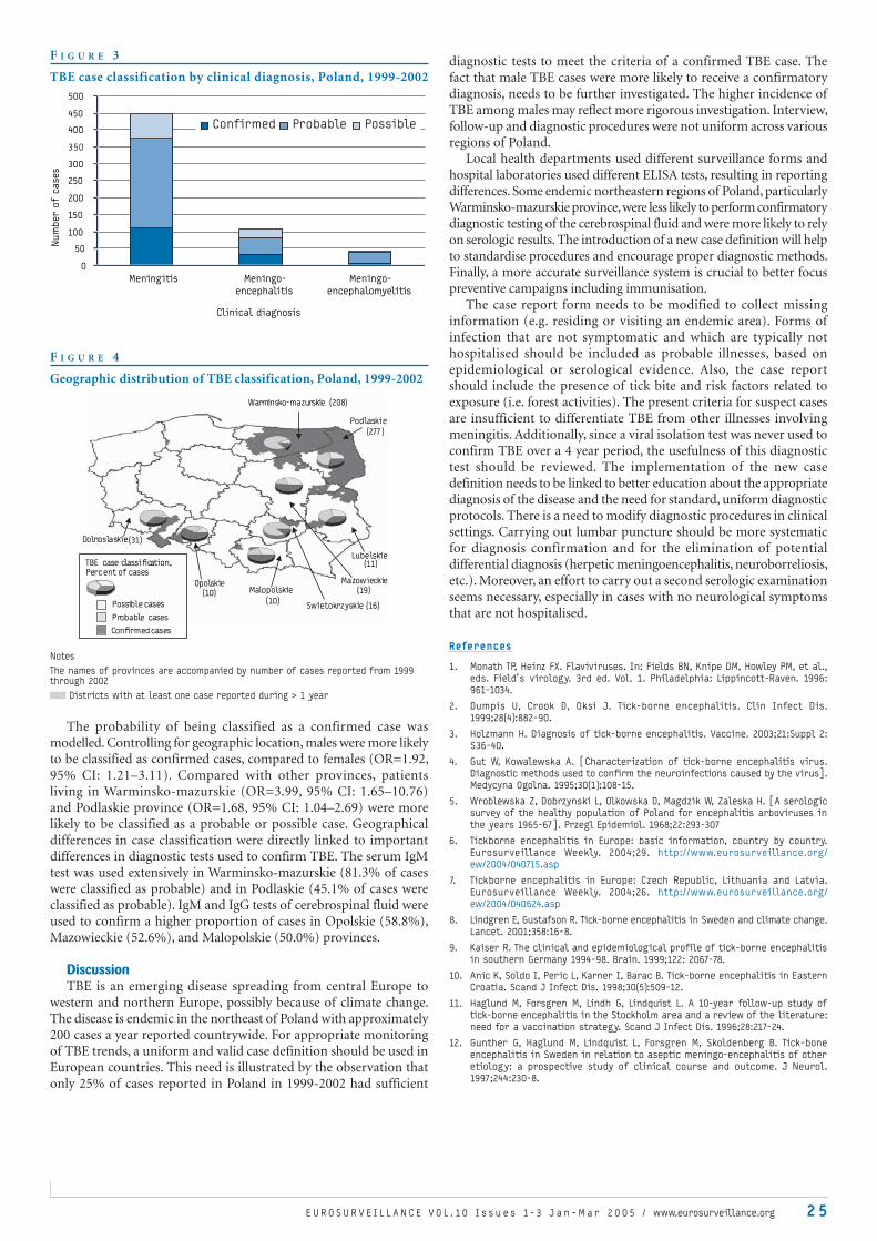
E U R OS U R V E I L L A N C E V O L . 10 I s s u e s 1 -3 J a n - M a r 2 0 0 5 / www.eurosurveillance.org 2 5
F I G U R E 3
TBE case classification by clinical diagnosis, Poland, 1999-2002
F I G U R E 4
Geographic distribution of TBE classification, Poland, 1999-2002
Notes
The names of provinces are accompanied by number of cases reported from 1999 through 2002
Districts with at least one case reported during > 1 year
The probability of being classified as a confirmed case was modelled. Controlling for geographic location, males were more likely to be classified as confirmed cases, compared to females (OR=1.92, 95% CI: 1.21–3.11). Compared with other provinces, patients living in Warminsko-mazurskie (OR=3.99, 95% CI: 1.65–10.76) and Podlaskie province (OR=1.68, 95% CI: 1.04–2.69) were more likely to be classified as a probable or possible case. Geographical differences in case classification were directly linked to important differences in diagnostic tests used to confirm TBE. The serum IgM test was used extensively in Warminsko-mazurskie (81.3% of cases were classified as probable) and in Podlaskie (45.1% of cases were classified as probable). IgM and IgG tests of cerebrospinal fluid were used to confirm a higher proportion of cases in Opolskie (58.8%), Mazowieckie (52.6%), and Malopolskie (50.0%) provinces.
DiscussionTBE is an emerging disease spreading from central Europe to
western and northern Europe, possibly because of climate change. The disease is endemic in the northeast of Poland with approximately 200 cases a year reported countrywide. For appropriate monitoring of TBE trends, a uniform and valid case definition should be used in European countries. This need is illustrated by the observation that only 25% of cases reported in Poland in 1999-2002 had sufficient
diagnostic tests to meet the criteria of a confirmed TBE case. The fact that male TBE cases were more likely to receive a confirmatory diagnosis, needs to be further investigated. The higher incidence of TBE among males may reflect more rigorous investigation. Interview, follow-up and diagnostic procedures were not uniform across various regions of Poland.
Local health departments used different surveillance forms and hospital laboratories used different ELISA tests, resulting in reporting differences. Some endemic northeastern regions of Poland, particularly Warminsko-mazurskie province, were less likely to perform confirmatory diagnostic testing of the cerebrospinal fluid and were more likely to rely on serologic results. The introduction of a new case definition will help to standardise procedures and encourage proper diagnostic methods. Finally, a more accurate surveillance system is crucial to better focus preventive campaigns including immunisation.
The case report form needs to be modified to collect missing information (e.g. residing or visiting an endemic area). Forms of infection that are not symptomatic and which are typically not hospitalised should be included as probable illnesses, based on epidemiological or serological evidence. Also, the case report should include the presence of tick bite and risk factors related to exposure (i.e. forest activities). The present criteria for suspect cases are insufficient to differentiate TBE from other illnesses involving meningitis. Additionally, since a viral isolation test was never used to confirm TBE over a 4 year period, the usefulness of this diagnostic test should be reviewed. The implementation of the new case definition needs to be linked to better education about the appropriate diagnosis of the disease and the need for standard, uniform diagnostic protocols. There is a need to modify diagnostic procedures in clinical settings. Carrying out lumbar puncture should be more systematic for diagnosis confirmation and for the elimination of potential differential diagnosis (herpetic meningoencephalitis, neuroborreliosis, etc.). Moreover, an effort to carry out a second serologic examination seems necessary, especially in cases with no neurological symptoms that are not hospitalised.
References
1. Monath TP, Heinz FX. Flaviviruses. In: Fields BN, Knipe DM, Howley PM, et al.,
eds. Field’s virology. 3rd ed. Vol. 1. Philadelphia: Lippincott-Raven. 1996:
961-1034.
2. Dumpis U, Crook D, Oksi J. Tick-borne encephalitis. Clin Infect Dis.
1999;28(4):882-90.
3. Holzmann H. Diagnosis of tick-borne encephalitis. Vaccine. 2003;21:Suppl 2:
S36-40.
4. Gut W, Kowalewska A. [Characterization of tick-borne encephalitis virus.
Diagnostic methods used to confirm the neuroinfections caused by the virus].
Medycyna Ogolna. 1995;30(1):108-15.
5. Wroblewska Z, Dobrzynski L, Olkowska D, Magdzik W, Zaleska H. [A serologic
survey of the healthy population of Poland for encephalitis arboviruses in
the years 1965-67]. Przegl Epidemiol. 1968;22:293-307
6. Tickborne encephalitis in Europe: basic information, country by country.
Eurosurveillance Weekly. 2004;29. http://www.eurosurveillance.org/
ew/2004/040715.asp
7. Tickborne encephalitis in Europe: Czech Republic, Lithuania and Latvia.
Eurosurveillance Weekly. 2004;26. http://www.eurosurveillance.org/
ew/2004/040624.asp
8. Lindgren E, Gustafson R. Tick-borne encephalitis in Sweden and climate change.
Lancet. 2001;358:16-8.
9. Kaiser R. The clinical and epidemiological profile of tick-borne encephalitis
in southern Germany 1994-98. Brain. 1999;122: 2067-78.
10. Anic K, Soldo I, Peric L, Karner I, Barac B. Tick-borne encephalitis in Eastern
Croatia. Scand J Infect Dis. 1998;30(5):509-12.
11. Haglund M, Forsgren M, Lindh G, Lindquist L. A 10-year follow-up study of
tick-borne encephalitis in the Stockholm area and a review of the literature:
need for a vaccination strategy. Scand J Infect Dis. 1996;28:217-24.
12. Gunther G, Haglund M, Lindquist L, Forsgren M, Skoldenberg B. Tick-bone
encephalitis in Sweden in relation to aseptic meningo-encephalitis of other
etiology: a prospective study of clinical course and outcome. J Neurol.
1997;244:230-8.
0
50
100
150
200
250
300
350
400
450
500
Meningitis Meningo-
encephalitis
Meningo-
encephalomyelitis
Clinical diagnosis
Confirmed Probable Possible
Num
ber o
f c
ases
Confirmed cases
TBE case classification,
Percent of cases
Possible cases
Probable cases
Opolskie
Podlaskie
Warminsko-mazurskie
Mazowieckie
Malopolskie
Dolnoslaskie
Swietokrzyskie (16)
Lubelskie
(277)
(10)
(19)
(208)
(10)
(31)
(11)

O R I G I N A L A R T I C L E S
S u r v e i l l a n c e r e p o r t
2 6 E U R OS U R V E I L L A N C E V O L . 10 I s s u e s 1 -3 J a n - M a r 2 0 0 5
M A N D ATO R Y D I S E A S E R E P O R T I N G B Y G E R M A N L A B O R ATO R I E S : A S U R V E Y O F AT T I T U D E S , P R A C T I C E S A N D N E E D S
AP Zucs 1, J Benzler 2, G Krause 2
database and a list that had originally been compiled by the Lower Saxony State Health Department to track remaining polio stocks in Germany.
All these laboratories were sent a standardised, pre-tested, anonymous postal questionnaire collecting information on hospital affiliation, catchment area, organisms routinely tested for, reporting habits, use of laboratory software, future electronic reporting and current feedback preferences. Questionnaires were analysed with Epi Info 2002 (CDC, Atlanta, Georgia, USA, 2002).
ResultsWe identified 1556 laboratories of which 853 (55%) completed and
returned their questionnaires. Three hundred and sixteen (37%) of the respondents were pathology and clinical chemistry laboratories that do not carry out tests for notifiable microorganisms. The remaining 537 facilities (63%) formed the actual study population. Approximately one third each was privately owned, part of a tertiary care centre or affiliated with a smaller hospital. Of 523 laboratories providing information on their catchment area, 349 (67%) received samples only from within their town and its immediate surroundings, 130 (25%) from their federal state and adjacent states, and 44 (9%) from the entire country and abroad.
Of 505 laboratories providing information on their reporting medium, 227 (45%) were still using paper forms that are filled in manually to report detection of a notifiable agent to the local health department. The others were using their ordinary results report or automated print-outs specifically generated for this purpose.
Delay between laboratory diagnosis and notification was reported by most respondents to be no longer than 24 hours. For the majority, their reporting duties required up to one additional working hour per week [TABLE 1].
Most laboratories employed microbiology-specific software packages [TABLE 1]. Of the 62 commercial products mentioned, none was used by more than 14% of the participating laboratories. An overwhelming majority of the participating laboratories were in favour of the introduction of electronic reporting formats [TABLE 2]. If they were to be introduced, 181 (46%) of 398 stated they would like to enter data directly into an internet mask, whereas 217 (54%) favoured automated data extraction.
T A B L E 1
Reporting delay, reporting associated workload and use of software in German laboratories, Germany 2004
N % Reporting delay (n=502)
24h 446 89
> 24h 56 11
Additional workload due to reporting
(n=520)
1h/month 180 35
1h/week 219 42
1h/day 98 19
> 1h/day 23 4
Use of laboratory software (n=537)
Yes 358 67
Software signalling notifiable
microorganism (n=370)
Yes 145 39
In 2000, the new German infectious disease control act replaced aggregate with individual case reporting. The process was facilitated by the simultaneous introduction of electronic data transfer within the public health system. Reporting laboratories have not been electronically connected to this network. A survey by means of a postal questionnaire was conducted in 2003among 537 German medical microbiology laboratories to explore their reporting habits, preference for electronic reporting formats, and relevant software equipment. Almost 90% of the respondents indicated a reporting delay of no more than 24 hours and 45% were still manually filling in paper forms for reporting purposes. The introduction of electronic reporting formats was favoured by 74% of the laboratories although 33% were not using any microbiology-specific software and the remaining 67% listed 62 different products. Pilot projects with selected software manufacturers might help to pave the way for the implementation of a standardised electronic infectious disease reporting format in Germany.
Euro Surveill 2005;10(1):26-27 Published online Jan 2005Key words: Germany, labo ratories, mandatory reporting,
software, survey
IntroductionMandatory disease reporting in Germany has been redefined with the
enactment of the Infektionsschutzgesetz (the Protection against Infection Act) in 2000. One of the main innovations of the new legislation was the introduction of individual instead of aggregate case reporting through all levels of the public health system. One legal paragraph is devoted to laboratory reporting: it lists microbial pathogens that are notifiable to the local health department, mostly within 24 hours [1].
Concomitantly, in 2001, transfer of case reports from local to state health departments and to the federal agency, Robert Koch-Institut (RKI) in Berlin, was converted from a paper-based to an electronic system [2]. Clinicians and laboratory scientists, however, still report to the local health department in non-electronic format. Within laboratory information systems, diagnostic units typically communicate results in electronic format to a central storage facility where they are linked to other information, e.g. patient data. Notifiable data are printed out on paper and usually sent by fax to the local health department where they are manually re-entered for electronic storage.
In 2002, two surveys looked into the acceptance of the new surveillance system by German clinicians and local health departments [3, 4]. In order to complete the picture, we conducted a survey among German laboratories. Our main objective was to assess how the laboratories are handling their legal reporting duties, to what extent this process has been computerised and in how far they would like the current reporting system to change.
MethodsThe survey took place in 2003 and addressed all German medical
laboratories testing patient material for the presence of microbial organisms. Eligible laboratories were identified using the RKI address
1. Federal Offi ce of Public Health, Berne, Switzerland (German FETP alumnus)
2. Robert Koch-Institut, Berlin, Germany

O R I G I N A L A R T I C L E S
S u r v e i l l a n c e r e p o r t
E U R OS U R V E I L L A N C E V O L . 10 I s s u e s 1 -3 J a n - M a r 2 0 0 5 / www.eurosurveillance.org 27
T A B L E 2
Laboratory scientists’ attitude towards introduction of electronic to the local health department (n=466), Germany, 2004
Scientists’ attitude N %
Urgently necessary 43 9
Good idea 301 65
Neutral 29 6
Unnecessary 66 14
Problematic 27 6
DiscussionAlmost 90% of the laboratories studied reported notifying
infectious organisms to the local health department within 24 hours. This enables timely surveillance and rapid intervention if necessary. The benefit comes at a reasonable cost: for more than 75% of the laboratories, disease reporting creates an additional workload of no more than 1 hour per week.
More than 66% of the participants would favour electronic reporting formats instead of the currently prevailing paperwork. Elsewhere, electronic reporting has been shown to be faster [5], less labour-intensive [6] and more complete [7] than traditional disease reporting. On the other hand, 33% of the laboratories in this survey do not use any laboratory software, and those that do are working with more than 60 different products. In the light of this heavily fragmented market, a uniform electronic reporting format is rather illusory in the near future. Past experience in Germany has shown that legislators are reluctant to impose standards regulating data transfer formats between healthcare providers and local health departments. Pilot projects with selected software manufacturers may be the way forward to promote national standards of electronic disease reporting and to catch up with European countries like the United Kingdom [8], the Netherlands [6] or Sweden [9], where such systems are already in place.
This was the first survey among German laboratories relating to practical implications of the Infektionsschutzgesetz. The survey response and the lack of non-responder data do not allow any safe assumptions as to the representativeness of the participating laboratories. It could be argued that laboratories with a keen interest in surveillance would have been more likely to participate in this study
and might therefore have been overrepresented. As a result, we would have overestimated German laboratories’ reporting compliance and enthusiasm for electronic reporting formats. The observed diversity of software products, however, would have probably been even more pronounced if all laboratories had participated.
Acknowledgements
We would like to thank Fabian Feil from the Lower Saxony State Health
Department for providing the laboratory address list, and Anna
Lukaschyk for entering the survey data.
References
1. Deutscher Bundestag. Infektionsschutzgesetz. [German] Bundesgesetzblatt.
2000;33:1045-77. http://www.rki.de/INFEKT/IFSG/IFSG_BGBL.HTM [accessed 30
June 2004]
2. Krause G, Altmann D, Claus H, Hellenbrand W, Buchholz U, Hamouda O, et
al. Erste Bilanz des neuen Systems zur Überwachung meldepflichtiger
Krankheiten nach dem Infektionsschutzgesetz. [German] Gesundheitswesen.
2003;65 (Suppl.1):S8-12.
3. Ropers G, Krause G, Tiemann F, Du Ry van Beest Holle M, Stark K. Nation-wide
survey on the role of travel medicine in primary care in Germany. J Travel
Med. 2004;in press
4. Brodhun B, Kramer MH, Krause G. Befragung der Gesundheitsämter zur
Umsetzung des Meldewesens nach dem Infektionsschutzgesetz. [German]
Bundesgesundheitsbl. Gesundheitsforsch-Gesundheitsschutz. 2004;in press
5. Panackal AA, M’ikanatha NM, Tsui F, McMahon J, Wagner MM, Dixon BW, et al.
Automatic electronic laboratory-based reporting of notifiable infectious
diseases at a large health system. Emerg Infect Dis. 2002;8(7):685-91.
6. Bosman A, van Straten EJA, Schrijnemakers P, Bloemberg BPM. ISIS: A Dutch
national electronic system for infectious disease surveillance and early
warning. http://www.rivm.nl/ isis/ [poster, accessed 30 June 2004]
7. Effler P, Ching-Lee M, Bogard A, Ieong M, Nekomoto T, Jernigan D. Statewide
system of electronic notifiable disease reporting from clinical laborato-
ries: comparing automated reporting with conventional methods. JAMA.
1999;282(19):1845-50.
8. Lopman BA, Reacher M, Gallimore C, Adak GK, Gray JJ, Brown DWG. A summertime
peak of “winter vomiting disease”: Surveillance of noroviruses in England and
Wales, 1995 to 2002. BMC Public Health. 2003;3:13-9. http://www.biomedcentral.
com/1471-2458/3/13 [accessed 30 June 2004]
9. Rolfhamre P, Grabowska K, Ekdahl K. Implementing a public web based GIS ser-
vice for feedback of surveillance data on communicable diseases in Sweden.
BMC Infect Dis. 2004;4:17-28. http://www.biomedcentral.com/content/pdf/1471-
2334-4-17.pdf [accessed 30 June 2004]
E L E C T R O N I C R E P O R T I N G I M P R O V E S T I M E L I N E S S A N D C O M P L E T E N E S S O F I N F E C T I O U S D I S E A S E N O T I F I C AT I O N , T H E N E T H E R L A N D S , 2 0 0 3M Ward1,2, P Brandsema2, E van Straten2, A Bosman 2
In 2002, the internet based reporting system OSIRIS was introduced in the Netherlands and by the end of that year had fully replaced the paper-based reporting system. The objectives of OSIRIS were to improve timeliness and completeness of surveillance data on infectious diseases reported from regional to national level.We compared the timeliness of infectious diseases reported by the conventional paper-based system in 2001 with those reported by OSIRIS in 2003. Two distict types of delay were compared: (1) total delay: defined as time between sympton onset and reporting at national level and (2)
central delay: defined as time between regional and national reporting. Median delays between both systems were compared using the Wilcoxon Rank Sum-Test. We also compared electronic reports received via OSIRIS in 2003 to those received through the conventional system for 2001 for completeness of specific data fields. The Fisher exact test and the Mantel-Haenzel test with Yates correction were used to determine the significance of proportions of completed data fields in each system.Results showed the median central delay was significantly reduced for all diseases in OSIRIS compared to conventional reporting system. Overall, the median central delay was reduced from 10 days (interquartile range 4) in 2001 to 1 day (interquartile range 1) in 2003. Except for cases of malaria, the total delay, from symptom
1. European Programme for Intervention Epidemiology Training (EPIET)
2. National Institute for Public Health and the Environment, Bilthoven, The Netherlands.
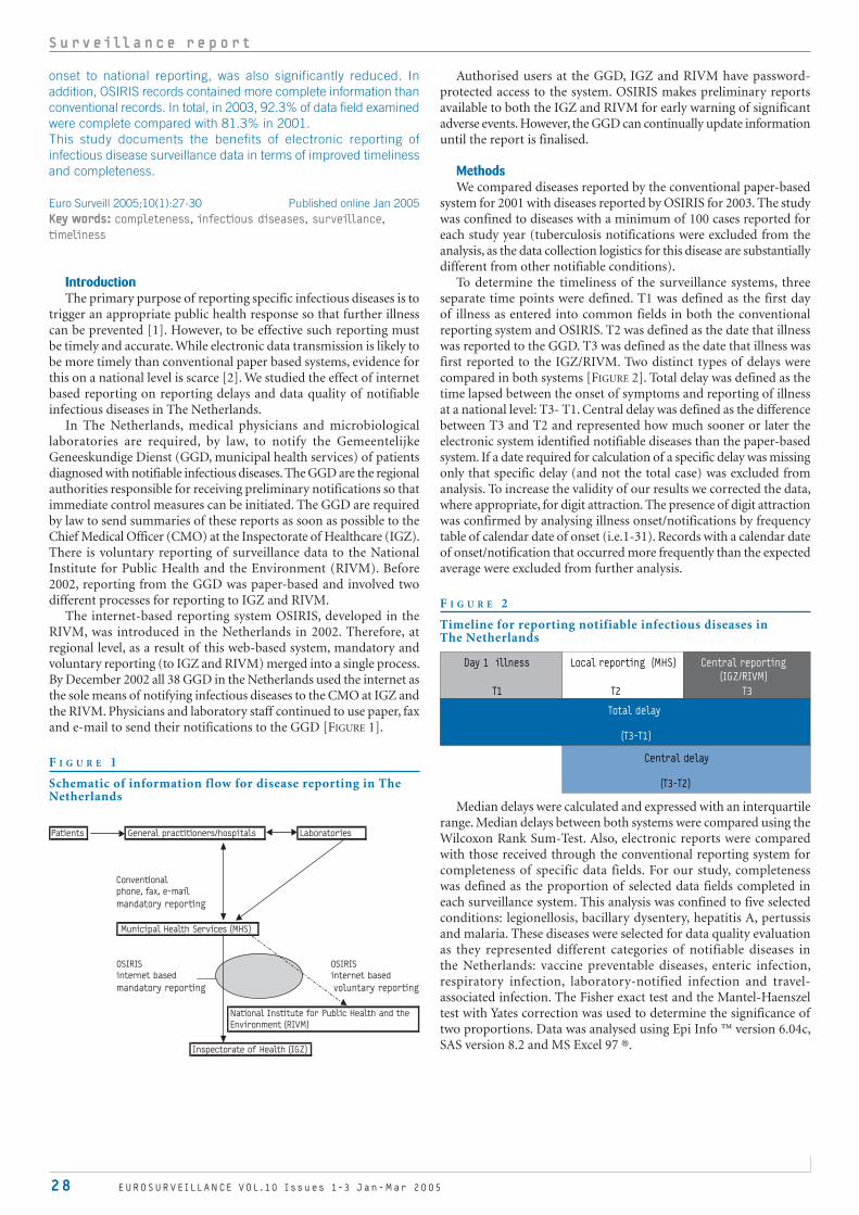
28 E U R OS U R V E I L L A N C E V O L . 10 I s s u e s 1 -3 J a n - M a r 2 0 0 5
S u r v e i l l a n c e r e p o r t
Authorised users at the GGD, IGZ and RIVM have password-protected access to the system. OSIRIS makes preliminary reports available to both the IGZ and RIVM for early warning of significant adverse events. However, the GGD can continually update information until the report is finalised.
MethodsWe compared diseases reported by the conventional paper-based
system for 2001 with diseases reported by OSIRIS for 2003. The study was confined to diseases with a minimum of 100 cases reported for each study year (tuberculosis notifications were excluded from the analysis, as the data collection logistics for this disease are substantially different from other notifiable conditions).
To determine the timeliness of the surveillance systems, three separate time points were defined. T1 was defined as the first day of illness as entered into common fields in both the conventional reporting system and OSIRIS. T2 was defined as the date that illness was reported to the GGD. T3 was defined as the date that illness was first reported to the IGZ/RIVM. Two distinct types of delays were compared in both systems [FIGURE 2]. Total delay was defined as the time lapsed between the onset of symptoms and reporting of illness at a national level: T3- T1. Central delay was defined as the difference between T3 and T2 and represented how much sooner or later the electronic system identified notifiable diseases than the paper-based system. If a date required for calculation of a specific delay was missing only that specific delay (and not the total case) was excluded from analysis. To increase the validity of our results we corrected the data, where appropriate, for digit attraction. The presence of digit attraction was confirmed by analysing illness onset/notifications by frequency table of calendar date of onset (i.e.1-31). Records with a calendar date of onset/notification that occurred more frequently than the expected average were excluded from further analysis.
F I G U R E 2
Timeline for reporting notifiable infectious diseases in The Netherlands
Median delays were calculated and expressed with an interquartile range. Median delays between both systems were compared using the Wilcoxon Rank Sum-Test. Also, electronic reports were compared with those received through the conventional reporting system for completeness of specific data fields. For our study, completeness was defined as the proportion of selected data fields completed in each surveillance system. This analysis was confined to five selected conditions: legionellosis, bacillary dysentery, hepatitis A, pertussis and malaria. These diseases were selected for data quality evaluation as they represented different categories of notifiable diseases in the Netherlands: vaccine preventable diseases, enteric infection, respiratory infection, laboratory-notified infection and travel-associated infection. The Fisher exact test and the Mantel-Haenszel test with Yates correction was used to determine the significance of two proportions. Data was analysed using Epi Info ™ version 6.04c, SAS version 8.2 and MS Excel 97 ®.
onset to national reporting, was also significantly reduced. In addition, OSIRIS records contained more complete information than conventional records. In total, in 2003, 92.3% of data field examined were complete compared with 81.3% in 2001. This study documents the benefits of electronic reporting of infectious disease surveillance data in terms of improved timeliness and completeness.
Euro Surveill 2005;10(1):27-30 Published online Jan 2005Key words: completeness, infectious diseases, surveillance,
timeliness
IntroductionThe primary purpose of reporting specific infectious diseases is to
trigger an appropriate public health response so that further illness can be prevented [1]. However, to be effective such reporting must be timely and accurate. While electronic data transmission is likely to be more timely than conventional paper based systems, evidence for this on a national level is scarce [2]. We studied the effect of internet based reporting on reporting delays and data quality of notifiable infectious diseases in The Netherlands.
In The Netherlands, medical physicians and microbiological laboratories are required, by law, to notify the Gemeentelijke Geneeskundige Dienst (GGD, municipal health services) of patients diagnosed with notifiable infectious diseases. The GGD are the regional authorities responsible for receiving preliminary notifications so that immediate control measures can be initiated. The GGD are required by law to send summaries of these reports as soon as possible to the Chief Medical Officer (CMO) at the Inspectorate of Healthcare (IGZ). There is voluntary reporting of surveillance data to the National Institute for Public Health and the Environment (RIVM). Before 2002, reporting from the GGD was paper-based and involved two different processes for reporting to IGZ and RIVM.
The internet-based reporting system OSIRIS, developed in the RIVM, was introduced in the Netherlands in 2002. Therefore, at regional level, as a result of this web-based system, mandatory and voluntary reporting (to IGZ and RIVM) merged into a single process. By December 2002 all 38 GGD in the Netherlands used the internet as the sole means of notifying infectious diseases to the CMO at IGZ and the RIVM. Physicians and laboratory staff continued to use paper, fax and e-mail to send their notifications to the GGD [FIGURE 1].
F I G U R E 1
Schematic of information flow for disease reporting in The Netherlands
Patients
OSIRIS
internet based
OSIRIS
internet based
Conventional
phone, fax, e-mail
General practitioners/hospitals Laboratories
Municipal Health Services (MHS)
Inspectorate of Health (IGZ)
National Institute for Public Health and the
Environment (RIVM)
mandatory reporting
mandatory reporting voluntary reporting
Day 1 illness Local reporting (MHS) Central reporting
(IGZ/RIVM)
T1 T2 T3
Total delay
(T3-T1)
Central delay
(T3-T2)

E U R OS U R V E I L L A N C E V O L . 10 I s s u e s 1 -3 J a n - M a r 2 0 0 5 / www.eurosurveillance.org 29
ResultsNine diseases with more than 100 cases reported in 2001 and 2003
were included in the study: bacillary dysentery, hepatitis A, hepatitis B, hepatitis C, legionellosis, malaria, meningococcal disease, pertussis and foodborne outbreaks.
Digit attraction was only evident for first day of illness (T1). Thus, we corrected total delay, for digit attraction (T3-T1). We excluded all cases with illness date of onset on 1,5,10,15,20,25 and 30 as these dates were more frequently recorded than expected if illness onset was equally likely on all days. Correction for digit attraction resulted in a decrease in the estimated total delay (T3-T1) for all person-based infections in 2001 and 2003. (There was no correction for digit attraction for hepatitis B and hepatitis C as less than one in five patients with these illnesses had a recorded date illness onset).
Between 2001 and 2003 the central delay for all nine diseases was significantly reduced
[FIGURE 3]. Overall, the central delay was reduced from a median value of 10 days (interquartile range 4) in 2001 to 1 day (interquartile range 1) in 2003. Except for malaria, the total delay (T3-T1 ) was also significantly reduced for diseases studied [TABLE 1].
T A B L E 1
Median total and central delay, interquartile range and statistical significance for notifiable infectious diseases in The Netherlands, 2001 and 2003
Condition Total delay: T3-T1 (IR)† Central delay: T3-T2 (IR)†
2001 2003 P * 2001 2003 P *
Dysentery 29 (23) 19 (12) 0.001 10 (11) 1(4) 0.001
Legionella 20 (33) 11 (22) 0.001 8 (9) 1(3) 0.001
Meningococcal disease 11 (10) 5 (6) 0.001 7 (8) 0 (3) 0.001
Malaria 12 (26) 13 (20) NS** 10 (35) 2 (4) 0.05
Hepatitis A 22 (16) 12 (13) 0.001 8 (9) 1 (3) 0.001
Hepatitis C NA NA 0.001 10 (16) 3 (13) 0.001
Hepatitis B NA NA 0.001 16 (27) 2 (8) 0.001
Pertussis 60 (33) 51 (33) 0.001 9 (6) 1 (26) 0.001
Food borne infections 30 (22) 18 (19) 0.001 14(12) 3 (9) 0.001
† IR: Interquartile range
* P value of difference calculated using Wilcoxon Rank Sum-Test
** P = 0.5
F I G U R E 3
Median central delay for nine notifiable conditions by conventional (2001) and electronic (2003) reporting, The Netherlands
Electronic reports contained more complete information on variables common to both conventional and electronic reporting formats. In 26 of 36 data fields studied, those completed electronically contained significantly more information (p<0.05). Overall, in 2003, 91.3% of examined data fields were complete in comparison with 82.3% in 2001 [TABLE 2].
T A B L E 2
Field completion rates for reports received by electronic (2003) and conventional (2001) reporting systems for five notifiable infectious diseases, The Netherlands
% of electronic reports with
complete field(2003)
% of paper reports with
complete field (2001)
P value
Dysentery n=267 n=346
Post code 92.1 82.4 <0.001
Year of birth or age 100.0 99.4 0.5 (F)
Deceased 96.3 82.7 <0.001
Hospitalised 96.3 80.1 <0.001
How diagnosis made 98.9 80.1 0.1(F)
Isolated case 97.8 95.0 0.05(F)
Infection acquired abroad 100.0 81.1 <0.001
Legionella n=222 n=182
Post code 87.8 72.5 <0.001
Year of birth or age 100.0 100.0 NA‡
Deceased 98.2 76.9 <0.001
Hospitalised 99.1 72.5 <0.001
How diagnosis made 100.0 96.7 <0.05 (F)
Isolated case 100.0 74.2 <0.001
Malaria n=356 n=569
Post code 68.3 41.1 <0.001
Year of birth or age 100.0 98.9 <0.001
Deceased 78.9 53.1 0.40
Hospitalised 70.5 52.4 <0.001
How diagnosis made 47.8 39.5 <0.05
Isolated case 91.6 46.4 <0.001
Infection acquired abroad 96.3 100.0 <0.001
Pertussis n=2701 n=6986
Post code 82.7 85.2 <0.05
Year of birth or age 100.0 100.0 NA‡
Deceased 93.2 83.5 <0.001
Hospitalised 90.2 81.1 <0.001
How diagnosis made 96.9 76.6 <0.001
Isolated case 95.3 83.9 <0.001
Vaccination status 98.3 97.9 0.28
Hepatitis A n=375 n=701
Post code 88.0 75.7 <0.001
Year of birth or age 100.0 100.0 NA‡
Deceased 97.1 74.8 <0.001
Hospitalised 97.6 74.3 <0.001
How diagnosis made 97.9 64.3 <0.001
Isolated case 64.5 75.5 <0.001
Infection acquired abroad 89.9 99.0 <0.05
Vaccination status 89.9 75.5 <0.001
N reported number
F Fisher exact test
NA not applicable
DiscussionTo our knowledge, this is the first report comparing electronic and
conventional reporting of infectious disease surveillance data on a national basis. Electronic reports were received at the national level significantly quicker than conventional reports for the nine diseases studied. This improved timeliness was not detrimental to data quality as electronic reports also contained more complete information than conventional reports. Similar results have previously been reported for electronically notifiable disease reporting from clinical laboratories [3,4].
The improved timeliness was almost exclusively due to the reduction in reporting delay between the GGD and the national authorities. This reduced reporting delay can be attributed to OSIRIS as there was no other major change in work practices at GGD level that would have resulted in a reduced local reporting delay (T2-T1). In fact, using this system lead to an estimated 50% reductions in administrative workload in relation to reporting infectious diseases at GGD level [5]. Correction for digit attraction resulted in a reduction in the estimated total delay for bacillary dysentery, hepatitis A, legionellosis, malaria, meningococcal disease and pertussis in both study periods. This suggests that some patients tend to overestimate the time period during which they are ill by ‘rounding-up’ to the nearest convenient date. While correcting for this phenomenon is impractical in routine practice, time intervals should be measured in a consistent way to allow comparison between different outbreak detection reports and surveillance systems [6].
0 5 10 15 20
pertussis
hepatitis C
hepatitis B
hepatitis A
malaria
meningitis
legionella
foodborne outbreaks
dysentery
Condit
ion
Median central delay (days)
2001
2003

O R I G I N A L A R T I C L E S
S u r v e i l l a n c e r e p o r t
3 0 E U R OS U R V E I L L A N C E V O L . 10 I s s u e s 1 -3 J a n - M a r 2 0 0 5
Respiratory virus activity is detected in Europe each winter, yet the precise timing and size of this activity is highly unpredictable. The impact of influenza infection and/or acute respiratory infection in European countries is continuously monitored through a variety of surveillance systems. All of these sources of information are used to assess the nature and extent of activity of influenza and other respiratory viruses, and to offer guidance on the prevention and control of morbidity and mortality due to influenza at a local, national and international level.The early warning system for a forthcoming influenza epidemic is mainly based on the use of a set of thresholds. In the Czech Republic, the acute respiratory infection (ARI) reporting system, with automated data processing, uses a statistical model for the early detection of unusual increased rates of the monitored indicators. The collected data consists of the number of ARI, the number of complications due to ARI and the population registered with the reporting general practitioners and paediatricians, all collected
separately in five age groups. To improve the reporting system in the Czech Republic, clinical data on the weekly incidence of influenza-like illness (ILI) within the same population and the same age groups was started in January 2004. These data fit the European Commission’s recently adopted ILI case definition and allows a better comparison of data with other countries in Europe, in particular those participating in EISS (European Influenza Surveillance Scheme).
Euro Surveill 2005;10(3):30-33 Published online Mar 2005Key words: acute respiratory infection, early warning system,
European Union, influenza surveillance.
Introduction
Information on the occurrence of infectious diseases is very important for maintaining public health in Europe. Every European country has its own national notification and surveillance system and legislation [1, 2]. National laboratories participate in many international surveillance programmes organised by the European Union, WHO and other organizations. Recently the Community
The noted improvement in data quality is also important as this availability of more complete information should enable national authorities to respond in a more timely and appropriate manner. While we only selected 7-8 data fields per disease as indicators of data quality the general superiority of electronic reports suggests that improved completeness is also likely in unexamined data fields.
A potential concern in comparisons such as this is variation in coding between the fields in the electronic and paper-based systems. However, in this study as we only selected variables that were equivalent on the hardcopy and the electronic surveillance forms, direct comparability was ensured. Also, before the introduction of the electronic system staff training, technical assistance was provided at local level to ensure any data entry and coding problems were identified and managed appropriately [5]. Another potential concern is that the relative benefits of electronic reporting in this study could be secondary to deterioration in the conventional system. As the transition from conventional to electronic reporting occurred mid-year in 2002 and we selected only years when one system functioned at GGD level, a decline in the conventional working process could not explain the improved reporting times in 2003. In addition, the consistency of our results for all nine conditions suggests that the improved reporting times are real.
OSIRIS has achieved its objectives. Data received at national level is more timely and of better quality than with conventional reporting. However, the primary purpose of surveillance is not merely speedy and complete transmission of data. Technologically innovative reporting systems, as OSIRIS, also need to be consistent with the purpose of disease reporting, that is, of translating information into action [1,7]. Thus, it must be a two-way communication process of information exchange between public health agencies and the clinical community. Even in this technologically advanced age, observations made by astute clinicians still remain important, in timely reporting of certain notifiable diseases [8]. In these instances,
S u r v e i l l a n c e r e p o r t
electronic surveillance systems help us verify suspicions of outbreaks as was recently observed in the Netherlands when action was taken as a result of the observed increased notifications of hepatitis A cases. This action was due to a combination of clinical observation and national notification by OSIRIS [9,10].
This study documented improved timeliness and completeness of national infectious disease surveillance data that has occurred as a result of the use of electronic communication.
References
1. M’Ikanatha N, Southwell B, Lautenbach E. Automated Laboratory Reporting
of Infectious Diseases in a Climate of Bioterrorism. Emerg Infect Dis. 2003
Sep;9(9):1053-7.
2. Bean N, Martin S. Implementing a Network for Electronic Surveillance repor-
ting from Public Health Reference Laboratories: An International Perspective.
Emerg Infect Dis. 2001 Sep-Oct;7(5):773-9.
3. Effler P, Chinf-Lee M, Bogard A, Ieong M, Nekomoto T, Jernigan D. State wide
System of Electronic Notifiable Disease Reporting from Clinical Laboratories.
JAMA. 1999;282 (19):1845-1853.
4. Grant A, Eke B. Application of information technology to the laboratory re-
porting of communicable disease in England and Wales. Commun Dis Rep CDR
Rev. 1993 May 21;3(6):R75-8.
5. Bosman A, van Straten E, Bovee L, De Boer J, Van Wijngaarden J. Melden van
infectieziekten via Internet. Infectieziekten Bulletin 2001;12:362-5.
6. CDC. Updated guidelines for evaluating public health surveillance systems: re-
commendations from the guidelines working group. MMWR 2001;50 (No. RR-13).
7. Bravata D, McDonald K, Smith W, Rydzak C, Szeto H, Buckeridge D, Haberland
C et al. Systematic Review: Surveillance Systems for early detection of bio-
terrorism-related diseases. Ann Intern Med. 2004;Jun 1;140(11):910-22.
8. De Jager C, Heijne J. Increase in hepatitis A in MSM in the Netherlands.
Eurosurveillance Weekly. 2004;8(22):27/05/2004. http://www.eurosurveillance.org/
ew/2004/040527.asp
9. De Jager C, Heijne JBovee L, Bruisten S. Stijging van Hepatitis A onder homo-
seksuele mannen in Nederland, Infectieziekten Bulletin 2004, 6: 210-211.
10. Horan J, Mallonee S. Injury Surveillance. Epidemiol Rev. 2003;25:24-42
1. Centre of Epidemiology and Microbiology, National Institute of Public Health, Prague, Czech Republic
2. EISS co-ordination centre, Netherlands Institute for Health Services Research (NIVEL), Utrecht, The Netherlands
H A R M O N I S AT I O N O F T H E A C U T E R E S P I R AT O R Y I N F E C T I O N R E P O R T I N G S Y S T E M I N T H E C Z E C H R E P U B L I C W I T H T H E E U R O P E A N C O M M U N I T Y N E T W O R K S
J Kyncl1, WJ Paget2, M Havlickova1, B Kriz1

E U R OS U R V E I L L A N C E V O L . 10 I s s u e s 1 -3 J a n - M a r 2 0 0 5 / www.eurosurveillance.org 3 1
network for epidemiological surveillance has been established in accordance with Decision No. 2119/98/EC of the European Parliament and of the Council.
Acute viral rhinitis, pharyngitis, laryngotracheitis, tracheobronchitis, bronchitis, bronchiolitis or pneumonia are associated with a large number of viruses, each of which is capable of producing a wide spectrum of acute respiratory illness, with different causes in children and adults [3].
Viral diseases of the respiratory tract may be characterised by fever and one or more systemic reactions, such as chills, headache, general aching, malaise and anorexia. Morbidity from acute respiratory diseases is particularly significant in children. In adults, the relatively high incidence and resulting disability, with consequent economic loss, make acute respiratory diseases a major health problem worldwide [3-5]. As a group, acute respiratory diseases are one of the leading causes of death from any infectious disease worldwide.
Influenza virus activity in Europe is detected each winter, yet the precise timing and magnitude of this activity remain highly unpredictable. The age groups of the population affected and the severity of illness that they experience depend on several factors including the virus types and subtypes that circulate during a given season. Clinical and virological data is collected and presented at a European level by the European Influenza Surveillance Scheme (EISS) through the internet [6]. EISS reported data for 22 countries during the 2003-2004 season; collaborators included 30 reference laboratories, at least 11 000 sentinel physicians and the surveillance covered a population of 445 million inhabitants [7].
Epidemics of influenza are reported almost every year. Influenza pandemics occur at irregular intervals (three in the last century) and have been associated with unpredictable reassortments of genome segments of human, pig or avian viruses leading to surface antigens to which humans have no pre-existing immunity.
In an attempt to improve the health care information systems, substantial changes were made to the acute respiratory infection (ARI) reporting system from 2000 to 2002 in the Czech Republic [8]. The system (formerly based on sending the data by fax and entering them into a central database) was changed to a modern web-based system, which enables data to be entered at a local level with basic analysis in real time. Further changes were made in 2003 in accordance with the Commission Decision of 19 March 2002 laying down case definitions (Decision No. 253/2002/EC) for reporting communicable diseases to the Community network. The system was extended to enable the collection of age-specific incidence of influenza-like infections (ILI) as well.
MethodsThe surveillance of influenza and other ARI is based mainly on
clinical surveillance (morbidity reports and mortality statistics of influenza and respiratory infections as well as of all causes) and virological surveillance from the community and hospitals. The influenza morbidity monitoring program started in the Czech Republic in 1951. Since 1968, the age specific incidence of ARI and total incidence of complications have been monitored weekly. The system now includes approximately 2230 general practitioners (GP) and 1240 paediatricians and covers approximately 5 million inhabitants (half of the Czech population) in all 86 districts of the Czech Republic.
ILI is defined as: the clinical picture compatible with influenza, e.g. sudden onset of disease, cough, fever > 38 ºC, muscular pain and/or headache, in accordance with the EU case definition for influenza. ARI for reporting purposes is defined as every GP’s clinical diagnosis of acute upper respiratory tract infection (as defined by the International Classification of Diseases, Tenth Revision (ICD-10), codes J00, J02, J04, J05, J06) and influenza (ICD-10 codes J10.1, J10.8, J11.1, J11.8).
Virological surveillance is performed by the Airborne Viral Infections Department at the National Institute of Public Health. The department is composed of two divisions: the National Reference Laboratory (NRL) for influenza and the NRL for non-influenza respiratory viruses. The virological surveillance program consists of a weekly assessment of routine laboratory test results of paired sera and nasopharyngeal swabs, provided by the collaborating virological
laboratories. Test methods used are the complement fixation reaction (CFR), direct antigen dete ction from clinical specimens (ELISA) and isolation of the causative agent from a suitable cell culture. Lately, rapid diagnosis of the major causative agents of acute respiratory virus infections such as influenza virus of types A and B, respiratory syncytial virus, adenoviruses and parainfluenza viruses has been used within this program [9].
The data on morbidity from epidemiological surveillance are integrated with those from virological surveillance. After validation and assessment, the results are presented in a weekly bulletin. The bulletin is sent to the regional public health institutes, the Ministry of Health, collaborating laboratories and is also posted on the web page of the National Institute of Public Health [10]. Comprehensive outputs for international organisations such as EISS or WHO FluNet are provided by the National Reference Laboratory for influenza.
ResultsStarting from the season 2001-2002, each regional public health
service entered data from collaborating general practitioners and paediatricians into a central SQL database, using an encrypted web transfer with name and password controlled access. The district-specific data consists of the number of ARI, the number of complications due to an ARI and the population registered with the reporting GPs and paediatricians, all collected in five age groups (0-5, 6-14, 15-24, 25-59, 60+ years) [FIGURE 1]. There is also space for comments. Pneumonia only is now considered as a complication of the infection. Starting from January 2004, clinical data on incidences of influenza-like illness (ILI) within the same population and the same age groups as in ARI have also been collected [FIGURE 2].
F I G U R E 1
Weekly ARI morbidity by age group per 100 000 population during the 2003-2004 season in the Czech Republic
F I G U R E 2
Weekly ILI morbidity by age group per 100 000 population during the 2003-2004 season in the Czech Republic
0
100
200
300
400
500
600
40 42 44 46 48 50 52 02 04 06 08 10 12 14 16 18 20 22 24 26 28 30 32 34
Week
Inci
den
ce / 1
00 0
00
0-5 y 6-14 y 15-24 y 25-59 y 60+ y total
Inci
denc
e / 10
0 00
0
0
100
200
300
400
500
600
40 42 44 46 48 50 52 02 04 06 08 10 12 14 16 18 20 22 24 26 28 30 32 34
Week
700
800

3 2 E U R OS U R V E I L L A N C E V O L . 10 I s s u e s 1 -3 J a n - M a r 2 0 0 5
The basic data processing is automated and uses a statistical model for early detection of unusual increased rates of the indicators monitored, based on a general linear model for left-censored data. Usual weekly ARI incidence is modelled and this rate can only increase if a possible epidemic occurs. A threshold was established by averaging non-epidemic ARI incidences in the past years and applying an upper tolerance limit (covering 90% observations with 95% probability). The thresholds are available for the whole of the Czech Republic and also for each region. Direct standardisation and weighting for the size of the monitored population are also used to enable comparison of ARI and/or ILI morbidity among regions and districts. Figures 3 and 4 show the district distribution of ARI clinical incidence during two peak weeks.
F I G U R E 3
The first peak of ARI morbidity (week 51/2003). ARI incidence per 100 000 population, by district, Czech Republic
F I G U R E 4
The second peak of ARI morbidity (week 6/2004). ARI incidence per 100 000 population, by district, Czech Republic
Laboratory results for the season 2003-2004 [FIGURES 5,6] confirm that both regional outbreaks were caused by influenza. Weekly numbers of positive samples of the main circulating respiratory viruses of that season and the total ARI incidence is shown. Positive results for influenza can be seen to peak almost simultaneously with clinical illness incidence (positive results by the paired sera test are shown by the week when the second sample was tested and the results are therefore shifted by 2-3 weeks).
F I G U R E 5
Detection of ARI using an antigen detection: number of positive samples by week, season 2003-2004, Czech Republic
F I G U R E 6
Detection of ARI using a serology: number of positive samples by week, season 2003-2004, Czech Republic
DiscussionSubstantial changes have been made to the influenza reporting
system in the Czech Republic in recent years. The changes started with an improvement of the ARI reporting system by making the system web-based during the 2001-2002 season. In January 2004, the reporting system also started a pilot ILI reporting project using an European Union adopted case definition.
Data collected by the influenza reporting system in the Czech Republic have been reported to EISS since 1998 [11]. During the 2003-2004 season, 20 networks reported weekly ILI incidences and four networks (the Czech Republic, France, Germany and Romania) reported ARI incidences [7]. The pilot ILI incidences from the Czech Republic means that it is now possible to compare influenza activity with many more countries in Europe. The ILI rates in Europe varied considerably during the 2003-2004 season, with the peak incidences ranging from 12 per 100 000 population in Wales to 1885 per 100 000 population in the Slovak Republic. The peak ILI incidence in the Czech Republic was 256 per 100 000 population, much lower than in the neighbouring Slovak Republic. This difference may be due to a number of factors, including different case definitions, different health care systems and recent changes in the surveillance systems [12].
The age groups 0-5 and 6-14 were chosen because compulsory education starts at the age of 6 in the Czech Republic. Dividing children into school and pre-school groups is relevant because of airborne spreading of respiratory infections.
Methods used for virological surveillance within EISS network were already Published in 2004 [13]. Although only a small part of all clinical cases are analysed virologically each year, the virological results
S u r v e i l l a n c e r e p o r t
Total A
RI r
ate p
er 1
00 0
00
0
2
4
6
8
10
No.
of
pos
itiv
e sa
mpl
es
36 38 40 42 44 46 48 50 52 02 04 06 08 10 12 14 16 18
Week
20
200
400
600
800
1000
1200
1400
1600
1800
12 2000
Flu AFlu BRSVTotal ARI incidence
0
0
5
10
15
20
25Flu AFlu BRSVTotal ARI incidence
No.
of
pos
itiv
e sa
mpl
es
36 38 40 42 44 46 48 50 52 02 04 06 08 10 12 14 16 18
Week
20
0
200
400
600
800
1000
1200
1400
1600
1800
Total A
RI r
ate p
er 1
00 0
00
2000
PraguePilsen
Brno
PraguePilsen
Brno

O R I G I N A L A R T I C L E S
S u r v e i l l a n c e r e p o r t
E U R OS U R V E I L L A N C E V O L . 10 I s s u e s 1 -3 J a n - M a r 2 0 0 5 / www.eurosurveillance.org 33
A survey of the prevalence of Shiga toxin-producing Escherichia coli (STEC) of O157 serotype in foodstuffs of animal origin (beef, veal, pork, chicken, fish) from 1999 to 2003 in Belgium was performed. STEC strains were only isolated from beef with a prevalence of 0.73%. This percentage is low in comparison with the prevalence in other countries. Among the 76 isolated STEC O157 strains, 75% belonged to the serotype O157:H7 and 25% to the serotype O157 non H7. Moreover, the most frequent pathotype was eae stx2 ehxA (74%).
Euro Surveill 2005;10(3):33-36 Published online Mar 2005Key words: Escherichia coli, Foodstuff, pathotype, Shiga toxin
IntroductionTwo legal texts Published by the European Parliament in November
2003 are dedicated to the survey and management of zoonoses and zoonotic agents in European Union (EU): Directive 2003/99/EC [1] on the survey of zoonoses and zoonotic agents and Regulation 2160/2003/EC [2] on the control of salmonella and other zoonotic agents present in food chain. These texts repeal the directive 92/117/CEE [3] concerning the survey of zoonoses and zoonotic agents in EU and indicate that each member country must collect relevant data concerning the major zoonotic agents and must report this to the European Commission. Among these zoonotic agents to be surveyed, the directive mentions verocytotoxigenic Escherichia coli (VTEC). These pathogens are a public health problem in Belgium: 46 pathologic cases associated with Shiga toxin-producing Escherichia coli (STEC) were identified in 2002 [4].
Enterohaemorrhagic E. coli (EHEC) are VTEC or STEC which can cause a broad spectrum of human diseases, including diarrhoea, haemorrhagic colitis, and the haemolytic uraemic syndrome (HUS).
are of equal significance. Substantially more data are available for the specimens analysed, e.g. patient’s age, clinical diagnosis, sampling date and onset of disease. First isolations of influenza virus and particularly an increase in their incidence may be predictive of the very beginning of an epidemic even before any change can be detected in the clinical morbidity rates. Routine detection of other viral respiratory pathogens yields complementary data which are useful in monitoring general trends in morbidity. Summary data are informative enough of the circulation of different agents in the population throughout the year. The virological results are also sometimes used to validate the clinical reports. For example, during the 2003-2004 season there were two ARI morbidity peaks in the Czech Republic [FIGURE 1]. This was caused by two regional influenza epidemics in different parts of the Czech Republic when the fast transmission was interrupted by the Christmas holidays [FIGURES 3,4].
The ARI / ILI reporting system of the Czech Republic is a modern and efficient surveillance system based on the collection of high quality data. The whole ARI / ILI reporting system is essential for pandemic planning in the Czech Republic. It can be linked with the system for crisis management to enable reporting and analysis on a daily basis. For efficient information at all levels, high quality local and national surveillance is necessary. Since using an internet-based platform, the reporting system in the Czech Republic as well as the EISS are easily accessible and provide timely information.
Acknowledgements
Thanks are given to Dr. Bohumir Prochazka, head of the Dept. of
Biostatistics, National Institute of Public Health in Prague for statisti-
cal contribution and advices on the Czech reporting system. The authors
wish to thank all Regional Public Health Institutes, general practitioners
and paediatricians as well as the virological laboratories participating
at the ARI surveillance in the Czech Republic.
The project was partly supported by the Ministry of Health of the Czech
Republic.
References
1. Van Dalen P. Outbreaks of disease: Current European reporting system. In:
Dando M et al., eds. Maximizing the Security and Development Benefits from
the Biological and Toxin Weapons Convention. Dordrecht: Kluwer Academic
Publishers, 2002:97-119.
2. Kriz B, Kyncl J. Natural Outbreaks of Disease: Communicable Disease
Surveillance in the Czech Republic. In: Dando M, Pearson G, Kriz B, eds.
Scientific and Technical Means of Distinguishing Between Natural and Other
Outbreaks of Disease. Dordrecht: Kluwer Academic Publishers, 2001:35-39.
3. Graham NMH. The epidemiology of acute respiratory infections. In: Nelson
KE, Williams CM, Graham NMH, eds. Infectious Disease Epidemiology: theory and
practice. Gaithersburg, Maryland: Aspen Publishers, Inc., 2001:439-476.
4. Fleming DM. The contribution of influenza to combined acute respiratory
infections, hospital admissions, and deaths in winter. Commun Dis Public
Health. 2000; 3(1): 32-38.
5. Monto AS. Viral respiratory infections in the community: Epidemiology, agents,
and interventions. American Journal of Medicine. 1995;99 (suppl. 6B):24S-27S.
6. Snacken R, Manuguerra JC, Taylor P. European Influenza Surveillance Scheme
on the Internet. Methods Inf Med. 1998;37(3):266-270.
7. Paget WJ, Meerhoff TJ, Meijer A on behalf of EISS. Epidemiological and virological
assessment of influenza activity during the 2003-2004 winter. Submitted.
8. Kyncl J et al. Improving the acute respiratory infection notification system
in the Czech Republic. The First European Influenza Conference, St.-Julians, Malta,
20-23 October 2002, poster No. P-W1-11, abstract book p. 54.
9. Roubalova K et al. Doporucene metody ve virologicke diagnostice (Standard
operational procedures in virological diagnostics), in Czech. Acta hygienica,
epidemiologica et microbiologica. 2000;(1):1-56.
10. Acute respiratory infections in the Czech Republic. Available at http://www.
szu.cz/cema/aro/aro.htm
11. Paget WJ, Meerhoff TJ, Goddard NL on behalf of EISS. Mild to moderate in-
fluenza activity in Europe and the detection of novel A(H1N2) and B viruses
during the winter of 2001-02. Euro Surveill 2002;7(11):147-157.
12. Paget WJ, Meerhoff TJ, Rebelo de Andrade H on behalf of EISS. Heterogeneous
influenza activity across Europe during the winter of 2002-2003. Euro Surveill
2003;8(12):230-239.
13. Meerhoff TJ, Meijer A, Paget WJ on behalf of EISS. Methods for sentinel vi-
rological surveillance of influenza in Europe - an 18-country survey. Euro
Surveill 2004;9(1):34-38.
1. Food Sciences department– Microbiology, Faculty of Veterinary Medicine, University of Liege, Belgium
2. Scientifi c Public Health Institute, Laboratory of Microbiology, Brussels, Belgium
3. Food Microbiology, Faculty of Veterinary Medicine, University of Gent, Belgium
4. Department of Microbiology, Academic Hospital, Vrije Universiteit Brussel, Brussels, Belgium
S U R V E Y O F T H E C O N TA M I N AT I O N O F F O O D S T U F F S O F A N I M A L O R I G I N B Y S H I G A T O X I N P R O D U C I N G E S C H E R I C H I A C O L I S E R O T Y P E O 1 57 : H 7 I N B E L G I U M F R O M 1 9 9 9 T O 2 0 0 3A Chahed 1, Y Ghafir 1, B China1, K Dierick 2, L De Zutter 3, D Piérard 4, G Daube 1.

3 4 E U R OS U R V E I L L A N C E V O L . 10 I s s u e s 1 -3 J a n - M a r 2 0 0 5
S u r v e i l l a n c e r e p o r t
The O157 serotype has been responsible for numerous outbreaks worldwide involving fatal cases [5], and therefore EHEC O157 are deserving of to a very careful survey. It remains the principal serotype involved in HUS in Europe. Vulnerability to EHEC infection depends on several factors.
1. Age: children aged 15 years and younger, and people aged over 65 years were more exposed.
2. Immunity: the presence of antibodies seems to protect people from a O157 challenge.
3. Gastrointestinal modifications: a diet poor in proteins is a risk factor.
4. Blood group: Shiga toxins seem to be transported by the red blood cells. The O group and people with red blood cells lacking the P antigen were more sensitive.
5. Ingested dose: the more bacteria were ingested the more severe disease occurred.
6. Previous antibiotic treatment is a risk factor suggesting a protecting role for the intestinal flora. Moreover, an antibiotic treatment of the disease increases the risk of HUS appearance [6].
Their major virulence factors are the Shiga-toxins Stx1 and Stx2 responsible for the kidney problems and the intimin encoded by the pathogenicity island LEE (Locus of enterocyte effacement) and involved in diarrhoea. Moreover, EHEC O157:H7 bacteria produce an enterohaemolysin encoded by the plasmidic ehxA gene [7].
Between 1994 and 2002 in Belgium, 398 STEC strains were isolated from human patients. Of these, 195 (49%) were from serotype O157, but only 182 were classical EHEC (positive for eae, stx and ehxA PCR) [4].
The major foodstuffs from animal origin have been randomly sampled among the national production in order to evaluate the prevalence of EHEC O157 and the major incriminated pathotype.
Material and methodsSampling of beef and pork carcasses was performed in
slaughterhouse by swabbing on a half-carcass 2 to 4 hours postmortem. For each half-carcass, 4 surfaces were sampled corresponding to a total surface of 600 cm2:
(a) the internal face of the jam (100 cm2), (b) the posterior part of the internal pelvis (100 cm2), (c) the sternum and the sternocephalic muscles (300 cm2), (d) the posterior face of the anterior member (100 cm2).
For beef, a 1600 cm2 surface corresponding to 4 zones of 400 cm2 was swabbed:
(a) the postero-external face of the thigh (400 cm2), (b) the flank (400 cm2), (c) the thorax (400 cm2), (d) the posterior face of the anterior member (400 cm2). For pork, the number of analysed carcasses was 163 in 2000
representing 0.0015% of the Belgian annual production. For beef, the number of carcasses studied was, 1984 in 1999, 1501 in 2000, 1388 in 2001, 1215 in 2002 and 1479 in 2003 representing 0.35 %, 0.25%, 0.19%, 0.26 % of the annual Belgian beef production, respectively. One hundred and fifty seven veal carcasses samples were analysed in 2000.
Chicken carcasses were sampled at the slaughterhouse exit or at the distribution level. Twenty five grams of carcass skin were removed at the neck and the front neck level. Two hundred and forty three carcass samples were analysed in 2001, representing 0.0007% of the annual Belgian chicken production. Minced meat and cuts of beef and pork were sampled at the production stage or at the distribution stage. Raw chicken minced meat preparations were sampled at the distribution level with a minimum sample of 100 g. Chicken fillets (without skin or bones) were sampled at the exit of the production chain or at the consumer distribution level. For fish, 25g of flesh and skin were sampled from entire fish at the abattoir exit.
The isolation protocol for E. coli O157:H7 involved a pre-enrichment at 42°C in mTSB broth, supplemented with novobiocin during 6 at 7 hours, followed by an enrichment in MacConkey broth supplemented with cefixime-tellurite and incubated at 37°C for 18 hours. An O157 immunoassay (VIDAS ECO) and an immunoconcentration if the immunoassay was positive (VIDAS ICE or Dynabeads O157) were performed. In the case of a positive immunoassay, a plating on sorbitol-MacConkey agar supplemented with cefixime-tellurite was performed and incubated for 18 hours at 42°C, followed by a confirmation by latex agglutination (Oxoid) and by biochemical gallery (Api20E, Biomérieux) [8]. If this confirmation step was not conclusive, the result was considered to be negative. The presence of the H7 antigen was investigated using H7 antiserum-sorbitol fermentation medium [9]. Finally the presence and of the virulence genes (eae, stx1, stx2, ehxA) were investigated by PCR[10].
The statistics (contingency table, χχ2 calculation) were performed using the InStat2.01 software.
ResultsOne hundred and sixty three pork carcasses were analysed in 2000
T A B L E 1
Prevalence of STEC O157 in foodstuffs of bovine origin, Belgium
Matrix Analysed amount Year 1999 2000 2001 2002 2003 Total Statistics (χ2)*
Beef carcass 1600 cm2
n
p
%
198425
1.3
15016
0.5
138813
0.9
121513
1.1
147910
0.68
7567
67
0.89
Significant
Significant
Nonsignifi-
cant
Statistics (χ2)* Non significant
Minced meat 25 g
n
p
%
9741
0.1
4871
0.2
298
0
0
2970
0
2852
0.7
2341
4
0.17
Significant
Statistics (χ2)* Non significant
Cut 25 g
n
p
%
NT NT NT
2220
0
2985
1.68
520
5
0.96
Nonsignifi-
cant
Statistics (χ2)* Non significant
Isolated strains
n
p
%
2958
26
0.88
1988
7
0.35
1686
13
0.77
1734
13
0.74
2062
17
0.82
10428
76
0.73
Statistics (χ2)* Non significant
n = number of analysed samples
* (p<0.05)
p: number of positive samples
NT: non tested
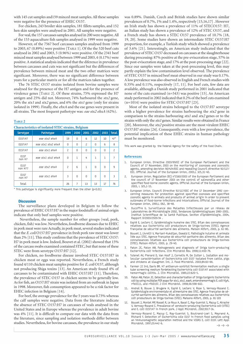
➔ ➔ ➔
E U R OS U R V E I L L A N C E V O L . 10 I s s u e s 1 -3 J a n - M a r 2 0 0 5 / www.eurosurveillance.org 3 5
with 145 cut samples and159 minced meat samples. All these samples were negative for the presence of EHEC O157.
For chicken, 243 broiler skin samples, 181 fillets samples, and 152 hen skin samples were analysed in 2001. All samples were negative.
For veal, the 157 carcasses samples analysed in 200 were negative. All of the 153 aquaculture fish samples analysed in 1999 were negative.
However, of the 7567 beef carcasses samples analysed from 1999 to 2003, 67 (0.89%) were positive [TABLE 1]. Of the the 520 beef cuts analysed in 2002 and 2003, 5 (0.96%) were positive. Of the 2341 beef minced meat samples analysed between 1999 and 2003, 4 (0.17%) were positive. A statistical analysis indicated that the diference in prevalence between carcasses and cuts was not significant but the differences of prevalence between minced meat and the two other matrices were significant. Moreover, there was no significant difference between years for a particular matrix or for all the matrices taken together.
The 76 STEC O157 strains isolated from bovine samples were analysed for the presence of the H7 antigen and for the presence of virulence genes [TABLE 2]. Of these strains, 75% expressed the H7 antigen and 25% did not. Moreover, 74% harboured the stx2 gene, 20% the stx1 and stx2 genes, and 6% the stx1 gene (only for strains isolated in 1999). Finally, the ehxA and the eae genes were present in all strains. The most frequent pathotype was: eae stx2 ehxA (62%).
TABLE 2
Characteristics of isolated STEC strains, Belgium
Serotype Pathotype 1999 2000 2001 2002 2003 Total
O157:H7 eae stx2 ehxA 19 0 6 12 10 47*
0157:H7 eae stx1 stx2 ehxA 0 0 2 0 6 8
O157:H7 eae stx1 ehxA 2 0 0 0 0 2
O157 non H7
eae stx1 stx2 ehxA 0 1 4 1 1 7
O157 non H7?
eae stx2 ehxA 2 6 1 0 0 9
O157 non H7
eae stx1 ehxA 3 0 0 0 0 3
Total 26 7 13 13 17 76
* This pathotype is signifi cantly more frequent than the other (p<0.01)
DiscussionThe surveillance plans developed in Belgium to follow the
prevalence of EHEC O157:H7 in the major foodstuffs of animal origin indicate that only beef samples were positive.
Nevertheless, the sample number for other groups (veal, pork, chicken, fish) was low. Nevertheless, foodborne diseases due to EHEC in pork meat were rare Actually, in pork meat, several studies indicated that the E. coli O157:H7 prevalence in fresh pork raw meat was lower than 2% [11]. This study confirms that the prevalence of STEC O157:H7 in pork meat is low. Indeed, Bouvet et al. (2002) showed that 15% of the carcass swabs examined contained STEC, but that none of these STEC were from serotype O157:H7 [12].
For chicken, no foodborne disease involved STEC O157:H7 in chicken meat or eggs was reported. Nevertheless, a French study found 4/110 chicken meat samples positive for E. coli O157, although not producing Shiga toxins [13]. An American study found 4% of carcasses to be contaminated with EHEC O157:H7 [11]. Therefore, the prevalence of STEC O157 in chicken seems to be generally low. As for fish, an O157:H7 strain was isolated from an outbreak in Japan in 1998. Moreover, fish consumption appeared to be a risk factor for EHEC infection in Belgium [14].
For beef, the average prevalence for the 5 years was 0.73% whereas the calf samples were negative. Data from the literature indicate the absence of STEC O157:H7 in carcasses of veals analysed in the United States and in Europe whereas the prevalence in adult bovine was 4% [11]. It is difficult to compare our data with the data from the literature, since sampling and isolation methods differ between studies. Nevertheless, for bovine carcasses, the prevalence in our study
was 0.89%. Danish, Czech and British studies have shown similar prevalences of 0.7%, 1% and 1.4%, respectively [15,16,17] . However an Irish study has shown a prevalence of 11% of STECO157: H7, an Italian study has shown a prevalence of 12% of STEC O157, and a French study has shown a STEC O157 prevalence of 10.7% [18, 19, 20]. Some studies have shown an intermediate STEC O157:H7 proportion, for example, a Turkish study which showed a prevalence of 3.6% [21]. Interestingly, an American study indicated that the prevalence of STEC O157 decreased on carcasses at the slaughterhouse during processing: 87% positive at the pre-evisceration stage, 57% in the post-evisceration stage, and 17% at the post-processing stage [22]. Since our samples were taken at the post-processing level, this may partly explain the low contamination level observed. The prevalence of STEC O157 in minced beef meat observed in our study was 0.17%. A low prevalence was also observed in English and French studies with 0.35% and 0.11%, respectively [17, 11]. For beef cuts, few data are available, although a Danish study performed in 2001 indicated that none of the cuts examined (n=543) was positive [15]. An American study performed in 2002 indicated that 0.2% of the beef cuts examined (n=1014) were positive for STEC O157:H7 [23].
Most of the isolated strains belonged to the O157:H7 serotype with a higher prevalence for strains harbouring the stx2 gene in comparison to the strains harbouring stx1 and stx2 genes or to the strains with only the stx1 genes. Similar results were obtained in France [20]. Moreover, the stx2 positive strains are the most virulent EHEC O157:H7 strains [24]. Consequently, even with a low prevalence, the potential implication of these EHEC strains in human pathology must be monitored.
This work was granted by the Federal Agency for the safety of the Food Chain.
References
1. European Union. Directive 2003/99/EC of the European Parliament and the
Council of 17 November, 2003 on the monitoring of zoonoses and zoonostic
agents, amending decision 90/424/EEC and repealing Council directive 92/117/
EEC. Official Journal of the European Union, 2003,L 325,31-40.
2. European Union. Regulation (EC) n°2160/2003 of the European Parliament and
the council of 17 November 2003 on the control of salmonella and other
specified food-borne zoonotic agents. Official Journal of the European Union,
2003, L 325,1-15.
3. European Union. Council Directive 92/117/EEC of the 17 December 1992 con-
cerning measures for protection against specified zoonoses and specified
zoonotic agents in animals and products of animal origin in order to prevent
outbreaks of food-borne infections and intoxications. Official Journal of the
European Union, 1993,L 062, 38-48.
4. Ducoffre G. Surveillance des Maladies Infectieuses par un réseau de
Laboratoires de Microbiologie 2002, Tendances Epidémiologiques 1983-2001.
Institut Scientifique de la Santé Publique, Section d’Epidémiologie, 2004,
Rapport D/2003/2505/34 .
5. Espié E, Leclerc C. Epidémiologie humaine des STEC. Bilan des connaissances
relatives aux Escherichia coli producteurs de Shiga-toxines (STEC). Agence
française de sécurité sanitaire des aliments. Maison-Alfort, 2003, p. 61-80.
6. Bouvet J, Livrelli V, Mariani-Kukdijan, Oswald E. Pathologie hulaine et animale
liée aux STEC. Agence française de sécurité sanitaire des aliments. Bilan des
connaissances relatives aux Escherichia coli producteurs de Shiga-toxines
(STEC). Maison-Alfort, 2003, p. 29-40.
7. Paton JC, Paton AW. Pathogenesis and diagnosis of Shiga toxin-producing
Escherichia coli infections. Clin Microbiol Rev. 1998;11:450-79.
8. Tutenel AV, Pierard D, Van Hoof J, Cornelis M, De Zutter L. Isolation and mo-
lecular caractérisation of Eschercichia coli O157 isolated from cattle, pigs
and chickens at slaughter. Int. J. Food Microbiol. 2003;84:63-69.
9. Farmer JJ 3rd, Davis BR. H7 antiserum-sorbitol fermentation medium: a single
tube screening medium fordetecting Escherichia coli O157:H7 associated with
hemorrhagic colitis. J. Clin Microbiol. 1985;22:620-5.
10. Paton AW, Paton JC. Detection and characterization of Shiga toxigenic Escherichia
coli by using multiplex PCR asays for stx1, stx2, eaeA, enterohemorrhagic E. coli hlyA,
rfbO111, and rfbO157. J Clin Microbiol. 1998;36:598-602.
11. Andral B, Bouve J, Brugere H, Espié E, Leclerc V, Roze S, Vernozy-Rozand C.
Epidémiologie environmentale et alimentaire des STEC. Agence française de sé-
curité sanitaire des aliments. Bilan des connaissances relatives aux Escherichia
coli producteurs de Shiga-toxines (STEC). Maisons-Alfort, 2003, p. 81-103
12. Bouvet J, Montet MP, Rossel R, Le Roux A, Bavai C, Ray-Gueniot S, Mazuy C, Atrache
V, Vernozy-Rozand C. Prevalence of verotoxin-producing Escherichia coli (VTEC)
and E. coli O157:H7 in French pork. J Appl Microbiol. 2002;93:7-14.
13. Vernozy-Rozand C, Mazuy C, Ray-Gueniot S, Boutrand-Loei S, Meyrand A,
Richard Y. Detection of Escherichia cole O157 in French food samples using
an immunomagnetic separation method and the VIDAS E. coli O157. Lett Appl
Microbiol. 1997;25:442-6.

O R I G I N A L A R T I C L E S
S u r v e i l l a n c e r e p o r t
➔ ➔ ➔
3 6 E U R OS U R V E I L L A N C E V O L . 10 I s s u e s 1 -3 J a n - M a r 2 0 0 5
14. Pierard D, Crowfort N, De Bock S, Potters D, Crabbe G, Van Loock F, Lauwers S.
A case-control study of sporadic infection with O157 and non-O157 verocyto-
toxin-producing Escherichia coli. Epidemiol. Infect., 1999;122:359-365.
15. Unknown.Report on trends and sources of zoonotic agents in the European
Union and Norway, 2001, pp.237-242. 15/11/2004 http://europa.eu.int/comm/
food/food/biosafety/salmonella/11_ecoli_2001.pdf
16. Lukasova J, Abraham B, Cupakova S. Occurrence of Escherichia coli O157 in
raw material and food in Czech Republic.J Vet Med B Infect Dis Vet Public
Health. 2004;51:77-81
17. Chapman PA, Cerdan Malo AT, Ellin M, Ashton R, Harkin. Escherichia coli O157 in
cattle and sheep at slaughter, on beef and lamb carcasses and in raw beef and
lamb products in South Yorkshire, UK.Int J Food Microbiol. 2001;64:139-150.
18. McEvoy JM, Doherty AM, Sheridan JJ, Thomson-Carter FM, Garvey P, McGuire
L, Blair IS, McDowell DA.The prevalence and spread of Escherichia coli O157:
H7 at a commercial beef abattoir.J Appl Microbiol. 2003;95:256-266.
18. Bonardi S, Maggi E, Pizzin G, Morabito S, Caprioli A. Faecal carriage of
Verocytotoxin-producing Escherichia coli O157 and carcass contamination in
cattle at slaughter in northern Italy. Int J Food Microbiol. 2001;66:47-53.
19. Rogerie F, Marecat A, Gambade S, Dupond F, Beaubois P, Lange M. Characterization
of Shiga toxin-producing E. coli and O157 serotype E. coli isolated in France
from headline domestique cattle. Int. J. Food Microbiol., 2001,63,217-223
20. Gun H, Yilmaz A, Turker S, Tanlasi A, Yilmaz H. Contamination of bovine car-
casses and abattoir environment by Escherichia coli O157:H7 in Istanbul.Int
J Food Microbiol. 2003,84,339-344.
21. Elder RO, Keen JE, Siragusa GR, Barkocy-Gallagher GA, Koohmaraie M, Laegreid
WW.Correlation of enterohemorrhagic Escherichia coli O157 prevalence in
feces, hides, and carcasses of beef cattle during processing. Proc Natl Acad
Sci U S A. 2000,97,2999-3003
22. Warren W. Characterization of E. coli O157 :H7 on subprimal beef cuts prior
to mechanical tenderization. Project Summary. National Cattlemn’s beef
association center for research and knowledge management.2002. 15/11/2004
http://www.beef.org/documents/E.%20coli%20Mech%20Tenderization_Warren_
6_6_03.pdf
23. Boerlin P, McEwen SA, Boerlin-Petzold F, Wilson JB, Jonhson RP, Gyles CL.
Associations between virulence factors of Shiga-producing Escherichia coli
and disease in humans. J. Clin. Microbiol., 1999,37,497-503.
S u r v e i l l a n c e r e p o r t
H O S P I TA L P R E P A R E D N E S S A N D M A N A G E M E N T O F P AT I E N T S A F F E C T E D B Y V I R A L H A E M O R R H A G I C F E V E R O R S M A L L P O X AT T H E L A Z Z A R O S P A L L A N Z A N I I N S T I T U T E , I TA LY
G Ippolito, E Nicastri, M Capobianchi, A Di Caro, N Petrosillo, V Puro
The US cases of anthrax in 2001 and the recent severe acute respiratory syndrome outbreak have heightened the need for preparedness and response to naturally emerging and re-emerging infections or deliberately released biological agents. This report describes the response model of the Istituto Nazionale per le Malattie Infettive Lazzaro Spallanzani (INMI), Rome, Italy for managing patients suspected of or affected by smallpox or viral haemorrhagic fever (VHF) either in the context of an intentional release or natural occurrence.The INMI is Italy’s leading hospital in its preparedness and response plan to bioterrorism-related infectious agents. All single and double rooms of INMI are equipped with negative air pressure, sealed doors, high efficiency particulate air (HEPA) filters and a fully-equipped anteroom; moreover, a dedicated high isolation unit with a laboratory next door for the initial diagnostic assays is available for admission of sporadic patients requiring high isolation. For patient transportation, two fully equipped ambulances and two stretcher isolators with a negative pressure section are available. Biomolecular and traditional diagnostic assays are currently performed in the biosafety level 3/4 (BSL 3/4) laboratories. Continuing education and training of hospital staff, consistent application of infection control practices, and availability of adequate personnel protective equipment are additional resources implemented for the care of highly infectious patients and to maintain the readiness of an appropriately trained workforce to handle large scale outbreaks.
Euro Surveill 2005;10(3):36-39 Published online Mar 2005Key words: biological agents, bioterrorism, haemorrhagic fever,
Italy, preparedness, smallpox
IntroductionThe cases of anthrax in Florida and New York City in 2001, following
the terrorist events in New York City and Washington, D.C. [1] and
the recent severe acute respiratory syndrome (SARS) outbreak [2] have heightened the need for preparedness and response to emerging and re-emerging infections or deliberately released biological agents [3,4]. Smallpox [5] and haemorrhagic fever viruses (VHF) [6] pose the greatest concern because of their potential ease of dissemination or transmission, major public health impact (e.g., high mortality), panic and social disruption [4].
This report describes the model of response for the Istituto Nazionale per le Malattie Infettive Lazzaro Spallanzani (INMI), Rome, Italy in managing patients suspected of or affected by smallpox or VHF either in the context of an intentional release or natural occurrence.
The InstituteSince its foundation in 1936, the Lazzaro Spallanzani hospital has
been devoted to the prevention, diagnosis and care for infectious diseases. Over the years, its focus has changed in relation to the evolving patterns of infection threat. In particular, the hospital was heavily involved in the control of hepatitis B and C epidemic in the ‘70s, and the human immunodeficiency virus (HIV) and tuberculosis spread in the mid ’80s and early ‘90s.
In 1982, after smallpox vaccinations in Italy were discontinued, the Italian Ministry of Health identified the Lazzaro Spallanzani hospital as the place that would receive suspected cases and a negative-pressure Gelman’s containment bed isolator was purchased. The isolator was rigid, uncomfortable and unacceptable to patients, although it gave the nursing and medical staff a high degree of protection. However, not all routine nursing and medical procedures could be carried out due to this rigid physical barrier and it was also not practical to perform mechanical ventilation or haemodialysis.
In 1994, a new three floor hospital complex was completed for a total of 256 beds in 7 wards, 48 beds in day hospital care, and 20 intensive care beds.
The building has an air conditioning system that is able to provide up to 12 air changes per hour to all single and double rooms. In addition, the system also allows changes from negative to positive room pressure and vice versa, enabling the rooms to be used for airborne isolation or as a protective environment. All rooms have Instituto Nazionale per le Malattie Infettive Lazzaro Spallanzani, Rome, Italy

E U R OS U R V E I L L A N C E V O L . 10 I s s u e s 1 -3 J a n - M a r 2 0 0 5 / www.eurosurveillance.org 3 7
private baths, TV, telephone line and a fully-equipped anteroom with well-sealed doors, and HEPA filter on the incoming and outgoing air flow.
Five biosafety level (BSL) 3, one cabinet BSL 4 laboratories, and a BSL 3-like autopsy suite are available [FIGURE 1. Hospital map].
F I G U R E 1
Map of the Istituto Nazionale per le Malattie Infettive Lazzaro Spallanzani, Rome, Italy
In 1995, INMI was identified by the Italian Ministry of Health as
the national referral centre for the management of patients affected by naturally occurring highly transmissible infectious disease (i.e. VHF) and in late 2001 as the national referral centre in the case of deliberately release of biological agents.
Recently, INMI has organised an effective multidisciplinary European network of isolation facilities, physicians and other health professionals with expertise in the management of these facilities and infections. EUNID (European Network of Infectious Diseases physicians) consists of representatives from all member states and applicant countries, which have or are planning highly secure isolation facilities. One of EUNID’s primary objectives is to establish an inventory of European high isolation facilities and the healthcare workers involved in the management of patients needing these facilities.
Patient transportation and admissionFor transportation of patients suspected to be affected by VHF or
smallpox, two fully equipped ambulances are available with a sealed negative pressure section that is completely isolated from two other sections (one for the driver and the other for the external staff control) [FIGURE 2]. The air is expelled outside through HEPA filters. The isolation section is minimally furnished and stripped of unnecessary devices; needed sharps can easily be removed and the interior is easy to decontaminate. All resuscitation equipment, including ventilator and mechanical aspirators, is available inside the ambulance. Ambulances are also equippe with mobile phones and internet access.
Two stretcher isolators (Vickers Medical Containment Stretcher Transit Isolator®) are also available, specifically designed for the isolation and transportation of patients believed to be affected by highly infectious diseases. The self-contained isolation system consists basically of a lightweight stretcher onto which a demountable framework is attached enveloped by a transparent plastic [FIGURE 3]. The plastic envelope has negative air pressure, which is maintained by an air supply system in order to avoid the exit of potentially contaminated air. Thus, patients can be transported by the stretcher isolator directly into the ambulance’s isolated negative pressure section.
To ensure competent use, continual education and training of selected personnel is required.
In case of admission of patients with suspected or documented VHF referred to the INMI from the airport, ports or other hospitals, a dedicated pathway with a separate entrance from daily hospital activities has been designed.
Isolation procedures are implemented at admission, where a triage area with a negative air pressure room is dedicated to patients
presenting syndromes of a suspected airborne infection.
F I G U R E 2
Fully equipped ambulance with a sealed negative pressure section for transportation of patient believed to be suffering from highly infectious diseases, Rome, Italy
F I G U R E 3
Stretcher isolator specifically designed for the isolation and transportation of patients believed to be suffering from highly infectious diseases, Rome, Italy
Care facilitiesCurrently, 42 single and 59 double rooms on 5 wards, and 42 beds
in day hospital care are in use. However, the remaining facilities can be activated in case of a crisis according to the National Response Plan.
All single or double rooms of the Institute are potentially suitable for isolation or cohorting according to adopted airborne, droplet and/or contact precautions. The anterooms contain supplies for routine patient care, protective barriers for personnel and hand washing facility.
Moreover, there are two special adjacent single rooms in a ward dedicated for sporadic cases of VHF or smallpox. Next to to the rooms, there is a BSL3 laboratory, available for bloodfilm examinations to rule out malaria, basic blood testing, bacteriological cultures and preparation-inactivation of biological samples for molecular biological testing. Intravenous ribavirin is also available for the treatment of patients with suspected VHF while laboratory confirmation is pending. In case such patients are admitted, the other patients in the ward can be easily transferred to other wards in the institute. A step-by-step gradual floor-to-floor evacuation plan has been prepared, if needed.
In the last three months of 2001, following the deliberate release of anthrax in US, 201 individuals were referred to our admissions unit who reported being contaminated by suspected dust. However, no exposed individuals were admitted and no anthrax spores were detected.
At the beginning of the SARS epidemic when limited data on the route of the SARS-Coronavirus transmission were available, the high isolation unit was used for the two initial suspected SARS cases we cared for. During this epidemic, 72 subjects were referred to our admissions unit with SARS-
BSL 3 Laboratories•
BSL 3-4 Laboratories•
Hospital building•
Administration•

3 8 E U R OS U R V E I L L A N C E V O L . 10 I s s u e s 1 -3 J a n - M a r 2 0 0 5
like symptoms. Eight of them were admitted in a dedicated hospital ward as suspected cases. One of them satisfied the WHO criteria for SARS [7].
Isolation precautions, education and training, and personal protective equipment
At INMI, isolation precautions have been updated several times in the last 20 years as it is Italy’s national referral institution.
In the mid 1980’s, universal precautions to prevent transmission of HIV were implemented. In the following years isolation procedures were strengthened to cope with the re-emergence of tuberculosis, possible cases of emerging infections such as Ebola virus, and the threat of bioterrorism. More recently with the advent of the SARS threat, the hospital protocols, largely based on the Hospital Infection Control Practices Advisory Committee guidelines on isolation precautions in hospitals [8], have been further reinforced. Healthcare workers have been strongly recommended to comply with the required precautions, wearing disposable personnel protective equipment (PPE) consisting of masks or respirators, gloves, gown, head and shoe covers, and eye protection before entering the patient’s room. These have to all be discarded in the anteroom. Multiple educational and training sessions, including simulations focused on adherence to infection control protocols, have been developed for healthcare and laboratory personnel. Special efforts have been made to stress the importance of seal checking when wearing disposable respirators, and the safe removal of PPE [9,10]. Tests of respirator fit has been carried out for all health care workers. Protocols for the surveillance and management of health care workers potentially exposed to highly transmissible agents have been issued and updated, including post-exposure treatment when available.
Available PPE recommended for the management of highly contagious patients consists of Tyvek™ tissue full-body suits with thermo activated closure, full face mask with P3 filtered respirators (EU standard EN 149:2001), and latex obstetric gloves to be used in double gloving. Needle stick prevention devices are also provided.
All materials used for patients and disposable items worn by staff, in accordance with the Italian Ministry of Health recommendations, must be placed into a secure waste bag and then packaged into a rigid container before leaving the isolation rooms. The containers are then destroyed by incineration.
In case of patient death, autopsy is discouraged. The corpse must first be wrapped in linens permeated with disinfectants and then double bagged in sealed impermeable body bags before being transported for burial or cremation. All unnecessary handling of the body should be avoided.
In Italy, immunisation of healthcare workers against smallpox has not yet been implemented. The Italian Ministry of Health will activate immunisation program within the National Response Plan.
Transport, and processing of biological samples Packaging and transportation of biological samples that are sent
to INMI by external facilities, or which is sent by INMI to a WHO-reference laboratory, is done according to WHO guidelines [11,12]. Tubes and sample vessels are made of non-breakable material, and are tightly closed before being packaged and forwarded to the laboratory. Secondary packaging is consists of a waterproof plastic envelope. A complete patient information sheet, including all useful information for laboratory personnel and suspected diagnosis is inserted in an external pocket of the secondary envelope. Usually a single secondary envelope is used for several samples from one patient, but different secondary envelopes are used for different patients. Several secondary envelopes are grouped in a rigid impermeable plastic container that is transported to the laboratory by dedicated personnel. Collection of samples is preceded by informal direct contact between clinicians and laboratory personnel, in order to optimise sample collection and diagnostic assays.
When a class A viral agent is suspected, preliminary blood tests are carried out in the laboratory juxtaposed to the high isolation unit to rule out malaria, as well as blood counts, transaminases and
other urgent determinations. The biosafety level for sample handling is based upon to the pathogen’s classification, which is divided into 4 risk groups [13,14]. For level 3 pathogens, when cultivation of the microorganism is not required, samples can be initially processed in a level 2 laboratory, adopting level 3 procedures. In case of microorganism cultivation, the appropriate cell line panel or bacterial culture medium is inoculated with each patient’s sample in a BSL 3 laboratory. Samples from patients suspected to be infected with class 4 VHF or variola virus are handled under level 4 procedures, in the BSL 4 facility for both aliquotation and initial assay set up. Viral cultures are maintained in a level 4 facility throughout the entire observation time, but other assays are continued under lower biosafety levels when they undergo a treatment known to inactivate pathogen infectivity, such as heat treatment, fixation, solvent exposure and protein or nucleic acid extraction. The methods currently available for the detection of class A viral agents, as well as methods to detect other viruses important for differential diagnosis are included in the table. This list is continuously updated according to the specific literature available from the international community. In addition, the immune response to suspect pathogens are tested by antibody tests, in both acute and convalescent serum sample pairs. Both commercial and in-house assays, including indirect immunofluorescence and enzyme-linked immunoassays are used.
T A B L E
Capability of INMI to detect class A viruses, including viral agents important for differential diagnosis, Rome, Italy
PCR SEQ VI EM IFA EIA NT CF IB
Class A viruses
Filoviruses: Ebola, Marburg X X X X
Arenaviruses: old and new world viruses
X X X X
Bunyaviruses: CCHF X X X X X
Orthopoxviruses X X X(*) X
Other viruses
HSV X X X X X X
VZV X X X X X X X
Flaviviruses: Dengue, Yellow fever
X X X X X X X
Hantaviruses X X X X X X
(*) Only for differential diagnosis. If virus isolation consistent with smallpox
In the BSL 3/4 laboratories, all solid waste and residual biological specimens are autoclaved before disposal. Liquid waste is chlorinated before entry into the hospital sewage system.
Finally, in the last two years INMI was alerted twice for a possible referral of a patient with suspected VHF. The first case was a suspected Guanarito virus infection after travelling in Venezuela. RT-PCR assays for the New World Arenavirus were negative. The second case was a missionary who fell sick after travelling in Central Africa and Lassa fever was suspected. Multiple RT-PCR assays for VHF viruses were negative and VHF infection was ruled out.
ConclusionThe present work is aimed at presenting the model of preparedness
and response of INMI within the scenario of public health threats due to emerging and re-emerging infections, or to deliberately released biological agents.
The efficiency of our system to deal with highly transmissible and threatening infectious diseases has not been extensively tested. Only a few individuals with suspected anthrax exposures or SARS-Coronavirus infection have been cared for in recent years and, although
S u r v e i l l a n c e r e p o r t
PCR: Polymerase chain reaction
SEQ: Sequencing
VI: Virus isolation
EM: Electron microscopy
IFA: Immunofl uorescence assay
EIA: Enzyme Immuno assay
NT: Neutralization test
CF: Complement fi xation
IB: Immuno-Blot

O R I G I N A L A R T I C L E S
S u r v e i l l a n c e r e p o r t
E U R OS U R V E I L L A N C E V O L . 10 I s s u e s 1 -3 J a n - M a r 2 0 0 5 / www.eurosurveillance.org 3 9
essential, training simulations do not represent real practice.Thus, it could be argued that an apparently perfect-looking system
could be over-stretched, and the clearest and best laid-out guidelines not complied with, when a patient or several patients with suspected VHF or smallpox are hospitalised.
However, in the past two decades INMI has efficiently dealt with the impact of the HIV epidemic and has cared for several patients with multi-drug resistant tuberculosis. Moreover, experiences from hospitals in other countries have demonstrated that a well-prepared system can manage sporadic cases of VHF [15-19]. Within this scenario, the anthrax and SARS emergencies we have dealt with represent important tests with substantially positive results. Based upon this, due to our consistent application of infection control practices, we feel sufficiently prepared to adequately care for these patients and to protect public health.
A key point to be addressed in the near future is the surge capacity. This is a healthcare system’s ability to rapidly expand beyond normal services to meet the increased demand for qualified personnel, medical care, and public health in the event of the release of biological agents or other large-scale public health emergencies or disasters. To build an effectivesurge capacity, INMI is currently developing innovative educational programs to create and maintain the readiness of an appropriately trained workforce. Its goal is to help healthcare workers change their focus from the traditional clinical oriented view of infectious disease treatment to a more integrated, problem solving, infection control management approach that should be relevant during a large scale emergency response situation.
Finally, we strongly believe that uniting as is the case for INMI, the people and facilities involved with clinical care and those that promote public health in a single institution, enhances cooperation, encourages the interchange of information and provides high quality clinical care to all patients.
Acknowledgment
Performed within the Ministero della Salute Ricerca Finalizzata 01.102
e Ricerca Corrente IRCCS. The authors wish to thank Dr. Andrea Stoler
for the editing of the manuscript.
References
1. Jernigan JA, Stephens DS, Ashford DA, Omenaca C, Topiel MS, Galbraith M, et
al. Bioterrorism-related inhalational anthrax: the first 10 cases reported in
the United States. Emerg Infect Dis. 2001;7:933-44.
2. Lee N, Hui D, Wu A, Chan P, Cameron P, Joynt GM, Ahuja A, et al. A major
outbreak of severe acute respiratory syndrome in Hong Kong. N Engl J Med.
2003;348:1986-94.
3. Crowcroft NS, Morgan D, Brown D. Viral haemorrhagic fevers in Europe effec-
tive control requires a co-ordinated response. Euro Surveill 2002;7:31-2.
4. CDC. Biological and Chemical Terrorism: Strategic Plan for Preparedness and
Response. MMWR. 2000;49 RR4.
5. Henderson DA, Inglesby TV, Bartlett JG, Ascher MS, Eitzen E, Jahrling PB, et
al. Smallpox as a biological weapon: medical and public health management.
Working Group on Civilian Biodefense. JAMA. 1999;281:2127-37.
6. Borio L, Inglesby T, Peters CJ, Schmaljohn AL, Hughes JM, Jahrling PB, et al.
Hemorrhagic fever viruses as biological weapons: medical and public health
management. JAMA. 2002;287:2391-405.
7. Petrosillo N, Puro V, Ippolito G. Border screening for SARS. Med J Aust.
2004;180:597.
8. Garner JS. Guideline for isolation precautions in hospitals. The Hospital
Infection Control Practices Advisory Committee. Infect Control Hosp Epidemiol.
1996;17:53-80.
9. Puro V, Nicastri E. SARS and the removal of personal protective equipment.
CMAJ. 2004;170:930.
10. Puro V, Magnavita N, Ippolito G. SARS and masks.J Hosp Infect. 2004;56:73-4.
11. WHO/CDS/CSR/LYO/2003.4 WHO guidelines for the safe transport of infectious
substances and diagnostic specimens 1997 (WHO/EMC/97.3).
12. World Health Organization. Transport of Infectious Substances. Background
to the amendments adopted in the 13th revision of the United Nations Model
Regulations guiding the transport of infectious substances, 2004. WHO/CDS/
CSR/LYO/2004.9
13. World Health Organization. Laboratory Biosafety Manual, 2nd (revised) edition.
Interim guidelines 2003. WHO/CDS/CSR/LYO/2003.4
14. U.S. Department of Health and Human Services, Public Health Service, Centers
for Disease Control and Prevention and National Institutes of Health. Biosafety
in Microbiological and Biomedical Laboratories (BMBL) 4th Edition, 1999.
15. Centers for Disease Control and Prevention. Imported Lassa fever New Jersey,
2004. MMWR Morb Mortal Wkly Rep. 2004;53:894-7.
16. Swaan CM, van den Broek PJ, Kampert E, Berbee GA, Schippers EF, Beersma MF,
Wijnands S.Management of a patient with Lassa fever to prevent transmission.
Hosp Infect. 2003;55:234-5.
17. Crowcroft NS. Management of Lassa fever in European countries. Euro Surveill
2002;7:50-2.
18. Cooper CB, Gransden WR, Webster M, King M, O’Mahony M, Young S, Banatvala
JE.A case of Lassa fever: experience at St Thomas’s Hospital. Br Med J.
1982;285:1003-5.
19. Crowcroft NS, Meltzer M, Evans M, Shetty N, Maguire H, Bahl M, Gair R, Brink
N, Lockwood D, Gregor S, Jones J, Nicoll A, Gopal R, Brown D, Bannister B. The
public health response to a case of Lassa fever in London in 2000. J Infect.
2004;48:221-8.
S U S P E C T E D S A R S P AT I E N T S H O S P I TA L I S E D I N F R E N C H I S O L AT I O N U N I T S D U R I N G T H E E A R LY S A R S E P I D E M I C : T H E F R E N C H E X P E R I E N C E
B Issartel1, O Lesens2, C Chidiac1, Y Mouton3, D Christmann2, D Peyramond1 and members of the French SARS study group*
During the SARS epidemic, many patients were screened according to WHO criteria but never went on to develop SARS. In May 2003, early in the epidemic, we conducted a retrospective study to describe suspected SARS patients hospitalised in France and compared them with documented cases of patients with SARS to evaluate the screening strategy. A total of 117 patients were studied. Only 3.4% had been in close contact with a SARS patient but 73.5% came from
an affected area. 67.5% had fever and respiratory symptoms on their admission to hospital. 49.6% had fever and non specific symptoms. Clinical symptoms that were significantly more common among patients with SARS were fever, myalgia, dyspnoea, and nausea or vomiting. Presumed viral fever and respiratory tract infection were the most common diagnosis. Symptoms cannot be distinguished from an early stage of SARS confirming the usefulness of the WHO case definitions in isolation decision to avoid further transmission
Euro Surveill 2005;10(3):39-43 Published online Mar 2005Key words: diagnosis, infection control, patient care
management, SARS virus, screening
1. Department of Infectious and Tropical Diseases, Hôpital de la Croix Rousse, Lyon, France
2. Department of Infectious and Tropical Diseases, Hôpitaux universitaires, Stras-bourg, France
3. Department of Intensive Care and Infectious Diseases, Centre Hospitalier de Tourcoing, Tourcoing, France.
*Members listed at the end of the paper

4 0 E U R OS U R V E I L L A N C E V O L . 10 I s s u e s 1 -3 J a n - M a r 2 0 0 5
Introduction Severe acute respiratory syndrome (SARS) is an emerging infectious
disease associated with a novel coronavirus [1]. The SARS epidemic, during which the disease spread to more than 30 countries within a few weeks in March 2003, affected 8098 people and caused 774 deaths [2]. Several reports described the clinical features of confirmed cases [3-6]. Later reports have described the epidemiology and progression of the illness in greater detail [1,7]. On the basis of early findings in hospitals, in March 2003, the World Health Organization (WHO) produced case definitions for suspected and probable cases of SARS that may be used for screening patients before admission to hospital [8,9].
The SARS epidemic ended in July 2003 [2]. The success of containing transmission was attributed to traditional epidemiologic work [10]. Source cases and contacts were identified and isolated. A lot of suspected cases were screened over the world. We have to learn from this first SARS epidemic to ensure better and more accurate screening with less sociological and economical impact, should this ever reoccur. The large population of ultimately excluded suspected SARS patients, which partly reflects the screening strategy, should be studied. Indeed, little information is available about this population as well as on criteria used for screening and isolation.
On 12 March 2003, in response to the SARS outbreak, the French General Health Department (Direction Générale de la Santé, DGS) required reference infectious disease departments throughout the country to set up SARS emergency screening and isolation clinics and to evaluate all suspected cases of SARS according to the WHO guidelines. This centralised organization in France enabled the study of the epidemiological, clinical and biological features, management and final diagnosis of suspected SARS patients hospitalised in France who did not develop SARS. We also rapidly initiated a retrospective study in May 2003, during the SARS epidemic, to characterize suspected SARS patients hospitalised in our units, in order to compare them to SARS patient populations. With these results, we discuss the accuracy of the WHO screening guidelines and report the safety of our strategy to prevent SARS spreading among the French population.
MethodsStudy designIn May 2003, during the SARS outbreak, we conducted a
retrospective case investigation in newly opened SARS screening and isolation clinics designated by the DGS. There were 12 reference infectious disease departments, and 18 second line infectious disease departments from regional university teaching hospitals soon opened, which had to hospitalise all suspected SARS patients as defined by WHO guidelines [TABLE 1].
T A B L E 1
WHO Definitions
WHO (World Health Organization) definition for affected area [9].
An area in which local chain(s) of transmission of SARS is/are occurring as reported by the national public health authorities.
WHO case definitions for suspected and probable SARS [9].
SARS is suspected in patients with:
• High fever (> 38°C)
• One or more respiratory symptoms (such as cough, shortness of breath, or breathing difficulty), and
• Close contact with a person previously diagnosed with SARS (having cared for, lived with, or had direct contact with bodily secretions of a person with SARS).
SARS is probable when a patient meets the criteria of a suspected case and there is radiological evidence of infiltrates consistent with pneumonia or respiratory distress syndrome.
Data collection and measurementIn May 2003, physicians in charge of each of the 30 newly opened
SARS screening and isolation clinics were asked to complete a questionnaire for all suspected SARS patient hospitalised from 12 March to 15 May 2003 in order to present this data to the French national Congress on Infectious Diseases held on the 12 and 13
of June 2003. The following data were recorded for each patient: gender, age, affected areas and contact exposure, symptoms, biological abnormalities, radiological monitoring, evolution and final diagnosis [TABLE 2]. Biological abnormalities were defined according to normal value range of each laboratory.
Characteristics of French suspected SARS patients were compared to those of SARS patients from Greater Toronto area [11] and from Hong Kong [7,12] Published at the time.
T A B L E 2
Characteristics, management and diagnosis of people hospitalized for suspected SARS in France
Characteristic Patients without SARS (n= 117)
Mean (SD) age ( years) 44.7 (1.9)
No (%) of men 61 (52%)
Status
Tourism travelers 61 (52.1%)
Business travelers 21 (17.9%)
Crew or airport workers 3 (2.6%)
Travel in “affected area” 86 (73.5%)
Contact with
SARS patient 4 (3,4%)
Not identified SARS ill patient 2 (1.7%)
Healthcare workers 3 (2,6%)
Disposal
Mean No of days (SD) of hospitalization 4,3 (0.44)
Discharged after 48 hours without fever 97 (82.9%)
Median No of chest X-ray (interquartile; range) during hospitalization
1 (1-2; 8)
No of CT scan performed 3 (2.6%)
Follow up visit after discharged 43 (36.8%)
Final diagnosis
Presumed viral isolated acute fever 30 (25.6%)
Respiratory tract infection (no pneumonia)
56 (47.9%)
Pneumonia 7 (6%)
Acute gastroenteritis 5 (4.3%)
Microbiological diagnosis No./no. with results
Mycoplasma 0/54
Chlamydia 0/54
Influenza 6/60
Adenovirus 0/56
Respiratory Syncytial Virus 1/53
Inclusion and exclusion criteriaPatients were included in the study if they had been admitted to
an isolation unit of a hospital for at least 48 hours. Patients diagnosed with SARS by the French National Public Health Institute were excluded.
Statistical analysisWe used the 2 test or Fisher’s exact test for categorical data. We used
SPSS software, version 10.0 (SPSS Inc, Chicago, IL, USA). All analyses were two tailed. P values of < 0.05 were considered statistically significant.
ResultsPatientsBetween 12 March and 15 May 2003, 117 suspected SARS patients
were hospitalised for at least 48 hours in isolation units of infectious disease departments. Ten of the 12 reference infectious disease departments and 13 of 18 second line infectious disease departments participated in the study and each department sent a mean of 8.7 (n= 87) and 2.3 (n=30) questionnaires respectively. The mean (±SD) age of the patients was 44.7 (±1.9) years , 52% were men and 48% were women [TABLE 2].
S u r v e i l l a n c e r e p o r t
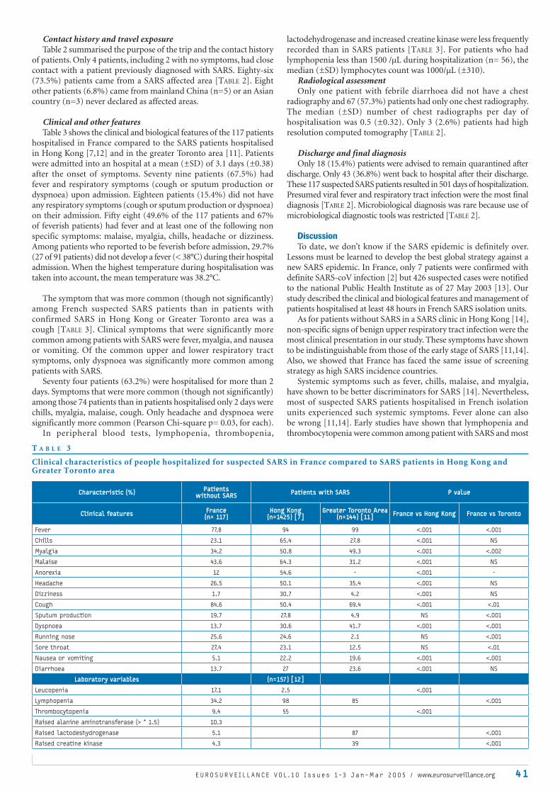
E U R OS U R V E I L L A N C E V O L . 10 I s s u e s 1 -3 J a n - M a r 2 0 0 5 / www.eurosurveillance.org 41
Contact history and travel exposureTable 2 summarised the purpose of the trip and the contact history
of patients. Only 4 patients, including 2 with no symptoms, had close contact with a patient previously diagnosed with SARS. Eighty-six (73.5%) patients came from a SARS affected area [TABLE 2]. Eight other patients (6.8%) came from mainland China (n=5) or an Asian country (n=3) never declared as affected areas.
Clinical and other featuresTable 3 shows the clinical and biological features of the 117 patients
hospitalised in France compared to the SARS patients hospitalised in Hong Kong [7,12] and in the greater Toronto area [11]. Patients were admitted into an hospital at a mean (±SD) of 3.1 days (±0.38) after the onset of symptoms. Seventy nine patients (67.5%) had fever and respiratory symptoms (cough or sputum production or dyspnoea) upon admission. Eighteen patients (15.4%) did not have any respiratory symptoms (cough or sputum production or dyspnoea) on their admission. Fifty eight (49.6% of the 117 patients and 67% of feverish patients) had fever and at least one of the following non specific symptoms: malaise, myalgia, chills, headache or dizziness. Among patients who reported to be feverish before admission, 29.7% (27 of 91 patients) did not develop a fever (< 38°C) during their hospital admission. When the highest temperature during hospitalisation was taken into account, the mean temperature was 38.2°C.
The symptom that was more common (though not significantly) among French suspected SARS patients than in patients with confirmed SARS in Hong Kong or Greater Toronto area was a cough [TABLE 3]. Clinical symptoms that were significantly more common among patients with SARS were fever, myalgia, and nausea or vomiting. Of the common upper and lower respiratory tract symptoms, only dyspnoea was significantly more common among patients with SARS.
Seventy four patients (63.2%) were hospitalised for more than 2 days. Symptoms that were more common (though not significantly) among those 74 patients than in patients hospitalised only 2 days were chills, myalgia, malaise, cough. Only headache and dyspnoea were significantly more common (Pearson Chi-square p= 0.03, for each).
In peripheral blood tests, lymphopenia, thrombopenia,
lactodehydrogenase and increased creatine kinase were less frequently recorded than in SARS patients [TABLE 3]. For patients who had lymphopenia less than 1500 /µL during hospitalization (n= 56), the median (±SD) lymphocytes count was 1000/µL (±310).
Radiological assessmentOnly one patient with febrile diarrhoea did not have a chest
radiography and 67 (57.3%) patients had only one chest radiography. The median (±SD) number of chest radiographs per day of hospitalisation was 0.5 (±0.32). Only 3 (2.6%) patients had high resolution computed tomography [TABLE 2].
Discharge and final diagnosisOnly 18 (15.4%) patients were advised to remain quarantined after
discharge. Only 43 (36.8%) went back to hospital after their discharge. These 117 suspected SARS patients resulted in 501 days of hospitalization. Presumed viral fever and respiratory tract infection were the most final diagnosis [TABLE 2]. Microbiological diagnosis was rare because use of microbiological diagnostic tools was restricted [TABLE 2].
DiscussionTo date, we don’t know if the SARS epidemic is definitely over.
Lessons must be learned to develop the best global strategy against a new SARS epidemic. In France, only 7 patients were confirmed with definite SARS-coV infection [2] but 426 suspected cases were notified to the national Public Health Institute as of 27 May 2003 [13]. Our study described the clinical and biological features and management of patients hospitalised at least 48 hours in French SARS isolation units.
As for patients without SARS in a SARS clinic in Hong Kong [14], non-specific signs of benign upper respiratory tract infection were the most clinical presentation in our study. These symptoms have shown to be indistinguishable from those of the early stage of SARS [11,14]. Also, we showed that France has faced the same issue of screening strategy as high SARS incidence countries.
Systemic symptoms such as fever, chills, malaise, and myalgia, have shown to be better discriminators for SARS [14]. Nevertheless, most of suspected SARS patients hospitalised in French isolation units experienced such systemic symptoms. Fever alone can also be wrong [11,14]. Early studies have shown that lymphopenia and thrombocytopenia were common among patient with SARS and most
T A B L E 3
Clinical characteristics of people hospitalized for suspected SARS in France compared to SARS patients in Hong Kong and Greater Toronto area
Characteristic (%) Patients without SARS Patients with SARS P value
Clinical features France(n= 117)
Hong Kong(n=1425) [7]
Greater Toronto Area(n=144) [11] France vs Hong Kong France vs Toronto
Fever 77.8 94 99 <.001 <.001
Chills 23.1 65.4 27.8 <.001 NS
Myalgia 34.2 50.8 49.3 <.001 <.002
Malaise 43.6 64.3 31.2 <.001 NS
Anorexia 12 54.6 - <.001 -
Headache 26.5 50.1 35.4 <.001 NS
Dizziness 1.7 30.7 4.2 <.001 NS
Cough 84.6 50.4 69.4 <.001 <.01
Sputum production 19.7 27.8 4.9 NS <.001
Dyspnoea 13.7 30.6 41.7 <.001 <.001
Running nose 25.6 24.6 2.1 NS <.001
Sore throat 27.4 23.1 12.5 NS <.01
Nausea or vomiting 5.1 22.2 19.6 <.001 <.001
Diarrhoea 13.7 27 23.6 <.001 NS
Laboratory variables (n=157) [12]
Leucopenia 17.1 2.5 <.001
Lymphopenia 34.2 98 85 <.001
Thrombocytopenia 9.4 55 <.001
Raised alanine aminotransferase (> * 1.5) 10.3
Raised lactodeshydrogenase 5.1 87 <.001
Raised creatine kinase 4.3 39 <.001

4 2 E U R OS U R V E I L L A N C E V O L . 10 I s s u e s 1 -3 J a n - M a r 2 0 0 5
S u r v e i l l a n c e r e p o r t
patients had a normal lymphocyte and platelet count at the onset of the disease [1,3,5,15]. 34.2 % of the patients had lymphopenia in our study, confirming that biological data were not useful for screening.
Therefore, early on French infectious disease specialists in charge of screening took into account the epidemiological data indicating the non specific presentation of SARS as well as the explosive transmissibility of SARS-CoV, notably before hospitalisation, in the community or health care setting. Indeed, we showed that the WHO criteria for suspected cases were variably interpreted in France. Presence of only one of the two clinical criteria (e.g., fever, respiratory symptoms) was enough to define a suspected case in case of exposure. Travel in an affected area, even without a close contact with a person previously diagnosed with SARS was considered as an exposure. French infectious disease specialists probably kept in mind that five of the seven French SARS cases were contaminated during an airplane flight.
Despite of rapid development of SARS biological diagnostic tools, a screening based only on these would presume a rapid bed test and a sufficient negative predictive value not existing today at the early stage of any infectious disease. Therefore, only evolution to SARS may predict a SARS-CoV infection.
The main question remains how, where and which suspected cases should be screened and isolated before being hospitalised as probable cases.
We are in agreement with Tambya [16] to consider that WHO case definitions were meant to lay down inclusion criteria for hospitalization and further investigation of a suspected case. Indeed, sensitivity of the WHO criteria for screening was estimated only over 27% [14,16], because they were with reference to hospitalised patients. In Singapore and Hong Kong, the positive predictive value was estimated at 10.6% and 54.3% respectively [14,16]. Given the lower prevalence of SARS in France, we could expect a lower positive predictive value but we considered that it was necessary to avoid SARS epidemic spreading in our country. With respect to the great number of suspected SARS who never develop the illness, suspected SARS patient must also be isolated from each other and from probable or definitive SARS cases to avoid cross contamination as was the case in Singapore [17]. Indeed, SARS was very localised in terms of transmission, at home with households or in emergency departments. That is why the French General Health Department asked all ill patients suspected to have had contact with a SARS patient, to phone the emergency mobile medical service (SAMU-SMUR) for a first phone screening. Suspected SARS patients were then directly take by specialist ambulance to the SARS isolation unit for a secondary screening. This strategy reduced exposure of healthcare workers at home or for general practitioners or emergency departments in the waiting rooms and corresponded to a priority of shortening the onset-to-admission interval [7] and of introducing early infection control measures.
Infectious disease teams are used to manage isolation of highly contagious diseases as air transmitted tuberculosis or handled transmitted diseases. Strict isolation procedures were also better understood, achieved (e.g. well fitted facemask) and accepted by the healthcare workers and also by patients. This strategy provided a better chance to avoid further transmission too, particularly in healthcare setting.
Retrospectively, we could have had a more specific screening if we had strictly respected SARS patient exposure definition. Indeed, contact exposure seems to be one of the best criteria for suspected SARS [7]. Nevertheless, precise contact exposure could be difficult to appreciate during the panic of an epidemic or because of mistrust in epidemiological data available, as was the case at the early stages of the epidemic. This emphases the need for an effective global alert system and to entrust the screening to infectious disease specialists, who are experts in epidemiological investigation and contact tracing.
Patients hospitalised in the French isolation units at the early stage of the worldwide SARS epidemic of the 2003 winter had mostly benign upper respiratory tract infection which can not be distinguished from an early stage of SARS. Screening and isolation have to be performed by infectious disease professionals. WHO case definitions have to lay down inclusion criteria for hospitalisation and further investigation
of a suspected case. Only strict observation of SARS exposure may reduce the hospitalisation rate and the cost of SARS screening strategy but epidemiological data have to be exhaustive, true and available in real time. This emphasizes the need to support the WHO Outbreak Alert and Response Network and the necessity for worldwide co-operation.
Contributors:B. Issartel, O. Lesens, C. Chidiac, Y. Mouton, D. Christmann
and D. Peyramond are co-principal investigators. B. Issartel and O. Lesens were responsible of the study design, analysis, and writing of the paper. All the members of the French SARS study group were responsible for patient management and were involved in collection of the clinical data.
*Members of the French SARS study group:Pr J Beytout and Dr F Gourdon, Hôtel Dieu, Clermont Ferrand. Pr
F Bricaire, Dr P Bossi and Dr A Guihot, G.H. Pitié -Salpêtrière, Paris. Pr F Caron and Dr P Abdoud, Hôpital Charles Nicolle, Rouen. P D Christmann and Dr O Lesens, Hôpitaux universitaires, Strasbourg. Pr Choutet and Dr D Garot, CH Tours. Pr Dellamonica, Dr E Bernard and E Cua, Hôpital de l’archet, Nice. Pr J Delmont, P Brouqui, Dr C. Foucault and P. Parola, Hôpital Nord de Marseille, Marseille. Pr M Dupon and H Dutronc, Hôpital Pellegrin, Bordeaux. Pr MGarre and Dr R Le Berre, Hôpital de la cavale blanche, Brest. Pr BHoen and Dr L Hustache-Mathieu, Hôpital Saint Jacques, Besancon. Pr Lenoir and Dr L Aaron, Hôpital Necker, Paris. Pr F Lucht and Dr A Frésard, C.H.U St Etienne, St Etienne. Pr T May, Ch Rabaud, Dr S Bevilacqua and M Stenzel, C.H.U de Brabois, Vandoeuvre-Les-Nancy. Pr C Michelet and Dr C Arvieux and P Tattevin, Hôpital Pontchaillou, Rennes. Pr Y Mouton, B Guery and Dr V Baclet, Centre Hospitalier de Tourcoing, Tourcoing. Pr D Peyramond, C Chidiac, Dr B Issartel, Hôpital de la Croix Rousse, Lyon. Pr E Pichard, Hôpital de l’Hôtel Dieu, Angers. Pr H Portier, P Chavanet and Dr MC Guillemot, Dijon. Dr Prazuck, Hôpital de la Source, Orléans. Pr Raffi and Dr D Boutoile, Hôpital de l’Hôtel Dieu, Nantes. Pr J Reynes and Dr C Lechiche, Hôpital Guy de Chauliac, Montpellier. Pr J P Stahl and Dr P Pavese, Hôpital Michalon, Grenoble. Pr P Weinbreck and Dr E Denes, Hôpital Dupuytren, Limoges.
Acknowledgements
We express our thanks to F. Gonzalez, clinical research assistant, who
supervised the study.
We are indebted to public health physicians and health care workers
who treated patients with suspected SARS or provided clinical materials
for evaluation for their assistance and cooperation.
References
1. Peiris JS, Chu CM, Cheng VC, Chan KS, Hung IF, Poon LL, et al. Clinical pro-
gression and viral load in a community outbreak of coronavirus-associated
SARS pneumonia: a prospective study. Lancet 2003;361(9371):1767-72.
2. Summary of probable SARS cases with onset of illness from 1 November 2002
to 31 July 2003. In: World Health Organization. 2003.
3. Lee N, Hui D, Wu A, Chan P, Cameron P, Joynt GM, et al. A major outbreak of severe
acute respiratory syndrome in Hong Kong. N Engl J Med. 2003;348(20):1986-94.
4. Chan-Yeung M, Yu WC. Outbreak of severe acute respiratory syndrome in Hong
Kong Special Administrative Region: case report. BMJ. 2003;326(7394):850-2.
5. Poutanen SM, Low DE, Henry B, Finkelstein S, Rose D, Green K, et al.
Identification of severe acute respiratory syndrome in Canada. N Engl J Med.
2003;348(20):1995-2005.
6. Tsang KW, Ho PL, Ooi GC, Yee WK, Wang T, Chan-Yeung M, et al. A cluster of
cases of severe acute respiratory syndrome in Hong Kong. N Engl J Med.
2003;348(20):1977-85.
7. Donnelly CA, Ghani AC, Leung GM, Hedley AJ, Fraser C, Riley S, et al.
Epidemiological determinants of spread of causal agent of severe acute
respiratory syndrome in Hong Kong. Lancet. 2003;361(9371):1761-6.
8. Ho W. Guideline on management of severe acute respiratory syndrome (SARS).
Lancet. 2003;361(9366):1313-5.
9. Case definitions for surveillance of severe acute respiratory syndrome (SARS).
In: World Health Organization. 2003.

O R I G I N A L A R T I C L E S
E u r o r o u n d u p
E U R OS U R V E I L L A N C E V O L . 10 I s s u e s 1 -3 J a n - M a r 2 0 0 5 / www.eurosurveillance.org 43
10. Drazen JM. SARS--looking back over the first 100 days. N Engl J Med.
2003;349(4):319-20.
11. Booth CM, Matukas LM, Tomlinson GA, Rachlis AR, Rose DB, Dwosh HA, et al.
Clinical features and short-term outcomes of 144 patients with SARS in the
greater Toronto area. JAMA. 2003;289(21):2801-9.
12. Wong RS, Wu A, To KF, Lee N, Lam CW, Wong CK, et al. Haematological manifes-
tations in patients with severe acute respiratory syndrome: retrospective
analysis. BMJ. 2003;326(7403):1358-62.
13. Bonmarin I. Investigation du SRAS en France. Bulletin Epidémiologique
Hebdomadaire 2003;24-25:112-113.
14. Rainer TH, Cameron PA, Smit D, Ong KL, Hung AN, Nin DC, et al. Evaluation of
WHO criteria for identifying patients with severe acute respiratory syndrome
out of hospital: prospective observational study. BMJ. 2003;326(7403):1354-8.
15. Ksiazek TG, Erdman D, Goldsmith CS, Zaki SR, Peret T, Emery S, et al. A novel
coronavirus associated with severe acute respiratory syndrome. N Engl J Med.
2003;348(20):1953-66.
16. Tambyah PA, Singh KS, Habib AG. SARS: Understanding the coronavirus: accuracy
of WHO criteria was similar in a «non-SARS» hospital in Singapore. BMJ.
2003;327(7415):620.
17. Singh K, Hsu LY, Villacian JS, Habib A, Fisher D, Tambyah PA. Severe acute respi-
ratory syndrome: lessons from Singapore. Emerg Infect Dis. 2003;9(10):1294-8.
V A R I C E L L A Z O S T E R V I R U S VA C C I N AT I O N P O L I C I E S A N D S U R V E I L L A N C E S T R AT E G I E S I N E U R O P E
A Pinot de Moira, A Nardone*
The incorporation of varicella zoster virus (ZVV) vaccination in childhood immunisation schedules is becoming an increasingly common option in Europe. The current study forms part of the European Sero-Epidemiology Network 2 (ESEN2) organisational analysis for VZV and describes current passive immunisation policies, as well as current and proposed active immunisation strategies, and existing surveillance systems for diseases caused by the varicella zoster virus in ESEN countries. A questionnaire was compiled and distributed to 23 participating countries. A VZV vaccine is currently licensed in 14 of the 20 participating ESEN countries. Germany is the only country to have incorporated VZV vaccination into its routine childhood immunisation programme. Three further countries currently recommend vaccination of children against VZV and five countries are also considering introducing routine immunisation against VZV for children. However, of the eight countries with or considering introducing childhood VZV immunisation, only six have case-based mandatory notification of varicella, and only two countries have primary care surveillance data available for herpes zoster.
Euro Surveill 2005;10(1):43-45 Published online Jan 2005Key words: Europe, surveillance, vaccination, Varicella Zoster
Virus
IntroductionVaricella is a self-limiting and relatively mild disease of childhood,
although it is frequently more severe and complicated amongst neonates (severe neonatal varicella), adults, pregnant women (potentially leading to congenital varicella syndrome in the child) and the immunocompromised. In addition, after an initial infection, the varicella zoster virus (VZV) lays dormant in dorsal root ganglia and may reactivate with declining cellular immunity to cause herpes zoster, particularly in the elderly and immunocompromised [1].
There are two methods of varicella infection control using immunisation: post-exposure passive antibody prophylaxis in the form of varicella zoster immunoglobulin (VZIG or VARITECT) and active vaccination. The varicella vaccine, which was developed in the early 1970s using a live attenuated form of the varicella zoster virus [2], has been licensed for use in some countries since the mid 1980s and has been part of the routine childhood immunisation schedule in the United States (US) since 1995 [3]. The cost-effectiveness of mass vaccination against varicella has, however, been questioned [4,5].
Universal vaccination programmes may cause an increase in the average age of infection, which may lead to increased adult morbidity and incidence of congenital varicella syndrome (CVS) and severe neonatal varicella. Studies have also suggested that re-exposure to exogenous varicella zoster virus protects against herpes zoster [6,8], thus, a reduction in the transmission of VZV (through vaccination) could result in an increased incidence of zoster.
Many European countries have already introduced targeted VZV vaccination for risk groups, and others are considering recommending either targeted vaccination or routine mass childhood immunisation. Only Germany has recently introduced VZV vaccination into the routine vaccination schedule. This is, therefore, an opportune moment to catalogue current passive immunisation policies, as well as current and proposed active immunisation strategies, and existing surveillance systems for diseases caused by the varicella zoster virus.
MethodsThe European Sero-Epidemiology Network 2 (ESEN2) is a network
of 22 European countries and Australia that aims to coordinate and harmonise the serological surveillance of immunity to a variety of vaccine preventable diseases in participating countries, including VZV [9]. This study formed part of the ESEN2 organisational analysis for VZV, the aim of which was to collate information regarding immunisation strategies and surveillance systems for the diseases under investigation.
A descriptive questionnaire was compiled, querying current and proposed VZV vaccination strategies and current surveillance of VZV. The questionnaire was split into three sections:
1. Current licensing of a VZV vaccine plus vaccine contraindications, current targeted vaccination of risk groups and mass vaccination, and also current use of VZIG.
2. Proposed mass childhood immunisation and targeted vaccination of specific groups. Questions included details of vaccination schedules, age and risk groups targeted, and catch-up campaigns being considered.
3. Current surveillance strategies for varicella, herpes zoster, congenital varicella syndrome and neonatal varicella, in particular mandatory notification, national hospital morbidity data and national primary care databases.
The questionnaire was distributed in February 2004 to lead epidemiologists in all 23 countries participating in the ESEN2 project. After three weeks a reminder was sent to participants to improve the response rate. Responses were received from 20 countries (87% of countries contacted) with a representative spread across Europe. Results were discussed at a one day workshop and returned to all participants for validation and feedback.
Immunisation Department, Health Protection Agency, CDSC, London, United Kinkdom
On behalf of the ESEN2 group*

4 4 E U R OS U R V E I L L A N C E V O L . 10 I s s u e s 1 -3 J a n - M a r 2 0 0 5
ResultsPassive immunisation strategiesEleven of the 20 countries responding (Australia, Cyprus, England
and Wales, Germany, Greece, Ireland, Israel, Italy, Lithuania, Malta, the Netherlands) currently use passive antibody prophylaxis for exposed risk groups. Groups for which prophylaxis is recommended include neonates and premature infants, pregnant women, and the immunocompromised.
Various VZV susceptibility screening procedures have been adopted for risk groups. For example, screening procedures for exposed pregnant women include either a verbal screen (Israel, Cyprus, and Malta), a serological screen (Australia, Germany, Italy, and the Netherlands) or a combination of the two (Greece, England and Wales, and Ireland)
Active immunisation strategiesA VZV vaccine is currently licensed in 14 of the 20 responding
countries; the six countries without a licensed vaccine are Bulgaria, Greece, the Netherlands, Romania, Slovakia and Slovenia.
Germany is the only country in Europe with routine childhood immunisation against VZV: VZV vaccination was incorporated into the routine immunisation schedule in July 2004, as a single dose at the age of 11-14 months [10].
In Lithuania, Cyprus, and Israel, VZV vaccination is recommended for children and is available either privately, or, as in Israel, through Health Maintenance Organisations (HMOs), but is not yet part of the routine childhood immunisation schedule. For Israel and Cyprus, the intention is to incorporate VZV vaccination into the routine childhood immunisation schedule in the short to medium term. Cyprus intends to vaccinate infants aged 13-18 months and children aged 11-12 years with no history of varicella, whereas Israel intends to administer the vaccine at the same time as the MMR vaccine, as a single dose at 12 months.
In Italy, there are no national recommendations for routine childhood immunisation, but, since July 2002, one of the twenty regions (Sicily) has offered free vaccination in the second year of life and for all anti-VZV negative 12 year olds.
Australia, Slovenia and Malta are also considering introducing recommendations for childhood immunisation against VZV in the short to medium term. Slovenia and Malta intend to combine vaccination with the MMR vaccine first dose, whilst Australia proposes to offer vaccination at 18 months with a catch-up at 10-13 years.
Eleven countries (Belgium, England and Wales, Finland, Germany, Israel, Ireland, Italy, Luxembourg, Malta, Slovenia and Spain) currently have targeted vaccination of specific groups, and one country (Slovakia) intends to issue guidelines for vaccination of specific groups [TABLE 1]. All of these countries, but England and Wales, either currently, or intend to, vaccinate immunocompromised patients. Susceptible healthcare workers are vaccinated in eight countries, with one further country (Israel) intending to introduce vaccination for healthcare workers. Interestingly, Belgium and Germany also recommend vaccination for susceptible child care workers.
Surveillance systemsTable 2 displays current surveillance systems for varicella in
participating countries. Of the five countries with some degree of childhood immunisation in place (Cyprus, Germany, Israel, Italy and Lithuania), Cyprus, Israel and Italy have case-based mandatory notification of varicella, and Lithuania has mandatory notification of varicella epidemics, as did Israel between 1949 and 2003. Italy also has a sentinel surveillance system based on paediatricians, estimated to cover roughly 4% of children aged 0-14 years. None of th ese countries, however, have primary care based sentinel surveillance data for herpes zoster, although data from HMOs are available in Israel [11]. Israel also has hospital morbidity data available for both varicella and herpes zoster, as does Italy. In Germany, there are plans for a sentinel surveillance scheme, based on that currently in place for measles, to be in place by 2005 for both varicella and herpes zoster.
T A B L E 2
Current surveillance strategies for varicella in 19 European countries and Australia, 2004
Country Mandatory Notification Sentinel surveillance
of primary care Year
established Case-based
or aggregate Year
established Estimatedcoverage
Countries with childhood vaccination (where childhood VZV vaccination is currently recommended or where routine vaccination is undertaken in one major region of the country)
Cyprus 2004 Case-based - -
Germany - - 2005 -
Israel 1949 Case-based‡ † †
Italy 1961 Case-based 2000 4% <14yrs
Lithuania 1973 Aggregate - -
Countries intending to introduce childhood vaccinationAustralia n/r Case-based* n/r n/r
Malta n/r Case-based - -
Slovenia 1977 Case-based Unknown Unknown
Other countriesBelgium - - - -
Bulgaria 1940 Aggregate - -
England & Wales - - 1967 1%
Finland - - n/r# n/r#
Greece 1950 Aggregate$ 2000 n/r
Ireland 2004 Case-based** 2000 3%
Latvia 1999 Case-based - -
Luxembourg - - - -
Netherlands - - 2000 1%
Romania 1978 Aggregate - -
Slovakia 1953 Case-based - -
Spain 1904 Aggregate - -
† Databases owned by HMOs‡ Case-based notifi cation introduced in 2003* State of South Australia only** Case-based mandatory notifi cation of outbreaks # Local primary care varicella databases (no national sentinel surveillance)
mandatory laboratory reporting
$ Replaced by notifi cation of ‘varicella with complications’ in 2004n/r not reported
E u r o r o u n d u p
T A B L E 1
Current and proposed targeted vaccination against Varicella Zoster Virus (VZV) in 12 european countries, 2004
Country Immuno-compromised
Contacts of immunocompromised
Susceptible healthcare workers
Susceptible child care workers
Susceptible adolescents
Susceptible women of childbearing age
Belgium +/- +/- +/- +/- +/-†
England & Wales +
Germany + + + + + +
Italy + *
Luxembourg +/- +/- +/- +/-
Malta + +
Slovenia + +
Spain + + *
Finland +/- +/- +/-
Ireland * +
Israel +/- +/- * +/-
Slovakia *
+ free or reimbursed vaccination currently in place+/- vaccination available privately (in Israel, through HMOs)* under consideration† Also recommended for healthy seronegative adults

E U R OS U R V E I L L A N C E V O L . 10 I s s u e s 1 -3 J a n - M a r 2 0 0 5 / www.eurosurveillance.org 4 5
Of the countries intending to introduce childhood immunisation against varicella (Australia, Slovenia and Malta), only Slovenia currently has primary care surveillance data for herpes zoster. In addition to this, Slovenia has varicella primary care data and case-based mandatory notification of varicella, for which data on attendance at daycare facilities and hospitalisation are collected [12]. Malta also has case-based mandatory notification of varicella. In Australia, although only the state of South Australia has mandatory varicella notification, data are also collected via sentinel surveillance of family doctors through the Australian Sentinel Practice Research Network.
Within those countries with neither childhood vaccination nor any current intentions to introduce it, Greece, Latvia and Slovakia have mandatory notification of varicella. In Greece, mandatory notification of ‘varicella with complications’ replaced notification of ‘varicella’ in 2004. Slovakia is the only participating country that has case-based mandatory notification of herpes zoster. Bulgaria, Ireland, Romania and Spain have mandatory notification of varicella epidemics. The Netherlands, Ireland, and England and Wales have primary care sentinel surveillance data for both varicella and herpes zoster.
DiscussionSince its development in the early 1970s, the VZV vaccine has
been licensed in numerous countries and incorporated into the US routine childhood immunisation schedule. Many European countries have targeted VZV vaccination of susceptibles for whom VZV infection poses a particular risk either to themselves (e.g. immunocompromised patients) or to others (e.g. healthcare workers), with many more considering introducing either targeted or mass childhood immunisation. Germany is the only country to have recently incorporated VZV vaccination into their routine childhood immunisation schedule, but several other countries plan to do so in the near future. Cyprus, Israel and Lithuania currently recommend VZV vaccination for children, and routine mass childhood immunisation has already been introduced in the Sicily region of Italy.
As with most universal mass vaccination, childhood immunisation against VZV could have a negative impact should it be introduced without sufficient coverage to induce herd immunity. Low vaccine coverage can result in an increase in the average age of primary infection, with a concomitant increase in severity of varicella in adult age groups [13], and especially in pregnant women, where infection can have adverse sequelae for both the mother and unborn child [14]. The levels of coverage estimated in countries with current VZV vaccination (approximately 25%), will have little impact on the age distribution of disease [15]. However, with increasing coverage, morbidity amongst adults is likely to increase, and vaccination is only predicted to decrease morbidity in both adults and children at around 70% coverage [16]. Thus, it is important that universal vaccination against VZV is introduced in a region or country only if the attainment of very high coverage can be assured. Furthermore, it is important that the age distribution of varicella disease is monitored, and this is best done through case-based surveillance of varicella. Of the eight countries that have or are considering introducing childhood VZV immunisation, only six have case-based mandatory notification of varicella. Initially, while disease incidence remains high, a well managed sentinel surveillance system could be an acceptable alternative: of the two countries without case-based mandatory notification of varicella, one is intending to implement such a surveillance system.
Exogenous exposure to varicella is thought to protect against zoster through boosting specific immune responses [6]. Therefore, the impact VZV vaccination will have on herpes zoster also needs to be considered. In the US, where mass childhood immunisation has been in place since 1995, no change in herpes zoster incidence was reported amongst 10-14 year olds, but the incidence of zoster in this
age group is low, and declines in exogenous VZV exposure would have been both recent and not immediate [17]. Mathematical modelling of a mass childhood immunisation strategy against VZV has predicted that there would be a significant rise in zoster morbidity, which is predicted to last more than 60 years [14]. Any country considering mass vaccination, should, therefore, have suitable surveillance for herpes zoster. Only two of the eight countries that have or are proposing to introduce childhood vaccination have primary care surveillance data available for herpes zoster.
Many countries have opted to limit vaccination to specific groups who are at increased risk of developing severe varicella disease or infecting risk groups (for example, healthcare workers). Targeted strategies have been predicted to have little impact on varicella incidence [16], and, consequently, are predicted to have little impact on herpes zoster [18], and the age distribution of primary disease [15]. Mass vaccination against VZV should be introduced only if very high coverage can be assured. With the introduction of routine childhood immunisation against VZV, however, adequate surveillance systems for both varicella and herpes zoster are advisable.
* ESEN2 group:Australia: J Backhouse, L Gilbert. Belgium: P van Damme, L De Cock, H Theeten, N Thiry, R Vranckx. Bulgaria: N Gatcheva, V Voynova. Cyprus: C Hadjianstassiou, M Zarvous. Finland: I Davidkin, S Jokinen. Germany: W Hellenbrand, S Reiter, A Tischer. Greece: C Anastassopoulou, T Georgakopoulou, A Hatzakis, T Panagiotopou-los. Ireland: M Carton, S Cotter, D O’Flanagan. Israel: D Cohen, L Moerman, Z Smetana. Italy: P Crovari, G Gabutti, MC Rota. Latvia: I Lucenko, J Perevoscikovs, I Velicko. Lithuania: V Bakasenas, J Surauciene. Luxembourg: J Mossong, F Schneider. The Netherlands: G Berbers, H de Melker. Malta: A Amato-Gauci, C Barbara. Roma-nia: A Pistol. Slovakia: J Lancová, M Sláciková. Slovenia: A Kraigher, K Prosenc. Spain: C Amela, F de Ory. UK: E Miller, RG Pebody.
References
1. Quinlivan M, Hawrami K, Barrett-Muir W, Aaby P, Arvin A, Chow VT et al. The
molecular epidemiology of varicella-zoster virus: evidence for geographic
segregation. J Infect Dis 2002;186(7):888-94.
2. Takahashi M, Okuno Y, Otsuka T, Osame J, Takamizawa A. Development of a live
attenuated varicella vaccine. Biken.J. 1975;18(1):25-33.
3. Recommendations for the use of live attenuated varicella vaccine. American
Academy of Pediatrics Committee on Infectious Diseases. Pediatrics
1995;95(5):791-6.
4. Brisson M, Edmunds WJ. Varicella vaccination in England and Wales: cost-
utility analysis. Arch Dis Child 2003;88(10):862-9.
5. Thiry N, Beutels P, Van Damme P, Van Doorslaer E. Economic evaluations of vari-
cella vaccination programmes: a review of the literature. Pharmacoeconomics.
2003;21(1):13-38.
6. Garnett GP, Grenfell BT. The epidemiology of varicella-zoster virus infections:
the influence of varicella on the prevalence of herpes zoster. Epidemiol.
Infect. 1992;108(3):513-28.
7. Gershon AA, LaRussa P, Steinberg S, Mervish N, Lo SH, Meier P. The protective ef-
fect of immunologic boosting against zoster: an analysis in leukemic children
who were vaccinated against chickenpox. J.Infect.Dis. 1996;173(2):450-3.
8. Thomas SL, Wheeler JG, Hall AJ. Contacts with varicella or with children
and protection against herpes zoster in adults: a case-control study. Lancet
2002;360(9334):678-82.
9. Osborne K, Weinberg J, Miller E. The European Sero-Epidemiology Network.
Euro.Surveill 1997;2(4):29-31.
10. Rasch G, Hellenbrand W. Germany adds varicella vaccine to the national
vaccination programme. Eurosurveillance Weekly 2004;8(31). http://www.eurosur-
veillance.org/ew/2004/040729.asp
11. Passwell JH, Hemo B, Levi Y, Ramon R, Friedman N, Lerner-Geva L. Use of a
computerized database to study the effectiveness of an attenuated varicella
vaccine. Pediatr.Infect.Dis.J. 2004;23(3):221-6.
12. Socan M, Kraigher A, Pahor L. Epidemiology of varicella in Slovenia over a
20-year period (1979-98). Epidemiol.Infect. 2001;126(2):279-83.
13. Arbeter AM. Clinical trials of varicella vaccine in healthy adolescents and
adults. Infect Dis Clin North Am 1996;10(3):609-15.
14. Fairley CK, Miller E. Varicella-zoster virus epidemiology--a changing scene?
J Infect Dis 1996;174 Suppl 3:S314-S319.
15. Brisson M, Edmunds WJ, Gay NJ, Law B, De Serres G. Modelling the impact of
immunization on the epidemiology of varicella zoster virus. Epidemiol Infect
2000;125(3):651-69.
16. Brisson M, Edmunds WJ, Gay NJ. Varicella vaccination: impact of vaccine
efficacy on the epidemiology of VZV. J.Med.Virol. 2003;70 Suppl 1:S31-S37.
17. Goldman GS. Varicella susceptibility and incidence of herpes zoster among
children and adolescents in a community under active surveillance. Vaccine
2003;21(27-30):4238-42.
18. Brisson M, Edmunds WJ. Varicella vaccination in England and Wales: cost-
utility analysis. Arch.Dis.Child 2003;88(10):862-9.

O R I G I N A L A R T I C L E S
O u t b r e a k r e p o r t
4 6 E U R OS U R V E I L L A N C E V O L . 10 I s s u e s 1 -3 J a n - M a r 2 0 0 5
M E A S L E S O U T B R E A K I N T H E P R O V E N C E - A L P E S - C Ô T E D ’A Z U R R E G I O N , F R A N C E , J A N U A R Y – J U LY 2 0 0 3C Six 1, F Franke 1, K Mantey 1,2, C Zandotti 3, F Freymuth 4, F Wild 5, I Parent du Châtelet 6, P Malfait 1
must be effective by 2010 [3]. However, epidemiological conditions in France are still favourable to the occurrence of outbreaks in high risk population groups for example non-vaccinated children. Outbreaks of measles attributed to inadequate vaccination coverage occurred in Italy, in 2002 with 1571 cases reported in Campania (12 encephalitis cases and 3 child deaths) [4] and in Switzerland, in 2003 with 464 cases reported (3 encephalitis cases) [5].
At the end of May 2003, the Marseilles Hospital Centre’s virology laboratory (La Timone) informed the French public heath institute (Institut de Veille Sanitaire, InVS) of 5 cases of confirmed measles in young adults living in Marseilles. An investigation was conducted to determine the measures to be taken, especially in relation to vaccination programmes, and to document the virus circulation in the PACA region by the Cellule Inter-Régionale d’Épidémiologie Sud (south interregional epidemiological cell, Cire Sud), who coordinated this investigation [6].
MethodsThe measles investigation [7], which was conducted between 20 May and end of July 2003, consisted of the following steps:
• A response to the alert of the La Timone Hospital virology laboratory with individual case-patient interview (begun 20 May 2003)
• Active case-finding of other measles cases in Marseilles and in neighbouring cities by consulting different community and hospital health services: hospitals (community and private), Conseil Général, hygiene and health community department (City of Marseilles), SOS Médecins, Médecins du Monde, school medical services, etc. (begun 29May 2003)
• A retrospective review of serologically confirmed measles cases in the PACA region from January 2003 by actively contacting the 3 main laboratories in France performing anti-measles IgM determination (begun 2 June 2003)
• A feasibility survey of prospective surveillance of measles among GPs located only in the main cities (restriction due to logistic reasons) of the region where the virus circulation was active (Marseilles, Avignon, Digne-les-Bains) (begun 19 June 2003) [6]
• A retrospective survey among GPs (homeopathic practitioners included) and paediatricians, in an area located between Manosque and Digne, where several cases were reported (begun 27 June 2003).
Definition of the casesThree definition levels were used to classify the cases:
• Clinical case: when measles diagnosis has been made by a GP;• Serologically confirmed case: when measles specific IgM serology
was positive;• Epidemiologically confirmed case: clinical case who contact with
a serologically confirmed case between 7 and 18 days before clinical symptoms appeared.
Data collectionThe following variables were collected for each patient: sex, age,
location, date of the first serology for the serologically confirmed cases or date of the clinical diagnosis, admission to hospital, complications, vaccination status and reasons for non-vaccination.
At the end of May 2003, the Marseilles Hospital Centre’s virology laboratory informed the French public heath institute of 5 cases of confirmed measles among young adults living in Marseilles. An investigation was conducted, consulting different community and hospital health services, to determine the virus circulation in the Provence-Alpes-Côte d’Azur (PACA) region by the southern interregional epidemiological cell. The investigation identified 259 cases: 183 clinical, 74 serologically confirmed and 2 epidemiologically linked cases. The first cases were identified during the first six months of 2003, with a peak in April. This outbreak of measles in the PACA region was favoured by poor vaccination coverage, which created groups of susceptible population. The real number of cases was probably higher than the number identified. This investigation has outlined the limitations of the measles surveillance system in France: the sentinel network had not detected any case for this period. France needs to reach the WHO objective of measles elimination by 2010 and the surveillance tools used must be those already used in the most countries that are furthest advanced in the elimination process. To reach this goal, the Direction Générale de la Santé has nominated a working group to be in charge of proposing a national plan to interrupt indigenous measles transmission in France.
Euro Surveill 2005;10(1):46-48 Published online Jan 2005Key words: disease outbreak, elimination, measles, vaccination
IntroductionMeasles was a notifiable disease in France from 1945-1985. Since
1985, a sentinel surveillance network known as “Sentinelles”, created by National Institute for Health and Medical Research (INSERM U444), has reported the number of cases each week declared by voluntary general practitioners (GPs). Extrapolation of data obtained from this sentinel network allows the determination of the national incidence, which fell from 300 000 cases per year in 1985 to less than 10 400 cases in 2003 (IC 95%: 6000-15 000). Since 1985, a shift in the age of measles cases has been noted through the sentinel network: the proportion of patients aged 10 years and above was 13% in 1985, 48% in 1997 and 62% in 2002 [1].
In 2001, childhood vaccine coverage against measles in France was estimated to be 84.6% at 24 months, with some differences between the different district areas. The south of France had the lowest coverage [2]. In the region of Provence-Alpes-Côte d’Azur (PACA), the coverage rate by district was 84% in the Alpes-Maritimes, 82% in the Bouches-du-Rhône and Var, 76% in Vaucluse and 59% in the Alpes-de-Haute-Provence. No data was available for the Hautes-Alpes.
In order to apply the World Health Organization (WHO) objectives, France, along with other European countries, has applied a policy of interruption of indigenous measles transmission. This target
1. Cellule interrégionale d’épidémiologie Sud (Cire Sud), Marseille, France
2. Programme de formation à l’épidémiologie de terrain, Cellule interrégionale d’épidémiologie Sud (Cire Sud), Marseille, France
3. Laboratoire de virologie de la Timone, Marseille, France
4. Laboratoire de virologie du CHU Clémenceau de Caen, CNR associé, France
5. Centre national de référence de la Rougeole, France
6. Institut de Veille Sanitaire (InVS, département des maladies infectieuses), Saint-Maurice, France

E U R OS U R V E I L L A N C E V O L . 10 I s s u e s 1 -3 J a n - M a r 2 0 0 5 / www.eurosurveillance.org 47
Laboratory investigationsSerological analysis (measles specific IgM) was carried out on
patients’ blood samples. Urinary and saliva samples were tested for measles virus by genetic amplification using reverse transcription-polymerase chain reaction (RT-PCR).
ResultsNumber of casesThe different steps of investigation identified 259 cases: 183
(71%) clinical cases, 74 (28%) serologically confirmed and 2 (1%) epidemiologically linked. The case detection rate in the PACA region was 3.1 per 100 000 inhabitants. The highest number of cases was identified in Alpes-de-Haute-Provence with a rate of 39.4 cases per 100 000 inhabitants, then Vaucluse (6.4) and Bouches-du-Rhône (2.5) [FIGURE 1].
F I G U R E 1
Detected cases rate of measles per 100 000 inhabitants per district, PACA region, January - July 2003
Information concerning 138 cases was collected from medical practitioners (documented cases).
No measles cases were identified through the sentinel system for the period under study.
Characteristics of the documented casesThe first cases were identified during the first week of January and
the last notified case appeared in July, with a peak in April [FIGURE 2]. Three district areas accounted for 96% of identified measles cases: Alpes-de-Haute-Provence (40%), Bouches-du-Rhône (33%) and Vaucluse (23%).
F I G U R E 2
Number of documented cases of measles by month of onset, PACA region, January-July 2003
The male/female ratio was 1.1. The mean age was 15 years in the PACA region (ranging from 11 months to 57 years), 11 years in the Alpes-de-Haute Provence, 14 years in Vaucluse and 20 years in the Bouches-du-Rhône [FIGURE 3].
F I G U R E 3
Distribution of documented cases of measles by age in 3 PACA departements, France, January-July 2003
Vaccination status: Information on vaccination status was known for 68 patients (50% of the documented cases) of whom 60 were not vaccinated and 8 had received only one vaccine dose (these cases were aged between 1 and 15 years old).
Clinical characteristics: Complications reported were: pneumonia (2 cases), neurological symptoms (without encephalitis) (2 cases), digestive problems (9 cases) and otitis (1 case). No deaths were reported.
A total of 25 patients (18% of the documented cases) were admitted to hospital. Reasons for admission to hospital were linked to diagnostic difficulties, differential diagnosis such as toxidermia and HIV primary infection, severe clinical symptoms such as cutaneous disease affecting more than 90% of the body surface (3 cases) and severe alteration of health (1 case). Fifteen patients were not vaccinated and ten patients had unknown vaccination status.
Microbiological description: from the 74 biologically confirmed cases, 4 samples (3 urinary, 1 pharyngeal) were sent to the Caen Hospital Centre for analysis by RT-PCR method and were positive. The study of the genotype by the national reference centre (Unité INSERM 404 Lyon) on 3 samples showed them to be of the D7 genotype.
Discussion and conclusionThis outbreak of measles in the PACA region during the first six
months of 2003 was favoured by the poor vaccination coverage, which created groups of susceptible population. The investigations showed a link between the vaccine coverage in the different districts and the mean age of cases, suggesting a shift of age at disease occurrence, from childhood to teenage and adults years, linked to the slowing down of the virus circulation [8]. Reasons for inadequate vaccination coverage among children especially in Alpes-de-Haute-Provence district were due to medical follow-up by GPs and homeopathic practitioners.
The decrease in the number of cases, in May and June, can be explained by the seasonal pattern of measles in France [1], more frequent in the first part of the year, and by the closing of the schools due to the teachers’ strike before the summer holidays.
The goal of this investigation was to document the virus circulation in the PACA region. This was not completely achieved. The investigation was limited to a restricted number of GPs in three head district cities. This choice was justified by the feasibility criteria (logistical limitations) instead of the representative activities of all GPs in the 3 cities. The participation rate was poor (20% of the GPs responded spontaneously, and 40% after many recalls) despite the mobilisation of an investigation team. Because of these results, the study was not extended to the whole. The different investigations have outlined the limitations of the measles surveillance system in France. Starting with 5 cases identified in Marseilles, 259 cases were been identified through this investigation. The real number of cases is probably higher than the number identified. Without the alert of
0
10
20
30
40
January February March April May June July
Month
Num
ber o
f c
ases
0%
10%
20%
30%
40%
50%
0 - 3 4 - 6 7 - 13 14 - 19 20 - 29 30
Age in years
Percenta
ge o
f c
ases
(13) (84)VaucluseBouches-du-RhôneAlpes-de-Haute-Provence
<1
>=1 and <3
>=3 and <10
Rate per 100 000 inhabitants
>=10

O R I G I N A L A R T I C L E S
O u t b r e a k r e p o r t
4 8 E U R OS U R V E I L L A N C E V O L . 10 I s s u e s 1 -3 J a n - M a r 2 0 0 5
La Timone Hospital laboratory, these cases would not have been identified: the Sentinelles network had not detected any case for the first half of 2003.
Considering the decrease of the number of cases reported by the sentinel GPs during previous years and the fact that the positive predictive value of a clinical case definition is poor, the Sentinelles network cannot identify measles residual transmission areas and does not allow us to know the proportion of measles among the suspected cases. Thus, a low proportion of real measles cases can be expected in patients with febrile rash symptoms. This proportion has been estimated in the United Kingdom as 3%.
The Italian outbreak in Campania during 2002 was predominantly detected by the national paediatric surveillance system (4 times more sensitive than mandatory notification) but data were only obtained from children under 15 years of age, and the extent of the outbreak in adolescents and adults was probably underestimated [4]. In Switzerland, the increase in measles cases during 2003 was detected by the mandatory notification system and not by the sentinel surveillance system [5]. In 2001, only 16 of 19 countries in Europe had a mandatory notification system for measles, and some countries had a sentinel surveillance system in addition to this [10].
France needs to reach the WHO objective of measles elimination by 2010 [3]. The surveillance tools must be those already used in the countries that are furthest advanced in the elimination process: exhaustive notification; wide clinical definition to obtain a high sensitivity and to detect all suspected cases; laboratory confirmation to improve specificity and only detect the real cases; strain determination to trace their origin; vaccine coverage follow-up for each dose; and estimation of the proportion of susceptible population by modelling or serological studies [8]. To reach this goal, the Direction Générale de la Santé has nominated a working group to be in charge of proposing a national plan to interrupt the indigenous measles transmission in France.
Acknowledgements
We wish to thank all the organisations and persons who contributed to
the investigations : Health Authorities (Directions départementales des
affaires sanitaires et sociales - Ddass) of Alpes-de-Haute-Provence,
Bouches-du-Rhône and Vaucluse. Medical practitioners and laboratories
of hospitals, private laboratories of Medical Biology, MCH and school
medical services.
References
1. Sentiweb , serveur du Réseau SENTINELLES des Médecins libéraux français:
Surveillance épidémiologique du réseau Sentinelles, janvier-décembre 2003.
2004 http://www.b3e.jussieu.fr/sentiweb
2. Antona D, Bussière E, Guignon N, Badeyan G, Lévy-Bruhl D. La couverture vac-
cinale en France en 2001. Bull Epidemiol Hebd. 2003;36:169-72
3. Progress towards measles elimination, WHO Eastern Mediterranean Region,
1980-1998. Wkly Epidemiol Rec. 1999 Dec 17;74(50):434-9. http://www.who.
int/docstore/wer/pdf/1999/wer7450.pdf
4. CDC. Measles Epidemic Attributed to Inadequate Vaccination Coverage;Campania,
Italy, 2002. MMWR Morb Mortal Wkly Rep. 2003 Oct 31;52(43):1044-7.
5. Richard JL, Boubaker K, Doutaz M, Schubiger G. Déclaration obligatoire de la
rougeole en Suisse:forte augmentation du nombre de cas au printemps 2003.
Bulletin des médecins suisses. 2003;84:27:1445-50.
6. Six C, Franke F, Pieyre A, Zandotti C, Freymuth F, Wild F, Parent du Châtelet I,
Malfait P. Investigation de cas de rougeole en région Provence - Alpes - Côte
d’Azur au cours du premier semestre 2003. Bull Epidemiol Hebd. 2004;16:63-64
7. Six C. Investigation de cas de rougeole en région Provence - Alpes - Côte
d’Azur, janvier-juin 2003. Investigation report. Institut de veille sanitaire.
February 2004
8. Bonmarin I, Parent du Châtelet I, Levy-Bruhl D. La rougeole en France : impact
épidémiologique d’une couverture vaccinale sub-optimale. Bull Epidemiol
Hebd. 2004;16:63-64
9. Ramsay ME, Jin L, White J, Litton P, Cohen B, Brown D. The elimination of in-
digenous measles transmission in England and Wales. J Infect Dis. 2003 May
15;187 Suppl 1:S198-207.
10. Muscat M, Glismann S, Bang H. Measles in Europe in 2001-2002. Euro Surveill
2003 Jun;8(6):123-9. http://www.eurosurveillance.org/em/v08n06/0806-221.
asp?langue=02&
C O M M U N I C A B L E D I S E A S E C O N T R O L I N A M I G R A N T S E A S O N A L W O R K E R S P O P U L AT I O N : A C A S E S T U D Y I N N O R W AY
PJ Guerin 1, L Vold 1, P Aavitsland 2
Reliable data on the health status of migrant seasonal workers in Europe is scarce. Access to public health care for this population depends on national regulations, and their legal status in host countries. In this manuscript we describe a case study of a salmonellosis outbreak that occurred in Norway, and highlight the difficulties encountered in applying control measures in a population of seasonal migrant farm workers. Surveillance and control of infectious diseases need to be supported by legislation which makes implementation of control measures possible. Efforts have been made to improve the rights for migrants in Europe with regard to healthcare, but seasonal migrant workers still remain largely outsiders where these measures are concerned. Special attention should be given to this disadvantaged group in terms of social rights and healthcare. Preparedness plans should be improved to deal with contagious pathogens involving the seasonal migrant population.
Euro Surveill 2005;10(3):48-50 Published online Mar 2005Key words: Agricultural workers, communicable diseases, health
services accessibility, international migration, seasonal workers
IntroductionSeasonal migrant farm-workers (SMFW) all over the world travel
frequently and over significant distances to secure their employment. The transient nature of their employment, migration in and out of countries, and the desire by some to avoid contact with governmental agencies, makes the exact number of SMFW difficult to determine [1]. The number of SMFW in Europe is believed to be substantial, but few data are available [2].
In many cases the SMFW and local populations differ with regard to nutrition, language, family structure, religion and health. Epidemiological studies of the health of SMFW are scarce, although some public health concerns have been identified [3,4]. Seasonal migrant farm-workers often come from countries that are poorer than the countries to where they travel for work, and these countries often have different disease epidemiology. Diseases, which can be endemic in the SMFW countries of origin, may be unusual and cause a lot of attention in the new, host countries. This can cause concern in the local communities employing SMFW. A substantial number of SMFW are never registered as employees, and therefore do not benefit from sick-leave in case of illness. They may feel forced to work even when ill. Seasonal migrant farm workers have been implicated in the contamination of produce at source, or hypothesised to be the source of contamination. [5-7].
Every summer and early autumn, more than 15 000 SMFW come
1. Division for Infectious Disease Control, Norwegian Institute of Public Health, Nydalen, Norway
2. Department of Infectious Disease Epidemiology, Divisioan of Infectious Disease Control, Norwegian Institute of Public Health, Nydalen, Norway
O u t b r e a k r e p o r t s

E U R OS U R V E I L L A N C E V O L . 10 I s s u e s 1 -3 J a n - M a r 2 0 0 5 / www.eurosurveillance.org 4 9
to Norway to harvest fruit, vegetables and berries [8]. This official figure underestimates the real number of SMFW in Norway, because many are not officially registered. Lack of recommendations on how to handle outbreaks of communicable foodborne disease among these people poses a problem for the local health authorities. The majority of the workers are from Poland (62%), Lithuania (20%), Latvia (5%), Estonia (2%), Slovakia (2%) and Ukraine (2%).
The aim of this paper is to describe an imported and self-limited salmonellosis outbreak among SMFW and highlight the social consequences and difficulties encountered in the investigation.
Description of the outbreakOn 12 July 2002, two cases of Salmonella Enteritidis infection were
reported to the Norwegian Institute of Public Health (NIPH). The two patients were part of a group of 14 Polish strawberry pickers who had come from Poland to Norway on the 1st of July. Five days after arrival, two female workers had symptoms of gastroenteritis, mainly diarrhoea without fever. After two days, their condition had not improved and they consulted the local hospital where they were hospitalised and diagnosed with Salmonella Enteritidis infection. The patients received symptomatic treatment and were discharged from the hospital after two days.
Investigations Investigation of the outbreak was undertaken by the Local Food
Authorities (LFA) and the Public Health Officer (PHO), assisted by epidemiologists from the NIPH. The investigation was limited and there were problems with, language, collaboration and social consequences of the outbreak. To try to find the source of the infection, food histories were collected and the premises inspected.
FindingsThe farmer who employed the Polish SMFW provided them with
basic lodging. Toilets and bathrooms were available on the farm, but the only toilet facilities available to workers in the fields were pit latrines without running water, soap or towels. The investigation revealed that the group of workers had brought with them fresh meat and eggs from Poland, and had cooked their own meals. The workers had only eaten food that they had brought with them. Two days before the onset of symptoms, they had eaten a meal based on lightly cooked eggs which had been stored at room temperature for some hours prior to consumption. Based on the food histories, it was suggested that this meal was the most probable source of the outbreak. No eggs were left over for analysis.
Media coverageEven though this outbreak was very limited in size, there was
extensive coverage in the local media. In Norway, infections with Salmonella Enteritidis are mainly contracted abroad (90 % of cases), and the local press angled their stories in a way that heightened concern about SMFW and introduction of pathogens into Norway. As a result of the scare of Salmonella spreading in the community, concern about possible spread of infection to the strawberries arose, and consequently the wholesale dealer no longer wanted to purchase strawberries from the farm involved.
Control measuresThe substantial coverage in the local media, and lack of
recommendations on how to handle the situation from the central authorities, led to the implementation of initial control measures, some of which were not very rational and without much purpose.
Basic hygiene recommendations were given and providing water and soap in the field was recommended. The two symptomatic cases were initially asked to stay away from work and limit contact with their co-workers. They were moved to a separate caravan and were not allowed to use the toilet facilities used by the other workers. They were offered the use of the stable as a toilet with possible contamination of this environment as a result. Contrary to normal procedures in cases of salmonellosis among workers in the food industry, all SMFW on
the farm were screened for salmonellosis. Two asymptomatic carriers were identified. The national reference laboratory reported that all four cases had Salmonella Enteritidis phage type 4, and it is likely that the source of infection was the same for the four cases.
Outcome On 22 July, two epidemiologists from NIPH were sent to the
community to assist the PHO. The objectives were to investigate possible sources of contamination, to ensure that all relevant public health measures were taken, to assist the local health authorities with handling the press, and to collaborate with the LFA in evaluating the public health risk connected with consumption of already harvested berries. When epidemiologists from the NIPH arrived, all the workers from the group concerned had left, escaping a situation that had become too complicated. After leaving the farm, the group of workers tried to find work on neighbouring farms, without success. It is likely that they then tried to find work in other parts of the country. There is no legal requirement for workers to inform employers that they could potentially be asymptomatic carries of Salmonella, and no effort was made to trace the group. No other case of gastroenteritis was reported in this community in the relevant period of time.
DiscussionSalmonella, food industry and labour rightsFoodborne disease caused by non-typhoid Salmonella is an
increasing public health problem worldwide [9]. In order to decrease the incidence of human foodborne salmonellosis, various control measures have been implemented to hinder introduction and multiplication of Salmonella in the food chain. In the Norwegian food industry, workers who have diarrhoea are not allowed to work as normal while ill, but are either allocated to duties which do not include direct contact with food or stay away from work on sick leave. For specific pathogens like Salmonella, they may not return to work until stool samples are negative with regard to carriage of the pathogen.
According to the Norwegian social security law, all those who have permission to work are covered by the National Insurance Fund [10]. Provided SMFW have a work permit, they have the right to sickness benefit after two weeks of work. Without a work permit, no sickness benefit is obtained, but workers still have the right to necessary health care according to the Communicable Disease Control Law and the Law on Municipal Health Care Services.
If the health authorities prohibit work because of danger of spread of infections, sickness benefit is paid on the same conditions that apply if the employee is ill. The workers’ rights in such cases are covered by laws and regulations of employment, and the workers receive their salary until the public health authorities consider it safe to permit them to return to work.
Such rights do not usually apply for SMFW, who often work on a piece-rate and on short-term contracts. In this outbreak, the workers had work permits but had not yet worked for the compulsory two weeks. Therefore, they had no right to sickness benefit, which could have helped control measures and prevented them from moving away.
The financial consequences of not being allowed to work due to illness are severe. Because travelling to Norway is a big investment in itself, SMFW would probably try to stay in the country even if ill, in order to recover their costs, or even simply to be able to afford to return home. Basic food items in Norway are more expensive than in the rest of Europe, particularly central and eastern Europe, so bringing food from home is one way to save money. Importing fresh products from Poland was illegal when this outbreak occurred, but nevertheless took place. This ‘import’ of food increases the risk of food contamination because of lack of control and sub-standard conservation procedures.
The absence of social benefits for seasonal workers without permission to work, and only partial access to social benefits while legally employed, can be supported by strong economic arguments.

5 0 E U R OS U R V E I L L A N C E V O L . 10 I s s u e s 1 -3 J a n - M a r 2 0 0 5
However, difficult situations arise when public health authorities apply control measures to prevent the spread of disease and thereby put migrant workers in an impossible financial situation.
Control measuresMeasures implemented during this outbreak, such as isolation of
symptomatic workers and screening of asymptomatic workers, were not in accordance with recommendations by the national public health authorities. They were mainly consequences of external pressures on local authorities under pressure from the mass media to make quick decisions, relatively poor collaboration from the employing farmer, who was afraid of losing his strawberry harvest, and incorrect estimation of the risk of cross-contamination between workers.
Basic access to water for washing hands in the fields was unavailable. Other outbreaks described in the literature have linked bad hygiene practice and possible contamination of food products during harvest to the spread of infection [11-13]
RecommendationsProvision of sanitary facilities for workers could decrease the
potential risk for direct or indirect contamination of berries. This investigation highlighted the poor knowledge of migrant
workers’ rights among the different public agencies involved. Efforts should be made to disseminate this information to officials and beneficiaries.
The migrant workers themselves should not be blamed for their role in the possible spread of infectious diseases. Surveillance and control of infectious diseases has to be supported by laws that make implementation of control measures possible. Efforts are being made to improve the rights for migrants in Europe with regard to health, especially through the revised European social charter from 1996 [14]. However, seasonal migrant workers still remain largely uncovered by these measures.
Special attention should be given to SMFW who are a disadvantaged group in terms of social rights and healthcare in Europe [1].
Concerns about methodological difficulties in epidemiological studies of seasonal migrant populations might have dissuaded researchers from conducting studies. However, although difficulties might occur due to the mobile nature of these populations, more studies should be encouraged, in particular in the field of communicable diseases [15].
In this particular outbreak, Salmonella Enteritidis infection was not considered a major public health risk. However, preparedness plans
should be improved to deal with more contagious and threatening pathogens involving seasonal migrant populations.
Acknowledgements
We thank Ragnhild Seip for her help in collecting information on health
regulation in Norway and the National Reference Laboratory for perform-
ing the phage typing on the Salmonella strains. The authors gratefully
acknowledge the thoughtful comments of Dr Peter Bilton, Angela Rose
and Dr Tore Tollersrud on the manuscript.
References
1. Rust GS. Health status of migrant farmworkers: a literature review and com-
mentary. Am.J.Public Health. 1990;80(10):1213-17.
2. Dobson J, Salt J. Review of Migration Statistics. Political Economy of Migration
in an Integrating Europe (PEMINT). Working paper No.: 7/2002. Migration research
Unit, Department of Geography, University College London, 2002.
3. Mobed K, Gold EB, Schenker MB. Occupational health problems among migrant
and seasonal farm workers. West J.Med. 1992;157(3):367-73.
4. Zahm SH, Blair A. Cancer among migrant and seasonal farmworkers: an epide-
miologic review and research agenda. Am.J.Ind.Med. 1993;24(6):753-66.
5. Naimi TS, Wicklund JH, Olsen SJ, Krause G, Wells JG, Bartkus JM, Boxrud DJ,
Sullivan M, Kassenborg H, Besser JM, Mintz ED, Osterholm MT, Hedberg CW.
Concurrent outbreaks of Shigella sonnei and enterotoxigenic Escherichia coli
infections associated with parsley: implications for surveillance and control
of foodborne illness. J Food Prot. 2003 Apr66(4):535-41
6. Calder L, Simmons G, Thornley C, Taylor P, Pritchard K, Greening G, Bishop J.
An outbreak of hepatitis A associated with consumption of raw blueberries.
Epidemiol Infect. 2003 Aug;131(1):745-51
7. Reid TM, Robinson HG. Frozen raspberries and hepatitis A. Epidemiol Infect.
1987 Feb;98(1):109-12
8. Norwegian Directorate of Immigration. Figures on seasonal workers in Norway,
2002. Norwegian Directorate of Immigration, 2003.
9. Gomez TM, Motarjemi Y, Miyagawa S, Kaferstein FK, Stohr K. Foodborne salmo-
nellosis. World Health Stat.Q. 1997;50(1-2):81-89.
10. Ministry of Social Affairs [sosialdepartementet]. Norwegian social secu-
rity law [Lov om folketrygd (folketrygdloven)]. http://www.lovdata.no/all/nl-
19970228-019.html, 1997.
11. Herwaldt BL, Ackers ML. An outbreak in 1996 of cyclosporiasis associated
with imported raspberries. The Cyclospora Working Group. N.Engl.J.Med.
1997;336(22):1548-56.
12. Hutin YJ, Pool V, Cramer EH et al. A multistate, foodborne outbreak of hepatitis
A. National Hepatitis A Investigation Team. N.Engl.J.Med. 1999;340(8):595-602.
13. From the Centers for Disease Control and Prevention. Outbreaks of Shigella son-
nei infection associated with eating fresh parsley-United States and Canada,
July-August 1998. JAMA.1999;281(19):1785-87.
14. Council of Europe. European Social Charter (revised). European Treaty Series
- No. 163, 1996.
15. Zahm SH, Blair A. Assessing the feasibility of epidemiologic research on migrant
and seasonal farmworkers: an overview. Am.J.Ind.Med. 2001;40(5):487-89.
O u t b r e a k r e p o r t s

O U T B R E A K D I S P A T C H E S
E U R OS U R V E I L L A N C E V O L . 10 I s s u e s 1 -3 J a n - M a r 2 0 0 5 / www.eurosurveillance.org 5 1
HIGH NUMBER OF NOROVIRUS OUTBREAKS ASSOCIATED WITH A GGII.4 VARIANT IN THE NETHERLANDS AND ELSEWHERE: DOES THIS HERALD A WORLDWIDE INCREASE?
A Kroneman, H Vennema, Y van Duijnhoven, E Duizer, M Koopmans,
on behalf of the Food-borne viruses in Europe network
RIVM, Bilthoven, the Netherlands
Published online 23 December 2004 (http://www.eurosurveillance.org/ew/2004/041223.asp#1)
An increasing number of acute gastroenteritis outbreaks have been reported in the Netherlands since October 2004 to the Rijksinstituut voor Volksgezondheid en Milieu (RIVM, the Dutch National Institute of Public Health and the Environment) [TABLE]. The early onset of the ‘winter vomiting disease’ season and the high number of reports are unusual. Outbreaks have been reported from different settings, most of which are institutional. So far, all of the outbreaks for which the diagnostic evaluation has been completed are caused by norovirus. This situation may be indicative of a wider trend as several countries have reported higher incidences recently through the global electronic reporting system ProMED-mail (http://www.promedmail.org).
T A B L E
Number of norovirus outbreaks reported in the Netherlands in the winter seasons from 2000/2001 to 2004/2005
September October November December January February March
2000/2001 3 1 4 3 13 11 8
2001/2002 2 6 8 14 18 12 8
2002/2003 7 11 33 52 26 12 2
2003/2004 1 1 1 2 9 4 3
2004/2005 9 18 31 13** - - -
**: Number of norovirus outbreaks reported in the fi rst 2 weeks of December. An additional fi ve outbreaks are under investigation.
We would like to share this observation, because we suspect a repeat of the situation in 2002. In that year, the Food-borne viruses in Europe network (FBVE, http://www.eufoodborneviruses.co.uk/) saw a sharp increase in the number of norovirus outbreaks across Europe, and an increase was also reported in the United States. This had a major impact on hospitals and other settings such as nursing homes and cruise ships. The large increase in 2002 was associated with the introduction of a new variant norovirus within the GGII.4 genotype. This virus was first detected early in 2002, and had replaced the resident virus population by mid-summer in all the countries in Europe that were participating in the Food-borne Viruses in Europe network [1,2]. In the United Kingdom, the cost of the 2002 epidemic was calculated to be approximately US$184 million [3].
In the Netherlands outbreaks analysed so far in 2004, another new lineage (GGII.4-2004) within the GGII.4 genotype has been found. This variant is distinct from the 2002 variant strain (GGII.4-2002). Since the beginning of August 2004, 71 norovirus outbreaks have been reported in the Netherlands. Of these, viruses from 44 outbreaks were characterised by sequence analysis, and all 44 belong to the new GGII.4 lineage.
This variant has already been highly active in Australia during the 2004 southern hemisphere winter season (personal communication, Michael Lyon, Public Health Virology Laboratory, Queensland Health Scientific Services, Brisbane, Queensland, Australia, 2004). It caused many outbreaks in different settings and has now almost completely disappeared in the southern hemisphere with the onset of warmer weather.
Since the outbreak season for norovirus in the Netherlands normally starts in December and peaks in January, we believe that a warning that a worldwide increase of outbreaks comparable to 2002
might be on its way is appropriate. Although data analysis needs to be finalised, we have indications from
the FBVE surveillance that GGII.4 is more commonly associated with outbreaks in institutional settings than other norovirus variants; this suggests that the norovirus GGII.4 genotype has properties facilitating transmission, and thereby has the propensity to cause epidemics.
We are continuing to monitor the situation in Europe and are studying the difference in virulence between strains, biological background of the mechanism for its rapid dissemination, and insight into the micro-evolution of noroviruses. Details on the genetic background of these variant noroviruses can be obtained by sending an email to [email protected]. We have used the polymerase gene primers for monitoring purposes, and sequence properties are given below. The FBVE network will monitor noroviruses as part of the activities in the EU-funded DIVINE project. We would also be interested to hear from parties outside the participating countries.
PreventionThere are some protocols for healthcare settings [4], hotels and
cruise ships available in the literature. Guidelines in these protocols are partly evidence-based and partly common sense, and the effectiveness of some of these measures is subject to debate. Therefore, controlled intervention studies are needed in order to apply evidence-based practice during outbreaks in institutional settings, especially nursing homes and homes for the elderly. Until results of such studies are available, their effectiveness in controlling outbreaks is not clear for all settings.
With these caveats, the following common prevention measures are recommended:
• isolation of affected persons; • use of gloves and facial masks while cleaning contaminated
areas; • cleaning of contaminated areas with disinfectants containing
1000 – 5000 ppm of hypochlorite, carpets with steam. Chadwick et al suggest the use of hypochlorite at 1000 ppm for disinfection [4], although recent reports suggest that this concentration may be too low for efficient inactivation of NoV and levels of 3000 to 5000 ppm free chlorine may be more appropriate [5,6];
• washing of contaminated bed linen at least at 70º C using detergents, preferably containing bleach;
• particular attention to door handles, taps, toilet or bath rails; • frequent handwashing; • no return to work until 48-72 hours after compete resolution of
symptoms for affected staff, and education on virus shedding which may continue for weeks.
F I G U R E
Sequence alignment of norovirus GGII.4 lineages

5 2 E U R OS U R V E I L L A N C E V O L . 10 I s s u e s 1 -3 J a n - M a r 2 0 0 5
GGII.4 cons is the consensus sequence of strains prevalent before 2002, GGII.4 2002 is the consensus sequence of the strain that was dominant in the 2002/2003 winter season, GGII.4 2004 is the consensus sequence of the strain that has become dominant during 2004. The sequence is from the RNA dependent RNA polymerase gene, the region upstream of the conserved YGDD motif. Eleven informative positions in the alignment have been highlighted with an asterisk above the sequence. In these positions one sequence is different from the other two.
References
1. Lopman B, Vennema H, Kohli E, Pothier P, Sanchez A, Negredo A, et al, for
the European Food-borne viruses Network. Increase in viral gastroenteritis
outbreaks in Europe and epidemic spread of new norovirus variant. Lancet
2004;363(9410):682-8
2. Koopmans M, Vennema H, Heersma H, van Strien E, van Duynhoven Y, Brown
D, et al, European Consortium on Foodborne Viruses. Early identification
of common-source foodborne virus outbreaks in Europe. Emerg Infect Dis
2003;9:1136-42. (http://www.cdc.gov/ncidod/EID/vol9no9/02-0766.htm)
3. Lopman BA, Reacher MH, Vipond IB, Hill D, Perry C, Halladay T, et al. Epidemiology
and cost of nosocomial gastroenteritis, Avon, England, 2002-2003. Emerg Infect
Dis 2004;10:1827-34. (http://www.cdc.gov/ncidod/EID/vol10no10/03-0941.htm)
4. Chadwick PR, Beards G, Brown D, Caul EO, Cheesbrough J, Clarke I, et al.
Management of hospital outbreaks of gastro-enteritis due to small round
structured viruses. J Hosp Infect 2000:45:1-10.
5. Barker J, Vipond IB, Bloomfield SF. Effects of cleaning and disinfection in
reducing the spread of Norovirus contamination via environmental surfaces.
J Hosp Infect 2004;58(1):42-9.
6. Duizer E, Bijkerk P, Rockx B, De Groot A, Twisk F, Koopmans M. Inactivation of
caliciviruses. Appl Environ Microbiol 2004 ; 70(8):4538-43.
FIRST VANCOMYCIN-RESISTANT ENTEROCOCCUS FAECIUM OUTBREAK REPORTED IN HUNGARY
K Böröcz, E Szilágyi, A Kurcz, B Libisch, K Glatz, M Gacs
National Center for Epidemiology, Budapest, Hungary
Published online 27 January 2005 (http://www.eurosurveillance.org/ew/2005/050127.asp#1)
The first healthcare-associated vancomycin-resistant Enterococcus faecium (VRE) outbreak in Hungary occurred between April and September 2004 at a haematology and stem cell transplantation unit of a hospital. Fourteen cases of infection and seven cases of intestinal colonisation were detected.
During the outbreak, E. faecium was identified in blood samples (9 patients), urine (12 patients) and wound secretions (two patients). The vancomycin-resistant isolates had vancomycin minimum inhibitory concentrations (MICs) of 48-128 µg/ml and were teicoplanin susceptible (MICs 1-2 µg/ml) (the so-called vanB phenotype). During the epidemiological investigation at the haematology unit in September, E. faecium isolates were also identified in three environmental samples (a surgical bowl, a sheet from a ward, and a wash basin from the bedpan-washing room). As part of the investigation, stool samples from forty patients were tested. Eight VRE positive samples were identified (colonisation in seven cases and one symptomatic case).
Two patients with symptomatic illness had undergone stem cell transplantation. Twelve of the 14 infected patients had malignant haematological disease. Five colonised patients also had haematologic malignancies, and one colonised patient had a benign form of disease.
Presence of the vanB gene in resistant E. faecium strains was confirmed by polymerase chain reaction testing. Twelve isolates analysed by pulse gel field electrophoresis (PFGE) showed similar patterns for resistant isolates that were different to the patterns seen with isolates of vancomycin-susceptible E. faecium strains found in the unit and with the set of vanB E. faecium isolates identified in the country.
Bacteriological surveillance data in Hungary show that, in 2003, vancomycin-resistant Enterococcus species isolates were less than 1% of all Enterococcus isolated in Hungary that year (15 933) [1]. The
monoclonal origin of the strains suggested that the emergence of the outbreak strain was recent and has not reached an endemic level.
During the outbreak, all patients were screened on admission. Patients were isolated until their screening results were negative. VRE-infected and/or colonised patients were isolated in separate rooms, and were nursed only by certain staff. The importance of hand hygiene and surface disinfection was emphasised. The outbreak ceased after the control measures were implemented. The last VRE-positive patient was identified on 2 September 2004.
This outbreak demonstrated the importance of strengthening infection control measures in the hospital, introduction of surveillance of multi-resistant pathogens, and revision of disinfection technologies and antimicrobial policy [2].
This is the first such outbreak reported in Hungary. The source was not identified cases were only identified by routine microbiological cultures. Three publications connected with the outbreak, on microbiological diagnosis of VRE [3], manifestations and therapy [4], and prevention and infection control [5]) have been Published on the website of the National Center for Epidemiology, in Hungarian only.
References
1. Cetinkaya Y, Falk P, Mayhall CG, Vancomycin-resistant enterococci. Clin
Microbiol Rev 2000;13(4):686-707. (http://cmr.asm.org/cgi/content/full/13/4/
686?view=full&pmid=11023964)
2. Muto CA, Jernigan JA, Ostrowsky BE, Richet HM, Jarvis WR, Boyce JM, et al.
SHEA guidelines for preventing nosocomial transmission of multidrug-resis-
tant strains of Staphylococcus aureus and enterococcus. Infect Control Hosp
Epidemiol 2003;24:362-82.
3. Gacs M. Az enterococcusok és a glikopeptid-rezisztencia. “Johan Béla” Országos
Epidemiológiai Központ (National Center for Epidemiology), 22 October 2004.
(http://www.antsz.hu/oek/epinfo/szoveg/Heti2004/heti421.htm)
4. Prinz G. Az enterococcus-fertozések klinikai vonatkozásai. “Johan Béla”
Országos Epidemiológiai Központ National Center for Epidemiology), 22
October 2004. (http://www.antsz.hu/oek/epinfo/szoveg/Heti2004/heti422.htm)
5. Böröcz K. A VRE-fertozések megelozése az egészségügyi intézményekben. “Johan
Béla” Országos Epidemiológiai Központ (National Center for Epidemiology), 22
October 2004. (http://www.antsz.hu/oek/epinfo/szoveg/Heti2004/heti423.htm)
CASES OF RABIES IN GERMANY FOLLOWING ORGAN TRANSPLANTATION
W Hellenbrand, C Meyer, G Rasch, I Steffens, A Ammon
Robert Koch-Institut, Berlin, Germany
Published online as an e-alert, 18 February 2005 (http://www.eurosurveillance.org/ew/2005/050217.asp#1)
On 16 February 2005, the Deutsche Stiftung Organtransplantation (German Foundation for Organ Transplantation, http://www.dso.de/) announced possible rabies cases in three of six patients who received organs from a donor who died in late December 2004 [1].These three patients, who received lung, kidney and kidney/pancreas transplants following the donor’s death, are in a critical condition. The remaining three organ recipients (two corneal, one liver) have not shown any signs of rabies.
The organ donor suffered cardiac arrest in a hospital, where she was resuscitated several times. Her circulatory system was stabilised, but prolonged hypoxemia led to brain death. There were no clinical indications that the donor patient was infected with rabies.
The Bernhard-Nocht-Institute for Tropical Medicine in Hamburg (http://www.bni-hamburg.de/) and the Konsiliarlabor for Rabies at the University Clinic in Essen’s Institute of Virology confirmed the diagnosis of rabies in the donor and two of the recipients on 16 and 17 February, 2005 [2]. As a precaution, all contacts of the infected donor and the infected patients in Germany have received rabies immunoglobulin and started a course of rabies vaccination. A warning was posted on the European Early Warning and Response System on 18 February.
The risk of rabies infection in Germany is extremely low. The last two deaths due to rabies in Germany occurred in 1996 and 2004

E U R OS U R V E I L L A N C E V O L . 10 I s s u e s 1 -3 J a n - M a r 2 0 0 5 / www.eurosurveillance.org 53
[3,4]. In both cases, the infection was acquired abroad, through an animal bite.
Transmission of the rabies virus to humans usually occurs through the bite of an infected animal, but can also occur through direct contact of mucous membranes or fresh breaks in the skin with infectious material (e.g. saliva, neural tissue, cerebrospinal fluid). Person-to-person transmission has been observed only in rare isolated cases after transplantation. Rabies in transplant recipients was last reported in 2004 in the United States [5,6]. Based on a risk analysis (http://www.cdc.gov/ncidod/dvrd/rabies/organ_update_070204.htm), 174 contacts associated with these cases received post-exposure prophylaxis with simultaneous passive immunisation with rabies immunoglobulin and active immunisation with rabies vaccine.
As a result of this situation, in consultation with the Konsiliarlabor for Rabies and the Bernhard-Nocht-Institute, the Robert Koch-Institut has defined indications for immunisation after contact with a person suspected of or confirmed as having rabies. These are available at http://www.rki.de.
References
1. Tollwut: Zu aktuellen Erkrankungen nach Organtransplantation.
Epidemiologisches Bulletin 2005;(7):51-52. (http://193.175.81.10/INFEKT/
EPIBULL/2005/07_05.PDF)
2. Bernhard-Nocht-Institut. Informationen zu Tollwut und Tollwut-Diagnostik.
Press release, 17 February 2005. (http://www.bni-hamburg.de/)
3. Tollwut: Zu einem importierten Erkrankungsfall. Epidemiologisches Bulletin
2004;(42):362-363. (http://193.175.81.10/INFEKT/EPIBULL/2004/42_04.PDF)
4. Summer R, Ross S, Kiehl W. Imported case of rabies in Germany from India.
Eurosurveillance Weekly 2004;8(46):11/11/2004 (http://www.eurosurveillance.
org/ew/2004/041111.asp#4)
5. CDC. Update: Investigation of Rabies Infections in Organ Donor and Transplant
Recipients --- Alabama, Arkansas, Oklahoma, and Texas, 2004. MMWR Morb
Mortal Wkly Rep 2004;53(Dispatch);1 (http://www.cdc.gov/mmwr/preview/mm-
wrhtml/mm53d709.htm)
6. CDC. Investigation of rabies infections in organ donor and transplant re-
cipients-Alabama, Arkansas, Oklahoma, and Texas, 2004. MMWR Morb Mortal
Wkly Rep 2004;53(26):586-589. (http://www.cdc.gov/mmwr/preview/mmwrhtml/
mm5326a6.htm)
LARGE ONGOING RUBELLA OUTBREAK IN RELIGIOUS COMMUNITY IN THE NETHERLANDS SINCE SEPTEMBER 2004
S Hahné1, M Ward1,2, F Abbink1, R van Binnendijk1, H Ruijs3,
J van Steenbergen4, A Timen4, H de Melker1
1. Rijksinstituut voor Volksgezondheid en Milieu (RIVM), Bilthoven, the Netherlands
2. European Programme for Intervention Epidemiology Training (EPIET)
3. Municipal Health Service (GGD) Rivierenland, the Netherlands
4. National Co-ordinating Centre for Communicable Disease Outbreak Management (bureau LCI), the Netherlands.
Published online 3 March 2005(http://www.eurosurveillance.org/ew/2005/050303.asp#3)
As of 25 February 2005, 128 serologically confirmed cases of rubella were notified in the Netherlands (since 1 September 2004). This is a large increase compared with the annual average of five cases notified from 2000 to 2003. Forty six cases were in males and 82 in females. The median age was 11 years. None of the patients had been vaccinated against rubella, most frequently for religious reasons (118 cases). Nine of the 128 reported cases are known to be in women who were pregnant at the time of infection. Of these, five were infected in their first trimester.
Postnatally acquired rubella is generally mild, and in many cases infection is asymptomatic. However, rubella infection acquired during early pregnancy can lead to severe birth defects known as congenital rubella syndrome (CRS). This syndrome occurs in up to 90% of infants born to mothers who were infected in the first trimester [1].
Since 1999, only laboratory confirmed cases of rubella have been notifiable in the Netherlands. Surveillance based on this has meant that the true number of infections has been largely underestimated. Age and
sex distribution may be also biased if based on notified cases only. Case finding has been enhanced by offering non-invasive diagnostic methods (using saliva, finger prick blood, urine and throat swabs).These non-invasive methods are being used in a pilot surveillance project for rash diseases, and will be introduced nationally later in 2005 [2].
Vaccination strategies against rubella aim primarily to prevent CRS. In the Netherlands rubella vaccination of 11 year old girls began in 1974. However, mathematical models predicted that more CRS cases might be prevented by universal vaccination [3]. Therefore, since 1987, rubella vaccination has been offered to all children aged 14 months and 9 years as part of the combined vaccination against measles, mumps and rubella (MMR).
The uptake of MMR is generally high in the Netherlands (96%, MMR (first dose) in 2004). However, this conceals areas of lower vaccination coverage which are sociogeographically linked [4]. The spread of the current outbreak closely matches these areas of lower coverage (see http://www.rivm.nl/vtv/object_map/o1219n21466.html). These areas are characterised by a high proportion of religious inhabitants, some of whom refuse vaccination because they feel prevention of disease interferes with divine providence. In these areas, GGDs (Municipal Health Services) continue to offer vaccination to individuals up to 18 years of age.
The risk of outbreaks in this specific community increases when a critical number of susceptible children are born after an epidemic. Periodic epidemics occurred in the last decade: poliomyelitis (1992/93), rubella (1996) and measles (1999/2000). The current rubella epidemic could be expected: a large seroprevalence study in 1995/6 estimated that the seroprevalence in unvaccinated individuals in the age group 1-9 years was low [5]. The prevalence of immunity in females >10 years of age was >97%, both overall as well as in areas of lower vaccination coverage. The latter can be explained by natural rubella infection in the past. Based on this, it is estimated that the current prevalence (8 years after the sample) of immunity in women of childbearing age is >97%, irrespective of vaccination status.
Experience in countries with MMR programmes has shown that immigrants may be a risk group for rubella infection and CRS [6,7]. Limited information available suggests that immunity in some immigrant groups in the Netherlands may be low compared to the indigenous population [8]. However, there is no indication yet that the current rubella outbreak in the Netherlands has spread beyond the unvaccinated religious community to immigrants.
In the past, outbreaks in the Dutch orthodox religious groups have spread abroad. In the 1992/3 poliomyelitis outbreak, spread of infection to Canada was documented [9]. In the 1999/2000 measles outbreak, Canada was again affected [10].
References
1. Banatvala JE, Brown DW. Rubella. Lancet 2004;363(9415):1127-37.
2. Binnendijk RSv, Kohl HG, Ruijs H, Kerkhof JHTCvd, Götz H, Stevens M, et al.
Differentiatie van mazelen, rodehond en vijfde ziekte bij de melding van
exantheem. Infectieziektenbulletin 2004;6:215-8.
3. De Boo TM, van Druten JA, Plantinga AD. Predicting the dynamic effects of
rubella vaccination programmes. Stat Med 1987;6(7):843-51.
4. RIVM [homepage on the internet] Bilthoven: RIVM; c2004 [updated 1 March
2005] BMR vaccinaties 1-1-2004. [in Dutch] Available from: http://www.rivm.
nl/vtv/object_map/o1185n21941.html
5. De Haas R, van den HS, Berbers GA, de Melker HE, Conyn-van Spaendonck MA. Prevalence
of antibodies against rubella virus in The Netherlands 9 years after changing from
selective to mass vaccination. Epidemiol Infect. 1999;123(2):263-70.
6. Lemos C, Ramirez R, Ordobas M, Guibert D, Sanz J, Garcia L et al. New features
of rubella in Spain: the evidence of an outbreak. Euro Surveill 2004;9(4).
7. Reef SE, Frey TK, Theall K, Abernathy E, Burnett CL, Icenogle J et al. The
changing epidemiology of rubella in the 1990s: on the verge of elimination
and new challenges for control and prevention. JAMA 2002;287(4):464-72.
8. Van Elsacker AMW, De Melker H, Peeters MF, Schreuder H, Schröder FP,
Galama JMD. Rubella en zwangerschap: is rubella-screening nog wel zinvol?
Infectieziektenbulletin 2004;15(4):149-50.
9. Dreb ot MA, Mulders MN, Campbell JJ, Kew OM, Fonseca K, Strong D et al.
Molecular detection of an importation of type 3 wild poliovirus into Canada
from The Netherlands in 1993. Appl Environ Microbiol 1997;63(2):519-23.
10. Van den Hof S, Meffre CM, Conyn-van Spaendonck MA, Woonink F, de Melker HE,
van Binnendijk RS. Measles outbreak in a community with very low vaccine
coverage, the Netherlands. Emerg Infect Dis 2001;7 (3 Suppl):593-7.

5 4 E U R OS U R V E I L L A N C E V O L . 10 I s s u e s 1 -3 J a n - M a r 2 0 0 5
NATIONWIDE OUTBREAK OF SALMONELLA ENTERICA SEROTYPE AGONA INFECTIONS IN INFANTS IN FRANCE, LINKED TO INFANT MILK FORMULA, INVESTIGATIONS ONGOING
E Espié1, FX Weill2, C Brouard1,3, I Capek1, G Delmas1,
AM Forgues4, F Grimont2, H de Valk1
1. Institut de Veille Sanitaire, St Maurice, France
2. Centre National de Référence des Salmonelles, Institut Pasteur, Paris, France
3. Programme de formation d’épidémiologie de terrain (PROFET), France
4. Département des Affaires Sanitaires et Sociales, Seine Maritime, France
Published online as an e-alert, 9 March 2005 (http://www.eurosurveillance.org/ew/2005/050310.asp#1)
In January and February 2005, the Centre National de Référence des Salmonelles (French National Reference Centre for Salmonella, NRC-Salm), noted an increase in isolates of Salmonella Agona. As of 4 March, 32 isolates have been reported, which is four times more than the median number of isolates sent to the NRC-Salm during these months in 2000-2004.
We defined a case as an infant with clinical symptoms compatible with a salmonella infection and an isolate of Salmonella Agona from stools or blood or urine, since 1 January 2005. As of 6 March, 21 infants cases have been investigated. The patients were all aged between 1 and 7 months and live in 14 different départements throughout France [FIGURE 1]. The cases investigated so far occurred between 28 December and 17 February 2005 [FIGURE 2]. The parents of all 21 infants reported feeding their infants milk made with different types of the Picot brand of infant powdered formula in the week before onset of symptoms. The parents used 5 different brands of bottled water to prepare the milk. Two infants had also consumed drinks containing fennel. Twenty-one healthy infants aged between 1 and 7 months were identified as controls, with the help of the sick infants’ attending physicians and the laboratories. None of the controls had consumed any Picot brand milk formula.
These preliminary results strongly suggest that milk formula prepared by this company is the source of this outbreak. Investigations are ongoing, particularly microbiological examination of the products and the production site, and further typing of the human isolates.
F I G U R E 1
Geographical distribution of infant cases of Salmonella Agona infection, France, January-February 2005
F I G U R E 2
Cases of Salmonella Agona infection in infants, by week of onset of symptoms, France, January-February 2005
On 4 March, based on the preliminary results of the epidemiological investigation, the French authorities recalled all instant milk formula produced under the Picot brand. Consumers have been advised not to drink any Picot formula and to discard the containers. Preliminary information indicates that this company exports its milk formula to Asia. Investigations are being carried out to identify if other countries have received its products.
A warning was posted on the European Early Warning and Response System on 4 March, and an urgent request for information was sent via Enter-net (the international surveillance network for human gastrointestinal infections, http://www.hpa.org.uk/hpa/inter/enter-net_menu.htm) on 7 March. Information is available in French on the Institut de Veille Sanitaire’s website [1,2].
Salmonella Agona is one of approximately 2000 salmonella serotypes that can cause illness in humans. The NRC-Salm identified approximately 100 isolates each year between 2000 and 2004. Like most other salmonella serotypes, Salmonella Agona is found in a variety of animal reservoirs in France, including poultry, cattle, pigs, and in animal feed [3]. An outbreak of Salmonella Agona infections in infants occurred in Germany in 2003, and was attributed to anise-fennel tea and fennel and anise seed drinks [4]. Other outbreaks have been attributed to dried milk [5], to a commercial peanut flavoured snack [6], and a commercial cereal product [5].
References
1. Institut de Veille Sanitaire. Retrait de lait infantile en raison d’infections
à Salmonella agona [in French: recall of infant milk formula because of
Salmonella Agona infections]. Press release, 4 March 2005.(http://www.invs.
sante.fr/presse/2005/communiques/lait_picot_070305/index.html)
2. Institut de Veille Sanitaire. Épidémie de Salmonellose à Salmonella enterica
sérotype Agona. Janvier-février 2005. [in French: Outbreak of Salmonella
enterica serotype Agona. January-February 2005]. 8 March 2005. (http://www.
invs.sante.fr/presse/2005/le_point_sur/lait_picot_080305/index.html)
3. Brisabois A, Frémy S, Gauchard F, et all. Inventaire des Salmonella 2002.
Agence française de sécurité sanitaire des aliments, juin 2004. (available
at http://www.afssa.fr)
4. Koch J, Schrauder A, Werber D, Alpers K, Rabsch W, Prager Ret al. A nation-
wide outbreak of Salmonella Agona in infants due to aniseed in herbal tea,
Germany, October 2002-July 2003. 5th World Congress, Foodborne Infections
and Intoxications, 7 - 11 June 2004, Berlin, Germany
5. Centers for Disease Control and Prevention. Multistate outbreak of Salmonella
serotype agona infections linked to toasted oats cereal--United States, April-
May, 1998. JAMA 1998 Aug 5;280(5):411.
6. Killalea D, Ward LR, Roberts D, de Louvois J, Sufi F, Stuart JM. International
epidemiological and microbiological study of outbreak of Salmonella agona
infection from a ready to eat savoury snack--I: England and Wales and the
United States. BMJ 1996 Nov 2;313(7065):1105-7. (http://bmj.bmjjournals.com/
cgi/content/full/313/7065/1105)
11
2
1
1
1
1
1
2
4
2
1
2
1
Num
ber o
f c
ases
6 1 case
5
4
3
2
1
51 52 1 2 3 4 5 6 7 8 9 10 11
Week of onset
December January February March

E U R OS U R V E I L L A N C E V O L . 10 I s s u e s 1 -3 J a n - M a r 2 0 0 5 / www.eurosurveillance.org 5 5
HEPATITIS B TRANSMISSION IN CARE HOMES LINKED TO BLOOD GLUCOSE MONITORING, BELGIUM AND UNITED STATES
K De Schrijver1 and the Eurosurveillance editorial team2
1. Gezondheidsinspectie, Antwerp, Belgium, and the Eurosurveillance editorial team
2. Eurosurveillance editorial offi ce
Published online 17 March 2005(http://www.eurosurveillance.org/ew/2005/050317.asp#1)
In two nursing homes in Flanders, Belgium, four cases of acute hepatitis B infection have recently been detected, linked to the multiple use of blood capillary sampling (‘fingerstick’) devices on diabetic patients. Another outbreak of hepatitis B transmission in an elderly care home in Belgium, also linked to improper use of these devices, occurred in 2002/3 [1].
In addition, three outbreaks of hepatitis B in care homes for elderly residents, linked to poor infection control procedures during blood glucose monitoring have recently been reported in the United States [2].
In the 2002/3 Belgian outbreak, Flemish public health officials conducted a sero-epidemiological study of 94 residents and 47 nursing staff of a nursing home after a fulminant acute hepatitis B virus infection in an elderly patient was notified. Five residents were identified with acute hepatitis B and two of these died. None of the nursing staff tested positive. Patients with diabetes mellitus who were exposed to a shared fingerstick device for blood sampling were 8.7 times more likely to contract the disease. Other identified potential risks were the shared razor blade of the hairdresser and a pedicure. The outbreak in this home ended after infection control measures were implemented and susceptible residents vaccinated.
In the first care home of the three reported on in the United States, a patient was identified as having an acute hepatitis B infection and later died. The home did not inform the state health department or conduct an internal investigation. After a second patient died of acute hepatitis B, and a third acute infection was reported, all 158 residents were tested. Including the two patients who had died previously, 15 cases of acute hepatitis B were found and 15 patients were immune. Of 38 patients whose blood glucose was routinely monitored, 14 had an acute hepatitis B infection.
A review of infection control procedures at the home revealed that the glucose monitoring apparatus (glucometer) and spring-loaded barrel of the fingerstick device were not cleaned between use, although a new end cap and lancet were used each time. Insulin and other multidose medications were also not labelled with patient names or the dates when the vials were opened. An anonymous staff survey also revealed that some staff members had observed others re-using needles or failing to change their gloves between sampling different patients’ blood.
In the second care home, four residents with both diabetes and acute hepatitis B were notified to the state health department. Twenty two of the 25 residents gave permission to be tested, and a further four patients with acute hepatitis B were identified. Six patients were immune and none had chronic infection. The blood glucose levels of all eight infected patients were sampled daily by nursing staff. None of the seven other patients who did their own blood sampling were infected with hepatitis B.
Although residents had their own fingerstick devices, nurses reported occasionally using a device from their own kits on consecutive patients. One glucometer was used for all residents. The wearing of gloves by staff members was discouraged and hand hygiene was poor.
In the third care home, after a case of HBV infection was discovered, all 192 residents were screened. Eleven had acute HBV, and 16 were immune. None had a chronic HBV infection. Of 45 patients whose blood glucose was monitored, eight had acute HBV. Interviews with staff revealed that only single lancets were used, and insulin vials were not shared among patients. However, one glucometer was used for many people, and gloves were not always changed between sampling blood for glucose testing.
Although recommendations concerning standard precautions and the reuse of fingerstick devices have been Published in the US, these
appear not to have been adhered to [3,4]. Blood on glucometers, insulin vials and other surfaces could have been transferred to gloves, other surfaces, and patients. Hepatitis B virus is stable at ambient temperatures and it is possible for infected patients to have a high amount of the virus in their blood or bodily fluid without having symptoms. Some of the residents in the third care home often had blood glucose monitored four times a day despite their blood glucose levels being consistently normal. The index cases described in the report were often not identified or investigated in a timely way, and opportunities to interrupt transmission were missed.
Incidences of hepatitis B transmission linked to blood glucose monitoring in care homes have been reported since the early 1990s [4,5]. A recent outbreak of hepatitis B in a care home in the United Kingdom is currently being investigated, but the route of transmission is still unknown.
References
1. De Schrijver K. An outbreak of hepatitis B in an elderly care home in Antwerp,
Belgium. Eurosurveillance Weekly 2004;8(21):20/05/2004 (http://www.eurosur-
veillance.org/ew/2004/040520.asp#5)
2. CDC. Transmission of Hepatitis B Virus Among Persons Undergoing Blood
Glucose Monitoring in Long-Term Care Facilities, Mississippi, North Carolina,
and Los Angeles County, California, 2003-2004. Morbid Mortal Wkly Rep MMWR
2005 (http://www.cdc.gov/mmwr/preview/mmwrhtml/mm5409a2.htm)
3. CDC. Update: universal precautions for prevention of transmission of human
immunodeficiency virus, hepatitis B virus and other bloodborne pathogens
in healthcare settings. MMWR 1988;37:377-88
4. American Association of Diabetes Educators. Educating providers and persons
with diabetes to prevent the transmission of bloodborne infections and
avoid injuries from sharps. Chicago, IL: American Association of Diabetes
Educators; 1997. (http://www.aadenet.org/PublicAffairs/PositionStatements/
EducProvidersBloodborneInfetions.pdf)
5. CDC. Nosocomial transmission of hepatitis B virus associated with a
spring-loaded fingerstick device-California. Morbid Mortal Wkly Rep MMWR
1990;39:610-3.
6. Nosocomial hepatitis B virus infection associated with reusable fingerstick
blood sampling devices-Ohio and New York City, 1996. Morbid Mortal Wkly Rep
MMWR. 1997 Mar 14;46(10):217-21.
FIRST CASE OF LGV CONFIRMED IN BARCELONA
M Vall Mayans1, B Sanz Colomo1, J M Ossewaarde2
1. STI Unit of Barcelona, Catalan Health Institute, Barcelona, Catalonia, Spain
2. Department of Medical Microbiology and Infectious Diseases, Erasmus MC, Rotterdam, Netherlands
Published online 3 February 2005 (http://www.eurosurveillance.org/ew/2005/050203.asp#2)
In December 2004, a patient presented at the sexually transmitted infection unit of Barcelona with anorectal syndrome. The patient was a 32 year old bisexual man from Colombia who had lived in Barcelona for three months. He had had at least four sex partners in the previous 12 months, and a history of syphilis and gonorrhoea over the previous two years. Culture of a rectal swab for Neisseria gonorrhoeae was negative, but direct immunoflourescence testing for Chlamydia trachomatis was positive. The case was confirmed using polymerase chain reaction (PCR) as being the L2 genotype. The patient was also tested for syphilis and HIV, but these were negative. This patient had a boyfriend in Barcelona from the Netherlands, whom he reported to be unwell at the time. The patient improved after taking doxycycline. Further contact tracing has not been possible.
This is the second case of LGV to be diagnosed in Barcelona this year; the first was a possible case in September in a patient who had had a sex partner who had been diagnosed with LGV in Amsterdam [1].
In 2003 and 2004, clusters of patients with LGV were identified in the Netherlands, followed by a series of case reports in various European cities among men who have sex with men (MSM), most of whom were HIV positive [2,3]. The European Surveillance of Sexually Transmitted Infections (ESSTI, http://www.essti.org/) network established a working group to facilitate information exchange on

5 6 E U R OS U R V E I L L A N C E V O L . 10 I s s u e s 1 -3 J a n - M a r 2 0 0 5
LGV in May 2004 [4].The outbreaks in Europe, which have been concentrated in sexual
networks of MSM in large cities, appear to be associated with the sex party scene, and many patients had had numerous anonymous partners abroad. Therefore, contact tracing has been of limited use so far.
Three European countries have recently launched enhanced surveillance programmes: Netherlands in April 2004, the United Kingdom in October 2004 [5], and France in January 2005.
References
1. Vall Mayans, Sanz B, Armengol P, Loureiro E. Outbreak of infectious syphi-
lis and other STIs in men who have sex with men in Barcelona, 2002-3.
Eurosurveillance Weekly 2004;8(44):28/10/2004. (http://www.eurosurveillance.
org/ew/2004/041028.asp#4)
2. CDC. Lymphogranuloma Venereum Among Men Who Have Sex with Men ---
Netherlands, 2003--2004. MMWR Morb Mortal Wkly Rep 2004;53(42):985-8. (http://
www.cdc.gov/mmwr/preview/mmwrhtml/mm5342a2.htm)
3. Plettenberg A, von Krosigk A, Stoehr A, Meyer T. Four cases of lymphogranuloma
venereum in Hamburg, 2003. Eurosurveillance Weekly 2004;8(30):22/07/2004.
(http://www.eurosurveillance.org/ew/2004/040722.asp#4)
4. Von Holstein I, Fenton KA, Ison C, on behalf of the ESSTI network. European
network for surveillance of STIs (ESSTI) establishes working groups on lym-
phogranuloma venereum and HIV/STI prevention among MSM. Eurosurveillance
Weekly 2004;8 (25):17/06/2004. (http://www.eurosurveillance.org/ew/2004/040617.
asp)
5. Simms I, Macdonald M, Ison C, Martin I, Alexander S, Lowndes C, et al.
Enhanced surveillance of lymphogranuloma venereum (LGV) begins in England.
Eurosurveillance Weekly 2004;8(41):07/10/2004. ( http://www.eurosurveillance.
org/ew/2004/041007.asp)
TWO CASES OF LYMPHOGRANULOMA VENEREUM (LGV) IN HOMOSEXUAL MEN IN STOCKHOLM
T Berglund1, G Bratt2, B Herrmann3, A Karlsson2,
M Löfdahl1, L Payne1,4
1. Department of Epidemiology, Swedish Institute for Infectious Disease Control (SMI), Solna, Sweden
2. Venhälsan, Department of Infectious Diseases, Karolinska University Hospital, Stockholm South Hospital, Stockholm, Sweden
3. Department of Clinical Microbiology, Uppsala University Hospital, Uppsala, Sweden
4. European Programme for Intervention Epidemiology Training (EPIET)
Published online 3 March 2005(http://www.eurosurveillance.org/ew/2005/050303.asp#4)
Two cases of the sexually transmitted infection lymphogranuloma venereum (LGV) were detected in men who have sex with men (MSM) in Sweden during 2004. LGV, a rare disease in Europe, is caused by serovars L1-L3 of the bacterium Chlamydia trachomatis. Both cases were diagnosed at the Venhälsan gay men’s health clinic at Södersjukhuset (Stockholm South Hospital). The first case was clinically diagnosed after reporting symptoms in late January 2004 and the second in November 2004. The patients were 36 and 42 years old respectively and lived in Stockholm. One was HIV positive.
These cases are thought to be connected to the recent outbreaks within Europe, which were reported throughout 2004. Since the first report in January 2004 of an outbreak of LGV in MSM in Rotterdam [1], several outbreaks or cases in MSM in other large cities in western Europe and the United States have been documented [2-5]. The cases in these outbreaks were all affected by severe invasive proctitis, All of these cases included negative urethral swabs, and a majority had previous HIV infection [2,3].
In the previously reported European cases, all patients had proctitis, except for the four cases in Hamburg, who had swollen lymph nodes. The symptoms experienced by the two Swedish patients were different: both men had inguinal lymphadenopathy for one or two weeks before diagnosis; one case had concurrent abscesses. Proctoscopy showed no signs of inflammation. One man had noticed
a small painless papule on his foreskin, about 6-8 weeks previously. The men were tested for C. trachomatis infection in urethral and rectal swabs. Urethral swabs for both men tested positive. One man had a urine test that was negative. Sequence analysis was performed in both cases, and confirmed the infecting strains to be LGV genotype L2, the same type identified in the ongoing outbreaks of LGV in MSM in several European cities. The patients were also tested for gonorrhoea and syphilis, but were negative for these infections.
Contact tracing was carried out to identify the source of infection and to detect more cases. One of the patients reported having only one sex partner, in Stockholm, who tested negative. He did not report any other sexual contacts in Sweden or abroad, so it is unclear where he acquired his infection. The other patient reported three male sex partners several weeks before the onset of symptoms: one resident in Stockholm, who tested negative, and two partners who were short-term tourists in Stockholm (from Switzerland and Italy). Both these men returned to their countries before the patient developed symptoms, but have been advised by the patient to seek medical testing.
In the light of the ongoing European outbreaks, Smittskyddsinstitutet (the Swedish Institute for Infectious Disease Control, SMI) intensified epidemiological surveillance in June 2004, in cooperation with the Department of Clinical Microbiology at Uppsala University Hospital and gay men’s health clinics throughout Sweden. In Stockholm, the majority of urine samples, urethral and rectal swabs for LGV testing are taken at the Venhälsan gay men’s health clinic. They are analysed for C. trachomatis at Karolinska University Hospital in Huddinge. Positive samples are then sequenced at Uppsala University Hospital. Through EPI-aktuellt, SMI’s electronic infectious disease bulletin, SMI informed the medical community that possible or confirmed cases of LGV (positive for C. trachomatis) should be notified to SMI by physicians and laboratories, in accordance with the Communicable Disease Act of 1 July 2004 [7-9]. Notifications should be reported as chlamydia cases, with indication of LGV status. Partner notification is mandatory. Epidemiological data collected includes information about age and sex, probable infection date and infection route.
Samples from ten possible cases of LGV in MSM presenting with proctitis, and diagnosed in other Swedish cities, have also been sequenced, but all were other genotypes of C. trachomatis.
References
1. Götz H, Nieuwenhuis R, Ossewaarde T, Bing Thio H, van der Meijden M, Dees J,
et al. Preliminary report of an outbreak of lymphogranuloma venereum in
homosexual men in the Netherlands, with implications for other countries
in western Europe. Eurosurveillance Weekly 2004;8(4):22/01/2004 (http://www.
eurosurveillance.org/ew/2004/040122.asp#1)
2. Simms I, Macdonald N, Ison C, Martin I, Alexander S, Lowndes C, Fenton K.
Enhanced surveillance of lymphogranuloma venereum (LGV) begins in England.
Eurosurveillance Weekly 2004;8(41):07/10/2004 (http://www.eurosurveillance.
org/ew/2004/041007.asp#4)
3. CDC. Lymphogranuloma Venereum Among Men Who Have Sex with Men-
Netherlands, 2003--2004. MMWR Morb Mortal Wkly Rep 2004;53(42):985-8.
(http://www.cdc.gov/mmwr/preview/mmwrhtml/mm5342a2.htm)
4. Vall Mayans M, Sanz Colomo B, Ossewaarde JM. First case of LGV confirmed in
Barcelona. Eurosurveillance Weekly 2005;10(5):03/02/2005 (http://www.euro-
surveillance.org/ew/2005/050203.asp#2)
5. Lymphogranuloma venereum – USA (New York City). ProMED-mail 20050203.0369.
(http://www.promedmail.org/pls/askus/f?p=2400:1202:10719029816420080307:NO:
F2400_P1202_CHECK_DISPLAY,F2400_P1202_PUB_MAIL_ID:X,27970).
6. Von Holstein I, Fenton K, Ison C. European network for surveillance of STIs
(ESSTI) establishes working groups on lymphogranuloma venereum and HIV/STI
prevention among MSM. Eurosurveillance Weekly 2004;8(25):17/06/2004 (http://
www.eurosurveillance.org/ew/2004/040617.asp)
7. Berglund T, Herrmann B. Utbrott av Lymfogranuloma venereum (LGV) i Europa.
EPI-aktuellt 2004:3(25):17/06/2004. (http://www.smittskyddsinstitutet.se/
SMItemplates/BigArticle/3942.aspx#LGV)
8. Berglund T, Löfdahl M, Karlsson A, Herrmann B. Två fall av lymfogranuloma
venereum (LGV) i Stockholm. EPI-aktuellt 2005;4(6):10/02/2005. (http://www.
smittskyddsinstitutet.se/SMItemplates/BigArticle/4544.aspx)
9. Smittskyddslagen [The Swedish Communicable Disease Act] (2004:168).
Stockholm: Sveriges riksdag [Swedish Parliament]; 1 July 2004.(http://www.
notisum.se/rnp/sls/lag/20040168.html)
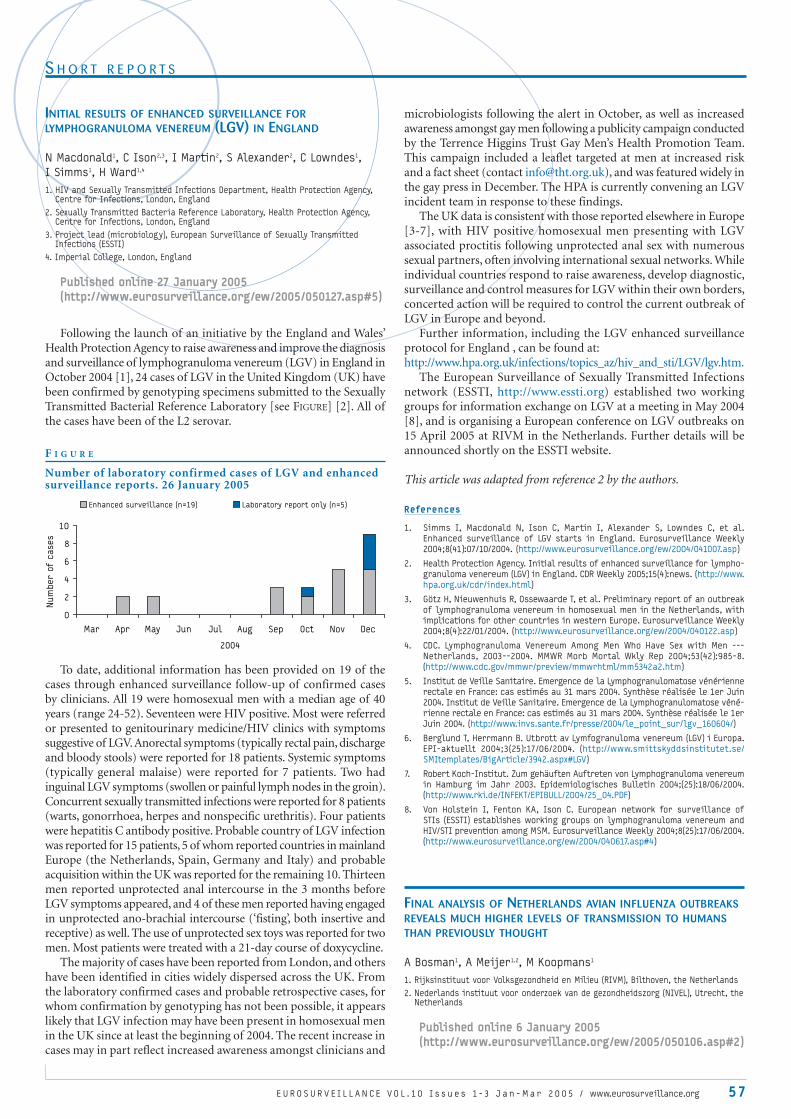
S H O R T R E P O R T S
E U R OS U R V E I L L A N C E V O L . 10 I s s u e s 1 -3 J a n - M a r 2 0 0 5 / www.eurosurveillance.org 57
INITIAL RESULTS OF ENHANCED SURVEILLANCE FOR LYMPHOGRANULOMA VENEREUM (LGV) IN ENGLAND
N Macdonald1, C Ison2,3, I Martin2, S Alexander2, C Lowndes1,
I Simms1, H Ward1,4
1. HIV and Sexually Transmitted Infections Department, Health Protection Agency, Centre for Infections, London, England
2. Sexually Transmitted Bacteria Reference Laboratory, Health Protection Agency, Centre for Infections, London, England
3. Project lead (microbiology), European Surveillance of Sexually Transmitted Infections (ESSTI)
4. Imperial College, London, England
Published online 27 January 2005(http://www.eurosurveillance.org/ew/2005/050127.asp#5)
Following the launch of an initiative by the England and Wales’ Health Protection Agency to raise awareness and improve the diagnosis and surveillance of lymphogranuloma venereum (LGV) in England in October 2004 [1], 24 cases of LGV in the United Kingdom (UK) have been confirmed by genotyping specimens submitted to the Sexually Transmitted Bacterial Reference Laboratory [see FIGURE] [2]. All of the cases have been of the L2 serovar.
F I G U R E
Number of laboratory confirmed cases of LGV and enhanced surveillance reports. 26 January 2005
To date, additional information has been provided on 19 of the cases through enhanced surveillance follow-up of confirmed cases by clinicians. All 19 were homosexual men with a median age of 40 years (range 24-52). Seventeen were HIV positive. Most were referred or presented to genitourinary medicine/HIV clinics with symptoms suggestive of LGV. Anorectal symptoms (typically rectal pain, discharge and bloody stools) were reported for 18 patients. Systemic symptoms (typically general malaise) were reported for 7 patients. Two had inguinal LGV symptoms (swollen or painful lymph nodes in the groin). Concurrent sexually transmitted infections were reported for 8 patients (warts, gonorrhoea, herpes and nonspecific urethritis). Four patients were hepatitis C antibody positive. Probable country of LGV infection was reported for 15 patients, 5 of whom reported countries in mainland Europe (the Netherlands, Spain, Germany and Italy) and probable acquisition within the UK was reported for the remaining 10. Thirteen men reported unprotected anal intercourse in the 3 months before LGV symptoms appeared, and 4 of these men reported having engaged in unprotected ano-brachial intercourse (‘fisting’, both insertive and receptive) as well. The use of unprotected sex toys was reported for two men. Most patients were treated with a 21-day course of doxycycline.
The majority of cases have been reported from London, and others have been identified in cities widely dispersed across the UK. From the laboratory confirmed cases and probable retrospective cases, for whom confirmation by genotyping has not been possible, it appears likely that LGV infection may have been present in homosexual men in the UK since at least the beginning of 2004. The recent increase in cases may in part reflect increased awareness amongst clinicians and
microbiologists following the alert in October, as well as increased awareness amongst gay men following a publicity campaign conducted by the Terrence Higgins Trust Gay Men’s Health Promotion Team. This campaign included a leaflet targeted at men at increased risk and a fact sheet (contact [email protected]), and was featured widely in the gay press in December. The HPA is currently convening an LGV incident team in response to these findings.
The UK data is consistent with those reported elsewhere in Europe [3-7], with HIV positive homosexual men presenting with LGV associated proctitis following unprotected anal sex with numerous sexual partners, often involving international sexual networks. While individual countries respond to raise awareness, develop diagnostic, surveillance and control measures for LGV within their own borders, concerted action will be required to control the current outbreak of LGV in Europe and beyond.
Further information, including the LGV enhanced surveillance protocol for England , can be found at:http://www.hpa.org.uk/infections/topics_az/hiv_and_sti/LGV/lgv.htm.
The European Surveillance of Sexually Transmitted Infections network (ESSTI, http://www.essti.org) established two working groups for information exchange on LGV at a meeting in May 2004 [8], and is organising a European conference on LGV outbreaks on 15 April 2005 at RIVM in the Netherlands. Further details will be announced shortly on the ESSTI website.
This article was adapted from reference 2 by the authors.
References
1. Simms I, Macdonald N, Ison C, Martin I, Alexander S, Lowndes C, et al.
Enhanced surveillance of LGV starts in England. Eurosurveillance Weekly
2004;8(41):07/10/2004. (http://www.eurosurveillance.org/ew/2004/041007.asp)
2. Health Protection Agency. Initial results of enhanced surveillance for lympho-
granuloma venereum (LGV) in England. CDR Weekly 2005;15(4):news. (http://www.
hpa.org.uk/cdr/index.html)
3. Götz H, Nieuwenhuis R, Ossewaarde T, et al. Preliminary report of an outbreak
of lymphogranuloma venereum in homosexual men in the Netherlands, with
implications for other countries in western Europe. Eurosurveillance Weekly
2004;8(4):22/01/2004. (http://www.eurosurveillance.org/ew/2004/040122.asp)
4. CDC. Lymphogranuloma Venereum Among Men Who Have Sex with Men ---
Netherlands, 2003--2004. MMWR Morb Mortal Wkly Rep 2004;53(42):985-8.
(http://www.cdc.gov/mmwr/preview/mmwrhtml/mm5342a2.htm)
5. Institut de Veille Sanitaire. Emergence de la Lymphogranulomatose vénérienne
rectale en France: cas estimés au 31 mars 2004. Synthèse réalisée le 1er Juin
2004. Institut de Veille Sanitaire. Emergence de la Lymphogranulomatose véné-
rienne rectale en France: cas estimés au 31 mars 2004. Synthèse réalisée le 1er
Juin 2004. (http://www.invs.sante.fr/presse/2004/le_point_sur/lgv_160604/)
6. Berglund T, Herrmann B. Utbrott av Lymfogranuloma venereum (LGV) i Europa.
EPI-aktuellt 2004;3(25):17/06/2004. (http://www.smittskyddsinstitutet.se/
SMItemplates/BigArticle/3942.aspx#LGV)
7. Robert Koch-Institut. Zum gehäuften Auftreten von Lymphogranuloma venereum
in Hamburg im Jahr 2003. Epidemiologisches Bulletin 2004;(25):18/06/2004.
(http://www.rki.de/INFEKT/EPIBULL/2004/25_04.PDF)
8. Von Holstein I, Fenton KA, Ison C. European network for surveillance of
STIs (ESSTI) establishes working groups on lymphogranuloma venereum and
HIV/STI prevention among MSM. Eurosurveillance Weekly 2004;8(25):17/06/2004.
(http://www.eurosurveillance.org/ew/2004/040617.asp#4)
FINAL ANALYSIS OF NETHERLANDS AVIAN INFLUENZA OUTBREAKS REVEALS MUCH HIGHER LEVELS OF TRANSMISSION TO HUMANS THAN PREVIOUSLY THOUGHT
A Bosman1, A Meijer1,2, M Koopmans1
1. Rijksinstituut voor Volksgezondheid en Milieu (RIVM), Bilthoven, the Netherlands
2. Nederlands instituut voor onderzoek van de gezondheidszorg (NIVEL), Utrecht, the Netherlands
Published online 6 January 2005(http://www.eurosurveillance.org/ew/2005/050106.asp#2)
0
2
4
6
8
10
Mar Apr May Jun Jul Aug Sep Oct Nov Dec
2004
Num
ber o
f c
ases
Enhanced surveillance (n=19) Laboratory report only (n=5)

5 8 E U R OS U R V E I L L A N C E V O L . 10 I s s u e s 1 -3 J a n - M a r 2 0 0 5
Recent alerts demonstrate that the transmission of avian influenza to humans, which re-emerged at the beginning of 2004, is far from over in South East Asia [1]. As efforts to control the epidemic and prevent further human cases continue, the need to assess the effectiveness of current control measures grows. An executive summary of the final report of the outbreak of avian influenza A/H7N7 in the Netherlands has recently been Published in English [2].
Between March and May 2003, an unprecedented outbreak of avian influenza occurred in humans in the Netherlands. During an extensive epizootic of influenza A virus H7N7 on commercial poultry farms, 86 cases in poultry workers and 3 cases in people with no poultry contact were initially confirmed by PCR. The predominant symptom was conjunctivitis [3]. One veterinarian developed fatal respiratory distress syndrome after close contact with infected poultry [4].
A questionnaire survey was carried out as a follow up to the outbreak. Approximately 400 poultry farmers and their families and almost 900 people who were involved in controlling the epidemic participated in this investigation. Blood samples were taken from 500 of these participants to determine possible infection with the avian flu virus. Additional studies were performed for 62 household contacts of 25 persons with avian flu virus infection.
Routine serological tests failed to detect any antibodies, even in the group of persons with confirmed avian influenza virus conjunctivitis. A modification of the haemagglutination assay was developed, based on observations that avian influenza viruses favour binding to red blood cells from horses rather than turkeys [5]. As at least 50% of the people exposed to infected poultry had H7 antibodies detectable with the modified assay, it was estimated that avian influenza A/H7N7 virus infection occurred in at least 1000, and perhaps as many as 2000 people. The seroprevalence of H7 antibodies in people without contact with infected poultry, but with close household contact to an infected poultry worker, was 59%. This suggests that the population at risk for avian influenza was not limited to those with direct contact to infected poultry, and that person to person transmission may have occurred on a large scale. Specificity of the unconventional assay was confirmed by the absence of reactivity in sera from 100 controls recently vaccinated with influenza vaccine (2002/2003) (specificity 100%). Assay specificity was further supported by the results of the cohort study: having measurable antibodies was associated with having conjunctivitis (RR 1.72; 95% CI 0.99-2.99), and a lower proportion of the exposed persons who took prophylactic antiviral medication developed antibodies (corrected OR 0.48; 95% CI 0.25-0.89).
Neither poultry farmers nor those engaged in controlling the epidemic complied satisfactorily with preventive measures. Only 6% of farmers reported consistent use of facial masks and 1% reported consistent use of goggles while working with infected poultry. In cullers, compliance was only slightly better: 25% consistently used facial masks and 13% used goggles. The results of the epidemiological study suggest that oseltamivir protected against conjunctivitis (corrected OR=0.14; 95% CI=0.08–0.27) as well as against infection without specific symptoms. No protective effect was demonstrable for safety goggles or mouth-nose masks [2].
After the outbreaks of group A subtype H5N1 (A/H5N1) avian influenza viruses in Hong Kong in 1997, in which 6 people died, the hypothesis was put forward that not only pigs but also humans themselves might serve as mixing vessels for the next pandemic influenza virus [6]. The outbreak of avian influenza A/H7N7 in the Netherlands and the recent unprecedented expansion of avian influenza A/H5N1 in Asia have reinforced this concern. A review of the outbreak and control efforts in the Netherlands highlights important lessons for preparedness: while separate systems are in place to signal and control animal diseases and human diseases, an outbreak of a zoonotic disease illustrates the importance of coordination between the two. In the Netherlands, the people infected came from a wide geographic region, and included foreign poultry workers. While the movement of animals was restricted, these people were out of the reach of the public health authorities while infectious and shedding the virus.
Although the disease in humans is more severe for A/H5N1, both avian influenza outbreaks illustrate that crossing the species
barrier is less rare than previously recognised, that avian influenza virus adaptation occurs rapidly, and that if such jumps between species occur, human behaviour in the broad sense may accelerate dissemination [7].
References
1. Avian influenza, human - Viet Nam (13): susp. in: ProMED-mail [online].
Boston US: International Society for Infectious Diseases, archive number
20050105.0020, 5 January 2005. (http://www.promedmail.org)
2. Bosman A, Mulder YM, de Leeuw JRJ, Meijer A, Du Ry van Beest Holle M, Kamst
RA, et al. Executive summary Avian flu epidemic 2003: public health con-
sequences. RIVM rapport 630940003. Bilthoven: RIVM;2004.(http://www.rivm.
nl/bibliotheek/rapporten/630940004.html)
3. Koopmans M, Wilbrink B, Conyn M, Natrop G, van der Nat H, Vennema H, et
al. Transmission of H7N7 avian influenza A virus to human beings during a
large outbreak in commercial poultry farms in the Netherlands. Lancet 2004;
363(9409):587-93.
4. Fouchier RA, Schneeberger PM, Rozendaal FW, Broekman JM, Kemink SA, Munster
V, et al. Avian influenza A virus (H7N7) associated with human conjunctivitis
and a fatal case of acute respiratory distress syndrome. Proc Natl Acad Sci
U S A 2004;101(5):1356-61.
5. Stephenson I, Wood JM, Nicholson KG, Zambon MC. Sialic acid receptor spe-
cificity on erythrocytes affects detection of antibody to avian influenza
haemagglutinin. J Med Virol 2003;70(3): 391-8.
6. Webby RJ, Webster RG. Are we ready for pandemic influenza? Science
2003;302(5650):1519-22 .
7. Li KS, Guan Y, Wang J, Smith GJ, Xu KM, Duan L, et al. Genesis of a highly
pathogenic and potentially pandemic H5N1 influenza virus in eastern Asia.
Nature 2003;430(6996):209-13.
IMMIGRATION AND HIV/AIDS PREVENTION IN GERMANY – AN INTERDISCIPLINARY CHALLENGE
E Steffan, V Kerschl, S Sokolowski,
SPI Forschung, Berlin, Germany
Published online 20 January 2005(http://www.eurosurveillance.org/ew/2005/050120.asp#4)
The availability of STI and HIV/AIDS prevention services and also general medical and social services for immigrants to Germany, needs to be better according to many health professionals. Possible solutions have been discussed for several years both in Germany and other European countries. There is awareness of barriers in foreigners’ rights, cultural barriers and not least, language barriers. To develop effective solutions, the help-seeking behaviour of immigrants and their basic knowledge about HIV/AIDS and STIs was studied [1].
In 2004, in 5 European countries (Germany, Austria, Italy, Spain and Greece), over 1500 immigrants were asked, most often in their mother tongue, about their experiences, knowledge and assessment of healthcare and social services (http://europa.eu.int/comm/health/ph_projects/2002/com_diseases/commdis_2002_04_en.htm, [2]). The survey contained about 100 questions about experiences and assessments of health and social services. The results of the quantative study were discussed and validated by two focus groups made up of immigrants and health and social work professionals.
In Germany, 315 people were surveyed in Berlin and the surrounding Brandenburg region. They included 55 prostitutes, whose experiences are not discussed in this article, so the total was 260 people. Recruitment of participants was defined by criteria set out in the study design. With the help of migrant organisations, participants were recruited using the ‘snowball effect’ (the first contact is via the organisation, and each contact was asked to supply two more contacts).
DemographicsThe largest group of interviewed people were from southeast
Europe (35%), followed by sub-Saharan Africa (32%), eastern Europe (24%) and Asia (5%). The average age of participants was 30.4 years old, for both men and women. About 30% of interviewees

E U R OS U R V E I L L A N C E V O L . 10 I s s u e s 1 -3 J a n - M a r 2 0 0 5 / www.eurosurveillance.org 5 9
were married, 36% were single, 18% were separated or divorced, 15% lived together with a partner (multiple answers possible), and 18% were single parents.
Interviewees were generally well-educated with 48% having a college or university qualification. Although the level of education was good, 32% were unemployed (women 29%, men 36%). Thirty percent had full-time or part-time jobs, and 58% had a monthly income under 1000 Euro, which is below the poverty line of 1100 Euro per month in Germany. Thirty-six percent of the participants left their native country to join family, 20% left because of political reasons and 17.5% for economic reasons.
Results Basic knowledge of HIV/AIDS and infection riskThe majority of the immigrants asked felt that they were not well
informed on this topic. Only half of those surveyed felt sure what HIV/AIDS was, about 25% were fairly sure, and a further 24% were ‘unsure’ or ‘did not know’. In contrast with German citizens (almost 100% are certain what HIV/AIDS is, [3]), this level of knowledge is very low.
Only 81% knew for certain that infection can occur through sexual contact, 32% thought that transmission could occur by kissing, and 13% believed that sharing a cup or glass could pass infection on. Only 77% knew that sharing needles was a very risky. These results show uncertainty and worry, but also indifference towards possible risk of contracting HIV. More than half the immigrants from eastern Europe were very worried about acquiring HIV. Women from southeast Europe were the group with the least knowledge and least awareness of risky behaviour.
Seventy-three percent of interviewed immigrants had already received information about HIV/AIDS, 49% in a language that they understood well. The men interviewed were generally better informed about HIV/AIDS than the women, and immigrants from sub-Saharan Africa were better informed than southeast Europeans. As with the German population, mass media (television, billboards) was the most important way of getting information (although only 41.5% of immigrants surveyed were informed this way compared with 92% of the German population2. As a source of information about HIV/AIDS, personal contacts such as friends were mentioned (25%), and health services (28%) or teachers (20%).
Knowledge and experience of HIV tests in GermanyFifty-two percent of immigrants surveyed got information about
health services from friends, 38% from family members, and 26% from other immigrants. The knowledge about HIV testing services in Germany was worryingly low. Only 24% knew that an HIV test is free and anonymous in Germany. Only 52% of those who had already undergone HIV testing in Germany could remember being counselled before the test, although this is required. The proportion of people tested who did not receive advice afterwards (or could not remember being advised) was similarly high at 57%.
Conclusions for HIV/AIDS preventionThe results of this survey indicate that HIV/AIDS educational
messages are not reaching immigrants as effectively as German citizens. Important basic knowledge was lacking in many cases, and information was not supplied about HIV testing or even during testing. The existence of free and anonymous testing by health services is too often not known. The fact that female immigrants are mostly informed about HIV by personal contacts makes specific tailored prevention activities required. As well as this, institutions which serve immigrants should be sensitised to this topic. Not least, it would help if HIV health information was advertised regionally, for example in buses, in the main languages of immigrants to that country.This article was translated from reference 1 by the Eurosurveillance editorial team.
References
1. Robert Koch-Institut (RKI): Migration und HIV/AIDS-Prävention – eine inter-
disziplinäre Herausforderung. Epidemiologisches Bulletin 2004 (48): 416-417
(http://www.rki.de/INFEKT/EPIBULL/2004/48_04.PDF)
2. Centro Regionale di Riferimento per la Promozione della Salute. European
project on HIV and Immigrants and European Southern and Eastern Borders:
Summary of project methodology and main quantitative and qualitative re-
search findings, Venice, 2004 (in print)
3. Bundeszentrale für gesundheitliche Aufklärung – BzgA - (2004): Aids im
öffentlichen Bewusstsein der Bundesrepublik Deutschland 2003. BzgA, Köln
[Public Awareness of AIDS Representative yearly survey, done by the German
Federal Centre for Health Education, www.bzga.de.
EMERGENCE AND DISSEMINATION OF A NEW MECHANISM OF RESISTANCE TO AMINOGLYCOSIDES IN GRAM-NEGATIVE BACTERIA: 16S RRNA METHYLATION
M Galimand, T Lambert, P Courvalin
Unité des Agents Antibactériens and Centre National de Référence de la Résistance aux Antibiotiques, Institut Pasteur, Paris, France
Published online 27 January 2005(http://www.eurosurveillance.org/ew/2005/050127.asp#2)
Despite the development of new ß-lactams and fluoroquinolones, aminoglycosides are still a very important class of antibacterials for the treatment of severe illness caused by a variety of pathogens, including Gram-negative bacteria, particularly if the pathogen has developed resistance to third-generation cephalosporins.
Aminoglycosides act by causing translational errors and by inhibiting translocation [1]. Their target sites include ribosomal domains in which the accuracy of codon-anticodon is assessed [2]. In particular, they bind to a highly conserved motif of 16S RNA which leads to alterations in ribosome function [3]. There are four known mechanisms of resistance to aminoglycosides in bacterial human pathogens:
1. decreased intracellular accumulation of the antibiotic by altering the outer membrane permeability, less inner membrane transport, or active efflux;
2. enzymatic modification of the drug, primarily through N-acetylation, O-nucleotidylation, or O-phosphorylation, which is the most common;
3. modification of the target by mutation in ribosomal proteins or in 16S RNA;
4. trapping of the drug.Microorganisms that produce aminoglycosides have developed
an additional pathway to avoid suicide. This self-defence mechanism involves post-transcriptional methylation of ribosomal RNA using S-adenosyl-methionine as a cofactor [4].
The armA (aminoglycoside resistance methyltransferase) gene, which confers resistance to 4,6-disubstituted deoxystreptamines (kanamycin, amikacin, isepamicin, gentamicin, netilmicin, sisomicin, and tobramycin) and to the structurally unrelated compound fortimicin, was initially characterised in Klebsiella pneumoniae BM4536. This was isolated from a urinary tract infection in 2000 in France. Possession of the armA gene did not confere resistance against the 4,5-disubstituted deoxystreptamines (lividomycin, neomycin, paromomycin, ribostamycin).
T A B L E
Minimum inhibitory concentrations (MICs) of various aminoglycosides against E. coli with and without plasmid pIP1204 carrying armA
MICs (mg/L)a
Strain AMI
GEN ISE NET TOB APR PAR STR
STR
BM694 2 0.5 0.5 0.5 0.5 2 4 4
BM694
(pIP1204)1024 256 1024 256 256 2 4 8
a- Abbreviations: AMI, amikacin; APR, apramycin; GEN, gentamicin; ISE, isepamicin; NET, netilmicin; TOB, tobramycin; PAR, paromomycin; STR, streptomycin.

6 0 E U R OS U R V E I L L A N C E V O L . 10 I s s u e s 1 -3 J a n - M a r 2 0 0 5
The armA gene was located on the self-transferable IncL/M plasmid pIP1204 of about 90 kb which also encodes extended spectrum ß-lactamase CTX-M-3 [5].
The armA gene was detected in 12 isolates among 34 enterobacteria from 3 hospitals in Paris, France, 2 hospitals in Sofia, Bulgaria, and from our laboratory collection. The bacteria collected by the laboratory were likely to produce a CTX-M enzyme since they were more resistant to cefotaxime than to ceftazidime. The isolates containing armA were C. freundii (3 strains out of 3), Enterobacter cloacae (1/1), E. coli (2/19), K. pneumoniae (4/5), Salmonella enterica serotype Enteritidis (1/1), and Shigella flexneri (1/1) [6]. Transfer of high-level aminoglycoside resistance from the 12 armA containing strains to E. coli was obtained by conjugation. Using disc diffusion susceptibility tests and polymerase chain reaction with specific primers and sequence analysis, the E. coli transconjugants were found to express resistance to 4,6-disubstituted deoxystreptamines mediated by armA after acquiring an IncL/M plasmid. More recently, presence of armA on a self-transferable IncN plasmid in an E. coli pig isolate from Spain has been reported [7].
Conjugation, analysis of DNA sequences, PCR mapping, and plasmid conduction experiments, indicated that the armA gene was part of the functional transposon Tn1548. The 16.6-kb transposon is a typical composite element flanked by two copies of IS6 in direct orientation [8]. Functionality of Tn1548 under natural conditions was confirmed by its presence on plasmids of different incompatibility groups [5,7].
Taken together, these data support the notion that spread of armA results from both conjugation and transposition. This combination accounts for the worldwide documented dissemination of aminoglycoside resistance by 16S rRNA methylation in enterobacteria from human or animal origin and in Acinetobacter baumannii in Europe and in Asia [6,7,9,10]. Two other closely related 16S rRNA methylases, RmtA in Pseudomonas aeruginosa isolates and RmtB in enterobacteria, have been recently reported in Japan [11, 12]. It therefore appears that post-transcriptional modification of 16S rRNA can confer high-level resistance to all the clinically available aminoglycosides, except streptomycin, in Gram-negative human pathogens.
The findings discussed here indicate that global dissemination of armA in Gram-negative pathogens has occurred and demonstrate the importance of maintaining ongoing surveillance of aminoglycoside resistance of human and animal isolates.
Strains suspected to harbour the armA gene can be sent for analysis to Marc Galimand, Unité des Agents Antibactériens, Institut Pasteur, 25 rue du Docteur Roux, 75724 Paris Cedex 15, France.
References
1. Davies J, Davis BD. Misreading of ribonucleic acid code words induced by
aminoglycoside antibiotics. The effect of drug concentration. J Biol Chem
1968;243:3312-6.
2. Woodcock J, Moazed D, Cannon M, Davies J, Noller HF. Interaction of antibio-
tics with A- and P-site-specific bases in 16S ribosomal RNA. EMBO J 1991;10:
3099-103.
3. Yoshizawa S, Fourmy D, Puglisi JD. Recognition of the codon-anticodon helix
by ribosomal RNA. Science 1999;285:1722-25.
4. Beauclerck AA, Cundliffe E. Sites of action of two ribosomal RNA methylases
responsible for resistance to aminoglycosides. J Mol Biol 1987;193:661-71.
5. Galimand M, Courvalin P, Lambert T. Plasmid-mediated high-level resistance
to aminoglycosides in Enterobacteriaceae due to 16S rRNA methylation.
Antimicrob Agents Chemother 2003;47:2565-71.
6. Galimand M, Sabtcheva S, Kantardjiev T, Poirel L, Arlet G, Courvalin P, Lambert
T. The armA aminoglycoside resistance methylase gene is disseminated in
Enterobacteriaceae by an IncL/M plasmid mediating CTX-M3 ß-lactamase.
Abstr. 43rd Intersci. Conf. Antimicrob Agents Chemother, abstr. C2-59, 2003.
7. Gonzales-Zorn B, Teshager T, Casas M, Porrero MC, Moreno MA, Courvalin P, et
al. The armA gene confers high-level aminoglycoside resistance in Escherichia
coli MUR50 animal isolate. Emerg Infect Dis, in press, 2005.
8. Lambert T, Galimand M, Sabtcheva S, Courvalin P. The armA aminoglycoside
resistance methylase gene is borne by composite transposon Tn1548. Abstr.
44th Intersci. Conf. Antimicrob Agents Chemother, abstr. C1-1496, 2004.
9. Yan J-J, Wu J-J, Ko W-C, Tsai S-H, Chuang C-L, Wu H-M, et al. Plasmid-media-
ted 16S rRNA methylases conferring high-level aminoglycoside resistance
in Escherichia coli and Klebsiella pneumoniae isolates from two Taiwanese
hospitals. J Antimicrob Chemother 2004;54:1007-12.
10. Wachino J, Yamane K, Kurokawa H, Suzuki S, Shibata N, Arakawa Y. Global
transmission of the 16S rRNA methylase gene, armA, among clinically isolated
Gram-negative rods. Abstr. 44th Intersci. Conf. Antimicrob Agents Chemother,
abstr. C2-1889,2004.
11. Yokoyama K, Doi Y, Yamane K, Kurokawa H, Shibata N, Shibayama K, et al.
Acquisition of 16S rRNA methylase gene in Pseudomonas aeruginosa. Lancet
2003;362:1888-93.
12. Doi Y, Yokoyama K, Yamane K, Wachino J, Shibata N, Yagi T, et al. Plasmid-
mediated 16S rRNA methylase in Serratia marcescens conferring high-level
resistance to aminoglycosides. Antimicrob Agents Chemother 2004;48:491-6.
STREP-EURO: PROGRESS IN ANALYSIS AND RESEARCH INTO SEVERE STREPTOCOCCAL DISEASE IN EUROPE, 2003-2004
A Jasir, C Schalén on behalf of the Strep-EURO study group
Department of Medical Microbiology, Dermathology and Infectious Diseases, Lund University, Lund, Sueden
Published online 3 February 2005(http://www.eurosurveillance.org/ew/2005/050203.asp#3)
The Strep-EURO project (http://www.strep-euro.lu.se/) collects data on severe group A streptococcal (GAS) disease in Europe. It was launched in September 2002 to improve understanding of severe GAS disease in Europe so that an integrated picture of these infections can be achieved [1], and consists of over 50 members from 11 European countries. We briefly summarise below progress on the project to date, including some preliminary results from the participating countries: Germany, Cyprus, Czech Republic, Denmark, Finland, France, Greece, Italy, Romania, Sweden, and the United Kingdom.
During the first phase of the project, a European case definition was agreed on, along with items to be included in a clinical and epidemiological questionnaire. The inclusion criteria were the isolation of GAS from blood or another normally sterile site of the body, or from a non-sterile site in the presence of a clinical diagnosis of streptococcal toxic shock syndrome (STSS) or necrotising fasciitis (NF), as defined previously [2].
Enhanced surveillance of GAS invasive disease began on 1 January 2003 for a two year period. Collected isolates were characterised by both serology (T, OF and M typing), and molecular tools (e.g., emm-sequencing, superantigen and antimicrobial drug resistance gene identification, and strain comparison by pulsed field gel electrophoresis (PFGE) and multilocus sequence typing (MLST)). Strains were screened for antibiotic susceptibility and minimum inhibitory concentrations determined by E test. To achieve a standardization of methods and assessment of capabilities in the different laboratories, two sets of external quality assessment (EQA) strains for antibiotic susceptibility testing and one set for typing were sent to participating laboratories. Data file specifications for collection of patient and microbiological data were developed by consensus, and each partner submitted their results to a central database in Finland.
Over 5000 cases were identified in the first 18 months, considerably more than had been anticipated. Around 3000 cases were from the United Kingdom, yielding an incidence for 2003 of 3.8/100 000 for that country. Similar incidences were documented in Sweden, Denmark and Finland, but the incidence was considerably lower in the other participating countries. This may be related to the fact that surveillance in northern Europe approached total coverage, whereas in the remaining countries, geographical coverage was limited and not always definable.
The type distribution of GAS also varied markedly. In a few countries, types 1, 3 and 28 were predominant; however, an overall increase of new invasive types (emm 77, 81, 82, 89) was noticeable. Relatively high rates of MLS antibiotic (macrolide, lincosamide, streptogramin B) resistance in some countries (France, Italy) and very frequent tetracycline resistance was found in almost all countries. In the UK, intravenous drug use (IDU) was found to be a major risk factor, which is consistent with a previously reported trend [3]. In France, the spread of a clone of GAS, type 28, resistant to MLS drugs and bacitracin, associated with puerperal sepsis, was reported [4]. Early results from the pathogenesis work package have identified that low antibody levels against some newly described cell wall-attached

E U R OS U R V E I L L A N C E V O L . 10 I s s u e s 1 -3 J a n - M a r 2 0 0 5 / www.eurosurveillance.org 6 1
proteins of GAS may predispose to severe GAS disease [5].The Strep-EURO project has managed to create a platform for
epidemiological analysis of and research into severe streptococcal disease in ten European Union countries and one EU candidate country. The 2003 results, in which three times the expected number of cases were identified primarily through improvements to case ascertainment methods, indicate the success of the surveillance. The apparent overall increase of invasive cases may thus partly depend on the stimulus of Strep-EURO to the establishment of national surveillance systems and the enhancement of existing ones.
Though incidence estimates are preliminary at this point, the marked fluctuations noted between countries may be attributable to a number of factors: true differences in rates of severe GAS infection, under-reporting to the national laboratory, or lack or failure of microbiological diagnostic procedures (e.g. no blood cultures prior to treatment). Definitive conclusions will have to await careful analysis of the data. The frequency of unusual emm-types is a concern from the point-of-view of prevention since current candidate vaccines against GAS are mostly based upon the M protein, the type-variable, most important virulence factor of this organism.
Actions required in the immediate future include standardisation of subtyping by PFGE and MLST, which will allow efficient cross-talk and tracking of strains among laboratories, and a detailed assessment of unambiguous emm-type assignment based on DNA sequences. There is also a need for further work to study the maintenance of tetracycline resistance despite the lack of use of this drug for treatment of streptococcal diseases.
Acknowledgements
We wish to acknowledge all members of the strep-EURO study group
for their contribution, enthusiasm and dedication towards this project
and in particular to Anne Bouvet, Androulla Efstratiou, Theresa Lamagni
and Panayiotis Tassios who actively contributed towards the prepara-
tion of this article.
References
1. Schalén C. European surveillance of severe group A streptococcal disease.
Eurosurveillance Weekly 2002;6 (35):29/08/2002. (http://www.eurosurveillance.
org/ew/2002/020829.asp)
2. Defining the group A streptococcal toxic shock syndrome. Rationale and
consensus definition. The Working Group on Severe Streptococcal Infections.
JAMA 1993;269(3):390-1.
3. Efstratiou A, Emery M, Lamagni TL, Tanna A, Warner M, George RC. Increasing
incidence of group A streptococcal infections amongst injecting drug users
in England and Wales. J Med Microbiol 2003;52(Pt 6):525-6. (http://jmm.sg-
mjournals.org/cgi/content/abstract/52/6/525) [abstract]
4. Mihaila-Amrouche L, Bouvet A, Loubinoux J. Clonal spread of emm type 28
isolates of Streptococcus pyogenes that are multiresistant to antibiotics.
J Clin Microbiol 2004;42:3844-6. (http://jcm.asm.org/cgi/content/full/42/8/
3844?view=full&pmid=15297545)
5. Akesson P, Rasmussen M, Mascini E, von Pawel-Rammingen U, Janulczyk R,
Collin M, et al. Low antibody levels against cell wall-attached proteins of
Streptococcus pyogenes predispose for severe invasive disease. J Infect Dis
2004;189(5):797-804. Epub 2004 Feb 18. (http://www.journals.uchicago.edu/JID/
journal/issues/v189n5/30801/30801.html)
BSE AGENT IN GOAT TISSUE: FIRST KNOWN NATURALLY OCCURRING CASE CONFIRMED
Editorial team
Eurosurveillance editorial offi ce
Published online 3 February 2005(http://www.eurosurveillance.org/ew/2005/050203.asp#1)
On 28 January, the European Commission confirmed the first known naturally occurring case of bovine spongiform encephalopathy (BSE) agent in a goat, slaughtered in France in 2002 [1]. Previously, sheep and goats had only been experimentally infected. The results have only been made available now, as the confirmatory tests included mouse bioassays, which took two years to complete.
Neither the infected goat, nor any other goat from the same herd, entered either the food or feed chain. This incident is therefore not considered to represent a risk to public health. The entire herd was slaughtered after the infected goat was first suspected to be infected with BSE agent. All adult goats in the herd were tested, and no other goat was found to have BSE infection or to show other signs of BSE disease [2].
The infected goat was born in 2000. A ban on feeding meat and bone meal (MBM) to ruminants (i.e., cattle, sheep and goats) has been in place since 1994; this was extended to all farmed animals in 2001. Goats in the European Union generally only live for a few years, which means that the majority of goats in the EU today were born after the total feed ban was put in place. Nevertheless, in response to this case of confirmed natural BSE infection in a goat, the Commission is proposing to improve vigilance for such incidents by increasing BSE testing of goats, and has set a target of 200 000 healthy goats tested in the European Union over the next six months. The current EU wide surveillance programme, designed to detect suspicious TSE strains in small ruminants in the EU, has tested 140 000 goats since 2002 [3].
It is proposed that the TSE monitoring programme will concentrate on member states where BSE is present in cattle. All confirmed cases of transmissible spongiform encephalopathy (TSE, includes scrapie) will undergo three-stage testing (already in use), which will differentiate between scrapie and BSE. These additional measures will be submitted for member states’ approval at the beginning of February.
As a precautionary measure and following scientific advice, milk and meat from goats which are affected by any type of transmissible spongiform encephalopathy (including scrapie) cannot currently be used, following a recommendation in 2001 from the European Commission Scientific Steering Committee [4]. Specified risk materials (the tissues most likely to carry infectivity if the disease is present) are also removed from all goats, even if healthy [5,6].
The European Food Safety Authority (EFSA, http://www.efsa.eu.int/) has advised that, based on current scientific knowledge, goat milk and derived products are unlikely to present any risk of TSE contamination if the milk comes from healthy animals [7]. It advises no change in current consumption of goat milk, cheese and meat.
The European Commission has asked the EFSA to carry out a quantitative risk assessment for goat meat and goat meat products, which is expected to be ready by July 2005. Further information can be found on the European Commission pages Food Safety – from the Farm to the Fork http://europa.eu.int/comm/food/index_en.htm).
References
1. European Commission. Case of BSE in a goat confirmed: Commission ex-
tends testing programme. Press release IP/05/105, 28 January 2005.
(http://europa.eu.int/rapid/pressReleasesAction.do?reference=IP/05/
105&format=HTML&aged=0&language=EN&guiLanguage=fr)
2. OIE. Bovine spongiform encephalopathy in a goat in France. Press release, 1
February 2005. (http://www.oie.int/eng/press/en_050201b.htm)
3. European Commission. Commission submits French Research Findings on
TSE in a goat to Expert Panel. Press release IP/04/1324, 28 October 2004.
(http://europa.eu.int/rapid/pressReleasesAction.do?reference=IP/04/
1324&format=HTML&aged=0&language=EN&guiLanguage=en)
4. Scientific Steering Committee of the European Commission. TSEs in small
ruminants should BSE in small ruminants become probable/confirmed. 18-
19 October 2001. European Commission Health and Consumer Protection
Directorate General. (http://europa.eu.int/comm/food/fs/sc/ssc/out234_
en.pdf)
5. Eurosurveillance. BSE agent in goat tissue: precautions discussed.
Eurosurveillance Weekly 2004;8(51):16/12/2004. (http://www.eurosurveillance.
org/ew/2004/041216.asp#4)
6. European Commission Health & Consumer Protection Directorate-General.
Opinion on safe sourcing of small ruminant materials (safe sourcing of
small ruminant materials should BSE in small ruminants become probable:
genotype, breeding, rapid TSE testing, flocks certification and specified risk
materials). Adopted by the Scientific Steering Committee at its meeting of
4-5 April 2002. (http://europa.eu.int/comm/food/fs/sc/ssc/out257_en.pdf)
7. European Food Safety Authority. Statement of the EFSA Scientific Expert
Working Group on BSE/TSE of the Scientific Panel on Biological Hazards on
the health risks of the consumption of milk and milk derived products from
goats. 26 November 2004 (http://www.efsa.eu.int/science/biohaz/biohaz_do-
cuments/709/bdoc_statement_goatsmilk_en1.pdf)

6 2 E U R OS U R V E I L L A N C E V O L . 10 I s s u e s 1 -3 J a n - M a r 2 0 0 5
POSSIBLE CASE OF BSE AGENT IN A UK GOAT THAT DIED IN 1990
Editorial team
Eurosurveillance Editorial Offi ce
Published online 10 February 2005(http://www.eurosurveillance.org/ew/2005/050210.asp#2)
Following the confirmation in January 2005 of a French goat having a transmissible spongiform encephalopathy indistinguishable from bovine spongiform encephalopathy (BSE) [1,2], the United Kingdom Department for Environment, Food and Rural Affairs has announced that a goat in the United Kingdom (UK), confirmed as having scrapie in 1990, may have had BSE [3].
More sensitive testing methods have found that a sample from the goat had traits similar to goats experimentally infected with BSE. However, this single result is insufficient to confirm that the goat did have BSE. Further testing, including bioassays, which take around 2 years to complete, are now necessary.
The year 1990 was the height of the BSE outbreak in cattle in the UK. A ban on feeding meat and bone meal to ruminants was introduced across the European Union in 1994. The TSE surveillance programme of sheep and goats will be increased in the United Kingdom, in line with the announced increases across the European Union.
The European Community TSE Reference Laboratory at the Veterinary Laboratories Agency (http://www.defra.gov.uk/corporate/vla/science/science-tse-rl-intro.htm) will complete the testing.
References
1. Eurosurveillance. BSE agent in goat tissue: first known naturally occurring
case confirmed. Euro Surveill 2005;10(1): Epub 3 February 2005. (http://www.
eurosurveillance.org/ew/2005/050203.asp#1)
2. European Commission. Case of BSE in a goat confirmed: Commission ex-
tends testing programme. Press release IP/05/105, 28 January 2005.
(http://europa.eu.int/rapid/pressReleasesAction.do?reference=IP/05/
105&format=HTML&aged=0&language=EN&guiLanguage=fr)
3. Department for Environment, Food and Rural Affairs. Possible BSE in a 1990
UK goat sample. Press release 58/05, 8 February 2005. (http://www.defra.gov.
uk/news/2005/050208a.htm)
OUTPATIENT CONSUMPTION OF ANTIBIOTICS IS LINKED TO ANTIBIOTIC RESISTANCE IN EUROPE: RESULTS FROM THE EUROPEAN SURVEILLANCE OF ANTIMICROBIAL CONSUMPTION
A Johnson
Healthcare-Associated Infections Surveillance, Health Protection Agency Centre for Infections, London, United Kingdom
Published online 24 February 2005(http://www.eurosurveillance.org/ew/2005/050224.asp#5)
There is increasing recognition that antibiotic consumption provides a major selective pressure for the emergence and persistence of antibiotic-resistant strains of bacteria. In 2001, a European Union Council Recommendation stated that data should be gathered on antibiotic use and antimicrobial resistance in European countries. The Recommendation also laid out an eight point prevention action plan to reduce the prevalence of antimicrobial resistance [1]. Subsequently, the European Surveillance of Antimicrobial Consumption (ESAC) project was established, to obtain comparable and reliable data on antibiotic use in Europe [2, 3]. The ESAC project group is closely linked to the European Antimicrobial Resistance Surveillance System (EARSS, http://www.earss.rivm.nl/) [4]. Analysis of data from EARSS showed that rates of antibiotic resistance are generally increasing, but there is distinct variation between countries, with resistance levels in central and southern Europe generally being markedly higher than those in northern European countries.
ESAC data on outpatient antibiotic use were gathered during 1997 to 2002 in 26 European countries, and the calculated relationship of
antibiotic consumption to rates of antibiotic resistance has recently been Published [5]. Although 32 countries take part in ESAC, the analysis presented was restricted to those countries able to provide internationally comparable data on antibiotic consumption derived from prescription reimbursement schemes or sales data. This was expressed as the number of defined daily doses (DDDs; the assumed average maintenance dose per day for a drug used for its main indication in adults) per 1000 inhabitants per day. The ecological association between antibiotic use and rates of resistance were assessed using Spearman’s correlation coefficients.
Rates of antibiotic use in primary care in Europe were found to vary greatly between countries, with the highest rate in France (32.2 DDD per 1000 inhabitants per day) being more than three times greater than in the Netherlands, which had the lowest rate of antimicrobial consumption, (10 DDD per 1000 inhabitants per day). In countries with high rates of antimicrobial use, seasonal fluctuations were noted, with increased consumption in the winter (mean increase equal to or greater than 30% in the first and fourth quarters). This may be related to the increase in respiratory infections seen in winter months and the tendency of physicians in high prescribing countries to regard such infections as bronchitis, while physicians in low prescribing countries label them as common colds or influenza. Another trend noted in the study was a shift from use of older narrow spectrum agents to newer broad spectrum drugs.
The European prevalences of resistance to macrolides and ß-lactams in Streptococcus pneumoniae, macrolide resistance in S. pyogenes and resistance to quinolones and co-trimoxazole in Escherichia coli were obtained from a number of national and international surveillance studies, and compared with antimicrobial consumption in the participating European countries. For all these organism-drug combinations, significant correlations between levels of resistance and antibiotic consumption were seen, particularly for S. pneumoniae, i.e higher levels of antibiotic prescribing were associated with higher levels of antibiotic resistance.
However, the authors rightly point out that further studies are needed to fully establish and clarify the association between antibiotic use and antibiotic resistance indicated in this group-level ecological study. For example, the data on usage volumes expressed as DDDs, allow comparisons but do not measure individual exposure to antibiotics. In other words, are the patients receiving antibiotics the same ones from whom antibiotic-resistant bacteria are isolated? Also, if physicians in a country, which uses twice as many DDDs per 1000 people compared with another country, treat the same number of patients (i.e patients in the first country receive two-fold higher doses), it might be anticipated that there would be less resistance in the high-user country because of the higher doses used. A further factor that needs to be addressed is that DDDs reflect adult dosing schedules, but estimates of antibiotic use will include drugs prescribed for use in children. In a recent French study, children were the main antibiotic consumers, with usage rates three times higher than that of older patients [6]. Clearly in countries with higher proportions of children, the total number of patients receiving antibiotics might be higher than the figure inferred from data expressed in terms of DDDs per 1000 people.
Further studies of factors that influence prescribing patterns may provide useful information for assessing public health strategies aimed at reducing antibiotic use and levels of antibiotic resistance
References
1. Council Recommendation of 15 November 2001 on the prudent use of an-
timicrobial agents in human medicine. Official Journal of the European
Communities. 2002/77/EC (http://europa.eu.int/eur-lex/pri/en/oj/dat/2002/l_
034/l_03420020205en00130016.pdf)
2. ESAC website: http://aiv-www.uia.ac.be/main.asp?c=*ESAC
3. Ferech M. European Surveillance of Antimicrobial Consumption: the ESAC
programme. Eurosurveillance Weekly 2004;8(32):5/8/2004 (http://www.euro-
surveillance.org/ew/2004/040805.asp#2)
4. European Antimicrobial Resistance Surveillance System [homepage on the
internet]. Bilthoven: RIVM; 2005 [cited 24 February 2005]. Available from:
http://www.earss.rivm.nl

E U R OS U R V E I L L A N C E V O L . 10 I s s u e s 1 -3 J a n - M a r 2 0 0 5 / www.eurosurveillance.org 63
5. Goossens H, Ferech M, Vander Stichele R, Elseviers M. Outpatient antibiotic use
in Europe and association with resistance: a cross-national database study.
Lancet 2005;365:579-87.
6. Sommet A, Sermet C, Boelle PY, Tafflet M, Bernede C, Guillemot D. No signifi-
cant decrease in antibiotic use from 1992 to 2000, in the French community.
J Antimicrob Chemother 2004;54:524-28.
EMERGENCE OF CTX-M EXTENDED SPECTRUM -LACTAMASE-PRODUCING ESCHERICHIA COLI IN BELGIUM
H Rodriguez-Villalobos, V Malaviolle, J Frankard,
R De Mendonça, C Nonhoff, A Deplano, B Byl, MJ Struelens
Department of Microbiology and Infection Control Unit, Erasme Hospital – Université Libre de Bruxelles, Brussels, Belgium
Published online 24 February 2005(http://www.eurosurveillance.org/ew/2005/050224.asp#3)
Infections due to ß-lactam resistant Escherichia coli strains that produce extended-spectrum ß-lactamases (ESBL) of the CTX-M family are emerging in European countries such as the United Kingdom and Spain [1, 2, 3]. In these countries, community-acquired infections caused by these strains appear to be increasingly frequent and represent a therapeutic problem, due to their multiple resistance to several antibiotic classes, including penicillins, cephalosporins, aminoglycosides and fluoroquinolones. To our knowledge, such strains have not yet been described in Belgium. We report here the emergence and rapid increase in the prevalence of CTX-M producing E. coli clinical isolates from patients attending the Erasme hospital in Brussels, Belgium.
The Erasme hospital is an 850-bed acute care teaching hospital where ESBL-producing strains of Klebsiella pneumoniae, Enterobacter aerogenes and Enterobacter cloacae have caused therapeutic and infection control problems in the past decade [4,5]. Since 2000, based on systematic screening of all Enterobacteriaceae isolates for ESBL production by the double disk synergy test with ceftriaxone, ceftazidime and cefepime disks, the number of ESBL-producing E. coli clinical isolates detected annually has increased from 21 (0.92%) to 33 (1.25%) in 2001, 48 (1.85%) in 2002, 64 (2.34%) in 2003 and 87 (3.40%) in 2004 (p<0.001). Patients with ESBL E. coli included 113 men and 140 women with a mean age of 61 (range: 0-94) and 66 (range:18-94) years, respectively.
These isolates included screening isolates from rectal swabs (41%) and clinical isolates from urinary tract (35%), respiratory tract (8%), blood (1%) or other sites (15%). Co-resistance to other non- ß lactam antibiotic classes was also commonly seen in ESBL-producing E. coli isolates, such as resistance to ciprofloxacin (64%), cotrimoxazole (54%), gentamicin (44%) and tobramycin (67%). Pulsed field gel electrophoresis typing of a subset of these isolates showed polyclonality with a predominance of sporadic cases and small clusters. Among case patients, 41% had community-acquired infection (positive culture in first 48 hours after admission), among whom 42% had been in-patients at the Erasme hospital in the previous five years. Hospital-acquired isolates originated from patients admitted to various hospital wards without time or space clustering.
ESBL-producing E. coli isolates were investigated by polymerase chain reaction for bla
TEM, SHV and
CTX-M genes and DNA sequencing. A
majority (53%) contained CTX-M enzymes whereas the remainder contained genes for
TEM ESBL enzymes (38%) or other ESBL gene
combinations (SHV alone or in combination with TEM) (9%). Particularly notable was the increasing proportion of isolates carrying bla
CTX-M genes over the study period[FIGURE]. DNA sequencing revealed
a diversity of CTX-M enzymes belonging to group 1 (76%), 2 (14%) and 9 (10%).
F I G U R E
ESBL enzyme families detected by PCR analysis in ESBL-producing E. coli, Erasme Hospital, Brussels, Belgium, 2000-2004
Although not previously reported in Belgium, multi-resistant CTX-M producing E. coli have been detected at the Erasme hospital since 2000. Of note, their prevalence has increased over three fold during the past 4 years. Infections caused by such strains were a therapeutic challenge because the majority of these strains were also co-resistant to the major classes of antibiotics used for empirical therapy of community-acquired infections such as urinary tract or intra-abdominal infection. These observations raise several questions with direct therapeutic and infection control relevance. What are the relative contributions of community and institutional spread to this rising occurrence? What is the prevalence of community-acquired infection caused by these strains? What are the risk factors for acquiring these multi-resistant E. coli infections? Epidemiological studies should be undertaken to address these issues at the level of primary care, long-term care and acute care facilities in Belgium.
References
1. Bonnet R. Growing group of extended-spectrum ß-Lactamases: the CTX-M
enzymes. Antrimicrob Agents Chemother 2004; 48: 1-14.
2. Rodriguez-Baño J, Navarro MD, Romero L, Martinez-Martinez L, Muniain MA,
Perea EJ et al. Epidemiology and clinical features of infections caused by
extended spectrum beta-lactamases-producing Escherichia coli in nonhos-
pitalized patients. J Clin Microbiol 2004;42: 1089-1094.
3. Woodford N, Ward ME, Kaufmann, ME, Turton J, Fagan EJ, James Johnson AP
et al. Community and hospital spread of Escherichia coli producing CTX-M
extended-spectrum beta-lactamases in the UK. J Antimicrob Chemother 2004;
54: 735-43.
4. Gori A, Espinasse F, Deplano A, Nonhoff C, Nicolas MH, Struelens MJ. Comparison
of Pulsed-field gel electropohoresis and randomly amplified DNA polymorphism
analysis for typing extended-spectrum-ß lactamase-producing Klebsiella
pneumoniae. J Clin Microbiol 1996;34: 2448-2453.
5. De Gheldre Y, Maes N, Rost F, De Ryck R, Clevenbergh P, Vincent JL, Struelens MJ.
Molecular epidemiology of an outbreak of multidrug-resistant Enterobacter
aerogenes infections and in vivo emergence of imipenem resistance. J Clin
Microbiol 1997;35: 152-160.
BURKHOLDERIA PSEUDOMALLEI INFECTIONS IN FINNISH TOURISTS INJURED BY THE DECEMBER 2004 TSUNAMI IN THAILAND
T Nieminen1, M Vaara2
1. Department of Infectious Disease Epidemiology, National Public Health Institute, Helsinki, Finland
2. Division of Clinical Microbiology, Helsinki University Central Hospital Laboratory, Helsinki, Finland
Published online 3 March 2005(http://www.eurosurveillance.org/ew/2005/050303.asp#5)
Burkholderia pseudomallei was isolated from three Finnish patients in January 2005. All three cases were in tourists who were visiting Khao Lak on the southwest coast of Thailand when the tsunami struck
0
10
20
30
40
50
60
70
2000 2001 2002 2003 2004
Year
Num
ber o
f c
ases
CTX-M
TEM
SHV

6 4 E U R OS U R V E I L L A N C E V O L . 10 I s s u e s 1 -3 J a n - M a r 2 0 0 5
in December 2004. Two strains were isolated from wound swabs and one from a blood culture.
B. pseudomallei is an environmental Gram negative bacterium, endemic in tropical climates, that can cause melioidosis, a potentially life-threatening disease, even in previously healthy individuals. Humans can be infected by soil contamination of skin abrasions. Most human cases are reported from South East Asia where B. pseudomallei is endemic. The infection is very rare in Europe, and the only case to have been previously reported in Finland was in a man who travelled to Thailand in 2000 [1]. The spectrum of infections caused by B. pseudomallei ranges from mild wound infections to septic disease or pneumonia. In the severe forms of the disease, the mortality is variable, ranging from about 20% to 40% [2,3].
There has been increased awareness of melioidosis as a potential complication of the December 2004 tsunami in South East Asia , and a number of B. pseudomallei isolates from people who were injured in the natural disaster have been reported [4-6]. Most of the isolates have been from wound swabs, and only a few cases of systemic disease have been reported. The three Finnish cases described here are a reminder for clinicians to consider melioidosis in patients who have returned from South East Asia after the tsunami with unexplained fevers, or unusual Gram negative isolates from wounds, blood, or respiratory samples.
The first case was in a 17 year old woman with a deep wound in her lower leg. B. pseudomallei was isolated from a wound swab. She had been treated in a hospital in Bangkok for three days before returning to Finland. On arrival in Finland, her left foot was red and swollen, and a swab taken before revision of the wound grew B. pseudomallei. Consecutive swabs remained negative and further plastic surgery procedures were carried out a week later. The patient was treated with clindamycin and ciprofloxacin. She did not develop any clinical symptoms of melioidosis and has fully recovered.
The second case was in a 47 year old male. He had several superficial wounds, some of which had been surgically treated in Khao Lak. On arrival in Finland he had a fever, but his general health was good and vital signs were normal. He had a deeper wound in his right elbow and a small abscess in the corner of his left eye. Aspiration pneumonia was suspected because he had breathed in muddy water and his chest x ray showed bilateral changes. B. pseudomallei was isolated from two blood cultures taken during his first day in hospital. This patient is considered to have had a confirmed case of meliodosis. He was treated with broad spectrum intravenous antibiotics (ceftriaxone, clindamycin and levofloxacin, followed by meropenem and ciprofloxacin after the results of the sensitivity testing were obtained). His fever continued for ten consecutive days, but he has now recovered. He is still on doxycycline and trimethoprim/sulfamethoxazole therapy, which is to be continued for twenty weeks.
The third case was in a 54 year old man who had a wound infection and was sent to hospital by a general practitioner one day after returning to Finland. Two of his wounds had been sutured in Thailand. After admission to hospital in Finland, he developed septic shock and was treated in an intensive care unit (ICU) for three days. He did not have pneumonia and was treated with meropenem and ciprofloxacin. All blood cultures remained negative. A wound swab taken during wound revision three days after the patient was released from the ICU grew B. pseudomallei. The diagnosis of meliodosis is presumptive. The patient was treated in hospital for 29 days and recovered fully. His antibiotic treatment has been discontinued.
Clinicians or microbiologists currently dealing with cases of melioidosis in patients returning from South East Asia after the tsunami are invited to contact David Dance at the Health Protection Agency South West Regional Microbiologist Office in England, who is collating information on cases worldwide. Email [email protected] or telephone +44 (0) 1752 247143.
References
1. Carlson P, Seppanen M. Melioidosis presenting as urinary tract infection in a
previously healthy tourist. Scand J Infect Dis 2000;32(1):92-3.
2. Currie BJ, Fisher DA, Howard DM, Burrow JN, Lo D, Selva-Nayagam S, et al.
Endemic melioidosis in tropical northern Australia: a 10-year prospective
study and review of the literature. Clin Infect Dis 2000;31(4):981-6.
3. White NJ. Melioidosis. Lancet 2003;361(9370):1715-22
4. MELIOIDOSIS, TSUNAMI-RELATED - THAILAND. in: ProMED-mail [online]. Boston
US: International Society for Infectious Diseases, archive no. 20050127.0296,
27 January 2005. (http://www.promedmail.org)
5. MELIOIDOSIS, TSUNAMI-RELATED (02): THAILAND, REQUEST FOR INFORMATION. in:
ProMED-mail [online]. Boston US: International Society for Infectious Diseases,
archive no. 20050202.0356, 2 February 2005. (http://www.promedmail.org)
6. MELIOIDOSIS, TSUNAMI-RELATED (03): AUSTRALIA, FINLAND. In: ProMED-mail [on-
line]. Boston US: International Society for Infectious Diseases, archive no.
20050205.0399, 5 February 2005. (http://www.promedmail.org)
HEPATITIS B IN NORTHWEST RUSSIA AND THE NORDIC AND BALTIC COUNTRIES: RECENT TRENDS AND PREVENTION ACTIVITIES
H Blystad1, L Blad2,3, A Tulisov4, P Aavitsland1
1. Department of Infectious Disease Epidemiology, Folkehelseinstituttet (Norwegian Institute of Public Health)
2. Smittskyddsinstitutet (Swedish Institute for Infectious Disease Control), 2001-2003
3. Secretariat for Northern Dimension Partnership in Public Health and Social Wellbeing
4. State Sanitary and Epidemiological Surveillance Centre in Arkhangelsk region, Russia
Published online 10 March 2005(http://www.eurosurveillance.org/ew/2005/050310.asp#3)
Hepatitis B is a notifiable disease in all the Baltic and Nordic countries, and in northwest Russia. Iceland and Estonia, however, do not have systems to separate acute infections from a chronic carrier state. The other countries in the region are able to distinguish and report the two forms of the disease separately. In northwest Russia, chronic hepatitis B patients and hepatitis B carriers are reported separately. All the countries report only laboratory confirmed cases.
F I G U R E 1
Number of cases of acute hepatitis B notified in 2003, per 100 000 population [1]
Trends The overwhelming majority of notified acute cases of hepatitis B
in the region occur in injecting drug users (IDUs). In the Nordic and the Baltic countries, the incidence rates of hepatitis B fell dramatically in the early 1980s. This followed the introduction of vaccination programmes targeted at high-risk groups such as IDUs and men who have sex with men. The introduction of HIV preventive measures such as disposable syringes in medical treatment contributed to the fall in incidence rates in the eastern part of the region.
In the 1990s, however, many of these countries experienced another surge in incidence, caused mainly by outbreaks among groups of non-immune IDUs. This coincided with an increase in the number of young people injecting drugs. In the early and mid-1990s, a growing number of IDUs led to very high incidence of hepatitis B in both
2.1
0.6
12.7
14.4
5.0
15.6
4.4
15.3
13.9
7.7
18.5
15.7
4.2
9.2
data not available
Iceland
Norway
Sweden
Denmark
Estonia
Latvia
Lithuania
Russia (Kaliningrad)
Northwest Russia
Nenets
Murmansk
Karelia
Finland
Arkhangelsk
Leningradblast
St Petersburg
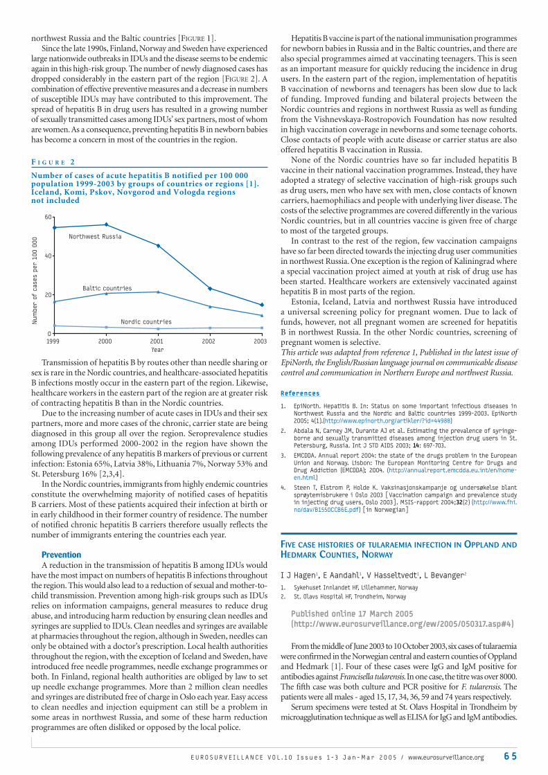
E U R OS U R V E I L L A N C E V O L . 10 I s s u e s 1 -3 J a n - M a r 2 0 0 5 / www.eurosurveillance.org 6 5
northwest Russia and the Baltic countries [FIGURE 1].Since the late 1990s, Finland, Norway and Sweden have experienced
large nationwide outbreaks in IDUs and the disease seems to be endemic again in this high-risk group. The number of newly diagnosed cases has dropped considerably in the eastern part of the region [FIGURE 2]. A combination of effective preventive measures and a decrease in numbers of susceptible IDUs may have contributed to this improvement. The spread of hepatitis B in drug users has resulted in a growing number of sexually transmitted cases among IDUs’ sex partners, most of whom are women. As a consequence, preventing hepatitis B in newborn babies has become a concern in most of the countries in the region.
F I G U R E 2
Number of cases of acute hepatitis B notified per 100 000 population 1999-2003 by groups of countries or regions [1]. Iceland, Komi, Pskov, Novgorod and Vologda regions not included
Transmission of hepatitis B by routes other than needle sharing or sex is rare in the Nordic countries, and healthcare-associated hepatitis B infections mostly occur in the eastern part of the region. Likewise, healthcare workers in the eastern part of the region are at greater risk of contracting hepatitis B than in the Nordic countries.
Due to the increasing number of acute cases in IDUs and their sex partners, more and more cases of the chronic, carrier state are being diagnosed in this group all over the region. Seroprevalence studies among IDUs performed 2000-2002 in the region have shown the following prevalence of any hepatitis B markers of previous or current infection: Estonia 65%, Latvia 38%, Lithuania 7%, Norway 53% and St. Petersburg 16% [2,3,4].
In the Nordic countries, immigrants from highly endemic countries constitute the overwhelming majority of notified cases of hepatitis B carriers. Most of these patients acquired their infection at birth or in early childhood in their former country of residence. The number of notified chronic hepatitis B carriers therefore usually reflects the number of immigrants entering the countries each year.
Prevention A reduction in the transmission of hepatitis B among IDUs would
have the most impact on numbers of hepatitis B infections throughout the region. This would also lead to a reduction of sexual and mother-to-child transmission. Prevention among high-risk groups such as IDUs relies on information campaigns, general measures to reduce drug abuse, and introducing harm reduction by ensuring clean needles and syringes are supplied to IDUs. Clean needles and syringes are available at pharmacies throughout the region, although in Sweden, needles can only be obtained with a doctor’s prescription. Local health authorities throughout the region, with the exception of Iceland and Sweden, have introduced free needle programmes, needle exchange programmes or both. In Finland, regional health authorities are obliged by law to set up needle exchange programmes. More than 2 million clean needles and syringes are distributed free of charge in Oslo each year. Easy access to clean needles and injection equipment can still be a problem in some areas in northwest Russia, and some of these harm reduction programmes are often disliked or opposed by the local police.
Hepatitis B vaccine is part of the national immunisation programmes for newborn babies in Russia and in the Baltic countries, and there are also special programmes aimed at vaccinating teenagers. This is seen as an important measure for quickly reducing the incidence in drug users. In the eastern part of the region, implementation of hepatitis B vaccination of newborns and teenagers has been slow due to lack of funding. Improved funding and bilateral projects between the Nordic countries and regions in northwest Russia as well as funding from the Vishnevskaya-Rostropovich Foundation has now resulted in high vaccination coverage in newborns and some teenage cohorts. Close contacts of people with acute disease or carrier status are also offered hepatitis B vaccination in Russia.
None of the Nordic countries have so far included hepatitis B vaccine in their national vaccination programmes. Instead, they have adopted a strategy of selective vaccination of high-risk groups such as drug users, men who have sex with men, close contacts of known carriers, haemophiliacs and people with underlying liver disease. The costs of the selective programmes are covered differently in the various Nordic countries, but in all countries vaccine is given free of charge to most of the targeted groups.
In contrast to the rest of the region, few vaccination campaigns have so far been directed towards the injecting drug user communities in northwest Russia. One exception is the region of Kaliningrad where a special vaccination project aimed at youth at risk of drug use has been started. Healthcare workers are extensively vaccinated against hepatitis B in most parts of the region.
Estonia, Iceland, Latvia and northwest Russia have introduced a universal screening policy for pregnant women. Due to lack of funds, however, not all pregnant women are screened for hepatitis B in northwest Russia. In the other Nordic countries, screening of pregnant women is selective. This article was adapted from reference 1, Published in the latest issue of EpiNorth, the English/Russian language journal on communicable disease control and communication in Northern Europe and northwest Russia.
References
1. EpiNorth. Hepatitis B. In: Status on some important infectious diseases in
Northwest Russia and the Nordic and Baltic countries 1999-2003. EpiNorth
2005; 4(1).(http://www.epinorth.org/artikler/?id=44988)
2. Abdala N, Carney JM, Durante AJ et al. Estimating the prevalence of syringe-
borne and sexually transmitted diseases among injection drug users in St.
Petersburg, Russia. Int J STD AIDS 2003; 14: 697-703.
3. EMCDDA. Annual report 2004: the state of the drugs problem in the European
Union and Norway. Lisbon: The European Monitoring Centre for Drugs and
Drug Addiction (EMCDDA); 2004. (http://annualreport.emcdda.eu.int/en/home-
en.html)
4. Steen T, Elstrom P, Holde K. Vaksinasjonskampanje og undersøkelse blant
sprøytemisbrukere i Oslo 2003 [Vaccination campaign and prevalence study
in injecting drug users, Oslo 2003]. MSIS-rapport 2004;32(2) (http://www.fhi.
no/dav/B1550CCB6E.pdf) [in Norwegian]
FIVE CASE HISTORIES OF TULARAEMIA INFECTION IN OPPLAND AND HEDMARK COUNTIES, NORWAY
I J Hagen1, E Aandahl1, V Hasseltvedt1, L Bevanger2
1. Sykehuset Innlandet HF, Lillehammer, Norway
2. St. Olavs Hospital HF, Trondheim, Norway
Published online 17 March 2005(http://www.eurosurveillance.org/ew/2005/050317.asp#4)
From the middle of June 2003 to 10 October 2003, six cases of tularaemia were confirmed in the Norwegian central and eastern counties of Oppland and Hedmark [1]. Four of these cases were IgG and IgM positive for antibodies against Francisella tularensis. In one case, the titre was over 8000. The fifth case was both culture and PCR positive for F. tularensis. The patients were all males - aged 15, 17, 34, 36, 59 and 74 years respectively.
Serum specimens were tested at St. Olavs Hospital in Trondheim by microagglutination technique as well as ELISA for IgG and IgM antibodies.
0
20
40
60
1999 2000 2001 2002 2003
Num
ber o
f c
ases p
er 1
00 0
00 Northwest Russia
Nordic countries
Baltic countries
Year

6 6 E U R OS U R V E I L L A N C E V O L . 10 I s s u e s 1 -3 J a n - M a r 2 0 0 5
Pus specimens and other types of specimens were tested by cultivation and polymerase chain reaction (PCR). Clinical and epidemiological information was obtained from the physician treating the patients.
The clinical presentation of five of the cases is described below: Patient one was admitted to hospital with multiple ulcers in the
foot and ankle region. Later, an enlarged lymph gland in the groin area was noted. The patient had a fever, felt unwell, and his condition deteriorated. The tentative diagnosis was vasculitis. The local physician eventually diagnosed tularaemia after reading about it in the national communicable disease bulletin (MSIS-rapport, http://www.fhi.no/eway/default0.asp).
Patient two was bitten by a wild rat, which he had found in an unwell state and was taking care of. The rat later died – most likely from an infectious disease. Two days after being bitten, the patient developed a fever and felt unwell. Ulcerations and enlargement of the lymph nodes were noted on clinical examination.
Patient three was probably bitten outdoors, by an insect, tick or snake, but could not recall this. The patient presented with a fever, and an enlarged lymph node (no abscess) and also with an ulceration that was small and considered atypical.
Patient four presented with typical ulceration around the ankle, a fever, and an enlarged lymph node with formation of an abscess. The abscess had to be drained, despite adequately targeted antibiotic treatment.
Patient five presented with typical ulceration in the underarm area, a fever, and enlarged lymph nodes with an abscess which was drained.
The patients all had the following symptoms and signs in common:
• The ulcerations were 2-3 cm in diameter with a central crater.
• The ulcerations lasted for weeks and did not heal quickly even when antibiotics such as ciprofloxacin were given
• The regional lymph nodes were larger than expected. • The patients felt very unwell approximately 10 days after the
onset of fever. • The general malaise generally lasted many weeks, and persisted
even after starting adequate treatment with antibiotics. • Blood test results for evidence of infection were close to the
normal reference values. All five cases described lived in areas where rodent activity is high
and where tularaemia has been diagnosed previously. There were no known associations between the patients. None of the patients reported drinking water from unchlorinated sources. Physician awareness of tularaemia in Norway is fairly low, and some of the
cases described were not recognised at initial presentation.
Tularaemia epidemiology in Norway 1985-2004Tularaemia has been a notifiable disease in Norway since 1975.
Between 1975 – 2004, the annual incidence has varied from 0 to 47 cases (incidence in 1985). In Sweden, the incidence has been much higher in recent years – up to many hundred cases annually [2].
From 1985 to 2004, there were 16 cases in Oppland and Hedmark counties [FIGURE]. There were 13 males and 3 females affected. No imported cases were documented. Most cases were among adults. All cases occurred between July and December.
F I G U R E
Tularaemia in Oppland and Hedmark counties, Norway, 1985 - 2004, distribution by year
Given that rodents move freely across national borders, it is not clear why the incidence in Norway remains low whereas in neighbouring areas in Sweden, incidences are much higher. It is likely that tularaemia is underdiagnosed in Norway. The degree of underdiagnosis/underreporting remains unknown. Increased awareness of tularaemia among physicians and the general public would improve knowledge of the epidemiological situation.
References
1. Hagen IJ, Hasseltvedt V, Aandahl E, Bevanger L. Tularemi i Oppland og Hedmark
2003 – mikrobiologi og epidemiologi (in Norwegian) (Tularaemia in Oppland
and Hedmark 2003 – Microbiology and Epidemiology. Proceedings from The
Annual Conference, Folkshelseinstitutett, Oslo, 2003 – abstract 13.
2. Arneborn M. Tularemi. Epi-aktuellt 2004:39 (http://www.smittskyddsinstitutet.
se/SMItemplates/BigArticle/4290.aspx)
0
1
2
3
4
5
6
7
8
9
10
1985 1986 1987 1988 1989 1990 1991 1992 1993 1994 1995 1996 1997 1998 1999 2000 2001 2002 2003 2004
Year of notification
Incid
ence

P O L I C Y A N D G U I D E L I N E S
E U R OS U R V E I L L A N C E V O L . 10 I s s u e s 1 -3 J a n - M a r 2 0 0 5 / www.eurosurveillance.org 67
EU DRUGS AGENCY PUBLICATION ON HEPATITIS C AND INJECTING DRUG USE LOOKS AT IMPACT, COSTS AND POLICY OPTIONS
V Hope, F Ncube
Health Protection Agency Centre for Infections, London, England
Published online 23 December 2004 (http://www.eurosurveillance.org/ew/2004/041223.asp#5)
The European Monitoring Centre for Drugs and Drug Addiction (EMCDDA), the EU drugs agency, has recently Published its latest scientific monograph, Hepatitis C and injecting drug use: impact, costs and policy options [1]. This publication brings together research by international experts from the hepatitis C, drug use and public health fields. It combines analyses on the impact and costs of hepatitis C virus (HCV) infection among injecting drug users (IDUs) so as to inform future policy making in the European Union.
Since screening for HCV became available in the early 1990s, drug injecting has been the most common route of infection in the EU, largely due to risk behaviours such as sharing of needles, syringes, and other injecting equipment. While HCV may affect over 1% of the population of the EU, prevalence is substantially higher among those who have injected drugs.
The monograph points to data indicating that up to 90% of newly notified cases of HCV infection in EU countries are now occurring in IDUs [1,2]. The EMCDDA 2004 Annual Report, Published last month, cites HCV prevalence rates of between 17% and 95% in IDUs, depending on the country and study setting, underlining the need for prevention and treatment in this the main at risk population [2].
Current IDUs often encounter difficulties in accessing treatment due to concerns about their poor compliance to programmes, side effects and risk of re-infection. Recent research studies, however, have shown that treating IDUs is feasible and effective, and new guidelines recommend case-by-case decisions on treatment.
Some other key findings:• New HCV infections occurring in 1999 in six of the most
affected countries – France, Germany, Italy, Portugal, Spain and the United Kingdom – are likely to result in healthcare costs of up to 1.43 billion over the next two decades. Data presented estimate lifetime healthcare costs ranging between 13 100 and 26 200 per infected person in these six countries.
• New cost effectiveness analyses presented suggest that screening IDUs for infection and offering combination antiviral therapy to those with moderate liver disease can enhance quality of life, extend life expectancy and be cost effective. It is estimated that through avoiding the costs of liver disease related complications, over two thirds of the average treatment costs can be compensated for.
• Needle and syringe programmes (NSPs) are a key public health intervention for IDUs in general. They are cost effective in reducing the general transmission bloodborne viruses although they seem less (cost-)effective for HCV than for HIV prevention.
• Methadone maintenance treatment (MMT), though highly effective and cost effective for HIV prevention, is less so in the case of HCV. As the benefits of MMT increase with the proportion of the IDU population covered it can become a cost effective method of HCV prevention once high levels of coverage are attained.
References
1. Jager J, Limburg W, Kretzschmar M, Postma M, Wiessing L, eds. Hepatitis C
and injecting drug use: impact, costs and policy options. Monograph 7. Lisbon:
European Monitoring Centre for Drugs and Drug Addiction; December 2004.
(available at http://www.emcdda.eu.int/?nnodeid=428)
2. EMCDDA. Annual report 2004: the state of the drugs problem in the European
Union and Norway. Lisbon: European Monitoring Centre for Drugs and Drug
Addiction; November 2004. (available in 20 languages at http://annualreport.
emcdda.eu.int/en/home-en.html)
CONSIDERABLE PROGRESS IN EUROPEAN PREPARATIONS FOR A POTENTIAL INFLUENZA PANDEMIC
Editorial team1 and John Paget2
1. Eurosurveillance Editorial Offi ce
2. European Infl uenza Surveillance Scheme, Netherlands Institute for Health Services Research (NIVEL), Utrecht, the Netherlands
Published online 23 December 2004 (http://www.eurosurveillance.org/ew/2004/041223.asp#2)
The threat of an influenza pandemic has been heightened in the past two years by outbreaks of avian influenza concentrated in South East Asia which have resulted in human deaths. So far, the avian influenza virus seems difficult to transmit from human to human, but changes in the virus genome may well increase transmissibility. Possibly worse, a person or animal (such as a pig) could become co-infected with human and avian influenza. These viruses could then combine, creating a very novel influenza virus that is both highly pathogenic and easily transmitted to humans.
The World Health Organization has warned of an influenza pandemic threat and is urging member states to devise a national influenza preparedness plan for this eventuality [1]. It has also devised warning levels and has linked actions to each level.
The European Commission and European Union (EU) member states have responded to the influenza pandemic threat and much progress has been achieved in recent years.
Preparation by the European Commission and European networksIn response to the outbreak of avian influenza in South East Asia,
the European Commission banned imports of live birds and poultry products from many countries in February 2004 [2,3]. This ban has been extended to 31 March 2005.
In March 2004, the European Commission Published a Working Paper on Community Influenza Pandemic Preparedness and Response Planning (http://europa.eu.int/comm/health/ph_threats/com/Influenza/com_2004_01_en.pdf) which called on all EU member states to complete their influenza pandemic preparedness plans, designate national reference laboratories for human influenza, achieve high vaccine coverage (especially in high risk groups), and prepare media briefing materials on influenza. The paper also stated the tasks of the European Commission in planning for a pandemic.
Surveillance of influenza in Europe (European Influenza Surveillance Scheme, http://www.eiss.org) has been considerably enhanced in recent years with funding from the Commission. Since October 2000, clinical, epidemiological and virological data have been presented on a weekly basis from October to May each year on the EISS website. In 2003 the Community Network of National Reference Laboratories for Human Influenza was created within EISS and this network is now operational (http://www.eiss.org/documents/eiss_poster_cnrl.pdf). Its primary goal is to provide high quality reference services for human influenza surveillance, guaranteeing highly qualified virological data reported to EISS as well as clinical data.
The European Commission’s DG Research has also funded projects related to influenza pandemic preparedness (e.g. the FLUPAN project) and it recently started funding a multicentre network called VIRGIL (http://www.virgil-net.org/), which will address current and emerging antiviral drug resistance concerning influenza.
European vaccine manufacturers (http://www.evm-vaccines.org/) have got together and are working on issues related to the production of an influenza vaccine in case of a pandemic, for

6 8 E U R OS U R V E I L L A N C E V O L . 10 I s s u e s 1 -3 J a n - M a r 2 0 0 5
example how many vaccines will be needed and how can production be increased to meet these needs (http://www.evm-vaccines.org/290403%20Flu%20pandemic%20final.pdf).
The European Scientific Working Group on Influenza (http://www.eswi.org) is also active in the area of pandemic preparedness. This group organises an important scientific conference in Europe every two years where issues related to pandemic preparedness are high on the conference agenda.
Preparation by member statesThe EU member states have also been active in preparing for a potential
influenza pandemic. A survey carried out in November 2000 found that eight countries (50% of those surveyed) had an official pandemic plan, seven countries had a plan that was in an advanced stage or draft format and one country did not have a plan. Many of these plans have now been finalised and European countries are now starting to implement these at a national and local level. A number of countries have started to stockpile antiviral drugs (France, Belgium and the Netherlands).
Further challenges to Europe-wide pandemic planningConsolidation of these different activities is now required and
the general level of preparedness will be tested by an EC-funded simulation project (http://europa.eu.int/comm/health/ph_programme/howtoapply/call_130356_2004.htm)The simulation should help measure preparedness at a European and national level, and identify weaknesses that need strengthening or correcting.
One important challenge that has not yet been resolved is the equitable distribution of vaccines (if these are available) and stockpiled antiviral drugs. Considering EU treaties no longer hold in a situation of ‘force majeure’, member states could legally hoard nationally produced vaccines and/or antiviral drugs. This would be a very unfortunate development for Europe and mechanisms to ensure equitable access to vaccines and antiviral drugs within the EU should therefore be encouraged.
References
1. Estimating the impact of the next influenza pandemic: enhancing prepared-
ness. WHO. 8 December 2004 (http://www.who.int/csr/disease/influenza/pre-
paredness2004_12_08/en/)
2. European Commission. Review of the avian influenza situation in Asia, Canada,
the USA and South Africa. Press release. (http://europa.eu.int/comm/dgs/
health_consumer/library/press/press350_en.pdf)
3. European Comission. Avian influenza in Malaysia: import of feathers and pet
birds banned. Press release. (http://europa.eu.int/comm/dgs/health_consu-
mer/library/press/press346_en.pdf)
DIFFERENCES BETWEEN NEW UNITED STATES RECOMMENDATIONS AND EXISTING EUROPEAN GUIDELINES ON THE USE OF POSTEXPOSURE PROPHYLAXIS (PEP) FOLLOWING NON-OCCUPATIONAL EXPOSURE
J Blackham1, J Almeda2 on behalf of the European Project
on Non-occupational Postexposure Prophylaxis for HIV (Euro-
NONOPEP)
1. Communicable Disease Surveillance Centre, Health Protection Agency Centre for Infections, London, England
2. CEESCAT, Hospital Universitari Germans Trias i Pujol, Badalona, Spain
Published online 27 January 2005 (http://www.eurosurveillance.org/ew/2005/050127.asp#3)
Last week the United States Department of Health and Human Services Published updated recommendations for the use of post-exposure prophylaxis (PEP) following non-occupational exposure to HIV [1]. The evidence is still unclear as to the efficacy of this
intervention and this report provides a comprehensive overview of available literature, and discusses the benefits and problems with the administration of PEP in certain circumstances. It also clearly re-emphasises that the most effective way to prevent transmission of HIV is to prevent exposure, and any programme of PEP administration should not replace primary prevention.
In 2004, the Euro-NONOPEP project group Published recommendations for PEP use along with the results of their two-year Europe-wide study [2]. Although the two guidelines considered the same intervention in the same circumstances, there are marked differences in their recommendations. Both guidelines state the basic notion that PEP should be administered to people exposed to potentially infectious bodily fluids of a known HIV-infected person, when the exposure represents a substantial risk of transmission. In these cases, a 28-day regimen of highly active retroviral therapy (HAART) should be prescribed. After this point, however, they differ in three main areas.
First, the United States (US) guidelines recommend that PEP is only prescribed when the source person is known to be HIV-infected. For cases where the HIV status of the source is unknown, the guidelines state that the clinician should assess each case individually and use their judgement. The European recommendations lay out the circumstances under which PEP should or should not be considered or prescribed if the status of the source patient is unknown. If the source patient is from a group or area of high HIV prevalence (at least 15%) the European guidelines recommend that PEP be prescribed following receptive anal sex; for other exposures, anal, vaginal or oral (with ejaculation), PEP should be considered. They also state that if the source patient is not from a high-risk group, then PEP should only be considered following receptive anal sex. The US recommendations put a stronger emphasis on the potential side effects of PEP and conclude that these may well outweigh the potential benefits if the infective status of the source patient is unknown.
Second, both guidelines focus on the risk of transmission. For some transmission situations, where the partner is HIV-infected, the transmission values used by each group are similar or the same, e.g. following a blood transfusion: US, 90%; European, 90%-100%. For other exposures, the transmission risk estimates used are very different. In particular, the US document estimates the risk of transmission via receptive anal sex to be 0.5%, while the European group estimates this to be 3%. This large difference in transmission risk may have influenced the recommendations made for PEP usage. As mentioned above, the European guidelines recommend that PEP be considered in any situation where unprotected receptive anal sex has occurred. As long as there is continuing uncertainty as to the true risk of transmission via different exposures, it is difficult to reach consensus on all the situations where PEP should be prescribed.
The final significant difference concerns the advice on the regimen of antiretrovirals to use. The Euro-NONOPEP group recommends the use of triple therapy (treatment with a combination of three drugs belonging to two different classes) but states that a two-drug regimen (treatment with two nucleoside reverse transciptase inhibitors (NRTI)) is also an option. This is based on evidence that drugs acting at different stages of the virus’ life cycle are superior to monotherapy and that tri-therapy has been shown to treat HIV-infected patients most effectively. However, the US recommendations state that there is no evidence to indicate that a three-drug regimen would be more effective than a two-drug regimen. They place a heavier emphasis on the possible risks of side effects and state that these should be discussed with the patients. They also consider the prescription of medication to treat side-effects of HAART.
The differences in recommendations highlight the ongoing controversy surrounding the use of PEP following a non-occupational exposure. An increasing number of countries are addressing the use of PEP and establishing recommendations [TABLE].

E U R OS U R V E I L L A N C E V O L . 10 I s s u e s 1 -3 J a n - M a r 2 0 0 5 / www.eurosurveillance.org 6 9
T A B L E
A selection of non-occupational PEP recommandation from European countries
Country Web pageGermany http://www.rki.de/INFEKT/AIDS_STD/AZ_ENG/HIVPEPL_E.HTM
http://www.rki.de/INFEKT/AIDS_STD/AZ_ENG/HIVPEPK_E.HTM
Italy http://www.inmi.it/news/LineeGuida/RecommendationsNONOCC.htm
Poland http://www.msi.com.pl/pub/hiv/vol_1/no_1/3177.pdf
Spainhttp://www.msc.es/profesional/preProSalud/sida/pdfs/guia_actuacion_
profilaxis.pdf
Switzerland http://www.hiv.ch/rubriken/therapie/pep/pepsex/pepsexi.htm (in Italian)
http://www.hiv.ch/rubriken/therapie/pep/pepsex/pepsexf.htm (in French)
http://www.hiv.ch/rubriken/therapie/pep/pepsex/pepsexf.htm (in German)
United Kingdom
http://www.bashh.org/guidelines/draft_04/pepse[1]_010404.doc
As there cannot be a randomised control trial for this intervention, it is important that countries share data and recommendations to build up the evidence available. Members of the Euro-NONOPEP group are promoting an initiative to analyse cases of high-risk exposure to HIV supplied by registries in Europe, Australia and the United States. The Euro-NONOPEP group has also submitted a protocol for a Cochrane review on NONOPEP to the Cochrane Review Group on HIV Infections and AIDS. Some of these registries have had difficulties sustaining operational funding; some have been discontinued, while others are operating on a voluntary basis of case reporting.
Since the publication of the Euro-NONOPEP recommendations for PEP, some studies of PEP regimens with a better adherence and fewer adverse events have been conducted [3-6]. These studies, and the recent publication of the US guidelines, have highlighted the need to revise and update the Euro-NONOPEP and other national guidelines. Thus, the comprehensive US guidelines will no doubt provide an important focal point in the future.
References
1. Smith DK, Grohskopf LA, Black RJ, Auerbach JD, Veronese F, Struble KA, et al.
Antiretroviral Postexposure Prophylaxis After Sexual, Injecting-Drug Use, or
Other Nonoccupational Exposure to HIV in the United States. MMWR Recomm Rep
2005;54(RR02):1-20. (http://www.cdc.gov/mmwr/preview/mmwrhtml/rr5402a1.
htm)
2. Almeda J, Casabona J, Simon B, Gerard M, Rey D, Puro V, et al. Proposed re-
commendations for the management of HIV post-exposure prophylaxis after
sexual, injecting drug or other exposures in Europe. Euro Surveill 2004;9(6):5-
6. (http://www.eurosurveillance.org/em/v09n06/0906-223.asp)
3. Mayer KH, Goldhammer H, Cohen D, Grasso C, Bill R, Maldonado A et al. Enhanced
tolerability and adherence using Tenofovir/3TC for non-occupational post-
exposure prophylaxis (NPEP) (Abstract number TuPeB4657). XV International
Conference on AIDS. 11-16 July 2004;Bangkok, Thailand.
4. Rabaud C, Burty C, Valle C, Prazuck C, Beguinot I, Dorvaux V, Salmon Ceron
D, May T. Post-Exposure Prophylaxis of HIV infection: Comparison of the to-
lerability of 3 PEP regimens (Abstract number TuPeB4651). XV International
Conference on AIDS. 11-16 July 2004;Bangkok, Thailand.
5. Rabaud C, Valle C, Prazuck T, Christian B, Grandidier M, Beguinot I, et al.
Tolerability of post-exposure prophylaxis of HIV Infection with the combi-
nation of Zidovudine/Lamivudine + Tenofovir (Abstract number TuPeB4647). XV
International Conference on AIDS. 11-16 July 2004;Bangkok, Thailand.
6. Winston A, McAllister J, Cooper D, Carr A. The use of a triple nucleoside
and nucleotide regimen for non-occupational HIV post exposure prophylaxis
(Abstract number TuPeB4658). XV International Conference on AIDS. 11-16 July
2004; Bangkok, Thailand.
RESULTS OF SURVEY OF NATIONAL INFLUENZA PANDEMIC PREPAREDNESS IN EUROPE
M Ciotti1, F Karcher1, B Ganter2, P Tüll2
1. Health Threats Unit, European Commission, Luxembourg
2. WHO Regional Offi ce for Europe Copenhagen, Denmark
Published online 3 March 2005 (http://www.eurosurveillance.org/ew/2005/050303.asp#2)
The World Health Organization (WHO) and the European Commission are currently working together to improve influenza preparedness in the European Region. So far, only a few countries have submitted national influenza pandemic plans to WHO and/or the European Commission. To help countries that do not yet have a draft national influenza preparedness plan, and to update plans where they already exist, the European Commission and WHO held a two day workshop for all countries in the WHO European region on 2-3 March 2005.
The specific objectives of this workshop were to:• facilitate the processes involved in planning influenza pandemic
preparedness • provide an opportunity to discuss the priorities of a pandemic
plan with colleagues and facilitators • identify the way forward for WHO/EU member states as they
develop their pandemic plans • identify whether further assistance is needed and, if so, what
form it should take To determine the stage of pandemic planning in the different
European countries, a questionnaire was sent to all member states of the WHO European Region (56 countries, including 25 European Union member states) before the workshop, all of whom responded. Fifty of the respondents stated that a responsible national body exists which is working on pandemic preparedness. Thirty-one have a national preparedness plan available and Published; of these, 18 are European Union (EU) states. The remaining states and entities either have a draft plan at differing stages of development, or do not have a plan [TABLE 1].
Within the European Union, considerable progress in influenza pandemic planning has been made in the last few years. In 2005, 18/25 (72%) EU countries had Published plans. In 2000, just 4 of 11 (36%) EU countries surveyed had plans that were accepted by health authorities [1,2].
T A B L E 1
Response from states/entities about the existence of a national influenza pandemic plan, 2005
National Plan and Responsibilities
All respondents (56)
EU Member States (25)
non-EU states/entities (31)
Question Yes Percentage Yes Percentage Yes Percentage
Is there a responsible body and/or a responsi-ble person working on influenza pandemic pre-paredness planning?
50 89% 25 100% 25 81%
Is there a national in-fluenza pandemic prepa-redness plan available and Published?
31 55% 18 72% 13 42%
National plans differ as far as the elements considered. The table below shows 10 components considered to be important and the percentage of countries which have these in their Published or draft plan. Based on the response, it is clear that surveillance and provision of laboratory facilities are the two most developed components included in the pandemic plans [TABLE 2].
Of those that have a Published plan, four countries have also conducted simulation exercises to test its efficiency and efficacy.
As well as specific questions related to the components of a pandemic preparedness plan, countries were also asked to provide details of their national influenza programme in the interpandemic period [TABLE 3]. Almost all countries have a functional surveillance system and a vaccination programme for risk groups (100% of EU member states have these two components). Twenty four countries (13 EU and 11 non-EU) maintain stocks of antivirals.
National influenza plans from European countries and other countries worldwide that are available on the internet can be found here: http://www.eiss.org/html/pandemic_plans.html
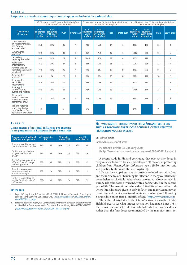
70 E U R OS U R V E I L L A N C E V O L . 10 I s s u e s 1 -3 J a n - M a r 2 0 0 5
T A B L E 3
Components of national influenza programme (non-pandemic) in European Region countries
Components of national influenza programme
All countries(56)
EU member states (25)
non-EU countries (31)
Does a surveillance sys-tem for influenza exist?
98% 55 100% 25 97% 30
Is there a vaccination programme for risk groups?
88% 49 100% 25 77% 24
Are influenza vaccines offered free of charge for risk groups?
63% 35 72% 18 55% 17
Does the government maintain a stock of anti-viral drugs?
43% 24 52% 13 36% 11
Is there laboratory ca-pacity for diagnosis of influenza?
80% 45 96% 24 68% 21
References
1. Paget WJ, Aguilera J-F (on behalf of EISS). Influenza Pandemic Planning in
Europe. Euro Surveill 2001;6:136-140. (http://www.eurosurveillance.org/em/
v06n09/0609-222.asp)
2. Editorial team and Paget, WJ. Considerable progress in European preparations for
a potential influenza pandemic. Eurosurveillance Weekly 2004;8(52):23/12/2005
(http://www.eurosurveillance.org/ew/2004/041223.asp#2)
HIB VACCINATION: RECENT PAPER FROM FINLAND SUGGESTS THAT A PROLONGED THREE DOSE SCHEDULE OFFERS EFFECTIVE PROTECTION AGAINST DISEASE
Editorial team
Eurosurveillance editorial offi ce
Published online 13 January 2005(http://www.eurosurveillance.org/ew/2005/050113.asp#1)
A recent study in Finland concluded that two vaccine doses in early infancy, followed by a late booster, are efficacious in protecting children from Haemophilus influenzae type b (Hib) infection, and will practically eliminate Hib meningitis [1].
Hib vaccine campaigns have successfully reduced mortality from and the incidence of Hib meningitis infection in many countries, but nevertheless vaccine failures have been recognised. Most countries in Europe use four doses of vaccine, with a booster dose in the second year of life. The exceptions include the United Kingdom and Ireland, where three doses are given in early infancy, and many Scandinavian countries (and Italy) where two doses in early infancy are followed by a single dose on or after 11 months of age. (http://www.euibis.org)
The authors looked at records of H. influenzae cases in the Greater Helsinki area, to see what impact vaccination had made. Since 1988, the Finnish vaccine schedule has included only three vaccine doses, rather than the four doses recommended by the manufacturers, yet
T A B L E 2
Response to questions about important components included in national plan
All 56 countries (31 have a Published plan; 25 with draft or no plan)
EU member states (18 have a Published plan; 7 with draft or no plan)
non-EU countries (13 have a Published plan; 18 with draft or no plan)
Components of the plan
% of countries
with a plan
% of countries with draft or no plan
Plan Draft plan% of
countries with
a plan
% of countries with draft or no plan
Plan Draft plan% of
countries with
a plan
% of countries with draft or no plan
Plan Draft plan
Clear division of responsibilities, obligations and mandates?
81% 16% 25 4 78% 14% 14 1 85% 17% 11 3
Surveillance systems?
97% 36% 30 9 94% 71% 17 5 100% 22% 13 4
Laboratory capacity and role?
94% 28% 29 7 100% 57% 18 4 85% 17% 11 3
Healthcare organisation?
87% 20% 27 5 83% 14% 15 1 92% 22% 12 4
Maintenance of essential community services?
77% 16% 24 4 72% 0% 13 0 85% 22% 11 4
Strategy for antivirals?
81% 8% 25 2 83% 0% 15 0 77% 11% 10 2
Strategy for vaccines/vaccination?
87% 20% 27 5 89% 14% 16 1 85% 22% 11 4
Strategy for information to public and media?
84% 16% 26 4 72% 14% 13 1 100% 17% 13 3
Other public health measures (views on public gatherings etc.)?
77% 16% 24 4 72% 14% 13 1 85% 17% 11 3
Has the national plan been tested in a ‘table top’ or equivalent exercise?
13% 4 6% 1 23% 3

E U R OS U R V E I L L A N C E V O L . 10 I s s u e s 1 -3 J a n - M a r 2 0 0 5 / www.eurosurveillance.org 71
only three cases of Hib meningitis infection occurred between 1991 and 1999, all in vaccinated or incompletely vaccinated children. During the same period, three cases of H. influenzae meningitis, caused by H. influenzae type f and a non-typable strain, occurred. The authors concluded that three doses of conjugate vaccine (two early doses with a late booster) are clinically effective in protecting children from Hib infection, and that epidemiological data such as these may be more useful than measuring antibody levels when judging the effectiveness of a vaccination programme.
In response to this study, a team in the United Kingdom described the UK experience, where a vaccine schedule of three doses at 2, 3 and 4 months with a large catch-up campaign for older children led to a 95% reduction in the attack rate of invasive Hib disease between 1992-1998 [2].
There was a large protective effect in unvaccinated age groups of children due to herd immunity (indirect protection). To assess individual protection from the vaccine, data on invasive Hib disease in 1996-2003 occurring in children born between 1996-1999 was analysed, and it was shown that direct protection from Hib conjugate vaccines given in an accelerated schedule declines rapidly over time. Therefore, excellent disease control does not necessarily imply high levels of indirect protection, and caution is needed in the longer term. The potential for disease to re-emerge after it has been initially controlled by a vaccine programme has been illustrated by recent increases in both the UK and the Netherlands [3].
The European Union Invasive Bacterial Infection Surveillance scheme (EU-IBIS, http://www.euibis.org/) is pooling surveillance data from European countries, including data on vaccine failures, and this will help to inform analysis on invasive meningitis trends, including those that may result from differences between national vaccination programmes.
References
1. Peltola H, Salo E, Saxén H. Incidence of Haemophilus influenzae type b me-
ningitis during 18 years of vaccine use: observational study using routine
hospital data. BMJ 2005; 330: 18-9. (http://bmj.bmjjournals.com/cgi/content/
full/330/7481/18)
2. Ramsay ME, Miller E, Andrews N, Slack M, Heath P. Epidemiological Data are
Essential [Rapid Response]. BMJ, 16 December 2004. (http://bmj.bmjjournals.
com/cgi/eletters/330/7481/18#89318 )
3. Rijkers GT, Vermeer-de Bondt PE, Spanjaard L, Breukels M, Sanders EAM.
Return of Haemophilus influenzae type b infections. [letter] Lancet 2003;
361(9368):1563-4
WORLD HEALTH ORGANIZATION DEVELOPS GUIDANCE FOR VACCINE SAFETY INFORMATION ON THE WEB
Editorial team
Eurosurveillance editorial offi ce
Published online 13 January 2005(http://www.eurosurveillance.org/ew/2005/050113.asp#2)
The World Health Organization (WHO) Global Advisory Committee on Vaccine Safety (GACVS, http://www.who.int/vaccine_safety/en/) was established in 1999 to respond promptly, efficiently, and with scientific rigour to vaccine safety issues of potential global importance. In 2003, GACVS launched the Vaccine Safety Net project. As part of this project, guidelines for websites which provide information on vaccine safety have been produced (http://www.who.int/vaccine_safety/good_vs_sites/en/).
By encouraging websites that provide accurate information to be of high quality and the first source of information for the media and public, it is hoped that the guidelines will counter the proliferation of websites providing false or misleading information about vaccine safety, thus undermining public health messages.
The guidelines specify content that should be included on the site, and credibility information, as well as guidance on accessibility of the website and design. Organisations providing information on vaccine safety are advised to consider the guidance and enhance their information if necessary.
Since setting these guidelines, the WHO has evaluated a number of vaccine safety information websites against these criteria, and websites that meet these are published on the WHO immunisation safety website (http://www.who.int/immunization_safety/safety_quality/approved_vaccine_safet_websites/en/). This site already includes various European websites, and many more European and worldwide websites will be evaluated in the coming months.

N E W S
72 E U R OS U R V E I L L A N C E V O L . 10 I s s u e s 1 -3 J a n - M a r 2 0 0 5
A selection of other news items from Eurosurveillance,
full text available at:
www.eurosurveillance.org
Increase in mumps in Ireland in late 200423 December 2004
Watching the detectives: celebrating the 10th year of EPIET23 December 2004
Health Protection Scotland: new health protection body for Scotland replaces SCIEH23 December 2004
Teringzooi – the ‘Bloody Mess’ exhibition - gives a striking history of infectious disease23 December 2004
Applicants sought for training fellowships in intervention epidemiology in Europe (the EPIET programme) 23 December 2004
Tsunami in south Asia and east Africa: public health response6 January 2005
Hib vaccination: recent paper from Finland suggests that a prolonged three dose schedule offers effective protection13 January 2005
World Health Organization develops guidance for vaccine safety information on the web13 January 2005
Avian influenza: current situation in South East Asia and impact on Europe20 January 2005
Increased influenza activity in Spain from mid-December 200420 January 2005
Increased influenza activity in Ireland, January 200520 January 2005
Muslim Hajj pilgrimage: minimising the risks of infectious disease20 January 2005
Public Health Programme 2003-2008: 2005 work plan and call for proposals Published20 January 2005
Two epidemiologically distinct syphilis outbreaks in south west England detected in 2004 with few apparent links to outbreaks in urban areas27 January 2005
Tsunami in south Asia and east Africa – infectious disease update27 January 2005
Sexually transmitted infections on the rise in Northern Ireland3 February 2005
Seven PhD fellowships in Denmark available for non-human use of antimicrobials through the EC Marie Curie programme3 February 2005
First case of vCJD reported in a Japanese patient10 February 2005
Influenza activity increasing in western central Europe and decreasing in Spain: an update10 February 2005
Understanding the socioeconomic burden of human metapneumovirus in childhood: Italian study10 February 2005
New WHO influenza vaccine recommendations for 2005-2006 season, northern hemisphere17 February 2005
Influenza A outbreak in a community hospital in south east Wales, February 200517 February 2005
Rates of new HIV diagnoses in Switzerland remain high but no longer rising17 February 2005
Multidrug resistant HIV and rapid progression of disease reported from New YorkAtypical presentation of avian influenza in Vietnam, February 200424 February 2005
Occupationally acquired HIV: international reports to December 200210 March 2005
Volume 10 - issue 11, 17 March 2005 Large outbreak of Salmonella Enteritidis in north east London17 March 2005
Avian influenza – update on recent situation in south east Asia17 March 2005
Contributions to Eurosurveillance are welcomed. Full instructions to authors are available at our website, http://www.eurosurveillance.org

E u r o s u r v e i l l a n c e V O L . 10 I s s u e s 1 -3J a n - M a r 2 0 0 5
AustriaMitteilungen der Sanitätsverwaltung
Bundesministerium für Gesundheit und FrauenStabsstelle I/A/4Radetzkystrasse 2A-1031 WienAustria
Monthly, print only. In German.
Ministry Website: http://www.bmgf.gv.at
Belgium Epidemiologisch Bulletin van de Vlaamse Gemeenschap
Gezondheidsinspectie AntwerpenCopernicuslaan 1, bus 52018 Antwerpen
Quarterly, print and online versions available. In Flemish.
http://www.wvc.vlaanderen.be/epibul/
and
Infectious Diseases in the Spotlights
Institut Scientifique de la santé PubliqueLouis Pasteur14, rue Juliette WytsmanB-1050 Bruxelles
Weekly, online only. In English.
http://www.iph.fgov.be/epidemio/epien/plaben/idnews/index_en.htm
BulgariaEpidemiological SurveillanceNational Centre of Infectious andParasitic Diseases26 Yanko Sakazov blvd.Sofia 1504
Print and online versions available. In Bulgarian, titles translated into English.
http://www.ncipd.org/bulletin.php
Cyprus Newsletter of the Network for Surveillance and Control of Communicable Diseases in CyprusMedical and Public Health ServicesMinistry of HealthMarkou Drakou 101449 Nicosia
Biannual, print and online versions available. In Greek.
Czech RepublicZpravy CEM
Státní Zradvotní Ústav (National Institute of Public Health) Srobarova 48100 42 Praha 10
Monthly, print and online versions available, in Czech with some important notifications in English.
http://www.szu.cz/cem/zpravy/default.htm
Denmark EPI-NEWS
Department of EpidemiologyStatens Serum InstitutArtillerivej 5DK-2300 København S
Weekly, print and online versions available. In Danish and English.
http://www.ssi.dk
England and Wales Communicable Disease Report Weekly
Health Protection Agency61 Colindale Avenue London NW9 5EQ
Weekly, online only. In English.
http://www.hpa.org.uk/cdr
Finland Kansanterveys
Department of Infectious Disease EpidemiologyNational Public Health Institute Mannerheimintie 16600300 Helsinki
Monthly, print and online versions available. In Finnish.
http://www.ktl.fi/kansanterveyslehti/
France Bulletin Epidémiologique Hebdomadaire
Institut de veille sanitaire12, rue du Val d’Osne 94415 Saint-Maurice Cedex
Weekly, print and online versions available. In French.
http://www.invs.sante.fr/beh/default.htm
Germany Epidemiologisches Bulletin
Robert Koch-Institut Presse. Oeffentlichkeitsarbeit, BibliothekenNordufer 20D-13353 Berlin
Weekly, print and online versions available. In German.
http://www.rki.de/INFEKT/EPIBULL/EPI.HTM
HungaryEpinfo (Epidemiológiai Információs Hetilap)
National Center For EpidemiologyGyali ut 2-61097 Budapest
Weekly, online version available. In Hungarian.
http://www.antsz.hu/oek/epinfo/szoveg/Heti2004/hetiindit04.htm
Ireland EPI-INSIGHT
Health Protection Surveillance Centre25-27 Middle Gardiner Street Dublin 1
Monthly, print and online versions available. In English.
http://www.ndsc.ie/Publications/EPI-Insight/
Italy Notiziario dell’Istituto Superiore di Sanità
Istituto Superiore di Sanità Reparto di Malattie InfettiveViale Regina Elena 299 I-00161 Roma
Monthly, online only. In Italian.
http://www.iss.it/publ/noti/index.html
Bolletino Epidemiologico Nazionale (BEN)
Istituto Superiore di Sanità Reparto di Malattie InfettiveViale Regina Elena 299 I-00161 Roma
Monthly, online only. In Italian and English.
http://www.ben.iss.it
LatviaEpidemiologijas Bileteni
State Public Health Agency7 Klijanu Street1012 Riga
Online. In Latvian.
http://www.sva.lv/epidemiologija/bileteni/
Netherlands Infectieziekten Bulletin
Rijksinstituut voor Volksgezondheid en Milieu PO Box 1NL-3720 Bilthoven
Monthly, print and online versions available. In Dutch, some summaries in English.
http://www.infectieziektenbulletin.nl
Northern IrelandCommunicable Disease Monthly Report
Communicable Disease Surveillance Centre (Northern Ireland)McBrien Building, Belfast City Hospital, Lisburn RoadBelfast BT9 7AB
Monthly, print and online versions available. In English.
http://www.cdscni.org.uk/publications/
Norway MSIS-rapport
FolkehelseinstituttetPostboks 4404 NydalenN-0403 Oslo
Weekly, print and online versions available. In Norwegian.
http://www.folkehelsa.no/nyhetsbrev/msis/
PolandReports on cases of infectious disease and poisonings in Poland
National Institute of Hygiene Department of Epidemiologyul. Chocimska 2400-791 Warsawa
Fortnightly. In Polish and English.
Portugal Saúde em Números
Direcção Geral da SaúdeAlameda D. Afonso Henriques 451049-005 Lisboa
Sporadic, print only. In Portuguese.
Ministry website: http://www.dgsaude.pt/
Scotland SCIEH Weekly Report
Scottish Centre for Infection and Environmental HealthClifton House, Clifton PlaceGlasgow G3 7LN
Weekly, print and online versions available. In English.
http://www.show.scot.nhs.uk/scieh/wrhome.html
Spain Boletín Epidemiológico Semanal
Centro Nacional de Epidemiología - Instituto de Salud Carlos IIIC/ Sinesio Delgado 6 - 28029 Madrid
Bi-weekly, print and online versions available. In Spanish.
http://cne.isciii.es/bes/bes.htm
SwedenEPI-aktuellt
Smittskyddsinstitutet171 82 SolnaWeekly, online only. In Swedish.
http://www.smittskyddsinstitutet.se
Smittskydd
Smittskyddsinstitutet171 82 Solna
Monthly, print version available. In Swedish.
http://www.smittskyddsinstitutet.se
N AT I O N A L B U L L E T I N S
A selection of report titles from the national epidemiological bulletins in the European Union and Norway are translated and published online each month in the Eurosurveillance Monthly section of our website, http://www.eurosurveillance.org

AUSTRIA
Reinhild StraussBundesministerium für Gesundheit und
Frauen
BELGIUM Germaine Hanquet Scientific Institute of Public Health
Koen De SchrijverMinistrie van de Vlaamse Gemeenschap
BULGARIA
Mira Kojouharova National Centre of Infectious and
Parasitic Diseases
CYPRUS
Olga Poyiadji-Kalakouta Ministry of Health
CZECH REPUBLIC Bohumir Kriz National Institute of Public Health
DENMARK
Peter Henrik Andersen Statens Serum Institut
ENGLAND AND WALESNeil Hough Health Protection Agency
ESTONIA Kuulo Kutsar Health Inspection Inspectorate
FINLAND
Hanna Nohynek National Public Health Institute
FRANCE
Florence Rossollin Institut de Veille Sanitaire
GERMANY Ines Steffens Robert Koch-Institut
GREECE
Afroditi Karaitianou-Velonaki Ministry of Health and Welfare
HUNGARYAgnes Csohan National Center for Epidemiology
IRELAND
Lelia Thornton Health Protection Surveillance Centre
ITALY Stefania Salmaso Istituto Superiore di Sanità
LATVIA
Jurijs Perevoscikovs State Agency "Public Health Agency"
LITHUANIA
Grazina Mirinaviciuté Communicable Diseases Prevention and
Control Center
LUXEMBOURG
Robert Hemmer National Service of Infectious Diseases
MALTA
Tanya Melillo Fenech Department of Public Health
THE NETHERLANDS
Hans van Vliet RIVM National Institute of Public Health
and the Environment
NORWAY Hans Blystad Folkehelseinstituttets
POLAND
Malgorzata Sadkowska-Todys National Institute of Hygiene
PORTUGAL Judite Catarino Direcção Geral da Saúde
ROMANIA
Alexandru Rafila Institutul de Sanatate Publica Bucuresti
SCOTLAND
Norman Macdonald Scottish Centre for Infection &
Environmental Health
SLOVAKIA
Eva Máderová National Institute of Public Health
of the Slovak Republic
SLOVENIA
Alenka Kraigher National Institute of Public Health
SPAINElena Rodriguez ValinInstituto de Salud Carlos III
SWEDENAase StenSmittskyddsinstitut
Visit our website at
www.eurosurveillance.org
Eurosurveillance quarterly is
a compilation of weekly and
monthly electronic releases published on the
Eurosurveillance website.
All the articles of this issue
are available on our website :
you can print each page sepa-
rately or download the whole
quarterly in pdf format.
All articles are archived since
1995 and the site offers a
search facility.
To receive Eurosurveillance
free electronic releases and
e-alerts by e-mail, please
subscribe on our website.
E u r o s u r v e i l l a n c e V O L . 10 I s s u e s 1 -3J a n - M a r 2 0 0 5
PeEuropean information on communicable disease surveillance E U R O S U R V E I L L A N C E
E D I T O R I A L A D V I S O R S
ISSN 1025 - 496X6000 copiesGraphic design © Françoise Parraud, ParisPrinting by Registered July 2004