Viral hepatitis
61
Viral hepatitis CHAPTER 1
-
Upload
khangminh22 -
Category
Documents
-
view
0 -
download
0
Transcript of Viral hepatitis

Viral hepatitis
CHAPTER 1

• Viral hepatitis is a group systemic infection affecting the liver predominantly caused by 5 kinds of viruses at least
• Viral hepatitis may be divided into 5 types according to etiology, that is hepatitis A, B, C, D and E
• Although the agents can be distinguished by its antigenic properties, the 5 kinds of viruses may produce clinical similar illness

• Clinical manifestations are characterized by anorexia, nausea, lassitude, enlarged liver and abnormal liver function, a part of cases may appear jaundice. Subclinical infection is common
• Hepatitis A and E shows acute hepatitis, hepatitis B, C and D predispose to a chronic hepatitis and is related to liver cirrhosis and hepatic cancer
• The course of acute hepatitis is about 2-4 months generally.
• Recently, 2 kinds of viruses named HGV and TTV are discovered and considered to relate to viral hepatitis
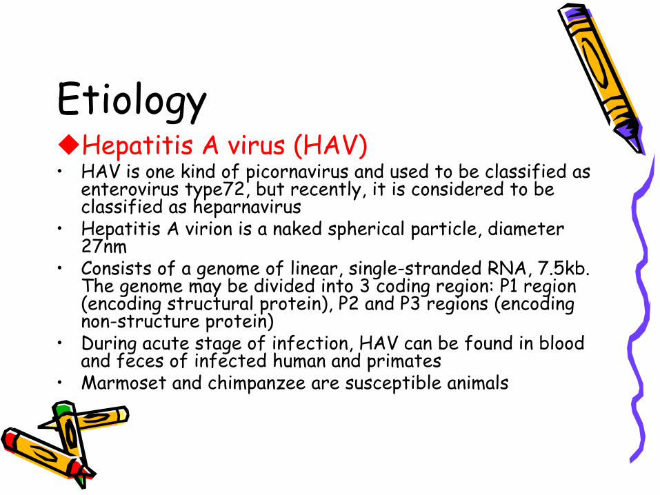
EtiologyHepatitis A virus (HAV)
• HAV is one kind of picornavirus and used to be classified as enterovirus type72, but recently, it is considered to be classified as heparnavirus
• Hepatitis A virion is a naked spherical particle, diameter 27nm
• Consists of a genome of linear, single-stranded RNA, 7.5kb. The genome may be divided into 3 coding region: P1 region (encoding structural protein), P2 and P3 regions (encoding non-structure protein)
• During acute stage of infection, HAV can be found in blood and feces of infected human and primates
• Marmoset and chimpanzee are susceptible animals

EtiologyHepatitis A virus (HAV)
• HAV can not cause cytopathy, replicate within cytoplasma of hepatocytes and via bill are discharged with feces
• 7 genetypes, 1, 2, 3, 7 types from humanbody• Only one antigen-antibody system. Anti-HAV IgM is
diagnostic evidence of recent infection, IgG is protective antibody.
• Resistance of HAV: 56°C, 30 min, usually temperature 1 week, dry feces at 25°C 30 days, fresh water, sea water ,shellfish or soil for several months. 70% alcohol at 25°C , 3 min, 100°C, 5 min and ultraviolet, 1 min
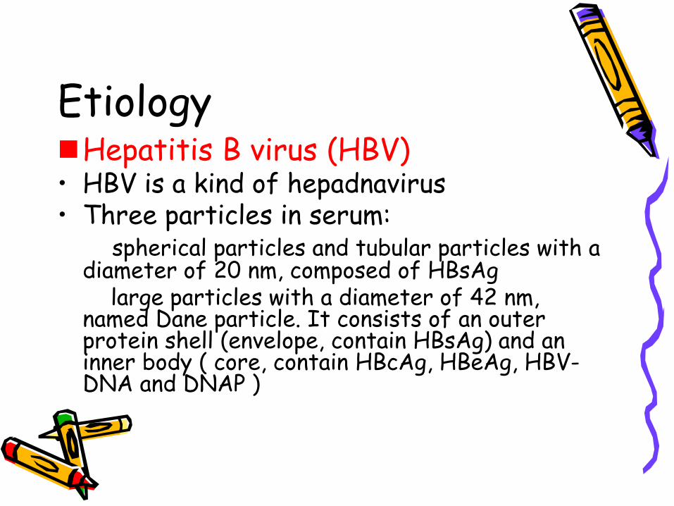
EtiologyHepatitis B virus (HBV)
• HBV is a kind of hepadnavirus• Three particles in serum:
spherical particles and tubular particles with a diameter of 20 nm, composed of HBsAg
large particles with a diameter of 42 nm, named Dane particle. It consists of an outer protein shell (envelope, contain HBsAg) and an inner body ( core, contain HBcAg, HBeAg, HBV-DNA and DNAP )
EtiologyHepatitis B virus (HBV)
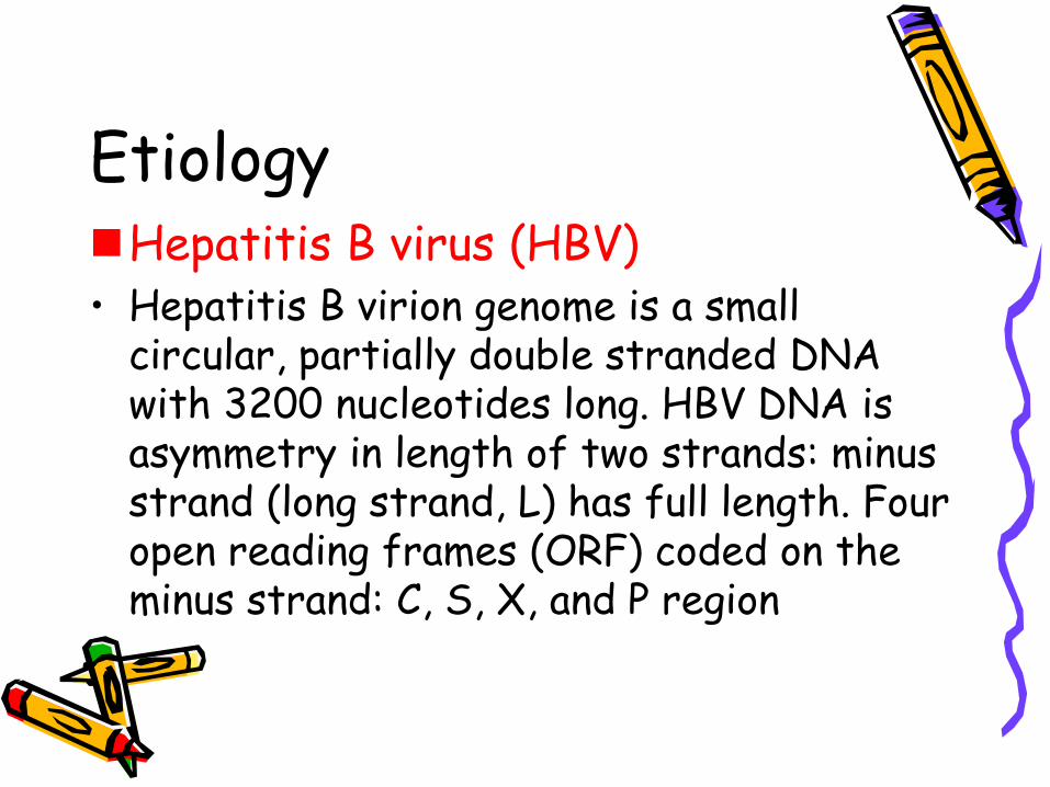
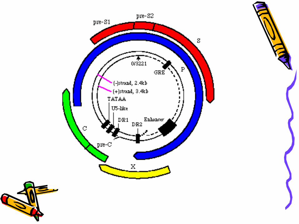
• Hepatitis B virion genome is a small circular, partially double stranded DNA with 3200 nucleotides long. HBV DNA is asymmetry in length of two strands: minus strand (long strand, L) has full length. Four open reading frames (ORF) coded on the minus strand: C, S, X, and P region

EtiologyHepatitis B virus (HBV)
• Four open reading frames (ORF) S region: include pre-s1, pre-s2 and S gene,
encoded pre-s1 protein, pre-s2 protein and HBsAg.Pre-s1 protein + pre-s2 protein + HBsAg—large proteinPre-s2 protein + HBsAg—middle protein HBsAg—major protein
C region included pre-c and C gene, encode HBeAg and HBcAg
X region encoded HBxAgP region encoded DNA polymerase
etiology• Three antigen-antibody system
HBsAg-- anti-HBs system:
Include HBsAg, anti-HBs, pre-s1,s2 antigen and anti-pres1, s2HBsAg appears 1-2 weeks (late to 11-12 weeks) after exposure, persists for 1-6 weeks( even 5 months) in acute hepatitis B. In chronic patients or carrier, HBsAg persist many yearsHBsAg antigencity but no infectivityHBsAg is the marker of infectivityHBsAg can be found in humors and secretions: salive, urine, semina, tears, sweat and breast milk10 subtype of HBsAg, 4 major subtypes: adr, adw, ayr, ayw. Anti-HBs appear after HBsAg disappear several weeks (or months) anti-HBs is protective antibody, can persist for many yearspre-s1 and pre-s2 antigens appear following HBsAg . They are the marker of infectivity. Anti –pre s2 has the action of clearing virus
Etiology• HBcAg—anti-HBc system
HBcAg can be found in the nuclei of liver cells, no free HBcAg in serumHBcAg is the marker of replication of HBVThe stage called window phaseAnti-HBc IgM is a marker of acute infection and acute attack of chronic infection of HBV. Anti-HBc IgG is the marker of past infection, high titer means low level replication of HBV

Etiology• HBeAg—anti-HBe system
HBeAg is a soliable antigenHBeAg is a reliable indicator of active replication of HBVAnti-HBe is a marker of reduced infectivity. If exist long may be a marker of integration of HBV into liver cell

Etiology• The marker of molecular biology of
HBV• HBV-DNA
The direct indicator of HBV infectionCan integrate into the genome of hepatocytes
• HBV DNA polymerasePossesses the ability of reverse
transcriptase and the indicator of the ability of replication of HBV
Etiology• HBxAg
Related to chronicity, activity of hepatitis B or liver cancer
• ResistanceResistant to heating and common disinfections. Chimpanzee is susceptible to HBV
EtiologyHepatitis C virus (HCV)
• HCV is a member of flavivirus family. • HCV genome is a single stranded positive-sense RNA and
contains 9.4kb• The genome contains 5’-non coding region, C region, E region
and NS region• HCV genome may be divided into many types and subtypes.• Resistance• Antigen-antibody system• The concentration of HCV in blood is low, HCV Ag has not be
detected, anti-HCV is the indicator of infection and the marker of infectivity
• HCV-RNAHCV-RNA may be detected from blood or liver tissue, it’s the direct evidence of infectivity
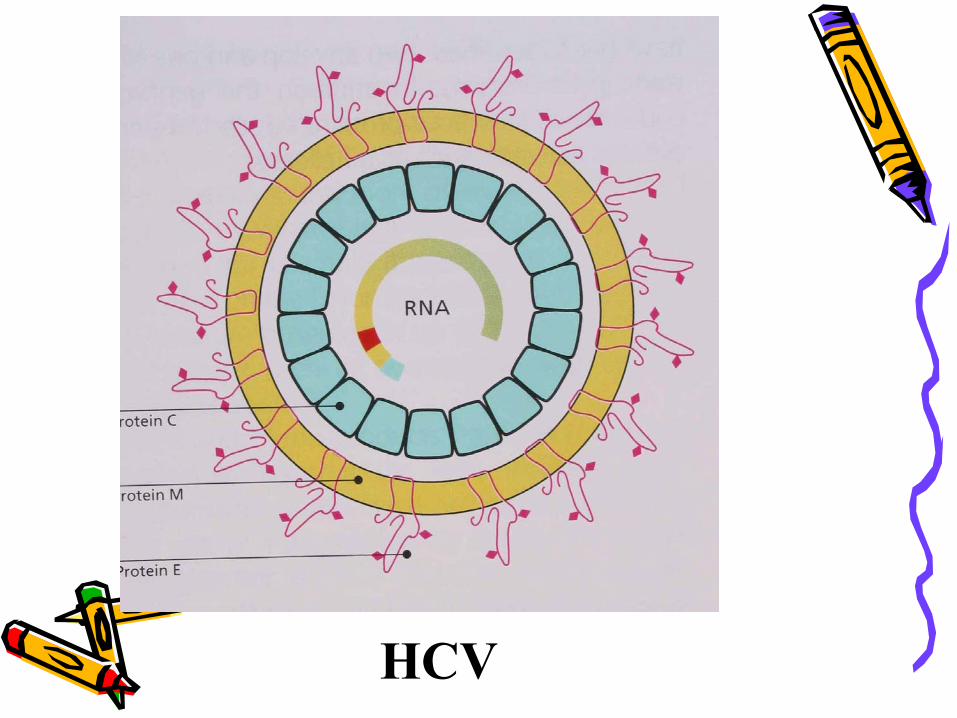
HCV
EtiologyHepatitis D virus (HDV)
• HDV (Delta hepatitis virus) is a kind of defective virus• HDV is found in the nuclei of infected hepatocytes and
replicate• HDV genome is a circular single strand RNA and contains
1.7kb• The replication of HDV depends on HBV or other
hepadnavirus, coated by HBsAg in blood• HDV has one antigen-antibody system
No free HDAg is detected in blood, it’s in the nuclei of hepatocytes; anti-HDV can be detected by RIA or ELISA in serum
• HBV and HDV co-infection or superinfection may make the disease exacerbation and may lead to fulminant hepatitis
• HDV RNA may be detected from liver cells, blood or humor.• Resistance
EtiologyHepatitis E virus (HEV)
• HEV is a member of calicivirus family.• HEV is a spherical nuenveloped icosahedral particles.• HEV genome is a single strand, positive –sense RNA (7.5kb),
include structure and non-structure regionThree ORF
ORF-1: encoding non-structure proteinORF-2: encoding neucleocapside proteinORF-3:encoding a part of neucleocapside protein
• 2 subtype of HEV founded : Burma subtype and Mexico subtype ; or epidemic strain and sporadic strain
• HEV replication within hepatocytes and via bill tract is discharged
• Monkeys and chimpanzee are susceptible to HEV• One antigen-antibody system
HGV, TTV
EpidemiologySource of infectionHepatitis A and E: patients with acute hepatitis and person with sublinical infectionHepatitis B, C and D: patients with acute, chronic hepatitis B, C, and D and carriersRoute of transmissionHepatitis A and E:
fecal-oral route predominantly

EpidemiologyRoute of transmissionHepatitis B, C, and D:
• humoral transmission (parenteral transmission)
• Mather to infent transmission(vertical transmission)
• Sexual contact transmission• Insect transmission
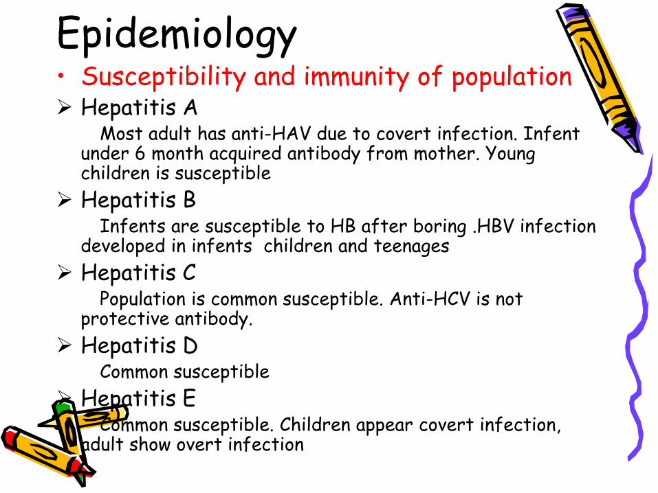
Epidemiology• Susceptibility and immunity of population
Hepatitis AMost adult has anti-HAV due to covert infection. Infent
under 6 month acquired antibody from mother. Young children is susceptibleHepatitis B
Infents are susceptible to HB after boring .HBV infection developed in infents children and teenagesHepatitis C
Population is common susceptible. Anti-HCV is not protective antibody.Hepatitis D
Common susceptibleHepatitis E
Common susceptible. Children appear covert infection, adult show overt infection
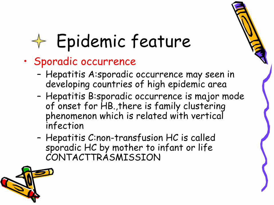
Epidemic feature• Sporadic occurrence
– Hepatitis A:sporadic occurrence may seen in developing countries of high epidemic area
– Hepatitis B:sporadic occurrence is major mode of onset for HB.,there is family clustering phenomenon which is related with vertical infection
– Hepatitis C:non-transfusion HC is called sporadic HC by mother to infant or life CONTACTTRASMISSION
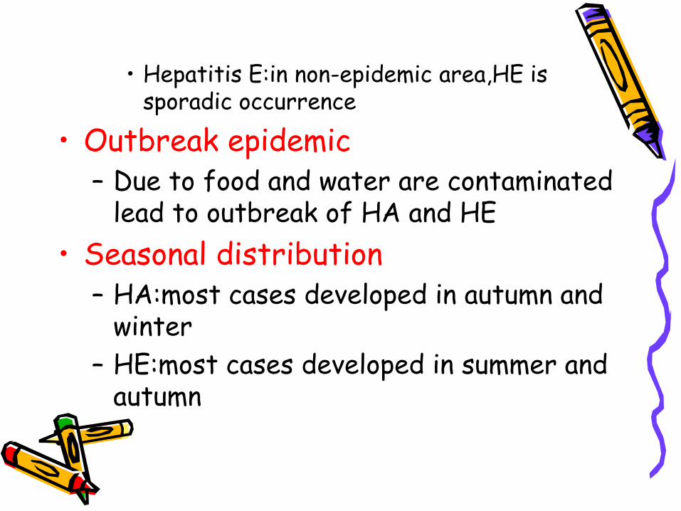
• Hepatitis E:in non-epidemic area,HE is sporadic occurrence
• Outbreak epidemic– Due to food and water are contaminated
lead to outbreak of HA and HE• Seasonal distribution
– HA:most cases developed in autumn and winter
– HE:most cases developed in summer and autumn
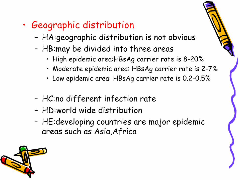
• Geographic distribution– HA:geographic distribution is not obvious– HB:may be divided into three areas
• High epidemic area:HBsAg carrier rate is 8-20%• Moderate epidemic area: HBsAg carrier rate is 2-7%• Low epidemic area: HBsAg carrier rate is 0.2-0.5%
– HC:no different infection rate– HD:world wide distribution– HE:developing countries are major epidemic
areas such as Asia,Africa

pathogenesis• Hepatitis A:
HAV invade into human body by mouth and cause viremia.After one week,the HAV reach liver cells replicate within.Then enter intestien with bill and appear in feces.someone believe that damage of liver cells maybe caused by immune response.Due to:– HAV does not cause cytopathy
– After HAV replicating and discharging,liver cells damage begin
– Animal experiment proved that immune complex may attend the pathogenesis of HA
– Complement level reduce the pathogenesis maybe following:activated T cell secrete γ-INF that promote the representation of HLA-Ⅰantigen on the liver cells,CTL may kill the target cell infected with HAV
• Hepatitis B:– HBV invade into the human body by skin
and mucosa,via blood flow enter the liver and other organs such as pancreas,bill duct,vessels,WBC,bone marrow,glomerular basement membrans
– HBcAg,HBsAg,HBeAg and HLA-Ⅰappear on the liver cells infected with are recognized by CTL simultaneously and lead to the cytolysis of liver cells
– Help T cell are activated by the receptor of HLA-Ⅱ on its surface combing with HBsAg, HBcAg and HLA-Ⅱ antigen on the B cells promote B cell to release anti-HBs and clear HBV
– The representation of HBcAg on the liver cells may cause cytopethy
– High degree representation of HBsAg within liver cells but the secretion is not enough lead to ground-glass-like change of liver cells
– Antigen-antibody complex precipitated on the wall of blood vessels and glomerular basement membrans causing nephritis or noduse polyarteritis,fever,rush and arthralgia called serum sick-like reaction
– TNF,IL-1,IL-6 may play roles in pathogenesis
– Chronicity of hepatitis B is related to immune tolerance, immune suppress and genetic factors
– HBV infection is related to HCC closely
• Hepatitis C – Is similar to that of HB. CTL and some
cytokines play an important action – The chronicity is related to the
variability of gene– HCV infection is related to HCC closely
but HCV does not integrate to liver cells, so from HCV infection to HCC may be related to chronic inflammation and cirrhosis
Pathology
– Degeneration– Necrosis– Regeneration– Infiltration of inflammatory cells– Hyperplasia of interstitial cells
• Acute viral hepatitis – The degeneration of liver cells include
ballooning degeneration,fatty degeneration,acidophilic degeneration
– Cell nucleus vacuolar degeneration– Focal or spotty necrosis and regeneration – The infiltration of mononuclear cell,
plasmocyte ,lymphocyte in portal area– Cholestasis and form of bile thrombas in bile
capillaries of liver– Piecemeal necrosis
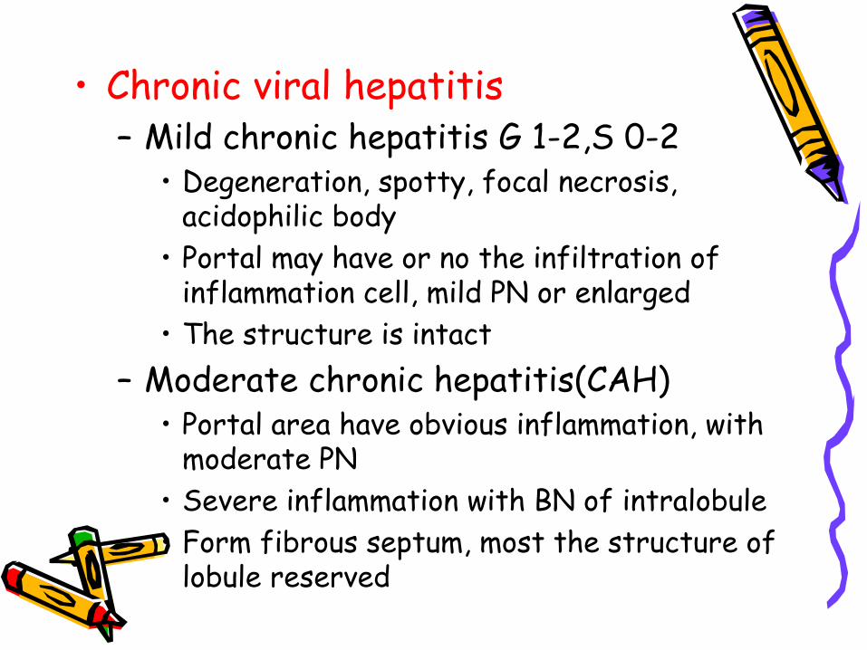
• Chronic viral hepatitis– Mild chronic hepatitis G 1-2,S 0-2
• Degeneration, spotty, focal necrosis, acidophilic body
• Portal may have or no the infiltration of inflammation cell, mild PN or enlarged
• The structure is intact– Moderate chronic hepatitis(CAH)
• Portal area have obvious inflammation, with moderate PN
• Severe inflammation with BN of intralobule • Form fibrous septum, most the structure of
lobule reserved

– Severe chronic hepatitis • Portal area has severe inflammation with severe PN• BN of extensive range involving several lobulus• Much more fibrous septums distortion of lobule
structure or form early liver cirrhosis
• Fulminant viral hepatitis(hepatitis gravis)– Acute hepatitis gravis: liver cells show massive
necrosis including great among of liver cells. Necrosis and reticular fiber network collapse, so the liver is greatly reduce in size-acute yellow hepatic etrophy
– Subacute hepatitis gravis• Except massive necrosis, there are ball-like
regeneration of liver cells • New connective tissue which form fibrous
band and separate the liver cells regenerated forming pseudolobuli
• Bile capillaries hyperplasia– Chronic hepatitis gravis: base on the
pathologic changes of chronic hepatitis or liver cirrhosis, there are massive or sub massive necrosis of liver cells
• Cholestatic viral hepatitis– There are the changes of acute
hepatitis – There is obvious cholestasis– In severe cases, the liver cells may
appear glandular duct like– Portal area shows edeme and small bile
duct is dilation
pathophysiology• Jaundice
the jaundice is mainly hepatocytic jaundice and part obstructive jaundice
• Hepatic encephalopathy– Retention of toxic material lead to poisoning of CNS– Imbalance of aminoacid– False neurotransmitter hypothesis– Other evoked factors

• Hemorrage Deficiency of many kinds of blood coagulating factors, DIC, thrombucytopenia lead to hemorrage
• Acute renal failure (hepatic-renal syndrome)
• Hepatopulmonary syndrome• Ascites
Clinical manifestation• Incubation period
– HA 15-45 days 30 days– HB 30-180 days 70 days– HC 15-150 days 50 days– HD similar to HB– HE 10-70 days 40 days
• Clinical types – Acute viral hepatitis
• Acute icteric viral hepatitis• Acute anicteric viral hepatitis
– Chronic viral hepatitis • Mild chronic viral hepatitis • Moderate chronic viral hepatitis • Severe chronic viral hepatitis
– Hepatitis gravis • Acute hepatitis gravis • Subacute hepatitis gravis• Chronic hepatitis gravis
– Cholestatic viral hepatitis• Acute viral hepatitis
– Acute icteric viral hepatitis the cause may be 2-4 months and divided three periods
• Preicteric period– In HA, HE, the onset is abrupt with fever; but HB, HC,
the onset is insidious. – The initial symptoms: loss of appetite, nausea, vomiting
lassitude, abdominal pain and diarrhea.– The end of the period, the urine darkens. A few
patients, especial children, fever, headache, upper respiratory tract symptome are main manifestations
– The duration of this period varies from 1-21 days, average 5-7 days
• Ictoric period– The urine deepens continuously and jaundice appears
on the skin and sclera within 2 weeks– Subjective symptoms is abate– Pruritus may appear about 1 week – Liver palpable 7%, spleen palpable 20%– The period lasts 2-6 weeks
• Convalscent period– The jaundice disappear gradually, symptoms abate or
disappear– Liver and spleen retract, liver function return to
normal – The period lasts 2 weeks to 4 months, average 1 month– About 10% of HB and 50% of HC will become chronic
hepatitis – Acute hepatitis D:
» Co-infection with HBV» Super-infection with HBV
– Acute hepatitis E is similar to acute hepatitis A, but cholestasis is obvious and symptoms and signs is severe.
– If women with pregnancy suffer from the HE--fulminant hepatitis
– If HB super-infect HEV or HCV--fulminant hepatitis
– Acute anicteric hepatitis • All of 5 kinds of hepatitis virus can cause
acute anicteric hepatitis. This type is most common.
• Important source of infection
• Chronic viral hepatitis Only appear in HBV, HCV and HDV infection – Mild chronic hepatitis
• The course is more than half year • Fatigue, dizziness, digestive tract symptoms,
dull pain of liver, enlarged liver tenderness or spleen tenderness,lower degree of fever, ALT↑, the pathology change has only mild
• The course may persist many years

– Moderate • The course →half year • The symptom are obvious • Spider nevus, liver palms, hepatic face • Dysfunction of liver • Accompany the lesions of other organs and
presence of autoantibody • Reverse the ratio of albumin/globulin • Biopsy show the changes of mild CAH
– Severe • Except symptoms and signs mentioned, the
biopsy show the changes of early cirrhosis and clinical manifestations of compensatory cirrhosis
• Hepatitis gravisAll of five kinds of hepatitis virus can cause this type of hepatitis. The incidence is only 0.2-0.5%, but the mortality is the highest. – Acute hepatitis gravis
• The onset may begin in a typical acute icteric hepatitis, but within 10 days
• Jaundice deepens rapidly• Vomit is frequent • Obvious anorexia • Hemorrhage• The liver shrinks in size• Toxic intestinal tympenice • Prothrombin time is prolonged • Ascites appear• Acute renal failure • Hepatic encephalopathy
– Subacute hepatitis gravis• The course of AIH is more than 10 days • The hepatic encephalupathy appear later • The course may be several months • The postnecrotic cirrhosis may develop
– Chronic hepatitis gravis • Based on chronic hepatitis or cirrhosis developed
subacute hepatic necrotic
• Cholestatic hepatitis– Intra hepatic cholestatic jaundice for a long
time(2-4 months or longer)– Pruritus– Pale feces – Hepatomogaly
– Subjective symptoms is slight – Course 2-6 months – Recovery is complete
• Manifostations of hepatitis for special population– Characteristics of hepatitis for child – Characteristics of hepatitis for the senility– The character of hepatitis of pregnancy period
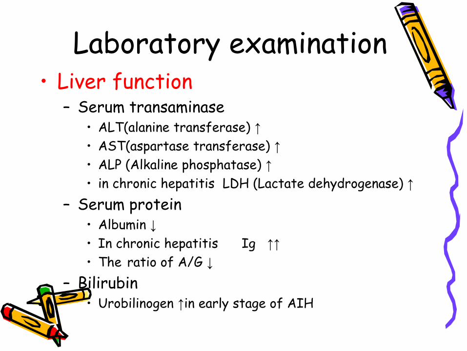
Laboratory examination• Liver function
– Serum transaminase • ALT(alanine transferase) ↑• AST(aspartase transferase) ↑• ALP (Alkaline phosphatase) ↑• in chronic hepatitis LDH (Lactate dehydrogenase) ↑
– Serum protein • Albumin ↓• In chronic hepatitis Ig ↑↑• The ratio of A/G ↓
– Bilirubin • Urobilinogen ↑in early stage of AIH
• Urobilinogen and urobilin ↑in icteric stage • Urobilin is positive and urobilinogen may be negative
in cholestatic hepatitis • In AIH, the directive bilirubin and indirective
bilirubin ↑– Prothrombin time may be prolonged especially
in fulminant hepatitis – Blood amonia examination
• Detection of the markers of hepatitis virus – Hepatitis A
• Serologic marker – Anti-HAVIgM: recent infection – Anti-HAVIgG: past infection
• Marker of feces – HAV particles may be detected by RIA or IEM– Isolation of HAV may use tissue culture or animal
inoculation
– Hepatitis B• Sero-immunologic marker
– HBsAg anti-HBs– HBcAg anti-HBc– HBeAg anti-Hbe
• Molecular biological marker – DNAp– HBV DNA– Immune tissue chemistry examination
– Hepatitis C• Serological marker
– Anti-HCVIgM– Anti-HCVIgG
• Molecular biologic marker– HCV RNA may be detective by RT-PCR 1-2 weeks after
infection of HCV– Quality of HCV RNA– Immune tissue chemistry method detect HCAg within
liver cells
– Hepatitis D• HDAg anti-HDV• HDV RNA
– Hepatitis E• Anti-HEVIgG,Anti-HEVIgm• RT-PCR• HEV particais: IF IEM
• Ultra-sound examination • Liver biopsy • Other laboratory examination
–Blood routine –Urine routine
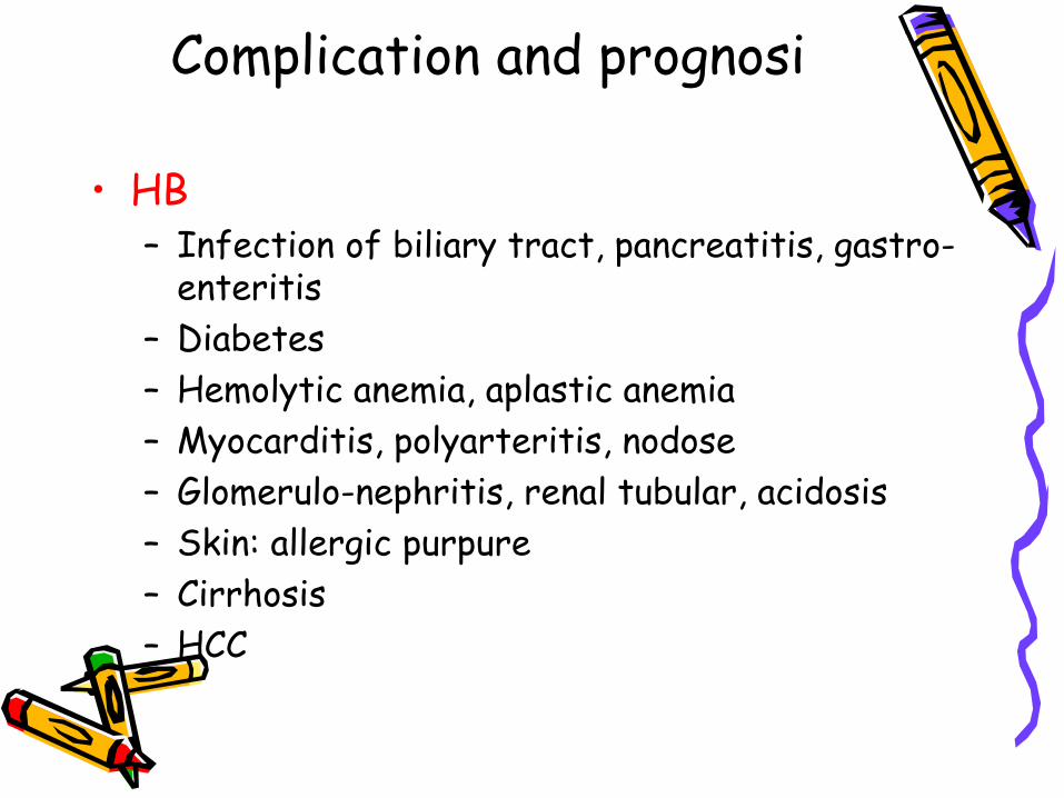
Complication and prognosi
• HB– Infection of biliary tract, pancreatitis, gastro-
enteritis– Diabetes – Hemolytic anemia, aplastic anemia – Myocarditis, polyarteritis, nodose– Glomerulo-nephritis, renal tubular, acidosis– Skin: allergic purpure– Cirrhosis– HCC

Diagnosis
• Epidemiological data– HA, HE: food, water, seasonal, age– HB, HC:
blood and blood product transfusion, contact history, inoculation history
• Clinical diagnosis– Acute hepatitis – Chronic hepatitis
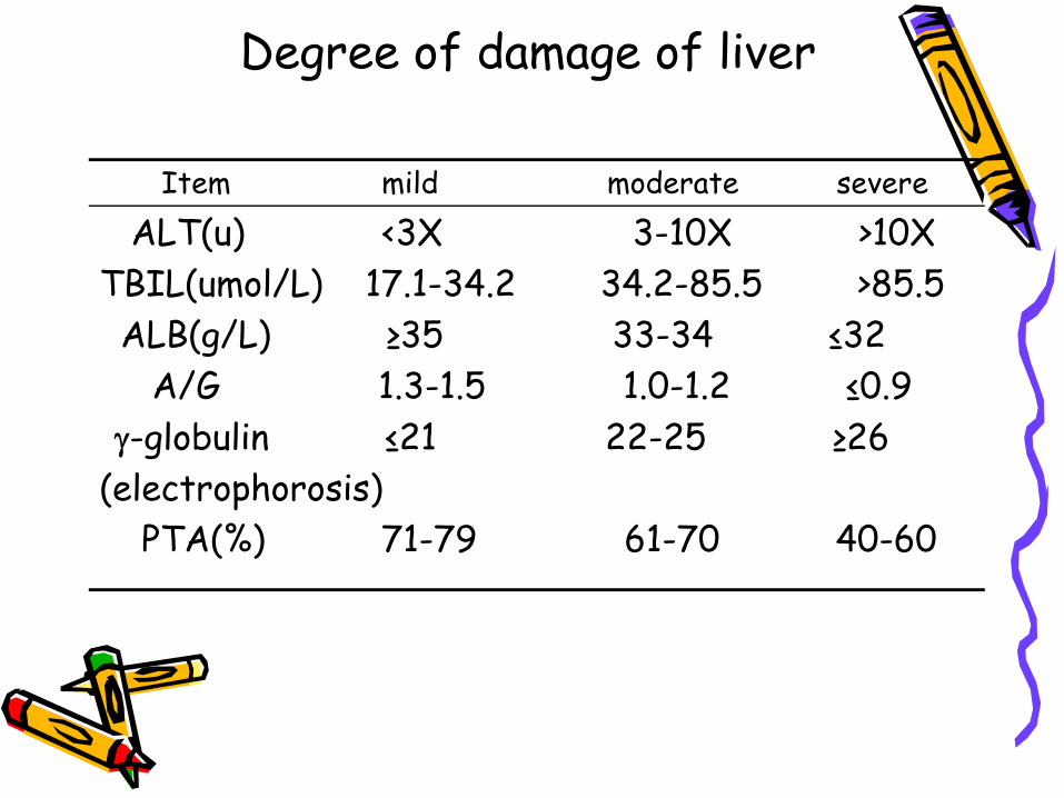
Degree of damage of liver
Item mild moderate severeALT(u) <3X 3-10X >10X
TBIL(umol/L) 17.1-34.2 34.2-85.5 >85.5ALB(g/L) ≥35 33-34 ≤32
A/G 1.3-1.5 1.0-1.2 ≤0.9γ-globulin ≤21 22-25 ≥26
(electrophorosis)PTA(%) 71-79 61-70 40-60

– Hepatitis gravis • Etiological diagnosis
– HA: Serum anti-HAVIgM , Feces HAV
– HB: HBsAg HBeAg HBcAg anti-HBc HBVDNA DANp
– HC: anti-HCV IgM IgG
– HD: HBsAg HDAg anti-HDV
– HE: anti-HEV IgM IgG HEVRNA HEV particals in feces

Differential diagnosis• Acute icteric hepatitis
– Must be differentiated with the jaundice caused by another disease
• Hemolytic jaundice • Extrahepatic obstructive jaundice
– Hepatitis caused by another reasons • Toxic hepatitis • Infective toxic hepatitis • Mononucleosis • Alcohol hepatic disease • Schistosomiosis • Wilson disease

Treatment • Acute hepatitis
– Isolation• HA: 3 weeks after onset• HB: HBsAg become negative • HC: HCVRNA become negative • HE: 2 weeks after onset
– Rest – Diet– Anti-virus therapy– hepatinice

• Chronic hepatitis– Symptomatic therapy– Diet– Rest – Hepatinice– Supporting therapy– Immunomodulator
• Fuminant hepatitis– General and supporting therapy
• Rest: strict bed rest • diet

• Supporting therapy
– Symptomatic therapy • Hemorrhage: fresh blood, prothrombin complex,
platelet • Hepatic encephalopathy
– Prevention and treatment of amino poisoning– Recovery normal neurotransmitter – Sodium glutamate and arginine – Treatment of cerebral edema – Control infection – Prevention and treatment of renal failure – Promoting the growth of liver cells
– Liver transplantation • Cholestatic hepatitis:
similar to acute hepatitis
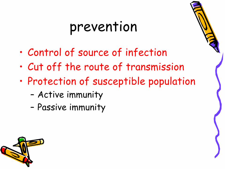
prevention
• Control of source of infection • Cut off the route of transmission• Protection of susceptible population
– Active immunity– Passive immunity