Shrimp viral diseases in India and prospects of viral vaccines
Upload
khangminh22Category
view
0download
0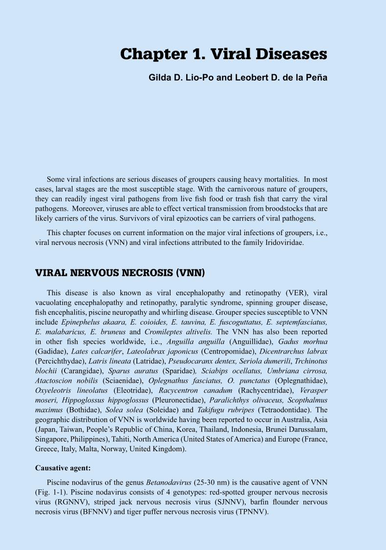
Diseases
ofC
ultu
redG
roupers
333
Some viral infections are serious diseases of groupers causing heavy mortalities. In most cases, larval stages are the most susceptible stage. With the carnivorous nature of groupers, they can readily ingest viral pathogens from live fish food or trash fish that carry the viral pathogens. Moreover, viruses are able to effect vertical transmission from broodstocks that are likely carriers of the virus. Survivors of viral epizootics can be carriers of viral pathogens.
This chapter focuses on current information on the major viral infections of groupers, i.e., viral nervous necrosis (VNN) and viral infections attributed to the family Iridoviridae.
VIRAL NERVOUS NECROSIS (VNN)
This disease is also known as viral encephalopathy and retinopathy (VER), viral vacuolating encephalopathy and retinopathy, paralytic syndrome, spinning grouper disease, fish encephalitis, piscine neuropathy and whirling disease. Grouper species susceptible to VNN include Epinephelus akaara, E. coioides, E. tauvina, E. fuscoguttatus, E. septemfasciatus, E. malabaricus, E. bruneus and Cromileptes altivelis. The VNN has also been reported in other fish species worldwide, i.e., Anguilla anguilla (Anguillidae), Gadus morhua (Gadidae), Lates calcarifer, Lateolabrax japonicus (Centropomidae), Dicentrarchus labrax (Percichthydae), Latris lineata (Latridae), Pseudocaranx dentex, Seriola dumerili, Trchinotus blochii (Carangidae), Sparus auratus (Sparidae), Sciabips ocellatus, Umbriana cirrosa, Atactoscion nobilis (Sciaenidae), Oplegnathus fasciatus, O. punctatus (Oplegnathidae), Oxyeleotris lineolatus (Eleotridae), Racycentron canadum (Rachycentridae), Verasper moseri, Hippoglossus hippoglossus (Pleuronectidae), Paralichthys olivaceus, Scopthalmus maximus (Bothidae), Solea solea (Soleidae) and Takifugu rubripes (Tetraodontidae). The geographic distribution of VNN is worldwide having been reported to occur in Australia, Asia (Japan, Taiwan, People’s Republic of China, Korea, Thailand, Indonesia, Brunei Darussalam, Singapore, Philippines), Tahiti, North America (United States of America) and Europe (France, Greece, Italy, Malta, Norway, United Kingdom).
Causative agent:
Piscine nodavirus of the genus Betanodavirus (25-30 nm) is the causative agent of VNN (Fig. 1-1). Piscine nodavirus consists of 4 genotypes: red-spotted grouper nervous necrosis virus (RGNNV), striped jack nervous necrosis virus (SJNNV), barfin flounder nervous necrosis virus (BFNNV) and tiger puffer nervous necrosis virus (TPNNV).
Chapter 1. Viral DiseasesGilda D. Lio-Po and Leobert D. de la Peña
4
Dis
ease
sof
Cu
ltu
red
Gro
uper
s Diseases
ofC
ultu
redG
roupers
555
Stages affected:
All growth stages are affected but heavy mortalities were reported among larvae less than 20 days old.
Gross clinical signs:
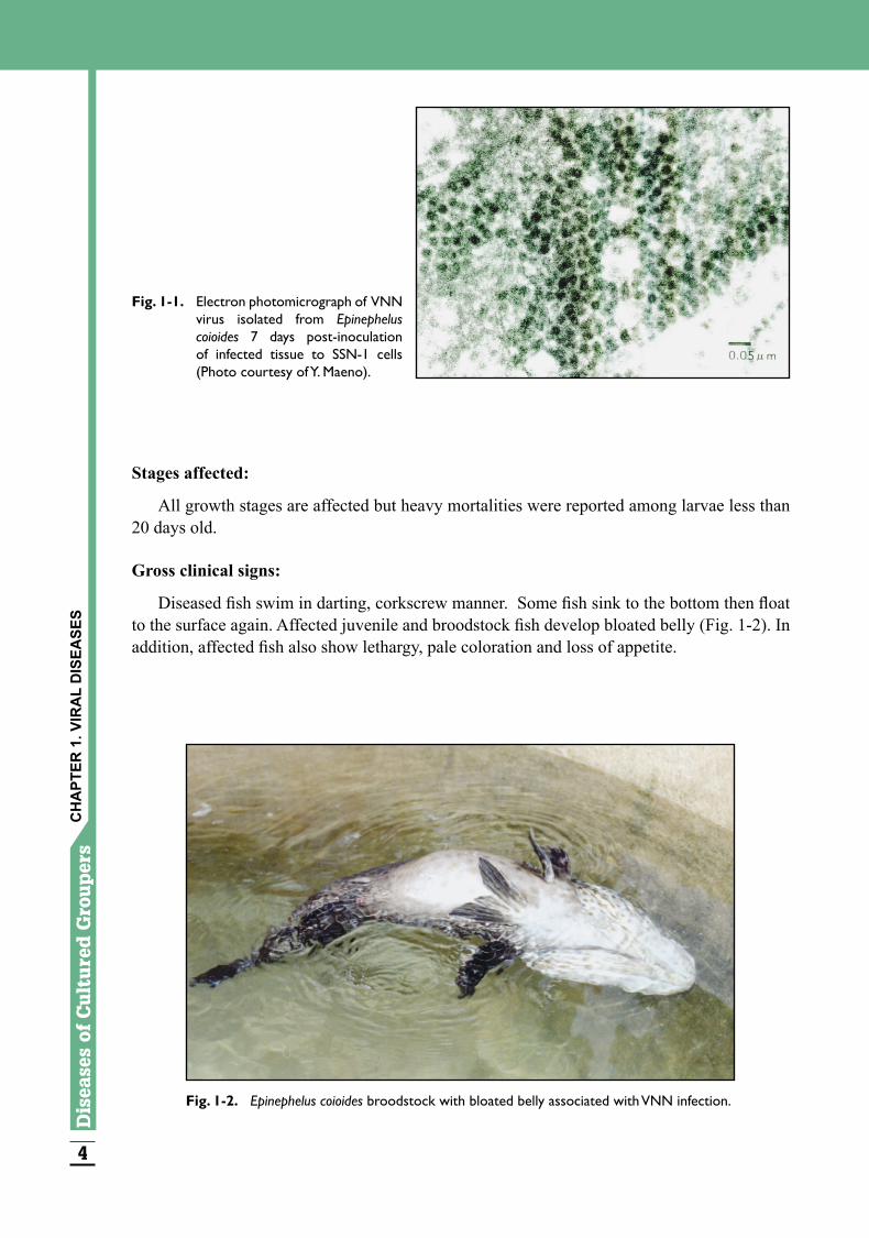
Diseased fish swim in darting, corkscrew manner. Some fish sink to the bottom then float to the surface again. Affected juvenile and broodstock fish develop bloated belly (Fig. 1-2). In addition, affected fish also show lethargy, pale coloration and loss of appetite.
Fig. 1-1. Electron photomicrograph of VNN virus isolated from Epinephelus coioides 7 days post-inoculation of infected tissue to SSN-1 cells (Photo courtesy of Y. Maeno).
Fig. 1-2. Epinephelus coioides broodstock with bloated belly associated with VNN infection.
CH
APT
ER 1
. VIR
AL
DIS
EASE
S
4
Dis
ease
sof
Cu
ltu
red
Gro
uper
s Diseases
ofC
ultu
redG
roupers
555
Effects on host:
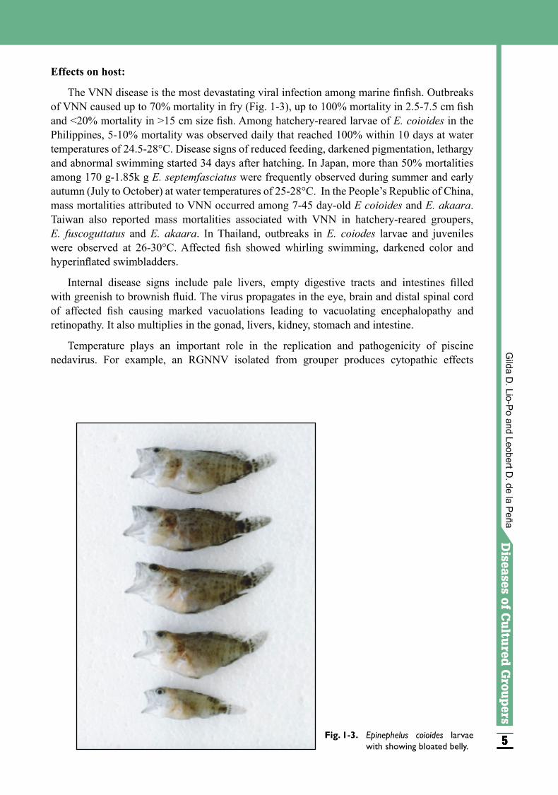
The VNN disease is the most devastating viral infection among marine finfish. Outbreaks of VNN caused up to 70% mortality in fry (Fig. 1-3), up to 100% mortality in 2.5-7.5 cm fish and <20% mortality in >15 cm size fish. Among hatchery-reared larvae of E. coioides in the Philippines, 5-10% mortality was observed daily that reached 100% within 10 days at water temperatures of 24.5-28°C. Disease signs of reduced feeding, darkened pigmentation, lethargy and abnormal swimming started 34 days after hatching. In Japan, more than 50% mortalities among 170 g-1.85k g E. septemfasciatus were frequently observed during summer and early autumn (July to October) at water temperatures of 25-28°C. In the People’s Republic of China, mass mortalities attributed to VNN occurred among 7-45 day-old E coioides and E. akaara. Taiwan also reported mass mortalities associated with VNN in hatchery-reared groupers, E. fuscoguttatus and E. akaara. In Thailand, outbreaks in E. coiodes larvae and juveniles were observed at 26-30°C. Affected fish showed whirling swimming, darkened color and hyperinflated swimbladders.
Internal disease signs include pale livers, empty digestive tracts and intestines filled with greenish to brownish fluid. The virus propagates in the eye, brain and distal spinal cord of affected fish causing marked vacuolations leading to vacuolating encephalopathy and retinopathy. It also multiplies in the gonad, livers, kidney, stomach and intestine.
Temperature plays an important role in the replication and pathogenicity of piscine nedavirus. For example, an RGNNV isolated from grouper produces cytopathic effects
Fig. 1-3. Epinephelus coioides larvae with showing bloated belly.
Gilda D
. Lio-Po and Leobert D
. de la Peña
6
Dis
ease
sof
Cu
ltu
red
Gro
uper
s Diseases
ofC
ultu
redG
roupers
777
(CPE) in GF-1 cells at 24-32°C but not at 20°C or 37°C. In larvae challenged with RGNNV at 28°C, mortality reached 100% 50-80 hours post-inoculation. In infection experiments on E. akaara juveniles, RGNNV caused 100% mortalities of the test fish at 24-28°C. At 16 and 20°C, mortalities were reduced to 57-61% and onset of abnormal swimming and deaths were delayed. In addition, the virus antigen was detected among survivors 50 days post-infection at 16°C.
Transmission:
The virus can be waterborne-transmitted from diseased to healthy fish within 4 days of contact. Nodaviruses were, likewise, detected in fish without clinical disease signs. As such, grouper broodstocks can be virus reservoirs and can therefore be a source of virus for its larvae.
Diagnosis:
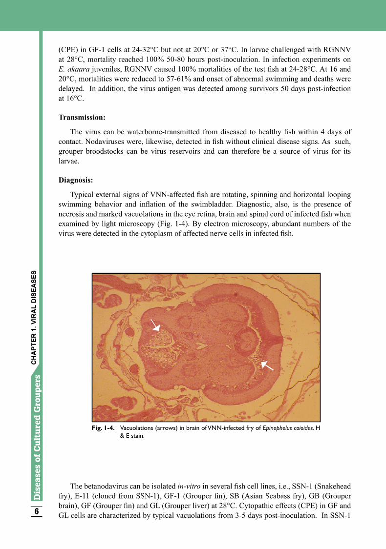
Typical external signs of VNN-affected fish are rotating, spinning and horizontal looping swimming behavior and inflation of the swimbladder. Diagnostic, also, is the presence of necrosis and marked vacuolations in the eye retina, brain and spinal cord of infected fish when examined by light microscopy (Fig. 1-4). By electron microscopy, abundant numbers of the virus were detected in the cytoplasm of affected nerve cells in infected fish.
The betanodavirus can be isolated in-vitro in several fish cell lines, i.e., SSN-1 (Snakehead fry), E-11 (cloned from SSN-1), GF-1 (Grouper fin), SB (Asian Seabass fry), GB (Grouper brain), GF (Grouper fin) and GL (Grouper liver) at 28°C. Cytopathic effects (CPE) in GF and GL cells are characterized by typical vacuolations from 3-5 days post-inoculation. In SSN-1
CH
APT
ER 1
. VIR
AL
DIS
EASE
S
Fig. 1-4. Vacuolations (arrows) in brain of VNN-infected fry of Epinephelus coioides. H & E stain.
6
Dis
ease
sof
Cu
ltu
red
Gro
uper
s Diseases
ofC
ultu
redG
roupers
777
cells, CPE is detected approximately 5-10 days post-inoculation. In addition, virus-neutralizing antibodies in fish serum can be assayed using susceptible cell lines.
The virus can, also, be detected by immunological methods like indirect fluorescent antibody technique (IFAT) and enzyme-linked immunosorbent assay (ELISA) using rabbit anti-VNN serum.
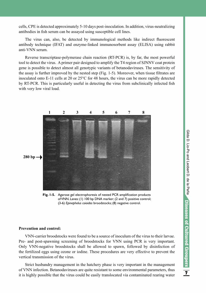
Reverse transcriptase-polymerase chain reaction (RT-PCR) is, by far, the most powerful tool to detect the virus. A primer pair designed to amplify the T4 region of SJNNV coat protein gene is possible to detect almost all genotypic variants of betanodaviruses. The sensitivity of the assay is further improved by the nested step (Fig. 1-5). Moreover, when tissue filtrates are inoculated onto E-11 cells at 20 or 25°C for 48 hours, the virus can be more rapidly detected by RT-PCR. This is particularly useful in detecting the virus from subclinically infected fish with very low viral load.
1 2 3 4 5 6 7 8
Fig. 1-5. Agarose gel electrophoresis of nested PCR amplification products of VNN. Lanes: (1) 100 bp DNA marker; (2 and 7) positive control; (3-6) Epinephelus coioides broodstocks; (8) negative control.
280 bp
Prevention and control:
VNN-carrier broodstocks were found to be a source of inoculum of the virus to their larvae. Pre- and post-spawning screening of broodstocks for VNN using PCR is very important. Only VNN-negative broodstocks shall be allowed to spawn, followed by disinfection of the fertilized eggs using ozone or iodine. These procedures are very effective to prevent the vertical transmission of the virus.
Strict husbandry management in the hatchery phase is very important in the management of VNN infection. Betanodaviruses are quite resistant to some environmental parameters, thus it is highly possible that the virus could be easily translocated via contaminated rearing water
Gilda D
. Lio-Po and Leobert D
. de la Peña
8
Dis
ease
sof
Cu
ltu
red
Gro
uper
s Diseases
ofC
ultu
redG
roupers
999
and paraphernalia. In Australian barramundi hatchery, the use of non-recycled, chemically treated rearing water and decontamination of tanks after every hatching cycle were effective in preventing VNN infection. Other measures in the control of VNN in larval striped jack are: disinfection of eggs with iodine or ozone and hatchery paraphernalia with chlorine; rearing of each batch of larvae and juveniles in separate tanks supplied with UV or ozone-sterilized seawater; and separation of larvae and juveniles from broodfish.
Vaccination is a promising method of preventing VNN in groupers. Immunization of groupers with recombinant coat proteins prepared from RGNNV genotype strains induced virus-neutralizing antibodies that resulted in high protection against experimental infection of the virus. However, a multivalent vaccine may be needed for total protection from infection by different genotypic variants of piscine nodavirus.
In addition, reduction of identified stress factors in the culture system is very important. Piscine nadavirus-free broodfish should be induced to spawn up to 7-10 times only since the stress of multiple spawnings may activate the residual virus. Reduction of larval stocking density in the tank may help reduce the possibility of viral transmission. Rearing water temperature influenced disease development wherein higher mortality and earlier appearance of the disease signs were observed at higher rearing water temperatures. This suggests that manipulation of water temperature will help reduce disease outbreaks.
IRIDOVIRUS INFECTIONS
Iridoviruses are a group of emerging viral pathogens causing infections in many marine finfish, e.g., Pagrus major, Evynnis japonica, Oplegnathus punctatus, O. fasciatus, Latelabrax sp., Seriola quinqueradiata, S. dumerili, S. aureovittata, Caranx delicatissimus, Takifugu rubripes, Epinephelus akaara, E. malabaricus and E. tauvina. The International Committee on Taxonomy of Viruses classifies the family Iridoviridae into four genera: Iridovirus, Chloriridovirus, Ranavirus and Lymphocystivirus. Grouper infections attributed to iridoviruses have been invariably identified as Fish Lymphocystis Disease (FLD), Blister Disease, Red Seabream Iridovirus Disease (RSIVD), Sleepy Grouper Disease (SGD), Grouper Iridovirus Disease of Taiwan (TGIVD), Grouper Iridovirus Disease (GIVD), Grouper Spawner Iridovirus Disease (GSIVD) and Singapore Grouper Iridovirus Disease (SGIVD). Except for FLD, the other iridovirus pathogens cause systemic infections in fish. However, the exact relationship among these systemic piscine iridoviruses is not clear. Hence, these diseases are presented separately.
1. Fish Lymphocystis Disease (FLD)
Lymphocystis is a chronic viral infection that occurs among finfish worldwide over a wide range of water temperatures including tropical climates. Among groupers, this disease was reported in E. bruneus, E. malabaricus and E. chlorostigma cultured in marine net cages in Guangdong, China, and also among E. fuscoguttatus in Malaysia.
Causative agent:
Fish Lymphocystis Disease virus (FLDV) is an iridovirus measuring 130-330 nm.
CH
APT
ER 1
. VIR
AL
DIS
EASE
S
8
Dis
ease
sof
Cu
ltu
red
Gro
uper
s Diseases
ofC
ultu
redG
roupers
999
Stages affected:
Fingerlings, juveniles and adult fish are affected.
Gross clinical signs:
Infected fish develop small (0.5-2 mm in diameter) pearl-like nodules occurring singly or in clusters on the body surface, fins and occasionally on the gills. The apparent nodules are a consequence of the enlargement of tissue cells infected by the virus. These cells are hypertrophied fibroblasts or the so-called Lymphocystis giant cells.
Effects on host:
Fish exhibiting the clinical signs of FLD have decreased market value. It is a chronic infection that can cause low mortality and is rarely fatal in older fish.
Transmission:
The viral pathogen is released after rupture of the nodules. The virus is thus transmitted from diseased to healthy fish through exposure to contaminated water via abraded skin or by cohabitation.
Diagnosis:
The disease can be readily diagnosed on the basis of the presence of the characteristic external nodules. The appearance of the Lymphocystis giant cells upon histological examination of sections of nodules is confirmatory for FLD.
2. Blister Disease
Causative agent:
The causative agent of blister disease is an iridovirus measuring 140-160 nm.
Stages affected:
The disease has been observed in 5-100 g Epinephelus malabaricus in Thailand.
Gross clinical signs:
Affected fish manifests signs of loss of appetite initially and whitish blister development on the body and fins.
Effects on host:
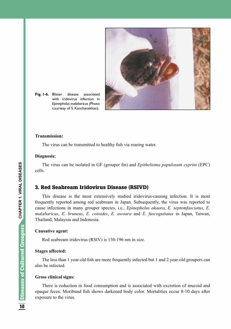
Fish develop localized, severe inflammation of the epidermal and dermal layer (Fig. 1-6). Necrotic dermis shows the presence of exudates and hemorrhagic infiltration. The virus is seen in the liver, spleen, kidney and lesions of infected fish. Natural infection results in 30-80% mortality within one month. Pathogenicity experiment showed that clinical signs and mortality occur within 5 days after exposure to the virus, and can reach 100% within 10 days. High stocking density can predispose susceptible fish to this disease.
Gilda D
. Lio-Po and Leobert D
. de la Peña
10
Dis
ease
sof
Cu
ltu
red
Gro
uper
s Diseases
ofC
ultu
redG
roupers
111111
Transmission:
The virus can be transmitted to healthy fish via rearing water.
Diagnosis:
The virus can be isolated in GF (grouper fin) and Epithelioma papulosum cyprini (EPC) cells.
3. Red Seabream Iridovirus Disease (RSIVD)
This disease is the most extensively studied iridovirus-causing infection. It is most frequently reported among red seabream in Japan. Subsequently, the virus was reported to cause infections in many grouper species, i.e., Epinephelus akaara, E. septemfasciatus, E. malabaricus, E. bruneus, E. coioides, E. awoara and E. fuscoguttatus in Japan, Taiwan, Thailand, Malaysia and Indonesia.
Causative agent:
Red seabream iridovirus (RSIV) is 130-196 nm in size.
Stages affected:
The less than 1 year-old fish are more frequently infected but 1 and 2 year-old groupers can also be infected.
Gross clinical signs:
There is reduction in food consumption and is associated with excretion of mucoid and opaque feces. Moribund fish shows darkened body color. Mortalities occur 8-10 days after exposure to the virus.
Fig. 1-6. Blister disease associated with iridovirus infection in Epinephelus malabaricus (Photo courtesy of S. Kanchanakhan).
CH
APT
ER 1
. VIR
AL
DIS
EASE
S
10
Dis
ease
sof
Cu
ltu
red
Gro
uper
s Diseases
ofC
ultu
redG
roupers
111111
Effects on host:
Experimental studies have shown that the RSIV multiply initially in the cytoplasm of cells of the spleen and head kidney of E. malabaricus as early as 2 days post-infection. This may result in diffused necrosis of the spleen that may be associated with hemorrhage. Subsequently, the virus was detected in the kidney and liver by the 6th day post-inoculation. The number of cells infected with the virus in the small sized grouper, 7.7 g, was higher than in the large test fish weighing 84.3 g. Eosinophilic, degenerated cells and basophilic, enlarged cells were seen in the spleen and head kidney of dead infected fish. In contrast, surviving fish had granulated eosinophilic cell debris but no enlarged cells were observed. The virus eventually infiltrates the blood vessels of the spleen and head kidney, causing degeneration of these organs. The virus also migrates to the liver, heart, gills, esophagus, stomach and intestine without inducing degenerative changes.
Intraperitoneal injection of E. malabaricus, 7.7 g and 84.3 g in size with the RSIV, exhibited mortalities in 8-11 days and 8-10 days, respectively, post-exposure. Cumulative mortalities in both sizes of challenged fish were 90%.
Transmission:
The viruses are contained in inclusion bodies, within the infected cells and are rapidly released upon lyses of the cell. Since the gills and the intestine have direct contact with the environment of the fish, transmission of the virus among fishes in the same culture system can readily occur. However, there is no indication that RSIV is transmitted vertically. The RSIVD reported in Malaysia was introduced through fish imported from Taiwan.
Diagnosis:
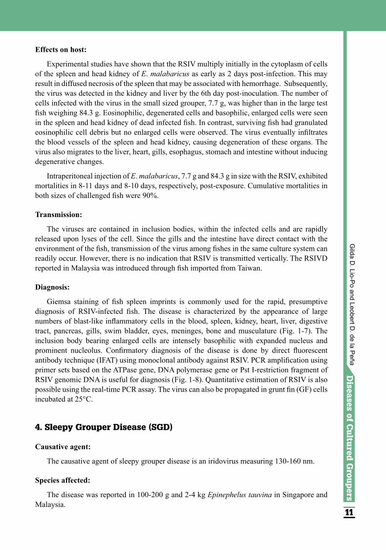
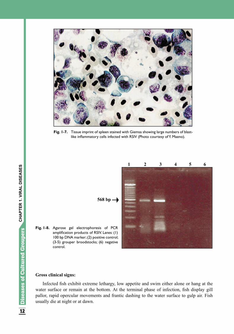
Giemsa staining of fish spleen imprints is commonly used for the rapid, presumptive diagnosis of RSIV-infected fish. The disease is characterized by the appearance of large numbers of blast-like inflammatory cells in the blood, spleen, kidney, heart, liver, digestive tract, pancreas, gills, swim bladder, eyes, meninges, bone and musculature (Fig. 1-7). The inclusion body bearing enlarged cells are intensely basophilic with expanded nucleus and prominent nucleolus. Confirmatory diagnosis of the disease is done by direct fluorescent antibody technique (IFAT) using monoclonal antibody against RSIV. PCR amplification using primer sets based on the ATPase gene, DNA polymerase gene or Pst I-restriction fragment of RSIV genomic DNA is useful for diagnosis (Fig. 1-8). Quantitative estimation of RSIV is also possible using the real-time PCR assay. The virus can also be propagated in grunt fin (GF) cells incubated at 25°C.
4. Sleepy Grouper Disease (SGD)
Causative agent:
The causative agent of sleepy grouper disease is an iridovirus measuring 130-160 nm.
Species affected:
The disease was reported in 100-200 g and 2-4 kg Epinephelus tauvina in Singapore and Malaysia.
Gilda D
. Lio-Po and Leobert D
. de la Peña
12
Dis
ease
sof
Cu
ltu
red
Gro
uper
s Diseases
ofC
ultu
redG
roupers
131313
Gross clinical signs:
Infected fish exhibit extreme lethargy, low appetite and swim either alone or hang at the water surface or remain at the bottom. At the terminal phase of infection, fish display gill pallor, rapid opercular movements and frantic dashing to the water surface to gulp air. Fish usually die at night or at dawn.
Fig. 1-7. Tissue imprint of spleen stained with Giemsa showing large numbers of blast-like inflammatory cells infected with RSIV (Photo courtesy of Y. Maeno).
1 2 3 4 5 6
568 bp
Fig. 1-8. Agarose gel electrophoresis of PCR amplification products of RSIV. Lanes: (1) 100 bp DNA marker; (2) positive control; (3-5) grouper broodstocks; (6) negative control.
CH
APT
ER 1
. VIR
AL
DIS
EASE
S
12
Dis
ease
sof
Cu
ltu
red
Gro
uper
s Diseases
ofC
ultu
redG
roupers
131313
Effects on host:
The disease affected farmed groupers, 100-200 g and 2-4 kg in size, in 10 of 33 farms in Singapore and Malaysia. Acute disease causes up to 50% mortality mostly occurring during the night or at the early hours of the morning. Gradual mortalities follow after fish become sluggish over 3-5 days, after which the fish lie at the net or tank bottom exhibiting weak fin movements. Acute mass mortalities may occur 12-24 hours after handling or excessive feeding.
Internal pathology consists of enlargement of the spleen or anterior kidney and heart inflammation. The virus can be detected in the spleen, heart and kidney of affected fish.
Experimentally injected fish develop signs of SGD and die in 3-4 days.
Transmission:
The virus is introduced into farms with some imported fish and subsequently spread to neighboring farms.
Diagnosis:
In severe cases, histological sections show generalized splenic necrosis and focal or generalized myocarditis/endocarditis of the spleen and heart. The virus can also be seen by electron microscopy showing viral particles in the spleen, heart, anterior and posterior kidney samples.
5. Grouper Iridovirus Disease of Taiwan (TGIVD)
Causative Agent:
The causative agent is an iridovirus with a size of 200-240 nm.
Stages affected:
TGIVD affects Epinephelus sp. from larvae to broodstock.
Gross clinical signs:
Fish infected with TGIV swim in circles and are anemic, lose appetite, and are underweight and lethargic.
Effects on host:
Natural infections associated with TGIVD can cause up to 60% mortalities among farmed groupers, 5-8 cm in length at 25-28°C. Transport stress of previously exposed fish predisposes TGIVD outbreaks. The spleen of affected fish has abnormal hypertrophied cells containing numerous icosahedral virions. Experimentally infected fish reach a cumulative mortality of 100% in 11 days without other clinical signs.
Transmission:
This disease has been reported in Taiwan since 1995, through importation of latently-infected grouper fry from Malaysia.
Gilda D
. Lio-Po and Leobert D
. de la Peña
14
Dis
ease
sof
Cu
ltu
red
Gro
uper
s Diseases
ofC
ultu
redG
roupers
151515
Diagnosis:
The TGIV virus can be propagated in grouper cell line (KRE). Histological and tissue imprints show basophilic enlarged cells in the spleen, heart, kidney, liver and gill. The virus can also be seen by electron microscopy. This virus has antigenic similarities with the red seabream iridovirus (RSIV) isolated in Japan, the epizootic haematopoietic necrosis virus, and the iridovirus isolated from sheatfish and the grouper iridovirus isolated in Thailand. A nested PCR amplification using primers from RAPD-derived amplicons is very useful for confirmatory diagnosis.
6. Grouper Spawner Iridovirus Disease (GSIVD)
Causative agent:
The causative agent is an iridovirus measuring 120-135 nm.
Stages affected:
Fingerling to spawners of Epinephelus malabaricus can be infected.
Gross clinical signs
Infected fish do not exhibit any lesion but its body becomes pale before sudden death.
Effects on host:
Up to 90% mortalities of 20 g-5 kg sized grouper reared in net cages have been associated with outbreaks of this viral infection in Thailand. Cells of the spleen, kidney, heart and digestive tract become enlarged. The virus is found within the enlarged cells of the spleen and head kidney causing necrosis of these organs.
Transmission:
Horizontal contact and waterborne transmission appear to be the principal mechanism for virus spread.
Diagnosis:
Histopathologic analyses of the spleen and head kidney show basophilic, larged cells.
7. Grouper Iridovirus Disease (GIVD)
Causative agent:
The causative agent is an iridovirus, genus Ranavirus, 200-240 nm in size that is distinct from RSIV.
Stages affected:
Epinephelus aurowa and E. malabaricus, 1.0-1.5 cm and 10-12 cm in size, were infected in Taiwan and Thailand, respectively.
CH
APT
ER 1
. VIR
AL
DIS
EASE
S
14
Dis
ease
sof
Cu
ltu
red
Gro
uper
s Diseases
ofC
ultu
redG
roupers
151515
Gross clinical signs:
Infected fish exhibited abnormal swimming with spasm, reduced feeding, lethargy and darkening of the tail and fins. Moribund fish floats at the water surface then finally sink to the bottom of the tank and die.
Effects on host:
The icosahedral virions were detected in the cells of fins, spleen and kidney of affected fish. Fish mortality runs to 20-30%.
Transmission:
The GIV can be transmitted to grouper fry and fingerlings but not to adults. Up to 60 % mortality was observed in intraperitoneally challenged fish, 1.5-2.0 cm in size, 6 days post-exposure. Cohabitation with infected fish caused 30% mortality after exposure for 14 days.
Diagnosis:
The GIV induces cytopathic effects (CPE) in grouper kidney (GK), grouper liver (GL), grunt fin (GF), EPC and grouper fin (GF) cells 3 days after inoculation at 28°C. The affected cells become rounded, granular and refractile. Complete disintegration of the monolayer occurs 7 days later. Cells form small dense grayish patches, then rounding and lysis. GIV produces cytoplasmic inclusions in infected cells. In addition, round plaques are formed in agarose overlays. PCR amplification using primer pair from MCP gene of GIV is useful for confirmatory diagnosis.
8. Singapore Grouper Iridovirus Disease (SGIVD)
Causative agent:
The causative agent is an iridovirus closely related to the genus Ranavirus, with a mean diameter of 200 nm.
Stages affected:
Fry and adult Epinephelus malabaricus and E. tauvina in Singapore were affected. The grouper fry were imported from other Southeast Asian countries.
Gross clinical signs:
Infections caused by SGIV are characterized by hemorrhage and enlargement of the spleen of infected fish.
Effects on host:
The SGIV causes serious systemic infection in cage-farmed E. malabaricus and E. tauvina. This virus can induce more than 90% mortality over several weeks. After experimental challenge of 10 g E. tauvina, a cumulative mortality of 96% was observed in 3-10 days post-infection.
Gilda D
. Lio-Po and Leobert D
. de la Peña

16
Dis
ease
sof
Cu
ltu
red
Gro
uper
s Diseases
ofC
ultu
redG
roupers
171717
Transmission:
The SGIV was experimentally transmitted via intraperitoneal injection of cell-cultured virus. No other information is available on transmission of the virus in culture systems.
Diagnosis:
Tissue filtrates from pooled samples of spleen, kidney, liver and heart, when inoculated into Grouper (GP) embryo cells, induce CPE at 25°C in 24 hours. PCR amplification using primers from the MCP gene of frog virus 3 (FV3) is useful for confirmatory diagnosis.
Prevention and control of iridovirus infections:
Fish Lymphocystis Disease virus (FLDV) may be prevented by avoiding skin damage and by quarantine of new fish. Early detection of viral pathogens in the hatchery can prevent continued culture in grow-out systems. Monitoring of viral pathogens in broodstocks may prevent vertical transmission. Reduction of known stress factors like transport stress and high stocking density will lessen the possibility of infection. A formalin-killed vaccine developed in Japan from the cell culture supernatant of RSIV-infected cells showed higher survival rates among the vaccinated group after experimental and natural infections of the virus.
REFERENCES
Boonyaratpalin, S., Supamattaya, K., Kasornchandra, J. and Hoffmann, R.W. 1996. Picorna-like virus associated with mortality and spongious encephalopathy in grouper Epinephelus malabaricus. Dis. Aquat. Org. 26: 75-80.
Caipang, C.M., Hirono, I. and Aoki, T. 2003. Development of a real-time PCR assay for the detection and quantification of Red Seabream Iridovirus (RSIV). Fish Pathol. 38: 1-7.
Chao, C.B., Chen, C.Y., Lai, Y.Y., Lin, C.S. and Huang, H.T. 2004. Histological, ultrastructural, and in situ hybridization study on enlarged cells in grouper Epinephelus hybrids infected by grouper iridovirus in Taiwan (TGIV). Dis. Aquat. Org. 58: 127-142.
Chao, C.B., Yang, S.C., Tsai, H.Y., Chen, C.Y., Lin, C.S. and Huang, H.T. 2002. A nested PCR for the detection of grouper iridovirus in Taiwan (TGIV) in cultured hybrid grouper, giant seaperch and largemouth bass. J. Aquat. Anim. Health 14: 104-113.
Chi, S.C., Lin, S.C., Su, H.M. and Hu, W.W. 1999. Temperature effect on nervous necrosis virus infection in grouper cell line and in grouper larvae. Virus Res. 63: 107-114.
Chi, S.C., Lo, C.F., Kou, G.H., Chang, P.S., Peng, S.E. and Chen, S.N. 1997. Mass mortalities associated with viral nervous necrosis (VNN) disease in two species of hatchery-reared grouper, Epinephelus fuscoguttatus and Epinephelus akaara (Temminck & Schlegel). J. Fish Dis. 20: 85-193.
Chou, H.Y., Hsu, C.C. and Peng, T.Y. 1998. Isolation and characterization of pathogenic iridovirus from cultured grouper (Epinephelus sp.) in Taiwan. Fish Pathol. 33: 201-206.
Chua, F.H.C., Loo, J.J. and Wee, J.Y. 1995. Mass mortality in juvenile greasy grouper, Epinephelus tauvina, associated with vacuolating encephalopathy and retinopathy. In: Diseases in Asian Aqauaculture II, M. Shariff, J.R. Arthur, R.P. Subasinghe (eds.), p. 235-241, Fish Health Section, Asian Fisheries Society, Manila, Philippines.
CH
APT
ER 1
. VIR
AL
DIS
EASE
S
16
Dis
ease
sof
Cu
ltu
red
Gro
uper
s Diseases
ofC
ultu
redG
roupers
171717
Danayadol, Y., Direkbusarakom, S. and Supamattaya, K. 1995. Viral nervous necrosis in brownspotted grouper, Epinephelus malabaricus, cultured in Thailand. In: Diseases in Asian Aquaculture II, M. Shariff, J.R. Arthur and R.P. Subasinghe (eds.), p. 227-233, Fish Health Section, Asian Fisheries Society, Manila, Philippines.
Danayadol, Y., Direkbusarakom, S., Boonyaratpalin, S., Miyazaki, T. and Miyata, M. 1997. Iridovirus infection in brown-spotted grouper (Epinephelus malabaricus) cultured in Thailand. In: Diseases in Asian Aquaculture III, T.W. Flegel and I.H. MacRae (eds.), p. 67-72. Fish Health Section, Asian Fisheries Society, Manila, Philippines.
Fukuda, Y., Nguyen, H.D., Furuhashi, M. and Nakai, T. 1996. Mass mortality of cultured sevenband grouper, Epinephelus septemfasciatus, associated with viral nervous necrosis. Fish Pathol. 31: 165-170.
Iwamoto, T., Nakai, T., Mori, K., Arimoto, M. and Furusawa, I. 2000. Cloning of the fish cell line SSN-1 for piscine nodavirus. Dis. Aquat. Org. 43: 81-89.
Iwamoto, T., Mori, K., Arimoto, M. and Nakai, T. 2001. A combined cell-culture and RT-PCR method for rapid detection of piscine nodavirus. J. Fish Dis. 24: 231-236.
Jung, S., Miyazaki, T., Miyata, M., Dayanadol, Y. and Tanaka, S. 1997. Pathogenicity of iridovirus from Japan and Thailand for the red seabream Pagrus major in Japan and histopathology of experimentally infected fish. Fish. Sci. 63: 735-740.
Kasornchandra, J. and Khongpradit, R. 1997. Isolation and preliminary characterization of a pathogenic iridovirus in nursing grouper, Epinephelus malabaricus. In: Diseases in Asian Aquaculture III, T.W. Flegel and I.H. MacRae (eds.), p. 61-66. Fish Health Section, Asian Fisheries Society, Manila, Philippines.
Kawakami, H. and Nakajima, K. 2002. Cultured fish species affected by red seabream iridoviral disease from 1996 to 2000. Fish Pathol. 37: 45-47.
Kongpradit, R., Kasornchandra, J., Krachaiwong, V. and Boonyaratpalin, S. 1997. Blister disease in malabar grouper, Epinephelus malabaricus: Isolation and some characterization of causative agent. Technical Paper No. 9/1997. National Institute of Coastal Aquaculture. Department of Fisheries, Songkhla, Thailand. 14 p. (in Thai with English abstract).
Lai, Y.S., Chiu, H.C., Murali, S., Guo, I.C., Chen, S.C., Fang, K. and Chang, C.Y. 2001. In vitro neutralization by monoclonal antibodies against yellow grouper nervous necrosis virus (YGNNV) and immunolocalization of virus infection in yellow grouper, Epinephelus awoara (Temminck & Schlegel). J. Fish Dis. 24: 237-244.
Lai, Y.S., John, J.A.C., Lin, C.H., Guo, I.C., Chen, S.C., Fang, K., Lin, C.H. and Chang, C.Y. 2003. Establishment of cell lines from a tropical grouper, Epinephelus awoara (Temminck & Schlegel), and their susceptibility to grouper irido- and nodaviruses. J. Fish Dis. 26: 31-42.
Lin, L., He, J., Mori, K., Nishioka, T., Wu, J.L., Weng, S., Musiake, K., Arimoto, M. and Nakai, T. 2001. Mass mortalities associated with viral nervous necrosis in hatchery-reared groupers in the People’s Republic of China. Fish Pathol. 36: 186-188.
Lio-Po, G.D. 2001. Viral diseases of fish and penaeid shrimps. In: Aquatic Animal Health Management, G.D. Lio-Po, C.R. Lavilla and E.R. Cruz-Lacierda (eds.), p. 9-23. SEAFDEC Aquaculture Department, Iloilo, Philippines.
Gilda D
. Lio-Po and Leobert D
. de la Peña
18
Dis
ease
sof
Cu
ltu
red
Gro
uper
sLio-Po, G.D., Cruz-Lacierda, E.R., de la Peña, L.D., Maeno, Y. and Inui, Y. 2002. Progress
and current status of diagnostic techniques for marine fish viral diseases at the SEAFDEC Aquaculture Department. In: Disease Control in Fish and Shrimp Aquaculture in Southeast Asia–Diagnosis and Husbandry Techniques, Y. Inui and E.R. Cruz-Lacierda (eds.), p. 172-180. SEAFDEC Aquaculture Department, Iloilo, Philippines.
Maeno, Y., de la Peña, L.D. and Cruz-Lacierda, E.R. 2002. Nodavirus infection in hatchery reared orange-spotted grouper Epinephelus coioides: First record of viral nervous necrosis (VNN) in the Philippines. Fish Pathol. 37: 87-89.
Munday, B.L., Kwang, J. and Moody, N. 2002. Betanodavirus infections of teleost fish: a review. J. Fish Dis. 25: 127-142.
Murali, S., Wu, M.F., Guo, I.C., Chen, S.C., Yang, H.W. and Chang, C.Y. 2002. Molecular characterization and pathogenicity of a grouper iridovirus (GIV) isolated from yellow grouper, Epinephelus awoara (Temminck & Schlegel). J. Fish Dis. 25: 91-100.
Nakajima, K., Ito, T., Kurita, J., Kawakami, H., Itano, T., Fukuda, Y., Aoi, Y., Tooriyama, T. and Manabe, S. 2003. Effectiveness of a vaccine against red seabream iridoviral disease in various cultured marine fish under laboratory conditions. Fish Pathol. 37: 90-91.
Nishizawa, T., Furuhashi, M., Nagai, T., Nakai, T. and Muroga, K. 1997. Genomic classification of fish nodaviruses by molecular phylogenetic analysis of the coat protein gene. Appl. Environ. Microbiol. 63: 1633-1636.
Nishizawa, T., Mori, K., Furuhashi, M., Nakai, T., Furusawa, I. and Muroga, K. 1995. Comparison of the coat protein genes of five fish nodaviruses, the causative agents of viral nervous necrosis in marine fish. J. Gen. Virol. 76: 1563-1569.
Office International des Epizooties (OIE). 2003. Viral encephalopathy and retinopathy. In: Manual of Diagnostic Tests for Aquatic Animals, p. 135-141. OIE, Paris, France.
Qin, Q.W., Chang, S.F., Ngoh-Lim, G.H., Gibson-Kueh, S., Shi, C. and Lam, T.J. 2003. Characterization of a novel ranavirus isolated from grouper Epinephelus tauvina. Dis. Aquat. Org. 53: 1-9.
Sano, M., Minagawa, M., Sugiyama, A. and Nakajima, K. 2000. Susceptibility of fish cultured in subtropical area of Japan to red seabream iridovirus. Fish Pathol. 36: 38-39.
Sano, M., Minagawa, M. and Nakajima, K. 2002. Multiplication of red seabream iridovirus (RSIV) in the experimentally infected grouper Epinephelus malabaricus. Fish Pathol. 37: 163-168.
Tanaka, S., Aoki, H. and Nakai, T. 1998. Pathogenicity of the nodavirus detected from diseased sevenband grouper Epinephelus septemfasciatus. Fish Pathol. 33: 31-36.
Yongjia, Z., Zeyang, W. and Kangrong, C. 1996. Ultrastructural study of lymphocystis in kelp bass (Epinephelus moara; Serranidae). In: Biology, Fisheries and Culture of Tropical Groupers and Snappers, F. Arreguin Sanchez, J.L. Munro, M.C. Balgos and D. Pauly (eds.), p. 385-398. ICLARM, Makati City, Philippines.
Zafran, Koesharyani, I., Johnny, F., Yuasa, K., Harada T. and Hatai K. 2000. Viral nervous necrosis in humpback grouper, Cromileptes altivelis, larvae and juvenile in Indonesia. Fish Pathol. 36: 95-96.
CH
APT
ER 1
. VIR
AL
DIS
EASE
S
Copyright © 2022 FDOKUMEN