Shrimp viral diseases in India and prospects of viral vaccines
379
“Recent Trends in Virology Research in the Omics Era” December 18-20, 2014 Indian Virological Society (IVS) XXIII National Conference on VIROCON-2014 SOUVENIR & ABSTRACTS Organized by Tamil Nadu Agricultural University Coimbatore - 641 003, Tamil Nadu In Association with Sugarcane Breeding Institute, Coimbatore & National Research Centre for Banana, Trichy VIROCON-2014 Sri Sakthi | [email protected] | +91 422 2403500
Transcript of Shrimp viral diseases in India and prospects of viral vaccines

“Recent Trends in Virology Research in the Omics Era”
December 18-20, 2014
Indian Virological Society (IVS)
XXIII National Conference on
VIROCON-2014SOUVENIR & ABSTRACTS
Organized by
Tamil Nadu Agricultural UniversityCoimbatore - 641 003, Tamil Nadu
In Association with
Sugarcane Breeding Institute, Coimbatore &National Research Centre for Banana, Trichy
VIROCON-2014
Sri S
akt
hi |
info
@sa
kthip
ress
.com
| +
91 4
22 2
403500

XXIII National Conference - VIROCON 2014“Recent Trends in Virology Research in the Omics Era”
18 - 20 December 2014
Coimbatore, Tamil Nadu
SOUVENIR & ABSTRACTS
Organized by
Tamil Nadu Agricultural University
Coimbatore
and
Indian Virological Society (IVS)
New Delhi

SOUVENIR & ABSTRACTS
XXIII National Conference - VIROCON 2014 on “Recent Trends in Virology Research in the Omics Era”18 - 20 December 2014, TNAU, Coimbatore, Tamil Nadu
Organized by
Tamil Nadu Agricultural University, Coimbatore
Indian Virological Society (IVS), New Delhi
Compiled and Edited by
R. RabindranG. KarthikeyanV.G. MalathiS.K. ManoranjithamP. RenukadeviT.K.S. Latha L. Rajendran D. Alice
Citation
R. Rabindran, G. Karthikeyan, V.G. Malathi, S.K. Manoranjitham, P. Renukadevi, T.K.S. Latha, L. Rajendran and D. Alice (Eds.) 2014. Souvenir and Abstracts, XXIII National Conference - VIROCON 2014 on ‘Recent Trends in Virology Research in the Omics Era’, 18 - 20 December 2014, TNAU, Coimbatore, Tamil Nadu
Published by
Tamil Nadu Agricultural University, Coimbatore
Indian Virological Society (IVS), New Delhi
All rights reserved. No part of these publications may be reproduced, stored in a retrieval system or transmitted in any form or any means without prior permission of the publishers.
Opinions in this publication are those of the authors and not necessarily of the society.
Printed at
Sri Sakthi Promotional Litho Process, S.F.No. 283 Masaniamman Nagar, Anna Nagar East, Edayarpalayam, Coimbatore – 641 025,Ph : 0422 - 2403500, E-mail : [email protected]
ISBN: 7 881923 306317

MESSAGES

ØÃòç\ cç¦Bkì gu®k ì gu¤[¶òç\ ¥ç¦B ØÄB_.
V
¶öB ØÄB_ïçá ¶ku®Âz cöBxçÅBVª kal_ ØÄFmx½ÂzD ]Åç\ cç¦Bkìï^ ØÃòç\Âz cöBkìï^ gkì.
MESSAGE
Yes Our great poet Thiruvalluvar saysGreat souls when their will is activeDo mighty deeds rare to achieveYes….By adhering the words of our poet our leader makkalin muthalvar
puratchithalavi Idhayatheivam Amma taking all progressive steps to bring Second
Green Revolution in Tamil Nadu and by her able guidance Tamil Nadu Agricultural
University is flying with colours for the past 3 years.
At this juncture I am highly delighted to know that Tamil Nadu Agricultural
University, Coimbatore is organizing XXIII National Conference on Recent Trends in
Virology Research in the Omics Era (VIROCON-2014) during December 18-20, 2014.
Since conventional disease management measures are not amenable for viral
diseases, molecular approaches have been found as viable to tackle viral diseases in
plants and the conference deals with recent advances in science to combat viral
diseases.
TNAU has contributed to growth of this branch of science in the country starting
from 1950s and is well equipped to do state of the art research in this field. Hence, it is
most appropriate for the Institution to host the prestigious National Conference, which
brings a common platform for the virologists working in various fields for sharing the
ideas and resolving many viral diseases affecting human, animals and crops.
If our political guide and our revolutionary leader Amma desires, an atom
becomes mountain and mountain becomes an atom, so by her vision and
mission we can achieve more and can add more feathers on our hat. At this great
moment I wish the National Conference a success and the outcome of the
deliberations will lead to resolving many viral diseases in the country.
Agri S.S. KrishnamoorthyMinister for AgricultureSecretariatChennai 600 009Tamil Nadu, India
Agri S.S. Krishnamoorthy

MESSAGE
Rajesh Lakhoni, I.A.S.Agricultural Production Commissioner &Secretary to GovernmentAgriculture Department
I am immensely delighted to know that the prestigious Tamil Nadu Agricultural
University, Coimbatore is organizing “XXIII National Conference on Recent Trends in
Virology Research in the Omics Era (VIROCON-2014)” during 18-20, December, 2014.
The main theme of the conference is relevant in present times as the problem of
viral disease is the biggest challenges faced by the World now. Viral diseases not only
pose serious threat to cultivation of many important crops such as rice, banana,
cassava, cotton, sugarcane, potato, pulses, groundnut, plantation crops etc. but
management through conventional methods is also not successful. The conference
mainly focus is on 'Omics Science' which is most suitable to tackle the viral diseases.
The Conference also provides a common platform for eminent scientists working on
Medical, Veterinary, Fish and Plant Virology to deliberate on the thematic issues and to
identify roadmap to resolve the issues faced by the mankind.
I am sure the outcome of the Conference will help find solution to many issues in
different fields of Virology and will be an informative and fruitful learning experience for
all the delegates. I wish the Conference a grand success.
Date: 03.12.2014
Rajesh Lakhoni

MESSAGE
Dr. K. Ramasamy, Ph.D.,Vice - ChancellorTamil Nadu Agricultural UniversityCoimbatore - 641 003
Date: 07.12.2014
K. Ramasamy
I am pleased to note that Indian Virological Society, New Delhi in collaboration
with Tamil Nadu Agricultural University (TNAU), Coimbatore is organising National th thconference on 'Recent trends in Virology Research in Omics Era' during 18 to 20 ,
December 2014 at TNAU, Coimbatore. There are several examples of plant, animal and
fish diseases caused by viruses which exert profound influence on socio-economic
conditions of livelihoods. Important viral problems in agriculture includes white spot
diseases of shrimps, koi herpes virus of carps, white muscle diseases of prawns etc. in
aquatic animals; foot and mouth disease, rota virus, rabies, bovine viral diarrhoea, bird
flu in animals and birds; and begomoviruses, potyviruses, tospoviruses, ilarviruses,
cucumoviruses etc., in plants have made havoc in agriculture production.
Increased globalization, socio-economic development and technological
advances have resulted in series of changes in agriculture that caused emergence of
new viral diseases. Timely detection of quiescent infection in seed and planting material,
employing advanced molecular and genomics tools and development of management
strategies using genetic engineering and other options are very important at this
juncture. The Department of Plant Pathology, Tamil Nadu Agricultural University,
Coimbatore having long pedigree in which Virology Science was specialized as early as
1950s. The research studies have contributed in enriching the knowledge base in
identifying virus disease of crops and evolving management strategies.
I hope that the discussions during this National Conference will help in evolving
new and innovative research strategies for combating the virus problems which will help
in enhancing the food production.
I wish a grand success to the National conference of Virology.
MESSAGE
Dr. S. Vijayakumar, I.A.S.Secretary to GovernmentAnimal Husbandry, Dairying& Fisheries Department
Date: 04.12.2014
I am extremely happy to note that the Tamil Nadu Agricultural University,
Coimbatore is organizing “XXIII National Conference on Recent Trends in Virology th thResearch in the Omics Era (VIROCON-2014)” during 18 to 20 , December, 2014.
The main theme of the conference is relevant in present times as the problem of
viral diseases is challenging the food security by affecting crops, livestock, fish and
human beings as well. Management of viral diseases is very challenging particularly in
animals for which bio-security measures coupled with movement control and
immunization of susceptible population besides control and containment operations
have to be ensured.
OMICS science, which is the most decisive science available now with respect
to infections, is the focus of the National Conference. It is heartening to note that a
common platform has been provided for scientists working in human, agriculture, animal
husbandry and veterinary sciences, fisheries virology streams. It is also a fact that
Zoonotic diseases like Bird Flu, Ebola etc., are a threat to the human and animal
population. Hence, the convergence of scientists of various streams augurs well for our
future in tackling this threat.
I am sure the deliberations on the thematic issues would identify measures to
effectively tackle the issues posed by viruses to mankind as a whole. I am sure that the
outcome of the Conference would pave the way for newer technologies and holistic
solutions to the problems posed by microorganisms.
I wish the Conference a grand success.
Dr. S. Vijayakumar

MESSAGE
Dr. A.K. PrasadPresidentIndian Virological SocietyNew Delhi
Date: 04.12.2014
I like the expression of my Senior Prof. P S Shankar:' No skill needed to grow old
and it is one of the most difficult chapter in the great art of living'. This has tons of
meaning, if one looks deep in this.
Every day we keep reading, observing new occurrences all aimed to reveal the
mystery of future. VIROCON 2014 taking place at Coimbatore is one such step forward.
I must congratulate the Organizers and the team who has worked hard by putting
day & night together to make the event a success meaningfully. I wish them very hearty
congratulations for the great job in bringing Medical, Veterinary & Marine, and Plant
virologist of the country together on one platform to share their research findings, and
thoughts for the betterment of future Fellowship together, rather than to stay in isolation
which leads to diverse unproductive steps, has been the sole aim of Indian Virological
Society of India in the past three decades. The IVS team along with all family members
feels proud to wish you all success in our deliberations, thoughts and with happy stay
together and to return home with enriched knowledge.
With kind regards
A.K. Prasad

MESSAGE
Dr. Govind P RaoSecretaryIndian Virological SocietyNew Delhi
Indian Virological Society (IVS) was established in December 1984. The objective
of the Society is to promote research and development in the ? eld of virology. IVS is a unique
scienti? c body that provides platform for those associated with the characterization and
management of viruses affecting Animal, Human, Fish, Insect, Plant and any other living
being. Besides, IVS organizes National and International Conference, Training, Seminar,
Workshop, Invited lecture series, Industry-meet, etc. The society recognizes outstanding
contribution of individual in the ? eld of virology and confers “Prof. K.S. Bhargava Oration
Award” and “Fellow of IVS” every year. The recipients of the prestigious “Prof K.S. Bhargava
orations award” are Prof. Anupam Varma, Dr. H.K. Pradhan, Prof A.K. Prasad, Prof P.K. Uppal,
Dr B.C. Das and Dr Jacob John. Besides, IVS also confers 3 young science awards in field of
different discipline of virology for the best poster presentation. IVS is also the member of
International Union of Microbiological Society (IUMS).
In the last 30 years of its existence, IVS has organized 3 international and 22
national conferences on different relevant themes. At present we have nearly 426 members
including life members and annual members. IVS also publishes an international reputed
journal” Virus Disease” with collaboration with Springer. It has received worldwide
acceptance and becoming popular all over the world. The journal has broad base covering
both basic and applied research on viral disease problems all over the world. Besides IVS
also publish 'Virus News'. I wish to congratulate all the elected IVS Fellows 2014 for their
contribution and achievements in their respective area of research.
On behalf of Indian Virological Society, I once again welcome all the delegates
attending VIROCON-2014 and extend my good wishes for the grand success of National
Conference and hope that delegates would find the program exciting and deliberations on
the technical papers very meaningful. In the end, once again thank the organizer of
VIROCON-2014, especially Prof. R. Rabindran, Organizing Secretary to accept our
invitation to hold this conference at TNAU, Coimbatore.
Govind P Rao
Date: 10.12.2014

MESSAGE
Dr. R. RabindranOrganising Secretary (VIROCON 2014)& Registrar i/c (TNAU)Coimbatore-641 003
On behalf of Tamil Nadu Agricultural University, I would like to extend a warm
welcome to all the delegates to attend the National Conference “VIROCON-2014” on th'Recent Trends in Virology Research in the Omics Era' during 18-20 December 2014.
We are honoured to organize the conference jointly with Indian Virological Society (IVS)
and the ICAR Institutes. I hope that the discussions during the three days conference will
help in evolving new innovative research ideas for combating newer viruses and their
strains. The conference has attracted nearly two hundred and fifty participants from
different parts of our country and abroad. Altogether there are about 243 papers
received, of which 53 will be presented by lead speakers, 57 are oral contributed papers
and 129 posters. In addition there are four papers for young scientist award of IVS.
I would like to take this opportunity to acknowledge the support received from TNAU,
ICAR institutions, PGIMR, IVRI and Fisheries Institutes.
I hope that the participation from tissue culture company's stakeholders will
efficiently address in developing disease management strategies. I would like to record
my deep appreciation to all the sponsors in making the meeting a success. The hard
work and dedication from various members of organizing committee is gratefully
acknowledged. I wish all the participants fruitful deliberations and a successful meeting.
We thank our honourable Vice-Chancellor for his encouragement and support for the
conduct of the VIROCON 2014 in a successful manner.
R. Rabindran
Date: 12.12.2014
VIROCON-2014
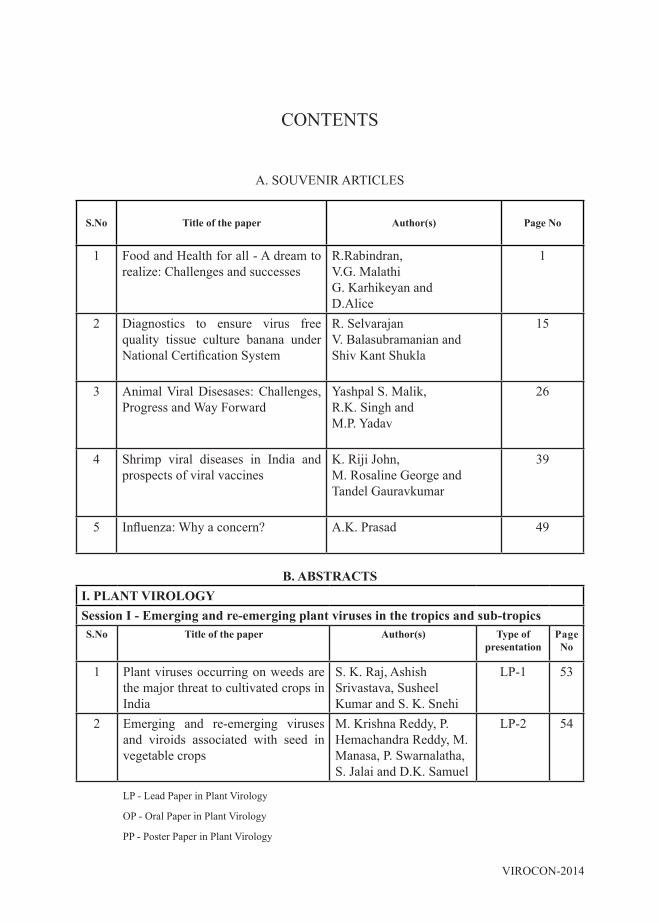
B. ABSTRACTSI. PLANT VIROLOGYSession I - Emerging and re-emerging plant viruses in the tropics and sub-tropicsS.No Title of the paper Author(s) Type of
presentationPage No
1 Plant viruses occurring on weeds are the major threat to cultivated crops in India
S. K. Raj, Ashish Srivastava, Susheel Kumar and S. K. Snehi
LP-1 53
2 Emerging and re-emerging viruses and viroids associated with seed in vegetable crops
M. Krishna Reddy, P. Hemachandra Reddy, M. Manasa, P. Swarnalatha, S. Jalai and D.K. Samuel
LP-2 54
LP - Lead Paper in Plant Virology
OP - Oral Paper in Plant Virology
PP - Poster Paper in Plant Virology
CONTENTS
A. SOUVENIR ARTICLES
S.No Title of the paper Author(s) Page No
1 Food and Health for all - A dream to realize: Challenges and successes
R.Rabindran, V.G. Malathi G. Karhikeyan and D.Alice
1
2 Diagnostics to ensure virus free quality tissue culture banana under National Certification System
R. Selvarajan V. Balasubramanian and Shiv Kant Shukla
15
3 Animal Viral Disesases: Challenges, Progress and Way Forward
Yashpal S. Malik, R.K. Singh and M.P. Yadav
26
4 Shrimp viral diseases in India and prospects of viral vaccines
K. Riji John, M. Rosaline George and Tandel Gauravkumar
39
5 Influenza: Why a concern? A.K. Prasad 49

VIROCON - 2014
3 Molecular characterization of a begomovirus associated with lentil in India
Naimuddin and M. Akram
LP-3 55
4 Engineering infectious cDNA cloning system and agroinfiltration approach to melon necrotic spot (MNSV-HYD) carmovirus
Naga Teja Natra and Gopinath Kodetham
LP-4 56
5 Natural infection of Rumex nepalensis with two begomoviruses in Western Himalayas
Dolly Sharma, Aditya Kulshreshtha, Aijaz A.Zaidi and Vipin Hallan
OP-1 57
6 Occurrence and distribution of viruses infecting cucurbitaceous crops in Tamil Nadu
G. Karthikeyan, K. Nagendran, C.G. Balaji, R. Aravintharaj, S.K. Manoranjitham, R. Priyanka, S. Rajamanickam and S. Mohankumar
OP-2 58
7 Africa as a site of origin of new plant viral diseases
S. Ajitkumar, M. Rajashekhar, P. Anjibabu,
PP -1 59
8 Characterization of Cucumber mosaic virus on snake gourd (Trichosanthes cucumerina L.) In Tamil Nadu
K. Nagendran, S. Mohankumar, S.K. Manoranjitham, R. Aravintharaj, Rayapati A. Naidu and G. Karthikeyan
PP -2 60
9 Current scenario of viral diseases under protected cultivation in Maharashtra
Savarni Tripathi, Raj Verma, Dhanashri Mungekar, Poornima Gaikwad, Sujan Singh Kushwah and Pandit B. Nawale
PP-3 61
10 Tobacco streak virus (TSV) - An emerging virus in horticultural crops
R. Kannan PP-4 62
11 Distinct nature of the Tamil Nadu isolate of Pigeonpea sterility mosaic virus (PPSMV) causing sterility mosaic disease in Pigeonpea
T.K.S. Latha, P Lava Kumar and Sabitha Doraiswamy
PP-5 63
12 Occurrence and distribution of viral diseases in garlic growing areas of Nilgiris ecosystem
S. Malathi, L.Rajendran, V.P.Santhi, N. Selvaraj, D.Alice and B.Anita
PP-6 64

VIROCON-2014
13 Survey on occurrence, distribution and survey of yellow leaf disease in sugarcane
S. Sundravadana and D. Alice
PP-7 65
14 Detection of Tobacco streak virus infecting Gloriosa superba
S. Sundravadana and D. Alice
PP-8 66
15 Effect of different months of sowing on the pigeonpea sterility mosaic disease incidence and its vector Aceria cajani population
M.S. Pallavi, H. K. Ramappa and H. K. Renuka
PP-9 67
16 Investigation of greater yam (Dioscorea alata L.) viruses in India
M. L. Jeeva, T. Makeshkumar, M.Rajitha,V.G. Manasa and S.Sruthy
PP-10 68
17 Identification and molecular characterization of a begomovirus from potato (Solanum tuberosum) exhibiting yellow mosaic symptoms from Meerut district of Western Uttar Pradesh, India
Jitender Singh, Rupashree, Pankaj Kumar, Anil Sirohi and V. K. Baranwal
PP-11 69
18 Effect of cassava mosaic disease incidence on growth and yield parameters of cassava
K. Manonmani, K. Sundaraia and R. Rabindran
PP-12 70
19 Emergence of Tobacco streak virus – A devastating virus causing necrosis disease of cotton in Tamil Nadu
P. Renukadevi, K. Nagendran, S. Nakkeeran, S. Rageshwari, G. Karthikeyan, V.G. Malathi and D. Alice
PP-13 71
20 Molecular identification of Ageratum enation virus, betasatellite and alphasatellite molecules isolated from Amaranthus showing yellow vein symptoms in India
M. Ashish Srivastava, S. Jaidi, Kumar and S. K. Raj
PP-14 72
21 Okra enation leaf curl virus - An emerging Begomovirus on Okra in North East India
P. Rakesh Kumar, Sairam Reddy and Sreenu kadiri
PP-15 73
Session II – Diagnostics of plant viruses and plant viruses in human habitats
22 Next generation sequencing in plant virus research: what next?
Stephan Winter LP-5 74

VIROCON - 2014
23 Development and validation of a microarray for the detection of all known plant viruses and viroids
V.K. Baranwal, K. Prabha, Prachi Jain, R.K, Saritha and R. K. Jain
LP-6 75
24 Diagnostics of plant viruses and plant viruses in human habitats
D.V.R. Sai Gopal LP-7 76
25 Rapid detection of six plant viruses by lateral flow assay
Bikash Mandal, Yogita Maheshwari, Prasanthi Yerrapothu, Anitha Kodaru and R.K. Jain
LP-8 77
26 Hydroxy naphthol blue (HNB) dye based molecular detection of Banana bunchy top virus
S. Basavaraj, K.T. Rangaswamy, R.N. Pushpa, M. Bhagyashree and H. A. Prameela
OP-3 78
27 Sequencing and computational analysis of two Citrus yellow mosaic virus (CMBV) isolate genomes and development of quick and sensitive diagnostics for its detection – A step to control the virus spread
A.M. Anthony Johnson, Indranil Dasgupta, Chinta Sudhakar and D.V.R. Sai Gopal
OP-4 79
28 Simultaneous detection of multi components of Banana bunchy top virus
T. Sasireka and R. Selvarajan
OP-5 80
29 Genomic properties of Potato virus M occurring in Northern plain of India
Akshay Katiyar, Alok Kumar and Bikash Mandal
OP-6 81
30 Detection and elimination of Bean yellow mosaic virus from Gladiolus
Charanjeet Kaur, Rashmi Raj, Susheel Kumar and S.K. Raj
PP-16 82
31 Detection and identification of potyviruses occurring on bulbous ornamentals
Susheel Kumar, Rashmi Raj and S. K. Raj
PP-17 83
32 Detection and characterization of Tomato leaf curl virus – replication protein in Solanum lycopersicum from Dharmapuri district of Tamil Nadu
S. U. Mohammed Riyaz and K. Kathiravan
PP-18 84
33 Taqman Real-Time PCR for detection and quantitation of Banana bract mosaic virus in banana and plantain
V. Balasubramanian and R. Selvarajan PP-19 85

VIROCON-2014
34 Rapid detection of Banana bract mosaic virus by reverse transcription loop mediated isothermal amplification (RT-LAMP) assay
R. Selvarajan and V. Balasubramanian
PP-20 86
35 Production of polyclonal antibodies against Lettuce mosaic virus using bacterial expressed recombinant coat protein
Prachi Sharma, Susheel Sharma, Jasvir Singh, Swati Saha and V. K. Baranwal
PP-21 87
36 Detection of vegetable viruses using FTA cards
S.K.Manoranjitham, G.Karthikeyan and R.A. Naidu
PP-22 88
37 Rapid detection of tomato leaf curl Gemini virus in the host and its vector Bemisia tabaci
N. Indra and R. Rabindran
PP-23 89
38 Molecular detection of Banana bunchy top virus (BBTV) affecting banana in Assam
Nilakshi Kakati and P. D. Nath
PP-24 90
39 IC-RT-PCR for the detection of Pigeonpea sterility mosaic virus, the causal agent of sterility mosaic disease of pigeonpea
M.S. Pallavi, H.K. Ramappa and H.M. Renuka
PP-25 91
40 Molecular detection and electron microscopy of Dolichos mosaic virus infecting field bean
H.M. Renuka, H.K. Ramappa, M. Byregowda and M.S. Pallavi
PP-26 92
41 Colorimetric detection of Cucumber mosaic virus infecting banana
S. Basavaraj, K.T. Rangaswamy, M. Bhagyashree and H.A. Prameela
PP-27 93
42 Application of molecular diagnostic tools for the production of quality disease- free planting materials of banana in Kerala
A.K. Cherian, P. M. Namitha, P.G. Sindu and R. Menon
PP-28 94
43 Detection and characterization of Taro bacilliform virus occurring in India
Adil Hakkim and T. Makeshktumar
PP-29 95
44 Duplex PCR for detection of two species of Begomoviruses associated with Yellow mosaic disease (YMD) of blackgram in Andhra Pradesh
B.V. Bhaskara Reddy, L. Prasanthi, S. M. Shareef and R. Sharadha Vijayalakshmi
PP-30 96

VIROCON - 2014
45 Distribution of Banana streak Mysore virus in cv. poovan in tamil nadu and diversity analysis using (RT/RNASE H) gene sequences
R. Selvarajan, K. Shivaranjani, V. Balasubramanian and R.Thilagavathi
PP-31 97
46 Occurrence, Distribution and Diagnosis of Coconut root (wilt) disease in Tamil Nadu
R. Ramjegathesh, G. Karthikeyan, I.Johnson, R. Rabindran, K. Ramaraju, T. Raguchander and R. Samiyappan
PP-32 98
47 Detection and elemination of Canna yellow mottle virus (CaYMV, Badnavirus) in Canna lilies through micropropagation
R. Radhajeyalakshmi, and Jeanmarie Verchot
PP-33 99
Session III – Viral genomics and diversity48 Current status of plant virus and
viroid diseases: India versus rest of the world
K.S. Sastry, Bikash Mandal, Teruo Sano and John Hammond
LP-9 100
49 Virus disease of Tuber crops and their management
S.K. Chakrabarti LP-10 101
50 Genetically diverse variants of Sugarcane bacilliform virus infecting sugarcane in India and evidence of a novel recombinant Badnavirus variant
Govind P. Rao, K. Susheel Sharma, Deepti Singh, Meenakshi Arya, Priyanka Singh and V.K. Baranwal
LP-11 102
51 Piper yellow mottle virus - characterization and diagnosis
A.I. Bhat LP-12 103
52 Characterization of variation in Sugarcane bacilliform virus (SCBV) associated with leaf fleck disease of sugarcane in India
R. Viswanathan and R. Karuppaiah
LP-13 104
53 Deciphering complete genome of Dasheen mosaic virus from Amorphophallus paeoniifolius transcriptome sequence data
T. Makeshkumar, S.Kamala, J. Sreekumar and S.K. Chakrabarti
LP-14 105
54 Identification of conserved domains in the sugarcane viruses responsible for targeting the RNA binding proteins through in-silico analysis
K. Bagyalakshmi, B. Parameswari, V. G. Malathi and R. Viswanathan
OP-7 106
55 Molecular detection and identification of Badnavirus infecting Canna spp. in India
Aarti Kumari, S. Kumar and S. K. Raj
OP-8 107

VIROCON-2014
56 Unusual betasatellite like component: a novel vehicle for genetic exchange among begomoviruses
Aditya Kulshreshtha, Dolly Sharma, Aijaz A. Zaidi and Vipin Hallan
OP-9 108
57 Molecular characterization of Chilli veinal mottle virus affecting chilli (Capsicum annuum L.)
Pradeep Manyam, A.S. Byadgi and M. Jyothsna
PP-34 109
58 Genetic divergence analysis for yield components and resistance to whitefly-transmitted Yellow vein mosaic virus in okra
M. Amaranatha Reddy and O. Sridevi
PP-35 110
59 Molecular characterization of Cucumber mosaic virus isolates infecting banana cv grand naine in Theni and Jalgaon region
T. Gayathrie and R. Selvarajan
PP-36 111
60 Genetic diversity of Banana bunchy top virus (BBTV) from Northeast India showed existence of distinct PIO isolates in naturally growing banana mats
Amrita Banerjee, Raghuveer Singh, S.S. Roy, S.K. Dutta, Hemavati Ranebennu and S.V. Ngachan
PP-37 112
61 Rolling circle amplification-fragment length polymorphism based variability of Banana streak my virus and a comparison of population and subpopulation wise variability
Susheel Kumar Sharma, P. Vignesh Kumar and Virendra Kumar Baranwal
PP-38 113
62 Phylogeography of simulated PRSV infection in Tamil Nadu using BEAST
Duleep Kumar Samuel, Krishna Reddy, Salil Jalali and H.C. Reddy
PP-39 114
63 “Faceting” to visually analyze high density multi-year, multi-centre multi- treatment data in GGplot using free R software
Duleep Kumar Samuel, Krishna Reddy, Salil Jalali and H.C. Reddy
PP-40 115
64 Analysis of complete nucleotide sequences and genome organization of Tomato Leaf Curl Viruses infecting tomato genotypes in Tamil Nadu
M. Deivamani, R. Rabindran and T. Ganapathy
PP-41 116
65 Characterization of Watermelon bud necrosis virus (WBNV) infecting watermelon in Tamil Nadu
R. Priyanka, K. Nagendran, U. Keerthana, P. Renuka Devi, S. Mohankumar and G. Karthikeyan
PP-42 117

VIROCON - 2014
66 Identification and molecular characterisation of complete genome of Banana streak virus species infecting banana cv. virupakshi (Hill banana)
R. Selvarajan and V.Balasubramanian
PP-43 118
67 Biological and molecular differentiation of Cassava Mosaic Virus isolates
N.Rajinimala, R.Rabindran, S. Mohan and K. Sethuraman
PP-44 119
68 Diversity of tospoviruses infecting tomato, chilli and capsicum in southern India
S. Amruta Bhat, V. Laxmi Devi and M. Krishna Reddy
PP-45 120
69 Genetic diversity of Papaya ringspot virus in India
Ritesh Mishra, Rakesh Kumar Verma and R.K.Gaur
PP-46 121
Session - IV – RNAi, VIGS and functional genomics
70 Investigating viral gene functions in Sri Lankan cassava mosaic virus and Rice tungro bacilliform virus
Indranil Dasgupta LP-15 122
71 Selective repression of NtRDR1 mediated antiviral silencing is crucial for AV2 mediated differential pathogenicity of Indian tomato-infecting begomoviruses
Supriya Chakraborty LP-16 123
72 Deep-sequencing transcriptome analysis of Abelmoschus esculentus (bhendi) towards deploying an effective RNAi strategy against bhendi yellow vein mosaic disease
V. Kavitha, P. Priyavathi and P.Gopal
LP-17 124
73 Plant virus induced gene silencing: is there any therapeutic prospect?
Anirban Roy and Bikash Mandal
LP-18 125
74 Effect of environmental conditions on virus infections, gene silencing and their implications on virus induced gene silencing (VIGS)
Basavaprabhu L. Patil, and Claude M. Fauquet
LP-19 126
75 A strategy for overcoming clone-instability in E. coli while developing infectious clone of Potato virus Y
A. Abdul Kader Jailani, Vikas Solanki and Bikash Mandal
OP-10 127

VIROCON-2014
76 RNAi vector construction against Sri Lankan cassava mosaic virus
G. J. Janavi, R. Rabindran, Indranil Dasgupta, D. Raghu, Ikuo Nakamura and Masahiro Mii
OP-11 128
77 The hairpin RNA gene comstruct targeting replication initation protein gene of Mungbean yellow mosaic virus (MYMV) causes PTGS of rep and trans silencing of the bar gene with the homologous promoter by TGS
G. Shanmugapriya, R. Rajeswaran and K. Veluthambi
OP-12 129
78 Proteomics to unravel the interaction of Banana bract mosaic virus in plaintain cv. Nendran
C. Anuradha and R.Selvarajan
OP-13 130
79 Physiological and hormonal changes in response to Banana bunchy top virus (BBTV) infection in banana
C. Anuradha, R. Selvarajan, S. Vasantha, K.P. Salin and G.S. Suresha
PP-47 131
80 Proteomic changes in banana in response to Banana bunchy top virus (BBTV)
C . Anuradha and R. Selvarajan
PP-48 132
81 Establishment of virus-induced gene silencing (VIGS) for functional analysis of endogenous genes in Nicotiana tabacum
Bhor Sachin Ashok and Kobayashi Kappei
PP-49 133
82 RNAi- mediated gene silencing of Cotton leaf curl virus and associated DNA β
Mohammad Akmal and Jawaid A. Khan
PP-50 134
83 Molecular biology and efficacy of constitutive bidirectional plant promoter from Cotton leaf curl virus
Zainul A. Khan, Malik Z. Abdin and Jawaid A. Khan
PP-51 135
84 Characterization of Sugarcane yellow leaf virus-p0 for RSS activity through Agrobacterium-mediated transient expression system
S. Brindha, V.G.Malathi and R. Viswanathan
PP-52 136
85 Small RNA deep sequencing of sterility mosaic disease infected Cajanus cajan
Surender Kumar, B.L. Subbarao, E. Rajeswari, V. Sunderesan and Vipin Hallan
PP-53 137

VIROCON - 2014
86 AV2 protein of Tomato leaf curl Palampur virus associates with a cysteine protease involved in plant defense
Poonam Roshan, Aijaz A. Zaidi and Vipin Hallan
PP-54 138
87 Changes in sugars and phenolics in rice varieties susceptible and resistant to rice tungro virus disease
I. Yesu Raja, M. Syamala, K. Sethuraman and S. Gnanaprakash
PP-55 139
Session V - Quality tissue culture and certification
88 Certification of tissue culture plants under NCS-TCP: its relevance, key components, operational guidelines and procedures
Shiv Kant Shukla LP-20 140
89 Recent novel techniques for plant virus diagnostics adoptable in the virus-free certification programmes
R. Selvarajan LP-21 141
90 Field level practical measures for rejuvenation of hill banana from Banana bunchy top virus (BBTV)
R. Pavalarajan OP-14 142
91 Problems associated with production of viral free embryogenic cell line system in hill banana
S. Elayabalan, S. Subramaniam and R. Selvarajan
OP-15 143
Session VI – Host – Virus – Vector interactions
92 Molecular typing of the vector Bemisia tabaci Genn., and the domains of coat protein involved in transmission of Mungbean yellow mosaic India virus
V.G. Malathi, S. Kanakala. K. Archana, P, Jyothsna, R. K.Varma and H.C. Prasanna.
LP-22 144
93 Host associated genetic variation and detection of endosymbionts in begomovirus vector, Bemisia tabaci (Genn.)
B. Preetha, R. Aravintha Raj, G. Karthikeyan and S. Mohankumar
LP-23 145
94 Thai sacbrood virus (TSBV) - A potential threat to Indian honey bee
M.R. Srinivasan, S. Kuttalam and K. Ramaraju
LP-24 146
95 Employing RNAi approach against Bemisia tabaci infestation in Gossypium hirsutum plants
Gazal Wamiq and Jawaid A. Khan
LP-25 147

VIROCON-2014
96 Epidemiology of Papaya ringspot virus (PRSV) in papaya (Carica papaya L.)
R.N. Pushpa, N. Nagaraju and K.T. Rangaswamy`
OP-16 148
97 Host range and virus vector relationships of leaf curl begomvirus disease on sunflower in relation to disease epidemiology
M. Vindyashree, M.R. Govindappa, V.N.Ghante, Aswathanarayana and D.S. Shankergoud
PP-56 149
98 Association of weather factors on aphid population and Papaya ring spot virus disease incidence
G. Thiribhuvanamala, K.Soorianathasundaram, S. Sridharan, R. M. Vijayakumar and D. Alice
PP-57 150
99 Host range, virus-vector relationship of leaf curl virus and whitefly in tomato
N. Indra and R. Rabindran
PP-58 151
100 Transmission and symptomatology of virus causing Dolichos mosaic virus disease on field bean
H. M. Renuka, H.K Ramappa, M. Byregowda and M.S. Pallavi
PP-59 152
101 Diagnosis of Thai sac brood virus of Indian honey bee Apis cerana indica through reverse transcriptase – PCR technique
R. Aruna, M.R.Srinivasan and R.Selvarajan
PP-60 153
102 Studies on endosymbionts associated with whitefly (Bemisia tabaci)
S. Rageshwari, R. Velazhahan and R. Rabindran
PP-61 154
103 Thrips and necrosis - A threat to Gloriosa cultivation
M. Suganthy, B. Meena and K. Rajamani
PP-62 155
Session VII – Application of Nanotechnology; Plant Quarantine and virus disease management104 Viral nanoparticles and virus-like
particles-applications in biomedicineM. Hema LP-26 156
105 Viral proteins are multifunctional and impact on host-resistance breaking
R.V. Chowda-Reddy, John Hill, V. Muniyappa, John Colvin, Aiming Wang, Vincent Fondong, Sue Seal and Steve Whitham
LP-27 157

VIROCON - 2014
106 Biosecurity umbrella for Indian agriculture against exotic plant viruses: A case study of quarantine of exotic germplasm
V. Celia Chalam, D.B. Parakh and A.K. Maurya
LP-28 158
107 Exploration of mechanisms for plant virus symptom expression- a challenge for plant protection in future
Kobayashi Kappei, Waliullah Sumyya, Bhor Sachin Ashok, Akhter Md. Shamim, Kosaka Naomi, Suganuma Yusuke, Sugiwaka Yuji, Tajima Kaoru, Yamashita Mei, Tomita Reiko, Atsumi Go and Sekine Ken-Taro
LP-29 159
108 Genetically engineered Papaya for virus resistance: Success & challenges
Savarni Tripathi, Jon Y. Suzuki and Dennis Gonsalves
LP-30 160
109 Functional and structural analsysis of rice tungro resistance gene and its introgression into popular rice varieties
C.N. Neeraja, D Krishanveni, Hemant Kishore, S.K. Mangrauthia and Chitra Shanker
LP-31 161
110 IPM strategies for the management of insect transmitted virus diseases in vegetable crops
G. Karthikeyan, C.G. Balaji, K. Nagendran, R. Aravintharaj, S.K. Manoranjitham R. Priyanka and S. Mohankumar
LP-32 162
111 Development of Peanut stem necrosis disease (PSND) resistant transgenic groundnut plants with inverted repeat-replicase gene of Tobacco streak virus (TSV)
R. Velazhahan, M. Gurivi Reddy, C. Senthilraja, R. Adhithya, V. K. Satya, E. Kokiladevi, D. Sudhakar and R. Rabindran
OP-17 163
112 Evaluation of resistance in urdbean against Mungbean yellow mosaic virus for crop improvement
V.K. Satya, D. Alice, V.G. Malathi, R. Vinoth, and P. Jayamani
OP-18 164
113 Characterization of resistance in back-crossed rice plants of variety ASD-16 containing transgene against Rice tungro bacilliform virus
Gaurav Kumar, S. Robin, R. Rabindran and I. Dasgupta
OP-19 165
VIROCON-2014
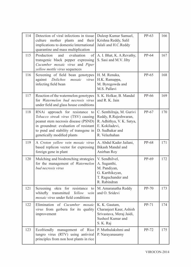
114 Detection of viral infections in tissue culture mother plants and their implications to domestic/international quarantine and mass multiplication
Duleep Kumar Samuel, Krishna Reddy, Salil Jalali and H.C.Reddy
PP-63 166
115 Production and evaluation of transgenic black pepper expressing Cucumber mosaic virus and Piper yellow mottle virus sequences
A. I. Bhat, K. A.Revathy, S. Sasi and M.V. Jiby
PP-64 167
116 Screening of field bean genotypes against Dolichos mosaic virus infecting field bean
H. M. Renuka, H.K. Ramappa, M. Byregowda and M.S. Pallavi
PP-65 168
117 Reaction of the watermelon genotypes for Watermelon bud necrosis virus under field and glass house conditions
S. K. Holkar, B. Mandal and R. K. Jain
PP-66 169
118 RNAi approach for resistance to Tobacco streak virus (TSV) causing peanut stem necrosis disease (PSND) in groundnut: evaluation of resistant to psnd and stability of transgene in genetically modified plants
C. Senthilraja, M. Gurivi Reddy, R.Rajeshwaran, R. Adhithya, V. K. Satya, E. Kokiladevi, D. Sudhakar and R. Velazhahan
PP-67 170
119 A Croton yellow vein mosaic virus based replicon vector for expressing foreign gene in plant
A. Abdul Kader Jailani, Bikash Mandal and Anirban Roy
PP-68 171
120 Mulching and biodrenching strategies for the management of Watermelon bud necrosis virus
V. Sendhilvel, A. Suganthi, M. Pandiyan, G. Karthikeyan, T. Raguchander and R. Rabindran
PP-69 172
121 Screening okra for resistance to whitefly transmitted Yellow vein mosaic virus under field conditions
M. Amaranatha Reddy and O. Sridevi
PP-70 173
122 Elimination of Cucumber mosaic virus from gerbera for its quality improvement
K. K. Gautam, Charanjeet Kaur, Ashish Srivastava, Meraj Jaidi, Susheel Kumar and S. K. Raj
PP-71 174
123 Ecofriendly management of Rice tungro virus (RTV) using antiviral principles from non host plants in rice
P. Muthulakshmi and P. Narayanasamy
PP-72 175

VIROCON - 2014
124 Effect of Pseudomonas fluorescens strains on RTV infection
P. Muthulakshmi and P. Narayanasamy
PP-73 176
125 Management of Scirtothrips dorsalis and sunflower necrosis disease in sunflower
M. Suganthy and P. Sakthivel
PP-74 177
126 Efficacy of application of endophytic bacteria Bacillus pumilus and Bacillus subtilis in banana plants cv.grand naine against Banana bunchy top virus
R. Manohar Jebakumar, , R. Selvarajan and M.M. Mustaffa
PP-75 178
127 Transgenic cassava production with gene(s) conferring resistance to cassava mosaic disease (CMD) through rnai technology
M. Jayakumar, M. Saravanakumar, V. Subramanian, G.S. Murugesan and K. K. Kumar
PP-76 179
128 Chitosanases and their role in plant defense against different pathogens
Manisha Sharma and Wamik Azmi
PP-77 180
129 Combating pigeonpea sterilty mosaic disease through acaricide
E. Rajeswari , K.P Smitha, P.Latha, D. Alice and J.R. Kannan Bapu
PP-78 181
130 On farm testing of MYMV disease management technique in blackgram in pudukkottai district of Tamil Nadu
S. Mathiyazhagan, V.R.S. Saminathan and R.P. Gnanamalar
PP-79 182
131 Genetic engineering in hill banana for banana bunchy top disease (BBTD) resistance
Sanii Lanah, P. Balasubramanian and J. Navaneetha Krishnan
PP-80 183
132 Artificial microRNAs targeting the intergenic region/replication origin provide broad spectrum resistance against begomoviruses
S. Harish, Yi-Jung Kung, Ang Rinzing Sherpa and Shyi-Dong Yeh
PP-81 184
133 Associvity of Phytophthora palmivora butler co-infection on Papaya ring spot virus infected plants: implications for management
Duleep Kumar Samuel, Krishna Reddy, S.Sriram, Salil Jalali and H.C.Reddy
PP-82 185
134 Molecular validation of SSR markers linked to sterility mosaic disease resistance gene in pigeonpea genotypes
M.S. Pallavi, H.K. Ramappa, D. Pramesh, M. Byre Gowda and S. Poonam
PP-83 186
135 Management of Peanut bud necrosis virus disease in tomato
K. Kalpana and M.N.Budhar
PP-84 187
VIROCON-2014
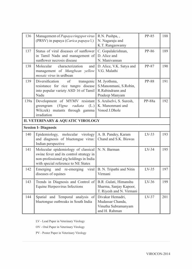
136 Management of Papaya ringspot virus (PRSV) in papaya (Carica papaya l.)
R.N. Pushpa, , N. Nagaraju and K.T. Rangaswamy
PP-85 188
137 Status of viral diseases of sunflower in Tamil Nadu and management of sunflower necrosis disease
C. Gopalakrishnan, D. Alice and N. Manivannan
PP-86 189
138 Molecular characterization and management of Mungbean yellow mosaic virus in urdbean
D. Alice, V.K. Satya and V.G. Malathi
PP-87 190
139 Diversification of transgenic resistance for rice tungro disease into popular variety ASD 16 of Tamil Nadu
M. Jyothsna, S.Manonmani, S.Robin, R.Rabindrann and Pradeep Manyam
PP-88 191
139a Development of MYMV resistant greengram (Vigna radiata (L.) Wilczek) mutants through gamma irradiation
S. Arulselvi, S. Suresh, K. Manonmani and Vonod J.Dhole
PP-88a 192
II. VETERINARY & AQUATIC VIROLOGY
Session I- Diagnosis
140 Epidemiology, molecular virology and diagnosis of bluetongue virus: Indian perspective
A. B. Pandey, Karam Chand and S.K. Biswas
LV-33 193
141 Molecular epidemiology of classical swine fever and its control strategy in non-professional pig holdings in India with special reference to NE States
N. N. Barman LV-34 195
142 Emerging and re-emerging viral diseases of equines
B. N. Tripathi and Nitin Virmani
LV-35 197
143 Trends in Diagnosis and Control of Equine Herpesvirus Infections
B.R .Gulati, Himanshu Sharma, Sanjay Kapoor, T. Riyesh and N. Virmani
LV-36 199
144 Spatial and Temporal analysis of bluetongue outbreaks in South India
Divakar Hemadri, Mudassar Chanda, Vinutha Subramanyam and H. Rahman
LV-37 201
LV - Lead Paper in Veterinary Virology
OV - Oral Paper in Veterinary Virology
PV - Poster Paper in Veterinary Virology

VIROCON - 2014
145 Epidemiology of Peste des petits ruminants vis-à-vis Control programme in India
V. Balamurugan, M.R. Gajendragad and H.Rahman
LV-38 203
146 Novel Viral Agents of Gastroenteritis in Animals
Yashpal S. Malik, K. Dhama, A.K. Tiwari and R.K. Singh
LV-39 204
147 Classical swine fever virus Genogroup 2.2 circulating in wild and domestic pigs of North Eastern states
N. N . Barman, E. Khatoon, Rajbongshi, Gitika, D. Borah, K. Baruah and N. Deka
OV-20 206
148 Absolute quantitation of classical swine fever virus by One-Step TaqMan Real-Time Quantitative Reverse Transcriptase Polymerase Chain Reaction Assay
Gitika Rajbongshi, N.N. Barman, E. Khatoon, K. Baruah, N. Deka and S. K. Das
OV-21 207
149 Characterization of 2013 outbreak strains of foot and mouth disease in southern peninsular India
Saravanan Subramaniam and Bramhadev Pattnaik
OV-22 208
150 Genetic characterization of swinepox virus from clinical samples by H3L gene
R. Mageswary, Nikunj Gupta, S. Chandra Sekar, G.Venkatesan , Sargam Arya, S.K. Minhas, A.B. Pandey, R. Singh and M. A. Ramakrishnan
OV-23 209
151 Development of an indirect-ELISA based on recombinant non-structural protein-3 N-terminus (NS3Nt) of bluetongue virus
Nirmal Chacko, Sanchay Kumar Biswas, Nihar Nalini Mohanty, Karam Chand, Bimalendu Mondal, Awadh Bihari Pandey and Sathish Bhadravati Shivachandra
OV-24 210
152 Identification and genotyping of porcine picobirnaviruses isolated from North-eastern region (NER) and Northern parts of India, during 2012-2014
Yashpal S. Malik, K. Sircar, D.P. Sharma, T. K. Bora, U.K. Datta, De, N. R. Sahoo, A. K. Tiwari and R.K. Singh
OV-25 211
153 Development of recombinant antigen based diagnostics for peste des petits ruminants in sheep and goats
V. Balamurugan, Sunil Abraham, S. Sowjanya Kumari, R. Apsana, M. Nagalingam, D. Hemadri and H. Rahman
OV-26 212
VIROCON-2014
154 Recurrent episodes of Zoonotic Buffalopox virus infections: a threat to the community milkers in India
Sanjay Barua, T. Riyesh, B.C. Bera, Taruna Anand, Surender Singh Chandel Mubarik Hussain, Mansi Yadav, R.K. Vaid and Praveen Malik
OV-27 213
155 Single step real-time RT-PCR could detect low concentration of classical swine fever virus comparing to gel based RT-PCR assay
Gitika Rajbongshi, N.N. Barman, E. Khatoon, K. Baruah, N. Deka and S. K. Das
PV-89 214
156 Molecular characterization of classical swine fever virus following its adaptation in porcine kidney cells
Rakesh Kumar and Sachin Kumar
PV-90 215
157 Epidemiology and Serosurveillance of FMD in Karnataka
V. Govindaraju, D. Rathnamma, R. Hegde, P. Giridhar, Shrikrishna. Isloor, A. Shivaraj, G.H. Channabasayya, B.M. Chandranaik, Akshtha, A. Nirupama, Srinivas Babu and M. Shivaraj
PV-91 216
158 Foot-and-Mouth Disease in Elephants in Kerala During 2013
M. Rout, N.S. Nair, B. Das, S. Subramaniam, J.K. Mohapatra and B. Pattnaik
PV-92 217
159 Isolation and identification of avipoxviruses from backyard poultry of North East India
D.P. Bora, B.Borah, D. Borkotoky, M. Bora, D.P. Saikia, R. Dutta, N.J. Pathak and N.N. Barman
PV-93 218
160 Adaptation of atypical goatpox virus in Vero cells
S. K. Minhas, R. Mageswary, G.Venkatesan, S. Chandra Sekar, K.P.Singh, A.B. Pandey and M.A. Ramakrishnan
PV-94 219
161 Development of loop-mediated isothermal amplification (LAMP) for the rapid detection of Bluetongue virus from sheep samples
V. Tharanath, A.M.A. Johnson and D.V.R. Sai Gopal
PV-95 220

VIROCON - 2014
162 Molecular Characterization of Orf virus isolated from goats of Assam
M. Bora, D.P.Bora, N.N.Barman, S. Das, B. Borah, P.L. Bora, A.Talukdar and S.Tamuly
PV-96 221
163 Molecular characterisation of Newcastle Disease Virus isolated from Northeast India
Moushumee Das and Sachin Kumar
PV-97 222
164 Genotypic and pathotypic characterisation of virulent Newcastle Disease Virus isolated from Eastern part of India.
Umesh Kumar and Sachin Kumar
PV-98 223
165 Seroprevalence of Orf in goats of Assam
S.S. Begum, G. Mahato, N.N Barman and D. Muthuchelvan
PV-99 224
166 Detection of caprine and ovine rotaviruses in and around Mathura region
Uttam Singh, Rashmi Singh, Ajay Pratap Singh, Sharad Kumar Yadav, Yashpal Singh Malik and Shubhankar Sircar
PV-100 225
167 Classical Swine Fever Virus genogroup 2.2 caused persistent infection in breeding sows
N. N. Barman, Gogoi, S. M., Khatoon, E, Deka, Nipu, Rajbongshi, Gitika, K. Baruah and M. Nath
PV-101 226
168 Seroprevelance of Peste des Petits Ruminants in Goats of Assam
Maitrayee Devi, Sutopa Das, Krishna Sharma, Probodh Borah, Rita Nath, Rupam Dutta and Indrani Chakrabarty
PV-102 227
169 N gene based molecular epidemiology of Peste-des-petits ruminants Viruses in India
Z. Ahamad, K. K. Rajak, D. Muthuchelvan, S. Bhadouriya, R.C. Dadas, D. Chaudhary, R. Kumar, A. K. Yadav, V.V. Dhanesh, M. Manu, A. B. Pandey and R. K. Singh
PV-103 228
170 Comparison of Three Different Techniques for Diagnosis of Animal Rabies
G.B. Manjunatha Reddy, K.Sumana, S.S.Patil, Yogisharadhya and H. Rahman
PV-104 229

VIROCON-2014
171 Recombinant Peste des petits ruminants virus nucleocapsid (N) protein/ antigen based indirect ELISA for serodiagnostics of PPR in sheep and goats
V. Balamurugan, Manisha Roy, S. Sowjanya Kumari, Sunil Abraham, D. Hemadri and H. Rahman
PV-105 230
172 Sero-Prevalence of Contagious Ecthyma (Orf) in Goats of Assam
M. Bora, D. P. Bora, N.N. Barman, B. Borah, S. Das, P. Das, A. Talukdar and S. Tamuly
PV-106 231
173 Sero-Prevalence of bovine herpes virus 1 (BHV-1) in dairy cattle population of Assam
S. Chettri, D. P. Bora, B. Borah, P. L. Bora, M. Bora, P. Das, D. K. Sarma and K. Ahmed
PV-107 232
174 Molecular epidemiology of Indian sheeppox and goatpox viruses
R. Santhamani, R. Yogisharadhya, V. Gnanavel, S. B. Shivachandra, A. B. Pandey and M. A. Ramakrishnan
PV-108 233
175 Detection and characterization of swinepox virus based on ORF114 gene
Nikunj Gupta, R. Mageswary, S.Chandra Sekar, G.Venkatesan, Sargam Arya, S.K. Minhas, R. Singh, A.B. Pandey and M.A. Ramakrishnan
PV-109 234
Session II - Genomics
176 Biotechnological Approaches in Viral Diseases of Wild and Domestic Ruminants
P. Minakshi, M.Shafiq, Koushlesh Ranjan, Basanti Brar, Shweta Balodi, Anjali Singh, Y.S. Malik, R. Dalal and Gaya Prasad
LV-40 235
177 Research Journey on whole genomic analysis of rotaviruses
Souvik Ghosh and Nobumichi Kobayashi
LV-41 236
178 Characterization of Equid Herpesvirus 1 Strains Isolated from Abortion in India based on ORF30 and ORF68 Genes
G. Anagha, B.R. Gulati, T.Riyesh and N.Virmani
OV-28 237
VIROCON - 2014
179 Expression and evaluation of P32 protein of Capripox virus as a diagnostic antigen in Indirect ELISA
G. Venkatesan, M. Dashprakash, Mahesh Kumar Teli, M.A. Ramakrishnan, M. Sankar, D. Muthuchelvan and A.B.Pandey
OV-29 238
180 Genetic changes in polymerase genes (PB2, PB1 & PA) of equine influenza virus from outbreaks in India
Virmani Nitin, B.C. Bera, K.Shanmugasundaram, B.K. Singh, B.R.Gulati, Anand Taruna, R.K.Vaid, Barua, Sanjay and R.K.Singh
OV-30 239
181 Analysis of Expression of Foot and Mouth Disease Virus Type O (IND R2/75) Capsid Protein in A549 vs. HEK-293 Cells Infected with Recombinant Human Adenovirus Type 5
Ramesh Kumar, B.P. Sreenivasa and R.P.Tamil Selvan
OV-31 240
182 Synthetic peptide based immuno-dominant epitope mapping and evaluation of its diagnostic potential for rotavirus detection
Naveen Kumar, Yashpal S. Malik, Kuldeep Sharma, Shubhankar Sircar, Vinay G. Joshi, Satish Kumar, Ashok Kumar Tiwari, and Raj Kumar Singh
OV-32 241
183 Receptor tyrosine kinase signaling regulates Peste des Petits Ruminants virus RNA synthesis
Naveen Kumar, Khushboo Chaudhary and Shoor Vir Singh
OV-33 242
184 Evidence of natural recombination in the non-structural protein of classical swine fever virus from India
T. Riyesh, Pronab Dhar, Sanjay Barua, Naresh Jindal, B.C. Bera, Vikaramaditya Upmanyu, TarunaAnand, R.K. Vaid, Mansi Yadav G. Anagha, A.K. Tiwari, Praveen Malik and A.B. Pandey
OV-34 243
185 Characterization of four Indian Bluetongue virus serotype 1 isolates based on full-length sequence of genome segment-2.
S. K. Karam Chand, Biswas, B.Mondal, and A.B.Pandey
PV-110 244

VIROCON-2014
186 Cloning and expression of bluetongue viral non-structural protein-1 (NS1) using prokaryotic expression system
Amir Showkat Khan, Sanchay Kumar Biswas, Vishaka, Gulam Mohd, Karam Chand, Awadh Bihari Pandey and Sathish Bhadravati Shivachandra
PV-111 245
187 Host-virus adaptation and evolutionary analysis of rotavirus serogroups of avian origin based on codon usage patterns
Jobin Jose Kattoor, Yashpal S. Malik, Kuldeep Sharma, Ashok Kumar Tiwari and Raj Kumar Singh
PV-112 246
188 Generation of full length cDNA backbone of cell culture adapted lapinized Classical Swine Fever Virus
P. Parveen Kumar, V. Dhar, A. Upmanyu, Kumar and A.K. Tiwari
PV-113 247
189 Evaluation of interferon response by shRNA constructs in caprine fetal fibroblast cells by real-time RT-PCR
Jyoti lakshmi, Hati Boruah, Rakesh Ranjan, Hamen Gogoi, Dharmendra Kumar, Amlanjyoti Phukan, Joygeshwar Bori, Tripti Jain and Bikash Chandra Sarkhel
PV-114 248
190 Evaluating the immune genes interaction network in sheep and goat PBMCs infected with Bluetongue Virus infection by RNA-Seq data analysis
Anjali Singh, P.Minakshi, S. Ravi Kumar Gandham, Manjunath, Shweta Balodi , Anupama Deora, Basanti Brar, Pawan Kumar, Ganesha V Joshi and Gaya Prasad
PV-115 249
191 Phylogenetic analysis of a Peste des petits ruminants virus from an outbreak in Nagaur, India
Naveen Kumar, Shoor Vir Singh, N.Shivsaranappa, Subhash Kachhawa, Sunil Maherchandani and Sudhir Kumar Kashyap,
PV-116 250
192 Expression of Toll-Like receptors in Classical swine fever infection in swine
B.H Veeresh, S.S. Patil, S. Geetha, G.B. Manjunatha Reddy, D. Hemadri, G.S. Desai and H.Rahman
PV-117 251

VIROCON - 2014
Session III - Disease prevention and control193 Reassortments in Avian Influenza
VirusesC. Tosh, S. Nagarajan, and D.D. Kulkarni
LV-42 252
194 Novel influenza threats with zoonotic potential: preparedness for diagnostics and vaccines in poultry
Sandeep Bhatia and Richa Sood
LV-43 254
195 The critical role of adjuvant behind a vaccine success: a view point
Prem Sagar, Ben Arous J, F. Bertrand and D. Sebastien
LV-44 256
196 Development of an indirect ELISA with recombinant nucleoprotein for diagnosis of influenza A
S. Nagarajan, V. Ramaswamy, R. Jain, K. Rajukumar and H. K. Pradhan
OV-35 258
197 Inactivated FMD Type ‘O’ virus adjuvanted with recombinant OmpA encapsulated in MC-PLGA nanoparticles induces a strong mucosal Immune response
R. Mageswary, S.Chandra Sekar, G.Elaiyaraja, M.Terhuja, K.Ganesh, V.Bhanuprakash and S. Kishore
OV-36 259
198 FMDV 2A Mediated Co-ordiante Expression of Peste des petits ruminants Virus F and HN Proteins in Baculovirus and their Immunogenicity in Mice
G. S. Desai, K. Prabhudas, M. Gopinath, S. S. Patil and M. S. Shaila
OV-37 260
199 Characterization of pathogenicity and infectivity of H9N2 avian influenza virus in chickens
Sandeep Dash, Manoj Kumar, J. M. Kataria, H.V. Murugkar, C. Tosh, D. D. Kulkarni and S. Nagarajan
OV-38 261
200 Spatial and temporal analysis of Indian H5N1 Avian influenza outbreaks
R. Sridevi, P.Krishnamoorthy, S. Dharmarajan and H. Rahman
OV-39 262
201 Replacement of hypervariable region of Salmonella flagellin with VP1 of foot-and-mouth disease virus does not hamper the proinflammatory activity of flagellin
Irshad Ahmed Hajam, A. Elamurugan, Pervaiz Ahmad Dar, Kondabattula Ganesh, Subodh Kishore and Veerakyathappa Bhanuprakash
PV-118 263

VIROCON-2014
202 Augmentation of immune response to inactivated foot and mouth disease virus trivalent vaccine using recombinant B2L protein of orf virus in guinea pigs
N. S. Muneeswaran, V. Bhanuprakash, S. Kishore, R.P Tamil Selvan, I. A. Hajam, A. Elamurugan and K. Ganesh
PV-119 264
203 Assessment of the relationship between serum neutralizing antibody titer and liquid phase blocking ELISA titer in Foot and Mouth Disease Virus Trivalent vaccinated serum samples
R.P. Tamil Selvan, B.P. Sreenivasa M. Hosamani, P. Saravanan, Suresh H Basagoudanavar and R. Venkataramanan
PV-120 265
204 Development of a simple in vitro interferon bio-assay in primary cell culture and detection of goat interferon activity against goat pox virus following immunostimulation
H.D. Karmakar PV-121 266
205 Evaluation of the effect of an herbal immunomodulator in Orf immunized goats
S.S Begum, G.Mahato, N.N.Barman, A. Saleque and D.Muthuchelvan
PV-122 267
206 Assessment of stability of thermostable and conventional Peste des Petits Ruminants vaccine viruses diluted with different diluents
S. Chitradevi, A.Thangavelu and R.Mathivanan
PV-123 268
Session IV - Aquatic Virology
207 Management of white spot virus in shrimp culture systems: options and challenges
I.S. Bright Singh LA-45 269
LV - Lead Paper in Aquatic Virology
OV - Oral Paper in Aquatic Virology
PV - Poster Paper in Aquatic Virology
208 Developments to combat viral diseases in coldwater aquaculture
B.S. Anand Kumar, Dimpal Thakuria and Amit Pande
OA-40 270
209 Differential protein and protease expression in shrimp (Penaeus vannamei) tissues during progressive white spot disease
P. Anand Kumar and K. Sankaran
OA-41 271

VIROCON - 2014
210 Replication pattern of White spot syndrome virus (WSSV) in Macrobrachium rosenbergii and Penaeus monodon
Saloni Shivam, Satya Prakash, Deepika Anand, K. Sreedharan, M. Makesh and K.V. Rajendran
OA-42 272
211 Pro-inflammatory cytokine responses in head-kidney leucocytes of rohu, Labeo rohita following stimulation with poly I:C, a synthetic analog of double stranded RNA virus
Pujarini Dash and P.K. Sahoo
OA-43 273
212 Application of monoclonal antibody against capsid protein of extra small virus of Macrobrachium rosenbergii
M. Makesh, A. Deepika and K.V. Rajendran
OA-44 274
213 Experimental infection of mixed genotypes shows reduced infectivity potential in white spot virus (WSV)
K. Riji John, M. Rosalind George, M. Mohamed Mansoor and M. Selvamaheswaran
OA-45 275
214 Viral-bacterium interaction in the eutrophic estuarine conditions of Cochin estuary, India
A. Parvathi, V.Jasna, S.Aparna and A.J.Aswathy
PA-124 276
IV. MEDICAL VIROLOGY
215 Molecular epidemiology and Immunopathogenesis of Hepatitis E
R.K.Ratho LM-46 277
216 RTLAMP Technology For Rapid and Reliable Diagnosis of Swine Flu: Translational Journey From Lab to Industry and Commercialization
Manmohan Parida LM-47 278
217 Transmission profile of epidemic Chikungunya virus in Indian Aedes mosquitoes
P. K. Dash, A. Agarwal and M. M. Parida
LM-48 279
218 Virology Diagnostic Laboratory (VDL) Network – a New Paradigm in Prevention & Control of Viral Infections
A. K. Bagga LM-49 280
219 Kaposi Sercoma Herpes Virus induced Primary Effusion Lymphoma during latency
Suchitra Mohanty, Amit Kumar, Sushil Kumar Sahu and Tathagata Choudhuri
LM-50 281

VIROCON-2014
220 Behaviour Change Communication – Chandigarh a unique Experience
H. C. Gera LM-51 282
LM - Lead Paper in Medical Virology
OM - Oral Paper in Medical Virology
PM - Poster Paper in Medical Virology
221 West Nile Encephalitis In Kerala B.Anukumar LM-52 283
222 Therapeutic Role of Antibodies in Viral Diseases
Tapan N. Dhole LM-53 284
223 Evidence of experimental vertical transmission of emerging novel ECSA genotype of Chikungunya virus in Aedes aegypti
Ankita Agarwal, Paban Kumar Dash, Anil Kumar Singh, Shashi Sharma, Natarajan Gopalan, Putcha Venkata Lakshmana Rao, Man Mohan Parida and Paul Reiter
OM-46 286
224 Expression, purification and enzymatic analysis of recombinant Chikungunya nsP2 protease
Amrita Saha, Raj Priya, M.KameswaraRao, Manmohan Parida and P.K. Dash
OM-47 287
225 Rubella outbreak investigation in the Union territory of Chandigarh, North India
A. Kumar, M.P.Singh, N.Gautam, J.Khurana, M.Gupta and R.K.Ratho
OM-48 288
226 Role of Heat shock protein 90 in Chikungunya virus replication
Indrani Das, Itishree Basantray, Prabhudutta Mamidi, Tapas K Nayak, B.M.Pratheek, Subhasis Chattopadhyay, Soma Chattopadhyay
OM-49 289
227 Distribution of Non- Polio Enteroviruses among children presenting with Acute Febrile Illness in Southwest India
Giselle Raisa Dsouza, Piya paul, Suresh Prabhu, Anjali Aithal, Revti Bhaskar,Santhosha Devadiga and G. Arunkumar
OM-50 290

VIROCON - 2014
228 Design, Synthesis and Evaluation of antiviral activity of Piperazine series of Nucleoprotein antagonists against pandemic Swine Flu (H1N1) influenza virus
Gaurav Joshi, Sanjeev kumar Verma, B.N. Acharya, D.P.Nagar, S.C.Pant and Manmohan Parida
OM-51 291
229 Assessment of prophylactic activity of recombinant haemagglutinin protein of pandemic Swine flu virus using yeast Pichia pastoris
Shweta Saraswat, T.N.Athmaram, P.K Dash and M. M. Parida
OM-52 292
230 Recombinant forms (RF) of hepatitis C virus (HCV)
Chetan Datta Podur OM-53 293
231 COX-2 induces lytic reactivation of Epstein Barr Virus through PGE2 by modulating the EP receptor signalling Pathway
Jaya Gandhi and Rajeev Kaul
OM-54 294
232 Standardization of Reverse transcription loop-mediated isothermal amplification (RT-LAMP) and one step real time RT-PCR for diagnosis of influenza viruses.
Vikrant Sharma and Samander Kaushik
OM-55 295
233 Pilot study on Hepatitis B Virus, Hepatitis C Virus and Human Immunodeficiency Virus infections among patients with Chronic Liver Diseases from North-East India attending a new tertiary care health setup at Shillong
J. Gurung, A.B. Khyriem, K. G. Lynrah and A. C. Phukan
OM-56 296
234 Rotavirus incidence and G and P genotype distribution: Increased prevalence of G9 and G12 strains among children in north western Himalayan foot hills, India
Yashpal S. Malik, Vinita Rawat, Nirupma Vaid, Kuldeep Sharma, Lalit M. Jeena, Naveen Kumar, Shubhankar Sircar, Poonam Kumari, Jobin Jose Kattoore and Raj Kumar Singh
OM-57 298
235 Distribution of Human Respiratory Syncytial Virus (HRSV) among elderly adults with Influenza-like illness from South west India
Anjali Aithal, Revti Bhaskar, Giselle Dsouza, Piya Paul, Hindol Maity, Aswathy Raj and G. Arunkumar
PM-125 299

VIROCON-2014
236 Molecular detection and characterization of Dengue isolates circulating in North India
Kanwalpreet , Manmohan Mishra and R.K. Ratho
PM-126 300
237 Molecular Epidemiology of Dengue Virus in Karnataka State, India in 2013
Revti Bhaskar, Piya Paul, J.Anitha, Giselle Dsouza, Anjali Aithal, C.Akhil and G. Arunkumar
PM-127 301
238 An investigative study of association of histo-blood group antigens with rotavirus gastroenteritis
Eileena Mohanty, Bhagirathi Dwibedi and Shantanu Kumar Kar,
PM-128 302
239 Japanese encephalitis an increasing trend in North-eastern Uttar Pradesh
Deepa Srivastava, Naveen Pandey and K.Shukla
PM-129 303
IVS - YOUNG SCIENTIST AWARD PAPERS
240 Coat protein mediated resistance against Tobacco streak virus in Nicotiana tabacum L. through RNA silencing
S. Rajamanickam, M. Raveendran and G. Karthikeyan
AP -1 304
241 Efficacy of transgenic resistance to RTBV on ‘Rice Tungro Disease’ in cv. CR 1009
P. Valarmathi, S. Robin S. Manonmani, Indranil Dasgupta, R. Velazhahan S. Suresh and R. Rabindran
AP-2 305
242 Recombinant Peste des petits ruminants virus Fusion protein antigen based ELISA for diagnosis of PPR
R. Apsana, V. Balamurugan, B.M. Veeregowda, S. Abraham, S.K. Sowjanya, D. Rathnamma, S.M. Byregowda, H. Rahman and M.S. Shaila
AP-3 306
243 “Vaccination” depending on immune memory and adaptive immunity – does it really help shrimp against WSSV?
P. Anand Kumar, K. Chandru, E. O. Koppang and K.Sankaran
AP-4 307
AP - Award Oral Paper

SOUVENIR
ARTICLES

1 VIROCON-2014
Indian Virological Society (IVS) - XXIII National Conference on“Recent Trends in Virology Research in the Omics Era”
December 18-20, 2014PLANT VIROLOGY Souvenir article -1
Food and Health for all – A dream to realize: Challenges and successes
R. Rabindran, V.G. Malathi, G. Karthikeyan and D. AliceDepartment of Plant Pathology, Tamil Nadu Agricultural University, Coimbatore – 641 003
It is our great pleasure and honour to welcome the delegates to the VIROCON- 2014, Coimbatore, India on behalf of the Organizing Committee and Tamil Nadu Agricultural University. The National Symposium on “Recent trends in Virology Research in the Omics Era” being held under auspices of Indian Virological Society, Tamil Nadu Agricultural University and Indian Council of Agricultural Research proposes to address the issues regarding emerging viral diseases affecting plant, animal and human health. In recent years, epidemic outbreak and emergence of new diseases in crops (cotton leaf curl, sunflower and groundnut necrosis), poultry (bird flu), livestock (foot and mouth virus disease), human beings (chikangunya, Hanta virus and Nipah virus) are frequent in our country. The symposium envisages to review the situation leading to emergence of epidemics and look into the solutions available in the modern genomics era. We sincerely hope deliberations in the meeting will ultimately develop a road map to a disease free society.It is befitting that VIROCON – 2014 is being held at Coimbatore, Tamil Nadu as this place surrounded by various hills of Western Ghats was the abode of ascetics reverently called “Siddhargal” who laid the foundation of system of Medicines called Siddha system of medicine which described cure for many viral diseases. Through the spirituality they attained supreme knowledge and wrote scriptures on all aspects of life from truth of life to miracle cure for disease. The sage Agasthiyar (probably 3500 BC) gave to the world, the system of Siddha’s medicine which was organized into medical texts by Bogar, in the script Skaptakanda, he discussed about 400 species of rare herbs and on the preparation of nine medicines which would cure all diseases. The great saint Thirumoolar in the scripture Thirumandiram wrote about remedy for various diseases. The Tamil literature is replete with references to the practices of the society, which are evidence for the awareness of the virus infection and the effective way of managing them. For example the worship of Mariamman (Mari- means rain), Sthalavriksha (Trees in the temple) like Odia maram (Acacia sp.,) Mantharai (Bautrinia sp.,) were followed more to make the people realize the importance of these elements in mitigating virus severity. From the extensive use of neem (vembu – Azadiracta sp.,) and keezhanelli (Phyllanthus neruri) to manage small pox and jaundice respectively it is clear that ancient Tamils knew about the virus disease management. There are documents prepared by German scientists to establish that as early as 1907 system of treatment was available for Rabies in Tamil

2 VIROCON - 2014
Food and Health for all – A dream to realize: Challenges and successes
Nadu.The perception on virus diseases affecting crops can be traced to the period around 1900, when small cardamom mosaic disease (1900), root wilt disease of coconut(1908), spike disease of sandalwood (1903), sugarcane mosaic (1923), yellow vein mosaic disease of bhendi (1924) were described and recorded. The first compilation of plant virus diseases was prepared by V.T. John (1957) who listed around 70 plant virus diseases. It is unimaginable that from a period of basically small number which was infecting crop plants, we have moved to period when there are more than 200 virus species affecting our crops.The systematic plant virus studies were initiated at the department of Plant Pathology, TNAU, Coimbatore under the able guidance of Prof. K. Ramakrishnan. He and his dedicated team of research workers contributed to identification of the etiological agents of cassava mosaic disease, big bud disease of tomato, management of citrus tristeza disease through crop protection and aerated steam therapy of sugarcane diseases. This pursuit flourished further under the leadership of Prof. Narayanaswamy, Prof. Sabitha Doraisamy, Prof. Ramaiah and Prof. Rabindran. At present the virology work is being continued with half a dozen of budding scientists. We have attempted below to give a brief review on the status of knowledge on selected virus diseases of national importance. The intensive agricultural activities, indiscriminate use of insecticides to control vector population and global exchanges in agricultural commodities in the post Green Revolution have led to resurgence in incidence of virus diseases. Of all the viral diseases, those caused by the three important group of whitefly transmitted begomoviruses, thrips transmitted tospo and ilarviruses are the most important one. In addition to these viruses, badna and potyviruses continue to affect the crops. The yield losss due to all these disease is estimated to be between 29 to 40 %. Salient features of some of the viral diseases are discussed below,RICEThe most important virus disease affecting rice crop is “tungro” disease which means “Degenerated growth”. During 1975-2001 severe tungro damage was reported from Andhra Pradesh, Bihar, Punjab and Tamil Nadu. Tungro incidence was recorded in farmers’ field for five years from 2003 to 2007 at Kanyakumari, Thirunelveli districts of Tamil Nadu. Production loss of 2 % was reported at the national level in India, although regional losses could be very significant. RTD is reported to be responsible for 5-10 % annual losses of rice yield in Asia. A complex of two viruses RTSV (Rice tungro spherical virus) and RTBV (Rice tungro bacilliform virus) causes rice tungro disease. RTBV is dependent on RTSV for insect transmission and dual infection of the two viruses causes severe yellowing symptoms. The two viruses interact to allow disease development and full symptom expression. RTSV alone induces few symptoms; it is

3 VIROCON-2014
Food and Health for all – A dream to realize: Challenges and successes
transmitted in a semi-persistent manner by rice green leafhoppers, primarily Nephotettix virescens. The badnavirus causing tungro disease, RTBV belong to the genus Tungrovirus of family Caulimoviridae. RTSV has been assigned belongs to the genus Waikavirus in the family Secoviridae on the basis of the presence of a poly-A tail and two short Open reading frames (sORF) downstream of the large ORF. A double-antibody sandwich enzyme linked immunosorbent assay (DAS-ELISA) has been developed to screen breeding lines for tungro virus resistance or tolerance. It is also used for disease surveys and epidemiological studies. RTBV was detected from even symptomless leaves and even from single GLH. Dasgupta et al. developed a rapid method of extraction of DNA in a single step and detected the presence of RTBV in plant sample. RT-PCR technique is adopted to detect RTSV. A novel multiplex RT-PCR technique has been evolved for the simultaneous detection of the DNA and RNA viruses causing rice tungro disease (Periasamy et al., 2006).An extensive search, by a number of research groups in the available rice germplasm failed to find a sufficient number of resistance sources against RTBV, although there were several resistance sources against RTSV. However, resistance to tungro by conventional breeding of rice is usually short-lived. In the rice germplasm, extensive search has revealed that resistance to vector (GLH) is much more common than that against the viruses.In absence of the well characterized genetic resistance sources, engineering resistance against tungro by using transgenic approaches appears promising. Transgenic resistance against RTBV and RTSV has been attempted and some degree of resistance has been reported in certain transgenic lines expressing RNA-interference against RTBV and RTSV as well as expressing coat protein against RTSV. The transgenic lines of ASD 16, BPT 5204 and CR 1009 have been developed targeting ORF IV region of RTBV applying RNAi phenomenon which imparts resistance to RTBV when subjected to phenotypic screening using viruliferous GLH at Paddy Breeding Station, TNAU. The robust diagnostic and screening protocol continued with incorporation of pathogen derived gene sequences leading to viral silencing will definitely help in the management of the disease.WHEATGlobally wheat is affected by many virus diseases which do not occur in India. However in recent years, in Tamil Nadu wheat plants were observed to show symptoms like yellowing of leaves, dwarfness and reduction of root symptoms analysis. Kumar and Rakesh (2010) cloned geminiviruses infecting wheat by rolling circle amplification (RCA) technique. The complete nucleotide sequence of the virus was determined to be 2783 bp long. Analysis of the nucleotide sequence revealed identity and a genome organisation typical of a mastrevirus. An identical virus was detected in the insect vector (leafhopper) collected from the field. Agroinoculation of young wheat plants

4 VIROCON - 2014
Food and Health for all – A dream to realize: Challenges and successes
with an infectious clone of the virus resulted in dwarfing of plants, identical to what was observed in the field, confirming that this novel virus was the causative agent of the disease. Considering the low degree of sequence identity to any known mastrevirus, the virus described here is suggested to be a member of a new species. Based on symptoms, the name “Wheat dwarf India virus” was proposed which has been approved by ICTV. The vector of the virus was identified as the leafhopper Psammotettix alienus. Interestingly though it is a mastrevirus, the virus was found associated with two alpha and one beta satellites. Two alphasatellite species were detected in different field samples of wheat infected with Wheat Dwarf India Virus (WDIV), a Cotton leaf curl Multan alphasatellite (CLCuMA) and a Guar leaf curl alphasatellite (GLCuA). In addition to the alphasatellites, a betasatellite, Ageratum yellow leaf curl betasatellite (AYLCB), was also identified in the wheat samples. No begomovirus was detected in the wheat samples, thus establishing association of the above-named satellites with WDIV. Agrobacterium-mediated inoculation of WDIV in wheat, in the presence of either of the alphasatellites or the betasatellite, resulted in infections inducing more severe symptoms. WDIV efficiently maintained each of the alphasatellites and the betasatellite in wheat. CASSAVACassava production is affected by various biotic and abiotic stresses, among them, cassava mosaic disease (CMD), caused by the Indian cassava mosaic virus (ICMV) or the Sri Lankan cassava mosaic virus (SLCMV), is a very serious disease that limit the productivity of cassava. CMD occurs in more severe form in Tamil Nadu and also in Kerala. It can cause yield losses ranging from 20-50% or even 80% depending on the stage / age of the occurrence. The main reason for the spread of the disease is due to the indiscriminate and repeated use of the infected planting material and by the rapid spread through whiteflies. Most of the popular varieties grown in Tamil Nadu are either susceptible (includes H226, Sree Harsha) or tolerant (includes H165, Co-1, Co-2 and MVD2) to the disease. The hybrids viz., H226, H165 were released for cultivation in 1971 and over the years have become repositories of CMD especially due to absence of any step(s) in periodic cleaning of planting material. Since 1963, when intensive breeding work started in India, nearly 20 varieties were released from CTCRI, Kerala Agricultural University and Tamil Nadu Agricultural University. Most of the varieties are highly susceptible to CMD. Continuous vegetative propagation resulted in very high virus load and led to clonal deterioration of these varieties.Two bipartite begomoviruses, Indian cassava mosaic virus and SriLankan cassava mosaic virus, have been recognized to be the causative agents for CMD in India; The DNA-A and DNA-B of two isolates of ICMV was first cloned and sequenced by Hong et al. (1993). SLCMV, earlier reported from Sri Lanka (Saunders et al., 2002) was also reported in India (Patil et al., 2005) whose infectivity was demonstrated on the natural host, cassava, thus fulfilling the Koch’s postulates. Later, in a biodiversity study,

5 VIROCON-2014
Food and Health for all – A dream to realize: Challenges and successes
while ICMV was found restricted to only certain regions, SLCMV was found to be rather widespread in southern India (Anitha et al., 2011). In addition, based on PCR-RFLP patterns from multiple samples, it was concluded that the isolates show high diversity. Phylogenetic analysis of several CMD-affected cassava samples revealed recombination among the population of cassava infecting-begomoviruses in India (Rothenstein et al. 2006). In several geminivirus-infected plants, smaller sized DNA, referred to as defective DNA (def-DNA), often occur naturally, some of which interfere with virus proliferation.Recently, at CTCRI, different serodiagnostic techniques were perfected for diagnosis of cassava mosaic disease. Using triple antibody sandwich ELISA (TAS-ELISA) and DIBA, ICMV infection was detected form both infected and symptomless cassava leaf samples as well as from different plant parts like leaf and petiole. Using leaf and petiole, ICMV infection was detected through tissue blot immuo assay (TBIA). Polymerase chain reaction (PCR) and Nucleic acid spot hybridization (NASH) were the nucleic acid based diagnostic techniques perfected for ICMD diagnosis. Different types of PCR viz., PCR, Immunocaptured PCR (IC-PCR), Spot capture PCR (SC-PCR), Print capture PCR (PC-PCR) were tested and found that all are equally effective in detecting ICMV. Nucleic acid spot hybridization technique also demonstrated to be more effective in detection of ICMV using CP gene non radioactive probe. Patil et al. (2005) has demonstrated the use of multiplex PCR for the detection of mixed infection of ICMV and SLCMV. Even though serological techniques were perfected, still the production of polyclonal antibody against ICMV and SLCMV needs to be achieved for large scale indexing of planting materials.CTCRI received few resistant clones from IITA through tissue culture and they were evaluated for their yield and CMD resistance in India. The clone TMS30001 (MNga-1), having better resistance to CMD remained with inconspicuous symptoms. After ten years of field trial, MNga-1 was found to be highly resistant and high yielding. So it was released as CMD resistant variety, Sree Padmanabha, for cultivation in Tamil Nadu. It was also used for developing more resistant clones through intervarietal hybridisation. The NADP funded by Government of India through government of Tamil Nadu sanctioned a project during 2007-2010 to TNAU for supplying SLCMV free cassava cuttings to farmers and accordingly, TNAU has supplied MVD1 and H226 disease free cuttings to the farmers. PULSESCHICKPEA
Chickpea stunt disease is caused by leafhopper transmitted geminivirus belonging to the genus Mastrevirus of the family Geminiviridae. This disease was recognized as a serious endemic problem in India and several other countries as early as 1970’s. Plants affected with this disease show very small leaves, intense discoloration [yellowing

6 VIROCON - 2014
Food and Health for all – A dream to realize: Challenges and successes
(kabuli type) and reddening (desi type)] and bushy stunted appearance of the plant as characteristic symptoms. The stunt disease caused by Chickpea chlorotic dwarf virus is transmitted in a persistent manner by the leafhopper Orosius orientalis (later named as O. albicinctus) (Horn et al., 1994). Kanakala et al. (2012) in India characterized the CpCDV, which was identified as one of the etiological agents of stunt disease belonging to the genus Mastrevirus of the family Geminiviridae.GRAIN LEGUMESIndia is one of the major pulse producing countries of the world, grain legumes rank third behind cereals and oilseeds. Yellow mosaic diseases are one of the major limiting factors for the low productivity of the pulse crops. The affected crops are blackgram, greengram, french bean, pigeonpea and soybean and causes an annual yield loss of about US $ 300 million in a year (Varma et al., 1992). The disease was recognized as a major threat in Tamil Nadu as early as 1970 (Murugesan and Chelliah, 1977a; Murugesan et al., 1977). Symptoms caused by YMV are largely dependent on host species and susceptibility. In blackgram, greengram and soybean, YMV causes irregular yellow and green patches on older leaves and complete yellowing of young leaves of susceptible varieties. Affected plants produce fewer flowers and pods; pods often develop mottling, remain small and contain fewer and smaller seeds. In blackgram two types of symptoms viz., yellow mottle and necrotic mottle can be distinguished. The necrotic mottle is usually associated with resistance. In pigeon pea, initially the symptoms appeared as yellow specks on the newly developed leaves. The specks coalesced and formed yellow patches against the green background of the lamina. In cowpea, affected plants showed a yellow mosaic with downward leaf curling, vein swelling, vein enations and severe leaf distortion (Rouhibakhsh and Malathi, 2005). Infections of french bean usually did not produce a mosaic but instead induced a downward leaf curling.Complete nucleotide sequence information of genomic components of about twenty eight yellow mosaic virus (YMV) isolates from different regions of India and from different hosts is currently available (Malathi and John, 2008). Phylogenetic analysis of YMV characterized, led to recognition of totally seven different YMV species, together referred to yellow mosaic viruses (YMVs). The species affecting cultivated legumes are Mungbean yellow mosaic virus (MYMV; Moringa et al., 1993), Mungbean yellow mosaic India virus (MYMIV; Usharani et al., 2004), Dolichos yellow mosaic virus (DoYMV; Maruthi et al., 2006), Horsegram yellow mosaic virus (HgYMV; Barnabas et al., 2010), Velvet bean severe mosaic virus (VBSMV; FN543425) and Kudzu mosaic virus (KuMV; Ha et al., 2008) from Vietnam. The two viruses Rhynchosia yellow mosaic virus (RhYMV; Ilyas et al., 2009) and Rhynchosia yellow mosaic India Virus (Jyothsna et al., 2011) have been reported from Pakistan and India respectively. Of these MYMIV and MYMV are most important, MYMIV is more predominant in northern, central and eastern regions of India (Usharani et al., 2004) and MYMV in southern (Karthikeyan
7 VIROCON-2014
Food and Health for all – A dream to realize: Challenges and successes
et al., 2004; Girish and Usha, 2004) and western regions. However in recent year occurance of MYMV in north and MYMIV in south are met with indicating changes in the virus distribution. Efforts to identify resistance gene and the markers associated with resistance are in progress.COTTONThe cotton disease scenario has shown a continuous change during the past sixty four years. Among the diseases, the necrosis disease caused by Tobacco streak virus (TSV) and the leaf curl disease caused by whitefly transmitted begomoviruses are the most important one.Necrosis disease
In India TSV was first identified from sunflower necrosis disease (SND) affected sunflower and peanut stem necrosis disease (PSND) affected groundnut during 1999-2000 from Andhra Pradesh state (Prasadarao et al., 2000; Reddy et al., 2002; Bhat et al., 2002). TSV was reported from cotton in 2001 from Maharashtra (Sharma et al., 2006).Following these observation, in the last five years, the necrosis disease caused by TSV is frequently met in all cotton growing regions of India. In Tamil Nadu, the disease occurrence was noticed and recorded in Dharmapuri, Perambalur and Salem districts of Tamil Nadu in 2010 (ICRP report - Nakkeeran 2010, Unpublished) and the incidence was found to be 12.6-38.8% . The virus causing disease was confirmed as tobacco streak virus (TSV) a member of ilarvirus group through bioassays and through molecular techniques.Sharma, 2006 reported cotton necrotic mosaic caused by TSV in Marathwada region of Maharastra. The TSV incidence was 10 -19 % in cotton during the month of September but it was only about 0- 2% in the month of December. The varieties of cotton specially Tulsi showed higher incidence of cotton necrosis disease in Beed, Parbhani, Nanded, and Hingoli districts of Marathwada region of Maharastra . Reddy et al., 2002 purified TSV from Peanut stem necrosis disease (PSND) affected groundnut. Purified virus particle preparation have four RNA species of estimated size 3.7(RNA- 1), 3.1(RNA- 2), 2.2(RNA -3), and 0.9 (RNA -4) Kb. The coat protein was estimated to be 28 kDa.Most common symptoms of TSV include chlorosis and necrosis of the leaves, necrotic streaks on petioles, stems, floral parts and stunted growth. TSV infection at seedling stage results in premature death of the plant. Infection during mid-stage of the plant growth may result in mild chlorotic symptoms, with little effect on plant growth and yield. In several weed hosts, such as Parthenium TSV causes asymptomatic infection. (Lava kumar, et al, 2008) .The TSV disease in cotton imparting veinal necrosis, leaf and stem necrosis extending to midvein and petioles, chlorotic and necrotic spots, leaf distortion resulting in stunting was observed in early stage of crop development (NCIPM 2006).

8 VIROCON - 2014
Food and Health for all – A dream to realize: Challenges and successes
TSV is transmissible by mechanical means as well as by Thrips palmi (Cotton thrips) and Thrips tabaci (Onion thrips) to an extent of 2- 5 % .The literatures so far analysed indicates that there is no seed transmission of TSV in cotton. (Jagtap et al, 2012). The presence of TSV in Parthenium pollen and transmission of this pollen to cotton plants by adult thrips of Frankliliniella schultzei was also confirmed through ELISA by Reddy et al ., 2002. Seed transmission was not found in naturally or experimentally infected groundnut, sunflower, parthenium or several other annual crops infected with TSV in India (Prasadarao et al., 2003; Reddy et al., 2007). Confirmatory testing of TSV can be done by serological or nucleic acid based diagnostic assay. TSV isolates in India are serologically related and antiserum produced against one isolate could be useful for the detection other isolates. Reverse transcriptase polymerase chain reaction (RT-PCR) assay was also described for detection of TSV (Bhat et al., 2002 & Reddy et al. 2002). The virus was confirmed through RT PCR using CP gene primers and the CP gene sequence was found to be highly conserved with that of isolates of TSV from Sunflower, tobacco, mungbean and sunhemp with sequence identity varying from 99-100 % both at the nucleotide and aminoacid levels which suggests their common origin.Leaf curlIn India, Cotton leaf curl disease was first reported in IARI, New Delhi in 1989 and later on near Sri Ganganagar, Rajasthan in 1993 (Ajmera, 1994). The disease continued to spread and it occupied around 300 and 500 ha in the year 1994 and 1995, respectively in Sri Ganganagar. In 1996 its epidemic outbreak took place and about 10946 ha area were affected with this disease. In Punjab, the disease was noticed at Ferozepur, Bhatinda and Faridkot in 1994 in an area of 1200 ha and in 1995 it spread to Abohar and Muktsar district. The disease further spread and covered about 1500 ha in 1996 crop season. The year 1997 saw a sudden flare up of this disease in all the three states and as per rough estimates an area of about 2.19 lakh ha was infested by this disease. CLCuD incidence varied from 1% to 97% on cultivars F846, Pakistani NIAB-72, F505, LH1134, LH900, PL 104, RST9, Somnath and Ganganagar Ageti. Yield loss in cotton caused by CLCuD has been estimated biy different worker from time to time. Reduction in yield up to 50% and, reduced weight of opened bolls by 48.6%, number of green bolls by 48.6% and number of bud and flowers by 4.3% were reported. In the recent years of 2008-09 and 2009-10, yield loss of up to 53.6% due to virus infection was estimated in cotton-growing areas in NW India.Plants affected by CLCuD exhibit very unusual symptoms, consisting of vein swelling, upward or downward cupping of the leaves, and the formation of enations on the main veins on the undersides of leaves. Frequently the enations develop into cup-shaped, leaf-like structures which can become as large as the leaf from which they emerge. Unusually CLCuD-affected cotton plants appear greener than non-infected plants due

9 VIROCON-2014
Food and Health for all – A dream to realize: Challenges and successes
to the proliferation of chloroplast-containing tissues. However, symptoms are variable with cotton variety and, particularly, the age of the plant at infection.Yield loss in cotton caused by CLCuD has been estimated by different worker from time to time. Reduction in yield up to 50% and, reduced weight of opened bolls by 48.6%, number of green bolls by 48.6% and number of bud and flowers by 4.3% were reported. In the recent years of 2008-09 and 2009-10, yield loss of up to 53.6% due to virus infection was estimated in cotton-growing areas in NW India.Investigation on the characterization of begomoviruses revealed that, there are more than five to six of begomovirus. The situation on the Indian subcontinent is complex. During the epidemic of CLCuD in Pakistan and north-western India, at least six distinct begomovirus species were identified in cotton and many plants were found to be infected with more than one species. The species identified were Cotton leaf curl Alabad virus (CLCuAlV), Cotton leaf curl Kokhran virus (CLCuKoV), Cotton leaf curl Multan virus (CLCuMuV), Cotton leaf curl Rajasthan virus (CLCuRaV), Papaya leaf curl virus (PaLCuV) and Tomato leaf curl Bangalore virus (ToLCBaV) (Kirthi et al., 2004; Zhou et al., 1998). However, of these species Koch’s postulates have been established in cotton for CLCuMuV, CLCuKoV and PaLCuV and in each case, the virus required the presence of a specific betasatellite, Cotton leaf curl Multan betasatellite (CLCuMuB) to infect cotton. Additionally Cotton leaf curl Bangalore virus (CLCuBaV), a further species associated with CLCuD, was identified in southern India. The virus was shown to be associated with Kenaf leaf curl betasatellite (KeLCuB), a betasatellite that occurs in another malvaceous fibre crop kenaf (Hibiscus canabinus) however, CLCuD is not a problem in southern India and CLCuBaV and KeLCuB were not part of the epidemic in NW India and they are infrequent pathogens of cotton.The most recent study of begomovirus diversity in cotton, after resistance breaking, in NW India has shown CLCuBuV and CLCuRaV to be present (Rajagopalan et al., 2012), frequently in India which differs from the situation in Pakistan. The resistance breaking in Pakistan and India is attributed to the emergence of a new recombinant virus, Cotton leaf curl burewala virus.SUNFLOWERSunflower necrosis disease caused by Tobacco streak virus belongs to the genus Ilarvirus and spread by thrips was reported for the first time in Karnataka during 1997 in seed production plots causing panic among farmers and seed growers. In Andhra Pradesh, sunflower necrosis disease was observed first time in kharif 2000 as a new virus disease in groundnut in Anantpur district. Later SND was observed in the districts of Jalna, Aurangabad, Latur and Akola of Maharashtra and because of its fast spreading nature, this necrosis virus was considered as one of the deadly virus diseases on crop in India. The disease has been noticed in an epidemic form consecutively for the last eight years and the incidence ranges from 5-90 per cent both in open pollinated varieties and hybrids. In subsequent years, outbreaks of this disease in major sunflower-growing

10 VIROCON - 2014
Food and Health for all – A dream to realize: Challenges and successes
states of India, especially Andhra Pradesh, Karnataka and Maharashtra, have virtually threatened the sunflower cultivation and yield losses ranging from 30 to 100 percent have been reported.Symptoms of sunflower necrosis disease comprised chlorotic and necrotic ring spots and leaf distortion. A general leaf and stem necrosis extending to mid-veins resulting in twisting of the leaf, petioles and flower bracts eventually results in stunting and dieback especially, when plants become infected in early stages of development. The tip of the growing plants becomes necrotic giving typical necrosis symptoms. The plants fail to produce flowers if infected early and finally die. In tobacco streak virus species belongs to the genus Ilarvirus belonging to the family Bromoviridae. The genome consists of RNA, single stranded and linear distributed among three RNA species, with M.Wt (x10-
6) of 1.1-1.3 (RNA-1), 0.9-1.1(RNA-2) and 0.7- 0.9 (RNA-3). On the basis coat protein gene sequences, primers and PCR protocols have been developed which are routinely used in diagnosis and detection.SUGARCANESugarcane which occupies an area of 3.52 lakh hectares is affected by virus diseases which degenerates the planting materials. Vegetative propagation favours accumulation of viruses in popular varieties of sugarcane and this leads to varietal degeneration i.e. loss of vigour and reduced performance of new varieties over the years in the field. Systematic studies established association of sugarcane viruses in varietal degeneration and management strategies were devised.Sugarcane mosaic
Possible association of two viruses Sugarcane mosaic virus (SCMV) and Sugarcane streak mosaic virus (SCSMV) with mosaic was conceptualized and detailed studies were conducted to establish association of these two viruses with the disease in sugarcane either alone or together at Sugarcane Breeding Institute, Coimbatore. Based on sequence comparison and phylogenetic studies, the distinctness of SCSMV from other potyviral related genera was established and hence a genus ‘‘Susmovirus’’ (Sugarcane streak mosaic virus) has been proposed, with SCSMV as the sole species to be included (Viswanathan et al., 2008a). The proposed new genus has been accepted by International committee on Taxonomy of viruses and suggested inclusion of another virus Triticum mosaic virus (TriMV) (Fellers et al., 2009, Arch. Virol. 154:1511-1515). Complete genome of SCSMV-IND has been characterized and reported from India for the first time (Parameswari et al., 2013). Recently the genus name was changed as Poacevirus based on the range of virus hosts. Molecular variation in SCMV has been studied in detail and the variants of the isolates were grouped under nine groups based on partial genome. Also complete genome of the virus has been sequenced and characterized at SBI.Sugarcane bacilliform virus (SCBV) causing leaf fleckWhen a new disease was noticed in sugarcane germplasm during 1990’s detailed ISEM and

11 VIROCON-2014
Food and Health for all – A dream to realize: Challenges and successes
ELISA studies were taken up which confirmed the association of SCBV with the disease. Subsequently the virus genome was characterized through detailed molecular studies. Based on full genome sequencing of seven isolates of SCBV, established occurrence of 3 new species of the virus viz., Sugarcane bacilliform BO virus(SCBBOV), Sugarcane bacilliform BB virus(SCBBBV) and Sugarcane bacilliform BRU virus(SCBRUV) from India for the first time. The PASC and phylogenetic analyses evidenced that the symptoms associated with badnaviruses in sugarcane in India are caused by at least three new species, SCBBBV, SCBBOV, and SCBBRV, besides SCBIMV and SCBMV represented by SCBV-BT and SCBV-I scam, respectively (Karuppaiah et al., 2013).Yellow leaf diseaseYellow leaf disease (YLD) occurrence was reported for the first time in the country and economic repercussions of the disease were brought out (Viswanathan, 2002). Detailed studies were conducted on the associated virus and it was characterized through molecular studies. Earlier, based on the partial genome of the virus, occurrence of three different SCYLV genotypes in India was found (Viswanathan et al., 2008b). Subsequently four SCYLV isolates infecting sugarcane from India were characterized after complete genome sequencing (~ 5,875 nt) as new genotype SCYLV-IND. It was estimated that reduction of 37.23% in cane weight and 34.94 % reductions in average juice yields due to the disease in the field. The techniques such as DAC-ELISA, DAS-ELISA, dot-blot, tissue-blot, ISEM, PCR, RT-PCR, duplex-RT-PCR, multiplex-RT-PCR, immunocapture (IC)-RT-PCR, IC-duplex-RT-PCR and NASH were standardized to diagnose SCMV, SCSMV, SCBV, SCYLV etc in the country(Viswanathan et al., 2008c, 2009, 2010, 2013a). Molecular diagnostics have been commercialized at SBI, Coimbatore to index sugarcane for the virus infection under National Certification System of Tissue Culture raised Plants (NCS-TCP), DBT, New Delhi to index sugarcane seedlings for viruses (2009-13). Recombinant coat proteins of SCMV, SCSMV and SCYLV were expressed in expression vectors. SCSMV antisera were made available for commercial testing of the viruses (Viswanathan et al., 2011, 2013b).Diagnosis of SCYLV through RT-qPCR assaysSCYLV titre was quantified in RT-qPCR in sugarcane plants by relative standard curve method. Through the assays established that meristem derived in vitro plantlets were found free from virus or with a low copy number of viruses ranging from 4,330 to 20,314. However, asymptomatic plants had virus population ranging from 0.27 to 8.96 million copies. This assay clearly established that meristem tip culture efficiently eliminate the virus in the infected sugarcane plants. Through real time PCR assays plant to plant transmission of SCYLV by sugarcane aphid Melanaphis sacchari was confirmed. Virus diseases of Vegetable cropsPlant virus diseases are emerging as a serious constraint in improving productivity of vegetable crops in India. India is the world’s second largest producer of vegetables with

12 VIROCON - 2014
Food and Health for all – A dream to realize: Challenges and successes
an annual production of 87.53 million tonnes from 5.86 million hectares representing 14.4% to the world production. Viral diseases are an important limiting factor in many vegetable crop production systems. The incidence of virus diseases ranges from 10 to 100% in different vegetable crops depending on stage and location and season. Virus diseases in vegetable crops significantly reduce the yield as well as quality. Among the virus groups, begomoviruses are contributing high incidence, yield losses in tomato, tomato leaf curl disease causing 45 to 89% yield loss, cucurbits 21 to 65%, okra 45 to 76% and recently in chilli leaf curl disease upto 85%. The second important virus group is the group of tospoviruses such as Groundnut bud necrosis virus causing yield loss of 10 to 65% in tomato and 5 to 41% in chilli. The potyviruses and tobacco streak virus also significantly contribute yield loss upto 55% in cucurbits.In India at least 76 viruses are reported on different vegetable crops, highest being begomoviruses (39), potyviruses (12), tospoviruses (4), tobamoviruses (4), potexviruses (3), cucumoviruses (2), carlaviruses (2), comoviruses (2), Sobemoviruses (2), alexiviruses (2), ilarvirus (1) and other ungrouped viruses (3). These viruses are very common on several vegetable crops in Andhra Pradesh, Bihar, Gujarath, Haryana, Karnataka, Kerala, Maharashtra, Madhaya Pradesh, Punjab, Tamil Nadu, Uttar pradesh and West Bengal. ELISA and DIBA based diagnosis has been developed for the detection and diagnosis of Chilli veinal mottle virus (ChiVMV), Cucumber mosaic virus (CMV), Potato virus Y (PVY), Tobacco mosaic virus (TMV), Tobacco streak virus (TSV), Tomato mosaic virus (ToMV) Watermelon bud necrosis virus (WBNV), using polyclonal antibodies (Krishnareddy and Singh, 1995). Molecular diagnostics based on NASH, RT-PCR and PCR diagnosis were developed for quick detection of begomoviruses, cucumoviruses, ilarviruses, potyviruses, tobamoviruses and tospoviruses in several vegetable crops.Integrated virus disease management practices, which include the cultural, chemical, and use of resistant cultivars have been developed the management of viral diseases in vegetable crops. Use of mechanical barrier provided by growing two rows of maize or jowar or bajra as boarder crop was found very effective against non-persistently transmitted viruses The practices of nursery raising is very important which includes soil application of neem cake/insecticides, raising of seedlings under white nylon net cover (40-60mesh), seedling dip in imdacloprid followed by spraying of nursery with systemic insectides prior to transplanting to the main field. Mineral oils/neemoils are known to interfere with the transmission process. They have been tried either alone or in combination with insecticides which were found to be effective in management of viruses. These management approaches are followed for the management of tomato virus diseases, vector borne diseases of chilli and watermelon bud necrosis disease. Virus Diseases of Fruit cropsPlant viruses are important limiting factors for the successful cultivation and production of fruit crops throughout India. Plant virus causes serious reductions in fruit production, even totally destroying affected orchards. Papaya is an important fruit crop, Papaya

13 VIROCON-2014
Food and Health for all – A dream to realize: Challenges and successes
ring spot virus and papaya leaf curl diseases cause 20 to 60% yield loss depending on stage crop and location. Papaya leaf curl disease is caused by more than four different begomoviruses. Grape also known to infected with viruses India as reported recently such as Grapevine leaf roll-associated virus 3 and 1 (GLRaV-3 and GLRaV-1), Grapevine fleck virus (GFkV) and Grapevine virus B (GVB). Among virus diseases, tristeza, ringspot, mosaic, and citrus yellow mosaic are important in India. In India, banana is known to be affected by four different viruses such as Banana bunchy top virus (BBTV) belonging to genus Babuvirus, Banana bract mosaic virus (BBrMV) of genus Potyvirus, Banana streak virus (BSV) of genus Badnavirus and Cucumber mosaic virus (CMV) belonging to genus cucumovirus.The early and accurate diagnosis of plant disease is a crucial component of any crop management system. Plant diseases can be managed most effectively if control measures are introduced at an early stage of disease development. Serological techniques such as Enzyme linked immunosorbent assays (ELISA) and Dot immuno binding assay (DIBA) based on antibodies and its modifications have been developed and used to detect viruses like Citrus tristeza virus (CTV), Indian citrus ring spot virus (ICRSV), Citrus yellow mosaic virus (CYMV), Citrus yellow vein clearing virus, papaya ring spot virus (PRSV). Molecular diagnostics based on labeled nucleic acid probes, ds RNA anlysis, Polymerase chain reaction (PCR), Reverse transcription polymerase chain reaction (RT-PCR), Immunocapture Polymerase chain reaction (IC-PCR) and multiplex Polymerase chain reaction (M-PCR) techniques have been developed and used for the detection and identification of several RNA/DNA viruses infecting banana, citrus, grape and papaya. Return phase Polyacrylamide gel electrophoresis (R-PAGE) technique has been widely used to detect viroid infections in citrus plants. In India, this technique was used to detect citrus exocortis viroid, Hop stunt viroid and citrus yellow corky vein. For the management of virus diseases specially in banana, citrus and grape, production of virus free planting material is very important and has been followed in these crops. Diverse measures have been used to manage papaya virus diseases. The management approaches include quarantine, eradication, avoidance by planting papaya in areas isolated from the virus, border cropping, continual rouging of infected plants, crop nutrition, agri-mulching use of tolerant varieties lower the economic losses caused by viruses. ConclusionFrom the overview given above, the magnitude of the challenges posted by the virus diseases is clear. Are we equipped enough to meet the challenges? There is an absolute requirement for diagnostics. Considering the cost and the technical complexity, ELISA is the most preferred diagnostic test which can be applied easily in large scale. The system to ensure continuous supply of polyclonal antibody, cheaper antibody-conjugate is yet to be established, which needs to be rectified. The techniques of multiplex PCR or sensitive microarray based detection have been standardized in premier institutes. It

14 VIROCON - 2014
Food and Health for all – A dream to realize: Challenges and successes
is essential that all the research workers investigating crop virus diseases are exposed to such technique. The successful management of the disease is dependent on development of resistance sources. This aspect of plant virology is yet to gain momentum.It is necessary that genetics of resistance is worked out, resistance gene identified and marker developed. In this context next generation sequencing technology may be employed to develop markers to be used in marker assisted selection. The application of NGS in nailing the etiology of some complex disease will be next phase of our research. It is also essential that using these techniques we develop basic map on the virus and vector population. This information is of fundamental importance to keep track of the entry of the unknown virus/ virus strain/genotype of the vector. Ultimately, we should explore whether the plant viruses can be used as vehicle to manage other diseases too. Days are not far off wherein the mechanism of co-habitation of the host and virus is understood and exploited to benefit the society. Food and health for all is our goal and hopefully we will reach there by 2020. Coimbatore is a 174 years old city known for scenic beauty, cultural heritage and ancient temples belonging to 8th to 10th century. This is also known as Manchester of Tamil Nadu famous for hand woven specially designed textile material. We wish you all enjoyable stay and time here.

15 VIROCON-2014
Indian Virological Society (IVS) - XXIII National Conference on“Recent Trends in Virology Research in the Omics Era”
December 18-20, 2014
Diagnostics to ensure virus free quality tissue culture banana under National Certification System
R.Selvarajan1, V. Balasubramanian1 and Shiv Kant Shukla2
1ICAR-National Research Centre for Banana, Tiruchirapalli- 620 102 2Biotech Consortium India Limited, New Delhi
Banana and Plantains are the important fruit crops grown in the tropical and sub tropical regions in the developing countries. Millions of resource poor people depend on banana for their livelihood and food security. India is the world largest producer, contributing almost 28 percent of global production with a total production of 27.57 million tones from an area of 0.80 million ha (NHB, 2014). In India, banana is grown in more than 21 states having tropical and sub tropical climate. Many pests and diseases of bananas and plantains diminish the productivity significantly. Diseases passing through the suckers like Fusarium wilt, Erwinia head rot, viral diseases and pests viz., weevils and nematodes are major constraints. Among the diseases, viruses are considered serious threat to banana, because they cause severe yield loss. Inadvertent use of virus infected planting material spreads the disease. The conventional suckers used for cultivation is presently a major source of pest and disease spread especially viruses and nematodes. During 1980’s the use of tissue culture plants slowly got popularized because of many advantages including higher yield and uniform harvesting. Now, the tissue culture banana has brought a paradigm shift in banana farming by increasing the production and productivity. In India, approximately 2.4 billion suckers/ tissue culture (TC) plants are planted covering an area of 0.80 million ha. As ratooning is practiced in most of the banana cultivars including tissue culture banana (Grand Naine), approximately we require 1/3rd of total planting material (800 million) annually. At present Indian companies produces approximately 200 million tissue culture plants annually and the rest comes from conventional suckers. The demand for the TC plants is enormous because their performance is much superior to conventional suckers. As such the tissue culture process eliminates pests like weevils, nematodes, fungi and bacteria but viral pathogens are not eliminated because they are obligate and lives inside the cells of shoot tissues. If non indexed latently infected plants are used for tissue culture, the virus spread and its effect on yield will be serious in TC plants. Different molecular diagnostic techniques have been developed to detect the banana viruses in mother plants of tissue culture plants. In this paper both visual and molecular diagnosis for banana viruses have been described besides the certification procedures adopted under NCS-TCP.
PLANT VIROLOGY Souvenir article -2
16 VIROCON - 2014
Diagnostics to ensure virus free quality tissue culture banana under National Certification System
Economic significance of virus diseasesBananas are affected by four known, relatively well-characterized viruses such as Banana bunchy top virus (BBTV) (genus, Babuvirus; family Nanoviridae), Banana streak Mysore virus (BSMYV) (genus, Badnavirus; family Caulimoviridae), Cucumber mosaic virus (CMV) (genus, Cucumovirus; family, Bromoviridae) and Banana bract mosaic virus (BBrMV) (genus, Potyvirus; family, Potyviridae). Banana bunchy top disease (BBTD) is one of the most serious viral diseases of banana. In India Bunchy top was first reported to occur in Kerala in 1943. Later it has been reported from all the banana-growing states. Now the disease has been found even in the deep forest of North eastern hill States. In lower Pulney hills of Tamil Nadu, a famous, unique flavoured elite dessert banana cultivar Virupakshi (Pome group, AAB) a choice banana cultivar registered under ‘Geographical Indications’ (GI) has been almost destroyed by the BBTD and the area under this banana has been reduced from 18,000 ha to 2,000 ha. Even now the BBTV incidence, in Lower Pulney hills, ranges 15.26 to 83.88% in Hill banana. A loss of about Rs 40 million annually has been reported in Kerala state alone due to this disease. During 2010-11, in Jalgaon district of Maharastra, 17.16 million plants of Cavendish banana were affected with BBTV and caused an estimated economic loss of around US$51 million. BBTV incidence ranging 15-95% both in tissue culture plants and ratoon crop of conventional sucker grown orchards were recorded in Kodur, Kadappa district of Andhra Pradesh. Outbreaks of BBTV in 2011 in Theni district, TN lead to an infection of 0.3 million plants out of 3 million plants of both tissue culture and conventional sucker grown plants.Banana streak virus (BSV) species are the causal agent of leaf streak disease of banana and plantains. The symptoms of this disease were noticed only with Mysore group of banana, hence the symptoms were considered as phenotypic character of that group. In India, BSV was first reported by NRC Banana in 1996. Though the Mysore group of bananas were suspected for a virus infection even before 40 years, but it remained unnoticed by Musa pathologists as the symptoms produced suspected to be the genetic characters of Mysore group. A yield loss of 49% has been recorded in cv. Poovan (Mysore, AAB) due to BSMYV. BSMYV severely infects Mysore group of banana namely Poovan (Syn: Palayankodan, Mysore, Champa, Alpon, Lalvelchi, Karpura Chakerakeli), Red banana, Robusta, Nendran and also in Cavendish group (Basrai, Grand Nain) and Musa zebrina. Many wild types of bananas also found to have BSV genome as integrant in their genomes but very rarely infect. It has been observed that the yield loss varies from variety to variety and also due to the climatic conditions prevail during growing seasons. Banana bract mosaic disease (BBrMD) is caused by banana bract mosaic virus (BBrMV). In India, BBrMD was first noticed in 1966 in a plantain cultivar Nendran in Kerala, but then the causal agent was not known and the malady was described as Kokkan. Association of BBrMV with Kokkan malady was first reported in 1996 by NRC

17 VIROCON-2014
Diagnostics to ensure virus free quality tissue culture banana under National Certification System
Banana, Trichy. Presently BBrMD is a major production constraint in southern states of India. The virus infects cvs. Nendran, Poovan, Robusta, Neypoovan, Rasthali and Red Banana. Cultivars Karpooravalli and Monthan are also affected but yield loss is minimal. An amount of 387 million per annum has been the loss due to BBrMV (Kokkan) in cv. Nendran which has been estimated by NRCB. The yield loss assessment study due to BBrMV showed that 68.34, 50.00 and 46.34 per cent reduction in bunch weight in cultivars Nendran, Poovan and Ney Poovan over uninfected healthy plants and the loss depends on cultivars and season. Banana mosaic or infectious cholorosis caused by cucumber mosaic virus has been reported as early as in 1940’s in Maharashtra state. Later the disease has been reported from many parts of the country. This disease was reported on Poovan and Cavendish clones widely grown in the country. In Tamil Nadu, an outbreak of this disease was reported by Mohan and Lakshmanan in Poovan and Robusta in 1988. An epidemic of CMV in tissue culture plants of Grand Nain banana was recorded in Jalgaon, Maharastra during 2008-2010. Severe, mild and heart rot strains have been reported in India. In tissue culture plants this disease is most common. Visual diagnosisBanana Bunchy Top Disease (BBTD): The initial symptoms of BBTD consist of dark green dots and small streaks on midrib and petiole (Fig.1). These green streaks are also prominent in the veins of lamina. This symptom is referred to as ‘Morse code streaking’ because of irregular streaks which resembles a series of dots and dashes. Marginal chlorosis resembling iron deficiency in leaf lamina is also noticed. Vein flecking symptoms on leaf lamina are noticed in some varieties. Plants with the advanced stage of infection show rosette appearance with progressively shorter, narrow, brittle textured leaves giving rise to the common name of ‘bunchy top’. Normally, the BBTV infected plants fails to produce bunch. When infection occurs at very late stage, the plant would show dark green streaks on the tip of the bracts and the bracts tip of male bud is green and leafy. In Grand Naine banana, the BBTV affected plants throw bunch with extremely long or very short peduncle. Sometimes affected Grand Naine banana fingers are malformed and appear like a non- Cavendish type fruit. Banana bract mosaic disease (BBrMD): it is characterized by the presence of spindle shaped pinkish to reddish streaks on pseudostem, midrib and peduncle (Fig.2). Typical mosaic and spindle shaped dark reddish brown mosaic streaks on bracts of the inflorescence, peduncle and fingers also observed. The characteristic mosaic symptoms on the flower bracts give the disease its common name. In Nendran, the leaf orientation changes in such a way giving the appearance of ‘Travelers palm’ plant. Bunches with unusual and very long or very short peduncle, chocking of bunches, raised corky growth on peduncle are also observed. In some Monthan bananas affected with bract mosaic disease, necrosis on the fingers of entire bunch is observed. In Robusta or Red Banana, fingers of infected plants stop to develop and give the appearance of ‘pencil’, explaining

18 VIROCON - 2014
Diagnostics to ensure virus free quality tissue culture banana under National Certification System
the local name ‘pencil kai’ (pencil-sized fruit). Recent past incidence of BBrMV in Grand Nain is increasing in Theni region. Banana streak virus disease (BSD): The most characteristic symptom of disease is chlorotic and necrotic streaks on leaves. Initially small dots with golden yellow colour develops, later it extend to form long streaks (Fig.3). The chlorotic streaks become necrotic giving a blackish appearance on lamina. Bunch choking, abortion of bunch and seediness in fingers are observed in infected plants. Diseased plants may be stunted and fruit may be distorted and bunches are small in size. On occasions, heart rot of the pseudostem and plant death have also been recorded. Leaf stripping symptoms are commonly noticed in cultivars Poovan, Grand Nain and Robusta. The temperature plays a major role in symptom expression. At ambient conditions the expression of symptoms is masked. Plants kept at 22ºC express more typical symptoms and the virus titre has increased considerably which can be detected by different serological assays. Banana mosaic or Infectious chlorosis: Diffused mosaic /line patterns / ring spots symptoms appear in affected leaf lamina (Fig.4). CMV infected plant also show mosaic symptoms sporadically on a few leaves. Rosette appearance of leaf arrangement and conspicuous inter-veinal chlorosis are also symptoms of the disease. Necrosis of emerging cigar leaves leading to varying degrees of necrosis in the unfurled leaf lamina. Internal tissue of pseudostem also becomes necrosis. Affected plants are stunted and throw small bunches with malformed fingers. Sometimes plants may die if severe strain of the virus affects the plant.

19 VIROCON-2014
Diagnostics to ensure virus free quality tissue culture banana under National Certification System
Serological and Molecular diagnostic techniques Diagnosis can be done based on the symptoms induced by the pathogens, but many a times it is often confused with the nutrient deficiencies. More importantly, the viral pathogens are not always inducing symptoms and they remain latent in host plant in different seasonal conditions. Visual diagnosis often fails to identify the pathogen’s presence and this warrant for use of reliable diagnostic kits for early detection. Detection techniques adopted for virus indexing as per SOP under NCS-TCPDirect Antigen Coating-ELISA (DAC-ELISA): This indirect method is used for detecting many plant viruses. In this method young leaf samples are finely ground using carbonate buffer and the extract is added with chloroform and after centrifuging, the aqueous part is collected and loaded onto the ELISA wells. Then virus specific polyclonal antibodies are added, washed and then antirabbit IgG conjugated with alkaline phosphatase enzyme is added. Finally add the substrate (p-nitro phenyl phosphate, pNPP in case of alkaline phosphatase conjugate) and the resulting yellow color developed can be read in ELISA reader and virus can be quantified in the test sample (Fig. 5a). At least two negative controls (healthy plant sap) besides buffer controls have to be used in order to have more confident background values. It is also advised to cross-adsorb antiserum with healthy plant sap to prevent false positives. In case of double antibody sandwich ELISA, homologous conjugate is used instead of universal conjugate. This method is more specific than DAC-ELISA. Dot Immuno Binding Assay (DIBA): In this technique instead of using microtitre plates, nitrocellulose membrane is used as a solid support of proteins (antigen). The extracted virus samples are blotted on to NCM, then the membrane in floated/submerged in specific antiserum solution. Later the NCM is placed on a universal conjugate solution. Then the substrates, BCIP and NBT are added for final reaction which forms insoluble colour on hydrolysis only when the antigen is present. The density of the colour is directly proportional to virus titre (Fig. 5c). This method is most suitable during survey, the samples can be blotted and the blotted membranes can be sent to diagnostic centres for rapid detection.

20 VIROCON - 2014
Diagnostics to ensure virus free quality tissue culture banana under National Certification System
Polymerase Chain Reaction (PCR): PCR enables from single to few copies of a DNA to make billions of copies of it in a very short time. PCR has been developed to detect very small quantities of nucleic acid present in sample by in-vitro amplification of a segment of the DNA situated between two regions of known nucleotide sequence (Fig.5b). Such a segment is flanked by two oligonucleotides, which serve as primers for a series of reactions that are catalyzed by a DNA polymerase. The amplification consists of cycles involving template denaturation, primer annealing and extension of the annealed primers by DNA polymerase, resulting in an exponential accumulation of specific fragment. Since the primer extension products, synthesized in one cycle will serve as a template in the next, the number of target DNA copies approximately doubles at every cycle. PCR involves the following 3 stages. The DNA is denatured to convert the duplex molecule into two single strands; Primers are annealed to the target single stranded DNA and the primers are extended by nucleotide addition by the action of DNA Polymerase.Reverse Transcriptase- PCR (RT-PCR): This technique is used for detection of viruses having RNA genome. In banana, two viruses namely BBrMV and CMV have RNA as their genome. In RT-PCR, reverse transcriptase enzyme is used to convert the viral genome in to cDNA before performing PCR with suitable amplification conditions. Nucleic Acid Spot Hybridization (NASH): It can be employed for detecting very small amount of virus in the plant material. The detection is based on mobilization of target (virus) Nucleic Acid (NA) on to a solid matrix followed by hybridization with NA probes under appropriate conditions. Nitrocellulose or charged nylon membranes are the commonly used solid matrix for hybridization. Both radioactive probes (32P DNA probes) as well as non-radioactive probes (chemiluminscent, digoxigenenin-labelled cRNA or DNA probes) can be used for NASH. The samples can be spotted onto membrane and sent to laboratories for testing. This technique is similar to dot-blot immunoassay (DIBA) for detection of viral proteins, in this hybridisation technique the viral genomes are detected from crude or purified samples after probing with labeled specific probes. The dot-blot does not give any information on the size or number of species of the target nucleic acid (Fig.5d). Novel detection methods which can be adopted in certification programme Loop mediated isothermal nucleic acid amplification (LAMP)
”LAMP” which stands for Loop-mediated isothermal amplification, is a relatively new DNA amplification technique and it is simple, rapid, specific and cost-effective nucleic acid amplification method. In contrast to the polymerase chain reaction (PCR) technology in which the reaction is carried out with a series of alternating temperature steps or cycles, whereas LAMP is carried out at a constant temperature, and does not require a thermal cycler. Amplification and detection of gene can be completed in a single step, by incubating the mixture of samples, primers, DNA polymerase with strand displacement

21 VIROCON-2014
Diagnostics to ensure virus free quality tissue culture banana under National Certification System
activity and substrates at a constant temperature (about 65°C) and hence, no thermal cycler is needed. Typically, 4 different primers are used to identify 6 distinct regions on the target gene, which adds highly to the specificity. An additional pair of “loop primers” can further accelerate the reaction. Due to the specific nature of the action of these primers, the amount of DNA produced in LAMP is considerably higher than PCR based amplification. The product of LAMP is a series of concatemers of the target region, giving rise to a characteristic “ladder” or banding pattern on a gel, rather than a single band as with PCR. Finally, formation of turbidity due to a white precipitate of magnesium pyrophosphate indicates successful target amplification, which eliminates the need for detecting amplified products by gel electrophoresis. It may be combined with a reverse transcription step to allow the detection of RNA viruses. LAMP can also be quantitative. Lateral flow immune assay (LFIA)
Lateral flow immune assay (LFIA) are used for qualitative or semi-quantitative detection and monitoring of pathogens in non-laboratory environments. This LFIA works with chromatographic principle coupled with immunological recognition system. LFIA is based on the interaction between the target virus and immune-reagents (antibodies and their conjugates with colored colloidal particles or nanoparticles) applied on the membrane carriers (test strips). When the test strip is dipped into the sample being analyzed, the sample liquid flows through membranes and triggers immunochemical interactions resulting in visible coloration in test and reference lines. The immune-chromatographic test strips were shown to enable the detection of viruses both in purified preparations and in leaf extracts of infected plants. The test strips may be used for detecting plant viruses’ onsite in field conditions. Lateral Flow Immuno-Assay (LFIA) or dipstick technique is presently available for the detection of CMV. Rolling circle amplification (RCA) for the Detection of BBTV and BSV’sRolling-circle amplification (RCA), uses bacteriophage Phi29 DNA polymerase, is a sequence-independent protocol which has been used for the amplification and characterization of circular DNA molecules, including plasmids, and several groups of DNA-viruses infecting humans, animals, and plants. RCA technology has been applied for the detection of plant-infecting viruses having single-stranded or double stranded circular DNA genomes of viruses in the families Geminiviridae, Nanoviridae and Calimoviridae. Diagnostic methods for BSV are inadequate because of the considerable genetic and serological diversity among BSV isolates and the presence of integrated BSV sequences in some banana cultivars which leads to false positives. RCA technique shown to overcome these limitations for the detection and subsequent characterization of

22 VIROCON - 2014
Diagnostics to ensure virus free quality tissue culture banana under National Certification System
BSV isolates infecting banana. RCA can discriminate between integrated and episomal BSV DNA, specifically detecting the latter in several banana cultivars known to contain episomal or integrated sequences of BSV species. RCA based approach can be used for the detection of BBTV and BSMYV. Immuno-capture PCR for detection of BSMYVDetection of BSV’s is difficult in PCR because of false amplification from viral integrated sequences. In immune-capture combined PCR technique, only the virus particles are captured for which a mixture of good-quality antiserum is required. Recently IC-PCR protocols were developed for the detection of BSMYV. To reduce the cost of testing multiplex-immuno- capture PCR (M-IC-PCR) and multiplex RT-PCR were developed for simultaneous detection of all four banana viruses namely BBTV, BBrMV, BSMYV and CMV in banana. All these techniques can be introduced in the SOP of virus indexing under certification programme.Real time PCR or Quantitative PCR (qPCR)The procedure of qPCR follows the general principle of polymerase chain reaction and its key feature is that the amplified DNA is detected as the reaction progresses in real time. Quantitative PCR is carried out in a thermal cycler with the capacity to illuminate each sample with a beam of light of a specified wavelength that allows direct detection of changes in fluorescence which is then used to quantify the amount of template used in samples. Quantitative real-time PCR assay based on the SYBR green and TaqMan® chemistry technology allows detection of specific viruses without the need for post-PCR processing. It is a highly sensitive technique that is suitable for large-scale, high-throughput applications. The assay can be used to detect the virus in infected plants before the first symptoms of virus disease appear. The TaqMan®PCR technique reported to be 10,000 times as sensitive as DAS-ELISA, indicating that it is a more suitable procedure for detection of viruses at low concentrations. In addition to sensitivity and specificity, this technique has certain advantages over PCR; it reduces the risk of cross-contamination, obviates post PCR manipulations, provides higher throughput, and enables quantification of virus load in a given sample. However, this technology is exorbitant and requires expensive special equipment and reagents compared with conventional PCR technology.

23 VIROCON-2014
Diagnostics to ensure virus free quality tissue culture banana under National Certification System
Virus certification of stock culture is needed only once i.e. either testing of clone or established culture. As per practice adopted by TC companies and accepted under NCSTCP, established culture is tested prior to mass multiplication. Certification programme for producing virus free tissue culture banana plants Banana Tissue Culture technology offers great promise for the production of quality planting material on account of disease free and true to type plants produced through micropropagation techniques. The need for a certification programme for the tissue culture plants is imperative since inadvertent micropropagation of virus infected plants will not only result in its poor performance, but also in undesirable spread of viruses wherever such plants are grown. To contain the disease spread through the TC plants the Ministry of Agriculture, has vide Gazette of India Notification dated 10th March 2006 , had notified as “In exercise of the powers conferred under section 8 of the seeds Act, 1966 (54 of 1966), the Central Government hereby authorizes Department of Biotechnology, Ministry of Science and Technology, Government of India to act as Certification Agency for the purpose for certification of the tissue culture-raised propagules up to laboratory level. To provide support to the tissue culture industry for the commercialization of tested virus free and high quality planting material a National Certification System for Tissue Culture Plants (NCS-TCP) has been developed for the first time, not only in the country but also globally, where currently no such organized structure exists for certification of Tissue Culture material. At present, Molecular Virology lab, NRC for Banana, Tiruchirapalli and plant pathology lab, GKVK, UAS, Bangalore have been accredited by DBT for virus testing of tissue culture raised plants of banana. The Advanced Centre of Plant Virology, IARI is recognized as referral lab for virus indexing for this certification programme. Biotech Consortium India Limited (BCIL), a company established and promoted by DBT, has been managing the NCS-TCP as the Project management Unit (PMU) since inception of this programme. The DBT has set a standard procedures and guidelines to ensure banana plants free from viruses under the NCS-TCP. The flow chart furnished in Fig.6 shows the stages of virus indexing to be carried out in TC banana production. As per the guidelines, virus indexing should be done for every mother plants and the DNA of mother plants are kept as reference for genetic fidelity test for random samples to be done along with virus testing before dispatch the plants to farmers.Present status on virus indexing in bananaSince 2003, the Molecular Virology lab, NRC Banana with an aim to manage the viral diseases of banana in India, has been offering virus indexing service to the farmers, tissue culture production units and horticultural departments and to some AICRP centres having banana germplasm. After accreditation by DBT, more number of samples was received from TCPU’s and approximately, 50000 tissue-culture and mother-plant samples were tested against major viruses. Two accredited test labs (NRCB and GKVK)

24 VIROCON - 2014
Diagnostics to ensure virus free quality tissue culture banana under National Certification System
have screened a large number of tissue culture mother plants / cultures and the number of samples tested is in increasing trend (Fig.7). The number of TCPUs recognized under the system has also gone up to 90 recently. Total number of hardened plants certified for quality was 30 million plants during 2013-14 and this year it is expected to reach 50 million plants. The number of positive samples in the past is depicted in figure which showed that maximum positives was from BBTV infection followed by BSMYV, CMV and BBrMV. Timely detection and elimination of positives has helped the farmers and TCPUs from the loss and further spread of the virus through TC plants. Indexing was done mostly for the cultivars ‘Grande Naine’ (AAA, Cavendish subgroup), ‘Robusta’ (AAA, Cavendish subgroup), and ‘Hill Banana’ syn. ‘Virupakshi’ (AAB, Pome sub group). Testing were also done for the banana germplasm samples received from different states for timely elimination of infected virus plantsConclusion and future thrust NCS-TCP operating in India is a successful certification programme for producing quality virus free banana tissue culture plants, implemented by DBT, Govt of India in 2006. Almost 90 TCPU’s involved mostly in banana TC production have been recognized under NCS-TCP. To ensure quality of banana tissue culture plants, all the TCPU’s has to get recognized by meeting all the requirements as per the standard operating procedures. Capacity to index large volume of samples against viruses is a big task which is accomplished by availability of antiserum and other detection tools/ techniques and trained manpower. The two ATL accredited under NCS-TCP are offering services of virus indexing for a huge number of samples. Accreditation of additional Test laboratories is under process to meet the industry demand. Many novel diagnostic techniques have been developed for indexing banana viruses; each technique has its own advantages and disadvantages. Cutting down the cost of index is another important criterion. The cost of the testing also increase the production cost of TC plants. Multiplex PCR technique can be adopted now in certification programme which will reduce the cost and time. To ensure virus freeness of all existing banana varieties, the tissue culture production facilities should also mass propagate the indigenous banana varieties such as Rasthali, Poovan, Nendran, Virupakshi, Thella Chakkararakeli, Ney Poovan and Plantains etc. To strengthen the system of quality clean planting material this certification method has to be extended to conventional suckers also. Capacity building for technicians involved

25 VIROCON-2014
Diagnostics to ensure virus free quality tissue culture banana under National Certification System
in tissue culture plants production on selection of mother plants and virus indexing is necessary at regular intervals. In future the TCPU has to develop technical expertise to index their plants on their own for in-house quality control. However, certification of plantlets has to be done only through NCS-TCP Accredited Test Laboratories. Novel onsite detection techniques like dipsticks/LFIA, and LAMP which is user’s friendly need to be introduced for ensuring planting of quality TC plants of banana at farmers level.

26 VIROCON - 2014
Indian Virological Society (IVS) - XXIII National Conference on“Recent Trends in Virology Research in the Omics Era”
December 18-20, 2014
Animal Viral Diseases: Challenges, Progress and Way Forward
Yashpal S. Malik1, R.K. Singh2 and M.P. Yadav3
1ICAR National Fellow, Indian Veterinary Research Institute, Izatnagar 2Director and Vice-chancellor, Indian Veterinary Research Institute, Izatnagar, Uttar Pradesh 3Ex. Director, Indian Veterinary Research Institute, Izatnagar and Ex. Vice Chancellor, SVP University of Agriculture & Technology, Meerut
Indian livestock sector, which is known globally for its magnificent animal wealth, has been seen as a potential solution for the national nutritional insecurity and burgeoning youth unemployment. Livestock rearing is central to the livelihoods and survival of millions of small and marginal farmers, and landless agriculture labour across the country. India is blessed with a tremendous livestock wealth with impressive livestock production performance. It has the world’s largest dairy herd (composed of cows and buffaloes), at over 304 million and stands first in milk production. There are many areas of concern that limit the realization of full potential of livestock sector. Among these rising and unescapable outbreaks, viral diseases among animals are posing considerable challenges to livestock population. Although exact estimates due to various viral diseases to livestock industry are not available due to inadequate reporting of the disease outbreaks, the viral diseases are most important as they cause heavy economic losses through morbidity, mortality, and other direct and indirect costs on treatment, hygiene, disinfection and sanitary measures; loss in production, reproduction, working capacity of animal, and replacement costs of stock. Unlike bacterial, fungal, parasitic and mycoplasmal diseases non availability and/or feasibility of antiviral drug therapy, rapid spread etc. make the task of their control more difficult. Viral zoonotic diseases of animal origin also pose threat to human welfare and livelihood through morbidity, mortality, reduced nutrition and working capacity. Over the past few decades, many of the diseases have negatively impacted the socio-economically deprived people, sustaining mainly on livestock. Agricultural activity like ploughing of fields and transportation of agricultural products are harshly affected by the viral diseases. With the collective and concerted scientific interventions and enabling policy support, few of the animal viral diseases have been stamped out globally or regionally. Rinderpest (also known as cattle plague), an ancient viral disease of bovines has been eradicated globally and Indian has been given disease free status from African horse sickness (also known as peste equine), a deadly disease of horses. Successful eradication of animal viral diseases Rinderpest
Rinderpest imitated serious threats to the livestock industry in many parts of the world,
VETERINARY VIROLOGY Souvenir article -3
27 VIROCON-2014
Animal Viral Diseases: Challenges, Progress and Way Forward
especially in Asia, Africa, Europe and America’s. The death and infection rates in newly exposed population were very high (about 95-100%) leading to colossal economic losses. Deaths, up to 200,000 animals were recorded from among the affected bovine population of 400,000. Throughout the history of mankind, the social, economic and ecological consequences due to Rinderpest had been more serious and severe. In India, the presence of Rinderpest was confirmed by Cattle Plague Commission during 1871. This disease has been conquered successfully, and on 28th June, 2011, FAO declared the global eradication of Rinderpest, marking it as the first ever viral disease of animals’ eradicated through mass vaccination. With the eradication of RP in animals, livestock production around the globe has become safer and the livelihoods of millions of livestock farmers have improved. Eradication of Rinderpest helped in assuring inclusive growth as it mainly benefitted landless, marginal and small holder livestock keepers, besides providing much needed animal protein, food and nutrition security and livelihood security. The milk production increased 2.99 times from the year 1955 to 1995 and further stepped to 4.796 times by 2006. The bovine meat production increased 17.99 times from 1959 to 1995. The income from milk and meat increased 102.06 and 193.96 times, respectively from 1950-51 to 2005-06. In value terms it amounted to 15563.56 million US dollars in respect of milk and 435011 million US dollars for bovine meat from the year 1950 to 1996. It is estimated that India gained additional food production valuing 289 billion dollars due to reduction in Rinderpest incidence from 1965 to 1998. This is one of the greatest contributions of veterinary scientists to crop production and dairy development programs after Independence in India. The success of Rinderpest control and eradication proved a rewarding experience and landmark for the veterinary services in India, providing confidence building capacity to undertake control program of livestock diseases at national level. The freedom of the country from Rinderpest, not only enabled the growth of dairy industry in India but has also boosted the export of meat and other dairy products from India in recent decade. Today India tops not only in milk production in the world but also largest exporter of buffalo meat. Cost benefit analyses indicated that every dollar spent on Rinderpest control program, gained about 20 dollars to the Indian cattle industry through more milk, meat and draft power for better agricultural productivity.African horse sicknessAfrican horse sickness (AHS) is a devastating, highly infectious, non-contagious; insect (biting midge) transmitted viral disease of equines. After the detection of first animal virus (Foot & Mouth Disease virus), AHS virus was the second virus, discovered by John McFadyean at the Royal Vet College London in blood samples from Africa. This virus affects all species of equidae family including horses, mules, donkeys and zebras. In susceptible horse population, the consequence of AHS can be dreadful, because of

28 VIROCON - 2014
Animal Viral Diseases: Challenges, Progress and Way Forward
up to 95% mortality. At present there is no treatment available for this disease and only vaccination eases the ravages of horse sickness. On 27th May 2014, India touched a major landmark by getting official disease-free status from a catastrophic illness of horses, named African horse sickness (AHS) (peste equine). As per Resolution No. 19 (82nd General Session), India was declared as Member Countries recognized free from African horse sickness according to the provisions of Chapter 12.1 of the Terrestrial Code by the World Health Organization for animals (82 GS/FR – PARIS, May 2014)
Animal viral disease control programTrade of livestock and livestock products has resulted in enhanced risk of spread of diseases to livestock and human beings. The occurrence of highly contagious viral diseases of livestock and poultry such as foot-and-mouth disease (FMD), Peste des petits ruminants (PPR), Swine Fever, African swine fever (ASF), Bluetongue, Equine infectious anemia (EIA), Equine influenza, highly pathogenic avian influenza (HPAI), IBD, and zoonotic diseases like West Nile fever, Rift valley fever, SARS-Corona, Nipah, Hendra, influenza virus (H1N1) and Crimean Congo hemorrhagic fever (CCHF) have compelled the formulation of policies and regulatory frameworks for preventing the entry of transboundry and exotic diseases on one hand and interstate spread of livestock diseases through uncontrolled movements of the animals for work, migration for grazing, etc on the other hand, and controlling dissemination of endemic diseases within the country.For the planning and execution of control program against viral disease, it is necessary to fully understand the disease, particularly the interaction between the host and the pathogen as well as between the pathogen, host vis-à-vis the environment. This knowledge should include the incubation period, pathogenesis, route of entry, route and extent of excretion of the virus from the host, reservoir and carrier hosts and duration, inter epidemic survival of the virus, survival of the virus in nature including its susceptibility to high and low temperature, freezing and thawing, acid and alkaline pH, survival in body fluids, antigenic variations including Types, Sub types, Clades, particularly in RNA viruses. Appropriate knowledge of these parameters will be very useful in planning and implementing cost effective disease control program. For example, in case of equine influenza, the knowledge that the virus is excreted for only 10 days from the infected animal can be used to restrict animal movements from infected to healthy premises or vice-versa. Similarly, all direct and indirect contacts between the sick and healthy animals and farms need to be avoided, besides symptomatic palliative treatment and complete rest to the sick animal to avoid secondary bacterial complications. In case the fever lasts for more than three days, antibiotic therapy has to be considered. As a thumb rule, a three day rest is recommended for each day of fever the animal had run. Efficiency of control program for viral disease will depend on several factors, such as rapid and accurate diagnosis of disease, incidence and/or prevalence, molecular

29 VIROCON-2014
Animal Viral Diseases: Challenges, Progress and Way Forward
epidemiology of the disease to understand antigenic variation at protein or gene level, choice of the diagnostic test and stage of the disease, correct interpretation of the test results; decision regarding undertaking control and preventive measures including vaccination, disinfection, stamping out, type of vaccine (live modified, killed, adjuvant) used in the vaccine and duration of immunity, route of vaccination, extent of humoral and CMI response induced, frequency of booster vaccination, affordability and availability of vaccine, and cold chain availability under field conditions. For ease of vaccination and reducing the stress to the animals and the vaccinator in controlling the animals, in future combined vaccines, multiple vaccines; peptide, split, thermostable, and recombinant vaccines will be in more demand. Similarly, intradermal and patch vaccination may become a common practice. Vaccination in advance stage of pregnancy to avoid immune tolerance in the new borne, merits/demerits of in-ovo vaccination in poultry also deserve due consideration, as the new borne chicks get antibody from the mother hen through yolk.Foot-and-Mouth (FMD) Control ProgramAfter successful eradication and getting freedom from RP, FMD, another important disease is next major viral disease of livestock inflicting heavy economic losses, and adversely affecting the trade of livestock and livestock products from India to other countries. Direct losses due to FMD in India have been estimated to the extent of RS. 20,000 crore (Rs. 2,00,000 million) per annum. Accordingly, to combat FMD, the GoI initiated FMD Control Program (FMD-CP) during the 11th Plan in 56 select districts of seven states having bearing on milk production. The FMD-CP envisaged vaccination of cattle and buffalo population using indigenously produced killed adjuvant trivalent (O, A & Asia1) vaccine under nationally coordinated and monitored mass vaccination program following OIE progressive pathway. Encouraged with its success in reducing the incidence of the disease, the program was expanded to further 200 districts in 14 states in the 12th plan. It is proposed to cover entire country under FMD-CP in the next phase with ultimate objective to eradicate the disease by 2050. Zoonotic viral diseases The explosive growth in human population, urbanization, high density of livestock population in modern livestock farms, environmental degradation, contact with wildlife and climatic changes are some of the well-known factors responsible for emergence of zoonotic diseases. The chances of a spillover of a pathogen from domestic or wildlife species is more in countries where the public health infrastructure is sub-optimal and the interaction between humans and animals is more intimate. The hotspots for disease emergence are generally places where wildlife, livestock and human interactions are more frequent. This is best exemplified by the emergence of Nipah virus in Malaysia and SARS in Guangdong Province of China. According to a report from the International Livestock Research Institute (ILRI), Ethiopia, Nigeria, and Tanzania in Africa, and India

30 VIROCON - 2014
Animal Viral Diseases: Challenges, Progress and Way Forward
in Asia, have the highest zoonotic disease burdens, with widespread illness and death (Mapping of Poverty and Likely Zoonoses Hotspots, 2012). There are nearly one thousand known animal pathogens, of which about 40% of pathogens of domesticated livestock species and 70% of domestic carnivores have zoonotic potential. Though at present only 11 to 18% of zoonotic pathogens from domestic livestock and carnivores are viruses, a significantly high number (55-59%) of emerging zoonotic pathogens are viruses. Approximately 90 novel human pathogens were discovered during the last 30 years, averaging 3 per year; 66% of which were viruses and more than 80% of these are RNA viruses. The propensity for emergence of new variants of RNA and single stranded DNA viruses is very high because of their small genome size, rapid rate of multiplication and polymerase enzymes that lack proof reading capability. Pathogens that can infect multiple species and those that find a closely related host species in close proximity, can jump species under suitable conditions and cause emergence of a new disease.Advances in countering infectious diseases and zoonosisIn the modern era of ever increasing worldwide population, changing lifestyles and food habits, globalization trends, tourism avenues, intensified animal produces and industrialization, ecosystem and biodiversity changes, emerging antimicrobial/drug resistance, want for effective and safer treatment regimens and vaccines, immune pressures, global warming, and one health one medicine concept, multidisciplinary and international level networking and coordinated approaches are required for countering economically important and emerging/re-emerging infectious diseases of humans and animals and checking their increasing zoonotic and pandemic threats as well as food safety concerns. In this context, some of the important infectious and emerging/re-emerging pathogens/diseases to be tackled include foot-and-mouth disease, Peste des petits ruminants, rotavirus, coronavirus, bovine herpes virus, parvovirus, infectious bovine rhinotracheitis, bluetongue, toroviruses, foamy viruses, avian influenza, infectious bursal disease, Newcastle disease, Marek’s disease, avian infectious bronchitis, chicken infectious anaemia, and hydropericardium syndrome. Various pathogens posing zoonotic and pandemic threats also need to be countered at global level viz., rabies, West Nile virus, avian flu (bird flu), swine flu, viral encephalitis, dengue, chikungunya, Crimean Congo hemorrhagic fever; Hendra, Nipah and Ebola viruses. Wild life animals, migratory/wild birds and other important vectors and reservoirs of various pathogens having significant important roles in spread of infectious agents need to be monitored for avoiding disease risks and threats to animal and poultry production as well as related public health concerns. Recent threats and epidemics/pandemic events of few infectious diseases viz., severe acute respiratory syndrome (SARS), avian/bird flu (H5N1) and swine flu (H1N1), along with earlier noted ones like influenza viruses warrants strengthening of global health issues and devising sound preventive and control measures. Along with this timely preparedness and prompt responses are necessary for combating these deadly and devastating pathogens/diseases

31 VIROCON-2014
Animal Viral Diseases: Challenges, Progress and Way Forward
having high threats to humans and their companion animals, and posing immense socio-economic significance. For this purpose, strategic and well planned veterinary and medical approaches and interventions are required. It is the high time to make practical implementation of advances in diagnostics and molecular detection tools from laboratory to field levels, along with strengthening of disease surveillance, monitoring, networking and early warning systems for providing quick and confirmatory detection of important infectious pathogens of humans and animals. Added to this, potential exploration of recent developments in vaccines, vaccine delivery systems and vaccinology, following regular and judicious vaccination programmes, immunomodulatory and effective therapeutic modalities must be given due priority, which would help devising effective and timely prevention and control strategies against various pathogens. Apart from this, good management practices, heightened biosecurity programmes, maintaining high standards of hygiene and sanitation measures, quarantine, on spot curbing and checking the spread of pathogens, following suitable trade restrictions also need be focused. For all these purposes, a holistic vision for effective and wider implementation of these novel concepts and strategies is warranted along with appropriate funding resources for strengthening various multidimensional research and development programmes. This would altogether help reduce the disease incidences, outbreaks and epidemics; lessen the economic burdens caused by infectious diseases of animals; boost animal production (livestock and poultry industry); protect health of humans and diminish the public health concerns and pandemic threats, and upgrading the socioeconomic status of the country.Newer approaches in animal viral disease diagnosisFor accurate and rapid diagnosis, capacity building of diagnostic laboratories adopting GLP, trained human resource, biosafety and containment facility according to the category of the virus being handled in the laboratory are prerequisite for checking spill over of the infectious agents from the laboratory to the environment or posing risk to the laboratory staff, when dealing with zoonotic agents. Development of indigenous diagnostic reagents, kits, vaccines and DIVA tests are required for cost effectiveness and better immune coverage results. Harmonization of the diagnostic tests, reagents, and SOPs between the laboratories in networking mode is a must exercise to be followed for comparative results. Development of thermo resistant vaccines will have added advantage in tropical countries having problem in maintaining cold chain in rural areas. Thus as far as possible, innocuous reagents and reverse genetics should be used for development of diagnostic tests and vaccines.Diagnosis of animal viral diseases employs both conventional and molecular tools as well as new generation diagnostic techniques. Apart from observing clinical signs, postmortem lesions and histopathology, diagnosis includes isolation and identification of the viral agents following in vitro cell culture techniques, in vivo isolation in the

32 VIROCON - 2014
Animal Viral Diseases: Challenges, Progress and Way Forward
host animals and embryonated eggs, demonstration of whole viral pathogen by electron microscopy or viral proteins/nucleic acid in tissue sections or infected cells using immunofluorescence/immunoperoxidase technique. Other conventional tests include Haemagglutination, Haemagglutination-inhibition, agar gel immunodiffusion, counter immunolectrophoresis, enzyme immunoassays, latex agglutination test etc. These conventional disease diagnostic techniques are time consuming, laborious and some even require in vivo systems. Moreover, it is difficult to differentiate antigenic variants, and virulent strains from classical strains by conventionally used methods. Therefore, it has been emphasized time and again to develop newer diagnostics with improved sensitivity and specificity which can also differentiate newly evolved pathogens from classical or vaccine strains. Advances in molecular biology and recent knowledge of virus pathogenesis have paved the way for development of highly sensitive and specific nucleic acid based detection systems for many viral diseases. Utilizing recent biotechnological tools, the detection of animal pathogens has been strengthened in terms of reliability and rapidity. Molecular tools and techniques that are commonly used now-a-days for detection, differentiation, characterization, monitoring, studying pathogenicity, analyzing epidemiological status, assessing the genomic relationship or variations, and for tracing the probable origin of various viral pathogens include polymerase chain reaction (PCR) and allied techniques viz. PCR-RE/RFLP, RT-PCR, PCR-ELISA, Quantitative (Q-PCR), Real time - RT-PCR (RRT-PCR), LUX-RRT-PCR, real-time PCR/high-resolution melt (HRM) curve analysis and various others like hybridization, LAMP, NASBA, nucleotide sequencing and phylogenetic analysis. The use of recombinant protein antigens in ELISA kits and rapid tests like LAT and others made these tests more specific, and reduced the need for conventional bulk cultivation in cell cultures/chicken embryos. For viruses difficult to propagate and/or obtain high titers (e.g. bovine papilloma virus), the r-DNA technology can generate a huge supply of viral antigens quickly in a cost effective way. Techniques like Real time reverse transcription PCR (RRT-PCR) have paved way for real-time detection, and eliminated the need for post-PCR screening, thus allowing definitive confirmation of a virus within minutes.Further, advances in biomedical instrumentation techniques and nanobiotechnology have led to development of microarray, biochips and biosensor platforms, that have revolutionized the modern day diagnostics and fully-automated small micro devices have become a reality for providing instant, ‘Point-of- Care’ diagnosis. Apart from being very sensitive, specific and quick, these can also be used even if the pathogen has lost infectivity. With these techniques it is possible to differentiate closely related organism directly from clinical samples and can be used to pin point origin/evolution of pathogen, making them very powerful tools for studying epidemiology. The present era also demands highly sensitive, specific, rapid, cost effective, labor friendly and off-the-shelf, pen-side/bed-side diagnostic assays; for diagnosing metabolic disorders and infectious

33 VIROCON-2014
Animal Viral Diseases: Challenges, Progress and Way Forward
diseases. With plethora of decisive advantages, the nanodiagnostics are proving to be promising alternative to current diagnostic techniques. The potential applications of nanodiagnostics are manifold and to name a few, these have an edge in the area of tumor detection, tissue imaging, intracellular imaging, immuno-histochemistry, infectious agent detection, multiplexed diagnostics and fluoroimmunoassays. The increasing use of Quantum Dots, decorated gold nanospheres and nanoshells, nanobarcodes and nanobiosensors in the field of diagnostics is quickly taking over diagnostic techniques of the past. These provide accurate, faster and sensitive monitoring and surveillance tools, intensifies network approaches assisting greatly in formulating effective disease prevention and control strategies. The nanodiagnostics have a promising future to shift the paradigm from organized laboratories and skilled personnel to point-of-care testing and lab-on-chip technologies, which are user friendly and can provide instant diagnosis right at the doorstep of livestock owners. One of the pre-requisite for developing any farmer friendly diagnostic assay is to use the reagents that do not require any cold chain facilities. In this direction, colloidal nano-gold particles fabricated with either antigens or antibodies have been used for developing field diagnostic assays for animal diseases. The immuno-comb assay has been developed for rapidly detecting PPR virus specific antibodies in serum samples. Similarly, colloidal gold particles fabricated with antibodies have been used to detect the PPR virus in a lateral flow assay. Both these diagnostics can be used at the door-step of farmers without use of any sophisticated instrumentation. Recently, peptide nucleic acid and colloidal gold nanoparticle based visual diagnostic assay has been developed for Newcastle disease virus. To keep pace with the recent advancements in the diagnostic arena, researchers are working on biosensor based diagnostic platforms. In this direction, research work on development of Surface Plasmon Resonance (SPR) optical sensor based label-free diagnostic assays have been initiated and biosensor assays for detecting PPR virus and specific antibodies in clinical samples has been optimized recently. The technique is advantageous because it can quantitatively detect the target in real-time within less than ten minutes and also can automatically analyze a number of samples in high throughput manner to provide rapid and confirmatory diagnosis of this disease. The efforts are directed towards developing this type of label-free biosensor assays for other viral diseases of livestock also.Networking programmes For the process of disease reporting and recording; cluster analysis; spread of infection and their modeling, assessment of outbreak situation, and for planning control strategies geographical information system (GIS) is helpful. Mapping for the location of herds as well as flocks and other related facilities has become possible with the advancement of GIS. The knowledge of epidemiologists as well as diagnosticians and clinicians along with researchers has also increased with the use of GIS. At global, national as well local

34 VIROCON - 2014
Animal Viral Diseases: Challenges, Progress and Way Forward
levels multidisciplinary efforts are required to create ‘One Health’ which is beneficial for our planet. A wide array of crucial issues at the global level viz., international trade as well as travel; global warming; emerging antibiotic resistance; rapid population growth; ecotourism; safety of food; people’s migration from rural to urban areas have raised the value of advancement in diagnosis; biosecurity measures; as well as disease surveillance and monitoring activities. Heightened risk of emergence and re-emergence of important zoonotic diseases including deadly disease like rabies, avian flu, and others have also led to advancement of novel diagnostic facilities and vaccinology that contribute to the global health as a whole. The FAO of the UN has established linkage with economic institutions at the regional levels in various parts of the globe through GIS. It has therefore ultimately led to engagement of man power as well as investment for strengthening veterinary services along with the whole spectrum of disciplines interplaying for determination of animal health.Safer and new generation animal viral vaccinesInfectious diseases are one of the most terrible enemies mankind has faced as these destroy all his economic backgrounds by affecting their pets, domesticated animals and themselves. For growth of profitable animal husbandry practices, prevention and control of diseases should be considered on the basis of cost-benefit ratio. The catastrophic losses are mostly incurred during disease outbreaks like FMD, IBR, PPR, and Bluetongue in livestock and IBD, MD, IB, EDS, avian influenza etc. in poultry. Vaccines and vaccination procedures are only ways to prevent and control such diseases in a cost-effective manner. The concept of vaccination was first popularized by Edward Jenner in 1798 as a method to fight against the deadly small pox disease. But earlier itself in the history, Chinese people have been doing a form of vaccination called as ‘ovination’ where sheeppox virus were inoculated to generate immunity against further infection with sheeppox virus. After the successful initial approaches by Jenner in 1798, the concept of killed vaccine was made into use by Louis Pasteur in 1885 to prevent rabies disease. The mid 1940s designated as ‘the era of cell culture’ made a revolution in the vaccine research to make more attenuated live organisms as vaccine. Vaccine research has moved starting from the application of whole organism approach to synthetic antigenic peptide based or gene based vaccine approaches giving the man and animal kind to withstand against a number of contagious and infectious diseases. Vaccine discovery was initially serendipity but later it has become a deliberate attempt to combat against infectious diseases. In initial times the whole organisms were applied in scarified wounds to give the host an opportunity to mount an immune response by it against an infectious agent. But later more specific and safe killed or attenuated live organism based vaccines became important in vaccine research even though the vehicles to make the vaccines more immunogenic were causing serious health problems in the individuals given the vaccines. Development of effective vaccines against many deadly and debilitating diseases has contributed significantly to the success of public health

35 VIROCON-2014
Animal Viral Diseases: Challenges, Progress and Way Forward
programmes. Still there is a great scientific challenge in developing a safer and effective vaccine that will provide better protection against the emerging and re-emerging pathogens. With the recent advancement in the field of vaccinology, prevention and control of such diseases have become much easier in the 21st century.To ameliorate these problems, recombinant DNA technology based vaccine research gained strength and advanced the idea of gene delivery as vaccines using shotgun method, replication defective live virus vector vaccine, single copy virus vaccine; split, peptide and edible vaccines have been researched. Thus, vaccinology has traversed through three successive phases/eras. The first one was with initial stages using scabs or infective material of related diseases in animals to prevent re-infection. The second era started with the advent of cell culture technique and introduction of inactivated and attenuated viruses as vaccine, whereas the inclusion of rDNA technology in vaccine production made them to advance to the third era. These novel technologies revolutionized the development of vaccine candidates and will help in the generation of safer and more effective vaccines. Progress in the scientific field and its application in wider sense will undoubtedly result in improved health-based outcomes. Improved outcomes are the main goal of health care technology which thereby helps in preventing diseases. For prevention as well as control of animal diseases, vaccination remains a high priority. These advances in development of new and safer vaccines will significantly reduce the prevalence of diseases in man and animals.Handling of Exotic viral Disease OutbreakWhen an exotic viral disease struck the country for the first time, it may initially affect one animal, few animals or large number of animals. The strategy to be adopted for containing the outbreak will depend on the nature of virus, its spread, risk assessment, and country legislation on disease control and prevention. Thus there is a need to develop strategic plans for prevention and control of exotic and transboundary diseases on case to case basis. Example of such viral diseases include African swine fever, transmissible gastroenteritis (TGE), Vesicular disease, Rift Valley Fever, West Nile Fever, EEE, WEE, VEE, FMD virus types ‘C’,’SAT I’,’SAT II’ and’ SAT III’; Nipah virus, Hendra virus, SARS-Corona virus; prion diseases BSE and Scrapie.Biosecurity measures to combat viral infections Timely biosecurity measures are important to maintain and improve animal health and reduce the risks from infectious diseases. Breach in biosecurity in livestock management is often an important reason for high incidence of zoonotic and other infectious diseases of animals. This is more so in case of viral diseases of livestock and poultry. Closer contact between wildlife, animals and humans; rearing of livestock and poultry in close association with people promote spread of viral and other infectious diseases. The emergence of viruses like Ebola, West Nile, SARS-Corona, Nipah, Hendra, HPAI and influenza virus H1N1 from time to time are examples of zoonotic diseases which have

36 VIROCON - 2014
Animal Viral Diseases: Challenges, Progress and Way Forward
potential for threatening health, economies and food security around the world. Due to breach in the biosecurity, viral diseases like foot-and-mouth disease (FMD) had re-occurred in countries where these had not been reported for many decades, including the developed countries.Disease incursions in the past have led to the establishment of many diseases like PPR, bluetongue, IBR, IBD, and classical swine fever (CSF) in India. African horse sickness resulted in the death of over 3, 00,000 equines between 1959- 61 in Asia including India and other countries. Since its first detection in 1996, H5N1 Avian influenza virus has spread to over 60 countries in Asia, Europe and Africa infecting wild birds or domestic poultry with sporadic zoonotic transmission to humans and raised pandemic concern. India and Bangladesh are experiencing outbreaks of H5N1 virus every year since their first detection in 2006 and 2007, respectively. These animal health emergencies have highlighted the vulnerability of the livestock sector to infectious diseases and the associated risks to human health, food security and global economies. Many countries also share a common concern about the natural occurrence or the risk from possible deliberate misuse of pathogenic agents. Highly virulent viruses can also be used intentionally as bioterrorism agents to devastate animal and human health. Animals could spread bio-warfare agents widely through animal-to-animal transmission which may prove difficult to control. Most of the potential bioterrorist viruses/agents also cause disease in humans. During war time, particularly involving army operations in difficult hilly or desert terrains, requiring the use of equines or other animal species for transport purpose, natural outbreak of infectious disease or its deliberate introduction by the enemy may cripple the fighting units. The OIE is responsible for developing standards and guidelines to prevent incursions of diseases during trade in animals (livestock, poultry, fish, aquatic animals and wild life) and animal products. Since 1995, the standards developed by the OIE have been formally recognized by the agreement on the application of sanitary and phytosanitary measures (SPS Agreement) of the World Trade Organization (WTO). Existing methods of disease prevention and containment, regulations, guidelines and standards are being extended at both national and international levels to improve the ability of countries to prevent, manage and recover from natural or deliberate introduction of animal diseases.Biosecurity is crucial not only in preventing the disease ingress across the borders or spread of the disease within the borders but also in keeping the natural resources (water, soil, feed, food) clean and fit for consumption, besides their sustainability. Modern farming demands a more holistic approach for disease management that incorporates biosecurity and lay more emphasis on prevention and protection from animal diseases. Introduction of high producing exotic stock on livestock farms often has inbuilt risk of introducing new viral and other diseases. Biosecurity is important to improve/maintain animal health and reduce the risk of the introduction and spread of endemic and foreign diseases. Biosecurity measures at national level incorporate the components of “external

37 VIROCON-2014
Animal Viral Diseases: Challenges, Progress and Way Forward
ORAL PAPER-1biosecurity” preventing ingress of trans-boundary animal diseases (TADs) and “internal biosecurity” within the country encompassing zonal, compartmental and farm level biosecurity. Policy Options, Strategies and Way Forward To accomplish livestock health protection and promotion, following policy imperatives and action points are suggested:i. For Diseases with Eradicated Status
• India needs to be in preparedness for rapid and unequivocal diagnosis of rinderpest, AHS, CBPP and dourine in an emergency situation preferably using noninfectious reagents, if these diseases re-emerge. Updated laboratory facilities for prompt detection of the etiological agent and trained manpower in selected places need to be defined and biological material of value, like vaccine strains, virulent viruses and antisera should be kept under strict bio containment facilities with periodical check up by eminent FAO/OIE experts/national consultants. There is also a need to have technical competence and preparedness to develop appropriate vaccine using reverse genetic engineering.
ii. For Other Eradicable Diseases• After global eradication of rinderpest we may succeed in eradicating PPR, a
serious disease of small ruminants, caused by a morbillivirus similar to rinderpest. Similarly, concerted efforts with adequate vaccine production and funding support, it should be possible to eradicate FMD from India in due course of time with significant dividends.
• Strengthening of the disease diagnostic laboratories at national, regional and State level with state of the art facilities having desired biosafety and containment facilities of BSL-3/BSL-4 status depending on the type of bio agent being handled.
• Since India has long porous land borders with the neighboring countries all around, there is always threat of transboundry diseases. Ideally disease control/eradication program should be taken in network mode involving all the neighboring SAARC/ASEAN nations as far as possible. A venture fund should be created by all the participating countries for routine programs as well as for meeting emergency situations in the face of disease outbreaks/epidemics.
• Farmers need to be updated on useful livestock technologies, best practices and innovations in animal health, nutrition and management. This will require strengthening of extension services for livestock, poultry and fishery in PPP mode using modern ICT tools.
• A comprehensive Livestock health policy should be developed at National and State level.
• Minimum 50% subsidy may be provided on production and supply of deworming

38 VIROCON - 2014
Animal Viral Diseases: Challenges, Progress and Way Forward
drugs, vaccines, diagnostics, area specific mineral mixtures, micro nutrient fortified complete feed blocks as a part of Pro- poor policies of the government for inclusive growth.
• Capacity building for cold chain; testing the quality of medicines, vaccines, mineral mixture; residues in livestock feed; production of sufficient doses of vaccines and diagnostics and disease free semen, chicks, and fingerlings.
• Livestock waste generated at farm units, dairy farms, poultry farms, dairy and meat plants, tanneries, feed mills, etc. should be properly processed to convert into wealth like biogas, compost, vermicomposting, and ensuring clean and eco-friendly healthy environment.
• The success story of FMD-CP needs to be replicated for PPR, Swine fever, H.S., Brucellosis and other dreadful diseases of livestock and poultry.
• The menace of stray cattle which act as source of disease to healthy livestock should be addressed on priority by associating all concerned departments/ agencies and public.
• One mobile clinic for every district and one polyclinic for every two districts should be provided so that the health and disease problems are attended immediately. This will check further spread of the disease.
• A service provider should be made available in each village to provide 24X7 basic health coverage and other services.
• A State level ZOONOSES CO-ORDINATION COMMITTEE with sub- committees at district level be constituted under the umbrella of Department of Animal Husbandry to bring about close association among Veterinarians, Medical professionals, wild life experts and other related departments focusing on one- world one- health concept for efficient handling of the newly emerging and re-emerging deadly diseases having zoonotic significance.
• Veterinary Vaccine Institutes/Biological Units should be strengthened and equipped to produce required diagnostics and combined/polyvalent, thermo-resistant, and easy to administer vaccines against prevalent major diseases of livestock.
• Co-operation of Village Panchayats should be sought for 100% vaccination through incentives and additional developmental grants.
• Creation of disease- free area/zones with emphasis on export oriented production need to be encouraged.
• An independent Veterinary Drug Controller should be conceived and appointed in view of the fast increasing volume of veterinary drugs, medicines, vaccines and biological etc. for an effective monitoring of their quality.

39 VIROCON-2014
Indian Virological Society (IVS) - XXIII National Conference on“Recent Trends in Virology Research in the Omics Era”
December 18-20, 2014
Shrimp viral diseases in India and prospects of viral vaccines
K. Riji John, M. Rosalind George and Tandel GauravkumarDepartment of Fish Pathology and Health Management, Fisheries College and Research Institute, Thoothukudi - 628008, Tamil Nadu
Aquaculture has been listed as one of the fastest growing fields in the world with an annual growth rate of 8 % per annum. Aquaculture has taken a central stage in most of the countries across the globe fully realizing the potential of fish as a protein of medical importance. Fish protein strongly contrasts with other animal proteins which everyone is trying to eschew due to health consciousness. Ever increasing importance of these factors coupled with the stabilisation of output from capture fisheries has actually led to the development of aquaculture both as a well developed industry and also as a livelihood option for many fish farmers in the country. Currently India exports fish products including shrimp and fish to the tune of Rs. 31,000 crores per annum. This figure has been consistently growing over the years. While Indian capture fisheries sector contributed substantially to this earlier, there has seen a paradigm shift at present owing to the remarkable development of the shrimp aquaculture in the country. Compared to the previous year, seafood exports recorded a growth of 5.98 % in quantity and 60.23% in rupee term which included over 3 lakhs tons of shrimps from our country. Aquaculture has contributed 73 % of this figure which was almost double the quantity the country produced the year before. This underlines the level of quantum leap aquaculture is acquiring in the industrial sector of our country. The quantum leap of aquaculture has been mainly due to the major policy intervention of the Govt of India in approving the import of Litopenaeus vannamei to India. Indian shrimp aquaculture, which had its main stay with the farming of giant tiger shrimp, Penaeus monodon has shifted to culture exotic American white leg shrimp, Litopenaeus vannamei, since 2008. Availability of quality specific pathogen free (SPF) shrimp seed was the main reason responsible for the shift from tiger to vannamei. Previously, rearing the post larvae produced from wild captured broodstock was practiced in tiger shrimp dominated farming in India. But larvae were infected with White spot virus (WSV) and broodstock transmitted the WSV (along with other viruses) to their offspring. Due to the extremely high prevalence of WSV in the broodstock of the tiger shrimp, it was virtually impossible to produce dependable SPF seeds. This has led to a consistently dwindling production of farmed shrimps. Following to the import of SPF broodstock of vannamei and supply of SPF seeds to the farmers, the shrimp aquaculture now picked up its lost momentum and started to contribute substantially to the export sector in terms of quantity and export earnings.
AQUATIC VIROLOGY Souvenir article -4

40 VIROCON - 2014
Shrimp viral diseases in India and prospects of viral vaccines
Presently in India, the estimated brackish water area suitable for undertaking shrimp cultivation is around 11.91 lakh ha extended over 10 states and union territories viz. West Bengal, Orissa, Andhra Pradesh, Tamil Nadu, Pondicherry, Kerala, Karnataka, Goa, Maharashtra and Gujarat. Only around 1.2 lakhs ha of this is under shrimp farming at present. Thus, the country has huge farming potential for shrimps both for the export market and internal marketing. The culture of shrimps has long been shifted from traditional extensive farming to semi-intensive and intensive farming with advanced infrastructure input and high energy feeds. Intensification has always been rewarded with high production rates if the crop could successfully be completed without the onslaught of viral and bacterial diseases. Among various diseases affecting cultured shrimps, viral diseases by far are the major causes resulting in large scale mortality. Compared to white spot viral disease, which causes complete mortality within 3-7 days of onset of infection, other viral diseases are slow killers with moderate mortality, reduced growth rate and high feed conversion ratio of the cultured shrimps. However, all the viral diseases are found to be economically debilitating causing substantial loss to the farmers. While over twenty viral diseases have been reported to infect shrimp aquaculture, in India, many viral infections are not yet reported. Shrimp viral diseases in India Following are the main viral diseases causing considerable mortality and economic losses in Indian shrimp farming sector.
White Spot Virus (WSV)
White spot virus (WSV) also known as white spot syndrome virus (WSSV) is the most virulent pathogen infecting Indian farmed shrimps since 1994 and still continues to cause serious mortalities in shrimp culture. The economic loss of shrimp aquaculture due to WSV in India has been estimated to be around US$ 1000 million per annum. The virus, which was reported first from China, infects all life stages of Penaeus monodon and Litopenaeus vannamei and the severity of infection varies from very low mortality to 100% die off in many ponds. Lethal infection could lead to 100 % mortality within 3-5 days. The virus causes characteristic white spots on the exoskeleton of the shrimps due to altered physiology and hence the name white spot virus. Presence of the white spots occurs at the terminal stage of infection but the mortality can occur even in the absence of typical white spots on the exoskeleton. WSV is a large double-stranded DNA (dsDNA), rod-shaped to oval virus having a trilaminar envelope with the size ranging between 80-120 x 250-380 nm. WSV has a genome size of about 300 kbp with different geographic isolates showing variations from 296 to 307 kp. Infected cells could be identified histologically by a hypertrophied nucleus where virions are generated without the production of occlusion bodies. White spot virus has now been classified under the genus Whispovirus in the family Nimaviridae.Apart from its ability to cause full scale mortality in shrimp farms, the virus infects a

41 VIROCON-2014
Shrimp viral diseases in India and prospects of viral vaccines
variety of other crustacean and non-crustacean hosts that are associated with shrimp farms and neighbouring ecosystems. WSV while infecting other hosts may cause no gross clinical signs of disease despite being present at very high viral loads. Thus, they may act as carriers to infect cultured shrimps. Due to the wide host species the virus enjoys, the management of the virus infection in culture ponds becomes a serious impediment. Due to its wide geographic distribution, host range and severity of disease, extensive work has been carried out for rapid diagnosis of WSV. Presently, the virus has been routinely identified in the field by PCR diagnosis including nested PCR. WSV shows several genomic variations among the isolates. Different genotypes of the virus are presently distributed across the east and west coast of India. These isolates with varying genotypes are also found to vary in their biological activity in terms of its virulence. Experimental evidences also show that the mixed genotype infections are less virulent compared to dominant single genotype infection. Monodon-type baculovirus (MBV)Monodon-type baculovirus (MBV) was earlier recognised as Penaeus monodon singly enveloped nucleopolyhedrosisvirus (PmSNPV) coming under the family Baculoviridae. MBV enjoys wide geographic distribution and extended host range. The virus is reported from several countries including Indo-pacific countries, the Middle-East, Mediterranean, Africa, Pacific and Americas. MBV also infects several species of shrimps including Penaeus monodon and Litopenaeus vannamei. Virus infects all life stages of Penaeus monodon but no infection could be detected with the presence of occlusion bodies in larval stages below early post larvae. Severity of infection has been reported to decrease with age and size of the shrimp. The infection in the hepatopancreas could be detected with the presence of spherical intranuclear occlusion bodies and marginated chromatin in the infected cells. MBV infections may not have lethal consequences in shrimp under culture and are well tolerated by shrimps under optimal culture conditions. However, the MBV infection could lead to health problems due to increased susceptibility of shrimps to secondary infections. MBV has been found in many cases associated with combined infection with other viruses. Clinically, MBV infection shows reduced feed intake, less preening activity and lethargy. MBV infection causes reduced growth rate and predisposes the shrimp to secondary bacterial infections and fouling leading to economic losses. Genotypic studies of the MBV isolates have also indicated the existence of strain variation of MBV in Indian shrimps.MBV is transmitted horizontally by occlusion bodies which are excreted by faecal matter of the infected shrimps which are orally ingested by other shrimps. Ultrastructurally, the virus is rod-shaped enveloped virus measuring 265-282 nm in length and 68-77 nm in diameter. MBV carries a double stranded circular DNA genome of 119.6 kbp size. The virus was classified under baculoviruses earlier and placed under the genus Nucleopolyhedrosisvirus. However, later studies on DNA polymerase protein

42 VIROCON - 2014
Shrimp viral diseases in India and prospects of viral vaccines
sequences have suggested that the virus has more homogeneity to insect nudiviruses and hence suggested to include MBV into the genus nudivirus Infectious hypodermal and haematopoietic necrosis (IHHNV)Infectious hypodermal and haematopoietic necrosis (IHHNV) is the smallest viral pathogen of penaeid shrimps. The virus is one of the major viral pathogens capable of causing up to 90% mortality in cultured post larvae and juveniles. IHHNV has recently been classified as Penaeus stylirostris densovirus (PstDNV) under the family Parvoviridae. In L. vannamei and P. monodon IHHNV infection can cause Runt Deformity Syndrome (RDS), which is a chronic non-lethal disease. IHHNV infected shrimp show large disparity in growth rates. Affected shrimp are found to exhibit reduced growth and a variety of cuticular deformities to rostrum, internal and other areas of exoskeleton, antennae and uropod. In P. monodon, the infection causes bluish coloration and opaque abdominal musculature. Histologically, prominent Cowdry type A eosinophilic intranuclear inclusions provides confirmative diagnosis of IHHN. The infection of IHHNV results in hypertrophic nuclei of cells in tissues of ectodermal (epidermis, hypodermal epithelium of fore and hind gut, nerve cord and nerve ganglia) and mesodermal origin (hematopoietic organs, antennal gland, gonads, lymphoid organ, connective tissue and striated muscles). Survivors of IHHN epizootics carry the virus for life and transmit the virus horizontally and vertically.The virion particle is a single stranded non-enveloped DNA virus of 3.9 kb length with icosahedral symmetry and 22 nm diameter in size. IHHNV is distributed worldwide with more prevalence in the Southeast Asia. P. monodon has been found to be the natural host of the virus. There are at least four genotypes identified for IHHNV. It has also noted that the IHHNV related sequences have been found inserted in the genome of Penaeus monodon. While two of the genotypes are infectious to Penaeus monodon and L. vannamei two other genotypes are not infectious to these species. Diagnostic primers are also designed for use to detect the infectious strains distinguishing them from the non-infectious variants that are incorporated into the genome. Hepatopancreatic parvovirus (HPV)Hepatopancreatic parvovirus (HPV), which is also known as Penaeus monodon densovirus (PmoDNV) was reported since early 1980s from different shrimp species. HPV belongs to the family Parvoviridae characterised by icosahedral symmetry with a size of 22 -24 nm, non-enveloped and has a single stranded linear DNA genome of 6 kb. It also carries hairpin structures on both ends like other parvoviruses. Due to the specific genomic and capsid characteristics, HPV is considered as a new member of the subfamily Densovirinae, which includes viruses that infect vertebrates and invertebrates. HPV has been widely distributed in Indo-pacific but has lo been reported from Americas, Africa and Middle East. HPV infects many cultured and wild penaeid shrimps and also other decapods crustaceans like mud crab. In Indian shrimp farms, the HPV shows a

43 VIROCON-2014
Shrimp viral diseases in India and prospects of viral vaccines
low incidence rate. In many cases HPV has been found to occur as dual infections with MBV. In cases of such infections, mortality rates can range from 50-100% of the affected populations within 4 weeks of onset of the disease. HPV infections in penaeids in most cases are characterized by the presence of an atrophied and white hepatopancreas, reduced growth rate, anorexia, poor preening activity, and occasionally occurring opacity of abdominal muscle followed by surface and gill fouling by epicommensals and secondary infections by opportunistic pathogens like Vibrio spp. In some cases of HPV infections, however, the animals are found to exhibit no outward signs of the disease.HPV infections can be diagnosed by the histologic demonstration of single, prominent, basophilic, Feulgen-positive intranuclear inclusion bodies in hypertrophied nuclei of hepatopancreatic tubule epithelial cells. Compression and lateral displacement of nucleolus and margination of chromatin could also be visualised in HPV infected cells. In the early stages of development, HPV inclusions are small eosinophilic bodies centrally located in the nucleus associated with nucleolus. Cells in the distal portion of the tubules are most commonly affected. PCR has also been successfully used for the diagnosis of HPV infections. HPV is transmitted horizontally but reports also suggest that the virus could be transmitted vertically. Laem-Singh Virus (LSNV)Laem-Singh Virus (LSNV) has been identified as a probable etiological agent of an emerging abnormal condition called monodon slow growth syndrome (MSGS) noticed in farmed Penaeus monodon. The infection was first noticed in the tiger shrimp farms of Thailand in 2001 where the shrimps grew to about 5-10 g in 4 months, which normally would have recorded a size of 25-40 g. Experimental studies have indicated the presence of a filterable infectious agent associated with MSGS. The virus since isolated for the first time from tiger shrimps reared at Laem-Singh District in Thailand was named as LSNV. The MSGS infection with the presence of LSNV would exhibit clinical signs like dark colour, low average daily weight gain, bright yellow markings, brittle antennae and bamboo-shaped abdominal segments. The LSNV particles are unenveloped, icosahedral and have a size of about 27 nm diameter. Genome characterisation studies have indicated the virus to have a single stranded RNA genome.Loose shell syndrome (LSS)Loose shell syndrome (LSS) is a problem noticed with tiger shrimp (Penaeus monodon) farming for over a decade. During early nineties and early last decade, the infection was pronounced in the Indian shrimp farms with a prevalence ranging between 20-50%. Association of LSNV and an unidentified filterable agent has been reported from LSS affected shrimps in India. There were also a few investigations carried out on bacterial and viral pathogens of LSS affected shrimp in India but its aetiology is still unclear

44 VIROCON - 2014
Shrimp viral diseases in India and prospects of viral vaccines
Viral vaccines in shrimp aquaculture Vaccines are formulations developed to protect animals or human beings from the serious diseases caused by infectious agents. Conceptually, vaccination mimics the development of acquired immunity by inoculation of non-pathogenic strains or closely related organisms of potentially serious pathogens or their immunologically active components so that the host organism is protected when attacked by the concerned pathogen. Theoretically, vaccines can be produced against any infectious agent that may or may not be amenable to chemical compounds. Unlike in case of human beings where the individual vaccination is of paramount importance, in shrimp or fish aquaculture, the concept is to vaccinate a group of animals, the aim of which is driven by economic considerations. While several methods and formulations are developed for vaccination, all of them rely on the basic principles of achieving efficacy, specificity, potency, protection, freedom from toxicity, safety, good shelf life and low cost of production. Primarily, the vaccine development has happened in human beings with Edward Jenner inoculating humans with the cowpox virus to confer protection against the related human smallpox virus. Human and veterinary vaccines depend on the fact that the host develops immunity against the antigens administered as vaccines and retains it sufficiently high to overcome a natural encounter with the pathogen at a later time. In all these cases the immune system has been well developed to mount an immune response against an immunologically competent antigen for a long duration. Depending on the nature of the antigen, this could be achieved by eliciting humoral or cell mediated immunity. Depending on the model, vaccines may be used to prevent clinical signs of the disease after infection or control the spread of infection, eliminate and even eradicate the pathogen from a population. Success of an animal vaccine however depends on the effective commercialisation and its availability for application at field level. Basic vaccine concepts used for the development of viral vaccines include live, attenuated vaccines, whole inactivated (killed) vaccines, purified subunits (proteins) of the pathogen, and DNA vaccines. Compared to DNA and live, attenuated vaccines, the inactivated, subunit or whole virus vaccines in general confer lower level of protection.Some vaccines have been a runaway success in aquaculture with the example of Norwegian aquaculture where the application of antibiotics has reduced from 50 tons per annum in 1987 to less than a ton per year since 1996 solely due to the development and application of vaccines at field level. Similarly, in aquaculture of fishes, many successful vaccines have been produced and commercialised for bacterial and viral pathogens. The success of vaccine development in fish culture could not be replicated in the farming of shrimps as the invertebrate shrimp immune system is not developed on par with a vertebrate immune system like that of a teleost fish. However, effective survival of the invertebrates also depends on the immune response produced by these animals against invading pathogenic agents. Invertebrate lack the cellular repertoire that the vertebrate immune system enjoys and thus do not have a true adaptive immunity.

45 VIROCON-2014
Shrimp viral diseases in India and prospects of viral vaccines
Survival strategy of the shrimp therefore highly relies on the innate immunity, which recently has been shown to be possible to be upregulated by a variety of formulations and their applications. Innate immunity of shrimps is comprised on both humoral and cellular responses. This immunity has been activated in shrimps by the recognition of pathogen associated molecular patterns (PAMPs) by germline-encoded pattern recognition receptors (PRRs). This recognition triggers an array of immune associated reactions till the elimination of the invading pathogen. Though the shrimps do not have the vertebrate B and T lymphocyte equivalents, the hyaline or granular haemocytes drives different cellular events such as phagocytosis, nodule formation and encapsulation. In addition, humoral responses mainly mediated by prophenoloxidase cascade play an important role in the immune response of shrimps. Immune response in shrimps is also mediated by antimicrobial peptides (AMPs), pattern recognition proteins (PRPs), lectins, proteinase inhibitors, clotting and cell adhesion (peroxinectin) proteins, Dscam and Toll and IMD pathways. Several works aimed at investigating the pathways and role of each of these components brought to light the nature specificity of the different humoral and cellular factors. Dscam (Down syndrome cell adhesion molecule) identified from shrimp L. vannamei and P. monodon was found to have an active role in the alternative adaptive immune system of shrimps. LvDscam protein isolated from L. vannamei showed a specific immune response against WSSV. Similar observations were also noted in other Dscams which were active against peptidoglycans of gram positive bacteria or lipopolysaccharides of gram negative bacteria. Expression analysis of many of the immune related components were also found to be showing specificity in their active role in PAMP recognition and pathogen elimination albeit limited to larger groups such as virus, gram negative and gram positive bacteria. Strain specific immune priming was also reported in cases of crustacean Daphnia magna, lobster Homarus americanus and social insect social insect Bombus Terrestris.Of the several viral diseases infecting shrimps, white spot viral disease has been the most devastating in the way of induction of mortality and steering of economic debilitation. Considering the potential losses to shrimp farming owing to white spot disease, several workers have tried to develop a vaccine formulation against WSV through a variety of traditional and innovative methods. Most of the vaccination studies were aimed towards enhancing survival, extending duration of protection and for increasing the efficacy of vaccine delivery. Vaccine development for shrimps was greatly hampered by the absence of an established shrimp cell line, which could support the multiplication of WSV. Production of virus in vitro in large quantities is the primary step normally undertaken in the development of majority of the viral pathogens. The absence of a cell line for the growth of WSV has led to the development f various other methods of vaccine development for WSV.

46 VIROCON - 2014
Shrimp viral diseases in India and prospects of viral vaccines
Inactivated viral vaccines Inactivated whole virus vaccines are the traditional method of vaccine development. Sub-lethal infection or exposure to inactivated WSSV is found to induce protection to WSSV. Formalin inactivated WSSV preparation has been found to offer protection in Penaeus indicus. Oral vaccination with feed pellets coated with BEI (binary Ethylenimine) -inactivated WSSV showed resistance to WSSV on the seventh day post vaccination (dpv). The relative percentage survival of 60% was observed for shrimps vaccinated once while it rose to 75% when vaccinated thrice with inactivated WSSV. Intramuscular injection inactivated WSSV prevented mortality at 17 days post challenge.Protein based vaccines White spot virus has over 500 putative open reading frames (ORFs) of which about 50 are functional proteins. Major structural proteins of WSV include VP28 and VP19, the envelope proteins, VP24 and VP15 the nucleocapsid associated proteins and VP26, the tegument protein that link the envelope and nucleocapsid. Among the different proteins, the envelope protein VP28 of WSV was the protein of choice for many investigators. VP28 has been shown to be involved in systemic infection of shrimp acting as a viral attachment protein to the shrimp cell. VP28 interacts with the host cellular protein PmRab7, heat shock cognate protein 70 and signal transducer and activator of transcription (STAT). Experiments with VP28 have been shown to increase the survival rate of WSV challenged shrimps. However, the protection offered was not high and long duration beyond 10 days. Shrimp vaccinated with the recombinant WSSV proteins and intramuscularly challenged with diluted WSSV inocula are found to offer protection to challenged shrimps. Baculovirus expression of WSV envelope protein VP28 as vaccineBaculoviruses are large double stranded DNA viruses mainly pathogenic to insects and are efficient expression system for recombinant proteins and vector vaccines. Recombinant VP28 (rVP28) administration have been found to give about 50 % survival in WSSV challenged shrimps. Studies involving oral administration of baculovirus displaying rVP28 surface expression under the WSSV-ie1 promoter showed 76 % of the vaccinated shrimp surviving the challenge at 15 days post vaccination. In addition, the protection generated was evidenced with in vivo expression of VP28 at the transcriptional and translational level during various time points in shrimp tissue coupled with low virus copy number compared to control shrimps. Recombinant VP28 expressed in bacteriaThe recombinant Bacillus subtilis strain with the ability of high-level secretion of WSV rVP28 can evoke protection in crayfish against WSSV by oral delivery. This could be achieved by inserting PCR amplified VP28 gene into a shuttle expression vector with a novel signal peptide sequence. After electro-transformation, Bacillus subtilis are used

47 VIROCON-2014
Shrimp viral diseases in India and prospects of viral vaccines
as delivery vehicles for oral vaccination by feeding coated food pellets with vegetative cells or spores for 20 days. The method offers protection to cray fish up to 14 days post vaccination with relative percentage survival up to 78 %. DNA vaccines One of the most promising vaccine preparations against viral diseases currently is the DNA vaccine (delivered intramuscularly) consisting of naked plasmid DNA that will result in gene expression of viral proteins in the muscle tissue of the vaccinated animal. DNA vaccines consist of a bacterial plasmid with a strong promoter, the gene of interest, and a polyadenylation/transcriptional termination sequence. A viral promoter like the immediate early gene promoter drives efficient expression. The plasmid DNA also contains its own bacterial DNA replication signals that allows the growth in large quantities in bacteria using standard protocols. The plasmid DNA is purified, dissolved in saline or phosphate buffered saline, and administered by injection.Shrimp injected with plasmid DNA constructs expressing envelope proteins VP28 and VP281 can be protected effectively whilst others expressing nucleocapsid proteins VP15 and VP35 remain susceptible to disease. DNA vaccination of black tiger shrimp (Penaeus monodon) using recombinant constructs of WSSV structural proteins VP28 and VP281 offered significant protection till 7 weeks whereas protein vaccination failed to protect vaccinated shrimp after 3 weeks of first immunisation. Tissue distribution study revealed the persistence of immunised DNA at least upto 2 months in the injected shrimp muscle. DNA vaccination strategy thus may have potential utility against WSSV infection in shrimp cultivation.Antiviral immunity by dsRNADouble-stranded RNA is one of the molecular structures commonly found during the virus replication cycle, which is capable of triggering a general non-specific antiviral response. DsRNA is a common virus-associated molecular pattern and acts as a potent inducer of antiviral responses in many organisms. Specific RNA interference (RNAi) response, which is triggered by dsRNA serves antiviral functions in invertebrates. However, the innate antiviral immune reactions induced by dsRNA (e.g. the Interferon response) were thought to be restricted to vertebrates. Recent works in the penaeid shrimp could change the notions of traditional distinctions, by demonstrating the existence of both innate (non sequence-specific) and RNAi-related (sequence-specific) antiviral phenomena in crustacea. These innate immune responses do not produce a memory of the pathogen and thus do not give rise to a long-term protective immunity.ConclusionsSeveral studies have shown that the shrimps could be vaccinated against the white spot virus though the level and duration of protection may vary. This paradigm shift in the understanding of crustacean immunology has led to the development of newer and
48 VIROCON - 2014
Shrimp viral diseases in India and prospects of viral vaccines
improved vaccine formulations by a variety of methods. While the experimental trials could show protection to the shrimps, the delivery of the vaccines to shrimps that have a crop duration of about 120 days to provide protection of at least half the life cycle i.e. 60 days would catapult the shrimp aquaculture to new heights to multiple the production and make the shrimp farming enterprise the most profitable health food producing sector in India and in the world.
49 VIROCON-2014
Indian Virological Society (IVS) - XXIII National Conference on“Recent Trends in Virology Research in the Omics Era”
December 18-20, 2014
Influenza: Why a concern?
A.K. PrasadRespiratory Virology, V. Patel Chest Institute, Delhi and Medical Microbiology, Delhi University
A viral infection caused by Myxo- group of viruses ( Influenza subtypes A, B & C), a familiar disease, known to all, commonly known as FLU. It is interesting that we all have suffered not once but few times and yet we ignore this as common cold and neglect the seriousness of infection. On the other hand cold is due to Rhino group of viruses (over 100 sub-types), never serious and lethal, but highly invasive. The disease recovery is as self-limiting infection in 1-2 days. Whereas, Influenza with short incubation,1-2 days, a disease of 5-7 days, mostly severe, infective, recovery takes 2-3 weeks. If the disease gets complicated in risk groups: children, old adult and persons with conditions, and a large percentage end fatally. In normal Case, were no complication(s) the recovery takes place as self-limiting.Among respiratory viruses (RV): Influenza & RSV in Children are a serious concern. Both cause acute infection of upper respiratory tract (URTI).Influenza group of viruses: Type A & B. are of more concerned in Human. Influenza type A virus cause infection in man, animals and birds. Although virus is host specific, but due to antigenic shift, host barrier jump has been observed in the past. The basic concern among emerging & re-emerging diseases of Human, Influenza stands a threat. Report suggests bird influenza virus HPAI (H5N1) increasing incidence in human has brought the disease closer to pandemic since the last pandemic of 1968& 2009.The H5N1 virus is >two times more pathogenic in human, than the 1918-1919 pandemic viruses, when 40 -50 million people died globally. From 2003 till 19 June 2008 (WHO) 385 cases of H5N1have been reported in human and 243 died (>63% mortality), in poultry workers and others in contact with diseased birds.Why concern today?Influenza virus is unique in changing surface antigen to avoid pre-existing immunity. The infection is by respiratory route which makes infection avoidance impossible. The infection has a short incubation period and spreads like wild fire over large area in short time. Most serious fatal complications observed in children, adults >60 Y, and in risk groups. There is no pandemic vaccine and Less anti-viral (expensive & not readily available).Treating influenza is expensive and elaborate Hospital facilities are required, which are not found readily at all places (little to none, in case of emergency OB arises). Influenza
MEDICAL VIROLOGY Souvenir article -5

50 VIROCON - 2014
Influenza: Why a concern?
Pandemics in the past have killed millions and millions due to shift in its surface antigen. Drift again in the surface antigen, causes large outbreaks and epidemics Influenza a serious disease?Influenza is one of the most common & serious respiratory infections of humans affecting 3 to 5 million cases worldwide and resulting in 250,000 to 500,000 deaths annually.Vaccines & antivirals are very much under-utilized in developing countries. Influenza affects people of all ages and both the sexes’ equally. Peaking Influenza incidence is observed twice a year: one at the start of rains season and again in winter when suddenly the temperature drops; but the virus is observed all the 12 months, 365 days of the year in low percentage of people. Each year, seasonal influenza affects large number of cases in USwith 114000 hospitalized and 36,000 deaths, due to influenza and influenza associated diseases. In India although the figures are not available but even if one multiply three times based on population, & area size, there will be over 1,00,000 deaths annually. In actual fact the figures will be over five times more as mentioned above. Our health care has no comparison to US, besides US uses influenza preventive vaccine religiously, to cover almost one third of the population each year. In India, influenza vaccination is negligible in comparison to USA.ObservationInfluenza is an intelligent virus and undergoing continuouslyundergoes antigenic evolution which gives the virus to evade the pre-existing host immunity. The earlier possessed immunity either is less effective or completely ineffective. Viral surface glycoprotein HA, is the antigen against which virus-neutralizing antibodies are directed. Any change in HA, becomes responsible for the immune escape. Even the minor antigenic drift in the antigenicity changecauses increase incidence & outbreaks. The reason being the population has partial to reduced protective antibodies and becomes a reason for season peaking in the incidence.Antigenic ShiftIn some cases not routinely, sometime an entirely new influenza virus subtype of avian origin emerges as having affinity for the human population (by antigenic shift) likeas the one H5N1, currently seen.As there is no immunity exists in the population, becomes the reason for influenza pandemic. This is a direct transmission of avian influenza virus to human after;a human virus acquires a subset of genes from an avian virus. Pig or human, serve as a mixing vessel for a dual infection with human and bird influenza virus. This process of genetic assortment, results into the formation of a new virus with properties of a normal new

51 VIROCON-2014
Influenza: Why a concern?
human influenza virus, which have a new HA on the surface, which has no existing population immunity.Usually pigs have been seen, as an ideal mixing incubator for the genetic assortment, as it has receptors for both human and avian viruses, but the recent evidence for HPAI (H5N1) avian virus infecting human contacts, suggest, that other species including man can also serve as the genetic mixing vessel for the influenza virusWhat we need for a Pandemic?(1) A New Virus (Avian) capacity to infect directly human and adapts to the new host with easy spread. (2) Human influenza virus may acquire number of gene segments from an avian virus in a process of genetic assortment.(3) Re-introduction of an old strain in the human population.The continuous undergoing antigenic evolution gives the virus to evade the pre-existing host immunity. The earlier possessed immunity either is less effective or completely ineffective. Viral surface glycoprotein HA, is the antigen against which virus-neutralizing antibodies are directed. Any change in HA, becomes responsible for the immune escape.PandemicsHistorically influenza spread across all over the world was observed over ages. Epidemics recorded by Hippocrates and described (based symtologically) influenza like disease almost 412 BC to middle ages to 1781 & 1830 its existing with devastating results, suffering and loss of extensive loves.The first pandemic of the last century occurred in 1918-18 (Spanish flu; H1N1)) coincided with the 1st World War, was devastating and 50 million people died. This is known as worst plague of mankind. This pandemic was followed by another in 1957 (Asian flu; H2N2) affected 40-50 percent of the total population with 2-4 million deaths. The 1967-68 started in China (Hong Kong Flu; H3N2) pandemic affecting 40 % of the population with a million deaths. The 1977 (Russian flu; H1N1) re-visited by an old strain reappeared. It is estimated a total of 50 -100 million people died due to these pandemics. Human influenza virus was first isolated in 1933. The disease affected mostly young people who were born after 1957 and had no exposure to H1N1 virus. Since then both the strains H1N1 and H3N2 are in circulation, a usual exceptional epidemiological occurrence. Normally, whenever a new strain appeared, the old strain use to disappear, at least seen in 1957 and 1967. There are other avian influenza viruses besides H5N1 as possible threat to human like H5N2, H7N7, H5N8 and H7N9.

52 VIROCON - 2014
Influenza: Why a concern?
Influenza AlertsVirus killing birds in Kerala identified as H5N1 can spread to humans Current Influenza AlertsTokyo gov’t issues influenza epidemic alert: News on Japan, 30 November 2014 Tokyo metropolitan government officials this week announced an official influenza epidemic alert within the metropolitan area-the first time for such an alert to be issued in November since 2007. Avian influenza: control room opened in district: The Hindu (India), 30 November 2014 Kerala District Medical Officer K.P. Mohanan has said that no instances of avian influenza have been reported in the district so far. Expert teams comprising health officials and veterinary doctors had visited every bird farm in the district, he said here on Saturday. Nation on high alert for avian influenza A/H5N8: Vietnam Net, 29 November 2014 The Ministry of Health and the Ministry of Agriculture and Rural Development have called for close supervision to detect cases of avian influenza in humans and poultry throughout the country. The warning has been made following recent outbreaks of A/H5N8 in Germany, the Netherlands and the United Kingdom.Thailand on high alert against bird flu: Channel News Asia, 19 November 2014Group at higher risk for influenza infection complications and need preventive influenza vaccination annually:Elderly over 60 years of age; Children & teenagers (6 months to 18 years age) receiving aspirin therapy; Pregnant Women; People with conditions like: Chronic respiratory disease like COPD, asthma, etc.; Chronic heart disease ;immunocompromised conditions like HIV, TB; Metabolic disease like Diabetes, chronic renal failure cases, hematological disease etc.

ABSTRACTS
PLANTVIROLOGY

53 VIROCON-2014
Indian Virological Society (IVS) - XXIII National Conference on“Recent Trends in Virology Research in the Omics Era”
December 18-20, 2014
Plant viruses occurring on weeds are the major threat to cultivated crops in India
S.K. Raj*, Ashish Srivastava, Susheel Kumar and S.K. SnehiPlant Molecular Virology Laboratory, Centre for Plant Molecular Biology Division, CSIR-National Botanical Research Institute, Lucknow-226001, Uttar Pradesh
India is a large country having diverse agro-climatic conditions which make India one of the richest sources of flora in the world, but on the other hand create ideal conditions for plant viruses to perpetuate. Although it has been established that weeds play an important role in the emergence of plant viral epidemics of crops in different parts of the world, they are still neglected and only limited work has been carried out to characterize the plant virus complexes associated with different weed species growing in India. As they harbor multiple viruses, the possibility of emergence of new recombinant strains from these weed hosts may be suspected. Therefore, study on viruses of weeds seems to be essential for their proper identification and characterization, their genetic diversity exist on various crop plants and development of their disease management strategies for enhancing the yield and production of crops grown in India. In our laboratory, we have reported various isolates of begomoviruses, cucumoviruses and potyviruses occurring on many weed plants growing nearby agricultural fields. Ageratum conyzoides, Croton bonplandianum, Acalypha indica, Malvasrtum sp., Eclipta alba, Coccinia grandis, Cleome gynandra, Jatropha gossypifolia, Physalis minima, Launia sp. and Sonchus oleraceus are important among them. The occurrence of DNA viruses: Ageratum enation virus (AEV) on Amaranthus cruentus, Amaranthus hypochondriacus, Trichosanthes dioica and Cleome gynandra; Croton yellow vein mosaic virus (CYVMV) and Jatropha yellow mosaic India virus on Jatropha gossypifolia and Tomato leaf curl New Delhi virus on Coccinia grandis have been reported. The natural occurrence of RNA viruses: Cucumber mosaic virus on Datura stramonium, Physalis minima, Ricinus comminis; Datura mosaic potyvirus on Datura metel and Bean yellow mosaic virus (Potyvirus) on Cape gooseberry have also been reported. Association of betasatellite (DNA-β) and alphasatellite with begomoviruses and satellite RNA (CARNA-5) with Cucumoviruses have also been reported. Their role has also been demonstrated to enhance the severity of the disease caused by them. Presence of the diverse species of RNA and DNA viruses and association of satellite molecules with the viral diseases: severe mosaic, yellow vein net, golden mosaic, excessive yellowing and leaf curling on various weed plants and their consequences in severity of symptoms and disease development of various crop plants grown in India will be discussed.
LEAD PAPER- 1
*Correspondence E-mail: [email protected]
LP-1PLANT VIROLOGY - SESSION I
Indian Virological Society (IVS) - XXIII National Conference on“Recent Trends in Virology Research in the Omics Era”
December 18-20, 2014
54 VIROCON - 2014
Emerging and re-emerging viruses and viroids associated with seed in vegetable crops
M. Krishna Reddy*, P. Hemachandra Reddy, M. Manasa, P. Swarnalatha, S. Jalai and D.K. SamuelDivision of Plant Pathology, ICAR - Indian Institute of Horticultural Research, Hesseraghatta Lake PO, Bengaluru, Karnataka -560089
Plant virus diseases are emerging as a serious constraint in improving productivity of vegetable crops in India. Viral diseases are an important limiting factor in many vegetable crop production systems. In a recent report, plant viruses were identified as the cause of 47% and 2% viroids of the emerging infectious diseases of plants and the emerging infectious diseases of plants that were recorded on the PubMed database. Due to liberalization of seed import policy and exchange of genetic material internationally, there also increase in the spread of virus and viroids associated with seeds. Among the most notable emergent seed associated viruses are tobamoviruses, carmoviruses and ilarviruses. Tobamoviruses are monopartite rod-shaped viruses that infect a wide variety of plants and the most prevalent tobamoviruses in vegetable are Cucumber green mottle mosaic virus (CGMMV) in cucurbits, Tomato mosaic virus (ToMV) in Tomato and Pepper mild mottle virus (PMMV) in Hot and sweet pepper. A Tm resistance breaking tomato mosaic virus strain expressing symptoms on fruit but not on leaf was identified in protected cultivation and open cultivation of tomato specially on imported tomato hybrids. This virus might have been introduced to India through seed from Japan or China. Recent surveys conducted on the mosaic disease of capsicum in polyhouse grown plants in Karnataka, Himachal Pradesh and Andhra Pradesh revealed the presence of Pepper mild mottle virus (PMMV) belonging to the genus Tobamovirus. The complete nucleotide sequence of the RNA genome of a Pepper Mild Mottle Virus (PMMoV) was determined and found to be introduced into country through seed. The cucumber green mottle mosaic virus which existed since long time, at present its incidence in severe in newly introduced hybrids of cucumber, bottle gourd and muskmelon were found to be commonly infected and this virus was also detected in seeds of these cucurbits. Melon necrotic spot virus (MNSV), a member of the Carmovirus genus in the Tombusviridae family, is a serious pathogen of greenhouse melons (Cucumis melo ) and cucumbers (Cucumis sativus) worldwide, but it also causes problems in field gown cucumbers and melons in several places. Seed health testing for the presence of seed borne viruses is an important step in the management of crop diseases.
LP-2
*Correspondence E-mail: [email protected]
PLANT VIROLOGY

55 VIROCON-2014
Indian Virological Society (IVS) - XXIII National Conference on“Recent Trends in Virology Research in the Omics Era”
December 18-20, 2014
Molecular characterization of a begomovirus associated with lentil in India
Naimuddin* and M. AkramDivision of Crop Protection, ICAR - Indian Institute of Pulses Research, Kanpur - 208024, Uttar Pradesh
Lentil is the second most important winter season legume crop of India. It is mainly grown in the northern and central parts of India. Commonly known as “Masoor”, it is a good source of protein and used in various ways. Besides many fungal diseases, a number of viruses are reported to infect lentil in different parts of the world. In fact, lentil is known to be the host of thirty different virus species belonging to 16 genera from nine families, but Bean leaf roll virus (BLRV), Bean yellow mosaic virus (BYMV), Beet western yellows virus (BWYV), Cucumber mosaic virus (CMV), Faba bean necrotic yellows virus (FBNYV), Pea seed borne mosaic virus (PSbMV), Pea enation mosaic virus 1 (PEMV-1) and Pea streak virus (PeSV) are most important ones globally. Among geminiviruses Chickpea chlorotic dwarf virus and Tomato yellow leaf curl virus are known to infect lentil. In India, there appears to be very little information on the occurrence of virus diseases of lentil. During winter seasons in the last three years, we have been observing lentil plants with symptoms such as mottling in the leaves, shortening of internodes leading to stunting of plants and in some cases reddening of leaves at the at main research farm of the Indian Institute of Pulses Research, Kanpur. These symptoms appeared to be of viral aetiology. In this paper we describe symptoms, characterization of associated virus based on genome analysis, sequence comparison and phylogenetic relationship. Investigations revealed association of a begomovirus which had a bipartite genome. DNA A consisted of 2740 nucleotides and DNA B consisted of 2712 nucleotides. Sequences submitted to similarity searches revealed that the begomovirus associated with lentil had nucleotide similarity between 96-97% with isolates of Bitter gourd yellow vein virus. Thus, the virus isolate from lentil described in the present study has been named as Bitter gourd yellow vein virus -[India:Kanpur:Lentil:2014] and abbreviated as BGYVV-[In:Kn:Len:14].
*Correspondence E-mail: [email protected]
PLANT VIROLOGY LP-3

Indian Virological Society (IVS) - XXIII National Conference on“Recent Trends in Virology Research in the Omics Era”
December 18-20, 2014
56 VIROCON - 2014
Engineering infectious cDNA cloning system and agroinfiltration approach to Melon necrotic spot virus (MNSV-HYD)
Naga Teja Natra and Gopinath Kodetham*Department of Plant Sciences, University of Hyderabad, Hyderabad- 500 046, Andhra Pradesh
Host–pathogen interactions is an exciting topic in the field of molecular plant virology; how viruses on their way hijack most sophisticated plant defense systems and cause undesirable pathological disorders and crop losses are intriguing. In order to study host-pathogen interaction in planta and pursue functional analysis of individual viral genes there is a need to convert viral RNA genomes (+/-ve) in to a cDNA and develop an assay system to functionally analyze the individual gene products at single cell/whole plant scenario. Towards this, we have identified a new plant virus infecting Cucumis melo from Rangareddy district of Telangana State. The new virus has been extensively characterized biologically and molecularly. Using in silico analysis and serodiagnosis, the new virus has been definitively identified as Melon necrotic spot virus (MNSV-HYD) a Carmovirus member and is a new report from India. In order to study host-pathogen interactions in the laboratory, we have converted the MNSV-HYD RNA genome in to a cDNA and resultant clones were sequenced completely on both the directions. Authentic 5′ and 3′ ends of the viral genome was analyzed by 5′ RACE and 3′ polyadenylation. A binary vector has been constructed to engineer an infectious cDNA (icDNA) clone by introducing Cauliflower mosaic virus 35S promoter at the 5′ end and a hybrid viroid ribozyme sequence at the 3′ end and further cloning into left and right borders of the Agrobacterium T-DNA. The binary vector harboring the above said recombinant cassette was mobilized into Agrobacterium EHA 105 cells. Agrobacterium cultures were grown, induced with Acetosyringone and infiltrated in to Nicotiana benthamiana leaves of 4-6 weeks old plants and transient expressions were further analyzed.
LP-4
*Correspondence E-mail: [email protected]
PLANT VIROLOGY

57 VIROCON-2014
Indian Virological Society (IVS) - XXIII National Conference on“Recent Trends in Virology Research in the Omics Era”
December 18-20, 2014
Natural infection of Rumex nepalensis with two begomoviruses in Western Himalayas
Dolly Sharma*, Aditya Kulshreshtha, Aijaz A.Zaidi and Vipin HallanPlant Virus Lab, CSIR- Institute of Himalayan Bioresource Technology, Palampur -176061 Himachal Pradesh
Rumex nepalensis (Nepal Dock), an Indian traditional medicinal herb of sub-temperate region of Western Himalayas showing vein clearing and leaf deformation were collected from Palampur region of Himachal Pradesh and analyzed separately. Plants with vein clearing symptoms were found infected with the bipartite begomovirus Tomato leaf curl Palampur virus. Both DNA A (2756 nt) and DNA B (2725 nt) were fully characterized. Additionally, Cotton leaf curl Multan betasatellite (1351 nt) was also identified and characterized from the same samples. On the other hand, plants showing leaf deformation were found infected with a monopartite begomovirus Alternanthera yellow vein virus (2745 nt) along with Cotton leaf curl Multan betasatellite (1354 nt). Data pertaining to these analyses will be presented to support the hypothesis that Rumex acts as a reservoir of these two important viruses.
*Correspondence E-mail: [email protected]
OP-1PLANT VIROLOGY

Indian Virological Society (IVS) - XXIII National Conference on“Recent Trends in Virology Research in the Omics Era”
December 18-20, 2014
58 VIROCON - 2014
Occurrence and distribution of viruses infecting cucurbitaceous crops in Tamil Nadu
G. Karthikeyan*, 1 K. Nagendran, 1 C.G. Balaji, 1 R. Aravintharaj, 1 S.K. Manoranjitham, 1 R. Priyanka, 1 S. Rajamanickam and 2 S. Mohankumar1 Department of Plant Pathology, 2 Department of Plant Biotechnology, Tamil Nadu Agricultural University, Coimbatore - 641 003, Tamil Nadu
Cucurbits are vegetable crops belonging to family Cucurbitaceae, which primarily comprised species consumed as food worldwide. The family consists of about 118 genera and 825 species. Virus diseases are major constraints for the production of the cucurbitaceous vegetables. Field surveys were conducted during 2013-14 to document the occurrence and distribution of viruses on cucurbits by covering all the seven agroclimatic zones of Tamil Nadu, India. Nearly 200 samples were collected from the symptomatic cucurbit plants belonging to thirteen species. Samples were tested against seven viruses viz., Cucumber mosaic virus (CMV), Papaya ring spot virus (PRSV), Zucchini yellow mosaic virus (ZYMV), Cucumber green mottle mosaic virus (CGMMV), Tomato leaf curl New Delhi virus (ToLCNDV), Squash leaf curl China virus (SLCCNV) and Water melon bud necrosis virus (WBNV) using serological (ELISA and DIBA) and nucleic acid detection techniques (PCR and RT-PCR). Results revealed that the infection and occurrence of begomoviruses, ToLCNDV and SLCCNV were found to be as high as 98% in cucurbitaceous crops followed by PRSV (35%) and CGMMV (20%) in Tamil Nadu. The occurrence and distribution of ZYMV (7.5%) and CMV (4.7%) were found to be less in Tamil Nadu. The infection and occurrence of WBNV was recorded on watermelon. The mixed infections of two or three viruses were also documented.
OP-2
*Correspondence E-mail: [email protected]
PLANT VIROLOGY

59 VIROCON-2014
Indian Virological Society (IVS) - XXIII National Conference on“Recent Trends in Virology Research in the Omics Era”
December 18-20, 2014
Africa as a site of origin of new plant viral diseases
S. Ajitkumar1*, M. Rajashekhar2 and P. Anjibabu3,
1College of Horticulture, Kerala Agricultural University, Thrissur, Kerala 2ICAR - Indian Agricultural Research Institute, New Delhi 3 Pandit Jawharlal Nehru Agricultural College and Research Institute, Puducherry
Maize lethal necrosis virus (MCMV) disease is caused by double infection of Maize chlorotic mottle virus and one of the poty viruses infecting cereals. MCMV (OR) SCMV typically produce mild symptoms when they infect maize alone. However, in combination these two viruses kill the infected plants. Maize plants are susceptible to MLN at all stages in their growth. Stunting, premature aging of plants and drying of leaf margin progressing to midrib are the typical symptoms. MLN is reported to be transmitted by thrips, beetles and seeds to some extent. Routine diagnostic techniques like PCR, ELISA are specific to particular strains of viruses but it are not helpful to detect a of mixture viruses. Hence, next generation sequencing (NGS) offers an alternative solution where sequence is generated in a non specific fashion and similarity searched in a gene bank.
PP-1
*Correspondence E-mail: [email protected]
PLANT VIROLOGY

Indian Virological Society (IVS) - XXIII National Conference on“Recent Trends in Virology Research in the Omics Era”
December 18-20, 2014
60 VIROCON - 2014
Characterization of Cucumber mosaic virus on snake gourd (Trichosanthes cucumerina L.) in Tamil Nadu
Nagendran, K1*, S. Mohankumar2, S.K. Manoranjitham1, R. Aravintharaj1, Rayapati A. Naidu3 and G. Karthikeyan1
1Department of Plant Pathology, 2Department of Plant Biotechnology, Tamil Nadu Agricultural University, Coimbatore - 641 003, Tamil Nadu 3Irrigated Agriculture Research and Extension Center, Washington State University, Prosser, USA.
Cucumber mosaic virus (CMV), the type member of Cucumovirus belonging to the family Bromoviridae has broader host range and is found to infect more than 1200 plant species in over 100 families. It is widespread globally and has been reported in Europe, Asia, Australia and North America. CMV has tripartite genome single-stranded positive sense RNAs (RNAs 1, 2, and 3). An isolate of CMV was collected from snake gourd in Coimbatore, Tamil Nadu. The biological characterization of the virus revealed that it was able to produce systemic mosaic mottling symptoms on Nicotiana glutinosa, snake gourd, pumpkin, ashgourd, ribbed gourd and tomato and it is produced chlorotic local lesions on cowpea and Chenopodium amaranticolor upon mechanical inoculation. The virus was also characterized serologically through DIBA. The coat protein (ORF3b) gene had 657 nucleotides (nt) (KF891359) and shared 97% identity with CMV isolate of pepper from Kerala and phylogenetic analysis shows that it belongs to the subgroup IB. The movement protein (ORF3a) gene had 852nt (KF891362) shared 97% identity with CMV isolate of banana from India (DQ642017). The complete sequence of replicase (ORF1a) of 2982 nt (KF891356) shared a highest identity of 95% with CMV isolate of Capsicum from Italy (HE962478). Also this isolate had an identity of 93% and 92% with cucumber isolate of Malaysia (JN054636) and banana isolate of India (EU159528) respectively. The complete coding sequence of RNA 2 (ORF 2a and ORF 2b) had 2671 nt (KJ778898) shared 94% identity towards CMV isolate of Capsicum from Italy (HE962479). Molecular analysis of ORF 1a (Replicase), ORF 2a, ORF 2b CP and MP region reported in this study represents the first confirmed molecular evidence of Cucumber mosaic virus on snake gourd in India.
POSTER-2
*Correspondence E-mail: [email protected]
PLANT VIROLOGY PP-2

61 VIROCON-2014
Indian Virological Society (IVS) - XXIII National Conference on“Recent Trends in Virology Research in the Omics Era”
December 18-20, 2014
Current scenario of viral diseases under protected cultivation in Maharashtra
Savarni Tripathi*, Raj Verma, Dhanashri Mungekar, Poornima Gaikwad, Sujan Singh Kushwah and Pandit B. NawaleICAR-Indian Agricultural Research Institute, Regional Station, Pune-411007, Maharashtra
Sweet pepper (Capsicum annumL.) and tomato (Solanum lycopersicum L.) belonging to the family Solanaceae are the most commonly grown vegetable under protected cultivation in Maharashtra. A survey was conducted for assessment of virus diseases infecting C. annum and S. lycopersicum grown under protected cultivation. Various types of severe systemic viral symptoms were observed on the leaves, stem and fruits of infected plants. A total of 162 samples were collected from five districts viz. Pune, Nasik, Satara, Kolhapur and Aurangabad. The assessment was done by ELISA and PCR. Incidence of Cucumber mosaic virus (CMV), Pepper mottle virus (PMoV), Groundnut bud necrosis virus (GBNV), Pepper mild mottle virus ( PMMoV), Tobacco mosaic virus (TMV), Pepino mosaic virus (PepMV), Tobacco etch virus (TEV), Zucchini yellow mosaic virus (ZYMV) and tomato leaf curl begomovirus were recorded. In Capsicum, CMV was prevalent in Pune, Satara and Kolhapur districts. In Kolhapur and Satara districts, incidence of CMV was higher (26.82 %) followed by PMMoV (12.19%) and TMV (9.75%). Incidence of ZYMV, PepMV and TEV was 7.31%, 2.43% and 4.87% respectively. In Nasik district the incidence of PMoV (33.33%) and leaf curl viruses (16.66%) was recorded. In Aurangabad PMMoV was prevalent (66.66%). In tomato, higher incidence of CMV was found in Pune followed by Satara, Nasik and Kolhapur. In Kolhapur district, incidence of ToLCV, TMV, TSV and PepMV was recorded. In Nasik district, incidence of PMMoV, GBNV, PepMV and PMoV was recorded. It was observed during this study that incidence of virus diseases is on increase in protected cultivation and sporadic incidences of some new viruses such as ZYMV and PepMV have also started emerging which was not reported earlier. Thus integrated efforts are required for management of these viruses in order to avoid further spread in the ecological area.
*Correspondence E-mail: [email protected]
PP-3PLANT VIROLOGY

Indian Virological Society (IVS) - XXIII National Conference on“Recent Trends in Virology Research in the Omics Era”
December 18-20, 2014
62 VIROCON - 2014
Tobacco streak virus (TSV) - An emerging virus in horticultural crops
R. Kannan*Horticultural Research Station, Tamil Nadu Agricultural University, Pechiparai - 629 161, Tamil Nadu
Tobacco streak virus (TSV) the type member of the genus Ilarvirus is reported mainly in herbaceous plants and cause diseases in tobacco, dahlia, cotton, tomato, asparagus and legumes. It has a wide host range and geographic distribution. Natural occurrence of TSV in India has been reported from sunflower, groundnut, cotton, sunn-hemp, mungbean, okra, cucumber, gherkin, safflower, chilli, urdbean, niger and soyabean. An experiment was conducted to know the host range of TSV isolated in Tamil Nadu from sunflower. Different species of plants were mechanically sap inoculated with TSV isolated from sunflower and the type of local lesions produced were recorded. The results indicated that TSV isolated from sunflower produced typical local lesions in Chenopodium amaranticolor, Macrotyloma uniflorus, Trianthema portulacastrum and Gomphrena globosa and it caused systemic infection in Arachis hypogaea, Vigna mungo and Nicotiana tabacum within seven to ten days after inoculation. N. glutinosa, N. benthamiana, Capsicum annuum, Solanum melongena, Lycopersicon esculentum, Datura metal, Petunia hybrida and Rosa indica were not infected by the virus. The anti viral principles from Bougainvillea spectabilis and Prosophis chilensis were found to be effective in reducing the TSV infection.
*Correspondence E-mail: [email protected]
PP-4PLANT VIROLOGY
63 VIROCON-2014
Indian Virological Society (IVS) - XXIII National Conference on“Recent Trends in Virology Research in the Omics Era”
December 18-20, 2014
Distinct nature of the Tamil Nadu isolate of Pigeonpea sterility mosaic virus (PPSMV) causing sterility mosaic disease in pigeonpea
T.K.S. Latha1*, P. Lava Kumar2 and Sabitha Doraiswamy1
1Department of Plant Pathology, Tamil Nadu Agricultural University, Coimbatore 641 003, Tamil Nadu 2International Crops Research Institute for the Semi-Arid Tropics, Patancheru 502 324 2Current address: International Institute of Tropical Agriculture (IITA), Ibadan, Nigeria
Sterility mosaic disease (SMD), the most devastating disease of pigeonpea (Cajanus cajan) is a threat to pigeonpea production in the Indian subcontinent. It is caused by Pigeonpea sterility mosaic virus (PPSMV) and transmitted by the eriophyid mite Aceria cajani. PPSMV is known to exist as distinct isolates in different regions of India. For efficient management of SMD and to select durable resistance sources in Coimbatore region (Tamil Nadu state, India) identification of pigeonpea sources that are resistant to the local strain are essential. For this purpose, it is necessary to characterize the SMD isolate prevalent in Coimbatore region (PPSMV-C) to understand its biodiversity and to develop specific diagnostic tools for utilization in resistance screening programs. Keeping these points in mind the Coimbatore isolate of PPSMV was purified, characterized at molecular level and compared with the type isolate of Patancheru region (Andhra Pradesh state, India; PPSMV-P). Reaction on differential pigeonpea genotypes showed variation among these isolates. But the symptom produced on the differential genotypes within the major pigeonpea growing areas of Tamil Nadu is similar. The virus specific ~32 kDa protein which is commonly present in Patancheru and Bangalore isolates was absent in the case of Coimbatore isolate and instead ~35kDa protein was present in the infected leaf sample which was apparently absent in the healthy leaf sample. Polyclonal antiserum raised against this particular isolate could detect the ~35kDa protein in the purified protein of infected leaf tissues. While viral nucleic acid analysis, northern hybridization, RT-PCR etc showed similarity among these isolates, variation in the molecular weight of virus related protein is responsible for designating PPSMV-C isolate as a distinct one.
*Correspondence E-mail: [email protected]
PP-5PLANT VIROLOGY

Indian Virological Society (IVS) - XXIII National Conference on“Recent Trends in Virology Research in the Omics Era”
December 18-20, 2014
64 VIROCON - 2014
Occurrence and distribution of viral diseases in garlic growing areas of Nilgiris ecosystem
S. Malathi1, L.Rajendren1, V.P.Santhi1, N. Selvaraj1, D.Alice2 and B.Anita1 1 Horticultural Research Station, Tamil Nadu Agricultural University, Ooty, Tamil Nadu 2 Department of Plant Pathology, Tamil Nadu Agricultural University, Coimbatore - 641 003, Tamil Nadu
Garlic (Allium sativum L.) is a valuable spice crop of prime importance, grown worldwide. In India it is grown mainly in the states, Madhya Pradesh, Gujarat, Rajasthan, Tamil Nadu, Orissa, U.P., Maharashtra, Punjab and Haryana. Under Nilgiris ecosystem, it is cultivated in an area of 66.70 ha. Garlic is known to be infected by multiple species of viruses, known as “garlic virus complex”. So far sixteen major viruses belonging to the genera, Potyvirus, Allexivirus, Carlavirus and Potexvirus have been reported. Very recently, Tospovirus (Iris yellow spot virus) was also reported from India. In this context, a survey was conducted during July to October 2014 mainly to apprise ourselves of the virus disease incidence in garlic. The farmer’s fields in Nilgiris district were visited and observations were recorded. The incidence of virus disease ranged between 15.25 to 46.84 per cent. Among thirty places surveyed in Nilgiris, maximum disease incidence (46.84%) was recorded in Kothumudi, Thuneri, Ooty compared to other places. The leaves showed virus symptoms like mosaic, chlorotic spots or streaks. In addition, infected plants exhibited stunted growth, reduced number of bulbs with small size bulb. The leaf samples were brought to the laboratory and sap transmission tests were conducted on cowpea seedlings under greenhouse condition. The chlorotic symptom was expressed in three plants out of five plants inoculated three days after inoculation. This is the first report of garlic virus infection from Nilgiris. Serological and molecular studies for further confirmation are in progress.
*Correspondence E-mail: [email protected]
PP-6PLANT VIROLOGY

65 VIROCON-2014
Indian Virological Society (IVS) - XXIII National Conference on“Recent Trends in Virology Research in the Omics Era”
December 18-20, 2014
Survey on occurrence and distribution of yellow leaf disease in sugarcane
S. Sundravadana* and D. AliceCoconut Research Station , Aliyar Nagar, Tamil Nadu Agricultural University, Tamil Nadu
Yellow leaf disease (YLD) is one of the most damaging diseases of sugarcane and it limits production of sugarcane (Viswanathan et al., 2008). It is estimated that severe infection of the disease reduces cane yield by 30 to 50 % and sugar yield is reduced significantly. (Chinnaraja et al., 2014). A survey was conducted on occurrence and distribution of Yellow leaf disease of sugarcane in North East, West and North West zones of Tamil Nadu. It revealed that the disease was found to occur at late maturity and stalk ripening stage.YLD was more prevalent in the North Eastern Zone (Tiruvannamalai and Vellore Districts) and Western Zone (Erode Districts) of Tamil Nadu. Distribution pattern of YLD observed in different varieties were viz., Co 86032, Co 99004, Co 99006, Co 91017, Co94010, COC 85061, COC 22, COC 23, COC 24,COC 90063, COG 94077, CoSi 95071, CoSi 94045, CoSi 2000-133, CoSi 2000-02, 83R23, 93V297, COV 94101, COV 92102 grown at sugar mill areas, farmers field and was found in almost all commercially grown sugarcane varieties viz., Co 86032, Co 91017, COC 24 ,COC 23, COC 90063, CoSi 94045. Variation was observed in disease incidence and severity symptoms, in Vellore Co operative sugarmill area 87.3 % sugarcane plants showed 37 % severity symptoms (Grade 4 & 6), Bannari amman sugarmill area has 78 % YLD with 21 % severity and Ponni sugarmill area more than 45 % YLD were observed in Co 86032 and 16 % in COC 23 sugarcane variety. In comparison of planted and ratoon crops of the commercial varieties, ratoon plants of Co 86032 and COC 23 had shown 50 % YLD symptoms with highest disease Grade 6. It is worth noting that Co 86032 produces very conspicuous symptoms. The symptoms are more prevalent in varieties suited to early season cycles than those suited to late season cycles. In addition, commercial varieties planted in Vellore and Erode Districts appeared more susceptible and expressed 60 % infection after 8 months of growth under local field conditions.
*Correspondence E-mail: [email protected]
PP-7PLANT VIROLOGY

Indian Virological Society (IVS) - XXIII National Conference on“Recent Trends in Virology Research in the Omics Era”
December 18-20, 2014
66 VIROCON - 2014
Detection of Tobacco streak virus in Gloriosa superba
S. Sundravadana* and D.AliceCoconut Research Station , Aliyar Nagar, Tamil Nadu Agricultural University, Tamil Nadu
Gloriosa superba L. is a medicinal plant belonging to the family Liliaceae. The tuber of G. superba is a rich source of colchicine, which has shown anti-gout, anti-inflammatory, and antitumor activity (Pandey et al. 2008). In India, the Tamil Nadu state leads in the production of glory lily with annual production of 800 t of dry seeds and the annual foreign exchange through the export of glory lily seeds is estimated to be Rs.100 crores. In commercial G.superba fields in the Tiruppur district of Tamil Nadu, dark brown colour discoloration was observed on the leaves, flower and in small pods resulting in drying of plants. The identification of the causal agent of the viral disease is important because the pathogen causes the loss of quantitative and qualitative characteristic of G.superba. In this study, samples were collected from affected G.superba plants in the fields. Since the symptoms produced resembled those caused by Tobacco streak virus, DAC-ELISA (double antibody sandwich ELISA) was performed with a polyclonal antiserum against TSV. The virus in the G.superba was mechanically transmissible to cowpea plants. The cowpea leaves showed necrotic local lesions three days post inoculation. Both Cowpea and G.superba samples were positive in DAC ELISA confirming the presence of TSV. Virus concentration was found more in growing leaf and flower petals.
*Correspondence E-mail: [email protected]
PP-8PLANT VIROLOGY

67 VIROCON-2014
Indian Virological Society (IVS) - XXIII National Conference on“Recent Trends in Virology Research in the Omics Era”
December 18-20, 2014
Effect of different months of sowing on the pigeonpea sterility mosaic disease incidence and its vector Aceria cajani population
M. S. Pallavi1*, H.K. Ramappa2 and H.M. Renuka1 1Department of Plant Pathology, University of Agricultural Sciences, Bengaluru, 2 All India Co-ordinated Research Project on Pigeonpea, Zonal Agriculture Research Station, University of Agricultural Sceinces, GKVK, Bengaluru, Karnataka
Pigeonpea, (Cajanus cajan [L.] Millsp.) is an important drought resistant pulse crop cultivated for its protein-enriched seeds in the semi-arid tropical and subtropical regions. Although, India leads the world both in area and production, its productivity is lower compared to world average due to various abiotic and biotic stresses. Among biotic factors, sterility mosaic disease (SMD) is a major constraint for production in India, inspite of various management strategies. Information on disease epidemiology is a primary requisite for developing a strong and sustainable management strategy. In the present study, to determine the effect of sowing time on disease incidence and vector population, we selected a SMD susceptible variety, ICPH8863. Fluctuation in disease incidence and mite population was recorded throughout the year on the variety ICP8863 at experimental fields of Zonal Agriculture Research Station, University of Agricultural Sciences, Bengaluru. Effect of different months of sowing on per cent disease incidence and mite population during the year 2012 and 2013 at various crop growth stages i.e., 15, 30, 45, 60, 75, 80, 90 and 110 DAS were recorded. It revealed that, the early stage of crop growth recorded less disease incidence with lower mite population and gradual increase was recorded at later stages of crop growth period. During 2012, the maximum disease incidence and mite population was recorded on the crop sown in the month of June and July compared to the August and subsequent months. However, during 2013, August and September month sown crop have recorded higher disease incidence and mite population. This inter-month variations in the disease incidence and vector populations across the year was due to the variations in the temperature and relative humidity where, monthly average temperature of 24-26oC and relative humidity (RH) of 67-71% was recorded during June-July month of 2012 and almost similar condition was prevalent in August-September month of 2013. Therefore, this study established that, monthly average temperature of 22-26oC with an RH of 65-75% is essential for the occurrence of SMD in severe form.
*Correspondence E-mail: [email protected]
PP-9PLANT VIROLOGY

Indian Virological Society (IVS) - XXIII National Conference on“Recent Trends in Virology Research in the Omics Era”
December 18-20, 2014
68 VIROCON - 2014
Investigation of greater yam (Dioscorea alata L.) viruses in India
M. L. Jeeva, T. Makeshkumar*, M. Rajitha, V.G. Manasa and S. SruthyDivision of Crop Protection, ICAR - Central Tuber Crops Research Institute, Sreekariyam, Thiruvananthapuram - 695 017, Kerala
Greater yam (Dioscorea alata, L.) is one of the important cultivated edible yam species of tropical tuber crops in India. It has great production potential, nutritive and medicinal value. Globally, anthracnose caused by a fungi and viral diseases are of great concern in sustainable production. Knowledge on the presence of viruses in the crop is needed to avoid the entry of new viruses from other countries and restrict the movement of infected materials to new areas. In this perspective, survey, detection and identification of the viruses infecting greater yam have been performed. The major symptoms observed were mosaic, mottling, green banding, distortion, puckering and leaf cupping. Samples collected from different yam growing areas in India were tested for viruses using nucleic acid and protein based methods. The genus and virus specific primers and antibodies were used for detection. Yam mild mosaic virus and Yam Maclura virus are the major viruses found in different leaf and tuber samples individually and also as mixed infection. Based on the gene sequence analysis, virus specific primers which could amplify the full coat protein gene were designed and utilized for virus indexing. The occurrence of virus diseases and different molecular methods used for investigating the viruses of greater yam are discussed.
PP-10
*Correspondence E-mail: [email protected]
PLANT VIROLOGY

69 VIROCON-2014
Indian Virological Society (IVS) - XXIII National Conference on“Recent Trends in Virology Research in the Omics Era”
December 18-20, 2014
Identification and molecular characterization of a begomovirus from potato (Solanum tuberosum) exhibiting yellow mosaic symptoms from Meerut district of Western Uttar Pradesh, India
Jitender Singh1*, Rupashree1, Pankaj Kumar1, Anil Sirohi1 and V.K Baranwal2
1College of Biotechnology, Sardar Vallabhbhai Patel University of Agriculture and Technology, Meerut-250110, Uttar Pradesh 2Advanced Centre for Plant Virology, Division of Plant Pathology, ICAR - Indian Agriculture Research Institute, New Delhi-110012, Delhi
Potato (Solanum tuberosum) is one of the most widely cultivated vegetables crops of India. Potato apical leaf curl disease (PALCD) is an important factor affecting potato production on Indian subcontinent and is caused by begomoviruses transmitted by whitefly Bemisia tabaci. During January’2014, very high disease incidence was observed in potato fields of Lawar, Macchri, Dulhera, Surani and Pabli khas villages of Meerut district. Symptoms similar to tomato leaf curl, such as stunting, crinkling, vein thickening and leaf distortion were observed in the potato fields. To confirm the association of begomovirus with the disease, polymerase chain reaction (PCR) was performed using total DNA extracted from infected potato leaves and four different sets of primers designed to amplify coat protein region, replication protein region, complete DNA-B and betasatellite DNA. Amplifications of expected size i.e. (~770 bp) for coat protein region, (~1086 bp) for replication protein region and (~2700) for DNA-B were produced. No amplification was observed for betasatellite DNA. These amplified products were cloned and sequenced. Rolling Circle Amplification was performed and the amplified product was eluted and cloned in pTZ19R The sequences obtained from coat protein gene and replication protein gene were submitted to GenBank, NCBI with accession no. AB976105 and AB976104 respectively. The sequence data was further validated by BLAST analysis and phylogenetic tree was constructed using MEGA5.0 software which revealed close similarity of sequences with coat protein gene (AV1) and replication protein gene (AC1) components of other bipartite tomato begomoviruses.
PP-11
*Correspondence E-mail: [email protected]
PLANT VIROLOGY
Indian Virological Society (IVS) - XXIII National Conference on“Recent Trends in Virology Research in the Omics Era”
December 18-20, 2014
70 VIROCON - 2014
Effect of cassava mosaic disease incidence on growth and yield parameters of cassava
K. Manonmani*1, K. Sundaraia2 and R. Rabindran3
1Department of Plant Pathology, Agricultural College and Research Institute, TNAU, Madurai 2Department of Vegetable Crops, Horticultural College and Research Institute, TNAU, Periyakulam 3Tamil Nadu Agricultural University, Coimbatore - 641 003, Tamil Nadu
Cassava crop is severely infected by cassava mosaic disease (CMD) caused by Sri Lankan cassava mosaic virus (SLCMV). It has been estimated that the yield loss by this disease is around 40 -60 percent (Calvert and Through, 2002) and also resulted in huge loss in starch. Field trials were conducted in three consecutive years (2011 - 13) at ARS, Vaigai Dam with free setts of cassava varieties viz., CO 2, Sengambu Local and H226. The effect of CMD on growth and yield parameters were also recorded. The establishment percentage of CO 2 and Sengambu Local were 90% but it is only 70% for H226. Latent infection by cassava mosaic virus was evident as expressed later in all the three varieties and CMD incidence was observed to be 19.50, 12.90 and 16.10 percent in CO 2, Sengambu Local and H226 respectively. Disease incidence was observed upto five months after planting in CO 2 and Sengambu Local and afterwards no fresh incidence of the disease was observed. In H226 fresh infections were observed even after nine months after planting unlike CO 2 and Sengambu Local and so they have been retained in the field for studying the influence of CMD incidence on growth and yield characters of cassava. Pronounced decrease in the biometric parameters of the cassava variety H226 was observed in infected plants when compared to healthy plants. Nearly 45.80% decrease in plant height, 38.73% decrease in number of nodes, 21.93% decrease in stem grith, 36.28% decrease in tuber length, 18.42% decrease in tuber grith, 21.43% decrease in number of tubers were recorded in the infected cassava plants. The study evidently demonstrated the influence of viral infection on crop growth and yield parameters.
PP-12
*Correspondence E-mail: [email protected]
PLANT VIROLOGY

71 VIROCON-2014
Indian Virological Society (IVS) - XXIII National Conference on“Recent Trends in Virology Research in the Omics Era”
December 18-20, 2014
Emergence of Tobacco streak virus – A devastating virus causing necrosis disease of cotton in Tamil Nadu
P. Renukadevi*, K. Nagendran, S. Nakkeeran, S. Rageshwari, G. Karthikeyan , V.G. Malathi and D. AliceDepartment of Plant Pathology, Tamil Nadu Agricultural University,Coimbatore-641 003, Tamil Nadu
Cotton is an important crop for the sustainable economy of India and livelihood of the Indian farming community. Among the diseases, the most important necrosis disease caused by Tobacco streak virus (TSV) in cotton was first reported by Sharma et al. (2001) from Maharastra. The first severe epidemic form of necrosis disease in cotton growing areas of Tamil Nadu was recorded in 2010. Results of preliminary investigation on the necrotic disease in cotton in Tamil Nadu are presented in this communication. The disease incidence ranged from 3.0% to 36.5% during 2013-2014. The level of severity varies between the hybrids and varieties. The reduction in boll number between the healthy and TSV infected hybrids /varieties ranged from 9.75% to 57.69. The symptoms observed in the field are chlorotic and necrotic lesions, purple colouring, veinal necrosis and square drying. The virus present in the naturally infected cotton plants was easily sap transmissible to cotton seedlings and cowpea. The bioassay tests showed that the virus can be easily transmitted to many hosts such as Vigna unguiculata, Impatiens balsamiana, N.tabaccum, Chenopodium amaranticolor. The virus is identified as Tobacco streak virus on the basis of its reaction with TSV specific antibody in ELISA and DIBA. The presence of TSV has been further confirmed by RT-PCR using coat protein gene specific primers (GKTSV CP F- 5’ AGATAAGTCGCTTCTCGGAC 3’; GKTSVCPR-5’ TGCTCGCATGGGTCATAGAC 3’) and movement protein gene specific primers (GKTSV MP F– 5’GTATTCTCCGAGCTTAAGATAC3’; GKTSV MP R- 5’ATGGTCTGGACCTTGGATCA3’). The CP gene of the cotton TSV isolate (GenBank Accession number-KJ494926) exhibited 100% identity with Karur isolate of TSV infecting soybean.
*Correspondence E-mail: [email protected]
PP-13PLANT VIROLOGY

Indian Virological Society (IVS) - XXIII National Conference on“Recent Trends in Virology Research in the Omics Era”
December 18-20, 2014
72 VIROCON - 2014
Molecular identification of Ageratum enation virus, betasatellite and alphasatellite molecules isolated from Amaranthus showing yellow vein symptoms in India
Ashish Srivastava*, M. Jaidi, S. Kumar and S.K. RajPlant Molecular Virology Laboratory, CSIR-National Botanical Research Institute (NBRI), Lucknow, 226001, Uttar Pradesh
Natural occurrence of yellow vein disease on Amaranthus was observed at Lucknow, India in the year 2008. The causal virus was successfully transmitted through whiteflies (Bemisia tabaci) from diseased Amaranthus to healthy seedlings of Amaranthus and other test species which indicated begomovirus infection. The begomovirus DNA-A, betasatellite and alphasatellite components associated with yellow vein disease were amplified by rolling circle amplification using Ø-29 DNA polymerase from diseased Amaranthus and characterized by their sequence analyses. The begomovirus DNA-A genome contained six ORFs: V2 and V1 in virion sense and C3, C2, C1, and C4 in complementary sense strand; and a non-translated intergenic region having the conserved geminiviral nonanucleotide sequence. The virus isolates showed 97–99% sequence identities and close phylogenetic relationship with various isolates of Ageratum enation virus (AEV); therefore, the isolate under study was identified belonging to the species as Ageratum enation virus (AEV). The betasatellite and alphasatellite molecules were also identified to be associated with the disease based on their high sequence identities and close phylogenetic relationship with the respective molecules reported worldwide. Co-infiltration of agro-infectious clones of AgEV DNA-A and its betasatellite DNA induced leaf curl and enation symptoms after 25-35 days on A. cruentus, Nicotiana benthamiana and N. glutinosa plants. To understand the correlation between the alterations in metabolic profile and induced anatomical abnormalities in AEV infected amaranth plants, the histopathological and metabolic studies have been conducted which showed a serious alterations in metabolome of AEV infected amaranthus.
PP-14
*Correspondence E-mail: [email protected]
PLANT VIROLOGY

73 VIROCON-2014
Indian Virological Society (IVS) - XXIII National Conference on“Recent Trends in Virology Research in the Omics Era”
December 18-20, 2014
Okra enation leaf curl virus - An emerging Begomovirus on Okra in North East India
Rakesh Kumar, P. Sairam Reddy* and Sreenu kadiriJK Agri Genetics Limited, Hydeabad, Telungana
Okra enation leaf curl virus (ELCV) is an emerging begomovirus (Family: Geminiviradae) in Okra (Abelmoschus eaculentis L) in India causing upto 90% crop loss. In the present study, for the first time the incidence of virus infecting okra plants is being reported in north east India, which is confirmed based on coat protein gene of DNA A. We surveyed okra fields during May and September 2014 in Vijayawada (Andhra Pradesh) and Varanasi (Uttar Pradesh) and collected two types of infected leaf samples; one type showing upward cup shaped curling, veinal thickening and deep green line on leaves and second type showing petiole bending and leaf folding. The begoma virus DNA was amplified from two types of infected samples using conserved coat protein gene specific primers and sequenced. PCR amplicons from both type of samples were cloned into pGEMT cloning vector and sequenced. Type one showing sequence identity 93.3-97.5% and type two was 91.8-95.7% with other seven sequences submitted from India and Pakistan. Both cultures were maintained and transmitted to healthy okra plants through whiteflies (B. tabaci) in green house. Transmission of virus was reconfirmed by PCR using specific primers (BR 34F and BR 35R). In Vijayawada in two acre field type one disease incidence was 20-40 % and type two was 10% but in Varanasi in ten acre field type one and two were 20-90% observed. So both type of symptom of virus might be caused by begoma viruses associated with enation leaf curl.
*Correspondence E-mail: [email protected]
PP-15PLANT VIROLOGY
Indian Virological Society (IVS) - XXIII National Conference on“Recent Trends in Virology Research in the Omics Era”
December 18-20, 2014
74 VIROCON - 2014
Next generation sequencing in plant virus research: What Next?
Stephan Winter*Plant Virus Department, Leibniz-Institut DSMZ, German Collection of Microorganisms and Cell Cultures GmbH, Braunschweig, Germany.
The introduction of RNA seq approaches to plant virus research has revolutionised the ways we are looking into virus diseases of plants bringing major advances to plant virus discovery, and the analysis of virus diversity and evolution. Furthermore, transcriptome profiling opens insights into the complexity of plant transcriptomes and allows an examination of changes in RNA expression during virus infections including identification of novel coding and non-coding RNAs.From the advancements in sequencing technology and the innovation of new bioinformatic algorithms continuously being improved and requiring enormous computing power, major advancements in our understanding of the virus infection process in plants are expected. This holds the promise to identify genes and gene networks leading to virus resistance. Still the molecular reconstruction of virus genome(s) through assembly of small RNAs presents only part of the identification process to define a plant virus since the biological significance of the findings still have to be proven. Furthermore, comparative transcriptome profiling of resistant vs. susceptible plant lines not only requires a validation of the gene regulation patterns identified but also an in planta functional verification of the candidate genes, to provide evidence of its role. Thus, next generation sequence approaches allow highlighting elements of complex genetic systems that can otherwise not be traced. The biological relevance of these findings however still has to be proven and for this, next generation tools in plant virus research need to follow. Matching next generation tools in sequencing and plant virus research will be the discussed.
LP-5
*Correspondence E-mail: [email protected]
PLANT VIROLOGY - SESSION II

75 VIROCON-2014
Indian Virological Society (IVS) - XXIII National Conference on“Recent Trends in Virology Research in the Omics Era”
December 18-20, 2014
Development and validation of a microarray for the detection of all known plant viruses and viroids
V.K. Baranwal*1, K. Prabha2, Prachi Jain1, R.K. Saritha1 and R. K. Jain1
1Advanced Centre for Plant Virology, Division of Plant Pathology, IARI, New Delhi - 110012 2Directorate of Floricultural Research, Pune, Maharashtra
DNA microarray is a promising new technology that allows the broad spectrum detection of plant viruses allowing parallel detection of thousands of viruses. A microarray was designed on affymetrix platform which has probes to detect all viruses and viroids (1155) whose sequences were available in the GenBank. Each virus is represented by a set of 7-11 unique probe sets. There are 1572 probe sets for detection of viruses and viroids sp`. totalling to 17292 unique probes on the chip. These include family, genus and species specific probes. Housekeeping genes of sequenced plant species are included as controls. Both DNA and RNA viruses can be detected using the same chip starting from as little as 25 ng of total plant RNA. cDNA is prepared from total RNA using random primers and viral RNA is amplified by invitro transcription. cDNA to the amplified RNA is then labeled and hybridized on the chip. Amount of hybridization is assessed by a laser confocal scanner and inferences are made by comparing with healthy samples. We were able to detect several viruses and viroids, from different crops like chilli, grapevine, tomato, soybean, sugarcane etc including those which were not known to occur in India. The chip was also able to detect mixed infections.This chip will be a valuable tool to identify exotic and emerging viruses to initiate quarantine measures as well as to prepare geographical distribution of different viruses infecting different crops in the country.
*Correspondence E-mail: [email protected]
LP-6PLANT VIROLOGY

Indian Virological Society (IVS) - XXIII National Conference on“Recent Trends in Virology Research in the Omics Era”
December 18-20, 2014
76 VIROCON - 2014
Diagnostics of plant viruses and plant viruses in human habitats
D.V.R. Sai Gopal*Department of Virology, SV University, Tirupati-517 502, Andhra Pradesh
In Plant virus research the diagnostics are playing an pivotal role from old era to modern era, starting from the macroscopic symptoms, microscopic changes (subclinical level) and preclinical changes (molecular changes). The protein, molecular and biosensor based diagnostics are being developed with low sensitivity to high sensitivity, low price to high price, short shelf life to long shelf life, short time to long time. Immuno based techniques are Enzyme labeled immune assay, Florescence labeled antibody, Lateral flow; molecular based techniques are Western, Southern and Northern blotting based, PCR, RT-PCR, real time based, nucleic acid specific hybridization, Loop mediated isothermal amplification (LAMP), molecular becon, microarrays, restriction fragment length polymorphism, random amplified polymorphic DNA; biosensor based methods. The application of various methods in sensitive detection of viruses will be discussed. Several plant viruses are playing an important role related to human habitats, such as viruses infecting fruits, vegetables. Depending on the human habitats the plant viruses are spreading in the environment especially through human contact, fomites- agricultural implements, water-born, seed and pollen-born, grafting- horticultural practices. The anthropurgic environment influence certain plant viruses stability, sustainability and spread. Not only the plant viruses affect under human habitat but certain insects, domestic and wild animals also play a role in this aspects which will be discussed .
*Correspondence E-mail: [email protected]
LP-7PLANT VIROLOGY

77 VIROCON-2014
Indian Virological Society (IVS) - XXIII National Conference on“Recent Trends in Virology Research in the Omics Era”
December 18-20, 2014
Rapid detection of six plant viruses by lateral flow assay
Bikash Mandal*, Yogita Maheshwari, Prasanthi Yerrapothu, Anitha Kodaru and R.K. JainAdvanced Centre for Plant Virology, Division of Plant Pathology, ICAR- Indian Agricultural Research Institute, New Delhi-110012, Delhi
Plant virus diagnosis generally involves multi-steps and specialized laboratory processes, which do not allow on-the-spot diagnosis of plant viruses. Rapid immuno-techniques like lateral flow or strips-test have been applied for plant virus detection. Lateral flow strips were available for some plant viruses commercially. In the present study, lateral flow assays have been developed for the on-farm detection of six different plant viruses: Cucumber mosaic virus, Groundnut bud necrosis virus, large cardamom Chirke virus, Papaya ringspot virus, Peanut mottle virus and Potato virus Y. Antibody coated gold particles conjugates were prepared with individual antibody to specific viruses and the strip parameters were standardized. A simple easy to use sample extraction procedure has been optimised. The immune-strips detected the above viruses in field samples with in 15 min. The lateral flow assay developed in this study will be helpful in rapid diagnosis of these viruses commonly occurring in India.
*Correspondence E-mail: [email protected]
LP-8PLANT VIROLOGY

Indian Virological Society (IVS) - XXIII National Conference on“Recent Trends in Virology Research in the Omics Era”
December 18-20, 2014
78 VIROCON - 2014
Hydroxy naphthol blue (HNB) dye based molecular detection of Banana bunchy top virus
S. Basavaraj*, K.T. Rangaswamy, R.N. Pushpa, M. Bhagyashree and H.A. Prameela Department of Plant Pathology, College of Agriculture, GKVK, University of Agricultural Science Bengaluru, Karnataka
Banana is an important fruit crop of both tropic and subtropical regions. Banana bunchy top virus has been causing considerable damage to the banana production due to its systemic invasion and common spread through vegetatively propagated planting materials. Hence, shoot tip culture is being used to produce a large number of virus free planting materials. Therefore, novel, rapid and cost effective techniques are necessary to screen large number of tissue culture samples for freedom from BBTV. In this regard, colorimetric detection technique was developed for detection of BBTV. Total DNA extracted from both healthy and BBTV infected banana plants were subject to PCR amplification using primers specific to DNA-1 component of BBTV. A distinct product of 1.1 kb was observed in BBTV infected banana samples after PCR amplification on agarose gel, which was absent in healthy banana samples. Colorimetric indicator dyes like HNB, VeriPCR dye and Thiazole orange were employed for evaluating PCR amplification based virus detection technique eliminating the need of agarose gel electrophoresis. In the case of HNB the color of the reaction mixture turned to violet after addition of HNB. At the end of the PCR reaction, the color of the reaction mixture changed from violet to blue in samples infected with BBTV, while it remained violet in healthy samples. In the case of VeriPCR dye bright green fluorescence was emitted from samples infected with BBTV while the fluorescence was dull green in healthy samples when viewed under UV. In the case of Thiazole orange the color of the reaction mixture changed to green in samples infected with BBTV, while the color of the reaction mixture was red in healthy samples after PCR amplification. These results indicated that above mentioned dyes could be used for closed tube colorimetric detection of amplified products.
*Correspondence E-mail: [email protected]
PLANT VIROLOGY OP-3

79 VIROCON-2014
Indian Virological Society (IVS) - XXIII National Conference on“Recent Trends in Virology Research in the Omics Era”
December 18-20, 2014
Sequencing and computational analysis of two Citrus yellow mosaic virus (CMBV) isolate genomes and development of quick and sensitive diagnostics for its detection – A step to control the virus spread
A.M. Anthony Johnson*1 , Indranil Dasgupta2, Chinta Sudhakar1 and DVR Sai Gopal3
1 Department of Botany, Sri Krishnadevaraya University, Ananthapuramu – 515 003. Andhra Pradesh 2 Department of Plant Molecular Biology, University of Delhi (South Campus), New Delhi 3 Department of Virology, Sri Venkateswara University, Tirupati – 517502, Andhra Pradesh
Citrus is among the major horticultural crops of India ranking fifth in the world production. Among the biotic stress factors affecting citrus cultivation, viruses play a major role. Citrus yellow mosaic Badnavirus (CMBV) is a member of Genus Badnavirus, Family Caulimoviridae which causes mosaic disease on various citrus species resulting in yield loses upto 77%. Inspite of the existence of full genome of CMBV, there is no clear picture of structural and functional genomics of this virus. Full genomes of two CMBV isolates, one infecting Citrus sinensis L. Osbeck (Sweet Orange) JN006805, 7544bp from a nursery and other infecting Citrus jhambiri (Rough Lemon) JN006806, 7462bp were sequenced and analysed. The sequence analysis of these two full genomes revealed the clear picture of structural and functional genomics of the virus. The resultant data is very much helpful to know about the viral genome and its organization. Since the virus is a threat to citrus industry, there is a great need for development of diagnostics for its quick detection. As the virus is a weak immunogen, only molecular diagnostics that can sensitively detect the virus can help in prevention of virus infection. Hence we have developed three molecular diagnostic tests such as PCR test; Loop mediated isothermal amplification (LAMP test) and SYBR Green based Real time PCR tests for sensitive and quick detection of CMBV. Among these molecular tests, LAMP is more advantageous due to its simple instrumentation usage, equal sensitivity as that of PCR. These tests were also successfully used in screening of virus in various field samples.
*Correspondence E-mail: [email protected]
OP-4PLANT VIROLOGY

Indian Virological Society (IVS) - XXIII National Conference on“Recent Trends in Virology Research in the Omics Era”
December 18-20, 2014
80 VIROCON - 2014
Simultaneous detection of multi components of Banana bunchy top virus
T. Sasireka* and R. SelvarajanMolecular Virology Lab, ICAR - National Research Centre for Banana, Trichirappalli, Tamil Nadu
Bunchy top disease caused by the Banana bunchy top virus (BBTV) is a serious disease of banana and plantain. At present, BBTD occurs in 36 countries in Africa, Asia and Oceania. Once this disease is established especially in perennial banana, it is extremely difficult to eradicate or manage. The genome of BBTV consists of six distinct, circular, single-stranded DNAs, each of about 1.1 kb encoding one protein except DNA-2. Timely detection of BBTV in mother plants used for tissue culture and suckers or corms used in conventional production system is very important to avoid the loss and spread due to the disease. Since all the six genomic components are necessary for causing infection in banana, we attempted to detect all the six components simultaneously by PCR. Many components specific primers were designed from the conserved sequences of all the BBTV sequences available in GenBank. Finally, we chose six set of primers targeting each of the six components based on reproducibility in PCR. The specificity and sensitivity of this PCR protocol developed was assessed. The method was validated by testing field samples collected during the surveys. Results of this study revealed that multiplex PCR is effective for BBTV detection in banana planting materials and also the mother plants used for mass propagation. The method has assured the specificity of detection of BBTV and could easily avoid the false positives in PCR amplification. While validating we noticed changes in the quantity of each components in samples exhibiting latency or different level of symptoms expression. This multiplex-PCR would be useful not only for certification but also to study the infection cycle of BBTV.
*Correspondence E-mail: [email protected]
OP-5PLANT VIROLOGY

81 VIROCON-2014
Indian Virological Society (IVS) - XXIII National Conference on“Recent Trends in Virology Research in the Omics Era”
December 18-20, 2014
Genomic properties of Potato virus M occurring in Northern plain of India
Akshay Katiyar*, Alok Kumar, Bikash MandalAdvanced Centre for Plant Virology, Division of Plant Pathology, ICAR - Indian Agricultural Research Institute, New Delhi-110012, Delhi
Potato virus M (PVM) is one of the most prevalent virus infecting potatoes worldwide, causing mild to severe mosaic, mottling, crinkling and abaxial rolling of potato leaves and stunting of shoots. The occurrence of PVM in India was reported only on the basis of their biological and serological properties. The present study was undertaken for the biological and molecular characterization of an Indian isolate of PVM. Further, genetic diversity of PVM based on coat protein gene sequence was analyzed collecting 14 isolates from the different places in northern plains. The complete genome sequence of one isolate, PVM-Del-144 was determined which shared 92.2-93.9% nucleotide sequence identity with the other PVM isolates reported from different countries. Phylogenetic analyses of complete genome of PVM revealed that the Indian isolate was distinct from other isolates. The PVM-Del-144 isolate caused leaf yellowing symptoms on tomato, which was a non symptomatic host of PVM isolate described prevesioly. Gomphrena globosa and Chenopodium quinoa were non-host for the present isolate, however these plant species were known as efficient assay hosts for some of the PVM isolates. The result suggests that PVM-Del-144 is a distinct strain of PVM. The PVM isolates from northern plain of India showed high genetic diversity among them serves as well as with the other available PVM isolates in the database. This study shows existence of at least three divergent population of PVM in the major potato growing regions in India.
*Correspondence E-mail: [email protected]
OP-6PLANT VIROLOGY

Indian Virological Society (IVS) - XXIII National Conference on“Recent Trends in Virology Research in the Omics Era”
December 18-20, 2014
82 VIROCON - 2014
Detection and elimination of Bean yellow mosaic virus from Gladiolus
Charanjeet Kaur*, Rashmi Raj, Susheel Kumar and S.K. RajPlant Molecular Virology Laboratory, CPMB Division, CSIR-National Botanical Research Institute, Lucknow-226001, Uttar Pradesh
Gladiolus (Gladiolus psittacinus L.) is an important ornamental plant grown for its beautiful flowers of different colors in India. However, the commercial production of gladiolus is being hampered by infection of several plant viruses deteriorating the quality of blooms which in turn affect the floriculture trade in India. Among them, the potyviruses are the major one, therefore, detection and elimination of potyvirus(es) infecting gladiolus is essential. The natural infection of potyvirus was detected by RT-PCR using potyvirus degenerate primers in various cultivars of gladiolus growing in CSIR-NBRI and in grower’s fields nearby Lucknow. The detected potyvirus was identified as Bean yellow mosaic virus (BYMV) based on sequence analyses of the cloned amplicons of ~1.5 kb obtained during RT-PCR. Elimination of BYMV was also attempted by in vitro chemotherapy in combination with electrotherapy of cormel explants of infected gladiolus cultivars: Shagun, Tiger flame, Vink’s Glory and Aldebaron. For elimination of virus in vitro chemotherapy Virazole (conc.: 30, 40, 50 and 60 mg/L for 30 days) and electrotherapy electric currents (10, 20 and 30 mA for 20 min.) were applied independently as well as in combinations. After 30 days of incubation, regenerated plantlets were tested by RT-PCR using potyvirus specific primers for presence /absence of BYMV in them. Among the plantlets tested, the maximum virus-free plants were obtained in combination of chemotherapy with electrotherapy and the lowest in electrotherapy alone in most of the cultivars. Therefore, it was concluded that the combination of chemo- and electrotherapy is more effective for elimination of BYMV from gladiolus cultivars. The BYMV-free cormels obtained during the study will be evaluated under control conditions to observe the quality of their blooms. The development of BYMV-free gladiolus cormels is an attempt towards the quality improvement of gladiolus which will ultimately promote the floriculture trade in India.
*Correspondence E-mail: [email protected]*Correspondence E-mail: [email protected]
PP-16PLANT VIROLOGY

83 VIROCON-2014
Indian Virological Society (IVS) - XXIII National Conference on“Recent Trends in Virology Research in the Omics Era”
December 18-20, 2014
Detection and identification of potyviruses occurring on bulbous ornamentals
Susheel Kumar*, Rashmi Raj and S.K. RajPlant Molecular Virology Laboratory, CPMB Division, CSIR-National Botanical Research Institute, Lucknow-226001, Uttar Pradesh
Bulbous ornamental plants occupy special position in the plant world by offering to man aesthetic satisfaction with its colourful flowers or multifarious foliage appearances. The fullness of blooming excellence is marred by the incidence of diseases caused by several phytopathogens amongst which viruses are considered to figure prominently. In present study, we surveyed the economically important bulbous ornamental plants for infection of potyvirus in Lucknow and nearby growing nurseries and gardens during April-September, 2013. The symptomatic and asymptomatic plants of belladonna lily (amaryllis), tuberose, spider lily (crinum), lily (Lilium), daylily (hemerocallis) and daffodil (narcissus), exhibiting severe to mild mosaic or stripe symptoms on leaves were collected and maintained in pots at glasshouse of CSIR-NBRI for detection and identification of potyvirus/es. The total RNA was isolated from the samples of symptomatic and asymptomatic plants and subjected to RT-PCR using two sets of potyvirus degenerate primers. The expected size amplicons obtained were cloned and sequenced. The analysis of nucleotide sequence data of clones showed presence of Narcissus yellow stripe virus and Vallota speciosa virus in narcissus; Nerine yellow stripe virus in Crinum sp; Tuberose mild mosaic virus in tuberose and Hippeastrum mosaic virus in Amaryllis. The sequence homologies and phylogenetic analyses of these potyvirus isolates with other potyviruses were established for identification of theses isolates. The details of work will be discussed in conference.
*Correspondence E-mail: [email protected]
PP-17PLANT VIROLOGY

Indian Virological Society (IVS) - XXIII National Conference on“Recent Trends in Virology Research in the Omics Era”
December 18-20, 2014
84 VIROCON - 2014
Detection and characterization of Tomato leaf curl virus -replication protein in Solanum lycopersicum, Dharmapuri district of Tamil Nadu.
S.U. Mohammed Riyaz* and K. KathiravanDepartment of Biotechnology, University of Madras, Guindy, Chennai- 600 025.
Solanum lycopersicum (Tomato) is an important vegetable crop cultivated throughout Tamil Nadu. Leaf Curl disease of tomato has emerged as serious problem in the Dharmapuri district, the major tomato growing area of Tamil Nadu. During 2011-2013, very high disease incidence (up to 100% of plants) was observed in a field. The characteristics field symptoms were upward curling, puckering and reduced size of leaves. Severely affected plants were stunted and produced small fruits. The presence of begomovirus was confirmed by polymerase chain reactions (PCR) using the - replication protein gene region degenerate primers ToLCVFP 5’- ATGAAGTAWGAACAGCCRCAC-3’ and ToLCVRP 5’- CCATCCGAACATTCAGGGAG- 3’, were used for amplification which gave an approximately 0.85kb product for all samples. Sequencing of the PCR products yielded an 800-840 bp product for all three samples (Acc.No.JN887124, JN887126, JN887129). A BLASTn search of nucleotide database revealed close (99%) similarity of the sequence with Tomato leaf curl Karnataka virus isolates (KF551586, HM803118, KF551579, HM007094, FJ514798).
*Correspondence E-mail: [email protected]
PP-18PLANT VIROLOGY

85 VIROCON-2014
Indian Virological Society (IVS) - XXIII National Conference on“Recent Trends in Virology Research in the Omics Era”
December 18-20, 2014
Taqman real-time PCR for detection and quantification of Banana bract mosaic virus in banana and plantain
V. Balasubramanian and R. Selvarajan*Molecular Virology Lab, ICAR-National Research Centre for Banana, Tiruchirapalli - 620102, Tamil Nadu
Banana bract mosaic disease is an emerging viral disease of banana and plantain and affects significantly the banana production in south India. Banana bract mosaic virus (BBrMV) is the causative agent of bract mosaic disease. BBrMV is transmitted non-persistently by several aphid species and also through infected planting material (suckers and corms), and micropropagated plantlets. BBrMV is also reported in small cardamom in India and also infects flowering ginger in Hawaii. Sensitive and early detection of this virus is thus critical to limiting their spread in areas where the viruses are endemic as well as in production of virus-free planting material. So far, ELISA and RT-PCR based assays were reported for the detection of BBrMV. In this study, a real-time PCR method using the TaqMan probe is described for quantitative detection of BBrMV. During a survey, banana leaves or bracts showing symptoms of bract mosaic disease were collected from different varieties and locations in south India. Total RNA was extracted using the RNeasy Plant Mini Kit. Primers and probes for specific detection of BBrMV were designed within the conserved region of the coat protein (CP) gene sequence of BBrMV published in NCBI genebank. For initial standardization of RT-qPCR, known infected and healthy plants confirmed through reverse transcriptase (RT-PCR) using virus specific primers were used. Same plants were used for determining detection limits of the virus in plants and its comparison with conventional RT-PCR. RT-qPCR reactions were performed in a final volume of 20μl using the TaqMan® master mix according to the manufacturer’s instructions. No cross reactivity has been observed. The sensitivity of this method was 1000 times higher than of regular RT-PCR. Real time PCR assay has showed that the BBrMV titre was more in root tissues followed by bract, leaf sheath, meristem, cigar leaf and unopened leaf of BBrMV infected plant. To validate the qPCR, field samples of banana collected from different regions of south India and germplasm samples were used. This new assay is suitable for large‐scale detection of BBrMV with sufficient sensitivity and specificity and is a valuable addition to existing detection and identification methods.
*Correspondence E-mail: [email protected]
PP-19PLANT VIROLOGY

Indian Virological Society (IVS) - XXIII National Conference on“Recent Trends in Virology Research in the Omics Era”
December 18-20, 2014
86 VIROCON - 2014
Rapid detection of Banana bract mosaic virus by reverse transcription loop mediated isothermal amplification (RT-LAMP) assay
R. Selvarajan* and V. Balasubramanian Molecular Virology Lab, ICAR-National Research Centre for Banana, Tiruchirapalli - 620102, Tamil Nadu
Banana and plantain (Musa spp.) are the most important fruit crops grown in the tropical and sub tropical regions in the developing countries. Banana bract mosaic virus (BBrMV) causes severe mosaic disease in banana and it belong to a genus potyvirus in the family Potyviridae. One of the measures to manage the spread of BBrMV is to develop methods to screen and certify tissue culture plants as BBrMV-free. Loop-mediated isothermal amplification (LAMP) of nucleic acids is a new technique becoming popular in virus diagnosis. In this study, total RNA from leaf samples of plants showing bract mosaic symptoms was extracted. RT-LAMP primers were designed based on the coat protein gene sequences of BBrMV available in NCBI Genebank. Various parameters such as concentration of magnesium sulphate and betaine and the incubation temperature were optimized. The RT-LAMP products were analyzed by electrophoresis on a 2 % (w/v) agarose gel and subsequently stained with ethidium bromide. Amplified products from BBrMV infected plant sample showed a ladder-like pattern of bands, while no such bands were detected from healthy plants. The assay successfully detected the virus in infected plants whereas no cross reaction was recorded with healthy plants. To find the specificity, the RT-LAMP products were cloned and sequenced and confirmed as BBrMV. The specificity of the assay was also tested by RFLP analysis. The sensitivity of the assay was 100 times higher than Reverse transcriptase PCR. RT-LAMP assay developed in this study would be helpful in the establishment of BBrMV-free mother plants for the mass propagation of virus-free banana plantlets and also useful for the certification and banana germplasm.
*Correspondence E-mail: [email protected]
PP-20PLANT VIROLOGY

87 VIROCON-2014
Indian Virological Society (IVS) - XXIII National Conference on“Recent Trends in Virology Research in the Omics Era”
December 18-20, 2014
Production of polyclonal antibodies against Lettuce mosaic virus using recombinant coat protein
Prachi Sharma*1, Susheel Sharma2, Jasvir Singh1, Swati Saha3 and V. K. Baranwal1
1Advanced Centre for Plant Virology, Division of Plant Pathology, ICAR - Indian Agricultural Research Institute, Present Address: Department of Plant Pathology, Sher-e-Kashmir University of Agricultural Sciences and Technology of Jammu (SKUAST-J), Jammu and Kashmir-180009, 2School of Biotechnology, Sher-e-Kashmir University of Agricultural Sciences and Technology of Jammu (SKUAST-J), Jammu and Kashmir-180009, 3Division of Vegetable Sciences, ICAR - Indian Agricultural Research Institute, New Delhi-110012, Delhi
Present study reports the production of polyclonal antibodies against coat protein of Lettuce mosaic virus (LMV) expressed in bacterial system and its use in routine sero-diagnosis. LMV a member of the genus Potyvirus of family Potyviridae, causing mosaic disease in lettuce has been identified recently in India. The virus is seed borne and secondary infection occurs through aphids. To ensure virus freedom in seeds, it is important to develop serological diagnostic tool for which the production of polyclonal antibodies is a prerequisite. The coat protein gene (~834bp) of LMV was amplified, cloned in to pGEMT-Easy vector and subsequently mobilized in pET-28a expression vector at NcoI and EcoRI restriction sites, in Escherichia coli BL21DE3 competent cells. The LMV coat protein (CP) was expressed as a fusion protein containing a fragment of E. coli. The LMV CP/His fusion protein (~32Kda) reacted positively with the commercial antiserum against LMV in immuno-blotting assay. The fusion protein was eluted, dialyzed and used for immunization of white New Zealand rabbit. Polyclonal antibodies obtained from immunized rabbits as fusion protein after IgG purification gave specific reactions to LMV from infected lettuce (Lactuca sativa) at 1:1000 dilution in PTA-ELISA. These were used for specific detection of LMV in screening lettuce accessions collected from Jammu and Kashmir and New Delhi. The efficacy of the raised polyclonal antiserum was high and it can be utilized in future for quarantine and clean seed production.
*Correspondence E-mail: [email protected]
PP-21PLANT VIROLOGY

Indian Virological Society (IVS) - XXIII National Conference on“Recent Trends in Virology Research in the Omics Era”
December 18-20, 2014
88 VIROCON - 2014
Detection of vegetable viruses using FTA cards
S.K. Manoranjitham*1, G. Karthikeyan1 and R.A. Naidu2
1Department of Plant Pathology, Tamil Nadu Agricultural University, Coimbatore - 641 003, Tamil Nadu 2Washington State University, Prosser, USA
To determine the prevalence of viral pathogens on different vegetable crops in Coimbatore and Dharmapuri districts of Tamil Nadu, India, samples were collected from twenty-seven plants showing virus-like symptoms from the field. Each sample was spotted on FTA Classic cards and allowed to dry at room temperature following which they were shipped to Irrigated Agricultural Research and Extension Centre (IAREC), Washington State University, Prosser,Washington state,USA. At IAREC, the samples were eluted from FTA cards and subjected to RT-PCR using degenerate primers against CMV-serogroups 1 and 2, potyviruses, tospoviruses and geminiviruses . Among the 27 samples, PCR products of the expected sizes were obtained from 20 samples while the remaining 7 were negative for all viruses tested. Of the twenty positive samples, two (sample nos. 1 and 2) were positive using universal geminivirus-specific primer pair and 18 were positive using degenerate primers specific for CMV serogroup1. Eight of the CMV-1-positive samples also tested positive to degenerate potyvirus-specific primers indicating mixed virus infections in these samples. All samples were negative using primers targeting CMV-serogroup 2 and degenerate primes targeting tospoviruses. To unravel the identities of the viruses amplified with different degenerate primers, six CMV-1-positive samples (sample nos. 5,6,7,13,18 and 21) and eight Potyvirus-positive samples (9, 11, 14, 20, 23, 24, 26, and 27) were cloned into TOPO TA cloning vector and sequenced. NCBI BLAST search results showed that all the CMV-1-positive samples shared maximum nucleotide sequence (nt) identities with Cucumber mosaic virus isolate while sharing 93-100% nt identities among themselves.
PP-22
*Correspondence E-mail: [email protected]
PLANT VIROLOGY

89 VIROCON-2014
Indian Virological Society (IVS) - XXIII National Conference on“Recent Trends in Virology Research in the Omics Era”
December 18-20, 2014
Rapid detection of tomato leaf curl gemini virus in the host and its vector Bemisia tabaci
N. Indra*1 and R. RabindranDepartment of Plant Pathology, Tamil Nadu Agricultural University, Coimbatore - 641 003, Tamil Nadu
Geminiviruses are single stranded DNA viruses infecting wide range of monocotyledonus and dicotyledonous plants that cause major losses in important crops in tropical and subtropical countries. Tomato leaf curl virus belonging to the genes begomovirus, are white fly transmitted geminivirus that causes severe leaf curl disease in tomato (Lycopersicon esculentum). The economic importance of this disease emphasized the need for a rapid identification of leaf curl virus in its host and vector. Polymerase chain reaction (PCR) is the most sensitive method for the early detection of very small amounts of nucleic acid. It is the most ideal method to amplify germiniviruses which replicate via a circular double stranded replicative form of DNA. In this study, germinivirus specific degenerate primers were employed to detect leaf curl virus in infected tomato and its vector whitefly Bemisia tabaci by PCR based approach. A set of primer targeting the core region of the coat protein gene of germini virus was used and the amplified fragment of about 0.56 kb was obtained. This method of detection is highly useful for the early detection of leaf curl virus occurring in very small amount in the vector B.tabaci and is very sensitive method to detect tomato leaf curl virus even in single whitefly.
PP-23
*Correspondence E-mail: [email protected]
PLANT VIROLOGY

Indian Virological Society (IVS) - XXIII National Conference on“Recent Trends in Virology Research in the Omics Era”
December 18-20, 2014
90 VIROCON - 2014
Molecular detection of Banana bunchy top virus (BBTV) affecting banana in Assam
Nilakshi Kakati* and P.D. NathDepartment of Plant Pathology, Assam Agricultural University, Jorhat-785013, Assam
Among viral infections, Bunchy top disease is the most serious and devastating disease of Banana (Musa spp.) caused by a multi component ssDNA virus Banana Bunchy Top Virus (BBTV). The virus is persistently transmitted by banana aphid Pentalonia nigronervosa from plant to plant and by man from place to place through vegetative planting material. Banana plants exhibiting the characteristic symptoms, such as bunched appearance at the top of the plant with narrow, upright and erect leaves, which are yellow at the margins, stunting of plant, presence of small dark green streaks on the petioles, leaf lamina and midrib were collected from six districts of Assam viz., Jorhat, Sivsagar, Golaghat, Morigaon, Nagaon and Kamrup Rural. Molecular detection of BBTV was carried out using nucleic acid based polymerase chain reaction (PCR) method with six different primer pairs for the six components of BBTV (DNA1 to DNA6) genome. All the six primer pairs detected the BBTV positive samples giving a amplicon size of 1111 bp for DNA 1, 1058 bp for DNA 2, 1075 bp for DNA 3, 1046 bp for DNA 4, 1018 bp for DNA 5 and 1089 bp for DNA 6. Results revealed the prevalence of BBTV in all the surveyed banana orchards.
PP-24
*Correspondence E-mail: [email protected]
PLANT VIROLOGY

91 VIROCON-2014
Indian Virological Society (IVS) - XXIII National Conference on“Recent Trends in Virology Research in the Omics Era”
December 18-20, 2014
IC-RT-PCR for the detection of Pigeonpea sterility mosaic virus, the causal agent of sterility mosaic disease of pigeonpea
M.S. Pallavi*1, H.K. Ramappa2 and H.M. Renuka3 1Department of Plant Pathology, University of Agricultural Sciences, GKVK, Bengaluru, 2All India Co-ordinated Research Project on Pigeonpea, Zonal Agriculture Research Station, University of Agricultural Sceinces, GKVK, Bengaluru -65, 3, Division of Plant Pathology, ICAR - Indian Agricultural Research Institute, New Delhi
Sterility mosaic disease (SMD), an important biotic constraint in pigeonpea cultivation in the Indian subcontinent, is caused by Pigeonpea sterility mosaic virus (PPSMV) and transmitted by the vector eriophyid mite, Aceria cajani. In this study, to enhance the sensitivity of the detection of PPSMV, Immuno-capture reverse transcription polymerase chain reactions (IC-RT-PCR) technique was standardized using PPSMV polyclonal antiserum and oligonucleotide primers specific to the RNA dependent RNA polymerase protein (RdRP) of the PPSMV. For specific amplification of PPSMV genome from the SMD infected pigeonpea samples, four sets of degenerate primer pairs were designed based on the conserved motifs on RNA1 (RdRp) such as Pre Motif A, Motif A and Motif C. Combination of immuno-capture with PPSMV specific antiserum and PCR with newly developed oligonucleotide degenerate primers (MAF1-MCR1, MAF1-MCR2, MAF1-MCR3 and MAF1-MCR4), an amplicon of 276bp was consistently amplified in PCR with an annealing temperature at 47.70C for 50s from the SMD infected sample but not in the healthy control. This is a highly sensitive and more reliable detection technique which can routinely be used in the screening of germplasm as well as in quarantine programme.
PP-25
*Correspondence E-mail: [email protected]
PLANT VIROLOGY

Indian Virological Society (IVS) - XXIII National Conference on“Recent Trends in Virology Research in the Omics Era”
December 18-20, 2014
92 VIROCON - 2014
Poster 11
Molecular detection and electron microscopy of Dolichos mosaic virus infecting field bean
H.M. Renuka*1, H.K Ramappa2, M. Byregowda2 and M.S. Pallavi1
1 Department of Plant Pathology, University of Agricultural Sciences, Bengaluru – 65 2All India Co - ordinate Research Project on Pigeon pea, Zonal Agricultural Research Station, University of Agricultural Sciences, Bengaluru, Karnataka
Field bean (Lablab purpureus L. Sweet.) is one of the important pulse crops in tropics grown for its tender and mature pods, seeds and also for fodder. It is susceptible to Dolichos Mosaic Virus (DMV) belonging to poty virus (strain of Bean Common Mosaic Virus) which causes an yield loss up to 30 per cent. The total RNA was isolated from DMV infected field bean leaf sample and cDNA was synthesized using reverse transcriptase enzyme. The cDNA obtained in Riverse Transcription - Polymerase Chain Reaction (RT- PCR) was used as template for converting double stranded DNA by using BCMV specific partial coat protein gene primers in the presence of Taq DNA polymerase. The amplicon of partial CP genes were confirmed by electrophoresis and ~ 340 bp bands were found to confirming the presence of partial CP gene and no such amplification obtained in healthy leaf samples. The Electron Microscopic observation of the purified preparation of diseased samples revealed the presence of flexuous rod shaped virus particles measuring about ~750 nm. No such particles were observed in the purified samples of healthy leaves.
PP-26
*Correspondence E-mail: [email protected]
PLANT VIROLOGY

93 VIROCON-2014
Indian Virological Society (IVS) - XXIII National Conference on“Recent Trends in Virology Research in the Omics Era”
December 18-20, 2014
Colorimetric detection of Cucumber mosaic virus infecting banana
S. Basavaraj*, K.T. Rangaswamy, M. Bhagyashree and H.A. PrameelaDepartment of Plant Pathology, College of Agriculture, University of Agricultural Science, Bengaluru, Karnataka
Banana production is constrained due to virus diseases; among different viruses infecting banana , cucumber mosaic virus has been causing considerable damage to the banana production due to its systemic invasion and common spread through vegetatively propagated planting materials. Hence, shoot tip culture is being used to produce a large number of virus free planting materials. Therefore, novel, rapid and cost effective techniques are necessary to screen large number of tissue culture samples for freedom from CMV. In this regard, colorimetric detection technique was developed for detection of CMV. Total RNA extracted from both healthy and CMV infected banana plants were subjected to reverse transcription followed by PCR amplification using primers specific to coat protein region of CMV. A distinct product of 650 bp was observed in CMV infected banana samples after RT-PCR amplification on agarose gel, which was absent in healthy banana samples. Colorimetric indicator dyes like HNB, VeriPCR dye and Thiazole orange were employed for evaluating RT-PCR amplification based virus detection technique eliminating the need of agarose gel electrophoresis. In the case of HNB the color of the reaction mixture turned to violet after addition of HNB. At the end of the PCR reaction, the color of the reaction mixture changed from violet to blue in samples infected with CMV while it remained violet in healthy samples. In the case of VeriPCR dye bright green fluorescence was emitted from samples infected with CMV while the fluorescence was dull green in healthy samples when viewed under UV. In the case of Thiazole orange the color of the reaction mixture changed to green in samples infected with CMV, while the color of the reaction mixture was red in healthy samples after PCR amplification. These results indicated that above mentioned dyes could be used for closed tube colorimetric detection of amplified products
PP-27
*Correspondence E-mail: [email protected]
PLANT VIROLOGY

Indian Virological Society (IVS) - XXIII National Conference on“Recent Trends in Virology Research in the Omics Era”
December 18-20, 2014
94 VIROCON - 2014
Application of molecular diagnostic tools for the production of quality disease- free planting materials of banana in Kerala
A.K. Cherian*, P.M. Namitha, P.G. Sindu and R. MenonBanana Research Station, Kerala Agricultural University, Kannara, Thrissur, Kerala
Banana, considered as a tropical treasure is the most remunerative crop of Kerala. It plays a vital role in the nutrition and steady income generation of farmers. But, one of the major hurdles faced for its successful cultivation is the non-availability of healthy, disease-free planting materials. Exploiting this situation of huge demand for planting materials, truck-loads of banana suckers are being transported to Kerala from neighbouring states without any quality assurance and sold to farmers at high price. This has led to the entry of new pests and diseases to the state and the spread of viral and soil borne diseases. An effective solution is the distribution of sufficient disease-free planting materials to the farmers. Recently, Kerala agricultural university and Department of Agriculture have taken measures for the large-scale production of planting materials especially, tissue culture plants. Utmost care needs be taken in the selection of mother plants during the production of tissue culture plantlets in the laboratory since symptomless host plants may carry the virus inoculum of banana bunchy top, banana streak, banana bract mosaic and cucumber mosaic diseases. Such infections can be detected only through virus indexing techniques. Hence, virus indexing of planting materials must be made mandatory for tissue culture propagation of banana and is essential to prevent the dissemination of viral diseases.. In order to index TC plants produced by various TC labs of the state, a central facility for virus indexing has been established at Banana Research Station, Kannara utilizing funds from Central and State Government agencies. The serodiagnostic techniques like DAC-ELISA and DAS-ELISA using polyclonal antisera of the banana viruses have been validated in this lab and applied for indexing more number of samples. The molecular indexing of BSV has been standardized using PCR with primers specific to RNase-H and Replicase gene. The detection of banana bunchy top virus and cucumber mosaic virus were standardized using PCR and RT-PCR respectively with primers specific to coat protein gene. The Multiplex PCR, a reliable technique for the simultaneous detection of infection by different viruses was standardized for the detection of DNA viruses like BBTV and BSV. Immunocapture PCR which allows the capture of specific episomal virions of BSV was also standardized for the accurate indexing of BSV. The paper discusses in detail about all these molecular and serodiagnostic techniques validated and applied for indexing of planting materials produced by the different TC laboratories of the state and thus, ensuring production and distribution of quality, disease-free planting materials of banana to the farmers of Kerala. These techniques are also applied to assess the disease reaction of the accessions maintained in the germplasm of BRS, Kannara and also to maintain a clean field gene bank.
PP-28
*Correspondence E-mail: [email protected]
PLANT VIROLOGY

95 VIROCON-2014
Indian Virological Society (IVS) - XXIII National Conference on“Recent Trends in Virology Research in the Omics Era”
December 18-20, 2014
Detection and characterization of Taro bacilliform virus occurring in India
Adil Hakkim* and T Makeshkumar Division of Crop Protection, ICAR- Central Tuber Crops Research Institute, Sreekariyam, Thiruvananthapuram - 695 017.
Taro (Colocasia esculenta (L.) Schott) is an important tuber crop belonging to the monocot family Araceae, which is primarily grown for its corm. Worldwide, taro ranks fifth among root crops and fourteen among staple vegetable crops. Among several viruses reported from taro, only Dasheen mosaic potyvirus (DsMV) has been well studied and characterized. The lack of data on other virus occurrence and lack of validated diagnostic strategies to detect them are some of the challenges which need to be addressed. In order to address this issue, taro leaf samples with various virus infection symptoms were collected from different places and screened for Taro Bacilliform Virus using both genus and species specific primers. PCR based diagnostics carried out using BadnaF / BadnaR badnavirus group specifc primer and TaBV 1/TaBV4 TaBV specific primer amplifying the RT/RNaseH-coding region giving an amplicon of 530 bp and 400 bp respectively proved to be an efficient and consistent method in detecting TaBV infections. Several samples were positive for TaBV sequences in PCR done with PNG Badnaf/PNG BadnaR primers. Amplified product of TaBV was cloned and sequenced. The 410 nt TaBV sequence showed maximum sequence identity of 92% to TaBV isolates (NC1, SI2 and S17) polyprotein gene. The phylogenic tree was constructed with similar sequences. The trees constructed at 100 bootstrap replicates showed similiarity with the RT region of TaBV of different isolates respectively. This study showed conclusive evidence for the occurrence of TaBV in India.
PP-29
*Correspondence E-mail:[email protected]
PLANT VIROLOGY

Indian Virological Society (IVS) - XXIII National Conference on“Recent Trends in Virology Research in the Omics Era”
December 18-20, 2014
96 VIROCON - 2014
Duplex PCR for detection of two species of begomoviruses associated with Yellow mosaic disease (YMD) of blackgram in Andhra Pradesh
B.V. Bhaskara Reddy*, L. Prasanthi, S.M. Shareef and R. Sharadha VijayalakshmiGenomics Lab, Institute of Frontier Technology, Regional Agricultural Research Station, Acharya N.G. Ranga Agricultural University, Tirupati-517502, Andhra Pradesh
Blackgram (Vigna mungo L. Hepper) is one of the major pulse crops grown in India. Of different biotic stresses affecting blackgram, yellow mosaic disease is an economically significant one. The disease is caused by a yellow mosaic viruses (YMV) of the genus Begomovirus and family Geminiviridae. The viral genome consists of bipartite, two single stranded covalently closed circular DNA molecules. The virus infect major leguminous crops like blackgram, greengram, frenchbean, pigeonpea and soybean causing an annual yield loss of about US $ 300 millions.. The association of Mungbean yellow mosaic virus (MYMV) and Mungbean yellow mosaic India virus (MYMIV) was reported recently in AP. The complete DNA-A of MYMIV and DNA-B of MYMV was cloned, sequenced and deposited in GenBank (Acc.no. JX110618,KF947526,KF928962, ). In order to detect two species of begomoviruses in single PCR, a duplex PCR techniques was developed by designing primers to coat protein (500bp) gene of MYMIV and movement protein gene (330bp) of MYMV. Screening of three resistant parents (PU31, PU19, TBG104) which do not show symptoms under field conditions and three susceptible parents (LBG787,788, PBG-1) with duplex PCR with above primers gave amplification in all parents with 330bp MP gene (MYMV) primer but absent for CP gene (MYMIV) primer in PU31, PU19 and TBG 104. These results show that PU31, PU19 and TBG 104 have resistance to MYMIV but not to DNA-B of MYMV. Further research on identification of variable DNA-B of MYMV and screening through agroinoculation is under progress. This technique was utilized in screening blackgram germplasm and developing YMV resistant genotypes through MAS.
PP-30
*Correspondence E-mail: [email protected]
PLANT VIROLOGY

97 VIROCON-2014
Indian Virological Society (IVS) - XXIII National Conference on“Recent Trends in Virology Research in the Omics Era”
December 18-20, 2014
Distribution of Banana streak Mysore virus in cv. Poovan in Tamil Nadu and diversity analysis using (RT/RNASE H) gene sequences
R. Selvarajan*, K. Shivaranjani, V. Balasubramanian and R.Thilagavathi Molecular Virology Lab, ICAR-National Research Centre for Banana, Tiruchirapalli - 620102, Tamil Nadu
Banana streak Mysore virus (BSMYV) is the causative agent of streak disease in banana. BSMYV infects a popular, triploid banana cultivar, Poovan of Mysore sub group (AAB). In this study, we report the distribution of this disease in different parts of Tamil Nadu. To determine the genetic variability of BSMYV isolates, the partial reverse transcriptase and ribonuclease H (RT/RNase H) region was sequenced and analysed. Leaf samples of exhibiting streak symptoms in cv. Poovan were collected and DAC-ELISA was performed using recombinant antiserum for virus associated protein of BSMYV. All the symptomatic samples positively reacted in ELISA confirmed the presence of virus in the surveyed samples. Partial RT-RNaseH region amplified using degenerate primers (BADNA 1A/ 4) yielded ca. 597 bp from all the 38 isolates which were either sequenced. Sequence analysis revealed that all the 38 isolates belonged to the BSMYV. Partial BSMYV sequences amplified from these samples displayed 78-100 % homology at the nucleotide levels. There were 18 isolates originated from different regions had 100 per cent sequence homology at nucleotide level. Phylogenetic analysis showed that except T2, K3, T1, V2 and LAL3 isolates all BSMYV isolates collected in this study formed in one cluster (clade I) along with BSMYV-TRY, BSMYV-AUS isolates. Isolate T1 and V2 clustered into clade - II and T2 and K3 formed into another clade- III. The possible reasons for lower genetic is discussed.
PP-31
*Correspondence E-mail: [email protected]
PLANT VIROLOGY

Indian Virological Society (IVS) - XXIII National Conference on“Recent Trends in Virology Research in the Omics Era”
December 18-20, 2014
98 VIROCON - 2014
Occurrence, Distribution and Diagnosis of Coconut root (wilt) disease in Tamil Nadu
R. Ramjegathesh*, G. Karthikeyan, I. Johnson, R. Rabindran,
K. Ramaraju, T. Raguchander and R. SamiyappanDepartment of Plant Pathology, Centre for Plant Protection Studies, Tamil Nadu Agricultural University, Coimbatore-641 003, Tamil Nadu
Root wilt disease (RWD) associated with phytoplasma is one of the major diseases of coconut in South India. A survey was undertaken to evaluate the incidence of root (wilt) disease in Tamil Nadu. The survey was conducted in different coconut growing districts of Tamil Nadu during the year of 2007-2012. The maximum disease incidence was observed in Theni district (Cumbum block) followed by Tirunelveli district (Shencottah block). Phytoplasma, the cell wall-less mollicute is an obligate parasite transmitted by phloem-feeding insects lace bug (Stephanitis typica) and plant hopper (Proutista moesta) resides endocellularly in insects and phloem tissues of coconut palms. The early detection of phytoplasma in the palm is very much challenging. Diagnosis and assessing the inoculum load of phytoplasma, causing root (wilt) in coconut rely on the molecular techniques. However, the phytoplasma could not be cultured under in vitro conditions. Hence in the present study, a nested PCR and quantitative PCR (qPCR) assay conjugated with SYBR Green was developed for rapid, sensitive and quantitative detection of phytoplasma causing coconut root (wilt) disease.The concentration of phytoplasma was found to be more in Cumbum area leaf sample followed by Shencottah area sample. The preliminary results indicated that it is efficient for quantitative estimation of phytoplasma concentration in various places of root (wilt) affected coconut palm samples.
POSTER -7PP-32
*Correspondence E-mail: [email protected]
PLANT VIROLOGY

99 VIROCON-2014
Indian Virological Society (IVS) - XXIII National Conference on“Recent Trends in Virology Research in the Omics Era”
December 18-20, 2014
POSTER - 8
Detection and elemination of Canna yellow mottle virus (CaYMV, Badnavirus) in Canna lilies through micropropagation
R. Radhajeyalakshmi*1,2 and Jeanmarie Verchot1 * 1 127, Noble Research Center, OSU, Stillwater, OK-74078, USA 2Department of Plant Pathology, Tamil Nadu Agricultural University, Coimbatore - 641003, Tamil Nadu
Canna lilies are rhizomatous perennials and are selected for their attractive foliage and flower colors ranging from orange, pink, red, white, and yellow. There are five viruses that infect Canna: Canna yellow mottle virus (CaYMV, Badnavirus), Bean yellow mosaic virus (BYMV, Potyvirus), Tomato aspermy virus (TAV, Cucumovirus), Cucumber mosaic virus (CMV, Cucumovirus) and Canna yellow streak virus (CaYSV, Potyvirus) (Fisher et al. 1997, Hayward 2008, Lockhart 1988, Monger et al. 2010, Monger et al. 2007, Pappu and Druffel 2008). Nursery growers are not aware of the nature of the viruses infecting their fields because they lack useful diagnostic tools. Viruses can cause streaks or breaks in flower color. Leaves show yellow mottling, striations and severe discoloration. We have found detection method for the presence of CaYMV for five promising varieties of canna. The two set primers used were: one targeting conserved regions of tRNA binding site in the intergenric region and the other targeting reverse transcription domain of ORF-III. The PCR amplicon were 1.4 kb and 565 bp length. Two conserved regions of Badnavirus tRNAmet binding site in the intergenic region and reverse transcriptase (RT) domain in ORFIII-565bp fragment. Efforts have been taken for providing virus free canna plants via micropropagation using bulbous explants in different combinations of growth regulators for testing in field nursery.
PP-33
*Correspondence E-mail: [email protected]
PLANT VIROLOGY

Indian Virological Society (IVS) - XXIII National Conference on“Recent Trends in Virology Research in the Omics Era”
December 18-20, 2014
100 VIROCON - 2014
Current status of plant virus and viroid diseases: India versus rest of the world
K.S. Sastry*1, Bikash Mandal2, Teruo Sano3 and John Hammond4
1Department of Virology, SV University, Tirupathi, Andhra Pradesh 2Advanced Centre for Plant Virology, Division of Plant Pathology, IARI, New Delhi-12 3Plant Pathology Lab, Faculty of Agriculture and Life Science, Hirosaki University, Hirosaki 4USDA-ARS, Beltsville, MD 20705, USA
Plant virus and viroid diseases are one of the important constraints for food production throughout the world. According to the latest IXth ICTV classification of 2012, there are 91 genera and nearly 1005 virus and viroid species infecting plants which are classified largely based on difference in host reaction, serology, genome sequence identity and genome organization. Increasingly viruses are reported in new geographical location. For example in India, presently there are no Criniviruses reported to infect any economically important crops. Similarly, in pome and stone fruits growing belt in Himachal Pradesh, five virus and viroid diseases are reported, whereas there are almost 21 virus and viroid diseases known on these crops in the other parts of the world. Strawberry cultivation is picking up in part of Maharashtra and North-eastern India. Only Strawberry mottle virus is reported from India, even though nearly 20 viruses and viroids are reported from different countries. In India, about 30 years back there was a mere record of little leaf phytoplasma and leaf roll disease in grapevines germplasm collection and in recent years, three viruses are reported. On grapevines there are 66 viruses and viroids are known in the other countries. Cacao is grown as mixed crop with coconut and also arecanut in some parts of southern India. Even though no virus is reported from India, there are three viruses reported from Central, North and South America, and also from West Africa. The sensitive and reliable diagonstic protocols developed in different laboratories in India may lead to identification and characterization of more viruses in all the crops.
*Correspondence E-mail: [email protected]
LP-9PLANT VIROLOGY - SESSION III

101 VIROCON-2014
Indian Virological Society (IVS) - XXIII National Conference on“Recent Trends in Virology Research in the Omics Era”
December 18-20, 2014
Virus Diseases of Tuber Crops and their Management
S.K. Chakrabarti*ICAR - Central Tuber Crops Research Institute, Thiruvananthapuram - 695 017, Kerala
Roots and tubers like cassava, potato, sweet potato and yam play a significant role in the global food security. They contribute to the energy and nutrition requirements of more than 2 billion people in developing countries. Infact, cassava, potato and sweet potato rank among the top 10 food crops produced in developing countries. Unlike most crops where ‘true’ seed is used for cultivation, roots and tubers are usually propagated through cuttings, sprouts, tubers and corms etc. This vegetative mode of propagation is beset with many problems; the most important of which is a progressive and significant yield decline primarily due to build-up of viruses, phytoplasmas and viroids that multiply during successive clonal generations. Potato is infected by more than three dozen viruses; out of which eight are reported to occur in India. They are PVY & PVA (Potyviridae), PVX (Potexvirus), PLRV (Polerovirus), PVS & PVM (Carlavirus), Potato apical leaf curl virus (Geminiviridae) and Potato stem necrosis virus (Tospovirus). Besides, one viroid (PSTVd) belonging to Pospoviroid and Phytoplasmas (Marginal Flavescence, Purple Top Roll, Hairy Sprout) are also responsible for degeneration in potato. Though cassava is infected by eighteen different viruses around the world, only two viruses viz., Indian cassava mosaic virus (ICMV) and Sri Lankan cassava mosaic virus (SLCMV) have been reported from India. About 20 different viruses are known to infect sweet potato world over; however only sweet potato feathery mottle disease caused by Sweet potato feathery mottle virus (SPFMV) and leaf curl disease caused by Sweet potato leaf curl virus (SPLCV) are prevalent in India at present. Yams are infected by Dioscorea alata virus (DAV), Yam mild mosaic virus (YMMV), Cucumber mosaic virus (CMV), Macluravirus and Dioscorea alata bacilliform virus (DaBV). Edible aroids like elephant foot yam and taro are infected by Dasheen mosaic virus (DMV). Most of these viruses are spread through insect vectors in the field and perpetuated through infected planting materials. Diseases caused by viruses, viroids and virus-like organisms cannot be easily controlled by chemicals. Pathogen exclusion, therefore, constitutes a major step to manage such diseases. Detection, surveillance, and diagnosis are the three pillars of effective pathogen exclusion strategy. Major virus diseases of tuber crops and their management strategies will be addressed in this topic.
LP-10
*Correspondence E-mail: [email protected]
PLANT VIROLOGY

Indian Virological Society (IVS) - XXIII National Conference on“Recent Trends in Virology Research in the Omics Era”
December 18-20, 2014
102 VIROCON - 2014
Genetically diverse variants of Sugarcane bacilliform virus infecting sugarcane in india and evidence of a novel recombinant Badnavirus variant
Govind P. Rao*1, Susheel K. Sharma2, Deepti Singh3, Meenakshi Arya1, Priyanka Singh1 and Virendra K. Baranwal1
1Advanced Centre for Plant Virology, Division of Plant Pathology, ICAR - Indian Agricultural Research Institute, New Delhi-110012 2ICAR-Research Complex for NEH Region, Manipur Centre, Lamphelpat, Imphal-795004 3Sugarcane Research Station, Kurnaghat, Gorakhpur-273008, Uttar Pradesh
Sugarcane bacilliform virus (SCBV) infection was detected in 28 sugarcane cultivars originating from different states of India. Eight isolates of SCBV from five states, was used for sequence analysis of RT/RNaseH region in the genome. The SCBV isolates showed sequence variability up to 27% among themselves. Five isolates [SCBV-AP (Co693077), SCBV-Assam (CoBIN94063), SCBV-Bihar (Bo141), SCBV-UP (Co1424) and SCBV-UP (CoSe6460)] showed sequence identity of 86-88% with Sugarcane bacilliform IM virus (SCBIMV), however, the other three isolates [SCBV-Kerala (Co7219), SCBV-TN (Co86032) and SCBV-UP (CoSe92423)] showed maximum sequence identity of 75-79% with Sugarcane bacilliform MO virus (SCBMOV). In phylogenetic analysis, the SCBV isolates segregated into two new subclades and were distinct from the existing SCBV genotypes. The rates of nonsynonymous and synonymous (dN/dS) substitution rates indicated the signs of purifying selection with strong functional constraints for RT/RNase H region in SCBV population. In recombination analysis, a SCBV variant (SCBV-UP, CoSe92423) was found to be a recombinant having two other Indian SCBV isolates as parents. Although RT/RNase H region is a recombination cold spot, a strong recombination might have played a key role in the evolution of this new variant of SCBV. Present study provides an insight into the diverse genetic structure of SCBV population and presence of a novel recombinant SCBV species/variant infecting sugarcane cultivars in India.
*Correspondence E-mail: [email protected]
LP-11PLANT VIROLOGY
103 VIROCON-2014
Indian Virological Society (IVS) - XXIII National Conference on“Recent Trends in Virology Research in the Omics Era”
December 18-20, 2014
Piper yellow mottle virus- characterization and diagnosis
A. I. Bhat*Division of Crop Protection, ICAR-Indian Institute of Spices Research, Marikunnu, Kozhikode - 673 012, Kerala
Yellow mottle disease caused by Piper yellow mottle virus (PYMoV) (genus: Badnavirus, family: Caulimoviridae) is an important production constraint of black pepper in the world. The virus is also known to infect other economically important Piper species such as P. betle (betelvine) and P. longum (Indian long pepper). The disease is characterized by wide range of symptoms such as mosaic, mottling, leaf deformation and stunting of vines. PYMoV is a non-enveloped bacilliform particle containing circular dsDNA genome transmitted primarily through vegetative means and through seeds while secondary spread occur through different species of mealybugs. The complete genome sequencing of PYMoV revealed that it is a distinct species under the genus, Badnavirus with about 7.6 kb in size comprising four open reading frames that potentially encode proteins of 16, 17, 219 and 17 kDa respectively. ORF III encodes a polyprotein consisting of viral movement protein, trimeric dUTPase, zinc finger, aspartic protease, reverse transcriptase, and RNase H whereas ORF I, II, and IV encode proteins of unknown functions. The complete genome sequences at the nucleotide level showed 39% to 56% identity with other badnaviruses. Genetic diversity studies of the virus based on conserved reverse transcriptase / RNase H region showed high level of sequence conservation among isolates of black pepper from different regions and cultivars. Black pepper being a vegetatively propagated perennial crop, use of virus-free material for planting is important to check spread of the virus. Nucleic acid based diagnostics such as polymerase chain reaction (PCR), real-time PCR and loop-mediated isothermal reaction (LAMP) based assays have been developed for quick and sensitive detection of the virus in plants. Parameters for production of virus-free planting material involving identification of virus-free mother plants and raising virus-free plants under nursery have been developed. Development of certified virus-free planting materials and resistant varieties need urgent future attention.
*Correspondence E-mail: [email protected]
LP-12PLANT VIROLOGY

Indian Virological Society (IVS) - XXIII National Conference on“Recent Trends in Virology Research in the Omics Era”
December 18-20, 2014
104 VIROCON - 2014
Characterization of variation in Sugarcane bacilliform virus (SCBV) associated with leaf fleck disease of sugarcane in India
R. Viswanathan* and R. KaruppaiahICAR - Sugarcane Breeding Institute, Coimbatore - 641007, Tamil Nadu
In India, presence of Sugarcane bacilliform virus (SCBV) was found in sugarcane germplasm during 1992 and subsequently it was found in cultivated varieties. Although presence of the virus was reported in the country, information on the viral genome and genomic variation were not available. We characterized Indian SCBV isolates and performed phylogenetic analyses in relation to SCBV and other closely related badnaviruses reported from other parts of the world. The virus isolate SCBV-BO91 from cv. BO 91 representing commercial cultivars and SCBV-BB, SCBV-BT, SCBV-BRU and SCBV-Iscam from Saccharum officinarum genotypes were sequenced to complete genome level and the viral genome lengths ranged from 7553 to 7884 nucleotides. The genome size of the isolates SCBV-BRU and SCBV-BO91 were found to be larger than that of the virus species reported in badnaviruses to date. The Indian SCBV isolates shared identities of 69-85% for the complete genomic sequence, indicating wide genetic diversity among them, and share 70-82% identity with Sugarcane bacilliform Ireng Maleng virus (SCBIMV) and Sugarcane bacilliform Morocco Virus (SCBMV), as well as 43-46% identity with Banana streak virus (BSV) and BSV-related SCBV species from Guadeloupe, indicating that the Indian SCBV isolates are distinct from SCBV isolates reported to date. Irrespective of the region compared, SCBV isolates from India, Australia and Morocco clustered together. BSV and BSV-related SCBV sequences from Guadeloupe formed another cluster. PASC and phylogenetic analysis evidenced that in India, the symptoms associated with badnaviruses in sugarcane are caused by at least three species, SCBBbV, SCBBoV and SCBBruV, besides SCBIMV and SCBMV represented by SCBV-BT and SCBV-Iscam, respectively for the first time. However, in this study, the substantial variability observed even within a limited number of isolates further suggests the occurrence of highly diverged virus isolates in the world sugarcane germplasm collections at Kannur, India. Also, SCBV-BO91 being a different genome may represent the virus present cultivated sugarcane varieties of India. The sequence comparison and phylogenetic study with another 16 partially characterized Indian SCBV isolates revealed that the existing genetic variation in the SCBV infecting S. officinarum is higher than those occurs in hybrid cultivars.
*Correspondence E-mail: [email protected]
LP-13PLANT VIROLOGY

105 VIROCON-2014
Indian Virological Society (IVS) - XXIII National Conference on“Recent Trends in Virology Research in the Omics Era”
December 18-20, 2014
Deciphering complete genome of Dasheen mosaic virus from Amorphophallus paeoniifolius transcriptome sequence data
T. Makeshkumar*, S. Kamala, J. Sreekumar and S.K. ChakrabartiCentral Tuber Crops Research Institute, ICAR - Thiruvananthapuram - 695 017, Kerala
Elephant foot yam (Amorphophallus paeoniifolius (Dennst.) Nicolson) is a tropical tuber crop of family Araceae, which is generally used as a vegetable and is well known for its medicinal properties. In the recent years viral disease is a major problem in this crop and the symptoms observed ranges from mild mosaic to severe leaf curling, malformation of leaves and stunting of entire plants. Dasheen mosaic virus (DsMV) the main causative agent of mosaic disease in elephant foot yam (EFY) belongs to the family Potyviridae. In the present investigation on understanding the putative viruses involved in mixed infection of EFY, transcriptome sequencing was followed. Leaf samples with conspicuous symptoms were collected from different EFY growing areas in India. Samples suspicious for mixed infection were sorted out based on results obtained from ELISA with antisera specific for DsMV and/ potyvirus. They were mixed and subjected to transcriptome analysis. Millions of reads obtained from the transcriptome sequencing data were assembled with SOAP de-novo tool. The clustered transcripts were searched against viral sequences present in NCBI using BLAST. This study revealed the presence of DsMV along with other viruses in EFY. The complete genome sequence of DsMV infecting A. paeoniifolius was assembled from the whole transcriptome sequencing reads of diseased host samples. It showed 83% identity with DsMV infecting Zantedeschia aethiopica (China) which was confirmed through amplification and sequencing with primers designed based on the assembled sequence. Efforts are in progress to get full genome sequence of viruses other than DsMV from EFY transcriptome data.
*Correspondence E-mail: [email protected]
LP-14PLANT VIROLOGY

Indian Virological Society (IVS) - XXIII National Conference on“Recent Trends in Virology Research in the Omics Era”
December 18-20, 2014
106 VIROCON - 2014
Identification of conserved domains in the sugarcane viruses responsible for targeting the RNA binding proteins through in-silico analysis
K. Bagyalakshmi1, B. Parameswari2, V. G. Malathi3 and R. Viswanathan*1
1Division of Crop Protection, ICAR-Sugarcane Breeding Institute, Coimbatore - 641007, Tamil Nadu 2 ICAR-Sugarcane Breeding Institute, Regional Centre, Karnal -132001 3Tamil Nadu Agricultural University, Coimbatore - 641003, Tamil Nadu
Plant RNA viruses execute an effective strategy after the successful entry into the host to suppress the host immune system either by direct or indirect interactions with various host proteins. In turn, the host also play a pivotal role in silencing the target virus utilizing their RNA binding proteins. PUF family, one of the most important groups of RNA binding proteins bind directly to the target elements located within the 3’UTR (untranslated region) of their target mRNAs. Once bound, they interact with other proteins to inhibit translation or trigger mRNA decay. PUF protein diversity occurs transversally across the kingdoms like mammals, fungal, protozoans and plant homologs. Sugarcane database lacks the information on PUF proteins besides the unveiled Arabidopsis, rice, sorghum, wheat and maize PUF proteins. Exploring the viral RNA motifs with the help of highly conserved Pumilio Homology Domain (PHD) will pave the way for tracing out the host RNA binding proteins present in sugarcane. After the complete genome sequence characterization of the two viruses causing mosaic viz., Sugarcane streak mosaic virus (SCSMV) and Sugarcane mosaic virus (SCMV) the sequence information was analysed to identify the PUF (RNA binding motifs) in these two viruses. Using the Drosophila PUM-HD amino acid query sequence BLASTp and tBLASTn searches of SCSMV and SCMV were performed. Three out of 10 proteins in SCSMV namely P1 (32% identity), CI (50% identity) and coat protein (23% identity) matched with the PUM-HD binding motifs which may be the targets for translation inhibition. Similarly, SCMV polycystronic proteins four HC-Pro, NIa-Pro, NIb replicase and CI with 23%, 38%, 22% and 47% identity respectively matched with the PUM-HD. Another interesting outcome of this analysis is that P1 of SCSMV and HC-Pro of SCMV the respective suppressors of silencing the host protein were also matched with the RNA binding domain strengthens our studies to construct hairpin cassette for RNAi approach using the suppressor genes. There is no doubt that this analysis will further focus the mapping of RNA silencing pathways of both the host as well as the viruses.
OP-7PLANT VIROLOGY
*Correspondence E-mail: [email protected]

107 VIROCON-2014
Indian Virological Society (IVS) - XXIII National Conference on“Recent Trends in Virology Research in the Omics Era”
December 18-20, 2014
Molecular detection and identification of Badnavirus infecting Canna spp. in India
Aarti Kumari*, S. Kumar and S. K. RajPlant Molecular Virology Laboratory, CPMB Division, CSIR-National Botanical Research Institute, Lucknow-226001, Uttar Pradesh
Canna spp. (family Cannaceae) are popular for their large attractive foliage and bright multicolor flowers. Recently CSIR-National Botanical Research Institute, Lucknow has started canna biology project for its improvement in several aspects, for which healthy plant material is a prerequisite. Leaf samples from canna cultivars exhibiting streaks, stunting of whole plant and malformed blooms and foliage were collected from various locations at Lucknow during a survey in 2012-2014. The total DNA isolated from leaf samples were subjected to PCR using the degenerate primers of Badnavirus. PCR resulted in expected size amplicons of ~565 bp in several samples which were cloned, sequenced and the data obtained were deposited in GenBank database. Analysis of nucleotide sequence data showed close homology and phylogenetic relationships with Canna yellow mottle virus (CaYMV) isolates reported world over and therefore the virus isolates were identified as new members of CaYMV which is a new report from India. Further, a clone of the CaYMV has also been utilized for generating probe and nucleic acid spot hybridization (NASH) tests were standardized with 10 symptomatic samples of canna which resulted in successful detection of CaYMV in all the samples tested similar to a positive control. The standardized NASH using molecular probe of CaYMV may be utilized for indexing the canna germplasm to find out the CaYMV-free/healthy propagating material of canna to be used for its quality improvement.
*Correspondence E-mail: [email protected]
OP-8PLANT VIROLOGY

Indian Virological Society (IVS) - XXIII National Conference on“Recent Trends in Virology Research in the Omics Era”
December 18-20, 2014
108 VIROCON - 2014
Unusual betasatellite like component: a novel vehicle for genetic exchange among begomoviruses
Aditya Kulshreshtha*, Dolly Sharma, Aijaz A. Zaidi and Vipin HallanPlant Virus Lab, CSIR-Institute of Himalayan Bioresource Technology, Palampur- 176061, Himachal Pradesh
A distinct begomovirus from Kangra region of Himachal Pradesh was found to be associated with severe leaf curling in Mirabilis jalapa. On characterization, the begomovirus isolated showed maximum nucleotide identity of 87.40 % with Tomato leaf curl Pakistan virus (DQ116884). The name Mirabilis leaf curl India virus (LK054801) was proposed for the new begomovirus characterized. An alphasatellite (LK054802), a betasatellite, β1 (LK054803) and an additional betasatellite like component, β2 (LK054803) were also characterized from infected sample. Betasatellite β1 (1367 bp) have conserved features (satellite conserved region, adenine rich region, and the βC1 ORF) shared a maximum nucleotide identity of 85.13% with Tomato leaf curl Patna betasatellite. Betasatellite like component β2 was larger (1406 bp). A part of β2 (nt position 121-1228) matched to β1 (nt position 34-1139), but surprisingly the remaining portion of β2 matched partially to helper virus and to other begomoviruses. Additionally, the characteristic satellite conserved region of 114 bases was also missing in β2. However, the βC1 encoded by β1 and β2 were identical to each other with a change of only one amino acid. Begomovirus DNA-A and alphasatellite were found to be recombinant. To the best of our knowledge, this is the first report of begomoviral infection on Mirabilis jalapa from India. The features associated with this β2 showed that it may act as a vehicle for genetic exchange among begomoviruses.
OP-9PLANT VIROLOGY
*Correspondence E-mail: [email protected]
109 VIROCON-2014
Indian Virological Society (IVS) - XXIII National Conference on“Recent Trends in Virology Research in the Omics Era”
December 18-20, 2014
Molecular characterization of Chilli veinal mottle virus affecting chilli (Capsicum annuum L.)
Pradeep Manyam*1, A.S. Byadgi1 and M. Jyothsna2 1University of Agricultural Sciences, Dharwad - 580 005, Karnataka 2 Tamil Nadu Agricultural University, Coimbatore - 641 003, Tamil Nadu
Chilli (Capsicum annuum L.) is an important vegetable and spice crop in India. Chilli leaf curl complex is one of the most serious diseases referred by “murda” in Karnataka which accounts for significant losses every year. Though, Thrips and mites involvement in the murda complex has been certain, the identity of the virus was imprecise. Survey in Northern parts of the Karnataka revealed that disease incidence ranged from 10.3-100 percent with an average incidence of nearly 36 percent for kharif 2010. Symptomatically, plants showed severe leaf curling in both upward and downward directions with puckering and crinkling of leaves associated with sucking pests. In the case of viral symptoms, dark green mottle, vein banding, necrotic ring spots were noticed. In electron microscopic studies, diseased samples showed typical flexuous rod shaped particles measuring about 700 nm. The diseased chilli samples were diagnosed through DAC-ELISA PAbs raised against TEV to detect Chilli veinal mottle virus (ChiVMV), reacted positively to antiserum in all diseased samples as well as in positive controls. The presence of ChiVMV, a potyvirus was confirmed by polymerase chain reaction (PCR) using a set of primers CVMV1037Pol/Oligo (dT) designed to amplify the CP gene in RT-PCR in all diseased isolates. The predicted ~1.2 kb DNA fragment was amplified in all murda infected samples. The present findings are in accordance with Tsai et al. (2008) who studied complete sequence of ChiVMV infecting peppers in India and reported that it shared more than 94.8% nucleotide identity with previously accomplished full length sequence of Pepper vein banding virus (PVBV) (Anindya et al., 2004).
*Correspondence E-mail: [email protected]
PP-34PLANT VIROLOGY

Indian Virological Society (IVS) - XXIII National Conference on“Recent Trends in Virology Research in the Omics Era”
December 18-20, 2014
110 VIROCON - 2014
Genetic divergence analysis for yield components and resistance to whitefly-transmitted Yellow vein mosaic virus in okra
M. Amaranatha Reddy* and O. Sridevi Department of Genetics and Plant Breeding, College of Agriculture, UAS, Dharwad - 580 005, Karnataka
Okra originating in tropical Africa, is an important vegetable throughout the tropical and subtropical regions of the world. Viruses pose serious constraints to its production. Okra yellow vein mosaic virus transmitted by whitefly are the most serious disease of okra. Infection of 100 percent plants in a field is very usual and yield losses range from 50 to 94 percent depending on the stage of crop growth at which infection occurs. The disease cannot be controlled properly by chemical means. Superior hybrids and genotypes resistant for Whitefly-transmitted yellow vein mosaic viruses were developed by hybridization between genetically divergent parents. Sixty four lines were evaluated for yield and yield related components and screened for reaction to yellow vein mosaic virus in three replications of Partial balanced lattice design. Variance due to genotypes was found highly significant for yield and yield related components and disease incidence to yellow vein mosaic virus which indicated that the genotypes differ significantly for all the traits. The 64 genotypes of okra were grouped into eight distinct clusters as evident from the clustering pattern and cluster I was the largest comprising of 44 genotypes, followed by cluster II with 14 genotypes. Amongst the yield contributing characters, fruit length and number of fruits per plant were the major contributors towards divergence. Cluster III, IV, V, VI, VII and VIII were solitary with one genotype in each cluster. The genotypes in these solitary clusters being diverge from others may serve as potential parents for breeding programmes and indicate their independent identity and importance due to various unique characters possessed by them.
*Correspondence E-mail: [email protected]
PP-35PLANT VIROLOGY

111 VIROCON-2014
Indian Virological Society (IVS) - XXIII National Conference on“Recent Trends in Virology Research in the Omics Era”
December 18-20, 2014
Molecular characterization of Cucumber mosaic virus isolates infecting banana cv Grand Naine in Theni and Jalgaon region
T. Gayathrie and R. Selvarajan*Molecular Virology Lab, ICAR-National Research Centre for Banana, Tiruchirapalli - 620102, Tamil Nadu
Viral-pathogens are major setback to banana cultivation among which Cucumber mosaic virus (CMV) is one of the major threats. In order to obtain finer molecular characterization, whole genome sequencing was attempted for the two CMV isolates infecting banana cultivar Grand Naine, collected from Theni, Tamil Nadu and Jalgaon, Maharashtra. These isolates were successfully transmitted by mechanical inoculation to Nicotiana glutinosa. Both isolates caused symptoms like systemic mosaic with leaf distortion and severe chlorotic shoestring symptom on young leaves.The symptoms developed 15 days post-inoculation. Total RNA was isolated from infected banana leaf, RNA isolated was used as template for cDNA synthesis and initially RT-PCR was carried out with CMV-CP gene specific primers (FP: ATGGCAAATCTGAATCAAC) and (RP: TCAAACTGGAGCACCC) which resulted in the amplification of expected amplicons of ~657bp in both samples. The sequence analysis of CP gene revealed that these isolates belong to the CMV subgroup IB. Specific and overlapping primers were designed for target sizes of 3358 bp of RNA1, 3017 bp of RNA 2 and 2326 bp of RNA3. In RNA1, two fragments of target size 1128bp and 1082bp and in RNA2, 1555bp fragment and in RNA3, 700bp fragment got amplified and those amplicons were cloned using pTZ57RT (Insta clone kit) and transformed to E.coli DH5α cells and sequenced. Sequence analysis showed that partial sequences of RNA 1, RNA 2, and RNA 3 of Jalgaon isolate had 97%, 97%, and 98% of homology with CMV subgroup IB whereas the Theni isolate had 98%, 90%, and 96% of homology with subgroup IB respectively. High level of variation in the RNA2 of Theni isolate needs further investigation. Possibly due to some variations, the primers did not amplify the other fragments of CMV genome.
*Correspondence E-mail: [email protected]
PP-36PLANT VIROLOGY

Indian Virological Society (IVS) - XXIII National Conference on“Recent Trends in Virology Research in the Omics Era”
December 18-20, 2014
112 VIROCON - 2014
Genetic diversity of Banana bunchy top virus (BBTV) from Northeast India showed existence of distinct PIO isolates in naturally growing banana mats
Amrita Banerjee*1, Raghuveer Singh2, SS Roy3, SK Dutta4, Hemavati Ranebennu5 and SV. Ngachan1
1ICAR Research Complex for NEH Region, Umroi Road, Umiam - 793 103, Meghalaya 2ICAR Research Complex for NEH Region, Arunachal Pradesh Centre, Basar-791101, Arunachal Pradesh 3ICAR Research Complex for NEH Region, Manipur Centre, Lamphelpat, Imphal-795004, Manipur 4ICAR Research Complex for NEH Region, Mizoram Centre, Kolasib - 796 081, Mizoram 5ICAR Research Complex for NEH Region, Tripura Centre, Lembucherra, Agartala-799210, Tripura
Banana bunchy top virus (BBTV) is considered as a major threat to banana (Musa spp.). Based on nucleotide sequence identity of DNA -R, BBTV isolates are categorized into two groups: the “Pacific-Indian Oceans” (PIO) and the “Southeast Asian” (SEA) group. So far, the BBTV isolates from India have been characterized as the PIO group members. Recently, we have reported a BBTV isolate (BBTV-Umiam) from local banana mats veins grown in mid-hills of Meghalaya, India as the most distinct member of PIO group till date. Further survey has been conducted in different parts of Northeast India and BBTV infected samples has been collected from Assam, Arunachal Pradesh, Manipur, Mizoram and Tripura. BBTV infection has been found in commercial orchards, road side local banana mats and even in tissue culture raised plant materials (var. Grand Naine). Altogether, ten BBTV isolates distributed throughout the surveyed area were characterized for sequence variability in DNA -R segment. The full DNA - R sequences of each isolate except one isolate from Mizoram (collected from local banana mat) shared >97.0% similarity with PIO isolates reported from plains of India. However, these isolates showed relatively less similarity (~95.0%) with BBTV-Umiam. Interestingly, the Mizoram isolate from local banana mat shared only 91.0-92.0% similarity with both PIO and SEA group members. In the phylogenetic analysis the Mizoram isolate including other isolates from Northeast India clustered within PIO group. The clustering pattern indicated the distinctiveness of Mizoram isolate as of previously reported BBTV-Umiam from Meghalaya. The planting materials introduced from the plains of India might be resulting in predominance of the common PIO isolates of BBTV in this region. However, the existence of distinct PIO isolates in naturally growing banana mats of Meghalaya and Mizoram further strengthened the possibility of differential evolution of BBTV in this isolated region.
*Correspondence E-mail: [email protected]
PP-37PLANT VIROLOGY

113 VIROCON-2014
Indian Virological Society (IVS) - XXIII National Conference on“Recent Trends in Virology Research in the Omics Era”
December 18-20, 2014
Rolling circle amplification-fragment length polymorphism based variability of Banana streak my virus and a comparison of population and subpopulation wise variability
Susheel Kumar Sharma, P. Vignesh Kumar* and Virendra Kumar BaranwalAdvanced Centre for Plant Virology, Division of Plant Pathology, ICAR - Indian Agricultural Research Institute, New Delhi-110012, India
Highly heterogeneous cryptic species complex collectively known as banana streak viruses (BSV) causing symptomatically similar streak disease is a significant constraint to banana production worldwide. Full genome sequences of three episomal Banana streak MY virus (BSMYV) isolates sampled from triploid banana hybrids (Chini Champa: AAB; Malbhog: AAB and Monthan: ABB), grown in North East and South India were deciphered employing sequence independent improved rolling circle amplification (RCA). Sequenced genomes showed low genome wide variation although associated with genetically different banana hybrids. RCA coupled with restriction fragment length polymorphism (RFLP) using a set of 10 restriction enzymes was used to study the diversity of five BSMYV isolates which are otherwise sharing >98% similarity for RT/RNase H requences. RCA/RFLP revealed diverse restriction patterns for the five isolates studied. Eight of the 10 enzymes were able to distinguish any one of the BSMYV isolate among the five isolates tested in this study. This demonstrated the applicability of RCA/RFLP in identifying the sequence variants as well as novel episomal badnaviruses. At subpopulation wise, BSMYV and Banana streak OL virus subpopulation showed nucleotide substitution rates in the range of 10-2 to 10-3. A correlation between the genetic diversity of banana and sugarcane badnaviruses showed contrasting patterns of sequence diversity for different genomic regions, however, both the group of viruses were under purifying selection. In recombination analysis among the different BSV species as well as the subpopulations of BSMYV and BSOLV indicated the existence of frequent recombination phenomenon. This showed that recombination might have played a role in the speciation of banana streak viruses. This study showed that BSV population although is very variable at species level, less variability exists within the isolates of a BSV species, which can be studied using RCA/RFLP.
*Correspondence E-mail: [email protected]
PP-38PLANT VIROLOGY

Indian Virological Society (IVS) - XXIII National Conference on“Recent Trends in Virology Research in the Omics Era”
December 18-20, 2014
114 VIROCON - 2014
Phylogeography of simulated PRSV infection in Tamil Nadu using BEAST
Duleep Kumar Samuel*, Krishna Reddy, Salil Jalali, and H.C.ReddyDivision of Plant Pathology, ICAR - Indian Institute of Horticultural Research, Bengaluru -560 089, Karnataka
Phylogeography analysis of viruses is the study of the historical processes that may be responsible for their contemporary geographic distributions in the light of the patterns associated with a gene genealogy. Phylogeography requires the availability of gene sequences separated by time. To develop the workflow, phylogeographic analysis in discrete space was carried out on a simulated data set of Papaya ring spot virus coat protein RNA data set, as described by (Lemey and Faria, 2013) using the software, CLUSTAL, BEAST, Tracer. The molecular phylogeny was printed using FigTree and a KML file of the animation was prepared using SPREAD, which was opened in Google Earth. The results and the phylogenetic spread of the virus will be discussed.
*Correspondence E-mail: [email protected]
PP-39PLANT VIROLOGY
115 VIROCON-2014
Indian Virological Society (IVS) - XXIII National Conference on“Recent Trends in Virology Research in the Omics Era”
December 18-20, 2014
“Faceting” to visually analyze high density multi-year, multi-centre multi- treatment data in GGplot using free R software.
Duleep Kumar Samuel*, Krishna Reddy, Salil Jalali, and H.C.ReddyDivision of Plant Pathology, ICAR - Indian Institute of Horticultural Research, Bengaluru -560 089, Karnataka
Analysis of high density multi-year, multi-centre multi- treatment data has become essential to compare and select the best treatment / performing variety in co-ordinated trial data. As such data are normally presented in individual tables comparing acros the years and centres. With a view to graphically analyse such data a R script was developed which will read the (*csv) file and graph the data in a visual manner to enable easy comparisons. The script is able to color based on the year / treatment / centre/ and is also able to color based on the data values. As the software R is free, such scripts will enable their widespread use.
*Correspondence E-mail: [email protected]
PP-40PLANT VIROLOGY

Indian Virological Society (IVS) - XXIII National Conference on“Recent Trends in Virology Research in the Omics Era”
December 18-20, 2014
116 VIROCON - 2014
Analysis of complete nucleotide sequences and genome organization of Tomato leaf curl viruses infecting tomato genotypes in Tamil Nadu
M. Deivamani*1, R. Rabindran2, and T. Ganapathy2
1Horticultural Research Station, Yercaud - 636 601, Tamil Nadu 2Department of Plant Pathology, Tamil Nadu Agricultural University, Coimbatore - 641 003, Tamil Nadu
Tomato leaf curl viruses are emergent, begomovriuses either mono or bipartite (genus Begomovirus; family Geminiviridae) genome that cause the most devastating epidemics in tomato (Solanum lycopersicum L.) in tropical and subtropical regions worldwide. The genetic diversity of five isolates of monopartite Tomato leaf curl viruses infecting tomato in different regions of Tamil Nadu, South India was described. The PCR based amplification products obtained from symptomatic samples were digested, cloned and sequenced. The results revealed that, the complete DNA sequence of five Tomato leaf curl viruses isolates were identified as Tomato leaf curl Karnataka virus (ToLCKV-CBE1/SA1 and ToLCGV-EDE1/TRN1/MDU1). The complete nucleotide sequences of DNA A of ToLCV isolates were between 2760 to 2763bp. These isolates have characteristic features of begomovirus genome organization with six conserved open reading frames (ORFs), with two open reading frames (ORFs) on viral sense strand; the ORF AV1 ~30 kDa encodes coat protein (CP) which overlaps with the small ORF AV2 ~13kDa. The four ORFs seen on the complementary sense strand are ORF AC1 (Replication initiator protein, Rep ~40 kDa), ORF AC2 (Transcription activation protein, TrAP ~15kDa), ORF AC3 (Replication enhancer protein, REn ~15kDa) and ORF AC4 (PTGS suppressor). Among them ORF AV1 and AC1 were highly conserved when all Tomato begomovirus isolates were compared. The complete nucleotide sequence of DNA-A genome of ToLCKV-CBE1/SA1 exhibited highest sequence homology (92%) to Tomato leaf curl Karnataka virus (HM851186), and of isolates ToLCGV-EDE1/TRN1/MDU1 (95 and 96%) to Tomato leaf curl Gujarat virus (AY234383). The ToLCKV-CBE1/SA1 and ToLCGV-EDE1/TRN1/MDU1 isolates show sequence similarity to begomovirus having homology of 92-96% over the entire genome. It is concluded that, these isolates are entirely different from North Indian isolates and there is variability within the isolates collected from a geographical location, indicating that there will be continuous variability in geminiviruses.
*Correspondence E-mail: [email protected]
POSTER-6PP-41PLANT VIROLOGY

117 VIROCON-2014
Indian Virological Society (IVS) - XXIII National Conference on“Recent Trends in Virology Research in the Omics Era”
December 18-20, 2014
Characterization of Watermelon bud necrosis virus (WBNV) infecting watermelon in Tamil Nadu
R. Priyanka*1, K. Nagendran1, U. Keerthana1, P. Renukadevi1, S. Mohan Kumar2 and G. Karthikeyan1
1Department of Plant Pathology, 2Department of Plant Molecular Biology and Biotechnology, Tamil Nadu Agricultural University, Coimbatore - 641 003, Tamil Nadu
Watermelon bud necrosis virus (WBNV), a serious pathogen of watermelon (Citrullus lanatus (Thumb) Mans) belongs to the genus Tospovirus, under the family Bunyaviridae. The samples showing necrotic spots and patches on leaves, necrosis of the buds and chlorotic ring spot and necrotic lesions on fruits from field grown watermelon were collected from Kittampalayam village, Coimbatore, Tamil Nadu. The virus was propagated on cowpea cv. C152 plants through mechanical sap inoculation. Host range studies revealed that, out of 23 plants inoculated, eight plants expressed the symptom of infection upon mechanical inoculation. The systemic infection was produced on hosts viz., Nicotiana tabacum and Citrullus lanatus. WBNV was serologically characterized using the polyclonal antisera of GBNV by DAC-ELISA, DIBA, TIBA and western blot analysis. Molecular characterization of WBNV was carried out by amplifying and sequencing the coat protein, movement protein, replicase and NSs genes through RT-PCR. The consensus sequences were determined and submitted in the GenBank (KJ874251; KJ874252; KJ874253 and KJ874254). The nucleotide and amino acid sequence of replicase gene shared an identity of 94.9% and 97.4% respectively with WBNV JT strain of India. The movement protein gene show 98.0% in nt and 97.4% in aa identity; the coat protein gene had 97.9% nt and 98.9% aa identity and NSs protein shared 99.5 % nucleotide and amino acid sequence identity with the WBNV JT strain of India.
*Correspondence E-mail: [email protected]
POSTER -7PP-42PLANT VIROLOGY

Indian Virological Society (IVS) - XXIII National Conference on“Recent Trends in Virology Research in the Omics Era”
December 18-20, 2014
118 VIROCON - 2014
Identification and molecular characterisation of complete genome of Banana streak virus species infecting banana cv. virupakshi (hill banana)
R. Selvarajan* and V.BalasubramanianMolecular Virology Lab, ICAR-National Research Centre for Banana, Tiruchirapalli - 620102, Tamil Nadu
Virupakshi banana (syn: Hill banana, AAB), registered under Geographical Indications (G.I 124) is a famous banana cultivar known for its unique qualities of flavor and aroma, is cultivated in the lower Pulney Hills of Tamil Nadu. This cultivar is known to be highly susceptible for Banana bunchy top virus. Now, we found that this variety is likely to be susceptible to streak disease caused Banana streak virus. A few hill banana plants expressing the characteristic chlorotic and necrotic streak symptoms were identified at NRC banana. Total genomic DNA was extracted from banana leaves of cv. Virupakshi exhibiting streak symptoms. PCR was performed using badna degenerate primers. An expected PCR product of size 596 bp was obtained from all the infected leaf samples and upon sequencing and analysis we confirmed the presence of Banana Streak GF Virus (BSGFV). No amplification was obtained from healthy leaf sample of the same variety. In order to amplify and characterize the whole circular genome of BSGFV, rolling-circle amplification (RCA) using bacteriophage Phi29 DNA polymerase was performed applied. The whole genome was amplified using the Illustra TempliPhi 100 Amplification Kit was according to the manufacturer’s instructions with slight modifications. Restriction analysis was carried out using single-cutting enzyme KpnI and then electrophoresed through 1% agarose gels. Digestion products were gel purified and cloned into predigested and dephosphorylated pUC19 vector. Recombinant clones were sequenced and sequences were analyzed. The complete genome comprised of 6591bp nt. The sequence analysis revealed that 99% sequence similarity with the Banana Streak GF Virus sequence obtained from NCBI genebank. Interestingly a deletion of 672 bp was observed compared to the published BSGFV. Further studies are needed on functional analysis of compete and defective ORFs. This is the first confirmed report of episomally derived BSGFV occurrence in cv. Virupakshi in India.
*Correspondence E-mail: [email protected]
POSTER-6PP-43PLANT VIROLOGY
119 VIROCON-2014
Indian Virological Society (IVS) - XXIII National Conference on“Recent Trends in Virology Research in the Omics Era”
December 18-20, 2014
Biological and molecular differenztiation of Cassava mosaic virus isolates
N. Rajinimala*, R. Rabindran, S. Mohan and K. SethuramanRice Research Station, Ambasamudram - 627 401, Tamil Nadu
A survey was conducted to record the incidence of cassava mosaic disease and its severity in major cassava growing districts of Tamil Nadu viz., Salem, Trichy, Coimbatore, Erode, Dharamapuri, Namakkal, Tirunelveli and Kanyakumari. Under this survey, 48 isolates of Cassava mosaic virus were collected. Among the 48 isolates, 47 isolates exhibited typical yellow mosaic symptom on Nicotiana glutinosa whereas only one isolate (Isolate Number 19) exhibited yellow mosaic and severe rosetting symptoms on Nicotiana glutinosa. The presence of Sri Lankan cassava mosaic virus (SLCMV) and Indian cassava mosaic virus (ICMV) were checked for the 48 isolates by using multiplex-PCR. SLCMV was differentiated from ICMV using multiplex PCR involving two primers having different forward primer and a common reverse primer amplifying a fragment encompassing 5’ end of AC1 region and the intergenic region. Among the 48 isolates, only one isolate collected from Musiri (Trichy district) was detected with dual infection of both SLCMV and ICMV. All the other samples from other districts of Tamil Nadu were detected invariably with SLCMV. SLCMV was most prevalent when compared with ICMV.
*Correspondence E-mail: [email protected]
POSTER -7PP-44PLANT VIROLOGY

Indian Virological Society (IVS) - XXIII National Conference on“Recent Trends in Virology Research in the Omics Era”
December 18-20, 2014
120 VIROCON - 2014
Diversity of tospoviruses infecting tomato, chilli and capsicum in southern India
Amruta S. Bhat*1, V. Laxmi Devi1 and M. Krishna Reddy2 1College of Horticulture, University of Horticultural Sciences, Bengaluru, 2ICAR - Indian Institute of Horticultural Research, Hesaraghatta, Bengaluru - 560 089, Karnataka
Diseases caused by tospoviruses are emerging as a major problem in the sustainable production of many horticultural crops in our country. There are five distinct species of tospoviruses reported from India and the natural host range of some of the vegetable infecting tospoviruses is expanding. So, an investigation was taken up to know the diversity of tospoviruses infecting tomato, chilli and capsicum in southern India. Samples suspected to be infected with tospoviruses were collected from tomato, chilli and capsicum fields from different locations of Karnataka, Andra Pradesh and Tamil Nadu during 2013-14. Samples which tested positive with polyclonal antibodies to Groundnut bud necrosis virus (GBNV) were taken further for characterization of viral strains specific to GBNV or the primers which can amplify either Capsicum chlorosis virus (CaCV) or Watermelon bud necrosis virus (WBNV) were carried out and then the amplified products were cloned, sequenced and compared with already reported tospovirus isolates using BLAST program. Out of the 35 samples of tomato, 32 samples showed GBNV incidence and 3 samples showed presence of watermelon bud necrosis virus (WBNV). In chilli, 5 samples showed presence of GBNV and 1 sample showed presence of WBNV whereas in capsicum 3 samples showed presence of GBNV and one each of CaCV and WBNV. This information will be useful for developing disease resistant varieties against particular tospovirus in a location.
*Correspondence E-mail: [email protected]
POSTER-6PP-45PLANT VIROLOGY

121 VIROCON-2014
Indian Virological Society (IVS) - XXIII National Conference on“Recent Trends in Virology Research in the Omics Era”
December 18-20, 2014
Genetic diversity of Papaya ringspot virus in India
Ritesh Mishra, Rakesh Kumar Verma and R.K.Gaur*Department of Science, FASC, Mody University of Science and Technology, Lakshmangarh, Sikar, Rajasthan, India
Papaya ring spot virus (PRSV) belongs to family Potyviridae and genus Potyvirus, causes severe economic losses in papaya and limiting the production of papaya worldwide. During survey in 2013-14 at different parts of India the typical potyvirus symptoms were observed. It was characterized by vein clearing, mosaic, distortion of leaves, stunting of plants, water soaked oily streaks on the petiole and upper part of the trunk and ring spots on surface of the fruits. Mechanical inoculation experiment proved that this virus could be transmitted mechanically in potato plants. The total RNA was isolated from the infected samples followed by whole genome amplification by specific gene primers and sequenced (KJ755852). Phylogenetic analysis of PRSV polyprotein showed closeness with others. After the BLAST analysis at nucleotide level showed 99% similarity with PRSV strain leaf mottling and necrosis from Taiwan and 83% similarity with PRSV P isolate DEL from India. At protein level showed 99% and 90% similarity with same. Our isolate showed the clear diversity of PRSV with other PRSV reported from India and rest of the world.
*Correspondence E-mail: [email protected]
POSTER -7PP-46PLANT VIROLOGY

Indian Virological Society (IVS) - XXIII National Conference on“Recent Trends in Virology Research in the Omics Era”
December 18-20, 2014
122 VIROCON - 2014
Investigating viral gene functions in Sri Lankan cassava mosaic virus and Rice tungro bacilliform virus
Indranil DasguptaDepartment of Plant Molecular Biology, University of Delhi, South Campus, New Delhi - 110021, Delhi
Plant viruses have typically small genomes, coding for a small number of proteins. However, many viral proteins perform multi-tasking; they pack multiple functions within their structures and this unique property helps viruses to establish infection. In return, plants bring about the activation of a battery of defence responses against viruses, whose interactions with the viral proteins and nucleic acids determine resistance, susceptibility or recovery. An important component of plant-virus interactions is the activation of RNA-interference (RNAi) by the plant against viruses. In our laboratory, we have used the geminivirus Sri Lankan cassava mosaic virus (SLCMV) to investigate the functions of its three proteins, namely AC2, AC4 and AV1. AC2 has been earlier shown to bring about trans-activation of viral promoters controlling the late-expressed viral proteins, such as coat protein and movement protein and AC4 is known as a RNAi suppressor. We performed mutagenesis of the AC2 and AC4 proteins, deleting known functional motifs and found indications of new roles for AC2 and AC4 in viral movement and anti-viral silencing suppression. Using SLCMV clones, where the coat protein has been mutated in single amino acid residues, we have recognized residues which influence the symptom development and viral titers. This indicates that coat protein may also have additional functions along with its well-studied titers roles in encapsidation, vector transmission and movement. We have also used Rice tungro bacilliform virus, a member of the family Caulimovoridae to investigate the roles of its proteins in influencing plant RNAi response, using a transient silencing reversal assay in tobacco. Our experiments suggest additional functions for viral genes, which may help us in devising new strategies for the control of virus infections in crop plants and to harness the viral genomes for sequence elements for gene expression and gene silencing initiatives.
LP-15
*Correspondence E-mail: [email protected]
PLANT VIROLOGY - SESSION IV

123 VIROCON-2014
Indian Virological Society (IVS) - XXIII National Conference on“Recent Trends in Virology Research in the Omics Era”
December 18-20, 2014
*Correspondence E-mail: [email protected]
Selective repression of NtRDR1 mediated antiviral silencing is crucial for AV2 mediated differential pathogenicity of Indian tomato-infecting begomoviruses
Supriya Chakraborty*Molecular Virology Laboratory, School of Life Sciences, Jawaharlal Nehru University, New Delhi -110 067, Delhi
Geminiviruses encoded proteins can suppress post transcriptional gene silencing (PTGS) pathway in host plants and act as pathogenicity determinant. Tomato leaf curl New Delhi virus (ToLCNDV) and Tomato leaf curl Gujarat virus (ToLCGV) are the two most predominant begomovirus species causing severe leaf curl disease of tomato in India. Unlike Solanum lycopersicum and Nicotiana benthamiana, ToLCGV inoculated N. tabacum (cv. Xanthi) plants demonstrated symptom recovery following infection. ToLCNDV inoculated plants remained symptomatic throughout. Protein products of AC2 and AV2 ORFs of ToLCNDV and AV2 of ToLCGV were identified as pathogenicity determinants and PTGS suppressors. Interestingly, inoculation with AV2 mutant of these viruses had led to symptom recovery in N. benthamiana. Conversely, ToLCGV mediated recovery in tobacco could be overcome when ORF AV2 of ToLCGV was replaced with that of ToLCNDV. An enhanced level of RDR1 was found to be associated with symptom recovery which could be selectively blocked by ToLCNDV AV2. Results demonstrated that AV2 of ToLCNDV imparted dual roles during pathogenesis, through direct interaction with SGS3 and by inhibiting the RDR1 mediated antiviral silencing in tobacco.
LP -16PLANT VIROLOGY

Indian Virological Society (IVS) - XXIII National Conference on“Recent Trends in Virology Research in the Omics Era”
December 18-20, 2014
124 VIROCON - 2014
Deep-sequencing transcriptome analysis of Abelmoschus esculentus (bhendi) towards deploying an effective RNAi strategy against yellow vein mosaic disease of bhendi
V. Kavitha, P. Priyavathi and P. Gopal*Department of Plant Biotechnology, School of Biotechnology, Madurai Kamaraj University, Madurai- 625021.
Bhendi (Abelmoschus esculentus) a traditional vegetable crop widely cultivated in the developing countries of Asia and Africa, is a rich source of dietary fibre, vitamins (A, B, C, K) and antioxidants (Kumar et al., 2010). However, the cultivation of bhendi is dramatically affected by the occurrence of yellow vein mosaic disease in India. The causative agent of Bhendi yellow vein mosaic virus (BYVMV), belongs to the genus Begomovirus and is associated with a satellite DNA components (Jose and Usha, 2003). BYVMV DNA-A component encodes seven open reading frames (ORFs) required for virus replication, encapsidation and transmission (Jose and Usha, 2003). DNA β encodes for a single ORF βC1 which acts as a suppressor of gene silencing (Gopal et al., 2007). It has been shown that RNA silencing is an effective strategy to control the pathogen by using small RNAs. Among the small RNAs, miRNAs have emerged as a master regulators of the gene expression. Plant miRNAs are small endogenous RNAs that are involved in a variety of activities including plant development, signal transduction and protein degradation as well as response to environment stress and pathogen invasion. Research on miRNA-based interaction between a virus and its plant host during virus infection is essential to increase our understanding on how viruses infect plants. In addition, to augment the plant silencing mechanisms, it is necessary to investigate the small RNAs presence and their role in defense pathway. Further, miRNAs which support or restrict the survival of BYVMV in the host as well as the mechanisms by which BYVMV modulates the level of miRNAs in order to replicate in the host has to be deciphered. Such a study has the potential to unravel pathways that could be utilized to develop a virus resistant plant. Therefore we did deep sequencing transcriptome analysis (both sRNA sequencing and mRNA sequencintg) using various platforms (Illumina or Ion Torrent). These results will be discussed in the presentation.
LP-17PLANT VIROLOGY
*Correspondence E-mail: [email protected]

125 VIROCON-2014
Indian Virological Society (IVS) - XXIII National Conference on“Recent Trends in Virology Research in the Omics Era”
December 18-20, 2014
*Correspondence E-mail: [email protected]
Plant virus induced gene silencing: is there any therapeutic prospect?
Anirban Roy and Bikash Mandal*Advanced Centre for Plant Virology, Division of Plant Pathology, ICAR - Indian Agricultural Research Institute, New Delhi-110012, Delhi
RNA silencing is central defence response of plants against virus infection. Virus induced gene silencing (VIGS) show its promise as a very powerful functional genomics tool for knock-down of any plant gene-function related to development processes, biotic and abiotic stress tolerance. Besides identifying the host gene function, potentiality of VIGS was shown as DNA-vaccine, which inhibited three different tospoviruses. It has also been shown that fungal and nematode gene function can also be studied by expressing VIGS-based hairpin construct of siRNA against them in plant. It will be interesting to see whether VIGS could be used as a tool to inhibit the multiplication or growth of such pathogens. Recently, it has been shown that siRNA against phytopathogenic fungus Colletotrichum acutatum (strain C71) can be expressed inside the fungus directly without any plant intermediate using a TMV based VIGS system. Besides the silencing of pathogen genes, many VIGS-based RNAi expression system have been demonstrated to silence the function of insect genes too. A citrus tristeza virus-based RNAi in citrus plants was shown to silence abnormal wing disc gene in Diaphorina citri, a phloem-sap sucking insect vector of citrus greening disease, inhibiting the normal development of the insect. A tobacco mosaic virus based RNAi expression system was shown to inhibit normal development of citrus mealybug (Planococcus citri) by knocking down actin, chitin synthase 1 and V-ATPase mRNAs of the insect. Transient silencing of the mid gut genes of lepidopteran pest Manduca sexta using a tobacco rattle virus based VIGS was also achieved. All these studies show the potentiality of VIGS-based transient expression of siRNA as an alternative strategies for pathogen or insect pest management. However, the duration, systemic spread and long term effect of such transiently expressed siRNA, the methods of delivery of such DNA-based vaccines/insecticides and their continuous expression in plant system are some of the major challenges need to be addressed thoroughly before its application under practical field conditions.
LP -18PLANT VIROLOGY

Indian Virological Society (IVS) - XXIII National Conference on“Recent Trends in Virology Research in the Omics Era”
December 18-20, 2014
126 VIROCON - 2014
Effect of environmental conditions on virus infections, gene silencing and their implications on virus induced gene silencing (VIGS)
Basavaprabhu L. Patil1, 2* and Claude M. Fauquet2, 3
1Donald Danforth Plant Science Center, St. Louis, MO, USA 2 National Research Center on Plant Biotechnology, New Delhi, India 3 Present Address: International Center for Tropical Agriculture, Cali, Colombia
RNA silencing is a sequence-specific post-transcriptional gene inactivation mechanism that operates in diverse organisms and which can extend beyond its site of initiation, owing to the movement of the silencing signal, called as non-autonomous gene silencing. Previous studies have shown that several factors manifest the movement of silencing signal, such as, the size of secondary siRNA produced, or the steady-state concentration of siRNAs and their cognate mRNA, or due to a change in sink-source status of plant parts affecting the phloem flow. Our studies show that, both light intensity and temperature have significant impact on the type of silencing phenotypes obtained in transient agro-infiltration studies. At high light intensities (>450 µEm-2s-1) and high temperature (>30°C), the silencing was localized without any systemic spread and the virus infected plants recovered from the symptoms. Whereas at low light intensities (<300 µEm-2s-1) and at a temperature of 25°C, there was strong systemic movement of silencing signal and also there was reduced recovery from virus infections. Accumulation of siRNA was reduced at higher temperature due to reduction in the accumulation of transcript on transient agro-infiltrations, mostly because of poor T-DNA transfer activity of agrobacterium. In contrast the virus infections at higher temperature produced increased amount of virus specific siRNA, eventually leading to reduced viral transcript and enhanced symptom recovery. However reduced systemic silencing and the reduced viral symptom severity at higher light intensities were due to the change in sink-source status within the plant, ultimately affecting the phloem translocation. There are several potential applications of these important findings in the field of functional genomics and virus control using gene silencing as a method of choice. These studies will significantly help in carrying out successful functional genomics studies using VIGS (VIGS: Virus induced gene silencing).
*Correspondence E-mail: [email protected]
LP-19PLANT VIROLOGY

127 VIROCON-2014
Indian Virological Society (IVS) - XXIII National Conference on“Recent Trends in Virology Research in the Omics Era”
December 18-20, 2014
A strategy for overcoming clone-instability in E. coli while developing infectious clone of Potato virus Y
A. Abdul Kader Jailani, Vikas Solanki and Bikash Mandal*Advanced Centre for Plant Virology, Division of Plant Pathology, Indian Agricultural Research Institute, New Delhi-110012, Delhi
Potato virus Y (PVY) is the type member of the genus Potyvirus in the family Potyviridae. PVY infects potato crops worldwide. The PVY genome is composed of single positive sense ssRNA of 9.7 kb. Full length amplification and cloning of PVY genome is difficult. Due to the presence of prokaryotic promoter like element (PPLE) in the genome of PVY, clone in E. coli is not stable. The present study attempts to develop infectious RNA of PVY by overlapping PCR method. The full length genome of PVY was amplified as two separate over lapping fragments of 4.8Kb. A T7 promoter sequences was fused with forward primer of the 5´ segment of the genome and a 20 nucleotides poly-A sequence was fused with the reverse primer of the 3´ segment of the genome. The overlapping PCR was conducted using the two amplified fragments as template and as a result the complete genome containing T7 and poly A sequences at the 5 and 3 terminals was obtained, which was confirmed by sequencing. In vitro transcript of the amplified complete genome was rub-inoculated in (-10ug/plant) Nicotiana tabacum cv. Xanthi, which developed vein clearing followed by mosaic symptoms at 9-10 days past inoculation. However, the clone from the 5´-amplified fragment was not stable in E. coli due to presence of PPLE at the end of Hc-Pro. Therefore, the genome was further divided in between -35 and -10 box of PPLE and two silent mutations were carried out in each of the divided segment. The full-length amplicon generated by extension PCR using both the mutated fragment as template was cloned in E. coli which gave stable replicon after transformation. The methodology will be useful in rapid generation of full-length infectious clone of PVY.
OP -10PLANT VIROLOGY
*Correspondence E-mail: [email protected]
Indian Virological Society (IVS) - XXIII National Conference on“Recent Trends in Virology Research in the Omics Era”
December 18-20, 2014
128 VIROCON - 2014
RNAi vector construction against Sri Lankan cassava mosaic virus
G. J. Janavi1, R. Rabindran1, Indranil Dasgupta2, D.Raghu*1, Ikuo Nakamura3 and Masahiro Mii3
1Tamil Nadu Agricultural University, Coimbatore - 641 003, Tamil Nadu, 2Department of Plant Molecular Biology, University of Delhi South Campus, New Delhi-110021
3 Laboratory of Plant Cell Technology, Graduate School of Horticulture, Chiba University, 648 Matsudo, Matsudo, Chiba 271-8510, Japan
Cassava is the most important staple food crop for more than 700 million subsistence farmers in tropical and sub-tropical Africa, Asia and Latin America and grown widely under diverse environmental conditions. Globally cassava has been cultivated mainly for its starchy roots as well as dietary carbohydrates source. One of the major constraints facing the large scale production of cassava roots is their susceptibility to cassava mosaic disease (CMD) caused by cassava mosaic geminiviruses (CMGs). Sri Lankan cassava mosaic virus (SLCMV) is a dominant CMG causing CMD in India. Hence, the study was aimed to develop a CMD resistant cassava against SLCMV and carried out at the Laboratory of Plant Cell Technology, Chiba University, Japan. . The 560 bp of SLCMV DNA fragment containing nucleotide sequence of common region and coat protein gene was amplified using Deng’s primers (Forward 5’TAATATTACCKGWKGVCCSC3’ and Reverse 5’TGGACYTTRCAWGGBC CTTCACA3’). The PCR amplified DNA fragment was cloned into TOPO TA cloning vector (Invitrogen, USA) and their presence was confirmed by restriction digestion and sequencing. Then the RNAi gene construct was mobilized into Agrobacterium tumefaciens, EHA105 for transformation.
*Correspondence E-mail: [email protected]
OP-11PLANT VIROLOGY
129 VIROCON-2014
Indian Virological Society (IVS) - XXIII National Conference on“Recent Trends in Virology Research in the Omics Era”
December 18-20, 2014
*Correspondence E-mail: [email protected]
The hairpin RNA construct targeting replication initation protein gene of Mungbean yellow mosaic virus (MYMV) causes PTGS of rep and trans silencing of the bar gene with the homologous promoter by TGS
G. Shanmugapriya*, R. Rajeswaran, and K. VeluthambiDepartment of Plant Biotechnology, School of Biotechnology, Madurai Kamaraj University, Madurai-625 021, Tamil Nadu
Hairpin RNA targeting genes of plant viruses are used to develop virus resistant transgenic crops by silencing viral genes. We generated transgenic tobacco plants with the hairpin RNA on the basis of Rep gene of Mungbean yellow mosaic virus (hpRep) to evaluate its usefulness to reduce viral DNA accumulation in the model tobacco plant. The binary vector used for Agrobacterium -mediated transformation harboured the bar gene for phosphinothricin (PPT) resistance. Three of the seven transgenic plants (43 %) were obtained which had the bar gene, displayed PPTR but did not harbour the hpRep gene. These plants had truncated T-DNAs. The second group of three plants (43 %), harboured the complete T-DNA with the bar, and hpRep genes. These three plants were initially PPTR, but interestingly turned PPTS upon subculturing and did not accumulate the bar transcripts. These three plants accumulated 21-22 nt siRNA of hpRep. These results suggest that the hpRep gene, besides causing posttranscriptional gene silencing (PTGS) of the hpRep gene, also caused trans silencing of the bar gene. Nuclear run-on transcription analysis showed that the bar gene silencing is by transcriptional gene silencing (TGS). The CaMV 35S promoter of the bar gene was methylated. It is proposed that the promoter of a PTGS gene may cause trans silencing of other genes with the homologous promoter by TGS. This is the first report which provides evidence of ‘trans’-TGS in transgenic plants.
OP -12PLANT VIROLOGY

Indian Virological Society (IVS) - XXIII National Conference on“Recent Trends in Virology Research in the Omics Era”
December 18-20, 2014
130 VIROCON - 2014
Proteomics to unravel the interaction of Banana bract mosaic virus in plaintain cv. Nendran
C. Anuradha* and R. SelvarajanMolecular Virology Lab , National Research Centre for Banana, Thogamalai Road, Thayanur Post, Trichirappalli -620102, Tamil Nadu
Banana bract mosaic virus (BBrMV) is one of the most destructive viral diseases of banana and plantain in India and Philippines. BBrMV is a single stranded RNA virus belonging to the genus Potyvirus, family Potyviridae, is the causal agent of bract mosaic disease. Though, lot of studies on diagnosis and characterization of viral proteins has been done, the proteomic approach has not been exploited to study the interactions of this virus in banana. No information is available on the molecular basis of defence mechanism involved in banana against BBrMV. An understanding of the interaction between the banana plant and BBrMV can help to design tools to manage the virus and improve banana production. The protein profiles of virus-infected and healthy banana leaves, pseudo stem and bracts of a plaintain cv. Nendran were compared by two-dimensional polyacrylamide gel electrophoresis. Among the observed protein spots, 40 protein spots from leaf and 30 each from pseudo stem and bract were identified using matrix-assisted laser desorption/ionization time of flight mass spectrometry. Of those identified proteins, 41 being upregulated and 56 being downregulated in the BBrMV-infected samples. These proteins were found to be involved in protein degradation, defense signal transfer, cell wall reinforcement, cell division, hormones, energy and metabolism regulation. Interestingly, Presequence Translocase-Associated Motor (PAM16), translation initiation factor IF-2, small heat shock protein 23.5 kDa, tubulin thyrosine ligase like protein, transcription factor bHLH145 and F1 ATP synthase subunit beta have appeared as new in infected sample. Further studies on validation for using them as disease biomarkers may prove to be useful for managing banana bract mosaic disease. This is the first extensive application of proteomics to the BBrMV-banana interaction and these data may help shed light on plant responses against stresses and viral infections.
*Correspondence E-mail: [email protected]
OP-13PLANT VIROLOGY

131 VIROCON-2014
Indian Virological Society (IVS) - XXIII National Conference on“Recent Trends in Virology Research in the Omics Era”
December 18-20, 2014
Physiological and hormonal changes in response to Bunchy top virus (bbtv) infection in banana
C. Anuradha*1, R. Selvarajan1, S. Vasantha2, K.P. Salin2 and G.S. Suresha 1ICAR- National Research Centre for Banana, Trichirappalli- 620 102, Tamil Nadu 2ICAR- Sugarcane Breeding Institute, Coimbatore - 641 007, Tamil Nadu
Virus infection can result in the alteration of physiological, biochemical and metabolic processes within plants leading to symptom development. Banana bunchy top virus (BBTV) is one of the most destructive viral diseases in tropical Asia, Pacific Indian Oceania (PIO) regions and Africa leading to 100% yield loss in banana. However, information on physiological and hormonal changes during banana-BBTV interaction is still unexplained. Therefore, the present investigation was conducted to find out the quantifiable changes in physiological and biochemical parameters such as proteins, pigments and carbohydrate contents, phenolic compounds, polyphenol oxidase (PPO), peroxidase (POX), ascorbate peroxidase (APX), guaiacol peroxidase (GPX), catalase (CAT) and superoxide dismutase (SOD) activities in leaves of banana cultivars Grand Naine and Virupakshi. The amount of carbohydrate contents, phenolic compounds, PPO, POX, APX, GPX, CAT were significantly higher in leaves of BBTV infected plants of both the cultivars over the healthy plants, whereas proteins, pigments and SOD activity showed reverse trend. Hormones such as auxin and GA were also quantified in BBTV-infected and healthy hill banana plants. GA was found to be higher in healthy plants than BBTV infected plants, whereas auxin showed reverse trend. Overall the results suggest that BBTV infection induces significant changes in enzymes and hormone levels leading to irreversible symptom development. Further studies would lead to identification of biochemical marker for studying plant-virus compatible and incompatible interactions.
*Correspondence E-mail: [email protected]
PP -47PLANT VIROLOGY

Indian Virological Society (IVS) - XXIII National Conference on“Recent Trends in Virology Research in the Omics Era”
December 18-20, 2014
132 VIROCON - 2014
Proteomic changes in banana in response to Banana bunchy top virus (BBTV)
C. Anuradha* and R. SelvarajanICAR - National Research Centre for Banana, Thogamalai Road, Thayanur Post, Trichirappalli -620102, Tamil Nadu
Banana Bunchy Top caused by Banana bunchy top virus (BBTV) is one of the most destructive viral diseases of banana and plantain. However, little is known about the molecular basis of defense mechanism in banana against this pathogen. We identified differentially expressed proteins in leaves and roots of BBTV infected cv. Virupakshi (hill banana) by proteomic approach. A total of ≈1100 reproducible spots were identified in each of the healthy and infected banana leaves and roots. Hundred and forty protein spots that showed two-fold difference in intensity were identified between mock-inoculated and BBTV-infected samples. We recovered 129 of the 140 differentially expressed proteins from two-dimensional electrophoresis (2-DE) gels of which 64 spots were upregulated and 65 spots were downregulated. These spots were identified by matrix assisted laser desorption/ ionization time-of-flight (MALDI-TOF) mass spectrometry (MS) and were potentially involved in defence, signal transduction, cell structure and function, photosynthesis and energy, plant growth, protein designation/storage, transcription/translation, protein degradation and reactive oxygen species. Gene expression analysis was performed for 25 genes selected from different functional categories by semi-quantitative PCR corroborated the proteomic results. This is the first report on the application of proteomics to unravel BBTV-Banana interaction and these results will contribute for a better understanding of the molecular basis of host responses to BBTV.
PP-48PLANT VIROLOGY
*Correspondence E-mail: [email protected]

133 VIROCON-2014
Indian Virological Society (IVS) - XXIII National Conference on“Recent Trends in Virology Research in the Omics Era”
December 18-20, 2014
*Correspondence E-mail: [email protected]
Establishment of virus-induced gene silencing (VIGS) for functional analysis of endogenous genes in Nicotiana tabacum
Bhor Sachin Ashok* and Kobayashi KappeiUnited Graduate School of Agricultural Sciences, Ehime University, Matsuyama, Japan.
Virus-induced gene silencing (VIGS) has been used widely in plants for analysis of gene function and has been adapted for high-throughput functional genomics. Here we established silencing of endogenous genes in tobacco (Nicotiana tabacum cv. SR1) by using Apple latent spherical virus (ALSV) vectors. To construct ALSV vectors containing different tobacco genes, DNA fragments were amplified using N. tabacum cDNA as a template and primer pairs containing Xho I and Bam HI sites. The amplified DNA fragments was cloned, sequenced and introduced into Xho I and Bam HI sites of pBICAL2 binary vector, which contains cDNA of ALSV RNA2 under the control of Cauliflower mosaic virus (CaMV) 35S promoter. The resulting plasmids were transformed into Agrobacterium tumefaciens strain GV3101. Three week old Nicotiana benthamiana plants (3-4 true leaf stage) grown in growth chamber (25oC, 16:8 light dark photoperiod) were inoculated with a mixture of Agrobacterium harbouring pBICAL1, which contains cDNA of ALSV RNA1 under the control of CaMV 35S promoter, and pBICAL2 binary vector. ALSV vectors carrying 168bp sequence of N. tabacum phytoene desaturase (PDS) genes (ALSV-NtPDS) induced uniform photo-bleaching within three weeks post-inoculation (wpi). We optimized a spot hybridization to detect ALSV infection in crude sap extracted from the ALSV infected N. benthamiana. N. tabacum plants at 4-5 true leaf stage were mechanically inoculated with the crude sap from ALSV-infected N. benthamiana. N. tabacum plant inoculated with crude sap from ALSV-NtPDS-infected N. benthamiana showed the same photo-bleaching symptoms as N. benthamiana. We have constructed silencing vectors for several immunity-related genes. The results of VIGS of those genes will also be presented.
PP -49PLANT VIROLOGY

Indian Virological Society (IVS) - XXIII National Conference on“Recent Trends in Virology Research in the Omics Era”
December 18-20, 2014
134 VIROCON - 2014
RNAi- mediated gene silencing of Cotton leaf curl virus and associated DNA β
Mohammad Akmal and Jawaid A. Khan*Plant Virus Laboratory, Department of Biosciences, Jamia Millia Islamia, New Delhi-110025
Cotton leaf curl disease is a serious and complex disease of cotton (Gossypium hirsutum) causing enormous losses to the crop. It is induced by Cotton leaf curl virus (CLCuV, a begomovirus) in association with beta satellite. CLCuV has a single stranded DNA genome of ~2.8 kb. CLCuV has become highly virulent due to its genetic recombination that overcame virus resistance in conventionally developed resistant varieties of cotton. One possible way to manage this disease is through RNA silencing approach. We have investigated non-conventional RNAi-mediated strategy to control CLCuD. RNA silencing in plants is a natural antiviral response triggered against large number of viruses. In the present study, it was attempted to apply artificial miRNA based strategy for down regulating the potential and silencing suppressor genes of Cotton leaf curl virus. To this, potential targets against CLCuV and DNA β genomes were in silico searched, and miRNA based gene constructs were designed and developed. The hypocotyl explants of G. hirsutum were used for the transformation with Agrobacterium containing miRNA gene constructs. Transformed G. hirsutum plants thus generated were analysed for the presence of miRNAs. Further, Southern hybridization confirmed the presence of miRNA gene integration. G. hirsutum plants transformed with different gene constructs were inoculated with viruliferous Bemisia tabaci vector. Most of the transgenic plants remained healthy and showed no diseased symptoms even in latter stage of growth. Results on RNAi gene silencing of CLCuV and DNA β satellites will be presented.
PP-50PLANT VIROLOGY
*Correspondence E-mail: [email protected]

135 VIROCON-2014
Indian Virological Society (IVS) - XXIII National Conference on“Recent Trends in Virology Research in the Omics Era”
December 18-20, 2014
*Correspondence E-mail: [email protected]
Molecular biology and efficacy of constitutive bidirectional plant promoter from Cotton leaf curl virus
Zainul A. Khan*1, Malik Z. Abdin2 and Jawaid A. Khan1
1Plant Virus Laboratory, Department of Biosciences, Jamia Millia Islamia (Central University), New Delhi-110025, Delhi 2Department of Biotechnology, Jamia Hamdard, New Delhi-110062, Delhi
Begomoviruses have single-stranded DNA genomes that are monopartite or bipartite in nature. The large intergenic region (LIR) of monopartite and common region (CR) of bipartite begomoviruses possess bidirectional promoter. The bidirectional promoter representing Rep and coat protein (CP) gene of Cotton leaf curl virus were isolated and characterized. The cis-regulatory DNA elements and Transcription Factor Binding sites of LIR were analyzed using the Plant Cis-Acting Regulatory Element database and Plant Promoter Analysis Navigator. The efficacy of these promoters was assayed both in stable and transient expression systems in tobacco as well as cotton plants. Rep and CP promoters of cotton begomovirus under study and 35S promoter of Cauliflower mosaic virus (CaMV) were fused with β-glucuronidase (GUS) and green fluorescent protein (GFP) reporter genes. Histochemical staining of GUS in transformed tobacco (Nicotiana tabacum cv. Xanthi) leaves showed highest expression driven by Rep promoter followed by 35S promoter and CP promoter. The fluorometric GUS activity was estimated in transformed tobacco plants driven by Rep, CP and 35S promoters. The expression level of GUS driven by Rep promoter was shown to be higher than that of 35S promoter, while the expression by CP promoter was slightly lower than that of 35S promoter. Further, the transient expression of GFP in N. benthamiana, N. tabacum and Gossypium hirsutum plants was monitored and compared using confocal laser scanning microscopy. Rep promoter showed strong consistent transient expression in tobacco and cotton leaves as compared to 35S promoter. The strong constitutive Rep promoter developed in this study could be very useful for high level constitutive expression of transgenes in a wide variety of plant cells.
PP -51PLANT VIROLOGY

Indian Virological Society (IVS) - XXIII National Conference on“Recent Trends in Virology Research in the Omics Era”
December 18-20, 2014
136 VIROCON - 2014
Characterization of Sugarcane yellow leaf virus-p0 for RSS activity through Agrobacterium-mediated transient expression system
S. Brindha1, V.G.Malathi2 and R. Viswanathan*1
1Division of Crop Protection, ICAR-Sugarcane Breeding Institute, Coimbatore - 641007, Tamil Nadu 2Department of Plant Pathology, Tamil Nadu Agricultural University, Coimbatore 641003, Tamil Nadu
RNA silencing is an adaptive defense mechanism operated in higher plants against viruses or other stress factors. Viruses in turn, have evolved mechanisms that can counteract this anti-viral defense of the host through viral encoded proteins with the potential to suppress the host RNA silencing mechanism. The virus-encoded RNA silencing suppressors (RSSs) serve as important biological tools to evolve the antiviral strategies. RNA-silencing suppressors from different plant viruses are structurally different, thus, identification and characterization of suppressors from individual virus groups, are indeed of utmost significance. The screening of virus ORFs for RSS activity is also essential to evolve virus resistance strategy, as the RSS encoding virus would not easily succumb to RNA silencing strategy. The silencing assay is based upon the principle that suppression of RNA silencing would lead to the expression of silenced reporter gene, such as the GFP transgene in this particular case. We used Agrobacterium-mediated transient expression system to establish the suppressor role of P0 protein encoded by Sugarcane yellow leaf virus (SCYLV; genus Polerovirus, family Luteoviridae), the causal agent of sugarcane yellow leaf disease, possessing a positive-sense single-stranded RNA of 5870 nucleotides (nt) in length, which contains six recognized open reading frames (ORF0-5), of which ORF0 is an RNA silencing suppressor. In this transient GFP-expression assay using wild-type and GFP-transgenic N. tabacum, addition of the plant viral suppressor SCYLV- P0 was shown to increase green fluorescent protein expression 3-4 fold, 7 days post inoculation (dpi) when compared to control plants. In contrast, in agro infiltrated patches without suppressor activity, near complete silencing of the GFP transgene was observed in the transgenic N. tabacum at 7 dpi. This suppressor assay using fluorescence imaging demonstrates that plant viral suppressors greatly enhanced transient GFP expression, with P0 showing a more prolonged suppressor activity over time (18dpi) and proving to be ideal candidate for enhancing target protein expression in plants. Further studies are in progress to develop RNAi constructs based on the conserved region of SCYLV suppressor and transform sugarcane tissues with these constructs to develop transgenic sugarcane plants which may confer resistance/tolerance to the infection by these viruses.
*Correspondence E-mail: [email protected]
PP-52PLANT VIROLOGY

137 VIROCON-2014
Indian Virological Society (IVS) - XXIII National Conference on“Recent Trends in Virology Research in the Omics Era”
December 18-20, 2014
Small RNA deep sequencing of sterility mosaic disease infected Cajanus cajan
Surender Kumar*1, 2, B.L. Subbarao3, E. Rajeswari4, V. Sunderesan5 and Vipin Hallan2
1Academy of Scientific and Innovative Research (AcSIR), New Delhi, India 2 Plant Virus Lab, CSIR-Institute of Himalayan Bioresource Technology, Palampur - 176061, Kangra district, Himachal Pradesh, 3 B-88, 3rd Ave, 6th Cross, Sainikpuri, Secunderabad - 500 094, Telangana, 4 Department of Plant Pathology, Tamil Nadu Agricultural University, Coimbatore - 641 003, Tamil Nadu 5 CSIR-CIMAP, Bengaluru, Karnataka
Pigeonpea [Cajanus cajan (L.) Millsp.], belonging to family Fabaceae, is a major legume crop grown in India for protein rich grains. Sterility mosaic disease (SMD), also known as green plague of pigeonpea is considered as major constraint in pigeonpea production in India. SMD affected plants show mosaic symptoms on leaves with no flowering, rendering the plant sterile, reduction in leaf size, severe reduction in internodes, shortening of the branches. The yield losses were estimated to be more than $300 million annually in India. Recently Pigeonpea sterility mosaic virus (PPSMV) an Emaravirus was identified as causal agent of the disease. By small RNA deep sequencing (sRNA), we determined the presence of virus and virus-like agents present in SMD infected plants. From 10 million reads total of 138 contigs specific emaraviruses were generated (mean length of 113.5 nt long, maximum contigs length achieved 5553nt). Contigs representing coat protein (CP) and movement protein (MP) were further validated by amplification and Sanger sequencing. Further, using CP specific primer, PPSMV presence was determined in sixteen field collected SMD affected samples. In addition to Emaraviruses, sequences similar to Ilar and Potyvirus and viroid-like sequences were also identified.
*Correspondence E-mail: [email protected]
PP -53PLANT VIROLOGY

Indian Virological Society (IVS) - XXIII National Conference on“Recent Trends in Virology Research in the Omics Era”
December 18-20, 2014
138 VIROCON - 2014
AV2 protein of Tomato leaf curl Palampur virus associates with a cysteine protease involved in plant defense
Poonam Roshan*1,2, Aijaz A.Zaidi2 and Vipin Hallan2
1Academy of Scientific and Innovative Research (AcSIR), New Delhi, India 2Plant Virus Lab, CSIR Institute of Himalayan Bioresource Technology, Palampur, Himachal Pradesh
Tomato leaf curl Palampur virus, a bipartite begomovirus was found associated with leaf curl disease of tomato in sub temperate region of Himachal Pradesh. DNA-A consisted of 2,756nt, encoding seven open reading frames (ORFs) and DNA-B consisted of 2,725 nt, encoding two ORFs. AV2 protein of Tomato leaf curl Palampur virus was functionally characterized. Transient expression of AV2 using Potato virus X (PVX) vector induced hypersensitive response in N. benthamiana after 10 dpi. The protein self interacts in split ubiquitin system in yeast. However, this self-interaction is not abolished when 10 amino acids from the N-terminal are deleted. To understand host pathogen interaction of the protein, yeast two hybrid screening with tomato cDNA library established a cysteine protease as a potent interactor with AV2. The protease consisted of 1400nt and in BLAST search it showed maximum identity with low temperature induced cysteine protease. It was established that the cathepsin propeptide inhibitor and peptidase C1A are functional domains of the protease associated with the protein in yeast. As cysteine proteases are involved in plant defense during the pathogen infection by targeting the proteins for degradation, our preliminary results indicate the probable role of AV2 in this function.
*Correspondence E-mail: [email protected]
PP-54PLANT VIROLOGY

139 VIROCON-2014
Indian Virological Society (IVS) - XXIII National Conference on“Recent Trends in Virology Research in the Omics Era”
December 18-20, 2014
*Correspondence E-mail: [email protected]
Changes in sugars and phenolics in rice varieties susceptible and resistant to Rice tungro disease
I. Yesu Raja*, M. Syamala, K. Sethuraman and S. GnanaprakashDepartment of Plant Pathology, Agricultural College and Research Institute, Tamil Nadu Agricultural University, Madurai -625 104, Tamil Nadu
Biochemical analyses were done for selected rice cultures/varieties which were resistant, moderately resistant and susceptible to Rice tungro virus disease at two stages viz., 20 and 40 DAS. The contents of total soluble sugar, reducing sugar and non-reducing sugar were significantly higher in suceptible varieties viz., T(N)1, ADT36, CO43, CO37, ADT38 and IR20 than the resistant and moderately resistant cultures/varieties at both ages studied. The total phenol and ortho dihydric phenol contents were more in RTV resistant varieties/cultures viz., IR72, IR33043-46-1-3, IR50404-57-2-2-3, IR52431-60-1-2-1, IR34686-56-2-2-2, CRM25, TNAU LFR842718, AS33773 than the susceptible and moderately resistant varieties. Plants of all the resistant cultures/varieties at 40 DAS had higher amount of total phenols and ortho dihydric phenols than at 20 DAS.
PP -55PLANT VIROLOGY

Indian Virological Society (IVS) - XXIII National Conference on“Recent Trends in Virology Research in the Omics Era”
December 18-20, 2014
140 VIROCON - 2014
Certification of tissue culture plants under ncs-tcp: its relevance, key components, operational guidelines and procedures
Shiv Kant Shukla*Biotech Consortium India Limited (BCIL), Anuvrat Bhawan, 210, DDU Marg, New Delhi -110002
National Certification System for Tissue Culture Raised Plants (NCS-TCP) has been established and operationalized by the Department of Biotechnology, Government of India for facilitating production and distribution of quality tissue culture plants (TCPs) in line with the mandate given under the Section 8 of the Seeds Act 1966. Certification of TCPs covers the two crucial aspects namely freedom from all the known viruses and determining its genetic fidelity or clonal uniformity. Indexing of viruses for the stock materials ensures multiplication of virus free culture and testing of batches of plants at hardening stages for viruses and fidelity facilitates distribution of quality plants. Deviating from standard package of practices in commercial micro-propagation particularly when the virus infected plants are inadvertently micro-propagated, it results into poor performance and becomes source for spreading pathogens. Moreover, unrestricted multiplication of explants and excessive use of chemicals/growth regulators lead to somaclonal variations. It is morphologically difficult to detect such problems at initial stage which results in heavy loss to farmers. Therefore, a well defined certification programme has been put in place by DBT after authorization as Tissue Culture Certification Agency (TCCA) as per the Govt of India Gazette notification dated March 10th 2006. In order to implement NCS-TCP, DBT has established Project Management Unit (PMU), Referral Centres and Accredited Test Laboratories (ATLs). Before entering into certification programme, tissue culture companies first need to get their tissue culture facilities recognized. Facilities are assessed by the expert committee for conformity with prescribed standards which broadly covers infrastructure, scientific and technical staff, package of practices and record keeping. Recognized facilities need to follow Standard Operating Procedures (SOP) to ensure overall quality management. Guidelines and SOPs provide guiding principle for testing of stock culture/mother plant tissue, sampling strategy of tissue culture plants, batch maintenance corresponding to tested stock culture, labelling etc.
LP -20
*Correspondence E-mail: [email protected]
PLANT VIROLOGY - SESSION V

141 VIROCON-2014
Indian Virological Society (IVS) - XXIII National Conference on“Recent Trends in Virology Research in the Omics Era”
December 18-20, 2014
Recent novel techniques for plant virus diagnostics adoptable in the virus-free certification programmes
R. Selvarajan* ICAR - National Research Centre for Banana, Thogamalai Road, Thayanur Post, Trichirappalli -620102, Tamil Nadu
Crop plants are the host for huge number of viruses, which cause serious yield loss all over the world. Timely diagnosis and eradication play a vital role in the management of plant viruses. This is more relevant especially for the vegetatively propagated plants like banana, sugarcane, cassava, citrus, black pepper and ornamental crops etc. Recent developments in virus diagnostics show that there is ample number of newer detection techniques having one or the other novelty or advantages over the previous techniques. However, all those new techniques are not being adopted for routine testing for certification purpose. Cost, sensitivity, reliability, adoptability and simplicity are factors determine the quality of diagnostic tests. ELISA, being the simplest and robust method, it is widely adopted for diagnostics of viruses. However, lesser sensitivity and poor immunogenicity of some plant viruses limits the application of ELISA to certain extent. PCR is relatively more easy and sensitive, being adopted widely in plant virus detection. To reduce the cost of test, multiplexing has been adopted widely in PCR and also recently in ELISA. Next-generation sequencing coupled with metagenomics analysis recently been used as diagnostic tool for detection of new plant viruses. Loop mediated isothermal amplification (LAMP) and rolling-circle amplification (RCA) techniques are easier, more sensitive, reliable, and costs lesser than polymerase chain reaction (PCR). Lateral flow devices have been applied for on-site detection in the field level. Certification of planting material especially the vegetatively propagated crops is gaining importance all over the world. Though ample of techniques are available in the literature, techniques for the use in certification programmes require to fulfill some of the criteria like easy handling, high-throughput, cost-effectiveness, and time saving besides specificity and sensitivity. In this paper, the recent developments in detection of plant viruses with special emphasis to the detection of banana viruses will be discussed.
LP -21
*Correspondence E-mail: [email protected]
PLANT VIROLOGY

Indian Virological Society (IVS) - XXIII National Conference on“Recent Trends in Virology Research in the Omics Era”
December 18-20, 2014
142 VIROCON - 2014
Field level practical measures for rejuvenation of hill banana from Banana bunchy top virus (BBTV)
R. Pavalarajan* Tamil Nadu Hill Banana Growers’ Federation (TNHBGF), Pattiveeranpatti - 624 211 Dindigul, Tamil Nadu
Hill Bananas are Virupakshi (AAB) G-I 124) and Sirumalai (AAB)- G.I-126 ) had been the ruling crop of the lower Palani’s, Sirumalai and Kolli hills of Tamil Nadu during 1940 – 1975. Among the several biotic stresses, Banana Bunchy top virus was the major cause and due to this dreaded everlasting bunchy top incidence in the hills, the crop was almost at the verge of extinction. Subsequently, the growers lost their interest in growing the hill banana due to enormous loss. The BBTV has been the sole cause for drastic reduction in hill banana cultivation from 18,000 ha in 1970’s to 2,000 ha in 1980’s and constantly the incidence of was ranging 14-74 percentage round the year. The government of Tamil Nadu and TNAU has taken steps for rejuvenating the Hill banana from 2000 onwards and with the effort of growers and research institutions crop has started to gain its glory. If the planters follow the correct field level practical measures to manage the BBTV this valuable crop would again flourish well in these regions. Use of viral free hill banana tissue culture plants, routine surveillance, and timely eradication of infected plants, nutrient management, stage specific application of beneficial microorganism (Pseudomonas spp. and Trichoderma viride) through banana injector, and integrated vector management to avoid the BBTV spread would ensure consistent growth and development of hill banana. This special banana would be getting significance in the global market as it recently recognized as Geographical Indication under the Protection of plant varieties and farmers rights act with the effort of the Tamil nadu hill banana growers association.
*Correspondence E-mail: [email protected]
OP -14PLANT VIROLOGY

143 VIROCON-2014
Indian Virological Society (IVS) - XXIII National Conference on“Recent Trends in Virology Research in the Omics Era”
December 18-20, 2014
*Correspondence E-mail: [email protected]
Problems associated with production of viral free embryogenic cell line system in hill banana
S. Elayabalan*1, S. Subramaniam2 and R. Selvarajan3
1Department of Plant Molecular Biology and Biotechnology, Centre for Plant Molecular Biology, Tamil Nadu Agricultural University, Coimbatore - 641003, Tamil Nadu 2School of Biological Sciences, Universiti Sains Malaysia (USM), Minden Heights, 11800, Georgetown, Penang, Malaysia 3 National Research Center for Banana (NRCB), Thayanur Post, Tiruchirapalli -620102
Bananas are one among the world’s leading food crops after rice, wheat and maize. Banana cultivation is affected by various diseases. Among them, globally banana bunchy top disease (BBTD) caused by the Banana bunchy top virus (BBTV) and Fusarium wilt caused by the Fusarium oxysporum f.sp. cubense are the most serious diseases. BBTD is the most destructive diseases of banana in tropical Asia, Australia and the South Pacific. BBTV is an isometric virus with a circular single stranded DNA (ssDNA) genome consisting of at least six components, BBTV DNA-1 to 6. The virus is transmitted by the banana aphid (Pentalonia nigronervosa). Conventional breeding is not successful in imparting disease resistance due to long generation time, various levels of ploidy, lack of genetic variability and sterility of most edible cultivars. In this problem associated with production of viral free embryogenic cell line system in Hill banana will be discuss ed in this meeting.
OP -15PLANT VIROLOGY
Indian Virological Society (IVS) - XXIII National Conference on“Recent Trends in Virology Research in the Omics Era”
December 18-20, 2014
144 VIROCON - 2014
Molecular typing of the vector Bemisia tabaci Genn., and the domains of coat protein involved in transmission of Mungbean yellow mosaic India virus
V.G. Malathi*1, S. Kanakala2, K. Archana2, P. Jyothsna2, R.K. Varma3 and H.C. Prasanna4
1Department of Plant Pathology, Tamil Nadu Agricultural University, Coimbatore -641003 2Advanced Centre for Plant Virology, Division of Plant Pathology, Indian Agricultural Research Institute, New Delhi - 110012 3Department of Plant Pathology, Jawaharlal Nehru Krishi Vishwa Vidyalaya, Jabalpur- 482004 4Genetics and Plant Breeding , Indian Institute of Vegetable Research, Shahanshapur, Varanasi - 221305, Uttar Pradesh
Bemisia tabaci is a cryptic species complex, causing significant loss on many agriculturally important crops worldwide. Knowledge on species composition and diversity within B. tabaci complex is critical for evolving sustainable pest management strategies. Here, we investigate the whitefly species complex in soybean in major soybean growing states of India. The mitochondrial cytochrome oxidase gene subunit- 1 (mtCOI) based phylogenetic relationships established using Bayesian methods indicated the existence of three cryptic species namely Asia I, Asia II 1, and Asia II 7. All the haplotypes detected in the study could be assigned to these three cryptic species following the species demarcation criteria of 3.5 % divergence threshold. Of these, Asia II 1 was found to be predominant with wide spread distribution across the surveyed regions from cool temperate zones to hot and humid tropical plains. On the contrary, cryptic species Asia II 7 showed localized distribution. The Asia II 1 exhibited highest haplotype diversity and Asia I showed high level of nucleotide diversity. There was a significantly high genetic differentiation among these three cryptic species. The MEAM 1, a dreadful invasive species was not detected in the specimens tested in the current study. Site directed mutations were performed in the coat protein region from amino acid residue 129 to 152, in the cloned DNA A component of MYMIV- blackgram isolate. Whitefly inoculations conducted with different mutants showed that in the mutation,T135Q, The virus was not transmitted and was not detected in the haemocoel and salivary gland importance of these results in developing transgenic resistance to yellow mosaic disease will be discussed.
LP -22PLANT VIROLOGY - SESSION VI
*Correspondence E-mail: [email protected]

145 VIROCON-2014
Indian Virological Society (IVS) - XXIII National Conference on“Recent Trends in Virology Research in the Omics Era”
December 18-20, 2014
*Correspondence E-mail: [email protected]
Host associated genetic variation and detection of endosymbionts in the begomovirus vector, Bemisia tabaci (Genn.)
B. Preetha1, R. Aravintha Raj1, G. Karthikeyan2 and S. Mohankumar*1
1Department of Plant Biotechnology, 2Department of Plant Pathology, Tamil Nadu Agricultural University, Coimbatore - 641 003, Tamil Nadu
Globally, Bemisia tabaci, a polyphagous insect pest and a major vector of begomoviruses, has complex population structure and many studies were done to characterize biotypes, geographical variation and host associated genetic variation present in populations. Different kinds of molecular markers were used in studying the genetic variation between the populations. In the present study, host associated genetic variation present in B. tabaci populations collected from ten hosts present in the same location was assessed using twenty five simple sequence repeat (SSR) markers. SSR marker analysis led to identification of five markers (BEM6, BEM21, BEM 23, Bt409 and Bt69) which showed distinct host associated variation. In addition, representative samples of all ten populations were analysed using mitochondrial DNA marker (Cytochrome oxidase 1). The amplified products were restricted with rare restriction enzyme Ase I to differentiate the host associated variation. The whitefly collected from five hosts (brinjal, tomato, cotton, sunflower and cowpea) showed two amplified fragments of 394 and 500bp whereas populations from four other hosts (cassava, horse gram, groundnut and blackgram) had two fragments of 294 and 500 bp. Population from sweet potato did not get restricted. Based on these differences in the restriction fragments, it could be inferred that the whitefly collected from different hosts of same location has a variation. Phylogenetic relationship analyses also separated host associated clusters. Analysis for the presence of endo-symbionts like Arsenophonus, Rickettsia, Fritschea, Cardinium and Wolbachia in B. tabaci populations also showed the variation and Rickettsia was not found to be associated with any of the populations.
LP -23PLANT VIROLOGY

Indian Virological Society (IVS) - XXIII National Conference on“Recent Trends in Virology Research in the Omics Era”
December 18-20, 2014
146 VIROCON - 2014
Thai sacbrood virus (TSBV) - A potential threat to Indian honey bee
M.R. Srinivasan*, S. Kuttalam and K. RamarajuDepartment of Agricultural Entomology, Tamil Nadu Agricultural University, Coimbatore - 641 003, Tamil Nadu
Apiculture is an important source of additional income for small and marginal farmers and landless labourers. Beekeeping made rapid strides in Southern India during the second half of the twentieth century and it got established as a major activity of the cottage industry sector. The Indian bee, Apis cerana indica Fab., has been one of the important domesticated species utilized for commercial beekeeping. During 1991-92, the catastrophic outbreak of Thai sacbrood virus (TSBV) disease resulted in the destruction of more than 90 per cent of the then existing bee colonies in the South India causing a drastic drop in the honey production. TSBV disease was first observed in 1976 in Thailand on Apis cerana causing 100 per cent mortality (Bailey et al., 1982). In India, this disease first appeared in 1978 to 1985 in North India and had virtually wiped out colonies of A. cerana indica (Shah and Shah, 1988) and in nineties caused havoc in South India. At present the disease infects 5 to 30% of the colonies in Kanyakumari district of Tamil Nadu which is the beekeeping hub of South India having about 2 lakh colonies reared by about twenty thousand beekeepers (Survey report- TNAU, 2014).A similar virus which infects Apis mellifera colonies worldwide is Sacbrood Virus (SBV) which is less virulent compared to TSBV. Both viruses primarily affects honeybee broods, and results in larval death. Infected larvae change colour from pearly white to pale yellow, and shortly after death they dry out, forming a dark brown scale. Several methods have been developed, such as immunodiffusion assays, radioimmunoassay, enzyme-linked immunosorbent assay (ELISA), and RT-PCR for the diagnosis of the viruses. However management of the TSBV is a challenge to scientists. So far, apiary hygiene is the only measure recommended for preventing spread of the disease. Evolving useful antiviral principles, breeding honeybees for resistance to TSBV and other effective management methods will save the beekeeping industry from the clutches of this disease.
*Correspondence E-mail: [email protected]
LP -24PLANT VIROLOGY

147 VIROCON-2014
Indian Virological Society (IVS) - XXIII National Conference on“Recent Trends in Virology Research in the Omics Era”
December 18-20, 2014
*Correspondence E-mail: [email protected]
Employing RNAi approach against Bemisia tabaci infestation in Gossypium hirsutum plants
Gazal Wamiq and Jawaid A. KhanMolecular Virology Laboratory, Department of Biosciences, Jamia Millia Islamia, New Delhi-110025, Delhi
Cotton (Gossypium hirusutum) is an important industrial crop. India is the world’s second largest cotton producer. Major constraint in cotton productivity is cotton leaf curl disease caused by one of the species of Cotton leaf curl viruses belonging to the Genus Begomovirus, family Geminiviridae, in association with betasatellite. It is transmitted through whitefly (Bemisia tabaci) vector in a persistent manner. B. tabaci (family Aleyrodidae) are hemipterous insects that damage crops by feeding on phloem tissues, excreting honey dew and transmitting plant viruses. B. tabaci have a very wide host range and are capable of feeding on over 500 species from 74 families. The present study is aimed at employing potential RNAi approach for regulating gene expression of the insect vector; B. tabaci. This was accomplished by targeting Expressed Sequence Tags (ESTs) of B. tabaci with cotton miRNAs applying in silico approach. The selected ESTs of B. tabaci were then BLAST searched for vital protein’s putative conserved domain. More than 50 Gossypium hirsutum miRNAs families, selected on the basis of complementarity, free energy and score, showed potential targets in the genome of B. tabaci. Possible role of over expression of the selected cotton miRNAs in G. hirsutum plants in providing resistance against B. tabaci infestation will be discussed.
LP -25PLANT VIROLOGY

Indian Virological Society (IVS) - XXIII National Conference on“Recent Trends in Virology Research in the Omics Era”
December 18-20, 2014
148 VIROCON - 2014
Epidemiology of Papaya ringspot virus (PRSV) in papaya (Carica papaya l.)
R.N. Pushpa*, N. Nagaraju, N. and K.T. RangaswamyDepartment of Plant Pathology, University of Agricultural Sciences, Bengaluru -560065, Karnataka
Studies on influence of weather parameters on aphid population and major aphid species on epidemiology of Papaya ringspot virus (PRSV) in Papaya (Carica papaya L.) were undertaken at University of Agricultural Sciences, Bangalore during 2013-14. Monitoring of aphid species on yellow sticky traps in papaya orchard revealed, trapping of eight major aphid species viz., cotton aphid, Aphis gossypii (Glover) (66.04%), cowpea aphid, Aphis craccivora (Koch) (26.80%), milkweed aphid, Aphis nerii Boyer de Fonscolombe (1.52%), Bamboo aphid, Astegopteryx bambusae Buckton (1.02%), the green peach aphid, Myzus persicae Sulzer (2.12%), Eupatorium aphid, Hyperomyzus carduellinus Borner (1.15%), cabbage aphid, Brevicoryne brassicae Linnaeus (0.55%) and banana aphid, Pentalonia nigronervosa Coquerel (0.82%). A peak population of aphids were observed during November to March with rainfall below 8.00mm, maximum and minimum temperature of 28-350C and 17-230C, maximum and minimum relative humidity of 60-90% and 30-50% respectively in addition to wind speed ranging from 3-5km/hr. Whereas, maximum rainfall ranging between 5.89-39.18mm during rainy season, maximum temperature range of 42.89-52.800C during summer, the relative humidity of less than 52% and wind speed of above 19.3 km/hr were not contributing for aphid population build-up. Fresh incidence of PRSV (%) coincides with major aphid vectors trapped during first four weeks suggesting the strong relationship between vector abundance and PRSV incidence. The infection gradually increased from 13th week of transplanting and reached 100 per cent by 23rd week of transplanting. Highly significant positive correlation coefficient of r = 0.969**, 0.970**, 0.952**, 0.963** and 0.943** were obtained in relation to per cent PRSV infection and cumulative number of major aphid vectors of PRSV viz., A. gossypii, A. craccivora, M. persicae, A. nerii and P. nigronervosa respectively.
*Correspondence E-mail: [email protected]
OP -16PLANT VIROLOGY

149 VIROCON-2014
Indian Virological Society (IVS) - XXIII National Conference on“Recent Trends in Virology Research in the Omics Era”
December 18-20, 2014
*Correspondence E-mail: [email protected]
Host range and virus vector relationships of leaf curl begomovirus disease on sunflower in relation to disease epidemiology
M. Vindyashree2, M.R. Govindappa*1, V.N. Ghante1, Aswathanarayana2 and D.S. Shankergoud1
1All India Co-ordinated Research Project on Sunflower, 2Department of Plant Pathology, University of Agricultural Sciences, Raichur - 584 104, Karnataka
Leaf curl disease of sunflower caused by Tomato leaf curl Karnataka virus exhibited symptoms of upward curling and reduction in leaf size with prominent enations on the lower surface of the leaves followed by severe stunting and bushy appearance with poor ear head formation. The virus was successfully transmitted from sunflower to H. annuus, C. annuum , L. esculentum, N. tabacum, and weed hosts such as A. hispidum, A. viridis, D. stramonium, P. hysterophorus by biological means of B. tabaci, further molecular analysis also confirmed the presence of coat protein (CP) of leaf curl begomovirus in all symptomatic hosts which revealed that they are the potential alternate hosts of the virus. Virus vector relationships revealed that, single viruliferous whitefly was enough to transmit the disease. However, transmission efficiency was increased with increasing number of whiteflies. Five viruliferous whiteflies were required for 100 per cent transmission of SuLCD from sunflower to sunflower. A minimum period of 30 min of acquisition (AAP) and inoculation IAP was necessary for B. tabaci to acquire and inoculate virus onto susceptible genotype, on the other hand an AAP and IAP of at least 6 hr was required for 100 per cent transmission of virus by whiteflies. The test carried out on persistence of virus in vector whitefly B. tabaci indicated, B. tabaci remained infective upto eight days.
PP -56PLANT VIROLOGY

Indian Virological Society (IVS) - XXIII National Conference on“Recent Trends in Virology Research in the Omics Era”
December 18-20, 2014
150 VIROCON - 2014
Association of weather factors on aphid population and Papaya ring spot virus disease incidence
G. Thiribhuvanamala*1 , K. Soorianathasundaram1, S. Sridharan2, R.M. Vijayakumar1 and D. Alice3
1Department of Fruit Crops, Horticultural College and Research Institute, 2Department of Agricultural Entomology, 3Department of Plant Pathology, Tamil Nadu Agricultural University, Coimbatore - 641 003
The Papaya ring spot virus (PRSV) which causes great havoc to papaya cultivation is reported to cause 40 to 90 % yield loss, sometimes 100 % depending upon the time of infection and age of the plant. Moreover, there exists complex interaction between aphid vectors, host plants of virus and prevailing weather and virus isolates on the epidemiology of PRSV. In this context it was felt vital to study the influence of weather factors on aphid population and PRSV incidence. Thirty papaya seedlings var. Co 2 were planted at monthly intervals for a period of one year (July 2013 to June 2014) at orchard of TNAU, Coimbatore. Present study showed that all the plants were infected with PRSV irrespective of the month of planting. However, variation in PRSV incidence was observed in plants planted at different month intervals. The April planted crop recorded 46.7 % PRSV incidence at 60 DAP which increased to 86.6 % at 90 DAP and 100% at 120 DAP respectively. During these periods, the population of the aphids was maximum (10 to 12.6/5 sq.cm) which probably contributed for 100 % incidences within 90 DAP itself.The crop planted from August to December recorded minimum PRSV incidence (10 to 20%) even at 60 DAP. The aphid activity was also minimum (5.0 to 7.3 /5 sq.cm) during this period which coincided with temperature range of 29.8 to 31.5◦ C. Further, PRSV incidence revealed 36.7 % to 56.7% increased incidence at 90 DAP; however after 120 DAP all the plants expressed PRSV symptoms.The aphid population slowly increased (6.3 and 8.8 numbers/5 sq.cm) during December and January which can be attributed for higher PRSV incidence in February planted crop.In the present study, there existed a significant positive correlation between aphid population and maximum temperature (32.4 to 36.3◦ C) and diurnal variation in temperature which prevailed during February to April months where as relative humidity and rainfall had negative correlation with aphid population. Since low incidence of PRSV infection was seen, the period from August to December can be recommended for papaya planting coupled with proper cultural and vector management practices.
*Correspondence E-mail: [email protected]
PP -57PLANT VIROLOGY

151 VIROCON-2014
Indian Virological Society (IVS) - XXIII National Conference on“Recent Trends in Virology Research in the Omics Era”
December 18-20, 2014
Host range, virus-vector relationship of leaf curl virus and whitefly in tomato
N. Indra* and R. RabindranDepartment of Plant Pathology, Tamil Nadu Agricultural University, Coimbatore - 641 003, Tamil Nadu
A study was conducted at Tamil Nadu Agricultural University, Coimbatore to find out the host range and virus-vector relationship of a leaf curl virus infecting tomato. Tomato leaf curl disease was transmitted to healthy tomato plant by whitefly (Bemisia tabaci Genn.) and grafting and not through sap inoculation. The Tomato leaf curl virus had a wide host range infecting Nicotiana benthamiana, N.glutinosa, N.tobacum, Lycopersicon esculentum, Abelmoschus esculentus, Solanum nigrum and weed species like Datura stramonium, D.metal, Parthenium hysterophorus and Euphorbia geniculata. Even a single viruliferous whitefly was able to cause 13 per cent infection but 100 per cent infection was obtained when 15 viruliferous whiteflies were used per plant. The minimum acquisition access period (AAP) and inoculation access feeding period (IAP) required for the whitefly were 0.5 hours to transmit the disease. However, 100 per cent transmission was achieved when the whiteflies were allowed for an AAP and IAP of 24 hours each. The percentage of transmission increased with increase in both acquisition and inoculation feeding periods. A pre-acquisition starvation period of 3h gave 100 per cent transmission. The whiteflies fed on the young leaves of older infected plants gave maximum transmission efficiency when compared to other source plants. The virus persisted in the vector for a period of 8 days but not throughout the life span of the insect. The virus could be acquired by the adult and larval stage of the insect, but not transmitted to the progeny.
*Correspondence E-mail: [email protected]
PP -58PLANT VIROLOGY

Indian Virological Society (IVS) - XXIII National Conference on“Recent Trends in Virology Research in the Omics Era”
December 18-20, 2014
152 VIROCON - 2014
Transmission and symptomatology of Dolichos mosaic virus on field bean
H.M. Renuka*1, H.K. Ramappa2, M. Byregowda2 and M.S. Pallavi1
1 Dept. of Plant pathology, University of Agricultural Sciences, GKVK, Bangalore – 65 2All India Co - ordinate Research Project on Pigeon pea, Zonal Agricultural Research Station, University of Agricultural Sciences, GKVK, Bangalore - 65
Field bean (Lablab purpureus L. Sweet.) is one of the important pulse crops in tropics grown for its tender and mature pods, seeds and also for fodder. It is susceptible to Dolichos mosaic virus (DMV) belonging to the genus potyvirus which causes an yield loss up to 30 per cent. The virus was readily transmitted by sap inoculation on to the field bean cv. HA- 4 under glass house condition. The sap transmission varied from 88.00 to 93.00 per cent. The virus was transmitted by the two species of aphid viz., Myzus persicae Sulz and Aphis craccivora Koch to a maximum of 72.00 and 33.00 per cent respectively. The Myzus persicae was more efficient vector. The virus was found to be seed transmissible in seeds of Field bean obtained from infected plants of field bean cv. HA- 4. The level of seed transmission of the virus varied from 5.80 to 12.00 per cent, with an average of 10.30 per cent. Dolichos mosaic virus infected plants produced symptoms like vein clearing, uneven leaf lamina, twisting of leaves, mosaic mottling, puckering and blistering on newly formed trifoliate leaves of Field bean.
*Correspondence E-mail: [email protected]
PP -59PLANT VIROLOGY
153 VIROCON-2014
Indian Virological Society (IVS) - XXIII National Conference on“Recent Trends in Virology Research in the Omics Era”
December 18-20, 2014
Diagnosis of Thai sac brood virus of Indian honey bee Apis cerana indica through reverse transcriptase – PCR technique
R. Aruna*1, M.R.Srinivasan1 and R. Selvarajan2
1Department of Agricultural Entomology, Tamil Nadu Agricultural University, Coimbatore – 641 003 2National Research Centre for Banana, ICAR, Trichirappalli – 620 102, Tamil Nadu
Thai sac brood virus (TSBV) infects larvae of the honeybee (Apis cerana indica) resulting in failure to pupate and death. The loss caused by TSBV was upto 80 per cent during 1990s. Now about 5 to 30 per cent of the honeybee colonies are infected by this virus in apiaries in different parts of Tamil nadu and equal proportions of colonies are lost every year. TSBV specifically infects Apis cerana indica and not known to cause infection on Italian honey bee, Apis mellifera. Until now, identification of TSBV in honeybee infections has been based on symptomatology. SBV has been identified by the techniques such as electron microscopy, immunodiffusion and enzyme – linked immunosorbent assay methods in the past. Studies were conducted on the identification of TSBV of A. cerana indica through Reverse Transcriptase PCR (RT – PCR) technique. The brood samples suspected to be infected with virus were collected from apiaries in different places of Tamil Nadu viz., Coimbatore, Gobichettipalayam of Erode district and Marthandam of Kanyakumari district. The presence of TSBV was tested by RT-PCR with two pairs of primers used for the identification of of SBV namely SB1, SB2. The cDNA was obtained after reverse transcription of total RNA isolated from the virus infected larvae using oligo dT primer. PCR was performed with cDNA and amplicon of expected molecular size i.e., approximately 597 bp. was obtained with infected samples. No such amplicons were obtained from healthy brood samples. The results indicated that the suspected brood samples were infected with TSBV. In cases of Coimbatore and Marthandam virus samples, only one of the two primers produced amplification while in Gobichettipalayam samples, both the primers amplified and yielded expected products. It is suspected that this could be due to the existence of variation among viral isolates. Further work is being carried out to confirm the results through sequencing.
*Correspondence E-mail: [email protected]
PP -60PLANT VIROLOGY

Indian Virological Society (IVS) - XXIII National Conference on“Recent Trends in Virology Research in the Omics Era”
December 18-20, 2014
154 VIROCON - 2014
Studies on endosymbionts associated with whitefly (Bemisia tabaci)
S. Rageshwari*, R. Velazhahan and R. RabindranDepartment of Plant Pathology, Centre for Plant Protection Studies, Tamil Nadu Agricultural University, Coimbatore - 641003, Tamil Nadu
Bemisia tabaci Gennadius (Hemiptera: Aleyrodidae) is a most devastating tropical and subtropical pests which affects a broad range of agricultural, fiber, vegetables and ornamental crops. In addition it acts as a vector for several economically important plant virus most being Begomovirus (Geminiviridae). Symbiotic associations of different bacteria are recognized within arthropods that substantially influence the biology of both the partners. These endosymbionts are classified as primary endosymbionts and secondary endosymbionts. The primary endosymbionts help in supplementing the host dietary needs when the diet is nutritionally limited or imbalanced, thus they directly contribute to eukaryotic host fitness whereas the secondary endosymbionts are facultative and they benefit the host fitness under specific environmental conditions viz., heat stress, available host plants or natural enemies and also they manipulate the reproduction (parthenogenesis, feminizing genetic materials, cytoplasmic male sterility and male-killing). The association between whitefly biotypes and secondary symbionts possibly contribute characters such as insecticide resistance, host range, virus transmission and speciation. Presence of secondary endosymbionts like Wolbachia and Arsenophonus were detected using specific primers. Whiteflies were collected from different crop plants and the DNA was extracted using single adult whitefly using lysis buffer methods (Frohlich et al., 1999). The extracted DNA was amplified by PCR with specific primers targeting 16s rRNA. Specific primers Wsp 81F & Wsp 691R produced an amplicon size of ~700 bp while Ars23S Ars 23S-2 produced an amplicon size of ~600 bp which reveals the presence of Wolbachia and Aresnophonus. This study can also be used to detect the aggressiveness of Bemisia tabaci genotypes and the crucial role of these endosymbionts in vector transmission.
*Correspondence E-mail: [email protected]
PP -61PLANT VIROLOGY

155 VIROCON-2014
Indian Virological Society (IVS) - XXIII National Conference on“Recent Trends in Virology Research in the Omics Era”
December 18-20, 2014
Thrips and necrosis - A threat to Gloriosa cultivation
M. Suganthy*, B. Meena and K. RajamaniDepartment of Medicinal and Aromatic Crops, Horticultural College and Research Institute, Tamil Nadu Agricultural University, Coimbatore - 641 003, Tamil Nadu
Gloriosa superba, the state flower of Tamil Nadu is an important medicinal crop cultivated for its seeds. Results of field survey revealed that the crop suffers due to thrips, which not only damages the crop by feeding, but also acts as a vector of gloriosa necrosis, a viral disease. Thrips infesting G. superba was identified as Thrips tabaci. Virus infected plants develop a bronze or purple discoloration. Leaves curl downwards and are distorted. Numerous small, dark spots develop on leaves and leaf stalks. Affected leaves wilt and die. Dark streaks often appear on stems near the growing point, leads to death of the plant. Transmission occurred when the viruses were acquired from infected plants by first or early second instar thrips. Thrips remained infective for life-time but did not pass the virus to their off-springs through the egg. Five days were required from the time the virus is acquired by the thrips from an infected plant until it was able to transmit the virus to another plant. As thrips act as a vector of gloriosa necrosis, even a single thrips could effectively transmit the disease from infected source to healthy plants. Results of field trial revealed that maximum reduction in thrips population was observed in fipronil (0.1 thrips/plant), spinosad (0.6 thrips/plant) followed by natural lactones (3.1 thrips/plant) treated plots at one day after treatment (DAT). Similar trend was observed on 3, 5, 7 and 14 DAT. Because of the reduction in the population of T. tabaci, spread of gloriosa necrosis was totally reduced in fipronil and spinosad treated plots followed by the plots treated with natural lactones. Regarding the seed yield, maximum seed yield of 516 kg per ha was recorded in spinosad treated plots, which was found to be on par with fipronil treated plots with the seed yield of 508 kg per ha as against the minimum seed yield of 328 kg per ha in untreated control. Hence, foliar applications of fipronil, new phenylpyrazole group of insecticide @ 750 ml/hectare twice at fifteen days interval was found to be effective in reducing the population of Thrips tabaci and completely suppressed the spread of gloriosa necrosis.
*Correspondence E-mail: [email protected]
PP -62PLANT VIROLOGY

Indian Virological Society (IVS) - XXIII National Conference on“Recent Trends in Virology Research in the Omics Era”
December 18-20, 2014
156 VIROCON - 2014
Viral nanoparticles and virus-like particles-Applications in Biomedicine
M. Hema* Department of Virology, Sri Venkateswara University, Tirupati- 517 502
Viral nanobiotechnology is an emerging, fascinating and highly interdisciplinary field.Viruses are inherently structured nanoparticles (virus nanoparticles, VNPs) and have several attractive features include nanometer size range, a high degree of symmetry and polyvalency, low polydispersity, efficient and inexpensive production and biocompatibility. Virus-like particles (VLPs) are generally genome-free non-infectious protein cages or capsids that show structural and morphological similarity to the wild-type infectious virus particles. VLPs derived from plants and bacteria are not only biocompatible and biodegradable but also are non-infectious and nonhazardous in humans and other mammals which make them excellent candidates for use in several biomedical applications. VNPs/VLPs can be modified chemically or genetically to use as diagnostic, therapeutic, imaging, specific cell targeting, and drug delivery agents. Some of the bacterial (e.g., M13, MS2, P22 and Qβ), plant (e.g., Cowpea mosaic virus, CPMV; Cowpea chlorotic mottle virus, CCMV and Tobacco mosaic virus, TMV) and animal (e.g., Flock house virus, FHV; Canine parvovirus, CPV; papilloma viruses and hepatitis B cores) VNPs/VLPs have been tested and proven to be effective for nanobiotechnology applications. Some of the bacterial (e.g., M13, MS2, P22 and Qβ), plant (e.g., Cowpea mosaic virus, CPMV;Cowpea chlorotic mottle virus, CCMV and Tobacco mosaic virus, TMV) and animal (e.g., Flock house virus, FHV;Canine parvovirus, CPV; papillomaviruses and hepatitis B cores) VNPs/VLPs have been tested and proven to be effective for nanobiotechnology applications. Certain examples of potential plant VNPs/VLPs that can be used for biomedical applications were discussed. Furthermore, our current study results on engineering of a model plant icosahedral virus nanoparticle, Sesbania mosaic virus (SeMV) as an imaging agent were highlighted.
*Correspondence E-mail: [email protected]
LP -26PLANT VIROLOGY - SESSION V11

157 VIROCON-2014
Indian Virological Society (IVS) - XXIII National Conference on“Recent Trends in Virology Research in the Omics Era”
December 18-20, 2014
Viral proteins are multifunctional and their impact on host-resistance breaking
R.V. Chowda-Reddy,*1, 2, 3, 4, 5 John Hill1, V. Muniyappa2, John Colvin3, Aiming Wang4, Vincent Fondong5, Sue Seal3 and Steve Whitham1
1Dept. of Plant Pathology and Microbiology, Iowa State University, Ames, Iowa 50010, U.S.A. 2Dept. of Plant Pathology, UAS-Bengaluru, Karnataka-560065, 3Dept. of Agriculture, Health and Environment, Natural Resources Institute, Chatham-Maritime ME4 4TB, U.K. 4Agriculture and Agri-Food Canada, London, Ontario-N5V 4T3, Canada. 5Dept. of Biological Sciences, Delaware State University, Delaware-19904, U.S.A.
Plant viruses have small compact DNA or RNA genomes. Some are monopartite, bipartite or multipartite in their genome content. Irrespective of genome (DNA or RNA or partite), they all contain common strategies for easy replication, transcription and translation. They produce several viral proteins that exhibit much variation in their nucleic acid and amino acid sequences. Each species and/or strain among the species possesses distinct and unique properties in their pathogenicity and transmission.A viral protein from a single virus is involved in few or most of the processes (replication, transcription, translation, movement and transmission) for virus survival are reported.Thus single viral protein leads to a chain or network of interactions along with host proteins with outcome of compatibility and non-compatibility in pathogenicity. My presentation emphasis is placed on begomoviruses and potyviruses to show that viral proteins are multifunctional and hard to contain them, although germplasm resistance is one of the best mechanisms.Germplasm resistance is temporary as with one or more amino changes in anyone or multi viral proteins were enough to break the resistance of hosts for virus survival.With “Omics” applications many new and unknown viruses, viral proteins interactions with host proteins are discovered in understanding plant-pathogen interactions much easier and help in controlling viruses.
*Correspondence E-mail: [email protected]
LP -27PLANT VIROLOGY

Indian Virological Society (IVS) - XXIII National Conference on“Recent Trends in Virology Research in the Omics Era”
December 18-20, 2014
158 VIROCON - 2014
Biosecurity umbrella for indian agriculture against exotic plant viruses: A case study of quarantine of exotic germplasm
V. Celia Chalam*, D.B. Parakh and A.K. MauryaDivision of Plant Quarantine, National Bureau of Plant Genetic Resources, Pusa Campus, New Delhi -110012
More than 130 viruses are seed-borne and seed-transmitted in plants and may also spread by vectors in fields causing severe crop losses. This calls for a stringent quarantine processing of imported seeds. A strategy of undertaking post-entry quarantine growing/ inspection followed by use of a combination of detection techniques viz., electron microscopy, enzyme-linked immunosorbent assay, and reverse-transcription polymerase chain reaction has helped in intercepting 38 viruses in the germplasm including transgenics imported into India during the last 15 years. The interception includes 14 destructive viruses yet not reported from India viz., Barley stripe mosaic virus, Bean pod mottle virus, Broad bean stain virus, Broad bean true mosaic virus, Cherry leaf roll virus, Cowpea mottle virus, Cowpea severe mosaic virus, High plains virus, Maize chlorotic mottle virus, Peanut stunt virus, Pea enation mosaic virus, Raspberry ringspot virus, Tomato ringspot virus and Wheat streak mosaic virus. Besides, 16 viruses viz., Alfalfa mosaic virus, Arabis mosaic virus, Bean common mosaic virus, Braod bean wilt virus, Cowpea aphid borne mosaic virus, Cowpea mosaic virus, Grapevine fan leaf virus, Peanut stripe virus, Pea seed-borne mosaic virus, Red clover vein mosaic virus, Southern bean mosaic virus, Soybean mosaic virus, Tobacco necrosis virus, Taboccao rattle virus, Tobacco streak virus and Tomato black ring virus are not known to occur on the hosts in India on which they were intercepted. Even though many of the intercepted viruses are not known to occur in India, their potential vectors exist and so also the congenial conditions for them to multiply, disseminate and spread the destructive exotic viruses/ strains. Since viruses are not easy to be controlled by chemical or physical means, adopting the reliable techniques with an appropriate strategy for their detection would go a long way in ensuring biosecured agriculture against exotic viruses through quarantine, virus-free trade and exchange of germplasm.
LP -28PLANT VIROLOGY
*Correspondence E-mail: [email protected], [email protected]

159 VIROCON-2014
Indian Virological Society (IVS) - XXIII National Conference on“Recent Trends in Virology Research in the Omics Era”
December 18-20, 2014
*Correspondence E-mail: [email protected]
Exploration of mechanisms for plant virus symptom expression- a challenge for plant protection in future
Kobayashi Kappei*, Waliullah Sumyya, Bhor Sachin Ashok, Akhter Md. Shamim, Kosaka Naomi, Suganuma Yusuke, Sugiwaka Yuji, Tajima Kaoru, Yamashita Mei, Tomita Reiko1, Atsumi Go1 and Sekine Ken-Taro1.Faculty of Agriculture, Ehime University, Matsuyama, Ehime, Japan 1Iwate Biotechnology Research Center, Kitakami, Iwate, Japan.
The use of resistant cultivars has been a major component of plant virus disease control. However, we have experienced the breakdown of resistance by the mutation in avirulence genes of viruses. Especially in case of the resistance driven by a NB-LRR class resistance gene that accompanies the hypersensitive reaction (HR), an acute cell death of virally infected cells and surrounding tissues, the breakdown of resistance results in severe symptoms such as systemic necrosis. Attempts to broaden the resistance spectrum of resistance genes have not always been successful. Recent studies have indicated that symptom expression in plant virus diseases could be resulted from side-effects of plant defense. Such a current situation prompted us to tackle the old but not fully answered question: How do viruses induce symptoms in plants? Because chlorosis (or yellowing) is commonly observed in wide ranging plant diseases and accompanies the loss of photosynthetic pigments and thus, of plant productivity, we focus on the mechanisms for damages in the photosynthetic organelle, chloroplasts. Our approaches include the analysis of plant chlorosis observed in transgenic plants expressing a viral pathogenic determinant and the analysis of a chloroplast protein that could transduce disease signals from nucleus/cytoplasm to chloroplast. We expect the understanding of mechanisms underlying chlorosis would pave the way to develop new “tolerant” genetic resources, with which we can breed plants that do not show any symptom even after a massive propagation of pathogenic viruses therein.
LP -29PLANT VIROLOGY

Indian Virological Society (IVS) - XXIII National Conference on“Recent Trends in Virology Research in the Omics Era”
December 18-20, 2014
160 VIROCON - 2014
Genetically engineered papaya for virus resistance: Success & challenges
Savarni Tripathi*1, Jon Y. Suzuki2 and Dennis Gonsalves2
1ICAR-Indian Agricultural Research Institute, Regional Station, Pune-411007 2USDA- Pacific Basin Agricultural Research Center, Hilo, HI 96720, USA
Papaya ringspot virus (PRSV) is the most limiting factor in papaya cultivation worldwide wherever papaya is grown. PRSV-disease management practices include quarantine, eradication, avoidance by planting papaya in areas isolated from the virus, use of tolerant lines to lower the economic losses caused by PRSV, cross protection, and transgenic resistance. The Hawaiian papaya story is discussed here because it involves all of the above efforts. Ultimately, transgenic resistance based on pathogen-derived resistance (PDR) in papaya was most successful in controlling the virus. The PDR approach for development of virus-resistant transgenic plants is a well established and proven technology that works. Although the PDR- technology for developing virus-resistant transgenic plants through the use of the coat protein gene of a virus was unveiled more than twenty-five years ago, it is surprising to note that only a few virus-resistant plants have been commercialized. The transgenic papaya cultivars Rainbow and SunUp were commercially released in Hawaii in 1998 and virtually saved Hawaii’s papaya industry from the devastation that was being caused by PRSV. This talk focuses on various aspects of success and challenges of the PRSV-resistant Hawaiian transgenic papaya story because the resultant Rainbow and Sunup are the only transgenic papaya to be deregulated and commercialized, and to be sold and consumed in the US, Canada and Japan. It is hoped that Hawaiian papaya story provide some insights that might help towards achieving the practical use of the technology for managing virus diseases.
*Correspondence E-mail: [email protected]
LP -30PLANT VIROLOGY

161 VIROCON-2014
Indian Virological Society (IVS) - XXIII National Conference on“Recent Trends in Virology Research in the Omics Era”
December 18-20, 2014
Functional and structural analsysis of rice tungro resistance gene and its introgression into popular rice varieties
C.N. Neeraja, D. Krishanveni*, Hemant Kishore, S.K. Mangrauthia and Chitra ShankerICAR - Directorate of Rice Research, Hyderabad-30
Rice is the most important cereal contributing to the global food security. On the other hand, its production potential is threatened by a variety of biotic and a-biotic tribulations. Rice tungro virus disease (RTD) is one of the destructive viral diseases of rice occurring in regular cycles causing huge yield losses. RTD is caused by a complex of two viruses, Rice tungro bacilliform virus (RTBV) and Rice tungro spherical virus (RTSV). The viral pair is transmitted by the insect vectors, known as green leafhoppers (GLH), Nephotettix virescens and N. nigropictus in a semi-persistent manner. Host plant resistance is the most effective and economical method for control of this disease. A number of donors have been identified through national and international screening nurseries. The popular varieties grown by the farmers are prone to this disease even today. Earlier workers attempted studies on tungro resistance and identified tentative location RTV resistance loci on chromosome 4. At DRR, for RTD resistance, two QTLs were identified on chromosome 7 and 2 in Utri Rajapan land race through molecular mapping and the major QTL qRTV-7 was fine mapped. Using two F2 and BC2F2 mapping populations involving TN1, IR64 (susceptible) and Utri Rajapan (resistant), 100 SSR based markers, 210 candidate gene based markers, a putative candidate gene LOC_Os07g29820 encoding NBS LRR protein was identified. Through real time PCR analysis, LOC_Os07g29820 showed 7.5 fold expression changes between infested and control samples. LOC_Os07g29820 was cloned from Utri Rajapan to base vector. The popular varieties grown in endemic regions viz., IR64, BPT5204, MTU1010, ADT39 and CR1009 which are susceptible to RTD were selected and the major QTL qRTV-7 is being introgressed into these varieties through marker assisted backcrossing.
*Correspondence E-mail: [email protected]
LP -29PLANT VIROLOGY
Indian Virological Society (IVS) - XXIII National Conference on“Recent Trends in Virology Research in the Omics Era”
December 18-20, 2014
162 VIROCON - 2014
IPM strategies for the management of insect transmitted virus diseases in vegetable crops
G. Karthikeyan*1, C.G. Balaji1, K. Nagendran1, R. Aravintharaj1, S.K. Manoranjitham, R. Priyanka1, S. Mohankumar2 and D. Alice1
1Department of Plant Pathology, 2 Department of Plant Biotechnology, Tamil Nadu Agricultural University, Coimbatore - 641 003, Tamil Nadu
Due to the lack of curative measures, virus diseases can be managed by a combination of integrated disease management strategies in an environmentally benign manner. In tomato, Tomato leaf curl New Delhi virus (ToLCNDV) disease transmitted by whitefly (Bemisia tabaci) and Groundnut bud necrosis virus (GBNV) disease transmitted by thrips (Thrips palmi) are the most important virus disease problems affecting tomato production in Tamil Nadu. Field trials were conducted in the endemic areas to evaluate the performance of selected tomato cultivars and hybrids commonly grown by farmers of Tamil Nadu. The results indicated that few cultivars and/or hybrids have field tolerance to diseases caused by GBNV and ToLCNDV. Combination of management practices viz., seed and soil treatment with plant growth promoting rhizobacterium, Pseudomonas fluorescens, soil application of neem cake, selection of healthy seedlings for planting, roguing virus-infected transplants within 45 days of transplanting, installing yellow sticky traps and need based applications of neem formulations was tried as an IPM package. The data obtained over different seasons and locations indicated that deployment of these IPM practices were effective in reducing the disease incidence under field conditions. The yield data also showed around 40 per cent yield increase in plots managed with IPM practices when compared to control plots, suggesting economic benefits of adopting IPM to resource poor farmers. Similarly, in cucurbitaceous vegetable crops, the white fly transmitted begomoviruses, aphid transmitted potyviruses and Cucumber green mottle mosaic virus are the major threats for the cucurbitaceous vegetable production in Tamil Nadu. The farmer’s participatory field trials were conducted at different locations in different cucurbit crops with an IPM module. IPM plots recorded significantly lower virus disease pressure with higher fruit yield than farmers practice. The farmers participatory field trials for the integrated management of Water melon bud necrosis virus (WBNV) in water melon with an IPM package (seed and soil application of P. fluorescens, soil application of neem cake, soil mulching with white / black polythene sheet, growing maize as border crop, installation of yellow sticky traps and application of botanical pesticides) was found to be effective in reducing the WBNV incidence.
*Correspondence E-mail: [email protected]
LP -32PLANT VIROLOGY

163 VIROCON-2014
Indian Virological Society (IVS) - XXIII National Conference on“Recent Trends in Virology Research in the Omics Era”
December 18-20, 2014
Development of Peanut stem necrosis disease (PSND) resistant transgenic groundnut plants with inverted repeat-replicase gene of Tobacco streak virus (TSV)
R. Velazhahan*1, M. Gurivi Reddy 1, C. Senthilraja 1, R. Adhithya 1, V. K. Satya 1, E. Kokiladevi 2, D. Sudhakar2 and R. Rabindran 1
1Department of Plant Pathology, 2 Department of Plant Biotechnology, Tamil Nadu Agricultural University, Coimbatore- 641 003, Tamil Nadu
Peanut stem necrosis disease (PSND) caused by Tobacco streak virus (TSV) is a major limiting factor to groundnut (Arachis hypogaea L.) cultivation in India. Early infection by this virus results in severe necrotic symptoms on leaves, petioles and stems leading to premature death of the plant. Virus infection at later stages of plant growth (>40 days old) results in partial necrosis of leaves and main stem and drastic reductions in pod yield. In an attempt to generate groundnut plants resistant to TSV, a gene construct containing inverted repeat-TSV replicase gene spaced by Pdk intron sequence under the control of the constitutive Cauliflower mosaic virus (CaMV) 35S promoter was prepared and used to transform into cotyledon explants of groundnut (cv. TMV-7) through Agrobacterium tumefaciens. The gene integration in the transgenic plants up to T3 generation was confirmed by PCR amplification of the 535 bp fragment of TSV Rep gene. The bioassay results indicated that necrotic lesions were observed on the leaves of the wild- type plants 7-9 days after inoculation with TSV and stem necrosis appeared 16-20 days after inoculation, whereas the transgenic plants did not develop symptoms until harvest. ELISA results indicated that the wild- type plants inoculated with TSV recorded the highest virus concentration as compared to the transgenic lines. The above results will be presented.
*Correspondence E-mail: [email protected]
OP -17PLANT VIROLOGY

Indian Virological Society (IVS) - XXIII National Conference on“Recent Trends in Virology Research in the Omics Era”
December 18-20, 2014
164 VIROCON - 2014
Evaluation of resistance in urdbean against Mungbean yellow mosaic virus for crop improvement
V.K. Satya*1, D.Alice2, V.G.Malathi2, R. Vinoth3 and P. Jayamani3
1National Pulses Research Centre, Vamban, Pudukottai-622303. 2Department of Plant Pathology, 3Department of Pulses, Tamil Nadu Agricultural University, Coimbatore - 641003, Tamil Nadu
Yellow mosaic disease caused by whitefly transmitted geminiviruses belonging to the genus Begomovirus of the family Geminiviridae is a serious malady of the grain legumes and is the major threat to cultivation of pulses in India (Malathi, 2007). Host plant resistance is the best way to manage viral diseases. Successful plant breeding programs for disease resistance depends on the accurate identification of resistance sources. Hence the present study was focused to screen for the resistance source against YMV. Recombinant Inbred Lines (RILs) 195 nos derived from Vigna mungo (VBN (Bg) 4) × Vigna mungo var silvestris 22/2 crosses were screened against MYMV disease, along with both the parents and infector variety Bg-Co5 under different locations (Coimbatore, Vamban and Panpozhi). The disease incidence was scored after the 100 percent incidence in infector row. The disease spread was uniform over the field. The scoring was done based on percentage of infection. The results revealed that VBN(Bg) 4 was completely resistant, whereas Vigna mungo var. silvestris 22/2 was highly susceptible in all the three hot spot locations. The phenotyping of RILs under field condition revealed that 101 RILS were resistant and 94 RILs were susceptible at Coimbatore; 95 RILS were resistant and 100 RILs were susceptible at Vamban and Panpozhi. Field screening in the hot spot areas did not give consistent results and 20 per cent deviation was observed among three locations which is found to be erroneous due to variation in vector population. To circumvent above problems, an agroinoculation technique was devised that involves the delivery of viral genomic DNA through Agrobacterium tumifaciens. The agroinoculation helped in differentiating virus resistance from vector resistance. A total of 93 RILs were found to be resistant and 102 RILs were susceptible. Those resistant RILs were further screened for high yield. The resistant RILs which give high yield included in the future resistance screening programme to develop the resistant high yielding varieties.
*Correspondence E-mail: [email protected]
OP -18PLANT VIROLOGY

165 VIROCON-2014
Indian Virological Society (IVS) - XXIII National Conference on“Recent Trends in Virology Research in the Omics Era”
December 18-20, 2014
Characterization of resistance in back-crossed rice plants of variety ASD-16 containing transgene against Rice tungro bacilliform virus
Gaurav Kumar*1, S. Robin2, R. Rabindran3 and I. Dasgupta1
*1Plant Molecular Biology, UDSC, New Delhi, 2 Department of Rice, Centre for Plant Breeding and Genetics, 3Department of Plant Pathology, Tamil Nadu Agricultural University, Coimbatore - 641 003, Tamil Nadu
Rice tungro disease of rice is the most damaging viral diseases of rice prevalent in south and Southeast Asia. It is a complex disease resulting from infection by two viruses: Rice Tungro Bacilliform Virus (RTBV), and Rice Tungro Spherical Virus (RTSV), through the green leafhopper (GLH) vector Nephotettix virescens. Previously, a transgenic rice line of the variety Pusa Basmati-1 was developed, having dsRNA construct against RTBV, which triggered RTBV resistance by using the conserved RNA-interference pathways of the plant. The transgenic Pusa Basmati-1 was subsequently used for back-crossing to transfer the resistance to the popular rice variety, ASD-16. The present work includes the resistance assay of the backcross lines (BC3F4) in the variety ASD-16. The procedure includes the molecular detection as well as biochemical confirmation of the transgene as well as the phenotypic comparison and determination of the virus levels by Real Time PCR. The backcrossed transgenic plants were inoculated with RTBV and RTSV using viruliferous GLH and analysed for degrees of stunting at successive time-points post inoculation (TPI) compared with their respective parent (check) lines. Consequently, the viral levels by quantitative Real Time PCR were also determined at same time-points. The results obtained identified the line ASD-16-115-3 to be showing low accumulation of RTBV and mild stunting. Hence, this line can certainly be shortlisted as a very promising candidate line for further evaluation of resistance, both at a larger scale and also for further molecular characterization.
*Correspondence E-mail: [email protected]
OP -19PLANT VIROLOGY

Indian Virological Society (IVS) - XXIII National Conference on“Recent Trends in Virology Research in the Omics Era”
December 18-20, 2014
166 VIROCON - 2014
Detection of viral infections in tissue culture mother plants and their implications to domestic/international quarantine and mass multiplication
Duleep Kumar Samuel*, Krishna Reddy, Salil Jalali and H.C. ReddyDivision of Plant Pathology, ICAR - Indian Institute of Horticultural Research, Bengaluru -560 089
Under the Department of Biotechnology funded project entitled National Facility for Virus Diagnosis and Quality Control for Tissue Culture Plants, mass multiplied tissue cultured plants are routinely screened for viruses. Of late the presence of Banana streak virus (BSV), Banana bunchy top virus (BBTV) and Cucumber mosaic virus (CMV-B) apart from Banana bract mosaic virus (BBrMV) from asymptomatic identified mother plants were sporadically noticed when tested by PCR and RT-PCR. While the detection of BBTV, CMV-B, and BBrMV from symptomless plants can be explained by the low titre and its subsequent build up to cause symptoms, the detection of BSV is particularly significant as BSV is episomally transmitted causing generational skipping. Hence this issue and ways and means to avoid / minimize the infection will be discussed.
*Correspondence E-mail: [email protected]
PP -63PLANT VIROLOGY

167 VIROCON-2014
Indian Virological Society (IVS) - XXIII National Conference on“Recent Trends in Virology Research in the Omics Era”
December 18-20, 2014
Production and evaluation of transgenic black pepper expressing Cucumber mosaic virus and Piper yellow mottle virus sequences
A.I. Bhat*, K. A. Revathy, S. Sasi and M.V. JibyDivision of Crop Protection, ICAR-Indian Institute of Spices Research, Marikunnu, Kozhikode 673012, Kerala
Cucumber mosaic virus (CMV) and Piper yellow mottle virus (PYMoV) are the important production constraints of black pepper in India and other black pepper growing countries. None of the currently grown varieties / cultivars of black pepper are resistant to the viruses. In view of this, pathogen derived resistance approach was attempted to get virus resistant plants by transforming black pepper with CMV and PYMoV sequences. Three constructs (CMV coat protein gene in sense orientation, reverse transcriptase region of PYMoV in sense and antisense orientations) were prepared in the binary vector, pBI121, mobilized into A. tumefaciens and were used for transforming four varieties of black pepper. Somatic embryos that are derived from the micropylar region of the matured berries of identified black pepper varieties were used as explants for the transformation. After co-cultivation of explants with viral constructs, putative transformants were selected in a stepwise increase in the concentration of kanamycin from 25 to 100 µg/ml in SH medium. The well differentiated plantlets were rooted in woody plant medium and hardened in the green house. Of the 23 transgenic lines of PYMoV in the sense orientation and 40 transgenic lines in antisense orientation obtained, 15 and 21 transgenic lines respectively showed presence of transgene when tested through PCR. The integration of the transgene was ascertained through southern hybridization using transgene specific probe while the transcript production was confirmed though RT-PCR and northern hybridization. Out of 67 transgenic plants carrying the CMV coat protein gene when subjected to PCR, 12 plants showed the presence of transgene. Southern analysis of these plants using transgene specific probe confirmed the presence of transgene in nine plants with three insertions in all plants. All nine plants showed transcript and protein production when subjected to northern and western blotting. Short listed transgenic lines of PYMoV and CMV were subjected to challenge inoculation with respective viruses are being monitored for resistance under green house conditions.
*Correspondence E-mail: [email protected]
PP -64PLANT VIROLOGY

Indian Virological Society (IVS) - XXIII National Conference on“Recent Trends in Virology Research in the Omics Era”
December 18-20, 2014
168 VIROCON - 2014
Screening of field bean genotypes against Dolichos mosaic virus infecting field bean
H.M. Renuka*1, H.K. Ramappa2, M. Byregowda2 and M.S. Pallavi1
1Dept. of Plant pathology, University of Agricultural Sciences, GKVK, Bengalurur -65 2All India Co - ordinate Research Project on Pigeon pea, Zonal Agricultural Research Station, University of Agricultural Sciences, GKVK, Bengaluru -65
Field bean (Lablab purpureus L. Sweet.) is one of the important pulse crops in tropics grown for its tender and mature pods, seeds and also for fodder. It is susceptible to Dolichos mosaic virus (DMV) belonging to poty virus causes an yield loss up to 30 per cent. Under field condition, among the 110 Field bean genotypes screened, Kadale avare genotype showed moderately resistant reaction, four genotypes showed moderately susceptible reaction, and 88 genotypes showed susceptible reaction and 17 genotypes showed highly susceptible reaction. Out of 75 Field bean genotypes screened under green house condition, Kadale avare genotype showed moderately resistant reaction, 22 genotypes showed susceptible reaction and remaining 52 genotypes showed highly susceptible reaction. Under green house condition 62 Pendal avare genotypes were screened. Among the 62 genotypes, 7 genotypes showed resistant reaction, 21 genotypes showed moderately resistant reaction, 14 genotypes showed susceptible reaction, 14 genotypes showed moderately susceptible reaction and remaining six genotypes showed highly susceptible reaction.
*Correspondence E-mail: [email protected]
PP -65PLANT VIROLOGY

169 VIROCON-2014
Indian Virological Society (IVS) - XXIII National Conference on“Recent Trends in Virology Research in the Omics Era”
December 18-20, 2014
Reaction of the watermelon genotypes for Watermelon bud necrosis virus under field and glass house conditions
S. K. Holkar1, B. Mandal and R. K. Jain*Advanced Centre for Plant Virology, Division of Plant Pathology, Indian Agricultural Research Institute, New Delhi-110012, 1Division of Crop Protection, Indian Institute of Sugarcane Research, Lucknow-226002
Watermelon bud necrosis virus (WBNV), a member of the genus Tospovirus (Family: Bunyaviridae) has emerged a major deterrent in watermelon cultivation in India. A total of 46 genotypes of watermelon that included a wild relative (Citrullus colosynthes), 7 popular varieties, 7 breeding lines and 31 hybrids were screened against WBNV under field and glasshouse conditions. Disease incidence (DI) was recorded at 10 days interval from 30 days after planting (DAP) till harvesting (90 DAP). A disease incidence rating scale of 0 - 4 was developed and utilized for evaluation of test genotypes [Immune (DI: <0.1%), highly resistant (DI: 0.1-25%), moderately resistant (DI: 25.1-50%), moderately susceptible (DI: 50.1-75%) and susceptible to highly susceptible (DI: >75%). During 2011 and 2012 crop growing seasons, genotypes PWMH-34, PWMH-36 and PWMH-37 found highly susceptible to WBNV with a disease incidence (DI) of 100% and the maximum ELISA values of the virus were ranged from 1.8 to ≥3.0 at A405 nm in DAC-ELISA. Similarly, Sugar Baby (DI: 75%) and Arka Manik (DI: 92.30) showed moderate to highly susceptible reaction to WBNV, respectively. Likewise, PWMH-1 (DI: 50%), PWMH-2 (DI: 48%), PWMH-3 (DI: 25.42%) PWMH-4 (DI: 66.66%) PWMH-12 (DI: 50%), PWMH-20 (DI: 43.75%) and PWMH-24 (DI: 60%) showed moderately resistant to moderately susceptible reaction against WBNV. The wild relative of watermelon C. colosynthaes showed highly resistant response to WBNV under field condition but showed 40% DI in glass house conditions. Similarly, all the genotypes were screened under glasshouse conditions and showed similar results as obtained in field evaluation.
*Correspondence E-mail: [email protected]
PP -66PLANT VIROLOGY

Indian Virological Society (IVS) - XXIII National Conference on“Recent Trends in Virology Research in the Omics Era”
December 18-20, 2014
170 VIROCON - 2014
RNAi approach for resistance to Tobacco streak virus (TSV) causing peanut stem necrosis disease (PSND) in groundnut: evaluation of resistant to PSND and stability of transgene in genetically modified plants
C. Senthilraja*1, M. Gurivi Reddy1, R.Rajeshwaran1, R. Adhithya1, V. K. Satya1, E. Kokiladevi2, D. Sudhakar2 and R. Velazhahan1
1Department of Plant Pathology, 2 Department of Plant Biotechnology, Tamil Nadu Agricultural University, Coimbatore- 641 003, Tamil Nadu
Virus diseases are serious constraints to groundnut (Arachis hypogaea L.) production worldwide and more than 20 viruses have been reported to infect this crop. Peanut stem necrosis disease (PSND) caused by Tobacco streak virus (TSV) is a major limiting factor to groundnut cultivation in India. We explored the possibility of controlling PSND by expressing double stranded (ds) RNA of the replicase (Rep) gene of TSV in groundnut through genetic engineering. We have successfully introduced a gene construct containing 535 bp sense and antisense TSV-Rep sequences flanking a 742 bp spacer sequence (Pdk Intron) under the control of the constitutive Cauliflower mosaic virus (CaMV) 35S promoter into groundnut (cv. TMV-7) through Agrobacterium tumefaciens mediated transformation method. In the present study the inheritance of the transgene in the transformed lines was studied and transgenic plants were evaluated for the resistance to TSV. The presence of the transgene in the transgenic lines was confirmed by PCR amplification of the 535 bp fragment of TSV Rep gene in the T2 and T3 generations of the transformed lines. The bioassay results indicated that the transgenic lines showed high levels of resistance to PSND. ELISA results indicated that the wild- type plants inoculated with TSV recorded the highest virus concentration as compared to the transgenic lines. The above results will be presented.
*Correspondence E-mail: [email protected]
PP -67PLANT VIROLOGY

171 VIROCON-2014
Indian Virological Society (IVS) - XXIII National Conference on“Recent Trends in Virology Research in the Omics Era”
December 18-20, 2014
A Croton yellow vein mosaic virus based replicon vector for expressing foreign gene in plant
A. Abdul Kader Jailani, Bikash Mandal and Anirban Roy*Advanced Centre for Plant Virology, Division of Plant Pathology, Indian Agricultural Research Institute, New Delhi-110012
Several plant based gene expression system for the production of recombinant proteins are currently under development in the hopes of generating cost effective and human pathogen free vaccines. The development of recombinant plant viral genomes allowed delivery of replicons into leaves, resulting in robust amplification and generation of target mRNA, without the need for stable integration into the genome. Geminivirus based vectors have some advantages over plant RNA virus based expression vectors, for example, they are able to function in a much broader range of plants, and thus provide more choices of the production system to be used. Another added advantage for geminiviruses is that they evade gene silencing mechanism of host plants. Under the family geminiviridae, the genus of Begomovirus although has the largest number of species, has been least exploited for effective gene expression vector compared to the other genera, Mastrevirus and Curtovirus. Croton yellow vein mosaic virus (CYVMV), originally reported from a weed Croton bonplandianum, infects economically important crops like radish, mustard, turnip, cluster bean etc., is one of the prolific begomoviruses in India. An isolate of CYVMV (M4A) and Croton yellow vein mosaic betasatellite were characterized from mustard crop. The partial di-meric construct of M4A alone was highly infectious in tobacoo, mustard, turnip and tomato. In the present study, a replicon vector (pCro) utilizing the infectious clone of CYVMV. The vector, pCro, included only AC1, AC2, AC3 and AC4 ORFs of the CYVMV, a duplicated CR region and a cassette consisting of double 35S promoter, multiple cloning site and NOS terminator. Agroinfiltration of pCro in Nicotiana.benthamiana (NB) plants produced viral replicon but neither produced any symptom nor moved systemically. The pCro successfully expressed GFP in NB, N. tabacum cv. Xanthi, and N. glutinosa within 4-5 days post agroinfiltration. The study showed that a CYVMV based replicon vector is potential for expression of foreign gene in plant.
*Correspondence E-mail: [email protected]
PP -68PLANT VIROLOGY

Indian Virological Society (IVS) - XXIII National Conference on“Recent Trends in Virology Research in the Omics Era”
December 18-20, 2014
172 VIROCON - 2014
Mulching and biodrenching strategies for the management of Watermelon bud necrosis virus
V. Sendhilvel*1, A. Suganthi1 , M. Pandiyan1 , G. Karthikeyan2, T. Raguchander2 and R. Rabindran2
1Agricultural Research Station, Virinjipuram, 2Department of Plant Pathology, Tamil Nadu Agricultural University, Coimbatore - 641 003, Tamil Nadu
Watermelon bud necrosis virus (WBNV) is an emerging Tospovirus, (Family Bunyaviridae) on cucurbits in India causing up to 100% crop loss in watermelon. The virus poses great peril on watermelon causing necrosis in infected crops. The symptom was characterized by necrosis in buds and mottling of leaves followed by die back of buds and shortening of internodes. A trial was conducted on management of viral infection and vector population at Vanur Village, Villupuram District. The results revealed that mulching of water melon field with silver lined poly ethylene mulching sheet (30µ thickness) raising maize as border crop and drenching of plants with liquid Pseudomonas fluorescens @ 5 ml/lit of water followed by foliar spray on 25 DAS was found to be effective in managing virus infection and also the vector population. The result was evident from reduction of disease severity to the tune of 9.3 % in the treated plot while it was 37.3 % in the untreated plot. The population of the vector viz., thrips as observed from 5x5 cm2 area of yellow sticky trap was 11 in treated plot as against 48 in the untreated plot. The average yield of fruit in the treated plot was 62.5 ton /ha when compared to the control plot (37.5 ton/ha).
*Correspondence E-mail: [email protected]
PP -69PLANT VIROLOGY

173 VIROCON-2014
Indian Virological Society (IVS) - XXIII National Conference on“Recent Trends in Virology Research in the Omics Era”
December 18-20, 2014
Screening okra for resistance to whitefly transmitted Yellow vein mosaic virus under field conditions
M. Amaranatha Reddy* and O. SrideviDepartment of Genetics and Plant Breeding, College of Agriculture, UAS, Dharwad.
Okra, originated in tropical Africa, an important vegetable throughout the tropical and subtropical regions of the world. Viruses pose serious constraints to its production. Okra yellow vein mosaic virus (OYVMV) transmitted by white fly is the most serious disease of okra. Infection of 100% plants in a field is very usual and yield losses range from 50-94% depending on the stage of crop growth at which infection occurs. The disease cannot be controlled properly by chemical means. Uprooting of infected plants is not practical and economical. The only practical solution of this problem is to develop tolerant varieties through screening germplasm.In the present study, Sixty four lines were screened for reaction to OYVMV in three replications of Partial balanced lattice design. High range was observed for the traits viz., fruit yield (10.02-21.98 t/ha) and disease incidence to OYVMV (0.00-85.09%). Since the variation for fruit yield and disease incidence to OYVMV is found to be quite high, this might be responsible for the wide range in yield potential of different genotypes. The results exhibited that four lines were highly resistant to OYVMV, ten lines showed moderate resistant, 26 lines tolerant, 10 lines moderate susceptible, 6 susceptible and one highly susceptible. High range was observed for the traits viz., fruit yield (10.02-21.98 t/ha) and disease incidence to yellow vein mosaic virus (0.00-85.09%). High genotypic and phenotypic coefficients of variation were noticed for disease incidence and fruit yield. The highest yield was found in the DBh-25 (21.98t/ha) followed by DBh-33 (19.9 t/ha) and DBh-7 (19.54 t/ha).
*Correspondence E-mail: [email protected]
PP -70PLANT VIROLOGY

Indian Virological Society (IVS) - XXIII National Conference on“Recent Trends in Virology Research in the Omics Era”
December 18-20, 2014
174 VIROCON - 2014
Elimination of Cucumber mosaic virus from gerbera for its quality improvement
K.K. Gautam*, Charanjeet Kaur, Ashish Srivastava, Meraj Jaidi, Susheel Kumar and S. K. RajPlant Molecular Virology Laboratory, CPMB Division, CSIR-National Botanical Research Institute, Lucknow-226001, Uttar Pradesh
Gerbera (Gerbera jamesonii, family Asteraceae) is very popular and widely used as cut flower and has high economic importance in floriculture industry due to its beautiful blooms of various colours, long vase life. However, gerbera production has setback of plant virus infections: Cucumber mosaic virus, Impatiens necrotic spot virus, Tomato spotted wilt virus, Tobacco mosaic virus and Tobacco rattle virus. Amongst them, CMV is considered to be the most important virus because it causes severe flower deformation in gerbera leading to great decline in the market value of its blooms. Therefore, elimination of CMV was attempted by in vitro chemotherapy (using 30 mg/l virazole) of ~4x8 mm2 capitulum explants of infected gerbera cv. Zingaro for its quality improvement. A total of 38 plants were developed from 57 explants on Murashige and Skoog (MS) medium supplemented with 1 mg/l 6-benzylaminopurine (BAP), 0.5 mg/l indole-3-acetic acid (IAA) and 0.5 mg/l adenine sulphate. The developed plants when screened by RT-PCR showed absence of CMV in 81.6% (31/38) plants. The CMV-free plants showed better plant growth: increase of 53.7% in length of leaf lamina and 59.2% in leaf width as well as better blooming performance; increase of 62.6% in flower size (diameter in cm) and 69.1% in number of flowers per pot having intense red flower colour as compared to the infected ones under similar glasshouse conditions. Elimination of CMV by in vitro chemotherapy (using virazole) of capitulum explants of gerbera is being reported for the first time from India.
*Correspondence E-mail: [email protected]
PP -71PLANT VIROLOGY

175 VIROCON-2014
Indian Virological Society (IVS) - XXIII National Conference on“Recent Trends in Virology Research in the Omics Era”
December 18-20, 2014
Ecofriendly management of Rice tungro virus (RTV) using antiviral principles from non host plants in rice.
P. Muthulakshmi* and P. NarayanasamyDepartment of Plant Pathology, Tamil Nadu Agricultural University, Coimbatore - 641003, Tamil Nadu
Rice is one of the important staple food crop for the majority of population in the world. Among the different constraints to increase the rice production diseases caused by the viruses are important since significant losses caused by them. Rice tungro virus disease is one of the major destructive disease in south east asia. Higher incidence of RTV was reported throughout the rice growing areas of Tamil Nadu. Crop virus disease management through host resistance is one of the profitable method. To enhance the level of resistance of susceptible cultivars utilization of antiviral principles (AVPs ) has been shown to be feasible . The experiment was conducted at Paddy Breeding Station, Glass house at TNAU, Coimbatore. With AVPs from 34 non host plant species and seed sprout extracts were tested against RTV at 10 % and 5% concentration by spraying the AVP extracts at the rate of 1ml/plant and the test plants were inoculated at 24hrs after application of AVPs. The AVPs from seed sprouts extracts of pigeon pea (Red gram) and Mungbean (Green gram) were found to be most effective in reducing the RTV infection by 77.27 % and 72.73% respectively at 10% concentration. The AVPs at 5% concentration were also effective in reducing RTV infection followed by Synadenium grandtii leaf extract at 10% concentration.
*Correspondence E-mail: [email protected]
PP -72PLANT VIROLOGY

Indian Virological Society (IVS) - XXIII National Conference on“Recent Trends in Virology Research in the Omics Era”
December 18-20, 2014
176 VIROCON - 2014
Effect of Pseudomonas fluorescens strains on RTV infection
P.Muthulakshmi* and P. NarayanasamyDepartment of Plant Pathology, Tamil Nadu Agricultural University, Coimbatore - 641003, Tamil Nadu
A number of fungus, bacteria, virus, nematode and phytoplasma cause disease in rice plants. Among these diseases viral disease such as tungro (Rice tungro virus) is most important. This disease is considered as a serious constraint for rice production. Rice disease management strategies mainly aim at prevention of outbreak or epidemics through the use of host plant resistance and chemical pesticides. The persistent, injudicious use of chemicals has toxic effects on non-target organisms and can cause undesirable changes in the environment. Most of these chemicals are too expensive for the resource poor farmers of Asia, where 90% of the world’s rice is grown. Large-scale and long-term use of resistant cultivars is likely to result in significant shifts in the virulence characteristics of pathogens, culminating in resistance breakdown. However, research during the previous two decades indicates another potential option for rice disease management. That is, biological control of rice diseases. Biocontrol assumes special significance being an eco-friendly and cost effective strategy which can be used in integration with other strategies for a greater level of protection with sustained rice yields. The ability of Pseudomonas fluorescens strains (Pf1 and CHAO) to reduce the RTV infection was tested by different methods of application viz., seed treatment, root dipping and foliar application. Seed treatment with P. fluorescens (Pf1 strain) was significantly more effective (36%) than root dipping and foliar application. In reducing the RTV infection. But in the case of CHAO strain, seed treatment and root dipping (32%) were equally effective and superior to foliar application (48%) when compared to untreated control.(88%)
*Correspondence E-mail: [email protected]
PP -73PLANT VIROLOGY

177 VIROCON-2014
Indian Virological Society (IVS) - XXIII National Conference on“Recent Trends in Virology Research in the Omics Era”
December 18-20, 2014
Management of Scirtothrips dorsalis and sunflower necrosis disease in sunflower
M. Suganthy* and P. SakthivelDepartment of Agricultural Entomology, Tamil Nadu Agricultural University, Coimbatore - 641 003, Tamil Nadu
Sunflower (Helianthus annuus) is one of the most important oilseed crops in India. Major constraints in sunflower production are pest problems. Sunflower necrosis disease (SND) is a new threat for sunflower cultivation in India. Outbreak of this disease in major sunflower growing states of India has virtually threatened the sunflower cultivation and the yield loss due to SND was reported to range from 30 to 100 percent. Sunflower necrosis disease (SFND) was found to exist in different forms in the field. The symptoms include complete stem necrosis, partial stem necrosis, top necrosis, yellow blotch, mild mosaic and severe mosaic. The results of laboratory and field experiments revealed that the mealybugs namely Paracoccus marginatus and Phenacoccus solenopsis were proved to be non-vector of sunflower necrosis disease (SFND). The thrips, Scirtothrips dorsalis was proved as the vector of SFNV. In DAC-ELISA test, persistence of the virus was found in thrips. The absorbance values recorded were 1.586, 1.458, 1.476 and 1.593 respectively for 1 h, 2 h, 3 h and 12 h acquisition access period (AAP) whereas it was 0.876 in healthy thrips. In case of inoculated leaf, the absorbance was found to be 1.762. Forty two alternate host plants of SFN disease were documented. Gossypium hirsutum, Parthenium hysterophorus, Amaranthus viridis, Trianthema portulacastrum, Digeria arvensis, Tridax procumbens, Cleome viscosa, Euphorbia hirta, Physalis minima, Datura metel and Vernonia cineraria were documented as the alternate hosts of S. dorsalis. IPM module involving growing of two rows of maize as border crop, TNAU designer seed treatment with imidacloprid, foliar application of neem oil 3% at 40 days after sowing and need based foliar application of profenophos 50 EC @ 750 ml / ha was found to be effective in the management of thrips and sunflower necrosis disease.
*Correspondence E-mail: [email protected]
PP -74PLANT VIROLOGY

Indian Virological Society (IVS) - XXIII National Conference on“Recent Trends in Virology Research in the Omics Era”
December 18-20, 2014
178 VIROCON - 2014
Efficacy of application of endophytic bacteria Bacillus pumilus and Bacillus subtilis in banana plants cv.grand naine against Banana bunchy top virus
R. Manohar Jebakumar, R. Selvarajan* and M.M. MustaffaMolecular Virology Lab, ICAR-National Research Centre for Banana, Tiruchirapalli - 620102, Tamil Nadu
Plants have an innate ability to recognize potential invading pathogens and to set up successful defenses. When plants are challenged by a pathogen, early local defense reactions and delayed, systemic responses get activated in order to counteract the pathogen attack. In banana, biohardening with rhizosphere Pseudomonas fluorescens I and endophytic bacterial isolates of Bacillus species have been shown to enhance the plant growth and control the Bunchy top virus. In order to establish this phenomenon, we have isolated thirty two bacteria from roots and corms of banana. Out of 32, two endophytic bacterial species viz. Bacillus pumilus and B.subtilis and rhizobacterial species Pseudomonas fluorescens I was used to evaluate their effect on BBTV incidence under pot culture. Endophytic bacterial species were inoculated to the plants by root feeding and soil drenching. Bio-hardening of mixtures of the rhizobacterial isolate Pseudomonas fluorescens I and endophytic bacteria Bacillus pumilus and B.subtilis isolated from the Grand Naine cultivar of banana were effective in promoting morphological and physiological characters such as pseudostem height,girth,number of leaves, phyllochron and leaf area in biohardened plants. The defense enzymes such as peroxidase, polyphenol oxidase, phenyl alanine ammonia lyase and total phenol also significantly induced in treated plants compared to control plants. The days taken for BBTD symptom expression in biohardened plants extended more after the viral inoculation with viruliferous aphids compared to control plants but unable to stop the symptom expression in virus inoculated plants permanently. Previously we have conducted experiments that endophytes inoculated to plants only by soil drenching, after third month when BBTV inoculated to those biohardened plants expressed BBTD symptom quickly compared to control plants. when BBTV was inoculated prior to biohardening led the plants to express symptoms without extending the latent period.The root feeding of endophytes followed by soil drenching induced the latent period in biohardened plants compared to the plants treated with only soil drenching but unable to stop the BBTD. This study showed that the applications of endophytic bacteria in banana do not control the establishment of virus in the plant. However the root feeding followed by soil drenching of these endophytes in banana plants induced the latent period compared to control plants.
*Correspondence E-mail: [email protected]
PP -75PLANT VIROLOGY

179 VIROCON-2014
Indian Virological Society (IVS) - XXIII National Conference on“Recent Trends in Virology Research in the Omics Era”
December 18-20, 2014
Transgenic cassava production with gene(s) conferring resistance to cassava mosaic disease (CMD) through RNAi technology
M. Jayakumar*1, M. Saravanakumar1, V. Subramanian1, G.S. Murugesan2
and K. K. Kumar3
1Rasi Research and Development Centre, Rasi Seeds (P) Ltd., Attur 2Department of Biotechnology, Bannari Amman Institute of Technology, Sathyamangalam 3 Center for Plant Molecular Biology, Tamil Nadu Agricultural University, Coimbatore - 641 003, Tamil Nadu
Cassava (Manihot esculenta Crantz), also called manioc, tapioca or yuca, native to South America that is extensively cultivated in Africa, South America and Asia for its edible starchy tuberous root. It is the third most important source of calorie for nearly 800 million people in the developing tropical countries. This crop is particularly suited to conditions of low nutrient availability and able to survive in drought. In India major Cassava growing states are Tamil Nadu, Kerala and Andhra Pradesh. Prospects of Cassava as an industrial (sago, textiles and pharmaceutical) raw material due to its starch content which amounts to 74-84% of its dry weight. The major constraint for cassava production is Cassava Mosaic Disease (CMD) caused by Indian Cassava mosaic virus (ICMV) and Sri Lankan cassava mosaic virus (SLCMV) transmitted by vector whiteflies. The virus causes up to 80% loss in yield and up to 25% reduction in starch. There are no specific control methods available for this disease, although vector control through pesticides may be partially effective. The availability of resistance sources for CMD in Germplasm is rather limited. Genetic improvement of cassava through conventional breeding is challenging due to its high heterozygosity and low fertility. Genetic engineering has been considered as an attractive tool to complement traditional breeding method in controlling CMD. Incorporation of virus resistance into cassava cultivars through genetic engineering method is expected to protect the crop from this devastating virus disease. Our study was aimed to incorporate transgene(s) using RNAi technology by targeting common regions of different ORF’S of DNA-A of ICMV and SLCMV, ORF’S of DNA-B of ICMV and SLCMV genomes. The fusion gene construct(s) (ICMV and SLCMV) is also being transferred into economically important cassava cultivars which are expected to develop resistance in cassava against CMD. This approach is unique to Indian Cassava cultivars and particularly for the cassava growing regions, especially affected by CMD. This will be useful to the small farmers to obtain sustainable yield with good quality tuber for the consumers and the industries.
*Correspondence E-mail: [email protected]
PP -76PLANT VIROLOGY
Indian Virological Society (IVS) - XXIII National Conference on“Recent Trends in Virology Research in the Omics Era”
December 18-20, 2014
180 VIROCON - 2014
Chitosanases and their role in plant defense against different pathogens
Manisha Sharma and Wamik Azmi*Department of Biotechnology, Himachal Pradesh University, Shimla-171005
Chitosanases (EC 3.2.1.132) are glycosyl hydrolases that catalyse the endohydrolysis of β -1,4-glycosidic bonds of partially acetylated chitosan to release chitosan oligosaccharides (COS). The chito-oligosaccharides produced by the enzymatic hydrolysis of chitosan are widely used in the food, agricultural and pharmaceutical fields. Chitosanase is produced by microbes and plants, where they play an important role in nutrition and defense. Chitosanase activity has been detected in vesicular–arbuscular mycorrhizal colonised leek and onion root as well as chemical or pathogen stressed leaves of various plant species. Chitosan and Chito oligosaccharides can induce defense reactions in plants including the induction of chitosanase and 1,3-glucanase isoforms. One Such strategy involves inducible defense reactions that are activated by elicitors. Chitosanases are defense enzymes which are induced in higher plants after application of elicitor. Since chitin and glucan are the major cell wall compound of most of the fungal pathogens, induction of chitinases and glucanases in higher plants are reported to play role in defense against invading pathogens. It has been reported that constitutive high level expression of chitosanase in transgenic plants can enhance resistance to a variety of pathogens. Virion-associated chitinase and chitosanase activities have potentially important applications in the recycling of natural resources. The present work focus on the production of microbial chitosanase for exploring it for few of these applications.
*Correspondence E-mail: [email protected]
PP -77PLANT VIROLOGY

181 VIROCON-2014
Indian Virological Society (IVS) - XXIII National Conference on“Recent Trends in Virology Research in the Omics Era”
December 18-20, 2014
Combating pigeonpea sterilty mosaic disease through acaricide
E. Rajeswari*1 , K.P Smitha2, P.Latha2, D. Alice 2and J.R. Kannan Bapu1
1Department of Pulses, 2Department of Plant Pathology, Tamil Nadu Agricultural University, Coimbatore - 641 003, Tamil Nadu
Pigeonpea is one of the important pulse crops grown in India. It serves as a low cost protein for the large section of the vegetarian people and plays a major role in maintaining soil fertility. Pigeonpea production is amenable for many biotic and abiotic constraints. Among the biotic constraints, Sterility Mosaic Disease (SMD) caused by Pigeonpea sterility mosaic virus (PSMV) is the major threat to pigeonpea production by drastically reducing the yield. It is transmitted through eriophyid mite Aceria cajani. Early infection of SMD results in 90 per cent yield loss. Therefore, the present investigation was carried out to find effective management strategy for SMD in pigeonpea. Field experiments were conducted for two consecutive years viz., 2011 -2012 and 2012 -2013 in Randomized Block Design with five treatments and four replications in the Pulse Breeding Station, Department of Pulses, Tamil Nadu Agricultural University, Coimbatore. The treatments comprised of T1 - Neem oil (3%), T2- Dicofol (0. 2%), T3- Proporgite ( 0.1%),0. T4 –Wettable Sulphur(2.5%) and T5- Control. The sowing was taken during the first week of August with the SMD susceptible cultivar ICP 8863 with a spacing of 60 cm between the rows and 20 cm between the plants. The crop was maintained by following standard package of practices. Two sprays were given with the above treatments. The first spray was given on 30 days after sowing and second at 15 days after first spray. The observation on SMD incidence was recorded on 60, 90 and 120 days after sowing. The per cent SMD incidence was worked out. The data recorded on grain yield also. The results revealed that spraying of Proporgite @ 0.1 per cent twice as mentioned above recorded the lowest SMD incidence of 11.5 per cent as against 73.5 per cent in the control. This accounted for the disease reduction of 84.4 per cent. Proporgite (0.1%) spray also gave the highest grain yield of 887 kg/ha as against 470 kg/ha in the control.
*Correspondence E-mail: agrirajeswari@gmailcom
PP -78PLANT VIROLOGY

Indian Virological Society (IVS) - XXIII National Conference on“Recent Trends in Virology Research in the Omics Era”
December 18-20, 2014
182 VIROCON - 2014
On farm testing of MYMV disease management technique in blackgram in Pudukkottai district of Tamil Nadu
S. Mathiyazhagan*, V.R.S. Saminathan and R.P. Gnanamalar Krishi Vigyan Kendra, ICAR, Tamil Nadu Agricultural University, Vamban colony, Pudukkottai – 622 303
Mungbean yellow mosaic virus (MYMV) disease management techniques were tested by Krishi Vigyan Kendra, Tamil Nadu Agricultural University, Pudukkottai during Kharif 2012 in blackgram in the farmer’s fields. Two technologies were tested for the management of MYMV disease in comparison with farmer practice of no seed treatment (T1). The technology options were seed treatment with Thiomethoxam (2g/kg of seeds) followed by foliar spraying of Triazophos (0.04%) at 30 days after sowing (T2) and seed treatment with Imidachloprid 70 WS @ 5ml/kg of seed followed by foliar spraying of systemic insecticide Dimethoate @ 750 ml/ha on 30 days after sowing (T3). T3 recorded lower MYMV disease incidence (2.62 PDI), maximum yield of 9.65 q/ha and BCR of 2.56 followed by T2 recorded MYMV disease incidence (9.3 PDI), yield (8.93 q/ha) and BCR of 2.36. When no treatment was imposed (T1), higher MYMV disease incidence (13.59 PDI), lower yield (7.99 q/ha) and BCR of 2.13 were recorded. The plant parameter viz., number of plants per square meter (24.2), survival percentage (77.6%), number of pods / plant (23.6), number of seeds / plant (94.4) were high in T3 when compared to farmer practice (T1) and the parameter were 21.8 (number of plant/ sq m), 66.7% (survival percentage) and 21.0 (number of seeds/plant). Seed treatment with Imidachloprid 70 WS @ 5ml/kg of seed followed by foliar spray of systemic insecticide Dimethoate @ 750 ml/ha on 30 days after sowing performed well by controlling MYMV disease and recorded higher yield. The farmers showed interest in adopting this technology, and highly satisfied because of higher yield through efficient management of MYMV disease in mungbean.
*Correspondence E-mail: [email protected]
PP -79PLANT VIROLOGY

183 VIROCON-2014
Indian Virological Society (IVS) - XXIII National Conference on“Recent Trends in Virology Research in the Omics Era”
December 18-20, 2014
Genetic engineering in hill banana for Banana bunchy top disease (BBTD) resistance
Sanii Lanah*, P. Balasubramanian and J. Navaneetha KrishnanDepartment of Plant Biotechnology, Tamil Nadu Agricultural University, Coimbatore -641 003
Banana bunchy top virus (BBTV) disease is one of the most severe viral diseases affecting the banana growing regions worldwide. Hill banana is an elite cultivar of banana cultivated exclusively in lower Palani hills and Kolli hills of Tamil Nadu and is highly susceptible to BBTD (Banana bunchy top disease). Developing BBTD resistant varieties through conventional breeding is not feasible due to lack of genetic viability, low fertility and various levels of ploidy. As an alternative, RNAi mediated gene silencing technique can be employed for developing transgenic hill banana plants resistant to BBTD. In this study, an RNAi construct, pBBTV (developed at Plant transformation laboratory, TNAU) targeting the silencing of three BBTV genes (Rep, CP and MP) was mobilised into Agrobacterium strain LBA4404 and used for Agrobacterium mediated transformation of hill banana. Hill banana immature male flower buds were isolated and placed on callus induction media. Friable embryogenic calli with dense cytoplasm were generated after 4 to 5 months of culturing and were used for developing embryogenic cell suspension (ECS). The highly proliferating embryogenic suspension cells were tested for regeneration efficiency and those cells which exhibited higher regeneration efficiency were forwarded for co-cultivation with Agrobacterium strain LBA4404 (pBBTV). Co-cultivated embryogenic suspension cells were placed in the selection media for further research. Simultaneously, control plants were generated from embryogenic cell suspensions and are currently in the rooting media.
*Correspondence E-mail: [email protected]
PP -80PLANT VIROLOGY

Indian Virological Society (IVS) - XXIII National Conference on“Recent Trends in Virology Research in the Omics Era”
December 18-20, 2014
184 VIROCON - 2014
Artificial microRNAs targeting the intergenic region/replication origin provide broad spectrum resistance against begomoviruses
S. Harish*1, Yi-Jung Kung2, Ang Rinzing Sherpa2 and Shyi-Dong Yeh2
1Department of Vegetable Crops, Horticultural College and Research Institute, Tamil Nadu Agricultural University, Coimbatore- 641 003, Tamil Nadu 2Department of Plant Pathology, National Chung Hsing University, Taichung- 402, Taiwan R.O.C.
Geminiviruses are small circular single stranded DNA viruses that cause severe diseases in major crop plants worldwide, particularly in the tropics and sub-tropics. Epidemics caused by newly emerging geminiviruses are becoming frequent even in regions that were earlier free from these viruses. Members of the largest genus, Begomovirus infect primarily dicotyledonous plants and are transmitted by the whitefly Bemisia tabaci. Various strategies have been carried out worldwide to control the virus but complete resistance to the begomovirus is questionable till date. Besides the conventional approaches for the management of geminiviruses, pathogen-derived resistance via genetic engineering has also been an exploited tool for geminivirus resistance. In this study, artificial miRNA directed against the transcriptable and non- transcriptable region of the geminivirus genome were constructed. Transient expression of all the miRNA constructs in Nicotiana benthamiana was done by Agroinfiltration and the processing was confirmed by northern blotting. Transgenic tobacco plants harboring these artificial miRNAs were developed and the expression of all the lines was studied by northern blotting. The high expressor lines were tested against two begomoviruses viz., Ageratum yellow vein virus and Poinsettia leaf curl virus using the infectious clones and an in vitro assay was developed for early screening of these transgenic lines. The results revealed that the transgenic lines expressing the artificial microRNA, amiR-IR1159 and amiR-IR2159 displayed 75 % resistance against Ageratum yellow vein virus and Poinsettia leaf curl virus whereas the control plants were found to be highly susceptible (100%) with all the plants showing the typical leaf curling and crinkling symptoms. The mechanism towards the resistance may be due to cleavage of the nanonucelotide which interrupts the replication or through transcriptional gene silencing. Thus, artificial microRNA targeting the intergenic region of begomoviruses will be an innovative strategy and can be used to obtain broad spectrum resistance against different groups of geminiviruses.
*Correspondence E-mail: [email protected]
PP -81PLANT VIROLOGY

185 VIROCON-2014
Indian Virological Society (IVS) - XXIII National Conference on“Recent Trends in Virology Research in the Omics Era”
December 18-20, 2014
Associvity of Phytophthora palmivora Butler co-infection on Papaya ring spot virus Infected Plants: Implications for Management
Duleep Kumar Samuel*, Krishna Reddy, S.Sriram, Salil Jalali and H.C.ReddyDivision of Plant Pathology,ICAR - Indian Institute of Horticultural Research, Bengaluru -560 089
Papaya ring spot virus (PRSV) is noticed in a severe manner in Karnataka. With a view to manage the disease, isolation of fungal pathogens from morbid Papaya plants showing symptoms of severe fruit drop was attempted. Phytophthora palmivora Butler confirmed by morphology, ITS sequence analysis was consistently isolated from Papaya plants showing severe symptoms. As PRSV doesn’t cause rapid morbidity of the plants, managing viral infections alone will not be sufficient and the role of Phytophthora palmivora Butler needs to be investigated to develop appropriate management strategies. These issues will be discussed.
*Correspondence E-mail: [email protected]
PP -82PLANT VIROLOGY
Indian Virological Society (IVS) - XXIII National Conference on“Recent Trends in Virology Research in the Omics Era”
December 18-20, 2014
186 VIROCON - 2014
Molecular validation of SSR markers linked to sterility mosaic disease resistance gene in pigeonpea genotypes
M.S. Pallavi*1, H.K. Ramappa2, D. Pramesh3,4, M. Byre Gowda2 and
S. Poonam5
1 Department of Plant Pathology, University of Agricultural Sciences (UAS), Bengaluru 2AICRP on Pigeon pea, ZARS, UAS, Bengaluru 3Division of Plant Pathology, Indian Agricultural Research Institute, New Delhi 4AICRP-Rice, Gangavathi, UAS, Raichur 5Department of Genetics and Plant Breeding, UAS, Bengaluru
Sterility mosaic disease (SMD) caused by Pigeonpea sterility mosaic virus (PPSMV) is an economically important disease affecting pigeonpea worldwide. The disease is prevalent in almost all pigeonpea growing areas, reducing yield up to 100%. Validation of earlier reported markers is essential to determine their efficiency as well as for marker assisted selection (MAS) to develop resistance varieties. In this study, four previously reported markers viz., CcM0970, CcM2485, CcM0416 and CcM2337 for SMD resistance were used to screen field resistance in the three SMD resistance genotypes (ICP7035, IPA8F, BRG3) along with three susceptible genotypes (ICP8863, TTB7, BDN2). Out of four markers tested, CcM0970, CcM2485, CcM2337 showed polymorphism between resistant and susceptible loci with an amplicon size of 187 bp, 227 bp and 130 bp respectively. This result provides the immediate information about the distribution of SMD resistant loci in pigeonpea genotypes as well their linked markers to be used in MAS to develop SMD resistant varieties.
*Correspondence E-mail: [email protected]
PP -83PLANT VIROLOGY

187 VIROCON-2014
Indian Virological Society (IVS) - XXIII National Conference on“Recent Trends in Virology Research in the Omics Era”
December 18-20, 2014
Management of Peanut bud necrosis virus disease in tomato
K. Kalpana* and M.N. Budhar Regional Research Station,Tamil Nadu Agricultural University, Paiyur- 635 112, Tamil Nadu
Tomato, Lycopersicum esculatum (L) is one of the cultivated vegetable crops throughout the tropical world and affected by many fungal, bacterial and viral diseases. Peanut bud necrosis virus is one of the most important viral diseases threatening tomato production. Lose due to this disease can be minimized by adopting preventive measures. Field experiments were conducted for two years to study effect of bacterial biocontrol agents, botanicals and insecticides on the Peanut bud necrosis virus incidence (PBNV), thrips population and fruit yield. The experimental results revealed that the foliar spraying of the bacterial biocntrol agent Pseudomonas fluorescens liquid formulation @ 0.2. %, buttermilk @ 3% at 15, 30, 45 & 60 DAS was found to very effective in reducing the PBNV incidence, thrips population and increasing the fruit yield, this was followed by the treatment foliar spraying of P. fluorescens powder formulation @ 0.5% & butter milk (3%). Botanicals neem oil and azadiractin were less effective against PBNV when compared to bacterial bicontrol agents. Untreated control recorded maximum disease incidence, thrips population and lower yield in tomato.
*Correspondence E-mail: [email protected], [email protected]
PP -84PLANT VIROLOGY

Indian Virological Society (IVS) - XXIII National Conference on“Recent Trends in Virology Research in the Omics Era”
December 18-20, 2014
188 VIROCON - 2014
Management of Papaya ringspot virus (PRSV) in papaya (Carica papaya l.)
R.N. Pushpa*, N. Nagaraju and K.T. RangaswamyDepartment of Plant Pathology, University of Agricultural Sciences, Bengaluru-560065
The papaya (Carica papaya L.) cultivation beset by Papaya ringspot virus (PRSV), represents the most serious threat as upto100 per cent yield loss occurs worldwide. The quick spread of the PRSV infection in the field necessitates the development of management strategies viz., biological, physical and chemical methods. The present study was carried out to test these methods on per cent disease control and per cent yield increase in two popular varieties of papaya viz., Arka Surya and Red Lady. The biological management method of growing papaya with African tall maize @1:1 and Banana G-9 @1:2 as live barriers recorded 60-90 per cent disease control by blocking the movement of aphids on target crop. The treatment recorded average fruit yield of 15.78 kg & 14.34 kg/plant in var. Arka Surya with CBR ratio of 1:3 and 1:1. Whereas in var. Red Lady the treatments recorded yield of 33.28 & 30.37 kg/plant with CBR ratio of 1:9.2 and 1:6.5. The physical management approach of growing papaya with silver reflective row covers recorded 90 and100 per cent disease control in Surya and Red Lady respectively by repelling the aphid flight. An another physical method of growing papaya inside the vector proof 40x nylon mesh recorded 90 per cent disease control in both the varieties by avoiding the aphid vectors inside the mesh. As chemical method, the insecticide Imidacloprid 17.8 SL (0.05%) in combination with defence inducing molecules viz., Salicylic Acid (0.002%) + Boric Acid (0.2%) found effective in controlling the papaya ringspot virus disease with yield of 9.72 kg / plant and 12.67kg / plant compare to control plants which yielded 1.27 and 2.57kg /plant in Surya and Red Lady respectively.
*Correspondence E-mail: [email protected]
PP -85PLANT VIROLOGY

189 VIROCON-2014
Indian Virological Society (IVS) - XXIII National Conference on“Recent Trends in Virology Research in the Omics Era”
December 18-20, 2014
Status of viral diseases of sunflower in Tamil Nadu and management of sunflower necrosis disease
C. Gopalakrishnan*, V. Rathiga, D. Alice and N. Manivannan Department of Plant Pathology, Tamil Nadu Agricultural University, Coimbatore - 641 003, Tamil Nadu
An extensive disease survey was conducted during kharif and rabi seasons 2013 in major sunflower growing districts of Tamil Nadu to assess sunflower disease scenario. The necrosis disease was observed in all the regions in the range of 2.5 - 6.3 per cent which indicates significant decrease in severity of the disease. A field experiment was carried out during kharif 2013 using sunflower variety CO 2 to find out the efficacy of different treatments comprising biocontrol agents , botanical and pesticides against sunflower necrosis disease. The seed treatments were tried with both conventional and priming methods. Priming sunflower seeds either with Trichoderma or with Carbendazim and Thiomethaxim has significantly increased seed germination when compared to conventional seed treatment. Moreover, priming has improved uniformity in seedling emergence when compared to non-primed methods. Seed biopriming with Trichoderma viride @ 10g/kg + foliar spray of Propiconazole @ 0.1% + Thiamethoxam @ 0.04% twice at 30 and 45 days has recorded effective control of sunflower necrosis disease and the highest yield of 1379 kg/ha with the highest B: C ratio of 2.7. This was followed by seed priming with (Carbendazim @ 2g/kg + Thiamethoxam @ 0.4 g/kg) + foliar spray of Propiconazole @ 0.1% + Thiamethoxam @ 0.04% twice has recorded yield of 1368 kg/ha with a B:C ratio of 2.5.
*Correspondence E-mail: [email protected]
PP -86PLANT VIROLOGY

Indian Virological Society (IVS) - XXIII National Conference on“Recent Trends in Virology Research in the Omics Era”
December 18-20, 2014
190 VIROCON - 2014
Molecular characterization and management of Mungbean yellow mosaic virus in urdbean
D. Alice*1, V.K. Satya2 and V.G. Malathi1 1Department of Plant Pathology, Tamil Nadu Agricultural University, Coimbatore - 641003, Tamil Nadu 2National Pulses Research Centre, Vamban, Pudukottai – 622303, Tamil Nadu
Yellow mosaic disease (YMD) caused by Mungbean yellow mosaic virus (MYMV) (genus Begomovirus, Family Geminiviridae) is the most important biotic stress affecting urdbean crops worldwide. A survey conducted, on the incidence of yellow mosaic disease during summer 2011-13 from three hot spot areas of Tamil Nadu, viz., Coimbatore (Coimbatore), Vamban (Pudukottai) and Panpozhi (Tirunelveli) which represent three districts of Tamil Nadu revealed high incidence of the disease with severe symptom expression in all the genotypes. To manage the disease in sustainable way, knowing the distribution pattern of virus is most important. Viral DNA was cloned and sequenced from all the three locations. From the sequence analysis the species was identified as Mungbean yellow mosaic virus. In DNA A component, among various open reading frames, ORF AV1 and AC1 was highly conserved when all MYMV isolates were compared. In DNA B component, both ORFs were conserved amongst MYMV isolates. The most interesting feature is that more than 15 % divergence was observed between the common region (CR) of DNA A and DNA B components. This divergence is unique compared to other begomoviruses in which CR is near identical between DNA A and DNA B component of the same virus. For managing yellow mosaic disease, foliar spray of micronutrients and plant extracts were experimented. The results of the two field trials revealed that the foliar spray of leaf extract of Notchi (Vitax negundo) (10%) and Borax (0.5%) recorded the lowest mean MYMV incidence of 7.6% and 8.38% respectively and was on par with the chemical treatment, imidacloprid (17.8% SL100 -125 ml/ha ) where the disease incidence was 5.56%. In the case of control plot the disease incidence was 73.39%. The grain yield of urdbean when sprayed with notchi, borax and imidacloprid was 569 kg/ha, 433 kg/ha and 579 kg/ha respectively whereas in control it was 402 kg/ha. Foliar spray of notchi leaf extract at 10 % concentration not only reduced the disease incidence but also significantly increased the grain yield. It is also safe, ecofriendly and economically cheap.
*Correspondence E-mail: [email protected]
PP -87PLANT VIROLOGY

191 VIROCON-2014
Indian Virological Society (IVS) - XXIII National Conference on“Recent Trends in Virology Research in the Omics Era”
December 18-20, 2014
Diversification of transgenic resistance for rice tungro disease into popular variety ASD 16 of Tamil Nadu
M. Jyothsna*1 , S. Manonmani1, S. Robin1, R.Rabindran2 and Pradeep Manyam3 1Department of Rice, 2Department of Plant Pathology, Tamil Nadu Agricultural University, Coimbatore - 641 003, Tamil Nadu 3University of Agricultural Sciences, Dharwad
Rice is the primary source of food for more than half of the world’s population. Rice Tungro is a viral disease caused by the joint infection of Rice tungro bacilliform virus (RTBV) and Rice tungro spherical virus (RTSV). In order to obtain transgenic resistance against RTBV, indica rice cultivar Pusa Basmati-1 was transformed to express the coat protein gene of an Indian isolate of RTBV. The transformed PB-1 showed a concomitant reduction of tungro symptoms. Hence, the present study was attempted to diversify this transgenic resistance for tungro from PB-1 into superior but highly susceptible variety ASD 16 by following marker assisted backcross breeding. The promising variety ASD 16 was crossed with transgenic PB1 and the F1 plants were repeatedly backcrossed with the recurrent parent ASD 16 to obtain BC3F1 population. The transgenic plants that contain the RTBV transgene were identified in BC3F1 and BC3F2 population by PCR analysis using primers associated with that gene. These foreground selected plants were subjected to background analysis with polymorphic markers to find out the recurrent parent genome contribution. Background analysis of the foreground selected homozygous plants revealed that there is 100 per cent recovery of the recurrent parent genome in BC3F2 plants.To confirm the RTBV résistance obtained through marker assisted backcross breeding, the selected homozygous plants were screened against RTD using viruliferrous GLH’S. The selected homozygous plants showed reduced tungro symptoms than the ASD 16 and susceptible TN 1.
*Correspondence E-mail: [email protected]
PP -88PLANT VIROLOGY

Indian Virological Society (IVS) - XXIII National Conference on“Recent Trends in Virology Research in the Omics Era”
December 18-20, 2014
192 VIROCON - 2014
Development of MYMV resistant greengram (Vigna radiata (L.) Wilczek) mutants through gamma irradiation
S. Arulselvi*1, S. Suresh2, K. Manonmani2 and Vonod J.Dhole3
1Agricultural College and Research Institute, Eachangkottai, Tamil Nadu Agricultural University, Thanjavur 2Agricultural College and Research Institute, Tamil Nadu Agricultural University, Madurai 3Bhabha Atomic Research centre, Mumbai
Among pulses, Greengram (Vigna radiata L.) Wilczek is one of the short duration pulse crop grown primarily for its grain purpose. One of the yield limiting factors in greengram production is Mungbean yellow mosaic virus (MYMV), which is a most devastating disease during summer. This virus disease can cause yield loss even upto 100% under severe epidemic conditions. Being self pollinated crop and MYMV resistance is under recessive control, mutation breeding is considered as apt method in greengram to solve this problem. Research work was initiated at Agricultural Research Station (TNAU) Vaigai Dam during 2011-2012 to develop MYMV resistant mutants in greengram. Greengram varieties viz., CO (Gg)3, CO5 and VBN(Gg)3 were irradiated at 600 Gray and 550 Gray dose of gamma rays. Totally 2,957 greengram mutants were developed and screened against MYMV under natural epidemic conditions during summer, 2012. Greengram variety VBN(Gg)3 is short duration variety and highly susceptible to MYMV infection which was used as infector row entry in the present study. Infector row entry was sown after every six rows of test entries in 6:1 ratio. A peculiar type of disease susceptible plants were observed in which all the leaves of a plant were free of disease symptoms whereas all the pods were heavily infested with disease symptom. Hence, all the greengram mutants were scored against MYMY infection only after pod formation stage by following 0 to9 scale. Out of 2,957 mutants, 49 mutants showed resistant reaction. Out of which fifteen agronomically superior greengram mutants along with check varieties were screened against MYMY infection at four different locations viz., National Pulse Research Centre, Vamban, Agricultural Research Station - Vaigai Dam, Tamil Nadu Rice Research Institute,Aduthurai and Agricultural College Research Institute, Madurai during summer 2013 under natural conditions. Among these mutants, CO(Gg)7-550Gy-372 and CO(Gg)7-600Gy-25 recorded resistance reaction against MYMV infection in all the three locations in addition to high grain yield. The confirmation of MYMV resistance of these greengram mutants in under progress
PP -88a
*Correspondence E-mail: [email protected]
PLANT VIROLOGY

ABSTRACTS
VETERINARY& AQUATICVIROLOGY

193 VIROCON-2014
Indian Virological Society (IVS) - XXIII National Conference on“Recent Trends in Virology Research in the Omics Era”
December 18-20, 2014
Epidemiology, molecular virology and diagnosis of bluetongue virus: Indian perspective
A.B. Pandey*, Karam Chand and S.K. BiswasDivision of Virology, IVRI, Mukteswar campus
Bluetongue (BT), an arthropod borne viral disease of domestic and wild ruminants, is caused by the bluetongue virus (BTV), the prototype member of the Orbivirus genus in the family Reoviridae. Although infections are observed in domestic and wild ruminants, the clinical disease and mortality occurs mainly in sheep particularly in the southern states of the country. The difference in disease patterns in different parts of the country could be due to varied climatic conditions, transmitting culicoides vectors, sheep population density and susceptibility of the sheep breeds to BTV. BT is endemic in India. Out of a total number of 26 serotypes of BTV reported worldwide, 21 are circulating in animal population in the country. Since the first report of BT in the country in 1964, several outbreaks have been reported in sheep. At least 13 different serotypes of virus (BTV-1-4, 6, 9, 10, 12 16-18, 21 and 23) have been isolated from the country till date. After launching of All-India Network Program on Bluetongue (AINP-BT), more systematic efforts have been made for isolation, characterization and reposition of the virus, epidemiological studies and development of diagnostics and vaccines. Genetic analysis of these viruses has revealed that some of them vary substantially from reference viruses, and others show high sequence identity with live virus vaccines used in different parts of the world. OIE recommended diagnostic techniques for BT are virus isolation in embryonated chicken egg (ECE) and cell culture or sheep and identification of agent by antigen capture enzyme-linked immunosorbent assay. Isolation of bluetongue virus is the most reliable method for diagnosis of bluetongue. RT-PCR based on NS1 gene is a highly sensitive and rapid assay for detection of BTV nucleic acid. Real-time RT-PCR method based on NS3 gene provides sensitive and rapid detection of BTV in a one-step procedure. The identification of a BTV serotype is carried out by virus neutralization test. However, the typing of virus is also currently carried out on the basis of nucleotide sequence of VP2 gene. The virus neutralization test has the highest specificity and sensitivity compared to all the other tests. Serological tests recommended for detection of antibody against BTV are agar gel immunodiffusion (AGID) and competitive ELISA (c-ELISA). In our country, indirect ELISA (i-ELISA) and sandwich ELISA (s-ELISA), developed in our laboratory under AINP-BT are being used for detection of serogroup specific BT antibody and
LV -33VETERINARY & AQUATIC VIROLOGY - SESSION I

Indian Virological Society (IVS) - XXIII National Conference on“Recent Trends in Virology Research in the Omics Era”
December 18-20, 2014
194 VIROCON - 2014
antigen, respectively. Both of these tests are comparable with those recommended by OIE. For effective control of BT in India, it is important to introduce sentinel and vector traps systems and proper diagnosis of disease. The vector ecological studies coupled with virus characterization along with vaccination will pave the way of disease control. Epidemiological investigations also have implications for selection of suitable vaccine candidate.
*Correspondence E-mail: [email protected]

195 VIROCON-2014
Indian Virological Society (IVS) - XXIII National Conference on“Recent Trends in Virology Research in the Omics Era”
December 18-20, 2014
Molecular epidemiology of classical swine fever and its control strategy in non-professional pig holdings in India with special reference to NE States
N.N. Barman*Department of Microbiology, College of Veterinary Science, Khanapara, Assam
The pig is considered as one of the best meat producing animal in the world. Total pig population in India is 11.134 million and NE states share 39.72% (4.423 million). Pig farming is still in traditional system and about 80-90% pig holdings are backyard. Such unscientific rearing of pigs leads to persistence of many swine pathogen including classical swine fever virus (CSFV). Outbreaks of CSF have been occurring periodically in India, but molecular epidemiological of CSF in domestic as well as in wild pigs of the country was not documented. DBT sponsored network project on CSF provided an opportunity to analyze the molecular epidemiological status in the country as a whole. CSF outbreaks occur throughout the year but maximum are recorded during summer (34.66%) and winter season (25.33%). Slaughter of CSFV infected pigs, ailing piglets sold in markets, migratory pigs and feeding of swill were identified as most probable factors associates with CSF outbreaks. Phylogenetic analysis of gene sequences processed in DBT network projects as well as retrieved sequences was performed on the basis of sequence comparison of the three partial regions of the CSFV genome. Isolate recovered during 1980 and in 1983 were probably first isolates genogrouped as 1.1 in Indian soil. Isolates recovered 2000 onwards from North eastern states as well as from other parts on India fall under genogroup 1.1. Three isolates recovered from Assam during 2002-2004 belonged to genotype 2.1. The CSF 40-02 was the first Indian isolate typed as genotype 2.2 detected in a northeastern state. Subsequently, from 2006 onwards, genotype 2.2 viruses were detected in other states in the north, central and southern India. Genotype 1.1, 2.1 and 2.2 viruses are circulating among domestic and wild pigs in India. There is a shift of genogroup 1.1 to 2.2 from 2002 onwards. Further, the close genetic relationship between the Indian isolates and those circulating in Nepal, Taiwan and China suggests that movement of wild pigs across the national borders may be facilitating the spread of the virus. Control of CSF in back yard pig holding is a challenging task. In this category pig farming, CSF and other viruses can easily spread among neighboring farms despite biosecurity measures put in place. A regional approach should be taken in order to be
LV -34VETERINARY & AQUATIC VIROLOGY

Indian Virological Society (IVS) - XXIII National Conference on“Recent Trends in Virology Research in the Omics Era”
December 18-20, 2014
196 VIROCON - 2014
more effective in the control of the disease. A passive survey should be carried out to identify typology of pig rearing systems in North Eastern states, level of bio-security adopted by farmers, the movement channel of pigs, pig products and should assess KAP of the farmers. Identification of these factors is prerequisite to formulate control strategy of CSF in population. Furthermore, efforts should be made to educate farmers and the general public about effective measures to prevent transmission and also critical aspects of disease control strategies. “Achilles’ Heel” needs to be identified by studying CSF disease dynamics and analysing risk factors associated with pig farming systems. Besides, effective disease diagnostic network should be in practice in the region. In endemic areas vaccination is only option to reduce disease incidence to a controllable level. Bait vaccination strategy will be convenient and effective strategy to immunize back yard, free grazing population as well as wild boar in the forest.
*Correspondence E-mail: [email protected]

197 VIROCON-2014
Indian Virological Society (IVS) - XXIII National Conference on“Recent Trends in Virology Research in the Omics Era”
December 18-20, 2014
Emerging and re-emerging viral diseases of equines
B.N. Tripathi* and Nitin VirmaniICAR- National Research Centre on Equines, Sirsa Road, Hisar 125 001, Haryana, India
Rapid continuous movement of humans and livestock globally due to increased trade, accelerated human exploitation of physical environment leading to changes in climate pattern and increased concentration of animals and humans pose continuous threat of emergence of already existing diseases besides incursion of exotic diseases into new territories. Equine is no different species to be excluded from this phenomenon which can be demonstrated by recent epizootic of equine influenza in India in 2008-09 after a gap of twenty years besides emergence of glanders in country since 2006. The World Organisation for Animal Health (OIE) monitors the emergence of diseases that have the potential to affect animal health worldwide and have enlisted some of the major viral diseases of equines in its list which includes- equine infectious anemia, equine influenza, equine rhinopenumonitis, equine viral arteritis, African horse sickness and Eastern, Western and Venenzuelan equine encephalomyelitis. While India is free of African horse sickness, it has recorded outbreaks of equine infectious anemia, equine influenza, equine rhinopneumonitis. Besides the OIE listed viral diseases, we have infections of rota virus, Japanese encephalitis and West Nile virus in the country.Equine infectious anemia (EIA) is a retroviral infection of horses, and animals once infected become in-apparent carriers that remain asymptomatic for the remainder of their life span and serve as infection source to other horses. NRCE through its extensive sero-surveillance program has tested more than 70000 equine serum samples from various states of the country in last one decade. After 1999, only two EIAV sero-positive cases have been recorded (one each from indigenous and thoroughbred equines was detected in 2010 and 2012, respectively). Occurrence of EIA after a long gap of 11 years is indicative of reemergence of EIA in India which warrants concerted efforts in nationwide surveillance and monitoring for detection and elimination of EIAV carrier animals to prevent EIA outbreak. EHV-1 and 4 have been reported to be the most important pathogens causing respiratory disease, however, EHV-1 in addition causes abortions, neurologic disorders and perinatal foal mortality. EHV-1 and 4 infections are endemic in India. NRCE has developed diagnostics for early detection of disease besides an inactivated vaccine for control of the infection. Epizootic of equine influenza
LV -35VETERINARY & AQUATIC VIROLOGY

Indian Virological Society (IVS) - XXIII National Conference on“Recent Trends in Virology Research in the Omics Era”
December 18-20, 2014
198 VIROCON - 2014
struck India in year 2008 and involved 14 states across the country from north to south and extending up to West Bengal in east. The last active case of EI was recorded in July, 2009. The disease was caused by H3N8 subtype of the virus which was classified as that belonging to Clade 2 of Florida sublineage. Although H7N7 also causes equine influenza, however, the disease due to it has not been recorded globally in last three decades. NRCE has developed state-of-the-art diagnostics for rapid detection of EI and has also harmonized its previous vaccine with the latest indigenous isolate belonging to Clade 2. The Centre is also having an OIE twinning program with Animal Health Trust, UK for capacity building with an ultimate aim of getting referral status from OIE. Japanese encephalitis (JE) is an important vector-borne viral disease of humans and horses in Asia. JE outbreaks occur regularly amongst humans in certain parts of India and sporadic cases occur among horses. NRCE has detected evidence of JE virus (JEV) infection among horses in Haryana besides serosurveillance pattern of around 10%.The sheer magnitude and enormity of Equine influenza outbreaks in India is a glaring example of the amount of devastation a disease can cause in a span of 10 -12 months. Scars from such outbreaks are stark reminders to work towards creating technologies and garner our scientific knowledge in pursuit to overcome these infections. Rapid detection, accurate reporting and effective response to any disease through preparedness and continuous vigil is crucial for safeguarding health of equines and all other species including human beings.
*Correspondence E-mail: [email protected]

199 VIROCON-2014
Indian Virological Society (IVS) - XXIII National Conference on“Recent Trends in Virology Research in the Omics Era”
December 18-20, 2014
Trends in Diagnosis and Control of Equine Herpesvirus Infections
B.R. Gulati *1, Himanshu Sharma2, Sanjay Kapoor2, Riyesh T1 and Virmani N1
1National Research Centre on Equines and 2Department of Veterinary Microbiology, Lala Lajpat Rai University of Veterinary and Animal Sciences, Hisar, Haryana
Equid herpesvirus (EHV) infections, caused by two related viruses (EHV1 and EHV4) are widely prevalent in horse population world over causing respiratory disease in young horses. EHV1 infection can also result in outcomes other than respiratory disease such as abortion, neonatal death or neurological disease. Both viruses are endemic in India and outbreaks of respiratory illness and abortions are reported commonly. Laboratory diagnosis of EHV infections is traditionally done either by PCR or virus isolation. A number of PCR or real-time PCR assays have been developed for detection and differentiation between the neuropathogenic and wild type genotypes. Serological diagnosis is done by demonstration of four-fold rise in titres between acute and convalescent samples. Sero-diagnosis is complicated by routine use of EHV1 vaccination and by extensive cross-reactivity between EHV1 and EHV4. EHV1 avoids being eliminated by the host immune response by down-regulating the expression of the MHC-1 proteins on infected cells and also by the development of latency. In latency, there is no shedding of the infectious virus, and the number of latently-infected cells may be very low, making it difficult for diagnosis. The ante-mortem diagnosis of neurological illness is done by cerebrospinal fluid examination in a febrile horse with neurological deficits, which is very unlikely to be achieved. Other samples (nasal secretions or leukocytes) are negative after first week of infection before the neurological signs develop after the initial EHV1 infection. Vaccination remains an important strategy to control EHV1 infection, in combination with management measures. Current commercialised or experimental vaccination strategies can be divided into either inert or live vaccines. Inert vaccines contain killed whole virus, subunit proteins or DNA, in contrast with live vaccines that include attenuated virus or living virus-based vector vaccines. In the absence of definitive immune correlates of protection, it is not clear how horses are protected after natural infection or vaccination. Another area of concern is to understand approaches to enhance immunogenicity of the
LV -36VETERINARY & AQUATIC VIROLOGY

Indian Virological Society (IVS) - XXIII National Conference on“Recent Trends in Virology Research in the Omics Era”
December 18-20, 2014
200 VIROCON - 2014
existing vaccines. The vaccination has shown to decrease clinical signs of respiratory illness or abortions in mares and virus shedding. However, current vaccines do not provide protection against neurological illnesses and fail to prevent establishment of latency in infected animals. This article gives an overview of the trends and challenges in diagnosis and control for EHV infections, with special reference to EHV1 associated neuro-virulence and latency.
*Correspondence E-mail: [email protected]

201 VIROCON-2014
Indian Virological Society (IVS) - XXIII National Conference on“Recent Trends in Virology Research in the Omics Era”
December 18-20, 2014
Spatial and Temporal analysis of bluetongue outbreaks in South India
Divakar Hemadri, Mudassar Chanda, Vinutha Subramanyam and H. RahmanNational Institute of Veterinary Epidemiology and Disease Informatics (NIVEDI), H A Farm Post, Hebbal, Bengaluru-560024
Bluetongue (BT) is one of the important viral diseases of small ruminants particularly sheep. In India, clinical disease has been reported from almost all the sheep rearing states with exception of the North Eastern states. Of late, the disease is seen mostly in the southern Indian states of Andhra Pradesh, Karnataka and Tamil Nadu although anti-BTV antibodies have been shown to exist in susceptible animals of Northern states.Like many parts of the world, the disease is seasonal in India, however, the timing of its occurrence varies depending on the region. For example, in many northern states, most of the bluetongue outbreaks occurred during south west monsoon (July-Sep), where as in southern states disease occurs during north-east monsoon (Oct-Jan). Barring isolated incidences of BT in goats, the clinical disease has been mostly described in sheep. Though prevalence of anti-BTV antibodies have described in cattle, camel the occurrence of clinical disease in these species has not been reported so far in our country. Many endemic livestock diseases in India are cyclical in nature, so is bluetongue. Analysis of data from three southern states indicates that years of high incidences have alternated with years of low incidences. Duration of each cycle was about 3-4 years in case of BT in our country. Though exact reason for cyclicity is not known, the availability of sufficient susceptible young stock, exposure to a new serotype cannot be ruled out. Apart from South Africa, India is only country to have existence of large number of serotypes. About 22 of the 26 BTV serotypes have been shown to exist in India by both serological and virological studies. Both eastern and western topotypes of viruses have been shown to circulate leading to isolation of reassortants from many outbreaks. About 20 species of culicoides have been shown to transmit the disease worldwide. In India, at least seven species are implicated as BT vectors, with C. imicola and C. oxystoma being predominant species. Geographically, prevalence of BT within the Indian states is not uniform. For example, In Karnataka, the prevalence of bluetongue is
LV -37VETERINARY & AQUATIC VIROLOGY

Indian Virological Society (IVS) - XXIII National Conference on“Recent Trends in Virology Research in the Omics Era”
December 18-20, 2014
202 VIROCON - 2014
shown to be very high in parts of Belgaum, Bijapur, Bagalkot, Koppal, Chitradurga and Tumkur districts. Similarly, in the state of Tamil Nadu, southern districts of Tirunelveli and Toothukudi have recorded higher prevalence compared to rest of Tamil Nadu. In Krishna-Godavari basin of Andhra Pradesh, BT has been recorded almost each year during 2000-12. In all, the epidemiology of BT is complex in India and multitude of factors including sheep migration, weather, geographical location, plurality of serotypes and sheep breeds play a significant role in the epidemiology of this economically important disease.

203 VIROCON-2014
Indian Virological Society (IVS) - XXIII National Conference on“Recent Trends in Virology Research in the Omics Era”
December 18-20, 2014
Epidemiology of Peste des petits ruminants vis-à-vis Control programme in India
V. Balamurugan*, M.R. Gajendragad and H. RahmanICAR-National Institute of Veterinary Epidemiology and Disease Informatics (NIVEDI) (Formerly PD_ADMAS), Hebbal, Bengaluru-560 024, Karnataka
Peste des petits ruminants (PPR) otherwise called as “Goat plaque” is an acute, highly contagious, OIE (World Organisation for Animal Health) notifiable and economically important transboundary viral disease of sheep and goats. On analysis of the PPR outbreaks reports/data available from 1987 to 2013 in the National Animal Diseases Referral Expert System (NADRES), National Institute of Veterinary Epidemiology and Disease Informatics (NIVEDI), Bengaluru it was observed that, PPR features among the top ten diseases reported in small ruminants and stands first among the viral diseases with highest reported diseases. The epidemiological status of PPR in India since 1987 disease reported was known. PPR is the major cause of mortality accounts for 34% in sheep and goats. PPR is enzootic in India as plenty of outbreaks have occurred in the past and now occurring regularly throughout the country, round the year in all the seasons but was encountered most frequently during the lean period with wide geographical distribution. Wide variations in the PPR pathozones in different states with different level of endemicity in the country was also observed. Temporal analyses showed a gradual increase in outbreaks since 1995 with highest peak during 2005 with declining trend after 2007. Some states like Andhra Pradesh and Karnataka have shown a decline trend in reported PPR outbreaks during the past five years due to implementation of strategic vaccination of sheep and goats and control measures under ongoing national control programme on PPR. In India decreased numbers of outbreaks in recent years as well as changes in the disease patterns, severity and distribution might be due to the effectiveness of vaccines, timely vaccination of sheep and goats, and circulation of a single lineage IV virus. PPR control and eradication depends mainly on rapid and accurate diagnosis, surveillance/monitoring and implementation of prompt vaccination programme. The details of the epidemiological status of PPR in India along with ongoing control programme will be discussed during deliberation.
LV -38VETERINARY & AQUATIC VIROLOGY
*Correspondence E-mail: [email protected]

Indian Virological Society (IVS) - XXIII National Conference on“Recent Trends in Virology Research in the Omics Era”
December 18-20, 2014
204 VIROCON - 2014
Novel Viral Agents of Gastroenteritis in Animals
Yashpal S. Malik, K. Dhama, A.K. Tiwari and R.K. SinghIndian Veterinary Research Institute (IVRI), Izatnagar 243 122, Bareilly, Uttar Pradesh,
Gastroenteritis and respiratory tract infections are the most predominant infectious disease syndrome globally these days affecting both humans and animals, with more severe situation in developing countries. Estimates reveal that nearly 5 billion episodes of diarrhea occur annually, claiming up to 15 to 30 percent of all deaths. These infections put higher proportion of morbidity and mortality in individuals towards extremes of age i.e. young ones or elderly. The gastroenteritis is a known cause of death that can be linked to a wide variety of pathogens. The frequency of bacterial and parasitic gastrointestinal infections have declined through implementation of improved public health measures and infrastructure. Contrariwise, viral gastroenteritis has not declined in an analogous way from these involvements. The discovery of the 27-nm Norwalk Virus in 1972 using immune electron microscopy enabled the research on viral causes of enteritis. Overcoming the constraints of conventional diagnostic assays, availability of newer tools and methodologies including sequencing and informatics has opened new prospects for understanding the aetiology of baffling illness. The discovery of novel viruses is important for the understanding of many human and animal diseases. The researchers want to catalogue every single mammalian virus in the world, before they have a chance to spread to humans. In virus associated gastroenteritis in animals, the main focus of research restricted on rotavirus, norovirus, coronavirus and little emphasis gained by the pathogens including picobirnavirus, kobuvirus, torovirus, sapeloviruses. In the past couple of years, some new information has accumulated relating to these viruses and their host species. Detection and characterization of newer enteric virus strains from different geographic areas are important for understanding the worldwide distribution, heterogeneity, and association of these viruses with enteric disease in animals. The presentation will summarize the current knowledge on these viruses including taxonomy, biology and viral characteristics, and covers various aspects of infection including epidemiology, clinical picture, host species diversity, laboratory diagnosis and future perspectives. In addition, a discussion of the zoonotic implications of these viruses detected in patients with gastroenteritis that resemble and cross-react with enteric viruses will be presented.
LV -39VETERINARY & AQUATIC VIROLOGY

205 VIROCON-2014
Indian Virological Society (IVS) - XXIII National Conference on“Recent Trends in Virology Research in the Omics Era”
December 18-20, 2014
The importance of continual vigilance for neglected but potential enteric viruses will be highlighted so as to alert the researchers to explore the existence of these viruses in animals and their contribution to the diarrheal disease complex. The availability of such type of information is must for the adoption of appropriate and suitable control easures. The evolution of newer enteric viruses has opened up new avenues to reassess the disease burden, examine their molecular epidemiology, and consider new directions for their prevention and control through vaccination.
*Correspondence E-mail: [email protected]

Indian Virological Society (IVS) - XXIII National Conference on“Recent Trends in Virology Research in the Omics Era”
December 18-20, 2014
206 VIROCON - 2014
Classical swine fever virus Genogroup 2.2 circulating in wild and domestic pigs of North Eastern states
Barman, N. N*1, E. Khatoon1, Rajbongshi, Gitika1, Borah, D., Baruah, K1 and Deka, N.College of Veterinary Science, AAU, Khanapara, Guwahati-781022, Assam, India
Classical swine fever (CSF) is a highly contagious and economically significant viral disease of pigs. Both domestic, wild pigs as well as Pygmy hogs are natural host of the virus. Classical swine fever virus (CSFV), is a small enveloped positive-stranded RNA having three groups. Molecular typing has proved to be a potent method to find relations between different outbreaks of CSF. Outbreaks of CSF have been occurring in North Eastern States periodically but molecular epidemiological of CSF in domestic as well as in wild pigs was not documented at different time point. Different outbreaks occurred during the period 2003-2007 in various states of North Eastern Region were attended and tissue samples were collected. Reverse transcriptase polymerase chain reaction was performed for the three genomic regions (viz. 5′UTR, E2 and 3′NS5B) of each isolate. Phylogenetic analysis of the isolates was performed on the basis of sequence comparison of the three partial regions of the CSFV genome using NEIGHBOR JOINING and SEQBOOT programmes of DNASTAR. A total of 58 tissue samples or isolates collected since 2003 were sequenced for E2, NTR and NS5B region and analyzed. Most of the samples collected during 2003-07 from Assam and adjoining hill states were grouped into 1.1 group. Samples collected from pygmy hog were also fall into the group 1.1. However, 13 isolates of domestic pig that originated from Assam (7), Meghalaya (2), Sikkim (1), Manipur (1), Arunachal Pradesh (1) and Mizoram (1) belonged to genotype 2.2. Interestingly, two isolates of wild hog originated from Assam (1) andWest Bengal (1) belonged to genotype 2.2. In conclusion, genotype 1.1, 2.1 and 2.2 viruses are circulating among domestic and wild pigs in NE states of India. However, there is a shift of genogroup 1.1 to 2.2. Genetic analysis of the isolates from domestic and wild pigs showed close homology between themselves indicating that there is transmission of viruses between the domestic and wild pigs including the endangered Pygmy hogs.
OV -20VETERINARY & AQUATIC VIROLOGY
*Correspondence E-mail: [email protected]

207 VIROCON-2014
Indian Virological Society (IVS) - XXIII National Conference on“Recent Trends in Virology Research in the Omics Era”
December 18-20, 2014
Absolute quantitation of classical swine fever virus by One-Step TaqMan Real-Time Quantitative Reverse Transcriptase Polymerase Chain Reaction Assay
Gitika Rajbongshi*, N. N. Barman, E. Khatoon, K. Baruah, N. Deka and S.K. DasCollege of Veterinary Science, AAU, Khanapara, Guwahati -781 022, Assam, India
In the present study, a single step TaqMan real-time quantitative RT-PCR (qRT-PCR) was used for detection and absolute quantitation of the classical swine fever virus (CSFV). The qRT-PCR used was specific and sensitive as it could detect as low as 1.63 x 101 copies of CSFV genomic RNA and could be performed within 2 hours. The standard curve established showed an efficiency of 102.3% with a slope of -3.260 and correlation coefficient (R2) of 0.993. The assay was also reproducible, as shown by satisfactory low intra-assay (0.80 % to 1.87 %) and inter-assay (1.00 % to 3.80 %). A total of 84 clinical and tissue samples were processed for absolute quantitation of CSFV and the CT value of the clinical and tissue samples collected from acute CSFV infection were ranged from 17.02 to 35.18 and copy number ranges from 1.34 X 101 to 7.97 X 10⁶. On the other hand in chronic CSFV infection CT values were ranged from 22.13 to 37.56 and copy number ranges from 1.54 X 100 to 1.44 X 10⁵. There was 100 to 1000 times more copy number observed in whole blood, tonsil and spleen incase of acute CSF infection and incase of chronic CSF infection 100 to 1000 times more copy number observed in ileum. Hence ileum can be a marker organ for chronic CSF detection. Thus, the real-time qRT-PCR assay used here allows the rapid, specific, and sensitive laboratory detection as well as quantitation of CSFV genomic RNA.
*Correspondence E-mail: [email protected]
OV -21VETERINARY & AQUATIC VIROLOGY

Indian Virological Society (IVS) - XXIII National Conference on“Recent Trends in Virology Research in the Omics Era”
December 18-20, 2014
208 VIROCON - 2014
Characterization of 2013 outbreak strains of foot and mouth disease in southern peninsular India
Saravanan Subramaniam* and Bramhadev PattnaikICAR-Project Directorate on Foot-and-mouth disease, Mukteswar-Kumaon, Nainital-263138, Uttarakhand
During the year 2013-14, a total of 472 serotype confirmed foot-and-mouth disease (FMD) outbreaks were recorded in India. FMD virus serotype O caused 454 outbreaks and was found to be most prevalent in all the geographical regions. Almost 50% of the outbreaks (228) were recorded in the southern states of Karnataka, Tamilnadu, Kerala and Andhra Pradesh. Serotype O was exclusively responsible for all the outbreaks. Maximum number of outbreaks was recorded in the month of November and increasing trend in the outbreak numbers was recorded since June 2013. Initially outbreaks were reported from Karnataka during March and April, and then both in Karnataka and Kerala through May, June and July. In the beginning, outbreaks in Andhra Pradesh were recorded in the month of August and in Tamilnadu in the month of September. During September 2013 to January 2014, the disease was reported in all the Sothern states. Phylogenetic analysis of serotype O virus shows that Ind2001 strains, which re-emerged in late part of the year 2008, nearly out-competed PanAsia lineage in causing outbreaks in the county. The Ind2001 lineage was reported for the first time in the year 2001 and subsequently evolved into four sub-lineages (Ind2001 a, b, c and d). The outbreaks strains precisely grouped in sub-lineage Ind2001d. Whole genome analysis of outbreak strains revealed conservation of critical sites and motifs without any deletion or insertion in the NSP region. The vaccine strain INDR2/1975 antigenically covered almost 85% of the field isolates. Few isolates were found divergent from the vaccine strain and emergence of antigenic variants in the field is a regular phenomenon in an endemic setting and such variants disappear quickly.
*Correspondence E-mail: [email protected]
OV -22VETERINARY & AQUATIC VIROLOGY

209 VIROCON-2014
Indian Virological Society (IVS) - XXIII National Conference on“Recent Trends in Virology Research in the Omics Era”
December 18-20, 2014
Genetic characterization of swinepox virus from clinical samples by H3L gene
R. Mageswary, Nikunj Gupta, S. Chandra Sekar, G. Venkatesan, Sargam Arya, S.K. Minhas, A.B. Pandey, R. Singh and M.A. Ramakrishnan*Division of Virology, Indian Veterinary Research Institute, Mukteswar, Uttarakhand – 263 138
Swinepox virus (SWPV) is the sole member of the Suipoxvirus genus, one of ten genera within the Chordopoxvirinae subfamily of the Poxviridae. It is responsible for swinepox, a disease that occurs worldwide and is associated with poor sanitation. Although, many countries reported the existence of swinepox, there are very few reports are available on molecular characterization of SWPV. In the present study, ORF71 which encodes H3L – one of the immunodominant proteins and involved in structure and assembly of the virion,was amplified, cloned and sequenced. A total of three scab samples which were suspected for SWPV infection were collected during the outbreaks. DNA was extracted using FNES protocol and amplified with H3L gene (ORF71) specific primers, cloned in to a Ptz57R/T vector and commercially sequenced. Amplicons with the product size of 975 bp were visualized on agarose gel from all the three samples. The sequences were analyzed with BLAST as well as MEGA6 programmes. The three samples showed >99% nucleic acid identity with each other and >98% identity with a reference sequence. In the phylogenetic analysis, all the H3L gene of SWPV clustered under a single clade whereas other group of pox viruses clusterd differently. Since, H3L is an immunodominant and one of the neutralizing antigens, this gene can be targeted for developing immunodiagnostics as well as developing PCR based diagnostics.
*Correspondence E-mail: [email protected]
OV -23VETERINARY & AQUATIC VIROLOGY

Indian Virological Society (IVS) - XXIII National Conference on“Recent Trends in Virology Research in the Omics Era”
December 18-20, 2014
210 VIROCON - 2014
Development of an indirect-ELISA based on recombinant non-structural protein-3 N-terminus (NS3Nt) of bluetongue virus
Nirmal Chacko*, Sanchay Kumar Biswas, Nihar Nalini Mohanty, Karam Chand, Bimalendu Mondal, Awadh Bihari Pandey and Sathish Bhadravati ShivachandraDivision of Virology, Indian Veterinary Research Institute (IVRI), Mukteswar-263138,Nainital, Uttarakhand (UK), INDIA.
Bluetongue caused by the bluetongue virus (BTV) is endemic in India. An inactivated vaccine and antibody or antigen detection assays are available in the country. Although, inactivated vaccines are being used for control of the disease, there is a need for differential diagnosis of infected from the vaccinated animals for routine disease monitoring and sero-surveillance. BTV genome is known to encode for four non-structural proteins (NS1-NS4) along with seven structural (VP1-VP7) proteins. Non-structural proteins are generally considered as candidate target antigens for development of diagnostic assays under DIVA strategy. Hence, we designed the experiment to produce recombinant NS3Nt protein and evaluated its diagnostic potential.In the present study, we analyzed the NS3 sequences of all BTV serotypes using bioinformatic tools which revealed the higher conservation of sequence with presence of coiled coil motifs (CCMs). NS3 gene encoding for N-terminus of non-structural protein (NS3) of BTV serotype 23 was amplified, cloned and over-expressed in prokaryotic expression system. The recombinant NS3Nt fusion protein (~32 kDa) along with hexa-histidine tags on its both termini was purified by affinity chromatography under denaturing/renaturing condition. The purified rNS3Nt was found to form stable dimers and high order oligomers. Further, its diagnostic potential was evaluated by indirect-ELISA and the cut-off value was determined using ROC analysis. Randomly collected field sera samples (n=234) of sheep and goat were used in comparative diagnostic efficacies of structural/non-structural proteins based ELISAs. In comparison to structural protein (VP7) based ELISA and c-ELISA, the sensitivity and specificity of rNS3Nt-ELISA were found to be relatively lower, which were in 85.1% and 92.5% respectively. However, the study indicated the potential possibilities to use rNS3Nt protein based indirect-ELISA in the development of diagnostic assay under DIVA strategy for bluetongue.
*Correspondence E-mail: [email protected]
OV -24VETERINARY & AQUATIC VIROLOGY

211 VIROCON-2014
Indian Virological Society (IVS) - XXIII National Conference on“Recent Trends in Virology Research in the Omics Era”
December 18-20, 2014
Identification and genotyping of porcine picobirnaviruses isolated from North-eastern region (NER) and Northern parts of India, during 2012-2014
Yashpal S. Malik1*, S. Sircar1, K. Sharma1, D.P. Bora2, T.K. Datta3, U.K. De1, N.R. Sahoo1, A.K. Tiwari1 and R.K. Singh1
1Indian Veterinary Research Institute (IVRI), Izatnagar 243 122, Bareilly, Uttar Pradesh, (INDIA);2Department of Microbiology, College of Veterinary Science, Assam Agricultural University, Khanapara, Guwahati-781022, ASSAM (INDIA); 3Department of Veterinary Microbiology, College of Veterinary Sciences and Animal Husbandry, Central Agricultural University, Selesih, Aizawl, Mizoram 796 014 (INDIA)
Picobirnaviruses (PBVs) are small, non-enveloped, bi-segmented double-stranded genome RNA viruses, affecting both humans and animals. Pigs are considered as pivot of many viral zoonosis and available reports shows that PBV establishes a persistent infection in the pigs with periods of low and high viral excretion. PBVs are classified into two genogroups i.e. Genogroup I and Genogroup II. The ecological pattern of PBV circulation in nature is still largely unknown. Therefore, we planned to determine the frequency and genotypic distribution of PBV infection in domestic pigs during February 2013 to June 2014 by screening diarrhoeic animals from organised or backyard piggeries located in north eastern region (NER) and northern parts (Uttar Pradesh, UP) of India. A total of 341 faecal samples (291 from NER and 50 from UP) were tested by RNA-dependent RNA polymerase gene (RdRp, genome segment 2) based reverse transcription-polymerase chain reaction assay (RT-PCR). Of these, 20.8% (71/341) samples detected positive for PBV in the RT-PCR technique. The percent prevalence was much higher in NER (21.99%, 64/291) in comparison to northern region (14%, 7/50). Except Meghalaya and Nagaland, PBV infection was seen in rest all of the NER states, with highest incidence (27.83%, 27/97) in Assam region. Of the 341 samples, 116 samples were collected during the outbreak periods (NER-81; UP-35), where 28.4% (33/116) samples showed presence of PBVs. The incidence of PBV was higher during outbreaks in NER (33.3%, 27/81) than UP (17.14%, 6/35). It was visualized that during the outbreak periods the incidence of detecting PBVs increased. In gengrouping studies, 83.1% (59/71) samples were identified as genogroup I, 9.85% (7/71) belonged to genogroup II, while 7% (5/71) remained untypable in multiplex PCR genotyping technique. For each genogroup and area affected, at least one positive PBV isolate was selected, cloned and sequenced. The results of this study confirms direct association of PBV with diarrhoeic episodes with existence of a high frequency of PBV genogroup 1 in pigs from both eastern and northern regions of the country and presence of genogroup II in pig population which warrants a stringent regional and national surveillance to study the potential zoonotic and emerging PBV infections. *Correspondence E-mail: [email protected], [email protected]
OV -25VETERINARY & AQUATIC VIROLOGY

Indian Virological Society (IVS) - XXIII National Conference on“Recent Trends in Virology Research in the Omics Era”
December 18-20, 2014
212 VIROCON - 2014
Development of recombinant antigen based diagnostics for peste des petits ruminants in sheep and goats
V. Balamurugan*, Sunil Abraham, S. Sowjanya Kumari, R. Apsana, M. Nagalingam, D. Hemadri and H. RahmanICAR-National Institute of Veterinary Epidemiology and Disease informatics (NIVEDI), (Formerly PD_ADMAS), Hebbal, Bengaluru-560 024, Karnataka, India.
Peste des petits ruminant (PPR) also known as “Goat Plaque” is a World Organization for Animal Health (OIE) notifiable, highly contagious and economically important transboundary viral disease of sheep and goats. In this study, expression of PPR Virus (PPRV) haemagglutinin (H) protein in Escherichia coli (BL21) envisaged to evaluate the potential use of recombinant protein as a diagnostic antigen in ELISA. The immunogenic region of H gene coding sequences from PPR vaccine virus was amplified, cloned in pET vectors and expressed in E.coli. Expression of PPRV H protein was induced with IPTG in recombinant PPRV H bacterial clone. The expression level of protein was optimized by subjecting to various parameters, like concentration of IPTG, temperature, duration of incubation etc., and characterized by SDS-PAGE and Western blot using a PPRV specific serum, anti-HisTag conjugate, that confirmed ~ 45kDa/ 63kDa PPRV specific recombinant H protein, which expressed as insoluble form. Then Ni-NTA purification and column refolding methods with different concentration of urea and protein cut off concentrators were used to obtain the expressed purified protein and used as coating antigen in the ELISA for its suitability as diagnostic antigen. The antibodies raised against the recombinant protein in rabbits showed reactivity in ELISA, when using recombinant protein and whole virus antigen, which indicated the expressed protein is immunogenic. Further, the characterization and reactivity of the protein in indirect ELISA was assessed using known positive and negative serum samples with respect to PPRV antibodies to optimize the reactivity. Standardization of PPRV H protein based indirect ELISA for serodiagnosis of PPR in sheep and goats will be discussed during deliberation.
*Correspondence E-mail: [email protected]
OV -26VETERINARY & AQUATIC VIROLOGY

213 VIROCON-2014
Indian Virological Society (IVS) - XXIII National Conference on“Recent Trends in Virology Research in the Omics Era”
December 18-20, 2014
Recurrent episodes of Zoonotic Buffalopox virus infections: a threat to the community milkers in India
Sanjay Barua*, T. Riyesh, B.C. Bera, Taruna Anand, Surender Singh Chandel, Mubarik Hussain, Mansi Yadav, R.K. Vaid and Praveen MalikICAR-Veterinary Type Culture Collection, NRCE, Hisar, Haryana
Animal Orthopoxvirus infections are emerging as potential zoonoses due to the reduction in cross-immunity against these viruses worldwide. Herein, we report the Buffalopox outbreaks in buffaloes, cattle and humans in Jalgaon and Nasik districts of Maharashtra state and Meerut district of Uttar Pradesh, India. Epidemiological investigations were carried out with collection of biological samples in the form of blood, swab and scab from affected humans and animals. In the Jalgaon outbreak, human and 3 buffaloes were affected, whereas the Nasik outbreak was associated with seven humans, 2 cattle and one buffalo while the Merrut outbreak was associated with three humans, four buffaloes and three cattle. Pock-like lesions were associated with fever and pain at the site of lesions in the humans. Infected buffaloes exhibited lesions on the teats and udders. The causative agent of the outbreak-buffalopox virus (BPXV) was confirmed among humans (11), buffaloes (8) and cattle (5) by polymerase chain reaction (PCR), retrospective serology, electron microscopy and virus isolation. The disease was further confirmed by sequencing analysis of ATI, C18L genes and different host range genes. Phylogenetic analysis based on nucleotide sequences of these genes indicated the circulation of a single strain of virus among the buffaloes, cattle and humans. Considering the productivity losses and zoonotic significance particularly in the context of reduction of cohort immunity against poxviruses in the human population, there is an urgent need to implement appropriate biosafety and biosecurity measures to reduce its impact on production and public health.
*Correspondence E-mail: [email protected]
OV -27VETERINARY & AQUATIC VIROLOGY

Indian Virological Society (IVS) - XXIII National Conference on“Recent Trends in Virology Research in the Omics Era”
December 18-20, 2014
214 VIROCON - 2014
Single step real-time RT-PCR could detect low concentration of classical swine fever virus comparing to gel based RT-PCR assay
Gitika Rajbongshi*, N. N. Barman, E. Khatoon, K. Baruah, N. Deka, S. K. DasCollege of Veterinary Science, AAU, Khanapara, Guwahati -781 022, Assam
Classical swine fever (CSF) is a highly contagious and multisystemic hemorrhagic disease that results in economic losses in the swine industry worldwide. The disease CSF is enzootic in most of the pig producing states, particularly in the North Eastern states of India. For controlling CSF, a rapid presumptive diagnosis at the site of a suspected disease outbreak would be extremely useful. In the present study gel based RT-PCR and real-time RT-PCR techniques were compared for detection of classical swine fever virus nucleic acid in both clinical and tissue samples. From CSF suspected outbreaks a total of 325 clinical and tissue samples were collected from different part of North Eastern region of India. In gel based RT-PCR percent positivity was detected as 44.61% while for real-time RT-PCR it was 57.23%. Highest percent positivity was recorded in tonsil followed by mesenteric lymph node, blood, nasal swab, spleen, kidney and ileum. The study indicated that probe based RT-PCR could specifically detect CSF virus genome and detection limit was about one log higher than a gel based PCR assay targeting the non translated region. Total time required to complete the gel based RT-PCR including extraction of viral RNA was about 6 hours. On the other hand real-time RT-PCR assay can be performed in 2 to 3 hours, thus providing a rapid detection tool for confirmation of a clinical diagnosis.
*Correspondence E-mail: [email protected]
PV -89VETERINARY & AQUATIC VIROLOGY

215 VIROCON-2014
Indian Virological Society (IVS) - XXIII National Conference on“Recent Trends in Virology Research in the Omics Era”
December 18-20, 2014
Molecular characterization of classical swine fever virus following its adaptation in porcine kidney cells
Rakesh Kumar* and Sachin KumarDepartment of Biotechnology, Indian Institute of Technology Guwahati, Guwahati 781039, Assam.
Classical swine fever virus (CSFV) is a causative agent of hog cholera in pig. The disease is very contagious in nature due to excretion of virus by secretary fluid of swine. Routinely, the lapinized vaccines are used to control the disease in swine population. In the present study, we have characterized the adaptation of normal vaccine strains of CSFV in porcine kidney cells. The adaptation of CSFV in porcine kidney cells showed enhanced virus replication. The E2 surface glycoprotein of cell culture adapted CSFV strain showed variation to its counter wild type virus. The actual significance of these changes is an open area of discussion in CSFV biology. The study will pave the way to further understand the role of antigenic variation in CSFV surface glycoprotein in modulating its adaptability in the porcine kidney cells.
*Correspondence E-mail: [email protected]
PV -90VETERINARY & AQUATIC VIROLOGY

Indian Virological Society (IVS) - XXIII National Conference on“Recent Trends in Virology Research in the Omics Era”
December 18-20, 2014
216 VIROCON - 2014
Epidemiology and Serosurveillance of FMD in Karnataka
V. Govindaraju1, D. Rathnamma1, R. Hegde2, P. Giridhar2, Shrikrishna.Isloor1, A. Shivaraj1, G.H. Channabasayya1, B.M. Chandranaik2,
A. Akshtha1, Nirupama1, Srinivas Babu1 M. Shivaraj2
1Department of Veterinary Microbiology, Veterinary College, Hebbal, Bengaluru 2Institute of Animal Health and Veterinary Biological, Hebbal, Bengaluru
Foot and Mouth Disease (FMD) is still an economically important disease of livestock affecting diary and draught animals in most of the Asian countries. The present work aimed at studying the epidemiological prevalence of outbreaks and attacks due to FMD in Karnataka during the year 2002-03 to 2012-13 and also Sero-protection and infection status of FMD in livestock in three districts of Karnataka namely Yadgir, Gulburga and Tumkur by using Liquid Phase Blocking (LPB) - ELISA and Differentiation of Infected from vaccinated animals (DIVA)-ELISA. Six villages from Yadgir, Gulburga each and nine villages from Tumkur district were selected for the study. Twenty samples from each village, total of 416 serum samples were collected and subjected to LPB-ELISA and DIVA-ELISA. During the study period total of 5260 outbreaks and 135662 attacks were reported with highest outbreak of 1566 and attacks of 42785 were recorded during 2005-2006. The occurrence of the disease was more during November followed by October, September and December . Tumkur recorded highest number of outbreaks followed by Haveri, Gulburga, Davangere and Chitradurga. Among the outbreaks Riachur recorded highest number of attacks followed by Gulburga, Haveri, koppal and Davangere. Vaccination for FMD before FMD-CP i.e. from 2002-03 to-2010-11 was around 24.3% and after FMD-CP was 73.2%. Highest vaccination of 81.5% was recorded during 2011-12. Number of animals protected for O, A and Asia 1 serotypes are 125, 123 and 202 respectively from 416 serum samples collected. Number of animals positive for infection by DIVA-ELISA is 114 comes around 27.4 % as percent of prevalence.
*Correspondence E-mail: [email protected], [email protected]
PV -91VETERINARY & AQUATIC VIROLOGY
217 VIROCON-2014
Indian Virological Society (IVS) - XXIII National Conference on“Recent Trends in Virology Research in the Omics Era”
December 18-20, 2014
Foot-and-Mouth Disease in Elephants in Kerala During 2013
M. Rout, N.S. Nair, B. Das, S. Subramaniam, J.K. Mohapatra and B. PattnaikProject Directorate on Foot and Mouth Disease, IVRI Campus, Mukteswar - 263138, Nainital, Uttarakhand, India
Foot-and-mouth disease (FMD) is a highly contagious acute vesicular disease of the cloven-hoofed animals including cattle, buffalo, sheep, goats, pigs along with more than 70 wildlife species. During the year 2013, several FMD outbreaks were recorded in the southern peninsular India comprising the states of Karnataka, Tamil Nadu, Kerala and Andhra Pradesh. Besides domestic livestock, the elephant population in Kerala was also affected by FMD. The present outbreak in six elephants occurred in Neendoor of Kottayam district, Guruvayoor and Thrissur of Thrissur district in Kerala during November-December 2013. The first clinical signs recorded in the elephants were loss of appetite and lameness with mild fever. Frank lesions were evident on the tongue, palate and mucous membrane of trunk with exudates from nostrils. There was copious salivation often appeared to be drooling. Severe lameness led to recumbency. Erosive lesions were also noticed in foot-slipper. The feet with blisters turned to open sores making the animals difficult to walk. Clinical samples (foot/oral/tongue/trunk/nasal epithelium) from all the six elephants were collected in 50% phosphate buffered saline/glycerol medium (pH-7.5). Supernatants of the homogenized clinical samples were used in a serotype differentiating antigen detection ELISA and samples found negative were further subjected to multiplex PCR. All samples were found positive for FMD virus serotype O. The capsid coding region based phylogenetic analysis indicated the involvement of O/Middle East-South Asia/Ind2001 lineage of serotype O virus, which was also responsible for severe disease in domestic livestock during 2013.
PV -92VETERINARY & AQUATIC VIROLOGY

Indian Virological Society (IVS) - XXIII National Conference on“Recent Trends in Virology Research in the Omics Era”
December 18-20, 2014
218 VIROCON - 2014
Isolation and identification of avipoxviruses from backyard poultry of North East India
D.P. Bora, B. Borah, D. Borkotoky, M. Bora, D.P. Saikia, R. Dutta, N. J. Pathak and N.N. Barman*Department of Microbiology, College of Veterinary Science, Assam Agricultural University, Khanapara, Guwahati-781022
Poultry industry is one of the fastest growing and livelihood generating enterprise in India including the North Eastern part. In India, fowl pox is considered as the second constrain after the Newcastle disease especially in backyard poultry sector. In this study, we attended two suspected outbreaks of fowlpox, one at Porba district of Nagaland while the other outbreak at Kamrup district of Assam. In both cases, the affected birds showed symptoms like dull and depressed, rise of temperature and pox lesions on various sites. Mortality recorded in affected birds was less (3-5%) while morbidity was high (45-48%).Clinical samples in the form of typical pock lesions from the affected birds were collected. The viral DNA was extracted from the clinical samples using the standard protocol. Avipox specific viral DNA could be demonstrated by PCR amplification of 4b core gene of fowlpox virus using previously reported primers. On gel electrophoresis, specific band size 578 bp was observed in all the samples. Of these, four positive samples representing each outbreak were inoculated into 9-11 day old embryonated eggs through CAM route for isolation of the virus. After four successive passages, virus specific CPR was observed in the CAM. The CAM was hervested and presence of the fowlpoxvirus was confirmed by amplification of 4b core gene yielding specific product of 578 bp. Further the PCR amplicons were purified, cloned and sequenced. On sequence analysis it was observed that, the fowl poxviruses isolated in this study shared 95-100% sequence homology at nucleotide level with other avipoxviruses reported worldwide. On phylogenetic analysis, fowlpox virus isolates from Nagaland and Assam were clustered along with other avipoxviruses reported from India. Further characterization of these viruses will pave the way to understand the epidemiology of the disease and so to formulate effective control measures.
*Correspondence E-mail: [email protected].
PV -93VETERINARY & AQUATIC VIROLOGY

219 VIROCON-2014
Indian Virological Society (IVS) - XXIII National Conference on“Recent Trends in Virology Research in the Omics Era”
December 18-20, 2014
Adaptation of atypical goatpox virus in Vero cells
S.K. Minhas, R. Mageswary, G. Venkatesan, S. Chandra Sekar, K.P. Singh, A.B. Pandey and M.A. Ramakrishnan*Division of Virology, Indian Veterinary Research Institute, Mukteswar, Uttarakhand – 263 138
There are three species in the genus Capripox virus of the family Poxviridae viz., sheeppox virus (SPPV), goatpox virus (GTPV), and Lumpy skin disease virus (LSDV) and these species are generally considered as host specific. SPPV and GTPV cause economically important diseases in both sheep and goats in many parts of the country. Although, SPPV and GTPV have host preference, they may infect both species. In the present study, an atypical GTPV with a history that this virus infects both sheep and goats was isolated using Vero cell line and growth characteristics were studied. The clinical scab material of both sheep and goat origin was inoculated on the Vero cell line and blind passaged until it started showing characteristic cytopathic effects (CPE) of GTPV. The flasks were incubated at 37 °C and were observed daily for the appearance of CPE for up to 10 days post-inoculation (dpi.). At 4th passage level, there was rounding, increased refractility, granulation, clustering/clumping, ballooning and later on detachment of the cells were observed between 5 and 6 dpi from GTPV of both origin. DNA was extracted using modified FNES method and was confirmed as goat poxvirus (GTPV) by VLTF 4 and RPO30 gene based PCR, followed by sequencing.
*Correspondence E-mail: [email protected]
PV -94zVETERINARY & AQUATIC VIROLOGY

Indian Virological Society (IVS) - XXIII National Conference on“Recent Trends in Virology Research in the Omics Era”
December 18-20, 2014
220 VIROCON - 2014
Development of loop-mediated isothermal amplification (LAMP) for the rapid detection of Bluetongue virus from sheep samples
V.vTharanath, A.M.A. Johnson and D.V.R. Sai Gopal*
Department of Virology, SVU College of Sciences, S.V. University, Tirupati
Bluetongue virus (BTV) is the causative agent of Bluetongue disease (BT) in wild and domestic ruminants including sheep, goat and cattle around the world. It mainly affects sheep causing a severe systemic disorder with moderate to high mortality rate. One of the important measures to minimize the morbidity and mortality rate of the disease is that isolation and proper treatment of infected animals from flocks by early detection of BTV in suspected blood samples. Even though RT-PCR and many immunological methods available for the screening of BTV in infected samples, techniques need highly equipped laboratories with technical personale. In the present study, loop-mediated isothermal amplification (LAMP) assay was developed with two pairs of primers that recognize six regions of viral S10 segment, amplify specifically and isothermally in a common water bath by eliminating the need for thermo-cycler. It was determined that LAMP detected the BTV dsRNA from minimum of 10ng to maximum of 1ng in blood samples on 10 fold dilutions of viral nucleic acid at 60OC of fixed temperature within one hour of time duration along with a proper positive and negative controls. The results will help in establishment of rapid diagnosis of BTV in blood samples at early stages of infection where diagnostic facilities are very limited like regular veterinary hospitals and Animal Disease Diagnostic Laboratories (ADDLS).
*Correspondence E-mail: [email protected]
PV -95VETERINARY & AQUATIC VIROLOGY

221 VIROCON-2014
Indian Virological Society (IVS) - XXIII National Conference on“Recent Trends in Virology Research in the Omics Era”
December 18-20, 2014
Molecular Characterization of Orf virus isolated from goats of Assam
M. Bora, D.P. Bora, N.N. Barman*, S. Das, B. Borah, P.L. Bora, A.Talukdar and S.TamulyDepartment of Microbiology, College of Veterinary Science, AAU, Khanapara, Guwahati-781022, Assam
Orf, also known as scabby mouth is one of the major health problems among the goat population of Assam. Apart from mortality, it causes significant production loss to the goat husbandry. The present investigation was designed to isolate orf virus in Primary Lamb Testes (PLT) cells and to characterize the isolated viruses by molecular methods from field outbreaks. A total of 52 scab samples from different age groups of goats of Assam, affected with orf were collected and used for the study. Of these, 40 samples were found positive by semi-nested PCR. Further, among these 40 samples, 5 samples were randomly selected for isolation of Orf virus in PLT cells. Virus induced cytopathic changes were observed from the 4th passage onwards and was initiated by day 2 post infection and progressed to 80% by day 4-5. For molecular characterization of the isolates, full length major envelope protein (B2L) was amplified, cloned in to pGEMT vector and sequenced commercially. Phylogenetic analysis showed that the ORFVs isolated in the present study were closely related to each other and were closer to ORFV-Shahjahanpur 82/04 isolate from North India. The isolated viruses can further be explored for their potential use as vaccine candidates. Further characterization of the genomic region (B2L gene) might provide evidence for the genetic variation and movement of circulating ORFV strains in India.
*Correspondence E-mail: [email protected]
PV -96VETERINARY & AQUATIC VIROLOGY

Indian Virological Society (IVS) - XXIII National Conference on“Recent Trends in Virology Research in the Omics Era”
December 18-20, 2014
222 VIROCON - 2014
Molecular characterisation of Newcastle Disease Virus isolated from Northeast India.
Moushumee Das* and Sachin KumarDepartment of Biotechnology, Indian Institute of Technology Guwahati, Guwahati 781039, Assam.
Newcastle Disease (ND) is one of the most important avian diseases worldwide and particularly affects poultry. ND is caused by avian-paramyxovirus serotype 1 also called Newcastle disease virus (NDV). India ranks third in the world in egg production with poultry farming being one of the major sources of livelihood in the country. Several outbreaks of NDV are reported from different parts of India. In the present study, we are reporting the molecular characterization of an NDV isolate from vaccinated chickens in Kamrup district of Assam, India during an outbreak in 2014. The virus was isolated from the vaccinated birds showing characteristic signs and symptoms of ND. The virus was grown in 9-days-old embryonated chicken eggs and chicken embryo fibroblast cells. Furthermore, the virus isolated from infected allantoic fluid and infected cell culture was further analysed by standard pathogenicity index tests such as mean death time (MDT) and intra cerebral pathogenicity index (ICPI). In addition, partial F gene was amplified and sequenced. The pathogenicity test and cleavage site of F gene sequence indicated the virulent nature of the NDV strain. Phylogenetic analysis of the isolate based on partial F gene sequence grouped it under genotype VII. The study will help us to understand the epidemiology of circulating strains of NDV in Northeast India.
*Correspondence E-mail: [email protected]
PV -97VETERINARY & AQUATIC VIROLOGY

223 VIROCON-2014
Indian Virological Society (IVS) - XXIII National Conference on“Recent Trends in Virology Research in the Omics Era”
December 18-20, 2014
Genotypic and pathotypic characterisation of virulent Newcastle Disease Virus isolated from Eastern part of India.
Umesh Kumar* and Sachin KumarDepartment of Biotechnology, Indian Institute of Technology Guwahati, Guwahati 781039, Assam.
Newcastle disease (ND) is an important disease of poultry. ND is caused by avian paramyxovirus serotype 1 also called NDV. The disease can lead to severe economic losses in both vaccinated and unvaccinated flocks due to huge mortality and morbidity. In the current study, NDV isolated during an outbreak in 2014 from the Eastern part of India is characterized. NDV was isolated from infected tissue samples from the birds showing clinical signs and symptoms of ND. The virus was plaque purified in BHK21 cells and propagated in 9-days-old embryonated chicken eggs. Mean death time and intracerebral pathogenicity test suggested velogenic nature of the NDV isolate. The virulent nature of the NDV strain was further confirmed by its partial F gene sequence. The phylogenetic analysis of the partial F gene sequence showed the virus to be belonging to genotype VII. The study will help us to understand the biology of NDV strains circulating in Eastern part of India.
*Correspondence E-mail: [email protected]
PV -98VETERINARY & AQUATIC VIROLOGY

Indian Virological Society (IVS) - XXIII National Conference on“Recent Trends in Virology Research in the Omics Era”
December 18-20, 2014
224 VIROCON - 2014
Seroprevalence of Orf in goats of Assam
S.S. Begum1, G. Mahato1, N.N. Barman*2 and D. Muthuchelvan2 1Department of Veterinary Epidemiology and Preventive Medicine, College of Veterinary Science, Khanapara, Guwahati-22, Assam ,2 Division of Virology, Indian Veterinary Research Institute, Mukteswar Campus, Uttarakhand - 263 138
The present study describes the prevalence of antibodies against Orf in goats of north-east state of Assam. A total of 266 random sera were collected from four districts and screened for antibodies against Orf using an in-house developed indirect ELISA. The overall prevalence of antibody against Orf virus in Assam was 68.04%. A relatively lower prevalence rate of infection was recorded in organized herds (45.35%) than from unorganized herds (78.89%). The high prevalence of antibodies against Orf virus indicates the endemicity of the disease and warrants implementation of appropriate disease control strategies such as vaccination.
*Correspondence E-mail: [email protected]
PV -99VETERINARY & AQUATIC VIROLOGY

225 VIROCON-2014
Indian Virological Society (IVS) - XXIII National Conference on“Recent Trends in Virology Research in the Omics Era”
December 18-20, 2014
Detection of caprine and ovine rotaviruses in and around Mathura region
Uttam Singh1, Rashmi Singh1, Ajay Pratap Singh1, Sharad Kumar Yadav1, Yashpal Singh Malik* and Shubhankar Sircar2
1Department of Microbiology, U.P. Pandit Deen DayalUpadhyaya Pashu Chikitsa Vigyan Vishwavidyalya, Evam Go-Anusandhan Sansthan, Mathura, Uttar Pradesh 281 001 2Indian Veterinary Research Institute, Izatnagar, Uttar Pradesh 243122
Group A rotaviruses (RVA) are the most common cause of gastroenteritis in animals and humans. They are important enteric pathogens of sheep and goat. Caprine RV is known to cause severe diarrhea along with dehydration, anorexia and prostration in neonatal kids and diarrhea accounted for 46% of lamb mortality Epidemiological information is scare on existence of RVA in Indian ovine and caprine population. A total of 225 fecal samples collected from 0-3 months old sheep lamb (n=100) and goat kids (n=125) during July, 2013 to February, 2014 from unorganized and organized farms in parts of northern India were tested for presence of RVA using RNA-PAGE, ELISA and reverse transcription-PCR. All caprine and ovine fecal samples failed to reveal clear 11 segmented banding pattern of RVA and a truncated banding pattern was observed. Only one kid diarrheic sample (K23) was found positive for RVA in ELISA. With VP6 gene based diagnostic PCR, a positive amplicon of 227 bp product was obtained. Overall, a prevalence of 8% was obtained for caprine RVA and 5% for ovine RVA in RT-PCR. Presence of rotavirus was detected in 1-4 weeks old kids and lambs. In the present study, the prevalence of caprine RVA and age of infection is similar to what have been reported earlier whereas, a low prevalence of ovine RVA was observed.
*Correspondence E-mail: [email protected]
PV -100VETERINARY & AQUATIC VIROLOGY

Indian Virological Society (IVS) - XXIII National Conference on“Recent Trends in Virology Research in the Omics Era”
December 18-20, 2014
226 VIROCON - 2014
Classical Swine Fever Virus genogroup 2.2 caused persistent infection in breeding sows
N.N. Barman1, S.M. Gogoi*2, E. Khatoon1, Deka, Nipu1, Rajbongshi, Gitika1, K. Baruah1, and M. Nath3.1College of Veterinary Science, AAU, Khanapara, Guwahati-781022, Assam, India2 Lakhimpur College of Veterinary Science, AAU, Lakhimpur, India
Classical swine fever, a notifiable disease to the Office International des Epizooties (OIE), is a highly contagious viral disease affecting both domestic pigs and wild boars. Disease occurs in acute, chronic and latent forms. Persistent infection with reduced virulence strain results abortion and foetal abnormalities. Outbreaks of CSF recorded in organized pig farms in Assam where gilts, breeding sows and boars were either infected with 2.2 geno-group of CSF virus or remained in contact with diseased pigs. After subside of the clinical episode of CSF, animals were bred naturally. The sows/gilts furrowed on time. However, every litter born showed one or two weak/mummified piglets. Two litters born out of recovered sows exhibited trembling posture and died on the next day. Tissue samples were collected from aborted/mummified piglets and placental tissues from mothers. Paired sera samples were collected from furrowed pigs. Tissue samples like tonsils, spleen, kidney tested for CSFV antigen by monoclonal based s-ELISA and viral RNA by RT-PCR and Real-time RT-PCR. Sera samples were screened for CSFV antibody by indirect ELISA. Both the outbreaks occurred in the farm were confirmed as CSF. Again, PCR amplicons were subjected for sequence analysis revealed same genogroup 2.2 was identified in the congenitally infected piglets. CSFV genotype associated in first outbreak was persisted in the gilt as well as in sows and caused abortion, foetal mummification, malformation, still-birth and birth of weak piglets with tremor.
*Correspondence E-mail: [email protected]
PV -101VETERINARY & AQUATIC VIROLOGY

227 VIROCON-2014
Indian Virological Society (IVS) - XXIII National Conference on“Recent Trends in Virology Research in the Omics Era”
December 18-20, 2014
Seroprevelance of Peste des Petits Ruminants in Goats of Assam
Maitrayee Devi*1 Sutopa Das1, Krishna Sharma1, Probodh Borah1, Rita Nath2, Rupam Dutta3 and Indrani Chakrabarty1
1Department of Veterinary Microbiology, CVSc, AAU, Khanapara, Guwahati-22 2Department of Veterinary Biochemistry, CVSc, AAU, Khanapara, Guwahati-22 3Division of Veterinary Biotechnology, IVRI, Izatnagar, Bareilly-22
The present study was undertaken to detect the presence of PPRV in the goats of Assam. Competitive ELISA used to detect the PPR viral antibody. A total of 579 serum samples (68.65 in outbreak samples and 5.29 in random samples) collected from different parts of Assam were tested by c-ELISA, indicated overall prevalence of 27.28 in goats. The percentage prevalence of PPRV antibodies in sera samples from goats collected at the time of outbreaks were 79.26%, 85.41%, 58.82%, 6% , 29.41% and 36.36% from different districts such as Kamrup, Nalbari, Mongoldoi, Jorhat, Darrang and Barpeta respectively. However, high percent prevalence (20.83%) was observed in district Dhubri in random samples. Among the suspected samples, high percent prevalence (85.41%) was observed in Nalbari. The competition percentage values (ranges from 35 to 45) obtained in competitive ELISA from tested goat samples found three categories, viz. positive, doubtful & negative. Most of the serum samples (n=158) with competition percentage less than or equal to 35% are considered positive for the presence of PPRV antibodies, (n=9) greater than 35% and less than or equal to 45% are considered doubtful and retested, and (n=423) greater than 45% are considered negative. The overall sensitivity, specificity, apparent prevalence and true prevalence rate was found to be 68.65%, 94.70%, 27.28% and 34.69% respectively. True prevalence rate was calculated based on the sensitivity and specificity of the c-ELISA employed in the study, which has a relative specificity of 94.70% and sensitivity of 68.65%.
*Correspondence E-mail: [email protected]; [email protected]
PV -102VETERINARY & AQUATIC VIROLOGY

Indian Virological Society (IVS) - XXIII National Conference on“Recent Trends in Virology Research in the Omics Era”
December 18-20, 2014
228 VIROCON - 2014
N gene based molecular epidemiology of Peste-des-petits ruminants Viruses in India
Z. Ahamad1, K. K. Rajak1, D. Muthuchelvan1, S. Bhadouriya1, R.C. Dadas1, D. Chaudhary1, R. Kumar1, A.K. Yadav1, V.V. Dhanesh, M. Manu M, A.B. Pandey*1 and R. K. Singh2
1Indian Veterinary Research Institute, Mukteswar, Nainital - 263 138, Uttarakhand 2Indian Veterinary Research Institute, Izatnagar, Bareilly, Uttar Pradesh
Peste-des-petits ruminants (PPR) is one of the devastating diseases of sheep and goats in India caused by PPR virus (PPRV). In the present study, field samples received at PPR laboratory at IVRI-Mukteswar in the past decade (2004-13) were subjected to laboratory investigations. Sandwich ELISA was used for initial screening. A total of 50 clinical specimens with clear collection history were studied. Partial sequencing of N gene (255 bp) revealed nucleotide identity of 97.2-100% with other Indian isolates. The phylogenetic analysis grouped all the viruses in Lineage IV. Two sub clusters could be identified among the lineage IV viruses. Findings re-confirm the fact of “only lineage IV virus circulation” in India.
*Correspondence E-mail: [email protected]
PV -103VETERINARY & AQUATIC VIROLOGY

229 VIROCON-2014
Indian Virological Society (IVS) - XXIII National Conference on“Recent Trends in Virology Research in the Omics Era”
December 18-20, 2014
Comparison of Three Different Techniques for Diagnosis of Animal Rabies
G.B. Manjunatha Reddy, K. Sumana*, S.S. Patil, Yogisharadhya and H. RahmanICAR- National Institute of Veterinary Epidemiology and Disease Informatics, Hebbal, Bengaluru - 560024
Rabies is one of the most important zoonotic disease,which affects warm blooded animals, usually causes a fatal central nervous system infection in humans and animals. The disease is caused by single stranded, unsegmented, negative polarized RNA virus belongs to the family Rhabdoviridae of the genus Lyssavirus. The disease is endemic in India except in the islands of Andaman and Nicobar and Lakshadweep. India ranks first in human deaths due to rabies in the world. For any disease control and prevention quick and early diagnosis plays a crucial role. There are different kinds of diagnostic tests are available for the detection of rabies virus antigen like direct florescent antibody test (dFAT), virus isolation, immunohistochemistry and molecular techniques like reverse transcriptase polymerase chain reaction and real time PCR technique. The molecular diagnostic tests are very useful because of its specificity and sensitivity. The study was carried out to standardize the real time PCR for detection of rabies virus in rabies suspected postmortem brain tissue samples collected from different states of India. Initially all the 61 suspected samples were subjected to dFAT and conventional RT-PCR using N gene targeted primers. Out of which, 41 samples were found positive by both dFAT and RT-PCR respectively. The same samples were subjected to real time PCR using another set of N gene primers. We observed that another seven cases were found positive along with the cases that were RT-PCR positive by SYBR Green real time PCR. In conclusion the real time PCR was found to be more accurate, specific and sensitive molecular method for diagnosis of rabies compared to the RT-PCR.
*Correspondence E-mail: [email protected]
PV -104VETERINARY & AQUATIC VIROLOGY

Indian Virological Society (IVS) - XXIII National Conference on“Recent Trends in Virology Research in the Omics Era”
December 18-20, 2014
230 VIROCON - 2014
Recombinant Peste des petits ruminants virus nucleocapsid (N) protein/ antigen based indirect ELISA for serodiagnostics of PPR in sheep and goats
V. Balamurugan*, S. Manisha Roy, Sowjanya Kumari, Sunil Abraham, D. Hemadri and H. RahmanICAR-National Institute of Veterinary Epidemiology and Disease informatics (NIVEDI), Hebbal, Bengaluru-560 024, Karnataka
Peste des petits ruminant (PPR) is a World Organization for Animal Health (OIE) notifiable, highly contagious and economically important transboundary viral disease of sheep and goats. In the present study, expression of truncated PPR Virus (PPRV) Nucleocapsid (N) protein in Escherichia coli (BL21) envisaged to evaluate the potential use of recombinant protein as a diagnostic antigen in Indirect ELISA. The immunogenic region of N gene coding sequences from PPR vaccine virus was amplified, cloned in pET vectors and expressed in E.coli as fusion protein. Expression of truncated PPRV N protein was induced with IPTG in recombinant PPRV N bacterial clone. The expression level of protein was optimized by subjecting to various parameters like concentration of IPTG, various temperature, duration of incubation etc., and characterized by SDS-PAGE and Western blot using a PPRV specific polyclonal and monoclonal serum/antibodies, anti-HisTag conjugate, that confirmed ~50kDa PPRV specific recombinant truncated N protein, which expressed as insoluble form. Then Ni-NTA purification and column refolding methods with different concentrations of urea and protein cut off concentrators were used to obtain the expressed purified protein and used as coating antigen in the ELISA for its suitability as diagnostic antigen. The antibodies raised against the whole PPR virus in rabbits showed reactivity in ELISA and Western blot, when using recombinant N protein, which indicated that the expressed protein is immunogenic. Further, the characterization and reactivity of the protein in indirect ELISA was assessed using known positive and negative serum samples with respect to PPRV antibodies to optimize the reactivity. Standardization and application of PPRV N protein based polyclonal Indirect ELISA for serodiagnosis of PPR in sheep and goats will be discussed during poster presentation.
*Correspondence E-mail: [email protected]
PV -105VETERINARY & AQUATIC VIROLOGY

231 VIROCON-2014
Indian Virological Society (IVS) - XXIII National Conference on“Recent Trends in Virology Research in the Omics Era”
December 18-20, 2014
Sero-Prevalence of Contagious Ecthyma (Orf) in Goats of Assam
M. Bora, D.P. Bora, N.N. Barman*, B. Borah, S. Das, P. Das, A. Talukdar and S. TamulyDepartment of Microbiology, College of Veterinary Science, AAU, Khanapara, Guwahati-781022, Assam
Sero-prevalence of Contagious Ecthyma infection among the goats of Assam were studied owing to its high prevalence rate. For this purpose, serum sample were collected from 231 goats of different age groups from 12 districts of Assam during September 2013 to July 2014. The serum samples were tested for presence of orf antibodies by Indirect Enzyme Linked Immuno Sorbent Assay (ELISA). Indirect ELISA was standardised by using purified orf reference virus produced in bulk in Primary Lamb Testes (PLT) cells. Studies on sero-prevalence showed 76.62% (177/231) of goats positive for orf virus antibodies. The total number of animals were divided into different age-groups starting from 0-2 months, 2-4 months, 4-6 months and above 8 months and accordingly highest prevalence of orf viral antibodies was recorded in the age group above 8 months of age (93/177). Significantly, lower rates of infection were observed in goats of age group 2-4 months (15 /177). The present study recorded that sero-positivity from naturally infected animals and in contact apparently healthy animals to be 95/177 (53.67%) and 82/177 (46.32%) respectively. These results indicate that contagious ecthyma is a prevalent infection in goats of Assam and the healthy population is at risk of infection.
*Correspondence E-mail: [email protected]
PV -106VETERINARY & AQUATIC VIROLOGY

Indian Virological Society (IVS) - XXIII National Conference on“Recent Trends in Virology Research in the Omics Era”
December 18-20, 2014
232 VIROCON - 2014
Sero-Prevalence of bovine herpes virus 1 (BHV-1) in dairy cattle population of Assam
S. Chettri1, D.P. Bora2, B. Borah2, P.L. Bora2, M. Bora2, P. Das2, D.K. Sarma3 and K. Ahmed1
1Department of Animal Reproduction, Gynae & Obst.(ARGO)College of Veterinary Science, Assam Agricultural University, Khanapara, Guwahati, Assam-781022, 2Department of Microbiology, 3TVCC
BHV-1, the causative agent of infectious bovine rhinotracheitis (IBR) is one of the important pathogens causing huge economic loss in dairy industry in terms of abortion and drop in milk production. As the disease has been reported from different parts of India, the present study was carried out to determine the seroprevalence of BHV-1 among the dairy cattle population of Assam. For this, representative serum samples (n= 118) from dairy cattle of different breeds and age groups from Assam, including both apparently healthy as well as diseased animals were collected and screened for presence of BHV-1 specific antibody by IBR-ELISA kit. The overall sero-prevalence of BHV-1 was 20.34% (24 of 118) in dairy cattle of Assam. Among the different breeds, crossbred Holstein-Friesian and crossbred Jersey cows showed the highest (23.17%) and the lowest (13.88%) sero-prevalence of BHV-1, respectively. Age wise, sero-prevalence of BHV-1 was highest (31.25%) in dairy cattle of age group 7 years and above, whereas dairy cattle of 3 years had the lowest prevalence (4.16%). In BHV-1 sero-positive animals, the incidence of reproductive problems like history of abortion, repeat breeding, retention of placenta and mastitis was found to be common. These results indicate the presence of IBR among dairy cattle of Assam.
PV -107VETERINARY & AQUATIC VIROLOGY

233 VIROCON-2014
Indian Virological Society (IVS) - XXIII National Conference on“Recent Trends in Virology Research in the Omics Era”
December 18-20, 2014
Molecular epidemiology of Indian sheeppox and goatpox viruses
R. Santhamani*, R. Yogisharadhya, V. Gnanavel, S. B. Shivachandra, A. B. Pandey and M. A. RamakrishnanDivision of Virology, Indian Veterinary Research Institute, Mukteswar, Nainital, Uttarakhand-263 138.
Sheeppox and goatpox are highly contagious, economically important viral diseases of sheep and goats caused by Sheeppox virus (SPPV) and Goatpox virus (GTPV). Both the diseases are enzootic in India and outbreaks are reported throughout the year. SPPV and GTPV were named according to its host origin and homologous live attenuated vaccine was used. The present study was focused to determine the host specificity of Indian sheeppox and goatpox viruses. A total of 66 samples including 8 sheeppox and goatpox vaccine strains/isolates and 58 scabs were screened with RPO30 gene based PCR for capripox virus detection and differentiation of SPPV and GTPV. All the cell culture adapted SPPV and GTPV isolates/strains (8 no.) and 47 scabs were found positive for capripox virus. With the exception of 2 scabs (one SPPV and one GTPV), which were obtained from Maharashtra in 2010, all showed amplicons as per their host origin. 4 SPPV and 4 GTPV isolates, 6 scabs of sheeppox outbreak, 3 scabs of goatpox outbreaks were sequenced for RPO30, and GPCR genes. Two samples were further analyzed with P32 gene PCR-RFLP and sequencing. Genetic analysis of RPO30 gene revealed that SPPV lineage had 12 lineage-specific signatures including deletion of 21-nucleotide and GPCR gene had 21-nucleotide deletion in both SPPV and GTPV lineages. With the exception of two scabs from Maharashtra outbreaks, RPO30 and GPCR genes based phylogenetic analysis formed three clades, viz. GTPV, SPPV, and LSDV lineages as per their host origin. In sequence and phylogenetic analysis, the two scabs mentioned above revealed SPPV infection in goats and vice versa. Present study confirmed the presence of cross-infecting SPPV and GTPV isolates in India and these isolates could be used for universal vaccine development after experimental studies.
*Correspondence E-mail: [email protected]
PV -108VETERINARY & AQUATIC VIROLOGY

Indian Virological Society (IVS) - XXIII National Conference on“Recent Trends in Virology Research in the Omics Era”
December 18-20, 2014
234 VIROCON - 2014
Detection and characterization of swinepox virus based on ORF114 gene
Nikunj Gupta, R. Mageswary, S. Chandra Sekar , G. Venkatesan , Sargam Arya, S.K. Minhas, R. Singh, A.B. Pandey and M.A. Ramakrishnan*Division of Virology, Indian Veterinary Research Institute, Mukteswar, Uttarakhand – 263 138
Swinepox virus (SWPV), the sole member of Suipoxvirus genus of the Poxviridae, is the etiological agent of a worldwide disease specific for swine. The morbidity of swinepox may approach 100% in young stock up to 4 months of age where poor hygiene occurs and mortality is usually less than 5%. In India, recently swinepox outbreaks have been reported during 2007 in Punjab. Although several researchers reported the existence of swinepox in India, all were based on clinical signs and/or conventional assay such as electron microscopy, however, molecular based confirmation of the disease is not available. In the present study, ORF 114 gene of swinepox virus (SWPV), which is an analogue of A27L protein of VACV, has been amplified from four clinical samples, cloned and sequenced. The ORF 114 gene specific PCR was found to be highly specific for swinepox virus as it showed no amplification with representative samples of other pox viruses. Further PCR was sensitive and was found to detect as minimum as ~1000 copies of the recombinant plasmid containing ORF 114 gene specific insert. Since, this test is highly specific and sensitive so, ORF 114 gene specific PCR could be developed as diagnostic test for swinepox virus. Sequence analyses of ORF 114 gene of these positive samples showed 97.8-98.4% similarity among samples under study at nucleotide level with the reference strain and >99% nt identity with each other which means that ORF 114 is conserved and thus can be a good candidate antigen for diagnostic tests. In the phylogenetic analysis, all the swinepox virus sequences cluster under a single clade and other pox viruses are distantly related.
*Correspondence E-mail: [email protected]
PV -109VETERINARY & AQUATIC VIROLOGY

235 VIROCON-2014
Indian Virological Society (IVS) - XXIII National Conference on“Recent Trends in Virology Research in the Omics Era”
December 18-20, 2014
Biotechnological approaches in viral diseases of wild and domestic ruminants
P. Minakshi*1, M. Shafiq1, Koushlesh Ranjan1, Basanti Brar1, Shweta Balodi1, Anjali Singh1, Y.S. Malik2, R. Dalal and Gaya Prasad3
1Department of Animal Biotechnology, LLR University of Veterinary and Animal Sciences, Hisar, Haryana -125004, 2IVRI Izatnagar; 3ICAR, New Delhi
Detection of viral infections among domestic and wild ruminant’s posse’s serious challenges to medical and veterinary sciences. Based on clinical signs and symptoms produced in the susceptible host several diagnostic methods have been applied to identify individual infectious agents. It is not always possible to identify or differentiate the disease on the basis of its signs and symptoms and thus requires other advanced biotechnological diagnostic techniques to clearly detect and confirm the pathogens. Rapid and sensitive detection of pathogens in animals as well as in the surrounding environment is essential for effective control of infectious diseases. Various approaches for detection of viral agents including conventional and biotechnological have been applied Conventional techniques such as isolation in cell culture, serology and histological identification are traditionally used to identify viral pathogens. However, the latest biotechnological approaches are more advantageous as they offer more sensitive, less time consuming, high throughput result. Other techniques such as polymerase chain reaction (PCR), Reverse Transcriptase PCR (RT-PCR), Real Time-PCR, DNA probe, microarray, nucleic acid sequencing as well as various ‘Omics’ methodologies such as proteomics, metagenomics, transcriptomics etc. provide a thorough understanding of accurate diagnosis and discrimination of present and emerging diseases. The aim of these new tools is to detect the presence of pathogen before the onset of disease. Here we describe the biotechnological approaches for diagnosis of important animal viruses of domestic and wild ruminants.
*Correspondence E-mail: [email protected]
LV -40VETERINARY & AQUATIC VIROLOGY - SESSION II

Indian Virological Society (IVS) - XXIII National Conference on“Recent Trends in Virology Research in the Omics Era”
December 18-20, 2014
236 VIROCON - 2014
Research journey on whole genomic analysis of Rotaviruses
Souvik Ghosh*1, 2 and Nobumichi Kobayashi 2
1Department of Biomedical Sciences, Ross University School of Veterinary Medicine, St. Kitts, West Indies. 2Department of Hygiene, Sapporo Medical University, Sapporo, Japan
Rotaviruses-A (RVA) are a major cause of severe diarrhea in the young of animals and humans. The RVA genome consists of 11 segments of double-stranded RNA encoding 6 structural and 6 nonstructural proteins. As the RVA VP7- and VP4- proteins are antigenically significant, until recently, most studies on genetic diversity of RVAs were based on the VP7- and VP4- genes. However, all the 11 gene segments of RVAs are susceptible to reassortment events. Therefore, whole genomic analyses of RVA strains are essential to obtain conclusive data on the overall genetic makeup and complex genetic diversity of RVAs. The advent of the whole genome-based RVA genotyping system in 2008 provided a reliable platform for whole genomic analysis of RVAs. Since then, we have sequenced and analyzed the whole genomes of several animal (bovine, porcine and equine RVAs) and human RVA strains, including archival and reference RVA strains, yielding a plethora of conclusive, new and important data on the (i) complex evolutionary patterns of RVA strains, (ii) emergence of novel RVA genotypes, (iii) zoonosis including the first evidence for transmission of RVAs from wildlife (simian) to humans, (iv) interspecies transmission of RVAs between farm animals, (v) reassortment events involving different host species including animal-to-human reassortment, (vi) inter- and intra- genogroup reassortment events, and (vii) genetic linkages involving different RVA gene segments. These findings have significantly improved our understanding of the complex genetic diversity and interspecies transmission of RVAs, with implications on medical and veterinary public health.
*Correspondence E-mail: [email protected]
LV -41VETERINARY & AQUATIC VIROLOGY

237 VIROCON-2014
Indian Virological Society (IVS) - XXIII National Conference on“Recent Trends in Virology Research in the Omics Era”
December 18-20, 2014
Characterization of Equid Herpesvirus 1 strains isolated from abortion in India based on ORF30 and ORF68 genes
G. Anagha, B.R. Gulati*, T. Riyesh and N. VirmaniNational Research Centre on Equines, Sirsa Road, Hisar-125001, Haryana
Equid herpesvirus 1 (EHV1) is enzootic in horse population world over and can cause a range of clinical signs including, respiratory disease in young horses, late-term abortion in pregnant mares, neonatal foal mortality and equid herpesvirus myeloencephalopathy (EHM) resulting in paresis/paralysis. Single nucleotide polymorphism in ORF30 and ORF68 genes are important in generating variation in their virulence or for diversity for their classification. EHM is associated with strains having single nucleotide polymorphism (A to G) at position 2254 in the EHV1 DNA polymerase gene (encoded by ORF30). EHV1 strains possessing guanine (G2254) at this site are considered to have neuropathogenic potential. ORF68 gene has a region with the highest sequence variation in EHV1 strains and is used as genetic marker for classification of EHV1 strains into different groups. In this study, genetic variations among Indian EHV1 isolates from different abortion outbreaks were analyzed by nucleotide sequence analysis of viral ORF30 and ORF68 genes. ORF30 genes of EHV1 isolates and clinical samples (n=16) from different regions of India were cloned and sequenced. In addition, an allelic discrimination real-time PCR assay was used to identify SNP in ORF30 for the rest of the samples (n=40). Study revealed that 96.43% (54/56) of the EHV-1 isolates in abortion cases were of non-neuropathogenic genotype (A2254) and 3.57% (2/56) contained the neuropathogenic marker (G2254). A fragment of 925 bp of ORF30 gene of EHV1 isolates (n=7) were cloned and sequenced to elucidate the genetic diversity among EHV1 isolates circulating in India. Three Indian EHV1 isolates (Jind/96, Rajasthan/98 and Delhi/07) were classified in to group 5 while the other three isolates (Delhi/98, Tohana/13 and Hisar/14) were classified as group 4. The SNP analysis of the EHV1 isolate Hisar/90 showed close characteristics of the members of group 4 and group 5. However, it varied from group 4 by one SNP at position 710 (T710→C), meanwhile in comparison with group 5, Hisar/90 isolate had two SNP variations at positions 710 (A710→C) and 713 (G713→C). These variations in Hisar/90 isolate are suggestive of their classification in to a novel group. The study reports for the first time the circulation of EHV1 strains of neuropathogenic genotype in India and prevalence of EHV1 strain of groups 4 and 5 from Asian continent.
*Correspondence E-mail: [email protected]
OV -28VETERINARY & AQUATIC VIROLOGY
Indian Virological Society (IVS) - XXIII National Conference on“Recent Trends in Virology Research in the Omics Era”
December 18-20, 2014
238 VIROCON - 2014
Expression and evaluation of P32 protein of Capripox virus as a diagnostic antigen in Indirect ELISA
G. Venkatesan*, M. Dashprakash, Mahesh Kumar Teli, M.A. Ramakrishnan, M. Sankar, D. Muthuchelvan and A.B.PandeyPox Virus Disease Laboratory, Division of Virology, Indian Veterinary Research Institute, Nainital (Distt.), Mukteswar 263 138, Uttarakhand
Capripox is one of the economically important and notifiable to OIE viral diseases of sheep and goats. As of now, the diagnostic tool available for sero-monitoring and surveillance is of SNT/whole virus antigen based ELISA which is not safe, but laborious to produce the antigen using cell culture system. The study is aimed to express the P32 protein gene of Capripox virus and evaluate the antigen in Indirect ELISA for sero-monitoring and surveillance using known and suspected sera for capripox infection. P32 gene of goatpox virus (with an ORF of 750 bp) was expressed in BL-21 codon plus cells using pET32a vector and characterized by SDS-PAGE analysis and confirmed by western blotting as 45 KDa Poly-His tagged fusion protein. The protein was purified under denaturing conditions using 8M Urea and characterized by SDS-PAGE and immunoblotting. The purified protein was used for optimizing ELISA in Chequer Board titration method using GTPV HIS known sera. The optimum conditions were found to be 300 ng of protein, 1:10 dilution of antibody, 1:10000 dilution of anti-goat/sheep conjugate with 5% Skim milk powder and 2% gelatin as blocking buffer. The expressed protein was specific only to capripox virus but not with related viruses of sheep and goats. The optimized ELISA was evaluated using known sera (n=120) containing pre and post vaccination and also post challenge samples and it is found to have a diagnostic specificity of 100/98.7% and sensitivity of 97.1/98.1% when compared to whole virus antigen based ELISA/SNT by ROC curve analysis. The rP32 protein based ELISA was evaluated using random field sera (n=1005) suspected for capripox and 246 number of samples were found to be positive with a percentage of 24.4. The rec.P32 protein based ELISA is found to be specific and sensitive for sero-evaluation of Capripox virus and it needs inter-laboratory validation and field evaluation before developing in the form of commercial kit.
*Correspondence E-mail: [email protected]
OV -29VETERINARY & AQUATIC VIROLOGY
239 VIROCON-2014
Indian Virological Society (IVS) - XXIII National Conference on“Recent Trends in Virology Research in the Omics Era”
December 18-20, 2014
Genetic changes in polymerase genes (PB2, PB1 & PA) of equine influenza virus from outbreaks in India
Virmani Nitin*, B.C. Bera, K. Shanmugasundaram, B.K. Singh, B.R. Gulati, Anand Taruna, Vaid, R.K. Barua, Sanjay and R.K. SinghNational Research Centre on Equines, Sirsa Road, Hisar 125 001, Haryana
Equine influenza epidemics are continuously being occurring in horses throughout the world and India faced huge epizootic of EI in 2008-09. The EIVs isolated from outbreaks were classified as Florida clade 2 lineage. In spite-of important role of polymerase genes in replication, transcription and evolutionary strategy, these genes are not fully explored. We analysed the sequences and phylogeny of polymerase genes of EIVs isolated from the current outbreak. The lengths of the PB2, PB1 & PA genes were 2280bp, 2274bp & 2151bp, respectively. The Indian EIVs revealed higher similarities (99.1 to 99.9%) among themselves and with isolates circulating in Asian regions along with one European isolate (Richmond/07). A total of fifteen consensus aa substitutions were observed in polymerase proteins of Florida Clade 2 EIVs compare to Clade 1 EIVs. These changes were found in EIVs from 2007 onwards starting from Richmond/07 isolate. Ten amino acid substitutions observed in EIVs since 1993 could be responsible for divergence of Florida sub-lineages from Eurasian lineage viruses. The phylograms of PA and PB1 gene classified EIVs in to more distinct clades indicating polymerase genes are not evolving in similar fashion. The PA gene showed more evolutionary divergence similar to HA1.The genetic analysis of polymerase genes of EIVs revealed that phylogenetically distinct group is circulating in Asian countries originating from Richmond/1/07. Further, lineage-specific consensus amino acid substitutions observed in polymerase proteins circulating globally since 1963 play the driving force for evolution of the EIVs worldwide which urges importance of active surveillance of EIVs in order to identify new strains that might be emerging threats to equine population.
*Correspondence E-mail: [email protected]
OV -30VETERINARY & AQUATIC VIROLOGY

Indian Virological Society (IVS) - XXIII National Conference on“Recent Trends in Virology Research in the Omics Era”
December 18-20, 2014
240 VIROCON - 2014
Analysis of Expression of foot and mouth disease Virus Type O (IND R2/75) Capsid Protein in A549 vs. HEK-293 Cells Infected with recombinant human Adenovirus Type 5
Ramesh Kumar*, B.P. Sreenivasa and R.P. Tamil SelvanIndian Veterinary Research Institute, Hebbal, Bengaluru -560024, Karnataka
Adenoviruses have been used as a vector for gene delivery and expression of foreign proteins in various fields of bio-medical sciences. Using replication deficient human adenovirus type 5 expressing capsid proteins of foot and mouth disease virus (hAd5/P1-2AB3BCwt and hAd5/P1-2AB3BCm), we carried out a study to compare level of expression of the target proteins by infecting two kinds of cell lines namely, HEK-293 (human embryonic kidney) and A549 (human lung carcinoma). Both HEK-293 cells and A549 cells were infected with the recombinant viruses at a multiplicity of infection (MOI) of 10 and 100. Cytopathic effect (CPE) was completed within 24 h in the HEK-293 cells, but there was no characteristic CPE in A549 cells till 5 days of incubation at 37oC. The infected cells were harvested at 24 hrs intervals and lysed with Dulbecco’s PBS containing 1% Triton X-100 and protease inhibitors. The lysate was clarified and tested in FMDV type O specific sandwich ELISA and western blot to check expression level of the target protein. Expression of the target protein could be demonstrated in ELISA, indirect fluorescence microscopy and western blot analysis. Our results demonstrate that A549 cells are better than HEK-293 cell for expression of the FMDV capsid proteins. At best of our knowledge, this is the first time to test the expression of FMDV proteins in A549 cells.
*Correspondence E-mail: [email protected]
OV -31VETERINARY & AQUATIC VIROLOGY

241 VIROCON-2014
Indian Virological Society (IVS) - XXIII National Conference on“Recent Trends in Virology Research in the Omics Era”
December 18-20, 2014
Synthetic peptide based immuno-dominant epitope mapping and evaluation of its diagnostic potential for rotavirus detection
Naveen Kumar, Yashpal S. Malik*, Kuldeep Sharma, Shubhankar Sircar,
Vinay G. Joshi, Satish Kumar, Ashok Kumar Tiwari and Raj KumarSinghIndian Veterinary Research Institute (IVRI), Izatnagar -243 122, Bareilly, Uttar Pradesh
Acute gastroenteritis caused by infection with rotaviruses is a serious global issue that is associated with substantial morbidity and mortality among infants and young animals. These deaths occur predominantly in developing countries in Africa and Asia. The middle layer capsid protein, VP6 is not only highly immunogenic but also conserved between any two mammalian group A rotavirus strains making it as a suitable diagnostic candidate. At present, diagnosis of rotaviruses mainly relies on RNA-PAGE and RT-PCR, which are costly, time consuming and cumbersome techniques. Lack of a simple, rapid, economic and indigenous diagnostic assay for rotaviruses led the design of this study. In this study, linear peptides corresponding to immuno-dominant epitopes within VP6 protein predicted in-silico were synthesized by solid phase peptide synthesis. The peptides were purified by Reverse Phase-High Performance Liquid Chromatography and structurally characterized by Circular Dichroism (CD) spectroscopy. Effects of polar and non-polar solvents on the conformation of peptides were also studied. Those peptides found highly reactive with anti-rotavirus polyclonal sera were synthesized separately on four arms of lysine mosaic (Multiple antigenic peptides-MAPs) prepared on Wang resin. Further, hyper-immune sera were raised in guinea pigs and rabbits against highly reactive MAPs. The overlapping peptides, LP-3 (112-134aa) and LP-4 (125-149aa) were found more reactive with anti-rotavirus polyclonal sera in western blot and ELISA format tests. Further, reactivity of MAP-2 (four arms copy of LP-4) was found higher in comparison to MAP-1 (four arms copy of LP-3) in ELISA format. High reactivity of LP-4 was found to be associated with more uniform transformation in the ordered structures (% increase in α-helix with decrease on β-sheet and random coil) with the increase in non-polar solvents concentration. The hyper-immune serum raised against the highly reactive immuno-dominant epitope, when used in sandwich ELISA format to detect rotavirus, it avoided non-specific reactions. Therefore, these peptides reside the potential for development of specific and economic diagnostics. We are working on development of peptide based point-of-care diagnostics for detection of dominating group A rotaviruses in clinical cases. This peptide-based assay can be employed in clinical diagnostic laboratories as a valuable tool to explain the kind and period of infection and ultimately help in minimizing the losses associated with it in poor livestock settings.
*Correspondence E-mail: [email protected], [email protected]
OV -32VETERINARY & AQUATIC VIROLOGY

Indian Virological Society (IVS) - XXIII National Conference on“Recent Trends in Virology Research in the Omics Era”
December 18-20, 2014
242 VIROCON - 2014
Receptor tyrosine kinase signaling regulates Peste des Petits Ruminants virus RNA synthesis
Naveen Kumar*, Khushboo Chaudhary and Shoor Vir SinghVirology Laboratory, Division of Animal Health, Central Institute for Research on Goats, Indian Council of Agricultural Research, Makhdoom - 281 122, Uttar Pradesh
In the present study, we first time observed that blocking receptor tyrosine kinase (RTK) signaling by a small molecule cell signaling inhibitor (tryphostin AG879) leads to impaired PPR virus (PPRV) replication in vitro. A significant reduction in the PPRV replication in Trk1 (a RTK family member) knockdown Vero (African green monkey kidney) cells further confirmed the functional role of the RTK in PPRV replication. With the time-course and virus step-specific assays, we further demonstrated that the RTK signaling specifically regulates PPRV RNA synthesis in the target cells. This is the first evidence indicating RTK signaling regulates any Morbillivirus replication.
*Correspondence E-mail: [email protected]
OV -33VETERINARY & AQUATIC VIROLOGY

243 VIROCON-2014
Indian Virological Society (IVS) - XXIII National Conference on“Recent Trends in Virology Research in the Omics Era”
December 18-20, 2014
Evidence of natural recombination in the non-structural protein of classical swine fever virus from India
T. Riyesh*1, Pronab Dhar2, Sanjay Barua1, Naresh Jindal3, B.C. Bera1, Vikaramaditya Upmanyu2, Taruna Anand1, R.K. Vaid1, Mansi Yadav1, Anagha G2, A.K. Tiwari2, Praveen Malik1 and A.B. Pandey2
1ICAR-Veterinary Type Culture Collection, NRCE, Hisar, Haryana 2ICAR-Indian Veterinary Research Institute, Bareilly, Uttar Pradesh 3Lala Lajpat Rai University of Veterinary and Animal Sciences, Hisar, Haryana
Classical swine fever (CSF) is a highly contagious and often fatal disease of swine causing significant economical losses to the pig industry in India and many other regions of the world. The disease is caused by classical swine fever virus (CSFV), a member of the genus Pestivirus within the family Flaviviridae. Outbreaks of CSF have been reported with varying severity from different parts of India with majority of the outbreaks being reported from the North-Eastern states. We did the molecular characterization of six CSFVs detected from four different outbreaks occurred in and around Haryana. Partial sequencing analysis based on E2 and NS5B regions revealed that all the six viruses belonged to subgenotype 2.2 of genotype 2. Genome sequence analysis of one of the six CSFVs revealed that the virus shared 83.4-95.5% nt identity and 90.5-95.8% aa identity with other reported CSFV strains. Simplot analysis detected the presence of intra-genotypic recombination in the non-structural protein region of this virus. This study revealed the circulation of recombinant CSFV among Indian swine population for the first time. This information will help in understanding the genetic heterogeneity of CSFVs existing in the field and also pave the way for future studies based on reverse genetics and thereby developing suitable disease control strategies.
*Correspondence E-mail: [email protected]
OV -34VETERINARY & AQUATIC VIROLOGY

Indian Virological Society (IVS) - XXIII National Conference on“Recent Trends in Virology Research in the Omics Era”
December 18-20, 2014
244 VIROCON - 2014
Characterization of four Indian Bluetongue virus serotype 1 isolates based on full-length sequence of genome segment-2
Karam Chand*, S. K. Biswas, B.Mondal and A.B.Pandey Division of Virology, Indian Veterinary Research Institute, Mukteswar-263138
Bluetongue is an insect-transmitted viral disease of ruminant and camelid species which is caused by the bluetongue virus (BTV). BTV contains ten double-stranded RNA (dsRNA) genome segments, that encode seven structural (VP1-VP7) and five non-structural proteins. Till date, a total number of 26 distinct serotypes of BTV have been reported worldwide. In present study amplification, sequencing and phylogenetic relationship of VP2 gene of four Indian BTV-1 isolate (JBP42/12/Ind, MUK126/12/Ind, DKL46/12/Ind and DPL36/12/Ind) were carried out. Full length VP2 gene were PCR amplified in multiple overlapping fragments, cloned in prokaryotic cloning vector and sequenced. Identity of assembled nucleotide sequences of full length VP2 gene for each isolate was confirmed by BLAST analysis Phylogenetic tree based on complete VP2 coding region segregated Indian isolates, Australian isolates and African/European isolates in three distinct clusters. Segregation of Indian BTV-1 isolates at close proximity to the monophyletic cluster of Australian BTV-1 isolates signifies the viruses as eastern topotype of BTV. African and European BTV-1 isolates (western topotype) segregated as separate cluster distantly related from Indian BTV-1 isolates. Multiple alignments of VP2 gene nucleotide sequences suggest that, Indian BTV-1 isolates are more closely related to Australian BTV-1 isolates where 14 % to 15 % divergence was observed at nucleotide sequence level. In contrast, the viruses of African and European origin were found to be 27 % to 29 % divergence. Overall base composition of VP2 gene of Indian BTV-1 isolates was found to be highly similar, where the nucleotide sequences varied from 0.2% to 3.0%. Based on full length VP2 gene sequences, it is concluded that Indian isolates of BTV-1 are closely related to Australian isolates than African and European isolates.
*Correspondence E-mail: [email protected]
PV -110VETERINARY & AQUATIC VIROLOGY

245 VIROCON-2014
Indian Virological Society (IVS) - XXIII National Conference on“Recent Trends in Virology Research in the Omics Era”
December 18-20, 2014
Cloning and expression of bluetongue viral non-structural protein-1 (NS1) using prokaryotic expression system
Amir Showkat Khan*, Sanchay Kumar Biswas, Vishaka, Gulam Mohd, Karam Chand, Awadh Bihari Pandey and Sathish Bhadravati ShivachandraDivision of Virology, Indian Veterinary Research Institute (IVRI), Mukteswar-263138, Nainital, Uttarakhand
Bluetongue, an important viral disease of small ruminants in Indian sub-continent, caused by bluetongue virus (BTV) an orbivirus of the Reoviridae family. Among the 26 serotypes isolated worldwide, 21 serotypes are reported in India. A segmented BTV genome (dsRNA) is known to encode for seven structural (VP1-VP7) and four non-structural proteins (NS1 to NS4). NS1 and NS2 are the most highly expressed non-structural proteins in infected cells. A recombinant NS protein based diagnostics for BTV could be developed under a DIVA strategy for routine sero-surveillance of susceptible animals.In the present study, we analyzed the NS1 gene sequences from all the available BTV serotypes by multiple sequence alignment as well as phylogenetic analysis. Further, the gene encoding for full length NS1 protein was amplified by PCR using cDNA as template prepared from BTV. An amplified PCR product (~1671 bp) was cloned in to prokaryotic expression vector (pET32a) in an overnight ligation reaction. The recombinant plasmid was transformed in to Escherichia coli Top10 cells and subsequently in to expression host cells, E. coli BL21-CodonPlus(DE3)-RIPL cells. Following induction with 1mM IPTG, recombinant E. coli cells over-expressed recombinant NS1 fusion protein (~84 kDa) along with hexa-histidine tags as observed on 10% SDS-PAGE. The study indicated a successful cloning and expression of BTV-NS1 gene product from prokaryotic expression system. Further studies on its optimal purification are currently underway. It revealed a potential possibility to produce a recombinant NS1 antigen from prokaryotic expression system for use as an antigen in development of DIVA diagnostic assay for bluetongue.
*Correspondence E-mail: [email protected]
PV -111VETERINARY & AQUATIC VIROLOGY

Indian Virological Society (IVS) - XXIII National Conference on“Recent Trends in Virology Research in the Omics Era”
December 18-20, 2014
246 VIROCON - 2014
Host-virus adaptation and evolutionary analysis of rotavirus serogroups of avian origin based on codon usage patterns
Jobin Jose Kattoor*, Yashpal S. Malik, Kuldeep Sharma, Ashok Kumar Tiwari and Raj Kumar SinghIndian Veterinary Research Institute (IVRI), Izatnagar - 243 122, Bareilly, Uttar Pradesh
Viral gastroenteritis, caused by infection with rotaviruses is a serious global problem. Based on antigenic and genetic characters of VP6 gene encoding major inner capsid protein of the virus, rotaviruses are classified in seven groups (A, B, C, D, E, F and G). Of these, groups A, B and C affects both mammals and birds, while groups D, F and G preferentially affects only birds. In this study, capsid protein VP6, was the target to assess the codon usage pattern evolution in virus–host systems using avian rotaviruses of type A, D, F and G (preferentially affecting avian species) and their natural host chicken. The relative synonymous codon usage (RSCU) values, codon usage bias (CUB) values, effective number of codons (ENCs) values and nucleotide contents were calculated to implement a comparative analysis to evaluate the dynamics of the virus evolution and host adaptation. Codon usage (CU) was measured for the viral inner capsid protein (VP6). The results confirms the evolutionary characteristics influencing the rotavirus genetic diversity and the effects of natural selection from its host on the codon of the rotaviruses affecting avian species. In this study, the patterns of synonymous codon usage was studied through multivariate statistical methods on available 20 sequences of avian rotavirus genomes representing genomic sequences of type A, D, F and G. In correspondence analysis (CoA), continental convergence of serotypes was strangely observed. The codon adaptation index (CAI) showed a higher adaptation of avian rotaviruses to its chicken hosts. Relative synonymous codon usage (RSCU) trees accommodating all serogroups of avian rotavirus and host sequences in a single topology showed a higher proximity of avian rotaviruses to the host chicken cell genes. The highly preferred codons used for amino acids were ending with A/T and a large percentage of less abundant codons were ending with G/C. The codon preference analysis of rotavirus for optimal expression in E. coli, yeast, and H. Sapiens showed that VP6 gene may express more efficiently in the yeast system where as for the efficient expression in E. coli and H. sapiens codon optimization may be required. The results are encouraging regarding codon usage biasness and bioinformatics data of this economically important poultry pathogen. The methodology can be performed with other antigenically important genes which could be highly useful for development of diagnostics and studying evolution of rotaviruses of avian origin.
*Correspondence E-mail: [email protected]
PV -112VETERINARY & AQUATIC VIROLOGY

247 VIROCON-2014
Indian Virological Society (IVS) - XXIII National Conference on“Recent Trends in Virology Research in the Omics Era”
December 18-20, 2014
Generation of full length cDNA backbone of cell culture adapted lapinized Classical Swine Fever Virus
P. Parveen Kumar, V. Dhar, A. Upmanyu, A. Kumar and A.K. TiwariDivision of Biological Standardization, Indian Veterinary Research Institute, Izatnagar - 243 122, Bareilly, Uttar Pradesh
Classical Swine fever (CSF) is the most important viral disease of swine which is presently controlled by lapinized vaccines. With the use of reverse genetics, there is further scope of improvement of the vaccine. In the present study, whole genome of cell culture adapted lapinized Indian Classical Swine Fever virus was amplified using RT-LA PCR into two fragments of 6.5 Kb and 5.8 Kb size. These were having a T7 promoter sequence at 5`end of 6.5 Kb fragment and a BamHI sequence in overlapping region of these fragments. These two segments were cloned separately into pTZ57R/T vector. The 6.5 Kb and 5.8 Kb fragments were released by RE digestion and were ligated together to form complete CSFV genome of 12.3 Kb. The complete genome was then cloned into pBR322 vector to generate pBR/T7-CSFV. The pBR/T-7CSFV recombinant plasmid was confirmed by PCR, restriction enzyme analysis and partial sequencing of both ends. This full length cDNA backbone of CSFV could be used to rescue the virus in vitro using T7 promoter. This would open up possibilities for development of recombinant viruses and to evolve DIVA strategy for CSFV.
PV -113VETERINARY & AQUATIC VIROLOGY

Indian Virological Society (IVS) - XXIII National Conference on“Recent Trends in Virology Research in the Omics Era”
December 18-20, 2014
248 VIROCON - 2014
Evaluation of interferon response by shRNA constructs in caprine fetal fibroblast cells by real-time RT-PCR
Jyoti lakshmi Hati Boruah*, Rakesh Ranjan, Hamen Gogoi, Dharmendra Kumar, Amlanjyoti Phukan, Joygeshwar Bori, Tripti Jain, Bikash and Chandra SarkhelAnimal Biotechnology Centre, Nanaji Deshmukh Veterinary Science University, Jabalpur - 482004, Madhya Pradesh
Mammalian cells are exquisitely sensitive to the introduction of dsRNA. The dsRNA is recognized by genome encoded pattern recognition receptors (PRRs) and can influence multiple signalling and transcription pathways leading to toxicity associated with induction of cytokines and interferon responsive genes such as OAS-1 and INF- β. Myostatin (MSTN), a member of transforming growth factor-β superfamily, is a negative regulator of the skeletal muscle growth and suppresses the proliferation and differentiation of myoblast cells. Knockdown of MSTN by RNAi has been reported to increase muscle mass in mammals. However, introduction of synthetic oligonucleotides and plasmids with short shRNAs might lead to induction of strong immune response both in-vitro and in-vivo conditions.In this study, we analyzed whether transfection of anti-myostatin shRNA contructs in caprine fetal fibroblasts had adverse effects on the cells i.e. induction of IFN stimulated gene, IFN-β by real-time RT-PCR. We investigated MSTN gene knockdown by transient transfection of four 29-mer anti-myostatin HuSH vectors in caprine fetal fibroblasts and observed 58%-85% silencing among different constructs. On the contrary, sh1 construct significantly upregulated the MSTN mRNA levels (p<0.05). Transfected shRNA constructs along with scrambled and empty shRNA vector induced 0.66- to 2.35- fold IFN-β gene transcripts (p<0.05) in transiently transfected caprine fetal fibroblast cells as compared to mock transfected cells. As a positive control for interferon induction 2.5 μg of poly (IC) was introduced into the cells. The induction of IFN-β gene by shRNA constructs was less than in the magnitude to induction caused by poly (IC).
*Correspondence E-mail: [email protected]
PV -114VETERINARY & AQUATIC VIROLOGY

249 VIROCON-2014
Indian Virological Society (IVS) - XXIII National Conference on“Recent Trends in Virology Research in the Omics Era”
December 18-20, 2014
Evaluating the immune genes interaction network in sheep and goat PBMCs infected with Bluetongue Virus infection by RNA-Seq data analysis
Anjali Singh1, Minakshi P*1, Ravi Kumar Gandham2, S. Manjunath2, Shweta Balodi1 , Anupama Deora1 , Basanti Brar1, Pawan Kumar1, Ganesha V Joshi1 and Gaya Prasad3
1Department of Animal Biotechnology, LUVAS, Hisar 125 004, Haryana 2Computational Biology and Genomics Facility (IVRI), Izatnagar 3ICAR, New Delhi
Bluetongue is a highly non-contagious, infectious,, vector borne viral disease of domestic (sheep, goat and cattle) and wild (samber, deer, antilope, elk, elephant, mithun, llama etc.) ruminants. The disease is more severe in sheep, mild in case of goats while cattle and buffalo act as reservoir. The disease was also reported in white-tailed deer and sporadically in South American camelids and even carnivores. Bluetongue virus infections are among the main transboundary diseases affecting the ruminant population in developing countries. BTV replicates within the mononuclear phagocytic and endothelial cells, lymphocytes and possibly other cell types in lymphoid tissues, the lungs, skin and other tissues. The viral pathogenesis of BTV in different hosts is not clearly understood and hence it is assumed that both the host and virus may be involved. In this study, we dissected the immune genes interaction network in sheep and goat PBMCs infected with Bluetongue Virus infection by RNA-Seq data analysis. After data analysis, we got a total of 1152 and 2350 differentially expressed genes in sheep and goat respectively based on 2-fold change. Among those DE genes, 164 genes in case of sheep and 299 genes in case of goat were found to be immune related genes. Network of these immune related genes were constructed in cytoscape for both the species and the degree of connectivity among the genes were studied. Cluego analysis was also carried out which showed high connectivity among the immune related genes of infected sheep as compared to that of infected goat and this can be thought to be a probable cause for greater susceptibility of the infection in sheep than in goat
*Correspondence E-mail:[email protected]
PV -115VETERINARY & AQUATIC VIROLOGY

Indian Virological Society (IVS) - XXIII National Conference on“Recent Trends in Virology Research in the Omics Era”
December 18-20, 2014
250 VIROCON - 2014
Phylogenetic analysis of a Peste des petits ruminants virus from an outbreak in Nagaur, India
Naveen Kumar1*, Shoor Vir Singh1, Shivsaranappa N1, Subhash Kachhawa2, Sunil Maherchandani3, Sudhir Kumar Kashyap3,1Division of Animal Health, Central Institute for Research on Goats, Indian Council of Agricultural Research, Makhdoom, P.O.-Farah, Mathura - 281 122, Uttar Pradesh 2Department of Veterinary Medicine, 3Department of Veterinary Microbiology and Biotechnology, Rajasthan University of Veterinary and Animal Science, Bikaner - 334 001, Rajsthan
In the present study, a recent outbreak of Peste des Petits Ruminants (PPR) was investigated in a goatherd in Siyola village of Nagaur District of Rajasthan (India). Interestingly, the outbreak was reported 2 months following PPR vaccination and resulted in 47.91% and 12.68% morbidity and mortality respectively. Though repeated attempts to isolate the virus in both Vero cells and primary lamb kidney cells were unsuccessful, but we were able to amplify the PPR virus (PPRV)-specific gene segments from the clinical specimens submitted for confirmation. Full length fusion (F) gene was amplified by PCR and sequenced completely. On phylogenetic analysis (based on full length F gene sequences), rather than clustering with Indian PPRV strains, it clustered with PPRV strains from China. On deduced amino acid sequence analysis of the F gene, twelve amino acid mutations were observed (as compared to the F protein of the Indian vaccine strain; PPRV/Sungri/96). The study emphasizes a need to carry out cross protection studies between field and vaccine strains of PPRV.
*Correspondence E-mail: [email protected]
PV -116VETERINARY & AQUATIC VIROLOGY

251 VIROCON-2014
Indian Virological Society (IVS) - XXIII National Conference on“Recent Trends in Virology Research in the Omics Era”
December 18-20, 2014
Expression of Toll-Like receptors in Classical swine fever infection in swine
B.H Veeresh*, S.S. Patil, S. Geetha, G.B. Manjunatha Reddy, D. Hemadri, G.S. Desai and H. RahmanICAR-National Institute of Veterinary Epidemiology and Disease Informatics (NIVEDI), Hebbal, Bengaluru-560024
Ten different tissue samples were collected randomly from 36 pigs from different parts of Karnataka. A total of six samples were found positive for CSFV infection by RT-PCR specific to 5’ UTR and NS5B genomic region. Reverse transcriptase PCR and quantitative real time PCR were performed for identification and quantification of the expression of TLR mRNAs in tissues. TRL1, TLR2, TLR3, TLR4 and TLR10 were expressed in almost all tissues but the expression was variable in different tissues. Muscle tissues showed low expression of all the TLRs. Expression of TLR5 was observed in all tissues except the muscle tissue, TLR6 expression was noted high in spleen, brain, heart, lung, lymph node and liver whereas TLR7, TLR8 and TLR9 expressions were observed in spleen, brain, heart, lung, LNs, liver, stomach and kidney. High level of expression of TLRs was found to be correlated with the chronic infection of Classical swine fever infection.
PV -117VETERINARY & AQUATIC VIROLOGY

Indian Virological Society (IVS) - XXIII National Conference on“Recent Trends in Virology Research in the Omics Era”
December 18-20, 2014
252 VIROCON - 2014
Reassortments in Avian Influenza Viruses
C. Tosh, S. Nagarajan and D.D. KulkarniICAR-National Institute of High Security Animal Diseases, Bhopal - 462 022, Madhya Pradesh
Avian influenza (AI) remains a threat to animal health and public health worldwide. The etiological agent, influenza A virus, is a RNA virus with segmented genome and classified under the family Orthomyxoviridae. The genome segments include basic polymerase 2 (PB2), basic polymerase 1 (PB1), acidic polymerase (PA), hemagglutinin (HA), nucleoprotein (NP), neuraminidase (NA), matrix (M), and nonstructural protein (NS). Influenza A viruses are subdivided based on the antigenic properties of the major surface glycoproteins: HA and NA. To date, 18 HA and 11 NA subtypes have been detected. Influenza A viruses adopts two major mechanisms of evolution, the antigenic drift and antigenic shift. Antigenic drift is the gradual accumulation of point mutations in the genome that results from error-prone replication of RNA polymerase complex, whereas the antigenic shift occurs either by recombination within genes of a single AI virus or the reassortment of genes from different AI viruses. Reassortment is a process by which viruses carrying segmented genomes exchange gene segments. The reshuffling of genetic material through reassortment supports rapid generation of variants that can be markedly different, genotypically and phenotypically, from the parental virus. It is this diversity that permits the rapid evolution of influenza A viruses and the generation of novel pandemic strains. The contribution of genetic reassortment to the emergence of pandemic influenza viruses is a well-established fact. The 1957 (Asian flu) and 1968 (Hong Kong flu) pandemics arose following reassortment between avian and human influenza viruses that allowed novel HA to gain access to human population leading to death of approximately 1.0 million people in each episode. The recently emerged pandemic H1N1 virus in 2009 was confirmed to be quadruple reassortant with genes from human, bird and swine strains. Widespread reassortment has also been detected in the internal genes of H5N1 highly pathogenic avian influenza (HPAI) viruses, which killed millions of poultry in Asia, Europe and Africa. Currently circulating H5N1 virus that emerged from goose (A/goose/Guangdong/1/96) in 1996, is evolving and continuing its spread by infecting poultry, other birds and occasional zoonotic transmission to humans (fatality rate ~60%) raising its pandemic potential. Analysis of all eight gene segments of the A/goose/Guangdong/1/96-like virus revealed that the virus has undergone extensive genetic reassortments with different AI viruses leading to generation of several distinct genotypes. The H5N1 virus
LV -42VETERINARY & AQUATIC VIROLOGY - SESSION III

253 VIROCON-2014
Indian Virological Society (IVS) - XXIII National Conference on“Recent Trends in Virology Research in the Omics Era”
December 18-20, 2014
that killed 6 individuals out of 18 confirmed human infections in Hong Kong in 1997 is a double/triple reassortant with the HA gene from A/goose/Guangdong/1/96-like virus, the NA and the six internal genes from H6 subtype or H9N2 virus found in terrestrial poultry. In April 2005, a dominant genotype “Z” detected and subsequently spread to Russia, Europe, Africa and other Asian countries including India, Pakistan, Bangladesh, Bhutan, Nepal, Afghanistan, Israel etc. In 2005, the “Fujian-like” lineage (phylogenetic clade 2.3.4) emerged from poultry in China and spread to Laos, Malaysia, Thailand, Myanmar and Bangladesh. The H7N9 low pathogenic AI virus that emerged in March 2013 in China is also a triple reassortant of strains from wild birds and poultry. The virus has caused concern owing to the high fatality rate associated with these infections and evidence of human-to-human transmission.Advances in phylogenetic analysis allow detection of reassortment events in the viral gene constellation. In India, reassortment in the internal genes has been detected in H5N1, H9N2 and H11N1 viruses. Phylogenetic analysis of AI H5N1 viruses isolated during 2006-2013 grouped into two major HA clades: clade 2.2 detected during 2006-2010 and clade 2.3.2.1 detected during 2011-2013. However, analysis of internal genes identified that the PB1 gene of viruses isolated from Assam, Meghalaya, Bihar, Chhattisgarh and Karnataka of clade 2.3.2.1 grouped with H9N2 viruses indicating reassortment. Similarly, reassortment of PB2, PB1, PA and NS genes has been detected in H9N2 viruses isolated in India. An H11N1 AI virus isolated from a wild aquatic bird in India revealed a unique genetic reassortment with HA gene from American lineage, whereas the NA and the six internal genes from Eurasian lineage. With the development of statistical models and availability of AI surveillance data, it is possible to identify areas where agricultural production systems are conducive to reassortment. Potential geographic areas of reassortment of subtype H3N2 of humans and subtype H5N1 of avian include the northern plains of India, coastal and central provinces of China, the western Korean Peninsula and southwestern Japan in Asia, and the Nile Delta in Egypt.In conclusion, the epidemiological studies indicate that reassortment in AI virus is wide-spread and an important means of viral diversification often facilitating inter-species transmission. Co-circulation of H9N2, H5N1 and other AI viruses isolated in this region may reassort to produce novel strains with pandemic potential. Therefore, monitoring the emergence of strains is important for management and control of AI.

Indian Virological Society (IVS) - XXIII National Conference on“Recent Trends in Virology Research in the Omics Era”
December 18-20, 2014
254 VIROCON - 2014
Novel influenza threats with zoonotic potential: preparedness for diagnostics and vaccines in poultry
Sandeep Bhatia and Richa SoodNational Institute of High Security Animal Diseases, Anand Nagar, Bhopal- 462022, Madhya Pradesh
Zoonotic influenza viruses continue to evolve both genetically and antigenically, leading to the need for change in surveillance and diagnostic systems for human as well as animals and for update of candidate vaccine viruses for pandemic preparedness purposes. Highly pathogenic avian influenza H5N1 and pandemic H1N12009 viruses have emerged as the greatest zoonotic threats among animal influenza viruses. In recent years, there has been an increase in number of influenza A viruses crossing the animal-human host species barrier and causing epidemic in humans. Direct avian-to-human transmissions of influenza A virus subtypes have been reported previously in the past decades with some of these strains causing lethal infections. However, the lack of sustained person-to-person transmission has been the major factor that prevented these viruses from causing new pandemics. In 2013, three (A/H7N9, A/H6N1, and A/H10N8) novel avian influenza viruses (AIVs) breached the animal-human host species barrier in Asia with almost 20% of the A/H7N9-infected patients succumbing to the infection whereas two of three A/H10N8 human infections were also lethal. These events revived the concerns of potential pandemic threats by AIVs in the horizon. Unlike HPAI A/H5N1 viruses which cause massive deaths in affected poultry, A/H7N9 and A/H10N8 do not induce noticeable clinical disease and thus escape the virus tracking and surveillance system. In February 2014, while investigating the source of a human infection with influenza A (H7N9) virus in northern China, H7N2 and H9N2 viruses from chickens were isolated on the patient’s farm. Sequence analysis revealed that the H7N2 virus is a novel reassortant of H7N9 and H9N2 viruses. Continued circulation of these viruses poses perpetual challenge to global public health and hence, constant vigilance for newly emerging viruses in nature should be highly encouraged. With the various numbers of AIVs demonstrating their capacity to breach the animal-human host interface and apparent limitations of current antivirals, there is a need to broaden the selection of pre-pandemic vaccine candidate viruses and development of novel alternative therapeutic strategies. Diagnostic laboratories working for veterinary or human side need to update their reference viruses and check their diagnostic tools against emerging influenza viruses. Use of reverse genetics system to generate influenza viruses with desired HA and NA subtype is very helpful in rapidly developing diagnostic reagents such as antigen and antisera and in designing suitable vaccine candidate viruses against emerging
LV -43VETERINARY & AQUATIC VIROLOGY

255 VIROCON-2014
Indian Virological Society (IVS) - XXIII National Conference on“Recent Trends in Virology Research in the Omics Era”
December 18-20, 2014
influenza viruses. Although pre-pandemic H5 and H7 vaccines are in the pipeline for development, there is also a dire need for other subtype-specific candidate vaccines (e.g., H6, H9, and H10). Apart from the reports of human infection with A/H7N9, A/H6N1 and A/H10N8, novel subtypes H5N6 and H5N8 were detected in birds in 2014 in China, Japan, Lao People’s Democratic Republic, Republic of Korea and Viet Nam. For pre-pandemic preparedness, development of vaccine candidate viruses is coordinated by WHO which maintains the inventory of vaccine viruses and makes it available to member countries on their demand. Currently, WHO has vaccine candidate viruses for various antigenic clades of A/H5N1 viruses, A/H5N6, A/H5N8, A/H7N9 viruses and A/H9N2 viruses. For veterinary use, individual countries have developed candidate vaccine viruses and used the vaccine as per their need and the circulating influenza viruses in poultry. In India, vaccine candidate viruses for clade 2.2 and 2.3.2.1 of A/H5N1 have been developed as part of preparedness using reverse genetics system. Due to constant threat from emerging H7N9 viruses in China, H7N2 virus has been generated using synthetic construct of H7 gene of A/Anhui/1/2013 virus for use in generating H7 specific sera for diagnostic use as well as a vaccine candidate virus.

Indian Virological Society (IVS) - XXIII National Conference on“Recent Trends in Virology Research in the Omics Era”
December 18-20, 2014
256 VIROCON - 2014
The critical role of adjuvant behind a vaccine success: a view point
Prem Sagar*1, J. Ben Arous2, F. Bertrand2 and D. Sebastien2
1 SEPPIC, O2, Commercial Twin Tower, B/1304, Asha Nagar Road, Malad West - 400 080, Mumbai 2 SEPPIC, 22 Terrase Bellini, Paris La Defense, 92806 Puteaux Cedex, France
Adjuvants are compounds that enhance the specific immune response against co-inoculated antigens. The word adjuvant comes from the Latin word Adjuvare, which means to help or to enhance. The role of adjuvants was noticed in 1920s from observations of Ramon et al. who noted that horses that developed an abscess at the inoculation site of diphtheria toxoid generated higher specific antibody titres.An ideal adjuvant should be safe, efficacious, stable & reproducible, easy to use and cost effective. Practically, there is no single universal adjuvant exist which is exactly fitting in to all these criteria for every possible infectious agent. Similarly there is no universal adjuvant technology exist as the adjuvant choice depends on the antigen technology itself. We have variety of antigens for example, 1. live antigens, where an adjuvant should be non viricidal and possibly aquous in nature to be used in the vaccine diluents 2. Killed antigens, where we have a range of whole cell bacteria/virus, crude & reactogenic antigen components, subunit or purified proteins etc where an adjuvant technology is more intended to be safer ( polymer, non mineral oil) for crude & reactogenic antigen and more potent (water in mineral oil) for weak antigens like subunit or purified proteins. Similarly, DNA based antigen technology would need a kind of transfection as well as immunological adjuvants.When we talk about the safety it includes various parameters that avoid any systemic and local reactions which have could have an impact on (a.) comfort and behavior of animal (b) carcass quality (c) daily weight gain and (d) reproduction. Similarly efficacy includes optimum immune response leading to complete protection against the infection. One very important aspect to be critically seen with an adjuvant is its stability & reproducibility. If an adjuvant is said to be stable & reproducible it means that it should be (a) resistant to de-stabilizing antigenic media (b) robust at different temperatures of storage (c) maintain the potency to maximum level and (d) retain the quality outputs with all the batches without inconsistency as well as at different workable volumes.Any adjuvant in a vaccine formulation is intended for playing a basic role (i) to enhance the immunogenicity (ii) to reduce the amount of antigen or the number of immunizations needed for protective immunity (iii) to improve the efficacy of vaccines (iv) to increase the duration of the vaccinal protection (v) to reduce the antigenic load in the vaccine
LV -44VETERINARY & AQUATIC VIROLOGY

257 VIROCON-2014
Indian Virological Society (IVS) - XXIII National Conference on“Recent Trends in Virology Research in the Omics Era”
December 18-20, 2014
formulation, hence reducing the risk of reversion, and residual pathogenicity with improve production capacity (Vi) to reduce the dose of the vaccine (Vii) to orient the immune response (humoral/cellular) (Viii) to assist as an antigen delivery system for efficient uptake of antigens and (ix) to enhance the overall stability of the vaccine formulations.With possible threat of safety issues due to reactogenic crude antigen, inclusion of poorly immunogenic purified, subunit or recombinant antigens in modern age vaccine is becoming more and more frequent. It is thus becoming more important that a proper adjuvant should be used in a vaccine formulation in order to get the best possible outcome in terms of immune response without compromising on the safety part. It is very important for a vaccine researcher to give proper attention to adjuvant selection criteria before selecting and finalizing adjuvant for their vaccine formulation. There are several key factors which altogether plays a vital role in arriving at the best adjuvant selection intended for a particular vaccine formulation, for example: (i) nature of antigens (Bacteria/Virus/Parasites: whole cell, crude extracts, Purified, recombinant, synthetic, vectored etc.) (ii) Species (Humans/Bovines/Pets/Swines/Ovines/Avians/Fish etc) (iii) length of immunity intended (Long term / Short term or both) (iv) route of administration (Systemic :I/M; Mucosal :oral/nasal.. etc.) (v) required immunity (Cellular/Humoral).Different adjuvant technologies have been developed. Mainly used technologies are (1) oil emulsions (oil in water, water in oil or double emulsions) (2) Mineral salts (aluminium hydroxide) (3) Immunostimulant polymers (4) Chemical particles (such as liposomes and nanoparticles) (5) adjuvants from biological origin (saponins, TLR ligands, cytokines…). Keeping this fact in mind that there is no universal adjuvant, the proper adjuvant selection should be the first step to optimize the vaccine thus it is very important to make a proper adjuvant choice depending on the antigen used in a vaccine formulation in order to have the best balance between safety, efficacy and stability to get the best possible outcome of vaccine. For example, oil emulsion which is most commonly used in veterinary vaccine has different preferences based on the target species, its sensitivity and immune responsiveness towards a particular type of emulsion in a vaccine formulation. Water in oil (W:O) emulsions is preferable for bovine and chickens whereas swine will require well tolerated adjuvant like oil in water (O:W) or water in oil in water (W:O:W) emulsions.
*Correspondence E-mail: [email protected]

Indian Virological Society (IVS) - XXIII National Conference on“Recent Trends in Virology Research in the Omics Era”
December 18-20, 2014
258 VIROCON - 2014
Development of an indirect ELISA with recombinant nucleoprotein for diagnosis of influenza A
S. Nagarajan*1, V. Ramaswamy2, R. Jain1, K. Rajukumar1 and H.K. Pradhan1
1 High Security Animal Disease Laboratory, IVRI, Anand Nagar, Bhopal - 462 022, Madhya Pradesh 2 Department of Animal Biotechnology, Madras Veterinary College, TANUVAS, Chennai – 600 007.
2287Avian influenza or “Bird flu”, caused by influenza A virus is a disease of global dimension, capable of causing extremely high (up to 100%) mortality in poultry. An indirect ELISA was developed with nucleoprotein (NP) expressed in E. coli cells. The NP gene (1.535 Kb) including 5’ non coding region was amplified by PCR from the RNA extracted from an Indian H9N2 virus (A/Chicken/India/2543/04). The amplified NP gene was cloned into pGEM-T easy® vector and subcloned into Not I digested pET28a+ expression vector. The sequence and orientation of the insert was confirmed by sequencing. The recombinant plasmids were transformed into BL21 (DE 3) cells and the log phase cultures were induced with 1 mM IPTG. The nucleoprotein (56.6 KDa) was found to be expressed as an inclusion body. The recombinant protein was then purified using His bind protein purification kit® and the elution fraction I had the maximum protein concentration of approximately 500μg/ml. The reactivity of the expressed protein was confirmed in Western blot with reference group specific AIV antiserum. By checker board titration, the optimal concentration of the antigen was found to be 100ng/well and optimal dilution of serum was determined as 1:100 since it gave the highest P/N ratio of 2.2. The Sp values were calculated and plotted on a Two-graph receiver operating characteristic (TG-ROC) program at 95% confidence level. The cut off Sp value of 0.308 gave 89.36% sensitivity and 80.85% specificity. The agreement ratio of the test was 85.11% with HI and commercial ELISA kit.
OV -35VETERINARY & AQUATIC VIROLOGY

259 VIROCON-2014
Indian Virological Society (IVS) - XXIII National Conference on“Recent Trends in Virology Research in the Omics Era”
December 18-20, 2014
Inactivated FMD Type ‘O’ virus adjuvanted with recombinant OmpA encapsulated in MC-PLGA nanoparticles induces a strong mucosal Immune response
R. Mageswary 1, S. Chandra Sekar1,2, G. Elaiyaraja1, M.Terhuja 1, K. Ganesh1, V. Bhanuprakash1 and S. Kishore 1
1Indian Veterinary Research Institute, Bengaluru – 560024 2Indian Veterinary Research Institute, Mukteswar, Nainital- 263138, Uttarkhand
A strategy to immunopotentiate Foot and Mouth Disease (FMD) vaccines by adjuvanting with recombinant Outer Membrane Protein A (OMPA) of Salmonella typhimurium (S. typhimurium) in addition to employing nano-particle based delivery system using biodegradable Mannosylated Chitosan (MC) and Poly(D,L-lactide-co-glycolide) (PLGA) was attempted. About 1.1 Kbp S. typhimurium OMPA gene was cloned into pET32a+ vector and expressed in E. coli BL21 cells. The recombinant fusion protein (≈55 KDa) was confirmed by western-blotting using hyperimmune serum raised in rabbits against whole cell lysate of Salmonella typhimurium and purified in bulk using Ni-NTA affinity chromatography. FMD type ‘O’ virus was grown in BHK 21 cells; inactivated using Binary Ethyleneimine and 146S particles were purified by preparative ultracentrifugation. MC-PLGA nanoparticles comprising varying quantities (2.5 µg to 20 µg) of recombinant OMPA and 3 µg FMD 146S particles were prepared by solvent- evaporation method and characterized for their size, charge and surface morphology by Scanning Electron Microscopy and Zeta-sizer. An initial dose optimization study was conducted in guinea pigs and 10µg of OmpA/animal was found to be the optimum dose based on Serum Neutralization Titre (SNT). To study the mucosal immune response, different groups of guinea pigs were immunized with various preparations of OmpA/ FMDV (O)/MC-PLGA nanoparticles by intranasal (I/N) route and the Mucosal immune response was assessed by measuring secretary IgA levels in nasal washes on days 0, 7, 14 21 and 28. The guinea pigs immunized with MC-PLGA+OmpA+FMDV(O) nanoparticles induced significantly higher levels of sIgA than other groups revealing that intranasal administration of the novel innate inducer OmpA in targeted FMDV-antigen-delivery through nanoparticles offers potential strategy in the immunoprophylaxis of FMD by inducing a strong mucosal immune response.
*Correspondence E-mail: [email protected]
OV -36VETERINARY & AQUATIC VIROLOGY

Indian Virological Society (IVS) - XXIII National Conference on“Recent Trends in Virology Research in the Omics Era”
December 18-20, 2014
260 VIROCON - 2014
FMDV 2A mediated co-ordiante expression of Peste des petits ruminants Virus F and HN Proteins in Baculovirus and their immunogenicity in mice
G. S. Desai*1, K. Prabhudas1, M. Gopinath2, S.S. Patil1 and M.S. Shaila2
1National Institute of Veterinary Epidemiology and Disease Informatics, Bengaluru 2 Department of Microbiology and Cell Biology, Indian Institute of Science, Bengaluru.
Peste des petits ruminants (PPR) caused by Peste des petits ruminants Virus (PPRV) is a highly contagious, transboundary, OIE notifiable disease of domestic and wild small ruminants, endemic in parts of Africa, Middle East and Asia including India. PPRV hemagglutitnin-neuranminidase (HN) and fusion (F) glycoproteins induce protective immunity in affected/vaccinated animals. In the present study, complete coding sequence of PPRV F and HN gene along with Foot and Mouth Disease Virus (FMDV) 2A gene were cloned in single open reading frame as tri-gene construct ‘F-2A-HN’ for the co-ordinate expression of the full length PPRV F and HN proteins by FMDV 2A mediated cleavage. The recombinant baculovirus expressed both 70 Kd HN and about 60 Kd F proteins as detected by western blot. Both PPRV F and HN proteins were confirmed to be localized on the insect cell membranes as observed by the cell surface immunofluorescence. The production of HN and F proteins by the F-2A-HN construct was highest at 72 hr post infection. These recombinant PPRV proteins reacted strongly with PPRV hyper immune serum, sheep and goat convalescent serum collected from the field. Mice immunized with purified extracellular baculovirus (ECV) expressing both PPRV F and HN showed strong antigen specific humoral immune response. Lympho-proliferation assay carried out to check for the antigen primed T cell mediated proliferation of splenocytes by PPRV antigen stimulation indicated antigen dose dependent proliferation in the splenocytes immunized with recombinant ECV containing F and HN.
*Correspondence E-mail: [email protected]
OV -37VETERINARY & AQUATIC VIROLOGY

261 VIROCON-2014
Indian Virological Society (IVS) - XXIII National Conference on“Recent Trends in Virology Research in the Omics Era”
December 18-20, 2014
Characterization of pathogenicity and infectivity of H9N2 avian influenza virus in chickens
Sandeep Dash, Manoj Kumar, J.M. Kataria, H.V. Murugkar, C. Tosh, D. D. Kulkarni and S. Nagarajan*ICAR-National Institute of High Security Animal Diseases, Anand Nagar, Bhopal-462022
The H9N2 viruses can cause asymptomatic to clinical diseases in chickens under field conditions. So, the present study was designed to assess the pathogenicity of Indian H9N2 avian influenza virus in experimentally infected specific pathogen free (SPF) chickens. The intravenous pathogenicity index of H9N2 virus was found to be 0/3 suggesting its low pathogenicity in chickens. Here, for infectivity studies six SPF chickens were inoculated intranasally with 106 EID50 of A/Chicken/India/04TI05/2012 (H9N2) avian influenza virus and observed for 14 days post inoculation. The sham phosphate buffered saline inoculated birds served as negative control. All the six inoculated chickens got infected and virus could be isolated from oral and cloacal swabs in embryonated chicken eggs. More viral isolations were obtained from oropharyngeal swabs as compared to cloacal swabs. Viral RNA was detected through 1 to 9 days post infection (dpi) by quantitative RT-PCR. High viral RNA copy number was detected in oropharyngeal swabs than cloacal swabs. These findings are suggestive of that oral shedding may have major role in virus spread and transmission. Clinically, all the H9N2 virus and sham inoculated birds did not show any overt signs of illness. All the experimental chickens seroconverted and the geometric mean haemagglutination inhibition titers at 6 and 12 dpi were 27.5 and 28.33 respectively. This study indicates that H9N2 virus can spread in chickens silently without any clinical signs warranting continuous surveillance of H9N2 under field conditions to better understand the ecology of influenza viruses.
OV -38VETERINARY & AQUATIC VIROLOGY

Indian Virological Society (IVS) - XXIII National Conference on“Recent Trends in Virology Research in the Omics Era”
December 18-20, 2014
262 VIROCON - 2014
Spatial and temporal analysis of Indian H5N1 Avian influenza outbreaks
R. Sridevi*, P. Krishnamoorthy, S. Dharmarajan and H. RahmanNational Institute of Veterinary Epidemiology and Disease Informatics, Bengaluru
Highly pathogenic avian influenza (HPAI) subtype H5N1 is a transboundary animal disease that has crossed animal–human species barrier and over the past decade has had a considerable impact on the poultry industry,wild bird populations and on human health.Since the isolation of highly pathogenic avian influenza virus (HPAIV) subtype H5N1 from a domestic goose from Guangdong Province in China (A/goose/Guangdong/1/96) in 1996, H5N1 highly pathogenic avian influenza (HPAI) has been identified in domestic poultry and/or wild birds from 61countries.The upsurge of H5N1 epizootic waves linked to changes in agricultural practices, intensification of the poultry sector, and globalisation of trade in live poultry and poultry products.The H5N1 avian influenza outbreaks were started from 2006 in India. The data was analysed both spatially and temporally. Most of the H5N1 AI outbreaks were restricted to Eastern and North Eastern states of India. Majority of the outbreaks occurred in 2008 involving three Eastern and North Eastern states leading to huge economic loss due to both outbreak as well as containment operations. Domestic poultry as well as wild aquatic birds/semi domestic birds were affected. The confirmed outbreaks were mapped as dot maps using epidemiological softwares. Clusters of outbreaks were also mapped. Case Fatality Rate (CFR), Morbidity rates & Mortality rates for H5N1 AI outbreaks were calculated. H5N1 Prevalence ranged from 0.21% to 13.53%.The overall prevalence was 1.68%. Among 102 Outbreaks ten outbreaks occurred in poultry farms, five in wild bird species and 87 from backyard poultry. Outbreaks were more during winter season which has very low temperature enhancing survivability of virus in environment. Isolation from wild water/migratory birds indicates the reservoir nature and source of infection for other species.
*Correspondence E-mail: [email protected]
OV -39VETERINARY & AQUATIC VIROLOGY

263 VIROCON-2014
Indian Virological Society (IVS) - XXIII National Conference on“Recent Trends in Virology Research in the Omics Era”
December 18-20, 2014
Replacement of hypervariable region of Salmonella flagellin with VP1 of foot-and-mouth disease virus does not hamper the proinflammatory activity of flagellin
Irshad Ahmed Hajam*1, A. Elamurugan1, Pervaiz Ahmad Dar2, Kondabattula Ganesh1, Subodh Kishore1, Veerakyathappa Bhanuprakash1
1FMD Research Centre, Indian Veterinary Research Institute, Bengaluru - 560024 2Division of Veterinary Microbiology and Immunology, Faculty of Veterinary Sciences and Animal Husbandry, SKUAST-Kashmir, Srinagar -190006,
Flagellin is a bacterial protein characterized by a highly conserved amino and carboxyl terminal domains with an intervening hypervariable region. The protein mediates potent adjuvant activity through the recognition of Toll-like receptor 5 (TLR5) present on immune cells. The foot and mouth disease virus antigen VP1 is highly immunogenic in the context of whole virus but recombinant form of the protein is only weakly immunogenic. The present study is therefore aimed to fuse VP1 with flagellin at the carboxyl terminal or in replace of hypervariable region, and to investigate the biological activities of these chimeric proteins in vitro. The Escherichia coli BL21 expressed chimeric proteins reacted specifically with anti-fliC MAb of Salmonella Typhimurium and anti-FMDV antisera, thus confirmed their authenticities. Our results showed that both the proteins induce IFN- β and NO production in bovine PBMCs comparable to flagellin. This proinflammatory response was flagellin specific and TLR5 dependent as VP1 and chimeric proteins fail to produce any response in PBMCs and TLR5 mutant cell line respectively. Moreover, anti-flagellin antisera abolished the NO producing capacity of flagellin and chimeric proteins in PBMCs. Taken together, the results demonstrate that both the chimeric proteins retained their biological activities comparable to flagellin, and conserved amino and carboxyl terminal domains of flagellin are sufficient to mediate the proinflammatory response in TLR5 positive cells.
*Correspondence E-mail: [email protected]
PV -118VETERINARY & AQUATIC VIROLOGY

Indian Virological Society (IVS) - XXIII National Conference on“Recent Trends in Virology Research in the Omics Era”
December 18-20, 2014
264 VIROCON - 2014
Augmentation of immune response to inactivated foot and mouth disease virus trivalent vaccine using recombinant B2L protein of orf virus in guinea pigs
N.S. Muneeswaran*, V. Bhanuprakash, S. Kishore, R.P Tamil Selvan, I. A. Hajam, A. Elamurugan and K. GaneshFMD laboratory, Indian Veterinary Research Institute, HA Farm, Hebbal, Bengaluru- 560 024, Karnataka
FMD is an economically important viral disease of cloven-hoofed animals and vaccination is the valuable tool for its control. An ideal adjuvant is to be safe and augment the immune response to specific antigens. The conventional adjuvants may be unsafe at times and purified antigens are less stable. The present study utilized rec-B2L protein from orf virus as a molecular adjuvant in combination with purified inactivated FMDV (146 ‘S’) trivalent antigens (type ‘O’, ‘A’ and ‘Asia 1’) in guinea pigs. Rec-B2L protein was expressed using prokaryotic expression system, pET-32a (+); and confirmed by SDS-PAGE and Western blotting. To assess the efficacy of rec-B2L protein, different groups of guinea pigs (n = 48) were co-administered intradermally with rec-B2L protein and purified trivalent inactivated FMDV type ‘O’, ‘A’ and ‘Asia 1’ antigens. The serum antibody, cellular response and the protective efficacy against FMDV type ‘O’ virulent virus challenge were assessed using various assays/kits. The results indicated that the rec-B2L protein elicited relatively higher levels of immunoglobulins such as IgG and IgG2 than FMDV trivalent antigens alone, connoting the adjuvant effect of rec-B2L protein. The adjuvant effect of rec-B2L protein was also reflected by enhanced levels of pro-inflammatory cytokines like INF-γ, IL-15 and TNF-β in the immunized serum samples. Enhanced levels of serum cytokines in the group administered with rec-B2L protein and inactivated whole virus trivalent antigens indicate the better antigen presentation. The anti-inflammatory cytokine, IL-10 levels in the serum were low following vaccination. Th1 response was demonstrated by lower IgG1/IgG2 ratio and higher levels of INF-γ. The rec-B2L group showed a centum protection compared to that of trivalent antigen group. These findings bespeak the potential adjuvant effect of rec-B2L protein. However, the longevity of adjuvant effect of rec-B2L protein and its efficacy in the natural host, cattle is warranted.
*Correspondence E-mail: [email protected]
PV -119VETERINARY & AQUATIC VIROLOGY

265 VIROCON-2014
Indian Virological Society (IVS) - XXIII National Conference on“Recent Trends in Virology Research in the Omics Era”
December 18-20, 2014
Assessment of the relationship between serum neutralizing antibody titer and liquid phase blocking ELISA titer in Foot and Mouth Disease Virus Trivalent vaccinated serum samples
R.P. Tamil Selvan*, B.P. Sreenivasa, M. Hosamani, P. Saravanan, H. Suresh, Basagoudanavar and R. VenkataramananIndian Veterinary Research Institute, Hebbal, Bengaluru-560024, Karnataka
Immune response to foot and mouth disease virus is traditionally expressed as median serum neutralizing antibody titer (SN50) estimated from the serum neutralization test (SNT). However, this test is more laborious to do, require 2-3 days to get the results and resulting tires are highly variable. Liquid phase blocking ELISA (LPB-ELISA) on the other hand is easy to carry out, results can be obtained within 2 working days, prone to least amount of variation in antibody titers. To replace the SNT with LPBE to predict the animal’s protection status, the relationship between serum neutralizing and the LPBE antibody titer need to be established. With this objective, serum Neutralization test and liquid phase blocking ELISA were carried out on 471 post-vaccinate sera samples. Diagnostic and analytical performance characteristics of the tests and relationship between antibody titers was modeled by linear regression using car package (Fox and Weisberg, 2011) in R Studio.For the purpose of assessing the immune response, diagnostic specificity of the LPBE was evaluated and found to be 99%, 99% and 100% for O, Asia1 and A respectively, which is on par with SNT. Linear regression modeling of relationship indicates that the strong positive relationship exists between the antibody titer estimated from SNT and LPBE tests as indicated by Pearson correlation coefficient of 0.66, 0.73 and 0.75 for O, A and Asia1 respectively.In conclusion, the linear regression modelling indicates that LPBE titers are always more than SNT titers and LPBE gives better confidence in test results in terms of repeatability.
*Correspondence E-mail: [email protected]
PV -120VETERINARY & AQUATIC VIROLOGY

Indian Virological Society (IVS) - XXIII National Conference on“Recent Trends in Virology Research in the Omics Era”
December 18-20, 2014
266 VIROCON - 2014
Development of a simple in vitro interferon bio-assay in primary cell culture and detection of goat interferon activity against goat pox virus following immunostimulation
H.D. Karmakar*Immunology Section, Indian Veterinary Research Institute, Izatnagar - 243 122, Bareilly , Uttar Pradesh
Exploration of intricate mechanisms of non-specific immunostimulation through PAMP and PRR during the recent years has re-established the untapped potential of immunomodulators for interferon induction against viral infections. It is well known that interferons play pivotal role in prevention as well as recovery of most viral infections, including pox viruses. However, detection of interferon activity against pox viruses with conventional cell lines imposes low sensitivity, may be due to intrinsic IFN-α/β receptors of the viruses as evident from pox virus genomics and less susceptibility of the cell lines. Therefore, this experiment was conducted to develop a simple pock reduction test in highly susceptible kid testicle (KT) primary cells with goat pox virus (GPV, UK strain) and detection of interferon activity in goat sera induced by immunostimulating M. phlei (ISMP). Eight adult goats of either sex were inoculated with 50 mg ISMP each. The standard interferon was obtained by stimulation of normal PBMNC with 12.5 μg/ml Con-A for 48 hrs. Development of the pock reduction assay comprises KT cell monolayer in 24 well plates fed with serial two fold dilutions of the standard IFN and the test sera in GMEM. After 18 hrs the wells were inoculated with 0.2 ml (Approx.100 PFU) of the virus. Six days incubation and staining with 0.1% crystal violet gave good resolution and pock count under microscope. One unit of IFN is defined as the activity present in a dilution showing 50% reduction of pocks. Sensitivity of the test system has shown a value of 673.18 unit/ml for the standard and a maximum of 46.37 ± 1.54 unit/ml for the samples. Therefore, this is a simple but sensitive in vitro bio-assay of goat interferon in KT primary cells and has detected interferon activity against goat pox virus in goat sera induced by immunostimulation.
*Correspondence E-mail: [email protected]
PV -121VETERINARY & AQUATIC VIROLOGY

267 VIROCON-2014
Indian Virological Society (IVS) - XXIII National Conference on“Recent Trends in Virology Research in the Omics Era”
December 18-20, 2014
Evaluation of the effect of an herbal immunomodulator in Orf immunized goats
S.S. Begum1, G. Mahato1, N.N. Barman1, A. Saleque1 and D. Muthuchelvan 2
1Department of Veterinary Epidemiology and Preventive Medicine, College of Veterinary Science, Khanapara, Guwahati-22, Assam 2 Division of Virology, Indian Veterinary Research Institute, Mukteswar, Uttarakhand - 263 138
Orf is one of the most important endemic viral diseases of sheep and goats in India. We evaluated the serological responses of goats against the live attenuated Orf vaccine procured from Indian Veterinary Research Institute, Mukteswar campus. Three groups (n=6) of seronegative animals of less than one year old from an organized farm were used in the study. Two groups of animals were immunized with one dose of the vaccine and the control group received placebo. Out of the two vaccinated groups, one group was fed with herbal immunomodulator (Stenot) @ half bolus/animal daily for 20 days. Sera collected on 0, 14, 30, 45, 60 and 90 days of post vaccination were tested in an indirect ELISA. The mean OD of the sera of 90 days post vaccination from immunomodulator, vaccinated and control groups were: 2.15±0.05, 1.60±0.14 and 0.05±0.02. This indicates that the immunomodulator Stenot could significantly boost the immune response of the Orf vaccinated animals.
*Correspondence E-mail: [email protected]
PV -122VETERINARY & AQUATIC VIROLOGY

Indian Virological Society (IVS) - XXIII National Conference on“Recent Trends in Virology Research in the Omics Era”
December 18-20, 2014
268 VIROCON - 2014
Assessment of stability of thermostable and conventional Peste des Petits Ruminants vaccine viruses diluted with different diluents
S.Chitradevi*, A.Thangavelu and R. MathivananDepartment of Veterinary and Animal Sciences, Tamil Nadu Agricultural University, Coimbatore - 641 003
Thermostable and conventional Peste des Petits Ruminants vaccine viruses developed from a local isolate were revived and propagated in Vero cells. The infected cell culture fluids were exposed to the temperatures of 37°C, 40°C and 42°C. When the viruses were exposed to 37°C, the initial decline in titre with respect to thermostable virus was higher when compared to conventional PPR vaccine virus. Up to 4 hours of exposure the decline was more in the thermostable virus when compared to conventional vaccine virus. At 40°C also there was rapid decline in infectivity titre. Up to 3 hours post exposure the decline in virus titre was more in thermostable virus. At 42°C also the same trend was noticed. The decline in titre was more up to 2 hours post exposure. The viruses were diluted with PBS, 1M MgSO4 and 1M MgCl2 to contain 103 TCID50/ml of virus and exposed to 37°C and 40°C and post exposure infective titre and the half-life period of each virus was calculated. It was evident that the thermostable viruses are more stable at 37°C with PBS as diluent. When 1M MgSO4, and 1M MgCl2 were used as diluents the difference in the half-life between the conventional and thermostable viruses were reduced.
*Correspondence E-mail: [email protected]
PV -123VETERINARY & AQUATIC VIROLOGY

269 VIROCON-2014
Indian Virological Society (IVS) - XXIII National Conference on“Recent Trends in Virology Research in the Omics Era”
December 18-20, 2014
Management of white spot virus in shrimp culture systems: options and challenges
I.S.Bright Singh*
National Centre for Aquatic Animal Health, Cochin University of Science and Technology, Lakeside Campus, Fine Arts Avenue, Cochin – 682016
The white spot virus (WSV) is a serious threat to shrimp aquaculture worldwide, and causes mass mortality in shrimp farms within 3 to 10 days of its invasion. The virus has wide host range among crustaceans and is reported from almost all shrimp farming countries impacting their economy and livelihood of farmers. However, the disease is manageable by implementing different strategies collectively. This includes selection of WSV free seed, exclusion of carriers and reservoirs of infection from the culture system, maintenance of closed grow out system without water exchange, adoption of low stocking density and implementation of bioremediation to maintain the required water quality. Development of specific pathogen (WSV) resistant broodstock is the long lasting solution to the issue, which remains a challenge. Meanwhile, adoption of other strategies such as administration of immunostimulants, vaccine (whole virus and recombinant), and application of dsRNA for virus genes silencing have been successfully experimented under laboratory conditions with promising results. But, their effective mass delivery to the animals in grow out systems happens to be the major impediment. An economically viable anti WSV compound for safe application in water is yet another requirement for the management of WSV. Even though, much has been done to reveal the host – pathogen (shrimp – WSV) interaction much to be yet understood, at molecular level, on the virus receptors on cell surface, virus capsid and envelop proteins and virus multiplication cycle, to develop appropriate therapeutic and prophylactic measures. An appropriate shrimp cell line susceptible to WSV is imminent, which shall give way to solving several issues relating to the management of WSV.
*Correspondence E-mail: [email protected]
LA -45VETERINARY & AQUATIC VIROLOGY - SESSION IV

Indian Virological Society (IVS) - XXIII National Conference on“Recent Trends in Virology Research in the Omics Era”
December 18-20, 2014
270 VIROCON - 2014
Developments to combat viral diseases in coldwater aquaculture
B.S. Anand Kumar, Dimpal Thakuria and Amit Pande* ICAR-Directorate of Coldwater Fisheries Research, Bhimtal, Nainital - 263136, Uttarkhand
Majority of the OIE listed viral diseases of fish species are known to occur in coldwater fish species like salmon and trout. These viruses have many variants in terms of both serotypes and genotypes based on the geographical locations. There are about 258 species of coldwater fish that inhabit some of the most extreme climatic conditions of the country. Aquaculture practices at commercial scale in coldwater fish species mainly rainbow trout, is gaining momentum as a major livelihood security in the hilly regions of the country. The intensive aquaculture practices at commercial scale, would lead to stress on the aquatic species that may cause serious outbreaks of viral diseases. As a measure of disease preparedness there is a need to develop robust diagnostics for detection of at early stages in order to contain the disease. In this regard, we have chemically custom synthesized immunogenic genes of infectious pancreatic necrosis (IPN), infectious hematopoietic necrosis (IHN) and viral hemorrhagic septicemia (VHS) viruses from consensus sequences of all the available global variants of these viruses. These genes were used as the positive controls for initial screening of the rainbow trout farms of hilly regions by RT-PCR. Till date we have screened samples from nearly thirty rainbow trout farms right across Laddakh region, Kashmir valley, Himachal Pradesh, Uttarakhand, Sikkim and Arunachal Pradesh but RT-PCR could not detect any of these viruses. Further, these genes have been sub-cloned in pTriEX1.1 and pMIBV5HisA vectors to produce recombinant proteins in both prokaryotic and eukaryotic systems. Recombinant proteins so produced may be used in the development of immuno-diagnostics for detection and screening of these viral diseases.
*Correspondence E-mail: [email protected]
OA -40VETERINARY & AQUATIC VIROLOGY

271 VIROCON-2014
Indian Virological Society (IVS) - XXIII National Conference on“Recent Trends in Virology Research in the Omics Era”
December 18-20, 2014
Differential protein and protease expression in shrimp (Penaeus vannamei) tissues during progressive white spot disease
P. Anand Kumar* and K. SankaranCentre for Biotechnology, Anna University, Chennai – 600 025
In spite of lot of attention and significant research efforts, White spot disease is the major cause for shrimp mortality in aquaculture industry. This is due to the limited understanding in White Spot Syndrome Virus (WSSV) pathogenesis. To understand shrimp molecular responses towards WSSV infection, proteome and protease profiles of various shrimp tissues (gill, muscle, gut and hepatopancreas) were studied at different time-intervals post-infection (pi) using SDS-PAGE analysis and In-gel gelatin zymography respectively. Expression of new proteins along with up-regulation, down-regulation and varied expression of many host proteins were observed. These variations were observed as early as 6 hour pi and the maximum variations were observed at the time-intervals 6 hour pi and 12 hour pi representing the early stage of infection. Protease profile analysis had revealed that most of the host proteases were down-regulated during WSSV infection. Among the tested shrimp tissues, gill, gut and hepatopancreas are the most affected due to WSSV infection, while muscle is the least affected one with minimum proteolytic activity, whereas hepatopancreas is highly enriched with active proteases. These results suggest that during WSSV infection, both protein and protease profiles of shrimp tissues gets drastically altered and down-regulation of the host proteases is the major step in WSSV-pathogenesis. These observations are significant for intervening with the early stages and delaying the morbidity and mortality, so that shrimps could be harvested at profitable incubation time.
*Correspondence E-mail: [email protected]
OA -41VETERINARY & AQUATIC VIROLOGY

Indian Virological Society (IVS) - XXIII National Conference on“Recent Trends in Virology Research in the Omics Era”
December 18-20, 2014
272 VIROCON - 2014
Replication pattern of White spot syndrome virus (WSSV) in Macrobrachium rosenbergii and Penaeus monodon
Saloni Shivam*, Satya Prakash, Deepika Anand, K. Sreedharan, M. Makesh and K.V. RajendranCentral Institute of Fisheries Education, Panch Marg, Off Yari, Mumbai
The present study compared the in vivo and in vitro replication pattern of white spot syndrome virus (WSSV) using real-time PCR in freshwater prawn, Macrobrachium rosenbergii and tiger shrimp, Penaeus monodon. No clinical signs of infection or mortality were observed in M. rosenbergii through oral / immersion challenge of WSSV throughout the period of experiment. Virus was detectable from 24 h up to 120 h p. i. (post-infection) with maximum level of infection at 48h. No detectable level of the virus was observed in samples collected from 10th day onwards until the end of the experiment (30 day). Intramuscular injection of WSSV inoculum containing 5.7 x10⁴
copies μl -1 did not produce any clinical sign in M. rosenbergii whereas 100% mortality occurred in P. monodon within 72 h (p.i.). At early time-points, in prawn, real-time PCR showed virus copies ranging from 1.3-1.8 x 10³ copies 100 ng-1 DNA during 6- 24 h p.i. Virus copy number increased at subsequent time-points with the highest viral load observed at 5 d p.i. However, at late stages (10, 15 and 30 days p.i.), PCR tests showed no detectable level of infection. In contrast, high copy number was detected even at early time-point of 12 h (2.4 x 10⁵ copies 100ng-1 DNA) in P. monodon. Subsequent time-points showed a steady increase in the viral copy number with the highest load at 72 h p.i. Replication pattern of WSSV was found to be similar in cultured haemocytes of both prawn and shrimp which showed an increase at 48 h followed by a decline at 72 h. However, the viral load in prawn remained significantly lower at all time-points than that in shrimp. In prawn haemocytes, viral copies at 24, 48 and 72h p.i were 1.1 x 10³, 3.0 x 10³ and 9.2 x 10² copies 100ng-1 DNA, respectively. The corresponding values in shrimp haemocytes were 8.7 x 10⁴, 1.2 x 10⁵ and 4.4 x10⁴ copies 100ng-1 DNA. Present investigation revealed that the lower extent of viral replication may be a probable reason for the tolerance of M. rosenbergii against WSSV.
*Correspondence E-mail: [email protected]
OA -42VETERINARY & AQUATIC VIROLOGY
273 VIROCON-2014
Indian Virological Society (IVS) - XXIII National Conference on“Recent Trends in Virology Research in the Omics Era”
December 18-20, 2014
Pro-inflammatory cytokine responses in head-kidney leucocytes of rohu, Labeo rohita following stimulation with poly I:C, a synthetic analog of double stranded RNA virus
Pujarini Dash and P.K. SahooCentral Institute of Freshwater Aquaculture, Kausalyaganga, Bhubaneswar - 751 002 , Odisha
Cytokines are the molecules which mediate complex interactions between lymphoid cells, hematopoietic cells and inflammatory cells in order to develop an effective immune response. IL-6, TNFα, IL-1β and IL-8 are pro-inflammatory cytokines which augment host inflammatory reactions in response to multiple stresses or infections. Despite their major contribution to host immunity, response of these four important cytokines following different infections especially during viral infection is an unexplored area in rohu (Labeo rohita), an economically important aquaculture species. In this study, we tried to analyze expression pattern of these cytokines in vitro in rohu head-kidney (an important immune-competent organ) leucocytes in response to an inducer poly I:C which mimics double stranded RNA viral infection. For isolation of head-kidney leucocytes, head-kidney tissues were collected in L-15 medium and leucocytes were isolated using 51% percoll gradient. Head-kidney leucocytes of 2×10⁷ viable cells were treated (in triplicate) with poly I:C (50 µg/ml) and incubated at 28 °C. Cells were collected at 0, 1, 3, 6, 12, 24, and 72 and 96 h post-treatment to isolate total RNA and cDNA synthesis. Expression pattern of IL-6, TNFα, IL-1β and IL-8 was studied using qPCR. The expression levels of IL-1β and IL-8 were found to be significantly up-regulated at 24 h post-stimulation. However, responses of IL-6 and TNFα were prominent at later time points where increased expression of IL-6 was observed at 72 h and that of TNF α at 96 h post-stimulation. Since pro-inflammatory cytokines are essential in recruiting various immune cells at the site of infection during onset of any infectious insult and help to initiate innate and adaptive immunity response, the absence of early inflammatory response in this study may be indicative of weak immunity in rohu against dsRNA viral infection. However, further studies in vivo will be of great help to understand the underlying mechanisms.
OA -43VETERINARY & AQUATIC VIROLOGY

Indian Virological Society (IVS) - XXIII National Conference on“Recent Trends in Virology Research in the Omics Era”
December 18-20, 2014
274 VIROCON - 2014
Application of monoclonal antibody against capsid protein of extra small virus of Macrobrachium rosenbergii
M. Makesh*, A. Deepika and K.V. RajendranCentral Institute of Fisheries Education, Versova, Mumbai - 400061
White tail disease (WTD) is an acute viral disease of Macrobrachium rosenbergii caused by two viruses, Macrobrachium rosenbergii Nodavirus (MrNV) and extra small virus (XSV). The disease affects larvae, post-larvae and early juveniles causing 100% mortality. Although the incidence of WTD has faded away, a high throughput, sensitive and user-friendly assay will always be handy to detect any reemergence of the disease. Hybridoma clones secreting monoclonal antibodies were developed using recombinant capsid protein of XSV. The specificity of the monoclonal antibodies was checked by western blotting. Isotyping of the monoclonal antibodies wasdone using commercially available mouse antibody isotyping kit. An immunodot assay using an immunocomb was developed to test clinical samples for white tail disease. Whole post-larvae or the tail muscle of juveniles were homogenized with PBS to form a 50% w/v suspension and clarified at 12000 g for 15 minutes. Two microliter of the supernatant was spotted on the nitrocellulose membrane of the immunocomb and allowed to air-dry. Positive and negative controls were also included. The unbound sites on the membrane were blocked with 3% BSA in PBS for 1 h. The immunocomb was incubated with monoclonal antibody for 2 h. After washing three times with PBST, the immunocomb was incubated for 1 h with anti-mouse HRP conjugate. The comb was washed five times in PBST and was incubated with substrate solution till the spots developed. Positive samples developed a clear dot while the negative control did not show any reaction. The test can be developed into a kit format which can be used at farm level to detect WTD infected M. rosenbergii.
*Correspondence E-mail: [email protected]
OA -44VETERINARY & AQUATIC VIROLOGY

275 VIROCON-2014
Indian Virological Society (IVS) - XXIII National Conference on“Recent Trends in Virology Research in the Omics Era”
December 18-20, 2014
Experimental infection of mixed genotypes shows reduced infectivity potential in White spot virus (WSV)
K. Riji John*, M. Rosalind George, M. Mohamed Mansoor and M. SelvamaheswaranDepartment of Fish Pathology and Health Management, Fisheries College and Research Institute, Thoothukudi - 628008 , Tamil Nadu
White spot disease continues unabated in both Penaeus monodon and Litopenaeus vannamei farming in India. White spot virus causes massive mortality in shrimps and complete stock dies off in 3-7 days of start of infection. Investigations have shown that the virus is capable of infecting different host species and exists in the field in multiple genotypes with varied infectivity potential. ORF 94 and ORF 125 have been used to effectively identify different genotypes of WSV with ORF 94 having more differentiating power. Environmental factors like temperature have been found to show profound influence on the induction of mortality in infected shrimp. We have investigated the infectivity potential of different isolates of WSV having singular dominant genotype and those isolates having multiple genotypes showing varied dominance and number of genotypes. Results indicated that the WSV isolates having multiple or mixed genotypes are less likely to be highly infective. Those isolates having singular dominant genotype are more likely to cause virulent infection resulting in complete mortality of experimental shrimps. The study also showed that the virus isolates are likely to produce singular or reduced number of genotypes up on passaging probably to reduce competition among the different genotypes as a survival strategy producing a resultant strain with increased virulence. However, the host pathogen interactions in different hosts coupled with the host selection pressure may delineate either the need for reducing the multiplicity of genotypes or the need to exist as mixed genotypes within the host.
*Correspondence E-mail: [email protected]
OA -45VETERINARY & AQUATIC VIROLOGY

Indian Virological Society (IVS) - XXIII National Conference on“Recent Trends in Virology Research in the Omics Era”
December 18-20, 2014
276 VIROCON - 2014
Viral-bacterium interaction in the Eutrophic Estuarine conditions of Cochin estuary, India
A. Parvathi* , V. Jasna, S. Aparna and A.J. Aswathy National Institute of Oceanography, Dr Salim Ali Road, PO Box 1913, Kochi - 682018
The importance of viruses in aquatic ecosystem functioning has been widely described. The ecology of virioplankton in tropical estuarine ecosystems is poorly documented especially in the Indian subcontinent. Here, we evaluated for the first time, viruses and their interactions with bacterium in the Cochin estuary. The abundance of virus and bacteria determined by epifluorescence microscopy were in the same range as reported previously for productive temperate systems. The viral to bacterial ratio (VBR) ranged from 5 – 32 indicating marked seasonality. In parallel, virus-infected bacteria were identified and quantified by transmission electron microscopy (TEM). Despite high bacterial production rates, the percentages of visibly infected cells (as determined by transmission electron microscopy) were similar to the lowest percentages (1.1 to 2.0 %; mean, 1.42 %) found previously at pelagic freshwater or marine sites, presumably because of the local environmental and climatic conditions. TEM quantitative analyses revealed that the frequency of visibly infected bacterial cells ranged from 10-20%. Moreover, the viral abundance was closely coupled with bacterial abundance suggesting that viral abundance tends to be driven by the reduction of hosts for viral infection. This poster gives a first insight on various viral groups affecting the different bacterial morphotypes in Cochin estuary. Our results demonstrate that viruses are controlled by biological substrates and to various environmental conditions and may affect processes at the ecosystem scale.
*Correspondence E-mail: [email protected]
PA -124VETERINARY & AQUATIC VIROLOGY

ABSTRACTS
MEDICALVIROLOGY

277 VIROCON-2014
Indian Virological Society (IVS) - XXIII National Conference on“Recent Trends in Virology Research in the Omics Era”
December 18-20, 2014
LEAD PAPER- 1LM-46MEDICAL VIROLOGY - SESSION I
Molecular epidemiology and Immunopathogenesis of Hepatitis E
R.K. Ratho*Dept of Virology, PGIMER, Chandigarh
Viral hepatitis E, a water borne hepatitis is known for its extensive epidemic potential. Focal outbreaks and sporadic cases are frequently observed. There have been 20 million hepatitis E infections, over three million acute cases, and 70,000 hepatitis E-related deaths reported globally to WHO. HEV is a single stranded RNA virus with 7.2 Kb genome length and three open reading frames; ORF1, 2 and 3. Sequence analysis of HEV strains recognized hepatitis E outbreaks in the Indian Subcontinent and in Central Asia are due to Genotype 1.However, the HEV strain subsequently isolated from an outbreak in Telixtac, Mexico, had a nucleotide sequence similarity of 76% and 77% with prototypic strains from Burma and Pakistan and identified as Genotype 2. Later, HEV was isolated from domestic swine and designated genotype 3 and 4. Both genotypes 3 and 4 have been found to infect a broader range of hosts including deer and boars. All four genotypes belong to a single serotype. These four HEV genotypes have been referred to as mammalian HEV. Epidemics and large outbreaks of HEV have been credited to genotypes 1 and 2. However genotypes 3 and 4 mainly cause sporadic cases and are zoonotic in nature. A wide separation between HEV strains circulating in human and animal populations exists in India. Human HEV strains belong to genotype1 and swine HEV strains belong to genotype 4. Since genotype1 is incapable of infecting pigs, cross-species HEV transmission between human and animal populations seems to be limited. In Indian subcontinent HEV accounts for 30-70% of sporadic hepatitis. Sewage contamination of drinking water is the commonest mode of virus transmission in developing nation like India. The disease runs with a milder self limiting course with a mortality of 0.2-1% in the general population, which rises up to 20% in pregnant women in outbreak setting. Studies had indicated HEV-specific cellular immune responses to play an important role in the immunopathogenesis of development of disease severity. Acute liver failure patients due to HEV infection showed a TH2-biased T-cell response, while, a stronger reactivity against HEV ORF2 and ORF3 proteins was associated with a milder course of the disease. The innate non-specific immune responses are particularly relevant in viral infections and lead to induction of type I interferons (IFNs) that have potent anti-viral activity, and can activate and recruit natural killer (NK) cells, natural killer T (NKT) cells and macrophages. The understanding of mechanisms used by the innate immune system to sense microbial infections has been greatly advanced through the characterization of the toll like receptors (TLRs). Therefore it will be interesting to understand if any possible interaction exists between TLR genes and cytokine production modulating the immunopathogenesis of viral hepatitis E
*Correspondence E-mail: [email protected]

Indian Virological Society (IVS) - XXIII National Conference on“Recent Trends in Virology Research in the Omics Era”
December 18-20, 2014
278 VIROCON - 2014
RTLAMP Technology For Rapid and Reliable Diagnosis of Swine Flu: Translational Journey From Lab to Industry and Commercialization
Manmohan ParidaDivision of Virology, Defence Research & Development Establishment (DRDE),Gwalior– 474002, Mad
The Swine H1N1 RTLAMP assay was developed as an indigenous test system for rapid detection as well as identification of novel pandemic Swine Flu virus. The assay is based on a simple isothermal gene amplification principle using a specially designed primer set that specifically amplify the HA gene of novel pandemic Swine Flu H1N1 virus only. The Swine Flu RTLAMP technology has great advantage in terms of assay performance and monitoring, wherein gene amplification can be accomplished in a heating block/water bath followed by monitoring through visual fluorescence in the form of colour change by naked eye. These simple attributes of H1N1 RTLAMP technology favors it to be adopted as a point of care gene amplification technology without requiring any expensive equipment. The multicentric evaluation with 1000 clinical samples at various ICMR reference centers (NCDC, Delhi; PGIMER, Chandigarh and NIMHANS, Bangalore, SMS Medical College, Jaipur) revealed that H1N1 RTLAMP is more sensitive by picking up 10% additional positive cases over to CDC Real-time RTPCR. The higher sensitivity and specificity of the reaction is attributed to continuous amplification under isothermal condition employing six primers recognizing eight
distinct regions of the target. The technology of H1N1 RTLAMP was then transferred to M/S RAS Life Science, Hyderabad through DRDO-FICCI ATAC Programme. Following technology absorption, M/S RAS Life Science has come up with Nu-LAMP (H1N1) kit . Subsequently, the Nu-LAMP (H1N1) kit was subjected to multicentric third party evaluation by Indian council of Medical Research at NIMHANS Bangalore. NCDC, Delhi, NIV, Pune, KMC, Manipal. NICED, Kolkata and KGMU, Lucknow. The third party evaluation revealed an overall sensitivity and specificity of 85% and 100% respectively with respect to WHO approved CDC RTPCR as gold standard. Based on the reproducible sensitivity and specificity at different centres, ICMR has approved and recommended Nu-LAMP (H1N1) kit as an alternate indigenous technology for diagnosis of Swine Flu in peripheral health care settings. The Swine Flu RTLAMP technology have great advantage in terms easy performance and less time consuming. Because of its operational ease and less turnaround time, Nu-LAMP (H1N1) kit will help in rapid and reliable diagnosis leading to better patient management.
LM-47MEDICAL VIROLOGY

279 VIROCON-2014
Indian Virological Society (IVS) - XXIII National Conference on“Recent Trends in Virology Research in the Omics Era”
December 18-20, 2014
“Transmission profile of epidemic Chikungunya virus in Indian Aedes mosquitoes”
P.K. Dash*, A. Agarwal and M.M. ParidaDivision of Virology, Defence Research & Development Establishment, Gwalior- 474 002, Madhya Pradesh
Chikungunya has emerged as one of the most important arboviral infections of international public health importance. Large scale Chikungunya outbreaks are now reported from both new and old world owing to expansion of geographical territories of Aedes mosquitoes. Chikungunya infection is caused by bite of infected Aedes aegypti or Aedes albopictus mosquitoes. Both the Aedes species are prevalent in most parts of India, with distinct territorial dominance. The present study describes both horizontal and vertical transmission of an epidemic isolate of Chikungunya virus in Indian Aedes aegypti mosquitoes. The mosquitoes were orally infected and subsequently, infection, dissemination and transmission rates were estimated at different days of extrinsic incubation period. The infected mosquitoes revealed high viral replication in midgut, efficient dissemination, and substantial detectable viral titres in saliva with a transmission rate of 60%. The highest viremia in saliva was achieved at day 10 post infection, confirming their vector competence. The potential for vertical transmission was studied over multiple gonotrophic cycles of Ae. aegypti. The results revealed that both the larvae and adults developed from later gonotrophic cycles were positive with minimum filial infection rates of 28.2 and 20.2 respectively. This study is the first to confirm experimental vertical transmission of emerging epidemic CHIKV in Ae. aegypti from India, indicating the possibilities of occurrence of this phenomenon in nature. This evidence may have important consequence for CHIKV survival during adverse climatic conditions and inter-epidemic periods. The high vectorial competence of Indian Aedes mosquitoes might be attributed to sustained transmission of Chikungunya virus in India over last decade.
MEDICAL VIROLOGY LM-48
*Correspondence E-mail: [email protected]

Indian Virological Society (IVS) - XXIII National Conference on“Recent Trends in Virology Research in the Omics Era”
December 18-20, 2014
280 VIROCON - 2014
“Virology Diagnostic Laboratory (VDL) Network – a New Paradigm in Prevention &Control of Viral Infections”.
A.K. Bagga* Indian Council of Medical Research (ICMR), New Delhi- 110029.
A majority of epidemics / pandemics are caused by emerging-reemerging infections of viral origin. Of the 20 emerging-reemerging infections all over the world, 14 are of viral origin. In India, out of the 30 epidemics that have occurred over the last 30 years, 21 are of viral origin. The only method to stop the spread of viral diseases, in most of the cases, is early diagnosis and initiation of steps to ensure that the infection is controlled / contained through adequate control measures that depend on quick and effective surveillance system taking these at the nearest site of outbreak wherever it occurs. Viruses causing devastating epidemics in human societies might be weaponised for biological warfare. The entire burden of diagnosing diseases of suspected viral etiology all across the country was therefore mainly borne by the ICMR’s National Institute of Virology, Pune and National Center for Disease Control of the MOH&FW, Delhi. These two apex laboratories used to be engaged in all activities during an epidemic viz. providing diagnostic services to patients, developing laboratory contingency plan, training, Quality Control, supply of reagents and kits to the site of outbreak, communication with local, national and international agencies (as is mandated by the global health regulations), providing support to our neighboring South East Asia Regional Organization (SEARO of WHO) countries as infections do not respect borders, besides carrying out essential basic, applied and translational research on the viruses. Time is an essence and as their samples needed to be moved to these two laboratories in Pune and New Delhi, critical time got lost in the disease management. In view of this dire need in recent years, the Department of Health Research proposes to establish a net work of laboratories across the country with capacity to handle all human pathogenic viruses as well as emerging-reemerging viral diseases and to develop tools for prevention. This will also meet the mandates of the DHR for providing technical support for dealing with epidemics and natural calamities and investigation of outbreaks due to new and exotic agents and development of tools for prevention. It is proposed under the new scheme to initiate/ start /establish 160 new laboratories (120 in medical colleges, 30 at state level and 10 at regional levels) keeping in mind the geographical spread of our country with an estimated expenditure of Rs.546 Crores over period of 12th Five Year Plan. The VDL Scheme is presented in brief.
LM-49MEDICAL VIROLOGY
*Correspondence E-mail: [email protected]; [email protected]

281 VIROCON-2014
Indian Virological Society (IVS) - XXIII National Conference on“Recent Trends in Virology Research in the Omics Era”
December 18-20, 2014
Kaposi Sercoma Herpes Virus induced Primary Effusion Lymphoma during latency
Suchitra Mohanty, Amit Kumar, Sushil Kumar Sahu and Tathagata ChoudhuriSiksha Vhabana, Visva Bharati, Santiniketan, Bolpur
Primary effusion lymphoma (PEL) is an aggressive form of non-Hodgkin lymphoma of B cells caused by Kaposi Sarcoma associated Herpes Virus (KSHV). PEL mostly occurs in immunocompromised individuals like HIV positive patients and organ transplant recipients. KSHV, a gamma virus typically undergoes latent and lytic phases during its life cycle. KSHV latent genes encode several oncogenic proteins that promote proliferation and modulate apoptosis. Nm23-H1 is a known metastatic suppressor whose expression inversely correlates with metastatic potential. In addition to its anti-metastatic property, it is also involved in broad-spectrum cellular responses, including development, differentiation, proliferation, endocytosis and apoptosis. In view of its multifunctional role, we tested the levels of Nm23-H1 in latently infected KSHV PEL cell lines with that of control cells. Nm23-H1 expression was changed in latently infected KSHV PEL cell lines. Therefore, to exploit the role of Nm23-H1 in KSHV infected PEL, we over expressed it in PEL cell lines. This inhibition results in down-regulation of autocrine and paracrine growth factors responsible for the survival of KSHV infected cells indicating that Nm23-H1 may be a major therapeutic target for primary effusion lymphoma.
MEDICAL VIROLOGY LM-50

Indian Virological Society (IVS) - XXIII National Conference on“Recent Trends in Virology Research in the Omics Era”
December 18-20, 2014
282 VIROCON - 2014
Behaviour Change Communication – Chandigarh a unique Experience
H.C.GeraEx- In charge National Vector Borne Disease Control ProgrammeU.T. Chandigarh
Behaviour Change Communication (BCC) is a consultative process for communication specialists and technical experts. Undoubtedly it addresses the knowledge, attitudes, practices of individuals and communities. It aims to share action oriented information with the members of the communities so that they are able to maintain and protect their health and take steps to prevent vector borne diseases. Experience has shown that when members of communities are involved in the government programme in a pragmatic manner they become active agents in behaviour change. Preparing the communities for equal participation and responding to the outbreak situations in prevention & control of Vector Borne Disease can become the most effective tool. Chandigarh has done a unique experiment by involving members from the communities, other members frominter sectorial groups, NGO’s, public representatives, stake holders and the decision makers. The experiment has proved successful in bringing down the incidence of Vector Borne Diseases in a big way. No death due to Malaria, Dengue and Chikungunya in Chandigarh further testifies the effective response of this experiment. In Union Territory Chandigarh, the vector control measures range from environmental manipulation, institution of anti-larval /adult control methods, implementation of civic byelaws, strong community participation and intensified Behaviour Change Communication activities has resulted into considerable decline in the vector borne diseases in Chandigarh. The U.T. Chandigarh presents a perfect model for others to follow for similar results.
LM-51MEDICAL VIROLOGY

283 VIROCON-2014
Indian Virological Society (IVS) - XXIII National Conference on“Recent Trends in Virology Research in the Omics Era”
December 18-20, 2014
West Nile Encephalitis in Kerala
B. Anukumar*Officer in charge, National institute of virology kerala unit, Govt.t.d.medical college hospital, vandanam, Alappuzha-688005, kerala
Flaviviruses are a group of positive-stranded RNA viruses that persist as causative agents for a wide range of human related infectious diseases. Owing to their widespread distribution and ability to cause significant morbidity and mortality, they maintain global impact on public health. Several members of this group, such as the four serotypes of Dengue virus, West Nile virus (WNV), Yellow fever virus and Japanese encephalitis virus (JEV) are of significant clinical importance. In the past 30 years, a dramatic global increase in human diseases caused by flaviviruses has been observed. The mosquito-borne JEV is the sole etiologic agent of Japanese encephalitis (JE) disease that causes of acute encephalopathy affecting children and adolescents and is the leading cause of Acute Encephalitis Syndrome (AES) in the South East Asia and Asia Pacific region. JE is prevalent across a huge swathe of Southeast Asia and adjoining regions, expanding its ‘geographical footprint’ into previously non-endemic regions and with several billion people at risk, representing an international health concern. Globally, approximately 45,000 human cases of JE are reported annually. WNV is now recognized as one of the widely distributed flaviviruses, which is widely endemic in Africa, the Middle East, and parts of Asia and Europe. In India, WNV is endemic in some parts. Sporadic cases of the WN fever occur in many areas and states like Maharashtra, Gujarat, Orissa, Andhra Pradesh, Madhya Pradesh, Karnataka, Rajasthan and Delhi have shown serological evidence of WNV etiology in the encephalitis cases. Isolation of the virus is the most conclusive evidence for its occurrence in any given area. The first Japanese encephalitis (JE) outbreak in Kerala state, India was reported in Kuttanad region in January-February, 1996, causing 96 cases and 16 deaths. The outbreak had unusual features which included seasonality different from that is known for South India and most cases were among adults. It was repeated again in 1997 causing 100 cases and three deaths. Thereafter, there had been no reported JE outbreaks in Kerala. The sudden rise of mosquito density of JE vectors like Cx. tritaeniorhynchus and Cx. gelidus was noticed in some places of Alappuzha district in February 2011, which were the sentinel sites for vector surveillance. This observation instigated us to investigate hospitalizations of AES cases for detecting virus etiology. The presentation includes the salient epidemiological features of an AES outbreak among elderly in Alappuzha district and the role of WNV in the encephalitis outbreak. The presentation also includes the details of follow-up studies undertaken thereafter in the area.
MEDICAL VIROLOGY LM-52

Indian Virological Society (IVS) - XXIII National Conference on“Recent Trends in Virology Research in the Omics Era”
December 18-20, 2014
284 VIROCON - 2014
Therapeutic Role of Antibodies in Viral Diseases
Tapan N. Dhole*Department of Microbiology, Sanjay Gandhi Post Graduate Institute of Medical Sciences, Lucknow-226014.
Antibodies are part of the body’s natural defense system against virus and bacterial infections. They bind to the pathogenic antigens and flag them for destruction by complement and cells of the immune system. Over a hundred years have passed since the development of “magic bullet” serum therapy by Kitasato and Behring, the first ever therapeutic use of antibodies. The monoclonal antibody (mAb) revolution that currently provides many new options for the treatment of neoplastic and inflammatory diseases has largely bypassed the field of infectious diseases. Since November 2012, only 1 such monoclonal antibody (mAb) palivizumab, which targets Respiratory Syncytial virus, has been approved. FDA approved raxibacumab, which targets anthrax toxin, on December 14, 2012. Atleast 52 very diverse mAb-related products are now being evaluated in this field, mainly against HIV, hepatitis viruses and influenza viruses. Infectious disease immunotherapeutic are limited in scope as endemic diseases necessitate active vaccine development The therapeutic antibodies of today are genetically engineered molecules that are designed to ensure high specificity and functionality. Some antibodies are loaded with toxic modules, whereas others are designed to function naturally, depending on the therapeutic application. Intravenous immunoglobulin has been found useful in some acute infection cases such as pediatric HIV infection & HIV-associated thrombocytopenia. It has been hypothesized that hyper immune serum may be an effective therapy for persons infected with the Ebola virus. The intravenous immunoglobulin have been for prophylaxis or treatment of infectious diseases such as Hepatitis A, Hepatitis B, Measles, Varicella, Rabies, Tetanus, RSV and CMV.Monoclonal antibody therapy is a form of immunotherapy that uses monoclonal antibodies (mAb) to specifically bind to target cells. Development of hybridoma technology has been basis of monoclonal antibodies production. Major antibody types developed are murine, chimeric, humanised and human. Nowadays recombinant DNA technology, transgenic mice and phage display technologies are being used to develop monoclonal antibodies. The understanding of proteomics has proven essential in identifying novel targets. Palivizumab, (Synagis) is the only mAb for prophylactic treatment against the viral disease RSV. It is a “Humanized” mouse monoclonal antibody formulated to prevent RSV pulmonary infections in high-risk patients, especially infants and young children. Monoclonal antibody therapy is, easy to use, causes no damage to microflora, industrial production possible, and has low toxicity and less lot variation but the only limiting factor is the cost.
LM-53MEDICAL VIROLOGY

285 VIROCON-2014
Indian Virological Society (IVS) - XXIII National Conference on“Recent Trends in Virology Research in the Omics Era”
December 18-20, 2014
ZMapp named as “Secret Serum” which is an experimental biopharmaceutical drug comprising three chimeric monoclonal antibodies has been used for treatment for Ebola virus disease during the 2014 West Africa Ebola virus outbreak, but has not been subjected to a randomized controlled trial to determine whether it works, and whether it is safe enough to allow in the market. It has three monoclonal antibodies against parts of the glycoprotein which is grown in tobacco plants. It suppresses viremia and viral spread and has been found to be effective in non-human primates starting on day 3 to 5. It has been used in humans post-exposure of Ebola in seven people and out of them five patients recovered. Today, modern biotechnology enables researchers to produce fully human antibodies against specific targets using a variety of in vivo and in vitro screening methods. Concurrently, research is being carried out on improving antibody efficacy, reducing production costs and improving affinity and specificity, with considerable success. With the emergence of new viruses and multidrug resistant bacterial strains, investment in the development of therapeutic antibodies may yield dividends in our clinical preparedness to combat these emerging threats. Hence, there is cautious optimism that the years ahead will see more mAbs in clinical use against microbial diseases.
*Correspondence E-mail: [email protected]

Indian Virological Society (IVS) - XXIII National Conference on“Recent Trends in Virology Research in the Omics Era”
December 18-20, 2014
286 VIROCON - 2014
Evidence of experimental vertical transmission of emerging novel ECSA genotype of Chikungunya virus in Aedes aegypti
Ankita Agarwal1*, Paban Kumar Dash1, Anil Kumar Singh2, Shashi Sharma1, Natarajan Gopalan2, Putcha Venkata Lakshmana Rao1, Man Mohan Parida1, Paul Reiter3
1Virology Division, 2Vector Management Division, Defence R and D Establishment, Gwalior, India. 3 Insects and Infectious Disease Unit, Institut Pasteur, Paris
Chikungunya virus (CHIKV) has emerged as one of the most important arboviruses of public health significance in past decade. The virus is mainly maintained through human-mosquito-human cycle. Other routes of transmission and the mechanism of maintenance of the virus in nature are not clearly known. Vertical transmission may be a mechanism of sustaining the virus during inter-epidemic periods. Laboratory experiments were conducted to determine whether Aedes aegypti, a principal vector is capable of vertically transmitting CHIKV or not. For this, female Ae. aegypti were orally infected with CHIKV in the 2nd gonotrophic cycle. On day 10 post infection, a non-infectious blood meal was provided to obtain another cycle of eggs. Larvae and adults developed from the eggs obtained following both infectious and non-infectious blood meal were tested for the presence of CHIKV specific RNA through real time RT-PCR. It was found that the larvae and adults developed from eggs derived from the infectious blood meal (2nd gonotrophic cycle) were negative for CHIKV RNA. However, the larvae and adults developed after subsequent non-infectious blood meal (3rd gonotrophic cycle) were positive with minimum filial infection rates of 28.2 (1:35.5) and 20.2 (1:49.5) respectively. This study is the first to confirm experimental vertical transmission of emerging ECSA genotype of CHIKV in Ae. aegypti from India, indicating the possibilities of occurrence of this phenomenon in nature. This evidence may have important consequence for survival of CHIKV during adverse climatic conditions and inter-epidemic periods.
OM-46MEDICAL VIROLOGY
*Correspondence E-mail: [email protected]

287 VIROCON-2014
Indian Virological Society (IVS) - XXIII National Conference on“Recent Trends in Virology Research in the Omics Era”
December 18-20, 2014
Expression, purification and enzymatic analysis of recombinant Chikungunya nsP2 protease
Amrita Sahaa, Raj Priyaa, M. KameswaraRaob, ManmohanParidaa and P.K. Dasha*
aDivision of virology, Defence Research & Development Establishment, Gwalior 474002, India bBiochemistry Division, Defence Research & Development Establishment, Gwalior 474002, India
Chikungunya virus (CHIKV) has emerged as one of the most important arbovirus threat over the last decade. No licensed vaccine or antivirals are available for the clinical management of CHIKV infection. CHIKV nsP2 protease (nsP2pro)belongs to papain-like cysteine protease, which is essential for nonstructural polypeptide processing thus making it a promising antiviral target. In this study, we report the cloning of genomic fragment (990 nt)encoding CHIKVnsP2pro in pET41a+ vector. The CHIKV nsP2pro of 72 kDa (GST-nsP2pro-6xHis) was expressed in E.coli BL21 (DE3) cells in soluble form and purified to >95% purity by single step affinity chromatography. Immunoblotting revealed the presence of CHIKV ns2pro, which was further confirmed by MALDI TOF/MS with 50.60% sequence coverage. Assessment of biological activity of purified CHIKV nsP2pro was done by using recombinant CHIKV nsP1/nsP2 junction polypeptide. The ultimate aim of this study is to use this biologically active recombinant CHIKV nsP2pro to develop high throughput screening (HTS) assay for identifying small molecule inhibitors (SMI) effective against Chikungunya virus.
MEDICAL VIROLOGY OM-47
*Correspondence E-mail: [email protected]

Indian Virological Society (IVS) - XXIII National Conference on“Recent Trends in Virology Research in the Omics Era”
December 18-20, 2014
288 VIROCON - 2014
Rubella outbreak investigation in the Union territory of Chandigarh, North India
A. Kumar*, M.P. Singh, N. Gautam, J. Khurana, M. Gupta1 and R.K. RathoDepartments of Virology and 1School of Public Health Post Graduate Institute of Medical Education and Research, Chandigarh, 160012, India
IntroductionRubella is an acute febrile illness characterized by rash and lymphadenopathy and usually affects children. The disease is preventable due to the availability of an effective vaccine. Since rubella vaccine was not a part of the Universal Immunisation Programme of India, the outbreaks have previously been reported every 6 to 9 years due to the accumulation of a large number of susceptible individuals. The present study reports the investigation of a rubella outbreak which occurred in the year 2012 in an urbanized village in Chandigarh, North India. Material and MethodsThe blood samples were collected from 39 cases presenting with febrile rash and 15 age and sex matched healthy controls residing in the same locality and subjected for the detection of Rubella IgM and IgG antibodies by ELISA. The throat swabs, urine and blood samples from acute cases were also collected and subjected to RT-PCR using the primers targeting the E1 region. The genetic characterization of the rubella virus was carried out to identify the circulating genotypes.ResultsA total of 13 laboratory confirmed cases occurred in the present outbreak. Rubella IgM antibodies could be detected in 12/39 (30.7%) patients. Rubella RNA could be detected in 83.3 % (5/6) of urine, 22.2% (2/9) of throat swabs and 8.3% (1/12) of blood samples. The rubella genotype responsible for the current outbreak was identified as genotype 1a. ConclusionThis is first study reporting the presence of genotype 1a in North India and stresses the need for molecular studies to identify the circulating strains of the virus. The recent decision by the Government of India to introduce rubella vaccine in the immunization schedule will be useful to prevent future outbreaks. This will also be a step towards the eradication of the disease.
OM-48MEDICAL VIROLOGY
*Correspondence E-mail: [email protected]

289 VIROCON-2014
Indian Virological Society (IVS) - XXIII National Conference on“Recent Trends in Virology Research in the Omics Era”
December 18-20, 2014
Role of Heat shock protein 90 in Chikungunya virus replication
Indrani Das1, Itishree Basantray1, Prabhudutta Mamidi1, Tapas K Nayak2, B.M. Pratheek 2, Subhasis Chattopadhyay2 and Soma Chattopadhyay1
1Infectious Disease Biology,Institute of Life Sciences, Bhubaneswar, Odisha 2School of Biological Science, National Institute of Science Education & Research, Bhubaneswar, Odisha
The recent massive global outbreaks of Chikungunya virus (CHIKV) emphasize the need to understand the biology of the virus for developing effective antiviral therapies. In this study, an attempt was made to understand the role of Heat shock protein 90 (Hsp90) in CHIKV infection and the molecular mechanism involved in Hsp90 mediated regulation of CHIKV infection in mammalian cells using CHIKV prototype strain (S 27) and an Indian outbreak strain of 2006. Our results showed that Hsp90 is required at very early stage of viral replication and Hsp90 inhibitor Geldanamycin (GA) can abrogate new virus particle formation more effectively in case of S 27 than that of the Indian outbreak strain. Further analysis revealed that CHIKV nsP2 protein level is specifically reduced by GA treatment as well as HSP90-siRNA transfection; however, viral RNA remains unaltered. Immunoprecipitation analysis showed that nsP2 interacts with Hsp90 during infection; however this interaction is reduced in presence of GA. In addition, our analysis on Hsp90 associated PI3K/Akt/mTORsignaling pathway demonstrated that CHIKV infection stabilizes Raf1 and activates Hsp90 client protein Akt which in turn phosphorylates mTOR. Subsequently, this phosphorylation leads to the activation of two important downstream effectors, S6K and 4EBP1 which may facilitate translation of viral as well as cellular mRNAs. Hence, the data suggests that CHIKV infection is regulated by Hsp90 associated Akt phosphorylation and the Indian outbreak strain is more efficient than S 27 in enhancing the activation of host signaling molecules for its efficient replication and virus production.Hsp90 positively regulates Chikungunya virus replication by stabilizing CHIKV-nsP2 through its interaction during infection. The study highlights the possible molecular mechanism of GA mediated inhibition of CHIKV replication and differential effect of this drug on two CHIKV strains which will be informative for developing effective anti-CHIKV therapies in future.
MEDICAL VIROLOGY OM-49

Indian Virological Society (IVS) - XXIII National Conference on“Recent Trends in Virology Research in the Omics Era”
December 18-20, 2014
290 VIROCON - 2014
Distribution of Non- Polio Enteroviruses among children presenting with Acute Febrile Illness in Southwest India
Giselle Raisa Dsouza, Piya paul, Suresh Prabhu, Anjali Aithal, Revti Bhaskar, Santhosha Devadiga and G. Arunkumar*Manipal Centre for Virus Research (Department of Virus Research), ICMR Laboratory Grade-1 Virus Diagnostic, Kasturba Medical College, Manipal University, Karnataka 576104.
Introduction: There are more than 100 Non-Polio Enteroviruses (NPEV) currently known to cause infection in human. NPEV cause a wide spectrum of disease among children varying from asymptomatic to fatal. We have typed NPEV from Acute Febrile Illness (AFI) cases among children which were positive for Enterovirus (EV) by Pan EV PCR or EV isolation.
Methods: Pan EV PCR positive samples and EV isolates during the period of January 2012 to December 2012 were included in this study. EV isolates were typed by seroneutralization by Lim Benyesh- Melnick (LBM) pool. RT-PCR was done for pan EV samples by targeting VP1 gene. Sequences were aligned using CLUSTAL W and phylogenetic analysis was carried out using MEGA 6 for identification of intra subtype variation among EV strains.Results: Of the 50 Pan EV PCR positive samples/ Isolates 23 could be typed. The three main circulating strains identified were Coxsackievirus B3, Coxsackievirus A6 and Echovirus 9, followed by a few other strains also, viz., echovirus 6, echovirus 30 and Coxsackievirus A9. CV-A6 and CV-B3 were observed as the predominant strains among children presenting with Acute Febrile Illness in South West India.
OM-50MEDICAL VIROLOGY
*Correspondence E-mail: [email protected], [email protected]

291 VIROCON-2014
Indian Virological Society (IVS) - XXIII National Conference on“Recent Trends in Virology Research in the Omics Era”
December 18-20, 2014
Design, Synthesis and Evaluation of antiviral activity of Piperazine series of Nucleoprotein antagonists against pandemic Swine Flu (H1N1) influenza virus
Gaurav Joshi1, Sanjeev kumar Verma², B.N. Acharya², D.P.Nagar3, S.C.Pant3 and Manmohan Parida1
1Virology Division, 2Discovery Division, 3Toxicology & Pharmacology Division, WDefense Research & Development Establishment, Gwalior- 474002, Madhya Pradesh
The pandemic Swine Flu (H1N1) virus continues to be a global health concern with the emergence of antigenically shifted highly virulent strains. The development of Oseltamivir resistant strains has furthermore demanded the need for new antiviral targets and new drugs for control of pandemic Swine Flu infection. The viral ribonucleoprotein (vRNP) is essential for viral replication, making it an ideal target for antivirals by providing multiple viral protein targets to reduce selection pressures and emergence of resistant strains. The highly conserved nucleoprotein (NP) gene being an integral part of vRNP complex is considered as a novel target for drug development due to its association with early stages of virus replication. Based on docking studies and structure activity relationship, a series of NP antagonist (DC-3 to DC-16) were designed, synthesized using piperazine as a lead molecule. These piperazine based small molecule inhibitors were evaluated for their in vitro and in vivo antiviral efficacy against pandemic swine flu (H1N1) virus. Among all the molecules, DC-12 was the most potent inhibitor with IC50 value 4.6 μM and SI value as 15.5 better than Nucleozin, the known NP antagonist. The protective efficacy of DC-12 was further established through significant reduction in viral nucleoprotein expression and virus induced apoptosis. Moreover, the treatment of Balb/C mice with DC-12 following infection resulted in reduced pathogenesis as indicated through increased survivability and more than 99.9% reduction in lung viral titre. Furthermore, the docking analysis also confirmed that DC-12 not only assumes more favourable conformation at the binding pocket of nucleoprotein but also overcome the problem of Nucleozin resistance. These findings clearly suggested that DC-12 is a potent small molecule inhibitor for effective control of pandemic swine flu H1N1 virus infection.
MEDICAL VIROLOGY OM-51
*Correspondence E-mail: [email protected]
Indian Virological Society (IVS) - XXIII National Conference on“Recent Trends in Virology Research in the Omics Era”
December 18-20, 2014
292 VIROCON - 2014
Assessment of prophylactic activity of recombinant haemagglutinin protein of pandemic Swine flu virus using yeast Pichia pastoris
Shweta Saraswat1*, T.N. Athmaram2, P.K. Dash1 and M.M. Parida1
1Virology Division, DRDE, Gwalior ,2 Virginia tech. Institute, Virginia USA
Recent emergence of a novel swine origin human Influenza A virus (SOIV) poses a serious global health threat. Worldwide spread of Swine influenza (H1N1) and frequent mutation associated with it necessitates the control of this disease. Currently available vaccine produced in chicken eggs have serious impediments viz limited availability, risk of allergic reactions and the possible selection of sub-populations differing from the naturally occurring virus, whereas the cell culture derived vaccines are time consuming and may not meet the demand of rapid global vaccination required during pandemics. For this, we explored yeast expression system for H1N1 hemagglutinin (HA) based subunit vaccine production, because it is a better platform compare to bacterial expression platform, due to glycosylation, ease to purification and proper protein folding. The HA gene of novel H1N1 A/California/04/2009 was engineered for expression in Pichia pastoris as a soluble secreted protein. The full length codon optimized HA- synthetic gene having α-secretory tag was integrated into P. pastoris genome through homologous recombination. The resultant Pichia clones having multiple copy integrants of the transgene expressed full length HA protein in the culture supernatant. The recombinant yeast derived H1N1 HA protein elicited neutralizing antibodies in mice. The sera from immunised animals also exhibited hemagglutination inhibition (HI) activity and cell-mediated immune response and HI activity of yeast derived HA antibody was found to be 1:32 and elicited a balanced Th1, Th2 cytokine response. Considering the safety, reliability and also economic potential of Pichia expression platform, our results indicates the feasibility of this system for large-scale production of recombinant influenza HA protein during influenza pandemic.
OM-52MEDICAL VIROLOGY
*Correspondence E-mail: [email protected]

293 VIROCON-2014
Indian Virological Society (IVS) - XXIII National Conference on“Recent Trends in Virology Research in the Omics Era”
December 18-20, 2014
Recombinant forms (RF) of hepatitis C virus (HCV)
Chetan Datta PoduriFlat No.: 501, Highlight Haveli, Habsiguda, Hyderabad (Deccan) - 500 007, Telangana, India.
Recombination in viruses seems to confer an evolutionary advantage in the form of better survivability under pressure. Whereas this recombination occurs primarily at the genetic level, antigenic variation is the major outcome at the protein level. This is best exemplified by the antigenic shift and drift of Influenza viruses. Flaviviridae members, of whom Hepatitis C virus (HCV) is one, are no strangers to this phenomenon of recombination. Recombinant forms (RF) of HCV have been reported from India. In the present study, based on an analysis of 118 full-length polyprotein sequences of HCV, regions that are unique to the RF of the virus are identified.Key Words: Hepatitis C virus (HCV), recombinant forms, unique, polyprotein.
MEDICAL VIROLOGY OM-53
*Correspondence E-mail: [email protected]

Indian Virological Society (IVS) - XXIII National Conference on“Recent Trends in Virology Research in the Omics Era”
December 18-20, 2014
294 VIROCON - 2014
COX-2 induces lytic reactivation of Epstein Barr Virus through PGE2 by modulating the EP receptor signalling Pathway
Jaya Gandhi and Rajeev Kaul*
Department of Microbiology, University of Delhi South Campus, Benito Juarez Road, New Delhi-110021
Inflammation is one of the predisposing factors known to be associated with Epstein Barr Virus (EBV) mediated tumorigenesis. However it is not well understood whether inflammation in itself plays a role in regulating the life cycle of this infectious agent. COX-2, a key mediator of the inflammatory processes is frequently over-expressed in EBV positive cancer cells. In various tumours, PGE2 is the principle COX-2 regulated downstream product which exerts its effects on cellular processes through the EP1-4 receptors. In this study, we further elucidated how upregulated COX-2 levels can modulate the events in EBV life cycle related to latency-lytic reactivation. Our data suggest a direct role of upregulated COX-2 on modulation of EBV latency through its downstream effector PGE2. This study demonstrates a direct role for increased COX-2 levels in modulation of EBV latency. This is important for understanding the pathogenesis of EBV-associated cancers in people with chronic inflammatory conditions.
OM-54MEDICAL VIROLOGY
*Correspondence E-mail: [email protected]

295 VIROCON-2014
Indian Virological Society (IVS) - XXIII National Conference on“Recent Trends in Virology Research in the Omics Era”
December 18-20, 2014
Standardization of Reverse transcription loop-mediated isothermal amplification (RT-LAMP) and one step real time RT-PCR for diagnosis of influenza viruses.
Vikrant Sharma* and Samander Kaushik Centre for Biotechnology, M.D University, Rohtak, Haryana-124001
Influenza viruses (Orthomyxoviridae) are the most important cause of winter time respiratory morbidity throughout the world. Annual influenza epidemics occur despite the availability of effective vaccines. In addition, there is the ever-present threat of pandemic influenza. Thus there is a need to develop better diagnostic methods to control influenza outbreaks. Influenza viruses are diagnosed by virus isolation, antibody detection and various nucleic acid detection techniques such as conventional Reverse Transcription-PCR (RT-PCR), Microarray and Real-time RT-PCR. Reverse Transcription Loop-mediated Isothermal Amplification (RT-LAMP) is a novel nucleic acid amplification method, in which reagents react under isothermal conditions with high sensitivity, specificity and rapidity. Standard strains of influenza viruses were obtained from School of Biotechnology, JNU, New Delhi. RNA was extracted from the standard strains and is used as sample RNA for standardization of RT-LAMP reaction and one step real time RT-PCR reaction. RT-LAMP was carried out in a total 25 µl reaction volume using the Loopamp RNA amplification kit (Eiken Chemical Co. Ltd., Tokyo, Japan). Primers corresponding to matrix gene of influenza A and nucleoprotein (NP) genes of influenza B virus were used for RT-LAMP reaction. Amplified product is visualized by agarose gel electrophoresis. Best results were obtained at 63°C. Taqman® chemistry based Real-Time RT-PCR was also standardized by using published primers and probes. The universal Real-Time RT-PCR was protocol used in the study. This assay was done on the StepOne™ Real-Time PCR Systems. Both RT-LAMP and one step real time RT-PCR are rapid, sensitive and specific diagnostic methods but RT-LAMP reaction is more cost effective that real time PCR.
MEDICAL VIROLOGY OM-55
*Correspondence E-mail: [email protected], [email protected]

Indian Virological Society (IVS) - XXIII National Conference on“Recent Trends in Virology Research in the Omics Era”
December 18-20, 2014
296 VIROCON - 2014
Pilot study on Hepatitis B Virus, Hepatitis C Virus and Human Immunodeficiency Virus infections among patients with Chronic Liver Diseases from North-East India attending a new tertiary care health setup at Shillong
J. Gurung, A.B. Khyriem, K.G. Lynrah and A.C. PhukanNorth Eastern Indira Gandhi Regional Institute of Health and Medical Sciences, Shillong- 793018.
Background : Chronic Liver Diseases (CLD) are experienced to be one of the major public health concerns in the north eastern region of the country. Association of Hepatitis B Virus (HBV), Hepatitis C Virus (HCV) and Human Immunodeficiency Virus (HIV) infections with CLD patients result in atypical clinical presentations with increased severity and duration of illness leading to adverse outcome. Detail understanding of the agent, host and clinical profiles with their co-relationship for better management and prevention of such diseases in the community is one of the important challenges in the region.Aims: Assessment of markers of HBV, HCV and HIV infections among the CLD patients with determining the predisposing risk factors of development of CLDs and their co-relationships were the primary objectives of the study.Methods: This study was conducted for a period of 1 ½ years in North Eastern Indira Gandhi Regional Institute of Health and Medical Sciences (NEIGRIHMS), Shillong. Blood samples were collected from 57 clinically diagnosed CLD patients after obtaining the Intuitional ethical clearance. Detail clinical profile with relevant biochemical test results were recorded accordingly. The viral markers - HBsAg, HBeAg, anti-HBe and anti-HCV were assessed employing enzyme immune assay techniques using commercial kits following the manufacturer’s instructions. The specimens were subjected for detection and confirmation of HIV infection as per NACO, GOI Guidelines. Results : The study showed detection of markers of HBV in 35(61.40%), HIV in 7(12.28%) and HCV in 2(3.5%) among 57 CLD patients. Three markers of HBV - HBsAg, HBeAg and anti-HBe were detected in 3 ( 5.26%) patients. Two markers - HBsAg, & anti-HBe, HBsAg & HBeAg and HBeAg & anti-HBe were detected in 9(15.80%) , 4(7.02%) and 2 (3.51%) patients respectively. Lone anti-HBe, HBsAg and HBeAg were detected in other 11(1929%) , 5(8.77%) and 1(1.75%) patients respectively. Among the HIV positive CLD patients 4 were co-infected with HBV and 2 with HCV. Such infection was found to be predominant in males (1.85:1) with maximum distribution in the age group of 31-50 years. Generalized weakness (64%), ascitis (58%), pain abdomen (50%) and jaundice (44%) were the major clinical features
OM-56MEDICAL VIROLOGY

297 VIROCON-2014
Indian Virological Society (IVS) - XXIII National Conference on“Recent Trends in Virology Research in the Omics Era”
December 18-20, 2014
of the CLD patients included in the study. Cirrhosis of liver with portal hypertension (22), hepatomegaly (14) and splenomegaly (12) were seen in 39 %, 64% and 55% patients respectively. Majority of patients (77.27%) with cirrhosis showed association of HBV infection. All 3 CLD patients with Hepato-Cellular Carcinoma demonstrated the presence of HBV markers. Combination of factors like multiple sex partners, IUD and blood transfusion with chronic alcoholism were found to be major risk factors (83.33%) whereas chronic alcoholism alone was found in 3.5% patients in association with HBV.Conclusion: CLDs were of multi-factorial origin where majority were associated with viral agent like HBV followed by HCV. Confections of HIV with HBV/HCV in CLDs patients was observed to be major public health concern in terms of risk factors and transmission dynamics of these chronic diseases in North-East India. Proper health awareness and education, early warning signal and altered health seeking behavior, strong vigilance system, political commitment and implementation of transfusion safety are present day needs to avoid impending devastations.Keywords: Chronic liver diseases, enzyme immune assays, Hepatitis B virus, Hepatitis C virus, Human Immunodeficiency Virus, viral markers.

Indian Virological Society (IVS) - XXIII National Conference on“Recent Trends in Virology Research in the Omics Era”
December 18-20, 2014
298 VIROCON - 2014
Rotavirus incidence and G and P genotype distribution: Increased prevalence of G9 and G12 strains among children in north western Himalayan foot hills, India
Yashpal S. Malik1*, Vinita Rawat2, Nirupma Vaid1, Kuldeep Sharma1, Lalit M. Jeena1, Naveen Kumar1, Shubhankar Sircar1, Poonam Kumari1, Jobin Jose Kattoore and Raj Kumar Singh1
1Indian Veterinary Research Institute (IVRI), Izatnagar 243 122, Bareilly,Uttar Pradesh 2Department of Medicine (AK), Government Medical College, Haldwani, Uttarakhand
Group A rotavirus (RVA) belonging to the family Reoviridae is one of the predominant causes of severe dehydrating diarrhoea in children and young animals worldwide. Every year RVs accounts for deaths of more than 400,000 children residing in less developed countries. Worldwide surveillance of RV strains has demonstrated predominance of G1-G4 genotypes with P[8] and P[4] genotypes. However, recent reports also reports unusual types (G5, G8 and G12) and rare combinations of G and P types. The current study was undertaken to determine the occurrence of RVA G- and P-types in north western Himalayan foot hills of India between October 2010 to March 2013, to ascertain the prevalence and molecularly characterize the VP4 and VP7 genes of the circulating strains and to infer information on their evolution. A total of 338 samples were collected from patients admitted or visited the Government Hospitalsbased in foot hills of western Himalayan region and screened for RVs by RNA electrophoresis in polyacrylamide gels and group A specific VP6 gene targeted RT-PCR. G- and P-typing was accomplished by amplifying VP7 and VP4 genes, respectively by RT-PCR and genotyped by semi nested multiplex PCR. Sequencing, sequence analysis and phylogenetic analysis of selected RV strains were carried out to understand the variations among the strains isolated from this region of India.Of the 338 samples tested, genomic pattern specific for RVA was observed in 88 samples and 61 samples were confirmed positive in RT-PCR. The genotypic distribution (G- and P-typing) varied remarkably during the period (2010-2013) with G1 (25.9%) being the predominant strain followed by G12 (22.2%), G9 (18.51%), while not a single G4 isolate was detected separately. Combination of dual G-types were also detected with G9G12 (50%) followed by G1G9 (25%). P-typing distribution showed P[6] (46.1%) being the most predominant strain followed by P[4] (15.4%), and P[3](0.43%).In P-types, 77.7% of the strains found in dual combination of P[4]P[6] followed by P[6]P[10] (22.2%). G-and P-type combinations showed predominance of G1P[6] over other types. Sequence analysis and phylogenetic analysis of selected RV strains showed higher homology with strains from earlier reported RV strains from India and abroad. The results of this study confirmed the growing trend of G9 and G12 strains as important RV strains among children in this part of India, thus necessitating its inclusion in future polyvalent vaccine to control rotavirus diarrhoea.
OM-57MEDICAL VIROLOGY
*Correspondence E-mail: [email protected]; [email protected]

299 VIROCON-2014
Indian Virological Society (IVS) - XXIII National Conference on“Recent Trends in Virology Research in the Omics Era”
December 18-20, 2014
Distribution of Human Respiratory Syncytial Virus (HRSV) among elderly adults with Influenza-like illness from South west India
Anjali Aithal, Revti Bhaskar, Giselle Dsouza, Piya Paul, Hindol Maity, Aswathy Raj and G. Arunkumar*Manipal Centre for Virus Research (Department of Virus Research), ICMR Laboratory Grade-1 Virus Diagnostic, Kasturba Medical College, Manipal University, Karnataka 576104
IntroductionHuman Respiratory Syncytial Virus (HRSV) is increasingly recognized as a causative agent of pneumonia in adults. Elderly adults and immunocompromised adults are considered high-risk groups due to reduced immunity. In this retrospective study, we have estimated prevalence of RSV Subtypes A and B among older adults. We undertook this study to estimate prevalence of HRSV subgroups A and B from throat swabs of patients (aged >60 years) with Influenza-like illnessMethodologyArchived clinical samples of adults (>60 years) with influenza-like illness (ILI) with or without breathlessness, submitted at Manipal Centre for Virus Research, Manipal University, were chosen for this study. Conventional Semi-nested PCR was used to detect and type HRSV in these samples. Positive samples were confirmed using Real-time PCR and sequencing.Result A total of 7 samples out of 340, tested positive for presence of HRSV giving prevalence of 2.06% (0.91%- 4.38%). Of this, six samples tested positive for HRSV-A and one tested positive for HRSV-B. 3 of 7 HRSV cases had Severe Acute respiratory tractInfection (SARI) symptoms. Co-infection was seen in two patients (one with Influenza B and one with Seasonal Influenza A(H3N2)).HRSV infection in elderly adults may be a cause of significant morbidity and mortality.
*Correspondence E-mail: [email protected]; [email protected]
MEDICAL VIROLOGY PM-125

Indian Virological Society (IVS) - XXIII National Conference on“Recent Trends in Virology Research in the Omics Era”
December 18-20, 2014
300 VIROCON - 2014
Molecular detection and characterization of Dengue isolates circulating in North India
Kanwalpreet, Manmohan Mishra and R.K. RathoDepartment of Virology, Post Graduate Institute of Medical Education and Research, Chandigarh
Introduction :Forty percent (40%) of the world population are at risk of Dengue infection each year and a significant proportion of them progress to Dengue hemorrhagic fever (DHF) and Dengue shock syndrome (DSS). Dengue viruses exist in nature as serotypes (Den 1-4), genotypes and further as lineages. Dengue disease severity or complications have been proposed to be the result of displacement of Dengue serotypes/genotypes/lineages.Aim : Detection and characterization of Dengue virus isolates circulating in North India.Materials and methods: Blood samples received at PGI (August to December, 2013) from the patients of Chandigarh and neighbouring states (n=1499 ), Jammu (n=15) and Punjab (n=34) were screened for Dengue diagnosis by Dengue IgM µ-Capture ELISA. Acute samples collected within 5 days of fever (n=62; Chandigarh 23, Punjab 34 & Jammu 5) were tested for NS1 Ag positivity. Reverse transcriptase PCR by targeting the cPRM gene was done in 13 acute samples.. PCR positive samples were subjected to single tube multiplex PCR for serotype determination and sequencing. Sequences were aligned by Clustal X 2.0 and phylogenetic tree was made using neighbour joining method by MEGA 6.0 software.Results :The Dengue IgM positivity was 13.58% (205/1509) and NS1 Ag positivity was turned out to be 62.90% (39/62). Samples tested on 2nd and 3rd day of illness were positive for NS1 Ag and negative for Dengue IgM whereas beyond 4 days of illness, 95.5% samples were positive for Dengue IgM with 4.5% positivity for NS1 Ag . Seven out of 13 NS1 Ag positive samples were positive for Dengue RNA RT-PCR. Of them 6 belonged to serotype Den-2 and one Den -3. Dengue -2 strains clustered with genotype IV where as Dengue -3 strain belonged to genotype III.Conclusion: Genetic characterization of circulating dengue strains needs to be carried out to understand the strain variations and their genetic evolution which might predict the outcome / severity of future dengue outbreaks and its complications including the recently recognised entity of dengue encephalitis.
PM-126MEDICAL VIROLOGY

301 VIROCON-2014
Indian Virological Society (IVS) - XXIII National Conference on“Recent Trends in Virology Research in the Omics Era”
December 18-20, 2014
Molecular Epidemiology of Dengue Virus in Karnataka State, India in 2013
Revti Bhaskar, Piya Paul, J. Anitha, Giselle Dsouza, Anjali Aithal, C. Akhil and G.ArunkumarManipal Centre for Virus Research (Department of Virus Research), ICMR Laboratory Grade-1 Virus Diagnostic, Kasturba Medical College, Manipal University, Karnataka 576104
Introduction: In recent years, Karnataka has witnessed increased number of cases of dengue. The study was undertaken to understand the genetic nature of circulating Dengue virus (DENV 1-4) in Karnataka during 2013 (March –June) and to analyze its molecular Epidemiology Methods: NS1 Ag positive serum samples from various districts of Karnataka were subjected to Dengue RT-PCR targeting CprM junction. Amplicons were purified by using QIAquick gel purification kit and sequenced using BigDye® Terminator v3.1 on an ABI 3500 sequencer. The sequences were aligned using CLUSTALW and analyzed using MEGA version 6.0. Phylogenetic tree was constructed by using neighbor joining methodResults: Out of 45 samples, 20 were found dengue PCR positive. However only 9 had sufficient amplicon.DENV-1, DENV-2, DENV-3 were the circulating serotypes in Karnataka.DENV-3 serotype and type III genotype was found predominant compare to other serotypes.
MEDICAL VIROLOGY PM-127

Indian Virological Society (IVS) - XXIII National Conference on“Recent Trends in Virology Research in the Omics Era”
December 18-20, 2014
302 VIROCON - 2014
An investigative study of association of histo-blood group antigens with rotavirus gastroenteritis
EileenaMohanty1, Bhagirathi Dwibedi2* and Shantanu Kumar Kar3
Regional Medical Research Centre,Indian Council of Medical Research,Chandrasekharpur,Nandankanan Road, Bhubaneswar, Odisha
Rotavirus is globally responsible every year for 527,000 child deaths <5 years with more than 85% of these deaths occurring in countries of Africa and Asia. Rotavirus is the leading cause of severe diarrhoea in children in developed and developing countries. India alone accounted for 100,000 deaths—one-fifth of the global toll .One of the predominant areas of research on the pathogenesis of rotavirus infection has been the investigation of a host receptor for the virus. There has been no clinical study on association of rotavirus infection with histo-blood group antigen in India. The present study, was undertaken to find out whether there was an association between any specific histo-blood group antigen with rotavirus infection. Stool and blood samples of children below 5 years age hospitalized with severe diarrhoea were collected and association studied. Our results do not indicate any preferential positive correlation between the presence of rota virus infection and blood group antigens. Hence blood group antigens may not be acting as a receptor for entry of the virus in the enterocytes of the Gastrointestinal tract .There might be some other factors which could be playing a major role.
PM-128MEDICAL VIROLOGY

303 VIROCON-2014
Indian Virological Society (IVS) - XXIII National Conference on“Recent Trends in Virology Research in the Omics Era”
December 18-20, 2014
Japanese encephalitis an increasing trend in North-eastern Uttar Pradesh
Deepa Srivastava1, Naveen Pandey2 and K.Shukla1
1Department of Botany, D.D.U Gorakhpur University, Gorakhpur, Uttar Pradesh 2Department of Plant Pathology, IARI, New Delhi
Japanese encephalitis (JE) virus is a single-stranded RNA virus that belongs to the genus Flavivirus. JE virus is transmitted to humans through the bite of an infected mosquito, primarily Culex species. The virus is maintained in an enzootic cycle between mosquitoes and amplifying vertebrate hosts, primarily pigs and wading birds. Humans are incidental or dead-end hosts, because they usually do not develop a level or duration of viremia sufficient to infect mosquitoes. Despite its restricted range mostly in the developing countries, a high annual incidence of 50,000 cases and about 10,000 deaths has been reported. Disease can be fatal in 25% cases. Magnitude of the problem is even more alarming since the survivors are left with serious long-term neuropsychiatric sequelae. No antiviral treatment is so far available for JE. The prevention of JE can be achieved by controlling the vector or by immunization regime. The vector control in the rural areas, which are the worst affected ones, is practically almost impossible. Three vaccines that have been implicated against JE include inactivated mouse brain derived; inactivated cell culture derived and cell culture derived live attenuated JE vaccine. But each has its own limitation. Currently, attempts to synthesize recombinant DNA vaccine are being made. New therapeutics is on the way of development like use of minocycline, short interfering RNA, arctigenin, rosmarinic acid, DNAzymes etc. Despite of all the efforts however the disease has shown an increasing trend in North-eastern Uttar Pradesh. This paper is concentrating on potential of an indigenous weed Ipomoea cairica for treatment of Japanese Encephalitis.
MEDICAL VIROLOGY PM-129

Indian Virological Society (IVS) - XXIII National Conference on“Recent Trends in Virology Research in the Omics Era”
December 18-20, 2014
304 VIROCON - 2014
Coat protein mediated resistance against Tobacco streak virus in Nicotiana tabacum l. through RNA silencing
S. Rajamanickam*1, M. Raveendran2 and G. Karthikeyan1
1Department of Plant Pathology,2Department Plant Biotechnology, Tamil Nadu Agricultural University, Coimbatore - 641 003
Tobacco streak virus (TSV) is widespread and causes severe yield loss in a range of crop plants. RNA interference (RNAi) triggered by hairpin RNA (hpRNA) from a inverted-repeat sequence is an effective way to defend viruses in plants. The coat protein gene of five different TSV isolates (designated as CPCBE1, CPCBE2, CPDHP, CPKAR and CPCBE3) were cloned and sequenced. The sequence analysis showed that they had a sequence identity of 99 per cent among themselves at nucleotide level. The hairpin (hpRNA) construct was generated using the conserved sequences of coat protein gene of the above TSV isolates. The hpRNA constructs corresponding to nucleotide positions 560 and 898 in the coat protein gene of TSV were introduced into tobacco (Nicotiana tabacum L.) cv. Abirami through Agrobacterium mediated transformation. The transgenic plants were screened through PCR and southern blot analysis using the genomic DNA from the transformed tobacco plants. Southern blot analysis confirmed the single and multiple-copy integration of the transgenes. The transgenic T0 tobacco plants showed resistance against TSV upon artificial mechanical inoculation of TSV without producing any symptoms of TSV, which was also confirmed by DAC-ELISA. These results showed successful generation of transgenic tobacco for TSV resistance through Agrobacterium-mediated transformation with inverted repeat coat protein gene fragment of TSV.
AP-1IVS - YOUNG SCIENTIST AWARD PAPERS
*Correspondence E-mail: [email protected]

305 VIROCON-2014
Indian Virological Society (IVS) - XXIII National Conference on“Recent Trends in Virology Research in the Omics Era”
December 18-20, 2014
Efficacy of transgenic resistance to RTBV on ‘Rice tungro disease’ in cv. CR 1009
P. Valarmathi*1, S. Robin1, S. Manonmani1, Indranil Dasgupta2, R. Velazhahan3 S. Suresh4 and R. Rabindran3 1Department of Rice, Tamil Nadu Agricultural University, Coimbatore-641 003, Tamil Nadu 2 Plant Molecular Biology, UDSC, New Delhi, 3 Department of Plant Pathology, Tamil Nadu Agricultural University, Coimbatore-641 003, Tamil Nadu 4 Department of Agricultural Entomology, Tamil Nadu Rice Research Institute, Aduthurai
Rice tungro disease (RTD) causes severe yield loss worldwide. Tungro is a complex disease comprising of two unrelated virus particles viz., Rice tungro bacilliform virus (RTBV) with double-stranded DNA genome and Rice tungro spherical virus (RTSV) with single-stranded RNA genome. Because of its role in limiting rice production, incorporation of tungro resistance has been an important breeding objective in rice improvement programs in Asia. Efforts have been made in a multi-institutional project funded by Department of Biotechnology, Government of India to incorporate the transgene, conditioning resistance to RTBV through RNAi from transgenic Pusa Basmati 1 into commercial high-yielding rice cultivar, CR 1009 through transgene-based marker assisted back cross breeding. Foreground selected plants in each generation were subjected to background analysis. Repeated backcrosses have been performed and the progenies were evaluated for the presence of transgene. The present study was carried out with the BC3F4 and BC3F5 generation seeds of CR 1009 for their reaction to artificial virus inoculation with viruliferous Green leafhopper. The developed transgenic lines showed significantly lesser reduction in the plant height and chlorophyll content than the susceptible variety, TN 1 and normal line of CR 1009. In order to quantify the viral DNA concentration, artificial inoculation of RTBV in the BC3F5 generation transgenic line of CR 1009 was carried out and assayed for abundance of viral DNA by real time PCR. For transgenic line CR 1009-10-4 at 10 DPI , 2.95×10² ± 1.63× 101 copies of viral DNA per ng of total plant DNA and at 30 DPI, 7.94 ×10³ ± 2.56× 10² copies of viral DNA per ng of total plant DNA were observed. Transgenic lines introgressed with ORF IV of RTBV through RNAi phenomenon recorded less accumulation of viral DNA. The developed transgenic line of CR 1009 shows ideal candidate for adoption to offer resistance against RTBV.
IVS - YOUNG SCIENTIST AWARD PAPERS AP-2
*Correspondence E-mail: [email protected]

Indian Virological Society (IVS) - XXIII National Conference on“Recent Trends in Virology Research in the Omics Era”
December 18-20, 2014
306 VIROCON - 2014
Recombinant Peste des petits ruminants virus Fusion protein antigen based ELISA for diagnosis of PPR
R. Apsana1, V. Balamurugan2*, B.M. Veeregowda1, S. Abraham2, S.K. Sowjanya2, D. Rathnamma1
, S.M. Byregowda3, H. Rahman and M.S. Shaila4
1Department of Veterinary microbiology, Veterinary College, KVAFSU, Hebbal, Bengaluru - 560024, 2National Institute of Veterinary Epidemiology and Disease Informatics (NIVEDI), (Formerly PD_ADMAS), Indian Council of Agricultural Research, Hebbal, Bengaluru - 560024 3 Institute of animal Health and Veterinary Biologicals, Hebbal, Bengaluru -560024 4 Department of Microbiology and Cell Biology, Indian Institute of Science, Bengaluru- 560 012
Peste des petits ruminants (PPR) or “goat plague” is a highly contagious transboundary disease of domestic and wild small ruminants. It is caused by PPR Virus belong to Morbillivirus genus of Paramyxoviridae family. The present study envisages cloning and expression of immunodominant ectodomain of PPRV Fusion (F) protein in Escherichia coli (BL21) and with the objective of replacing live PPRV antigen with recombinant protein in ELISA. Immunodominant F region gene was amplified from PPR vaccine virus, cloned into pET33b vector and expressed in E. coli. Expression was optimal at temperature of 37 °C with 1 mM IPTG for 5 hours. The F protein was found in the inclusion body fraction and protein purification was performed under denaturing conditions at room temperature using His tagged Ni-NTA resin for rapid purification. Denatured F protein was refolded using different concentration of urea buffer. Further, expressed immunodominant ectodomain of the PPRV F protein (31 kDa) was characterized by SDS-PAGE and Western blot using anti-his-tagged-conjugate and convalescent sera from sheep and goats. The antibody response mounted against the recombinant PPRV F protein in immunised rabbits was detected by PPRV antigen based indirect ELISA indicating the native confirmation of the expressed protein in E. coli. Indirect ELISA was standardised using purified recombinant PPRV F protein with known positive and negative serum. The study revealed that recombinant ectodomain of PPRV F protein can be used as diagnostic antigen for serodiagnosis of PPR in sheep and goats.
AP-3IVS - YOUNG SCIENTIST AWARD PAPERS
*Correspondence E-mail: [email protected]

307 VIROCON-2014
Indian Virological Society (IVS) - XXIII National Conference on“Recent Trends in Virology Research in the Omics Era”
December 18-20, 2014
“Vaccination” depending on immune memory and adaptive immunity – does it really help shrimp against WSSV?
P. Anand Kumar*a, K. Chandrua, E. O. Koppangb and K.Sankarana
a Centre for Biotechnology, Anna University, Chennai, India. b Dept of Food Safety & Infection Biology, Norwegian School of Veterinary Sciences, Oslo, Norway
Shrimp aquaculture industry comprises one of the most important entities of the global economics. This industry is continuously being threatened by various diseases especially White spot disease. This disease is caused by White Spot Syndrome Virus (WSSV) (Family Nimaviridae, genus Whispovirus, species White spot syndrome virus). Cure for this disease is elusive for past two and half decades. Though vaccination studies showed protection in shrimp against WSSV, these results remain controversial and naïve as they lack mechanistic foundations and they fail to convert into viable commercial product against WSSV. In this study, DNA, protein and bimodal vaccination were tested in shrimp (Penaeus monodon) against WSSV and it was found that these vaccination strategies failed to provide any significant protection to shrimps against the disease. The prevalence of viral infection in vaccinated infected shrimp gills was detected by immunohistochemistry technique. The proteomic changes resulted from WSSV infection were interesting observations, as studying these changes may enable us to formulate other viable intervention strategies. Hence we argue that vaccination, which depends on immune memory and adaptive immunity may not be the feasible solution for this disease as shrimp being invertebrate lacks true immune memory and depends almost entirely on innate immunity. Reliable cure or preventive measure against this is further more difficult without in-depth understanding of shrimp-WSSV pathogenesis.
IVS - YOUNG SCIENTIST AWARD PAPERS AP-4
*Correspondence E-mail: [email protected]

309 VIROCON-2014
Author Index
AaAarti Kumari 107
AbAbdul Kader Jailani A. 171Abraham, S. 306
AcAcharya, B.N. 291
AdAdhithya , R. 163,170Adil Hakkim 95,Aditya Kulshreshtha 57,108,
AgAgarwal, A. 279
AhAhamad, Z. 228Ahmed, K. 232,264Ahmed, I.H. 263
AiAijaz A. Zaidi 57,108,138Aiming Wang 157
AjAjay Pratap Singh 225 Ajitkumar, S. 59,
AkAkhil , C. 301Akhter Md. Shamim 159 Akram, M. 55,Akshay Katiyar 81Akshtha 216
AlAlice, D. 1, 64,65,66,71,150,164,181,189,190,Alok Kumar 81

310 VIROCON - 2014
Author Index
Am Amaranatha Reddy, M. 110,173Amit Kumar 281,288Amit Pande 270Amrita Banerjee 112,Amrita Saha 287Amruta Bhat S. 120
AnAnagha, G. 237, 243.Anand Kumar B.S. 270Anand Kumar P. 271, 307Anand Taruna 234Ang Rinzing Sherpa 184Anil Kumar Singh 286 Anil Sirohi 69,Anirban Roy 171 Anita, B. 64,Anitha Kodaru 77Anitha, J. 301Anjali Aithal 290,299,301Anjali Singh 235, 249Anjibabu, P. 59,Ankita Agarwal, 286 Anthony Johnson, A.M. 79,220Anukumar, B. 283Anupama, D. 249Anuradha, C. 130,131,132
ApAparna, S. 276Apsana, R. 212,306
ArAravintha Raj, R. 58,60,145,162Archana, K. 144

311 VIROCON-2014
Author Index
Aruna, R. 153Arulselvi, S. 192Arunkumar, G. 290,299,301
AsAshish Srivastava, M. 53,72,174Aswathanarayana 149Aswathy Raj 299 Aswathy, A.J. 276
AtAthmaram, T.N. 292Atsumi Go 159
Ba Bagga, A. K.280
Bagyalakshmi, K. 106Balaji, C.G. 58,162Balamurugan, V. 203,212,230,306Balasubramanian, P. 183Balasubramanian, V. 15,85,86,97,118Baranwal, V. K. 87Barman N. N. 195,206,214,218,221,224,226,231,267Baruah, H. 248 Baruah, K. 207,214,226Basagoudanavar 265Basanti Brar 235,249 Basavaprabhu, L. Patil 126Basavaraj, S. 78,93
BeBegum, S. S. 224, 267Benarous J. 256Bera, B.C. 213,239,243Bertrand F. 256
BhBhadouriya, S. 228

312 VIROCON - 2014
Author Index
Bhagirathi Dwibedi 302Bhagyashree, M. 78,93Bhanupraksh, V. 259,263,264Bhaskara Reddy, B.V. 96Bhat, A. I. 103,167Bhor Sachin Ashok 133,159
BiBikash Mandal 77,81,100,171Bikash C. S. 248Bimalendu Mondal 210 Biswas, S.K. 193,210,244
BoBora, D. P 218,221,231,232Bora, M 218,221,231Bora, P. L.221,232Bora, T. K. 211Borah, B. 218,221,231,232Borah, D. 206Borkotoky, D 218
BrBramhadev Pattnaik 208Bright Singh, I.S. 269Brindha, S. 136
BuBudhar, M.N. 187
ByByadgi, A.S. 109,Byregowda, M. 92,152,168,186,306
CeCelia Chalam, V. 158
ChChakrabarti, S.K. 101,105Chandra Sekar, S. 209,219,234,259

313 VIROCON-2014
Author Index
Chandranaik, B.M. 216Chandru, K. 307Channabasayya, G.H. 216Charanjeet Kaur, 82,174Chaudhary, D. 228Cherian, A.K. 94,Chetan Datta Podur 293Chettri, S. 232Chinta Sudhakar 79Chitra Shanker 161Chitradevi, S. 268Chowda-Reddy. 157Claude M. Fauquet 126
DaDadas, R.C. 228Dalal, R. 235Das, B. 217Das, P. 231,232Das, S. 221,231Das, S. K. 207,214Dash, S. 261Dashprakash, M. 238Datta De, U.K. 211
DeDeepa Srivastava, 303Deepika Anand, 272,274Deepti Singh. 102Deivamani, M. 116Deka, N. 207,214,226Dennis Gonsalves 160Desai, G.S. 251,260
DhDhama, K. 204

314 VIROCON - 2014
Author Index
Dhanashri Mungekar, 61,Dhanesh, V.V. 228 Dhar, V. 247Dharmarajan, S. 262Dharmendra Kumar 248
DiDimpal Thakuria 270Divakar Hemadri 201,212,230,251
DoDolly Sharma, 57,108
DuDuleep Kumar Samuel, 54, 114,115,166,185Raghu, D. 128 Dutta, R. 218Dutta, S.K. 112,
EiEileena Mohanty 302
ElElaiyaraja, G. 259Elamurugan, A. 263,264Elayabalan, S. 143
GaGajendragad. M. R. 203Ganapathy T. 116Ganesh K. 259,263,264Gaur, R.K. 121Gaurav Joshi, 291Gaurav Kumar, 39,165Gautam, K. K. 174Gautam, N. 288Gaya Prasad 235,249Gayathrie, T. 111,Gazal Wamiq 147
315 VIROCON-2014
Author Index
GeGera, H. C. 282Geetha G.B. 251
GhGhante, V.N. 149
GiGiridhar, P. 216Giselle Dsouza 290,299,301 Gitika Rajbongshi 214,226
GnGnanamalar, R.P. 139,182Gnanaprakash, S. 139Gnanavel, V. 233
GoGogoi, S. M. 226Gopal, P. 124Gopalakrishnan, C. 189,189Gopinath Kodetham 56,Gopinath, M. 260Govind P. Rao. 102 Govindappa, M.R. 149Govindaraju, V. 216
GuGulati, B.R. 199, 237,239Gulam Mohd 245Gupta, M. 288Gurivi Reddy, M. 163,170Gurung, J. 296
HaHarish, S. 184Hati Boruah 248

316 VIROCON - 2014
Author Index
HeHegde, R. 216Hema, M. 156Hemachandra Reddy, P. 54,Hemadri, D. 251Hemant Kishore 161 Hemavati Ranebennu 112
HiHimanshu Sharma 199Hindol Maity 299
HoHolkar, S. K. 169Hosamani, M. 265
IkIkuo Nakamura 128
InIndra , N. 89,151Indrani Chakrabarty 227Indrani Das, 289Indranil Dasgupta 79,122,128,165,305Isloor 216 Itishree Basantray, 289
JaJaidi, S. 72Jain, R. K. 75,77,169Jian R. 258Jain T. 248Jalai S. 54,114,115Janavi, G. J.Jasna, V. 276Jasvir Singh, 87 Jawaid A. Khan 134,135,147

317 VIROCON-2014
Author Index
Jaya Gandhi 294Jayakumar, M. 179Jayamani, P. 164
JeJeanmarie Verchot. 99Jeeva, M. L. 68
JiJiby, M.V. 167Jitender Singh, 69
JoJobin Jose Kattoore 246,298John Colvin 157 John Hammond. 100John Hill, R.V 157Johnson, I. 98Jon Y. Suzuki, 160Joygeshwar Bori. 248
JyJyothsna, M. 109,191Jyothsna, P. 144Jyoti lakshmi. 248
KaKalpana, K. 187Kamala, S. 105KameswaraRao, M. 287Kanakala, S. 144Kannan Bapu J.R. 181Kannan, R. 62,Kanwalpreet 300 Karam Chand 193,210,244,245 Karmakar, H.D. 266Karthikeyan, G. 1,58,60,71,88,98,117,145,162,172,304

318 VIROCON - 2014
Author Index
KaKaruppaiah, R. 104Kataria, J.M. 261Kathiravan, K. 84Kavitha, V. 79,124
KeKeerthana, U. 117,
KhKhatoon, E, 207,214, 226Khurana, J. 288Shushboo Chadhary 242Khyriem, A.B 296
KiKishore S. 259,263,264
KoKobayashi Kappei 133, 159Kokiladevi, E. 163,170Koppang, E. O. 307Kosaka Naomi 159 Koushlesh Ranjan 235
KrKrishnamoorthy, P. 262Krishanveni, D 161Krishna Reddy. M, 54,114,115,120,166,185Krishna Sharma 227
KuKuldeep Sharma, 241, 246,298Kulkarni D.D. 252,261 Kumar 247Kumar G. 249Kumar, S. 72Kumar, K. K. 179Kumar, R. 228Kuttalam, S. 146

319 VIROCON-2014
Author Index
LaLalit M. Jeena 298Latha, P. 181Latha, T.K.S. 63,Lava Kumar, P. 63,Laxmi Devi V. 120,Lynrah, K. G.296
MaMageswary, R. 209,219,234,259Mahato, G. 224,267Mahesh Kumar Teli 238 Maitrayee Devi 227Makesh, M 272,274Makeshkumar, T. 68,95,105Malathi, S. 64,Malathi, V. G. 1,71,106,136,144,164,190Malik Z. Abdin 135Manasa, M. 54,68Manasa, V.G. 68Mandal, B. 169Mangrauthia, S.K 161Manisha Roy, 230Manisha Sharma 180Manivannan, N. 189Manjunath 249Manjunatha Reddy, G.B. 229,251Manmohan Mishra 300Manmohan Parida 278,279,286,287,291,292Manohar Jebakumar, R. 178Manoj Kumar 261Manonmani, K. 70,191,192Manonmani,S 305Manoranjitham, S. K. 58,60,88,162

320 VIROCON - 2014
Author Index
Mansi Yadav 213,234 Manu, M 228Masahiro Mii 128.Mathivanan, R. 268Mathiyazhagan, S. 182Maurya, A.K. 158
MeMeena, B. 155Meenakshi Arya. 102 Menon, R. 94Meraj Jaidi 174
MiMinakshi, P. 235,249Minhas, S.K. 209, 219,234
MoMohamed Mansoor , M. 275Mohammad Akmal 134Mohammed Riyaz, S. U. 84Mohan, S. 119Mohankumar, S.58,60,117,145,162Mohapatra, J.K. 217Moushumee Das 222
MuMubarik Hussain, 213 Mudassar Chanda, 201Muneeswaran, N.S. 264Muniyappa, V. 157Murugesan, G.S. 179Murugkar, H.V. 261Mustaffa, M.M. 178Muthuchelvan, D. 224, 228,238,267Muthulakshmi, P. 175,176

321 VIROCON-2014
Author Index
NaNaga Teja Natra 56,Nagar, D.P. 291Nagarajan, S. 252,258,261Nagaraju, N. 148,188Nagalingam, M. 212Nagendran, K. 58,60,71,117,162Naidu, R.A. 88,Naimuddin, 55Nair, N.S. 217Nakkeeran, S. 71,Namitha, P. M. 94,Narayanasamy, P. 175,176Natarajan Gopalan, 286Naresh Jindhal 243Nath , M. 226Nath, P. D. 90,Navaneetha Krishnan, J. 183Naveen Kumar 241,242,250,298Naveen Pandey 303Neeraja, C.N 161Ngachan, S.V. 112Nihar Nalini Mohanty, 210Nikunj Gupta, 209,234Nilakshi Kakati, 90Nirmal Chacko 210 Nirupama, A. 219Nirupma Vaid, 298Nobumichi Kobayashi 236
PaPaban Kumar Dash, 279,286,287,292Pallavi, M. S. 67,91,92,152,168,186Pandey A. B. 193, 209,210,219,233,234,238,243,245

322 VIROCON - 2014
Author Index
Pandit B. Nawale, 61,Pandiyan, M. 172Pankaj Kumar, 69,Pant, S.C. 291Parakh, D.B. 158 Parameswari, B. 106Parvathi, A 276Pathak N .J 218Patil, S.S. 229,251,260Pattnaik, B. 217Paul Reiter 286Pavalarajan, R. 142Pawan Kumar 249
PePervaiz, A.D. 263
PhPhukan, A. C. 248, 296
PiPiya paul, 290,299,301
PoPoonam Kumari, 298Poonam Roshan, 138 Poonam, S. 186Poornima Gaikwad, 61,
PrPrabha, K. 75Prabhudas, K. 260Prabhudutta Mamidi, 289Prachi Jain, 75Prachi Sharma 87 Pradeep Manyam 109,191Pradhan, H.K. 258Prameela, H. A. 78,93

323 VIROCON-2014
Author Index
Pramesh, D. 186Prasad, A.K. 49Prasanna, H.C. 144Prasanthi Yerrapothu, 77Prasanthi, L. 96Pratheek, B.M. 289Praveen Malik 213,243Praveen Kumar, P 247Preetha, B. 145Prem, S. 256Priyanka Singh. 102 Priyanka, R. 58,117,162Priyavathi, P. 124 Probodh Borah, 227Pronab Dhar 243
PuPujarini Dash 273Pushpa, R.N. 78,188Putcha Venkata Lakshmana Rao, 286
RaRabindran R. 1,70,89,98,116,119,151,154,163,165,172,191,305Radhajeyalakshmi, R. 99Rageshwari, S. 71,154Raghuveer Singh, 112,Raguchander,T. 98,172Rahman, H 201, 203,212,229,230,251,262,306Rajeev, K. 294Rajkumar K. 258Raj Kumar Singh 241Raj Priya, 287Raj Verma, 61,Raj, S.K. 53,72,82,83,174Rajak, K.K.228

324 VIROCON - 2014
Author Index
Rajamani, K. 155Rajamanickam, S. 58,304Rajashekhar, M. 59,Rajendran, K.V. 272,274Rajendran, L. 64,Rajeshwaran, R 129,170, 181Rajeswari, E. 137Rajinimala, N. 119Rajitha, M. 68Rakesh Kumar, P. 73,121,215Ramakrishnan, M. A. 209,219,233,234,238Ramappa, H. K. 67,91,92,152,168,186Ramaraju, K. 98,146Ramaswamy, V. 258Ramesh Kumar 240 Ramjegathesh, R. 98Rangaswamy, K.T. 78,93,148,188Rashmi Raj 82,83,225Rashmi Singh 225Rathnamma, D. 216,306Ratho, R.K. 277,288,300Raveendran, M. 304Ravi S. 249Rayapati, A. Naidu 60,
ReReddy, H.C.114,115,166,185Renuka, H. M. 67,91,92,152,168Renukadevi, P. 71,117Revathy, K. A. 167Revti Bhaskar, 290,299,301
RiRicha, S. 254Riji John, K. 39,275

325 VIROCON-2014
Author Index
Rita Nath 227 Ritesh Mishra, 121Riyesh, T. 199,213,237,243
RoRobin, S. 165,191,305Rosalind George, M. 39,275Rout, M. 217Roy, S.K. 112,Rupam Dutta 227 Rupashree, 69
SaSabitha Doraiswamy 63,Sachin Kumar 215,222,223Sahoo, N.R. 211Sahoo, P.K. 273Sai Gopal, D.V.R. 76,79,220Saikia, D.P 218Sairam Reddy 73,Sakthivel, P. 177Saleque, A. 267 Salil Jalali 166,185Salin, K.P. 131 Saloni Shivam, 272Samander Kaushik 295Saminathan, V.R.S. 182Samiyappan, R. 98Samuel, D.K. 54Sandeep B. 254Sanii Lanah 183 Sanjay 239Sanjay Barua 213,243Sanjay Kapoor,199 Sanjeev kumar Verma, 291

326 VIROCON - 2014
Author Index
Sankar, M. 238Sankaran, K. 271,307Santhamani, R. 233Santhi, V.P. 64,Santhosha Devadiga, 290Saravanakumar, M. 179Saravanan Subramaniam 208Saravanan, P. 265Sargam Arya 209,234 Saritha, R.K, 75Sarma, D. K. 232Sasi, S. 167Sasireka, T. 80Sastry, K.S. 100Sathish Bhadravati 245Satish Kuamr 241Shivachandra 210,245Satya Prakash, 272Satya, V. K. 163,164,170,190Savarni Tripathi, 61,160
SeSebastien D. 256Sekine Ken-Taro 159Selvamaheswaran, M. 275Selvaraj, N. 64,Selvarajan, R. 15,80,85,86,97,111,118,130,131,132,141,143,153,178Sendhilvel, V. 172Senthilraja, C. 163,170Sethuraman, K. 119,139
ShShafiq, M. 235Shaila, M.S. 260,306Shankergoud, D.S. 149

327 VIROCON-2014
Author Index
Shanmugasundaram, K. 239Shanmugapriya, G. 129Shantanu Kumar Kar, 302Sharad Kumar Yadav 225 Sharadha Vijayalakshmi, R. 96Shareef, S. M. 96Sharma, D.P. 211Shashi Sharma, 286Shiv Kant Shukla 15, 140Shivachandra, S. B. 233Shivsarappa N. 250Shivaraj, A. 216Shivaraj, M. 216Shivaranjani, K. 97Shoor Vir Singh 242,249Shrikrishna. 216Shubhankar Sircar 211,225,298Shukla, K. 303Shweta Balodi 235,249 Shweta Saraswat, 292Shyi-Dong Yeh 184
SiSindu, P.G. 94Singh, B.K. 239Singh, K.P. 219Singh, M. 288Singh, R. 209,234Singh, R. K. 26,204,211,228,239,246,298Sircar, K. 211
SmSmitha, K.P. 181
SnSnehi, S. K. 53,

328 VIROCON - 2014
Author Index
SoSoma Chattopadhyay 289Soorianathasundaram, K. 150 Souvik Ghosh 236Sowjanya Kumari, S. 212,230Sowjanya, S.K. 306
SrSreedharan,K. 272Sreekumar, J. 105Sreenivasa, B.P. 240,265Sreenu kadiri 73Sridevi, O. 110,173Sridevi, R. 262Sridharan, S. 150Srinivas Babu 216Srinivasan, M. R. 146,153Sriram, S. 185Sruthy, S. 68
StStephan Winter 74Steve Whitham 157
SuSubbarao, B.L 137Subhasis Chattopadhyay 289Subhash K. 250 Subramaniam, S. 143,217Subramanian, V. 179Suchitra Mohanty 281Sudhakar, D. 163,170Sudhir, K.K. 250Sue Seal 157Suganthi, A. 172Suganthy, M. 155,177

329 VIROCON-2014
Author Index
Suganthy, M.Suganuma Yusuke 159 Sugiwaka Yuji, 159Sujan Singh Kushwah, 61Sumana, K. 229Sundaraia, K. 70Sunderesan, V. 137Sundravadana, S. 65,66Sunil Abraham 212,230Sunil, M. 250Supriya Chakraborty 126Surender Kumar 137Surender Singh Chandel 213 Suresh Prabhu 290Suresh, H 265Suresh, S 192,305Suresha, G.S. 131Susheel Sharma, K. 102,113Susheel Kumar 53,82,83,174Susheel Sharma, 87Sushil Kumar Sahu, 281Sutopa Das 227
SwSwarnalatha, P. 54,Swati Saha 87
SySyamala, M. 139
TaTajima Kaoru 159 Talukdar, A. 221,231Tamil Selvan, R.P. 240,264,265Tamuly, S. 221,231Tapan N. Dhole 192,284

330 VIROCON - 2014
Author Index
Tapas K Nayak, 289Taruna Anand 213,239 Tathagata Choudhuri 281
TeTerhuga, M. 259Teruo Sano 100
ThThangavelu, A. 268Tharanath, V. 220Thilagavathi, R. 97Thiribhuvanamala, G. 150
TiTiwari, A. K.204,211, 241,243,246,247
ToTomita Reiko 159Tosh, C. 252, 261
TrTripathi.B.N. 197
UmUmesh Kumar 223Upmanyu, A. 247Uttam Singh 225
VaVaid, R.K. 213,239Valarmathi, P 305Varma, R. K. 144 Vasantha, S. 131
VeVeeregowda, B.M. 306Veeresh B.H. 251Velazhahan, R 154,163,170,305Veluthambi, K. 129.Venkataramanan, R. 265Venkatesan , G. 209,219,234,238

331 VIROCON-2014
Author Index
Verma, 121Vignesh Kumar, P. 113,Vijayakumar, R. M. 150Vikas Solanki, Vikrant Sharma 295 Vikramaditya Upmanyu 243Vinay G. Joshi 241,249Vincent Fondong, 157Vindyashree, M. 149Vinita Rawat 298Vinoth, R. 164Vinutha Subramanyam 201 Vipin Hallan 57,108,137,138Baranwal, V.K 75,102,113Virmani N 197,199,237,239Viswanathan, R. 104,106,136Vishaka 245
WaWaliullah Sumyya 159 Wamik Azmi 180
YaYadav, A. K. 26,228Yamashita Mei, 159Yashpal S Malik, 26,204,211,225,235,241,298
YeYesu Raja, I. 139
YiYi-Jung Kung 184
YoYogisharadhya, R. 229,233Yogita Maheshwari, 77
ZaZainul A. Khan, 135

Thanking our sponsors - cut flower growers
M/s. Godrej Agrovet Limited
Plot No. 1, Nandha Nagar
No.1. Tollgate, Pitachandar Kovil (Post)
Tiruchirapalli - 621 216
M/s. Nilgiris Lily Agro,
1418G Ripple Range Road
Kotagiri - 643 217
M/s. Ruchi Soya Industries Limited
D.No. 324/C
Compost Yard Street, 14th ward
Behind Municipality Office
Theni - 625 531
M/s. Moerheim
Plants & Flowers Pvt. Ltd
Apt. 404, Palace View apartments
223, Ramanamaharishi Road
Bangalore - 560 080
M/s. Shiffa Agro Farms
218, Reilly Compound
Coonoor - 643 101,
The Nilgiris
M/s. Mithu Flora
No.4, Panchayat Building,
Bus stand,
Kotagiri - 643 217
Warwick Estate, Kotagiri - 643 217, The Nilgiris
M/s. Warwick Estate
M/s. Golden Tulip Floritech Pvt Ltd
A-304, Spring Fields Apartment
Sarjapur Road, Bellandur Gate
Bangalore - 560 102
M/s. Balaji Flowers
15, Clovelly East
Club Road,
Coonoor - 643101
M/s. Muruga Flora
Hotel Rames Vihar
Kotagiri
M/s. SPIC
Agro Biotech Centre, Chitirai Chavadi
Pooluvapatti (PO), Siruvani Road,
Coimbatore - 641 101
M/s. Rajeshmootha
555, MKN Road,
Alandur, Chennai - 600 016
M/s. Meenakshi Bio-Tech Pvt. Limited
No. 16, white Road, 4th floor
Royapettah, Chennai - 600 014

M/s. Suresh Scientific Limited
Trichy
M/s. Novozymes South Asia Pvt Ltd.
Plot No. 32, 47-50, (Genisys Buildings)
EPIP Area, Whitefield,
Bangalore- 560 066
M/s. Lark Innovative
fine Teknowledge,
7, Kuberaganapathy Street,
Mathiyazhagan Nagar,
Padi, Chennai - 600 050
M/s. Shri Varalakshmi Company
3/100, Vivekananda Street,
Behind Co-op, Mandapam,
New Fairlands
Salem - 636 016
M/s. Makhteshim -
Agan India Limited
542/2, Genome Valley, Turkapally,
Shameerpet, Ranga Reddy District,
Hyderabad - 500078
Telangana
M/s. Indian Farmers Fertilisers
Cooperative Limited
“Ponmani Tower”,
73 Avaramapalayam Road,
New Siddhapudur,
Coimbatore - 641 044
M/s. MediSpec (I) Ltd.
Lotus Corporate Park
Jay Coach,
Goregaon East,
Mumbai - 400 063
M/s. Jayasree Biotech
Thorapalli road
Onnalvadi Village, Hosur - 635 109
Krishnagiri
M/s. Ponmani Limited
Trichy
Thanking our sponsors - Scientific Suppliers

With Thanks . . .
KSNM Drip Irrigation Products
KSNM Marketing,
Onapalayam, Vadaveli,
Thondamuthur Road,
Coimbatore - 641 109
Ambi Ply Panels & Doors
678/3, Kurumbanur,
Dasanoor Post,
Mettupalayam - 641 305
Flow Tech Power
18, Flowtech Towers,
New Scheme Road,
Papanaikenpalayam,
Coimbatore - 641 037
Imayam Educational Institutions
Kannanur, Thuraiyur (Tk)
Trichy - 621 206
Raga Educational Trust
2/115 B, New Town
Ayyaneri Post
Kovilpatti - 628 502
Tuticorin
Dharani Suagrs and PGP College of Agricultural Sciences (Namakkal)
PGP House
# 57, Sterling Road Nungambakkam,
Chennai - 600 034

The Organisers gratefully acknowledge
the financial supports of . . .

Thanks to . . .
The financial assistance received from Research and Development Fund of
NABARD towards publication of the conference is gratefully acknowledged.
The financial assistance from SBI, TNAU branch is gratefully acknowledged.

KUMARAGURU INSTITUTE OF AGRICULTURE(A Unit of Sakthi Foundation)
Sathy- Athani - Bhavani Main Road,
Sakthinagar - 638 315
Erode District
Affiliated to Tamil Nadu Agricultural University
Offering
B.Sc., Agriculture - 4 year Degree Course
Kumaraguru institute of Agriculture was started onth8 September 2014 to fulfill the dreams of
Padmaboosan Arutchelvar Dr. N. MAHALINGAM ayya to:
ü
intensive Erode district
ü Improve the Standard of living of the rural mass
ü Introduce the various latest agricultural technologies.
Educate the children of Farming Community of the agriculturally
WE WISH THE CONFERENCE WITH
ALL SUCCESS
Executive Officer, Dean
Teaching & Non-Teaching Staff
& Students