Ophthalmic diseases and therapeutics
676
-
Upload
khangminh22 -
Category
Documents
-
view
1 -
download
0
Transcript of Ophthalmic diseases and therapeutics


LIBRARY OF CONGRESS.
Chap.Aj.6 Copyright ]so
Shell..
UNITED STATES OF AMERICA.


OPHTHALMIC DISEASES
AND
THERAPEUTICS.
BY
A. B. XORTOX, :M. D.
Professor of Ophthalinolog>- in the College of the New York Ophthalmic Hospital ; Surgeonto the New York Ophthalmic Hospital : Yisiting. Oculist to the Laura Franklin Free
Hospital for Children; Ex-President American Homoeopathic Ophthalmological.
Otological and Lar^-ngological Society : First Yice-President American In-
stitute of Homoeopathy : President Homoeopathic Medical Society
of the State of New York ; Editor Homoeopathic Eye. Earand Throat Journal : Associate Editor. Department
of Ophthalmology, North American Journal
of Homoeopathy, etc.
With Xixety Illustrations and Eighteen
Chromo-Lithographic Figures.
SECOND EDITIUX REVISED AND ENLARGED.
PHILADELPHIA :
BOERICKE & TAFEL.1S9S.
i

'fi
,vIs-
14S34
Copyrighted, 1898,
By A. B. Norton, M. D.
PRINTED BYT. B. & H. B. COCHRAN,
LANCASTER, PA.
find COPY,1898.

DEDICA TION.
TO THE MEMORY OF MY BROTHER,
Author of the Ophthalmic Therapeutics.
This Book is Affectionately Dedicated as a Tribute to His
LiFE-WORK in OPHTHALMOI.OGY.

PREFACE TO THE SECOND EDITION.
The indorsement extended the first edition of this book by
the leading speciahsts of our school, as evidenced b}" the fact that
it has been made the text-book on ophthalmology in twenty one
of the twenty-two homoeopathic medical colleges, and by the pro-
fession at large, as shown by its rapid sale, is extremely gratifying
to the author and seems to warrant its continuance.
To bring this edition thoroughly up to date, it has been neces-
sary^, owing to the marked and rapid advancements in the domain
of ophthalmology, to make very extended revisions, a number of
subjects having been wholly rewritten.
The object of its inception, " to furnish the student and general
practitioner with a concise practical manual," has been continually
kept in view. In order to accomplish this end without too greatly
enlarging the size of the book, many of the illustrative cases in
Part II. have been stricken out; the repetition of the indications
for various remedies in the different diseases has been avoided by
grouping under one general heading, as, under ** Indications for
Remedies in Conjunctivitis" will be found the remedies for all
the different varieties of conjunctivitis; by these changes muchvaluable space has been saved for new matter.
The remedies given under the treatment of the various diseases
have been arranged, so far as possible, in the order of their most
frequent use by the author, instead of alphabetically as in the first
edition.
Over one hundred pages of new matter have been added cover-
ing the following subjects, viz.: The Examination of the Eye;
The Use of the Ophthalmoscope; The Hygiene of the Eye, a
subject of everyday practical value that has never before been
written upon in any text-book of the eye; Refraction and Ac-
commodation, two chapters that have been kindly prepared for
me by- Dr. Charles H. Helfrich, professor at the college of tht
New York Ophthalmic Hospital; A Tabulated Statement of Dis-

VI PREFACE TO THE SECOND EDITION.
eases with More or Less Characteristic Eye Symptoms, a most
excellent resume of the eye in its relation to general diseases, pre-
pared by Dr. B. H. Linnell for his valuable work, " The Eye as
an Aid in General Diagnosis," and published by courtesy of the
author.
Many new and original illustrations have been prepared for this
edition by Dr. A. H. Hart; of these the additional plate of six
chromo-lithographs illustrating external diseases of the eye is an
unusual and valuable addition.
To these gentlemen, and to my assistant, Dr. Edwin S. Mun-son, for aid in revision of proof, the preparation of the index, etc.,
the author desires to acknowledge his great indebtedness.
16 West Forty-fifth Street, New York, September, i8g8.

FROM PREFACE TO FIRST EDITION.
The scope of the work as originally planned has been followed
out closely, and was to give as concisely as possible all the essential
features necessar}^ to a thorough knowledge of the diseases of the
eye, commencing with sufficient anatom}^ of the various structures
to aid in an understanding of their diseases.
In treating of the different diseases it has been our aim to follow
a definite and systematic order, taking up successivel}^ the pathol-
ogy? symptoms, course, causes, diagnosis, prognosis, and treat-
ment of each separate disease.
As the object of the work has been to furnish the student and
the general practitioner with a concise, practical manual, all use-
less verbiage has been discarded and the effort made to present a
practical condensation of all important facts, believing such a book
to be of more value to the student than one which hides the kernel
under a profuse, even though interesting, envelopment.
Special attention has been devoted to the homoeopathic treat-
ment of diseases; at the same time, knowing the importance of
both local and operative measures, it has been our aim to omit
nothing that may be of value in these methods. The homoe-
opathic treatment has, of course, been practically that of the last
edition of the Ophthalmic Therapeutics, to which several newremedies and mau}^ new symptoms of old ones have been added;
on the other hand, some of the old remedies have been cut downby dropping out the reports of some of the clinical cases and
occasionally some general symptom of the drug. In the revision
of this department of the work all homoeopathic publications of
the last ten years, together with copious case records of mybrother's as well as my own, have been carefully scrutinized and
sifted.
New York, August, i8g2.


CONTENTS.
CHAPTER I.
EXAMINATION OF THE EYE.
Examination of the Outer Structures—Examination by the Oblique Illumi-
nation—Method of Determining the Tension—The Field of Vision, 17
CHAPTER II.
THE USE OF THE OPHTHALMOSCOPE.
The Art of Using the Ophthalmoscope—The Indirect Method—The Direct
Method—The Fundus of the Eye as Seen by the Ophthalmoscope, . 27
CHAPTER III.
REFRACTION AND ACCOMMODATION OF THE EYE.
Normal Refraction and Accommodation— Convergence— Accommodationand Convergence Associated—The Angle Alpha and Angle Gamma
—
Abnormalities of Refraction and Accommodation— Hypermetropia or
Hyperopia—Myopia—Astigmatism or Astigmia—Irregular Astigmatism
—Anisometropia—Presbyopia, ... 35
CHAPTER IV.
DIOPTOMETRY.
Subjective Dioptometry—Cycloplegics—Estimation of Refraction by the
Direct Method— Indirect Method— Skiascopy (Retinoscopy, or the
Shadow Test)—Ophthalmometry—General Considerations, 59
CHAPTER V.
HYGIENE OF THE EYE.
School Hygiene- -Examination of the Eye Upon Entrance of School—TheConstruction of School Buildings—School Furniture, 84

X CONTENTS.
CHAPTER VI.
A TABULATED STATEMENT OF DISEASES WITH MORE OR LESS
CHARACTERISTIC EYE SYMPTOMS.
Abdominal Growths to Urticaria, 94
CHAPTER VII.
DISEASES OF THE EYELIDS.
Anatomy—Blepharitis—Abscess of the Lid.—Hordeolum—Ptosis—Blepharo-
spasm— Nictitatio — Blepharophimosis — Symblepharon— Ankyloble-
pharon— Lagophthalmos— Epicanthus— Trichiasis and Distichiasis
—
Entropium— Ectropium— Molluscum Contagiosum— Xanthelasma —Milium—Papillomata— Dermoid Cyst— Naevi—Chalazion—Epithelioma
Lupus and Sarcoma— Syphilitic Ulcers, Chancre and Gummata—Herpes Zoster Ophthalmicus — Contusions — Wounds — Burns and
Scalds, 102
CHAPTER VIII.
AFFECTIONS OF THE LACHRYMAL APPARATUS.
Anatomy—Dacryoadenitis—Hypertrophy of the Lachrymal Gland—Tumorsof the Lachrymal Gland—Anomalies of the Puncta and Canaliculi
—
Strictura Ductus Lachrymalis—Dacryocystitis Catarrhalis—Dacryocys-
titis Phlegmonosa—Fistula Lachrymalis, 132
CHAPTER IX.
DISEASES OF THE ORBIT.
Anatomy—Cellulitis Orbitae—Tenonitis— Periostitis Orbitae— Caries and
Necrosis—Empyema of the Frontal Sinus—Tumores Orbitae—Woundsand Injuries of the Orbit—Morbus Basedowii, 141
CHAPTER X.
AFFECTIONS OF THE OCULAR MUSCLES.
Anatomy—Paralysis of Ocular Muscles—Paralysis, External Rectus—Pa-
ralysis, Superior Oblique— Paralysis, Internal Rectus— Paralysis,
Superior Rectus—Paralysis, Inferior Rectus—Paralysis, Inferior Ob-
lique—Complete Paralysis of the Third Nerve—The Localizing Value of

CONTENTS. XI
Paralyses of Orbital Muscles in Cerebral Disease—Strabismus or Squint
—
Strabismus Convergens—Strabismus Divergens—Strabismus Sursum and
Deorsum Vergens—Nystagmus—Muscular Asthenopia—Hyperphoria
—
Bsophoria—Exophoria, 152
CHAPTER XI.
DISEASES OF THE CONJUNCTIVA.
Anatomy—Hypersemia— Conjunctivitis Catarrhalis— Conjunctivitis Puru-
lenta—Ophthalmia Neonatorum—Conjunctivitis Gonorrhoica—Conjunc-
tivitis Diphtheritica—Conjunctivitis Crouposa—Conjunctivitis Follicu-
laris— Conjunctivitis Trachomatosa— Papillary Trachoma — Conjunc-
tivitis Phlyctenularis— Conjunctivitis Vernalis—Amyloid Degeneration
of the Conjunctiva—Pemphigus Conjunctivae—Xerosis Conjunctivse
—
Pterygium—Sub-conjuuctival Ecchymosis—Sub-conjunctival Emphy-sema—Tuberculosis Conjunctivae - Lesions of the Conjunctiva—Tumorsof the Conjunctiva, 191
CHAPTER XII.
DISEASES OF THE CORNEA.
Anatomy—Inflammation of the Cornea—Keratitis Phlyctenularis— Keratitis
Fasicularis—Keratitis Pannosa—Keratitis Vesiculosa—Ulcus Corneae
—
Hypopyon Keratitis—Ulcus Rodens—Asthenic Ulcer—Marginal Ring
Ulcer— Keratitis Dendritica — Keratitis Neuro-Paralytica—Keratitis
Bullosa—Abscessus Corneae—Descemetitis— Keratitis Parenchymatosa
—
Opacities of the Cornea—Staphyloma Corneae—Keratoconus—Kerato-
globus—Injuries and Wounds of the Cornea—Tumors of the Cornea, 235
CHAPTER XIII.
DISEASES OF THE SCLERA.
Anatomy— Episcleritis— Scleritis— Staphyloma Sclerae — Injuries of the
Sclera, 277
CHAPTER XIV.
DISEASES OF THE IRIS.
Anatomy—Hyperaemia Iridis—Iritis—Iritis Syphilitica—Iritis Rheumatica
—
Iritis Spongiosa—Iritis Parenchymatosa—Iritis Serosa—Tumors of the
Iris— Mydriasis — Myosis— Hippus— Iridodonesis — Iridoncosis—Hy-paemia—Iridodialysis—Coloboma Iridis—Irideraemia—Membrana Pupil-
laris. Persistans—Heterochroma—Operations on the Iris, 283

Xll CONTENTS.
CHAPTER XV.
DISEASES OF THE CILIARY BODY.
Anatomy—Cyclitis—Cyclitis Plastica—Cyclitis Serosa—Cyclitis Purulenta
—Injuries Implicating the Ciliary Region—Paresis Musculus Ciliaris
—
Spasmus Musculus Ciliaris—Irido-Choroiditis, 310
CHAPTER XVI.
SYMPATHETIC OPHTHALMIA.
Symptoms—Causes—Prognosis—Treatment, 321
CHAPTER XVII.
DISEASES OF THE CHOROID.
Anatomy—Hyperaemia— Choroiditis—Choroiditis Serosa—Choroiditis Dis-
seminata Simplex—Choroiditis Areolaris—Choroiditis Circumscripta
—
Choroiditis Syphilitica— Choroiditis Suppurativa—Sclerotico-Choroidi-
tis, Anterior—Sclerotico-Choroiditis, Posterior—Senile Changes of the
Choroid—Albinism—Tumors of the Choroid—Ossification of the Choroid
—Haemorrhages in the Choroid—Detachment of the Choroid—Rupture
of the Choroid—Coloboma of the Choroid, 327
CHAPTER XVIII.
DISEASES OF THE RETINA.
Anatomy—Hyperaemia Retinae^—Retinitis Simplex—Retinitis Albuminurica
—Retinitis Diabetica—Retinitis Leukaemica—Retinitis Haemorrhagica
—
RetinitisSyphilitica—Retinitis Punctata Albescens—Retinitis Proliferans
—Retinitis Pigmentosa—Detachment of the Retina—Ischaemia Retinae
—Embolus of the Arteria Centralis Retinae—Thrombus of the VenaCentralis — Hyperaesthesia Retinae — Commotio Retinae — Glioma
Retinae, 354
CHAPTER XIX.
DISEASES OF THE OPTIC NERVE.
Anatomy—Opaque Nerve Fibres—Coloboma of the Sheath—Hyperaemia of
the Disc—Haemorrhage of the Optic Nerve—Neuritis Optica—Neuritis
Retrobulbaris—Atrophy of the Optic Nerve—Injury of the Optic Nerve
—
Tumors of the Optic Nerve, 387

CONTENTS. Xlll
CHAPTER XX.
AMBLYOPIA AND AMAUROSIS.
Amblyopia Ex-Anopsia—Traumatic Amblyopia—Amblyopia from Light-
ning—Amblyopia from Loss of Blood—Hysterical Amblyopia— Pre-
tended Amblyopia—Hemeralopia—Snow Blindness—Color Blindness
—
Hemianopsia, 408
CHAPTER XXI.
DISEASES OF THE VITREOUS BODY.
Anat< my—Hyalitis Suppurativa—Opacitates Vitrei—Hsemorrhage into the
Vitreous—Foreign Bodies in the Vitreous—Cysticercus in the Vitreous
—
Persistent Hyaloid Artery—Detachment of the Vitreous, 420
CHAPTER XXII.
DISEASES OF THE CRYSTALLINE LENS.
Anatomy—Cataract—Varieties of Cataract—Complete Congenital Cataract
—
Cataracta Lamellaris—Cataracta Zonularis—Cataracta Polaris Anterior
—
Cataracta Pyramidalis—Cataracta Polaris Posterior—Cataracta Trau-
matica—Cataracta Secondaria—Cataracta Capsularis—Operative Treat-
meat of Cataract—Aphakia—Luxatio Lentis, 428
CHAPTER XXIII.
GLAUCOMA.
Anatomy—Physiology of Secretion and Excretion—Pathology—Symptoms
—
Course—Causes—Diagnosis— Prognosis—Varieties of Glaucoma—Glau-coma Acuta— Glaucoma Chronica— Glaucoma Simplex— GlaucomaHaemorrhagica— Glaucoma Absolutum—Glaucoma Consecutiva—Treat-
ment, 460
PART SECOND.
Acetic Acid to Zincum, 485


PARX I
OPHTHALMIC DISEASES.


OPHTHALMIC DISEASES.
CHAPTER I.
Examination of the Eye.
The importance of a thorough and systematic examination, not
only of the eye itself, but of co-existent general conditions, in order
to determine the underlying states and to make a correct diagnosis,
cannot be overestimated. Every patient should be examined
systematically for both an accurate understanding of the case and
for the preservation of careful records for subsequent use. Thenecessity of a thorough general examination varies with different
cases. There is, of course, not the same necessity for an examina-
tion into the family and personal history, occupation, habits, con-
dition of the various organs, such as heart, kidneys, nervous
system, etc., etc., in cases of simple conjunctivitis as there is in
the more grave ocular diseases. Furthermore, as the method of
general examination or " taking the case " varies with different
physicians, it will not be entered into here.
In the examination of the e^^e itself, we cannot emphasize too
strong!}^ the value of systematic methods. Many times has the
ophthalmoscope revealed a retinitis or an optic neuritis in cases
with a normal acuteness of vision, and no s3'mptoms indicative of
an intra- ocular disease. The records of a thorough examination
to-day may be of the utmost value in the prognosis of some con-
dition that ma}' arise five, ten or twenty 3'ears later. Full records
of each passing condition will often prove of great service in the
treatment of subsequent similar conditions, and much of one's
success in diagnosis depends upon careful routine observation and
record. Therefore, we would urge, the thorough examination and
the full recording of all eye cases. The author's method is in
every instance to first determine the visual acuteness and any re-
fractive error that may be present, the range and power of the ac-
l8 EXAMINATION OF THE EYE.
commodation, and the strength and balance of the extra-ocular
muscles. The appearance of the lids, lachrymal sac, conjunctiva,
sclera, cornea, iris, aqueous humor and lens are carefully noted;
following this, a thorough ophthalmoscopic examination of the
entire fundus should be made. The examination as to the field of
vision and color-perception is not necessary except in more rare
instances, and is therefore only made when the previous results
indicate the necessity. To avoid useless repetition, the method of
determining the refraction and accommodation, the muscular
balance, and the color-sense will all be detailed later on in the
chapters devoted to these subjects.
Examination of the Outer Structures.—Much can often
be learned before touching the eyes for an examination of the in-
dividual structures by noting the general appearance of the patient
and of the eyes. One important factor in children which is often
neglected by many physicians is to first secure their aid and con-
fidence. A few moments spent in acquiring the child's trust and
attention will give better results and save time later on. We can
detect from a casual glance as the patient enters the room the
presence or absence of photophobia, lachrymation and discharge
from the eye—the character of the discharge, if purulent or
mucus, thick or thin, bland or excoriating. A paralysis of the
muscles can often be recognized by the inclination of the head,
and the deviation of the eye will denote either a paralysis or
strabismus. Twitchings of the lids, the face or other parts of the
body will indicate nervous disorders. The expression of the face
and the general physical condition are also to be noted.
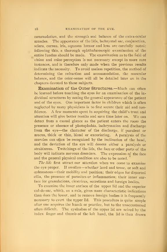
The lids first attract our attention when we come to examinethe eye proper. If swollen—whether hard and tense, or soft andcedematous—their mobility and position; their edges for distorted
cilia, the presence of parasites or inflammation; their inner sur-
face for granulations, cicatrices, secretions and foreign bodies.
To examine the inner surface of the upper lid and the superior
cul-de-sac, which, as a rule, gives more characteristic indications
than does the lower, and to remove foreign bodies it is frequently
necessary to evert the upper lid. This procedure is quite simpleafter one acquires the knack or practice, but to the unaccustomedoften difficult. The eyelashes of the upper lid are seized by theindex finger and thumb of the left hand, the lid is then drawn


Fig. I.
Method of examining the 63*6 in children.

EXAMINATION OF THE OUTER STRUCTURES. 1
9
downward and away from the ball, the point of the thumb of the
right hand or a pencil is then placed above the tarsal cartilage of
the lid and by a quick downward pressure of the thumb and a
simultaneous upward movement of the left hand grasping the cilia
the edge of the lid is turned over the point of the thumb. During
the entire manoeuvre you must insist upon the patient's keeping
the e3^e downward, if not the eversion of the lid becomes unneces-
sarily difl&cult and painful. When everted the thumb of the
right hand presses the edge of the lid backward against the eye-
ball and holds it for examination.
The lachrymalpiinda and sac should be examined for any ob-
struction, and by pressure over the sac notice whether any mucoid
material or tears can be expressed from the puncta. The inspec-
tion of the conjunctiva shows us the presence of phlyctenules,
pterygium, growths, adhesions, etc. The vascular condition of
the eye affords most important information, and it should derive
careful attention. Note if the redness is due to the large, tortu-
ous, bright red superficial vessels of the conjunctiva, which are
especially numerous toward the peripher}^ and looser portion of
the membrane, or if fine, radiating lines, pink in color, confined
to the ciliary region and due to the episcleral vessels. The charac-
ter of the congestion can be determined by gently rubbing the
lower lid over the eyeball, when it will be seen that the coarser
conjunctival vessels will glide over the deeper episcleral ones. In
some cases we may note a leash of vessels, more or less pyra-
midal in shape, with the apex toward the cornea, indicative of an
ulceration. Again, we may see a marked enlargement and tortu-
osity of the episcleral veins, pointing out a glaucoma.
The thorough examination of the conjunctiva and cornea in
young children where there is much photophobia and inflamma-
tion is usually a matter of great difficult^^ When, owing to these
causes, there is a spasmodic contraction of the lids, their forcible
separation can be best accomplished as shown in Figure i. Thenurse or attendant seated at your side lays the child across
her lap with the head held firmly between the surgeon's knees.
The attendant in this way can readily hold the child's hands, feet
and body while the head is held as within a vise by the surgeon's
knees. A towel should first be placed across the lap of the sur-
geon to prevent the staining of the clothes from any solutions that

20 EXAMINATION OF THE EYE,
may be used. The surgeon then grasps the ciliary border of the
upper lid with the index finger of the right hand and with the
thumb of the left hand the border of the lower lid. In opening
the eye the pressure must be mainly upward toward the supra-
orbital ridge and just sufficiently backward to prevent the eversion
of tie lid. Great care must be used 7iot to make too great pressure
backward or downward upon the eyeball, because in an ulceration
of the cornea (which is so apt to be present in cases where this
method has to be resorted too) the pressure is liable to cause a
rupture of the cornea with loss of the eye. Many an eye has un-
doubtedly been lost through careless and severe handling in an
effort to examine the same. You will often have to hold the eye
open for several minutes before a clear view of the cornea can be
had, as it will roll so far upwards that the cornea cannot be seen
until the muscles have become tired out and allow it to resume the
direct position. In some cases, when one has become especially
dexterous in this manipulation, they can open the lids by the use
of the thumb and index finger of the same hand, leaving the other
hand free to make any necessary applications.
As the examination of the cornea is greatly facilitated by the
use of the oblique illumination, it should always be employed.
This cannot be too strongly emphasized, as we have frequently
seen our students by neglect of this method overlook s^ome minute
yet important diagnostic sign which was readily discernible by
its employment. Make it, therefore, a routine practice in all
cases when examining the anterior part of the eye. Its use aids
the minute examination of the lids and conjunctiva, as well as
the cornea, iris, lens and aqueous. By it we may often determine
small superficial ulcers and abrasions, commencing interstitial infil-
trations, faint opacities or nebulae, and particles of foreign sub-
stances imbedded in the cornea. The discovery of minute tears or
abrasions of the corneal epithelium ma}^ be aided by the instillation
of a drop of a two per cent, solution of the potassium or sodium
salt of fluorescin. This should be dropped upon the cornea and
followed by a washing with distilled water; any break of the epi-
thelium will be made apparent by a deep greenish stain, which
remains for about two hours.
Oblique illiunination , or, as it is sometimes called, /"^(f^/ ox lateral
ilhimijiation^ is used as shown in Fig. 2. The patient is placed two

Fig. 2.
Method of oblique illumination.


EXAMINATION OF THE OUTER STRUCTURES. 21
feet from the gaslight iu a darkened room, as preferable to daylight,
the light is then brought to a focus upon the cornea with a two or
three inch lens, the surgeon may at the same time observe the
surface under examination through another magnifying lens held
before the eye. In order to focus the light upon the different
structures, the illuminating lens will have to be moved slightly,
according as the pencil of light is made to play over the cornea,
iris, or lens.
Inspection of the iris may frequently reveal normal ph3'siologi-
cal differences in color or shade of the two irides; and we mayalso have instead of the uniform pigmentation one or more irregu-
lar spots of different color. We can also detect hy the oblique illu-
mination swelling, discolorations and vascularity of the iris tissue;
the loss of lustre or the presence of gumma, foreign bodies, etc.;
the shape and size of the pupil, the presence of adhesions to either
the cornea or lens. The mobility of the iris should be carefully
studied, as the pupils of the two e3'es should act consensually; to
examine, the patient is placed before a window in da3dight and
directed to look at a distance; one eye is then covered, the other
exposed eye will contract to the bright light, while the covered
eye acts in harmony. If both eyes be now shaded dilatation en-
sues, and if t'.ien again exposed to the light contraction immedi-
ately follows, succeeded in a moment by slight dilatation and again
a contraction; thus oscillating for a moment it finally settles downto its original size. This action is called hippus, and is sometimes
present in a marked degree in cases of hysteria, mania, and other
nervous disorders. As the pupils contract under the influences of
accommodation and convergence, care must be taken during the
examination that the eyes are constantly fixed on a distant object.
Dilatation of the pupil occurs in glaucoma, atrophy of the optic
nerve, from fright, in anaemia, nervous conditions, etc., in youngpeople and from the use of mydriatics. According to McEwendilatation in diseases of the nervous system, w^hen of cerebral
origin, indicates extensive lesion, and when of spinal origin irrita-
tion of the part.
Contraction of the pupil occurs in old people, from the use of
myotics, is present in inflammation of the iris, in some fevers, in
mitral disease and pulmonary congestion, and in paralysis of the
sympathetic. If of cerebral origin, as in meningitis, it indicates
22 EXAMINATION OF THE EYE.
an early irritative stage of the disease; if of spinal origin, a de-
pression, paralysis or even destruction of the part (McEwen).The Arg341-Robertson pupil is the small, contracted pupil which
affected little, or none at all, by light and shade, responds by con-
tracting still farther under the influence of convergence. This
action of the pupil is found in degeneration of the posterior col-
umns of the cord and indicates a serious central lesion.
The examination of the ante^dor chamber and lens also, by the
aid of the oblique illumination, shows if the former is more shal-
low or deeper than normal, the presence of any exudation, etc.,
while in the lens the faintest trace of disturbance or change can be
detected.
Proptosis, or protrusion of the eye, if unilateral, may be noted
by comparing the position of the corneae with each other and with
the brows. It is present in Graves's disease, orbital diseases, in-
traocular tumors, paralysis of the ocular muscles, etc.
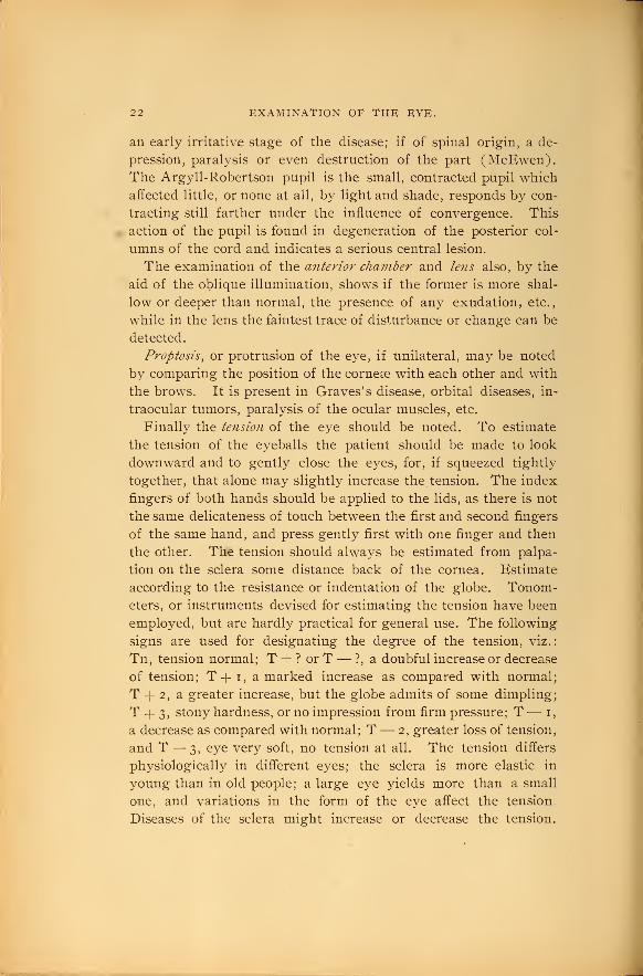
Finally the tension of the eye should be noted. To estimate
the tension of the eyeballs the patient should be made to look
downward and to gently close the eyes, for, if squeezed tightly
together, that alone may slightly increase the tension. The index
fingers of both hands should be applied to the lids, as there is not
the same delicateness of touch between the first and second fingers
of the same hand, and press gently first with one finger and then
the other. The tension should always be estimated from palpa-
tion on the sclera some distance back of the cornea. Estimate
according to the resistance or indentation of the globe. Tonom-eters, or instruments devised for estimating the tension have been
employed, but are hardly practical for general use. The following
signs are used for designating the degree of the tension, viz.:
Tn, tension normal; T+?orT— ?, a doubful increase or decrease
of tension; T+ i, a marked increase as compared with normal;
T -f- 2, a greater increase, but the globe admits of some dimpling;
T -f 3, stony hardness, or no impression from firm pressure; T— i,
a decrease as compared with normal; T— 2, greater loss of tension,
and T — 3, eye very soft, no tension at all. The tension differs
physiologically in different eyes; the sclera is more elastic in
young than in old people; a large eye yields more than a small
one, and variations in the form of the eye affect the tension.
Diseases of the sclera might increase or decrease the tension.

Fig. 3.
il^'^-m>^if
Method of determinin^i the tension.


THE FIELD OF VISION. 23
Variations in the curvature of the sclera at the point of impression
will cause a slight difference in the tension, the greater the curva-
ture the softer the e\'e. The tension of one eye should alwa3'S be
compared with its fellow, and when in doubt with an eye knownto be normal, in a person of the same age as the patient.
The Field of Vision.—By the field of vision is meant the
space, when the visual axis of one eye is fixed upon some
stationar}^ point, in which all other objects are visible. This
space is large or small, in proportion to the distance at which the
fixation point is from the eye. The object fixed imprints its
image upon the macula lutea, while the image of all other objects
fall upon some peripheral portion of the retina.
Peripheral vision is of value, in that while we only see objects
iudistinctl}" upon which the visual axis is not fixed, it attracts
our attention to other objects which we maj^ desire to see, and the
eye is then turned in that direction. As, for example, in crossing
a street our peripheral vision is attracted b}^ the approach of a
team within the field of vision and our attention is turned to it
that we ma}^ avoid an accident. In many diseased conditions of
the fundus a knowledge of the field of vision is of the greatest
importance both in diagnosis and prognosis.
T/ie 7iormal field of vision varies indifferent directions, being
greatest toward the temporal side, where it has an extent of over
90° because the rays from such a point, owing to the strong re-
fraction at the surface of the cornea, can still enter the pupil.
The field at the nasal side and above is of much less extent, be-
cause of the limitation caused b}^ the nose and brow. The normal
field for colors is found practically to be more contracted than that
for white, and to vary with the different colors—blue being the
least contracted, red next and green the most contracted.
Pathological changes in the field of vision are both numerous,
varied, and, in many diseases, are quite characteristic. Altera-
tions in the visual field may be concentric, uniformh' drawn in at
all points; sector-shaped, where it has the shape of a triangle
whose base corresponds to the periphery; hemiopic, one-half of
the field wanting; in addition to these more or less regular and
frequently found forms of contraction there are many irregular
shaped notches in the normal field. Scotomata, or blind spots in
the visual field, when found as the result of disease, are classed

24 EXAMINATION OF THE EYE.
as central or peripheral. A central scotoma involves the point of
fixation, and means that direct vision is either diminished or wholly
lost. Peripheral scotoma, on the other hand, do not involve direct
vision and cause but little disturbance; in fact, are often not knownto the patient until found in examining the field. An annular
scotoma is one that more or less completely surrounds the point of
fixation like a ring, the direct vision being left intact. In the
healthy eye we have a scotoma, known as Mariotte's blind spot,
which corresponds to the entrance of the optic nerve and lies
about 15° to the outside of the point of fixation.
Concentric conti^action with central vision impaired, may be found
in atrophy of the optic nerve or retina; with central vision good,
in retinitis pigmentosa and sometimes in the early stages of
glaucoma. Sector-shaped alterations may be found in atrophj^ of
the optic nerve, in occlusion of one of the retinal arteries, in de-
tachment of the retina, and in glaucoma the nasal side is con-
tracted. Scotomata are found in choroiditis disseminata and other
choroidal diseases, in haemorrhages, especially when in the macula
lutea, in toxic amblyopias, etc.
The importance of a careful study of the field for colors, as well
as for white, is well illustrated in atrophy of the optic nerve, as in
this disease the color field is more constantly involved than that
for white, and in some cases will be the first sign of the disease.
In glaucoma the field for colors is lost with that for white, and
they bear the same concentric arrangement throughout. In toxic
amblyopia there is frequently found a central scotoma for red and
green.
These few illustrations are merely suggestive as to the impor-
tance of perimetry in the study of intraocular and cerebral con-
ditions, further reference to the pathological involvements of the
field of vision will be found under the various diseases. '.
Examination of the Field of Visioji.—This must be made for
each eye separately; the eye to be examined is directed at a fixed
point, as it must remain steadily in the same position, while the
other eye is closed. There are three methods of determining the field
of vision; the simplest, and, at the same time the poorest, is that by
using the hand as a test-object. The physician stands in front of
the patient, who directs his left eye to the right ej^e of the physi-
cian, the other eye of each being closed. The physician then

THE FIELD OF VISION. 25
moves his hand in a plane midwa}^ between the patient and him-
self from the periphery inward over the limits of the field of view.
The patient is to tell as soon as he sees the hand, and if his field
is normal he should see the hand at the same time as does the
physician. This method is onh' adopted to determine large de-
fects and in those where the central vision is too poor to see
smaller test-objects. The field in patients wnth cataract is usualh^
tested in this way, using a candle-flame in place of the hand.
The blackboard is the second method of determining the field.
In this the patient's head is rested on a support 30 cm. from the
board. x\ chalk mark is made directly opposite the eye to be ex-
FlG. 4.
Skeele's perimeter.
amined, on which he is to fix his gaze. The chalk is now gradu-
ally approached from the edge to the center, and the patient tells
at the moment he first sees it. By marking this spot where he
first sees the chalk in all directions of the field, and then connect-
ing the points thus determined, we have the field of vision. Byusing colored chalks we can determine the field for the various
colors. This method is also inexact.

26 EXAMINATION OF THE EYE.
The only exact and scientific method of determining the field
of vision is that where the projection is upon a hollow sphere.
This is now determined by means of an instrument known as the
perimeter. The patient's head is supported on a chin rest, which
is so placed in front of a semi-circle that the eye to be examined
is situated in the centre of the curvature of the latter. The eye is
then fixed upon the middle point of the semi-circular arc, while a
test-object, a small white or colored square, is carried along the
arm of the semi-circle. The semi-circular arc is marked with a
scale of degrees which can be read off, or in the best perimeters is
self-registering on a chart attached.
i

THE USE OF THE OPHTHALMOSCOPE. 27
CHAPTER II.
The Use of the Ophthalmoscope.
In all the realm of modern medicine there has probably been
no one discover}' of greater beneficence to humanit}' than the in-
vention of the ophthalmoscope b}' Helmholtz in 1851. Through
its use the mj-steries of the interior of the e3'e stand revealed and
many conditions that previously resulted in blindness are nowmade remediable. With it we are able to study changes in the
circulatory system, as exhibited in the retinal vessels; and in the
optic nerve and retina we have, under the eye of the surgeon,
direct communication with the brain and spinal system. Theophthalmoscope, therefore, has become of the greatest value in
general medicine as an aid to diagnosis, for in the fundus of the
eye are found many characteristic changes of disease of the
various organs. Helmholtz 's discovery was not a matter of
chance, but resulted from a careful study of the laws of optics,
one of which is that light follows the same lines in returning
through a lens (in case it can return) as when entering. Therays of light returning from the eye must go direct to the
luminous source from which they emanated, and in order to fall
upon the retina of the observer his eye must be in the path
formed by the source of the illumination and. the eye under ex-
amination. The device used by Helmholtz consisted of a trans-
parent mirror formed of three slips of plane glass. The present
principle of a perforated metallic mirror w^as first proposed b}?-
Ruete, in 1852. In examining the interior of an eye, light is
thrown into the eye by the mirror, and in order to see the
fundus we must receive in our own eye the light reflected from
the fundus and unite its rays to form a sharp image. Themirrors used may be either plane or concave. The concave
mirror by converging the rays from the source of light gives a
stronger illumination and is therefore generally used. Themodern ophthalmoscope, of which Loring's is one of the best

28 THE USE OF THE OPHTHALMOSCOPE.
consists then of a concave mirror, silvered on the back, for
illuminating the eye and a series of lenses for measuring the
refraction, and for diagnosing pathological changes by the direct
method.
The art of using the ophthalmoscope is one much more difficult
Fig. 5.
Loring's ophthalmoscope.
to acquire than that of any other instrument of precision and is
only accomplished after long and persistent practice. Every
physician realizes the months or years of practice required to
detect with the stethoscope the finer shades of sounds due to
varying diseases of the heart and lungs. In one case the ear and

THE USE OF THE OPHTHALMOSCOPE. 29
in the other the eye has to be trained by long experience before
the examiner can become expert. The beginner is apt to think
that after he has acquired a few details of the nerve and vessels
that he can see all that is to be seen. At this stage we have often
told our students that they have as yet not crossed the threshold
of that vast storehouse of beautiful pictures formed by diseases
within the eye. Even after years of daily use this little instru-
ment reveals significant and often important variations of patho-
logical states not heretofore seen, the meaning of which the
observer is often at a loss to understand.
The first and most essential point in order to become a skilled
ophthalmoscopist, and which is often neglected, is familiarity
with the healthy fundus. The student should first practice over
and over again upon every healthy eye-ground he can before
attempting to study diseased states. This necessit}^ becomes
apparent from the fact that the normal fundus in health varies
with the age, condition and complexion. What a large range of
physiological pigmentation ma}^ be found from the negro to the
albino. The skilled use of the ophthalmoscope is in the deter-
mination of the very slightest changes from normal, as the detec-
tion of gross pathological conditions does not present the impor-
tance that does the recognition of the incipient stages of disease.
In making an ophthalmoscopic examination artificial light is
generall}' used and is preferable to daylight. We therefore
darken the room and use a vsingle light, the best being that from
an Argand burner or a student's lamp. The e3'e is first illumi-
nated from a distance of about eighteen inches, and as the light
plays over the cornea we note any opacities that may be present
in the cornea or lens. Occasionally when there is a marked error
of the refraction the retinal blood-vessels will be seen. If the eye
is highh' far-sighted the vessels will move in the same direction
as the head of the observer, while if it is a very near-sighted e3^e
the vessels will move in an opposite direction.
There are two methods of examining the fundus of the eye:
First, the direct method, so called because the eye-ground is
studied by rays coming directly from it, and by this method wehave an upright image; and second, the i?idirect, because the rays
are received from an aerial image, or indirectly from the observed
eye, and the image seen is inverted. The latter method will first

30 THE USK OF THE OPHTHAI^MOSCOPK.
be considered because it is more frequently employed and because
it is the more natural order after the preliminary examination of
the cornea and lens.
The indited method, or the method of examination by the divert-
ed image, is made as shown in Figure 6. The patient is seated in
a darkened room with the light from an Argand burner about
eighteen inches behind, on the same side, and level with the eye
to be examined. He should be instructed to fixate the unused
eye upon some distant object. The observer sits about eighteen
inches in front of the patient and holds the ophthalmoscope in the
hand corresponding to the eye to be examined. A convex lens,
about thirteen to eighteen diopters, is held between the thumband forefinger of the unused hand, before the eye of the patient.
By resting the middle, third and little fingers upon the outer part
of the supra-orbital ridge of the patient's eye the lens is held
steadily and focused upon any part of the fundus desired, and the
middle finger may also be used if necessary to raise the upper lid
for a better view. In all ophthalmoscopic work the student
should learn to keep both eyes open, as the effort to close one eye
tires the eye and prevents the complete relaxing of the accommo-
dation. He should also accustom himself to using the right ej^e
and holding the ophthalmoscope in the right hand when examin-
ing the right eye of the patient, and the left eye and hand whenexamining the left eye. The first objective point is the optic
nerve head, and this is brought into view b}^ having the patient
look at the right ear of the observer, and vice versa, when examin-
ing the left eye, the patient should be told to look at the left ear
of the surgeon. From this point he may be told to look directly
at the centre of the observer's forehead, which gives a view of the
macula lutea, and then, up and down, to the right and left, in
order to examine all parts of the fundus. If the image of the
disc when first brought into view appears dim and ill-defined, the
lens and the ophthalmoscope should be moved slightly forward or
backward until the image is as clear and distinct as possible.
The student must always remember that by the indirect method he
sees the aerial picture of the fundus and that it is inverted and
reversed. The image by the indirect method is magnified about
four or five times, while by the direct method we get a picture
magnified about fourteen times. The extent of the field of vision

Fig. 6.
Opthalnioscopic examination by the indirect method.


Fig. 7.
Opthalmoscopic examination by the direct method.
<
i

THE USE OF THE OPHTHALMOSCOPE. 3
1
on the contrary is about four times greater in the indirect than it
is by the direct method. The intensity of the ilhimination is also
greater with the indirect than with the direct, hence a view of the
fundus can often be had by the indirect method when, owing to
haziness of the refracting media, it is no longer visible by the
direct. The indirect method gives then a larger view and better
general relation of the fundus, while the direct method is particu-
larly adapted for the recognition of the finer details.
The direct method, or the examination with the erect image, is
shown in Figure 7. The patient and light are placed in the
same positions as in the indirect examination. The surgeon
seats himself by the side of the patient and again uses his right
eye in examining the right eye of the patient, and vice versa.
The ophthalmoscope is held in the same hand as the eye to be ex-
amined and brought up to about one inch from the eye of the
patient. Both eyes are to be kept open so as to avoid as much as
possible the impulse to accommodate. Ao the field is enlarged,
and the examination by this method greatly facilitated b}' a dilata-
tion of the patient's pupil, the use of a mydriatic is to be recom-
mended to the student when first learning to use the direct method.
"The dilatation of the pupil can be increased also by having the
room as dark as possible, by closing the other eye, and lowering
the light from which the illumination is received. If a still
larger pupil be required for an examination of the fundus a 4 per
cent, solution of cocaine should be used, as it will give the neces-
sary dilatation in from twenty to thirty minutes and its effect
passes away in a few hours.
By the direct method, if both the eye of the observer and of
the patient be normal in refraction, and the accommodation at
rest in both, the details of the fundus are readily seen. If, how-ever, either the surgeon or the patient be m^^opic, or if hyper-
metropic in excess of the power of accommodation to overcome,
the refractive error must first be corrected. The power of relax-
ing one's accommodation comes by practice. The primary ob-
jective point in the examination is, as by the indirect method, the
optic disc, and this is brought into view b}^ having the patient
look straight forward w^hile the surgeon looks into the eye slightly
from the temporal side.

32 THE USE OF THE OPHTHALMOSCOPE.
The Fundus of the Eye as Seen by the Ophthalmo-scope.— (See Figures i and 2, Plate II, Chromo-Iyithographs.)
As already mentioned, the first objective point in all examina-
tions of the interior of the eyeball is the optic disc, or papilla.
The term papilla is somewhat inaccurate, as the inference drawn
from the word papilla would be that it was an elevation or
something protruding from the surface of the fundus. This is
not the case, as there is no prominence, and hence the term papilla
is misleading; as, however, it is so generally employed, we shall
use the word interchangeably with the more correct term disc.
The optic nerve appears usually as a circular or slightly oval
shaped disc, but may be quite irregular in outline. Its color varies
from a pinkish white to a deep red, and may vary in different
parts of the disc, often paler at the centre than at the circumfer-
ence, or the nasal side a more decided red than the temporal. Thetint also varies wnth the age and complexion of the patient, and
the contrast with the color of the surrounding fundus. Thewhite appearance of some portion of the disc is due to a depres-
sion at that point, the floor of which is composed of an interlacing
opaque fibrous tissue called the lamina cribrosa, through which
the nerve fibres pass, and it is here they lose their medullary
sheath and become transparent axis cylinders. This white spot,
varying in size, is seen usually at the centre of t-he papilla, or,
rarely, more at the temporal side is called the physiological cup or
excavation. Care must always be taken to differentiate this
physiological cupping from the excavation found in glaucoma and
in optic nerve atrophy. A description of the different forms of
cupping of the disc will be found under the study of glaucoma.
The border of the optic disc is well defined, being sharply outlined
by a double ring. The inner, or scleral ring, appears as a faint
white streak, especially distinct in elderly people, and indicates
the opening of the sclerotic coat through which the optic nerve
enters the eyeball. Jaeger has called this the connective tissue
ring, formed by the junction of the connective tissue elements of
the inner sheath of the nerve with layers of the sclera. Theouter, or choroidal ring, usually seen as a slight black crescent
upon one side of the disc and often wholly absent, bounds the
opening in the choroid.
The next most noticeable feature in the examination of the

FUNDUS OF THE EYE AS SEEN" BY THE OPHTHALMOSCOPE. 33
fundus is the blood-vessels. The arterial trunk usuall}^ divides,
just before emanating from the bottom of the disc, into an up-
ward and downward branch, each of these branches generally
dividing again as the}^ pass off from the optic disc. These arteries
as they spread out above and below continue to divide dichoto-
mously into numerous branches, supplying all parts of the fundus,
excepting a small area at the temporal side of the optic nerve.
This area is called the viacula hctea, or yellow spot, and at its
centre is the point of most distinct vision, the fovea centralis.
The temporal half of the retina is more freely supplied with
blood-vessels than is the nasal side. The retinal veins follow the
same general course and parallel to the arteries, and empty by
two large branches into the centre of the disc. From this general
arrangement of the retinal vessels we maj^ have manj^ variations in
the normal fundus. The arteries and veins are distinguishable
by their size and color, the veins being larger in proportion of
about three to two and of a dark red as contrasted with the bright
color of the arteries. The veins are also more tortuous in their
course and spontaneous pulsation is not infrequently seen in the
veins. The so-called reflex or light streak, which runs along the
crest of the vessels, covering about one-third of their diameter, is of
a pale straw color, and is more brilliant, broader and more sharply
defined upon the arteries than veins and may be entirely absent in
the veins. The cause of this reflex is unsettled, some claiming it
to be a reflex from the vessel wall, others from the blood column.
The appearance of the macula lutea is as difiicult to describe as
it is to the student to see. No two observers seem to illustrate or
describe it in the same coloring. In many cases while we ex-
amine the macular region we see nothing, and often we are
but conscious of a luminous oval ring, the centre of which is
marked by a small spot of a darker color. This phantom-like reflex,
or, as it is sometimes called, halo, varies in size, though usually
of an oval or circular shape. The inclosed space seems to be
more of a grayish or brown color than the 3'ellow we should
naturally expect from the macula lutea being commonh^ spoken
of as the j^ellow spot. The examination of the region of the
macula lutea should always be practiced, for while in the normal
eye the halo is often absent and the coloration of this spot vari-
able, in diseased states an accurate picture of the macula is often
3

34 THK USE OF THE OPHTHAI.MCSCOPE.
of the utmost importance. The location of the yellow spot is
about one and one-tialf optic nerve diameters to the outer side of
the disc and is usually best seen by the indirect method.
The retina, being a transparent membrane, is practically invisi-
ble and reveals nothing of its delicate structure excepting the
retinal vessels, which are readily seen ramifying within its inner
layers. Some, however, have claimed to have seen, especially in
the deeply pigmented eye of the negro, with a weak illumination,
the presence of the retina as a very faint grayish tinge in the
neighborhood of the disc. To the observer, especially when in-
experienced, the retinal vessels seem to course over and form a
part of the background of the eye. They should, however, always
remember that they lie some little distance in front of the under-
lying choroid. This can be more easily appreciated in the slightly
pigmented eye, especially the albino, where they are readily seen
passing over the choroidal vessels. Recognition of the choroid
varies with the pigmentation of the eye. The bright red color
from the pupil when the eye is illuminated with the opthalmo-
scopic mirror arises from the choroid. The choroidal vessels ap-
pear as flat curvilinear stripes of a light pink hue interlacing in
distinct meshes. The pigment stroma shows as irregular patches
within the meshes of the choroidal vessels. The pigmentation is
often more dense around the optic nerve and posterior part of the
fundus. The visible choroidal vessels are always broader than
the retinal trunks, and no distinction can be made between the
arteries and veins.

NORMAL REFRACTIOX AND ACCOMMODATION, 35
CHAPTER III.
Refraction and Accommodation of the Eye.
By Chas. H. Hf:lfrich, M. D., Surgeon to the X. Y. Ophthalmic Hospital.
Normal Refraction and Accommodation.—The dioptric
media of a normal or emmetropic eye f cornea, aqueous humor,
lens and vitreous humor) have the requisite refractive power to
bring parallel rays of light to a focus on the layer of rods and
cones of the retina. These media are centered on the optic axis,
a line passing through the centre of the cornea and the posterior
pole of the eye.
Fig. S.
Schematic eye. *J•^ anterior or first principal focus; A, anterior surface of
the cornea; H^ and H'^, principal points; K^ and K^^ nodal points;
4'^^, posterior or second principal focus; F.r., fovea centralis; ^^ 4>^^^
optic axis.

36 REFRACTION AND ACCOMMODATION OF THE EYE.
Upon the optic axis are situated the cardinal points of the
dioptric system.
Objects situated at a distance of five metres or more are con-
sidered as being at infinity, because those rays from them which
enter an eye are so shghtly divergent that for practical purposes
they may be considered parallel. As parallel rays are brought to
a focus at the second principal focus, the eye is capable of forming
distinct inverted images of distant objects upon the retina.
The eye, however, can also see near objects distinctly, and as the
rays from such sources become more divergent the nearer they ap-
proach, it is obvious that it must contain some mechanism to in-
crease its refractive power. The power by which it is increased
so that divergent rays are also brought to a focus on the retina is
the accommodation.
Fig. 9.
Changes in the eye produced by accommodation, r, cornea; a, anterior
chamber; /, lens; v, vitreous humor; /, iris; z, zonula of Zinn; Tn,
ciliary muscle.
By the term static refraction is meant the power the eye has
when at rest (without an effort of accommodation) to bring
parallel rays of light to a focus on the retina or to render diver-
gent rays less divergent.
The dynamic refraction constitutes the increase of refractive
power produced by the effort of accommodation.
The mechanism of accommodation is as follows: By con-

THE FAR AND NEAR POINTS. 37
tracting the ciliary muscle the tension on the zonula of Zinn is
relaxed, permitting the lens to become more convex through its
own elasticit_y, and thus increasing the refractive power.
The changes which take place in accommodation are repre-
sented by the dotted lines in (Fig. 9.)
The anterior surface of the lens advances and becomes more
convex, while the convexit}' of its posterior surface increases but
little and does not change its position at all. Associated with this
act is a contraction of the pupil.
The far and near points.—The name piuidum rcmotiivi, or
far jjoint, is given to the point to which the ej'e is adapted whenat rest. It represents the most distant point of distinct vision, and
is designated by R. By the term pitnclum p7'oximnm or near
point, is understood the nearest point of distinct vision. It is
found by ascertaining the nearest point at which the smallest test-
letters can be read, and is designated b}' P. It is possible for the
eye to see all objects distinctly between these two points.
The range or amplitude of accommodation is the amountof accommodative effort of which an eye is capable, and is equal
to the difference in the refractive power when in a state of rest
and when its acommodation is exerted to the utmost. It may be
represented b}^ that convex lens, placed in front of an eye, which
would give to ra3^s coming from the near point a direction as if
they came from the far point. If we consider a equals the numberof dioptres represented by the range of accommodation, p the
number of dioptres represented b}^ the eye when adapted to its
near point, and r the number of dioptres represented by the eye
when adapted to its far point, we can calculate the amplitude of
accommodation by the following formula:
—
a = p — r.
In the emmetropic e3'e R is at infinity, therefore r= o; hence
a =p. To illustrate, when the near p jint is 20 cm. (a focal length
of 20 cm. represents a lens of 5. D) from the eye,' we have a =5. D.
In myopia R is at a fixed distance, and, for example, if it is situ-
ated at 50 cm. (myopia of 2. D) and P at 20 cm. (5. D) we have
a=S' D.-2. D.=3. D.
The hyperopic eye is adapted for rays which converge to a point
behind the retina, therefore r is negative and must be added \.o p.
In this case we have a —/>_(—;') and reduced a =p —- r. To illus-

38 REFRACTION AND ACCOMMODATION OF THE EYE.
trate, if the hyperopia is lo. D. and P is situated at 20 cm., wehave a =5. D.-\^io. D.=r^. D.
Convergence. — Ordinarily man looks simultaneously with
both eyes, yet appreciates but a single image. This union in one
single impression of the retinal images received by both eyes is
called binocular vision. In order to obtain this each eye must
receive upon its fovea centralis a distinct image of the object, and
hence it is necessary that both lines of fixation (a line connecting
the object of fixation with the centre of rotation) be directed
towards the object looked at. When looking at a distant object
the lines of fixation are parallel, but the nearer it approaches the
more the lines of fixation must converge and the eyes turn in. If
an object is moved along the median line (I M, Fig. 10), a line
Fig. 10.
The metre angle.
perpendicular to the middle of a line uniting the centres of rota-
tion, both eyes converge equally to any given situation. The
degree of convergence is measured by the angle through which
an eye turns when it fixes the object. When it is situated at I,
one metre distant from the eye, the angle of convergence E I M

ACCOMMODATION AND COXVERGEXCE ASSOCIATED. 39
is one metre angle which is taken as a unit. If the object be
situated at ^2 of a metre, it is obvious that the angle of converg-
ence is twice as large as in the former instance; that is, it equals
2 metre angles.
Accommodation and Convergence Associated.—With
ever}' degree of convergence is associated a certain effort of the
accommodation. When looking at an object situated at one metre,
it is necessary to converge i metre angle, and an effort of the ac-
comodation equal to a convex lens of i. D must be employed.
That is, the refraction and convergence must increase b}' an equal
quantit}^ which is the inverse of the distance of the object.
This association between accommodation and convergence,
however, is not absolute, for with the lines of fixation fixed on a
given point and stationary, the accommodation can be somewhat
increased and diminished; and conversely, with a given amount
of accommodation, the degree of convergence can be augmented
and reduced.
If an object is held at one metre and first weak convex and
then weak concave glasses be placed before the eyes the distinct-
ness of the image is unaltered. The relative amplitude of accom-
modation is thus obtained. The part represented b}^ the strongest
convex glass which can be placed before the eye without affecting
the distinctness of the object is termed the negative, and the part
represented b}^ the strongest concave glass the positive. Whensustained efforts of the accommodation are necessary at any dis-
tance, it is essential that the positive relative amplitude of accom-
modation be considerable.
That the convergence may be altered while the same effort of
accommodation is maintained can be demonstrated by placing a
weak prism with its base in before one ej'e. If the convergence
remained unaltered, the prism would cause double vision, but the
eyes rotate outward and the object looked at is still distinct and
the image single. Likewise, it will be found that a weak prism
with its base out will be followed by a rotation of the eye inward
wnth no effect on the distinctness of the image. The relative
amplitude of convergence is thus obtained.
The Angle Alpha and Angle Gamma.—The optic axis AA' (Fig. II.) is an imaginary line, which ma}^ be regarded as pass-
ing through the centre of the cornea C and the posterior pole of

40 REFRACTION AND ACCOMMODATION OF THE KYK.
the eye— a point situated between the fovea and the optic papilla.
Upon it are the cardinal points and the centre of rotation M.
Fig. II.
Schematic figure showing the angles a and }. AA^, optic axis; *^. anterior
focus; ^'\ posterior focus; WW^, principal points; K^K^^, nodal
points; M, center of rotation; C, centre of cornea; BB, base of the
cornea; BL, major axis of the corneal ellipsoid; F, fovea centralis; O,
poinf of fixation; K^O, line of vision; MO, line of fixation; O X B,
angle a\ O M A, angle }
.
Ths visual line O F unites the point of fixation O—the object
looked at—with the fovea. It does not coincide with the optic
axis, but crosses it at the nodal points.

THE ANGLE ALPHA AND ANGLE GAMMA. 4I
The line of fixation O M joins the centre of rotation with the
point of fixation.
If the fovea coincided with the posterior pole, the visual line,
line of fixation and optic axis would also coincide, but this is not
the case.
The apex of the corneal ellipsoid E does not coincide with the
centre of the cornea, and therefore neither does the major axis of
the ellipse E L coincide with the optic axis.
The angle O XE formed by the visual line and the major axis
of the corneal ellipse is called the angle alpha.
When the anterior portion of the corneal axis is situated to the
temporal side of the line of vision, the angle a is called positive;
when it is situated to the nasal side, negative.
The angle O M A formed b}^ the line of fixation with the optic
axis is called the angle gamma.It is termed positive when the anterior extremit}^ of the line of
fixation passes to the inner side of the optic axis, and negative
when it passes to the outer side.
In practice, it is usual to consider the line of fixation and the
visual line as indentical.
In order to measure the angle gamma, the patient is placed be-
fore the perimeter as for an examination of the field of vision.
A lighted candle is moved along the arc of the perimeter, and by
means of the corneal reflection of the flame the centre of the
cornea is found. The position of the candle at the perimeter is
now read from the arc in degrees and represents the size of the
angle. Its average size is five degrees.
In emmetropia and hyperopia the visual line cuts the cornea to
the inside of its major axis, and the angle gamma is therefore posi-
tive. Ownng to the shortness of the eyeball in hyperopia, the
effect of which is to increase the distance between the fovea and
the optic axis, the angle gamma is very much greater than in
emmetropia. This may give to the eyes the appearance of an
apparent divergent strabismus, as the axes of the corneae seem to
diverge though the fixation is correct.
In myopia the length of the eyeball is too great, so the visual
line cuts the cornea nearer the major axis, or they may coincide,
or it may cut it to the outer side making the angle gamma nega-
tive. In the latter case the effect will be to give the eyes the ap-
pearance of an apparent convergent strabismus.

42 REFRACTION AND ACCOMMODATION OF THE EYE.
Abnormalities of Refraction and Accommodation.— Ashas been explained in the preceding pages, a normal or emmetropic
eye is one whose static refraction is sufficient to bring parallel
rays to a focus on the retina; or, one whose retina is situated at
the focus of its dioptric system. Its far point is always at in-
finity. Any departure from emmetropia is known as ametropia
of which three different forms are recognized: i. Hypermetropia,
in which the retina is situated in front of the focus of parallel
rays. 2. Myopia, in which the retina is situated behind the focus
of parallel rays. 3. Astigmatism, in which the refraction of the
different meridians is different.
Hypermetropia or Hyperopia.—In hyperopia the static re-
fraction is not sufficient to bring parallel rays to a focus on the
retina. Such rays if not intercepted by the retina would come to a
focus behind it. As they are intercepted by the retina they do
not form there a distinct image of the object looked at but a
circle of diffusion. In order to bring parallel rays to a focus on
the retina, it is necessary either to place an appropriate convex
lens before the eye which causes them to converge or to call the
accommodation into play.
Fig. J 2 shows how the parallel rays a b converge toward a point
r, behind the retina, after passing through the dioptric system, and
how the diffusion circle d e\^ formed upon the retina.
Fig. 12.
Formation of diffusion circles on the retina in hyperopia.
As the retina in hyperopia is nearer the dioptric system than its
principal focus, rays passing out from any point upon it such as
R (Fig. 13) will leave the eye divergent and will appear to come
from a point R situated behind the eye.
The point R' , the virtual conjugate focus of R, is the far point

HYPERMETROPIA OR HYPEROPIA.
Fig. 13.
43
Far point of a hyperopic eye.
of the eye, or the point towards which the rays must converge be-
fore entering in order to be brought to a focus on the retina. Be-
ing behind the eye it is negative. Jn order that parallel rays maybe brought to a fccus on the retina, it is necessary that the refrac-
tive power of the e3^e be increased by such a lens as will render
them convergent towards the point R' . This is shown in Fig.
14 where the lens L renders the parallel rays convergent towards
R' , and which the dioptric S5'stem render still more convergent
so that they come to a focus at R on the retina.
Fig. 14.
Correction of hyperopia by a convex lens.
The greater the hyperopia the nearer the far point is to the
eye, the more convergent the rays must be in order to co i;e to a
focus on the retina, and the stronger must be the lens which
renders them so. But the power of accommodation is also sufficient
to increase the static refraction sufficiently to bring parallel rays
to a focus on the retina if the degree of hyperopia is not too great.
In fact, it Ordinarilv does so in such cases so that the vision mav

44 REFRACTION AND ACCOMMODATION OF THE EYE.
be normal for distant objects, which has given rise to the mislead-
ing term of farsightedness. A beginner might fall into the error
of considering such an eye emmetropic; but it can be proven to
be hyperopic by successively placing stronger and stronger convex
glasses before it, which, as the accommodation relaxes, do not "in-
terfere with the distinctness of the object until the hyperopia is
overcorrected, or an artificial myopia is produced. Hence, it is
necessary to find the strongest convex glass through which the
hyperopic eye can see distant objects most distinctly in order to
find the measure of the error. Generally the ciliary muscle,
through force of habit, does not relax to its fullest extent, so that
the strongest convex glass simply represents the amount of mayii-
fest hyperopia (Hm). The balance of it, the late7it (HI), can
only be made manifest by instilling a solution of some cycloplegic
like atropine which suspends the accommodation.
The sum of the latent and manifest hyperopia gives the total
(Ht). Theoreticall}^ that glass placed in contact with the eye
whose focal distance is equal to the distance of the far point
behind the eye, or which renders parallel rays convergent to-
wards the far point, is the measure of the hyperopia. In prac-
tice, however, the glass is placed about 15 mm. in front of the
eye, and it is regarded as the measure, though in reality it is not
as great.
Causes.—The eyeball is either abnormally short, constituting
axial hyperopia, or its refractive power may be deficient, curva-
ture hyperopia. Hyperopia is nearly always congenital. Most
children are so at birth, but as they grow older the refraction
increases and they become less hyperopic, or emmetropic, or
myopic. Senile changes in the lens, flattening, give rise to it;
and its removal, as for cataract, produces a high degree. The
latter condition, however, is termed aphakia.
Symptoms.—The constant effort of the accommodation ne-
cessary in order to see distinctly gives rise to many symptoms.
As the ciliary muscle tires, vision blurs, and it is necessary to stop
work and rub the eyes. The resp.te obtained in this way is
only temporary, as the muscle soon tires again and the per-
formance must be repeated again and again until finally the
work must be discontinued. Such people often seek a good
light because the contraction of the pupil renders the vision

HYPERMETROPIA OR HYPEROPIA. 45
clearer. Frequently too they hold the object near the face to
secure larger retinal images and contract the lids to shut off
the more divergent rays. This gives the semblance of myopia,
and many children are erroneously given concave glasses which
aggravate the trouble.
When left uncorrected, hyperopia frequentl}' gives rise to con-
junctivitis, blepharitis, nictitation of the lids, and congestion of
the retina, choroid and optic nerve. Headaches and various
reflex neuroses are very common.
Strabismus convergens is frequenth' associated with hyperopia
the discussion of which will be found in the chapter upon that
subject.
Hyperopia is often complicated with spasm of the ciliary
muscle, the effect of which is to bring nearly or wholly the entire
accommodation into play. This reduces the amount of manifest
hyperopia when it is of high degree, and in some instances
may even convert the case into one of false myopia. The vision
in the latter instance will be improved by concave glasses, though
it would be a serious error to prescribe them. Such a mistake is
prevented by detecting the real nature of the refractive error by
means of the ophthalmoscope, as described in the chapter on
dipotometry.
When spasm of the accommodation is present, it is imperative
that a cycloplegic be instilled to temporarily paralyze the ciliary
muscle and so suspend the accommodation.
Manifest hyperopia is divided into facultative, relative, and
absolute.
Facultative hyperopia may be overcome b}^ using the accom-
modation without squinting.
Relative hyperopia represents a greater degree, and can only
be overcome by the accommodation when the patient squints
inward.
Absolute hyperopia is the highest degree, and cannot be over-
come b}^ using the entire accommodation.
The determination of hyperopia will be described in the
chapter on dioptometry.
Correction of Hyperopia.—If the patient has normal acute-
ness of vision and no asthenopic s}- mptoms glasses need not be
prescribed for him.

46 REFRACTION AND ACCOMMODATION OF THE EYE.
When distant vision is imperfect, and asthenopic symptoms are
present, it is necessary to prescribe glasses which represent the
amount of manifest trouble, either for constant use or for near
work. In some instances, it may be necessary to correct the mani-
fest and part of the latent if the latter exists. As a rule, if
hyperopia is associated with exophoria it is best to prescribe as
weak a convex glass as possible, whereas if esophoria is present,
the strongest. In spasm of the accommodation it is advisable to
put on nearly the entire correction while the eye is under the
influence of the cycioplegic, and later glasses which correct all
the manifest and as much of the latent as is tolerated
Many cases of convergent strabismus in children are cured by
prescribing appropriate glasses. The degree of hyperopia can be
determined by the direct examination with the ophthalmoscope
or skiascopy if the child is too young to know its letters.
Myopia.—In this form of ametropia parallel rays of light are
brought to a focus in front of the retina, therefore the latter is
situated beyond the principal focus.
The focus of the rays a b (Fig. 15) is at/ where they cross each
other, and on arriving at the retina form the diffusion circle c d.
Fig. 15.
Formation of diffusion circles on the retina in myopia.
As the retina is situated behind the principal focus, rays com-
ing from any point upon it such as c (Fig. 16) leave the eye con-
vergent and meet at a point r in front of it.
The points (f and r are conjugate foci, for if the rays coming
from r enter the eye its dioptric system will bring them to a focus
at c. For this reason r is the far point of the e3^e, as it is the most
distant point of distinct vision. A myopic eye is adapted for

MYOPIA. 47
divergent rays of light, therefore if a distant object is brought
nearer it can be seen distinctly when it arrives at the far point.
In order that a myopic eye may be able to see objects at infinity,
it is necessary, that parallel rays be given a divergence as if they
came from its far point. This can be accomplished b}' a concave
Fig, 1 6.
Far point of a myopic eye.
lens whose focal distance coincides with the distance of the far
point from the e\"e. Such a glass placed in contact with the
cornea would represent theoreticalh' the degree of myopia.
In practice, however, the glass is placed about ij mm. in front
of the cornea, and is somewhat stronger than the theoretical
degree.
The effect of a concave glass rendering parallel rays divergent
as if they came from the far point is shown in (Fig. 17.)
Fig. 17.
Correction of myopia with a concave lens.
The divergence given to parallel rays by a weak concave glass
can be overcome by an effort of the accommodation, and this is of

48 REFRACTION AND ACCOMMODATION OF THE EYE.
importance in testing for myopia. It makes it essential to select
the weakest concave glass that renders distant vision most distinct.
Spasm of the accommodation is also frequently present in myopia,
rendering the instillation of a cycloplegic necessary. In fact, it
is generally the best plan, everything else being equal, to test
myopic eyes with the accommodation paralyzed in order to pre-
vent the very serious mistake of prescribing too strong concave
glasses.
Causes.—Myopia is most frequently due to an increased
length of the axis of the eyeball, axial myopia; but may be due
to an abnormally high refractive power, curvature myopia. It is
rarely congenital, but comes on about the eighth year and is very
prone to increase.
Its progressive increase is encouraged by use of the eyes at
near work, such as reading, writing, drawing, sewing, etc., but
the essential connection between such work and the lengthening
of the eyeball is still problematical. Children, when reading and
writing, bend their heads forward to bring them close to the books.
In such a position the visual plane is lowered, the ciliary muscle
is kept tense, and the eyes are made strongly convergent.
On account of the excessive convergence the recti are rendered
tense; and, owing to the lowering of the visual field, the obliqui
also. The pressure of these muscles, augmented by_ the ciliary,
increases the internal tension of the eye. The tension is further
augmented by the hypersemia due to the woik itself and the
interference with the return of venous blood induced by bending
the head forward. It is presumed that the increased tension
causes the envelopes of the eye to yield, but it is certain that
there must be some predisposing causes as well. While myopia
is more apt to come on during school life, and is more prevalent
among the upper classes and in artisans whose work demands
close inspection, the reverse is frequently true. All school chil-
dren working under the same conditions do not become myopic,
and many high degrees of myopia are found in people in the
lower walks of life who do not use their eyes for close work.
In cases of commencing cataract, a weak degree of myopia
often sets in as the result of the changes in the lens which cause
an increase in its refractive power.
Conical cornea gives rise to myopia by the increase of the

MYOPIA. 49
curvature of the cornea and the lengthening of the axis of the
eyeball.
Symptoms.—Subjective symptoms are not as common as in
hyperopia. The most common are headaches, aching of the eye-
balls, burning of the lids, floating specks and congestion of the
conjunctiva. ^Myopes ordinarily half close the lids when viewing
distant objects and hold small objects quite close to the eyes.
The most pronounced objective symptoms are found whenprogressive myopia becomes complicated with organic disease.
Posterior staphyloma, recognized by the ophthalmoscope as a
white crescentic patch at the outer side of the optic papilla, is
found in nearly all myopic eyes. It is caused b\' the increased
tension which results in atrophy of the choroid at this point, per-
mitting the white sclerotic to be seen. In extreme cases it maybecome annular and extend all around the optic papilla.
Often it is possible to distinguish between a stationary and a
progressive myopia by the edge of the staphyloma, which if clear
cut, usually denotes it to be stationary. Conversely, if it extends
towards the macula and is irregular it is more likely to be pro-
gressive.
More serious and frequent complications of progressive myopia
are choroidal degeneration and haemorrhages in the neighborhood
of the yellow spot, detachment of the retina and opacities in the
vitreous humor.
Divergent squint and exophoria are frequently associated with
myopia.
Treatment.—On account of the ability of myopes to see fine
objects so distinctly when held near to the eyes, they are popu-
larh' supposed to have strong eyes. From what has been said it
is easy to appreciate what a serious mistake this is.
Owing to the progressive character of the trouble its manage-
ment is a most important ta.sk, especially during the school life of
children. ^^lany cases are stationary and need cause no anxiety,
but those which are progressive demand special care. In order
to prevent the necessity of too great convergence, these patients
should occupy themselves with large objects which need not be
held so close to the eyes.
A proper position at the desk is necessary with the book on a
slope and the head upright. The desk should be so placed that
4

50 RKFRACTION AND ACCOMMODATION OF THE EYE.
a good light comes over the shoulder, and it should not be too low.
It is essential that the number of working hours be restricted and
that frequent short rests be taken. Proper exercise in the open
air is also advisable. When the more serious complications are
present, complete rest of the eyes with suspension of the accom-
modation by atropine is to be ordered. During this time smoked
glasses should be worn.
The correction by suitable glasses is an important part of the
treatment of myopia. Very weak degrees of mj^opia in young
people with good amplitude of accommodation may be fully cor-
rected by glasses to be worn constantly, but if it amounts to more
than say 2. D. , weaker glasses for reading are required in addition.
In higher degrees, providing the vision is good, nearly the full
correction for distance can be worn, but a separate glass for
reading should be given. Such cases should be warned not to use
their distance glasses for near work. The proper near glass is
one that will permit the patient to read at about jo cm., at which
distance the convergence is not excessive. Such a glass can always
be found by deducting from the distance glass the lens whose
focal length is equal to jo cm. This lens is j.^5 D.
In like manner proper glasses for the special distance, at which
painting and piano playing are carried on can be calculated.
Patients whose acuteness of vision is much reduced are prone to
hold their work nearer thanjo cm. in order to obtain larger retinal
images. If so, the near glasses must be still further reduced or
taken away entirely.
Since Fukala recommended extraction of the lens for extreme
degrees of myopia the operation has been taken up by a number
of German surgeons and lately by some in the United States.
The operation consists first in making a discission of the lens,
and later when it has become swollen and cataractous it is
removed through a linear incision. Jackson calculates that a
myopia corrected by a lens of from 17 to 18 dioptres will be
followed by emmetropia upon extracting the lens of the eye,
and a gain in the size of the retinal image of about 55 per cent.
As the dangers are comparatively insignificant and the ad-
vantages great, the operation is no doubt destined to play an
important part in the management of myopia of more than 15. D.
Swanzy operates only upon one eye, thereby improving the

ASTIGMATISM OR ASTIGMIA. 5
1
vision in it for distant objects, leaving the unoperated eye better
adapted for near work. His method is to make repeated discis-
sions until the lens is absorbed, and does not extract unless
glaucoma supervenes.
Astigmatism or Astigmia.—In discussing hyperopia and
myopia, the cornea has been considered as an ellipsoid of revolu-
tion, so that planes passing through it produce sections having an
equal curvature, the effect of which is to bring all raj^s emanating
from a luminous point to a single focus. But there is also a form
of eye in which all the rays of light are not brought to a focus at
a single point, because the refractive power is not the same in all
its meridians, or in the various sections of the same meridian.
In astigmatism, those rays of light which enter in the direction
of the greater curvature form their focus first and at a point
nearer the dioptric system than those which enter in the direction
of a meridian of less curvature. Homocentric light is there-
fore brought to a focus at several points instead of one.
Astigmatism is divided into regular and irregular.
Fig i8.
Refraction of the rays in regular astigmatism.
In regular astigmatism the curvature of the cornea is greater
in one meridian than in another, whereas in irregular astigmatism
the curvature varies in the different sectors of the same meridian.
Regular astigmatism is divided into simple hyperopic, compouyid
hyperopic, simple myopic, compound myopic and mixed.
The meridians of maximum and minimum curvatures are
alwa3^s at right angles to each other, and most usually are the
vertical and horizontal. They are known as the principal
meridians, the vertical being generally that of the greatest
curvature. The intermediate meridians between the principal
meridians are of regularly intermediate refracting power. The

52 REFRACTION AND ACCOMMODATION OF THE EYE.
effect on a pencil of rays passing through an astigmatic eye
whose vertical meridian is that of the greatest curvature is shown
in Fig. 1 8 by various sections supposed to be thrown on a screen
placed at varying distances from the cornea.
Remembering that the rays passing through the vertical
meridian are most sharply refracted, we have at A, not a round
section, but a horizontal oval. At B the rays passing through
the. vertical meridian have come to a point, and those from the
horizontal meridian form a horizontal line. Beyond this, the
vertical rays diverge, having crossed at the focus, while the
horizontal diffusion decreases, giving rise to the section at C, and
later, when the two are equal, a circle as at D. The figure next
becomes a vertical oval, as at E and later, when the horizontal rays
come to a focus, a vertical line at F. Finally a vertical oval
as at G.
The interval between the foci of the two principal meridians is
called the focal interval of Sturm.
The position of the retina with reference to the two principal
foci designates the kind of astigmatism. Thus in simple hyperopic
astigmatism one focus is situated upon the retina and the other
behind; in compound hyperopic both are behind; in simple
myopic one is situated upon and the other in front ; in compound
myopic both are in front ; in mixed one in front and the other be-
hind.
Causes—The seat of astigmatism is usually the cornea but it
may also be present in the lens, and when this is the case it mayneutralize some of the corneal astigmatism. Sometimes, however,
it augments it. Lentil astigmatism is often compensatory and is
produced by localized contractions of the ciliary muscle. Astig-
matism may also be produced by an oblique position of the lens.
Operations upon the cornea frequently produce it by the contrac-
tion of the cicatrix formed by the healing of the incision.
Symptoms—From what has been said it will be easy to under-
stand the difficulties under which an astigmatic individual labors
in appreciating horizontal or vertical lines, depending upon the
kind of astigmatism present. As letter-press is composed for the
most part of horizontal and vertical lines, and as the astigmatic eye
is unable to clearly recognize at the same moment both kinds of
lines in the same plane, considerable difficulty in reading letters is

ASTIGMATISM OR ASTIGMIA. 53
experienced because the circles of diffusion which form in one
direction cover the distinct images which are formed in the
other.
If parallel rays from a point enter an e3-e which is emmetropic
in the vertical meridian and hyperopic in the horizontal, those
rays which enter the former meridian will focus at a point on the
retina, while those which enter the latter will form horizontal
diffusion lines at either side. As a line is made up of an infinite
number of points such an eye would appreciate horizontal lines
much clearer than vertical ones, because the lines of diffusion would
not materially affect horizontal lines except to elongate them.
These facts are utilized in the diagnosis of astigmatism b}" the
use of the ordinary " clock face " test-tj^pe.
They also explain why astigmatic persons often partly close
their eyelids to shut out the rays from one meridian and incline
their heads to one side or the other to bring the other principal
meridian to correspond to the slit-like palpebral opening. For
like reasons a stenopaic slit improves the vision of astigmatic in-
dividuals.
Persons with hyperopic astigmatism frequently bring objects at
which they ma}^ be looking very near their ej^es to increase the
visual angle. Astigmatism is the cause of a very large per-
centage of headaches and gives rise to a number of nervous
troubles of a reflex nature. Chorea and epileps}' have been cured
by correcting it with proper glasses.
Frequently the weaker degrees give rise to more of these
troubles than the higher, owing to the constant efforts of the
ciliary muscle to overcome the error.
The presence of astigmatism can often be determined by the
ophthalmoscopic appearance. An observer has difficulty in see-
ing both vertical and horizontal vessels simultaneously, and must
alter his accommodation to see first one and then the other. Theoptic papilla, instead of being circular, appears oval. In the direct
examination the long axis corresponds to the meridian of greatest
curvature and in the indirect to the least.
Treatment.—As spherical lenses refract light equalh' in all me-
ridians, it is evident that they cannot correct the differences in the
refractive powers of the two principal meridians in astigmatism.
This canonl}' be corrected by cylindrical lenses, which are sections
54 REFRACTION AND ACCOMMODATION OF THE EYE.
of cylinders parallel to their axes. Such lenses refract light in
one direction only, viz. , that at right angles to their axes. Thussimple hyperopic and myopic astigmatism, where one meridian is
emmetropic and the other hyperopic or myopic, are corrected by
convex or concave cylinders with their axes corresponding to the
emmetropic meridian.
Cases of compound hyperopic and compound myopic astigma-
tism, where both foci are either behind or in front of the retina, are
corrected by convex or concave sphericals which render one me-
ridian emmetropic and partially correct the other, combined with
convex or concave cylinders which correct the remainder.
Mixed astigmatism, where the retina is situated between the two
foci, requires a combination of a convex and a concave cylinder
placed at right angles to each other which set back one focus and
advance the other.
A variety of combinations of spherical with cylindrical lenses
is possible which are optical equivalents.
In testing the compound forms of astigmatism, it is the rule to
correct as much as possible with spherical lenses and the balance
with cylinders.
As in simple myopia and hyperopia, the weakest concave and
the strongest convex glasses which render distant vision most dis-
tinct represent the degree of the error. In prescribing glasses, it is
the general rule to fully correct the astigmatism with cylinders,
but the sphericals may be weakened to suit the accommodation.
The general rules governing their selection in hyperopia and
myopia apply in astigmatism. In simple hyperopic or myopic
astigmatism, the strongest convex and weakest concave cylinders
which improve distant vision most are selected. In compound
hyperopic the spherical may be weakened, and in compound
myopic this is frequently necessary, especially for near work.
Mixed astigmatism ordinarily receives the full correction. As a
general rule, all cases of astigmatism ought to be thoroughly tested
with the accommodation paralyzed.
Irregular Astigmatism.—A low degree of this defect occurs
in the majority of eyes. This is often more manifest when the
pupil is dilated, or when the eye is being tested under atropine.
It will be found impossible to bring the vision up to what it was
before the mydriatic was instilled.

PRESBYOPIA. 55
Higher degrees reduce the vision very much. The stenopaic
hole increases vision, but such spectacles are impracticable on
account of their small field. Sometimes one meridian of regular
curvature can be found, and, if so, the vision is benefited, by
means of a cylindrical lens, which can be prescribed. Irregular
astigmatism is frequentl}" produced by the cicatrices of ulcers of
the cornea. The congenital form is due to irregular refracting
power in different parts of the lens.
Anisometropia.—By this term is meant a difference in the
refraction of the two eyes, one being more hyperopic or m3^opic
than its fellow, or a different form of ametropia existing in each
eye.
When the dift'erence is slight, it is usualh- possible to fuU}^ cor-
rect each eye. When the difference is considerable, an attempt
may be made to do so, but if it is impossible the stronger glass
should be weakened. Sometimes the choice of eyes to be cor-
rected lies with the vision, the best eye receiving the proper correc-
tion. Again, it maybe advisable to correct one for distance and
the other for near.
Each case is usually a law^ unto itself, and should be dealt w^ith
accordingl3\ The difficulties are usually due to the absence of
binocular vision and the prismatic effects of the correcting lenses.
Presbyopia.—There is a diminution in the amplitude of
accommodation, which, commencing at an early age, progresses
with advancing years. It is caused chiefly by a progressive loss
of elasticity of the lens, and the different layers becoming more
homogeneous. Late in life the ciliary muscle becomes less power-
ful, and this adds to the difficulty. The effect of this progressive
diminution is to cause the near point to recede from the eye.
From the tenth year there is a steady decline in the dynamic re-
fraction and a relative recession of the near point. This is showndiagramatically in Fig. 19 devised by Bonders.
The numbers along the top refer to the ages, and those at the
side give the number of dioptres. The line starting at zero and
terminating in the curve r r shows the static refraction, w^hich
remains unchanged until the fifty-fifth year, when it diminishes.
At this age the emmetropic ej^e becomes hyperopic, the hyperopic
eye more hyperopic and the myopic eye less myopic.

56 REFRACTION AND ACCOMMODATION OF THE EYE.
The curve //> shows the maximum refracting power and howit progressive!}^ diminishes as age advances.
Both curves meet at the age of seventy-three, which marks the
point when accommodation ceases. The number of dioptres
included between the two curves on any given vertical line ex-
presses the amplitude of accommodation at the age which the
vertical line represents.
Fig. 19.
Range of accommodation at different ages.
The change in the amphtude of accommodation is the same in
all eyes, whether emmetropic or ametropic, though it will be
seen that the position of the near point wnll vary with the condi-
tion of the static refraction. From the diagram we obtain the
following table giving the range of accommodation at different
ages:
YEARS. RANGE OF ACOMMODATION.
14. D10
15 D

PRESBYOPIA. 57
20 10. D25 8.50 D30 1-
35 5-50 D40 4-50 I)
45 3-50 D50 2.50 D55 1-75 D60 I. D65 . . • 0.75 D70 0.25 D75 o
The recession of the near point does not give rise to an}' dis-
turbance until it passes be\'ond the distance at which the person
ordinarily reads, writes or sews. When this occurs such occupa-
tions become difficult, on account of the small retinal images ob-
tained. This distance differs with different people, therefore 22
cm., the point selected by Bonders as the commencement of
presbyopia, is purely arbitrary, though it is customary to state
that such is the case. In order to see at 22 cm. a positive re-
fracting power of ^.5 D. is necessary, and if the range of accom-
modation is less than that presbyopia is present. As the emme-tropic eye at forty has just that amount, presbyopia commences
in such an eye at that age. A hyperopic eye, however, would
become presbyopic earlier, and a m^^opic eye later, depending upon
the degree of the ametropia. Thus a hj^perope of i. D. would
become presbyopic at thirty-five, because the amount of his
hyperopia deducted from his range of accommodation, ^.^o
D.—I. D., leaves him but /.50 D or just sufficient to bring the
near point to 22 cm. A myope of i. D. would not become pres-
b^^opic until forty-five, because his myopia added to his range
would give him a positive refraction of /.50 D.
Thus it will be seen that it is always necessary to take a
patient's static refraction into account before fitting him for near
work.
If presbyopia is considered present when the near point recedes
beyond 22 cm., it is obvious that it is corrected by that convex
glass which brings it back to that point, and the amount is meas-
ured by the glass which does so.

58 REFRACTION AND ACCOMMODATION OF THE EYE.
Most text-books give a table showing the amount of presbyopia
present in an emmetropic eye at various ages to assist the student jl
in determining the amount in a patient.
As this is only approximate, and as all people do not want to
hold their work at ^^ cm., the wisdom of it is very questionable.
It is far better to individualize every case. Book-keepers, painters,
violinists, carpenters, etc., work at a greater distance than 22 cm.,
and watch-makers and engravers at a shorter distance. The best
course to pursue is to find the distance at which their work must
be held, and fit them for that particular distance.
The theoretical glass having been determined in this manner, it
is necessary, as Dr. Norton first pointed out some years ago in a
paper read before the Homoeopathic Medical Society of the County
of New York, to test the muscles at the near point with the glasses
on, and if exophoria be present their strength reduced and if
esophoria be present their strength increased. The method of
ascertaining this is described in the chapter on the muscles.
\

DIOPTOMETRY. 59
CHAPTER IV.
Dioptometry.
By Chas. H, HEI.FRICH, M. D., Surgeon to the N. Y. Ophthai^mic
HOSPlTAIv.
By dioptometry is understood the methods for determining the
refraction and accommodation of the eye. These methods are of
two kinds
—
subjective and objective.
Subjective Dioptometry embraces the methods which de-
pend largely upon the statements of the patients themselves.
The method which is almost universally used, and which it is
wise always to employ even though other methods are also fol-
lowed, is that based upon the acuteness of vision.
It has been determined by experiment that the smallest
distance separating two objects which permits of their being seen
discrete is one that subtends a visual angle of one minute. Nearer
than that they appear as one. The visual angle ma}' be conceived
to be formed by lines extending from the extremities of an
object which meet at the nodal point of the eye, as in Fig. 20.
Fig. 20.
The visual angle.
These lines represent secondary axes, which cross each other at
the nodal point without undergoing refraction, and upon reaching
the retina determine the size of the retinal image.

6o DIOPTOMETRY.
Snellen's test-t3^pes which are in general use are based upon this
principle. Each letter as a whole, held at the distance markedabove it, subtends an angle of 5^ while the component strokes and
the spaces between contiguous strokes subtend angles of i'.
It is evident by the figure that the distance of the object is an
important matter. The size of the object remaining the same,
the angle becomes larger the nearer the object is brought to the
e5^e; while conversely, the greater the distance the larger the ob-
ject must be to preserve the same angle. Snellen's test-types are
so designed that they are seen under a visual angle of five
minutes when held at the distance at which they should be seen.
The largest type should be seen at 60 metres by the normal eye,
and from this they range down to a size visible at five metres.
Fig. 22 shows them reduced in size. In testing the acuteness of
vision, which is the first step to be taken, the patient should be
seated with his back to the light and the test-type for distance
placed opposite him at a distance of five metres or more, as space
will permit. Such a distance is practically, infinity, and has the
advantage that such rays which come from the card and enter
the eye are parallel. Testing each eye separately, the patient is
asked to read the smallest line of letters he can. His acuteness
of vision ( F) is expressed by a fraction, the numerator of which
represents the distance of the test-card and the denominator, the
distance at which the line oftype he read should be distinguished.
Thus, if he simply read the largest type at a distance of five
metres his acuteness of vivsion would be expressed as follows:
It is important not to reduce the fraction, as it represents both
the distance and the line read.
The abbreviations O. D. and O. S. respectively stand for the
right and left eye, and are utilized for designating the eye ex-
amined. The abbreviation O. U. stands for both t-yes used simul-
taneously. Should the patient's sight be so bad that he is unable
to read the largest type, the greatest distance at which he can
count the examiner's fingers should be ascertained. If even this
is impossible, he should be placed in a dark room, and by altern-
ately shading and uncovering a lighted candle his power to dis-
tinguish light should be noted.
After the acuteness of vision has been ascertained and recorded,

DIOPTOMETRY. 6
1
the next step is the determination of the static refraction. In
order to do this, it is necessary to possess a case of trial lenses and
appurtenances, such as can be found at a first-class optician's, and
several trial frames. The numbering of lenses is now almost uni-
versally after the metric s5'Stem which takes as the unit a lens
having a refractive power of i dioptre, and which has a focal
length of I metre, or about 40 inches.
A lens of 2. D. is twice as strong and, therefore, has a focal dis-
tance of half a metre. Between the w^hole numbers are lenses of
.25 Z>., .so B. and .75 /^. The advantage of this system over the
old or English system, where a strong lens was taken as the unit
and where the number expressed the focal distance and not the
refractive power, is that we are dealing with whole numbers in
our calculations and not with vulgar fractions. It is a very simple
matter both to find the focal distance of a given lens of the
dioptric system and its equivalent in the English system.
If it be required to find the focal distance of a given lens of
the dioptric system, divide 100 centimeters (i metre) by the num-
ber of the lens and the answer will be the focal length in centi-
metres. For example, the focal length of 5 D. is ^^^=20 cm. If
the focal length is known and we desire to ascertain its dioptric
number, we divide 100 cm. by the focal length, as for example
with a focal length of 20 cm., thus -y/=5. D.
In translating from the old inch system to the metric, w^e
can consider 40 inches equal to one metre, and to obtain its
dioptric equivalent, we divide 40 by the number of the lens in
inches. For instance. No. 20 of the old system is equal to 2. D.,
for 4i=2.To convex lenses is given the plus sign (4-), and to concave
lenses the minus (— ) sign.
In ascertaining the static refraction, each eye must be tested
separately, as in the case of the acuteness of vision. Considerable
advantage is obtained by commencing the test with convex
spherical lenses, as these cannot be overcome by an effort of the
accommodation. If these lenses increase the acuteness of vision,
or do not make it worse, the refraction is hyperopic. Should the
weakest convex lenses make the vision worse, concave spherical
lenses should be employed. In the event of their failure to im-
prove, convex cylindrical lenses are next utilized, and lastly con-
cave C3dinders.

62 DIOPTOMETRY.
Even though the acuteness of vision is normal in the first
place, it is still necessary to place convex lenses in front of the eye
in order to determine if there is an}^ manifest hyperopia present.
Under such circumstances the strongest convex lens through
which the said line of type can be read is the measure of the*
manifest hyperopia. In some instances the acuteness of vision
may not be up to the normal, and no lens or combination of lenses
makes it so, though the same line can be read equally well with
convex lenses up to a certain strength. In this case the strongest
lens also represents the manifest hyperopia. If convex lenses
improve the vision to a certain degree, but short of the normal,
recourse should next be had to convex cylindrical lenses in ad-
dition to the strongest sphericals found, which may bring it up to
normal, the case being one of compound hyperopic astigmatism.
The cylinder must be rotated in front of the spherical until the
axis of the astigmatism is found. The strongest convex cylinder
should be ascertained as in the case of convex sphericals.
In the event of failure with convex glasses, concave ones should
be employed. Unless they actually improve the vision they are not
to be considered, because all eyes, no matter what their refraction
is, can overcome the weaker concave lenses by an effort of the
accommodation. Presuming, however, that they do improve the
vision, the weakest concave glass which produces the maximumacuteness obtainable is the measure of the myopia.
If the vision is improved somewhat by concave glasses, though
not up to normal, concave cylinders should be tried in addition to
the weakest spherical obtained in the first place, and the combina-
tion may bring the vision up to normal. This would indicate
compound myopic astigmatism.
Failing with both convex and concave sphericals, convex cylin-
ders should be employed to find if simple hyperopic astigmatism
exists. The cylinder should be slowly rotated in the frames in
order to find at what axis it seems best. This being found,
stronger lenses are placed in the frames at this axis until the maxi-
mum improvement is obtained.
In the case of simple hyperopic astigmatism the strongest con-
vex cylinder represents the measure of it.
Simple myopic astigmatism is tested in a similar manner, but here
the weakest concave cylinder is the measure.

METHODS OF DETERMINING ASTIGMATISM. 63
In testing as if for simple h3'peropic astigmatism, a certain im-
provement ma}' be obtained, but less than normal. Leaving the
strongest convex cylinder so ascertained in position, concave
cylinders are added in a position at right angles to the former
until the maximum improvement is obtained.
Such a combination composed of the strongest convex cylinder
and the weakest concave C3'linder is the measure of the mixed
astigmatism.
This, in brief, is the plan to be followed in the examination of
Fig. 21.
Wallace's astigmatic chart.
any given case, and if closely adhered to will prevent much con-
fusion and loss of time.
There are other methods of determining the astigmatism whichmust also be employed either as soon as its presence is determinedor as a check upon results obtained after the former methods.Thus the presence of astigmatism is frequently discovered by ask-
ing the patient to look at the clock-face test-type, made up of lines,
in series of three, radiating from a center in various directions.
Wallace's astigmatic chart is one of the most convenient. If

64 DIOPTOMETRY.
astigmatism is present, one set of lines will stand out clear and
distinct, while the others, but especially those at right angles to
the first, will be indistinct. These designate the principal meri-
dians of the astigmatism. Or the stenopaic slit may be rotated in
front of the eye until a point is found where the vision is most
distinct, which will designate one of the principal meridians, and
as the emeridians are always at right angles to one another the other
meridian is determined at the same time. Convex and concave
glasses are now placed in front of the slit and the degree of hy-
peropia or myopia, if either exists, ascertained. Next, rotating
the slit to a position at right angles to the first the same procedure
is again followed out. If convex lenses improve or do not makethe vision worse in one meridian, and concave lenses fail to im-
prove it in the other, the case is one of simple hyperopic astigma-
tism. If concave glasses improve the vision in one position, and
convex glasses make it worse in the other, simple myopic astig-
matism is present.
If convex glasses improve or do not make vision worse in both
positions, it is a case of compound hyperopic astigmatism. Thedifference between the strongest convex glass in each position
represents the astigmatism, and the weaker of the two, thus found,
the hyperopia. Compound myopic astigmatism is determined in
the same manner by the difference between the tw-o weakest con-
cave glasses.
If the case is mixed astigmatism, convex glasses will improve
or will not make the vision worse in one position and concave
glasses will improve the vision in the other.
Numerous combinations and variations of these methods are
made by different surgeons, but the same principles hold good
throughout.
After the astigmatism is determined by any of these methods,
it is usual to place the correcting lenses in the frames and have
the patient look at the clock face, when, if the astigmatism is pro-
perly corrected, the lines will all appear similar.
In all cases of astigmatism, or in any case where spasm of the
accommodation is found or suspected, the test should also be made
under the influence of a cycloplegic.
Cycloplegics.—By cycloplegics are meant those drugs which
produce temporary paralysis of the ciliary muscle, and therefore

CYCLOPLEGICS. 65
suspension of the accommodation. The importance of this in
determining ametropia has been stated in the preceding chapter.
In addition, however, complete physiological rest of the eyes is
obtained which often removes congestive conditions of the retina
and choroid, and later when glasses are prescribed they give
more comfort than they would have done without the use of a
cycloplegic. The drugs most commonly employed are the sul-
phates of atropine, hyoscyamine and duboisine and the hydro-
bromates of homatropine and scopolamine.
(i) Atropine is usually employed in a strength of four grains
to the ounce. Ordinarily it paralyzes the accommodation in about
two hours and the effects remain for a w^eek. A drop of the
solution should be dropped into the outer canthus three times
during one da^^ In cases of marked spasm of the accommoda-
tion in young hyperopic subjects it can be continued for several
days.
(2) Hyoscyamine and Duboisine are employed in the form of
solutions made up of two grains to the ounce. Their action is
much more rapid than atropine and their cycloplegic effect more
transitory.
(3) Scopolamine x^2iy either be employed in a one per cent, solu-
tion, a single drop being instilled, or in a one-fifth per cent, solution,
one drop every fifteen minutes for an hour and a half. Cycloplegia
occurs in about fort3'-five minutes and lasts from three to five
days. Toxic symptoms sometimes develop, so considerable care
should be exercised.
(4) Homatropine used in a three per cent, solution, one drop
being instilled every fifteen minutes for an hour and a-half pre-
ceding the examination, can be employed when a very transitory
effect on the ciliary muscle is desired. Its effect is increased bydropping a drop of a four per cent, solution of cocaine in the eye
each time before instilling the homatropine. The cycloplegic ef-
fects of homatropine pass off in about fifty hours, and are in a
degree neutralized by eserine.
It is not safe to use strong cycloplegics in elderly people on ac-
count of the danger of precipitating an attack of glaucoma. Ofcourse, they must never be employed if glaucoma is suspected or
present. It is unnecessar}^ to use them in people whose ad-
vanced age denotes that the accommodative power is very w^eak.
5
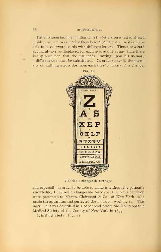
66 DIOPTOMETRY.
Patients soon become familiar with the letters on a test card, and
children are apt to memorize them before being tested, so it is advis-
able to have several cards with different letters. Thus a new card
should al-ways be displayed for each eye, and if at any time there
is any suspicion that the patient is drawing upon his memorya different one must be substituted. In order to avoid the neces-
sit}^ of walking across the room each time to make such a change,
Fig. 22.
Helfrich's changeable test-type.
and especially in order to be able to make it without the patient's
knowledge, I devised a changeable test-type, the plans of which
were presented to Messrs. Clairmont & Co., of New York, whomade the apparatus and perfected the motor for working it. This
instrument was described in a paper read before the Homoeopathic
Medical Society of the County of New York in 1893.
It is illustrated in Fig:. 22.

REFRACTOMETER. 67
The apparatus consists of an ornamented wooden case, uponwhich are mounted the ordinary Snellen test-tj^pes. The five
lower lines only are capable of being changed, the change being
produced by the revolution of five quadrangular rollers permitting
the exhibition of four series of letters. Motor power is furnished
by an accordion-pleated rubber tube, which, when expanded bj^
the column of air communicated to it by the pressure of a bulb,
elevates a weight to which is attached an arm. As the armmoves upward it carries a cog which locks with the wheel that
revolves the five rollers. This wheel contains four slots, placed
at intervals of ninety degrees, for the reception of two catches,
an upper and a lower one, which limit the revolution of the
wheel to a quarter of a circle. After each quarter revolution the
weight carries the arm back to its former position, setting the ap-
paratus for the next change. It is operated by a bulb at the side
of the patient, which is connected with the motor by tubing.
An instrument called the Refractometer has been invented by
H. L. De Zeng, whose purpose is the estimation of the total re-
fractive error and particularl}- the whole amount of astigmatism
present in all the dioptric media without the use of a cycloplegic.
This instrument shown in Fig. 23 is manufactured by the
Cataract Optical Co., of Buffalo. While disclaiming that any
mechanical device can wholly replace the ordinar}^ test under a
cycloplegic this instrument is certainly of great value where a
cycloplegic is contra-indicated or refused, as well as in general
routine work.
In brief this instrument consists of a nickel tube, in the head of
which is placed a stationery concave lens of 20. D. It also con-
tains an inner tube w^hich is movable along its cylindrical axis
by means of a rack and pinion adjustment, and which carries at
its front end a convex achromatic objective. These lenses in com-
bination at different distances give all the spherical foci from
+ .12 D to + 18. D, and from — .12 D to — 9. D inclusive. Theconvex spherical effects are recorded upon a revolving dial at the
side and the concave effects upon the top of the inner tube, visible
to the observer through an oval opening in the top of the outer
tube.
Owing to the range of the negative scale being limited to — 9.
D, two auxilliary caps accompany the instrument, one containing

68 DIOPTOMKTRY.
— lo. D and the other — 20. D, which, when placed over the eye
piece raises the negative scale to either — 19. D or — 29. D re-
spectively. The outer tube is further armed at its front end with
a revolving head, composed of two revolving discs, containing
blanks, a stenopaic slit, and eleven minus concave cylinders set at
right angles with their radii. The resulting combinations possi-
ble give a range of cylindrical lenses from — .12 D to — 8.75 D,
which can be rotated to any given axis, the latter being indicated
by a pointer.
Fig. 23.
De Zeng's refractometer.
By reason of the instrument's optical construction, it has an am-
plification of two and one-third diameters, and in consequence of
this the test-types furnished with it are reduced^'to three-sevenths
of the size of Snellen's letters, so that the visual acuity may be
reliably estimated with the instrument. The instrument must be
properly adjusted for whatever range is desired, either 3, 4, 5 or
6 metres.
The best method employed in testing is what is known as the
" fogging system," which consists in over-correcting a hyperopic

CONVERGENCE AND ACCOMMODATION. 69
eye with convex lenses or under-correcting a mj'opic one with
concave lenses which are too weak. The effect of this is to render
the lines and letters deeph^ blurred, which causes relaxation of the
ciliary muscle and in consequence latent errors to become mani-
fest. With the instrument properly adjusted, and the patient
proper!}^ placed, the thumb-screw is turned until the test-letters
are distinctly seen and the reading noted.
Fogging is next resorted to b}^ producing artificial myopia,
which encourages relaxation of the accommodation. The thumb-
screw is now turned slowl}^ back and the patient requested to
watch the astigmatic fan. If he states that one or more lines ap-
pear distinct before the others he is astigmatic, whereas if they
appear equally distinct simultaneously the error is simple hyper-
opia or myopia. If astigmatism is present it is necessary to
utilize the concave cylinders contained in the revolving discs to
render the lines equally clear. This procedure should be repeated
to verify the result or to make any necessar}' corrections.
The power of convergence is tested with an opthalmo-dynamome-
ter, that of Landolt's being the simplest and best. It consists of a
metallic cylinder blackened on the outside, containing a vertical
slit 0.3 mm. wide covered with ground glass. Beneath the
cylinder is attached a tape measure graduated on one side in
centimetres, and on the other in metre-angles. The vertical line of
light, produced when the cylinder is placed over a lighted candle,
is the object of fixation. Approaching the patient in the median
line until the patient sees the line double the near point of con-
vergence is found and the distance in centimetres with its equiva-
lent in metre -angles recorded. The minimum of convergence is
found by withdrawing the instrument from the patient; but as it is
usually negative, it is determined by the strength of the strongest
abducting prism which will not cause diplopia with the patient
fixing the object at six metres. If the number of this prism is
divided by seven, the quotient will approximately give in metre-
angles the deviation of each eye when the prism is placed before
one. The amplitude of convergence is equivalent to the differ-
ence between the maximum and minimum of convergence.
Accommodation is tested by means of the reading tj^pes of
Snellen or Jaeger. A sample is shown in Fig. 24.
It is necessary to test each eye separatel}^ and finally both to-

70 DIOPTOMETRY.
gether. The nearest point the type can be read represents the
punctum proximum. This subject has already been discussed in
the preceding chapter.
Presbyopia is to be determined after the static refraction has
been tested. With the distance glasses in the frame, the patient is
asked to hold the reading type or a newspaper at the distance at
which he desires to work or read. Convex glasses are now added
to the distance glasses, until the best vision at this distance is ob-
tained. The optical equivalent of the glasses before each eye is
Fig. 24.
O. D.
Tlift elouel)eMt adapted for Utho-
pniphic purposes U a calcareoua
elate found on the banks of tha
Danube, in Bavarin, the finest
being found near ^funich. Agood Btonfl is porous, yet briltla,
of a pale yoUowish diiib. andBomotmjes of a gray nouti'ai tint
The stones am foi-med into slaba
from one and & half to threa
Inches in Uilckneea To prc-paro
them for use. two etonea are put£.ce to laoo with eoaie fino sifted
Bond betwct^n them, and then are
rubbed together wiUi a arcular.
.5OM.
o. s.
Tim stone Ijoit adapted for !itha-
pniLihic purposoB is a cttloftreoua
e\u.X4s found on the banka of the
Danube, In Ba\'aria, the finest
being found near MunicK 'Agood Btona is porous, yot brittle,
of a pale yoUowiah drab, andBometimea of a gray neutral tint
The atones are formed into slaba
trom one and a half to three
Inches in thlckneaa. To prepare
them for use, two stones are putface to fiu30 with some fine sifted
6and between them, and then ar»
rubbed together with a, circular.
O. D.
The artist completes a
chalk (lra"wir).g -upoii a
grained stone ad hewould make one withpencil or chalk uponpaper. If -while in this
state a wet sponge is
passed over the face
of the stone, the draw*ing will xu.h oiE To
•75M.
O. S. -
The ai-tist completes a
chaUc drawing upon agrained stone as Jie
would make one withpencil or chalk uponpaper. If while in this
State a wet sponge is
passed over the face
of the stone, the draw*ing will rub affi To
Type for testing accommodation.
now computed, and the resultant glass is presumably his prescrip-
tion for near. The approximate amount of presbyopia at different
ages can be computed from the chart (Fig. 19) given in the previ-
ous chapter. This amount must be added to the amount of hyper-
opia and deducted from myopia. This should not be considered
as final, but used sim.ply as a check on the result obtained by
testing and to save unnecessary loss of time.

ESTIMATION OF REFRACTIOX BY THE DIRECT METHOD. 7 I
Objective Dioptometry.—Objective dioptometry embraces
the methods of determining the refraction independent of any
statements b}^ the patient. These methods are valuable in
conjunction with the test made b}' the trial lenses and test letters,
and especially so when the patient is illiterate or too \'oung to
read letters.
Frequenth^ the}^ are utilized to save time in arriving at an approx-
imate estimate of the error, the accurate degree of which is fully
determined in the ordinary manner. Not that they are inaccurate
in themselves when applied b\' an expert, but because it is a safer
plan to check results.
Estimation of Refraction by the Direct Method —
A
qualitative estimation can be made with the ophthalmoscope held
at a httle distance from the observed eye. When it is remem-
bered that the rays issuing from a hyperopic eye are divergent and
those from a myopic eye convergent, it is eas}^ to understand howan observer can see an upright image of the retinal vessels in the
former and an inverted serial image in front of the latter. If the
observer moves his head from side to side, the vessels seen in a
hyperopic eye will mov^e with the mirror, the image being upright,
while they will move in the opposite direction if the eye be
m3^opic. At this distance no vessels can be seen in an emmetropic
eye, because the pencils of rays emanating from any two points
upon the retina (each is made up of parallel rays) will diverge
from each other so that no rays will enter the observer's eye.
Close to the eye, however, an upright image of the fundus can be
seen. The quayititative examination is conducted w4th the mirror
held close to the observed eye, if possible as near as 13 mm., the
anterior focus of the e3^e and the proper situation for the glasses
to be worn. If the examination is conducted at a greater distance
proper allowance must be made. In following out this method,
it is imperative that both the surgeon and the patient thoroughly
relax their accommodation. This is easily accomplished for the
patient, if no spasm exists, when the examination is made in a
dark room, but the examiner can onl}- attain it after much practice.
It is rather doubtful if any expert can relax his accommodation
absolutely unless he be so old that he practically has none. Still,
many can do so thoroughU^ enough to obtain approximately
correct tests.

72 DIOPTOMETRY.
The most desirable point in the eye-ground upon which to
focus is either the edge of the disc or the medium-sized vessels be-
tween the disc and the macula, especially two vessels running at
right angles to each other. The macula is unsuitable, because of
the contraction of the pupil caused by throwing the light upon it
and the annoying corneal reflections obtained.
If the observer has any error of refraction, it must either be cor-
rected with glasses or allowance made for it in computing the
final result. All these conditions being fulfilled the examination
is commenced. If the patient's eye is emmetropic, the vessels will
be seen distinctly, and convex lenses rotated back of the opening
in the ophthalmoscope will blur them. If hyperopia is present
the divergent rays issuing from the eye are rendered parallel by
the rotation of convex lenses, and the strongest convex lens
through which the vessels are seen distinctly is the measure of
the error. The convergent rays from a myopic eye are rendered
parallel by concave glasses, and the weakest concave lens through
which the vessels are seen most distinctly is a measure of the
myopia. The direct method is used to determine the height of
retinal tumors by estimating the refraction at their summit, and
the depth of a cupping of the papilla by estimating the refraction
at its bottom.
If the examinatii n is conducted at a distance not more than 2.5
cm. from an e3^e, a hyperopia of i. D will represent a shortening
of the axis equal to 0.3 mm. and a myopia of i. Da lengthening
of the axis to that same amount.
Astigmatism can also be estimated by this method. It is
known to be present when the upper and lower margins of the
disc and the horizontal vessels are well defined wdiile the lateral
margins and vertical vessels are blurred, or vice versa.
Its presence may also be suspected if the disc is elongated
either horizontally, or vertically, the long axis corresponding to
the meridian of greatest refraction.
In estimating the degree it is the best to fix two vessels run-
ning at right angles to each other and whose direction conform
most nearly to the principal meridians of the astigmatism. If the
vessels in one meridian are seen distinctly, while in order to see
the vessels in the opposite meridian a convex or concave lens is
necessary, the case is respectivel}^ one of simple hyperopic or

ESTIMATION OF REFRACTION BY THE INDIRECT METHOD. 73
myopic astigmatism. If, in order to see distinctl}' the vessels in
the two principal meridians, two convex or two concave lenses
of different strength is required, the case is one of compound astig-
matism, either hyperopic or myopic. The difference in strength
between the two convex or two concave lenses represents the
astigmatism, while the weaker lens represents the simple hyperopia
or myopia. The axis of the cylinder to correct the astigmatism
is placc'l in the direction of the vessel which was seen through
the strongest of the two lenses.
It seems unnecessary to again state that in hyperopic astigma-
tism the strongest convex lenses represent the measure and in
myopic astigmatism the weakest concave.
In mixed astigmatism the vessels in one meridian are rendered
distinct by convex glasses and the vess Is in the opposite meridian
by concave glasses. Hyperopia exists in the meridian at right
angles to that in which the vessels were made distinct by the con-
vex glasses and myopia exists in the other. The axes of the
cylinders to correct this would be th- reverse of this, because they
refract only thos.. rays which enter at right angles to their axes.
Estimation of the Refraction by the Indirect Method.
—
This method is not generally resorted to, because of its difficulties.
It is sometimes used in estimating very high degrees of m3'0pia,
but rareh^ in hyperopia. The rays coming from an emmetropic
eye being parallel they are brought to a focus by the object glass
at its principal focus, whereas the divergent rays from a hyperopic
eye are brought to a focus farther from the object glass than its
princii^al focus and the convergent rays from a myopic eye nearer
than its principal focus. The degree of the ametropia is deter-
mined' by measuring this distance. Schmidt-Rimpier's methodis usuall ^ employed.
Schweigger uses the indirect method in comparison with the
direct method to determine the presence of astigmatism. It has
already been explained that thti disc appears elongated in the
direction of the meridian of greatest refraction when seen in the
upright image, so it is only necessary to state that the reverse ob-
tains in the inverted image.
Skiascopy (Retinoscopy, or the Shadow Test).—This is
a method of determining the refraction by observing the direction
in which the light appears to move across the pupil when the
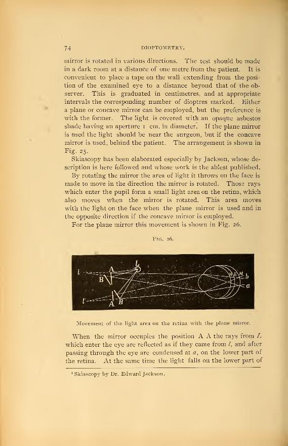
74 DIOPTOMETRY.
mirror is rotated in various directions. The test should be madein a dark room at a distance of one metre from the patient. It is
convenient to place a tape on the wall extending from the posi-
tion of the examined eye to a distance beyond that of the ob-
server. This is graduated in centimetres, and at appropriate
intervals the corresponding number of dioptres marked. Either
a plane or concave mirror can be employed, but the preference is
with the former. The light is covered with an opaque asbestos
shade having an aperture i cm. in diameter. If the plane mirror
is used the light should be near the surgeon, but if the concave
mirror is used, behind the patient. The arrangement is shown in
Fig. 25.
Skiascopy has been elaborated especially by Jackson, whose de-
scription is here followed and whose work is the ablest published.
By rotating the mirror the area of light it throws on the face is
made to move in the direction the mirror is rotated. Those rays
which enter the pupil form a small light area on the retina, which
also moves when the mirror is rotated. This area moves
with the light on the face when the plane mirror is used and in
the opposite direction if the concave mirror is employed.
For the plane mirror this movement is shown in Fig. 26.
Fig. 26.
Movement of the light area on the retina with the plane mirror.
When the mirror occupies the position A A the rays from Lwhich enter the eye are reflected as if they came from /, and after
passing through the eye are condensed at «, on the lower part of
the retina. At the same time the light falls on the lower part of
^Skiascopy by Dr. Edward Jackson.

Fig. 25.
Correct position for retinoscopy


SKIASCOPY. 75
the face. If now the mirror is rotated to B B the light which
enters the eye is reflected as if it came from /', and is condensed
toward d, on the upper part of the retina. Simultaneously the
light which falls on the face moves upw^ard also. The same
movement of the light with the light on the face occurs in hj^per-
opia and myopia as well as in emmetropia.
The movement of the light area on the retina caused by a con-
cave mirror is shown in Fig. 27.
Fig. 27.
Movemeiit of the light area on the retina with the concave mirror.
When the mirror occupies the position A A the rays which
enter the eye come from the focus of the mirror at / and are con-
densed towards a, on the upper part of the retina, while the light
falls on the lower part of the face. When the mirror is rotated
to B B the rays which enter the eye come from /' and are con-
densed towards d, on the lower part of the retina, while the light
on the face moves upward. The same is true in hyperopia and
myopia.
The rea/ movement of the light on the retina, as it would ap-
pear from behind, is alwa3's with the light on the face with the
plane mirror and in the opposite direction with the concave
mirror.
In our examination, however, we do not see it in that waj^, but
we watch the apparent movement as seen through the pupil.
When the plane mirror is used the apparent movement in the pupil
and the real movement on the retina are the same when the observer
sees an erect image, and in the opposite direction when he sees aji in-
verted image.

76 DIOPTOMETRY.
The reverse of this obtains with the concave mirror.
The rays of light coming from a myopic e3^e are convergent
and cross each other at its far point, and proceed divergently.
The point B (Fig. 28), at which they cross and which" corre-
sponds to the far point, is known as ih^ poi7it of reversal. As has
been explained under myopia, the distance of the far point from
the eye represents the focal length of the glass required to correct
it, and, therefore, if the point of reversal is known the amount of
myopia is also known.
Retinoscopy is an accurate method of determining the point of
reversal.
Fig. 28.
The point of reversal of a myopic eye.
In the following description it is assumed that the plane
mirror is used, though it will apply equally to the concave mirror
if we reverse the movement in the pupil and change the lenses.
If the mirror is held closer to the eye than its point of reversal,
as at A (Fig. 28), an erect image is seen, and the light in the
pupil will seem to move with the light on the face. Be5^ond the
point of reversal, as at C, an inverted image is seen, and the light
will appear to move in an opposite direction to the light on the
face.
At the point of reversal it is impossible to see which way the
light moves, and the illumination is much more feeble. At a dis-
tance of one or two metres from the point of reversal the illumina-
tion is very bright, but as the distance increases it becomes more
and more feeble. Without altering the rapidity of the movement
of the mirror, the apparent movement of the light is more rapid as
we approach the point of reversal.
While the test depends mainly on the direction of the movement

SKIASCOPY. 77
of the light in the pupil, the degree of illumination and the rapidity
of movement aid in arriving at a diagnosis.
Myopia is estimated by finding the nearest point that an in-
verted image is seen (C, Fig. 28) and the most distant point at
which an upright image is seen, as at A. Midway between the
two is the point of reversal, B, whose distance from the eye should
be noted on the tape for that purpose, and the number of dioptres
corresponding is the measure of the myopia. Thus if C is at 55
cm. and A at 45 cm. B would be at 50 cm., which corresponds to
2: D. giving a myopia of that amount. When the myopia is of
high degree, the point of reversal lies very close to the eye, and in
this situation a slight error in marking the distance may mean an
error of a dioptre or more in estimating the myopia; whereas if
a similar error is made when the point of reversal is situated at a
metre or more from the e3^e it is unimportant. Therefore, in
high degrees of myopia, in order to check results, it is well to cor-
rect all but about one dioptre by placing a suitable concave lens
in a frame before the eye and measure the remainder, which is to
be added to the lens in the frame.
If the m3^opia is less than one dioptre, the point of reversal
lies so far away from the eye that when near it one cannot see
which way the light moves. In this case put a weak convex
glass in the frame to increase the myopia, then determine the point
of reversal, and deduct the convex glass from the amount of
myopia found.
Hyperopia gives an upright image no matter how far we recede
from the eye, because the raj'^s leave it divergently. In order
to obtain a point of reversal, it is necessary to convert it into an
artificial myopia by putting a convex glass in the frame and then
finding the point of reversal as in myopia. The amount of
myopia is to be deducted from the convex lens, the hyperopia
being represented by the remainder.
Thus, if a convex 5. D lens is placed in the frame, and the
point of reversal is found at 1 metre (i. D), the hyperopia v/ill be
4. D. That is, 4. D out of the five were necessar}- to render the
divergent rays parallel and the other dioptre to bring them to a
focus at I metre.
Emmetropia acts the same as hyperopia, but when a convex
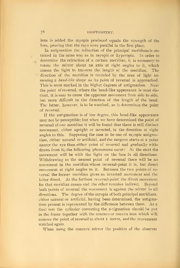
78 DIOPTOMETRY.
lens is added the myopia produced equals the strength of the
lens, proving that the rays were parallel in the first place.
In astigmatism the refraction of the principal meridians is ob-
tained in the same way as in myopia or hyperopia. In order to
determine the refraction of a certain meridian, it is necessary to
rotate the mirror about an axis at right angles to it, which
causes the light to traverse the length of the meridian. Thedirection of the meridian is revealed by the area of light as-
suming a band-like shape as its point of reversal is approached.
This is most marked in the higher degrees of astigmatism. Near
the point ofreversal, where the band-like appearance is most dis-
tinct, it is easy to cause the apparent movement from side to side,
but more difficult in the direction of the length of the band.
The latter, however, is to be watched, as it determines the point
of reversal.
If the astigmatism is of low degree, this band-like appearance
may not be perceptible; but when we have determined the point of
reversal of one meridian it will be found that there is still distinct
movement, either upright or inverted, in the direction at right
angles to this. Supposing the case to be one of myopic astigma-
tism, either natural or artificial, and the surgeon starts at a point
nearer the eye than either point of reversal and gradually with-
draws from it, the following phenomena occur: At the start the
movement will be with the light on the face in all directions.
Withdrawing to the nearest point of reversal there will be no
movement in the meridian whose reversal-point it is, but direct
movement at right angles to it. Between the two points of re-
versal the former meridian gives an inverted movement and the
latter direct. At the farthest reversal-point the direct movement
for that meridian ceases and the other remains indirect. Beyond
both points of reversal the movement is against the mirror in all
directions. The degree of the myopia of both principal meridians,
either natural or artificial, having been determined, the astigma-
tism present is represented by the difference between them. As a
final test the cylinder correcting the astigmatism should be put
in the frame together with the concave or convex lens which will
remove the point of reversal to about i metre, and the movements
watched again.
When using the concave mirror the position of the observer

OPHTHALMOMETRY. 79
does not admit of much change, the distance being generally one
metre. The movements of the light are the reverse of those just de-
scribed. Skiascopy is the most valuable of all objective methods,
and the student should practice it industriously upon an artificial
eye made for the purpose before he can depend upon his results
in actual practice.
Ophthalraometry.—The term indicates mensuration of the
eye, but it is usually employed to mean the measurement of the
radii of curvature of the cornea and the corneal astigmatism pres-
ent in an eye with the ophthalmometer. The ophthalmometer in
general use in this countr}^ is that of Javal and Schiotz and is
shown in Fig. 29.
Fig. 29.
Javal and Schiotz ophthalmometer.
The principles upon which it is based are as follows: Thesurface of the cornea acts as a convex reflector, the size of-
the image produced by it, of an object of known size at a knowndistance, depending on its radius of curvature. The size of
the image is determined by doubling it with a double refracting
prism, and then altering the strength of the prism until the images
come into contact. When this has taken place, a displacement
equal to the size of the image has been produced. Two objects

8o DIOPTOMETRY.
known as mires are situated upon an arc, one a white rectilinear
figure, which is stationary, and the other, made up of white
enameled blocks, capable of being moved along the arc. Theseobjects are so placed that their images are reflected by the cornea
and are viewed through a telescope by the observer. The tele-
scope contains a prism (to double the images) placed between twobi-convex lenses, with a third bi-convex lens to shorten the pos-
terior foci of the two images. It stands upon a tripod, which can
be moved in order to obtain the proper focus. The patient places
his chin on the rest and looks in the tube, the eye which is not
under observation being covered by a disc.
When everything is properly adjusted, the central images are
obtained on a spider web, also provided in the tube, and the movable
object is moved along the arc until its image comes in contact
with the image of the rectilinear object. Its position on the arc
is noted upon the index.
The arc itself is now turned on its own axis to a position at
right angles to its first position, and the relation of the two images
noted. If astigmatism is present, they will either overlap or sepa-
rate, but if absent, they will occupy the same relative position.
The degree of astigmatism is measured by the overlapping, each
step of the white enameled block representing one dioptre. If
the reflections are separated in the second position they are
brought in contact once more and the arc turned back to its
primary position, where they will overlap. The overlapping in
this case will give the measure of an astigmatism against the rule;
that of the previous instance with the rule.
The ophthalmometer merely measures the corneal astigmatism
and not the refraction of the eye. Corneal astigmatism may be
modified by lental astigmatism, consequently the ordinary test
with the trial lenses should always be made in addition.
Ophthalmometry is servicable in revealing the corneal astig-
matism and the principal meridians.
General Considerations.—After the test is completed a
proper record of it should be made in the surgeon's case-book.
As this is the first step in testing any case, the result forms an
integral part of the complete record. To it should be added the
record of the correcting lens, and finally the acuteness of vision
produced by this lens. The following example will illustrate this:

GENERAL CONSIDERATIONS. 8
1
O. D. V= I ^ 2.50 D V= fO. S.F ==4 + 0.75 D V=iWhen the correcting lens is a compound glass, the component
lenses are united by the sign Q, which signifies'
' combined with."
Thus a combination composed of a + 3. D spherical with a — 1.50
D cylinder axis vertical is recorded — 3. D* O + 1.50 D^ axis 90°.
Notice that the spherical lens is always recorded in advance of the
C3dinder, and that the sign — or — is expressed to show that the
lenses are either convex or concave.
Simple cylinders are ground on one side only, the other side
being plane. Compound glasses contain the spherical on
one side and the cylinder upon the other. The correcting
lenses for mixed astigmatism may either be crossed cylinders,
a convex cylinder on one side and a concave cylinder at right
angles on the other, or a combination of a spherical with a
cylinder. Thus in a case of mixed astigmatism corrected by a
convex cylinder of 2. D axis vertical and a concave cylinder of i.
D axis horizontal the prescription may either be -^ 2. D^ axis 90
r — I. D^ axis 180, or — 2. D* Q — 3. D*^ axis 180. The two are
optical equivalents. Unless specially ordered to do otherwise, the
optician will grind the lens according to the latter. The sign f
is usually employed when two cylinders are combined, the con-
vex cylinder being expressed first.
Patients should be instructed to return with their glasses after
they have obtained them from the optician, in order that the sur-
geon may ascertain if the lenses have been correctly ground and
that the frames are properly fitted. The importance of this can-
not be overestimated. The correctness of the lens is verified byneutralizing it with the opposite form of lens, that is, convex
spherical and cylinders either alone or in combination are neu-
tralized by concave sphericals or cylinders of the same number.
If one holds a convex glass near one eye, and fixes an object like
the edge of a door, the edge will appear to move in an opposite
direction to the lens when the latter is moved from side to side.
With a concave lens it moves in the same direction. If a convex
and concave lens of equal strength, held together, are moved in a
similar manner, the object will remain stationary, the effect of
the convex being neutrahzed by the concave. Thus the number

82 DIOPTOMKTRY.
of any lens can be ascertained by neutralizing it with the lenses
from a trial case, which are always numbered.
The optical centre of the lens should coincide with its geometric
centre, unless it has been ordered decentered. To find the optical
centre of a lens we can utilize the reflection of a piece of paper
pasted on the window pane with our back turned to the window.
A reflection of it appears on the anterior and posterior surfaces of
the lens and when the images overlie each other the optic axis
of the lens is determined and therefore the optical centre which is
situated upon this. The point can be marked upon the lens with
ink and its situation as regards the geometric centre noted.
It can also be determined by refraction by finding two meridians
of the lens at right angles to each other which do not displace a
vertical line horizontally when it is viewed through the lens. It
is customary to take the edge of a door or a card. When the
lens occupies such a position that the vertical line appears as a
continuous line, above and below, with that seen through the
lens, this meridian is marked by a line with a pen. The glass is
rotated at right angles and the same process gone through with
again. The point at which these two lines intersect each other is
the optical centre. The frames should be so fitted that the pupil
is opposite the geometric centre, and the optical centre should
coincide with the latter unless it has been purposely decentered.
This is the rule when glasses are to be worn for distance or con-
stant use. Near glasses are usually decentered in about 4 mm.on account of the convergence of the visual lines. Reading
glasses are also tilted forward and placed lower down than dis-
tance glasses, to conform with the depression of the visual line.
A decentered glass when not required is sufficient to render void
the beneficial effects of the most perfect prescription. Lenses are
often decentered in a given direction when a prismatic effect is
desired. Convex and concave lenses may be considered as being
made up of a series of prisms. The effect of decentering a convex
glass in, is equivalent to obtaining a prism with its base in, while to
decenter a concave glass inwards produces a prism with its base
out. The degree of prismatic effect obtainable depends on the
distance the lens is decentered and the strength of the lens. Thegreater the decentration and the greater the strength of the lens
the greater the prismatic effect.

GKNERAI^ CONSIDERATIONS. 83
Spectacle frames are always preferable to eye-glasses, and in fact
their use is imperative in high grades of astigmatism. Still the
prejudices of many patients, particularly women, against themmust be regarded if we wish them to wear their glasses, so under
these circumstances eye-glasses must frequently be prescribed.
Nowadaj^s, with the many improved guards in use, eye-glasses can
be fitted nearly as perfectly as spectacles, and if the patient is
taught to take proper care of them and to have them readjusted
frequently they answer the purpose quite as well.
When separate glasses for reading and distance are required a'
' bifocal lens'
' for constant use may be prescribed to avoid the in-
convenience of changing from one to the other. Many peo-
ple never become accustomed to them, however, and often
meet with accidents caused by looking through the lower part
of the glass when going down stairs or stepping out of a con-
veyance.
84 HYGIENE OF THE EYE.
CHAPTER V.
Hygiene of the Eye.
Under this heading we shall endeavor to gather together a few
very essential and practical points, some of which, while they
may be elsewhere considered, are of sufficient importance to
merit repetition. Many cases of blindness have undoubtedly re-
sulted from the neglect of simple everyday precautions, which
are supposed to be so generally understood, by the laity
as well as the medical profession, that we are unable to find any
author who has devoted the slightest space to the subject.
The care of the eye commences at birth, and in order to secure
its highest usefulness must be continued throughout the whole
life. The eyes of the new-born babe should be at once carefully
cleansed with warm water or a saturated solution of boracic acid.
In all cases of gonorrhceal or leucorrhoeal discharge in the mother
the method recommended by Crede should always be employed.
This consists of the instillation between the eyelids of the child
immediately after birth of a 2% solution, gr. x. ad ^j.rof nitrate
of silver. This method is practiced by many of the best obstet-
ricians in all cases, and since its general adoption the percentage
of cases of blindness from ophthalmia neonatorum has been won-
derfully reduced. In former years the percentage of blindness
from this disease alone formed, in different countries, from 20 to
79 per cent, of all the cases of blindness.
The examination of the eyes of the babe from day to
day should be a part of the physician's routine duty for
at least one or two weeks, so that the onset of any trouble
may be at once met by active treatment. The eyes of
young infants should be protected from all glaring lights,
and especially the direct rays of the sun, both indoors and out.
The babe should never have its attention attracted by ob-
jects held close to the eyes, for repeated convergence at close
objects may predispose or even produce strabismus. This obser-
vation holds good as the child grows older, for in addition, from

SCHOOI. HYGIENE 85
poring over story and picture books, when too fine or held too
close to the eyes, myopia threatens. The fine worsted and bead
work used in some of the kindergarten teachings is for this reason
objectionable. Give the growing child plenty of out-door amuse-
ments, vvhere the eyes may have a long range during the develop-
ing period of life, and we shall see fewer little ones wearing glasses
for myopia and astigmatism.
School Hygiene.—We believe that one of the most important
fields for the exhibition of the to-day knowledge and interest in
hygiene is presented in our educational institutions. Whenwe consider the total number of hours passed in the class rooms
during the child's school and college life, the additional hours
required lor study and preparation outside of the school-room by
the present day system of forcing the child too rapidly and all
children alike, to keep up with their classes, then when wecompare these hours with the time left for recreation, exercise
and sleep, and recall that these years are the years of physiologi-
cal growth, is it any wonder that we find so many commencing
their active life as physical wrecks? It is, therefore, plainly a duty
we owe to posterity to consider carefully the hygienic environ-
ments of our children as well as their mental and moral training.
The school life of the growing child should be so regulated as to
secure the best mental advancement and at the same time the best
physical development. Every observing physician has seen manychildren w^ho commenced school life in apparently good health
soon complaining of headaches, nervousness, loss of appetite and
other symptoms indicative of impaired general vigor.
It is, however, not our function to consider the subject of
school hygiene in its relation to the general health, but simph^ as
to its bearing upon the eyes of our children.
In the early part of the present century we find attention first
called to the relations existing between the myopic eye and the
demands of civilized life. W^ithin a comparatively few years
more complete and S3^stematic examinations of the eyes of school-
children have been made, so that to-day we have as a basis for
our statistics the examinations of the eyes of over two hundred
thousand pupils of all grades. An analysis of these examina-
tions show that in the primary schools nearly all the children
enter with normal eyes. In the higher grades 25 per cent, have

86 HYGIKNK OF THE KYK.
become myopic, while in university life the percentage of
myopia has increased to from 60 to 70 per cent., which shows
that the number of near-sighted pupils increase from the lowest
to the highest schools and that the increase is in direct propor-
tion to the length of time devoted to the strain of school life.
In the face of these facts, it seems the imperative duty of the
hour to carefully investigate the cause of this deterioration in the
eyes of our children during school life. The evident relationship
of this increasing near-sightedness with school work seems to indi-
cate some fault in our educational methods. Owing to the fact
that myopia is often hereditary, it is impossible to wholly eradicate
the condition for generations to come. We believe, however, that
acquired myopia can be prevented, or very greatly decreased, by
careful and frequent examinations of the eyes, together with
thorough hygienic preventive methods, during the years of physi-
cal growth and mental training of the child.
First, as to the importance of frequent examinations of the eyes
of children. Statistics prove that a very large proportion of the
eyes of young children are hypermetropic. So great is this pre-
ponderance that many authorities claim the normal eye to be a
hypermetropic one. Careful observations have shown that in
almost every instance the change from far to near-sight is through
the turnstile of astigmatism. That this change does take place
has been proven by the progressive increase in the percentage of
myopia during school life. By repeated examinations, from year
to year, the first change can be detected and suitable treatment
taken to check its progress. We believe that the eyes of every
child should be carefully examined at the commencement of
school life, and that the examination be repeated every year at
least until the time of full development of both body and mind.
The care of the teeth commences even earlier than this and is
continued through the whole life. We have become educated to
the importance and necessity of sending our children to the den-
tist every six months or year for examination, whether disease is
suspected or not. The far more precious and delicate organ, the
eye, on the contrary, is almost universally left to do its work un-
aided and uncared for, until often serious and irreparable damage
has been done and the innocent victims of our ignorance and
neglect are deprived of the full realization of God's greatest

SCHOOL HYGIENE. 87
gift, that of sight. It is not the vision alone that pays the penalty
of this criminal neglect, but a long train of physical wrecks
brought about through reflex action, from eye-strain.
It is not our purpose at this time to go into the details as to howor what general conditions may result from defective eyes, but
merely to sound a warning as to the danger from neglect of the
eyes in early life. To continue the comparison with the teeth,
we can get very acceptable false teeth, when lost from neglect,
but artificial eyes have not proven of much practical service.
There is great need of popular education as to the importance
of such examinations of the eyes. Parents who follow out the
greatest precautions for the welfare of their children in other re-
spects are unmindful in this, through lack of knowledge of its
importance.
Rxaminatio7i of the Eyes Upon Entrance of School.—Every
school should possess a series of test letters, and each scholar at
the commencement of each term should have the eyes examined
by the teacher. This examination is so simple that any teacher
can be instructed in a few moments so that they can determine
if any defect exists. All that is essential is a set of Snellen's test-
types placed in a good light; the smallest letters should then be
read with each eye separatel}^ at twenty feet. The child should
then be examined with the astigmatic card at the same distance,
and the lines running in all directions should appear with each
eye alone equally clear and distinct. Then a small card clearly
printed in 4}^ point (diamond) type should be read by
the child, while the teacher measures with a rule the nearest point
at which it can be clearly read. This distance should correspond
with the normal near-point of an emmetropic eye, which should
be recorded on the back of the card for the different ages from
six to twenty years. If these tests show no defect the child maythen be admitted to the school, but, on the contrar}^, if a defect be
found in any of the tests, particularly the first, the parents of the
child should be at once informed of the existing defect of vision
and the consequent need of professional advice. Further than
this, during the school year, if the child complains frequently of
headaches while studying, or seems to be getting nervous, anaemic,
etc., the teacher's duty is to again suggest to the parents the
wisdom of seeking a physician's advice.

b« HYGIENE OF THE EYE.
The examination as suggested would at once detect imperfect
vision, from any cause; if due to refractive errors it could then be
corrected, if to intra-ocular disease treatment might save the
sight which otherwise might possibly be lost
In all cases of children with inflamed eyes they should be re-
quired to present a physician's certificate of the non-infectious
nature of the disease before being permitted to enter the schools.
Our orphan asylums, piblic homes and institutions of all kinds
require a physician's certificate before admitting children with
any redness or inflammation of the eyes. Should we be any less
strict before permitting these children to associate with the
healthy ones in our schools ?
Let us now consider other faulty conditions of school life which
bear more or less directly upon the eye as well as the general
health of the child. T.ie curriculum of study in the majority of
public schools is a hard and fast one, which all students are ex-
pected to follow. We believe a more elastic curriculum should be
adopted, whereby children with defective eyes, or in more or less
feeble health, should only be required to take as many and such
studies as they may master in safety Such a modified course,
while it would lengthen the student life by one or more j^ears,
would do much toward preserving the eyes and general health,
A decided reform should also be made in the system of requir-
ing study at home. The average school sessions of five or six
hours a day should be sufficient to prepare for college by the time
they are sixteen or eighteen without requiring nearly as many ad-
ditional hours of stud}^ at home, which robs the student of the
recreation and sleep he should have. The work at home is
usually accomplished when the body is tired, and the brain slug-
gish, generally by artificial light (which is too often an improper
one) and frequently with a faulty position of the body. We be-
lieve that with a proper regulation of recitation and study during
school-hours alone, the brain, made more active by sufficient re-
creat'on, exercise and sleep outside, will accomplish far more than
by the present system.
The paper and type used in school-books have in recent years
been vastly improved, yet there is room for still further improve-
ment. In selecting books for children, the type should always be
large, bold and clear. Cohn and Weber claim that type at least

THE COXSTRUCTIOX OF SCHOOL BriLDIXGS. 89
one and a half millimetres in height (equal to long primer) is the
smallest that should be used in school-books, and the distance be-
tween the lines, or leading as it is called, should be two and a half
millimetres. The paper should be of a dull finish, instead of the
highly glazed finish of many books, and of a dead white or a
cream color. In many of the books used by children the print is
too small and of a poor impression, which is ver}' injurious to the
eyes. This perhaps applies more particularly to the interesting
books and periodicals prepared for the young, and especiall}^ to
newspapers. The character and the amount of reading is too
often not properly regulated at home. The reading of sensational
papers and novels at hours when the child should be at sleep is a
habit too freel}^ indulged at the expense of both mental and
physical development.
There should also be frequent breaks in the application of the
eyes at close work. This frequent interval of rest for both the
brain and the eyes can easily be secured in the school-room by a
change from the books to the blackboard, to oral instruction,
lectures, etc. The school session should be broken b}' short re-
cesses in the open air, gymnastic exercises, etc.
The system of examination, usually followed in schools, we be-
lieve, can be modified with benefit to the eyes and health of
the pupil. The present method of frequent set examinations for
promotions results in an unusual amount of study, or cramming,
for a given time previous to the examination. This additional
amount of study is always secured, at the expense of the eyes and
health by taking the time from the already- too limited hours
given up to recreation and rest. All educators recognize that
"cramming" is not the best method of learning, and yet the
prevalent system of examinations leads to this. We would sug-
gest that a better method to determine the true standing of the
pupil would be by unexpected examinations, or by allowing the
standing and fitness for promotion to rest upon the marks for
daily recitations and the teacher's estimate of the pupil.
The ConstrudioJi of School Buildings.—A consideration of the
eyes and health of our school-children must necessarily involve
the location of the building, as to surroundings, light, etc., and
the school furniture.
The location in cities should avoid narrow streets, and hio:h sur-
90 HYGIENK OF THK KYK-
rounding buildings, which interfere both with light and air; and
away from noises, exhalations, smoke and dust of factories, stables,
markets, etc. Play-grounds in the open air, either in ample
grounds or on the roof of the building, should be provided for in-
termission of the sessions. The building should be so constructed
as to avoid dampness and should furnish ample ventilation with-
out draughts. In the country, especially, care should be taken
that the location be well drained and away from malarial and
other injurious environments.
Sufficient light is of the utmost importance, and should be first
considered in the architectural plan of all school-houses. Thequantity of light, Cohn says, cannot be too much, while Javal
says that every portion ofthe room should be so flooded with light
that the darkest place will have sufficient illumination on a dark
day. To secure this, Javal says that the distance of surrounding
structures should be twice their height. The necessity of suffi-
cient light is shown by an attempt to read in the twilight or in a
dimly-lighted room. A test as to the amount of light is the
ability of a normal eye to read diamond type readily at twelve
inches. According to Risley the window surface should never
fall below one square foot of glass for every five square feet of
floor space, and that this should be exceeded in many localities,
on the north side of buildings and on the ground" floors. Thequantity of the light is, of course, modified by the color of the walls
in the school-rooms. The light shades of yellow, green, blue or
gray should be used in the coloring of the walls and also of the
furniture and wood-work. The loss of light caused by large sur-
faces of blackboards can be saved by roller shades, of the same
color as the walls, to be lowered when not in use.
Next in importance to the quantity of the light is its direction.
The ideal light of the school-room is that from the left side, or
the left and the rear, of the pupils. Lighting of the room from
two opposite sides should be avoided if possible, yet when neces-
sary to secure the requisite amount of light that from the right
side should be high up in the room. In this way we secure a dif-
fused light in the room from the illumination of the ceiling and
avoid the objectionable cross-lights. This arrangement at the
same time affords better means of ventilation.

SCHOOL FURNITURE. 9
1
School Furniture.—In the most excellent and thorough article
upon School Hygiene, by Dr. S. D. Risley,* to which we are
greatly indebted in the preparation of this chapter, much space has
been devoted to the consideration of the school furniture. While
the faulty construction of the school desk and seat is a very im-
portant factor, according to orthopaedic surgeons, in the causa-
tion of spinal curvature, it has, and undoubtedly still is no small
factor in the increasing myopia of school life. Vast improvements
have been made in the average school-rooms of to-day in this re-
spect, still a visit to almost any school will show more or less of
the pupils in an improper position. The grave danger to the eyes
lies in the pupil bending over his desk and thus bringing the eyes
too close to the work. This abnormal near point adds greatly to
the strain upon the accommodation and convergence, and at the
same time causes an increased congestion of the coats of the eye,
all of which serves to increase the tendency to near-sightedness.
The proper arrangement of the seat and desk is such that the
child will find it easier to sit upright at his work than in any
other position he can assume. The directions and measurements
for securing such a position by means of a correct seat and desk
are fully given in the article referred to.
The blackboard forms an important adjunct to school life, and
its more general and extended use should be encouraged. Thestrain upon the eyes is much less when looking at a relatively
distant object like the blackboard than it is at the near point, as
in reading and writing. Hence instruction by board exercises is
much less fatiguing than work done with the pencil or pen. Thesurface of the board should be kept black and clean by frequent
washing, and the crayons used should be either white or yellow.
Wall maps and charts are also useful, for the same reason as the
blackboard, in that they permit of instruction at a greater dis-
tance. The character of the type, paper, etc., in school-books
has already been referred to.
In all children who have alread}^ developed near-sight, to avoid
the increasing tendency to draw the work nearer and nearer to the
eyes, some of the many forms of head rest, which hold the head
erect and at the proper distance from the work, should be used.
In considering the subject of hygiene of the eye we have dwelt
^System of Diseases of the Eye. Norris and Oliver. Vol. II. 1897.

92 HYGIKNE OF THE EYE.
at length upon the care of the eyes in children, because it is at
this time of life that the greatest danger to vision exists. Further-
more, when the proper care has been given to the eyes in early
life we enter adult life with better eyes and a better understand-
ing of their requirements. In all classes, men, women and chil-
dren, there is an inherent prejudice to the use of glasses, but to
those suffering from refractive errors the use of the correct glass
is one of the greatest boons to humanity. We acknowledge that
the prevalent error of all oculists is the too early and frequent
prescribing of glasses. In many instances the use of glasses can
be avoided by the correction of some deficiency in the balance of
the extrinsic muscles of the eyes which is the cause of the asthen-
opic or reflex symptoms. In all cases of decided refractive errors,
however, the use of correcting lenses is a necessity. When glasses
are required they should be given proper care by the wearer. Wehave often seen patients wearing glasses so scratched and dirty
that a great effort must necessarily be made to see through them.
In using eye-glasses, the}^ should never be folded, as they soon be-
come misshappen or scratched. For the same reason glasses should
not be thrown carelessly upon tables, stands, etc., and when out
of shape, nicked and scratched they should be repaired or newones purchased. After the correct lens has been selected care
should be taken that the frames are correctly adjusted by a com-
petent optician, as oftentimes improperly fitted frames destroy all
the benefit that would have resulted from the glasses. The too
common custom of allowing incompetent opticians, jewellers,
peddlers, etc., to examine the eyes and fit glasses cannot be too
strongly reprimanded.
The prevalent habit of going without glasses for reading as long
as possible is also a bad one. The public should be taught that
all normal eyes require glasses for near vision about the age of
forty or forty-five. That postponing their use later than this age
causes an effort of the accommodation which does harm. Theprejudice to the use of glasses seems to be dying out, and the laity
are realizing more and more the necessity of attention to the
eyes.
One of the most important questions relating to the general
care of the eyes is that as to the best light. This should always
be answered, the diffuse natural light of day, and the next best,

HYGIENE OF THE EYE. 93
that which most nearly approaches dayHght. Artificial light
should be profuse, white and steady and that which most nearly
meets these requirements is that known as the Welsbach light.
The incandescent light, w^hen protected by translucent globes, is
also an excellent light. Gas and kerosene are also good, but
-should be shaded with globes colored w^hite on the inside and
tinted green on the outside. What has been said in regard to
children in school applies as well to the adult, that the use of the
eyes should only be when the body is in the erect position, and
that the light should fall upon the book or paper from the left
side. It hardly seems necessary to caution against the use of the
eyes in reading after twilight, when the illumination has become
poor, when riding on the cars, while lying down, etc , but as all
these things are being done daily we cannot cry don't too often.
In conclusion let us remind the reader that the health of the
eye depends to a great measure upon the condition of the general
-system. The eye is not a separate and distinct organ, to be
treated wholly independent of the general bodily health. Whilethe eye can undoubtedly cause abnormal conditions of other
organs, it can at the same time suffer from other diseased condi-
tions. Therefore, by obeying the general laws of hygiene the
usefulness of the eYes wuU be best maintained.

94 GENBRAI, DISEASES WITH EYE SYMPTOMS.
CHAPTER VI.
A Tabulated Statement of Diseases With More or LessCharacteristic Eye Symptoms.
From The Eye as an Aid in General Diagnosis by B. H. Linnell. M. D.
Abdommal growths.—More or less pigmentation of the skin of
the eyehds.
Addison'' s disease of the supra-renal capsules.—Pigmentation of the
skin of the hds and of the sclera.
Albuminuria.—Retinitis and neuro-retinitis.
Alcoholism.—Paretic mydriasis. Paralysis of accommodation, or
spastic myosis in the early excitable stage. Paralysis of the
external ocular muscles. Ptosis.
Ancemia {Cerebral).—Paretic mydriasis.
Ancsmia (^ConstitutionaV)
.
—Paralysis of accommodation, chorodi-
itis, retinitis and retinal haemorrhages.
Aneurism of orbital artery, or internal carotid.—Exophthalmus.
Aneurism of aorta and arteria innominata.—Reflex spastic my-driasis on the side of the lesion. Retinal pulsation.
Apoplexy.—Dilation or contraction of the pupils distinguishes it
from embolism, in which the pupils are unaffected. Spastic
myosis in premonitory stage. When, during a seizure, my-
driasis occurs after a previous myosis, it is an unfavorable
symptom, signifying increasing pressure. Nystagmus.
Homonymous hemianopsia.
Apoplexy of cortex or cor07ia radiata.—Byes and extremities para-
lyzed on the same side. Eyes deviate toward the side
of the lesion.
Apoplexy of the crus or pons varolii.—Eyes and extremities para-
lyzed on opposite sides. Eyes deviate away from the side
on which the lesion exists.
Apoplexy ofthepons.—Spastic myosis.
Apoplexy of ventricles.—Spastic myosis.

GENERAL DISEASES WITH EYE SYMPTOMS. 95
Atheroma. — Conjunctival hsemorrhage. Intra-ocular hoemorr-
hages.
Atrophy,progressive ^ muscular.—Ocular paralyses.
Basedow' s disease or exophthalmic goitre.—Diminished frequency of
winking. Spasm of the levator of the upper lid. (Abadie's
sign.) Widening of the palpebral fissure, owing to contrac-
tion of Mueller's muscle. (Stellwag's or Dalrymple's sign.)
Loss of associated movement of the upper lid and the eye-
ball. (Von Graefe's sign.) Exophthalmus.
Brairi, abscess of.—Neuritis.
Brain, basilar affectiojis of.—Loss of pupillary reflexes. Homony-mous hemianopsia. Paralyses of ocular muscles.
Brain, cerebral cortical affectio7is
.
—Conjugate ocular paralyses (or
ophthalmoplegias), loss of voluntary movements of the eyes,
with preservation ofinvoluntary or reflex movements of pupil
and eye-ball. Eyes paralyzed on side opposite the cerebral
lesion. Ej^es deviate towards side of lesion. Psychic visual
disorder.
Brai?i, cerebral affections, with increased i7itra-cranial pressure.—Paretic mydriasis ordinarily. Sometimes reflex spastic my-driasis.
Brain, cerebellum, affections of.—Nystagmus.
Brain, concussion of.—Sluggish action of pupils without marked
dilatation or contraction.
Brain, hypercEmia of.—Spastic myosis.
Brain, tumor of.—Nystagmus. Paretic mydriasis. Choked disc.
Choked neitritis. Atrophy of optic nerve. Homonymous hem-
ianopsia, when pressure is extended upon fibres of one tract.
Cholera.—Conjunctival hsemorrhage. Anaesthesia of cornea.
Neuro-paralytic keratitis. Loss of the light reflex indicates a
fatal termination even in apparently mild cases. Preserva-
tion of the light reflex warrants a favorable prognosis, even
in severe cases. Black patches appear in the sclerotic below
the cornea in severe cases. They are of irregular form and
size and tend to coalesce. Their presence is of very un-
favorable significance.
Coma, alcoholic or urcemic.—Mydriasis.
96 GENERAL DISEASES WITH EYE SYMPTOMS.
Coma, syphilitic.—Myosis and reflex iridoplegia.
Death, signs of.—Opacity and insensibility of the cornea. Dessic-
cation of the sclera. Abolition of pupillary reflexes. Ab-
sence of the red reflex from the fundus.
Dejital affectio7is.—Various forms of inflammation of the cornea.
Nictitation.
Diabetes.—Eczema of the eyelids. Conjunctival haemorrhage.
Ulceration of the cornea. Paralysis of the external ocular
muscles and of the accommodation. Cortical cataract. Retin-
itis and neuro- retinitis. Atrophy of the optic nerve. De-
generation of the retinal vessels and haemorrhages. La-
grange found, in 52 cases of diabetes, 13 of intra-ocular
haemorrhage and the same number of cases of cataract. (See
"Arch, d' Ophth.," Jan., 1887.) Galezowski found, in 144
cases of diabetes, 5 of paresis of accommodation, 4 of kera-
titis, 7 of iritis, 4 of glaucoma, 46 of cataract, 27 of retinitis,
31 of amblyopia, 3 of amotio retinae, and 3 of atrophy of the
optic nerve. (See " Jahr. f. Aug.," 1883, p. 297.)
Digestion, disorders of.—Styes. Nictitation.
Diphtheria.—Diphtheritic conjunctivitis. Paralyses of the ex-
ternal e5^e muscles rare; of accommodation more frequent.
Embolism, cerebral. No pupillary symptoms; in contradistinction
from apoplexy.
Epilepsy.— Paretic mydriasis during the seizure or spastic myosis.
Hippus as consciousness returns and frequently during the in-
tervals. Spasms of the ocular muscles.
Fever, puerperal a?id typhoid.—Metastatic suppurative choroiditis.
Fever, relapsi7ig.—Iritis.
Fifth nerve, affections of.—Reflex spastic myosis.
Fourth ventricle, lesions in.—Nuclear ocular paralyses 'affecting
separate nuclei of the third nerve, or successive implication
of its various branches. Also total paralysis of all the
muscles of both eyes.
Friedreich' s disease (hereditary ataxia).—Nystagmus.
Gout.—Retinitis.
Heart, aortic ijisufficieyicy .—Alternate reddening and pallor of the
optic disc.

GENERAI. DISEASES WITH EYE SYMPTOMS. 97
Hearty endocarditis.—Embolism of the arteria centralis retinae.
Heart, organic affections of.—CEdema of lids. Venous hyperaemia
of retina and pulsation retinal arteries. Seen with valvular
affections, fatty heart and aortic insufficiency.
Heart, hypei^trophy of left ventrical—Retinal haemorrhages.
Heart, valvula? lesions of.—Retinal haemorrhages.
Helminthiasis
.
—Reflex spastic mydriasis.
Hepatic affections.—Pigmentation of the skin of the lids. Color-
ation of sclera.
Hydrcemia.—GBdema of lids.
Hydrocephalics.—Paretic mydriasis. Neuritis and atrophy of optic
nerve.
Hysteria.—Chromidrosis. Epiphora. Ptosis. Spastic myosis
(during a hysterical convulsion). Hippus. Hyperaesthesia
of the retina. Spasm of accommodation. Ambh^opia. Contrac-
tion of the visual field. Sudden onset, erratic course, sudden
disappearance.
Insanity.—Monocular mydriasis and paralysis of accommodation
are suspicious premonitory signs, as is also transient recur-
rent mydriasis.
Kidney , diseases of
.
—Degeneration of the retinal vessels with or
without haemorrhages. Retinitis and neuro-retinitis. Am-blyopia. CEdema of lids.
Leprosy.—Leprous nodules in eyelids, conjunctiva, cornea and iris.
Anaesthetic spots and white patches in the lids. According
to Lopez (" Archiv. f. Aug.," xxii, 2 and 3) " the eye is
affected in half the cases, the e}^ with its appendages in all
cases." Knies.
Liing, diseases of apex.—Reflex spastic mydriasis.
Malaria.—Chronic superficial non-suppurative keratitis. Sensi-
tiveness of supra-orbital nerves. Retinal haemorrhages.
Choroiditis.
Mania.—Reflex spastic mydriasis.
Masturbation.—Paralysis of accommodation. Hyperaesthesia of
retina.
Melancholia.—Reflex spastic mydriasis.
7
98 GKNKRAI. DIvSEASKS WITH BYK SYMPTOMS.
Meningitis, cerebral, acute.—Mydriasis or myosis, photophobia.
Injection of conjunctiva.
Meningitis, cerebral, chronic.—Interstitial and peri-neuritis.
Atrophy of the optic nerve.
Mejiingitis , of the co?ivexity.—Cortical blindness or hemianopsia
with preserved pupillary light reflex. Hypersesthesia of the
retina. Photophobia, phosphenes, etc.
Meningitis, cerebro-spinal.—Eye symptoms frequent. Conjunc-
tivitis in early stages. I^ater oedema of conjunctiva, denoting
exudation in cranial cavity. Strabismus. Nystagmus. Spastic
myosis in early stages. Reflex spastic mydriasis from pinching
the skin at the back of the neck. (Parrot's sign.) Hippus.
Choroiditis. Photophobia. Neuritis. •
Meningitis, spinal.—Spastic mydriasis in the early stage.
Menijigitis, tubercular.—Strabismus. Nystagmus. Ocular paraly-
ses. Spastic myosis in early stage. Rapid alternation of myosis
and mydriasis. Paretic mydriasis in later stages in contra-dis-
tinction from cerebro-spinal meningitis, in which it is rare.
Tuberculosis of the choroid. Homonymous hemianopsia.
Meningeal hcsmorrhage.—Nystagmus. Hemianopsia.
Menstruation, disorders of
.
—Styes.
Myelitis, acute and chronic.—Neuritis or simple atrophy of the.
optic nerve.
Myxcedema.—Thickening and swelling of the Uds.
Nephritis.—See diseases of the kidney.
Neuralgia of thefifth nerve.—Paralysis of accommodation.
Neuritis, multiple or pseudo-tabes.—Axial neuritis with central
scotoma. Absence of pupillary symptoms, in contra-distinc-
tion from true tabes.
Nicotine poisoni7ig
.
—Spastic myosis. Retro-bulbar neuritis with
central scotoma.
Paralysis agitans.—Tremor of the lids. Ptosis.
Paralysis, general (^paralysis of i?isane, paresis).—Monocular
mydriasis and paralysis of the accommodation and transient
recurrent mydriasis are suspicious premonitory symptoms.
Paretic mydriasis is an early symptom. The '
' Argyll-

GENERAL DISEASES WITH EYE SYMPTOMS. 99
Robertson pupil," is found in fifty per cent, of the cases.
Anisocorea. Paretic myosis. Optic-neuritis. Atroph}^ of
optic nerve. Soul blindness. Sudden development and
transient duration of ocular symptoms, similar to multiple
sclerosis and tabes.
Polio e7icephalitis superior {iyiflarnination of the floor of the fourth
ventricle^.—Progressive paralyses of the ocular muscles is the
essential feature.
Pons varolii, lesions of.—Nystagmus. Associate ocular paralyses
in horizontal lines. Spastic myosis. An isolated lesion for
one side produces paralysis of the external rectus on the
same side.
PycBtnia.— Metastatic suppurative choroiditis. Retinal haemor-
rhages.
Rachitis.—Cortical or laminated cataract.
Rheumatism.—Paralysis of external ocular muscles, usually of one
eye, and affecting one or more contiguous branches of the nerve
such as the superior rectus and levator palpebrae superioris.
Iritis with gelatinous exudation.
Sclerosis, 77iultiple.—Nystagmus, a frequent and valuable diag-
nostic sign. Ocular paralyses characterized by sudden de-
velopment, transient duration and variable course, similar to
syphilis and tabes. Hippus. Paralysis of accommodation.
Impairment of vision, but rarely complete blindnesss. Cen-
tral scotoma. Irregular or concentric contraction of the
visual field. Neuritis.
Scrofula.—Eczema of lids. Styes. Ciliary blepharitis. Con-
junctivitis. Pustules and abscesses of the cornea. Phlyc-
tenular conjunctivitis and keratitis. Choroiditis.
Snake poisoning.—Retinal haemorrhages.
Skin, exte7isive burns of,— Reflex spastic mydriasis (skin reflex).
Spinal cord, infla77i77iation a7id congestio7i of, spi7ial irritatio7i.—Spastic mydriasis occurs in the early stages.
Spinal cord, degenerative disease of
.
—Nystagmus.
Syphilis.—Every tissue of the eye, except the lens, is affected.
Inflammation of the lids, orbit and lachrymal passages. Ar-
rest of development, such as microphthalmus, etc., in the

lOO GENERAL DISEASES WITH EYE SYMPTOMS.
congenital form. Periostitis and caries of the orbit. Para-
lytic affections of the lids and external ocular muscles.
Various muscles are suddenly, successively and transiently
involved. Parenchymatous keratitis. Inflammation of the
sclera with gummata. Mydriasis with loss of accommoda-
tion. Gummous iritis. Choroiditis. Degeneration of the
retinal vessels with or without haemorrhages. Retinitis and
neuro-retinitis. Atrophy of the optic nerve. Heteronymous
and homonymous hemianopsia. Zimmerman, of Milwaukee^
says, in '' Knapp's Archives," January, 1895, that only
about 15 per cent, of the cases of brain syphilis are without
ocular symptoms.
Tabes.—x^naesthesia of the skin of the lids, of the conjunctiva and
cornea, with false localization of sensation. Paresis of orbi-
cularis palpebrarum. Narrowing of the palpebral fissure.
Ptosis. Paralysis of the ocular muscles, sudden in develop-
ment and transient in duration, similar to syphilitic paralyses
and to those which occur in multiple sclerosis. Spastic my-
driasis may be a premonitory symptom. Paretic myosis oc-
curs in 23 percent, of the cases. The "Argyll-Robertson
pupil " is a very characteristic symptom, and occurs in 70 per
cent, of the cases. In 25 per cent, it is an early symptom.
Reflex iridoplegia, or failure of all the pupillary reflexes.
The reaction to light fails flrst, followed by loss of reaction
with accommodation and convergence, and lastly the skin
reflex is lost. Anisocorea occurs in 34 per cent, of the cases.
Reflex iridoplegia is a valuable diagnostic point between true
and false tabes, or multiple neuritis. In the latter, myosis
and reflex iridoplegia are wanting. Atrophy of the optic
nerve. Ocular symptoms may appear very early, even manyyears before the ataxic symptoms. Gowers relates 'a case
where twenty years elapsed between blindness, optic nerve
atrophy, etc., and the onset of ataxia. When spinal symp-
toms are well marked, ocular symptoms are often latent or ab-
sent, and the reverse is also true, viz. , when ocular symptoms
are m-arked the spinal symptoms are slight or absent and maybe lo7ig delayed.
Trichinosis.—CEdema of the lids and paralysis of accommodation.

GENERAL DISEASES WITH EYE SYMPTOMS. lOI
Tuberculosis.—Periostitis and caries of the orbit. Tubercular
nodules in eyelids, conjunctiva, iris and choroid. Iritis with
grayish-red nodules.
Typhoid Jever. — Anaesthesia of the cornea, neuro-paralytic
keratitis. Hippus in stage of cerebral manifestations. Me-tastatic suppurative choroiditis. Intra-ocular haemorrhages.
UrcBniia,—Mydriasis is a premonitory sign. Sudden failure of
vision.
Uterine affections.—Pigmentation of the skin of the lids. Para-
lysis of accommodation.
Urticaria.—Reflex spastic mydriasis.

I02 DISEASES OF THE EYELIDS.
CHAPTER VII.
Diseases of the Eyelids.
Anatomy.—The e3^ehds form the external covering of the
eyeballs, and serve to protect the eye from injury, both from ex-
cessive light and foreign substances; they also serve at the same
time to distribute to the eyes the moisture secreted by the various
glands.
The eyelids, or palpebrse, are two thin movable folds, the upper
being the larger and more movable of the two; their movementis both voluntary and involuntary, the latter action being due to
the orbicularis muscle. The opening of the lids is chiefly by the
action of the levator palpebrae superioris lifting the upper lid;
when opened an elliptical space is left between the margins of the
lids; this opening varies greatly, being larger in prominent eyes
than in sunken ones, and greater when looking upward. Theangles of junction of the upper and lower lids are called canthi;
the outer caiithus is more acute than the inner; near the extremity
of the inner canthus is found on both the upper and lower lid a
slight elevation, the apex of which is pierced by a small orifice,
t\i^ punctum lachrymale, the commencement of the small channel
or canaliculus leading to the tear sac.
The eyelids are composed of four layers, arranged from without
inward in the following order, the integument, a layer of mus-
cular fibres, the tarsus, or as often erroneously called the tarsal
cartilage, and the conjunctiva. The iiitegument , which is ex-
tremely thin but similar in every other respect to the integument
elsewhere, becomes, at the margin of the lids, continuous with
the conjunctiva. Beneath the skin the connective tissue is loose
and contains no fat.
The muscular fibres consist of the orbicularis palpebrarum, a
large flat, voluntary muscle extending over the orbital margins
above and below and terminating by tendinous attachments at the
angles of the lids, the tendons, together with some flbres of the
muscle, being inserted in the adjacent bony wall. The fibres of

ANATOMY OF THE EYELIDS. 103
the orbicularis which lie upon the tarsi are paler than the others,
and certain bundles of these which give to the lid its involuntary
action, are known as the ciliary muscles of Riolani. The orbic-
ularis is somewhat adherent to the skin, but glides loosely over
the tarsus. It receives its nerve supply from the facial. Its
fibres being more or less circular in arrangement, and, acting as a
sphincter, serve to close the eyes.
Fig.
Section through upper eyelid. A, the ciliary muscle of Riolanus: B, follicle of the
eye-lashes; C, opening of the Meibomian follicles.
The levator palpebrcE supe?ioris arises at the apex of the orbit,
and passing along its upper wall becomes intermingled in front of
the tarsus with the orbicularis; other fibres become attached to
the upper edge of the tarsus, and still others go to the con-
junctiva. This muscle is supplied by a branch from the third
nerve, and its action is, as its name implies, to raise the upper lid.
The lower lid is supplied with a prolongation from the inferior
rectus, whose insertion and action is analogous to that of the
levator palpebrse.
The tarsi are two thin, elongated plates composed of condensed
fibrous tissue, and serve to form the framework of the lids; they
are united to each other and to the adjacent bone through the
medium of the internal and external lateral ligaments.
The conjunctiva is a delicate mucous membrane which com-
mences at the free border of the lid, where it is continuous with

I04 DISEASES OF THE EYELIDS.
the skin; it lines the inner surface of the lids and is then reflected
upon the globe, over which it passes and becomes continuous
with the cornea. The palpebral portion is thicker and more
vascular than that covering the globe, and is firmly attached to
the tarsus. Where it passes from the lids to the globe it is thin
and loose, and forms what are called the fornix conjunctivae.
The plica semilu?iaris is a verticle fold of conjunctiva at the inner
canthus, and the reddish elevation at the inner angle is called the
caruncula lachrymalis
.
The cilia are short, thick, curved hairs, arranged in double or
triple rows at the margins of the lids; their follicles are sur-
rounded by sebaceous glands and the glands of Moll. Within
the tarsus are embedded the Meibomian glands, which in struct-
ure resemble the sebaceous glands. These various glands by
their secretions serve to lubricate the eye, and discharge their
secretion through excretory ducts, opening by minute orifices
upon the free border of the lids between the rows of cilia.
Blepharitis.— {Blepharitis Simplex, Blepha?o-Adenitis, Bleph-
aritis Ciliaris, Blepharitis Margiyialis , Seborrhoea; Blepha-
ritis Ulcerosa, Blepharitis Hypert7'ophica, or Squamosa.^ Under
this general heading, inflammation of the lids, we shall group
the various clinical sub-divisions. The numerous names (of
which the above are but few) that have been given to an inflam-
mation involving the border of the eyelids may be all grouped
under two headings—non-ulcerative and ulcerative blepharitis.
Symptoms.—The non-ulcerative form commences as a simple
hypersemia of the lid border, which gives to the lids a red, swollen
appearance. This is accompanied by a slight burning and smarting
in the eyelids, which is aggravated by cold winds, smoke, dust,
exposure to bright light, or use of the eyes at close work. • There
is agglutination of the eyelids in the morning, with dry scales or
scabs adhering to the margins of the lids, and more or less photo-
phobia and lachrymation are present. This variety depends upon
an abnormal secretion of the sebaceous glands
The ulcerative variety may be considered as an extension of the
preceding form. If we remove with the forceps the yellow crusts
surrounding and embedding the cilia, w^hich have formed through
neglect in the previous stage, we find a red, bleeding ulcerative
surface. This surface continues to secrete pus that forms other
BLEPHARITIS. 105
crusts, and by extension of the ulceration the entire edge of the
lid may become involved. At this stage the disease is known as
blepharitis ulcerosa, and as it advances the edge of the lid not
only becomes red and covered with scales, but considerably
thickened, and it is then termed blephaj^itis hypertrophica. If the
disease still continues unchecked, it involves the hair follicles and
causes the lashes to become stunted and misplaced (^trichiasis') , or
to fall out, and when entirely wanting we have the condition
known as {?nadarosis) . The final stage of the disease is whenthe lid itself becomes rounded, red, thickened, everted and de-
prived of lashes {lippitiido)
.
Course.—The course of the disease is usually ver}^ chronic,
and yet should be cured by thorough and prolonged treatment.
Causes —The disease is especially the result of refractive
errors in young, delicate persons of a strumous diathesis. Asthe causes are so closely associated with the treatment, they will
be considered further under that heading.
Treatment.—First, we should examine the refraction, and, if
any error is found, correct the same with the proper glass, as in
many cases this alone will cure the entire trouble.
In rare cases the presence of lice on the e3'elashes may be the
exciting cause {^phthiriasis ciliariiiji) , when we should be careful
to remove them and apply either cosmoline or some mercurial
ointment, which will destroy them and prevent their recurrence.
Fungous growths in the hair follicles are also said to cause this
disease, in which case the hairs should be extirpated, and either
external or internal medication employed.
Another cause is frequently found in affections of the lachry-
mal canal, particularly catarrh of the lachrymal sac and stricture
of the duct; in these cases the tears, being hindered from flowing
through their natural passage into the nose, collect in the eye,
flow over the lids and down the cheek; thus the retention of
the tears will cause an inflammation of the margins and even-
tually of the whole structure of the lids. Any other affection
which will have the same result (flowing of the tears over the
lids) will, of course, produce the same trouble, and this is often
found in slight degrees of eversion of the lower lids {ectropiu7}i)
,
which displaces the puncta lachrymalis and thus prevents the
tears from passing into the sac. In all such cases the first thing
I06 DISEASES OF THE EYELIDS.
to be done is to open the canaliculus into the sac, and, if neces-
sary, the nasal duct into the nose, so as to give a free passage for
the tears into that organ, after which the treatment is the same
as in uncomplicated cases.
But the most common causes of ciliary blepharitis are exposure
to wind, dust, smoke, etc., especially when complicated with
want of cleanliness ; it is for this reason we see this trouble so fre-
quently among the poorer classes. As it is upon this point
—
cleanliness—that the success of our treatment depends to a great
extent, we should impress upon the patient's mind the necessity
of it in terms as forcible as possible.
They should be directed to remove the scales or crusts from the
margins of the lids as soon as formed, not allowing them to re-
main even a few minutes. This should not be done by rubbing,
as the patient is inclined to do on account of the itching sensation,
for by so doing excoriations are made, lymph is thrown out and
new scabs form, which only aggravate the inflammation. But
they should be directed to moisten the crusts in warm water and
then carefully remove them with a piece of fine linen, or by draw-
ing the cilia between the thumb and fingers; at the same time
gentle traction may be made on the lashes, so as to remove all
that are loose, as the}^ act only as foreign bodies. Sometimes the
scabs are so thick and firm that moistening in warm water is not
sufficient to remove them; in such cases, hot compresses or poul-
tices should be applied for ten or twenty minutes at a time, until
they can be easily taken away.
In the treatment of chroyiic inflammation of the margins of the
lids, external applicatio7is are of great value and without their use
a cure is often impossible. It is true that a careful attention to
cleanliness, together with the internal administration of the indi-
cated remedy, will cure a large proportion of the cases, but the
duration of treatment will be usually much longer than if we em-
ploy local means at the same time we give internal remedies.
Oosmoline or Vaseline.—This unguent has been of great
service in the treatment of blepharitis. It may be used alone or
to form a base for the administration of other remedies. It pre-
vents the formation of new scales and the agglutination of the
lids, besides seeming to exert a beneficial influence over the prog-
ress of the disease. This, like all other ointments, should be

BLEPHARITIS. IO7
used once or twice a daj^ or even oftener, if the case is very
severe. All scales or crusts should be carefully removed; after
which a very little of the ointment may be applied to the edge of
the lids with the finger or a camel's hair brush. The smallest
amount possible, to oil the ciliary margins of the lids, should be
applied, as an aggravation of the inflammation may result from
its too free use.
Mercury.—For years this has been a favorite local application
in blepharitis. It seems to be better adapted to the severe forms
of inflammation of the lids than Graphites, for there is more red-
ness, more swelling, more secretion and more tendency toward
ulceration. The two following prescriptions have been employed
with the most favorable results, especially the yellow oxide:
^. Hydrarg. ox3'd. flav., gr. ij.
Vaselin, 5^-Misce.
9^. Liq. Hydrarg. nitr., gtt. iij.
Vaselin, . 5ij-
Misce.
In some cases more of the mercury is used, in others less, ac-
cording to the severity of the symptoms.
Grrapho-vaseline.—Graphites, as will be seen in the sympto-
matology, is more commonly indicated in blepharitis than anyother one remedy. Many cures have resulted from its internal
administration alone, when indicated, but more brilliant results
may be obtained by employing at the same time locally the follow-
ing unguentum:
^. Graphites, gr, ij,
Vasehn, 3ij-
Misce.
Various other ointments and washes have been used with vari-
able success. The use of milk, cream, lard and simple cerate, to
prevent the lids from sticking together, have also been of aid,
with internal medication.
Graphites.—This is one of the most important remedies wepossess for the chronic form of this disease, though it may be in-
dicated in acute attacks, especially if complicated with ulcers or
pustules on the cornea. Particularly useful if the inflammation
occurs in scrofulous subjects covered with eczematous ej^uptiojis

io8 disp:ases of the eyelids.
chiefly on the head and behind the ears; which are moist, fissured
a7id bleed easily. The edges of the lids are slightly swollen, of a
pale red color and covered with d7y scales or sciirfs, or the mar-
gins may be ulcerated. The inflammation may be confined to
the canthi, especially the outer, which have a great tendency to
crack and bleed, upon a?iy attempt to open the lids. Burning and
dryness of the lids are often present, also biting, and itching,
causing a constant desire to rub them. It is important in eczema
of the lids, if the eruption is moist, with tendency to crack, while
the margins are covered with scales or crusts.
Mercurius sol.—Very favorable results have been gained by
this remedy in blepharitis, especially if dependent upon or found
in a syphilitic subject, or if caused from working over fires or
forges. The lids are thick, red, swollen and ulcerated (particularly
the upper) a7id sensitive to heat or cold and to touch. Profuse acrid
lachrymation is usually present, which makes the lids sore, red
and painful, especially worse in the open air or by the constant
application of cold water. All the symptoms are worse in the
evening aftergoing to bed and from warmth in general, also from
the glare of a fire or any artificial light. The concomitant symp-
toms should receive special attention, as excoriaton of the nose
from the acrid coryza, flabby condition of the tongue, nocturnal
pains, etc., etc.
Hepar sulph.—This is the remedy most frequently employed
in acute phlegmonous inflammation, especially after the first stage
has passed and suppui'atiofi is about to, or has already, taken place.
The lids are inflamed, as if erysipelas had invaded them, with
throbbing, aching, stinging pains, and very sensitive to touch; the
pains are aggravated by cold and from contact, but ameliorated by
warmth. It may also be useful in certain forms of blepharitis in
which the lids are inflamed, sore and corroded, as if eaten out, or
if small red swellings are found along the margins of the lids,
which are painful in the evening and upon touch. There \'$> gen-
eral amelioration from warmth. Often called for when the Mei-
bomian glands are affected. For eczema palpebrarum, in which
the scabs are thick and honeycombed in character on and around
the lids, it is very valuable.
Pulsatilla.—Blepharitis, both acute and chronic, especially if
the glands of the lids are affected (blepharo-adenitis) or when
BLEPHARITIS. 109
there is a great tendenc}- to the formation of styes or abscesses on
the margin of the palpebrae. Blepharitis resulting from high
living or fat food and when accompanied by acne of the face;
also in cases in which the lachrymal passages are involved. Theswelling, redness and discharges vary, though the latter are more
often profuse and bland, causing agglutination of the lids in the
morning. Itching and burning are the chief sensations experi-
enced. The S3^mptoms are usually aggravated in the evening, in
a warm room or in a cold draught of air, but ameliorated iji the
cool open air.
Oalcarea carb.—Blepharitis occurring in persons inclined to
grow fat, or in imhealthy, '^pot-bellied''' children of a scrofulous
diathesis who sweat much about the head. The lids are red,
swollen and indurated. Inflammation of the margins of the lids,
causing loss of the eyelashes, with thick, purulent, excoriating
discharge and burning, sticking pains. Great itching and burn-
ing of the margins of the lids, particularl}^ at the canthi; throb-
bing pain in the lids. Most of the eye symptoms are worse in
the morning, on moving the eyes, and in damp weather. Great
reliance should be placed on the general cachexia of the patient.
Oalcarea iod.—Seems to act better than the carbonate in ble-
pharitis found in those unhealthy children afflicted with enlarge-
ment of the glands and especially of the tonsils.
Rhus tox.—Its chief use is in acute phlegmonous inflamma-
tion of the lids and erj^sipelas; lids cedematously swollen (espe-
cialh' the upper) and accompanied by pi'ofuse lachrymatio7i ; there
ma}^ be erysipelatous swelliiig of the lids, with vesicles on the
skin; chemosis \l oi\.^\i present. The pains are worse at night,
and in cold, damp weather, but relieved by warm applications.
It may be of service in acute aggravations of chronic inflammation
from exposure in w^et weather or when worse at that time, with
much swelling of the lids 2,w^ profuse lachrymation
.
Mezereum.—Blepharitis accompanied b}^ tinea capitis ; or
eczema of the lids and head, characterized by thick hard scabs, from
under which pus exudes on pressure.
Antimoniuin crud.—Obstinate cases in which the lids are
red, swollen and moist, with pustules on the face. Especially in
cross children. Pustules on the ciliary margins.
Arsenicum.—Inflammation of the margins of the lids, which

no DISEASES OF THE EYELIDS.
SiXQ. thick, red and excoriated by the bunmig, acrid lachrymatio7i.
The cheek may also be excoriated. The lids are sometimes
oedematoUvSly swollen and spasmodically closed, especially whenthe cornea is at the same time affected. The characteristic burning
pains are important and usually present. The general condition
of the patient decides us in the selection, as the great prostration,
restlessness, aggravation after midnight and thirst are commonlyseen in scofulous children. Often useful in the early stages of
abscess of the lids.
Sulphur.—A remedy called for, especially in the chronic form
of this disease and when found in children of a strumous diathesis
who are irritable and cross by day and restless and feverish by
night; also for blepharitis appearing after the suppression of an
eruption or when the patient is covered with eczema. The lids
are red, swollen and agglutinated in the morning, or there maybe numerous small, itching pustules on the margins. The pains
are itsually of a sticking character, though we may have itching,
biting, burning and a variety of other sensations in the lids.
There is generally great aversion to water so that they cannot bear
to have the eyes washed. Eczematous affections of the lids, like
eczema in other portions of the body, which indicate Sulphur, are
often controlled.
Psorinum.—Old chronic cases of inflammation of the lids, es-
pecially when subject. to occasional exacerbations. It has also
been of service in the acute variety when the internal surface of
the lids was chiefly affected, wnth considerable photophobia. Par-
ticularly indicated in a strumous diathesis, with unhealthy, offen-
sive discharges from the eyes.
Aconite.— Chiefly called for in the acute variety of this trouble,
especially when caused from exposure to cold dry winds and in the
very first stage <?/" abscess. The lids—especially the icpper^-are red
and swollen, with a tightfeeling in them, whilegreat heat, dryyiess,
burning and se?isitiveness to air are present; the dry heat is tem-
porarily relieved b}^ cold water. The conjunctiva is usually im-
plicated.
Alumina.—Chronic inflammation of the lids (particularly if
complicated with granulations), characterized by burning and
dryness of the lids, especially in the evenings; itching dryness, and
excoriation at the canthi. Absence of lachrymation. There is not

BLEPHARITIS I I I
usually much destruction of tissue nor great thickening of the
lids.
Apis mel.—Incipient stage of abscess, before the formation of
pus, if there is great puffiness of the lids, especially of the upper,
with stingijig pains. Much reddish-blue swelling of the lids;
temporary relief from cold water. There is often chemosis and
the lachrymation is profuse, hot and burning (Rhus), though not
acrid, as under Arsenicum. Drowsiness and absence of thirst are
often present.
Argentum. nit.—Lids sore, very 7'ed and swollen, especialh'
when complicated with granular conjunctivitis or some other ex-
ternal trouble There is usually profuse discharge from the eyes,
causing firm agglutination in the morning. The symptoms are
often relieved in the cold air, or b}^ cold applications, and may be
associated with headache, pain in the root of nose, etc.
Aurum.—Rareh^ useful in uncomplicated blepharitis, except
when occurring in scrofulous or syphilitic subjects, after the
abuse of Mercury. The lids may be red and ulcerated, with
stinging, pricking or itching pain in them. Cilia rapidly fall out.
Causticum.—Blepharitis, with warts on the eyebrows and lids.
The symptoms are ameliorated in the open air. Feeli^ig of sa7id
in the eye.
Ohamomilla.—Of benefit as an intercurrent remedy, even if
it does not complete the cure, in cross, peevish children who want
to be carried. The local symptoms are not marked.
Oinnabaris.—Ciliary blepharitis, with dullpain over or around
the eye. There may be dryness of the eye, or considerable dis-
charge.
Croton tig.^—When there is complicated with the blepharitis a
vesicular eriiption on the lids aiidface.
Euphrasia.—A valuable remedy if the lids are red, swollen
and excoriated by the profuse, acrid, muco-purulent discharge, or
even if ulcerated. The lachrymation is also profuse, acrid andburning; often accompanied hy fluent coryza. The cheek around
the eye is usually sore and red from the nature of the discharges.
Mercurius corr.—This form of Mercury differs very little in
its symptomatology from the solubis, and that is chiefly in degree,
as the pains are generally more severe and spasmodic in character,
lachrymation m.ore profuse and acrid, secretions thi7iner and more

112 DISEASES OF THE EYElvIDS.
excoriatmg and inflammatory swelling greater than in any other
preparation. It has proved curative in inflammatory sv^elling of
indurated lids; inflammatory swelling of cheeks and parts around
the orbits, which are covered with small pustules, and especially
in scrofulous infianiniation of the lids. Nocturnal aggravation of
the symptoms is usually present.
Natrum mur.—Ciliary blepharitis, particularly if caused bythe use of caustics (nitrate of silver). The lids are thick and in-
flamed, smart and burn, with a feeling of sand in the eye. Thelachrymation is acrid, excoriating the lids and cheeks, makingthem glossy and shining; often accompanied by eczema.
Nux vom.—Chronic inflammation of the lids, with smarting
and dryness, especially worse in the morning. It is particularly
indicated in blepharitis dependent upon gastric disturbances.
Petroleum.—Has been of benefit in blepharitis, especially if
combined with the use of cosmoline externally. Great reliance
should be placed on the occipital headache, rough skin, etc., gen-
erally found when this drug is indicated, though it has been
used with advantage when no marked symptoms were present.
Sepia.—Chronic inflammation of the edges of the lids with
scales on the cilia and small pustules on the lid margins (acne
ciliaris). Feeling as if the lids were two heavy, or as if they
were too tight and did not cover the ball. Worse rnoriiing and
evening.
Silicea.—Blepharitis from working in a damp place or being
in the cold air. (Calc, Rhus.) Indicated in abscesses, after
suppuration has commenced. Silicea is more particularly called
for in the carbuncular form, and especially if the patient is very
nervous and the local symptoms are accompanied by sharp pains
in the head, releived by wrapping up warm.
Staphisagria.—Blepharitis, in which the margins of the lids
are dry, with hard nodules on the borders and destruction of the
hair follicles. Itching of the lids.
Tellurium.—Kczema of the lids, especially if complicated with
a moist eruption behind the ears and offensive otorrhcea smelling
like fish brine.
In addition to the above, the following remedies have also
proved serviceable: Lycop., Merc, nitr., Merc, prot.. Sang.,
Senega.

ABSCESS OF THE LID. II3
Abscess of the Lid— (yPhleginoii, Furuncle').—Suppuration in
the connective tissue of the lid is generall}^ the result of trauma,
and is then due to a breaking down of the blood-clot formed at
the time of the contusion. It may also follow debilitating dis-
eases, or be associated with adenitis in scrofulous children.
There will be great swelling of the lid, with more or less heat,
redness and pain. Fluctuation may be detected early, and in-
flammation of the conjunctiva is apt to be associated with this
disease. General inflammation of the lid may sometimes be seen
in children without suppuration; while in cachectic subjects wemay have a gangrenous condition. Furuncle differs from abscess
or phlegmon, in that it is less diffuse, less evidence of flunctua-
tion and more necrosis of tissue.
Treatment.— By a careful selection of our remed}^ in the first
stage, we can often cause the inflammation to subside before sup-
puration has taken place. It is also possible to promote the reso-
lution and discharge of pus already formed. Cold (iced) applica-
tions are recommended if the disease is seen at the outset; but if
we suspect that the formation of pus has commenced, a change to
hot applications (poultices) should be made.
As soon as flunctuation can be felt, a free incision into the
swelling parallel to the margin of the lids should be made, in
order to give free vent to the confined pus. x\fter the escape of
the pus, warm applications of Calendula and water (ten drops to
the ounce) are advised. A compress bandage should also be em-
ployed if the abscess is extensive, so as to keep the lid in position
and the walls of the abscess in contact, and thus hasten the union.
If it has already spontaneously opened, the perforation should
be enlarged, if it be insufficient and unfavorably situated; also if
there be several apertures, they should be united by an incision,
in order to leave as small a cicatrix as possible. K generous diet
should be prescribed. For remedies, see blephajitis page 107.
Hordeolum {^Stye, Acne).—Is an acute inflammation of the cel-
lular tissue of the lid leading to suppuration and pointing at the
edge of the lid. The location of the inflammation is usually in
the tissue surrounding a hair follicle.
Symptoms.—At first it appears as a hard, circumscribed redtieSs
and swelling, w^hich frequently extends so that the whole lid will
8

114 DISEASES OF THE EYEI.IDS.
become oedematously swollen. There is at first much severe
throbbing pain. Occasionall}'- there are two or more at the same
time, and they often occur in successive crops.
Course.—They usually point and break in three or four days^
though they may undergo absorption without breaking.
Causes.—It is especially found in young people, and is usually
due to general debility, associated with overuse of the eyes
;
chronic blepharitis, or conjunctivitis and exposure to cold winds
may cause it.
Treatment.—We are not often calledupon to prescribe for a sin-
gle stye, but usually to prevent the recurrence of successive crops.
If the case is seen at its verj^ outset cold compresses or dry heat
will sometimes abort the attack; though usually more benefit is
derived, especially after its commencement, from hot poultices.
If pus has formed, as shown by a yellow point, an incision
should be made to permit its escape. If dependent, as it fre-
quently is, upon impairment of the gener«.l health, proper hygienic
measures must be advised.
Pulsatilla.—This is an excellent remedy for styes of every
description and in every stage of the disease. If given early^
before the formation of pus, it will often cause them to abort ; if
used later, relief from the pain and hastening of the process of
cure is frequently produced, while, as a remedy for- the preven-
tion of the recurrence of successive crops, it is of great value. It
is especially useful if dependent upon some gastric derangement^
as from indulgence in high living, fat food, etc., and if accompa-
nied by ac7ie of theface; also when found in amenorrhoeic females
or the peculiar Pulsatilla temperament.
Hepar.—Indicated if suppuration has already commenced, with
throbbi7ig pai?i^ great se?isitiveness to touch a?id c9melioration by-
warmth.
Rhus tox.—Useful in the early stages w^hen there is an oedem-^
atous swelling of the lids, especially if associated with conjunc-
tivitis and profuse lachrymation.
Graphites.—Useful in preventing the recurrence of styes.
(Compare general symptoms of patient.)
Staphisagria.—Recurrence of styes, especially on the lower
lid, which are inclined to abort and leave little hard nodules in
the lids.

PTOSIS. 115
Sulphur.—'To prevent the recurrence of successive crops, espe-
cially in a strumous diathesis, as shown b}' eruptions, boils, etc.,
on various portions of the body. Cannot bear to have the eyes
washed, and is restless and feverish at night.
Thuja.—Obstinate forms of styes, which seem to resist treat-
ment and form little hard nodules on the margins of the lids.
The following remedies have also been recommended and used
with advantage: Aeon., Arsen., Calc , Caust., Con., Lyco.,
Merc , Pic. ac, Phosph. ac, and Silicea.
Ptosis is a drooping of the upper lid, due to either partial or
complete paralysis of the levator palpebrse superioris. It ma}' oc-
cur alone or be associated with a paralysis of the other muscles
supplied by the third nerve, and is sometimes congenital. Whencomplete, the upper lid covers nearly the whole of the cornea.
The most frequent causes are syphilis, when of a central origin
and trauma when peripheral. The so-called spurious ptosis is a
drooping of the lid due to increased weight rather than to any di-
minished power of the levator muscle of the lid. This condi-
tion is found in thickening of the tissue from chronic trachoma,
from new growths, etc. Ptosis adiposa, a condition in which a
relaxed fold of the skin hangs down like a pouch over the free
border of the lid when it is raised, also comes under this heading.
Treatment.—Chief reliance must be placed upon internal
medication, though sometimes electricity proves of great value,
either used alone or in connection with the indicated remedy. If
the disease—dependent upon irremedial causes—resists all treat-
ment, operative measures must be resorted to.
Oausticum.—More benefit has probably been derived from
this remedy in the treatment of ptosis than from any other.
Its special indication is drooping of the lid, resultingfrom exposure
^<:^/^ (Rhus, from damp cold). The symptoms in the provings
very strongly point to Caust. as a remedy in this disorder, as " in-
clination to close the eyes; they close involuntarily. Sensation of
heaviness in the upper lid, as if could not raise it easily," etc.
Rhus tox.—Especially if found in a rheumatic diathesis^ and if
the cause can be traced to working in the wet, getting the feet
damp, or to change in the weather. Heaviness and stiffness of the
lids, like a parah^sis, as if it were difficult to move them. There

Il6 DISKASKS OF THE EYEIvIDS.
may be aching, drawing pains in the head and face, or they maybe absent. The concomitant symptoms will point to its selection,
though it has proved useful when none are present.
Alumina.—The upper lids are weak, seem to hang down as if
paralyzed, especially the left. Burning dry^iess in the eyes, espe-
cially on looking up. Absence oflachrymation . Particularly use-
ful for loss of power in the upper lids 7net with in old, dry cases of
gramilations.
Euphrasia.—If caused from exposure to cold and wet and ac-
companied by catarrhal symptoms of the conjunctiva.
Ledum.—Ptosis resulting from an injury, with ecchymosis of
the lids and conjunctiva.
Spigelia.—Ptosis, resulting from inflammation or other causes,
in which sharp, stabbing pains throiigh the eye are present. Some-
times hot, scalding lachrymation accompanies the above.
Gelsemium, Stan^iiim and Conium have been favorably em-
ployed in this affection, especially the former. (Compare paraly-
sis of the muscles.
)
Many operative measures have been recommended for the relief
of Ptosis. The old method was simply the removal of a portion
of the skin and fibres of the obicularis muscle, and this, in some
cases of partial ptosis, may suffice.
Pagenstecher, in 1881, introduced the » operation ~ of inserting
subcutaneous sutures, running from near the margin of the
lid to the forehead above the brow, and tied tightly, expecting by
the resulting cicatrices, as the sutures cut their way out, to raise
the lid.
Wecker combines the operation of sutures with the old method
of excision, as follows: He removes an elliptical portion of the
skin and muscular fibres. He then inserts a strong thread above
the brow, running underneath the skin to the upper edge of the
wound, over which it passes to the lower border of the woundand then pierces the skin near the free border of the lid; it is nowcarried parallel to the lid for about five millimetres, where the
puncture is again made, and it travels backward on a reverse
course, parallel to its downward path, to the brow. The two
ends are now tied together over a roll of plaster and tightened
from time to time as they become loose (Figs. 31 and 32). Twosuch sutures are introduced. The wound is in this wa}^ closed,

BLEPHAROSPASM. 117
and, as the suturCvS cut through, cicatrices are formed which hold
the lid up permanently.
Ill addition to the operations described Birnbacher makes an
operation to connect the tarsus with the frontalis by cicatrices.
He passes three sutures through the upper border of the tarsus up
under the skin and out in the eyebrow, where they are tied over a
roll of lint and left in place for three weeks. Panas endeavors to
bring about a union between the lid and frontalis muscle by fasten-
ing a flap from the lid to the skin of the forehead. Kversbusch
and Hugo Wolff both propose an operation for congenital ptosis
to increase the power of the levator by advancing its insertion.
Fig. 31, Fig.
Wecker's operation for ptosis. Wecker's operation (completed).
Blepharospasm.—Spasmodic closure of the lids, due to re-
flex irritation of the ophthalmic division of the fifth nerve, is often
dependent upon some conjunctival irritation, or it may be due
to a foreign body, an ulcer of the cornea, iritis, refractive errors,
carious teeth, hysteria, or other reflex conditions. The spasm of
the lid may be of the tonic variety, in which there is continued
and complete closure of the lids, with perhaps an inversion of the
eyelashes, which will of course cause great irritation of the ej^e.
In obscure cases where no lesion of the eye itself can be found
search should be made for " pressure points," z. <?. , a point upon
some branch of the third nerve, or the supraorbital at the

Il8 DISEASES OF THE EYEI.IDS.
orbital notch ; or it may be the infra-orbital, temporal, supra-
malar, inferior alveolar, or even some remote point along the
course ofsome other nerve, that will stop the spasm.
Treatment.—This should first be directed to the cause of the
irritation, and, as it is usually dependent upon corneal troubles, is
relieved with them. When, however, it is independent of other
affections, remedies must be prescribed for the spasm per se.
AgariCTlS.— Twitchings of the lids, with a feeling of heaviness
in them, relieved during sleep and sometimes temporarily by wash-
ing in cold water Spasms of the lids. It is very rare to meet a
case of morbid nictitation which will not yield to this remedy.
(Four-drop doses of the tincture two or three times a day will
often relieve when the potencies fail.
)
Other remedies which have proved useful in individual cases are
Alumina, Cicuta, Ignatia, Nux v., Ph5^sos. and Pulsat. Division
of the affected nerve may be necessary in aggravated cases. Theuse of galvanism is often of great value in some cases—the positive
pole is applied on any discoverable pressure point and the nega-
tive to the back of the neck. Canthomy, the division of the
structures at the outer canthus by means of scissors, may be val-
uable in cases of tonic spasm.
Nictitatio.—Constant blinking is frequently ~met with,
especially in children and nervous, delicate persons. It is also
sometimes present in exhaustion from excessive drinking, etc.
This condition is generally due to some irritation in the eye itself,
or may be reflex, from worms, decayed teeth, etc. Remove the
cause, if determinable, and Agaricus will complete the cure.
Blepharophimosis.—Narrowing of the palpebral opening
from contraction of the skin of the lid is usually caused by chronic
conjunctivitis, especially when flabbiness of the skin in old age
favors such a formation and is relieved by canthotomy.
Symblepharon.—Adhesion, partial or complete, of the eyelid
to the eyeball. This condition occurs when, as the result of de-
struction of the mucous membrane from burns, as acids or lime,
etc., or after diphtheritic or trachomatous conjunctivitis, two op-
posed portions of the conjunctiva grow together and may be
either partial, when but one or more bands extend between the

SYMBI.EPHARON. Iig
lid and globe, or complete, when the entire surface of the lid is at-
tached, to the globe. The lower lid is most commonly adherent,
and more or less limitations in the movements of the e3'e ensues. It
may comprise the cul-de-sacs or the border of the lids (Fig. 33).
The thickness of the adherent portions may var}^ and we will
have a sarcomatous, membranous or fibrinous sj^mblepharon.
Owing to the restrictions in the movement of the eye, and from
the possible loss of vision due to its covering the pupil, every
means must be tried to prevent adhesions in burns of the conjunc-
tiva. To accomplish this the wounded surfaces must be separated
dail}^ during healing by some mechanical means, and the applica-
FiG. 33-
Symblepharon
tion of oil or vaseline to prevent their sticking together. Themucous membrane in these cases is destroyed to such an extent
that simply dividing the adhesions will do no good, as they will
at once grow together again. There are two methods of oper-
ating upon these cases with some prospect of success. First by
separating the affected portions and inserting conjunctival flaps
brought from near by or from the opposite side of the cornea
between them; or, second, failing in this, to transplant from
another person.
Ankyloblepharon is an adhesion of the ciliary margins of
the eyelids. It may be complete or partial, often combined with
S3^mblepharon, and is acquired from burns or wounds, or congen-
ital. The treatTuent is by inserting a grooved director behind
the lids and dividing the adhesions, or, when extensive, by cantho-
plasty with sutures in the conjunctiva to prevent readhesion.

I20 DISEASES OF THE EYELIDS.
Lagophthalmos, is an incomplete closure of the palpebral fis-
sure when the lids are shut together, and in the most cases there
is inability to close the eyelids. It may result from a paralysis
of the orbicularis, cicatrices in the skin of the lids, exophthalmos,
staphyloma, etc. The danger of this condition is from ulceration
of the cornea, due to its exposure to air and external irritants,
owing to the inabilit}^ to remove foreign substances by winking.
Treatment.—In paralytic cases galvanism, with remedies
directed to the cause of the paralysis (such as syphilis, rheuma-
tism, etc.) wall often result in a cure. Cases that do not yield to
this treatment require the operation of tarsorrhaphy to reduce the
size of the opening of the eyelids.
Epicanthus is a congenital deformity in which a crescentic fold
of skin projects in front of the inner canthus. It may also be
seen in persons with flat noses, such as Mongolians, or syphilitics.
As the child grows it disappears altogether, or becomes muchless noticeable, so that operative interference is only called for in
high degrees for cosmetic purposes. The operation is to remove
an elliptical piece of skin from the bridge of the nose and uniting
the edges of the wound together with sutures, care being taken
to secure union by first intention by preventing any dragging on
the stitches.
Trichiasis and Distichiasis.—Where there is an irregularity
in the shape and position of the eye-lashes so that they become
curved in and in contact with the eye, it is called trichiasis, and
Fig
Epilation forceps.
where there is a double row of lashes, one of w^hich is in contact
with the eye, it is called distichiasis . Inverted cilia may affect
part or the whole of the lid, and are usually thin, pale or stunted.
They cause more or less irritation of the eye, sometimes ulcers,
pannus, etc., and are usually due to blepharitis, trachoma, in-
juries, burns, etc.

KNTROPIUM. 121
Treatment.—Where there are but a few ingrownng lashes,
epilation, if often repeated, may cause an atrophy of the hair
bulbs and in this way cure the case (Fig. 34).
Electrolysis, as a curative measure, is most serviceable whenthe lashes to be destroyed are not too numerous. This is done
by inserting a needle into the hair follicle and then connecting it
with the negative pole of the battery, the positive being applied
to the temple.
Arlt's operation, when modified to meet the exigencies of
the case, seems to be the most generally advisable method of
operating for the severe cases and is shown in the illustration
(Fig. 35). An incision two or three mm. in depth is made along
the free border of the lid, splitting it into two layers.
Fig 35.
Arlt's operation for trichiasis.
A narrow strip of skin is then excised from the lid, cutting downto the first incision, as shown in the dotted lines in Fig. 35, so that
the cilia remain in a bridge of tissue, which is shifted upward and its
upper edge attached by sutures to the skin of the lid—its lower
margin being left free. To avoid cicatricial contraction drawing
the cilia down again, some transplant a narrow strip of the skin
removed from the lid into the space that gapes open in the free
border of the lid.
Entropium is an inversion of the eyelid. We find
two varieties of entropium ; first the spastic, which is
usually of the lower lid and is due to a spasmodic

122 DISEASES OF THE EYELIDS.
contraction of the orbicularis muscle. It is often met with
in old people, sometimes coming on after operations, and is
due to the lax condition of the skin. It may also be due
to irritation from a foreign body, from keratitis, etc. The second
variety of entropium is the cicatricial, which gradually comes on
during the process of cicatrization and is the result of granular
and diphtheritic conjunctivitis, burns, etc., where there has been
loss of substance in the conjunctiva. Entropium results in muchirritation and pain in the eye. From the continuous scratching
of the cornea from the inverted lashes, ulcers and pannus finally
ensue.
Treatment.—In the spasmodic entropium a cure may often
be effected by painting the parts with collodion, which should be
renewed every two or three da3^s, or oftener, in order to keep the
Fig. 36.
Knapp's entropmm forceps.
lid in position. Adhesive strips may be applied for the same pur-
pose. If these measures do not suffice, and always in the cicatri-
cial entropium, operative interference must be resorted to before a
cure can be made. . Owing to the excessive vascularity of the
lids, free haemorrhage occurs from any operation upon the lids
unless controlled by the use of entropium forceps (Fig. 36).
There are various operations for the relief of entropium, of which
perhaps Green's is the most serviceable in the majority of cases.
This is made by everting the lid and making an incision from the
conjunctival side, parallel to and about two mm. above its free
border through the entire thickness of the tarsus, the incision to
extend from one canthus to the other. A strip of skin about two
mm. above the cilia is then removed. Three sutures are then
passed from the conjunctival side of the cilia through the free
edge of the tarsus to just above the lower border of the skin

ECTK.OPIUM. 123
wound; from there they pass through the muscular fibres on the
outer surface of the tarsus and emerge from the skin about half
an inch above the upper border of the wound. On tying these
sutures the ciliary border is everted and the skin wound closed.
Streatfeild's operation is the removal of a strip of skin with
subjacent muscular fibres and a portion of the tarsus, just above
the border of the lid, by cutting a deep V-shaped groove in its
outer surface.
Other operations are Arlt's, already described, and those of
Hotz, Snellen, Pope and others.
Remedies may possibly be useful, if the inversion is recent and
only slight in degree. The following are suggested: Aconite,
Argent, nit., Calc. and Natrum mur.
Eetropium is an eversion of the eyelids. The eversion maybe slight or so great as to expose almost the whole of the palpe-
bral conjunctiva. Kctropium is also divided into two classes, the
spastic, due to chronic inflammation and swelling of the conjunc-
tiva, which separates the lid margin from the eye, and this sepa-
ration serves to increase the trouble by causing the tears to flow
over the lid, which, in its turn b}^ this irritation, aggravates the
existing trouble. This form is also sometimes seen in children
—
due to the swelling and inflammation of the conjunctiva, with
contraction of the orbicularis. There is also in some cases a
drooping of the lower lid from paralysis ofthe orbicularis, or from
a defective innervation of this muscle in old people, which al-
lows the tears to flow over the lid and cause eetropium. The cica-
tricial eetropium is due to a contraction after loss of the skin of
the lids or of the face, following burns, wounds, abscesses, caries
of the edge of the orbit w^ith adhesions of the skin, etc.
Treatment.—In cases of spastic eetropium we may sometimes
give relief by replacing the lid and retaining it there for several
days by the use of the compress bandage. Scarification or re-
moval of a strip of the conjunctiva to reduce the obstruction
from thickening of the conjunctiva, together with the slitting upof the canaliculus, to permit the passage of the tears through the
normal channels, maj^ be tried. Other operations recommendedto correct this affection are legion, and, as they must necessarily
vary in nearly every case, according to the cause, degree and
position of the eversion, but two will be detailed.

124 DISEASES OE THE EYELIDS.
No plastic operation should be attempted until all tendency to
contraction of the cicatrices has disappeared. In all flap opera-
tions allowance must be made for shrinking.
In Wharton Jones's operation a V-shaped (Fig. 37) incision is
made to include all the cicatricial tissue possible; the flap thus
formed is to be dissected from the underlying tissues, and the
skin at either side undermined sufficiently to allow of the lid
Fig. 37. Fig. 38.
Wharton Jones's operation. Wharton Jones's operation (completed).
being returned to its normal position. The exposed surface is
then to be covered by bringing together the edges of the V-shaped wound with sutures so that it becomes Y-shaped (Fig. 38,)
Dieffenbach's operation is especially useful in the cicatricial
form of ectropium and is made by dissecting away the diseased
tissue by a triangular incision. A flap of skin is then loosened
by careful dissection from the immediate neighborhood and
shifted up on the exposed surface, where it is kept in position by
pins and sutures. (See Figs. 39 and 40.) The surface from
which the flap was removed, if small, will become covered by
growth of the integument from the edges of the wound; but,
when large, it should be fllled with grafts of skin from other
parts of the body. The ectropium may also be corrected by skin
grafting from other parts.
Apis.—Is especially indicated in the flrst stage of this affection^
in which the swelling of the conjunctiva is very great.

ECTROPIUM. 125
Argent, nit.—If the lids are swollen, inflamed, everted and
X'he pu7icta lachrymalis very red and prominent. The discharge of
tears and pus is profuse.
Hamamelis vir.—A dilute solution of "Pond's Extract"
applied locally is said to have cured a case occurring during the
course of a severe conjunctivitis.
Little reliance must be placed upon internal medication in either
entropium or ectropium, as operative measures are almost invari-
ably necessary, except occasionally in the first stage.
Fig. 39- Fig. 40.
Dieffenbach's operation. Dieflfenbach's operation (completed).
Molluscum Contagiosum is an affection of the sebaceous
glands. It consists of small, round, umbilicated whitish promi-
nences which may become inflamed and go on to suppuration. It
is a hypertrophy of the gland and the tumor is composed of
epithelial elements. The contents can often be pressed out
through the depression at the summit of the tumor. For treat-
ment see chalazion.
Xanthelasma is a hypertrophy of the sebaceous glands and a
fatty degeneration of the connective tissue of the skin. Thej^
appear as yellowish patches, especially on the upper lid. Ivins*
reports a. case cured in fifteen treatments by the galvanic current
*The Hahnemannian Monthly, December, 1897,

126 DISEASES OF THE EYEI.IDS.
of five milliamperes applied for fifteen minutes at each sitting.
He used a special pliable silver electrode, two rods, ^% of an inch
in diameter, so adjusted as to be easily approached to contact or
separated to two inches. These two electrode points, wet in salt
water, were so separated as to bring one at each end of the patch,
on the normal skin. They may be removed when small by ex-
cision, for cosmetic purposes. Excision of large patches in somecases might result in ectropium.
Milium are small, circular, white tumors. They are removed
by incision and pressing out of the contents.
Papillomata (warts) are occasionally found upon the edge
of the eyelid and upon the conjunctiva. They should be
snipped off.
Dermoid cyst is congenital, and contains hair-follicles, hairs,
connective tissue, fat, etc. It is generally situated at the outer
angle of the orbit, develops slowly and causes but little incon-
venience. It should be removed without rupturing the tumor, if
possible.
Nsevi are found on the eyelids and are similar in appearance
to those occurring elsewhere. They are best removed by the
galvano-puncture
.
Chalazion (^Meibomian or Tarsal Cyst).—I^atest authorities
seem to consider a chalazion as a granuloma in connection with a
Meibomian gland, and not a mere retention cyst. There is, how-
ever, at the same time associated with it an inflammation of the
tissues surrounding the gland. It varies in size, rarely growing
larger than a good-sized pea; several may occur at the same time
or they may recur in successive crops. It is hard and tense to
the touch, and the skin is freely movable over it, but the tumor is
adherent to the underlying tissues (Fig. 41).
It develops slowly, causing no inconvenience for months. It
may suppurate, and, when it does, it usually points on the con-
junctiva.
Treatment.—Excision, as a rule, is the most satisfactory
treatment of tarsal tumors, providing there are only one or two.
But as it is usually tedious and painful to remove entire a small
solid tumor, we may substitute for excision, opening of the tumor,
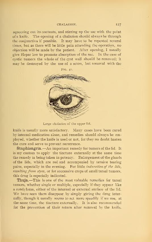
CHALAZION. 127
squeezing out its contents, and stirring up the sac with the point
of a knife. The opening of a chalazion should always be through
the conjunctiva if possible. It may have to be repeated several
times, but as there will be little pain attending the operation, no
objection will be made by the patient. After opening, I usually
give Hepar low to promote absorption of the sac. In the case of
cystic tumors the whole of the cyst wall should be removed; it
may be destroyed by the use of a seton, but removal with the
Fig, 41.
Large chalazion of the upper lid.
knife is usually more satisfactory. Man}^ cases have been cured
by internal medication alone, and remedies should always be em-
ployed, whether the knife is used or not, for they no doubt hasten
the cure and serve to prevent recurrence.
Staphisagria.—An important remedy for tumors of the lid. It
is my custom to apply the tincture externally at the same time
the remedy is being taken in potency. Enlargement of the glands
of the lids, which are red and accompanied by tensive tearing
pains, especiall}^ in the evening. For little induration of the lids,
resultingfrom styes, or for successive crops of small tarsal tumors,
this drug is especially indicated.
Thuja.—This is one of the most valuable remedies for tarsal
tumors, whether single or multiple, especially if they appear like
a condyloma, either of the internal or external surface of the lid.
We have seen them disappear by simply giving the drug inter-
nall}^, though it usually seems to act more speedily if we use, at
the same time, the tincture externally. It is also recommended
for the prevention of their return after removal by the knife.

128 disease:s of the eyelids.
For condylomata, or warty excrescences on the lids, especiall}^
if occurring in syphilitic subjects, this drug deserves attention.
Hepar.—Tarsal tumors that have become inflamed and are
sensitive to touch. It also aids absorption after operation.
Oalcarea carb.—Tarsal tumors occurring in fat, flabby sub-
jects.
Causticum.—Tumors; especially warts, found on the lids and
eyebrows.
Coniuin.—Induration of the lids remaining after inflamma-
tion.
Pulsatilla.—Tarsal tumors of recent origin that are subject to
inflammation, or are accompanied by catarrhal conditions of the
eye. The temperament and general symptoms will decide the
case.
Zincum.—Tumors of the lids with soreness and itching in the
internal canthi.
Baryta carb. and iod.. Graph., Lyco., Kali iod., Merc, Nitric
acid, Sep., Sil. and Sulph. may be required.
Epithelioma is the most frequent malignant growth affecting
the eyelid. It rarely occurs before the age of forty, and appears
first as small hard nodules, and later becomes covered with a scab,
which, on being removed, shows a slight excoriation ; this in-
creases to an ulceration of considerable depth, with^ purulent se-
cretion and irregular, hardened edges. It may remain indolent
for months, then assume an active stage, leading rapidly to exten-
sive destruction of tissue. In the early stages there is little or no
pain, but becomes excessively painful in the later stages.
Epithelioma, lupus and chancre have a very similar general
appearanceDifferential Diagnosis.
Epithelioma. Lupus. Chancre.
Indurated irregular
edges.Slow growth.Attacks middle-agedand elderly people,
lyymph glands of neckinvolved late.
Less induration. Moreinflamed.
Growth slower thanepithelioma.
Lymph glands involvedlate.
Generally associatedwith lupus elsewherein body.
Indurated, but edgesmore rounded.
Rapid growth.Usually in younger sub-
jects.
Lymph glands early in-
volved.Other symptoms of
syphilis.

LUPUS AND SARCOMA. 1 29
Lupus and Sarcoma have both been found affecting the eye-
hds. A lupoid growth is more slow in development than an
epithelioma, and both may result in ulceration ; but, as Noj^es^ so
well says,'
' A discrimination between them is hardly needful for
practical purposes.'
'
Treatment.—Excision is advised in all malignant growths of
the lids, if the disease is circumscribed and moderate in extent;
care being taken that all the morbid tissue is removed. The edges
of the wound may be brought together b}^ sutures, or a plastic
operation may be made, bringing the integument from the temple
or some adjoining point.
Juler- advises scrapi?ig away all the diseased tissue b}^ means of
a small steel scoop. This is attended with considerable haemor-
rhage, and is tedious, but he claims its success in arresting this
malignant affection is marvellous.
\"arious caustics have been emplo3'ed, chief arnong which ma}^
be mentioned caustic potash, nitrate of silver, chloride of zinc,
arsenic paste and acetic acid. An objection to the use of caustics
lies in the deformit}^ apt to occur afterward. Electrolysis has been
recommended.
If the disease is ver}^ extensive, involving the tissues of the
face to such an extent that extirpation is impracticable, we then
rely chiefly upon our internal remedies, using only such local ap-
plications as prove agreeable and of temporary relief to the
patient. For instance, if the discharge is profuse and offensive, a
weak solution of carbolic acid, salicylic acid, or some other disin-
fectant proves of service. An application from which we hav^e
often seen excellent results is carbolic acid and linseed oil (4grains to the ounce); it relieves the patient and seems to exert a
beneficial effect over the progress of the disease.
Iodoform.— Dr. George A. Shepard reports surprising results
in two cases of lupus from the administration of this drug in the
third decimal trituration.
Apis —Lupus non-exedens, sharp, stiiigiiig pains ^ and tendency
toward puffiness of the lower lids.
Hydrocotyle asiatica.—Has obtained a high reputation in
the hands of Dr. Boileau as a remedy for lupus and deserves our
attention.
^ Diseases of the Eye, 1890.
- Ophthalmic Science and Practice, 1S84.

130 DISEA.SKS OF THK EYKI.IDS
Phytolacca dec.—Benefit seems to have been derived in re-
lieving, if not curing, malignant ulcers of the lids, when used both
externally and internally.
Thuja.—Epithelioma of the lids.
Syphilitic Ulcers, Chancre, and Gummata are all occa-
sionally found on the eyelids, and should be treated the same as
occurring elsewhere. The most useful remedies are the following,
prescribed according to general indications: Ars. , Apis, Aurum,Cannab. , Hepar, Kali, iod., Merc, Nitric ac, and Thuja.
Herpes Zoster Ophthalmicus commences with severe neu-
ralgic pains along the supraorbital division of the fifth nerve of
one side, sometimes the nasal and rarely the infra-orbital branch of
the same nerve. The skin becomes swollen and red, covered with
herpetic vesicles which unite, crusts form upon them, followed by
ulcerations which ultimately leave deep cicatrices. The cause of
the disease is probably an inflammation of the nerve and the Gas-
serian ganglion belonging to it. The disease involves one-half of
the face, never extending beyond the median line, and has often
associated with it corneal ulcers and iritis. The intense pain that
precedes the herpetic eruption ma}^ last for a few hours, or extend
over months, but usually subsides with the appearance of the
vesicles.
Treatment.—The remedies most useful are: Ars., Croton tig..
Electricity, Canth., Graph., Merc, Puis., Ranunc. bulb, and
Rhus.
Contusions are very often found, and are frequently accom-
panied by some lesion of the globe or orbit.
Treatment.—Immediately after contused wounds of the lids,
cold compresses should be employed; they should be applied with
a firm bandage, which often proves of advantage in limiting the
amount of ecchymosis.
Arnica, our great remedy for all contusions, deserves its exten-
sive reputation for curing "black eyes," as there is no other drug
better adapted to this condition. A solution of the tincture in
water, ten drops to the ounce, is usually employed, though both
stronger and weaker solutions are in vogue. Ledum, used in the
same manner, has also proved of service.

WOUNDS. 131
Wounds.—We may have an incised, lacerated, or penetrating
wound of the Hd; and, in all cases, the condition of the orbit and
globe should be caref ulh'- examined to see if they remain intact.
Treatment.—The first object in view should be to bring to-
gether the edges of the wound by means of sutures, adhesive
strips or collodion. If the wound is incised, without any contu-
sion, and the edges are early brought accurately together, a dry
dressing will often be found most beneficial.
Hamamelis and Ledum have both been recommended for
wounds of the lids.
Burns and Scalds must be treated as usual in other parts of
the body, except that care should be taken to prevent the union
of the lids (ankyloblepharon) bj- frequently opening them, and
by inunction of the edge's with simple-cerate or cosmoline; also,
great attention should be paid to the prevention of a cicatrix
(which causes ectropium) by keeping the skin on the stretch by a
bandage during the period of cicatrization. Cosmoline is espe-
cially recommended as an external application.
When dependent upon the stings of insects, the sting should be
removed and cold water dressings applied.
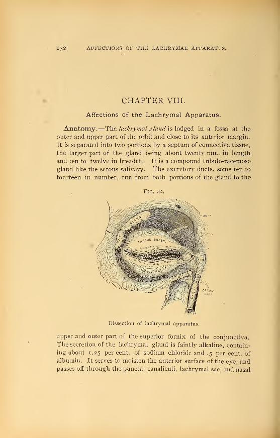
132 AFl^KCTlONvS OF THE LACHRYMAL APPARATUS.
CHAPTER VIII.
Affections of the Lachrymal Apparatus.
Anatomy.—''X\i^ lachrymal gland y^^Xo^.^^^ in a fossa at the
outer and upper part of the orbit and close to its anterior margin.
It is separated into two portions by a septum of connective tissue,
the larger part of the gland being about twenty mm. in length
and ten to twelve in breadth. It is a compound tubulo-racemose
gland like the serous salivary. The excretory ducts, some ten to
fourteen in number, run from both portions of the gland to the
Fig. 42.
Dissection of lachrymal apparatus.
Upper and outer part of the superior fornix of the conjunctiva.
The secretion of the lachrymal gland is faintly alkaline, contain-
ing about 1.25 percent, of sodium chloride and .5 per cent, of
albumin. It serves to moisten the anterior surface of the eye, andpasses off through the puncta, canaliculi, lachrymal sac, and nasal
\

DACRYOADENITIS. 1 33
duct, into the inferior meatus of the nose (Fig. 42). ThQ piinda
are the two minute openings of the canaUcuH, on the free margin
and about six mm. from the inner angle of the lids. The canali-
€uli, both upper and lower, extend from the puncta to the lach-
rymal sac, just before reaching which they unite. The upper
canaliculus is slightly smaller than the lower, and first ascends,
then turns downward and inward. The lower canaliculus first
descends, then runs horizontally to the sac. The lachrymal sac
is the upper dilated portion of the nasal duct. It is located in a
groove formed by the lachrymal and superior maxillary bones;
its upper end is closed and rounded. The nasal duct extends
from the lachrymal sac to the inferior meatus of the nose. Thebony canal is lined with a fibrous membrane, and this, in turn, by
cylindric epithelium like that of the nostrils. It is highly vas-
cular and thrown into folds at two or three points. The total
length of the sac and duct is about one inch. Its direction is
downward, backward and slightly inw^ard. The tears are forced
into the excretory passages by muscular action, aided by a kind
of suction caused by the muscular fibres of the puncta and canali-
€uli.
Dacryoadenitis.—Inflammation and abscess of the lachry-
mal glands are extremely rare. In the acute form the symptoms
are, great swelling and redness of the upper lid at its outer angle.
The globe ma}" be displaced downw^ard and inward by the swell-
ing. The pain is severe and increased by touch. The conjunc-
tiva is inflamed and may be chemosed. The inflammation mayterminate by resolution, suppuration, or run into the chro7iic
form, in which all the acute symptoms are less severe, merely a
considerable swelling remaining. Dacryoadenitis is generally
caused by injury, and this fact, together with its symptoms,
makes it very difficult to diagnose from periostitis of the orbit or
abscess of the lid.
Treatment.—When in the early stage, before there is a for-
mation of pus, ice will often cut short an attack which might
otherwise go on to suppuration. As soon, however, as there is
evidence of suppuration, we should resort to hot fomentations in
order to promote suppuration, and, when well established, a free
incision should be made through the conjunctiva, if possible.

134 AFFECTIONS OF THE LACHRYMAI, APPARATUS.
The most useful remedies are Aeon., Apis, Hepar, Rhus and
/Silicea. Vox \r\^dA(i2X\or\'s>'$>^^ dacryocystitis phleg-inonosa, page 139.
Hypertrophy of the Lachrymal Gland is very rare. It
is a circumscribed, nodular tumor of gradual growth and has
been known to occur in children and infants. If it increases suf-
ficiently to cause interference with the movements of the eyeball
it should be removed.
Tumors of the Lachrymal Gland, such as fibroids, sarco-
matas, adenomas, hydatid cysts and cancers have all been recorded.
They require extirpation of the gland.
AnomaUes of the Puncta and Canaliculi.—Eversion of
the puncta is frequently found in blepharitis and conjunctivitis,
causing epiphora, or watering of the eye, and will often result in
ectropium from the irritation of the tears flowing over the lid.
The same result will occur from a narrowing or stoppage of the
canaliculus, from wounds of the lid involving the canaliculus, or
foreign bodies in the canaliculi blocking the passage of the
tears. Obstruction of the canaliculus can be relieved by slit-
ting up the canal with the canaliculus knife (Fig. 43), a
narrow-bladed, probe-pointed knife, which is to be entered into
Fig. 43.
Agnew's canalicula knife.
the puncta vertically, the handle then brought to a horizontal
position, when the knife is pushed directly inward until it reaches
the inner wall (the lid being kept taut with the thumb of the
other hand), the knife is then brought to the vertical position,
cutting through the whole length of the canaliculus. The edge
of the knife should be kept toward the conjunctiva during its
passage, so as to divide the canaliculus close to the muco-cutane-
ous junction. It is better, where possible, to preserve the physio-
logical suction action of the canaliculus by onl}^ slitting it up for
two or three mm. from the punctum and dilating the remainder
of the canal with probes.

STRICrURA DUCTUS LACHRYMALIS. 1 35
Strictura Ductus Lachrymalis.—Stricture of the nasal duct
is the most common affection of the lachrymal apparatus.
Symptoms.—The chief characteristic symptom of stricture is
the overflow of tears, which is increased on exposure to cold wind
or bright light. There is also often noticed a dryness of the nos-
tril on the same side as the stricture. Usually on making pressure
with the finger over the sac we can press out from the puncta a
few drops of clear viscid secretion. We may also find a slight
conjunctivitis present.
Causes.— It is usually due to the extension of a nasal catarrh.
Injury or periostitis of the nasal bones, carious teeth, or pressure
from tumors in that vicinity may cause it. Its treatment will be
found under dacryocystitis.
Dacryocystitis Oatarrhalis.—Catarrhal inflammation of the
lachrymal sac is generally the result of a stricture or associated with
it. The catarrhal inflammation, on the other hand, may be the
cause of the stricture, as the swelling of the mucous membranewill in itself cause a damming up of the secretions. The reten-
tion of the tears, from obstruction, causes a gradual distension of
the sac—a swelling at the inner angle of the ej^e. By makingfirm pressure on this swelling the mucus can be pressed either out
of the canaliculus, or if no stricture remaining, down through the
nasal duct into the nose. The contents of the swelling may be
either clear and transparent or mixed with pus. This disease
usuall}^ develops ver}^ slowly, with simply the history of having
had a watery eye for a long while previous to noticing any swell-
ing of the sac, and oftentimes they will notice a dryness of the
corresponding nostril. The swelling is usually free from pain
or sensitiveness to touch.
Treatment.—Since lachr3mial diseases are frequently depend-
ent upon nasal catarrh, treatment must be directed to this affec-
tion.
As in nearl}^ all cases of blennorrhoea of the sac, a more or less
firm stricture of the lachrymal duct is present, this will require
our special attention. If the stricture is due to inflammatory
swelling of the mucous membrane, the knife is rarel}^ necessary.
Bony strictures are to be regarded as incurable. The cutting of
the stricture is only necessar}^ in those rare cases that will not
yield to probing and electrolysis.
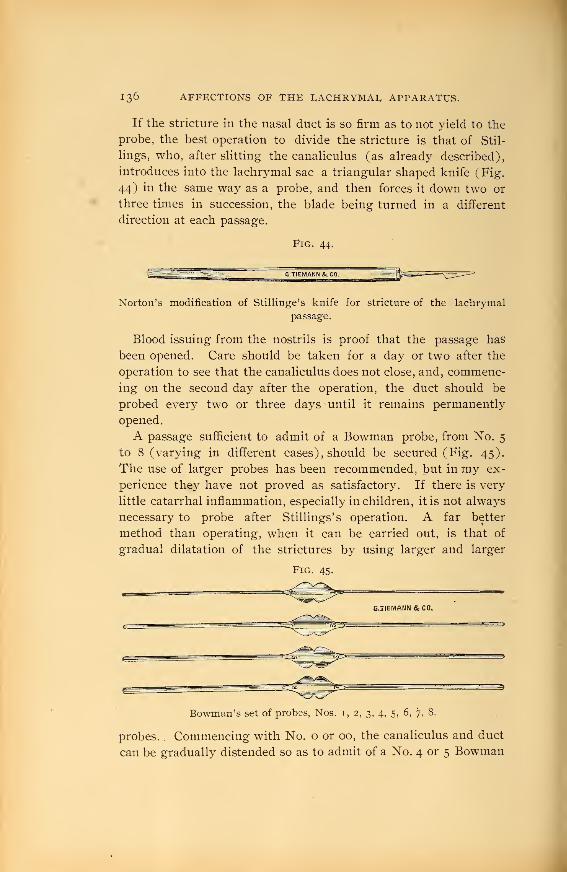
136 AFFECTIONS OF THE IvACHRYMAL APPARATUS.
If the Stricture in the nasal duct is so firm as to not yield to the
probe, the best operation to divide the stricture is that of Stil-
lings, who, after slitting the canaliculus (as already described),
introduces into the lachrymal sac a triangular shaped knife (Fig.
44) in the same way as a probe, and then forces it down tw^o or
three times in succession, the blade being turned in a different
direction at each passage.
Fig. 44.
Norton's modification of StilUnge's knife for stricture of the lachrymal
passage.
Blood issuing from the nostrils is proof that the passage has
been opened. Care should be taken for a day or two after the
operation to see that the canaliculus does not close, and, commenc-
ing on the second day after the operation, the duct should be
probed every two or three days until it remains permanently
opened.
A passage sufficient to admit of a Bowman probe, from No. 5
to 8 (varying in different cases) , should be secured (Fig. 45).
The use of larger probes has been recommended, but in my ex-
perience they have not proved as satisfactory. If there is very
Httle catarrhal inflammation, especially in children, it is not always
necessary to probe after Stillings's operation. A far better
method than operating, when it can be carried out, is that of
gradual dilatation of the strictures by using larger and larger
Fig. 45-
Bowman's set of probes, Nos. i, 2, 3, 4, 5, 6, 7, 8.
probes. Commencing with No. o or 00, the canaliculus and duct
can be gradually distended so as to admit of a No. 4 or 5 Bowman

DACRYOCYSTITIS CATARRHALIS. 1 37
probe without even slitting the canaliculus. In many cases the
dilatation to simply a No. i probe has been sufficient to result in
a cure, and in some cases it seems to be better than to dilate to a
larger size. Rarely, where the punctum is occluded, it will benecessary to slightly nick it with the point of the knife so as to
admit of the smallest-sized probe. The advantages of this method
seems to lay in the fact that we do not destroy the function of the
parts and that the normal suction action of the canal is retained.
This plan, of course, is not practical in very firm or bon}^ strict-
ures. Marked benefit has also been observed from electrolysis,
and there is no doubt it should be more extensivei}^ employed.
My plan has been to insert a probe the usual w^ay until it comes
in contact with the stricture, then attaching the probe to the
negative pole of a battery, apply the positive to the temple, and
make gentle pressure as the stricture yields. Usually four or five
treatments will be sufficient to keep the passage permanently
opened.
In the treatment of the blenorrhoea, a free vent for the secre-
tions through the opened or unopened canaliculus being present,
the patient should be instructed to press out the matter several ti?nes
a day.
Mild astringent injections of boracic acid, sulphate of zinc of a
two to four per cent, solution or some similar preparation maysometimes prove very serviceable.
Fig 46.
Agnew's lachrymal s^^ringe.
Recently I have been using with good results an injection of the
blue pyoktanin, i to i,ooo.
Pulsatilla.—One of the most important remedies for dacryo-
cystitis, which may sometimes be cut short at its very beginning
with it, and may be useful at any stage of the inflammation. It

138 AFFECTIONS OF THF l^ACHRYMAI, APPARATUS.
is also important in blenorrhoea of the sac, if the discharge is
profuse and bland. Profuse thick and bland discharge from the
nose; especially beneficial in children.
Stannum.—Very favorable results have frequently been ob-
tained in controlling the yellow-white discharge from the lachry-
mal sac, itching or sharp pain in the inner canthus, especially at
night.
Hepar sulph.—Inflammation of the lachrymal sac after pus
has formed, or in blennorrhoea, with great sensitiveness to touch
and cold, with profuse discharge. Throbbing pains.
Euphrasia.—Much thick, yellow, acrid discharge, making the
lids sore and excoriated. Blurring of the vision relieved by wink-
ing. Thin, watery, blayid discharge from the nose.
Argentum nit.—Discharge very profuse, caruncula lachry-
malis swollen, looking like a lump of red flesh; conjunctiva
usually congested.
Aconite.—In the first stage, when the lids are much swollen,
with great heat, dryness, tenderness, sharp pains and general
fever.
Apis.—Before the formation of pus. lyids oedematously swol-
len, with stinging, shooting pains. Patient drowsy, without
thirst.
Arum triph.—Catarrh of the lachrymal sac, with desire to
bore into that side of the nose; nose obstructed, compelling to
breathe through the mouth; nostrils sore, the left discharges
continually.
Mercurius. — In the later stages after the pus has become
thin and excoriating ; acrid coryza ; nocturnal aggravation.
Petroleum.—Discharge from the lachrymal sac, with rough-
ness of the cheek, occipital headache, and other marked concom-
itant symptoms. In the early stages, when there is considerable
swelling of the lids, with burning pains.
Silicea.—Occasionally indicated in dacryocystitis characterized
by the usual symptoms of pain, swelling, tenderness and lachry-
mation ; even cases that have far advanced toward suppuration
have been checked. Blennorrhoea of the lachrymal sac often
calls for it. The patient is particularly sensitive to cold air and
wishes to keep warmly covered.
Other remedies which have been recommended and proved use-

DACRYOCYSTITIS PHLKGMONOSA. 1 39
ful are Bell., Calc, Cinnab., Hydrast., Kali iod., Merc, prot.^
Nat. mur., Nux, Sulph, and Zinc suli>h.
Dacryocystitis Phlegmonosa.—Phlegmonous inflammation
of the lachrymal sac may be considered as merely a higher stage
or extension of the preceding disease. It consists in a purulent
inflammation of the connective tissue surrounding the lachrymal
sac. and results in an abscess that breaks externally.
Symptoms.—In this the swelling is greatly increased and ex-
tremel}^ sensitive to touch. The integument becomes very tense
and assumes a dusky-red hue. There is usually an oedematous in-
filtration of the surrounding parts, viz.: eyelids, side of the nose
and cheek. There is intense pain and heat, with sometimes gen-
eral symptoms of chills, fever and vomiting. The conjunctiva maybe inflamed and even chemosed.
Differential Diagnosis. Tlie appearance at this time resembles
an abscess of the cellular tissue overlying the lachrymal sac and
must be carefully differentiated, but in the dacryocN-stitis phleg-
monosa firm pressure over the swelling will usually empty the
tumor either through the puncta or downward through the nose,
while in abscess it will not. In dacryocystitis we also have the
previous history of a long-continued lachrymation, which is, of
course, absent in abscess. An abscess over the lachrymal sac is
rare, and we may therefore generally assume the abscess to have
originated within the sac.
Course.—If left to itself the swelling usually increases steadily,
the skin over the sac becomes thinner and thinner, until it finally
gives way, the abscess discharges leaving a fistula of the lachrymal
sac, which is extremely difficult to heal.
Cause.—A catarrhal inflammation precedes a phlegmon. Thedecomposed secretions in the sac penetrate the mucous membraneand set up a purulent inflammation. The exciting cause is fre-
quently a simple cold in the head.
Treatment.—At the commencement, before the formation of
pus, cold compresses (even ice) are advisable, w^hich, together
with the indicated remedy, may cause the inflammation to abort
before an abscess has formed.
As soon, however, as pus has begun to collect in the lachrymal
sac, our treatment must undergo a decided change. The t?/'^/ and

140 AFFECTIONS OF THE LACHRYMAL APPARATUS.
most important step to be taken to prevent its breaking externally,
with the possible formation of a fistula, is the opening of the
canaliculus into the sac and the evacuation of its contents, through
the natural channel. But if the disease has so far advanced that
perforation is inevitable, a free incision into the sac should be
made externally, after which, and also in case the abscess has
opened spontaneously, warm compresses may be employed for
twenty-four or forty-eight hours, but must not be continued too
long. The opening should be kept open by the insertion of a
strip of iodoform gauze every day until the subsidence of the in-
flammation, when the opening will usually close without trouble,
though it may be necessary to open the nasal duct and establish a
free passage for the tears before it does so. Probing of the nasal
duct should be avoided until the severity of the inflammation has
subsided. Warm and moist applications should be substituted
for the cold as soon as suppuration has commenced. Among the
best of those in use is a solution of calendula. Internal medica-
tion during the whole course of the disease will form an important
feature in the treatment. For indications see dacryocystitis
catarrhalis, page 137.
Fistula Lachrymalis.—An opening externally of the lachry-
mal sac, when the result of an abscess breaking,- is often very
obstinate and difficult to heal; hence, when evidently about to
break, it should be opened with a bistoury.
Treatment.—The first point to be attended to is to see that
the passage is free into the nose. We must therefore slit up the
canaliculus and divide any stricture found in the nasal duct, pro-
viding it is sufficient to interfere with the flow of tears; after
which the canal should be kept open.
The fistula must now be healed, and, if recent, this is best done
by touching the edges with a stick of nitrate of silver, or the
gentle application of the galvano-cautery. If the edges of the
fistula are healed and covered with smooth skin, it will be neces-
sary to pare the edges and unite with a suture.
The following remedies have been advised and may have been
of service in recent cases, though we doubt if any effect can be ob-
tained in old chronic fistulse: Brom., Calc, Fluoric ac. , Lach.,
Merc, Nat. mur., Petrol., Sil. and Sulph.
\

ANATOMY OF THE ORBIT. I4I
CHAPTER IX.
Diseases of the Orbit.
Anatomy.—The shape of the orbit is that of a quadrangular
pyramid, the base or facial opening, the four walls and the apex.
The axes of the orbits converge posteriorly at an angle varying
in different individuals. The bones entering into the formation of
the orbital walls are the frontal, sphenoid, superior maxillary,
malar, palate, ethmoid and lachr3^mal. The optic foramen, sit-
uated at the apex of the orbit, transmits the optic nerve and the
ophthalmic artery. The superior orbital fissure transmits the
third, fourth and sixth nerves, ophthalmic branch of the trige-
minus, the superior and inferior ophthalmic veins, few sympa-
thetic filaments from the cavernous plexus, the recurrent lach-
rymal artery and sometimes orbital branches of the middle menin-
geal artery. The inferior orbital fissure gives passage to the malar
and infra- orbital nerves, infra-orbital vessels, a facial branch of
the ophthalmic vein, and the ascending branches of the spheno-
palatine ganglion. The supra-orbital notch, at the upper and
inner margin of the orbit, contains the supra-orbital nerve, arterj^
and vein as they pass to the forehead. The orbit, in addition to
the eyeball, vessels, muscles, etc., contains considerable adipose
tissue.
TenoiV s Capsule is the limiting membrane between the cellulo-
fatty tissue and the globe and conjunctiva. It ensheaths to someextent the muscles, vessels, nerves, etc., that pass through it, ana
is continuous with the periosteum of the orbit as well as with the
conjunctiva. It is somewhat analogous to the pleura, and serves
as a cup in which the globe revolves. It constitutes a second-
ary attachment for the ocular muscles, and by this attachment it
renders it possible to sever the tendon of a muscle without losing
its entire action upon the eye, for it still remains in connection
with the eye through Tenon's capsule, unless too extensive lateral
cuts have been made, separating the tendon from the capsule.
The dura mater is firmly attached at the sphenoidal fissure and

142 DISEASES OF THE ORBIT.
optic foramen and is continuous with the outer sheath of the
optic nerve and with the periosteum of the orbit.
Cellulitis Orbitae i^Abscess or Phlegmon of the Orbit).—In-
flammation of the cellular tissue of the orbit may occur as a
simple oedematous cellulitis or in a far more active form as a
phlegmojious cellulitis.
Symptoms.—In oedematous cellulitis the eye will be slightl}''
bulged forward, its movements limited and sometimes diplopia is
complained of. There is usually in these mild cases little or no
swelling or redness of the lids or conjunctiva, and but little dull
pain, except on pressure upon the globe. This form of cellulitis
generally occurs in young and delicate children, and usually sub-
sides within a few days.
In the severer form, ox phlegmonous cellulitis, the onset is apt to
be accompanied with a chill and rise in temperature. There will be
swelling and dusky discoloration of the lids, especially the upper,
and a more or less intense pain, greatly increased by pressure
upon the globe. The eyeball is protruded directly forward, and
its movements limited in all directions; in some severe cases it
will have absolutely no motion. (In periostitis, which it closely re-
sembles, there is greater swelling and redness and the protrusion of
the eye and the limitation in its movement is in but one direc-
tion.) The conjunctiva is chemosed and the cornea completely
or partiall}" anaesthetic. Diplopia is usually present and the vision
may be greatly impaired from optic neuritis and atrophy. Digital
examination will find the tissues firm, tense and very painful to
the touch. In extreme cases the eyeball may become involved
and end in panophthalmitis.
Causes.—Various and frequently obscure. May be metastatic
from phlebitis, septicaemia, puerperal fever, etc. It is often co-
incident with facial erysipelas. May result from cold, from in-
juries, periostitis and inflammation of the lachrymal gland.
Prognosis. —Is always serious as vision may be lost from neur
itis, slough of the cornea, or panophthalmitis. It may terminate
fatally through meningitis and abscess of the brain, though the
large majority recover.
Treatment.—When due to a foreign body, \\. should be re-
moved, and the ice bag employed to subdue if possible the inflam-

CELLULITIS ORBITS. 1 43
matory symptoms. But if suppuration has already set in, poultices
should be applied to promote the discharge of pus, which should
be evacuated at an early period, by a free incision through the
conjunctiva if practicable, if not, through the lid itself. Care
should be taken that the pus has free vent at all times. Noyes
{loc. cit.) advises an early incision, even before pus forms, claim-
ing its value " as a means of arresting the phlegmonous inflam-
mation and the formation of pus." as by the incision " the oculo-
orbital fascia is relieved, the vessels are unloaded, serum finds
vent and the tissues are relaxed." Diet and rest should be pre-
scribed according to the general tone of the patient and severity
of the attack.
Rhus tox.—This is a remedy of the very first importance in
this form of inflammation. The lids are cedematously swollen, as
well as the conjunctiva, and, upon opening them, a profuse gush
of tears takes place. The pains are especially severe at night,
vary in character and may be greatly influenced by any change
in the weather. Panophthalmitis is liable to complicate the
trouble.
Hepar sulph.—Especially after pus has formed. Lids swollen
and very sensitive to both touch and cold. The pains are usually of
a throbbing character.
Phytolacca.—Inflammation of the cellular tissue of the orbit
without much pain, slow in its course and with little tendency to
suppuration. The eye will be protruded and the infiltration into
the orbit and lids will be hard and imyielding to touch.
Aconite.—In the first stage, when the lids are much swollen,
with a tight feeling in them; chemosis, with much heat and sensi-
iivejiess in and around the eye, and a sensation as if the eyeball
were protruding, making the lids tense, associated with the gen-
eral Aconite fever.
Apis mel.—Before the formation of pus. Lids cedematously
swollen, with stinging, shooting pains. Patient drowsy, without
thirst.
Lachesis.— Orbital cellulitis following squint operation, point
of tenotomy sloughing, with a black spot in the centre; chemosis,
and much discharge, with general Lachesis condition.
Mercurius.—In the later stages after pus has formed, and
even after it has discharged for some time and has become tJiiji in

144 DISKASKS OF THE ORBIT.
character, especially if occurring in a syphilitic subject. Thereis often much pain in and around the eye, always worse at night.
Other remedies may be thoug^ht of, as Ars., Bell., Bry., Kali
iod. , Sil., Sulphur.
Tenonitis.—Inflammation of the capsule of Tenon is a com-
paratively rare disease which may follow operations for strabis-
mus and less frequently occurs idiopathically, especially in those
of a rheumatic diathesis.
Symptoms. —There may be slight swelling of the lids, chemosis
of the conjunctiva, exophthalmos and diminished mobility of the
eye. Pain more or less severe, especially on pressure or movementof the eye, is apt to be present.
,Treatment. — Internal medication is all sufficient in this
disease and the most serviceable remedies 2.x^ Kalmia lat., Kali
iod. , Rhus and Puis.
Periostitis Orbitae.—Inflammation of the orbital periosteum
may result from injuries or occur idiopathically in rheumatic,
syphilitic or scrofulous subjects. The disease is most commonlyfound in early life and its usual location is the margin of the orbit.
Symptoms.—There is present pain, especially from pressure on
the bone, oedema of the lids, chemosis and a tense, .swollen, sensi-
tive spot in which fluctuation may be detected later. In the acute
form there may also be fever, vomiting, delirium, etc. Periostitis
in its acute form resembles very closely a phlegmonous cellulitis,
and must be differentiated by the acute pain on pressure upon the
orbital margin ; by less swelling and redness of the lids ; the in-
flammation is more circumscribed, so that the displacement of the
eyeball is in one direction and its mobility is more restricted in one
direction.
In the chro7iic form of periostitis there is simply slight swelling
of the upper lid and supra- orbital pain, together with localized
swelling at the seat of the inflammation. This is the more fre-
quent form, and especially found in young scrofulous subjects.
Course.—When chronic, it is very tedious, lasting months or
years, and is apt to result in caries of the bone, fistulae, deformity
of the lids, etc. In the acute form, if the abscess is near the sur-
face and promptly opened, it may heal in a short time ; but if deep

CARIES AND NKCROSIS. I45
and neglected, or if occurring in one of a syphilitic or scrofulous
constitution, will usually be much more serious.
Prognosis.—When near the orbital margin it is favorable, butif deep in the orbit it is much less so, as it may result in atrophyof the optic nerve, paralysis of the orbital muscles, or meningitis.
Treatment.—See Caries.
Caries and Necrosis of the orbital walls is almost universally
a result of periostitis or an injury, although it may occur in the
bone itself without a previous inflammation of the periosteum,
especially in syphilitic or scrofulous subjects. After the abscess
has opened, a fistula is formed which leads to the roughened anddenuded bone. The discharge of pus through the fistula has the
peculiarly fetid odor of osseus caries. The general symptoms of
periostitis are present in caries, and, in addition, the diseased bonecan be detected by the probe. Caries is most commonly found in
children and necrosis in adults.
Treatment.—The general plan of treatment is very similar to
that recommended for cellulitis, as we should at first endeavor to
prevent destruction of tissue, but, if that does occur, give the pus
free vent. If the bone should be diseased the opening must be
kept open and an injection of a solution of carbolic acid i to 100,
or of the sulphate of zinc gr. x to 5J may be used with advantage.
If any of the loose pieces of the bone are discovered they should
be removed. The remedies described under cellulitis are also
applicable to this disease ; in addition to which we note the fol-
lowing :
Kali iod.—This form of potash is one of the most important
remedies we possess for periostitis. It is especially adapted to
the syphilitic variety, though useful when dependent upon other
causes. The pain is usually marked, though may be absent en-
tirely. The lids will often be oedematous. The crude salt in
large doses has seemed to act more rapidly than the attenuations.
Aurum.—For both periostitis and caries, when depejideni upon
or complicated with a mercurio-syphlitic dyscrasia; also useful in
strumous subjects. The pains are tense, and seem to be in the
bones, are worse at night, bones sensitive to touch and the patient
is excessively sensitive to pain.
Mercurius.—As described under cellulitis, will be found very
10

146 DISEASES OF THE ORBIT.
useful in both periostitis and caries, particularly when dependent
upon S3^philis, as the nocturnal aggravation is very marked under
both the drug and disease. The different forms are employed
according to general indications.
Silicea.—Its action upon diseased bones renders it especially
valuable in caries of the orbit. The roughened bone and moder-
ately profuse yellow-white discharge are the principal indications,
though the weakened general condition, relief from warmth and
other concomitant symptoms will be present.
Calc. hypophos.—In appreciable doses, has been used as a
"tissue remedy" in scrofulous subjects, apparently with good
results.
The following remedies ma}^ be required: Asaf., Calc. carb..
Fluoric acid, Hecla lava, Lyco., Mezer., Nitr. ac. Petrol., Phos-
phor, and Sulphur.
Empyema of the Frontal Sinus is extremely rare. Thecause is uncertain. It may occur at any time of life, except in
young children. The early swelling and distension are unnoticed,
but it finally perforates the bony wall and a swelling appears at
the upper and inner angle of the orbit, causing epiphora, dis-
placement of the eye downward, foreward and outward, with
diplopia. The patient may have had headache for a long time, or
it may have caused but little discomfort.
Treatment.—Similar to that given for cellulitis and perios-
titis.
Tumores Orbitae.—In the orbit may be found both benign
and malignant tumors, which may have developed primarily in
the orbit, in some of the neighboring sinuses such as the antrum
or ethmoidal, or have spread from the eyeball or face. Theyusually cause more or less exophthalmos and restriction in the
mobility of the eye; the displacement of the eye depends upon
the location of the growth. The eye may suffer from inflamma-
tion, the optic nerve may become inflamed or atrophic, the retina
detached, etc. The examination should be directed to the degree
and direction of the exophthalmos; to the impairment in motion,
whether in one direction or all; to the feel of the growth, its
smoothness, mobility, solidity, pulsation, fluctuation, etc.

TUMORES ORBITS. I47
We note the effect of pressure upon the eyeball, if it causes
pain on being pushed backward, or if the position of the tumor
is altered. We inspect the nostrils, the pharj'nx, the frontal and
maxillary sinuses. The tumor may be explored with the hypo-
dermic syringe. The history of the case should be elicited for
hereditary tendency, the progress of the growth whether slow or
rapid, whether associated with pain or not. All these points
and many more should be considered, as an aid to diagnosis, prog-
nosis and treatment. Nearly all varieties of tumors may be
found in the orbit, viz.: Osteoma, naevi, angioma, lipoma,
fibroma, cysts (dermoid or hydatid), neuro-fibroma, l^^mphoma,
sarcoma, scirrhus, encephalocele, epithelioma, etc.
Treatment.—The most approved method of treatment of all
tumors of the orbit is to remove them as early as possible, en-
deavoring to save the eye whenever sight is present, unless it be
a malignant growth and there is danger of not removing the whole
of the tumor without sacrificing the globe; in which case it is
usually better to remove all the contents of the orbit. A careful
diagnosis must be made before operating, in order to aid in the
operation and prevent the opening of a vascular tumor.
Electricity is of great value in removing vascular tumors of the
orbit. When the growth is small the negative pole may be ap-
plied by a sponge to the temple, but if large both poles should be
attached to platinum needles, two or three inches in length,
which are then to be inserted into the tumor. The positive needle
should remain in one position while the negative may be inserted
at different points for a few moments at a time. Care should be
taken to make the first sitting brief, from fear of a too severe in-
flammator}" reaction.
Our remedies are the same as for tumors in other portions of
the body, though we would especially mention Thuja and Kali
iodata, which have been of service in some cases.
Wounds and Injuries of the Orbit may prove serious from
inflammation of the orbital tissue, or periostitis, which they ma3"
cause. Penetrating injuries from knives, shot, pitchforks, etc.,
cause laceration of the soft parts. Injury causing fracture of the
orbital walls may prove more or less serious according to the
location and extent of the fracture; a slio^ht lesion of the orbital

148 DISEASKS OF THK ORBIT.
margin may heal without trouble; in fracture of the frontal or
ethmoidal cells we will usually have emphysema of the orbit and
lids, due to an entrance of air into the cellular tissue. If the in-
jury has occurred in the vault of the orbit, we may have a
serious inflammation of the brain- or its. membranes. Frac-
ture of the roof of the orbit has frequently been found with a
fracture of the base of the skull. Out of 86 cases of fracture of
the base of the skull, fracture of the orbital roof was found in 79.*
Haemmorrhage into the cellular tissue is very apt to occur in all
injuries or wounds of the orbit, hence exophthalmos is usually
present. Foreign bodies of large size have frequently remained
imbedded in the orbital tissues for a long while without creating
any material disturbance.
Treatment.—When a foreign body has penetrated the orbit
it should be removed as soon as possible, after which cold
compresses of calendula in solution, or the ice bag should be
applied.
Injuries with an effusion of blood into the orbit, causing the
eye to protrude, will be benefited by a cold compress and a firm
bandage. In emphysema of the orbit and lids a compress ban-
dage will be required.
Morbus Basedowii (^Exophthalmic Goitre, Graves' Disease').
Symptoms.—The main symptoms of this disease are : rapidity
of the,heart's action, enlargement of the thyroid and protrusion
of the eyes, although any one of these symptoms may be
absent, The acceleration of the heart's action is the earliest,
most constant and essential symptom, and this may reach
from 100 to 200 beats per minute and may be weak and irregular.
Linnell \ in an excellent paper on this disease calls attention to one
case in which there was no change in the pulse beat, whether sit-
ting or standing, the usual variation returning as the case im-
proved. The heart symptoms are usually first developed, followed
later by the enlargement of the thyroid and the exophthalmos
either simultaneously or in succession. The goitre may develop
suddenly, has a soft, elastic feel, a visible pulsation, and a systolic
murmur on auscultation. The enlargement of the thyroid and the
*BerUn: Graefe and Saetnisch, Handbuch der Augenheilkunde, 1880.
t Trans. Amer. Inst. Homoe., 1892.

MORBUS BASEDOWII. 1 49
exophthalmos are originally due to vascular engorgement. Theexophthalmos is almost universally bilateral, though it may be
confined to one side. The degree of protrusion varies. It may be
so excessive that the lids are unable to cover the eyes, and ulcera-
tion of the cornea may result from the exposure. The protrusion
is straight forward, causes no interference with the movements of
the eyes, and the eyes may be pressed back into their normal posi-
tion, but will become prominent again on relief of the pressure.
The exophthalmos gives the patient a frightened, staring appear-
ance. On turning the eyes downward the upper lid does not fol-
low it at all, or moves along for a certain distance and then re-
mains stationary. This peculiarity, called Graefe's sign, because
first noticed by him, is of much diagnostic value, because highly
characteristic of this disease, and is rarely found in exophthalmos
from any other cause. Other symptoms of this disease are
dyspnoea and excessive nervousness. There is usually excessive
nervous excitability and tremulousness of the hands. The dispo-
sition is often changed. The patient is easily frightened and
flushes readily. Epistaxis or haemorrhage from other parts mayoccur. Dilatation and pulsation of the retinal arteries has been ob-
served. Anaemia is often present, and in women may be asso-
ciated with menstrual disturbances ; rapid emaciation may occur
in some cases.
Course.—The disease as a rule is very chronic, lasting for sev-
eral years with frequent temporary improvements and relapses;
the symptoms may then gradually subside, though often some en-
largement of the thyroid or exophthalmos will remain perma-
nently. Rarely death occurs in this disease, especially in men in
advanced life, from organic disease of the heart or exhaustion.
The majority of these cases, however, practically recover.
Causes. —Basedow's disease is much more often found in womenthan in men, some authorities claiming that over 85 per cent, of
all cases are found in women. The majority of cases occur
between the age of puberty and thirty in women, while the average
age in men for the appearance of this disease is from thirty to
fifty.
This disease has at various times been attributed to a disease of
the sympathetic, to rheumatism, etc., and, while its cause has not
yet been definitely demonstrated, it will probably be found in
I50 DISKASES OF THE ORBIT.
some central lesion located in the gray matter of the third and
fourth ventricles, the vaso-motor centre and the origin of the
pneumogastric. It is generally brought on by some mental shock,
by exhaustion from disease, excessive haemorrhages, or severe
labor.
Treatment.—To promote a permanent cure, rest, especially
in the country ; freedom from all excitement, especially emo-
tional ; exercise in open air ; a generous diet, and abstinence from
all stimulants, are particularly required and should be insisted
upon whenever practicable. Galvanization of the sympathetic in
the neck has been followed by very good success in many instances,
especially when combined with internal medication.
Amyl nit.—Cases have been entirely cured by olfaction of this
drug alone. The eyes are protruding, staring, and the conjunc-
tival vessels injected, as well as those of the fundus. Especially
indicated when there are frequent flushes of the face and head,
oppressed respiration, etc.
BadiagO.—Exophthalmic goitre, with aching pain in the poste-
rior portion of the eyeballs, aggravated on moving them, accom-
panied by tremulous palpitation of the heart and glandular swell-
ings. The pulse is rapid and irregular.
Cactus grand.—Cases of exophthalmic goitre have been im-
proved, when prescribed on the heart symptoms.
Ferrum.—Both the iodide and acetate have been followed by
favorable results, especially when the disease comes on after the
suppression of the menses;protruding eyes, enlargement of the
thyroid, palpitation of the heart and excessive nervousness.
Linnell found good results from the simultaneous use of Iron and
Digitalis in one case, after both had been tried separately with no
benefit.
Lycopus virg.—^Judging from its provings, in which" we find
a rapid, irregular, intermittent and very compressible pulse,
together with a bellows murmur over the heart and large vessels,
it should be a valuable remedy in this disorder. Linnell (yloc. cit. )
rep )rts very gratifying results from the use of this remedy.
Spongia.—Exophthalmos, enlargement of the thyroid and
palpitation of the heart, great uneasiness and easily frightened,
especially at night ; stitches in the ball and burning around the
eyes, with lachrymation in the light ; the eye feels twisted around;

MORBUS BASEDOWII. I5I
chromopsies, especially deep red, and photopsies, even when the
e3^e is closed at night^—all indicate this drug, which has proved
serviceable.
Mr. Hulke"^^ recommends Aconite, and Dr. Samuel Wilks
Belladonna.
Nat. viur. and Baryta carb. are reported to have cured well-
marked cases. Other remedies which have been recommended are
Ars., Bell., Brom., Calc, lod., Phos., Sil., Sulph. and Veratrum
alb. and viride.
* Trans. Ophthal, Soc, Vol. VI.

152 AFFECTIONS OF THE OCUI.AR MUSCIvKS.
CHAPTER X.
Affections of the Ocular Muscles.
Anatomy.—The movements of the eyeball are carried on
through the action of six voluntary muscles; four of these passing
directly from their origin, around the optic foramen, to their in-
sertion in the sclerotic, are called the recti muscles. These
muscles are inserted at varying distances, from 5.5 to 8 mm. from
the corneal margin, and are distinguished by their relations to the
eyeball, as internal, external, superior and inferior rectus. Theinsertion of the internal rectus is the nearest to the corneal
margin, and that of the superior rectus is the most posterior.
The remaining muscles, from their taking a curved direction
around the eyeball, are known as the superior and inferior
oblique muscles. The superior oblique arises at the optic fora-
men, passes forward along the upper part of the inner orbital
wall to a pulley attached at the superior-internal angle at the front
of the orbit; from here it is reflected backward and outward be-
tween the superior rectus and the eyeball, and is inserted into the
sclerotic, 16 to 18 mm. from the corneal margin on the posterior
and outer part of the globe, between the superior and external
recti.
The inferior oblique arises from the inner and anterior
part of the floor of the orbit and passes outward and backward be-
tween the inferior and external recti and the globe, and is inserted
near the superior oblique, between the superior and external
rectus, 17 to 19 mm. from the corneal margin. The internal,
superior and inferior recti and the inferior oblique are supplied by
the third 7ierve (oculomotorius), the superior oblique by they^2^r/'^
(patheticus) and the external rectus by the sixth (abducens).
The action of all these muscles is to turn the eye around a
point, called the centre of rotation , situated on the antero-posterior
axis of the globe, about 14 mm. behind the anterior surface of the
cornea and about 10 mm. in front of the posterior surface of the

ANATOMY 153
sclera. These six muscles form three pairs of antagonistic
muscles. The axis of rotation of the internal and external recti
is vertical, as these muscles turn the globe directly inward and
outward The axis of rotation of the superior and inferior recti
is horizontal, although not exactly straight across; it forms an
angle of 67° with the visual axis, the nasal end of the line being in
front of the temporal and is the axis of elevation and depression.
The axis of the superior and inferior oblique is also horizontal,
forming an angle of 38° with the visual axis, with the temporal
extremity in front of the nasal, and the movements about it are
those of rotation. (Fig. 47.
)
Fig. 47-
Axes of ocular muscles.
Taken singly, the muscles act as follows: The internal rectus
draws the cornea directly inward (adduction), the external rectus
turns the cornea directly outward (abduction), the superior rectus
turns the cornea upward and slightly inward, the inferior rectus
turns the -cornea downward and slightly inward, the superior

154 AFFECTIONS OF THE OCUI.AR MUSCLES.
oblique, taking its point of action from the pulley through which
it passes, turns the cornea downward and outward and rotates
from above downward; the inferior oblique turns the cornea up-
ward and outward and rotates it from below upward.
Each movement of the eye results from the combined action of
certain muscles, and there is never a period at which one or the
other is entirely inactive, as by its living and elastic tension it aids
in guiding a definite movement, even though it docs not take part
in it. In looking directly upward both the superior rectus and
inferior oblique are brought into action, the inclination inward of
the former being counteracted by the outward tendency of the
latter, and so in all other movements of the eye the action of all
the muscles must be considered. Paralysis of any one muscle will
cause all movements of the eye to be less sure.
The function of the ocular muscles is to secure single vision
with the two eyes by directing both eyes to the point of fixation
in such a manner that the image of the object fixed shall fall
simultaneously on the macula luteaof each eye. When this is done,
all objects lying in the same horopter will form images upon the
respective retinae which will be equidistant from the fovea centralis
and will, therefore, be appreciated as single, giving what is called
binocular vision.
The horopter, as described by Miieller, is represented by a circle
which passes through the centres of rotation of each eye and
through the apex of the point of fixation of the visual lines. All
objects beyond or inside the horopter will cast images on parts of
the retinae not equidistant from the fovea and will create the im-
pression of two objects or double vision; for example, holding two
pins in the same line, one eight inches and the other twenty from
the eyes, on looking at the nearest pin the other is seen double,
and vice versa. This double vision of objects not lying in the
horopter causes no annoyance, because the mind ignores the im-
pression of objects with which it does not concern itself. Thesymmetrical position of the physiological centres of the retina is
the anatomical ground for binocular vision. If a distant object be
fixed with parallel lines of vision, to fix an object at one side
which throws its image in both eyes at an equal distance from the
macula lutea, both eyes must move through an equal angle; con-
sequently, equal innervation on both sides is necessary for the

PARALYSIS OF OCULAR MUSCLES. 1 55
sj^mmetrical movements of the e3"es. All objects are seen single
only when retinal images fall on corresponding points of the two
retinae.
Homonymous Diplopia. If the visual line of the left eye be
directed on an object and there is convergence of the right eye,
the image, which would in the left eye be formed on the fovea
in the right would fall upon the retina to the inner side of the
fovea and would be projected outward to the right of the object
fixed, or homonymous diplopia.
Heteronymous Diplopia. If there is divergence of the right eye
at the time the visual line of the left eye is directed upon an object,
the impression from the object fixed would in the right eye fall
upon the retina to the outer side of the fovea and when projected
outward would appear to the left of the object fixed, causing
crossed or heteronymous diplopia. The displacement of the false
image is always in the direction which is opposite to that of the
deviation of the eye. When the eye deviates inward, the diplopia
is homonymous; when outward, heteronymous; when upward, the
false image is below, and, when downward, it is above. Thefalse image is the image of the deviating e5^e.
Binocular Diplopia is present when the visual axis of one ej^e
deviates from the object of fixation.
Monocular Diplopia is due to the formation of two images of the
same object upon one retina and exists when the other eye is
closed. As in binocular diplopia there is one image cast upon
each one of the two retinae the diplopia disappears on closing
either eye, while if the diplopia persists on closing one eye, it is
then monocular. The cause of monocular diplopia is either an
anomalous refraction or a double pupil.
Paralysis of Ocular Muscles.—We may have one or more
muscles paralyzed, and the cause ma3^ be either orbital or intra-
cranial; if the latter, it may be along the course of the nerves or
in the brain. Lesions of the spinal cord may cause paralysis
through fibres which proceed to the brain.
Symptoms.—The characteristic indications of paralysis are false
position of the eye, limitation and irregularity in motion, and
double images. x\s secondary effects, we find dizziness, nausea,
headache, incorrect projection of the field of vision and inability

156 AFFKCTIONS OF THE OCUI.AR MUSCLES.
to guide the hands or feet aright. Indistinct vision may occur in
cases of slight paralysis, where actual diplopia is absent due to an
overlapping of images. If only one eye is involved the inclina-
tion is to close it. Another peculiar effect is the attitude which
the head assumes to obviate the double images. It is turned
toward the paralyzed muscle to diminish the diplopia.
Causes.—The effective causes are localized periostitis, inflam-
mation of the sheath of the nerves, basilar meningitis, haemor-
rhages, tumors, degeneration of nerve structure or of the cerebral
nerve centres, injuries, diphtheria, rheumatism, draught of air,
etc. The majorit}^ of cases of ocular paralysis occur in syphilitic
subjects and are most frequently of orbital origin. Paralysis mayalso be the first premonition of sclerosis of the spinal cord. Both
eyes may be affected, and the cause then is intra-cranial and gener-
ally basilar. Paralysis may occur at any age and may be tempor-
ary or permanent.
Diagnosis.—In paralysis the secondary deviation is always
greater than the primary, while in concomitant strabismus the
primary and secondary deviations are equal. The primary devia-
tion is the deviation of the affected eye when the healthy eye
fixes, while the secondary deviation is the deviation of the good eye
when the affected eye fixes. If recovery does not occur, there
follows secondary contraction of the opposing or associated
muscle. For example, in paralysis of the right external rectus,
from diminished resistance, the right internal rectus will turn the
eye unduly inward, and the left internal rectus will also undergo
contraction, and, if the right eye looks directly forward, the left
will consequently squint inward. This fact in old cases often
makes the diagnosis as to which muscle was first affected very
difficult.
Theoretically the examination of the double images should
render the diagnosis easy and accurate, but in practice the inabil-
ity of many patients to appreciate and describe the relations of
the images, together with secondary contractions and involuntary
compensations, makes it oftentimes extremely difficult to attain
an accurate chart of the double images. In many recent cases
we can tell what muscle is affected without an analysis of the
double images. The movement of the eye in the direction of
action of the paralyzed muscle is less than normal, and is increased

PARALYSIS OF OCULAR MUSCLES. 1 57
in the opposite direction; its movements are irregular and jerky.
The image of the affected eye is projected
—
i. e., seems to the
patient to lie—in the direction of the paralyzed member. The in-
clination of the head, when present, will be such as to favor the
lamed muscle and will be in its line of action.
When, however, there is any uncertainity as to the muscle
affected, the examination of the double images should always be
made. The double images are best detected by having one eye
covered with a red glass, and the patient to describe the position
and inclination of the two lights (one red and the other white)
seen when looking at the flame of a candle eight to ten feet away.
The images are to be noted in the different parts of the field as fol-
lows: First on the level with the patient's eyes directly in front,
then to the right and left, and also at about three feet above and
below this level at the centre, right and left. The use of the red
glass aids the patient in detecting the two images and at the same
time informs the physician to which eye each belongs. Paralysis
affecting but one single muscle usually attacks either the external
rectus or the superior oblique, because each of these muscles is
supplied by an independent nerve. Paralysis of several muscles
is usually due to the oculomotor nerve.
Ophthalmoplegia totalis is a paralysis of all the eye-muscles. In
this the lids droop, the eye is directed forward and immovable,
pupil dilated and no power of accommodation.
Ophthalmoplegia externa, all but the pupil and accommodation
affected. Is more frequent than the former and is always of
central origin.
Ophthalmoplegia interna, only the pupil and accommodation
affected.
Conjugate paralysis affects associated movements, as to the right
or left, etc., eyes will onl}- follow to the median line. As for ex-
ample to the right, and appears as paralysis right externus and
left internns. But left internus will converge in median line show-
ing only affected in associated movements. The cause is a lesion
in the association centres of the nerves.
Paralysis may result from a lesion anywhere in the course of the
nerve tract, intra-cranial, it may affect the centres in the cortex
of the brain (cortical paralysis), the association centers, or the
nerve nuclei upon the floor of the fourth ventricle (nuclear par-

158 AFFECTIONS OF THE OCULAR MUSCLES.
alysis), or the nerve trunks along the base of the skull (basal
paralysis). Orbital paralyses occur from a lesion of the nerve
trunk or its branches after its entrance into the orbit.
Paralysis, External Rectus. — Paralysis of the external
rectus muscle causes a limitation in the outward movement of the
eye. In complete paralysis the eye can only be turned but little
beyond the median line, while in incomplete it may often go to
nearly the normal limit, but with an irregular, jerking motion.
The head is turned toward the paralyzed side. The deviation of
the affected eye is inward; the diplopia is homonymous; the
double images are on the same level and parallel, and the distance
between the images increases on looking toward the affected side.
The line which separates that part of the field in which there is
single vision from that in which it is double is not exactly vertical,
but is inclined obliquely, the diplopia extending further toward
the healthy side below than above.
Paralysis, Superior Oblique.—The restriction in motion is
downward and outward, and in complete paralysis of this muscle
the motion downward is diminished. The deviation of the affected
eye is upward and inward, and the image of the affected eye is in-
clined inward at the top, owing to the torsion action of this
muscle on the eyeball. The obliquity of the false image is in-
creased on looking toward the affected side. The diplopia is
homonymous and present only in the lower part of the field. Theimage of the affected eye is lower than that of the healthy eye,
and the difference in height between the two images is increased
on looking downward and toward the healthy side. The image
of the affected eye generally appears nearer to the patient than
that of the healthy eye. The direction of the healthy eye, when
the diseased eye fixes, is downward and inward. The line of de-
marcation between the true and false images is slightly oblique to
the horizontal, the end corresponding to the affected side being
lower. The face is inclined downward and to the healthy side.
Paralysis, Internal Rectus.—In this, the restricted move-
ment is inward, the affected eye is outward, the diplopia is heter-
onymous, the double images are parallel and of the same height,
the distance between them increases on looking toward the healthy

PARALYSIS, SUPERIOR RECTUS. 1 59
side and on looking upward. The line of demarcation between
the true and false images is oblique to the vertical, the diplopia
extending further toward the healthy side above than below. Theface is turned in the direction of the affected eye.
Paralysis, Superior Rectus.—The restricted motion is up-
ward and slightly inward, the deviation of the affected eye is
downward and on looking up is downward and outward; diplopia is
slightly crossed and in the upper part of the field the false image
is higher than the true, its upper end is inclined to the healthy
side; the difference in height between the two images increases on
looking upward and the obliquity increases on looking to the
healthy side. The line of demarcation is inclined to the hori-
zontal, the diplopia extending lower toward the affected side.
The face is directed slightly upward.
Paralysis, Inferior Rectus.—In this we find the restricted
movement is downward, the deviation of the affected eye is up-
ward and outward, the diplopia is slightly crossed, especialh^ in
the lower part of the field; the false images are lower and inclined
toward the affected side; the difference in height increases on
looking downward and to the affected side, and the obliquity in-
creases on looking toward the healthy side. The line of demarca-
tion is inclined to the horizontal, the diplopia extending higher
toward the affected side. The face is inclined downward and
slightly toward the affected side.
Paralysis Inferior Oblique.—The resticted movement is up-
ward and outward, the deviation of the affected eye is downwardand inward, the diplopia is slightly homonymous and especially
in the upper part of the field, the image of the affected e^'e is
higher and inclined outward, the difference in height increases on
looking upward and inward and the obliquity increases on look-
ing to the affected side. The line of demarcation is inclined to
the horizontal, the diplopia extending lower toward the affected
side. The face is directed upward and slightly toward the
sound side.
Complete Paralysis of the Third Nerve.—In this there is
ptosis, slight exophthalmos, pupil moderately dilated, accommo-

l6o AFFECTIONS OF THE OCULAR MUSCI.ES.
dation paralyzed; movements are restricted in all directions ex-
cepting directly outward; the deviation of the affected eye is out-
ward; there is heteronymous diplopia, the false image is oblique
and inclined toward the healthy side; it also appears higher than
the true image and nearer to the patient. The distance between
the images increases on looking toward the sound side, and the
difference in height increases on looking upward. The face is
inclined toward the sound side and slightly upward.
Course.—Paralysis may occur suddenly or develop insidiously.
Relapses may occur, and the course is always chronic. Manycases, especially old ones, are absolutely incurable, and in even
the most favorable ones six weeks or more are required for a
cure. The prognosis depends mainl}^ upon the cause. Syphilitic
and rheumatic cases are the most favorable.
Treatment.—The treatment varies according to the nature
of the cause, which should always receive due consideration in the
selection of a drug. Our chief reliance must be on internal medi-
cation.
Prismatic glasses, to which we frequently resort, may be used
for two purposes: i. To relieve the annoying diplopia by giving
that prism which neutralizes the double vision. 2. For the pur-
pose of exercising the paralyzed muscle by using a weak prism,
which nearly fuses the double images, when by the exercise of
the will they may be brought together; by daily using weaker
and weaker prisms much improvement can be made in restoring
the muscle power.
Electricity is the most valuable agent for the cure of paralysis,
and we should employ the constant current, of from two to three
milliamperes. The applications should be made daily for from
three to five minutes at each sitting, with the negative pole over
the insertion of the muscle and the positive at the occiput.
Forcible movements of the eye made by seizing the conjunc-
tiva over the insertion of the paralyzed muscle with the fixation
forceps, and strongly turning the eye in the direction of action of
the weakened muscle and then in the directly opposite direction,
has proved of very great value in my hands. Under Cocaine this
catises no pain.
As a last resort, after the condition has existed sufficiently long
to render all hopes of improvement by other means impossible.

COMPLETE PARALYSIS OF THE THIRD NERVE. l6l
careful tenotomy of the opposing muscle may be performed, with
or without advancement of the paralyzed muscle, according to the
degree of deviation.
To overcome the annoying diplopia in hopeless cases, specta-
cles with a ground glass before the paralyzed eye may be em-
ployed.
Oausticum.—Paralysis of the muscles resulting from exposure
to cold. It has been especially successful in paralysis of the
sphincter pupillse (mydriasis), of the ciliary muscle, levator pal-
pebrae superioris (ptosis), orbicularis, and external rectus.
Gelseraium.—A valuable remedy in all forms of paralysis of
the ocular muscles, especially of the external rectus. Paresis from
diphtheria, or associated with paralysis of the muscles of the
throat.
Rhus tox.—A remedy often indicated in paralysis of the ocu-
lar muscles resulting from rheumatism or exposure to cold, wet
weather and getting thefeet wet. Causticum is very similar in its
action, though it is more especially adapted to those cases result-
ing from exposure to cold, dry weather.
Aconite.—Paresis from exposure to a draught of cold air.
Kali iodata.—The iodide of potassium is more commonly in-
dicated than any other drug in paralysis of the muscles of syphilitic
origin. Appreciable doses are usually employed.
Euphrasia.—Paralysis of the muscles, particularly of the third
pair of nerves, caused from exposure to cold and wet; especially
if catarrhal symptoms of the conjunctiva, blurring of the eyes, re-
lieved by winking, etc., are present.
Senega.—Want of power of the superior rectus or superior ob-
lique, in which the diplopia is relieved by bending the head back-
ward. The other muscles may be complicated in the trouble.
Arnica.—Paralysis of the muscles resulting from a blow or
injury.
Ohehdonium.—Paresis of the right external rectus. Distant
objects are blurred, and on looking steadily two are seen. Pain
in the eye on looking up.
Cuprum acet.—Insufficiency or paralysis of the external rec-
tus muscle.
Merc. iod. flav.—Paralysis of the third pair, especially if
syphilitic in origin.
II

1 62 AFFLICTIONS OF THE OCUI.AR MUSCI.KS.
Nux vom.—Paresis or paralysis of the ocular muscles, partic-
ularly if caused or made worse by the use of stimulants or
tobacco.
Paris quad.—Paralysis of the iris and ciliary muscle, ivith
pain drawingfrom the eye to the back of the head; or pain as if
the eyes were pulled into the head. Eyes sensitive to touch.
Phosphorus.—Paralysis of the muscles caused or accom-
panied by spermatorrhoea or sexual abuse.
Spigelia.—When associated with sharp, stabbing pain through
the eye and head.
Alumina, Aurum, Conium, Hyoscyamus and Sulphur have also
been used with advantage.
The Localizing Value of Paralyses of Orbital Muscles in
Cerebral Disease.
Parai^ysis of the Third Nerve.—Ptosis may be present in
cortical lesions without any other branch of the third nerve being
paralyzed. Ptosis on the same side as the lesion indicates a disease
of the pons, if on the opposite side a lesion in the crus cerebri.
Paralysis of the third nerve as a whole is usually present in
lesions of the cerebral peduncle. Paralysis of the whole or
part of the third nerve on the same side as the lesion, coming on
at the same time as crossed hemiplegia, indicates a disease of the
crus cerebri. The most frequent causes of oculomotor paralyses
are basal lesions and usually affects all of its branches. Complete
paralysis of all the branches of this nerve with no other paralysis
present is always basal. Lesions in the interpeduncular space mayalso cause total or partial paralysis of the third nerve. Throm-
bosis of the cavernous sinus invariably produces paralysis of the
third nerve, but all the orbital nerves and the fifth may also be
affected.
Paralysis of the Fourth Nerve alone is extremely rare, one
case on record where due to tumor of the pineal gland, but is
more apt to be of basal origin, and is frequently double. Mayoccur in meningitis, from exudation between corpora quadrigemina
and the splenium of the corpus callosum according to Pfungen.*
*Wieu. Med. Blatt, Nos. S-ii, 1883.
STRABISMUS OR SQUINT. 163
It has been found with paralysis of the third in lesion of the
cerebral peduncle.
PARAI.YSIS OF THE Sixth Nervk when the only focal sign
usually results from basal disease. It is also especially liable
to occur from distant pressure, and Wernicke says, particularly
from a tumor of the cerebellum. Paralysis of the sixth with hemi-
plegia of the opposite side indicates a lesion in the pons. Thefacial is frequently involved with the sixth in the lesion of the
pons.
Paralysis of the Fifth Nerve with hemiplegia of the oppo-
site side points to disease in the pons.
Strabismus or Squint is inability to bring the visual axes of
both eyes to meet at a certain point, or when the point fixed casts
its image only on the macula lutea of one eye, while in the other
it falls on some eccentric part of the retina. If the squinting eye
deviates inward, it is called strabismus convergens; if outward,
divergens; if upward, sursum vergens, and if downward, deor-
sum vergens. The squint in concomitant strabismus differs from
that of paralysis in the following points: The primary and sec-
ondar}^ deviation are equal in strabismus, while in paralytic squint
the secondary deviation is greater than the primary. In strabis-
mus the extent of the movement in the two eyes is normal and
equal, while in paralysis the mobility of the eye decreases in the
direction of action of the paralyzed muscle. Diplopia is gener-
ally absent in strabismus, except at the commencement of the
squint; but, when present, is found in all parts of the field, and
in strabismus there is no particular inclination of the head.
Strabismus is usually viono-lateral , that is a faulty position of
one e5^e; or it may be alternatijig , when the patient will be able
to fixate objects with either eye separately, and when doing so
the other eye becomes the squinting one. In alternating squint
one eye usually is used to fixate distant objects and the other for
near objects. The strabismus may also be inte7inittant or con-
stant. Strabismus is not observed after death, during deep sleep
or in deep narcosis.
Concomitant squint is very seldom accompanied by diplopia.
This is believed to be due to a suppression of the retinal image in
the squinting eye, for, being that of some object with which the

164 AFFECTIONS OF THF OCUIvAR MUSCIvES.
mind is not interested, it is simply ignored at will. Hansell*
concludes that, " Amblyopia is congenital and not acquired; is
not improved by tenotomy when high or of long duration; is al-
ways present in monocular squint; is not a factor in alternating
squint; can be replaced by full acuity of vision after the hitherto
good eye has been rendered by accident or disease inferior to the
squinting eye."
Binocular visi07t, according to von Graefe, is absent in about 90
per cent, of the cases of strabismus, that it can be produced by
prisms in about 25 per cent., and exists after operation in about
50 per cent. Its presence is proved at once by the existence of
binocular diplopia, and when not present it is determined by hav-
ing the patient look at the flame of a candle at the distance of six
or eight feet through a prism placed before one eye, when either
diplopia or a corrective squint will occur, if the prism is not too
strong, for the patient will endeavor to overcome the prism by
squinting and fusing the images, or if neither of these effects
occur, absence of binocular vision is proven. Binocular vision is
frequently only lost over certain portions of the retina.
The visual acuity of the squinting eye is diminished. This
may exist before the development of the squint and may be one
of the reasons for squinting.
Apparent Strabis?nus is the condition where there is a deviation
of the optic axes, and yet both eyes fixate and neither moves
when the other is closed. This is due to the relation between
the optic axis and the visual line. If the optic axis lies to the
outer side of the visual line, we have an apparent divergent
squint and, if to the inner side, may have the reverse—an ap-
parent convergent squint.
Strabismus Oonvergens is the most common form of squint
met with, and usually develops between the second and seventh
years of life. Diplopia is generally present in all cases which de-
velop later than childhood.
Causes.—In a great majority of cases it is found to exist in
connection with hypermetropia. Bonders first called attention to
the relation of hypermetropia to convergent strabismus, and
showed it to be present in about 75 per cent, of the cases; other
*Journ. Amer. Med. Assoc, Feby. 16, 1895.

STRABISMUS CONVERGENS. 1 65
authorities since then have placed it as high as 85 per cent. It
has also been estimated that about 16 per cent, of all hyper-
metropes squint. It is usualh^ the median or slight degrees of
hypermetropia that most often induces strabismus. Hyperme-
tropia causes strabismus on account of the normal or physiologi-
cal relation between convergence and accommodation; that is,
with an increase of convergence the relative range of accommo-
dation approaches the eye. All hypermetropic eyes require a
strong tension of the accommodation for distinct vision, and
hence to aid the accommodation they are inclined to converge too
much. As a result of too strong continued convergence the hy-
permetrope soon learns to give up binocular vision because he
finds he can secure a more distinct image, with less strain on the
accommodation, by monocular fixation.
In the high degrees of hypermetropia there will be but
slight cause for sacrificing binocular vision when, in spite
of too strong convergence, distinct retinal images cannot
be obtained. In the medium and lower grades of h3^per-
metropia there does exist the inclination to exchange binocu-
lar vision for monocular fixation when any cause makes
binocular fixation less valuable, as in differences in refrac-
tion, astigmatism, corneal opacities, etc., affecting only one eye.
It has also been found by Schweigger and others that the non-
squinting eye does not possess full acuity of vision, and this in-
duces squint by an effort to secure by convergence as large an
image as possible.
Another contributing factor in the causation of convergent
squint is an insufi&ciency of the external recti muscles, which
arises from the fact that in hyperopia there is a deviation
inward of the visual lines, and the constant effort necessary
on the part of the external recti to maintain parallelism of the
visual lines finally results in insufficiency , which favors strabism us
convergens. As other predisposing causes we find constantly
working in poor light, excessive use of the eyes for near work,
weakened ciliary muscle and constant looking to one side. Con-
vergent squint may also be found in myopic eyes, due to a pre-
ponderance of the internal recti. Macula of the cornea also cause
squint from confusion of the retinal images, which cannot be sup-
pressed while falling upon identical points of the two retinae;

1 66 AFFECTIONS OF THE OCULAR MUSCLES.
hence the eye turns, in order to throw the image upon some eccen-
tric part of the retina where it may be suppressed, and the eye is
more apt to turn in on account of the greater strength of the in-
ternal rectus.
In convergent strabismus amblyopia is usually present in the
squinting eye. The amblyopia is considered by some to be a con-
sequence of the squint and by others as the cause. It is probable
that both views are correct, and that in some cases it is the cause
of the squint, while in others the amblyopia results from the
squint. It is easy to understand that the squinting eye will
become amblyopic from long disuse, and in these cases, where it is
a consequence of the squint, it is called amblyopia ex anopsia ; in
other cases, where there may be a difference in the degree of the
Fig. 48.
Lawrence's strabismometer.
hypermetropia in the two eyes, the child would naturally use the
best eye for vision and allow the poorer eye to turn, in order to
more readily suppress the indistinct image. Hence we must con-
clude that a slight pre-existing amblyopia in one eye, associated
with hypermetropia, will have a tendency to cause convergence,
and after the squint has become established the amblyopia mayincrease from disuse.
Strabismus convergens is by far the most common form of
squint met with, and is, in the majority of cases, a stationary
monolateral squint. The degree of the strabismus can be obtained
sufficiently accurate for all practical purposes by means of the
strabismometer—an ivory scale shaped to fit the lower lid and
graduated in millimetres on its free edge (Fig. 48). It is used by
covering the good eye and fixing with the squinting eye, the Oon the scale is then placed directly under the centre of the pupil,
the good eye is then uncovered and the squinting eye allowed to
resume its ordinary position ; the number then exactly under the

STRABISMUS DIVERGENS. 1 67
centre of the pupil gives the linear measure of the deviating eye
in millimetres.
Strabismus Divergens is much less frequently seen than con-
vergent squint and generally develops later, after childhood has
passed. It is frequently in the beginning periodic, but usually
becomes permanent later; it may also be alternating; generally,
however, it is monolateral and concomitant, in that the deviation
of the affected eye, or primary deviation, will be equal to the devia-
tion of the good eye, or secondary deviation. Diplopia is usually
present at the commencement of the affection, especially whenperiodic, but gradually disappears as the condition advances. Aspontaneous cure never takes place in divergent squint.
Causes.—While convergent strabismus is usually associated
with hypermetropia, divergence is, on the other hand, most often
associated with myopia. About 65 per cent, of all cases of di-
vergent squint are myopic. The myope requires little or no
accommodation for near vision, hence impulse for convergence is
too weak. With this functional insufficiency of the interni, the
increased dimensions of the myopic eye adds a mechanical impedi-
ment to convergence. Myopes therefore are predisposed to di-
vergence and particularly when one eye has less visual power than
the other. As myopia increases the demand for convergence in-
creases owing to the approximation of the near point, but the
strain upon the accommodation and the impulse to convergence
decreases. The convergence finally is no longer able to answer
the demand upon it and the eye turns out. This occurs first when,
fixating near objects and in some cases never exists except at the
near point, but usually later on the eye deviates out at all times.
In some cases where there is myopia of one eye and emmetropia
or hypermetropia of the other a similar process ensues: the myopic
eye will then usually be used for near vision, because it is impos-
sible to secure binocular vision for reading, and as the myopic eye
can be used without any exertion of the accommodation, it is
almost invariably used, while the other e>e is used for distant
vision. Macula of the cornea may also cause divergence as well
as convergence, if in such a location that suppression of the in-
distinct image can be more readily obtained by turning the eye
outward instead of inward.

1 68 AFFECTIONS OF THE OCULAR MUSCLES.
Strabismus Sursum and Deorsum Vergens are usually
seen as a complication of lateral deviation and disappear whenthe lateral deviation is relieved. A concomitant vertical deviation,
however, may occur alone, and, when it does, is increased, if an
upward deviation, on looking inward, and if downward the squint
is the greatest on looking outward. Diplopia is usually present
in cases of vertical deviation.
Treatment of Strabismus.—This should first be directed
toward preventing the development of the squint. Whenever a
tendenc}^ to squint is noticed, the child should be prevented from
reading, writing and all near use of the eyes as much as possible.
As soon as the child is old enough to wear glasses the refractive
error should be corrected. My rule is to wait until they are about
five years old, and then to prescribe a glass of about a .25 to a .50
D less than the total refractive error as shown by the ophthalmo-
scope, to be worn constantly. The use of atropine to paralyze the
accommodation, and thus preventing near vision, if continued for
several weeks, will often greatly benefit an inclination to conver-
gent squint. If the case is one of permanent strabismus, and an
early operation is not desired, the good eye should be covered for
a short period daily and the child compelled to use the affected
eye. In this way the vision of the squinting eye will be retained.
The fact must also be borne in mind in the treatment of conver-
gent squint that there is a tendency in some cases to a gradual dis-
appearance of the squint as the child grows older, and, when this
does occur, it is usually not earlier than the tenth year and often
much later.
The use of remedies has in the early stages of many cases re-
lieved the tendency to permanent strabismus.
Oicuta vir.—Indicated in strabismus convergens occurring in
children, particularly if spasmodic in nature, or caused- from con-
vulsions, to which the child is subject.
Jaborandi.—Strabismus convergens, periodic and resulting
from spasm of the internal recti; also for the return of squint
after operation.
If helminthiasis has been the cause, Cina, Cyclamen or Spigelia
may be required. If due to spasms, convulsions, or any intra-
cranial disorders, Agar., Bell., Eserine, Gels., Hyos., Nux or
Stram. would be first suggested to our minds.

OPERATIVE TREATMENT FOR STRABISMUS. 1 69
Operative Treatment for strabismus ma}^ be by either tenotomy
of the contracted muscle, advancement of the weak or opposing
muscle, or b}^ both combined. The operation for the relief of
squint was first suggested b}^ Taylor in the seventeenth century; his
method, however, was unsuccessful, and, consequently, abandoned.
In 1839, DiefFenbach made the first successful operation for squint;
the method practiced by him was to divide the belly of the muscle
instead of its tendon, thereby greatly impairing the action of the
muscle and often causing the eye to turn in the opposite direction.
At a later period von Graefe placed the operation upon a scientific
basis by suggesting the division of the tendon instead of the body
of the muscle, and his operation, with some slight modifications, is
the one in more common use at present. As to the time whenthe operation should be made, my preference is to wait until the
child is ten years of age, unless it is a very pronounced permanent
squint, when a tenotom}^, aiming to correct only a portion of the
squint, may be made at a much earlier age.
A simple tenotomy corrects a convergent squint of three or
four millimetres, but in divergent squint not more than two milli-
metres. As the effect of the operation in divergent squint de-
creases afterwards, the attempt should be made to get an over-
correction. To correct a divergence it is usually necessary to
make the tenotomy in both ej^es and often an advancement is
also needed.
As to the advisability of operating on one or both eyes at the
same sitting, authorities seem to be about equally divided. It is,
however, always m}" rule to operate upon but one eye at a time,
taking at first the eye with the greatest deviation and making a
free division of the muscle, and a few weeks later making a second
operation upon the other eye, if needed. Previous to operating
we should determine the cause of the squint, the vision, the rela-
tive power of the muscles, and the degree of the deviation in each
eye. The technique of the operation is the same, irrespective of
the muscle operated upon.
The result to be obtained from the operation is merely cosmetic,
as the vision in the squinting eye is not improv^ed and binocular
vision is only restored in a few instances.
Tenotomy of the Internal Rectus.—As this operation is quite
painful, an anaesthetic should be used. Four to six instillations of

lyo AFFECTIONS OF THE OCUI.AR MUSCLES.
a 4 per cent, solution of cocaine at intervals of about five minutes
renders the operation painless, and is to be preferred to a general
anaesthetic because we are better able to judge of the effect accom-
plished. In young or excessively nervous subjects, ether or
chloroform may have to be used. The instruments required are
a speculum, fixation forceps, curved scissors and strabismus hook
(Figs. 49 to 53). The lids should be widely separated by the
Fig. 49. Fig. 50. Fig. 51. Fig. 52. Fig. 53.
Mittendorf 's
speculum.
Fixationforceps.
Steven'shook.
Blunt-pointed,curved strab-
ismus scissors
Strabismushook.
Speculum; the conjunctiva and subconjunctival tissue directly
over the insertion of the muscle to be divided is seized with the
fixation forceps, and with a pair of curved, blunt-pointed scissors,
make a vertical cut down to the muscle, which is then separated
from the subconjunctival tissue by dissecting backward with the

TENOTOMY OF INTERNAL RECTUS. 171
scissors as far as it is desired to have the muscle slide for its re-
attachment. The strabismus hook is now to be introduced behind
the muscle at its lower edge, and, with the point pressed against
the eyeball, turned upward beneath the tendon, which is then to
be divided close to its insertion by cutting from the point to the
base of the hook. The hook should then be inserted again to see
if all the lateral expansions of the tendon have been divided; but
too frequent and extensive excursions of the hook should be
avoided, as it tends to increase the inflammatory action. Care
must be exercised that too extensive dissections of Tenon's cap-
sule, both above and below the muscle, are not made, as the cap-
sule of Tenon serves as a secondary attachment for the ocular
muscles, and, if too freely separated from the sclerotic, the effect
of the operation may be too greatly increased and the power of the
muscle so much affected that it will have little or no action upon
the movements of the eye. The sinking of the caruncle is also
the result of a too free dissection of Tenon's capsule.
The use of the suture to limit the effect of the operation, if too
extensive, may be necessary; it is introduced through the con-
junctiva at the margin of the cornea and then through the con-
junctiva over the muscle. In convergent strabismus, with good
vision in both eyes, the patient should be able after the operation
to fixate and hold the eyes on an object at a distance of about six
inches, but if the operated eye ceases to converge, or begins to
diverge at eight inches, ultimate divergence may be expected. If
the vision is poor, or the muscles weak, a convergence at eight or
ten inches is sufficient. Diplopia, with the images near together
for two or three da3^s after the operation, is not unfavorable, as it
is usually due to the inflammatory action and disappears as the
inflammation subsides.
The effect of an operation for convergent squint may be in-
creased, if desired, by a strong suture passed through a fold of the
conjunctiva at the outer side near to the cornea, and then carried
through the skin at the external canthus, one end above and the
other below, about one-eighth of an inch apart, and then tied; or
they may be fastened to the skin by strapping. The eye is thus
held in the proper position for two days, when the muscle should
have become reattached and the suture removed.
Subco7ijunctival Tenotomy is preferred by some to the operation

172 AFFECTIONS OF THK OCUI^AR MUSCI.ES.
just described. This is made by snipping the conjunctiva
along the lower edge of the insertion of the muscle with blunt
straight scissors (Fig. 54). The subconjunctival tissues are then
separated over the muscle and the strabismus hook inserted,
catching up the muscle on the hook. The scissors are nowintroduced, one blade in front and the other behind the
muscle, which is held on the stretch by the hook, and it is di-
vided subconjunctivally.
Advancement of the Muscle is designed to increase the power of
a muscle by shortening it. This operation is useful in cases of
Fig. 54.
Blunt-pointed straight scissors.
extreme divergence and especially in those cases where the oper-
ation for convergent strabismus has resulted in a deviation in the
opposite direction, and hence the internal rectus is the muscle
most frequently advanced. A tenotomy of the opposing muscle
is usually necessary and is generally made at the same time.
In advancement of the internal rectus the conjunctiva over
the tendon is divided as for tenotomy. The conjunctiva between
the cornea and the opening is separated from the sclerotic with
the scissors. The tendon is then caught upon the hook and held by
an assistant. A suture is next introduced from the upper margin
between the tendon and the sclerotic, and passed through the ten-
don at the median line some distance back of its insertion. Another
suture is passed through the tendon from below in the same way.
Each suture is then firmly tied on the tendon, a long end being
left to each. (See Fig. 55.) The tendon is now to be divided at
its insertion, and the sutures passed forward under the conjuncti-
val flap, the upper to the upper margin of the cornea and the
lower to the lower margin. The sutures are then tied separately;
the tighter they are drawn the further is the tendon advanced.
The operation for advancement devised by Stevens and described
on page 184 under heterophoria is a particularly neat and simple

NYSTAGMUS. 1 73
Operation where but little effect is desired, but where great efifects
are required we have found the single suture is apt to cut
through from the excessive tension.
The After-Treatment.—Simple tenotomy creates no serious re-
action, and the patients are allowed to return to their homes and
to use their eyes as much as is desired. The use of the ice bag,
bathing the eyes in cold water or a solution of calendula gives
some relief of the soreness experienced for the first day or two.
No bandage should be allowed (except while returning homefrom the operation), for with protection of the eyes there is not
that incentive to parallelism of the visual axes which occurs whenthe eyes are being used. The advancement of a muscle is accom-
panied by considerable pain and swelling, which is usually con-
trolled by keeping the patient in bed wnth both eyes bandaged,
the ice bag applied locally and Aconite given internally.
Fig. 55-
Operation of advancement.
Nystagmus {Oscillation of the Eyeballs).—These movementsare involuntary, exceedingly rapid, almost rhythmical and affect
both eyes at the same time. The mobility of the eye is not other-
wise impaired. The oscillation is generally in the horizontal
direction, but may be rotatory, vertical, or in the direction of a sin-
gle muscle. It is usually permanent, but may be periodic and in
some positions of the eye may have a point of rest. Nystagmus
174 AFFECTIONS OF THE OCUI.AR MUSCI.ES.
is increased in near vision and from excitement; in some cases it
is complicated by similar movements of the head, but in an oppo-
site direction. The sight is always impaired, but objects are seen
as they are by the patient. Nystagmus is not infrequentlj^ asso-
ciated with squint.
Causes.—Generally occurs in early childhood and is princi-
pally the result of amblyopia, as in congenital opacities of the
cornea, congenital cataract, or total blindness. Nystagmus maybe a symptom of cerebral disease, especially disseminated
sclerosis.
The nystagmus of miners is a peculiar form of this affection,
which first comes on while working in the darkness of the mines,
and, as it progresses, lasts through the day, but increases as twi-
light comes on and has often associated with it night-blindness.
These patients are often made dizzy and greatly annoyed by the
apparent movement of objects. This form of nystagmus is due
to the work done in an insufficient light and with the eyes turned
in an unnatural position and often disappears on giving up their
work in the mines.
Prognosis.—When once developed, it generally remains, al-
though it may diminish somewhat in advanced life.
Treatment.—If strabismus coexists with nystagmus, tenot-
omy of the contracted muscle should be made. If there is any
anomaly of refraction, it must be corrected with glasses.
Agaricus.—Very useful in all spasmodic affections of the mus-
cles of the eye, especially if associated with spasm of the lids, or
general chorea. Twitchijigs of the lids varying from frequent
winking to spasmodic closure of them. Twitchings of the eyeballs
with various sensations in and around them, chiefly pressing and
aching. Eyeball sensitive to touch. The spasmodic movemerits
are absent during sleeps but return on waking and may be tran-
siently relieved by washing in cold water.
Belladonna.—-If accompanied by headache and hyperaesthesia
of the senses.
Hyoscyamus.—Spasmodic action of the eyeballs.
Ignatia.—Morbid nictitation and spasmodic affections occur-
ring in nervous, hysterical women.
Jaborandi, Physostig., Nux, Puis, and Sulph. have also been
used with benefit, as may any of that class of remedies denomi-
nated our antispasmodics.

MUSCULAR ASTHENOPIA. 1 75
Muscular Asthenopia.—This term is applied in a general
way to various tired and strained sensations about the eyes and
head resulting from insufl&ciency of some of the extrinsic mus-
cles of the eye. Special attention was first directed to this sub-
ject by von Graefe, which until within a few years had received
but meagre attention and had been applied especially to an insuf-
ficiency of the internal recti muscles. Within the last five or ten
years, however, hardly a number of our current ophthalmological
literature has appeared without containing more or less reference
to some of the muscular anomalies. To Dr. George T. Stevens
is due the credit of having been the one to first turn the present
attention of ophthalmologists in this direction, and, while we can-
not accept in full his methods of treatment, must acknowledge
that by his efforts researches in this line have been greatly ad-
vanced and stimulated. An exhaustive consideration of the in-
sufficiencies of the ocular muscles is of course impossible within
the limits of an ordinary text-book, and it is for this reason, pre-
sumably, that none of the more recent works upon the eye give
this subject the attention that it should receive.
It is a well recognized fact that defects of the ocular muscles
can and do cause various reflex disorders, such as vertigo, gen-
eral nervous excitements, gastric derangements, neuralgic pains
of the back, head, etc. On the other hand, mau}^ asthenopic
symptoms of the eye may be the effect of some remote disorder
such as uterine disturbances, etc. In treating of muscular
asthenopia we shall adopt the terms suggested by Dr. Stevens as
more accurately describing the various forms of insufficiency,
viz.: Orthophoria: Normal adjustment of the eye muscles.
Heterophoria: Abnormal adjustment of the eye muscles.
Esophoria: A tendenc}^ of one or both eyes to deviate toward
the nose. Exophojda: A tendency of one or both eyes to deviate
toward the temple. Hyperphoria: A tendency of one eye to rise
above the level of its fellow. Hyperesophoria: A tendency up-
ward and inward. Hyperexophoria: A tendency upward and out-
ward The designation '
' right "or " left'
' must be applied to
these two terms.
The examination for heterophoria may be practiced by a num-ber of methods.
Stevens' Phorometer (Fig. 56). In this test the patient, while

176 AFFECTIONS OF THE OCUI^AR MUSCLKS.
holding the head erect, looks at a lighted candle at the distance
of twenty feet, which should be upon a level with the eyes. Thenwith the refractive error, if any, corrected, prisms of sufficient
power to produce diplopia are placed with the base inward before
Fig. 56.
Stevens' phorometer.
each eye. The images thus produced are homonymous, and, if
seen exactly on the same horizontal plane, there is no tendency
to a vertical deviation. If, however, one image is higher than

STEVENS PHOROMETER. 1 77
that of the other, there is absence of the vertical equihbrium, or
hyperphoria. If the left image is higher than the right, it indi-
cates that the visual line of the right eye has a tendenc}- to rise
above that of the left; this is right hyperphoria. If the right
image is seen above that of the left, it is known as left hyper-
phoria. The degree of the deviation is shown by the prism,
which, when placed with the base up or down before one e3'e,
brings the two images exactly on the same horizontal plane.
Diplopia is again induced by placing a prism with the base up or
down before one eye, and, if the two images are now exactl}^ ver-
tical, no deviation in the horizontal plane is shown. A prism of
7° is usually sufficient to cause vertical diplopia when placed with
the base up or down before one ej^e. Say, with the base downbefore the right eye, if now, the upper image appears more at the
Tight than the lower, it indicates esophoria; but, if the upper
image is to the left of the lower, exophoria is shown. The degree
of the horizontal deviation is shown by the degree of the prism
which, when placed with the base in or out bef .re either eye,
brings and holds the images in a vertical line.
In the Stevens instrument the degree of the deviation is found
b}' rotating the test prisms. The amount being indicated on the
scale on the face of the prisms.
Examination should then be made in the same way at the dis-
tance of eighteen inches to determine the condition of the mus-
cles in accommodation, and for this purpose a small white cross
on a black background is used in the Stevens' phorometer.
Man}' oculists claim the examination as to the muscular bal-
ance in accommodation is of no consequence. This the writer
believes to be a grave error, as in the very large majority of cases
the troubles complained of are only present, or at least markedly
worse, when using the eyes at near vision. There is no question
but that the muscular balance in the majority of instances is de-
cidedly different in accommodation than it is in distant vision,
and as the eyes are used for a large percentage of the time at near
vision the condition of the muscles in accommodation is, there-
fore, of the utmo-t importance.
The Maddox Test is made by a small glass rod, which maybe mounted or held in the hand, and, for hyperphoria, is held
exactly vertical before one eye. A red glass may be placed before
12

178 AFFECTIONS OF THE OCUI.AR MUSCLES.
either eye to more clearly show the two different objects, the line
of light and the flame. When looking at the lighted candle, a
long horizontal line of light is seen by the eye in front of which
the rod is held, while the other eye sees the natural light. Now,if the line of light passes exactly through the centre of the flame,
as seen by the other eye, there is no hyperphoria present; but if
above or below, then hyperphoria is shown, and the degree is rep-
resented by the prism which causes the line of light to pass
directly through the centre of the flame. The rod is now turned
horizontally in front of one eye, causing a vertical line of light
which, if passing directly through the flame, shows orthophoria
of the lateral muscles; if the line is to the same side as the eye
before which the rod is held, it indicates esophoria; if to the op-
posite side, exophoria, and the prism which brings the vertical
line of light directly through the flame indicates the degree of
esophoria or exophoria. This we believe to be the most reliable
test for muscular insufficiency at present devised.
The Savage Testfor Insufficiency of the Oblique Muscles.—Occa-
sionally cases of undoubted eye-strain are met with which are not
relieved even after the most careful correction of the refractive
error or of any of the already named forms of heterophoria. These
cases may be due to an insufficiency of one of the oblique muscles, a condition easily detected by Savage's test; but if found,
there has been at present no satisfactory suggestion offered as to
treatment.
In testing, a double prism of six degrees each, base to base, is
held with its axis vertical before one eye and the patient re-
quested to look at a horizontal line on a card eighteen inches
away. With the other eye covered, the line appears to be two,
each parallel with the other; on uncovering the other eye, a third
line is seen between the other two, with which it should be ex-
actly parallel. If there is any loss of balance between the oblique
muscles, this test will show a lack of parallelism of the middle
line with the other two, the right end of the middle line will
point to the low^er line and the left end to the upper line, or
vice versa.
With the double prism before the right eye, if the middle line
is seen nearer the bottom, there is left hyperphoria; if it extends
farther to the right than the other two, and not so far to the left,
I

HYPERPHORIA. 179
exophoria is present; or, if reversed, extending farther to the left
and not so far to the right, esophoria is shown. If the right
ends of the middle and lower lines converge, insufficiency of the
superier oblique of the left eye is shown; if they diverge there is
insufficiency of the left inferior oblique. By changing the double
prism to the left eye, the right eye may be similarly tested.
The Harold Wilson Pho7V?neter.—Of the many different kinds
of phorometers now upon the market, the Wilson, in the author's
opinion, is far better than any other, because in the one instru-
ment are combined the Stevens, the Maddox and the Savage
tests. Its accuracy, ease and rapidity of working make it the in-
strument /<2r^;^;<f<?//^;^<:<?. As oftentimes the results of the exami-
nation of the muscles by the different methods vary the value of
having all the different tests in one instrument is at once ap-
parent.
Fig. 57.
The Harold Wilson phorometer.
Hyperphoria.—By this is meant the condition of the ocular
muscles in which there is a tendency of one eye to rise above the
level of its fellow. Hyperphoria should be designated as right or

l8o AFFECTIONS OF THE OCULAR MUSCLES.
left. In right hyperphoria the image of the right eye is higher
than that of the left, the right eye standing lower its image is
projected above that of the left and vice versa in left hyperphoria.
This term does not imply that the visual line referred to is too
high, but that it is higher than the other, without indicating
which may be at fault.
Symptoms.—This condition is apt to cause far greater disturb-
ance than any other variety of heterophoria. A greater or less
degree of amblyopia is, according to Stevens, found in a large per
cent, of the cases of hyperphoria, and is by him attributed to sup-
pression of images, owing to the inability of the muscles to main-
tain the two eyes on the same horizontal plane. Spasmodic
closure of the lids and involuntary contractions of the muscles of
the face may be present. The head is apt to be carried toward
the shoulder. Constitutional effects, such as neuralgia, headache^
persistent nausea, pain in the back, vertigo and neurasthenia have
undoubtedly been caused, and relieved b)^ the correction of an ex-
isting hyperphoria. Examination as to the power of the muscles
in overcoming prisms with the base up or down should always be
made. The usual strength in normal eyes will vary from 3° to
8°, and I have found those who could fuse the images through a
prism of 12° with the base up or down.
Esophoria.—Is a condition of insufficiency of the external
recti muscles or a tendency to convergence of the visual lines.
The method of determining esophoria has been already described,
but quite frequently examination reveals an esophoria at the dis-
tant point and exophoria in accommodation. In such cases the
presence of hyperphoria should always be suspected.
Esophoria as a cause of muscular asthenopia has received but
very brief attention in text-books upon ophthalmology, and yet
that it can and does cause many annoying cases of asthenopia is
now being recognized by the advanced thinkers in this depart-
ment. Noyes {Joe. dt.) states that out of 100 consecutive cases
of muscular asthenopia, 74 were cases of insufficiency of the ex-
ternal recti (esophoria).
Stevens states {he eit.): " In respect to the clinical importance
of esophoria, which occurs in the proportion of more than three to
one of exophoria (this has been modified in a later article by
p:sophoria. i8i
Stevens to a proportion of two to one), it plays a much more im-
portant role than the latter as a predisposing cause to a variety of
neuroses; and, as the immediate cause of asthenopia and kindred
affections about the eyes, it is an element of great disturbance.''
My own experience, however, has show^n a larger proportionate
per cent, of cases of exophoria than has been found by the authori-
ties quoted. This difference has probably been due to the fact,
that it is our practice to base the diagnosis, in those cases fre-
quenth' met with, in which there is found esophoria at the distant
point and exophoria in accommodation, upon the condition w^hich
seems to be creating the most disturbance. That is, if the asthen-
opic symptoms are only caused upon use of the eyes at the near
point, the case then, we believe, should be considered one of
exophoria; and if the distress occurs after use of the eyes at dis-
tant vision, one of esophoria. We believe, oculists generally base
the diagnosis upon the results found from examination at the dis-
tant point alone, and too frequently ignore the loss of muscular
equilibrium in accommodation.
Symptoms.—The disturbing effects from esophoria are more ob-
scure and remote than in exophoria. The patient affected wdth
esophoria is apt to suffer from headaches at the back of the head
and neck, a general sense of illness, nausea, vertigo, etc., on the
day following a visit to a picture gallery, an attendance at church
or theatre, from driving or riding on the cars, in fact from any use
of the eyes to maintain parallelism of the visual lines at distant
objects. While in exophoria the patient is more apt to suffer
with pains in the eyes or head immediately after one or two hours
use of the eyes for reading, sewing, etc. There is often a
weakened power of accommodation, with dilatation of the pupil
present. They often complain of the annoyance of seeing their
nose, or of seeing a black spot before their eyes. Amblyopia is
the rule in cases of esophoria exceeding 2° (Stevens). Esophoria
is found to be almost invariably of a higher degree when the eyes
are under atropine.
Examination as to the power of the external recti muscles in
overcoming prisms base in shows the average strength to vary
from 10° to 14°, yet an abduction of 6° may not be incompatible
with orthophoria. In some cases of esophoria I have seen a
power acquired of overcoming prisms of 24° base in, and still an
examination would reveal esophoria.

1 82 AFFECTIONS OF THE OCUIvAR MUSCLES.
Exophoria.—Insufficiency of the internal recti, or tendency of
the eyes to divergence, is, we believe, in spite of the experience of
others, the most common form of muscular disturbance. This, it
would seem natural to expect, especially in the educated classes,
from the constant over-use of the internal recti at the near point.
Symptoms.—In exophoria w^e find the most characteristic symp-
toms on using the eyes, the pains are generally in the eyeballs,
the patient wants to rub and press them for relief. Often a
blurring of the vision; the letters seem to run together. Theheadaches occur especially after using the eyes for near work, and
are more often frontal, but may be in any part of the head. Thepower of accommodation in exophoria, contrary to that in eso-
phoria, is usually increased; this is owing to the relation existing
between convergence and accommodation, the insufficiency of the
internal recti demands and results in greater action of the accom-
modation. Examination of the power of the internal recti for
overcoming prisms will, like the other muscles, give most variable
results; in some cases orthophoria wnll be established when an ad-
duction of 20° is reached, but usually an adduction of upward of
70° will readily be acquired by a little exercise. I have also seen
an adduction of 80° reached in man}^ cases, and the tests still
showed an exophoria.
Heterophoria and its Treatment.—The -definition of
heterophoria, as given by Stevens, of an abnormal adjustment of
the eye muscles, or a tending of the visual lines in some other di-
rection than parallelism, applies as well to strabismus, and the
difference between the two conditions must be borne in mind. In
heterophoria binocular vision is habitually maintained, but by the
expenditure of a greater amount of force than is demanded in the
perfect equilibrium of the ocular muscles. In strabismus there is,
on the contrary, habitual diplopia, though, as a rule, the subject,
by long suppression of one image has become unconscious of it.
Therefore, the dividing line between heterophoria and strabis-
mus should rest on the habitual ability or failure to maintain
binocular vision.
In no ophthalmic disorder has it been found so necessary to in-
vidualize the case as in muscular insufficiency. Sometimes the
highest degrees of heterophoria may be present without asthenopic
symptoms, headache or nervous disturbances, while, upon the

TREATMENT OF HETEROPHORIA. 1 83
other hand, the lowest degree ma}' give rise to an aggravated
train of symptoms.
Locomotor ataxia, chorea, epilepsy, acute mania and other
mental disorders are claimed and undoubtedly have been cured,
in some cases, through correction of the heterophoria.
When we come to consider the delicate mechanism of the eye
and the important part that the ocular muscles perform in the car-
rying out of the function of this organ, one must become con-
vinced that any insufficiency on their part 77iay serve to create
various asthenopic symptoms, headaches, neuralgias and reflex
nervous disturbances We must, however, decry the tendency of
some enthusiasts to attribute all ills that flesh is heir to, to hetero-
phoria.
As regards the treatment of heterophoria we wish to reiterate
our statement of six years ago, viz.: We desire first to most em-
phatically protest against the too prevalent operation ofgraduated
tenotomies. While graduated tenotomies are still being made as
frequently as ever by a few enthusiasts, specialists as a rule nowonly resort to operative measures in exceptional cases. It is to
be sure far more brilliant to give the sufferer relief by operative
measures than by a more or less tedious course of treatment, but
have we the right to permanently lame or weaken the power of
any muscle so long as the desired end, relief of the patient, can
be attained by any other method ? In all forms of heterophoria
there is either an insufficiency of one muscle or a preponderance
of strength in its opposing muscle, and in order to secure relief
the normal equilibrium between these two muscles must be re-
stored. It seems to the writer more rational to consider the mass
of cases to be the result of a muscle weakened by overuse, rather
than a faulty insertion, or an abnormal strength of the opposing
muscle. The theory of tenotomy is to weaken the action of the
stronger muscle so that the normal balance between it and its
opposing muscle may be re-established. The theory of treatment
is to strengthen the weaker muscle and thereby re-establish the
normal balance. In either method the proper equilibrium is re-
stored; in the first plan by means of two weakened muscles and
in the latter by two strong muscles. I have seen many cases
where tenotomies have given prompt and marked relief for a
time, but in six months or a j^ear later the}^ have fallen back into

184 AFFECTIONS OF THE OCULAR MUSCLES.
an even worse state than at first. I do not wish to be understood
as claiming that no case requires operative treatment, for some of
them can only be relieved by operation; but first the weaker
muscle should be strengthened to its utmost limit, and then, if
relief is not given and the normal balance not restored, a gradu-
ated ttnotomy may be made.
Heterophoria of some kind or degree may be found in nearly
every person, and frequently a very high degree be present with-
out causing any disturbance. In all such cases when the
patient is not suffering from eye-strain or any reflex symptom no
treatment for the heterophoria should be undertaken.
The Operation of Graduated Te?iotomy, as suggested and prac-
ticed by Stevens, is as follows: Three or four instillations of a
4 % solution of cocaine should be made at intervals of four or
five minutes, the eyelids then being separated with a speculum, a
fold of conjunctiva exactly over the center of the insertion of the
muscle is seized, and with the scissors a transverse incision not
exceeding one-half of a mm. in length is made through this
membrane; with the forceps, pressing the outer cut edge of the
conjunctiva backward, the tendon of the muscle is seized in its
centre a little behind its insertion and divided with the scissors.
The delicate Stevens' hook is then inserted through this opening
in the centre of the tendon and the opening enlarged by careful
division of the tendon toward the borders until the desired effect
is acquired. Under cocaine the patient experiences no pain, and
the extent of the operation is regulated by frequent examina-
tions with the phorometer. If too extensive, it may be limited
by a very fine suture inserted through the centre of the divided
muscle and through the conjunctiva at the inner side of the
wound and tying it only sufficiently tight to have the desired
effect.
Steve?is' Advancemc7it Operation.—The eye is cocainized and the
conjunctiva opened as in tenotomy. The tendon is seized with
the fixation forceps at the exact center of its attachment to the
sclera and a free tenotomy made. The forceps remain attached,
or a silk loop is passed through, to hold the tendon and to act as
a guide to its exact center. The tendon is then drawn out and
the connective tissue between it and the conjunctiva loosened.
When the tendon has been drawn out sufficiently to reach the

TREATMENT OF HETEROPHORIA. 1 85
point desired for its new attachment it is seized at that point by a
fixation forceps, and a V-shaped piece cut out with the apex to-
ward the new attachment of the forceps. A fine, but strong-
suture is now passed sufficiently behind the forceps to insure a
good hold, and then through the conjunctiva and some of the
sclera near to the cornea and tied. The patient should be tested
with the phorometer immediately after the operation and the aim
should be to secure a few degrees of excess over the actual
amount required, as usually several degrees of effect are lost dur-
ing the first forty-eight hours owing to stretching or partial cut-
ting out of the suture. It is always well to cover the eyes for
one or two days after an advancement to prevent traction upon the
suture until some degree of new adhesion has taken place.
As already stated, however, the operation, we believe, should
be held as a dernier ressort, and treatment first directed toward
increasing the power of the weaker muscle This is best accom-
plished by the use of prisms to exercise and strengthen the mus-
cle, precisely on the same theory as other voluntary muscles of
the body are strengthened by the use of dumb-bells, exercise in
the gymnasium, etc.
In hyperphoria my practice is to give the patient prisms, set in
spectacle frames, with the base up in one e3'e and down in the
other of sufficient strength to cause vertical diplopia with the
images near together. These are to be worn daily for from ten
to twenty minutes, after the double images are fused while look-
ing at objects in the room and at greater distances. The strength
of the prisms are increased from time to time, as the superior and in-
ferior muscles gain power and are able to overcome the prisms. Thedegree of the hyperphoria should be tested frequently to learn whento stop the exercise, that it may not be overdone. I have found
in some cases that an ability to overcome prisms of 12° (a prism
of 6° base up in one eye, and of 6° base down in the other) can be
acquired by this exercise, but usuall}^ they will be only able to
overcome prisms of from 5° to 8°; and oftentimes the hyperphoria
wall be corrected when even a power to overcome a weaker prism
than this is acquired.
In esophoria precisely the same method is followed, but of course
with the base of the prism set in, tovvard the nose, before each eye.
In this exercise a power of overcoming prisms of from 12° to 16^

1 86 AFFECTIONS OF THE OCUI.AR MUSCI.ES.
(divided between the two eyes) is usually obtained, and as high
as 24° has been acquired.
In exophoria my method of exercising with prisms is entirely dif-
ferent. For this, prisms one and a half to two inches square are used.
The patient is seated from fifteen to twenty feet from a lighted
candle, a prism of sufficient strength to produce horizontal diplopia
with the images near enough together to be readily fused, is then
held with the base out before one eye, by an effort of the patient the
lights are brought together and held one for a moment, when this
is removed and a stronger prism placed before the eye. This pro-
cedure is followed, using stronger and stronger prisms, so long as
the eye can fuse the images; then the other eye is put through
the same training, and the exercise should then cease for that
sitting. My experience has been that the bCvSt results are gained
in exercising the internal recti every other day; that they will
usually gain from 2° to 10° at each sitting, and should always be
exercised to the same degree at least as the previous sitting, in
order to hold all gain that is made. The prisms should always
be held so that the images are on the same level, else they cannot
be fused. In some cases it is necessary to use a red glass before
one eye, so that the patient can more readily recognize the
two images. In low degrees of exophoria the patient should
always be exercised b}^ the surgeon; but, where there is consid-
erable exophoria, he may be ordered a set of prisms, shown howto use them and carry out the exercise at home, but always under
the direction of the surgeon. My usual directions are to exer-
cise up to a certain degree, when they are to report for examina-
tion; if exophoria is still present, they are allowed to go on to a
still higher degree, and so on. The power of the internal recti to
overcome prisms is readily increased in nearly every case to up-
ward of 60° and in the majority to upward of 70°.
Results from exercising with prisms are always more prompt
and satisfactory in cases of exophoria than in either hyperphoria
or esophoria, as the internal recti, having much greater power
normally, seem to respond more readily to the systematic exercise.
Prisms may also be used for another purpose, viz. : To correct
the heterophoria. In this case they serve as a crutch to remove
the strain from the weaker muscle. In prescribing prisms for this
purpose it is better, as a rule, not to put on the full correction of
I
TREATMENT OF HETEROPHORIA. 1 87
the heterophoria, and they should not be ordered at all until after
the prismatic exercise had been carried to its full extent without
relief. They should, however, as a rule be worn in preference to
an operation, for after their use for some time the muscles mayregain their normal balance and the prisms be discarded. Yery
flattering results have followed the wearing of even a 1° prism.
In all cases the refraction should be carefully examined, and
usually it is necessary to make a thorough examination under
atropine. If any error is detected, it should be as carefully cor-
rected, for an uncorrected refractive error is undoubtedly the
cause, in nianj^ cases, of the heterophoria, and its correction will
often cure the heterophoria without other treatment. The mus-
cular equilibrium should be especially examined, zi'/ii'le the glasses
to be used both at distance a7id m accom?7iodatio7i are on, before pre-
scribing them, because cojivex glasses will increase exophoria and
dirninish esophoria. Many cases of esophoria associated with
hyperopia will be wholly corrected by the use of the proper convex
glass, which should be as nearl}^ as possible the glass which causes
orthophoria both in distance and in accommodation On the con-
trar}^, in exophoria associated with hyperopia the weakest glass
possible, or none at all, should be prescribed. Concave glasses
will have an opposite effect on exophoria and esophoria to that of
convex glasses. Examination of hyperopic eyes under atropine
will always show a much higher degree of hyperopia when eso-
phoria is present than when there is exophoria, and hence stronger
convex glasses should always be prescribed in hyperopia when eso-
phoria is present than where there is exophoria.
It is, therefore, important to always test the muscles zvith the eyes
in the state they are to be used, viz., zvith their glasses on.
The effect of glasses for refractive errors on heterophoria can
be aided by either combining prisms or b}^ decentering the lenses;
the decentering of the lens gives the same effect as the combina-
tion with prisms without the annoyance of the additional weight.
As a rule, but a very low degree prism can be worn for distant
vision. The effect that can be secured by the decentration of
lenses is also ver}^ limited varying, of course, with the strength
of the lens.
Another method of toning up a weakened muscle which ma}^ be
employed- with benefit is forcible stretching of its antagonist in

1 88 AFFECTIONS OF THE OCULAR MUSCLES.
the following manner: After cocainizing the eye take hold of the
eyeball firral}^ with the fixation forceps over the insertion of the
impaired muscle; then force the eye strongly in the direction in
which the muscle acts, carrying it also moderately in the opposite
direction. Do this two or three times at one sitting. This can
be repeated at intervals of from two days to a week.
Both faradism and galvanism have also proved of much value
in the restoration of muscle power. Galvanism seems to have
given the best results in my hands, and is now universally em-
ployed in all cases immediately after the prism exercise, using
from tvv^o to four milliamperes, the positive pole applied to the
temple, and the negative, through a small ball electrode, to the
lid directly over the insertion of the impaired muscle. Locally
the patient is advised to apply to the closed eyelid three or four
times each day a solution of equal parts of brandy and water or
a weak solution of the remedy used, especially Ruta. Gels., or
Senega.
Internal medication for the relief of asthenopic symptoms is of
the first importance.
Ruta grav.—Achijtg in and over the ej/es, with blurring of the
vision after using or straining the eyes at fine work. The eyes
feel hot like balls of fire, appear irritable and run water, especially
toward evening after working all day.
Onosmodium.
—
Heaviness and dullness of the eyes. Dull,
heavy pains and soreness of the eyeball. Tense, drawing and
tired feeling in the ocular muscles. Pain in and over left eye.
Pains in the left side of the head. Dull, heavy pain in the occi-
put with dizzy sensations, eyes feel as though they were wide
open, vision blurred. Great muscular prostration and tiredfeeling
over the entire body. The muscles feel unsteady and treacherous,
as though you dare not trust them to move.
Gelsemiura.—Asthenopia, with weakness of the external
recti, or if associated with blepharitis or hypersemia of the con-
junctiva. Soreness of the eyeballs. Drawing over the eyes.
Diplopia which can be controlled by an effort of the will.
Senega.—Is a remedy of especial value in cases of asthenopia
from hyperphoria. Confused feeling in the head. Dullness of
the head, with pressure and zveakness of the eyes. Tired, aching or
pressing pains in or around the eye, smarting and burning of the
(

TREATMENT OF HETEROPHORIA. 1 89
eyes. The headache is of the whole head and not centered in any
particular spot, and is often relieved in the open air. Aggrava-
tion of all the symptoms upon use of the e3'es.
Natrura mur.—No remedy is more often indicated in asthen-
opia than this. The vision soon becomes dim and the letters run
together upon using the eyes for near vision and sometimes for
distant vision. The internal recti are usually weak. The mus-
clesfeel stiff and drazvn and ache on vioving the eye in any direc-
tion. Pain in the eye upon looking dozvn. Burning, smarting,
itching and heat in the eyes upon reading or writing, with a va-
riety of other sensations, even headache. Heaviness and droop-
ing of the lids on use of the eyes for near vision. The eyes ap-
pear irritable, with some dread of light, so the patient desires to
close them firmly.
Sepia.—Smarting in the eyes and a variety of other sensations
may be experienced, as can be seen bv reference to the verified
symptomatology. Aggravation of the symptoms vioriiing and even-
ing.
Argentum nit.—Letters blur, and sight vanishes on reading
or writing.
Cinnabar.—xAsthenopia, withpain in the inner cantJuts , extend-
ing above or around the eye, worse in the evening and upon using
the eyes. Soreness over exit of supra-orbital nerve.
Aconite.—Asthenopia from over-use of the eyes. Lids spas-
modically closed, with a heavy feeling in them; while the eyes
feel very hot and dry after using. Conjunctiva may be hyper-
semic. Cold w^ater may relieve temporarih' the heat in the eyes.
Agaricus.—^Asthenopia, especially muscular, if accompanied
by sudden 7>r/^j- of the ball, twitching of the lids, etc.
. Calcarea.—Pale, flabby subjects, inclined to grow fat; with
coldness of the extremities and perspiration about the head. Eyespain after using, and are generalh* worse in damp weather and
from warmth. Burning and cutting pains in the lids, and stick-
ing pains in the ej^es on reading. Dim vision after fine work.
Objects run together.
Oonium.—Letters run together on reading. Burning pain
deep in the eye. Great dread of light.
Kalmia.—Stiff draw^ing sensation in the muscles upon movingthe eyes (Nat. mur.).

IQO AFFECTIONS OF THE OCUIyAR MUSCLES.
Lachesis.—Asthenopic symptoms, especially in the left eye,
with a variety of pains and sensations, worse upon thinking of
them, using the eyes and on waking in the morning.
Lilium tig.—Burning, smarting, and heat in the eyes after
reading, relieved in the open air (Pulsat.). Photophobia.
Mercurialis peren.—Asthenopia, with a sensation of dryness
of the eyes and heaviness of the lids. Mist before the eyes in the
morning. Burning pain in the eyes in evening, and upon read-
ing.
Phosphorus.—Mistiness and vanishing of vision, with pain
and stiffness in the eyeball. Light aggravates, so the patient is
better in the twilight. Muscse volitantes. Photopsies.
Rhododendron.—Insufficiency of the internal recti muscles,
with darting pains through the e5^es and head, always worse before
a storm.
Spigeha.—If accompanied by sharp, stabbing pains through
the eye and around it, extending back into the head.
In addition to the above, many other remedies have been em-
ployed with success. Attention is particularly directed to the fol-
lowing: Ammon. carb.. Apis, Arn., Asarum, Carbo veg., Caust.,
Crocus, Euph., Ignat., Kali carb., Ledum, Lith carb., Macrotin,
Nux vom., Paris quad., Phos. ac, Pulsat., Santon, and Sulphur.
i

ANATOMY OF THE CONJUNCTIVA. I9I
CHAPTER XI.
Diseases of the Conjunctiva.
Anatomy. — The conjunctiva is the dehcate mucous mem-brane Hning the inner surface of the eyehds; from the Hds it is re-
flected upon the globe and covers the sclerotic as far as the cornea,
with which it becomes continuous. The conjunctiva is divided
into three portions: the palpebral, covering the inner surface of the
lids; the bulbar, covering the sclerotic; and \}li^ fornix, or loose
folded portion connecting these two. At the cornea the conjunctiva
overlaps the cornea slightly, and at this point is called the limbus
conjunctivae or cornese. The bulbous portion of the conjunctiva
is formed of three layers—the external epithelial layer, the fibrous
tissue and the subconjunctival tissue.
The epithelial layer is formed of cylindrical cells externalh' and
a deeper laj^er of smaller cells. The fibrous tissue is a fine retic-
ulated structure, containing nucleated cells, together with a few
elastic elements. The subconjunctival tissue is loose and elastic,
with fibres uniting it to the sclerotic. The conjunctiva, especially
the portion covering the lid and forming the cul-de-sac, contains
numerous lymphatic follicles and acinous glands.
The nerve supply of the conjunctiva is very free and is derived
from the fifth pair. The blood supply is also extremely abundant,
especially in the region of the limbus and around the caruncle.
The function of the conjunctiva is to act as a lubricating
surface.
Hyperaemia of the Conjunctiva.—In the strict sense of the
word, hyperaemia is but the prehminary stage of different diseases,
yet in the conjunctiva it is often the only symptom, and is, there-
fore, considered here as an independent condition.
Symptoms.—The vessels have the appearance of a coarse net-
work. The transparency and smoothness of the conjunctiva is

192 DISEASES OF THE CONJUNCTIVA.
lost and the papillae of the lids are more marked. The eyes are
red, feel hot and heavy, and as though there was sand m them;
there is a smarting, itching and a tired feeling on using them, or
from exposure to a bright light.
Causes.—The most frequent cause is a prolonged effort of the
accommodation in those who have some uncorrected error of re-
fraction, exposure to severe cold or heat, or from foreign bodies.
Often seen in those living or working in a vitiated atmosphere.
It is also frequently associated with nasal catarrh, hay fever, etc.
Course.—It may be either acute or chronic, and when chronic
it may cause a blepharitis, or become a catarrhal conjunctivitis.
Treatment.—See conju7ictivitis catarrhalis.
Conjunctivitis Catarrhalis is a hypersemia of the conjunc-
tiva plus a discharge from the membrane—a simple hypersecre-
tion. The healthy conjunctiva secretes mucus mixed with effete
epithelial cells, and, when abundant, it becomes tenacious, string}^
and with it small masses of pus. The difference between catarrhal
and purulent ophthalmia is simply one of degree; in catarrh the
secretion contains elements of pus, but still is never really puru-
lent, and is much less likely to destroy the corneal epithelium by
maceration than does the purulent.
Symptoms.—These are the same as found in hypersemia, but
of a higher degree; the itching, smarting and burning sensations,
the photophobia, lachrymation and redness of the eye are all
present, and, from the greater inflammation and infiltration, we
have chemosis. The discharge from the eye may be more or less
excessive, but of a bland or muco-purulent character. The
amount of secretion varies, and at night it is apt to accumulate
and cause crusts on the cilia. The patient often complains of a
temporary blurring of vision and black spots before the eyes, due
to small flakes of secretion passing in front of the pupil.
Course.—An acute attack does not usually last more than from
one to three weeks, although it may run into the chronic form,
especially in cachectic persons, or from unfavorable surroundings.
In the chronic form the discharge is less contagious, and the
disease is often associated with an inflammation of the lachrymal
sac, especially if one eye only is affected.
Cause.—In the acute form, where there is a muco-purulent dis-

CONJUNCTIVITIS CATARRHALIS. 193
charge, it is certainly contagious. It may appear as an extension of
a nasal catarrh, from an affection of the eyelids, or from an in-
flammation of the lachrymal sac. Frequently occurs from an ex-
posure to cold, from dust or smoke, from confinement in a close or
vitiated atmosphere; often due to refractive errors, and is apt to
occur with the exanthemata.
Diagnosis.—As other diseases of the eye may very closely re-
semble a catarrhal conjunctivitis, the differential diagnostic points
should be considered. The principal diseases usually mistaken
for conjunctivitis are iritis, episcleritis and keratitis.
Differential Diagnosis.
Conjunctivitis. Episcleritis.
The redness of thej
conjunctiva i s
general, andon pressurethrough thelower Hd the in-
jected vessels
are seen to
move with themembrane overthe sclerotic.
There is alwaysredness of thefornix conjunc-tivae, and usu-ally of the pal-
pebral conjunc-tiva. There is a
m u co-purulentdischarge,moreor less profuse.
The iris is clear
and bright, thepupil reacts
readily to light
and the corneais clear.
The redness is
de e p- s eated,surrounds thecornea as a rosyzone, and is notaccompaniedbyredness of thefornix or palpe-bral conjuncti-va. The inject-
ed vessels are
beneath thecon ju n c ti va,and do notmove with it.
The iris is dis-
colored, pupilsluggish and in-
active and thevision is impair-ed. There is
usually very se-
vere pain in theeye and head,which is gener-ally worse at
night.
The redness is ofa dusky- redcolor, is subcon-junctival andlocalized; it is
most often sit-
uated over theexternal rectusmuscle, or overthe internal.
There is usu-ally little or nOj
pain, and theduration of thedisease is muchlonger.
The redness i s
dee p-se a t ed,
and u s u a lly
most markedaround the cor-
nea. The trans-parency of thecorneals alwaysmore or less di-
minished. Thephotophobia is
more intenseand the lachry-mation moreprofuse. Thevision may begreatly impair-ed.
Treatment.—The first point in the treatment should be the
removal of any exciting cause. To accomplish this the lids
should first be everted and examined for the presence of a foreign
body, which, if detected, should be removed. If the conjuncti-
vitis depends upon any anomaly of refraction, this should be cor-
13

194 DISEASES OF THE CONJUNCTIVA.
rected. If due to straining of the e3^es in reading, writing, etc.
(especially in the evening) , or exposure to wind, dust or any bright
light, as working over a fire, directions to abstain from over-use,
or to protect the eyes from the injurious causes, should be given.
Should the case be very severe, the patient may be confined to his
room, though this is rarely required in pleasant weather. As a
local remedy in acute inflammation of the conjunctiva, the use of
ice is especially recommended. It may be used in rubber bags
made for that purpose or by wrapping it in a towel. If prescribed
it should be used consta?itly for twenty-four or forty-eight hours
or longer, according to the benefit derived. Cleanliness especially
should be required. To prevent the formation of crusts on the
lids, the edges may be smeared at night with a little vaseline,
simple cerate, cream or the like.
In conjunctivitis, after the acute symptoms have subsided, wesometimes find the inflammation will come to a standstill, notwith-
standing our most careful selection of remedies. In these cases a
mild astringent will be found advisable, and the two following
prescriptions have proven of much value:
V^. Sodse biboratis, QjAquae camphoratse, ....... giij
Misce.
9^. Zincisulph., T . gr. ij
Sodii chlorid., . gr. iv
Aquae dest., . . 5i
Misce.
Instil a few drops in the eye four times a day. Atropine should
not be used unless there is iritic complication.
The attendants should be warned that the discharge is con-
tagious, and that the sponges, towels, etc., used upon the patient
should not be employed for any other purpose.
The administration of the internal remedy is, as a rule, all that
is necessary in this disease. In the following list of remedies will
be found those most frequently indicated. For special indica-
tions see Remedies in Conjunctivitis, page 214. Aeon., Pulsat.,
Euphras., Apis, Rhus, Sulph., Arsen., Merc, sol., Hepar, Graph.,
Bell., Sepia, Allium cepa. Alumina, Argent, nit., Calcarea
Caust., Cham., Cinnabar., Ignat., Nux vom.. Sang., Zincum.

co^7UxcTIVITIS purulenta or blexxorrhoica. 195
Conjunctivitis Purulenta or Blennorrhoica.— ( Ophthalmia
Neonatorum, Conjimdivitis Gonorrhoica) . An acute inflammation
of the conjunctiva with a purulent discharge ma}' occur at any
time of life, and bears essentiall}' the same features, whether
occuring in the infant soon after birth, or in the adult as a result
of infection from a gonorrhoea! or other discharge. It is, how-
ever, more commonly spoken of under the heading of its two
principal causes as ophthalmia neonatorum and conjunctivitis
gonorrhoica; but, as the patholog}', symptoms, course and treat-
ment are practical!}^ the same, they will be generally described
under the one heading, reserving for each a brief mention of its
characteristic peculiarities.
Pathology.—No great changes take place in the conjunctiva.
It simpl}^ becomes infiltrated with serum, together with a prolifera-
tion of cells and lymphoid elements; the blood-vessels become di-
lated, the capillaries increased in number and there is also some
thickening of the epithelium. The contagiousness of this disease
is due to micro-organisms, the gonococci of Xeisser, which are
found in the pus secreted by the conjunctiva and also in the super-
ficial la3'ers of the conjunctiva itself.
Symptoms.—All those of catarrhal conjunctivitis are present in a
much higher degree. The most prominent symptom is chemosis,
which ma\^ be sufiicient to overlap the cornea. This is due to in-
filtration of the conjunctiva wuth exudation, which also extends
to the lids, causing a violet colored puffiness. The oedema of the
lids is so great as to cause ptosis. The papillae of the conjunctiva
are elevated and form villi, which bleed easily and give a thick,
swollen appearance to the conjunctiva of the lids. Another most
prominent symptom is the discharge, which at first is thin, muco-
purulent, but soon becomes distinctly purulent, a thick yellow
secretion, and is so excessive that the eye and cheek become
literally bathed in pus.
At first there is a feeling of heat, smarting and burning pains
in the eye, then ciliary pains and shooting pains in the head set
in. In some there is a distinct febrile movement. The swelling
of the lid becomes hard and tense, making it difficult for even the
physician to open them, and of a dusky red color—the upper lid
over-hanging the lower.
The first stage, or stage of infilti^ation , lasts from two to four

196 DISEASES OF THE CONJUNCTIVA.
days, in which the disease has reached its height. The second
stage, or that oi pyorrhoea, succeeds in which the swelHng of the
Uds and the tense infiltration of the conjunctiva decreases. With
this there begins a profuse secretion of pus, which gradually
lessens as the swelling subsides, and the mucous membrane
finally returns to a normal condition in from four to six weeks.
The great danger is an involvement of the cornea, which mayoccur in three ways, viz. : First, small facets are formed by a loss of
the epithelium, which, if seen and watched, may be prevented
from extending; if not, they extend deeper, unite and form an
ulcer, which may terminate by perforation. Second, there appear
toward the centre of the cornea grayish points of infiltration,
which increase in size, fuse and tend to form an abscess. Third,
infiltration takes place at the margin of the cornea in the form of
a ring; and, if this occupies more than a third of the cornea, it in-
dicates an affection of the nutrition and becomes the starting point
of a general necrosis, which almost inevitably causes loss of the
eye. The cause of this participation of the cornea is due to either
direct action of the infectious matter on the cornea, to direct con-
tinuity of inflammation to the substance of the cornea, or to the
stoppage of the nutrition of the cornea by the chemosis. A clean
cut ulcer will sometimes form at the extreme margin of the cornea,
under the chemosis, which is liable to perforate, and is particu-
larly dangerous because apt to be overlooked.
Causes.—Contagion is undoubtedly the direct cause in nearly
every case and the disease breaks out in from a few hours to three
days after infection. In infants it usually results from a leu-
corrhoeal or gonorrhoeal discharge at the time of birth. In adults
the infection is introduced from the genitals, by touching the eyes
with unclean fingers and is more often the result of infection from
gonorrhoea. Girls have been affected from an ordinary leucor-
rhoeal discharge. Secretions from a diphtheritic conjunctivitis,
or an altered or decomposed discharge from a catarrhal con-
junctivitis may cause a purulent ophthalmia.
Prognosis is always serious if the cornea is affected, as perma-
nent opacities, staphyloma of the cornea, or even complete destruc-
tion of the eye may ensue. Even when the cornea is not affected,
do not give a too favorable prognosis, as corneal complications
may arise at any time.

OPHTHALMEA NEONATORUM. 1 97
Ophthalmia Neonatorum.—The ophthalmia of the newly-
born child is undoubtedl}^ solely the result of inoculation from a
leucorrhoeal or gonorrhoeal discharge from the vagina or cervix
and occurs during birth, or later from the soiled linen or sponges.
It is only in cases where the eyes are prematurely opened that
they contract the contagion, when due to contact with the virus
during labor, and it usually manifests itself from the second to
fourth day after birth. When resulting from soiled linen, etc.,
it is usually later in presenting its appearance, and, when begin-
ning later than the first week, is almost certainly due to some ex-
traneous source of inoculation. The symptoms are those just
described under purulent conjunctivitis, and are generally less
severe than under gonorrhoeal. The prognosis is therefore more
favorable, because with less swelling there is less danger of cor-
neal ulceration.
Statistics have shown that in former years a very large propor-
tion of all cases of blindness have resulted from this disease, in dif-
ferent countries var3ing all the wa}^ from 20 to as high as 79 per
cent. In late years the attention that has been paid to the pre-
vention of this disease has very materially lowered the per cent.
Still we are daily meeting cases of permanent blindness from this
disease which should be attributed wholly to the ignorance of
those attending these cases. Much of this fatality could be
obviated by careful disinfection of the vagina at the time of par-
turition and in cleanliness in the linen and the sponges used and
the hands of those coming in contact wath the child or mother.
In a/l cases of gonorrhoe il or leucorrhoeal discharge in the mother,
the method recommended by Crede should ahuays be employed.
This consists in carefully cleansing the child's eyes with clean
water and then the instillation between the eyelids of a drop of
the 2 per cent, solution, gr. x. ad si of nitrate of silver. Thismethod is practiced by many obstetricians in all cases, and since
its general adoption the percentage of cases of blindness from
ophthalmia neonatorum have been greatly reduced.
Conjunctivitis Gonorrhoica.—This disease is always due to
infection. Its onset is often accompanied by a severe arthritis.
The inflammatory process is usually very intense and runs a rapid
course. The lids are greatly swollen, as is also the palpebral con-
junctiva. There is excessive chemosis and purulent secretion.
1 98 DISEASES OF THE CONJUNCTIVA.
The disease usually develops in about forty -eight hours; it mayoccur in either acute or chronic gonorrhoea, and the more active
the gonorrhoea is, at the time of infection, the more severe will be
the inflammation. The virus of gonorrhoea may also cause diph-
theritic conjunctivitis.
The right eye is more often first affected, and it is more fre-
quently found in males than in females. The physician must
always handle all cases of purulent conjunctivitis with extreme
care, on account of possible infection. This variety of purulent
conjunctivitis is more severe than when due to any other cause, as
about one-half of all eyes attacked with this disease are lost,
while hardly one-third of the other forms of purulent conjuncti-
vitis are fatal.
Treatment of Conjunctivitis Pitrulenta.—If the attack is very
severe, the patient may be confined to a darkened room, or even
to bed; if only one eye is affected, the other should be hermeti-
cally closed in order to prevent any of the matter coming in con-
tact with that eye, for this discharge is very contagious, especially
in the gonorrhoeal form, and in that found in new-born children.
The healthy eye is best protected by means of a watch-crystal
held in place over the eye by strips of adhesive plaster. In this
way the eye is hermetically sealed; the patient can use the eye
for necessary vision, and it is always under the observation of the
surgeon. The examination of the cornea to see if ulceration is
present is always essential, but great care must be taken to makeno pressure upon the eye in opening it, on account of the danger,
if ulceration should be present, of causing rupture of the cornea
and escape of the lens. There is also danger of some of the
purulent secretion spurting into the eye of the surgeon. Owing
to the contagiousness of the secretions great care should be exer-
cised both by the nurse and physician, to protect their own eyes
and those of others, by providing that the sponges, towels, etc.,
are used only by the patient; also, that their hands are thor-
oughly cleansed before touching another eye, for often the physi-
cian and other patients have been inoculated and vision destroyed
through carelessness on this point. If, by accident, any of the
discharge should have entered a healthy eye, lukewarm water
should be at once injected under the lids to wash it away; after
which, drop in a strong solution of chlorine water, or a weak

CONJUNCTIVITIS GONORRHOICA. 1 99
solution of nitrate of silver (gr. ij . ad 5j ) . Fresh air and nourish-
ing diet are important aids. But the special and primary point to
be attended to in the treatment is clea7iliness. To ensure this, the
discharges should be often removed by dropping warm water into
the inner canthus until all the pus has been washed awa}^ or by
cleansing with the palpebral syringe. This should be done at in-
tervals of from fifteen minutes to an hour during the day, and oc--
casionally through the night, according to the severit}^ of the
case. The use of peroxide of hydrogen is of great aid in the
thorough cleansing of the eye, so essential in purulent affections.
In the inflammator}^ stage of purulent conjunctivitis, ice com-
presses will usuall}^ be found most valuable, often serving to abort
an attack. Ice bags may be used on the eye, or when the weight
of the ice bag is complained of, cold may be applied by means of
four or five thicknesses of old linen, cut about three inches square
and laid upon a large cake of ice; these pads are to be conveyed
quickly from the ice to the eye and changed ever}^ one or two
minutes so that the cold will be constant. Carbolic acid, a i to
200 solution, or other disinfectants, can be used in combination
with the ice, by having a large piece of ice in a large vessel of
the solution.
Caustics are directly efficacious by the irritation of the vascular
walls, and indirecth' by the contraction that follows a considerable
discharge of serum, such as accompanies the shedding of the
eschar. A 2 per cent, solution of the nitrate of silver applied
once or twice a day is preferable to stronger solutions. The use
of Chlorine water, Boracic acid and the like is frequently of great
service. Astringents, especially a weak solution of nitrate of
silver (2 to 5 grains to the ounce) ma\^ be required. If the patient
is seen shortly after inoculation with gonorrhoeal virus, it may be
aborted by one application to the everted lid of a strong solution
of nitrate of silver (gr. xxx. ad 5J), washing it off with water.
Scarification is sometimes needed. The incisions are not to be
made deep, but long and parallel to each other, and ma}^ be re-
peated every twenty-four hours, if needed; promote the bleeding
b}' warm water and by kneading the lid.
Aqua chlorinii, as an external application, has proved a very
valuable remedy in the various forms of purulent ophthalmia.
Cases have been relieved by it when used alone, as well as with
20O DISEASES OF THE CONJUNCTIVA.
the indicated remedy. The strong solution is sometimes employed,
though we usuall}^ dilute it one-half, one-third, or still weaker.
When the cornea becomes ulcerated, some operative measure,
paracentesis, or Ssemisch's incision or the use of Atropine or
Eserine, may be required, according to the complication. Cantho-
plasty may be necessary if the lids are much swollen and very
tense to relieve the pressure upon the e^^eball and to permit of
more thorough opening of the lids for the purpose of cleansing
the eye.
The most important remedies in this disease are Argent, nit.,
Hepar, Mercurius, Rhus tox., Calc. hypophos. , Aeon., Apis,
Cham., Euphras., Nitric ac, PuLsat., Sulphur. For indications
see Remedies in Conjunctivitis, page 214.
Conjunctivitis Diphtheritica is perhaps one of the worst dis-
eases of the eye we have to deal with. It is a purulent inflamma-
tion, that spreads by infection and the secretion of which is con-
tagious. It may exist alone or with diphtheria of the throat.
PathoL'jGY.— It is a fibrinous infiltration throughout the en-
tire thickness of the mucous membrane which seriously interferes
with the circulation.
Symptoms.—It commences with acute pain (due to strangula-
tion of the nerves and vessels by the infiltration), a feeling of
heat and lachrymation. The upper lid becomes very muchswollen and sometimes of such a board-like hardness that it is
impossible to evert it. The skin of the lid is smooth, shining
and of a pale, rosy or livid hue. The upper lid falls down, over-
lapping the lower, and it is impossible for the patient to raise it.
The conjunctiva becomes congested and chemosed, due to an in-
filtration of coagulated fibrin. There is a dryness of the eye, and
on everting the upper lid we find it smooth and yellowish; upon re-
moving a portion of the thickened membrane we find that it has the
same appearance all through, due to the infiltration. Owing to a
constriction of the blood-vessels, a deep incision will produce no
bleeding. The whole lid has a lardaceous appearance in the
most severe cases, while in the cases of partial diphtheria we will
notice one or two smooth, depressed places of a grayish-yellow
color where the exudate is excessive. The conjunctiva between
these islands is swollen, red and bleeds easily. Occasionally
diphtheritic patches are found on the external angles of the lids.

COXJU^XTIVITIS DIPHTHERITICA. 20[
The secretion is sanious and contains flakes of diphtheritic
matter.
The disease so far has been one of infiltration, lasting from one
to ten da3's, and is the most dangerous stage, on account of cor-
neal complications. Then begins the second stage, that of puru-
lent discharge. The lids lose their hardness and there is set up
a copious discharge of fibrinous masses. The vessels reappear at
points and the infiltration looks like white patches here and there.
The chemosis loses its yellow appearance and stiffness, and the
whole disease now looks like an ordinarj^ attack of purulent con-
junctivitis. Instead of ending here, it enters a stage of cicatriza-
tion, in which there is a slough of the gangrenous portions of the
conjunctiva, followed b}- a granulating surface covered b}^ a newepithelial layer, which, extending from the neighboring conjunc-
tiva, causes a drawing in or contraction of that membrane that re-
sults in more or less adhesions between the lid and eyeball, and in
some cases ends in xerophthalmia.
The great danger in this disease is the liability of corneal com-
plications, due to the infiltration of the conjunctiva shutting off
the nutrition of the cornea.
Causes.—Contagion is the principal cause, 3^et the disease must
be looked upon as an expression of the general condition, for wefind diphtheritic affections of other parts at the same time and wemay have the general symptoms of fever, exacerbations, weak-
ness, loss of appetite, etc. The good eye, though protected, will
often become affected. It is most frequentl}' found from the sec-
ond to the eighth years of life, and is rarely seen in adults.
Prognosis depends upon the amount of infiltration, grayish
chemosis and stiffness of the lids. It is always serious and as a
rule, more serious in adults than in children. The earlier the
cornea is affected the more serious is the prognosis. There is not
only the probabilit}- of the loss of vision, but in addition the dan-
ger of the loss of life. Cases caused by direct contagion are
always much more serious.
Treatment.—See Conjunctivitis Crouposa.
Conjunctivitis Crouposa {^Conjundivitis Membranacea) .—
This disease is characterized by an exudate on the surface of the
tissue where it hardens into a membrane, while in diphtheria the
exudate is within the tisue itself. In this disease there is the form-

202 DIvSEASES OF THE CONJUNCTIVA.
ation of a membrane, which may be thrown off as a cast of the sac.
The membrane may be peeled off and leaves a bleeding surface
underneath, while in diphtheria the membrane cannot be peeled
off. The swelling and injection is less than that of purulent oph-
thalmia. The membrane has an especial affinity for the cul-de-
sac, while the tarsus and globe are least affected. The lids, while
red and swollen, are soft. The upper lid hangs down over the
lower. There is at first a watery secretion mixed with mucuswhich later becomes more purulent. The membrane is similar
microscopically to that of tracheal croup. The disease is always
acute, and the formation of a fibrinous layer is the essential
feature, which is cast off with a slight purulent discharge and
cure rapidly follows. It occurs particularly in the spring and
autumn, when there is apt to be an epidemic form of diphtheria,
and is found especially among children.
Treatment.—Although these tw^o forms of inflammation are
wholly distinct from each other, they will be considered under
the same section, as the treatment is not dissimilar in manypoints. If only one eye is involved, endeavor to prevent the ex-
tension of the disease to the other eye by hermetically closing it,
for the discharge is very contagious, though extension may take
place through the general dyscrasia. Clea7ilmess is of the great-
est importance, as in purulent conjunctivitis. It is better not to
exercise any force in removing the false membrane, as it only
leaves a raw surface, upon which a new membrane forms, thus
doing more harm than good; though all loose shreds should be
carefully removed whenever the eyes are washed.
The application of caustic or strong astringents, especially in
diphtheritic conjunctivitis, is always injurious except in the puru-
lent stage, and then must be used very guardedly. Hot applica-
tions are better than cold, which serve to still further constrict
the blood-vessels, and may be used especially in the purulent
stage. A solution of alcohol and water (5j ad 5ij) has been em-
ployed locally with some benefit in dipththeritic inflammation;
also a one per cent, solution of Carbolic acid. Solutions of both
lactic and acetic acid have been used locally wdth benefit. Lemonjuice brushed over the surface of the conjunctiva every six hours
is highly recommended by a number of physicians. In croupous
inflammation chlorine w^ater has been useful as an external appli-

CONJUNCTIVITIS FOLLICULARIS. 203
cation. Corneal complications require especial attention. In the
cicatricial stage palliate the desiccation of the tyQ by instillations
of milk, boroglycerine, or the carbonate of soda, gr. xxx. ad 51.
The most serviceable remedies are Acetic acid, Kali bich. , Apis,
Merc, prot., Argent, nit., Arsen., Hepar, Lachesis, Phj'tolacca,
etc. See Remedies in Conjunctivitis, page 214.
Conjunctivitis Follicularis is very frequently found and
presents a very similar appearance to that met in trachoma, and
is often mistaken for it.
Pathology.—It is a simple hypertrophy of the lymph follicles,
which microscopically are found to have a delicate reticular con-
nective tissue, in the meshes of which are found lymph corpus-
cles and free nuclei, and are also permeated by fine capillaries.
Symptoms.—The conjunctiva appears filled with small, round,
pinkish prominences, occupying the cul-de-sacs especialh^ of the
lower lid, where the}^ are first noticed and always more promi-
nent. In advanced cases they are arranged in rows, running
parallel with the margin of the lid, and later may involve the
superior cul-de-sac and the angles of the tarsus. Occasionall}^
the\^ ma}' be found on the tarsus, where they appear as small,
whitish, slightl}' raised patches. (See Chromo-Lithograph, Plate
I, Fig. 3.)
The condition comes on slowl}^ and lasts for months or years.
It is unaccompanied by any discharge except in the acute cases
when it simulates a catarrhal conjunctivitis, and has but slight
photophobia, with occasional painful sensations, but no markedsymptoms.
Causes.—Usually results from bad hygienic surroundings and
is often endemic in schools, asylums and prisons. The use of
atropine in some cases will cause it. This disease is often com-
plicated by a catarrhal conjunctivitis when we have the symp-
toms of irritation, discharge, etc. The disease is mostly met with
in children and appears to be contagious. Follicular conjunc-
tivitis never leads to subsequent shrinking and cicatrization.
Treatment.—Fresh air, change of climate and proper h}"-
gienic surroundings are a great aid to the cure of the trouble.
Local applications, such as those used in trachoma, have not
proved of much benefit, although mild astringents will in some
204 DISEASES OF THE CONJUNCTIVA.
cases be of help. Correct any errors of refraction that may be
present. Pressing out the contents of the follicles between the
thumb nails, or by Knapp's roller forceps, as described under
trachoma, will cure the disease in a much shorter time than under
any other method of treatment. We have seen a number of
cases of follicular conjunctivitis promptly and quickly cured by
the use of Knapp's forceps, with little or no reaction, and we be-
lieve this operation is the best treatment at present known for ag-
gravated cases of this disease. The use of electricity is also of
much value; but, as each follicle has to be punctured with the
needle, the treatment is necessarily very tedious.
The local use of corrosive sublimate as described under
trachoma is of much service in this disease.
The internal administration of Natrum mur. , Euphras. , and
Sepia has cured some of these cases. See Remedies in Conjunc-
tivitis, page 214.
Conjunctivitis Trachomatosa.—{Granular Lids , Trachoma,
Egyptian Ophthalmia. ) Is an infectious inflammation of the con-
junctiva, that is characterized by its chronic course and hypertro-
phy of the conjunctiva with a purulent infectious secretion.
This disease has received numerous sub-divisions and classifica-
tions, we prefer that of granular diwdpapillary. The great majority
of cases, however, are of the mixed variety and so distributed that
the most prominent feature over the lids is the papillary prolifera-
tion, while the trachoma granules are more characteristic toward
the cul-de-sacs.
Pathology.—This has been the subject of much dispute and
still does not seem to be definitely settled. Some claim it to be a
circumscribed hyperplasia of the lymphatics normally found in
the reticulated connective tissue of the conjunctiva, thus -forming
true lymphoid follicles; while others claim they are new growths.
They consist of small rounded masses made up of lymphoid and
connective tissue cells, surrounded by a fibrous capsule and tra-
versed by blood-vessels and connective tissue fibres. Swanzy *
says:'
' The trachoma bodies have no capsule as have the follicles,
but seem to grow from or in the stroma of the conjunctiva. Theyare to be regarded as new growths in the conjunctiva." Thegranule may undergo fatty degeneration and its contents be
* Diseases of the Eye, 1897, p. 112.

CONJUNCTIVITIS TRACHOMATOSA. 205
pressed out, or the connective tissue of the granule may increase
and, from ultimate contraction, result in entropium. De Wecker "^
says: " A granulation lives and dies feeding on the parent that
gave it life—it consumes the conjunctiva." Hence it is a malig-
nant product, while follicles or purulent conjunctivitis are essen-
tially benign.
Symptoms.—They appear as yellow or reddish-gray translucent,
roundish elevations, looking like frog-spawn (Fig. 58), and are
generally found in the retro-tarsal folds and at the angle of the
lids in the earliest stages. When occurring in the conjunctiva
over the tarsus they are smaller and less visible because the mem-brane is so closely adherent to the tarsus. In this situation they
Fig. 58.
Everted granular lids.
appear as small bright-yellowish points deeph' imbedded in the
membrane. As the disease advances they extend in the worst
cases, over the ocular conjunctiva and even upon the cornea.
They are oval in shape and broader and less prominent than the
hypertrophied follicles. (See Chromo-Lithograph, Plate i, Fig.
4.) There is some drooping and swelling of the lids and a slight
secretion of a purulent character, causing some agglutination of
the lids in the morning. The irritation of the eye and the quantity
of the discharge is much increased in fresh cases or in acute ag-
gravations of old cases. Pain, photophobia and lachr3'mation
may be present and, during acute aggravations, become very
severe.
* Ocular Therapeutics.

206 DISEASES OF THE CONJUNCTIVA.
Course.—The disease is usually very insidious in its course.
As it progresses the granulations increase in size and become
covered with fine capillaries, the conjunctiva becomes red and in-
filtrated and secretes a muco-purulent discharge, the papillae
swell, increase in size and blend with the granules. We have
then the conjunctiva appearing as a fleshy mass, in which weare no longer able to distinguish the individual granules or
papillae. Then a retrograde process sets in, and terminates in a
cicatricial state with contraction of the conjunctiva. The for-
mation of cicatrices is shown first by narrow whitish striae in
the red thickened conjunctiva. These striae unite to form a
delicate network. These lines grow broader, the islands they en-
close smaller until the whole conjunctiva becomes pale, thin and
smooth. Sometimes the neoplasm appears as polypoid excres-
cences or condylomatous masses.
Causes.—Trachoma is the result of infection from the secre-
tions of an infected eye b}^ some direct transfer, and is probably
due to some micrococcus. In the majority of cases trachoma re-
quires months or years to run its course. Trachoma may appear
at all ages, except in the very young. It may be either acute or
chronic, and we may have acute exacerbations of an existing
chronic trachoma. It is usually met with in places where the
population is over crowded, ill-fed and amid unfavorable hygienic
surroundings. Negroes in this country seem to enjoy great im-
munity from trachoma. True trachoma is very rarely seen amongthe better classes, but is often endemic in public institutions,
asylums, etc.
Complications.—The conjunctiva in long existing cases, un-
dergoes fibroid degeneration, atrophies and appears as grayish
white cicatricial bands, usually running parallel wnth the border
of the lid. The most frequent sequelae of trachoma is the distor-
tion of the lid due to the cicatricial contraction and resulting in
trichiasis and entropium. Symblepharori may also result from the
same cause. There may be also more or less dryness and shrivel-
ling up of the conjunctiva, which sometimes goes on to complete
destruction of the conjunctiva
—
xerosis. The roughened condi-
tion of the conjunctiva causes a superficial vascularity of the cor-
nea, with a proliferation of the epithelial cells between the epithe-
lium and Bowman's membrane, resulting in an opacity of the
cornea called pannus. I^ater the whole cornea may become

CONJUNCTIVITIS TRACHOMATOSA. 207
softened, lose it resistance, yield to the intra-ocular pressure and
bulge. Occasionally the granules extend and may even be found
in the cornea. Ulcers and abscesses of the cornea or a catarrhal
or purulent conjunctivitis are very frequently found associated
with trachoma.
Prognosis.—In the early stages, when under careful treat-
ment, resolution may occur in a short time. Later, however, the
disease is more stubborn, its duration almost unlimited and it
leaves changes in the lid or cornea which may produce more or
less serious disturbance of vision and even blindness.
Differential Diagnosis.
Conjunctivitis Trachomatosa. Papillary Trachoma.
Affects especially the up-per lid — particularly
the retro-tarsal fold.
The granule is oval,
reddish-gray and moreor less opaque; it is im-bedded in the mem-brane, and is less prom-inent than the follicles,
and it ma}' be foundon the ocular conjunc-tiva and even the cor-
nea. In the granularvariety, the affection,
as a rule, soon takeson the mixed form,characterized by thepresence of follicular
and papillary hyper-trophy in addition to
the neoplasm. Thereis also general lym-phoid infiltration ofthe conjunctiva andof the deeper tissues of
the lid, including thetarsus, great prolifera-
tion of the epitheliumand the formation of
new vessels. Fre-quently accompaniedby pannus. Ver}- sel-
dom met with in chil-
dren. Always leaves acicatricial membrane.
Its location is pre-
dominantly over thesurface of the tarsus
instead of its bor-
ders. The enlargedpapillae are of a
bright red or bluish-
red color which givesthe lid a velvety, in-
jected appearance.Is more rapid in its
onset.
Conjunctivitis Follicularis.
Affects especially thelower lid, particularlythe cul-de-sacs. Thefollicle is round orelongated, pale andsemi-transparent; it is
more prominent andsharply raised abovethe surface of the con-junctiva, and can beremoved or separatedfrom it. Its generalarrangement is in rowsparallel to the free
margin of the lid.
Never causes pannus.Found especially in
children. Entirely re-
covers; leaves no cica-
tricial membrane.

208 DISEASES OF THE CONJUNCTIVA.
Papillary Trachoma, as described b}^ some, is a hyperaemia,
inflammation and hypertrophy of the normal papillae of the con-
junctiva. Their elevations are mostly found on the surface of the
conjunctiva over the tarsus, which gives to it a velvety appearance,
and is always most pronounced upon the upper lid. There is also
a proliferation of the epithelium. This gives the conjunctiva at
first a red, roughened appearance, and later that of a swollen,
bright red mass, studded with elevations. If uncomplicated, the
inflammatory product may be absorbed and the conjunctiva
restored to its normal condition.
Treatment.—As these forms of conjunctivitis are usually
found among the lower classes or those who are constantly ex-
posed to wind and dust, care should be taken that these exciting
causes be removed as far as possible; cleanliness and proper
hygienic measures are very important aids in the treatment of this
affection.
It should be remembered that the discharges from granular lids
are contagious, and that whole families or a whole school may be
inoculated from one member by an indiscriminate use of towels,
etc.; therefore, strict attention should be paid to the prevention of
its extension. All trachoma patients should have their own wash-
ing materials, linen, bed, etc., and in schools, institutions, etc.,
the cases should be isolated from the other inmates. _
If a cure can be effected by internal medication it seems to be
more permanent than if total reliance is placed upon local applica-
tions, but I do not hesitate to use local remedies if there is no
particular indication for any special drug, or if the case proves
very obstinate. In acute trachoma or acute aggravations of
chronic granular lids, ice compresses will prove very agreeable to
the patient and aid materially in controlling the intensity of the
inflammatory process. In chronic granular inflammation, of the
conjunctiva, especially when complicated with pannus, which is
usually present, local treatment will be found of the greatest
service. The following topical applications have been followed
by more favorable results in my hands than any others:
9;. Acidi Carbohci gtt vj
Glycerini, 5JMisce.
9;. Acidi Tannici, gr. xvGlycerini, gjMisce.

TREATMENT OF TRACHOMA. 209
They should be applied with a camel's hair brush to the everted
lids once a day. Other applications which have also proved ben-
eficial in individual cases are alum, used as a powder, in a satur-
ated solution with gl5xerine and in the crude stick; cuprum
aluminatum and sulphuricum used in crystals; nitrate of silver in
a weak solution (gr. ij—x ad 5), and bichromate of potash in a
saturated solution.
The local use of corrosive sublimate in solutions of varying
strengths, from i to 1000 to i to 200, gives extremely satisfactory
results in man}' cases. It ma}" be used by simply rubbing the
lids energetically with a hard wad of absorbent cotton moistened
in the solution, or by making slight scarifications with the Johnson
grattage-knife (Fig. 59) and then applying the solution. Under
Fig
Johnson's grattage knife.
the use of cocaine there is but little if an}" pain, and but slight in-
flammatory reaction occurs except possibly from the stronger solu-
tions. The scrubbing of the lids with the bi-chloride solutions
should be followed up two or three times a week, and if followed up
yields the best results of any treatment we know of. A number of
operative measures have been suggested from time to time which
have been credited with more or less success by different authorities.
We shall, however, refer to but one operation in detail which, in
our experience, seems to be the most satisfactory, and that is the
Fig. 60.
Knapp's roller forceps.
following, which may be called the operatio?i of expression of the
viorbid substance with a rollerforceps , and is described byKnapp.*He uses the roller-forceps (Fig. 60) made by having two steel
Archiv. -Ophtbal.
14
-ol xxi., I, 1892.

2IO DISEASES OF THE CONJUNCTIVA.
cylinders so inserted into the forked end of a small but strong
pair of forceps that they roll upon each other.
As the operation is rather long and more or less painful, a general
angesthesia should be employed. The lower lid is everted and
may be superficially scarified with the Johnson knife, although
scarification is by no means indispensable. One roller of the
forceps is then pushed deeply into the fornix, and the other ap-
plied to the anterior portion of the everted lid, the forceps are then
compressed and drawn forward, so that the tissue between the
cylinders is milked out. The instrument is reinserted and the
neighboring portion treated in the same way. This manipulation
is repeated until the cylinders roll easil}^ and evenly over the
evacuated conjunctiva. The upper lid is then treated in the same
way. To reach the superior cul-de-sac, the tarsus may be drawn
away from the eye with fixation forceps. Especial care should be
taken to reach all parts of the conjunctiva at the fornix, and com-
missures. The forceps should be frequently dipped into an anti-
septic fluid in order to be kept clean and free from coagulated
blood, which prevents the rollers from turning. Both lids of both
eyes may be treated at the one operation, and but one operation is
usually required. But slight reaction usually occurs, and the
patient is not necessarily, though preferably, kept confined. Theapplication of the corrosive sublimate solution, i to i,ooo, or
weaker, may be used immediately after the operation, if desired.
This method is of the greatest value in follicular conjunctivitis
and in the cases of densely packed spawn-like granulations.
When thoroughly done the conjunctiva is left perfectly smooth
and assumes its normal appearance in from one to two months.
Excision of the j^etro-tarsal fold, as advised by Jacobson, has
been extensively practiced. Treitel* reported its trial in 170
cases with satisfactory results. It has at the same tirne been
praised and condemned by many other authorities. From our
own observation it has not given as favorable results by far as the
operation detailed above.
The once much-lauded Jequirity treatment seems now to be a
thing of the past.
At the same time local treatment is employed the carefully se-
lected internal remedy (see page 214) should be administered.
^Therap. Monatshefte., 1889, 2 and 3.

CONJUNCTIVITIS PHLYCTENUI.ARIS. 211
The selection will usually be from the following list: Aeon.,
Aurum., Mercurius, Rhus tox., Pulsat., Sulph., Nux vom.,
Argent, nit., Kali bich., iVlumen, Alumina, Arsen., Bell., Calcarea
carb., Chininum mur. , Cuprum, Euphras. , Natrum mur., Thuja.
Conjunctivitis Phlyctenularis. — {Pustular, Scrofulous,
Stru7nous a?id Hei'petic Conjunctivitis. )
Pathology.—Consists of a collection of l3'mphoid cells just
beneath the epithelium raising it up. The apex breaks down,
leaving a minute ulcer.
Symptoms and Course.—In the most simple form we find on
the ocular conjunctiva a slight triangular-shaped injection, at the
apex of which there is a small reddish eminence. There is gen-
erally several of these, which may become absorbed, leaving no
trace behind them; but usually the epithelial' covering breaks
down, forming an ulcer at the apex of the cone, which gradually
sinks to the level of the conjunctiva and then quickly heals. Thesmaller the nodules the greater the number as a rule, and the
small ones disappear by absorption. Again we may find a very
pronounced redness with the formation of a very large phlyctenule
Fig. 6i.
Conjunctivitis phlyctenularis.
at the border of the cornea itself. (Fig. 6i.) There may be sev-
eral of these at a time or they ma}^ form successively. Theybreak down and form large ulcerations that ma}- be some weeksin healing, or they ma}^ perforate and cause staphyloma. Some-
times this form may be accompanied by a yellowish infiltration or
abscess of the cornea. Finally, very large phlyctnules may form

212 DISEASES OF THE CONJUNCTIVA.
on the ocular conjunctiva; the inflammation extends, grows deeper
and involves the sclera. They often ulcerate, but usually only
affect the superficial layers of the sclerotic and are of long duration.
There are sometimes no subjective symptoms, but usually ex-
cessive lachrymation, violent pain, intense photophobia and
blepharospasm. Frequently the child will lay with the eyes
closed and the face buried in the pillow all day long. There is
always a great tendency to relapses.
The disease will usually run its course in from eight to fourteen
days, but, as relapses or successive crops are particularly liable to
occur, the eye may not be entirely free from the trouble at any
time for months or even years. The parents should always be
warned that the child is liable to have recurrent attacks for years,
often continuing until and ceasing with puberty. The prognosis
is always favorable so far as the cure of individual attacks, the
only danger being that subsequent attacks may involve the cornea
and, leaving a macula over the pupil, affect in this way the vision.
Causes.—Especially apt to occur in children and may be found
in the perfectly healthy child as well as the weak and delicate ones.
It is, however, most frequently seen in children of a scrofulous
diathesis. Any irritating influence may cause it, such as errors
in refraction, etc. It is again frequently observed associated with
or following the exanthematous diseases. Bissell^ calls attention
Differential Diagnosis.
Conjunctivitis Phlyctenularis.
One or more small nod-ules usually at thecorneal border. Thephlyctenule forms theapex of a triangular-
shaped congestion, thevessels running to thenodule. The vessels
are more superficial
and movable on pres-
sure. No discharge.
Runs a rapid courseand heals by ulcera-
tion.
Conjunctivitis Catarrhalis.
Has no localized eleva-
tions. The inflam-m a t ion is generaland of the conjunc-tival vessels alone.Discharge is more orless profuse.
Episcleritis.
The elevation has a verymuch larger base, its
color is darker andshows no tendency to
ulcerate. The conges-tion is much deeper,more of a bluish tint,
and overlaying thescleral vessels are seenthe conjunctival ves-
sels, which by pressureare made to move overthe swelling. Little orno discharge. Coursevery chronic.
*The Homoeopathic Eye, Ear and Throat Journal, Vol. ii, p. lo, 1896.

CONJUNCTIVITIS PHLYCTENUI.ARIS. 21
3
to intra-nasal conditions as playing a ver}^ important factor in the
causation of this trouble.
Treatment.—It has been thought best to include under this
head the various forms of phlyctenular inflammation of the eye,
whether affecting the cornea or conjunctiva, as the aetiology,
symptomatolog}^ course and treatment varj^ little in either case;
in fact, those remedies which have been found useful when the
cornea is invaded are also our chief reliance in this form of inflam-
mation of the conjunctiva.
The first points to be attended to are cleanliness and careful reg-
ulation of the diet. It is our rule to always prohibit all sweets
and fats from the diet and confining the child mainly to milk,
eggs, beef, mutton, etc. The eyes should be bathed often in
lukewarm water, and any little scabs which may have formed on
the lids immediately removed, as they only prove a source of irri-
tation. If there is considerable photophobia, and the child is
rubbing the eyes constantly, a compress bandage will prevent this,
and at the same time, by keeping the lids closed, will relieve
the irritation to the eyeball occasioned b}^ their constant
opening and closing; it also excludes the light, relieving the
photophobia, soaks up the tears and so prevents their running over
the cheek, making it sore and excoriated. The bandage is very
seldom required, hut when it is, should be removed every four
or five hours and the eyes cleansed. External applications should
not, as a rule, be emploj-ed, as we can usually cure better and
quicker with internal remedies alone, if we are careful in the selec-
tion of our drugs. Occasionally, however, a case will be found
which has proved very obstinate to treatment, where the ciliary in-
jection is great, photophobia intense, and pupil a little sluggish
—
in which a weak solution oi Atropine dropped into the eye once or
twice a daj^ will be of great benefit. In* some slow, indolent
cases, the use of the following to stimulate into a more active
condition is of value:
V^. Hydrarg. oxid. flav. , gr. iv
VaseHn, • • • SJ
Misce.
Of this, a very small piece, not larger than the head of a pin,
may be placed within the eye once or twice a day.

214 DISEASES OF THE CONJUNCTIVA.
The most frequently indicated remedies are Pulsat., Sulphur,
Hepar, Ipecac, Graph., Calc. carb. , Calc. iod., Cham., Conium,
Mercurius, Arsen., Rhustox., Euphras., Antimon. crud., Aurum,Kali bich., Nux vom., Segia, Apis, Baryta, Natrum mur.,
Psorinum. See Remedies in Conjunctivitis.
Remedies in Conjunctivitis.
Acetic acid.—A remedy of the first importance in croiipous
conjunctivitis in which the false membrane is dense, yellow-white^
tough, and so closely adhere^it that removal is almost impossible.
The lids are oedematously swollen and red. Although the mem-brane is closely adherent, it is not iyi the tissue, and so does not
correspond to diphtheritic conjunctivitis.
Aconite.—Is especially indicated in "C^^ first stage of an 5^ in-
flammation of the conjunctiva when the eyes are red, burning^
very pai7tful and with great dry?iess. The conjunctiva is intensely
hypersemic, may be oedematous and even chemosed. The eyes
are usually dry, but may be useful when there is a moderate
lachrymation and a muco-purulent discharge. The pain is gen-
erally described as smarting, bunting, with sensitiveness to the
air, but there is often an aching or bruised pain, with the feeling as
if the eyeball was enlarged and protruding, making the lids tense.
Kspecially useful in an inflainmatio?i fro^n aforeign ^ody, in acute
catarrhal or aii acute aggravatioii of grajiular lids and payinus,
particularly when induced by overheating, violent exercise, or
by exposure to dry, cold air. It is in the Aconite cases that ice
is especially serviceable.
Allium cepa.—Of use in acute ra/arr/z^/ conjunctivitis asso-
ciated with a similar condition of the air passages, as in hay
fever; the lachrymation is scalding, profuse and not excoriating,
though the nasal discharge is so (reverse of Euphrasia) .
'
Alumina.—In chronic granular lids where there is muchmarked dryness of the lids and eyes, especially in the evening,
with burning, itching and pressure always aggravated from over-
use of the eyes, and accompanied by a sensation of dryness, with
a moderate discharge and a heavy feeling in the lids.
Antimonium crud.—In phlycteyiular or pustular conjuncti-
vitis, especially in cross children who are afflicted with pustules on
the face and moist eruptions behind the ears. The lids are red,

REMEDIES IN CONJUNCTIVITIS. 215
swollen and excoriated by the profuse mucous discharges and
lachrymation. Excoriation of the nostrils and swollen upper lip.
(Similar to Graphites.)
Apis mel.—May be indicated in any form of conjunctivitis if
there is great swelling (^(Edematous) of the lids and adjacent cellular
tissue. The conjunctiva is congested and of a dark, red, puffy
condition. The discharge is moderate, while the lachrymation is
profuse, hot and burning, with photophobia (Rhus). The tears
while hot and burning do not excoriate the lids, as in cases in
which Arsenicum is indicated. The character of the pains, which
are stingiyig and shooting, is an important indication, and serve to
distinguish between Apis and Rhus cases which objectivel}' are
very similar. The symptoms are aggravated in the evening and
often concomitant symptoms, such as drowsiness, absence of
thirst, and dropsy, are present.
Argentum nit.—This is the most serviceable remedy in the
whole materia medica for any form of purulent inflammation of
the conjunctiva. The most intense chemosis with strangulated
vessels, profuse purulent discharge and commencing haziness of the
cornea, with a tendc^ncy to slough, have been seen to subside rapidly
under this remedy. The absence of subjective symptoms, with
the profuse purulent discharge, and the swollen lids, swollen from
being distended by a collection of pus in the eye, or from swelling
of the sub-conjunctival tissues, and not from infiltration of the
connective tissues of the lids themselves (as in Rhus or Apis), in-
dicates the drug. In the blenorrhoeal stages of conjunctivitis
when the discharge becomes profuse and assumes a purulent char-
acter. In some chronic forms of conjunctivitis when the con-
junctiva is scarlet-red and the papillae hypertrophied. The in-
flammatory S3^mptoms usuall}^ subside in the open air and are ag-
gravated in a warm room. In the early stages of acute granular
conjunctivitis, if the conjunctiva is intensely pink or scarlet-red
and the discharge is profuse. The use of a solution of five or ten
grains of the first decimal trituration to two drachms of water, as a
local application, after the very profuse discharge has subsided, is
often of very great value.
Arsenicum.—Especially of service in phlyctenular ^orv]\xx\QX!iv-
itis after the pustules have broken leaving a superficial ulceration.
Also in acute catarrhal conjunctivitis with chemosis, much hot,

2l6 DISEASES OF THE CONJUNCTIVA.
scalding lachrymation, burimig pains, especially at nighty and an
cedematous condition of the lids, particularly the lower. Indicated
in chronic granular lids, when the palpebral conjunctiva only is
inflamed; the lids are painful, dry, and rub against the ball; they
burn and can scarcely be opened. In chronic forms if the lachry-
mation and discharges are thin and acrid excoriating the lids and
cheek. The photophobia is usually intense, and often relieved
in the open air. The lids may be cedematous and spasmodically
closed, or else inflamed and excoriated by the acrid discharges.
The nostrils and upper lip are usually excoriated by the acrid
coryza. It is especially indicated in low, cachectic conditions, and
the ill-nourished, scrofulous children of the poor. Great restless
ness and thirst for small quantities of water are commonly noticed.
Warm applications generally relieve. The attacks of inflamma-
tion are frequently periodic and often alternate from one eye to
the other.
Aurum met. — Scrofulous ophthalmia, with ulceration and
vascularity of the cornea. Useful in trachoma, either with or
without pannus (especially when pannus is present); there is
probably no other remed}^ given internally alone that has cured
more cases. Photophobia severe, lachrymation profuse and scald-
ing; eyes very sensitive to touch. The pains are from without
inward, and worse upon touch (reverse of Asafoetida). The cer-
vical glands are usually swollen; patient very irritable and sensi-
tive to noise.
The muriate of gold is frequently emplo)^ed, though the symp-
toms, as far as known, vary but slightly from those of the metal.
Baryta iod.—In scrofulous ophthalmia when enlarged cervical
glands are present.
Belladonna.—In the early stages of catarrhal conjunctivitis, if
there is great dr3mess of the eyes, with a sense of dryness and
stiffness in the thickened red lids and smarting, burning pain in
the eyes. Photophobia is marked. Concomitant symptoms of
headache, red face, etc.
Calcarea carb.—Particularly indicated \nphlyctenular keratitis
and conjunctivitis and in some cases of trachoma, when due to
exposure to wet. There is usually excessive photophobia and
lachrymation (often acrid). The lids may be red, swollen and
agglutinated in the morning. The eye symptoms are aggravated

REMEDIES IN CONJUNCTIVITIS. 217
during da^np iveather, or from the least cold, to which the patient
is ver}^ sensitive. In fat, icnhealthy, strumous children with en-
larged glands, distended abdomen, pale flabby skin, eruptions on
the head and body which burn and itch, and cold sweat of the
head. Occasionally useful in catarrhal conjunctivitis caused b}^
working in water. Usually prescribed upon the concomitant
symptoms. As the discharges are often profuse, it has been used
with advantage in purule^it ophthalmia, especially neonatorum,
characterized by a profuse yellowish-white discharge, oedema of
the lids and ulceration of the cornea.
Oalcarea iod.—Indications are similar to Calcarea carb., but
especially in cases where we have considerable swelling of the
tonsils and cervical glands.
Oalcarea hj^-pophos.—Purulent conjunctivitis, with ulcer-
ation of the cornea, occurring in patients who are very muchdebilitated, and who have little vitality. In one case of gonor-
rhoeal ophthalmia treated by Dr. George S. Norton, ''- where there
was a moderate discharge and no pain, but with excessive che-
mosis, tendenc}^ to perforation at peripher}^ of the cornea, and, in
fact, the whole cornea seemed to be sloughing, Eserine i to 200
was instilled, and Calcarea hypophos. given with immediate and
remarkable improvement of the whole condition.
Chamomilla.—Scrofulous ophthalmia in cross, peevish children
during dentition and will often relieve the severity of the symp-
toms, even though it does not complete the cure. In ophthalmia
neonatorum is often of service as an intercurrent remed\", even if
does not remove the whole trouble. The cornea is usually in-
vaded, and we have great intolerance of light, considerable red-
ness and lachrymation. Sometimes the conjunctiva is so muchcongested that blood may ooze out, drop by drop, from^ between *
the swollen lids, especially upon an}' attempt to open them (Nux).
Oinnabaris.—When the characteristic symptom of pain above
the eye, extendingfrom the internal to the external canthus (usually
above, though sometimes below). The cornea is generalh^ impli-
cated and the s3^mptoms of photophobia, lachrymation, etc., are
severe.
Oonium mac.—\x\ phlyctenular inflammation, especiall}^ whenthe cornea is implicated. There is intense photophobia and profuse
,
* Transactions Horn. Med. Soc. State of New York, 1884.

2l8 DISEASES OF THE CONJUNCTIVA.
lachrymation upon any attempt to open the spasmodically closed
lids. The pains are various, but generally worse at night. Withall this intense photophobia there is very slight or no redness of
the conjimctiva.
Oroton tig.—In phlyctenular ophthalmia associated with a
vesicular eruption on the face and lids; the eyes and face feel hot
and burning, especially at night; the photophobia is marked, cil-
iary injection like iritis often present, and considerable pain in
and around the eye, usually worse at night.
Duboisin.—Chronic hypercsmia of the palpebral conjunctiva in
hyperopes.
Euphrasia.— In catarrhal conjunctivitis, especially acute,
when caused by exposure to cold, and in those cases occurring
during the first stages of the exanthematous diseases. In phlyc-
tenular ophthalmia and trachoma when the characteristic objective
symptoms are present. The lachrymation is excessive, acrid a?id
burning. The discharge is profuse, thick, yellow, muco-puruleyit and
acrid, making the lids sore a?id excoriated. (The secretion is also
excoriating under Arsenic and Mercurius, but thinner.) Intoler-
ance of light is generally present, though not always, and the con-
junctiva may be red, even chemosis. Fluent, acrid coryza often
accompanies the above symptoms.
Graphites.—Is one of our most important remedies in phlycten-
ular conjunctivitis and keratitis in both acute and chronic forms,
but perhaps more often the chronic recurrent form. It may also be
of service in the chronic <r«/<2rr/z<2/ conjunctivitis. It is especially
indicated in scrofulous subjects, covered with eczematous eruptions,
chiefly on the head and behind the ears, which eruptions are glutin-
ous, fissured and bleed easily. Th.^photophobia is usually intense and
the lachrymation profuse, though in some cases nearly or entirely
absent; generally worse in the morning and by daylight than by
gaslight. The redness is generally marked and there may be
pannus; the discharges are muco-purulent, constant, thin and ex-
coriating. The pains are not important. The lids are red, sore
and agglutinated in the morning, or else covered with dry scabs,
while the external cinthi are cracked and bleed easily upon opening
the eye. A thin, acrid discharge from the nose often accompanies
the eye symptoms.
Hepar sulph.—Is most useful in the severer forms oipustular

REMEDIES IN CONJUNCTIVITIS. 219
intlammation, especially of the cornea and when ulceration has
commenced. Also often indicated in purulent ophthalmia if
ulceration of the cornea is present. Intense photophobia, lachry-
mation and great redness of the eye, even chemosis. The lids may be
swollen, spasmodically closed, bleeding easily upon any atteinpt to open
them and very sensitive to touch. The discharge is considerable and
of a yellowish-white color. The pains, like all the other symp-
toms, are severe, of a throbbiyig, stinging character, relieved by
warmth and aggravated by cold, and usually worse at night or in
the evening. Particularly indicated in scrofulous, outrageously
cross children, who have eruptions and boils on various portions
of the body. When hypopyon is present with the ulceration,
Hepar is especially the remedy. Sometimes employed with benefit
in catarrhal and in the blenorrhoeal stage of croupous conjunc-
tivitis.
Ipecacuanha.—Is a remedy ver}^ frequently serviceable in
phlyctenular ophthalmia, where there is much photophobia and
redness of the eye. It has more redness than Conium and less
lachrymation than the Rhus patient.
Ignatia.—In some cases oi catarrhal conjunctivitis in nervous,
hysterical subjects, sensation as if sand in the eyf^s with great
dryness.
Kali bichrom.—Both croupous and diphtheritic conjunctivitis,
and trachoma with panjius especially indicated when the false
membrane is present, if shreds or strings of itfloat loose in the eye.
The discharge is of a stringy character and mixed with tears. In
phlycteriular conjunctivitis, especially chronic indolent cases.
Absence of photophobia and redness. Pain and lachrymation are
also generally absent. The eye is frequently sensitive to touch.
Lachesis.
—
Croupous conjunctivitis where there is a great
tendency to haemorrhage, especially upon removing any of the
membranes.
Mercurius.—A frequent remedy for stncmous ophthalmia,
especially when the cornea is involved. In ophthalmia neona-
torum when the discharges are thin, excoriating and caused bysyphilitic leucorrhoea. It is also one of our best remedies in
gonorrhoeal or purulefit conjunctivitis in either acquired or heredi-
tary syphilitic subjects. The lachry^nation is profuse, burning andexcoriating, and the muco-purulerit discharges are thin a7id acrid.

220 DISEASES OF THE CONJUNCTIVA.
The pains are generally severe, tearing, burning, shooting or lan-
cinating, may extend to the forehead and temples, and seem to
lie deep in the bones; they are always aggravated at night, espe-
cially before midnight, by heat, extreme cold and in dampweather, and are temporarily relieved by cold water. The red-
ness and photophobia are generally marked and may be intense,
more often aggravated by a7iy artificial light, as gaslight or the
glare of a fire. The lids are often spasmodically closed, thick, red,
swollen, excoriated from the acrid lachrymation and sensitive to
heat or cold and to contact. The concomitant symptoms of ex-
coriation of the nose, condition of the tongue, eruption on the
face, pain in the bones, etc., etc., are of the first importance in
selecting this drug.
Mercurius corr.—The pains are more severe, photophobia
more marked, lachrymation more profuse and excoriating, and all
the symptoms more intense than under any preparation of Mer-
cury we have. Pustules on the cheek, enlarged cervical glands,
coated tongue, excoriating coryza, etc., are usually present.
Mercurius dulc.—Calomel dusted into the eye has been em-
ployed for many years by the old school in scrofulous ophthalmia,
and even now is considered one of their chief remedies. We also
have found this remedy, given internally in the potencies, very
useful in the severer forms of this inflammation occurring in pale,
flabby, scrofulous subjects. Nose sore and upper lip swollen.
Mercurius nit.— One of Dr. I^iebold's favorite remedies
in phlyctenular ophthalmia. In severe cases as well as mild,
chronic as well as acute, superficial as well as deep ulcers, all
have yielded to its influences; in some cases there has been much
photophobia, in others none at all, in some severe pain, in others
none. It may be used as an external application at the same
time; the first potency, ten grains to two drachms of water (or
even stronger), dropped into the eye two, three or more times a
day.
Mercurius prsec. rub.—In trachoma with pannus it is a valu-
able remedy, especially in old chronic cases, when the cornea is
covered with pannus of a high degree with considerable redness,
discharge and photophobia.
Mercurius prot.—More commonly called for in membranous
conjunctivitis than any of the other preparations of Mercury,

REMEDIES IN CONJUNCTIVITIS. 221
and may be indicated in all stages of the disease. The membrane
on the conjunctiva and cornea is ulcerated. Cornea more vascu-
lar, pains, photophobia and other symptoms of a higher degree
than Kali bichr. The nocturnal aggravation and characteristic
appearance of the tongue and throat are present. In phydenu-
la? inflammation where there is quite extensive superficial ulcera-
tion of the cornea. In conjunctivitis trachomatosa, especiall}- if
pannus is present, the eye is quite 7^ed and painful, with photo-
phobia and acrid discharges. Tongue coatedyellow at the base.
Mercurius sol.—Most common preparation indicated in
catarrhal conjunctivitis. The redness and dread of light are
usually well marked, especially in the evening, by artificial light.
The lachrymatio7i is profuse bur7iing and excoriating and the
muco-purulent discharges thin and acrid, making the lids and cheek
red and sore. This remedy is very often used in scrofulous oph-
thalmia and the indications correspond very closely in all points
to those found under the head of Mercurius.
Natrum mur.—The eye symptoms are not particularly char-
acteristic; there may be itching, burning and feeling of sand in
the eyes, worse in the morning and forenoon; the pains are not
severe, except, perhaps, the sharp pain over the eye upon looking
down. The lachr3anation is acrid and excoriating, making the
lids red and sore; the discharges from the eye are also thin,
watery and acrid. The photophobia is usually marked and the
lids are spasm odicall}^ closed. The skin of the face, around the
e3^es, is often glossy and shiriing, while throbbing headaches and
other concomitant symptoms are generall}^ present. This remedy
has proven of especial value in follicular conjunctivitis.
Nitric acid.—Is especially advised for gonorrhoeal ophthalmia.
Lids much swollen, red, hard and painful, conjunctiva
hyperaemic, chemosis, cornea ulcerated, great photophobia and
lachrymation, copious discharge of yellow pus which flows downthe cheek, pressing and burning pain in the eye, worse at night.
The cheeks are also usually much swollen and painful. A very
weak solution may be used locally at the same time it is given
internally.
Nux vom.—May be useful in phlyctenular ophthalmia and
trachoma, especially if the cornea is involved, as the most charac-
teristic indications are excessive photophobia a7id morning aggrava-

222 DISEASES OF THE CONJUNCTIVA.
Hon of all symptoms. The lachrymation is usually profuse and
the pains vary in character, usually sharp, darting or burning
in and around the eye and are always worse in the morning.
The pains are sometimes relieved by bathing the eyes in cold
water. Gastric symptoms of Nux are often present.
Phytolacca.—Should be valuable in diphtheritic Q.ox\]\xnQ.\Sx\\\'s,
with firm, hard swelling of the lids.
Psorinum.—Is often useful in chronic cases of recurre7it scrof-
ulous ophthalmia.
Pulsatilla.—Is one of our most valuable remedies in phlyctenu-
lar, catarrhal and purulent conjunctivitis. In phlyctenular particu-
larly when the pustules are confined to the conjunctiva and in
purulent when the discharge is bland and profuse. Many cases
of ophthalmia neonatorum have been cured by this drug alone,
though it is particularly called for as an intercurrent remedy dur-
ing the treatment by Argent, nit. , for often when the improve-
ment under the latter remedy is at a stand still a few doses of
Pulsatilla will materially hasten the progress of the cure. In
trachoma w^hen the granulations are very fine (papillary tra-
choma).
It is particularly indicated in persons, especially amenorrhoeic
females, of a mild temperament and a fair complexion, and is also
very suitable in this class of ailments occurring in the negro. Thedread of light is moderate or absent and the redness varies. Thelachrymation is not acrid, but more abundant in the open air,
while the other discharges are generally profuse, thick, white or
yellow and bland. The pains are most often of a pressing, sting-
ing character, usually worse in the evening, when in the wind and
after reading, but relieved by the cool, open air. The lids may be
swollen, are not excoriated, but very subject to styes. The eyes
feel worse on getting warm from exercise or in a heated room and
generally in the evening, but are ameliorated i7i the open air and
by cold applications. Gastric symptoms, amenorrhoea, and other
concomitant conditions will influence our choice.
Rhus tox.—Useful in /w^/z^/<2r inflammation after it has pro-
gressed to superficial ulceration of the cornea. Occasionally in
catarrhal conjunctivitis and frequently in purule7it ophthalmia.
Of value in relieving the intensity of the symptoms found in
conjunctivitis granulosa with pannus. Especially useful in

REMEDIES IX CONJUNCTIVITIS. 223
relieving the profuse secretion of tears. The lids are red,
oedematous and spasmodically closed so that we are compelled
to use force to open them, when a profuse gush of tears takes
place. The photophobia is inte?ise and the conjunctiva very red,
even chemosed. There is usuall}- a copious, thick, yellow puru-
lent discharge. The child is usualh^ cachectic and restless; head
hot. The skin of the face is often covered with a Rhus eruption,
and a rheumatic diathesis would especially suggest this remedy.
The symptoms are usuall}' worse at night, after midnight and in
damp weather; the patients are restless at night and disturbed by
bad dreams.
Rhus rad. has been employed with excellent results in scrofuloits
ophthalmia when the above symptoms were present.
Sanguinaria.—Benefit has been derived from its use in r^z/ar-
r/zrt-/ conditions of the conjunctiva, with burning in the edges of
the lids, worse in the afternoon. Haemorrhages in the conjunc-
tiva with tendency to trachoma; moderate discharge and pain.
Sepia.—Acute catarrhal conjunctivitis, with drawing sensation
in the external canthi and smarting in the eyes, ameliorated by
bathing in cold water and aggravated morning arid eveni?ig. Con-
junctivitis with muco-purulent discharge from the eyes in the
morning and great dr3'ness in the evening. This remedy has
proven a most valuable one in those cases of recurrent conjunc-
tivitis known as venial catarrh and in follicular or trachomatous con-
junctivitis, which is only observed in or alwa3's made worse b}^ hot
weather. Especially of value in //^^//^/(^r inflammation in women,either occui'ring icith or depe^ident upon utej'ine troubles, particu-
larly w^hen the cornea is affected. The pains are usually of a
drawing, aching, piercing character, aggravated by rubbing,
pressing the lids together or pressing upon the e3'e. The con-
junctiva ma}' be swollen, with agglutination of the eyes morning
and evening; considerable purulent discharge; edges of the lids
raw and sore; feeling as if the lids were too tight and did not
cover the ball; eruption on face, etc. All the symptoms are worse
in the morning and evening and better in the middle of the day.
Sulphur.—This is the remedy, par excellence, in phlycteJiular
ophthalmia and is a valuable remedy in both acute and chronic ca-
tarrhal eon^nnoXwAtAs. Its sphere of action is very wide and adapts
it to a great variety of cases, especially cJuvnic, and occurring in

2 24 DISEASES OF THE CONJUNCTIVA.
scrofulous children covered with eruptions. The pains vary, though
are usually of a sharp, lancijiating character, as ifa jieedle or splinter
were piercing the eye; we may have a sharp shooti?ig paiji going
through the eye back ijito headfrom i to j a.7n., which disturbs the
sleep. Various other sensations may be present, as pressing, ten-
sive, cutting or burning pains, a feeling of sand in the eyes, etc.
The photophobia and lachrymation are usually well marked, but
may be absent. The redness may vary greatly, but is usually con-
siderable. The secretions also vary both in quantity and quality,
but often, acrid and corroding and sometimes tenacious. Morning
agglutination of the lids is commonly present. The lids are often
swollen, burn and smart, as if bathed in some acrid fluid, or an
itching sensation. They are frequently covered with an eruption,
as well as the surrounding integument of the head and face. Thepatients are usually /"^z^^m/z and restless at night. All the symp-
toms are, as a rule, aggravated by bathi7ig the eyes, so that the child
cannot bear to have any water touch them; also usually worse in the
open air.
Syphilinum.—Chronic recurrent phlycte?iular inflammation
in scrofulous, delicate children, especially if there is any taint of
hereditary syphilis. The photophobia will be intense and lachry-
mation profuse.
Terebinth.—In catarrhal conjunctivitis with gr^at redness,
usually dark in color, with severe pai?t i?i the eye afid corresponding
side of the head. Some pain in the back, and uri?ie dark in color.
Thuja occid.—Favorable results have been gained by the use
of this remedy in conj unctivitis trachomatosa, when the granulations
have been large, like warts or blisters, with burning in the lids or
eyes, worse at night; photophobia and suffusion of the eyes with
tears.
Zincum.—In conjunctivitis, especially when confined" to the
inner half of the eye, with much discharge; worse toward evening
and in the cool air. Generally there is itching, and perhaps pain,
in the internal cafithus.
Conjunctivitis Vernalis.—This condition is sometimes called
spring catarrh, but the name catarrh seems to us wrong, because
there is no discharge in this disease. It is characterized by a
phlyctenoid eruption on the conjunctival limbus, which becomes a

AMYLOID DEGENERATION OF THE CONJUNCTIVA. 225
swollen ring, more or less large, encircling the cornea, the growths
are hard, uneven, and of a brownish, gelatinous appearance.
The swelling and redness are greater at the outer and inner sec-
tions of the conjunctiva. These nodules are unlike phlyctenules,
in that they never ulcerate, and may remain with slight variations
in size for years. The papillae of the tarsus are broad and
flattened, and over the surface of the conjunctiva appears a bluish-
white film. It affects children especially from the seventh to
the twentieth year, and almost always attacks both eyes. Thegreat ch iracteristic of this disease is that the annoyance—redness,
photophobia and itching—comes on every spring, lasting through
the warm weather, and goes off with the cool days of autumn
and is entirely absent during the winter m mths. There is usually
but slight photophobia, with occasionall}^ some pain. The disease
is rare, but is usually controlled after one or two seasons, without
leaving any affection of the cornea behind.
Treatment.—See that given for Conjunctivitis Phlycte7inlaris.
Sepia and Nux vom. have proven of especial value in this disease.
Amyloid Degeneration of the Conjunctiva.—This is a
very rare disease which is occasionally confounded wdth granula-
tions. It consists of a hypertrophy of the mucous membrane,
especially of the upper cul-de-sacs and semi-lunar folds, which be-
come of so great a thickness as to often protrude between the lids.
The conjunctiva appears like a yellow gelatinous-looking mass, in
the substance of w^hich are semi-transparent granules, which are
usually larger and more transparent than the granules of tra-
choma. On everting the lid the tarsus appears as though covered
with wax. The tarsus is itself thickened and metamorphosed.
Treatment.—Excision of the mass.
Pemphigus Conjunctivae.—This is an extremely rare dis-
ease that is generally found in conjunction with eruptions of
pemphigus vulgaris upon the skin. It is manifested b}^ raw spots
upon the conjunctiva, which become covered with a gra}' coating,
and undergo cicatricial contraction. By the formation of other
spots which gradually pass through the same process of cicatriza-
tion, the whole conjunctiva in the course of months or years
becomes involved. The cornea becomes cloudy and dry, and in
15

226 DISEASES OE THE CONJUNCTIVA.
the bad cases the lids become completely adherent to the eyeball
(symplepharon) and the eye is incurably blind. A very interest-
ing case of this disease has been described by MacLachlan.*
Treatment of this disease has so far proven of no avail.
Xerosis Conjunctivae is a dryness of the conjunctiva from
atroph}^ Where the whole stroma of the conjunctiva is affected
it is called pare7ichymatoiis , and where the atrophy is superficial it
may be 0.2X1^^ partial or epithelial. When confined to only a part
of the mucous membrane it is called xerosis glabra, and whenit occupies the entire structure, xerosis squamosa, and in the
latter the cornea is usually affected, when it is termed xerophthal-
mia. This condition is usually caused by inflammations that
leave cicatrices, as in diphtheritic and granular conjunctivitis and
burns of the conjunctiva. Idiopathic parenchymatous xerosis is
extremely rare, but may occur as a sequelse of the desquamative
skin diseases, especially psoriasis and pemphigus.
Epithelial xerosis is more common, and is usually a sequela of
conjunctivitis vernalis, although it may be idiopathic. In this form
we may find grayish-white patches of a satin-like lustre on the
conjunctiva and most often on that part of the ocular conjunctiva
left uncovered when the lids are opened.
In complete xerosis the conjunctiva is pale and-dry, with small
scales; the cul-de-sac and semi -lunar folds are obliterated and the
palpebral conjunctiva is directly continuous with that of the eye-
ball. The cornea is opaque, atrophied and diminished in all of its
diameters. The Meibomian glands are atrophied, the ducts of
the lachrymal gland and the puncta lachrymalis are obliterated.
The -absence of secretion causes dryness of the eye and the move-
ments are limited by the retraction of the mucous membrane and
the adhesions.
Treatment.—Can only be palliative, that is, ameliorate the
excessive dryness which gives rise to so much heat and pain.
Milk answers the purpose very, well, or a weak solution of glycer-
ine and water, to which one per cent, of salt should be added.
Artificial serum is perhaps better still.'
' Cold expressed castor
oil has also proved effectual."— Thomas.
*The Homoeopathic Eye, Ear and Throat Journal, Vol. II., p. 35, 1896.

PTERYGIUM. 227
Pterygium.—Is a vascular triangular thickening of a portion
of the conjunctiva. Its apex rests on the edge of the cornea. It
may be movable or adherent to the globe, and its width varies.
The most frequent location of a pter^^gium is over the internal
rectus muscle, less frequently over the external rectus. It is
called 7nembranous when thin and not inflamed, and sarcomatous
when thick and accompanied by more or less conjunctival catarrh.
Pterygium grows very slowly and has a tendency to spread over
the cornea, though rarely seen to grow beyond the centre of the
pupil.
Fig 62. .
Pter^'gium.
Causes.—Arlt says they are due to small ulcers at the corneal
margin which, in cicatrization, draws the epithelium of the con-
junctiva forward, which then folds upon itself and becomes in-
flamed and vascular. Horner says that it is the result of a
Pinguecula, forming a cavity between it and the corneal marginin which secretions and small foreign bodies become lodged; that
these then cause ulcerations at the corneal border, which in
cicatrization, draws the pinguecula upon the cornea. It occurs
mostly in individuals who are exposed to constant irritation of the
conjunctiva, from dust, etc., and is only found in adults.
Prognosis.—Depends on the extent of the cornea involved
and the tendency to progress as it only affects the vision when it
has extended over the pupil. The cornea remains opaque after
its removal and the growth may recur.
Treatment.—As a rule operative measures will have to beresorted to, both to prevent loss of vision and for cosmetic pur-
228 DISEASES OF THE CONJUNCTIVA.
poses. Numerous methods have been advocated, chief amongwhich are excision, ligation and transplantation.
Excision is perhaps the best and most frequently performed.
The pterygium is raised with the forceps, a narrow knife is passed
under the growth and a cut made to the corneal border; then, with
a strabismus hook, pull it off from the cornea and with the scissors
cut off at the base with converging incisions and unite the edges
of the membrane with sutures—usually three.
Ligation is performed with a thread having a needle at each
end. Raise the growth with the forceps and pass one needle from
above downward under the growth near the cornea and the other
at the base of the pterygium; now cut off your needles and tie the
sutures; one takes in the base, the second the apex and the third
detaches it from its posterior surface.
Transplantation is by detaching from the cornea and sclera so
that it adheres only by the base, then make an incision in the con-
junctiva below and parallel to the cornea and fasten the apex of
the growth in this incision with sutures. When large it may be
divided and inserted half above and half below the cornea.
Zincum.—Zinc has been more frequently employed and with
greater satisfaction than any other remedy, especiallj^ in that form
of pterygium which extends from the inner canthus (as it usually
does), for the majority of the eye symptoms are found at the inner
angle, as will be noticed by examination of the provings. Thelachrymation is usually profuse and photophobia marked, espe-
cially by artificial light. The pains are pricking, with itching and
soreness in the iiiner angle worse at night; also itching and heat
in the eyes, worse in the cold air and better in a warm room; ex-
ternal canthi cracked; green halo around the evening light.
There may also be present great pressure across the root of the nose
and supra-orbital region.
Oalcarea carb.—Especially indicated vapterygium causedfromexposure to wet and cold.
Ohimaphila.—This drug has been used with some success in
many cases in which no marked indications have been present,
though it has also often failed to improve.
Argent, nit., Arsen., Cannabis, Psor. , Ratan., Spig. and Sulph.
have also been used.
Sub-conjunctivalEcchymosis, or an effusion of blood beneath

SUB-COXJUXCTIVAL ECCHYMOSIS. 229
the conjunctiva, may come from a blow, operations, or anything
causing cranial congestions— the lifting of weights, coughing,
vomiting, etc. It is quite common in old people with atheromata,
and occurs in children with whooping cough, from vomiting, etc.
Appears as a patch or deep red ring around the cornea Looks
alarming, but is generally of no importance, gradually disappear-
ing of its own accord and presenting various shades during the
process of absorption.
Treatment.—Applications of cold water, ice compresses, or a
solution of one of the following remedies are advised, unless due
to some chemical injury.
Arnica.—An important remedy for traumatic conjunctivitis or
keratitis,followijig blows and various injuries of the eye. It is par-
ticularl}^ called for immediately after the injury, before the inflam-
matory symptoms have really set in, though it is also useful in the
later stages. Both spontaneous and traumatic haemorrhages into
the conjunctiva have been promptly absorbed under the use of
Arnica. The relaxation ot the blood-vessels and too fluid condi-
tions of the blood, which predispose to these haemorrhages in
whooping cough, have seemed to be corrected by this drug.
Ledum pal.—Is more commonly called for in both traumatic
and spontaneous ecchymoses of the conjunctiva than an}^ other rem-
edy. It also often seems to correct the tendency to haemorrhage
in these cases. Of value in inflammation of the conjunctiva in
which extravasations of blood predominate.
Hamamelis virg.—Has proved very beneficial in traumatic
conjunctivitis and keratitis consequent upon burns or other in-
juries. It also seems to hasten the absorption of conjunctival
haemorrhages. " Pond's Extract " may be used locally.
Aconite.—There is no remedy more frequently useful than
this in inflammatory conditions of the eye resultingfrom the irritant
action offoreign bodies, as cinders, chips of steel, stone or coal,
which produce a variable amount of redness and pain, with a sen-
sation of dryjiess, heat and biirning in the eye.
Calendula.—Useful in traumatic inflammation of the con-
junctiva or q.oxvl^2i,following any operatio?i or resulting from a cut
w^ound of any description.
Cantharis.—Ophthalmia traumatica caused from any burn, as
from the flame of a candle, explosion of fireworks, etc., espe-
cially if characterized by much burni7ig pain in the eye.

230 DISEASES OF THE CONJUNCTIVA.
An3^ of the above remedies, with perhaps the exception of
Cantharis, should always be used locally as well as internally in
proportion of from ten to twenty drops of the tincture to the ounce
of water. Compresses wet in this should be laid upon the eye.
Euph., Hep., Ign., Nux, Rhus, Sil. and other remedies mayprove serviceable when special indications point to their use.
Sub-conjunctival Emphysema.—This is a puffy condition of
the conjunctiva in which there is a sensation of crepitus to the
touch, the swelling disappearing on pressure. It is of diag-
nostic value in other diseases, as it may be due to fracture of the
orbital wall, opening a communication between the sub-conjunc-
tival tissue and the nasal fossa, the frontal sinus or the ethmoidal
cells. It also may occur from rupture of the lachrymal sac or
tear passage, which allows the air to enter the tissues on blowing
the nose.
Tuberculosis Conjunctivae.—This disease is of rare occur-
rence, but that it does occur has been demonstrated by micro-
scopical investigation. Knapp* produced tuberculous disease in
the eyes of rabbits by inoculation from a case of tuberculosis of
the conjunctiva. The lymphatic glands of the neck on the same
side as the affected eye are apt to be involved. The^disease usually
commences in the conjunctiva of the upper lid, occurs in young
subjects and is apt to affect but one eye. There is a thickening
of the eyelids,. the swelling is somewhat reddened and of a soft,
elastic feeling. On everting the lid the conjunctiva appears cov-
ered with exuberant granulations of grayish-red color which are
somewhat flattened and of a nodular appearance. There may also
be one or more ulcerations in the palpebral conjunctiva. These
granulations seem to spring especially from the retro-tarsal fold
and are accompanied by a profuse purulent secretion. The cornea
may become affected with pannus. Arltf says a positive diagnosis
can only be made after a microscopical examination. Swan M.
Burnett J reports a case that was undoubtedly of primary origin,
and argues that the condition is more often primary than sec-
ondary.
* Archiv. Ophthal., vol. xix., i, 1890.
t Klinische Darstellung der Krankheiten des Auges, S. 98, 1881.
:j: Archiv. Ophthal., vol. xix., 2, 1890.

LKSIOXS OF THE CONJUNCTIVA. 23
1
Treatment.—Fontan ^ advises scraping out the nodules and
dusting iodoform upon them. The use of the galvano-cautery, as
well as the knife to destroy the local products, is recommended by
Knapp {loc. cit.). Internal medication, such as is used in tuber-
culous conditions elsewhere, should be followed.
Lesions of the Conjunctiva.—Foreigii bodies penetrate the
conjunctiva and may cause irritation, if not removed early. They
are mostly found on the inner surface of the upper lid, 2 to 3 mm.from the posterior border; the shallow border between cornea and
sclera; the upper fornix. If allowed to remain, they set up
hyperaemia and catarrhal conjunctivitis. They should be removed.
Woundsfrom shqrp mstnwients , so long as they do not involve
other structures, are not important and readily heal. When ex-
tensive, clean the wound carefully, unite the edge with sutures
and apply cold compresses.
From biirjis and cauterisvts by chemical reagC7its there is caused
thick, whitish patches that project above the mucous surface and
may result in severe pain and inflammation. Danger depends on
the size, their influence on the cornea and the amount of cicatriza-
tion causing symblepharon.
Treatment.— If dependent upon some foreign body which has
lodged on the conjunctiva, the first point to be attended to is its
removal, which is generally easily effected. After which, direc-
tions should be given to bathe the eye in cold water or a weaksolution ot Aconite, Arnica or Calendula This will usuallj^ suf-
fice, though in severe cases it may be advisable to drop a little
olive oil into the eye after removing the foreign body.
Chemical hijuries , especially from lime, are, unfortunately, of fre-
quent occurrence and very dangerous in their nature on account of
the formation of deep sloughs, which have a great tendency to re-
sult in symblepharon. If seen early, we should endeavor to re-
move as much as possib'e of the lime and then drop into the eye
either a little olive oil, oil of sweet almonds, milk, weak solution
of vinegar, or some substance which will unite with the lime and
form an innocuous compound. Water should never be employed.
Great care should be taken w^hile the wound is healing that no
adhesions between the lids and ball occur. If there is a tendency
*Rec. d'ophth., No. ]o, 1886.

232 DISEASES OF THE CONJUNCTIVA.
in this direction, the adhesions should be broken up once or twice
a day by means of a probe.
When the injury is from strong acids, as sulphuric or nitric, the
eye should be syringed out with a weak solution of carbonate of
soda or potassa Oj to ^vi aq. destil.) in order to neutralize the
acid; afterwards olive oil should be dropped in. To relieve the
severe pain a few drops of a 4% solution of cocaine may be in-
stilled.
Tumors ofthe Conjunctiva. —Nearly all varieties of tumors,
either benign or malignant, may occur in the conjunctiva, and
will be but briefly referred to.
Pinguecula is a yellowish-white swelling, the size varying from
that of a pin-head to a pea, situated usually on the nasal side and
near to the cornea. Fuchs says: It consists of a thickening of the
conjunctiva with increase of its elastic fibres and an infiltration of
hyaline into the tissue elements. It contains no fat as is usuall37
considered from the appearance of the swelling. They are due to
erosions from foreign bodies, are of no importance and need no
treatment.
Dermoid Tumors are small, smooth and of a yellowish -gray ap-
pearance, of a size varying from a pea to a hazel nut and are
located at the margin of, and growing some upon the cornea.
They are especially apt to be found at the lower and outer border
of the cornea and hairs are sometimes seen growing from them.
They consist, according to Fick, of a thick layer of stratified
epidermis cells beneath which are connective tissue, fat cell-,
smooth muscular fibres, glands and hairs, all being tissues of the
external skin. They are congenital, have a tendency to increase
in size and are apt to recur, if not all removed. They should be
removed for cosmetic purposes and because they are liable to
gradually encroach more and more upon the cornea. Removewith a cataract knife, first from the cornea and then from the
sclerotic. .They enter very deep into the substance of the cornea,
but it suffices to remove on a level with the cornea
Lipoma is another rare, congenital and benign tumor which
is most frequently found to the outer side, either above or below
the external rectus muscle They appear as flat, yellow swell-
ings, and are apparently extensions of the fatty tissue of the orbit.
TUMORS OF THE CONJUNCTIVA. 233
They do not disfigure much or interfere with the movements of
the eye and should not be touched.
Vascular Tumors.—These are very rare, but we do meet them
occasionally as a small triangular-shaped elevation of the con-
junctiva which causes deformity by their bright red appearance.
They are most frequently found associated with vascular dilata-
tions of the face or nose. They should be carefully removed by
electrolysis.
Angioma spring from the caruncle or cul-de-sacs, and are
seldom large enough to require removal; but when the}^ do, maybe removed by suture or electrolysis.
Fleshy Excresences are usually from wounds or operations and
should be removed.
Cysts are round, circumscribed, rose-colored tumors. They are
semi-transparent and may take on a yellowish hue, sometimes
grow to the size of a bean and contain a thick fluid. They are
usually congenital, but may follow a blow on the eye, and should
be either incised or removed entire.
Erectile Tumors are usually an extension from a similar tumor
of the lid. They should be destroyed by excision, galvano-
cautery or electrolysis.
Pigment Spots are quite frequent and harmless. May be excised
for cosmetic effect.
Fibroma and Osteoma have been found.
Epithelioma, while not common, are sometimes found on the
ocular conjunctiva and at first may be mistaken for phlyctenules,
but their persistence and the age of the patient will be apt to cause
suspicion. When well advanced, they appear usually at the
corneal margin as a puffy, reddish lobulated mass and are some-
times ulcerated. Their course varies— they may invade the
cornea slowly or rapidly, but will do so sooner or later. Theyshould be removed, and, if they return, the eye must be enucleated.
Sarcoma and Melano-Sarcoma.—The former are generally pedun-
culated and may cover the cornea without penetrating it. Thelatter are generally close to the corneal border and are rarely
found on the tarsus or in the cul-de-sacs. If they seem to be
rapidly progressing, there should be a careful examination madefor general infection, and, if convinced that general infection is
not imminent, the eye may be enucleated. There is always great
danger of the growth returning after an operation, or of metastasis.

234 DISEASES OF THE CONJUNCTIVA.
Carci?ioma are extremely rare; but, when found, should be re-
moved.
Lupus generally spreads to the conjunctiva from other struc-
tures and rarely occurs in the conjunctiva primarily. The}^ are
usually found on the lid in the form of fungous granulations, a
soft doughy mass broken by cicatrices. They should be scraped
away thoroughly with a currette and cauterized with nitrate of
silver, i to lo.
Entozoon or Cysticercus may occur under the conjunctiva.
Syphilitic Ulcerations are usually found on the palpebral con-
junctiva and cul-de-sacs. They have the usual hard base and
indurated edges and give the syphilitic history.
Gummata have also been seen under the conjunctiva. Thetreatment should be both general and local.

ANATOMY. 235
CHAPTER XII.
Diseases of the Cornea.
Anatomy.—The cornea is nearly circular in shape, though
slightly more prominent in youth than in old age. It is perfectly
transparent and this transparency is due to the arrangement as
well as the individual transparency of each of its constituent
elements. The cornea is composed of five layers which, taken
from without inward, are the anterior epithelial layer, Bowman'smembrane, the substantia propria, Descemet's membrane and
the posterior endothelial layer.
The anterior epithelial layer is like the epithelium in other parts
of the body, though in this location it is composed of from six to
eight layers of cells, varying in size and shape, with nuclei and
nucleoli, the innermost ones having offshoots. This epithelium
is more transparent and thicker, but continuous with that of the
conjunctiva.
Bowman' s membra7ie is simply a layer of corneal cement con-
taining fibrillse and fasciculi, but with no lacunae or lymph canals,
and, therefore, has neither fixed cells nor movable corpuscles. It
is closely adherent to the cornea proper and cannot be separated
from it as a distinct layer.
The substantia propria is made up of extremely fine con-
nective tissue fibrillae united into fasciculi. A cement substance
binds the fibres and fasciculi together. There is a system of
spaces, lacunae, and lymphatic canals, canaliculi, which are a con-
tinuation of the lymphatic vessels and spaces. The corneal cells
fill these lacunae and their branching arms the canaliculi.
The fibrillcE and fasciculi of fibres are disposed in layers one
above the other, forming in this way a structure made up of
lamellae, about sixty in number.
The cemeyit is a homogeneous substance binding the fibres and
fibrillae together.
The corneal spaces are hollowed out of the compact tissue formed

236 DISEASES OF THE CORNEA.
by the cement and the fascicuU arranged in lamellae and are com-posed of numerous lenticular-shaped shallow spaces communicat-
ing together by offshoots or canaliculi. These spaces have no
proper lining and vary in size. They run from one layer to
another and even penetrate between the fibres, thus forming a
network throughout the corneal tissue. The canals convey the
lymph for the nourishment of the cornea and the spaces contain
the cells of the cornea, which are of three varieties—the fixed
cells or corneal corpuscles, the migratory and the pigment cells.
^\i.^fixed cells lie in the lacunae, are flattened, have both nuclei
and nucleoli and send prolongations into the canal.
The migratory cells are distinguished from the fixed by their
large and variable size, their brilliancy and the power of motion.
They correspond to leucocytes and increase in number during in-
flammation.
Th^ pigment cells are similar to the fixed cells, found only at the
periphery of the cornea and are especially present in negroes.
Descemef s membrane is the inner lining of the parenchyma. It
is a structureless, homogeneous membrane, of a glassy appearance
and highly refractive. It is firm and elastic, and is supposed to
be a condensation of the cement substance of the cornea proper.
The endothelial layer consists of a single layer of endothelial
cells lining the posterior surface of the membrane of JDescemet.
The cells are flat and of varying shape and distinctly nucleated.
At the periphery of the cornea this layer is reflected upon the
ligamentum pectinatum iridis and thence upon the anterior surface
of the iris.
Blood-vessels are only found on the outskirts of the cornea and
are derived from the anterior ciliary arteries.
The nerves of the cornea, about forty in number, penetrate its
tissue, lose their medullary sheath, become transparent and divide
dichotomously to form large plexuses, which ultimately end in a
fine plexus beneath the anterior epithelium. From this sub-
epithelial plexus fibrils are given off which pass among the epithe-
lial cells.
Inflammation of the Cornea may be either primary or sec-
ondary to inflammation of other tissues. It may induce inflam-
mation of the adjacent structures, such as the conjunctiva, iris,

KERATITIS PHLYCTENULARIS. 237
ciliary bod}', etc., or it may exist with inflammations of these
structures.
Inflammation of the cornea is diagnosed by increased ciliary in-
jection, a decreased transparency-, a loss of lustre of the cornea or
by ulceration. Vision is more or less disturbed, according to the
location of the inflammatory foci, b\" the dispersion, absorption
or irregular refraction of light. Keratitis is perhaps one of the
most frequent diseases of the e^'e; it is also, considering the func-
tion of the eye, one of its most important affections. It endangers
the function and also the form of the eye in man}- wa3's, viz.:
by causing incurable opacities, by a permanent change in its con-
vexity, by perforation and its sequelae; by inducing iritis, irido-
cyclitis, etc. ; by destruction of a portion or the whole of the
cornea; by destruction of the form of the entire globe, as in
staphj^loma, phthisis bulbi, etc.; or, again, keratitis may run its
course, leaving no bad results.
The classification of corneal diseases is as varied as the different
writers upon this subject, each new author seeming to strive for
some new name or sub-division to cover different types of the same
disease. We have selected to follow in a general way the classi-
fication of De Wecker as being in our opinion the simplest and
most practical.
Keratitis Phlyctenularis. — (^Eczema Comece, Scrofulous,
Strumous, or Pustular Keratitis.^
Pathology.—Consists of a circumscribed infiltration of leuco-
cytes into Bowman's layer. This infiltration is always confined
to the parts surrounding a nerve-branch as it pierces the epithelial
layer. This simple local infiltration generally results in local
purulent keratitis, with a loss of substance and the formation of a
scar.
Symptoms.—We will first notice small, grayish elevations upon
some part of the cornea, usually the periphery, varying in size
—
perhaps, as a rule, about one-half the size of the head of a pin.
There may be one such elevation or a row of them extending
around the cornea. There will be a redness which may encircle
the entire cornea, or more often, where there is but one phlyc-
tenule, confined to the quadrant of the e^'e affected and assuming
a more or less triangular-shaped injection, the apex of which will

238 DISKASES OF THE CORNEA.
be at the point of infiltration. The conjunctiva is often more or
less inflamed, may be swollen and secretes a muco-piirulent dis-
charge. The photophobia is often so intense that the child will
bury its face away from the light and it will be found difficult to
open the eye for examination. Again, other cases will be met
with in which there is not the slightest photophobia. The appear-
ance of the vesicle is generally preceded or accompanied by a bense
of heat and itching in the eyelids, lachrymation and ciliary neur-
algia. Sometimes the phlyctenules are very superficial and ap-
pear as small, transparent vesicles, whose epithelial covering is
soon shed, leaving a small excoriation which may easily escape
detection Generally, however, the infiltration is more apparent
and is surrounded by a zone of opaque and swollen cornea; the
apex breaking down, it extends in circumference and depth and
forms an ulcer which usually becomes covered with a layer of epi-
thelium and gradually fills up, the cornea regaining more or less
of its transparency.
Course.—It is usually acute, though it may be very chronic,
as there is a great tendency to relapses. Just as the symptoms of
irritation and vascularity are subsiding, the phlyctenule disap-
pearing and the disease seems to be almost cured, all the acute
symptoms return, a fresh crop of pustules make their appearance
and a severe relapse takes place. This may occur again and again
and the affection assume a chronic character.
Causes.—It is most frequently found in children and young
persons of a weakly, scrofulous constitution and those of a nerv-
ous, excitable temperament. It is often found after the exanthe-
matous diseases or in eczema of the face, from confinement in
close, dusty rooms, after exposure to cold or moisture.
Prognosis.—Depends upon the course and extent of the dis-
ease. Each attack in itself should be promptly treated, when it
will usually heal in a short time, as it yields readily to treatment.
When due to scrofula there will probably be relapses and more or
less loss of sight will occur from macula, if the location be central.
Treatment.—As the treatment of this disease is precisely the
same as that for conjunctivitis phlyctenularis, what is there said
of treatment applies as well to keratitis phlyctenularis and avoids
unnecessary repetition. (See Conjunctivitis Phlyde^iularis, page
2I3-)

KERATITIS FASCICUIvARIS. 239
Keratitis Fascicularis.—This disease is very similar to and
may occur with phlyctenular keratitis. It consists of a vascularized
infiltration in which there is a narrow band of vessels running
parallel to each other and extending into the cornea. At the end
of this fasciculus of vessels there is a more or less crescentic-shaped
infiltration, which is often ulcerated. There is usually but one
such infiltration, although several may occur at the same time.
There are the usual symptoms of pericorneal injection, photo-
phobia and lachrymation. The affection is mostly found in
scrofulous children and its treatment is the same as that for
phlyctenular keratitis. The yellow oxide of mercury ointment is
often of especial value in this form of keratitis.
Keratitis Pannosa is a vascular superficial inflammation of
the cornea.
Pathology.—There is a formation of a neoplastic layer of cells
beneath the epithelium and also in the corneal layers just beneath
Bowman's membrane. These cells develop into connective tissue,
and blood-vessels, and capillaries form in them. There are two
sets of these vessels—one a venous set continuous with the con-
junctival vessels, and a deeper arterial layer arising from the con-
junctival and sub-conjunctival vessels. Where the cornea is very
red, opaque and of a flesh}^ appearance, it is csilled pa7inus crassiis;
if the blood-vessels are few and the cloudiness is slight, it is then
called pamius tenuis.
This condition is sometimes classified according to its cause,
panniis trachomatosus when due to trachoma, pamuis ecze^natosiis
when found in eczematous or phlyctenular ophthalmia, 2iWd pannustraumaticus when due to trichiasis, entropium, etc.
Symptoms and Course.—In the acute form of the disease wemay find photophobia, lachrymation, ciliary neuralgia and both
conjunctival and sub-conjunctival injection. When it becomes
chronic, the irritability is but slight. The cornea becomes opaque,
rough, uneven and filled with ridges. The disease usually ex-
tends from the periphery, where it is the thickest, to the centre,
but it may be the reverse. Superficial and even deep ulcers mayform in the pannus. The cornea may become bulged from a long-
existing pannus causing a thinning and weakening of the cornea;
or the reverse, flattening and atrophy of the cornea may ensue

240 DISEA.SES OF THE CORNEA.
from sclerosis and contraction of the new tissue within the corneal
layers.
Causes.—In a large majority of the cases it is due to trachoma,
and is produced by the friction of the roughened conjunctiva on
the cornea. From this cause the disease is usually confined to
the upper half of the cornea, or affects that portion first. Pannus
may also result from phlyctenular or purulent ophthalmia; from
the friction and irritation due to inverted eyelashes, entropium or
chalazion, or from exposure to external irritants, as in lagoph-
thalmos.
Prognosis.—If the case is recent and inconsiderable and the
cause remediable, the prognosis is favorable; but in chronic cases
the normal transparency of the cornea can never be restored and
the ultimate effect upon the vision will depend upon the extent of
the corneal invasion.
Treatment.—This should, of course, be mainly directed to
the cause of the pannus, as elsewhere described. The general
health should be promoted and the eyes may be protected from all
intense light. In some of the indolent cases, where the pannus is
dense and does not clear up after the relief of the cause, the use of
warm fomentations applied for about one-half hour at a time, tw^o
or three times a day, together with massage with the 5^ellow oxide
ointment, may be of service. Atrophie may be of service where
the vascularity is excessive and should be used when there is a
tendency to iritic complications. Eserine, however, seems to have
a directly beneficial result as an aid to the healing process; but in
using eserine the iris must be carefully watched for pupillary ad-
hesions. The operation of syndedomy , in all cases due to trachoma
where the cornea remains opaque and vascular after the granular
lids have passed over into the cicatricial stage, may sometimes be
very serviceable. The operation consists in dis-ecting away a
narrow strip of the conjunctiva close to the corneal border for
either a portion or the entire circumference of the cornea.
The administration of the homoeopathic remedy is, however,
the most essential part of the treatment. Aeon. , Aurum. , Hepar.
,
Merc, sol., Rhus tox. and Sulphur are perhaps the ones most
often of value. For special indications for the use of these and
other drugs, see treatment of Ulcus Cornecs, page 241.
Keratitis Vesiculosa {Herpes Comece).—Small, roundish

ULCUS CORNEy^. 24I
vesicles filled with serum appear upon the surface of the cornea
and usually form in a line running across the cornea. They are
due to a circumscribed upraising of the epithelium, in which a
thin layer of the cement substance takes part. The disease ap-
pears in paroxysms, with very severe pain, which is only relieved
upon the shedding of the vesicular envelope and the formation of
a slight excoriation.
Cause.—The cause of this affection is probably some inflam-
mator}^ change in the fifth nerve, as evidenced by its frequent
association with ophthalmic herpes zoster, and also by the fact
that the severe' neuralgic pain usually precedes and often con-
tinues after the vesicles are healed. The disease is periodic, sub-
ject to relapses and leads to no serious lesion. It is accompanied
by little or no inflammatory symptoms and the presence of the
vesicle from its small size and transparency is often difficult to
recognize.
Diagnosis.—This disease may be mistaken for phlyctenular
keratitis, but in this the elevations are clear, while in phlyctenules
they are flatter and more greyish in color. In herpes there is no
vascularization and the disease is rarely found under puberty.
Phlyctenular keratitis is usually associated wath more or less
vascularity and is a disease of childhood.
The treatment should be by the application of warm fomenta-
tions, instillations of cocaine, electricity and the removal of the
epithelial covering of the vesicle with forceps.
Ulcus Corneae.—The clinical sub-divisions of corneal ulcers
are almost innumerable, it seeming from examination of the various
text-books as though each author strove to find some new namefor each differently located or shaped ulceration of the cornea.
It has, therefore, been thought best to place the general stud}' of
all ulcers under one heading, with brief mention of one or two of
the more distinctive varieties to follow.
Pathology.—The pathological changes in an ulceration of the
cornea are, of course, the same in all cases (irrespective of any
clinical sub-divisions), w4th the exception or variation as to the
extent or amount of tissue involved, and hence the following de-
scription is applicable to all forms of corneal ulcerations: There
is at first an infiltration of leucocytes into the cornea lamellae
16

242 DISEASES OF THE CORNEA.
lying next to Bowman's membrane. The number of these cells
increase and Bowman's layer, together with the epithelium, be-
comes raised and the latter begins to necrose. The destruction is
first noticed in the epithelial layer, then the lamellae and Bowman'smembrane become destroyed and an ulcer is formed. The base of
the ulcer is covered with pus, its walls are ragged and the adjoin-
ing tissue is filled with round cells. The epithelium surrounding
the ulcer is thickened from proliferation of its cells. The lamellae,
forming the base and walls of the ulcer, become bent toward the
surface at an angle of about 45°. The ulcer from this stage mayprogress by the surrounding parts becoming more and more infil-
trated and destroyed as described, or it may proceed to reparation,
in which case the bottom of the ulcer cleanses itself, its margins
lose their ragged appearance and vessels extend to the ulcer from
the limbus. This is followed by a marked and rapid proliferation
of the epithelium extending from the sides over the walls and
base of the ulcer, while at the same time a new and delicate con-
nective tissue formation is taking place upon the base of the ulcer
beneath the epithelium and gradually pushing it forward until it
has completely filled up the ulceration, so that the new epithelial
layer becomes continuous with that of the health}^ cornea. Thenew epithelial cells are mostly round, though sometimes cylindrical
or spindle-shaped. This new formed tissue later becomes more
dense and tough and assumes somewhat of a lamellar arrange-
ment, but the lamellae are smaller, more irregular and intersect at
obtuse angles. This new tissue is translucent, and, although it
may clear up somewhat, it never becomes perfectly transparent.
If the ulceration extends, growing deeper and deeper, there is
first a protrusion of Descemet's membrane (^keratocele^ , due to the
loss of substance, causing a diminished resistance to the intra-
ocular pressure, and later a perforation of the cornea with a pro-
lapse of the iris into the corneal wound, where it may become
attached.
Symptoms.—The first objective symptom noticed is that the
patient usually enters the room with the eyes wholly or partially
closed from the dread of light, and, upon attempting to examine
the eye, as we raise the lid the eye rolls upward in order to hide
from the light and a flow of tears will result as the light strikes
the eye. As a reverse to this, which may be called the usual oc-

ULCUS CORNER. 243
currence, we may sometimes find cases with the eyes widel}' open
and without the sHghtest photophobia. On opening the eye weare attracted to the redness of the eye, which is due to the injec-
tion of both the conjunctival and sub-conjunctival vessels and
which varies greatly in degree. Then, upon examination of the
cornea, we find at some point a loss of corneal substance, that is,
instead of seeing the normal, smooth, glistening epithelial layer.
Fig. 63.
A dscess.
Vertical section through anterior part of the globe,
we notice a depression which is opaque, saturated with pus and
has ragged edges. The depression is usualh^ irregular, though of
a general circular shape. The corneal tissue is also opaque for a
varying extent around the ulcer from purulent infiltration of its
lamella. Sometimes one or more blood-vessels may be seen run-
ning to the ulcer from the corneal margin. There ma}" also be
present a collection of pus in the anterior chamber (^hypopyon) or
a burrowing of pus in the substance of the cornea {^onyx') (Fig.
63) or a bulging of Descemet's membrane in the base of the ulcer
(Jzeratocele)
.
Subjectively the patient w^ill usually complain of more or less

244 DISEASES OF THE CORNEA.
intense photophobia, lachrymation and ciliary neuralgia, all due
to an exposure of the terminal filaments of the corneal nerves from
loss of substance, and from this we will sometimes find a slight
superficial ulceration, creating more disturbance than will some-
times be present in a deeper and far more serious ulcer. Occa-
sionally there will be met cases of slight superficial ulceration,
hardly more than abrasions of the epithelium, with but very little
redness, yet the most intense photophobia (so much that the child
will la}^ all day with the head buried from the light) which will
last for months without in the least yielding to treatment.
Course.—In a majority of cases the course of an ulcer is acute
and rapid, but in others it may be chronic and protracted, ob-
stinately defying all treatment until perhaps perforation has taken
place, when it will at once begin to heal. In favorable cases only
the portion of the cornea involved in the primary infiltration
breaks down, when the ulcer cleanses itself and rapidly heals.
Very often, however, the infiltration extends, involving the
healthy cornea either in depth or breadth, breaks down into pus
and the ulceration increases in size or depth. In the former case
larger opacities result, while in the latter perforation threatens.
The extension of the ulcer may take place in one direction while
repair is going on at the other end {^serpiginous)
,
Causes.—Corneal ulcers frequently occur from _some form of
conjunctivitis, and are apt to vary in degree, according to the
severity of the conjunctival disease, from a superficial abrasion to
a slough of the entire cornea Injuries frequently lead to ulcera-
tions of the cornea in various wa3^s. A foreign body removed by
inflammation and suppuration leaves an ulcer. Wounds or opera-
tions may result in ulcerations when the healing by first intention
is prevented by impurities, by re-opening, or by bad adaptation of
the wound. Chemical agents may produce a slough. Friction of
the cornea, from inverted eyelashes, from calcareous concretions
or foreign bodies in the tarsal conjunctiva or colds, may cause
either an ulcer or an abscess. Paralysis of the orbicularis palpe-
brarum, ectropium, exophthalmic goitre, etc., cause ulcerations
from exposure of the cornea to external irritants. Deficient nutri-
tion in children, and the exanthematous diseases are very commoncauses. Corneal ulceration is frequently found in the working
classes, due to neglect of some conjunctival catarrh and their

ULCUS CORNER. 245
greater liability to injuries which at the same time carries infectious
matter into the wound.
Prognosis.—Favorable as a rule, but depends upon the condi-
tion of the ulcer, and upon the age and general condition of the
patient. When situated near the centre of the cornea the vision
is much interfered with, and when more peripheral the vision is
but slightly, if at all, affected. Ulcers that have advanced as far
as the substantia propia always leave an opacity. Superficial ulcer-
ations are far more favorable as to the resulting opacities than are
deep ones. Ulcers that heal with vascularization of the cornea
leave opacities. In children and youth there is always a more com-
plete regeneration than in advanced age. Very deep ulcerations
from ophthalmia neonatorum ma}' heal with only a slight opacity.
In old or debilitated persons there is often a more extensive
opacity than would be expected from appearance. Irritants, such
as dust, smoke, etc., increase the density of an opacity. In cases
of central ulceration always advise the patient at the first exami-
nation that there will be more or less loss of vision.
Results.— If an ulceration extends and causes a perforation of
the cornea, a long train of results may follow. When the perfora-
tion occurs, the aqueous flows off and the iris and lens come for-
ward into apposition with the cornea. If the perforation is small,
as the anterior chamber refills, the iris may be forced b:ickward
into place and the cornea heal with no synechia, but, in larger
perforations, the iris falls into its margins, or bulges through,
and, the cornea healing, it is held there as a synechia called
leucoma adherens, or prolapsed iris: the latter, exposed to external
irritants, ma}^ become purulently inflamed and be the starting
point of a purulent irido-choroiditis. If the entire cornea is
destroyed there is a total adhesion of the iris to the cornea
(leucoma adherens) and the new cicatricial tissue may not with-
stand the intra-ociilar pressure, nnd staphyloma, either part al or
total, results. Instead of staphyloma resulting from the cicatricial
tissue, it ma}' flatten and atrophy of the globe follow. If the
rupture of the cornea is extensive and sudden, from a fit of cough-
ing, sneezing, etc., there may occur a dislocation of the lens or
even its entire escape from the eye, a prolapse of the vitreous, or
an intra-ocular haemorrhage. Occasionally a perforation will not
heal, leaving a fistula of the cornea. An anterior capsular cataract
may result from a small central perforation.

246 DISEASES OF THE CORNEA.
Treatment.—Our chief reliance must be placed upon the care-
fully selected homoeopathic remedy, as by the use of our remedies
we can greatly cut short the course of the disease. Superficial
ulcerations of the cornea will not usually require local treatment,
unless caused b}^ granular lids (see Conjuyidivitis Trachomatosa,
page 204), or by entropium, inverted lashes, etc., in which case
the cause must, of course, be first removed. Severe cases, not
dependent upon granular lids or traumatic causes, will be greatly
improved by the use of a bandage. Atropine may be of service in
rare cases, with much photophobia and deep ciliary injection,
though it is not commonly necessary under appropriate homoeo-
pathic treatment. If the palpebral aperture is much shortened
and the eyelids thus press upon the eyeball, the outer canthus
may be divided {canthoplasty) so as to relieve the increased press-
ure on the cornea.
In the treatment of ulcers and abscesses of the cornea, local and
dietetic measures are of great importance. If the ulcer is exten-
sive, the patient should be directed to remain quiet in the house
(in bed, if possible), that absolute rest may be obtained. As this
disease is more often found in weak, debilitated subjects, a very
nutritious diet should be prescribed, and it may even be necessary
to use stimulants; in these cases, the concentrated tincture of
avena sativa, ten-drop doses four times a day, or the use of cod
liver oil, will be found of great service.
As a rule, cold applications are injurious, except occasionall}^ in
the first or inflammatory stage of superficial keratitis, or in ulcera-
tion of the cornea occurring during the course of pannus. Hotpoultices also are not advised, except in indolent ulcers which are
deep, non-vascular and have no tendency to heal, in which they
may often be employed with advantage.
Bandaging, upon the other hand, is of the utmost importance
in the treatment, even in some cases, producing a cure alone. In
all cases in which the ulcer or abscess is deep, or obstinate to
treatment, a protective brandage should be immediately applied.
It is usually sufficient to bandage only the affected eye (if one
be healthy), unless the ulcer be very deep and extensive, whenboth eyes should be covered. The objects of the bandage are, to
keep the eye quiet and protected by its natural coverings, the lids,
from all irritating causes, such as wind, dust, etc., and to keep
ULCUS CORNE.^. 247
the eye warm, in order to promote local nutrition. The bandage
is also of much value when the ulcer is deep and shows a tendency
to perforate; it then serves to support the thinned portion of the
cornea against the intra-ocular pressure.
Atropi7ie is not usually required in ulcers or abscesses of the
cornea, unless the ulcer is central and has a tendency to perforate,
or if iritis complicates the corneal trouble; then Atropine should
be employed until full dilatation of the pupil is produced, which
should be maintained. It ma}^ also be of service in relieving the
great irritability and intense photophobia observed in some obsti-
nate forms of corneal inflammation.
Eserine should be instilled if the ulcer tends toward perforation
at the peripher^^ or if the intra-ocular tension becomes increased.
Aqua chlori used locally has proved beneficial in some cases,
especially in the crescenticform and when the discharge of pus has
been profuse. It may be used pure, or diluted one-half, one-
third, or even more. The peroxide of hydrogen and pyoktanin
may also prove of much service in cases where the ulcer is accom-
panied by a considerable purulent discharge.
The use of the galvano-cautery , as described under hypopyon
keratitis, has proven most valuable in manj^ severe cases.
In those cases in which the ulcer is deep, with a great tendency
to perforate, Scemisch' s incision is recommended. It consists in
cutting through the base of the ulcer into the anterior chamber
with a Graefe cataract knife, which is entered in the healthy
tissue on one side and brought out in the healthy tissue on the
other side of the ulceration, which is then divided by a sawing
movement of the knife, after which Atropine is instilled and a
compress bandage applied. The wound can be kept open by the
aid of a spatula or Daviel's spoon, for two or three days if de-
sirable.
Paracentesis may also be resorted to in the above cases, though
it has been nearly supplanted by Saemisch's incision, which, in the
majority of instances, is far preferable. The puncture should be
made with a fine needle, through the deepest portion of the ulcer,
the aqueous allowed to flow off as gently as possible and a com-
press bandage applied. It is not necessary to wait too long before
making a paracentesis, as it aids repair b}" relieving the pressure
from intra-ocular tension and may be repeated several times, if

248 DISEASES OF THE CORNEA.
necessary. We have often found ver}^ good results from Felchen-
feld's* method of scratching out superficial corneal ulcers with a
gouge or needle and then applying an antiseptic dressing. All
ulcers should be closely watched, that we may detect any hernia
of the cornea or prolapse of the iris as soon as they occur. If a
prolapse has taken place and is of recent origin, we should en-
deavor to replace it, either by dilating or contracting the pupil
according to its situation; if this proves inadequate, the protrud-
ing iris should be snipped off with a pair of scissors, Atropine
instilled and a pressure bandage applied.
In some very indolent ulcers the application of a mild irritant,
such as the powdered calomel, or the yellow oxide of mercury
ointment, page 107, will materially aid the process of repair.
Hepar.—This is one of the most frequently indicated remedies
for ulcers and abscesses of the cornea, especially for the deep,
sloughing Jorm and when hypopyon is preseyit. Also useful in
acute aggravation of pannus tending toward ulceration.
Some torpid forms of ulcers and abscesses have been benefited,
though usually the symptoms are well pronounced when this drug
is indicated. There is intense photophobia, profuse lachrymation
and great redness of the cornea and conjunctiva, even chemosis.
^h^ pains a7'e severe and of a throbbing, aching, stinging character,
ameliorated by warinth a?id aggravated by cold or U7Uoveri7ig the
eye and in the evening. There is marked sensitive?iess of the eye to
touch. The lids may be red, swollen, spasmodically closed and
bleed easily upoji opeiiing them. For the absorptio7i of pus in the
anterior chamber (hypopyon) there is no better remedy than Hepar.
Cases found in strumous, outrageously cross children, should
suggest this drug. Generally symptoms of chilliness, etc., are
important.
Mercurius sol.—Mercury in some of its preparations is a
common prescription for ulcers and abscesses, and as the soluble
mercury of Hahnemann is perhaps more commonly employed than
any other, we shall describe this more in detail and afterward give
simply the variations found in the other forms.
Is adapted to both superficial and deep ulceration, especially in
syphilitic or strumous subjects. The cornea, at the point of ulcera-
*Zehender's kHn. Monatsbl. f. Augenheilk., vol. xxviii., p. 502.

ULCUS CORNER. 249
tion, is usually quite vascular, though ma}' be surrounded b}- a
grayish opacitj^ due to infiltration between its laj'ers; the con-
junctival redness is also marked. The dread of light is generally
great, especially of artificial light, and the lachrymation is profuse,
burning and excoriating, while the discharges are thin and acrid in
character. The pains are often severe and var}^ in character, but
are akuays aggravated at night, by damp weather or extreme cold
and ameliorated temporarilj^ by cold water. The lids are thick,
red, szvollen and excoriated by the acrid discharges, sensitive to
extreme heat or cold and to contact and are forcibly closed. Theconcomitant symptoms of excoriation of the nostrils, flabby
tongue, night-sweats, pain at night, etc., are usually present.
Calcarea carb.—Particularly valuable for corneal ulcerations
found in fat, unhealthy children wnth large abdomens, who sweat
much, especialh^ about the head and are very susceptible to cold
air; also in deep, sloughing ulcers, found in weak, cachectic indi-
viduals. The pains, redness, photophobia an 1 lachrymation are
variable, and, though it is a prominent remed}- for this disorder,
there are no characteristic eye symptoms and we are guided in its
selection chiefly by concomitant indications.
Gale. iod.—Ulcerations in strumous subjects, with enlargement
of the tonsils and cervical glands.
Calc. hypophos.—This preparation of lime is most commonlycalled for in deep sloughing ulcers or abscesses found in weak, de-
biliated individuals. Especially indicated in crescentic ulcers fol-
lowing purulent conjunctivitis.
Arsenicum.—Especialh^ when found in scrofulous, anaemic,
restless children. The ulceration is chiefly superficial and has a
tendenc}" to recur first in one eye and then in the other.
The photophobia is usually excessive and the lachrymation hot^
burning, acrid and profuse. The pains are bur7iing, sticking ;
there ma^^ be throbbing, pulsating, or tearing, around the eye,
worse at night. The burni7ig pains predominate and are worse at
night, especially after yjiidnight, when the child becomes ver}^ rest-
less and cross. Bathing in cold water often aggravates, while
warm water may relieve. E3'eballs sore to touch. Conjunctiva
quite red; chemosis. Marked soreness on the internal surface of the
lids, which are swollen externally (oedematous), spasmodically^
closed and often excoriated by the acrid discharges.

250 DISEASES OF THE CORNEA.
Rhus tox.—Sliperjicial keratitis, with excessive photophobia andlachrymation, so that the tears gush out upon openijig the spasmodi-
cally closed lids ; if a child, will often lie with its face buried in the
pilloWvS all day. Profuseflow of tears is a very important symptomunder this drug, and benefit is frequently derived from its use in
superficial ulceration of the cornea with granular lids in which this
symptom is prominent. Keratitis caused from exposure in the
water often calls for Rhus (Calc).
The redness of the eye is generally marked, with chemosis. The
lids are cedematously swolle7i, especially the upper. An eruption
may frequently be found around the eye, characteristic of the
drug. The symptoms are generally worse in damp weather and at
night a/ter midnight, therefore the patients are restless at night
and disturbed by bad dreams. A rheumatic diathesis would also
influence our choice.
Coniuni.—An important remedy in superficial ulceration in
w^hich the surface of the cornea only is abraded. Thus, owing to
the exposure of the terminal filaments of the nerves or to hyper-
sesthesia, there is intense photophobia and much lachrymation. Onaccount of the great photophobia the lids are spasmodically closed,
and when opened a profuse gush of tears occurs (Rhus). Thedischarges are usually slight and the pains variable, though aggra-
vated by any light. But, notwithstanding all this photophobia,
pain and lachrymation, we find upon examination very little or ?io
redness of the co7iJ2i?ictiva, not sufficient to account for the great
photophobia, which is out of all proportion to the amount of
trouble. Strumous conditions, enlarged glands, etc. , would as-
sist us
(Conium & has sometimes cured when the higher potencies have
failed.)
Sulphur.— Both acute and chronic cases have been relieved,
though it is more often to be thought of in the latter form, even
in cases in which the destruction of tissue is great and pus is
prese?it in the anterior chamber, especiall}^ if the inflammation be
indolent in nature, with no photophobia and but slight vascu-
larity. Ulcerations occurring in or dependent upon a scrofulous
diathesis, as showm by eruptions, etc., suggest this remedy. The
most prominent eye indications which would lead us to its selec-
tion are the pains which are usually sharp ajid sticking as if a

ULCUS CORNER. 25
1
needle or splinter zvere sticking in the eye, or there may be sharp,
shooting paiyis through the eye into the head from one to tlij^ee A. M.
(The severe pains through the eye into the head, during the
day or evening, rarely call for Sulphur, but for Spig., Bry.,
Cimicif. , or the like. ) Again we ma}^ have a great variety of
other sensations. The intolerance of light is generally great and
the lachiymation profuse, though both are variable.
Mercurius' prot.—Serpiginous ulceration of the cornea that
commences at the margin and extends over the zvhole cor7iea, or a
portion of it, especially the upper part, involving only the super-
ficial layers. This form of ulceration is more commonl}^ found
during the course of trachoma and pannus, in which the proto-
iodide of mercury has often proved its value. The vascularity of
the corjiea a?id C07ij7cnctiva \s usVi2Ci\y great, while the photophobia
is excessive. The pains are the same as those given under Mer-
curius sol. Non-vascular central ulcer of the cornea with pain
at night. A thick yellow coatiug at the base of the toiigue is goier-
ally present.
Kali bichrom.—Especially of value in those cases of indolent
ulceration, which prove so intractable to treatment; cases in which
there is no active inflammiatory process, only a low grade of chronic
inflammation, therefore marked by no photophobia and no redness.
The pains are generally slight and variable and the discharge, if
any, of a stringy character. Ulcers which have a tendency to
bore in, without extending laterally.
Graphites.—A very valuable remedy in ulceration of the cornea,
especiall}^ occurring in scrofulous children who are covered with
eczematous eruptions, particularly on the head and behi7id the ears;
eruptions are 77ioist,^fssured a7id glutinous. Is especialh^ adapted
to superficial ulcerations resultingfro7n pustules, though it has also
been useful in deep ulcers even, with hypopyon. The cornea is
more frequently found quite vascular and conjunctiva much in-
jected, though both may be slight in degree. The photophobia
is us7ially i7ite7ise and the lachiymation profuse, but may be very
moderate in amount. The pains are variable and the discharges
generally thin and excoriating. The lids are sometimes covered
with dry scales{f}i\^ edges) , though they are more commonly red and
sore, with cracki7ig a7id bleeding of the exter7ial ca7ithi upon any at-
tempt . to open the eyes. Generally accompanying the above

252 DISEASES OF THE CORNEA.
symptoms we find au acrid discharge from the nose, which makesthe nostrils sore and covered with scabs.
Photophobia is generally present, as well as profuse^ acrid
^
burbling lachry?7iation , together with p7'ofuse, acrid, yellowish-
white, muco-purulent dischargefrom the eyes, which makes the lids
red and excoriated, giving them and the cheek an appearance as
if varnished. The conjunctiva is quite red and the eyes smart
and burn. Blurri^ig of the eyes, relieved by winking.
Ipecacuanha.—Vascular ulceration of the cornea, with muchphotophobia.
Pulsatilla.— Superficial ulcers, especially in females of a mild
temperament. Thick, bland, white or yellow discharge from the
eyes and general amelioration of the symptoms in open air. Small
ulcers on the centre of the cornea, with no vascularity and only
moderate irritation of the eye.
Silicea.—Adapted to sloughing ulcers of the cornea and the
crescentic form of ulceration; also to small, round ulcers which
have a tendency to perforate, especially if situated near the centre
of the cornea and having no blood-vessels running to them.
Pain, photophobia, lachrymation, redness and discharges vary,
though the latter are generally profuse in the sloughing form of
the disease. Hypopyon may be present. The Silicea patient is
usually very seiisitive to cold and therefore wishes to k^ep wrapped
up warm, especially about the head.
Cinnabaris.—When accompanied by that characteristic pain
above the eye, extendingfrom the internal to the external canthus or
running around the eye. This pain varies greatly both in inten-
sity and character. Photophobia and lachrymation are usually
present.
Argentum nit.— Ulceratio?i ofthe cornea in new-born infa7its, or
from any for77i of purulent ophthalmia, with profuse dischargef^omthe eyes. Ulceration with pains like darts through the eye morn-
ing an 1 evening. The pains are usually better in the cool, open
air and aggravated in a warm room. The lids are generally red,
thick and swollen; conjunctiva oedematous and there is 2i profuse
discharge of yellowish-white pus.
Aconite.—Superficial ulceration of the cornea of traumatic
origin. First stage of ulceration caused from exposure in the
open air. Conjunctiva very red, chemosis, photophobia and lach-

UIvCUS CORNER. 253
rymation; or, more commonl}-, the e3'e is dry, hot, burning and
ver}^ sensitive to air. Patient restless, feverish and thirst3^
Apis.—Ulcerations of the cornea, vascular, with photophobia,
hot lachrymation and burning, stinging pains; sometimes the
pains are very severe and shoot through the eye, with swollen,
cedematous condition of the lids and conjunctiva. Patient drowsy
and thirstless.
Arnica.—:Traumatic ulceration with much hcsmorrhage into the
anterior chamber. (Superficial traumatic ulcerations generally
yield more readily to Aconite.
)
Asafoetida.—Ulceration, accompanied by iritic pains, which
extendfrom within outward, and are relieved by rest and pressure.
Aurum.—Ulceration of the cornea, especially occurring dur-
ing the course of pannus or scrofulous ophthalmia. Cornea quite
vascular, and the patient very irritable and sensitive to noise.
Cervical glands often enlarged and inflamed. The photophobia is
marked, lachrymation profuse and scalding and the eyes very sensi-
tive to touch. The pains extend from without inward and are
worse on touch (reverse of Asaf. ).
Belladonna.—Superficial ulceration of the cornea, with in-
tense photophobia and some throbbing pain, aggravated afternoon
and evening.
Cantharis.—Superficial ulceration caused by burns, with burn-
ing pains and lachrymation.
Chamoniilla.—Ulceration occurring in cross, peevish children
during dentition.
Chininum mur.—Ulceration of the cornea of malarial origiyi
or dependent upon ancemic conditions, especially if the iris has
become affected and there are severe pains, either in or above the
eye, periodic in character and accompanied b}" chills. Ulcers found
in the course of pannus, with much pain in the morning.
Cimicifuga.—Ulcers with sharp, neuralgic pains through the
eye into the head.
Croton tig.—Ulceration, with marked pain in the superciliarj^
region at night, especiallj^ if accompanied by a vesicular eruption
on theface and lids.
Cundnrango.—Superficial ulceration, with sores or cracks at
the corners of the mouth.
Duboisin.—Low form of ulceration, more or less deep, without
photophobia and lachrymation.

2 54 DISEASES OF THE CORNEA.
Eserine.—Sloughing ulceration of the cornea, with tendency
to increased intra-ocular tension.
Euphrasia.—Superficial ulceration (sometimes with pannus)
may be relieved, though it rarely affects beneficially any extensive
ulceration, except to palliate the symptoms in the first stage.
Hamamelis.—When dependent upon a blow or burn, especially
when complicated with haemorrhage into the anterior chamber
(hypaemia).
Mercurius corr.—Called for when the mercurial symptomsare especially severe, particularly if the iris has become in-
volved. The photophobia, acrid lachrymation, discharges, pains
y
burning and excoriation of the lids are excessive (which are often
found in those of a scrofulous diathesis).
Mercurius dulc.—Deep or superficial ulcers or abscesses found
in pale, flabby , strumous children, with enlarged glands and general
scrofulous cachexia. Other symptoms vary little from Merc. sol.
Mercurius nitr.—Has been used empirically with excellent
success in all kinds of ulceration, both in the acute and chronic,
superficial and deep forms, whether accompanied by hypopyon or
not, in cases in which there has been much photophobia, and in
cases in which there has been none, where there has been muchpain and where there has been no pain. In fact, it has been suc-
cessfully employed in all imaginable forms of the disease, but it
seems to act better in those cases in which there is a tendency to
the formation of pustules. It is generally prescribed both exter-
nally and internally at the same time and in the lower potencies;
the first potency in water externally and the third internally.
Mercurius praec. rub.—Ulceration of a cornea covered with
pannus, lids granular and usual eye-symptoms of mercury. Ag-
gravation from working over a fire.
Natruni mur.—Photophobia usually marked, so that a child
will lie with the head buried in the pillows, lachrymatio7i acrid,
discharges thin and excoriating, lids swollen, eruption around the
eye on face which is often shining, pains various, though often
sharp and piercing above the eye on looking doum, are the most
prominent eye indications. Concomitants will decide our choice.
Nux vom.—Superficial ulceration of the cornea characterized
by excessive photophobia, especially in the morning; during the day
is often comparatively free from it. The amount of redness is not

HYPOPYON KERATITIS. 255
usually excessive, though it varies, as does also the character of
the pains. Lachrymation is profuse. To be thought of in cases
that have been previously over-dosed with medicine, both exter-
nally and internally. Neuro-parah'tic inflammation of the cornea
has been benefited.
Spigelia.—Ulcers with sharp, shooting pains through the eye
and into the head.
All the symptoms are, as a rule, aggravated by bathing the eyes,
so that a child cannot bear to have any water touch them.
Thuja.— Ulcerations of a S3'philitic origin, even when hypopyon
is present, suffusion of the eyes and burning in them. Pain over
the eye as if a nail were being driven in.
The following remedies have also been followed by favorable
results in occasional cases: Alumina, Baryta carb. and jod.,
Cannabis, Caust., Chin, ars., Kali carb., Kali mur. and jod.,
Kreos., Nitric ac, Petrol., Sang., Secale, Seneg., Sepia and
Vaccin.
Hypopyon Keratitis {Ulcus Comco' Serpens, Serpiginous,
Infecting).—Many of the deeper ulcers of the cornea just de-
scribed, as well as abscesses, are accompanied by a deposition of
pus in the anterior chamber (hypopj^on) and hence are apt to be
diagnosticated as hypopyon keratitis. (See Chromo-Lithograph,
Plate I, Fig. 5.) This, however, is wrong and such cases should
be diagnosed as ulcus cornecB cum hypopyon. True hypopyon
keratitis is a much more serious condition than the preceding, as
shown b}" the fact that formerly many eyes were completely lost
from this disease. In the clinic of Schmidt-Rimpler* the propor-
tion of disastrous cases being as high as 19.2 per cent. The pro-
gressive course of this ulcer, from which it derives the name of
serpiginous, is by infection. Its infectious origin is proven by
the fact that it may be produced in animals b}' introducing putrid
or fermenting substances into wounds made in the cornea.
Symptoms and Course.—In the imflammatory or active cases
there is ver}^ severe pain and photophobia, with redness of the
conjunctiva and some muco-purulent discharge. The ulcer ma}^
develop at either the margin or the centre of the cornea as a
slight loss of substance, usually oblong in shape, its base appears
*Lucanus, "Ulcus Cornese Serpens," inaug. dissert, Marburg, 1882.

256 DISEASES OF THE CORNEA.
grayish, its edges raised and mottled or streaked with white. In
the very early stage careful examination shows a deposit upon the
membrane of Descemet, giving the base of the ulcer an appear-
ance as though coated with mud, and often a small quantity of
pus may be seen in the bottom of the anterior chamber. Thehypopyon increases as the ulcer spreads and gives the base of the
ulcer a jxllow color, as though due to suppurating corneal tissue.
The rapidity of the ulceration is such that the entire cornea maybe eaten away in two or three days. The ulcer always extends
from its border, steadily and rapidly, either directly across, or
around the entire cornea. In this way the nourishment of the
cornea is cut off and slough of the entire cornea may ensue. In-
crease in the amount of the hypopyon is always an evidence that
the destruction of the cornea is still going on. When a large
perforation takes place, allowing of the escape of the hypopyon,
repair may then set in. In the early stages of this disease there
is usually excessive pain, but later on, owing to the cutting off
of the corneal nerves by the ulceration, pain may be wholly ab-
sent. The disease may occasionally make its appearance in a far
less acute attack, in which the inflammatory symptoms and pain
may be so moderate that the subject will allow the condition to
go on for several days before seeking advice, when we may find a
considerable portion of the cornea affected, and the anterior
chamber may be half full of pus. On watching these cases two
processes may sometimes be seen going on at the same time, viz.:
An extension of the destruction at one extremity of the ulcera-
tion and repair at the other end or seat of original infection.
Hypopyo7i.—The occurrence of pus in the anterior chamber maybe accounted for in several ways, viz : by direct passage through
the corneal canals, which communicate in the ligaraentum pectin-
atum (through Fontana's canals) with the contents of the anterior
chamber; in cases where the abscess or ulcer is deep and near
Descemet' s membrane it becomes affected together with the en-
dothelial layer, the cells of which proliferate, become detached and
fall into the anterior chamber; again, if the ulcer or abscess is sit-
uated near the corneo-scleral margin, the iris and ciliary body maybecome inflamed and through this inflammation hypopyon mayform. Verdese* made a microscopic examination of an e)^e af-
^ Ann. di Ottalm., vol. xvii , i, p. 67.

HYPOPYON KERATITIS. 257
fected with hypopyon keratitis in its incipienc}' and found a small
rupture of Descemet's membrane. The ulcer had penetrated but
one-third of the thickness of the cornea, but the layers beneath
were infiltrated with leucocytes and otherwise unaltered; the iris
was normal. From this he concludes that hypopyon is due to a
bursting of Descemet's membrane and the entrance of leucocytes
from the ulcer.'
Causes.—Being due to infection, it is most generalh' found in
the poorer classes, where, from want of care, decomposed matter is
allowed to sojourn in the conjunctival sac. Leber-'^ attributes the
cause to a fungus, which enters the cornea through some loss of
the epithelium. This fungus he believes to be present in the air
during the harvest season and secures lodgment in the secretions
of a catarrhal inflammation of the conjunctiva or lachrymal sac.
In the better classes the septic material is usuall}^ the outcome of an
inflamed lachrymal sac, the secretion being retained and decom-
posed. Any abrasion from chips of stone or metal, or simple
epiphora, b}^ maceration of the epithelium, gives an opportunity
for the infection. The disease nearly alwa3^s occurs in adults
over forty years of age.
Treatment.—For many 3'ears the treatment of this disease
was the bete noire of all ophthalmic surgeons. Local and medical
measures seemed to fail, iridectomj^ and paracentesis were tried
without avail, when Saemisch advised the slitting up of the ulcer,
and to him is due the credit of suggesting the operation which is
to-day most generally employed. Of late years the use of the
actual cautery, or of either the galvano or thermo-cautery, has to
some extent supplanted or been used together with, the Ssemisch
operation. My own preference is for the galvano-cautery loop,
and since its use I have seldom resorted to the knife. Gruening^
advises using the point of a delicate platinum probe, brought to a
red heat in a spirit lamp, held behind the patient. The lids are
separated and the eye steadied by the fingers of the left hand of
the operator, while with the right hand the red hot point is ap-
plied to the arc or zone of propagation. The eye having been
previously'' cocainized, the patient suffers no pain. Under the
compress bandage the eschar is thrown off in twenty-four hours,
^ Archiv. Ophthal., vol. xiv., i, 18S5.
t VonGraefe's Archiv., xxv., pt. II, p. 285.
17

258 DISEASES OE THE CORNEA.
leaving a clean ulcer which heals rapidly. He advises in the
more advanced stages of the disease, where the floor of the ulcer
is thin and infiltrated and the anterior chamber partly filled with
septic material, that the actual cautery be combined with Ssem-
isch's operation; the cautery destroying the septic material of the
cornea, while the Ssemisch incision removes the septic ma-
terial from the anterior chamber. The operation of Ssemisch
and other treatment, both local and medical, will be found under
treatment Ulcus Cornece, page 246.
Ulcus Rodens.—The rodent ulcer unlike the ulcus serpens
runs a very slow chronic course, often lasting for months, and
affects only the superficial layers of the cornea. It appears as a
small gray infiltration near the corneal margin which ulcerates,
and when appearing to heal, relapses set in and more tissue is in-
volved until it has extended over the entire cornea. It is accom-
panied by decided irritation, pain, photophobia, lachrymation
and ciliary injection. The disease usually involves both eyes, oc-
curs in debilitated people past middle life, and as it eats away the
whole surface of the cornea results in complete loss of sight.
Treatment.—The cautery should be used to arrest the dis-
ease. Atropine and the bandage are of service to relieve the pain.
For remedies, see Ulcus Corjiecs^ page 248.
Asthenic Ulcer.— {^Absorption Ulcer y Non-Inflammatory or
Clear Ulcer^.—This form of ulceration differs, as its names im-
ply, somewhat from those already described, and hence is briefly
mentioned. It is but rarely seen, generally appears suddenly
and most often in the central part of the cornea. The edges are
jagged and outlines irregular, though more or less circular. Thedepth of the ulcer is often deceptive, owing to the bulging for-
ward of its thin base (keratocele). This ulcer may remain
stationary for weeks or months, then the edges may become vas-
cular, the margins rounded off and the loss of substance be re-
stored. Repair is often more or less incomplete and a slight
facet may remain. It is usually found in adults and sometimes
is seen symmetrically in both eyes and may go on to perforation.
There is no infiltration either at the base or margins of the ulcer
and no pericorneal injection. The only subjective symptom is
more or less interference with vision, according to its site.

MARGINAL RING ULCER. 259
Treatment.—Warm applications and a compress bandage maybe applied and local irritants may be used to stimulate the ulcer.
For remedies, see indications under Ulcics Cor7ie^, especially Con.,
Kali bich. and Nux vom.
Marginal Ring Ulcer.—Is a deep, clear cut ulcer occurring
at the corneal -margin. There is but very slight infiltration and
it ma}' extend all around the cornea, causing a slough b}^ cutting
off the nutrition of the cornea. Healing maj^ occur at one ex-
tremity while it is extending at the other. It is a ver\' rare form
of ulcer, occurring usualh^ in adults or old people whose nutrition
has fallen very low, but may occur in children from a marginal
phlyctenular infiltration.
Keratitis Dendritica {Malarial, Mycotic and Furrow Kera-
titis).—Under these various headings have been described a super-
FiG. 64.
Malarial keratitis (Kipp).
ficial ulceration of the cornea, all of which have the one general
appearance of narrow furrows with offshoots or ramifications which
follow a crooked or zigzag course over the cornea (Fig. 64). In
these cases there may be ver}^ intense photophobia, lachr3'mation
and neuralgic pains in the e3'e, with but little inflammation or in-
filtration. It is generalh' considered to be due to some particular
microbe and is apt to be rather chronic in its course. Emmert ^
found in two cases a peculiar bacillus in or on the epithelial cells.
Kippt describes the disease as malarial keratitis, and claims that,
out of 120 cases that he had seen, all but about twelve wxre suf-
fering from malarial poisoning.
Treatment.—Scraping the bottom of the ulcer with a sharp
* Hirschberg's Centralblatt fiir Aiigenheilkunde, October, 1885.
t Trans. Amer. Ophthal. Soc, 1889, p. 331.
26o DISEASES OF THE CORNEA.
Spoon and the local use of a yoVo" solution of corrosive sublimate
has proven of decided value in this form of ulceration. Ipecac,
Conium and Hepar. have been the most serviceable remedies in
my hands.
Keratitis Neuro-Paralytica.—The cause of this fortunately
rare disease is a paralysis of the ophthalmic division of the fifth
nerve, which results in a loss of sensibility of both the cornea and
conjunctiva, thus allowing external irritants which, under normal
conditions, are rapidly removed by winking, to remain in contact
with the cornea and create a traumatic inflammation. It is
further claimed by some that the paralysis also causes an interfer-
ence with the trophic fibres which preside over the nutrition of
the cornea. The cornea becomes dull and cloudy; the epithelium
of nearly the entire cornea is thrown off. The cloudiness of the
cornea at its centre increases, breaks down into pus and a large
ulcer with hypopyon is formed. The course of the disease is slow
and characterized by absence of pain and slight symptoms of
irritation. The paralysis of the nerve may result from injury,
tumors, syphilis, etc.
The prognosis in all these cases is necessarily bad, as dense
opacities form over the entire cornea.
Treatment should be to protect the eye from external irri-
tants; hence, a compress bandage should be applied. The use of
electricity to stimulate the nutrition of the nerve is of great value.
Otherwise, the treatment should be that of ulcers in general.
Sepia has given very excellent results in this disease. (Shepard.
)
Keratitis Bullosa.—This is characterized by an elevation of
the epithelium, and, according to Landesberg,* of some of the
corneal layers as well, from an effusion of slightly cloudy fluid.
The elevation is of considerable size and of a sacciform appear-
ance. Its approach is accompanied by a severe attack of peri-
orbital neuralgia, photophobia and acute congestion of the eye.
After a few days rupture of the bullae takes place and a more or less
deep ulceration remains, which finally heals as described under
ulcers. The pain in this affection is usually very severe, paroxysmal
in character and ceases after the breaking of the covering. Thedisease usually follows upon other diseases of the eye, such as glau-
* Archiv. Ophthal and Otol., vol. vi., p. 135, 1877.
ABSCKSSrS CORNER. 26
1
coma and irido-C3xlitis. There seems to be a tendency to recurrence
of these attacks, and, from this fact, together with a case seen by
Tangeman,'^^ in which there was distinct malarial history, wdth a
cure from large doses of quinine, he suggests the possibility of its
cause being malaria. Kick says: "The nature of the disease is
not known; the best presumption is that some disease of the
corneal nerves 'is at the bottom of it."
Treatment.—To relieve the pain, remove the envelope of the
bullae and then treat as an ordinar}^ ulcer.
Abscessus Corneae.—By corneal abscess we mean a circum-
scribed collection of pus within the layers of the cornea.
Pathology and Course.—There is at first an infiltration of
round cells into the corneal tissue. The pressure from these cells
upon the parenchyma of the cornea causes a mortification and
fatty degeneration of the parts involved, and thus a pus cavity is
formed which is generally the shape of a flattened globe or ovoid.
Trabeculae are sometimes found extending from one wall to the
other. The cavity is filled with round cells and a fatty, cheesy
detritus. The surrounding corneal tissue is also infiltrated. Theabscess may heal at this stage without further destruction. If so,
it begins by the formation of new blood-vessels, the cells and de-
tritus become absorbed and the walls of the cavity heal together;
or the cavity may be partially or totally filled with connective
tissue, and then the blood-vessels either atrophy and disappear or
remain persistent. As a rule, however, the abscess does not heal
at this stage, but, instead, more cells immigrate and are at the
same time formed within the cavity and thus more and more of the
parenchyma is destroyed. This ulceration usually extends until
the outer surface is reached and an ulcer is formed. More rarely
the destructive process extends inward, through Descemet's mem-brane, into the anterior chamber. Lastly, the abscess frequently
extends in both directions at the same time and results in perfora-
tion of the cornea, with an escape of the aqueous and hypopyon,
and a prolapse of the iris takes place. The further progress of the
disease from this point has been described under ulcers.
Symptoms.—The beginning of this alfection is in the deeper
layers and usuall}^ at about the centre of the cornea. There ap-
pears a round, circumscribed, gray opacity, in which may be seen
*Archiv. Ophthal., vol. xvii, p. 92, 188S.
262 DISEASES OF THE CORNEA.
short, gray striae. The surface of the cornea over the abscess
may be slightly raised at first, but later it becomes sunken in,
simply depressed not ulcerated. Photophobia, lachrymation and
ciliary neuralgia are, as a rule, very severe. There are usually
violent symptoms of irritation, such as intense injection of the con-
junctival and ciliary vessels, chemosis even is very apt to be pres-
ent. The pupils are contracted, iritis is apt to be present, and
hypopyon is usually found. The disease as a rule is a most pain-
ful one; the terrible pains often radiate to the occiput and teeth,
causing sleepless nights, etc.; in fact, in some cases, the pain
seems to equal that of acute glaucoma or cyclitis. The intense
pain is evidently occasioned by implication of the uveal tract and
coincides with the appearance of hypopyon. Very rarely we will
meet cases of torpid abscess with but slight symptoms of irritation.
Sometimes there are several superficial infiltrations close to each
other, which may extend in circumference and depth, coalesce
and give rise to a large abscess which may leave a dense opacity
or lead to an extensive slough of the cornea. The disease usually
shows a tendency to extend in depth rather than breadth. Re-
lapses may occur and the affection thus becomes chronic in char-
acter.
Causes.—An abscess originates by infection, usually by some
slight abrasion of the epithelium, as from the scratch of a finger-
nail, from a branch or leaf striking the cornea, or from any foreign
bodies flying into the eye. The pyogenic organisms that cause
the suppurative process may arise from the substance that causes
the abrasion, or may be present in the conjunctival sac, due to
catarrh, trachoma or blenorrhoea of the lachrymal sac. This
form of abscess occurs almost exclusively in adults of the laboring
class. The infection may also occur through metastasis, from the
germs circulating in the blood, as in smallpox, typhus, scarlet
fever, etc. Metastatic abscesses may be found in children as well
as adults.
Prognosis is, as a rule, unfavorable, as a permanent opacity,
more or less extensive, always remains. Where there has been no
perforation, the convexity may appear normal; but after the escape
of the aqueous, we may have any of the conditions following per-
foration as described under ulcers.
Treatment.—(See Ulcus Coniecs, page 246).

DESCEMETITIS. 263
Descemetitis {^Keratitis Punctata, Keratitis '[Posterior^.—Aninflammation of the membrane of Descemet pure and simple is of
rare occurrence. This disease is most frequentl}^ described as a
pimdate keratitis and the whole subject is so thorougl}" mixed up
with serous iritis in ophthalmological literature that a clear un-
derstanding of the two conditions is somewhat difficult. (See
Choroiditis Serosa.)
Symptoms.—Pain, photophobia, lachrymation, ciliary injection,
dilatation of pupil and hypersecretion of aqueous humor are all of
a low degree, and may or may not be present. There is, however,
on the posterior surface of the cornea more or less numerous small,
gf^ayish or dirty-white points, especially found over the pupil.
These points may extend into the parenchyma and may result in
a sclerosis of that tissue. Its course is always long-lasting and
obstinate to treatment. It has a great tendency to recur, and peri-
odical aggravations are frequent.
Causes.—Women seem to be more subject to this affection than
are men or children. It is often ascribed to some constitutional
dyscrasia, such as syphilis, scrofula, etc. The condition described
is very frequently associated with a serous inflammation of the
iris or uveal tract, still I am convinced that it can occur primarily.
Prognosis—Entire recovery may take place and the cornea re-
sume its normal characteristics, or the opaque spots may remain
permanent, and especially so if the cornea proper has been in-
volved.
Diagnosis.—As this disease so closely resembles serous iritis,
we will contrast the differential diagnostic points:
Descemetitis. Iritis Serosa.
May occur alone.
Hypersecretion of aqueous withlittle if any cloudiness. Punctatespots on the posterior surface of thecornea, which are not triangular in
shape, do not change position onmovement of the head, but are per-manent and more often at the centreof the cornea over the pupil. An-terior chamber normal. No dis-
coloration of iris. Tension is notincreased.
Usually follows upon some intra-
ocrtlar inflammation.Hypersecretion of aqueous, which
is cloudy. Deposits of lymph onthe posterior surface of the cornea,taking a pyramidal shape, wnth thebase at the lowest point, changingwnth the position of the head, anddue to gravity, x^nterior chamberdeeper. Iris somewhat discolored.
Tension may be slightly increased.

264 DISEASES OF THE CORNEA.
Treatment.—The patient should be ordered a good, plain,
nourishing diet, together with plenty of exercise in the open air.
The eyes may be protected with smoked glasses when there is
much photophobia. The chief dependence is upon the use of the
homoeopathic remedy. Kali bich. has proven in our hands to be
the remedy in this disease. Others, such as Gels., Aurum, Calc,
Conium, Ars. , Hep. and Merc, may be of service. For special
indications refer to keratitis and iritis.
Keratitis Parenchymatosa {Keratitis hiterstitialis , Diffusa,
Pi'ofji7ida , Syph ilitic) .
Pathology.—In this form of keratitis there is first an infiltra-
tion of round cells into the affected portion of the cornea, with a
little later, proliferation of the corneal fixed cells. The infiltra-
tion is usually in the innermost layers of the corneal lamellae, and
when confined there, the epithelium remains unaltered; but if the
outer layers, adjoining Bowman's membrane, become involved,
the epithelium becomes irregular, thickened and loses its normal
lustre and smoothness. This infiltration usually heals by absorp-
tion, either with or without the formation of new blood-vessels,
which, when present, disappear during the process of recovery,
except in rare cases. When absorption does not take place, the
infiltration produces sclerosis of the involved part, which is the
result of new formation of translucent connective tissue between
the normal transparent lamellae with subsequent obliteration of
the corneal canals.
Symptoms.—The characteristic appearance of the cornea is that
of a deep seated grayish opacity, acccompanied by slight injection
of the ciliary vessels. The opacity usually commences at the
periphery of the cornea, and gradually extends concentrically
from all sides toward the centre, or it may advance by sending in
processes which afterward become confluent. In other cases the
centre is first affected by small, dim gray maculae, which increase
in number and extend further and further toward the margin, but
are always massed most thickly in the centre where they frequently
become confluent. The density of the infiltration is apt to vary
in different portions of the cornea, but is always thickest at the
centre over the pupil. The cornea is frequently so opaque that
the iris is quite invisible. The color of the opacity also depends

KERATITIS PARENCHYMATOSA. 265
upon its density, assuming a decidedly yellow hue at the thickest
part and from that shading to white at the thinner spots. (See
Chromo-Lithograph, Plate i , Fig. 6. ) The extent of the irritation
and inflammation varies so much in this disease that some authors
speak of it as of two forms—the vascular and non-vascular—but
as the amount of the inflammation is the only distinguishing
feature of the two varieties, we shall consider it simply as one
disease.
We find the majority of the cases of the disease without any
vascularity of the cornea and occasionally without any congestion
of the conjunctiva; but, as a rule, there is more or less pericorneal
injection together wnth photophobia, lachrymation and pain, which
are usually more pronounced the greater the amount of vasculari-
zation.
Occasionall}^ the vascularity of the cornea will be so great that
the appearance will be that of an extravasation of blood into the
corneal layers. As the opacity clears up, these new vessels
gradually disappear. The vision is always impaired if the centre
of the cornea is involved and often to such an extent that only
shadows are descernible. Parenchymatous keratitis is generally
complicated with inflammation of the uveal tract, in severe cases
iritis and choroiditis is nearly always present.
Course.—Both eyes are usually aifected in this disease, com-
mencing generally in one eye first, and when this is w^ell advanced
the second will become inflamed, or sometimes the second eye will
not be involved until long after the first has recovered.
The course of the disease is very slow and protracted ; in fact, it
may last from three months to as many years, although as a rule
the majority of cases will recover in from two to ten months. In
most cases the infiltration will seem to steadily increase, regard-
less of all treatment, for one to three months before it reaches its
height, where it will seem to remain nearly stationary for a short
period and then begin gradually to clear up from the margin, the
central portion over the pupil being the last to clear.
Causes.—It is more frequentl}^ found in girls than in boys and
from the fifth to the twentieth year, and is more often met with
in feeble, delicate children, due, perhaps, to want and privation,
or from close confinement in a vitiated atmosphere. The most
frequent predisposing cause is inherited syphilis or scrofula, and

266 DISEASES OF THE CORNEA.
some authors attribute as high as 90 percent, of the cases to these
causes. It is certainly the fact that in the large majority of
cases of parenchymatous keratitis, if the teeth are examined, one
can find the deformities to which attention was first called by
Mr. Jonathan Hutchinson and by him considered a manifestation
of inherited syphilis. The so-called Hutchinson teeth consist es-
sentially of a single broad notch in the cutting-edge of the tooth,
especially found in the upper central incisors, and in addition to
this characteristic pecularity they will sometimes diverge or slant
toward each other. In addition to the teeth we should always
look for other evidences of inherited syphilis. The child is apt
to be thin, anaemic and of stunted growth, with fiat nose, cica-
trices at the angles of the mouth, and often more or less deaf.
Prognosis.—In the majority of cases the recovery will be
nearly or entirely complete; that is, the haziness will disappear
and vision will be restored to normal or nearly so. In view of
the possibility of incomplete clearing of the cornea the prognosis
should always be guarded as to the ultimate amount of vision.
Cases accompanied by vascularity of the cornea are less favorable
than those where no vessels appear in the cornea. Where the
disease is complicated by inflammation of some of the adjoining
tissues of the eye the prognosis should be more guarded, de-
pendent upon the existing disease.
Complications.—There is a great tendency of the iris to be-
come inflamed, and when it is, it is often overlooked on account of
the haziness of the cornea. Cyclitis is a more dangerous but for-
tunately a less frequent complication. Choroiditis and opacities
of the vitreous may also occur as a complication of this disease,
while in extremely rare cases shrinking of the cornea and even
phthisis of the eyeball have occurred. An extension of the in-
flammatory process would be suspected if there was noticed an in-
creased vascularity, lachrymation, photophobia and ciliary neur-
algia, if the sight should be diminished more than the opacity of
the cornea would indicate and if the field of vision became con-
tracted and the eye sensitive to touch.
Treatment.—In a large majority of cases the use of the
homoeopathic remedy is all sufficient and gives decidedly better re-
sults than any other mode of treatment. Here homoeopathy shows
its great advantage over the old school, for we can often check

KERATITIS PAREXCHYMATOSA. 267
the progress of the disease in a speedy manner b}- the careful se-
lection and administration of our drugs.
As the disease is nearly always found in those of an enfeebled,
debilitated constitution, the diet should be nutritious and easily di-
gestible, while the use of tonics and stimulants may be advisable.
The eye should be protected from the light and wind. During re-
pair fresh air and out-door exercise should be advised. In those
cases where the infiltration is so dense that the iris cannot readily
be watched, Atropine should be instilled to prevent involvement of
the iris. In some extremel}' indolent cases good effect is obtained
from the use of the yellow precipitate ointment or the dusting of
the eye with powdered calomel. Hot fomentations have sometimes
seemed beneficial in causing the development of new blood-vessels
to hasten the absorption, and also in preventing the lymphoid in-
filtration.
In regard to remedies the following have proven of the greatest
value in this disease:
Aurum raur.—The muriate of gold has been most commonly
used, and in the lower potencies. It is especialh^ important in all
those cases in which the cause can be traced to hereditary syphilis,
and as the majority of cases of genuine interstitial keratitis are of
this origin, it can readily be seen how common a remedy this maybe. We have seen it act speedily and permanenth* in both the
vascular and non-vascular variety of the disease. The subjective
symptoms are not prominent and may be absent, though usually
there is some photophobia, irritable condition of the eye and dull
pain in and around the eye, which often seems deep in the bone.
Cannabis sat.—Interstitial inflammation of the cornea from
hereditar}^ syphilis. Cornea densely opaque and vascular. Thephotophobia is intense, and lachrymation profuse.
Hepar. — Keratitis parenchymatosa in scrofulous subjects.
Cornea opaque and vascular, wdth deep ciHary injection, severe
iritic pains, excessive photophobia, profuse lachrymation and
great sensitiveness of the eyeball to touch. Of service in clear-
ing the cornea after the inflammator}^ process has been checked.
Mercurius sol.—Especially indicated if the cause can be traced
to either acquired or hereditar}' syphilis. The ciliar}- injection,
pain and iritic complication are well marked, as well as the noc-
turnal aggravation and general concomitant symptoms. Theinflammation is more active than under Aurum.

268 DISEASHS OF THE CORNEA,
Calcarea phos.—Parench\anatous inflammation of the cornea
of strumous origin. Tiie infiltration into the cornea may be dense.
Photophobia is usually present. Enlargement of the tonsils andother Calcarea symptoms will be found.
Arsenicum.—Interstitial keratitis, cornea hazy with commenc-ing vascularity. Intense photophobia and profuse lachrymation,
with burning pain in a?id around the eye, worse after midnight.
Apis.—Cornea densely infiltrated, with moderate redness andphotophob'a. History of hereditary syphilis, with exostoses,
swelling of the joints, high fever, drowsiness and thirstlessness
may be present.
Baryta iod.—Interstitial keratitis occurring in scrofulous sub-
jects, with great enlargemnt of the cervical glands^ which are hard
and painful on pressure.
Kali mur.—Diffuse infiltration of the cornea, with some pain,
moderate photophobia and redness.
Sepia.—Keratitis parenchymatosa complicated wath uterine
disturbances.
Sulphur.—Indicated in strumous subjects, even if the inflam-
mation is in an active stage. Especially useful, however, in pro-
moting the absorption of the infiltration into the cornea, after the
inflammation has been allayed b}^ proper remedies.
Other preparations of Calcarea, Kali and Mercurius. may prove
of service in the treatment of this disease.
Opacities of the Cornea.—These are often classified accord-
ing their density as leucoma, macula, nebula, etc.—the leucoma
being the most dense or non-transparent. This sub-division is of
no exact value or practical importance, as the impairment in
vision will be oftentimes surprisingly great from an almost im-
perceptible nebula over the pupil, while a dense leucoma at the
periphery of the cornea will cause no loss of vision. Very sharp,
careful scrutiny of the cornea with the oblique illumination is
often necessary to recognize a very faint opacity, which may be
the cause of more or less loss of vision. We have frequently seen
our students make a protracted examination of an eye both with
glasses and with the ophthalmoscope in order to determine the
cause of the impaired vision, which was due to a very faint opacity
that had been overlooked. Distant vision is always more affected
by a faint opacity than is near vision.

OPACITIES OF THE CORNEA. . 269
Congenital Opacity is sometimes met with, and in these some
gradual spontaneous clearing up may be expected.
Arcus Senilis or Gerontoxon is a light gray arc or oval ring at
the periphery of the cornea. It is perfectly smooth and more
intense toward the limbus, from which it is separated by a narrow,
transparent strip. It appears first above and then below and is
due to a deposition of a colloid substance in the more superficial
layers of the cornea.
Opacities are most frequently caused b}^ corneal ulcers, which,
when recent, are of a gra}^ dull appearance, but later become
smooth and bluish-white. They also may result from deposits of
lead or lime in corneal cicatrices. Pannus, inverted eyelashes,
etc., cause opacity.
Sclerosis of the cornea is another form of opacity resulting from
an infiltration of the cornea. It is produced by a damming up of
the corneal canals with a dilatation of the lacunae, and from this
pressure upon the corneal tissue its transparenc}' is changed to re-
semble more the tissue of the sclerotic, and it is sometimes difficult
to determine where the sclera ends and the cornea begins. This
condition generall}' results from keratitis, episcleritis, etc.
Prognosis.—In recent opacities a gradual absorption may be
expected, especially if occurring in a young subject. Central
opacities may cause strabismus, or, if semi-transparent, maysimulate myopia, as the patient will hold objects nearer to the
eyes in order to obtain a clearer retinal image. Bilateral corneal
opacities in young children may cause nystagmus. The prognosis
of opacities of the cornea varies according to their duration and
character. If they are dependent upon infiltration into the
cornea, proper treatment will usually cause their absorption; but
if they result from new scar tissue, as in extensive ulceration, the
prognosis is not favorable. Time will, however, do considerable
in clearing the cornea, especially in the opacities of children.
Treatment.—If there is some vascularity of the cornea re-
maining, which indicates that the opacit3^ is recent, the ap-
plication of irritants to the cornea to promote the dispersion of
opacities is often of great advantage. The following have been
found most efiicacious and should be applied directly to the opacity:
Sulphate of soda, aluminate of copper, bichromate of potash, or
carbolic acid and glycerine (gtt. vi. ad 5j).

270 DISEASES OF THE CORNEA.
In order to cut off the irregularly refracted rays of light in
some forms of opacities, stenopaic spectacles, either with or with-
out convex or concave glasses, may be of advantage.
The use of electricity may be of some value to clear up a recent
opacity of the cornea.
Various operative procedures have been used in appropriate
cases. In an old, dense and large central opacity, an iridectomy
opposite a transparent portion of the cornea is frequently advis-
able. In cases of a deposit upon the cornea from lead, lime or
other substances, it may be removed by scraping or excision of the
superficial layers containing the deposit. Oil and a wet compress
should then be applied. When the opacity is very dense and dis-
figuring, it may, for cosmetic effect, be covered by tattooing with
India ink. In this operation care must be taken not to do too
much at one sitting on account of possible disagreeable reaction.
The eye is cocainized and the opacit}^ covered with a paste of the
finest quality of India ink, a needle or bundle of needles then pricks
the epithelium obliquely and forces the ink under the epithelium.
Care must be taken that a flow of tears does not spread the ink
into any wound of the conjunctiva made by the fixatioa forceps.
As there is usually a total lack of e^^e symptoms in these
cases, we must chiefiy rely upon the general condition of the
patient, though the following are the drugs which have been
found more commonly indicated: Aurum, Calc. card, and iod.,
Cannabis, Cupriun at., Hepar., Kali bichr., Nat. sulph., Sil. and
Sulphur,
Staphyloma Oorneae is a bulging forward of the cornea,
either in part or of the whole, and is due to perforation of the
cornea with a prolapse of the iris and a large adhesion to the cornea.
A central perforation, with no adhesion of the iris, will not cause
staphyloma. Total staphylomas result from larger perforations,
such as are apt to be found in purulent or diphtheritic conjuncti-
vitis or hypopyon keratitis, while the partial staphyloma is more
apt to result in cases of pustular keratitis in children. Staphy-
loma is directly due to the glaucomatous symptoms resulting from
the adhesions of the iris and cornea, which blocks up the filtra-
tion passages or excretory channels of the eye. Thus, excretion
being partially or totally prevented and secretion constantly going

STAPHYLOMA CORNER. 27
1
on, something must give, and the cornea, weakened by infiamma-
tor}^ changes, bulges. The tension in staphj'loma is usually
slightly increased, and a glaucomatous cupping of the nerve has
been demonstrated. A partial staphyloma may increase to an in-
volvement, of the entire cornea, or total. When the bulging has
increased so as to protrude between the lids, its exposure is apt to
produce inflammatory exacerbations, which cause a still greater
increase in the staphyloma. Its shape is usuall}^ spherical and
the sclera or whole anterior part of the eyeball may be involved.
The lens may have escaped at the time of the perforation, or, if
it remains, is usually cataractous. The appearance of a staphy-
loma is usually densely white or bluish and with large vessels
coursing over it, or it ma}' appear thinner and darker in color.
Treatment.—In partial staphyloma paracentesis, frequently
repeated, may prevent its increasing, although, as a rule, iridec-
tomy, which acts by diminishing the intra-ocular pressure, is better.
When complete, and the vision destroyed, our aim should be to
improve the appearance and relieve the pain by a removal of the
protrusion. This may be done in several ways. Abscission is the
operation which has given the best results in our hands, and is
made after the method proposed b}^ De Wecker*, who first sep-
arates the conjunctiva all around the cornea, nearh^ back to
the equator of the eyeball. Four sutures of different colors
are passed through the conjunctiva about three mm. from the
margin of the wound. The protrusion is then cut off b}^ trans-
fixing it through the middle, and cutting outward, then seiz-
ing the end of the flap thus formed and removing the rest with
scissors. The sutures are then tied and the eye closed with-
out much loss of its contents or risk of bleeding. This operation
is preferable to enucleation, because it furnishes a much better
stump for the wearing of an artificial eye. The objections to it
are the danger of setting up a severe inflammatory reaction or of
causing a S3'mpathetic inflammation of the other eye. It there-
fore, perhaps, should not be made where the staphyloma is sub-
ject to inflammatory attacks or where the other eye shows any
evidence of S3'mpathetic irritation.
Enucleation of the Eye.—In this operation a pair of curved
blunt-pointed scissors, speculum, fixation forceps and a squint
*Chirurgie Oculaire, p. 188.

272 DISEASES OF THE CORNEA.
hook are necessary. An anaesthetic should always be used, unless
some general condition renders it dangerous. The conjunctiva
is to be separated from the globe close to the cornea all around,
and the capsule of Tenon dissected back. Then the superior
rectus muscle is taken up on the squint hook and severed close to
the globe; the other recti muscles may be divided as most con-
venient, and it is my practice to pick up and sever the obliques
before cutting the optic nerve. Many operators, however, prefer
to extrude the eye from the orbit by pressing between it and the
orbital rim, then dividing the nerve and oblique muscles after-
ward. To divide the optic nerve the scissors should be inserted
closed at the inner side and pushed back until coming in contact
with it, when the blades are opened and the nerve severed. The
eye should then be pushed forward with the scissors and the numer-
ous small bands of adhesion, which are usually found cut away.
Immediately after cutting the nerve more or less haemorrhage is
apt to occur, but usually is readily controlled, after which a
thorough irrigation of the cavity with a 1-5000 solution of cor-
rosive sublimate should be employed and firm, tight compress
bandage applied.
Evisceration or Exenteration of the eye, as removal of its con-
tents is called, is done by excising the cornea at the limbus and
removing the entire contents of the globe down to Ihe sclerotic,
either with a spatula, or, as performed by the late Dr. Liebold, with
balls of picked lint, wiping out the vitreous, retina and choroid
until perfectly clean. The cavity is thoroughly irrigated with a
1-5000 bi-chloride solution and the scleral wound drawn together
with sutures. This operation has been highly commended of late
years by Alfred Graefe, Bunge and others as a substitute for
enucleation, but to Liebold should belong the credit of having
first practiced it. The principal value of this operation over
enucleation is that it leaves a larger and better stump for the wear
ing of an artificial eye.
Artificial Eyes are made of both glass and celluloid and are of
various sizes and colors. The eye should not be worn until from
six to eight weeks after the removal of the globe, and then not
worn continuously at first and always should be removed at night.
The insertion of an eye is very easy and readily acquired by the
patient. It is to be pushed beneath the upper lid and held there

KERATOCOXUS. 273
while the lower lid is brought over its lower edge. In its removal
the lower lid is depressed and a probe inserted beneath the eye,
which is brought forward and slips out from its own weight.
Keratoconus {Cornea Conica, Staphyloma Pelluciduni)
.
—Conical cornea is easily overlooked when but slight. In a marked
case w^e notice that the centre of the cornea appears unusuall}^
bright and glistening, as though from a tear drop; but from a
side view a decided prominence is at once seen. The conicity is
usually in the centre, but ma}^ be found at the margin of the
cornea. On examination with the ophthalmoscope b}" the direct
method there is seen a central bright red spot, surrounding which
is a dark zone, and again outside of this a red ring. The dark
zone is due to a diffusion and reflection of the light at the base of
the cone. The vessels of the fundus appear distorted and broken
and the optic disc seems elongated—due to irregular refraction.
The vision is often greatly impaired, even in the slightest cases,
owing to the eye having become myopic from lengthening of the
anterior-posterior axis and from the astigmatism caused by the
irregular curvature of the cornea. This astigmatism is too irreg-
ular to be corrected with glasses. The bulging is due to a thin-
ning or diminution in the power of resistance of the cornea. It
is non-inflammatory and probably results from atrophy or some
degenerative change in the corneal tissue, especialh^ at its centre,
so that it 3'ields to the normal intra-ocular tension. Tweedy*believes that there may be some congenital weakness of the centre
of the cornea. The condition usually commences between the
ages of ten and thirty, generally attacks both eyes and is most
often found in delicate people. Its course is ver}^ slow and maybecome stationary at any point. Often the apex of the conical
cornea is more or less opaque.
Treatment.—Glasses, either spherical or cylindrical, will usu-
ally give but little improvement, although, when they do, they
should be used. Stenopaic aperture or slit ma}^ sometimes be
added with some further improvement in the vision. Operations
of various kinds have been tried with more or less success. Whenthere is a central opacit}^ of the cone, an iridectomy will often be
* Trans. Ophthal. Soc. Un. K., Vol. XII, p. 67.
rS

274 DISEASES OF THE CORNEA.
of value. Despagnet* recommends the excision of a crescentic
bit of the cornea. The use of the galvano-cautery has been tried
in numerous cases and will benefit in some. We have seen a case
treated by Dr. Knapp with decided flattening of the conicity and
some improvement of vision. He applies an electrode of about
the size of the head of a pin to the apex of the cone and burns
away a portion of the external surface of the cornea. Underantiseptic dressings there is but little reaction and the resulting
opacit}^ is but slight.
The progress of conical cornea can often be checked by the em-
ployment of the proper homoeopathic remedy, though it is impos-
sible to diminish the conicity of the cornea without instrumental
interference.
The remedy must be chosen according to both local and consti-
tutional symptoms, though Calc. iod. , Eserine and Pulsat. have thus
far proved most serviceable. Suitable hygienic measures are of
great importance, as this affection may be dependent upon a de-
bilitated condition of the health. A pressure bandage may some-
times be used with advantage.
Keratoglobus {Hydrops of the Anterior Chamber, Globular
Cornea). In this disease there is a general spherical distension of
the cornea in all its diameters. The sclerotic is often involved in
the same process. Sometimes the protrusion becomes so great as
to extend between the lids, which cannot close over it, giving a
peculiarly staring appearance to the eye {buphthalmos). In
buphthalmos the cornea may either remain transparent or become
opaque. The sclera is thinned and of a bluivSh tint, due to the
shining through of the choroid. The anterior chamber increases
in depth and circumference, and the aqueous remains clear. Theiris is enlarged, stretched and somewhat tremulous from lack of
support; the pupil is dilated and sluggish. The iris may be
bulged forward or cupped back. Vision is usually greatly im-
paired. The disease is fortunately quite rare and its treatment
of but little value. The use of Eserine and the compress band-
age in the early stages may somewhat tend to check its progress.
Iridectomy may also be of some value, and in advanced cases, the
operations for staphyloma may be needed.
*Soc. d, Opht. de Paris, November 4, 1890.

INJURIES OF THE CORNEA FROM FOREIGN BODIES. 275
Injuries of the Cornea from Foreign Bodies, such as chips
of iron, steel, wood, glass, etc., are of very frequent occurrence.
When imbedded in the cornea they generally excite considerable
reaction—the eye becomes red and painful, there is photophobia
and lachfymation and the pupil may be somewhat contracted.
They are as a rule readily seen, but in some cases of very small
particles it is with difficulty that they can be recognized even with
the use of an oblique illumination and of atropine to give a dark
background. They ma3^ set up an inflammation of the cornea or
even the iris, and, in old, enfeebled subjects, may result in con-
siderable ulceration or slough of the cornea. Again, they maycause no irritation. Knapp reports a piece of steel imbedded in
the cornea for two years without causing any irritation.
Treatment.—Foreign bodies in the cornea can usuall}- be
easily removed, after the instillation of cocaine, by the aid of a
gouge without fixation of the eye, though if the patient be very
nervous and the foreign bod}" be deeply imbedded in the cornea it
is better to use a stop speculum and fix the eye with a pair of
forceps or employ an anaesthetic. If the foreign body has pene-
trated the cornea and lies partly in the anterior chamber, a broad
needle should be introduced behind it in order to prevent its being
pushed backward in the attempt to extract it. Pieces of steel
may often be removed with a magnet. After the removal, if
much inflammation, use Aeon, and cold compresses.
Injuriesfrom Lime, Lead ajid Chemical Agents are apt to cause
more or less extensive sloughing of the cornea. The conjunctiva
is usualh^ affected, together with the cornea, and their treatment
has already been referred to under the conjunctiva.
Wounds of the Cornea.—Small, clean cuts or perforations
usually soon heal, with no trace of the injury remaining. Thechief danger of penetrating wounds of the cornea is from injury or
prolapse of the iris, or from injury to the lens, and, from either of
these accidents, lead to a general inflammation of the whole eye
(panophthalmitis). Bruises are very apt to excite suppuration.
Treatment.—The treatment of wounds of the cornea varies
according to the complications which ma}" arise. Our first en-
deavor should be to subdue the inflammatory symptoms, if seen
early, by the use of ice bags; especially is this true if the iris and
other tissues have also been injured, Cold compresses of Aconite
y

276 DISEASES OF THK CORNEA.
Arnica, Calendula or Hamamelis may be emplo3^ed locally; at the
same time administering one or another, usually Aconite, intern-
ally. Atropine should be instilled into the eye if the injury is
near the centre of the cornea or if the iris is involved. If the
perforation is near the periphery of the cornea Eserine should be
substituted for the Atropine. Perfect rest should be insisted upon
if the injury is extensive. If a fistulous opening should remain,
a compress bandage may be necessary, or even the introduction of
a suture.
Tumors of the Cornea.—Various forms of tumors, such as
dermoid cysts, fibroma, sarcoma, melano-sarcoma, epithelioma,
etc., may involve the cornea; but, as their seat primarily is on the
conjunctiva, or the deeper structures of the eye, their considera-
tion is taken up in other places. (See Tumors of the Conjunctiva.')
ANATOMY.^ 277
CHAPTER XIII.
Diseases of the Sclera.
Anatomy.—The sclerotic is a dense, tough, fibrous structure,
continuous with the cornea, and, by its strength, serves to main-
tain the form of the eyeball.
Its structure is similar to that of the cornea, excepting that it
possesses blood-vessels and its fibres are coarser and less uniformly
arranged. It also contains lymph channels, fixed and wandering
cells, together with some pigment cells. The thickest portion of
the sclera is at the posterior pole and the thinnest just behind the
insertion of the muscles. In front it is covered by a loose epi-
scleral connectiv^e tissue, and over that b}^ the conjunctiva. Pos-
teriorly, about 2.5 mm. to the inner side of the antero-posterior
axis of the globe, it is perforated by the optic nerve, whose sheath
joins with the sclera. The place of entrance constitues a sieve-
like perforation called the lamina cribrosa. Surrounding the optic
nerve the sclera is perforated by blood-vessels and nerves knownas the posterior or short ciliary, w^hich go to the choroid, ciliary
body and iris, and in front it is pierced by the anterior ciliar}^ ves-
sels. Near the equator four apertures transmit the veiicc vorticosce
from the choroid.
Episcleritis.—Inflammation of the episcleral tissue may occur
alone or wath scleritis. It appears as a circumscribed swelling
near the edge of the cornea and close to the insertions of the
muscles. The bulging is of a dusky red or a dull purple hue.
The most frequent situation of episcleritis is over the external
rectus muscle, although it may occur at the inner side or either
above or below. It may be accompanied by both conjunctival and
sub-conjunctival injection, which is apt to be localized. There is
usually photophobia, lachrymation and ciliary neuralgia, with
some dull heavy pains around the ej^e.
Episcleritis frequently resembles very closely a large phlyctenule

278 t DISEASES OF THE SCLERA.
of the conjunctiva; but overlaying the swelling of episcleritis are
seen the conjunctival vessels, which are movable on pressure,
while in a phlyctenule the conjunctival vessels do not run over the
nodule. Episcleritis has a larger base, its color is darker and
shows no tendency to ulceration. The coarse of an episcleritis is
long, usually lasting for weeks and is apt to recur again and again.
There is never any ulceration in this disease, as they always dis-
appear by resorption. It is most frequently found in adult females,
and has often been found in those of a rheumatic diathesis, and,
De Wecker* says, " especially in the articular form, affecting the
knees. '
' A slight cloudiness of the cornea near the elevation of
the sclera is sometimes seen. The prognosis is unfavorable as to
the duration of the disease, but favorable as to the final outcome
of vision.
The treatment of episcleritis and scleritis are practically the same
and will be detailed after a few words upon the latter disease.
Scleritis {Sclerotitis)
.
—Is an inflammation of the deeper layers
of the sclera and is distinguished from an inflammation of the
more superficial layers, episcleritis by its tendency to extend to
other parts of the eye. This condition appears as a general faint
pinkish tint, due to injection of the superficial vessels of the
sclera. There may also be present a conjunctival injection, which
is distinguished from that of the sclera by being of a deeper red
and movable over the pinkish hue of the scleral injection. As the
inflammation increases the sclerotic assumes a deeper color—more
of a bluish-red tint and sometimes covers the whole circum-corneal
region. In the early stages of scleritis it resembles somewhat both
iritis and conjunctivitis, and is differentiated from iritis by clearness
of the aqueous and absence of adhesions, ^nd from conjunctivitis by
absence of secretion. There is apt to be quite severe. pain in
scleritis and it is undoubtedly of a rheumatic or gouty origin.
Females are more subject to this disease than males, and disturb-
ances of menstruation seem to be an exciting cause. Inflamma-
tion of the sclera is apt to lead, from ultimate thinning and weak-
ing of its tissue, to staphyloma of the sclera which may be total
involving the whole anterior part of the eye, or it may be partial
confined to some one section.
^ Ocular Therapeutics.

SCLERITIS. 279
As complications of this deep form of scleritis we may have a
sclerotizing opacity of the cornea, an iritis or a choroiditis with
opacities of the vitreous.
Treatment.—The local symptoms of this disease being usually
few and indefinite, we are often obliged to derive our indications
for remedies from the general sj'mptoms of the patient.
If there is great ciliary injection and pain, a solution of
Atropine may be employed, but it is rareh^ necessary.
Thuja.—This is a very valuable remedy in all forms of inflam-
mation of the sclera, even if no characteristic symptoms are present.
In most instances there has been great tenderness of the globe,
intolerance of light and active inflammation, with a general
cachectic condition, occurring in persons badl}" nourished, either
scrofulous or syphilitic, and those for a long time deprived of
fresh air.
Sepia.—Especially indicated in those cases dependent upon or
associated with menstrual disturbances. The eyes feel fatigued
when using them, a strained dragging sensation in the eyes.
Everything gets black before the eyes during menstruation.
Sepia cases are always aggravated morning and evening and in
hot weather and relieved during the middle of the da}'.
Mercurius.—Inflammation of the sclerotic, which is thinned
so that the choroid shines through. Steady aching pain in the eye
all the time, but worse at night; also usually some pain around the
eye, especially if the iris has become involved. Particularly to
bethought of if of syphilitic origin. Concomitant symptoms of
flabby tongue, offensive breath, night pains, etc., are of great
importance. The sjlubis and corrosivus have been most com-
monly employed, though the other preparations may be indicated.
Kalmia.—Sclero-choroiditis anterior. Sclera inflamed, vitreous
filled with opacities, glimmering of light below one eye, especially
on reading with the other, were indications present in one case in
which Kalmia w^as of great service.
Aconite.—In the acute stage, if there is violent, aching, drag-
ging, tearing, or burning pains in the eyeball with contracted
pupil, photophobia and the characteristic reddish-blue circle
around the cornea. The eye is usually quite sensitive to touch
and feels hot and dry. Especially useful if caused from cold or
exposure to dry cold air.

28o DISEASES OF THE SCLERA.
Aurum.—Low forms of scleritis in which the infiltration has
extended into the parenchyma of the cornea. Moderate pain,
redness and photophobia Syphintic dyscrasia.
Cinnabaris.—Inflammation of the sclera, with pain over the
eye, usually aggravated at night.
Nux mosch.—Nodules over external recti, very large and
painful. Patient very drowsy, with sleepy expression of eyes.
Silicea. — Sclerotic inflamed, with or without choroidal com-
plication. The pains may be severe and extend from the eyes to
the head and are relieved by warmth. Aching in the occiput
corresponding to the eye affected.
Terebinth.—Inflammation of the superficial layers of the
sclera, with a considerable redness and intense pai7i in the eye andcorrespondijig side of the head. Urhie dark and scanty.
The following have also been used and are recommended: Puls.y
Spig., and Sulph,
Staphyloma Sclerse.—Bulging of the slera may be either
partial or complete. When partial, it is usually at the ciliary
region where the sclera is weakened, because perforated by the
venae vorticosse and the anterior ciliary vessels; and betw^een the
insertion of the muscles, because of less resistence at these points.
Staphyloma usually results from an irido-choroiditi&, accompanied
by an increase of the intra-ocular tension; or, it may result from
simply thinning of the sclera from inflammation, sclerotico^
choroiditis anterior, without increased tension. If its course is
very acute, we find conjunctival and sub-conjunctival injection,
chemosis and intense ciliary neuralgia. The ciliary region is ex-
tremely sensitive to touch, the cornea and aqueous are hazy, the
iris discolored and adhered to the lens, the vitreous is clouded
with large shreds, tension increased, the vision and field 'of vision
impaired. As the bulging increases it assumes a dusky, dirty-
gray or bluish hue, due to the choroid shining through. Theprogress of the staphyloma is very slow and gradual. The curva-
ture of the sclerotic will first be noticed to be slightly altered after
an irido-choroiditis; the protrusion slowly increases, changing its
white color for a bluish tint and will often assume a mulberry ap-
pearance, due to trabeculae forming a framework to the darker
spots. Inflammatory exacerbations come and go, each time in-

STAPHYLOMA SCLERA. 28
1
creasing the staphyloma. When the bulging extends all around
the sclera it is called annular staphyloma, and, when complete,
maj^ protrude so far as to be called buphthalmos. Total staphy-
loma can only develop in 3'outh, for in adults the sclera becomes
so rigid that it can onl}^ bulge in some weakened spot. Both
partial and complete staphyloma may remain stationary, go on to
atrophy from an inflammation, or, the bulging give way with
escape of the contents of the eye, followed by subsequent inflam-
mation and atrophy.
Treatment.—We should endeavor to prevent this result by
the use of those remedies given under scleritis, but if it seems to
progress in spite of our remedies, an iridectomy must l^e made.
If the staphyloma has existed for some time, it may be abscised
according to one of the various methods advised under staphyloma
of the cornea; if it be extensive, and sight is lost, enucleation is
to be preferred.
Sclerotico-Choroiditis Anterior and Posterior.—As these
diseases involve the choroid as well as the sclera their description
will be found under the choroid.
Injuries of the Sclera.—In penetrating wounds there is
diminished tension, and, if extensive, there is usualh' a prolapse
of the iris, ciliary body or vitreous. Wounds accompanied by a
prolapse of the iris are especially dangerous from their liability to
cause an irido-cyclitis and even sympathetic ophthalmia of the
other eye. Wounds further back, causing escape of the vitreous,
if slight, may heal without anj^ serious consequences; but, if ex-
tensive, may result in inflammatory changes in the choroid, de-
tachment of the retina, or a panophthalmitis with subsequent
atroph}^ of the globe. Rupture of the sclera from a severe blow
usually takes place parallel with the cornea and from two to five
mm. posterior to it. There is usually a dislocation of the lens,
which maj^ be through the rupture and found underneath the con-
junctiva, or, if the conjunctiva is also ruptured, it may be ex-
pelled wholly from the eye.
Treatment.—The treatment of wounds of the sclerotic varies
according to their extent and situation. If any protrusion of the
contents of the o:lobe has occurred, it should be cut off and the

282 DISEASES OP THE SCIyERA.
edges of the wound approximated as closely as possible by the
aid of a bandage, or the introduction of a fine suture. The patient
should be kept quiet in bed and ice-compresses, with or without
applications of Arnica or Calendula solutions, employed as may be
most applicable from the nature of the injury, whether contused
or incised. Aconite should be given internally.
If the wound, however, is extensive, especially if in the ciliary
region, even though the vision is not wholly lost, enucleation is
far the safer method of proceeding in order that all danger of sym-
pathetic trouble in the other (healthy) eye may be taken away.
In all cases in which a large portion of the globe has escaped and
sight is irretrievably lost enucleation is necessary.
If there is a foreign body in the sclerotic it should be removed,
but if it has penetrated the sclerotic and is within the eye, it
is usually necessary to enucleate, although its extraction may be
attempted if there has not been too much injury to the ciliary
body. The magnet has, of late, been highly recommended for the
removal of steel or iron from the interior of the eyeball.

ANATOMY. 283
CHAPTER XIV.
Diseases of the Iris.
Anatomy.—The iris is the beautiful, colored and contractile
membrane which is seen through the transparent cornea. It is
attached at its periphery to the sclera through the fibres of the
ligamentum pectinatum, and is perforated at about its centre by a
round opening, the pupil. It rests posteriorly on the capsule of
the lens, while its anterior surface is free. The iris is continuous
with the ciliary body and choroid, and together they form the
vascular tissue of the eye known as the uveal tract, which secretes
the aqueous humor and nourishes the lens and the vitreous.
Between the iris and lens is a circular space known as the posterior
chamber, while between the iris and cornea is found the anterior
chamber. Both anterior and posterior chambers contain the
aqueous humor, in which fluid floats the iris. The anterior sur-
face of the iris is lined with a layer of endothelial cells which is
continuous with that on the posterior surface of the cornea. Onthe back of the iris is a much thicker layer of epithelium contain-
ing much pigment and which is continuous with that of the ciliar}'
body and choroid. The substantia propria consists of connective
tissue fibres and cells, many of which, in dark eyes, contain pig-
ment, and within this stroma are found the muscular fibres, blood-
vessels, l3^mphatics and nerves. The muscular fibres are flat,
found in bundles, and are of the unstriped variety. Some are
arranged in curves about the pupil, nearer the posterior than the
anterior surface, and constitute the sphincter pupillcB, while others,
more deeply situated, run in a radial direction from the centre to
the circumference and are called the dilator pupillcs.
The blood supply of the iris is derived from the circulus i^idis
major, which is formed by two long posterior ciliary arteries unit-
ing at the ciliary region with the branches of the anterior ciliary
arteries, these then give off branches that pass radially toward the
pupil, where they form by anastomosis another ring called the
circulus iridis minor (Fig. 65). Capillary networks are given off

284 DISEASES OF THE IRIS.
Fig. 65.
Diagrammatic representation of the ocular vessels. The veins are repre-sented black, the arteries clear. «, short posterior ciliary arteries; b, longposterior ciliary arteries; cc, anterior ciliary artery and vein; dd\ posteriorconjunctival artery and vein; e'e, central artery and vein of the retina; _/,
vessels of the internal, and g^ of the external optic sheath; /z, vense vorti-
cosae; i. short posterior ciliary vein; k, branch of the posterior short ciliary
artery to the optic nerve; /, anastomoses of the choroidal and optic nervevessels; m, chorio-capillaris; ;z, episcleral branches; o, arteria recurrenschoroidalis; p, circulus arteriosus iridis major; q, vessels of iris; r, of theciliary processes; 5, branch from the ciliary muscle to the venae vorticosae;
«, circulus venosus; z', marginal loop plexus of the cornea; w^ anterior con-junctival artery and vein.
\

HYPEREMIA IRIDIS. 285
that terminate in veins, which return the blood in a similar course
to that followed by the arteries. The iris contains no lymphatic
vessels, the lymph being conveyed in sinuses in the sheaths of
blood-vessels. The nerves of the iris are very numerous and fol-
low the same course as the vessels. The action of the pupil is
controlled by two antagonistic mechanisms—the sphincter muscle
supplied by the third nerve and the dilator muscle, by the sympa-
thetic system. Hence, in division of the third nerve, contraction
ceases and dilatation results from the unopposed action of the
sympathetic. On the other hand, division of the sympathetic
causes contraction from an unopposed action of the third nerve.
Stimulation of the third nerve causes contraction by overcoming
the dilating action of the sympathetic, and, when the S3^mpathetic
is Stimulated, any contracting influence of the third nerve is over-
come and dilatation follows.
Physiology.—The eyes of newl}' born children are alwa^'s
blue— pigmentation taking place after birth. The iris serves as a
cur!ain to shut ofE peripheral rays, to regulate the amount of light
entering the eye and acts as an aid to accommodation. Contrac-
tion of the pupil occurs, from stimulation of the retina b}^ bright
light or electricity^; when the eyes are accommodated for near
vision; in poisoning by morphia, eserine, etc., in deep sleep and
after the local application of eserine or other myotics. Dilatation
of the pupil occurs when going from a bright into a dim light,
when the eye is adjusted for distant vision, in violent muscular
efforts, in poisoning by Atropia and other drugs, and after the
local application of Atropine or other mydriatics. In examining
the mobility of the pupil the eye should be shielded from the light,
when a gradual dilatation ensues, then a bright light is suddenly
thrown into the eye, when, if normal, the pupil wnll quickly con-
tract, followed by a very slight dilatation. The pupil is usually
larger in children than in adults or old age. Myopes often have
large pupils. The pupil is frequently large in nervous, excitable
people.
Hyperaemia Iridis.—Congestion of the vessels in the iris is
frequently a symptom of other irritation or inflammations of the
eye. It is met with as a result of the irritation from over use of
the eyes or from a foreign body lodged in the cornea. It is also

286 DISEASES OF THE IRIS.
found in inflammations of other coats of the eye, the cornea, sclera,
choroid, etc., and it is, of course, the first change in either a
primary or secondary inflammation of the iris itself. It is diag-
nosed by the ciliary injection, a fine pinkish or rosy zone surround-
ing the cornea; bj^ a change in the color of the iris, it loses its
normal lustre and brilliance becoming of a dull hazy appearance;
and by a sluggish contracted pupil, there are no adhesions, but it
is simply slow of action. The change in the color of the iris
results from the reddish tinge, due to the congestion, which whencombined with the normal color of the iris, gives to a blue iris
a greenish hue, to the black a reddish brown color, etc.
Iritis.—Inflammation of the iris, of whatever form or variety,
presents certain characteristic features which are found in varying
degrees in the different pathological or clinical classifications of
iritis, and hence it is thought best to make a general study of the
disease as a whole, with brief allusions to the different sub-divi-
sions. There are frequently cases of what might be termed inter-
mediate forms; a serous iritis may change to a plastic; and pos-
terior synechise, the diagnostic sign of plastic, may occur in any
form.
PathoIvOGY.—Iritis may be divided pathologically into three
classes, viz.: Plastic, parenchymatous or purulent, and serous.
In plastic iritis there is first a hypersemia, followed^by an increase
of the stroma cells, which become swollen and turbid, together
with a fibrinous exudation. This exudation first appears at the
pupillary edge and later upon the posterior surface of the iris, but
is very rarely found on the anterior surface. The exudation con-
sists of an amorphous, fibrinous coagulum containing some pig-
ment and round cells. The process may be arrested at this period
with an absorption of the fibrin. If not arrested at this stage,
the fibrinous exudation becomes changed into a delicate mem-brane, and later into tough, fibrous, connective tissue (containing
blood-vessels), which forms the adhesions between the iris and
the lens capsule.
In purulent iritis the hyperaemia is at once followed by a con-
siderable immigration of round cells and proliferation of the stroma
cells. The iris becomes rapidly swollen with an inflammatory pro-
duct rich in pus cells, which saturates its tissue and overflows into

IRITIS. 287
the anterior chamber. If the disease progresses, all the tissues of
the iris become gradually destroyed and the condition is apt to
extend, resulting in purulent panophthalmitis.
In Serous iritis, following the hyperasmia, there is an infiltration
of serous fluid and a few cells into the tissue of the iris and upon
the surfaces. This serous exudate is glutinous in character and
serves to cause adhesions between the iris and lens capsule, but
does not contain the cellular elements found in plastic iritis—the
adhesions are much less firm. A variet}" of serous iritis is the
se7'o-Jibrinoits iritis. In this the characteristics are numerous
haemorrhages into the stroma of the iris, the fluid parts of the
blood transude into the anterior chamber and the cellular elements
remaining in the iris undergo fatty degeneration.
Symptoms.—Subjectively ciliary neuralgia is the most charac-
teristic symptom, for in acute iritis the pain is always consider-
able; it is not confined wholl}^ to the eye, but extends into the
forehead and temples as well and frequently patients complain of
shooting pains through the whole head. The pain of iritis is
alwaj's worse at night and in damp weather. It is often increased
by cold and relieved b}^ warmth. There is usually but little
sensitiveness to pressure, except when complicated by C3^clitis.
The amount of the pain as a rule indicates the severity of the in-
flammation, although Fick says lachrymation and photophobia
are more trustworthy- guides, increasing with the exacerbations of
the inflammation and subsiding as the inflammation becomes less
severe. In chronic or serous iritis there is sometimes almost
complete absence of pain. Photophobia and lachrymation are
usually well marked. Dimness of vision will often be complained
of, and, when present, may be due to the exudation, cloudiness
of the aqueous or to a congestion of the optic nerve or retina.
Objectively the lids may be red and puffy, and, on opening the
eye, we will find marked ciliary injection, which should always
direct one's attention to iritis or some deeper inflammation of the
eye. (See Chromo-Lithograph, Plate i, Fig. 2.) The cornea
will appear surrounded by a violet red ring of sub-conjunctival
vessels; together with this there is an engorgement of the con-
junctival vessels which in some cases amounts to a chemosis.
In the ciliary or sub-conjunctival injection there are numerousvery fine, deep vessels extending from the corneal margin in an

288 DISEASES OF THE IRIS.
almost straight course directly backward and giving the appear-
ance often described as a rosy zone; while in the conjunctival in-
jection the vessels are far less numerous, more superficial, larger
and more tortuous in their course. The breadth and intensity
of the pericorneal injection is, like the amount of pain, a good in-
dication as to the severity of the iritis. Discoloration of the iris
is always present and is due to the addition of the reddish-yellow
color, resulting from the congestion and infiltration, to the normal
color of the iris; a blue or gray iris changing to a green and a
black or brown iris to a reddish-brown color. The iris loses its
lustre, assuming a dull, heavy look, instead of its normal, glossy,
shining appearance. The pupil is sluggish and contracted. Pos-
terior synechise are usually present and are recognized by irregu-
larities of the pupil seen when attempting dilatation with a mydri-
atic. (Fig. 66. ) The pupil may be partially or completely closed by
Fig. 66.
Posterior synechise.
the exudation, in this way forming exclusion or occlusion of the
pupil. These adhesions or posterior synechise form one of the
chief and most serious complications of iritis. They interfere
with the mobility of the pupil and render subsequent attacks more
liable, with greater probability of further adhesions. The hazi-
ness of the aqueous is due to the cellular elements thrown off from
the iris and suspended in the anterior chamber and may vary from
a fine opacity to a hypopyon. A slight increase in intra-ocular
tension is sometimes detected. Hypersemia of the optic nerve is
said to be almost invariably present in iritis, but owing to the
opacities it can seldom be recognized.

IRITIS. 289
Course.—Iritis may be either acute or chronic in its course.
In the acute variety they run their course in about two to six
weeks.
The 'chronic form of iritis lasts much longer and is apt to be
overlooked, as the inflammatorj^ symptoms and pain are very
slight and in some cases almost entireh' absent. In this form,
frequently, the only indications of iritis will be very slight peri-
corneal redness, lachrymation, dimness of vision and posterior
synechia. Recurrent attacks of iritis are very frequent, especially'
in the syphilitic, and the rheumatic form where other rheumatic
affections exist. The tendency to recurrence is greatl}' increased
by the presence of old adhesions.
Causes.—Iritis is rather rare in childhood, being usually found
in adults from the 20th to the 45th year. It may occur in one or
both eyes and seems to be found more frequently in men than in
women. Scrofula and syphilis are most frequent causes (60 to 75per cent, of the cases of iritis being due to syphilis alone, accord-
ing to some authorities). Rheumatism and gout may cause iritis.
It ma}^ occur after injuries, such as cataract operations, foreign
bodies penetrating into the anterior chamber, etc. It may be
secondary to other diseases of the eye, especially from the cornea
and conjunctiva. Iritis frequentl}' follows after severe constitu-
tional diseases, such as variola, typhoid fever, etc., and is not in-
frequently seen as a sympathetic affection. It is said to come on
from colds and from over-use of the eyes.
Prognosis.—This depends upon the stage of the disease and
the treatment followed. If iritis is seen early, before adhesions
have formed, it should be conducted to a perfect resolution; if,
however, posterior synechiae are present, there will be more or
less interference with vision, depending upon the size and strength
of the adhesions. Relapses are very apt to occur in iritis, and
are rendered more liable when posterior synechiae remain, owing
to the constant traction upon the iris from the adhesions during
pupillary movements.
C0MPI.1CATIONS.—Corneal affections may result from an iritis,
but more frequently affection of the iris follows that of the cornea
and is usually due to direct continuity of tissue through the liga-
mentum pectinatum. More important, however, are the second-
ary affections of the ciliary body and choroid. This complication

290 DISKASES OF THE IRIS.
would be suspected, if impairment of the vision increased; if the
eye became extremely sensitive to touch, or if there was increased
episcleral redness and most characteristic of all would be opacities
of the vitreous, as revealed by ophthalmoscopic examination.
The tension should also be watched in suspected involvement of
the uveal tract, as in irido- choroiditis it is apt to be increased,
while in irido-cyclitis it is the reverse. Glaucoma is liable to
occur, especially where there has been total posterior synechiae,
and is due to the accumulation of fluid in the vitreous chamber,
resulting from the closure of the passage between the anterior and
posterior chambers of the eye. Cataract, especially capsular,
results from the adhesions of the iris to the lens capsule.
Diagnosis.—As iritis often resembles very closely an attack of
acute glaucoma, the importance of an early diagnosis between the
two diseases cannot be over-estimated, because the treatment of
the two diseases in certain very essential respects is diametrically
opposite. In iritis dilatation of the pupil is of the utmost impor-
tance, in glaucoma contraction of the pupil is equally essential.
The differential diagnosis is, as a rule, readily made, but in rare
cases it is almost impossible.
Differential Diagnosis.
Acute Glaucoma,
1. Usually a history of premature
recession of the near point; that is,
the patient has been unable to use
his ordinary glasses, but has been
changing them every little while for
stronger ones.
2. May have had periodic dimness
of vision.
3. May have noticed a rainbow of
colors encircling a light.
4. Onset apt to occur suddenly
during the night, and sets in with
severe pain in the eye and head
which increases in severity and is
often accompanied by vomiting,
fever, and general prostration. At-
tacks usually brought on by some
sudden excitement or grief.
Iritis.
I. Usually no such symptom.
2. Usually no such symptom.
3. Usually no such symptom.
4. Onset more gradual, with muchless severe pain, and no constitu-
tional symptoms.

IRITIS. 291
5. Causes. Especially a disease of
old age, very rarely found under the
age of thirty-five, usually in one eye.
Often hereditary, sex no influence,
most frequently in hypermetropic
eyes. May be due to neuralgia of
fifth nerve, irritation from decayed
teeth. Hysteria, convulsions, nerv-
ous excitement, anxiety, mental dis-
turbances, anger, fear, etc., are pre-
disposing causes; also gout, acute
rheumatism, atheroma, climatic
changes, intoxication, indigestion,
fever, sleeplessness, etc. Atropine
will also cause it in some eyes.
6. Lids may be swollen and oedem-
atous.
7. Conjunctiva inflamed, chemos-
ed.
8. Sub-conjunctival or scleral in-
jection.
9. Lachrymation and photopho-
bia.
10. Cornea hazy, may have lost its
sensitiveness to touch.
11. Iris may be discolored.
12. Pupil sluggish and (a^Z/rt/^^^.
13. No synechias or exudation in
pupil.
14. Greenish reflex from pupil.
15. Aqueous cloudy, anterior
chamber shallow.
16. Intense pain iii the eye andhead.
17. Eyeball is hard.
18. Eye sensitive to touch.
19. Vision impaired and field con-
tracted.
20. Ophthalmoscopic examination
often difficult from haziness and gen-
eral inflammation, but if possible
find excavation of optic disc, retinal
arteries small and pulsate, retinal
veins enlarged.
5. Usually in adults from the
twentieth to the forty-fifth year.
May occur in one or both eyes, moreoften in men than w^omen. Comeson from colds and over use of the
eyes. Sj'philis the most frequent
cause, but may be due to scrofula,
rheumatism, gout, after injuries, or
secondary to other inflammations of
the eye.
6. Lids red and puffy.
7. Conjunctiva inflamed, chemos-
ed.
8. Sub-conjunctival or scleral in-
jection.
9. Lachrymation and photopho-
bia.
10. Cornea may be slightly hazy.
1 1
.
Ins always discolored.
12. Pupils sluggish but contracted.
13. Posterior synechiae usually
present and pupil may be partially
closed by exudation.
14. Greenish reflex from pupil ab-
sent.
15. Aqueous cloudy, anterior
chamber normal.
16. Severe pain in the eye and
head, which is worse at night and in
damp weather.
17. Tension may be slightly in-
creased in rare cases.
18. Eye sensitive to touch.
19. Vision may be impaired.
20. Ophthalmoscopic examination
may reveal slight hyperaemia of the
optic disc.

292 DISEASES OF THE IRIS.
Iritis Syphilitica is one of the most frequent varieties of iritis.
It occurs as a secondary manifestation of acquired syphilis and in
inherited syphilis as well. When occurring as a secondary mani-
festation, it is usually one of the latest symptoms to appear, al-
though it may be one of the earliest. When the result of inherited
syphilis, it most generally occurs about puberty, although it mayoccur in early infancy. The diagnosis as due to syphilis cannot
be determined from the appearance of the eye alone, except whenwe find more or less prominent yellowish-red or dirty brown
nodules {iritis nodosa) in the iris; but, when these nodules are
present, it is almost invariably due to syphilis. These nodules
vary from about the size of the head of a pin to sometimes of suf-
ficient size to entirely fill the anterior chamber and may even in-
volve the cornea and break through at the corneo-scleral j unction
and become external. The usual size is about that of millet seed.
They are always found at either the pupillary or peripheral edge
of the iris and may undergo fatty degeneration or become ab-
sorbed, but leave a broad synechia behind them. Syphilitic iritis
has all the characteristic symptoms already described and is apt to
be more tedious in its course. In most cases syphilitic iritis ap-
pears within the first year after infection. Very rarely it occurs
in the later stages of syphilis, and in exceptional cases nodules
appear, which are scummy tumors in this class {p-itis gummosa).
They may be observed in both the iris and ciliary body, and mayattain great size, even perforating the e3^eball.
Iritis Rheumatica. —Rheumatism as a cause of iritis is con-
sidered by some to be far more frequent than any other. Berry"^
says, "the rheumatic form is decidedly the most common." In
our own experience, however, syphilitic iritis has been very muchmore frequently met with. Rheumatic iritis is espeoially found
in articular rheumatism and occurs in adults—from twenty to
fifty years of age. Attacks of this form of iritis are apt to be
more severe, of a much longer duration and relapses are especially
prone to appear.
Iritis Spongiosa.—This clinical division of iritis is applied
where there is found in the anterior chamber a yellowish-white,
* Diseases of the Eye.

IRITIS PARENCHYMATOSA. 293
spongy looking mass, which may completely fill the anterior cham-
ber. This condition is due to haemorrhages into the stroma of the
iris, the fluid part of the blood transuding into the anterior cham-
ber. The absorption of this peculiar exudation always begins in
the parts nearest the cornea, and, in the progress of absorption,
may sometimes appear like an opaque lens dislocated into the an-
terior chamber, for which it has been mistaken.
Iritis Parenchymatosa {Suppurative, Purulent or Traiimatic
Iritis).—In this we have all the previous symptoms of iritis to-
gether with greater swelling of the iris, exudation on the surfaces
and the circulation impeded so that large blood-vessels may be
seen coursing across the iris; there may be extravasation of blood
into the iris and occlusion of the pupil forms rapidly. Occurs
especially after operations on the eye, as in cataract extractions.
It commences w4th considerable oedema of the lids, profuse lach-
rj-mation, chemosis and hypopyon. The iris is very much sw^ollen
and there maj' be a layer of pus on the anterior surface or large
drops of pus in the pupil. The severity of the attack depends
upon whether or not septic matter or any foreign body has re-
mained in the e\'e. This form of iritis is usually complicates with
an inflammation of the ciliary bod}^ or choroid and often forms
a part only of a general suppurative destruction of the eye.
Iritis Serosa consists of a serous exudation which deposits on
the posterior surface of the cornea an amorphous exudate, which
may be mixed with some cell elements. These spots of exuda-
tion soon fall off into the anterior chamber and are either absorbed
or remain and form hypopyon. There maj' be infiltration into the
stroma of the cornea, w^hich ma}' result in sclerosis of the corneal
fibres. It runs a slow chronic course and shows but few" signs of
inflammation.
Sy:\iptoms.—(See Choroiditis Serosa.) There is a hypersecre-
tion of the aqueous humor, w^hich becomes slightly cloud}'.
Deposits of lymph attach themselves to the posterior surface of
the cornea or settle to the bottom of the anterior chamber.
These deposits on the posterior surface of the cornea generally
take a pyramidal shape, the base of the triangle at the periphery
and the apex at the centre. By changing the position of the

294 DISKASKS OF THE IRIS.
head the triangular-shaped mass will also change, thus prov-
ing that the deposit is on the posterior surface of the cornea and
that the pyramidal form is due to gravity. The increased secre-
tion causes a slight increase in the tension, and, as a result, the
pupil becomes moderately dilated and sluggish. The anterior
chamber may also be visibly deeper from the same cause. Theiris is only slightly discolored. The photophobia, lachrymation,
pain and pericorneal injection are very moderate and, as a rule,
much less than in other forms of iritis. If the inflammation is
severe and of long duration, it may result in a descemetitis or a
parenchymatous keratitis. The disease is usually found com-
plicated with some inflammatory process of the deeper structures,
particularly chronic irido-choroiditis, though isolated cases un-
complicated by other diseases occasionally arise. It is much more
frequently found in women than in men. The prognosis, so far
as a complete cure without synechiae, is more favorable than is
plastic.
Treatment of Iritis.—Under this heading will be included
all varieties of iritis, syphilitic, rheumatic, idiopathic, traumatic,
sympathetic, purulent, spongy and serous, also descemetitis.
The first point that demands our attention is the removal of any
exciting cause, as, for instance, a foreign body in the conjunctiva,
cornea or interior of the eye. If it be due to swelling or disloca-
tion of the lens forward, or to a portion of the lens substance lying
against the iris, an incision should be made and the irritating ob-
ject removed. When dependent upon sympathetic irritation from
the other eye, which has already been destroyed, enucleation of
the injured eye should be performed as early as possible. If pre-
vious synechiae are the exciting causes, an iridectomy frequently
becomes necessary.
We are sometimes compelled to treat quite severe cases of this
disease as out-patients and often with excellent results, though it
is far better and safer in all cases to confine the patient to the
house. We should, however, in all cases, especially if severe,
most positively insist upon the patient remaining in a darkened
room and in bed, in order that perfect rest may be obtained both
from the irritation of light and from muscular movements. A low
or milk diet usually proves beneficial, unless the patient is too
much debilitated. Alcohol and stimulating food are to be avoided.

IRITIS—TREATMENT. 295
Warmth is one of our most important aids in the treatment. It
may be emplo5'ed in various ways, though I would especially ad-
vise dry warmth, covering the eye and corresponding side of the
head with a large, thick cotton pad, for by this the heat may be
kept more uniform than by the application of moisture. Small
bags, partially filled with fine table salt, applied hot to the ej^e
will often relieve the severe iritic pain experienced at night.
The use of cold applications in all forms of iritis, excepting the
traumatic iritis, has always been condemned by the best authori-
ties until quite recently, when Helfrich * reported its use in cases
of iritis of the rheumatic form, in which there was an unusual
amount of conjunctivitis, or what he says might appropriately' be
termed an irido-conjunctivitis. Schenck also reports in the same
transactions its use in cases of syphilitic iritis. The result from
the use of ice in these cases was extremely satisfactor}^ and affords
another means of combating certain intractable forms of iritis.
The best method of application is the ice-bag, and in its use it is
necessary to keep the patient under close supervision, as the con-
tinuous use of ice may affect the nutrition of the cornea and must
be discontinued as soon as any haziness of the cornea appears.
In traumatic iritis, ice compresses may be used with great advan-
tage.
Atropine.—The next point in the treatment of iritis is one of
great importance and should always be attended to, viz., complete
dilatation of the pupil as early as possible by the use of Atropine.
As soon as the nature of the disease has been detected a solution
of x\tropine should be instilled strong enough to produce the de-
sired result, and when the dilatation is complete we should en-
deavor to keep it so b}^ a continued application of the mydriatic.
In severe cases of iritis it ma}^ be necessary to use the Atropine
every hour. Dryness of the throat or flushing of the face will
indicate that it must be used at longer intervals or perhaps discon-
tinued entireh\ If the pupil is already bound down by adhesions
which cannot be torn after several days' trial it is sometimes better
to discontinue the mydriatic until the inflammatory symptoms have
subsided, when it may again be tried to break up the adhesions.
A solution of Atropine, four grains to the ounce of water, is most
"* Trans. Horn. Med. Soc, State of New York, vol. xxvi., p. 167, 1891.

296 DISEASES OF THE IRIS.
commonly employed, but the pupils must be dilated if possible, even
if we have to employ the crude substance. These remarks re-
garding Atropine will apply to the various forms of iritis, with
the exception of the serous variet}^ in which dilatation must be
carefully watched, as glaucomatous symptoms may arise, whenthe use of Atropine must be stopped and possibly Eserine em-
ployed. If Atropine should act as an irritant or the eyes show a
great antipathy to its use, some other mydriatic, as Scopolamine,
Duboisine, Daturine, or Homatropine, may be substituted Therelative strength of mydriatics and myotics has been shown by
Jackson,* who found the weakest solutions producing any effect
on the pupil were Homatropin hydrochlorate, s^o^^o^o^ gr. , Eserin
sulphate yooVoo g^"-* Atropin sulphate tso^ooo g^-. and Hyoscy-
amine hydrochlorate 300^000 gr-
An iridectomy may be made in the later stages or if other treat-
ment fails. It may also be indicated in serous iritis, if glaucoma-
tous symptoms supervene, though frequent paracentesis of the
cornea may relieve without causing the disfigurement from an
iridectomy.
Mercurius.—Mercury in its various combinations is our '
' sheet-
anchor '
' in the treatment of allforms of iritis, especially the syph-
ilitic, and the cases which call for its use present a great variety of
symptoms, differing widely in both character and intensity. Thepaijis are usually severe, of a tearing, boring, cutting, burning
nature, chiefly aroimd the eyes, i7i the forehead and temples and ac-
companied by throbbing, shooting, sticking pains in the eye,
though in rare cases they may be almost or entirely absent.
These pains, as well as all the symptoms of the mercurials, are
always zvorse at night after going to bed and in damp -weather, in
this respect corresponding very closely to the disease. There is
generally much heat both in and around the eye and soreness of
the corresponding side of the head to touch. Great se7isitiveness
to heat or cold may be found; also to light, especially the glare of
afire. Acrid lachrymation may be present. The pupil is con-
tracted and overspread by a thin bluish film, while there is great
tendency to theformation of adhesions (posterior synechias). Theiris is discolored, aqueous cloudy and ciliary injection marked.
^Journ. Am, Med. Assoc, Oct., 1895.

REMEDIES IN IRITIS. 297
Hypopyon may be present or not. Gummata may also be found
on the iris. The lids may be red, swollen and spasmodically
closed-, or even normal in appearance. The concomitant symp-
toms of nocturnalpains in different portions of the body, perspira-
tion at night, condition of tongue, mouth and throat and eruptions
on the skin are of great importance in selecting this drug and in
choosing between the different preparations.
The Corrosivus is most frequently employed and proves most
beneficial, as the intensity of the symptoms is more marked under
this than any other form.
The Solubis comes next in order of usefulness and should be
given if the above symptoms are present and if the inflammation
is of medium intensity or lower grade and if certain characteristic
general symptoms are observed.
The Diilcis is to be thought of when iritis is found in very
scrofulous subjects, especially children with pale, flabby skin, and
when associated with corneal ulceration.
The Proto-iodide should be chosen from concomitant symptioms,
as thick yelloiu coating on the base of the tongue, enlarged glands,
etc. , and when S2iperfcial ulceration of the cornea complicates the
difficulty, especially if found during the course of pannus.
Kali iod.—An important remedy in syphilitic iritis, especially
after mercurializaton and if the secondary eruption on the skin is
present. The special indications are not marked, though the i?i-
flammatio7i is usually of high degree. It has been given upon
general principles in many instances with excellent success. It
often relieves when given in large doses when the potencies have
failed.
Aurum.—Chiefly serviceable in syphilitic iritis and after over-
dosing with mercury or potash. There is much/«z>2, which seet?is
to be deep in the bones surrounding the eye, of a tearing, pressing
nature, often extending down into the eyeball, with burning heat,
especially on trying to open the eyes; the pressing pain is usually
from above dowjiward and from ivithout inwat^d, aggravated on
touch. The mental condition of the patient is that of great de-
pression; this, together with the bone pains in other portions, aid
us materially in our choice.
Cinnabaris.—Of great value in the treatment of iritis, particu-
larly syphilitic and if gummata are present in the iris. The

29» DISEASES OF THE IRIS.
characteristic pain commences at the i7iner canthus and exte7ids
across the brow or even passes around the eye, though there may be
shooting pains through the e3^e into the head, especially at the
inner canthus. Sharp pain over the eye, or soreness along the
course of the supra-orbital nerve and corresponding side of the
head. Like mercury, the ?ioctur7ial aggravatioyi is usually markedand the symptoms intermit in severity.
Hepar.—Especially serviceable if the inflammation has ex-
tended to the neighboring tissues, cornea (kerato-iritis) and ciliary
body (irido-cyclitis) or after gummata have ruptured, and if there
is pus in the ayiterior chamber (^hypopyo7i) . Especiallj^ in sup-
purative iritis. It is also of value in purule?it irido-capsulitis after
cataract extraction. The pains are pressing, boring or throbbing
in the eye, ameliorated by warmth and aggravated by motion.
The eye is very teyider to touch. There is usually much photo-
phobia and great redness of the conjunctiva, even chemosis, while
the lids may be red, swollen, spasmodically closed and sore to
touch. The patient feels chill}^ and wants to keep warml}^ cov-
ered.
Rhus tox.—Idiopathic or rheumatic iritis, if caused by ex-
posure to wet, or if found in a rheumatic patient. Suppurative
iritis, particularly if of traumatic origin, as after cataract extraction,
more often calls for Rhus than any other remedy. Also useful in
kerato-iritis. The lids are oedematously swollen, spasmodically
closed, ayid, upon opefiing than, a profuse gnsh of tears takes place.
There is chemosis, the photophobia is marked and the pains are
various, both in and around the eye, worse at ?iight, especially
after midnight, and in damp weather. The swelling of the lids
often involves the corresponding side of the face and may be cov-
ered by a vesicular eruption. Concomitants must be taken into
consideration.
Bryonia.—Iritis resulting from exposure to cold not infrequ-
ently calls for this drug, especially if occurring with a rheumatic
diathesis. The pains may be sharp and shooting in the eyes, ex-
te?iding through into the head, or down into the face, or there maybe a sensation of soreness and aching in and around the ball,
especially behind it, extending through to occiput; the patient
also sometimes describes the pain '' as if the eye was being forced
out of the socket.'' All the pains are generally aggravated by

REMEDIES IN IRITIS. 299
Tnoving the eyes 171 their sockets, or upon any exertion of them, a?id
at night. The seat of pain often becomes sore to touch. In the
serous form it also proves serviceable.
AsafoGtida.—Especially indicated in the syphilitic variety and
after the abuse of Tnerciiry. The pains are severe in the eye, above
it and in the temples, of a throbbi7ig, pulsating, pressing, burning
or sticking character and tend to become periodic; they extend
usually from within outivard and are 7'elieved by rest a7id p7'essure
(reverse of Aurum).
Thuja.—Syphilitic iritis, with gu77i77iata 07i the iris. Large
wart-like excrescences on the iris, with severe, sharp, sticki7ig pains
in the eye, aggravated at jiight a7id ameliorated by warmth.
Usually accompanying the above we find 77iuch heat above and
around the eye and in the corresponding side of the head; there
may also be tearing, dull, aching pains in the brow, or a pain
above the eye (left) as if a nail were being driven in. Ciliary
injectio7i decided, even in some cases amounting to inflammation
of the sclera. Lids may be indurated, noises in the head, etc.
Cedron.—This remedy is particularly of value in relieving the
severe ciliary 7ieuralgia observed in iritis, if supra-orbital, seeming
to follow the course of the supra-orbital nerve, especially if there
is marked periodicity.
China.—Iritis dependent upon the loss of vital fluids, or
malaria. The pains are variable, but have a marked periodicity
.
The 77iuriate of quini7ie, in appreciable doses, will often relieve
severity of the pains, especially when of an intermitte7it type a7id
acco77ipanied by chills and fever.
Nitric acid.—Chronic syphilitic iritis of a low degree, with
very little or no nightly pain. The pains may be worse during the
day and are of a pressing, stinging character. Posterior synechiae
will be found.
Arsenicum.— Iritis, with periodic burning pai7is, worse at
night, after 77iidnight, ameliorated by warm applications. Fre-
quently indicated in serous iritis.
Aconite.—In the very first stage, or, in a sudden reappear-
ance, this remedy is often of the greatest value, especially if oc-
curring in young, full-blooded patients and when the cause can be
traced to an exposure to a cold d7'aught of air. It is the most
commonly indicated remedy in traumatic i7^itis. The ciliary injec-

300 DISEASEvS OF THE IRIS.
tion is usually marked, pupils contracted and pains often severe,
beating and throbbing, especially at night. There is a sensation
of great heat, burning and dryness in the eyes.
Arnica.—Rheumatic iritis has been benefited, though its
special sphere of action is in the trau7natic variety^ in which it
may be employed with advantage.
Belladonna.—Early stages of iritis, caused from a cold; or
chronic plastic iritis, following cataract extraction, with muchredness and severe throbbing pain in the eye and head, worse at
night Sensitiveness of the eyeball to touch, congestion of the
face, etc.
Clematis. — By some, this drug is considered to be as fre-
quently called for as Mercury, in iritis and kerato-iritis, though
we have never used it to the same extent. Chronic syphilitic iritis,
with very little pain. The pains are similar to those of Mercurius,
but there is usually much heat and dryness in the eye and great
sensitiveness to cold air, to light and bathing.
Conium.— Descemetitis, with excessivephotophobia and but little
redness or apparent inflammation.
Euphrasia.—Rheumatic iritis, with constant aching and occa-
sional darting pain in the eye, always worse at night; ciliary in-
jection and photophobia great; aqueous cloudy; iris discolored
and bound down by adhesions.
Gelsemium.—In serous iritis alone or complicated with cho-
roidal inflammation, Gelsemium is the most prominent remedy.
There is hj^persecretion and cloudiness of the aqueous, with mod-
erate ciliary injection and pain.
Hamamelis.—Iritis traumatica, or other forms in which hcsmor-
rhage has taken place into the iris or anterior chamber.
Kali bichrom.—It is the remedy for true descemetitis charac-
terized by fine punctate spots on the posterior surface of the cornea,
especially over the pupil, with moderate redness and very little
photophobia. May be required in syphilitic iritis.
Natrum salicyl.— Iritis with intense ciliary neuralgia, espe-
cially resulting from operations on the eye.
Nux vom.—May be useful at the beginning of the disease or as
an intercurrent, especially in the syphilitic form, if there is muchphotophobia, lachrymation, etc., in the morning.
Petroleum. —Syphilitic iritis accompanied by occipital headache.

TUMORS OF THE IRIS. 301
Pain in eyes pressing or stitching and skin around the eyes dr}'
and scurfy.
Spigelia.—Rheumatic iritis, if the pains are sharp and shooting
both 171 and around the eye, especially if they seein to radiate frojn
one point.
Sulphur.— Iritis, particularly if chronic and found in strumous
subjects, may find its remed}' in Sulph., also if hypopyon compli-
cates the trouble. May be of service as an intercurrent, even if it
does not complete the cure. The pains are usually of a sharp,
sticking character, worse at night and toward morning. General
indications will decide our choice.
Terebinth.—Rheumatic iritis with intense pains in the eye and
head, especiall}- if resulting from suppressed perspiration of the
feet. Pain in the back and dark urine will be present.
The following remedies have also been emplo^'ed in occasional
cases with favorable results. Their meagre indications can be
found b\^ reference to Part II. Arg. nit., Crot. tig., Prunus sp.,
Puis., Sil., Staph, and Zinc.
Tumors of the Iris.—Both simple and malignant tumors are
met with in the iris.
Cysts, both epidermoid ^ndi serous, are in the large majority of
instances due to traumatism. The epidermoid is usually the result
of a penetrating wound, by which one of the cilia is carried into
the iris and is found to consist of cells covered with pavement
epithelium and sometimes surrounded b}' a thin connective tissue
membrane. The appearance is that of a jxUow, opaque tumor,
and it ma}' undergo fatty degeneration. The serous cyst is mereh^
distended iris tissue, which may become so thin as to be simply a
structureless membrane. The appearance is that of a graj'ish-
white or translucent tumor and its contents are similar to the
aqueous humor.
The treatment consists of excising them as early as possible, and
care should be taken not to rupture the cyst wall during removal.
Melanoma is an extremely rare form of tumor affecting the iris.
It is merely a hj-perplasia of the pigmented cells of the stroma of
the iris and appears as a small black tumor. They are benign,
usually congenital, more or less stationary- and produce no irrita-
tion. Rarely they have been seen to develop into a melano-
sarcoma.

302 DISKASKS OF THK IRIS.
Gramiloma may very rarely appear in children as a pale red
or yellow vascular tumor, which may gradually increase, in-
volve the cornea and perforate, when it appears like a spongy mass,
bleeding easily and resembling a sarcoma. They may also occur
from a prolapse of the iris or after operations. They have been
found to consist of numerous small, round, irregular or spindle-
shaped cells, with a few large nucleated cells and may be covered
with epithelium.
Sarcoma are very rarely found occurring primarily in the iris.
One reported by Andrews* was first excised with a piece of the
iris and one week later the eye enucleated. They are usually an
extension from the choroid or cilary body and are most frequently
of the pigmented variety. Limbourgf reports a case of leuco-
sarcoma with complete details of the microscopical examination.
The eye should be enucleated.
Tubercles appear chiefly in children and most frequentl}^ at the
periphery of the iris and are accompanied by more or less inflam-
mation. They appear in two forms, either as miliary tuberculosis
characterized by a number of grayish semi-translucent nodules,
which may subside spontaneously, or as a solitary tubercle of a
yellowish-white color, which may increase to such a size as to
finally perforate the cornea. The treatment when they are in-
creasing in size is either to excise by an iridectomy or enucleation.
They may increase in size and number and should be either ex-
cised or the eye enucleated.
Mydriasis.—Dilatation of the pupil may occur in one eye or
both, and may result from various causes.
Irritatio7i mydriasis occurs from intestinal irritation, as in
worms; from spinal or cerebral irritation, as in hyperaemia or newgrowths and in acute mania or melancholia. It is also found in
hysteria and sexual irritation.
Paralytic mydriasis is found in diseased processes at the base of
the brain, in orbital processes, or intra-ocular tumors where there
is pressure on the ciliary nerves, in glaucoma, diphtheria, syphilis,
etc., from injuries, simple cold or draughts of air. Von GraefeJ
•5^ N. Y. Med. Journ., June i, 1889.
t Archiv. Ophthal., vol. xix., p. 239, 1890.
lArchiv. f. Ophthal., Ill, pt. Ill, p. 350.

MYOSIS. 303
considers unilateral mydriasis occurring at short intervals, first in
one eye then the other, as a premonitor}^ sign of mental derange-
ment/
Mydriasis occurs as a paralytic affection of the third nerve
or as a result of some irritation of the sympathetic. It causes a
dimness of vision from too much light and an increase of the
circles of diffusion.
This functional disturbance of the pupil is, therefore, usually
merely a symptom of deeper and more serious trouble and re-
quires remedies adapted to that condition. It is, however, some-
times found uncomplicated with other disorders, being dependent
upon cold, trauma, etc., in which case Arnica, Bell., Caust. and
a score of remedies may be indicated. The instillation of sul-
phate of Eserine is often also of great service. As m3'driasis is
generally associated with paralysis of one or more of the ocular
muscles, refer for treatment to paralysis of the muscles.
Myosis.— Contraction of the pupil is, physiologically, greater
in infants and old age than in middle life; is greater in h3^peropes
than in myopes and in those who use the eyes steadily at fine
work.
Spastic myosis or contraction of the pupil due to spasm of the
third nerve may be dependent upon some irritation or inflamma-
tion of the eye, as in hyperaemia of the iris, or from some irrita-
tion of the cerebral centre, as in the early stages of inflammation of
the brain or its meninges and when this myosis gives way to
mydriasis it is a serious prognostic sign indicating the stage of
depression with paralysis of the third nerve. It also occurs in
the early stage of apoplexy, intra-cranial tumors, hysteria or
in epileptic attack and in tobacco or alcoholic amblyopia.
Paralytic myosis occurs in spinal lesions above the dorsal verte-
brae, in general paralysis of the insane, and is a prognostic sign
of general paralysis approaching when it follows the dilated pupil
of acute mania and may be due to a paralysis of the cervical
sympathetic from injury.
Spinal myosis is nearly alwaj-s bilateral and may be preceded
or accompanied by atrophy of the optic nerve and contraction of
the field of vision. This form of myosis is seen in tabes dorsalis,
and, from the peculiarity of the contraction of the pupil, it is

304 DISEASES OF THE IRIS.
called the Argyll Robertson pupil, as he first called attention to
it in 1869. In this, the pnpil is contracted, and, while not re-
sponding at all or very feebly to light, responds actively to con-
vergence and is always indicative of a serious central lesion.
Contraction of the pupil unassociated with more serious disturb-
ance is of rare occurrence; the cause, therefore, usually demands
our attention. Atropine instilled into the eye may be emploj^ed,
though it generall}^ gives only temporary relief.
Jaborandi and Physostigma ven. are especially recommended
for this condition, though various remedies which produce con-
traction of the pupil may be thought of.
Hippus.— Is a spasmodic pupillary movement irrespective of
light or accommodation. There is an alternate contraction and
dilatation of pupil which may occur in nystagmus, in multiple
sclerosis, acute meningitis, after epileptic attacks, in hysterical
spasms, etc.
Iridodonesis.—Tremulousness of the iris is dependent upon
loss of support of the iris and is usually the result of a partial or
total dislocation or absence of the lens. It may also occur in
hydrophthalmus, or when there is an increased amount of fluid
in the posterior chamber. There is nothing to be done for it.
IridonCOSis.—Atrophy of the iris is the result of inflammation,
usually a chronic parenchymatous iritis, and sometimes occurs
after perforation of the cornea where the anterior synechiae have
kept up the inflammation. It consists of a fatty degeneration.
Hypaemia.—Blood in the anterior chamber is usually of
traumatic origin, although may be spontaneus, as a result of
some intra-ocular inflammation, as iritis. It may be but slight,
or sufficient to entirely fill the anterior chamber, and is usually
rapidly absorbed b}^ the application of a compress-bandage and
the internal administradon of Arn., Aurum, Ham. or Ledum.
Iridodialysis.—A separation of the iris from its peripheral at-
tachment is usually traumatic, as from a blow on the e3^e, which
spreads the cornea at the corneo-scleral junction, causing a separa-
tion, or from operation, as in iridectomy.

COLOBOMA IRIDIS. 305
Coloboma Iridis. — Congenital absence, due to an arrest of
development of a part of the iris, of either one or both ej^es, is
not uncommon. It is usually complicated with other congenital
anomalies, especially of the choroid, or may be of the lens,
or optic nerve, or in microphthalmos. It may extend to the
periphery or not, and is usually downward, or downward and in-
ward, and may be of various shapes. Pollak* reports three
cases and gives the literature upon coloboma of the iris.
Irideraemia {A7iiridia).—Absence of the iris may be total or
partial, but it is usually total and in both eyes. When complete,
we may be able to see the ciliary processes, unless small or
atrophied. The lens usually becomes opaque, vision is decreased
and nystagmus often accompanies it. Stenopaic glasses may im-
prove.
Membrana Pupillaris Persistans consists of the presence of
two or three fibres running across the pupil which are attached on
the anterior surface of the iris and external to the sphincter.
During the greater part of intra-uterine life the pupillary mem-brane stretches across the pupil, and in normal eyes it remains
permanent after birth only in that part which covers the iris, whose
endothelial layer it becomes; but occasionally small shreds will
remain and are called persistent pupillary membrane.
Heterochroma is the term applied to variations in the color of
the iris. In one eye the iris may be black or brown and in the
other blue, or the two colors may exist in different sections of the
same iris.
Corectopia, an anomalous position of the pupil; Diplokoria,
double pupil, and Polycoria, many pupils, are all congenital anom-
alies which are sometimes met with.
Operations on the Iris—Iridectomy.—This operation, con-
sisting in the excision of a portion of the iris, is the one most fre-
quently made for both therapeutic and optical measures. It is in-
dicated for therapeutic purposes in glaucoma, staphyloma, posterior
synechise and sometimes may be of value in obstinate cases of re-
*Archiv. Ophthal,, vol. xx., p. 410, 1891.
20

3o6 DISKASKS OF THE IRIS.
current iritis, or in keratitis with deep ulceration or hypopyon.
It is also indicated in tumors of the iris or for foreign bodies in
the iris and is frequentlj^ performed preliminary to cataract ex-
traction. For optical purposes it may be indicated in opacity of
the cornea, occlusion of the pupil, central opacity or dislocation of
the lens and in cataract extraction. Iridectomy, when made for
visual purposes, should of course be made where there is the least
opacity of the cornea and lens, preferably below and a little to the
inner side, if possible. When made for therapeutical purposes it
is preferably above, that the upper lid may cover the deformity as
much as possible. The size of the iridectomy, when made for
optical purposes, should be small and large when made as a thera-
peutical measure.
Fig. 67. Fig. Fig. 69. Fig. 70. Fig. 71.
Angularkeratome.
Curved iris
forceps.
Straight iris
forceps.
Curved iris scissors.
Straightkeratome.

IRIDECTOMY. 307
The instruments needed for this operation are a speculum (see
Fig. 49), fixation forceps (see Fig. 50), an angular or straight
keratome (Figs. 67 and 68), or Graefe cataract knife; either curved
or straight iris forceps (Figs. 69 and 70) and a pair of iris scissors
(Fig. 71). Cocaine anaesthesia is usually sufficient in all cases,
excepting, possibl}^, in glaucoma or an especially nervous subject,
when a general anaesthetic should be administered. The eyelids
are kept open with the speculum and the eye steadied by a firm
hold with the fixation forceps directly opposite the point at which
the incision is to be made. The keratome is then inserted in the
sclerotic at the corneo- scleral margin (except when made for
therapeutical purposes, when it should be from one to two mm.
Fig. 72.
Iridectoni}-—The incision.
from the edge of the cornea) obliquely and in such a direction
that, if continued, it would wound the iris and lens; but, as soon
as the point is seen in the anterior chamber, the handle is de-
pressed so as to bring the blade into a plane anterior and parallel
to that of the iris and the blade is pushed forward into the anterior
chamber until an external wound of sufficient size has been at-
tained. The keratome is now to be slowl}" withdrawn, with its
apex toward the cornea and well away from the iris and lens.
The aqueous escapes with the withdrawal of the knife and should
be allowed to pass off slowly. The iris forceps are now entered,
closed, into the anterior chamber, opened, and the iris seized near

3o8 DISKASES OF THE lEIS.
its pupillary edge, drawn out and cut off by an assistant at one
cut of the scissors, or, as some prefer, drawing it to one side of
the wound and partially snip it off and then drawn to the other
angle, where the excision is completed (Figs. 72 and 73). Care
should be taken to see that the cut edges of the iris go back into
place, if not they may be pushed in with a hard rubber spoon, as
none of the iris should be allowed to remain in the wound. Theeye is then closed and a compress bandage applied.
The Accidentsfrom Iridectomy are first, from an injury of the lens
by the keratome. This is a very serious accident which will be
followed by partial or complete cataract and possibly glaucoma
from swelling of the lens. Sometimes the keratome will enter the
layers of the cornea instead of the anterior chamber when making
the incision, due to its being held too obliquely. When this is
discovered, the instrument should be withdrawn and a fresh in-
FiG. 73.
Iridectomy—Cutting the iris.
cision made. Haemorrhage into the anterior chamber-, if occur-
ring after the excision of the iris, requires no attention, as it will be
rapidly absorbed, and if it occurs before the iris is cut, the blood can
usually be made to flow out by depressing the edge of the wound.
Haemorrhage into the fundus of the eye is apt to occur during
iridectomy for glaucoma and is of serious import. It results from
a too rapid escape of the aqueous on the withdrawal of the kera-
tome.
Iridotomy {Iritomy) consists in the formation of an artificial pupil

OPERATIONS ON THE IRIS. 309
by simple incision of the iris. It is onlj^ occasionally adopted,
and that in cases of absence of the lens, when the pupil is closed
and the iris adherent to the lens-capsule, as may sometimes occur
after cataract operation, where the iris has been put on the stretch
by being drawn upward b}^ the cicatrix. It is best made after DeWecker's method,—a vertical incision about 3 mm. long is madein the cornea about the same distance from its margin with a
keratome. De Wecker's forceps-scissors are then entered, closed,
into the anterior chamber. The blades are opened and the sharp
point of one is forced through the iris, by closing the blades the
tightly stretched iris-fibres are cut through and from their retrac-
tion a central clear pupil is formed.
h'idodialysis is a tearing awa}^ of the periphery of the iris. This
operation is rarely done and only when the extreme margin of the
cornea is the only clear portion.
Iridavidsioii
.
—Removal of the entire iris by tearing it from its
periphery has been performed with remarkable effect by Noyes in
cases of hydrophthalmus. The iris should be seized by the forceps
or hook at its periphery opposite to the point of opening of the
cornea and drawn out. Care must be taken not to injure the lens.
Corelysis.—The breaking of pupillary adhesions by the hook or
toothless forceps has not proved a great success, owing to their
tendency to re-form and the danger of wounding the lens.
Iridodesis consist in drawing the pupillary edge of the iris through
a small opening in the margin of the cornea and securing it by a
fine silk suture on the outside. This operation was formerly prac-
ticed by Critchett and others, but has fallen into disuse from the
possible danger of sj^mpathetic ophthalmia.

3IO DISEASES OF THE CII.IARY BODY.
CHAPTER XV.
Diseases of the Ciliary Body.
Anatomy.—The ciliary body is that part of the uveal tract
extending from the periphery of the iris to the choroid, and con-
sists of the ciliary processes and ciliary muscle.
Fig. 74-
A section through the cihary region.
The Ciliary Processes, some seventy or eighty in number, are
composed of a connective tissue stroma, continuous with that of
the iris and ligamentum pectinatum, of blood-vessels arranged in
convolutions or folds, and overlaying these folds is a densely pig-
mented layer. The tips of the ciliarj^ processes lay a little in front
ANATOMY. 311
of the edge of the lens, but are not in contact with it. From the
posterior surface of the processes extends a transparent structure
called the zonule of Zinn or suspensory ligament of the lens. This
is derived from the hyaline layer on the inner surface of the
ciliary body, and, as it passes to the border of the lens, it splits
up to go to each surface of the lens, leaving a small triangular
space called the canal of Petit. Through this structure trans-
fusion from the vitreous to the aqueous humor takes place. Theciliary processes, while not erectile, enlarge or shrink with varia-
tions of blood pressure.
The Ciliary Muscle is composed of three sets of unstriped fibres:
The meridional, running parallel to the sclerotic; the circular,
forming a ring parallel to the cornea; and the radiating fibres.
Iwanoff has shown that in certain m3^opic eyes, the circular
fibres may be entirely lacking, and, on the contrary, in hyperopic
eyes are so highly developed that they form one-third of the
ciliary muscle.
As to the action of this muscle Heinrich Miiller ascribes a
different action to each set of fibres:
1. "The circular fibres of the ciliary muscle exert a pressure
upon the edge of the lens, by means of which the latter becomes
thicker."
2. " The longitudinal fibres of the muscle cause an increase of
tension in the vitreous humor, on account of which the posterior
surface of the lens is prevented from shifting and the action of
the peripheral pressure is chiefly confined to the anterior surface,"
and also, that, "The arching forward of the centre of the an-
terior surface of the lens is rendered possible and favored by the
recession of the peripheral portion of the iris, which is accom-
panied by a contraction of the deeper (circular) layer of the
ciliary muscle and the iris." Thus we see that the circular fibres
of the ciliary muscle are the ones by which the act of accommo-dation is chiefly caused, and, further, that these circular fibres
are especiall}^ developed in hyperopic eyes.
The vessels supplying the ciliary body are the posterior and
anterior ciliary. The nerves are from the ciliary, forming a net-
w^ork in which are multi-polar ganglion cells containing sensitive,
motor and sympathetic filaments. From this plexus fibres pass to
supply the ciliary body, iris and the cornea.

312 DISEASES OF THE CILIARY BODY.
Cyclitis.—Inflammation of the ciliary body is very rarely
found uncomplicated with other diseases and usually, except
when caused by wounds, is an extension of a choroiditis, or iritis,
and when the inflammation commences in the ciliary body it
usually extends to these other parts, and in fact the iris is always
more or less involved. The recognition of cyclitis is essential on
account of the danger to vision it threatens. The distinction be-
tween cyclitis and iritis is not an easy one, and it is necessary,
therefore, to search carefully for the characteristic signs, which
are the extreme sensitiveness to touch, cloudiness of the vitre-
ous and the change in the tension, which is first increased and
later decreased. Cyclitis may occur as either a plastic, serous or
purulent inflammation.
Cyclitis Plastica.—Pathology.—The pathological changes
are the same as those found in plastic inflammation of the iris,
viz. : Hyperaemia, swelling of the stroma cells, accumulations of
wandering cells and an exudation of an amorphous mass,
especially on the inner surface of the ciliary bod3\ The exuda-
tion m.ay extend forward upon the posterior surface of the iris
and may fill the whole of the posterior chamber. It may be de-
posited on the posterior surface of the cornea, floating about in
the aqueous, or be found in the iritic angle. There is also an
exudation into the vitreous, especially in its anterior portion,
causing it to become hazy, which, as it gradually absorbs, leaves
opacities floating in the vitreous that may become membranous.
If the disease goes on, the retina and choroid become affected and
the retina detached from the contractions of the pathological mem-brane and filled with a sero-albuminous fluid. In the late stages
the ciliary processes may become detached from the sclera. Theexudation is the same, only more extensive than in iritis.
Symptoms.—There is ciliary injection and often chemosis.
The iris may be discolored and the pupil contracted, but there are
no synechise, unless the iris is involved. The veins of the iris are
engorged, owing to the swelling of the ciliary body preventing a
return of the blood from the iris. Pain is usually a prominent
symptom; it is generally quite severe in and around the eye and
often extending into the head—in fact, about the same as that in
iritis. The most characteristic symptom is the extreme sensitive-

CYCLITIS SEROSA. 313
ness of'the eye to touch. There may or may not be haziness of the
aqueous, but the haziness of the vitreous is almost invariably
present in the early stages, appearing on weak illumination like
fine dust floating in the anterior part of the vitreous. Theanterior chamber may be deepened in the earlier stages, and later
it may be shallow from fluid or exudation behind the iris or lens
pressing it forward. There is a rapid loss of vision and the ac-
commodation is impaired. The tension may be either increased,
decreased, or normal.
Oyclitis Serosa.—In this we have the same pathological
changes as in serous iritis and it is alwa3's invariably accompanied
by serous infiltration of other parts of the uveal tract. The symp-
toms are the same as just described, but less severe. The tension,
however, in serous cyclitis is apt to be increased and the pupil is
usually dilated. (See Choroiditis Serosa.^
Cyclitis Purulenta —In this there is a very marked lymphoid
infiltration. The pus will extend into the aqueous humor, form-
ing an hypopyon, which in purulent cyclitis ma}^ come and go
very quickly. The disease, as a rule, passes over into panoph-
thalmitis, in which there is a suppuration of the whole eyeball
with subsequent atrophy. In sub-acute cases we may find a
diminished tension, but there is generally increased tension. All
the symptoms of the plastic form are present in this, and even of
a higher degree.
Causes.—Cyclitis, when not dependent upon other inflamma-
tions, most frequently results from some form of injury, as in con-
tusions of the e3^e or penetrating wounds in the ciliar\^ region;
after cataract operations where the incision was far back in the
sclera: from dislocation or swelling of the lens from rupture of its
capsule. It ma}^ occur from a prolapse of the vitreous through a
scleral wound, or from a contraction of scars in the ciliary region.
It may, however, be spontaneous so far as any direct cause can be
determined, and is often from sympathetic inflammation. It
chiefly occurs as secondary to inflammations of the iris or choroid.
Prognosis is most unfavorable in the purulent form, as it gen-
erally leads to suppuration of the entire eye. In the plastic form
the prognosis is also unfavorable, because from its pathological

314 DISEASES OF THE CILIARY BODY.
changes, the vitreous loses its nutrition, becomes fluid, the retina
detached, lens cataractous and the eyeball becomes atrophied.
Treatment should first be directed to the cause. If depend-
ent upon a foreign body, it may be removed by the magnet; if
due to a dislocation or swelling of the lens, remove it; if there is
a wound with a prolapse of the iris, it should be drawn out and
cut off and a compress bandage applied.
The treatment of inflammation in this portion of the uveal tract
will depend almost exclusively upon internal medication. Theeye must be kept warm, as in iritis, and Atropine may be neces-
sary, as the iris is liable to become involved, but must be used
with caution from the danger of increased tension. Special indi-
cations for remedies are to be found under Iritis, page 296.
Injuries Implicating the Ciliary Region are not only dan-
gerous on account of inflammatory complications, but as a cause
of sympathetic ophthalmia. Simple incised wounds ma}^ readily
unite by keeping the eye at rest, or it may be necessary to use a
fine suture, which should be inserted from within outward in both
edges of the wound. Extensive injuries in this region will
usually necessitate enucleation, though under certain circum-
stances the eye may be preserved, providing the patient is in-
telligent and will attend to the first unfavorable symptoms which
may arise. Foreign bodies must be removed, if it is possible,
wittyout too much injury to the tissues, or the eye must be sacri-
ficed. Nettleship has called the region of about 5 mm. around
the cornea the " dangerous zone," because an injury of this zone
is almost certain to implicate the ciliary body.
Paresis Musculi Ciliaris.
—
Paralysis of the Accommodation.—This may be either partial or complete, and the cause may be
either local or general. If but one eye is affected, the cause is
more apt to be local, affecting the third nerve in some part of its
course, and the primary cause may be syphilis. Some injury of
the eye or orbit may cause it, through some reflex influence, as
may also some irritation of the fifth nerve, as in decayed teeth,
etc. Exposure to draughts of, air may also cause a one-sided
paralysis of the accommodation. When the paresis affects the
ciliary muscle of both eyes the cause is more apt to be general and

PARESIS MUSCULI CII.IARIS. 315
often from some constitutional disorder. The most frequent
cause is diphtheria and comes on usually during convalescence or
some time after. Paralysis of the accommodation is also seen
after fevers, such as typhoid and recurrent fever. It also occurs
in diabetes, articular rheumatism, locomotor ataxia, after debili-
tating excesses, as masturbation, sexual indulgence, etc. It is
sometimes found due to uterine disease and from syphilis. Ex-
posure to draught is a very frequent cause, and it has often been
seen following la grippe. It is also present with paralysis of the
external muscles in total paralysis of the third nerve.
The diagnosis of paralysis of the ciliary muscle depends upon
the one constant sjmiptom, viz., the diminution or complete
abolition of the amplitude of accommodation from a recession of
the near point. This will always be suspected in subjects whoformerly had good vision for near objects, but find they can
only see well at a distance. The pupil will usually, at the same
time, be dilated, though frequentl}' there will be a paresis of ac-
commodation without mydriasis.
Prognosis is, as a rule, in these cases favorable, for as the ma-
jority of cases result from diphtheria, fevers, etc., the proper
treatment will effect a relief. It must be borne in mind, however,
that the paral5'sis of the accommodation may be the forerunner of
some grave general condition which may be of serious import to
the life of the patient, as, for example, when due to diabetes, to
some obscure cerebral or spinal disease, etc. Hence the progno-is
always depends upon a correct diagnosis as to the cause of the
malady.
Treatment.—The cause of the parah'sis must be sought out
and given due consideration in the treatment. As precautionary
measures, all convalescents should be carefuU}^ warned of the
danger of overtaxing the e3"es. The use localh^ of Eserine or
Pilocarpine, of sufficient strength to slightly contract the pupil
and stimulate the accommodation, is of great value. Eserine is
the most active of the two, and the best results are had from a
weak solution of the sulphate. A one -tenth to one-half grain to
the ounce solution, instilled once or twice a day, is sufficient andusually better than a more concentrated solution. Pilocarpine is
less energetic, and a solution of from two to four grains of the
muriate to the ounce ma}^ be instilled once or twice a day if any
unpleasantness is experienced from the Eserine.

3l6 DISEASES OF THE CILIARY BODY.
Galvanism should also be employed, using from two to five
milliamperes, with the positive pole applied to the base of the
occiput and the negative over the closed lids. The current should
be applied for two or three minutes daily. Patients may also be
allowed moderate use of the eyes for necessary work with the
proper convex glass.
The use of the appropriate remedy will also be of much service,
and attention is especially directed to Aeon., Argent, nit., Canst.,
Gels., Opium, Paris quad, and Physostig. See indications under
Paralysis of the Ocular Muscles, page i6o.
Spasmus Musculi Oiliaris.—Spasm of the Accommodation.—This may be clonic, when existing only during convergence, or
during fixation for distinct vision and ceases when the eye is in
repose; or tonic, when it is permanent and only yielding to mydri-
atics. Spasm usually affects both eyes in an equal degree, but
may exist in one alone, or be of a greater degree in one eye than
in the other. Spasm of the ciliary may occur in normal eyes or in
any refractive error of the eyes. It causes a decrease of the
hypermetropia and an increase of the existing myopia. It mayproduce an apparent astigmatism or conceal a real one. Patients
will usually complain of an indistinctness of distant objects, while
near objects are held closer to the eye than they should be, and
they will have tired, strained feelings of the eyes together, with
headaches upon using the eyes.
There may be in some cases a tendency to convergence of the
eyes, owing to the intimate relation between accommodation and
convergence. Spasm of the accomaiodation is frequently found in
children wnth hypermetropia from the strain occasioned by use
of the eyes; it may be produced in emmetropes from prolonged
use of the eyes and in myopes from the use of too strong glasses.
As other local causes we find it in injuries of the eye, in inflam-
mation of the cornea, conjunctiva, sclera or lids. It may occur as
symptomatic of affections of the central nervous system, as in epi-
lepsy and hysteria. Spasm of the ciliary muscle may or may not
be associated with contraction of the pupil. The condition occurs
most frequently among asthenic subjects and more especially
among young girls. The diagnosis of spasm depends upon a
comparison of the apparent refraction with that which is real, as

IRIDO-CHOROIDITIS. 317
determined by an examination under the influence of a mydriatic.
Treatment.—In aggravated cases of spasm of the ciliary mus-
cle the regular and prolonged use of Atropine or the constant use
of convex glasses may be necessary, but usually internal medica-
tion, with rest of the eyes for near work, will sufi&ce to diminish
the spasm; after which any anomal}^ of refraction may be cor-
rected.
Jaborandi.—In spasm of the accommodation, or irritability of
the ciliary muscle, there is no remedy so frequently useful as this.
Many cases of simulated myopia have yielded to its use. Every-
thing at a distance is blurred without concave glasses, though near
objects are seen distinctly. The vision may be constantly changing.
Nausea or vertigo 07t using the eyes. Eyes tire easily and are irri-
table, especially on sewing. Twitching of the lids and pain in the
eyeballs. Spasm of the internal recti muscles.
Eserine.—Dr N. L. Macbride has pointed out the value of
this drug in spasm of the accommodation, and has found it of
much value in young hyperopes of slight degree, associated with
headache and general asthenopic symptoms. The physiological
action of Eserine is to produce an almost perfect picture of spasm
of the accommodation.
Physostigma ven.—In its proving there has been developed
marked spasmodic action of the ciliary muscle and muscles of the
lid. It has, therefore, been used with manifest advantage in these
conditions, particularly the former. The patient cannot read long
on account of this spasm and must bring the book near the eyes.
There is also generally to be seen twitchings in the lids and around
the eyes when Physostigma is required. The pupil is contracted.
Agaricus.—In spasm of the ciliary muscle especially if asso-
ciated with spasm of the lids or general chorea. Tzvitchings of
the eyelids.
Lilium tig.—Spasm of the accommodation in low degrees of
myopic astigmatism, when the cylindrical glasses are not wornwith comfort.
Nux, Puis, and Sulph. have also been used with benefit, as mayany of that class of remedies denominated as antispasmodics.
Irido-choroiditis {Irido-cycHtis) .—0^\xi% to the fact that the
iris, ciliary body and choroid form one continuous tissue, any in-

3l8 DISEASES OF THE CILIARY BODY.
flammation involving one structure is prone to extend through the
whole uveal tract. There are in irido-choroiditis, as in both iritis
and cyclitis, three pathological changes that may occur, viz.:
Plastic, serous and suppurative, each taking on the same changes
as have already been detailed under the iris. Clinically the disease
may be divided into two forms.
The first form is the result of an iritis in which there has been
posterior synechiae, with exclusion of the pupil. In this the pupil
may remain clear, but there is soon noticed a gradual bulging for-
ward of the iris, in one portion, in knob-like protuberances which
may be confined there or extend, involving nearly the whole iris.
The bulging is due to an accumulation of fluid in the posterior
chamber and occurs in spots, because the inflammation of the iris
has weakened the tissue at these points. The iris is discolored,
its fibrillae appear stretched and its veins are enlarged and tortuous
in their course. The tension soon becomes increased, but, as the
disease advances toward an atrophied ball, becomes diminished.
The vitreous is diffusely clouded, there is ciliary injection, con-
siderable pain, which is usually worse at night; the eyeball very
sensitive to touch and the anterior chamber is shallow, due to the
bulging of the iris.
The second form of irido-choroiditis may be called a parenchyma-
tous variety; as there is a considerable swelling and proliferation
of the connective tissue, with an engorgement of the vessels
throughout the whole uveal tract. There is occlusion of the pupil
and gummata may be found. A false membrane, tough and tena-
cious, forms behind the iris and may extend back over the ciliary
processes and choroid. This membrane becomes organized, ad-
heres closely to the capsule of the lens and may undergo second-
ary contraction, causing possibly a rupture of the capsule of the
lens or detachment of the ciliary body. In the later stages the
retina may be detached and the plastic exudation on the choroid
may become cartilaginous or bony.
In this form there is no bulging. The iris is perfectly straight,
though it may be pressed forward, with occlusion of the pupil.
The anterior chamber is shallow. The iris is discolored, appear-
ing of a dirty-red; its tissue is stretched, and large vessels are seen
coursing across it. The tension is increased at first and later
diminished. The vision is destroj^ed, and, when it is lost early in

IRIDO-CHOROIDITIS. 319
the disease, the choroid is probably the seat of the original inflam-
mation, as when the disease commences in the iris the loss of vision
does not come on as rapidl3\
Course.—Irido-choroiditis is usually chronic in its course and
the disease generally terminates in an atrophy of the ej^eball. In
rare cases it may, however, come to a standstill, the form of the
eye be saved and still more rarely some vision be restored.
Causes.—The most frequent causes are the adhesions between
the iris and lens, which result in frequent recurring attacks of
iritis with more and stronger adhesions until there becomes an oc-
clusion of the pupil, and, if then another attack of iritis occurs, it
will almost inevitably lead to an involvement of the ciliary body
and choroid. Trousseau^ describes a peculiar form of irido-
choroiditis with h3'pop3'on recurring regularh^ a few days before
the menses and disappearing in a few da^'s. The condition lasted
for a long time, extending through the period of one pregnancy,
ceasing with the menses during gestation and returning again
after pregnanc3^ Similar cases were reported by others, Irido-
choroiditis may also arise from injuries or wounds of the e^'e, as
from foreign bodies, after operations, especiall}^ cataract extrac-
tions. It also is apt to occur as sympathetic ophthalmia in conse-
quence of an injur}- to the other e3^e.
Prognosis.—As a rule the prognosis in irido-choroiditis is un-
favorable, but depends somewhat upon the extent of the intra-
ocular changes. If seen early, before the vision and field of vision
have become much impaired and there are but slight changes in
the iris, it ma}^ be more favorable.
Treatment.—Our first object should be to prevent the disease,
if possible, by pfoperh- treating every case of iritis, so that no pos-
terior synechiae ma}^ remain to cause inflammation. In order to
do this and also to prevent or break up adhesions which ma}' tend
to form betw^een the iris and lens. Atropine should be energetic-
ally employed as early as possible and continued during the course
of the disease, unless there is exclusion or occlusion of the pupil,
where it will be of little service. Leplat f reports several cases of
irido-cyclitis in elderly people and cautions against the use of
Atropine in such cases where the tension is increased, and claims
better results are to be had from using Eserine, and, if necessar}-,
*Soc. d'Opht de Paris, June 3, 1890.
t Ann. de la Soc. med.-chir., de Liege, 1889.

320 DISEASES OF THE CII.IARY BODY.
puncture, combined with subcutaneous injections of Pilocarpine.
If we have to deal with that form of irido-choroiditis in which the
iris is bulged forward in knob-like protuberances, with complete
adhesion of the pupillary edge of the iris to the lens, an iridectomy
is indicated; but if it is the parenchymatous variety, with adhe-
sion of the whole of the posterior surface of the iris to the lens,
iridectomy will do more harm than good. If a foreign body should
be the cause of the inflammation, it must be removed if possible,
though when the injury in the ciliary region is very great, it is
better to enucleate the eye in order to prevent trouble in the other.
If the inflammation of the uveal tract is caused by sympathetic irri-
tation from an injured eye—and this is the most common form of
sympathetic ophthalmia—the injured eye, especially if sight is
lost, should be removed upon the first symptoms of irritation or
as early as possible, unless the inflammatory process is very severe,
when it may be better to wait until it has subsided in some de-
gree. If there is some sight in the injured eye, it is often difficult
to decide whether enucleation is advisable or not. As a rule, an
eye that has been lost from any cause and which remains painful,
even if there are no symptoms of irritation in the healthy eye,
should be removed. The irritation being probably transmitted
by the ciliary nerves, division of these nerves (optico-ciliary
neurotomy) has been recently proposed as a substitute for enuclea-
tion. It may be adapted to rare cases.
In old cases, in which the lens has become cataractous, it
should be removed.
In the treatment of all forms of inflammation of the uveal tract
complete rest of the eye for a long period must be insisted upon.
In acute cases the patient should be confined to the house and
treated as for iritis. In chronic cases it is better to allow moderate
exercise in the open air, with the eyes protected by a bandage or
colored glasses. The diet should be nutritious and generous,
especially if the patient is feeble and ill-nourished.
The chief reliance must be placed upon internal medication, but
for special indications refer to the therapeutics of iritis and choroid-
itis. The following remedies, however, have been more commonly
used with advantage and would be among the first suggested to
our minds: Rhus, Gels. ,Kali iod. , Bry. , Merc. corr. and iod.
, Bell. ,
Sil., Hepar, Apis, Ars. , Asaf., Aur., Prunus spin., Sulph., and
Thuja.

SYMPATHETIC OPHTHALMIA. ^21
CHAPTER XVI.
Sympathetic Ophthalmia.
Numerous affections of the eye are supposed to be due to a pre-
existing inflammator}^ condition of the other eye and hence are
called sympathetic. The most frequent form of sympathetic in-
flammation is that of irido-cyclitis, or irido-choroiditis. It is
claimed by many writers that we maj' have a sympathetic iritis,
keratitis, choroiditis, neuritis, retinitis, etc. Fuchs and some
others, however, question if these diseases are the result of sympa-
thetic involvement. Glaucoma even has been considered by some
as possibly occurring from sympathetic irritation, although this is
now disputed by the best authorities. As the disease may as-
sume so many varying forms and may result in pathological
changes in nearly all the structures of the eye, a special study of
its pathology is therefore too complex to be undertaken here.
We will, consequently, content ourselves with simply referring to
the pathological processes detailed under the study of the other
diseases and especialh^ those of the uveal tract. This disease, in
its more serious form, is of the greatest importance, for, if not
cut short in its earlier stages, it almost inevitabh' leads to blind-
ness.
The disease has what may be termed a prodromal stage, during
which it is called sy7npathetic irritation. At this time the patient
complains that the eye soon grows tired on using it. There is
more or less sensitiveness to light, lachrymation and slight peri-
corneal redness. The most important symptoms at this period are,
a failure in the vision and a diminution in the range of accommo-
dation, ownng to the affection commencing usually in the ciliarj^
body. During this stage there is also apt to be found a more or
less sensitive spot in the diseased eye. These symptoms of slight
irritation of the eye may exist for a long period, or possibh' might
never lead to the dreaded sympathetic inflammation, but as a rule
there is a gradual increase of the pain and cloudiness of the aque-
21

322 SYMPATHETIC OPHTHALMIA.
ous with a decrease of the visual acuity, as the stage of sympa-
thetic inflammation sets in. Sympathetic ophthalmia may also
set in without any of the previous symptoms of sympathetic irri-
tation.
Symptoms.—There is a loss of vision, due to haziness of the
vitreous, which in the early stages is diffuse, but later we find
large opacities floating about. Photophobia, lachrymation and
ciliary neuralgia are present in varying degrees in different cases.
The ciliary region is very sensitive to touch. The range of ac-
commodation, when it can be tested, is much diminished. There
may be ciliary injection, or chemosis and the lids may be red and
swollen. On examination of the eye we find the aqueous is hazy,
due to the exudation into the aqueous from the ciliary processes,
and this exudation may be deposited in masses on the endothe-
lial layer of the cornea. The exudation extends into the iris and
we have posterior synechiae, which form very rapidly, even re-
sulting in complete exclusion within twenty-four hours, but maybe more gradual. As the exudation goes on the iris becomes very
much swollen, a false membrane forms attaching it to the lens
throughout its entire extent, resulting in complete occlusion of
the pupil. There is a venous engorgement of the iris, and its en-
tire structure becomes degenerated The anterior chamber be-
comes shallow, the opacities in the vitreous increase all the time,
the choroid becomes affected and we have an inflammation of
both the choroid and retina. The periphery of the iris is drawn
back and its pupillary edge, together with the lens, is pushed
forward. Vision at last becomes entirely lost. The tension is in-
creased during the early stages, but later becomes diminished.
The field of vision becomes contracted very early in the course
of the disease.
The foregoing describes a marked or severe form of sympathetic
inflammation, but we may have a more mild type of the disease,
which assumes a serous rather than a plastic form of inflammation.
In this there will be some pericorneal injection, the iris somewhat
discolored, with a few slight adhesions and the sight slightly re-
duced In some cases there may be a papillitis or neuro-retinitis,
and in these light cases the eye may entirely recover. Other more
rare conditions, such as conjunctivitis, keratitis, etc. , have been
reported as due to sympathetic irritation and have been relieved
upon removal of the diseased or exciting eye.

SYMPATHETIC OPHTHALMIA. 323
Causes.—The most frequent causes of sympathetic inflamma-
tion are foreign bodies and injuries, especially when occurring in
the ciliary region; operations at the corneo-scleral junction, as in
cataract extractions, contraction of scars, or rupture of the sclera
at the ciliary region, previous inflammations of the e5^e where the
eye has become atrophied and especially when accompanied by
chalky or bony formations within the eye, intra-ocular haemor-
rhages or contusions of the eye, prolapse of the iris and anterior
synechiae, external irritation of an atrophied ball, as when an arti-
ficial eye is worn upon a shrunken stump, and in fact any eye that
has been lost and is painful may awaken a dormant tendenc}^ to
sympathetic inflammation.
The period during which there is a danger of transmission of
S3'mpathetic inflammation varies from two weeks to thirty years;
the most frequent period, however, seems to be from four to eight
weeks. There is, then, practically no period during which an in-
jured or diseased eye may not be the cause of sympathetic irrita-
tion of its fellow. From the statistics of this disease by Gunn and
others there seems to be no difference in the result or severity of
the sympathetic inflammation, whether there is a short or a long
interval between the primary lesion and the onset of the sympa-
thetic disease of the other eyQ, According to some there is a
greater tendency to sympathetic ophthalmia in young individuals,
while others hold that there is less. It is also claimed by some
that it never occurs unless there is a perforating lesion of the ex-
citing eye; or, at any rate, that it is extremel}" rare.
As to the method of transmission of the sympathetic irritation
very much has been written and man}- experiments made, still the
exact mode is far from being definitely settled. The condition , it
seems to us, from the experiments of the various investigators of
this subject, may be transmitted either through the ciliar}^ or the
optic nerves, with the weight of the evidence in favor of the
former.
Mackenzie in 1840, and followed later by Alt, Leber and
Deutschmann, by means of experiments and microscopi : examina-
tions, have argued in favor of the optic nerve being the path along
which the affection extends. Deutschmann* made experiments
upon rabbits, first by injections of spores of the aspergillus fumi-
*v, Gra-efe's Archiv. fiir Ophthalmologle, xxviii., 2; xxix., 4; xxx., 4.

324 SYMPATHETIC OPHTHAI^MIA.
gatus into the vitreous, with the result of causing choroido-iritis
in the injected eye, and four weeks later the same condition, to-
gether with opacities in the vitreous, was found in the other eye.
Microscopic examination revealed an interstitial neuritis extend-
ing up to the chiasm and down to the other eye by way of the
optic nerve. Later he resorted to the pus organisms for the inocu-
lating material and made injections of a suspension of staphylo-
coccus pyogenes aureus. Subsequent microscopic examination
reveals purulent infiltration of the optic nerve, with diminishing
intensity up to the chiasm, and then with increasing intensity
down to the other eye. He, therefore, concludes that sympathetic
ophthalmia is propagated through the optic nerve. A few years
later Deutschmann* published his views in a very complete paper
and claims that the disease is a process of microphj^tic origin,
passing from one eye to the other through the optic nerve appa-
ratus and suggests that the name ophthahnia migratoria better
expresses the character of the disease than does sympathetic
ophthalmia. He also claims that it should be kept separate from
an affection of the other eye dependent upon an irritation of the
ciliary nerves of the first eye, and which should be called reflex
irritation. In opposition to these theories of optic nerve trans-
mission we find H. Mullerf pronouncing in favor of the ciliary
nerves. RandolphJ details numerous exhaustive experiments upon
both dogs and rabbits, made with the expectancy of confirming
Deutschmann's results, but in no instance was sympathetic oph-
thalmia a sequel of the operation, and hence his results were en-
tirely negative so far as confirming those obtained by Deutschmann.
Galezowski§ thinks the inflammation is carried from one eye to
the other through the ciliary nerves and their lymph channels and
not through the optic nerve. Further, the fact that sympathetic
ophthalmia is most frequently apt to occur after wounds in the
ciliary region, and also that it never, or very rarely, occurs in sup-
purative diseases of the eye, such as panophthalmitis, where the
ciliary nerves are destroyed, leads us to think that the most fre-
quent path of the transmission is through the ciliary nerves.
* On Ophthalmia Migratoria (sympathetic ophthalmia) Hamburg and
Leipzig, 1889. t-'^i'chiv. Ophthal. (Graefe), vol. iv., i, 1858.
JArchiv. Ophthal., June, 1888, p. 188.
I Soc. d'Opht. de Paris, October 7, 1890.
SYMPATHETIC OPHTHALMIA. 325
Prognosis.—In the prodromal stage, or that of sympathetic ir-
ritation, the prognosis ma}^ always be considered favorable; but if
later, after exudation has taken place, it is unfavorable, as in the
majority of cases the disease leads to blindness. The prognosis
is always more favorable when the course of the disease is slow,
rather than rapid, and more favorable in the serous than in the
plastic form.
Treatment.—Our attention should first be directed to the ex-
citing eye, and in all eyes where we find it at all irritable, sensi-
tive to touch and the vision lost, an enucleation of that e^'e should
at once be made. If the affected eye is only in the stage of pro-
dromal irritation, the removal of the eye which is creating the
sj'mpathetic irritation will usually result in at once checking the
disease and restoring the eye to its normal condition. If the ex-
citing e3'e contains a foreign bod}^ which cannot be independently
removed and is itself undergoing inflammatory changes, it should
be removed, even though possessing more or less vision, as that
eye will probably be lost, and, by its removal, the other may be
saved. The advisability of removing an exciting eye when it has
a certain amount of vision, in a plastic form of sympathetic
ophthalmia, is ^ decidedly delicate question, because there is a
ver}^ grave probability of losing the sympathetic eye, while the
eye originally causing the irritation may come out with the best
vision.
Enucleation should alwa3's be practiced in the irritative stage,
if possible, as often, if delayed until the sympathetic eye has be-
come actively inflamed, its removal then will not serve to check
the progress of the disease. In all cases where one eye has be-
come lost, and especially when from injuries or wounds in the
ciliar}- region, the patient should be thoroughly cautioned as to
the possibilit}^ of its serving at any time to cause destruction of
the other eye from sympathetic ophthalmia and its removal ad-
vised. If enucleation is not submitted to at this time, they should
be warned of the importance of immediate attention upon the
slightest sign of any pain or irritation.
EniicleatioJi or Evisceration as described on page 271 is the best
operative measure for sympathetic ophthalmia.
Op tico- Ciliary Neurectomy
.
—The operation of division of the
optic and ciliary nerves behind the eyeball has been advised

326 SYMPATHETIC OPHTHALMIA.
and practiced by some. Its only claimed value is in the retention
of the eye. Schweigger and others recommend it as a preventive
measure; but, as a remedy in the inflammatory type, it is untrust
worthy, and even in the stage of irritation it ranks below enuclea-
tion. The operation is best performed by division of the internal
rectus muscle, and dissecting back with the blunt scissors to the
vicinity of the optic nerve. Then, by rotating the globe outward,
insert a strabismus hook, with which you catch up the optic nerve
and drag it out as far as possible. The scissors are now inserted
over the hook and the nerve severed as near to the apex of the
orbit as possible; the nerve is then drawn out, and, with the for-
ceps and scissors, cut off all the nerve tissue possible close to the
eye. The e3^e is then restored to its place and the muscle and
conjunctival wound closed with sutures. Profuse haemorrhage
and reaction are apt to follow from this operation.
In addition to the operative measures when an eye is sympathet-
ically inflamed it should be given the same treatment as that for
irido-choroiditis. The use of Atropine to dilate the pupil and
relieve the pain may be tried, if there is not an exclusion of the
pupil, when it had better not be used. The patient should be
kept at rest in a darkened room. Hot fomentations or the cotton
pad may be applied. Ice has been employed in some cases,
though as a rule it is not advisable. Recently the sub-conjunctival
injection of the bichloride of mercury has been greatly lauded by
Darier and others in diseases of the uveal tract. My own ex-
perience has given me no beneficial results from these injections.
Iridectomy has been recommended, but sclerotomy is better, if the
tension is increased and the eye painful, but it is far better to
make no operation upon the affected eye during the inflammatory
stage and not until long after it has become quiescent, as there is
grave danger of lighting up another attack. The remedies most
apt to be of value are Bell., Merc, Sil., Kali iod. , Rhus tox. and
Bry. For special indications see Part II.

ANATOMY. 327
CHAPTER XVII.
Diseases of the Choroid.
Anatomy.—The choroid is that portion of the uveal tract ex-
tending from the ciHary body backward to the optic nerve. It
Hes between the retina and the sclera and is principally composed
of blood-vessels and pigment. The choroid may be considered the
nutrient membrane for the interior structure of the eyeball, and
consists of four layers. The outermost layer is composed of loose
connective tissue and of irregular shaped pigment cells; these con-
nective tissue fibres extend into, and are derived from, the sclerotic,
and the meshes of these fibres form spaces conveying lymph. This
lymph space is held to be in direct communication with that of the
capsule of Tenon and the other lymph-spaces of the eyeball. In
separating the choroid from the sclera, these fibres are necessarily
torn, and that portion remaining adherent to the choroid has been
termed the lamina supra-choroidea, and that part remaining at-
tached to the sclerotic, the lamina fusca. The next layer is that
of the tunica vasculosa—a laj^er of large blood-vessels which forms
a large portion of the parenchyma of the choroid. The third
layer, known as the chorio-capillaris, is made up of the finer
branches or capillaries of the arteries and veins of the tunica
vasculosa. These two la3^ers, together with a small amount of
connective tissue, some elastic fibrillae, and cells, both pigmented
and unpigmented, form the parenchyma of the choroid. Theblood supply of the choroid (see Fig. 65) is chiefl}^ derived from
the short posterior ciliary arteries, which pierce the sclerotic
obliquely and enter the choroid and branch off, anastomosing
with the long posterior and the anterior ciliary arteries. Theveins, beginning as capillaries in the chorio-capillaris, take, in
the tunica vasculosa, a whorl-like form and uniting into from four
to six large trunks called the venae vorticosae, pass obliquely

328 DISEASES OF THE CHOROID.
through the sclera at about the equatorial region of the eye and
empty in the ophthalmic vein. A small amount of the blood from
the anterior part of the choroid passes out through the anterior
ciliary veins. The parenchyma of the choroid also contains a
great many nerves coming from the short and long ciliary nerves
and which form in the choroid fine plexuses of nerves with manyganglionic cells. The most internal layer of the choroid is called
the lamina vitrea or elastica. It is an elastic and perfectly trans-
parent membrane, upon which the (uveal) pigmented epithelium
lies. This pigmented epithelium, lying between the choroid and
retina belongs to the latter but remains attached to the choroid
when the retina is removed from the eye. Nearly every patho-
logical condition of the choroid exerts an influence upon this
la37er of pigment, while important changes are apt to occur in the
retina without any alteration in these pigmented epithelial cells.
As it is anatomically a part of the retina, we will consider its
further description under that membrane.
Hyperaemia of the Choroid.—Owing to the general con-
tinuity of tissue with the ciliary body and iris, and to its excessive
vascularity, the choroid is necessarily very apt to become hyper-
aemic from almost any inflammatory changes of the eye. In fact,
hyperaemia of the choroid undoubtedly is present more often than
is generally recognized. The diagnosis is difiicult to~make on ac-
count of the pigment layer in front of it, and is especially so in
dark people. In blondes and albinos the choroidal vessels maybe seen and the diagnosis aided. The only symptoms, however,
of diagnostic value are a diffuse hypersemia of the optic disc and
a woolly appearance of the pigment layer. Hypersemia of the
choroid is distinguished from a hyperaemia of the optic nerve and
retina by the fact that in the former the redness of the disc is
diffuse and its outlines are sharp and well defined, while in
hypersemia of the nerve and retina the outlines of the disc are ill-
defined and the redness has more of a striated appearance. Thetreatment is detailed under that of choroiditis in general.
Choroiditis.—The appearance of the healthy choroid must first
be carefully studied before one can fully appreciate changes,
whether inflammatory or otherwise, of its structure. The color

CHOROIDITIS SEROSA. 329
of the fundus of the eye, when examined with the ophthalmo-
scope, varies in different individuals according to the amount of
pigment granules contained within the pigment or uveal layer.
In fair persons we find the fundus appearing of a yellowish-red
color and the vessels of the choroid can usually be plainly seen
(See Chromo Lithograph, Plate ii. Fig. i.); in darker persons
and negroes the color varies from a brownish red to a slate-color
and the choroidal vessels are entirel}" hidden by the pigment
layer. The color and intensity of the light used and the extent
of the dilatation of the pupil also serve to affect somewhat the
color of the fundus.
Great alteration ma}'- take place in the choroid and nothing be
discernible; in fact, in some cases it is onlj^ when the retinal pig-
ment-cells have become affected b}^ the pathological process that
ophthalmoscopic changes are observed. Hence, characteristic
appearances of choroiditis are onl}^ seen w^hen the retina has be-
come affected as well; that is, when the disease has become a
choroido-retinitis. Clinically, however, the disease is still a
choroiditis, even though the outer layer of the retina has become
secondarily affected. In choroiditis the eye shows no external
evidence of disease (except in suppurative choroiditis), and is only
manifested to the patient by a loss of vision and to the physician
by the ophthalmoscopic appearance. Inflammation of the chor-
oid may be the same as in other divisions of the uveal tract, of a
serous, plastic or purulent type.
Choroiditis Serosa.—Is considered by some authorities as a
form of glaucoma. The consideration of serous inflammations of
the uveal tract is a subject of great disagreement among the
various authorities. We have referred to each disease separately
because each division of the uvea, the iris, ciliary body, and chor-
oid are certainly susceptible to separate involvement by other
pathological processes, and we cannot conceive why there may not
be a serous inflammation of either structure alone. As a rule,
however, we usually find a serous inflammation involving the
choroid, ciliary body and iris at the same time, and while it maycommence in one structure it probably rapidly extends to the
others and perhaps should more properly be termed a serous
uveitis. As the cornea is also usually affected in serous uveitis, it

330 DISEASES OF THE CHOROID.
may be that the disease described as descemetitis is merely a
manifestation of the similar process in the cornea through a direct
extension by continuity of tissue.
Pathology.—There is at first a general or localized hyperaemia,
especially affecting the veins of the choroid. Following the
hypersemia there is either a serous or sero-fibrinous exudation,
which is found extending inward either into the pigment layer of
the retina, between the retina and vitreous, or into the vitreous
body itself. The increased secretion of a serous fluid within the
eye may, when the channels of excretion have become altered or
obliterated, result in glaucomatous symptoms.
Symptoms.—There may be slight ciHary injection and the
dotted appearance of the cornea, as in serous iritis. The aqueous
and vitreous humors are slightly hazy, causing a general indis-
tinct and hazy appearance of the fundus. Fine floating opacities
may be discovered in the vitreous and vision is impaired in pro-
portion to the opacity of the media. The tension should always
be examined, as it is very liable to become increased and glau-
comatous symptoms set in. Serous choroiditis seems often to be
associated with syphilis, rheumatism or gout, and generally oc-
curs as a complication of serous iritis.
The treatment is the same as with other inflammations of the
choroid, but the use of remedies, especially Gels, and Bry., is very
essential.
Choroiditis Disseminata Simplex.—Simple disseminated
choroiditis is of the plastic form of inflammation.
Pathology.—Plastic choroiditis never attacks the whole
choroid, but takes place in small patches, which may coalesce and
grow larger. The hyperaemia in this form is followed by a fibro-
cellular exudation into the stroma of the choroid, and we find
numerous small nodules composed of a fibrinous substance and
round cells. There is also a lymphoid infiltration along the ves-
sels, which makes them appear as yellowish-white striae. Theretina and pigment layer at this stage are normal, or only slightly
elevated by the underlying infiltration. Absorption may take
place at this stage, leaving the stroma of the choroid normal. If
the process goes on, there is a proliferation of the pigment layer
over the nodules, the exudation presses more and more upon the

CHOROIDITIS DISSEMINATA SIMPLEX. 33!
retina, the layer of rods and cones become involved and the tissue
of the retina is affected. The proliferation of pigment extends
into the external granular la3^er, and, when the exudation ex-
tends deeply into the retina, the radial fibres of the retina run
into the exudation and become united with the fibrillated struc-
ture of the nodule. In the later stages the cell elements gradu-
all}^ disappear and the fibres retract, leaving a depressed retinal
scar. The cells covering the exudation lose their pigment and
may be totall}' destroyed. Their pigment, having thus been
freed, is taken up b\' the cells at the peripher}^ of the patch of
exudation, and then we have the characteristic white atrophic
spot surrounded by a dark pigmented border.
Symptoms.—The subjective symptoms of this disease are not
at all prominent or characteristic. They will simplj^ complain
that their eyes feel a little weak and that the vision does not seem
quite as clear as formerly. Very frequently we find, upon oph-
thalmoscopic examination, extensive choroidal changes with little
or no loss of the visual acuit3^ Randolph-'^ reports two cases of
very pronounced disseminate choroiditis with preservation of
normal acuteness of vision. Often there is some night-blindness
in those cases of marked choroidal changes, even when the vision
is good. Slight scotomata are frequently complained of, especially
in the later stages, and upon testing the field of vision we are
apt to find it somewhat contracted. All the subjective symptomsare apt to be more marked during the stage of exudation than in
the atrophic stages of the disease. The extent of the visual dis-
turbances and the other symptoms depend, in a great measure, upon
the location of the disease—if near the periphery, the effect is of
course much less than when occurring at or near the macula. Asthe disease advances toward the macula the patient may complain
that objects looked at appear distorted (jnetamorphopsia) . This
is due to the exudation causing a change in the relative position
of the percipient elements of the retina. Again, all objects mayappear diminished in size (^micropsia) or unnaturally enlarged
{^macropsia) . In micropsia there is, owing to a fresh exudation,
a separation of the retinal elements at the point of such an
exudation, and the image of any object falling on such a point
affects a smaller number of retinal elements than normal and the
*Archiv. Ophthal., vol. xviii., 4, 1889.

332 DISEASES OF THE CHOROID.
object appears smaller. In macropsia, on the other hand, there
is a greater approximation of the retinal elements, due to the con-
traction of an old exudation or some other atrophic change, and
objects appear enlarged. Hyperaemia of the disc and haziness
of the vitreous may sometimes be present when the disease has
reached the neighborhood of the optic disc; or, on the other hand,
if the disease extends far forward, it may result in some affection
of the ciliary body or iris.
The most characteristic indications of choroiditis are, however,
only to be determined by an ophthalmoscopic examination In the
early stages, if occurring in dark persons, it is difficult or impossi-
ble, owing to the greater amount of normal pigment, to recognize
the first changes. If, however, the patient is a blonde, there is
first seen a slight injection of the choroid in spots. Following
this, in the stage of exudation, there may be seen numerous yel-
lowish-red nodules, of a more or less circular shape, varying in
size, scattered about through the equator of the eye. When the
disease is acute, there may be slight haziness of the retina around
the optic nerve, and possibly slight haziness of the vitreous.
After a longer or shorter period the color of these plaques of ex-
udation changes to a yellow and later to a white, or a bluish-white
appearance, and, owing to the pigment proliferation around their
borders, become surrounded by bla^k masses of pigment. (See
Chromo-Iyithograph, Plate II., Fig, 4). The stage of the disease
is determined by the appearances of these patches, which are, in
the atrophic stages, white in color—due to the showing through
of the sclera and to the cicatricial tissue itself, they have irregular
margins, more or less surrounded by pigment, and choroidal
vessels may be seen here and there passing through the white
spot. In the stage of exudation the choroidal vessels are entirely
hidden and the color of the patches are of a yellowish red, with
no pigment. In atrophy there is a depression or sinking of the
patch, while in exudation there is an elevation. Frequently there
may be even in the same eye all stages of the disease.
Choroiditis Areolaris.—This is only a variety of the dis-
seminate form just described, in which the nodules have more of
an areolar structure and with great, proliferation of the pigment
over the nodule. The patches in areolar choroiditis are usually

CHOROIDITIS CIRCUMSCRIPTA. 333
larger, of a round or oval shape, and their location is especially
around the optic nerve and the macula. The recent patches ap-
pear as very dark masses of pigment. As they grow older they
gradually lose their dark color and finally appear white, sur-
rounded by a black ring. In this form the pigment spots and
exudation bear a certain relation to each other; the pigment spots
are first noticed, and, as it progresses, the centre of the patch be-
comes lighter, of a yellowish-white appearance, and gradually
goes on to a clear white spot surrounded by pigment. The choroid
between the patches remains perfectly healthy, and, although the
location of the disease is all around the macula, it is usually last
affected; but, w^hen it is involved, it often becomes affected sud-
denly. This form is especially found in young persons and will
exist for a long time with good vision—the macula rarel}^ being
involved before middle life.
Choroiditis Circumscripta, or Choroido-retinitis Centralis, is
simply another form of disseminate choroiditis occurring in and
near the macula. There is in this usually the development of a
single nodule, but ver}^ rarely there may be two or three, consist-
ing of a connective tissue frame-work, with cells and- agglomera-
tions of pigment. It arises in the choroid, but extends into the
layers of the retina, which are raised at the point of the nodule,
but are perfectly normal and healthy around it, thus proving the
affection of the retina to be due merely to pressure and not to in-
flammation of its own tissue. It goes on to atrophy with shrink-
ing of the cicatrix, which draws the retina back along with it.
(Fig. 75-)
The disease is always uniform in that scattered nodules are
never found, and, while there may be more than one nodule, they
are always closely arranged around the macula. An ophthalmo-
scopic examination shows at first a reddish- yellow, or later a
bright yellow^ round or oval spot at the macula. It is in the
earlier stages elevated, well defined and may be vascular, while in
the later stages it becomes whiter as it goes over into atrophy and
then a depression occurs. This form of choroiditis affects vision
by causing a very pronounced scotoma, which is persistent andespecially anno34ng in the earlier stages; metamorphopsia is also
usually complained of.

334 DISEASES OF THE CHOROID.
Choroiditis Syphilitica, or Choroido-retinitis Syphilitica, is
still another form of plastic choroiditis, the pathological changes
in which are the same in the late stages as already described under
choroiditis disseminata and choroiditis circumscripta (Fig. 75).
In the first stage the disease attacks the epithelial layers, the
retina and the vitreous. This form of choroidal affection, if not in-
variably, is, in a large majority of cases, a manifestation of syph-
ilis, and generally appears in the late secondary or early tertiary
stage of syphilis. It seems to be most apt to occur in cases where
syphilis has been acquired late in life, is most frequently found
affecting both eyes and is sometimes preceded by an attack of
iritis.
Symptoms.—The characteristic symptoms of this affection are
the fine, dust-like opacities of the vitreous, seen upon an examin-
ation by the direct method, with a weak illumination (after caus-
ing the eye to move rapidly upward and downward), to rise like
dust before the wind. The opacities in some cases may form
flakes or filaments, which, however, are seen to float up and downin a medium full of the characteristic dust. In certain cases these
coarser opacities may increase greatly in size, but the peculiar
dust appearance is always present and remains throughout the
whole course of the disease. Another peculiarity of this dust-like
haziness of the vitreous is that it varies considerably in amount
from time to time during the course of the affection, and, owing
to this, the vision will improve or diminish often within a few
hours. When the vitreous dust is not sufficiently dense to obscure
a view of the fundus, we will often see a hypersemic condition of
the disc, an indistinctness of its outlines and a slight haziness of
the retina.
Hemeralopia, or night-blindness, is almost universally present
among patients suffering from double specific choroiditis, and, in
all cases where but one eye is affected, the size of objects seen
with the diseased eye, when compared with the healthy, appear
small (micropsia). Another characteristic sign is the subjective
perception of luminosities (phosphenes), generally seen as spark-
ling scotoma and are always seen upon entering a bright light.
The vision is always very much reduced in this form of choroiditis.
Course.—This form of choroiditis may run an acute course, re-
covering with nearly perfect vision and leaving no trace behind.

CHOROIDO-RETIXITIS.
Fig. 75-
335
Choroido-retinitis. B, choroid; C, retina; 3, choroidal nodule, to whichthe retina is attached by its radial fibres, cicatricial contraction well advanced.DD, points showing a reunion of the choroid and retina.

336 DISEASES OF THE CHOROID.
More frequently, however, there remains some impairment of
vision due to the persistence of vitreous opacities, which may be
very dense, or to the changes in the choroid, similar to those in
the disseminate form, which may develop gradually in the later
stages of the disease.
Causes.—Choroiditis of all forms, in a very large proportion of
cases, is due to syphilis, and in those cases not resulting from that
disease there seems to be a decided hereditary trait. Scrofula,
chlorosis, anaemia and similar general disorders of nutrition maycause choroiditis. Other cases are connected with, or extend
from, a progressive posterior staphyloma. The disseminate and
central forms of choroiditis may occur spontaneously, so far as any
direct cause can be determined, while the areolar form seems to
occur frequently in tutors, governesses and others of whom it maybe said their occupation requires what may be called an intelligent
use of the eyes. Gould* reports cases of central choroido-retinitis,
which he attributes to ametropia, believing that chronic uncor-
rected ametropia may result in permanent lesion in the region of
the macula, with pigmentary changes, which he thinks may re-
sult from straining of the retina in an effort to secure a clear
image.
Treatment. ^—Under this heading we shall consider the treat-
ment of the several forms of choroiditis already described.
Rest in a darkened room for a long period has been recom-
mended for inflammation of the choroid. This, together with
bandaging of the eye, will answer in some cases of acute serous
inflammation, but its tendency to impair the general health
usually renders it unsafe, especially in chronic cases and in the
disseminate form of inflammation, in which it is far wiser to al-
low moderate exercise in the open air, with the eyes protected
from the bright light by smoked or blue glasses. Complete rest
of the eyes from all work should always be required. Atropine
may be useful in some cases, as it paralyzes the tensor choroidea,
thus preventing any movement of the inflamed tissue upon change
of light. In the serous variety, if the intra-ocular tension be-
comes increased,^ frequent paracentesis may be performed, or if
this does not suffice an iridectomy must be made. Abstinence
from all stimulants and proper hygienic measures are necessary.
^Archiv. Ophthal., vol. xix., i, 1890.

TREATMENT OF CHOROIDITIS. 337
AururQ.—Choroiditis, with or without retinal complication,
especially if there is exudation into the choroid and retina or into
the vitreous, causing haziness of the vitreous. We may have sen-
sitiveness to light and touch, ciliary injection and some pressive
pain in the eye from above downward or from wdthout inward,
aggravated on touch, or pain in the bones around the eye. Ageneral feeling of malaise and depression of spirits is often present.
Kali iod.—It is the remedy for syphilitic choroido-retinitis char-
acterized by great haziness and exudation into the vitreous, which
may vary from day to day; also for syphilitic disseminate choroid-
itis, with little or no haziness of vitreous. Much benefit has
been derived from its use in simple disseminate choroiditis even
when the atrophic changes in the choroid are far advanced or
when the whole uveal tract has. become involved.
Mercurius.—The various preparations are used, according to
special indications, though the corrosivus or solubis is more often
needed. Mercury is of great value in choroiditis, especially dis-
seminate, and when the iris is also ijivolved (irido-choroiditis).
The syphilitic dyscrasia would particularly point to its use, though
it is indicated in non- syphilitic cases. The pains are usually in-
tense both in and around the eye, varying to a great extent in char-
acter. The nocturyial aggravatio7i of all the symptoms is of im-
portance in the selection of this remedy, as well as the general
condition of the patient.
Phosphorus.—Both serous and disseminate choroiditis have
been benefited, especially when accompanied by photopsies ajid
diromopsies of various shapes and colors (^redpredominating^ . Wefind in the proving of Phosphorus, that it has produced hypercsmia
of the choroid, and experience shows that it is often adapted to this
condition. When sexual excesses seem to be the cause of the
trouble this remedy is indicated. The optic nerve and even retina
ma}^ show decided hyperaemia. Black spots pass before the vision.
There may be some dread of light The eyes seem better in the
twilight. Particularly suitable to lean, slender persons, and espe-
cially if complicated with cough, etc.
Bryonia.—Serous choroiditis, or inflammation of the uveal tract,
following rheumatic iritis. From serous infiltration into the vitre-
ous the haziness is often so great as to seriously interfere with our
view of the fundus. The vessels of the fundus are congested; the
22

338 DISEASES OF THE CHOROID.
pupils may be somewhat dilated and the tension increased. The
eyeball feels sore to touch and 7notio7i, while darting pains through
the eye into the head are usually present.
Gelsemiuin.—It may be of service in the plastic forms of
choroiditis, but its grand sphere of action is in serous inflamma-
tion of the uveal tract, especially if anterior to the equator, with
great haziness of the humors The impairment of vision will be
great; may be slow and gradual or subject to sudden changes.
The haziness of the vitreous is usually fine; the tension may be
increased and pupil dilated. The iris may be involved, with
tendency to posterior synechiae. The pain is dull, aching, press-
ing, in and over the eyes; may extend to occiput and be relieved
by hot applications. Eyeball sore to touch. Heaviness of the
lids. Headache, general depression, loss of muscular tone, fever
and thirstlessness.
Belladonna.—An important remedy in hypercemia or acute in-
flammatory conditions of the choroid, particularly of the dissemi-
nate variety and accompanied by congestive headaches. Theoptic disc is of a deep red color and the retinal vessels enlarged,
especially the veins. The pupil is slightly dilated, ciliary injec-
tion usually marked and the eyes sensitive to light, with full feel-
ing as if pressed out of the head. Disturbances of the vision are
often present, as halo around the light and various flashes of light
and sparks. The headache and constitutional symptoms decide
our choice.
Kali mur.—The benefit derived from its use in the absorption
of exudations has been demonstrated in exudative choroiditis.
Nux vom.—In disseminate choroiditis occurring in persons ad-
dicted to the use of stimulants, also when atrophic changes are
even far advanced, Nux often seems to materially improve the
degree of vision. The eyes are especially weak and sensitive to
light in the viorjiing. Gastric derangements and other constitu-
tional symptoms are of great importance in selecting this drug.
Prunus spin.—Inflammation of the choroid, either with or
without iritic or retinal complication. Haziness of the vitreous
and other common symptoms of the disease are present, but the
characteristic indication will be found in the/>a^>^, which is usually
severe, as if the eyeball were being pressed asutider, or else sharp,
shooting and cutting through the eye a?id corresponding side of the
head^ or crushi7ig in character
»

CHOROIDITIS SUPPURATIVA. 339
Pulsatilla.—Hyperaemia of the choroid or sub-acute cases of
choroiditis occurring in women of a mild, tearful, yielding disposi-
tion and when accompanied by amenorrhoea; also in tea drinkers
who are subject to neuralgic headaches. Eye symptoms not char-
acteristic.
Sulphur.—Chronic cases of choroiditis, especially if occurring
in a strumous subject. Sharp darting pains are usually present.
Often assists in clearing the vitreous and completing a cure after
other remedies have been used with advantage. The hemeralopia
found in some cases may be relieved.
Veratrum vir.— Choroiditis, especially in women with muchvaso- motor disturbance. Aching pains in the eyes, becoming sharp
in the evening. Photopsies. Painful menstruation and aggravation
of eye symptoms at that time.
In addition to the above, the following remedies have been em-
ployed with favorable results: Aeon., Arsen., Duboisin, Hepar,
Jaborandi, Psor., Ruta, Sil.
Choroiditis Suppurativa (^Choroiditis Metastatica. Panophth-
almitis, Traumatic Purulent Choroiditis) . — The characteristic
feature of purulent choroditis is an infiltration and new formation
of cells in the parenchyma of the choroid, and, as the disease ad-
vances, the whole structure of the choroid and uveal tract be-
comes filled with round cells, and the retina and vitreous are
infiltrated with pus cells. The purulent inflammation may extend
outward and thus may finally result in a purulent infiltration
of all the membranes of the eye (panophthalmitis). Schbbl*
describes the pathological anatomy in twent3^-seven cases of
panophthalmitis, and concludes that, from whatever cause it
occurs, it commences as a fulminating purulent retinitis or choroi-
ditis, or both together, and from this rapidly extends to the other
coats.
Symptoms and Course.—The lids are oedematously swollen,
red and puffy, the conjunctiva chemosed, and, from the inflam-
mation of the capsule of Tenon, there is exophthalmos, with limita-
tions in the movement of the eye. The cornea is hazy, the aqueous
cloudy and hypopyon rapidly forms. There are posterior synechise,
^ Archiv. Ophthal. vol. xx., i, [891.

340 DISEASES OF THE CHOROID.
the anterior chamber shallow and the tension is liable to be in-
creased. The white reflex from the fundus is present and indi-
cates the formation of pus within the eye. In this form of
choroiditis the pain in and around the eye is usually very severe
and the eye is sensitive to touch. Tension of the eye is dimin-
ished. The rapid onset and course is apt to be accompanied with
high fever, vomiting and other general manifestations. Destruc-
tion of vision takes place early and the suppurative process quickly
results in perforation and atrophy of the eyeball.
Causes.—It most frequently develops after some injury of the
eyeball, where a foreign body has become lodged within the eye;
or may occur from a slight perforation or incision in which possi-
bly some infectious matter has been conveyed into the eye. Thusit may be set up by operations—after the removal of cataracts or
any opening of the bulbus, in cases of purulent or infectious con-
ditions of the eye, as in dacryocystitis, perforating ulcers, etc.
It is most often found in old people and the prognosis is always
unfavorable.
As a metastatic choroiditis it may occur in two w^ays. First,
where it is found in connection with pyaemia, puerperal fever, ty-
phoid, variola, malignant pustule, phlegmonous er^^sipelas, sup-
purative endocarditis, acute rheumatism, etc., the immediate cause
in these cases is an embolus affecting the choroidal vessels, and this
-embolus undoubtedly conveys the infecting micro-organism. Thechoroiditis, when resulting in this way, is usually confined to but
one eye, although it sometimes has been found occurring in both
eyes. This form is also apt to be very rapid and acute in its de-
structive course. The purulent infiltration occurs first between
the retina and choroid and rapidly extends, involving the whole
eyeball.
The second form of metastatic infection is that found following
meningitis or cerebro-spinal meningitis, and in these cases it is
due to the communication between the sub-arachnoid cavity of the
brain and the intervaginal space of the optic nerve, forming a direct
channel for the transmission of the inflammatory products to the
eye. One or both eyes may be affected, although more frequently
hut one. In this variety the course is not usually as acute or as
purulent as in the preceding form. Thrombosis of the cerebral
sinuses, associated with a thrombosis of the ophthalmic veins, mayalso give rise to a suppurative choroiditis.

CHOROIDITIS SUPPURATIVA. 34I
Diagnosis.—Rarely we may find a very sluggish form of sup-
purative choroiditis with no inflammatory signs which looks so
much like a glioma of the retina that it is almost impossible to
differentiate between them. In both, the cornea, aqueous and
lens are clear, anterior chamber shallow, pupil dilated, iris and
lens pushed forward, with a light colored (whitish or yellow) re-
flex from the pupil. The chief diagnostic sign may be the tension
which early will be normal in both, but soon becomes increased in
glioma, and decreased in choroiditis (or pseudo-glioma as it is also
called). In choroiditis we may have a history of previous illness.
The course of the disease would of course ultimately determine the
diagnosis, as choroiditis leads to atrophy of the eyeball and glioma
to perforation. We should never delay too long, as early enuclea-
tion offers the only chance in glioma, and as choroiditis leads to
blindness nothing is lost from an enucleation in that case.
Prognosis.—In all forms of suppurative choroiditis, from what-
ever cause it ma}^ arise, the prognosis is absolutely unfavorable,
and when due to meningitis, pyaemia, etc., life itself is of course
in danger.
Treatment.—Our first endeavor should be to save the eye if
possible, and with this end in view any exciting cause must be re-
moved. If it is due to a swollen, cataractous lens, this must be ex-
tracted; if to an orbital abscess, this must be opened; or if a foreign
body is found to be the cause, as is frequently the case, we must
try to remove it, unless it is too deep within the eye, when it is
far better to enucleate. In cases of metastatic origin little can be
done, as the general illness will usually require our main attention.
Enucleation should not be performed while the inflammatory
process is very pronounced, as experience has shown that it is ad-
visable to wait before we undertake the operation until the severity
of the symptoms has subsided; but if a foreign body is present
within the ball, enucleation of the eye is strongly recommended
after the inflammation has been subdued, for there is always
danger of sympathetic irritation of the other e3'e.
Enucleation during panophthalmitis has been practiced and
recommended by some. RoUand^ has enucleated in eighty cases
without a death. He, however, does not enucleate when phos-
phenes indicate that the lymph sheaths of the optic nerve have be-
*Rec. d'ophth., 1888, No. 7.
342 DISEASES OP THE CHOROID.
come affected or cerebral symptoms have manifested themselves.
Andrews* reported twelve enucleations with no unfavorable
symptoms. He also gives the statistics of thirty fatal cases, but
believes the danger can be diminished by strict antiseptic precau-
tions and care.
For the disease itself, in the first stage, cold or ice compresses
may be used with advantage, but if the pain becomes very severe
in and around the eye, especially if suppuration has commenced,
more benefit will be gained from warm applications, either dry or
moist. Atropine may be of advantage, early, in palliating the pain.
If the pain is very severe and the tension increased, paracentesis
or an iridectomy will be found of service. If, however, suppura-
tion has so far advanced so as to destroy the eye and the pain is
intense, it is best to make a deep free incision of the eyeball at
once and employ hot fomentations.
A nourishing diet, even stimulants, becomes necessary to sustain
the patient's strength after suppuration has taken place.
Rhus tox.—The most commonly indicated remedy in panoph-
thalmitis, whether it be of traumatic origin or not. It is useful in
nearly every stage of the disease, though is particularly adapted
to the first. The lids are oedematously swollen, spasmodically
closed, and upon ope*ning them a profuse gush of tears pours out.
The conjunctiva is cedematous, forming a wall around the cornea,
which may be slightly hazy. The iris may be swollen, pupil con-
tracted and aqueous cloudy, while the pain in and around the eye
is often severe, especially at ?iight and upon any change in the
weather.
Aconite.—First stage, accompanied by high fever and muchthirst. Eyelids red, swollen, hot and dry, with much pain in the
eye.
Hepar.—After suppuratio?i has begim. Eye very sensitive to
touch a?id the pains severe and throbbifig, a7neliorated by warm ap-
plications.
Phytolacca.—Panophthalmitis, especially if traumatic. Lids
very hard, red and swollen; chemosis and pus in the interior of
the eye. Pains quite severe.
Apis.—Lids cedematous, chemosis, stinging pains through the
*N. Y. Med. Jour., Dec. 29, 1888. (

SCLEROTICO-CHOROIDITIS ANTERIOR. 343
eye. Drowsiness and absence of thirst usually accompany the
local indications.
Arsenic.—If the patient is very restless and thirsty, with oedema
of the lids and conjunctiva, and severe burning pain. Arsenicum
cases are similar to Rhus, though the former does not compare
with the latter in degree of usefulness.
Asafoet., Bell., Merc, Sil., Sulph. and other remedies may in
certain cases and stages be useful.
Sclerotico-Ohoroiditis Anterior.—In this form of choroiditis
there is a participation of the sclera in the inflammatory process.
The disease is generally circumscribed to a portion of the sclera
and choroid in the vicinity of the iris, although it may entirely
surround the cornea. It is usually chronic in its nature, the most
acute cases often lasting for months, w^hile others w411 run for
3^ears. The appearance resembles very closely that described
under episcleritis, although the pain is apt to be more severe and
the inflammation and swelling of the conjunctival tissues is more
general. The inflammation ma}^ extend to the iris, causing
synechiae; or to the cornea, causing what has been called a sclero-
tising keratitis.
In the chronic form, staph3^1oma frequently results from a
weakening of the sclera, due to the inflammation; it becomes
thinned, presenting then a bluish or grayish-blue color. Thestaphyloma may be of varying size or shape, and occur either at the
sclero-corneal margin or as far back as behind the ciliar}^ region.
This gradual bulging is a very slow^ process, extending often over
a period of years and is usuall}^ unaccompanied by much pain.
It seems to occur more frequently in women than in men and is
most liable to occur before adult life.
The prognosis is alwa3's unfavorable, especially in the morechronic cases, as treatment seems to be of little value. See
Scleritis.
Sclerotico-Ohoroiditis Posterior {Sclerectasia Posterior,
StaphyloTna Posterior^
.
Pathology.—The pathological changes present in posterior
staph3doma are those of an atrophic choroiditis, with a gradual
thinning and atrophy of the sclera. It may have small points of

344 DISEASES OF THE CHOROID.
exudation, especially near the macula, which have a tendency to
coalesce and go on gradually to atrophy. Pigment proliferations
are usually present around the edges of the crescent, especially
when the condition is progressive. There may also be found
fluidity and opacities in the vitreous and changes in the retina.
The vitreous may be detached either at the lens or the posterior
pole.
Symptoms.—The disease is always found existing in myopic
eyes, the eyeball is apt to appear prominent and its movementsmay be somewhat impaired. Patients will sometimes complain of
a feeling of tension in the eyes and there may be some pain in or
around the eye, and a tired, strained aching of the eyes whenusing them. In the progressive stage metamorphopsia is a most
frequent symptom, and often times complaint is made of black
spots floating before the vision (muscse volitantes), of cloudy
vision and subjective light sensations. An ophthalmoscopic ex-
amination will show the presence of a white crescent around the
optic nerve, usually at its outer side (See Chromo-Iyithograph,
Plate II., Fig. 3); the size and shape of the crescent may vary
greatly from a small, narrow rim atone side to a spot several times
the diameter of the optic nerve.
The optic nerve will have a pinkish appearance, from contrast
with the whiteness of the staphyloma, due to the sclerotic shining
through the atrophied choroid. The retina is detached somewhat
from the choroid and the retinal vessels may be seen pass ng across
the staphyloma. Vision becomes affected by enlargeme -t of the
blind spot and there is usually some amblyopia, which may be due
to the reflection of light from the white surface and to the conges-
tion of the retina.
If the disease becomes progressive, the myopia increases, the
vision is more and more impaired, the black spots before the eye
increase and the optic nerve and retina become more irritable.
The edges of the crescent show signs of inflammation, appear
more irregular and congested; there is slight proliferation of pig-
ment in small spots surrounding the borders of the staphyloma,
which increases more and more as the disease advan:es, and ex-
tending especially toward the macula. These spots of pigment
gradually coalesce and the centres undergo a gradual change in
color until they form one large, white, atrophic spot with a nar-

SCLKROTICO-CHOROIDITIS POSTERIOR. 345
row border of pigment surrounding it, similar to the changes
found in disseminated choroiditis.
Causes.—Posterior staphyloma is considered to be mechanical
in its origin. The predisposing cause being congenital and heredi-
tary, the insertion of the optic nerve being obliquely, and to the
inner side of the posterior pole of the eye, would render the
outer side where the staphyloma occurs weaker, and any abnor-
mality in this direction would increase the weakness at that point.
The exciting causes are first an insufficienc}^ of the internal recti,
causing a dragging upon the eye from prolonged efforts of con-
vergence, and results in an elongation from an increased pressure
upon the eyeball. The use of the accommodation in myopic eyes
is another factor in the causation of posterior staphyloma. In
myopia the longitudinal fibres of the ciliary muscle are especially
developed, and in the effort of accommodation there is a drawing
forward of the choroid through the fibres of the tensor choroidea,
which results in an increased vascularity of the choroid at its at-
tachment around the optic nerve. In this way there is created a
low form of choroiditis at that point which causes a softening and
bulging of the tissue. The sclera is more vascular around the
optic nerve, and any congestion there would serve to soften its
tissue. An increased vascularity would tend to increase the tension,
but, owing to the weakness of the sclera, it bulges instead. Bend-
ing the head forward, as is so frequently seen in myopic children,
seems to increase the vascularity and so increases the staphy-
lom. Myopia is especially liable to increase from ten to twenty
years of age, becatise at this time the sclera is more pliable and
the child is using the eyes more.
Complications.—As a result of posterior staphyloma, we fre-
quently find opacities of the vitreous and pigmentation of the
retina from the traction upon it.
The majority of cases of detachment of the retina are dependent
upon the posterior staphyloma. Posterior polar cataract is also
apt to result from disturbances of nutrition.
Prognosis.—The prognosis should alwaj^s be guarded, espe-
cially if the patient is obliged to use the ej^es.
Treatment.—As myopia always accompanies this disorder of
the fundus, the proper selection of glasses should receive our first
attention, the greatest care being taken that they are not too strong.

346 DISEASES OF THE CHOROID.
We should next warn the patient against the overuse of the eyes
for near objects, and also to always avoid stooping or bending
forward when using the eyes at near work, as this tends to increase
the venous congestion, thus serving to accelerate the progress of
the disease. It is injurious to read in the recumbent position.
These patients should, therefore, sit upright, with head erect,
when reading, and with the back to the light, so that the page
will be illuminated and the eyes not subjected to its bright glare.
The work or book should not be brought nearer as the eye
becomes fatigued, but be laid aside until the eyes are thor-
oughly rested. If the patient complains of dazzling from the
bright light, as is often the case, either blue or smoked glasses
may be allowed. In aggravated cases they should be required to
abstain from all near work. An effort should also be made to
overcome the insuflSciency of the internal recti by exercising with
prisms as described under exophoria, as by increasing the power
of convergence we remove somewhat the strain upon the accom-
modation.
The constant and continued use of Ab'-opine for a long time has
been found advantageous in some instances.
Belladonna.—Sclero-choroiditis posterior, with flushed face
and throbbing co7igestive headaches. The eye appears hypersemic
externally as well as internally. The optic 7ierve and wholefundus
are seen congested. Opacities may be present in the vitreous;
photopsies and chromopsies are sometimes observed. The eyes
quite sensitive to light.
Duboisia.—Vessels of the optic disc and retina much enlarged
and tortuous. Disc congested and outlines indistinct. Sharp
pain in the upper part of the eyeball.
Phosphorus.—Fundus hyperaemic. Muscae volitantes and
flashes of light before the vision. Everything looks red.
Prunus spin.—Staphyloma posterior, accompanied hy paijts iyi
ball, as ifpressed asmider, or sharp a7id shooti?ig in and around the
eye. Vitreous hazy and vessels of the fundus injected.
Spigelia.—When accompanied by sharp stabbing pains through
the eye and around it, often commencing at one point and then
seeming to radiate in every direction.
Thuja.—An important remedy in all inflammatory conditions of
the sclera, ^s^o^oidiWy \n strumous or syphilitic subjects. The globe

SENILE CHANGES OF THE CHOROID. 347
may be quite sensitive to touch and the photophobia is usually
marked.
Carboveg., Croc, Jaborandi., Lyco., Kaliiod,, Merc, Physos.,
Ruta and Sulph. are also remedies to be borne in mind. Compare
remedies for Choroiditis.
Senile Changes of the Choroid.—A rare form of colloid
degeneration is sometimes met with in old people. There arises
at the periphery from the lamina elastica, small, yellowish-white
nodules which press forward into the retina, pushing aside the
pigment layer. These nodules are irregularl}^ scattered through
the periphery and ma}^ be irregularly surrounded by pigment.
The}' gradually extend toward the posterior pole of the e5^e.
They resemble somewhat the spots of disseminate choroiditis, and
are practically of little importance, as the vision is but ver}"
slightly, if any, disturbed.
x\nother form described by Berry (Joe. cit.) as se?iile central
choroiditis shows in the early stages a reddish-yellow, irregularlj^-
oval shaped patch which later assumes more of an atrophic appear-
ance, the edges become more irregular and bordered by pigment.
The patch generally appears in both e3^es and varies in size. Thecondition causes metamorphopsia and a central scotoma, so that
central vision is very greatly impaired. There is no tendenc}^ for
the condition to extend to other parts of the fundus and hence
vision is never entirely destroyed.
Albinism,—General absence of pigment in the tissues is a con-
genital defect which may affect the entire uveal tract, and, whenit does, the iris is of a very, pale blue, the pupil is small and there
is a constant effort to avoid the light. Nystagmus is usually
present, the lens may be ill- developed and there is always ambly-
opia. A pinkish glare is seen from the pupil, and with the
ophthalmoscope the choroidal vessels are brilliantly outlined.
Albinos always bring objects very close to the e^^es to compensate
for their amblj^opia and to abate the nystagmus by strong con-
vergence. Slight relief is obtained by the use of dark glasses to
moderate the light.
Tumors of the Choroid.— Tubercidosis of the choroid occurs
in the disseminated form or as a single nodule. The miliary form

348 DISEASES OF THE CHOROID.
appears as small, round, elevated spots of a whitish or pale yellow
color, which may within a few days grow larger and increase in
number. Sometimes twenty or thirty may be counted in the eye.
Both eyes may be involved, and they are especially found around
the optic nerve. Pathologically they are the same as miliary
tubercles elsewhere. With the addition that the giant cells con-
tain pigment. The choroid between the nodules is hypersemic
and infiltrated with round cells. Their presence forms one of the
symptoms of general miliary tuberculosis, especially when the
meninges are affected.
Solitary tubercle of the choroid appears as a rather large light
-
colored tumor which causes detachment of the retina and blind-
ness. They are found to consist of a great number of smaller
miliary nodules that have coalesced. Their occurrence in youngpeople and the discovery of other foci of tuberculosis are the
diagnostic signs between the solitary tubercle and sarcoma of the
choroid. As the eye is aways lost and life endangered the prog-
nosis is unfavorable. The treatment is enucleation.
Sarcojna.—Nearly all varieties of sarcoma may be found oc-
curring in the choroid, although the pigmented or mel mo -sar-
coma are by far the most frequent. Sarcomas usually commenceat either the ciliary region or around the posterior pole of the eye.
When located anteriorly the iris is apt to be bulged forward and
upon dilatation of the pupil a greyish-brown or black mass may be
seen and a scotoma is present. If in the macular region there is
in the early stage loss in visual acuity and the ophthalmoscope
shows a detachment of the retina of a nodular form and abrupt
sides. The diagnosis of a tumor behind the detachment maypresent some difficulties. If a tumor is present, the color appears
darker than in a simple detachment and the tension is increased
;
while, in simple detachment, the tension is diminished, the
detached part of the retina has a wavy appearance on movement
of the eye, generally occurs at the posterior pole of the eye
and usually settles to the bottom. Glaucomatous symptoms in a
detachment of the retina generally indicate the presence of a
tumor. The origin of the growth in some cases may be traced to
an injury of the eye, although more often it is a primary condi-
tion with no traceable cause. Knapp* divides the symptoms and
* A Treatise on Intra-ocular Tumors.

SARCOMA OF THE CHOROID. 349
progress of choroidal sarcoma into four stages: First, the origin
and commencing growth without sj^mptoms of pain or irritation
of the eye, the only subjective symptom being a disturbance of
vision. The first stage varies from six months to four years.
Second, the appearance of inflammatory symptoms in the eyeball.
The most cliaracteristic symptom of this stage is the severe pain
due to the increased tension. In this second, or glaucomatous
stage, blindness rapidly comes on and the diagnosis is now often
impossible. This stage usually lasts about one year. Third, the
stage of perforation, when the external appearance of the grow^th,
if it breaks anteriorly, is that of dark, hard nodules. If the per-
foration occurs at the posterior pole we soon get an exophthalmos
and restricted movement of the eye. There is at first relief of
pain, but as the progress is now rapid pain returns, haemorrhage
and an abundant secretion sets in and there is simply a mass of
tumor and death is apt to occur from exhaustion. The fourth
and last stage is that of metastasis to other organs, usually the
liver or lungs, with the inevitable death of the patient. Sarcomas
occurring at the posterior pole of the eye, while rare, have been
found, and GriSith* reports a case seen by himself and gives the
records of six other cases. Sarcoma appears especially in old age,
is very rarely seen under fort}-, and rareh' affects but one eye.
The prognosis is fatal if left alone and is unfavorable even in the
early stage. There is but little danger of recurrence in the orbit
after enucleation in the first stage, but Fuchs says the danger of
metastasis is not essentially influenced by the time at which the
operation is performed. The pigmented variety is more malig-
nant and more liable to return, especiall}' w^hen it has reached
the glaucomatous stage prior to removal. About two-thirds of all
sarcomas return after removal. Microscopical examination ma}^
show the tumor to be any one of the following varieties: Melano-
sarcoma, leuco-sarcoma, fibro-sarcoma, m5^o-sarcoma, chondro-
sarcoma, osteo-sarcoma, cysto-sarcoma, glio-sarcoma, or sarcoma-
cavernosum. The treatment should alw^a3's be an early enuclea-
tion and care should be taken to sever the optic nerve as far back
as possible. In the third stage the orbit must be thoroughly
cleaned out.
Ossification of the Choroid.—True bone may, in the course
^Archiv. Ophthal., vol. xvii., 2, 1888.

350 DISEASES OF THE CHOROID.
of time, be formed in the choroid. It is usually found in the
inner layers of atrophied" balls and more especially in ej^es that
have been lost by irido- choroiditis. The plate of bone generally
assumes a more or less spherical shape, although some spiculae or
growths may be found upon it. The diagnosis is made by feeling
a hard body which ends at the ciliary region, as ossification never
takes place in the ciliary body. The principal danger seems to be
that of exciting sympathetic irritation and the treatment should be
enucleation.
Haemorrhages in the Choroid are the result of some diseased
condition of the blood-vessels. The exciting cause may be from
injuries, operations, coughing, etc. The haemorrhage ma}^ be
slight or extensive and it may extend forward between the choroid
and retina, producing detachment of the retina, or more frequently
it will extend outward between the choroid and sclera and maycause a separation of the choroid from the sclera. The diagnosis
between haemorrhage in the choroid and haemorrhage in the retina
is uncertain, when occurring in the outer layers of the retina; but
when in the inner, or nerve-fibre layer of the retina, the haemor-
rhage has a striated or flame-shaped appearance, while that in the
outer layer of the retina or in the choroid is not striated. Haem-
orrhage into the retina usually corresponds to the retinal vessels
—that is, it usually occurs along the course of the vessels and is
apt to cover the vessels slightly. If there are no retinal vessels
near the haemorrhage, it is more likely to be in the choroid. If
the haemorrhage is in the retina, its color is more of a bright red
and its outlines are well defined; while if in the choroid it appears
of a darker red and the outlines are ill-defined. Choroidal haem-
orrhages interfere somewhat with the vision by causing scotoma.
On absorption of a haemorrhage there is left behind an atrophic
spot surrounded by pigment.
Treatment.—Haemorrhage is the most common symptom that
demands our attention in the treatment of a rupture of the chor-
oid, though we may have haemorrhages arising spontaneously or
from inflammatory changes, etc.
The remedies chiefly called for will be Am., Bell., China,
Crotal., Ham., Lack., Merc. corr. , or Phosph.
For special indications refer to Retinitis Hcsinorrhagica.
DETACHMENT OF THE CHOROID. 35
1
If there is h5^per8emia or inflammation of the choroid present,
our treatment will be guided by the rules laid down under chor-
oiditis.
Detachment of the Choroid from the sclera maj^ occur from
an injury, from an effusion of blood or serum, or from a tumor.
Its diagnosis is always diflBcult and uncertain. The ophthalmo-
scopic examination gives an appearance similar to that of a tumor
—a dark mass, like that seen in sarcoma, but more often at the
lower part of the eyeball. In detachment the tension is decreased,
while in tumor of the choroid it is increased. Groenouw * reports
two cases of detachment of the choroid, after cataract extraction,
with spontaneous recovery. As these simulate choroidal tumor,
he cautions against enucleation until after watching the case for
two weeks at least, as the apparent tumor may disappear spon-
taneously Eyes have been enucleated for choroidal tumor which
have proven to have been a detachment of the choroid. The ten-
sion is, however, diminished and it differs from retinal detachment
by having none of the wavy appearance on moving the eye, and it
has a dark red or black appearance instead of the bluish-green
color of a detached retina.
Rupture of the Choroid.—This condition is of comparatively
frequent occurrence. It is usually found after a blow on the eye,
and we ma}' find associated with it, from the same injur^^ a
rupture of the retina, a separation of the iris, or a dislocation of
the lens, but often the rupture of the choroid occurs alone. It is
generally accompanied by a haemorrhage that often conceals the
rupture at first. The location of the rupture is almost always at
the posterior pole of the eye, and is more frequenth* seen between
the optic nerve and the macula. Rupture occurs at this point,
because the choroid is here more closely attached to the sclera
and does not so easily give to the impact of the blow. It is gen-
erally vertical and in the shape of a curved line, the concavitv
being directed toward the optic nerve; it is most frequently a
single line, with occasionally one or more bifurcations, although
two distinct ruptures ma^^ occur.
The ophthalmoscopic appearances var3\ In the early stage it
*Archiv. Ophthal. , vol. xviii., 3, 1889.

352 DISEASES OF THE CHOROID.
may be completely obscured by the haemorrhage. Later it is
seen as a yellowish-red line, and, as the blood becomes absorbed
and the swelling and haziness of the retina pass off, it gradually
assumes a more and more white appearance, until it finally looks
like a clear white line, possibly bordered by a little pigment.
The pigment layer of the retina is always ruptured, and usually
the layer of rods and cones is also involved. The loss of vision
depends upon the amount of destruction in the retina and its
nearness to the macula. It causes a scotoma, more or less large,
according to the size of the rupture.
Prognosis.—Should be guarded, because, in the atrophic stage,
the vision may decrease after it has first improved. When the
retina is involved we never find the retinal vessels passing over
the ruptured choroid as they may in an uncomplicated case.
Ooloboma of the Choroid.—This is a congenital anomaly,
often hereditary, due to an arrest of development in foetal life and
is usually associated with acoloboma of the iris, but may be found
alone and may be present in one or both eyes. Other evidences
of arrested development, such as harelip and cleft palate, are
sometimes met with, together with the ocular defect. The usual
location of a coloboma of the choroid is downward or downward
and inward, and may extend from, the iris through the ciliary
body and choroid to the optic nerve, which may also be involved.
Coloboma may vary greatly in shape in different cases. It is,
however, generally more pointed or narrow at the optic nerve, be-
coming wider toward the equator. The appearance with the
ophthalmoscope is that of a white, glistening patch, with the
retinal vessels seen coursing over it and occasionally masses of
pigment may be found here and there upon it; the edges of the
coloboma appear distinct, clear cut and often pigmented. In all
cases the retina is either imperfectly developed or absent, and, in
consequence, a scotoma corresponding to the coloboma is usually
present. The sclera may also be to some extent affected; that is,
it is thinned and may be staphylomatous. In place of the choroid
over the region of the coloboma there is to be found a thin con-
nective tissue membrane which corresponds to both the choroid
and retina, as the vessels of both are found in it. Cases of so-called
coloboma, in which the defect is confined to the region of the

COLOBOMA OF THE CHOROID. 353
macula alone, have been rarely reported, but these may possibly
have been the result of degenerative changes following some in-
flammatory condition in intra-uterine life. Johnson^ gives a
thorough description of extra papillary colobomata, illustrated by
drawings and chromo-lithographs. He believes that coloboma
may occur in any part of the fundus and are more frequent than
generally diagnosed. They differ from atrophic changes due to
disease in that their margins are always sharply defined, they are
always surrounded by healthy tissue, the pigment is in front and
never behind the retinal vessels, the appearance of the coloboma
always remains the same, the floor of the coloboma being of a daz-
zling whiteness, or sometimes covered by a laj^er of connective
tissue looking like mother-of-pearl.
*Archiv. Ophthal., vol. xix., i, 1890.
23

354 DISEASES OF THE RETINA.
CHAPTER XVIII.
Diseases of the Retina.
Anatomy.—The retina is the delicate membrane lying be-
tween the choroid and the vitreous and extending from the optic
nerve to the ciliary processes, where it terminates in a finely in-
dented border called the ora serrata. Microscopically the retina is
divided into ten layers which are, from within outward: i. Theinternal limiting membrane; t. The nerve-fibre layer; 3. Thelayer of ganglion cells; 4. The internal molecular layer; 5. Theinternal granular layer; 6. The external molecular layer; 7. Theexternal granular layer; 8. The external limiting membrane; 9.
The layer of rods and cones; 10. The pigment layer.
The internal limiting membrane is a very thin, imperfect mem-brane, serving to separate the nerve-fibre layer from the vitreous.
The fibres of Miiller terminate in this layer.
The nerve-fibre layer consists of the axis cylinders of the optic
nerve-fibres, which run in a radiating direction to the ora serrata,
where they terminate. At the macula lutea these fibres are bent
into arches and are so arranged that a larger number of them
reach the yellow spot than could if they approached it in a radiat-
ing direction.
The layer of ganglion cells forms, excepting in the region of
the macula, several layers of multipolar cells, having both a
nucleus and a nucleolus. A nerve-fibre enters each of these cells
and one or more prolongations extend outward into the inner
molecular layer. These ganglionic cells are arranged more closel}^
to each other near the optic nerve than at the ora serrata.
The internal molecular layer consists of the fine fibres from the
layer of ganglion cells, irregularly arranged, with an amorphous
molecular substance.
The inter7ial gra?nilar layer is composed of two kinds of cells
with nuclei. The larger of these are nerve cells, similar to those in

ANATOMY. 355
the la3'er of ganglion cells, and having two offshoots, one passing
into the inner granular layer to anastomose with the offshoots from
the ganglionic cells and the other passing outward into the ex-
ternal molecular laj^er, where, it is claimed b\' some, they anasto-
mose with fibres from the layer of rods and cones. The smaller
Fig. 76.
Diagram showing minute anatomical structure of retina.
cells of this layer are connected with the fibres of Miiller.
The exter7ial molecular layer is ver}' thin and is made up of the

356 DISEASES OF THE RETINA.
fibres just mentioned, together with a molecular substance simi-
lar to that of the internal molecular layer.
The external granular layer, like the internal, is composed of
both nerve and connective tissue elements. The former consists
of bi-polar cells, from which offshoots pass outward to the la3^er of
rods and cones and inward to the internal granular laj^er.
The external limiting membrane is the expansion formed by the
terminal extremities of the fibres of Miiller.
The layer of rods and cones is the most important part of the
retina. The rods, commencing as fine fibres in the outer mole-
cular layer, pass through the outer granular layer and, just be-
neath the external limiting membrane, begin to increase in size,
forming the rod granule, and some distance after passing through
this membrane thej^ taper down into cylindrical shaped rods,
which extend outward to the pigment layer. The cones also
commence as a cone-shaped swelling in the outer molecular layer,
where they are in direct communication with the fibres from the
internal %ranular layer. The cone-fibre becomes thinner until,
just underneath the external limiting membrane, it again swells
rapidly and there forms the cone itself, which contains a large
oval nucleus and nucleolus. The cones are shorter and thicker
than the rods and are of a bottle-shaped appearance. The rods
and cones are arranged perpendicularly to the pla.ne of the retina
and may be divided into an inner and outer part. The inner
segment is thicker than the outer and appears granulated; the
outer part is broken up into fine, highly refracting lamellae, ap-
pearing like superposed circular discs or a pile of coins.
The pigment layer consists of a single layer of hexagonal
nucleated cells, the inner surface of which is loaded with pigment
granules.
'X\i^ fibres of Miiller form the connective tissue framework of
the retina which traverses its various layers from the internal to the
external limiting membranes and spreads out in these membranes.
The macula lutea or yellow spot is about 1.25 mm. in diameter
and is the most sensitive portion of the whole retina. It lies to
the outer side of the antero-posterior axis of the eyeball. Theshape of the macula has been almost universallj^ described in text-
books as oval; the error of this has been pointed out by Johnson*,
^ Archiv. Ophthal., vol. xxi., i, 1892.

ANATOMY. 357
who claims that: " The macula is invariably circular, and prob-
ably corresponds to the extreme limit of the macula region," and
that it is best seen with a very weak illumination and by the direct
method; that by this method of examination the macula ring can
be seen in its entire circumference in nearlj^ every person under
thirty-five 3^ears of age and frequently over that age. He con-
cludes that the mistake of all authors who have seen the macula
as oval is that they have examined by the indirect method. Thecolor of the macula has also been variously described, usually as
of a somewhat \'ellowish appearance, from which it w^as called the
yellow spot, but Johnson {loc. cit ), by means of sixteen colored
drawings, shows that " in normal eyes of Europeans the inner
portion of the macula appears of a more intense or brighter red
than the fundus generally, the color deepening as it approaches
the fovea centralis, where it is almost always masked by the bright
foveal reflex, varying in shape and intensity."
Anatomically the macula differs from other parts of the retina
in that there are no rods, and the cones are longer and narrow^er
than in other parts of the fundus. At the centre all the other
layers of the retina are thinner, forming a depression called the
fovea centralis, but toward the margin the retinal layers, espe-
cialh' the layer of ganglionic cells, are for the most part thicker
than elsewhere.
The vasadar supply of the retina is derived from the arteria cen-
tralis retinae which divides on the optic disc into an upper and
lower branch. These branches then turn outward, forming a large
ellipse around the macular region, none of its capillaries extend-
ing into the fovea; other branches are given off to supply the
inner and other parts of the retina. Each artery is generalh^ ac-
companied by a vein.
The appearance of the healthy retina is that of perfect trans-
parency. The retinal vessels are easily distinguished from those
of the choroid b}- being more clear and well-defined and by their
taking a more radiating course and branching dichotomousl3\
Pulsation of the retinal veins and still more rarely of the arteries
may occasionally be seen in normal eyes; both may be produced
by pressure of the finger upon the globe during an ophthalmo-
scopic examination. Usually, however, pulsation of the retinal
vessels is indicative of some pathological change.

358 DISEASES OF THE RETINA.
Hypersemia Retinae.—It is always difficult to state in any
given case whether the congestion exceeds that which is physio-
logical or not, and in making the diagnosis the relative sizes of
the arteries and veins should be considered. Normally the retinal
arteries are about three-quarters the size of the corresponding
vein. Hypersemia may be either active or passive. Active hy-
percsinia {arterial 01^ ir?'itation) usually results from some strain-
ing of the eyes, such as a prolonged use of the eyes at fine work or
by poor light. It is very often associated with or caused b}^ some
refractive error and is, of course, present in the first stage of retin-
itis, or may be present with inflammation of the cornea, iris, etc.
The relative size of the vessels in active hyperaemia is usually
well maintained and the diagnosis rests upon the congestion of the
optic disc, which becomes more pinkish, with less contrast between
it and the surrounding fundus. It manifests itself to the patient
by fatigue on using the eyes, sensitiveness to light, pain and
pressure within the eye. Passive hypercEmia {venous or stasis)
results from some circulatory interference which ma}^ take place
in the eye, as in glaucoma, or external to the eye, as in pressure
upon the optic nerve. In this form the relative normal propor-
tions between the arteries and veins becomes lost and we find the
veins tortuous and increased in size, while the arteries may either
remain normal or become diminished. In hypersemia the only
symptoms complained of by the patient will be some dimness of
vision; or of flashes of light before the eyes.
Treatment.—H3^persemia frequently depends upon some re-
fractive error, which should be corrected by suitable glasses. Rest
of the eyes is of great importance, and hence the patient should be
instructed to abstain from all use of the eyes. The remedies most
frequently found of service are Dubois., Bell., Phos., Conium,
Puis, or Bry. The special indications will be found under
Retinitis.
Retinitis Simplex. — {Retinitis Serosa, Retinitis Diffusa,
CEdema of the Retina.)
Pathology.—There is a hypersemia of the retinal vessels fol-
lowed by an infiltration of serous fluid into all the layers of the
retina. The membrane, especially in the neighborhood of the
disc, becomes somewhat swollen and thickened.

RETINITIS SIMPLEX. 359
Symptoms.—Patients will complain of a diminution of vision,
as though looking through a mist, and the field of vision may be
somewhat impaired. The ophthalmoscope will show a hyperaemia
of the retina and optic papilla, together with a diffuse grayish or
bluish appearance of the retina, especially in the vicinity of the
optic disc, the outlines of which are slighth^ blurred and indis-
tinct. The vessels may be slightly covered, as with a veil, or ap-
pear perfectly distinct.
Causes.—This form of inflammation may precede or extend into
other types of retinitis. It has also been attributed to exposure
to cold, heit or strong light, and as a result of overuse of the e3^es
b}'- poor light especially when there is some refractive error, and
in many cases it is impossible to assign a distinct canse.
Prognosis.—If the disease leads to no more serious form of in-
flammation, recovery, with perfect restoration of vision, is the rule.
Neuro-retinitis is a more common diagnosis, as the optic nerve and
retina are usuall}^ inflamed at the same time.
Treatment.—Restis the most important aid in all cases whether
inflammatory or only hyperaemic, and the more complete it is, es-
pecially in neuritis or retinitis, the better for the patients. Theyshould be instructed to abstain from all use of the eyes, particu-
larly by artificial light. Some authors, Stellwag and others, rec-
ommend the confinement of the patient in a darkened room and
the emplo3^ment of a bandage. Such severe measures are, how-
ever, not required except in extreme cases. It is better to allow
moderate exercise in the open air, taking care that the e3^es are
properl}^ protected from the irritating influence of bright light b}^
the use of either blue or smoked glasses.
Proper hygienic rules, according to the nature of the case, de-
mand our most careful attention.
Belladonna.—One of the most frequently indicated remedies
for both hyperaemia and inflammation of the optic nerve and retina.
The retmal vessels will befound enlarged a7id lorhions, particularl}'
the veins, while a blue or bluish-grayfilm may seem to overspread
the fundus (oedema). Extravasations of blood may be numerous
or few in number. The optic disc is swollen and its outlines ill-
defined. The vision is, of course, deteriorated. The pains are
usually of an aching^ dull character, though may be throbbing and
severe, accompanied b}" throbbing^ congestive headaches with visibly

360 DISEASES OF THE RETINA.
beating carotids and flushed face. Phosphenes of every shape and
hue, especially red, may be observed by the patient. Decided
sensitiveness to light. The eyes feel worse in the afternoon and
evening, when all the symptoms are aggravated.
Duboisia.—Of great value in the treatment of both hypersemia
and inflammation of the optic nerve and retina. Retinal vessels
large and tortuous, especiall}^ the veins. Optic papilla swollen and
outlines ill-defined (engorged papilla). Haemorrhages in the
retina, aching in the eyes and pain through the upper part of the
eyeball just beneath the brow, which may be very severe. Chronic
hypersemia of the conjunctiva.
Phosphorus.— Hyperaemia or inflammation of the optic nerve
and retina, especially with extravasations of blood. Degeneration
of the coats of the blood-vessels. The eye may be sensitive to light
and vision improved in twilight. Vision impaired, muscae voli-
tantes, photopsies and chromopsies are present, halo around the
light. The eyeballs may be sore on motion and pain may extend
from eyes to top of head.
Pulsatilla.—HypercBmia and i7ifiammation of the optic nerve
and retina accompanied by more or less severe pains in the head
always relieved i7i the ope7i air. Sensation as if a veil were before
the eyes, or the vision may be nearly lost. All the ophthalmo-
scopic appearances of engorged papilla or simple hyperaemia maybe present; if dependent upon menstrual difficulties or associated
with acne of the face or disorders of the stomach.
Bryonia.—Serous reti litis or hyperaemia, with a bluish haze
before the vision and severe sharp pain through the eye and over
it. Eyes feel full and sore on motion or to touch. Great heat in
the head, aggravated by stooping.
Mercurius.—Retinitis with marked nocturnal aggravation and
sensitivejiess of the eyes to the glare of thefire. Congested condi-
tions of the fundus found in those who work at a forge or over
fires. Degeneration of the blood-vessels, with haemorrhages into
the retina. Concomitant symptoms will assist us in the selection.
Cactus.—Retinal congestions, especially if heart trouble is
present.
Conium.—Fundus congested, with much photophobia; ciliary
muscle weak.
Nux VOm.— Retinitis occurring with gastric disturbances,

RETINITIS ALBUMINURICA. 36
1
especially in drunkards. The eye indications vary, but are
usually aggravated in the morning.
Veratrum viride.—E^igorged disc, with severe pain at menses
and general vaso-motor disturbances.
In addition to the above, the following remedies may be of
benefit in rare cases or as intercurrents: Aeon., Ars., Aurum,
Chin, sulph., Gels., Kali, iod., Kalimur., Lach., Spig. and Sulph.
Dazzling of the Retina.—Under this heading we shall class
all those cases accompanied b}^ a dazzling sensation, due to ex-
posure to the bright glare of the sun, upon snow or water, to the
electric light, etc. These conditions ma^^ produce a diffuse retin-
itis or neuro-retinitis, or, again, such exposure may be followed
by amblyopia, with no ophthalmoscopic signs. Widmark * con-
siders the trouble as produced by direct irritation of the part af-
fected and that this is caused almost exclusively by the ultra-
violet rays which exert a similar influence upon the skin. Thepatients complain of a dazzling, a central scotoma and slight im-
pairment of the vision. Objects appear in a mist and the air
seems to flicker. Cases of retinitis have also been reported as the
result of a single intense flash of light. The treatment of these
cases consists in the prevention of all use of the eyes and in pro-
tection from the light.
Retinitis Albuminurica.— {Renal Retinitis, Papillo retijiitis.
Retinitis of BrighV s Disease).
Pathology.—The pathological changes in albuminuric retinitis
are numerous and variable in the different stages of the disease.
There is at first a slight granular exudation into the retina, with
a fatty degeneration of the walls of the vessels. Following this
there is a h3^perplasia of the connective tissue of the retina with
subsequent fatty degeneration. The nerve-fibres become remark-
ably swollen; these swellings are club-shaped and highly refracting
and the whole layer is much thickened. Later these fibres under-
go fatt}^ degeneration and atrophy. The ganglion cells mayundergo a similar degeneration or remain unaltered. The gran-
ular layers become infiltrated and thickened and pass into a fatty
degeneration. Haemorrhages, which result from the degen-
" Revue generale d'ophthalmologie, Paris, Aug., 1890.

362 DISEASES OF THE RETINA.
erated walls of the retinal vessels, may occur at any place, but
are found most numerous in the nerve-fibre layer, and serve to in-
crease the destruction of the retinal elements. There may be a
slight proliferation of pigment, but this layer is but little affected.
There are usually found pathological changes in the choroid,
nerve, and other parts of the eye. According to Weeks,* we maydivide this disease into two classes of cases—those occurring in all
forms of acute disease of the kidneys, such as pregnancy, scarlet
fever, etc., in which the kidney disease precedes the changes in the
retina, and, in the other class, he places those dependent upon a
general diseased condition of the vessels in which the eye symp-
toms precede those of the kidney. In the first form, which is by
far the most frequent, oedema and white patches appear first, to be
followed by haemorrhages; while in the second class a slight
haemorrhage near the macula and a few bright dots are the first
evidence, followed later ])y the oedema and white patches.
Symptoms.—The only subjective symptoms noticed by the
patient is that of impairment of vision, which may vary from a
slight cloudiness to complete blindness. The field of vision and
also the color vision remains good. Frequentl}^ the disease is
diagnosed by the ophthalmoscope before the patient is aware that
there is any kidney lesion whatever. With the ophthalmoscope
there is seen swelling and hyperaemia of the disc; the retinal
arteries are somewhat diminished and the veins increased in size;
there is a diffuse haziness of the retina, together with haemor-
rhage and the formation of white patches.
In a well-marked case there is in the macula or its immediate
vicinity numerous fine white spots, which are, in the early stages,
small and separate, but later on, or in a truly typical case, form a
star-shaped figure, at the centre of which lies the fovea centralis.
These specks are due to the infiltration with fat of. Miiller's
fibres.^ Other of these spots and somewhat larger in size, due to
the fatty degeneration of the two granular layers, are usually
seen around the papilla, and in this locality they will often
coalesce into a broad zone around the optic nerve entrance, giving
it the appearance usually designated as surrounded by a snow
bank. These peculiar white spots of the retina are due to a fatty
degeneration of the nerve-fibre and granular layers,, and, when
^Archiv. Ophthal., vol. xvii., 3, 1888.

RETINITIS- ALBUMINURICA. 363
seen, may be considered almost pathognomonic of albuminuria,
particularly so when assuming the star-shaped arrangement at
the macula.
The white patches occur mosth' in the deeper layers of the
retina, as proven b}^ the fact that the retinal vessels ma}' usuall}'
be seen passing over them, but may be partially or completel}^
covered by the patch at some places.
Haemorrhages are almost universall}^ found in albuminuric
retinitis, but, unlike the white patches just described, are not es-
pecially pathognomonic of this disease. They may occur in great
numbers and of various sizes and shapes, from large, dark red ex-
travasations to small, round or linear-shaped spots scattered
throughout the fundus. Haemorrhages occurring in the nerve-
fibre la5^er are striated in appearance. The extent of the haemor-
rhages is considered to be somewhat indicative of the severity of
the disease.
Course.^—When due to pregnancy, diphtheria, or scarlet fever,
is comparatively short, but when dependent upon the contracted
kidney it is very chronic. Albuminuric retinitis ma}^ either
gradually or suddenly pass into a neuro-retinitis resembling very
closel}^ the choked disc from cerebral causes. The ophthalmo-
scope picture of retinitis albuminurica ( See Chromo-Lithograph,
Plate II, Fig. 6) maj^ remain unaltered for a long time, the
haemorrhages and white patches slowly disappearing, while newones at the same time may make their appearance. The white
plaques at the macula are alwaj^s the last to disappear, and,
may never, according to some authorities. As the secondary
changes go on the optic disc becomes discolored and atrophied,
the retinal vessels become contracted and pigment changes in
the retina result.
Causes.—Renal retinitis may occur with any form of kidnej'
disease, but is especially found wath the contracted kidney. It is
also quite frequenth^ seen associated with the albuminuria of
pregnancy, and more rarely with post scarlatinal nephritis. A few
cases have also been reported associated with functional albumin-
uria. Both e^'es are as a rule involved, although it may occur in
but one.
Diagnosis.—The ophthalmoscopic appearances are alwa3^s quite
characteristic of this disease, and the presence of albumin in the

364 DISEASES OF THE RETINA.
urine would at once confirm the diagnosis. Diabetic and leukse-
mic retinitis both present appearances of the fundus very similar
to those found in this disease, and an examination of the urine
will be necessary to clear up the diagnosis. A neuro-retinitis re-
sulting from intra-cranial disease, especially if it be complicated
by albuminuria, would present great difficulty in the differential
diagnosis and a very careful study of the general symptoms would
be required. The white spots in choroidal affections would differ
from this by the presence of more or less pigment and by the dif-
ferent location and shape of the white patches. Opaque optic
nerve-fibres resemble somewhat closely the snow-bank appearance
around the papilla, but in opaque nerve-fibres the white patches
extend out from the disc in a fan-shaped manner, it is unaccom-
panied by any change in the macula or oedema of the retina and
the vision is but little or none affected.
Prognosis.—In albuminuric retinitis the prognosis must neces-
sarily cover two points; first, as to vision, and second, as to the
life of the patient. The prognosis as to vision should always be
unfavorable, excepting in the slighter cases and particularly those
occurring in pregnancy. The appearance of albuminuric retinitis
in all cases, excepting when associated with pregnancy, is always
a most unfavorable symptom as regards the life of the patient. It
is extremely rare for recovery to take place in cases of kidney dis-
ease after the retina has become involved, and in the majority of
cases a fatal termination will ensue inside of two years.
In the retinitis albuminurica of pregnancy the prognosis depends
chiefly upon the period of gestation, and secondly upon the extent
of the disease. Some cases of very extensive haemorrhages, with
marked patches of infiltration of the retina and almost complete
loss of vision, when only occurring in the list weeks of pregnancy,
may recover, after confinement at full term, with almost complete
restoration of vision. On the other hand, slight changes in the
earlier months of pregnancy, which have a tendency to increase
in spite of treatment, may prove very serious both to vision and
the life of the patient as well, if allowed to go on to full term.
The longer the disease exists the greater are the degenerative
changes which may take place, and it is on this account that the
appearance of the disease in the last weeks of pregnancy proves
far less serious than when occurring early. Induction of prema-
RETINITIS ALBUMINURICA. 365
ture delivery in these cases becomes then a question of grave im-
portance. The presence of albuminuric retinitis, when of a high
degree and accompanied by loss of sight, denoting advanced de-
generation of the kidneys, together with the fact that the ursemic
condition of the blood, threatens the life of both mother and foetus,
to us argues in favor of interference. Howe draws the conclusion
that'
' The induction of labor is warrantable when the retinitis
appears in the earl}^ stage of pregnancy and persists in spite of
proper treatment, but is not warrantable in the last few weeks, in
spite of the greater ease with which it is accomplished, unless the
inflammation is unusually severe."
Complications.—Detachment of the retina and haemorrhage
into the vitreous are the most frequently seen complications, al-
though other conditions, such as glaucoma, extravasations into
the choroid and embolisrh have been recorded as occurring with
this disease.
Treatment.—The principal treatment should be directed to
the kidneys, the seat of the primary disease, and such hygienic
and dietetic measures adopted as are recommended for Bright'
s
disease. Benefit has sometimes been derived from keeping the
patient quiet in bed and upon a low or skim-milk diet. The use
of stimulants should be avoided.
Mercurius corr.—Has been more extensively used in albumiri-
uric retiyiitis than any other remedy. The fatty degeneration, ex-
travasation of blood from the weakened vessels and all the patho-
logical changes in the eye as well as in the kidney point to Mer-
cury as the remedy, even though no characteristic subjective
symptoms are present. The results are especially favorable whenpregnancy appears to be the exciting cause of the difiiculty.
Apis.—If associated with oedematous swelling of the lids and
general dropsical condition. Patient very drowsy, with little thirst
and scanty urine.
Arsenicum.—If the patient is restless, especially at night after
midnight, with great thirst for small quantities. Urine scanty and
albuminous.
Gelsemium.—Retinitis albuminurica occurring during preg-
nancy. White patches and extravasation of blood in the retina.
Dimness of vision appears suddenly. Serous infiltration into the
vitreous, making it hazy, may be observed. The patient is thirst-
less, and albumen is found in the urine.

366 DISKASKS OF THE RETINA.
Kalmia.—Nephritic retinitis accompanied by much pain in the
back, as if it would break.
Hepar, Kali iod., Plumb, and Phosph. have either been used
or are highly recommended for this condition of the eye. In fact,
any remedy applicable to the disease of the kidney will often
prove of service in the eye complication.
As haemorrhages are usually found in the retina in this form
of inflammation, compare the remedies recommended for Retinitis
Hcsmorrhagica.
Retinitis Diabetica (^Retinitis Glycosurica).—The appearance
and general features of this form of retinitis are practically the
same as already desciibed under albuminuric retinitis, with the
exception that in diabetic retinitis there is, as a rule, less exuda-
tion or white patches and usually more haemorrhages. Opacities
in the vitreous, probably due to haemorrhage, are frequently
found. The white spots are apt to be smaller in size and not
grouped at the macula or around the disc in the manner so charac-
teristic of albuminuric retinitis, although cases have been reported
in which the appearance was absolutely identical. Retinitis oc-
curring with diabetes is quite rare and usually makes its appear-
ance only after the diabetes has existed for a long while. It is
more frequently found existing in both eyes, although it mayoccur in one eye alone. The pathological change's are the same
as those occurring in albuminuric retinitis.
As this form of retinitis is especially characterized by haemor-
rhages into the retina, compare the remedies recommended for
Retinitis Hcsmorrhagica and Albiiminicrica. In addition to which,
Secale is suggested, though the chief attention must be directed
to the diabetes.
Retinitis Leukaemica {^Splenic Retiititis).—This 'is an ex-
tremely rare form of retinal inflammation, which, in its earlier
stages, resembles a simple retinitis, but later it develops a charac-
teristic appearance, the essential features of which are the peculiar
color of the fundus and of the blood in the retinal vessels. Thefundus becomes of a peculiar orange hue, due to an alteration in
the elements of the blood of the choroidal vessels, and presumably
the same changes in the blood exists in all the vessels of the body.

RETINITIS H.^MORRHAGICA. 367
Loring "^ says this orange tint is b}^ no means constant and that
he has more frequently seen the color as a pale graj'ish-pink, and
in some cases there is no deviation from the normal color of the
fundus. The retinal veins are of a bluish-pink, while the arteries
have the same orange color; the veins are enormously distended;
he arteries are less distended, so that the normal proportionate
relation between the vessels seems exaggerated. The optic disc
ma3^ be somewhat paler than normal, and its outlines slightly ill-
defined. Haemorrhages are especialh^ prone to occur in this
tdisease, and are generally found in the anterior half of the retina.
These haemorrhages ma}^ disappear rapidly and fresh ones appear
in different parts of the fundus. The diagnosis should depend
upon a microscopical examination of the blood, which shows a
decreased proportion of red blood-corpuscles and an increased pro-
portion of w^hite. The treatment should of course be directed to
the general disease, as the retinal complication may be looked
upon as merely a symptom of the disease.
Retinitis Haemorrhagica {^HcEmorrhages into the Retina,
Retinitis Apoplectica)
.
—Under this heading we class all haemor-
rhages into the retina, whether accompanied b}^ inflammation or
not, excepting those occurring in the characteristic or distinct
forms of retinitis elsewhere described. Haemorrhage into the
retina is common to nearly all forms of retinal inflammation and
perhaps should be considered a symptom rather than a disease,
but they are certainly met with in cases independent of local in-
flammatory changes. When unaccompanied by inflammation,
haemorrhages may occur in any la3'er of the retina, and ma}^
extend into the vitreous or backward, causing detachment of the
retina. They are especiall}^ apt to occur along the course of some
of the larger vessels, and the macular region seems to be a favorite
site. The immediate cause of the haemorrhage seems to be usually
the result of a disease of the walls of the vessels, allowing of dia-
pedesis, rather than from rupture, although rupture no doubt does
occur in man}^ cases, especiall}^ when following traumatism or
after operations.
The location of the haemorrhage, as to the la3^ers of the retina
* Text-Book of Ophthalmoscopy, Part II., p. 154.

368 DISEASES OF THE RETINA.
involved, can usually be determined by the appearance of the ex-
travasation (See Chromo-Lithograph, Plate III., Fig. i.) If
occurring in the nerve-fibre layer, as is more often the case,
because it is in this layer that the larger retinal vessels course,
the haemorrhage assumes a flame-like appearance, elongated and
sharply defined borders, with radiating extremities. This pecu-
liar shape of the extravasation is due to the direction of the nerve-
fibres between which the blood settles. If occurring deeper in the
retina, the haemorrhage is more irregular or circular in shape,
although this appearance may be seen in an extensive haemor-
rhage in the nerve-fibre layer by causing a rupture or pushing
aside of the nerve-fibres.
Symptoms.—The subjective symptoms of retinal haemorrhages,
when unaccompanied by inflammation, are, when in the posterior
pole, a positive scotoma, together with sometimes a colored vision;
that is, in some rare cases the patient will complain of a reddish
mist before the eyes after a fresh haemorrhage. If the haemor-
rhage has occurred at the periphery, there will be found a defect
in the field of vision corresponding to the extravasation. Meta
morphopsia or distortion of objects seen by the portion of the
retina affected may also be present. If signs of inflammation are
added to haemorrhages in the retina, we will then have a swollen
appearance of the disc, its outlines are clouded and indistinct, the
retina is hazy, its veins engorged and tortuous and the arteries are
small. When the signs of retinitis are present, they may be
secondary and due to the irritation caused by the blood clots,
which may lead to fatty degeneration, to hypertrophy of the
nerve-fibres, and, rarely, to attacks of acute glaucoma.
In the so-called Retinitis Apopledica, the fundus is spattered
with small haemorrhages, with here and there larger, irregular
patches of blood. These haemorrhages are usually along the
side of the vessels, and many of the smaller ones appear as short
lines running parallel to the vessels. This form of retinitis always
comes on suddenly and chiefly among elderly people. The
vessels are affected with atheroma or syphilitic degeneration, and
cardiac or chronic renal disease are usually present. The occur-
rence of apoplexy of the retina is always indicative of serious
cerebral haemorrhage.
Causes.—Haemorrhage into the retina may occur from numer-

RETINITIS H.^MORRHAGICA. 369
ous causes; in the majority of cases it is due to arterio-sclerosis or
some heart disease, especially if but on one side; from disturbances
in the circulation we find it frequently in cardiac lesions, such as
hypertrophy or valvular stenosis, in embolism or thrombosis of the
central vessels of the retina and in menstrual disturbances. It mayalso be caused by general conditions or diseases resulting in changes
in the composition of the blood or in the walls of the vessels, as
found in septicaemia or pyaemia; in diseases of the kidney, spleen
or liver; in pernicious anaemia, haemophilia, purpura and scurvy;
in jaundice, diabetes, gout, etc. It also often results from trau-
matism and from sudden reduction of the intra-ocular tension, as
in iridectomy for glaucoma. Many other causes have been as-
signed in reports of individual cases of retinal haemorrhage.
Lang* records a haemorrhage at the macula of three times the
diameter of the disc in size, occurring after an action of the bowels
in a healthy boy, twenty years of age, subject to constipation.
Four months later the haemorrhage had disappeared and vision
w^as perfect.
Prognosis.—This depends upon the cause, together with the
size and location of the haemorrhage. Many cases recover with
partial or complete restoration of vision when occurring in young,
robust individuals and when not the result of some organic lesion
elsewhere. As a rule, however, the prognosis is unfavorable, not
only so far as the ocular condition is concerned, but to life as well,
for, as we have seen, retinal haemorrhage is frequently the fore-
runner of cerebral extravasation, or occurs mereh^ as a s^^mptom
of some serious disease of other organs.
Complications.—Secondary changes in the retina and optic
nerve frequently follow. Glaucoma, either acute or haemorrhagic,
detachment of the retina and vitreous opacities, not infrequentl}^
result from haemorrhage into the retina.
Treatment.—Rest for the eyes must be enforced. All undue
mental or ph3^sical exertion and the use of stimulants must be
strictly prohibited. Change of scene and quiet and cheerful sur-
roundings, with suspension of business cares or literary labor, are
often important. If dependent upon general disturbances, these
will require our attention.
^Trans. Ophth. Soc. Unit. Kingd., vol, viii., p. 155.
24
370 DISEASES OF THE RETINA.
Lachesis.—From its use, haemorrhages into the retina have
been seen to speedily disappear and the accompanying inflamma-
tion rapidly diminished. It is very commonly called for when no
characteristic symptoms are present with the exception of the
pathological changes. The retina, and perhaps optic nerve, are
inflamed and congested, while throughout the swollen retina maybe observed extravasations of blood of various ages and sizes.
General indications determine its selection.
Orotalus.—In the snake poisons we possess our chief agents
for hastening the absorption of extravasations of blood into the
retina. CrotaJus has been used with great advantage, especially
if the haemorrhage is unaccompanied by inflammation.
Belladonna.—Apoplexy of the retina, especially when arising
from or accompanied by congestive headaches. Suppressed men-
struation may be the cause of the difficulty. The retina and optic
nerve will be found inflamed and congested.
Mercurius corr.—Of great benefit in haemorrhages into the
retina dependent upon pronounced degenerative changes in the
coats of the blood-vessels, with or without inflammation. It not
only hastens their absorption, but serves to restore tone to the
vessels themselves.
Phosphorus.—In a haemorrhagic diathesis when the concomi-
tant indications point to its selection.
Arnica.—Retinal haemorrhages of traumatic origin.
Duboisia and Pulsatilla may also render valuable service.
Retinitis Syphilitica.—Retinitis due to syphilitic infection
may be either hereditary or acquired. In the acquired form it
generally occurs with the secondary lesions and rarely with the
tertiary.
Symptoms. —One eye may, at first, be alone affected, though
usually the second will become involved later. It is often asso-
ciated with choroiditis, as already described under choroiditis
syphilitica, and may be found associated with an inflammation of
the optic nerve, forming a neuro-retinitis. It may be either
diffused or circumscribed, usually the former, when it appears as
a grayish opacity, especially around the optic disc and extending
in lines along the vessels, as white striations in places partly cov-
ering the vessels. There may be slight congestion of the disc>

RETINITIS SYPHILITICA. 37 I
and quite rarely, slight haemorrhages. The vitreous is especially
apt to be involved {^chorio - retinitis) , and we find a diffuse, dust-like
opacit}', with now and then some thin floating shreds.
Syphilitic retinitis often passes into a chorio-retinitis, when wemay have the following appearances, as described by Hirschberg,*
based upon an examination of about three hundred cases: Opaci-
ties in the vitreous, usually like a cloud of fine dust, some hazi-
ness and congestion of the retina with numerous small white spots
throughout the fundus which are free from pigment, sharply out-
lined, and with no tendency to coalesce. Patients complain of a
persistent dazzling and night-blindness, with scotomas and possi-
bly blindness following. Xearh' all parts of the e3^e, cornea, iris,
lens and vitreous, may become involved and undergo changes.
Course and Prognosis.—Syphilitic retinitis will often come
on quite rapidl}^ and run an exceedingl}^ chronic course. Theprognosis should always be guarded, for, even under the most
active treatment and the most favoral general conditions, the im-
provement may be but slight and relapses are apt to occur. In
spite of all treatment it may result in atrophic changes in the
retina, choroid or optic nerve.
Treatment.—The general indications mentioned under other
varieties of retinitis are, of course, applicable in this form of retnal
intlammation.
Mercurius.—Especialh- the remedy for this form of inflamma-
tion of the retina. The solubis or corrosivus have been more
commonly employed, though the other preparations are also use-
ful when special indications point to their use. The retina will
be found hazy, congested and often complicated with an inflam-
matory condition of the choroid or neighboring tissues. The eye
is particularly sensitive to artificial light. Nocturnal aggravation
of all the symptoms is always present. More or less pain is ex-
perienced both in and around the eye, especially during the even-
ing and after going to bed.
Kali iod.—For syphilitic retinitis this should be one of the first
remedies thought of, especially if there is choroidal complication,
though the chief indications for its use will be furnished b}- the
general condition of the patient.
^Berliner klin. Wochenschr., No. 46, 188S, and Centralbl. f. Augenhk.,
vol. xii., p". 369.

372 DISEASES OF THE RETINA.
Aurum.-—Especially after over-dosing with iodide ofpotassium
or mercury, and if accompanied b}^ detachment of the retina. Eyesensitive to touch, with pain in and around, seeming to be deep in
the bones. A general syphilitic dyscrasia is perceptible in the
constitutional symptoms which govern our selection of Aurum.Asafoetida.—When accompanied by severe boring, burnhig
pains above the brows, especially at night; also, if there is pain in
the balls from within outward, ameliorated by pressure (reverse
of Aurum).
Other anti-syphilitic remedies may be useful, given according
to general indications, or we may find a remedy recommended for
the other forms of retinitis, serviceable in this variety when par-
ticular indications are present.
Retinitis Punctata hJ^%^Q.&n^(yCe7itrat Punctate Retinitis').—This is a circumscribed form of retinitis to which Mooren gave the
name of punctata albescens.
Its essential features consist of numerous small white glistening
dots and striae, closely packed together, giving a stippled appear-
ance to that part of the fundus involved. In one case under myobservation, a few years ago, the entire fundus was filled with
these small white dots, and yet the patient had nearly perfect cen-
tral vision.
The usual location of this disease is in the posterior pole of the
•eye in the vicinity of the macula lutea.
In this form of retinitis there is usually but slight if any in-
flammatory symptoms—merely an increased tortuosity of the
retinal veins and possibly a few small haemorrhages. No especial
cause has been assigned for this form of retinitis, which usually
occurs in those of middle age. Burnett * reports one case under
his own observation and gives an abstract of five other, recorded
cases, from which he concludes that the prognosis is, as a rule,
good—some cases going on to complete recovery, while none
seem to lead to destruction of the vision.
Retinitis Proliferans.—A development of connective tissue in
the retina may occur at any part of the fundus and appears as a
shred-like or membranous formation. These masses are of a
*Archiv. Ophthal., vol. xii., i, p. 22.

RETINITIS PIGMENTOSA. 373
bluish-white appearance and often extend into the vitreous, hid-
ing the optic disc and covering the fundus to quite an extent.
They sometimes seem to follow the course of some of the larger
vessels, which are in parts covered by the formation and again
are seen passing over it. The formation of these patches mayhave resulted from a previous retinitis or neuritis, although Loring
{Joe. cit.) has seen cases which he believed were either congenital
or acquired in very early life. Leber* attributes the cause of
these formations to repeated haemorrhages, which gradually be-
come organized.
Retinitis Pigmentosa {Sclerosis of the Retina, Pigjnent De-
generation of the Retina).—The name retinitis pigmentosa is ac-
cording to Frost t a misnomer, because the disease is much more
of a degenerative change than it is an inflammatory condition.
Pathology.—This consists of a hypertrophy of the connective
tissue throughout the retina, with atrophy of the nervous element
(especially the layer of rods and cones) and the migration of pig-
ment. There is also a new formation of pigment cells which are
very rich in pigment, and at some points an atrophy of the pig-
ment occurs. There is a considerable thickening of the walls of
the vessels, with a corresponding diminution in their calibre.
The changes commence first in the outer layers of the retina, and
later the granular layers are affected, then the layer of rods and
cones is destroyed and the entire retina becomes changed and
sometimes adherent to the choroid in spots. There is a colloid
thickening of the vitreous membrane of the choroid, which ex-
tends into the retina and becomes covered with pigment. Thedisease begins at the periphery of the fundus and gradually ex-
tends toward the posterior pole.
Symptoms.—Central vision is but slightly affected in the earlier
stages of the degeneration, and only becomes materially dimin-
ished when the process has involved the region of the macula
lutea. The field of vision becomes concentrically contracted, ex-
tending as the disease advances until often there is such extreme
narrowing of the field that the patient is only able to read by
fixing a single word at a time. He may read fine print, yet be
*Graefe and Saemisch, vol. v., p. 666.
fBrit. Med. Journ., Dec. 14, 1889.
374 DISEASES OF THE RETINA.
unable to cross the street alone, owing to the contraction in his
field of vision. In extreme cases the contraction progresses to
complete blindness. Hemeralopia or night blindness is usually
one of the earliest symptoms and often the first to attract the pa-
tients' attention—they notice that on approach of dusk their
movements become uncertain and that they are apt to stumble
over objects.
Ophthalmoscopic examination presents a striking and character-
istic appearance. (See Chromo-Iyithograph, Plate II., Fig 5.)
The entire periphery of the fundus will generally show beauti-
fully arranged masses of pigment, which assume the shape and ap-
pearance of bone corpuscles, the processes from which extending
off to unite with each other form a network which encircles the
periphery of the fundus. The pigmentation will often extend
farther toward the macula, on the temporal than on the nasal side
and is frequently seen extending farther backward along the
course of the vessels. The retinal vessels, both arteries and veins,
become greatly contracted. The optic disc becomes of a greyish-
yellow appearance, at the same time the vessels are gradually
narrowing, owing to the loss of capillary circulation.
Course.—This condition, commencing either congenitally or in
early childhood, may remain stationary at some period, but usually
advances steadily, with increasing contraction of the field, until
finally, a little after middle life, vision has become nearly de-
stroyed. Both eyes are almost invariably affected.
Causes. It is undoubtedly of hereditary origin in nearly all
cases and consanguinity seems to be an important factor, as inter-
marriage, one or two generations remote, can generally be found.
Congenital deaf-mutism, epilepsy and idiocy are frequently seen
in cases of retinitis pigmentosa. This disease is more frequently
found in men than in women. The prognosis is unfavorable.
Diagnosis.—Retinitis pigmentosa may be confounded" with dis-
seminated choroiditis, but in the latter the shape and arrangement
of the pigment is decidedly different, the patches are more or less
circular, are isolated and present signs of exudation with atrophy,
and we find corresponding white patches with irregularly pig-
mented borders. In retinitis there are no spots of choroidal atro-
phy, the pigment is stellate and is more apt to be along the vessels
or covering them in spots.

DETACHMENT OF THE RETINA. 375
Complications.—Nystagmus is frequenth^ present and more
especially so in those cases that have become far advanced in very
early life. Posterior polar cataract may also be found in the later
stages and very rarely vitreous opacities.
Treatment.—Over use of the eyes and exposure to bright
light must be avoided. Much attention must be given to the gen-
eral health for a long period. Lyco., Nux vom. and Phos. are
suggested as remedies.
Detachment of the Retina.—{Aviotio Retina-, Ablatio Re-
iina).
Pathology.—In simple or idiopathic cases the fluid behind the
detachment is found to be albuminous in character and contains
blood and lymph corpuscles, fat cells, pigment, epithelium, etc.
The vitreous is usually liquid and the retinal changes consist in a
destruction of the rods and cones; fibrous tissue ma^^ be formed in
the retina and atrophy of the nerve elements ensues. When de-
tachment occurs as a result of morbid conditions of other struc-
tures, such as cyclitis and choroiditis, there is, of course, the added
pathological changes of those diseases.
Symptoms.—There is more or less loss of vision, which ma}-
come on suddenly. This is usually the first symptom to attract
the patient's attention to the eyes, although sometimes black spots
floating before the eyes, or flashes and rays of light, have been
noticed as preceding a detachment. Vision is not wholly lost,
unless the region of the macula is involved in the detachment.
There is a limitation in the field of vision which appears to the
patient as a dark cloud. This restriction in the field often escapes
observation in fresh detachments, unless the examination be madecarefuU}^ and with a weak lis?ht, because the retina being nour-
ished by its own vessels still retains its function to a certain ex-
tent. When the detachment is due to a tumor, the defect in the
field of vision is more sharpl}^ defined than when the result of an
exudation and the central vision may remain unaffected. If the
detachment be of the lower part of the retina, the upper portion
of the field of vision is lost, and, if above, the lower, and so on.
Patients complain of a distortion of objects (metamorphopsia), of
black spots floating before the vision—due to opacities of the
vitreous—and of various light sensations and phosphenes. Night
blindness may or may not be present

376 DISEASES OF THE RETINA.
The objective appearances with the ophthahnoscope are best
seen by an examination by the direct method and there is theu
noticed, in place of the normal red reflex from the fundus, a green
or bluish-gray (See Chromo-Lithograph, Plate III., Fig. 2) mem-brane which is thrown up into folds and extending forward into
the vitreous. The detached retina as a rule is seen to oscillate on
movement of the eye and the vessels on the surface of the detached
part appear darker and often smaller than normal. xMovements of
the detached retina are seen when the underlying substance is
fluid and the amount of motion depends upon the consistency of
the vitreous and is not seen when there is a solid substance, as a
tumor, beneath. At the borders of the detachment, which are
usually sharply defined from the normal fundus, the retinal vessels.
pass out of focus and a change of focus is always necessary on
passing from a normal to a detached portion of the retina.
The retinal vessels, as they rise over the separated portion, lose
their light streak and appear dark and tortuous as they course upand down over the furrows of the detachment. There ma}^ often
be seen a rupture at some portion of the detached retina. Thesize and position of these rents vary in diffc rent cases. Diminished
tension is found in all cases that have existed for some time and
when not due to a tumor. The detachment may occur in any
part of the fundus, though usually above, and extends gradually
to the lower part from a sinking of the fluid, the -upper portion
of the retina sometimes becoming again re-attached. Detach-
ments may be complete or only partial, and when the latter,
may appear as a small line or furrow or may be more or less
circular in shape.
Course.—Detachment often develops within a few hours, but
it may gradually take place during one or two weeks. Kver}^ de-
tachment has a tendency to extend and become total. Idiopathic
detachment is frequently found in both eyes, but rarely occurs in
both eyes simultaneously, the second eye being involved often
only after many years.
Causes.—Separation of the retina is most frequently found in
myopic eyes, and is more apt to occur in very high degrees of
myopia. It seems to occur more frequently in men than in
women and in about one-half of the cases in those upward of fifty
years of age. It results from traumatism, haemorrhages, intra-

DETACHMENT OF THE RETINA. 377
ocular tumors, cysticerci and from diseased conditions of the eye^
such as retinitis, cyclitis, irido-cycHtis, etc.
The mechanism of a detachment has been the subject of ex-
tended investigation, and the researches of Leber and Nordenson
would indicate that it is due to a shrinking of the anterior portion
of the vitreous, which, by dragging upon the retina, causes a
rupture and that the fluid of the vitreous passes in behind the
retina through the rupture and fills up the space left by the mem-brane. The diffusion theory supported by Rsehlmann-^ is that,
owing to some chemical change in the vitreous, the diffusing
" vitreous salts " cause an albuminous fluid to collect behind the
retina, this diffusion of fluid behind the retina going on it is
pushed more and more inward until it finally gives away. Theprimary cause, therefore, seems to be due to some change in the
vitreous which may perhaps be due to some senile change or to
some disease of the choroid or ciliary body whereby the nutrition
of the vitreous has become altered.
Diagnosis.—In the majority of cases the ophthalmoscopic ap-
pearances present such a perfect picture that no trouble is found
in recognizing a detachment. The only difficult3^ occurs in small
or transparent detachments, and these will usually be revealed by
a careful examination with the aid of a mydriatic. The most im-
portant point in the diagnosis is to determine whether it is due to
an intra-ocular tumor, and the most valuable sign rests on the
tension, which is plus in tumor and minus in simple detachment.
Prognosis.—This, as a rule, is unfavorable, for the detach-
ment, when of any size, will usually extend and become total, no
matter what care or treatment is followed. A detachment of the
upper part of the fundus will usually extend to the lower, from a
sinking of the sub-retinal fluid. Cataract frequently occurs in
cases of detached retina. Spontaneous re-attachment may take
place after a longer or shorter interval, and, when it occurs early,
the vision may be greatly restored and no ophthalmoscopic signs
be seen that any detachment has taken place.
Treatment.—If the patient comes under treatment a short
time after the detachment has occurred, or even in six monthsafterward, he should be confined to his bed for from four to six
weeks at least, chiefly upon his back, with the eyes bandaged.
*Archiv. fiir Ophthal., xxvii, Part I, p. i.

378 DISEASES OF THE RETINA.
This is of great importance in aiding recovery. If it is impossible
to confine the patient to his room, he must be warned to avoid all
use of the eyes and to keep as qniet as possible. If he must be out
in the light the eyes should be protected by darkly colored glasses.
In many cases, the constant use of Atropine is of advantage, as
it prevents accommodation and thus keeps the eye and tissue more
quiet.
Operations to allow the escape of the fluids have been reported
with some success. Sutphen * reports three cases of detachment
of the retina treated by puncture, with one success and two
failures. Bull f reports five cases treated by Schoeller's method
of injecting tincture of iodine into the vitreous in front of the de-
tached retina to tear the shrinking or contracting bands in the
vitreous and to produce an adhesive retinitis. In all of these cases
the results were unfavorable. Yet successes have been reported
by the authors of both these methods.
GelsemiurQ.—One of the most prominent remedies for serous
infiltration beneath the retiiia dependent upon injury, or myopia.
Especially indicated if accompanied by choroiditis, with haziness
of the vitreous and some pain. A bluish haze, or wavering is
often observed.
Aurum.—Has been used successfully in amotio retinae. Thesymptom under Aurum which suggests its use is as follows:
*' Upper half of vision as if covered by a black hody; lower half
visible." The choroid, or retina, is usually inflamed, and opaci-
ties are seen in the vitreous, giving rise to the '' blacks " com-
plained of by the patient.
Apis.—Fluid beneath the retina. Passive pain in the lower
part of the ball, with flushed face and head. Stinging pains
through the eye. CEdematous swelling of the lids.
Arnica.—Traumatic detachment of the retina.
Digitalis.—Adapted to the general pathological condition and
has this common symptom of detachment of the retina: "As if
the upper half of the vision were covered by a dark cloud even-
ings on walking." Benefit has been seen from its use.
Ars. , Bry., Hep., Kali iod., Merc and Rhus may also be
thought of for this condition.
* Trans. Atner. Ophthal. Soc, 1888.
t Trans. Amer. Ophthal. Soc, 1891.

ISCHEMIA RETINA. 379
Ischaemia Retinae {Ayicsmia of the Retina).—Thi^ term has
been applied to a peculiar condition of the retinal circulation
which has been seen or recorded a few times.
It consists in a great reduction in the size of the retinal vessels,
especially the arteries, which appear as very fine threads, and in
one case recorded by Knapp"^ could not be found at all in one eye.
This was a case of sudden blindness during whooping cough,
the optic nerves were white and the vessels were all very fine and
thread-like, while no arteries could be discerned in the right eye.
No other lesions were present. Vision improved and there was
an increase in the size of the vessels after paracentesis was made.
The reduction in the size of the arteries, as a rule, is the only
ophthalmoscopic appearance present, although the optic disc maj^
be pale and its outlines indistinct. There is usually total blind-
ness, and the attack may come on suddenh^ or gradually and last
from a single moment to several weeks. The trouble has been
supposed to be due to reduced heart's action, and, by von Graefe,
attributed to some obstructive cause within the optic sheath.
Treatment.—When the anaemic condition of the retina is
complete (vision entirely lost), paracentesis or iridectomy, to di-
minish the intra-ocular tension, becomes necessary. Inhalation of
Nitrite of Amyl will be of service. We sometimes observe a partial
anaemia of the optic nerve and retina associated with and depend-
elit upon general anaemia. These cases should be treated by the
administration of those remedies indicated by the general condi-
tion of the patient, as Calc, China, Ferrum, Phos., Puis., etc.
Agaricus has cured cases accompanied by a tendency toward
chorea.
Embolus of the Arteria Centralis Retinae.—An embolus
may become lodged in the central artery of the retina, or in anj^
of its branches. It is only rarely that the circulation becomes
completeh^ stopped. As the retina has an independent circulation
of its own with no provision for collateral circulation in case of ob-
struction, its nutrition ceases at any stoppage of the central
artery.
Symptoms.—There is nearly always sudden loss of sight, with-
out pain or external symptoms. Occasionally there is slight gid-
*Archiv. Ophthal., vol, iv., p. 448, 1875.

380 DISEASES OF THE RETINA.
diness and headache, flashes of light and some uncertainty of
vision preceding the sudden onset of blindness. The field of
vision, in complete obstruction, is lost in all directions, as there
may be even no perception of light in these cases. If one of the
branches is involved there may be simply a loss of the field of
vision in one direction. The pupil may be somewhat dilated and
will not respond to light. The tension may be either increased,
decreased or normal.
The optic nerve becomes paler and of a grayish-white appear-
ance from a diminished amount of blood in its capillaries. Theretinal vessels appear thin and contracted, the arteries can only
be traced for a short distance into the retina, while the veins
present a contraction as they pass from the disc, but become fuller
again toward the periphery of the fundus. Minute haemorrhages
in the vicinit}^ of the macula or disc are sometimes found. There
is a whitish opacity of the retina, especially in the region of the
macula and around the disc. This opacity may come on within a
few hours or it may be delayed a week and after a time it begins
to disappear. The opacity of the retina probably occurs in the
inner layers, which receive their blood supply from the retinal
arteries. In connection with the appearance of the opacity around
the macula there is seen a cherry red spot corresponding to the
position of the fovea centralis. This bright red spot is due to the
red color of the choroid shining through the thinned retina and is
less likely to form when the stoppage is in some of the retinal
branches. After some weeks the o^tic disc undergoes atrophy,
the retinal opacity subsides, the arteries show a white streak and
may become converted into white threads; if haemorrhages have
occurred they undergo degenerate changes.
The diagnosis depends upon sudden blindness in one eye, the
ophthalmoscopic picture already described, and the discovery of
an endocarditis, valvular disease, or some other source of an
embolus.
When the embolus becomes lodged in one of the branches it
may sometimes be seen with the ophthalmoscope, but as a rule
the diagnosis is made on account of a swelling in the artery at
some point with an obliteration of the vessel beyond. The visual
disturbance is limited to a scotoma of the portion of the field
involved.

THROMBOSIS OF THE VENA CENTRALIS. 38
1
Thrombosis of the Vena CentraUs will present appear-
ances very similar to those of an embolus of the artery, but with
more inflammatory symptoms, simulating to some extent retinitis
hsemorrhagica. In complete stoppage of the vein the optic disc
will be nearly obliterated by haemorrhage, there will be numerous
haemorrhages throughout the retina and especially along the
course of the vessels, and, together with these, j^ellowish patches
of exudation. The veins will be enlarged and tortuous and
the arteries small and straight, and there ma}^ be a diffuse opacity
of the retina. Thrombus usually occurs in old people with ather-
omata, and orbital cellulitis from erysipelas is frequently a cause.
In partial plugging of the vein there will be less opacity of the
retina and fewer haemorrhages, the veins wall be enlarged and the
arteries contracted. Vision is wholly or partially destroyed, and
there may be recurrences of the haemorrhage. Interesting cases
of thrombosis of the central vessels were reported by Loring *
which he had previousl}^ reported to the i\merican Ophthalmo-
logical Society as cases of embolism. Later Angelucci gave the
following differential diagnostic points:
*' Embolism.—Normal course of vessels, arteries narrow^ed, veins
gradually increasing in calibre toward the periphery, no venous
pulsation, absence of retinal haemorrhages.
" Thrombosis.—Tortuosity of vessels, arteries of normal calibre
or nearly so, veins gorged with blood and here and there inter-
rupted, venous pulsation and retinal haemorrhages."
Causes.—Valvular disease of the heart, especially when com-
plicated b}^ an acute endocarditis, is the most frequent cause of em-
bolism. It occurs also in diseases of the kidney and in aneurisms.
While a thrombus generally results from a phlebitis and also in
cardiac diseases. Embolism may occur at any age and usually
affects but one eye.
Prognosis.—This is ahvaj^s unfavorable, as embolism of the
central artery, when complete, almost invariably leads to blindness.
In some cases there will at first apparentl}^ be some improvement,
but even in these cases optic nerve atroph}^ is apt to follow^
When, however, a branch, instead of the main trunk, is involved,
the prognosis is, of course, more favorable.
^Amer. Jour. Med. Sci., April, 1874,

382 DISEASES OF THE RETINA.
Treatment.—But little if anything of value can be done for
this condition. Operations to reduce the intra-ocular tension, in
hopes of restoring the circulation, such as sclerotomy and para-
centesis, have been practiced. Inhalations of the Nitrite of Amylhave been credited with curing some cases of embolism.
Vision may, in exceptional cases, return without an}^ treatment,
though it is better to give those remedies which seem to be con-
stitutionally required.
By reference to Opuim a case will be found described in which
a cure was effected. Whether or not this was due to the Opiumadministered is a question.
Hyperaesthesia Retinae.—Over-sensitiveness of the retina to
light may be a symptom of inflammation, but it also results from
close application of the eyes at fine work. It may also be found
in neurotic or hysterical subjects and may or may not be associated
with refractive errors.
Symptoms.—-There is a dread of light, which may be so intense
that the subjects will shield their eyes from all light, and it often
results in a blepharospasm. There is frequently lachrymation
and more or less neuralgic pains around the eyes and head. Thevision is not at all affected, but we find many of the asthenopic
symptoms so often present in refractive errors,^ such as fatigue
upon using the eyes, with some blurring of near objects. Uponophthalmoscopic examination we may find slight congestion of the
disc and retina. In the higher degrees of irritation the outlines
of the disc become ill-defined. lyoring believes that atrophy of
the optic nerve may be excited by a chronic condition of irritation.
Treatment.—If dependent upon any anomaly of refraction,
the proper glass must first be prescribed.
In rare, severe cases it may be necessary to confine- the patient
in complete darkness for a week or more and then gradually ac-
custom him to the light. Though usually it is better to advise exer-
cise 171 the open air, having the eyes protected by smoked or blue
glasses, or a shade. Especial attention must be paid to the gen-
eral health of the patient.
Belladonna.—Hypercssthesia of the retina, particularly if de-
pendent upon some anomaly of refraction or reflex irritation.
Eyes very sensitive to light; cannot bear it, as it produces severe i

COMMOTIO RETlNy^. 383
aching and pain in the eye and even headache. Flashes of light
and sparks observed before the vision. The ej^e symptoms as well
as the headache are usually aggravated in the afternoon and
evening.
Coniuni-— Over-sensitiveness of the retina to light, especially if
accompanied with asthenopic symptoms, so that one cannot read
long without the letters running together; with pain deep in the
eye. Excessive photopsies, but fundus normal in appearance.
Photophobia. Everything looks white.
Natrum mur.—Hypersesthesia of the retina, especially from
reflex irritation in chlorotic females; there is great photophobia,
with muscular asthenopia; some conjunctival injection; eyesfeel
stiff and ache on moving them or on readiiig; letters run together
on attempting to read; sticking, throbbing headache in the
temples.
Nux vomica.—When t\\& photophobia is excessive in the morn-
ing and better as the day advances.
Ignatia.—H3^persesthesia of the retina in nervous, hj^sterical
patients. Great dread of light and severe pain around the eye.
Lactic acid.—Hyperaesthesia of the retina, with steady aching
pain in and behind the eyeball.
Macrotin.—Angell considers Macrotin more widely service-
able than any one remedy. The ciliary neuralgia is usually
marked.
Merc. sol.—Eyes more sensitive to artificial light, and in the
evening.
A large number of remedies which produce marked photophobia
may be indicated by the general symptoms and cachexia of the
patient, as Aeon., Antimon. tart., Ars. , China, Gels., Hep.,
Hyos. , Puis., Rhus., Sep., Sulph., etc.
Commotio Retinae.—This term is applied to sudden loss of
vision from blows or concussion of the ej^eball. An injury from
a blow upon the eye may result in an almost complete loss of
vision without any immediate evidence of damage having been
done; although, after a time, there may be signs of atrophy of the
nerve. In more moderate injuries, such as from the cork of a
bottle, there may be slight ciliary injection, some contraction andsluggishness of the iris and a grayish haziness of the retina.

384 DISEASES OF THE RETINA.
especially in the region of the macula. This opacity of the retina
is of considerable size and is due to an acute oedema of the retina,
which comes on within a few hours and disappears after two or
three days. There may be a decrease in the size of the arteries
and an enlargement of the veins. Vision may be more or less
affected and is not perfectly regained until long after the opacity
of the retina has disappeared.
Glioma Retinae {Fungus Hcsmatodes of the Eye, Encephaloid
of the Retina).—Gliomata are the only tumors arising from the
retina, and they take their origin from the connective tissue or
neuroglia of the retina.
Pathology.—It consists of small cells with large nucleus, and
minute processes similar to those of the granular layers of the
retina, numerous blood-vessels and a small amount of connective
tissue. A glioma most often originates from the inner granular
layer, although it may have its starting point in any of the layers
of the retina; it may extend either outward or inward from its place
of origin and its especial path of extension is along the optic nerve.
Its histological features are similar to small, round-celled sarcoma
and is often called a glio-sarcoma.
Symptoms and Course.—The condition generally first noticed
is a bright reflex from the interior of the eye. There is no pain or
redness and the anterior part of the eye is normal. Whenseen early there is noticed, with the ophthalmoscope, a white or
yellowish-red tumor, with either a nodulated or smooth surface,
and usually blood-vessels are seen coursing over its surface. The
retina is generally detached and the lens and vitreous are clear. As
'
the tumor increases the iris and lens become pushed forward and
the anterior chamber shallow. The growth continues to increase,
causing distension of the eye; pain sets in, the coats .of the eye
give way and the tumor appears externally. It becomes ulcerated,
bleeds easily and exudes a bloody, foetid discharge. Its growth
is rapid and soon involves the orbit and temporal regions and pre-
sents a huge vascular mass. The choroid and optic nerve become
involved—first the medullary portion of the nerve and then the
sheath. This causes a thickening of the nerve, and, in this way,
extension to the brain. Metastasis may take place to other parts
of the body, the patient becomes cachectic and death from exhaus-
tion or brain-disease is the final result.

GLIOMA RETINA. 385
Causes.—Glioma of the retina is either congenital or occurs in
infants. It usually appears under the age of ten, and but one
case, that of Mervill,* in a man aged twenty-one, is on record where
it occurred after the sixteenth year. Usually but one eye is in-
volved, although both may be implicated simultaneousl}^ or in
succession.
Diagnosis.—Purulent choroiditis so closely simulates glioma
as to be spoken of sometimes as pseudo-glioma. The differential
diagnosis depends on the histor}^ and local appearances. In
choroiditis there is the history of previous illness, meningitis or
cerebro-spinal meningitis, and often an inflammation of the ej^e
will have been noticed. These two symptoms are the essential
diagnostic points, although in choroiditis the mass is apt to be
more yellowish in appearance and the tension minus; w^hile in
glioma the tension is rarel}^ low and may be increased even before
glaucomatous symptoms become evident.
Prognosis.—In glioma the prognosis is always bad, although
numerous cases are recorded where the eye has been removed early
with no recurrence of the growth, but it is altogether hopeless, if
left to its own course. The most common method of death is by
an extension along the optic nerve to the brain. If a relapse oc-
curs after enucleation a fatal issue is most certain. Noyes {^loc. cit.)
says: " A single case is given in which the patient survived after
removal of secondary tumor." To this may be added a case re-
ported by Dr. Geo. S. Norton,* where the secondar}^ growth ap-
peared about two and one-half months after the enucleation, with
the previous symptoms of constitutional disturbance. This was
removed and the fluid extract of Red Clover blossoms (^Ceanotlucs
Americana) administered. This patient was last seen by the
writer ten years later, at which time she was in perfect health and
with no signs of any return of the growth.
Treatment.—This should always be by operation, and, if the
enucleation is made in the very early stage, there is a fair chance
of eradicating the disease, w^hich is at this time purely local. In
enucleation for glioma it is always best to remove as long a portion
of the optic nerve as possible. If the operation is made in the
glaucomatous or later stages of the disease, all the contents of the
* Trans. American Ophthal. Soc, vol. 2, p. 364.
25
386 DISEASES OF THE RETINA.
orbit should be removed. After the growth has perforated the
eyeball and becomes of a fungus appearance, operation can only
be considered for relief of the pain, as it is then too late to offer
any hope of preserving the life.
The use of Red Clover blossoms to prevent the recurrence of
glioma, after operation, seems from the case reported by Frenchf
and Norton {loc. cit. ) to be worthy of a trial in all cases of this
malignant disease. So far as we have been able to find, no other
medication has ever proved of any value.
"^ Journ. Ophthal., Otol. and Laryngol., vol. 2, April, 1890.
t Trans. Amer. Inst. Horn., 1884.
1

ANATOMY. 387
CHAPTER XIX.
Diseases of the Optic Nerve.
Anatomy.—The optic nerve extends from its terminal expan-
sion, the retina, which receives visual impressions, to the brain
centres, where perception takes place. It maj^ be divided for its
anatomical considerations into three separate regions—cranial,
orbital and intra-ocular portions.
Each optic tract arises by two roots, of which the exterrial is
made up of fibres arising from the corpus geniculatum externum,
from the thalamus opticus and from the anterior corpus quadrigem-
inum. From these ganglia radiating fibres extend to the gray
matter of the occipital lobe. The hiner root of the optic tract re-
ceives fibres from the corpus geniculatum internum and from both
the posterior and anterior corpus quadrigeminum. Another
bundle of fibres comes direct from the cortex of the occipital lobe.
Other fibres have been traced as coming through the crus cerebri
and along the pons varolii from the posterior columns of the cord.
Still other fibres of this internal root come from the corpus denta-
tum of the cerebellum.
The optic tract formed by the union of these two roots passes
forward along the inferior surface of the thalamus opticus, crosses
the crus cerebri and unites upon the olivary process of the sphenoid
bone with the optic tract from the opposite side to form the optic
chiasm or commissure.
In addition to the fibres of the optic tracts, the chiasm has fibres
which appear to come from the corpus subthalamicum and serve
to connect corresponding parts on opposite sides of the brain.
They are known as the commissural fibres of Meynert and of
Gudden, and have no direct visual function (Fig. 77). In the
optic commissure the fibres of each optic tract undergo partial
decussation, the fibres of the right optic tract supplying the right
half of each retina and the left optic tract supplies the left half of

388 DISEASES OF THE OPTIC NERVE.
each retina. The dividing hne in the eye is on the vertical meri-
dian through the macula, while at the fovea there is an interming-
ling of the fibres from both sides. The orbital portion of the optic
Fig. 77.
Scheme of the central visual apparatus. R, retina, shaded where it is
innervated by the left, clear where innervated by the right hemisphere; No,
nervus opticus; Ch, chiasma; Tro, tractus opticus; CM, Meynert's commis-
sure; CG, Gudden's commissure; /, lateral tract root; m, median tract root,
Tho, thalamus opticus; Cgl, corpus geniculatum laterale; Qa, nates; Bqa,
brachia anteriora; Rd, direct cortical tract root; Ss, sagittal medullary
layer of occipital lobe; Co, cortex (chiefly of the cuneus); Lin, median tract
(Schleife).
nerve commences where it passes through the optic foramen from
its orgin in the optic commissure. From the chiasm to the fora-

AXATOMY. 389
men the nerve is about 10 mm. long, and from the foramen to the
eyeball it is about 28 mm. long and 4 mm. in diameter. At the
optic foramen the nerve becomes invested with a sheath from the
dura mater, in addition to the pial sheath in which it has been
inclosed in the skull. Between the dural and pial sheaths of the
optic nerve is a space which is imperfectly divided by trabeculse
of connective tissue and containing lymph. This space is directly
continuous with the arachnoid cavity of the brain. Another
lymph space lies beneath the pial sheath, but this is normally
only microscopical. The arteria and vena centralis retinae pierce
the nerve about 15 mm. behind the eyeball. The central artery
does not supply the nerve as a whole, but gives off very minute
branches just behind the lamina cribrosa to supply it and the
optic papilla.
The pial sheath is a fibro-vascular structure, very closely ad-
herent to the nerve and gives off connective tissue bands which
form a network of trabecular tissue between the fibres of the
nerve. It receives its blood supply from branches of the oph-
thalmic artery, and, by its continuity with the pia mater, forms a
communication between the intra-cranial and orbital arteries.
The pial sheath terminates by becoming blended with the inner
layers of the sclera. The dural sheath forms a fibrous covering
to the nerve and terminates by blending with the outer layers
of the sclera.
The ocular portion of the optic nerve is that part where it pene-
trates the globe. On passing into the ej^eball the sheaths are left
behind, as described above, and with them the connective tissue
septa separating the fibres turn aside and blend with the sclera.
The nerve-fibres, having lost their medullary coat, are continued
as naked axis cylinders, and terminate as the optic papilla.
The Lamina Cribrosa is made up of fibrous tissue interwoven
with the connective tissue sheaths and septa from the optic nerve
at the level of the sclerotic opening. This structure is more or less
visable with the ophthalmoscope and represents the limit of an
ophthalmoscopic view. The optic nerve fibres, in order to gain
entrance to the globe, must pierce both the sclera and choroid,
which they do through a circular opening. The edge of this
opening may be in close contact with the nerve or a small space
may be left through which the sclera may be seen. Krause esti-

390 DISEASES OF THE OPTIC NERVE.
mates the number of fibres within the optic nerve as high as
400,000.
The Ophthalmoscopic Appearance of the Healthy Papilla is that of
a circular area, whitish in color, due to the lamina cribrosa, which
shines through the transparent nerve-fibres—the white substance
of the sheaths having terminated at this point. It generally has
a pinkish tint, due to the presence of capillaries, the degree of
this coloration varying in" different individuals. A little to the
inner side of the disc the central artery of the retina is seen to
emerge, which usually divides after passing the lamina cribrosa,
although it may sometimes have divided before coming into view.
The two chief divisions thus formed pass, one upward and the
other downward, to the retina. The central vein is somewhat
darker in color and larger in size than the artery and accompan-
ies it. There are frequently small lines of pigment bordering the
disc at some point.
The Physiological Cup is an excavation at about the centre of the
disc of a varying extent, but it never reaches to the edge of the
disc, as does the cup of glaucoma. It is usually funnel-shaped
and more distinctly white in appearance than is the rest of the disc.
(See Chromo-Lithograph, Plate II, Fig. 2.) This is due to an ex-
posure of the central part of the lamina cribrosa from the diverg-
ence of the nerve-fibres as they turn or bend to pass over into
the retina.
The Sclerotic Ring is a whitish ring found at the edge of the disc
and is caused by the opening in the choroid being somewhat
larger than that in the sclera, and thus permitting the sclera to be
seen through the transparent nerve-fibres. It is generally more
visible at the outer edge of the disc, owing to a greater thinness
of the nerve-fibres at that point. The average diameter of the disc
is about 1.6 mm., its apparent size varying with the refraction of
the eye.
Opaque Nerve Fibres.—This is a rather frequent congenital
anomaly which generally affects but one eye, though it is some-
times seen in both. The condition is due to the continuance of
the opaque medullary sheath of some of the fibres of the nerve
for a short distance after passing through the lamina cribrosa of
the sclera. Opaque fibres are most often seen extending either

COLOBOMA OF THE SHEATH. 39
1
above or below (See Chromo-Lithograph , Plate III, Fig. lo) and
appear as a white patch, which runs a variable distance, sometimes
ending abruptly, but generally as a striated, fan-shaped margin.
The diagnosis of the condition is not difficult; and yet it is some-
times confused with atrophy of the choroid or with the white
mound around the disc seen in retinitis albuminurica. In opaque
nerve-fibres the white or yellowish patch is, except in extremely
rare instances, continuous with and concealing the margins of the
disc. The retinal vessels may be wholly concealed or will appear
here and there. The striation and flame-like shape of the opaque
fibres are also characteristic diagnostic points. The surrounding
parts of the fundus are normal and vision is usually only affected
by an enlargement of the blind spot.
Coloboma of the Sheath.—This is another very rare anomaly
which depends upon an imperfect closure of the foetal fissure and
is very frequently mistaken for a retraction of the choroid found
in myopia. It is often accompanied b}- a fissure or defect in the
choroid; but, unlike choroidal changes, it has no pigment border.
There is an apparent elongation downward and backward of the
nerve which has a concave look. The nerve runs into the ex-
posed sclera or sheath. The usual location of the coloboma is at
the lower part of the disc, though in three cases that came under
my observation and reported in the Journal of Ophthalmology
,
Otology and Lajyngology, vol. ii., p. 2, 1890, the coloboma was
in each instance at the upper part of the disc.
Hyperaemia of the Disc.—Simple congestion of the disc is
evidenced by an increased redness, it assuming a general dull red
hue which shades off into the surrounding fundus so that the out-
lines of the disc become blurred and indistinct. In addition to
the appearance of the disc, as seen with the ophthalmoscope,
there may be some photophobia, fatigue on using the eyes or
slight pains around the eye. Hyperaemia of the disc occurs in all
inflammations of the retina and choroid, and may be caused by
refractive errors, especially in hypermetropic and astigmatic eyes.
It is also common in those exposed for a long period to the glare
of a bright light. Cerebral hyperaemia, fracture of the skull,
or morbid process at the base of the skull may result in hyperaemia
of the optic nerve.

392 DISEASES OF THE OPTIC NERVE.
As the normal redness of the disc may vary, being greater in
plethoric persons and in those using alcohol to excess, it is
always somewhat difficult to say when an abnormal congestion
is present.
The treatment must be directed to the cause, as it is more fre-
quently symptomatic than idiopathic. The remedies especially
to be considered are Bell., Duboisia, Phos. and Pulsat.
Haemorrhage of the Optic Nerve.—When the extravasa-
tion occurs in the papilla it is readily seen. It is found to occur
where the vessels have become degenerated, as in albuminuria,
diabetes, etc. It may also occur iu embolism or thrombus of the
central vessels, and is perhaps most frequently seen in some form
of neuritis or neuro-retinitis. In these cases the vision may be
but little affected, unless the fibres going to the macula are in-
volved, when the damage to sight is serious.
Haemorrhage into the sheath of the nerve behind the eye is
more rare and generally results from injury, as fracture of the
base of the skull or of the orbit. The ophthalmoscopic signs are
not at all indicative of the condition, as they may simply consist
in a slight hyperaemia and haziness of the retina, or there may be
all the characteristic signs of embolism of the central artery..
The vision is as a rule destroyed from atrophy.
Neuritis Optica (^Papillitis, Choked Disc.)—Inflammation
of the optic nerve has been divided clinically into several forms,
viz.: Papillitis or choked disc, ?ieuritis descendejis or neuro-retinitis,
and neuritis retro-bulbaris. As all but the last variety present
ophthalmoscopic signs and are very similar in appearances, as well
as causes and pathological changes, they will be described under
the general heading of neuritis optica, while to the last form,
neuritis retro-bulbaris, we shall devote a separate space.
Pathology.—The changes in the nerve head consist, first, of
a venous hypersemia and oedema, followed by a hypertrophy of
the nerve-fibers, lymphoid infiltration and an incn ase of the con-
nective tissue, especially that of the neuroglia of the nerve and
that surrounding the central vessels. There are also inflamma-
tory changes in the trunk of the nerve and its sheaths. Tubercles
have been found in the sheaths of the nerve in cases due to tuber-

NEURITIS OPTICA. 393
cular meningitis, and in suppurative meningitis pus cells are
found, not only in the spaces between the sheaths but also in the
connective tissue of the nerve itself. In s^^philitic neuritis there
is thickening from h3'pertroph3' and cell infiltration of the inter-
stitial connective tissue and pial sheath. Later on the pressure
from the increased size of the nerve results in an atrophy of the
nerve-fibres. The atrophic changes of the nerve in choked disc
are described at length by Ulrich.^ In some cases where there is
less thickening of the connective tissue, gra3^ atrophy ensues, in
which the nerve-fibres are preserved, but become smaller through
loss of their medullary sheath.
Symptoms.—There are no external signs of neurit's, excepting
in some cases a dilatation of the pupils, and, when present, is gen-
erally in those cases where central vision has been lost for some
time, and in complete blindness the pupil is not only dilated, but
is often immobile. Pain is only present in those cases due to
some orbital affection, when there ma}' be some tenderness on
pre- sure. The vision in neuritis is sometimes perfecth' normal,
and yet the ophthalmoscopic picture of inflammation of the optic
nerve be quite characteristic. This fact illustrates the importance
of an ophthalmoscopic examination in all cases, even though
central vision be perfect. Usually there is, however, more or less
impairment of vision even to mere perception of light, which mayhave come on quite rapidh^ or more often gradually. In some
cases of an oedematous papillitis the vision will remain normal for
several weeks and then commence to gradually fail, while in other
cases it w^ill be impaired from the first and steadily become worse.
The field of vision may be varioush^ affected, sometimes re-
maining normal; again it may become concentrically contracted,
or it maybe irregularly contracted in different sections. Hemian-opsia, absence of half of the visual field, may be present and be
either horizontal or vertical, and indicates the origin of the neu-
ritis to be intra- cranial. There may be a central scotoma, due to
an enlargement of the normal blind spot from swelling, or there
ma3^ be an abnormal scotoma, due to involvement of the axial
fibres. Color sense may or ma}- not be lost, and, when it fails,
generally does so in the usual order of green first, then red and
blue last. The loss of color perception is generally proportionate
*Archiv Dphthal., vol. xviii., i, zSSg.

394 DISKASES OF THE OPTIC NERVE.
with the loss of central vision and affords an indication as to the
course of the disease. If the vision and color sense fail gradually
and proportionally, atrophy may be expected to follow, and, in
cases where recovery takes place, the visual field and color per-
ception return proportionately.
The Ophthalmoscopic Appeara?ices vary greatly, but in every case
there is hypersemia, haziness and swelling, or wooliness, of the
disc, with increase in the size of the central vein. In severe cases
the swelling of the disc is excessive, the cental vein enormously
distended and the artery contracted. Flame-shaped haemorrhages
on or near the disc are often present and sometimes white spots of
exudation are found in the retina. The media and remaining
portions of the fundus are normal.
In Papillitis, or Choked Disc, we find in its simplest form a serous
infiltration, causing an excessive swelling, with redness of the disc
and engorgement of the retinal veins. To this may be added
other inflammatory changes, resulting in a grayish exudation into
the disc and surrounding retina, with sometimes haemorrhages.
When both eyes are affected one is usually more so than the other
and one nerve is apt to be affected before the other. The swelling
in a marked case of choked disc, due to serous infiltration, forms
an almost globular, bright red mass of marked prominence, pro-
jecting into the vitreous several mm., whose outlines shade off
into the surrounding retina. The retina may or may not be in-
filtrated, its arteries are small, but the veins are engorged. These
cases are very similar to those of neuritis descendens and are most
frequently found affecting both eyes, as is the rule in almost all
cases of papillitis, when of intra-cranial origin. Monocular papil-
litis, due to cerebral tumor, usually affects the eye to the opposite
side of the tumor, but in all cases of neuritis affecting but one eye
the cause is generally below the optic chiasm. Fick says choked
disc is due less to inflammatory round cell infiltration than to the
dilatation of the vessels, the saturation with serum, and the thick-
ening of the non-meduUated nerve-fibres. Yet actual inflamma-
tion is never quite lacking, and an infiltration with round cells
and hypertrophy of interstitial tissue occurs sooner or later.
In IVeuritis Descendens, or Neuro-retijiitis the ophthalmoscopic ap-
pearances, while somewhat similar to those of papillitis, are not so
well marked. There is less swelling of the disc, its outlines are

NEURITIS OPTICA. 395
indistinct, the arteries small and the veins enlarged. (See Chromo-
Lithograph, Plate III, Fig. 3. ) The disc is opaque and of a deep
red color and there is apt to be an infiltration along the retinal
vessels. There are often more extensive changes in both the
nerve and retina; they become swollen and infiltrated, haemor-
rhages occur and white patches appear in the retina in the vicinity
of the macula and disc. These patches in the neighborhood of
the macula often assuming the stellate appearance seen in retinitis
albumin urica.
Course.—The duration of optic neuritis will vary greatly in
different cases In some the disease will reach its height in two
or three weeks, remain stationar}^ for perhaps a similar period and
then subside, the nerve returning to its normal condition. These
cases are often dependent upon a meningitis which runs its course
before the neuritis may be said to have reached its height. Other
and more severe cases may develop rapidly, but the subsidence of
the neuritis \\i\\ be ver}^ slow—taking wrecks or months—and the
symptoms are replaced by those of atrophy. A case of choked
disc has been reported by Matthewson * in which the appearance
of the nerve remained unchanged for three years
Causes.—It is impossible to differentiate the various forms of
neuritis from the cause, for, while papillitis often iadicates a
cerebral or intra-cranial disease, still a neuro-retinitis may originate
from similar conditions. If the neuritis is monolateral the cause
is probably of local origin, depending upon some disease in the
orbital region, as caries, periostitis, tumors and cellulitis, or of
some disease of the surrounding structures, such as the frontal
sinus or the antrum of Highmore. If bilateral, it is in the major-
ity of instances due to some diseased condition of the brain, of
which tumors (syphiloma, tubercle, glioma, and abscess) are by
far the most frequent cause. Neuritis resulting from tumor of
the brain is usually the most intense kind (choked disc) and does
not seem to depend upon their size or location, although tumors
involving the cerebellum are considered the most apt to cause this
lesion, w^hile those of the convexity of the brain are least liable to
cause optic neuritis. Next to cerebral tumors, tubercular meningitis
is the most frequent cause. In addition to these local and cerebral
causes we may find neuritis appearing in constitutional disturb-
* Trans. Fifth Internat. Ophthal. Congress, p. 613, 1876.

39^ DISEASES OF THE OPTIC NERVE.
ances of various kinds among which may be mentioned syphilis.
Horstmann"!^ gives the description, together with the field of
vision, in eight cases of specific optic neuritis. It may also be
found in severe febrile diseases, such as typhus, variola, etc., from
toxic agents, such as lead, albuminuria, etc ; in anaemia, espe-
cially when occurring in youth; in females with menstrual dis-
turbances; from simple exposure to cold and rheumatism. It
occurs in all ages and may be congenital. Several cases of optic
neuritis have been found accompanying acute myelitis which waslocated in the lower and middle portions of the cord. The course
of the extension in these cases has not been demonstrated, but in
all probability takes place along the fibres of the optic tract which
have been traced to the posterior columns of the cord.
The method by which an intra-cranial disease causes neuritis
optica has been the subject of much research and controversy.
We believe the weight of opinion to-day has abandonded the earlier
theories and accepts now what might be called two methods of ex
tension, the mechanical and the inflammatory. In the me-
chanical it is due to hydrops of the sheath resulting from increased
pressure within the skull, forcing the cerebro-spinal fluid forward
between the sheaths of the optic nerve, causing, through compres-
sion on the nerve, an oedema and neuritis. In the inflammatory
theory, which seems to be supplanting the mechanical, there is
found evidence of inflammation in the nerve and its membranes,
and even in the nerve head and retina itself. The assumption is
that germs or some chemically acting material is carried from the
brain to the papilla, by the lymph current where its destructive
influence is developed.
Prognosis.—This depends chiefly upon the cause and severity
of the disease. In all cases of neuritis optica the prognosis should
always be guarded, as more or less loss of vision is apt to result
from atrophy of the optic nerve. If due to some grave cerebral
or general disease, it is of course unfavorable; but, if there is no
incurable disease causing the optic nerve lesion, then the eye
trouble may be relieved. The progress of the neuritis is indicated
as already mentioned by the progress of the visual and color sense.
In neuritis from meningitis, or cerebro-spinal meningitis, useful
*Archiv. Ophthal., vol. xviii., 2, 1889.
1
I

NEURITIS RKTRO-BULBARIS. 397
vision may sometimes be recovered, especially if the primary
disease is rapidly controlled or when the optic nerve has become
involved only in the later stages of the disease; but, as a rule,
however, more or less atrophy ensues. When the neuritis is due
to orbital affections, syphilis, anaemia or menstrual disturbances,
the prognosis is somew^hat more favorable, as restoration of vision
may be more or less complete.
Treatment.—As neuritis usually is associated with other and
more serious diseases, the treatment will, as a rule, be directed to
the general condition or cause of the neuritis. If resulting from
some orbital condition, as cellulitis, tumors, etc., treatment as
laid down under those headings would be indicated. In some
cases of syphilitic neuritis the use of potassium iodide in large
doses has given most flattering results. For remedies and their
indications, what has been said under Retinitis applies to neuritis
as well, but a careful study of the materia medica may show
the true remedy to be one the least thought of.
Neuritis Retro-bulbaris.—{Axial Neuritis, Orbital Optic
Neuritis, Central Amblyopia, Toxic Amblyopia^.—This consists of
an inflammation of the optic nerve between the eyeball and the
chiasm and partakes of the nature of both a neuritis and an
atrophy.
Pathology —The pathological changes in retro-bulbar neuritis
have been the subject of extended investigation, and Samelsohn*
was the first to describe in detail the anatomical changes in this
disease. Since then his results have been corroborated b}^ a num-ber of others, among whom may be mentioned the cases of Vossius
and Uthoff, reported at a later date in the same archives. More
recent papers, to which liberal reference has been made in the prep-
aration of this subject, are those of Knappt and De Schweinitz.;|:
The disease is one of interstitial inflammation followed by atrophy
of the axial fibres of the optic nerve, with connective tissue pro-
liferation, which may start at different points. According to DeSchweinitz there is an increase of nuclei, hypertrophy of the con-
nective tissue and wasting of the nerve-fibres of a limited portion
^Graefe's Archiv., 1882, No. i.
fArchiv. Ophthal., vol. xx., i, 1891,
X The Toxic Amblyopias, 1896.

398 dise:asks of thk optic nkrvk.
of the optic nerve known as the papillo-macular bundle; in fact,
that there is an interstitial sclerosing inflammation comparable,
according to Samelshon, to the same pathological process that
alcohol produces in the liver. The papillo-macular bundle, ac-
cording to Bunge consist of those fibres in the optic nerve which
supply the retina, between the macula lutea and the papilla, and
lie in the temporal portion of the nerve-tip in a wedge shaped
segment. They occupy about one-third of the surface of the
papilla with the apex of the triangle toward the vessels. These
fibres gradually approach the axis of the nerve and reach it at
the optic canal. In front of the chiasm they occupy the upper
and inner portion, but in the optic tract they sink again to the
central portion and remain there until they arrive at the brain.
This same condition is the lesion in central amblyopia from alco-
holism and other similar affections. A certain number of healthy
nerve-fibres may be seen in the atrophic parts, which explains whysight may be preserved in isolated spots of the field in persons
practically blind from retro-bulbar neuritis and also why they do
not become perfectly blind in the amblyopia from alcohol.
Symptoms.—Knapp divides the disease into acute and chronic
types and details the following symptoms as found in acute retro-
bulbar neuritis: More or less severe headache; pain in the orbit
aggravated by movements of the eye and by pressure upon it; im-
pairment of sight which advances rapidly and may cause blindness
within a day or two; central scotoma, for both color and form,
which may be partial or complete, the periphery of the field
remaining normal; diminished color-perception; moderate con-
gestion and serous effusion of the optic papilla and surrounding
retina, which may be followed by ischaemia, or this may be pres-
ent from the beginning, and the termination of the condition, as
shown by the ophthalmoscope, in either a return to the normal
wnth recovery of vision, or a partial atrophy of the disc—always
in the temporal half—with a central scotoma remaining, or a gen-
eral atrophy with total blindness.
Causes.—Exposure and over-work, acute infectious diseases,
such as measles, rheumatism, diphtheria, etc.; poisoning from
alcohol, nicotine, lead, opium, etc., and suppression of menses.
Prognosis is always uncertain, as many cases will either
partially or completely recover, while others will result in perma-

NEURITIS RETRO-BULBARIS. 399
nent blindness, with sometimes the preservation of several islets of
sight in the visual field, as already referred to.
In the chro7iic type, or what may, perhaps, be more properly
termed toxic reU^o-bulbar neuritis, we find the following symptoms
are present:
Symptoms.—There is a gradual loss of vision almost always af-
fecting both eyes, the subjects frequently complaining of a fog
before the eyes and that they see better at dusk, or day blindness;
there is no pain, either spontaneous or upon pressure; central
.scotoma, stretching between the fixing point and the blind spot,
at first for colors and then absolute; ring scotoma are sometimes
noticed; the color-perception is lost for both red and green; and
exceptionall}^ for blue (green appears as a dirty white and red as
a brownish color); the range of accommodation is diminished; the
peripheral boundaries of the visual field are normal, and although
direct vision is destroyed, complete blindness is not to be appre-
hended; the ophthalmoscopic appearances in the earlier stages of
the disease ma}^ show a slight congestion or a nearly normal con-
dition of the papilla, in the later stages an atrophy of the temporal
half or sometimes the lower and outer quadrant of the disc or a
general atrophy of the nerve.
Causes.—In a large majority of cases it is due to an abuse of
alcohol, tobacco, or m.ore frequently of both, and hence is found
almost exclusively in males, and in them not generally until
middle life. When due to alcohol it is more apt to be found in
those who seldom or never drink to intoxication, but who indulge
in frequent drinks daily. Noyes {loc. cit.) found, out of 204
cases, that 132 w^ere due to these causes. The remaining cases
in the table cited by Noyes were due to diabetes, lead, bisulphide
of carbon, syphilis, multiple sclerosis, cold, menstrual disturb-
ances, pregnancy, loss of blood from abortion, anomaly of heart,
periostitis orbitae, and in 32 cases the cause was unknown. Manyother drugs, such as iodoform, the coal tar products, arsenic,
quinine, salicylic acid, etc., are also causes of toxic amblyopia.
That tobacco alone can cause a retro-bulbar neutritis has been
denied by some, but sufficient cases have been reported to makeit certain in our opinion that it may.
Prognosis.—In the early stage, before atrophy of the optic
nerve has occurred, the prognosis may be considered favorable, as

400 DISEASES OF THE OPTIC NERVE.
more or less complete recovery may be expected if the patients will
give up their use of tobacco and alcohol. In some cases the
sight will return to normal, even though triangular atrophy of the
disc remains.
Treatment.—When due to alcohol or tobacco, total abstinence
from all spirituous liquors and todaceo must be strictly enforced;
after which our attention should be turned to those remedies which
will restore the whole system to its natural tone. The hypodermic
injection of strychnine has proven of value in some cases that
would not yield to other remedies.
Nux vom. has been, and probably always will be, the most im-
portant and most commonly indicated remedy in this trouble. Theresults following its use are often marvelous. There are no
marked eye symptoms in this disease, and therefore nothing to
guide us to this drug with the exception of the cause.
Arsenic seems especially adapted to loss of vision dependent
upon the use of tobacco, and has proven clinically to be of the first
value in retro-bulbar neuritis.
Terebinth.—Amblyopia potatorum, with dull aching pain in
the back and dark-colored urine.
Atrophy of the Optic Nerve.—This disease may occur in
any part of the nerve from the eye to its origin, and, when present,
may extend in either direction. Atrophy may be sub-divided into
non-inflammatory and inflammatory types.
Non-infla7nmatory (simple, primary or genuine) atrophy is that
form where the wasting away of the nerve substance has not been
preceded by visible signs of inflammation, although Loring be-
lieved that all cases, if seen early enough and examined with
sufficient care, would have shown evidences of inflammation.
Inflammatory atrophy is that form occurring as the. result of a
neuritis or a retinitis. This variety is also sometimes spoken of as
a neuritic or retinitic atrophy.
Pathology.—Atrophy consists of changes in all the nerve ele-
ments; there is a degeneration of the nerve-fibres, and interspersed
between the fibres are found fat globules, granular cells and
amyloid corpuscles; there is an increase of the connective tissue;
the walls of the blood-vessels become thickened and their calibre
reduced; the nerve-fibres are reduced to an indifferent structure;
ATROPHY OF THE OPTIC NERVE. 4OI
the whole nerve becomes smaller and appears to be changed into a
cord of connective tis.-ue. The medullary substance is first
affected. In gray atrophy, in addition to these changes, the nerve
assumes a gray, translucent and jelly-like appearance and a gela-
tinous substance may be found around the vessels.
Symptoms.—The loss of central vision varies all the way from
a slight depreciation to blindness, and, if both eyes are affected, it
is apt to be more advanced in one than the other. The contrac-
tion of the field of vision is always a well-marked defect, but is
not indicative of the cause of the atrophy; it usually commences
as a concentric, peripheric narrowing. The limitation may begin
in any direction and as a rule advances concentricall}^, but is
usually well advanced before central vision begins to decline;
Tience, if there is any paleness of the nerve or suspected atrophy,
'
the field of vision should be carefulh^ examined. Occasionally wefind an irregular contraction of the field, which will cause a peri-
pheral scotoma corresponding to the defect and it may occur either
with or without concentric narrowing. Central scotoma which
points to a lesion of the macular fibres is rarely in the earlier
stages, yet it may occur. Hemianopsia, or complete loss of one-
half of the visual field, may also be found. In this the same side
of each eye is usualh^ affected, although it has been seen in simple
atrophy affecting the inner half of each retina, and in this respect
simulating the limitation due to cerebral disease.
There is always a defect in the color vision in all kinds of
atrophy. Green is usually first affected and is confounded with
the gray or yellow colors; following the loss of the green will be
that of the red, blue, yellow and white in the order named, al-
though exception all}'' red may be lost first. Contraction of the
color-field is usually much greater than that for form. Dilatation
of the pupil is often present in complete atrophy, and frequently
will show no contraction when light is thrown into the eye, but
may do so in the act of convergence. When atrophy is present
in but one eye, and the pupil makes no contraction from the stimu-
lus of light thrown into that eye, if the light be thrown into the
unaffected eye instant contraction will take place in the diseased
eye.
The ophthalmoscopic appearances in atrophy of the optic nerve
are always distinctive and characteristic. (See Chromo-Lithograph
26

402 DISKASKS OF THE OPTIC NERVK.
Fig. 5, Plate III.) The first change is the reduction in the
amount of the circulation, which first affects the capillaries at the
outer part of the disc because they are less numerous in this loca-
tion. Diminution in the amount of the vascularity in the temporal
part of the disc results in a slight paleness, in contrast with which
the vessels from the nasal side appear more distinct, and on ac-
count of this it may be mistaken for a congestion. The paleness
then commences on the nasal side and finally extends over the
entire surface of the disc. The diminution or disappearance of
the capillaries often constitutes all the changes that occur in the
vessels, as in some cases the larger vessels will remain normal for
years (in inflammatory atrophy vessels are also contracted).
The alteration in the color of the optic disc admits of consider-
able variations, from a slight gray to a white hue, and sometimes
it assumes a greenish or bluish cast. Its outlines are distinct and
clear cut, especially so in advanced cases. Much care and con-
sideration is always required to distinguish a slight pathological
paleness of the nerve from that which is normal or physiological,
as the whiteness of the disc may vary decidedly in health. Thepaleness of the optic disc in atrophy is due to the want of capillary
vessels and to the over-development of connective tissue. Owing to
the shrinking or wasting away of the substance of the nerve in
atrophy, the size of the disc appears, and is in reality, smaller
than normal. The surface of the disc is flattened or concave, and
the extent of the concavity will vary, depending somewhat upon
the degree of the normal physiological cup; while the depth of
the excavation depends upon the degree of nerve-fibre degenera-
tion that has taken place. At the bottom of the excavation in
atrophy the lamina cribrosa is usually distinctly seen and has a
mottled-gray appearance.
Inflammatory atrophy differs from the non-inflammatory, in that
the papilla shows connective tissue changes due to organization of
the exudate. The neuritic atrophy gives a grayish-white color to
the disc, margins ill-defined, veins enlarged and tortuous. Later
the disc becomes a bluish-white, smaller in size, clear cut. the
vessels contracted, and we do not see the lamina cribrosa as in the
inflammatory type. In retinitic atrophy the disc is of a grayish-red
color and clouded, margins indistinct and vessels greatly dimin-
ished in calibre.

ATROPHY OF THE OPTIC NERVE. 403
The surrounding fundus, in atrophy following papillitis or reti-
nitis, will frequentl}' show spots of degeneration and masses of
pigment here and there, indicating previous inflammatory changes
and haemorrhages. In simple gray or white atrophy these spots
are not seen.
Course.—In optic nerve atrophy the course depends some-
w^hat upon the cause, but is always slow, lasting for months
and in man}- cases taking 3'ears to run its course to complete
blindness. Non-inflammator}' atrophy generalh' occurs in middle
life and men seem to be more subject to it than women. Theatrophy of children is as a rule neuritic.
Causes.—Graj^ degeneration occurs in sclerosis and paralysis
of the insane, but generall}^ as a result of some disease of the
spinal cord, especially tabes dorsalis by far the most frequent
cause of non-inflammatory atrophy. This atrophj^ usually comes
on in the early stage of tabes and with the Argyll-Robertson
pupil and the absence of patellar reflex, which are also often early
symptoms, affords valuable aid in the diagnosis of this disease. Asto the general causes, Noyes ijoc. cit.) gives the following table of
the causes of atrophy in 183 cases cited by Uhthoff:
Whole 1
Number.|
^^"- Women.
Spinal cordBrainSimple progressive
59 1 5541 I 2322 1 16
17 i 13
8 3
7 6
4 1
4 ' 42 2
3 1 2
3 22 2
I
I I
I1
418
6
45
5I
4
I
I
;I
After neuritis opticaSudden embolism of arteries
Disease and accident in orbit
Dementia paralyticaLoss of blood . . ...AlcoholismLead poisoning . . .
Hereditary ...IniuryEpilepsyNephritis . .
Railway spineCongenital, ' with hydrophthalmia . .
183;
132 51
In addition to this table, which only includes causes outside of
the eye, could be added man}^ other causes, such as syphilis,

404 DISEASES OF THE OPTIC NERVE.
diabetes, menstrual disturbances, colds, malaria, etc., while, from
within the eye, inflammatory atrophy ma}" follow from an in-
flammation of the nerve, retina, choroid, etc. Examination of
Uhthoff's table shows a preponderance of cases resulting from
lesions of the brain and spinal cord, and this fact has been fre-
quently noticed by other observers, and, hence, examination of
the eye is always important in suspected lesions of these structures.
Diagnosis.—In w^ell-marked cases of optic nerve atrophy, the
diagnosis presents no difficulties, but in the earlier stages, or
where there is but slight paleness of the nerve, it often requires a
careful consideration of all the symptoms detailed, with especial
attention paid to the field and color perception. As this disease,
in the great majority of cases, is due to some disease of the brain
or spinal cord it is often necessary to consult the neurologist for
a thorough examination of these structures. The differential
diagnosis between optic nerve atrophy and glaucoma will be con-
sidered under the latter disease.
Prognosis.—In all forms of atrophy of the nerve the prognosis
should always be guarded, for, as a rule, it is unfavorable. In
some cases, where the originating cause has been controlled, or
but transient, as in meningitis—especially when occurring in young
subjects—more or less complete recovery has taken place. Other
cases will be met with in which there is a remarkable preservation
of the sight, as judged from the appearance of the disc and the
circulation. In estimating the prognosis, the field of vision should
be carefully watched, as it is considered to be more unfavorable
where there is a regular concentric limitation than in the irregu-
larly notched field.
Treatment.— In true atrophy of the optic nerve very little can
be done to restore vision, though we are often able to check its
progress by the selection of appropriate remedies as indicated by
general symptoms.
The general health requires most careful attention. The diet
should be nutritious and light, while tobacco and all liquors must
be prohibited. Mental and physical fatigue must not be allowed.
The hypodermic injection of Strychnia has proved efficacious in
some instances, though its internal administration is usually more
satisfactory.
Favorable results have been reported from the use of galvan-

TUMORS OF THE OPTIC NERVE. 405
ism, and it seems as though, when properly and persistently used,
it should be of value in checking the progress of the disease at
least.
Nux vom.—Has been followed by more favorable results in
this condition than any other remedy.
Argent, nitr., Arsen., Verat. vir. and others have been used
with advantage.
Injury of the Optic Nerve.—This may result from a fracture
of the orbital wall, or of the base of the skull, or from the pene-
tration into the orbit of a foreign body, and it results in an atrophy
of the nerve.
Tumors of the Optic Nerve.—Very few cases of tumors of
the optic nerve are on record, and those reported have been of the
fibroma, sarcoma, glioma and myxoma type.
vSymptoms.—There is simply a very slow, gradually increasing
exophthalmos, wdth defective vision. The growths are usually
ver}' slow and painless, and the movements of the ej^e are gener-
ally unaffected. There is seen upon ophthalmoscopic examination
symptoms of papillitis, the veins are engorged, the papilla oede-
matous and congested, and later there wall be a shrinking of the
vessels and white atrophy of the nerve.
Treatment.—Removal of the tumor is, of course, the only
remedy. This should be done, if possible, without removal of
the eyeball; but in most instances enucleation has been necessary.
The nerve should be severed as far back as possible, so as to
include the whole tumor.
As removal of a tumor of the optic nerve without enucleation of
the e^^eball has onl}^ been successfully made in a very few in-
stances, the following successful operation made by Dr. Geo. S.
Norton, and reported by the writer in the Archives of Ophthal-
mology, July, 1892, is worthy of record and is reported as taken
from his case records:
" Miss J , age 30, was first seen on September 25, 1890,
and gave the following histor}^: The right e3^e began to protrude
ten or twelve 3'ears ago, and has steadily increased ever since.
For five j^ears she had much sharp pain in the e3^e, extending to
the back of the head and down to the stomach. Examination
4o6 DISKASKS OF THK OPTIC NKRVK.
shows O. D. V. = yI-o, O. S. v. = |f, a protrusion of the right
eye directly outward of twenty-two centimetres, which varies at
different times, being greater on some days than others and greater
during menstruation, and she says that the vision seems to be
better when the eye is small. The ophthalmoscope showed the
retinal vessels contracted and optic nerve atrophic. The move-
ments of the eye were as good as could be expected from the
amount of the protrusion. Is very nervous and would not allow
of deep pressure to determine the nature of the growth.*' October 7th.—Eyeball protrudes directly forward so that the
posterior portion of the globe is just even with the outer border
of the orbit. No pulsation could be detected. Patient very
nervous, had a fainting spell with retching just before being put
on the table for operation, but recovered after taking some
whisky. Ether was given and well borne throughout the opera-
tion until after the tumor was removed, when the pulse became
so much weakened that a hypodermic of brandy was given. Pal-
pation, after she was under ether, showed a soft, elastic tumor
behind the eyeball and connected with it. Aspirating needle
inserted at the outer canthus drew out a drop or two of serum.
An incision was then made between the superior and internal recti
muscles of sufiicient size to permit of the finger being passed
down to the tumor. Using the finger as a guide, the tissues
around the tumor were severed, back to the optic foramen. Thenerve was then divided at the optic foramen and afterward severed
close to the eyeball. Tumor at once appeared at the opening
and w^as removed. By rotating the eye the cut end of the nerve
could be seen, and it appeared clear and white. The socket was
washed out with a i to 4,000 solution of the bichloride of mercury,
the eyeball replaced and covered with the lid. There was but
moderate haemorrhage throughout, yet some infiltration of blood
underneath the conjunctiva and into the lids was noticed, so that
protrusion appeared about the same as before the operation; a
compress bandage was applied, the ice-bag used locally and
Aconite given.
" Oct. 2 1 St.—The day after the operation the temperature was
99}4, pulse 102. The ice was used for three or four days.
Patient has had no pain at any time. The eyeball protruded
greatly for three or four days, caused by the infiltration of blood

TUMORS OF OPTIC NERVE. 407
into the orbit and the Uds, extending even to the other eye.
There was quite extensive chemosis below, so thac the conjunctiva
protruded greath^ appearing between the lids. Upper lid greatly
swollen, but covered the eyeball. This swelling graduall}" sub-
sided, so that about the sixth or seventh day the lid retracted,
leaving the eyeball exposed and the cornea haz3\ The eyelid was
drawn over the eyeball and held in place by adhesive strips, which
were kept on for four or five days. To-day there is considerable
haziness of the cornea, but it has diminished greatly. The eye
has sunken back into the orbit nearl}^ as much as the other e^^e.
Eye is, however, nearly immovable, especially outward and in-
ward, and stands somewhat outward. The conjunctiva still pro-
trudes between the lids, but is diminishing, and with but slight
discharge. No pain or unpleasant sensations.
*' Nov. 20th.—Still some swelling at the inner canthus, mod-
erate discharge, cornea a little hazy below, but clearing. Muchbetter movements of the eye.
"Dec. loth.—Optic disc very white, but from its centre are
seen tw^o small vessels running upward for about the distance of
the diameter of the optic disc. There are also seen slight choroidal
changes. Much less redness of the eye and movements muchbetter.
" May 3, 1892 —Eighteen months after the operation there is
little, if any, perceptible protrusion of one eye more than the
other. The movements of the eye are a trifle limited in all direc-
tions, but the eye stands perfectly straight. An oblique illumina-
tion of the cornea shows a very slight opacity. The pupil is
slightly dilated, but not as much as one year ago. In fact, the
general external appearance of the eye is not noticeably different
from the other, and the eye operated upon could only be told by
a close examination. The ophthalmoscope shows the optic papilla
to be of an extremely white, glistening appearance, with a faint
line showing the position of the central vessels; at the upper part
of the papilla there appears a leash of small vessels. There are
some choroidal changes a short distance from the disc. Thefundus appears normal and in the lower part of the field there is
seen two good sized retinal vessels.'
'
408 AMBLYOPIA AND AMAUROSIS.
CHAPTER XX.
Amblyopia and Amaurosis.
Under this heading are classed all those conditions where there
is either partial or complete loss of vision without any perceptible
ocular lesions. The term Amblyopia is applied to those cases
where there is but partial loss of vision, and Amaurosis where the
loss of vision is complete. Formerly many cases of loss of vision
from excessive use of alcohol and tobacco were classed under the
heading of amblyopia, but in the advance of ophthalmological
knowledge they have been found to be cases of inflammation of the
optic nerve behind the eyeball and are now classed as cases of
retro-bulbar neuritis. Hemianopsia, while also considered under
this heading, perhaps should not be, because, while there is no
perceptible lesion of the eye, the cause is known as due to some
cerebral disease.
Amblyopia ex Anopsia.—Weak-sightedness from disuse
results when a child with hitherto healthy eyes commences to
squint, for in an effort to overcome the annoyance of seeing double
he learns to suppress by a mental act the image seen by the
squinting eye. As a result of this suppression when followed for
months and years, there takes place a permanent change in the
nervous function of the eye, which is manifested b}^ more or less loss
of vision, a^iiblyopia ex anopsia. This condition is often associ-
ated with high degree of hypermetropia and astigmatism and will
usually not excite attention until the child has entered into school
life, and is probably due to the fact that, owing to the refractive
error, distinct images have never been focused upon the retina.
If, however, the condition is not detected until adult life, no im-
provement is to be expected.
Other cases of amblyopia ex anopsia are due to non-use of the
visual function, owing to congenital opacities of the cornea or

TRAUMATIC AMBLYOPIA. 409
lens, from persistent pupillary membrane, or in cases of strabismus
in early infancy. There is usuall}^ but one eye affected in these
cases, and, when the vision is defective in both eyes, nystagmus
is often present. Often by bandaging the good eye and thus com-
pelling the squinting eye to perform the function of vision, even
for half an hour each da}^, the development of amblyopia may be
prevented. An existing amblyopia can sometimes be benefited by
this procedure; but as it has to be followed out for a ver\^ long
time it is apt to be neglected. Naumo\v* in an analysis of 47 cases
of death of new-born infants, has found in 12 cases retinal
oedema, retinal haemorrhages into the macula, choroidal haemor-
rhages and the beginning of choked disc. The changes were
found especially in those who suffered difficult and instrumental
deliver}^ and he believes that congenital amblyopia has its origin
in these changes, which are caused by stasis in the vessels of the
head.
Traumatic Amblyopia may occur from an}- severe injury to
the head, from concussion of the spine or from a direct blow upon
the eye. In some of these cases there may be a fracture of the
skull or a haemorrhage along some part of the nerve, which is apt
to produce a neuritis; and, having then inflammatory signs in the
eye, should not come under the classification of amblyopia. Theprognosis of these cases, while often favorable, should, as a rule,
be guarded and dependent somewhat upon the severit}' of the
injury.
Amblyopia from Lightning.—Cases of loss of vision from a
stroke of lightning are usually accompanied b}^ such lesions as
burning of the skin, hair or cornea; ptosis is often present and
cataract is apt to follow. In some cases there may be a neuro-
retinitis, or, later atrophy of the nerve. These cases often have
a greater loss of vision than can be accounted for b}^ the recogniz-
able changes in the eye, and hence can onlj^ be considered in part
an ambl3'opia.
Amblyopia from Loss of Blood.—Loss of sight may take
place after severe haemorrhage and may be accompanied by changes

41 AMBI^YOPIA AND AMAUROSIS.
in the retina or nerve, yet in some cases may present no visible
lesions. It has been seen after severe haemorrhages from various
organs, lungs, stomach, uterus, bladder, etc. Both eyes are usually
affected in the large majority of cases and in nearly one-half the
loss of sight is permanent. Temporary blindness may occur from
loss of blood, and is due to the lack of blood supply to the visual
centres or to the retina, and in these cases more or less complete
recovery may be expected.
Hysterical Amblyopia.—Hysterical blindness is more fre-
quently found in young girls and women. It is usually temporary
and unaccompanied by other hysterical symptoms. The loss of
vision is usually complete, and but one eye is generally affected.
These patients will often be made to see by placing a perfectly
plane glass in the form of spectacles before the eyes and the result
is due to suggestion. There is usually a concentric contraction of
the field of vision, hemianopsia and color blindness may result from
hysteria, and we have seen other functional disturbances, such as
ptosis, blepharospasm and strabismus in hysterical subjects.
Pretended Amblyopia {Mali?igering). —The pretense of
blindness is not infrequently met with, and, as a rule, these sub-
jects only claim more or less complete blindness in one eye.
Simulated blindness may be practiced for various reasons, the
most frequent, perhaps, being in order to secure damages after
some trivial injury, to excite sympathy, to secure pensions, etc.
We have seen blindness claimed from the mere lodgment of a
cinder in the cornea, which had been removed, leaving no trace
of its location behind.
An exaggeration of an existing defect of the vision is often
claimed. In all cases where the amaurosis is claimed to exist in
both eyes, its detection becomes extremely difficult and may often
only be proven by careful watching of the subject without his
knowledge. The action of the pupils may give some clue; if
dilated and immovable, the use of a mydriatic nny be suspected,
for in dilatation of the pupil in true blindness there may be some
contraction for the stimulus of a bright light or upon convergence
and there is apt, also, to be a shade more of dilatation when the
eyes are in a shadow. By bringing an object suddenly before the

HEMERALOPIA. 411
eyes iu assumed blindness there may be the natural closure of the
lids to prevent injury to the eye. Where the loss of sight is
claimed to be in but one eye, there are several tests that may ex-
pose the deception. The test by causing diplopia is perhaps the
simplest. In this a prism of eight or ten degrees is placed before
the sound eye with its base up or down, and if the person on look-
ing at a lighted candle fifteen or twenty feet away acknowledges
the double images, binocular vision is at once proven. Thecrossed diplopia test is made by holding a prism of ten degrees
base outward before the pretended blind eye, and if it really sees
the eye will rotate inward for the sake of single vision. Another
test is made by using a strong convex glass before one eye and a
plane glass before the other to read the Snellen test type at
twenty feet, and, by a reversible frame, make the person use
unconsciously his bad eye. Again by paralyzing the accommo-
dation of the good eye, or by placing a strong concave lens in
front of this eye, and, if the patient can read, we know it is done
with the affected eye. The stereoscope, Snelling's colored type
and various other methods are also useful in discovering an
assumed blindness of one eye.
In all these tests caution should be taken that the patient does
not suspect that you are trying to detect his dissembling. Rather
let him infer that you are seeking to find the cause of his
amaurosis.
Hemeralopia (^Nyctalopia, Night-bUnd^iess)
.
—This condition
is found quite frequently without any recognizable lesions of the
eye and must be considered as distinct from the night-blindness
occurring as a symptom of retinitis pigmentosa and other lesions
of the fundus. Hemeralopia is a functional complaint due to ex-
posure to strong, brilliant lights, and is more prone to affect those
whose systems have become greatly debilitated from the want of
proper food. It prevails sometimes as endemic in certain coun-
tries, as in Russia, during their protracted fasts; it is frequently
found in sailors, from exposure to tropical suns, and is often by
them called "moon-blindness;" in soldiers, after prolonged
marches; in travelers in the arctic zone and in those who workbefore furnaces. Hemeralopia is sometimes congenital and then
remains unchanged during life.

412 AMBIvYOPIA AND AMAUROSIS.
Persons suffering from night-blindness are found to have goodvision during daylight, but, upon the approach of dusk, or whengoing into a moderately darkened room, the vision becomes greatly
impaired. The field of vision is normal and the fundus shows nolesion. The cause of the difficulty is probably a torpor of the
retina, and, under favorable conditions, improvement may be ex-
pected.
Treatment.—As the general health is usually more or less im-
paired in hemeralopia, a generous diet must be ordered. Rest and
protection of the eyes from bright light are first required; in severe
cases it may be necessary to confine the patient to a dark roomwith a gradual return to ordinary daylight.
Lycopodiura is the remedy most commonly needed in this dis-
order. Many cases have yielded promptly to its use.
Other remedies, as China, Hyos. and Ranunculus bulb, may be
required.
Snow-blindness.—The dazzling of the snow may produce a
contraction of the visual field, scotoma and night-blindness from
torpor of the retina. (See retinitis nyctalopica. ) In other cases
it will cause intense photophobia, pain, blepharospasm and re-
sult in an acute conjunctivitis, or sometimes an ulceration of the
cornea.
Color Blindness.—Inability to discriminate colors is usually
congenital, but may occur in diseases of the retina, optic nerve,
brain or spinal cord, and consists of some impairment of the
function of the retina. When not the result of disease, the sub-
jects' s vision may be in every other respect perfect. He will
simply be unable to detect certain colors, as red, green or blue,
when partial, and, when complete, all colors will be indistinguish-
able, simply black and white being recognized. Red is the color
for which blindness is most frequently present, while the var3dng
shades of green are next most frequently lost. Numerous theories
have been advanced to explain the phenomena of color-blindness,
of these the Young- Helmholtz and the Hering are the more
generally accepted.
The You7ig-Helmholtz theory originally suggested by ThomasYoung, in 1807, who considered that there were three sets of

COLOR BLINDNESS.. 413
color-perceiving elements in the retina, corresponding to the funda-
mental colors, red, green and violet, and that all other colors are
mixtures of these sensations. This assumption was modified by
Helmholtz, who suggests that all colors excite the red, green, and
violet elements at the same time but in varying degrees of intensity.
According to this theory red-blindness is due to absence or
paralysis of the organs peceiving red, and that therefore the red-
blind have but two fundamental colors—green and violet.
The red colors then exciting only the organs for green and violet,
and the latter but very slightly appear to the red-blind as a green of
feeble intensity, while the light shades of red do not excite the
organs for green, in the red-blind, sufficiently to even create the
sensation of green but appear to them as black. The sensation of
green excited by red rays in those who are red- blind is of a less
degree of brilliancy than the green colors appear to them, because
red does not stimulate the fibres for the perception of green to the
same extent as does green. In this way the red-blind is generally
able to distinguish red from green, not b}^ the difference in color
but by the difference in brilliancy. By experience he learns to
recognize reds from greens and may go through life without being
aware of any defect.
As the man with red-blindness onl}^ distinguishes between red
and green of equal brilliancy by the fact that the red appears
darker, we can by decreasing the brilliancy of the green find a
shade which will excite exactly the same sensation as does the
red, and he is then unable to tell the two colors apart. Such colors
are known as confusion colors and are used to detect color-
blindness.
In green-blindness the case is of course reversed, the elements
perceiving green being either absent or paralyzed, the green-
blind having only the red elements excited by green it appears to
them as red.
Bering' s theory is based upon an analysis of the sensations re-
ceived when looking at a color. All colors excepting four primary
colors—red, green, blue and yellow—excite in us a mixed sensa-
tion. These primary colors form two pairs, red and green andyellow and blue, each color being antagonistic to its mate. Theyare also known as complementary colors, because when mixed in
proper proportions they neutralize each other and produce the

414 AMBIvYOPlA AND AMAUROSIS.
sensation of white. These pure colors excite in us a simple, un-
mixed sensation, but other colors excite a mixed sensation, as, for
example, orange gives an impression of both yellow and red. Ac-
cording to Hering's theory, both white and colored light cause
chemical changes in the retina or its visual substances, which he
says are of three kinds, the white-black, the red-green, and the
blue-yellow. He believes color-blindness to be due to the absence
of one or both of the colored visual substances. In red-green
blindness there is absence of the red-green visual substances and
this variety is quite common. The blue-yellow blindness is very
rarely found, if both the colored visual substances are absent there
is total color blindness, this form is extremely rare, and, in fact,
its existence is denied by some.
To the red-green blind both red and green appear white or gray.
This variety of color-blindness is found in about four per cent, of
males, and in about one-fourth of one per cent, in females.
Color-blindness causes no trouble excepting to those in certain
callings, as painters, railroad and nautical service, etc.
Heredity seems to play an important role in the occurrence of
color-blindness.
There are upward of fort}^ different tests that have been sug-
gested for color-blindness, but the best is that made by Holmgren's
wools, which consists in having the observer select from a heap
of wools of various shades those that correspond to the one given
him as a test object. There are three tests to be made. Thefirst will detect all those who have any defect of color-vision, and
the others show the nature of the defect.
In the first test, a skein of light pure green, rather freely mixed
with white, is used and the patient required to select all the
corresponding shades of green. If he selects any of the confusion
colors, viz. : grays, drabs, yellows, rose and salmon, or hesitates and
Ghows doubt as to whether he should choose one, then he should
be subjected to the second test. For the second test a bright shade
of purple (rose) is taken as a test; the confusion colors are blues,
violets, grays and greens. If the patient be red-bliiid he will
choose the blue and violet, because purple being composed of red
and violet or blue, is to the red-blind identical with the two latter
colors; while if he h^ green-blijid, he will choose agray and bright
green. For the third test a bright red skein is selected, the con-

HEMIANOPSIA. 415
fusion colors for this being the dark and light shades of green, and
brown or olive. The red-blind chooses a green and dark brown;
while the green-blind selects a green or lighter brown.
In acquired color-blindness there is impairment of the visual
acuity. There may be constriction of the field both for white and
colors, or in some cases a normal peripheral field with a central
color-scotoma. Acquired color-blindness is usually dependent
upon some disturbance of the conducting nerve-fibres, as in neuritic
atroph)" or toxic amblyopia. In neuritis the vision may be but
slightly impaired and the field for white but slightly contracted,
and still we may find color-disturbance, from a slight limitation for
a single color to total color-blindness. In atrophy of the optic
nerve, together with the decrease in the vision and concentric
contraction of the field for white, we have failure of the color-
sense first for red and green and last of all for blue. In toxic
amblyopia, especially when due to alcohol and tobacco, there is in
the earlier stages a relative scotoma for red and green. There
may be peripheral defects for the same colors, and in rare cases a
small central scotoma for blue. In toxic amblyopias the relative
scotoma, z. e., for colors, in the later stages as the disease pro-
gresses becomes an absolute scotoma, blind to light of any kind.
Hemianopsia (yHemiopia, Hemianopid)
.
—Obscuration of one-
half of the visual field almost always involves both eyes. Thedivision is almost universally vertical, although cases have been
reported in which the upper or lower half of the field has been
lost.
Vertical hefuianopsia may be of three varieties. The most fre-
quent form is that of homonymons he^nianopsia, in which the cor-
responding half of the field of each eye is wanting. Thus the
right half of the field, from the patient's point of view, is lost,
and is due to a loss of function in the left half of each retina and
is called right homonymous lateral hemianopsia, and is vice versa
when the left half of the field of each eye is wanting.
Temporal hemianopsia (or heteronymous lateral hemianopsia)
is where the external or temporal half of the field of each eye is
blind. The form of hemianopsia is due to loss of conducting
power in the nasal halves of the retina and results from pressure
or disease at the angles of the commissure or the inner strands ol

41
6
AMBIvYOPIA AND AMAUROSIS.
the Optic nerve just before reaching the chiasm. This form comes
on less suddenly than the homonymous.Nasal heteronymous hemianopsia is where both nasal fields are
wanting and is the most rare form of vertical hemianopsia.
In the two last forms of heteronymous hemianopsia the dividing
line is apt to be irregular, while in homonymous cases the line
of division is usually distinct and vertical.
Horizontal hemianopsia may occur in diseases of the eye or pos-
sibly from some lesion causing pressure upon the lower or upper
part of the optic nerve or chiasm, or downward upon one optic
tract.
Monocular hemianopsia may occur from a lesion of one optic
nerve in front of the chiasm and, as a rule, has an irregular bound-
ary line.
Hemiachromatopsia is where the color sense in corresponding
halves of each eye becomes lost. This is an extremel}^ rare con-
dition and but very few uncomplicated cases are on record. Thelesion is probably in the cortex.
In a defect of the light sense where there is a corresponding
defect in the form and color sense it is called absolute hemianopsia.
In cases where there is defect for form, with an equal defect for
colors, the light sense remaining intact, it is called relative hemian-
opsia. Wilbrand * concludes that the centre for form lies between
the centres for color and light sense and that the centre for color
occupies the most central position in the brain. When the
hemianopsia is partial the defect is usually of an equal extent in
both eyes. The reaction of the pupil in hemianopsia is always a
valuable diagnostic sign, and the examination of the pupil should
be made in a dark room, the eye illuminated with a weak light,
while an intense light is thrown obliquely in various directions
into the pupil. According to Wernicke, if, in hemianopsia, the
light thus thrown upon both the seeing and blind sides of the
retina causes contraction, the lesion is back of the primary optic
centres. If there is contraction when the light falls upon the see-
ing side of the retina and none when it falls upon the blind side,
the lesion is either in or in front of the primary optic centres.
Peripheral contraction of the field that remains in hemianopsia in-
* Hemianopsia. Berlin. 1881.

HEMIANOPSIA.
Fig. 78.
417
L.r.r R.N. r
P.O.C.
G.A
L.O.D
Diagram illustrating the visual path and its relation to the visual field, left
lateral hemianopsia being shown (Seguin). ly. T. F., left temporal half-field;
R. N.F., right nasal half-field; O. S., oculus sin.; O. D., oculus dexter; N. T.,
nasal and temporal halves of retinae. N. O. S., nervus opticus sin.; N. O. D.,
nervus opticus dext. ; F. C. S., fasciculus cruciatus sin.; F. L. D., fasciculus
lateralis dext.; C, chiasraa, or decussation of fascicula cruciati; T. O. D.,
tractus opticus dext.; C. G. L., corpus geniculatum laterale; L,. O., lobi optici
(corpus quad.); P.O.C, primary optic centres, including lobus opticus,
Corp. genie, lat., and pulvinar of one side; F. O., fasciculus opticus (Gratio-
let) in the internal capsule; C. P., cornu posterior; G. A., region of gyrus
angularis; L. O. S., lobus occip. sin.; I^. O. D., lobus occip. dext.; Cu.,
cuneus and subjacent gyri constituting the cortical visual centre in man.The heavy or shaded lines represent parts connected with the right halves ofboth retincs.
dicates some additional complication. Pressure on the insensitive
sides of the ej^e will not cause phosphenes. Ophthalmoscopic ex-
27

41
8
AMBI.YOPIA AND AMAUROSIS.
amination shows no lesion, except in the later stages of the disease,
when there is sometimes a paleness of the papilla.
Lateral homonymous hemianopsia usually develops suddenly
and is often associated with hemiplegia, and at times diminution
of the cutaneous sensibility. If on the right side of the field it
may be accompaned by aphasia. The line of demarcation is
usually nearly vertical at the point of fixation. Transient hemi-
anopsia, and generally of the homonymous lateral type, has been
noticed.
Mauthner says in right homonymous hemianopsia the right
optic nerve will appear atrophic, while the left remains normal.
Blindness of the right side of the field causes more trouble in read-
ing than when the left side is gone. Central vision may be either
perfect or impaired.
Homonymous lateral hemianopsia results from intra-cranial and
generally cerebral disease, which may be either tumors, haemor-
rhage, embolisms, injuries, softening, etc. The seat of the lesion
may be in any part of the visual tract between the eyeball and the
cortex of the brain.
The explanation as to the path through which pathological lesions
result in the various forms of hemianopsia has been the subject of
prolonged investigation and discussion. The accompanying dia-
gram illustrating the visual paths and their relation to the visual
field will give all that seems necessary here. Fuller details in the
study of this subject falls under the domain of the neurologist to
whom the student is referred.
As to the nature of the lesion, the diagnosis must depend upon
the history, nature of the attack and concomitant symptoms; but
in all cases of cerebral disease, especially where the ophthalmo-
scope reveals no lesion, the visual field should be examined.
Prognosis.—Restoration of the visual field is rare, but whenit does occur, it is apt to be symmetrical in both eyes. If both
hemiplegia and hemiansesthesia are present, the former may dis-
appear and the latter remain. The hemianopsia is usually but
one of other cerebral symptoms which may end in death. Nasal
^ homonymous hemianopsia generally affords a better prognosis
"than temporal.
Treatment.—Half vision is usually only a symptom of some

HEMIANOPSIA. 419
deep disorder of the eye, but as it is sometimes the onl}' S3'mptom
to be found those remedies appropriate to it will be mentioned:
Upper half of visual field defective: Aurinn, Dig. and Gels.
Right half of visual field defective: C3'clamen, Lith. carb. and
Lyco.
Vertical hemiopia, either half invisible: Calc. carb., Chin,
sulph., Mur. ac, Nat. mur., Rhus, Sep , and Stram.
420 DISEASES OF THE VITREOUS BODY.
CHAPTER XXI.
Diseases of the Vitreous Body.
Anatomy.—The vitreous humor is the transparent, jelly-Hke
structure occupying the space between the lens and the retina.
The vitreous has somewhat of a depression on its anterior surface
called the lenticular fossa in which rests the crystalline lens, and
to the posterior capsule of which the vitreous is attached, while
behind, it is adherent to the optic nerve. The presence of a
hyaloid mem^brane inclosing the vitreous is claimed by some au-
thorities; but, according to others, the so-called hyaloid is identical
with the internal limiting membrane of the retina, which, accord-
ing to Lieberkuehn, from the developmental standpoint, belongs
to the vitreous. The structure of the vitreous has not with cer-
tainty been determined. It is claimed that it can be split into
concentric layers and various forms of cells have been found in it.
These cells are toward the centre roundish in shape and more
stellate or fusiform toward the peripheral layers of the vitreous.
Chemically the vitreous is 98 per cent, water, with salts, extractive
matter and a trace of albumin. Its consistency becomes less as
age advances and in adult life is slightly more tenacious than the
white of an ^<g%. Its index of refraction is identical with that of
the aqueous humor. A canal of about 2 mm. in diameter runs
through the vitreous from the optic nerve to the centre of the
posterior capsule of the lens, and during foetal life it contains the
hyaloid artery. The vitreous contains neither blood-vessels nor
nerves, and yet, on account of its cells, it must be considered an
organized structure.
Hyalitis Suppurativa.—Purulent inflammation of the vitre-
ous may occur when a foreign body has penetrated into it, or it
may be the result of an extension of some other inflammation of
the eye and generally from an inflammation of the retina, choroid

HYALITIS SUPPURATIVA. 42 I
or ciliary body. It is now generally accepted that a primary
hyalitis may occur and manifest itself either as an opacity or it
may go on to suppuration. Pus may be found in the vitreous in
some infectious blood diseases, and is due to exhaustion and
debility. After cataract extraction or the removal of a staphyloma
suppuration has sometimes been noticed as beginning in the pro-
lapsed vitreous.
Symptoms.—There may be bulging of the pupillary border of
the iris and retraction of its periphery. Posterior synechiae are
usually present and the tension is diminished Together with the
inflammation of the iris and ciliary body there is more or less peri-
corneal injection. Ophthalmoscopic examination will show a
light yellowish reflex from the fundus, and, when the pus is cir-
cumscribed, the appearance resembles very closely a glioma of the
retina and is somQtim.es called pseiido-g-lwma. It is distinguished
from the true glioma by the history, the appearance of the iris and
the symptoms of iritis and by the diminished tension. The vitre-
ous is hazy while in glioma it is clear.
CouRSK.—Suppuration of the vitreous usually results in destruc-
tion of the eye. It generally becomes complicated with choroiditis
and often extends to a general inflammation of the eye or panoph-
thalmitis. If the active process continues the eyeball ruptures and
atrophy follows. Less severe inflammatory conditions of the
vitreous, resulting in opacities and more or less destruction of
vision may occur and, when it does, it is usually an extension from
inflammation of other structures.
Treatment.—Hyalitis rarely occurs idiopathically, being usu-
ally associated with severe inflammations of the fundus, especially
inflammation of the whole or part of the uveal tract. The treat-
ment must then be directed to the primary disease. Particularly
study the remedies recommended for choroiditis. Traumatic in-
flammation of the vitreous humor is more frequently observed,
especially from a foreign body, which usually necessitates the re-
moval of the eye.
When occurring in cases of great debility from low fevers and if
seen in its earlier stages a stimulating, nourishing treatment maysave the eye; but, as a rule, when pus has once formed in the
vitreous the eye cannot be saved and enucleation is demanded.

42 2 DISEASES OF THE VITREOUS BODY.
Opacitates Vitrei {Myodesopsia, Musccs Volitantes, Synchysis^
Syiichysis Scintillans)
.
—Opacities in the vitreous may vary greatly
in form and size from a mere diffuse dust-like haziness to large
membranous patches or strings. Their color may also vary from
a gray to a decided black and they may be either fixed or floating
in the vitreous. The diffuse opacities are often so transparent
that they will simply appear to hide the retina as through a thin
veil, or fine dust, and this form of opacity generally occurs in
syphilitic retinitis or choroiditis. In other cases there will be
such intense opacities of the vitreous as to obscure all reflex from
the fundus, and the diagnosis is made b}^ exclusion by oblique
illumination, the presence of any opacity of the cornea, anterior
chamber or lens. If this condition results from a haemorrhage,
there may sometimes be seen on the posterior surface of the lens
a red appearance from particles of blood which have become at-
tached to the lens capsule. Membranous opacities may often be
seen adherent by one or more points to the retina, choroid or
disc.
Symptoms.— The subjective symptoms depend upon the amount
and density of the opacities. Thick, circumscribed opacities
cause less impairment of vision than do thin and transparent but
diffuse ones. Vision, therefore, may be not at all affected or
wholly lost, but, in cases of floating opacities, varies according as
the opacity is in the line of vision or not. Patients describe the
appearance as gray or black spots of different sizes and shapes and
often are able to draw pictures of their appearance.
Opacities of the vitreous are best determined by an examination
with the direct method at the distance of about thirty centimetres
and, as the patient moves the eye upward and downward, dark spots
or streaks are brought into view in the red reflex of the fundus.
The movement of the eye may have to be continued for sometime before the opacity comes into view. The rapidity of the
movement of the opacity increases in proportion to the fluidity
of the vitreous and the movement continues after movement of
the eye ceases. Vitreous opacities move in an opposite direction
to the movement of the eye, while opacities of the lens or cornea
move with the movement of the eye and cease as soon as the eye
comes to rest. Opacity of the cornea or lens can be recognized bj^
an oblique illumination.

OPACITATES VITREI. 423
Course.—Diffuse opacities may entirely clear up, or they maybecome aggregated into thick, circumscribed shreds, while the
remainder of the mass becomes more transparent. The thick, cir-
cumscribed opacities may be somewhat absorbed but more slowly
and obstinately.
Causes.—Opacities are especially found in myopes, with pos-
terior staphyloma and choroidal changes. The}^ are frequently
due to some inflammation of the ciliary body, choroid or retina,
or from an injury of the eye which has caused a haemorrhage into
the vitreous. They have also in many instances been due to
various general conditions, such as exhaustion after severe con-
stitutional diseases, in anaemia, menstrual disturbances, syphilis,
constipation, etc., and have frequently been seen where no cause
can be given for their origin.
In musccE volitantes or myodesopsia there is no true opacity of
the vitreous, and the black spots complained of by patients as
floating before the eye when looking at a bright surface are due
to shadows upon the retina produced by some normal elements in
the vitreous or from small particles of secretions or tears moving
over the cornea. In these cases there is no interference with
vision and the ophthalmoscope shows no opacity. The}^ will fre-
quently cause great anno^-ance, especially in nervous individuals,
and are generally attributed by the laity to biliousness or indi-
gestion.
Synchysis is a fluidity of the vitreous and can only be diagnosti-
cated b}' the rapid movement of opacities in the vitreous during
motions of the eye. The tension of the eye is often diminished,
but low tension does not necessaril}^ indicate fluidity of vitreous,
although soft eyeballs usually contain fluid vitreous. Synchysis
is more often found in elderly people with staphjdoma or choroidal
disease. Synchj^sis, when present in cataract cases, may compli-
cate the extraction by loss of vitreous.
Synchysis Scintillans is where the fluid vitreous contains numer-
ous scales of cholesterin and tyrosin. When seen it presents,
with the ophthalmoscope, a beautiful appearance, as of a shower
of brilliant crystals. This condition seems to be associated usually
with choroiditis, although it may be seen in eyes presenting no
evidence of other disease. The vision, as a rule, is but slightly
affected.

424 DISEASES OF THE VITREOUS BODY.
Treatment.—Dense membranous opacities may be torn with
a fine needle, though operative measures are not usually required.
If the opacity has been recent, especially if haemorrhagic, a com-
press bandage should be applied and the patient kept in bed.
If there has been haemorrhage into the vitreous humor, their
absorption maybe hastened by Arnica, Bell., Crotal., Ham. virg.,
Lack., Ledum or Phos. If the opacities are the result of inflam-
mation of the choroid or retina, benefit has been derived from the
following: Arg. nit., Aurum, Bell., Gels., Jab., Kali iod., Kali
mur., Kalmia, Lach. , Merc, Natmur., Phos., Prunus, Sen., Sil.
and Sulph.
Hsemorrhage into the Vitreous.—Usually occurs from the
vessels of the choroid, retina or ciliary body, and generally re-
sults from an injury, such as a blow or wound of the eye or con-
cussion of the skull. The haemorrhage may be partial or entirely
fill the vitreous, being so dense as to wholly obscure the red re-
flex of the fundus, and then may often be seen as a dark red mass
by the oblique illumination. There is partial or total blindness,
which may have come on gradually or suddenly. Spontaneous
haemorrhage into the vitreous and retina have been seen to occur.
The haemorrhage will often be wholly or partially absorbed in the
course of a few weeks, but more frequently floating opacities re-
main behind. -
Foreign Bodies in the Vitreous.—Usually in injury the
foreign body will become lodged in the coats of the eye, although
it may penetrate into the vitreous. It most generally passes in
through the cornea, wounding the iris and lens, or lens alone,
and more often becomes lodged either in the iris or lens. In some
cases the foreign body will penetrate by the way of the sclera, in
which it may be lodged. It may drop into the vitreous,' or, pass-
ing through the vitreous, become lodged in the coats of the eye at
the opposite side, or, penetrating these, may become embedded in
the tissues of the orbit. A foreign body within the vitreous
usually becomes within a few hours surrounded by a cloudy
opacity which may become organized, forming a cyst wall around
it, but as a rule, instead of becoming encysted, it will result in
inflammation, which may lead to abscess, or, remaining localized.

FOREIGN BODIES IN THE VITREOUS. 425
result in detachment of the retina; or, by an extension of the in-
flammation, cause panophthalmitis
In some cases the foreign body will remain visible in the
vitreous for some time without becoming fixed or encysted, and
may lead, when in this condition, to either glaucoma or sympa-
thetic ophthalmia. The entrance of the foreign body is usually
accompanied by haemorrhage from the choroidal vessels. Themost frequent foreign bodies are chips of iron, steel, glass and
shot—the latter are always more favorable than are other foreign
substances. In diagnosing foreign bodies in the vitreous, the
history of the injury will be the first clue. We may then find a
corneal wound or scar, with evidences of injury of the iris or lens,
by the ophthalmoscope. If there is not too great haziness of the
lens or vitreous, we may often see the foreign body itself, if a
piece of iron or steel, of a bluish or greenish white color with a
glistening border.
Treatment.—Removal of the foreign body by means of the
electro-magnet has been successfully done in many cases. Themagnet is, of course, only applicable to particles of iron or steel,
yet, as these substances form the large majority of cases, its use
has been the means of saving many eyes that, previous to its em-
ployment, would have been lost. If seen shortly after the injury,
before the wound is closed, the opening, if in the sclera, is some-
what enlarged, so that the substance will not be brushed off whenthe magnet is withdrawn. The needle of the magnet is then
introduced through the wound to as near the foreign body as
possible. When the substance can be located by the ophthalmo-
scope the needle can be passed directly to it. If, however, it can-
not be seen, the appearance of the wound will often indicate the
direction to be followed and a certain amount of exploratory ex-
cursions are permissible. If the penetration has been through the
cornea and lens, the lens should first be removed and the needle
inserted through the corneal opening. Haab * uses a very power-
ful magnet applied to the surface of the eyeball, and many suc-
cessful results have been reported from this method. After the
wound has closed, if the substance can be discerned with the
ophthalmoscope, an opening may be made in the sclera by means
^Bericht d. Ophthal. Gesellschaft zu Heidelberg, 1892.

426 DISEASES OF THE VITREOUS BODY.
of a meridional cut through the equatorial part of the sclera andthe magnet used. If, however, the substance cannot be discerned,
it is better to delay opening the sclera until the eye gives evidence
of well developed inflammatory symptoms, as in rare instances it
becomes encapsulated and may be allowed to remain, if the patient
is made to thoroughly understand the importance of an immediate
enucleation of the eye upon the first evidence of sympathetic irri-
tation. Failing in the attempt to remove the foreign body, if the
injury has been sufficient to destroy vision, enucleation or eviscera-
tion may be employed at once.
The inflammation arising from injuries must be subdued by ice
compresses, the instillation of Atropine, and proper internal medi-
cation. The remedies will usually be Aeon., Arn., Calend.,
Ham., Led. or Rhus.
Cysticercus in the Vitreous.—The presence of a parasite in
the eye is of extremely rare occurrence in this country, but is
quite frequently met with in North Germany. Its origin is be-
tween the choroid and retina. It causes detachment of the retina,
and finally perforates it, enters the vitreous and sooner or later
causes an irido-cyclitis, with inflammatory changes which end in
destruction of the eye. It has a dumb-bell shape, is iridescent,
and has a peristaltic motion. The treatment is to remove the
cysticercus.
Persistent Hyaloid Artery.—The hyaloid artery is an ex-
tension from the central artery of the retina which in the embryo
runs from the papilla to the lens and furnishes the nourishment
of the lens. Obliteration of this artery is usually complete before
the termination of fcetal life, but sometimes it fails and some
vestige of the artery remains. With the ophthalmoscope a some-
what tortuous cord may be seen, which may extend from the disc
forward to the lens or merely as a rudimentary strand attached
either to the disc or lens, and in some instances it has been seen to
contain blood. This condition is often found associated with per-
sistent pupillary membrane or other foetal abnormalities. Thevision is often but slightly affected.
Detachment of the Vitreous.—The vitreous may become

DETACHMENT OF THE VITREOUS. 427
detached from the retina by traumatism, or haemorrhages, as a
result of exudation in choroiditis and from intra-ocular growths.
It is always of serious import from its tendency, by dragging
upon the retina, to cause a detachment of that membrane. It is
considered b}^ some authorities to be a forerunner of nearl}^ all
detachments of the retina. Galezowski diagnosed detachment by
a semicircular gray rim at the border of the optic disc, but there
appears to be no constant signs by which it can be diagnosed with
certainty.

428 DISKASKS OF THE CRYSTALI.INE IvKNS.
CHAPTER XXII.
Diseases of the Crystalline Lens.
Anatomy.—The crystalline lens is a transparent, bi-convex,
solid body that is inclosed in a transparent capsule and rests just
behind the iris in a depression in the vitreous humor. The meas-
urements of the lens at different periods of life were most carefully
made by Priestley Smith,* who examined one hundred and fifty-six
lenses and found the diameters to vary from 8.25 mm. to 10 mm.The antero posterior diameter was not systematically measured,
but he believes that the measurement of 3.7 mm. attributed to this
axis of the lens in the schematic eye is too small even for the
young adult, and that in elderly people he has found the axial
thickness of the lens to be as high as 6.5 mm. His experiments
also show that the healthy lens increases in weight and volume
throughout the whole of life, adding from the twenty-fifth to the
sixty-fifth year of life one-third to its weight, one-third to its
volume and one-tenth to its diameters. I^enses which are becom-
ing cataractous are as a rule smaller than healthy lenses. Thelens in the young is perfectly colorless, but becomes yellowish
later in life, at the same time it becomes flatter and less elastic.
The shape of the lens is, therefore, more globular in early life,
and, from its greater elasticity at this period, it has a greater
amplitude of accommodation.
The substance of the lens is made up of long fibres or cells and
an interstitial cementing substance. The fibres are band-like
structures having an oval nucleus and are arranged in concentric
lamellae, each lamella consisting of a single layer of fibres. Each
lens-fibre runs from the anterior to the posterior surface in a
meridional direction, the ends meeting at the poles of the lens in
such a manner as to form a stellate figure. The result of this
arrangement is to divide the mass into sectors. The fibres toward
the centre of the lens are more compressed than the peripheral
* Trans. Ophthal. Soc, vol. iii, 1883.
I

CATARACT. 429
ones, and hence the centre of the lens is spoken of as the nucleus
and the more peripheral portion as the cortex. The refractive
index of the lamellae increases from the periphery toward the
centre. In the young the superficial fibres have about the same
index of refraction as the aqueous; but, as the refracting power
increases with age, the difference causes an abrupt change which
creates a reflection of light from the surface of the lens in old
people and causes the gra3^ish or semi-opaque appearance of the
pupil so frequently seen in the aged.
The capsule of the lens is a highly elastic homogeneous mem-brane, thicker in front than behind. The anterior capsule is lined
with a single layer of transparent, hexagonal cells which are sup-
posed to govern the nutrition of the lens. The posterior capsule
has no epithelial lining, but is in close contact with both the sub-
stance of the lens and the vitreous.
The lens is held in position by the zoiiule of Zinn or suspensory
ligament of the lens, which is a continuation of th^membranalimitans of the retina. This membrane is firml}^ attached to the
ciliary processes, and, as it passes to the border of the lens, divides
into two layers, to be inserted into both the anterior and posterior
capsule. The small space left b}^ the separation of this membraneis called the canal of Petit and is supposed by some to convey the
nourishment to the lens. The function of the zonule of Zinn is
probabU^ to maintain the lens in sitic and to control its accommo-
dative changes.
Cataract.— Opacity may occur in the lens or its capsule, or
both.
Pathology.—The pathological changes in cataract have been
especially studied in seiiile cataract. It commences by the separa-
tion of the lens-fibres at spots so that spaces containing fluid
(spheres of Morgagni) are produced. These spaces are first
formed between the nucleus and the cortex and are supposed to be
due to sclerosis and shrinking of the nucleus. The liquid within
the spaces, which is at first transparent, coagulates and becomes
cloudy. The lens-fibres then become cloudy and contain in their
interior minute drops of a fatty substance. The lens-fibres swell
up in spots and finally break down, so that from the lens tissue
there is formed a pultaceous mass composed of drops of fat, spheres

430 DISEASES OF THE CRYSTAI.LINE LENS.
of Morgagni, remains of lens fibres, and an albuminous liquid.
As the lens-fibres break down liquid collects between the lens and
capsule, loosening the intimate connection that normally exists.
The nucleus remains unchanged in the midst of the disintegrat-
ing cortex. There may later be a gradual resorption of the pulta-
ceous lens-masses resulting in some clearing up of the opacity.
Capsular Cataract is due to a deposit upon the inner surface of
the capsule, between it and the lens, and results from a prolifera-
tion of the capsular epithelium. There is formed a multiple layer
of these cells from which, by the growth of the epithelial into
elongated fibres, there is formed a sort of fibrous tissue which
causes a distinct elevation on the anterior surface of the lens.
In Soft Cataract the fibres are swollen and varicose; they become
degenerated and destroyed. There is an increased amount of
fluid, the fibres containing molecules, and debris of fat are thrown
off between the fibres. The Traumatic Cataractis due to an injury,
causing a * rupture in the capsule, allowing the entrance of the
aqueous, which causes a swelling and disintegration of the lens
fibres. In Diabetic Cataract Kamocki^ describes the pathological
changes in the lens as follows: Commencing with proliferation
of the intra-capsular cells, together with the formation of vesicle
cells, and, finally, the absorption of water by the lens nucleus,
with resulting loosening of its fibres, and drops of water forming
in the spaces made by the separation of its fibres.
Symptoms.—There is at first the appearance of motes before
the e^^e and a slowly progressing dimness of vision which causes
some obscuration of distant objects and, from the swelling of the
lens, the refraction of the eye increases so that the tendency is to
cause myopia of a low degree. The opacity in the lens will also
often produce astigmatism or alter an already existing irregularit3^
The extent of the loss of vision will depend upon the situation
and extent of the opacity and will be different in different cases;
some will be greatly affected by the presence of a few striae, while
in others we are often surprised at the acuity of vision that will
be possessed by an eye in which the fundus will be almost obscured
by the numerous striae. As the lens becomes more and more
opaque the acuity of vision is gradually reduced until only per-
*Archiv. Ophthal., vol. xvii., i, 1888.

CATARACT. 43 I
ception of light remains. Photophobia and phosphenes are some-
times complained of and in some cases pain and asthenopic symp-
toms will be experienced on attempting to use the eyes. There is
occasionally from the strain on using the eyes slight conjunctival
hypersemia. In ripe cataract the pupil will appear gray or white
in color, excepting in some cases of so-called "black cataract,"
when it will appear dark; the pupil may also be small and slug-
gish. The anterior chamber is sometimes a little shallow from
swelling of the lens.
The use of the ophthalmoscope, together with the oblique illum-
ination, renders the diagnosis of cataract not at all difficult. Dila-
tation of the pupil either with homatropine or preferably cocaine
renders the examination more thorough. With the oblique illum-
ination the opacity, if immature, appears as white or gray streaks
or dots. The ophthalmoscope is used by the direct method and with
a weak illumination. The patient is then made to look in all direc-
tions so as to bring all parts of the periphery into view. Themost frequently seen opacities in cataracts are in the form of
streaks or spiculse running from the periphery toward the centre
of the lens, and which appear black with the ophthalmoscope.
Particles may be seen in any part of the lens and the remainder
of the lens be perfectl}^ transparent. Thus the nucleus ma3^'be
involved and the periphery clear, or the periphery may be affected
all around or at one spot and the nucleus and balance of the lens
remain clear. If the opacity involves the whole substance of the
lens, there will be no reflex from the fundus In some cases the
undilated pupil will give no reflex, but, upon using some mydria-
tic, the periphery of the lens will give the reflex, showing that
the cataract is still immature and that the extreme peripher}' has
not become entirely opaque.
Course.—Simple senile cataract, as a rule, follows a progres-
sively increasing course from incipiency to full maturity; but, ac-
cording to some authorities, an immature cataract ma}^ in some
cases remain stationary for many years. The period of time re-
quired for the development of mature cataract from its incipiency is
most variable, as in some cases the cataract will progress rapidly
and blindness ensue within a few months, while others will go on
for several years before the cataract will become fully matured.
Some idea of the rapidit}' of the progress ma^^ be formed in the

432 DISEASES OF THE CRYSTAI,I,INE I.ENS.
striated variety of the cataract from the appearance of the striae,
as the thin, narrow striae are of much slower course than are the
broad and thick striae. Opacities occurring as dark, flocculent
masses usually advance more rapidly than do those commencingas striae. Opacities showing both the striae of the cortex and the
haziness of the nucleus as well, are spoken of as mixed cataracts
and are usually more rapid in their progress. After reaching
maturity the cataract may proceed to the condition of'
' over-ripe-
ness or Morgagnian cataract, in which the cortical substance be-
comes liquified and the nucleus displaced. Other changes some-
times met with in over matured cataracts are calcareous and fatty
degenerations of either the lens or its capsule. Spontaneous
absorption may take place in diabetic cataracts, and has been re-
ported as occurring in the ordinary senile cataract.
The course above described refers to the ordinary senile or hard
cataract, occurring in individuals upward of thirty-five years of
age. All cataracts occurring under this age are of the soft variety,
the course of which will be referred to later under the different
varieties.
Causes.—Senile cataract often occurs in its incipiency in young
people from twenty to thirty years of age, and for this reason the
term simplex would be preferable to senile. Schoen* says that
' * three-fourths of the total number of cataractous eyes are hyper-
metropic or astigmatic.'
' The strain from uncorrected refractive
errors, we believe, bears a very impotant part in the causation of
cataract.
Sugar in the urine has been found in about one per cent, of
cataract cases. The urine should, therefore, always be examined,
and especially when cataract is met with in young persons.
Albumin has also been found in the urine. Cataract has been
seen in persons suffering from epilepsy and other convulsions,
also after meningitis, cutaneous diseases, etc. It is also fre-
quently found in those working in excessive heat and light, as
in glass-blowers. Heredity appears to bear a very important rela-
tion to its occurrence, as examples of its transmission have fre-
quently been reported.
In countries subject to epidemics of ergotism, cataract has been
*Archiv Ophthal., vol. xviii., 3, 1889.

VARIETIES OF CATARACT. 433
frequently found associated with it. Animals poisoned with Naph-
thalin have developed cataract. Cataract is often secondar}- to in-
flammatory aifections of the eye, especiall}^ of the iris, ciliary
body and choroid, in glaucoma and detachment of the retina.
Traumatism is also a frequent cause, and maj^ be either from a
direct injury of the lens or from concussion.
Varieties of Cataract.—The description already given maybe considered as applying in a general way to all forms of cataract,
the most typical type of which is the simple or senile cataract.
The classification of cataracts met with in different works is most
confusing, as by different authorities thej^ will be classed accord-
ing to their anatomical location, their consistency, their color,
whether primary or secondary, partial or complete, senile or con-
genital, etc. The following table, taken from De Schweinitz,*
gives a resume of the various classifications:
{I, Lenticular.
2. Capsular.
3. Capsulo-lenticular.
r o -1 I («•) Cortical ) ^ .
r I. Semle-^l^j^,^^^^^^ -General.
r(«., Complete '^^"^P^^^f-,T -1 ^1 ^
\ Cong-enital.2. Juvenile or cons^entaM
, t 1? 1' '^1 / A T^ ^- 1 • Lamellar, or zonulary{b. \ Partial , t^ -, \ ,^ ^
( Pyramidal, or polar.
Clinically r Anterior polar cataract.
3. Complicated or secondary \ Posterior polar cataract.
(. Complete cataract.
4. Traumatic.
5- After cataract.
The clinical classifications are perhaps most frequently em-
ployed, and as the description already given applies to the first
clinical sub-division, i. e., senile cataract, we w411 refer briefl}^ to
some of the other varieties.
Complete Congenital Cataract involves all the layers of the
lens and is a rare form of cataract, frequenth' accompanied by
n3'stagmus. In this the lens is soft, densely opaque and either of
a white or bluish-white color. There is also often an opacity
of the capsule as well, and this form of cataract is apt, in course
* Diseases of the Eye, 1892, p. 386.
28

434 DISEASES OF THE CRYSTAI.I,INE I.ENS.
of time, to undergo degeneration, becoming shriveled up, mem-branous or calcareous. There may be a dislocation of the lens
from degenerative changes of the zonule of Zinn.
The cause of this variety of cataract has been variously at-
tributed to heredity and disturbances of nutrition in intra-uterine
life. The treatment before degenerative changes have set in is by
discission. If, however, it has become calcareous or membranous,
extraction should be made as in senile cataract.
Cataracta Lamellaris or Cataracta Zonularis is a con-
genital opacity involving only a portion of the lens, and, as its
name implies, affects only one or more lamellae between the
nucleus and the cortex, forming an opaque layer surrounding the
clear nucleus of the lens. Microscopical examination of this form
of cataract has but rarely been made, and hence the description
given by Beselin * is of much value. He concludes, ''that a
chemical alteration in the subsequent nucleus, brought about by a
change in the nutrition of the part, attending rhachitis, is the prim-
ary factor. This change is followed by a general contraction of the
affected part, which causes the formation of fissures between this
part and the normally developed, unaffected, cortical layers of lens
substance. A granular deposit takes place in the tissue plasma
collected in the fissures and at the same time the neighboring lens-
fibres take on a form of granular degeneration. In the majority
of cases lamellar cataract is present in both eyes, although it does
occur in but one." It is supposed that the opaque layers are the
outer layers of the lens during the period when the nutrition is
disturbed by rachitis, later, after the disappearance of the rachitis,
normal transparent lens substance is again deposited, forming the
transparent external layers.
The vision is more or less affected, depending upon the density
of the opacity and the amount of lens involved. The patients
hold objects very close to the eyes and are commonly thought to be
near-sighted, and true myopia may exist. With the ophthalmo-
scope there is seen a sharply defined opacity in the axis of the lens
which is generally circular and more or less dense; the periphery
of the lens is usually perfectly clear. The darkness of the opacity
*Archiv. Ophthal., vol. xvii., 3, 1888.

CATARACTA POLARIS ANTERIOR. 435
is greater near the edge than the center, and this aids in the diag-
nosis because a solid cataract with the nucleus involved would be
densest at the centre. Occasionall}^ opaque dentations are seen
projecting into the transparent periphery from the margin of the
opacity. Lamellar cataract usually affects both e3^es. A history
of convulsions, as pointed out by Arlt, is frequently found in these
cases. In addition to rickets, scrofula or hereditary syphilis is
often determinable as a constitutional cause and is frequently
evidenced by an examination of the teeth, which appear irregular,
notched and broken. Lamellar cataract, as a rule, remains station-
ary, though not invariably so.
Treatment of this form of cataract is usually by iridectomy,
which should be made when, upon dilatation of the pupil, with
the refractive error, if any, corrected, the vision is improved, as by
displacing the pupil the transparent portion of the lens is used and
clearer vision results. Discission or removal of the lens may be
made when the vision is less than one-third of the normal, whenopaque spots extend into the transparent periphery beyond the
layers involved indicating a tendency to form a complete cataract
and when there seems no likelihood of improving the vision by an
iridectomy.
Oataracta Polaris Anterior or Cataracta Pyramidalisma}' be simpl}' a minute white dot upon the capsule of the lens,
or. a larger, dense, chalky-white, circular patch involving both the
capsule and the substance of the lens for a short distance immedi-
ately beneath. It often seems to stands out in front in a pjTamidal
form. The condition is sometimes congenital, and, if so, usually
in both eyes. In these cases it may be the result of imperfect
closure of the capsule, or possibly from the remains of the pupil-
lary membrane. More frequently, however, the opacity occurs
after birth from a central perforating ulcer of the cornea, as in
ophthalmia neonatorum; the lens is thrown forward in contact
with the cornea upon escape of the aqueous, a plug of lymph is
thrown out, closing up the perforation, and as the aqueous re-forms
the lens is pressed back, carrying with it a little mass of lymphattached to its capsule. In these cases a central opacity of the
cornea may frequently be seen by the oblique illumination. This
form of cataract is alwaj^s stationary.

436 DISEASES OF THE CRYSTAI^LINE EENS.
Oataracta Polaris Posterior is the term applied to a glisten-
ing, white, round opacity of the posterior pole of the lens or its
capsule. In its congenital form it is due to some remains or im-
perfect absorption of the hyaloid artery. When acquired it is
usually the result of a disease of the choroid, vitreous or retina
causing malnutrition of the lens. Opacities in the vitreous are
usually determinable, and often lesions of the fundus may be dis-
covered with the ophthalmoscope. The opacity is seen to movebut very slightly upon movement of the eye because of its posi-
tion at about the centre of rotation. The opacity in itself does not
cause a very great loss of vision, but the patients see poorly on ac-
count of the lesions further back. This form of opacity may re-
main stationary for a long time, but in the end the lens usually
grows more opaque.
Oataracta Traumatica is an opacity of the lens due to injury,
and as a rule the whole substance of the lens becomes opaque.
The traumatism may be a direct injury to the capsule of the lens,
as in penetrating substances, which, by lacerating the capsule,
permits the aqueous to come into contact with the substance of
the lens, causing it to swell and become opaque. Absorption
sometimes follows within a few weeks after the injury without
other serious results, but in other cases the swelling of the lens
will set up either glaucoma, iritis or cyclitis. A blow upon the
eye may also cause an opacity of the lens by the force of the con-
cussion, rupturing either the anterior or posterior capsule, and in
some cases it may possibly occur without any rupture of the
capsule. Cataract usually forms very rapidly within a few days,
after the admission of aqueous to the lens substance and results
from the action of the sodium chloride of the aqueous upon the
globulin of the lens substance. The younger the patient the
more rapid is the swelling and absorption and the danger of in-
creased tension and inflammation is less. The older the patient
the greater is the danger of glaucomatous symptoms. In an ex-
tensive rupture of the capsule some of the lens substance escapes
into the anterior chamber where it swells up, breaks down and
becomes absorbed. In some cases of slight laceration of the cap-
sule there is formed a limited opacity, which may disappear, re-
main stationary or increase.

CATARACTA SECONDARIA. 437
Cataracta Secondaria, or after cataract, as it is sometimes
called, is the term applied to changes occurring in the capsule of
the lens that remains after the extraction of the cataract. Second-
ary cataract may be either simple or complicated. In the majority of
cases, after the extraction of the lens, the lacerated anterior capsule
is thrown into folds and adheres to the posterior capsule, within these
folds is shut up a small amount of lens substance which becomes
opaque or there may be simply a proliferation or increased thicken-
ing of the capsule. This simple form may be either thick and
opaque or thin and almost transparent. It may appear within a
few days after the operation, or not for months, and is more apt
to come on rapidly when peripheral opening of the capsule has
been made. The complicated form of secondary opacity is a more
serious affair, for in these cases there may be thickening and con-
traction, the iris, which is adherent to the capsule, is drawn up
toward the w^ound and may wholly obliterate the pupil, Cyclitis,
detachment of the retina and other changes ma}^ ensue, the
vitreous becomes fluid and the eye passes into atroph}", and some-
times even sympathetic ophthalmia may result from the irritation.
Secondary cataract should never be operated upon until all signs
of irritation of the eye have ceased. We believe that, as a rule,
four weeks after the extraction is the earliest date that the opera-
tion for secondary cataract should be made, and that it would be
better to wait as man}^ months, or even a year in many cases.
Generally, however, these patients are desirous of securing the
use of the eyes again as soon as possible, so that usually the opera-
tion is made in from six week to three months after the extrac-
tion. We have seen a very thick, opaque secondary opacity
graduall}^ absorb so that after several months it became very
thin and almost transparent. The operation for secondary cata-
ract is discission, described on page 444.
Cataracta Capsularis is the name sometimes used when there
is an opacity of the capsule. It may be seen either congenitally
or as secondary to some other inflammation of the eye, and there
is generally a proliferation and thickening of the epithelium of
the capsule. This form of cataract is more often placed under the
heading of polar cataract, either anterior or posterior.
Diagnosis.—The simple diagnosis as to the presence or not of

438 DISEASES OF THE CRYSTAI,I,INE I^ENS.
either a partial or complete cataract has been sufficiently shownunder the symptoms already detailed. The importance, however,
of a thorough examination of the cataract itself, together with a
diagnosis as to whether complicated or not by other diseased con-
ditions of the eye, is of the utmost value in forming an opinion as
to the advisability of an operation and the prognosis as to the
ultimate vision after an extraction. The size and density of the
lens should be determined, the action of the pupil, whether ad-
hered to the capsule, sluggish or freely movable, and the tension
of the eye must be considered. The perception of light is the
most important diagnostic point to determine both the acuity and
the field of vision. In a mature, uncomplicated cataract the
patient should be able to recognize the light of a candle in a dark
room from thirt}^ to forty feet away. The patient should also be
able to recognize the direction from which the light comes, and
the test should be made by holding the light in all parts of the
field of vision. Any inability to recognize the light in the differ-
ent directions would at once cause a suspicion of some intra-ocular
lesion, which, of course, renders the prognosis less favorable.
The examination as to the light perception may be made by the
ophthalmoscopic mirror held from three to five feet away and
using a weak illumination. A decided limitation in the field
might indicate detachment of the retina, atrophy of the choroid,
or other lesions of the nerve, retina or vitreous. "The tension of
the eye should be noted, as cataract may occur with glaucoma,
when it would be increased, or with an inflammation of the ciliary
body or choroid, when we would have a diminished tension with
possibly a discoloration of the iris and tenderness of the ciliary
region. Fluidity of the vitreous would be indicated by tremu-
lousness of the iris and lens. This might also occur from a relax-
ation or loosening of the suspensory ligament. The lens itself, if
shriveled or flattened, or if of a chalky-white or calcareous ap-
pearance, would indicate some serious intra-ocular changes.
Operative procedures are not necessarily contra-indicated b}^ the
existence of any of these conditions, still it is important to recog-
nize the complication in order that the probable results may be cor-
rectly appreciated by both the surgeon and the patient. The diag-
nosis as to whether the cataract is mature or not is also of impor-
tance, as in immature cataract, where there is not complete opacity

CATARACTA CAPSULARTS. 439
of the cortex of the lens, more or less of its cortical substance mayremain after escape of the nucleus and cause trouble. The usual
test is by the oblique illumination, which, when the cataract is
immature, shows a shadow upon the lens on the side from which
the light comes. This is the shadow of the iris and is due to the
fact that the opaque lens is posterior to the plane of the pupil and
that there is a clear space between the iris and the opacity. Whenthe cataract is ripe the opacity is level with the margin of the pupil
and there is no shadow formed. In hypermature cataract the
shadow is present, but a careful examination may show the yellow
nucleus of the lens sunken out of the axis of the eye and its rim
may be seen in the pupil and may change its position as the head
is inclined from one side to the other. We must avoid mistaking
the black rim at the pupillary border of the iris, due to a projec-
tion of its posterior pigment layer, for the shadow. In certain
cases of so-called amber lenses the nucleus of the lens, instead of
appearing of the usual grayish-white color, assumes an amber-like
translucency, and in these cases there may be a slight iris shadow
together with a certain reddish reflex from the fundus with the
ophthalmoscope, while the cataract is really matured as much as
it ever will be. No red reflex from the fundus is to be seen with
the ophthalmoscope in any other variety of cataract when matured.
The appearance of the surface of the lens gives some information,
for when the striae appear very fine the lens is hard, while, if
broad and white, the cortex is more or less soft.
Prognosis.—In the immature senile cataracts, particularly
where the opacity is in the form of peripheral striae, I believe, if
they are taken in the earlier stages, that the tendency to progress
to complete opacity cayi be checked by homoeopathic treatment in
the majority of cases. In my report of "The Homoeopathic
Treatment of Incipient Senile Cataract, with Tabulated Results
of One Hundred Cases "^ this claim of the value of homoeopathic
remedies to check the progress of incipient cataract was borne out
by the fact that one-half of all the cases under observation for
two years or over showed no failure in the vision and no increase
of the opacity, and that in about one-third more there had been
but a very slight loss of vision. Further that in those cases where
* No. Amer. Jour, of Homoeo., Dec, 1891,
440 DISEASES OF THE CRYSTALLINE LENS.
ment, there was no increase of the cataract in 60 per cent, of the
cases; but that in those cases where the vision was not better than
if the growth of the cataract seemed checked in but 45 per cent,
of the cases treated. Therefore, I beUeve we are warranted in
holding out to patients with incipient senile cataract a probability
of preserving their sight unimpaired, instead of dooming them to
a long period of gradually increasing blindness, with the pros-
pects of an operation at the end. Rare instances of the spon-
taneous disappearance of cataract by absorption have been re-
ported. The usual course, however, of cataract, when once started,
is a steadily increasing opacity of the lens until complete blindness
has ensued. For matured cataracts nothing but operative pro-
cedures are to be considered, and, when uncomplicated, the results
are usually good. Unfavorable conditions in these are when the
patient is very greatly debilitated, in very fat subjects, or whenthere is a bad cough. Other conditions which would militate
against the prompt healing of the wound are of course unfavor-
able and should be relieved, if possible, before the operation is un-
dertaken. The most formidable of these are catarrh of the lach-
rymal sac, chronic conjunctivitis and pterygium, all of which
should be corrected, if possible. Careful prophylaxis is always
an essential feature in the prognosis and should be followed out
thoroughly. The results of cataract operations are usually good.
Statistics have been compiled which will show the average per
cent, of failures of numerous operators, taken collectively, to be
only from 5 to 10 per cent.; but, for individual operators of an
extended experience, the per cent, has been much lower.
A vision of y^ ^^^ been called fully successful, and whenbut mere perception of light and worse, failures. This standard,,
under recent years experience with asepsis, should, we think, be
lowered to that adopted by some, of \ in order to be called suc-
cessful.
Treatment.^A large number of cases are to be found in our
literature in which the internal administration of a few doses of
the properly selected remedy has worked a wonderful cure of
cataract, but the great majority of these must betaken "cumgrano salis," and put aside with the remark, "mistaken diag-
nosis."

TRKATMENT OF CATARACT. 441
After years of experience in the treatment of cataract, I have no
doubt that a careful selection of drugs, according to the homoeo-
pathic law, and their continuance for a long period, will succeed
in a large proportion of cases in checking the progress of the
disease and in many cases clear up a portion of the diffuse hazi-
ness, thus improving vision to a certain extent. B2it after de~
generatioji of the lens-fibres has taken place, no remedy will befound
of avail in restoring its lost transparency and improving the sight.
We must then, providing the vision is seriously impaired, resort
to operative measures.
The medical treatment will consist of the selection of remedies,
according to the constitutional symptoms observed in the patient,
for the objective indications are entirely or nearly absent. Wecannot yet decide from the appearance of an opaque lens what
remedy is required.
The following drugs have been found most efficacious in arrest-
ing the progress of cataract:
The accurate correction of all refractive and muscular errors
is, in my opinion, a very important factor, which is too frequently
neglected, in checking the progress of immature cataract.
Oausticura.—Of all the remedies used, Causticmjt has un-
doubtedly proven of the most value in my hands. The principal
indications for Causticum seem to be a feeling as if there was sand
in the eyes, sensation of pressure in the eyes, heaviness of the lids,
burning, itching of the eyes, with desire to keep them closed,
photophobia, flashes of light before the eyes, winking and twitch-
ing of the lids.
Iodoform.—Dr. Wilham R. King, of Washington, D. C,reports in the fournal of Ophthalmology , Otology and Laryngology
for April, 1891, six cases of incipient cataract very much benefited
by the use of this drug. In the last seven years I have used this
remedy quite extensively, in all forms of senile cataract, with
decidedly good results. It seems to me especially useful in those
cases of broad striae or patches of flocculent masses in the cortex
of the lens indicative of a rapidly progressing cataract.
Sepia.—Eyes feel weak, worse toward evening and better in
the middle of the day; some sharp pains in the eyes, heaviness of
the lids, twitching of the lids, smarting of the eyes, relieved by
eating; headaches which are worse morning ayid evening; perspires

442 DISEASKS OF THE CRYSTAI^I^INE LENS.
ver}^ freely, worse morning and evening. The morning and even-
ing aggravation seems to be characteristic of Sepia, and it is a
useful remedy in the cataracts of women.Phosphorus has proven of value in some cases of cataract.
The especial indication of the eyes seems to be the colors before
vision, black floating spots before the eyes, flashes of light and
the concomitant symptoms of the drug.
Calcarea phos.—In checking the progress of cataract it has
appeared to be of decided service and will be of value when the
following symptoms are present: Headaches, especially of the
right side, pain around the right eye, aching pain in the right eye,
tired feeling of the right eye. These have all been relieved by
Calc.phos. Other symptoms noted were: Eyes feel stiff and weak,
dizziness, rheumatic pains, etc.
Naphthahne is a remedy I have used, simply because it wasfound to have produced cataract in the eyes of animals, but mysuccess with this drug has been very indifferent.
Other remedies that may be called for are Argent, nit., Calc.
carb., Chin, ars., Con., Ignat., Kaliiod., Kalimur., Lach., Lyco.,
Magn. carb., Natr. mur., Nitric ac, Nux vom., Puis., vSenega,
Silicea and Sulph.
Galvanism I have employed in a great many cases, but always
together with remedies. The cases in which it has been used have
shown no more favorable results than those treated with remedies
alone, so am undecided whether it has any value or not.
Massage of the eye in incipient cataract has been credited with
surprising results by some. I have employed it in some cases, but
with no apparent benefit.
In addition to the remedial treatment just referred to for imma-
ture cataract the refraction should be carefully examined and the
glass which gives the best vision prescribed.
Operative Treatment of QdX^x^^oX.—Iridectomy may be per-
formed in the usual manner, as already described, on page 305,
for three purposes: First, in order to secure an artificial pupil in
front of some transparent portion of the lens in central, stationary
forms of cataract, as in lamellar and partial congenital cataracts
when central. Iridectomy is indicated in these cases when, after
dilatation of the pupil, there is sufficient improvement in the vision

OPERATIVE TREATMENT OF CATARACT. 443
to warrant the slight disfigurement of a new pupil. It should be
made in front of the clearest portion of the lens and preferably
downward and inward. Iridectomy may also be of service in some
cases of polar cataract, but, as this variety will sometimes increase,
the beneficial results are not as liable to be permanent.
As a second indication for iridectomy, it may be made for the
purpose of producing artificial ripening of an immature senile cat-
aract, according to the method suggested by Forster* which is by
making an upward iridectomy and then bruising the lens-fibres by
rubbing the cornea either through the lids or by a horn spoon di-
rectly upon the cornea. Some operators insert a spatula into the
anterior chamber and press directly upon the lens. This method,
artificial ripening, is indicated in those cases in which the opacit}^
has reached the stage where the blindness is sufficient to incapaci-
tate one for their customary occupation and the progress of the
cataract still indicates a long period before maturity w^hen extrac
tion is justifiable. In some cases complete maturity of the opacity
will result in from two to four weeks when extraction may be
made. Others will mature more slowly, and, in some cases, wehave seen no effect whatever from the operation. Only gentle
rubbing of the cornea is required, and no bad results have ever
occurred in our hands from the procedure. In some cases we have
followed this method in one eye, when there was an equal opacity
in both, with subsequent extraction and good vision in the operated
eye, while the other would be watched for several years before
the cataract would become matured and extraction be warranted.
Thus, instead of waiting for the cataract to become ripe and ready
for extraction, several years of useful vision may be given the
patient in those cases where the cataract is almost matured in both
eyes.
The third use of an iridectomy is as a preliminary operation to
the extraction or at the same time as the extraction. Up to within
the last eight or ten years mj- invariable practice was, when the
patient would submit to two operations, to make a prelimi-
nary iridectomy, believing it to be by far the safest method of ex-
traction, more certain to insure success than any other method,
and with less danger of after inflammation. My more recent
*Archiv. Ophthal,, xi., pt. iii., p. 349.

444 DISEASES OF THE CRYSTALI.INE LENS.
method, in uncomplicated cases, is to extract without an iridec-
tom}^, and this is undoubtedly the ideal operation in suitable cases.
When an iridectomy is made, it is more frequently done at the time
of the extraction. The following indications for iridectomy are re-
commended by Galezowski: * ( i ) Where the iris falls before the
knife; (2) in synechiae; (3) when the corneal wound is too small;
(4) in capsular cataract; (5) in subluxation of the lens; (6) in
constitutional cataract (glycosuria, albuminuria); (7) in syphilis.
Discission, or needle operation, is applicable to all forms of com-
plete cataract in children, viz.: Congenital or traumatic cat-
aracts. The object of this operation is to open the capsule of the
lens, and, by allowing the entrance of the aqueous to the lens
substance, cause its absorption. The value of this operation is
especially in children under fifteen years of age, and may be used
up to the twenty-fifth year, but after that age absorption takes
place very slowly, if at all. The operation may be made in con-
FiG. 79.
/y^/kf^^^A/ &CO
Bowman's stop needle.
genital cataract as early as the third month, but it is just as well
to wait until the child is a year old. In very early_life one slight
puncture is often sufficient to cause complete absorption, although
in later years it will usually have to be repeated several times at
intervals of two or three months. The dangers to be apprehended
from discission are glaucoma from too rapid and excessive swell-
ing of the lens substance and iritis from pressure of the swelling
masses upon the iris.
The method of discission is, after dilatation with atropia, to
produce general anaesthesia in young children, or the use of co-
caine in older subjects. The lids, separated by the speculum, and
the eye steadied by fixation forceps, the needle (Fig. 79) or knife
needle (Fig. 80) is now entered through the cornea at the temporal
side, and, reaching to the opposite side of the pupil, the needle
penetrates the capsule, when, by using the cornea as a fulcrum it
is made to cut the capsule (Fig. 81). A second cut may be
made at right angles to the first, although at the first operation it
*Soc. d'Opht. de Paris, July 2, 1889.

OPERATIVE TREATMENT OF CATARACT. 445
is better not to make too extensive laceration of the capsule, for
fear of too much swelling of the lens, causing damaging pressure
upon the iris or ciliary body. More extensive cutting of the cap-
sule may be made at subsequent operations. Sometimes two
Fig. 8o.
Knapp's scalpel needle.
needles may be used, entering them at opposite sides of the cornea
and penetrating the lens at the centre of the pupil, then, by
inclining the handles toward each other, the points separate and
an opening is at once made. Stop needles, which prevent a
Fig. 8i.
The needle operation.
too deep entrance of the needle, are made for this purpose.
After the operation atropia should be instilled and the eye
bandaged. The patient is usually kept in bed for one daj^, whenhe is allowed to get up and the bandages removed. The pupil
should be kept well dilated until absorption has ceased. In young
children there is usualW but very little, if any, reaction; but the
older the subject the greater is the liability to swelling and in-
flammatory reaction. Sometimes the lens begins to swell rapidly
within a few hours, fragments of the lens substance push forward
into the anterior chamber, and there will be associated with it
much pain and pericorneal injection. Ice compresses should be

446 DISEASES OF THE CRYSTALIvINE LENS.
immediately applied, atropia instilled and Aconite given. If not
controlled within a short time and the aqueous becomes hazy, iris
discolored and chemosis sets in, a large paracentesis should be
made to allow of the escape of the aqueous and some of the lens
substance, if possible.
Discission is also made for the opacity of the capsule following
extraction, and for this purpose the knife needle is always prefer-
able. The operation is the same as just described, excepting that
in secondary cataract the object is to secure by a clean cut the
curling away of the divided membrane in such a manner as to
give a clear pupil, and for this purpose various shaped discissions
have been recommended and practiced.
The shape of the discissions most generally made are -j-, T and
>. Knapp,* in a paper on " Glaucoma After Discission of Sec-
condary Cataract," says that the first or + shaped discission gives
the truly ideal results, i. e., clear pupils, and is made by making
at first a horizontal incision, then, by cutting from above down to
the horizontal section and from below upward in the same way.
But, owing to the occurrence of glaucoma in about i per cent, of
his cases during the last six years, he has returned to the T-shaped
discission. Care should be taken not to enter the knife any deeper
into the vitreous than is necessary for a sufficient opening in the
capsule. In some rare cases the use of the two needles as described
may be preferable to the knife.
We believe that discission should be practiced in a large majority
of cases of cataract extraction, as by so doing a greater improve-
ment of vision can be gained. Disastrous results have been re-
ported from discission, but so far we have been fortunate enough
not to meet them, and hence do not consider our cataract opera-
tions completed until a perfectly clear pupil has been secured by
discission. The operation should never be made until all signs of
irritation of the eye, after the extraction, has passed away. Theknife must be very sharp, and all rough handling or dragging
upon the resisting bands must be avoided.
Cataract Extraction.—The various methods for the extraction
of a cataractous lens that have been employed by different
operators would, if described in detail, form a volume in them-
*Archiv. Ophthal., vol. xxi., 2, 1892.

OPERATIVE TREATMENT OF CATARACT. 447
selves. In fact, it may be said that no two operators follow pre-
cisely the same method in ever}^ detail. The experience and
technique of one will var}^ from that of another, and, in conse-
quence, the procedure of one varies in some details from that of
the other. On account of this variance, many so-called modifica-
tions are being constantly brought forward. There are, however,
two essentially different methods of extraction which will be con-
sidered, viz.: Extraction with an iridectomy, and extraction
without an iridectomy, or, as it is frequentl}^ called, the simple
operation.
Previous to all cataract operations are certain preliminary con-
siderations worthy of attention. As to the season of the j^ear, it
should depend upon the location, simply avoiding, if possible, ex-
treme cold or heat. Age has less influence upon success than the
general condition of the patient. Any chronic disease, such as
nephritis or diabetes, that will impair the vital forces will tend to
influence unfavorably recovery from the operation. A severe
cough, asthma, incontinence of urine, or any condition affecting
the general health, should be controlled as far as possible. All
sources of infection, such as suppurating wounds, erysipelas,
catarrh of the lachrymal sac, conjunctivitis, etc., must be pro-
vided against.
Thorough antiseptic measures should be strictly followed out.
The room and bedding should be perfectl}^ clean and free from all
sources of impurity; the patient should have the face, hair, beard
and hands thoroughly scrubbed with soap and water once or twice
before the operation. The surgeon and the assistant should have
their hands scrubbed with soap and water and the nails carefully
cleansed and then again washed in a solution of mercury or car-
bolic acid. The instruments are thoroughly cleansed either in a
solution of bichloride of mercury, i to 2,000, or in carbolic acid,
I to 200, with the exception of the knife, which is immersed in
a solution of boracic acid, or for a few minutes in boiling water.
The face of the patient is then washed with one of the above solu-
tions, taking great care to cleanse the margin of the lids at the
root of the cilise. The conjunctival sac, especially if it contains
any secretion, should be flooded with a i to 8,000 solution of the
bichloride of mercury. General anaesthesia is not employed un-
less the patient is particularly nervous and unmanageable, when

448 DISEASES OF THK CRYSTALIvINE LENS.
ether is administered. A 2 or 4 per cent, solution of cocaine is
dropped upon the cornea two or three times, at intervals of about
ten minutes, when local anaesthesia is complete.
Extractio7i with Iridectomy.—This operation as most generally
performed is practically that introduced by von Graefe as his modi-
fied linear operation, the slight variations or so-called modifica-
FiG. 82. Fig. 83. Fig. 84. Fig. 85. Fig. 86.
CiaS
Q
Graefe 's
linear knife.
Norton's
cataract knife.
Knapp's
cystotome.
Hard rubber
lens spoon.
Fenestrated
lens spoon.
tions being merely a slight variance in the position of the incision.
Anesthesia being complete, the speculum is inserted and the globe
steadied with the fixation forceps. The knife is then entered by
making the puncture at the corneo-scleral margin at a point on a
level with a semi-dilated pupil. The direction of the knife when
making the puncture is toward the centre of the pupil, and, when

OPERATIVE TREATMENT OF CATARACT. 449
well in the anterior chamber, is gradually, while being pushed
across the chamber, brought parallel to the horizontal diameter
until its point comes directly on a level with the puncture. Thecounter-puncture is now made and the knife cut out so that the
whole section is about in the corneo-scleral margin. (See Fig. 87.
)
This first stage of the operation varies, as already referred to, with
different operators merely as to the position of the puncture,
counter-puncture and the completion of the section above, some
making it further in the cornea and others deeper in the sclera,
and some making a conjunctival flap above. In making the counter-
FiG. 87.
Cataract extraction—The incision.
puncture the point may catch in a wrong position, when it may be
slightly withdrawn and entered again, care being taken not to in-
crease the size of the opening at the point of puncture and allow
of the escape of the aqueous. In cutting out, the iris may fold
over the knife, when one of two procedures should be followed
—
either slowly withdraw the knife and postpone the operation, or
preferably complete the section, cutting through the iris, which
does not materially interfere with the success of the operation,
except by the bleeding in the anterior chamber.
After the completion of the section, the iris, if not previously
removed, is seized at its pupillary border with the iris forceps and
29

450 DISKASES OF THE CRYSTALLINE LENS.
gently drawn out. As but a small iridectomy is necessary, no
undue traction should be made upon the iris. The iris should be
severed by one cut of the scissors, and, if its edges become caught
in the wound, it may be made to free itself by gentle friction with
the lid, or be replaced with the spatula.
The cystotome is then introduced into the anterior chamber, the
back of the instrument preceding and its cutting point held parallel
to the surface of the lens; it is now pushed downward to the lower
margin of the pupil or even beneath the iris, and the point turned
toward the lens, the capsule of which it readily pierces. Thedivision of the capsule is usually made by drawing the cystotome
from the lower border directly upward, making a vertical incision
through the capsule, and then a horizontal incision crossing it
above, making a T-shaped opening. My method has been that
practiced b}^ Knapp, making a peripheral cystotomy. The larger
the opening in the capsule the easier the lens is removed.
The final step in the operation is the removal of the lens, and
for this purpose it is my preference to remove the speculum, hold-
ing the upper lid with the index finger of the left hand while the
assistant draws down the lower lid; (See Fig. 88), but most
authorities recommend leaving the speculum in situ. Pressure is
then made with a scoop upon the lower border of the cornea,
directing the force backward, not upward, until the upper edge
of the lens, having been titled forward, engages- in the wound:
the direction of the force should now be slightly upward as well
as backward, following the lens as it passes out. The cortical
substance is usually more easily removed directly following the
nucleus than after waiting for the anterior chamber to fill, and
the effort should be made to remove as much of the cortical sub-
stance as possible by gentle manipulation of the cornea with the
scoop gradually coaxing it out. A too prolonged attempt in this
direction, however, must not be made, as it tends to increase the
danger of infiammatorj^ reaction, and a clear pupil can be obtained
later by discission.
After removal of the lens great care should be taken to
thoroughly cleanse the wound from any cortical substance, shred
of capsule, or prolapse of the iris. This is done with a hard
rubber spud or spoon, dropping a solution of boracic acid upon
the eye at the same time. The conjunctival sac is then thoroughly

OPERATIVE TREATMENT OF CATARACT. 451
cleansed by irrigating with a solution of boracic acid, all shreds
or blood-clots removed, and the dressing is then applied. Manyforms of dressing have been used b}^ different operators from the
simple application of strips of isinglass plaster to very elaborate
bandages. My plan has been to apply to the closed eyes a piece
of antiseptic gauze. The hollow at the inner canthus is then filled
up with borated absorbent cotton, and over this another light layer
of the cotton; the whole is then held in place by two strips of
one-half inch adhesive plaster. These plasters are to run from
the cheek to the brow, one over the inner the other the outer can-
FlG.
Cataract extraction—The removal of the lens.
thus, care being taken that there be no pressure made upon the
eyeball. This dressing when carefully applied makes no pressure
upon the eye, and, while light and comfortable, supports the eye
by keeping the lids closed and at rest. It is far more comfortable
than the bandage and is easily raised for examination and dress-
ing of the eye.
After-Treatment.—The patient is placed in bed in a slightly
darkened room and directed to lie as quietly as possible, turning
from the back to the unoperated side as he desires. We believe it
best that the patient be not allowed to sit up to eat, or, as a rule,
452 DISKA.SES OF THK CRYSTAI^LINE LENS.
permitted to get up to urinate; but that rest in the prone position
be followed for the first two or three days, unless the patient be-
comes very nervous and restless, when more liberty may be
allowed. After this time he may be permitted to sit up and gradu-
ally allowed to do more and more each day. The dressing should
not be removed for the first twenty-four or hours, if it has not be-
come disarranged, or the patient has not complained of pain or
discomfort of the eye. It is my custom to open the eye twenty-
four hours after the operation, especially in the simple extraction,
sufficiently to see if the anterior chamber is re-established and
the iris in place. At the end of the second or third day, if
there has been no trouble, the eye may be more thoroughly ex-
amined for the first time. The covering of the unoperated eye
may be removed at the end of the third or fourth day. Normally
the patient will have some smarting and often pain in the eye for
the first four or five hours after the operation. The application
of the ice-bag to the side of the head, or raising the dressing
enough to draw down the lower lid and let out a tear, will usually
relieve the pain. It is the routine practice of the majority of
operators to instil a drop of atropine every day after the first
forty-eight hours; this seems hardly necessary unless there are
indications of iritis present. The covering of the eye, as a rule,
can be removed about the sixth day and the eye protected for a
few days longer with a light shield or smoked glasses. The eye
is gradually accustomed to more and more light and the patient
allowed to go out from the tenth to the fourteenth day,
Accide7its during the operation are apt to be met with. One in
which the iris falls before the knife during the incision has already
been referred to. Another unfortunate accident is, when the
incision is too small to permit of the escape of the lens, as too great
pressure at this time to expel the lens may cause rupture of the
zonula and prolapse of the vitreous may ensue. When this oc-
curs, the incision should be enlarged with the blunt-pointed
scissors. Dislocation of the lens, either partial or complete, has
occurred from too great pressure with the cystotome. If it is
pushed back into the vitreous it should be removed with the scoop
or wire loop.
Escape of the vitreous may occur either before or after the ex-
traction of the lens. If it occurs before the lens has become en-

OPERATIVE TREATMENT OF CATARACT. 453
gaged in the external wound, further pressure on the cornea must
be at once abandoned, as it will cause additional loss of vitreous
without resulting in the escape of the lens. The lens will then have
to be removed with the scoop or wire loop, which is gently in-
serted well behind the lens, care being taken not to cause greater
dislocation, and, bj^ gentle pressure forward to prevent its slipping
off, is gradually drawn out. When the loss of vitreous has oc-
curred after the escape of the lens, the eye should be at once
closed, a bandage applied, the patient put to bed with an ice-bag
to the eye and Ai:onite given. Loss of vitreous, while a frequent
and undesirable accident, is not necessarily serious, as good visual
results are often obtained even after a large loss. Kerschbaumer^
reports the loss of vitreous thirteen times out of two hundred
cases operated upon, and in no instance did loss of the eye occur.
In some cases the division of the capsule is not of sufficient ex-
tent to allow of the shelling out of the lens, and when this occurs
the cystotome must be again inserted and a larger laceration made.
The absence of an anterior chamber is often noticed at the first
dressings, but it should occasion no alarm if the wound is clear,
as it is often not restored for a number of days.
Of the evil results that ma}^ occur after the operation severe pain
is usually the forerunner, and ma}^ set in within a few hours or
several da3's after a perfectly smooth operation and may indicate
an intra-ocular haemorrhage, suppuration of the wound or iritis.
Intra-oadar HcBmorrhage is the most serious accident that occurs
at the time of an extraction, and, as a rule, results in panoph-
thalmitis and loss of the eye. It is fortunately of extremeh^ rare
occurrence, as shown by the fact that, in the extended experience
of Dr. Knapp, but one case had been seen by him up to Novem-ber, 1890, which he reports in the Archives of Ophthalmology
^
January, 1891. In this case the eye was saved by carefully re-
moving the blood, washing the conjunctival sac with a mild anti-
septic, sterilizing the outside of the lids and applying an anti-
septic dressing, which should be changed once or twice daily, ac-
cording to the discomfort and discharge. If panophthalmitis
supervenes, the eye should be enucleated at once.
Suppuration of the Cornea, since the general practice of anti-
*Archiv. Ophthal., vol. xx., 3, 1891.

454 DISEASES OF THE CRYSTAI^LINE LENS.
sepsis in ophthalmic surgery, has, fortunately, become of quite
infrequent occurrence. It results from some infection of the
wound, either introduced at the time of the operation or within
the first few days following, from some lachrymal or conjunctival
discharge, and in some cases occurs in the very old or debilitated
patients from want of sufficient nutrition in the cornea. It occurs
usually within the first three days, though may occur as late as
two weeks after the operation . The onset of suppuration is usually
ushered in with severe pain, and, upon examining the eye, wefind the lids swollen and puffy, the conjunctiva chemosed, the
cornea hazy and sloughing at the margins of the wound. Thesuppurative process may be checked and the wound healed with-
out any damaging results, or it may result in slough of the cornea
with leucoma, or extend into a general panophthalmitis. Thetreatment is practically the same as decribed under ulcerations of
the cornea. •
Iritis following cataract extraction generally makes its appear-
ance about the eighth day, sometimes earlier or later, and should
receive the usual treatment for this condition.
To prevent as well as to arrest the progress of any form of in-
flammation in its initial stage occurring soon after cataract ex-
traction, no local remedy is equal to the use of ice. Internal
medication is also of decided value in the treatment of the various
complications which arise after cataract extraction. For the
neuralgic pains, which often occur within the first twenty-four
hours, relief can often be obtained from five-drop doses of the tinct-
ure ofAllium cepa, as first recommended by Dr. I^iebold. Morphine
in rare cases may be of service to relieve this pain. In any in-
flammation of the eye following cataract extraction, Rhus tox. is
a most valuable remedy, and is given as soon as the patient begins
to complain of pain, accompanied by lachrymation and puffiness
of the lids. After pus has formed, Hepar, Silicea, or Calc.
hypophos., either alone or in alternation with Rhus, are of value.
In some cases a low form of chronic conjunctivitis follows for
awhile after the operation, and one of the best things for this con-
dition is to keep the patient out in the open air.
Extraction Without Iridectomy
.
—Simple extraction, as this is
usually called, is practically the same as the operation already de-
scribed, with the exception of removing a section of the iris. All

OPERATIVE TREATMENT OF CATARACT. 455
the preliminary precautions should be followed out in this as in
any other operation, with the exception of the Atropia dilatation*
The corneal incision varies somewhat, in that it is made wholly in
the clear cornea in simple extraction and should involve about
the upper two-fifths of the circumference of the cornea. Theobject of the making a more central incision in this operation is to
avoid the greater tendency to incarceration and prolapse of the
iris from a too peripheral incision. A free division of the capsule
should be made by inserting the cystotome as before and carrying
it well under the margin of the iris. The speculum may then be
removed and the upper lid drawn back with the forefinger of the
left hand, which at the same time may make slight pressure on
the upper part of the globe. The spoon is now applied to the
lower part of the cornea and pressure made directly backward until
the lens is tilted upon its axis and presents at the opening, whenthe pressure should be upward and backward, which causes an
extrusion of the lens with more or less prolapse of the iris. Agentle pressure and stroking of the cornea with the spoon below,
together with pressure above to open the w^ound, will promote the
escape of the cortical substance remaining. This may be aided
by irrigating the lips of the wound and the conjunctival sac with
a warm, saturated solution of boracic acid, many operators recom-
mending the irrigation of the anterior chamber at the same time.
The prolapsed portion of the iris, if it has not already returned to
its place, can be made to do so by gently stroking and pushing it
within the lips of the wound with a smooth probe or spatula. Onreplacing the iris it should return to a central position and assume
its circular shape; if it should not, gentle massage through the
closed lid will often cause it to do so. Before applying the dress-
ing, as already detailed, the eye should be thoroughl}^ irrigated
with the boracic acid solution and a few drops of Eserine solution
may be instilled.
In the use of Eserine, Bull* cautions against the instillations of
a strong solution of Eserine, believing that it is apt to cause iritis,
and claiming that half a grain to the ounce solution, or, in some
cases, even one-tenth of a grain, is sufficient to produce any desired
contraction. After forty-eight hours a solution of Atropia may
^ Trans. Amer. Ophthal. Soc, 1890, p. 578.

456 DISEASES OF THE CRYSTAI^I^INE LENS.
be instilled to prevent posterior synechise, which are apt to follow;
in other respects the after treatment in uncomplicated cases is the
same as in extraction with iridectomy.
The accidents and complications liable to occur in this operation
are the same as those already referred to, with the addition of pro-
lapse of the iris, which may occur at the time of the operation or
immediately afterward, before closure of the wound, and in somecases by the reopening of the wound after having partially healed.
When it occurs soon after the operation and cannot be returned
to its place, it should be cut off. Prolapse may also occur later,
and when it does is almost always of traumatic origin, due to somesudden movement of the patient, from coughing, lying on the
operated side, from a too early examination of the eye, etc. ; and
w^hen occurring later, after the wound is partially healed, it should
be left undisturbed, as they generally heal with a cystoid cicatrix
which in course of time flattens down, and although it causes someupward distortion of the pupil the ultimate vision may still be
good. In some cases of a very large prolapse the bulging may be
reduced by simply pricking, allow the aqueous to escape and apply
a compress bandage.
Incarceration of the iris or anterior synechise is an adhesion of
the iris in the lips of the wound without being prolapsed through
it. This accident causes considerable distortion of the pupil and
may be the source of irritation to the eye. Its occurrence, how-
ever, is, w^e believe, becoming less and less frequent, owing to a
more general adoption of the more central corneal incision.
As previously stated, we believe the extraction without an iri-
dectomy to be the ideal method of removing cataractous lenses; but,
as it has its advantages and disadvantages, we quote from Bull
(^loc. cit. ) a comparison of the same with the operation of extrac-
tion with iridectomy :
" I. If successful and without complication, it preserves the
natural appearance of the eye—a central, circular and movable
pupil.
"2. The acuteness of vision, with the astigmatism carefully
corrected, is somewhat greater than after the old operation.
"3. Eccentric vision and orentation are decidedly better than
by the old operation.
"4. Small particles of capsule are much less likely to be in>

APHAKIA. 457
carcerated in the wound, and thus act as foreign bodies and excite
irritation.
"5. It is a shorter operation in point of time, by reason of the
abscence of an iridectomy.
" 6. As there is no iridectomy, there is little or no hemorrhage,.
and this may be considered a very decided advantage." The disadvantages of simple extraction are as follows:
" I. The technique of the operation is decidedly more difficult
than that of the old operation. The corneal section must be
larger, in order that the passage of the lens through it ma}' be
facilitated, as the presence of the iris acts as an obstacle to its
exit. The section must be performed rapidly, so as to avoid the
danger of the iris falling on the knife and being wounded. This
rapid passage of the knife across the anterior chamber renders it
difficult to make the height of the flap an even curve, particularly
when the incision is entirely in the clear cornea, as it should be.
The cleansing of the pupillary space and the posterior chamber is
much more difficult than after the old operation.
" 2. Posterior synechiae, secondary prolapse and incarceration
of the iris are more frequently met with than after the old opera-
tion. The two latter may be largely avoided by making the cor-
neal section, as before stated, in the clear cornea and not in the
limbus, which is too peripheral and rather favors both prolapse
and incarceration of the iris.
" 3. The operation is not applicable to all cases. This objec-
tion, however, may be applied to all operations."
Aphakia.—Absence of the lens is recognized by greater depth
of the anterior chamber; a peculiarly black pupil and often trem-
ulousness of the iris is present. Dilatation of the pupil will often
show traces of the opaque capsule left behind. The power of ac-
commodation is also lost. Removal of the lens in an emmetropic
eye will leave a high degree of hypermetropia equal to about 1
1
D., and, of course, much less in a previously myopic eye. For
near vision, as reading, writing, etc., a still stronger convex lens
must be used. In addition to the hypermetropia, after the ex-
traction of the lens, there is usually a certain amount of astigma-
tism, varying from i D. to 4 D., which is more often " contrary
to the rule" and which should be corrected, together with the

458 DISEASES OF THE CRYSTAI^LINE IvENS.
hypermetropia. Glasses, as a rule, should never be prescribed
until all signs of irritation of the eye have passed away, and are
not often worn constantly at first with comfort. It is usually
best to wait one or two months, at least after the operation, be-
fore prescribing permanent glasses.
Luxatio Lentis (^Ectopia Lentis, Dislocation of the Lens).—This condition may be either partial or complete, and may be
congenital (^ectopia le7itis) or from disease of the eye and from
traumatism. The lens may be tilted obliquely, in the vertical
plane or in any direction. It may be displaced backward into the
vitreous or forward into the anterior chamber, and, from injury of
the sclera, it may become lodged under the conjunctiva or entirely
escape from the eye. Dislocation most often follows some dis-
ease where the vitreous has become fluid and the suspensory liga-
ment, stretched and atrophied, gives way. High degrees of
myopia favor this displacement, and when but partial, the border
of the lens being in the pupil, there will exist two different states
of refraction in the same eye, and we then may have monocular
diplopia.
Symptoms.—A high degree of hypermetropia is produced in
emmetropic eyes; the accommodation is lost, the anterior chamber
is deepened from the sinking of the iris, the pupil is small and
iridodonesis or trembling of the iris is usually present. Whendue to disease, atrophy of the choroid and opacities of the vitreous
are generall}^ present.
Diagnosis.—If the edge of the lens is in the pupil it will ap-
pear with the ophthalmoscope by the direct method as a dark
border and a double view of the fundus be seen, one image
through the lens and the other beyond the lens. Total absence
of the lens is determined by the catoptric test, which is made in a
dark room with a lighted candle passed slowly before the eye,
when, if the lens is present, three images should be seen—a clear,
distinct, upright image from the cornea; a second, also upright,
but diffused and faint image from the anterior surface of the lens,
and a third, small, sharp and rather bright image, which is in-
verted, from the posterior surface of the lens. If two or all of
these images are seen, the lens is in place.
A dislocated lens will frequently become cataractous and may
give rise to attacks of glaucoma, iritis, etc.

LUXATIO LENTIS. 459
Prognosis.—Congenital displacement, or ectopia lentis, usually
occurs upward, upward and inward, or upward and outward. It
is often hereditary and usually remains unaltered. Other defects
are frequently found with congenital dislocation. When the dis-
placement is due to disease, the vision is as a rule very bad and
apt to grow worse. In traumatic cases the lens will usualh- be-
come cataractous. Severe inflammation and glaucoma is apt to
occur from a displacement into the anterior chamber.
Treatment.—If the dislocation is forward into the anterior
chamber, removal of the lens by a peripheral incision is a very
simple affair. In some cases it is first necessary to transfix the
lens with a needle to prevent it from slipping back through the
pupil again. If the dislocation occur from a rupture of the sclera
and it lies beneath the conjunctiva it is easily removed. If the
lens remain clear and is but partiallj^ dislocated, it should be left
alone and the most suitable glasses be prescribed. Dislocation of
the lens backward into the vitreous is a much more serious affair.
When it is of long standing and has caused no irritation of the
eye, it may be left alone. If, however, it rests in the front part
of the vitreous particularly, and there are signs of inflammation
which may lead to destruction of the eye, its removal should be
attempted. The operation recommended by Knapp and Bull for
the removal of lenses dislocated into the vitreous is, after thorough
local anaesthesia, to make an upper corneal section and remove
the speculum. The upper lid is now lifted away from the eyeball
by the assistant, and through the lower lid pressure is made on
the lower part of the eyeball and made directly backward. Thelens will usuall}^ rise into the pupil and ma}' come through the
pupil and engage in the corneal wound, where the hook or spoon
will usually become necessary to complete the removal. Failing
to remove the lens by this procedure, or when the backward pres-
sure causes an escape of the vitreous before the lens can be
engaged in the wound, then the spoon or wire loop must be intro-
duced into the vitreous and the lens extracted.
Dr. Agnew devised a double needle or bident which has been
successfully used in a number of cases for the removal of a lens
dislocated into the vitreous. It is used to fish up the lens and
hold it from being pushed further backward by transfixing the
globe about 6 mm. behind the cornea. The usual corneal section
is then made and the lens extracted with the scoop, after which
the bident is removed.

460 GLAUCOMA.
CHAPTER XXIII.
Glaucoma.
The name comes down from olden timcF, and was employed
because, in certain advanced cases, the pupil acquires a greenish
hue {y'Aayfcoz^ green). Glaucoma should be more broadly defined
as a7i excess of pressure within the eye, plus the causes of and the
co7isequences of that excess. In the preparation of this chapter ver}^
liberal use has been made of the most excellent work by Priestley
Smith on '* The Pathology and Treatment of Glaucoma."
Anatomy.—The important part which the iritic angle plays in
the causation of glaucoma makes a knowledge of its anatomical
construction important. According to Waldeyer the iritic angle
is the point where the tissue of the iris, cellular stroma of the
ciliary body, muscle of accommodation, and the posterior and ex-
ternal portions of the cornea and sclerotic intersect (Fig. 89).
These structures jointly form a peculiar cavernous tissue composed
of flattened and rounded elastic trabeculse, which, as a continua-
tion of Descemet's membrane, forms toward the canal of Schlemmtrue fenestrated lamellae. Into the composition of this trabecular
tissue enters the elastic tendons and cellular tissue of the ciliary
muscle. The membrane of Descemet splits up in this fenestrated
lamellae. Toward the anterior chamber this trabecular tissue is
arranged cross-wise, leaving in front of the more closely constituted
trabecular tissue large spaces called Fontana's spaces, which are
simply the large meshes of the trabecular tissue that merges into
the membrane of Descemet and is then called the ligamentum
pectinatum iridis. Fontana's spaces communicate in this waydirectly with the anterior chamber. The canal of Schlemm is
formed by a series of spaces or fissures of the cavernous tissue
toward the external surface of the eye in the sclerotic, hetice the
spaces of Fontana and the canal of Schlenmi are nothing more than
a series of continuous lacunae in the trabecular tissue and all com-

Section through the ciliary region ( Waldeyer). «, cavernous tissue of theligamentum pectinatutn; b, prolongation of the iris, c, canal of Schlemm;dd, blood-vessels, ee, spaces of Fontana;/, Descemet's membrane; /, iris;
M^ ciliary muscle; Cr, cornea; Sc, sclera; EE^ epithelium.

462 GLAUCOMA.
miinicating together. They belong to the lymphatic system and
never contain blood during health. The canal of Schlemm com-
municates with the sclerotic veins and thus completes the connec-
tion between the anterior chamber and the venous circulation. Asystem of valves is supposed to exist which prevents the blood
from passing into Schlemm' s canal and the anterior chamber.
The zonula of Zinn is a transparent structure extending from
the posterior surface of the ciliary processes to the lens. It is de-
rived from the hyaline layer on the surface of the ciliary body,
and, as it passes to the border of the lens, separates, leaving a
small triangular space called the canal of Petit. Through this
structure transfusion readily takes place from the vitreous to the
aqueous humor.
Physiology of Secretion and Excretion.—The normal in-
tra-ocular pressure is equal to about 25 mm. of mercury, and the
pressures in the aqueous and vitreous chambers are equal. Anexcess of even 5 mm. in the vitreous would cause a displacement
of the lens and iris. A tension of + 3 is equal to an intra-ocular
pressure of about 80 mm., but a pressure equal to 200 mm. of
mercury has been produced experimentally in animals by com-
pression of the aorta and simultaneous irritation of the fifth nerve.
The maintenance of the normal pressure in ttie chambers of the
eye depends upon the due secretion and excretion of the fluids
which traverse them. , The intra-ocular fluid flows from the blood
stream. The ciliary body supplies the fluid to the vitreous, lens
and aqueous, and is well adapted for this purpose by the peculiar
arrangement of its secreting surface into ridges and grooves,
which are in direct contact with the vitreous and aqueous. Patho-
logical changes confirm this fact, for eyes excised in the first
stages of infiltration of the vitreous show an inflammatory exuda-
tion extending into the vitreous from the ciliary body. In a
shrinking of the vitreous it becomes detached from the retina, but
remains adherent to the ciliary body. Disease of the ciliary body
always tends to destruction of the vitreous. Priestley Smith has
made an elaborate series of experments regarding secretion and
excretion and concludes that'
' the fluids which nourish the vit-
reous body and lens and fill the aqueous chamber are secreted
chiefly by the ciliary portion of the uveal tract. The larger part

PATHOLOGY. 463
of the secretion passes directly into the aqueous chamber, forward
through the pupil and out at the filtration angle. A ver}' muchsmaller portion passes backward through the vitreous body and
escapes at the papilla. The hyaloid membrane and zonula, which
separate the two chambers, are readily permeable by the vitreous
fluid."
The escape of the fluids from the anterior chamber by filtration
through the ligamentum pectinatum into the canal of Schlemmand sclerotic veins has been proven by the experiments of Leber.
Pathology.—In the advanced stages of glaucoma there maybe found, from the long existing intra-ocular pressure, pathological
changes in nearly all the structures of the eye. The pathological
changes have only been determined from examination of eyes
that have been lost from glaucoma, and therefore only show the
results of the increased tension without giving any light as to
the cause of the disease. The most important changes are those
found occurring at the iritic angle and which result in a partial or
total occlusion of the vessels composing or entering into the canal
of Schlemm. They consist of inflammatory changes at the junc-
tion of the cornea, sclera and iris. These changes seem to still
further hinder the excretion of the fluids from the eye and in this
way augment the trouble. The uvea in recent cases shows
evidence of inflammatory oedema, with marked distension of its
veins. The ciliary processes are greatly distended and push for-
ward the periphery of the iris against the sclera and cornea, where
there is formed a permanent adhesion. Later, from atroph}^, there
is a retraction of the ciliary processes away from the iris, but
peripheral synechia remain. The iris atrophies, its vessels walls
become thickened and their calibre becomes contracted or obliter-
ated. The ligamentum pectinatum becomes condensed into tough,
fibrous tissue, and, finall}^ even Schlemm's canal disappears.
The ciliary muscle as well, as the processes atrophy and the
atroph}^ of the choroid, results in obliteration of its vessels. In
the cornea there is found between the anterior lamellae, and
especially between the epithelium and Bowman's membraneminute drops of fluid (Fuchs). The sclera gives evidence of in-
flammatory action and fatty degeneration. The aqueous is more
albuminous than normal. The flbres of the optic nerve become

464 GLAUCOMA.
inflamed and atrophy in the later stages. The lamina cribrosa
loses its power of resistance and is pressed backward. The retina
shows a thickening and cystoid degeneration, with subsequent
atrophy. There may be fluidity and detachment of the vitreous
and the lens cataractous.
Symptoms.—There are certain characteristic signs or symptomsof glaucoma more or less regularly found in all varieties of the
disease which may be interestingly studied individually.
Recession of the Near Point or diminution of the range of accom-
modation is one of the earliest prodromal symptoms. As this dis-
ease is CvSpecially one of old age, we naturally have more or less
presbyopia, but in glaucoma the presbyopia increases rapidly and
is greater than it should be at that time of life. It is due to the
increased intra-ocular pressure upon the choroid, ciliary body and
suspensory ligament of the lens, causing a partial paralysis of the
ciliary nerves.
Chayiges i7i Refraction.—Glaucoma may occur in any condition
of the refraction, although hypermetropes are more predisposed
to it, and it is found in from 50 to 75 per cent, of the cases.
Glaucoma may also cause hypermetropia from a flattening of the
cornea and slight shortening of the antero-posterior diameter of
the eye from the pressure.
Iridescent Vision.—The halo or rainbow of colors around a light
is perfectly circular, and the size and breadth of^ each colored
ring increases the further the light is from the eyes. The inten-
sity of the colors vary with the light, the red being the brightest
by gas or candlelight and the blue by electric light. The arrange-
ment of the colors is also the same. There is, first, a colorless
space surrounding the light; the internal ring next to the color-
less space is always the blue or bluish-green, while the outer ring
is red. The generally accepted explanation as to the cause of
the halo is, that it is due to a diffraction of the rays entering the
eye, as a result of the opacity in the cornea.
The Increased Tension.—This symptom is the essential one that
characterizes the disease. Nearly all the other symptoms and the
pathological results of the disease are either directly or indirectly
due to the increased tension This sympton is due to an increase
in the contents of the eye, but the cause of the increase in the
contents is still not definitely settled. The degree of increased

SYMPTOMS. 465
tension may vary from T+? to T+3. For the method of deter-
minmg the tension of the eye see page 22.
Haziyiess of the Cornea is usually present in all forms of glau-
coma, excepting in glaucoma simplex, when it may be absent.
The haziness is uniform, but most intense at the centre, and
often shows a dull, stippled appearance of the surface. Haziness
of the cornea either disappears immediately or soon after the
tension becomes normal again. The haziness is due to an oedema
of the cornea, and its rapid disappearance on the return of nor-
mal tension is owing to the elasticity of the cornea.
AncBsthesia of the Cornea is found in almost all cases of chronic
glaucoma, and is apt to be more complete at some points than
others. This anaesthesia is explained by Fuchs as due to an in-
creased amount of fluid in the nerve channels, causing a disten-
sion, and that the nerve-fibres become paralyzed by the infiltra-
tion and pressure from this fluid, at some places he has found
the nerve-fibres torn asunder.
Dilatatioyi ajid Inactivity of the Pupil.—This is a very constant
symptom of glaucoma. The pupil is often oval or egg-shaped,
and, in this respect, differs from the dilatation in optic nerve
atrophy when it is usually circular. The cause of the dilatation
has been attributed to a paresis of the ciliary nerves from the
pressure, and also, by some, to a constriction of the vessels of the
iris. The irregularity of the dilatation is supposed to be due to a
firmer attachment of the iris to the sclera at some points than at
others.
The Green Reflexfrom the Pupil is due to the bluish -white tinge
from the haziness of the cornea and aqueous, combined with
the physiological yellow tint of the nucleus of the lens caused
by age.
Shallow Ante7'ior Chamber.—This is due to a pushing forward
of the lens and iris and in old cases to the peripheral adhesion of
the iris to the posterior surface of the cornea. It may be so
shallow in some cases as to render an iridectomy very difficult.
The Haziness of the Humors is very slight and diffuse and is due
to the increased amount of fluid within the ej^e.
Enlargement of the Ciliary Vei?is is due to the compression upon
the venae vorticosae, causing a damming up of the blood, which
has to pass off through the anterior ciliary veins.
30

466 GLAUCOMA.
Pulsatio7i of the Retinal Veins may be physiological and is found
in normal eyes. It is due to a transmission of the arterial wavethrough the vitreous, and is apparent at the papilla because the
veins bend and are contracted at this point. The walls of the
veins are thin and the pressure from the vitreous causes a momen-tary stoppage of the circulation until another arterial wave pushes
the blood forward again. It is also noticed, in some instances,
where an artery crosses a vein and the pulsation is then given di-
rect to the vein. When absent, it may be owing to a hyper-
trophy of the walls of the artery, and, hence, no pulsation. Asvenous pulsation may be seen in normal eyes, it is not of especial
diagnostic value.
Pulsation of the Retinal Arteries at the Disc.—There is, of
course, a normal physiological pulsation of the arteries, but it is
so slight as not to be seen in normal eyes, yet it may be pro-
duced by pressure upon the globe with the finger. It is so rarely
seen under other circumstances that, when present, it is claimed
by some to be almost pathognomonic of glaucoma. It is due to a
resistance to the flow of blood, the current only being complete
during systole. This resistance is caused by the increased intra-
ocular pressure and possibly, as claimed by some, to an active
spasmodic constriction of the vessels themselves. The absence of
arterial pulsation in some cases is due to hypertrophy of the walls
of the arteries.
Pain.—This symptom varies from a slight sense of fullness or
dragging to a most severe acute jieuralgia. over the whole region
supplied by the fifth nerve, and may be associated with general
symptoms of pallor, fever, nausea and vomiting. The cause of
the pain is pressure upon the nerves from the increased tension.
In acute attacks the pain may be an intense agony associated with
symptoms of great depression. In sub-acute cases the pain is less
marked, while in chronic cases there may simply be a sensation
of fullness or discomfort.
Swelliyig of the Lids, Chemosis and Exophthalmos are all due to
infiltration from the pressure.
Contraction ofthe Field of Vision is usually a loss of the inner or
nasal side first, followed by a loss of the lower, then the upper part
of the field, showing an affection first of the temporal or outer half
of the retina and then of the upper and lower quadrants. This
J

SYMPTOMS. 467
order of retinal affection is due to the vascular distribution. Thetemporal portion of the retina being less freely supplied with ves-
sels, it becomes first affected from the pressure obstructing its
capillary circulation. There is, however, in glaucoma no abso-
lute constancy in the manner in which the field is affected, as
there may be concentric restriction of the entire field, or sectional
defects, and even in some cases a central scotoma with the periphery
of the field remaining good. The color fields are usually con-
tracted proportionate with the form fields.
Excavation of the Optic Disc.—More or less cupping of the disc
is met with sooner or later in all forms of glaucoma, but it bears
no close relation to the loss of vision. It is the result of the intra-
ocular pressure upon the lamina cribrosa, which becomes pushed
backward, and, when complete, the vessels are pushed to the inner
or nasal side, the veins are large and the arteries small, the ves-
sels bend sharply over the edge of the disc, becoming lost to view
and reappearing again at the bottom of the cup, and the disc itself
appears of a grayish-blue color. (See Chromo-Lithograph Plate
III, Fig. 12.) Surrounding the papilla is a narrow yellowish-
white ring, due to atrophy of the choroid. By an examination
with the indirect method we can determine by the parallax test
slight degrees of excavation. This shows, upon moving the ob-
ject lens from side to side, an apparent movement, the edges of
the papilla seeming to slide back and forth over the centre. Fromthis we know that the edge of the papilla lies nearer to the eye of
the observer than its centre. The depth of the excavation can
be approximately estimated by the direct examination, allowing 3
D. to every millimeter of depth.
It is important to distinguish the excavation of glaucoma from
that occurring physiologically and from atrophy of the nerve.
(See Chromo-Iyithograph Plate II., Fig. 2, and Plate III., Figs.
II and 12.)
The Physiological Cup is white, occurs in a normally tinted
nerve-head and never involves very much of the nasal part of the
disc over which the vessels can be seen to course. The vessels
can always be followed down the side of the cup, which is funnel-
shaped and not deep like the cup of glaucoma.
The Cup in Atrophy of the Nerve is shallow and usually involves
the whole,of the disc. The vessels never bend sharply over its

468 GLAUCOMA.
margins. The nerve-head is abnormall>' white from diminished
capillary circulation.
The Cup of Glaucoma is abrupt and deep, the vessels disappearl
ing at its edge. There is a crowding of the vessels to the nasa-
side. It often has a greenish hue, and there is usually a yellowish
choroidal ring around the papilla.
The Impairment of Visio?i varies considerably. In every acute
attack it fails rapidly and then recovers somewhat when the symp-
toms subside, but each attack causes a little more destruction than
the preceding one, until finally it becomes completely lost. Theloss of vision is due to pressure upon the nerve elements of the
retina and optic nerve, excepting in those cases where the loss of
vision is sudden and complete, when it may result from an isch-
semia of the retina.
Photopsia, or subjective sensations of light, is an inconstant
symptom which may be present especially during attacks and maypersist even after complete blindness and is probably due to a
dragging upon the retina.
Course.—The history of a case of glaucoma will usually showa longer or shorter period of premonitory symptoms. This pro-
dromal stage may have extended over several weeks or months,
and then there will occur a sudden attack of acute glaucoma, last-
ing from a few hours to several days, when the symptoms subside
and the eye returns to normal or nearly so. These attacks return,
the intervals becoming shorter and shorter, the vision more and
more impaired until finally it leads to a chronic or absolute glau-
coma. In some cases an acute attack may continue directly into
an absolute form without any subsidence of symptoms. Glaucoma
does not lead to spontaneous cure, but tends, if unchecked, to ab-
solute blindness.
Causks.—The statistics of glaucoma show it to form about one
per cent, of all eye cases, varying, however, in different countries
and different clinics. It is especially a disease of old age, some
claiming that an attack of primary glaucoma under the age of
thirty-five is extremely doubtful. Glaucoma simplex may occur
in young people and it is also found in myopic eyes.
Sex seems to have little or no influence, and in some cases it
seems to be hereditary. Hypermetropia predisposes to glaucoma,
50 to 75 per cent, of the cases being found in hypermetropic eyes.
CAUSES. 469
Neuralgia of the fifth nerve may caUvSe it, as does also irritation
from decayed teeth. Attacks are often precipitated by hysteria,
convulsions, nervous excitement, anxiety, mental disturbances,
anger, fear, etc. Any condition causing vascular turgescence maycause it, as in gout, acute rheumatism, atheroma, climatric
changes, intoxication, indigestion, fever, sleeplessness, etc. Theuse of atropine in some eyes will cause it. It has often occurred
after an iridectomy for glaucoma in the other eye.
Priestley Smith concludes, from a study of the immediate causes
of increased intra-ocular tension, that it may result from three con-
ditions, viz.: " Hypersecretion by the ciliary processes, serosity of
the fluids and obstruction at the filtration angle."
Hypersecretion may be expected from irritation of the fifth
nerve or from dilatation of the ciliary vessels, and, while these
conditions may serve to act as an exciting cause, he considers it
a pure hypothesis.
The serosity of the intra-ocular fluids is probably a supplemen-
tary cause of increased tension, for, whenever the circulation of the
blood is obstructed, serum is apt to escape from the capillaries.
In a mild form of serous inflammation of the uveal tract there is a
serous exudation, the aqueous is cloud^^ punctate spots on the
posterior surface of the cornea, dilated pupil and an increased
tension, which maj'- result in glaucoma from the serous nature of
the intra-ocular fluids.
To an obstruction in excretion he chiefly attributes the cause
of increased intra-ocular pressure. This obstruction at the iritic
angle in glaucoma was first demonstrated by Knies * and Weber f
in 1876. Since then Priestley Smith has examined over eighty
eyes having had various varieties of glaucoma and found obstruc-
tion of the filtration angle in all but three or four. As predispos-
ing causes to this obstruction he considers the rigidity of the
sclera that increases with age and the extra rigidity of the sclera
found in Jews (who as a race are found to have a greater liability
to glaucoma) one factor in the causation of glaucoma. Thesmallness of the eye, as demonstrated in the Egyptians, who, as
a race, have small eyes and are especially prone to glaucoma, may
* Von Graefe's Archiv. Vol. xxii., 3.
t Von Graefe's Archiv. Vol. xxiii., i.

470 GI.AUCOMA.
be considered another feature. The increasing size of the lens in
age, as demonstrated by him, seems, however, to be the most im-
portant predisposing factor in the causation of obstruction at the
iritic angle with resulting glaucoma. His measurements further
show that a small cornea belongs to a small eye, that the horizontal
diameter of the cornea in glaucomatous eyes is less than that in
healthy eyes, and that the size of the lens in small eyes is not
proportionately small. The increase of the size of the lens in age
without proportionate increase in the other structures of the eye
causes it to encroach upon the surrounding parts; its margins
press upon the ciliary processes, its anterior surface approaches
nearer the cornea and in this way the depth of the anterior cham-
ber is decreased. Priestley Smith summarizes as follows:
'' The causes of primary glaucoma, then, are various and com-
plex, and are not yet completely known; but they are alike in this
—they all lead to compression of the filtration-angle. With that
compression the actual glaucoma process begins. The escape of
fluid is retarded and the intra-ocular pressure rises; this, in its
turn, increases the compression of the filtration-angle. The fluid
which still exudes from the turgid ciliary body is albuminous and
less diffusible than the normal secretion; it tends to accumulate
behind the lens, and this latter, being pressed forward, intensifies
the mischief. Thus cause and effect react upon each other in a
vicious circle."
The theory of Priestley Smith does not satisfactorily explain all
the phenomena of glaucoma, hence many other theories have from
time to time been presented. The latest, that of Abadie*, of
Paris, presents features worthy of careful consideration. Heargues that persistent changes at the iritic angle would necessarily
cause persistent and not transitory symptoms. We know that wefrequently have transitory attacks of both acute and sub-acute
glaucoma, and he claims therefore that the nervous system must
be interested in these transitory attacks that disappear without
leaving a trace behind them. The old theory that the fifth nerve
played this important role, in the light of recent investigations,
must be abandoned, as it is a nerve of sensation only, and that the
trophic influence which this nerve was claimed to have upon the
*OphthalmologisclieKUnik, November, 1897.

CAUSES. 471
nutrition of the eye must be attributed to the sj^mpathetic branch
which accompanies it.
He then claims that the true origin of the disease is an excita-
tion, at times transitory, again permanent, of the vaso-dilator
nerve-fibres of the blood-vessels in the eye resulting in either the
acute or chronic forms. The increased tension results from the
increased blood-pressure in the vessels, which perhaps also in-
creases the intra-ocular secretions. He cites the action of my-
driatics and myotics as the most positive proof that glaucoma is
due to dilatation of the blood-vessels. Atropine producing dilata-
tion of the vessels aggravates the glaucomatous symptoms.
Eserine on the contrary constricts the vessels and lessens the in-
tensity of the glaucoma.
He further claims that the good results of an iridectomy in
glaucoma substantiates his theory and says that the success of the
operation rests solely upon breaking up the circular set of exciter
nerves which regulate the dilatation and constriction of the blood-
vessels. These nerves are situated in the middle part of the iris,
and Abadie says that only this portion of the iris need be removed,
and that even a simple slit without excision would be sufficient.
That the reason the removal of either the pupillary or ciliary
margin of the iris, or sclerotomy, is only a partial success is because
this ring plexus of nerves in the centre of the iris is not removed.
Galezowski * considers glaucoma to be due to an alteration of
nutrition through an obliteration of the lymph-vessels and disten-
sion of the lymph-canals, plastic exudation around the canal of
Schlemm, hyaline degeneration of the walls of the vessels of the
iris, rigidit3^ of the lamina cribrosa, and concentration of lymph at
the entrance of the optic nerve.
The causes of secondary glaucoma are those of some previous
disease of the eye which obstructs the excretion.
Annular Posterior Synechi(E, by partially or totally obstructing
the passage of the fluids from the posterior to the anterior
chambers, causes glaucoma because the secretion going on, the
iris becomes pushed forward and closing up the filtration-angle
increased tension sets in. Iridectomy maj' be advisable in annular
synechiae to prevent glaucoma.
. *Rec. d'Oph., July, August, 1894.

472 GLAUCOMA.
Anterior Sy^iechicE, from some perforating wound or ulcer of the
cornea, causes an insufficient access to the filtration-angle.
Dislocatio7i a7id hijuries of the Lens will often be the cause of
glaucoma. When dislocated into the anterior chamber it causes a
stoppage of the filtration-angle, a lateral dislocation causes by
pressing forward the iris and ciliary processes. An injury of the
lens by the keratome in iridectomy, needling of a soft cataract, or
penetrating wounds of the eye injuring the lens, causes swelling
with pressure upon the iris and glaucoma. The lens should be
immediately removed in these cases. Glaucoma occurs in somecases after extraction of the lens and also after discission for
secondary cataract. Intra-ocular tumors and haemorrhages also
cause increased tension. In serous exudations from the uveal
tract, glaucoma results from diminished filtration power of the
fluids. Glaucoma has also been seen in eyes with a detached
retina.
Diagnosis.—The importance of an early diagnosis in this dis-
ease cannot be over-estimated, and the most usual prodromal
symptoms are, a frequent changing of the reading glasses, the
halos around a light and periods of obscuration of vision. In all
cases, the chief symptoms to be looked for are the enlarged ciliary
veins, anaesthesia and haziness of the cornea, irregularity of the
pupil, contraction of the field of vision, pulsation of the retinal
arteries, cupping of the optic disc and increased tension.
Acute glaucoma has frequently been mistaken for iritis, and in
some cases the differential diagnosis, which practically rests upon
the increased tension and dilatation of the pupil in glaucoma, is
extremely difficult. The inflammatory symptoms of both give
the same appearance, the fundus is often not to be seen in either^
there may be haziness of the cornea in both, and the iridescent
vision may occur in iritis as well as glaucoma. Add to this the
fact that the two diseases may occur at the same age, that Atropia
may have been used in the eye with partial dilatation of the pupil
and the difficulty to accurately determine the tension in an acutely
inflamed eye may render the differential diagnosis extremely un-
certain. The two diseases may even exist together. For the dif-
ferential diagnostic signs see page 290.
Glaucoma simplex and optic nerve atrophy are, according to
GLAUCOMA ACUTA. 473
Schweigger,"^ often mistaken for each other. The essential diag-
nostic point seeming to rest upon a comparison of the field of
vision in the two diseases, which would, of course, be aided by
the presence of any or all of the following symptoms of glaucoma:
The history, halos, pain, increased tension, more rapid progress,
depth of the cup and arterial pulsation. The peripheral color
sense is not so markedly defective in glaucoma as in atrophy.
We have also seen cases where the failing vision has been at-
tributed to cataract, but the mistake could hardly be made by
any careful observer. Other cases we have seen attributed to a
cold in the eye, the pain said to be neuralgia, and instillations of
Atropia used. This inexcusable error could not have been madehad the tension of the eye been examined.
Prognosis.—In all forms of glaucoma the prognosis is always
bad, if the disease is allowed to follow its own course, as blindness
inevitably results sooner or later. When, however, the proper
treatment is undertaken in acute glaucoma the prognosis ma}' be
said to be favorable, doubtful in glaucoma simplex and unfavor-
able in the absolute or haemorrhagic glaucoma. That is, in
acute glaucoma, the further progress of the disease can often be
stopped and vision, as a rule, preserved where it is without fur-
ther loss; where the disease has been of but a short duration,
there may be complete restoration of sight after an iridectoni}'.
Varieties of Glauconia.—This disease may be divided into
two general classes; primary glaucoma, which arises without
previous disease of the eye, and secondary glaucoma, or that form
in which we can see some previous disease of the eye to account
for the glaucoma.
Glaucoma Acuta.—Usually the patient has had warning of
impending danger in the way of certain premonitory symptoms—due to an increased tension and not to inflammation. There is
premature recession of the near point. This impairment of the
accommodation, where the patient is unable to use his ordinary
glasses, but keeps changing every little while for stronger and
stronger ones, is always suggestive of glaucoma. He complains
* Archiv. Ophthal., vol. xx., 4, 1891.

474 GIvAUCOMA.
also of having noticed a periodic dimness of vision, as though
clouds of smoke were coming before the eyes from time to time.
There is seen a rainbow of colors encircling a light, and, upon
examination of the eye at this time, there will be detected a slight
increase of the tension; the cornea is a little dull and diffusely
clouded, the pupil is dilated and sluggish, the field of vision maybe contracted and there may be a hyperaemia of the retina. Such
an attack lasts a few hours, when the eye returns to normal, and
may remain so for weeks or months, when another similar attack
occurs. These infrequent attacks may occur for years, the eye
gradually undergoing changes so that it is not normal between
attacks, and in this way gradually pass into a chronic glaucoma.
As a rule, however, the attacks become more and more frequent,
when suddenly there comes on an attack of acute glaucoma. Theonset is apt to occur during the night and sets in with severe pain
in the eye and head, which increases in severity and is accom-
panied by rapid loss of vision and often by vomiting, fever and
general prostration. These attacks are usually brought on by
some sudden excitement or grief, or some venous congestion as
from a feeble heart. Upon examination of the eyes we may find
any or all of the following symptoms: The lids are swollen and
may be oedematous; conjunctiva inflamed, possibly chemosed;
scleral vessels injected, eyeball protruded, lachrymation, photo-
phobia, cornea hazy and may have lost its sensitiveness to touch,
iris discolored, pupils dilated and sluggish, greenish reflex
from the pupils, aqueous cloudy and anterior chamber shallow.
There is intense pain in the eye and head, the eyeball is hard, the
vision impaired and the field contracted. Ophthalmoscopic ex-
amination is often unsatisfactory on account of the haziness and
general inflammation of the eye; but, if possible, there may be
seen an excavation of the optic disc, the retinal arteries are small
and pulsate, the retinal veins enlarged and there may be slight
extravasations of blood.
An attack of acute glaucoma may last from a few hours to sev-
eral days or weeks, when the symptoms will gradually subside,
the vision improves and the eye becomes normal again. These
attacks usually follow one another, the intervals growing less and
less, until it finally passes into what is called chronic or absolute
glaucoma. In some cases the first attack will be of unusual

GLAUCOMA CHRONICA. 475
severity, in which the vision does not return, the tension does not
decrease and the dullness of the cornea persists. These cases are
called glauco7na fulminans. The cupping of the optic nerve is.
frequently not present in an attack of acute glaucoma. The im-
pairment of the vision may mean that it is reduced to the faintest
glimmer of light, which De Wecker sa3'S is due to ischaemia of the
retina. The pain in acute glaucoma is often so intense that the
patient may ignore a complete loss of vision and demand relief for
his neuralgia, and in this way often mislead the physician.
Glaucoma Chronica.—This form may develop from an acute
attack, or directly from the premonitory stages. In fact, it gen-
erally does not amount to an actual attack, but develops gradually
the symptoms of the acute, irritation being absent. .
Among its symptoms we find the anterior ciliary veins enlarged
and tortuous, the sclerotic has a dull, leaden hue, the cornea is
hazy and loses its sensitiveness to touch, the pupil is large and
inactive, the iris is discolored and becomes atrophied, the anterior
chamber is shallow^, the tension is increased, may be + 3. Anophthalmoscopic examination shows an excavation of the optic
disc, the retinal veins large and the arteries small and pulsate.
There is a progressive failure of sight, the field becomes more and
more contracted and the halo around the light is seen. The pain
in chronic glaucoma varies, though, as a rule, is not so violent. If
the disease has come on gradualh' it may ])e entirely absent,
though rarely so, and in other cases it may be severe. There
may be a remission of the symptoms in some cases, or they maycontinue all the time, but become increased from nervous excite-
ment. Chronic glaucoma gradually leads on to absolute.
Glaucoma Simplex.—This is considered by those w^ho claim
glaucoma to be a non-inflammatory disease to be the most charac-
teristic form, as in this variety of glaucoma there are no inflam-
matory symptoms. We will simply find an increased tension
which ma}^ vary at different times, but usually not elevated to the
degree we have in other forms, and in some cases we may never
find the tension distinctly increased. The pupil is dilated and
sluggish, though, as a rule, not so much so as in acute or chronic.
The vision is impaired and the field contracted, there is no pain

476 GIvAUCOMA.
or haziness of the cornea and with the exception of some disten-
sion of the anterior ciliary veins the eye looks quite normal.
With the ophthalmoscope there is seen an excavation of the optic
disc, some choroidal atrophy around the disc and displacement of
the retinal vessels. The characteristic signs of glaucoma simplex
are the increased tension, excavation of the disc and the regu-
larity with which the pressure acts upon the circulation of the
retina, first limiting and then abolishing the field of vision. Thecentral vision, as a rule, gradually diminishes but sometimes re-
mains good until the field of vision has become almost lost. Theabsence of pain and inflammation together with the very gradual
loss of vision renders the patient often unconscious of any trouble
until late. In the excavation of the disc in glaucoma the edge of
the disc overhangs so that the vessels wholly or partially disap-
pear from view as they pass over the margin of the disc; some
claim they are always displaced toward the inner side and believe
that in any excavation where the vessels are not so displaced the
cupping is not the result of glaucoma. The choroid around the
excavated disc in glaucoma may be detached by the pressure,
often presenting the appearance of posterior staphyloma. Glau-
coma simplex is usually chronic in form and may terminate in
acute inflammatory glaucoma or in absolute. Fuchs says: " Glau-
coma simplex always attacks both eyes," but this statement is
not borne out in my own experience.
Glaucoma Hsemorrhagica.— This may be considered a
primar}^ glaucoma when associated with haemorrhage, or second-
ary when it is caused by haemorrhage. The symptoms are the
same as those already detailed under acute or chronic forms, plus
the greater tendency to haemorrhage on account of the degenera-
tion of the vessels. The haemorrhage occurs especially from the
retinal vessels. Varicose and aneurismal dilatations, together
with changes in the walls of the retinal vessels, have been fre-
quently found. Sudden relaxation of the tension by an iridec-
tomy has often resulted in a serious intra-ocular haemorrhage,
causing destruction of the eye. The pain in this form of glau-
coma, when there may be but even a slight increase of the tension
»
is often unbearable and frequently necessitates enucleation. In
many cases the haemorrhage is the cause of the outbreak of acute

GLAUCOMA ABSOLUTOM. 477
symptoms when there were no glaucomatous signs before. Hsem-
orrhagic glaucoma generally affects but one eye and is usually
found in old people with arterio-sclerosis.
Glaucoma Absolutura.—By this we mean a glaucoma that
has run its course, or all cases that have resulted in a total loss of
sight. The results vary somewhat, whether due to acute,
chronic, or simple glaucoma.
Result of Acute or Chronic Glaucoma.—In this the anterior
ciliary veins are large and dark, especiall}^ at the recti muscles;
the conjunctiva is thinned, the sclera pale, the cornea rough, hazy
and not sensitive to touch; the pupil is dilated to a mere rim,
which is in contact with the cornea; the lens is cataractous and
pushed forward near to the cornea, the pain often continues severe
and the patient has the subjective symptoms of photopsies and
chromopsies.
Result of Glaucoma Simplex.—In these cases the eyeball will
usually appear healthy and may be free from pain, but there is
extreme hardness of the ball, excavation of the disc, choroidal
atrophy around the disc, arteries contracted, anterior chamber
shallow, pupil dilated (may be contracted) and vision entirely
lost. An eye may remain in this condition for years or pass into
degenerative changes at any time.
In the last stages of an absolute glaucoma from any form, the
eyeball may enlarge, cornea flatten and sclera bulge. This pro-
cess is accompanied by severe pain until finally the ej^eball rup-
tures and passes over into atrophy. Another change is where the
eyeball shrinks, the secretion all the time growing less, the tension
becomes minus and atrophy ensues. This last change ma}' or maynot be accompanied with pain, and inflammatory attacks maj^
occur.
Glaucoma Consecutiva.—The preceding forms of glaucoma
have resulted from a loss of balance between secretion and excre-
tion in previously healthy eyes, that is, due to either an increased
secretion or an obstructed excretion in eyes in which there was no
other apparent disease. In this there is a similar disturbance in
eyes showing other diseased conditions. All affections of the
eye become glaucomatous when, with other symptoms, there is an

478 GLAUCOMA.
increase in the tension. The symptoms, then, of secondary
glaucoma are increased tension, which may be the only symptom.
The accommodation and refraction may be impaired, but, as a
rule, the other diseases of the eye will hide this. The vision is
impaired, may have iridescent vision, the field is contracted,
more or less severe pain, anterior chamber shallow, pupil dilated
if not bound down by posterior synechiae from other disease.
There is marked dilatation of the episcleral vessels and cupping
of the disc. Often there are changes in the form of the globe
and the condition terminates in atrophy, as in glaucoma absolutum.
The most frequent causes of secondary glaucoma are total adhe-
sions of the iris. Isolated adhesions may also lead to it. In-
juries and luxations of the lens. Atheroma of the retinal vessels
and tumors of the interior of the eye may cause.
Treatment.—This should vary according to the stage of the
disease ; taken in the premonitor}^ stage where the patient suffers
from only occasional attacks of temporary blindness, pain, etc.,
while in the interval the vision is good, we may look for benefit from
the use of remedies. The best local remedy to be considered is
either the sulphate or salicylate of eserine, which may be em-
ployed in the strength of from one-half to two grains to the ounce
of water and may be instilled into the eye as often as every hour,
and should in itself speedidly cut short an attack. Pilocarpine in
twice the strength of eserine is preferred by some. Even in some
cases of acute glaucoma, if used early and often, the necessity of
an operation may be postponed, if not permanently avoided. In
all cases the use of eserine should be early, very early, hence webelieve it best in cases once having had a premonitory attack, that
the patient should be supplied with the eserine with directions as to
its use that no time should be lost. The action of the eserine is to
cause contraction of the iris and in this way it is drawn away from
the iritic angle and the filtration passages opened; it also, by con-
striction of the vascular system of the eye, diminishes secretion.
Mydriatics, especially atropine, must be avoided, as they are
liable to cause an acute attack of glaucoma.
Iridectomy
,
—The introduction of this operation for the relief of
glaucoma was empirically made by von Graefe, in 1857, ^^^ is
still the operation for this disease. Iridectomy has been the means
of saving useful vision in thousand of patients who would other-

TREATMENT OF GLAUCOMA. 479
wise have been hopelessly blind. While iridectomy is the most
valuable remedial agency extant for this disease, still it is not in-
fallible, as in some cases or forms of glaucoma even this operation
will not check the disease. The operation is preferably madeearly, before the vision has been too long affected. In acute
glaucoma we can expect to retain the vision where it is at the
time of the operation, and if not too long standing we usually get
more or less improvement in the sight. Iridectomy may also be
made to relieve the pain even after the vision is totally and per-
manentl}' destroyed. It sometimes happens where the first iridec-
tomy has not relieved, that the second or even the third iridec-
tomy or repeated sclerotomies will do so. In acute inflammatory
glaucoma an iridectomy is, as a rule, extremely favorable. In
Fig. 90.
Parenteau's sclerotomy Icnife.
Operating, the previous use of eserine is advisable, as it renders
less liable accidents from sudden relief of the tension, and it has
also been advised by Arlt and others that it be used in the sound
eye as well, for the mental anxiety caused from the dread of an
operation has been considered not infrequently to have been the
cause of an attack in the good eye. Ether should, as a rule, be
used in this operation, as thorough anaesthesia cannot be obtained
from cocaine in a glaucomatous eye. The incision should be madeentirely in the sclera, the iridectomy large and care taken not to
injure the capsule of the lens, which is liable to occur owing to the
shallow anterior chamber, and that the escape of the aqueous be
yery gradual. The beneficial results of an iridectomy in glau-
coma simplex are quite problematical, the statistics of manyprominent operators showing that only about one-half of these
cases are cured by an iridectomy.
Sclerotomy has been strongly advocated by De Wecker, but it
has not seemed to have met with the hearty support of the other
authorities. In certain cases, especially the hsemorrhagic form of
glaucoma, sclerotomy may with advantage take the place of iridec-
tomy. The writer has had the best of results from this operation

480 GLAUCOMA.
in a few instances. Sclerotomy is often only resorted to after an
iridectomy has failed to give relief. Sclerotomy has usually been
made with a Graefe cataract knife, the incision being made wholly
in the sclerotic, a bridge of tissue being left above. I have, how-
ever, used for the last three years Parenteau's sclerotomy knife
(Fig. 90) as much safer and easier to use while giving equally
good results. This knife is used exactly ,the same as a keratome
in iridectomy.
In the premonitory stage, as has already been said, our en-
deavor should be to cure by the aid of internal medication, which
may be done in many cases if we take into consideration the con-
stitutional disturbances which are associated with or cause the
intra-ocular trouble. The habits of our patient should receive
careful attention. The excessive use of stimulants (either alcohol
or tobacco), or any exhaustive mental or physical labor must
be strictly forbidden. Only moderate use of the eyes should be
allowed, and, during the attacks, or when they follow each other
in rapid succession, complete rest is necessary. Bright light,
either natural or artificial, should be avoided, or the eyes pro-
tected by colored glasses. The diet should be good and nutritious,
particularly in elderly persons, and all indigestble substances for-
bidden.
Massage, according to Kick, is of service in the after treatment
and in cases of simple glaucoma where futile operations have been
performed, to retard as long as possible the decline in visual acuity.
"' The result of massage is instantaneous, the hard eyeball grows
soft under the physician's finger, so to say, but its effect is not
lasting. The patient should, therefore, learn to massage himself,
and practice it daily.'
'
The results from the use of internal remedies alone in glaucoma
seem to me somewhat problematical. In the majority of cases
recorded, where no operation was made, the local use of eserine
was employed as well as the remedy, and in consequence it is un-
scientific to give the credit to the remedy alone.
Gelsemium.—Is one of the principal remedies in this disease
and is, perhaps, more frequently used than any other. There
seems to be no especially characteristic symptoms upon which it
is given, hence we come to the conclusion that its use has de-
pended upon the fact that clinically it has proven its value.

TREATMENT OF GLAUCOMA. 48
1
Bryonia.—From its value in serous inflammations in general,
this remedy has been given with benefit in glaucoma. It is more
often indicated in the prodromal stage. The eyes feel full, as if
pressed out, often associated with sharp, shooting pains through
the e3"e and head. The eyesfeel sore to touch and on moving them
in any direction. There may be a halo around the light, with
heavy pain over the ej^e, worse at night. The usual concomitant
symptoms will decide us in its selection.
Aconite may be of service at the commencement of an acute
attack when we have much heat, redness and burning pain in
the eye, together with fever and other symptoms of the drug
generally.
Osmium.—This remedy has proven of value in the hands of
some, and from its symptoms should be given a more thorough
trial. It has sudden, sharp, severe pains in and around the eye.
Dimness of vision, objects seen in a fog. Halo of various colors
around a light.
Phosphorus.—Of great importance in improving vision andremoving many subjective symptoms after iridectomy. Fundushypergemic and hazy, halo around the light, and various lights
and colors (especially red) before the eye. Sensation as if some-
thing w^as pulled tightly over the eyes. Vision impaired, better
in the twilight.
Belladonna.—Of benefit in relieving the severe pains of glau-
coma, especially if accompanied by throbbing headache andflushed face. The eyes are injected, pupils dilated, fundus hy-
persemic and pain both in and around the eye. The pains are
usually severe and throbbing; maj^ come and go suddenly and are
worse in the afternoon a?id eve7iing. The e3'es are hot and dry,
with sensitiveness to light. IJalo around the light, red predomi-
nating. Photophobia.
Asafcetida.—Glaucoma, with severe, boring pain over the eye,
and around it.
Cedron.—For the relief of the pains of glaucoma, when they
are severe and shooting along the course of the supi'a- orbital
nerve.
Colocynth.—Of service in relieving the pains of glaucomawhen they are severe, burning, aching, sticking or cutting in
character in the eye and around, always 7'elieved by firm pressui^e
31
482 GI.AUCOMA.
and by walking in a warm room; aggravated by rest at night and
upon stooping.
Nux vom.—Indicated if the morning aggravation is very
marked and for the resulting atrophy of the optic nerve.
Prunus spin.—Pain severe^ crushing, in the eye, as if the eye
were pressed asundef , or sharp, shooting through the eye and corre-
sponding side of the head (Spig.) Aqueous and vitreous hazy,
fundus hypersemic.
Rhododendron.—Incipient glaucoma, with much pain in and
around the eye, periodic in character and always worse just before
a storm, a7neliorated after the storm commences.
Spigelia.—Pai?is sharp and stabbing through the eye and heady
worse on motion and at night.
Our range of drugs will be extensive in this affection, as wemust take into consideration all the general symptoms to make a
sure prescription. The above remedies have been most often
called for in the cases we have met, though the following may be
found useful: Arn., Ars., Aur., Cham., Con., Crot. tig., Ham.,
Kaliiod., Macrotin., Merc, and Sulph.
I

PART II
THE
OPHTHALMIC THERAPEUTICS


OPHTHALMIC THERAPEUTICS,
ACETIC ACID.
Clinical.—The benefit obtained from Acet. ac. in croupous in-
flammation of the air-passages led to its use in croupous conjunc-
tivitis. Though empirically prescribed at first, it has proved of
decided value in certain forms of this inflammation. It is adapted
to those cases in which the false viembrane is dense, yellowish-
white, tough and so closely adherent that removal is almost impos-
sible; thus differing from Kali bichr., in which the membrane is
loosely attached, easily rolled up and separated in shreds or strings.
The lids are oedematously swollen and red, especially the upper,
which hangs down over the lower. It does not seem to correspond
to the diphtheritic form of conjunctivitis, for, though the false
membrane is closely adherent, it does not extend deeply into the
conjunctival tissue—no scars remaining after resolution—and at
no time is there firm, rigid infiltration of the lids. Little or no
benefit can be derived from its use if the discharge is profuse and
purulent, mixed with small portions of the membrane, or if the
cornea has become involved.
Compare Arg. nitr., Pulsat. and Hepar.
ACONITE.
Objective.— The lids {^especially the upper) are swollen, red andhard, with a tight feeling, w^orse mornings. Edges of lids sore,
red and inflamed. Pupils dilated. The conJ2inctiva is intensely
hypercEmic and oedematous, mostl}^ toward the inner canthus.
Lachrymation with local inflammations is usually slight, if any.
Subjective.—In the lids, dryness, burning, se?isitiveness to air.
Pressure into the upper lids, as if the whole ball were pushed into
the orbit, causing a bruised pain in the eye; itching, smarting,
burning in the eyes, especially worse in the evening. Sticking
485
486 OPHTHATOMIC THERAPKUTlCS.
and tearing pains around the eyes, worse at night. The eye
is generally sensitive, with much heat, burni7ig and aching,
worse on looking down or turning the eyes; feeling as if the
eyes were swollen, or as if sand was in them. The ball, espe-
cially the upper half, is sensitive if moved; feeling as if it would
be forced out of the orbit, relieved on stooping; the ball feels en-
larged, as if protruding and making the lids tense. Sparks
before the eyes, flickering. Vision as through a veil; it is difficult
to distinguish faces, anxiety and vertigo. Photophobia.
Clinical.—Aconite is the remedy for inflammations of the eye
in general, which are very painful, with^^<x/ and burning, as well
as dryness; also for inflammatory conditions resultingfrom in-
juries, ranging from those of a most severe character, as when all
the tissues of the globe have been injured by a perforating wound,
to those dependent upon the irritant action oiforeign bodies in the
cornea or conjunctiva, or the irritation Q.2MS^^hy ingrowing lashes.
Sometimes it is indicated in acute inflammation of the lids or
lachrymal sac, though not as frequently as some other remedies.
The verifications of this drug in the following forms of inflam-
mation of the conjunctiva are every-day occurrences: Catarrhal
inflammation (first stage, prior to exudation), marked by great
redness, heat, burning and pain; chemosis, with pain so terrible
that patient wishes to die. Early in purulent inflammation, as
illustrated in the case of a child with following conditions: Lids
red and swollen; intense redness of conjunctiva and chemosis,
worse in the right eye; much purulent discharge, with heat and
sticking pains; aggravated in the morning.
Acute aggravations of granular lids and pannus, with exces-
sive hypersemia, heat and dryness, especially if the aggravation be
induced by overheating, from violent exercise, or by exposure to
dry, cold air.
Affections of the cornea seldom require its use, except when of
traumatic origin, although one case of. superficial ulceration of the
cornea in a woman fifty years of age which had been present ten
days, with burning heat in the eye and sharp pain on looking
at the light or reading, was quickly relieved under Aconite^ after
Hepar had failed.
Benefit may be derived in the acute stage of scleritis, with con-
AGARICUS. 487
tracted pupils, sticking or tearing pains, photophobia, blue circle
around the cornea and aching in the ball. (Compare Spig.)
For traumatic iritis Aconite is one of the first remedies to be
thought of. It may also be required in simple plastic iritis whencaused by exposure to cold, or in recurrent iritis.
Of use in the earlier stages of violent acute inflammation of the
deep structures of the ball, when it becomes sensitive to touch and
feels as if it were protruding (rarely, if ever, called for after the
exudative stage is reached).
The following case illustrates the good effects of Aconite in
asthenopia: A middle-aged man was employed to sort railroad
tickets, to run through columns of figures and do other fine work
by a dim light; in eight days he began to have a spasmodic closure
of the lids and heavy feeling over the eyes; then his eyes would
get very hot—" felt as though they could set a match on fire," or
as after a lash with a whip. The conjunctiva of the lids was in-
tensely red, with constant winking and closing of the lids; could
hardly force them open. The heat w^as always dr\^ and tempo-
rarily relieved by cloths wet in cold water; vision normal; refrac-
tion normal* Aconite relieved these symptoms magically and
allowed the man to continue his work (which he was obliged to
do) till time enabled him to change his occupation.—T. F. A.
In a case oi paralysis of the ciliary muscle oi one day's standing,
caused by sleeping near an open window, the accommodation was
wholly restored within forty eight hours after using Aconite^".
AGARICUS.
Objective.—The lids are half-closed, swollen, especially toward
the inner canthus; ptosis, twitchi7igs of the lids, with a contracted
palpebral fissure, without swelling. Twitchings of the ball, often
painful; twitching of the ball while reading (especially the left);
very little appearance of inflammatory action.
Subjective.—Pressure and heaviness in the eyes, especially
painful on moving them or exerting them by lamplight, with left-
sided headache and involuntary twitching of the facial muscles.
Yellow spots before the vision when looking at white. The bit-
ings, itchings and jerkings about the brow and in the lids are very
numerous in the provings. In the eyeball the sensations are
mostly pressive and aching; the ball is sensitive to touch. Vision

488 OPHTHALMIC THERAPEUTICS.
dim, as through a veil, with flickering; reads with difficulty, as
the type seems to move. Short-sightedness. Beating in vertex
driving almost to despair. Headache with chorea. Tearing in
the head.
Clinical.—Agaricus is of the greatest service in spasmodic affec-
tions of the lids and muscles of the ball. Its value can hardly be
overestimated in morbid nictitation or chorea-like spasms of the
lids, with general heaviness of them, especially if the spasms occur
on wakings or are relieved temporarily by washing with cold water.
Four drop dosts of the tincture, two or three times a day, will often
relieve when the higher attenuations have no effect.
Dr. Linnell reports a case of lagophthalmos of the left eyelid
with paralysis of the same side of the head. There was markedtwitching of the eyelids on the unaffected or right side, together
with right trigeminal neuralgia. These symptoms which had ex-
isted for one year were promptly cured by Agaricus.
An interesting case of anaemia of the optic nerve, retina and
choroid, with a general tendency to chorea, has been cured by this
drug.—T. F. A.
Benefit has been obtained from Agaricus in mj^opia dependent
upon spasm of the ciliary muscle, especially if complicated with
twitchings of the lids.
Its usefulness in nystagmus is illustrated by the following case:
A school girl, set 14, was unable to read, owing to spasm of the
orbicularis and oscillation of the globes. The motion was circu-
latory in both eyes and continual, whether eyes were fixed at near
or far points; it caused much pain, indistinctness of vision and
occasional attacks of vertigo. In three months' treatment with
Agaricus, 3d trit., a powder three times daily and gymnastic
exercises of the ocular muscles, the globes were so far controlled
as to cease oscillating when the vision was fixed upon near objects.
Improvement continued.—W. H. Winslow.That its action upon the muscles is not confined to spasm is
shown in the following case of weakness of the internal recti : Alady suffered from muscular asthenopia consequent upon uterine
disorders and spinal anaemia. The spine was very sensitive to
touch between the shoulders. She could not fix the eyes long
even upon distant objects; could not converge the eyes (weak
internal recti). She had sudden jerks in the ball itself; twitches

ALLIUM CKPA. 489
of the lids and at times in other parts of the body; the Hds seemed
heavy, as if stuck together, but were not; she had been given
prisms (which allowed binocular vision without effort) and had
been under various forms of general and local treatment. After
Agaricus the change was marvellous; within a week the eyes
could be fixed on objects at ten feet without conscious effort; the
unpleasant sensations had entirely vanished and the patient wasenabled to begin systematic gj^mnastics for the eye (initiated by
fixing the eyes on a white object while it is moved slowly right
and left;. The eyes have steadily improved, but the old pain
returned in the spine; relieved only temporarily by applications
of cold w^ater.—T. F. A.
ALLIUM CEPA.
Lachr3^mation excessive, especially of the left eye, with redness
of the eyeball after frequent sneezing. Lachrymation (not ex-
coriating) with coryza. The lachrymation is for the most part in
the evening in a warm room, the left eye weeps more and is more
sensitive to the light. Sensation as if something were under the
lid. Burning and smarting in the eyes as from smoke, wants to
rub them.
Clinical.—Of use in acute catarrhal conJu?tctivitis, associated
with a similar condition of the air-passages, as in hay-fever; the
lachrymation is not excoriating, though the nasal discharge is (re-
verse of Euphrasia). Dr. Liebold first recommended this drug
in five-drop doses of the tincture for ciliary neuralgia after cataract
extraction. G. S. N. reports a case of irido-choroiditis in a young
man somewhat subject to iritis. The inflammation began in the
iris with the usual symptoms and, although the pupil was kept di-
lated with atropine, the inflammation progressed until the chemosis
became excessive, the eyeball very sensitive to touch, the aqueous
and vitreous so clouded that the fundus could not be seen, the
vision was lost and the pain in the eye extending to the whole
head, worse on the right side and aggravated at night, became al-
most beyond control. His nerves were completely shattered from
pain and loss of sleep and it w^as feared that the eye was lost.
Allium cepa was given in five-drop doses with most rapid relief of
the pain and inflammation.

490 OPHTHALMIC THERAPEUTICS.
ALUMEN EXSICCATUM.
Clinical.—This substance, first recommended by Dr. Liebold,
has been employed with great benefit in trachoma^ by dusting the
crude powder upon the inner surface of the lids, allowing it to
remain about a minute, more or less, and then washing off with
pure water. At the same time the lower preparations are given
internally. As a saturated solution in glycerine it may also be
used with great benefit in trachoma and pannus.
ALUMINA.
Objective.—The upper lids are weak, seem to hang down as if
paralyzed, especially the left lid; the lashes fall out; small pimples
or incipient styes on the lids. Twitching of the lids, especially
right upper. Redness and inflammation of conjunctiva, worse in
right eye and aggravated in the evening. Lachrymation, hot or
even acrid, may be present, but absence of lachrymation pre-
dominates. Squinting of the eyes.
Subjective.—Burning and drjmess in the lids every evening,
with pain in the internal canthus of the left eye and much dry
mucus in the morning on waking; morning agglutination; the
eyes burn on being opened, with photophobia; itching in the
canthi; dryness and excoriation in the internal canthi. In the eye
in general the sensations are: Burning; burning on waking, espe-
cially on looking up; pressure on the eyes, cannot open them;
also photophobia.
Vision.—Dim vision, with a sensation as if the lids would ad-
here in the corners; dim vision, as through a fog, or as if hairs or
feathers were before the eyes. In the evening the vision is dim
and eyes dry, so that she cannot use them.
Clinical.—Alumina is indicated in chronic inflammation of the
lids in which there is dryness and smarting without much destruc-
tion of tissue (ulceration) and without great thickening of the lids.
Nictitation, dependent upon enlarged papillae of the conjunctiva,
has been relieved.—J. H. Buffum.
Some cavSes of chronic granular lids, or loss of power in the
upper lids, met with in old, dry cases of granulations, yield only
to this drug. The evening dryness and dimness of the eyes, with

AMMONIUM CARBONICUM. 49
1
inability to use them, have been verified in cases of chronic dry
catarrh.
Dr. A. Wanstall recommends Alumina verj^ highly for both
acute and chronic catarrhal conjunctivitis, especially the latter, and
sends me this case illustrating its action: Miss A., set. i8, artist,
for a year or more has been unable to use her eyes at night.
Hra. g^Q. Eyes inflame after using; palpebral conjunctiva espe-
cially inflamed, with rough appearance on lower lids (follicular);
dry scurfs on lashes. The patient declined to wear glasses.
Alumina was prescribed, and in one week she reported: " I can
now read with more ease at night," and in six weeks she was en-
tirely cured.
Alumina should be one of our most important remedies for loss
of power of the internal recti (compare Conium and Xatr. mur.
)
and for parah^tic squint.
AMMONIUM CARBONICUM.Eyes weak and watery, especially after reading. A large black
spot floats before vision after sewing.
Clinical.—Ammon. carb. is especially serviceable in cases of
muscular asthenopia from overstraining the eyes by prolonged
sewing, etc. (Compare Ruta, Natr. mur.).
AMYL NITRITE.
Under the ophthalmoscope the veins of the disc were seen to be-
come enlarged, varicose and tortuous; the arteries small, but not
abnormally so; conjunctiva bloodshot; protruding, staring eyes;
sight hazy; stupefaction; dull, heavy pressure over the eyes, as
if a heavy weight were within.
Clinical.—Am}-! nitr. is one of the two remedies most frequently
indicated in a form of ciliary neuralgia with acute conjunctivitis
dependent upon disturbances in the vaso-motor system.
This drug has been found of great service in some cases of ex-
ophthalmic goitre, one case of which has been completeh' cured
by the olfaction of Amyl. nitr. alone.—T. F. A.
Embolism of the central artery of the retina has been reported
as cured by the olfaction of the Nitrite of Amyl.
ANTIMONIUM CRUDUM.Small humid spots in the external canthus which are very pain-

492 OPHTHALMIC THERAPEUTICS.
ful if sweat touches them; mucus in the canthi mornings with
dry crusts on the lids. Eyes red and inflamed, with itching and
agglutination nights and photophobia mornings; lids red, swollen
and excoriated. Itching in the canthi.
Clinical.—This drug has cured, or assisted in curing, someobstinate cases of blepharitis in which the lids have been inflamed,
swollen and moist, with pustules on the face; especially whenoccurring in cross, peevish children. (Compare Graphites.)
Marked success has been observed from the use of this remedyin scrofulous ophthalmia when occurring in cross children and
characterized by pustules on the cornea or conjunctiva, with pro-
fuse mucous discharge and lachrymation ; lids swollen, red, ex-
coriated and bathed in the secretions; accompanying which will
be found pustular eruptions on the face, moist eruptions about
and on the ears, soreness of anterior nares, swollen upper lip, etc.
ANTIMONIUM TARTARICUM.In cross, peevish, fretful children With, phlyctenular ophthalmia.
The photophobia is usually quite marked but may be absent.
The lids are tightly squeezed together. Redness with lachryma-
tion.
APIS.
Objective. — Lids much swollen, red a7id (edematous; often
everted; the upper lid ha?igs like a sac over the eye. Erysipelas of
the lids; they are dark bluish-red and so swollen as to close the
eye, following severe pains: the swelling extends around the eyes
and down over the cheek. The conjunctiva becomes congested,
puffy, oedematous and full of dark red veins. Lachrymation hot,
spurts out of the eye. Lachrymation with burning in the eyes
and with photophobia; with pains in the eyes on sewing, even-
ings; with pain on looking at bright objects; with severe burning
and sensation of a foreign body in the eye.
Subjective.—Burning, stinging and sensation of swelling
around the left eye in the superciliary ridge. Soreness of the
lids and canthi, with agglutination; burning of the edges of the
lids, causing lachrymation. Stinging or itching in the internal
canthi, or smarting of edges of lids. Stinging i7i the ball and
pain across the forehead; aching pressing in the lower part of the

APIS. 493
left e3^eball. Fulness inside the ball, with flushed head and face.
Violent pains over the right eye extending down to eyeball.
Smarting and sensation of burning in the eyes, with bright red-
ness of the conjunctiva. Stinging pains; most dreadful pains
shoot through the eye in inflammations; pains throbbing and
burning; pains aggravated on moving the eyes; photophobia;
eyes pain and are easily fatigued on exertion.
Clinical.—The clinical record of this drug is important, veri-
fying nearly all its symptoms. It is especially adapted to oedema-
tous conditio7is of the lids or conjunctiva, particularly non-inflam-
matory; inflammations with burning, biting pains; inflammations
with severe shooting pains, heat of the head, red face, cold feet,
etc.; erysipelatous inflammations of the lids, with adjacent
smooth swelling of the face, especially with chemosis.
It is also indicated in various forms of blepharitis with thicken-
ing or swelling, such as incipient phlegmon, with great puffiness
and stinging pains. Chronic blepharitis with thickening of con-
junctival layer, so that the lower lid is everted.
Ulceration of the margins of the lids and canthi with stinging
pains may require this remedy.
Occasionally of service in acute catarrhal conjunctivitis, if there
is bright redness of the conjunctiva and chemosis, with stinging
pains; also in ophthalmia neonatornm , with great swelling of the
lids and adjacent cellular tissue.
Various and severe forms of keratitis have been cured b}' Apis,
although I believe its sphere of usefulness in diseases of the
cornea is very limited. Keratitis, with dreadful pains shooting
through the eyes, with swollen lids and conjunctiva, with photo-
phobia and hot lachrymation gushing out on opening the eyes
(see Rhus). Pustular keratitis with chemosis, dark red conjunc-
tiva and swollen lids. Ulceration of the cornea, vascular, with
photophobia, lachrymation and burning pain; lids everted andoften ulcerated on the margins.
An interesting case under my care in the N. Y. Ophthalmic
Hospital shows that Apis may be of service in parenchymatous
keratitis. A boy, 1 1 years of age, was brought to me with the
right cornea densely infiltrated, moderate redness and photo-
phobia. Inflammation was just commencing in the left eye. Thehistory of hereditary syphilis was tolerably clear. There was an
494 OPHTHAIvMIC THERAPKUTICS.
exostosis on inferior maxilla; the joints were enlarged and pain-
ful, so that he could not walk; high fever and loss of appetite.
He was taken into the hospital and several remedies given with
no avail. Both cornese became worse, when infiltrated and in-
flamed, until vision was nearly lost in both eyes. His fever also
increased and was accompanied with drowsiness and thirstless-
ness. Apis ^ was given, with immediate relief of the drowsiness,
fever and inflammatory symptoms of the eye. Under its influence
the cornea began at once to clear and a complete cure was the
final result.
A case of hydrops retinae, with pressive pain in the lower part
of the ball, with flushed face and head, was partially relieved by
Apis, but not cured.— T. F. A.
Asthenopic troubles, especially affections from using the eyes at
night, causing redness of the eyes, with lachrymation and sting-
ing pains, may call for this remed3^
The character of the pains will usually serve to distinguish the
Apis from the Rhus cases, which are objectively very similar.
Apis does not seem to control suppurative inflammations of the
deep structures of the eye as does Rhus, though the puffiness of
the lids might seem to indicate it; these cases are at first generally
painless and the external swelling is not bright red, as are the
local and external troubles of Apis.
The burning, hot lachrymation calls to mind Arsen. , but the
discharges are not acrid and excoriating in Apis, though they feel
burning hot; besides the Arsenicum cases usually present a well
marked cachexia.
ARGENTUM METALLICUM.
Margins of lids very thick and red. Violent itching and sting-
ing in the canthi.
Clinical.—This remedy has proved useful in some cases of
blepharitis, relieving the severe itching of the lids and angles of
the eye. One case of stricture of the lachrymal duct improved
very rapidly under its use until lost sight of.—T. F. A.
ARGENTUM NITRICUM.
Objective.—Ophthalmia, often with intense pain, abating in
the cool and open air, but intolerable in a warm room. The con-

ARGEXTUM XITRICUM. 495
juncLiva, both ocular ayid palpebral, becomes greatly swollen, con-
gested and infiltrated, with scarlet redness. The caruncula lachry-
malis is sn'ollen and looks like a lump of red flesh; clusters of in-
tensely red vessels extend from the inner canthus to the cornea.
Profuse mucous discharge in the morning on waking, with dullness
of the head, especially in the forehead and root of the nose. Themargins of the lids are red, sore and thickened, with but little
secretion; the canthi red and sore. Opacity of the cornea.
(From the local application of this drug, most violent inflamma-
tion of the conjunctiva, of the lids and eyeballs ensues, with pro-
fuse muco-purulent discharge which is not excoriating to the lids.
)
Subjective.—Heaviness over the e3"es, which open with diflS-
cult}'. Boring above the left eye. Infra-orbital neuralgia. Photo-
phobia. Burning, biting and itching in the eyes, especially in
the canthi; heat and pain in the ball on motion and touch; aching
pain, deep in the eye, earl}" in the morning.
Vision.—The letters become blurred before the eyes; the sight
vanishes w^hen reading or writing. Vanishing of sight. Obscura-
tion of sight with anxiet}'; heat in the face and lachrymation
;
gray spots and bodies in the shape of serpents move before the
vision
.
Clinical.— It is useful in blepharitis if the lids are very red,
thick and swollen, especially if complicated with granulations,
conjunctivitis or some deeper inflammation of the e>'e. In one
case of ciliary blepharitis with entropium, caused by being over a
fire, and ameliorated in the cold air or by cold applications, it ef-
fected a cure.
Acute conjunctivitis resulting from bathing, with profuse dis-
charge and dark redness of the conjunctiva, has been relieved.
Nitrate of silver is not homoeopathic to granular lids in the later
stages, but is the appropriate remedy in the early stages of acute
gra?iular co7iju7ictivitis, in which the conjunctiva is intense!}' pink
or scarlet-red and the discharge is profuse and inclined to be
muco-purulent. xVlthough these may be confounded with Eu-phrasia cases, there is a wide difference , more easy to recognize
than to describe. In Euphrasia the profuse discharge causes
soreness of the lids and more or less swelling; the character of the
inflammation is more acute and short lived and, as a rule, the red-
ness is much less brilliant. In Nitrate of silver cases we mav, in-

496 OPHTHALMIC THERAPEUTICS.
deed, have very little discharge, except, perhaps, flakes of mucus,
when the patient complains of itching and biting in the eyes and
a dry, burning sensation without real dryness. (Cantharis has
intense heat and real dryness; Sulphur is very often indicated in
these dry conjunctival catarrhs, especially if there be sharp stick-
ing pains under the lids as if splinters were sticking into the eye-
balls. Compare also Alumina, Graphites and Natr. mur.)
The greatest service that Argent, nitr. performs is in purulent
conjunctivitis. With large experience in both hospital and private
practice, not a single eye has been lost from this disease whenseen before the cornea has been destroyed, and most of them have
been treated with nitrate of silver, which should be used inter-
nally and in all severe cases locally as well. In the mild cases
where the discharge is not excessive and the chemosis not great
I am in the habit of using a solution of from ten to twenty grains
of the first potency to the ounce. When the discharge is thick,
yellow and profuse, especially if the chemosis is extensive,
it should be used locally in a solution of from two to five or even
ten grains to the ounce. In the very first stages of purulent con-
junctivitis it may sometimes be aborted by a single application of
a solution of nitrate of silver, thirty grains to the ounce. Themost ~ intense chemosis, with strangulated vessels, most profuse
purulent discharge, even the cornea beginning to get hazy and
looking as though it would slough, have been seen to subside
rapidly under this treatment. The subjective symptoms are
almost none; their very absence, with the profuse purulent dis-
charge and the swollen lids, swollen from being distended by a
collection of pus in the eye or swelling of the sub-conjunctival
tissue of the lids themselves (as in Rhus or Apis) indicates the
drug.
It has also relieved and contributed to the cure of diseases with
destruction of tissue, as ulceration of the cornea; in one case with
pains like darts through the eye mornings, better evenings; in
another case there were small ulcers on the upper part of the
cornea with much inflammation, burning pain and profuse dis-
charge.
It has also been useful in kerato-iritis, with violent congestion
of the conjunctiva; the cornea was vascular and eroded, with terrific
pains from the vertex into the eye and with burning heat in the
eyes.—T. F. A.

ARNICA. 497
Coldness of the eye, with boring pain in the head and sensation
as if the scalp was drawn tightly, has been removed by Arg. nitr.
—T. F. A. (Fluor, ac. has a sensation of cold air blowing into
the eye.) In the Arg. nitr. cases we sometimes meet with
trembling of the whole body and headaches.
A case of retino-choroiditis was successfully treated by this
remedy.—W. H. Woodyatt.Arg. nit. has greatly improved two cases of atrophy of the optic
nerve.—C. M. Thomas.Dr. Woodyatt was the first to call attention to Arg. nitr. as a
remedy for weakness or paralysis of the accommodation. Since
then it has been found of great service in mam^ cases of this kind,
especially if dependent upon errors of refraction, in which the
asthenopic symptoms on using the eyes are not relieved after cor-
rection with the proper glasses.
ARNICA.
The margins of the upper eyelids are painful when the lids are
moved, as if they were too dry and a little sore. Cramp-like
tearing or pressure in the eyebrow (left). Headaches between
the eyes. There is some burning and itching of the eyes with
slight lachrymation and photophobia. Feeling of heaviness of
eyes; eyeballs are inflamed.
Clinical.—Arnica has been employed with marked success in a
variety of eye troubles resulting from blows and various injuries;
sometimes applied locally (tincture diluted with water) and some-
times given internally. It seems to be better adapted to contused
than lacerated wounds, and to injuries before inflammatory symp-
toms have become prominent, although benefit has been derived
from its use in inflammations of the lids, conjunctiva, and even of
the whole globe, when of traumatic origin. (Aeon., Calend.
)
In hastening the absorption of extravasations of blood in the
conjunctiva, aqueous humor, retina, or other ocular tunics, espe-
cially if resulting from injuries or the straining in whooping-
cough. Arnica often acts well; it seems also, sometimes, to correct
the relaxed condition of the blood-vessels and the too fluid condi-
tion of the blood, which predisposes to sub-conjunctival haemor-
rhages in whooping-cough. (HamameHs is more frequently
32

498 OPHI^HALMIC THERAPEUTICS.
used in haemorrhages into the anterior chamber, and Ledum in
sub-conjunctival ecchymoses.
)
In two cases of traumatic detachment of the retina, Dr. Hunthas observed the retina become re-attached under the influence of
Arnica^".
ARSENICUM.
Objective.
—
Eyelids swollen and (edematous, first the upper and
then the lower (this swelling is mostly non-inflammatory and pain-
less) ; the oedematous lids are firmly and spasmodically closed and
look as if distended with air. Blepharo-adenitis; edges of lids
very red. Continual trembling of the upper eyelids, with lachry-
mation. Cornea degenerated. Conjunctiva inflamed; extreme
redness of the inner surface of the eyelids. Lachryrnation and dis-
chai^gesfrom the eye excoriate the lids and cheek. Anxious expres-
sion of the face.
Subjective.—Sub-orbital pain on the left side with prickings
as with needles, sometimes quite severe. Pain in the margin of
the eyelids on moving them, as if they were dry and rubbed against
the eyeballs. Burjiing on margins of lid. In the evening a feel-
ing as of sand in the eyes, obliging him to rub them. Burning
in the eyes ; eyes hot, with burning, sore pain in the balls, and a
feeling as if they had no room in the orbit. Pulsative throbbing
in the eyes and with every pulsation a stitch; after midnight.
Photophobia. Ciliary neuralgia with fine burning pains.
Clinical.—Only by concomitant symptoms can we distinguish
between Arsenicum and Apis for non-inflammatory oedematous
swelling of the lids, as both are indicated in this condition.
Blepharitis following erysipelas ten years previous, with scaly
condition of edges of the lids and dry, smooth, scaly skin, was
cured by this remedy.—J. H. Buffum.
Its value in croupous conjunctivitis following ophthalmia neona-
torum was illustrated in a child three weeks old. The discharge
was moderately tenacious, stringy and yellow-white in color.
The lids were slightly oedematous. The right cornea was clear,
but the palpebrae (especially lower lid) w^as covered with shreds
of exudation, loosely attached but easily removed, leaving a bleed-
ing surface and hypertrophied papillae. On the lower half of the
left cornea was a large ulcer which had perforated and the remain-

ARSENICUM. 499
der of the cornea was opaque. The conjunctiva of the lower lid
was covered with a dense, white, semi-transparent, fibrinous ex-
udation which could not be remov^ed without much force. A sim-
ilar membrane was present on the upper lid, but not as dense nor
as firmly attached. After Arg. nitr. , Bromine and Chlorine water
had failed to improve, Arsen.^'^ was given on account of the rest-
lessness after midnight and desire to nurse often and little. Asolution of alcohol (3j ad .^ij) was used locally at the same time.
The membrane rapidly disappeared and the ulcer healed, leaving
a slight purulent discharge which Arg. nitr. controlled.
Arsenicum may be called for in chronic tracho??ia, in w^hich the
internal surfaces of the lids are inflamed, painful, dry and rub
against the ball, especially if there are intense bnr7ii7ig pains a7id
excoriating lachrymation.
In scrofulous ophthalmia this remedy has been frequently em-
plo3'ed with success, especially in ulcers of the cornea, with sore-
ness of the internal surface of the lids, w^hich are swollen and
spasmodically closed, so that opening them causes intense burn-
ing, sticking pains, worse at night, excessive photophobia and
acrid lachrymation; tears gush out on opening the eyes; eyes can
be opened well in the cool, open air, but not in the house, even in
a dark room; eyes feel as if they had no room in the orbit; throb-
bing, pulsating in the eyeballs and around the orbit, with general
ulceration of the cornea recurring first in one eye and then the
other, especially in young people who are anaemic (in one case
when the eyes were better the feet w^ere swollen); ulcer on outer
side of cornea with elevated edges, pain like the pricking of
needles, excoriation of the external canthus, burning and stick-
ing pains.
Vascular elevations on the cornea resulting from ulceration, ag-
gravated by opening and closing the eyes, with violent, burning
pains every afternoon, have been benefited.
Parejichymatous kei'atitis may require the use of this remedy, as,
for example, the following case occurring in my clinic and treated
b}^ Dr. Charles C. Boyle: Mrs. J., aet. 30, had suffered from aninflammation of the eyes for eleven weeks. Both corneae were
ver}' hazy, densel}^ infiltrated and vision nearly lost, especially in
the right eye. There was deep ciliary injection and commenc-ing vascularity of the cornea; much photophobia and lachryma-

500 OPHTHALMIC THERAPEUTICS.
tion; burning pain in the eye and over the brow^ worse about fourA. M. ; shooting pain in the ball, over the head and down the
cheek; sensation like pins and needles sticking in the eyes, worse
at night. Pupil dilated slowly under Atropine. No specific
history. For one week Cinnab.^ and Aur. mur.^ with Atropine
externally, were prescribed with only an aggravation of the
symptoms. Arsen.^ was then given with almost immediate ame-
lioration of pain and rapid diminution of the inflammatory symp-
toms. One week later the 30th was prescribed. In about four
weeks the haziness of the cornea had so nearly disappeared that
with correcting glasses vision was f^.
Several cases of kerato-iritis with burning pains over the orbit,
worse at night and with profuse acrid lachrymation, have been
cured.
. Benefit has been derived from its use in syphilitic iritis and also
idiopathic iritis, characterized by burning pains in the eye, worse
at night, especially after midnight, with great restlessness and
much thirst.
Arsen. cured a progressive choroiditis disseminata which alter-
nated with bronchial catarrh; when the eyes were better the chest
was worse, and vice versa. There was heat in the eyes and burn-
ing in the chest, with dyspnoea and a whole train of Arsenic chest
symptoms.—T. F. A.
The favorable results obtained from use of Arsen. in retinitis
albuminurica are sometimes very gratifying, as shown in the fol-
lowing case: Miss M. P., set. 20; retinitis albuminurica fully
developed in both eyes. I^. V. counts fingers at two feet.
R. V. y^. Right ventricle hypertrophied; appetite variable;
bowels regular; great thirst for small amounts; occipital headache
of a pricking character; tongue large, dry and yellowish; menses
too often and venous; breath oppressed and pulse irregular. Cured
in two months by Arsenicum 3d and 30th, and Sulphur^". lyast
report: No albumin; R. V. |^; L. V. H-—W. S. Searle.
Both Arsenicum and Rhus are often indicated in scrofulous
cases, but the paroxysmal character of the pains, the extreme
prostration often present, the burning, sticking pains and the ex-
coriating discharges wall distinguish Arsenic. The brilliant red
inner margins of the lids and the dryness of the inner surfaces
are very marked indications for its use in trachoma. The nervous

ARUM TRIPHYLLUM. 5OI
irritability associated with the symptoms of Arsenic is a very pro-
nounced anaemic hypersesthesia
.
Arsenic cases are generally relieved by warm applications.
They are very frequentl}^ periodic in their occurrence, commenc-
ing every fall, and often alternating from one eye to the other.
ARUM TRIPHYLLUM.Clinical.—A brilliant cure of catarrh of the lachrymal sac,
with desire to bore into the side of the 7iose, was made by this
drug.— C. A. Bacox.
ASAFCETIDA.
Severe boring pains above the brows. Tearing pain in the fore^
head; dull pressure at the external border of the left orbit. Trou-
blesome dryness of the eyes. Periodic burning in the eyes and
pressing together of the lids, as if overcome with sleep. Burn-
ing pain in the ball from within outward. Throbbing pain at
night relieved by pressure.
Clinical.—Asaf. is very useful in ciliary neuralgia, and from
its power of relieving the intense boring, burning pain in the
brows, especially at night, has arisen its very beneficial action in
certain forms of deep-seated inflammation of the eyeball attended
by these ciliar}^ pains and turbidity of the humors, as in iritis,
kerato-iritis, irido-choroiditis and retinitis, especially if of syphi-
litic origin. The pains are usually throbbing, beating, boring or
burning in character, either in the eye, over or around it; they
are often intermittent, extend from within outward and are
ameliorated by rest and pressure (reverse of Aurum).
Asaf. has relieved a sharp pain extending through the eye into
the head upon touching.
ASARUM.
Clinical.—Asthenopia, accompanied by congestive headaches,
has been cured. The eyes were worse morning and evening,
when outdoors in the heat and sunlight; were better in the mid-
dle of the day and from bathing them in cold water.—T. F. A.
ATROPINUM.About 9 p. M. eyelids felt heavy and difficult to keep open.

502 OPHTHAI^MIC THERAPEUTICS.
Sharp pain under the right eye, with slight pain in the temples.
Neuralgic pains commencing under the left orbit and running
back to the ear, lasting perhaps ten minutes at a time and then
disappearing for fifteen or twenty; these have been noticed for
several hours. Hard, tense eczematous swelling and redness of
the lids.
Clinical.—Its wholesale and empyrical application for thera-
peutical purposes is unwise and often unsafe, since we have few
accurate data upon which to base a prescription of Atropine to
cure (it should never be used when Belladonna is indicated, since
Atropine does not comprise Belladonna).
It is a very happy provision that the local application of Atro-
pine lo a healthy eye almost always spends its whole drug power
upon the peripheral nerve-fibres of the iris and ciliary muscle and
that very seldom do any constitutional symptoms arise. In twenty
years' experience we have seen no single bad effect from the
use of strong (four grains to the ounce) solution of Atropine for
dilating the pupil in order to examine the fundus. Its use should
always be avoided in all stages of glaucoma, as cases are reported
of most violent inflammation following its use in that disease
(though Belladonna does not seem to be at all homoeopathic ta
glaucoma, as the action of Atropine is probably mechanical).
If an attack of iritis could be promptly recognized and met at
the very beginning, before the exudative stage is reached (that is,
within twenty-four hours), there might be no need of Atropine;
but if exudation has taken place and the inflammation is violent,
use immediately a strong solution of Atropine, a drop every one
to four hours; it will not materially interfere with the action of
remedies. It is, however, in all cases, the safest plan, for, if ad-
hesions take place, an iridectomy will usually be required. In
severe cases, in which the congestion of the capillaries is enormous,
and the iris, being so full of blood, cannot dilate, Aconite may be
employed in frequent doses to reduce the hyperaemia. In rare
cases of this kind even cupping of the temples may be justifiable
as a temporary expedient to enable us to obtain a dilated pupih
This being accomplished, remedial measures may be resumed and
continued.
its use is recommended for the relief of ciliary neuralgia.

AURUM. 503
AURUM.
Objective.—Redness and swelling of the lids. Redness of the
sclerotic; constant lachrymation;photophobia; morning aggluti-
nation; eye very red and angry looking.
Subjective.—Burning, stitching, drawing and itching in the
inner canthus of the eyes and in the lids; sensation upon using
the eyes, as of violent heat in them; pressure in the eyes and con-
stant feeling of sand in them; pressive pain in the right ball from
above downward, also from without inward, worse on touch; pain
in the eye from blowing the nose. Bones around eyes feel bruised.
Vision.—Hemiopia, the upper half of the field of vision seems
covered by a black body, the lower half visible. He cannot dis-
tinguish anything clearly, because he sees everything double and
one object is seen mixed with the other, with violent tension in
the eyes. Vision indistinct as through a veil.
Clinical.—In considering the clinical application of Aurum in
ophthalmic diseases, no distinction will be made between metallic
gold and the muriate, for experience has not yet demonstrated that
there is any practical difference between these two preparations;
In blepharitis it is rarely useful, though it may be called for,
especially in syphilitic patients after the abuse of mercury, if the
lids are red, swollen and ulcerated.
For trachoma ivith or without pannus (especially with), there
is probably no remedy oftener indicated than Aurum. Its charac-
teristics are not well marked, but its usefulness has been con-
firmed in a variety of cases; there is commonly much photophobia,
lachrymation and pain, burning or dull in character, compelling
one to close the lids, usually worse in the morning and amelior-
ated by the application of cold water; although one or more of
these symptoms may be absent without necessarily contra-indicat-
ing this remedy.
For ulcerations and pannus-like thickening of the outer layer,
Aurum is of great service, especially in cases of scrofulous oph-
thalmia with ulcerations and vascularity of the cornea; with great
irritability of the patient; great sensitiveness to noise; photophobia;
profuse, scalding lachrymation; sensitiveness of the eyes to touch;
swollen cervical glands; pains from without inward, worse on
touch (reverse of Asaf.).
504 OPHTHALMIC THERAPEUTICS.
No remedy has given greater satisfaction in the treatment of
interstitial keratitis than Aurum muriaticum and many cases of
this sluggish form of inflammation have yielded promptly to its use.
Its sphere of action does not seem to be closely circumscribed, for
rapid improvement has followed its use in cases of a scrofulous
origin, as well as in those which can be traced to hereditary syphilis.
The cornea is more or less opaque and may be very vascular or not.
The degree of ciliary injection, photophobia and pain is variable.
Its verification in cases dependent upon hereditary syphilis is of
frequent occurrence. In old, obstinate cases of superficial ulcera-
tion of the cornea with moderate redness, photophobia and lach-
rymation.
In low forms of episcleritis in which the cornea is becoming
infiltrated from the sclera, with moderate redness, pain and photo-
phobia, benefit has been derived from Aurum.Favorable results have followed its use in iritis and kerato-iritis,
particularly the syphilitic variety, and after the abuse of mercury.
There is usually much pain around the eye which seems to be
deep in the bone and to extend from without inward; aggravated
by touch. In one case recently under my care in which the im-
provement and cure was remarkably rapid under Aur. mur,^,
there was great swelling of the iris, extensive posterior synechise,
large £"umma of the iris, haziness of aqueous, with deposits on
posterior surface of the cornea, tending to extension into the
parenchyma, together with much pain and soreness around the
eye; worse at night.
It is almost a specific for exudative chorio-retinitis with exuda-
tions in the vitreous.—T. F. A.
Hemiopia, in which the right half of objects is invisible, has
been helped, though not cured. But the form of hemiopia to
which Aurum is especially adapted is when the patient can see
nothing above the median line, as the following case will illustrate:
Some years ago a gentleman, who had taken large quantities of
iodide of potash, complained that the vision of the left eye had
been failing for a year and a half ; he could not see the upper half
of a room or any large object, though the lower half was clear; no
pains in the eye; objects seemed smaller and more distant; had
some black spots before vision; was always worse as the day pro-
gressed and better in the morning; twitching in the upper lid.

BADIAGA. 505
On inquiry it was found that he had syphiHs ten years ago, but
had not been recently troubled with secondary symptoms, except
that a large bursa-like swelling on the wrist had persisted a long
time. Vision was 2-fo^. Upon ophthalmoscopic examination there
was found chorio-retinitis (chronic) with an accumulation of fluid
beneath the retina, which settled to the lower portion of the eye
and caused a large detachinent of the retina. Vitreous hazy froin
infiltration. Right eye normal; refraction normal. Knowledge
of the pathological conditions here gave no clue to the remed}^
and we were obliged, this time at least, to rely upon the symptom-
atology (as one should always be ready to do). The remarkable
symptom of not seeing anything in the upper half of the field of
vision is of course the most prominent. Taking the history of the
case into account and the previous dosing with iodide of potash,
Aurum^°° was given, under which he steadily improved, the hazi-
ness of the vitreous almost entirely disappeared; the inflammation
of the retina subsided and in one year the vision rose to and re-
mained at y^o^V' beyond which it would not go, for the retina was
partly disorganized and could not be repaired with retinal tissue.
—T. F. A. Since then several cases of retinal disease have been
successfully treated with Aurum, though in some cases no improve-
ment followed and the remedy only served to arrest further prog-
ress of the malady (compare Gelsem.). Aurum cases will usually
be found to follow overdosing by potash or mercury and perfect
vision can never be expected from the nature of the tissue changes.
Its reported benefit in paralysis of the muscles from syphilitic
periostitis seems reasonable, though I have not yet had occasion
to verify its action.
BADIAGA.
Bluish purple margins of lids. Headache, extending to the eye-
balls. Pains in the eyeballs, extending into the temples, aggra-
vated by turning them in either direction. Slight aching pains
in the posterior portion of both eyeballs and in the temples (with
headache from 2 p. m. till 7 A. M.). The left eyeball quite sore,
even upon closing it tightly.
Clinical.—This varietj^ of sponge has been useful in some cases
of exophthalmic goitre and should always be thought of in this
disease.

506 OPHTHAI^MIC THKRAPKUTICS.
Has proven of value in some cases of scrofulous ophthalmia
with enlarged cervical glands.
BARYTA CARBONICA.
Redness of the conjunctiva, with swollen lids and dryness of
the eyes. Itching of the eyes. Sensation as of a gauze before
the eyes in the morning and after a meal. Ivight dazzles the eyes,
with fiery spots before the eyes in the dark. Photophobia.
Clinical.—Dr. Dudgeon advises its use in scrofulous inflamma-
tions of the eye characterized by phlyctenules and ulcers on the
cornea, especially when associated with glandular swellings.
Has been of service in checking the advancement of cataract.
BARYTA lODATA.
Clinical.—Up to the present time no proving has been rdade of
this substance, so that its sphere of action is hypothecated from its
composition; clinically, it has proved a great addition to our arma-
mentarium. It was first introduced to notice as an ophthalmic
remedy by Dr. Liebold, who says that it is especially adapted to
diseases occurring in scrofulous subjects, in which there is great
swelling of the glands, particularly of the lymphatics, "whichfeel like a string of beans everywhere between the muscles downto the spinal column; they can be felt of all sizes and all degrees
of induration; some may be suppurating, while others have healed
with an ugly scar.'
' It has been used very successfully in chronic
recurrences of phlyctemilar keratitis and conjunctivitis found in the
above subjects.
Dr. Woody att has reported a cure of specific interstitial keratitis
of both eyes, in which vision had decreased so that fingers could
not be counted at more than four feet, complicated with enlarge-
ment of the cervical glands which were hard and painful on
pressure. Since then I have verified its usefulness in one case of
parenchymatous keratitis.
BELLADONNA.Objective.—The eyes are protruding, staring and brilliant.
The eyes become distorted, with redness and swelling of the face;
spasms of the eyes; the eyes are in constant motion. Lids puffy,
red and congested; inflammatory swelling of the lower lid near

BEIvIvADONNA. 507
the inner canthus, with throbbing pains, etc. Conjunctiva red^
tumefied. Lachrymation, with great photophobia. Total ab-
sence of lachrymation; motion of the eyes attended with a sense
of dryness and stiffness; the conjunctival vessels fully injected.
Pupils (at first, or from large doses) dilated; (afterward, or from
minute doses) contracted. The optic disc greatly deepened in tint,
and the retinal arteries and veins much enlarged, the veins 77iosf
markedly so.
Subjective.—Bye dry, motion attended with a sense of dryness
aiid stiff7iess. Pain and burning in the eyes. Feeling of heat in
the eyes; it seems as if they were surrounded by a hot vapor.
Burning heat in the eyes. The surface of the ball became quite
dry, which caused a very disagreeable and uncomfortable sensa-
tion, which could not be relieved by winking or continued closing
of the eyes. Pressive pain deep in the ball when she closed the
eyes; feeling as if the eyes protruded. Severe throbbing pain in
the eye and head, worse at night.
Vision.—Dimness of sight or actual blindness. Every object
in the room, both real and spectral, had a double or at least a dim
outline, owing to the extreme dilatation of the pupils. Every-
thing he looks at seems red. A large halo appears round the
flame of the candle, partly colored, the red predominating; at
times the light seems as if broken up into rays. Occasional
flashes of light before the eyes; sparks of electricity before the
eyes, especially on moving them; large, bright sparks before the
eyes. Photophobia. Diplopia. Flickering before the eyes.
When walking in open air, black spots and stripes before the
eyes, rapidly appearing and disappearing. Objects passing before
the eyes have an undulating motion.
Clinical.—The use of this drug in inflammator}^ diseases of
the eye is much more limited than is generally supposed.
Erythema and erysipelatous inflammation of the lids often re-
quire the administration of Belladonna.
It may be of service in some forms of co7iJ2inctivitis (especially
catarrhal in the early stages) with dryness of the eyes, thickened,
red lids and burning pains in the eyes, though not as frequently
called for as Aconite. Its use may be necessary in acute aggra-
vations of various chronic diseases, as in granular lids, when, after
taking cold, the eyes become se?isitive to air and light, with dry-

5o8 OPHTHALMICA THERAPEUTICS.
ness and a gritty feeling in them; or in chronic forms of keratitis
in which the eye suddenly becomes intensely congested, with ex-
cessive photophobia, heat and pains which may be throbbing or
sharp, shooting through the eyeball to the back of the head.
Idiopathic iritis has been cured in the early stages by this rem-
edy, but it is not often indicated. Two cases, however, Qi simple
plastic iritis, resultingfrom a cold on the eighth day after a cataract
extraction, in which Wi^ pairis are severe and of a throbbing charac-
ter in and above the eye, worse at 7iight, were promptly relieved
under Bell.^*', after Atropine, cold and warmth externally, and
Rhus, Merc, and Bry. internally had been given a faithful trial
for several days with no improvement.
Mydriasis resulting from nervous headache has been relieved.
In diseases of thefundus, Belladonna has been a most valuable
remedy. It has been employed with great advantage in hyperse-
mia of the choroid and also in inflammation, especially the dis-
seminate form of choroiditis. There will usually be found ac-
companying these cases much headache, congestion of the head
and considerable photophobia.
Bell, has relieved temporarily the severe pains of glaucoma,
though I have never seen any permanent benefit from its use.
(Glaucomatous eyes are exceedingly sensitive to the action of this
drug, and atropine should never be used in this disease).
It is often the remedy for hypercssthesia of the retiyia dependent
upon some anomaly in refraction, or due to reflex irritation.
In hypercsmia of the optic 7ierve a?id reti^ia this remedy has been
especially efficacious, particularly if dependent upon cerebral con-
gestion and accompanied by aching pain in the eye, aggravated by
any light; also, in chronic forms of h3^per3emia, if a red conjunc-
tival line is very marked along the line of fissure of the lids. In
some of these cases, as well as in some acute inflammatory affec-
tions, retinal photopsies are present, such as red sparks, flames,
bright spots, lights, etc.
Its usefulness is not, however, confined to simple congestion of
the optic nerve and retina, as it is one of our chief remedies in in-
flammation of these tissues. The following cases will show the
sphere of action in inflammation of the optic nerve and retina:
Optic neuritis, in which the papilla was very much swollen, veins
large, flashes of light before the eye and pains in the head. Bell.
BELLADONNA. 509
cured speedily. Retinitis, occurring in a young lady who was sub-
ject to congestive headaches, always worse in the afternoon. Theretina was very hazy and oedematous, appearing as if covered with
a bluish-gray film; outlines of disc ill-defined; vessels large and
tortuous. Under Bell.^*^ a rapid disappearance of the above symp-
toms took place. Neuro-retinitis. Edith G. , set. 5, had suffered
from '
' chills and fever,'
' which had been relieved without quinine.
Six weeks previous to my seeing her and immediately after the
chills had been stopped, it was first noticed that her sight was
poor, but variable; sometimes she seemed to be nearly blind, while
again would distinguish medium-sized objects with comparative
ease; complained very often of headache, especially every after-
noon, when the head would be quite hot and the face flushed.
She was more irritable and cross than formerly. The condition at
the time was reported as follows: ** Child has a full face, light
complexion and red hair; is bright and smart. Her vision is very
poor; does not seem to be able to count fingers, though, owing to
her age, her statements are unreliable; sight is markedly better
in right than left eye; nothing abnormal is to be seen externally,
with the exception of a slight convergent strabismus in left eye.
Ophthalmoscopic examination after dilatation of both pupils with
Atropine. Right eye: Optic disc very much swollen and outlines
ill-defined; its edges, as well as the surrounding retina, are so in-
filtrated that it is only by tracing the retinal vessels that the optic
nerve entrance is discovered. The arteries are about normal in
size, but they, as well as the veins, which are large and tortuous,
are veiled here and there by the infiltration. In the macula lutea
bright white patches are seen, of a triangular shape and extend-
ing more toward the nerve than outward from the macula (are
somewhat similar to the stellated arrangement usually found in
retinitis albuminurica). Left eye: The same swollen condition of
the optic nerve and surrounding retina is perceived as in the right
eye, though mingled with the infiltration into the retina are spots of
exudation of an opaque character. The retinal vessels are enlarged,
especially the veins, which are full and tortuous; they are hidden at
points by the exudation into the retina; while along their course,
especially on the nerve entrance and immediately around, small
points of extravasation of blood are noticed. The changes in the
macula lutea are similar to those in the right eye, though the

5IO OPHTHALMIC THERAPEUTICS.
white patches are more marked and divided into many by fine
lines or inter-spaces. A careful examination of the urine shows
not the slightest trace of albumin."- Bell.^ was given. In three
days vision was better. In two weeks no haemorrhages were to be
found in the retina, the swelling of optic papilla and retina were
decidedly less and the headache was relieved. In one month the
vision was very good, both nerves somewhat atrophic and the
points of exudation in the retina could scarcely be distinguished,
though no perceptible change could be seen in the white patches
in the macula lutea.
Convulsive movements of the eyeball in the light, with terrible
pressive pain extending through the whole head, ameliorated in a
dark room, have been cured by Bell.; hence its use has been
recommended in strabismus due to spasmodic action of the mus-
cles, or when resulting from brain affections. In orbital neuralgia,
especially of the infra-orbital nerve, with red face and hot hands,
it is a valuable remedy.
Some cases of amaurosis and amblyopia will require this drug,
especially if they are congestive in form and accompanied by the
headache and other characteristic symptoms.
BROMIUM.
Great depression of mind; pain deep in the crown of the head.
Protruding eyes. A gray point before the- right eye, moving up
and down with movement of the eye. Particularly in blondes.
BRYONIA.
Objective.—Puffiness of the right upper lid. The conjunctiva
is dark-red and swollen, with some discharge of pus. Morning
agglutination and frequent lachrymation.
Subjective.—Pressive pain above the left eye. Pressure from
within outward over the left orbit into the brain, which changes
to a pressure on the eyeball from above downward. Pain deep in
the right orbit, aggravated by pressure upon the eyeball. Draw-
ing together of the left upper lid, wdth a sensation of heaviness
therein ; aching pains in the eyes. Severe burning and lachryma-
tion of the right eye. Eye very sore, and worse on moving.
Headache in morning on opening eyes. Photophobia. Very se7i-
siiive pJ^essive pain (coming and going) in the left eyeball, especially
BRYONIA. 511
violent on 7noving the ball, with a feeling as if the ej'e became
smaller and retracted in the orbit.
Vision.—Dim vision; on reading, the letters seem to run to-
gether; appearance of all colors of the rainbow; every object seems
covered with these colors.
Clinical.—It is found that Bryonia is rarely indicated in dis-
eases affecting the external tissues of the eye, although in one case
of acute inflammation of both the ocular and palbebral con-
junctiva, worse in the left eye and toward the outer canthus, wdth
marked soreness to touch or upon any motion of the eyes and with a
sticking sensation as of hairs in the eyes, a speedy cure resulted
under Byronia.
Its great sphere of usefulness is, however, in diseases of the
uveal tract, especially when of a rheumatic origin.
Favorable results frequently follow its use in iritis caused by a
cold, especiall}^ in rheumatic subjects, in which there is sharp,
shooting pain through the eye into the head, aggravated by motion
and relieved by pressure; or if the pain is a steady aching in the
posterior portion of the eye, extending through to the occiput,
worse at night and on motion.
It is also often indicated if the inflammation has extended to
the choroid, as was shown in a case of acute irido-choroiditis of
the left eye in which there were present opacities in the vitreous,
tremulous iris, great ciliary injection, pus in the anterior chamber,
soreness in eyeball on moving it and darting pains from the eye
through the head, with heaviness of the head afternoons.
Bryonia speedily relieved.
In the serous variet}^ of choroiditis it is an important remed}^,
as one would be led to suppose from its relation to serous inflam-
mation in general. Experience has also verified its usefulness in
this disease.
Glaucoma appears to have been checked in its progress by
Bryonia when the eyeball has seemed too full, as if pressed out,
with sharp, shooting pains in the e3'e and head, worse at night;
also, in a case in the prodromal stage, in which the symptomswere as follows: The vision of the left ej^e had been failing three
months, and especially for one week; there was heavy pain over
the eye, worse at night; halo around the light for one da}^; cup-
ping of the optic disc, and T+. The patient was rheumatic and

512 OPHTHAI^MIC THKRAPEUTICS.
nervous. Under Bryonia^" all the symptoms were relieved with
the exception of the excavation of the nerve.
A case of hyperaemia of the optic nerve and retina was imme-diately relieved by this drug; a bluish haze appeared before the
vision (vision |^) ; with severe pain over the eye as from a needle
going through the eye and head (compelling her to go to bed);
with heat through the whole head, aggravated by stooping.
Ciliary neuralgia often requires Bryonia, especially if the pains
are sharp and severe, even making the patient scream out; the
pains are aggravated by opening the eye and b}^ any motion of
the eyeball; the eyes must be kept closed and at rest. The pains,
when this remedy is indicated, are usually sharp in character^ pass-
ing through the eye into the head^orfrom the eye downward into the
malar region and thence backward to the occiput; the seat of pain
becomes as sore as a boil, and the least exertion, talking, moving, or
using the eyes, aggravates the trouble. The following symptoms
have been reported as cured by this drug, though not found in
any proving; some have been repeatedly verified, and seem to
direct the choice of the remedy; they are mostly variations of
sensation in different persons, dependent upon the great charac-
teristics of the remedy—aggravation on motion, and amelioration
on pressure: Pressing, crushing pain in the eyes, worse on motion;
soreness and aching of the eyes on moving them; scalding in the
corners of the eyes, aggravated at night; dull pain and soreness,
especially in the left eye, worse in the morning and relieved by
pressure.
CACTUS GRAND.
Clinical.—From its action on the heart, cases of exophthalmic
goitre have been improved. Angell advises its use in hyperaemia
of the eye, especially of the fundus.
CALCAREA CARBONICA.
Objective.—Swelling and redness of the lids, with nightly
agglutination; during the day the eyes are full of mucus, with a
hot sensation, smarting pain and acrid lachrymation. Redness of
the conjunctiva with photophobia. I^achrymation on writing,
lids hard and swollen, with induration and dry scales.
Subjective.—Painful sensation as if a foreign body were in

CALCARKA CARBONICA. 513
the eye. Pressure and itching in the eyes, worse in the evening.
Itching, burning and stitches, especially on the margins of the
lids and in the inner canthi. Tearing headache over the eyes,
with nausea. Sticking pains in the eyes.
Vision.—Only one side of objects visible, with dilated pupils.
Dimness of the eyes after getting the head cold. Halo around
the light. Flickering, sparks and black spots before the eyes.
Clinical.—The clinical record of this drug, in superficial in-
flammations of the eye, is very full.
It has been found especially curative in various forms of blephar-
itis occurring in unhealthy, "pot-bellied" children inclined to
grow fat and who sweat profusely about the head; lids red, swollen
and indurated; inflammation of the margins of the lids causing
loss of the eye-lashes, with thick, purulent, excoriating discharge
and burning, sticking pains; blepharitis with great itching in the
lids.
Induration remaining after styes and tarsal tumors have disap-
peared under its use.
Simple inflammation of the conjunctiva may call for this remedy,
as in the following instance of acute conjimctivitis caused by bathing:
There was moderate redness and lachrymation; eyes felt hot and
feverish, with a sensation as of sand in them. Acon.^ failed to
relieve; Calc.^° cured quickly.
The discharges from the eye are often profuse and, therefore,
this drug has been used with advantage in purulent ophthalmia
,
especially in that form found in new-born children, characterized
b}^ profuse yellowish-white discharges, great swelling of the lids
and ulceration of the cornea.
Conjunctivitis trachomatosa, with pannus, much redness and
lachrymation, caused from working in the wet, has been speedily
relieved.
A marked illustration of the curative action of the drug in affec-
tions caused by working in water is shown by the following case:
A boatman suffered for years from repeated attacks of sore eyes,
caused by getting wet and cold. Pterygium developed and grew
rapidly. Calc. c. speedily checked the progress of the disease,
and w^hen last seen the cornea had cleared and but little thickness
remained in the internal canthus.—T. F. A.
Favorable results have followed the use of this preparation of
33
514 OPHTHAI^MIC THERAPEUTICS.
lime in various forms of inflammation of the cornea caused fromgetting wet or aggravated in damp weather (Rhus). It is, how-
ever, particularly in scrofulous inflammation of the cornea and con-
junctiva, characterized by pustules and ulcers, that Calc. c. proves
so beneficial. The following cases afford a good illustration of the
prominent features of this drug: A man was attacked with phlyc-
tenules on the conjunctiva, after a severe cold, caused by working
at night washing carriages. There were severe, sharp, shooting
pains from the eye up into the head, worse from two to three in
the morning and ameliorated on closing the eyes. Sulph. failed
to benefit, but Calc. c.^*' gave immediate relief.—Keratitis phlyc-
tenularis, with much redness and photophobia; pain at night
which wakes the child from vSleep, with cold perspiration; was
cured under Calc. c.^*^.—Keratitis pustulosa, with profuse lachry-
mation, excessive photophobia and sticking pains; lids closed, red
and swollen, with painful itching in them; agglutination morn-
ings; head scurfy; cervical glands swollen, also the upper lip; acrid
discharge from the nose; eruptions that burn and itch; abdomendistended and hard; skin pale and flabby. After the administra-
tion of Calc. the above symptoms were promptly relieved and the
eye restored. It will be observed that the photophobia and lachry-
mation are usually excessive, but cases sometimes occur in which
they are absent or present only in a moderate degree, though the
general indications lead us to prescribe this remedy. The pains are
more commonly sharp or sticking in character (Sulph.), though
they may vary greatly. Another form of ulceration of the cornea
in which Calcarea is frequently indicated is when an ulcer or
pustule appears in the centre of the cornea with more or less hazi-
ness of the corneal tissue around it, no vascularity of the cornea,
very little or no ciliary injection, and a variable amount of photo-
phobia and lachrymation. (Compare Puis, and Sil.)
It has seemed to hasten the absorption of the exudation into the
cornea in interstitial keratitis, especially after the inflammatory
symptoms have, in a measure, subsided. Benefit has also been
derived from its use in opacities of the cornea resulting from vari-
ous forms of keratitis. Dr. C. M. Thomas writes me: "I have
lately treated three cases of transverse calcareous band of the" cor-
nea, in two of which a complete clearing of the cornea followed a
six to twelve weeks' use of Calcarea carb.,preceded by a number

CALCAREA HYPOPHOS. 515
of doses of Sulph. The third and least marked of the three re-
sisted all treatment.'
'
The following symptoms found vn asthenopia have been verified:
Pain in the eyes after using them, worse in damp weather and
from warmth. Burning and cutting pains in the lids, especially
on reading, or sticking pains in the eyes, with dull hearing. Dimvision after fine work, like a cloud before the e5^es, objects run to-
gether, with desire to close the eyes. Red and green halo around
the light.
The selection of Calcarea wall, in the majority of cases, depend
mainly upon the general condition (cachexia) of the patient,
since the eye symptoms are ver}^ often too general to individualize
the remedy. The reverse may be said of Euphrasia and other
remedies exhibiting no general dyscrasia.
CALCAREA HYPOPHOS.
Clinical.— The hypophosphite of lime has proved to be a
remedy of the first importance in severe cases of abscess or ulcera-
tion of the cornea. It is especially adapted to those cases in which
the patient is in a very low state of general health and does not
seem to have vitality sufficient to resist the ulcerative process.
We meet with this condition not infrequently in sloughing ulcers
of the cornea, and also in that dangerous form of ulceration, the
crescentic, in which, although it may not primarily be dependent
upon a debilitated state of the general system, the health usually
becomes impaired from the severity of the ulcerative process and
blennorrhoea of the conjunctiva, which commonly accompanies
this condition. In some of these cases pus will be found in the
anterior chamber (hypopyon) or the iris will become inflamed and
so increase the intensity of the symptoms.
CALCAREA lODATA.
Clinical.—The provings of this preparation of calcium give no
clue to its sphere of action in diseases of the eye. But it is found
by clinical observation to be an important remedy in scrofulous i7i-
flammations of the eyes and lids, as in chronic cases of blepharitis,
complicated with enlargement of the tonsils.
It is, however, of especial value in pustules and ulcers, particu-
larly of the cornea, marked by great photophobia, acrid lachryma-

5l6 OPHTHALMIC THERAPEUTICS.
tion, sticking pains and spasm of the lids; upon forcing open the
lids a stream of tears flows down the cheek; also in erysipelatous
swelling of the lids, chiefly of the upper, which is shining and red
(compare Rhus). The inflammation of the eye is always worse
from the least cold, to which these cases are very susceptible. It
is chiefl}" indicated in pale, fat subjects who sweat much about
the head, with enlargement of the tonsils and cervical glands.
In several cases benefit seems to have been obtained from the
use of iodide of calcium in checking the progress of both conical
cornea and staphyloma; in one marked case of progressive
staphyloma of the cornea, the sequela of trachoma and pannus,
the bulging of the cornea was checked and the infiltration into
its parenchyma absorbed under the use of Calc. iod.
CALCAREA PHOSPHORICA.
Eyes red; capillary vessels visible in streaks from corners to
cornea. Sensation of something in the eye; always felt if it is
mentioned. Cannot read; light hurts, particularly candle-light.
Glittering circles of light before the eyes.
Clinical.—Valuable results have been obtained from the use of
Calc. phos. in parefichymatous keratitis, especially if occurring in
patients of a scrofulous diathesis; in one case, in which the hazi-
ness of the left cornea had been present two weeks and had ex-
tended from above downward, the vision was almost wholly lost.
On account of enlargement of the tonsils, Dr. C. C. Boyle pre-
scribed Calc. phos.^, under which rapid improvement took place
and six weeks later only a slight macula remained; vision f^.
The photophobia has been well marked in all cases of corneal
inflammation successfully treated with Calc. phos.
In checking the progress of cataract, it has appeared to be of
decided service. The range of usefulness of this drug in oph-
thalmic disorders is, no doubt, much more extended than here
given, but further experience is necessary to demonstrate its
proper sphere of action.
CALCAREA PICRATA.
Dr. Sterling * reports valuable results from the use of this drug
* Trans. Amer. Inst, of Homoeo. for 1885.

CALCAREA SULPHURICA. 517
in st3'es and phlyctenules of both the cornea and conjunctiva.
He has aborted styes within twenty-four hours and in phlyctenu-
lar troubles has subdued them within forty-eight hours, especially
where there has not been pronounced vascularit3\
CALCAREA SULPHURICA.
Has proven of value in some cases of phlyctenular and pus-
tular keratitis, with a thick j^ellow discharge and accompanied by
enlarged cervical glands.
CALENDULA.Clinical.—The most marked success which has attended the
use of Calendula has been observed in injuries of the eye and its
appendages, especially cut wounds.
After all operations upon the eye or lids, this drug is useful in
preventing any undue amount of inflammation and in hastening
recovery. Its action is not, however, limited to the prevention of
inflammation, as it has been of service in various forms of trau-
matic inflammation of the eye.
Benefit has been derived from this drug in blennorrhoea of the
lachrymal sac.
Only occasionalh' has this drug been used internally, its most
marked results having been obtained from its local application.
A solution of the tincture—from ten drops to two drachms to the
ounce of water—ma}' be employed, but a decoction, made from
the leaves, is the best preparation which can be used upon the
€ye.
CANNABIS SATIVA.
(Under this drug the symptoms of Cannabis indica will be included, but
•designated by '^', as the clinical application of the two remedies is apparently
the same.)
Objective. — ^Injection of the vessels of the conjunctiva.
^ T/ie vessels of the conjunctiva of both eyes are injected in a tri-
angular patch extending fro?7i the internal caiithus to the cornea;
worse at night. The cornea becomes obscured. ^Lachrymation.
Subjective.—"^ Heat in the eyes Sensation of spasmodic
drawing in the eyi\ as if sand were in the eyes. Pressure from
behind the eye forward.

5l8 OPHTHAI^MIC THKRAPKUTICS.
Vision.—* While reading the letters run together. * Twink-ling, trembling and glimmering before the eyes. * Sensitiveness
of the eye to light.
Clinical.—Cannabis deserves to be employed more extensively
in ophthalmic troubles than it has hitherto been, especially in
affections of the cornea and conjunctiva. I would suggest its use
in pterygium, though have not, at present writing, given it a trial.
The following case will illustrate its action in pustular keratitis
r
Colored man, set. 28, duration of disease tw^o days. There was a
large pustule on the inner margin of the left cornea, with exces-
sive injection of the conjunctival vessels, commencing in a broad-^
base at the internal canthus and terminating in the pustule which
forms the apex of a triangle, similar to a pterygium. He wasentirely cured in three days under Cannabis ind.—A. Wanstall.
Both varieties of Cannabis have been used with benefit in vascu-
lar conditions of the cornea. Some cases of pannus have yielded to
its influence, though more valuable results have been obtained
from its use in parenchymatous keratitis, as a case recently under
treatment will illustrate: A boy, set. 7, was brought tome on De-
cember 24th for treatment of an interstitial inflammation of the
right cornea of two weeks' duration. The history of hereditary
syphilis was fairly clear. Under Aurum.mur.^ the eye was nearly
well on January 20th, when the left ej^e became inflamed and con-
tinued to grow worse until March 2d, notwithstanding the use of
Aurum, Con., Arsen., etc., internally, and the instillation of
Atropine in the eye. On March 2d the cornea was densely opaque
and vascular, so that the iris could not be seen through it. Theepithelial layer was a little rough, but there was no superficial
ulceration. There was profuse lachrymation and ifitense photo-
phobia; the child not being able to open his eyes in any light. Hecomplained of some pain. Cannabis sativa^ had been given for
four days with no relief; the tincture, ten drops in two-thirds of a
glass of water, one teaspoonful every hour was prescribed. Im-
mediate improvement followed its use and on March 8th the child
could open the eye well, had no pain and the vascular infiltration
into the cornea had diminished. The cornea continued to clear
for a month or more, when, only a very moderate amount of hazi-
ness remaining, other remedies were given for other symptoms.
)

CANTHARIS. 519
CANTHARIS,
Inflammation of the e3^es. Lachrymation. Burning in the eyes
and glowing heat as from coals. Biting sensation as if salt were
in them. Margins of lids pain on opening them.
Clinical.—Has proved efflcacious in inflammations of the eye
caused by burns, as in the case of a young man who had had a hot
iron thrust into the eye, burning the conjunctiva and thus pro-
ducing quite severe conjunctivitis, with burning pain in the eye.
Cantharis quickly relieved the pain and cured. In another case,
in which the cornea was inflamed as a result of a burn from fire-
works, with some ciliary injection, great photophobia and moder-
ate pain, a speedy cure was effected under Cantharis^^ after
Aconite and Atropine had failed to relieve.
CARBO VEGETABILIS.
Subjective.—A heavy weight seemed to lie upon the eyes so
that he must make a great exertion when reading or writing in
order to distinguish letters. The muscles of the eye pain whenlooking up. Itching in the margin of the lids and about the eyes.
Eyes become weak and ache from over work. Burning in eyes.
Vision.—He became short-sighted after exerting the eyes for
some time. Black floating spots, flickering and rings before the
eyes.
Clinical.—This drug has been too little employed in eye dis-
eases and its clinical history is extremely scant.
From its symptomatology we are led to recommend its use in
cases of myopia, accompanied by posterior staphjdoma, in which it
ought to relieve the unpleasant symptoms and prevent the increase
of the staphjdoma, though I do not imagine that it would in any
degree diminish the amount of myopia.
In asthenopia, as the verified symptoms indicate, it has proved
beneficial.
CARBOLIC ACID.
Very severe orbital neuralgia over the right eye. Slight pain
over the right eyebrow; the same kind of pain, but in a milder de-
gree, under the right patella, both of short duration. Piercing
520 OPHTHAI.MIC THERAPEUTICS.
pain in a spot in left supra-orbital ridge. Swimming before the
eyes.
Clinical.—In conjtmctivitis trachomatosa, with or without pan-
nus, remarkable success has often followed the use of Carbolic
acid and glycerine as a local application. I have used it in the
proportion of six drops to the ounce and in many cases it has
acted much better than tannic acid or other astringents.
As indicated by the above verified symptomatology, it has
proved of service in some cases of supra-orbital neuralgia.
CAUSTICUM.
Objective.—Inflammation of the eyes, with burning and pres-
sure in them and agglutination in the morning. Visible twitch-
ing of the lids and in the left eyebrow. I^achrymation even in
a warm room, but worse in the open air. Pupils dilated.
Subjective.—Burning and stinging as with needles in the
eyes, with dryness and photophobia, especially in the evening.
Pressure m the eyes as if sand were in them. Pressive pain in the
eye increased by touch. Biting and pressure in the eyes, which
seem heavy, with redness of the lid. Itching of the eyes,
especially in the lids; disappears on rubbing. Inclination to close
the eyes; they close involuntarily. Sensation of heaviness in the
upper lid as if he could not raise it easily, or as if it were agglu-
tinated to the lower lid and could not be easily loosened. Open-
ing of the lids is difficult. Itching on the lower lid and on its
inner surface, with burning as soon as he touches the eye or
moves it.
Vision.—Photophobia; constantly obliged to wink. Flicker-
ing before the eyes, as from swarms of insects. If he winks, he
sees sparks of fire before the eyes, even on a bright day. The
eyes become dim and the visioii indistinct; it seems as though a
thick cloud were before the eyes. Obscuration of the vision, as
if a veil were drawn before them; transient obscuration on blow-
ing the nose. Diplopia from paralysis of the muscles, worse on
turning the eyes to the right.
Clinical.—It has been employed with benefit in some cases of
blepharitis (especially if ameliorated in the fresh air—Liebold)
and in certain forms of tumors of the lids, particularly warts on
the lids and brow.

CAUSTICUM. 521
Simple acute conjicndivitis, with a sensation of sa7id in the eye
and dull pain in the eyeball as if sore, has been relieved under
Caust.^°°. It is not, however, often the remedy for external in-
flammations of the eye, though as intercurrent, in scrofulous
inflammations and trachoma with pannus, it has been of decided
service, if called for according to indications in the above symp-
tomatology.
The action of Caust. upon the lens is probably as pronounced as
that of an}^ remedy in our materia medica, and many cases of cata-
ract have been arrested i?i their pj'ogress and even the sight im-
proved, where before its administration they were rapidl}^ going
on to complete blindness.
The following case will illustrate its action: A man appeared
for treatment with well-marked hard cataract, which was rapidl}^
increasing. (Had been told by celebrated oculists of the old
school that he would soon be blind and that he then could be
operated upon.) He complained of the following symptoms: Asensation as if there was a substance in the eye too large, causing
a kind of heaviness and distension, only in the evening; also a
feeling as if there was something moving in the eyes in the even-
ing; could not retain his urine and could not feel the urine pass-
ing through the urethra. Under the influence of Caust. the prog-
ress of the cataract was immediately checked, and one year after-
ward the vision was found somewhat improved, though the white
striae in the lens underwent no appreciable change. After seven
years his vision remained fully as good as when he began treat-
ment.—T. F. A. That this remedy has checked the progress of
cataract and improved the vision has often been demonstrated to
my satisfaction. It must not be supposed, however, that I believe
cataract can be cured by internal medication, for I have never seen
any change in the opaque striae found in the lens, but only a clear-
ing of the diffuse haziness which often accompanies this condition.
But its principal sphere of action is in paralysis of the muscles,
and here it is the remedy "par excellence." It has been used
more often with advantage in paralysis of the ciliary muscle, ex-
ternal rectus, levator palpebrae superioris, or orbicularis, though
indicated in paralysis of any of the muscles, particularly if caused
from exposure to cold. In cases of paralysis following diphtheria
it has also been of service. Selected from a number of cures are

522 OPHTHALMIC THERAPEUTICS.
the following, which will serve to illustrate its action: A girl,
eleven years of age, had complained of her vision gradually fail-
ing for near objects for a week; supposed to be due to a cold. V.
1^ improved by + 24 to |-|. Could only read 3^- Snellen, at the
distance of two or three feet, or with -f- 24 glasses. The eyes
were perfectly normal, pupils not dilated and the action of the
other muscles good. The diagnosis was paralysis of the accom-
modation in both eyes. Caust.-°° was prescribed. Three days
later, when next seen, she had fully recovered the power of ac-
commodation, and reported that two hours after first taking the
medicine the vision began to improve, and on the next day she
could read as well as ever.
For paralysis of the muscles brought on by getting wet, Rhusis more often called for than Caust. , as the latter is especially in-
dicated in those cases resulting from exposure to cold.
CEANOTHUS AMERICANOS.
Dr. French reports in the Trans. Amer. Inst, of Homoeo., 1884,
a case of glioma retinae, in which he found curative action of the
extract of red clover. After enucleation the microscope revealed
the characteristic gliomatous cell formation. For four days after
enucleation the stump discharged an ichorous, excoriating and san-
guinolent fluid with the appearance of unhealthy granulations in
the centre of the stump. The Ceanothus was applied locally and
the fluid extract given internally. The character of the discharge
was changed within twenty-four hours to healthy pus and so re-
mained until the stump was healed. On the sixth day the child
was profusely salivated, which disappeared upon stopping the
drug. Two years had elapsed with no sign of recurrence of the
disease and the child was in perfect physical health. Another
case reported by G. S. N. will be found under Glioma Retinae.
CEDRON.
Pain across the eyes from temple to temple. Severe shooting
pain over the left eye. Severe pain in the eyeball, radiating pains
all around the eye, shooting into nose, scalding lachrymation.
Clinical.—The sphere of usefulness for Cedron, so far as ex-
perience has taught us, is confined to neuralgic affections of the

CHAMOMILLA. 523
eye, particularly when involving the supra-orbital nerve; and in
supra-orhital neuralgia so often found in iritis, choroiditis, etc.,
it is among the first remedies to be thought of. The pains are
usually severe^ sharp and shooting-, starting from one point over
the eye (more often over the left) and then extending along the
branches of the supra-orbital nerve up into the head; in some cases
the pains would come and go suddenly and would be worse in the
evening or upon lying down, though these may not be character-
istic.
CHAMOMILLA.
Objective.—The eyelids are swollen in the morning and agglu-
tinated with purulent mucus. Conjunctiva swollen and dark-red»
Lachrymation, Intense photophobia. Inflammation from ex-
posure to cold, damp atmosphere, worse by every cold change of
weather. Profuse acrid discharge.
Subjective.—Burning and sensation of heat in the eyes;
pressure in the eyes, wdiich are inflamed and full of mucus in the
morning. Violent pressure in the orbital region; sensation in the
eyeball as if it were compressed from all sides, with momentary
obscuration of vision. Stitches in the orbital region and soreness
in the canthi.
Clinical.—Chamomilla is especially adapted to superficial in-
flammations of the eye occurring in children, being rarely, if ever,
useful in diseases of the deeper tissues.
It is an excellent remedy in ophthalmia neonatorum charac-
terized by the usual symptoms (even if the cornea has been
attacked) if the child is very fretful and wants to be carried all
the time. It should also be thought of in inflammations of the
eye in which the congestion is so great that the discharges are
bloody as well as purulent (Nux).
Cham, has proved very serviceable in scrofulous ophthalmia oc-
curring in cross, peevish childreji during dentition, and it will often
relieve the severity of the symptoms, even though it does not
complete the cure. The symptoms which call for this drug are
usually severe; the pustules and ulcers are chiefly situated on the
cornea, and are attended with great intolerance of light, consider-
able redness and lachrymation.

524 OPHTHAI^MIC THERAPEUTICS.
CHELIDONIUM MAJUS.
Objective.—Twitching and blinking of the lids. The white
of the eye is of a dirty yellow color. Redness of the conjunctiva,
especially of the lower lid. lyachrymation. Eyehds swollen andred. Redness and swelling of the margins of the lid. Hordeolum.Thick yellow discharge. Photophobia.
Subjective.—Tearing pain in and about the eyes. Neuralgic
j)ain above the right eye, especially in the evening when reading byartificial light. Pressive pain above the left eye, which seems to
press down the upper lid. Aching or pai?i iji the eyeballs on look-
ing up or moving the eyes. Sharp, piercing, sticking p^ins.
Vision.—Dimness of vision. A blinding spot seems to be
before the eyes, and, if he looks at it, the eye waters. Blackness
before the eyes, with a sensation of fainting.
Clinical.—At one time remarkable success was claimed to have
followed the use of this drug in a variety of eye troubles, as in-
flammations, opacities of the cornea, intermittent ciliary neuralgia,
etc., but later observations have failed to verify much of its
vaunted success.
The pain in and over the eye upon looking up has occasionally
led to its employment with favorable results; as for instance in a
case of acute aggravation of chronic trachoma in which the right
•eye had been very red and inflamed for five davs, wfth much pain
all night and a hard, sharp pain on turning the eye upward.
Under Chel.^° the pain was at once relieved and the acute condi-
tion had entirely subsided in three days.
This remedy may be of service in affections of the muscles, as
suggested by the following case: A lady (age 40) reported that
her eyes had been growing weak for three days from no apparent
cause. She complained of distant objects being blurred and that
upon attempting to fixate an object, two were seen. Near vision
was not affected. Examination showed decided weakness of the
right external rectus muscle. Chel.^*^'' relieved all the symptoms
in two days.—T. F. A.
CHIMAPHILA UMBELLATA.Clinical.—A large number of cases of pterygium have been
treated by this drug, a few of which have been improved, while
others have exhibited no good results from its use.

CHINA. 525
Dr. Bushrod James reports favorable results from the use of this
remedy in checking the progress of incipient senile cataract.
CHINA.
Motion of eyes painful, with sensation of mechanical hindrance.
Lachrymation, with crawling pains in the eyes and in the inner
surface of the lids. Dimness of vision. Neuralgia about the
eyes. Yellow color before eyes. Photophobia. Pressure in
eyes.
Clinical.—The clinical application of China in ophthalmic dis-
orders has been varied according to the reports in our literature,
though it is a remedy not often called for in ophthalmic thera-
peutics.
It is especially adapted to those diseases of the eye which are
of a malarial origin, or in which the pains are of an intermittent
type; also, to those affections in which there is impairment of tone
from loss of vital fluids.
CHININUM MURIATICUM.
Clinical.—This form of quinine, in appreciable doses, has been
used with great success in controlling the severe neuralgic pains
occurring in iritis and various other diseases of the eye. In some
cases it does more than control pain, as it exercises a very bene-
ficial influence over the progress ofthe disease. This is especially
so if malaria complicates the trouble and the pains are intense andintermittent i7i type.
Favorable results have been observed from its use in trachoma
with or without pannus.
In ulceration of the cornea it is of service if the iris has become
involved and there is severe pain, either in the eye or above,
periodic in character, especially if accompanied by chills. Theintensity of the pains and their intermittent character will furnish
our chief indications.
CHININUM SULPHURICUM.
Disc and retina both very anaemic. Pupils dilated. Neuralgic
twinges in the supra and infra-orbital nerves, generally periodic
in character. Photophobia and lachrymation.
Vision.—Dimness of vision as from a net before the eyes and

526 OPHTHAI^MIC THERAPEUTICS.
as from a dark fog. Bright light and sparks before the eyes.
Black spots before the eyes.
Clinical.—From the physiological action of quinine upon the
eye, it should prove a valuable remedy in affections of the optic
nerve and retina. It has not, however, been employed to any
extent, although cases of optic neuritis are said to have been cured
by its use.
An interesting case of intermittent strabismus, occurring in a
child and continuing for some time (would squint one day and be
entirely well on the next), was cured by the use of this remedy in
the hands of an empiric.
CHLORALUM.Clinical.—The hydrate of chloral has a marked action upon
the eye, in some persons producing injection of the conjunctiva,
weakness of the eyes, paleness and congestion of the optic nerve,
dimness of vision, etc. The clinical verifications of these symp-
toms have not, howeyer, been made.
Dr. Buffum reports that he has cured with Chloral, hyd.^ the
following symptoms in asthenopia: " Burning, smarting, itching;
lids gummed in the morning; lids heavy, droop at night and after
use; eyeballs feel too large; lids puffed; all symptoms brought on
by use; eyes feel better in cool air."
CHRYSOPHANIC ACID.
Clinical.—This drug is of especial value in obstinate cases of
blepharitis ciliaris, especially in scrofulous, poorly nourished
children, with pustules or eczematous eruption about the eyes. In
phlyctenular keratitis and conjunctivitis when but little pain,
photophobia or lachrymation with eczematous eruption of the face.
May be used locally as an unguentum, eight grains to the ounce
of vaseline, at the same time it is given internally.
CICUTA VIROSA.
Objective.—Eyes staring. Pupils dilated and insensible.
Pupils first contracted then dilated. Eyes sensitive to light.
Trembling and twitching of lids.
Vision.—When she attempts to stand she wishes to hold on to
something, because objects seem now to come nearer, and now to
recede from her. Objects seem double (and black).

CIMICIFUGA. 527
Clinical.—It is in spasmodic affections of the eye and its ap-
pendages that this remedy is especially indicated. Thus we find
it very valuable in strabismus, particularly if periodic and spas-
modic in character; many cases of which have been cured (this,
of course, excludes that form of periodic squint dependent upon an
anomaly of refraction). Strabismus occurring after a fall or blow,
has been relieved.
CIMICIFUGA.
Subjective.—Eyes congested during headache. Pain over the
eyes, extending from them to the top of the head. Pain over the
left eye, extending along the base of the brain to the occiput.
Pain in the centre of the eyeballs, and also sensation as if pain
were situated between the eyeball and orbital plate of the frontal
bone, worse in the morning. Dull pain in occiput. Aching pain
in both eyeballs. Black specks before the eyes.
Sensation of swelling or heaviness of the eyelids. Dilatation
of the pupil.
Clinical.—Cimicifuga is not often required if there has been
much tissue change, unless it be to control the pains which arise
in the course of the disease, as for instance in occasional cases of
ulceration of the cornea in which the pains are sharp, extending
through the eye into the head.
It may be indicated in asthenopic troubles, as in a case of ac-
commodative asthenopia in a myope of one-sixth, with aching in
the eyeballs and shooting pains back into the head, aggravated at
the menstrual periods. Cured by Cimicif.—J. H. Buffum.
In certain forms of ciliary neuralgia its value has been frequently
demonstrated. It is indicated by aching pains in the eyeball or in
the temples extending to the eyes so severe, especially at night,
that in some instances it seems as if the patient would go crazy;
also if the pains are sharp or shooting, extending either from the
occiput through to the eyes, from the eyes to the occiput or from
the eyes to the top of the head; these pains are generally worse
on the right side, in the afternoon and at night, and are amelior-
ated on lying down.
. Macroiin, a resinoid from Cimicifuga, has often been employed
in place of the whole drug, especially in ciliary neuralgia. Its
action upon the e3^e is very similar to Cimicifuga, and, by some,

528 OPTHAIvMIC THKRAP:EUTICS.
it is usually given in preference to the latter. Angell highly
reccommends it for hyperaesthesia of the retina.
CINA.
Pulsation of the superciliary muscles; a kind of convulsion. Aslow stitch extending from above the upper orbital margin deep
into the brain. Pupils dilated.
On rising from the bed all becomes black before the eyes, with
dizziness in the head and faintness; he totters to and fro, relieved
on lying down. Yellow vision. Optical illusions in bright colors.
Clinical.— Cina or Santonine may be of service in strabismus
or other ophthalmic disorders depending upon helminthiasis, if
the child has a pale, sickly look, with blue rings around the eyes,
pain about the umbilicus, boring of the nose, etc. In scrofulous
keratitis, in feverish, fretful children, who cry out in sleep.
Santonine has been used with favorable results in asthenopia
caused by anomalies in refraction. The second decimal potency
was employed.—W. H. Woodyatt.
CINNABARIS.
Subjective.—Inflammation of the eye. Aching soreness of
the eyes, worse in the evening. Pain fro7n inner canthus of left
eye across eyebrows. Weakness and sleepiness in the eyes about
noon; could scarcely keep them open. Di^awiiig sensation fromright i7iner canthus across the malar bone to the ear. Shooting
pains in inner canthus of right eye, with a burning and itching.
Painfrom right lachrymal duct around the eye to the temple.
Clinical.—This form of mercury is an important remedy in
ophthalmic therapeutics, and the indications for its use are gen-
erally very clear.
In various forms of blepharitis, conjunctivitis and keratitis, even
when severe ulceration of the cornea has occurred, it has proved
especially serviceable, if accompanied by that characteristic symp-
tom of pain above the eye, extending from the internal to the ex-
ternal canthus, or a pain which runs around the eye, usually above
but sometimes below; this pain may vary greatly in intensity and
character, being sometimes sharp, stinging or stitching, at other
times dull or aching, and may extend into the eye or up into the
head. The photophobia and lachrymation are usually very

CLEMATIS. 529
marked as well as the redness. The lids frequently feel so heavy
that it is with difficulty they are kept open, especially in the
evening.
Keratitis parenchymatosa and scleritis, in which there has been
more or less pain over the eye, have been benefited by Cinnabar.
In iritis and kerato-iritis it is often called for, especially in the
syphilitic variety and if gummata are present in the iris. Thechief indication will be found in the characteristic pain over the
€ye, although, in addition to this, there may be shooting pains
through the eye into the head, or soreness along the course of the
supra-orbital nerve and corresponding side of the head. The pai7is
<ire worse at night, usually in the evening, though in one case the
aggravation was from one to three in the morning.
Asthenopia, with pain extending from the inner canthus around
the eye and soreness over the exit of the supra-orbital nerve,
worse in the morning; also with pain around the right eye, ag-
gravated in the evening and upon using the eyes; has been re-
lieved by this remedy.
Cinnabar is a very valuable remedy in certain forms of ciliary
neuralgia, as indicated by the symptoms already mentioned. Thepains are not sharp and lanciating, radiating from one point in
various directions as in Spigelia, neither do the}^ follow the course
of the supra-orbital nerve as do the pains of Cedron. Kali
bichrom. has a similar pain, on the left side.
CLEMATIS.
Inflammation of the white of the eye with lachrymation. Pain
in the eye. Burning in the eyes as if fire were streaming out of
them, with sensation of dryness. Eyes red, glittering, hot and
dry. Much smarting of the eyes. Photophobia.
Clinical.—This remedy has been most useful in iritis or kerato-
iritis, in which there has been much dryness and burning heat
in the eyes with great sensitiveness to cold air, light or bathing.
COCCULUS INDICUS.
Eyelids inflamed. Redness of the sclerotic and haziness of the
cornea. Pressive pains in the eyes; lids feel difficult to open.
Can only see the left half of the line in reading. Useful in head-
aches due to asthenopia. Throbbing in vertex, aggravated on
motion of the eyes. Pressive headaches in forehead.
34

530 OPHTHALMIC THERAPEUTICS.
COLCHICUMInflammation of the eyes. Violent tearing pains in eyes.
Lids in constant motion. Eyes red and watery, worse in open
air. In rheumatic affections of the eye. Intense neuralgic head-
aches.
COLOCYNTHIS.
Painful pressure in the eyeballs, especially on stooping. Pain
in the eyes; a sharp cutting in the right eye-ball. Inflammation
of the eye.
Clinical.—It is chiefly serviceable in controlling the pains of
iritis and glaucoma, with severe burning, sticking or cutting, ex-
tending from the eye up into the head and around the eye; or
else an aching pain going back into the head, usually worse on
rest at night and on stooping and ameliorated byfirm pressure and
walking in a warm room; a sensation on stooping as if the eye
would fall out is also sometimes present. The lachrymation is
profuse. Dr. Linnell * reports a case of irido-choroiditis serosa
w^hich was at least aggravated, if not primarily caused, by the
use of a Colocynth hair wash.
COMOCLADIA.
The eyes feel very heavy, larger than usual, painful and press-
ing out of the head, as if something were pressing on top of the
eyeballs, moving them downward and outward. Right eye very
painful^ feeling much larger and more protruded tha7i the left.
The eyes feel more painful when near the warm stove. Right
eyeball very sore, worse on moving the eye. Eyeballs feel worse
on moving them.
Clinical.—Ciliary neuralgia, from asthenopia, iritis and a
variety of ocular diseases, have been relieved by Comocladia whenindicated by the above symptoms.
CONIUM MACULATUM.Objective.—White of the eye yellow. Affected with a weak-
ness and dazzling of the eyes, together with a giddiness and
^No. Amer. Journ. Homoeo., 1887.

CONIUM MACULATUM. 53
1
debility of the whole bod}^ especially the muscles of the arms
and legs, so that on attempting to walk one staggers like a per-
son who had drunk too much liquor. Partially paralyzed con-
dition of the external muscles of the eye; he could hardly raise
the eyelids, which seemed pressed down by a heavy weight and
w^as disposed to fall off to sleep. Pupils dilated and sluggish.
Lachrymation. Intense photophobia with but little redness of
the eye.
Subjective.—Pressure in the eyes while reading. Burning
in the eyes and on the inner surface of the lids. Aching pain
across the eyebrows and mistiness of vision. Itching and prick-
ing in the inner canthus. Smarting in the inner canthus as if
something corrosive had got into the e3'e.
Vision.—Weakness of vision. Double vision. Sluggishness
cf accommodatio7i; vision good for fixed objects, but when an
object is put in motion before the eyes there is a haze and dim-
ness of vision producing vertigo.
Clinical.—In superficial inflammations of the eye Conium is a
remedy^ of the first importance; but when the deeper structures
have become invaded, not as much benefit has been derived from
its use.
Indurations of the lids have been removed and ptosis has been
benefited by Conium.
It is, however, in inflammatory conditions of the cornea (ulcers
and pustules) that this remedy is chiefl}^ useful, especially if the
inflammation is superficial , iiivolviyig only the epithelial laye7's and
caused either from an injury, cold or scrofulous diathesis, the
latter of which is most frequently the case. The indications for
its use are generally very clear and well marked; thus, \yi^ photo-
phobia, which is the most prominent symptom, is excessive, so that
it is with great difficulty that we are enabled to open the spasmod-
icall}' closed lids, and when the}^ are opened a profuse flow of hot
tears takes place (Rhus) . Upon examination of the ej-e we usually
find very slight or no redness, not sufficient to account for the great
photophobia, which is out of all proportion to the amount of
trouble. The discharge of mucus or pus is rarely profuse, but
intimatel}^ mixed with tears. The pains vary greatly, but are
generall}' worse at night (eye aches on lying down to sleep) and
in any light; relieved in a dark room and sometimes by pressure.

532 OPHTHAI^MIC THERAPEUTICS.
Hence it appears that Conium is chiefly adapted to those cases in
which the nerves are in a state of hypersesthesia or when only the
terminal filaments are exposed by superficial abrasion of the epi-
thelial layer.
Hypersesthesia of the retina frequently calls for this drug. Thefollowing instructive case, in which hypersesthesia of the retina
was a prominent symptom, came under my care about three years
ago: Jessie H. , aet. 20, had been subject to severe headaches, often
with nausea, all her life. Seven weeks previous to my seeing her,
upon waking in the morning, she found she could see only dimly,
with great photophobia and loss of color perception. She had
been perfectly well (no headache or pain in the eyes) the day be-
fore and interested in obtaining a situation which she desired.
This condition of the eyes continued, without change, until I saw
her, although she had suffered from a mild attack of pneumonia
during the interval. There was some leucorrhoea, but no other
uterine symptoms. Upon examination found photophobia so in-
tense that she could not open the eyes, even in a moderately dark-
ened room, without the aid of blue glasses. She could not see
print of any size, not even No. 200, Snellen, though could count
fingers at twenty feet. All colors appeared white. External and
internal examination of the eyes revealed nothing abnormal.
There was constant headache hi the forehead, somewhat in occiput,
worse after 4 p. m. and in the morning, relieved by tying the
head up. Conium was given. Upon the next day, when in
church, she was attacked with intense pain in both eyes, followed
by headache, after which she could distinguish colors. Ten days
later the sensitiveness to light had nearly disappeared, the head-
aches had been relieved and her perception of colors was good.
Vision 2V-0 , but with convex 40 vision f|. Could not read without
glasses, but with convex 60 could read at usual distance. Under
the use of Ruta grav.^ for about six weeks, both near and distant
vision became perfect without glasses (vision f^).
By reference to the symptomatology, it will be seen that it must
be an important remedy in paralysis of the muscles, especially
weakness of the accommodation, in which it has often been of
^reat service.
CROCUS SATIVUS.
Objective.—Visible twitching of the lids, with a sensation as if

CROTALUS HORRIDUS. 533
something must be wiped from the right eye. Inclined to press
the e3^es tightly together from time to time. Pupils dilated.
Excessive photophobia and lachrymation.
Subjective.—Sensation of soreness in the lids. Feeling in the
eyes as though he had wept very violently. After reading a
while (even during the day) the eyes pain, with a sore burning
and some dimness, so that he was frequently obliged to wink.
Feeling as of biting smoke in the eyes. Feeling as though water
were constantly coming into the eyes, only in the room, not in
the open air.
Vision.—The light seems dimmer than usual, as if a veil were
between the eyes and the light; is frequently obliged to wink and
wipe his eyes, as though a film of mucus were over them. Sud-
den flashes before the eyes like electric sparks.
Clinical.—The use of Crocus has been chiefly limited to the
relief of individual symptoms, arising in the course of various
diseases, as indicated by the verified symptomatology.
The chief benefit has been observed from its use iti asthenopic
troubles, in which the above symptoms are especially found.
The feeling in the eyes as from violent weeping, especially if
complicated with the sensation as if something were alive in the
abdomen, is well marked and has been relieved by Crocus.
The following clinical symptoms have also disappeared under
the use of this drug: Pain in the eye to the top of the head
(Cimicif., Lach.). Pain in the left eye darting to the right. Asensation of cold wind blowing across the eyes (Fluoric acid).
Constant winking with suffusion of the eyes in tears.—J. T.
O'Connor.
CROTALUS HORRIDUS.
Yellow color of the eyes. Blood exudes from the eye. Pres-
sure and oppression above the eyes. Blue rings around the eyes.
Burning and redness of the eyes, with lachrymation.
Clinical.—The chief sphere of action for Crotalus, in commonwith the other snake poisons, as suggested by Dr. C. Th. L<iebold,
is to be found in intra-ocular hcsmorrhages. It has appeared to
hasten the absorption of extravasations into the vitreous, though
more favorable results have been obtained from its use in retinal
hcemorrhages. It has been of service in the extravasations into

534 OPHTHANMTC THERAPEUTICS.
the retina dependent upon various forms of retinitis, but it is
especially adapted to those cases which result from a degeneration
of the vessels and are non-inflammatory in origin, in which it is
more frequently indicated than I^achesis. This latter is, however,
very similar to Crotalus in its action upon intra-ocular haemor-
rhages, and general indications must decide between the two.
CROTON TIGLIUM.
Inflammatory redness of the conjunctiva. Copious lachryma-
tion.
Violent pains in the eye. Tensive pain above the right orbit.
Ivids oedematous. Stinging in eyeball. Sensation as of a string
pulling eyeball back into head.
Clinical.—Croton tig. may be called for in ptistular eruptions
upon the lids, either with or without corneal or conjunctival com-
plications, especially if accompanied by vesicular eruptions upon
the face or head. (Ant. crud., if pustules are confined to the
margins of lids.
)
That it is an important remedy in herpes zoster ophthalmicus
was illustrated in the case of a child in which a vesicular eruption,
with much redness of the surrounding integument, appeared
along the course of the nerve on the right side of the forehead after
very severe pain. The pain continued after the appearance of the
eruption and was so violent the child could not sleep at night.
Some of the vesicles were filled with pus. Crot. tig.^° gave im-
mediate relief, after Rhus^° had proved of no avail.
In phlyctenular keratitis and conjunctivitis it has been employed
with benefit, especially if associated with the characteristic erup-
tion upon the face and lids; the eyes and face feel hot and burn-
ing, the photophobia is marked and ciliary injection deep, with
considerable pain in and around the eye, usually worse at night.
CUNDURANGO.Clinical.—This drug has been very useful in superficial ulcera-
tion of the cornea, with varying amount of redness, photophobia
and pain, if accompanied by sores or cracking of the corners of the
mouth.

CUPRUM ACETICUM. 535
CUPRUM ACETICUM.
Clinical.—The acetate of copper has proved beneficial in insuf-
ficiency of the external recti muscles.—J. H. Buffum.
CUPRUM ALUMINATUM.(The preparation of aluminate of copper most commonh' em-
ployed is the so-called "Lapis divinus," which is composed of
equal parts of sulphate of copper, nitrate of potass, and alum.)
Clinical.—The aluminate of copper has been successfully used
to a great extent in trachoma, to which condition it seems espe-
cially adapted. The results obtained are usually more satisfactory
than those found from the sulphate of copper, which is the main
reliance of the old school in the treatment of this disorder. It is
used by application of the crj^stals to the granulations, at the same
time giving the remedy in the potencies internall}'. Cuprum al.
has been of service in conjunctivitis pustulosa with inflammation
-of the lids, though it cannot be often indicated in this affection.
Benefit has been derived from its use as a local application to
opacities of the cornea. Its irritative action serves to stimulate
the absorption of the new cells in the cornea which result from in-
flammation.
CUPRUM SULPHURICUM.
Clinical.—The sulphate of copper is one of the most efiicient
local applications employed by the old school in many superficial
troubles of the eye, chief among which may be mentioned granular
lids, although it has also proved beneficial in both catarrhal and
purulent conjunctivitis.
CYCLAMEN.Swelling of upper lids, with a sense of dryness and pres-
sure. Flickering of light and various colors before the eyes.
Burning in the eyes. Double vision from a squint dependent
upon helminthiasis. Disturbances of vision that seem to be as-
sociated with indigestion, or uterine irregularities.
DIGITALIS.
Pupils dilated. Objects seem green or yellow. In the evening

53^ OPHTHALMIC THERAPEUTICS.
while walking it seemed as though the upper part of the field of
vision was covered by a dark cloud. Margins of the lids red,
swollen and agglutinated in the morning. Slight photophobia.
Clinical.—This remedy is reported to have been beneficial in
some cases of superficial inflammation of the eye, but I have never
had occasion to confirm its usefulness in ophthalmic inflammation.
Some benefit has seemed to follow the use of Digitalis in detach-
ment of the retina, especially in relieving such disagreeable symp-
toms as wavering before the eyes and the appearance as if every-
thing were green.
DUBOISIA.
(The sulphate of Duboisin is more commonly used than the whole plant,,
Duboisia, but as the two are so similar in action, both will be considered
under the above heading.)
Objective.—Lids slightly oedematous. Agglutination of lids in
the morning. Dilatation of the pupil. Vessels of the optic disc
much e7ilarged and tortuous^ so as to be easily visible. Disc red
and outline indistinct. Reti?ial veins dilated and tortuous. Retinal
arteries diminished. Fundus of the eye generally very hypemmic.
Subjective.—Eyes hot and dry. Eyes feel tired as if over-
worked. Pain in eyeball, just beneath brow. Sharp pain in the
upperpart of the eyeball.
Vision.—Complete paralysis of the accommodation; could not
read at any distance and could not look at food while eating, on
account of pain. Can read better and the print looks blacker at
double the usual distance. Paralysis of the accommodation takes
place before dilatation of the pupil and continues after the latter
has recovered.
Clinical.—From a very valuable paper upon the clinical and
physiological action of Duboisin, by Dr. Charles Deady, in the
Trans, of the Am. Hom. Oph. and Otol. Soc, 1880, the following
conclusions upon its usefulness in diseases of the cornea and con-
junctiva are cited: " The results obtained in the cases of ulcer of
the cornea, in which it has been used, are sufliciently good to
warrant a trial in cases which prove intractable under other
remedies. So far as we have been able to observe it seems to be
adapted to a slow form of ulcer, more or less deep and without
severe photophobia and lachrymation; in cases of superficial

DUBOISIA. =)T,y
ulceration, or in which much photophobia was present, we have
thus far obtained no benefit from its use.*
' The drug has been successfully used in several cases of
chronic hypemmia of the palpebral conjiinctiva, involving to someextent the border of the lids. The symptoms calling for its use
in this condition strongly resemble those of Aconite, viz., bright
redness of the papebral conjunctiva, with heat and dryness of the
eye. The difference between the two drugs consists in the fact
that the hyperaemia which Duboisin cures is a chronic affectioiiy
such as is found in hyperopes and which is not ahvays relieved by
the use of glasses.'
'
In diseases of the fundus, especially of the optic nerve andretina, Duboisin has proved, as might be expected, an important
remedy. It is of the utmost value in hypercsmia of the j^etina as-
sociated with weak7iess of the acco7nniodation.
In optic neuritis and retinitis Duboisin is, no doubt, often indi-
cated, for several cases have yielded promptly to its influence, as
the following will illustrate: A man, aet. 42, had suffered from
attacks of vertigo for three months. There was a history of
syphilis twenty j^ears and of a blow on the head seventeen 3'ears
before. When first seen he complained of sleeplessness daj^ and
night, severe headache from the back of the neck over the head
to the e3^es, worse at night, and eyes painful as if the balls were
being pressed into the head. V. \% o. u. with difficulty. Theophthalmoscope revealed a X3'pical picture of " engorged papilla
"
in each eye, marked enlargement of vessels on the disc and ex-
travasations on the right optic papilla and in the retina immedi-
ately around it. Bell^ was given for ten days with slight change
in the symptoms except that haemorrhages were found on both
discs, pain above the eyes with constant aching in them and
heavy pressure on the vertex, worse in the morning. Within
two weeks under Duboisin^ the pain in the head had been relieved,
the haemorrhages in the nerves and retina had disappeared, and
the inflammation was decidedly less. V. |-g-o. u. There were
one or two slight aggravations after this, but not important, whenthe patient was lost sight of.
True weakness of the accom^nodation may call for this remed}'',
as already suggested by the symptomatology (compare Ruta,
Con., Arg. nitr.). I use the term *' true weakness," for I believe

538 OPHTHALMIC THERAPEUTICS.
many of the so-called cases of asthenopia are dependent upon an*' irritable weakness" of the accommodation, which is controlled
by Jaborandi or one of that class of drugs.
ELECTRICITY AND GALVANISM.
Clinical.—The sphere of electricity in eye diseases, while at
present limited, is, we think, capable of much further extension.
The reason of this limitation, we believe, lies in the fact that the
oculist too little understands the true principles of electricity to
try it in diseases where it might be serviceable, while the electro-
therapeutist, not having the sufficient number of eye cases neces-
sary to form scientific data, has not the opportunity to give this
agency the thorough trial it should receive in order to demon-
strate its true value in ocular diseases.
Commencing with the eyelids, we find electrolysis of the utmost
value in the very annoying and obstinate condition of trichiasis.
Epilation gives but temporary relief of the condition and has to be
frequently repeated. Plastic operations are not always successful
and only usually made when there is complete incurvation of all
the lashes. In those partial cases of trichiasis, when only a part
of the hairs turn in, electrolysis is the ideal treatment.
The use of electrolysis in trichiasis was first suggested by
Michel, of St. Louis.
It is best to use a triangular platinum or gold needle, which is
to be inserted into the hair follicle and then connected with the
negative pole of an eight to twenty cell battery, while the positive
pole is applied to the temple. Minute bubbles of gas appear around
the needle and the tissue whitens when the circuit is closed. The
current should be continued for about a minute or until the hair
can be removed without resistance. When properly applied the
hair bulb is destroyed and the hair will not grow again.-
Angiomata or vascular tumors of the lid or orbit are also better
removed by electrolysis than by any other method or operation.
When the growth is small the negative pole may be applied by a
sponge to the temple, but if large both poles should be attached
to platinum needles two or three inches in length, which are then
to be inserted into the tumor. The positive needle should remain
in one position while the negative may be inserted at different
points for a few minutes at a time. The first sitting should be

ELECTRICITY AND GALVANISM. 539
brief and careful notice taken of the reaction, as sometimes a too
severe inflammatory reaction occurs. Other growths, even epi-
thelioma, have been destroj^ed by electrolysis.
Blepharospasm has been successfully treated by galvanism. In
this it is best to apply the positive pole behind the mastoid and the
negative on the eyelid.
In strictures of the lachryjnal duct , electrolj^sis has been especiall}^
advocated by some. We have used it in a great many cases with
universally good results; it seems, however, to be particularly
adapted to chronic strictures associated with blenorrhoea of the sac.
The beneficial results are evidenced by an improvement in the
blenorrhoea as well as in the stricture itself. My method of using
electrolysis in these cases is to insert the ordinary lachrymal probe
in the usual way until it comes in contact with the stricture, then
attach the upper end of the probe to the negative pole, holding
the positive on the temple; make gradual pressure until the stric-
ture yields. The electrolysis, repeated at four or five sittings, is
usually sufficient to keep the passage permanenth' opened.
Gramdar and Follicular Conjunctivitis.—I have seen cases of
true trachoma very materially benefited by the use of electricity,
while in follicular conjunctivitis I believe all cases that followed
the treatment at all regularly were wholly relieved. The cases of
trachoma were treated with the double electrode applied directl}'
to the conjunctiva after the use of cocaine. The follicular cases
were treated by puncturing the follicles, which renders the course
of such a treatment necessarily long and tedious.
George Lindsay Johnson, of London, describes in the Archives
of Ophthalmology , vol. xix., a new method of operating upon cases
of trachoma by electrolysis and claims for it very favorable results.
His plan is to make incisions parallel to the free border of the lid
through the whole breadth of the conjunctiva by means of a three-
bladed scalpel, the depth of the incision to be regulated by the
swelling or thickening of the conjunctiva. The electrolj^zer, madeof two platinum blades, is then drawn slowl}^ along the furrows
made by the scalpel, all the grooves being taken in turn. Hesa3^s the best results are obtained by using a current of about
thirty milliamperes and cautions against using a stronger current.
In diseases of the cornea we find at present only a limited sphere
for the use of electricity. The galvanic current has a favorable

540 OPHTHALMIC THERAPEUTICS.
effect in the rare disease of neuro-paralytic keratitis, and as other
treatment has proven of but Httle vahie in this affection, its use
should always be tried. In corneal ulcers, especially of the serpigi-
nous and crescentic varieties, the use of the galvano-cautery has
proven of great value. Its use in this disease is to destroy the
septic material lining the base and sides of the ulcer. In using it
the galvano-cautery loop is brought to a red heat and, with the
lids opened and the eye steadied with the left hand, the red hot
loop is applied directly to the zone of propagation. The eye, pre-
viously cocainized, suffers no pain. The resulting eschar is thrown
off in about twenty-four hours and the cornea heals with a less
dense macula than under any other mode of treatment. Care
should be taken that the patient does not see the cautery before it
is applied, as the knowledge that a red hot wire is to be applied to
the eye is not generally conducive to steadiness of the eye. Bycontinuing the application of the loop a moment longer the floor
of the ulcer can be perforated when desirable and the hypopyon
evacuated.
In exophthalmic goitre cases that seem well authenticated have
been reported cured by galvanization of the sympathetic.
The use of the electro-magnet in removing particles of iron and
steel from the eye has been very generally adopted by oculists and
many successful cases have been reported.
The results as to vision are always far more favorable if used
very soon after the injury. Various magnets have been used for
this purpose, that of Hubbell being perhaps the best on account
of its size, shape and power. If the particle has penetrated into
the interior of the eye, as for example the vitreous, and the woundis still open, it should first be made somewhat larger, so that the
foreign body will not be scraped off from the magnet when it is
withdrawn. The needle of the magnet is then to be inserted into
the wound and as near as possible to the foreign body, which can
sometimes be located by the ophthalmoscope. In this way parti-
cles of steel weighing twenty milligrammes have been removed
from the vitreous and the eye recover with perfect central vision.
In musctilar troubles of the eye, we find, however, perhaps the
most extended and general use of electricity. Cases of paralysis,
both complete and partial, of all the ocular muscles, have been
restored by this agency. It is usually applied by placing one

ESERINE. 541
electrode (some say the positive and others the negative) over the
affected muscle, while the other is placed upon the brow or at the
back of the neck; it should be applied regularly every day or two
and from one to three minutes at each sitting. The current
should be constant at first and then interrupted a few times at
each application. It should also be used as strong as possible
without causing too much discomfort.
In cases of muscular insufficiencies where we are trying to
restore power to the weakened muscle by the systematic exercise
with prisms, it is our universal rule to apply electricity for a min-
ute or two after each treatment and to this use of electricity weattribute much credit in the restoration of the muscle power. It
certainly leaves a very grateful sensation to the eye when gently
used and often the patient states that the eyes feel rested and
stronger after its use. It has been and is claimed by some that
electricitjT- gives beneficial results in various diseases of the lens,
choroid, retina, optic nerve, etc., but with these the writer has
had no experience and finds no sufiiciently accurate evidence to
warrant a favorable opinion of its use.
The static electricity has been used of late in muscular troubles
with even more benefit than galvanism. It seems to relieve the
asthenopic symptoms in some cases in a remarkable manner.
ESERINE.
No proving of Eserine, an alkaloid of Calabar bean, has been
made, but it is very extensively employed in ophthalmic practice.
In the Medical AnalecHc, October, 1887, we find recorded a
poisoning from a solution of gr. j to 3j. About one-half hour
after the instillation of one drop of this solution in the eye severe
clonic spasms of the eyelids set in; a quarter of an hour later
spasmodic stiffness of the lips, especially the upper, set in, and
soon the same sensation under the jaw on the left side. Within
an hour there was a feeling of tremor or spasm in the arms or
thighs and at the same time a sensation of pressure on the eye-
balls and of weight and pressure within the head. Mental con-
fusion and memory impaired; slept uneasily, waking often; pupils
contracted to a pin point and conjunctiva injected. The action of
Eserine upon the ciliary muscle is to stimulate its contraction and
diminish the range of accommodation. It is, therefore, homoeo-

542 OPHTHAIyMIC THKRAPKUTICS.
pathic in spasm of the accommodation. It has also proven of
value clinically in ulcerations of the cornea.
I^aqueur,* of Strassburg, first recommended the instillations of
Eserine in the treatment of glaucoma. Since then it has been
used by most oculists with varying success. There is no doubt
that in some cases it may relieve the intra-ocular tension tem-
porarily, if not permanently, and should, therefore, always be
given a few hours' trial, at least in every case of glaucoma. Its
action is chiefly, if not wholly, mechanical. By acting upon the
muscular tissue of the vessels it causes a contraction in their
calibre, or, as is more probable,by drawing away the iris from the
angle of the anterior chamber the filtration passages are opened
and so excretion accelerated.
Eserine has been recommended by prominent specialists of the
old school for a variety of ocular diseases, as conjunctivitis puru-
lenta; keratitis, especially suppurative; kerato-conus; asthenopia,
muscular and accommodative. Harlant reports eighteen cases of
corneal ulcers after injury, all of which were cured with Eserine.
It is being recommended now in much weaker solutions than
formerly, yoVo ^r even -^-^^-^ being considered strong enough by
some. Personally we have seen perfect contraction after cataract
extraction without iridectomy from a yo^io^-o solution.
EUPATORIUM PERFOLIATUM.
Soreness of the eyeballs. Redness of margins of lids. Great
aversion to light.
Clinical.—As an intercurrent remedy in various affections in
which excessive soreness of the eyeballs has been a prominent
symptom, this drug has been very useful.
EUPHRASIA.
Objective.—Redness and swelling of the margins of the lids,
with at times an itching burning in them and increased watery
discharge. Margins of lids red, with dry sensation. The lids
are swollen and red. Injection of conjunctival vessels. Lachry-
mation profuse; tears acrid and burning. Excessive photophobia.
^Archiv. fiir Ophthalmologie, xxiii., 3.
tMed. Rec, June 23, 1888.

EUPHRASIA. 543
Subjective.—Burning and pressure in the eyes, with muchlachrymation. Burning, biting in the e3"es, obhging frequent
winking. Itching of the eyes on going out, obliging frequent
winking and wiping of the eyes, with increased lachrymation in the
afternoon. Burning in the margins of the lids, with distressing
sensation of dryness. The lids are sensitive and swollen. Feel-
ing as though the cornea zuas covered with much amicus; it obscures
his vision and obliges him to frequently close a7id press the lids to-
gether. Vision somewhat dim, as through a veil, in the evening.
Eyes sensitive to candle-light.
Clinical.—The indiscriminate use of Euphrasia in all cases of
ophthalmia, as prescribed b}' man}' practitioners, is not to be imi-
tated, for although it is a remedy of great importance, especially
in superficial diseases of the eye, still its sphere of action is well
defined.
The results of many cases have proved its value in blepharitis.
When indicated, the lids will be found red, swollen and covered
with a thick, yellow, acrid discharge, together with profuse acrid
buriiing lachrymation, luhich fnakes the lids arid cheek sore and ex-
coriated; firm agglutination of the lids in the morning is also
present and fluent coryza often accompanies the eye symptoms.
The cases of catarrhal and strumous inflainmatioji of the cornea
and conjimctiva, which speedily respond to this drug, are to be
counted by scores, for it is in these that Euphrasia is especially'
efficacious. It is useful in both the chronic and acute form of in-
flammation, but especially in the latter, as follows: Catarrhal in-
flammation from exposure to cold; catarrhal inflammation of the
eyes and nose in the first stage of measles; papillary trachoma
with or without pannus; pustules on the cornea and conjunctiva;
superficial ulceration of the cornea (sometimes accompanied with
pannus), though is rarely indicated in the deep form, except,
perhaps, as a palliative in the first stage. In all the above cases
we usually find much photophobia, though it maj^ be nearly
absent. The lachr3'mation is profuse, acrid and burning, as is
also the thick, yellow, muco-purulent discharge, which is usually
present excoriating the lids, making them red, inflamed and sore,
as well as giving the cheek an appearance as if varnished. Theconjunctiva ma^' be quite red, with chemosis. The pains are not
characteristic though usualh' smarting, sticking or burning,

544 OPHTHAI^MIC THKRAPEUTICS.
from the nature of the discharges. Fluent coryza often accom-
panies the above symptoms.
Purule7ii ophthalmia has been benefited, particulary that form
found in new-born children (ophthalmia neonatorum). The con-
dition of the lids and nature of the discharges already given, which
indicate its choice, will be found more often in the later stages
than at the beginning of the disease.
The following case indicates that it may be useful in paralysis
vf the muscles: A man, set. 52, appeared for treatment, with total
paralysis of the oculo-motor nerve, even to those filaments which
supply the iris and ciliary muscle, which came on rapidly after
exposure in the cold and wet. Electricity was applied every day
or two for about five weeks, and either Rhus or Caust. given
internally at the same time, with no benefit. At the end of this
time, on account of some slight catarrhal symptoms, Euphrasia^"
was given and the electricity continued. After taking two doses
of Euphrasia the upper lid could be slightly raised, the pupil be-
gan to contract and the eye to turn inward; within four weeks a
complete cure was effected.
Euphrasia is very similar to Mercurius in the character of its
discharges, only that in Merc, they are thin and excoriating,
while under Euphrasia they are thick and excoriating. Arsenicum
also has acrid secretions, but they are usually thin,, not as profuse
as in the above remedies and are accompanied by much burning
pain and photophobia. Rhus, like Euphrasia, has profuse lachry-
mation, but it is not as excoriating. In paralysis of the muscles,
caused by exposure to cold or wet, Euphrasia may be compared
to Caust. and Rhus, the remedies upon which we chiefly rely in
these affections, but it seems especially called for when a catarrhal
condition of the eye is, at the same time, present.
FERRUM.
Sticking pain over the left eye, coming suddenly and lasting a
short time only. Photophobia. Lids swollen and profuse dis-
charge of pus when opened. Inflamed eyes with burning sting-
ing pains.
Clinical.—The iodide of iron has been used by Dr. Eiebold
with benefit in exophthalmic goitre. In one case, occurring in a
woman after suppression of the menses and characterized by pro-

FERRUM PHOSPHORICUM. 545
trusioii of the eyes, enlargement of the thyroid gland, palpitation
of the heart and excessive nervousness, the menses soon reap-
peared, the nervousness diminished and all the symptoms improved
after the administration of Ferrum iodatum. Another similar
case, occurring in a colored woman, was relieved by the acetate
of iron.
FERRUM PHOSPHORICUM.
First stage of abscess of the cornea, for the pain and redness.
Inflammation of the Qjes, with acute pain, without secretion of
mucus or pus. Acute conjunctivitis. Conjunctivitis and photo-
phobia in measles. Pain in the e3^eball aggravated by moving
the eyes. Retinitis. £ves inflamed, red, ivith burning sensation,
sore a7id red looking. Se7isatio7i as if graijis of sayid were under
eyelids.'^
Clinically this remedy has proven of great value in parenchyma-
tous keratitis where there is excessive inflammation. The cornea
appears as though a haemorrhage had occurred in its layers.
Excessive lachrymation and photophobia.
FLUORIC ACID.
Sensation as if the eyelids were opened by force and a f'esh
wind were blowing on them; after that, sensation like sand in the
eye, which had the same feeling as if the eyes were inflamed.
Clinical.—x\ case of lachrymal fistula on the left side, of one
years' duration, with clear, yellow scab on the cheek, near the
inner canthus, which is but slightly red and painful to pressure.
Every three or four days it begins to itch, grow moist, then heals
again; it is sometimes painful before it opens. Fluoric acid^*^
cured.— C. Hering.
The symptom, as if cold wind zvere blowing in the eye, has been
frequently verified in various ophthalmic diseases.
GELSEMIUM.
Objective.—Droopijig of the eyelids. Eyelids half closed, with
apparent inability to move them. Lids close on looking steadily
at anything. Irritability of the conjunctiva. Pupils dilattd.
Iris sluggish. Eyeballs oscillate lateralh\
*Tlie Twelve Tissue Remedies, Boericke & Dewey, 1888.
35

546 OPHTHALMIC THERAPEUTICS.
Subjective.—Soreness of the eyeballs. Great heavifiess of the
lids. Drawing over the eyes. Dull, full feeling (attended with
some aching) in the whole of the orbits. Aversion to light.
Orbital neuralgia. Eyes tire upon using for near vision. Pain
running from over the eyes to the occiput, aggravated on the
right side. Bruised pain above and back of the orbits.
Vision.—Dimness of vision. Dimness of sight and vertigo.
Black and bright specks before eyes. Glimmering appearances
before the eyes. Smoky appearance before the eyes^ with pain above
them. Objects appear double. Diplopia which can be controlled
by an effort of the will. Diplopia when inclining the head toward
either side, but vision single when holding the head erect.
Clinical.—Gelsemium is rarely found of benefit in superficial
affections of the eye, but is especially adapted to diseases of the
fundus and paralysis of the nerves.
Its action upon the uveal tract is very marked, especially in the
serous form of inflammation, either when it involves the iris,
ciliary body and choroid separately or all three at the same time.
In serous iritis, the hypersecretion and cloudiness of the aqueous
humor, together with moderate ciliary injection and varying
amount of pain in the eye and head, will be our chief indications.
(Compare with Kali bichrom., which is the remedy for desceme-
titis, improperly classed by some authorities und^r serous iritis).
In serous choroiditis Gelsem. is a remedy of the first importance.
According to Dr. W. A. Phillips in a paper read before the Am.Hom. Ophth. and Otol. Soc, in 1881, the symptomatic indica-
tions for its use in serous choroiditis are: " i. A dull pain in or
about the eye, extending all of the time, or periodically, or finally,
to the back of the head, and ameliorated by hot applications, but
not by cold. 2. Impairment of vision gradually developed and
not characterized by sudden changes, either for the better or the
worse. 3. Heaviness of the lids. 4. Inability to accommodate
the eye quickly for varying distances. 5. The asthenopic symp-
toms not marked by great irritability of the eye, but resulting
from an evident want of tone or energy of the muscular structures
—in other words, a passive asthenopia rather than active. 6. In
general, a feeling of depression and lassitude, which is not relieved
by food or stimulants." In addition to the above it will usually
be found that the haziness of the vitreous is very fine, the tension

GELSEMIUM. 547
tends to increase, the pupil to dilate and the eyeball to become
sore to touch, with aching pain over and in the eye. The im-
pairment of vision is not necessarily constant, as it may vary
greatly, being one day very dim and the next quite bright.
Its usefulness in serous inflammation of the whole uveal tract
(irido-choroiditis) is sufiiciently illustrated in the following
case: A woman, aet. 32, had complained of the eyes being weak
for two months, but worse recenth'. Right vision-f-^-^
with diffi-
culty. Left vision, counted fingers at 20 feet. Ophthalmoscope
showed serous inflammation of the iris and choroid, deposits on
the membrane of Descemet, aqueous and vitreous hazy in both
eyes and left pupil dilated and sluggish. There was a sensation
of pressure over both eyes and headache in the temples. She was
a seamstress and would not take proper rest. R. Gelsem.^*^. Twoweeks later she reported that she began to improve immediately
on taking the above powders, had taken no other medicine and
had used the e^^es all the time for sewing. Right vision |-§- with
difficulty. Left |-q- with difficulty. Eyes appeared perfectly well,
with the exception of a few small points on the posterior surface
of the cornea and slight dilatation of the left pupil, which event-
ually disappeared.
. From its value in serous inflammations and from some tem-
porary benefit derived from Gelsemium in glaucoma, it is recom-
mended for this disease, especially if dependent upon increased
secretion. (Glaucoma is probably usually due to obstruction in
excretion.) Dr. F. Park Lewis reports that it has been of use to
him in ' * one case of glaucoma after iridectomy. Notwithstanding
a large coloboma in both eyes, the sight began to diminish and
pain and tenderness to come back in the head. Gelsem.^ relieved
the pain and somewhat benefited the sight."
Dr. CM. Thomas writes me: "In disseminate choroiditis and
retino-choroiditis with no outside symptom, the good effect of
Gelsem. is undoubted and it is used bj^ me almost to the exclusion
of other remedies."
A case of retinitis albuminurica, in which the dimness of vision
came on suddenly during pregnancy and was worse after deliver}^
was cured under Gelsemium There were white patches and ex-
travasations of blood throughout the retina, while the outer part
of the optic nerve appeared whiter than usual. There was no
pain, only an itching of the eyes.—T. F. A.

548 OPHTHALMIC THERAPEUTICS.
Another great sphere of usefulness for this drug is to be found
in detachment of the reti7ia. Dr. F. H. Boynton first reported a
case cured under Gelsem. in which the detachment had been
present three weeks and was dependent upon an injury. It was
accompanied with diffuse haziness of the vitreous and serous in-
flammation of the choroid and retina. In one month, under
Gelsem. ^°, the vision improved from perception of light to fg- and
the retina became completely re-attached. Since then similar
results have been obtained from its use in detachment of the
retina, from myopia. (Compare Arnica and Aurum.) It is no
doubt the most commonly indicated remedy in this affection.
In paresis or paralysis of any of the ocular muscles, decided
benefit has often been derived from the use of this remedy. It has
been of service in paralysis following diphtheria (Buffum), and
when associated with paralysis of the muscles of the throat,
although often when indicated there is a complete lack of all sub-
jective or objective symptoms, with the exception of the impair-
ment of the muscle.
In asthenopia, dependent upon weakness of the external recti
muscles, Gelsem. was highly recommended by Dr. W. H. Wood-yatt. As a remedy for clearing up troublesome asthenopic symp-
toms, even local irritations, such as blepharitis and conjunctival
hypersemia, due to refractive errors, Dr. CM. Thomas has found
it more serviceable than any other drug.
In paralysis of the nerves, compare Gelsem. with Caust.,
Conium and Rhus, and in serous choroiditis compare with Bryonia.
The condition which indicates Gelsemium is usually one of stolid
indifference to external irritants, in which rCvSpect it stands in
marked contrast to Conium, whose paralytic symptoms are char-
acterized by great reflex irritability, photophobia, etc.
GRAPHITES.
Objective.—A stye on the lower lid, with drawing before the
discharge of pus. Red, painful inflammation of the lower lid and
inner canthus. Very inflamed margins of the lids. Canthi crack
a7id bleed easily, hiflammation of the external canthus. Drymucus in the lashes. Agglutination of the eyes in the morning.
Redness of the whites of the eyes, lachrymation and photophobia,
with a muco-purulent, thin and excoriating discharge. I^achry-
mation.

GRAPHITKS. 549
Subjective.—Sensation of dryness in the lids and pressure.
Heaviness of the lids. Heat about the lids. Heat, burning and
biting in the eyes. Smarting and biting in the eyes.
Vision.—Vanishing of sight during menstruation. Intolerance
of light, with redness of the eyes. Great sensitiveness of the eyes
to daylight. Letters appear double and run together when writ-
ing. Flickering before eyes.
Clinical.—The are few remedies in the materia medica so com-
monly indicated in inflammatory conditions of the lids, conjunc-
tiva and cornea as Graphites, especially if occurring in .scrofulous
subjects, with eczematous eruptions, which are moist, fissured,
bleed easily and are situated chiefly on the head and behind the
ears.
It is particularly indicated in the chronic form of blepharitis, or
in eczema of the lids, though sometimes called for in acute at-
tacks, especially if complicated with such affections of the cornea
as ulcers and pustules. In chronic ciliary blepharitis, in which
Graphites is useful, the edges of the lid will usually be foundslightly swollen a?id of a pale red color; the inflani7nation may be
confined to the canthi (blepharitis angularis), especially to the outer,
'which have a great tendency to crack and bleed easily upon any at-
tempt to open the lids; the margins may be ulcerated; dry scurfs
are usically present on the cilice; there maj^ be burning and dryness
in the lids and biting and itching, which cause a constant desire
to rub the eyes. (Compare with Antimon. crud., which is
adapted to pustules on the margins of the lid.)
In one case of slight roughness of the integument of the lids,
with intense itching, which had been present for a year or more,
quick and permanent relief w^as obtained from Graphites.
It is of service in preventing the recurrence of successive crops
of styes. It is also valuable in eczema of the lids if the eruption
is moist and fissured, while the margins of the lids are covered
with scales or crusts.
In catarrhal ophthalmia Graphites has been employed with ben-
efit, and in scrofulous ophthalmia characterized by ulcers and pus-
tules it is second to no other drug in importance. It has cured
deep ulcers of the cornea, even with hypopyon, but it is more
particularl}^ adapted to superficial ulcerations, especially if result-
ing from pustules; often with considerable vascularity of the

550 OPHTHALMIC THKRAPKUTICS.
cornea. The pustules which have been removed under the in-
fluence of Graphites have been of various kinds and accom-
panied by various symptoms; they may be either on the cornea or
conjunctiva, but especially on the former; the attacks may be
acute or chronic, but it is particularly called for in the chronic re-
current form.
The photophobia is usually intense and the lachrymation profuse,
though in some cases nearly or entirely absent; it is generally
worse by daylight than gaslight and in the morning, so that often
the child cannot open the eyes before 9 or lo A. m. The redness
of the eye is generally marked and the discharges of a muco-
purulent character, constant, thin and excoriating. The pains
vary and are not important, being sometimes sticking, burning,
aching or itching in character. The lids are red, sore and ag-
glutinated in the morning or else covered with dry scurfs and the
external canthi are cracked and bleed easily upon opening the eye.
A thin, acrid discharge from the nose often accompanies the
ophthalmias of Graphites.
Graphites is somewhat similar to Hepar and Sulphur in scrofu-
lous inflammation of the eyes. Under Graphites, however, the
discharges from the eyes and nose are thinner and more excoriat-
ing, and there is a greater tendency toward cracking of the ex-
ternal canthi. The latter symptom is also sometimes observed
under Hepar, but is not as marked, and the discharge is not as
excoriating, though the lids are more swollen, eyes redder and
ulceration deeper. The Sulphur patient is more restless and
feverish at night, and complains of occasional sharp sticking pains
in the eye; though the face and body may be covered with erup-
tions, they differ in character from those of Graphites.
HAMAMELIS VIRGINICA.
Eyes inflamed and burning. Intense soreness as from a foreign
body. Swelling of eyelids. Feeling as if the eyes would be
forced out.
Clinical.—A spontaneous eversion of the upper lid during the
course of a severe conjunctivitis was relieved by the application of
dilute '' Pond's Extract."—W. S. Searle.
This remedy has been employed with decided success in inflam-

^HEPAR SUI.PHUR. 551
mation of the conjunctiva or cornea, even in ulceration of the
latter, if caused by a burn or an injury.
The action of Hamamelis in injuries of the eyeball is very
similar to that of Arnica and Calendula, although it seems to be
of more service than either of the above in hastening the absorption
of intra-ocular hcsmorrhages.
Traumatic iritis with haemorrhage into the iris, traumatic iritis
with great pain at night, and haemorrhage into the interior of the
eye, have been speedily relieved by this drug.
HEPAR SULPHUR.
Objective.—Redness, inflammation and swelling of the upper
lid, with pressive pam. The lids are closed in the morning on
waking, so that she cannot open them for a long time. Inflam-
mation and swelling of the eye, with redness of the white. Intense
photophobia and profuse lachrymation.
Subjective.—Smarting pain in the external canthus, with ac-
cumulation of hardened mucus. Pains in the eyes from the day-
light. Feeling as if the eyes were being pulled back into the
head by strings. The eyes are very pai7iful in bright daylight if
he attempts to move them. Pressure in the eyes, especially on
moving them, wnth redness. Eyes sore, agglutinated at night;
secretion of hardened mucus. Pressive pain in the eyeballs and
a feeling as if beaten when touched. Boring pains in the upper
bones of the orbit. Eyeballs sore to touch.
Vision.— Obscuration of vision while reading. The eyes be-
come dim and he cannot see well in the evening by candle-light.
Feeling of blindness before the eyes on rising and standing upafter sitting bent over. Field of vision reduced one-half. Com-plains of a continual movement of bright circles before the eyes.
Objects look too large.
Clinical.—In dacryocystitis and orbital cellulitis Hepar is a
remed}^ of importance, especiallj^ if pus has formed and there is
great sensitiveness to touch, with throbbing pain. It may prevent
the formation of pus or accelerate its discharge; it also seems use-
ful in controlling the diicharge after the canaliculus has been
opened.
Hepar may be called for in chronic ciliary blepharitis if com-
552 OPHTHAIvMIC THERAPEUTICS. «
plicated with swelling of the meibomian glands, or ulcers and
swellings on the margin of the lid, which are painful in the even-
ing and upon touch, though its chief sphere of action in palpebral
diseases is in acute phlegmonous inflammation of the lids, which
tend toward suppuration. The inflamed lids will be swollen,
tense and shining, as if erysipelas had invaded them, with throb-
bing, aching, stinging pain and sensitiveness to touch; the pains
are aggravated by cold a7id relieved by war^nth.
Eczema of the lids, in which thick honey-comb scabs are found
both on and around the lids, with nocturnal agglutination, etc., is
especially amenable to Hepar.
Palpebral tumors have frequently disappeared under its use.
It is sometimes useful in simple catarrhal conjunctivitis after the
inflammatory stage has passed, and also in some cases of purulent
conjunctivitis characterized by profuse discharge and excessive
sensitiveness to air and touch. Pustules on the conjunctiva mayrequire its use, but not usually, unless the cornea has become in-
volved.
For the severer forms of strumous ophthalmia, in which the
pustules and ulcers invade the cornea and are marked by great
intensity oi the symptoms, there is probably no remedy more fre-
quently indicated than Hepar. Its value in tilcers and abscesses of
the cornea, especially the deep sloughing form of ulcer compli-
cated with hypopyon, is undoubted. It has proved curative in
some torpid iilcers in which general symptoms have pointed to its
use, but there is MSMdiWy intense photophobia, profuse lachrymation
,
great redness of the cornea and cojijunctiva, even chemosis, and
Tnuch pain of a throbbing, aching, shooting character, which is
relieved by warmth, so that one constantly wishes to keep the eye
covered, and is worse on any draught of air (Sil.), at night or in
the evening; the lids are often swollen, spasmodically closed and
very sensitive to touch, or may be red, swollen and bleed easily upon
opeyiing.
It has been successfully employed in acute aggravations of
pannus which tend toward ulceration, especiall}^ if occurring in
mercurialized subjects.
In keratitis parenchymatosa it often serves to promote absorp-
tion after the disease has been checked by Merc, Aurum., Calc.
or other remedies. It may, however, be of service in arresting
the progress of the disease, as is well illustrated in the following

HYDROCOTYLE. 553
case: Mary A, 33 3^ears of age. For three months the left
cornea had been so hazy that the iris could onl}^ be seen with dif-
ficulty, and for two months the right cornea had gradually be-
come involved from the periphery toward the centre. Both
cornea were wholly opaque and vision lost. There was consider-
able pain in the ej^es and head, with iritis. The ciliary injection
was great and the dread of light excessive; lachrymation marked.
There was no history of syphilis, but she suffered severely from
rheumatic pains, particularly in the shoulder. Various remedies,
high and low, had been given for two months with no avail.
Under Hepar^'^ rapid improvement took place; in a month she
was discharged with fair vision and only moderate haziness of the
cornese.
Kerato-iritis frequently requires the use of this remedy, espe-
ciall}' if characterized by ulceration of the cornea, hypopyon, sen-
sitiveness to air and touch.
For hasteniyig the absorption of pus i?i the anterior cha^nber {hy-
popyon) there is no better remedy than Hepar. On this account
it has been employed with benefit in iritis with hypopyon, or asso-
ciated with small abscesses in the iris (suppurative iritis). It has
also appeared to exert a ver}' beneficial influence in purulent cap-
sulitis after cataract extraction, either used alone or in alternation
with Rhus.
Inflammation of the ciliar}^ bod}^ in which the sensitiveness to
touch is excessive, sometimes yields to this drug.
From its usefulness in suppurative inflammation in general, it
has been administered with benefit in suppurative choroiditis or
panophthalmitis.
Ulceration of the external parts of the eye, which bleed easily
and are very sensitive to touch, most positively indicate Hepar.
These cases usually have excessive photophobia, which is also
very marked in Merc, protoiod.; while Kali bichrom., though in-
dicated in extensive destruction of tissue and great sensitiveness
of the eye to touch, lacks entirely the photophobia so markedunder Hepar.
HYDROCOTYLE.Clinical.—This remedy has seemed to be of benefit in some
cases of tumors of the lid, especially in epithelioma. Linnell
cured a case of lupus with this drug.

554 OPHTHAI^MIC THERAPKUTICS.
HYOSCYAMUS.Eyes look red, wild, sparkling. Squinting. Pupils dilated.
Spasmodic closing of the lids, eyes rolling about in the orbits.
Spasmodic action of the internal rectus.
Obscuration of vision; objects seem indistinct; he is near-
sighted and is obliged to hold the book nearer than usual whenreading. Dimness of vision, as if a veil were before the eyes.
Deceptive vision; the flame of one light seemed smaller, that of
another larger, though both were of equal size. Illusions of vision;
small objects seem very large.
Clinical.—A case of hemeralopia in a myopic eye, with shoot-
ing pains from the eyes into the nose and head and accompanied
by headache ameliorated on closing the eyes, was relieved by
Hyos.—T. F. A.
HYPERICUM.
Clinical.—The benefit which has been observed from this rem-
edy in relieving the pain in old cicatrices led Dr. John L. Moffat
to its use in a case of pain and irritation of the eye from an
anterior synechia which resulted from an injury two or three
years previous. The healthy eye was also irritable. Hypericum'
relieved.
IGNATIA.
Pain extending from the head into the left eye, when the eyes
began to burn and water. Ciliary neuralgia. PrCvSSure within the
eye as from sand. Sensation as if a particle were in the left ex-
ternal canthus. Pain in the inner surface of the upper lid as if
it were too dry in the evening. Intense, but fitful photophobia.
Constant winking, with spasmodic action of the muscles of the
face.
Unable to endure the glare of light. Zigzag and serpentine,
white flickering at one side of the field of vision.
Morbid nictitation, with spasmodic action of various muscles of
the face, has been relieved by this drug.
Catarrhal conjunctivitis with a sensation as of sand in the eye
and great dryness, may require this remedy, as in the following:
A lady, artist, of dark complexion. So excessively nervous that

IPECACUANHA. 555
she started at the slightest noise, had been working late at night.
She complained of one eyelid feeling as if sand were under it,
with great dryness. Diagnosis, conjunctivitis palp. ac. Ignatia,
3d dec, one dose, removed the feeling in half an hour.—F. ParkLewis.
Dr. J. H. Buffum reports the following: Two " chipping ulcers"
at upper margin of right cornea, accompanied by periorbital pains,
sharp sticking, generally in one spot in superciliary ridge, temple
or side of head. The sleep was disturbed and digestion poor.
Ignatia'' cured in four days.
Dr. W. P. Fowler reports * a case of " hyperaesthesia of the
retina with hysteria," characterized by intense photophobia and
ciliary neuralgia with general nervous symptoms, which was cured
in ten days under Ignatia^ and proper hygienic measures.
A case of ciliary neuralgia in a woman was cured very promptly
by this remedy; the pains were very severe, extending from the
eye to the top of the head, producing nausea, and often alternated
with swelling in the throat (globus hystericus) ; the pains would
begin very slightly, increase gradually until they became very
severe and would only cease when she became exhausted.
From a study of the clinical application of Ignatia it wall be seen
that its usefulness is confined almost exclusively to those ophthal-
mic affections which may be found in nervous, hysterical patients.
IPECACUANHA.
Inflammation of the ej^es. On opening the right lids, which were
swollen, there w^as a copious gush of tears. The conjunctiva of
the bulb was injected and infiltrated. The cornea was divi, as if
infiltrated; on close exa7ninatio7i there was noticed a number of small
depressions. Intense tearing or tensive pains in the eyes. Great
photophobia. Profuse lachrymation. Conjunctiva injected, espe-
cially of the lid. Skin of lids red. Pupils contracted. Constant
severe pain over the eyes, especially the left. Blue and red halo
around a light.
Clinical.—My attention was first directed to Ipecac, as a remedy
for pustular inflammation of the cornea and conjunctiva by Dr. A.
Wanstall, who was led to its use from an article of Jousset's recom-
* Trans. Am. Horn. Oph, and Otol. Soc, 1879.

556 OPHTHALMIC THERAPEUTICS.*
mending it as a remedy for pustular conjunctivitis. W. says: '' In
my hands it has been as near a specific as can be, and certainly I
have never handled any one drug that will cure as many cases."
It is no doubt a very valuable remedy iox phlyctenular ophthalmia^
as I have had occasion to verify in many cases. It is adapted to
both phlyctenules and ulcers of the cornea or conjunctiva, espe-
cially if there is much photophobia with redness and lachrymation.
(Con. has intense photophobia, but with little or no redness of the
conjunctiva or lachrymation.) The cornea may be vascular.
The redness of the conjunctiva, lachrymation and pain are vari-
able, though are usually well marked. Nausea occasionally ac-
companies the above symptoms. (Compare Conium, Hepar and
Merc, protoiod.)
JABORANDI.
Contraction of the pupiL Tensio?i of the accommodative appara-
tus of the eye, with approximation of the nearest and farthest
points of distinct vision. Everything at a distance appeared hazy,
and although he could read moderate-sized type at one foot, at two
feet it was indistinct. The state of vision is constantly changing,
becoming suddenly more or less dim every few moments. Twitch-
ing of lids. Eyes tire on reading. Spots before vision. Head-
ache on using eyes.
Clinical.—In 1878, after a study of the physiological action of
Jaborandi upon the eye, I determined to test its value, according
to the law of similia, in spasm of the accommodation. The results
in many cases exceeded my most sanguine anticipations.
Selected from a large number of cases are the following which
will illustrate its sphere of action:
Case I.
—
Hyperopia et spasmus muse. cil. James L-, set. 32,
complained of everything becoming black before the eyes on
stooping, aching of the eyes on reading and spots before the
vision. V. f^. With concave 42, V. f^. Ophthalmoscope showed
slight hyperopia. R. Jaborandil Three days later all the symp-
toms were relieved; V. \%.
Case II.
—
Spasmus muse. cil. Mr. M., aet. 32. V. |^. Con-
cave 42, V. fJ. For nine months, had had spots before the vision
and aching of the eyes upon using. In three days, under Jabor.^
vision had become fJ and the muscae volitantes had disappeared.

JABORANDI. 557
Case III.—Myopia cum spasmus muse. cil. Mr. R. , aet. 28.
For seven years had been writing in a poor light all da}^ Hethought his near-sightedness had appeared within one or two years.
He complained of the myopia increasing and the eyes tiring on
using them one and one-half hours. Fundus normal. V. |-§- o.
u. Concave 40, V. \%. Three weeks after using Jaborandi^,
he reported that he had used his eyes more than usual and
had experienced no trouble. V. |^. Concave 50, V. |^.
Case IV.
—
Hyperopia eum asthenopia. MissS., aet. 40. For
many months had not been able to read more than five minutes
without the eyes tiring. Nausea was always produced on looki7ig
at objects movi7ig. Jaborandi^ relieved the nausea in twenty-four
hours, and in a w^eek she could read three-quarters of an hour
without inconvenience.
The following symptoms observed in various anomalies of re-
fraction have also been speedily relieved by this drug: Blur before
the eyes at times, especially on looking in the distance. Eyes tire
easily and are irritable, especially on moving them. Heat and
burning in the eyes upon using. Headache upon using the eyes.
Smarting and pain in the globes on use. Dim vision, twitching
of the lids and pain in the eyeballs.
From the above it will be seen that Jaborandi is of the greatest
importance in spasm or irritability of the ciliary muscle. In ex-
planation of its usefulness in so-called asthenopia, I am inclined
to believe that a large number of these cases are not dependent
upon true weakness of the accommodation, but upon an irritable
weakness and that Jaborandi relieves by virture of its power to
control irritation. This is also further demonstrated in its ability
to relieve reflex symptoms, as in case IV., in which nausea and,
vertigo due to reflex irritationfrom the eyes were at once cured by
this drug. Thus far these two reflex symptoms have been valu-
able and characteristic indications.
From examination into the general sphere of action of Jaborandi
it should be suggested to our minds as a remedy for serous choroid-
itis and in one case it has improved the vision somewhat.
Its action upon the ciliary muscle seems to extend to a limited
degree to the internal recti. It is, therefore, recommended for
periodic convergent squint, for strabismus of recent date not de-
pendent upon weakness of the opposing muscle in which for one

558 OPHTHAl,MIC THERAPEUTICS.
reason or the other it is necessary to postpone the operation and
for the tendenc}^ to recurrence of squint after an operation.
Jaborandi is very similar to Physostigma and Agaricus in its
action upon the accommodation, though it has been of more ser-
vice to me in spasmodic affections of the ciliary muscle than
either of the two latter remedies. It is opposed to Duboisin in its
action; the latter being indicated in true weakness of the accom-
modation, while Jaborandi is called for in irritable weakness.
KALI BICHROM.
Objective.—Margins of the lid very red. Dark rings about
the eyes. Inflammation of the eyes, with yellow discharge and
agglutination in the morning. Inflammation of the eyes. Red-
ness of the conjunctiva, with lachrymation. Conjunctiva both of
bulb and lids injected. Appearance of small white pustules in the
conjunctiva. Pustules on left cornea, with surrounding indolent
inflammation.
Subjective.—Itching and burning in both eyes, lachrymation
and photophobia. Heat and pressure in the eyes. Violent shoot-
ing pain from the root of the nose along the left orbital arch to
the external angle of the eye exactl}^, with dimness of sight like a
scale on the eye. Heavy pain above the eyes aggravated on
motion and from cold. Much smarting of the eyes aggravated in
the evening and in the open air. Especially useful in affections
due to a rheumatic diathesis.
Clinical.—The local application of a saturated aqueous solution
of bichromate of potash to large acute granulations of the lids has
often caused their disappearance. It is, however, also serviceable
as an internal remedy in trachoma and pannus.
Kali bichr. is of great value and especially indicated in mild
cases of croupous conjunctivitis (a condition midway between
purulent and genuine croupous inflammation), in which the false
membrane is loosely adherent, easily detached and has a tendency
to roll up and separate into shreds, which come away in the dis-
charges, giving them a stringy appearance. The discharges are
profuse and the conjunctiva very much inflamed; even chemosis.
The lids are swollen and the cornea may be hazy.
It is of especial importance, however, in chronic indolent forms

KALI CARBOXICUM. 559
of inflammation of the eye, particularly of ulcers and pustules on
the conjunctiva or cornea, in which no active inflammatory pro-
cess is present and therefore characterized by no photophobia or no
redness or very little, not as much as might be expected from the
nature of the disease; the pains and lachrymation are also usuall}^
absent. Corneal ulcers which have a tendenc}^ to bore in without
spreading laterally indicate Kali bichr. The eye may be quite
sensitive to touch and the secretions are of a stringy character.
Opacities of the cornea have been cleared under this remedy;
sometimes used internally alone and again both externalh^ and in-
ternally.
For true descemetitis, characterized by fine punctate opacities in
the membrane of Descemet, especiall}^ over the pupil, with onl}'
moderate irritation of the ej'e, there is no remedy so frequently
called for as Kali bichr. If a serous inflammation of the iris ac-
companies the changes in the membrane of Descemet, Gelsemium
should be suggested to our minds.
KALI CARBONICUM.Objective.—Inflammation of the lids of the right eye, with
pain in the eyes and inability to read by the light. Swelli7ig be-
tweeji the eyebrows a7id lids like a sac. Redness of the white of the
eye, with many vessels in it. Lachrymation.
Subjective.—Pressure above the eyes. Sharp tearing in the
right orbit and in the eye at night. Soreness of the external
canthus with burning pain. Burning, biting and pressure in the
eyes. The eyes are painful on reading. Stitches in the middle
of the eye. Smarting pain in the eye. Weakness of vision.
Photophobia. Bright sparks, blue or green spots before the eyes.
Clinical.—CEdema of the lids, especially if accompanied by
sticking pains and heart indications, often subsides under the use
of Kali carb.
It may be occasionall}" of service in small round ulcers of the
cornea with no photophobia.
The verified symptoms indicate its usefulness in asthenopic
troubles.
KALI lODATUM.CEdematous swelling of eyelids. Inflammation of the conjunc-
tiva with purulent secretion; chemosis. Eyes red and burn.
Lachrymation.

56o OPHTHAIvMIC THKRAPEUTICS.
Vision dim and foggy; she sees objects only indistinctly. Pain
over the left side of the head and in the eye, aggravated on dark,
stormy days.
Clinical.—The iodide of potassium is of the greatest importance
in the treatment of many syphilitic affectio?is of the eye. It serves
to antidote the syphilitic poison, and there should be no hesitation
in employing it in material doses.
Periostitis of the orbit will often require this remedy, especially
if of syphilitic origin, though cases in which there has been no
trace of syphilis have been benefited. There will be more or less
swelling extending to the temple, with oedema of the lids. Thepain may be intense or absent entirely.
Tumors of the orbit have disappeared under the use of material
doses of Kali iod., as in the following: " A colored woman, with
a history of syphilis, had several tumors on the entire upper
border of the left orbit. The growths were very hard and en-
croached considerably upon the upper lid, especially at the inner
corner; were painless and presented no signs of inflammation or
softening. Entirely disappeared under the iodide of potassium
in material doses."—A. Wanstai^i^.
It is sometimes useful in stricture of the lachrymal duct.
Its action in pustules of the conjunctiva and cornea is very
similar to bichromate of potash, and it has been used with benefit
in similar cases.
In syphilitic iritis, Kali iodatum is of great value. It is CvSpe-
cially indicated if the inflammation is very severe and unyielding
to the influences of atropine. The inflammatory process in the
iris is so high that the pupil tends to contract, notwithstanding
the frequent instillation of the strongest solution of atropine.
The iris is much swollefi and the aqueous more or less cloudy. The
ciliary iyijection is very marked and of a bright angry appearance.
The pain ma}^ be severe, but is worse at night. The photophobia
and lachrymation are variable.
Kali iodatum is a very prominent remedy in the treatment of
acute or chronic irido-choroiditis and disseminate choroiditis, espe-
cially if of syphilitic origin. In one typical case of syphilitic cho-
roiditis, recently under treatment, in which the chief S3anptom was
an excessive and variable amouyit of haziness of the vitreous, the
vision improved from R. V., counts fingers (held to outer side of

KAI.I MURIATICUM. 56
1
the field) at two feet and L. V. -^-q, to normal, under the daily
use of fifteen grains of the iodide of potassium. Its special indi-
cations are not known, though its effects are ofen marvellous, even
when the disease is non-syphilitic in origin.
In paralysis of any of the muscles dependent upon syphilitic
periostitis the iodide of potassium is the remedy most frequently
called for. The following case of paralysis of the left nervus ab-
ducens will show its action: A man, 40 5^ears of age, ten days
previous to his appearance for treatment awoke in the morning
with dizziness, and afterward had three similar attacks. Had had
a very severe cold. For two days had noticed a blurring of vision
and diplopia which had been steadily increasing and was only
noticed on looking to the left. Examination showed onl}^ slight
action of the left external rectus. R. Caust.^°. Two days later
the paralysis of the muscle had become complete. It was found
that he had syphilis. R. Kali iod., eight grains a day. In two
days decided improvement was observed, and in two weeks the
muscle had regained its normal power.
KALI MURIATICUM.
Discharge of white mucus from the eyes, or yellow-greenish
matter. Superficial flat ulcer of the eye arising from a vesicle.
Blisters on the cornea. Feeling of sand in the eye. Onyx and
hypopyon.
Clinical.—The recommendation by Schiissler of Kali niur. for
the stage of exudation in inflammations suggested its employment
in parenchymatous keratitis, especially since we know that the
Kali's are adapted to indolent forms of inflammation, which this
form of keratitis usually assumes. The following cases will give
its sphere of action in pareiichym.atous keratitis: Mr. L., aet. 35.
For three months there had been an infiltration into the right
cornea, which commenced at the outer side and extended over
the whole cornea. He could only count fingers. There was oc-
casional pain, moderate photophobia and redness. The pupil
dilated slowly and incompletely, though regularly under atropine
and contracted quicklj^ Aurum mur. , Cinnabar and other reme-
dies, with atropine externally, had been used with no benefit, ex-
cept some relief of pain. Under Kali mur., 6th dec, the inflam-
36

562 OPHTHAIvMIC THERAPKUTICS.
mation was soon arrested and the cornea gradually cleared. In
three months R. V. -|^. The improvement has continued.
In asthenic ulcers of the cornea with but little redness, photo-
phobia, lachrymation or pain. The ulcer generally begins at the
periphery and extends toward the centre, its base is a dirty white
or yellow, often very vascular and with considerable infiltration
of cornea around the ulcer and with a moderate amount of
mucous discharge. In some cases the infiltration becomes more
purulent in character and extends between the layers of the
cornea (onyx) or into the anterior chamber (hypopyon), but
even then the inflammation retains its asthenic type.
The benefit which has been derived from the muriate of potas-
sium in a case of chorio-retinitis indicates that it may be a valu-
able remedy in intra-ocular troubles.
KALMIA LATIFOLIA.
Sensation of stiffness in the muscles around the eyes and of
the eyelids. Pain in the eyes, which makes it painful to turn
them. Glimmering before the eyes. Ptosis with supra-orbital
neuralgia. Pain over right eye, giddiness, eyes weak and
watery.
Clinical.—From its action upon the muscles we are led to give
it in asthenopia and with good results, especially if there is pres-
•ent a stiff draiving sensation in the muscles upon moving the eyes.
Sclero choroidits anterior, in which the sclera was inflamed,
vitreous perfectly filled with exudation and glimmering of light
before the eye, especially on reading with the other, was cured by
this drug.—T. F. A.
Kalmia was prescribed in a case of retinitis albuminurica oc-
curring during pregnancy, on account of the characteristic pains
in the back; it was continued for a long time, during which the
white patches gradually became absorbed and recovery took
place.—T. F. A.
Dr. Hunt reports several cases of rheumatic iritis with sharp
pain, boring in the eye, ciliary injection and tenderness of the
eye, in rheumatic subjects, promptly relieved by this drug.
Dr. Boyle cured a case of tenonitis with Kalmia in which the
lids were red and swollen, chemosis and pain on movement and

KREOSOTUM. 563
pressure of the eye. Also several cases of scleritis with syphilitic
history after Kali iod. had failed.
KREOSOTUM.
Burning and redness of the conjunctiva. Smarting in the eyes.
The tears are acrid like salt water. Chronic swelling of the eye-
lids. Twitching of lids.
Clinical.—Kreasote has been of service in acute aggravations
of chronic keratitis, in which there was excessive, hot, smarting
lachrymation; also in blenorrhoea of the conjunctiva, with
moderatel}^ profuse discharge and much smarting in the ej^es.
—
T. F. A.
LACHESIS.
Subjective.—The eyes feel stiff. Aching of the eyes, especially
of the left. A sticking, drawing pain in the right eye extend-
ing up to the vertex. Pressure in the eyes. Stitches as from
knives in the eyes, coming from the head. Pains near the eyes.
Vision.—Difnness of vision; much blackflickering before the eyes,
that seems very near; it frequently makes reading difficult. A fog
before the eyes; in the evening a bluish-gray ring, about six
inches in diameter, around the light. Eyes sensitive to light.
Flickering before the eyes. Flickering and jerking in the right
eye, with violent congestion to the head. Flickering before the
eyes; as from threads, or rays of the sun. Flickering in peculiar
angular zigzag figures, with congestion to the head and headache.
A beautiful bright blue ring about the light that was beautifully
filled with fiery rays.
Clinical.—A case of orbital cellulitis, following an operation
for strabismus, was effectuall}^ and rapidly cured under this
remedy. The symptoms were a marked protrusion of the eye and
chemosis, with a purulent discharge and sloughing at the point of
tenotomy, with a black spot in the centre ofthe slough; the retina
was hazy and congested.—T. F. A.
Lachesis is sometimes useful in phlyctenular keratitis, especially
the chronic recurrent form, in which the surface of the cornea
may be ulcerated, with moderate redness of the eye. The chief
characteristic, however, has been the mdiT^ed. photophobia, which is

564 OPHTHATOMIC THP:RAPKUTICS.
always worse in the morning and after sleeping. The various pains
in the eyes and head are also subject to the same aggravation.
As already referred to in the article upon Crotalus, intra-oculaf
hcEfnorrhages will often call for Lachesis. Many cases might be
given to illustrate its usefulness in this respect, but little would be
gained by so doing, for the eye indications, with the exception of
the haemorrhages, have usually been unimportant or absent alto-
gether. It may be said, however, that haemorrhages into the
anterior chamber, into the vitreous, into the retina and into the
choroid, w^hether of spontaneous origin or dependent upon various
diseased conditions, have all been seen to speedily disappear under
the use of this remedy. The general indications are of more value
in the selection of this drug than those relating only to the eye.
The brilliant results often observed from its employment in retin-
itis apoplectica do not seem to be confined to the absorption of the
haemorrhages as it also appears to control the inflammatory symp-
toms and diminish the tendency to retinal extravasations.
Is of value in relieving asthenopic symptoms.
LACTIC ACID.
Clinical.—Hyperaesthesia of the retina, with steady aching
pain in and behind the eyeball, was quickly relieved by a few
doses of Lactic acid.—T. F. A.
LEDUM PALUSTRE.
A pressure (or dull pain) behind the eyeball as if it would be
forced out. Photophobia. Dilated pupils. Acrid lachrymation
and burning of lids.
Clinical.—Ledum has proved chiefly beneflcial in contusions
or wounds of the eye and lids, especially if accompanied by ex-
travasations of blood.
In a case of complete ptosis (right eye) from an injury by a
piece of wood striking the eye, in which there was ecchymosis of the
lids and conjunctiva, a complete restoration of power to the upper
lid took place in five days under Ledum externally and internall)',
after Arnica had been used for two days with no benefit.
Ecchymoses of the conjunctiva, either of traumatic or spontaneous
origin, are often quickly absorbed by the use of this remedy, and

LILIUM TIGRINUM. 565
in many cases more promptly than when our usual remedies, Arnica
or Hamamelis, are employed.
A case of hcsmorrhage into the a7iterior chamber^ after an iridec-
tomy^, which had resisted both Hamamelis and Arnica for two
weeks, was absorbed in four days under this drug, used externally
and internally.
It is the remedy in asthenopia, if there is dull pain behind the
-eyeball, as if it would be forced out.
LILIUM TIGRINUM.
Lachrymation. Burning feeling in the eyes after reading or
writing; eyes feel very weak. Blurred sight with heat in the eye-
lids and eyes, extending back into head. Redness of edges of
eyelids, and hot, sandy feeling in ej^es on using at near work.
Clinical.—Favorable results have been obtained from Lilium in
the relief of so-called asthenopic symptoms, which were in all
probability dependent upon spasm of the accommodation.
LITHIUM CARBONICUM.
Eyes pained during and after reading, as if dry. Uncertainty
of vision and an entire vanishing of the right half of whatever she
looked at; or if two short words occurred in succession, that on
the right hand was invisible. Pain and heaviness over brows,
worse toward evening.
Clinical.—A brilliant cure of hemiopia with Lithium^*' is re-
ported b}^ Dr. Dunham, in which only the left half of an object
was visible with the right eye and nothing at all with the left.
In two or three other cases of hemiopia, in which only the left
lialf of object was visible, no benefit was derived from this remedy.
It may be of service in some cases of asthenopia.
LYCOPODIUM.
Objective.—Swelling and painfulness of the lids, with nightly
agglutination of the canthi. Styes on the lids, toward the inner
canthus. Ulceration and redyiess of the eyelids; the water which
flows from the eye smarts and bites the cheek. Inflammation of
the eyes, with itching in both canthi, redness and swelling of the
lids of the right eye; distressing pain, as if they were dry, with
nightly agglutination.

566 OPHTHALMIC THERAPEUTICS.
Subjective.—Eyelids dry, with smarting pain; they cannot be
opened, even on rubbing, in the morning. Smarting and burn-
ing of the lids. Dryness of the eyes in the evening and at night.
Eyes dry and dim. Eyes dry, difficult to open, with smarting
pain, in the morning. Severe burning and itching in the e\^es.
Pressive pain in the eyes, as if dust were in them. Stitches in
the eyes. Itching in the canthi.
Vision.—Vision weak, is unable to distinguish small objects as
well as formerly. The evening light blinds hint very much; he can-
not see a7iythi7ig upon the table. A veil and flickering before the
eyes after the afternoon naps. Hemiopia: he sees only the left
half of an object; same with one eye as with both, but worse with
the right. Sensitiveness of the eye to daylight. Floating black
spots before the eyes at a short distance.
Clinical.—External diseases of the eye are not commonlyamenable to this drug, as its chief remedial power has been ex-
hibited in the disorders of nutrition and function of the deep
seated structures.
Ciliary blepharitis and hordeola occasionally call for the use of
Lycopodium.
The progress of cataract has been arrested by this remedy whenprescribed for chronic dyspeptic symptoms.—T. F. A.
Opacities of the vitreous have occasionally been- known to dis-
appear during the administration of Lyco.
Hemiopia, in which the right half of the field of vision was ob-
scured, has been improved.
In Hemeralopia its great value as an eye remedy becomes ap-
parent, for no other drug in our Materia Medica has cured such a
large number of cases as Lyco. There seems to be no marked in-
dication for its use, with the exception of the night blindness
coming on in the early morning, though in some instances it was
found that the patient could see better at a distance than near at
hand, yet in other cases this indication was wanting, so it cannot
be considered important. If black spots floating before the eyes
accompany the night blindness this drug is particularly called for.
LYCOPUS VIRGINICUS.
Clinical.—This remedy is noticed here on account of its re-
puted power in the treatment of exophthalmic goitre (morbus

MERCURIUS PERENNIS. 567
Basedowii). In my hands, however, it has failed to benefit in
every case in which it has been given.
MERCURIALIS PERENNIS.
Objective —Bhnking of the eyes in the open air and sunlight.
Twitching of the upper lids, especially of the left eye. Watery
eyes. Pupils dilated.
Subjective.—On waking at night she was unable to open the
lids immediately; they seemed paralyzed and could not be opened
until she had rubbed them. Weakness of the upper lids, so that
at times she could not completely raise them. Lids heavy anddry. Dryness of the eyes. Burning in the eyes. Pain i7i the eyes
while reading and writiiig. Bruised, sore feeling of the eyes,
with sensation as if they were too large for their sockets or as if
they would be pushed out.
Vision foggy. Weakness and sensitiveness of the eyes to bright
and artificial light. Blinking of the eyes while sewing or reading
by the light. Letters riui together while reading.
Clinical.—Hyperaemia of the conjunctiva after using the eyes
with heaviness of the lids will often find its remedy in Mercurialis.
Our attention should be more frequently directed toward this
drug in the treatment of asthenopia. It is especiall)' indicated if
the patient complains of a sensation of dryness in the eyes and
heaviness of the lids. (Compare with Alumina.) The sensation
as of a mist before the eyes in the morning, and a burning pain in
the left eye, worse in the evening and after using, occurring in
cases of asthenopia, have also been relieved. Also the hyperaemia
of the conjunctiva, already noted, should suggest this remedy in
these cases.
MERCURIUS CORROSIVUS.
Redness of both ocular and palpebral conjunctiva. Inflamma-
tion of the eyes. Pupils insensible to light. Thin and acrid muco-
purulent discharges.
Eyespainful. Burning in the eyes. Tearing as if in the bone
above the left eye, near the root of the nose, and in other parts
of the bone. Pain behind the eyeballs as if they would be
forced out. Photophobia. Lachrymation profuse, burning and
excoriating.

568 OPHTHALMIC THERAPEUTICS.
Clinical.—The corrosive sublimate is more often indicated in
severe inflammatory conditions of the eye, especially superficial,
than any other form of mercury.
In certain forms of blepharitis it is frequently very valuable, as
an inflammatory swelling ofindurated lids; inflammatory swelling
of the cheeks and parts around the orbits, which are covered with
pustules, or in scrofulous inflammation of the lids, which are red as
in erysipelas. In these cases the lids are usually very red and ex-
coriated by the acrid lachryfnation , and the pains are very severe^
particularly at night.
Chronic catarrhal conjunctivitis, tending toward trachoma, with
redness and excoriation of the lids, and a dull feeling, with itching
in the eyes in the evening, has been cured under this remedy.
Merc. corr. is usually more useful in strumous ophthalmia than
Merc. sol. It is chiefly called for if phlyctenules, ulcers, or even
deep abscesses are formed in the cornea, for then the severity of
the symptoms would lead us to its selection, as this remedy is es-
pecially indicated in the erethistic form of inflammation. Theeye is usually very red and the cornea vascular and ulcerated.
The cornea may have become so weakened from the inflammation
as not to be able to resist the normal intra-ocular pressure and so
commencing staphyloma may be noticed. Th.^ photophobia is ex-
cessive and the lachrymation profuse, which, together with the
ichorous discharges are acrid, excoriating the lids a7id cheek. Thepains vary in character, though are generally very severe and not
confined to the eye, but extend into the forehead and temples;
always worse at night. The lids are much swollen, erysipelatous,
cedematous or indurated; are red and excoriated from the acrid
discharges and are spasmodically closed, rendering it almost im-
possible to open them and they often bleed easily upon attempt-
ing to do so. There are also usuall}^ present pustules on the cheek
around the eye, soreness and excoriation of the nose, enlargement
of the cervical glands, coated tongue, etc.
It has been employed with benefit in ophthalmia neonatorum in
which the discharges were thin and excoriating, especially if the
mother has gonorrhoea or syphilitic leucorrhoea.
Episcleritis, with much pain in and arou7id the eye at night,
requires Merc. corr.
For kerato-iritis it is one of our chief remedies.

MERCURIUS DULCIS. 569
In iritis, especially the syphilitic variety, it no doubt surpasses
any other remedy in frequency of indication, and by some it is
even considered a specific, providing atropine is used at the same
time locally. The severity of the symptoms and the intensity of
the pains at night over the eyes and through them, through the
head and in the temples, are our chief indications. It is 710 less
useful in the other forms of plastic iidtis, as every-day experience
full}' verifies.
Hypopyon occurring in the course of abscess of the cornea or
iritis has been frequently absorbed under its use.
If the inflammatory process has extended to other portions of
the uveal tract (cyclitis, choroiditis, irido-cyclitis or irido- choroid-
itis) this remedy still deserves special prominence.
In retinitis albuminurica, no remedy has been employed with
better success in such a large number of cases; the inflammatory
process is often seen to rapidly subside, and the exudations into
the retina disappear under the influence of this remedy. Theprescription is chiefly based upon the pathological changes, as the
symptoms are so few in this disease.
In retinitis hsemorrhagica Merc. corr. is of great value in has-
tening the absorption of extravasated blood and in toning up the
walls of the vessels so as to successfuU}" resist further blood
pressure.
In superficial inflammations of the eye, Mercurius closely re-
sembles several remedies, as Graph., Euphras., Arsen. andSulph.,
but the severity of the symptoms and nightly aggravations are
much more marked under Merc, than either of the above. UnderGraphites the discharges are also acrid and excoriating and the
photophobia often intense, but the pains are not usually so severe
as under Merc. Besides we usualh' find the external canthi
cracked and a moist eruption on the face and behind the ears whenGraph, is indicated. The acrid discharges of Euphrasia are gen-
erally thick, while those of Mercurius are thin. The character
of the pains and general cachexia will serve to distinguish it from
Arsen. and Sulph.
MERCURIUS DULCIS.
Clinical. — Ciliary blepharitis associated with phlyctenular
ophthalmia and accompanied by eruption on the face, soreness of

570 OPHTHAI^MIC THKRAPKUTICS.
the nose and swelling of the upper lip, is often amenable to Merc,
dulc.
Calomel has been employed for many years by the old school in
scrofulous ophthalmia, and even to this day it is considered bythem as one of the most important remedies, though not a specific
as was formerly supposed. Dusting the fine powder in the eye is
the manner in which it is used by them.
We also, as homoeopaths, find it adapted to certain forms of
strumous ophthalmia^ though given in a different manner, in dif-
ferent doses under different principles. We use it only internally
and for the general cachexia, as the following case will illustrate:
A little girl, set. 6, light complexion, pale skin, muscles soft and
flabby, glands enlarged and general strumous diathesis. Uponexamination a very deep ulcer of the left cornea was seen which
had .so nearly perforated that the membrane of Descemet had
begun to bulge; small ulcers and pustules were present at the
border of the cornea. In the right eye pustules and maculae of
the cornea were also found. There was considerable redness and
great photophobia. Various remedies, chiefly the anti-psorics,
had been given with no benefit; Merc, dulc.^ three doses daily,
was administered; improvement soon began and went rapidly on
to recovery, leaving only a macula behind.
Benefit has also been derived from the use of Merc. dulc. in
deeper forms of inflammation of the eye, as in irido-choroiditis,
especially if dependent upon a scrofulous diathesis, and the gen-
eral cachexia of the patient suggests the remedy.
MERCURIUS lODATUS FLAVUS.Clinical.—Dacryocystitis blennorrhoea may call for this rem-
edy, though it is not often indicated.
In some cases of blepharitis of syphilitic origin favorable results
have been obtained from Merc, prot., if the concomitant symp-
toms point to its use.
It has been of service in uncomplicated granular lids, but is
more particularly adapted to trachoma with pannus. It may be in-
dicated in all stages of pannus, but especially in acute aggravations
after the first, or Aconite stage has passed. In these cases it often
exerts a marked beneficial influence upon the trachoma itself.
(Special indications have been given after ulceration of the cornea,
page 248).

MKRCURIUS lODATUS RUBER. 571
Merc. iod. flav. has been useful in pustular inflammation of the
cornea and conjunctiva, but its principal sphere of action is in
ulceraHo7i of the cornea, especiall}^ in that form of ulceration which
commences at the margin of the cornea and extends, involimig
only the superficial layers, either over the whole cornea or a portion
of it, particularly the upper part, which appears as if chipped out
with the finger nail, the so-called serpiginous form. Also in cases
oi ulceration occurring in the course of pan^ius arid granular con-
junctivitis it is excelled, by no other remedy in frequency or in-
dication.
In all these cases there is usually present excessive photophobia
and redness, though sometimes these may be nearly absent. Thepains are generally of a throbbing, aching character, worse at
night; the pain often extends up into the head, which is sore to
touch. In nearly every case we have the thick yelloiv coating at
the base of the toiigue and swelling of the glands in parts of the
body, which are so prominent under this drug.
Benefit has been derived from this preparation of Mercury in
syphilitic iritis, although it is not as frequently required as Merc,
corr.
In mtra-ocular trouble Dr. Woodyatt has observed very favor-
able results from the use of iodide of mercury, as in opacities of
the vitreous and in irido-choroiditis.
Cases of paralysis of the oculo-motor nerve of syphilitic origin
have been cured by this preparation of mercury as is markedl}^
shown in the following case: A young man appeared for treatment
with complete paralysis of all the fibres of the third pair of nerves
of the right eye. It was probably of syphilitic origin, as fifteen
months previously he had had a chancre which was followed by
pain in the bones, worse at night, sore throat, etc. Kali iod., in
material doses for three weeks and Rhus tox.^ for one week,
together with electricity all the time, failed to improve. After
three days' use of Merc. iod. flav.^° and electricity, the power
began to return to the muscles so that he could raise the upper
lid somewhat, and in less than a month he was fully restored.
MERCURIUS lODATUS RUBER.
Clinical.—The action of this form of mercury is very similar to
that of the yellow iodide and by some is used instead of the latter.

572 OPHTHAI^MIC THERAPKUTICS.
Its usefulness has been especially verified in trachoma and
pannus, though the points of difference between the two iodides
in this affection are not known.
MERCURIUS NITROSUS.
Clinical.—The nitrate of mercury has been successfully em-
ployed, both externally and internally, in various forms of bleph-
aritis with no particular indications.
As a caustic in syphilitic ulceration of the lids, there is none
better.
This preparation of mercury is more often indicated than any
other vn pustules and ulcers of the cornea, particularly the former.
It has been especially used by Dr. Liebold with remarkable success
in a large number of cases, without regard to symptoms. Severe
cases as well as mild, chronic cases as well as acute, and superficial
as well as deep (even with hypopyon) have yielded to its in-
fluence; also in some cases there has been much photophobia, in
others none at all; in some, severe pain, especially at night, while
in others it has been nearly absent, and thus we might go through
a variety of other symptoms, differing as much as the above, in
which this drug has been curative. It has usually been employed
both externally and internally at the same time and in the lower
potencies; about the first potency ten grains to two drachms of
water (or even stronger) as an external application, to be used in
the eye two, three or more times a day, and the second or third
potency to be taken internally. It may, however, be given in-
ternally alone with success.
MERCURIUS PRiECIPITATUS FLAVUS.
Clinical.—Dr. W. P. Fowler writes of this remedy as follows :
'' The yellow oxide of mercury is a remedy from which I have ob-
tained very favorable results in marginal blepharitis. It has
proved most beneficial where the edges of the lids were red, cov-
ered with fine crusts and slightly thickened. Where there is
ulceration of the lids of an indolent nature, it is also efficacious.
I have prescribed this remedy in the 6th trit. and applied it locally,
prepared according to the formula :
^. Hydrarg. oxyd. flav., gr. viii
Vaselin . . -Si
MERCURIUS PR^CIPITATUS FLAVUS. 573
" Every night a little of the ointment should be applied with a
camel's hair brush to the roots of the lashes. Before making the
application, the margins of the lids should be thoroughly washed
with warm water, all the crusts removed and the lids then care-
fully dried. Unless this precaution be taken, little, if any, benefit
will follow." Dr. C. M. Thomas also says, "I find the yellow
oxide of mercury (i gr. to ^i cosmoline) far more effectual than
Graphites or Merc, nitr., in blepharitis." The latter prescription
of Merc, praec. flav. I have also found especially beneficial in
ciliary blepharitis.
MERCURIUS PRiECIPITATUS RUBER.
Clinical.—The red precipitate of mercury, so often used b}^ the
old school, has been too little employed by us, as w^e have no
symptomalogy, but are guided in its selection simply by clinical
indications.
In scrofulous ophthalmia it has proved beneficial. There is
commonly bright red swelling of the conjunctiva; the lids may be
everted and granular; the cornea is superficially ulcerated and
covered with vessels; the discharges from the eye are copious and
purulent, forming crusts upon the lids which are firmly aggluti-
nated in the morning; the photophobia is usually great; the symp-
toms are aggravated by working over a fire.
Benefit has been derived from its use in ophthalmia neonatorum.
In trachoma with pannus it is a valuable remedy. It is rarely
of much service in the acute stage, for it is especially adapted to
old chronic cases, in which the cornea is covered with pannus of
high degree, with considerable redness, discharge and photopho-
bia; granulations may be present, or may have been already re-
moved by caustics.
MERCURIUS SOLUBIS.
Objective.— The upper lid is thick a7id red, like a stye. Great
swelling, redness and constriction of the lids, which were very sen-
sitive to touch. Eyelids agglutinated in the morning. He is un-
able to open the eyes well, as if the eyeballs were agglutinated (to
the lids) . Inflamed swelling in the region of the lachrymal bone.
Inflammation of both eyes, with burning pain, worse in the open
air. The eyes were forcibly drawn together, as if long deprived
of sleep. Lachrymation.

574 OPHTHAI^MIC THKRAPEUTICS.
Subjective.—A sensation as of a cutting substance beneath the
left upper lid. Heat, redness and pressure in eyes. Burning in
the eyes. Burning and biting in the eyes, as from horse-radish.
Vision.—If she attempts to look at anything she cannot dis-
tinctly recognize it, and then the eyes are almost always involun-
tarily drawn together; the more she tries to restrain the contrac-
tion the less able is she to prevent it, she is obliged to lie downand close the eyes. A fog before one or both eyes. Dimness of
vision. The eye cannot tolerate the firelight or daylight. Fire-
light blinds the eyes very much in the evening. Things like black
insects constantly float before the vision.
Clinical.—Mercurius solubis has for many years been one of
the most prominent remedies in ophthalmic practice, and even
now it may be considered one of the polychrests.
Inflammation or Blenorrhoea of the Lachrymal Sac should sug-
gest this remedy; if there is considerable swelling and pain at
night, or if the discharge is thin and acrid in nature, providing the
general condition of the patient at the same time calls for it. For
fistula lachrymalis, with external ulceration resulting from syph-
ilis, it has also proved useful.
In blepharitis there is no better remedy if the lids are red, thick
and swollen (particularly the upper) and sensitive to heat, cold or
touch. The lachrymation rs^ profuse, burning a?id acrid, making
the lids sore, red and painful, especially worse in open air or by
the constant application of cold water. The symptoms are all
worse at night in bed and by warmth in general, alsofrom theglare
of a fire, which is unusually painful. It is especially indicated in
ciliary blepharitis caused by working over fires or forges, or by
gaslight.
Ophthalmia neonatorum, marked by acrid discharge (usually
thin) which makes the cheek sore and particularly if caused from
syphilitic leucorrhoea in the mother, is more quickly relieved by
this drug than any other.
In superficial inflammations of the cornea and conjunctiva, either
ulcerative, phlyctenular or catarrhal, Mercurius has proved espe-
cially serviceable. We are led to its use by the following symp-
toms, which have been collected from a large number of cases: In
inflammatory conditions dependent upon syphilis, either hereditary
or acquired, it is one of the first remedies to be thought of. The

MERCURIUS SOLUBIS. 575
ulcers of the cornea are usually quite vascular, though they maybe surrounded by a grayish opacity and complicated with existence
of pus between the layers of the cornea (onyx). The redness of
the conjunctiva is variable, though more frequently of high degree;
in some cases chemosis. Tlie dread of light is generally very
marked, in some cases so intense that the eyes can hardly be opened,
even in a darkened room, and is more often aggravated by any
artificial light, as gaslight or glare of a fire. The lachrymation
is profuse, burning and excoriating and the muco-purulent dis-
charges are very thin and acrid. The pains are generally severe
and var3dng in character, but are more frequently tearing, burn-
ing, shooting or sticking, and are not confined to the eye, but
extend up into the forehead and temples; and always worse at
night, especially before midnight, from heat, damp weather or ex-
treme cold, and are often ameliorated tem.porarily by cold water.
The lids ma}' be spasmodically closed, are thick, red, swollen,
even erysipelatously, excoriated by the acrid discharges and are
sensitive to heat, cold or contact; there is usually biting and burn-
ing in the lids, sometimes a feeling as if there were many fiery
points in them; worse in the open air. The general aggravations
in the evening by gaslight and at night after going to bed are of
the first importance. At the same time the concomitant S3'mp-
toms of soreness of the head, excoriation of the nose, eruptions
on the face, condition of the tongue, offensive breath, night sweats
without relief and pain in the bones, especiallj^ at night, would lead
us in its selection.
Keratitis parenchymatosa dependent upon hereditary syphilis
very frequently calls for Mercurius, which has proved extremely
valuable in this affection. It may also be of service in interstitial
inflammation of the cornea, if traceable to acquired syphilis, as
was verified in a case recently under treatment.
Kerato-iritis, both with and without hypop^^on, has been cured
with Merc; it is indicated by the pains and nightly aggravation.
In one case in which benefit was derived the pain was very severe
at night, the eye feeling as if it were a ball of fire, the lachryma-
tion was hot and hypopyon was present.—T. F. A.
In the treatment of episcleritis it should be considered with
Thuja.
Mercury has always been and probably always will be the prin-

576 OPHTHALMIC THERAPEUTICS.
cipal remedy for iritis. The solubis has been employed with great
success in many cases, though it is not as commonly useful as the
corrosivus. It is especially called for in the syphilitic variety and
when gummata are present in the iris, though its sphere of use-
fulness is not confined to this form, as it may be indicated in the
rheumatic or any other form of iritis, in mild cases as well as
severe, when hypopyon is present and when it is absent. Theusual symptoms of iritis, contraction, discoloration and immobility
of the iris, ciliary injection, haziness of the aqueous, etc., are of
course found, but the characteristic indications are to be looked
for in the pains, which are usually of a tearing, boring character,
chiefly around the eye, in the forehead and temples, which are often
sore to touch; with this there may be throbbing, shooting and
sticking pains in the eye; all of which are alwaj^s worse at night.
In retinitis or in choroiditis, particularly if dependent upon
syphilis, this remedy has been employed with benefit. In these
cases the retina is often very sensitive to the glare of a fire. It
is the great remedy for diseases of the optic 7ierve and retina oc-
curring in workers in foundries.
MEZEREUM.Obstinate jerking of the muscle of the left upper lid. Lachry-
mation, with biting in the eyes. Eyes hot, inflame^d, on rising in
the morning; the conjunctiva of the ball very much injected, dirty
red, especially in the vicinity of the external canthus; most in
the left eye; with pressive pain and a sensation of dryness.
Much pressure in the eyes, with a sensation of dryness, as if the
conjunctiva of the lids were much inflamed. Smarting in the
eyes, compelling to rub them. Pressive pain above the left eye.
Itching, biting on the margin of the lids and skin near the nose.
Clinical.—In eczematous affections of the lids, face and head,
characterized by thick, hard scabs from under which pus exudes on
pressure, Mezereum is especially useful. It has been given with
benefit in blepharitis, pustular conjunctivitis and abscess of the
cornea, chiefly when these symptoms have been present.
Ciliary neuralgia, especiall}^ after operations upon the eye, has
been relieved by this drug.
MURIATICUM ACIDUM.
Clinical.—The following symptom, found in a case of muscular

NAPHTHAI^IN. 577
asthenopia, was speedily relieved by Muriatic acid: Sharp burn-
ing pain extending from the left to the right eye in the morning,
ameliorated by washing. Vertigo worse on moving the eyes.
NAPHTHALIN.In the Berliner Klinische Wochenschrift, No. 44, 1887, there is
reported the changes occurring in the eyes of animals who were
fed naphthalin. When the drug was given in large doses, from
thirty to forty grains, the results come on rapidly, with sometimes
detachment of the retina; in smaller doses the process was muchslower. The fundus was filled with numerous small brilliant
points, which seemed to be formed as a large, white plaque,
usually below the optic disc, and which in extending covered
over the choroidal vessels. The same brilliant spots were seen in
the optic papilla. The most important changes were seen in the
lens, which showed a turbidity extending from the posterior sur-
face and the borders, increasing so rapidly that within two days
the whole posterior surface was dim. There were also opaque
radiating striae coming from the borders of the lens. The author
believed the haziness to be due to an exudation between the cap-
sule and the lens and also between the lens fibres.
From the experiments upon animals Naphthalin has been fre-
quently tried to arrest the progress of cataract, but so far as
known at present with little or no success.
NATRUM CARBONICUM.
He could hardly open the lids; they involuntarily closed. Small
ulcers about the cornea, with stinging pains in the eye, so that he
was obliged to shade it from every ray of light. Needle-like
stitches in both e3^es, after dinner. Heaviness of th e upper lid.
Eyes dim; he was constantl}^ obliged to wipe them. Black,
floating spots before vision while writing. Blinding lightnings
before the eyes on waking.
NATRUM MURIATICUM.
Objective.—Redness of the margins of the lids; in the morning
the eyes were agglutinated with scabs. A catarrhal affection of
the margins of the lids developed; they become red with burnings
37
578 OPHTHALMIC THERAPEUTICS.
especially hi the evening while reading; secreted mucus and were
agglutinated in the morning on waking and covered with thick
scabs. Spasmodic closure of the lids. Irritability of the margins
of the lids and their conjunctiva. Lachrymation in the open air.
Acrid lachrymation, which makes the canthi red arid sore. Redness
of the white of the eyes, with lachrymation. Redness and inflam-
mation of the white of the eye, with a feeling as if the balls were
too large and compressed. Inflammation of the eyes and lachry-
mation in every slight wind. Giving out of the eyes. The eyes
give out on reading or writing; with a pressure in the right eye,
extending into the head, disappearing on walking about the
room.
Subjective.—Slight pressive pain above the eyebrows. Sen-
sitive dry sensation in the eyes, as after weeping a long time,
while riding in a carriage. Pain as from a foreign body in the
eye. Burning in the eyes, with increased secretion of mucus; the
lids are agglutinated in the morning, with great sensitiveness to
lamplight. Violent burning in the eyes in the evening. Pressure
in the eyes on looking intently at anything. Sticking in the right
eye. Sensitiveness of the eyes. Smarting pain in the eyes.
Sensation as if sand were in the eyes in the morning. Itching in
the eyes. Itching in the inner canthi and lachrymation.
Vision.—Eyes dim and weak. Vision not as_ clear as usual;
the eyes seem misty all day. Objects seem covered with a thin
Teil. On looking at anything, especially on sewing, sudden dark-
ness before the eyes; she could see nothing till she directed the
eyes to another object, at 6 p. m., with sleepiness. Unsteadiness
of vision; objects become confused on lookirig at them. Letters and
stitches run together, ^o that she cannot distinguish anything for
five minutes. Small fiery points before the eyes wherever she
looks.
Clinical.—Natrum mur. has been successfully employed in a
variety of ophthalmic affections, both superficial and deep. It is
better adapted to chronic diseases than to those which are more
acute in their course.
Well authenticated cases of morbus Basedowii are reported to
have been permanently relieved by this remedy. It is, therefore,
mentioned in this place, though my own experience has not veri-
fied the indication.

NATRUM MURIATICUM. 579
Stricture of the lachrymal duct, fistula and blenorrhoea of the
lachrymal sac, in which the diagnosis cannot be questioned, have
been benefited by this remedy.
It is very useful in certain forms of blepharitis in which the
thick, inflamed lids smart and burn, with a sensation of sand in
the eye and acrid lachr3^mation which excoriates the lids and
cheek, especially if caused from caustics.
Dr. F. H. Boynton first called attention to Natrum mur. as a
valuable remedy for follicular conjunctivitis. It is useful in this
form of inflammation of the conjunctiva, in which the follicular
formations are chronic and chiefly confined to the oculo-palpebral
folds. It has also been of service in these cases when complicated
with true trachoma.
Old cases of granular lids, with or without pannus, may require
this remedy, especially if they have previously been *'much
treated " with caustics and are accompanied by acrid, excoriating
lachrymation.
In pustules a7id ulcers of the cornea much benefit is frequently
derived from the administration of Natrum mur.. especially in
chronic recurreiit cases, though the s\^mptoms which lead to its
selection are not particularly characteristic. There may be itch-
ing and burning in the eyes or a feeling as from sand in them,
usually worse in the morning and forenoon. The pains vary in
character, though are not severe, with the exception of a sharp
pain over the eyes on looking down. The lachrymatio7i is acrid andexcoriating , niakifig the lids red and sore; the discharges from the
eye are also timi, watery and excoriating (Merc, Arsen.). Thephotophobia is usually well marked and the lids are spasmodically
closed. The skin of theface arouyid the eye is often glossy and shin-
ing, while the lips inay be sore and the corners of the mouth cracked.
Hypersesthesia of the retina has been relieved, in which there
was much lachrymation and burning in the morning, with someconjunctival injection; also in cases in which, on looking at a
bright light, there was great photophobia, severe sticking in the
temples and, on reading, objects seemed to swim before the sight.
It is especially indicated in chlorotic females.
In asthenopia, particularly muscular, a?id dependent upon over-
use of the eyes, in either ametropia or emmetropia, Natrum mur, is
a most important remedy. By reference to the verified symptom-
580 OPHTHAI^MIC THERAPEUTICS.
atology of this drug, it will be seen how closely indicated it is in
a large majority of asthenopic troubles, In addition to which
many clinical indications have been observed, as follows: Draw-ing, stiff sensation in the muscles of the eyes 07i moving them (this
is very characteristic of Natr. mur.). Pain, burning and smart-
ing of the eyes on attempting to use them ayid after using them.
Heat and a feeling as though there was a rush of blood to the
eyes. Pain on looking steadily at distant or near objects. Severe
pain over the right internal rectus muscle (Buffum). Use of the
eyes brings on heaviness and drooping of the lids; causes letters
or sketches to blur and, if continued, produces aching in the balls;
lamplight is particularly troublesome; retinal images are retained;
right lower lid twitches a great deal (Woodyatt). Lids smart
and feel heavy on slight use of eyes, we desire to rub them; sharp
shooting pains in the globe; blurred vision; constant dull aching
pain in the globes; photophobia, especially to gaslight (Wood-yatt). Headaches coming on in the morning, often before ris-
ing, beginning in one eye with a sensation as if the eye would
be pressed out, accompanied, by nausea and vomiting; when look-
ing down, objects appear larger than when looking forward
(Buffum). Together with the above symptoms, marked weak-
ness of the internal recti muscles will usually be found, though in
some cases the weakness of the ciliary muscle will be more pro-
nounced. A corresponding decrease in the acuteness of vision is
frequently observed and remedied by the use of Natr. mur.
NATRUM SALICYLICUM.
Clinical.—The salicylate of soda, although an empirical rem-
edy, often renders valuable service in relieving severe pain in and
aromid the eye. In severe cases of iritis and other diseases it maynot only relieve the ciliary neuralgia, but also have a beneficial
influence over the progress of the disease. Its use has been par-
ticularly noted for the relief of the pain of iritis following severe
operations, as cataract extraction. From three to five grain doses,
repeated from one to three hours or even oftener in some cases,
will usually be found necessary.
NATRUM SULPHURICUM.
Both eyes agglutinated in the morning, with photophobia.
Clinical.—As a local application in maculae of the cornea it has

NITRICUM ACIDUM. ' 58
1
seemed to hasten the absorption of the new elements and clear the
cornea.
NITRICUM ACIDUM.
Yellowness about the eyes, with red cheeks. Difficulty in open-
ing the eyes and raising the upper lid in the morning. Eyelids
swollen, hard, smart and burn.
Burning, biting and stitches in the eyes. Photophobia.
Vision.—Double vision of horizontal objects at some distance.
Obscuration of the eyes while reading. She can clearly distinguish
nothing at night and everything seems double. Shortsighted; ob-
jects at a moderate distance were indistinct. He was obliged to
stop reading in the twilight sooner than usual.
Clinical.—Nitric acid is of especial importance in diseases of
the eye of syphilitic origin, or if the patient has been over-dosed
with mercury or potash.
In one case of gonorrhoeal ophthalmia, in which the discharge
was profuse and cornea ulcerated, with burning pain, favorable re-
sults were obtained from Nitric acid^ internally and a weak solu-
tion externally.
More benefit has been derived from this remedy in syphilitic
iritis than in any other ophthalmic disease. It seems to be adapted
to those cases which are chronic in their course and unaccom-
panied by the customary nightly pain, or this pain is very mild in
•comparison with the usual iritic pains; sometimes the pain maybe more severe during the day than at night. Posterior synechiae,
often very firm, will usually be found when these patients appear
for treatment.
NUX MOSCHATA.
Blue rings around the eyes.
Sensation of dryness in the eyes; reading by artificial light was
•difficult; the eyes would close from sleep; the head and forehead
were dull in the evening. Sensation of fullness in eyes.
Everything looks too large. Lids heavy and stiff.
Clinical.—Dr. D. J. McGuire sends me the report of a "case
of episcleritis in a delicate girl often years, involving both eyes.
The nodule over each- external rectus was very large and very
painful, had existed two weeks and was growing worse daily.

582 OPHTHALMIC THKRAPKUTICS.
The child was very sleepy, with dry lips and tongue, and had a
tired, sleepy expression. Gave Nux mosch.^" every three hours
the first day, then three times daily. After three days, as she
was much relieved, gave blank powders for two days, during
which time the patient became worse. Returned to Nux m. with
prompt relief and a cure in twelve days."
NUX VOMICA.
Objective.—Twitching of the eyelids. Blinking of the eyes.
Swelling of the eyes, with red streaks in the whites and pressive-
tensive pain. Inflammation of the eyes. The e3^es run water, as
in a moist inflammation of the eyes or as in stopped coryza.
While yawning, in the morning, the eyes stand full of water,
with lachrymation.
Subjective.—The margin of the lid is painful, as if rubbed sore,
especially on touch and in the morning. Pressure in the upper
lids especially in the mornijig. The canthi are painful, as if sore.
A smarti7ig dry sensation in the ijiner canthi, in the morning, in
bed. Biting in the eyes, especially in the external canthi, as from
salt, with lachrymation. Itching in the eyes, relieved by rubbing.
Vision.—Vision extremely sensitive. Vision cloudy. Intoler-
ance of the daylight, in the morni7ig, with obscuration of vision.
Photophobia. Glittering appearance with black and gray points
before eyes.
Olinical.—The power of Nux vomica to relieve nervous irrita-
bility has led to its beneficial use in diverse affections of the eye,
as the following clinical record will show.
In dacryo-cysto-blenorrhoea good results have been obtained
from its use.—A. Wanstall.In ciliary blepharitis, with smarting and dryness of the lids,
especially in the mor7iing, our remedy will be found in Nux vom.
It is also indicated in ciliary blepharitis dependent upon certain
forms of gastric disturbances.
From its action in spasmodic affections we are led to its use in
blepharospasm or morbid nictitation, in which it has been given
with benefit, though it is not so frequently indicated as Agaricus.
As a remedy for conjunctivitis, it is not as often called for as
when the cornea becomes involved, though in both catarrhal and
scrofulous inflammation of the conjunctiva benefit has been de-

NUX VOMICA. 583
rived, especially if there is marked morning aggravation and the
usual concomitant symptoms. In acute conjunctivitis with haem-
orrhages in the conjunctiva it is also sometimes indicated.
Good results were obtained from its use in ophthalmia neona-
torum, in which the lids were much swollen, bled easily and the
child was troubled with vomiting, constipation and flatulent colic.
Old cases of trachoma, especially if complicated with pannus
and if they had much treatment, are often benefited by this
remedy. It is, however, frequently of use, either to commencethe treatment or as an intercurrent remedy in trachoma with or
without pannus, though it rarely effects a cure unassisted by any
other drug. It has been of service in trachoma and follicular con-
junctivitis occurring only in the summer and worse in the morn-
ing (compare Sepia).
Nux vom. is frequently indicated in ulcers and pustules of the
cornea, especially the former, ivith excessive photophobia. An im-
portant point regarding the photophobia, as well as the other
symptoms, is the morning aggravition, which is rarely absent.
In addition to this we usually have much lachr3^mation and a
variety of pains, none of which, however, can be said to be very
characteristic, though the following are a few which have been re-
lieved: Sharp darting pains in the eye and over it, in some cases
extending to the top of the head and always worse in the morning.
Burning pain in the eyes and lids. Tearing pain in the eye at
night, awakening from sleep. Eyes feel pressed out whenever
she combs her hair. Sensation as of hot water in the CN'e. Pain
in the lower lid as if something were cutting it. Burning pain
when looking at a light, darting upward above the eyes, with pain
in the e3^ebrow on going to bed. Pain in the eyes in the morning.
Sometimes relief from the pain is obtained by bathing the eyes in
cold water.
It has proved useful in iritis, as in one case of the syphilitic
variety, with moderate ciliary injection, some photophobia, hot
lachr3^mation, morning aggravation and great sensitiveness to the
air, though it cannot be often indicated.
Even after the deeper structures have become inflamed, benefit
has been derived from Nux vom. , as in a case of chorio-retinitis,
in which there was much throbbing pain, especially in the left eye
and in the morning, ball sore to touch, upper part of the sclera

584 OPHTHALMIC THERAPEUTICS.
bright red, burning pain in the eye not relieved by bathing and
aggravation of the symptoms on lying down.
Of late years strychnia has been employed very extensively bythe old school in the treatment of atrophy of the optic nerve and
various forms of amblyopia. It is used chiefly by hypodermic in-
jection, and in many cases with marked success. We also often
find Nux vom. useful in atrophy of the optic nerve, checking the
progress of the disease and in many cases restoring the vision to a
limited extent, though it is, of course, impossible to restore the
sight wholly if genuine atrophy has once commenced.
In amblyopia potatorum, or impairment of the vision due chiefly
to the use of intoxicating drinks, or even to dissipation in general,
no remedy will more frequently restore to power the function of
the benumbed nerve than this.
Tobacco amaurosis or amblyopia will also often be benefited by
the use of Nux.Dr. Boyle reports a case of retro-bulbar neuritis in a young girl
where the loss of vision occurred suddenly; two weeks later whenfirst seen the vision was \%. The ophthalmoscope showed nothing
abnormal in the fundus, the field for white normal, but for red very
much contracted. There was soreness of the eyeball on pressing
it backward, and the patient complained of feeling much worse in
the mornings. Nux vom. was given and in three days the vision
was improved to f^ and field for red nearly normal.
Its action upon the muscles should not be overlooked, for
though it is not often called for in strabismus, still it has benefited
some cases, periodic in character, especially aggravated by mental
excitement or when caused by an injury.
For paralytic affections of the muscles it may sometimes be
useful, especially when caused or made worse by stifnulants or
tobacco.
Very favorable results have followed the use of this drug in
asthenopia, especially when the symptoms are more pronounced
in the morning; also when aggravated by stomach derangements.
ONOSMODIUM VIRGINIANUM.
The following symptoms of this drug are taken from the prov-
ing by Dr. W. E. Green:* The ophthalmoscope shows the optic
*Hahnemannian Monthly, 1885.

OPIUM. 585
disc hypersemic, and the retinal vessels engorged, worse in the
left eye.
Subjective.—Heaviness and dullness of the eyes.—The eyes
feel as though she had lost much sleep. Heaviness of the lids.
Dull, heavy pains and soreness of the eyeballs. Eyes feel as
though they were very wide open, and he feels as though he
wanted to look at objects very far away. Far-off objects look
large. It is disagreeable for him to look at objects near by.
Tense, drawing and tired feeling in the ocular muscles. Pain in
and over the left eye. Pain in the upper portion of the left orbit
with a feeling of expansion. Pains in the left side of the head and
over the left eye extending around the left side to the back of the
head and neck. Dull, heavy pains i7i the occiput pressing upward
with dizzy se^isations. Great inuscularprostratio7i and tiredfeeling
over the entire body. The musclesfeel unsteady and treacherous , as
though you dare not trust them to move. Vision impaired. Vision
blurred.
We are reminded of Cimicifuga by the pains in the eyeball, and
of Kalmia by the tense, stiff feeling of the muscles, but we miss
the sharp, shooting pains of the latter. Ruta also bears a close
relation to Onosmodium.
Symptoms are always worse on the left side, as shown by case
reported by G. S. N.: Mrs. D. had suffered for several months
with headaches, which for the last two weeks had'been most in-
tense, a constant dull, stupid ache, in the right occiput and eye,
worse when tired or from any sudden motion. There was some
vertigo and a strained feeling in the right eye. Under Onos-
modium^"" all the symptoms were quickly relieved.
This is one ofour very best remedies in headaches from eye-strain
and in muscular asthenopia.
OPIUM.
Pupils contracted.
Eyes dry and weak, with burning and a sensation as if dust
were in them. Scintillations before the eyes. Sensation as if
eyes were too large for orbits. Eyes half closed, red and burning.
Clinical.—The use of this drug in ophthalmology has been
very limited, except as an anodyne.

586 OPHTHAI^MIC THERAPEUTICS.
Two very interesting cases are, however, presented, in which
Opium acted very favorably: A woman, set. 35, had been troubled
with her eyes for six weeks. Upon examination, total paralysis
of the accommodation with impaired sensibiUty of the retina of
the right eye and partial paralysis of the accommotion of the left
eye was found. It was supposed to be due to the use of a cos-
metic, which probably contained carbonate of lead. The other
symptoms present were as follows: Almost constant frontal head-
ache, vertigo with darting pains from the occiput to the forehead,
distressing feeling of emptiness in the stomach, especially in the
morning, bowels constipated and a sensation of pain and constric-
tion as of a band encircling her chest in the line of the pleura.
Nux vom.^ failed. Opium^ cured.—W. A. Phili^ips.
The second case was one of embolism of the central artery of
the retina. The arteries were bloodless, veins engorged and stag-
nant and hsemorrhagic spots on the disc. Came on after a severe
attack of neuralgia. The face was very red, numb and drawn to
the right side; tongue protruded to the right side; speech was im-
perfect, nearly voiceless, except with effort; pain in the back. All
the pains were on the right side. Under the use of Opium alone
he gradually recovered not only his vision but also power over the
paralyzed parts.—T. F. A.
• PARIS QUADRIFOLIA.
Some stitches through the middle of the eye. Jerking and
twitching of the right upper lid. The eyeballs seem too large.
Eyes feel heavy.
Clinical.—This drug produced a permanent cure of paralysis
of the iris and ciliary muscle supposed to be due to an injury re-
ceived two years previous. There was pain drawing from the eye
to the back of the head, where there was a sore spot; even pressure
with the finger would cause her to cry out. Many black, floating
specks before the vision were present.
The following symptoms have also been relieved by Paris: Pain
171 the eyes as if pulled into the head. Double vision. Headache
worse in the evening, with confusion of the whole forehead and
sensation as if skin of the forehead were drawn together and the
bones scraped sore, with inflamed hds, red margins and sensation
as if threads drewfrom the eye i?ito the middle of the head. Ten-
PETROLEUM. 587
sion around the brow as though the skin were thick and difficult
to wrinkle. " Feeling of contraction in the internal canthi."
—
Deady.
PETROLEUM.Objective.—An inflamed swelling, as large as a pigeon's egg,
in the inner canthus, like an incipient lachrymal fistula, together
with dryness of the right side of the nose. Lachrymation. Con-
junctivitis and blepharo-adenitis.
Subjective.—Burning and pressure in the inner canthus. Itch-
ing and dryness of the lids. Burning in the eyes and pressure,
with dimness on exerting them. Itching and sticking in the eyes.
The visual power is weak.
Clinical.—In disorders of the lachrymal apparatus, especially
blennorrhcea of the lachrymal sac, decided benefit has been de-
rived from Petrol. ; its choice depends mainly upon the concomi-
tant symptoms.
Within the last few years the purified preparations of Petroleum^
Cosmoline and Vaseline have been used to a great extent and with
much benefit as external applications in cases of blepharitis; they
prevent the formation of new scabs and the agglutination of the
lids, besides seeming to exert a beneficial influence over the prog-
ress of the disease. At the same time the use of Petrol, internally
is highly recommended, especially if indicated by the characteristic
occipital headache, rough skin, etc. Cases in which ciliary bleph-
aritis has resulted from conjunctivitis granulosa, also when it has
been a sequela of small-pox, with smarting and sticking pains in
the inner canthus, have been cured by this drug.
It is sometimes indicated in trachoma with pannus, especially
when occurring in a scrofulous habit, with considerable white dis-
charge from the eye and roughness of the cheek.
It may be called for in scrofulous ophthalmia, with muco-puru-
lent discharge from the eyes, inflammation of the lid margins and
burning, itching or sticking in the eyes and lids.
Iritis, with dull pulsating pain in the occiput, may require
Petroleum.
PHOSPHORICUM ACIDUM.
Clinical.— The headaches of school children dependent upon over-
use of the eyes (asthenopia) are frequently amenable to Phos. ac.

588 OPHTHAI^MIC THERAPEUTICS.
PHOSPHORUS.Blue rings about the eyes. Even sunken. Pupils contracted.
Eyes seem large. Ptosis and paralysis of muscles. Aching and
boring pains in the eyes. CEdema of lids. Stiffness and heat in
the eyes. Dull pain in the eyes after reading.
Vision.—He sees more distinctly in the morning and in the
twilight, than during the day. Giving out of the eyes while
reading. Letters look red when reading. She was obliged to
hold objects near in order to see distinctly; at a distance every-
thing seemed enveloped in a smoke or mist; she could see better
when the pupils were dilated by shading the eyes with the hand.
Cloudiness or dimness of vision. Everything seems in a mist.
A green halo about the candle-light in the evening. Flickering
before the eyes and roaring in the head. Sparks before the eyes
in the dark. It seems as though a black veil were before the
right eye. Black floating points before the eyes. Dark objects
and spots before the eyes.
Clinical.—Very little successful use of Phosphorus has been
made in external affections of the eye. Its greatest sphere of
action is to be found in diseases of the fundus, especially w^hen
the optic nerve and retina are involved.
In both disseminate and serous choroiditis benefit has been de-
rived from the use of Phos. In these cases there will usually be
found photopsies or chromopsies; in one case of choroiditis dissemi-
nata the latter were red in color.
There seems to be no question that Phos. is a valuable remedy
in clearing up the vision and relieving many subjective symptoms
in old cases of glaucoma after an iridectomy has been made, as
illustrated by the following case: In a case of glaucoma after an
iridectomy the patient suffered from a feeling as if something were
pulled tight over the eye, with spangles (white) around the gas
and a boring in the eye extending into the head. Phos. relieved
the pulling sensation and headache and cleared up the vision.
—
T. F. A.
In both hyperc?mia and inflammation of the retina, favorable re-
sults have been obtained from this remedy. In one case it relieved
very quickly a congestion of the retina in which the balls were
sore on motion, no photophobia, pains extending from the eyes to
the top of the head.—T. F. A.

PHYSOSTIGMA. 589
It may be called for in various forms of retinitis. It is espe-
cially indicated in retinitis nj^ctalopica. From its pathogenesis weare also led to believe that it will prove a valuable addition to our
list of remedies for retinitis albiuninurica and some experience
seems to corroborate this view. The degenerated condition of the
blood-vessels found in retinitis apopledica not infrequently requires
the use of Phos. It not only seems to aid in restoring the proper
tone to the vessels, but also appears to hasten the absorption of
the haemorrhages. (Compare with Crotalus and Lachesis.) Thehaemorrhages may be confined to the different layers of the retina
or (as has occasionally been the case) may have extended into the
vitreous. The inflammatory symptoms are not usually prominent,
the impairment of vision and haemorrhages into the retina consti-
tuting the chief symptoms. A haemorrhagic diathesis w411 often
accompan}^ the eye indications (especially haemoptysis).
The symptom of cherry red color before the vision, found in optic
neuritis and other diseases of the fundus, has been frequently re-
lieved by this remedy.
Benefit has been observed from its use in stopping the progress
of cataract, as in the case of an old lady, with incipient and pro-
gressive hard cataract, in whose lens were hard, white, convergent
striae with diffuse haziness; she complained that on reading the
letters seemed as if printed in red ink, although the paper looked
white and natural. V., -5^. Under Phosphorus the haziness dis-
appeared, no more striae appeared and in six months the vision
improved to i|.—T. F. A.
Rapidly increasing myopia has been checked in its progress bythis drug.—T. F. A.
In weakness of the internal recti muscles it has been found indi-
cated, as in a case of asthenopia muscularis, in which there waspain and stiffness of the eyeballs on moving them and at times a
feeling of heat in the eyes as after looking at a fire (Nat. mur. ).
—T. F. A.
PHYSOSTIGMA.
Subjective.— Twitching of lids. Drawing, twisting sensation
in the eyes. Sharp, shooting and drawing sensation in the right
eye. Eyes are sore and give pain when moved from side to side.
Intolerable pain over both orbits. Eyes smart; lids feel sore.

590 OPHTHALMIC THKRAPKUTICS.
Felt film over the eyes and blur; objects mixed; after which dull
pain over the eyes and between the eyes. Eyes feel weak.
Pain in the eyeballs. Contraction of the pupils. Spasm of accom-
Tnodation, which may be irregular, producing astigmatism. Muscse
volitantes. The accommodation recovers before the pupil.
Clinical.—Calabar bean being one of our most prominent myot-
ics and antagonistic in its action to atropine, has been often em-
ployed to overcome the ill effects of atropine when used for pur-
poses of examination, etc. Its action, however, is so short that
frequent instillations are necessary to thoroughly counteract the
action of the mydriatic and its effects are often unpleasant.
It has been used as a mechanical aid in tearing adhesions of the
iris, especially to the cornea. In cases of deep ulceration of the
cornea when at the periphery it is used to contract the pupil, so
that if perforation occurs the pupillary edge of the iris will not be
drawn into the opening.
It has also been of service, used locally, in paralysis of the ac-
commodation and dilatation of the pupils consequent upon loss of
power of the oculo-motor nerve.
Its usefulness is not confined to its mechanical power, for w^hen
given internally upon physiological principles and according to
the law of " similia " it is valuable.
Twitching of the lids should direct our attention to this drug,
especially if combined with spasm of the ciliary muscle. In one
case in which there was twitching around the eyes, patient could
not read at all without much pain, frontal headache aggravated
by any light, Physostigma gave quick relief.
Dr. W. H. Woodyatt, adopting the theory that myopia in a
great majority of cases is due to spasm of the ciliary muscle or at
least that its increase depends upon this cause, gave Physostigma
2d dec. in several cases with excellent results, often reducing the
degree of myopia very perceptibly and even in some cases restor-
ing the vision entirely. The symptoms of irritation, pain after
using the eyes, musccs volitantes,flashes of lights etc., which might
lead us to suspect spasm of the accommodation, were usually
present and were soon relieved; while in other cases no symptoms
of irritation were to be perceived; still the administration of Phy-
sostigma was followed by favorable results. The above observa-
tions have been frequently verified in practice, though hardly to
PHYTOLACCA DECANDRA. 59
1
the extent first reported by W. Its action in these conditions is
very similar to Jaborandi, though I do not believe it be as com-
monly indicated as the latter in irritability of the ciliary muscle.
It has been useful in paresis of the accommodation after diph-
theria and in muscular asthenopia.—^J. H. Buffum.
PHYTOLACCA DECANDRA.
Eyelids agglutinated and oedematous. Reddish blue swelling
of the eyelids, worse on the left side and in the morning. Eyes
inflamed. Lachrymation.
Aching pains along the lower half of the right orbit. Pressure
around the eyes in the aftertiooji as if the eyes were too large.
Smarting and sandy feeling in the eyes. Lids feel as if granulated
and the tarsal edges have a scalded, hot feeling, as if raw\ Dull
aching pains in ej^es, worse from motion, light or exercise.
Photophobia.
Clinical.—There is a comparatively rare form of orbital cellu-
litis in which Phytolacca is a remedy of great value. The inflam-
mation is slow in its course and not attended by severe pain. Theinfiltration into the cellular tissue of the orbit is very pronounced
;
hard a7id U7iyielding to touch. The eyelids are reddish-blue, hard
and swollen. The eyeball is pressed forward and its mobility im-
paired or lost entirely. There is chemosis and more or less dull,
aching pain, lachrymation and photophobia.
It has been emploj'cd with some success in ameliorating, if not
curing, malignant ulcers of the lids, as lupus and epithelioma.
In blepharitis Dr. Fowler has used this remedy when the lids
are thick, incrusted, dark-red, tender to touch and somew^hat
ulcerated.
A very interesting case of suppurative choroiditis (panophthal-
mitis) in the right eye of a child, after a needle operation for
cataract, occurred in Dr. Liebold's clinic. The lids were enor-
mously swollen, very hard and red, conjunctiva injected, chemosis,
anterior chamber filled with pus and cornea tending toward sup-
puration; child pale, weak and restless. Phytolacca was pre-
scribed, externally and internally. Rapid subsidence of all the
inflammatory symptoms followed its use.
In orbital cellulitis and panophthalmitis Rhus tox. should be
compared wdth Ph3'tolacca. The former, however, more often

592 OPHTHAI^MIC THERAPEUTICS.
corresponds to the symptomatolog}^ of these diseases, as the symp-toms are more intense, pain more severe and inflammation moreactive under Rhus than under Phytolacca. The lids are also
cedematously swollen and lachrymation profuse in Rhus, while
they are hard, bluish -red and swollen in Phytolacca.
PLANTAGO MAJOR.
Clinical.—Ciliary neuralgia from decayed teeth has been re-
lieved by this drug. " In one case there was a dull, heavy ache
in the left eye, with exquisite tenderness of the ball; left upper in-
cisor decayed. Plantago relieved promptly."—^J. H. Buffum.
PRUNUS SPINOSA.A sharp pain beginning in the right side of the forehead^ shoot-
ing like lightning through the brain and coming out at the occiput.
Pain in the right eyeball as if the inner portion of the eye would be
torn out.
Clinical.—As a remedy for ciliary neuralgia, whether originat-
ing from some diseased condition of the eye or not, there are few,
if any, drugs more often called for than Prunus.
The character of the pains will furnish our chief indications;
thus we have pain in the eyeball as if it were crushed or wrenched,
or pain as if pressed asunder; again we often find_ the pain of a
sharp, shooting character extending through the eye back into the
brain, or this sharp pain may be seated above the eye extending
into and around it or over the corresponding side of the head.
Sometimes the pain will commence behind the ears and shoot
forward to the eye, but, as already remarked, it is generally of
this sharp, piercing character. Motion usually aggravates, and
rest relieves, the severity of the pains. The pains are occasion-
ally periodic in character and may be worse at night. -
These pains, to which Prunus is adapted, are especially found
in disorders of the internal structures of the eye; therefore it has
been given in many of these cases with marked benefit. Par-
ticularly in sclerotico-choroiditis posterior, have good results been
obtained in stopping the progress of the disease.
Other cases of choroiditis, either with or without retinal compli-
cation, have been quickly relieved and the vision restored so far
as possible in the degenerated condition of the tissues.

PSORINUM. 593
The opacities and haziness of the vitreous occurring during the
course of choroidal troubles have been known to disappear under
Prunus, when given in accordance with the usual indications.
PSORINUM.
Ophthalmia, with pressing pains, as if sand were in the e5^es.
Soreness of the eyes and burning; she has to close them con-
stantly. The eyes become gummy. Lachrymation. Lids spas-
modically closed. Eyelids swollen and inflamed.
Burning, pressing pains in the eyes. Stitches in the eyes. Itch-
ing of the lids, especially in the canthi. Photophobia.
Vision blurred. Fiery sparks before the eyes.
Clinical.—This remedy occupies an important position in the
treatment of many ophthalmic disorders dependent upon scrofula.
Cases of ciliary blepharitis, especially if of a chronic recurrent
nature, are often amenable to this drug; they are usually old
chronic cases with no marked local symptoms to govern us in the
selection of the remedy. Inflammation of the lids, of a more
acute character, as when the internal surface has become muchcongested and combined with great photophobia so that the child
cannot open the eyes, but lies constantly on the face, has been
cured.
In old recurrent cases of pustular inflammation of the cornea
and conjunctiva most benefit seems to have been gained. Thechronic nature, recurrent form and scrofulous basis are our chief
indications.
A case of serous choroiditis occurring in a young lady about
twenty-one was greatly improved under its use. There was some
ciliary congestion and great haziness of the vitreous, so that the
optic nerve was only discerned with great difficulty, and then was
found decidedly hyperaemic, as was the whole fundus. Someheadache was present, especially in the morning; also a profuse
sweating of the palms of the hands all the time.
PULSATILLA.
Objective.—The margin of the lower lid is inflamed and swol-
len, with lachrymation, in the morning. Stye on the lid, with in-
flammation of the white of the eye, now in one, now in the other
canthus, with drawing, tensive pains in the eyes on moving the
38

594 OPHTHAI^MIC THERAPEUTICS.
muscles of the face and with ulcerated nostrils. The eyelids are
agglutinated i?i the ^norning. Lachrymation i7i the cold^ open air.
The eyes are full of water; they lachrymate; blear eyed. A red
{inflamed^ spot on the white of the eye, near the cornea.
Subjective.—Dryness of the lids. A biting pain and a sensa-
tion of soreness in the inner canthus. Pressive pain in the inner
canthus. Violent sticking in the eyelids and canthi in the even-
ing. Itchijig {biting) and burning in the lids in the evening.
Dryness of the eye and a sensation in the morning as if a foreign
body were pressing in it. Pressure as from sand in the eye whenreading. Pressive pain in the eyes as if there were heat in them.
A pressive, burning pain in the eyes. Burnijig and itching in the
eyes, that provokes rubbing and scratching.
Vision.—Dimness before the eyes and lacluyinatioji in the ope^t
air. Dimness of vision, like a fog before the eyes. Obscuration
of vision, with inclination to vomit and paleness of the face.
Dizzy obscuration of vision after sitting, on rising and beginning
to walk about. Transient obscuration of vision. It seems dark
before the eyes in the morning on rising from bed. During the
meyises it became black before the eyes a7id she felt ivorse 07i goi7ig
into a warm room.
Clinical.—This remedy is very frequently indicated in a great
variety of diseases of the eye, but in its selection we are governed
in a great measure by the temperament and general symptoms of
the patient. Those eye troubles, especially the superficial, found
in the 7iegro race, as well as those occurring iii the mild, tearful
female, seem to be particularly amenable to Pulsatilla.
Its action upon the lachrymal sac is very decided. No remedy
is more frequently needed in the early stages of acute phlegmonous
dacryocystitis than Pulsatilla. It will sometimes abort the inflam-
mation and prevent the formation of pus, even when the swelling
at the inner angle of the eye is extensive, sensitive to touch and
involves both lids. It may be useful throughout the whole course
of the disease. For blenorrhoeal inflammation of the lachrymal
sac it is also valuable, especially if the discharge is profise, yellow^
white, thick a7id bland diUdi occurring in a Pulsatilla temperament.
It has appeared to be particularly called for in affections of the
lachrymal sac found in children.
For blepharitis, both acute and chronic, it is a valuable remedy,

PUSATILLA. 595
especially if there is inflammation of the glands of the lids both
meibomian and sebaceous; also in cases of blepharitis in which
there is a great tendency to the formation of stj^es or abscesses on
the margin of the lids. It is called for in blepharitis resulting
from indulgence in high living or fat food and if accompanied by
acne of the face. The swelling and redness of the lids vary in
different instances, as does also the discharge, though more fre-
quently we find profuse secretions, which cause agglutination of
the lids in the morning. The sensations experienced are usually
of an itching, burning character, and are aggravated in the even-
ing, in a warm room, or in a cold draught of air, but a7neliorated
in the cool opeyi air.
If prescribed early in the treatment of styes (hordeola) it will,
in the majority of cases, cause them to abort without the forma-
tion of pus. It is especially adapted to the attack pei^ se, but
may be of service in preventing the recurrence of successive crops.
In tarsal tumors, especialh^ of recent origin, subject to inflam-
mation or when accompanied by a catarrhal condition of the eye,
help has been derived from its use.
Spasmodic action of the lids, with lachrymation and photo-
phobia, has been relieved.
Pulsatilla has been successfully employed in a great variety of
conjunctival and corneal affections. It is often the remedy for
smi'i^le catarrhal conjiaictivitis, especially the acute form (though
also useful in the chronic) either resulting from a cold, from bath-
ing, an attack of measles, or other cause; if there is present a
variable amount of redness, even in some cases of chemosis, b2i7 7i-
hig^ itching or sticking pain in the eye; usually worse in the evenings
when out in the wind, and after reading, but relieved in the cool
ope7i air. The lachrymation may be profuse by day, with purulent
discharge at night, though generall}^ a moderately profuse muco-
purulent discharge of a whitish color and bland character, which
agglutinates the lids in the morning, is to be found. Catarrhal
conditions of the conjunctiva dependent upon gastric disturbances
may also require this remedy.
In /?<;?z^/(?;2/ <?;^/z//^<2/;;2za benefit will frequently be derived from
this drug if the discharge is profuse and bland and the concomitant
symptoms also indicate its selection. The form of purulent ophthal-
miafoimd in new-boryi childre7i (ophthalmia neonatorum) has been

596 OPHTHAlvMIC THKRAPKUTlCvS.
greatly benefited; even in some instances well-marked cases have
been cured without the use of any other drug. It seems, how-
ever, especially useful in this trouble as an intercurrent remedyduring the treatment by Argentum nitricum, for often when the
improvement is at a standstill a few doses of Pulsatilla will materi-
ally hasten the progress of the cure.
It has been employed with some success in trachoma, usually
uncomplicated with pannus. The granulations are generally very
fine; eye sometimes dry or may be bathed in an excessive secre-
tion of bland mucus. There may also be soreness of the ball to
touch and itching or pain in the eye, worse in the evening and
better in the cool air or by cold applications. It is especially
adapted to cases occurring in anaemic amenorrhoeic females.
Another large class of superficial ophthalmic disorders in which
Pulsatilla is particularly useful is to be found in scrofulous oph-
thalmia, phlyctenular conjunctivitis or keratitis. Here it has
proved one of our sheet anchors in the treatment, especially if the
pustules are on the conjunctiva. The dread of light is usually
moderate in degree, though it may be absent. The lachrymation
is not acrid, but more abundant in the open air, while the other
discharges may be very moderate or profuse, thick, white or yel-
low and bland. The pains are more often of a pressing, stinging
character, though they vary greatly. The lids 4nay be swollen,
but are not excoriated, though subject to styes. The eyes feel
worse on getting warm from exercise, or in a warm room and gener-
ally in the eve?iing, but are relieved in the open air and by cold ap-
plications. The concomitant symptoms of ear disorders, thirst-
lessness, gastric derangement and amenorrhoea must be taken into
consideration.
Pulsatilla has been successfully given in ulcers of the corjiea, es-
pecially if superficial and resulting from phlyctenules-. Excellent
results have also followed its used in those small ulcers which
prove so intractable to treatment, occurring near the centre of the
cornea, with no vascular supply, especially if found in strumous
subjects, with phlyctenules on the cornea or conjunctiva. Thephotophobia and pain are usually considerable in these cases.
A case of episcleritis, circumscribed, situated between the
superior and external recti muscles, was very promptly relieved
by this remedy. It occurred in a man highly myopic: the sclera

RANUNCULUS BULBOSUS. 597
was slightly bulged and some itching, sticking pain in the ball,
with dimness of vision. His eyes always felt much better in the
open air.
This drug may occasionally be required in idiopathic iritis, espe-
cially in young girls with delayed and scanty menstruation. More
often indicated in the colored race.
Its influence upon choroidal affections was illustrated in a case
of hypersemia of the choroid consequent upon hyperopia. Thepatient could not look long at any object; was subject to severe
neuralgic headaches extending into the eyes; head felt full and
congested; was a great tea drinker. Puis, effected a cure.—T.
F. A.
The value of Pulsatilla in h3^per8emia and inflammation of the
optic nerve and retina is not, I believe, fully appreciated. Its
marvellous results and indications for selection are well illus-
trated in the following case: A clerk complained of a sensation as
of a veil before his eyes, especially in the bright light, together
with headache when in the sto?e, relieved in the open air. V. ^o)
diflSculty. The ophthalmoscope revealed decided injection of the
retinal vessels, halo around the macula lutea and hypersemia of
of the optic nerve, more marked in the right eye. Within a week,
under Puls.'^°, the hyperaemic ring around the macula, dimness of
vision and headache were relieved.
In a case of immature hard cataract with blurring of the sight,
especiall}^ in the forenoon, and some conjunctival irritation, the
progress was checked and vision improved under Puls.^°^.—A.
Wanstall.Accommodative asthenopia, with much aching sensation in
the eyes after using; also darting pains in the eyes after sewing,
and asthenopia from general prostration, have been cured.—T.
F. A.
RANUNCULUS BULBOSUS.
Sensation of burning soreness in the right lower lid. Smarting
and feeling of soreness in the outer canthus of the right e^^e.
Smarting in the eyes. Sore smarting within the right eye.
Violent pressing pains in the eyeballs, at times in one, at times in
the other. Painfulness of the right eyeball. Mist before the
eyes.
59^ OPHTHAI^MIC THERAPEUTICS.
Clinical.—Ranunculus was found indicated in one case of
herpes zoster supra-orbitalis, with bluish-black vesicles, high fever
and the usual pains accompanying this disease. The success con-
sequent upon the use of the drug was exceedingly brilliant.
RHODODENDRON.Dilatation of the pupils. Periodical burning in the eyes with-
out inflammation. Burning pain in the eyes; when reading or
writing he has a feeling of heat in the eyes. Eye symptoms ag-
gravated before a storm.
In insufficiency of the internal recti muscles (asthenopia muscu-
laris) benefit has been derived, as was well marked in a case, in
which darting pains like arrows through the eye from the head,
always worse before a storm, was an accompanying symptom.
—
T. F. A.
The prodromal attacks of glaucoma, in which the pains were
always worse at the approach of a storm, were entirely relieved
by this drug.
Ciliary neuralgia, in which the pains are always aggravated be-
fore a storm, will usually be relieved by Rhododendron.
RHUS TOXICODENDRON.
Objective.—l7ifla7m?iatio?i of the lids. A red, hard, swelling,
like a stye, on the left lower lid, toward the inner canthus, with
pressive pain. Great swelliyig of the lids. The eyes are red and
agglutinated with matter, in the morning. Relaxation of the eye-
lids, with puffiness of lids and hot flushed face. Heaviness and
stiffness of the lids, like a paralysis, as ij it were difficult to 7nove the
lids. I^achrymation in the evening, with burning pain. Weep-
ing eyes. Inflanimatio?i of the eyes.
Subjective.—Drawing and tearing in the region of the brows
and in the malar bones. Very sore around the right eye. Violent
burning, itching and prickling in the swollen lids. Burning in
the inner canthus of the right eye. Itching in the eyes on exert-
ing vision. Aching in the eyes. Her left eye felt enormously
swollen and enlarged. Pressive pains in the eyes. Pressure as
if dust were in the eye. Sharp pains run from the eyes into the
head. Biting as from something sharp and acrid in the right eye.
Biting in the eyes; in the morning the eyes are agglutinated with

RHUS TOXICODENDRON. 599
matter. When he turns the eye or presses 2ipon it, the eyeball is sore,
he can scarcely turn it.
Vision.—Sensation of a veil before the eyes; she could not see
well. Extreme confusion of sight. Great obscurity of vision.
Objects were seen double.
Clinical.—The clinical application of this drug in diseases of
the eye is extensive and merits careful consideration. It is of
value in many ophthalmic disorders, but it seems especiall}^
adapted to the severer forms of the inflammatory process, in which
there is a great tendency to suppuration, or even when the forma-
tion of pus has already taken place.
For orbital cellulitis it is a remedy of the first importance and
w^ill no doubt be oftener called for than any other drug, whatever
may be the origin of the trouble (whether traumatic or not), as
the picture of the disease corresponds very closeh^ to the symp-
tomatology of the drug and experience has proven the truth of
the assertion that it is the remedy for the treatment of this danger-
ous malady. Some alarming cases of t' .is disease have been
promptly arrested by this drug. In one case, one eye was entirely
lost and had been operated upon with a view of providing free exit
for the suppurative process and the disease was making alarming
and rapid progress in the other eye. Rhus^ speedily arrested its
progress.
Epiphora of long standing with no apparent stricture of the
lachrymal duct was immediately relieved under Rhus*".
It will be seen from a study of the symptoms which Rhus pro-
duces upon the palpebrae that its curative power is chiefly exerted
upon those sj^mptoms of the lids which are dependent upon in-
flammation of the deeper structures. However, we may often
find it a valuable remedy in uncomplicated blepharitis, especially
of the acute form, if there is a tendency to the formation of an
abscess and the lids are cedeniatously swollen, accompanied by p}0-
fuselachrymation and pains which are worse at night and relieved
by warm applications.
We also occasionally find it useful in chronic inflammation of
the lids in which there is puffiness of the lids and face, enlarge-
ment of the meibomian glands, falling out of the ciliae, itching
and biting in the lids, sensation of dr5mess of the eyes and burn-
ing in the internal canthus, with acrid lachrymation in the morn-

6oo OPHTHALMIC THERAPEUTICS.
ing and in the open air, or, as is more commonly the case, con-
stant profuse lachrymation which may be acrid or not.
Simple oedema of the lids has been relieved. (Compare Apis,
Ars. and Kali carb.
)
In erysipelas of the lids of spontaneous or traumatic origin it is
a very important aid in the treatment, if there is oedematous ery-
sipelatous swelling of the lids and face, with small watery vesicles
scattered over the surface and drawing pains in the cheek and
head.
In any of these cases, in which the lids are affected, there is fre-
quently spasmodic closure zvith profuse lachrymation upon opening
them, which more than ever points to the employment of Rhus.
Ptosis has been relieved under this remedy; it is probably
adapted to that variety caused from exposure to cold or wet
(Caust.).
Simple conjunctivitis caused from exposure to wet, or aggra-
vated in damp weather (Calc. ) frequently calls for Rhus, especially
if there is much chemosis, with some photophobia, profuse lachry-
mation and oedematous swelling of the lids.
In severe cases of conjunctivitis granulosa w4th pannus the in-
tensity of the symptoms may occasionally be relieved by the use
of this remedy and possibly a cure be effected.
Rhus may in rare cases be found serviceable in ophthalmia neo-
natorum if the lids are red, oedematously swollen and spasmod-
ically closed. There will also be restlessness at night and other
concomitant symptoms.
In ulcers and pustules of the cornea Rhus has been often em-
ployed with success, especially in the latter and superficial forms
of ulceration in which the photophobia is very great, so that the
patient lies constantly on the face. The lachrymation is very pro-
fuse, so that the tearsgush out on openhig the spasmodically closed
lids, which are usually much swollen, especially the upper. The
conjunctiva is quite red; chemosis. The skin of the face around
the eyes is often covered with a Rhus eruption. The remedy is
especially suitable to persons of a rheumatic diathesis. The
symptoms are usually worse at night, after midnight and in damp
weather, therefore the patients are restless at night and disturbed
by bad dreams.
Its action, however, is not confined to the superficial variety of

RHUS TOXICODENDRON. 6or
keratitis, as great benefit has been observed ftom its use in sup-
puration of the cornea, especially if consequent upon cataract
extj'action.
In simple idiopathic or rheumatic iritis this drug has proved
serviceable, especially in those cases resulting from exposure to
wet, or if the predisposing cause can be referred to a rheumatic
diathesis.
Mydriasis from exposure to cold and dampness has been re-
lieved by Rhus.
Its grandest sphere of action is to be found in suppurative iritis,
or in the still more severe cases in which the inflammatory process
has involved the remai7ider of the uveal tract (ciliary body and cho-
roid), especially if of traumatic origin, as after cataract extraction.
As a remedy in this dangerous form of inflammation of the eye it
stands unrivalled, no other drug having, as yet, been found equal
to it in importance in this serious malady. The symptoms of the
drug will be seen to correspond very closely to a great majority of
the cases. The lids are red, sivollen and oedematous, especially the
upper, and spasmodically closed, withprofuse gushes of hot tears upon
ope7iing them; sac-like swelling of the conjmictiva and yellow, puru-
lent, mucous discharge ;/<2?^ i7i and aroiuidthe eye; swelling of the
cheek and surrounding parts, besides the usual concomitant symp-
toms. For suppurative inflammation of a part or whole of the
uveal tract of non-traumatic origin, Rhus has been known to re-
store the eye "ad integrum;" even if the formation of pus has
already taken place it may cause its absorption. We also think
from experience that it serves, to a certain extent, to prevent
suppurative inflammation after severe operations upon the eye,
though do not by anj^ means consider it a sure preventive.^
In paresis or paralysis, of any of the muscles of the eyeball re-
sulting from rheumatism, exposure to cold or getting the feet wet
this remed}^ is very useful and should be compared with Causti-
cum in frequency of indication.
The symptomatology of Rhus and Apis are somewhat similar,
but the latter is not as frequently indicated in severe inflammations
*NoTE.—If most prompt results are not found from the higher potencies
in a few hours, the first should be resorted to. This is a most important
note to make, for not a moment can be lost in arresting the disease, nor can
we afford to produce an aggravation in a sensitive subject with large doses.

6o2 OPHTHAI.MIC THERAPEUTICS.
of the deep structures which tend toward suppuration as the
the former. The Apis patient is drowsy and thirstless, while the
Rhus patient is restless and thirsty (Arsen.).
RJms radicans has been employed with great success in scrofu-
lous ophthalmia in which the same symptoms are present which
have been given under Rhus tox.
RUTA GRAVEOLENS.
Subjective.—Pressure deep in the orbits. Pain as from a
bruise in the tarsal cartilages. Stitches in the left frontal bone
only while reading. Pressure over the eyebrows. Itching in the
inner canthi and on the lower lids that after rubbing became a
biting, so that the eye tilled with water. Burning beneath the
left eye. Sensatioii of heat and fire in the eyes and aching while
7eadi7ig {in the evening, by the li^ht). The eyes feel fatigued, as
after reading too long. Weary pain in the eyes after reading.
Vision.— Vision very weak, as if the eyes were excessively strai?ied.
Objects seem dim before the eyes, as if a shadow were flitting
before it.
Clinical.—Ruta has been of service in a case of choroiditis in a
myope resulting from over-straining the eyes. There was muchpain in the eyes on trying to look at objects, heat in the eye
(though it seems cold) and twitching in the eyeballs.—T. F. A.
Under the use of this drug the vision has been restored in
amblyopia dependent upon over-exertion of the eyes in anomalies
of refraction, or even when no cause has been apparent.
Its chief value is to be found in the relief of asthenopia, in
which it is a remedy of the first importance. It is more often in-
dicated in zveakness of the ciliary muscle than of the internal recti.
Such asthenopic symptoms as heat and aching in and over the eyes,
feeling as if the eyes were balls of fire at night, blurring of the
vision, letters seem to run together and lachrymation, which are
caused or always made worse by straining the eyes at fine work or
too much reading, are often relieved by a few doses of Ruta.
We must, of course, remember that a great majority of these
cases are dependent upon anomalies in the refraction or accommo-
dation which render the proper selection of glasses absolutely
necessary before we can ameliorate the asthenopic symptoms.
In comparing the usual remedies employed in asthenopia it will

SANGUINARIA. 603
be found that Conium and Arg. nitr. are ver}^ similar in their action
to Ruta; all three are especially called for in accommodative as-
thenopia. Conium has more photophobia and Arg. nitr. more
tendency to catarrhal symptoms than Ruta. The asthenopic
symptoms which Nat. mur. relieves are more commonly dependent
upon muscular asthenopia.
SANGUINARIA.
Redness of the eyes in the morning. Lachrymation. Burning
dryness in the eyes. Pain over the eyes. Dilatation of the pupils.
Headaches in a small spot over the right eye. Eyes relieved b}^
hard pressure upon the eyeballs.
Clinical.—Benefit has been derived from its employment in
blepharo-adenitis, with a feeling of dryness under the upper lid
and burning in the edges of the lids, with accumulation of mucusin the eye in the morning.
Acute conjunctivitis, with excessive redness and numerous
ecchymoses in the conjunctiva, tending towards trachoma, wnth
moderate discharge and some pain in the eye, has been speedil}"
cured by the local use of Sanguinaria (gtt. x : aq c. sj).
SECALE CORNUTUM.Cataracts, both hard and soft. Eyes sunken and surrounded
with a blue margin. Dilatation of the pupils. Dimness of vision.
Clinical.—The unquestionable production of cataract by this
drug should suggest its use in checking the progress of this dis-
ease.
From a study of the general action of Secale, it is recommended
for retinitis diabetica.
SENEGA.
Weakness of the eyes, with much smarting, burning and lach-
rymation. When looking at an object intently or permanently
the eyes tremble and run. Aching pain over the orbits. Dull,
aching, pressive pains about the eyes. Drawing and pressure in
the eyeballs, with diminution of visual power.
Vision.—Weakness of sight and flickering before the eyes whenreading, obliging one to wnpe them often. Objects look shaded.
While reading the eyes feel dazzled; this makes reading difficult.

604 OPHTHALMIC THERAPEUTICS.
Flickering before the eyes and weakness of sight when continu-
ing to read or write. When walking toward the setting sun he
seemed to see another smaller sun hover below the other, assum-
ing a somewhat oval shape when looking down, disappearing on
bending the head backward and on closing the eyes. Flickering and
running together of letters w^hen reading.
Clinical.—The action of Senega upon the lids is very markedin the provings. This, together with its marked action upon gen-
eral mucous surfaces, renders its use in catarrhal ophthalmia
obvious, as also in blepharitis, in which there is smarting and dry
crusts on the lids, especially in the morning.
Very marked improvement was observed from this drug in an
old case of opacities in the vitreous. Within three months the
vitreous had cleared to such an extent that the vision had in-
creased, from counting fingers at ten feet, to \% and was still
improving under Senega^—A. Wanstali,.
Senega is of great importance in promoting the absorption of
lens fragments after cataract operations or injuries to the lens.
In addition to and corresponding with the general muscular
laxity we find remarkable symptoms of paralysis of the muscles
of the ej'e.
This remedy is of the first importance in weakness or even
paralysis of the recti and oblique muscles, especially in hyper-
phoria. The patient will usually complain of dull, tired, aching
or pressive pains in, around or behind the eyes, with smarting and
burning in the eyes, always worse after any use. There may be
conjunctival catarrh and often general headache or dullness in the
open air.—G. S. N.
SEPIA.
Objective.—Lachrymation, morning and eventing. Lachry-
mation in the open air. Drooping of the eyelids, with the dull
headache. Agglutination of the eyelids. A red, herpetic spot on
the upper eyelid, scaly and peeling off. Pustules o?i the conjunctiva
of the left eye. A swelling in the eyes, burning and a flow of tears,
which affords relief. Inflammation of the eyes, with redness of
the whites; stitching and pressure therein. Redness of the white
of the eye in the morning on awaking, with burning, smarting
and pressure. The eyes feel tired and look injected.

SEPIA. 605
Subjective.—Eyelids heavy, with much frontal pain. Heat
and dryness of the margins of the lids. The e5^elids pain on
awaking as if too heavy. Great itching of the margin of the lids.
Momentary attacks of giddiness while writing. In the evening,
after walking in the cold wind, I had a sore, rough, burning feel-
ing in my eyes, aggravated by gas-light and on attempting to
read. Eyes feel verj^ sore, as if bruised. Dragging feeling in
eyes. Pressure in the right eye, as from a grain of sand, aggra-
vated by rubbing; felt most sensitive when pressing the eyelids
together. Pressure in the eyes at night. Great burning and
lachrymation of the eyes. Burning in the morning. Eyes hot
and dry. Eyes feel like balls of fire, especially the left, which is
much injected. A slight burning feeling in the eyes and a desire
to close them; they feel sore to touch. Smarting pain in both
eyes. Smarting in the right eye, in the evenijig, with inclination
of the lids to close against one's wish. The eyes become fatigued
from reading and writing. The candle-light fatigues the eyes
when reading or writing by causing a contractive sensation. Can-
not bear reflected light from bright objects; annoj^ed by reflec-
tions from bright objects.
Vision.—Vanishing of sight. Vision is impeded by fiery zig-
zags before the eyes. Fiery sparks before the eyes, wath great
weakness. Flickering before the eyes when looking into light;
he sees a zigzag circle of colors. Many black spots before the
eyes. During the menses everthing gets black and clouded be-
fore the eyes, in the evening, accompanied by great weakness,
which passes off when b'ing.
Clinical.—Sepia is especially adapted to ophthalmic disorders
dependent upon uterine troubles, and in prescribing this drug
great reliance should be placed upon these and other accessory
symptoms.
The aggravation viornijig and evening, and the amelioration in
the middle of the day are almost always present.
In chronic ciliary blepharitis very favorable results have beenobtained from Sepia. In addition to the scaly condition of the lid
margin, small pustules will usually be found on the ciliary border
(Ant. crud.). The subjective symptoms will also indicate ourchoice, as feeling of heaviness in the lids in the morning, or onwaking at night; soreness or numb pain in the internal canthi

6o6 OPHTHAI^MIC THKRAPKUTICS.
and scratching sensation in the eyes, worse at night and at any
time during the day, upon closing the lids, as they feel as if they
were too tight and did not cover the eye. The aggravation of
the symptoms morning and evening will usually be noticed.
Tarsal tumors have been benefited by Sepia'^.
Acute catarrhal conjunctivitis, with drawing sensatio?i in the ex-
ternal canthus and smarting in the eyes, ameliorated by bathing
in cold water and aggravated 7norning and evening; also conjunc-
tivitis, with muco-purulent discharge in the morning and great
dryness in the evening, have been quickly relieved under this
remedy.
In follicular conjunctivitis, or a mixed form of follicular and
trachomatous conjunctivitis, and in conjunctivitis vernalis, which
is only observed during the summer, or always made worse by hot
weather. Sepia is especially indicated. It may be serviceable in
trachoma, with or without pannus, especially in tea-drinking
females. It is indicated if there is excessive irritability of the
eye to both use and light, particulary night and morning, better
through the day; lids close in spite of him and sparks may be
flashing before the eyes.
It is sometimes indicated in phlyctenular conjunctivitis, though
not as frequently as when the cornea is implicated.
For keratitis phlyctenularis, especially in females_suffering from
uterine disturbances, Sepia is of great value. The pains are
usually of a drawing, aching or sticking character, aggravated by
rubbing, pressing the lids together, or pressing upon the eye.
The light of day dazzles and causes the head to ache, with lachry-
mation, especially in the open air. The conjunctiva ma}^ be
swollen, with considerable purulent discharge, edges of lids raw
and sore and eruption on the face. The usual time of aggravation
is present.
Dr. C. Th. lyiebold has used it with very favorable results in
keratitis parenchymatosa complicated with uterine troubles.
At Dr. W. H. Woodyatt's suggestion. Sepia has been employed
in several cases of cataract, especially in women, with manifest
advantage, arresting the progress of the disease and often improv-
ing the vision very decidedly. The concomitant symptoms will
guide us in the selection of this remedy in diseases of the lens.

SILICKA. 607
SILICEA.
Objective.—Sivelling 171 the I'egion of the right lachrymal gla7id
and lachrymal sac. Lachr3'mation. Agglutination of the e3xs.
Twitching of the eyelids. Redness at first around the ej^es, then
also of the white of the ej^es, with inflammation and lachr3^mation.
Redness of the whites of the ej^es. Ulcer on the left eye.
Subjective.—Pressure in the upper lid, wdth violent stitches,
as from a splinter, and vanishing of visual powder. The eyes are
painful, as if too dry and full of sand, in the morning. Tension
in the eyes and forehead, with w^eakness of the body. Piercing,
stinging pain in the left e\^e. Tearing and burning in the e3'es on
pressing them together. Heat and smarting in the eyes.
Vision.—Vision indistinct, misty, wdth flickering before the
e3"es. She could neither read nor write; everything ran together.
Climcal.—Silicea is more commonly indicated in caries of the
orbit than any other remed3' in the materia medica.
In diseases of the lachr3^mal apparatus it is a remedy of prime
importance. It is often indicated in inflammation of the lachr3^mal
sac (dacr3'0cystitis) characterized b3^ all the prominent symptoms,
vSwelling, tenderness, pain and lachr3^mation, especiall3^ if the
patient takes cold easily or is ver3^ sensitive to a draught of air.
Several cases, even though so far advanced that suppuration
seemed inevitable, have been cured without breaking externally
and without the aid of an operation. But, notwithstanding, ex-
perience shows how much may be sometimes gained from the ad-
ministration of Silicea and other remedies, 3^et we would not
advise dela3^ in opening the canaliculus as soon as pus has begun
to form.
Blenorrhoea of the lachrymal sac has quite frequentl3^ been con-
trolled, and Sil. should be one of the first remedies thought of in
connection with this trouble.
The treatment of acute lachrymal fistulse by Sil. has been at-
tended with favorable results, but chronic cases do not seem to
yield to this or any other drug.
Blepharitis, either acute or chronic, caused or aggravated from
working in a damp place or from being in the cold air wall often
require Silicea (compare Calc).
It has been useful in tarsal tumors w^hen indicated by concomi-
tant symptoms.

6o8 OPHTHAI.MIC THERAPEUTICS.
Silicea is often the remedy for sloughing ulcers of the cornea,
with or without hypopyon; for crescentic ulcers; for small round
ulcers which have a tendency to perforate, and also for non-
vascular ulcers centrally located. The pains, photophobia and
lachrymation are not particularly marked. The discharge is fre-
quently very profuse, though it may be moderate in quantity.
But there is almost always present in these cases, in fact in the
majority of ophthalmic disorders which call for Silicea, a great
sensitiveness to cold and desire to be warmly wrapped, especially
about the head.
For hypopyon it is especially valuable.
It has also proved useful in choroiditis in a myope in whom,upon any exertion of the eye, excessive pain extended to the head
and ears.—T. F. A.
Irido-choroiditis, with great tenderness of the eye to touch,
deep ciliary injection, contraction of the pupil, posterior synechiae
and excessive sensitiveness to a draught of air will be found
amenable to Silicea.
Many brilliant cures of cataract under this remedy are reported,
though grave doubts are entertained regarding the correctness of
the diagnosis. It may be serviceable, however, in checking the
progress of cataract when indicated by concomitant symptoms,
upon which chief reliance is placed in prescribing for diseases of
the lens.
Ciliary neuralgia, characterized by darting pains through the
eyes and head upon exposure to any draught ofair , or just before a
storm, has been speedily relieved by Silicea.
Silicea and Hepar should be compared with each other, as their
actions are very similiar. Both are indicated in ulceration, are re-
lieved by warmth and aggravated by cold. The ulceration of
Hepar is, however, usually accompanied by more pain, redness,
photophobia and sensitiveness to touch than that of Silicea.
SPIGELIA.
Objective.—Lids lax and paralyzed; they hang low down and
must be raised with the hand, with dilated pupils. Redness and
inflammation of the white of the eye; in the morning the lids are
so heavy that he can scarcely open them. Lachrymation.

SPIGELIA. 609
Subjective.—Pain as if the upper lid were hard or immovable;
he cannot raise it easil3^ Fine painful cutting on the margin of
the left lower lid like a knife. Striking pressure under both lids.
Pain as if the left orbit were pressed from above downward. Ten-
sive, tearingpain in theforehead , especially beneath the left frontal
eminence^ extejiding toward the orbits. Thrust-like tearing pain in
theforhead, worse in the rightfrontal eminence. A shoot ofpain
through theforehead. Burning pain in the right side of the fore-
head, extending to the eye, so that he could not turn it without pain.
The eyes hurt on motion, as if too largefor their orbits. He could
not turn the eyes in all directions without pain. Violent burrow-
ing stitch in the middle of the eye and inner canthus that does not
prevent vision, but presses the upper lid downward, hitolerable
pressivepain in the eyeballs, still more painful on turning the eyes;
on attempting to look with the eyes turned he became dizzy, so that
he was obliged to turn the whole head. Pressive pain in the eye-
balls. A contractive burning pain in the right eyeball. Co7istant
sticking pain in the right eyeball also on moving it. Itching stitch
in the right eyeball that returned after rubbing.
Clinical.—Spigelia is especially applicable to severe neuralgic
pains arising in a great variety of ophthalmic trouble, particularly
in rheumatic and arthritic inflammations. In all cases the char-
acter and intensity of the pains furnish the chief indications for
the selection of this remedy.
Ptosis, as one w^ould be led to suppose from the symptomatology;
should often require the use of Spigelia. A case, occurring in a
seamstress, after inflammation, with sharp stabbing pains through
the eyes and head and much hot, scalding lachrymation, was very
favorably affected by its use.—T. F. A.
It is not a remedy which we would be liable to think of in in-
flammatory diseases of the conjunctiva or cornea, still it has been
found beneficial in exceptional cases when accompanied by the
characteristic sharp pains. Even in ulcers of the cornea, with
considerable infiltration into the cornea around the ulcer, its em-
ployment has been followed by brilliant results, providing shoot-
ing, radiating painsfroin the eyes i7ito the head, usually worse at
night, have been present.
Excellent results have been obtained from this drug in iritis, es-
pecially in the rheumatic form, wuth severe pains around and deep
in the eye.
39

6lO OPHTHALMIC THKRAPEUTICS.
The pains of glaucoma may indicate this remedy. Benefit has
also been derived from its use in sclero-choroiditis accompanied bymuch pain.
In accommodative asthenopia, with slight retinitis and severe
neuralgic headaches; also in asthenopia, with anaemia of the optic
nerve and characteristic pains dependent upon too great indulgence
in tea, great benefit has been obtained from Spigelia.
It is, however, in ciliary neuralgia, intermittent or not inter-
mittent, dependent upon some observable disease or arising from
some cause unknown, that the greatest power of Spig. is exercised.
The pains are various in character, though usually sharp andstabbi?ig, like a knife sticking through the ball into the head, or they
may seem to startfrom one point and then radiate in different direc-
tions; are generally aggravated by motion and at night. The fol-
lowing variety of pains, as described by patients, have been cured
by Spigelia, in addition to those already given: Pains around and
deep in the eye. Severe pain on moving the eyes, worse at night.
Severe pressure extending to the orbit after sleep, or as if the eye
would ulcerate. Very severe, sharp pain in and around the left
eye, seems as if it would drive him crazy, wakes him at 3 a. m.
and continues the remainder of the night; also has a similar attack
in the latter part of the forenoon, always accompanied by fever
and sweat. Sharp pains through the right eye and corresponding
side of head, worse at night and relieved by warmth, accompanied
by excessive sensitiveness of the eyeball to touch. Burning or
sticking pains in the eye and sensation as if the eyeball were too
large. Burning pains going to the bones. Sticking, boring pains
extending to the bones around the eyes, especially supra-orbital
and temporal regions. Eyes feel too large and as if forcibly
turned around in the orbit; the pain makes one shut the eye and,
on opening it, seem to see a sea of fire; with severe pain, hot tears
run out of the eye and the pains are worse in the open air and at
night. After long continued use of the eyes, terrible pains every
morning at six in the left eye, as if the ball were too large and
was forcibly pressed out of the orbit, with violent aching, boring
and severe stitches, made worse by opening and moving the eye,
often extending to the forehead. The slightest touch excites the
pains, which disappear about noon. Severe, boring pain deep in
the eye, aggravated on moving it; parts around the eye painful to

SPONGIA. 6ri
touch and sparks before the vision. Sharp sticking pains through
the ball of the eye into the head on the right side, worse at night;
frontal headache and frequent winking. Intolerable pain in the
supra-ciliary ridge, worse on any change of weather and in the
wind. Severe, pressing, jerking, sticking pains in the left eye,
so hard as to cause her to cry out and lose consciousness; every
few minutes they would extend to the muscles of the left upper
arm.
SPONGIA.
Clinical.—The chief use which has been made of Spongia in
ophthalmic therapeutics has been in Basedow's disease, as the fol-
lowing case will illustrate: A woman, about 40. Eyeballs staring
and perceptibly protuding; stitches in the balls and burning
around the eyes, w^ith lachrymation worse from any sudden light;
often the eye feels as if twisted around; there is constant flashing
of different colors, mostly deep red, figures of light, etc. , even
when the eye is closed, especially at nigh't. The thyroid gland is
considerably hypertrophied. The palpitation of the heart is very
marked, which makes her uneasy, restless and easily frightened,
especially at night. Spongia in the higher potencies effected a
cure.—T. F. A.
STANNUM.
Pustular swelling of the left inner canthus.
Pressure in the left inner canthus as from a stye, with lachry-
mation. Itching in the inner canthus. Vertigo when reading,
with loss of sight.
Clinical.—Ptosis from sympathetic paralysis, in which the
disease returned every Tuesday, was cured by Stannum.—J. A.
Campbell.
Stannum, first employed by Drs. Liebold and Hunt in blenor-
rhoea of the lachrymal sac, has now become one of our most
common remedies for this affection. It is often used with advant-
age in controlling the profuse yellowish-white discharge observed
in this condition. There may be a tendency to a more active form
of inflammation, especially toward night, and accompanied by
sharp pain in the internal canthus.

6l2 OPHTHAIvMIC THERAPKUTICS.
STAPHYSAGRIA.Subjective.—Pain as if a hard substance were lying beneath
the left upper lid. Pressure in the upper lid all day, worse on
closing the eye. Itching of the margins of the lids. Dryness of
the eyes. The eyes are dry in the morning on waking. Theeyes are dry in the evening, with pressure in them.
Clinical.—The clinical application of Staphysagria has been
chiefly confined to the lids. The form of blepharitis to which it
is adapted is characterized by dryness of the margins of the lids,
small, hard nodules on the ciliary border and destruction of the
hair follicles, with much itching of the margins of the lids.
Its greatest usefulness is in tarsal tumors, in which it is quite
commonly employed, as when the glands of the lids are enlarged,
with redness and tensive tearing pains, especially in the evening,
or more particularly if little, hard nodules are found on the lids,
resulti7ig from styes, also if crops of small, tarsal tumors are con-
stantly recurring.
Syphilitic iritis, with bursting pain in the eyeball, temple and
side of face, worse from evening to morning and upon using the
eyes by any light, was promptly relieved by this drug.—C. A.
Eacon.
SULPHUR.
Objective.—Lachrymation in the morning, followed by dry-
ness. Lachrymation and burning in the morning. Swelling and
pain in the eyelids, with lachrymation. Redness of the eyelids
and conjunctiva. Eruption of pimples on the upper lid. Agglu-
tinated eyes, in the morning. Jerking in the lids. Eyes sunken,
surrounded by blue rings. Redness of the eyes during the day;
violent itching in them in the everting. A white vesicle on the
white of the eye, close to the cornea. Purulent mucus in the
eyes. Heaviness of the eyes. Dryness of the eyes.
Subjective.—Much itching in the eyebrows and in the tip of the
nose. Dryyiess of the inner surface of the lids. Pressure in the
eyelids in the evening. Burnijig of the lids, which are inflamed
and red and tense on motion. Sticking and burning in the outer
canthi. Burning in the edges of the lids in the morning; cutting,
burning pains in the borders of the eyelids and especially in the

SULPHUR. 613
external canthi. Smarting pain as from dryness of the 7nargins
of the lids. Smarting of the lids; inclination to rub them; the
eyes can hardly bear the light in the evening. In the morning,
slight sensitiveness of the edges of the lids. In the morning, on
awaking, feeling of sand in the eyes, with raw pain on rubbing
them. In the ^norning, after awaking, painful, rubbhig, dry feel-
ing in the borders of the right eyelids. Itchi?ig of the eyelids, as
if they would become inflamed. The eyes can scarcely endure
the light at all in the evening. Itching and burning of the lids,
which ai'e red and swollen in the mornin<g. A sensation of prick-
ling in both eyelids, causing him to scratch and rub them. In
the afternoon, itching, burning and redness of the edges of the
lids. Itching on the borders of the eyelids.
The eyeballs are painful on moving them. Pressure in the eye-
balls on walking in the open air. In the evening, dull aching and
feeling of weight in both eyeballs, with loss of vision, as if a thick
veil were before the eyes. Pain as from dryness of the eyeballs
and a sensation as if they rubbed against the lids.
Burning in the eyes, especially toward the external canthi, at
various times of day. Burning and easy fatigue of eyes whenreading. Burning of the eyes, without redness. Burning of the
eyes, with great se7isitiveness to daylight. Burning in the eyes
with redness of them, hi the afternoon, burniyig, shooting beneath
the lids of the left eye, as though sand had got into it. Painful
smarting of the eyes. Sensation of foreign body in the eyes.
Violent pains in the left eye, as if it were rubbed against spicules of
glass and drawn in toward the pupil; he zvas obliged to close the eye
five or six times involuntarily; this was followed by burning in the
eye and flow of tears. Severe cutting in right eye. Shooting
in the left eye, preventing him from reading for several days;
when he attempts to read he gets, immediately, violent shooting
pains through the middle of the pupil deep into the eye. Biting
of the eyes and lachramation every evening. Stitches as with ^
knife in the right eye.
Vision.—Dimness of vision as from a fog, with the headache.
Dimness of vision of both eyes, with great sensitiveness to bright
daylight. Dimness of vision and weakness of both eyes, with in-
numerable confused, dark spots floating before the eyes. Very
often a feeling of heaviness and aching in the eyeballs when read-

6l4 OPHTHAI^MIC THERAPEUTICS.
ing or writing. I must cover the eyes with the hand, slightly
press and rub them in order to read. Sensation of a veil before
the eyes and dim visionfor near arid distant objects. Sensitiveness
of the eyes to daylight. Intolerance of sunlight. Obscuration of
vision while reading. Objects seem more distant than they really
are. Flickering before the eyes. Dark points and spots before the
eyes. Black flies seem to float not far from the eyes,
Clinical.—The clinical application of Sulphur in diseases of
the eye has been more varied than that of any other remedy,
though it will be seen that its sphere of action is usually well
marked.
In blenorrhoea of the lachrymal sac it may be of service,
though it is not often indicated.
Blepharitis, particularly the chronic form, quite frequently calls
for this drug, especially if occurring in children of a strumous dia-
thesis who are irritable and cross by day and restless a?id feverish
by night. The lids are swollen, red and agglutinated in the morn-
ing, or there may be numerous small, itching pustules on the
margins of the lids. There may be itching, biting, burning or
sensation as if sand were in the eye, though the pains are usually
of a sticking character. There is generally great aversion to
water, so cannot bear to have the eyes washed. It is especially
useful if the blepharitis appears after the suppression of an erup-
tion or if the child or adult is already covered with eczema.
Bczematous affections of the lids have been often controlled
when Sulph. has been given according to the indications for
eczema in other portions of the body.
In conjunctivitis catarrhalis, both acute'and chronic, this remedy
is often very useful. The degree of redness may vary greatly, be
confined to one eye, or involve both. The lids may be swollen,
even puffy, or remain unaffected. But the sharp, darting pains,
like pins sticking irito the eye, will furnish our chief indications
(these pains are characteristic of the drug and may occur at any
time of the day or night). There may also be pressing, tensive,
cutting or burning pains, feeling as of sand in the eyes, tearing in
the head; poor appetite and feverishness at night, with chills dur-
ing the day.
Acute and chronic trachoma has been benefited by Sulphur. It
is often called for as an intercurrent remedy, even if it does not

SUI.PHUR. 6i5
complete the cure alone, especially if the pains are sharp ajid stick-
ing in the morning and the lids are glued together so that it is
with the greatest difficulty they can be opened. Water is not a
favorite application and usually aggravates the trouble.
Sulphur has been employed with success in ophthalmia neona-
torum, especially in chronic cases which have a great tendency to
relapse.
It is, howev^er, the remedy '
' par excellence'
' for pustular i?i-
Jlamviation of the cornea or conjunctiva. As its sphere of action is
very wide, it is adapted to a great variety of cases, especially if
chronic and occurring in scrofulous children covered with erup-
tions (among which the majority of these cases are found). Thecharacter of the pains may vary, though the}^ are usually sharp
and sticking, as if a needle or splinter zvere stickiyig into the eye, or
there may be a sharps shooting pain going through the eye back into
the head, from, one to three A. m. , awakening him from sleep;
although besides these we have a variet}'- of other sensations, such
as smarting, itching and burning in the eyes, a feeling of pressure
as from a foreign body, stinging, burning in the eye, especially
from light and in the morning, painful dryness as if the lids rubbed
the eyeballs, bruised pain, etc. The photophobia is generally
very marked and the lachrymation profuse, though in some cases
they may be almost or entirely absent. The redness varies greatly,
but is usually considerable, especially at the angles The secre-
tions also vary both in quantity and quality, being often, how-
ever, acrid, corrosive and sometimes tenacious. Agglutination in
the morning is common!}^ present. The lids are often swollen,
burn and smart as if bathed in some acrid fluid, or there is an
itching sensation, compelling the patient to rub them most of the
time. The lids, as well as the surrounding integument of the
head and face, are frequentl}^ covered with an eruption. All the
symptoms are, as a rule, made worse by bathi7ig the eyes, so that
the child cannot bear to have any water touch them. Open air,
especially on first going out, usually aggravates.
The value of Sulphur in the treatment of ulcers and abscesses of
the cornea is hardly less than in pustular inflammation. Its use-
fulness is not confined to any one species of ulcer, as it has cured
not only the superficial variety, but also the deep, sloughing form
which tends toward perforation and destruction of the whole cor-
6l6 OPHTHALMIC THERAPEUTICS.
nea. In fact it should always be thought of in ulceration or ab-
scess of the cornea with hypopyon, especially if of an indolent
type with no photophobia or vascularity, as it has often produced
absorption of the pus and exercised a beneficial influence over the
destructive process going on in the cornea. The indications which
lead us to its selection are derived chiefly from the general con-
dition of the patient, while the eye symptoms are the same as
those given above for the phlyctenular form of inflammation,
except that in severe cases the pains may be more intense. There
may be severe, pressing pains in the eye, besides the characteristic
stitches. The other symptoms may also be proportionately in-
creased.
Pannus, resulting from various causes and occurring in patients
of a strumous diathesis, has been frequently cured under this rem-
edy. In some instances there has been true pannus crassum, the
whole of the cornea presenting the appearance of a piece of fresh,
raw beef, and yet vision has been rCvStored by the internal adminis-
tration of Sulphur. It is especially useful in the so-called herpetic
pannus resulting from phlyctenular inflammation.
A case of keratitis parenchymatosa in a scrofulous subject, cor-
nea like ground glass, photophobia, lids swollen and bleeding
easily, was permanently relieved by this remedy. It will often be
found to promote absorption of the infiltration after the disease
has been checked by other remedies.
In severe forms of inflammation of the cornea the iris not un-
frequently becomes involved (kerato-iritis), though this does not
by any means contra-indicate the use of Sulph., even if hypopyon
be present.
It has been employed with favorable results in inflammation of
the sclera, with corneal and iritic complications, as well as in un-
complicated cases. There may be, in addition to the well knownobjective symptoms, only a feeling of fullness and largeness of the
eyeball, worse from use or exposure to light, especially gaslight,
or there may be great photophobia, acrid lachrymation and severe,
tearing pains in the supra-orbital and temporal regions, as well as
in the eye itself, especially worse in the evening and at night
(Merc).
In iritis, both idiopathic and syphilitic (especially the former),
benefit has occasionall}^ been derived from the use of this remedy.

SULPHUR. 617
though it is not frequently indicated. It is especially adapted to
chronic cases marked somewhat by drawing pains around the eye,
but chiefly by sharp, sticking pains in the eyes, worse at night
(Spig. ) and toward morning. The eyeballs may be painful on
motion and the usual characteristic symptoms are present.
The hypopyon resulting from iritis or, in fact, pus found in the
anterior chamber under any circumstances, will frequently dis-
appear after the administration of a few doses of Sulphur. (Com-
pare Hepar and Sil.
)
Benefit has been derived from the use of Sulphur in inflam-
matory affections of the fundus. It has been successfully em-
ployed in chorio-retinitis and uncomplicated choroiditis, if accom-
panied by darting pai?is, and in one case in which hemeralopia
was present. It is not, however, a frequently indicated remedy
in any of the acute forms of intra-ocular disease, though is some-
times useful, especially as an intercurrent; it is particularly called
for in chronic cases.
Retinitis caused from over-study, with much congestion of the
optic nerve, outlines ill-defined and accompanied b}^ pain around
the eye and itching in the internal canthi, has been cured.
—
T. F. A.
Sulphur often acts very promptly in clearing up opacities in
the vitreous, resulting from choroidal exudations and old haemor-
rhages.
Asthenopia, both muscular and accommodative, has been occa-
sionally benefited by Sulphur when the character of the pains has
pointed to its selection. '* Gaslight hurts more than sunlight," a
symptom not rarely found in these cases, has been relieved.
Benefit was derived from this remedy in the following case of
paresis of the external rectus muscle: A woman, set. 40, had, for
three months, been troubled with double images to the right and
downward. There was no perceptible diminution in the move-
ments of the eye and no cause for the paresis was apparent.
There was some pain in the eye on looking upward; some head-
ache and restlessness at night. Various remedies were used for
two months with no avail. Under Sulph.^° a cure resulted in
three weeks.
The iodide of sulphur has occasionally been employed in ophthal-
mic diseases with marked success, especially in strumous subjects
with enlarged glands.

6l8 OPHTHAIvMIC THKRAPEUTICS.
SYPHILINUM.
Clinical.—Very marked success has attended the use of this
remedy in some cases of chronic recurrent phlyctenular inflamma-
tion of the cornea. When indicated, successive crops of phlyc-
tenules and abrasions of the epithelial layer of the cornea will be
found; the photophobia will be intense and the lachrymation pro-
fuse; the redness and pain will vary, but will be usually well
marked. It is indicated in delicate, scrofulous children, especially
if any trace of hereditary syphilis can be discovered.
TABACUM.Pains behind the eyeball.
Clinical.—The following case, consequent upon the use of
tobacco, may prove interesting: The patient was amblyopic, vision
Y-^Q, refraction normal, divergence of one and a half lines behind
a screen, diplopia in the distance. On leaving off tobacco for a
time he improved and saw single, but within ten minutes after
returning to its use the vision became dim, black spots floated be-
fore the eyes and he saw double. Stimulants only aggravated the
difficulty.
(See cure of tobacco amaurosis under Nux v.)
TELLURIUM.
Clinical.—Tellurium has proved successful in conjunctivitis
pustulosa, with eczema impetiginoides on the lids and muchpurulent discharge from the eyes; also an offensive discharge from
the ear, to which the child was subject.
It is probably more often indicated in scrofulous ophthalmia
than we are now led to suppose.
The offensive otorrhoea, smelling like fish-brine, is- an impor-
tant concomitant symptom.
TEREBINTHINA.
Clinical.—In a comparatively rare form of ciliary neuralgia,
with acute conjunctivitis. Terebinth is the remedy most frequently
indicated. The injection of the conjunctiva is variable, sometimes
being excessive and again very moderate, amounting to hardly
more than a simple hyperaemia, but at no time commensurate with

THUJA. 619
the severity of the pain. The redness is usually dark, especially
in the later stages, though during the height of the inflammation
may be bright. There may be chemosis and even infiltration into
the cellular tissue of the orbit. Deep ciliary injection, swelling of
the lids, photophobia and lachrymation may be present. Thepupil is contracted, but dilates regularly, though slowly, under
Atropine. The tension is changeable even within a short time,
though is more frequently diminished than otherwise. The eye-
ball is sensitive to touch. The pain is excessive and alwaj^s pres-
ent; varies in character from a dull, *' grumbling," aching, beat-
ing, sore pain to a severe, sharp, darting pain seeming as if it
would almost drive the patient craz}- ; not only involves the eye-
ball, but is especially severe over and around the eye, extending
through to occiput on corresponding side of the head, often fol-
lowing the course of the supra-orbital nerve; is ahvays worse at
night, and is frequently accompanied by severe paroxysms, par-
ticularly in the early morning hours (i to 3 A. m.). The corre-
sponding side of the face is flushed. General disturbances will
accompany the above, especlalh^ scanty and high-colored urine,
with pai7i i?t the back, which is always present when Terebinth is
indicated. (Compare Amyl nitrite.)
A man, about 40 years of age, had been suffering from epi-
scleritis in the left eye for over three weeks. The eye was ver}^
red, especially at inner side of the cornea, w^here there was a hard,
bluish-red elevation. The pain in the eye and corresponding side
of the head was intense day aiid night. The pupil reacted well.
The urine was very dark. Atropine and various remedies of both
schools had failed to relieve. Terebinth^ relieved the pain in a
few hours and a complete cure resulted in less than a week.
This remedy, first employed in iritis by Dr. Liebold, has proved
a valuable remedy, especiall}" in rheumatic iritis. The pains will
be intense and the characteristic urinar}- symptoms—frequent de-
sire, pressure and pain in the kidneys, burning in the urethra and
dark urine—will be present. Also called for if there is a sup-
pression of habitual perspiration of the feet.
THUJA.
Objective.—Agglutination of the lids at night. Pimple on
the margin of lower lid. Stye on the right eye. The white of

620 OPHTHAI^MIC THERAPEUTICS.
the eye is very much inflamed and red. Weak eyes, pressure as
from fine sand in them.
Subjective.—Tearing pain in the left eyebrow, disappearing
after touch. Feeling as if the eyelids were swollen and a foreign
body were in the eye. Burning and stinging in the edges of the
eyelids in the evening. Feeling of dryness in the eyes. Feeling
of sand in the eyes. Burning and stinging in both eyes and eye-
lids, with injection of the cornea. Pressure in the eyes. A pain-
ful stitch through the centre of the left eye, commencing in the
centre of the brain.
Vision.—Dimness of vision like a mist before the eyes and
pressure in them as if they would be pressed out of the head or as
if they were swollen. Dimness of vision in the open air, like a
veil, for near and distant ojects, with confusion of the head for
half an hour. The vision seems dim, with a feeling as if some-
thing were before the eyes. Muscae volitantes. Great flicker-
ing before the eyes. Seeing of a luminous disc shining like a
firefly.
Clinical.—No remedy is more frequently indicated in tarsal
tumors than Thuja, especially for verucae and tumors that resem-
ble small condylomata, though it is also useful in other varieties,
not only in preventing their return after removal by the knife,
but in promoting their absorption without the employment of in-
strumental means. This can sometimes be done by simply using
the drug internally, though it usually seems to act more rapidly
if employed in the tincture externally at the same time.*
' In two cases of what appeared undoubted epithelioma of the
left lower eyelid, one of eight months' the other three years'
standing, there was complete recovery under Thuja locally and
internally."—C. M. Thomas.Conjunctivitis trachomatosa, in which the granulations are large,
like warts or blisters, with burning in the lids and eyes, worse at
night, photophobia by day and suffusion of the eye with tears,
would lead us to give Thuja.
In chronic cases of large phlyctenules of the conjunctiva, which
are very slow in progress, unyielding to treatment and which de-
cidedly involve the sub- conjunctival tissue. Thuja will often be
found to be the remedy.
It has occasionally been of service in inflammations of the cor-

VERATRUM VIRIDE. 621
nea, especially in ulcerations of a S3^philitic origin, even if hy-
popyon is present.
The action of Thuja upon the sclera is very marked indeed;
probably more so than any other drug. It has been employed
with excellent success in episcleritis, sderitis, sclero-choroiditis ant.
and commencing scleral staphyloma, even when no characteristic
indications were present. There will, however, usually be sen-
sitiveness of the eyeball to touch and aching pain in and over the
eye, with some heat.
For syphilitic iritis, with gui}i7?iata on the iris, it is a grand rem-
edy. The pains are usually severe, sharp and sticking in the eye,
worse at night, with much heat above and around the eye, or there
may be a pain in the left frontal eminence as if a nail were being
driven in; in some cases the pain is described as a dull aching in
the eye and sometimes seems relieved in the open air.
The following symptom of vision was relieved by Thuja:** Flames of light before the eyes, mostly yellow."—T. F. A.
VERATRUM VIRIDE.
Subjective.—Aching in the upper part of the right orbit. Full,
pressing, heavy feeling in the eyes, with slight headache. Severe
shooting, suddenly-stopping pain in left eye. Pupils dilated.
Vision.—Dimness of sight. Cannot walk; if I attempt it I amvery faint and completely blind. Photophobia and vertigo, re-
lieved by closing eyes and resting head. If I assumed the erect
position for even a minute, dimness of sight and partial syncope
supervened. Unsteady vision.
Clinical.—In erysipelatous inflammation of the lids, face and
head, especially if of traumatic origin, Veratrum viride, as recom-
mended by Dr. Liebold, has proved of great value. It is usually
used locally as well as internally.
From the experience of Dr. D. J. McGuire it seems that Verat-
rum viride must be an important remedy in a certain class of
ocular diseases dependent upon menstrual irregularities.
The doctor also says: "My experience with the remedj^ has
been quite extended, and while I have found its principal sphere
of usefulness in diseases of the fundus oculi to be confined to
females, it has not been entirely so. In one case of white atrophy
of the disc,, in a gentleman who had gone through a severe busi-

622 OPHTHAI^MIC THERAPEUTICS.
ness excitement, the head pains were always promptly relieved by
it. In all cases, in which the fundus disease, w^hether of the
choroid, retina or disc, could be traced to vaso-motor influence,
the results have been most satisfactory."
ZINCUM.
Objective.—Agglutination of the inner canthus in the morn-
ing, with a pressing sore feeling. Inflammation and redness of
the conjunctiva of the right eye; suppuration in the inner canthus;
the eyes are most painful in the evening and night, as from sand,
with frequent lachrymation; even the upper lid, toward the inner
canthus, is red and swollen. During the menses inflammation of
the eyes.
Subjective.—Burning of the left lid, as is too dry. Feeling of
soreness iJi the inner canthi. Soreness of the outer canthus, with
biting pain. Pains at the root of the nose as if pressed into the
head. Pressure on the margin of the left lower lid near the inner
canthus. Painful pressure in the right inner canthus, with red-
ness of the conjunctiva. Biting in the right inner canthus, re-
lieved by rubbing. ItcJmig and stickiyig pain i7i the inyier ajigles
of the eyes, with cloudiness of sight. Feeling of dryness in the
eyeball. Constant burning in the eyes with feeling of dryness
and pressure in them. Hot, scalding tears. Constant pressure
in the left eye in the evening. Burning and biting, with photo-
phobia of the eye, which waters, especially in the evening, and is
agglutinated in the morning. Tickling in the right eye, as from
dust, frequently. Sore, painful biting in the eyes towards even-
ing, especially in the right eye. Violent itching of the left eye.
Vision.—Dimness of vision. A good deal of photophobia.
Green rings before the eyes.
It is not infrequently called for in inflammation of the conjunc-
tiva, especially if confined to the canthi—more particularly the in-
ternal—and accompanied by soreness, itching and sticking pains
at the angles of the eye, with agglutination. It has also removed
persistent redness of the conjunctiva remaining after pustular
keratitis, without any discharge, worse toward evening and in the
cool air.
For pterygium it is a valuable remedy, as several well marked
cases have been cured. In one case, reported by Dr. Carroll Dun-

ZINCUM. 623
ham, the pterygium covered one-half of the pupil and was grow-
ing rapidly; there was much conjunctival injection, lachrymation
in the evening, discharge and photophobia, especially by artificial
light, pricking pain and soreness, worse in the inner angle and in the
evening, but particularly marked was a sensation of great pressure
across the root of the nose and supra-orbital region. Zincum cured.
The sulphate of zinc has sometimes been employed in place of
the metal. " A case of dacryo-cysto-blenorrhoea; discharge pro-
fuse, muco-purulent, integument adjoining inner canthus red and
inflamed, was entirely relieved by Zincum sulph.^"—A. Wan-STAI.L.


BIBLIOGRAPHY.
Adams: Diagnosis, Differential Diagnosis and Treatment of Diseases of the
Eye. New York. 1894.
Allport: The Eye and its Care. Philadelphia. 1896.
Alt: A Treatise on Ophthalmology. St. Louis. 1884.
Alt: Lectures on the Human Eye. New York. 1880.
Angell: Diseases of the E3-e. Boston. 1891.
Arlt: Clinical Studies on Diseases of the Eye. Am. Ed. Philadelphia. 1885.
Berridge: Repertory to the Materia Medica . Eyes. London. 1873.
Berry: Diseases of the Eye. Philadelphia. 1889.
Buffum: Diseases of the Eye. Chicago. 1884.
Burnett: Astigmatism. St. Louis. 1887.
Burnett: Curability of Cataract with Medicines. London. 1880.
Carter: Eye Sight and Vision. Philadelphia. 1882.
Carter and Frost: Ophthalmic Surgery. Philadelphia
Claiborne: Functional Examination of the Eye. Philadelphia. 1895.
De Schweinitz: Diseases of the Eye. Philadelphia. 1892.
De Schweinitz: Toxic Amblyopias. Philadelphia. 1896.
De Wecker: Ocular Therapeutics. London. 1879.
Donders: Accommodation and Refraction of the Eye. London. 1864.
Fick: Diseases of the Eye. Philadelphia. 1896.
Frost: The Fundus Oculi. Edinburgh. 1896.
Fuchs: Text-book of Ophthalmology. Nev>- York. 1892.
Gowers: Medical Ophthalmoscopy. Philadelphia. 1890.
Graefe and Ssemisch: Handbuch der gasammten Augenheilkunde. 7 vols.
Leipzig. 1880.
Haab: Atlas of Ophthalmoscopy New York. 1895.
Hartridge: The Ophthalmoscope. London. 1891.
Hartridge: Refraction Philadelphia 1892.
Holden: Embryology of the Eye. New York. 1893.
Jackson: Skiascopy. Philadelphia. 1895.
Jeffries: Color Blindness. Boston. 1880.
Jennings: Color Vision and Color Blindness. Philadelphia. 1896.
Juler: Ophthalmic Science and Practice. London. 1884.
Knapp: A Treatise on Intra-ocular Tumors. New York. 1869.
Knies: Eye in General Diseases. 1895.
Landolt: Refraction and Accommodation of the Eye. Edinburgh. 1886.
Landolt and Gygax: Ophthalmological Therapeutics. Philadelphia. 1398.
40

626 BIBLIOGRAPHY.
Linnell: The Eye as an aid in General Diagnosis. Philadelphia. 1897.
Loring: Text-book Ophthalmoscopy. Parts i and 2. New York. 1886 and1891.
Mackenzie: Diseases of the Bye. Philadelphia. 1855.
Macnamara: A Manual of the Diseases of the Eye. Philadelphia. 1876.
Maddox: Clinical Use of Prisms. London, 1889.
Mauthner: Sympathetic Diseases of the Eye. New York. 1881.
Meyer: Diseases of the Eye. Philadelphia. 1887.
Morton: Refraction of the Eye. London. 1881.
Nettleship: Diseases of the Eye. Philadelphia. 1890.
Norris and Oliver: System of Diseases of the Eye. Philadelphia. 1897.
Norris and Oliver: Text-book of Ophthalmology. Philadelphia. 1893.
Noyes: Diseases of the Eye. New York. 1890.
Phillips: Spectacles and Eye-glasses. Philadelphia. 1892.
Quain: Anatomy. Vol. III., Pt. III. London. 1894.
Ranney: Eye-strain in Health and Disease. Philadelphia 1897.
Ranney: Lectures on Nervous Diseases. Philadelphia. 1889.
Roosa: Clinical Manual of Diseases of the Eye. New York. 1894.
Savage. New Truths in Ophthalmology. Nashville, 1893.
Schmidt-Rimpler: Ophthalmology and Ophthalmoscopy. New York. 1889,
Smith: Pathology and Treatment of Glaucoma. London. 1891.
Stellwag: Treatise on Diseases of the Eye. New York. 1873.
Stephenson: Epidemic Ophthalmia. Edinburgh. 1896.
Stevens: Functional Nervous Diseases. New York. 1887.
Swanzy: Hand-book of Diseases of the Eye. London. 1897.
Thorington: Retinoscopy. Philadelphia. 1897.
Tiffany: Anomalies of Refraction and of the Muscles of the Eye. Kansas
City. 1894,
Valk: Lectures on the Errors of Refraction. New York. 1889.
Vilas: Diseases of the Eye and Ear. Chicago. 1890.
Walker: Students' Aid in Ophthalmology. Philadelphia. 1895.
Wells: Diseases of the Eye. Philadelphia. 1883,
Wolfe: Diseases and Injuries of the Eye. Philadelphia. 1882,
Zander: The Ophthalmoscope. London. 1864.
MONOGRAPHS.
American Ophthalmological Monographs. Cincinnati.
Beitrage zur Pathologie des Auges. Eduard Jaeger. Wein. 1870,
Deutschmann: Ophthalmia Ueber die Migratoria, 1889.
Lucanus: Ulcus Cornese Serpens, Inaug, Dissert, Marburg, 1882.
New York Eye and Ear Infirmary Reports. New York.
Ophthalmic Hospital Reports. London.
Ophthalmoscopischer Atlas. Dr, Hugo Magnus, Leipzig, 1872.
Peltzer: Die Ophthalmia Militaris sive granulosa. Berlin. 1870,
Schweigger: Vorlesungen ueber den Gebrauch des Augenspiegels. Berlin.
1864.
Societe d' Ophthalmologic, Paris,

BIBLIOGRAPHY. 627
Wilbrand: Hemianopsia. Berlin. 1881.
Transactions American Homoeopathic Ophthalmological, Otological andIvaryngological Society.
Transactions American Institute of Homoeopathy.
Transactions American Medical Association. Ophthalmological Section.
Transactions American Ophthalmological Societ}-,
Transactions Homoeopathic Medical Society of the State of New York.
Transactions International Medical Congress. ( Fifth.
)
Transactions International Ophthalmological Congress.
Transactions Ophthalmological Societ}^ of the United Kingdom. London.
American Year-book of Medicine and Surger\-. Philadelphia.
Annales de la Societe Medico-Chirurgicales. DeLiege.
Annual of the Universal Medical Sciences. Philadelphia.
PERIODICALS.
American Journal of Ophthalmolog}-. St. Louis.
Annales d' Oculistique. Paris.
Annali de Ottalmologie. Pavia, Italy.
Annals of Ophthalmology. St. Louis.
Archiv fiir Augenheilkunde. Wiesbaden.
Archiv fiir Ophthalmologic. Albrecht von Graefe. Berlin.
Archives of Ophthalmology. New York.
Centralblatt fiir praktische Augenheilkunde. Hirschberg. Leipzig.
Homoeopathic E3'e, Ear and Throat Journal. New York.
Jahresbericht der Ophthalmologie. Nagel. Tiibingen.
Journal of Ophthalmology, Otology and Lar3-ngology. New York.
Klinische Monatsblatter fiir Augenheilkunde. Zehender. Stuttgart.
Mittheilungen aus der Ophthalmia trischen Klinik in Tiibingen.
Ophthalmic Record. Chicago.
Ophthalmic Review. London.
Recueil d' Ophtalmologie. Paris.
Refractionist. Boston.
Revue Generale d' Ophtalmologie. Paris. 1890.
American Journal of Medical Sciences. Philadelphia.
Berliner klinische Wochenschrift, Berlin.
British Medical Journal. London.
Hahnemannian Monthly. Philadelphia.
Homoeopathic Review. London.
Journal of Nervous and Mental Disease. New York.
Medical Century. Chicago.
Medical Era. Chicago.
Medical Record. New York.
New England Medical Gazette. Boston.
New York Medical Journal. New York.
North American Journal of Homoeopathy. New York.
St. Petersburger Medicinische Wochenschrift.
Therapeutische Monatschefte. Berlin.


LIST OF ILLUSTRATIONS.
Fig. Page1. Method of examining the eye in children, 19
2. Method of oblique illumination, 20
3. Method of determining the tension, 22
4. Skeele's perimeter, 25
5. Loring's ophthalmoscope, 28
6. Ophthalmoscopic examination by the indirect method, 30
7. Ophthalmoscopic examination by the direct method, 31
8. Schematic eye showing cardinal points, 35
9. Changes in the lens during accommodation, 36
10. The metre angle, 38
11. Schematic eye showing the angle alpha and the angle gamma, . . 40
12. Diffusion circles formed on the retina of hypermetropic eye, ... 42
13. Far point of hypermetropic eye, 43
14. Correction of hypermetropia by a convex lens, 43
15. Diffusion circles formed on the retina of myopic eye, 46
16. Far point of a myopic eye, 47
17. Correction of myopia by a concave lens, 47
18. Refraction of the rays in regular astigmatism, 51
19. Range of accommodation at different ages, ... 56
20. The visual angle, . . 5921. Wallace's astigmatic chart, 63
22. Helfrich's changeable test-type, €6
23. De Zeng's refractometer, 68
24. Type for testing the accommodation, 70
25. Correct position for retinoscopy, 74
26. Movement of the light area on the retina with the plane mirror, . . 74
27. Movement of the light area on the retina with the concave mirror, 75
28. The point of reversal of a myopic eye, .... 76
29. Javal and Schiotz ophthalmometer, 79
30. Section through upper eyelid, .... 103
31. Wecker's operation for ptosis, 117
32. Wecker's operation for ptosis (completed), 117
33. Symblepharon, 119
34. Epilation forceps, 120
35. Arlt's operation for trichiasis, .... 121
36. Knapp's entropium forceps, . . , 122
37. Wharton Jones' operation, 124
38. Wharton Jones' operation (completed), .... 124

630 LIST OF ILLUSTRATIONS.
39. Dieffenbach's operation, 125
40. Dieffenbach's operation (completed), 125
41. Large chalazion of the upper lid, 127
42. Dissection of the lachrymal apparatus, .... ....13243. Agnew's canalicula knife, 134
44. Norton's modification of Stilling's knife for stricture of the lachry-
mal passage, 136
45. Bowman's set of probes, 136
46. Agnew's lachrymal syringe, . 137
47. Axes of ocular muscles, 153
48. Lawrence's strabismometer, 166
49. Mittendorf's speculum, 170
50. Fixation forceps, .... 170
51. Strabismus hook 170
52. Steven's hook, 170
53. Blunt pointed curved strabismus scisssors, . . , 170
54. Blunt pointed straight scissiors, 172
55. Operation of advancement, 173
56. Steven's phorometer, 176
57. The Harold Wilson phorometer, . . . . 179
58. Everted granular lids, . 205
59. Johnson's grattage knife, 209
60. Knapp's roller forceps, 209
61. Conjunctivitis phlyctenularis, •. 211
62. Pterygium, 227
63. Vertical section through anterior part of the globe, 243
64. Malarial keratitis (Kipp), 259
65. Diagrammatic representation of the ocular vessels, ..~ 284
66. Posterior synechiae, 288
67. Angular keratome, 306
68. Straight keratome, 306
69. Curved iris forceps, 306
70. Straight iris forceps, 306
71. Curved iris scissors, ." 306
72. Iridectomy—the incision, 307
73. Iridectomy—cutting the iris, .... 308
74. A section through the ciliary region, 310
75. Choroido-retinitis, 335
76. Diagram showing minute anatomical structure of retina, 355
77. Scheme of the central visual apparatus, . . 388
78. Diagram illustrating the visual path and its relation to the visual
field, 417
79. Bowman's stop needle, 444
80. Knapp's scalpel needle, . . 445
81. The needle operation, 445
82. Graefe's linear knife, 44^
83. Norton's cataract knife, 44^

I^IST OF II,I,USTRATI0NS. 63
1
84. Knapp's cystotome, 448
85. Hard rubber lens spoon, 448
86. Fenestrated lens spoon, . 448
87. Cataract extraction—the incision, 44988. Cataract extraction—the removal of the lens, 451
89. Section through the ciliary region, 461
90. Parenteau's sclerotomy knife, 479

CHROMO-LITHOGRAPHS.
DESCRIPTION ON PAGEPlate I., Figure i.—Chalazion, 126
"I., " 2.—Iritis, 287
"I
,
" 3.—Follicular Conjunctivitis, 203" I, " 4.—Conjunctivitis Trachomatosa et Pan nus, 205"
I., " 5.—Hypopyon Keratitis, 255"
I., " 6.—Keratitis Parenchymatosa, 264"
II., " r.—Normal Fundus Oculi, 32-^57"
II.,"
2.—Physiological Cupping of the Optic Disc, .... 32
"II., " 3.—Posterior Staphyloma, 344
"II., " 4.—Choroiditis Disseminata Simplex, 332
"II., " 5.—Retinitis Pigmentosa, 374
"II., '• 6.—Retinitis Albuminurica, 363
" III, " I,—Retinitis Hsemorrhagica, 368
." III., " 2.—Detachment of Retina, .... 376
"III., " 3.—Neuro-Retinitis, 395
"III., " 4.—Opaque Nerve Fibres, 390
"III., " 5.—Atrophy of the Optic Nerve, . , . _ 401
" HI., " 6.—Glaucoma, 467

PLATE
r'"^.'>-*
^C'-^y


PLATEJI,

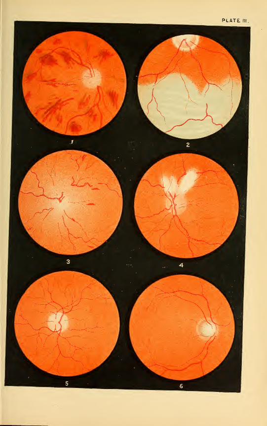
PLATE. IK.


INDEX.
Abdominal growths, 94
Ablatio retinse, 375
Abscess of cornea. 261
of lids, 113
of orbit, 142
Abscission of staphyloma, 271
Acetic acid, 485
Accommodation, amplitude of, 37
relative amplitude of, 39mechanism of, 36
paralysis of, 314
range of, 37, 56
spasm of, 316
Accommodation and Convergence
associated, 39Acne, of lids, 113
Aconite, 485
Addison's disease, 94Advancement operation, 172
Agaricus, 487
Albinism, 347Albuminuria, 94
Alcoholism, 94
Allium cepa, 489
Alumen exsic, 490
Alumina, 490
Amaurosis, 408
Amblyopia, 408
ex anopsia, 408
central, 397
from loss of blood, 409
hysterical, 410
from lightning, 409
Pretended, 410
traumatic, 409
Ammonium carb., 491
Amotio retinae, 375Amplitude of accommodation, 37
Amyl nitrite, 491
Amyloid degeneration of conjunc-
tiva, 225
Anaemia, (cerebral), 94constitutional, 94of retina, 379
Aneurism, of aorta, 94of arteria innominata, 94
of internal carotid, 94
of orbital artery, 94
Angle alpha, 39gamma, 39
Aniridia, 305
Anisometropia, 55
Ankyloblepharon, 119
Anterior chamber, exam, of, 22
Antimonium crudum, 491
Antimonium tart., 492
Aphakia, 44, 457
Apis mel., 492
Apoplexy, 94of corona radiata, 94
of cortex, 94
of crus, 94
of pons varolii, 94
of pons, 94
of ventricles, 94
Arcus senilis, 269
Argentum met., 494Argentum nit., 494Argyll-Robertson pupil, 22
Arlt's operation for trichiasis, 121
Arnica, 497
Arsenicum, 498
Artificial eyes, 272
Arum triph., 501
Asafoetida, 501
Asarum, 501

634 INDEX.
Asthenic ulcer, 258
Asthenopia, muscular, 175
Astigmatism, 51
compound hyperopic, 51
compound myopic, 51
hyperopic, 51
irregular, 51, 54
mixed, 51
myopic, 51
regular, 51
test for, 62
by skiascopy, 78
by ophthalmometer, 79
Astigmia, 51
Atheroma, 95
Atrophy of optic nerve, 400
progressive muscular, 95
Atropine, 65, 501
Aurum, 503
Axial neuritis, 397Badiago, 505
Baryta carb., 506
Baryta iod., 506
Basedow's disease, 95, 148
Belladonna, 506
Binocular diplopia, 155
vision, 38
Birnbacher's operation, 117
Blepharitis, 104
Blepharophimosis, T18
Blepharospasm, 117
Brain, abscess of, 95
basilar affections of, 95
cerebral cortical affections, 95
cerebral affections, -95
cerebellum, affections of, 95
concussion of, 95
hypersemia of, 95
tumor of, 95
Bromium, 510
Bryonia, 510
Buphthalmus, 274-281
Burns of conjunctiva, 231
of eyelids, 131
Cactus grand., 512
Calcarea carb., 512
Calcarea hypophos., 515
Calcarea iod., 515
Calcarea phos., 516
Calcarea pic, 516
Calendula, 517
Canaliculi, anomalies of, 134
Cannabis sativa, 517
Cantharis, 519
Carbo vegetabilis, 519
Carbolic acid, 519
Cardinal points, 36
Caries of orbit, 145
Cataract, 429
anterior polar, 435capsular, 437complete congenital, 433
extraction, 446
operation, 442
with iridectomy, 448
lamellar, 434posterior polar, 436
pyramidal, 435secondary, 437
traumatic, 436
varieties, 431
zonular, 434Causticum, 520
Ceanothus amer., 522
Cedron, 522
Cellulitis orbitse, 142
Central amblyopia, 397
Chalazion, 126
Chamomilla, 523
Chancre of eyelids, 130
Chelidonium majus, 524
Chimaphila umbellata, 524
China, 525
Chininum mur., 525
Chininum sulph., 525
Chloralum, 526
Choked disc, 392
Cholera, 95
Choroid, diseases of, 327
anatomy of, 327
coloboma, 352
detachment of, 351
haemorrhages in, 350
hypersemia of, 328

INDEX. t>35
ossification of, 349
in panophthalmitis. 339
rupture of, 351
senile changes of, 347
tumors of, 347Choroiditis, 328
areolaris, 334• circumscripta, 333
disseminata simplex, 330
metastatica, 339serosa, 329
suppurativa, 339syphilitica, 334traumatic purulent, 339
Choroido-retinitis centralis, 333sclerotico-choroiditis anterior,
343
sclerotico-choroiditis posterior,
343Chrysophanic acid, 526
Cicuta virosa, 526
Ciliary body, 310
anatomy of, 310
injuries of, 314
irido-choroiditis, 317
paresis musculi ciliaris, 314
spasmus musculi ciliaris, 316
Cimicifuga, 527
Cina, 528
Cinnabaris, 528
Clematis, 529
Cocculus indicus, 529
Colchicum, 530
Coloboma, of choroid, 352
iridis, 305
optic nerve sheath, 391
Colocynthis, 530
Color-blindness, 412
Coma, alcoholic, 95
ursemic, 95
syphilitic, 96
Commotio retinae, 383
Comocladia, 530
Conium maculatum, 530
Conjunctiva, 191
amyloid degeneration of, 225
anatomy of, 191
angioma of, 233
burns, 231
carcinoma of, 234
chemical injuries, 231
cysticercus, 234
cysts of, 233
dermoid tumor of, 232
emphysema sub-conjunctival,
230
epithelioma of, 233
examination of, 19
fibroma of, 233
fleshy excrescences of, 233
foreign bodies, 231
gummata, 234
hyperaemia of, 191
lesions of, 231
lipoma of, 232
lupus of, 234
melano-sarcoma of, 233
osteoma of, 233
ophthalmia neonatorum , 195-
1 97
pemphigus of, 225
Pinguecula of, 232
pterygium of, 227
pigment spots of, 233
sarcoma of, 233
sub-conjunctival ecchymosis, 228
syphilitic ulcerations of, 234
trachoma, 204
papillar}', 208
tuberculosis of, 230
tumors of, 232
erectile, 233
vascular, 233
wounds, 23
1
xerosis of, 226
Conjunctivitis, blennor.hoea, 195
catarrhalis, 192
crouposa, 201
diphtheiitica, 200
follicularis, 203
gonorrhoica, 195-197
herpetic, 214
membranacea, 201
phlyctenularis, 211
purulenta, 195

636 INDEX.
pustular, 211
scrofulous, 211
strumous, 211
trachomatosa, 204
vernalis, 224
Contusions of eyelids, 130
Convergence, 38
relative amplitude of, 39test for, 69
Corelysis, 309Cornea, 235
abscessus 261
anatomy of, 235
arcus senilis, 269
buphthalmos, 274, 281
conica, 273
descemetitis, 263
eczema of, 237
examination of, 20
Felchenfeld's operation, 248
gerontoxon, 269
globular, 274
herpes, 240
hypopyon keratitis, 243, 255
inflammation of, 236
injuries of cornea, 275
keratitis, bullosa, 260
dendritica, 259
diffusa, 264
fascicularis, 239
furrow, 256
interstitialis, 264
malarial, 259
mycotic, 259
neuro-paralytica, 260
pannosa, 239
parenchymalosa, 264.
phlyctenularis, 237
posterior, 263
profunda, 264
punctata, 263
pustular, 237
scrofulous, 237
strumous, 237
syphilitica, 264
vesiculosa, 240
keratocele, 242
keratoconus, 273
keratoglobus, 274
leucoma of, 268
leucoma adherens, 245
macula of, 268
nebula of, 268
onyx, 243
opacities of, 268
pannus, 239
paracentesis. 247
sclerosis of, 269
staphyloma of, 270
abscission of, 271
Saemisch's incision, 247-257
tattooing of, 270
tumors of, 276
ulcus corneae, 241
ulcer, asorption, 258
asthenic, 258
clear, 258
infecting, 255
marginal ring, 259
non-inflammatory, 258
rodent, 258
serpiginous, 255
wounds of, 275
Corneal abrasions, fluorescin in, 20
Crocus sativus, 532 -
Crotalus horridus, 533
Croton tiglium, 534Crystalline lens, 428
Cyclamen, 535
Cycloplegics, 64
Cyclitis, 312, 327
plastica, 312
purulenta, 313
serosa, 313
Cysticercus in vitreous, 426
Cundurango, 534
Cuprum aceticum, 535
Cuprum alum., 535
Cuprum sulph., 535
Dacryoadenitis, 133
Dacryocystitis, catarrhalis, 135
phlegmonosa, 139
Dazzling of retina, 361
Death, signs of, 96

INDEX. 637
Decentering of lenses, 82
Dental affections, 96
Dermoid cysts of eyelids, 126
Descemetitis, 263
Detachment, of choroid, 351
of retina, 375
of vitreous, 426
De Zeng's refractometer, 67
Diabetes, 96
Dieffenbach's operation for ectropi-
um, 124
Digestion, disorders of, 96
Digitalis, 535
Dilatation of the pupil, 21
Dioptometry, objective, 59, 71
subjective, 59Diphtheria, 96
Diplopia, monocular, 155
heteronymous, 155
homonymous, 155
binocular, 155
Dislocation of lens, 45S
Distichiasis, 120
Duboisia, 536
Duboisine. 65
Dynamic refraction, 36
Ectopia lentis, 458
Ectropium, 105, 123
Eczema corneae, 237
Electricity, 538
Embolus of arteria centralis retinae,
379Embolism, cerebral, 96
Encephaloid of retina, 384
Entropium, 121
Enucleation of eye, 271
Epicanthus, 120
Epilepsy, 96
Episcleritis, 277
Eserine, 541
Esophoria, 175-180-185
Eupatorium perfoliatum, 542
Euphrasia, 542
Eversbusch's operation, 117
Evisceration of eye, 272
Examination, of the eye, 17
of field of vision, 23
by focal illumination, 20
oblique illumination, 20
of tension, 22
of the outer structures 18
Exenteration of eye, 272
Exophthalmic goitre, 95, 148
Exophoria, 175-180-186
Eyelids, 102
abscess of, 113
acne, 113
anatomy, 102
ankyloblepharon, 119
blepharitis, 104
ciliaris, 104
hypertrophic, 104
marginalis, 104
seborrhoea, 104
simplex, 104
squamosa, 104
ulcerosa, 104
blepharo-adenitis, 104
blepharophimosis, 118
blepharospasm, 117
burns, 131
canthoplasty, 246
chalazion, 126
chancre, 130
contusions, 180
dermoid cyst, 126
distichiasis, 120
ectropium, 105, 123.
entropium, 121
epicanthus, 120
epithelioma, 128
furuncle of, 113
gummata, 130
herpes zoster ophthalmicus, 130
hordeolum, 113
lagophthalmos, 120
lippitudo, 105
lupus, 129
madarosis, 105
meibomian cyst, 126
milium, 126
molluscum contagiosum, 125
nsevi, 126
nictitatio, 118

638 INDKX.i
papillomata, 126
phlegmon of, 113
phthiriasis ciliarum, 105
ptosis, 115
sarcoma, 129
scalds, 131
stye, 113
symblepharon, 118
syphilitic ulcers, 130
tarsal cyst, 126
trichiasis, 105, 120
warts, 126
wounds, 131
xanthelasma, 125
Far point, 37
Ferrum, 544Ferrum phos., 545
Fever, puerperal, 96
typhoid, 96
relapsing, 96
Field of vision, 23
Fistula lachrymalis, 140
Fifth nerve, affection of, 96
Fluorescin in corneal abrasions, 20
Fluoric acid, 545
Focal illumination, 20
Foreign bodies in vitreous, 424
Fourth ventricle, lesions in, 96
Friedreich's disease, 96
Frontal sinus, empyema of, 146
Fundus, as seen by ophthalmoscope,
32
Fungus haematodes, 384
Galvanism, 538
Gelsemium, 545
Gerontoxou, 269
Glaucoma, 460
absolutum, 477acuta, 473anatomy, 460
causes, 468
chronica, 475
consecutiva, 477course, 468
diagnosis, 472
hsemorrhagica, 476
pathology, 463
physiology, 462
simplex, 475
symptoms, 464
treatment, 478
varieties, 473Glioma of retina, 384
Globular cornea, 274
Gout, 96
Graphites, 548
Grapho-vaseline, 107
Graves' disease, 148
Green's operation for entropium, 122
Gummata of eyelids, 130
Hamamelis virginica, 550Haemorrhages, in choroid, 350
of optic nerve, 392
into retina, 367
into vitreous, 424
Heart, aortic insufficiency, 96
endocarditis, 97
organic affections of, 97hypertrophy of left ventricle, 97valvular lesions of, 97
Helminthiasis, 97Hemeralopia, 411
Hemianopia, 415
Hemianopsia, 415
Hemiopia, 415
Hepatic affections, 97
Hepar sulphur, 551
Herpes corneae, 240
zoster ophthalmicus, 130
Heterophoria, 175
Heteronymous diplopia, 155
Heterochroma, 305
Hippus, 21, 304
Homatropine, 65
Homonymous diplopia, 155
Hordeolum, 113
Horopter, 154
Hyalitis suppurativa, 420
Hydraemia, 97
Hydrops of anterior chamber, 274
Hydrocotyle, 553Hydrocephalus 97
Hygiene, of the eye, 84
school, 85

INDEX. 639
Hyoscyamine, 65
Hypsemia, 304
Hyperaemia, of the choroid, 328
iridis, 285
of retina, 358
of the disc, 391
Hypercesthesia of retina, 382
Hyperesophoria, 175
H3-perexophoria, 175
Hypericum, 554Hypermetropia, 42
Hyperphoria, 42-175-179-185
Hyperopia, absolute, 45
axial, 44causes, 44correction of, 45
curvature, 44
facultative, 45
latent, 44
manifest, 44test for, 61
by skiascopy, 77
total, 44symptoms, 44relative, 45
Hypopyon keratitis, 243-255
Hysteria, 97
Ignatia, 554Injuries, of ciliary region, 314
of cornea, 275
of optic nerve, 405
of orbit, 147
of sclera, 281
Insanity, 97
Ipecacuanha, 555Iridavulsion, 309
Iridectomy, 305
Iridersemia, 305
Irido-choroiditis, 317
Irido-cyclitis, 317
Iridodesis, 309Iridodialysis, 304, 309Iridodonesis, 304
Iridoncosis, 304
Iridotomy, 308
Iris, 283
anatomy, 283
aniridia, 305
coloboma of, 305
corectopia, 305
corelysis, 309
diplokoria, 305
examination of, 21
heterochroma, 305
hippus, 304
hyphemia, 304
hyperaemia of , 2S5
iridavulsion, 309iridectomy, 305
irideraemia, 305
iridodesis, 309
iridodialysis, 304, 309iridodonesis, 304
iridoncosis, 304
iridotomy, 308
membrana pupillaris persistans,
305
mydriasis, 302
myosis, 303
operations on, 305
physiology, 285
polycoria, 305
tumors, 301
Iritis, 286
parench^-matosa, 293
purulent, 293
rheumatica, 192
serous, 293
spongiosa, 292
suppurativa, 293
syphilitic, 292
traumatic, 293
Iritomy, 308
Ischsemia of retina, 379Jaborandi, 556
Jones's operation for ectropium, 124
Kali bichrom,, 558
Kali carb., 559Kaliiod., 559Kali mur,, 561
Kalmia latifolia, 562
Keratitis, bullosa, 260
dendritica, 259
fascicularis, 239

640 INDEX.
neuro-paralytica, 260
pannosa, 239
parenchymatosa, 264
phlyctenularis, 237
pustular, 237
scrofulous, 237
strumous, 237
vesiculosa, 240
Keratocele, 242
Keratoconus, 273
Keratoglobus, 274
Kidney, diseases of, 97
Kreosotum, 563
Lachesis, 563
Lachrymal apparatus, 132
anatomy, 132
anomalies of puncta, 134
canaliculi, 134
dacryoadenitis, 133
dacryocystitis catarrhalis, 135
phlegmonous, 139
duct, stricture of, 135
fistula, 140
gland, hypertrophy of, 134
gland, hypertrophy of, 134
gland, tumors of, 134
puncta, examination of 19
sac, examination of, 19
Lactic acid, 564
Lagophthalmos, 120
Ledum palustre, 564
Leucoma adherens, 245
of cornea, 268
Lens, anatomy of, 428
dislocation of, 458
ectopia lentis, 458
examination of, 22
luxatio lentis, 458
Lenses, bifocal, 83
concave, 81
convex, 81
cylindrical, 81
decentered, 82
Leprosy, 97
Lids, 102
Lilium tigrinum, 565
Lippitudo, 105
Lithium carb., 565
Luxatio lentis, 458
Lung, disease of, apex, 97Lupus of eyelids, 129
Lycopodium, 565
Lvcopus virginicus, 566
Macula of cornea, 268
Madarosis, 105
Maddox test, 177
Malaria, 97
Malingering, 410
Mania, 97Marginal ring ulcer, 259
Masturbation, 97Meibomian cyst, 126
Melancholia, 97
Membrana pupillaris persistans, 305
Meningeal haemorrhage, 9S
Meningitis, cerebral, acute, 98
chronic, 98
cerebro-spinal, 98
of the convexity, 98
spinal, 98
tubercular 98
Menstruation, disorders of, 98
Mercurialis perennis, 567
Mercurius corrosivus, 567
Mercurius dulcis, 569
Mercurius iodatus flavus, 570
Mercurius iodatus ruber, 571
Mercurius nitrosus, 572
Mercurius prsecipitatus flavus, 572.
Mercurius praecipitatus ruber, 573
Mercurius solubis, 573
Metre angle, 39
Mezereum, 576
Milium, 126
MoUuscum contagiosUm, 125
Monocular diplopia, 155
Morbus Basedowii, 148
Muriaticum acidum, 576Muscse volitantes, 422
Muscles, ocular, 152
paralysis of, 155
Muscular asthenopia, 175
Musculi ciliaris, paresis of, 314
spasm of, 316
i

INDEX. 641
Mydriasis, 302
Myelitis, acute and chronic, 98
Myodesopsia, 422
Myopia, 46
axial, 41
causes, 48
curvature, 48
symptoms, 49
test for, 62
by skiascopy, 77
treatment, 49Myosis, 303
Myxcedema, 98
N8e\-i of eyelids, 126
Xaphthalin, 577
Natrum carb., 577Natrum mur., 577
Nattum salic, 580
Natrum su'ph,, 580
Near point, 37
Nebula of cornea, 268
Necrosis of orbit, 145
Nephritis, 98
Neuralgia of the fifth nerve, 98
Neuritis, multiple or pseudo-tabes,
98
optica, 392
retro-bulbaris, 397
Nicotine poisoning, 98
Nictitatio, 118
Nightblindness, 411
Nitricum aciduni, 581
Nux moschata, 582
Nux vomica, 582
Nyctalopia, 411
Nystagmus, 173
Oblique illumination, 20
Ocular muscles, 152
anatomy of 152
asthenopia, 175
diplopia, 155
esophoria, 175-1 80-1 85
exophoria, 175-180-1S6
heterophoria, 175
hyperesophoria, 175
hyperexophoria, 175
hyperphoria, 1 75-179-185
nystagmus, 173
orthophoria, 175
ophthalmoplegia, 157
oscillation of eyeball, 173
paralyses, localizing value in
cerebral disease, 162
paralysis of, 155
external rectus, 158
fifth nerve, 163
fourth nerve, 162,
inferior oblique, 159
inferior rectus, 159
internal rectus, 158
sixth nerve, 163
superior oblique, 158
superior rectus, 159
third nerve, complete, 159-
162
strabismus, 163
squint, 163
convergens, 164
dorsum vergens, 168
divergens, 167
sursuni vergens, 168
CEdema of retina, 358
Onosmodium, 584
Onyx, 243
Opacitates vitrei, 422
Opacities of cornea, 268
Opaque nerve fibres, 390
Operations,
abscission of staphyloma, 271
advancement, 172
Arlt's trichiasis, 121
Birnbacher's ptosis, 117
canthoplasty, 246
cataract extraction, 446
corelysis, 309
Dieffenbach's extropium, 124
discission, 444enucleation of eye, 271
Everbusch's ptosis, 117
evisceration of eye, 272
exenteration of eye, 272
Felchenfeld's, 248
Forsters, 443graduated tenotomy, 184

642 INDEX.
Green's entropium, 122
iridectomy, 305
iridodialysis, 309iridototny, 308
iridodesis, 309iridavulsion, 309
iritomy, 308
Jones' ectropium, 124
neurectomy, optico-ciliary, 325
Pagenstecher's ptosis, 116
Panas' ptosis, 117
paracentesis, 247
roller forceps, 209
Saemisch's incision, 247, 257
sclerotomy, 479Stevens' advancement, 184
Streatfeild's entropium, 123
subconjunctival, 171
tattooing of cornea, 270
tenotomy of internal rectus, 169
Wecker's abscission, 271
ptosis, 116
Wolff's ptosis, 117
Ophthalmia, migratoria, 324
neonatorum, 195-197.
Ophthalmo-dynamometer, 69
Ophthalmometr}^, 79Ophthalmoplegia, externa, 157
interna, 157
totalis, 157
Ophthalmoscope, use of, 27
Optical center of lenses, 82
Opium, 585
Optic nerve, 387
anatomy of, 387
axial neuritis, 367
atrophy of, 400
central amblyopia, 397choked disc, 392
coloboma of sheath, 391
haemorrhage of, 392
hyperaemia of disc, 391
injury of, 405
neuritis optica, 392
neuritis retro-bulbaris, 397
opaque nerve fibre, 390
orbital optic neuritis, 397
papillitis, 392
toxic amblyopia, 397tumors of, 405
Orbit, 141
abscess of, 142
anatomy of, 141
caries of, 145
cellulitis of, 142
empyema of frontal sinus, 146
exophthalmic goitre, 148
froatal sinus, empyema of, 146
Graves' disease, 148
injuries of, 147
morbus Basedowii, 148
necrosis of, 145
periostitis of, 144
phlegmon of, 142
tenonitis, 144
tumors of, 146
wounds of, 147
Orbital optic neuritis, 397Orthophoria, 175
Ossification of choroid, 349Pagenstecher's operation, 116
Panas' operation, 117
Pannus, 239
Papillitis, 392
Papillo-retinitis, 361^
Papillomata of eyelids, 126
Paralyses of orbital muscles in cere-
bral disease, localizing value,
162
Paralysis, agitans, 98
conjugate, 157
of external rectus, 158
of fifth nerve, 163
of fourth nerve, 162
general (paralysis of insane)
paresis, 98
of inferior oblique, 159
of inferior rectus, 159
of internal rectus, 158
of ocular muscles, 155
of sixth nerve, 163
of superior oblique, 158
of superior rectus, 159
of third nerve, 159, 162

INDEX. 643
Paresis, 98
Paris quadrifolia, 586
Pemphigus conjuiictiv?e, 225
Periostitis orbitse, 144
Persistent hyaloid artery, 426
Petroleum, 587
Phlegmon of orbit, 142
Phosphoricum acidum, 587
Phosphorus, 588
Phthiriasis ciliarum, 105
Physostigma, 589
Phytolacca decandra, 591
Pinguecula, 232
Plantago major, 592
Polio encephalitis superior, 99Pons varolii, lesions of, 99Posterior staphyloma, 49Presbyopia, 55
test for, 70
Proptosis, 22
Prunus spinosa, 592
Psorinum, 593Pterygium, 227
Ptosis, 115
Pulsatilla, 593Puncta, anomalies of, 134
Punctum, proximum, 37
remotum, 37
Pupil, contraction of, 21
dilatation of, 21
Pyaemia, 99Pyoktanin, 137
Rachitis, 99Range of accommodation, 37Ranunculus bulbosus, 597
Refrac. and accommodation, 35
Refrac. and accom., abnormalities, 42
Refraction, dynamic, 36
estimation of by direct method,
71
by indirect method, 73
static, 36
Refractometer, 67
Relative amplitude of accom., 39amplitude of convergence, 39
Remedies:
Acetic acid, 485
Aconite, 485
Agaricus, 487
Allium cepa, 489
Alumen exsic, 490Alumina, 490
Ammonium carb., 491
Amyl nitrite, 491
Antimonium crudum, 491
Antimonium tart., 492
Apis mel., 492
Argentum met., 494Argentum nit., 494Arnica, 497Arsenicum, 498
Arum triph., 501
Asafoetida, 501
Asarum, 501
Atropine, 501
Aurum, 503
Badiago, 505
Baryta carb., 506
Baryta iod., 506
Belladonna, 506
Bromium, 510
Bryonia, 510
Cactus grand., 512
Calcarea carb., 512
Calcarea hypophosph., 515
Calcarea iod., 515
Calcarea phos.,516
Calcarea pic, 516
Calendula, 517
Cannabis sativa, 517
Cantharis, 519
Carbo vegetabilis, 519
Carbolic acid, 519
Causticum, 520
Ceanothus amer., 522
Cedron, 522
Chamomilla, 523
Chelidonium majus, 524
Chimaphila umbellata, 524
China, 525
Chininum mur., 525
Chininum sulph., 525
Chloralum, 526
Chrysophanic acid, 526

644 INDEX,
Cicuta virosa, 526
Cimicifuga, 527
Cina, 528
Cinnabaris, 528
Clematis, 529
Cocculus indiciis, 529
Colchicnni, 530
Colocynthis, 530Comocladia, 530
Conium maculatum, 530
Crocus sativus, 532
Crotalushorridus, 533Crotoii tiglium, 534Cundurango, 534Cuprum aceticum, 535Cuprum alum., 535
Cuprum sulph,, 535
Cyclameu, 535
Digitalis, 535
Duboisia, 536
Electricity, 538
Eserine, 541
Eupatorium per., 542
Euphrasia, 542
P'errum, 544Ferrum phos., 545
Fluoric acid, 545Galvanism, 538
Gelsemium, 545Graphites, 548
Hamamelis virginica, 550Hepar sulphur, 551
Hydrocotyle, 553Hyoscyamus, 554Hypericum, 554Ignatia, 554Ipecacuanha, 555
Jaborandi, 556
Kali bichrom., 558Kali carb., 559Kali iod., 559Kali mur., 561
Kalmia latifolia, 562
Kreosotum, 563
Lachesis, 563
Eactic acid, 564
Ledum palustre, 564
Lilium tigrinum, 565
Lithium carb., 565
L3^copodium, 565
Lycopus virginiciis, 566
Mercurialis perennis, 567
Mercurius corrosivus, 567
Mercurius dulcis, 569
Mercurius iodatus flavus, 570
Mercurius iodatus ruber, 571
Mercurius nitrosus, 572
Mercurius prsecipitatsus flavus,
572
Mercurius pracipitatus ruber, 573Mercurius solubis, 573Mezereum, 576
Muriaticum acidum, 575
Naphthalin, 577
Natrum carb., 577Natrum mur., 577Natrum salic, 580
Natrum sulph., 580
Nitricum acidum, 581
Nux moschata, 582
Nux vomica, 582
Onosmodium virg., 584
Opium, 585
Paris quadrifolia, 586
Petroleum, 587
Phosphoricum acidum, 587
Phosphorus, 588
Physostigma, 589
Phytolacca decandra, 591
Plantago major, 592
Prunus spinosa, 592
Psorinum, 593
Pulsatilla, 593
Ranunculus bulbosus, 597Rhododendron, 598
Rhus toxicodendron, 598
Ruta graveolens, 602
Sanguinaria, 603
Secale cornutum, 603
Senega, 603
Sepia, 604
Silicea, 607
Spigelia, 608
Spongia, 611

INDEX. 645
Stannum, 611
Staphysagria, 612
Sulphur, 612
Syphilinum, 618
Tabacum, 618
Tellurium, 618
Terebinthina, 618
Thuja, 619
Veratrum viride, 621
Zincum, 622
Renal retinitis, 361
Retina, diseases of, 354ablatio retinae, 375
amotio retinse, 375anatomy of, 354anaemia of, 379commotio retinae, 383dazzlinsT of, 361
detachment of, 375
embolus of arteria centralis ret-
inse, 379encephaloid of, 384
fungus haematodes, 384
glioma, 384
hyperaemia of, 358
hyperaesthesia of, 382
ischaemia, 379oedema of, 358
thrombosis of vena centralis, 381
Retinitis, albuminurica, 361
apoplectica, 367
of Bright's disease, 361
central punctate, 372
diabetica, 366
diffusa, 358
glycosurica, 366
haemmorrhagica, 367
leukaemica, 366
papillo, 361
pigmentosa, 373proliferan, 372
punctata albescens, 372
renal, 361
serosa, 358
simplex, 358
splenic, 366
syphilitica, 370
Retinoscopy, 73
Rheumatism, 99Rhododendron, 598
Rhus toxicodendron, 598
Rupture of choroid, 351
Ruta graveolens, 602
Sanguinaria, 603
Sarcoma of eyelids, 129
vSavage test, 178
Scalds of eyelids, 131
School, buildings, construction of, 89
examination of eyes, on entrance
of, 87
furniture, 91
hygiene, 85
Sclera, 277
anatomy of, 277
episcleritis, 277
injuries of, 281
scleritis, 278
sclerotitis, 278
sclerotico-choroiditis anterior,
280, 281, 343
posterior, 281, 343staphyloma annular, 281
sclerae, 280
Sclerosis multiple, 99Sclerectasia posterior, 340
Scopolamine, 65
Scotomata, 23
Scrofula, 99
Secale cornutum, 603
Senega, 603
Sepia, 604
Shadow test, 73
Silicea, 607
Skiascopy, 73
Skin, extensive burns of, 99Snake poisoning, 99Snow blindness, 412
Spigelia, 608
Spinal cord, degenerative diseases
of, 99inflammation and congestion of,
99Spinal irritation, 99Splenic retinitis, 366

646 INDEX.
Spongia, 611
Squint, 163
Stannum, 611
Staphyloma, cornese, 270
posterior, 343sclerse, 280
Staphysagria, 612
Steven's phoronieter, 175
Strabismus, 163
convergens, 164
deorsum vergens, 168
divergens, 167
operation for, 169
sursum vergens, 168
Streatfield's operation for entro-
pium, 123
Strictura ductus lachrymalis, 135
Sturm's focal interval, 52
Stye, 113
Sub-conjunctival ecchymosis, 228
emphysema, 230
Sulphur, 612
Symblepharon, 118
Sympathetic irritation, 321
ophthalmia, 321
Synchysis, 422
scintillans, 422
Syphilinum, 618
Syphilis, 99Syphilitic ulcers of eyelids, 130
Tabacum, 618
Tabes, 100
Tabulated statement of diseases,
with eye symptoms, 94Tarsal cyst, 126
Tattooing of cornea, 270
Tellurium, 618
Tenonitis, 144
Tenon's capsule, 141
Tenotomy of internal rectus, 169
Tension of eye, 22
Terebinthina, 618
Test-type, Helfrich's, 66
Thrombosis of vena centralis, 381
Thuja, 619
Toxic amblyopia, 397Trachoma, 204
Trachoma, papillary, 204
Trichiasis, 105, 120
Trichinosis, 100
Tuberculosis, loi
Tuberculosis conjunctivae, 230
Tumors, of choroid, 347
of conjunctiva, 232
of cornea, 276
of iris, 301
arbitse, 146
of the optic nerve, 405
Typhoid fever, loi
Ursemia, 101
Ulcer, astlienic, 258
absorption, 258
clear ulcer, 258
corneal, 241
infecting, 255
marginal ring, 259
non-inflammatory, 258
rod.nit, 253
serpiginous, 2^5
Urticaria. lor
Uterine aTections, loi
Veratrum viride, 621
Vitreous bod}-, 420
anatomy of, 420
cy>ticircas in, 426
detachment of, 426
foreign bodies in, 424
hsemorrh.-ge into, 424
hyalitis suppurativa, 420
muscse volitantes, 422
myodesopsia, 422
opacitates vitrei, 422
persistent hyaloid artery, 426
synchysis, 422
scintillans, 422
Visual angle, 59
Visual field, 23
Vision, acuteness of, 60
Warts of eyelids, 126
Wecker's operation, 116
Wilson's phorometer, 179
Wolff's operation, 117
Wounds, of conjunctiva, 231
cornea, 275

INDEX. 647
eyelids, 131 I Xerosis conjunctivae, 226
orbit, 147 Yellow oxide ointment, 107
Xanthelasma, 125 Zincum, 622


'4