INTERNAL DISEASES PROPEDEUTICS
164
Ministry of education and science of Russian Federaion Federal budgetary educational establishment of higher education «Ulyanovsk State University» The Institute of medicine, ecology and physical culture A.Yu. Smirnova, V.V. Gnoevykh INTERNAL DISEASES PROPEDEUTICS PART III DIAGNOSTICS OF DISEASES OF GASTROINTESTINAL TRACT AND KIDNEYS Textbook of medicine for medicine faculty students Ulyanovsk 2017
-
Upload
khangminh22 -
Category
Documents
-
view
4 -
download
0
Transcript of INTERNAL DISEASES PROPEDEUTICS
Ministry of education and science of Russian Federaion
Federal budgetary educational establishment of higher education
«Ulyanovsk State University»
The Institute of medicine, ecology and physical culture
A.Yu. Smirnova, V.V. Gnoevykh
INTERNAL DISEASES PROPEDEUTICS
PART III
DIAGNOSTICS OF DISEASES OF GASTROINTESTINAL TRACT
AND KIDNEYS
Textbook of medicine for medicine faculty students
Ulyanovsk 2017
2
УДК 616.33./.34+616.61-07=111(075.8)
ББК 54.13-4я73+54ю14-4я73
C68
Published by the decision of the Academic Council of the UlSU
Institute of Medicine, Ecology and Physical Culture
(document № 2/192 of 18.10.2017)
Reviewer:
MD, professor of Department of faculty therapy R.H. Gimaev
Smirnova A.Yu.
C68 Internal diseases propedeutics. Part III. Diagnostics of diseases of
gastrointestinal tract and kidneys : Textbook of medicine for medicine
faculty students / A.Yu. Smirnova, V.V. Gnoevykh. – Ulyanovsk : Ulyanovsk
State University, 2017. – 152 [12] p.
This publication is the third part of “Internal diseases propedeutics”, which main
goal is the practical assistance for students in the development of the fundamentals of
clinical diagnosis of diseases of the gastrointestinal and urinary systems. It contains a
description of the main methods of laboratory and instrumental diagnostic tests of diseases
of the gastrointestinal and urinary systems. The publication is illustrated with charts,
drawings and tables. The textbook is intended for students of medical universities.
УДК 616.33./.34+616.61-07=111(075.8)
ББК 54.13-4я73+54ю14-4я73
©Smirnova A.Yu., Gnoevykh V.V., 2017
©Ulyanovsk State University, 2017
3
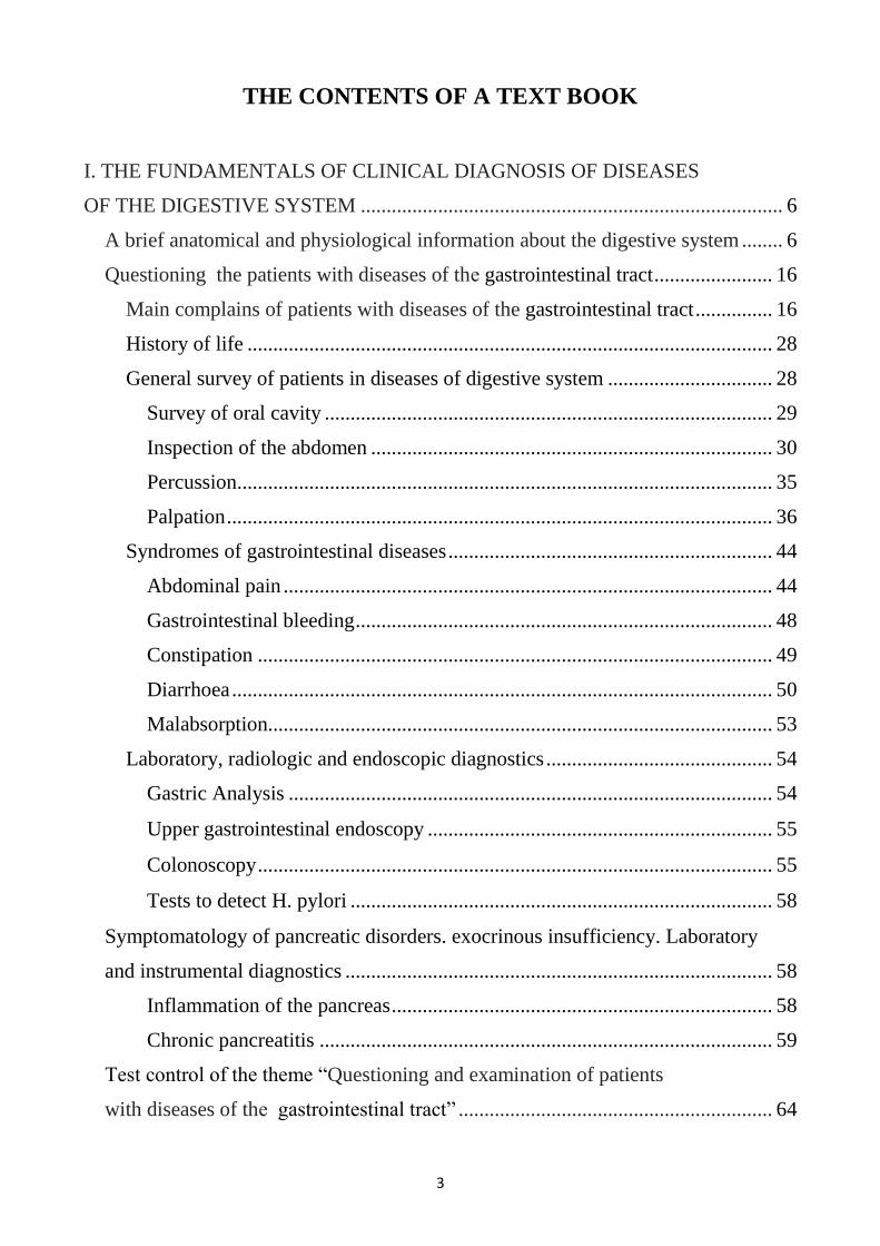
THE CONTENTS OF A TEXT BOOK
I. THE FUNDAMENTALS OF CLINICAL DIAGNOSIS OF DISEASES
OF THE DIGESTIVE SYSTEM .................................................................................. 6
A brief anatomical and physiological information about the digestive system ........ 6
Questioning the patients with diseases of thе gastrointestinal tract ....................... 16
Main complains of patients with diseases of the gastrointestinal tract ............... 16
History of life ...................................................................................................... 28
General survey of patients in diseases of digestive system ................................ 28
Survey of oral cavity ....................................................................................... 29
Inspection of the abdomen .............................................................................. 30
Percussion ........................................................................................................ 35
Palpation .......................................................................................................... 36
Syndromes of gastrointestinal diseases ............................................................... 44
Abdominal pain ............................................................................................... 44
Gastrointestinal bleeding ................................................................................. 48
Constipation .................................................................................................... 49
Diarrhoea ......................................................................................................... 50
Malabsorption.................................................................................................. 53
Laboratory, radiologic and endoscopic diagnostics ............................................ 54
Gastric Analysis .............................................................................................. 54
Upper gastrointestinal endoscopy ................................................................... 55
Colonoscopy .................................................................................................... 55
Tests to detect H. pylori .................................................................................. 58
Symptomatology of pancreatic disorders. exocrinous insufficiency. Laboratory
and instrumental diagnostics ................................................................................... 58
Inflammation of the pancreas .......................................................................... 58
Chronic pancreatitis ........................................................................................ 59
Test control of the theme “Questioning and examination of patients
with diseases of thе gastrointestinal tract” ............................................................. 64
4
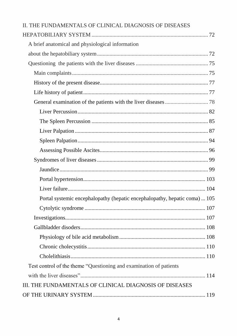
II. THE FUNDAMENTALS OF CLINICAL DIAGNOSIS OF DISEASES
HEPATOBILIARY SYSTEM .................................................................................... 72
A brief anatomical and physiological information
about the hepatobiliary system ................................................................................ 72
Questioning the patients with the liver diseases .................................................... 75
Main complaints .................................................................................................. 75
History of the present disease.............................................................................. 77
Life history of patient .......................................................................................... 77
General examination of the patients with the liver diseases ............................... 78
Liver Percussion .............................................................................................. 82
The Spleen Percussion .................................................................................... 85
Liver Palpation ................................................................................................ 87
Spleen Palpation .............................................................................................. 94
Assessing Possible Ascites .............................................................................. 96
Syndromes of liver diseases ................................................................................ 99
Jaundice ........................................................................................................... 99
Portal hypertension ........................................................................................ 103
Liver failure ................................................................................................... 104
Portal systemic encephalopathy (hepatic encephalopathy, hepatic coma) ... 105
Cytolytic syndrome ....................................................................................... 107
Investigations..................................................................................................... 107
Gallbladder disoders .......................................................................................... 108
Physiology of bile acid metabolism .............................................................. 108
Chronic cholecystitis ..................................................................................... 110
Cholelithiasis ................................................................................................. 110
Test control of the theme “Questioning and examination of patients
with the liver diseases” .......................................................................................... 114
III. THE FUNDAMENTALS OF CLINICAL DIAGNOSIS OF DISEASES
OF THE URINARY SYSTEM ................................................................................. 119
5
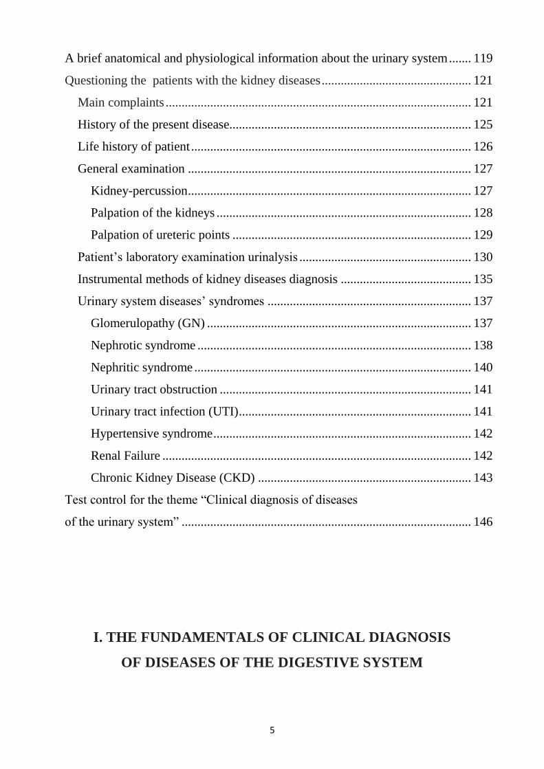
A brief anatomical and physiological information about the urinary system ....... 119
Questioning the patients with the kidney diseases ............................................... 121
Main complaints ................................................................................................ 121
History of the present disease............................................................................ 125
Life history of patient ........................................................................................ 126
General examination ......................................................................................... 127
Kidney-percussion ......................................................................................... 127
Palpation of the kidneys ................................................................................ 128
Palpation of ureteric points ........................................................................... 129
Patient’s laboratory examination urinalysis ...................................................... 130
Instrumental methods of kidney diseases diagnosis ......................................... 135
Urinary system diseases’ syndromes ................................................................ 137
Glomerulopathy (GN) ................................................................................... 137
Nephrotic syndrome ...................................................................................... 138
Nephritic syndrome ....................................................................................... 140
Urinary tract obstruction ............................................................................... 141
Urinary tract infection (UTI) ......................................................................... 141
Hypertensive syndrome ................................................................................. 142
Renal Failure ................................................................................................. 142
Chronic Kidney Disease (CKD) ................................................................... 143
Test control for the theme “Clinical diagnosis of diseases
of the urinary system” ........................................................................................... 146
I. THE FUNDAMENTALS OF CLINICAL DIAGNOSIS
OF DISEASES OF THE DIGESTIVE SYSTEM
6
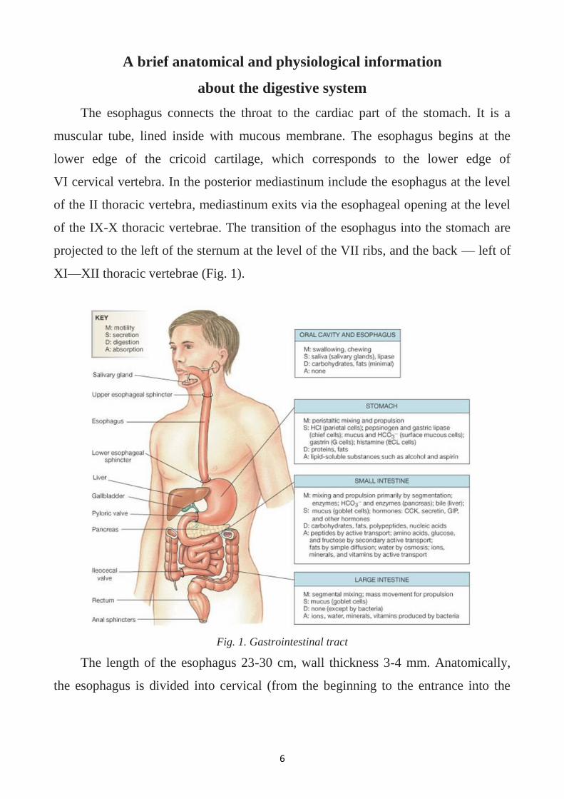
A brief anatomical and physiological information
about the digestive system
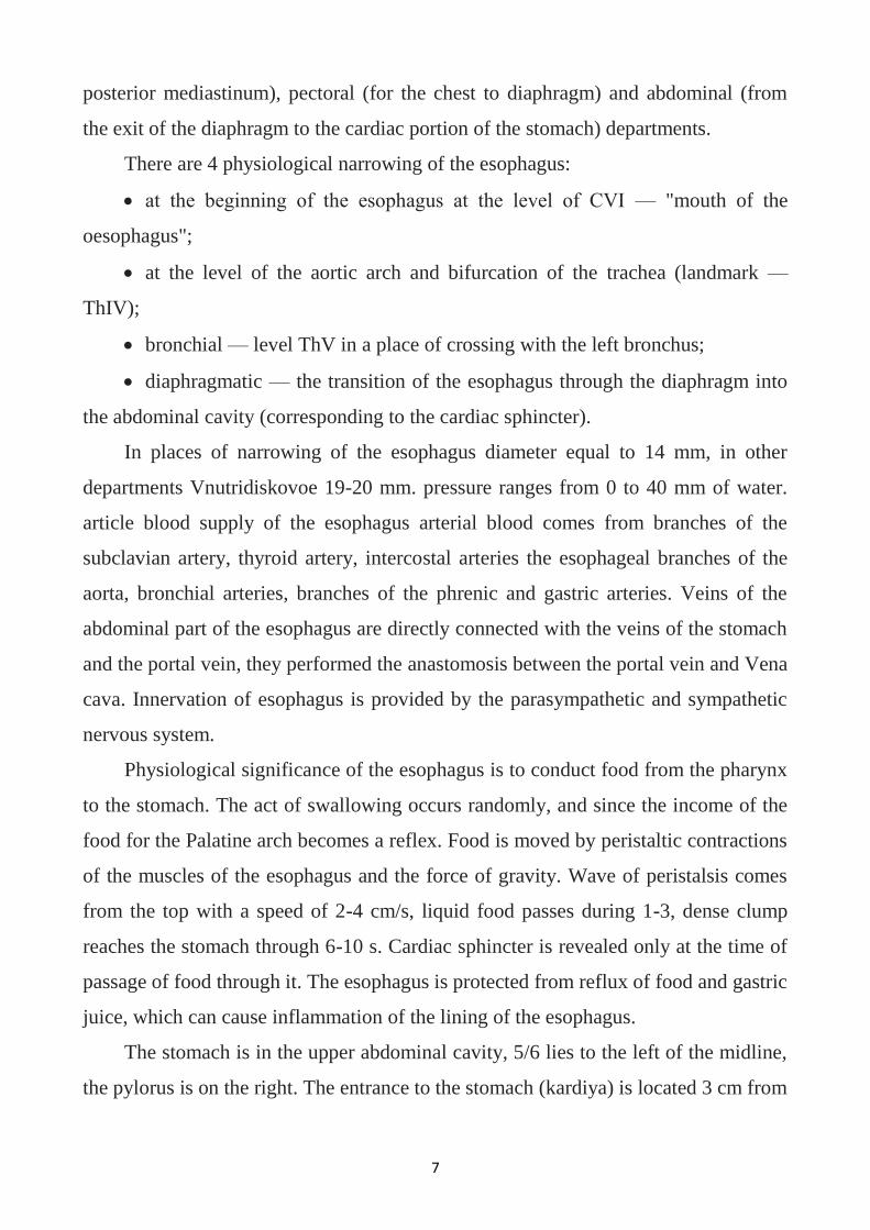
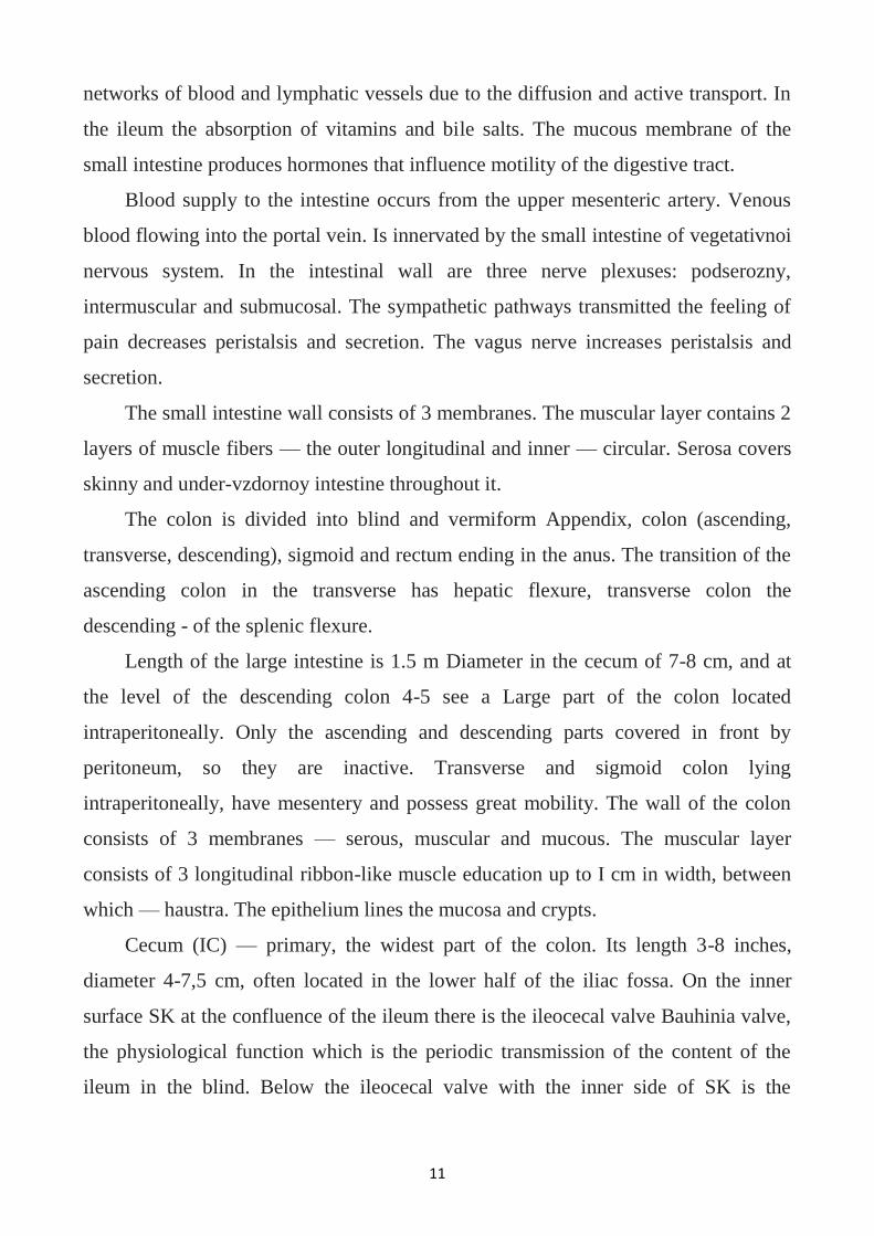
The esophagus connects the throat to the cardiac part of the stomach. It is a
muscular tube, lined inside with mucous membrane. The esophagus begins at the
lower edge of the cricoid cartilage, which corresponds to the lower edge of
VI cervical vertebra. In the posterior mediastinum include the esophagus at the level
of the II thoracic vertebra, mediastinum exits via the esophageal opening at the level
of the IX-X thoracic vertebrae. The transition of the esophagus into the stomach are
projected to the left of the sternum at the level of the VII ribs, and the back — left of
XI—XII thoracic vertebrae (Fig. 1).
Fig. 1. Gastrointestinal tract
The length of the esophagus 23-30 cm, wall thickness 3-4 mm. Anatomically,
the esophagus is divided into cervical (from the beginning to the entrance into the
7
posterior mediastinum), pectoral (for the chest to diaphragm) and abdominal (from
the exit of the diaphragm to the cardiac portion of the stomach) departments.
There are 4 physiological narrowing of the esophagus:
at the beginning of the esophagus at the level of СVI — "mouth of the
oesophagus";
at the level of the aortic arch and bifurcation of the trachea (landmark —
ThIV);
bronchial — level ThV in a place of crossing with the left bronchus;
diaphragmatic — the transition of the esophagus through the diaphragm into
the abdominal cavity (corresponding to the cardiac sphincter).
In places of narrowing of the esophagus diameter equal to 14 mm, in other
departments Vnutridiskovoe 19-20 mm. pressure ranges from 0 to 40 mm of water.
article blood supply of the esophagus arterial blood comes from branches of the
subclavian artery, thyroid artery, intercostal arteries the esophageal branches of the
aorta, bronchial arteries, branches of the phrenic and gastric arteries. Veins of the
abdominal part of the esophagus are directly connected with the veins of the stomach
and the portal vein, they performed the anastomosis between the portal vein and Vena
cava. Innervation of esophagus is provided by the parasympathetic and sympathetic
nervous system.
Physiological significance of the esophagus is to conduct food from the pharynx
to the stomach. The act of swallowing occurs randomly, and since the income of the
food for the Palatine arch becomes a reflex. Food is moved by peristaltic contractions
of the muscles of the esophagus and the force of gravity. Wave of peristalsis comes
from the top with a speed of 2-4 cm/s, liquid food passes during 1-3, dense clump
reaches the stomach through 6-10 s. Cardiac sphincter is revealed only at the time of
passage of food through it. The esophagus is protected from reflux of food and gastric
juice, which can cause inflammation of the lining of the esophagus.
The stomach is in the upper abdominal cavity, 5/6 lies to the left of the midline,
the pylorus is on the right. The entrance to the stomach (kardiya) is located 3 cm from
8
the point of attachment to the sternum VII left rib cartilage, at the level of
X-XI thoracic vertebra from behind. Large curvature of the stomach and adjacent
movable part of the front surface to the abdominal wall. The upper part relates to the
spleen, bottom - poperechnopolostah colon in a horizontal position is 2-3 cm above
the navel. The output part of the stomach is at the level of I lumbar vertebra 1-2 cm
right of the midline
There are the following parts of the stomach: cardiac (area of entrance to
stomach - kardiya) part, the fundus (upper part of the stomach), body, pylorus and
antrum of the stomach. On the border with the duodenum is the pyloric orifice
surrounded by a sphincter. The stomach wall has 3 layers: outer layer — serous
membrane (peritoneum) covers the stomach from all sides except for the narrow
strips on the curvatures, the inner layer is three - layer layer of smooth muscle. The
outer and middle layers of the muscles of the pylorus are thickened, forming a
sphincter of the pylorus (the sphincter). Next is the loose submucosa shell, riddled
with blood vessels and nerves, and then muscular layer of the mucosa and, finally, the
mucous membrane lining the whole internal surface of the stomach.
In the bottom of gastric pits open up the ducts of the glands. The mucous
membrane of the stomach is covered with a single layer epithelium with glandular
character. Surface epithelial cells secrete mucoid secret containing neutral
mucopolysaccharides. In the deeper layers of the mucosa are the main, and additional
parietal cells. Chief cells secrete enzymes and parietal — hydrochloric acid.
The blood supply to the stomach is from three branches of the coronary artery of
the stomach. The blood flowing from the stomach into the portal vein. Between the
coronary vein and lower veins of the esophagus are anastomoses. Innerasia provided
by extrastyle stomach nerves — vagus and sympathetic and intramural.
The physiological functions of the stomach: the accumulation of food mass,
their mechanical and chemical treatment, evacuation of food into the intestine. The
stomach has the absorptive, excretory and hematopoietic functions. The capacity of
the stomach about 2 liters. Muscle tone increases with stimulation of the vagus and
9
the level of the hormone gastrin. Due to the presence of two drivers of a rhythm,
every 20-26 seconds. the stomach makes a peristaltic wave towards the pylorus. The
vagus nerve is stimulated, and the sympathetic - decreases the motor function of the
stomach. Food leaves the stomach in 1.5-3 hours.
Fasting stomach contains 10-40 ml of gastric juice acidic or neutral reaction.
Food stimulus during the day, the stomach produces up to 2 litres of juice, and when
abundant food - up to 3 liters. The gastric secretion has phase 2 — hard-reflex and
neuro-chemical. Final digestion of proteins to small ICA-Sivitsa, is completed in the
small intestine. Under the influence of hydrochloric acid the proteins in the stomach
swell, which improves the impact of the enzymes - pepsin, gastrokine, pepsin, renin.
Physiological functions of hydrochloric acid: HCl creates an acidic environment
in the stomach facilitates the digestion of proteins; has antibacterial properties; it
activates the process of transformation of pepsinogen to pepsin; promotes the release
of gastrin, which stimulates secretion of hydrochloric acid; regulates the transfer of
food from the stomach, 12-duodenum; causes the secretion of enterokinase and
gastrin, stimulate the secretion of the pancreas.
Part of the stomach in hematopoiesis is due to the generation of hematopoietic
factor castle.
Duodenum with the exception of its upper part adjoining the pylorus, located
retroperitoneale. Has a length of about 20 cm and width of 1,5-5 cm with multiple
bends. The top curve is short, lies to the right of the spine at the thoracic level
II-I lumbar vertebra has a horizontal or upward direction. Descending part is located
to the right of the spine. The lower horizontal part is at the level of III lumbar
vertebra, crosses the spine and to the left of him at the level II lumbar vertebra moves
to jejunum. The wall of the duodenum is the upper part of the 3 membranes —
serous, muscular, mucous membrane, next — of 2 shells (muscle and mucosa).
On the inner surface of the mucosa has numerous villi height to 0.5 mm, which
are rich in capillary network and lymphatic vessels.
10
In descending Department duodenum features of the Vater papilla, height
11-21 mm. and a width of 5-10 mm. over the top open common bile and pancreatic
ducts (approximately 70% in a single duct). The end portion of the common bile duct
in the wall of the duodenum is covered by the sphincter of Oddi. Duodenum lies in
close proximity to several important organs: adjacent to the stomach, and the top,
descending and horizontal part for the head of the pancreas, the ascending part of the
body of the pancreas. Duodenum is located near the right lobe of the liver, aorta, right
adrenal gland, inferior Vena cava. Duodenum located to the left and posterior to the
gallbladder.
Duodenum is supplied with blood from branches of the gastro-duodenal and
superior mesenteric artery, plus hepatic, left gastric, right gastro-colic and jejunal
arteries.
Duodenum anatomically and functionally is a continuation of the stomach, there
is an activation of protein, fat and starch enzymes, the emulsification of bile and
pancreatic juice treatment of food masses, hydrolytic cleavage of nutrients.
Hormones enterogastrone, secretin, cholecystokinin, pancreozymin regulate the
activity of the stomach, pancreas and intestines.
The jejunum is 2/5 of the small intestine, the remainder is the ileum. The length
of the small intestine is about 7 m. In the primary departments diameter colon about 5
cm, distally about 3 cm.
The small intestine has a mesentery located intraperitoneus. Topographically
loops of the small intestines lie in the umbilical region with the spread in all
directions. The front of the small intestine is covered with omentum. In the small
intestine the process of digestion reach the maximum, contribute a pendulum and
oscillating movement in the direction of the colon, the allocation of about 3 liters/day.
intestinal juice containing digestive enzymes. In the small intestine are the main
stages of the fermentative processes of digestion and absorption of proteins, fats and
carbohydrates, the most important role here belongs to the parietal and membrane
digestion. The absorptive function is carried out by fibers with highly developed
11
networks of blood and lymphatic vessels due to the diffusion and active transport. In
the ileum the absorption of vitamins and bile salts. The mucous membrane of the
small intestine produces hormones that influence motility of the digestive tract.
Blood supply to the intestine occurs from the upper mesenteric artery. Venous
blood flowing into the portal vein. Is innervated by the small intestine of vegetativnoi
nervous system. In the intestinal wall are three nerve plexuses: podserozny,
intermuscular and submucosal. The sympathetic pathways transmitted the feeling of
pain decreases peristalsis and secretion. The vagus nerve increases peristalsis and
secretion.
The small intestine wall consists of 3 membranes. The muscular layer contains 2
layers of muscle fibers — the outer longitudinal and inner — circular. Serosa covers
skinny and under-vzdornoy intestine throughout it.
The colon is divided into blind and vermiform Appendix, colon (ascending,
transverse, descending), sigmoid and rectum ending in the anus. The transition of the
ascending colon in the transverse has hepatic flexure, transverse colon the
descending - of the splenic flexure.
Length of the large intestine is 1.5 m Diameter in the cecum of 7-8 cm, and at
the level of the descending colon 4-5 see a Large part of the colon located
intraperitoneally. Only the ascending and descending parts covered in front by
peritoneum, so they are inactive. Transverse and sigmoid colon lying
intraperitoneally, have mesentery and possess great mobility. The wall of the colon
consists of 3 membranes — serous, muscular and mucous. The muscular layer
consists of 3 longitudinal ribbon-like muscle education up to I cm in width, between
which — haustra. The epithelium lines the mucosa and crypts.
Cecum (IC) — primary, the widest part of the colon. Its length 3-8 inches,
diameter 4-7,5 cm, often located in the lower half of the iliac fossa. On the inner
surface SK at the confluence of the ileum there is the ileocecal valve Bauhinia valve,
the physiological function which is the periodic transmission of the content of the
ileum in the blind. Below the ileocecal valve with the inner side of SK is the
12
Appendix. Ascending colon starts from blind in the right iliac fossa, continuing
upward to the visceral surface of the liver, where it forms a bend and passes in the
transverse colon. The length of the ascending colon 20 cm, it is projected into the
right lateral region of the anterior abdominal wall, and its the right bend at the end of
X rib.
The transverse colon lies almost horizontally, forming a convex downward and
forward in a gentle arc to the left goes into the descending colon. Its length is about
50 cm with a mesentery it is movable and may be located above the navel or to reach
the pelvis.
The descending colon is the most narrow and short - 12 cm Is a continuation of
the transverse colon below the left bend goes at the back of the abdominal wall to the
iliac crest, where it passes into the sigmoid.
The sigmoid colon is the longest part of the colon - extends from iliac crest to
the third sacral vertebra, at the level of which becomes the rectum. The average
length of the sigmoid colon about 54 cm, mesenteric — 8 cm, projected sigmoid
colon to the anterior abdominal wall within left-side, left inguinal and pubic regions
partially.
Rectum (PC) — the final part of the colon PC located in the pelvic cavity,
behind her prilezhat sacrum and coccyx, in front of men the prostate, seminal
vesicles, portion of the rear surface of the bladder; in females the uterus, its cervix
and the posterior fornix of the vagina. The upper limit PC is located on the upper
edge of 3 sacral vertebra (promontory).
The blood supply of the colon is via the mesenteric artery, and rectum using the
ileum and the middle and lower rectal arteries. Venous blood from the intestine flows
into the portal vein, except the lower cut where blood flowing in the lower-standing
inferior Vena cava via the hemorrhoidal, and iliac veins.
Nervous regulation of activity of intestines is carried out by meisnerova plexus,
which is located in the submucosal layer, and auelbekova — in the muscle membrane.
13
Autonomic innervation is provided by the parasympathetic division, stimulating
movement and secretion of the intestine and the sympathetic division, inhibiting them.
The function of the colon is the accumulation neperevedeni food, further
processing with intestinal enzymes and Mick-rotary of the intestine, the absorption of
water, formation of feces. After eating and handling her in the stomach and small
intestine the first portion of the chyme appear in the caecum after 2 - 4 hours move
food through the gastrointestinal tract occurs within 24-36 h.
Pancreas — parenchymal organ, located in the epigastric region and left upper
quadrant on the back of the abdominal wall in the retroperitoneal space. There are
3 division of the pancreas — head, body and tail. Length RV — 14-23 cm, head
width — 3-7,5 cm, body 2-5 cm, tail — 0,3-3,4 cm Thickness of the pancreas about
3 cm, weight —60-115 g. the Front surface of the pancreas adjacent to the rear wall
of the stomach. The head of the pancreas located to the right of the spine and
penetrations in the inner bend of the duodenum. The body of the pancreas lies in front
and to the left of the spine, then goes into a tail reaching to the spleen. Front and
bottom surface of the body of the pancreas is covered with peritoneum. The back of
the head of the pancreas located inferior Vena cava, the beginning of the portal vein
and common bile duct passing through the head.
Behind the body of the pancreas are the abdominal aorta, lymph nodes and part
of the solar plexus. Behind the tail of the pancreas located of the left renal vessels and
left adrenal gland. From the tail to the head in the thickness of the pancreas is
pancreatic duct, which opens on top of a large duodenal papilla, often in connection
with the common bile duct. The allocation of the juice contributes to the pressure in
the duct, reaching 30-35 mm of water column, and the suction action of peristalsis of
the duodenum.
The blood supply to the head of the pancreas originates from the common
hepatic and superior mesenteric arteries, and the body and tail from branches of the
splenic artery. Venous blood flowing into the portal vein. The pancreas is innervated
by sympathetic and parasympathetic fibers of the autonomic nervous system coming
14
from the solar plexus. Deep in the pancreas, a plexus, which is composed of
intraorganic ganglia. The nerve endings are located in the lobules and the the
excretory ducts.
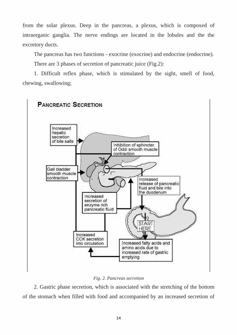
The pancreas has two functions - exocrine (exocrine) and endocrine (endocrine).
There are 3 phases of secretion of pancreatic juice (Fig.2):
1. Difficult reflex phase, which is stimulated by the sight, smell of food,
chewing, swallowing;
Fig. 2. Pancreas secretion
2. Gastric phase secretion, which is associated with the stretching of the bottom
of the stomach when filled with food and accompanied by an increased secretion of
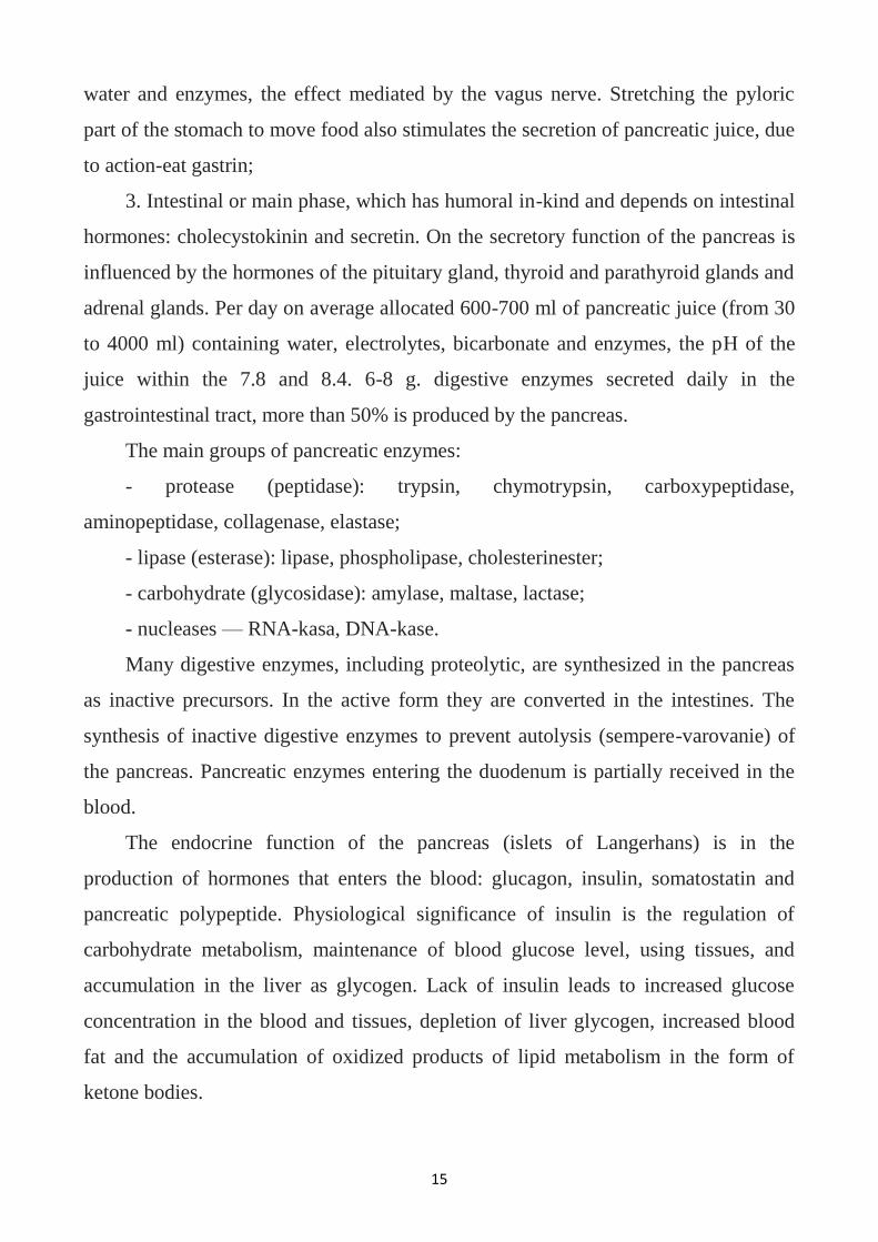
15
water and enzymes, the effect mediated by the vagus nerve. Stretching the pyloric
part of the stomach to move food also stimulates the secretion of pancreatic juice, due
to action-eat gastrin;
3. Intestinal or main phase, which has humoral in-kind and depends on intestinal
hormones: cholecystokinin and secretin. On the secretory function of the pancreas is
influenced by the hormones of the pituitary gland, thyroid and parathyroid glands and
adrenal glands. Per day on average allocated 600-700 ml of pancreatic juice (from 30
to 4000 ml) containing water, electrolytes, bicarbonate and enzymes, the pH of the
juice within the 7.8 and 8.4. 6-8 g. digestive enzymes secreted daily in the
gastrointestinal tract, more than 50% is produced by the pancreas.
The main groups of pancreatic enzymes:
- protease (peptidase): trypsin, chymotrypsin, carboxypeptidase,
aminopeptidase, collagenase, elastase;
- lipase (esterase): lipase, phospholipase, cholesterinester;
- carbohydrate (glycosidase): amylase, maltase, lactase;
- nucleases — RNA-kasa, DNA-kase.
Many digestive enzymes, including proteolytic, are synthesized in the pancreas
as inactive precursors. In the active form they are converted in the intestines. The
synthesis of inactive digestive enzymes to prevent autolysis (sempere-varovanie) of
the pancreas. Pancreatic enzymes entering the duodenum is partially received in the
blood.
The endocrine function of the pancreas (islets of Langerhans) is in the
production of hormones that enters the blood: glucagon, insulin, somatostatin and
pancreatic polypeptide. Physiological significance of insulin is the regulation of
carbohydrate metabolism, maintenance of blood glucose level, using tissues, and
accumulation in the liver as glycogen. Lack of insulin leads to increased glucose
concentration in the blood and tissues, depletion of liver glycogen, increased blood
fat and the accumulation of oxidized products of lipid metabolism in the form of
ketone bodies.
16
Glucagon has the opposite effect, reduces the content of glycogen in liver and
muscle, leading to hyperglycemia. Somatostatin inhibits the release of gastrin, insulin
and glucagon, the secretion of hydrochloric acid of the stomach and the flow of
calcium ions into cells of pancreatic islets. The PP-cells of the pancreas produce 90%
of the polypeptide - antagonist cholecystokinin.
Questioning the patients with diseases
of thе gastrointestinal tract
Main complains of patients with diseases of the gastrointestinal tract
Complaints of patients with lesions of the esophagus
Difficulty passing of food through the esophagus (dysphagia): character
appearance (sharply or gradually); the stability and duration of existence; character
progression; the conditions of occurrence (the passage of solid or liquid food, mental
factors)
Vomiting (time of occurrence, the nature of vomit – the smell of blood)
Bleeding from the esophagus (the main reason varicose veins of the
esophagus)
Pain: location, radiation, causes
Complaints of patients with diseases of the stomach
Deranged (poor or increased) appetite occurs in infectious diseases, metabolic
disorders, etc. Poor appetite or its complete absence (anorexia) is usually
characteristic of gastric cancer. This symptom is often an early sign of cancer.
Appetite often increases in peptic ulcer, especially in duodenal ulcer. Loss of appetite
should be differentiated from cases when the patient abstains from food for fear of
pain (citophobia). This condition often occurs in subjects with gastric ulcer, though
their appetite is increased.
Perverted appetite that sometimes occurs in patients is characterized by the
desire to eat inedible materials such as charcoal, chalk, kerosine, etc.
17
Appetite is perverted in pregnant women and in persons suffering from
achlorhydria. Some patients with cancer of the stomach or some other organs often
feel aversion to meat. The developmental mechanism of appetite is connected with
excitation of the food centre (according to Pavlov). Excitation or inhibition of this
centre depends on impulses arriving from the cerebral cortex, on the condition of the
vegetative centres (excitation of the vomiting centre causes loss of appetite), and on
reflex effects from the alimentary organs. The multitude of factors that act on the
food centre accounts for the high variation in appetite.
Taste may be perverted due to the presence of unpleasant taste in the mouth and
partial loss of taste in an individual. It can often be associated with some pathology in
the mouth, e.g. caries or chronic tonsillitis. A coated tongue can be another cause of
unpleasant taste in the mouth.
Regurgitation usually implies two phenomena: a sudden and sometimes loud
uprise of wind from the stomach or esophagus (eructation), and the return of
swallowed food into the mouth (sometimes together with air). Regurgitation depends
on contraction of the esophageal muscles with the open cardia. Regurgitation may be
due to air swallowing (aerophagy). It is heard at a distance and occurs in
psychoneurosis. In the presence of motor dysfunction of the stomach, fermentation
and putrefaction of food with increased formation of gas occur in the stomach (the
phenomenon otherwise absent in norm). In abnormal fermentation in the stomach, the
eructated air is either odourless or smells of bitter oil, which is due to the presence of
butyric, lactic and other organic acids that are produced during fermentation in the
stomach. In the presence of abnormal putrefaction, the belched air has the odour of
rotten eggs (hydrogen sulphide). Bitter belching indicates intensive degradation of
proteins. Belching is characteristic of stenosed pylorus with great distention of the
stomach and significant congestion in it. Acid regurgitation is usually associated with
hypersecretion of gastric juice and occurs mostly during pain attacks in ulcer. But it
can also occur in normal or insufficient secretion of the stomach in the presence of
insufficiency of the cardia (when the stomach contents are regurgitated into the
18
esophagus). Bitter regurgitation occurs in cases with belching up of bile into the
stomach from the duodenum, and also in hyperchlorhydria; bitterness depends on the
bitter taste of peptone.
Pyrosis is otherwise known as heartburn, i.e. burning pain in the epigastric and
retrosternal region. Heartburn arises in gastro-esophageal reflux, mostly in the
presence of gastric hyperacidity in various diseases the alimentary tract (e.g. peptic
ulcer or cholecystitis), hiatus hernia, and sometimes in pregnancy. Heartburn in
healthy subjects can be due hypersensitivity to some foods.
Nausea is a reflectory act associated with irritation of the vagus nerve, indefinite
feeling of sickness and sensation of compression in the epigastrium. Nausea is often
attended by pallidness of the skin, general akness, giddiness, sweating, salivation, fall
in the arterial pressure, cold the limbs, and sometimes semisyncopal state. Nausea
often (but not necessarily) precedes vomiting. The mechanism of nausea is not
known. Its frequent association with vomiting suggests that it might be the early sign
of stimulation of the vomiting centre. The leading role in the development of nausea
is given to the nervous system and also the tone of the stomach, the duodenum, and
the small intestine. Nausea may develop without any connection with diseases of the
stomach, e.g. in toxemia of pregnancy, renal failure, deranged cerebral circulation,
and sometimes in healthy people in the presence of foul odour (or in remembrance of
something unpleasant). Some diseases of the stomach are attended by nausea, e.g.
acute and chronic gastritis or cancer of the stomach. Nausea associated with gastric
pathology usually occurs after meals, especially after taking some pungent food.
Nausea often develops in secretory insufficiency of the stomach.
Vomiting (emesis) occurs due to stimulation of the vomiting centre. This is a
complicated reflex through the esophagus, larynx and the mouth (sometimes through
the nose as well). Vomiting may be caused by ingestion of spoiled food, by
seasickness, or irritation arising inside the body (diseases of the gastro-intestinal tract,
liver, kidneys, etc.). In most cases vomiting is preceded by nausea and sometimes
hypersalivation. Factors causing the vomiting reflex are quite varied. This can be
19
explained by the numerous connections that exist between the vomiting centre
(located in the medulla oblongata, in the inferior part of the floor of the 4-th
ventricle) and all bodily systems. Depending on a particular causative factor, the
following can be differentiated:
(1) nervous (central) vomiting;
(2) vomiting of visceral etiology (peripheral or reflex);
(3) hematogenic and toxic vomiting.
Vomiting is an important symptom of many diseases of the stomach, it can be
regarded as the symptom of a particular disease only in the sense of other signs
characteristic of this disease. Vomiting of gastric etiology is caused by stimulation of
receptors in the gastric mucosa by inflammatory processes (acute or chronic
gastritis), in ingestion of strong acids or alkalis, or food acting on the gastric
receptors by chemical (spoiled) or physical (overeating or excessively cold food)
routes. Vomiting can be caused by difficult evacuation of the stomach due to spasms
or stenosed pylorus. If patient complains of vomiting, the physician should inquire
the time when the vomiting occurred, possible connections with meals, association
with pain, the amount and character of the vomited material. Morning vomiting (on a
fasting stomach) with expulsion of much mucus is characteristic of chronic gastritis,
especially in alcoholics, Hyperacid vomiting in the morning indicates nocturnal
hypersecretion of the stomach. Vomiting occurring 10-15 minutes after meals
suggests ulcer or cancer of the cordial part of the stomach, or acute gastritis. If
vomiting occurs 2-3 hours after meals (during intense digestion) it may indicate ulcer
or cancer of the stomach body. In the presence of ulcer of the pylorus or duodenum,
vomiting occurs 4-6 hours after meals. Expulsion of food taken a day or two before is
characteristic of pyloric stenosis. Patients with peptic ulcer often vomit at the height
of pain thus removing it, which is typical of the disease. The odour of the vomit is
usually acid, but it can often be fetid (putrefactive processes in the stomach); the
odour may be even fecal (in the presence of a fecal fistula between the stomach and
the transverse colon).
20
The vomited material may have acid reaction (due to the presence of
hydrochloric acid, in hyperchlorhydria), neutral (in achylia), or alkaline (in the
presence of ammonia compounds, in pyloric stenosis, hypofunction of renal function,
and also in regurgitation of the duodenal contents into the stomach). Vomitus may
contain materials of great diagnostic importance, e.g. blood, mucus (in chronic
gastritis), ample bile (narrowing of the duodenum, gastric achylia), and fecal matter.
Vomiting may attend acute gastritis, exacerbation of chronic gastritis, gastric
neurosis, peptic ulcer, spasm and organic stenosis of the pylorus, and cancer of the
stomach.
Pain is the leading symptom in diseases of the stomach. Epigastric pain is not
obligatory connected with diseases of the stomach. It should be remembered that the
epigastrium is the "site of encounter" of all kinds of pain. Epigastric pain may be due
to diseases of the liver, pancreas, and due to hernia of the linea alba. Epigastric pain
may develop in diseases of other abdominal organs (sometimes of organs located
outside the abdomen) by the viscero-visceral reflex (acute appendicitis, myocardial
infarction, affection of the diaphragmatic pleura, etc).
In order to locate correctly the source of pain, the physician should ask the
patient
(1) to show exactly the site of pain;
(2) to characterize the pain which may be periodical or paroxysmal (at certain
time of the day); permanent or seasonal (in spring or autumn);
(3) to describe the connection (if any) between pain and meals, the quality of
food and its consistency;
(4) to indicate possible radiation of pain (into the back, shoulder blade, behind
the sternum, left hypochondrium);
(5) to describe conditions under which pain lessens (after vomiting, after taking
food or baking soda, after applying hot-water bottle or taking spasmolytics);
21
(6) to describe possible connections between pain and physical strain (weight
lifting, traffic jolting, etc.), or strong emotions. Intensity and character of pain are
also important diagnostically.
The pain may be dull, stabbing, cutting, etc. Pain in hollow organs with smooth
muscles (e.g. stomach) is provoked by spasms (spastic pain), distension of the organ
(distensional pain), and by its motor dysfuncion.
Paroxysmal, periodical epigastric pain is due to the spasm of the pyloric
muscles. It arises under the influence of strong impulses arriving from the vagus
nerve centre in cerebral cortex dysfunction. The spasm of the pylorus is stimulated by
the hyperacidity of gastric juice due to hyperstimulation of the vagus.
Depending on the time of paroxysmal pain (after meals), it may be early pain
(occurring 30-40 min after meals), late pain (90-120 min after meals), nocturnal
pain, and hunger pain (which is abated after taking food). If pain occurs after meals
stimulating secretion of gastric juice (bitter, pungent, spicy or smoked foods), this
indicates the leading role of hypersecretion in its etiology. The pain then localizes in
the epigastrium, radiates to the back, and is rather intense; it is abated after vomiting
and taking alkali or foods that decrease acidity of gastric juice, and also after taking
antispastic preparations and applying hot-water bottle (which removes spasms).
A seasonal character of pain, i.e. development of periodic pain during spring and
autumn, is characteristic of peptic ulcer, especially if the process is localized in the
peripyloric region. Permanent boring pain is usually caused by stimulation of the
nerve elements in the mucous and submucous layer of the stomach; the pain is
usually intensified after meals and is characteristic of exacerbation of chronic gastritis
or cancer of the stomach.
Perigastritis (chronic inflammation of the peritoneum overlying the stomach and
its adhesion to the neighbouring organs) is manifested by pain developing
immediately after taking much food (irrespective of its quality). The full stomach
distends to stimulate nerve fibres in the adhesions. In the presence of perigastritis and
22
adhesions between the stomach and the adjacent organs, pain may be caused by any
physical strain and when the patient changes his posture.
Gastric hemorrhage is a very important symptom. It can be manifested by
vomiting of blood (hematemesis) or by black tarry stools (melena). Gastric
hemorrhage is usually manifested by the presence of blood in the vomitus. The colour
of the vomitus depends on the time during which the blood is present in the stomach.
If the blood was in the stomach for a long time, the blood reacts with hydrochloric
acid of the gastric juice to form hematin hydrochloride. The vomitus looks like coffee
grounds. If hemorrhage is profuse (damage to a large vessel) the vomitus contains
much scarlet (unaltered) blood. Hematemesis occurs in peptic ulcer, cancer, and
polyps, in erosive gastritis, rarely in sarcoma, tuberculosis and syphilis of the
stomach, and in varicosity of the esophageal veins. Tarry stools are not an obligatory
sign of gastric hemorrhage.
Anamnesis
When collecting anamnesis, the patient should be asked about his nutrition. It is
important to establish if meals are regular because taking food at random is an
important factor in the etiology of gastric diseases. Food quality is as important as its
amount taken during one meal. Mastication of food matters as well. Conditions of rest
and work, and possible occupational hazards should be established. Abuse of alcohol
and smoking are important factors in the etiology of gastric diseases. It is very
important to find out if the patient's condition has undergone some changes during
recent time (e.g. loss of weight, anemia, blood vomiting, or tarry stools).
Gastrointestinal diseases of the past, surgical intervention on the abdominal organs,
long medication with preparations irritating the stomach mucosa (acetylsalicylic acid,
sodium salycilate, steroid hormones, potassium chloride, etc.) are also very important.
Complaints of patients with diseases of the intenstine
The main complaints with intestinal diseases are pain, meteorism (inflation of
the abdomen), motor dysfunction of the intestine (constipation and diarrhea), and
intestinal hemorrhage.
23
Pain. If the patient complains of pain in the abdomen, the following should be
established: location of pain, its radiation, intensity, character, duration, and means
by which it is lessened.
The general signs by which intestinal pain may be differentiated from gastric
one are:
(1) absence of regular dependence of pain on food taking; the only exception is
inflammation in the transverse colon (transversitis): pain develops immediately after
meals; the pathogenesis of this pain is connected with reflex peristaltic contractions
of the transverse colon when food enters the stomach;
(2) close association of pain with defecation: pain occurs before, during, and
(rarely) after defecation;
(3) pain relief after defecation or passage of gas.
Pain may be boring and spasmodic (intestinal colic). Colicky pain is
characterized by short repeated attacks which arise and disappear quite of a sudden.
Pain may very quickly change its location, the main site being round the navel.
Sometimes pain may arise in other areas of the abdomen. Boring pain is sometimes
permanent; it intensifies during cough, especially if the mesenterium or peritoneum is
involved. Pain is characteristic of inflammatory diseases of the intestine. As
inflammation extends onto the peritoneum, pain is attended by a pronounced
muscular defence.
Exact location of the source of pain is very important. Pain in the right iliac
region occurs in appendicitis, tuberculosis, cancer, or inflammation of the cecum
(typhlitis). Acute pain in the left lower abdomen occurs in intestinal obstruction and
inflammation of the sigmoid (sigmoiditis). Pain in the umbilical region occurs in
inflammation of small intestine (enteritis) and inflammation or cancer of the colon.
Pain in the perineal region, and especially during defecation (with the presence of
blood in feces), is characteristic of the rectum diseases (proctitis, cancer). Pain in
intestinal pathology may radiate into the chest; pain associated with affection of the
spleen angle of the descending large intestine radiates into the left side of the chest (it
24
is sometimes mistaken for pain attacks of angina pectoris); colics of appendicitic
origin radiate into the right leg.
In acute affection of the left portions of the large intestine (dysentery), pain
radiates into the sacral area. Thermal procedures, spasmolytics, passage of gas, and
emptying of the bowels can relieve pain or remove it completely.
Intestinal pain is caused by obstruction of intestinal patency and upset motor
function. Intestinal pain is mostly caused by spasms (spasmodic contraction of
smooth muscles; hence spastic pain), or by distension of the intestine by gases. Both
mechanisms often become involved.
Spastic pain can be due to various causes. Individual predisposition to spastic
contractions in general (vegetoneurosis) may be as important as irritation originating in
the intestine proper, e.g. in enteritis, colitis, intestinal tumour, poisoning with arsenic or
lead, and also in diseases of the central nervous system (posterior spinal sclerosis).
Pain arising due to intestinal distension by gases, and associated with tension
and irritation of the mesentery, differs from spastic pain (1) by the absence of
periodicity; it is long-standing and gradually lessens in prolonged inflation; and (2)
by exact localization. In intestinal obstruction (complete or partial) colicky pain is
combined with almost permanent pain in the abdomen. It is characterized by exact
and permanent location (the umbilical region and large intestine). The pain intensifies
with intestinal peristalsis.
Appendicular colic first localizes round the navel and the epigastrium but in
several hours (or even on the next day) it descends to the right iliac region where it
intensifies gradually. Sometimes the pain arises straight in the right iliac region.
Rectal colic, or tenesmus, is also known. It occurs in frequent and painful tenesmus to
defecate and is associated with spasmodic contractions of the intestine and the
sphincter ani. Only clots of mucus are sometimes expressed instead of actual
defecation. Tenesmus occurs in dysentery and other inflammatory or ulcerous
diseases, and in cancer of the rectum. Pain associated with defecation depends on
many factors. Pain preceding defecation is associated with the disease of the
25
descending colon or sigmoid colon. Pain during defecation is characteristic of
hemorrhoids, anal fissures, and cancer.
Meteorism. The patient feels flatulence, inflation, and boring distension of the
abdomen.
The causes of meteorism are
1) excessive gas formation in the intestine due to ingestion of vegetable cellular
tissue and easily fermented food (peas, beans, cabbage, etc.);
2) intestinal motor dysfunction due to decreased tone of the intestinal wall or
intestinal obstruction;
3) lowered absorbability of gases by the intestinal wall, the process of gas
formation being normal;
4) aerophagia, i.e. excess swallowing of air, with its subsequent propulsion to
the stomach and the intestine;
5) hysterical meteorism: the abdomen is rapidly inflated to the size of the
abdomen of a pregnant woman at her last weeks; this nervous mechanism is very
complicated.
When inquiring the patient, the physician should ask about the character of his
nutrition and the site of abdomen inflation (the entire abdomen or only its limited part
may be inflated). If inflation is local, it is necessary to ask the patient whether or not
inflation occurs always at one and the same area. In intestinal obstruction, the patient
feels rumbling sounds inside the abdomen, feels movement of liquid in the intestine,
and intense peristaltic movements above the point of obstruction.
Diarrhea. Frequent and liquid stool is a common sign of intestinal pathology.
Diarrhea occurs in acute and chronic intestinal infections (enteritis, enterocolitis,
sigmoiditis, proctitis), in various exogenous intoxications (poisoning with arsenic or
mercury), endogenous intoxications (uremia, diabetes, gout), in endocrine disorders
(adrenal dysfunction, thyrotoxicosis), and in hypersensitivity to some foods
(allergy).
26
The mechanism of diarrhea is very complicated. Different pathogenic factors
may prevail in various pathological conditions. Accelerated movement of the
liquefied food in the intestine due to peristalsis is among them. Almost undigested
food can thus be evacuated. Another factor is disordered absorptive function of the
intestine. Affection of the intestinal wall, disordered mechanisms regulating
absorption, purgatives and upset water metabolism produce a marked change in the
absorption process and are the cause of diarrhea.
The third cause of liquid stools is inflammation of the intestine. Large
quantities of inflammatory secretion stimulating the intestinal receptors are released
into the lumen of the intestine to intensify its peristalsis and to impair its absorptive
function.
Paradoxical diarrhea occurs in prolonged constipation due to mechanical
irritation of the intestinal wall by hard fecal masses.
Upset equilibrium between the fermentative and putrefactive flora of the
intestine is another important factor in the etiology of diarrhea.
Diarrhea occurring in organic affections of the large intestine is mostly of the
inflammatory character. It is not copious, nor does it produce strong negative effect
on the patient's general condition (as compared with affections of the small intestine
which is attended by profuse diarrhea associated with deranged motor and absorption
function of the intestine). The pronounced disorder in digestion causes some
metabolic disorders in the patient (impaired absorption of proteins, iron, vitamins,
and electrolytes).
Obstipation (constipation). This is obstinate constipation during which feces are
long retained in the intestine (for more than 48 hours). But the duration of
constipation is only relative, because in many cases it is not the result of pathology
but of the living conditions and nutrition. If vegetable food dominates in the diet, the
subject may defecate two or three times a day. Stools become rarer if the diet is rich
in meat. A radical change in nutrition can remove constipation. Limited mobility of
the subject, hunger, and irregular defecations (during the day) may prolong pauses
27
between defecation. The main factor determining defecation is the condition of
intestinal motor function. Bowel contents are retained in the large intestine and the
rectum during constipation
Organic and functional constipation is differentiated. Organic constipation is
usually associated with mechanical obstruction, such as narrowing of the intestinal
lumen due to a tumour, scar, adhesion, and also abnormalities in the intestine
(megacolon, dolichosigmoid, megasigmoid, diverticulosis).
Functional constipation is subdivided into:
1) alimentary constipation; it occurs due to ingestion of easily assimilable foods,
which leave small residue and normally stimulate peristalsis of the intestine by
irritating its nervous receptors;
2) neurogenic constipation due to dysfunction of the intramural nervous
apparatus or vagus nerve; these are the so-called dyskinetic constipation, caused by
the reflex action on the intestinal motor function of another affected organ
(cholecystitis, adnexitis, prostatitis, etc.), or by organic affections of the central
nervous system (tumours of the brain, encephalitis, posterior spinal sclerosis);
3) constipation associated with inflammatory affections, mainly of the large
intestine (dysentery);
4) toxic constipation occurring in exogenous poisoning with lead, morphine, or
cocaine;
5) constipation of endocrine etiology, occurring in thyroid or pituitary
hypofunction;
6) constipation caused by lack of physical exercise;
7) constipation caused by flaccidity of the prelum.
Intestinal hemorrhage often occurs in ulcerous affections of the alimentary
system. It develops in the presence of tumour, protozoal and helminthic invasions,
acute infections (typhoid fever, bacillary dysentery), in thrombosis of mesenteric
vessels, ulcerous non-specific colitis, etc.
28
Anamnesis
The patient should be inquired thoroughly about his nutrition from his early
childhood till the onset of the disease (especially directly before the disease), about
poisonings in the past history and hypersensitivity to some feeds. It is necessary to
find out if the patient's meals are regular, if the food is varied, and if the patient
smokes or drinks alcohol. Information on the past diseases of the intestine and also on
pathology of other organs is sometimes decisive for establishing the cause of the
present affection.
Some functional disorders of the intestine can be associated with occupation
(lead or arsenic poisoning, constipation due to frequent suppression of tenesmus to
defecate).
History of life
The presence of inflammatory and infectious gastrointestinal diseases in
history.
Comorbidities In chronic kidney disease , endocrine disorders can often be
observed dyspeptic symptoms
Occupational hazards:Mercury, lead, phosphorus, acid vapors, etc.
Working conditions: People leading a sedentary lifestyle, prone to constipation
Lifestyle, eating habits (the regularity, frequency, quantity, quality, time of
eating).
Bad habits: Smoking, alcohol abuse
General survey of patients in diseases of digestive system
The general condition and state of consciousness of the patient are first assessed.
The general inspection of the patient with dysphagia may suggest an organic
affection of the esophagus if the patient is extremely asthenic (cachexia). During
general inspection of the patient with stomach diseases the physician may assess poor
nutrition of the patient (cachexia) which is characteristic of stomach cancer and
29
untreated benign pyloric stenosis. Patients with uncomplicated peptic ulcer look
practically healthy. Severe prolonged affection of the absorptive function causes
grave cachexia. Pale skin is observed after gastric and intestinal hemorrhage, and in
anemia. Edema is possible in loss of protein with simultaneous retention in the body
of water and salt. Inspection of the skin reveals its dryness and pallidness; the mucosa
is pale due to insufficient absorption of iron and anemization of the patient.
Insufficient absorption of vitamins results in development of fissures of the lips, the
skin becomes rough, and perleche develops.
Facies Нippocratica (first described by Hippocrates) is associated with collapse
in grave diseases of the abdominal organs (diffuse peritonitis, intestinal obstructionб,
perforated ulcer of the stomach or duodenum, rupture of the gall bladder). The face is
characterized by sunken eyes, pinched nose, deadly livid and cyanotic skin, which is
sometimes covered with large drops of cold sweat.
Survey of oral cavity
When inspecting the mouth, attention should be paid to its shape (symmetry of
the angles, permanently open mouth), the colour of the lips, eruption on the lips
(cold sores, herpes labialis), and the presence of fissures. The oral mucosa should
also be inspected (for the presence of aphthae, pigmentation, Filatov-Koplik spots,
thrush, contagious aphthae of the foot and mouth disease, hemorrhage). Marked
changes in the gums can be observed in some diseases (such as pyorrhea, acute
leukemia, diabetes mellitus, and scurvy) and poisoning (with lead or mercury). The
teeth should be examined for the absence of defective shape, size, or position. The
absence of many teeth is very important in the etiology of some alimentary diseases.
Caries is the source of infection and can affect some other organs. The absence of
many teeth accounts for inadequate disintegration and mastication of food in the
mouth, while the presence of carious teeth favours penetration of microbial flora
into the stomach.
30
The tongue is not the "mirror of the stomach" as it was formerly believed.
Nevertheless in some diseases its appearance is informative: clean and moist tongue
is characteristic of uncomplicated peptic ulcer, while the tongue coated with a foul
smelling white-grey material is characteristic of acute gastritis; a dry tongue indicates
a severe abdominal pathology or acute pancreatitis; a tongue with atrophied papillae
suggests cancer of the stomach, atrophic gastritis with pronounced gastric secretory
hypofunction, or vitamin B deficiency. The glassy tongue is characteristic of gastric
cancer, pellagra, sprue, and ariboflavinosis. The tongue in intestinal diseases often
becomes crimson (cardinal tongue) in vitamin PP deficiency (pellagra), its papillae
are smoothed down. The gums may be loose and bleeding. Disordered movement of
the tongue may indicate nervous affections, grave infections and poisoning.
Inspection of the abdomen
The abdomen is inspected for vertical and horizontal position.
Pay attention to the shape and dimensions of the abdomen, symmetry of both
sides, the presence of hernia, visible peristalsis and expansion of subcutaneous
venous network.
Normal right and left part of the abdomen is symmetrical, the umbilicus is
slightly retracted. Normosteniks abdomen moderately protruding shape of the rib arc
not sharply delineated. Hypersteniks – more dimensional, protrusion more
pronounced. Asteniks - small size, flattened or slightly retracted (Fig.3).
Look at the general contour of the abdomen and note whether it is sunken as
in wasting disorder (malnutrition, chronic infection, malignancy), or protuberant as in
pregnancy, abdominal masses, obesity and ascites. If there is a localized swelling,
note its position, whether fixed or mobile, and if it moves with respiration as do
masses connected with the liver, spleen or kidney.
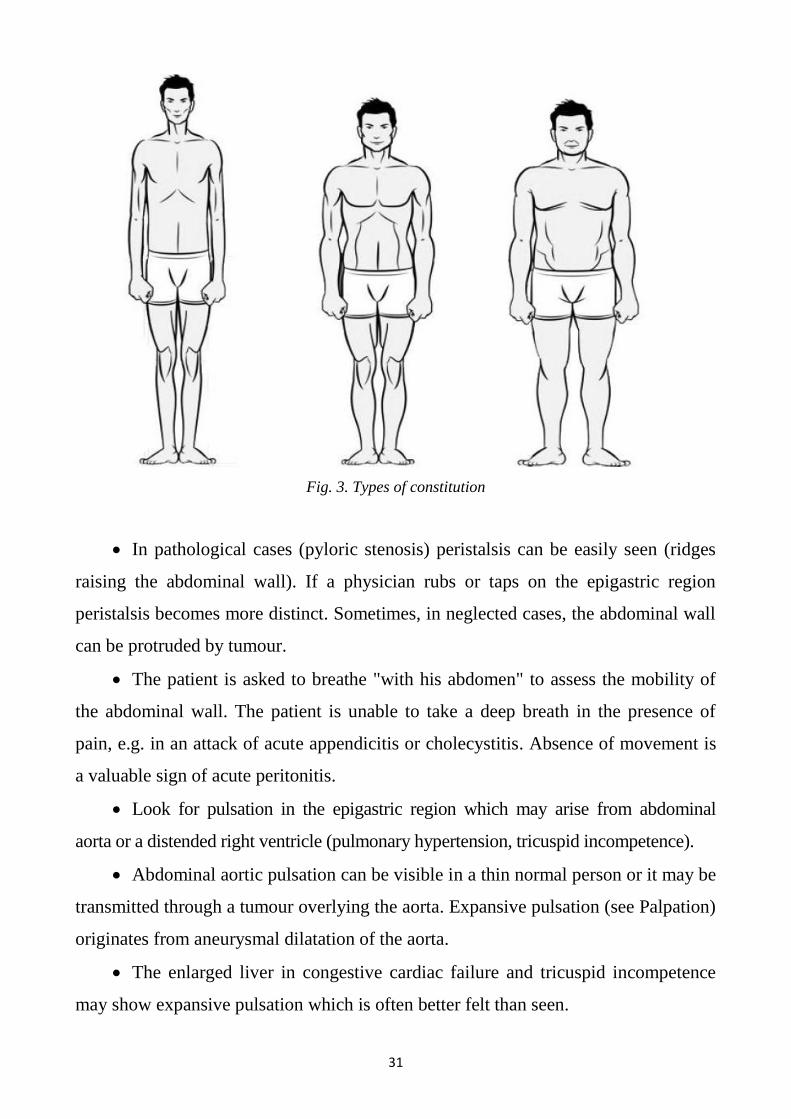
31
Fig. 3. Types of constitution
In pathological cases (pyloric stenosis) peristalsis can be easily seen (ridges
raising the abdominal wall). If a physician rubs or taps on the epigastric region
peristalsis becomes more distinct. Sometimes, in neglected cases, the abdominal wall
can be protruded by tumour.
The patient is asked to breathe "with his abdomen" to assess the mobility of
the abdominal wall. The patient is unable to take a deep breath in the presence of
pain, e.g. in an attack of acute appendicitis or cholecystitis. Absence of movement is
a valuable sign of acute peritonitis.
Look for pulsation in the epigastric region which may arise from abdominal
aorta or a distended right ventricle (pulmonary hypertension, tricuspid incompetence).
Abdominal aortic pulsation can be visible in a thin normal person or it may be
transmitted through a tumour overlying the aorta. Expansive pulsation (see Palpation)
originates from aneurysmal dilatation of the aorta.
The enlarged liver in congestive cardiac failure and tricuspid incompetence
may show expansive pulsation which is often better felt than seen.
32
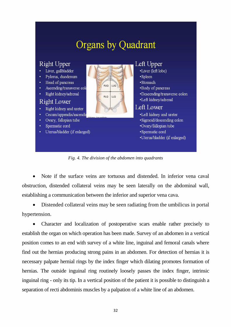
Fig. 4. The division of the abdomen into quadrants
Note if the surface veins are tortuous and distended. In inferior vena caval
obstruction, distended collateral veins may be seen laterally on the abdominal wall,
establishing a communication between the inferior and superior vena cava.
Distended collateral veins may be seen radiating from the umbilicus in portal
hypertension.
Character and localization of postoperative scars enable rather precisely to
establish the organ on which operation has been made. Survey of an abdomen in a vertical
position comes to an end with survey of a white line, inguinal and femoral canals where
find out the hernias producing strong pains in an abdomen. For detection of hernias it is
necessary palpate hernial rings by the index finger which dilating promotes formation of
hernias. The outside inguinal ring routinely loosely passes the index finger, intrinsic
inguinal ring - only its tip. In a vertical position of the patient it is possible to distinguish a
separation of recti abdominis muscles by a palpation of a white line of an abdomen.
33
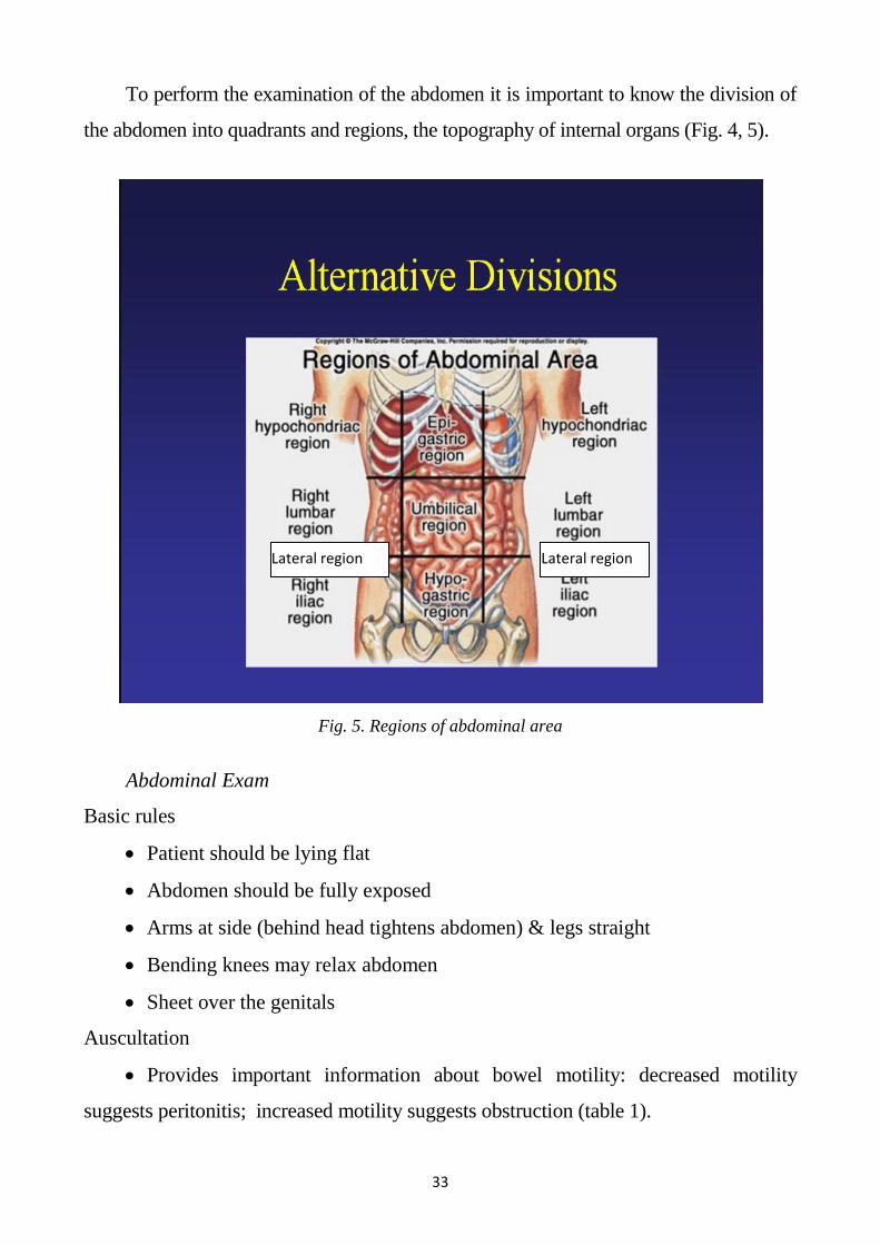
To perform the examination of the abdomen it is important to know the division of
the abdomen into quadrants and regions, the topography of internal organs (Fig. 4, 5).
Fig. 5. Regions of abdominal area
Abdominal Exam
Basic rules
Patient should be lying flat
Abdomen should be fully exposed
Arms at side (behind head tightens abdomen) & legs straight
Bending knees may relax abdomen
Sheet over the genitals
Auscultation
Provides important information about bowel motility: decreased motility
suggests peritonitis; increased motility suggests obstruction (table 1).
Lateral region Lateral region
34
Table 1
Information about bowel motility
Hyperactive bowel sounds Hypoactive/paralitik ileus
Postprandial physiologic Adinamic ileus
Laksatif consumption peritonitis
Diare
Early mechanical obstruction
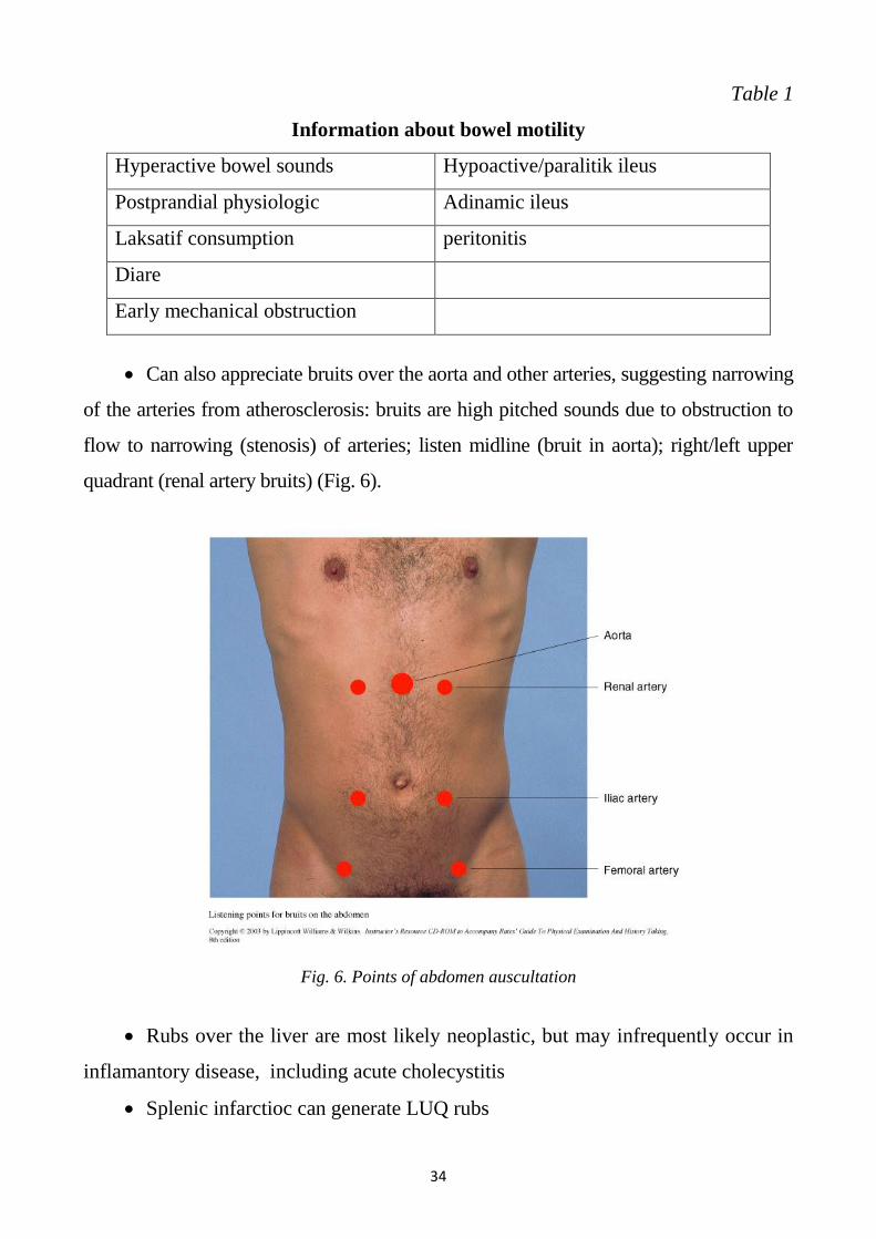
Can also appreciate bruits over the aorta and other arteries, suggesting narrowing
of the arteries from atherosclerosis: bruits are high pitched sounds due to obstruction to
flow to narrowing (stenosis) of arteries; listen midline (bruit in aorta); right/left upper
quadrant (renal artery bruits) (Fig. 6).
Fig. 6. Points of abdomen auscultation
Rubs over the liver are most likely neoplastic, but may infrequently occur in
inflamantory disease, including acute cholecystitis
Splenic infarctioc can generate LUQ rubs
35
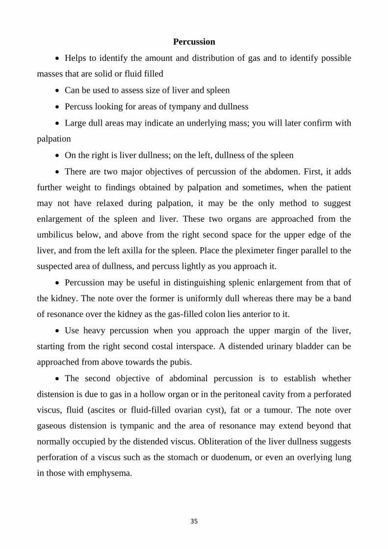
Percussion
Helps to identify the amount and distribution of gas and to identify possible
masses that are solid or fluid filled
Can be used to assess size of liver and spleen
Percuss looking for areas of tympany and dullness
Large dull areas may indicate an underlying mass; you will later confirm with
palpation
On the right is liver dullness; on the left, dullness of the spleen
There are two major objectives of percussion of the abdomen. First, it adds
further weight to findings obtained by palpation and sometimes, when the patient
may not have relaxed during palpation, it may be the only method to suggest
enlargement of the spleen and liver. These two organs are approached from the
umbilicus below, and above from the right second space for the upper edge of the
liver, and from the left axilla for the spleen. Place the pleximeter finger parallel to the
suspected area of dullness, and percuss lightly as you approach it.
Percussion may be useful in distinguishing splenic enlargement from that of
the kidney. The note over the former is uniformly dull whereas there may be a band
of resonance over the kidney as the gas-filled colon lies anterior to it.
Use heavy percussion when you approach the upper margin of the liver,
starting from the right second costal interspace. A distended urinary bladder can be
approached from above towards the pubis.
The second objective of abdominal percussion is to establish whether
distension is due to gas in a hollow organ or in the peritoneal cavity from a perforated
viscus, fluid (ascites or fluid-filled ovarian cyst), fat or a tumour. The note over
gaseous distension is tympanic and the area of resonance may extend beyond that
normally occupied by the distended viscus. Obliteration of the liver dullness suggests
perforation of a viscus such as the stomach or duodenum, or even an overlying lung
in those with emphysema.
36
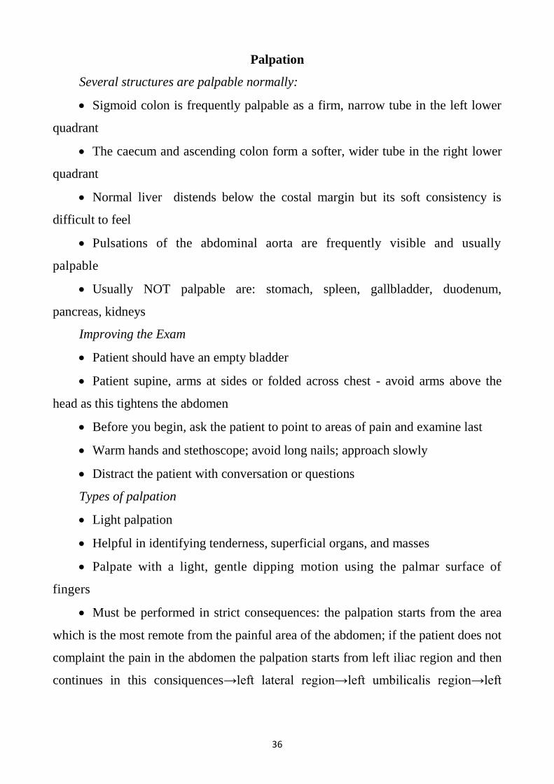
Palpation
Several structures are palpable normally:
Sigmoid colon is frequently palpable as a firm, narrow tube in the left lower
quadrant
The caecum and ascending colon form a softer, wider tube in the right lower
quadrant
Normal liver distends below the costal margin but its soft consistency is
difficult to feel
Pulsations of the abdominal aorta are frequently visible and usually
palpable
Usually NOT palpable are: stomach, spleen, gallbladder, duodenum,
pancreas, kidneys
Improving the Exam
Patient should have an empty bladder
Patient supine, arms at sides or folded across chest - avoid arms above the
head as this tightens the abdomen
Before you begin, ask the patient to point to areas of pain and examine last
Warm hands and stethoscope; avoid long nails; approach slowly
Distract the patient with conversation or questions
Types of palpation
Light palpation
Helpful in identifying tenderness, superficial organs, and masses
Palpate with a light, gentle dipping motion using the palmar surface of
fingers
Must be performed in strict consequences: the palpation starts from the area
which is the most remote from the painful area of the abdomen; if the patient does not
complaint the pain in the abdomen the palpation starts from left iliac region and then
continues in this consiquences→left lateral region→left umbilicalis region→left
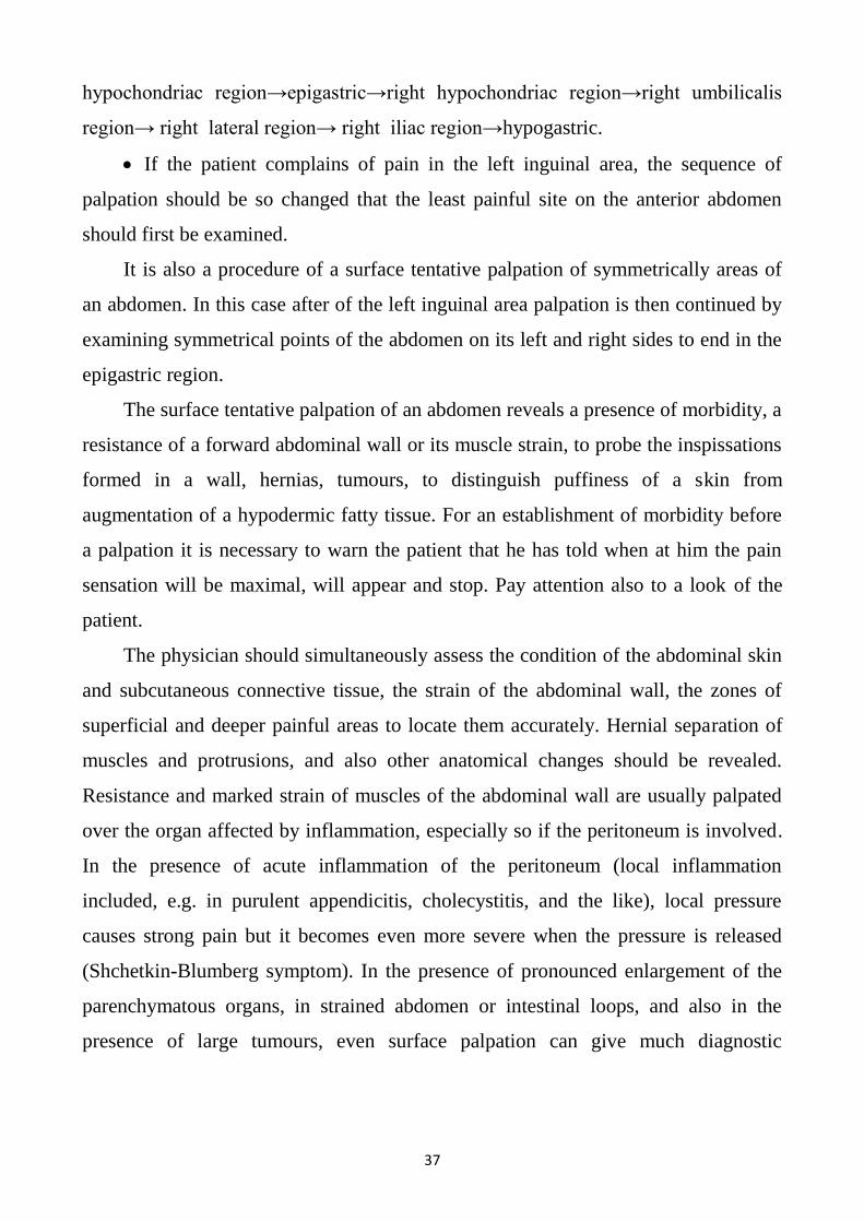
37
hypochondriac region→epigastric→right hypochondriac region→right umbilicalis
region→ right lateral region→ right iliac region→hypogastric.
If the patient complains of pain in the left inguinal area, the sequence of
palpation should be so changed that the least painful site on the anterior abdomen
should first be examined.
It is also a procedure of a surface tentative palpation of symmetrically areas of
an abdomen. In this case after of the left inguinal area palpation is then continued by
examining symmetrical points of the abdomen on its left and right sides to end in the
epigastric region.
The surface tentative palpation of an abdomen reveals a presence of morbidity, a
resistance of a forward abdominal wall or its muscle strain, to probe the inspissations
formed in a wall, hernias, tumours, to distinguish puffiness of a skin from
augmentation of a hypodermic fatty tissue. For an establishment of morbidity before
a palpation it is necessary to warn the patient that he has told when at him the pain
sensation will be maximal, will appear and stop. Pay attention also to a look of the
patient.
The physician should simultaneously assess the condition of the abdominal skin
and subcutaneous connective tissue, the strain of the abdominal wall, the zones of
superficial and deeper painful areas to locate them accurately. Hernial separation of
muscles and protrusions, and also other anatomical changes should be revealed.
Resistance and marked strain of muscles of the abdominal wall are usually palpated
over the organ affected by inflammation, especially so if the peritoneum is involved.
In the presence of acute inflammation of the peritoneum (local inflammation
included, e.g. in purulent appendicitis, cholecystitis, and the like), local pressure
causes strong pain but it becomes even more severe when the pressure is released
(Shchetkin-Blumberg symptom). In the presence of pronounced enlargement of the
parenchymatous organs, in strained abdomen or intestinal loops, and also in the
presence of large tumours, even surface palpation can give much diagnostic
38
information. But only deep systematic palpation can give full information about the
condition of the abdominal cavity and its organs, as well as their topography.
Utmost degree of muscles contraction (abdominal guarding) suggests
peritoneal irritation (peritonitis). Generalized rigidity of the abdominal muscles
should be interpreted in the context of the patient's clinical state.
Rebound tenderness is elicited by removing the palpating hand suddenly after
firm pressure has been applied over an area of the abdomen. If the rebound
tenderness exists the patient will report pain on removal. It indicates localized
peritonitis.
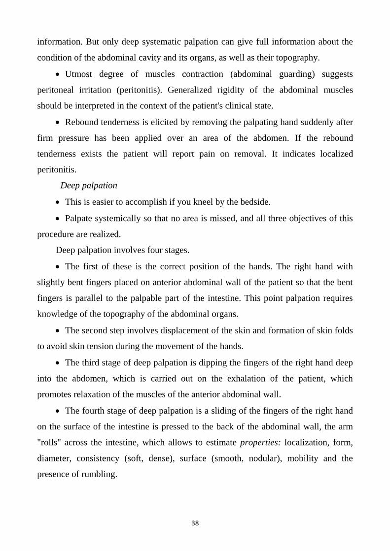
Deep palpation
This is easier to accomplish if you kneel by the bedside.
Palpate systemically so that no area is missed, and all three objectives of this
procedure are realized.
Deep palpation involves four stages.
The first of these is the correct position of the hands. The right hand with
slightly bent fingers placed on anterior abdominal wall of the patient so that the bent
fingers is parallel to the palpable part of the intestine. This point palpation requires
knowledge of the topography of the abdominal organs.
The second step involves displacement of the skin and formation of skin folds
to avoid skin tension during the movement of the hands.
The third stage of deep palpation is dipping the fingers of the right hand deep
into the abdomen, which is carried out on the exhalation of the patient, which
promotes relaxation of the muscles of the anterior abdominal wall.
The fourth stage of deep palpation is a sliding of the fingers of the right hand
on the surface of the intestine is pressed to the back of the abdominal wall, the arm
"rolls" across the intestine, which allows to estimate properties: localization, form,
diameter, consistency (soft, dense), surface (smooth, nodular), mobility and the
presence of rumbling.
39
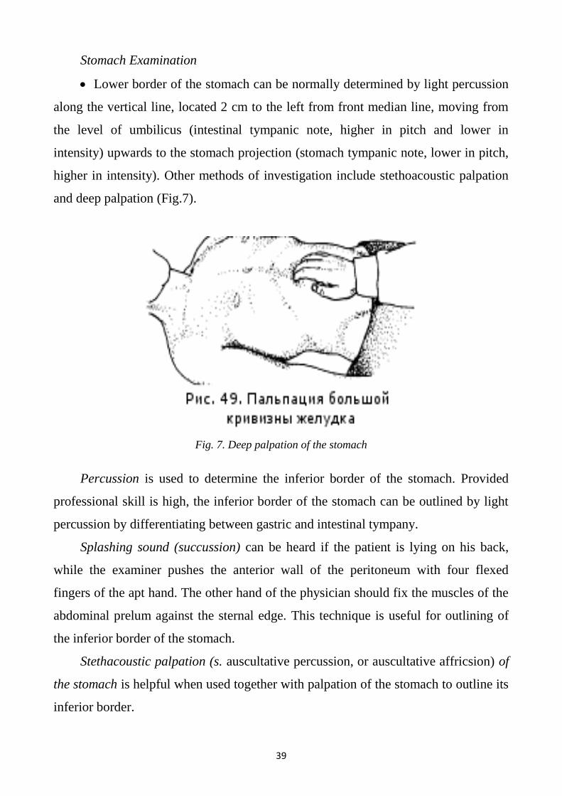
Stomach Examination
Lower border of the stomach can be normally determined by light percussion
along the vertical line, located 2 cm to the left from front median line, moving from
the level of umbilicus (intestinal tympanic note, higher in pitch and lower in
intensity) upwards to the stomach projection (stomach tympanic note, lower in pitch,
higher in intensity). Other methods of investigation include stethoacoustic palpation
and deep palpation (Fig.7).
Fig. 7. Deep palpation of the stomach
Percussion is used to determine the inferior border of the stomach. Provided
professional skill is high, the inferior border of the stomach can be outlined by light
percussion by differentiating between gastric and intestinal tympany.
Splashing sound (succussion) can be heard if the patient is lying on his back,
while the examiner pushes the anterior wall of the peritoneum with four flexed
fingers of the apt hand. The other hand of the physician should fix the muscles of the
abdominal prelum against the sternal edge. This technique is useful for outlining of
the inferior border of the stomach.
Stethacoustic palpation (s. auscultative percussion, or auscultative affricsion) of
the stomach is helpful when used together with palpation of the stomach to outline its
inferior border.
40
Colon Examination
Normally all parts of the colon can be assessed by deep palpation. The usual
sequence of deep palpation includes investigation of sigmoid, then terminal part of
ileum, caecum, ascending and descending colon and finally - transverse colon. This
sequence also represents decreasing probability of success in palpation: it means, that
sigmoid colon can be easily felt in most of the patients, even obese, while transverse
colon is extremely difficult to detect. There are also some divergences in technique of
palpation of different parts of the colon: you should use your left palm as a support at
palpation of ascending and descending colon; you should use bimanual palpation for
assessment of transverse colon.
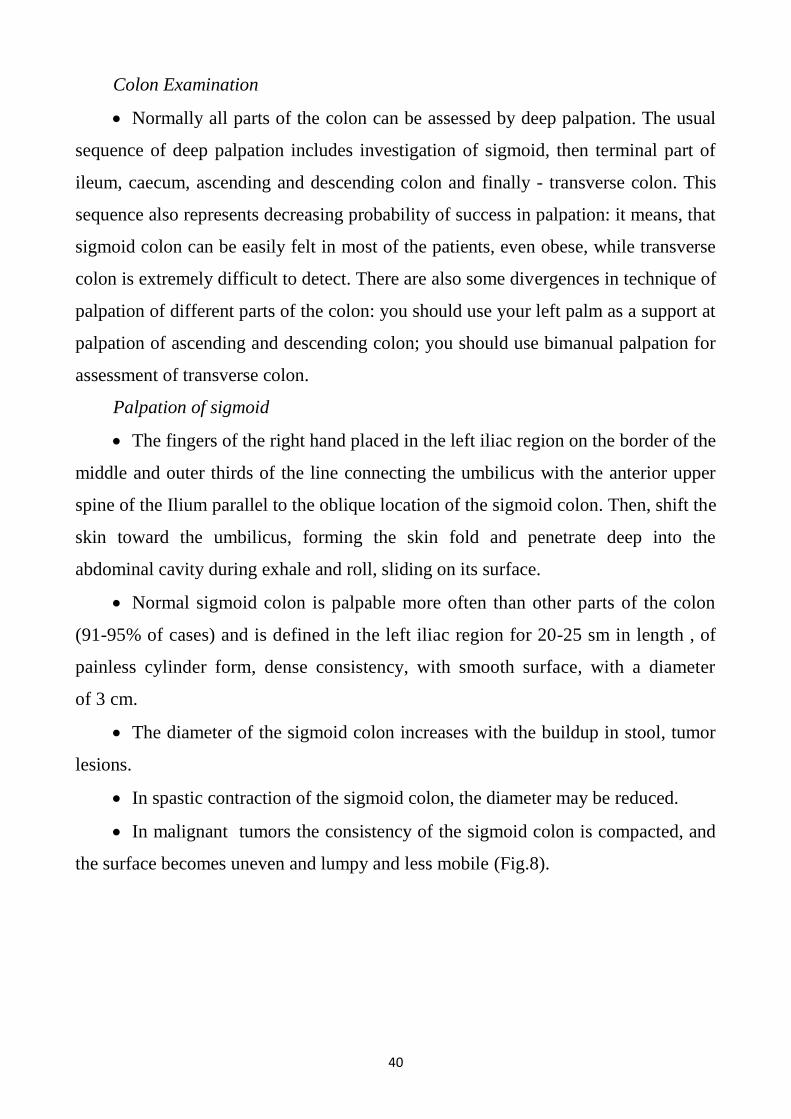
Palpation of sigmoid
The fingers of the right hand placed in the left iliac region on the border of the
middle and outer thirds of the line connecting the umbilicus with the anterior upper
spine of the Ilium parallel to the oblique location of the sigmoid colon. Then, shift the
skin toward the umbilicus, forming the skin fold and penetrate deep into the
abdominal cavity during exhale and roll, sliding on its surface.
Normal sigmoid colon is palpable more often than other parts of the colon
(91-95% of cases) and is defined in the left iliac region for 20-25 sm in length , of
painless cylinder form, dense consistency, with smooth surface, with a diameter
of 3 cm.
The diameter of the sigmoid colon increases with the buildup in stool, tumor
lesions.
In spastic contraction of the sigmoid colon, the diameter may be reduced.
In malignant tumors the consistency of the sigmoid colon is compacted, and
the surface becomes uneven and lumpy and less mobile (Fig.8).
41
Fig. 8. Palpation of sigmoid
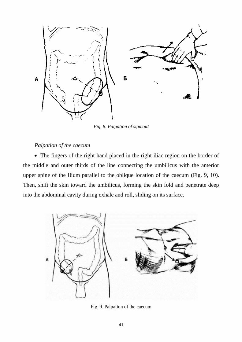
Palpation of the caecum
The fingers of the right hand placed in the right iliac region on the border of
the middle and outer thirds of the line connecting the umbilicus with the anterior
upper spine of the Ilium parallel to the oblique location of the caecum (Fig. 9, 10).
Then, shift the skin toward the umbilicus, forming the skin fold and penetrate deep
into the abdominal cavity during exhale and roll, sliding on its surface.
Fig. 9. Palpation of the caecum
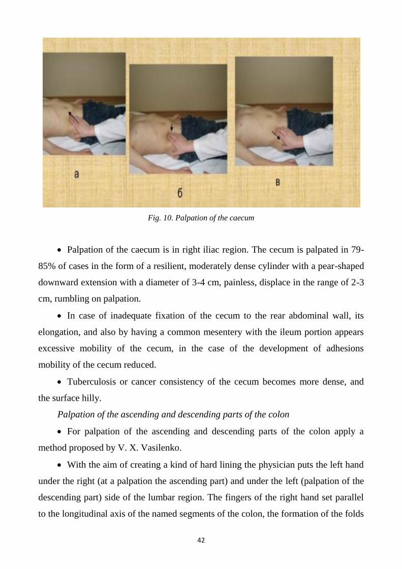
42
Fig. 10. Palpation of the caecum
Palpation of the caecum is in right iliac region. The cecum is palpated in 79-
85% of cases in the form of a resilient, moderately dense cylinder with a pear-shaped
downward extension with a diameter of 3-4 cm, painless, displace in the range of 2-3
cm, rumbling on palpation.
In case of inadequate fixation of the cecum to the rear abdominal wall, its
elongation, and also by having a common mesentery with the ileum portion appears
excessive mobility of the cecum, in the case of the development of adhesions
mobility of the cecum reduced.
Tuberculosis or cancer consistency of the cecum becomes more dense, and
the surface hilly.
Palpation of the ascending and descending parts of the colon
For palpation of the ascending and descending parts of the colon apply a
method proposed by V. X. Vasilenko.
With the aim of creating a kind of hard lining the physician puts the left hand
under the right (at a palpation the ascending part) and under the left (palpation of the
descending part) side of the lumbar region. The fingers of the right hand set parallel
to the longitudinal axis of the named segments of the colon, the formation of the folds
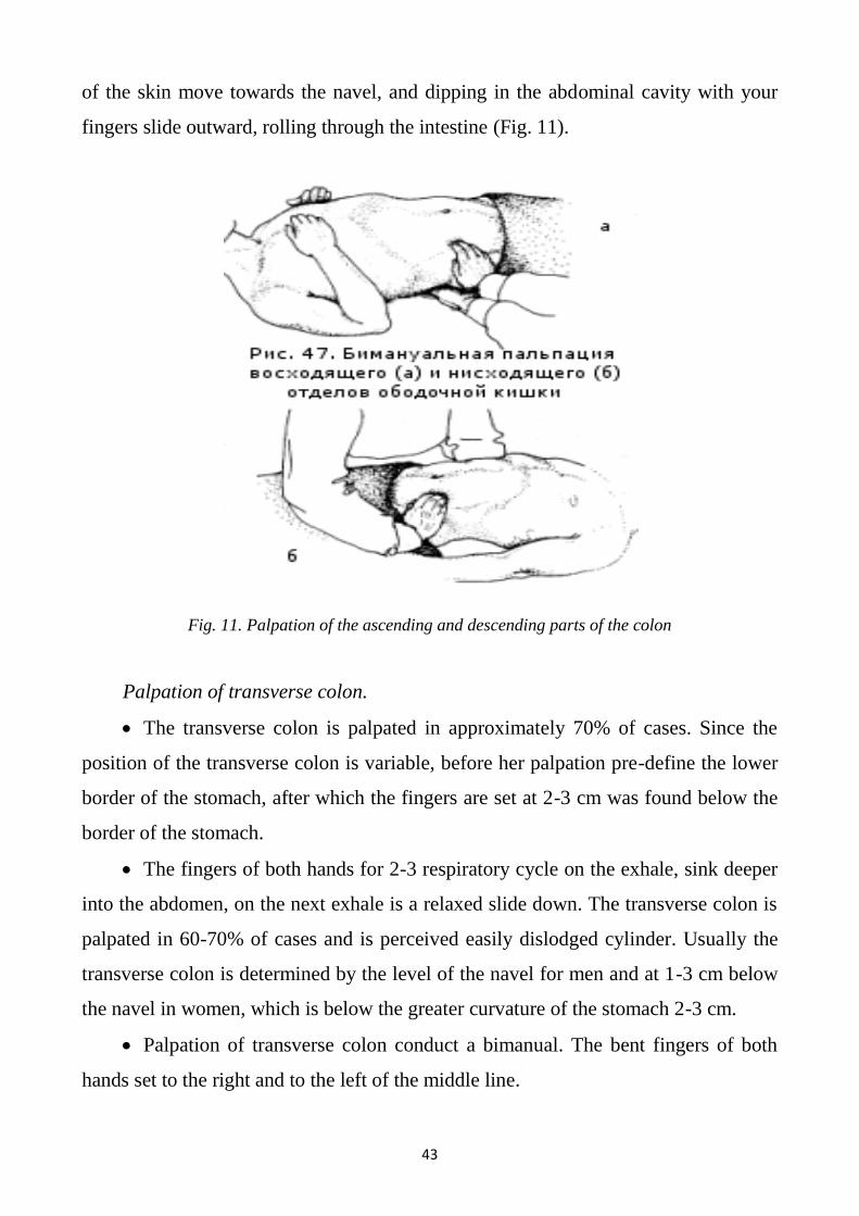
43
of the skin move towards the navel, and dipping in the abdominal cavity with your
fingers slide outward, rolling through the intestine (Fig. 11).
Fig. 11. Palpation of the ascending and descending parts of the colon
Palpation of transverse colon.
The transverse colon is palpated in approximately 70% of cases. Since the
position of the transverse colon is variable, before her palpation pre-define the lower
border of the stomach, after which the fingers are set at 2-3 cm was found below the
border of the stomach.
The fingers of both hands for 2-3 respiratory cycle on the exhale, sink deeper
into the abdomen, on the next exhale is a relaxed slide down. The transverse colon is
palpated in 60-70% of cases and is perceived easily dislodged cylinder. Usually the
transverse colon is determined by the level of the navel for men and at 1-3 cm below
the navel in women, which is below the greater curvature of the stomach 2-3 cm.
Palpation of transverse colon conduct a bimanual. The bent fingers of both
hands set to the right and to the left of the middle line.
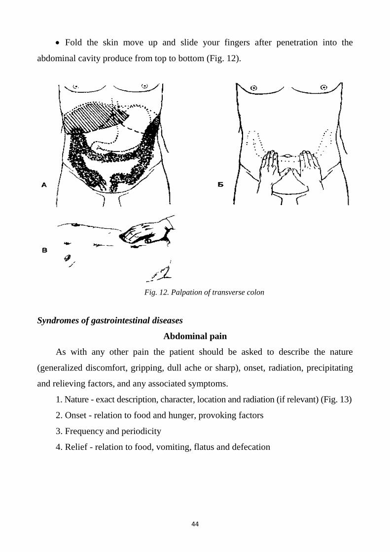
44
Fold the skin move up and slide your fingers after penetration into the
abdominal cavity produce from top to bottom (Fig. 12).
Fig. 12. Palpation of transverse colon
Syndromes of gastrointestinal diseases
Abdominal pain
As with any other pain the patient should be asked to describe the nature
(generalized discomfort, gripping, dull ache or sharp), onset, radiation, precipitating
and relieving factors, and any associated symptoms.
1. Nature - exact description, character, location and radiation (if relevant) (Fig. 13)
2. Onset - relation to food and hunger, provoking factors
3. Frequency and periodicity
4. Relief - relation to food, vomiting, flatus and defecation
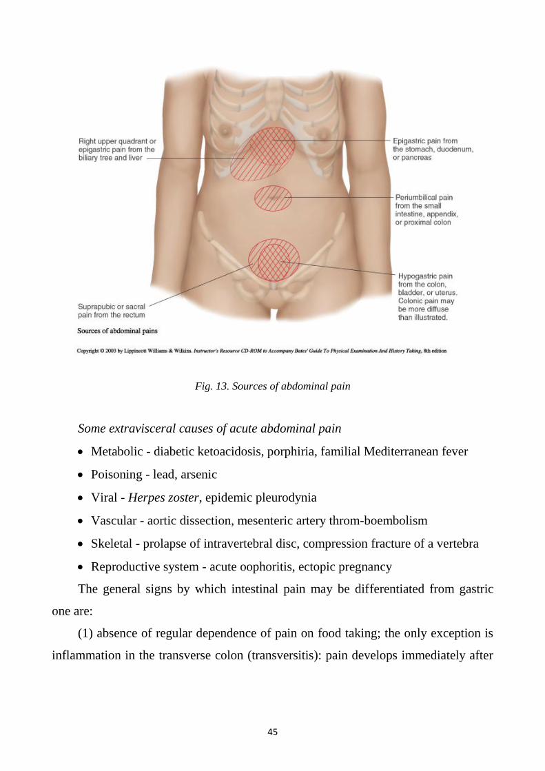
45
Fig. 13. Sources of abdominal pain
Some extravisceral causes of acute abdominal pain
Metabolic - diabetic ketoacidosis, porphiria, familial Mediterranean fever
Poisoning - lead, arsenic
Viral - Herpes zoster, epidemic pleurodynia
Vascular - aortic dissection, mesenteric artery throm-boembolism
Skeletal - prolapse of intravertebral disc, compression fracture of a vertebra
Reproductive system - acute oophoritis, ectopic pregnancy
The general signs by which intestinal pain may be differentiated from gastric
one are:
(1) absence of regular dependence of pain on food taking; the only exception is
inflammation in the transverse colon (transversitis): pain develops immediately after
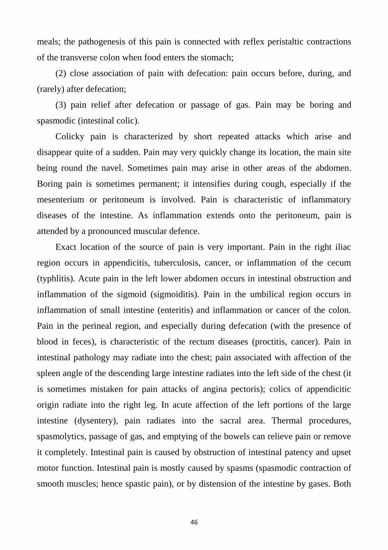
46
meals; the pathogenesis of this pain is connected with reflex peristaltic contractions
of the transverse colon when food enters the stomach;
(2) close association of pain with defecation: pain occurs before, during, and
(rarely) after defecation;
(3) pain relief after defecation or passage of gas. Pain may be boring and
spasmodic (intestinal colic).
Colicky pain is characterized by short repeated attacks which arise and
disappear quite of a sudden. Pain may very quickly change its location, the main site
being round the navel. Sometimes pain may arise in other areas of the abdomen.
Boring pain is sometimes permanent; it intensifies during cough, especially if the
mesenterium or peritoneum is involved. Pain is characteristic of inflammatory
diseases of the intestine. As inflammation extends onto the peritoneum, pain is
attended by a pronounced muscular defence.
Exact location of the source of pain is very important. Pain in the right iliac
region occurs in appendicitis, tuberculosis, cancer, or inflammation of the cecum
(typhlitis). Acute pain in the left lower abdomen occurs in intestinal obstruction and
inflammation of the sigmoid (sigmoiditis). Pain in the umbilical region occurs in
inflammation of small intestine (enteritis) and inflammation or cancer of the colon.
Pain in the perineal region, and especially during defecation (with the presence of
blood in feces), is characteristic of the rectum diseases (proctitis, cancer). Pain in
intestinal pathology may radiate into the chest; pain associated with affection of the
spleen angle of the descending large intestine radiates into the left side of the chest (it
is sometimes mistaken for pain attacks of angina pectoris); colics of appendicitic
origin radiate into the right leg. In acute affection of the left portions of the large
intestine (dysentery), pain radiates into the sacral area. Thermal procedures,
spasmolytics, passage of gas, and emptying of the bowels can relieve pain or remove
it completely. Intestinal pain is caused by obstruction of intestinal patency and upset
motor function. Intestinal pain is mostly caused by spasms (spasmodic contraction of
smooth muscles; hence spastic pain), or by distension of the intestine by gases. Both
47
mechanisms often become involved. Spastic pain can be due to various causes.
Individual predisposition to spastic contractions in general (vegetoneurosis) may be
as important as irritation originating in the intestine proper, e.g. in enteritis, colitis,
intestinal tumour, poisoning with arsenic or lead, and also in diseases of the central
nervous system (posterior spinal sclerosis). Pain arising due to intestinal distension
by gases, and associated with tension and irritation of the mesentery, differs from
spastic pain (1) by the absence of periodicity; it is long-standing and gradually
lessens in prolonged inflation; and (2) by exact localization. In intestinal obstruction
(complete or partial) colicky pain is combined with almost permanent pain in the
abdomen. It is characterized by exact and permanent location (the umbilical region
and large intestine). The pain intensifies with intestinal peristalsis.
Appendicular colic first localizes round the navel and the epigastrium but in
several hours (or even on the next day) it descends to the right iliac region where it
intensifies gradually. Sometimes the pain arises straight in the right iliac region.
Rectal colic, or tenesmus, is also known. It occurs in frequent and painful tenesmus to
defecate and is associated with spasmodic contractions of the intestine and the
sphincter ani. Only clots of mucus are sometimes expressed instead of actual
defecation. Tenesmus occurs in dysentery and other inflammatory or ulcerous
diseases, and in cancer of the rectum. Pain associated with defecation depends on
many factors. Pain preceding defecation is associated with the disease of the
descending colon or sigmoid colon. Pain during defecation is characteristic of
hemorrhoids, anal fissures, and cancer.
The signs of acute peritonitis
Utmost degree of muscles contraction (abdominal guarding) suggests
peritoneal irritation (peritonitis).
Rebound tenderness is elicited by removing the palpating hand suddenly
after firm pressure has been applied over an area of the abdomen. It indicates
localized peritonitis.
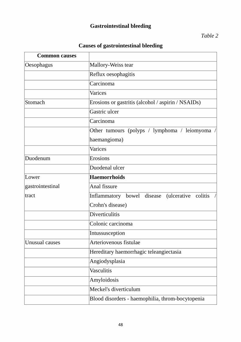
48
Gastrointestinal bleeding
Table 2
Causes of gastrointestinal bleeding
Common causes
Oesophagus Mallory-Weiss tear
Reflux oesophagitis
Carcinoma
Varices
Stomach Erosions or gastritis (alcohol / aspirin / NSAIDs)
Gastric ulcer
Carcinoma
Other tumours (polyps / lymphoma / leiomyoma /
haemangioma)
Varices
Duodenum Erosions
Duodenal ulcer
Lower
gastrointestinal
tract
Haemorrhoids
Anal fissure
Inflammatory bowel disease (ulcerative colitis /
Crohn's disease)
Diverticulitis
Colonic carcinoma
Intussusception
Unusual causes Arteriovenous fistulae
Hereditary haemorrhagic teleangiectasia
Angiodysplasia
Vasculitis
Amyloidosis
Meckel's diverticulum
Blood disorders - haemophilia, throm-bocytopenia
49
Signs of gastrointestinal bleeding
Patients with upper gastrointestinal bleeding may present with frank
haematemesis (vomiting of fresh blood), with vomiting of altered blood («coffee
ground» vomit), with passage of altered blood in the stool (melaena), or silently as
anaemia. Those with lower intestinal bleeding may present with the passage of fresh
blood or clot per rectum, with frank blood-streaking of the stool, or silently with
anaemia (and occult blood in the stool on testing).
Constipation
Obstipation (constipation). This is obstinate constipation during which feces are
long retained in the intestine (for more than 48 hours). But the duration of
constipation is only relative, because in many cases it is not the result of pathology
but of the living conditions and nutrition. If vegetable food dominates in the diet, the
subject may defecate two or three times a day. Stools become rarer if the diet is rich
in meat. A radical change in nutrition can remove constipation. Limited mobility of
the subject, hunger, and irregular defecations (during the day) may prolong pauses
between defecation. The main factor determining defecation is the condition of
intestinal motor function. Bowel contents are retained in the large intestine and the
rectum during constipation
Organic and functional constipation is differentiated. Organic constipation is
usually associated with mechanical obstruction, such as narrowing of the intestinal
lumen due to a tumour, scar, adhesion, and also abnormalities in the intestine
(megacolon, dolichosigmoid, megasigmoid, diverticulosis).
Functional constipation is subdivided into: (1) alimentary constipation; it occurs
due to ingestion of easily assimilable foods, which leave small residue and normally
stimulate peristalsis of the intestine by irritating its nervous receptors; (2) neurogenic
constipation due to dysfunction of the intramural nervous apparatus or vagus nerve;
these are the so-called dyskinetic constipation, caused by the reflex action on the
intestinal motor function of another affected organ (cholecystitis, adnexitis, prostatitis,
50
etc.), or by organic affections of the central nervous system (tumours of the brain,
encephalitis, posterior spinal sclerosis); (3) constipation associated with inflammatory
affections, mainly of the large intestine (dysentery); (4) toxic constipation occurring in
exogenous poisoning with lead, morphine, or cocaine; (5) constipation of endocrine
etiology, occurring in thyroid or pituitary hypofunction; (6) constipation caused by lack
of physical exercise; (7) constipation caused by flaccidity of the prelum.
Acute constipation occurs when a change in bowel habits produces infrequent
stools or hard stools that are difficult to pass. A sudden change suggests an organic
cause:
- mechanical bowel obstruction;
- adynamic ileus often accompanies acute intra-ab-dominal disease and may
complicate various traumatic conditions or may follow general anaesthesia.
A reduced stool size suggests an obstructive lesion in the distal colon. Local
anorectal conditions (e.g., anal fissures) that cause pain or bleeding should be sought.
Diarrhoea
Frequent and liquid stool is a common sign of intestinal pathology. Diarrhea
occurs in acute and chronic intestinal infections (enteritis, enterocolitis, sigmoiditis,
proctitis), in various exogenous intoxications (poisoning with arsenic or mercury),
endogenous intoxications (uremia, diabetes, gout), in endocrine disorders (adrenal
dysfunction, thyrotoxicosis), and in hypersensitivity to some foods (allergy). The
mechanism of diarrhea is very complicated. Different pathogenic factors may prevail
in various pathological conditions. Accelerated movement of the liquefied food in the
intestine due to peristalsis is among them. Almost undigested food can thus be
evacuated. Another factor is disordered absorptive function of the intestine. Affection
of the intestinal wall, disordered mechanisms regulating absorption, purgatives and
upset water metabolism produce a marked change in the absorption process and are
the cause of diarrhea. The third cause of liquid stools is inflammation of the intestine.
Large quantities of inflammatory secretion stimulating the intestinal receptors are
51
released into the lumen of the intestine to intensify its peristalsis and to impair its
absorptive function. Paradoxical diarrhea occurs in prolonged constipation due to
mechanical irritation of the intestinal wall by hard fecal masses. Upset equilibrium
between the fermentative and putrefactive flora of the intestine is another important
factor in the etiology of diarrhea. Diarrhea occurring in organic affections of the
large intestine is mostly of the inflammatory character. It is not copious, nor does it
produce strong negative effect on the patient's general condition (as compared with
affections of the small intestine which is attended by profuse diarrhea associated with
deranged motor and absorption function of the intestine). The pronounced disorder in
digestion causes some metabolic disorders in the patient (impaired absorption of
proteins, iron, vitamins, and electrolytes).
Basic types of diarrhoea are following:
1. Osmotic diarrhoea occurs when unabsorbable, water-soluble solutes remain in
the bowel, where they retain water:
lactose intolerance
use of poorly absorbed salts (Mg sulfate, Na phosphates).
ingestion of large amounts of the hexitols (e.g., sor-bitol, mannitol), which are
used as sugar substitutes, causes osmotic diarrhoea as a result of their slow absorption
and stimulation of rapid small-bowel motility («dietetic food» diarrhoea).
2. Secretory diarrhoea occurs when the small and large bowel secrete more
electrolytes and water than they absorb. Nausea, vomiting, abdominal pain,
flatulence, and weight loss may occur. Secretagogues include:
bacterial toxins (e.g., cholera),
enteropathogenic viruses,
bile acids (e.g., after ileal resection),
unabsorbed dietary fat (e.g., in steatorrhea),
drugs (e.g., anthraquinone cathartics, castor oil),
peptide hormones (e.g., VIP produced by pancreatic tumours).
52
3. Exudative diarrhoea occurs with several mucosal diseases that cause mucosal
inflammation, ulceration, or tumefaction. The resultant outpouring of plasma, serum
proteins, blood, and mucus increases faecal bulk and fluid content. Involvement of
the rectal mucosa may cause urgency and increased stool frequency because the
inflamed rectum is more sensitive to distension:
regional enteritis;
ulcerative colitis;
TB;
lymphoma;
cancer.
4. Decreased absorption time occurs when chyme is not in contact with an
adequate absorptive surface of the GI tract for a long enough time so that too much
water remains in the faeces:
smallor large-bowel resection;
gastric surgery (resection, pyloroplasty, vagotomy);
surgical bypass of intestinal segments;
drugs (e.g., Mg-containing antacids, laxatives);
humoral agents (e.g., prostaglandins, serotonin).
5. Malabsorption produces diarrhoea by osmotic or secretory mechanisms. The
mechanism may be osmotic if the unabsorbed material is abundant, water-soluble,
and of low molecular weight. Lipids are not osmotic, but some (fatty acids, bile
acids) act as secre-tagogues and produce secretory diarrhoea. In generalized
malabsorption, fat malabsorption causes colonic secretion, and carbohydrate
malabsorption causes osmotic diarrhoea.
6. Paradoxical diarrhoea results from oozing around a faecal impaction in
children and in debilitated or demented adults.
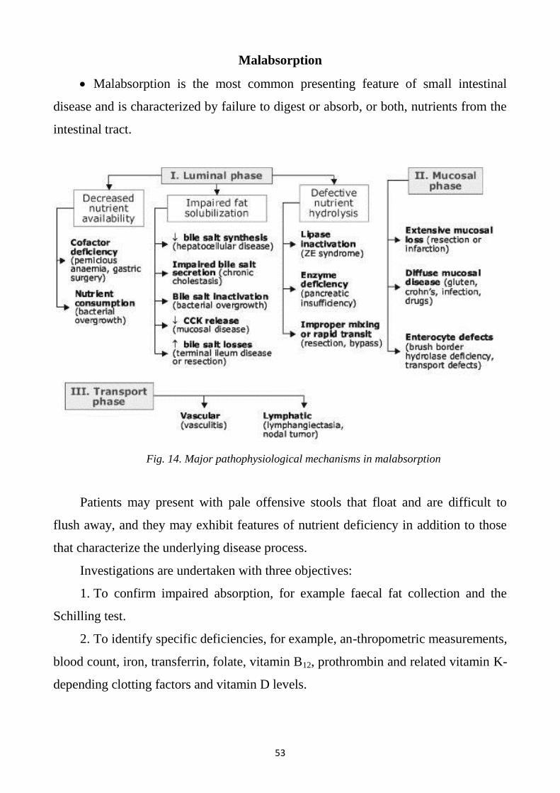
53
Malabsorption
Malabsorption is the most common presenting feature of small intestinal
disease and is characterized by failure to digest or absorb, or both, nutrients from the
intestinal tract.
Fig. 14. Major pathophysiological mechanisms in malabsorption
Patients may present with pale offensive stools that float and are difficult to
flush away, and they may exhibit features of nutrient deficiency in addition to those
that characterize the underlying disease process.
Investigations are undertaken with three objectives:
1. To confirm impaired absorption, for example faecal fat collection and the
Schilling test.
2. To identify specific deficiencies, for example, an-thropometric measurements,
blood count, iron, transferrin, folate, vitamin B12, prothrombin and related vitamin K-
depending clotting factors and vitamin D levels.
54
3. To establish the mechanism and cause of malab-sorption; this may include a
search for bacterial overgrowth, testing pancreatic exocrine function, and aspiration
or biopsy of the proximal small bowel to look for evidence of giardiasis or gluten
enteropathy.
Laboratory, radiologic and endoscopic diagnostics
Gastric Analysis
Gastric analysis is used to evaluate hyperchlorhydria (eg, Zollinger-Ellison
syndrome) or hypochlorhydric states (eg, atrophic gastritis, Menetrier's syndrome);
unexplained hypergastrinemia in patients with planned acid-reducing surgery as part
of pre- or postoperative assessment; and the possibility of incomplete vagotomy in
patients with recurrent peptic ulcer disease after a surgical vagotomy.
Contraindications include recent active bleeding or pain caused by active ulcer
disease.
A nasogastric tube is passed. For intubation, the patient sits upright or lies in the
left lateral decubitus position. With the patient's head partially flexed, the lubricated
tube is inserted through the nares, aimed back and then down to conform with the
nasopharynx. As the tip reaches the posterior pharyngeal wall, the patient should sip
water through a straw. (Violent coughing with flow of air through the tube during
respiration indicates that the tube is misplaced in the trachea.) Aspiration of gastric
juice verifies entry into the stomach. The position of larger tubes may be confirmed
by instilling 20 to 30 mL of air and listening with the stethoscope under the left
subcostal region for a rush of air.
Gastric contents are aspirated and discarded. Four 15-min samples of gastric
juice are collected by continuous manual aspiration (basal acid output [BAO]). Next,
pentagastrin (6 μg/kg) is given sc, and again, four 15-min samples are obtained
(maximal [or peak] acid output [MAO or PAO]). Samples are titrated with sodium
hydroxide to calculate BAO and stimulated MAO secretory rates.
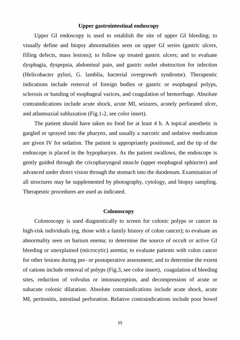
55
Upper gastrointestinal endoscopy
Upper GI endoscopy is used to establish the site of upper GI bleeding; to
visually define and biopsy abnormalities seen on upper GI series (gastric ulcers,
filling defects, mass lesions); to follow up treated gastric ulcers; and to evaluate
dysphagia, dyspepsia, abdominal pain, and gastric outlet obstruction for infection
(Helicobacter pylori, G. lamblia, bacterial overgrowth syndrome). Therapeutic
indications include removal of foreign bodies or gastric or esophageal polyps,
sclerosis or banding of esophageal varices, and coagulation of hemorrhage. Absolute
contraindications include acute shock, acute MI, seizures, acutely perforated ulcer,
and atlantoaxial subluxation (Fig.1-2, see color insert).
The patient should have taken no food for at least 4 h. A topical anesthetic is
gargled or sprayed into the pharynx, and usually a narcotic and sedative medication
are given IV for sedation. The patient is appropriately positioned, and the tip of the
endoscope is placed in the hypopharynx. As the patient swallows, the endoscope is
gently guided through the cricopharyngeal muscle (upper esophageal sphincter) and
advanced under direct vision through the stomach into the duodenum. Examination of
all structures may be supplemented by photography, cytology, and biopsy sampling.
Therapeutic procedures are used as indicated.
Colonoscopy
Colonoscopy is used diagnostically to screen for colonic polyps or cancer in
high-risk individuals (eg, those with a family history of colon cancer); to evaluate an
abnormality seen on barium enema; to determine the source of occult or active GI
bleeding or unexplained (microcytic) anemia; to evaluate patients with colon cancer
for other lesions during pre- or postoperative assessment; and to determine the extent
of cations include removal of polyps (Fig.3, see color insert), coagulation of bleeding
sites, reduction of volvulus or intussusception, and decompression of acute or
subacute colonic dilatation. Absolute contraindications include acute shock, acute
MI, peritonitis, intestinal perforation. Relative contraindications include poor bowel
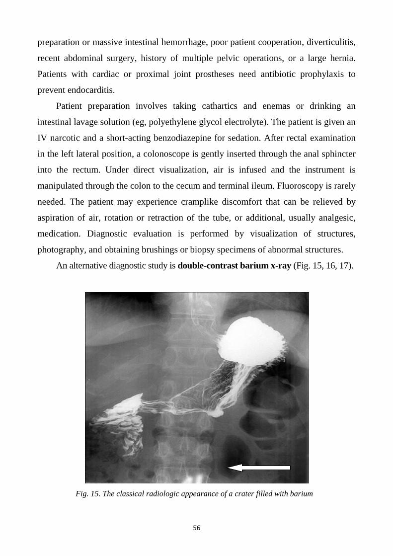
56
preparation or massive intestinal hemorrhage, poor patient cooperation, diverticulitis,
recent abdominal surgery, history of multiple pelvic operations, or a large hernia.
Patients with cardiac or proximal joint prostheses need antibiotic prophylaxis to
prevent endocarditis.
Patient preparation involves taking cathartics and enemas or drinking an
intestinal lavage solution (eg, polyethylene glycol electrolyte). The patient is given an
IV narcotic and a short-acting benzodiazepine for sedation. After rectal examination
in the left lateral position, a colonoscope is gently inserted through the anal sphincter
into the rectum. Under direct visualization, air is infused and the instrument is
manipulated through the colon to the cecum and terminal ileum. Fluoroscopy is rarely
needed. The patient may experience cramplike discomfort that can be relieved by
aspiration of air, rotation or retraction of the tube, or additional, usually analgesic,
medication. Diagnostic evaluation is performed by visualization of structures,
photography, and obtaining brushings or biopsy specimens of abnormal structures.
An alternative diagnostic study is double-contrast barium x-ray (Fig. 15, 16, 17).
Fig. 15. The classical radiologic appearance of a crater filled with barium
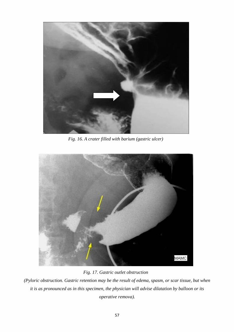
57
Fig. 16. A crater filled with barium (gastric ulcer)
Fig. 17. Gastric outlet obstruction
(Pyloric obstruction. Gastric retention may be the result of edema, spasm, or scar tissue, but when
it is as pronounced as in this specimen, the physician will advise dilatation by balloon or its
operative remova).
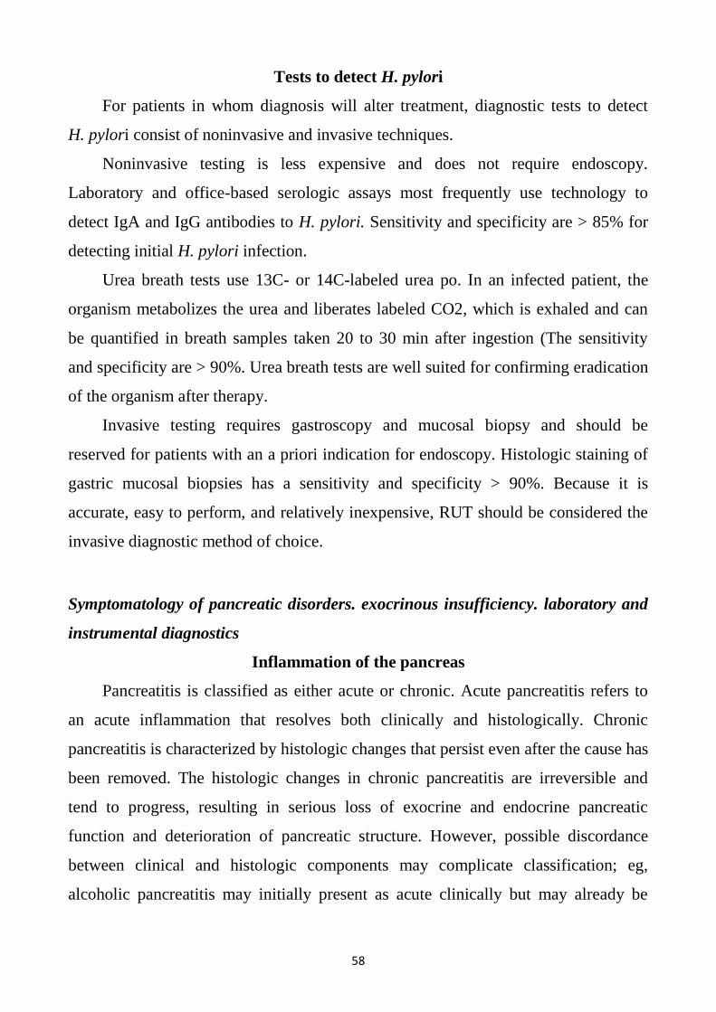
58
Tests to detect H. pylori
For patients in whom diagnosis will alter treatment, diagnostic tests to detect
H. pylori consist of noninvasive and invasive techniques.
Noninvasive testing is less expensive and does not require endoscopy.
Laboratory and office-based serologic assays most frequently use technology to
detect IgA and IgG antibodies to H. pylori. Sensitivity and specificity are > 85% for
detecting initial H. pylori infection.
Urea breath tests use 13C- or 14C-labeled urea po. In an infected patient, the
organism metabolizes the urea and liberates labeled CO2, which is exhaled and can
be quantified in breath samples taken 20 to 30 min after ingestion (The sensitivity
and specificity are > 90%. Urea breath tests are well suited for confirming eradication
of the organism after therapy.
Invasive testing requires gastroscopy and mucosal biopsy and should be
reserved for patients with an a priori indication for endoscopy. Histologic staining of
gastric mucosal biopsies has a sensitivity and specificity > 90%. Because it is
accurate, easy to perform, and relatively inexpensive, RUT should be considered the
invasive diagnostic method of choice.
Symptomatology of pancreatic disorders. exocrinous insufficiency. laboratory and
instrumental diagnostics
Inflammation of the pancreas
Pancreatitis is classified as either acute or chronic. Acute pancreatitis refers to
an acute inflammation that resolves both clinically and histologically. Chronic
pancreatitis is characterized by histologic changes that persist even after the cause has
been removed. The histologic changes in chronic pancreatitis are irreversible and
tend to progress, resulting in serious loss of exocrine and endocrine pancreatic
function and deterioration of pancreatic structure. However, possible discordance
between clinical and histologic components may complicate classification; eg,
alcoholic pancreatitis may initially present as acute clinically but may already be
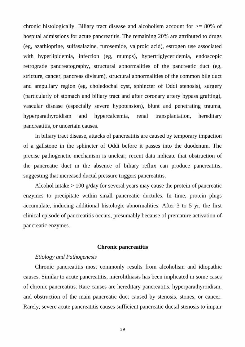
59
chronic histologically. Biliary tract disease and alcoholism account for >= 80% of
hospital admissions for acute pancreatitis. The remaining 20% are attributed to drugs
(eg, azathioprine, sulfasalazine, furosemide, valproic acid), estrogen use associated
with hyperlipidemia, infection (eg, mumps), hypertriglyceridemia, endoscopic
retrograde pancreatography, structural abnormalities of the pancreatic duct (eg,
stricture, cancer, pancreas divisum), structural abnormalities of the common bile duct
and ampullary region (eg, choledochal cyst, sphincter of Oddi stenosis), surgery
(particularly of stomach and biliary tract and after coronary artery bypass grafting),
vascular disease (especially severe hypotension), blunt and penetrating trauma,
hyperparathyroidism and hypercalcemia, renal transplantation, hereditary
pancreatitis, or uncertain causes.
In biliary tract disease, attacks of pancreatitis are caused by temporary impaction
of a gallstone in the sphincter of Oddi before it passes into the duodenum. The
precise pathogenetic mechanism is unclear; recent data indicate that obstruction of
the pancreatic duct in the absence of biliary reflux can produce pancreatitis,
suggesting that increased ductal pressure triggers pancreatitis.
Alcohol intake > 100 g/day for several years may cause the protein of pancreatic
enzymes to precipitate within small pancreatic ductules. In time, protein plugs
accumulate, inducing additional histologic abnormalities. After 3 to 5 yr, the first
clinical episode of pancreatitis occurs, presumably because of premature activation of
pancreatic enzymes.
Chronic pancreatitis
Etiology and Pathogenesis
Chronic pancreatitis most commonly results from alcoholism and idiopathic
causes. Similar to acute pancreatitis, microlithiasis has been implicated in some cases
of chronic pancreatitis. Rare causes are hereditary pancreatitis, hyperparathyroidism,
and obstruction of the main pancreatic duct caused by stenosis, stones, or cancer.
Rarely, severe acute pancreatitis causes sufficient pancreatic ductal stenosis to impair
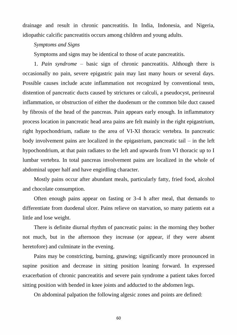
60
drainage and result in chronic pancreatitis. In India, Indonesia, and Nigeria,
idiopathic calcific pancreatitis occurs among children and young adults.
Symptoms and Signs
Symptoms and signs may be identical to those of acute pancreatitis.
1. Pain syndrome – basic sign of chronic pancreatitis. Although there is
occasionally no pain, severe epigastric pain may last many hours or several days.
Possible causes include acute inflammation not recognized by conventional tests,
distention of pancreatic ducts caused by strictures or calculi, a pseudocyst, perineural
inflammation, or obstruction of either the duodenum or the common bile duct caused
by fibrosis of the head of the pancreas. Pain appears early enough. In inflammatory
process location in pancreatic head area pains are felt mainly in the right epigastrium,
right hypochondrium, radiate to the area of VI-XI thoracic vertebra. In pancreatic
body involvement pains are localized in the epigastrium, pancreatic tail – in the left
hypochondrium, at that pain radiates to the left and upwards from VI thoracic up to I
lumbar vertebra. In total pancreas involvement pains are localized in the whole of
abdominal upper half and have engirdling character.
Mostly pains occur after abundant meals, particularly fatty, fried food, alcohol
and chocolate consumption.
Often enough pains appear on fasting or 3-4 h after meal, that demands to
differentiate from duodenal ulcer. Pains relieve on starvation, so many patients eat a
little and lose weight.
There is definite diurnal rhythm of pancreatic pains: in the morning they bother
not much, but in the afternoon they increase (or appear, if they were absent
heretofore) and culminate in the evening.
Pains may be constricting, burning, gnawing; significantly more pronounced in
supine position and decrease in sitting position leaning forward. In expressed
exacerbation of chronic pancreatitis and severe pain syndrome a patient takes forced
sitting position with bended in knee joints and adducted to the abdomen legs.
On abdominal palpation the following algesic zones and points are defined:

61
1. Choffar's zone — between vertical line, passing through umbilicus and
bisector of angle, formed by vertical and horizontal lines, passing through umbilicus.
Tenderness in this zone is characteristic of pancreatic head inflammation;
2. Gubergritz -Skoolsky's zone — is analogous Choffar's zone, but is situated at
the left side. Tenderness in this zone is characteristic of pancreatic body
inflammation;
3. Dejardin's point — is situated 6 cm above umbilicus along the line,
connecting umbilicus with the right axilla. Tenderness in this point is characteristic of
pancreatic head inflammation;
4. Gubergritz's point — is analogous Dejardin's point, but is situated on the left
side. Tenderness in this point is characteristic of pancreatic tail inflammation;
5. Mayo-Robson's point — is situated on the border of external and middle third
of the line, connecting umbilicus and the middle of the left costal arch. Tenderness in
this point is characteristic of pancreatic tail inflammation;
6. area of the left costovertebral angle — tenderness in this zone is characteristic
of pancreatic body and tail inflammation.
7. Grot's sign is defined in many patients – atrophy of subcutaneous fat in the
projection area of the pancreas on the anterior abdominal wall.
8. “Red drops” sign may be observed — presence of red spots on the abdominal,
chest and back skin, and also brownish skin colouring above the pancreas area.
2. Dyspeptic syndrome (pancreatic dyspepsia) — is characteristic enough of
chronic pancreatitis, particularly frequent it is expressed in exacerbation or severe
course of disease. Dyspeptic syndrome is revealed by increased salivation, air or
eaten food eructation, nausea, vomiting, loss of appetite, fatty food intolerance,
flatulence.
3. Weight loss develops due to restrictions in diet (on starvation pains are
decreased), and also owing to pancreatic exocrinous function disorders and intestinal
absorption impairment. Loss of appetite also predisposes to weight loss. It is
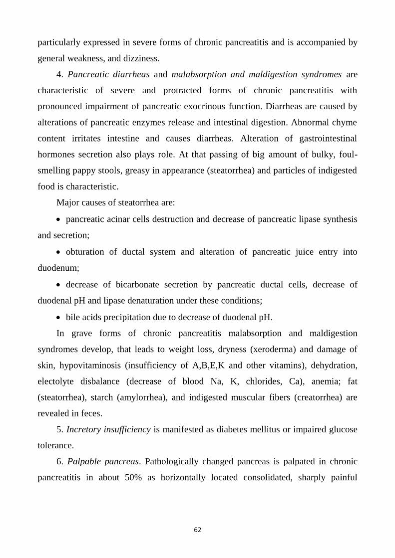
62
particularly expressed in severe forms of chronic pancreatitis and is accompanied by
general weakness, and dizziness.
4. Pancreatic diarrheas and malabsorption and maldigestion syndromes are
characteristic of severe and protracted forms of chronic pancreatitis with
pronounced impairment of pancreatic exocrinous function. Diarrheas are caused by
alterations of pancreatic enzymes release and intestinal digestion. Abnormal chyme
content irritates intestine and causes diarrheas. Alteration of gastrointestinal
hormones secretion also plays role. At that passing of big amount of bulky, foul-
smelling pappy stools, greasy in appearance (steatorrhea) and particles of indigested
food is characteristic.
Major causes of steatorrhea are:
pancreatic acinar cells destruction and decrease of pancreatic lipase synthesis
and secretion;
obturation of ductal system and alteration of pancreatic juice entry into
duodenum;
decrease of bicarbonate secretion by pancreatic ductal cells, decrease of
duodenal pH and lipase denaturation under these conditions;
bile acids precipitation due to decrease of duodenal pH.
In grave forms of chronic pancreatitis malabsorption and maldigestion
syndromes develop, that leads to weight loss, dryness (xeroderma) and damage of
skin, hypovitaminosis (insufficiency of A,B,E,K and other vitamins), dehydration,
electolyte disbalance (decrease of blood Na, K, chlorides, Ca), anemia; fat
(steatorrhea), starch (amylorrhea), and indigested muscular fibers (creatorrhea) are
revealed in feces.
5. Incretory insufficiency is manifested as diabetes mellitus or impaired glucose
tolerance.
6. Palpable pancreas. Pathologically changed pancreas is palpated in chronic
pancreatitis in about 50% as horizontally located consolidated, sharply painful
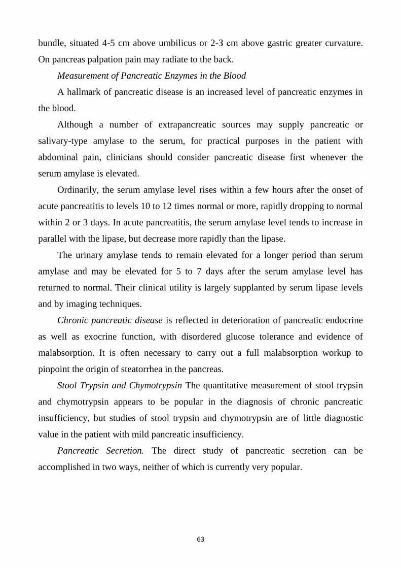
63
bundle, situated 4-5 cm above umbilicus or 2-З сm above gastric greater curvature.
On pancreas palpation pain may radiate to the back.
Measurement of Pancreatic Enzymes in the Blood
A hallmark of pancreatic disease is an increased level of pancreatic enzymes in
the blood.
Although a number of extrapancreatic sources may supply pancreatic or
salivary-type amylase to the serum, for practical purposes in the patient with
abdominal pain, clinicians should consider pancreatic disease first whenever the
serum amylase is elevated.
Ordinarily, the serum amylase level rises within a few hours after the onset of
acute pancreatitis to levels 10 to 12 times normal or more, rapidly dropping to normal
within 2 or 3 days. In acute pancreatitis, the serum amylase level tends to increase in
parallel with the lipase, but decrease more rapidly than the lipase.
The urinary amylase tends to remain elevated for a longer period than serum
amylase and may be elevated for 5 to 7 days after the serum amylase level has
returned to normal. Their clinical utility is largely supplanted by serum lipase levels
and by imaging techniques.
Chronic pancreatic disease is reflected in deterioration of pancreatic endocrine
as well as exocrine function, with disordered glucose tolerance and evidence of
malabsorption. It is often necessary to carry out a full malabsorption workup to
pinpoint the origin of steatorrhea in the pancreas.
Stool Trypsin and Chymotrypsin The quantitative measurement of stool trypsin
and chymotrypsin appears to be popular in the diagnosis of chronic pancreatic
insufficiency, but studies of stool trypsin and chymotrypsin are of little diagnostic
value in the patient with mild pancreatic insufficiency.
Pancreatic Secretion. The direct study of pancreatic secretion can be
accomplished in two ways, neither of which is currently very popular.
64
(1) The secretin test is the more standard and more sensitive, though
detecting alterations in pancreatic function sometimes so slight as to lack clinical
reflection.
(2) The Lundh test meal evaluates by direct aspiration of a test meal from the
duodenum the status of the digestive process. The test is reliable only when there is a
moderate diminution of pancreatic secretion.
Because the secretin test is less physiologic and its stimulus of greater potency,
it will usually display lesser degrees of pancreatic dysfunction than the Lundh test
meal.
Test control of the theme “Questioning and examination
of patients with diseases of thе gastrointestinal tract”
1. GIVE AN EXPLANATION OF THE TERM "DYSPHAGIA" (GIVE ONE
ANSWER):
a) violation of passing of food through the esophagus;
b) violation of digestion in the stomach or duodenum;
с) a violation of the wall digestion and absorption in the small intestine;
d) malabsorption in the colon.
2. FOR ANY OF THESE CONDITIONS, THE MOST TYPICAL APPEARANCE
OF ACUTE PAIN IN THE EPIGASTRIC REGION AFTER 15-30 MIN. AFTER A
MEAL, WEAKENING AFTER VOMITING?
a) chronic gastritis;
b) ulcerous disease of the stomach;
с) peptic ulcer of the duodenum;
d) cancer of the stomach.
65
3. ON SOME OF THESE VIOLATIONS INDICATES THE PRESENCE IN A
PATIENT OF HEARTBURN?
a) hyperacid the state of gastric secretion;
b) hypoacid state of the stomach secretions;
с) narrowing of target Department of a stomach;
d) dysfunction of the cardiac sphincter of the esophagus;
e) stricture (narrowing) of the esophagus.
4. THE PATIENT, PRESENTING COMPLAINTS OF CONSTANT DULL
EPIGASTRIC PAIN, HEARTBURN, BELCHING AIR WITH THE SMELL OF
ROTTEN EGGS, WITH NAUSEA, VOMITING UNDIGESTED FOOD (EATEN A
FEW HOURS AGO), WEAKNESS, LOSS OF APPETITE, WEIGHT LOSS,
PALPATION OF THE STOMACH AFTER 7 HOURS AFTER A MEAL IS
DETERMINED BY SPLASHING. WHAT KIND OF PATHOLOGY YOU CAN
THINK OF?
a) the stenosis of exit of a stomach (pylorus);
b) a significant increase in the secretory activity of the stomach;
с) sharp depression of the secretory and motor activity of the stomach;
d) none of the listed conditions.
5. THE CHANGE IN THE STOOL WITH THE HIGHEST DEGREE OF
PROBABILITY WILL ALLOW TO SUSPECT THE PRESENCE OF THE
PATIENT ACCOMPLISHED A MASSIVE BLEEDING FROM THE UPPER
GASTROINTESTINAL TRACT?
a) diarrhoea (diarrhea);
b) constipation (constipation);
с) black liquefied stool (mushy);
d) black designed cal;
e) dark brown feces in the form of lumps ("sheep").
66
6. DESCRIBE THE LIKELY STATE OF GASTRIC SECRETION IN PATIENTS
WITH GASTRIC ULCER (1):
a) achlorhydria combined with achilios;
b) hyposecretory condition of the stomach;
с) normal secretory activity of the stomach;
d) hypersecretory condition of the stomach;
e) true (a), (b), (d);
f) right (b), (C), (d);
g) right (a), (b).
7. NAME 3 MAIN X-RAY SYMPTOM OF PEPTIC ULCER DISEASE:
a) a symptom of the niche (depot of barium, which supports a conventional
circuit stomach);
b) a filling defect of the stomach barium suspension;
с) changed the character of the folds of the gastric mucosa;
d) increased peristalsis of the stomach;
e) lowered motility of the stomach.
8. A PATIENT WITH STOMACH ULCER COMPLAINS OF SUDDENLY
DEVELOPED SEVERE WEAKNESS, DIZZINESS, SWEATING,
PALPITATIONS, NAUSEA. ON EXAMINATION – PALE SKIN, WET. HEART
RATE – 110 BEATS/MIN, BP - 90/60 MM HG. ST. TONGUE DRY, LINED WITH
GRAY BLOOM. BELLY SWOLLEN, PERISTALSIS IS LISTENED TO; ON
PALPATION – SOME TENDERNESS IN THE EPIGASTRIC REGION.
SYMPTOM SHCHETKINA – BLUMBERG – NEGATIVE. ON THE
DEVELOPMENT OF SOME COMPLICATIONS OF PEPTIC ULCER DISEASE
YOU THINK?
a) peptic ulcer bleeding;
b) penetration of gastric ulcer;
67
с) ulcer perforation;
d) malignancy of the ulcer;
e) scar-ulcerative stenosis of the duodenum.
9. EXPLAIN THE TERM "DYSPEPSIA"
a) violation of passing of food through the esophagus;
b) violation of digestion in a stomach and 12-duodenal ulcer;
d) a violation of the wall digestion and absorption in the small intestine;
e) malabsorption in the colon;
f) right (b), (с), (d).
10. WHAT INFORMATION CAN BE REVEALED DURING AUSCULTATION
OF THE ABDOMEN (THE STUDY OF THE PERISTALSIS) IN A PATIENT
WITH CHRONIC INFLAMMATORY LESIONS OF THE STOMACH
(GASTRITIS), NOT ACCOMPANIED BY PRONOUNCED VIOLATIONS OF ITS
SECRETORY AND MOTOR FUNCTIONS (GIVE ONE ANSWER)?
a) normal peristalsis;
b) dramatically enhanced (turbulent) peristalsis of the intestine;
с) a weakened intestinal peristalsis;
d) absence of bowel sounds ("dead silence")
11. THE DEFEAT OF ANY GASTROINTESTINAL TRACT ARE MOST LIKELY
TO SHOW COLICY PAIN LOCALIZED IN THE LEFT ILIAC REGION?
a) esophagus;
b) stomach;
c) 12 duodenal ulcer;
d) of the small intestine;
e) the sigmoid colon;
f) rectum.
68
12. THE DEFEAT OF ANY GASTROINTESTINAL TRACT ARE MOST LIKELY
TO SHOW MODERATELY INTENSE CRAMPING PAIN IN THE EPIGASTRIC
REGION, OCCURRING 3-4 HOURS AFTER MEALS OR AT NIGHT AND
DECREASING AFTER A MEAL?
a) esophagus;
b) stomach;
c) 12 duodenal ulcer;
d) of the small intestine;
e) the sigmoid colon;
f) rectum.
13. THE PATIENT COMPLAINS OF DULL CONSTANT EPIGASTRIC PAIN,
HEARTBURN, BELCHING AIR WITH THE SMELL OF ROTTEN EGGS, WITH
NAUSEA, VOMITING UNDIGESTED FOOD (EATEN A FEW HOURS AGO),
WEAKNESS, LOSS OF APPETITE AND WEIGHT LOSS. PALPATION OF THE
STOMACH AFTER 7 HOURS AFTER A MEAL IS DETERMINED BY
SPLASHING. ON WHICH OF THE FOLLOWING PATHOLOGIES YOU CAN
THINK OF (GIVE ONE ANSWER)?
a) the stenosis of target Department of a stomach (pylorus);
b) a significant increase in the secretory activity of the stomach;
с) sharp depression of the secretory and motor functions of the stomach;
d) none of the listed conditions.
14. 6 SPECIFY THE SYMPTOMS THAT COMPRISE THE SYNDROME OF
"ACUTE ABDOMEN":
a) local dull pain of constant character in the epigastrium;
b) bottled intense abdominal pain;
с) the absence of a sharp inhibition of intestinal motility;
d) dramatically enhanced (turbulent) peristalsis of the intestine;
69
e) positive symptom Mendel;
f) positive symptom Vasilenko;
g) positive symptom Shchetkina - Blumberg;
h) the belly is actively involved in the act of respiration;
i) stomach partially involved or not involved in the act of respiration;
j) a moist tongue thickly coated gray bloom;
k) dry tongue "like a brush";
l) tongue with atrophied papillae;
m) palpation abdomen is soft, painful in the course of the colon;
n) palpation – abdomen tense and greatly painful locally or diffuse.
15. FOR ANY OF THESE CONDITIONS, THE GASTRIC MUCOSA IS THE
MOST TYPICAL DEVELOPMENT VITAMINEVRETER ACHLORHYDRIA?
a) an acute or chronic inflammatory process with formation of erosions;
b) focal metaplasia (degeneration) of the epithelium;
c) focal or diffuse hyperplasia (increased development) epithelium;
d) focal atrophy of the epithelium;
e) diffuse atrophy of the epithelium;
f) a malignant process in the stomach.
16. AS EVIDENCED BY THE PRESENCE IN A PATIENT OF HEARTBURN
(GIVE ONE ANSWER)?
a) hyperacid the state of gastric secretion;
b) lipacide the state of gastric secretion;
C) narrowing of pyloris of a stomach;
d) dysfunction of the cardiac sphincter of the esophagus;
e) stricture (narrowing) of the esophagus.
70
17. FOR WHICH OF THE FOLLOWING SYNDROMES IS CHARACTERIZED
BY THE APPEARANCE OF DIARRHEA WITH THE RELEASE OF BROWN
MUSHY STOOL OF ACID REACTION 2-3 TIMES A DAY, CONTAINING A
LARGE AMOUNT OF GAS BUBBLES, GRAINS OF STARCH AND FIBER?
a) atonic state of the colon (atonic colitis);
b) a spastic condition of the colon (spastic colitis);
c) bleeding from the upper gastrointestinal tract;
d) bleeding from the lower GI tract;
e) putrid dyspepsia;
f) fermentative dyspepsia.
18. THE PATIENT NOTED THE PRESENCE OF WEAKNESS, DIZZINESS,
PALPITATIONS. OVER THE LAST 3 WEEKS PERIODICALLY NOTED THE
APPEARANCE OF THE STOOLS MIXED WITH VENOUS BLOOD IN THE
FORM OF CLOTS. ON EXAMINATION – PALE SKIN. HEART RATE=105 UD.
MIN, BP=100/60 MM RT. ST. THE ABDOMEN IS SOFT, PAINLESS.
PERISTALSIS IS NOT CHANGED. WHAT STATE CAN YOU THINK OF?
a) the syndrome of "acute abdomen";
b) bleeding from the upper gastrointestinal tract;
с) bleeding from lower GI tract;
d) the syndrome of cachexia;
e) acute intestinal obstruction;
f) fermentative dyspepsia;
g) putrid dyspepsia.
19. SYMPTOMS OF GASTRIC DYSPEPSIA:
a) a feeling of heaviness in the epigastric
b) nausea
с) early satiety after meals
71
d) night hunger pains
e) mushy stools
20. STEATORRHEA IS:
a) "fat" shiny cal, poorly washed
b) discolored feces (gray)
с) the stool with bits of undigested food
d) black designed cal
e) fatty feces-decorated colors.
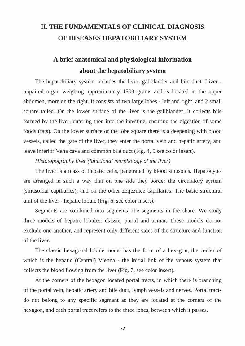
72
II. THE FUNDAMENTALS OF CLINICAL DIAGNOSIS
OF DISEASES HEPATOBILIARY SYSTEM
A brief anatomical and physiological information
about the hepatobiliary system
The hepatobiliary system includes the liver, gallbladder and bile duct. Liver -
unpaired organ weighing approximately 1500 grams and is located in the upper
abdomen, more on the right. It consists of two large lobes - left and right, and 2 small
square tailed. On the lower surface of the liver is the gallbladder. It collects bile
formed by the liver, entering then into the intestine, ensuring the digestion of some
foods (fats). On the lower surface of the lobe square there is a deepening with blood
vessels, called the gate of the liver, they enter the portal vein and hepatic artery, and
leave inferior Vena cava and common bile duct (Fig. 4, 5 see color insert).
Histotopography liver (functional morphology of the liver)
The liver is a mass of hepatic cells, penetrated by blood sinusoids. Hepatocytes
are arranged in such a way that on one side they border the circulatory system
(sinusoidal capillaries), and on the other zeljeznice capillaries. The basic structural
unit of the liver - hepatic lobule (Fig. 6, see color insert).
Segments are combined into segments, the segments in the share. We study
three models of hepatic lobules: classic, portal and acinar. These models do not
exclude one another, and represent only different sides of the structure and function
of the liver.
The classic hexagonal lobule model has the form of a hexagon, the center of
which is the hepatic (Central) Vienna - the initial link of the venous system that
collects the blood flowing from the liver (Fig. 7, see color insert).
At the corners of the hexagon located portal tracts, in which there is branching
of the portal vein, hepatic artery and bile duct, lymph vessels and nerves. Portal tracts
do not belong to any specific segment as they are located at the corners of the
hexagon, and each portal tract refers to the three lobes, between which it passes.
73
Lobules are separated from each other by a layer of connective tissue which in
humans is very poorly developed. The parenchyma of the lobules formed of radially
arranged around the Central vein of the hepatic beams, one cell thick. Penetrating
through the terminal plate of hepatocytes that separates the parenchyma of the lobules
from portal fields portal vein give the blood sinusoids, which empties into the Central
vein. Hepatic sinusoids are sphincters that regulates blood flow to the slice. Hepatic
artery-like veins break up into capillaries, which are included in the slice and on its
periphery merge with capillaries originating from the portal vein. Due to this in the
intralobular capillary network is a mixture of blood flowing from the portal vein and
hepatic artery.
Model of the portal lobule is based on the fact that liver is much more terminal
branches of the portal vein than the hepatic vein terminals. Schematically, this slice
has the shape of a triangle, the sides of which are lines connecting the Central veins
of three adjacent classic lobules hexagonal, and in the center of this figure is the
portal tract. The number of portal lobules in 2 times more than the classic.
Model acini of the liver as an independent structural unit, based on the fact that
the blood coming from the hepatic artery and portal vein, before you get to sine wave,
is sent to the branches of these vessels. These side branches will form the basis of the
hepatic acini, i.e., acini located between the two Central venules (according to the
International classification 1980). The line connecting these venules, forming acinus.
Zone acini composed of hepatic plates are arranged around the axis of the acinar.
Distribution of blood in acinose is so that in the direction from the inner (first zone)
to the outer (8 zone) are removed the substance. This is determined by the metabolic
organization of hepatocytes. Function of hepatocytes depending on their localization
in the acini. In hepatocytes the first zone to actively the processes of pinocytosis, the
uptake of nutrients from the portal blood, protein metabolism and synthesis of plasma
proteins, is excretia golevyh acids and bilirubin. In hepatocytes of zone 3 is provided
by glycolysis, glucose utilization, detoxification of ammonia.
74
Thus, only the position of the hepatic acini may a correct explanation of the
diverse metabolic and detoxification functions of the liver. Acinus is not only the
microcirculatory unit of the liver, but of secretory and. Gall ductuli are part of their
respective axial triad which occupies the centre of the acini. A complex acinus
consists of 3-4 simple acini. Blood from flowing complex acini in the terminal
hepatic venules located between the third zones of simple acini.
The intralobular alnuaimi in contact with each hepatocyte. Maximum exchange
between the blood and the hepatic parenchyma is facilitated by the absence in the
capillaries - sinusoids basal membrane, intrinsic to the capillaries of other organs, and
is constructed of a single layer of endothelial cells. Between endothelial cells and
hepatocytes have perisinusoidal space (space of disse), which is exchanged between
the blood and hepatocytes.
Sinusoidal cell functions are divided into endothelial, stellate reticulo-
endothelial cells (Kupffer cells), macrophages localized around the portal tracts. They
phagocytose immunogenic from the blood flowing from the intestine, and delay their
entrance into the General circulation.
The connective tissue of the portal tracts composed of collagen and contains
histiocytes, lymphocytes, plasma cells and fibroblasts.
Biliary system consists of the intrahepatic and extrahepatic structures.
The structure of the intrahepatic biliary system: bile capillaries (ductuli),
intercellular bile canaliculi, merging into pererabotannye bile ducts (cholangio), then
to the interlobular bile ducts (septal equipment), which form the right and left
intrahepatic ducts.
The structure of the extrahepatic biliary system: the distal segments of the right
and left intrahepatic ducts, uniting in the common hepatic duct; cystic duct, which
bile from the common hepatic duct enters the gallbladder; and the distal segment of
the common hepatic duct below the discharge from it of the cystic duct is called the
common bile duct (choledoch), opening by means common to it and the pancreatic
duct of the ampoule into the duodenum.
75
Questioning patients with the liver diseases
Main complaints
Pain is localized in the right hypochondrium and sometimes in the epigastrium
and differs depending on the cause. Pain may be persistent and dull, or it may be
severe and occur in attacks. Persistent pain is usually boring, or the patient feels
pressure, heaviness, or distension in the right hypochondrium. Pain may radiate to the
right shoulder, scapula, and in the interscapular space (in chronic cholecystitis,
perihepatitis and pericholecystitis, i.e. when the process extends onto the peritoneum
overlying the liver and the gall bladder, and also in rapid and considerable
enlargement of the liver which causes distension of Glisson's capsule). This radiation
of pain is quite characteristic of many diseases of the liver and gall bladder, because
the right phrenic nerve, innervating the capsule in the region of the falciform and the
coronary ligaments of the liver and the extrahepatic bile ducts, originates in the same
segments of the spinal cord where the nerves of the neck and shoulder originate as
well. Pain usually becomes more severe in deep breathing; in adhesion of the liver or
the gall bladder to the neighboring organs, pain is also intensified when the patient
changes his posture, and sometimes during walking.
Attacks of pain (biliary or hepatic colics) develop suddenly and soon become
quite severe and unbearable. The pain is first localized in the right hypochondrium
but then spreads over the entire abdomen to radiate upwards, to the right, and
posteriorly. An attack of pain may continue from several hours to a few days during
which pain may subside and then intensify again; the attack ends as suddenly as it
arises; or pain may lessen gradually. Attacks of pain occur mostly in cholelithiasis.
They are provoked by jolting (as in riding) or by fatty food. Pain attacks occur also in
hypermotoric dyskinesia of the gall bladder and bile ducts. Pain usually develops
quite unexpectedly due to spastic contractions of muscles of the gall bladder and
large bile ducts caused by irritation of their mucosa by a stone, and due to
comparatively rapid distension of the gallbladder in congestion of bile (e.g. due to
obstruction of the common bile duct by a stone). Warmth applied to the liver

76
(provided the attack is not attended by considerable fever) and also administration of
cholino- and myospasmolytics (atropine sulphate, papaverine hydrochloride, etc.)
remove pain characteristic of the colic. An attack of hepatic colic can be attended by
subfebrility (fever develops with pain and subsides with alleviation of pain), which is
followed by a slight transient subicteric colour of the sclera or pronounced jaundice
in obstruction of the common bile duct by a stone.
Pain developing in dyskinesia of the bile ducts is associated with upset
coordination between contractions of the gall bladder and of the Oddi sphincter under
the effect of increased tone of the vagus nerve. As a result, bile congests in the ducts,
and the gall bladder is no longer emptied. This causes its convulsive contraction.
Dyskinetic pain is characterized by the absence of signs of inflammation
(leucocytosis, ESR, etc.).
Dyspeptic complaints include decreased appetite, often bitter taste in the mouth,
eructation, nausea, vomiting, distension of the abdomen and rumbling, constipations
or diarrhea. These complaints are characteristic not only of diseases of the
hepatobiliary system but also of other parts of the digestive system. Causes of these
symptoms in diseases of the liver and bile ducts are explained by deranged secretion
of bile (and hence impaired digestion of fats in the intestine) and derangement of the
detoxicating functions of the liver.
Fever occurs in acute inflammatory affection of the gall bladder and bile ducts,
in abscess and cancer of the liver, in hepatitis, and active cirrhosis.
Itching and yellowness of the skin and mucous membranes, are the result of
accumulation of bile acids in the blood and irritation of nerve endings of the skin
(itching is usually persistent in nature, increases at night, is found in hepatic and
obstructive jaundice and the diseases that cause these syndromes, hemolytic jaundice
itching is absent) and the result of the accumulation of different kinds of bilirubin
(yellowness of the skin and mucous membranes). These symptoms occur when
gallstones, cirrhosis (especially biliary) liver, hepatitis, toxic liver damage, liver
cancer and pancreatic cancer.
77
Increase in the abdomen size, swelling complaints, which are often the
manifestations of decompensated liver disease. Abdominal enlargement may be due
to bloating or ascites, accumulation of fluid in the abdomen when cirrhosis, liver
cancer, pancreas, thrombosis of hepatic veins.
The appearance of a rash on the skin hemorrhagic, scratching, nasal, uterine and
gastrointestinal bleeding, often caused by abnormalities of the hepatic parenchyma
due to impaired synthesis of clotting factors and detoxification of the liver while
cirrhosis, toxic hepatitis.
Weight loss – as a result of violations of protein metabolism in liver disease
(cirrhosis).
Breast enlargement in men (gynecomastia) is due to violation of utilization in
the liver, excess estrogen (Fig. 8, see colour insert).
History of the present disease
When collecting anamnesis, it is necessary to find out if the patient had in his
past history jaundice or acute diseases of the liver or the gall bladder (Botkin's
disease, acute cholecystitis, cholangitis), attacks of hepatic colics, enlargement of the
liver or the spleen, which might be an early symptom of the present disease (chronic
hepatitis, liver cirrhosis, chronic cholecystitis, cholangitis, cholelithiasis).
Life history of patient
When inquiring the patient it is necessary to establish factors that might be
important for the etiology of the present disease of the liver or bile ducts: liking for
fat and meat foods, exposure to chemical and vegetable poisons (alcohol, carbon
tetrachloride, compounds of phosphorus, copper, lead, arsenic, dichloroethane, etc.),
poisoning with mushrooms containing strong hepatotropic poisons (e.g. helvellic
acid, amanitotoxin, etc.), some infectious diseases (Botkin's disease, lambliosis,
typhoid fever, malaria, syphilis, etc.), diseases of the gastro-intestinal tract (gastritis,
colitis), and diabetes mellitus. Familial predisposition is also important in the
78
development of some liver diseases (e.g. congenital benign hyperbilirubinemia) and
diseases of the gallbladder (cholelithiasis).
General examination of the patients with the liver diseases
The general condition of the patient is first assessed. In the presence of marked
functional hepatic insufficiency of various etiology (liver cirrhosis, cancer, prolonged
obstructive jaundice, etc.), the patients's condition can be grave because of
pronounced poisoning (hepatic coma). The patient's condition may be grave in acute
inflammatory diseases of the liver (abscess), gallbladder (acute cholecystitis), or bile
ducts (acute cholangitis). But in many chronic diseases of the liver and the bile ducts,
the general condition of the patient may remain satisfactory for long periods of time.
Patients with hepatic colics are restless, they toss in bed, try to find (without success)
a position in which the pain might be relieved. Hepatic coma is characterized by
deranged consciousness in the form of pronounced euphoria or inhibition to complete
loss of consciousness.
The general appearance (habitus) of the patient usually does not change. At the
same time, hypersthenic constitution with predisposition to obesity is often
characteristic of patients with cholelithiasis. Quite the reverse, significant wasting (to
cachexia) occurs in cirrhosis or malignant tumour of the liver or the bile ducts. If the
disease of the liver begins in childhood or adolescence, the patient may look infantile.
In certain cases the skin becomes pallid due to anemization (hemorrhage from
varicose esophageal or hemorrhoidal veins in portal cirrhosis); the skin may be
greyish ("dirty") in patients with some hepatic diseases. Greyish-brown or brown
skin is characteristic of hemochromatosis (bronzed diabetes or pigmentary cirrhosis
of the liver), the disease associated with primary or secondary excessive absorption
of iron in the intestine and accumulation of hemosiderin in various organs and
tissues (in the first instance in the liver and the pancreas). Local hyperpigmentation
of the skin in the right hypochondrium can be due to frequent application of a hot-
79
water bottle, which indicates persistent pain in this region (in chronic diseases of the
gallbladder).
Inspection of the skin (especially in obstructive and less frequently in
parenchymatous jaundice) can reveal scratches due to severe itching. The scratches
are often infected and purulent. Jaundice of this type can be attended by hemorrhagic
diathesis - petechial eruption and hemorrhage into the skin (ecchymosis).
Jaundice. An important diagnostic sign is jaundice of varying intensity. In
order to assess correctly the colour of the skin, the patient should be inspected in
daylight or in the light of the luminescent lamp. A subicteric symptom is jaundice of
the sclera, the lower surface of the tongue, and the soft palate; next coloured are the
palms, soles, and finally the entire skin. Inspection of the sclera helps differentiate
between true (bilirubinogenic) and exogenic jaundice. Prolonged use of quinacrine,
ethacridine lactate (rivanol), carotin (carrots), excess tangerines and oranges,
exposure to trinitrotoluene and picric acid can cause slight jaundice of the skin (false
jaundice) but the sclera is not coloured in such cases. Hepatic jaundice is usually
attended by itching and scratching of the skin.
The diversity of signs in liver disease reflects the key role that the liver plays in
homeostasis. Jaundice is a frequent sign, and it can be detected clinically when the
serum bilirubin level rises above 50 pmol/litre (Fig. 9, see colour insert).
In haemolytic states the pigment circulates attached to albumin and does
not appear in the urine - it usually imparts a pale yellow colour to the skin and
sclerae.
In hepatocellular and obstructive jaundice the conjugated bilirubin
accumulates to very high levels and may give a much darker colour to the skin and
sclerae, which may become orange or greenish in colour. Mild jaundice is often most
evident in the sclerae, and may be unaccompanied by obvious jaundice in the skin.
This jaundice results from an elevated level of conjugated bilirubin, which produces a
deeper yellow colour than unconjugated bilirubin. The high level of conjugated
bilirubin, maintained over a long period, e.g. in primary biliary cirrhosis (PBC), gives
80
a characteristic dark brown-orange pigmentation to the skin and sclerae. Patients with
PBC usually develop large xanthelasmata and corneal arcus as a consequence of
disordered lipid metabolism.
Other yellow pigmentation of skin, which may mimic jaundice, follows
mepacrine ingestion or the excessive ingestion of carotenes, but these do not colour
the sclerae. Pruritus may result from retained bile salts in cholestatic disorders, and it
may appear before the onset of frank jaundice.
Scratch marks may be present in accessible skin areas.
Palmar erythema is a red flushing on the thenar and hypothenar eminences
(Fig. 10, see colour insert). Palmar erythema is a common finding in chronic liver
disease, but is also found in pregnancy, during oral contraceptive use, in rheumatoid
arthritis and in thyrotoxicosis. It may also occur without apparent cause. This is
common but not specific to liver disease. Similar changes may also be found in the
soles of the feet.
Loss of body hair, including pubic and axillary hair, and testicular atrophy are
also common.
Finger clubbing is a common feature of liver disease and may also involve the
toes; it is nonspecific, being also found in respiratory, cardiac, alimentary and
endocrine diseases (Fig. 11, see colour insert).
White nails: the cause is unknown but their whiteness mirrors the severity of
the liver disease. White nails are also found in other conditions in which the serum
albumin is low.
Spontaneous bruising and excessive bleeding are a reflection of the failure of
the liver to synthesize coagulation factors II, VII, IX and X, often compounded by the
failure to absorb vitamin K, as a result of retention of bile salts. Disturbance of
coagulation mechanisms is a common problem in chronic liver disease, and the risk
of excessive bleeding should always be assessed by coagulation studies before liver
biopsy or other operative procedures.
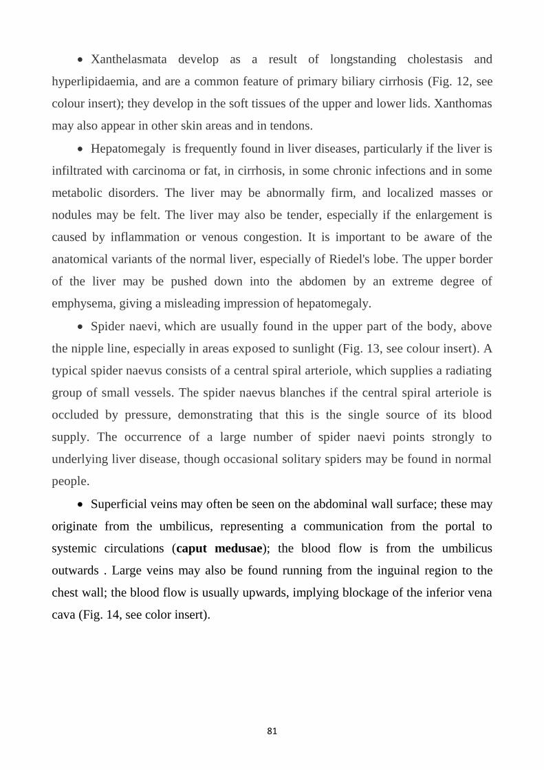
81
Xanthelasmata develop as a result of longstanding cholestasis and
hyperlipidaemia, and are a common feature of primary biliary cirrhosis (Fig. 12, see
colour insert); they develop in the soft tissues of the upper and lower lids. Xanthomas
may also appear in other skin areas and in tendons.
Hepatomegaly is frequently found in liver diseases, particularly if the liver is
infiltrated with carcinoma or fat, in cirrhosis, in some chronic infections and in some
metabolic disorders. The liver may be abnormally firm, and localized masses or
nodules may be felt. The liver may also be tender, especially if the enlargement is
caused by inflammation or venous congestion. It is important to be aware of the
anatomical variants of the normal liver, especially of Riedel's lobe. The upper border
of the liver may be pushed down into the abdomen by an extreme degree of
emphysema, giving a misleading impression of hepatomegaly.
Spider naevi, which are usually found in the upper part of the body, above
the nipple line, especially in areas exposed to sunlight (Fig. 13, see colour insert). A
typical spider naevus consists of a central spiral arteriole, which supplies a radiating
group of small vessels. The spider naevus blanches if the central spiral arteriole is
occluded by pressure, demonstrating that this is the single source of its blood
supply. The occurrence of a large number of spider naevi points strongly to
underlying liver disease, though occasional solitary spiders may be found in normal
people.
Superficial veins may often be seen on the abdominal wall surface; these may
originate from the umbilicus, representing a communication from the portal to
systemic circulations (caput medusae); the blood flow is from the umbilicus
outwards . Large veins may also be found running from the inguinal region to the
chest wall; the blood flow is usually upwards, implying blockage of the inferior vena
cava (Fig. 14, see color insert).
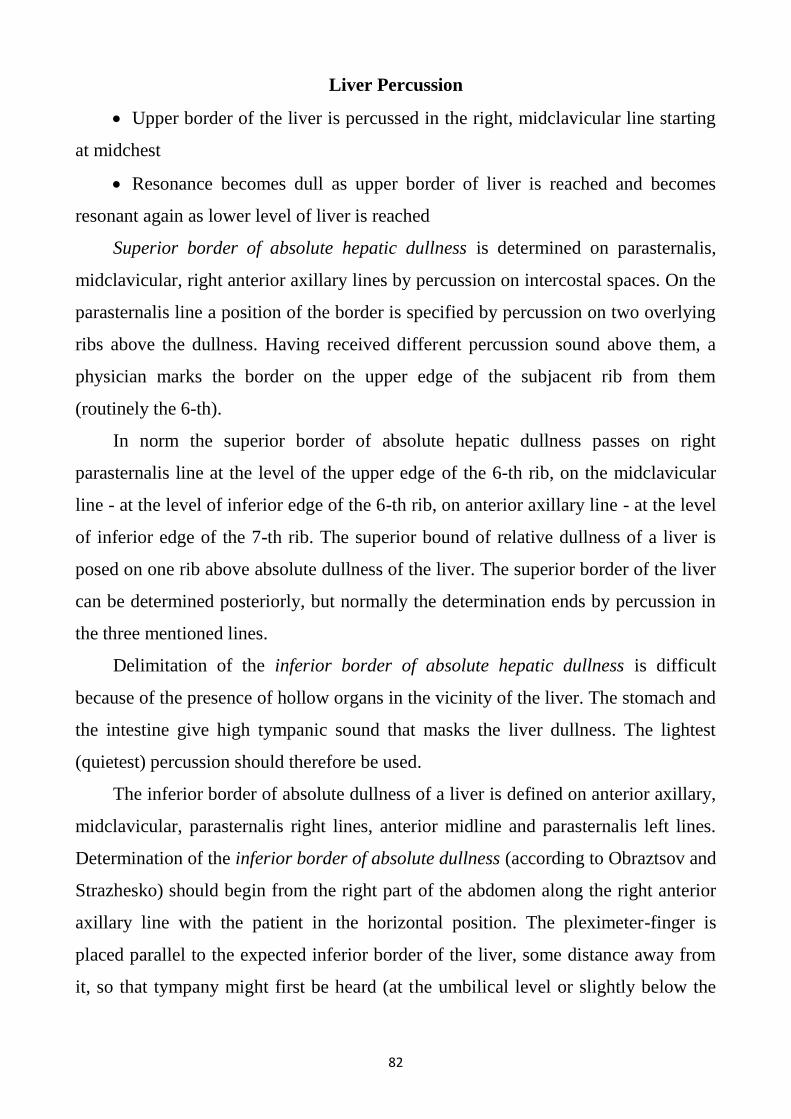
82
Liver Percussion
Upper border of the liver is percussed in the right, midclavicular line starting
at midchest
Resonance becomes dull as upper border of liver is reached and becomes
resonant again as lower level of liver is reached
Superior border of absolute hepatic dullness is determined on parasternalis,
midclavicular, right anterior axillary lines by percussion on intercostal spaces. On the
parasternalis line a position of the border is specified by percussion on two overlying
ribs above the dullness. Having received different percussion sound above them, a
physician marks the border on the upper edge of the subjacent rib from them
(routinely the 6-th).
In norm the superior border of absolute hepatic dullness passes on right
parasternalis line at the level of the upper edge of the 6-th rib, on the midclavicular
line - at the level of inferior edge of the 6-th rib, on anterior axillary line - at the level
of inferior edge of the 7-th rib. The superior bound of relative dullness of a liver is
posed on one rib above absolute dullness of the liver. The superior border of the liver
can be determined posteriorly, but normally the determination ends by percussion in
the three mentioned lines.
Delimitation of the inferior border of absolute hepatic dullness is difficult
because of the presence of hollow organs in the vicinity of the liver. The stomach and
the intestine give high tympanic sound that masks the liver dullness. The lightest
(quietest) percussion should therefore be used.
The inferior border of absolute dullness of a liver is defined on anterior axillary,
midclavicular, parasternalis right lines, anterior midline and parasternalis left lines.
Determination of the inferior border of absolute dullness (according to Obraztsov and
Strazhesko) should begin from the right part of the abdomen along the right anterior
axillary line with the patient in the horizontal position. The pleximeter-finger is
placed parallel to the expected inferior border of the liver, some distance away from
it, so that tympany might first be heard (at the umbilical level or slightly below the
83
navel). As the pleximeter-finger is then moved upwards, tympany is followed by
absolute dullness. The point of disappearance of tympany is marked in each vertical
line on the inferior edge of the pleximeter-finger.
When determining the left border of liver dullness, the pleximeter-finger is
placed perpendicularly to the edge of the left costal arch, at the level of the 8-9-th
ribs, and percussion is carried out to the right, directly over the edge of the costal
arch, to the point where tympany changes to dullness (in the region of Traube's
space).
Normally the inferior border of absolute dullness of a lying patient with
normosthenic chest passes at the level of upper edge of 10-th rib in the right anterior
axillary line, at the inferior edge of the right arch in the midclavicular line, 2 cm below
the interior edge of the right costal arch in the right parasternal line, and 3-6 cm away
from the inferior edge of the xiphoid process (at the border of the upper third of the
distance from the base of the xiphoid process to the navel) on the anterior median line;
on the left parasternalis line - at the level of the inferior edge of a costal arch.
The lower margin of the liver in norm can be very depending on the shape of the
chest and constitution of the patient, but it has only effect on the position in the
anterior median line. The lower margin of the liver in a hypersthenic chest is slightly
above the mentioned level, while in an asthenic chest below it, approximately
midway between the base of the xiphoid process and the navel. If the patient is in the
upright posture, the lower margin of the liver descends 1-1.5 cm. If the liver is
enlarged, its lower margin is measured in centimeters from the costal arch and the
xiphoid process.
When you apply percussion of the liver according to M. G. Kurlovu estimated
its size, which allows to identify hepatomegaly. In a healthy person the dimensions of
a liver on Kurlovu: on the midclavicular line - 9 ± 1 cm along the median line of 8 ±
1 cm along the left costal arch 7 ± 1 cm (Fig. 18).
84
Fig. 18. Percussing liver spans
8±1
9±1
7±1 cm along
the left rib
arch
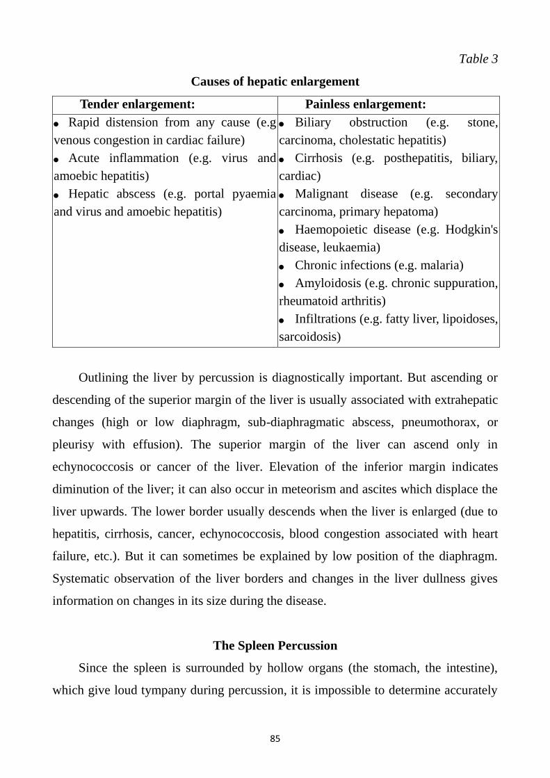
85
Table 3
Causes of hepatic enlargement
Tender enlargement: Painless enlargement:
Rapid distension from any cause (e.g
venous congestion in cardiac failure)
Acute inflammation (e.g. virus and
amoebic hepatitis)
Hepatic abscess (e.g. portal pyaemia
and virus and amoebic hepatitis)
Biliary obstruction (e.g. stone,
carcinoma, cholestatic hepatitis)
Cirrhosis (e.g. posthepatitis, biliary,
cardiac)
Malignant disease (e.g. secondary
carcinoma, primary hepatoma)
Haemopoietic disease (e.g. Hodgkin's
disease, leukaemia)
Chronic infections (e.g. malaria)
Amyloidosis (e.g. chronic suppuration,
rheumatoid arthritis)
Infiltrations (e.g. fatty liver, lipoidoses,
sarcoidosis)
Outlining the liver by percussion is diagnostically important. But ascending or
descending of the superior margin of the liver is usually associated with extrahepatic
changes (high or low diaphragm, sub-diaphragmatic abscess, pneumothorax, or
pleurisy with effusion). The superior margin of the liver can ascend only in
echynococcosis or cancer of the liver. Elevation of the inferior margin indicates
diminution of the liver; it can also occur in meteorism and ascites which displace the
liver upwards. The lower border usually descends when the liver is enlarged (due to
hepatitis, cirrhosis, cancer, echynococcosis, blood congestion associated with heart
failure, etc.). But it can sometimes be explained by low position of the diaphragm.
Systematic observation of the liver borders and changes in the liver dullness gives
information on changes in its size during the disease.
The Spleen Percussion
Since the spleen is surrounded by hollow organs (the stomach, the intestine),
which give loud tympany during percussion, it is impossible to determine accurately
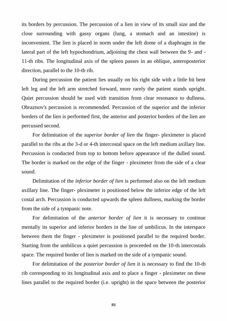
86
its borders by percussion. The percussion of a lien in view of its small size and the
close surrounding with gassy organs (lung, a stomach and an intestine) is
inconvenient. The lien is placed in norm under the left dome of a diaphragm in the
lateral part of the left hypochondrium, adjoining the chest wall between the 9- and -
11-th ribs. The longitudinal axis of the spleen passes in an oblique, anteroposterior
direction, parallel to the 10-th rib.
During percussion the patient lies usually on his right side with a little bit bent
left leg and the left arm stretched forward, more rarely the patient stands upright.
Quiet percussion should be used with transition from clear resonance to dullness.
Obraztsov's percussion is recommended. Percussion of the superior and the inferior
borders of the lien is performed first, the anterior and posterior borders of the lien are
percussed second.
For delimitation of the superior border of lien the finger- pleximeter is placed
parallel to the ribs at the 3-d or 4-th intercostal space on the left medium axillary line.
Percussion is conducted from top to bottom before appearance of the dulled sound.
The border is marked on the edge of the finger - pleximeter from the side of a clear
sound.
Delimitation of the inferior border of lien is performed also on the left medium
axillary line. The finger- pleximeter is positioned below the inferior edge of the left
costal arch. Percussion is conducted upwards the spleen dullness, marking the border
from the side of a tympanic note.
For delimitation of the anterior border of lien it is necessary to continue
mentally its superior and inferior borders in the line of umbilicus. In the interspace
between them the finger - pleximeter is positioned parallel to the required border.
Starting from the umbilicus a quiet percussion is proceeded on the 10-th intercostals
space. The required border of lien is marked on the side of a tympanic sound.
For delimitation of the posterior border of lien it is necessary to find the 10-th
rib corresponding to its longitudinal axis and to place a finger - pleximeter on these
lines parallel to the required border (i.e. upright) in the space between the posterior
87
axillary and scapular lines. Percussion is performed immediately on the 10-th rib
before appearance of a dulled sound. The posterior border of lien is marked from the
side of a tympanic sound.
Normally the superior border of the splenic dullness corresponds to the lower
edge of IX rib, inferior border - to the lower edge of XI ribs. The anterior border of
the splenic dullness is on 1-2 sm outside of anterior axillary line, the posterior
border – on the posterior axillary line.
The measurement of the lines bridging the superior and inferior, anterior and
posterior borders of splenic dullness gives conception about size of lien. Its width is
4—6 cm, its length is 6-8 sm (Fig. 19).
а б
c
Fig. 19. Percussion of the spleen on Kurlovu: a) the position of the finger – plessimeter in
determining the upper and lower limits, b) the anterior and posterior boundaries, c) normal sizes
Liver Palpation
Surface palpation in diseases of the liver can reveal a tender zone in the right
hypochondrium and epigastrium. Especially severe local pain (caused even by a
slight touch on the anterior abdominal wall in the zone overlying the gallbladder) is
observed in acute cholecystitis and biliary colic. In chronic cholecystitis slight or
moderate tenderness is only revealed at the point of projection of the gall bladder
fundus onto the anterior abdominal wall. In healthy subjects this point is found
88
immediately below the right costal arch by the lateral edge of the right rectus
abdominis muscle.
Palpation of a liver purposes detection of the inferior edge, definition of its
localization, form, lineament, consistence, character of surface and tenderness. Percussion
of hepatic inferior borders on all lines foreruns always to palpation of the liver.
The liver is palpated by the Obraztsov and Strazhesko method. As the lower edge
of the liver descends to meet the examining fingers during a deep inspiration it slides
over the fingers and thus becomes detectable. It should be remembered that the
respiratory mobility of the liver is the highest compared with that of the other
abdominal organs because the liver is the closest to the diaphragm. It follows therefore
that during palpation of the liver, the active role belongs to its respiratory mobility
rather than to the palpating fingers (as is the case with palpation of the intestine).
Position of the patient. The patient should lay horizontally with slightly raised
head and the stretched legs. The hands routinely settle down along a trunk or are
crossed on a chest with the purpose of restriction of mobility of a chest in the sides on
an inspiration. It promotes increase of diaphragm motility according to a liver in the
upper-inferior direction that is important for a palpation of a lower edge of a liver.
The patient should stand or lie during palpation of the liver and the gall bladder.
But in certain cases the liver can be easier palpated if the patient lies on his left side:
the liver hangs by gravity from under the hypochondrium and its inferio-anterior edge
can thus be better palpated.
Position of the doctor. The examiner sits by the right side, facing the patient. He
places four fingers of his left hand on the right costal arch of the patient chest and
uses his left thumb to press on the costal arch to move the liver closer to the palpating
fingers of the right hand and to prevent expansion of the chest during inspiration. It
stimulates greater excursions of the right cupula of diaphragm. The palm of the right
hand is placed flat on the abdomen below the costal arch between the right
parasternalis and midclavicular lines. The slightly flexed fingers press lightly on the
abdominal wall.
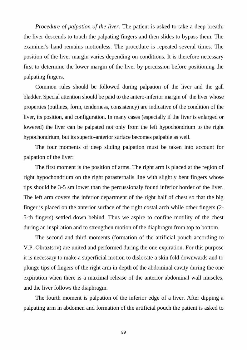
89
Procedure of palpation of the liver. The patient is asked to take a deep breath;
the liver descends to touch the palpating fingers and then slides to bypass them. The
examiner's hand remains motionless. The procedure is repeated several times. The
position of the liver margin varies depending on conditions. It is therefore necessary
first to determine the lower margin of the liver by percussion before positioning the
palpating fingers.
Common rules should be followed during palpation of the liver and the gall
bladder. Special attention should be paid to the antero-inferior margin of the liver whose
properties (outlines, form, tenderness, consistency) are indicative of the condition of the
liver, its position, and configuration. In many cases (especially if the liver is enlarged or
lowered) the liver can be palpated not only from the left hypochondrium to the right
hypochondrium, but its superio-anterior surface becomes palpable as well.
The four moments of deep sliding palpation must be taken into account for
palpation of the liver:
The first moment is the position of arms. The right arm is placed at the region of
right hypochondrium on the right parasternalis line with slightly bent fingers whose
tips should be 3-5 sm lower than the percussionaly found inferior border of the liver.
The left arm covers the inferior department of the right half of chest so that the big
finger is placed on the anterior surface of the right costal arch while other fingers (2-
5-th fingers) settled down behind. Thus we aspire to confine motility of the chest
during an inspiration and to strengthen motion of the diaphragm from top to bottom.
The second and third moments (formation of the artificial pouch according to
V.P. Obraztsov) are united and performed during the one expiration. For this purpose
it is necessary to make a superficial motion to dislocate a skin fold downwards and to
plunge tips of fingers of the right arm in depth of the abdominal cavity during the one
expiration when there is a maximal release of the anterior abdominal wall muscles,
and the liver follows the diaphragm.
The fourth moment is palpation of the inferior edge of a liver. After dipping a
palpating arm in abdomen and formation of the artificial pouch the patient is asked to
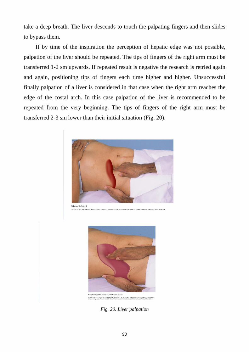
90
take a deep breath. The liver descends to touch the palpating fingers and then slides
to bypass them.
If by time of the inspiration the perception of hepatic edge was not possible,
palpation of the liver should be repeated. The tips of fingers of the right arm must be
transferred 1-2 sm upwards. If repeated result is negative the research is retried again
and again, positioning tips of fingers each time higher and higher. Unsuccessful
finally palpation of a liver is considered in that case when the right arm reaches the
edge of the costal arch. In this case palpation of the liver is recommended to be
repeated from the very beginning. The tips of fingers of the right arm must be
transferred 2-3 sm lower than their initial situation (Fig. 20).
Fig. 20. Liver palpation
91
The lower edge of a normal liver is usually palpated between the right
parasternal and midclavicular line; the liver is impalpable to the right of the
midclavicular line because it is located behind the costal arch; the liver is hardly
palpable to the left of the line because of the abdominal muscles. An enlarged or
consolidated liver can be palpated in all lines. It is easily to perform a palpation on
the right parasternalis line as here the inferior edge of a liver settles down in standard
conditions on 2 sm of below costal arch. On a right midclavicular line it is as a rule at
a level of a costal arch.
According to Obraztsov, normal liver can be palpated in 88 per cent of cases.
Physical properties of the liver can be determined by palpating its lower edge (it can
be soft, firm, rough, sharp, rounded, tender, etc.). The margin of an unaffected liver
palpated at the height of a deep inspiration is 1—2 cm below the costal arch. It is soft,
sharp or slightly rounded under the form, readily bending, smooth and insensitive.
The liver of patients with pronounced distension of the abdomen should be examined
with the empty stomach to facilitate palpation. In accumulation of much fluid in the
abdominal cavity (ascites) the liver is not always palpable if the patient is lying. The
patient should then be examined in the erect position, or he may lie on his left side. If the
amount of fluid in the abdomen is very large, it should be released by paracentesis.
Expressed accumulation of fluid in an abdominal cavity (ascites) often very
much complicates carrying out of a palpation of a liver on V.P. Obraztsov. In
accumulation of much fluid in the abdominal cavity, ballotment should be used to
palpate the liver. To that end the right hand (two or four flexed fingers) should be
placed on the lower right part of the abdomen, perpendicularly to the expected lower
edge of the liver. The abdominal wall is given a sharp tap from the palpating fingers
which move upward to meet the firm object, the liver, which is first tossed to the
deeper parts of the abdominal cavity but is then returned back to strike the fingers (a
sign "floating ice").
Palpation is painful if the liver is inflamed and the affection extends onto the
liver capsule; the liver is also tender when it is distended (e.g. in blood congestion
92
due to heart failure). The liver of a healthy subject (if it is accessible to palpation) is
soft; it becomes firmer in hepatitis, hepatosis, and cardiac congestion. The liver is
especially firm in cirrhosis. Its edge becomes sharp and the surface smooth or
covered with small tubercles. The liver is also firm in the presence of tumour and
multiple metastases of cancer. Its surface then becomes covered with rough tubercles
(surface metastases) and the lower margin is rough. The liver is firm in amyloidosis.
Comparatively small tumours and echinococcosis can sometimes be palpated.
Protrusion of the lower margin of an enlarged liver is assessed with respect to the
costal arch in the right anterior axillary line, right midclavicular line, right parasternal
line, anterior median line, and left parasternal line. Palpation verifies the findings
obtained by percussion of the liver.
The gallbladder cannot be palpated in healthy subjects because of its soft
consistency and the insignificant protrusion. But if the gallbladder is enlarged
(hydrops, stones in the bladder, cancer, etc.) it becomes palpable. The position of the
patient for palpation of the gallbladder is the same as in palpation of the liver. After
the margin of the liver has been found, the gall bladder should be palpated at the
lateral edge of the right rectus abdominis muscle. The palpation technique is the same
as that for palpation of the liver. The gallbladder can easier be found by moving the
palpating fingers in the direction perpendicular to the axis of the gallbladder. The
bladder is felt like a pear of variable size, firmness and tenderness depending on the
character of pathology in the gallbladder proper or the surrounding organs (e.g. the
gallbladder is enlarged, soft, and elastic in tumour-obstructed bile duct: Courvoisier-
Terrier sign; the bladder is firm and tuberous in the presence of newgrowths in its
wall, in overfilling with stones, in inflammation of the wall, etc.). An enlarged
gallbladder is mobile during respiration (it performs lateral pendulum-like
movements). The gallbladder loses its mobility in inflammation of the overlying
peritoneum (pericholecystitis). In the presence of cholecystitis and cholelithiasis, the
palpation is difficult because of sharp pain and reflectory rigidity of the muscles of
the anterior abdominal wall.
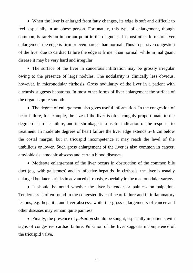
93
When the liver is enlarged from fatty changes, its edge is soft and difficult to
feel, especially in an obese person. Fortunately, this type of enlargement, though
common, is rarely an important point in the diagnosis. In most other forms of liver
enlargement the edge is firm or even harder than normal. Thus in passive congestion
of the liver due to cardiac failure the edge is firmer than normal, while in malignant
disease it may be very hard and irregular.
The surface of the liver in cancerous infiltration may be grossly irregular
owing to the presence of large nodules. The nodularity is clinically less obvious,
however, in micronodular cirrhosis. Gross nodularity of the liver in a patient with
cirrhosis suggests hepatoma. In most other forms of liver enlargement the surface of
the organ is quite smooth.
The degree of enlargement also gives useful information. In the congestion of
heart failure, for example, the size of the liver is often roughly proportionate to the
degree of cardiac failure, and its shrinkage is a useful indication of the response to
treatment. In moderate degrees of heart failure the liver edge extends 5- 8 cm below
the costal margin, but in tricuspid incompetence it may reach the level of the
umbilicus or lower. Such gross enlargement of the liver is also common in cancer,
amyloidosis, amoebic abscess and certain blood diseases.
Moderate enlargement of the liver occurs in obstruction of the common bile
duct (e.g. with gallstones) and in infective hepatitis. In cirrhosis, the liver is usually
enlarged but later shrinks in advanced cirrhosis, especially in the macronodular variety.
It should be noted whether the liver is tender or painless on palpation.
Tenderness is often found in the congested liver of heart failure and in inflammatory
lesions, e.g. hepatitis and liver abscess, while the gross enlargements of cancer and
other diseases may remain quite painless.
Finally, the presence of pulsation should be sought, especially in patients with
signs of congestive cardiac failure. Pulsation of the liver suggests incompetence of
the tricuspid valve.
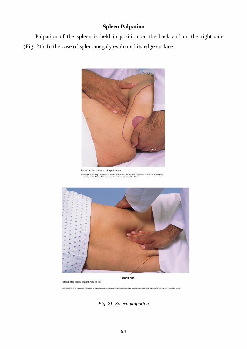
94
Spleen Palpation
Palpation of the spleen is held in position on the back and on the right side
(Fig. 21). In the case of splenomegaly evaluated its edge surface.
Fig. 21. Spleen palpation
95
Again, with the left hand, reach over and round the patient to support and press
forward the lower left rib cage
With your right hand below the left costal margin, press in toward the spleen
Again, begin palpation low so you don’t miss an enlarged spleen
Again ask the patient to take a deep breath and try to feel the tip of the spleen as
it comes down to meet your fingertips
The four degrees of enlarged lien are distinguished:
I degree - lien protrudes from under the left costal arch not more than the width
of one patient’s finger; II degree - lien reaches the middle of distance between the
umbilicus and the left costal arch; III degree - lien reaches the midline of the
abdomen, i.e. occupies only the left half of the
abdomen; IV degree - lien reaches to the right half of the abdominal cavity and
the pelvic cavity.
The characteristic peculiarity of lien is one or several notches (incisures) on the
anterior edge of the spleen can be palpated if its enlargement is considerable. The
notches are used to identify the spleen (to differentiate it from other organs, e.g. from
the left kidney, tumors originated from the left kidney, splendid curvature of a
transverse colon and caudal part of pancreas).
A normal spleen is impalpable. It can only be palpated in rare cases of extreme
splenoptosis, and more frequently in enlargement of the organ. The anterior surface
of the enlarged spleen emerges from under the costal arch and also becomes palpable.
The spleen is enlarged in some acute and chronic infectious diseases (typhus,
viral hepatitis, sepsis, malaria, etc.), in liver cirrhosis, thrombosis or compression of
the splenic vein, and also in many diseases of the hemopoietic system (hemolytic
anemia, thrombocytopenic purpura, acute and chronic leucosis). Considerable
enlargement of the spleen is called splenomegaly. The greatest enlargement of the
spleen is observed at the terminal stage of chronic myeloleucosis: it often occupies
the entire left part of the abdomen, while its lower pole is found in the small pelvis.
96
The spleen is not firm in acute infectious diseases; it is especially soft (the
consistency of dough) in sepsis. In chronic infectious diseases, liver cirrhosis, and
leucosis the spleen is firm, especially in amyloidosis.
In most diseases the spleen is insensitive to palpation. It becomes tender in
infarction, perisplenitis, and in distension of the capsule, due to the rapid
enlargement, e.g. in venous blood congestion due to thrombosis of the splenic vein.
The spleen surface is usually smooth; the edges and the surface are irregular in
perisplenitis and old infarctions (depressions in the surface). In syphilitic gummas,
echinococcosis, cysts and very rare tumours of the spleen its surface is tuberous.
The spleen is normally quite mobile, but the mobility becomes limited in
perisplenitis. A markedly enlarged spleen remains motionless during respiration but it
can however be displaced by the palpating fingers.
Assessing Possible Ascites
Ascites is the accumulation of free fluid in the peritoneal cavity. The most
common cause of ascites is the onset of liver failure, with resulting
hypoalbuminaemia and portal hypertension. The mechanism is complex: low serum
albumin, prostaglandins, atrial natriuretic factor, secondary hyperaldosteronism and
venous pressure all play a role. The diagnosis is usually obvious if the condition is
gross, but it should be differentiated from other causes of abdominal swelling (fat,
fluid, faeces, fetus, fibroids, etc.). Peripheral oedema is a common accompaniment of
ascites and is gravitational. There may also be bilateral hydrothorax.
The abdomen can be enlarged significantly due to accumulation of free fluid
(ascites). When the patient with ascites stands erect, his abdomen becomes pendulous
due to the downward flow of fluid; in the lying position the abdomen is flattened
(“frog belly”). The navel often becomes protruded in ascites when the patient stands
erect. It is due to increased infra-abdominal pressure. This sign can be used to
differentiate between enlargement of the abdomen in ascites (also large
intraabdominal tumours) and pronounced obesity (the navel is retracted).
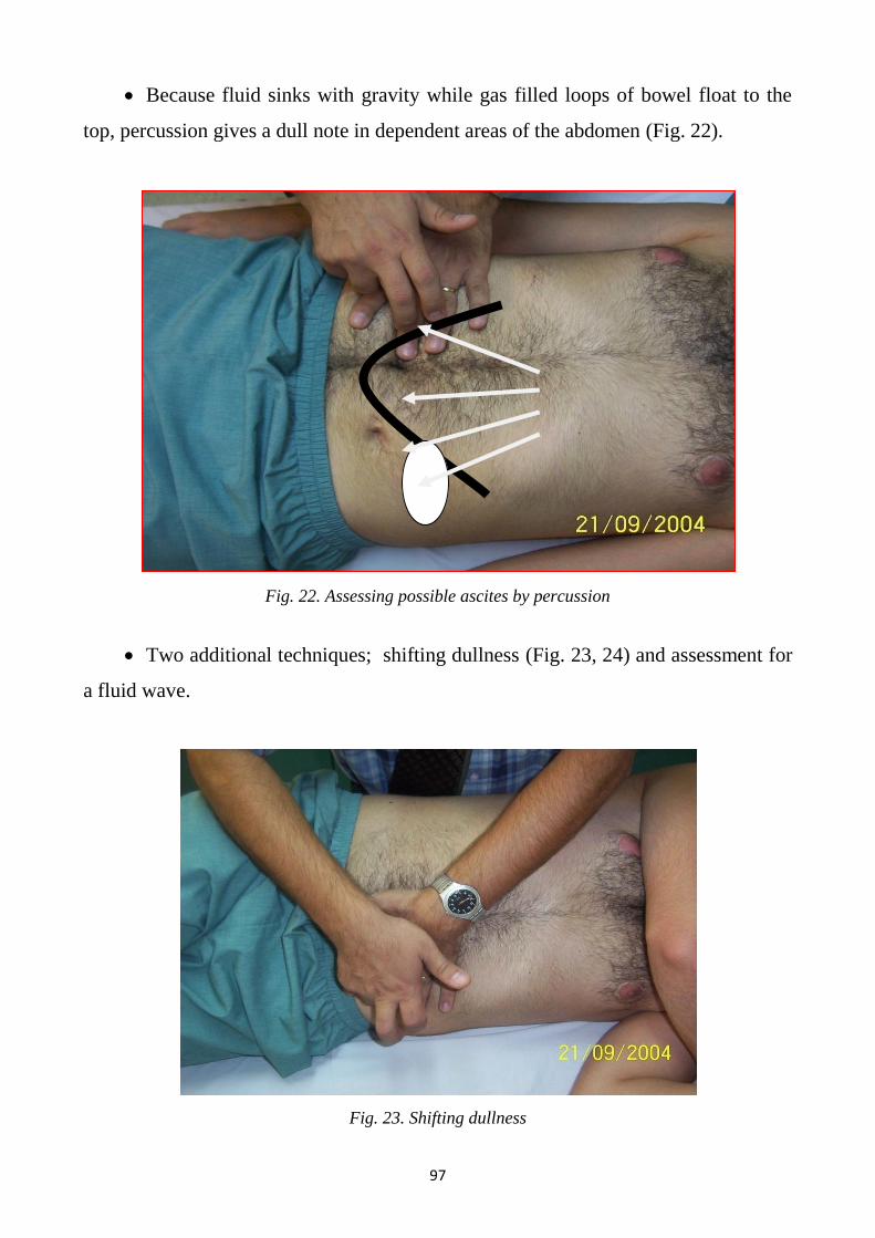
97
Because fluid sinks with gravity while gas filled loops of bowel float to the
top, percussion gives a dull note in dependent areas of the abdomen (Fig. 22).
Fig. 22. Assessing possible ascites by percussion
Two additional techniques; shifting dullness (Fig. 23, 24) and assessment for
a fluid wave.
Fig. 23. Shifting dullness
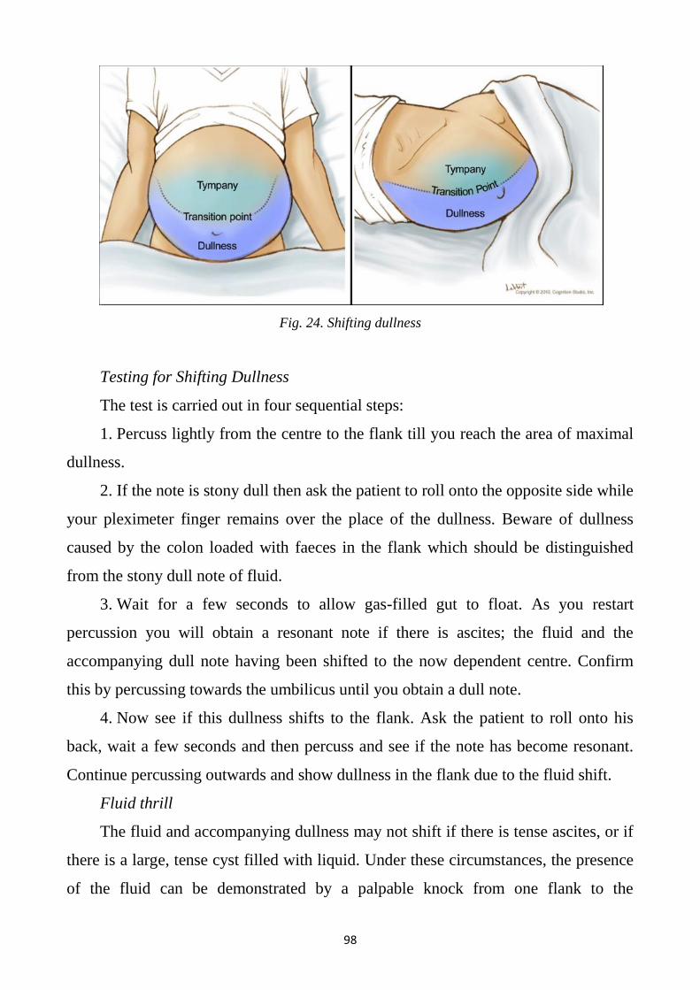
98
Fig. 24. Shifting dullness
Testing for Shifting Dullness
The test is carried out in four sequential steps:
1. Percuss lightly from the centre to the flank till you reach the area of maximal
dullness.
2. If the note is stony dull then ask the patient to roll onto the opposite side while
your pleximeter finger remains over the place of the dullness. Beware of dullness
caused by the colon loaded with faeces in the flank which should be distinguished
from the stony dull note of fluid.
3. Wait for a few seconds to allow gas-filled gut to float. As you restart
percussion you will obtain a resonant note if there is ascites; the fluid and the
accompanying dull note having been shifted to the now dependent centre. Confirm
this by percussing towards the umbilicus until you obtain a dull note.
4. Now see if this dullness shifts to the flank. Ask the patient to roll onto his
back, wait a few seconds and then percuss and see if the note has become resonant.
Continue percussing outwards and show dullness in the flank due to the fluid shift.
Fluid thrill
The fluid and accompanying dullness may not shift if there is tense ascites, or if
there is a large, tense cyst filled with liquid. Under these circumstances, the presence
of the fluid can be demonstrated by a palpable knock from one flank to the
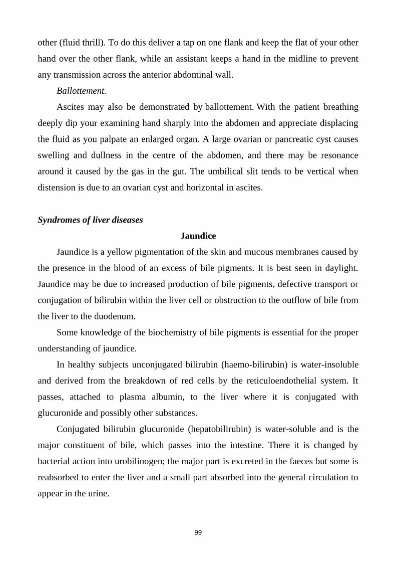
99
other (fluid thrill). To do this deliver a tap on one flank and keep the flat of your other
hand over the other flank, while an assistant keeps a hand in the midline to prevent
any transmission across the anterior abdominal wall.
Ballottement.
Ascites may also be demonstrated by ballottement. With the patient breathing
deeply dip your examining hand sharply into the abdomen and appreciate displacing
the fluid as you palpate an enlarged organ. A large ovarian or pancreatic cyst causes
swelling and dullness in the centre of the abdomen, and there may be resonance
around it caused by the gas in the gut. The umbilical slit tends to be vertical when
distension is due to an ovarian cyst and horizontal in ascites.
Syndromes of liver diseases
Jaundice
Jaundice is a yellow pigmentation of the skin and mucous membranes caused by
the presence in the blood of an excess of bile pigments. It is best seen in daylight.
Jaundice may be due to increased production of bile pigments, defective transport or
conjugation of bilirubin within the liver cell or obstruction to the outflow of bile from
the liver to the duodenum.
Some knowledge of the biochemistry of bile pigments is essential for the proper
understanding of jaundice.
In healthy subjects unconjugated bilirubin (haemo-bilirubin) is water-insoluble
and derived from the breakdown of red cells by the reticuloendothelial system. It
passes, attached to plasma albumin, to the liver where it is conjugated with
glucuronide and possibly other substances.
Conjugated bilirubin glucuronide (hepatobilirubin) is water-soluble and is the
major constituent of bile, which passes into the intestine. There it is changed by
bacterial action into urobilinogen; the major part is excreted in the faeces but some is
reabsorbed to enter the liver and a small part absorbed into the general circulation to
appear in the urine.
100
Prehepatic (haemolytic) jaundice
This form of jaundice is due to the presence in the blood of an excess of
unconjugated bilirubin. Although haemolysis is the most important cause of
prehepatic jaundice, it is now recognized that about 1% of the population have a mild
unconjugated hyperbilirubinaemia of an entirely benign nature - Gilbert's syndrome.
The jaundice is often not clinically detectable, but may deepen during fasting or
intercurrent illness,resulting in a mistaken diagnosis of hepatitis.
Haemolytic jaundice may result from an inherited abnormality in the red cells or
from acquired causes. Since these forms of haemolysis are usually accompanied by
anaemia, they are dealt with in a chapter on anaemias. Sometimes a breakdown of red
cells, as in gross pulmonary infarction or incompatible blood transfusion, causes
prehepatic jaundice without anaemia.
When the red cells themselves are abnormal, as in hereditary spherocytosis,
thalassaemia and to a lesser extent in pernicious anaemia, the cells may become
osmotically and mechanically more fragile and are thus destroyed by the
reticuloendothelial system. There may be a history of previous attacks of jaundice or
a family history of jaundice. Auto-antibodies, neoplasia and certain virus infections
may similarly cause acquired haemolytic jaundice.
In most forms of prehepatic jaundice, the skin and mucosae are delicately
jaundiced (a lemon-yellow tint), but the urine and faeces remain normal in colour,
though the urine may darken on standing due to oxidation of the excess urobilinogen.
Hepatocellular jaundice
This results from damage to the liver parenchyma interfering with the transport
or conjugation of bi-lirubin and sometimes with its excretion through the canaliculi.
The commonest cause of hepatocellular jaundice is a virus hepatitis, so that a
history of transfusion, contact with another case or, in hospital workers, contact with
the blood of a carrier may be obtained. The possibility of exposure to a medicinal
liver toxin, such as chlorpromazine, testosterone, halothane or rifampicin, or an
industrial one such as carbon tetrachloride, should always be considered.
101
Hepatocellular jaundice also occurs in congestive cardiac failure and in the later
stages of cirrhosis. When hepatic damage is accompanied by obstruction to the bile
canaliculi (cholestat-ic jaundice), the characteristics of the jaundice itself are similar
to those described under post-hepatic obstruction. The history of events preceding the
jaundice, notably the prodromal period of anorexia and nausea in virus hepatitis,
helps to differentiate the hepatocel-lular and posthepatic varieties . Liver function
tests may also be helpful.
Posthepatic (obstructive) jaundice
This form of jaundice results from obstruction to the bile ducts outside the liver.
The common causes include gallstones, primary carcinoma of the head of pancreas or
bile ducts, and secondary carcinomatous masses in the porta hepatis. When the
obstruction is due to gallstones the jaundice is usually preceded by biliary colic and
may be intermittent. Jaundice due to carcinoma tends to be insidious in onset and
progressive in its course, and the gallbladder is sometimes palpable.
Obstructive jaundice varies in intensity from a slight yellowish tinge in the skin
and mucous membranes to a pronounced canary yellow, or, in longstanding cases, a
dark greenish-yellow discoloration. It affects the skin of the whole body, but is most
marked on the trunk and proximal parts of the limbs.
Even before the skin is affected, the yellowing is seen in the mucous membranes
and should be sought in the con-junctivae and soft palate. Intolerable itching is
common and is probably due to bile salts, as it may precede the actual pigmentation
of the skin and mucosae.
The excess of bile pigments (conjugated bilirubin) in the blood leads to their
appearance in the urine, which may be visibly bile-stained or in which bile may be
detected by special tests. The lack of the normal flow of bile into the duodenum
deprives the faeces of one of their colouring constituents and further interferes with
the digestion and absorption of fats because of the lack of bile salts. As a result, the
faeces have a lighter colour than normal and are often clay-coloured. In complete
obstruction, urobilinogen is absent from the urine.
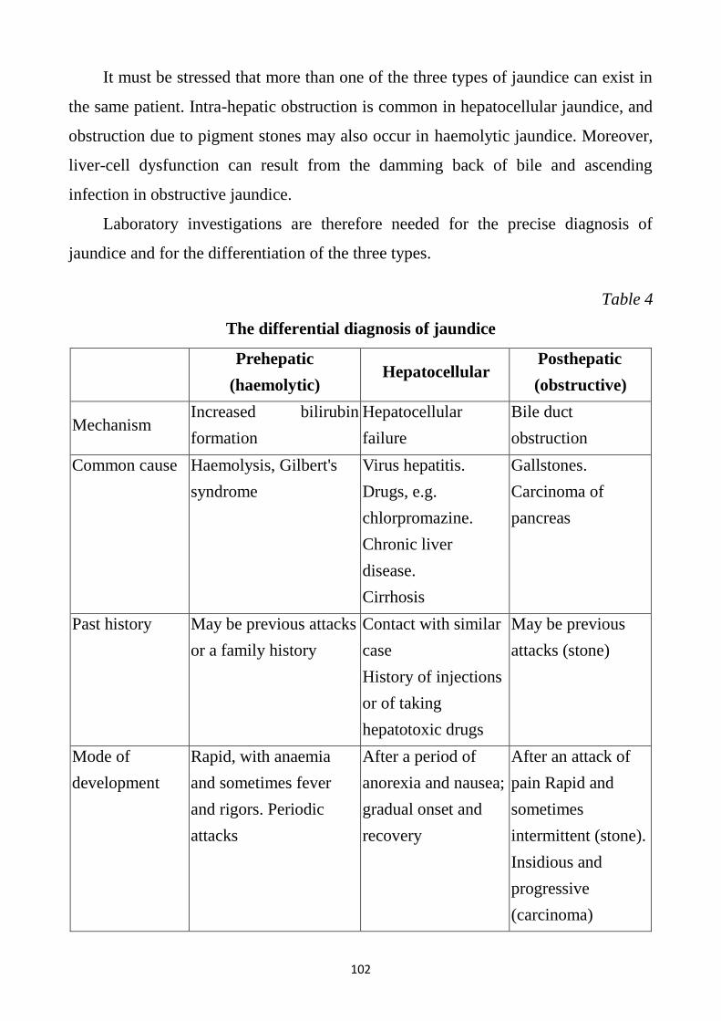
102
It must be stressed that more than one of the three types of jaundice can exist in
the same patient. Intra-hepatic obstruction is common in hepatocellular jaundice, and
obstruction due to pigment stones may also occur in haemolytic jaundice. Moreover,
liver-cell dysfunction can result from the damming back of bile and ascending
infection in obstructive jaundice.
Laboratory investigations are therefore needed for the precise diagnosis of
jaundice and for the differentiation of the three types.
Table 4
The differential diagnosis of jaundice
Prehepatic
(haemolytic) Hepatocellular
Posthepatic
(obstructive)
Mechanism Increased bilirubin
formation
Hepatocellular
failure
Bile duct
obstruction
Common cause Haemolysis, Gilbert's
syndrome
Virus hepatitis.
Drugs, e.g.
chlorpromazine.
Chronic liver
disease.
Cirrhosis
Gallstones.
Carcinoma of
pancreas
Past history May be previous attacks
or a family history
Contact with similar
case
History of injections
or of taking
hepatotoxic drugs
May be previous
attacks (stone)
Mode of
development
Rapid, with anaemia
and sometimes fever
and rigors. Periodic
attacks
After a period of
anorexia and nausea;
gradual onset and
recovery
After an attack of
pain Rapid and
sometimes
intermittent (stone).
Insidious and
progressive
(carcinoma)
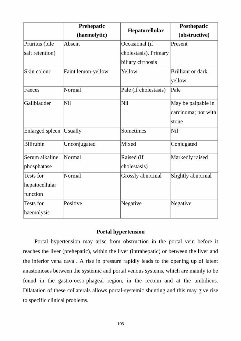
103
Prehepatic
(haemolytic) Hepatocellular
Posthepatic
(obstructive)
Pruritus (bile
salt retention)
Absent Occasional (if
cholestasis). Primary
biliary cirrhosis
Present
Skin colour Faint lemon-yellow Yellow Brilliant or dark
yellow
Faeces Normal Pale (if cholestasis) Pale
Gallbladder Nil Nil May be palpable in
carcinoma; not with
stone
Enlarged spleen Usually Sometimes Nil
Bilirubin Unconjugated Mixed Conjugated
Serum alkaline
phosphatase
Normal Raised (if
cholestasis)
Markedly raised
Tests for
hepatocellular
function
Normal Grossly abnormal Slightly abnormal
Tests for
haemolysis
Positive Negative Negative
Portal hypertension
Portal hypertension may arise from obstruction in the portal vein before it
reaches the liver (prehepatic), within the liver (intrahepatic) or between the liver and
the inferior vena cava . A rise in pressure rapidly leads to the opening up of latent
anastomoses between the systemic and portal venous systems, which are mainly to be
found in the gastro-oeso-phageal region, in the rectum and at the umbilicus.
Dilatation of these collaterals allows portal-systemic shunting and this may give rise
to specific clinical problems.
104
The spleen may be grossly enlarged.
Ascites may be gross. The first aim in its management is to create a net negative
balance of sodium (dietary restrictions and diuretics). It is simple to monitor
abdominal girth, weight, blood urea and electrolytes and 24-hour urinary sodium loss.
Paracentesis is of value in excluding other diagnoses by biochemistry and cytology. It
may also have a place in treatment in association with plasma expanders (e.g.
albumin).
Oesophageal varices lie in the submucosa of the lower oesophagus and are liable
to rupture because of portal pressure and local trauma. Bleeding is also compounded
by severe coagulation defects caused by liver cell failure. Haematemaesis and
melaena or chronic iron deficiency are the main presentations. Diagnosis is made by
barium swallow or at endoscopy, and varic-es may also occur in the fundus of the
stomach. Barium swallow may show typical barium-coated filling defects, evident as
oesophageal varices. In some patients in addition, gastric varices can be seen. These
thin-walled varices are easily damaged, and bleeding is a frequent complication.
«White» varices have relatively low risk of immediate bleeding, because they are
covered with a thick layer of mucosa. The presence of red lines (red wale markings)
or spots (cherry-red spots) is associated with a strong likelihood of bleeding.
Rectal anastomoses may become extremely large and bleed after bowel
movements. Massive bleeding is extremely rare. Umbilical anastomoses are rarely
clinically obvious. The anastomotic veins radiate from the umbilicus across the
abdomen to join systemic veins, forming a «caput Medusae». Lesser degrees of
anastomosis are more common.
Liver failure
Liver failure can develop acutely in a previously normal liver (as in fulminant
viral hepatitis or drug-induced hepatitis), or it may develop insidiously in a
chronically damaged liver.
105
Fluid retention is an early problem; patients present with ankle and leg oedema,
ascites and small pleural effusions.
Bruising is seen spontaneously and after trauma, sometimes even from the
pressure induced by a blood pressure cuff. This results from defective coagulation
factor synthesis and from thrombocytopenia associated with splenomegaly. A
bleeding tendency may also be shown by excessive bleeding at venepuncture sites.
Nausea, vomiting, anorexia, drowsiness, tremor and confusion may lead on to
encephalopathy: deep coma with fits and decerebrate posture.
Other metabolic disturbances, including hypoglycaemia, pancreatitis and renal
failure, are common.
Encephalopathy is an acute or chronic neurological impairment that may result
from liver cell failure associated with shunting of blood from the portal system; it is
probably caused by the failure of the liver to detoxify some as yet unidentified
component in the portal blood. There is progressive impairment of higher cerebral
function, with eventual coma and death. Early features suggesting encephalopathy
include fetor hepaticus - a sweet apple-like smell on the breath - a coarse flapping
tremor and an inability to draw or write accurately.
Portal systemic encephalopathy (hepatic encephalopathy, hepatic coma)
A neuropsychiatric syndrome caused by liver disease and usually associated
with portal-systemic shunting of venous blood.
"Portal-systemic encephalopathy" is a more descriptive term of the
pathophysiology than "hepatic encephalopathy" or "hepatic coma," but clinically all
three are used interchangeably.
Portal-systemic encephalopathy may occur in fulminant hepatitis caused by
viruses, drugs, or toxins, but it more commonly occurs in cirrhosis or other chronic
disorders when extensive portal-systemic collaterals have developed as a result of
portal hypertension. The syndrome also follows portacaval shunt or similar portal-
systemic anastomoses. In patients with chronic liver disease, encephalopathy is
106
usually precipitated by specific, potentially reversible causes (eg, GI bleeding;
infection; electrolyte imbalance, especially hypokalemia; alcoholic debauches) or
iatrogenic causes (tranquilizers, sedatives, analgesics, diuretics).
The liver metabolizes and detoxifies digestive products brought from the
intestine by the portal vein. In liver disease, these products escape into the systemic
circulation if portal blood bypasses parenchymal cells or if the function of these cells
is severely impaired. The resulting toxic effect on the brain produces the clinical
syndrome. Ammonia, a product of protein digestion, probably plays an important
role, but biogenic amines, short chain fatty acids, and other enteric products may also
be responsible or may act with ammonia. Aromatic amino acid levels in serum are
usually high.
The pathogenesis of the cerebral toxicity is also uncertain. Pathologic changes
are usually confined to hyperplasia of astrocytes with little or no neuronal damage,
but cerebral edema is common in fulminant hepatitis.
Clinical manifestations develop 4 stages of hepatic encephalopathy:
1st stage (prodromal period) — Personality changes (eg, inappropriate
behavior, altered mood, impaired judgment) are common early manifestations that
may antedate apparent change in consciousness. Sophisticated psychomotor tests can
often detect such abnormalities not suspected clinically. Initially, subtle sleep pattern
changes or sluggish movement and speech may be present. Inversion of sleep is also
typical.
2nd stage — stage of profound neuropsychic derangements (inadequate
behaviour, twenty-four-hour lethargy). Usually, impaired consciousness occurs.
Constructional apraxia, in which the patient cannot reproduce simple designs
(eg, a star), is a characteristic early sign. A peculiar, characteristic flapping tremor,
asterixis, is elicited when the patient holds his arms outstretched with wrists
dorsiflexed; as coma progresses, this sign disappears.
3d stage — precoma stage - somnolence, confusion, stupor indicate
increasingly advanced encephalopathy. Dysarthria, appearance of pathologic reflexes
107
and Cheyne-Stokes or Kussmaul breathings, growth of flapping tremor are observed.
A typical musty sweet odor of the breath, called fetor hepaticus, often occurs. Sharp
decrease of the liver dimensions; jaundice without pruritus and hemorrhagic
syndrome progressing, hypoalbuminemia, fever are characteristic.
4th stage — frank coma stage is characterized by areflexia, hypoalbuminemai,
hyperbilirubinemia, and low blood cholesterol and prothrombin value.
The diagnosis is clinical. There is no correlation with liver function tests. Blood
ammonia levels are usually elevated, but values correlate poorly with clinical status;
bedside judgment is a better guide. Encephalopathy in chronic liver disease usually
responds to treatment, especially if the precipitating cause is reversible.
Coma associated with fulminant hepatitis is fatal in up to 80% of patients,
despite intensive therapy; patients with advanced chronic liver failure often die with
portal-systemic encephalopathy.
Cytolytic syndrome
Cytolytic syndrome is characterized by increase of serum transaminase, which
reflects level of hepatocytes necrosis in acute and chronic hepatic diseases of
different etiology. The peak increase is detected in acute viral hepatitis.
Investigations
Investigations are of value in defining the cause of liver disease, the extent of
damage and the effects of treatment.
Full blood count is of value in detecting anaemia - often iron deficient
because of bleeding from oesoph-ageal varices; macrocytosis is often found in liver
disease with biliary obstruction and may not reflect vitamin B12deficiency (B12 levels
may be elevated if there is hepatic cell necrosis); folate levels are often low, caused
by a combination of malabsorption and poor dietary intake; thrombocytopenia is
often present, because of a combination of factors that may include the direct effects
108
of alcohol on the bone marrow, secondary hypersplenism, disseminated intra-vascular
coagulation and marrow aplasia in acute fulminant hepatitis, and folate deficiency.
Coagulation abnormalities are common and often complex, as the liver makes
most coagulation factors and destroys others; tests should include the activated partial
thromboplastin time (APPT), the pro-thrombin time (PT), the thrombin time (TT),
whole blood platelet count and simple tests of fibrinolysis such as measurement of
fibrinogen - fibrin degradation products (FDPs).
Routine biochemistry is the keystone of diagnosis and assessment of progress;
tests should include total bi-lirubin, with direct and indirect values as necessary,
alkaline phosphatase, the aminotransferases (especially alanine aminotransferase,
ALT), γ-glutamyl transpeptidase, total proteins, albumin and γ -globulins; none of
these tests is specific or diagnostic, but all are of value in combination and in
following the course of the disease; two broad patterns of liver function tests may
emerge: cholestatic and hepatitic; serum albumin measures the synthetic capacity of
the liver generally.
Gallbladder disoders
Physiology of bile acid metabolism
Bile is formed in the liver as an isosmotic solution of bile acids, electrolytes,
bilirubin, cholesterol, and phospholipids. Bile flow is generated by the active transport of
bile salts and electrolytes and the accompanying obligate passive movement of water.
The liver synthesizes water-soluble bile acids from water-insoluble cholesterol, but
precise mechanisms are not completely understood. Cholic and chenodeoxycholic acids
form in the liver in a ratio of about 2:1 and constitute 80% of bile acids. Bile acids are
excreted in bile, which flows from the intrahepatic collecting system into the proximal or
common hepatic duct. About 50% of bile secreted in the fasting state passes into the
gallbladder via the cystic duct; the rest flows directly into the distal or common bile duct.
Up to 90% of water in gallbladder bile is absorbed as an electrolyte solution, principally
via gallbladder mucosal intracellular pathways. Bile remaining in the gallbladder is thus
109
a concentrated solution consisting primarily of bile acids and sodium. During fasting,
bile acids are concentrated in the gallbladder, and little bile acid-dependent bile flows
from the liver. Food entering the duodenum initiates an exquisite hormonal and neural
sequence. Cholecystokinin is released from duodenal mucosa and stimulates the
gallbladder to contract and the biliary sphincter to relax. Bile flows into the duodenum to
mix with food contents and to perform its several functions: (1) Bile salts solubilize
dietary cholesterol, fats, and fat-soluble vitamins to facilitate their absorption in the form
of mixed micelles. (2) Bile acids induce water secretion by the colon as they enter that
organ, thus promoting catharsis. (3) Bilirubin is excreted in bile as degradation products
of heme compounds from worn-out RBCs. (4) Drugs, ions, and endogenously produced
compounds are excreted in bile and subsequently eliminated from the body. (5) Various
proteins important in GI function are secreted in bile. Food entering the duodenum
stimulates gallbladder contraction, releasing much of the body pool (total, 3 to 4 g) of
bile acids into the small intestine. Bile acids are poorly absorbed by passive diffusion in
theproximal small intestine; most of the pool reaches the terminal ileum, where 90% is
absorbed into the portal venous circulation by active transport. Bile salts are efficiently
extracted by the liver, promptly modified, and secreted back into bile. Bile acids undergo
enterohepatic circulation 10 to 12 times per day. During each pass, a small amount of
primary bile acids reaches the colon, where anaerobic bacteria containing 7-hydroxylase
form secondary bile acids. Cholic acid is thus converted to deoxycholic acid, which is
largely reabsorbed and conjugated. Chenodeoxycholic acid conjugates are converted in
the colon to their secondary bile acid form, lithocholic acid. This insoluble secondary
bile acid is partially reabsorbed; the rest is lost in the feces. Anatomy of the Biliary Tract
Other than absorptive functions of the normal gallbladder and bile storage mediation by
the sphincters, the extrahepatic ductal system is a passive conduit. There are no
functional smooth muscle fibers in the biliary duct walls. Ductal secretions stimulated by
secretin contain a high concentration of bicarbonate and contribute variably to total bile
volume. The ampulla of Vater (Fig.49) consists of the terminal intramural segments of
the biliary and pancreatic ducts and of the two or three sphincter segments and
110
surrounding soft tissue. The sphincter of Oddi surrounds both ducts or their common
channel, and each duct has its separate (inconstant) sphincter. Normal sphincter function
results in timely release of bile and pancreatic enzymes during food passage; during
fasting, however, gallbladder filling is facilitated. The two systems normally remain
independent (ie, bile does not flow retrograde into the pancreatic duct).
Chronic cholecystitis
Pathologically, a thick-walled, fibrotic, contracted gallbladder; clinically,
chronic gallbladder disease characterized by symptoms that include recurrent colic.
The mucosa may be ulcerated and scarred, and the lumen may contain sludge or
stones that often obstruct the cystic duct. It is tempting to ascribe these findings to the
ravages and repair of previous episodes of acute cholecystitis, but the clinical history
may not include any record of such events. Clinical and pathologic manifestations are
poorly correlated. Both are nearly always associated with calculi in the gallbladder.
Cholelithiasis
Formation or presence of calculi (gallstones) in the gallbladder. Most clinical
disorders of the extrahepatic biliary tract are related to gallstones. Factors that
increase the probability of gallstones include female sex, obesity, increased age, a
Western diet, and a positive family history.
Pathophysiology Cholesterol, the major component of most gallstones, is highly
insoluble in water, and biliary cholesterol is solubilized in bile saltphospholipid
micelles and phospholipid vesicles, which greatly increase the cholesterol-carrying
capacity of bile. Bile salt micelles are aggregates of bile salts in which water-soluble
(ionic) regions of the molecule face outward into aqueous solution, while the water-
insoluble (nonpolar) steroid nuclei face inward. Cholesterol is soluble inside these
spheroid micelles, and their cholesterol-carrying ability is further enhanced by lecithin,
a polar phospholipid. The amount of cholesterol carried in micelles and vesicles varies
with the bile salt secretion rate. Supersaturation of cholesterol in bile is a necessary
condition, but not a sole cause, of cholesterol gallstone formation because
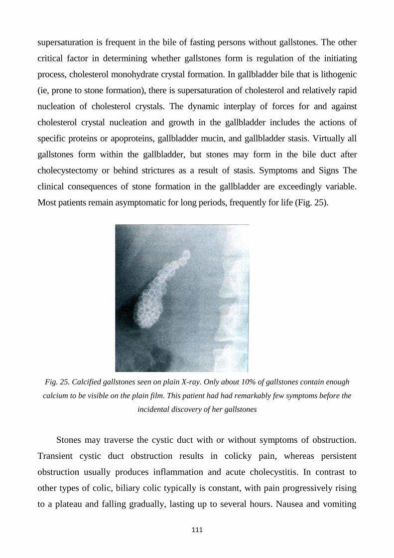
111
supersaturation is frequent in the bile of fasting persons without gallstones. The other
critical factor in determining whether gallstones form is regulation of the initiating
process, cholesterol monohydrate crystal formation. In gallbladder bile that is lithogenic
(ie, prone to stone formation), there is supersaturation of cholesterol and relatively rapid
nucleation of cholesterol crystals. The dynamic interplay of forces for and against
cholesterol crystal nucleation and growth in the gallbladder includes the actions of
specific proteins or apoproteins, gallbladder mucin, and gallbladder stasis. Virtually all
gallstones form within the gallbladder, but stones may form in the bile duct after
cholecystectomy or behind strictures as a result of stasis. Symptoms and Signs The
clinical consequences of stone formation in the gallbladder are exceedingly variable.
Most patients remain asymptomatic for long periods, frequently for life (Fig. 25).
Fig. 25. Calcified gallstones seen on plain X-ray. Only about 10% of gallstones contain enough
calcium to be visible on the plain film. This patient had had remarkably few symptoms before the
incidental discovery of her gallstones
Stones may traverse the cystic duct with or without symptoms of obstruction.
Transient cystic duct obstruction results in colicky pain, whereas persistent
obstruction usually produces inflammation and acute cholecystitis. In contrast to
other types of colic, biliary colic typically is constant, with pain progressively rising
to a plateau and falling gradually, lasting up to several hours. Nausea and vomiting
112
are often associated. Fever and chills are absent in uncomplicated gallbladder colic.
Pain most often occurs in the epigastrium or right upper quadrant, radiating to the
right lower scapula. Symptoms of dyspepsia and fatty food intolerance are often
inaccurately ascribed to gallbladder disease. Belching, bloating, fullness, and nausea
are associated about equally with cholelithiasis, peptic ulcer disease, or functional
distress. Such symptoms may disappear after cholecystectomy but should not be the
only indication for operation. Postprandial fatty food intolerance is likely to be
caused by cholelithiasis if symptoms include right upper quadrant pain; however, the
prevalence of postprandial functional distress is so high in the general population that
symptoms alone are insufficient for diagnosis of gallbladder disease without
supportive clinical signs and diagnostic studies.
Gallbladder Signs
Malignancy
Courvoisier’s Terrier sign – a palpable nontender gall bladder in a patient
with jaundice suggesting extrahepatic obstruction of the biliary system
secondary to malignancy (original description) (Fig. 12, see colour insert).
Acute Cholecystitis
1. Murphy’s sign – with the examiner’s fingers positioned along the inferior
border of the liver in the right costal arch the patient is allowed to inspire. During
inspiration the inflamed gallbladder touches the examiners fingers resulting in the
sudden cessation of inspiration.
2. Keri symptom is a significant increase of sensitivity to pain on inspiration
during palpation of the gall bladder with the thumb of the right hand. Diagnostic
value: cholecystitis (inflammation of gallbladder).
3. Kerte symptom – pain and tension of the abdominal muscles, epigastric
region, left hypochondrium or in the place of projection of the pancreas. Diagnostic
value: it is observed in acute pancreatitis.
4. Symptom Vasilenko-tenderness to percussion in the projection point of the
gall bladder at the height of inspiration
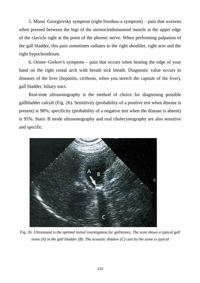
113
5. Mussi–Georgievsky symptom (right frenikus-a symptom) – pain that worsens
when pressed between the legs of the sternocleidomastoid muscle at the upper edge
of the clavicle right at the point of the phrenic nerve. When performing palpation of
the gall bladder, this pain sometimes radiates to the right shoulder, right arm and the
right hypochondrium.
6. Ortner–Grekov's symptom – pain that occurs when beating the edge of your
hand on the right costal arch with breath sick breath. Diagnostic value occurs in
diseases of the liver (hepatitis, cirrhosis, when you stretch the capsule of the liver),
gall bladder, biliary tract.
Real-time ultrasonography is the method of choice for diagnosing possible
gallbladder calculi (Fig. 26). Sensitivity (probability of a positive test when disease is
present) is 98%; specificity (probability of a negative test when the disease is absent)
is 95%. Static B mode ultrasonography and oral cholecystography are also sensitive
and specific.
Fig. 26. Ultrasound is the optimal initial investigation for gallstones. The scan shows a typical gall
stone (A) in the gall bladder (B). The acoustic shadow (C) cast by the stone is typical
114
Test control of the theme “Questioning and examination
of patients with the liver diseases”
1. THE MAIN COMPLAINTS OF PATIENTS WITH DISEASES OF THE
HEPATOBILIARY TRACT:
a) pain in the right hypochondrium, vomiting of bile, fever;
b) headache, palpitations, swelling in the lower extremities;
с) heartburn, nausea, constipation, abdominal pain;
d) weakness, diarrhea with blood, abdominal pain.
2. ITCHING LIVER DISEASES AND GALLBLADDER OFTEN
ACCOMPANIED BY:
a) icteric staining of the skin and mucous membranes;
b) paleness of skin and mucous membranes;
с) redness of the skin and mucous membranes.
3. THE MAIN RISK FACTORS OF DEVELOPMENT OF DISEASES OF THE
HEPATOBILIARY SYSTEM:
a) alcohol abuse, violation of diet, family history;
b) hypothermia, medications, stressful situation;
с) a sedentary lifestyle, stress, Smoking.
4. FROM THE ABOVE DISEASE GROUPS, SELECT THE ONE IN WHICH ARE
ALL CHARACTERIZED BY THE SYMPTOM OF "FINGER CLUBBING ":
a) lung abscess and bronchiectasis, congenital heart disease, bacterial
endocarditis, biliary cirrhosis of the liver;
b) chronic bronchitis, pneumonia, bronchial asthma, lung abscess;
с) pneumonia, bacterial endocarditis, biliary cirrhosis of the liver;
d) chronic bronchitis, pulmonary fibrosis, pancreatitis.
115
5. AN INCREASE IN THE ABDOMEN IN PATIENTS WITH PATHOLOGY
HEPATOBILIARY SYSTEM IS ASSOCIATED WITH:
a) flatulence;
b) ascites;
с) peritonitis;
g) bleeding;
d) dysphagia.
6. THE CAUSE OF THE EXPANSION OF THE VENOUS NETWORK ON THE
ANTERIOR ABDOMINAL WALL AROUND THE UMBILICUS IN PATIENTS
WITH LIVER DISEASE IS:
a) portal hypertension;
b) increased blood pressure;
с) the exhaustion of the patient;
d) flatulence;
e) hypersplenism.
7. ICTERIC STAINING OF THE SKIN AND MUCOUS MEMBRANES IN
PATIENTS WITH LIVER DISEASE AND BILIARY TRACT BEGINS WITH:
a) lower limbs;
b) mucosa of the palate and oral cavity;
с) palms and feet.
8. CHANGES DETECTED DURING THE INSPECTION AND PERCUSSION OF
THE ABDOMEN IN PATIENTS WITH PORTAL HYPERTENSION
SYNDROME:
a) the stomach does not participate in the act of respiration;
b) the abdomen is enlarged, dome-shaped, tympanic percussion sound;
с) the abdomen is enlarged, in a horizontal position sprawled belly bulges, on
side surfaces of the expanded venous network.
116
9 POSITIVE SYMPTOM OF KERA CHARACTERISTIC:
a) peritonitis;
b) acute inflammation of the gallbladder;
с) peptic ulcer disease;
d) colitis.
10. A POSITIVE SIGN OF THE GEORGE- MUSSI THIS:
a) the emergence or strengthening of pain during effleurage on the right costal arch;
b) pear-shaped palpable education in the projection of the gall bladder;
с) tenderness at the site of projection of the legs of the right clavicular-mastoid
muscle;
d) tenderness to effleurage finger on the anterior abdominal wall.
11. THE CAUSE OF THE PAIN IN DISEASES OF THE LIVER IS:
a) stretching glass on by capsules;
b) poor coordination between the contraction of the gallbladder and relaxation of
sphincter of Oddi;
c) irritation of the phrenic nerve;
d) ischuria.
12. PAIN IN DISEASES OF THE LIVER AND BILIARY TRACT RADIATES TO:
a) left upper quadrant;
b) the right shoulder;
c) in the region of the heart;
d) in the groin area;
e) right shoulder blade;
13. THE CAUSE OF PRURITUS WHEN THE JAUNDICE IS TRUE:
a) the accumulation in the blood of bile acids;
117
b) increasing levels of iron;
с) increasing the level of creatinine in the blood.
14. THE MAIN SYMPTOMS DURING THE INSPECTION OF PATIENTS WITH
DISEASES OF THE LIVER:
a) the yellowness of the skin and mucous membranes, telangiectasia, ascites;
b) paleness of skin and mucous membranes, hemorrhagic rashes on her lower
limbs;
с) barrel chest, cyanosis of skin and mucous membranes, ascites.
15. CAUSE OF SPIDER VEINS, REDNESS OF THE PALMS IN DISEASES OF
THE LIVER IS:
a) hyperestrogenia;
b) high levels of bilirubin;
с) high levels of cholesterol in the blood;
d) increasing the level of hemoglobin.
16. AN EXAMPLE OF A FORCED POSITION OF THE PATIENT WITH
ATTACK OF PAIN IN THE RIGHT UPPER QUADRANT FOR LIVER AND
GALL BLADDER IS:
a) orthopnea;
b) on the right side;
c) on the abdomen;
d) there is no such provision.
17. THE DIMENSIONS OF A LIVER ON KURLOVU NORMAL:
a) 9+1 cm, 8+1, 7+1;
b) 12+1, 11+1, 10+1 cm.
с) 7+1, 6+1 cm, 5 +1, see
118
18. PERITONITIS IS CHARACTERISTIC CHANGES, DETECTABLE BY
INSPECTION AND PERCUSSION OF THE ABDOMEN:
a) stomach, almost not involved in the act of respiration;
b) the abdomen is increased in volume, swollen, participates in the act of
breathing, percussion - tympanitis loud;
с) the abdomen is increased in volume, upright looks saggy.
19. A DISEASE IN WHICH REVEALS THE SYMPTOM OF ORTNER
a) inflammation of the peritoneum;
b) inflammation of the lungs;
с) inflammation of the pleura;
d) inflammation of the gallbladder.
20. POSITIVE SYMPTOM MURPHY IS:
a) the emergence or strengthening of pain during effleurage edge of his hand
along the left costal arch;
b) the appearance of sharp pain with the introduction of the hands in the right
hypochondrium at the height of inspiration;
с) the appearance or strengthening of pain during effleurage with one finger on
the abdominal wall.
119
III. THE FUNDAMENTALS OF CLINICAL DIAGNOSIS
OF DISEASES OF THE URINARY SYSTEM
A brief anatomical and physiological information
about the urinary system
To the organs of the urinary system include the kidneys, ureters, bladder and
urethra. The main function of the urinary organs – excretion of metabolic waste
products, involved in the regulation of water content in the body and maintain this
constancy of its internal environment.
Kidney (Fig. 13, see color insert) – paired organ bean-shaped. Length 10-12 cm,
width - 5-6 sm, a thickness - 3-4 sm, weight - 120-200 g. the Kidneys are located in
the retroperitoneal region on the sides of the spine. Syntopia and skeletondeploy and
left kidneys are different. The upper pole of the left kidney is at the level of XI
thoracic vertebra, and the lower, between II and III lumbar vertebrae. XII edge
intersects the left kidney in the gate area. The right kidney is 3 cm lower than the left.
The kidney distinguish the front and back surface, upper and lower ends, medial and
lateral edges. On the medial, concave, the edge facing the spine, is the gate of the
kidney. At the gate there are: renal artery, renal vein, lymphatic vessels, lymph nodes,
nerves and renal pelvis. The kidney is enveloped in a fat capsule composed of loose
connective tissue. The most outer shell is the renal fascia, representing the double-
layer disc. The front and rear sheets of the renal fascia at the outer edge and the upper
pole of the kidney are connected, and at the bottom of the case continue through the
ureter to the bladder.
In kidney distinguish the cortical substance thickness of 5-7 mm is located in the
periphery, and the medulla, consisting of 7-12 pyramids facing the base toward the
cortical substance, while the tip is in the renal sinus (Fig.14, see color insert).
Blood flow to the kidney is due to renal artery which starts from the abdominal
aorta and hilum is divided into 5 – 6 branches, heading to the upper, lower poles and
the Central part, which branch out into smaller arteries. Venous vessels, with the
120
exception of vascular glomerulus bringing arterioles and efferent arterioles, repeated
branching of the arteries.
The ureters is a hollow tube that connects the renal pelvis to the bladder. The
wall of the ureter has mucosa, muscle and connective tissue sheath. Urine moves
through the ureter due to a peristaltic contraction of smooth muscle walls.
The bladder is a hollow organ where portions continuously drains the urine from
the ureters. It is located in the pelvis behind the symphysis. Contraction of smooth
muscle, with an open hole in the urethra contributes to the emptying of the bladder.
The urethra connects the bladder to the surface of the human body. Starts the
urethra the inner hole on the wall of the bladder. Where the urethra passes through
the urogenital diaphragm, formed around the sphincter (constrictor) of striated
skeletal muscle tissue, arbitrarily regulating the emptying of the bladder.
The concept of the allocation process and its value.
Allocation is the process of excretion of waste products and the harmful and
unnecessary substances. More than 90% derived from the organism of substances is
removed via the urinary system
Structural and functional unit of the kidney is the nephron the tubular system of
the kidney involved in urine formation (Fig. 15, see color insert). The length of one
nephron ranges from 18 to 50 mm, and the total length is 100 km. each kidney has
over 1 million nephrons. The nephron consists of a capsule and three-tubules:
proximal tubule (convoluted tubule of the first order) of the nephron loop, and distal
tubule (convoluted tubule of the second order), rolling in the collecting tube. Capsule
is the initial portion of the nephron located in the cortical substance of the kidney has
the shape of a double-walled bowl. It tightly covers the capillaries of the glomerulus
of the kidney, forming the so-called renal corpuscle. Thus, one end of the nephron
begins in the renal capsule, a second end flows into the collecting tube. The most
active part of the nephron is the proximal his Department in which the processes of
formation of urine are fast.
121
Urine is formed from blood plasma. As the flow of blood in the vessels of the
glomerulus to the inside of the capsule from it through the filter into the lumen of the
capsule moving almost all the components except proteins and formed elements,
forming a so-called primary urine. During the day, it produces about 100 liters. With
the passage of primary urine through the tubules out of it back into the blood soaked
water, some salt, sugar, resulting in a final urine. The number of final urine is about
1.0 to 1.5 liters. It has a higher concentration than the primary urine. Final urine
through the collecting duct, passing in the cortex and then the medulla of the kidney,
drains into the holes in the top of the pyramid, first in small cups, then large and
finally into the renal pelvis, which is a continuation of the ureter.
Questioning of patients with the kidney diseases
Main complaints
Patients with diseases of the kidneys complain most commonly of pain in the
lumbar region, disordered urination, edema, headache, and dizziness.
They may also complain of deranged vision, pain in the heart, dyspnea, absence
of appetite, nausea, vomiting, and elevated body temperature. But diseases of the
kidneys may also proceed without any symptoms of renal or general clinical
insufficiency. If the patient complains of pain, its location should first of all be
determined. Pain of renal origin localizes frequently in the lumbar region. If the
ureters are affected, the pain is felt by their course. If the bladder is involved, pain is
suprapubical. Radiation of pain into the perineal region is characteristic of an attack
of nephrolithiasis. The character of pain should then be determined. It is necessary to
remember that the renal tissue is devoid of pain receptors. The pain is felt when the
capsule or the pelvis is distended. Dull and boring pain in the lumbar region occurs in
acute glomerulonephritis, abscess of the perirenal cellular tissue, in heart
decompensation ("congestive kidney"), in chronic pyelonephritis (usually unilateral)
and less frequently in chronic glomerulonephritis. Pain arises due to distension of the
renal capsule because of the inflammatory or congestive swelling of the renal tissue.
122
Sharp and suddenly developing pain on one side of the loin can be due to the renal
infarction. The pain persists for several hours or days and then subsides gradually.
The pain is rather severe in acute pyelonephritis: inflammatory edema of the ureter
interferes with the normal urine outflow from the pelvis and thus causes its
distension. The pain is usually permanent. Some patients complain of attacks of
severe piercing pain in the lumbar region or by the course of the ureter. The pain
increases periodically and then subsides, i.e. has the character of renal colic.
Obstruction of the ureter by a calculus or its bending (movable kidney) is the most
common cause of this pain, which is usually attended by spasmodic contraction of the
ureter, retention of the urine in the pelvis, and hence its distension. The spasmodic
contractions and distension of the pelvis account for the pain. Pain in renal colic is
usually unilateral. It radiates into the corresponding hypochondrium and most
frequently by the course of the ureter to the bladder and to the urethra. This radiation
of pain is explained by the presence of nerve fibres (carrying the impulses from
kidneys, ureters, genital organs and the corresponding skin zones) in the immediate
vicinity of the relevant segments of the spinal cord (DX-DXII and LI-LII). This
facilitates propagation of the excitation. Patients with renal colic (like those with
colic of other etiology) are restless; they toss in bed. Patients with severe pain of
other etiology would usually lie quiet in their beds (movements may intensify the
pain). The conditions promoting pain should be established. For example, pain in
nephrolithiasis can be provoked by taking much liquid, jolting motion, or the like;
pain is provoked by urination in cystitis. Difficult and painful urination is observed in
stranguria. Patients with urethritis feel a burning pain in the urethra during or after
urination.
It is necessary also to establish the agent that lessens or removes the pain. For
example, atropine sulphate, hot water-bottle or warm bath helps in renal colic. Since
these remedies only help in spasmodic pain by removing spasms of the smooth
muscles, their efficacy in renal colic confirms the leading role of the ureter
contraction in the pathogenesis of this pain. Pain of the renal colic-type in patients
123
with movable kidney may lessen with changing posture: urine outflow improves with
displacement of the kidney. Pain slightly lessens in patients with acute paranephritis
if an ice pack is placed on the lumbar region and if the patient is given analgesics.
Many renal diseases are attended by deranged urination: changes in the daily volume
of excreted urine and in the circadian rhythm of urination. Secretion of urine during a
certain period of time is called diuresis. Diuresis can be positive (the amount of urine
excreted exceeds the volume of liquid taken) or negative (the reverse ratio). Negative
diuresis is observed in cases of liquid retention in the body or its excess excretion
through the skin, by the lungs (e.g. in dry and hot weather). Positive diuresis occurs
in resolution of edema, after administration of diuretics, and in some other cases.
Deranged excretion of urine is called dysuria.
1. Dysuria. - urethritis and cystitis. - inflammation of vagina and penis.
2. Polyuria and nocturia. - > 3 L/ day. - solute diuresis, diabetes insipidus, CRF.
3. Oliguria. - < 300 ml/ day. - hypotension, hypovolaemia. - intrinsic renal disease. -
urinary tract obstruction.
Anuria should be differentiated from ischuria, when the urine is retained in the
bladder and the patient is unable to evacuate it. This occurs in compression or other
affection of the spinal cord, and in loss of consciousness. Pollakiuria (frequent
micturition) is observed in certain cases. A healthy person urinates from 4 to 7 times
a day. The amount of excreted urine during one micturition is from 200 to 300 ml
(1000-2000 ml a day). But frequency of micturition may vary within wider range
under certain conditions: it may decrease in limited intake of liquid, after eating much
salted food, in excessive sweating, in fever, and the like, or the frequency may
increase (polyuria) if the person takes much liquid, in getting cold, and the like
circumstances. Frequent desire to urinate with excretion of scanty quantity of urine is
the sign of cystitis. A healthy person urinates 4-7 times during the day time; a desire
to urinate during night sleep does not arise more than once. In the presence of
pollakiuria the patient feels the desire to urinate during both day and night. In the
presence of chronic renal insufficiency and if the kidneys are unable to control the
124
amount and concentration of excreted urine in accordance with the amount of liquid
taken, physical exertion, the ambient temperature, or other factors important for the
liquid balance in the body, the patient urinates at about equal intervals with
evacuation of about equal portions of urine. This condition is called isuria. Under
certain pathological conditions, the frequency of urination is normal during the day
time but increases during night. The amount of urine excreted during night often
exceeds the amount of daily urine (nycturia). Nocturnal enuresis (nycturia) and
oliguria during day time occur in cardiac decompensation and are explained by a
better renal function at night, i.e. at rest (cardiac nycturia). Nycturia may concur with
polyuria in renal dysfunction, at the final stage of chronic glomerulonephritis, chronic
pyelitis, vascular nephrosclerosis, and other chronic renal diseases (renal nycturia). In
the presence of isuria and nycturia of renal origin, which arise due to the loss by the
kidneys of their concentrating ability, the specific gravity of the urine is monotonous.
The condition is known as isosthenuria. The specific gravity of urine is usually
decreased (hyposthenuria). The specific gravity of urine varies from 1.009 to 1.011,
i.e. approaches the specific gravity of primary urine (plasma ultrafiltrate) in patients
with pronounced nephrosclerosis, which is the final stage of many chronic renal
diseases. Some diseases of the bladder and the urethra are attended by difficult and
painful urination. The patient would complain of change in the colour of the urine, its
cloudiness, and traces of blood. Edema is observed in acute and chronic diffuse
glomerulonephritis, nephrotic syndrome, amyloidosis, and acute renal excretory
dysfunction (anuria). It is important to ask the patient about the site that was the first
to be attacked by edema, the sequence of edema spreading, and the rate of
intensification of this phenomenon. Headache, dizziness, and heart pain may result
from kidney affections. These symptoms occur in those renal diseases which are
attended by considerable increase of arterial blood pressure, e.g. in acute and chronic
glomerulonephritis or vascular nephrosclerosis. A pronounced and persistent increase
in the arterial pressure can be among the causes of deranged vision (neuroretinitis).
Patients with diseases of the kidneys can complain of weakness, indisposition,
125
impaired memory and work capacity and deranged sleep. Vision may be deranged
along with skin itching and unpleasant breath. Dyspeptic disorders sometimes include
loss of appetite, dryness and unpleasant taste in the mouth, nausea, vomiting, and
diarrhea. All these phenomena are associated with retention in the body of protein
decomposition products due to renal insufficiency which develops at the final stage
of many chronic renal diseases, and sometimes in acute diseases attended by retention
of urine during several days. Fever is the common symptom of infectious
inflammatory affections of the kidneys, the urinary ducts and perirenal cellular tissue.
History of the present disease
When questioning the patient, it is necessary to establish the connection of the
present disease with previous infections (tonsillitis, scarlet fever, otitis, acute
respiratory diseases). This sequence is especially characteristic of acute
glomerulonephritis. But it is sometimes difficult to establish the time of onset of the
disease because some chronic affections of the kidneys and the urinary ducts can for
a long time be latent. Moreover, when questioning the patient, it is necessary to find
out if he had deranged hearing or vision in his childhood that might be suggestive of
congenital renal pathology.
Special attention should be given to the presence in the patient's past history of
diseases of the kidneys and the urinary ducts (acute nephritis, pyelitis, cystitis) or
symptoms that might suggest them (dysuria, hematuria, edema, arterial hypertension,
attacks of pain in the abdomen or loin resembling renal colics), since these symptoms
can be connected with the present renal pathology. In certain cases the cause and the
time of onset of grave kidney affections (necronephrosis) can be established by
revealing industrial or domestic poisoning, intentional (or by mistake) taking of some
poisons (corrosive sublimate, preparations of bismuth, phosphorus, silver, large doses
of sulpha preparations, or of some antibiotics, e.g. aminoglycosides, expired
tetracyclines, phosphorus compounds), transfusion of incompatible blood, etc.
Amidopyrin, phenacetin, barbiturates, camphor, and some other medicines can cause
126
allergic changes in the kidneys. The patient must be asked about the character of the
disease course: it may be gradual (arteriolosclerosis, chronic diffuse
glomerulonephritis, amyloidosis of the kidneys), or with periodical exacerbations
(chronic pyelonephritis, chronic diffuse glomerulonephritis). It is necessary to
establish the cause of exacerbations, their frequency, clinical signs, the character of
therapy given and its efficacy, the causes inducing the patient to seek medical help.
Life history of patient
Special attention should be given to the factors that might provoke the present
disease or have effect on its further course. For example, a common factor promoting
development of acute and chronic nephritis and pyelonephritis is chilling and cooling
(poor housing or working conditions, draughts, work in the open, acute cooling of the
body before the disease). Spreading of genital infection onto the urinary system can
be the cause of pyelonephritis. It is necessary to establish the presence or absence in
the past of tuberculosis of the lungs or other organs. This helps establish the
tuberculous nature of the present disease of the kidneys. It is necessary to establish if
the patient has some other diseases that might cause affections of the kidneys
(collagenosis, diabetes mellitus, certain diseases of the blood, etc.). Various chronic
purulent diseases (osteomyelitis, bronchiectasis) can be the cause of amyloidosis of
the kidneys. Occupations associated with walking, riding, weight lifting, etc., can
have their effect on the course of nephrolithiasis and provoke attacks of renal colic.
Some abnormalities of the kidneys, nephrolithiasis, amyloidosis, etc., can be
inherited. It is also necessary to record thoroughly the information on past operations
on the kidneys or the urinary ducts. When examining women, it is important to
remember that pregnancy can aggravate some chronic diseases of the kidneys and be
the cause of the so-called nephropathy of pregnancy (toxemia of late pregnancy).
127
General examination
The position of the patient: passive (uremic coma), forced (paranephritis is
characterized by the position given to the stomach with the foot on the affected side,
in renal colic the patient tosses).
Facies nefritica, Anasarca (Fig. 17, 18, see color insert). The characteristic
pale skin, scratching, the odor of ammonia from the mouth and from the skin of the
patient.
When paranephritis may detect swelling of the lumbar region on the affected
side.
The colour of the patient's skin is also important. Edematous skin in chronic
nephritis is pallid due to the spasm of skin arterioles, and anemia which attends this
disease. The skin is wax-pallid in amyloidosis and lipoid nephrosis. It should be
remembered that in cardiac edema (as distinct from renal edema) the skin is more or
less cyanotic. When inspecting a patient with chronic nephritis, it is possible to
observe scratches on the skin and coated dry tongue; an unpleasant odour of ammonia
can be felt from the mouth and skin of the patient (factor uremicus). All these signs
characterize chronic renal insufficiency (uremia).
Inspection of the abdomen and the loin does not usually reveal any noticeable
changes. But in the presence of paranephritis, it is possible to notice swelling on the
affected side of the loin. In rare cases, an especially large tumour of the kidney may
be manifested by protrusion of the abdominal wall. Distended bladder can be
protruded over the pubic bone in thin persons. The distension can be due to
overfilling of the bladder, for example, due to retention of urine in adenoma or cancer
of the prostate.
Kidney-percussion
kidney-percussion (to detect areas of tenderness by costovertebral test,
normally will feel a thudding sensation or pressure but not tenderness) and palpation
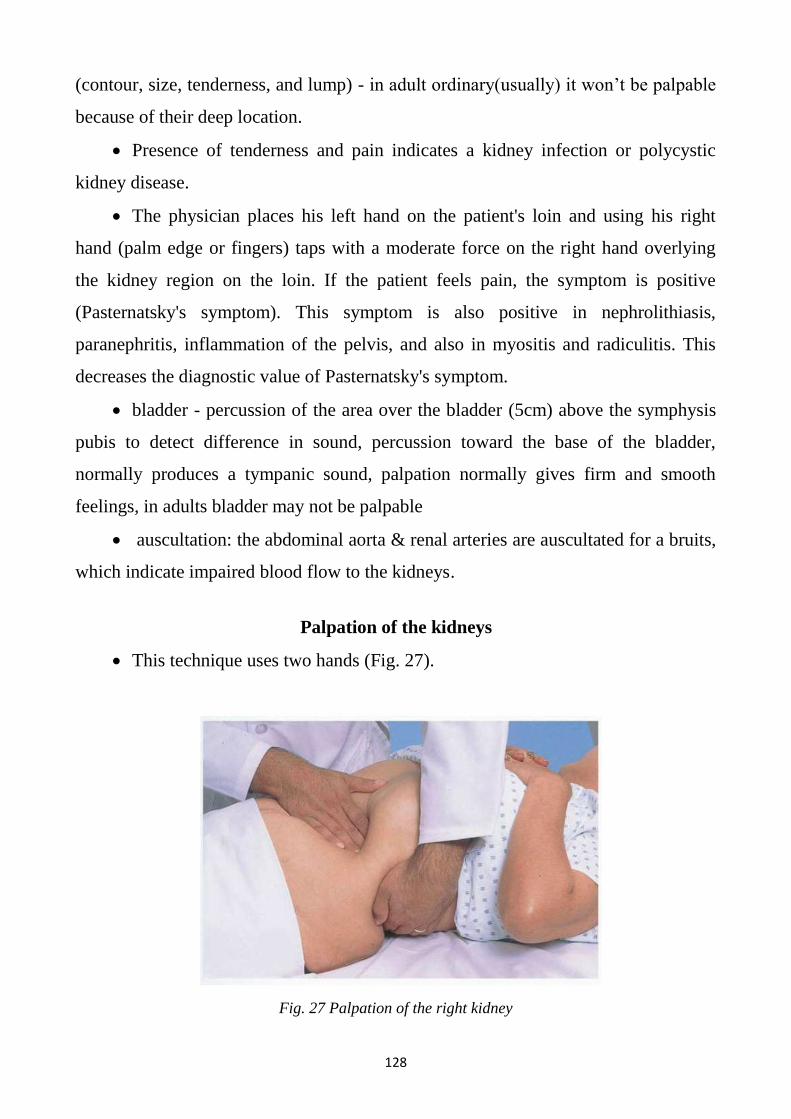
128
(contour, size, tenderness, and lump) - in adult ordinary(usually) it won’t be palpable
because of their deep location.
Presence of tenderness and pain indicates a kidney infection or polycystic
kidney disease.
The physician places his left hand on the patient's loin and using his right
hand (palm edge or fingers) taps with a moderate force on the right hand overlying
the kidney region on the loin. If the patient feels pain, the symptom is positive
(Pasternatsky's symptom). This symptom is also positive in nephrolithiasis,
paranephritis, inflammation of the pelvis, and also in myositis and radiculitis. This
decreases the diagnostic value of Pasternatsky's symptom.
bladder - percussion of the area over the bladder (5cm) above the symphysis
pubis to detect difference in sound, percussion toward the base of the bladder,
normally produces a tympanic sound, palpation normally gives firm and smooth
feelings, in adults bladder may not be palpable
auscultation: the abdominal aorta & renal arteries are auscultated for a bruits,
which indicate impaired blood flow to the kidneys.
Palpation of the kidneys
This technique uses two hands (Fig. 27).
Fig. 27 Palpation of the right kidney
129
Reach one hand round to the patient's right loin with your other hand over the
right upper quadrant. Push your hands together whilst asking the patient to breathe in
and out. Try to palpate any enlarged kidney between your two hands (called
'balloting').
Repeat for the left kidney. This can either be done by examining the patient
from the left side with your right hand under their left loin or by examining them
from the right side with your left hand reaching round under their left loin area.
In a very thin person who relaxes well, it may be just possible to feel a
kidney, especially on the left but usually it is abnormal.
Examine for enlarged kidneys, renal masses or loin tenderness.
Palpation of ureteric points
Tenderness at palpation along the course of ureter and sensitive loin (sensitive
to pressure exerted in the angle between the 12-th rib and the longissimus thoracic
muscles) is of certain diagnostic importance. Palpation of the anterior surface of
abdomen and lumbar range in some cases enables to determine presence of the pain
points connected to an affection of kidneys and urinary tract. Three pairs of anterior
ureteric points: (1) subcostal point - at the anterior end of 10-th rib; it corresponds to
renal pelvis; (2) superior ureteric point - at the edge of the rectus abdominis muscle at
the level of the umbilicus; it corresponds to superior third of ureter; (3) medium
ureteric point - at the intersection of the biiliac line and the vertical line passing the
pubic tubercle; it corresponds to medium third of ureter. Two pairs of posterior
ureteric points: (1) costovertebral point - in the angle formed with the inferior edge of
12-th rib and a columna vertebralis; (2) costolumbar point – at the intersection of
lumbar muscle and 12-th rib. Pressure in these points in norm routinely painless
becomes sharply responsive at a pyelonephritis, a paranephritis, a nephrolithiasis, a
tumor and tuberculosis of kidneys.
130
Patient’s laboratory examination urinalysis
collection of urine specimens – first voided morning (most common) –
random (for emergency) – clean-catch, midstream (for urine culture)
Attention: need to be examined within 1 hour urine specimens examination –
physical (appearance, volume, specific gravity (SG) – chemical – microscopic
examination – urine for culture and sensitivity
Urine specimens examination physical appearance
Color – normal, pale to dark yellow (urochrome)
– abnormal
- some drugs cause color changes
- red urine (hematuria, hemoglobinuria, myoglobinuria, pseudohematuria)
- yellow-brown or green-brown urine (bilirubin: obstructive jaundice)
Clarity – normal, clear
– abnormal, cloudy
- crystals or nonpathologic salts
- phosphate, carbonate in alkaline urine
- uric acid in acid urine
- various cellular elements ( leukocytes, RBCs, epithelial cells)
Appearance - causes of discoloration of urine include
– cholestatic jaundice, haemoglobinuria, drugs such as rifampicin, use of
fluorescein or methylthioninium chloride(methylene blue), and ingestion of
beetroot.
– microscopic hematuria (urinary tract source (urethra or bladder, prostate,
ureter or kidney), non-urinary tract source (vagina, anus or rectum)
– pseudohematuria (myoglobinuria, hemoglobinuria, phenolphtalein laxatives,
phenotiazines, porphyria, rifampin, pyridium, bilirubinuria, phenytoin,
pyridium, red diaper syndrome, foods (beets, blackberries, rhubarb)
131
– causes of asymptomatic gross hematuria (acute cystitis, bladder cancer,
benign prostatic hyperplasia, nephrolithiasis, benign essential hematuria,
prostatitis, renal cancer, pyelonephritis, prostate cancer, urethral stricture)
Volume -CKD or diabetes insipidus, impairment of concentrating ability
requires increased volumes of urine to be passed, given the same daily solute output.
normal adult average – (400 – 2000) ml/24h
– increase average (polyuria) – > 2000 ml/24h
- physiological (water intake, some drugs, intravenous
solutions)
- pathologic (CKD, diabetes mellitus, diabetes insipidus)
– decrease average ( oliguria - < 400 ml/24h, anuria - < 100ml /24h)
- prerenal (hemorrhage, dehydration, congestive heart failure)
- postrenal (obstruction of the urinary tract, may be stones,
carcinoma)
- renal parenchymal disease (acute tubular necrosis, chronic
renal failure)
Specific gravity -density of the urine (compares the density of urine to the
density of water)
– normal average in adults: 1.001 - 1.040
– increased (dehydration, fever, vomiting, diarrhea, diabetes mellitus, other
glycosuria, congestive heart failure, syndrome of inappropriate ADH secretion
(SIADH), adrenal insufficiency)
– decreased (urine volume↓ and SG↑) in diabetes insipidus (urine volume↑
and SG ↓)
Urine PH: normal 5 - 9 (depends on diet), increased (alkaline urine: drugs
(sodium bicarbonate), classic renal tubular acidosis, alkalosis (metabolic or
respiratory), decreased (acid urine: drugs (ammonium chloride), acidosis (metabolic
or respiratory)
Proteinuria
132
– Most reagent strips can detect protein if albuminuria exceeds 300 mg/d. They
react primarily with albumin and are relatively insensitive to globulin and
Bence Jones proteins. > 3.5 g/ day: nephrotic syndrome.
– Timed 24-hour urinary excretion rates provide the most precise measure of
microalbuminuria. - 30-300 mg/ day. - can be early indicator of DM.
Glucose
Renal glycosuria is uncommon, so that a positive test for glucose always
requires exclusion of diabetes mellitus.
Bacteriuria -based on the detection of nitrite produced from the reduction of
urinary nitrate by bacteria and also for the detection of leucocyte esterase, an
enzyme specific for neutrophils.
Microscopy
– White blood cells. The presence of 10 or more WBCs per cubic millimetre in
fresh mid-stream urine samples is abnormal and indicates an inflammatory
reaction within the urinary tract such as urinary tract infection (UTI), stones,
tubulointerstitial nephritis, papillary necrosis, tuberculosis and interstitial
cystitis.
– Red cells. The presence of one or more red cells per cubic millimetre in
unspun urine samples results is abnormal.
Erythrocytes in the urinary sediment may be:
- Isomorphic (unmodified) – yellowish-greenish color due to hemoglobin
having a disk shape or a biconcave lens. The reaction of such urine is usually
slightly acidic (pH 6,5), neutral (pH 7.0) or slightly alkaline (pH of 7.5).
- Dysmorphic (changed) – this is usually red blood cells lack hemoglobin,
have no color, they form single-circuit or two-circuit, substantially less
than normal erythrocyte (Fig.28; Fig.19, see color insert). These
erythrocytes occur in the urine with low specific gravity, an acid reaction
(pH 5-6) or prolonged their stay in the urine.
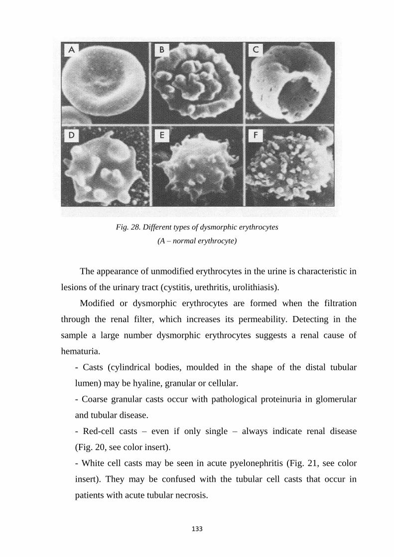
133
Fig. 28. Different types of dysmorphic erythrocytes
(A – normal erythrocyte)
The appearance of unmodified erythrocytes in the urine is characteristic in
lesions of the urinary tract (cystitis, urethritis, urolithiasis).
Modified or dysmorphic erythrocytes are formed when the filtration
through the renal filter, which increases its permeability. Detecting in the
sample a large number dysmorphic erythrocytes suggests a renal cause of
hematuria.
- Casts (cylindrical bodies, moulded in the shape of the distal tubular
lumen) may be hyaline, granular or cellular.
- Coarse granular casts occur with pathological proteinuria in glomerular
and tubular disease.
- Red-cell casts – even if only single – always indicate renal disease
(Fig. 20, see color insert).
- White cell casts may be seen in acute pyelonephritis (Fig. 21, see color
insert). They may be confused with the tubular cell casts that occur in
patients with acute tubular necrosis.
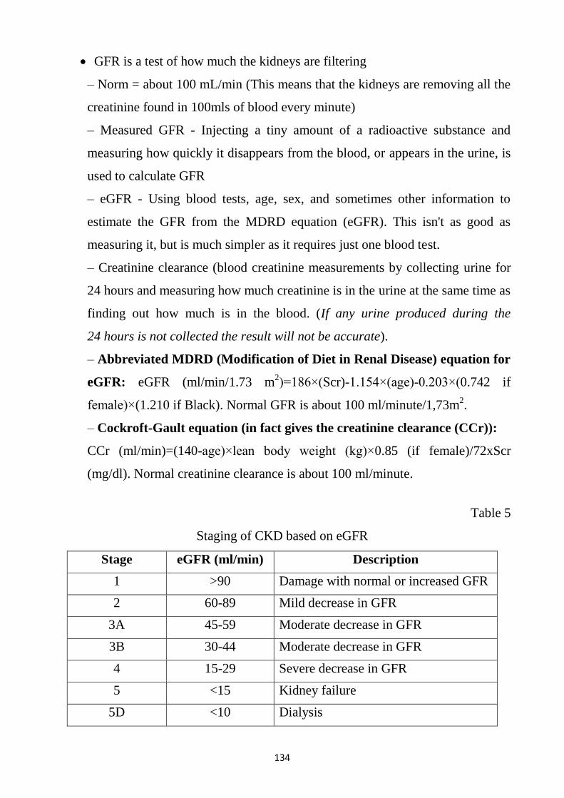
134
GFR is a test of how much the kidneys are filtering
– Norm = about 100 mL/min (This means that the kidneys are removing all the
creatinine found in 100mls of blood every minute)
– Measured GFR - Injecting a tiny amount of a radioactive substance and
measuring how quickly it disappears from the blood, or appears in the urine, is
used to calculate GFR
– eGFR - Using blood tests, age, sex, and sometimes other information to
estimate the GFR from the MDRD equation (eGFR). This isn't as good as
measuring it, but is much simpler as it requires just one blood test.
– Creatinine clearance (blood creatinine measurements by collecting urine for
24 hours and measuring how much creatinine is in the urine at the same time as
finding out how much is in the blood. (If any urine produced during the
24 hours is not collected the result will not be accurate).
– Abbreviated MDRD (Modification of Diet in Renal Disease) equation for
eGFR: eGFR (ml/min/1.73 m2)=186×(Scr)-1.154×(age)-0.203×(0.742 if
female)×(1.210 if Black). Normal GFR is about 100 ml/minute/1,73m2.
– Cockroft-Gault equation (in fact gives the creatinine clearance (CCr)):
CCr (ml/min)=(140-age)×lean body weight (kg)×0.85 (if female)/72xScr
(mg/dl). Normal creatinine clearance is about 100 ml/minute.
Table 5
Staging of CKD based on eGFR
Stage eGFR (ml/min) Description
1 >90 Damage with normal or increased GFR
2 60-89 Mild decrease in GFR
3A 45-59 Moderate decrease in GFR
3B 30-44 Moderate decrease in GFR
4 15-29 Severe decrease in GFR
5 <15 Kidney failure
5D <10 Dialysis
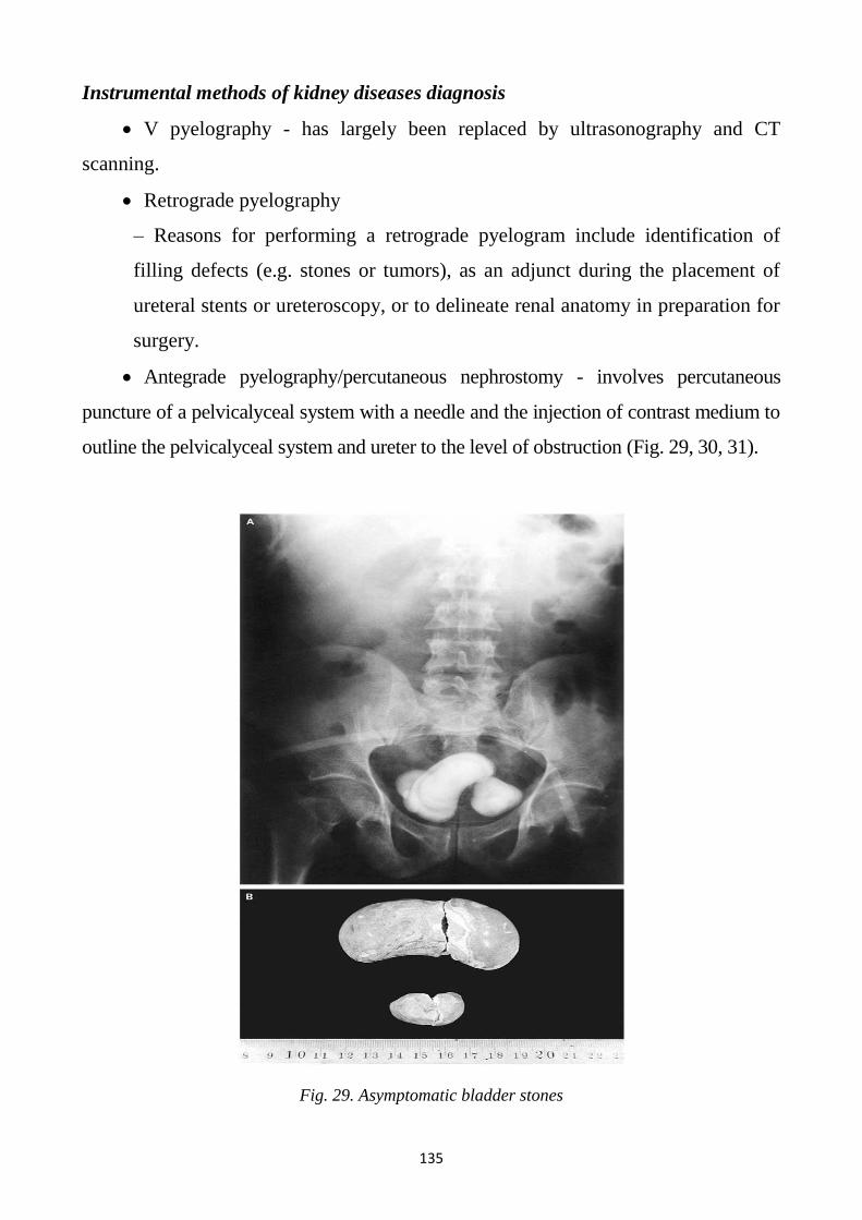
135
Instrumental methods of kidney diseases diagnosis
V pyelography - has largely been replaced by ultrasonography and CT
scanning.
Retrograde pyelography
– Reasons for performing a retrograde pyelogram include identification of
filling defects (e.g. stones or tumors), as an adjunct during the placement of
ureteral stents or ureteroscopy, or to delineate renal anatomy in preparation for
surgery.
Antegrade pyelography/percutaneous nephrostomy - involves percutaneous
puncture of a pelvicalyceal system with a needle and the injection of contrast medium to
outline the pelvicalyceal system and ureter to the level of obstruction (Fig. 29, 30, 31).
Fig. 29. Asymptomatic bladder stones
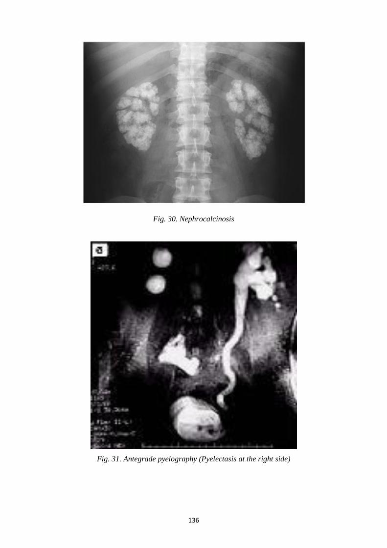
136
Fig. 30. Nephrocalcinosis
Fig. 31. Antegrade pyelography (Pyelectasis at the right side)
137
CT scan Renal calculi, characterization of masses, renal trauma,
retroperitoneal lesion CT + IV contrast can reveals kidney function
Renal angiography - Assessment of renal artery stenosis
MRI -to characterize renal masses, demonstrate the renal arteries and cancer
staging.
Radionuclide imaging
– Dynamic scintigraphy: investigation of obstruction,RBF & GFR.
– Static scintigraphy: Kidney visualization,localization of infection, renal
function.
Renal biopsy (Fig. 22-26, see color insert).
– Renal biopsy is carried out under ultrasound control in specialized centres
and requires interpretation by an experienced pathologist.
– Renal biopsy is helpful in the investigation of the nephritic and nephrotic
syndromes, acute and chronic renal failure, haematuria after urological
investigations and renal graft dysfunction.
Urinary system diseases’ syndromes
Glomerulopathy (GN)
Glomerulopathies are the third most common causes of endstage renal
disease.
Glomerulopathy is a general term for a group of disorders in which:
- the kidney are involved symmetrically
- there is primarily an immunologically mediated injury to glomeruli
- may be a part of generalized disease
Classsification of glomerulopathies:
- nephrotic syndrome
- nephritic syndrome
Clinical presentations of GN
1. Hypoalbuminemia. – Pedal oedema, Ascites, Anasarca, Pleural Effusion.
138
2. Protenuria. – Frothy urine.
3. Hematuria. – Microscopic or bloody urine.
4. Hypertension. – Asymptomatic. – Headache, confusion, seizure (hypertensive
crisis).
5. Fluid retention. – Reduced effort tolerance, orthopnea. – Acute pulmonary
edema. – Reduced urine output (oliguria).
6. Uremia. – Nausea, vomiting. – Confusion, seizure.
Nephrotic syndrome
Definition
Clinical and laboratory syndrome characterized by massive proteinuria, which
lead to hypoproteinemia ( hypoalbuminemia), hyperlipidemia and pitting edema in
results from increased permeability of glomerular basement membrane (GBM) to
plasma protein
Criteria:
- hematuria (RBC in urine, gross hematuria)
- hypertension (≥140 /90 mmHg)
- azotemia (renal insufficiency - Increased level of serum BUN, Cr)
- hypocomplementemia (decreased level of serum c3)
Causes of neprotic syndrome:
1. Primary - minimal change GN - membranous GN - focal segmental
glomerulosclerosis - Ig A nephropathy.
2. Secondary - infection; HBV, HIV,CMV, - malignancy; leukemia, lymphoma -
drug/toxin; NSAID, mercury - CT disease; SLE - metabolic disease; DM.
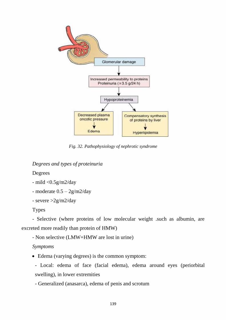
139
Fig. 32. Pathophysiology of nephrotic syndrome
Degrees and types of proteinuria
Degrees
- mild <0.5g/m2/day
- moderate 0.5 – 2g/m2/day
- severe >2g/m2/day
Types
- Selective (where proteins of low molecular weight .such as albumin, are
excreted more readily than protein of HMW)
- Non selective (LMW+HMW are lost in urine)
Symptoms
Edema (varying degrees) is the common symptom:
- Local: edema of face (facial edema), edema around eyes (periorbital
swelling), in lower extremities
- Generalized (anasarca), edema of penis and scrotum
140
Other clinical symptoms
- fatigue, lethargy
- loss of appetite, nausea and vomiting ,abdominal pain, diarrhea
- body weight increase
- urine output decrease
- pleural effusion (respiratory distress)
Blood tests: serum protein >5.5gm/dL, albumin <2.5gm/dL, cholesterol
>220 mg/dl)
Urine tests: proteinuria, oliguria (during stage of edema formation), microscopic
hematuria 20%, large number of hyaline casts)
Differential diagnosis of generalized edema
Nephritic syndrom
Definition
Clinical and laboratory syndrome associated with disorders affecting the
kidneys, more specifically glomerular structures, and characterized by having a thin
glomerular basement membrane and small pores in the podocytes of the glomerulus,
large enough to permit proteins (proteinuria) and red blood cells (hematuria) to pass
into the urine.
Criteria:
- hematuria , with red blood cell (RBC) casts present in the urine
- proteinuria (<3.5 g/day)
- hypertension
- uremia, due to retention of waste products
- variable renal insufficiency, with azotemia, oliguria (low urine output
<400 mL/day)
Types:
- post-streptococcal glomerulonephritis
- crescentic glomerulonephritis (rapidly progressive glomerulonephritis)
141
Symptoms:
- hematuria (e.g. cola coloured)
- proteinuria
Hypertension (with headache):
- oliguria
- flank pain
- general symptoms
- post-infectious (2-3 weeks after strep-throat/URTI)
Differential diagnosis:
- malignancy (older patients)
- UTI
- Trauma
Urinary tract obstruction
- can occur at any point in the urinary tract, from the kidneys to the urethral
meatus
- it can develop secondary to calculi, tumors, strictures, anatomical
abnormalities, or functional abnormalities
- obstructive uropathy can result in pain, urinary tract infection, loss in renal
function, or, possibly, sepsis or death
Urinary tract infection (UTI)
Presence of pure growth of >100000 colony forming units/ml in urine with
pyuria.
UTI sites: bladder (cystitis), prostate (prostatitis), kidney (pyelonephritis)
Cystitis symptoms: frequency, dysuria, urgency, hematuria, suprapubic pain.
Pyelonephritis symptoms: fever, rigors, vomiting, loin pain, tenderness,
oliguria.
142
Prostatitis symptoms: flu-like symptoms, low backache, swollen and tender
prostate.
Hypertensive syndrome
- elevated > 140/90 mm Hg blood pressure (renal or renovascular hypertension),
caused by a narrowing in the arteries that deliver blood to the kidney (renal artery
stenosis)
- when the kidneys receive low blood flow, they respond by releasing hormones
that stimulate the body to retain sodium and water, blood vessels fill with additional
fluid, and blood pressure increases
- the narrowing in one or both renal arteries is most often caused by
atherosclerosis, or hardening of the arteries
- symptoms: headache, confusion, blurred or double vision, bloody
(pinkcolored) urine, nosebleed, bruits over affected renal artery
- hypertension can cause chronic kidney disease
Renal Failure
Definition
- Significant deterioration in renal function occur over hrs or days.
- Reversible over days /weeks(injury to kidney is short term and potentially
reversible)
- Clinically no symptom or sign but oliguria ( < 400 ml/day) common.
- No long term complication seen in CKD eg:renal anemia,renal bone disease.
Causes:
1. Prenal - failure or perfusion of kidney - hypovolaemia,↓CO, renal artery
obstruction
2. Intrinsic renal failure - acute tubular necrosis, acute interstitial nephritis,
acute GN
3. Post-renal - UTO Renal Post-Renal Acute Renal Failure Pre-Renal
143
Clinical feature: Early stage asymptomatic - ARF does not produce a classic set
of symptoms. The most common symptom is decreased urine output, which occurs in
70% of patients.
Chronic Kidney Disease (CKD)
Definition
CKD implies long-standing, and usually progressive, impairment in renal
function. In many instances, no effective means are available to reverse the primary
disease process
Classification of CRF is based on the grade of severity and characteristic clinical
manifestations.
- Mild: GFR is 30-50 ml/min.
- Moderate: GFR is 10-30 ml/min. – anemia; – hypertension; –
osteodystrophy.
- Grave: GFR is 5-10 ml/min. – nausea; – anorexia; – pruritus.
- Terminal (end-stage): GFR is <5 ml/min. – pericarditis; – pulmonary
edema; – coma.
In end-stage CRF pharmacotherapy is ineffective, long-term dialysis or
transplantation should be considered for prolongation of life.
Symptoms and Signs.
Patients with mildly diminished renal reserve are asymptomatic, and renal
dysfunction can be detected only by laboratory testing. Skin with peculiar greenish
tint (urochromes retention) allows to suspect CRF. Anemia pathogenesis as a sign of
renal damage is combined:
- influence of uremic toxins on a marrow;
- decrease of RBC's life span in uremia;
- kidneys' inability to produce sufficient supply of erythropoietin, essential for
maintenance of marrowy erythropoiesis, in pronounced nephrosclerosis.
144
A patient with mild to moderate renal insufficiency may have only vague
symptoms despite elevated creatinine; nocturia is noted, principally due to a failure to
concentrate the urine during the night. Lassitude, fatigue, and decreased mental
acuity often are the first manifestations of uremia. Neuromuscular features include
coarse muscular twitches, muscle cramps, and convulsions (usually the result of
hypertensive or metabolic encephalopathy). Anorexia, nausea, vomiting, hiccup,
diarrhea, stomatitis, and an unpleasant taste in the mouth are almost uniformly
present. Malnutrition leading to generalized tissue wasting is a prominent feature of
chronic uremia. Skin is dry with excoriations due to pruritus, tongue is of brownish
colour, dry with fetor ammonia In advanced CRF, GI ulceration and bleeding are
common. Hypertension is present in > 80% of patients with advanced renal
insufficiency and is usually related to hypervolemia and occasionally to activation of
the renin-angiotensin-aldosterone system. Cardiomyopathy (hypertensive, ischemic)
and renal retention of Na and water may lead to congestive heart failure or dependent
edema. Fibrinous pericarditis, usually seen in end-stage uremia, may occur in acute,
potentially reversible, uremia It is manifested by severe retrosternal pain, effusion
addition is accompanied by dyspnea and other signs of heart tamponade.
The skin may appear yellow-brown; occasionally, urea from sweat may
crystallize on the skin as uremic frost. Pruritus is especially uncomfortable for some
patients. Abnormalities with lipid metabolism also occur with CRF, on dialysis, and
after renal transplantation. The primary finding in CRF and dialysis is
hypertriglyceridemia; the total cholesterol level is usually normal.
- Produces symptoms when renal function – which is measured as the
glomerular filtration rate (GFR) – falls below 30 milliliters per minute (<30 mL/min).
This is approximately 30% of the normal value.
- When GFR slows to below 30 mL/min, signs of uremia (high blood level of
protein by-products, such as urea) may become noticeable. When the GFR falls
below 15 mL/min most people become increasingly symptomatic.
145
Clinical features of severe uraemia:
1. Anaemia - Pallor; fatigue; malaise
2. Platelet abnormality - Epistaxis, bruishing
3. GI - Anorexia; nausea; vomiting; metallic taste; hiccups
4. CNS - Confusion; Irritability; poor concentration; insomnia; restless legs;
twitching; coma; fits
5. Skin - hyperpigmentation, pruritis
6. Cardiovascular system - Uraemic pericarditis, heart failure
7. Renal - Nocturia, polyuria, salt & water retention cause edema.
8. Renal osteodystrophy - osteomalacia, muscle weakness, bone pain,
hyperPTH, osteoslerosis
9. Endocrine - amenorrhoe, erectile impotence, infertility
Diagnosis
The first step is to determine whether the renal failure is acute, chronic, or acute
superimposed on chronic. Progression to CRF is common when the serum creatinine
concentration is >1.5 to 2 mg/dL. This may occur even if the underlying disorder is
not active. Obtaining a precise diagnosis becomes increasingly difficult as the patient
approaches end-stage renal disease. The definitive diagnostic tool is renal biopsy, but
it is not recommended when ultrasonography indicates that the kidneys are small and
fibrotic. Urea and creatinine are elevated. Plasma Na concentrations may be normal
or reduced. The serum K is normal or only moderately elevated (< 6 mmol/L).
Usually, moderate anemia is characteristic. The anemia of CRF is normochromic-
normocytic, with an Hct of 20 to 30%. It is usually caused by deficient erythropoietin
production due to a reduction of functional renal mass. Other causes include
deficiencies of iron, folate, and cyanocobalamin. Urinary volume does not respond
readily to variations in water intake. Findings on urinalysis depend on thenature of
the underlying disease, but broad (especially waxy) casts often are prominent in
advanced renal insufficiency of any cause.
146
Test control for the theme “Clinical diagnosis of diseases
of the urinary system”
1. WHAT IS THE INCREASE IN THE DAILY AMOUNT OF URINE?
a) pollakiuria
b) strangury
c) ischuria
d) anuria
e) polyuria
2. WHAT IS THE BASIC MECHANISMS OF RENAL COLIC?
a) stretching of renal capsule in the increase in volume (swelling) of renal
tissue
b) spasm of the ureters
3. SOME OF THE MECHANISMS OF FORMATION OF EDEMA
SYNDROME PREVAILS IN NEPHROTIC SYNDROME?
a) violation of vascular permeability
b) activate system:aldosterone-ADH
c) the decrease of oncotic pressure of plasma
d) a sharp decrease of filtration of the kidneys - retention swelling
e) a sharp increase in hydrostatic pressure in the venous line with the blood
circulation
4. The PATIENT 19 YEARS IN the GENERAL ANALYSIS of URINE: specific
gravity - 1028, proteinuria of 3.5 g/l, leukocytes 8-10, red blood cells - 10 in p/Zr.
WHAT KIND OF DISEASE THINK?
a) acute pyelonephritis
b) chronic pyelonephritis
147
c) acute glomerulonephritis
d) chronic renal failure
e) urolithiasis
5. CHARACTERISTIC OF RENAL ARTERIAL HYPERTENSION (CHRONIC
KIDNEY DISEASE):
a) labile AD, roughly equivalent to the increase in systolic and diastolic blood
pressure, malignant hypertension is rare,renal failure is rare;
b) a stable increase in pressure,increase in systolic and especially diastolic blood
pressure, often there is a malignant arterial hypertension, often develop chronic renal
failure.
6. WHAT FEATURES ARE CHARACTERISTIC FOR UREMIA?
a) the smell of ammonia breath
b) pericardial RUB
c) dianagonsales
d) increasing the content of urea and creatinine in blood serum,anemia
e) stupor,coma
f) all of the above
7. WHAT ARE THE SYMPTOMS CHARACTERISTIC OF THE ACUTE
GLOMERULONEFRITE?
a) persistent back pain
b) fever with chills
c) gross hematuria
d) anaemia
e) all of the above
148
8. WHAT ARE CLINICAL AND LABORATORY INDICATIONS SHOW
CONCENTRATION FUNCTION OF KIDNEYS?
a) pollakiuria,nocturia
b) izostenuriya, gipostenuriya
c) azotemia,anuria,proteinuria
9. HYPERTENSION IS CHARACTERISTIC FOR:
a) nephrotic syndrome
b) nephritic syndrome
c) edematous syndrome
10. BELOW ARE SOME OF THE SIGNS IDENTIFIED IN PATIENTS WITH
GLOMERULONEPHRITIS. SELECT FROM THEM THOSE THAT ALLOW TO
DISTINGUISH CHRONIC GLOMERULONEPHRITIS FROM ACUTE?
a) level of BP increase, the severity of edema syndrome, accent II Tanana the
aorta, the level of proteinuria,cylindruria the presence of more frequent occurrence of
microscopic hematuria;
b) enhanced apical impulse, displacement of the left border serdtsevina, ECG-
signs of hypertrophy of the left ventricle, the presence of ISO-hyposthenuria,
nocturia, polyuria.
11. KAKNAZYVAETSYA COMPLETE CESSATION OF URINE OUTPUT BY
THE KIDNEYS?
a) polyuria
b) pollakiuria
с) strangury
d) ischuria
e) anuria
149
12. OF THE FOLLOWING MECHANISMS OF THE PAIN SYNDROME IN
DISEASES OF THE KIDNEY, SELECT THE ONE THAT PROVES TO BE THE
LEADING ACUTE GLOMERULONEFRITE:
a) stretching of renal capsule in the increase in volume (swelling) of tissue
b) stretching of renal pelvis due to zatrudneniya through the ureter
c) spastic contractions of the ureter
13. WHICH OF THE FOLLOWING CRITERIA IS ESSENTIAL FOR DIAGNOSIS
NEPHROTIC SYNDROME?
a) edema
b) serum albumin below 30 g/l
c) daily proteinuria more than 3.5 g/day
d) hypercholesterolemia
e) hypercoagulability
14. WHAT CHARACTERISTICS DISTINGUISH ECLAMPSIA FROM RENAL
HYPERTENSIVE CRISIS (2 answeres)?
a) the presence of seizures
b) severe headaches
c) blurred vision
d) nausea
e) dizziness
15. WHAT MANIFESTATION DOES NOT MATTER FOR THE DIFFERENTIAL
DIAGNOSIS OF ACUTE AND CHRONIC GLOMERULONEFRITA?
a) the presence of persistent arterial hypertension
b) the level of proteinuria
c) left ventricular hypertrophy
d) reduction of relative density of urine
e) anamnestic data
150
16. THE PATIENT 35 YEARS OLD THE NEXT DAY RECOVERING FROM A
SORE THROAT APPEARED SWELLING PARAORBITALNAH REGION,
GROSS HEMATURIA, INCREASED HELL. THE MOST LIKELY DIAGNOSIS?
a) acute glomerulonefrit
b) acute pyelonephritis
c) cronicacircotasilor, exacerbation
d) jade
e) amyloidosis
17. WHAT kind of DISEASE AND SYNDROMES LIKELY to be involved IN the
FOLLOWING CLINICAL SITUATION: the patient was admitted to the Department
with intense sharp constant pains in the right lumbar region, the increase was
temperature to 380 and small swelling under the eyes. The pain appeared after
exposure and lasted about 5-7 days. After treatment remain long dull aching lower
back pain?
a) acute glomerulonephritis
b) acute pyelonephritis(inflammation of renal pelvis and renal tissue)
c) urolithiasis
d) a"congestive kidney" (heart failure)
e) cystitis
f) urethritis
18. WHAT IS THE INABILITY TO EMPTY THE BLADDER (URINARY
RETENTION)?
a) pollakiuria
b) strangury
c) ischuria
d) anuria
e) polyuria
151
19. WHAT TEST IS PERFORMED TO STUDY THE CONCENTRATION
FUNCTION OF KIDNEYS?
a) the Cocraft-Gault equation
b) sample Nechiporenko
c) test of General
d) sample Adisson-Kotovskogo
e) test with prednisone
20. HOW IS CALLED PAINFUL URINATION?
a) pollakiuria
b) strangury
c) ischuria
d) anuria
e) polyuria
152
Учебное издание
Smirnova A.Yu., Gnoevykh V.V.
INTERNAL DISEASES PROPEDEUTICS
PART III
DIAGNOSTICS OF DISEASES OF GASTROINTESTINAL TRACT AND
KIDNEYS
Textbook of medicine for medicine faculty students
Директор Издательского центра Т.В. Филиппова
Подготовка оригинал-макета, обложки Е.П. Мамаевой
Печатается в авторской редакции
Подписано в печать 13.12.2017. Формат 6084/16.
Гарнитура Times New Roman. Усл. печ. л. 1,9. Уч.-изд. л. 1,6.
Тираж 100 экз. Заказ № 173/
Оригинал-макет подготовлен в Издательском центре
Ульяновского государственного университета
432017, г. Ульяновск, ул. Л. Толстого, 42
Отпечатано с оригинал-макета в Издательском центре
Ульяновского государственного университета
432017, г. Ульяновск, ул. Л. Толстого, 42
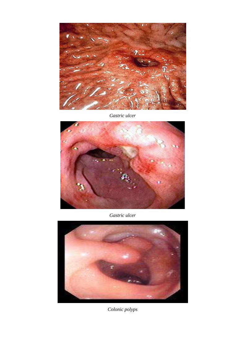
Gastric ulcer
Gastric ulcer
Colonic polyps
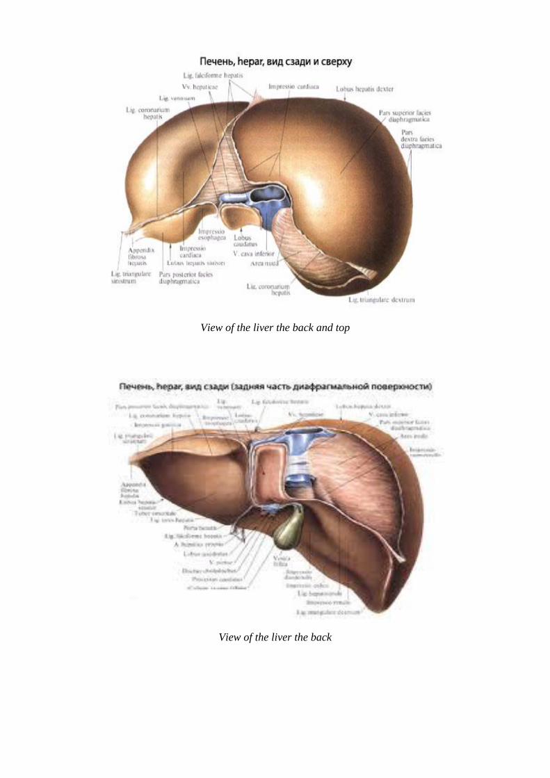
View of the liver the back and top
View of the liver the back
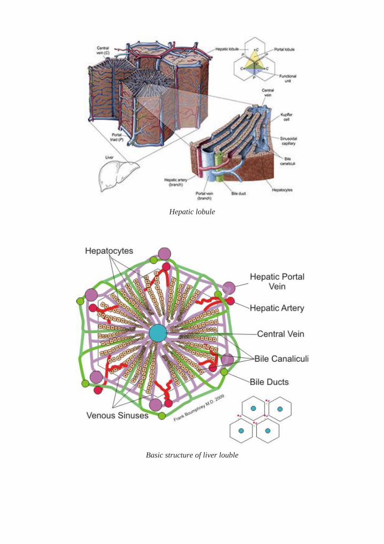
Hepatic lobule
Basic structure of liver louble
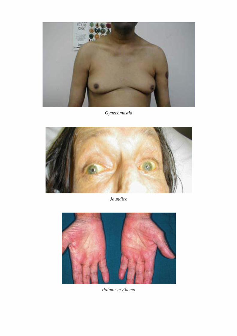
Gynecomastia
Jaundice
Palmar erythema
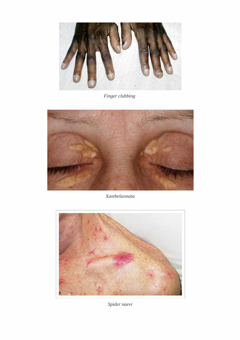
Finger clubbing
Xanthelasmata
Spider naevi
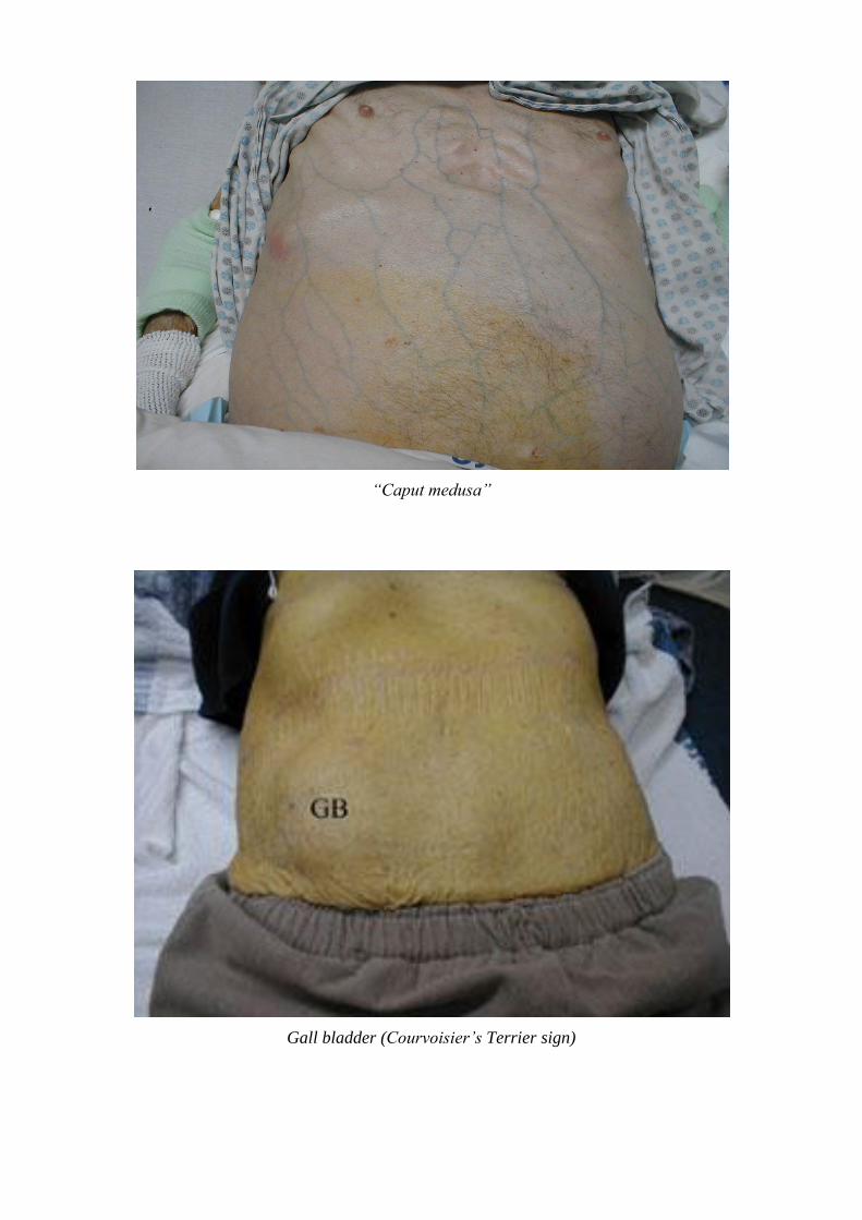
“Caput medusa”
Gall bladder (Courvoisier’s Terrier sign)
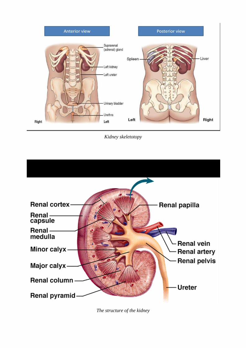
Kidney skeletotopy
The structure of the kidney
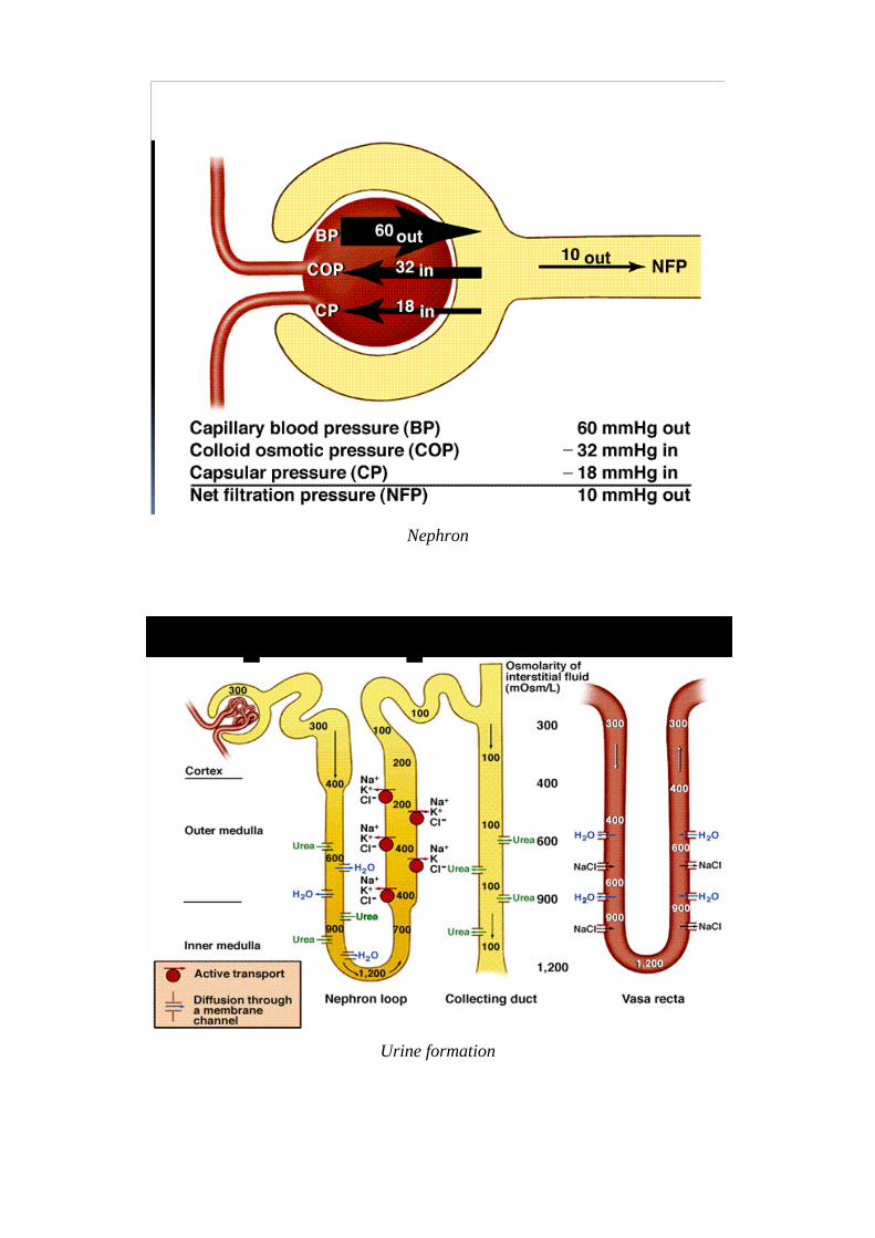
Nephron
Urine formation
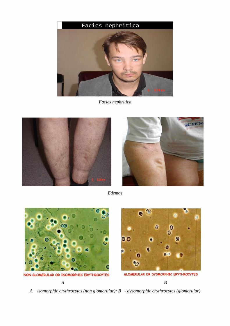
Facies nephritica
Facies nephritica
Edemas
A B
A – isomorphic erythrocytes (non glomerular); B –- dysomorphic erythrocytes (glomerular)
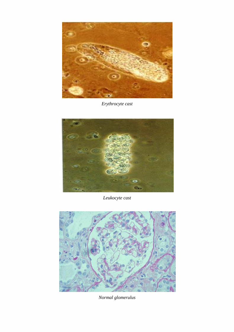
Erythrocyte cast
Leukocyte cast
Normal glomerulus
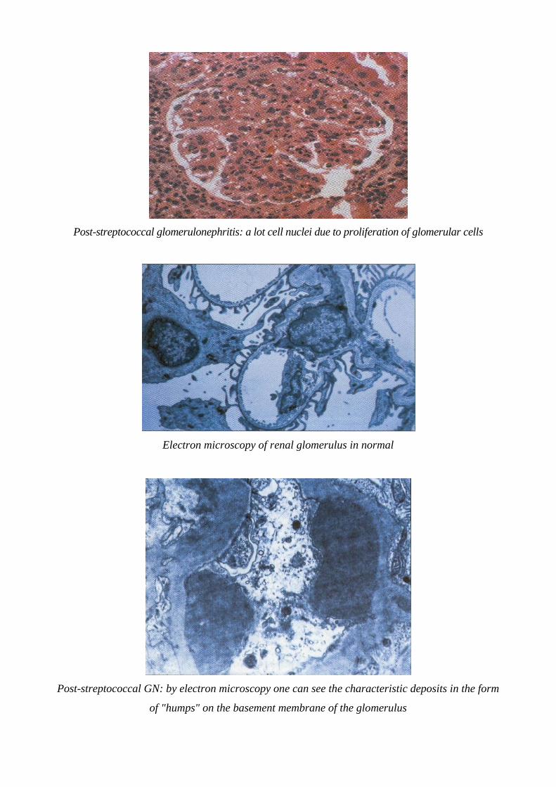
Post-streptococcal glomerulonephritis: a lot cell nuclei due to proliferation of glomerular cells
Electron microscopy of renal glomerulus in normal
Post-streptococcal GN: by electron microscopy one can see the characteristic deposits in the form
of "humps" on the basement membrane of the glomerulus
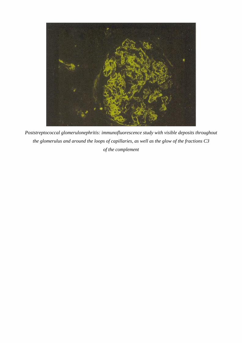
Poststreptococcal glomerulonephritis: immunofluorescence study with visible deposits throughout
the glomerulus and around the loops of capillaries, as well as the glow of the fractions C3
of the complement