Nanoparticle Delivery Platforms for RNAi Therapeutics ... - MDPI
33
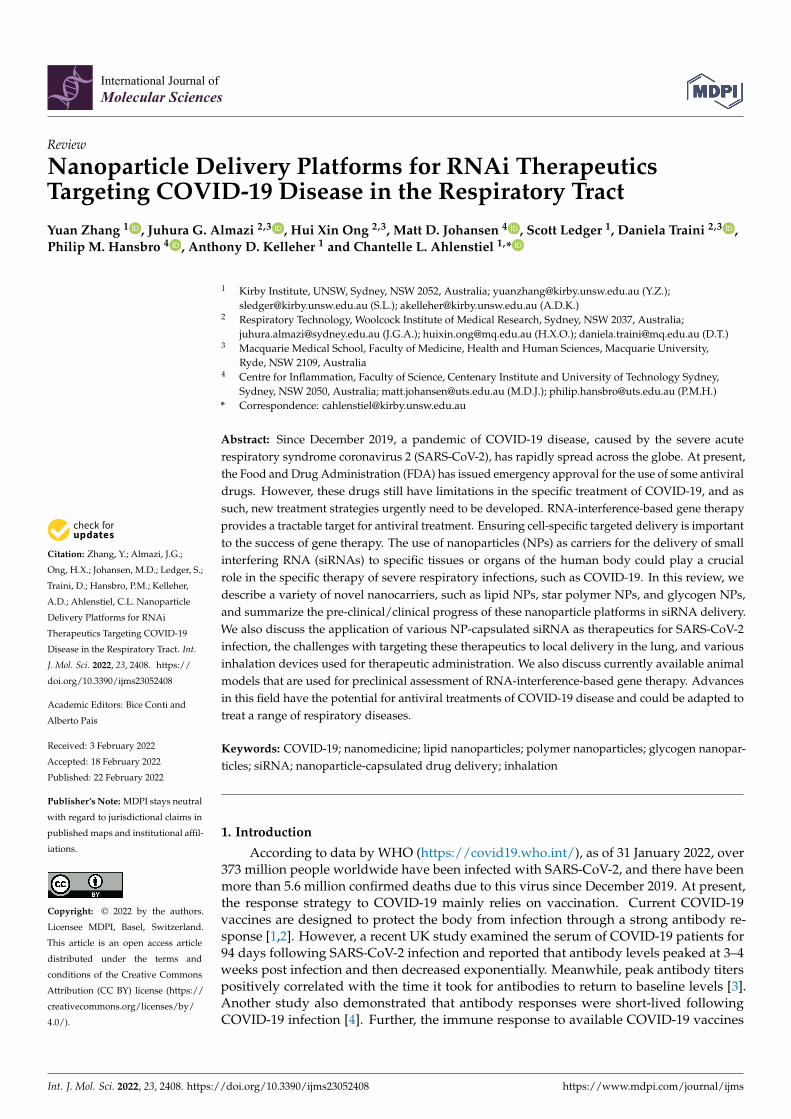
Citation: Zhang, Y.; Almazi, J.G.; Ong, H.X.; Johansen, M.D.; Ledger, S.; Traini, D.; Hansbro, P.M.; Kelleher, A.D.; Ahlenstiel, C.L. Nanoparticle Delivery Platforms for RNAi Therapeutics Targeting COVID-19 Disease in the Respiratory Tract. Int. J. Mol. Sci. 2022, 23, 2408. https:// doi.org/10.3390/ijms23052408 Academic Editors: Bice Conti and Alberto Pais Received: 3 February 2022 Accepted: 18 February 2022 Published: 22 February 2022 Publisher’s Note: MDPI stays neutral with regard to jurisdictional claims in published maps and institutional affil- iations. Copyright: © 2022 by the authors. Licensee MDPI, Basel, Switzerland. This article is an open access article distributed under the terms and conditions of the Creative Commons Attribution (CC BY) license (https:// creativecommons.org/licenses/by/ 4.0/). International Journal of Molecular Sciences Review Nanoparticle Delivery Platforms for RNAi Therapeutics Targeting COVID-19 Disease in the Respiratory Tract Yuan Zhang 1 , Juhura G. Almazi 2,3 , Hui Xin Ong 2,3 , Matt D. Johansen 4 , Scott Ledger 1 , Daniela Traini 2,3 , Philip M. Hansbro 4 , Anthony D. Kelleher 1 and Chantelle L. Ahlenstiel 1, * 1 Kirby Institute, UNSW, Sydney, NSW 2052, Australia; [email protected] (Y.Z.); [email protected] (S.L.); [email protected] (A.D.K.) 2 Respiratory Technology, Woolcock Institute of Medical Research, Sydney, NSW 2037, Australia; [email protected] (J.G.A.); [email protected] (H.X.O.); [email protected] (D.T.) 3 Macquarie Medical School, Faculty of Medicine, Health and Human Sciences, Macquarie University, Ryde, NSW 2109, Australia 4 Centre for Inflammation, Faculty of Science, Centenary Institute and University of Technology Sydney, Sydney, NSW 2050, Australia; [email protected] (M.D.J.); [email protected] (P.M.H.) * Correspondence: [email protected] Abstract: Since December 2019, a pandemic of COVID-19 disease, caused by the severe acute respiratory syndrome coronavirus 2 (SARS-CoV-2), has rapidly spread across the globe. At present, the Food and Drug Administration (FDA) has issued emergency approval for the use of some antiviral drugs. However, these drugs still have limitations in the specific treatment of COVID-19, and as such, new treatment strategies urgently need to be developed. RNA-interference-based gene therapy provides a tractable target for antiviral treatment. Ensuring cell-specific targeted delivery is important to the success of gene therapy. The use of nanoparticles (NPs) as carriers for the delivery of small interfering RNA (siRNAs) to specific tissues or organs of the human body could play a crucial role in the specific therapy of severe respiratory infections, such as COVID-19. In this review, we describe a variety of novel nanocarriers, such as lipid NPs, star polymer NPs, and glycogen NPs, and summarize the pre-clinical/clinical progress of these nanoparticle platforms in siRNA delivery. We also discuss the application of various NP-capsulated siRNA as therapeutics for SARS-CoV-2 infection, the challenges with targeting these therapeutics to local delivery in the lung, and various inhalation devices used for therapeutic administration. We also discuss currently available animal models that are used for preclinical assessment of RNA-interference-based gene therapy. Advances in this field have the potential for antiviral treatments of COVID-19 disease and could be adapted to treat a range of respiratory diseases. Keywords: COVID-19; nanomedicine; lipid nanoparticles; polymer nanoparticles; glycogen nanopar- ticles; siRNA; nanoparticle-capsulated drug delivery; inhalation 1. Introduction According to data by WHO (https://covid19.who.int/), as of 31 January 2022, over 373 million people worldwide have been infected with SARS-CoV-2, and there have been more than 5.6 million confirmed deaths due to this virus since December 2019. At present, the response strategy to COVID-19 mainly relies on vaccination. Current COVID-19 vaccines are designed to protect the body from infection through a strong antibody re- sponse [1,2]. However, a recent UK study examined the serum of COVID-19 patients for 94 days following SARS-CoV-2 infection and reported that antibody levels peaked at 3–4 weeks post infection and then decreased exponentially. Meanwhile, peak antibody titers positively correlated with the time it took for antibodies to return to baseline levels [3]. Another study also demonstrated that antibody responses were short-lived following COVID-19 infection [4]. Further, the immune response to available COVID-19 vaccines Int. J. Mol. Sci. 2022, 23, 2408. https://doi.org/10.3390/ijms23052408 https://www.mdpi.com/journal/ijms
-
Upload
khangminh22 -
Category
Documents
-
view
0 -
download
0
Transcript of Nanoparticle Delivery Platforms for RNAi Therapeutics ... - MDPI
�����������������
Citation: Zhang, Y.; Almazi, J.G.;
Ong, H.X.; Johansen, M.D.; Ledger, S.;
Traini, D.; Hansbro, P.M.; Kelleher,
A.D.; Ahlenstiel, C.L. Nanoparticle
Delivery Platforms for RNAi
Therapeutics Targeting COVID-19
Disease in the Respiratory Tract. Int.
J. Mol. Sci. 2022, 23, 2408. https://
doi.org/10.3390/ijms23052408
Academic Editors: Bice Conti and
Alberto Pais
Received: 3 February 2022
Accepted: 18 February 2022
Published: 22 February 2022
Publisher’s Note: MDPI stays neutral
with regard to jurisdictional claims in
published maps and institutional affil-
iations.
Copyright: © 2022 by the authors.
Licensee MDPI, Basel, Switzerland.
This article is an open access article
distributed under the terms and
conditions of the Creative Commons
Attribution (CC BY) license (https://
creativecommons.org/licenses/by/
4.0/).
International Journal of
Molecular Sciences
Review
Nanoparticle Delivery Platforms for RNAi TherapeuticsTargeting COVID-19 Disease in the Respiratory TractYuan Zhang 1 , Juhura G. Almazi 2,3 , Hui Xin Ong 2,3, Matt D. Johansen 4 , Scott Ledger 1, Daniela Traini 2,3 ,Philip M. Hansbro 4 , Anthony D. Kelleher 1 and Chantelle L. Ahlenstiel 1,*
1 Kirby Institute, UNSW, Sydney, NSW 2052, Australia; [email protected] (Y.Z.);[email protected] (S.L.); [email protected] (A.D.K.)
2 Respiratory Technology, Woolcock Institute of Medical Research, Sydney, NSW 2037, Australia;[email protected] (J.G.A.); [email protected] (H.X.O.); [email protected] (D.T.)
3 Macquarie Medical School, Faculty of Medicine, Health and Human Sciences, Macquarie University,Ryde, NSW 2109, Australia
4 Centre for Inflammation, Faculty of Science, Centenary Institute and University of Technology Sydney,Sydney, NSW 2050, Australia; [email protected] (M.D.J.); [email protected] (P.M.H.)
* Correspondence: [email protected]
Abstract: Since December 2019, a pandemic of COVID-19 disease, caused by the severe acuterespiratory syndrome coronavirus 2 (SARS-CoV-2), has rapidly spread across the globe. At present,the Food and Drug Administration (FDA) has issued emergency approval for the use of some antiviraldrugs. However, these drugs still have limitations in the specific treatment of COVID-19, and assuch, new treatment strategies urgently need to be developed. RNA-interference-based gene therapyprovides a tractable target for antiviral treatment. Ensuring cell-specific targeted delivery is importantto the success of gene therapy. The use of nanoparticles (NPs) as carriers for the delivery of smallinterfering RNA (siRNAs) to specific tissues or organs of the human body could play a crucialrole in the specific therapy of severe respiratory infections, such as COVID-19. In this review, wedescribe a variety of novel nanocarriers, such as lipid NPs, star polymer NPs, and glycogen NPs,and summarize the pre-clinical/clinical progress of these nanoparticle platforms in siRNA delivery.We also discuss the application of various NP-capsulated siRNA as therapeutics for SARS-CoV-2infection, the challenges with targeting these therapeutics to local delivery in the lung, and variousinhalation devices used for therapeutic administration. We also discuss currently available animalmodels that are used for preclinical assessment of RNA-interference-based gene therapy. Advancesin this field have the potential for antiviral treatments of COVID-19 disease and could be adapted totreat a range of respiratory diseases.
Keywords: COVID-19; nanomedicine; lipid nanoparticles; polymer nanoparticles; glycogen nanopar-ticles; siRNA; nanoparticle-capsulated drug delivery; inhalation
1. Introduction
According to data by WHO (https://covid19.who.int/), as of 31 January 2022, over373 million people worldwide have been infected with SARS-CoV-2, and there have beenmore than 5.6 million confirmed deaths due to this virus since December 2019. At present,the response strategy to COVID-19 mainly relies on vaccination. Current COVID-19vaccines are designed to protect the body from infection through a strong antibody re-sponse [1,2]. However, a recent UK study examined the serum of COVID-19 patients for94 days following SARS-CoV-2 infection and reported that antibody levels peaked at 3–4weeks post infection and then decreased exponentially. Meanwhile, peak antibody titerspositively correlated with the time it took for antibodies to return to baseline levels [3].Another study also demonstrated that antibody responses were short-lived followingCOVID-19 infection [4]. Further, the immune response to available COVID-19 vaccines
Int. J. Mol. Sci. 2022, 23, 2408. https://doi.org/10.3390/ijms23052408 https://www.mdpi.com/journal/ijms

Int. J. Mol. Sci. 2022, 23, 2408 2 of 33
drop at 3 and 6 months after the second dose of vaccination, and antibody levels to thevaccines negatively correlated with the age of vaccinated individuals [5]. Moreover, vac-cination is likely to be suboptimal in those with impaired immunity and allergies and befurther compromised by the emergence of variants of concern (VOC). Vaccination may notprovide adequate protection for people with a dysfunctional immune system. Therefore, toeffectively reduce the risk of serious illness and hospitalization, it is necessary to developantiviral treatments against COVID-19.
All direct-acting antiviral therapies that are active against SARS-CoV-2 need to be givenearly in the disease course, so they need to be administered easily and should be safe [6].Several drugs are being tested for the treatment of COVID-19, which include antiviral, anti-malarial, and anticancer agents. Notably, the latest two orally delivered antiviral drugs, thePfizer Paxlovid (a protease inhibitor) and Merck molnupiravir (a polymerase inhibitor) havebeen proven to be effective in treating SARS-CoV-2 infection [7–9]. Particularly, Paxlovidmight have therapeutic potential in the newly emerging Omicron variant [10], which isfundamentally different from previous VOC, as it does not appear to use the TMPRSS2protease co-receptor [11,12] or target the lung as effectively as previous ones [13–17]. Asthere is currently no specific treatment for SARS-CoV-2, drugs for the treatment of otherviral infections, such as Remdesivir, Baricitinib, Tocilizumab, and Favipiravir, have beenrepurposed and may be used to assist in the treatment of COVID-19 [18]. Some of theseantiviral drugs have already been shown to have limitations in treating SARS-CoV-2, andthe side effects of those antiviral drugs on COVID-19 patients also remain to be explored [19].Other available therapeutics such as the intravenous (IV) injection of monoclonal antibodies(mAb) are highly efficacious; however, production is intensive, time-consuming, andexpensive [20]. Moreover, the therapeutic efficacy of mAbs is significantly affected by avirus with a high mutation rate, and it is difficult to perform organ-targeted therapy [21].Thus, the therapeutic effects and side effects of these currently available antiviral drugs onCOVID-19 patients remain to be defined [19]. Since many current treatments for COVID-19patients are repurposed therapeutics for other diseases, investigating approaches that aretargeted and specific to COVID-19 disease is highly valuable. One potential treatmentstrategy is RNA interference (RNAi), and a more targeted drug delivery platform involvesnanoparticle (NP) carriers.
RNAi is a conserved biological process induced by non-coding RNAs (ncRNAs) withthe mechanism of inhibiting gene expression by blocking the transcription or translationof specific genes. This phenomenon was first discovered and proposed in Caenorhabditiselegans by Fire et al. [22]. Small RNAs that can produce RNAi include small interfer-ing RNAs (siRNAs), microRNAs (miRNAs), P-element-induced wimpy testis (PIWI)-interacting RNAs (piRNAs) and endogenous siRNAs (esiRNAs) [23]. As a phenomenonthat has been widely studied, RNAi has been proven to be a potential treatment strategy formany diseases (viral and bacterial infections, respiratory diseases, cancer, and autoimmunediseases), including COVID-19 [24–26]. Currently, the most studied non-coding RNAs aresiRNA (exogeneous and double-stranded) and miRNA (endogenous and single-stranded),which play a vital role in gene regulation [27]. The physical and chemical properties ofsiRNA and miRNA are similar, while the mechanisms underlying their mode of action aredifferent, and their stability and targeted delivery in vivo also have challenges [28]. Studieshave found that both siRNA and miRNA have the potential to be used in treating COVID-19 [29]. Treatments directly into the lung are relatively stable for miRNAs [27]. Importantly,siRNA can be also delivered to the lung epithelium of mice via intranasal or intravenousadministration via liposomes [30,31]. This review focuses on summarizing the therapeuticrole of siRNA in COVID-19 disease. Furthermore, siRNA could inhibit viral infection by si-lencing specific genes that are required for viral entry or utilized for viral replication [32,33].A variety of siRNAs that target highly conserved regions of SARS-CoV-2 sequence havebeen identified [30,34]. Delivery systems for these siRNAs can be divided into viral vectorsand nanoparticles [35,36]. Viral delivery systems, such as adenovirus and lentivirus, are themost used viruses in research and can successfully deliver siRNA in vivo [37–39]. However,

Int. J. Mol. Sci. 2022, 23, 2408 3 of 33
several disadvantages of these vectors are known, such as their high immunogenicity [40],restriction of specific delivery to target cells [41], the need for ex vivo transduction andreinfusion [42], limited transduction rates for certain cell types [43], potential mutagenicityof integration sites [44], and complexity and high costs of GMP synthesis [45]. By contrast,several bioconjugates have been applied to carry siRNAs in vitro and in vivo, includingimmune proteins, peptides, aptamer, cholesterol, PEG, cell-penetrating peptides, and NPs.
NPs have been identified as carriers to deliver functional nucleic acid fragments intothe body for specific targeted therapy [36], which has recently become a new medicalfield [46–50]. Compared with viral delivery systems, NPs can be designed to have low orlimited immunogenicity. Another advantage of NP carriers is that they can be designed toensure that siRNA is protected from ribonuclease (RNase), making it stable and difficult todegrade by enzymatic degradation. NP carrier systems can be in the form of vesicle-basedNP suspensions after encapsulation [51]. Thus, NPs can be used as a carrier platformto encapsulate therapeutic drugs and have played an increasingly promising role in thetargeted therapy of diseases [52–54].
So far, siRNA treatment measures have mostly targeted the liver, as this organ rep-resents an accessible target due to the first-pass metabolism of both siRNA and manynanoparticles. ONPATTRO™ (patisiran), approved by the US Food and Drug Administra-tion (FDA) and the European Medicines Agency (EMA), also uses lipid NPs to effectivelydeliver siRNAs to hepatocytes for therapeutic effect [55–57]. Since SARS-CoV-2 has beenfound mainly in the respiratory tract of COVID-19 patients [58], a logical treatment strategyis to deliver therapeutics directly to the lung and respiratory tract via topical administra-tion or administration directly into the airways, avoiding the high first-pass metabolismof systemically delivered siRNAs by the liver [59]. Therefore, combining the targetingproperties of NPs with the specificity of RNAi could allow for the design of products withbetter therapeutic efficacy with lower side effect profiles. Herein, we discuss a variety ofpotential nanocarriers, including organic nanoparticles, such as lipid NPs, polymer NPs,and glycogen NPs, and inorganic nanoparticles, such as gold NPs and magnetic NPs; then,we summarize the progress of these platforms in siRNA delivery. We also present thepotential of formulating NP-siRNAs that specifically target the lungs or respiratory tractand evaluate the potential application of various NP-capsulated siRNAs (NP-siRNAs) astherapeutics for SARS-CoV-2 infection.
2. siRNA Therapy—A Potential and Promising Antiviral Therapeutic
It is well known that siRNAs are derived from long double-stranded RNAs, which arecleaved into small double-stranded RNA with the same length of ~21 nucleotides throughendonuclease Dicer-mediated cleavage. siRNAs have the potential to act as a highly specifictherapeutic strategy for the treatment of various diseases [46,60,61]. So far, the FDA hasapproved four siRNA therapies, including patisiran (for polyneuropathy of hereditaryTTR-mediated amyloidosis) [57], givosiran (for acute hepatic porphyria) [62], lumasiran(for primary hyperoxaluria type 1) [63], and inclisiran (for primary hypercholesterolemia ormixed dyslipidemia) [64]. siRNA therapy has made great progress in tumor treatments [65].siRNA therapeutics for some cancers were developed from animal experiments throughto clinical trials. A study in mice demonstrated that siRNAs were effective in reducingmelanoma growth [66]. Additionally, some siRNAs (such as CALAA-01, Atu027, andDCR-MYC) have been used in phase I clinical trials in metastatic pancreatic cancer, coloncancer, and hepatocellular carcinoma [67–71], and siG12D-LODER has been used in phaseII clinical trials in advanced pancreatic cancer [72]. Some studies have also revealed thatsiRNAs can be used to combat viral infections, especially COVID-19 [73,74].
2.1. Mechanisms of siRNA Therapy
siRNAs can induce gene silencing in mammalian cells through two distinct pathways;post-transcriptional gene silencing (PTGS) or transcriptional gene silencing (TGS) (Figure 1).PTGS primarily occur in the cytoplasm. Firstly, the guide strand of the siRNAs binds to the

Int. J. Mol. Sci. 2022, 23, 2408 4 of 33
Argonaute-2 protein (Ago-2) of the RNA-inducing silencing complex (RISC), while the othernon-guide strand is cleaved and ejected by the Ago-2 [75]. Subsequently, Ago-2 carriesthe guide strand to target mRNA complementary to its sequence, causing the degradationof the binding site of the target RNA to induce gene expression silencing. Finally, RISCejects the guide strand in preparation for the next round of gene silencing [76]. PTGS isconsidered the main pathway of siRNA-mediated mRNA silencing [77]. TGS is anotherpathway of siRNA-induced gene silencing. It is less well characterized in mammaliancells and takes place exclusively in the nucleus [78]. Unlike PTGS, in TGS, siRNA bindsto the Ago-1 protein on the RNA-induced transcriptional silencing (RITS) complex [79].Increasing the methylation and decreasing the acetylation of critical chromatin-associatedproteins, contributes to the production of heterochromatin and induces the epigeneticsilencing of the gene [80]. TGS is potentially more beneficial for the long-term treatmentof chronic infections, whereas the PTGS is likely adequate for acute infections such asCOVID-19.
Int. J. Mol. Sci. 2022, 23, x FOR PEER REVIEW 4 of 34
siRNAs can induce gene silencing in mammalian cells through two distinct path-ways; post-transcriptional gene silencing (PTGS) or transcriptional gene silencing (TGS) (Figure 1). PTGS primarily occur in the cytoplasm. Firstly, the guide strand of the siRNAs binds to the Argonaute-2 protein (Ago-2) of the RNA-inducing silencing complex (RISC), while the other non-guide strand is cleaved and ejected by the Ago-2 [75]. Subsequently, Ago-2 carries the guide strand to target mRNA complementary to its sequence, causing the degradation of the binding site of the target RNA to induce gene expression silencing. Finally, RISC ejects the guide strand in preparation for the next round of gene silencing [76]. PTGS is considered the main pathway of siRNA-mediated mRNA silencing [77]. TGS is another pathway of siRNA-induced gene silencing. It is less well characterized in mam-malian cells and takes place exclusively in the nucleus [78]. Unlike PTGS, in TGS, siRNA binds to the Ago-1 protein on the RNA-induced transcriptional silencing (RITS) complex [79]. Increasing the methylation and decreasing the acetylation of critical chromatin-asso-ciated proteins, contributes to the production of heterochromatin and induces the epige-netic silencing of the gene [80]. TGS is potentially more beneficial for the long-term treat-ment of chronic infections, whereas the PTGS is likely adequate for acute infections such as COVID-19.
Figure 1. Mechanisms of siRNA therapy. PTGS primarily occurs in the cytoplasm. The guide strand binds to Ago-2 of RISC, while the non-guide strand is cleaved and ejected by the Ago-2; Ago-2 carries the guide strand to target mRNA, causing gene expression silencing. TGS has taken place in the nucleus. siRNA binds to Ago-1 on RITS, which contributes to the production of heter-ochromatin and induces the epigenetic silencing of the gene. PTGS, post-transcriptional gene si-lencing pathway; TGS, transcriptional gene silencing; Ago-1, Argonaute-1 protein; Ago-2, Argo-naute-2 protein; RISC, RNA-induced silencing complex; RITS, RNA-induced transcriptional si-lencing complex. Created with BioRender.com.
2.2. siRNA Therapeutics for SARS-CoV-2, SARS-CoV-1, and MERS-CoV
Figure 1. Mechanisms of siRNA therapy. PTGS primarily occurs in the cytoplasm. The guide strandbinds to Ago-2 of RISC, while the non-guide strand is cleaved and ejected by the Ago-2; Ago-2 carriesthe guide strand to target mRNA, causing gene expression silencing. TGS has taken place in thenucleus. siRNA binds to Ago-1 on RITS, which contributes to the production of heterochromatinand induces the epigenetic silencing of the gene. PTGS, post-transcriptional gene silencing pathway;TGS, transcriptional gene silencing; Ago-1, Argonaute-1 protein; Ago-2, Argonaute-2 protein; RISC,RNA-induced silencing complex; RITS, RNA-induced transcriptional silencing complex. Createdwith BioRender.com.
2.2. siRNA Therapeutics for SARS-CoV-2, SARS-CoV-1, and MERS-CoV
Among all human coronaviruses (CoV), there are now seven main types that causerespiratory infections. Four of these are the previously prevalent CoVs (229E, OC43, NL63,
Int. J. Mol. Sci. 2022, 23, 2408 5 of 33
and HKU1) which cause mild respiratory infections, while the remaining three CoVs(MERS -CoV, SARS-CoV-1, and SARS-CoV-2) can cause severe viral pneumonia, includingsevere acute respiratory syndrome (SARS), Middle East respiratory syndrome (MERS), andCOVID-19 [81,82].
CoVs are a group of roughly spherical virus particles with a mean diameter of80–120 nm. Their viral envelope consists of the membrane (M), envelope (E), and spike (S)proteins, and the nucleocapsid inside the envelope is mainly composed of nucleocapsid (N)protein [83]. The CoV genome contains a positive-sense single-stranded RNA, which has agenomic structure of 5′UTR-ORF1ab-S-E-M-N-3′UTR-poly (A) tail. Open reading frames(ORFs), which are not only necessary during virus replication [84], but also play a role inthe pathogenicity and infectivity of the virus [85], occupy nearly two-thirds of the entireviral genome and encode the replicase polyprotein (pp1ab). The subsequent reading framesencode the major structural proteins (S, E, M, and N proteins) [86]. The severity of CoVdisease varies widely. MERS-CoV, SARS-CoV, and SARS-CoV-2 are three major pathogenicviruses that can cause severe symptoms [87]. SARS-CoV-1 and SARS-CoV-2 share multiplehighly homologous regions. Several B and T cell epitopes are highly conserved betweenthem, which indicates that vaccines and therapeutic strategies designed to target theseconserved regions could contribute to the prevention of SARS-CoV-1/2 infection to someextent [88]. In terms of the genetic sequence, the similarity of SARS-CoV-2 to SARS-CoV-1and MERS-CoV is approximately 79% and 50%, respectively [89]. When codon bias occurs,one codon can be more efficient than others during translation. Compared to SARS-CoV-1and MERS-CoV, SARS-CoV-2 is capable of enhanced gene expression due to its highercodon usage bias [90].
Several factors need to be considered to improve the effectiveness of siRNAs. Firstly,their binding targets should be highly conserved to decrease the possibility of viral escapecaused by viral evolution [91]. For the treatment of SARS-CoV-2 infection, it is suggestedthat siRNA therapeutics that target the highly conserved region of SARS-CoV-2 couldbe effective [35]. siRNAs designed against SARS-CoV-2 infection mainly act on codingregions for cellular invasion factors (e.g., host factors ACE2, TMPRSS2, and spike) andgene sequences required for viral survival and amplification (e.g., RNA-dependent RNApolymerases (RdRP)) [24]. Meanwhile, the cell protease furin has been found to activate theS protein of SARS-CoV-2 through cooperation with TMPRSS2 during virus invasion, whichmakes furin another target of siRNAs [92]. The 5′-UTR of the SARS-CoV-2 genome containsa leader sequence, which could also become an effective target for the development ofsiRNA-specific viral therapies [34].
Apart from identifying the target sequence of SARS-CoV-2, it is also necessary to avoidoff-target effects as well as optimize the gene-silencing function of siRNA [93]. Duringthe design of siRNAs, homology and specificity checks should be performed to avoid off-target effects. At the same time, it is also necessary to avoid acidification and degradationof siRNA after it enters the cell to form an endocytic vesicle [73]. In addition, siRNAformulation (i.e., GC content) also affects the silencing effect. Lower GC % will result indecreased specificity and weak binding, while higher GC % will hinder the unwinding ofits duplex [94].
Studies show that some siRNAs are effective against SARS-CoV-1 [95–98] and MERS-CoV in vitro [84,99,100]. A computational model was applied to evaluate the applicationand potential effectiveness of RNAi in MERS-CoV and found that five siRNAs and fourmiRNAs are potential antiviral agents [99]. Another study designed and evaluated thefunctions of six siRNAs with increased specificity. These siRNAs could combine SARS-CoV-2 mRNA targets, inducing greater inhibition of viral replication and reduced off-targeteffects (Table 1) [91]. In addition, bioinformatics-based methods were also used to designsiRNA targeting SARS-CoV-2 RdRp as a therapeutic agent [101]. Chen et al. identified ninepotential siRNA targets in the SARS-CoV-2 genome [102]. They found that an effectivesiRNA (5′GUUUAGAGAACAGAUCUACAA3′) has a high affinity to the leader sequenceand a low possibility of off-target effects [103]. Moreover, 11 siRNAs were designed and

Int. J. Mol. Sci. 2022, 23, 2408 6 of 33
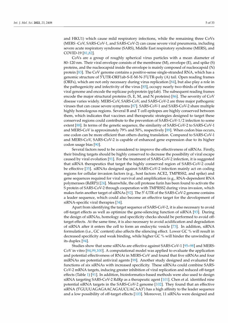
tested for silencing efficiency against the conserved regions of the three viral genes, the spike(S), nucleocapsid (N), and membrane (M). This revealed that four siRNAs (3329i, 1878i,1104i, and 2351i) were able to decrease the expression of the S gene, and four other siRNAs(418i, 881i, 214i, and 1068i) could separately reduce the expression of the N gene, while theremaining three siRNAs (607i, 344i, and 79i) could decrease the M gene expression [104].Several researchers have also predicted functional and potential siRNAs as therapeuticsusing in silico analysis [105,106] (Table 1).
Although RNAi therapeutics may have a positive influence in fighting COVID-19,there remain challenges in the safe delivery of siRNA [107]. Therefore, various improve-ments concerning delivery platforms need to be developed to manage this problem.
Table 1. siRNA therapeutics for SARS-CoV-2, SARS-CoV-1, and MERS-CoV.
Virus Sequence (5′-3′) Region Stage Refs
MERS-CoV UAGAAGAACAGCUAUCACCCU ORF1ab Computational approach
[99]MERS-CoV AACAUAGAAAGCAGAUAGGUC ORF1ab Computational approachMERS-CoV UCUAAGAGCUGCAUAAGUGUC ORF1ab Computational approachMERS-CoV AUCAAGAAAAGCGUUAGAGGA ORF1ab Computational approachMERS-CoV UUUGUAGUACCAAUGACGCAA ORF1ab Computational approach
MERS-CoV UAAUAGUAAAAAUAGAUUGCU ORF1ab In vitro (HEK-293)
[108]MERS-CoV AACAUUAAUAGCAUUAUCCAU ORF1ab In vitro (HEK-293)MERS-CoV UAAGAUAUCAUCUAAAGUGUC ORF1ab In vitro (HEK-293)MERS-CoV UUAAAACUCAAACUAAUAGCA ORF1ab In vitro (HEK-293)MERS-CoV UAGUUAAAGAGUUUCUAAGAG ORF1ab In vitro (HEK-293)
MERS-CoV UAAUAGUAAAAAUAGAUUGCU ORF1ab In vitro (Vero cells)[109]MERS-CoV UAAGAUAUCAUCUAAAGUGUC ORF1ab In vitro (Vero cells)
MERS-CoV AACAUUAAUAGCAUUAUCCAU ORF1ab In vitro (Huh7)
[110]MERS-CoV UUAAAACUCAAACUAAUAGCA ORF1ab In vitro (Huh7)MERS-CoV AUUAAAUCUGUUAAUGUUGUU ORF1ab In vitro (Huh7)MERS-CoV AAAUAGUUAUGAAUAGUUGAG ORF1ab In vitro (Huh7)
SARS-CoV-1 GGGCUAUCAACCUAUAGAU Spike protein (S) In vitro (Vero E6)[111]SARS-CoV-1 CAAGGCGAUUAGUCAAAUU Spike protein (S) In vitro (Vero E6)
SARS-CoV-1 CGUAACUAAACAGCACAAG 3′-UTR In vitro (Vero E6)
SARS-CoV-1 GCUCCUAAUUACACUCAAC ORF2, spike In vitro (FRhk-4)
[112]SARS-CoV-1 GGAUGAGGAAGGCAAUUUA ORF1b, nsp-12 In vitro (FRhk-4)SARS-CoV-1 GGAUAAGUCAGCUCAAUGC ORF1b, nsp-13 In vitro (FRhk-4)SARS-CoV-1 CUGGCACACUACUUGUCGA ORF1b, nsp-16 In vitro (FRhk-4)
SARS-CoV-1 GCUCCUAAUUACACUCAAC ORF2, spike In vivo (Rhesus macaque)[113]SARS-CoV-1 GGAUGAGG AAGGCAAUUUA ORF1b, nsp-12 In vivo (Rhesus macaque)
SARS-CoV-1 GUACCCUCUUGAUUGCAUC Replicase 1A In vitro (FRhk-4)[114]SARS-CoV-1 GAGUCGAAGAGAGGUGUCU Replicase 1A In vitro (FRhk-4)
SARS-CoV-1 GCACUUGUCUACCUUGAUG Replicase 1A In vitro (FRhk-4)
SARS-CoV-1 CACUGAUUCCGUUCGAGAUC S In vitro (FRhk-4)
[115]SARS-CoV-1 CGUUUCGGAAGAAACAGGUAC E In vitro (FRhk-4)SARS-CoV-1 UGCUUGCUGCUGUCUACAG M In vitro (FRhk-4)SARS-CoV-1 GUGGCUUAGCUACUUCGUUG M In vitro (FRhk-4)
SARS-CoV-1 UGAAGGAGUUCCUGAUCUUCU Small envelope protein (E) In vitro (Vero E6)[116]SARS-CoV-1 AGCUUAAACAACUCCUGGAAC Small envelope protein (E) In vitro (Vero E6)
SARS-CoV-1 GAUAAUGGACCCCAAUCAAAC Membrane protein (M) In vitro (Vero E6)
SARS-CoV-1 CUUACAUAGCUCGCGUCUC Nt 14450 to 14468 In vitro (Vero cells)[117]SARS-CoV-1 GAAUAUUAGGCGCAGGCUG Nt 15877 to 15895 In vitro (Vero cells)
SARS-CoV-2 UUCGUUUAGAGAACAGAUC 5′-UTR In vitro (Vero E6) [34]
SARS-CoV-2 GUACUUUUUUUGAACUUCUACA Surface glycoprotein In vitro (Vero cells)
[91]
SARS-CoV-2 CAACAAAGAUAGCACUUAA ORF1ab polyprotein In vitro (Vero cells)SARS-CoV-2 UCAUACCACUUAUGUACAA ORF1ab polyprotein In vitro (Vero cells)SARS-CoV-2 CCAAAAUCAUAACCCUCAAA ORF3a protein In vitro (Vero cells)SARS-CoV-2 AAACCUUCUUUUUACGUUUA Envelope protein In vitro (Vero cells)SARS-CoV-2 CGAACGCUUUCUUAUUACAA Membrane glycoprotein In vitro (Vero cells)
Int. J. Mol. Sci. 2022, 23, 2408 7 of 33
Table 1. Cont.
Virus Sequence (5′-3′) Region Stage Refs
SARS-CoV-2 AUGAACCACAAAUCAUUACUA S In vitro (human HUVECs and A549 cells)
[104]
SARS-CoV-2 AGAUCAACUUACUCCUACUUG S In vitro (human HUVECs and A549 cells)SARS-CoV-2 AUAUAAUUCCGCAUCAUUUUC S In vitro (human HUVECs and A549 cells)SARS-CoV-2 AAGUCAAACAAAUUUACAAAA S In vitro (human HUVECs and A549 cells)SARS-CoV-2 AAUACACCAAAAGAUCACAUU N In vitro (human HUVECs and A549 cells)SARS-CoV-2 UAUUGACGCAUACAAAACAUU N In vitro (human HUVECs and A549 cells)SARS-CoV-2 AAGGAACUGAUUACAAACAUU N In vitro (human HUVECs and A549 cells)SARS-CoV-2 GUUCCAAUUAACACCAAUAGC N In vitro (human HUVECs and A549 cells)SARS-CoV-2 AACUAUAAAUUAAACACAGAC M In vitro (human HUVECs and A549 cells)SARS-CoV-2 AAACUAACAUUCUUCUCAACG M In vitro (human HUVECs and A549 cells)SARS-CoV-2 CUAUUCCUUACAUGGAUUUGU M In vitro (human HUVECs and A549 cells)
SARS-CoV-2 GUUGGACUGAGACUGACCUUA RdRp In vitro (Vero E6) and in vivo(K18-hACE2 mice)
[30]SARS-CoV-2 UUAUACCUUCCCAGGUAACAA 5′UTR In vitro (Vero E6) and in vivo(K18-hACE2 mice)
SARS-CoV-2 UCACCUUAUAAUUCACAGAAU Helicase In vitro (Vero E6) and in vivo(K18-hACE2 mice)
SARS-CoV-2 GGAAGGAAGUUCUGUUGAA RdRp In vitro (Vero cells) and in vivo(Syrian hamsters) [118]
SARS-CoV-2 UUUGUAUGCGUCAAUAUGCUU Nucleocapsidphosphoprotein Computational approach
[119]
SARS-CoV-2 UCAACGUACACUUUGUUUCUG Surface glycoprotein genes Computational approachSARS-CoV-2 AAAAACUUCACCAAAAGGGCA Surface glycoprotein genes Computational approachSARS-CoV-2 UUAAAAACUUCACCAAAAGGG Surface glycoprotein genes Computational approachSARS-CoV-2 UUAAAGCACGGUUUAAUUGUG Surface glycoprotein genes Computational approachSARS-CoV-2 AACUUCUUGGGUGUUUUUGUC Surface glycoprotein genes Computational approachSARS-CoV-2 UUUGAUUGUCCAAGUACACAC Surface glycoprotein genes Computational approachSARS-CoV-2 UAAUUUGACUCCUUUGAGCAC Surface glycoprotein genes Computational approach
SARS-CoV-2 UCCUUCUUUAGAAACUAUACA ORF1ab Computational approach
[102]
SARS-CoV-2 UGGUUUCACUACUUUCUGUUU ORF1ab Computational approachSARS-CoV-2 UUCACUACUUUCUGUUUUGCU ORF1ab Computational approachSARS-CoV-2 AUGUCAUCCCUACUAUAACUCAAA ORF1ab Computational approachSARS-CoV-2 UUAAAAUAUAAUGAAAAUGGA S Computational approachSARS-CoV-2 CUUGAAGCCCCUUUUCUCUAUCUUU ORF3a Computational approachSARS-CoV-2 UUGAAUACACCAAAAGAUCACAUU N Computational approach
SARS-CoV-2 AGUAGAAAUACCAUCUUGGAC N Computational approach [120]
SARS-CoV-2 GUUUAGAGAACAGAUCUACAA The leader sequence Computational approach [103]
SARS-CoV-2 UAGUACUACAGAUAGAGACAC RdRp Molecular dynamics (MD) simulation [101]
3. NP Delivery—A Platform Ensuring the Efficacy and Stability of siRNAs
NPs are valuable carriers to deliver functional nucleic acid fragments into the bodyfor specific targeted therapy. These nanocarrier systems can be used to form vesicle-based NP suspensions after encapsulation, thereby improving the stability of the small-molecule drugs contained therein [51]. Thus far, the four siRNA drugs approved bythe FDA applied N-acetylgalactosamine (GalNAc) coupling or lipid NP pathways toeffectively deliver siRNAs to liver cells for their therapeutic effects. NPs can also improvethe therapeutic effects of drugs by changing drug metabolic rates [121]. NPs can bedivided into different types according to their size, shape, and physical/chemical properties.Advanced nanocarriers include organic NPs (lipid NPs, polymer NPs, and glycogen NPs)and inorganic NPs (magnetic NPs, gold NPs, carbon-based NPs, ceramic NPs, metal NPs,and semiconductor NPs) (Figure 2) [122,123].

Int. J. Mol. Sci. 2022, 23, 2408 8 of 33Int. J. Mol. Sci. 2022, 23, x FOR PEER REVIEW 9 of 34
Figure 2. Comparison of different nanocarriers. Advanced nanocarriers can be divided into or-ganic NPs (lipid NPs, polymers NPs, and glycogen NPs) and inorganic NPs (gold NPs and mag-netic NPs) according to their size, structures, and characteristics. NPs, nanoparticles; NLCs, nanostructured lipid carriers; LNPs, lipid nanoparticles. Created with BioRender.com.
3.1. Lipid NPs (LNPs) in siRNA Therapeutics LNPs belong to the organic nano-system and have hydrophilic parts exposed to the
surrounding aqueous solvent as well as hydrophobic parts facing each other to form a supramolecular structure [124]. LNPs are particularly attractive for biomedical applica-tions, owing to their enhanced lipid-specific biocompatibility. Thus, LNPs are widely used in the field of nanomedicine. In 2018, the FDA and the EMA approved a therapeutic drug for hereditary transthyretin-mediated amyloidosis (hATTR amyloidosis) based on siRNA LNPs, ONPATTRO™ (patisiran) [55,56]. In patisiran, the weight ratio of total lipids and siRNAs is 12.1, and the composition of these LNPs, which could effectively target the transthyretin of hepatocytes, includes ionizable lipid, phospholipid, cholesterol, and PEG lipid [125]. To date, LNPs are the most frequently studied in vivo delivery carriers, and they can silence target genes by encapsulating and delivering specific siRNAs. LNPs can
Figure 2. Comparison of different nanocarriers. Advanced nanocarriers can be divided into organicNPs (lipid NPs, polymers NPs, and glycogen NPs) and inorganic NPs (gold NPs and magnetic NPs)according to their size, structures, and characteristics. NPs, nanoparticles; NLCs, nanostructuredlipid carriers; LNPs, lipid nanoparticles. Created with BioRender.com.
3.1. Lipid NPs (LNPs) in siRNA Therapeutics
LNPs belong to the organic nano-system and have hydrophilic parts exposed to thesurrounding aqueous solvent as well as hydrophobic parts facing each other to form asupramolecular structure [124]. LNPs are particularly attractive for biomedical applications,owing to their enhanced lipid-specific biocompatibility. Thus, LNPs are widely used inthe field of nanomedicine. In 2018, the FDA and the EMA approved a therapeutic drugfor hereditary transthyretin-mediated amyloidosis (hATTR amyloidosis) based on siRNALNPs, ONPATTRO™ (patisiran) [55,56]. In patisiran, the weight ratio of total lipids andsiRNAs is 12.1, and the composition of these LNPs, which could effectively target thetransthyretin of hepatocytes, includes ionizable lipid, phospholipid, cholesterol, and PEGlipid [125]. To date, LNPs are the most frequently studied in vivo delivery carriers, andthey can silence target genes by encapsulating and delivering specific siRNAs. LNPs can

Int. J. Mol. Sci. 2022, 23, 2408 9 of 33
also ensure the stability of siRNA in vivo, protecting them from degradation and ensuringsmooth delivery to target cells.
Liposomes, as the earliest generation of lipid particles, are small-molecule artificialvesicles that are spherical in structure, with diameters of 25–2500 nm. According to theirsize, they can be divided into multilamellar (MUV) or unilamellar vesicles (UV). UV can bedivided into LUV (large unilamellar vesicles, 100–1000 nm in diameter) and SUV (smallunilamellar vesicles, 20–100 nm in diameter). Liposomes consist of an amphiphilic bilayerphospholipid on the outside and an aqueous compartment in the center (Figure 3A) [123].The bilayer phospholipid can carry hydrophobic therapeutic drugs in the middle, and theaqueous compartment can carry hydrophilic therapeutic drugs internally [126].
Int. J. Mol. Sci. 2022, 23, x FOR PEER REVIEW 10 of 34
also ensure the stability of siRNA in vivo, protecting them from degradation and ensuring smooth delivery to target cells.
Liposomes, as the earliest generation of lipid particles, are small-molecule artificial vesicles that are spherical in structure, with diameters of 25-2500 nm. According to their size, they can be divided into multilamellar (MUV) or unilamellar vesicles (UV). UV can be divided into LUV (large unilamellar vesicles, 100-1000 nm in diameter) and SUV (small unilamellar vesicles, 20-100 nm in diameter). Liposomes consist of an amphiphilic bilayer phospholipid on the outside and an aqueous compartment in the center (Figure 3A) [123]. The bilayer phospholipid can carry hydrophobic therapeutic drugs in the middle, and the aqueous compartment can carry hydrophilic therapeutic drugs internally [126].
Compared with classic liposomes, cationic LNPs have a more complex liposome-like structure and are more suitable for encapsulating various nucleic acids (RNA and DNA). Cationic LNPs, spherical particles ~50 nm in diameter, are composed of ionizable cationic lipids, cholesterol, phospholipids, and polyethylene glycol-lipids (PEGylated lipids) (Fig-ure 3B) [127]. Various lipids in LNPs have different characteristics and functions. Cationic lipids can also ensure the stability of siRNA. Ionizable cationic lipids can be ionized under acidic conditions (pH < 5), encapsulating siRNA in particles through electrostatic com-plexation. Subsequently, when the positively charged cationic lipid contacts the nega-tively charged cell membrane, it can be endocytosed into the cell and release the siRNA inside [128,129]. PEGylated lipids are used to control the spacing between NPs, inhibit LNP aggregation, and prevent them from being recognized by the human immune system [130]. Neutral lipids promote and stabilize the formation and arrangement of the phos-pholipid bilayer structure, while cholesterol has strong membrane fusibility, which can promote the intracellular uptake and cytoplasmic entry of siRNA [131,132].
Figure 3. Structures of liposomes and cationic LNPs. (A) Liposomes: small-molecule artificial vesi-cles with a hydrophobic phospholipid double layer outside and a hydrophilic core inside. (B) Cati-onic LNPs: spherical particles composed of ionizable cationic lipids, cholesterol, phospholipids, and PEGylated lipids. LNPs, lipid nanoparticles; PEG, polyethylene glycol. Created with BioRen-der.com.
Figure 3. Structures of liposomes and cationic LNPs. (A) Liposomes: small-molecule artificial vesicleswith a hydrophobic phospholipid double layer outside and a hydrophilic core inside. (B) CationicLNPs: spherical particles composed of ionizable cationic lipids, cholesterol, phospholipids, andPEGylated lipids. LNPs, lipid nanoparticles; PEG, polyethylene glycol. Created with BioRender.com.
Compared with classic liposomes, cationic LNPs have a more complex liposome-like structure and are more suitable for encapsulating various nucleic acids (RNA andDNA). Cationic LNPs, spherical particles ~50 nm in diameter, are composed of ioniz-able cationic lipids, cholesterol, phospholipids, and polyethylene glycol-lipids (PEGylatedlipids) (Figure 3B) [127]. Various lipids in LNPs have different characteristics and functions.Cationic lipids can also ensure the stability of siRNA. Ionizable cationic lipids can beionized under acidic conditions (pH < 5), encapsulating siRNA in particles through elec-trostatic complexation. Subsequently, when the positively charged cationic lipid contactsthe negatively charged cell membrane, it can be endocytosed into the cell and release thesiRNA inside [128,129]. PEGylated lipids are used to control the spacing between NPs,inhibit LNP aggregation, and prevent them from being recognized by the human immunesystem [130]. Neutral lipids promote and stabilize the formation and arrangement of thephospholipid bilayer structure, while cholesterol has strong membrane fusibility, whichcan promote the intracellular uptake and cytoplasmic entry of siRNA [131,132].

Int. J. Mol. Sci. 2022, 23, 2408 10 of 33
The ratio of polyethylene glycol (PEG) to LNPs can affect the efficacy of siRNAs ingene silencing [133], while increasing the size of LNPs may help redirect siRNAs in themto antigen-presenting cells (APCs) [134]. The release of siRNA from LNPs is considered arate-limiting factor in the process of gene silencing. Therefore, it is necessary to increasethe release-potential of siRNA to promote gene silencing [135].
siRNA LNPs have the potential to play a vital role in the treatment of many diseases.Intravenous injection of anti-miRNA145 LNPs in a rat model of pulmonary arterial hy-pertension (PAH) improved heart function [136]. It was also found that siRNA LNPs canenter bone cells after intravenous injection to induce the gene silencing of SOST encodingsclerostin, providing a new possibility for the treatment of osteoporosis [137]. New researchhas developed a novel type of siRNA LNP delivery system (LNP-Ihh siRNA). Deletingcartilage-specific genes (Ihh) to reduce cartilage degeneration is hoped to provide supportfor the treatment of cartilage diseases [138].
The delivery of siRNAs by LNPs may also play an important role in the treatment oftumors and blood diseases. A study applied tripeptide LNP as a carrier and co-wrappedchemotherapeutic drug paclitaxel (PTX) and VEGF siRNA with sucrose laurate (S) andfolate-PEG2000-DSPE (FA) to construct a new type of nanoparticle (PTX/siRNA/FALS),which could target tumor cells to improve the efficacy of cancer treatment [139]. Moreover,LNPs can encapsulate multiplexed siRNA and play a positive role in the treatment of lym-phoma [140]. Packaging a leukemia-specific fusion transcript, BCR-ABL, and siRNA intoLNPs can also be used to target oncogenes in hematopoietic tissue in vitro and in vivo [141].In addition, some helper lipids such as DSPC-cholesterol could facilitate the stable deliveryof siRNA by LNPs [142].
Currently, ionizable LNPs are recognized as one of the most advanced non-viral vectorsfor efficient nucleic acid delivery [143]. Idris et al. identified three candidate siRNAs fromnumerous siRNAs targeting the conserved regions of SARS-CoV-2, and their inhibitoryeffect on the virus was as high as 90%. At the same time, two new LNPs that can deliversiRNAs to the lung have also been developed [30], and after intranasal administration,cationic LNPs show higher bioavailability [144]. LNPs also have the potential to loadvaccines [131,145–147].
In addition, other types of LNPs have also been used to deliver siRNA, includingsolid LNPs (SLNs) [148], nanostructured lipid carriers (NLCs) [149], nonlamellar LNPs(NLNs) [150], ethosomes [151], and echogenic liposomes [152] (Table 2). These LNPscould also improve the stability of siRNA and effectively release siRNA after enteringthe target cells [127]. Therefore, LNPs have been recognized as the most effective siRNAdelivery system.
Table 2. Lipid nanocarriers loaded with therapeutic siRNAs.
Carrier siRNAs’ Target Cells/Animal Models Refs
Ionizable LNPs LINC01257 siRNA Kasumi-1 cells (human acute myeloidleukemia cell line) [153]
Ionizable LNPs Irf5 siRNA C57BL/6 Irf5−/− Apoe−/−mice [154]Ionizable LNPs MRTF-B siRNA Human conjunctival fibroblasts [155]
Ionizable LNPs CTNNB1 Dicer substrate RNA(DsiRNA) HepG2 and A375.S2 cells [156]
Ionizable LNPs RBPMS siRNA HEK293-GFP stable cells (GFP293) [157]
Ionizable LNPs (CL4H6) STAT3 HIF-1α ICR (♀, 4–10 weeks) mice andBALB/cAjcl-nu/nu (♂, 4–5 weeks) mice [158]
Ultra-small LNPs MK siRNA Human HCC cell line, HepG2, and maleBALB/c nude mice [159]
Ginger-derived NPs (GDNPs) Dmt1 siRNA Hamp KO mice [160]
SLNs PD-1 J774A.1 murine macrophages and B16-F10murine melanoma cells [161]
SLNs TNF-α J774A.1 murine macrophages [162]

Int. J. Mol. Sci. 2022, 23, 2408 11 of 33
Table 2. Cont.
Carrier siRNAs’ Target Cells/Animal Models Refs
SLNs Bcl-2 HeLa (human cervical canceradenocarcinoma) cell line [163]
SLNs BACE1 Caco-2 cells (human epithelial colorectaladenocarcinoma cells) [164]
SLNs MDR1 MCF-7 cells and MCF-7/ADR cells [165]
SLNs KSP
Murine endothelial cell line MS1-VEGF andhuman primary umbilical vein endothelialcell line (HUVEC); human dermalmicrovascular endothelial cell (HMEC-1) line
[166]
SLNs Bcl-2 A549 cells (human lung carcinoma) [167]
SLNs MCL1 KB cells (human epithelial carcinoma) andBALB/c nu/nu mice [168]
SLNs c-Met U-87MG human GBM cells and U-87MGtumor-bearing mouse [169]
SLNs VEGF PC-3 (human prostate cancer cells) andMDAMB435 (human breast cancer cells) [170]
Cationic SLNs EphA2 siRNA PC-3 and DU145 (prostate cancer cell lines) [171]NLCs BCL2 and MRP1 A549 human lung adenocarcinoma cells [149]
NLNs GFP Chinese hamster ovary cells (CHO-GFP) andhuman embryonic kidney (HEK293) cells [150]
Ethosomes GAPDH Human adult epidermal keratinocytes andfemale BALB/c mice [151]
Echogenic Liposomes CPP Human breast adenocarcinoma cells(HT-1080) [152]
3.2. Polymer NPs (PNPs) in siRNA Therapeutics
PNPs are small molecular particles with a size of 1–1000 nm and include nanospheresand nanocapsules. PNPs are used for drug or small RNA delivery and are generallybiodegradable polymers, in which the poly(lactic acid) (PLA), poly(glycolic acid) (PGA),and poly (lactic-co-glycolide) (PLGA) have been approved by FDA [172]. They are consid-ered to have good safety and biocompatibility, as well as low levels of immunogenicity andtoxicity [173].
Earlier, fluorescent conjugated PNPs (cPNPs), which were found to deliver gene-specific siRNA to tobacco BY-2 protoplasts [174], are considered non-toxic and efficient inthe delivery of siRNA [175]. siRNA PNPs can be used to deliver various tumor drugs aswell as siRNAs/miRNAs to assist in cancer treatment [176]. A study developed cationiccore–shell NPs, called P(MDS-co-CES), which may be used to deliver both chemother-apeutic drugs (paclitaxel) and siRNA to the same cell [177]. The PEGylation of siRNAreduces the protein adsorption of the PNP–siRNA complex, thereby delivering siRNA moreefficiently [178]. Multifunctional poly (lactide-co-glycolic acid) (PLGA) NPs have also beendeveloped to deliver tumor antigen and immunosuppressive gene (SOCS1) siRNA to bone-marrow-derived (BM) dendritic cells (DC) to enhance the effects of immunotherapy [179].Doxorubicin (DOX) and siRNA could be co-loaded into PNPs containing polyethylenimine(PEI) and then delivered to the lungs, which could be effective in the treatment of metastaticlung cancers [180].
Meanwhile, encapsulated siRNA can also be assembled using star polymers, whichhave a multi-branched structure and can self-assemble with siRNA to form cationic nano-sized complexes to enhance cell entry. Star polymer–siRNA can hold the rate of targetedgene silencing at 50%. Kavallaris et al. applied poly (DMAE-MA) loaded with Luc2 siRNAto human pancreatic cancer (MiaPaCa-2) and H460 non-small cell lung cancer (NSCLC)cell lines and found that star polymer–siRNA can inhibit the expression of target genes inboth [181]. In BALB/c nude mice transplanted with human pancreatic cancer cells, star–POEGMA–siRNA can be delivered to mouse pancreatic cancer cells [182]. Subsequently, inthe BALB/c nude mice model of pancreatic cancer in situ, the use of star polymer loaded

Int. J. Mol. Sci. 2022, 23, 2408 12 of 33
with TUBB3 (βIII-tubulin) siRNA for treatment can silence the expression of βIII-tubulinand inhibit the growth and metastasis of pancreatic tumors [183]. In addition, star polymer–siRNA can also be used to enhance the drug delivery potential in acute lymphoblasticleukemia (ALL) cells [184].
Interestingly, NPs that integrate lipids and polymers (hybrid lipid–polymer NPs) werefound to combine the biocompatibility of lipids with the structural solidity of polymers andenhance the binding affinity with siRNA to maintain stable encapsulation of siRNA [185].Hybrid lipid–polymer NPs with siRNA exhibited excellent gene silencing effects in HeLaand A549 lung carcinoma cells [186]. Another study also found that applying lipid-assistedPNPs could efficiently deliver specific siRNA targeting GLUT3 into glioma stem cells andbulk cancer cells, thereby enhancing the effectiveness of cancer treatment [187].
As carriers, PNPs also play an important role in the delivery of therapeutic drugsand siRNA for metabolic diseases (such as diabetes) and infectious diseases (such asreproductive tract and severe lung diseases). Biodegradable PLGA NPs loaded with siRNAcould enter the mucosal epithelial tissue of the reproductive tract to play a therapeutic roleagainst infectious pathogens [188]. Cationic PNPs composed of β-cyclodextrin (β-CD) andpoly amidoamine can be used as an effective carrier of MMP-9-siRNA and injected intothe wounds of diabetic rats, resulting in the down-regulation of MMP-9 gene expressionand enhancing wound healing ability [189]. Poly lactic-glycolic acid NPs (PLGA NPs)encapsulating siRNA for administration to the mucosa of the mouse reproductive tract caneffectively improve the survival rate after HSV-2 infection [190]. Additionally, novel hybridlipopolymer NPs (hNPs) composed of PLGA and dipalmitoyl phosphatidylcholine (DPPC)can deliver siRNA to the lungs and have great potential for the treatment of severe lungdiseases [191].
In addition, the efficacy of delivery is related to the strong affinity of endosomes andthe weak affinity of cytoplasm for siRNA [192]. Encapsulating micellar NPs (MNPs) withpalmitic-acid-linked siRNA (siRNA-PA) can enhance the efficacy of siRNA [193]. Amongsix complexes, mikto star polymer containing a hydrophobic poly (butyl methacrylate)block could deliver siRNA more effectively to better induce gene silencing [194]. Fur-thermore, cationic poly(caprolactone)-based (PCL) NPs could be synthesized and stablycombined with anionic siRNA and delivered effectively [195]. Combination with palmiticacid could improve the loading efficiency of NPs and the intracellular uptake of siRNAs andat the same time can increase their intracellular half-life [193]. The poly (butyl methacry-late) block can ensure a better silencing ability of siRNA in serum-free medium, whilepoly(caprolactone) can maintain the stability of siRNAs under physiological conditions andload and deliver intact siRNAs to the cytoplasm [194,195]. Therefore, the addition of somecofactors may improve the delivery efficiency and gene silencing effect of siRNA PNPs.
To evaluate the effect of siRNA PNPs against SARS-CoV-2 in vitro and in vivo, Khaitovet al. identified the most effective siRNA (siR-7) from 15 candidate siRNAs. After addinglocked nucleic acids (LNAs) to enhance its stability, it was assembled with peptide den-drimer KK-46 (to form siR-7-EM/KK-46 complex). This siR-7-EM/KK-46 complex wasdelivered by inhalation, which reduced viral titers and lung inflammation in vitro andin vivo [118]. Therefore, designing polymer nanocarriers with ACE2 antibodies to wrapsiRNAs with therapeutic potential can accurately target cells with ACE-2 and exert gene-silencing effects [196].
3.3. Glycogen NPs (GNPs) in siRNA Therapeutics
GNPs are randomly branched dendritic nanopolysaccharides composed of glucoserepeating units connected by linear α-d -(1-4) glycosidic bonds and α-d -(1-6) branchedchains. The glycogen used to prepare GNPs comes from a wide range of sources, fromanimal tissues to plants (including rabbit and bovine liver, oysters, and sweet corn) [197].
As a functional biomaterial, glycogen has several obvious advantages, including bio-compatibility, biodegradability, and high water solubility. These characteristics highlightthe potential of GNPs as drug delivery carriers. The outer surface of GNPs is highly

Int. J. Mol. Sci. 2022, 23, 2408 13 of 33
branched and has a dense inner core. Because of their high hydrophilicity and biocompati-bility, they can easily be biochemically modified to produce functional derivatives [198],such as adhesive GNPs composed of lipoate-conjugated phytoglycogen (L-PG) [199] andoyster GNPs with poly(N-isopropylacrylamide) (PNIPAM) chains on their surface [200].
GNPs are composed of single-molecule spheres (β-particles) and many smaller parti-cles (α-particles). The diameter of GNPs is generally about 20–150 nm and can be modifiedinto various sizes [201]. Cavalieri et al. have found that galacto-GNPs showed a highaffinity for prostate cancer cell membranes and could induce the aggregation of prostatecancer cells [202]. They also constructed soft GNPs with a diameter of 20 nm which candeliver siRNAs in human prostate cancer cells more effectively and lead to a 60% genesilencing effect [203]. The size of glycogen is important for the stability of NPs loaded withsiRNA as well, as GNPs with a smaller size and a higher protein content are more likely tomaintain stability [204].
Gold GNPs were found to deliver siRNA to the lungs in vivo, which can induce atumor size reduction of nearly 80% in B6 albino mouse lungs grafted with CMT 167 adeno-carcinoma cells [205]. Another study applied lipopolysaccharide-amine nanopolymersomesloaded with specific siRNA (siNoggin), which enhanced the effect of bone differentiationin vitro [206]. The construction of cationic enzymatically synthesized glycogen (cESG) canachieve the simultaneous delivery of tetraphenylporphine sulfonate (TPPS) and siRNA,which can reduce the survival rate of cancer cells (ovarian clear cell cancer cells) [207].In addition, cells transfected with the polysaccharide derivative DMAPA-Glyp/siRNAinhibited the activation of nuclear transcription factor-κB (NF-κB) in human retinal pigmentepithelial (hRPE) cells, resulting in the expression of mRNA and protein being reduced,which can be used for gene therapy for diabetic retinopathy [208]. In addition, GNPs havethe proven potential to be used in drug-assisted tumor treatment [209]. Therefore, the valueof GNPs in COVID-19 needs further exploration.
3.4. Other NP Platforms in siRNA Therapeutics
As well as organic NPs, there are also some inorganic NP platforms, mainly includinggold NPs (Au NPs) and magnetic NPs (Mag NPs) [123,210]. Au NPs are widely usedin diagnosis and treatment and have the potential to be used as drugs and vaccine car-riers [211]. They can be used for intranasal delivery and can spread to lymph nodes toactivate CD8+ cell-mediated immune responses [211]. It is reported that Au NPs carry-ing hydrophilic-b-cationic copolymers (HbC-Au NP) could effectively deliver siRNA tocancer cells [210]. siRNAs are located on the surface of Au NPs by gold–thiol chemistryor electrostatic interactions [212]. Au NPs could also easily trigger the immune systemthrough the internalization of APC, thereby presenting outstanding potential in vaccinedevelopment [123]. Another study used antigen-coated Au NPs through the principle ofplasmon resonance to assist in the detection of the N protein of SARS-CoV-2 [213]. Au NPscould be also used as nanoprobes to detect the S protein of SARS-CoV-2 [214].
Further, Mag NPs with surface hyaluronan can selectively bind to the cell surfaceadhesion molecule CD44, which plays an important role in the occurrence and develop-ment of atherosclerotic plaques, thereby assisting in the magnetic resonance imaging ofplaques [215]. Mag NPs can also be used to assist in the detection of the S protein [216].In addition, layered double hydroxide (LDH) is considered a biocompatible inorganic NP.Studies have found that LDH carrying shRNA can protect it from degradation and make itmore easily internalized by cells [217,218]. It has been also found that extracellular vesicles(EVs) have the potential to transport and load antiviral agents into cells to assist in thetreatment of COVID-19 [219]. Besides, although a few newly synthesized NPs, such as zincoxide NPs (ZnO NPs) and titanium dioxide NPs (TiO2 NPs), are weakly cytotoxic to hostcells, they have been recently applied as a powerful disinfectant to combat SARS-CoV-2due to their potent antiviral activity [220,221].
Despite all of the advances in vaccines and drugs, the global epidemic of COVID-19continues. The mutation of the target sequences of RNAi could lead to viral escape [222].

Int. J. Mol. Sci. 2022, 23, 2408 14 of 33
Therefore, designing specific small RNAs for highly conserved viral genome regionscould resist viral infections with high mutation rates, which is considered one of theadvantages of RNAi therapy [223]. Additionally, multiplexing or combining siRNA targetsin a siRNA combination therapeutic could increase protection by providing redundancy,which means that if one target does mutate, the other target will still provide protection.This combination treatment approach has been utilized for HIV-1 to address the highlydiverse virus sequences and subtypes globally [224]. Given the rapidly evolving SARS-CoV-2 genome and resulting resistance to vaccines and some monoclonal antibodies, furtherresearch is needed to explore and optimize RNAi treatment strategies and appropriate NPcarrier platforms against SARS-CoV-2 infection.
4. Challenges of Local Delivery of NP-siRNAs—The Importance of Administration Route
The intravenous route can deliver therapeutic siRNAs directly to the liver via thecirculatory system, so therapeutic siRNA plays an important role in the treatment ofmany systemic diseases. Indeed, previous studies on NP carriers have focused on liverdelivery [225]. Currently, all four siRNA drugs approved by the FDA are effectivelydelivered to hepatocytes by GalNAc coupling or LNP carriers for therapeutics [57,62–64].In contrast, local delivery of therapeutic siRNAs to specific organs, such as the lungs, canbe achieved via other routes of administration, such as inhalation. Organ-targeted therapyfor diseases that may cause damage to specific organs or tissues other than the liver, suchas the lungs for COVID-19, is both a potential advantage as well as a challenge for localdelivery [226]. Local administration has many advantages, including the rapid onset ofaction, reduced dosage, and limited side effects [226]. Additionally, since siRNAs usedsystemically can accumulate in off-target tissues [227], local drug delivery strategies couldavoid the off-target accumulation of siRNAs [228].
The primary consideration in the deposition of particles after entering the respiratorytract through the mouth or nose mainly depends on their size and density, with depositionin the lungs occurring through three different ways: impact, sedimentation, and diffu-sion [229]. As a general rule, aerosols with an aerodynamic diameter >10 µm will depositin the nasal cavity and pharynx through impact and cannot enter the lower respiratorytract. Particles between ~1 and 10 µm will be deposited in the bronchi and bronchiolesthrough sedimentation, while particles with diameters ranging between 10 and 500 nm willdeposit in the lower bronchioles and alveoli by diffusion [230] (Figure 4).

Int. J. Mol. Sci. 2022, 23, 2408 15 of 33Int. J. Mol. Sci. 2022, 23, x FOR PEER REVIEW 16 of 34
Figure 4. Intravenous delivery vs. lung delivery of NP-siRNAs. Intravenous delivery can deliver therapeutic siRNAs directly to the liver via the circulatory system. Lung delivery can deliver siR-NAs into the respiratory tract through the mouth or nose, and the deposition distance depends mainly on the size and density of the particles. Aerosols with an aerodynamic diameter >10 µm will deposit in the nasal cavity and pharynx through impact, and particles with a diameter of ˂10 nm with high diffusion velocity are more likely to be deposited in the upper respiratory tract [231,232]. Particles between ~1 and 10 µm will be deposited in the bronchi and bronchioles through sedimentation; particles with diameters ranging between 10 and 500 nm will deposit in the lower bronchioles and alveoli by diffusion. Created with BioRender.com.
The NP carrier platform could provide many benefits for lung mucosal drug delivery [233]. Several researchers have developed NP-based nasal delivery, aiming to effectively and safely deliver small nucleic acid fragments with therapeutic or vaccine effects to target cells [234]. Most animal experiments focus on intranasal instillation [235] and orotracheal administration [236] to detect the efficacy of NP-siRNAs [236–238] (Table 3).
Table 3. Mouse models used to assess respiratory nanocarrier delivery.
Mouse Models Carriers Drugs Delivery Refs
Female BALB/c mice Solid lipid nano-particles (SLNs)
Rhynchophylline (Rhy)
Aerosol administra-tion
[239]
Female BALB/c mice Dextran nanopar-ticles
SET-M33 (a non-nat-ural antimicrobial
peptide)
Hamilton syringe and laryngoscope
[240]
Female BALB/c mice Dextran nanogels siRNA Tracheal aspiration [241]
Female BLAB/c mice Nanocluster Ceftazidime Dry powder inhala-tion
[242]
Female CF-1 mice PEGylated human ferritin nanocages
(FTn)
Doxorubicin (DOX) Inhalation [243]
Female CF-1 mice PLGA and PLGA–PEG NP
Dexamethasone so-dium phosphate
(DP)
Intranasal instilla-tion
[244]
Female C57BL/6OlaHsd mice
PEG–PEI copoly-mer
Dexamethasone (DEX)
Aerosol administra-tion
[245]
Figure 4. Intravenous delivery vs. lung delivery of NP-siRNAs. Intravenous delivery can delivertherapeutic siRNAs directly to the liver via the circulatory system. Lung delivery can deliver siRNAsinto the respiratory tract through the mouth or nose, and the deposition distance depends mainly onthe size and density of the particles. Aerosols with an aerodynamic diameter >10 µm will depositin the nasal cavity and pharynx through impact, and particles with a diameter of <10 nm with highdiffusion velocity are more likely to be deposited in the upper respiratory tract [231,232]. Particlesbetween ~1 and 10 µm will be deposited in the bronchi and bronchioles through sedimentation;particles with diameters ranging between 10 and 500 nm will deposit in the lower bronchioles andalveoli by diffusion. Created with BioRender.com.
The NP carrier platform could provide many benefits for lung mucosal drug deliv-ery [233]. Several researchers have developed NP-based nasal delivery, aiming to effectivelyand safely deliver small nucleic acid fragments with therapeutic or vaccine effects to targetcells [234]. Most animal experiments focus on intranasal instillation [235] and orotrachealadministration [236] to detect the efficacy of NP-siRNAs [236–238] (Table 3).
Table 3. Mouse models used to assess respiratory nanocarrier delivery.
Mouse Models Carriers Drugs Delivery Refs
Female BALB/c mice Solid lipid nanoparticles(SLNs)
Rhynchophylline (Rhy) Aerosol administration [239]
Female BALB/c mice Dextran nanoparticles SET-M33 (a non-naturalantimicrobial peptide)
Hamilton syringe andlaryngoscope
[240]
Female BALB/c mice Dextran nanogels siRNA Tracheal aspiration [241]Female BLAB/c mice Nanocluster Ceftazidime Dry powder inhalation [242]
Female CF-1 mice PEGylated human ferritinnanocages (FTn)
Doxorubicin (DOX) Inhalation [243]
Female CF-1 mice PLGA and PLGA–PEG NP Dexamethasone sodiumphosphate (DP)
Intranasal instillation [244]
FemaleC57BL/6OlaHsd mice
PEG–PEI copolymer Dexamethasone (DEX) Aerosol administration [245]
Male CF-1 mice Mucus-penetratingparticles (MPP)
Fluticasone propionate (FP) Intratracheal instillation [246]
Male CD-1 mice Mesoporous silicananoparticles (MSNs)
Nanopure water droplets Aerosol nose-onlyexposure system
[247]

Int. J. Mol. Sci. 2022, 23, 2408 16 of 33
Since SARS-CoV-2 infection is acquired through breathing and mainly occurs in theepithelial cells of the respiratory tract, lipids, polymers, or lipid–polymer hybrids canbe configured as inhalable aerosols for the delivery of therapeutic siRNAs [196]. Astraditional LNPs tend to accumulate in the liver after IV administration, changing the routeof administration from IV to inhalation could prevent NP-siRNAs from accumulating inthe liver and instead specifically localize them in the lungs (Figure 4) [248]. An effectivemeasure is to combine nanotechnology with inhalation delivery, where NP-siRNAs aremade into dry powders and then delivered to the lung through the nebulization of NPsuspensions for local administration. Indeed, lipo-polymeric NPs (LP NPs) can deliversiRNA targeting the ABCC3 gene to the lungs via dry powder inhalation [249]. Freeze-driedpulmonary surfactant (Curosurf®)-coated nanogels could also deliver therapeutic siRNAsto the lungs via a nebulizer [250,251]. Another nanocarrier made of triphenyl phosphonium(TPP)-modified generation 4 poly(amidoamine) (PAMAM) dendrimer (G4NH2-TPP) can bemade into inhalation preparations after loading siRNA [252]. Thus, local administration ofsiRNA therapeutics to the lungs can effectively combat lung infections, lung cancer, cysticfibrosis, asthma, and many other diseases of the respiratory system [253].
The most prominent feature of the respiratory tract is that it has several branches.From the outside to the inside are: the nasal cavity, trachea, bronchi, bronchioles, andalveoli. Studies have shown that most lung cancer lesions appear in the bronchi andbronchioles [254], while SARS-CoV-2 infection mainly occurs in pneumocytes and ciliatedcells and can cause alveolar damage [255]. The main challenges of pulmonary deliveryinclude mucociliary clearance, alveolar macrophages, the mucus barrier, and shear forces inthe nebulizer [256]. The cilia of the lung epithelium can remove particles deposited on it andcause them to be coughed up from the respiratory tract [257]. Additionally, macrophagesin the alveoli endocytose and degrade particles through phagocytosis [258]. The presenceof mucus in the respiratory system is a factor that seriously affects the transportation ofnanoparticles. Mucus covering the respiratory tract, composed of mucin, acts as a physicalbarrier, which reduces the penetration and diffusion of drugs when it increases in viscosity,preventing the exposure of the respiratory epithelium [257,259]. In addition, pulmonarysurfactant (PS) is another barrier that nanoparticles need to pass through before they reachlung cells [260]. Moreover, shear forces increase nebulization temperature and degradesiRNAs [256]. New treatment strategies based on NP-siRNAs provide new possibilities forthe treatment of lung diseases and promote research progress on targeted drug delivery tothe lung and respiratory tract [261]. Therefore, it is necessary to optimize the size of NPs andapply appropriate excipients to ensure that NP-siRNAs enter target cells more effectively.
In addition to the liver and lung, other potential targets of NP-siRNAs include theeyes, brain, kidney, spleen, and tumor tissues. In a rat spinal cord injury model, injection ofNP-encapsulated therapeutic siRNA (PLK4-siRNA) into the injured spinal cord improvedmotor function to a certain extent [262]. In rats with optic nerve transection, the eye nervesare protected by the delivery of caspase-2-siRNA by intravitreal administration [263].Therefore, the current delivery strategies vary for different organs.
5. Methods to Perform Respiratory Delivery of NP-siRNAs
There are three potential methods to deliver NPs directly to the respiratory tract:intratracheal, intranasal, and orotracheal. Accordingly, inhalation devices are divided intodifferent types, namely nebulizers, pressurized metered dose inhalers (pMDIs), dry powderinhalers (DPIs), and nasal sprays. (Figure 5). Traditional pulmonary drug delivery devices,such as capillary aerosol generators, and recent inhalers, such as Respimat, were mainlyapplied to deliver micron-sized particles [264,265]. The onTarget system, which is identifiedas a targeted inhalation device, can also be used to target pharmaceutical ingredients tospecific parts of the respiratory tract [266]. An endotracheal aerosolization device (HRHMAG-4) applied a solid LNP suspension as the drug carrier. After verification, it was foundthat the in vitro efficiency was as high as 90%, and the in vivo efficiency in rats was as highas 80% [267].

Int. J. Mol. Sci. 2022, 23, 2408 17 of 33
Int. J. Mol. Sci. 2022, 23, x FOR PEER REVIEW 18 of 34
devices, such as capillary aerosol generators, and recent inhalers, such as Respimat, were mainly applied to deliver micron-sized particles [264,265]. The onTarget system, which is identified as a targeted inhalation device, can also be used to target pharmaceutical ingre-dients to specific parts of the respiratory tract [266]. An endotracheal aerosolization device (HRH MAG-4) applied a solid LNP suspension as the drug carrier. After verification, it was found that the in vitro efficiency was as high as 90%, and the in vivo efficiency in rats was as high as 80% [267].
Figure 5. Examples of different inhalation devices. Inhalation devices are divided into nebulizer, inhaler (including pMDIs, DPIs, and SMIs), and nasal spray. pMDIs, pressurized metered dose inhalers; DPIs, dry powder inhalers; SMIs, soft mist inhalers. Created with BioRender.com.
Inhalation is the most popular method of targeted lung delivery [260]. There are dis-tinct advantages and disadvantages to using specific inhaler devices to deliver siRNA. Nebulizers are easy to use, requiring only tidal breathing, and enable the delivery of large doses, but they are relatively time-consuming (~5 min) [268] compared to other inhalation devices; furthermore, the presence of shear forces due to their mechanism of action could make them impracticable for the delivery of siRNA since shear stress can degrade nucleic acids, which would have an impact on their biological activity [256]. The advantage of pMDIs is their flow independence; however, their disadvantages are linked to the breath coordination required by the patient for successful delivery. DPIs are the most recently developed delivery device. Comparatively, they are easier to use than the pMDIs since they are breath-actuated. They also contain no propellants and are small and portable, with the only disadvantage being the higher required respiratory rate that makes DPIs not ‘user-friendly’ for pediatric and elderly populations [268]. Regardless of the delivery devices used, nanocarriers are needed to protect siRNA from physical and chemical deg-radation.
6. Methods to Assess Lung Delivery Inertial impaction methods are used as the gold standard for in vitro assessment of
the aerodynamic deposition of inhaled formulations. Data from these methods are regu-latory requirements to estimate the amount of the drug that is delivered to the lungs [269].
Figure 5. Examples of different inhalation devices. Inhalation devices are divided into nebulizer,inhaler (including pMDIs, DPIs, and SMIs), and nasal spray. pMDIs, pressurized metered doseinhalers; DPIs, dry powder inhalers; SMIs, soft mist inhalers. Created with BioRender.com.
Inhalation is the most popular method of targeted lung delivery [260]. There aredistinct advantages and disadvantages to using specific inhaler devices to deliver siRNA.Nebulizers are easy to use, requiring only tidal breathing, and enable the delivery of largedoses, but they are relatively time-consuming (~5 min) [268] compared to other inhalationdevices; furthermore, the presence of shear forces due to their mechanism of action couldmake them impracticable for the delivery of siRNA since shear stress can degrade nucleicacids, which would have an impact on their biological activity [256]. The advantage ofpMDIs is their flow independence; however, their disadvantages are linked to the breathcoordination required by the patient for successful delivery. DPIs are the most recentlydeveloped delivery device. Comparatively, they are easier to use than the pMDIs sincethey are breath-actuated. They also contain no propellants and are small and portable, withthe only disadvantage being the higher required respiratory rate that makes DPIs not ‘user-friendly’ for pediatric and elderly populations [268]. Regardless of the delivery devicesused, nanocarriers are needed to protect siRNA from physical and chemical degradation.
6. Methods to Assess Lung Delivery
Inertial impaction methods are used as the gold standard for in vitro assessment of theaerodynamic deposition of inhaled formulations. Data from these methods are regulatoryrequirements to estimate the amount of the drug that is delivered to the lungs [269]. Thesemethods classify particles according to the aerodynamic diameter of the entire delivereddose [270]. An impactor consists of a series of nozzles (circular or slot-shaped) and animpaction surface [271,272]. Cascade impactors operate on the principle of curvilinearmotion of particles in the aerosol stream. Air is drawn into the impactor using a vacuumpump, and the air stream flows through the nozzles and toward the impaction surface,where particles are separated from the air stream by their inertia. Larger particles collecton the impaction surface, while small particles do not and follow the air stream. Severaltypes of impactors are used to assess delivery to the lungs, such as the Andersen cascadeimpactor (ACI, Figure 6A) used by the United States Pharmacopeia (USP) (Table 4).
Int. J. Mol. Sci. 2022, 23, 2408 18 of 33
Int. J. Mol. Sci. 2022, 23, x FOR PEER REVIEW 20 of 34
twin impinger is a valuable device for routine quality assessment of aerosols during prod-uct development, stability testing and quality assurance, and the comparison of inhalation drugs [276].
Another two-stage impactor is the fast-screening impactor (FSI, Figure 6E), which incorporates the NGI’s pre-separator technology and utilizes the same induction port. The FSI first separates large non-inhalable particles which are captured by the liquid trap, fol-lowed by an impaction stage at a 5-micron cut-off (30-100 L/min). The fine particle dose is collected on a glass fiber filter. Additional inserts are available for 10-micron cut-off size, but only at a 30 L/min flow rate. When used for DPI deposition assessments, FPF perfor-mance is comparative to that of full NGI, and the FPD is comparative or better when using the FSI, depending on the drug tested [277].
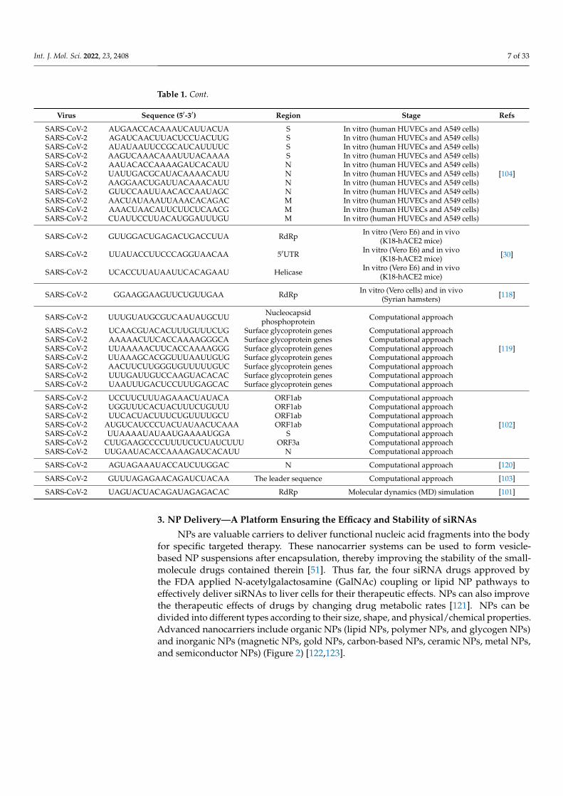
Figure 6. Types of impactors. (A) Andersen cascade impactor; (B) multi-stage liquid impinger; (C) twin-stage impinger; (D) next-generation impactor; (E) fast-screening impactor. Adapted from images from www.copleyscientific.com.
While these impactors are an excellent way to test aerodynamic particle size distri-bution in the lungs, other aspects of the drug uptake, such as dissolution rate, transport through the epithelium, and therapeutic efficacy of the drugs, cannot be determined using standard impaction methods [278,279]. To address this issue, novel hybrid approaches and modifications have been developed, which involve marrying the biological represen-tation of the lung epithelium with the impaction instrumentation.
Air–liquid interface (ALI) culture models are an effective way to produce epithelial layers that represent the pulmonary epithelium [280]. Specifically, the Calu-3 lung cell line has been a promising in vitro model of airway epithelia due to its similarity to in vivo physiology [281]. Nevertheless, the use of primary broncho-epithelial cells is the gold standard and can be obtained from donors who are healthy or have diseases and/or may be infected with viruses [280,282–284]. Using these epithelial layers, assays that test the biochemical characteristics of drugs, such as permeability, integrity, and mucus produc-tion of epithelial layers, can all be performed. Additionally, the impact of the deposited drugs on inflammation and wound healing can be investigated [285,286]. Haghi et al.
A B C
D E
Figure 6. Types of impactors. (A) Andersen cascade impactor; (B) multi-stage liquid impinger;(C) twin-stage impinger; (D) next-generation impactor; (E) fast-screening impactor. Adapted fromimages from www.copleyscientific.com.
Table 4. USP cascade impactors for orally inhaled device testing.
Impactors Devices Flow Rates # of Stages Particle Size Range
Andersen cascade impactor pMDIs and DPIs28.3 L/min (60 and
90 L/min usingmodifications)
6 or 8
0.4–0.9 microns(28.3 L/min), 0.3–8.6
(60 L/min), 0.2–8(90 L/min)
Multi-stage liquidimpinger DPIs 30–100 L/min 4 1.7–13 microns
Twin-stage impinger pMDIs and nebulizers 60 L/min 2 6.4 microns
Fast-screening impactor pMDIs, DPIs, and nasalspray 30–100 L/min 2 5 and 10 microns
Next-generationpharmaceutical impactor
pMDIs, DPIs, andnebulizers 15–100 L/min 7 0.24–14.1 microns
A pre-separator is typically used between the induction port and stage 0 of a cascadeimpactor to capture the large, non-inhalable carrier particles in DPI formulations to preventimpactor over-loading. By analyzing the amount of active drug deposited on variousstages in which the geometrical models are individually built up for the whole dimensionsof the ACI, it is then possible to calculate several essential parameters that define theoverall inhalation formulation performances. These are the fine particle dose (FPD) andfraction (FPF), mass median aerodynamic diameter (MMAD), and geometric standarddeviation (GSD) of the active drug particles collected. The impactors are used to assessthe performance of pMDIs, nebulizers, and DPI devices. Using a calibrated pump, thesedevices operate at specific flow rates for each that simulate a patient lung capacity of 4 L;specifically, 28.3 L/min for pMDIs or at 60 and 100 L/min with modifications for DPIs [273].
Another type of cascade impactor is the next-generation pharmaceutical impactor(NGI, Figure 6D), which was specifically designed for pharmaceutical testing. Particles are

Int. J. Mol. Sci. 2022, 23, 2408 19 of 33
deposited on seven stages (collection cups) that are held in a removable tray for samplecollection. It also has a micro-orifice collector (MOC) that captures extremely small particlesin a collection cup, normally collected on the final filter in other impactors [274]. NGIs areideal for characterizing drug deposition by nebulizers [275].
For the assessment of DPIs, the multi-stage liquid impinger (MSLI, Figure 6B) can alsobe used. The MSLI has four impaction stages, each containing a small volume of liquidduring operation, and a final filter stage. The liquid in the impinger reduces particle bounce.For the operation of the MSLI, the induction port is connected to the inhaler, but unlike theACI’s, they do not require a pre-separator.
The twin stage impinger, a two-stage separation device (Figure 6C), is a more ‘ele-mentary’ impinger that separates particles according to two different aerodynamic cut-offsizes only. The aerosol is fractionated by rushing through a simulated oropharynx andthen through an impinger stage of defined aerodynamic particle size cut-off characteristics.The fine (pulmonary) fraction which penetrates is collected by a lower impinger. The twinimpinger is a valuable device for routine quality assessment of aerosols during productdevelopment, stability testing and quality assurance, and the comparison of inhalationdrugs [276].
Another two-stage impactor is the fast-screening impactor (FSI, Figure 6E), whichincorporates the NGI’s pre-separator technology and utilizes the same induction port.The FSI first separates large non-inhalable particles which are captured by the liquid trap,followed by an impaction stage at a 5-micron cut-off (30–100 L/min). The fine particledose is collected on a glass fiber filter. Additional inserts are available for 10-micron cut-offsize, but only at a 30 L/min flow rate. When used for DPI deposition assessments, FPFperformance is comparative to that of full NGI, and the FPD is comparative or better whenusing the FSI, depending on the drug tested [277].
While these impactors are an excellent way to test aerodynamic particle size distri-bution in the lungs, other aspects of the drug uptake, such as dissolution rate, transportthrough the epithelium, and therapeutic efficacy of the drugs, cannot be determined usingstandard impaction methods [278,279]. To address this issue, novel hybrid approaches andmodifications have been developed, which involve marrying the biological representationof the lung epithelium with the impaction instrumentation.
Air–liquid interface (ALI) culture models are an effective way to produce epitheliallayers that represent the pulmonary epithelium [280]. Specifically, the Calu-3 lung cell linehas been a promising in vitro model of airway epithelia due to its similarity to in vivo phys-iology [281]. Nevertheless, the use of primary broncho-epithelial cells is the gold standardand can be obtained from donors who are healthy or have diseases and/or may be infectedwith viruses [280,282–284]. Using these epithelial layers, assays that test the biochemicalcharacteristics of drugs, such as permeability, integrity, and mucus production of epitheliallayers, can all be performed. Additionally, the impact of the deposited drugs on inflamma-tion and wound healing can be investigated [285,286]. Haghi et al. (2014b) developed amodified Andersen cascade impactor (mACI) that incorporated ALI models of Calu-3 cellsinserted within the mACI at stages 4–7, representing the deep lung region. These modifiedinserts can be customized to be inserted at various stages of the ACI for studies in both theupper and the lower respiratory regions of the lungs. By integrating the ALI culture modelswithin the ACI, the deposition and subsequent permeability of formulations delivered withpMDIs and DPIs can be assessed with physiological relevance [279,287,288].
Similarly, the NGI can also be modified to include an ALI cell culture model (mNGI),enabling aerosol deposition and transport across cell epithelia. mNGIs have been engi-neered to deliver aerosolized particles under controlled vacuum flow to ALI-culturedCalu-3 cells at stages 2, 3, and 4 [289] and stages 3, 5, and 7 [290], highlighting the versatilityand adaptability of an mNGI depending on the research question.
Thus, utilizing standard and modified cascade impactor methods enables a thoroughassessment of drug deposition. Both the aerodynamic distribution in the lungs as well as
Int. J. Mol. Sci. 2022, 23, 2408 20 of 33
cellular responses such as uptake, transport, and therapeutic efficacy of known and novelinhaled formulations can be determined.
7. Animal Models to Assess Pulmonary Delivery
Preclinical animal models are essential for the translation of RNAi-based therapeuticsfrom in vitro studies to human clinical implementation [291]. Several factors need tobe considered when choosing appropriate animal models. Relative costs and logisticswith the handling and storage of animals are major factors dictating the species used inpreclinical studies.
Mouse models are the most widely used animal models to assess the pharmacokineticsand the effects of pulmonary drug delivery [291]. They reflect important features of humandisease with severe infection and virus-induced hyper-inflammation, as well as featuresof acute lung injury, acute respiratory distress syndrome, impaired lung function. Miceare relatively inexpensive and easy to handle and can be bred in large quantities for high-throughput analyses [40]. Larger rodent species, such as rats or hamsters, are also usedfor the examination of pulmonary drug delivery; however, these models are much lesscommonly used compared to standard mouse models. This is due to their larger size andlack of established infrastructure in many laboratories, which dictates higher general costsfor purchasing, larger specialized housing requirements, and ultimately, smaller samplesize. Similarly, ferrets are less commonly used than mouse models due to higher costs andthe requirement for unique housing arrangements. This species shares a high degree ofanatomical similarity with the human respiratory tract, making them an excellent modelfor pulmonary drug delivery assessment [292]. Non-human primates (NHP), such as theRhesus macaque, are uncommonly used for pulmonary drug delivery assessment and aretypically only used in the final stages of toxicology and safety profiling prior to humantrials [293]. NHP models are very expensive and require highly specialized animal facilities,which are rare. NHP models have a very high fidelity of recapitulation of the humanrespiratory system, making them the ‘gold standard’ in terms of clinical translatability.Within the context of SARS-CoV-2 infection, a summary of the different animal models tointerrogate COVID-19 pathogenesis can be found here [291].
Routes of pulmonary drug delivery are relatively similar between all different animalmodels; however, the volume of the drug administered and the specialized equipmentrequired all need to be scaled to the relative size of the animal species [278]. Intranasal drugdelivery is the most common pulmonary drug delivery system and can be performed usinglight anesthesia to administer small volumes to the nostrils, which can be done quickly andwith relative ease. However, particle size is an important determining factor for successfulintranasal drug delivery, as it needs to bypass the mucociliary barrier within the nasophar-ynx for deposition into the distal airways and alveoli [294]. Comparatively, intratrachealdrug delivery requires the induction of surgical anesthesia prior to tracheostomy and/ortracheal intubation to administer larger volumes to the lungs, which is more labor-intensiveand takes longer compared to intranasal delivery [295]. Aerosol exposure is often usedto deliver nebulized drug formulations that are suspended in a particulate state. This isoften achieved using a passive inhalation system via nose-only exposure or a whole-bodyexposure chamber. This is less common, largely due to low throughput capacity, variabledosage delivery, larger amounts of the drug being required, and the process is stressful forthe animal due to the restraints used during the procedure [278,296].
Administration of RNAi-based therapeutics in mice via the intranasal and intratra-cheal routes are highly successful for the amelioration of experimental asthma diseasephenotypes [297–302], chronic obstructive pulmonary disease, lung cancer, influenza, andSARS-CoV infections [298,303–305]. Recently, lipid-nanoparticle-encapsulated siRNA for-mulations injected via intravenous administration were shown to increase survival andsignificantly reduce infectious viral titers in a lethal model of SARS-CoV-2 infection inK18-hACE2 mice [30]; however, the administration of these formulations via pulmonarydelivery was not evaluated. These mice have the human ACE2 receptor incorporated into
Int. J. Mol. Sci. 2022, 23, 2408 21 of 33
the epithelial specific K18 promoter and thus express it on the epithelial surface to enableefficient infection [291]. Furthermore, intranasal delivery of siRNA to block SARS-CoV-2RdRp significantly enhanced the antiviral response and reduced lung inflammation in aCOVID-19 hamster model [118]. The implementation of siRNA therapies for pulmonarydelivery in both rats and ferrets has been examined in the context of asthma, chronicobstructive pulmonary disease, cystic fibrosis, and influenza infection models; however,to date, this has not been explored for COVID-19 [306–308]. Finally, the use of siRNAin NHP was highly effective in post-exposure protection against the Ebola virus whengiven intravenously, whilst prophylactic and therapeutic siRNA intranasal administrationreduced SARS pathogenesis in Rhesus macaques, providing strong rationale and incentivesfor the use of NHP in developing RNAi-based therapeutics for COVID-19 [309,310].
8. Conclusions
Despite the advances in RNA therapeutics and nanomedicine, COVID-19 continues tocause severe infections and deaths globally, highlighting the urgent need to optimize RNAitreatment strategies and appropriate NP carrier platforms for SARS-CoV-2 infection. Im-portantly, these future advances will provide preparedness in the form of lung therapeuticdelivery strategies for future emerging diseases.
Author Contributions: All authors were involved in writing and review or editing. All authors haveread and agreed to the published version of the manuscript.
Funding: Y.Z. is funded by an Australian Government Research Training Program Scholarship. P.M.H.and M.D.J. are funded by a fellowship and grants from the National Health and Medical ResearchCouncil (NHMRC) of Australia (1175134, 2011467), the NSW RNA Production Research Network,SPHERE, and UTS. D.T. is funded by a fellowship and grants from the National Health and MedicalResearch Council (NHMRC) of Australia (APP1173363) and the NSW RNA Production ResearchNetwork. S.L., A.D.K., and C.A. are funded by an NHMRC project grant (APP1128012) and programgrant (APP1149990), a UNSW Rapid Response Research grant, the NSW RNA Production ResearchNetwork, and a philanthropic grant to the Immunotherapy Research Fund at the Kirby Institute.
Institutional Review Board Statement: Not applicable.
Informed Consent Statement: Not applicable.
Data Availability Statement: Not applicable.
Conflicts of Interest: The authors declare no conflict of interest.
References1. Jackson, L.A.; Anderson, E.J.; Rouphael, N.G.; Roberts, P.C.; Makhene, M.; Coler, R.N.; McCullough, M.P.; Chappell, J.D.; Denison,
M.R.; Stevens, L.J.; et al. An mRNA Vaccine against SARS-CoV-2—Preliminary Report. N. Engl. J. Med. 2020, 383, 1920–1931.[CrossRef]
2. Counoupas, C.; Johansen, M.D.; Stella, A.O.; Nguyen, D.H.; Ferguson, A.L.; Aggarwal, A.; Bhattacharyya, N.D.; Grey, A.;Hutchings, O.; Patel, K.; et al. A single dose, BCG-adjuvanted COVID-19 vaccine provides sterilising immunity againstSARS-CoV-2 infection. NPJ Vaccines 2021, 6, 143. [CrossRef] [PubMed]
3. Seow, J.; Graham, C.; Merrick, B.; Acors, S.; Pickering, S.; Steel, K.J.A.; Hemmings, O.; O’Byrne, A.; Kouphou, N.; Galao, R.P.; et al.Longitudinal observation and decline of neutralizing antibody responses in the three months following SARS-CoV-2 infection inhumans. Nat. Microbiol. 2020, 5, 1598–1607. [CrossRef] [PubMed]
4. Anna, F.; Goyard, S.; Lalanne, A.I.; Nevo, F.; Gransagne, M.; Souque, P.; Louis, D.; Gillon, V.; Turbiez, I.; Bidard, F.C.; et al. Highseroprevalence but short-lived immune response to SARS-CoV-2 infection in Paris. Eur. J. Immunol. 2021, 51, 180–190. [CrossRef][PubMed]
5. Naaber, P.; Tserel, L.; Kangro, K.; Sepp, E.; Jurjenson, V.; Adamson, A.; Haljasmagi, L.; Rumm, A.P.; Maruste, R.; Karner, J.; et al.Dynamics of antibody response to BNT162b2 vaccine after six months: A longitudinal prospective study. Lancet Reg. Health Eur.2021, 10, 100208. [CrossRef] [PubMed]
6. Kim, P.S.; Read, S.W.; Fauci, A.S. Therapy for Early COVID-19: A Critical Need. JAMA 2020, 324, 2149–2150. [CrossRef] [PubMed]7. Mahase, E. COVID-19: Pfizer’s paxlovid is 89% effective in patients at risk of serious illness, company reports. BMJ 2021, 375,
n2713. [CrossRef]8. Fischer, W.; Eron, J.J.; Holman, W.; Cohen, M.S.; Fang, L.; Szewczyk, L.J.; Sheahan, T.P.; Baric, R.; Mollan, K.R.; Wolfe, C.R.; et al.
Molnupiravir, an Oral Antiviral Treatment for COVID-19. medRxiv 2021. [CrossRef]

Int. J. Mol. Sci. 2022, 23, 2408 22 of 33
9. Mahase, E. COVID-19: UK becomes first country to authorise antiviral molnupiravir. BMJ 2021, 375, n2697. [CrossRef]10. Wang, Z.; Yang, L. In the age of Omicron variant: Paxlovid raises new hopes of COVID-19 recovery. J. Med. Virol. 2021, 27, 540.
[CrossRef]11. Zhao, H.; Lu, L.; Peng, Z.; Chen, L.L.; Meng, X.; Zhang, C.; Ip, J.D.; Chan, W.M.; Chu, A.W.; Chan, K.H.; et al. SARS-CoV-2
Omicron variant shows less efficient replication and fusion activity when compared with delta variant in TMPRSS2-expressedcells. Emerg. Microbes Infect. 2021, 15, 1–18. [CrossRef] [PubMed]
12. Meng, B.; Ferreira, I.A.T.M.; Abdullahi, A.; Goonawardane, N.; Saito, A.; Kimura, I.; Yamasoba, D.; Gerba, P.P.; Fatihi, S.; Rathore,S.; et al. SARS-CoV-2 Omicron spike mediated immune escape and tropism shift. bioRxiv 2022, 17, 473248.
13. Huntington, K.E.; Carlsen, L.; So, E.Y.; Piesche, M.; Liang, O.; El-Deiry, W.S. Integrin/TGF-beta1 inhibitor GLPG-0187 blocksSARS-CoV-2 Delta and Omicron pseudovirus infection of airway epithelial cells which could attenuate disease severity. medRxiv2022. [CrossRef]
14. Diamond, M.; Halfmann, P.; Maemura, T.; Iwatsuki-Horimoto, K.; Iida, S.; Kiso, M.; Scheaffer, S.; Darling, T.; Joshi, A.; Loeber, S.;et al. The SARS-CoV-2 B.1.1.529 Omicron virus causes attenuated infection and disease in mice and hamsters. Res. Sq. 2021, 21,1792. [CrossRef]
15. Kozlov, M. Omicron’s feeble attack on the lungs could make it less dangerous. Nature 2022, 601, 177. [CrossRef] [PubMed]16. Bentley, E.G.; Kirby, A.; Sharma, P.; Kipar, A.; Mega, D.F.; Bramwell, C.; Penrice-Randal, R.; Prince, T.; Brown, J.C.; Zhou, J.; et al.
SARS-CoV-2 Omicron-B.1.1.529 Variant leads to less severe disease than Pango B and Delta variants strains in a mouse model ofsevere COVID-19. bioRxiv 2021. [CrossRef]
17. McMahan, K.; Giffin, V.; Tostanoski, L.H.; Chung, B.; Siamatu, M.; Suthar, M.S.; Halfmann, P.; Kawaoka, Y.; Piedra-Mora, C.;Martinot, A.J.; et al. Reduced Pathogenicity of the SARS-CoV-2 Omicron Variant in Hamsters. bioRxiv 2022. [CrossRef]
18. Dowarah, J.; Marak, B.N.; Yadav, U.C.S.; Singh, V.P. Potential drug development and therapeutic approaches for clinicalintervention in COVID-19. Bioorg. Chem. 2021, 114, 105016. [CrossRef]
19. Zhao, B.; Yang, T.F.; Zheng, R. Theory and reality of antivirals against SARS-CoV-2. World J. Clin. Cases 2021, 9, 6663–6673.[CrossRef]
20. Burnett, D.L.; Jackson, K.J.L.; Langley, D.B.; Aggrawal, A.; Stella, A.O.; Johansen, M.D.; Balachandran, H.; Lenthall, H.; Rouet, R.;Walker, G.; et al. Immunizations with diverse sarbecovirus receptor-binding domains elicit SARS-CoV-2 neutralizing antibodiesagainst a conserved site of vulnerability. Immunity 2021, 54, 2908–2921.e6. [CrossRef]
21. Torrente-Lopez, A.; Hermosilla, J.; Navas, N.; Cuadros-Rodriguez, L.; Cabeza, J.; Salmeron-Garcia, A. The Relevance ofMonoclonal Antibodies in the Treatment of COVID-19. Vaccines 2021, 9, 557. [CrossRef] [PubMed]
22. Fire, A.; Xu, S.; Montgomery, M.K.; Kostas, S.A.; Driver, S.E.; Mello, C.C. Potent and specific genetic interference by double-stranded RNA in Caenorhabditis elegans. Nature 1998, 391, 806–811. [CrossRef] [PubMed]
23. Siomi, H.; Siomi, M.C. On the road to reading the RNA-interference code. Nature 2009, 457, 396–404. [CrossRef] [PubMed]24. Uludag, H.; Parent, K.; Aliabadi, H.M.; Haddadi, A. Prospects for RNAi Therapy of COVID-19. Front. Bioeng. Biotechnol. 2020, 8,
916. [CrossRef] [PubMed]25. Berber, B.; Aydin, C.; Kocabas, F.; Guney-Esken, G.; Yilancioglu, K.; Karadag-Alpaslan, M.; Caliseki, M.; Yuce, M.; Demir, S.;
Tastan, C. Gene editing and RNAi approaches for COVID-19 diagnostics and therapeutics. Gene Ther. 2021, 28, 290–305. [CrossRef][PubMed]
26. Tay, H.L.; Kaiko, G.E.; Plank, M.; Li, J.; Maltby, S.; Essilfie, A.T.; Jarnicki, A.; Yang, M.; Mattes, J.; Hansbro, P.M.; et al. Antagonismof miR-328 increases the antimicrobial function of macrophages and neutrophils and rapid clearance of non-typeable Haemophilusinfluenzae (NTHi) from infected lung. PLoS Pathog. 2015, 11, e1004549.
27. Foster, P.S.; Plank, M.; Collison, A.; Tay, H.L.; Kaiko, G.E.; Li, J.; Johnston, S.L.; Hansbro, P.M.; Kumar, R.K.; Yang, M.; et al. Theemerging role of microRNAs in regulating immune and inflammatory responses in the lung. Immunol. Rev. 2013, 253, 198–215.[CrossRef]
28. Deng, Y.; Wang, C.C.; Choy, K.W.; Du, Q.; Chen, J.; Wang, Q.; Li, L.; Chung, T.K.; Tang, T. Therapeutic potentials of gene silencingby RNA interference: Principles, challenges, and new strategies. Gene 2014, 538, 217–227. [CrossRef]
29. Hasan, M.; Ashik, A.I.; Chowdhury, M.B.; Tasnim, A.T.; Nishat, Z.S.; Hossain, T.; Ahmed, S. Computational prediction of potentialsiRNA and human miRNA sequences to silence orf1ab associated genes for future therapeutics against SARS-CoV-2. Inform. Med.Unlocked 2021, 24, 100569. [CrossRef]
30. Idris, A.; Davis, A.; Supramaniam, A.; Acharya, D.; Kelly, G.; Tayyar, Y.; West, N.; Zhang, P.; McMillan, C.L.D.; Soemardy, C.; et al.A SARS-CoV-2 targeted siRNA-nanoparticle therapy for COVID-19. Mol. Ther. 2021, 29, 2219–2226. [CrossRef]
31. McCaskill, J.; Singhania, R.; Burgess, M.; Allavena, R.; Wu, S.; Blumenthal, A.; McMillan, N.A. Efficient Biodistribution and GeneSilencing in the Lung epithelium via Intravenous Liposomal Delivery of siRNA. Mol. Ther. Nucleic Acids 2013, 2, e96. [CrossRef][PubMed]
32. Castanotto, D.; Rossi, J.J. The promises and pitfalls of RNA-interference-based therapeutics. Nature 2009, 457, 426–433. [CrossRef][PubMed]
33. Kalhori, M.R.; Saadatpour, F.; Arefian, E.; Soleimani, M.; Farzaei, M.H.; Aneva, I.Y.; Echeverria, J. The Potential Therapeutic Effectof RNA Interference and Natural Products on COVID-19: A Review of the Coronaviruses Infection. Front. Pharmacol. 2021, 12,616993. [CrossRef] [PubMed]

Int. J. Mol. Sci. 2022, 23, 2408 23 of 33
34. Tolksdorf, B.; Nie, C.; Niemeyer, D.; Rohrs, V.; Berg, J.; Lauster, D.; Adler, J.M.; Haag, R.; Trimpert, J.; Kaufer, B.; et al. Inhibition ofSARS-CoV-2 Replication by a Small Interfering RNA Targeting the Leader Sequence. Viruses 2021, 13, 2030. [CrossRef]
35. Ghosh, S.; Firdous, S.M.; Nath, A. siRNA could be a potential therapy for COVID-19. EXCLI J. 2020, 19, 528–531.36. Tai, W. Current Aspects of siRNA Bioconjugate for In Vitro and In Vivo Delivery. Molecules 2019, 24, 2211. [CrossRef]37. Shen, C.; Reske, S.N. Adenovirus-delivered siRNA. Methods Mol. Biol. 2004, 252, 523–532.38. Lundstrom, K. Viral Vectors in Gene Therapy. Diseases 2018, 6, 42. [CrossRef]39. Gupta, P.K.; Sonwane, A.A.; Singh, N.K.; Meshram, C.D.; Dahiya, S.S.; Pawar, S.S.; Gupta, S.P.; Chaturvedi, V.K.; Saini, M.
Intracerebral delivery of small interfering RNAs (siRNAs) using adenoviral vector protects mice against lethal peripheral rabieschallenge. Virus Res. 2012, 163, 11–18. [CrossRef]
40. Ruigrok, M.J.R.; Frijlink, H.W.; Hinrichs, W.L.J. Pulmonary administration of small interfering RNA: The route to go? J. Control.Release 2016, 235, 14–23. [CrossRef]
41. Yi, Y.; Noh, M.J.; Lee, K.H. Current advances in retroviral gene therapy. Curr. Gene Ther. 2011, 11, 218–228. [CrossRef] [PubMed]42. Canver, M.C. Evaluation of the Clinical Success of Ex Vivo and In Vivo Gene Therapy. J. Young Investig. 2009, 7, 19.43. Nayerossadat, N.; Maedeh, T.; Ali, P.A. Viral and nonviral delivery systems for gene delivery. Adv. Biomed. Res. 2012, 1, 27.
[CrossRef] [PubMed]44. Miller, D.G.; Trobridge, G.D.; Petek, L.M.; Jacobs, M.A.; Kaul, R.; Russell, D.W. Large-scale analysis of adeno-associated virus
vector integration sites in normal human cells. J. Virol. 2005, 79, 11434–11442. [CrossRef]45. Karda, R.; Counsell, J.R.; Karbowniczek, K.; Caproni, L.J.; Tite, J.P.; Waddington, S.N. Production of lentiviral vectors using novel,
enzymatically produced, linear DNA. Gene Ther. 2019, 26, 86–92. [CrossRef] [PubMed]46. Chan, Y.; Mehta, M.; Paudel, K.R.; Madheswaran, T.; Panneerselvam, J.; Gupta, G.; Su, Q.P.; Hansbro, P.M.; MacLoughlin, R.;
Dua, K.; et al. Versatility of liquid crystalline nanoparticles in inflammatory lung diseases. Nanomedicine 2021, 16, 1545–1548.[CrossRef]
47. Chan, Y.; Ng, S.W.; Chellappan, D.K.; Madheswaran, T.; Zeeshan, F.; Kumar, P.; Pillay, V.; Gupta, G.; Wadhwa, R.; Mehta, M.; et al.Celastrol-loaded liquid crystalline nanoparticles as an anti-inflammatory intervention for the treatment of asthma. Int. J. Polym.Mater. Polym. Biomater. 2021, 70, 754–763. [CrossRef]
48. Khatak, S.; Mehta, M.; Awasthi, R.; Paudel, K.R.; Singh, S.K.; Gulati, M.; Hansbro, N.G.; Hansbro, P.M.; Dua, K.; Dureja, H. Solidlipid nanoparticles containing anti-tubercular drugs attenuate the Mycobacterium marinum infection. Tuberculosis 2020, 125,102008. [CrossRef]
49. Solanki, N.; Mehta, M.; Chellappan, D.K.; Gupta, G.; Hansbro, N.G.; Tambuwala, M.M.; Aa Aljabali, A.; Paudel, K.R.; Liu, G.;Satija, S.; et al. Antiproliferative effects of boswellic acid-loaded chitosan nanoparticles on human lung cancer cell line A549.Future Med. Chem. 2020, 12, 2019–2034. [CrossRef]
50. Mehta, M.; Paudel, K.R.; Shukla, S.D.; Shastri, M.D.; Satija, S.; Singh, S.K.; Gulati, M.; Dureja, H.; Zacconi, F.C.; Hansbro, P.M.;et al. Rutin-loaded liquid crystalline nanoparticles attenuate oxidative stress in bronchial epithelial cells: A PCR validation.Future Med. Chem. 2021, 13, 543–549. [CrossRef]
51. Aigner, A.; Kogel, D. Nanoparticle/siRNA-based therapy strategies in glioma: Which nanoparticles, which siRNAs? Nanomedicine2018, 13, 89–103. [CrossRef] [PubMed]
52. Yetisgin, A.A.; Cetinel, S.; Zuvin, M.; Kosar, A.; Kutlu, O. Therapeutic Nanoparticles and Their Targeted Delivery Applications.Molecules 2020, 25, 2193. [CrossRef]
53. Yao, Y.; Zhou, Y.; Liu, L.; Xu, Y.; Chen, Q.; Wang, Y.; Wu, S.; Deng, Y.; Zhang, J.; Shao, A. Nanoparticle-Based Drug Delivery inCancer Therapy and Its Role in Overcoming Drug Resistance. Front. Mol. Biosci. 2020, 7, 193. [CrossRef] [PubMed]
54. Patra, J.K.; Das, G.; Fraceto, L.F.; Campos, E.V.R.; Rodriguez-Torres, M.D.P.; Acosta-Torres, L.S.; Diaz-Torres, L.A.; Grillo, R.;Swamy, M.K.; Sharma, S.; et al. Nano based drug delivery systems: Recent developments and future prospects. J. Nanobiotechnol.2018, 16, 71. [CrossRef] [PubMed]
55. Anselmo, A.C.; Mitragotri, S. Nanoparticles in the clinic: An update. Bioeng. Transl. Med. 2019, 4, e10143. [CrossRef]56. Thi, T.T.H.; Suys, E.J.A.; Lee, J.S.; Nguyen, D.H.; Park, K.D.; Truong, N.P. Lipid-Based Nanoparticles in the Clinic and Clinical
Trials: From Cancer Nanomedicine to COVID-19 Vaccines. Vaccines 2021, 9, 359. [CrossRef]57. Hoy, S.M. Patisiran: First Global Approval. Drugs 2018, 78, 1625–1631. [CrossRef]58. Synowiec, A.; Szczepanski, A.; Barreto-Duran, E.; Lie, L.K.; Pyrc, K. Severe Acute Respiratory Syndrome Coronavirus 2 (SARS-
CoV-2): A Systemic Infection. Clin. Microbiol. Rev. 2021, 34, e00133-20. [CrossRef]59. Conickx, G.; Mestdagh, P.; Avila Cobos, F.; Verhamme, F.M.; Maes, T.; Vanaudenaerde, B.M.; Seys, L.J.; Lahousse, L.; Kim, R.Y.;
Hsu, A.C.; et al. MicroRNA Profiling Reveals a Role for MicroRNA-218-5p in the Pathogenesis of Chronic Obstructive PulmonaryDisease. Am. J. Respir. Crit. Care Med. 2017, 195, 43–56. [CrossRef]
60. Awasthi, R.; Roseblade, A.; Hansbro, P.M.; Rathbone, M.J.; Dua, K.; Bebawy, M. Nanoparticles in Cancer Treatment: Opportunitiesand Obstacles. Curr. Drug Targets 2018, 19, 1696–1709. [CrossRef]
61. Prelli Bozzo, C.; Nchioua, R.; Volcic, M.; Koepke, L.; Kruger, J.; Schutz, D.; Heller, S.; Sturzel, C.M.; Kmiec, D.; Conzelmann, C.;et al. IFITM proteins promote SARS-CoV-2 infection and are targets for virus inhibition in vitro. Nat. Commun. 2021, 12, 4584.[CrossRef] [PubMed]
62. Scott, L.J. Givosiran: First Approval. Drugs 2020, 80, 335–339. [CrossRef] [PubMed]63. Scott, L.J.; Keam, S.J. Lumasiran: First Approval. Drugs 2021, 81, 277–282. [CrossRef] [PubMed]

Int. J. Mol. Sci. 2022, 23, 2408 24 of 33
64. Lamb, Y.N. Inclisiran: First Approval. Drugs 2021, 81, 389–395. [CrossRef]65. Mahmoodi Chalbatani, G.; Dana, H.; Gharagouzloo, E.; Grijalvo, S.; Eritja, R.; Logsdon, C.D.; Memari, F.; Miri, S.R.; Rad, M.R.;
Marmari, V. Small interfering RNAs (siRNAs) in cancer therapy: A nano-based approach. Int. J. Nanomed. 2019, 14, 3111–3128.[CrossRef]
66. Liu, D.Q.; Lu, S.; Zhang, L.X.; Ji, M.; Liu, S.Y.; Wang, S.W.; Liu, R.T. An indoleamine 2, 3-dioxygenase siRNA nanoparticle-coatedand Trp2-displayed recombinant yeast vaccine inhibits melanoma tumor growth in mice. J. Control. Release 2018, 273, 1–12.[CrossRef]
67. Tabernero, J.; Shapiro, G.I.; LoRusso, P.M.; Cervantes, A.; Schwartz, G.K.; Weiss, G.J.; Paz-Ares, L.; Cho, D.C.; Infante, J.R.;Alsina, M.; et al. First-in-humans trial of an RNA interference therapeutic targeting VEGF and KSP in cancer patients with liverinvolvement. Cancer Discov. 2013, 3, 406–417. [CrossRef]
68. Wagner, M.J.; Mitra, R.; McArthur, M.J.; Baze, W.; Barnhart, K.; Wu, S.Y.; Rodriguez-Aguayo, C.; Zhang, X.; Coleman, R.L.;Lopez-Berestein, G.; et al. Preclinical Mammalian Safety Studies of EPHARNA (DOPC Nanoliposomal EphA2-Targeted siRNA).Mol. Cancer Ther. 2017, 16, 1114–1123. [CrossRef]
69. Tolcher, A.W.; Papadopoulos, K.P.; Patnaik, A.; Rasco, D.W.; Martinez, D.; Wood, D.L.; Fielman, B.; Sharma, M.; Janisch, L.A.;Brown, B.D. Safety and Activity of DCR-MYC, a First-in-Class Dicer-Substrate Small Interfering RNA (DsiRNA) Targeting MYC, in aPhase I Study in Patients with Advanced Solid Tumors; American Society of Clinical Oncology: Alexandria, VA, USA, 2015.
70. Schultheis, B.; Strumberg, D.; Santel, A.; Vank, C.; Gebhardt, F.; Keil, O.; Lange, C.; Giese, K.; Kaufmann, J.; Khan, M.; et al.First-in-human phase I study of the liposomal RNA interference therapeutic Atu027 in patients with advanced solid tumors. J.Clin. Oncol. 2014, 32, 4141–4148. [CrossRef]
71. Zuckerman, J.E.; Gritli, I.; Tolcher, A.; Heidel, J.D.; Lim, D.; Morgan, R.; Chmielowski, B.; Ribas, A.; Davis, M.E.; Yen, Y. Correlatinganimal and human phase Ia/Ib clinical data with CALAA-01, a targeted, polymer-based nanoparticle containing siRNA. Proc.Natl. Acad. Sci. USA 2014, 111, 11449–11454. [CrossRef]
72. Varghese, A.M.; Ang, C.; Dimaio, C.J.; Javle, M.M.; Gutierrez, M.; Yarom, N.; Stemmer, S.M.; Golan, T.; Geva, R.; Semenisty, V. APhase II Study of siG12D-LODER in Combination with Chemotherapy in Patients with Locally Advanced Pancreatic Cancer (PROTACT);American Society of Clinical Oncology: Alexandria, VA, USA, 2020.
73. Sajid, M.I.; Moazzam, M.; Cho, Y.; Kato, S.; Xu, A.; Way, J.J.; Lohan, S.; Tiwari, R.K. siRNA Therapeutics for the Therapy ofCOVID-19 and Other Coronaviruses. Mol. Pharm. 2021, 18, 2105–2121. [CrossRef] [PubMed]
74. Li, T.; Zhang, Y.; Fu, L.; Yu, C.; Li, X.; Li, Y.; Zhang, X.; Rong, Z.; Wang, Y.; Ning, H.; et al. siRNA targeting the leader sequence ofSARS-CoV inhibits virus replication. Gene Ther. 2005, 12, 751–761. [CrossRef] [PubMed]
75. Elbashir, S.M.; Harborth, J.; Lendeckel, W.; Yalcin, A.; Weber, K.; Tuschl, T. Duplexes of 21-nucleotide RNAs mediate RNAinterference in cultured mammalian cells. Nature 2001, 411, 494–498. [CrossRef] [PubMed]
76. Jinek, M.; Doudna, J.A. A three-dimensional view of the molecular machinery of RNA interference. Nature 2009, 457, 405–412.[CrossRef]
77. Klemm, V.; Mitchell, J.; Cortez-Jugo, C.; Cavalieri, F.; Symonds, G.; Caruso, F.; Kelleher, A.D.; Ahlenstiel, C. Achieving HIV-1Control through RNA-Directed Gene Regulation. Genes 2016, 7, 119. [CrossRef]
78. Morris, K.V.; Chan, S.W.; Jacobsen, S.E.; Looney, D.J. Small interfering RNA-induced transcriptional gene silencing in human cells.Science 2004, 305, 1289–1292. [CrossRef]
79. Kawasaki, H.; Taira, K.; Morris, K.V. siRNA induced transcriptional gene silencing in mammalian cells. Cell Cycle 2005, 4, 442–448.[CrossRef]
80. Callinan, P.A.; Feinberg, A.P. The emerging science of epigenomics. Hum. Mol. Genet. 2006, 15, R95–R101. [CrossRef]81. Corman, V.M.; Lienau, J.; Witzenrath, M. [Coronaviruses as the cause of respiratory infections]. Internist 2019, 60, 1136–1145.
[CrossRef]82. Araf, Y.; Faruqui, N.A.; Anwar, S.; Hosen, M.J. SARS-CoV-2: A new dimension to our understanding of coronaviruses. Int.
Microbiol. 2021, 24, 19–24. [CrossRef]83. Forgie, S.; Marrie, T.J. Healthcare-associated atypical pneumonia. Semin. Respir. Crit. Care Med. 2009, 30, 67–85. [CrossRef]84. Menachery, V.D.; Mitchell, H.D.; Cockrell, A.S.; Gralinski, L.E.; Yount, B.L., Jr.; Graham, R.L.; McAnarney, E.T.; Douglas, M.G.;
Scobey, T.; Beall, A.; et al. MERS-CoV Accessory ORFs Play Key Role for Infection and Pathogenesis. mBio 2017, 8, e00665-17.[CrossRef] [PubMed]
85. Chen, L.; Zhong, L. Genomics functional analysis and drug screening of SARS-CoV-2. Genes Dis. 2020, 7, 542–550. [CrossRef][PubMed]
86. Fehr, A.R.; Perlman, S. Coronaviruses: An overview of their replication and pathogenesis. Methods Mol. Biol. 2015, 1282, 1–23.[PubMed]
87. Zhu, Z.; Lian, X.; Su, X.; Wu, W.; Marraro, G.A.; Zeng, Y. From SARS and MERS to COVID-19: A brief summary and comparison ofsevere acute respiratory infections caused by three highly pathogenic human coronaviruses. Respir. Res. 2020, 21, 224. [CrossRef]
88. Grifoni, A.; Sidney, J.; Zhang, Y.; Scheuermann, R.H.; Peters, B.; Sette, A. A Sequence Homology and Bioinformatic Approach CanPredict Candidate Targets for Immune Responses to SARS-CoV-2. Cell Host Microbe 2020, 27, 671–680.e2. [CrossRef]
89. Lu, R.; Zhao, X.; Li, J.; Niu, P.; Yang, B.; Wu, H.; Wang, W.; Song, H.; Huang, B.; Zhu, N.; et al. Genomic characterisation andepidemiology of 2019 novel coronavirus: Implications for virus origins and receptor binding. Lancet 2020, 395, 565–574. [CrossRef]

Int. J. Mol. Sci. 2022, 23, 2408 25 of 33
90. Kandeel, M.; Ibrahim, A.; Fayez, M.; Al-Nazawi, M. From SARS and MERS CoVs to SARS-CoV-2: Moving toward more biasedcodon usage in viral structural and nonstructural genes. J. Med. Virol. 2020, 92, 660–666. [CrossRef]
91. Niktab, I.; Haghparast, M.; Beigi, M.H.; Megraw, T.L.; Kiani, A.; Ghaedi, K. Design of advanced siRNA therapeutics for thetreatment of COVID-19. Meta Gene 2021, 29, 100910. [CrossRef]
92. Bestle, D.; Heindl, M.R.; Limburg, H.; Van Lam van, T.; Pilgram, O.; Moulton, H.; Stein, D.A.; Hardes, K.; Eickmann, M.; Dolnik,O.; et al. TMPRSS2 and furin are both essential for proteolytic activation of SARS-CoV-2 in human airway cells. Life Sci. Alliance2020, 3, 786. [CrossRef]
93. Aljaberi, A.; Migdadi, E.M.; Khadra, K.M.A.; Samak, M.A.; Basheti, I.A.; Al-Zoubi, N. siRNA as a potential therapy for COVID-19.Curr. Drug Deliv. 2021, 19, 528. [CrossRef] [PubMed]
94. Fakhr, E.; Zare, F.; Teimoori-Toolabi, L. Precise and efficient siRNA design: A key point in competent gene silencing. Cancer GeneTher. 2016, 23, 73–82. [CrossRef] [PubMed]
95. Bing, S.U.N.; Bojian, Z.; Wei, L.U.; Ke, X.U. SARS Coronavirus Disturbance RNA and Its Uses. Patent No. CN101085986, 8 June2006.
96. Hou, W.; Shen, L.I.; Lu, A. Medicine for Preventing and Treatig SARS Coronavirus. Patent No. CN1548054, 5 May 2003.97. Ying, W.; Li, L.I.U.; Shuhui, W.; Yun, Z. Small Interfering RNA for Restraining SARS Corona Virus M Protein Gene Expression,
Encoding Gene and Application Thereof. Patent No. CN20061114168, 31 October 2006.98. Yizheng, Z.; Gang, W.; Min, L.I.; Haiyan, W.; Hong, F. siRNA Disturbing RdRp Gene Function of SARS Virus. Patent CN101113158,
18 December 2006.99. Nur, S.M.; Hasan, M.A.; Amin, M.A.; Hossain, M.; Sharmin, T. Design of Potential RNAi (miRNA and siRNA) Molecules for
Middle East Respiratory Syndrome Coronavirus (MERS-CoV) Gene Silencing by Computational Method. Interdiscip. Sci. 2015, 7,257–265. [CrossRef] [PubMed]
100. Habtemariam, S.; Berindan-Neagoe, I.; Cismaru, C.A.; Schaafsma, D.; Nabavi, S.F.; Ghavami, S.; Banach, M.; Nabavi, S.M. Lessonsfrom SARS and MERS remind us of the possible therapeutic effects of implementing a siRNA strategy to target COVID-19: Shootthe messenger! J. Cell Mol. Med. 2020, 24, 10267–10269. [CrossRef]
101. Shawan, M.; Sharma, A.R.; Bhattacharya, M.; Mallik, B.; Akhter, F.; Shakil, M.S.; Hossain, M.M.; Banik, S.; Lee, S.S.; Hasan,M.A.; et al. Designing an effective therapeutic siRNA to silence RdRp gene of SARS-CoV-2. Infect. Genet. Evol. 2021, 93, 104951.[CrossRef]
102. Chen, W.; Feng, P.; Liu, K.; Wu, M.; Lin, H. Computational Identification of Small Interfering RNA Targets in SARS-CoV-2. Virol.Sin. 2020, 35, 359–361. [CrossRef]
103. Pandey, A.K.; Verma, S. An in silico analysis of effective siRNAs against COVID-19 by targeting the leader sequence of SARS-CoV-2. Adv. Cell Gene Ther. 2021, 15, e107. [CrossRef]
104. Wu, R.; Luo, K.Q. Developing effective siRNAs to reduce the expression of key viral genes of COVID-19. Int. J. Biol. Sci. 2021, 17,1521–1529. [CrossRef]
105. Sohrab, S.S.; Aly El-Kafrawy, S.; Abbas, A.T.; Bajrai, L.H.; Azhar, E.I. In silico Prediction and Designing of Potential siRNAs to beUsed as Antivirals Against SARS-CoV-2. Curr. Pharm. Des. 2021, 27, 3490–3500. [CrossRef]
106. Zhang, D.; Lu, J. In Silico Design of siRNAs Targeting Existing and Future Respiratory Viruses with VirusSi. bioRxiv 2020.[CrossRef]
107. Rizkita, L.D.; Astuti, I. The potential of miRNA-based therapeutics in severe acute respiratory syndrome coronavirus 2 (SARS-CoV-2) infection: A review. J. Pharm. Anal. 2021, 11, 265–271. [CrossRef] [PubMed]
108. Sohrab, S.S.; El-Kafrawy, S.A.; Mirza, Z.; Hassan, A.M.; Alsaqaf, F.; Azhar, E.I. Designing and evaluation of MERS-CoV siRNAs inHEK-293 cell line. J. Infect. Public Health 2021, 14, 238–243. [CrossRef] [PubMed]
109. Sohrab, S.S.; Aly El-Kafrawy, S.; Mirza, Z.; Hassan, A.M.; Alsaqaf, F.; Azhar, E.I. In silico prediction and experimental validationof siRNAs targeting ORF1ab of MERS-CoV in Vero cell line. Saudi J. Biol. Sci. 2021, 28, 1348–1355. [CrossRef] [PubMed]
110. El-Kafrawy, S.A.; Sohrab, S.S.; Mirza, Z.; Hassan, A.M.; Alsaqaf, F.; Azhar, E.I. In Vitro Inhibitory Analysis of Rationally DesignedsiRNAs against MERS-CoV Replication in Huh7 Cells. Molecules 2021, 26, 2610. [CrossRef] [PubMed]
111. Wu, C.J.; Huang, H.W.; Liu, C.Y.; Hong, C.F.; Chan, Y.L. Inhibition of SARS-CoV replication by siRNA. Antivir. Res. 2005, 65,45–48. [CrossRef] [PubMed]
112. Zheng, B.J.; Guan, Y.; Tang, Q.; Du, C.; Xie, F.Y.; He, M.L.; Chan, K.W.; Wong, K.L.; Lader, E.; Woodle, M.C.; et al. Prophylactic andtherapeutic effects of small interfering RNA targeting SARS-coronavirus. Antivir. Ther. 2004, 9, 365–374. [PubMed]
113. Li, B.J.; Tang, Q.; Cheng, D.; Qin, C.; Xie, F.Y.; Wei, Q.; Xu, J.; Liu, Y.; Zheng, B.J.; Woodle, M.C.; et al. Using siRNA in prophylacticand therapeutic regimens against SARS coronavirus in Rhesus macaque. Nat. Med. 2005, 11, 944–951. [CrossRef]
114. He, M.L.; Zheng, B.; Peng, Y.; Peiris, J.S.; Poon, L.L.; Yuen, K.Y.; Lin, M.C.; Kung, H.F.; Guan, Y. Inhibition of SARS-associatedcoronavirus infection and replication by RNA interference. JAMA 2003, 290, 2665–2666. [CrossRef]
115. He, M.L.; Zheng, B.J.; Chen, Y.; Wong, K.L.; Huang, J.D.; Lin, M.C.; Yuen, K.Y.; Sung, J.J.; Kung, H.F. Development of interferingRNA agents to inhibit SARS-associated coronavirus infection and replication. Hong Kong Med. J. 2009, 15 (Suppl. 4), 28–31.
116. Shi, Y.; Yang, D.H.; Xiong, J.; Jia, J.; Huang, B.; Jin, Y.X. Inhibition of genes expression of SARS coronavirus by synthetic smallinterfering RNAs. Cell Res. 2005, 15, 193–200. [CrossRef]
117. Wang, Z.; Ren, L.; Zhao, X.; Hung, T.; Meng, A.; Wang, J.; Chen, Y.G. Inhibition of severe acute respiratory syndrome virusreplication by small interfering RNAs in mammalian cells. J. Virol. 2004, 78, 7523–7527. [CrossRef] [PubMed]

Int. J. Mol. Sci. 2022, 23, 2408 26 of 33
118. Khaitov, M.; Nikonova, A.; Shilovskiy, I.; Kozhikhova, K.; Kofiadi, I.; Vishnyakova, L.; Nikolskii, A.; Gattinger, P.; Kovchina, V.;Barvinskaia, E.; et al. Silencing of SARS-CoV-2 with modified siRNA-peptide dendrimer formulation. Allergy 2021, 76, 2840–2854.[CrossRef] [PubMed]
119. Chowdhury, U.F.; Sharif Shohan, M.U.; Hoque, K.I.; Beg, M.A.; Sharif Siam, M.K.; Moni, M.A. A computational approach todesign potential siRNA molecules as a prospective tool for silencing nucleocapsid phosphoprotein and surface glycoprotein geneof SARS-CoV-2. Genomics 2021, 113 Pt 1, 331–343. [CrossRef] [PubMed]
120. Bappy, S.S.; Shibly, A.Z.; Sultana, S.; Mohiuddin, A.K.M.; Kabir, Y. Designing potential siRNA molecule for the nucleocapsid(N)gene silencing of different SARS-CoV-2 strains of Bangladesh: Computational approach. Comput. Biol. Chem. 2021, 92, 107486.[CrossRef] [PubMed]
121. Bao, W.; Liu, R.; Wang, Y.; Wang, F.; Xia, G.; Zhang, H.; Li, X.; Yin, H.; Chen, B. PLGA-PLL-PEG-Tf-based targeted nanoparticlesdrug delivery system enhance antitumor efficacy via intrinsic apoptosis pathway. Int. J. Nanomed. 2015, 10, 557–566. [CrossRef]
122. Mintzer, M.A.; Simanek, E.E. Nonviral vectors for gene delivery. Chem. Rev. 2009, 109, 259–302. [CrossRef] [PubMed]123. Itani, R.; Tobaiqy, M.; Al Faraj, A. Optimizing use of theranostic nanoparticles as a life-saving strategy for treating COVID-19
patients. Theranostics 2020, 10, 5932–5942. [CrossRef]124. Faria, M.J.; Lopes, C.M.; das Neves, J.; Lucio, M. Lipid Nanocarriers for Anti-HIV Therapeutics: A Focus on Physicochemical
Properties and Biotechnological Advances. Pharmaceutics 2021, 13, 1294. [CrossRef]125. Suzuki, Y.; Ishihara, H. Difference in the lipid nanoparticle technology employed in three approved siRNA (Patisiran) and mRNA
(COVID-19 vaccine) drugs. Drug Metab. Pharm. 2021, 41, 100424. [CrossRef]126. Ortega-Berlanga, B.; Gonzalez, C.; Navarro-Tovar, G. Recent Advances in the Use of Lipid-Based Nanoparticles Against
Glioblastoma Multiforme. Arch. Immunol. Ther. Exp. 2021, 69, 8. [CrossRef]127. Kulkarni, J.A.; Witzigmann, D.; Chen, S.; Cullis, P.R.; van der Meel, R. Lipid Nanoparticle Technology for Clinical Translation of
siRNA Therapeutics. Acc. Chem. Res. 2019, 52, 2435–2444. [CrossRef] [PubMed]128. Dey, A.K.; Nougarede, A.; Clement, F.; Fournier, C.; Jouvin-Marche, E.; Escude, M.; Jary, D.; Navarro, F.P.; Marche, P.N. Tuning
the Immunostimulation Properties of Cationic Lipid Nanocarriers for Nucleic Acid Delivery. Front. Immunol. 2021, 12, 722411.[CrossRef] [PubMed]
129. Kulkarni, J.A.; Darjuan, M.M.; Mercer, J.E.; Chen, S.; van der Meel, R.; Thewalt, J.L.; Tam, Y.Y.C.; Cullis, P.R. On the Formation andMorphology of Lipid Nanoparticles Containing Ionizable Cationic Lipids and siRNA. ACS Nano 2018, 12, 4787–4795. [CrossRef][PubMed]
130. Park, S.; Choi, Y.K.; Kim, S.; Lee, J.; Im, W. CHARMM-GUI Membrane Builder for Lipid Nanoparticles with Ionizable CationicLipids and PEGylated Lipids. bioRxiv 2021, 61, 5192–5202. [CrossRef]
131. Schoenmaker, L.; Witzigmann, D.; Kulkarni, J.A.; Verbeke, R.; Kersten, G.; Jiskoot, W.; Crommelin, D.J.A. mRNA-lipid nanoparticleCOVID-19 vaccines: Structure and stability. Int. J. Pharm. 2021, 601, 120586. [CrossRef]
132. Aldosari, B.N.; Alfagih, I.M.; Almurshedi, A.S. Lipid Nanoparticles as Delivery Systems for RNA-Based Vaccines. Pharmaceutics2021, 13, 206. [CrossRef]
133. Tao, W.; Davide, J.P.; Cai, M.; Zhang, G.J.; South, V.J.; Matter, A.; Ng, B.; Zhang, Y.; Sepp-Lorenzino, L. Noninvasive imaging oflipid nanoparticle-mediated systemic delivery of small-interfering RNA to the liver. Mol. Ther. 2010, 18, 1657–1666. [CrossRef]
134. Basha, G.; Novobrantseva, T.I.; Rosin, N.; Tam, Y.Y.; Hafez, I.M.; Wong, M.K.; Sugo, T.; Ruda, V.M.; Qin, J.; Klebanov, B.;et al. Influence of cationic lipid composition on gene silencing properties of lipid nanoparticle formulations of siRNA inantigen-presenting cells. Mol. Ther. 2011, 19, 2186–2200. [CrossRef]
135. Gilleron, J.; Querbes, W.; Zeigerer, A.; Borodovsky, A.; Marsico, G.; Schubert, U.; Manygoats, K.; Seifert, S.; Andree, C.; Stoter, M.;et al. Image-based analysis of lipid nanoparticle-mediated siRNA delivery, intracellular trafficking and endosomal escape. Nat.Biotechnol. 2013, 31, 638–646. [CrossRef]
136. McLendon, J.M.; Joshi, S.R.; Sparks, J.; Matar, M.; Fewell, J.G.; Abe, K.; Oka, M.; McMurtry, I.F.; Gerthoffer, W.T. Lipid nanoparticledelivery of a microRNA-145 inhibitor improves experimental pulmonary hypertension. J. Control. Release 2015, 210, 67–75.[CrossRef]
137. Basha, G.; Ordobadi, M.; Scott, W.R.; Cottle, A.; Liu, Y.; Wang, H.; Cullis, P.R. Lipid Nanoparticle Delivery of siRNA to OsteocytesLeads to Effective Silencing of SOST and Inhibition of Sclerostin In Vivo. Mol. Ther. Nucleic Acids 2016, 5, e363. [CrossRef][PubMed]
138. Wang, S.; Wei, X.; Sun, X.; Chen, C.; Zhou, J.; Zhang, G.; Wu, H.; Guo, B.; Wei, L. A novel therapeutic strategy for cartilagediseases based on lipid nanoparticle-RNAi delivery system. Int. J. Nanomed. 2018, 13, 617–631. [CrossRef] [PubMed]
139. Zhang, C.; Zhao, Y.; Zhang, E.; Jiang, M.; Zhi, D.; Chen, H.; Cui, S.; Zhen, Y.; Cui, J.; Zhang, S. Co-delivery of paclitaxel andanti-VEGF siRNA by tripeptide lipid nanoparticle to enhance the anti-tumor activity for lung cancer therapy. Drug Deliv. 2020, 27,1397–1411. [CrossRef] [PubMed]
140. Knapp, C.M.; He, J.; Lister, J.; Whitehead, K.A. Lipid nanoparticle siRNA cocktails for the treatment of mantle cell lymphoma.Bioeng. Transl. Med. 2018, 3, 138–147. [CrossRef] [PubMed]
141. Jyotsana, N.; Sharma, A.; Chaturvedi, A.; Budida, R.; Scherr, M.; Kuchenbauer, F.; Lindner, R.; Noyan, F.; Suhs, K.W.; Stangel, M.;et al. Lipid nanoparticle-mediated siRNA delivery for safe targeting of human CML in vivo. Ann. Hematol. 2019, 98, 1905–1918.[CrossRef] [PubMed]

Int. J. Mol. Sci. 2022, 23, 2408 27 of 33
142. Kulkarni, J.A.; Witzigmann, D.; Leung, J.; Tam, Y.Y.C.; Cullis, P.R. On the role of helper lipids in lipid nanoparticle formulations ofsiRNA. Nanoscale 2019, 11, 21733–21739. [CrossRef] [PubMed]
143. Schlich, M.; Palomba, R.; Costabile, G.; Mizrahy, S.; Pannuzzo, M.; Peer, D.; Decuzzi, P. Cytosolic delivery of nucleic acids: Thecase of ionizable lipid nanoparticles. Bioeng. Transl. Med. 2021, 6, e10213. [CrossRef]
144. Law, S.L.; Huang, K.J.; Chou, V.H.; Cherng, J.Y. Enhancement of nasal absorption of calcitonin loaded in liposomes. J. LiposomeRes. 2001, 11, 165–174. [CrossRef]
145. Seyfoori, A.; Shokrollahi Barough, M.; Mokarram, P.; Ahmadi, M.; Mehrbod, P.; Sheidary, A.; Madrakian, T.; Kiumarsi, M.;Walsh, T.; McAlinden, K.D.; et al. Emerging Advances of Nanotechnology in Drug and Vaccine Delivery against Viral AssociatedRespiratory Infectious Diseases (VARID). Int J. Mol. Sci 2021, 22, 6937. [CrossRef]
146. Kulkarni, J.A.; Thomson, S.B.; Zaifman, J.; Leung, J.; Wagner, P.K.; Hill, A.; Tam, Y.Y.C.; Cullis, P.R.; Petkau, T.L.; Leavitt, B.R.Spontaneous, solvent-free entrapment of siRNA within lipid nanoparticles. Nanoscale 2020, 12, 23959–23966. [CrossRef]
147. Dobrowolski, C.; Paunovska, K.; Hatit, M.Z.C.; Lokugamage, M.P.; Dahlman, J.E. Therapeutic RNA Delivery for COVID andOther Diseases. Adv. Healthc. Mater. 2021, 10, e2002022. [CrossRef] [PubMed]
148. Jorge, A.; Pais, A.; Vitorino, C. Targeted siRNA Delivery Using Lipid Nanoparticles. Methods Mol. Biol 2020, 2059, 259–283.[PubMed]
149. Taratula, O.; Kuzmov, A.; Shah, M.; Garbuzenko, O.B.; Minko, T. Nanostructured lipid carriers as multifunctional nanomedicineplatform for pulmonary co-delivery of anticancer drugs and siRNA. J. Control. Release 2013, 171, 349–357. [CrossRef] [PubMed]
150. Tajik-Ahmadabad, B.; Mechler, A.; Muir, B.W.; McLean, K.; Hinton, T.M.; Separovic, F.; Polyzos, A. A QCM-D and SAXS Study ofthe Interaction of Functionalised Lyotropic Liquid Crystalline Lipid Nanoparticles with siRNA. Chembiochem 2017, 18, 921–930.[CrossRef] [PubMed]
151. Chen, M.; Zakrewsky, M.; Gupta, V.; Anselmo, A.C.; Slee, D.H.; Muraski, J.A.; Mitragotri, S. Topical delivery of siRNA into skinusing SPACE-peptide carriers. J. Control. Release 2014, 179, 33–41. [CrossRef]
152. Xie, X.; Lin, W.; Li, M.; Yang, Y.; Deng, J.; Liu, H.; Chen, Y.; Fu, X.; Liu, H.; Yang, Y. Efficient siRNA Delivery Using NovelCell-Penetrating Peptide-siRNA Conjugate-Loaded Nanobubbles and Ultrasound. Ultrasound Med. Biol. 2016, 42, 1362–1374.[CrossRef]
153. Connerty, P.; Moles, E.; de Bock, C.E.; Jayatilleke, N.; Smith, J.L.; Meshinchi, S.; Mayoh, C.; Kavallaris, M.; Lock, R.B. Developmentof siRNA-Loaded Lipid Nanoparticles Targeting Long Non-Coding RNA LINC01257 as a Novel and Safe Therapeutic Approachfor t(8;21) Pediatric Acute Myeloid Leukemia. Pharmaceutics 2021, 13, 1681. [CrossRef]
154. Kawase, W.; Kurotaki, D.; Suzuki, Y.; Ishihara, H.; Ban, T.; Sato, G.R.; Ichikawa, J.; Yanai, H.; Taniguchi, T.; Tsukahara, K.; et al.Irf5 siRNA-loaded biodegradable lipid nanoparticles ameliorate concanavalin A-induced liver injury. Mol. Ther. Nucleic Acids2021, 25, 708–715. [CrossRef]
155. Sanghani, A.; Kafetzis, K.N.; Sato, Y.; Elboraie, S.; Fajardo-Sanchez, J.; Harashima, H.; Tagalakis, A.D.; Yu-Wai-Man, C. NovelPEGylated Lipid Nanoparticles Have a High Encapsulation Efficiency and Effectively Deliver MRTF-B siRNA in ConjunctivalFibroblasts. Pharmaceutics 2021, 13, 382. [CrossRef]
156. Chen, D.; Ganesh, S.; Wang, W.; Lupieri, A.; Amiji, M. Role of vitronectin-rich protein corona on tumor-specific siRNA deliveryand transfection with lipid nanoparticles. Nanomedicine 2021, 16, 535–551. [CrossRef]
157. Huang, X.; Chau, Y. Enhanced Delivery of siRNA to Retinal Ganglion Cells by Intravitreal Lipid Nanoparticles of Positive Charge.Mol. Pharm. 2021, 18, 377–385. [CrossRef] [PubMed]
158. Shobaki, N.; Sato, Y.; Suzuki, Y.; Okabe, N.; Harashima, H. Manipulating the function of tumor-associated macrophages bysiRNA-loaded lipid nanoparticles for cancer immunotherapy. J. Control. Release 2020, 325, 235–248. [CrossRef] [PubMed]
159. Younis, M.A.; Khalil, I.A.; Elewa, Y.H.A.; Kon, Y.; Harashima, H. Ultra-small lipid nanoparticles encapsulating sorafenib andmidkine-siRNA selectively-eradicate sorafenib-resistant hepatocellular carcinoma in vivo. J. Control. Release 2021, 331, 335–349.[CrossRef] [PubMed]
160. Wang, X.; Zhang, M.; Woloshun, R.R.; Yu, Y.; Lee, J.K.; Flores, S.R.L.; Merlin, D.; Collins, J.F. Oral Administration of Ginger-Derived Lipid Nanoparticles and Dmt1 siRNA Potentiates the Effect of Dietary Iron Restriction and Mitigates Pre-Existing IronOverload in Hamp KO Mice. Nutrients 2021, 13, 1686. [CrossRef] [PubMed]
161. Hanafy, M.S.; Hufnagel, S.; Trementozzi, A.N.; Sakran, W.; Stachowiak, J.C.; Koleng, J.J.; Cui, Z. PD-1 siRNA-Encapsulated SolidLipid Nanoparticles Downregulate PD-1 Expression by Macrophages and Inhibit Tumor Growth: PD-1 siRNA-EncapsulatedSolid Lipid Nanoparticles. AAPS PharmSciTech 2021, 22, 60. [CrossRef]
162. Wang, J.L.; Hanafy, M.S.; Xu, H.; Leal, J.; Zhai, Y.; Ghosh, D.; Williams Iii, R.O.; David Charles Smyth, H.; Cui, Z. AerosolizablesiRNA-encapsulated solid lipid nanoparticles prepared by thin-film freeze-drying for potential pulmonary delivery. Int. J. Pharm.2021, 596, 120215. [CrossRef]
163. Buyukkoroglu, G.; Senel, B.; Yenilmez, E. Vaginal Suppositories with siRNA and Paclitaxel-Incorporated Solid Lipid Nanoparticlesfor Cervical Cancer: Preparation and In Vitro Evaluation. Methods Mol. Biol. 2019, 1974, 303–328.
164. Rassu, G.; Soddu, E.; Posadino, A.M.; Pintus, G.; Sarmento, B.; Giunchedi, P.; Gavini, E. Nose-to-brain delivery of BACE1 siRNAloaded in solid lipid nanoparticles for Alzheimer’s therapy. Colloids Surf. B Biointerfaces 2017, 152, 296–301. [CrossRef]
165. Huang, R.; Yao, X.; Chen, Y.; Sun, X.; Lin, Y. [Cytological Study in vitro on Co-delivery of siRNA and Paclitaxel within Solid LipidNanoparticles to Overcome Multidrug Resistance in Tumors]. Sheng Wu Yi Xue Gong Cheng Xue Za Zhi 2016, 33, 108–114.

Int. J. Mol. Sci. 2022, 23, 2408 28 of 33
166. Ying, B.; Campbell, R.B. Delivery of kinesin spindle protein targeting siRNA in solid lipid nanoparticles to cellular models oftumor vasculature. Biochem. Biophys. Res. Commun. 2014, 446, 441–447. [CrossRef]
167. Bae, K.H.; Lee, J.Y.; Lee, S.H.; Park, T.G.; Nam, Y.S. Optically traceable solid lipid nanoparticles loaded with siRNA and paclitaxelfor synergistic chemotherapy with in situ imaging. Adv. Healthc. Mater. 2013, 2, 576–584. [CrossRef] [PubMed]
168. Yu, Y.H.; Kim, E.; Park, D.E.; Shim, G.; Lee, S.; Kim, Y.B.; Kim, C.W.; Oh, Y.K. Cationic solid lipid nanoparticles for co-delivery ofpaclitaxel and siRNA. Eur. J. Pharm. Biopharm. 2012, 80, 268–273. [CrossRef] [PubMed]
169. Jin, J.; Bae, K.H.; Yang, H.; Lee, S.J.; Kim, H.; Kim, Y.; Joo, K.M.; Seo, S.W.; Park, T.G.; Nam, D.H. In vivo specific delivery of c-MetsiRNA to glioblastoma using cationic solid lipid nanoparticles. Bioconjug. Chem. 2011, 22, 2568–2572. [CrossRef] [PubMed]
170. Kim, H.R.; Kim, I.K.; Bae, K.H.; Lee, S.H.; Lee, Y.; Park, T.G. Cationic solid lipid nanoparticles reconstituted from low densitylipoprotein components for delivery of siRNA. Mol. Pharm. 2008, 5, 622–631. [CrossRef] [PubMed]
171. Oner, E.; Kotmakci, M.; Baird, A.M.; Gray, S.G.; Debelec Butuner, B.; Bozkurt, E.; Kantarci, A.G.; Finn, S.P. Development of EphA2siRNA-loaded lipid nanoparticles and combination with a small-molecule histone demethylase inhibitor in prostate cancer cellsand tumor spheroids. J. Nanobiotechnol. 2021, 19, 71. [CrossRef]
172. Chenthamara, D.; Subramaniam, S.; Ramakrishnan, S.G.; Krishnaswamy, S.; Essa, M.M.; Lin, F.H.; Qoronfleh, M.W. Therapeuticefficacy of nanoparticles and routes of administration. Biomater. Res. 2019, 23, 20. [CrossRef]
173. Zielinska, A.; Carreiro, F.; Oliveira, A.M.; Neves, A.; Pires, B.; Venkatesh, D.N.; Durazzo, A.; Lucarini, M.; Eder, P.; Silva, A.M.;et al. Polymeric Nanoparticles: Production, Characterization, Toxicology and Ecotoxicology. Molecules 2020, 25, 3731. [CrossRef]
174. Silva, A.T.; Nguyen, A.; Ye, C.; Verchot, J.; Moon, J.H. Conjugated polymer nanoparticles for effective siRNA delivery to tobaccoBY-2 protoplasts. BMC Plant. Biol. 2010, 10, 291. [CrossRef]
175. Moon, J.H.; Mendez, E.; Kim, Y.; Kaur, A. Conjugated polymer nanoparticles for small interfering RNA delivery. Chem. Commun.2011, 47, 8370–8372. [CrossRef]
176. Devulapally, R.; Paulmurugan, R. Polymer nanoparticles for drug and small silencing RNA delivery to treat cancers of differentphenotypes. Wiley Interdiscip. Rev. Nanomed. Nanobiotechnol. 2014, 6, 40–60. [CrossRef]
177. Wang, Y.; Gao, S.; Ye, W.H.; Yoon, H.S.; Yang, Y.Y. Co-delivery of drugs and DNA from cationic core-shell nanoparticlesself-assembled from a biodegradable copolymer. Nat. Mater. 2006, 5, 791–796. [CrossRef] [PubMed]
178. Beh, C.W.; Seow, W.Y.; Wang, Y.; Zhang, Y.; Ong, Z.Y.; Ee, P.L.; Yang, Y.Y. Efficient delivery of Bcl-2-targeted siRNA using cationicpolymer nanoparticles: Downregulating mRNA expression level and sensitizing cancer cells to anticancer drug. Biomacromolecules2009, 10, 41–48. [CrossRef] [PubMed]
179. Heo, M.B.; Cho, M.Y.; Lim, Y.T. Polymer nanoparticles for enhanced immune response: Combined delivery of tumor antigen andsmall interference RNA for immunosuppressive gene to dendritic cells. Acta Biomater. 2014, 10, 2169–2176. [CrossRef] [PubMed]
180. Xu, C.; Tian, H.; Wang, P.; Wang, Y.; Chen, X. The suppression of metastatic lung cancer by pulmonary administration of polymernanoparticles for co-delivery of doxorubicin and Survivin siRNA. Biomater. Sci. 2016, 4, 1646–1654. [CrossRef]
181. Boyer, C.; Teo, J.; Phillips, P.; Erlich, R.B.; Sagnella, S.; Sharbeen, G.; Dwarte, T.; Duong, H.T.; Goldstein, D.; Davis, T.P.; et al.Effective delivery of siRNA into cancer cells and tumors using well-defined biodegradable cationic star polymers. Mol. Pharm.2013, 10, 2435–2444. [CrossRef]
182. Teo, J.; McCarroll, J.A.; Boyer, C.; Youkhana, J.; Sagnella, S.M.; Duong, H.T.; Liu, J.; Sharbeen, G.; Goldstein, D.; Davis, T.P.; et al. ARationally Optimized Nanoparticle System for the Delivery of RNA Interference Therapeutics into Pancreatic Tumors in Vivo.Biomacromolecules 2016, 17, 2337–2351. [CrossRef]
183. McCarroll, J.A.; Sharbeen, G.; Kavallaris, M.; Phillips, P.A. The Use of Star Polymer Nanoparticles for the Delivery of siRNA toMouse Orthotopic Pancreatic Tumor Models. Methods Mol. Biol. 2019, 1974, 329–353.
184. Bayat, N.; McOrist, N.; Ariotti, N.; Lai, M.; Sia, K.C.; Li, Y.; Grace, J.L.; Quinn, J.F.; Whittaker, M.R.; Kavallaris, M.; et al. Thiol-Reactive Star Polymers Functionalized with Short Ethoxy-Containing Moieties Exhibit Enhanced Uptake in Acute LymphoblasticLeukemia Cells. Int. J. Nanomed. 2019, 14, 9795–9808. [CrossRef]
185. Dutta, K.; Bochicchio, D.; Ribbe, A.E.; Alfandari, D.; Mager, J.; Pavan, G.M.; Thayumanavan, S. Symbiotic Self-Assembly Strategytoward Lipid-Encased Cross-Linked Polymer Nanoparticles for Efficient Gene Silencing. ACS Appl. Mater. Interfaces 2019, 11,24971–24983. [CrossRef]
186. Shi, J.; Xu, Y.; Xu, X.; Zhu, X.; Pridgen, E.; Wu, J.; Votruba, A.R.; Swami, A.; Zetter, B.R.; Farokhzad, O.C. Hybrid lipid-polymernanoparticles for sustained siRNA delivery and gene silencing. Nanomedicine 2014, 10, 897–900. [CrossRef]
187. Xu, C.-F.; Liu, Y.; Shen, S.; Zhu, Y.-H.; Wang, J. Targeting glucose uptake of glioma cells by siRNA delivery with polymernanoparticle. J. Control. Release 2015, 213, e23–e24. [CrossRef] [PubMed]
188. Woodrow, K.A.; Cu, Y.; Booth, C.J.; Saucier-Sawyer, J.K.; Wood, M.J.; Saltzman, W.M. Intravaginal gene silencing using biodegrad-able polymer nanoparticles densely loaded with small-interfering RNA. Nat. Mater. 2009, 8, 526–533. [CrossRef] [PubMed]
189. Li, N.; Luo, H.C.; Yang, C.; Deng, J.J.; Ren, M.; Xie, X.Y.; Lin, D.Z.; Yan, L.; Zhang, L.M. Cationic star-shaped polymer as an siRNAcarrier for reducing MMP-9 expression in skin fibroblast cells and promoting wound healing in diabetic rats. Int. J. Nanomed.2014, 9, 3377–3387. [CrossRef] [PubMed]
190. Steinbach, J.M.; Weller, C.E.; Booth, C.J.; Saltzman, W.M. Polymer nanoparticles encapsulating siRNA for treatment of HSV-2genital infection. J. Control. Release 2012, 162, 102–110. [CrossRef] [PubMed]

Int. J. Mol. Sci. 2022, 23, 2408 29 of 33
191. d’Angelo, I.; Costabile, G.; Durantie, E.; Brocca, P.; Rondelli, V.; Russo, A.; Russo, G.; Miro, A.; Quaglia, F.; Petri-Fink, A.; et al.Hybrid Lipid/Polymer Nanoparticles for Pulmonary Delivery of siRNA: Development and Fate Upon In Vitro Deposition on theHuman Epithelial Airway Barrier. J. Aerosol. Med. Pulm. Drug Deliv. 2018, 31, 170–181. [CrossRef] [PubMed]
192. Koide, H.; Fukuta, T.; Okishim, A.; Ariizumi, S.; Kiyokawa, C.; Tsuchida, H.; Nakamoto, M.; Yoshimatsu, K.; Ando, H.; Dewa,T.; et al. Engineering the Binding Kinetics of Synthetic Polymer Nanoparticles for siRNA Delivery. Biomacromolecules 2019, 20,3648–3657. [CrossRef]
193. Sarett, S.M.; Kilchrist, K.V.; Miteva, M.; Duvall, C.L. Conjugation of palmitic acid improves potency and longevity of siRNAdelivered via endosomolytic polymer nanoparticles. J. Biomed. Mater. Res. A 2015, 103, 3107–3116. [CrossRef]
194. Dearnley, M.; Reynolds, N.P.; Cass, P.; Wei, X.; Shi, S.; Mohammed, A.A.; Le, T.; Gunatillake, P.; Tizard, M.L.; Thang, S.H.; et al.Comparing Gene Silencing and Physiochemical Properties in siRNA Bound Cationic Star-Polymer Complexes. Biomacromolecules2016, 17, 3532–3546. [CrossRef]
195. Dragoni, L.; Ferrari, R.; Lupi, M.; Cesana, A.; Falcetta, F.; Ubezio, P.; D’Incalci, M.; Morbidelli, M.; Moscatelli, D. Small interferingRNA delivery through positively charged polymer nanoparticles. Nanotechnology 2016, 27, 125102. [CrossRef]
196. Ullah, A.; Qazi, J.; Rahman, L.; Kanaras, A.G.; Khan, W.S.; Hussain, I.; Rehman, A. Nanoparticles-assisted delivery of antiviral-siRNA as inhalable treatment for human respiratory viruses: A candidate approach against SARS-CoV-2. Nano Sel. 2020, 1,612–621. [CrossRef]
197. Wojnilowicz, M.; Glab, A.; Bertucci, A.; Caruso, F.; Cavalieri, F. Super-resolution Imaging of Proton Sponge-Triggered Rupture ofEndosomes and Cytosolic Release of Small Interfering RNA. ACS Nano 2019, 13, 187–202. [CrossRef] [PubMed]
198. Besford, Q.A.; Cavalieri, F.; Caruso, F. Glycogen as a Building Block for Advanced Biological Materials. Adv. Mater. 2020, 32,e1904625. [CrossRef] [PubMed]
199. Pacchin Tomanin, P.; Zhou, J.; Amodio, A.; Cimino, R.; Glab, A.; Cavalieri, F.; Caruso, F. Nanoengineering multifunctional hybridinterfaces using adhesive glycogen nanoparticles. J. Mater. Chem. B 2020, 8, 4851–4858. [CrossRef] [PubMed]
200. Besford, Q.A.; Weiss, A.C.G.; Schubert, J.; Ryan, T.M.; Maitz, M.F.; Tomanin, P.P.; Savioli, M.; Werner, C.; Fery, A.; Caruso, F.; et al.Protein Component of Oyster Glycogen Nanoparticles: An Anchor Point for Functionalization. ACS Appl. Mater. Interfaces 2020,12, 38976–38988. [CrossRef]
201. Sonaje, K.; Chuang, E.Y.; Lin, K.J.; Yen, T.C.; Su, F.Y.; Tseng, M.T.; Sung, H.W. Opening of epithelial tight junctions andenhancement of paracellular permeation by chitosan: Microscopic, ultrastructural, and computed-tomographic observations.Mol. Pharm. 2012, 9, 1271–1279. [CrossRef]
202. Besford, Q.A.; Wojnilowicz, M.; Suma, T.; Bertleff-Zieschang, N.; Caruso, F.; Cavalieri, F. Lactosylated Glycogen Nanoparticles forTargeting Prostate Cancer Cells. ACS Appl. Mater. Interfaces 2017, 9, 16869–16879. [CrossRef]
203. Wojnilowicz, M.; Besford, Q.A.; Wu, Y.L.; Loh, X.J.; Braunger, J.A.; Glab, A.; Cortez-Jugo, C.; Caruso, F.; Cavalieri, F. Glycogen-nucleic acid constructs for gene silencing in multicellular tumor spheroids. Biomaterials 2018, 176, 34–49. [CrossRef]
204. Cimino, R.; Bhangu, S.K.; Baral, A.; Ashokkumar, M.; Cavalieri, F. Ultrasound-Assisted Microencapsulation of Soybean Oil andVitamin D Using Bare Glycogen Nanoparticles. Molecules 2021, 26, 5157. [CrossRef]
205. Conde, J.; Tian, F.; Hernandez, Y.; Bao, C.; Baptista, P.V.; Cui, D.; Stoeger, T.; de la Fuente, J.M. RNAi-based glyconanoparticlestrigger apoptotic pathways for in vitro and in vivo enhanced cancer-cell killing. Nanoscale 2015, 7, 9083–9091. [CrossRef]
206. Huang, M.; Zhang, X.; Li, J.; Li, Y.; Wang, Q.; Teng, W. Comparison of osteogenic differentiation induced by siNoggin and pBMP-2delivered by lipopolysaccharide-amine nanopolymersomes and underlying molecular mechanisms. Int. J. Nanomed. 2019, 14,4229–4245. [CrossRef]
207. Engelberth, S.A.; Hempel, N.; Bergkvist, M. Cationic dendritic starch as a vehicle for photodynamic therapy and siRNAco-delivery. J. Photochem. Photobiol. B 2017, 168, 185–192. [CrossRef] [PubMed]
208. Liu, Z.; Gong, H.; Zeng, R.; Liang, X.; Zhang, L.M.; Yang, L.; Lan, Y. Efficient delivery of NF-kappaB siRNA to human retinalpigment epithelial cells with hyperbranched cationic polysaccharide derivative-based nanoparticles. Int. J. Nanomed. 2015, 10,2735–2749.
209. Gálisová, A.; Jirátová, M.; Rabyk, M.; Sticová, E.; Hájek, M.; Hrubý, M.; Jirák, D. Glycogen as an advantageous polymer carrier incancer theranostics: Straightforward in vivo evidence. Sci. Rep. 2020, 10, 10411. [CrossRef] [PubMed]
210. Kirkland-York, S.; Zhang, Y.; Smith, A.E.; York, A.W.; Huang, F.; McCormick, C.L. Tailored design of Au nanoparticle-siRNAcarriers utilizing reversible addition-fragmentation chain transfer polymers. Biomacromolecules 2010, 11, 1052–1059. [CrossRef]
211. Marques Neto, L.M.; Kipnis, A.; Junqueira-Kipnis, A.P. Role of Metallic Nanoparticles in Vaccinology: Implications for InfectiousDisease Vaccine Development. Front. Immunol. 2017, 8, 239. [CrossRef]
212. Lytton-Jean, A.K.; Langer, R.; Anderson, D.G. Five years of siRNA delivery: Spotlight on gold nanoparticles. Small 2011, 7,1932–1937. [CrossRef]
213. Behrouzi, K.; Lin, L. Gold nanoparticle based plasmonic sensing for the detection of SARS-CoV-2 nucleocapsid proteins. Biosens.Bioelectron. 2021, 195, 113669. [CrossRef]
214. Aithal, S.; Mishriki, S.; Gupta, R.; Sahu, R.P.; Botos, G.; Tanvir, S.; Hanson, R.W.; Puri, I.K. SARS-CoV-2 detection with aptamer-functionalized gold nanoparticles. Talanta 2022, 236, 122841. [CrossRef]
215. El-Dakdouki, M.H.; El-Boubbou, K.; Kamat, M.; Huang, R.; Abela, G.S.; Kiupel, M.; Zhu, D.C.; Huang, X. CD44 targeting magneticglyconanoparticles for atherosclerotic plaque imaging. Pharm. Res. 2014, 31, 1426–1437. [CrossRef]

Int. J. Mol. Sci. 2022, 23, 2408 30 of 33
216. Zhong, J.; Rösch, E.L.; Viereck, T.; Schilling, M.; Ludwig, F. Toward Rapid and Sensitive Detection of SARS-CoV-2 withFunctionalized Magnetic Nanoparticles. ACS Sens. 2021, 6, 976–984. [CrossRef]
217. Acharya, R. The recent progresses in shRNA-nanoparticle conjugate as a therapeutic approach. Mater. Sci. Eng. C Mater. Biol.Appl. 2019, 104, 109928. [CrossRef] [PubMed]
218. Acharya, R. Prospective vaccination of COVID-19 using shRNA-plasmid-LDH nanoconjugate. Med. Hypotheses 2020, 143, 110084.[CrossRef] [PubMed]
219. Fu, Y.; Xiong, S. Tagged extracellular vesicles with the RBD of the viral spike protein for delivery of antiviral agents againstSARS-CoV-2 infection. J. Control. Release 2021, 335, 584–595. [CrossRef] [PubMed]
220. El-Megharbel, S.M.; Alsawat, M.; Al-Salmi, F.A.; Hamza, R.Z. Utilizing of (Zinc Oxide Nano-Spray) for Disinfection against“SARS-CoV-2” and Testing Its Biological Effectiveness on Some Biochemical Parameters during (COVID-19 Pandemic)—”ZnONanoparticles Have Antiviral Activity against (SARS-CoV-2)”. Coatings 2021, 11, 388. [CrossRef]
221. Hamza, R.Z.; Gobouri, A.A.; Al-Yasi, H.M.; Al-Talhi, T.A.; El-Megharbel, S.M. A New Sterilization Strategy Using TiO2 Nanotubesfor Production of Free Radicals that Eliminate Viruses and Application of a Treatment Strategy to Combat Infections Caused byEmerging SARS-CoV-2 during the COVID-19 Pandemic. Coatings 2021, 11, 680. [CrossRef]
222. Haasnoot, J.; Westerhout, E.M.; Berkhout, B. RNA interference against viruses: Strike and counterstrike. Nat. Biotechnol. 2007, 25,1435–1443. [CrossRef]
223. Ketzinel-Gilad, M.; Shaul, Y.; Galun, E. RNA interference for antiviral therapy. J. Gene Med. 2006, 8, 933–950. [CrossRef]224. Mendez, C.; Ledger, S.; Petoumenos, K.; Ahlenstiel, C.; Kelleher, A.D. RNA-induced epigenetic silencing inhibits HIV-1 reactiva-
tion from latency. Retrovirology 2018, 15, 67. [CrossRef]225. Xu, Y.; Ou, M.; Keough, E.; Roberts, J.; Koeplinger, K.; Lyman, M.; Fauty, S.; Carlini, E.; Stern, M.; Zhang, R.; et al. Quantitation of
physiological and biochemical barriers to siRNA liver delivery via lipid nanoparticle platform. Mol. Pharm. 2014, 11, 1424–1434.[CrossRef]
226. Keil, T.W.; Merkel, O.M. Dry powder inhalation of siRNA. Ther. Deliv. 2019, 10, 265–267. [CrossRef]227. Mehta, A.; Michler, T.; Merkel, O.M. siRNA Therapeutics against Respiratory Viral Infections-What Have We Learned for Potential
COVID-19 Therapies? Adv. Healthc. Mater. 2021, 10, e2001650. [CrossRef] [PubMed]228. Merkel, O.M.; Kissel, T. Nonviral pulmonary delivery of siRNA. Acc. Chem. Res. 2012, 45, 961–970. [CrossRef]229. Ou, C.; Hang, J.; Deng, Q. Particle Deposition in Human Lung Airways: Effects of Airflow, Particle Size, and Mechanisms. Aerosol
Air Qual. Res. 2020, 20, 2846–2858. [CrossRef]230. Thomas, R.J. Particle size and pathogenicity in the respiratory tract. Virulence 2013, 4, 847–858. [CrossRef] [PubMed]231. Jakobsson, J.K.F.; Aaltonen, H.L.; Nicklasson, H.; Gudmundsson, A.; Rissler, J.; Wollmer, P.; Londahl, J. Altered deposition
of inhaled nanoparticles in subjects with chronic obstructive pulmonary disease. BMC Pulm. Med. 2018, 18, 129. [CrossRef][PubMed]
232. Londahl, J.; Moller, W.; Pagels, J.H.; Kreyling, W.G.; Swietlicki, E.; Schmid, O. Measurement techniques for respiratory tractdeposition of airborne nanoparticles: A critical review. J. Aerosol Med. Pulm. Drug Deliv. 2014, 27, 229–254. [CrossRef] [PubMed]
233. Zhao, L.; Seth, A.; Wibowo, N.; Zhao, C.X.; Mitter, N.; Yu, C.; Middelberg, A.P. Nanoparticle vaccines. Vaccine 2014, 32, 327–337.[CrossRef]
234. Alshweiat, A.; Ambrus, R.; Csoka, I. Intranasal Nanoparticulate Systems as Alternative Route of Drug Delivery. Curr. Med. Chem.2019, 26, 6459–6492. [CrossRef]
235. Frede, A.; Neuhaus, B.; Knuschke, T.; Wadwa, M.; Kollenda, S.; Klopfleisch, R.; Hansen, W.; Buer, J.; Bruder, D.; Epple, M.;et al. Local delivery of siRNA-loaded calcium phosphate nanoparticles abates pulmonary inflammation. Nanomedicine 2017, 13,2395–2403. [CrossRef]
236. Conte, C.; Monteiro, P.F.; Gurnani, P.; Stolnik, S.; Ungaro, F.; Quaglia, F.; Clarke, P.; Grabowska, A.; Kavallaris, M.; Alexander, C.Multi-component bioresponsive nanoparticles for synchronous delivery of docetaxel and TUBB3 siRNA to lung cancer cells.Nanoscale 2021, 13, 11414–11426. [CrossRef]
237. Hibbitts, A.J.; Ramsey, J.M.; Barlow, J.; MacLoughlin, R.; Cryan, S.A. In Vitro and In Vivo Assessment of PEGylated PEI forAnti-IL-8/CxCL-1 siRNA Delivery to the Lungs. Nanomaterials 2020, 10, 1248. [CrossRef]
238. Bohr, A.; Tsapis, N.; Foged, C.; Andreana, I.; Yang, M.; Fattal, E. Treatment of acute lung inflammation by pulmonary delivery ofanti-TNF-alpha siRNA with PAMAM dendrimers in a murine model. Eur. J. Pharm. Biopharm. 2020, 156, 114–120. [CrossRef][PubMed]
239. Lv, C.; Li, H.; Cui, H.; Bi, Q.; Wang, M. Solid lipid nanoparticle delivery of rhynchophylline enhanced the efficiency of allergicasthma treatment via the upregulation of suppressor of cytokine signaling 1 by repressing the p38 signaling pathway. Bioengineered2021, 12, 8635–8649. [CrossRef] [PubMed]
240. Falciani, C.; Zevolini, F.; Brunetti, J.; Riolo, G.; Gracia, R.; Marradi, M.; Loinaz, I.; Ziemann, C.; Cossio, U.; Llop, J.; et al.Antimicrobial Peptide-Loaded Nanoparticles as Inhalation Therapy for Pseudomonas aeruginosa Infections. Int. J. Nanomed.2020, 15, 1117–1128. [CrossRef] [PubMed]
241. Merckx, P.; De Backer, L.; Van Hoecke, L.; Guagliardo, R.; Echaide, M.; Baatsen, P.; Olmeda, B.; Saelens, X.; Perez-Gil, J.; De Smedt,S.C.; et al. Surfactant protein B (SP-B) enhances the cellular siRNA delivery of proteolipid coated nanogels for inhalation therapy.Acta Biomater. 2018, 78, 236–246. [CrossRef]

Int. J. Mol. Sci. 2022, 23, 2408 31 of 33
242. Ruiz, S.I.; El-Gendy, N.; Bowen, L.E.; Berkland, C.; Bailey, M.M. Formulation and Characterization of Nanocluster Ceftazidime forthe Treatment of Acute Pulmonary Melioidosis. J. Pharm. Sci. 2016, 105, 3399–3408. [CrossRef]
243. Huang, X.; Chisholm, J.; Zhuang, J.; Xiao, Y.; Duncan, G.; Chen, X.; Suk, J.S.; Hanes, J. Protein nanocages that penetrate airwaymucus and tumor tissue. Proc. Natl. Acad. Sci. USA 2017, 114, E6595–E6602. [CrossRef]
244. Schneider, C.S.; Xu, Q.; Boylan, N.J.; Chisholm, J.; Tang, B.C.; Schuster, B.S.; Henning, A.; Ensign, L.M.; Lee, E.; Adstamongkonkul,P.; et al. Nanoparticles that do not adhere to mucus provide uniform and long-lasting drug delivery to airways followinginhalation. Sci. Adv. 2017, 3, e1601556. [CrossRef]
245. Gulin-Sarfraz, T.; Jonasson, S.; Wigenstam, E.; von Haartman, E.; Bucht, A.; Rosenholm, J.M. Feasibility Study of Mesoporous SilicaParticles for Pulmonary Drug Delivery: Therapeutic Treatment with Dexamethasone in a Mouse Model of Airway Inflammation.Pharmaceutics 2019, 11, 149. [CrossRef]
246. Popov, A.; Schopf, L.; Bourassa, J.; Chen, H. Enhanced pulmonary delivery of fluticasone propionate in rodents by mucus-penetrating nanoparticles. Int. J. Pharm. 2016, 502, 188–197. [CrossRef]
247. Li, X.; Xue, M.; Raabe, O.G.; Aaron, H.L.; Eisen, E.A.; Evans, J.E.; Hayes, F.A.; Inaga, S.; Tagmount, A.; Takeuchi, M.; et al.Aerosol droplet delivery of mesoporous silica nanoparticles: A strategy for respiratory-based therapeutics. Nanomedicine 2015, 11,1377–1385. [CrossRef] [PubMed]
248. Yan, Y.; Zhou, K.; Xiong, H.; Miller, J.B.; Motea, E.A.; Boothman, D.A.; Liu, L.; Siegwart, D.J. Aerosol delivery of stabilizedpolyester-siRNA nanoparticles to silence gene expression in orthotopic lung tumors. Biomaterials 2017, 118, 84–93. [CrossRef][PubMed]
249. Patel, V.; Bardoliwala, D.; Lalani, R.; Patil, S.; Ghosh, S.; Javia, A.; Misra, A. Development of a dry powder for inhalation ofnanoparticles codelivering cisplatin and ABCC3 siRNA in lung cancer. Ther. Deliv. 2021, 12, 651–670. [CrossRef] [PubMed]
250. Merckx, P.; Lammens, J.; Nuytten, G.; Bogaert, B.; Guagliardo, R.; Maes, T.; Vervaet, C.; De Beer, T.; De Smedt, S.C.; Raemdonck, K.Lyophilization and nebulization of pulmonary surfactant-coated nanogels for siRNA inhalation therapy. Eur. J. Pharm. Biopharm.2020, 157, 191–199. [CrossRef]
251. De Backer, L.; Naessens, T.; De Koker, S.; Zagato, E.; Demeester, J.; Grooten, J.; De Smedt, S.C.; Raemdonck, K. Hybrid pulmonarysurfactant-coated nanogels mediate efficient in vivo delivery of siRNA to murine alveolar macrophages. J. Control. Release 2015,217, 53–63. [CrossRef]
252. Bielski, E.; Zhong, Q.; Mirza, H.; Brown, M.; Molla, A.; Carvajal, T.; da Rocha, S.R.P. TPP-dendrimer nanocarriers for siRNAdelivery to the pulmonary epithelium and their dry powder and metered-dose inhaler formulations. Int. J. Pharm. 2017, 527,171–183. [CrossRef]
253. Bardoliwala, D.; Patel, V.; Javia, A.; Ghosh, S.; Patel, A.; Misra, A. Nanocarriers in effective pulmonary delivery of siRNA: Currentapproaches and challenges. Ther. Deliv. 2019, 10, 311–332. [CrossRef]
254. Bade, B.C.; Dela Cruz, C.S. Lung Cancer 2020: Epidemiology, Etiology, and Prevention. Clin. Chest. Med. 2020, 41, 1–24. [CrossRef]255. Schaefer, I.M.; Padera, R.F.; Solomon, I.H.; Kanjilal, S.; Hammer, M.M.; Hornick, J.L.; Sholl, L.M. In situ detection of SARS-CoV-2
in lungs and airways of patients with COVID-19. Mod. Pathol. 2020, 33, 2104–2114. [CrossRef]256. Lam, J.K.; Liang, W.; Chan, H.K. Pulmonary delivery of therapeutic siRNA. Adv. Drug Deliv. Rev. 2012, 64, 1–15. [CrossRef]257. Knowles, M.R.; Boucher, R.C. Mucus clearance as a primary innate defense mechanism for mammalian airways. J. Clin. Investig.
2002, 109, 571–577. [CrossRef] [PubMed]258. Roy, I.; Vij, N. Nanodelivery in airway diseases: Challenges and therapeutic applications. Nanomedicine 2010, 6, 237–244.
[CrossRef] [PubMed]259. Reid, A.T.; Veerati, P.C.; Gosens, R.; Bartlett, N.W.; Wark, P.A.; Grainge, C.L.; Stick, S.M.; Kicic, A.; Moheimani, F.; Hansbro,
P.M.; et al. Persistent induction of goblet cell differentiation in the airways: Therapeutic approaches. Pharmacol. Ther. 2018, 185,155–169. [CrossRef] [PubMed]
260. Kubczak, M.; Michlewska, S.; Bryszewska, M.; Aigner, A.; Ionov, M. Nanoparticles for local delivery of siRNA in lung therapy.Adv. Drug Deliv. Rev. 2021, 179, 114038. [CrossRef]
261. Gencer, A.; Duraloglu, C.; Ozbay, S.; Ciftci, T.T.; Yabanoglu-Ciftci, S.; Arica, B. Recent Advances in Treatment of Lung Cancer:Nanoparticle-based Drug and siRNA Delivery Systems. Curr. Drug Deliv. 2021, 18, 103–120. [CrossRef]
262. Gu, Y.; Zhang, R.; Jiang, B.; Xu, X.; Guan, J.J.; Jiang, X.J.; Zhou, Y.; Zhou, Y.L.; Chen, X. Repair of Spinal Cord Injury by Inhibitionof PLK4 Expression Through Local Delivery of siRNA-Loaded Nanoparticles. J. Mol. Neurosci. 2021, 9, 1–11. [CrossRef]
263. Ahmed, Z.; Kalinski, H.; Berry, M.; Almasieh, M.; Ashush, H.; Slager, N.; Brafman, A.; Spivak, I.; Prasad, N.; Mett, I.; et al. Ocularneuroprotection by siRNA targeting caspase-2. Cell Death Dis. 2011, 2, e173. [CrossRef]
264. Longest, P.W.; McLeskey, J.T., Jr.; Hindle, M. Characterization of Nanoaerosol Size Change During Enhanced CondensationalGrowth. Aerosol Sci. Technol. 2010, 44, 473–483. [CrossRef]
265. Worth Longest, P.; Hindle, M. Evaluation of the Respimat Soft Mist Inhaler using a concurrent CFD and in vitro approach. J.Aerosol Med. Pulm. Drug Deliv. 2009, 22, 99–112. [CrossRef]
266. Dolovich, M.B.; Dhand, R. Aerosol drug delivery: Developments in device design and clinical use. Lancet 2011, 377, 1032–1045.[CrossRef]
267. Huang, Z.; Huang, Y.; Ma, C.; Ma, X.; Zhang, X.; Lin, L.; Zhao, Z.; Pan, X.; Wu, C. Endotracheal Aerosolization Device forLaboratory Investigation of Pulmonary Delivery of Nanoparticle Suspensions: In Vitro and in Vivo Validation. Mol. Pharm. 2018,15, 5521–5533. [CrossRef] [PubMed]
Int. J. Mol. Sci. 2022, 23, 2408 32 of 33
268. Geller, D.E. Comparing clinical features of the nebulizer, metered-dose inhaler, and dry powder inhaler. Respir. Care 2005, 50,1313–1321; discussion 1321–1322. [PubMed]
269. Rangaraj, N.; Pailla, S.R.; Sampathi, S. Insight into pulmonary drug delivery: Mechanism of drug deposition to device characteri-zation and regulatory requirements. Pulm. Pharmacol. Ther. 2019, 54, 1–21. [CrossRef]
270. Taki, M.; Marriott, C.; Zeng, X.M.; Martin, G.P. Aerodynamic deposition of combination dry powder inhaler formulations in vitro:A comparison of three impactors. Int. J. Pharm. 2010, 388, 40–51. [CrossRef] [PubMed]
271. Marple Virgil, A.O.B.A. Sampling and Measurement Using Inertial, Gravitational, Centrifugal, and Thermal Techniques. AerosolMeas. 2011, 15, 129–151.
272. Loo, B.; Jaklevic, J. Fine Particles: Aerosol Generation, Measurement, Sampling, and Analysis; Academic Press: New York, NY, USA,1976.
273. Dechraksa, J.; Suwandecha, T.; Maliwan, K.; Srichana, T. The comparison of fluid dynamics parameters in an Andersen cascadeimpactor equipped with and without a preseparator. AAPS PharmSciTech 2014, 15, 792–801. [CrossRef] [PubMed]
274. Marple, V.A.; Roberts, D.L.; Romay, F.J.; Miller, N.C.; Truman, K.G.; Van Oort, M.; Olsson, B.; Holroyd, M.J.; Mitchell, J.P.;Hochrainer, D. Next generation pharmaceutical impactor (a new impactor for pharmaceutical inhaler testing). Part I: Design. J.Aerosol Med. 2003, 16, 283–299. [CrossRef]
275. Nichols, S.C.; Mitchell, J.P.; Shelton, C.M.; Roberts, D.L. Good Cascade Impactor Practice (GCIP) and considerations for “in-use”specifications. AAPS PharmSciTech 2013, 14, 375–390. [CrossRef]
276. Hallworth, G.W.; Westmoreland, D.G. The twin impinger: A simple device for assessing the delivery of drugs from metered dosepressurized aerosol inhalers. J. Pharm. Pharmacol. 1987, 39, 966–972. [CrossRef]
277. Mohan, M.; Lee, S.; Guo, C.; Peri, S.P.; Doub, W.H. Evaluation of Abbreviated Impactor Measurements (AIM) and Efficient DataAnalysis (EDA) for Dry Powder Inhalers (DPIs) Against the Full-Resolution Next Generation Impactor (NGI). AAPS PharmSciTech2017, 18, 1585–1594. [CrossRef]
278. Youngren-Ortiz, S.R.; Gandhi, N.S.; Espana-Serrano, L.; Chougule, M.B. Aerosol Delivery of siRNA to the Lungs. Part 1: Rationalefor Gene Delivery Systems. Kona 2016, 33, 63–85. [CrossRef]
279. Cidem, A.; Bradbury, P.; Traini, D.; Ong, H.X. Modifying and Integrating in vitro and ex vivo Respiratory Models for InhalationDrug Screening. Front. Bioeng. Biotechnol. 2020, 8, 581995. [CrossRef] [PubMed]
280. Hsu, A.C.; Starkey, M.R.; Hanish, I.; Parsons, K.; Haw, T.J.; Howland, L.J.; Barr, I.; Mahony, J.B.; Foster, P.S.; Knight, D.A.; et al.Targeting PI3K-p110alpha Suppresses Influenza Virus Infection in Chronic Obstructive Pulmonary Disease. Am. J. Respir. CritCare Med. 2015, 191, 1012–1023. [CrossRef] [PubMed]
281. Ong, H.X.; Traini, D.; Young, P.M. Pharmaceutical applications of the Calu-3 lung epithelia cell line. Expert Opin. Drug Deliv. 2013,10, 1287–1302. [CrossRef]
282. Wark, P.; Hsu, A.; Starkey, M.; Hansbro, P. Micro-RNA-125a/b target A20 and MAVS to promote inflammatory and impairantiviral responses in chronic obstructive pulmonary disease. Eur. Respir. J. 2016, 48 (Suppl. 60), PA4658.
283. Kedzierski, L.; Tate, M.D.; Hsu, A.C.; Kolesnik, T.B.; Linossi, E.M.; Dagley, L.; Dong, Z.; Freeman, S.; Infusini, G.; Starkey, M.R.;et al. Suppressor of cytokine signaling (SOCS)5 ameliorates influenza infection via inhibition of EGFR signaling. Elife 2017, 6,e20444. [CrossRef] [PubMed]
284. Hayman, T.J.; Hsu, A.C.; Kolesnik, T.B.; Dagley, L.F.; Willemsen, J.; Tate, M.D.; Baker, P.J.; Kershaw, N.J.; Kedzierski, L.; Webb, A.I.;et al. RIPLET, and not TRIM25, is required for endogenous RIG-I-dependent antiviral responses. Immunol. Cell Biol. 2019, 97,840–852. [CrossRef] [PubMed]
285. Sheikh, Z.; Bradbury, P.; Pozzoli, M.; Young, P.M.; Ong, H.X.; Traini, D. An in vitro model for assessing drug transport in cysticfibrosis treatment: Characterisation of the CuFi-1 cell line. Eur. J. Pharm. Biopharm. 2020, 156, 121–130. [CrossRef]
286. Fallacara, A.; Busato, L.; Pozzoli, M.; Ghadiri, M.; Ong, H.X.; Young, P.M.; Manfredini, S.; Traini, D. Combination of urea-crosslinked hyaluronic acid and sodium ascorbyl phosphate for the treatment of inflammatory lung diseases: An in vitro study.Eur. J. Pharm. Sci. 2018, 120, 96–106. [CrossRef]
287. Haghi, M.; Traini, D.; Young, P. In vitro cell integrated impactor deposition methodology for the study of aerodynamicallyrelevant size fractions from commercial pressurised metered dose inhalers. Pharm. Res. 2014, 31, 1779–1787. [CrossRef]
288. Ong, H.X.; Traini, D.; Loo, C.Y.; Sarkissian, L.; Lauretani, G.; Scalia, S.; Young, P.M. Is the cellular uptake of respiratory aerosolsdelivered from different devices equivalent? Eur. J. Pharm. Biopharm. 2015, 93, 320–327. [CrossRef] [PubMed]
289. van Rensburg, L.; van Zyl, J.M.; Smith, J. Deposition and transport of linezolid mediated by a synthetic surfactant Synsurf((R))within a pressurized metered dose inhaler: A Calu-3 model. Drug Des. Devel. Ther. 2018, 12, 1107–1118. [CrossRef] [PubMed]
290. Kumar, V.; Bariwal, J.; Narang, A.S.; Tso, J.; Cheong, J.; Mahato, R.I. Functional similarity of modified cascade impactor to depositdrug particles on cells. Int. J. Pharm. 2020, 583, 119404. [CrossRef] [PubMed]
291. Johansen, M.D.; Irving, A.; Montagutelli, X.; Tate, M.D.; Rudloff, I.; Nold, M.F.; Hansbro, N.G.; Kim, R.Y.; Donovan, C.; Liu, G.;et al. Animal and translational models of SARS-CoV-2 infection and COVID-19. Mucosal Immunol. 2020, 13, 877–891. [CrossRef][PubMed]
292. Enkirch, T.; von Messling, V. Ferret models of viral pathogenesis. Virology 2015, 479–480, 259–270. [CrossRef] [PubMed]293. FDA. U.S. Product Development under the Animal Rule Guidance for Industry; U.S. Department of Health and Human Services; Food
and Drug Administration; Center for Drug Evaluation and Research; Center for Biologics Evaluation and Research: New York,NY, USA, 2015.

Int. J. Mol. Sci. 2022, 23, 2408 33 of 33
294. Heyder, J.; Gebhart, J.; Rudolf, G.; Schiller, C.F.; Stahlhofen, W. Deposition of particles in the human respiratory tract in the sizerange 0.005–15 µm. J. Aerosol Sci. 1986, 17, 811–825. [CrossRef]
295. Sakagami, M. In vivo, in vitro and ex vivo models to assess pulmonary absorption and disposition of inhaled therapeutics forsystemic delivery. Adv. Drug Deliv. Rev. 2006, 58, 1030–1060. [CrossRef]
296. Cryan, S.A.; Sivadas, N.; Garcia-Contreras, L. In vivo animal models for drug delivery across the lung mucosal barrier. Adv. DrugDeliv. Rev. 2007, 59, 1133–1151. [CrossRef]
297. Zafra, M.P.; Mazzeo, C.; Gamez, C.; Rodriguez Marco, A.; de Zulueta, A.; Sanz, V.; Bilbao, I.; Ruiz-Cabello, J.; Zubeldia, J.M.; delPozo, V. Gene silencing of SOCS3 by siRNA intranasal delivery inhibits asthma phenotype in mice. PLoS ONE 2014, 9, e91996.
298. Kim, R.Y.; Horvat, J.C.; Pinkerton, J.W.; Starkey, M.R.; Essilfie, A.T.; Mayall, J.R.; Nair, P.M.; Hansbro, N.G.; Jones, B.; Haw, T.J.;et al. MicroRNA-21 drives severe, steroid-insensitive experimental asthma by amplifying phosphoinositide 3-kinase-mediatedsuppression of histone deacetylase 2. J. Allergy Clin. Immunol. 2017, 139, 519–532. [CrossRef]
299. Morales, L.; Oliveros, J.C.; Fernandez-Delgado, R.; tenOever, B.R.; Enjuanes, L.; Sola, I. SARS-CoV-Encoded Small RNAsContribute to Infection-Associated Lung Pathology. Cell Host Microbe 2017, 21, 344–355. [CrossRef] [PubMed]
300. Huang, D.T.; Lu, C.Y.; Shao, P.L.; Chang, L.Y.; Wang, J.Y.; Chang, Y.H.; Lai, M.J.; Chi, Y.H.; Huang, L.M. In vivo inhibition ofinfluenza A virus replication by RNA interference targeting the PB2 subunit via intratracheal delivery. PLoS ONE 2017, 12,e0174523. [CrossRef] [PubMed]
301. Miwata, K.; Okamoto, H.; Nakashima, T.; Ihara, D.; Horimasu, Y.; Masuda, T.; Miyamoto, S.; Iwamoto, H.; Fujitaka, K.; Hamada,H.; et al. Intratracheal Administration of siRNA Dry Powder Targeting Vascular Endothelial Growth Factor Inhibits Lung TumorGrowth in Mice. Mol. Ther. Nucleic Acids 2018, 12, 698–706. [CrossRef] [PubMed]
302. Ng, B.; Cash-Mason, T.; Wang, Y.; Seitzer, J.; Burchard, J.; Brown, D.; Dudkin, V.; Davide, J.; Jadhav, V.; Sepp-Lorenzino, L.; et al.Intratracheal Administration of siRNA Triggers mRNA Silencing in the Lung to Modulate T Cell Immune Response and LungInflammation. Mol. Ther. Nucleic Acids 2019, 16, 194–205. [CrossRef]
303. Dua, K.; Wadhwa, R.; Singhvi, G.; Rapalli, V.; Shukla, S.D.; Shastri, M.D.; Gupta, G.; Satija, S.; Mehta, M.; Khurana, N.; et al. Thepotential of siRNA based drug delivery in respiratory disorders: Recent advances and progress. Drug Dev. Res. 2019, 80, 714–730.[CrossRef]
304. Hsu, A.C.; Dua, K.; Starkey, M.R.; Haw, T.J.; Nair, P.M.; Nichol, K.; Zammit, N.; Grey, S.T.; Baines, K.J.; Foster, P.S.; et al.MicroRNA-125a and -b inhibit A20 and MAVS to promote inflammation and impair antiviral response in COPD. JCI Insight 2017,2, e90443. [CrossRef]
305. Liu, G.; Cooley, M.A.; Jarnicki, A.G.; Hsu, A.C.; Nair, P.M.; Haw, T.J.; Fricker, M.; Gellatly, S.L.; Kim, R.Y.; Inman, M.D.; et al.Fibulin-1 regulates the pathogenesis of tissue remodeling in respiratory diseases. JCI Insight 2016, 1, e86380. [CrossRef]
306. Seth, S.; Templin, M.V.; Severson, G.; Baturevych, O. A potential therapeutic for pandemic influenza using RNA interference.Methods Mol. Biol. 2010, 623, 397–422.
307. Li, T.; Koshy, S.; Folkesson, H.G. RNA interference for CFTR attenuates lung fluid absorption at birth in rats. Respir. Res. 2008, 9,55. [CrossRef]
308. Wu, M.; Yang, J.; Liu, T.; Xuan, P.; Bu, B.; Xu, X.; Wu, R. Effect of Src tyrosine kinase on a rat model of asthma. Exp. Ther. Med.2022, 23, 172. [CrossRef]
309. Tang, Q.; Li, B.; Woodle, M.; Lu, P.Y. Application of siRNA against SARS in the rhesus macaque model. Methods Mol. Biol 2008,442, 139–158. [PubMed]
310. Geisbert, T.W.; Lee, A.C.; Robbins, M.; Geisbert, J.B.; Honko, A.N.; Sood, V.; Johnson, J.C.; de Jong, S.; Tavakoli, I.; Judge, A.; et al.Postexposure protection of non-human primates against a lethal Ebola virus challenge with RNA interference: A proof-of-conceptstudy. Lancet 2010, 375, 1896–1905. [CrossRef]