RNAi as Random Degradative PCR
11
Cell, Vol. 107, 297–307, November 2, 2001, Copyright 2001 by Cell Press RNAi as Random Degradative PCR: siRNA Primers Convert mRNA into dsRNAs that Are Degraded to Generate New siRNAs the process might be catalytic in nature (Fire et al., 1998; Kennerdell and Carthew, 1998). Genetic studies in Neu- rospora, Arabidopsis, and C. elegans identified several genes involved in quelling, PTGS, and RNAi, and include members of the helicase family (Cogoni and Macino, Concetta Lipardi, Qin Wei, and Bruce M. Paterson 1 Laboratory of Biochemistry National Cancer Institute National Institutes of Health Bethesda, Maryland 20892 1999b; Dalmay et al., 2001), RNase III-related nucleases (Ketting et al., 1999), members of the Argonaute family (Catalanotto et al., 2000; Fagard et al., 2000; Hammond Summary et al., 2001a; Tabara et al., 1999), and RNA-dependent RNA polymerases (RdRP) (Cogoni and Macino, 1999a; In posttranscriptional gene silencing (PTGS), “quell- ing,” and RNA interference (RNAi), 21–25 nucleotide Dalmay et al., 2000; Mourrain et al., 2000; Smardon et al., 2000). RNA fragments are produced from the initiating dsRNA. These short interfering RNAs (siRNAs) medi- Molecular studies in plants (Hamilton and Baulcombe, 1999), C. elegans (Parrish et al., 2000), and Drosophila ate RNAi by an unknown mechanism. Here, we show that GFP and Pp-Luc siRNAs, isolated from a protein (Zamore et al., 2000) have also shown that a common feature shared among these various targeted RNA deg- complex in Drosophila embryo extract, target mRNA degradation in vitro. Most importantly, these siRNAs, radation processes is the production of short 21–25 nu- cleotide RNA fragments of both sense and antisense as well as a synthetic 21-nucleotide duplex GFP siRNA, serve as primers to transform the target mRNA into orientation derived from the input dsRNA. Recent stud- ies using Schneider cell extracts prepared from dsRNA dsRNA. The nascent dsRNA is degraded to eliminate the incorporated target mRNA while generating new treated cells have identified Dicer, an ATP-dependent ribonuclease related to the RNase III family of nucleases siRNAs in a cycle of dsRNA synthesis and degradation. Evidence is presented that mRNA-dependent siRNA that specifically cleaves double stranded RNA into 21-25 nucleotide pieces (Bernstein et al., 2001). The resulting incorporation to form dsRNA is carried out by an RNA- dependent RNA polymerase activity (RdRP). small dsRNA fragments have been proposed to act as “guide RNAs” that target an associated nuclease com- plex, called RISC (RNA-induced silencing complex), Introduction (Hammond et al., 2000) to the corresponding mRNA through strand complementarity (Bass, 2000). However, Fire and associates first reported on RNA interference (RNAi) when they demonstrated potent and specific ge- this has not been demonstrated experimentally and the exact mechanism of action involving the 21-25 nucleo- netic interference upon the injection of dsRNA into C. elegans (Fire et al., 1998). However, the underlying phe- tide RNAs remains to be elucidated. With the recent development of Drosophila embryo nomenon, known as posttranscriptional gene silencing (PTGS), was initially described in studies on transgenic (Tuschl et al., 1999) and Schneider cell extracts (Ham- mond et al., 2000) capable of carrying out the dsRNA- plants (Napoli et al., 1990). PTGS was observed when the introduction of all or a portion of a transgene occa- dependent targeted degradation of mRNA in vitro, we sought to further clarify the role of the 21–25 nucleotide sionally resulted in the loss of expression from the corre- sponding endogenous gene with no disruption in gene RNAs in mRNA silencing. Tuschl and coworkers have shown that synthetic short dsRNAs, called siRNAs for transcription. Mature mRNA transcripts simply did not accumulate in the cytoplasm of the affected plants short interfering RNAs, can mediate RNAi in Drosophila extracts by directing a cleavage event in the target RNA (Kooter et al., 1999; Plasterk and Ketting, 2000). The viral stocks used to make the transgene constructs were (Elbashir et al., 2001b). Here, we show that the siRNAs produced upon the addition of GFP and Pp-Luc dsRNA also known to generate dsRNA during the replication cycle and the phenomenon of PTGS was linked to repli- to Drosophila embryo extract can be enriched in a micro- cation-competent virus (Baulcombe, 1999a; Burton et coccal nuclease-resistant fraction. Once these short al., 2000; Ruiz et al., 1998). Similar findings were also RNAs are treated with calf-intestinal phosphatase, they described in Neurospora where the phenomenon was mediate efficient RNAi in vitro. These siRNAs, as well termed “quelling” (Cogoni et al., 1996). as a synthetic 21-nucleotide GFP duplex siRNA, are A remarkable aspect of this process is that the local shown to act as mRNA-specific primers that are incorpo- induction of gene silencing can spread throughout the rated during the subsequent conversion of the target organism and is often heritable to the next generation. mRNA into dsRNA. Nascent dsRNA is then cleaved by This is particularly true in plants (Palauqui and Vauch- RNase III-related enzymes to degrade the mRNA while eret, 1998; Sonoda and Nishiguchi, 2000; Voinnet et al., generating new siRNAs in the process. RdRP activity is 1998) but it has also been demonstrated in C. elegans shown to mediate the incorporation of a synthetic siRNA (Grishok et al., 2000). Furthermore, only a few molecules into nascent dsRNA. We propose that repeated cycles of dsRNA per cell or embryo completely silence the of dsRNA synthesis and concomitant siRNA/primer pro- corresponding gene, a result that led to the speculation duction result in targeted mRNA degradation, and that this process can account for the underlying mechanism responsible for PTGS, quelling, and RNAi. 1 Correspondence: [email protected]
-
Upload
independent -
Category
Documents
-
view
1 -
download
0
Transcript of RNAi as Random Degradative PCR
Cell, Vol. 107, 297–307, November 2, 2001, Copyright 2001 by Cell Press
RNAi as Random Degradative PCR: siRNA PrimersConvert mRNA into dsRNAs that Are Degradedto Generate New siRNAs
the process might be catalytic in nature (Fire et al., 1998;Kennerdell and Carthew, 1998). Genetic studies in Neu-rospora, Arabidopsis, and C. elegans identified severalgenes involved in quelling, PTGS, and RNAi, and includemembers of the helicase family (Cogoni and Macino,
Concetta Lipardi, Qin Wei, and Bruce M. Paterson1
Laboratory of BiochemistryNational Cancer InstituteNational Institutes of HealthBethesda, Maryland 20892
1999b; Dalmay et al., 2001), RNase III-related nucleases(Ketting et al., 1999), members of the Argonaute family(Catalanotto et al., 2000; Fagard et al., 2000; HammondSummaryet al., 2001a; Tabara et al., 1999), and RNA-dependentRNA polymerases (RdRP) (Cogoni and Macino, 1999a;In posttranscriptional gene silencing (PTGS), “quell-
ing,” and RNA interference (RNAi), 21–25 nucleotide Dalmay et al., 2000; Mourrain et al., 2000; Smardon etal., 2000).RNA fragments are produced from the initiating
dsRNA. These short interfering RNAs (siRNAs) medi- Molecular studies in plants (Hamilton and Baulcombe,1999), C. elegans (Parrish et al., 2000), and Drosophilaate RNAi by an unknown mechanism. Here, we show
that GFP and Pp-Luc siRNAs, isolated from a protein (Zamore et al., 2000) have also shown that a commonfeature shared among these various targeted RNA deg-complex in Drosophila embryo extract, target mRNA
degradation in vitro. Most importantly, these siRNAs, radation processes is the production of short 21–25 nu-cleotide RNA fragments of both sense and antisenseas well as a synthetic 21-nucleotide duplex GFP siRNA,
serve as primers to transform the target mRNA into orientation derived from the input dsRNA. Recent stud-ies using Schneider cell extracts prepared from dsRNAdsRNA. The nascent dsRNA is degraded to eliminate
the incorporated target mRNA while generating new treated cells have identified Dicer, an ATP-dependentribonuclease related to the RNase III family of nucleasessiRNAs in a cycle of dsRNA synthesis and degradation.
Evidence is presented that mRNA-dependent siRNA that specifically cleaves double stranded RNA into 21-25nucleotide pieces (Bernstein et al., 2001). The resultingincorporation to form dsRNA is carried out by an RNA-
dependent RNA polymerase activity (RdRP). small dsRNA fragments have been proposed to act as“guide RNAs” that target an associated nuclease com-plex, called RISC (RNA-induced silencing complex),Introduction(Hammond et al., 2000) to the corresponding mRNAthrough strand complementarity (Bass, 2000). However,Fire and associates first reported on RNA interference
(RNAi) when they demonstrated potent and specific ge- this has not been demonstrated experimentally and theexact mechanism of action involving the 21-25 nucleo-netic interference upon the injection of dsRNA into C.
elegans (Fire et al., 1998). However, the underlying phe- tide RNAs remains to be elucidated.With the recent development of Drosophila embryonomenon, known as posttranscriptional gene silencing
(PTGS), was initially described in studies on transgenic (Tuschl et al., 1999) and Schneider cell extracts (Ham-mond et al., 2000) capable of carrying out the dsRNA-plants (Napoli et al., 1990). PTGS was observed when
the introduction of all or a portion of a transgene occa- dependent targeted degradation of mRNA in vitro, wesought to further clarify the role of the 21–25 nucleotidesionally resulted in the loss of expression from the corre-
sponding endogenous gene with no disruption in gene RNAs in mRNA silencing. Tuschl and coworkers haveshown that synthetic short dsRNAs, called siRNAs fortranscription. Mature mRNA transcripts simply did not
accumulate in the cytoplasm of the affected plants short interfering RNAs, can mediate RNAi in Drosophilaextracts by directing a cleavage event in the target RNA(Kooter et al., 1999; Plasterk and Ketting, 2000). The
viral stocks used to make the transgene constructs were (Elbashir et al., 2001b). Here, we show that the siRNAsproduced upon the addition of GFP and Pp-Luc dsRNAalso known to generate dsRNA during the replication
cycle and the phenomenon of PTGS was linked to repli- to Drosophila embryo extract can be enriched in a micro-cation-competent virus (Baulcombe, 1999a; Burton et coccal nuclease-resistant fraction. Once these shortal., 2000; Ruiz et al., 1998). Similar findings were also RNAs are treated with calf-intestinal phosphatase, theydescribed in Neurospora where the phenomenon was mediate efficient RNAi in vitro. These siRNAs, as welltermed “quelling” (Cogoni et al., 1996). as a synthetic 21-nucleotide GFP duplex siRNA, are
A remarkable aspect of this process is that the local shown to act as mRNA-specific primers that are incorpo-induction of gene silencing can spread throughout the rated during the subsequent conversion of the targetorganism and is often heritable to the next generation. mRNA into dsRNA. Nascent dsRNA is then cleaved byThis is particularly true in plants (Palauqui and Vauch- RNase III-related enzymes to degrade the mRNA whileeret, 1998; Sonoda and Nishiguchi, 2000; Voinnet et al., generating new siRNAs in the process. RdRP activity is1998) but it has also been demonstrated in C. elegans shown to mediate the incorporation of a synthetic siRNA(Grishok et al., 2000). Furthermore, only a few molecules into nascent dsRNA. We propose that repeated cyclesof dsRNA per cell or embryo completely silence the of dsRNA synthesis and concomitant siRNA/primer pro-corresponding gene, a result that led to the speculation duction result in targeted mRNA degradation, and that
this process can account for the underlying mechanismresponsible for PTGS, quelling, and RNAi.1 Correspondence: [email protected]
Cell298
Figure 1. siRNAs Are Produced in the Ab-sence of RNAi
(A) The rate of GFP mRNA (746 bp) degrada-tion is proportional to the input concentrationof GFP dsRNA (716 bp). Degradation doesnot occur below a threshold concentration of20 pM dsRNA. Pp-Luc mRNA (1682 bp) is thenontargeted control mRNA. GFP and Pp-Lucindicate the positions of the full-length targetand control RNAs, respectively.(B) Data from (A) have been analyzed on theFuji Phosphoimager and normalized to Pp-Luc mRNA over the concentration range ofGFP dsRNA indicated.(C) GFP siRNAs reach maximal levels in theprocessing reaction within 15 min in responseto dsRNA concentrations that do (200 pM)or do not (20 pM) trigger RNAi. The specificactivity of 32P-UTP-labeled dsRNA added tothe processing extract was approximately1 � 109 cpm/�g. MW: 25 bp DNA ladder withthe 25 nucleotide position indicated.
Results appearance of siRNAs was also noted previously forDrosophila embryos injected with 32P-labeled dsRNA(Yang et al., 2000). The amount of GFP siRNAs producedProduction of siRNAs Can Occur in the Absence
of RNAi depended upon the input concentration of GFP dsRNAand this, in turn, was correlated with the level of RNAiDrosophila embryo extracts proficient in RNAi were pre-
pared (Tuschl et al., 1999) to study the dsRNA-depen- in the reaction (Figure 1). Below a threshold concentra-tion for the siRNAs, targeted mRNA degradation did notdent degradation of mRNA. 32P-labeled GFP mRNA (746
bp, capped and adenylated with 30 A residues) was occur.selected as the target with 32P-labeled Photinus pyralisluciferase (Pp-Luc) mRNA (1682 bp, capped and adenyl- siRNAs Are Double Stranded and Protected
in a Micrococcal Nuclease-Resistantated with 30 A residues) serving as the nontargetedcontrol. Full-length unlabeled GFP dsRNA (716 bp) was Protein Complex
Previous studies have suggested that the processedused to activate targeted GFP mRNA degradation. Simi-lar to the findings reported for dsRNA injected into C. short RNAs are double stranded and contained in a
ribonucleoprotein complex (Elbashir et al., 2001b). Weelegans (Fire et al., 1998), the rate of GFP mRNA degra-dation in Drosophila embryo extracts was dependent used micrococcal nuclease (Pelham and Jackson, 1976)
to test whether or not the small RNAs could be isolatedupon the concentration of GFP dsRNA added to thereaction. Using the same conditions described pre- in a nuclease-resistant protein complex. In the first in-
stance, extract was treated with micrococcal nucleaseviously (Tuschl et al., 1999) with 10–50 pM mRNAs andvarying concentrations of dsRNA from 20 pM to 10 nM, and blocked with EGTA prior to the addition of any
RNAs. In agreement with previous observations (Ham-maximal degradation rates for GFP mRNA were ob-served with GFP dsRNA concentrations above 10 nM, mond et al., 2000) micrococcal nuclease-treated extract
was inactive in RNAi (Figure 2A, lane 2). This was notwhereas no targeting was detected at or below 20 pMGFP dsRNA (Figures 1A and 1B). 32P-labeled full-length due to a nonspecific interaction between RNA and the
nuclease (Wang and Gegenheimer, 1990) since the si-GFP dsRNA was also used to look at the concentration-dependent appearance of the siRNAs produced under multaneous addition of micrococcal nuclease, EGTA,
and calcium to extracts was previously shown not tosilencing and nonsilencing conditions. Although 20 pMGFP dsRNA did not support detectable targeted GFP inhibit or affect RNAi (Hammond et al., 2000). The pro-
duction of siRNAs was analyzed in the same reactionmRNA degradation, the time course of appearance forthe siRNAs was the same for both 20 pM and 200 pM at 30 and 60 min time points using 10 nM full-length 32P-
labeled GFP dsRNA as a cleavage substrate. In micro-GFP dsRNA and reached a plateau value within 15 minof incubation (Figure 1C). A similar time course for the coccal nuclease-treated extract, the rate of appearance
Mechanism of RNA Interference299
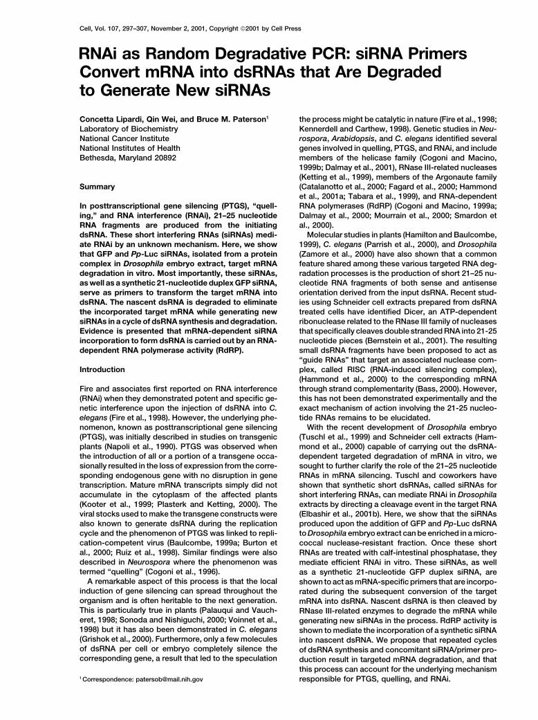
Figure 2. siRNAs Are Double Stranded andAssociate in a Dynamic Ribonucleprotein Com-plex Protected from Micrococcal NucleaseDigestion
(A) Treatment of Drosophila embryo extractwith micrococcal nuclease before or after theaddition of GFP dsRNA inhibits RNAi: Lane1, the standard reaction; Lane 2, addition ofmicrococcal nuclease prior to dsRNA; Lane3, addition of dsRNA prior to micrococcalnuclease; and Lane 4, addition of dsRNA tothe reaction in Lane 3 restores modest levelsof RNAi. Percent silencing shown as GFP/Pp-Luc ratios is indicated below each lane.(B) Addition of 32P-UTP-labeled GFP dsRNA(200 pM, �109 cpm/�g) to extract prior tomicrococcal nuclease treatment produces in-active siRNAs. Lane 1, control; Lane 2, addi-tion of micrococcal nuclease prior to dsRNA;Lane 3, addition of dsRNA prior to micrococ-cal nuclease; and Lane 4, addition of 10 nMunlabeled GFP dsRNA to the reaction in Lane3 chases the level of siRNAs. The initial inputof labeled dsRNA was the same in all lanesshown, 2.5 � 105 cpm.(C) The GFP siRNAs after micrococcal nu-clease treatment, called Mn-siRNAs (Figure2B, lane 3), are double stranded and resistantto Ribonuclease One digestion. Lane 1, siR-NAs with no nuclease; Lane 2, siRNAs withRNase One; and Lane 3, denatured siRNAswith RNase One.
as well as the plateau value for the small RNAs was nuclease One (RNase One), a single-strand-specific ri-bonuclease that can cut gaps and single base-pair mis-greatly reduced (Figure 2B, lane 2).
Extract incubated with unlabeled GFP dsRNA prior matches (Meador et al., 1990).to micrococcal nuclease treatment also was unable todirect the degradation of GFP mRNA added subse- siRNAs Require a 3�-Hydroxyl Group to Direct
RNAi In Vitroquently to the reaction (Figure 2A, lane 3). However, anexamination of the GFP siRNAs produced under these Using micrococcal nuclease and 500 ng of unlabeled
GFP dsRNA, we prepared an RNA fraction enriched forconditions revealed that the small RNAs remained resis-tant to nuclease digestion and were produced in the GFP-specific siRNAs for further testing in RNAi re-
actions. Addition of this RNA fraction to a silencingamounts comparable to that seen in control reactions(Figure 2B, compare lanes 1 and 3). Furthermore, with reaction did not trigger the degradation of GFP mRNA
(Figures 3A and 3B, control versus Mn-siRNAs). Thethe exception of the GFP siRNAs, most of the labeledGFP dsRNA was degraded during nuclease treatment products of RNA digestion with micrococcal nuclease
are mononucleotides and oligonucleotides with terminal(Figure 2B, lane 3).When additional unlabeled GFP dsRNA was added to 3�-phosphate groups (Krupp and Gross, 1979). Tuschl
and coworkers have recently shown that the siRNAsthe reaction shown in Figure 2A, lane 3, modest RNAiwas observed—about 30% of control values (Figure 2A, derived from dsRNA incubated in Drosophila extracts
have 5�-monophosphate and 3�-hydroxyl groups (El-lane 4). Similarly, unlabeled GFP dsRNA added to extractpreincubated with 32P-labeled GFP dsRNA also resulted bashir et al., 2001b). We speculated that a phosphate
group on the 3�-hydroxyl position might inhibit the func-in roughly a 30% decrease in the amount of 32P-labeledGFP siRNAs (Figure 2B, lane 4). Taken together, these tion of the GFP siRNAs in some aspect of the silencing
process. In order to check this, the enriched GFP Mn-results indicate that the production of the siRNAs is adynamic process involving cleavage and displacement siRNAs were treated with calf-intestinal alkaline phos-
phatase (CIP) to remove any phosphate groups prior toby the dsRNA substrate.The resistance of the siRNAs to micrococcal nuclease retesting for RNAi (Mossner et al., 1980). Phosphatase
treatment restored mRNA target degradation to a leveldigestion suggested the small RNAs were in a proteincomplex. This was confirmed upon treatment of the comparable to that observed with 500 ng of full-sized
GFP dsRNA (Figures 3A and 3B,control versus CIP-Mn-protected siRNAs with proteinase K, as described byTuschl (Tuschl et al., 1999), which now rendered the siRNAs). The slight trimming of the siRNAs during
nuclease treatment did not impair their silencing activitysiRNAs digestible by micrococcal nuclease (data notshown, but similar to Figure 2C). The same short RNAs and they remained within the 21–25 nucleotide size
range, as noted in Figures 2B and 2C. This was a directwere also resistant (Figure 2C) to digestion with Ribo-

Cell300
of the complementary template RNA. 32P-labeled siRNAs(2.5 � 105 cpm), corresponding to 250 pg of dsRNA,were prepared from full-size GFP and Pp-Luc 32P-labeleddsRNAs using the micrococcal nuclease digestion pro-cedure and assayed for template RNA-dependent incor-poration into larger RNAs. Full-size GFP (716 bp) and Pp-Luc (1652 bp) single strand, double strand, and capped/adenylated RNAs were used as templates. The reactionproducts were analyzed on denaturing 1.5% agaroseformaldehyde gels. Consistent with their role as primersfor RdRP activity, the labeled GFP siRNAs were incorpo-rated specifically into full-size RNA for all the templates(Figure 4A). Heterologous RNA/siRNA combinations didnot result in any detectable siRNA incorporation (Figure4A) and neither single- nor double-stranded DNA sub-strates served as templates for siRNA uptake (data notshown).
siRNAs Are Incorporated into dsRNAthat Is Subsequently DegradedWe wanted to determine if the full-length RNAs labeledwith siRNAs were double stranded. The reaction prod-ucts were digested with RNase one and found to beresistant to nuclease digestion under conditions thatcompletely degraded single-stranded GFP RNA tran-scribed in vitro with T7 RNA polymerase (Figure 4B).The slight sensitivity of the dsRNAs derived from thedifferent RNA templates suggested that some of themmight have contained gaps or mismatches susceptibleto RNase one digestion. RNase T1, which does not de-grade dsRNA but cleaves single stranded RNA 3� to Gresidues, also did not degrade the siRNA-labeled RNAs,as shown for GFP mRNA derived dsRNA (Figure 4B).Therefore, the siRNA-abeled RNAs are double stranded.
The siRNA-labeled RNAs could also serve as sub-strate for cleavage by RNase III-related enzymes. Totest this, the full-length GFP dsRNA (shown in Figure4A) was gel purified and incubated in extract. As shownin Figure 4C, lane 3, the siRNA-labeled RNA was pro-cessed to produce new GFP siRNAs.
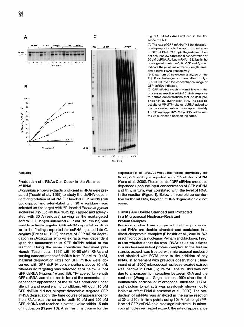
Figure 3. siRNAs Require a 3�-Hydroxyl Group to Function in RNAi Given this result, we speculated that longer incubation(A) GFP siRNAs produced from 500 ng of full-length GFP dsRNA of the siRNA incorporation reaction would show degra-(716 bp) and treated with micrococcal nuclease trigger efficient RNAi dation of the dsRNA and accumulation of the corre-if they are treated with calf-intestinal alkaline phosphatase (CIP). sponding siRNAs. This was observed when 32P-labeledControl, 500 ng GFP dsRNA under standard reaction conditions;
GFP and Pp-Luc siRNAs and the cognate RNA tem-Mn-siRNAs, siRNAs derived from 500 ng of GFP dsRNA treated withplates were used to produce dsRNA (Figures 5A andmicrococcal nuclease; CIP Mn-siRNAs, Mn-siRNAs treated with CIP.
GFP (746 bp) and Pp-Luc (1682 bp) mRNA positions are indicated. 5B). The products were analyzed over a 3 hr period. The(B) Quantification of the GFP/Pp-Luc band intensity ratios shown full-length dsRNAs appeared rapidly within 2–5 min forin (A) were measured with the Fuji Phosphoimager. both the Pp-Luc and GFP templates (Figure 5A, panels
1 and 3) and reached a maximum level within 5–10 minof incubation before the onset of degradation was ob-
demonstration that the GFP siRNAs produced in a si- served. A longer exposure revealed that the siRNAs werelencing reaction efficiently target degradation of the cor- incorporated into shorter products that were degradedresponding mRNA and that this activity required a 3� during the time course of the reaction (Figure 5A, panelshydroxyl group on the siRNAs. 2 and 4). By the end of the incubation period, more than
90% of the siRNA-labeled dsRNA was degraded whilenascent siRNAs continually increased over the timesiRNAs Behave as Primers and Are Incorporated
into dsRNA course of the reaction (Figures 5A and 5B). SiRNA levelsdid not plateau within 15 min, as observed with theThe requirement for a 3�-hydroxyl group on the GFP
siRNAs to degrade the target mRNA indicated that they addition of labeled dsRNA to the extract, but continuedto accumulate (Figure 5B). This result confirmed that themight serve as primers for an RdRP activity in RNAi. If
this was correct, labeled siRNAs would be incorporated siRNA-labeled RNA was double stranded since single-stranded RNA is not a substrate for RNase III-relatedinto RNA product dependent specifically upon the use

Mechanism of RNA Interference301
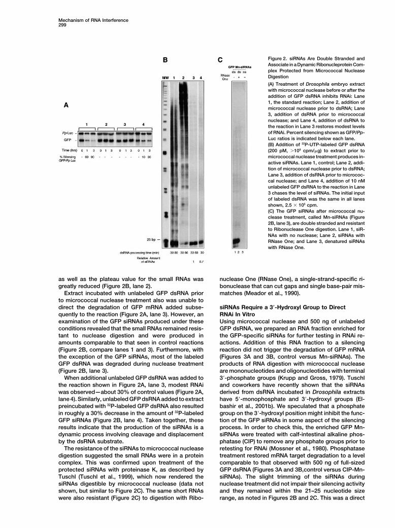
Figure 4. siRNAs Are mRNA-Specific Prim-ers for dsRNA Synthesis
(A) 32P-UTP labeled GFP or Pp-Luc Mn-siR-NAs, prepared from the full-length dsRNAs bythe micrococcal nuclease/CIP method (seeExperimental Procedures), were incubated inextract with the cognate and heterologousRNA templates and assayed for incorporationinto larger RNAs on 1.5% agarose formalde-hyde gels. Pp-Luc and GFP siRNAs were in-corporated up to the corresponding full-sizeRNAs only with the homologous single- ordouble-stranded RNA templates. Incubationof siRNAs in the absence of template or withthe heterologous template resulted in no prod-uct. Single-stranded antisense RNA (as), dou-ble-stranded RNA (ds), and capped-polyA RNA(mRNA) were all templates for primer incorpo-ration. GFP (746 bp) and Pp-Luc (1682 bp)mark the positions of the corresponding full-length RNAs.(B) siRNAs are incorporated into RNA that isdouble-stranded and resistant to digestionby Ribonuclease One and Ribonuclease T1.siRNAs-labeled GFP RNA produced from ei-ther single- or double-stranded templates isresistant to two different nucleases that donot digest dsRNA. T7 ss GFP, single strandcontrol RNA transcribed in vitro with T7 RNApolymerase. The same notation as in (A) isused here.(C) Full-length siRNA-labeled GFP dsRNA isprocessed into new siRNAs. Full-length GFPdsRNA, as shown in (A), was gel purified asdescribed in Experimental Procedures andincubated in extract for processing. Lane 1,25 bp ladder; Lane 2, marked control, pro-cessed GFP dsRNA (90,000 cpm input); Lane3, new GFP siRNAs produced from the full-length GFP RNA labeled by siRNA incorpora-tion (90,000 cpm input).
enzymes and does not produce siRNAs. When GFP strand (Figure 6A). This siRNA was tested for template-specific incorporation into full-length GFP dsRNA usingdsRNA was used as template for siRNA incorporation,
the production of new siRNAs was significantly slower sense and antisense GFP and Pp-Luc RNAs. Specificincorporation would provide evidence for RdRP activityand reached a lower level compared to that seen with
mRNA template (Figure 5B, compare dsGFP to ssGFP). in the extract. This synthetic duplex siRNA primed thesynthesis of essentially full-length GFP dsRNA onlyThis can be explained by the fact that cold dsRNA tem-
plate competes with the newly labeled dsRNA for the when the antisense RNA strand was used as template(Figure 6B). The sense strand template primed the syn-cleavage reaction, as shown in the previous results (Fig-
ure 2B, compare lanes 3 and 4). SiRNAs incubated in thesis of the predicted 44 nucleotide fragment corre-sponding to the number of base pairs from the 3� endthe absence of template remained at the same position
and intensity and did not shift into new products at all of the siRNA to the GFP AUG start codon at the 5� endof the template (Figure 6B). Pp-Luc templates did nottime points (Figure 5B, no template).
Therefore, siRNAs are used to convert the target produce any incorporation of the GFP siRNA (Figure6B). Therefore, both strands of the siRNA were used asmRNA into dsRNA, which is then cleaved by RNase III
activity in the extract to eliminate the target RNA while primers on the appropriate template strand and wereincorporated into dsRNA as a single primer event.producing a new set of siRNAs to repeat the process.
The dsRNA produced with the synthetic GFP duplexsiRNA was also synthesized and degraded with kineticsA Synthetic 21 nt GFP Duplex siRNA
Is Incorporated into Full-Length similar to the GFP dsRNA produced with micrococcalnuclease-generated GFP siRNAs (Figure 6D). This resultGFP dsRNA
It has been recently shown that synthetic 21 nt duplex suggests that a single primer in the natural siRNA popu-lation would be capable of priming the entire GFPsiRNAs can mediate RNAi in both Drosophila embryo
extracts and in Schneider cells (Elbashir et al., 2001b). dsRNA. Using the synthetic GFP siRNA, we never sawthe intermediate products noted with the natural siRNAsWe prepared a 32P-labeled 21 nt duplex siRNA corre-
sponding to nucleotides 26–44 in the GFP coding region (Figure 6C, panel 2 versus Figure 5A, panels 2 and 4).These intermediate products may represent numerouswith two additional uridine residues on the 3� end of each

Cell302
Figure 5. siRNAs Are Incorporated intodsRNA Prior to Degradation and the Forma-tion of New siRNAs
(A) Nascent full-length GFP and Pp-LucdsRNAs labeled by siRNA incorporation aresynthesized then degraded upon prolongedincubation in extract. Labeled siRNAs arerapidly incorporated into dsRNAs up to thefull-length of the template RNAs. Panels 1and 3, short exposure (20 min) of Pp-Luc andGFP siRNA incorporation into dsRNA show-ing a maximal labeling rate at 5 to 10 min.Panels 2 and 4, long exposure (2 hr) of panels1 and 3 to reveal shorter labeled products.The full-length products are essentially ab-sent by 180 min of incubation.(B) New siRNAs are produced during the deg-radation of the full-length GFP and Pp-LucdsRNAs labeled by siRNA incorporation. Notemplate, the siRNAs are not incorporated(see Figure 4A, minus template) and remainat a constant level at each time point; ssGFPtemplate, using the reaction shown above in(A) panel 3, new siRNAs are derived from thelabeled GFP dsRNA and continue to accumu-late during the incubation period; dsGFP,GFP dsRNA was used as a template for siRNAincorporation. New siRNAs accumulate moreslowly during the reaction period due to thecompetition for cleavage by cold GFP dsRNA.Images are from the Fuji Phosphoimager.
single primer extensions from various primer positions III activity into new siRNAs. In this way, mRNA is de-graded through a cycle of “degradative-PCR” (Figurealong the target RNA.
Efforts to use unlabeled siRNAs and 32P-UTP to follow 7). We present substantial evidence for RdRP activityin Drosophila extracts and suggest siRNA incorporationdsRNA synthesis were unsuccessful due to the high
levels of nonspecific UTP incorporation in the extract into dsRNA involves RdRP, the crucial step in the ampli-fication of the target RNA for rapid degradation bythat were unaffected by �-amanitin and actinomycin
D. Nevertheless, the incorporation of the synthetic 21 RNase III-type activity. Although we cannot exclude thatsiRNAs may be incorporated into dsRNA by a directnucleotide GFP duplex siRNA into dsRNA is consistent
with the presence of RdRP in the extract and its role “guide” mechanism not involving RdRP, such a processwould not give the sufficient amplification of the double-in the siRNA-dependent generation of dsRNA from the
mRNA target. stranded RNA target. This would be needed to triggerefficient RNAi with substoichiometric levels of the initiat-ing double-stranded trigger RNA. Consistent with theDiscussiongenetic screens in other lower eukaryotes, our resultssuggest a role for RdRP in Drosophila RNAi as well.Model for RNAi
Our findings complement and clarify the previously pro-posed models concerning the mechanism of PTGS, quell- Double-Stranded RNA as the Target for Degradation
and the Source for New siRNAsing, and RNAi (Bass, 2000; Baulcombe, 1999b, 2000;Carthew, 2001; Cogoni and Macino, 2000; Gura, 2000; The requirement for a dsRNA trigger as the effector for
silencing can be partially explained by the nature of theHammond et al., 2001b; Hunter, 2000; Kooter et al., 1999;Maine, 2000; Marx, 2000; Meins, 2000; Plasterk and Ket- dsRNA cleavage step required for siRNA production. As
previously noted by Fire and associates (Parrish et al.,ting, 2000; Sharp, 2001; Sijen et al., 2001). Most im-portantly, we demonstrate the template-specific incorp- 2000) and reiterated by Sharp (Sharp, 2001), any factor
that significantly alters the double-stranded nature oforation of the 21–25 nucleotide RNAs, or siRNAs, togenerate dsRNA that is subsequently cleaved by RNase the dsRNA trigger, such as sequence divergence or

Mechanism of RNA Interference303
Figure 6. Incorporation of a Synthetic 21 Nu-cleotide Duplex GFP siRNA into dsRNA
(A) The GFP coding region and the sequenceof the synthetic 21 nucleotide duplex GFPsiRNA are indicated. The siRNA correspondsto nucleotides 26–44 in the GFP coding regionwith two additional uridine residues on the 3�
end. The 5� end of each strand of the siRNAwas labeled with [�-32P]ATP (see Experimen-tal Procedures).(B) Left, the expected product lengths for in-corporation of each strand of the synthetic 21nucleotide GFP siRNA using full-length GFPsense and antisense RNA templates areshown: 690 bp with the antisense strand tem-plate and 44 bp with the sense strand tem-plate. Right, the 32P-labeled siRNA incorpora-tion into full-length GFP dsRNA occurs onlywith the GFP antisense RNA template andgives the expected 44 bp product with thesense strand template. Pp-Luc templatesgive no product. GFP and Pp-Luc mark thepositions of the full-length RNAs and MW de-notes the 25 bp ladder.(C) GFP dsRNA produced with the synthetic21 nucleotide duplex GFP siRNA is rapidlysynthesized and then degraded upon pro-longed incubation in the reaction (Panel 1).Note with longer exposure the absence ofintermediate products for the early timepoints for GFP dsRNA formation (Panel 2).GFP marks the position of the full-length RNA(690bp). Panels 1 and 2 were exposed for 30min and 2 hr, respectively.(D) GFP dsRNA produced either with micro-coccal nuclease generated GFP siRNAs (Mn-siRNA, data from Figure 5A, GFP) or the syn-thetic 21 nucleotide duplex GFP siRNA(siRNA-21) is synthesized and degraded withthe same kinetics in the incorporation reac-tion. Note that maximal levels of GFP dsRNAoccur at 5 min for both synthetic and naturalsiRNAs. Data were quantified on the FujiPhosphoimager.
chemical modification, affects silencing substantially. In tracts and in Schneider cells (Elbashir et al., 2001a,2001b). The siRNAs produced in Drosophila embryo ex-the model proposed here, any changes in strand com-
plementarity could presumably reduce the susceptibility tract by micrococcal nuclease and CIP treatment areessentially as efficient on a weight basis in RNAi asof the triggering dsRNA to RNase III-type cleavage. The
nature of the sense and antisense strands in the trig- the full-length dsRNA from which they were derived,suggesting there is some optimal length for siRNAs ingering dsRNA would also play a role in the efficacy
of silencing since the RdRP amplification step would RNAi. The conservation in the size range for the smallRNAs associated with silencing in all the species exam-depend upon the production and functionality of the
siRNAs. We show that both strands of a synthetic 21 ined proposes that it may be closely correlated withprimer function. This could be due to some unique prop-nucleotide GFP duplex siRNA function as primers to
give the expected RNA products when the appropriate erty of primer activity in a protein complex that has yetto be identified.GFP template strand is used (Figure 6).
The length of the siRNAs may be an important aspect We demonstrate that siRNAs require a 3� hydroxylgroup for function in RNAi. The authentic siRNAs, pro-of their function. Previous reports indicated that 29–36
nucleotide dsRNAs transcribed in vitro do not direct duced in Drosophila extracts by RNase III-related en-zymes such as Dicer (Bernstein et al., 2001), have beenRNAi efficiently in Drosophila extracts (Elbashir et al.,
2001b), and that a 26 nucleotide dsRNA, also tran- chemically characterized and shown to have a 5� phos-phate and a 3� hydroxyl group (Elbashir et al., 2001b).scribed in vitro, when injected into worms, triggered
lower than expected levels of RNA interference at 25�C The micrococcal nuclease generated siRNAs describedhere are functional in RNAi and can be incorporated intoand none at 16�C (Parrish et al., 2000). However, chemi-
cally synthesized 21 and 22 nucleotide siRNAs can me- dsRNA after phosphatase treatment (Figure 3) to removethe 3� phosphate group produced by the nuclease diges-diate targeted RNA cleavage in Drosophila embryo ex-
Cell304
In agreement with the results reported using the syn-thetic siRNAs hybridized to RNA (Elbashir et al., 2001b),we show that cleavage of the GFP target RNA occursafter the synthetic GFP siRNA is incorporated intodsRNA. If cleavage occurred in the template RNA imme-diately upon binding to the synthetic GFP siRNA, nofull-length GFP dsRNA would have been observed.Therefore, cleavage occurs in the nascent dsRNA inregions inside and outside the zone represented by theinitial siRNA since the primers are extended to makedsRNA. The fact that the synthetic GFP siRNA is ex-tended to the 5� end of the sense strand template (Figure6B) would also restrict cleavage, in this instance, to theregion upstream of the 3� terminus of the siRNA. Anyregion of the target RNA converted into duplex by agiven siRNA would be subject to digestion by RNase IIIactivity.
A Role for RdRP in Drosophila RNAiIt has been proposed previously that there is no amplifi-cation of the trigger dsRNA in RNAi in C. elegans (Sharp,2001), based upon the effects of asymmetric strand sub-stitutions in the input dsRNA (Parrish et al., 2000). Weprovide evidence that both single- and double-strandedRNAs can serve as templates for siRNA incorporationinto dsRNA in Drosophila extract. However, the rapiddegradation of dsRNA suggests that amplification of thetrigger dsRNA is of limited value. In the substitutionexperiments described by Fire et al. (Parrish et al., 2000),
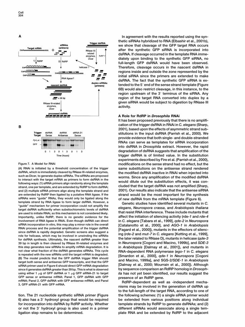
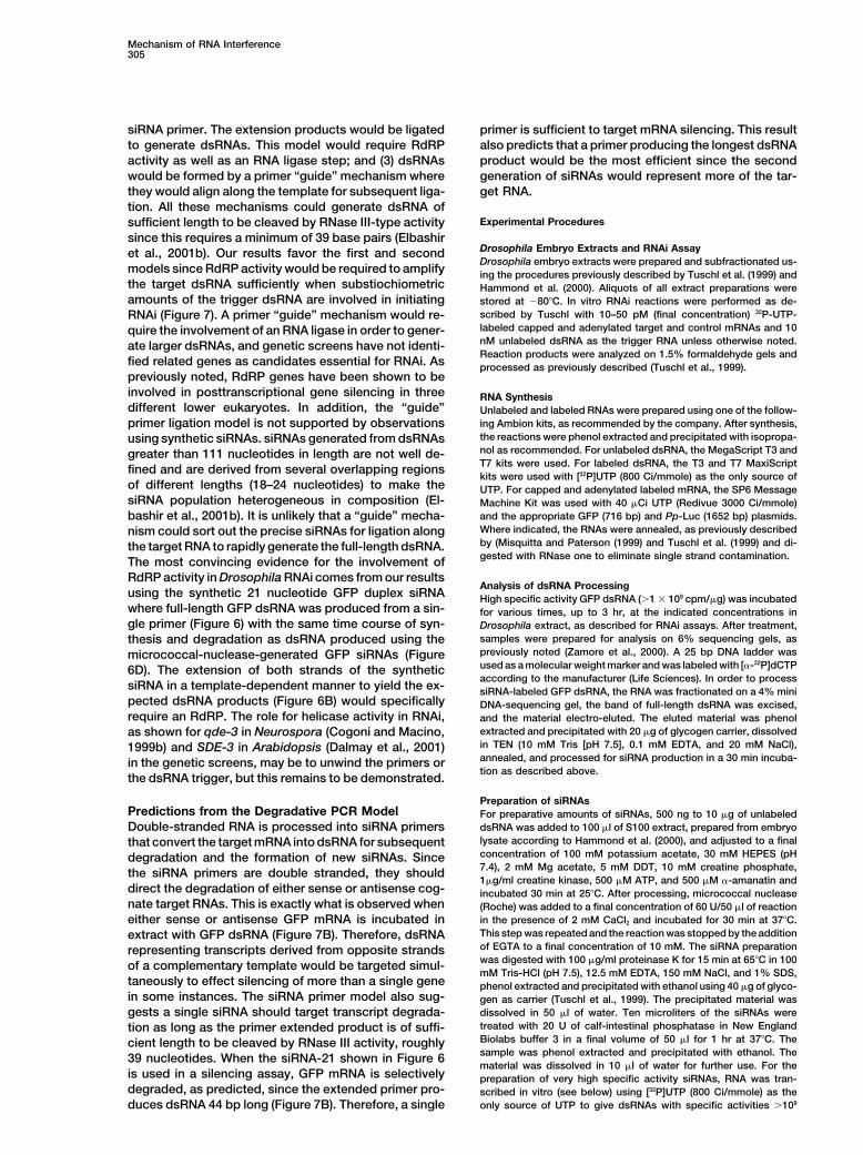
Figure 7. A Model for RNAi modifications on the sense strand had no effect, but the(A) RNAi is initiated by a threshold concentration of the trigger same substitutions on the antisense strand rendereddsRNA, which is immediately cleaved by RNase III-related enzymes, the modified dsRNA inactive in RNAi when injected intosuch as Dicer, to generate duplex siRNAs. The siRNAs are proposed worms. Since any amplification of the modified dsRNAto interact with the target mRNA as primers to form dsRNA in the
would dilute out the substitution effects, it was con-following ways: (1) siRNA primers align randomly along the templatecluded that the target dsRNA was not amplified (Sharp,strand, one per template, and are extended by RdRP to form dsRNA;2001). Our results also indicate that the antisense siRNAand (2) multiple siRNA primers align along the template strand and
are extended by RdRP then ligated by a putative RNA ligase. If the strand would be the most important for the synthesissiRNAs were “guide” RNAs, they would only be ligated along the of new dsRNA from the mRNA template (Figure 6).template strand by RNA ligase to form larger dsRNA. However, a Genetic studies have identified several mutants in C.“guide” mechanism for primer incorporation could not amplify the
elegans, Neurospora crassa, and Arabidopsis thalianatarget dsRNA sufficiently when substiochiometric levels of dsRNAthat resist RNA interference. These include mutants thatare used to initiate RNAi, so this mechanism is not considered likely.affect the initiation of silencing activity (rde-1 and rde-4Importantly, unlike RdRP, there is no genetic evidence for the
involvement of RNA ligase in RNAi. Even though dsRNA can direct in C. elegans [Tabara et al., 1999], qde-2 in NeurosporasiRNA incorporation in vitro, this may play a minor role in the overall [Catalanotto et al., 2000], and AGO-1 in ArabidopsisRNAi process and the potential amplification of the trigger dsRNA [Fagard et al., 2000]), mutants in the effectors of silenc-since dsRNA is rapidly degraded. Genetic screens also suggest a
ing (rde-2 and mut-7 in C. elegans [Ketting et al., 1999],role for helicase, which may be involved in unwinding the siRNAsthe later related to RNase D), mutants in helicase (qde-3for dsRNA synthesis. Ultimately, the nascent dsRNA greater thanin Neurospora [Cogoni and Macino, 1999b], and SDE-339 bp in length is then cleaved by RNase III-related enzymes and
this step generates new siRNAs to amplify mRNA degradation. It is in Arabidopsis [Dalmay et al., 2001]), and mutants innot clear what fraction of the dsRNA generates siRNAs. This cycle RNA-dependent RNA polymerase (ego-1 in C. elegansis repeated with the new siRNAs until the target mRNA is degraded. [Smardon et al., 2000], qde-1 in Neurospora [Cogoni(B) The model predicts that the GFP dsRNA trigger RNA should
and Macino, 1999a], and SGS-2/SDE-1 in Arabidopsistarget both sense and antisense GFP transcripts, and that the GFP[Dalmay et al., 2000; Mourrain et al., 2000]). AlthoughsiRNA primer used in Figure 6 should direct GFP mRNA degradationby sequence comparison an RdRP homolog in Drosoph-since it generates dsRNA greater than 39 bp. This is what is observed
using either 1 �g of GFP dsRNA or 1 �g GFP siRNA-21 to target ila has not yet been identified, our results suggest theGFP sense or antisense mRNA: Panel 1, GFP dsRNA with GFP presence of an RdRP gene.mRNA; Panel 2, GFP dsRNA with GFP antisense mRNA; and Panel RdRP-dependent as well as -independent mecha-3, GFP siRNA-21 with GFP mRNA.
nisms may be involved in the generation of dsRNA upto the full-length of the target RNA, according to one ofthe following schemes: (1) a single siRNA primer wouldtion. The 21 nucleotide synthetic siRNA primer (Figure
6) also has a 3� hydroxyl group that would be required be extended from various positions along individualtemplate strands by RdRP to generate dsRNAs; and (2)for incorporation into dsRNA by RdRP activity. Whether
or not the 3� hydroxyl group is also used in a primer different siRNAs would associate along a single tem-plate RNA and be extended by RdRP to the adjacentligation step remains to be determined.
Mechanism of RNA Interference305
siRNA primer. The extension products would be ligated primer is sufficient to target mRNA silencing. This resultalso predicts that a primer producing the longest dsRNAto generate dsRNAs. This model would require RdRP
activity as well as an RNA ligase step; and (3) dsRNAs product would be the most efficient since the secondgeneration of siRNAs would represent more of the tar-would be formed by a primer “guide” mechanism where
they would align along the template for subsequent liga- get RNA.tion. All these mechanisms could generate dsRNA of
Experimental Proceduressufficient length to be cleaved by RNase III-type activitysince this requires a minimum of 39 base pairs (Elbashir
Drosophila Embryo Extracts and RNAi Assayet al., 2001b). Our results favor the first and secondDrosophila embryo extracts were prepared and subfractionated us-
models since RdRP activity would be required to amplify ing the procedures previously described by Tuschl et al. (1999) andthe target dsRNA sufficiently when substiochiometric Hammond et al. (2000). Aliquots of all extract preparations wereamounts of the trigger dsRNA are involved in initiating stored at �80�C. In vitro RNAi reactions were performed as de-
scribed by Tuschl with 10–50 pM (final concentration) 32P-UTP-RNAi (Figure 7). A primer “guide” mechanism would re-labeled capped and adenylated target and control mRNAs and 10quire the involvement of an RNA ligase in order to gener-nM unlabeled dsRNA as the trigger RNA unless otherwise noted.ate larger dsRNAs, and genetic screens have not identi-Reaction products were analyzed on 1.5% formaldehyde gels and
fied related genes as candidates essential for RNAi. As processed as previously described (Tuschl et al., 1999).previously noted, RdRP genes have been shown to beinvolved in posttranscriptional gene silencing in three RNA Synthesisdifferent lower eukaryotes. In addition, the “guide” Unlabeled and labeled RNAs were prepared using one of the follow-
ing Ambion kits, as recommended by the company. After synthesis,primer ligation model is not supported by observationsthe reactions were phenol extracted and precipitated with isopropa-using synthetic siRNAs. siRNAs generated from dsRNAsnol as recommended. For unlabeled dsRNA, the MegaScript T3 andgreater than 111 nucleotides in length are not well de-T7 kits were used. For labeled dsRNA, the T3 and T7 MaxiScriptfined and are derived from several overlapping regionskits were used with [32P]UTP (800 Ci/mmole) as the only source of
of different lengths (18–24 nucleotides) to make the UTP. For capped and adenylated labeled mRNA, the SP6 MessagesiRNA population heterogeneous in composition (El- Machine Kit was used with 40 �Ci UTP (Redivue 3000 Ci/mmole)
and the appropriate GFP (716 bp) and Pp-Luc (1652 bp) plasmids.bashir et al., 2001b). It is unlikely that a “guide” mecha-Where indicated, the RNAs were annealed, as previously describednism could sort out the precise siRNAs for ligation alongby (Misquitta and Paterson (1999) and Tuschl et al. (1999) and di-the target RNA to rapidly generate the full-length dsRNA.gested with RNase one to eliminate single strand contamination.The most convincing evidence for the involvement of
RdRP activity in Drosophila RNAi comes from our resultsAnalysis of dsRNA Processing
using the synthetic 21 nucleotide GFP duplex siRNA High specific activity GFP dsRNA (�1 � 109 cpm/�g) was incubatedwhere full-length GFP dsRNA was produced from a sin- for various times, up to 3 hr, at the indicated concentrations ingle primer (Figure 6) with the same time course of syn- Drosophila extract, as described for RNAi assays. After treatment,
samples were prepared for analysis on 6% sequencing gels, asthesis and degradation as dsRNA produced using thepreviously noted (Zamore et al., 2000). A 25 bp DNA ladder wasmicrococcal-nuclease-generated GFP siRNAs (Figureused as a molecular weight marker and was labeled with [�-32P]dCTP6D). The extension of both strands of the syntheticaccording to the manufacturer (Life Sciences). In order to process
siRNA in a template-dependent manner to yield the ex- siRNA-labeled GFP dsRNA, the RNA was fractionated on a 4% minipected dsRNA products (Figure 6B) would specifically DNA-sequencing gel, the band of full-length dsRNA was excised,require an RdRP. The role for helicase activity in RNAi, and the material electro-eluted. The eluted material was phenol
extracted and precipitated with 20 �g of glycogen carrier, dissolvedas shown for qde-3 in Neurospora (Cogoni and Macino,in TEN (10 mM Tris [pH 7.5], 0.1 mM EDTA, and 20 mM NaCl),1999b) and SDE-3 in Arabidopsis (Dalmay et al., 2001)annealed, and processed for siRNA production in a 30 min incuba-in the genetic screens, may be to unwind the primers ortion as described above.
the dsRNA trigger, but this remains to be demonstrated.
Preparation of siRNAsPredictions from the Degradative PCR Model For preparative amounts of siRNAs, 500 ng to 10 �g of unlabeled
dsRNA was added to 100 �l of S100 extract, prepared from embryoDouble-stranded RNA is processed into siRNA primerslysate according to Hammond et al. (2000), and adjusted to a finalthat convert the target mRNA into dsRNA for subsequentconcentration of 100 mM potassium acetate, 30 mM HEPES (pHdegradation and the formation of new siRNAs. Since7.4), 2 mM Mg acetate, 5 mM DDT, 10 mM creatine phosphate,the siRNA primers are double stranded, they should1�g/ml creatine kinase, 500 �M ATP, and 500 �M �-amanatin and
direct the degradation of either sense or antisense cog- incubated 30 min at 25�C. After processing, micrococcal nucleasenate target RNAs. This is exactly what is observed when (Roche) was added to a final concentration of 60 U/50 �l of reaction
in the presence of 2 mM CaCl2 and incubated for 30 min at 37�C.either sense or antisense GFP mRNA is incubated inThis step was repeated and the reaction was stopped by the additionextract with GFP dsRNA (Figure 7B). Therefore, dsRNAof EGTA to a final concentration of 10 mM. The siRNA preparationrepresenting transcripts derived from opposite strandswas digested with 100 �g/ml proteinase K for 15 min at 65�C in 100of a complementary template would be targeted simul-mM Tris-HCl (pH 7.5), 12.5 mM EDTA, 150 mM NaCl, and 1% SDS,
taneously to effect silencing of more than a single gene phenol extracted and precipitated with ethanol using 40 �g of glyco-in some instances. The siRNA primer model also sug- gen as carrier (Tuschl et al., 1999). The precipitated material was
dissolved in 50 �l of water. Ten microliters of the siRNAs weregests a single siRNA should target transcript degrada-treated with 20 U of calf-intestinal phosphatase in New Englandtion as long as the primer extended product is of suffi-Biolabs buffer 3 in a final volume of 50 �l for 1 hr at 37�C. Thecient length to be cleaved by RNase III activity, roughlysample was phenol extracted and precipitated with ethanol. The39 nucleotides. When the siRNA-21 shown in Figure 6material was dissolved in 10 �l of water for further use. For the
is used in a silencing assay, GFP mRNA is selectively preparation of very high specific activity siRNAs, RNA was tran-degraded, as predicted, since the extended primer pro- scribed in vitro (see below) using [32P]UTP (800 Ci/mmole) as the
only source of UTP to give dsRNAs with specific activities �109duces dsRNA 44 bp long (Figure 7B). Therefore, a single
Cell306
cpm /�g. Typically, 1 �g of labeled dsRNA was processed as above Bernstein, E., Caudy, A.A., Hammond, S.M., and Hannon, G.J. (2001).Role for a bidentate ribonuclease in the initiation step of RNA inter-to generate highly labeled siRNAs.
Two synthetic 21 nucleotide RNAs were synthesized by the Yale ference. Nature 409, 363–366.Keck Labs (trityl-off) representing nucleotides 26–44 in the GFP Burton, R.A., Gibeaut, D.M., Bacic, A., Findlay, K., Roberts, K., Hamil-coding region and containing two additional U residues on the 3� ton, A., Baulcombe, D.C., and Fincher, G.B. (2000). Virus-inducedend: RNA-1, CUGGAGUUGUCCCAAUUCUUU and RNA-2, UUGAC silencing of a plant cellulose synthase gene. Plant Cell 12, 691–706.CUCAACAGGGUUAAGA. To label the 21 nucleotide RNAs, 0.1 �g
Carthew, R.W. (2001). Gene silencing by double-stranded RNA. Curr.of each RNA was labeled in a 10 �l reaction using T4 polynucleotide
Opin. Cell Biol. 13, 244–248.kinase (NEB) with 3 �l of [�-32P]ATP (crude, ICN 6000 Ci/mmol) and
Catalanotto, C., Azzalin, G., Macino, G., and Cogoni, C. (2000). Genepassed over a G-25 spin column in 50 �l final volume (preadjustedsilencing in worms and fungi. Nature 404, 245.with TE) to yield a specific activity of �109 cpm/�g for each 21Cogoni, C., and Macino, G. (1999a). Gene silencing in Neurosporanucleotide RNA. Kinase was inactivated by incubation at 70�C forcrassa requires a protein homologous to RNA-dependent RNA poly-10 min after the addtion of 1 �l of 0.5 M EDTA, as recommendedmerase. Nature 399, 166–169.by the manufacturer. Twenty microliters of each labeled synthetic
RNA was annealed in a 50 �l reaction to form the siRNA as described Cogoni, C., and Macino, G. (1999b). Posttranscriptional gene silenc-(Elbashir et al., 2001b). Unlabeled siRNAs were prepared by anneal- ing in Neurospora by a RecQ DNA helicase. Science 286, 2342–2344.ing 7.5 �g of each RNA in 50 �l as previously noted.
Cogoni, C., and Macino, G. (2000). Post-transcriptional gene silenc-ing across kingdoms. Curr. Opin. Genet. Dev. 10, 638–643.
SiRNA Incorporation AssayCogoni, C., Irelan, J.T., Schumacher, M., Schmidhauser, T.J., Selker,S100 fraction, prepared from embryo extract, was used instead ofE.U., and Macino, G. (1996). Transgene silencing of the al-1 genecrude extract under standard RNAi reaction conditions with 500 �Min vegetative cells of Neurospora is mediated by a cytoplasmic
�-amanitin, 2.5 � 105 cpm of 32P-UTP labeled siRNAs, and 3 �g ofeffector and does not depend on DNA-DNA interactions or DNARNA template (either double- or single-stranded RNA), incubatedmethylation. EMBO J. 15, 3153–3163.at 25�C for 60 min or the indicated times for the time course reac-Dalmay, T., Hamilton, A., Rudd, S., Angell, S., and Baulcombe, D.C.tions. The primer incorporation reaction was stopped with protein-(2000). An RNA-dependent RNA polymerase gene in Arabidopsisase K/SDS buffer and extracted as before (Tuschl et al., 1999).is required for posttranscriptional gene silencing mediated by aLabeled RNA products were analyzed on 1.5% agarose formalde-transgene but not by a virus. Cell 101, 543–553.hyde gels or 6% DNA sequencing gels. For incorporation reactions
using the 21 nucleotide synthetic duplex siRNA, 1 �g of unlabeled Dalmay, T., Horsefield, R., Braunstein, T.H., and Baulcombe, D.C.siRNA was mixed with 3 � 106 cpm of labeled siRNA along with 3 (2001). SDE3 encodes an RNA helicase required for post-transcrip-�g of template and incubated in extract as described above. tional gene silencing in Arabidopsis. EMBO J. 20, 2069–2078.
Elbashir, S.M., Harborth, J., Lendeckel, W., Yalcin, A., Weber, K.,Ribonuclease One and Ribonuclease T1 Digestions and Tuschl, T. (2001a). Duplexes of 21-nucleotide RNAs mediateDigestions of single- and double-stranded RNAs as well as the RNA interference in cultured mammalian cells. Nature 411, 494–498.products of the siRNA incorporation reactions were carried out using Elbashir, S.M., Lendeckel, W., and Tuschl, T. (2001b). RNA interfer-Ribonuclease One (Promega, 10 �U/3 �g of cold template) for up ence is mediated by 21- and 22-nucleotide RNAs. Genes Dev. 15,to 5 min at 37�C in buffer supplied by the manufacturer. Ribo- 188–200.nuclease One digests RNA to nucleotide monophosphates so gel
Fagard, M., Boutet, S., Morel, J.B., Bellini, C., and Vaucheret, H.purification of dsRNA was not required. Reactions were then phenol(2000). AGO1, QDE-2, and RDE-1 are related proteins required forextracted as before and analyzed on 1.5% agarose formaldehydepost-transcriptional gene silencing in plants, quelling in fungi, andgels. For Ribonuclease T1 (Roche) digestion, identical RNA samplesRNA interference in animals. Proc. Natl. Acad. Sci. USA 97, 11650–were dissolved in 50 �l of TE (10 mM Tris [pH 7.5] and 0.1 mM11654.EDTA) and digested with 100–500 units of enzyme for 30 min atFire, A., Xu, S., Montgomery, M.K., Kostas, S.A., Driver, S.E., and37�C. The reactions were phenol extracted and precipitated with 20Mello, C.C. (1998). Potent and specific genetic interference by dou-�g of glycogen carrier prior to gel analysis.ble-stranded RNA in Caenorhabditis elegans. Nature 391, 806–811.
Grishok, A., Tabara, H., and Mello, C.C. (2000). Genetic requirementsImage Analysisfor inheritance of RNAi in C. elegans. Science 287, 2494–2497.Radioactivity was detected by exposing the dried agarose gel (dried
under vacuum) onto an Immobilon-NY membrane or sequence gel Gura, T. (2000). A silence that speaks volumes. Nature 404, 804–808.directly to Kodak AR-2 film at �80�C with a Dupont screen or a Fuji
Hamilton, A.J., and Baulcombe, D.C. (1999). A species of small anti-Image plate, the latter quantified with a Fujix Bas 2000 using Image
sense RNA in posttranscriptional gene silencing in plants. ScienceGauge 3.0 software.
286, 950–952.
Hammond, S.M., Bernstein, E., Beach, D., and Hannon, G.J. (2000).AcknowledgmentsAn RNA-directed nuclease mediates post-transcriptional gene si-lencing in Drosophila cells. Nature 404, 293–296.We would like to acknowledge Claude Klee for help and suggestions.Hammond, S.M., Boettcher, S., Caudy, A.A., Kobayashi, R., andAlso we would like to thank the anonymous referee for describingHannon, G.J. (2001a). Argonaute2, a link between genetic and bio-our results as “Nature’s own PCR machine” and for inspiration onchemical analyses of RNAi. Science 293, 1146–1150.the title. We thank Carl Wu for the use of his Drosophila cages.Hammond, S.M., Caudy, A.A., and Hannon, G.J. (2001b). Post-tran-scriptional gene silencing by double-stranded RNA. Nat. Rev. Genet.Received June 25, 2001; revised September 17, 2001.2, 110–119.
References Hunter, C.P. (2000). Gene silencing: shrinking the black box of RNAi.Curr. Biol. 10, R137–R140.
Bass, B.L. (2000). Double-stranded RNA as a template for gene Kennerdell, J.R., and Carthew, R.W. (1998). Use of dsRNA-mediatedsilencing. Cell 101, 235–238. genetic interference to demonstrate that frizzled and frizzled 2 act
in the wingless pathway. Cell 95, 1017–1026.Baulcombe, D.C. (1999a). Viruses and gene silencing in plants. Arch.Virol. Suppl. 15, 189–201. Ketting, R.F., Haverkamp, T.H., van Luenen, H.G., and Plasterk, R.H.
(1999). Mut-7 of C. elegans, required for transposon silencing andBaulcombe, D.C. (1999b). Gene silencing: RNA makes RNA makesno protein. Curr. Biol. 9, R599–601. RNA interference, is a homolog of Werner syndrome helicase and
RNaseD. Cell 99, 133–141.Baulcombe, D.C. (2000). Molecular biology. Unwinding RNA silenc-ing. Science 290, 1108–1109. Kooter, J.M., Matzke, M.A., and Meyer, P. (1999). Listening to the
Mechanism of RNA Interference307
silent genes: transgene silencing, gene regulation and pathogen artifactual inhibition of RNA processing reactions. Nucleic AcidsRes. 18, 6625–6631.control. Trends Plant Sci. 4, 340–347.
Yang, D., Lu, H., and Erickson, J.W. (2000). Evidence that processedKrupp, G., and Gross, H.J. (1979). Rapid RNA sequencing: nucleasessmall dsRNAs may mediate sequence-specific mRNA degradationfrom Staphylococcus aureus and Neurospora crassa discriminateduring RNAi in Drosophila embryos. Curr. Biol. 10, 1191–1200.between uridine and cytidine. Nucleic Acids Res. 6, 3481–3490.
Zamore, P.D., Tuschl, T., Sharp, P.A., and Bartel, D.P. (2000). RNAi:Maine, E.M. (2000). A conserved mechanism for post-transcriptionaldouble-stranded RNA directs the ATP-dependent cleavage ofgene silencing? Genome Biol. 1, 1018.mRNA at 21 to 23 nucleotide intervals. Cell 101, 25–33.Marx, J. (2000). Interfering with gene expression. Science 288, 1370–
1372.
Meador, J., 3rd, Cannon, B., Cannistraro, V.J., and Kennell, D. (1990).Purification and characterization of Escherichia coli RNase I. Com-parisons with RNase M. Eur. J. Biochem. 187, 549–553.
Meins, F., Jr. (2000). RNA degradation and models for post-tran-scriptional gene-silencing. Plant Mol. Biol. 43, 261–273.
Misquitta, L., and Paterson, B.M. (1999). Targeted disruption of genefunction in Drosophila by RNA interference (RNA-i): a role for nautilusin embryonic somatic muscle formation. Proc. Natl. Acad. Sci. USA96, 1451–1456.
Mossner, E., Boll, M., and Pfleiderer, G. (1980). Purification of humanand bovine alkaline phosphatases by affinity chromatography.Hoppe Seylers Z Physiol. Chem. 361, 543–549.
Mourrain, P., Beclin, C., Elmayan, T., Feuerbach, F., Godon, C.,Morel, J.B., Jouette, D., Lacombe, A.M., Nikic, S., Picault, N., et al.(2000). Arabidopsis SGS2 and SGS3 genes are required for posttran-scriptional gene silencing and natural virus resistance. Cell 101,533–542.
Napoli, C., Lemieux, C., and Jorgensen, R.A. (1990). Introduction ofa chimeric chalcoen synthase gene into Petunia results in reversibleco-suppression of homologous genes in trans. Plant Cell 2, 279–289.
Palauqui, J.C., and Vaucheret, H. (1998). Transgenes are dispens-able for the RNA degradation step of cosuppression. Proc. Natl.Acad. Sci. USA 95, 9675–9680.
Parrish, S., Fleenor, J., Xu, S., Mello, C., and Fire, A. (2000). Func-tional anatomy of a dsRNA trigger. Differential requirement for thetwo trigger strands in RNA interference. Mol. Cell 6, 1077–1087.
Pelham, H.R., and Jackson, R.J. (1976). An efficient mRNA-depen-dent translation system from reticulocyte lysates. Eur. J. Biochem.67, 247–256.
Plasterk, R.H., and Ketting, R.F. (2000). The silence of the genes.Curr. Opin. Genet. Dev. 10, 562–567.
Ruiz, M.T., Voinnet, O., and Baulcombe, D.C. (1998). Initiation andmaintenance of virus-induced gene silencing. Plant Cell 10, 937–946.
Sharp, P.A. (2001). RNA interference—2001. Genes Dev. 15,485–490.
Sijen, T., Vijn, I., Rebocho, A., van Blokland, R., Roelofs, D., Mol,J.N., and Kooter, J.M. (2001). Transcriptional and posttranscriptionalgene silencing are mechanistically related. Curr. Biol. 11, 436–440.
Smardon, A., Spoerke, J.M., Stacey, S.C., Klein, M.E., Mackin, N.,and Maine, E.M. (2000). EGO-1 is related to RNA-directed RNA poly-merase and functions in germ-line development and RNA interfer-ence in C. elegans. Curr. Biol. 10, 169–178.
Sonoda, S., and Nishiguchi, M. (2000). Graft transmission of post-transcriptional gene silencing: target specificity for RNA degradationis transmissible between silenced and non-silenced plants, but notbetween silenced plants. Plant J. 21, 1–8.
Tabara, H., Sarkissian, M., Kelly, W.G., Fleenor, J., Grishok, A., Tim-mons, L., Fire, A., and Mello, C.C. (1999). The rde-1 gene, RNAinterference, and transposon silencing in C. elegans. Cell 99,123–132.
Tuschl, T., Zamore, P.D., Lehmann, R., Bartel, D.P., and Sharp, P.A.(1999). Targeted mRNA degradation by double-stranded RNA invitro. Genes Dev. 13, 3191–3197.
Voinnet, O., Vain, P., Angell, S., and Baulcombe, D.C. (1998). Sys-temic spread of sequence-specific transgene RNA degradation inplants is initiated by localized introduction of ectopic promoterlessDNA. Cell 95, 177–187.
Wang, M.J., and Gegenheimer, P. (1990). Substrate masking: bind-ing of RNA by EGTA-inactivated micrococcal nuclease results in