EstablishmEnt of PCR laboRatoRy in dEvEloPing CountRiEs
96
ESTABLISHMENT OF PCR LABORATORY IN DEVELOPING COUNTRIES
-
Upload
khangminh22 -
Category
Documents
-
view
0 -
download
0
Transcript of EstablishmEnt of PCR laboRatoRy in dEvEloPing CountRiEs

EstablishmEnt of PCR laboRatoRy
in dEvEloPing CountRiEs


EstablishmEnt of PCR laboRatoRy
in dEvEloPing CountRiEs

© World Health Organization 2016
Second Edition
All rights reserved.
Requests for publications, or for permission to reproduce or translate WHO publications – whether for sale or for noncommercial distribution – can be obtained from SEARO Library, World Health Organization, Regional Office for South-East Asia, Indraprastha Estate, Mahatma Gandhi Marg, New Delhi 110 002, India (fax: +91 11 23370197; e-mail: [email protected]).
The designations employed and the presentation of the material in this publication do not imply the expression of any opinion whatsoever on the part of the World Health Organization concerning the legal status of any country, territory, city or area or of its authorities, or concerning the delimitation of its frontiers or boundaries. Dotted lines on maps represent approximate border lines for which there may not yet be full agreement.
The mention of specific companies or of certain manufacturers’ products does not imply that they are endorsed or recommended by the World Health Organization in preference to others of a similar nature that are not mentioned. Errors and omissions excepted, the names of proprietary products are distinguished by initial capital letters.
All reasonable precautions have been taken by the World Health Organization to verify the information contained in this publication. However, the published material is being distributed without warranty of any kind, either expressed or implied. The responsibility for the interpretation and use of the material lies with the reader. In no event shall the World Health Organization be liable for damages arising from its use.
This publication does not necessarily represent the decisions or policies of the World Health Organization.
Printed in India
WHO Library Cataloguing-in-Publication data
Establishment of PCR laboratory in developing countries, 2nd edition.
1. Polymerase Chain Reaction - standards. 2. Laboratories. 3. Developing Countries. 4. Clinical Protocols.
ISBN 978-92-9022-531-7 (NLM classification: QH 450.3)

Abbreviations ............................................................................................................... v
Foreword .................................................................................................................... vii
1. Introduction ...................................................................................................... 1
2. Principles of polymerase chain reaction ........................................................ . 5
3. Structure and functions of PCR machines ..................................................... 19
4. Applications of PCR technology in medicine and allied sciences ................. 25
5. Establishment of a PCR laboratory ................................................................. 31
6. Sample collection, storage and shipment of clinical material for
diagnosis of viral infections by PCR .............................................................. 41
7. Conventional PCR protocols ........................................................................... 49
Protocol 7.1 RNA extraction using QIAamp Viral RNA
Mini Kit (Qiagen) .................................................................. 49
Protocol 7.2 Reverse Transcription Synthesis of Complementary
DNA (cDNA) from RNA using high-capacity cDNA Reverse
Transcription Kit (applied biosystems) ................................. 51
Protocol 7.3 Conventional Reverse Transcriptase (RT) PCR assay
for Chikungunya virus .......................................................... 53
CONTENTS
EstablishmEnt of PCR laboRatoRy in dEvEloPing CountRiEs
iv
8. Real-time PCR Protocols .................................................................................. 57
Protocol 8.1 One-step real-time RT PCR for 2009 H1N1
Influenza A (CDC Protocol) .................................................. 57
Protocol 8.2 Quantitative Taqman Real-time PCR
assay for human immunodeficiency virus (HIV) ................. 60
9. Quality in PCR .................................................................................................. 65
10. Care and maintenance of PCR machines ........................................................ 73
11. Troubleshooting for PCR ................................................................................. 77
12. References ....................................................................................................... 81
Annex 1: Contributors .............................................................................................. 83

ABBREVIATIONS
BHQ Black hole quencher
CMV Cytomegalovirus
CSF Cerebrospinal fluid
Cy5 Cyanine 5
DABCYL 4-dimethylaminobenzene-4’-sulphonyl
(4- (4’-dimethylaminophenylazo) benzoic acid)
cDNA Complementary DNA
Ct Threshold cycle
Rn Normalized reporter
DNA Deoxyribonucleic acid
dNTPs Deoxynucleoside triphosphates
EBV Ebstein Barr virus
EDTA Ethylenediaminetetraacetic acid
ELISA Enzyme linked immunosorbent assay
FAM 6 carboxy-fluorescein
FRET Fluorescence resonance energy transfer
HHV 6 Human herpes virus 6
HIV Human immunodeficiency virus
HSV Human herpes virus
HTLV Human T lymphotrophic virus
JOE 6-carboxy-4’,5’-dichloro-2’, 7’-dimethoxyfluorescein

EstablishmEnt of PCR laboRatoRy in dEvEloPing CountRiEs
vi
NTC Non-template control
PCR Polymerase chain reaction
ROX 6-carboxy-X-rhodamine
RNA Ribonucleic acid
RT-PCR Reverse transcriptase PCR
SARS Severe acute respiratory syndrome
SOP Standard operating procedures
TAMRA 6-carboxytetra-methylrhodamine
Tm Melting temperature
VTM Viral transport medium
VZV Varicella zoster virus

FOREWORD
Molecular biology has revolutionized modern
diagnostic technology in the past two decades.
Polymerase chain reaction (PCR) has now become
the method of choice in early and accurate diagnosis of
most infectious diseases, and has become an indispensable
research tool. With this revolutionary, yet relatively
inexpensive, molecular genetics technology, it is possible to
generate millions of DNA copies from a single strand of DNA.
PCR has significantly helped in early commencement of
specific interventions for disease control, and also plays a
critical role in understanding disease epidemiology, thereby
unravelling the transmission dynamics of the disease.
The results of PCR are reproducible and comparable between
different laboratories, and hence are globally accepted.
Affordability has further increased its acceptance and utility
in developing countries also.
The utility of this technology was amply demonstrated during
the influenza pandemic and outbreaks of emerging viral
infections such as SARS, Ebola, MERS CoV, ZIKA and many
other communicable diseases.
Apart from rapid diagnosis of infectious diseases, PCR can
be used in a variety of applications in forensic and medical
areas. Likewise, it can be widely used in a variety of research
purposes in the field of molecular genetics.

EstablishmEnt of PCR laboRatoRy in dEvEloPing CountRiEs
viii
Good laboratory capacity is integral part of strong health
system. Availability of PCR as point of care technology has
significantly contributed in early detection of diseases which
helps in timely prevention, treatment, and containment of
diseases. It is now widely recognized that PCR technology has
strengthened health systems by firming laboratory services.
In days to come the utility and application of PCR is bound
to increase manifold.
During the past few years, WHO has been providing technical
assistance to all Member States in the South-East Asia Region
to establish and maintain PCR facilities for public health
actions. It is a matter of great pride that all 11 member states
of the WHO South East Asia now have national capacity to
efficiently apply PCR technology in response to public health
emergencies. WHO is also assisting Member States of the
Region with tools to assure the quality of the results of PCR.
This document has been developed to assist Member States
in expanding their PCR facilities within the country and also
in forging networks that can collaborate to further improve
the utility of this application for the benefit of people.
We hope that laboratory professionals, medical technicians
and technologists, and students and their supervisors will
find this document simple, practical and useful.
Dr Poonam Khetrapal Singh
Regional Director
WHO South-East Asia Region
May 2016

PCR is the abbreviation for “polymerase chain reaction”. PCR is a method used for
amplifying DNA. In April 1983, Kary Mullis stumbled across a process that could make
unlimited numbers of copies of genes now known as PCR. Since then, PCR has become
popular as a diagnostic tool worldwide .
The name “ polymerase chain reaction” is derived from the deoxyribonucleic acid
(DNA) polymerase used to amplify a piece of DNA by in vitro enzymatic replication.
This process is known as a “chain reaction” because the original DNA template is
exponentially amplified in every cycle of replication. The PCR has been extensively
modified and is widely used in molecular biology, microbiology, genetics, diagnostics,
and clinical, forensic and environmental laboratories, besides several other applications.
PCR methodologies have clearly evolved over the years from the engineering of
thermostable polymerases and construction of automated thermocyclers to newer
digital PCR (d-PCR) methodologies and associated microfluidic devices for handling
high-throughput PCR applications. However, the fundamental procedure – denature,
anneal, extend – has not changed much; countless PCR variations have been created
and applied to answer unique biological questions. “Real-time” PCR is a modification
that combines the objectivity of fluorescence detection with the simplicity of a basic
PCR assay. “Real-time” PCR has earned wider acceptance due to the reduced risk of
carryover contamination and improved sensitivity, rapidity and reproducibility. It is
accepted as the “gold standard” for diagnosis of several viruses, and quantification
of viral load in clinical samples – an indicator of active infection, disease progression
and therapeutic response to antiviral drugs. The substantial monetary investment
required for “real-time” PCR instrumentation and reagents is a major stumbling block
for its routine use in most diagnostic laboratories. However, it is cost effective in high
throughput laboratories and can become a feasible option for many other laboratories
as more indigenous and less expensive kits/reagents are made available in the future.
1INTRODuCTION

EstablishmEnt of PCR laboRatoRy in dEvEloPing CountRiEs
2
Diseases caused by viruses continue to pose a significant diagnostic and public
health challenge worldwide. Since the turn of the century , several new viruses such
as Nipah, SARS and H5N1 influenza virus have been identified, while few others, such
as West Nile, Chikungunya, Influenza A (H1N1) pdm 09, MERS Corona, Ebola, Yellow
Fever and Zika viruses have re-emerged to cause epidemics. Most laboratories,
especially in developing countries, were ill-equipped to perform the PCR-based assay
recommended for diagnosis of these infections. The positive aspect, however, is that
several laboratories in developing countries were upgraded with infrastructure and
technical training to perform PCR-based diagnosis of the pandemic influenza virus.
Any virus, including those that are fastidious or noncultivable, can potentially be
detected using this technology; multiple pathogens can be identified in a single
sample, using a single test (multiplex PCRs). Since only a small volume of the sample
is required, PCR is especially valuable for clinical specimens such as CSF and ocular
fluids that are usually available in limited quantity. An additional advantage of PCR is
its ability to detect the viral nucleic acids even when the viability of virus is lost, most
often due to storage or transport at inappropriate temperatures. Once established for
viral diagnosis, the capacity of a PCR laboratory can be expanded to include diagnostic
assays for bacterial, mycobacterial and fungal pathogens as well.
Despite the numerous advantages, the widespread acceptance and use of nucleic acid
amplification techniques such as PCR have revealed several potential shortcomings.
Standardized procedures for amplification methods are not yet widely available, and
dramatic interlaboratory variability in test results using the same methods is not
uncommon. Amplicon contamination in PCR laboratories continues to pose a significant
problem. Due to the high sensitivity of these assays, low levels of clinically insignificant
pathogens may be detected and mislead clinicians. These challenges underline the
need for laboratories to recognize the benefits and limitations of each test and
provide the appropriate interpretation of test results to clinicians. It is also essential
for laboratories to participate actively in interlaboratory quality control programmes
and communicate with each other to address problems such as standardization and
optimization of PCR assays on a continual basis.
scopeThis manual is designed to offer a basic knowledge of the principles and utility of PCR-
based assays for diagnosis of viral infections. It includes a few protocols of commercially
available nucleic acid extraction kits and PCR assays, as well as the protocols of PCR
assays developed, standardized and available for routine diagnosis at the Department

EstablishmEnt of PCR laboRatoRy in
dEvEloPing CountRiEs
3
of Neurovirology, National Institute of Mental Health and Neuro Sciences (NIMHANS)
in Bangalore, India.
Each laboratory must develop its own standard operating procedures (SOPs) depending
on the diagnostic tests they can offer. The choice of molecular diagnostic tests such as
PCR depends on the endemic diseases/objective of the laboratory and type of patients
that the hospital/laboratory caters to (e.g. primary health care and specialized health-
care facilities for particular disorders/diseases/priority areas of national laboratories
structure, etc.), the purpose of testing (screening, diagnosis, therapeutic response/
drug resistance, epidemiological surveillance, etc.), prevalence of the disease/virus
sought, cost effectiveness, as well as the availability of infrastructure, clinicians and
technically skilled laboratory staff/staff expertise in the laboratory.
This manual provides primary guidelines to assist clinical and laboratory personnel in
developing countries to establish a PCR diagnostic facility, and thereby expand their
diagnostic profile.
development processFirst edition of this book was published in 2011. Since then, there have been considerable
developments that have occurred in the use of PCR technology and therefore it was
thought appropriate to update the manual.
guidelines development teamWHO wishes to acknowledge the support provided by several experts in drafting,
reviewing and finalization of this document (Annex 1).

EstablishmEnt of PCR laboRatoRy in dEvEloPing CountRiEs
4

Polymerase chain reaction or PCR is a simple laboratory technique to obtain multiple
copies of specific DNA fragments from samples that may even contain only minute
quantities of DNA or RNA.
Essential components of polymerase chain reaction1. template dna – is the DNA that contains the target sequence of interest to be
amplified during the PCR.
2. Primers – a pair of synthetic oligonucleotides (forward and reverse primers) that
are complementary to the 3’ ends of each of the two strands of target DNA.
3. thermostable dna polymerase – enzyme such as Taq polymerase (originally
isolated from thermophilic bacterium Thermus aquaticus) is a vital ingredient of
a PCR to catalyse the template-dependent synthesis of DNA.
4. divalent cations – usually Mg2+ are required in optimum concentration for the
activity of most thermostable DNA polymerases as well as for several other steps
in PCR.
5. deoxynucleoside triphosphates (dntPs) – Equimolar amounts of each dNTP
(dATP, dCTP, dGTP, dTTP), which are building blocks used by the DNA polymerase
enzyme to synthesize a new strand of DNA.
2PRINCIPLES OF POLYMERASE CHAIN REACTION

EstablishmEnt of PCR laboRatoRy in dEvEloPing CountRiEs
6
6. buffer solution – to maintain a suitable ionic environment for optimum activity
and stability of the DNA polymerase.
steps of PCRThe PCR typically consists of three basic steps:
1. denaturation: The first step of a PCR where the sample is heated to separate or
denature the two strands of the DNA (>90 0C).
2. annealing: Following the denaturation step, the reaction temperature is lowered
(usually 3–5 0C below the Tm of primer) to allow the oligonucleotide primers to
bind to the single strands of the template DNA
3. Extension: The final step of the PCR where the temperature is raised, typically to
72 °C, allowing specific enzymes to synthesize a new DNA strand complementary
to the DNA template.
One thermal cycle of these three steps theoretically doubles the amount of DNA present
in the reaction. Typically about 25 to 45 cycles of PCR are performed depending upon
the type of PCR used, the amount of initial template DNA and the number of amplicon
copies desired for post-PCR processing.
The PCR is commonly performed in a reaction volume of 10–200 μl in small reaction tubes
(0.2–0.5 ml volumes) in a thermocycler that heats and cools the reaction tubes to achieve
the temperatures required at each step of the reaction.
Post-PCR analysis/processingPost PCR detection system must accurately and reproducibly reflect the nature and quantity
of the starting DNA template. Specialized methods used in post-PCR analysis are usually
tailored depending on specific applications.
The simplest method uses agarose gel electrophoresis. After the electrophoresis, PCR
products can be visualized by staining the gel with fluorescent dye such as ethidium
bromide, which binds to DNA and intercalates between the stacked bases. Confirmation
of size of the DNA product is done by comparing the size with the DNA ladder. The
appearance of the discrete band of the correct size may be indicative of a successful PCR
amplification.

EstablishmEnt of PCR laboRatoRy in
dEvEloPing CountRiEs
7
Other methods used for post-PCR analysis are
1. sequencing of the PCR product
• Isthegoldstandardbutexpensiveandnotwidelyavailable.
• PCRproductmaybesequenceddirectlyorclonedbeforesequencing.
• However,itisthetestofchoiceinoutbreaksituationswherethereareserious
public health and/or medical-legal implications.
• Sequencingcanbeusedtoconfirmresultsofothermolecularepidemiological
assays. As a matter of fact, all other assays can be considered as simpler
screening assays.
2. Restriction fragment length Polymorphism (RflP) – very simple, rapid and
economical technique but the result may be difficult to read.
3. Elisa – the PCR product obtained can be detected by performing an ELISA.
4. hybridization with a specific oligonucleotide probe – a wide variety of formats
are available e.g. dot-blot, Southern blot, reverse hybridization, DNA enzyme
immunoassay etc.
variations of the basic PCR technique» Reverse transcriptase PCR (Rt-PCR)
In reverse transcriptase or RT-PCR, a strand of RNA is initially reverse transcribed
into its complementary DNA or cDNA using the reverse transcriptase enzyme. The
resulting cDNA is further amplified by PCR. The reverse transcription step can be
performed either in the same tube with PCR (one-step PCR) or in a separate one
(two-step PCR) depending on the properties of the reverse transcriptase enzyme
used. The RT-PCR is used for detection of RNA viruses in clinical samples and in
gene expression studies.
» multiplex PCR
Multiplex PCR refers to the simultaneous amplification of multiple selected target
regions in a sample using different pairs of primers. In this version, multiple primer
pairs are employed in the amplification mix so as to facilitate detection of multiple
targets. Amplification products are finally differentiated by gel electrophoresis,
sequence-specific oligoprobes or in a real-time format, by melting curve analysis.
Since multiplex PCR can be used to detect multiple genes of interest in one

EstablishmEnt of PCR laboRatoRy in dEvEloPing CountRiEs
8
specimen, it can minimize the number of separate reactions and help conservation
of time, reagents and samples that are of limited volume.
» nested PCR
Nested PCR involves two successive PCRs, where the amplification product from
the first PCR reaction is used as the template for the second PCR. Either one of the
primers (semi-nested PCR) or both the primers (nested PCR) used in the second
PCR may be different from the primers used in the first PCR.
It has been employed to detect organisms present in low copy numbers in
specimens, and has the benefits of enhanced sensitivity and specificity, the latter
resulting also from a cleaner template provided by the first amplification.
» Real-time PCR
Real-time PCR method is used for the detection and quantitation of an amplified
PCR product as the reaction progresses in ‘real time.’ (9)
This new approach of PCR is based on the incorporation of a fluorescent dye where
the increase in fluorescence signal, generated during PCR, is in direct proportion
to the amount of the PCR product.
This modification avoids the requirement of a separate amplicon detection step,
by employing fluorescent amplicon detection technology (using DNA-intercalating
dyes such as SYBR Green or sequence-specific oligonucleotide chemistry such
as TaqMan probes). Here, the fluorescent molecules added to the PCR mixture
produce fluorescent signals which are detected simultaneously with the progress
in amplification. use of a closed system, reduced turnaround time, dynamic range
of target detection, and feasibility for quantitation are a few of the advantages of
this method.
» in-situ PCR
The PCR amplification reaction takes place within the cell which is often fixed on a
slide. It can be employed for the detection of nucleic acid in small tissue samples.
The PCR master mix is directly applied onto the sample on a slide, and then both
are covered using a coverslip, and the latter is subjected to amplification in a
thermocycler with a slide adaptor or in-situ adaptor.
» digital PCR (dPCR)
Digital PCR is a refinement of conventional polymerase chain reaction methods
that can be used to directly quantify and clonally amplify nucleic acids including
DNA, cDNA or RNA. The key difference between dPCR and traditional PCR lies in the

EstablishmEnt of PCR laboRatoRy in
dEvEloPing CountRiEs
9
method of measuring nucleic acid amount, with the former being a more precise
method than traditional PCR, though also more prone to error in the hands of
inexperienced users. PCR carries out one reaction per single sample. dPCR also
carries out a single reaction within a sample; however, the sample is separated
into a large number of partitions and the reaction is carried out in each partition
individually. This separation allows a more reliable collection and sensitive
measurement of nucleic acid amounts. The method has been demonstrated as
useful for studying variations in gene sequences – such as copy number variants
and point mutations – and it is routinely used for clonal amplification of samples
for “next-generation sequencing.”
Real-time PCRThe procedure for Real-time PCR follows the general principle of the traditional
polymerase chain reaction. However, unlike traditional PCR where endpoint detection of
the amplification products is performed, in Real-time PCR the amplified DNA is detected
as the reaction progresses. Real-time PCR technology is based on the detection and
quantitation of a intercalating fluorescent dye such as SYBR Green, which only emits
light upon excitation when bound to double stranded (ds) DNA. Another approach is
using fluorescently labeled oligonucleotide probes e.g. TaqMan. These probes bind to
their target sequence and fluoresce only when the target-probe interaction has been
achieved. Probe-based systems provide highly sensitive and specific detection of
DNA and RNA. The probes are attached with both reporter and quencher fluorescent
molecules.
Three types of fluorescent molecules can be defined by their function as follows:
» Reporter – (donor dye)
The fluorescent signal from the reporter is the one that is monitored during the
course of the experiment, e.g. FAM, JOE, SYBR GREEN, CY5
» Quencher – (acceptor dye)
It is responsible for the initial quenching of the reporter signal. E.g. TAMRA,
DABCYL, BHQ
» Reference – (Passive reference dye)
It is common to all reactions, does not interact with the assay components and is
used to normalize the signal generated from well to well. E.g. ROX, FLuORESCEIN

EstablishmEnt of PCR laboRatoRy in dEvEloPing CountRiEs
10
figure 2.1: Real-time PCR Curve
Real-time PCR analysis terminology (Figure 2.1)
X axis: Number of PCR cycles
y axis: Intensity of fluorescent signal
threshold: It is the numerical value assigned for each run to calculate the Ct value for each sample. The threshold is generally set in the exponential part of the PCR curve. It is usually 10X the standard deviation of Rn for the early PCR cycles (baseline).
baseline: The initial cycles of PCR during which there is little change in fluorescence signal (usually cycles 3 to 15).
Ct (threshold cycle): The cycle number at which the fluorescence generated within a reaction crosses the threshold is called the threshold cycle or Ct value. The Ct value is inversely proportional to the starting quantity of template DNA.
Rn (normalized reporter signal): Rn is the intensity of fluorescence emission of the reporter dye divided by the fluorescence emission of the passive reference dye. Rn+ is the Rn value of a reaction containing all components, including the template and Rn- is the Rn value of a reaction that does not contain any template.
ΔRn (delta Rn, dRn): The ΔRn value is determined by the following formula: (Rn+) – (Rn-) and is the magnitude of the fluorescent signal generated during the
PCR at each time point.
Fluo
resc
ence
BaselineCt value
Cycle
threshold
Rn Rn
Rn
Rn
+
-

EstablishmEnt of PCR laboRatoRy in
dEvEloPing CountRiEs
11
Real-time PCR chemistryAll chemistries used in real-time PCR allow detection of PCR products by generation
of a fluorescent signal. Two types of chemistries commonly in use are
non-specific fluorescent compounds like SYBR Green that intercalate with any double
stranded DNA to emit a strong fluorescent signal (Figure 2.2).
sequence-specific dna probes that are oligonucleotides labelled with a fluorescent
reporter and quencher moiety, and generate fluorescence only after hybridization of
the probe with its complementary DNA target. TaqMan probes, Molecular Beacons
and Scorpions are sequence-specific probes that depend on Flourescent Resonance
Energy Transfer (FRET) to generate the fluorescence signal (Figure 2.3).
figure 2.2: SYBR Green chemistry
SYBR Green fluoresces when it binds to
double- stranded DNA
When DNA is denatured SYBR Green
is released causing a decrease in
fluorescence
As more PCR products are generated
SYBR Green binds to more double-
stranded DNA causing a net increase in
fluorescence detected by the machine
advantages
• Simple and rapid screening of amplicon
accumulation
• Relatively inexpensive compared to
probe- based assays
disadvantages
• Non-specific
• Has to be coupled with melt curve
analysis to increase specificity. Not
ideal for multiplexing
EstablishmEnt of PCR laboRatoRy in dEvEloPing CountRiEs
12
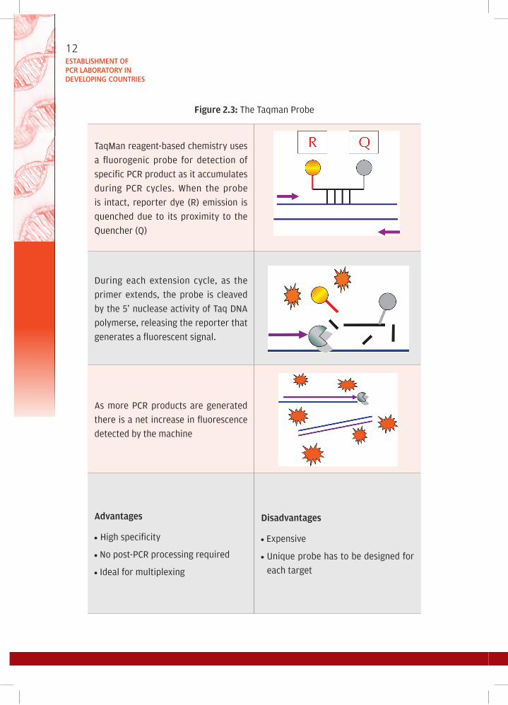
figure 2.3: The Taqman Probe
TaqMan reagent-based chemistry uses
a fluorogenic probe for detection of
specific PCR product as it accumulates
during PCR cycles. When the probe
is intact, reporter dye (R) emission is
quenched due to its proximity to the
Quencher (Q)
During each extension cycle, as the
primer extends, the probe is cleaved
by the 5’ nuclease activity of Taq DNA
polymerse, releasing the reporter that
generates a fluorescent signal.
As more PCR products are generated
there is a net increase in fluorescence
detected by the machine
advantages
• High specificity
• No post-PCR processing required
• Ideal for multiplexing
disadvantages
• Expensive
• unique probe has to be designed for
each target
EstablishmEnt of PCR laboRatoRy in
dEvEloPing CountRiEs
13
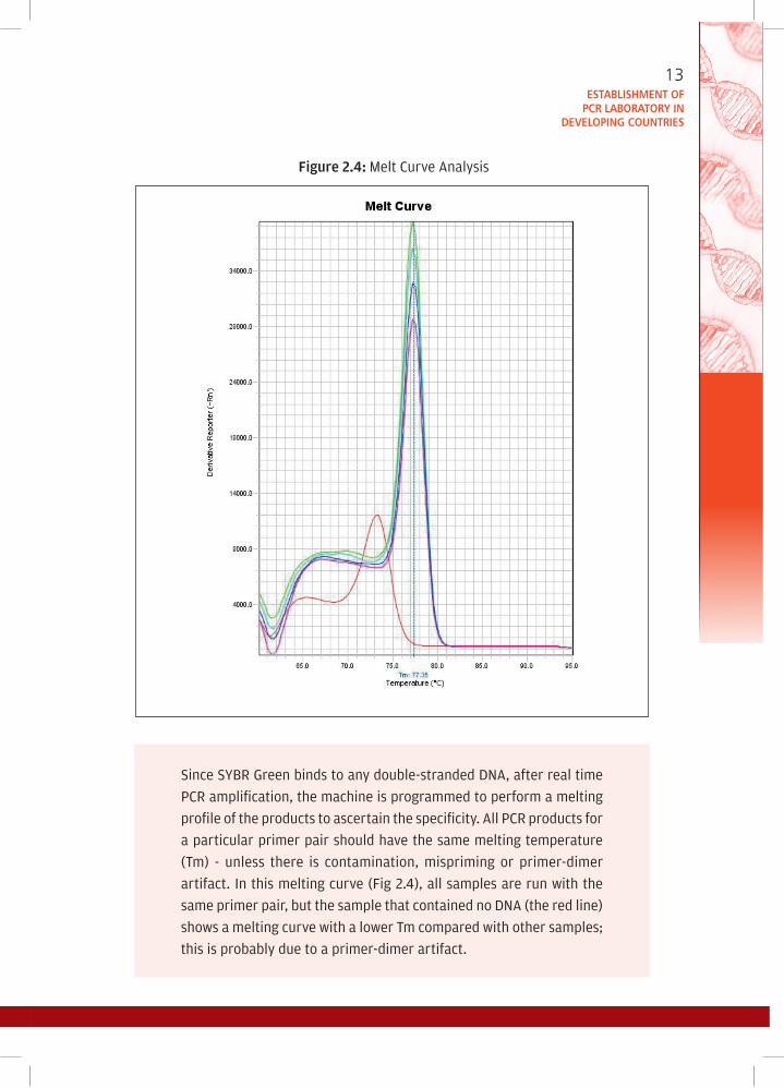
figure 2.4: Melt Curve Analysis
Since SYBR Green binds to any double-stranded DNA, after real time
PCR amplification, the machine is programmed to perform a melting
profile of the products to ascertain the specificity. All PCR products for
a particular primer pair should have the same melting temperature
(Tm) - unless there is contamination, mispriming or primer-dimer
artifact. In this melting curve (Fig 2.4), all samples are run with the
same primer pair, but the sample that contained no DNA (the red line)
shows a melting curve with a lower Tm compared with other samples;
this is probably due to a primer-dimer artifact.

EstablishmEnt of PCR laboRatoRy in dEvEloPing CountRiEs
14
Quantification in real-time PCRRelative quantification – Relative quantification is commonly used in gene expression
studies. A gene of interest in a given sample is compared to the same gene in another
reference sample (such as an untreated control sample) and the results are expressed
as fold-up or down-regulation of treated versus untreated sample.
absolute quantification – Absolute quantification is the process that determines the
absolute quantity of a single nucleic acid target sequence within an unknown sample
(e.g. viral load assays). Serial dilutions of samples with known quantity are amplified
to generate a standard curve. An unknown sample can be quantified based on this
curve (Figures 2.5 and 2.6).
figure 2.5: Real-time PCR-absolute quantification amplification plot (linear)

EstablishmEnt of PCR laboRatoRy in
dEvEloPing CountRiEs
15
figure 2.6: Absolute quantification-standard curve

EstablishmEnt of PCR laboRatoRy in dEvEloPing CountRiEs
16
table 2.1: Applications, advantages, and disadvantages of PCR types
PCR Type Target Application Advantages Disadvantages
Conventional DNA • Amplification and detection of DNA sequences
• Easiest of the PCR types to perform
• Low cost of equipment and supplies
• Normally produces only qualitative results
• Requirement for post amplification analyses increases time and labour as well as risk of cross contamination and human error
• Products should be confirmed by probe hybridization or sequencing
Real-time DNA • Amplification, detection and quantification of initial copy number of nucleic acid target
• Rapid potential for relative or absolute target sequence quantification
• usually eliminates requirement for post amplification analyses
• Increased specificity because probes or melting curves are used
• Totally closed tube analyses creates less potential for cross- contamination
• Requires more expensive equipment and reagents
• Less flexibility in primer and probe selection
• Less amenable to other downstream product confirmation analyses, such as sequencing due to small amplicon size
application, advantages and disadvantages of PCR typesVarious applications, advantages and disadvantages are summarized in Table 2.1.

EstablishmEnt of PCR laboRatoRy in
dEvEloPing CountRiEs
17
PCR Type Target Application Advantages Disadvantages
Multiplex DNA • Simultaneous amplification and detection of two or more different DNA sequences (can be performed as a conventional or real time procedure)
• Amplification of multiple target sequences in a single reaction reduces time and labour requirements
• Less flexibility in primer selection
• Requires significant optimization
• Generally has lower sensitivity and specificity
Nested DNA • Amplification and detection of DNA using external and internal primer sets in sequential steps
• Potentially more sensitive
• Decreases the potential for nonspecific amplification
• More likely to produce false positives due to carryover of products from first amplification step
• An additional room for sample preparation after the first amplification step is needed
ReverseTranscription(RT)
mRNA, rRNA,viral RNA
• Amplification and detection of RNA
• Amplification of all RNA types
• RNA is sensitive to degradation
• Added RT step may increase time and costs as well as potential for contamination
Source: The united States Environmental Protection Agency (EPA), Quality Assurance/Quality Control Guidance for Laboratories Performing PCR Analyses on Environmental Samples, October 2004

EstablishmEnt of PCR laboRatoRy in dEvEloPing CountRiEs
18

3STRuCTuRE AND FuNCTIONS OF PCR MACHINESA PCR machine is also called a thermal cycler. It rapidly changes temperatures (heating
and cooling) for PCR reactions, thereby allowing the reaction to cycle between primer
annealing (50-60 0C), DNA amplification (72 0C), and strand melting cycles (94 0C). The
device has a thermal block with holes where tubes holding the PCR reaction mixtures
can be inserted. The cycler then raises and lowers the temperature of the block in
discrete, pre-programmed steps.
The earliest thermal cyclers were designed for use with the Klenow fragment of
DNA Polymerase I. Since this enzyme is destroyed during each heating step of the
amplification process, a new enzyme had to be added every cycle. This led to a
cumbersome machine based on an automated pipettor, with open reaction tubes.
Later, the PCR process was adapted to the use of thermostable DNA polymerase from
Thermus aquaticus, which greatly simplified the design of the thermal cycler.
While in some old machines the block is submerged in an oil bath to control
temperature, in modern PCR machines a Peltier element is commonly used. The
Peltier effect bears the name of Jean-Charles Peltier, a French physicist who in 1834
discovered the calorific effect of an electrical current at the junction of two different
metals. Another way to understand how this effect could cool a junction is to note
that when electrons flow from a region of high density to a region of low density, this
“expansion” causes cooling (as with an ideal gas). An interesting consequence of this
effect is that the direction of heat transfer is controlled by the polarity of the current;
reversing the polarity will change the direction of transfer and thus the sign of the heat
absorbed/evolved. A Peltier cooler/heater or thermoelectric heat pump is a solid-state

EstablishmEnt of PCR laboRatoRy in dEvEloPing CountRiEs
20
active heat pump that transfers heat from one side of the device to the other. Peltier
cooling is also called thermo-electric cooling (TEC).
Present-day thermal cyclers contain a thermoelectric heat pump, which allows
for a solid state solution for temperature control without the need for a constant
temperature bath. It is fitted with a heated lid that serves to press against the top
of the sample tubes. This feature, in turn, hinders water condensation on the lid and
removes the need to top off samples in the tubes with PCR oil. Higher-end thermal cycler
models have multiple thermal blocks that make multiple synchronous PCR reactions
possible. The gradient thermal cycler allows the user to set different temperatures
in specific sections of the thermal block, which is an extremely useful feature when
testing appropriate temperatures for the annealing of primers. Quality thermal blocks
are constructed from silver to obtain the most uniform temperature throughout the
block and the quickest temperature changes.
While choosing a PCR machine, much care has to be exercised to choose the appropriate
specifications for the machine being purchased. In particular, the following points
should be considered before purchasing a PCR machine. A typical specifications list
for a PCR machine may include the following features: (i) temperature range (4 °C –
99 °C), (ii) block heating rate (up to 3 °C/sec, (iii) block cooling rate (up to 2 °C/sec), (iv)
block uniformity across wells (± 0.5 °C within 15 sec), (v) display resolution (0.1 °C),
(vi) maximum programmed dwell time (9 hours 59 minutes 59 seconds), (vii) heated
lid temperature range (95 °C – 120 °C), (viii) block modules (preferably silver, with
provision for holding 0.2 ml or 0.5 ml tubes/strips), (ix) number of programmes (>50),
(x) maximumnumber of programme stages (5), (xi) maximum number of programme
steps (5), (xii) auto-restart facility (Yes), (xiii) temperature ramping (Yes), (xiv) pause
facility (Yes), (xv) programme naming (Yes), and (xvi) run “end time” calculation (Yes).
Real-time PCR thermal cyclerThe real-time PCR thermal cyclers combine PCR product generation and recognition
of the product into one integrated format that allows for the subsequent analysis of
the captured data. One of the fundamental differences between a conventional PCR
machine and a real-time PCR machine is that no post-PCR processing is involved in
real-time PCR technology as the results of the reaction are available on the machine
as soon as the run is over. Procedures such as gel electrophoresis and/or ELISA are
not required to visualize a PCR result.
To accomplish these two tasks of amplification of DNA and detection of the product,
a real-time PCR machine incorporates the traditional PCR thermal cycling technology
EstablishmEnt of PCR laboRatoRy in
dEvEloPing CountRiEs
21
along with integrated fluorimeters and detectors that provide the ability to both
excite a fluorochrome and detect the emitted light. The second difference is that the
real-time PCR machine through a kinetic approach looks at the reaction in the early
stages while it is still linear rather than the end-point. Consequently, the design of a
real-time PCR machine incorporates a heating block much similar to the one found in
conventional PCR machines. In addition, it has a sensitive camera that monitors the
fluorescence in each well of the 96-well plates at frequent intervals during the PCR
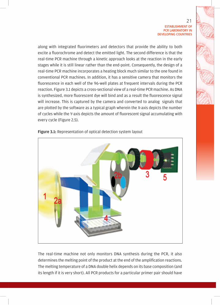
reaction. Figure 3.1 depicts a cross-sectional view of a real-time PCR machine. As DNA
is synthesized, more fluorescent dye will bind and as a result the fluorescence signal
will increase. This is captured by the camera and converted to analog signals that
are plotted by the software as a typical graph wherein the X-axis depicts the number
of cycles while the Y-axis depicts the amount of fluorescent signal accumulating with
every cycle (Figure 2.5).
figure 3.1: Representation of optical detection system layout
The real-time machine not only monitors DNA synthesis during the PCR, it also
determines the melting point of the product at the end of the amplification reactions.
The melting temperature of a DNA double helix depends on its base composition (and
its length if it is very short). All PCR products for a particular primer pair should have

EstablishmEnt of PCR laboRatoRy in dEvEloPing CountRiEs
22
the same melting temperature, unless there is contamination, mispriming, primer-
dimer artifacts, or some other problem. Since SYBR Green does not distinguish between
one DNA and another, an important means of quality control is to check that all samples
have a similar melting temperature.
After real-time PCR amplification, the machine can be programmed to do a melt curve
(Figure 2.4), in which the temperature is raised by a fraction of a degree and the change
in fluorescence is measured. At the melting point, the two strands of DNA will separate
and the fluorescence rapidly decreases. The software plots the rate of change of the
relative fluorescence units (RFu) with time (T) (-d(RFU)/dT) on the Y-axis versus the temperature on the X-axis, and this will peak at the melting temperature (Tm).
There are several suppliers of such equipment and these can range in cost depending
on the features. For many laboratories, this represents a substantial investment but
can result in savings, as the analysis of PCR products can be conducted without running
agarose gels, representing savings in both time and money when processing a large
number of samples.
There are several platforms available commercially on which real-time PCR machines
have been designed. The fundamental difference between platforms is the ability
to detect more than one fluorochrome with the addition of multiple excitation and
detection channels. Many suppliers provide different models ranging from a relatively
low-cost single-channel machine suitable for SYBR Green detection (e.g. Bio-Rad’s DNA
Engine Opticon) to a multichannel machine (e.g. the 7500 real-time PCR system from
Applied Biosystems) that can be used for the detection of multiple fluorochromes at
once (multiplexing). Many work best with specific chemistries and fluorochromes, so
it is important to consider the cost and chemistry options available for each platform
in addition to the cost and capabilities of the various thermal cyclers themselves.
Because real-time PCR reactions are quantified at each cycle by measuring
fluorescence, most real-time thermal cyclers need to be calibrated for the particular
tube or microtiter plate and microtiter plate seal used, and for the PCR reaction volume.
Therefore, it is important to know which real-time thermal cycler will be used for the
experiment before setting up the reactions.
Detecting the PCR product in real-time involves the use of a fluorescent dye. These
can be either nonspecific dyes, such as fluorescent DNA-binding dyes (e.g. SYBR
Green I) or strand-specific probes (such as Taqman or molecular beacons), many of
which use a phenomenon known as fluorescent resonance energy transfer (FRET) to
distinguish between various products. For the initial optimization of the real-time
PCR amplification, nonspecific fluorescent dyes from the SYBR Green family are most

EstablishmEnt of PCR laboRatoRy in
dEvEloPing CountRiEs
23
commonly used, because they are more economical compared with strand-specific
probes, and easier to optimize. When using this method of detection, a single, specific
DNA fragment has to be obtained during PCR amplification, because any additional
nonspecific DNA fragment accumulation will contribute to the fluorescence measured.
Strandspecific probes need to be designed if nonspecific bands are amplified in the PCR
reaction or if the amplification of more than one target sequence will be monitored
in a single PCR reaction.
digital PCR technologyDigital PCR measurements are performed by dividing the sample and qPCR assay
mixture into a very large number of separate small volume reactions, such that
there is either zero or one target molecule present in any individual reaction (Fig.
3.2). This is the fundamental concept for making ”digital” measurements. Thermal
cycling is performed to end-point. Any target-containing compartments will become
brightly fluorescent while compartments without targets will have only background
fluorescence. A reaction with no target molecule is counted as a 0 (PCR-negative), and a
reaction that has one target molecule is counted as a 1 (PCR-positive). When the entire
set of divided reactions is counted, the total number of ‘positive’ reactions is equal to
the number of original target molecules in the entire volume, and the total number
of reactions multiplied by the individual reaction volume equals the total volume
assayed. Thus, the absolute concentration of the target is easily calculated as being
equal to the total number of target molecules divided by the total measured volume.
uncertainty in this “absolute” measurement comes only from error in the measured
figure 3.2: Separation and digital counting provide sensitive, absolute quantification.
Digital PCR is performed by dividing the sample and the assay (e.g. qPCR hydrolysis probe and primers) into enough separate reaction chambers such that any reaction will contain either only 0 or 1 target molecule. Standard end-point PCR is performed and the number of fluorescent reactions counted. PCR-positive, “bright” reactions each contained 1 target molecule, and PCRnegative, “dark” reactions have no target .

EstablishmEnt of PCR laboRatoRy in dEvEloPing CountRiEs
24
volume or the presence of more than a single target molecule in a compartment, so
dPCR methods that control for these two factors provide the highest accuracy.
When target molecules are divided into separate reaction compartments, the chances
for more than one target molecule to be co-located in the same compartment can
be calculated using Poisson statistics. When the number of target molecules is
significantly smaller than the number of compartments (low occupancy), the chance
of co-compartmentalization is small. Poisson statistics can be used either as a small
correction factor (at low occupancy) or to calculate an estimated concentration (at
high occupancy). dPCR platforms, which divide the sample into a larger number of
compartments, will have the highest accuracy by directly counting single molecules
(low occupancy). Similarly, dPCR performed using higher numbers of compartments
provides the highest sensitivity—with limits of detection approaching 1 in 1 million,
and the widest dynamic range of inputs—over 6 logs.

4APPLICATIONS OF PCR TECHNOLOGY IN MEDICINE AND ALLIED SCIENCES
introductionPolymerase chain reaction (PCR), a revolutionary nucleic acid amplification technology,
has brought in a quantum leap in all walks of modern biology. The technique involves
amplification of genetic material to billions of copies from minute amounts of starting
material in a matter of hours, and at times, even lesser duration. Perhaps no other
technique has shown as much promise in as shorter a time-frame as PCR.
The use of PCR in molecular diagnostics has increased to the point where it is now
accepted as the gold standard for detecting nucleic acids from a number of origins
and it has become an essential tool in the research laboratory. Real-time PCR has
engendered wider acceptance of the PCR due to its improved rapidity, sensitivity,
reproducibility and the reduced risk of carry-over contamination.
Point-of-care (POC) testing is defined as analytical testing performed outside the
central laboratory using devices that can be easily transported to the vicinity of the
patient. Microfluidic technologies have enabled miniaturization of PCR processes onto
a chip device with potential benefits including speed, cost, portability, throughput and
automation, thereby rendering PCR as a POC test. In the developing countries where
high infectious disease burden is compounded by diagnostic challenges due to poor
clinical laboratory infrastructure and cost constraints, the potential utility of PCR for
POC testing is even greater.

EstablishmEnt of PCR laboRatoRy in dEvEloPing CountRiEs
26
applications of PCR in clinical microbiologyThe applications of PCR in infectious disease diagnostics include specific or broad-
spectrum pathogen detection, evaluation of emerging infections, surveillance,
detection of agents of bioterrorism and antimicrobial resistance profiling. In the
field of infectious diseases, the scope of PCR can be recognized under three areas,
viz., diagnosis, epidemiology and prognostic monitoring. In this field, PCR can be
an attractive strategy as it yields rapid results, has highthroughput along with high
sensitivity and specificity. Practically, each type of PCR has applications in all branches
of clinical medicine , biotechnology and forensic medicine.
applications in virology
The PCR is currently being employed in the detection and quantitation of a number
of DNA and RNA viruses, including Hepatitis C virus, HIV, Japanese encephalitis
virus, human papillomaviruses, chikungunya virus, influenza viruses, rabies virus,
cytomegalovirus and JC virus and Ebola. It has become a valuable tool in the
diagnosis, clinical management and prognostic monitoring of HIV infection, in
particular, and a number of other infections such as Hepatitis B virus. Quantitation
of viral load assay to monitor the progress of infection is an important application
in this regard.
Another very useful application of PCR in the field of HIV has been in early infant
diagnosis, which has been a problem area to date. The PCR also remains the only
reliable method of diagnosis for a few infections, as in the case of the most recent
H5N1 and pandemic influenza A H1N1 (2009). Amenability of PCR to high-throughput
platforms becomes a boon in rapid investigation of such outbreaks.
The PCR-based studies targeting conserved genetic regions of viral pathogens also
provide valuable insights into the epidemiological patterns of infections in the
areas affected. Conventional PCR methods are now increasingly replaced by real-
time PCR techniques for rapid detection of many viruses andalso their advances in
the development of fluorophores, nucleotide labelling chemistries, and the novel
applications of oligoprobe hybridization have provided real-time PCR technology with
a broad enough base to ensure its acceptance.
Recently, instrumentation has appeared that is capable of incredibly short cycling
times combined with the ability to detect and differentiate multiple amplicons. New
instruments are also flexible enough to allow the use of any of the chemistries making
real-time nucleic acid amplification an increasingly attractive and viable proposition
for the routine diagnostic laboratory. In many cases these laboratories perform tissue

EstablishmEnt of PCR laboRatoRy in
dEvEloPing CountRiEs
27
culture to isolate virus and serological methods to confirm the identity of the isolate,
which may take a considerable, and clinically relevant, amount of time.
Recent developments in multiplex real-time PCR have suggested a future in which easy
identification, genotyping and quantitation of viral targets in single, rapid reactions will
be commonplace. Of course, this technology is by no means restricted to virology, as
significant achievements have appeared in the area of mutation detection, applying all
the benefits described above to enhance the detection of genetic disease and, where
applicable, allow quantification of the extent of such genetic changes.
In diagnostic laboratories, the use of PCR is limited by cost and sometimes the
availability of adequate test sample volume. To overcome these shortcomings and
also to increase the diagnostic capacity of the PCR, a variant termed multiplex PCR has
been described. In multiplex PCR more than one target sequence can be amplified by
including more than one pair of primers in the reaction. Multiplex PCR has the potential
to produce considerable savings of time and effort within the laboratory without
compromising the test utility. Since its introduction, multiplex PCR has been successfully
applied in many areas of nucleic acid diagnostics, including gene deletion analysis,
mutation and polymorphism analysis, quantitative analysis, and RNA detection. In the
field of infectious diseases, the technique has been shown to be a valuable method for
identification of viruses, bacteria, fungi and/or parasites. A representative list of such
agents utility of multiplex PCR for diagnosis of viral infections is shown in Table 4.1.
table 4.1: Representative list of applications of multiplex PCR to the diagnosis of infectious diseases
Infectious agent
Pathogen targeted Clinical manifestation and/ or specimen
Virus
HIV-1, HIV-2, HTLV-1, and HTLV-2 Blood
HSV-1, HSV-2, VZV, CMV, HHV-6, EBV, and EV
Meningitis, encephalitis, or meningoencephalitis; CSF
Bacterium
Haemophilus influenzae, Streptococcus pneumonia, Mycoplasma catarrhalis, and Alloiococcus otitidis
upper respiratory tract
Campylobacter jejuni and Campylobacter coli
Human campylobacteriosis
Actinomyces actinomycetemcomitans, Porphyromonas intermedia, and Porphyromonas gingivalis
Periodontal infection

EstablishmEnt of PCR laboRatoRy in dEvEloPing CountRiEs
28
table 4.1: Representative list of applications of multiplex PCR to the diagnosis of infectious diseases
Infectious agent
Pathogen targeted Clinical manifestation and/ or specimen
Bacterium
N. gonorrhoeae and C. trachomatis Genital infections
C. trachomatis, N. gonorrhoeae, ureaplasma urealyticum, and M. genitalium
Genital infections
Parasite
Giardia lamblia and Cryptosporidiumparvum
Diarrheal disease; water
Leishmania spp. Leishmaniasis
Combination
HSV, H. ducreyi, and T. pallidum Genital ulcer disease
HPVs, HSV, and C. trachomatis Genital swabs
Adenovirus, HSV, and C. trachomatis Keratoconjunctivitis
EV, influenza viruses A and B, RSV, PIV types 1 and 3, adenovirus, M. pneumoniae, and C. pneumoniae
Acute respiratory tract infections
applications in bacteriologyPCR, though not widely employed as a routine diagnostic method in bacteriology,
has been helpful in the detection of sexually transmitted bacterial pathogens such
as Chlamydia trachomatis, Neisseria gonorrheae, etc. Extensive protocols have also
been developed for PCR-based detection of Mycobacterium tuberculosis and related
species, and also for bacterial pathogens causing meningoencephalitis. Variations
of the technique have also been employed in identification and characterization of
antimicrobial resistance patterns in several general forms of bacteria (e.g. MRSA, MDR
and XDR-TB, “Superbugs” with metallobetalactamase-1 gene).
applications in mycologyThe PCR techniques have also been employed for the detection of a number of
localized (e.g. endophthalmitis) and invasive fungal diseases (e.g. aspergillosis). Lack
of information regarding the ideal type of specimen to be tested, kinetics of fungal
DNA during infections, and issues with standardization of protocols and interpretation
of results have been hampering this area of PCR-based diagnostics.

EstablishmEnt of PCR laboRatoRy in
dEvEloPing CountRiEs
29
applications in parasitologyProtocols have been developed for PCR diagnosis of protozoal pathogens of humans
and animals, including pathogenic Plasmodium spp., pathogenic amoebae, Giardia
spp., Cryptosporidium spp., Microsporidia filarial parasites, etc.
applications of PCR in noninfectious diseasesThe PCR has been applied in the identification of gene mutations associated with
malignancies (mutations in oncogenes/protooncogenes, e.g. BRCA1 mutations in
carcinoma breast, p53 mutations in a number of tumours, etc.), metabolic errors and
psychiatric disorders, etc. Prenatal screening and diagnosis can be attempted with the
help of PCR on tissue samples obtained by amniocentesis, chorionic villus sampling or
even from rare fetal cells in maternal circulation. Pre-implantation genetic diagnosis
is also facilitated by PCR, wherein individual embryonic cells are tested for mutations.
Another major application of PCR has been in the study of gene expressions associated
with specific diseases. Qualitative and quantitative data on gene expression profiles
can be obtained by PCR-based methods. Both spatial and temporal patterns of gene
expression can be studied in tissue samples by PCR-based methods. The PCR has greatly
facilitated attempts towards personalized drug therapy. Specific polymorphisms/
mutations of genes involved in drug metabolism can have important consequences in
treatment outcome. The PCR-based pharmacogenomic methods have been employed
in such attempts.
applications in forensic medicineSpecific examples of PCR applications in this area include resolution of disputed
paternity, and personal identification from specimens obtained at crime scenes (e.g.
blood stains, hair strands, semen etc.) Variable Nuleotide Tandem Repeats (VNTR),
Short Tandem Repeat Polymorphisms (STRPs), and Single Nucleotide Polymorphisms
(SNP) are a few markers that are employed for such purposes.
applications in biotechnology and allied fieldsThe PCR-based methods have been employed extensively in the field of biotechnology
for the production of hybridization probes, development of disease-resistant and high-yield varieties of plants and animals and production of therapeutic proteins, etc. Another interesting application is the production of human vaccines using transgenic plants. Considerable success has been obtained with vaccines for diseases such as rabies and hepatitis B.

EstablishmEnt of PCR laboRatoRy in dEvEloPing CountRiEs
30
5ESTABLISHMENT OF A PCR LABORATORY
Over the past two decades, the development of the PCR as the basic component of a
molecular biology laboratory has occurred very rapidly. The PCR technique became
indispensable in laboratories to amplify small amounts of template nucleic acid.
Laboratory personnel at the same time, learned that PCR had a strong susceptibility
to contamination from its own product. A number of precautions are required to be
adopted when designing a PCR laboratory such that the laboratory is operated in a
way that prevents contamination of reactions with amplified products from previous
assays and cross-contamination between samples, both of which can lead to false-
positive results.
laboratory space arrangementA PCR laboratory should contain two functional work areas: a pre-amplification area
and a post-amplification area. These two areas should ideally be in separate rooms,
or when space constraints exist, separate work stations/biosafety cabinets in a single
room. Supplies and equipment should be dedicated to each work area and should not
be interchanged between areas.
A laboratory performing PCR analyses on diagnostic samples should be divided into
at least three physically separate rooms (Figure 5.1):
• Reagent preparation (using positive pressure to prevent the introduction of
contamination)
• Sample preparation (using negative pressure to keep template nucleic acids in
the room)

EstablishmEnt of PCR laboRatoRy in dEvEloPing CountRiEs
32
• Amplification and product detection (using negative pressure to keep amplified
nucleic acids in the room)
A unidirectional workflow should be observed to reduce the chances for contamination
to occur. No materials, supplies or equipment from the sample preparation room should
be taken into the reagent preparation room. Similarly, nothing from the amplification
and product detection room should be taken into the sample preparation room or the
reagent preparation room.
figure 5.1: A sketch depicting the model layout for a PCR laboratory.
Reagent preparation roomThe reagent preparation room should be designated for the preparation and storage
of PCR reagents. Preparation of master mixes and aliquoting of master mixes to
PCR tubes should be performed in this room. To prevent cross contamination and to
avoid repeated freezing and thawing, reagent-stock solutions should be aliquoted
into smaller volumes. To deter contamination, the room should be under positive
pressure. The reagent preparation room should have a dedicated set of adjustable
pipettes with plugged, aerosol-barrier tips, laboratory coats and disposable gloves.
Personnel should complete tasks in this room before working in the sample processing
or amplification/detection rooms and should not return from these rooms to the
reagent preparation room.

EstablishmEnt of PCR laboRatoRy in
dEvEloPing CountRiEs
33
sample preparation roomThe sample preparation room should be designated for sample processing. This facility
should be used for aliquoting of sample and preparation of positive and negative
controls. As per the protocol used for extraction of nucleic acid the required quantity of
clinical sample should be added to lysis buffer and then transferred to RNA /nucleic acid extraction area. The processed samples and controls are then added to tubes containing the PCR master mix in this room. PCR tubes should be capped as soon as the sample is added.
Workflow in a PCR laboratory
A unidirectional workflow should be used to reduce the
potential for contamination.
Reagent Preparation Room
PCR master mix preparation and aliquoting room
sample Preparation Room
(a) Processing of sample (b) Isolation of nucleic acids
(c) Addition of sample to master mix aliquot
amplification and Product visualization Room
(a) PCR thermal cycling (b) Confirmation
i. gel electrophoresis (conventional PCR)
ii. visualization (real-time PCR)
amplification and product roomThis room should be designated for PCR amplification and post-PCR analyses. The
thermocycler /real-time PCR machine should be kept in this room. Gloves and
laboratory coats should be worn at all times and removed before leaving the room to
control amplicon contamination of other locations. All equipment used for amplification
and product detection should be dedicated to this room, including adjustable pipettes
with plugged, aerosol-barrier tips. This room should be kept under negative pressure.
Although PCR amplification and post-PCR analyses may be performed in the same room,
these activities can be conducted in different areas or different rooms to reduce the
risk of contamination from amplified products.

EstablishmEnt of PCR laboRatoRy in dEvEloPing CountRiEs
34
Equipment in PCR laboratoriesTo ensure that pre-PCR and post-PCR events remain separated, each room must have
its own separate set of equipment, reagents, pipette tips and racks, etc. used in that
location only.
thermocyclers and real-time PCR machines
Thermocyclers are essential to all PCR methods, and great care should be taken
to ensure that they are well maintained and reliable. The block temperature of a
thermocycler should be tested at least twice a year by the laboratory or under a
maintenance agreement to ensure uniform heating throughout the block. Block
temperature should be tested with an external probe that has been calibrated against
a temperature standard. For testing, the probe is placed in several of the wells in
the periphery and centre of the instrument. All temperatures should be within the
manufacturers’ specifications. The amplification programme used in each run should
be printed to further verify the conditions of the PCR.
Real-time PCR instruments are equipped to perform fluorescence excitation and
detection to monitor amplification throughout the PCR cycles. The design is usually
different from the standard thermocycler, and calibration may be specific to the
instrument design. Temperature, laser performance, alignment, and safety devices
should be checked and optical systems calibrated. The machine should be serviced
annually.
Real-time machines should be used with uninterrupted power supply (uPS) as this
equipment is very delicate and, sensitive and also the laser needs to be protected
from damage.
Centrifuges
Separate centrifuges, including microfuges, are required for pre- and post-PCR
procedures. The manufacturers’ instructions for calibration should be followed.
The centrifuge should be balanced before use to increase bearing life and minimize
vibrations.
vortex mixer
The vortex is an important equipment required for reagent preparation in the PCR clean room and for nucleic acid extraction.

EstablishmEnt of PCR laboRatoRy in
dEvEloPing CountRiEs
35
gel electrophoresis chambers
This equipment is used to detect the PCR products after amplification.
Chambers should be inspected before each use to ensure that electrodes and buffer
tanks are intact, and that power supply electrodes fit snugly. Gel electrophoresis
chambers should be rinsed several times with water after each use in the designated
product room.
Pipettes
Automatic, fixed-volume, adjustable, positive-displacement pipettes, and/ or
micropipettes are used in the PCR laboratory. These should be calibrated quarterly
by the manufacturer or a technician. Each pipette should be sterilized according to
manufacturers’ recommendation on a regular basis or whenever contamination is
suspected.
laminar-flow hoods/biological safety cabinets (bsCs)
Laboratory users should pay careful attention to the specifications of the hood or
cabinet to ensure that it is appropriate for its designated use by the laboratory.
Unit type UV light Airflow system Use
PCR cabinet (Type A) Yes NoneReagent preparation
only
PCR cabinet (Type B) No Intake filtered
Not recommended
for any aspect of PCR
preparation
PCR cabinet (Type C) /
Laminar-flow hoodYes Intake filtered
Reagent
preparation only
Class I biological safety
cabinetYes Exhaust filtered
Not recommended
for any aspect of
PCR preparation
Class II or III biological
safety cabinetYes
Intake and
exhaust filteredAll aspects of PCR
Class I cabinets have inward air flow and HEPA-filtered exhaust that provides personal
and environmental protection, but no product protection. Class II and Class III BSCs

EstablishmEnt of PCR laboRatoRy in dEvEloPing CountRiEs
36
filter both air intake and exhaust, and prevent contaminants from entering and leaving
the hood (reducing the likelihood of sample and work area contamination). Before
use, hoods should be decontaminated using uV light for at least half an hour and
cleaned with bleach or other effective nucleic acid inactivating agent. The airflow and
HEPA filtration in all hoods should be monitored and certified as per manufacturers’
recommendations at least annually.
Refrigerator
Separate refrigerators for temporary storage of sample, extracted RNA/nucleic acid
and final amplification products should be maintained in the respective laboratory.
usually long-term storage is not recommended but if needed separate deep freezers
(-80 °C) can be maintained.
freezer (-20 °C)
PCR clean reagents, enzymes, buffer, dNTPS and primers are required to be stored
at -20 °C.The primers, dNTPS and water should be stored in small aliquots to avoid
freezing and thawing effects and also to rule out contamination issues.
To verify that equipment is functioning properly, the laboratory should have a schedule
for maintaining equipment. The schedule should include the set-up, calibration, repair,
record-keeping, and normal operation of all equipment used in sample analysis. The
results of all tests should be documented in an equipment logbook and/or electronic
database. The logbook or database should be checked monthly by quality control
(QC) personnel or the laboratory supervisor, and any problems and corrective actions
managed. Equipment should be dedicated to a specific laboratory room, and instrument
manuals from the manufacturer should be available.
Consumables
Disposable materials used in a PCR laboratory include pipette tips, sample tubes, PCR
tubes and gloves. To reduce the contamination and degradation of the target nucleic
acids, disposable materials should be of good quality.
Pipette tips
Special tips for PCR analysis include barrier tips and aerosol-resistant tips, both of
which minimize cross-contamination of samples during pipetting. These tips can be
purchased pre-sterilized and pre-loaded in hinged racks to provide tip protection
and easy access. Pipette tips for PCR analyses should be RNase-free, DNase-free, and
pyrogen-free.

EstablishmEnt of PCR laboRatoRy in
dEvEloPing CountRiEs
37
sample and PCR tubes
Polypropylene tubes that are certified DNase-, RNase-, and pyrogen-free are best
recommended for PCR laboratories. The size and style of PCR tubes or reaction
plates should be compatible with the block and lid height of the thermocycler/real-
time machine. Thin-walled tubes provide the best heat transfer, ensuring that the
reaction volume reaches its specified temperature in the shortest amount of time,
thereby improving specificity and reproducibility. Tubes containing stored samples
and reagents should be centrifuged briefly before opening to ensure that all liquids
are at the bottom of the tubes.
laboratory wear
Disposable gloves should be available in each section of laboratories used for PCR
analysis. Gloves should be changed before leaving and entering each section of the
laboratory and each time that contaminating DNA is potentially encountered. In
addition to reducing potential contamination from samples, wearing gloves may protect
the technician from potential chemical exposure and prevent sample contamination
due to human DNases and RNase.
Dedicated laboratory coats should be available in each laboratory room. Laboratory
coats should be removed and gloves discarded appropriately before leaving each
room. Changing laboratory coats and gloves reduces the possibility of contamination
with template or amplified nucleic acid. Laboratory coats should be cleaned regularly
to reduce the possibility of contamination of the designated workspace and the PCR
reaction. To eliminate the need for cleaning laboratory coats, disposable (single-use)
laboratory coats may also be used.
Reagents
The reagents used in PCR amplification can be purchased or prepared inhouse. All
reagents should be clearly labelled with name, expiration date, and relevant safety
information. Reagents from different lot numbers should not be interchanged
or combined together. Precautions should be taken to ensure that reagents are
contamination free and storage conditions are well maintained.
Molecular-grade water or its equivalent from commercial sources should be used for
all assays. Water purification systems that produce high-quality pyrogen and DNase/
RNase-free water may be used. Diethylpyrocarbonate (DEPC) treatment can be
used to eliminate RNase from plastic ware and from water which is required in RNA
analysis. Reagent water is treated with a solution of 0.1% DEPC for several hours and

EstablishmEnt of PCR laboRatoRy in dEvEloPing CountRiEs
38
then autoclaved to degrade the DEPC completely. Proper autoclaving is necessary,
because trace amounts of DEPC in a solution will lead to the modification of the purine
residues in RNA by carboxymethylation. This leads to downstream effects in RNA
experimentation (e.g. removing the ability of reverse transcriptase to bind RNA and
synthesize DNA from an RNA template).
Commercially prepared reagents should be of molecular grade and should be stored
according to manufacturers’ recommendations. All reagents from new lots should
be tested to ensure that they work properly by running a PCR positive control using
the new reagents.
For reagents prepared in-house, criteria should be developed for expiration dates,
functional acceptability, and storage conditions using product sheets from similar
commercial products as guidance. The criteria should be documented in the laboratory
standard operating procedures (SOP). Buffers should be inspected for precipitates or
microbial contamination before each use.
Commercially available kits
Many types of commercial kits are available for PCR applications. These products
expedite and simplify procedures, such as the isolation of DNA and RNA and the
purification of nucleic acids to remove contaminants.
Primers and Probes (oligonucleotides)
The PCR analyses require the use of short segments of chemically synthesized DNA
(oligonucleotides or oligos). Primer sets are oligos that are designed specifically to
prime the amplification of a portion of a target nucleic acid of interest. Primers and
probes, containing a specific sequence of nucleotides, can be obtained commercially.
Certification of the quality of the oligos, including method of purification, purity, and
concentration, should be required from all commercial manufacturers.
storage: Most oligos and DNA templates should be stored at -20 °C or -70 °C in either
TE buffer (10 mM Tris-HCl and 0.1mM EDTA, pH 8.0) or molecular grade water. The
TE buffer generally is the preferred storage buffer for oligos and DNA templates,
because it may prevent DNA degradation. The pure, concentrated oligos should be
stored in the original tube from the manufacturer and labelled with the primer name
and concentration. To minimize the chance of contamination and degradation, these
concentrated stocks should not be used on a regular basis. Diluted working stocks
should be made for each oligo, and these working stocks should be used for all
experiments. Before use, oligos should be thawed and mixed completely.

EstablishmEnt of PCR laboRatoRy in
dEvEloPing CountRiEs
39
Enzymes
Enzymes are the critical requirement of a PCR and should be purchased from a
commercial source to ensure purity. Quality assurance (QA) information with the
enzymes should be obtained from the source. Each new lot of enzyme should be
compared with old lots using known controls and samples.
storage: The manufacturer’s instructions on enzyme storage and use should be
followed carefully. Enzymes are usually stored at -20 °C, and should never be left at
room temperature. Insulated bench-top coolers or ice can be used to keep the enzyme
cold in the laboratory, when being used on the bench top. PCR products may also be
stored at -20 °C or -70 °C. RNA templates should be aliquoted and stored at -70 °C.
Personnel
Personnel working in a PCR laboratory should undergo training in the methodology
that covers PCR and recombinant DNA theory and practice. The course work should
also include biosafety in a PCR laboratory as well as quality issues and troubleshooting
PCR-related problems. Hands-on training should be completed for each technique
under the supervision of experienced personnel. The time required for training will
vary depending on the trainee and technique. Personnel should demonstrate that
they can successfully perform the method by testing confirmed positive and negative
control samples before being allowed to analyse diagnostic samples.

EstablishmEnt of PCR laboRatoRy in dEvEloPing CountRiEs
40

6SAMPLE COLLECTION, STORAGE AND SHIPMENT OF CLINICAL MATERIAL FOR DIAGNOSIS OF VIRAL INFECTIONS BY PCRLaboratory diagnosis of viral infections requires a good understanding of the
pathogenesis of the suspected virus, the type and duration of illness, stage of infection,
age and immunization status of patient and the occurrence of outbreaks of a similar
illness in the community to determine the most appropriate specimen and diagnostic
test. Even a well performing molecular assay cannot restore the loss or degradation
of viral nucleic acids due to inappropriate collection, handling or transport of the
sample. Hence every laboratory should devise protocols to describe sample collection,
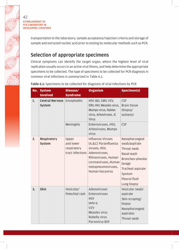
EstablishmEnt of PCR laboRatoRy in dEvEloPing CountRiEs
42
transportation to the laboratory, sample acceptance/rejection criteria and storage of
sample and extracted nucleic acid prior to testing by molecular methods such as PCR.
selection of appropriate specimensClinical symptoms can identify the target organ, where the highest level of viral
replication usually occurs in an active viral illness, and help determine the appropriate
specimens to be collected. The type of specimens to be collected for PCR diagnosis in
common viral infections is summarized in Table 6.1.
table 6.1: Specimens to be collected for diagnosis of viral infections by PCR
No. System Involved
Disease/Syndrome
Organism Specimen(s)
1. Central nervous system
Encephalitis HSV 1&2, CMV, VZV, EBV, HIV, Measles virus, Mumps virus, Rabies virus, Arboviruses, JC Virus
CSFBrain tissue (biopsy/autopsy)
Meningitis Enteroviruses, HSV, Arboviruses, Mumps virus
CSF
2. Respiratory system
upper and lower respiratory tract infections
Influenza Viruses (A,B,C) Parainfluenza viruses, RSV, Adenoviruses, Rhinoviruses, Human coronaviruses, Human metapneumoviruses, Human bocavirus
Nasopharyngeal swab/aspirate
Throat swab
Nasal wash
Broncheo-alveolar lavage
Tracheal aspirate
Sputum
Pleural fluid
Lung biopsy
3. skin Vesicular/ Petechial rash
AdenovirusesEnterovirusesHSVHHV-6VZVMeasles virusRubella virusParvovirus B19
Vesicular swab/ aspirate
Skin scraping/biopsy
Nasopharyngeal aspirates
Throat swab

EstablishmEnt of PCR laboRatoRy in
dEvEloPing CountRiEs
43
No. System Involved
Disease/Syndrome
Organism Specimen(s)
4. gastrointestinal system
Gastroenteritis RotavirusAstrovirusCalicivirusEnterovirusesAdenovirusCMV
Stool
Rectal swab
Hepatitis Hepatitis virus A,B,C,D,E, GTransfusion Transmitted virus(TTV)CMVEBVParvovirus B19
Blood
Serum
Tissue specimen (biopsy/autopsy)
5. urogenital system
Haemorrhagiccystitis, urethritisCervicitisGenital wartsCervical carcinoma
AdenovirusBK virusHSVVZVHPV
urineurethral discharge/swabCervical swabTissue biopsy
6. ocular tract Conjunctivitis
Keratoconjunc-tivitis
Chorioretinitis
Adenovirus
Enterovirus
HSV
CMV
VZV
Corneal scrapingsConjunctival swabs/scrapingsAqueous/Vitreousfluids
7. haematopoietic system
Lymphoid disordersDisorders of redblood cells
EBVHIVHTLV-1Parvovirus B19
Blood
Bone marrow aspirate
8. Congenital/Perinatalinfections
Multi-systeminvolvement
CMVHBVHIVParvovirus B19Rubella virus
BloodSerum/PlasmaAmniotic fluidTissue biopsy

EstablishmEnt of PCR laboRatoRy in dEvEloPing CountRiEs
44
specimen collection and storageFor many viral infections viral shedding begins before the onset of symptoms and
decreases after the acute phase of illness. Since the possibility of detection of
virus/viral nucleic acid is high during the early stages of the infection, specimens
for diagnosis of viral infections by PCR must be obtained within one to three days
of onset of symptoms.
The success of molecular diagnostic methods such as PCR is essentially dependent
on the quality of nucleic acid purified from a clinical specimen, which in turn is
directly related to how the sample has been stored and/or transported after
collection.
All samples to be processed for molecular diagnosis of any viral infection by PCR
should be obtained under strict aseptic precautions. Each specimen should be
placed in a separate sterile, nuclease free container labelled with the patient’s
name and identification number, the collection site, and date and time of collection.
Swabs, scrapings and small pieces of tissue are placed in a container with 2-3 ml
of viral transport medium (VTM). The VTM consists typically of a buffered salt
solution to maintain appropriate pH and osmotic environment, protein such as
bovine serum albumin to stabilize the virus, and antibiotics to prevent microbial
contamination. It prevents the specimen from drying and helps maintain viral
viability. Specimens like CSF, blood, urine, broncheoalveolar lavage fluid, vesicle
fluid and stool specimens can be sent without transport medium.
The best yield and quality of DNA/RNA is obtained from a fresh specimen; however,
if samples cannot be processed immediately, they must preferably be snap-frozen
in liquid nitrogen and stored at -70 0C. Repeated freezing and thawing can lead
to reduced yield of viral nucleic acid and should be avoided.
Compared with DNA that is very stable, RNA is extremely labile and susceptible to
degradation by RNases, which are ubiquitous. Since the human skin is a common
source of RNases, use of gloves while handling and processing samples and strict
adherence to specimen storage temperatures can ensure a yield of good quality
RNA from the sample.
Preservation of the integrity of viral nucleic acids in specimens during shipping and
handling is crucial for PCR. An economical and convenient method is described for nucleic acid stabilization by using an RNA stabilizing solution (e.g. RNAlater) in plasma that is designed for the shipment of samples in tropical countries.

EstablishmEnt of PCR laboRatoRy in
dEvEloPing CountRiEs
45
special considerations for some common clinical specimens
swabs
Sterile dacron or rayon swabs with plastic shafts or if available, flocked swabs should
be used. Calcium alginate swabs or cotton swabs with wooden sticks should not be
used, as they may contain substances that inactivate some viruses and inhibit some
molecular assays. Swab specimens in VTM can be refrigerated for up to four days
before processing. For long-term storage prior to nucleic acid extraction, the swab
is swirled in the VTM fluid, squeezed off the fluid inside the tube and discarded. The
VTM is then stored in aliquots at -70 0C.
blood and bone marrow aspirate
Whole blood samples/bone marrow should be collected in EDTA tubes. Heparin should
not be used as an anticoagulant since it is known to interfere with Taq polymerase
enzyme used in PCR. Whole blood samples can be stored in the refrigerator at
2–8 0C for up to 48 hours before they are processed for nucleic acid extraction. Freezing
of whole blood is not recommended since disruption of the RBCs results in release of
large amounts of PCR inhibiting haem. For long-term storage of whole blood samples,
preliminary purification steps such as lysis of RBCs using ammonium chloride followed
by purification of leucocytes or separation of leucocytes by gradient sedimentation
methods can be done. If serum/plasma is required for PCR, centrifugation of blood
sample should be ideally done within six hours of collection. After centrifugation,
serum/plasma samples can be stored at 2–8 0C for up to six hours and for long-term
storage, in aliquots at -70 0C until they are processed for nucleic acid extraction.
An alternative specimen type to whole blood/ plasma whose use is more practical in
the field in resource-limited settings is dried blood spots (DBS). DBS require a smaller
volume of blood than plasma or serum, and their usage minimizes the need for cold-
chain storage and transportation requirements. In addition, DBS specimens, once
completely dried, are considered noninfectious and nonhazardous, allowing them to
be shipped at a reduced cost since they can be transported at ambient temperature
using a standard courier service. The use of DBS has been well established for Early
Infant Diagnosis of HIV for the detection of HIV nucleic acid by PCR.
Csf and other sterile body fluids
CSF and other body fluids are collected in sterile, nuclease free, leak-proof containers.
VTM is not recommended since dilution of sterile body fluids such as CSF, pericardial
fluid, pleural fluid, aqueous and vitreous fluids, etc. is contraindicated. However since

EstablishmEnt of PCR laboRatoRy in dEvEloPing CountRiEs
46
these fluids, especially CSF, aqueous and vitreous fluids are known to contain proteins
and other uncharacterized substances that can inhibit a PCR, extraction of nucleic
acids is recommended before PCR testing.
tissue specimens
Solid tissue samples obtained by biopsy/autopsy that have to be transported to another
location should be cut into small pieces and placed in RNAlater (Ambion) solution
to stabilize the cellular RNA in the tissue. Tissue specimens have to be minced and
treated with proteolytic enzymes before extraction of nucleic acids. Nucleic acids can
also be obtained from formalinized, paraffin embedded tissues after deparaffinization
and extraction.
table 6.2: Long-term storage of extracted nucleic acids
Nucleic Acid DNA RNA
storage medium
1. TE Buffer-(Tris-EDTA Buffer) (10 mmol/L Tris, 1mmol/L EDTA, pH 7.4-8.0)
2. Sterile nuclease free water
1. Store RNA as precipitate in ethanol
2. TE Buffer
3. Sterile nuclease free water
storagemethod
Divide DNA into small aliquots and store in sterile, nuclease free tubes.
Divide RNA into small aliquots and store in sterile, nuclease free tubes or diethylpyrocarbonate (DEPC) treated tubes.
RNA can be reverse transcribed to cDNA and stored for better long term stability.
storagetemperature
Recommended -700C
Store at -200C if -700C not available
Avoid repeated freeze-thaw of samples.
transport of specimens
Place the tightly capped and labelled specimen container and the appropriate
laboratory requisition form into separate compartments of a plastic specimen 46

EstablishmEnt of PCR laboRatoRy in
dEvEloPing CountRiEs
47
Establishment of PCR laboratory in developing countries transport bag. Appropriate
packaging ensures safety from biohazardous specimens and prevents introduction of
microbial contaminants during transit. (Figure 6.1) All specimens should be delivered
to the laboratory as soon as possible after collection, since loss of infectivity occurs
over time; though viability of the virus is not essential for molecular assays like PCR,
the same sample may be required for viral culture or other assays.
figure 6.1: Packing of specimens for transport to laboratories
Rack-type holder(styrofoam, sponge)
Package marking
Primary receptacle(leakproof or siftproof)
Itemized listof contents(specimen record)
Proper shippingnameProper shippingname
Rigid outerpackaging
WaterproofCap
Absorbentpackingmaterial
Secondarypackaging(leakproof orsiftproof)
To/From labels
If immediate delivery is not possible, refrigerate specimens (2–8 0C), or place them
on wet ice or a cold pack. Loss of viability is slower at refrigeration temperature. As
far as possible, do not freeze samples. If samples must be frozen, freeze them rapidly
at -20 0C or -70 0C.

EstablishmEnt of PCR laboRatoRy in dEvEloPing CountRiEs
48
7CONVENTIONAL PCR PROTOCOLSProtocol 7.1 – Rna extraction using Qiaamp viral Rna mini Kit (Qiagen)
introduction
The first step to any PCR is the nucleic acid extraction procedure. A high yield of purified
RNA is vital for increasing the sensitivity of a PCR.
Principle
The QIAamp Viral RNA Mini Kit (Qiagen) is designed for fast and easy extraction, and
purification of viral RNA from clinical samples. After optimal lysis, the sample is loaded
onto the QIAamp mini spin column and the RNA that is absorbed onto the QIAamp
silica membrane is washed for removal of any residual contaminants. Purified RNA
is eluted from the QIAamp mini spin column in a concentrated form in a RNase-free
buffer for direct use in PCR.
Requirements
Equipment Consumables Reagents
•Refrigerator•Freezer(-700C)•Micropipettes•Microcentrifuge•Timer•Vortexmixer
•Tissuepaper•Disposablegloves•70%Ethanol•1.5mlmicrocentrifugetubes•Sterile,nuclease-freefiltertips•Ethanol(96-100%)•Markerpens
Qiaamp viral Rna minikit (Qiagen)•QIAampMiniSpincolumns•Collectiontubes(2ml)•BufferAVL•BufferAW1(Concentrate)•BufferAW2(Concentrate)•BufferAVE•CarrierRNA

EstablishmEnt of PCR laboRatoRy in dEvEloPing CountRiEs
50
storage of reagents
All QIAamp Viral RNA Mini Kit reagents should be stored at room temperature, ethanol
(96-100%) at 4 0C, reconstituted aliquots of carrier RNA at -20 0C.
test procedure1
» When using a new QIAamp Viral RNA Mini kit add alcohol to buffers AW1 and AW2
as indicated on the bottles.
» Reconstitute carrier RNA (310 μl of AVE into tube containing 310 μg Carrier RNA).
Aliquot and store at -20 oC.
» Determine the number of samples and prepare working AVL buffer by mixing 560
μl of AVL and 5.6 μl of reconstituted carrier RNA for each sample.
» All centrifugation steps should be carried out at room temperature.
» Pipette 560 μl of working AVL buffer into a 1.5 ml microcentrifuge tube.
» Add 140 μl of the sample and vortex the tube briefly.
» Incubate at room temperature (15–25 oC) for 10 minutes.
» Centrifuge briefly to remove drops from inside the lid.
» Add 560 μl of ethanol (96–100%) to the tubes. Vortex tube briefly.
» Centrifuge to remove drops from inside the lid.
» Pipette 630 μl of the mixture into the QIAamp Spin column placed in the 2 ml
collection tube without wetting the rim, close the cap.
» Centrifuge at 8000 rpm for 1 minute. Discard the collection tube containing the
filtrate.
» Place the spin column into fresh collection tube.
» Add the remaining 630 μl of the mixture into the spin column.
» Centrifuge at 8000 rpm for 1 minute. Discard the collection tube containing the
filtrate.
» Place the spin column into fresh collection tube.
1 Handbook – QIAamp Viral RNA Mini and Blood Mini Kit. (Qiagen)

EstablishmEnt of PCR laboRatoRy in
dEvEloPing CountRiEs
51
» Add 500 μl AW1 buffer into the spin column and centrifuge for 1 minute at 8000
rpm. Discard collection tube containing the filtrate.
» Place mini spin column into a fresh collection tube and add 500 μl of AW2 buffer.
» Centrifuge for 3 minutes at 14 000 rpm to completely remove the residual AW2.
Discard the collection tube containing the filtrate.
» Place the spin column into appropriately labelled 1.5 ml microcentrifuge tube.
» Add 60 μl of Buffer AVE into the spin column. Incubate for 1 minute at room
temperature.
» Centrifuge for 1 minute at 8000 rpm.
» Discard the spin column and store the eluted RNA at -70 0C to be used for PCR
» An extraction should be performed on distilled, deionized, sterile water as an
extraction control for each set of extractions.
Protocol 7.2 – Reverse transcription: synthesis of complementary dna (cdna) from Rna using high-capacity cdna reverse transcription kit applied biosytems
introduction
In a two-step PCR for amplification of viral RNA, the first step involves conversion of
viral RNA into complementary DNA (cDNA.)
Principle
The reaction involves the use of random primers for the synthesis of cDNA from RNA.
The enzyme required for the conversion is reverse transcriptase.
Requirements
Equipment Consumables Reagents
•Refrigerator
•Freezer(-700C)
•Micropipettes
•Microcentrifuge
•Timer
•Tissuepaper
•Disposablegloves
•70%Ethanol
•0.2mlmicrocentrifugetubes
•Sterile,nuclease-freefiltertips
•Markerpens
•High–CapacitycDNAReverseTranscription Kit (Applied Biosystems)
•Nuclease-freewater

EstablishmEnt of PCR laboRatoRy in dEvEloPing CountRiEs
52
storage of reagents
Reagents should be stored at -30 0C
test procedure
» Take 50 μl of extracted RNA in a microcentrifuge tube and incubate at 65 0C for
10 minutes in a thermocycler.
» Transfer the tubes to a beaker containing ice for 2 minutes.(Snap chilling)
» Thaw all the reagents from the high–capacity cDNA Reverse Transcription Kit
(Applied Biosystems) completely before use, and keep on cold rack during the
assay set-up.
» Calculate the number of reactions to be set up and prepare the reaction mix using
the reagents as follows.
Reagentsvolume of reagent added
per reaction (in μl)
10x RT buffer 5
25x dNTP’s 2
10x Random primers 5
Reverse transcriptase enzyme 2.5
Nuclease-free water 9.5RNAse inhibitor 1TOTAL 25
» Aliquot 25 μl of the reaction mix into 0.2 μl microcentrifuge tubes labelled
appropriately.
» Carefully carry the tubes to the Template Addition Hood.
» Add 25 μl of template RNA into the respective tubes containing reaction mix.
» Mix well taking care not to generate air bubbles.
» Transfer the tubes to the thermocycler.
» The thermocycling profile is as follows.
Synthesis 37 0C for 120 minRT Inactivation 95 0C for 5 minHold 4 0C
» The tubes are stored at 4 0C until testing or at -70 0C, if the PCR is not performed
the same day.

EstablishmEnt of PCR laboRatoRy in
dEvEloPing CountRiEs
53
Protocol 7.3 – Conventional reverse transcriptase (Rt) PCR assay for Chikungunya virus
introduction
Chikungunya, a mosquito-borne RNA virus has assumed public health importance
due to the large-scale outbreaks it has caused since 2005. Incapacitating arthralgia
is the hallmark of the chikungunya virus infection. Molecular methods such as PCR
have been found to be rapid and sensitive for detection of Chikungunya viral nucleic
acids from serum, plasma or CSF samples.
Principle
It involves the conversion of viral RNA into complementary DNA followed by the
amplification of the target gene by PCR that uses forward and reverse primers and
a heat-stable DNA polymerase. The reaction mix also contains a buffer with various
ions such as Mg2+ and dNTPs.
Primers: The target region that is amplified is the envelope glycoprotein, E1, of
Chikungunya virus and the expected size of the amplicon is 646 base pairs.
Forward primer 5’CGT GGT GTA CAA AGG TGA CG 3’
Reverse primer 5’ ACG CCG GGT AGT TGA CTA TG 3’
definitions
•ntC - Non Template Control
•PC - cDNA from known positive clinical specimen or culture isolate.
•EC - Extracted-water negative control (RNA extraction performed on distilled,
deionized, sterile water followed by cDNA synthesis).
Requirements
Equipment Consumables Reagents
•Refrigerator
•Freezer(-300C)
•Freezer(-700C)
•Micropipettes
•ABIVeritithermocycler
•Vortexmixer
•Microcentrifuge
•Tissuepaper
•Disposablegloves
•1.5mland0.2mlmicrocentrifuge tubes
•Sterile,RNase/DNasefree10 μl, 200 μl, and 1000 μl filter tips.
•10XPCRbuffer
•ForwardandReversePrimers
•TaqDNApolymerase
•10mMdNTPmix
•Nucleasefreewater

EstablishmEnt of PCR laboRatoRy in dEvEloPing CountRiEs
54
storage of reagents
All reagents should be stored at -30 0C
test Procedure
RNA Extraction from clinical sample – refer to Protocol 7.1
Reverse Transcription (cDNA synthesis) – refer to Protocol 7.2
PCR
» Prepare template worksheet.
» Switch on the PCR thermocycler and programme the thermal cycling profile for
the run.
» Remove stored cDNA from -20 0C and allow it to thaw completely.
» In PCR clean room hood, gather reagents for the PCR and allow them to thaw
completely before use. Mix the contents well before use.
» The composition of the master mix for a single reaction is as follows:
Reagents volume per reaction (μl)
10 X PCR buffer 2.5
Forward primer (10pm) 1.0
Reverse primer(10pm) 1.0
MgCl2
1.0
dNTPs(10mM) 0.5
Taq DNA Polymerase 0.5
Nuclease free water 17.5
Total 24.0
» Calculate the number of reactions required per assay including NTC, PC and EC.
» Prepare the master mix accordingly.
» Aliquot 24 μl of the prepared master mix into appropriately labelled 0.2 μl
microcentrifuge tubes.
» Add 1 μl of nuclease-free water to NTC tube in the PCR clean room.
» Close the tubes. Carefully transport the tubes to template addition room.
EstablishmEnt of PCR laboRatoRy in
dEvEloPing CountRiEs
55
» Add 1 μl of cDNA template (sample/PC/EC) to corresponding tubes and mix well,
taking care not to generate air bubbles
» After addition of the template to the tubes, close the tubes and transfer them to
the thermocycler.
» Cycling conditions for the PCR are as follows:
First holding stage 94 0C/10 minutes
PCR amplification (35 cycles)
95 0C/ 45 seconds
58 0C/45 seconds
72 0C/45 seconds
Final Extension 72 0C/7 minutes
detection of PCR end products by agarose gel electrophoresis
Agarose gel electrophoresis is a method used to separate DNA molecules based on
their size. This is achieved by moving negatively charged nucleic acid molecules
through an agarose matrix with an electric field. Shorter molecules move faster
and migrate further than larger ones. The DNA fragments are visualized by staining
with ethidium bromide. This fluorescent dye intercalates between bases of DNA and
fluoresces under uV light.
Requirements
Equipment Consumables Reagents
•Gelcastingtrays
•Gelelectrophoresisunit
•UV-transilluminator
•Micropipettes
•Tissuepaper
•Disposablegloves
•Sterile,nuclease-freefiltertips
•Agarose
•1XTAE(Tris-Acetate-EDTA)buffer
•MolecularweightDNAladder
•1%ethidiumbromidesolution
•Sampleloadingdye
» Prepare the gel tray by sealing the edges with the adhesive tape and place the gel
comb in position.
» Prepare 2% agarose gel by adding 0.8g of agarose to 40 ml of 1X TAE buffer in a
conical flask.
» Dissolve the agarose by heating in a microwave oven.
» The temperature of the solution is brought down to ~60 0C
EstablishmEnt of PCR laboRatoRy in dEvEloPing CountRiEs
56
» 1 μl of ethidium bromide is added into the solution and mixed well.
» The gel is slowly poured onto a gel casting tray and allowed to solidify.
» After solidification, the comb and the adhesive tape are carefully removed
» The gel tray is kept inside the electrophoresis tank and 1X TAE buffer is added to
a level that immerses the gel.
» 10 μl of each sample is mixed with 3 μl of sample loading dye and loaded into well.
» The molecular weight ladder is loaded into to the first well, followed by NC, PC, EC
and samples.
» The power pack is connected to gel tank and electrophoresis is carried out at 100V
for 30 min.
» At the end of the run, the power supply is disconnected.
» The gel is removed from the electrophoresis tank, placed inside a gel documentation
system with uV transilluminator and observed.
interpretation of results
» A positive result is indicated by the presence of the DNA band corresponding to
646 base pairs in relation to the molecular weight ladder.
» There should be no spurious bands observed in the lane in which NC and EC are
loaded.

8REAL-TIME PCR PROTOCOLS
Protocol 8.1 – one-step real-time Rt PCR for 2009 h1n1 influenza a (CdC Protocol)2
introduction
A novel strain of Influenza virus 2009 A/H1N1, identified in April 2009 has emerged as
a global pandemic agent. This novel influenza virus contains a combination of swine,
avian and human influenza virus genes and can cause a febrile respiratory infection
ranging from self-limited to severe illness. A one-step Real-time RT PCR protocol
developed by CDC is used for laboratory diagnosis of 2009 H1N1 virus. Nasal/throat
swabs, broncheoalveolar lavage, tracheal aspirates, nasopharyngeal /oropharyngeal
aspirates or washes are the recommended specimens for diagnosis of 2009 H1N1
infection.
Principle
This PCR includes a panel of four sets of oligonucleotide primers and dual labelled
hydrolysis (Taqman) probes for the qualitative detection of 2009 H1N1 Influenza A virus
in respiratory specimens. Viral RNA is reversely transcribed with primers followed by
TaqMan real-time PCR. Each sample RNA is tested for 4 targets by different primer/
probe sets – Influenza A (InfA), universal swine influenza A (swA), Swine H1 (SwH1)
and RNase P (RP) which serves as an internal positive control.
2 CDC Protocol of real time RT PCR for Swine Influenza H1N1 http://www.who.int/csr/resources/publications/swineflu/CDCrealtimeRTPCRprotocol_20090428.pdf

EstablishmEnt of PCR laboRatoRy in dEvEloPing CountRiEs
58
Requirements
Equipment Consumables Reagents
•Refrigerator
•Freezer(-30OC)
•Freezer(-70OC)
•Micropipettes
•ABIRealtimePCRmachine
•Vortexmixer
•Microcentrifuge
•PlateCentrifuge
•Tissuepaper
•Disposablegloves
•70%ethanol
•1.5mlsterilemicrocentrifuge tubes
•Sterile,RNase/DNasefree10 μl, 200 μl, and 1000 μl filter tips
•48/96wellpolypropyleneplate
•MicroAmp™OpticalAdhesive Film
•AdhesivefilmApplicator
•Nuclease-freeWater
•AgPath-IDOnestepRTPCRkit(Applied Biosystems, uSA).
– Assay Mix
– Mix 1- Inf A primers and Probes
– Mix 2- sw A Primersand Probes
– Mix 3- sw H1 Primers and Probes
– Mix 4- RNase P Primers and Probes
– Enzyme Mix
– 2X PCR Buffer
storage of reagents
Reagents should be stored at -30 OC
test procedure
RNA Extraction – Refer to Protocol 7.1
one-step real-time PCR
» Prepare plate template work sheet.
» Determine the number of reactions (N) to set up per assay. For each reaction mix
(i.e. mix for each target gene), it is necessary to make extra reaction mix to allow
for NTC (No Template Control) PC (Positive Control), EC (Extraction Control-water)
reactions and pipetting errors.
E.g.: If, No. of Samples (S) = 10,
No. of reactions (N) to set up will be S (10) + NTC (2) + PTC (1) +
EC (1) + Pippetting error (2) = 16 reactions

EstablishmEnt of PCR laboRatoRy in
dEvEloPing CountRiEs
59
» Calculate the amount of each reagent to be added for each reaction master mix
as shown below
Reagents Volume of Reagent Added per Reaction (in μl)
2X PCR Reaction Buffer 12.5
Nuclease free water 6
Enzyme Mix 1
Assay Mix 0.5
Total 20
» Thaw all the reagents completely before use, and keep on cold rack during the
assay set up.
» Prepare reaction master mix for each target gene as calculated previously. Mix
well before addition.
» Pipette 20 μl of each reaction master mix into appropriate well of the PCR plate,
according to the plate template.
» Add 5 μl of nuclease free water to the NTC wells in the PCR clean room.
» Cover the plate and carefully transport it to the template addition hood.
» Inside the template addition hood, add 5 μl of the extracted RNA (samples/PC/EC)
into the respective wells according to the PCR template.
» NOTE: PC should be always added last.
» Seal the plate with adhesive film.
» Centrifuge the plate briefly to spin down the contents and to remove trapped air
bubbles, if any.
» Place the reaction plate in the PCR machine
» Program the thermocycler as follows:
Reverse transcription 50 OC for 30 min
Taq activation 95 OC for 10 min
PCR amplification (45 cycles)95 OC for 15 sec
55 OC for 30 sec*
* Fluorescence data are collected during the 55 OC incubation step
EstablishmEnt of PCR laboRatoRy in dEvEloPing CountRiEs
60
interpretation of results
validity criteria
» The NTC should not exhibit fluorescence that crosses the threshold line.
» All clinical samples should be positive for RP (Ct < 37), indicating that the specimen
is of acceptable quality.
» The extraction water control should NOT exhibit fluorescence that crosses the
threshold line.
» PC should give a positive result with the InfA, swInfA, swH1, and RP reactions
(Ct < 40).
Positive result
» 2009 H1N1 influenza A positive- Sample positive for all 4 targets (Ct value of 40
or less).
» Positive for other Influenza A viruses- Sample positive for InfA and RP (Ct value of
40 or less).
» Negative result.
» Negative for Influenza A- Sample should be negative for Inf A, sw A, sw H1 and
positive for RP.
Protocol 8.2 – Quantitative taqman real-time PCR assay for human immunodeficiency virus (hiv)3
introduction
The nucleic acid-based diagnostic assays for the quantitation of plasma human
immunodeficiency virus (HIV) RNA levels are used to monitor disease progression and
the response of patients to antiretroviral drug therapy. Baseline CD4 cell counts and
HIV-1 RNA levels have been shown to predict immunologic and virologic responses in
HIV infected patients on anti-retroviral therapy.
3 Kamat A, Ravi V, Desai A, Satishchandra P, Satish KS, Borodowsky I, Subbakrishna DK, Kumar M. 2007. Quantitation of HIV-1 RNA levels in plasma and CSF of asymptomatic HIV-1 infected patients from South India using a TaqMan real time PCR assay. Journal of Clinical Virology; 39: 9 -15

EstablishmEnt of PCR laboRatoRy in
dEvEloPing CountRiEs
61
Principle
This quantitative real-time assay is based on the Taqman principle. During PCR, forward
and reverse primers hybridize to a specific sequence product. An oligonucleotide
probe, labelled with a 5’reporter dye (FAM) and a downstream, 3’non-fluorescent
quencher dye, hybridizes to one of the strands of the target DNA of the amplicon. The
Taq polymerase which possesses 5’–3’ exonuclease activity cleaves the probe as the
primers are extended. The reporter dye and quencher dye are separated on cleavage,
resulting in an increase in fluorescence which is directly proportional to the quantity
of the amplicon.
Primers and probe: The primers and probe were selected in the gag region of an Indian
isolate of HIV-1 subtype C.
Forward primer 5’ ACC CAT GTT TAC AGC ATT ATC AGA AG 3’
Reverse primer 5’ GCT TGA TGT CCC CCT ACT GTA TTT 3’
Probe 5’AGC CAC CCC ACA AGA TTT AAA CAC CAT GT 3’
definitions
• ntC: Non template control
• standards: A reference sample with known copy numbers of the viral RNA is
diluted five fold from 1:5 to 1:3125. These 5 standard dilutions are run in triplicates
to generate a standard curve.
• EC: Extracted-water negative control (RNA extraction performed on distilled,
deionized, sterile water followed by cDNA synthesis).
Requirements
Equipment Consumables Reagents
•Refrigerator
•Freezer(-30OC)
•Freezer(-70OC)
•Micropipettes
•ABIRealtimePCRmachine
•Vortexmixer
•Microcentrifuge
•PlateCentrifuge
•Tissuepaper
•Disposablegloves
•1.5mlsterilemicrocentrifugetubes
•Sterile,Rnase/Dnasefree10µl,200μl and 1000 μl filter tips
•48/96wellPolypropylenePCRplate
•MicroAmp™OpticalAdhesiveFilm
•Adhesivefilm
•Applicator
•TaqMan®UniversalPCR Master Mix
•Primersandprobes
•Nucleasefreewater

EstablishmEnt of PCR laboRatoRy in dEvEloPing CountRiEs
62
storage of reagents
All reagents should be stored at -30 OC
test procedure:
RNA Extraction – Refer to Protocol 7.1
cDNA synthesis – Refer to Protocol 7.2
Real-time quantitative PCR
» Prepare the PCR template worksheet.
» Switch on the PCR machine computer, feed in the data according to your PCR
template worksheet and programme the thermal cycling profile for the run.
» Remove stored cDNA of samples and standards from -20 OC and allow them to
thaw completely.
» Allow the PCR reagents to thaw completely and mix the contents well before use.
» The composition of the master mix for a single reaction is as follows:
Reagents Volume per reaction(μl)
TaqMan®UniversalPCRMasterMix 12.5
20X primer probe mix 1.25
Nuclease free water 6.25
Total volume 20
Note• Foreachrun, include5standards, 1NTCand1ECeach in triplicate (unknown
samples are added in duplicate if included in the PCR assay.)
• Numberofreactionstobepreparedforeachstandard/NTC/EC=5(toaccommodate
pipetting error)
• Totalnumberofreactions=(5+1+1)X5=35reactions
• Thetotalvolumeofthemastermixis20x35=700µl
• Aliquot80µl(20x4)ofthepreparedmastermixintoappropriatelylabelled0.5
μl microcentrifuge tubes for each of the standard, NTC and EC.
• Add20µlofnucleasefreewatertoNTCtubeinthePCRcleanroom.

EstablishmEnt of PCR laboRatoRy in
dEvEloPing CountRiEs
63
• Closethetubes.Carefullytransportthetubestotemplateadditionroom.
• 20µlofcDNAtemplate/ECisaddedtocorrespondingtubesandmixedwell,taking
care not to generate air bubbles
• Afteradditionofthetemplatetothetubes,25µlofthemixisaddedtothe96well
reaction plate according to the template worksheet prepared.
• Theplateissealedusingtheopticaladhesivefilm.
• Spintheplateforfiveminutestoremoveanyairbubblespresentinthewells.
• Placetheplateinthemachineandrunitasperthefollowingcyclingconditions
1st holding stage 50 OC/2 minutes
2nd holding stage 95 OC/10 minutes
PCR amplification (40 cycles)94 OC/15 seconds60 OC/1 minute
interpretation of results
• Amplificationdataareanalysedbyinstrumentsoftware.
• Validityofthetest:TheNTCshouldnotshowanyamplificationandallstandards
should show appropriate Ct values.
• Thesoftwaregeneratesastandardcurveusingthecopynumber(CO)oftheknown
standards and their respective Ct values. There should be a linear relation between
the log CO and the Ct values generated.
• TheCtvaluesofthesamplesareextrapolatedonthestandardcurveandthecopy
number in the unknown sample is generated by the instrument software.

EstablishmEnt of PCR laboRatoRy in dEvEloPing CountRiEs
64

9QuALITY IN PCR
Quality control can be defined as a system of routine and consistent steps taken to
ensure integrity, accuracy and reproducibility results. It helps identify and correct
errors/omissions that might occur during processing as well as recording and archiving
results and data. On the other hand, quality assurance is a planned system of review
of procedures, preferably performed by an independent third party laboratory, which
assesses procedures and verifies that quality objectives have been, and are being met.
Like any other laboratory procedure, accuracy and consistency in PCR procedures is
highly dependent on instituting and maintaining highest level of quality in all stages.
factors influencing quality of PCR in the laboratory(1) Sample integrity
(2) Extraction of nucleic acid
(3) Quality of reagents
(4) Thermocycler
(5) use of appropriate controls
(6) Contamination
(7) Design of assay
sample integrity: Sample collection and transport conditions impact the results of PCR
analysis. Laboratories should develop a detailed sampling and processing standard
operating procedures (SOPs) for each new method. The SOPs should define the range
of acceptable sample volumes, sample handling protocols and the time samples can

EstablishmEnt of PCR laboRatoRy in dEvEloPing CountRiEs
66
be held before beginning sample processing. It also should describe the use, cleaning
and sterilization of any sampling apparatus.
The procedures described in the SOPs should be designed to preserve the integrity of
the target nucleic acid sequence in the sample.
Extraction of nucleic acid: Some nucleic acid extraction methods require the lysis
of the target organism and the isolation of its nucleic acid before proceeding to
PCR. If the nucleic acids of the organism are isolated before addition to the PCR, the
performance of this part of the method also should be assessed independently and
as part of the entire analytical process. The efficiency of nucleic acid isolation varies
with sample type and extraction procedure. Ideally, an isolation technique should
meet the following goals:
• Demonstrateahighefficiencyoftargetnucleicacidrecovery
• Maintainnucleicacidintegrityandminimizefragmentation
• Providesufficientlypurenucleicacid,freefromPCRinhibitors
• Minimizetheuseofdangerouschemicals
• Reproducible
Reagents: The reagents used in PCR amplification can be purchased or prepared in-
house. Care should be taken to ensure that reagents are maintained contamination-
free. All reagents should be clearly labelled with name, expiration date and relevant
safety information. Reagents from different lot numbers should not be interchanged
without prior functional validation. Moleculargrade water or its equivalent from
commercial sources should be used for all assays. Laboratories also may use water
purification systems that produce highquality pyrogen and DNase/RNase-free water.
Diethylpyrocarbonate (DEPC) treatment can be used to eliminate RNase from water
used in RNA analysis. Addition of DEPC results in the covalent modification of nucleases
(such as RNase), causing them to lose their function. Proper autoclaving is necessary,
because trace amounts of DEPC in a solution will lead to the modification of the purine
residues in RNA by carboxymethylation. All reagents should be diluted and working
stocks should be made for all critical reagents such as primers, probes and enzymes.
These working stocks should be stored at appropriate temperatures and used for all
experiments. Primers and probes should be free from other contaminating sequences
and enzymes. Impure oligos will decrease the specificity of the procedure. Certification
of the quality of the oligos, including method of purification, purity and concentration,
should be required from all commercial manufacturers. Functional validation should

EstablishmEnt of PCR laboRatoRy in
dEvEloPing CountRiEs
67
be performed on every new lot of primers and probes by comparing their performance
against older sets of known quality.
thermocycler: Thermocyclers are essential to all PCR methods, and care should
be taken to ensure that they are well maintained and reliable. The manufacturers
of the instruments recommend procedures to maintain the instrument. The block
temperature of a thermocycler should be tested at least twice a year (or at the
frequency specified by the manufacturer) by the laboratory or under a maintenance
agreement to ensure uniform heating throughout the block.
Real-time PCR instruments are equipped to perform fluorescence excitation
and detection to monitor amplification throughout the PCR cycles. The design is
often different from the standard thermocycler, and calibration may be specific
to the instrument design and should be carried out according to manufacturers’
specifications. For example, several real-time platforms do not have heating blocks
but rotor designs, with samples spinning in an air chamber that is heated and cooled.
Temperature can be calibrated for this type of instrument using a calibration rotor
provided by the manufacturer.
use of appropriate controls: Laboratories using PCR should analyse positive and
negative QC samples on a routine basis to demonstrate adequate performance of
PCR-based methods. The actual number of controls will depend on the experimental
design and expected variability. The method for preparation of controls should be
documented in the laboratory’s SOPs. This helps promote consistency between
analysts. SOPs for a particular method should also document QA procedures, such
as the types and frequency of QC controls, and corrective actions for positive and
negative control failures.
PCR positive controls (PC) are used to verify that the PCR master mix and reagents were
prepared correctly to produce amplification of the target nucleic acid.
Negative control (NC) samples are used to verify that no contaminating nucleic acid has
been introduced into the master mix or into samples during sample processing. These
negative controls are clinical samples earlier confirmed not to contain target nucleic acid.
No template control (NTC) is used to verify that no contaminating nucleic acid has
been introduced into the master mix. These controls are prepared when no template
is added to the master mix. They are prepared as separate samples to which aliquots
of molecular-grade water or buffer are added to the master mix in place of clinical
sample. A negative result with this control indicates that the master mix and final
processing reagents are not contaminated.

EstablishmEnt of PCR laboRatoRy in dEvEloPing CountRiEs
68
Contamination: Contamination remains an important issue for laboratories performing
PCR procedures for the detection of infectious agents. There are a number of
approaches to control PCR contamination, and the degree of stringency that is required
in a laboratory is often determined by the assay being performed.
Contamination in PCR occurs primarily because of:
(1) Generation and spread of aerosols;
(2) Contaminating materials present on hands, clothing or hair, introduced into PCR
mixes;
(3) Fomites – Laboratory coats, gloves, vortexers, pipettes etc;
(4) Circulating PCR amplicons (aerosols/fomites);
(5) Positive control specimens; and
(6) DNA clones used for synthesis of positive control material
Contamination can be prevented to a great extent by :
(1) Compulsory wearing of a laboratory coat and disposable latex/ neoprene gloves
and changing the laboratory coat and disposing the gloves upon moving from
one area to the next in the PCR annexe.
(2) Maintaining strict unidirectional flow of material and personnel in the PCR annexe,
from the sample processing room to the pre-PCR room to the PCR amplification
room to the post-PCR analysis room.
(3) Keeping all bottles and reaction tubes sealed for as long as possible.
(4) using as few manipulations as possible.
(5) Avoiding vigorous and excessive vortexing, pipetting and spillages in the PCR
areas.
(6) Centrifuging vials containing specimens, nucleic acids, PCR mix ingredients and
PCR amplicons, before opening them.
(a) Aliquoting reagents into single-use volumes
(b) Autoclaving /incinerating contaminated utensils (glassware, reaction tubes,
pipette tips, buffers etc.)
(c) Cleaning the lab benches periodically with 1N HCl (causes depurination
and hydrolysis of contaminant nucleic acid molecules), or expose the
workstations overnight to uV light.

EstablishmEnt of PCR laboRatoRy in
dEvEloPing CountRiEs
69
(d) Working within an easily-cleaned Biological Safety Cabinet or PCR
workstation, preferably one with a built-in uV source.
(e) Treatment of surfaces with psoralene compound may be considered in cases
of recalcitrant contamination.
(f) Substitution of dTTP in PCR mix by duPT
(g) using “anti-contamination primers” (which amplify sequences from within
the contaminating vector itself)
Design of the assay – quality considerations: while designing a PCR assay for detection
of infectious agents, the following paratmeters need to be considered:
(1) The quality of enzymes used i.e. exonuclease activity, reverse transcription
activity, half-life, storage, buffer requirements
(2) Primer design and synthesis: Important parameters to consider are sequence
specificity, GC content and secondary structure. Also of critical importance is
the purity and integrity of template (purified DNA) and primer design. There
are several software programs available online for designing primers. The best
primers have 18–24 bases, no secondary structure (i.e. hairpin loops), balanced
distribution of G/C and A/T pairs, are not complementary to each other at the 3’
ends and have melting temperatures (Tm) about 5–10 OC below the annealing
temperature, which is usually between 55 and 65 OC. The Tm for both primers
should be similar for best results. Purification of primers is also of importance
and primers purified by HPLC should be preferred.
(3) Reaction mix: The proportions of the reaction mixture have a huge influence
of the quality of PCR results. There is a general formula for concentrations of
template, enzyme, primers and nucleotides to use, but this can be tweaked a
little bit. Optimal primer concentrations are between 0.1 and 0.6 micromoles/L.
The amount of template varies depending on the type of DNA (human, bacterial,
plasmid). With very low amounts of template there are other strategies to
improve results, like increased cycle numbers or use of “hot start”. Always test
new primers with a positive control reaction, to be sure they work under your
specific experimental conditions.
(4) Number of cycles, reaction cycle lengths, temperatures, and number of cycles,
all have a critial role in determining how well a PCR will work. The initial heating
step must be long enough to completely denature the template and cycles must
be long enough to prevent melted DNA from reannealing to itself. Increased
yield can be achieved by increasing the extension time about every 20 cycles,

EstablishmEnt of PCR laboRatoRy in dEvEloPing CountRiEs
70
to compensate for less enzyme to amplify more template. usually less than 40
cycles is enough to amplify less than 10 template molecules to a concentration
large enough to view on an ethidium bromidestained agarose gel.
(5) Micropipettes: As the results of PCR are depended on accurate quantities of the
enzymes and primers used, it is very essential that micropipettes are regularly
calibrated and documented.
(6) Reaction parameters
– Mg ion concentration should be between 0.5 mM–5.0 mM
– Primer concentration should be between- 0.1 μM–1.0 μM
– dNTP concentration should be between 20 μM–200 μM
– Enzyme concentration can range from 0.2-2.0 units/reaction.
– pH: 8.5-10
– Type of buffer used: Sodium, potassium or ammonium ions.
– Additives added if any – DMSO, Betain.
– Templates – RNA/DNA, (un)diluted
Quality considerations specific to Rt-PCR
Reverse transcription PCR (RT-PCR) is done for detection of RNA viruses by first
converting the RNA to its complementary DNA (cDNA). As this involves many steps
and additional reagents, RT PCR is sometimes associated with problems related to
false positive and false negative PCR results. Problems likely to cause false-positive
results in RTPCR are:
(1) Contamination with genomic DNA. This can be prevented by removing
contaminating genomic DNA by adding RNase-free DNase to extracted RNA
and incubating it for 1 hour at 37 °C, and heat-inactivating the DNase prior to
RT steps.
(2) Amplification of pseudogene sequences. Pseudogenes are nonfunctional genes
that lost their functional significance over the course of evolution .They may still
show sequence homology to the original functional gene intron-free pseudogenes
may lead to falsepositive results in RTPCR if residual DNA is present in the
reaction mix and if RT primer-annealing site is not affected by mutations. It is
highly recommended to include a DNA digestion step in the RTPCR protocols to
prevent amplification of pseudo genes.

EstablishmEnt of PCR laboRatoRy in
dEvEloPing CountRiEs
71
In the worst case scenario of PCR contamination it is advisable to:
• ShutdowntheoffendingPCRlaboratory
• Discardalltheworkingsolutions
• Thoroughlydecontaminateallworksurfaces,laboratorycoats,pipettesetc.
• OrdernewsuppliesofPCRreagents.
biosafety in PCR laboratoriesThe biosafety levels followed in PCR laboratories are similar to any other microbiology
laboratory depending on the type of the microbe handled. Adherence to the required
bio-safety level is critical while handling the clinical samples and extracting the nucleic
acid from the sample. For some viruses likely to spread by aerosols like H1N1, working
in a bio-safety level III cabinet is mandatory. While working, it is necessary to wear
personal protective equipment (PPE) and take a shower before exiting. For viruses
such as rabies and Japanese encephalitis either BSL II or BSL III cabinet can be used.
safety aspects related to the use of ethidium bromide staining of agarose gels
Ethidium bromide is a DNA-intercalating dye and helps in visualization of DNA band;
usually used at a concentration of 0.5 μg/ml. It is a mutagenic agent. People who
perform conventional PCR often have repeated exposure to ethidium bromide. They
should take the following precautions:
• Useachemicalsafetyhoodtoprepareethidiumbromidecontaininggels.
• Avoiddirectcontactandinhalationoffumes.
• Usefiltertipstodispensetheagentintomoltenagarose.
• Usevinylglovesifpossible,whilehandlingethidumbromidecontaininggels.
• AfterdetectionofDNAbands,disposeofthegelsinthehazardouswaste,closeto
their site of preparation.
• Decontaminationofsolutionswithactivatedcharcoalfollowedbyincineration
– Add 100mg of powdered activated charcoal for each 100ml of solution; keep
at RT x 1 hr with intermittent mixing; filter through a Whatman No.1 filter and
discard the filtrate; seal the filter and charcoal in a plastic bag and dispose in
hazardous waste.
– Commercial decontamination resins available (e.g., Amberlite XAD-16 from
Sigma-Aldrich, EtBr Green Bag (Q.BIOgene)) Gels to be discarded alongwith
hazardous waste, close to their point of preparation

EstablishmEnt of PCR laboRatoRy in dEvEloPing CountRiEs
72

10CARE AND MAINTENANCE OF PCR MACHINES
Great care should be taken to ensure that PCR machines are well maintained and
reliable. Care and maintenance of a PCR machines is essential for the following reasons:
(i) it is a sophisticated equipment and not routinely used in most laboratories, (ii) it
is expensive to buy and repair, (iii) a preventive action avoids breakdowns/repairs
and (iv) it requires specialized, trained engineers to repair it. It is a good laboratory
practice to ensure that PCR machines are maintained by the manufacturer and/or
an authorized service engineer. This would avoid break down of equipment, prevent
disruption of clinical laboratory services and ensure credibility of laboratory results.
General tips for care and maintanence of a PCR machine
» Keep the machine in a cool, dry place.
» Always use an uninterrupted power supply (battery back up) – Do not connect the
machine directly to the power source.
» Keep the room and the area around the machine “dust free”.
» Switch off the machine when not in use. Disconnect the power supply from the
electrical source at the end of the day.
» Fix responsibility among users for each machine.

EstablishmEnt of PCR laboRatoRy in dEvEloPing CountRiEs
74
» Maintain a log book – Who used it?; When was it used?; What was the condition of
the machine?
» Keep the manual of the machine near the machine.
» Paste the business card of the service engineer on the side of the machine.
» Paste a note near the machine indicating the due date for the next service.
Typically, the manufactures of the instruments have developed recommended
procedures to test and maintain the instrument. However, it is essential that a check
list for the care and maintenance of PCR machine be prepared in every PCR laboratory
and that would include the following essential points:
» Safety of the machine and user –electrical safety, chemical safety
» Cleanliness of the machine and its surroundings
» Temperature verification
» Checking and calibration of fluorescence in real time PCR machines
» use of calibration kits
» Schedule of visit by service engineer
» Basic training for staff in care and maintenance of PCR machine
safety: PCR machines are essentially heating blocks and therefore they use electricity.
Electrical safety should be given prime importance and all safety practices used for
any electrical appliance in a laboratory should also be adopted while using a PCR
machine. The lid/door of an instrument should never be opened when the machine
is in operation. The plugs and power points should be checked periodically by the
laboratory for their physical integrity and quality of power supply (voltage, earthing,
etc.). Fuses should be checked periodically and replaced when blown up. It is a god
practice to disconnect the PCR machine from the power source at the end of every day
so that the equipment is protected from any surge in power that might occur when
laboratory personnel are away.
Chemical safety is also to be given top priority in a PCR laboratory. Staff should use
appropriate precautions in handling hazardous chemicals and clean the machine
immediately in the event of a spill.
Cleanliness of machine: The PCR machine uses sophisticated electronics to run its
applications and hence requires to be kept in a dust free environment. The real time

EstablishmEnt of PCR laboRatoRy in
dEvEloPing CountRiEs
75
PCR machines also have sophisticated lasers, lenses and optical pathways which require
that the environment in the laboratory be dust free. Cleaning of PCR machines has to
be carried out on a daily basis using practices recommended by the manufacturer. In
addition periodic checking of individual wells in a PCR heating block must be carried
out to ensure that there is uniformity of thermal cycling in all the wells. Spillage
reagents into the well of a PCR block is a very common incident in a laboratory and
therefore checking of wells should become part of daily routine in the laboratory.
Cleaning of wells in a PCR block may be carried out using solutions recommended by
the manufacturer. use of individual ear cleaning buds for each of the wells is highly
recommended.
temperature verification: The block temperature of a thermocycler should be tested at
least twice a year (or at the frequency specified by the manufacturer) by the laboratory
or under a maintenance agreement to ensure uniform heating throughout the block.
Monthly testing should be performed by laboratories performing a large number of
reactions. Block temperature should be tested with an external probe that has been
calibrated against a temperature standard. For testing, the probe is placed in several
of the wells in the periphery and centre of the instrument. All temperatures should
be within the manufacturers’ specifications. The amplification program used in each
run should be printed to further verify the conditions of the PCR. This also allows the
analyst to determine if there were any instrument malfunctions during the reaction.
If the thermocycler does not have software with this capability, then the cycling
program used should be documented in the laboratory’s SOP with results of monthly
calibration checks.
Temperature verification in real-time PCR machines requires proper understanding
of how the machine functions. Indeed the design is often different from the standard
thermal cycler, and calibration may be specific to the instrument design and should be
carried out according to the manufacturer’s specification. For example, several real-
time platforms do not have heating blocks but rotor designs, with samples spinning
in an air chamber that is heated and cooled. Temperature can be calibrated for this
type of instrument using a calibration rotor provided by the manufacturer.
Recently, there are mobile, personal measurement devices that are available
commercially to monitor temperature of a PCR machine. These can be used daily,
weekly or monthly to monitor the performance and temperature drift of thermal cyclers.
They measure temperature accuracy – deviation of the actual block temperature from
the set temperature. They also monitor uniformity – the spread of the temperature
between wells on the same block and also provide data on the speed, or ramp rate,

EstablishmEnt of PCR laboRatoRy in dEvEloPing CountRiEs
76
at which the cycler changes temperature and report on any under or overshoots – all
significant factors affecting the success of your PCR reactions.
Checking and calibration of fluorescence: Real-time PCR instruments are equipped
to perform fluorescence excitation and detection to monitor amplification throughout
the PCR cycles. Laser performance, alignment, and safety devices should also be
checked and optical systems calibrated. This may involve analysing fluorescent test
trays to confirm consistent detection from each well. For rotor designs, each tube is
passed by the same detector, providing greater sample-to-sample consistency. Spectral
calibration files may also need to be recalibrated to confirm that the excitation signals
from the range of fluorophores used can be separated, and gain settings adjusted.
Analysts can perform part of this calibration, but service technicians trained by the
equipments supplier will be required to carry out a full maintenance and calibration
service. A full service should be performed at least annually.
use of calibration kits: Most manufacturers of real-time PCR machines recommend
the use of calibration kits to check the performance of the machine. These kits not
only help in verifying the cycling efficiency of the thermal cycler in each well but also
help monitor the spectral analysis as well the alignment and integrity of the optical
pathways used for fluorescence detection. It is therefore good laboratory practice to
procure these calibration kits and use them periodically to ensure that the equipment
is performing optimally.

11TROuBLE SHOOTING PCR
A laboratory needs to implement a number of precautions when using PCR to generate
large amounts of a desired product. At one end, multiple nonspecific and unwanted
products may get amplified, at times leading to the masking of the desired product.
On the other extreme, no product may be amplified. The general corrective action at
this juncture is to vary one or more of the many optimization variables such as Mg2+
concentrations, buffer pH and cycling conditions that are known to contribute to
primer-template fidelity and primer extension. However, most often these variables are
interdependent. For example, because dNTPs directly chelate a proportional number
of Mg2+ ions, an increase in the concentration of dNTPs decreases the concentration
of free Mg2+ available to influence polymerase function.
guide for PCR troubleshooting
Problem Causes Actions
No PCR product
Inhibitors present Reduce volume of sample in the mix. Check ethanol was evaporated from DNA extractions.
No of cycles not sufficient
Check temperature and cycle time. Repeat therun with an additional five cycles
Error in gel analysis
Check wells on gel loaded correctly, correct loading buffer was added to samples, EtBr is added to gel and uV settings are correct

EstablishmEnt of PCR laboRatoRy in dEvEloPing CountRiEs
78
guide for PCR troubleshooting
Problem Causes Actions
No PCR product
Incorrect annealing temperature
Run a temperature gradient. Decreaseannealing temperature and /or increase elongation time depending on product size.
Incorrect MgCl2
concentrationRun a MgCl
2 gradient of 0.5 mM increments
between 1.5 mM and 4.0 mM
Nucleotides degenerated
Store nucleotides in small aliquots at -20 °C. Thaw on ice.
Primer dimers Increase temperature and/or decrease MgCl2.check self complementarity of primers onprimer design software, Redesign primers.
Primer design error
Check primer specificity. Recheck primerparameters on primer design software. Redesignprimers if required.
Set-up Check that all components are addedcorrectly.
Low yield
Annealing temperaturenot optimal
Run a temperature gradient in 2 OC increments
MgCl2 concentration not optimal
Run a MgCl2 gradient of 0.5 mM incrementsbetween 1.5 mM and 4.0mM
Buffer not optimal use a NH4 based buffer instead of KCI basedbuffer for greater yield
Insufficient template
Increase template concentration
Insufficient primers
Increase primer concentration
Insufficient cycles Increase number of cycles
Secondary structure in template
use touchdown PCR, add adjuvant such asDMSO, BSA or Betaine, or use a hot-start TaqDNA polymerase
GC-rich template Add adjuvant such as DMSO 5% (v/v, finalconcentration) or BSA (0.1 to 0.8 :g/l finalconcentration).

EstablishmEnt of PCR laboRatoRy in
dEvEloPing CountRiEs
79
guide for PCR troubleshooting
Problem Causes Actions
Low yield
Extension time too short
For long products (>2kb), extension time (in mins) should be approximately equal to thenumber of kb in the amplicon.
Long denaturationinactivating enzyme
Only use a 2 minute denaturation time for polymerases which do not require a hot-start.
DNA not clean orcontains inhibitors
Check template is clean. Check all ethanol was evaporated from DNA extractions. If inhibitors are present diluting DNA can improve the reaction.
Sample evaporating during thermal cycling
Check levels in wells after cycling. Ensure screw-down lid is pressing firmly on plate. use high quality adhesive seals and rigid PCR plates.
Multiple non- spe-cific am-plification products
Reaction conditions not optimum
Optimize annealing / elongation temperature,MgCl2 concentration, keep reactions onice while mixing, use a hot-start Taq DNApolymerase
Annealing temperature not optimal
Decrease annealing time. Run a temperaturegradient in 2 ºC increments
Several targets present in the template
Redesign primers with higher specificity
Buffer not optimal use a KCI based buffer instead of a NH4 basedbuffer for greater specificity
Primers not specific
Primers should not be self complementary.Prevent 3 successive Gs or Cs at 3’ end.
Excess primer Decrease primer concentration
Excess template Decrease template concentration
Contamination Check no template control (NTC) for bands
Smeared product
Set up use a hot-start Taq DNA polymerase
Annealing temperaturenot optimal
Run a temperature gradient in 2 ºC increments

EstablishmEnt of PCR laboRatoRy in dEvEloPing CountRiEs
80
guide for PCR troubleshooting
Problem Causes Actions
Smeared product
MgCl2 concentration notoptima
Run a MgCl2 gradient of 0.5mM incrementsbetween 1.5 and 4.0mM
Buffer not optimal use a KCI based buffer instead of a NH4 basedbuffer for greater specificity
Primers not specific
Blast primers to check specificity. Redesignprimers
Overabundance of primer
Decrease primer concentration
Overabundance of template
Decrease template concentration
Annealing time too long
Decrease time of annealing step
Template degraded
Minimize freeze thawing of DNA.
Extension time too short
For long products (>2kb), extension time (inmins) should be approximately equal to thenumber of kb in the amplicon.
Band in No tem-plate con-trol (NTC) - contam-ination
Reagents contaminated
use fresh aliquot of reagents
Pipettes contaminated
Clean and sterilize pipettes. use filter tips.Always use different pipettes for pre- andpost-PCR.
Work area contaminated
Clean work bench as well as the room.
Aerosol contamination
use a master mix to minimize pipetting steps,use filter tips, close lids on all tubes and expelreagents carefully. Change gloves regularly.
Wrong size band amplified
Contamination Check no template control for bands
Incorrect primers or template
Check labels on primer and template. Checkset up.
Adapted from1. www.abgene.com/downloads/PCR_troubleshooting_guide.pdf2. http://www.scedosporium-ecmm.com/Protocols/PCRtroubleshooting.pdf

12REFERENCES
(1) Forman MS and Valsamakis A. 2007. Specimen collection, transport, and processing: Virology, p. 1284-1296. In Murray PR, Baron EJ, Jorgensen JH, Landry ML and Pfaller MA, (ed.), Manual of clinical microbiology. ASM Press, Washington, D.C.
(2) Bustin S.A. 2004. A-Z of Quantitative PCR. International university Line, La Jolla, CA
(3) Farkas DH, Kaul KL, Wiedbrauk DL et al. 1996. Specimen collection and storage for diagnostic molecular pathology investigation. Arch Pathol Lab Med; 120(6): 591-6.
(4) Guidelines for establishment of virology laboratory in developing countries. World Health Organization (WHO), 2008
(5) Mackay IM, Arden KE, Nitsche A. 2002. Real-time PCR in virology. Nucleic Acids Res; 30:1292-1305.
(6) Dieffenbach CW and Dveksler GS. Setting up a PCR laboratory. In PCR Methods and Applications, Cold Spring Harbor Laboratory Press. Genome Research,1993; 3 : S2-S7 http://genome.cshlp.org/content/3/2/S2.refs.html
(7) Newton CR. 1995. Setting up a PCR laboratory. In PCR: Essential data. (ed. C.R. Newton), p. 216. Wiley, New York.
(8) Mifflin TE. Setting up a PCR laboratory. www.biosupplynet.com/pdf/01 (9) Roux KH. Optimization and trouble shooting in PCR. In PCR Methods and Applications, Cold Spring Harbor Laboratory Press. Genome Research, 1995; 4 :S185-S194 http://genome.cshlp.org/content/4/5/S185.refs.html
(10) Kubista M, Andrade JM, Bengtsson M et al. 2006. The Real Time PCR. Mol Aspects Med; 27: 95-125

EstablishmEnt of PCR laboRatoRy in dEvEloPing CountRiEs
82
(11) Millar BC, Xu J, Moore JE. 2007. Molecular diagnosis of medically important bacterial infections. Curr Issues Mol Biol;9: 21-39
(12) Ascioglu S. Rex JH, de Panw B et al. 2002. Defining opportunistic invasive fungal infections in patiets with cancer and haemopoetic stem cell transplants: an international consensus. Clin infect Dis; 34:7-14
(13) Farcas GA, Zhong KJ, Mazzulli T et al. 2004. Evaluation of the RealArt Malaria LC real time PCR assay for diagnosis of malaria. J Clin Microbiol ; 42: 636-638
(14) Bieche I, Olivi M, Champeme MH et al. 1998. Novel approaches to quantitative PCR using real time technology to the diagnosis of gene amplification in breast cancer. Int J Cancer; 78:661-6
(15) Alberston DG, Pinkel D. 2003. Genomic microarrays in human gene disease and cancer. Human Mol Genet ; 12: 145-52
(16) Alizadeh AA, Eisen MB, Davis RE et al. 2000. Distinct types of diffuse large B-cell lymphoma identified by gene expression profiling. Nature; 403: 503-11
(17) Burmeister M, McInnis MG, Zollner S. 2008. Psychiatric genetics:Progress amid controversy. Nat Rev Genet;9: 527-40
(18) Roper SM, Tatum OL. 2008. Forensic aspects of DNA based human identity testing. J Forensic Nurs; 4: 150-6
(19) Yusibov V, Streatfield SJ, Kushnir N. 2011. Clinical development of plants producing recombinant pharmaceuticals: vaccines, antibodies and beyond. Human Vaccin ;7: 156-64
(20) Burkardt HJ. 2000. Standardization and quality control of PCR analyses. Clin Chem Lab Med; 38: 87-91
(21) Innis MA, Gelfand DH, Snisky JJ, White TJ. In PCR protocols: A guide to methods and applications. San Diego, Academic Press 1990
(22) Victor T, Jordaan A, du Toit R et al. 1993. Laboratory experience and guidelines for avoiding false positive PCR results. Eur J Clin Chem Clin Biochem 1993; 31: 531-535.
(23) Centres for Disease Control (CDC) and National Institute of Health (NIH) Biosafety in Microbiology and Biomedicine, 5th ed. 2007
(24) Collin C, Kennedy D. Laboratory acquired infections. History, incidence and prevention. Butterworth-Heinemann 1999
(25) Bizourarn F (2014) Clinical applications using digital PCR. Methods. Mol. Biol. 1, 160: 189-214. PMID 24740231

ANNEX 1:
CONTRIBuTORS
Prof v. Ravi Head of Neurovirology National Institute of Mental Health and Neurosciences (NIMHANS) Bangalore 560025 India
dr. s. n. madhusudana Additional Professor in Neurovirology National Institute of Mental Health and Neurosciences (NIMHANS) Bangalore 560029 India
dr. anita desai Associate Professor in Neurovirology National Institute of Mental Health and Neurosciences (NIMHANS) Bangalore 560025 India
dr. Reeta mani Assistant Professor in Neurovirology, National Institute of Mental Health and Neurosciences (NIMHANS) Bangalore 560025 India
ms. malinee Chittaganpitch Chief of Respiratory virus section National Influenza Center National Institute of Health Department of Medical Science Nonthaburi 11000, Thailand
dr. varsha Potdar Scientist, Influenza group National institute of Virology Pune 411001 India
dr. Rajesh bhatia Former Director Department of Communicable Diseases World Health Organization Regional Office for South-East Asia I.P. Estate, Ring Road, New Delhi-110002 India
dr. lin aung Coordinator - Emerging Diseases Department of Communicable Diseases World Health Organization Regional Office for South-East Asia I.P. Estate, Ring Road, New Delhi-110002 India
dr. aparna singh shah Regional Adviser Health Laboratory Services and Blood Transfusion Safety Department of Communicable Diseases World Health Organization Regional Office for South-East Asia I.P. Estate, Ring Road, New Delhi-110002 India


Polymerase Chain Reaction (PCR) has significantly helped in early diagnosis and commencement of specific interventions for diseases control. It also plays a critical role in understanding the disease epidemiology and unraveling the transmission dynamics of the disease. This manual intends to provide primary guidelines to assist health lab personnel in developing countries to establish a PCR diagnostic facility for efficient support to patient care as well as public health actions.