application of real-time pcr for detection
226
APPLICATION OF REAL-TIME PCR FOR DETECTION OF ANTIBIOTIC RESISTANT PATHOGENS AND SHIGA-TOXIN PRODUCING ESCHERICHIA COLI _______________________________________ A Dissertation presented to the Faculty of the Graduate School at the University of Missouri-Columbia _______________________________________________________ In Partial Fulfillment of the Requirements for the Degree Doctor of Philosophy _____________________________________________________ By PRASHANT SINGH Dr. Azlin Mustapha, Dissertation Supervisor May 2015
-
Upload
khangminh22 -
Category
Documents
-
view
2 -
download
0
Transcript of application of real-time pcr for detection

APPLICATION OF REAL-TIME PCR FOR DETECTION
OF ANTIBIOTIC RESISTANT PATHOGENS AND
SHIGA-TOXIN PRODUCING ESCHERICHIA COLI
_______________________________________
A Dissertation
presented to
the Faculty of the Graduate School
at the University of Missouri-Columbia
_______________________________________________________
In Partial Fulfillment
of the Requirements for the Degree
Doctor of Philosophy
_____________________________________________________
By
PRASHANT SINGH
Dr. Azlin Mustapha, Dissertation Supervisor
May 2015

The undersigned, appointed by the dean of the Graduate School, have examined the
dissertation entitled
APPLICATION OF REAL-TIME PCR FOR DETECTION
OF ANTIBIOTIC RESISTANT PATHOGENS AND
SHIGA-TOXIN PRODUCING ESCHERICHIA COLI
presented by Prashant Singh,
a candidate for the degree of doctor of philosophy of Food Science, and hereby certify
that, in their opinion, it is worthy of acceptance.
Professor Azlin Mustapha, Food Science
Associate Professor Mengshi Lin, Food Science
Associate Adjunct Professor Guolu Zheng, Food Science
Professor Sheila Grant, Bioengineering

ii
ACKNOWLEDGEMENTS
Every person that comes in our life has a role to play. When I look back, I
realized I have little bit of everyone who has touched my life and that little bit of
everyone constitutes me. I would like to express my sincere gratitude to PhD advisor
Prof. Azlin Mustapha who gave me this opportunity and taught me so many things.
I would like to thank Dr. Yvonne Pfeifer (Robert Koch Institute, Germany), Dr.
Katia Jaton-Ogay (Institute of Microbiology, Switzerland), Dr. Anette M. Hammerum
(Statens Serum Institut Microbiology & Infection Control, Denmark), Dr. Brian Johnston
(VA Medical Center, USA), Dr. Luxin Wang (Auburn University, USA), and Dr. T. G.
Nagaraja (Kansas State University, USA) for providing me DNA samples required for
my research. In addition to that I would also like to thank Dr. Victoria Vieira-Potter and
Dr. Kamlendra Singh for their friendship and support.
Special thanks to all my lab members for all your love and support. Finally I
would like to thank to all the staff members of Food Science Department and University
of Missouri.
Thank you all for your kindness, help, patience, generosity and compassion.

iii
TABLE OF CONTENTS
ACKNOWLEDGEMENTS......................................................................................................... ii
LIST OF FIGURES ................................................................................................................... vii
LIST OF TABLES ..................................................................................................................... ix
ABSTRACT ............................................................................................................................... xi
Chapter 1 Introduction..................................................................................................... 1
REFERENCES ............................................................................................................................ 4
Chapter 2 Literature Review ........................................................................................... 5
2.1 Antibiotic Resistance in Bacteria Associated with Food Animals ........................................ 5
2.2 The origin of antibiotic resistance ......................................................................................... 6
2.3 Antibiotic resistance in foodborne pathogens and reservoirs ................................................ 7
2.4 Beef Cattle and Associated Antibiotic Resistance ................................................................. 8
2.5 Poultry and Associated Antibiotic Resistance ....................................................................... 9
2.6 Salmonella ........................................................................................................................... 10
2.6.1 Multi-Drug Resistant Salmonella ............................................................................................... 11 2.7 Salmonella Isolation ............................................................................................................ 15
2.7.1 Real-Time PCR Detection of Salmonella ................................................................................. 16 2.7.2 Gene Targets for the PCR based Detection of Salmonella ................................................ 22
2.8 Carbapenems and Extended Spectrum beta-lactam Resistance in Enterobacteriaceae ....... 25
2.8.1 Extended spectrum beta lactamase .......................................................................................... 27 2.8.1.1 Types of ESBL ............................................................................................................................................... 29
2.8.2 Carbapenemase ................................................................................................................................. 33 2.8.2.1 Classification of carbapenemases ........................................................................................................ 36
2.8.2.2 Distribution of carbapenemase enzymes ......................................................................................... 39
2.8.3 Epidemiology and spread of these enzymes ......................................................................... 42 2.8.3.1 Primary Reservoir ...................................................................................................................................... 42

iv
2.8.3.2 Mobile Genetic Elements ......................................................................................................................... 42
2.8.3.3 Exchange among human population .................................................................................................. 43
2.8.4 Need for detection of antibiotic resistance............................................................................ 43 2.8.4.1 Need for molecular methods for antibiotic susceptibility testing ......................................... 45
2.8.4.2 Ideal gene targets for rapid detection of antibiotic resistant pathogens ........................... 46
2.8.4.3 PCR based method for ESBL and carbapenems detection ........................................................ 50
2.9 Shiga Toxin-Producing Escherichia Coli ............................................................................ 55
2.9.1 Foodborne non-O157 STEC outbreaks ................................................................................... 57 2.9.2 Virulence factors of STEC .............................................................................................................. 57 2.9.3 Methods for the detection of E. coli O157 and non-O157 STEC .................................... 59
2.9.3.1 Culture-based methods ............................................................................................................................ 59
2.9.3.2 Immunological methods .......................................................................................................................... 60
2.9.3.3 Nucleic acid amplification based assay ............................................................................................. 62
REFERENCES .......................................................................................................................... 66
Chapter 3 Multiplex TaqMan® Detection of Pathogenic and Multi-Drug Resistant
Salmonella ...................................................................................................... 83
3.1 INTRODUCTION ............................................................................................................... 84
3.2 MATERIAL AND METHODS ........................................................................................... 88
3.2.1 Bacterial strains ................................................................................................................................ 88 3.2.2 Phenotypic and genotypic determination of antibiotic resistance .............................. 88 3.2.3 Bacterial DNA isolation .................................................................................................................. 89 3.2.4 Primers and probes ......................................................................................................................... 89 3.2.5 Standardization of IAC concentration ..................................................................................... 90 3.2.7 Sensitivity of the real-time PCR assay ..................................................................................... 93 3.2.8 Preparation of artificially spiked samples ............................................................................. 94
3.3 RESULTS ............................................................................................................................ 96
3.3.1 Antibiotic resistance of tested Salmonella strains .............................................................. 96 3.3.2 Specificity of the assay ................................................................................................................... 98

v
3.3.3 Sensitivity of the assay ................................................................................................................... 98 3.3.4 Detection of antibiotic resistant Salmonella in spiked samples................................. 100
3.4 DISCUSSION .................................................................................................................... 102
3.5 CONCLUSION ................................................................................................................. 107
REFERENCES ........................................................................................................................ 108
Chapter 4 Development of a real-time PCR melt curve assay for simultaneous
detection of virulent and antibiotic resistant Salmonella ........................ 111
4.1 INTRODUCTION ............................................................................................................. 112
4.2 MATERIAL AND METHODS ......................................................................................... 115
4.2.1 Bacterial strains ............................................................................................................................. 115 4.2.3 Bacterial DNA extraction............................................................................................................ 116 4.2.4 Primer design.................................................................................................................................. 116 4.2.5 Real-time PCR ................................................................................................................................. 119 4.2.6 Sensitivity of the real-time PCR assay .................................................................................. 119 4.2.7 Preparation of artificially spiked food samples ................................................................ 120
4.3 RESULTS .......................................................................................................................... 121
4.3.1 Antibiotic resistance of Salmonella strains ........................................................................ 121 4.3.2 Real-time PCR ................................................................................................................................. 126 4.3.3 Sensitivity of the assay ................................................................................................................ 129 4.3.4 Detection of antibiotic resistant Salmonella in spiked samples................................. 130
4.4 DISCUSSION .................................................................................................................... 134
4.5 CONCLUSIONS ............................................................................................................... 141
REFERENCES ........................................................................................................................ 142
Chapter 5 Multiplex real-time PCR assay for the Detection of Extended-Spectrum
β-Lactamases and Carbapenemase Producing Genes Using Melting
Curve Analysis ............................................................................................ 147
5.1 INTRODUCTION ............................................................................................................. 148
5.2 MATERIALS AND METHODS ...................................................................................... 151
5.2.1 Procurement of standard DNA sample and cultures ...................................................... 151 5.2.2 Bacterial DNA extraction............................................................................................................ 152 5.2.3 Primer design.................................................................................................................................. 152 5.2.4 Internal amplification control (IAC) design ....................................................................... 153 5.2.5 Real-time PCR ................................................................................................................................. 153

vi
5.2.6 Validation of the assay ................................................................................................................ 157 5.2.7 In silico analysis of primer pairs used for multiplex assay .......................................... 157
5.3 RESULTS .......................................................................................................................... 157
5.3.1 Multiplex real-time PCR ............................................................................................................. 157 5.3.2 Validation of the assay ................................................................................................................ 159 5.3.3 In silico analysis of primer pair used for multiplex assay ............................................ 162
5.4 DISCUSSION .................................................................................................................... 162
5.5 CONCLUSIONS ............................................................................................................... 168
REFERENCES ........................................................................................................................ 169
Chapter 6 Multiplex real-time PCR assays for the Detection of Eight Shiga Toxin-
Producing Escherichia coli Using Melting Curve Analysis ..................... 172
6.2 MATERIALS AND METHODS ...................................................................................... 175
6.2.1 Bacterial strains ............................................................................................................................. 175 6.2.2 Bacterial DNA extraction............................................................................................................ 177 6.2.3 Primer design.................................................................................................................................. 177 .......................................................................................................................................................................... 181 6.2.4 Development of real-time PCR melt curve assay ............................................................. 182 6.2.5 IAC design ......................................................................................................................................... 182 6.2.6 Real-time PCR ................................................................................................................................. 182 6.2.7 Limit of detection of the real-time PCR assay ................................................................... 183 6.2.8 Comparison of enrichment media .......................................................................................... 184 6.2.9 Preparation of artificially spiked food samples ................................................................ 184
6.3 RESULTS .......................................................................................................................... 187
6.3.1 Real-time PCR ................................................................................................................................. 187 6.3.2 Limit of detection of the assay ................................................................................................. 190 6.3.3 Comparison of enrichment media .......................................................................................... 191 6.3.4 Artificially spiked food samples .............................................................................................. 192 6.3.5 Spiked food sample according to USDA recommendation ........................................... 193
6.4 DISCUSSION .................................................................................................................... 195
6.5 CONCLUSIONS ............................................................................................................... 201
REFERENCES ........................................................................................................................ 202
Chapter 7 CONCLUSIONS AND FUTURE STUDIES ............................................ 206
VITA........................................................................................................................................ 211

vii
LIST OF FIGURES
Figure 2.1 Salmonella Typhimurium DT104 integron resistance gene cluster ................ 13
Figure 2.2 An overview of Salmonella isolation from food samples ............................... 15
Figure 2.3 Phylogenetic tree of the metallo-carbapenemase and serine carbapenemase
genes ............................................................................................................... 35
Figure 3.1 Standard curve constructed using serially diluted DNA99
Figure 3.2 Standard curve constructed using a spiked beef sample ............................... 100
Figure 4.1 Multiplex assay for the detection of virulent and antibiotic resistant
Salmonella..................................................................................................... 128
Figure 4.2 Sensitivity of multiplex assay: DNA concentrations ranging from 20 ng to
200 fg using MeltDoctor™ master mix ........................................................ 129
Figure 4.3 Standard curve constructed using serially diluted DNA ............................... 130
Figure 5.1 Multiplex assay for the detection of blaVIM, blaKPC, blaCMY, blaCTX and blaNDM
with IAC........................................................................................................ 158
Figure 5.2: Multiplex assay for the detection of blaOXA, blaIMP, blaACC, blaTEM and blaSHV
with IAC........................................................................................................ 159
Figure 6.1 Multiplex real-time PCR melt curve assay without IAC. .............................. 188
Figure 6.2 Multiplex real-time PCR melt curve assay with IAC. ................................... 188
Figure 6.3 Multiplex real-time PCR assay for the detection of STEC and Salmonella. . 189

viii
Figure 6.4 Sensitivity of multiplex real-time PCR at different DNA concentration ...... 190
Figure 6.5 Melt curve real-time PCR using 325 g ground beef spiked with 8 STEC
serogroups after 6 h of enrichment ............................................................... 193
Figure 6.6 Melt curve real time PCR using 325 g ground beef spiked with seven STEC
serogroups and Salmonella after 8 h of enrichment. ..................................... 195

ix
LIST OF TABLES
Table 2.1 Selected real-time PCR assay for the detection of Salmonella ........................ 17
Table 2.2 Classification of β-lactamases enzymes ........................................................... 36
Table 2.3 List of most important antibiotic resistance trait and genes that molecular
methods should be able to detect for the identification of Extended-spectrum
cephalosporins and Carbapenems resistance in Enterobacteriaceae ............... 48
Table 2.4 Partial list of commercially available assays for detecting non-O157 Shiga
toxin–producing Escherichia coli ................................................................... 63
Table 3.1 Primers and probes for real-time PCR assays. .................................................. 92
Table 3.2 Phenotypic and genotypic antibiotic resistance profile of Salmonella isolates. 97
Table 3.3 Multiplex real-time PCR results of artificially contaminated food samples. . 101
Table 4.1 Primers used for real-time PCR melt curve assay. ......................................... 118
Table 4.2 Phenotypic and genotypic antibiotic resistance profile of Salmonella isolates.
....................................................................................................................... 123
Table 4.3 Melting temperature (Tm) of the amplicons. ................................................... 127
Table 4.4 Multiplex real-time PCR results of artificially contaminated food samples. . 131
Table 4.5 Mean Ct values of each PCR primer in uniplex format, using ground beef of
different fat contents. .................................................................................... 133
Table 5.1 Primer set for the first multiplex assay. .......................................................... 155
Table 5.2 Primer set for the second multiplex assay. ..................................................... 156

x
Table 5.3 Antibiotic resistance gene profiling using our multiplex assay. ..................... 160
Table 5.4 Antibiotic resistant profiling using multiplex real-time PCR assay and its
comparison with previous reports. ................................................................ 161
Table 5.5 Allelic variants with the same primer-binding site ......................................... 164
Table 6.1 STEC and Salmonella strains used in this study ............................................. 176
Table 6.2 Primer set for the detection of eight Shiga toin producing E. coli.................. 178
Table 6.3 Primer set for the detection of eight Shiga toxin producing E. coli................ 178
Table 6.4 Primer set for the detection of seven Shiga toxin producing E. coli and
Salmonella..................................................................................................... 180
Table 6.5 Primer set for the detection of seven Shiga toxin producing E. coli and
Salmonella..................................................................................................... 180
Table 6.6 Sensitivity of each primer in singleplex. ........................................................ 191
Table 6.7 Mean Ct values of each PCR primer in uniplex format, inoculated with 10
CFU/325g of ground beef. ............................................................................ 194

xi
Application of Real-Time PCR for Detection of Antibiotic Resistant Pathogens and
Shiga-Toxin Producing Escherichia coli
Prashant Singh
Dr. Azlin Mustapha, Dissertation Supervisor
ABSTRACT
Salmonella and Shiga toxin producing Escherichia coli are among the most
important food pathogens of concern. Culture based method for the identification of these
food pathogens takes 4-5 days for the final confirmation. Alternatively, real-time PCR
based methods targeting specific genes of the pathogen are highly specific and sensitive
method for the detection of foodborne pathogens. Real-time PCR based methods for
pathogen detection most commonly uses dual-labeled fluorescent probe or double
stranded DNA binding fluorescent dye. Dual-labeled fluorescent probe probes are well
known for providing high specificity to real-time PCR assays, whereas the use of High
Resolution Melting (HRM) Dyes has been advocated for real-time PCR melting curve
based pathogen detection assays. HRM dyes bind to the PCR amplicons in high
concentration, completely saturate the amplicons without inhibiting the PCR reaction in
the process, thus generating a melt curve of higher resolution.
Increasing use of antibiotics for treatment and as a therapeutic agent on food
animals has been proposed as a reason for the emergence of multiple drug resistant
(MDR) strains of food pathogens. In the past few years, higher incidences of outbreaks
caused by MDR Salmonella have been increasingly documented. Numerous multiplex
real-time PCR methods have been published for the detection of Salmonella but there is

xii
lack of methods for the rapid detection of antibiotic resistance strains of Salmonella. A
multiplex TaqMan® real-time PCR was designed by targeting the invasin virulence gene
(invA), and four commonly found antibiotic resistance genes, viz. ampicillin,
chloramphenicol, streptomycin and tetracycline. To avoid any false negative results and
to increase the reliability of the assay, an internal amplification control (IAC) was added
which was detected using a locked nucleic acid (LNA) dual-labeled probe. The assay
performed equally well on artificially contaminated samples of beef trim, ground beef of
different fat contents, chicken rinse, ground chicken, ground turkey, egg, spinach and
tomato. The detection limit for un-enriched inoculated food samples was 104 CFU/g, this
was improved to 10 CFU/g after a 12-h enrichment in buffered peptone water (BPW).
Another multiplex real-time PCR melt curve assay for the detection of virulent
and antibiotic resistance strains of Salmonella was developed. The first set of the
multiplex reaction targeted the virulence gene invasin (invA), tetracycline (tetG),
streptomycin (aadA2) and sulphonamide (sulI) antibiotic resistance genes, whereas the
second set amplifies ampicillin (blaPSE, blaTEM) and chloramphenicol (floR) resistance
genes. This multiplex real-time PCR melt curve assay worked efficiently over a DNA
concentration range of 20 ng - 200 fg and showed a sensitivity of 290 CFU/mL with
serially diluted broth cultures. The detection limit for un-enriched artificially inoculated
food samples was 104 CFU/g, but an enrichment period of 6 h using BPW as a
enrichment media allowed detection of food samples artificially contaminated with 10
CFU/g.
Extended-spectrum β-lactam (ESBL) and carbapenem resistant
Enterobacteriaceae are being increasingly detected in humans and a similar trend has

xiii
been also observed for food pathogens isolated from food animals. Infections caused by
ESBL and carbapenem resistant food pathogens leads to treatment failure and prolonged
infections that are much harder to treat. A real-time PCR melt curve assay was developed
for the detection ESBL and carbapenem resistant pathogens. Two multiplex real-time
PCR melt curve reactions with IAC were standardized for the identification of 10 genes
(blaKPC, blaNDM, blaCTX, blaCMY, blaVIM, blaIMP, blaOXA, blaSHV, blaTEM, blaACC) that confer
resistance towards ESBL and carbapenem. The multiplex was evaluated using 38 DNA
samples. The results of the developed multiplex PCR assay either mostly paralleled with
the previous studies or presence of more antibiotic resistance genes were detected using
our assay. The assay developed in this study offers a simple, low cost method for the
detection of ESBL and carbapenem resistance among enteric pathogens.
Shiga toxin-producing Escherichia coli are pathogenic strains of E. coli that cause
bloody diarrhea and have a high mortality rate, whereas Salmonella is the second most
frequently reported food pathogen in the United States. The United States Department of
Agriculture Food Safety and Inspection Service (USDA-FSIS) declared seven STEC
serogroups O157, O26, O45, O103, O111, O121 and O145 as adulterants in ground beef
and beef trims. Two sets of multiplex real-time PCR melt curve assays with IAC were
standardized for the detection of STEC serogroups and Salmonella. The first multiplex
assay targeted E. coli O145, E. coli O121, E. coli O104, E. coli O157, E. coli O26, E.
coli O45, E. coli O103 and E. coli O111; while the second set detected E. coli O145, E.
coli O121, E. coli O157, E. coli O26, E. coli O45, E. coli O103, E. coli O111, Salmonella
and Shiga toxin genes (stx1 and stx2). Food samples spiked with a cocktail of four STEC
serogroups with a combined count of 10 CFU/25 g food, all targets of the multiplex

xiv
assays could be detected after 6 h enrichment in BPW. The assay also worked efficiently
when 325 g of ground beef was spiked with 10 CFU of each STEC serotype and
Salmonella, all targeted were detected after enrichment period of 8 h. The assay
developed in this study can be used for the detection of seven STEC serogroups, STEC
virulence genes (stx1 and stx2) and Salmonella and can be completed in less than 11 h.
Unlike other commercially available methods, it does not require fluorescent-labeled
probes or immunomagnetic beads, making it more economically feasible to execute.

1
Chapter 1 Introduction
The Salmonella family includes over 2,300 serotypes out of
which S. Typhimurium and S. Enteritidis are the most common foodborne pathogens in
the United States (USDA 2014). Widespread use of antibiotics in hospitals and for animal
husbandry has been proposed as the possible explanation for the emergence of antibiotic
resistant strains of Salmonella, which are resistant towards commonly used antibiotics,
such as trimethoprim, sulfasulfonamides, streptomycin, ampicillin and tetracycline. These
antibiotic resistant strains of Salmonella are also responsible for some of the major
Salmonella outbreaks worldwide. Our first objective of this study was to isolate and
characterize antibiotics resistant strains of Salmonella from farm animals such as cattle,
chicken, turkey and from various meat and meat products and to develop a real-time
PCR-based test for rapid identification of pathogenic as well as antibiotic resistant strains
of Salmonella.
There have been several reports of Salmonella resistance to the cephalosporin
class of antibiotics, which were recently approved for the treatment of severe cases of
Salmonella infection in humans (Sjölund-Karlsson and others 2010). Previous studies
have reported an increase in the number of extended-spectrum cephalosporin resistant
strains of Salmonella isolated from food animals (Folster and others 2012). Carbapenems
are another group of broad-spectrum β-lactam antimicrobials, which are used for the
treatment of serious infections in humans. The uses of these drugs are banned for
veterinary application in the European Union. However, ceftiofur, a member of

2
carbapenem group of antibiotics has been licensed for veterinary application in the
United States since 1988 (Smet and other 2010). With use of this class of antibiotics,
there have been reports of carbapenem resistant pathogens in food-producing animals and
their environment. The second objective of this study was to develop a multiplex real-
time PCR test, targeting the most common genes that confer resistance towards extended-
spectrum beta-lactam and carbapenem group of antibiotics.
Shiga toxin producing Escherichia coli (STEC) strains are also known as
verocytotoxin-producing E. coli. These pathogens are the causative agents for
hemorrhagic colitis (HC) and hemolytic-uremic syndrome (HUS). The majority of
foodborne outbreaks by STEC are primarily caused by E. coli O157:H7, but the United
States Department of Agriculture Food Safety Inspection Service (USDA-FSIS)
surveillance data showed six non-O157 STEC serogroups (O26, O45, O103, O111,
O121, and O145) that cause diseases of equal severity, are also responsible for the
majority of non-O157 STEC infections in the United States. Similar to E. coli O157:H7,
cattle are an important reservoir for non-O157 STEC serogroups. Beef, beef products and
food potentially contaminated with animal feces have been associated with human illness
and HUS (Bai and others 2012). In the wake of increasing incidences of non-O157 STEC
infections, the USDA FSIS has recently declared E. coli O26, O45, O103, O111, O121,
and O145 as adulterants in non-intact raw beef products. The third objective of this
research was to develop a real-time PCR test for the identification of O157:H7 and non-
O157 STEC.

3
Research Objectives
1. To isolate and characterize antibiotics resistant strains of Salmonella from farm
animals such as cattle, chicken, turkey and from various meat and meat products
and develop a real-time PCR-based test for rapid identification of pathogenic and
antibiotic resistant Salmonella.
2. To isolate and characterize extended-spectrum beta-lactam (ESBL) resistant
strains of Salmonella and develop a multiplex real-time PCR test, based on melt
curve analysis for their identification.
3. To develop a real-time PCR test for the identification of O157:H7 and non-O157
STEC using high resolution melt (HRM) curve analysis.

4
REFERENCES
Bai J, Paddock ZD, Shi X, Li S, An B, Nagaraja TG. 2012. Applicability of a multiplex
PCR to detect the seven major Shiga toxin producing Escherichia coli based on genes
that code for serogroup-specific O-antigens and major virulence factors in cattle
feces. Foodborne Pathog Dis 9(6):541-8.
Folster JP, Pecic G, Singh A, Duval B, Rickert R, Ayers S, Abbott J, McGlinchey B,
Bauer-Turpin J, Haro J, Hise K, Zhao S, Fedorka-Cray PJ, Whichard J, McDermott PF.
2012. Characterization of extended-spectrum cephalosporin–resistant Salmonella
enterica Serovar Heidelberg isolated from food animals, retail meat, and humans in the
United States 2009. Foodborne Pathog Dis 9(7):638-45.
Sjölund-Karlsson M, Rickert R, Matar C, Pecic G, Howie RL, Joyce K, Medalla F,
Barzilay EJ, Whichard JM. 2010. Salmonella isolates with decreased susceptibility to
extended-spectrum cephalosporins in the United States. Foodborne Pathog Dis
7(12):1503-9.
Smet A, Martel A, Persoons D, Dewulf J, Heyndrickx M, Herman L, Haesebrouck F,
Butaye P. 2010. Broad-spectrum β-lactamases among Enterobacteriaceae of animal
origin: molecular aspects, mobility and impact on public health. FEMS Microbiol
Rev 34(3):295-316.
U.S. Department of Agriculture, Food Safety and Inspection Service. 2014. Salmonella
Questions and Answers. Available at: http://www.fsis.usda.gov/ Accessed 2014
December 21.

5
Chapter 2 Literature Review
2.1 Antibiotic Resistance in Bacteria Associated with Food Animals
In 1949, researchers noted chickens that were administered crude Streptomyces
aureofaciens fermentations as a vitamin B12 supplement, showed a significantly higher
growth rate than those fed a diet containing purified vitamin B12 (Stokstad and others
1949). Later, this growth enhancing mystery compound was identified as
chlortetracycline (Stokstad and Jukes 1950). Later, the United States Food and Drug
Administration (FDA) approved certain antibiotics for use in the diet of domesticated
animals as a prophylactic and disease prevention agent (Kiser 1976). The use of
antibiotics on animal farms serves four main purposes: (1) treatment of sick animals; (2)
metaphylaxis - short-term administration of antibiotics to prevent the spread of infection
among sick and healthy animals; (3) prophylactic – prevention of infection during high
risk period, such as transport or weaning; (4) growth promotion – to increase the feed
efficiency (McEwen and Fedorka-Cray 2002; Viola and DeVincent 2006).
With the modernization of human civilization, production of food animal moved
into large organized farms, which have a greater animal density and requires a superior
disease control program. In the United States, at least 17 classes of antimicrobials have
been approved for use in food animals (Anderson and others 2003). To track the
consumption of these antibiotics on animal farms and agricultural sectors, a reporting
system has been established in several European countries but, unfortunately, no such
reporting system exists in the United States. According to the Animal Health Institute
(AHI) report, member companies sold more than 10,108 tons of antimicrobial products in

6
the United States in 2003, which comprised of ionophores/arsenicals, tetracyclines,
cephalosporins, macrolides, sulfonamides, penicillins, aminoglycosides, and
fluoroquinolones. It was also estimated that 92% of the total antimicrobials were used for
therapeutic purposes (AHI 2005; Mathew and others 2007).
This use of antimicrobials in farm animals as a prophylactic agent resulted in
improved animal health, and increased production and reduction in foodborne pathogens.
However, the use of antibiotics for animal husbandry purposes has come under scrutiny,
as it has been shown to increase the prevalence of antibiotic resistant pathogens, which
can lead to serious foodborne outbreaks. In some cases, even though banning of growth-
promoting antibiotics led to a reduction of antibiotic-resistant pathogens, it also increased
animal morbidity and mortality, especially of young animals (Mathew and others 2007).
2.2 The origin of antibiotic resistance
A number of microbial species (e.g. bacteria and mold) possess an ability to
produce antimicrobial compounds. This trait helps them gain a competitive advantage
over other microorganisms in complex environmental conditions, such as biofilm and soil
(Amábile-Cuevas and Chicurel 1992). Many antimicrobial compounds that are used
today originated from organisms such as Streptomyces, Bacillus, Pennicillium,
Cephalosporium, and Pleurotus. However, the use of antibiotics to treat human infections
and as prophylactic agents for animal production may promote the selection of antibiotic
resistant strains and the dissemination of antibiotic resistance genes to other closely
related bacteria. D’Costa and others (2006) suggested that soil bacteria that act as a big
reservoir of antibiotic resistance genes could easily distribute the resistance genes to the
microbial community under the selective pressure of antibiotics, including enteric

7
bacteria and pathogens. The earliest report on the use of antibiotics on animal farms
promoting antibiotic resistance in bacteria was published in 1951. Starr and Reynolds
(1951) reported the isolation of streptomycin resistant E. coli strains from a turkey farm
where streptomycin was administered in form of animal feed. Since then, numerous
reports were published by the scientific community substantiating the link between
antibiotic use on animal farms and the emergence of antibiotic resistant pathogens in food
animals (Mathew and others 2007). Due to the increasing incidences of isolation of
antibiotic resistant pathogens, the United States, in 1991, established the National
Antimicrobial Resistance Monitoring System (NARMS) a cooperative effort of the Food
and Drug Administration (FDA)- Center of Veterinary Medicine, United States
Department of Agriculture (USDA), Centers for Disease Control and Prevention (CDC)
and state and local health departments (http://www.cdc.gov/narms/) to perform
surveillance and monitor the prevalence of antibiotic resistant pathogens. The stated
objectives of the program were to provide data on the prevalence and trends of drug
resistance in enteric bacteria of concern, identification of antibiotic resistance as it arises
in pathogens of concern, and to provide data to veterinarians and clinicians regarding
prevailing patterns of antibiotic resistance pattern of pathogens. In 2002, NARMS also
started a pilot surveillance study on pathogens from retail meat isolates and food animals.
2.3 Antibiotic resistance in foodborne pathogens and reservoirs
According to the CDC, it is estimated that around 48 million people in the United
States suffer from domestically acquired foodborne illness annually (Scharff 2012). The
most common genera involved in these foodborne illnesses are Salmonella spp.,
Campylobacter spp., Listeria spp., Yersinia spp., and certain strains of E. coli. Food

8
animals are a good reservoir for these pathogens and they can be passed to human
through food chain. For example, cattle, chickens, pigs, and turkeys are common
reservoirs of Salmonella, whereas Campylobacter is frequently found in chickens and
turkeys (Anderson and others 2003).
Severe cases of foodborne infections require the use of antibiotics for treatment,
making the occurrence of antibiotic resistance in foodborne pathogens a matter of
concern. Apart from antibiotic resistant foodborne pathogens of animal origin, other
commensal bacteria, such as E. faecalis and E. faecium, that are part of the natural gut
microflora, can also pose a zoonotic risk via environmental routes and cause infections
(Mathew and others 2007). It has also been suggested that the non-target enteric
microflora are regularly exposed to a wide variety of antibiotics in our lifetime. This
process leads to the selection of antibiotic resistance genes, plasmids, transposons, and
integrons, that act as additional reservoirs, facilitating transfer and spread of these genes
(Lipsitch and others 2002; Tenover 2001).
2.4 Beef Cattle and Associated Antibiotic Resistance
Mellon and others (2001) reported that each year more than 2,000,000 kg of
antimicrobial agents was administered on beef cattle. Chlortetracycline, ionophores,
sulfamethazine, tylosin, and virginiamycin are the most widely used antimicrobials for
domestication of beef cattle (Inglis and others 2005). Chlortetracycline is used in beef
cattle to help them gain weight under abnormal respiratory conditions and to prevent
diseases like liver abscesses, diarrhea, and foot rot (Troxel and Gadberry 2006).
Ionophores are used with high concentration diet-mixes to increase feed efficiency of
cattle. Tylosin and virginiamycin are other commonly used feed additives (Inglis and

9
others 2005). These antibiotics are usually administered to beef cattle in the form of
animal feed, this practice helps to prevent diseases, and also improves the feed efficiency.
On feedlots, medicated feed additives are sometimes applied to young calves during the
weaning process to prevent coccidiosis and increase feed efficiency. Hoyle and others
(2004) reported the isolation of nalidixic acid, apramycin, and ampicillin resistant strains
from calves within a few weeks of birth. Administration of feed mixes with antibiotics
can be one of the possible reasons behind this observation. Sulfisoxizole, followed by
tetracycline, are the most commonly observed antibiotic resistant phenotypes among
bacterial strains isolated from beef cattle. Resistance to tetracycline, chloramphenicol and
streptomycin are other prominent antibiotic resistance phenotypes. Further,
chloramphenicol-sulfisoxizole-streptomycin-tetracycline and sulfisoxizole-tetracycline
are the two most commonly reported MDR pattern (Mathew and others 2007).
2.5 Poultry and Associated Antibiotic Resistance
The poultry industry is susceptible to a wide range of bacterial infections.
Antibiotics on a poultry farm are used therapeutically, non-therapeutically, and for
growth promotion purposes (Lu and others 2006). Chlortetracycline, bacitracin,
bambermycin, tylosin, and virginiamycin are the common growth-promoting antibiotics
used by the poultry industry (CVP 2006). The poultry industry suffers a severe economic
toll due to diseases like colibacillosis, enteritis, and salmonellosis. Poultries are also
affected by pathogens such as Clostridium, Mycoplasma and Campylobacter (Singer and
Hofacre 2006). Common antibiotics used for the control these pathogens and diseases
include sulfonamides, amoxicillin, tetracycline, tylosin, virginiamycin, neomycin, and
penicillin. NARMS data indicates that the Salmonella strains isolated from the chicken

10
samples showed an increased level of resistance towards amoxicillin/clavulanic acid,
ceftiofur, cefoxitin, and tetracycline. Various studies have been conducted that compared
Salmonella isolated from conventional poultry farms and organic poultry farms.
Salmonella isolated from conventionally raised birds were more resistant to antibiotics
and they also harbored higher number of multiple-drug resistant strains. Cui and others
(2005) reported that all S Typhimurium isolated from conventional chicken farms were
multi-drug resistant strains and were resistant to at least 5 antibiotics (five or higher),
whereas most S Typhimurium strains (79%) obtained from organic chickens were
susceptible to all 17 antimicrobials tested in the study. In another similar but recent study,
Sapkota and others (2014) reported the isolation of Salmonella from both conventional
and organic poultry farms. Salmonella Kentucky was the most predominant Salmonella
serovars. The S. Kentucky isolates obtained from organic poultry farms showed a
significantly lower level of antibiotic resistance when compared with isolates obtained
from conventional poultry houses. Similar results were obtained for the multi-drug
resistant Salmonella strains isolated from the conventional poultry farms showed a high
prevalence of multiple-drug resistant Salmonella. These studies support the claim that
voluntarily withdrawal of antibiotics from poultry farms can lead to a reduction in the
number of antibiotic resistant and multi-drug resistant Salmonella.
2.6 Salmonella
Salmonella is a Gram-negative, rod-shaped bacterium that was first identified by
an American scientist named Salmon, after whom the bacterium was named
“Salmonella” (CDC 2010). The Salmonella family includes over 2,300 serotypes out of
which S. Typhimurium and S. Enteritidis are the most common and account for half of all

11
human infections in the U.S. (Lawley and others 2008). Salmonella led to a
gastrointestinal illness called as salmonellosis that has symptoms of diarrhea, fever, and
abdominal cramps. Non-typhoidal Salmonella is known to cause infection all around the
world. Each year in the United States, it is estimated to cause more than 1.2 million
infections, out of which 100,000 cases involves multi-drug-resistant Salmonella, resulting
in more than 23,000 hospitalizations and 450 deaths (CDC 2013; Scallan and others
2011). Salmonella Typhi causes approximately 21.7 million illnesses worldwide
annually. In the United States, it causes approximately 5,700 illnesses, out of which 3,800
cases are caused by multi-drug resistant strains leading to 620 hospitalizations each year
(CDC 2013). These S Typhi related cases of salmonellosis in the United States, are
generally associated with contaminated meat products, such as ground beef, turkey and
eggs. These food products have been previously reported as an important source of multi-
drug resistant Salmonella strains (Glenn and others 2013).
2.6.1 Multi-Drug Resistant Salmonella
In the last few decades, widespread use of antibiotics in hospitals and animal
husbandry has been thought to lead to an emergence of antibiotic resistant strains of
Salmonella in many countries, including many developed countries such as the United
States, United Kingdom, France and Germany. These antibiotic resistant strains of
Salmonella were responsible for some of the major Salmonella outbreaks worldwide. In
addition to resistance towards commonly used antibiotics, such as trimethoprim,
sulfasulfonamides, streptothricin, ampicillin and tetracycline, there have been an
increasing number of reports on cephalosporin resistance in Salmonella which were
recently approved for the treatment of severe cases of Salmonella infection in humans

12
(Sjölund- Karlsson and others 2010). Infections caused by these antibiotic resistant
strains of Salmonella are a great public health concern, as they causes more severe
infections leading to treatment failure, prolonged hospitalization and even death.
The multi-drug resistant S Typhimurium DT104 was first isolated in England in
1984. It has been reported to show resistance to ampicillin, chloramphenicol,
streptomycin, sulfonamide, and tetracycline. In 1990, cases of infections by this
bacterium started rising rapidly and by the year 1996, it had become the second most
indicted organism causing salmonellosis after S. Enteritidis. In the U.S., human isolates
of S. Typhimurium DT104 (R-type ACSSuT) increased from 9% in 1990 to 32% in 1996
when the strain led to the first major outbreak of human salmonellosis (Akkina and others
1999). In the United Kingdom, S. Typhimurium DT104 was never reported in cattle
before 1986, after which its incidence increased to 13% in 1991 and further to 64% in
1994 (Evans 1996). Various studies conducted worldwide indicated worldwide
occurrence of this pathogen (Yan and others 2010; Hur and others 2011; Daly and others
2000). Additionally, all the S. Typhimurium DT104 strain were also reported to be
genetically identical, indicating its very recent origin and rapid spread around the world
(Glynn and others 1998). Beef, dairy cattle and pet animals, once infected with this
pathogen, serve as local reservoirs of the infection. This strain of Salmonella is more
pathogenic in comparison with other Salmonella strains because of several reasons: 1)
ability to cause cross-species infections, such as cattle, human, and pets; 2) antibiotic
resistance genes can be transferred to other bacteria by conjugation and other methods of
horizontal gene transfer mechanisms and 3) presence of the antibiotic resistance genes on
chromosomal DNA in the form of a gene cassette in a genetic element known as an

13
integron, making the antibiotic resistance traits very stable that are not easily lost even
when the selective pressure is removed.
Integrons are natural expression systems, which allows insertion and expression
of various antibiotic resistance genes by their own promoters, thus making the inserted
gene functional. Integron 1 has been shown to harbor up to 10 different antibiotic
resistance gene cassettes. These gene cassettes were reported to confer resistance towards
trimethoprim, aminoglycosides, sulfasulfonamides, streptothricin, spectinomycin,
rifampicin, ampicillin and tetracycline (White and others 2001) (Figure 2.1).
Figure 2.1 Salmonella Typhimurium DT104 integron resistance gene cluster (Adapted
from: Carattoli 2001)
The most common serotypes of Salmonella causing human disease in the United
States are Enteritidis, Typhimurium, Newport and Javiana. However in the recent past,
antibiotic resistant strains of Salmonella Heidelberg and Salmonella Newport have
become a matter of concern. Salmonella leads to a self-limiting injection, but severe
cases of salmonellosis causing invasive disease typically require treatment with extended-
spectrum cephalosporins (ESCs) or fluoroquinolones. Out of these two families of drugs,
ESCs are the drug of choice for treating children (Forsythe and Ernst 2007). In 2009,
cases related to Salmonella Heidelberg increased significantly and this pathogen became
the third most common Salmonella serotype isolated from retail meat and food animals
14
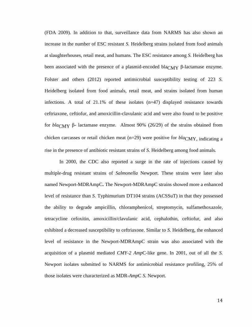
(FDA 2009). In addition to that, surveillance data from NARMS has also shown an
increase in the number of ESC resistant S. Heidelberg strains isolated from food animals
at slaughterhouses, retail meat, and humans. The ESC resistance among S. Heidelberg has
been associated with the presence of a plasmid-encoded blaCMY β-lactamase enzyme.
Folster and others (2012) reported antimicrobial susceptibility testing of 223 S.
Heidelberg isolated from food animals, retail meat, and strains isolated from human
infections. A total of 21.1% of these isolates (n=47) displayed resistance towards
ceftriaxone, ceftiofur, and amoxicillin-clavulanic acid and were also found to be positive
for blaCMY β- lactamase enzyme. Almost 90% (26/29) of the strains obtained from
chicken carcasses or retail chicken meat (n=29) were positive for blaCMY, indicating a
rise in the presence of antibiotic resistant strains of S. Heidelberg among food animals.
In 2000, the CDC also reported a surge in the rate of injections caused by
multiple-drug resistant strains of Salmonella Newport. These strains were later also
named Newport-MDRAmpC. The Newport-MDRAmpC strains showed more a enhanced
level of resistance than S. Typhimurium DT104 strains (ACSSuT) in that they possessed
the ability to degrade ampicillin, chloramphenicol, streptomycin, sulfamethoxazole,
tetracycline cefoxitin, amoxicillin/clavulanic acid, cephalothin, ceftiofur, and also
exhibited a decreased susceptibility to ceftriaxone. Similar to S. Heidelberg, the enhanced
level of resistance in the Newport-MDRAmpC strain was also associated with the
acquisition of a plasmid mediated CMY-2 AmpC-like gene. In 2001, out of all the S.
Newport isolates submitted to NARMS for antimicrobial resistance profiling, 25% of
those isolates were characterized as MDR-AmpC S. Newport.

15
2.7 Salmonella Isolation
The standard microbiological method for the isolation of Salmonella is depicted in
Figure 2.2. Twenty-five grams of food sample are diluted (1:9) in a non-selective
enrichment broth. Lactose broth or buffered peptone water (BPW) are the two most
commonly used enrichment media. After 24 h of enrichment, samples are transferred to
Rappaport-Vassiliadis medium (RV) or Tetrathionate broth (TT) for selective enrichment
of Salmonella. Post selective enrichment step, broth samples from both RV as well as TT
medium are streaked on three differential agar media (Bismuth Sulfite, Xylose Lysine
Desoxycholate, Hektoen Enteric) for the isolation of presumptive/suspected Salmonella
colonies.
Figure 2.2 An overview of Salmonella isolation from food samples

16
Suspected colonies from the three differential agar medium are subjected to a
wide range of biochemical test (e.g. Enterotube II, Vitek 2 GN, API 20E, MICRO-ID).
Biochemically confirmed strains are further subjected to serological identification. The
whole process takes between 4-6 days for final confirmation.
2.7.1 Real-Time PCR Detection of Salmonella
Real-time Polymerase Chain Reaction (PCR) is a method of amplifying a specific
sequence in a provided DNA sample. Unlike conventional PCR where the results can
only be checked at the end of the reaction, real-time PCR allows continuous monitoring
of the amplification data. Real-time PCR reaction employs either fluorescent double-
stranded DNA binding dye (e.g. SYBR® Green, EvaGreen®) or fluorescent-labeled
hybridization probes (e.g. TaqMan, Molecular Beacon) for the quantification and specific
detection of the amplicons of the PCR reaction. A wide range of regulatory and virulence
genes of Salmonella have been previously used for developing real-time PCR assays for
the detection of Salmonella, and a selected list of these genes and assays have been
compiled in Table 2.1.

17
Table 2.1 Selected real-time PCR assay for the detection of Salmonella
Targeted
Gene
Real-time
PCR type
Detection Limit IAC Inclusivity/exclusivity
Number of strain
tested
Sensitivity %
Food Samples tested
Reference
invA (287
bp)
TaqMan 2 cfu/ PCR reaction
using pure culture of S.
Typhimurium
3–7 cfu per PCR
reaction for spiked food
samples
No 100/100
164 Salmonella
50 non-Salmonella
100%
50 chicken carcass rinses
and 60 raw milk samples
Chen and
others 1997
invA
(Unknown)
Commercial
kit
TaqMan 3 CFU/PCR reaction
using pure Salmonella
culture
Yes 100/100
42 serotypes of 68
Salmonella strains, 39
non-Salmonella strains
100%
100 meat and chicken
Kimura and
others 1999
invA
(119 bp)
TaqMan Not tested Yes 100/100
210 S. enterica isolates
(100 problematic
“rough” isolates)
120 non-Salmonella
strains,
Not tested Hoorfar and
others 2000
himA (122
bp)
Molecular
Beacon
2 CFU/PCR of pure
culture
No 100/100
7 Salmonella strain
3 non-Salmonella
strains
Not tested Chen and
others 2000
invA (285
bp)
SYBR
Green1
6 CFU/ml No Not tested 492 intestinal
homogenates and 27 drag
swabs
Eyigor and
others 2002
17

18
1. sipC
2. invE-
invA
3. spaQ
TaqMan 2 CFU/PCR of pure
culture of S. Newport
No 1. 87.5/100
2. 97/100
3. 100/100
116 Salmonella strain
19 non-Salmonella
strains
1. Not tested
2. Not tested
3. 100%
230 clinical fecal
specimens
Kurowski
and others
2002
invA (102
bp)
TaqMan Less than 100 fg of
DNA/reaction
No 100/100
111 Salmonella strains
37 non-Salmonella
strains
Food samples associated
with outbreak
Daum and
others 2002
ttrRSBCA
(94 bp)
TaqMan >103 CFU/ml (70%
probability)
>104
CFU/ml (100%
probability)
Yes 100/100
110 Salmonella strains
87 non-Salmonella
strains
100%
110 food samples
(chicken rinses, minced
meat, fish, raw milk)
Malorny and
others 2004
invA (285
bp)
PCR-
ELISA
103CFU/ml pure
culture
Yes 100/100
84 Salmonella
44 non-Salmonella
100%
60 artificially-
contaminated samples -
fish, minced beef, raw
milk
Perelle and
others 2004
SipB and
SipC
(251bp)
Fluorescent
ly-labeled
hybridizati
on probes
Approximately 6
genome
equivalents/reaction
No 100/100
15 Salmonella
Enteritidis
12 non-Enteritidis
strains
spiked raw and ready-to-
eat beef products
Ellingson
and others
2004
invA
(119 bp)
SYBR
Green1
>102 and >10
3 in broth
and milk respectively
with enrichment
No 100/100
124 Salmonella spp.
116 non-Salmonella
strains
100 %
Lagoon water, feed/silage,
bedding soil, and bulk
tank milk
Nam and
others 2005
fimC
(102bp)
TaqMan Yes 100/100
53 Salmonella
100%
36 artificially and 100
Piknová and
others 2005;
18

19
49 non- Salmonella
strains
naturally contaminated
food sample
Krascsenicso
va and others
2008
ttrRSBCA
(94 bp)
LNA
TaqMan
fishmeal and chicken
rinse (100 copies), pig
feces (10 copies)
No/
Yes
Not tested fishmeal, chicken rinse
and pig feaces spiked with
Salmonella genomic DNA
Reynisson
and others
2006
Prot6e TaqMan >102 genome
equivalents (100%
probability)
>10 genome
equivalents (83%
probability)
Yes 95/100
79 Salmonella
Enteritidis
119 non-Enteritidis
strains
100%
25 chicken carcass rinse,
egg
Malorny and
others 2007
stn (129bp) TaqMan 3CFU/PCR reaction No 100/96.4
269 Salmonella
84 non- Salmonella
strains
Not tested Moore and
Feist 2007
bipA (65bp) TaqMan 6CFU/25 g food (95.5
probability)
Yes 100/100
48 Salmonella
30 non- Salmonella
strains
100%
120 diversified food and
water sample
Calvó and
others 2008
invA (68 bp) TaqMan,
enzyme-
linked
fluorescent
assay
(ELFA)
3.16pg/PCR Yes 100/100
39 Salmonella strain
29 non-Salmonella
strain
97%
20 artificially and 68
naturally contaminated
chicken feces
Tomás and
others 2009
ssrA TaqMan 1-10 genome
copies/PCR reaction
Yes 100/100
30 Salmonella
30 non- Salmonella
strains
Swine swab at different
concentrations
McGuinness
and others
2009
19

20
fimY
(102bp)
TaqMan Approximately
60CFU/ml
No 100/100
6 Salmonella
1 non- Salmonella
strains
biscuit, egg, juice, milk,
pork, spinach
Li and others
2010
ompF
(59bp)
TaqMan 2·8 or approximately
3CFU/PCR reaction
No 218 Salmonella
180 non- Salmonella
strains
Total 30 samples -
orange juice, mayonnaise,
chicken cuts, egg salad
and hamburger patty
Tatavarthy
and Cannons
2010
ssaN (56bp) MGB-
TaqMan
18.6 to 41.2fg of
genomic DNA/PCR
reaction (serovars
dependent)
Yes 100/100
40 Salmonella
24 non- Salmonella
strains
Artificially contaminated
chicken, liquid egg and
peanut butter
Chen and
others 2010
invA
(261bp)
TaqMan
(EMA-real-
time PCR)
103
CFU/ml of pure
culture
105
CFU/ml of spiked
food
Yes 100
S. Enteritidis 13076
and S. Typhimurium
14028
artificially contaminated
chicken rinses and egg
broth
Wang and
Mustapha
2010
invA (285
bp)
TaqMan 20 genome copies
(100% probability)
Yes 100/100
100 Salmonella strain
42 non-Salmonella
strains
100%
1,934 natural food
samples
Anderson
and others
2011
hilA (270bp) Fluorescent
ly
hybridizati
on TaqMan
10 genomic equivalent
of pure culture
Yes 100/100
106 Salmonella
enterica
30 non- Salmonella
strains
Carcass swab, minced
beef
McCabe and
others 2011
invA
(200bp)
Multiplex
TaqMan
100-1000 genomic
equivalent /PCR
reaction
104 CFU/g of spiked
food
Yes 100/100
30 Salmonella
73 non- Salmonella
strains
Spiked ground beef,
whole chicken, ground
chicken, ground turkey,
raw egg, spinach and
tomato
Singh and
Mustapha
2013
20

21
invA
(284bp)
SYTO9
intercalatin
g dye
20 ng–200 fg/PCR
reaction
290 CFU/mL of pure
culture
104 CFU/g of spiked
food
No 100
41 Salmonella
Spiked ground beef,
whole chicken, ground
chicken, egg, produce
Singh and
Mustapha
2014
21

22
2.7.2 Gene Targets for the PCR based Detection of Salmonella
Salmonella contain a 40-kb DNA sequence known as Salmonella pathogenic
island 1 (SPI1). This DNA sequence (SPI1) encodes for at least 33 proteins, which
include the components of a type III secretion apparatus, regulatory proteins, secreted
effector proteins and their chaperones (Darwin and Miller 1999). Three proteins coded by
the SPI1 are directly involved in building a supramolecular syringe-like structure, which
reaches from the cytoplasmic membrane to the outer membrane. This syringe-like
structure secretes effector proteins from Salmonella, stimulates dramatic cytoskeletal
rearrangements in eukaryotic host cells called membrane ruffling. This phenomenon of
membrane ruffling, which is regulated by SPI1 genes, facilitates the engulfment of the
Salmonella by eukaryotic cells. Mutation in any of these SPI1 genes coding for apparatus
proteins or the regulatory proteins greatly diminishes the ability of pathogen to cause host
invasion (Galan and others 1992).
The invA gene is the most commonly targeted SPI1 gene for the detection of
Salmonella. invA encodes a 71kDa putative inner membrane protein and plays an
important role in the internalization of Salmonella into epithelial cells and has been
frequently targeted for PCR-based detection of Salmonella (Rahn and other 1992). The
hilA (hyperinvasive locus A), is another SPI1 gene which has been used for the PCR
based detection of Salmonella (McCabe and others 2011). The hilA is an upstream
regulator of invA and, just like invA, it is also required for the regulation of Type III
secretion system (T3SS). The hilA sequence has also been previously utilized as a target
for the detection of Salmonella enterica (Fey and others 2004; McCabe and others 2011).
The Salmonella invasion proteins or Salmonella secreted protein (sipB-sipC gene) is also

23
a part of SPI1 which encodes proteins required for the invasion of S. Typhimurium and S.
Typhi into tissue culture cells (Darwin and Miller 1999), playing an important role in the
pathogenesis of Salmonella. Ellingson and others (2004) developed a species-specific
real-time PCR assay targeting the sipB-sipC genes for the detection of Salmonella.
The ttrRSBCA locus is located near the Salmonella pathogenicity island 2 (SPI2).
The genes, ttrA, ttrB, and ttrC, of the ttrRSBCA locus encode the tetrathionate reductase
structural proteins, whereas the ttrS and ttrR genes encode the sensor and response
regulator components of the system. The end products of the ttrRSBCA locus is required
for the tetrathionate respiration in Salmonella (Hensel and others 1999). This
tetrathionate respiration is an important characteristic of Salmonella, which is also the
basis for the selective enrichment of Salmonella using tetrathionate broth. The ability to
respire tetrathionate in Salmonella is a significant component of the Salmonella life
cycle. Therefore, the ttrRSBCA locus should be stable in Salmonella. It is believed that
the SPI1 genes in Salmonella were acquired by the process of a horizontal gene transfer.
Natural mutations with a deletion of the SPI1 genes (e.g. inv, spa, hil) can occur, leading
to a false-negative result by PCR assays based on the SPI1 gene (Ginocchio and others
1997). Malorny and others (2004) developed a real-time PCR TaqMan® assay based on
the ttrRSBCA sequence for the detection of Salmonella.
The stn gene of Salmonella encodes a 29-kDa Salmonella-specific enterotoxin
protein (Chopra and others 1994). Previous researchers indicated that the stn is highly
conserved in S. enterica serotypes. However, Ziemer and Steadham (2003) reported the
prevalence of stn among Salmonella bongori strains. The stn gene sequence is genetically
diverse, the percentage of sequence similarity of S. bongori with S Typhimurium is

24
660/750 bp (88%), whereas with S. Typhi is 659/750 bp (87·9%). Moore and Feist (2007)
designed a conserved PCR primer and TaqMan® probe for the detection of S. enterica
and S. bongori both.
Regulatory genes in a bacterial genome are considered to be more stable and are
less prone to mutational changes. The bipA (or typA) gene of Salmonella is a member of
the “GTP-binding elongation” which acts as an essential translation factor for the
efficient expression of fis gene, thus regulating a wide variety of other global downstream
processes and exhorting a global modulating properties (Owens and others 2004). The
bipA gene product also binds to ribosomes at the elongation factor G binding site, and has
additionally shown to have a GTPase activity. Therefore, considering the stable role of
this gene in the Salmonella genome, Calvó and others (2008) developed a real-time PCR
assay for the detection of Salmonella.
In addition to the above-mentioned genes, there have been reports of some other
targets that have been also used for the detection of Salmonella. The ompF gene coding
for a porin protein is present in all Salmonella subspecies. Tatavarthy and Cannons
(2010) used the conserved region of ompF for the real-time PCR detection of Salmonella.
The Prot6e gene is a gene located on the 60-kb virulence plasmid of S. Enteritidis and is
considered specific and unique to S. Enteritidis (Chu and others 1999). Malorny and
others 2007 developed real-time PCR method on Prot6e gene sequence for the detection
of S. Enteritidis. A selected list of real-time PCR assays for the detection of Salmonella is
shown in Table 2.1.

25
2.8 Carbapenems and Extended Spectrum beta-lactam Resistance in
Enterobacteriaceae
Members of the Enterobacteriaceae family are the most common causes of
community- and hospital-acquired infections. The beta-lactam, cephalosporins,
carbapenems and monobactams are the most commonly used antibiotics all around the
world constituting around 60% (by weight) of all antimicrobials used. Owing to their
efficacy, safety and ease with which they can be chemically manipulated, they are the
most preferred group of antimicrobials. (Livermore and Woodford 2006). Therefore, they
are considered to be the most versatile and malleable group of antibiotics. Resistance
towards beta-lactam antibiotics originated from penicillin-binding proteins of Gram-
positive bacteria. These penicillin-binding proteins are an important mode of resistance in
Haemophilus and Neisseria (Georgopapadakou 1993). However, in Gram-negative
bacteria, synthesis of beta-lactamase enzymes, impermeability and porin loss are the most
important mechanisms of resistance (Georgopapadakou 1993; Livermore and Woodford
2006).
Based on the amino acid sequence data, beta-lactamase enzymes have been
divided into four major classes: Class A - serine-β-lactamases, Class B - metallo-β-
lactamases, Class C - serine-β-lactamases, and Class D - serine-β-lactamases. These four
classes are further classified into plasmid-mediated or chromosomal-mediated genes.
However, the demarcation between the chromosomal and plasmid genes can sometimes
be confusing because during the process of evolution, all plasmid genes escape from
chromosomal DNA (Livermore and Woodford 2006).

26
In the last 60 years, the spread of beta-lactamase enzymes among pathogenic
bacteria has driven the development of newer antibiotics with higher efficacy to fight the
problems of antibiotic resistance. Benzylpenicillin was the first analog that penetrated
Gram-negative bacteria poorly and was easily hydrolyzed by penicillinases. The
resistance towards benzylpenicillin rapidly spread through Staphylococcus aureus and
other streptococcal species. In order to counter the increasing resistance towards
benzylpenicillin, semi-synthetic penicillins (e.g. ampicillin, carbenicillin, methicillin and
oxacillins) were created in the 1960s. These new antibiotics possessed the ability to
penetrate resistant strains of Gram-negative bacteria and were also stable against
staphylococcal penicillinase. However, with widespread use of these antibiotics, new
plasmid-mediated penicillinases emerged (e.g. TEM, SHV) in the Enterobacteriaceae and
compromised the activity of these antibiotics. These events drove researchers to develop
newer antibiotics, which led to the synthesis of 2nd
, 3rd
and 4th
- generation oxyimino-
cephalosporins (e.g. cefuroxime, cefotaxime, ceftriaxone, ceftazidime and cefepime), and
other β-lactamase inhibitors (e.g. clavulanic acid). Hereafter, oxyimino-cephalosporins
became the most potent antibiotics and were commonly used all around the world, but
once again, their widespread uses led to the emergence of cephalosporin and
fluoroquinolones resistant (Livermore 2005), thus, driving the use of carbapenems
antibiotics. Unfortunately, the practice led to a rapid selection of carbapenem-resistant
Enterobacteriaceae strains (Walsh 2010). As a result of increasing rates of resistance
towards carbapenem antibiotics, only a limited number of antimicrobials (e.g., colistin,
fosfomycin, tigecycline) are left for the treatment of these resistant strains. Therefore, the
rapid antimicrobial resistance profiling of a pathogen is of the utmost importance.

27
2.8.1 Extended spectrum beta lactamase
In nature, the evolution of predator and prey coincide. The emergence of
resistance to beta-lactam antibiotics began even before the first beta-lactam antibiotic was
identified and made available for medical use (Abraham 1940). Penicillin was one of the
most widely used antibiotics for the treatment of injured soldiers during World War II.
However, the age of penicillin suffered a major setback with the emergence of plasmid-
encoded penicillinase in S. aureus. This resistance was a plasmid-borne trait that was
quickly transferred to other staphylococcal species (Bradford 2001).
Many genera of soil and Gram-negative bacteria possess chromosomally
mediated genes that encode for beta-lactamase enzymes. This group of enzymes that
confers resistance towards beta-lactam antibiotics evolved from naturally occurring
penicillin-binding proteins. One of the theories behind the evolution of these resistant
strains is the selective pressure exerted by naturally occurring beta-lactam producing soil
bacteria in the environment (Ghuysen 1991). The situation was later exacerbated by the
excessive use of the antibiotic in clinical settings, which hastened the emergence of
antibiotic resistant strains.
Datta and Kontomichalou (1965) reported the first beta-lactamase enzyme, TEM-
1, in Gram-negative bacteria. This beta-lactamase enzyme was originally identified in a
patient named Temoniera, hence the name, TEM (Medeiros 1984). The gene coding for
TEM was located on a plasmid and transposon that facilitated rapid spread of the enzyme
among other species, and very soon it was reported from all around the world. The
enzyme is now commonly found in antibiotic resistant strains of members of the
Enterobacteriaceae, Pseudomonas aeruginosa, Haemophilus influenzae, and Neisseria

28
gonorrhoeae. The SHV-1 that stands for “sulphydryl variable” is another commonly
found beta-lactamase enzyme reported among the antibiotic resistance strains of
Klebsiella pneumoniae and E. coli.
After the isolation of the first β-lactam resistant pathogen, more and more
antibiotics were identified to counter the effects of antibiotic resistance. One of these new
classes of antibiotics was called as oxyimino-cephalosporins, which in the 1980s, was
widely used for the treatment of serious infections caused by Gram-negative pathogenic
bacteria. However, with identification and their clinical applications, emerged the new
beta-lactamase enzyme. The extensive use of the antibiotic for medical purposes exerted
selective pressure for the selection of newer β-lactamase enzymes conferring resistance to
newer antibiotics. The SHV-2 enzyme was the first enzyme that showed an extended
spectrum β-lactam hydrolyzing capability and was isolated from a Klebsiella ozaenae in
Germany (Kliebe and others 1985). Because of the increased spectrum of activity of these
β-lactamase enzymes, especially against the oxyimino-cephalosporins, these enzymes
were called extended-spectrum β-lactamases (ESBLs) (Bradford 2001). Hence, ESBLs
were classified as a class of enzymes that confer resistance against penicillins, broad-
spectrum cephalosporins with an oxyimino side chain (cefotaxime, ceftriaxone and
ceftazidime) and the oxyimino-monobactam aztreonam antibiotics. Another common
property of these ESBLs is that they can be inhibited by serine-type β-lactamase
inhibitors e.g. sulbactam, clavulanate and tazobactam (Philippon and others 1989;
Bradford 2001). At present, more than 150 different ESBL enzymes have been described.
Today, ESBLs are one of the most influential mechanisms for cephalosporin resistance in
Enterobacteriaceae, particularly in E. coli and K. pneumoniae.

29
2.8.1.1 Types of ESBL
The first ESBL lactamase to be identified was SHV-2 in a clinical isolate of K.
ozaenae in Germany (Kliebe and others 1985). To date, over ten families of enzymes
conferring the ESBL phenotype have been documented e.g. CTX-M, SHV, TEM, PER,
VEB, BES, GES, TLA, SFO and OXA (Paterson and Bonomo 2005). Most of the ESBL
enzymes found are derivatives of TEM or SHV enzymes (Bush and others 1995; Jacoby
and Medeiros 1991). At present, there are more than 221 TEM-type and greater than 189
SHV-types of ESBL enzymes (http://www.lahey.org/studies/webt.htm). New types of
ESBL enzymes arise by a few mutations in the DNA sequence of the gene, resulting in a
change in amino acid sequence of the protein, generating new enzymatic phenotypes.
These enzymes are commonly found in the Enterobacteriaceae (e.g. E. coli and K.
pneumoniae). However they have been also reported in Proteus spp., Providencia spp.,
and Acinetobacter spp. (Bradford 2001).
2.8.1.1.1 TEM
The TEM-1 is one of the oldest and commonly found beta-lactamase enzymes in
Gram-negative bacteria. The majority of ampicillin resistance (90%) in E. coli is due to
the production of TEM-1 enzyme (Livermore 1995). Apart from ampicillin and
penicillin resistance, the TEM-1 enzyme is responsible for hydrolysis of early
cephalosporins, such as cephalothin and cephaloridine. TEM-2 was the first enzymatic
variant, which originated by a single amino acid substitution in the original TEM-1
sequence (Barthélémy 1985). Another important TEM variant was the TEM-3 enzyme
that was first reported in 1987, and it was also the first TEM-type lactamase that showed
an ESBL phenotype (Sougakoff and others 1988). The amino acid substitution resulting

30
in a new enzymatic variant, occurs in the TEM protein sequence at a limited number of
positions. These combinations of new amino acid substitutions in enzyme protein
sequences led to subtle alternations in the enzymatic activity and substrate range that they
can hydrolyze, resulting in a change in the ESBL phenotype e.g. to hydrolyze different
oxyimino-cephalosporins. It has been proposed that the origin of these TEM-type ESBL
enzymes was not the result of selective pressure exhorted by one single beta-lactam, but
rather due to the application of several beta-lactams within institutions (Bonnet and
others 1999). The TEM-type beta lactamase enzymes are commonly reported among
clinical isolates of E. coli and K. pneumoniae. In addition to these, they have been also
frequently reported in Enterobacteriaceae such as Enterobacter aerogenes, Morganella
morganii, Proteus mirabilis, Proteus rettgeri, and Salmonella spp.
2.8.1.1.2 SHV
The SHV-1 beta-lactamases are most commonly found in K. pneumoniae and are
responsible for more than 20% of resistance towards ampicillin. The gene responsible for
the synthesis of SHV lactamase is blaSHV; it can either exist as a plasmid gene or a
chromosomal gene (Livermore 1995). It has been previously reported that a majority of
SHV-type derivatives possess the ESBL phenotype capabilities. These ESBL variants of
SHV lactamases also possess a substitution of a serine for glycine at the 238th
position of
the protein sequence (Bradford 2001).
2.8.1.1.3 CTX-M
In the family of ESBLs, CTX-Ms are plasmid–mediated enzymes that
preferentially hydrolyze cefotaxime, and are the latest addition that have significant

31
clinical impacts. This new class of ESBL enzymes is commonly found among the
antibiotic resistant strains of Salmonella and E. coli, but they have also been frequently
reported among other members of the Enterobacteriaceae. The CTX-M enzymes are one
of the biggest and most diverse groups of ESBL enzymatic variants that also include
MEN-1 and Toho enzymes. The CTX-M enzymes are not closely related to the SHV and
TEM beta-lactamase enzymes, and show around 40% of sequence homology with SHV
and TEM, which happen to be the most frequently isolated ESBL enzymes (Tzouvelekis
and others 2000). The chromosomally encoded class A cephalosporinases enzymes found
in K. oxytoca, C. diversus, Proteus vulgaris, and Serratia fonticola are the most closely
related to the CTX-M family enzymes. They share around 73-77% sequence homology
(Bauernfeind and others 1996; Bonnet and others 1999). However, some recent studies
showed that the CTX-M lactamase share a much higher degree of homology with the
chromosomal mediated AmpC enzyme of Kluyvera ascorbata, which are designated as
Klu-1 and Klu-2, suggesting that CTX-M enzymes originated from Kluyvera ascorbata
(Decousser and others 2001). Phylogenetic analysis of all the CTX-M enzyme sequence
suggest an early divergence from a common ancestor gene (Bonnet and others 2000).
Most of the CTX-M type enzymes exhibit strong activity against cefotaxime and
ceftriaxone but not ceftazidime. However, there are some exceptions e.g. CTX-Ms, such
as CTX-M-15 (Poirel and others 2002), CTX-M-16 (Bonnet and others 2001) and CTX-
M-19 (Poirel and others 2001), which exhibit enhanced catalytic activity against
ceftazidime.
The CTX-M lactamases are a wide and diversified group of enzymes. To date the
number of enzymatic variants of CTX-M are dramatically increasing and so far more

32
than 160 CTX-M variants (CTX-M-1 to CTX-M-160) have been reported by the Lahey
database (Jacoby and Bush 2012). These variants have been classified into four major
types: (A) CTX-M-1 type - CTX-M-1 and CTX-M-3 (B) CTX-M-2 type - that includes
CTX-M-2, CTX-M-4, CTX-M-5, CTX-M-6, CTX-M-7, and Toho-1 (C) Toho-2 (D)
CTX-M-8 (Bonnet and others 2000). The diversity of CTX-M enzymes varies with
geographical locations, but CTX-M-15 and CTX-M-14 (followed by CTX-M-2, CTX-M-
3 and CTX-M-1) are the most common enzymatic variants worldwide among all major
clinically important pathogens (Zhao and Hu 2013).
Strains expressing CTX-M lactamases have been reported from many parts of the
world. They also have been the focal point of outbreaks in Europe, South America and
Japan (Bradford and others 1998; Ma and others 1998). The CTX-M type lactamase has
also been involved in the outbreaks caused by E. coli and Salmonella (Sabaté and others
2000). According to a review paper published by Zhao and Hu (2013), CTX-M type
enzymes have been detected in at least 26 bacterial species e.g. Acinetobacter baumannii,
Aeromonas caviae, A. hydrophila, Citrobacter amalonaticus, C. freundii, C. koseri, E.
coli, Enterobacter cloacae, E. aerogenes, E. gergoviae, E. hormaechei, K. pneumoniae,
K. oxytoca, Morganella morganii, P. mirabilis, Pantoea agglomerans, Providencia
rettgeri, P. stuartii, Pseudomonas aeruginosa, S. enterica, Shigella flexneri, S. sonnei,
Serratia marcescens, S. liquefaciens, Stenotrophomonas maltophilia and Vibrio cholera.
Out of these 26 bacterial species CTX-M ESBL are more prevalent in E. coli, K.
pneumoniae and P. mirabilis (Zhao and Hu 2013). In a study conducted in 16 hospitals in
England over a period of 12-weeks, a total of 19,252 clinical isolates were tested for
antibiotic resistance. Out of which, E. coli and Klebsiella were the two species that

33
harbored the maximum number of resistant strains, and CTX-M was the most common
resistance mechanism (Potz and others 2006).
2.8.1.1.4 Class C chromosomal AmpCs
Class C chromosomal AmpC (cAmpC) is a class of β-lactamases, which confer
resistance towards third-generation cephalosporins and β-lactamase inhibitors. However,
they are susceptible to fourth-generation cephalosporins (e.g., cefepime) and the
carbapenems group of antibiotics (Jacoby 2009). These enzymes are common in some
genera of Enterobacteriaceae e.g. Citrobacter freundii, Enterobacter spp., Serratia spp.
Genes responsible for the synthesis of these enzymes (AmpC) are under the control of
complex regulatory elements (ampD, ampR) (Jacoby 2009). With the widespread use of
cephalosporins for treatments, an increasing number of blapAmpC positive
Enterobacteriaceae strains have been isolated. There are several families of these
enzymes (e.g. CMY, FOX, LAT, MIR, ACT, DHA, ACC, MOX). Out of these, CMY-2
is the most prevalent pAmpC among members of Enterobacteriaceae. However, DHA
and FOX have been also reported within certain geographic regions (Jacoby 2009;
Seiffert and others 2013).
2.8.2 Carbapenemase
The emergence of resistance towards carbapenems in Gram-negative bacteria
especially in the Enterobacteriaceae, Pseudomonas, and Acinetobacter species (E.P.A)
has become a matter of a public health crisis worldwide, and they are responsible for a
large number of hospital-acquired and nosocomial infections. This scenario has been

34
further worsened by the rapid spread of these resistance phenotypes and the lack of
development of new and effective antimicrobial drugs (Diene and Rolain 2014).
In the early 1990s, resistance towards carbapenems was reported to be a species-specific
phenomenon, but resistance-conferring genes, being a plasmid borne trait, spread to all
related genera and become a worldwide phenomena.
Carbapenemase are enzymes that are produced by pathogenic bacterial species to
degrade and neutralize the effects of almost all beta-lactam antibiotics, including
carbapenems (Cantón and others 2012). In the last two decades, a large number of
different groups of enzymes possessing carbapenemase activity have emerged, and are
rapidly spreading all around the world. This highly genotypically and phenotypically
diversified group of carbapenemase enzymes differs in their enzymatic activity (Figure
2.3). Some of them hydrolyze carbapenems very efficiently, whereas others show weak
or limited activity. Some enzymes can confer resistance towards broad-spectrum
cephalosporins, while some do not (Nordmann and Poirel 2014).

35
Figure 2.3 Phylogenetic tree of the metallo-carbapenemase and serine carbapenemase
genes. Their mean percentage GC content, according to groups and phenotypic
properties. The tree was constructed from the amino acid sequences aligned with the free
ClustalX software version 2.0 and MEGA software version 6.06, by use of the neighbour-
joining method with the amino acid Poisson correction model with 1000 bootstrap
replicates. Bootstrap values are expressed as percentage of the 1000 replicates, and only
those up to 50% are kept and shown at branch points. aOXA genes described overall in
Gram-negative bacteria. bOXA genes described only in Acinetobacter species. The
percentage GC values correspond to the averages of each subgroup. ATM, aztreonam;
CLA, clavulanic acid; R, resistant; S, susceptible. Adapted from Diene and Rolain
(2014); License Number: 3505650398577; License Date: Nov 10, 2014
Acc
epte
d A
rtic
le
This article is protected by copyrright. All rights reserved.

36
2.8.2.1 Classification of carbapenemases
The carbapenemases group of enzymes can be either classified using its genetic
makeup or they can be also categorized based on the functional characteristics of their
active sites (Table 2.2).
Table 2.2 Classification of β-lactamases enzymes
Class Enzymes that are
plasmid-, transposon- or
integron-mediated
Chromosomal and ubiquitous in
species or group
Class A- Serine-β-
lactamases
TEM, SHV, VEB, PER,
CTX-M penicillinases and
ESBLs; KPC, IMI/NMC
and SME carbapenemases
SHV-1, LEN-1 and K1 in Klebsiella
spp.; chromosomal cefuroximases of
Proteus vulgaris; chromosomal, β-
lactamases of Bacteroides spp.
Class B- Metallo-
β-lactamases
IMP, NDM, VIM, SPM L1 enzyme of Stenotrophomonas
maltophilia; chromosomal enzymes
of some Chryseobacterium spp. and
Aeromonas spp.; CcrA enzyme found
in 1–3% of Bacteroides fragilis
isolates
Class C- Serine-β-
lactamases
CMY-1, LAT-1, BIL,
MOX, ACC, FOX, DHA
Chromosomal AmpC enzymes of
Escherichia coli, Shigella spp.,
Enterobacter spp., Citrobacter
freundii, Morganella morganii,
Providencia spp., Serratia spp.
Class D - Serine-
β-lactamases
Most OXA types,
excluding those detailed
here as chromosomal
Chromosomal (along with other b-
lactamases) in Acinetobacter spp.
(OXA-51-like); P. aeruginosa (OXA-
50) chromosomal and some
Aeromonas spp. (e.g. OXA-12).
(Adapted from Livermore and Woodford 2006)
Based on the composition of the active site, carbapenemases enzymes can be
broadly classified into two major groups: (1) Serine Carbapenemases: This group consists
of a serine amino-acid in their active site e.g. class A penicillinases and class D

37
oxacillinases. These enzymes can be inactivated with the use of the β- lactamase
inhibitors e.g. clavulanic acid and tazobactam. (2) Metallo-betalactamases: This group of
enzymes is also known as Class B carbapenemases. Enzymes of this group have one or
more zinc atoms in the active site of the enzyme. The activity of these enzymes can be
inhibited by ethylenediaminetetraacetic acid (EDTA) (Queenan and Bush 2007).
2.8.2.1.1 Class A carbapenemases
Enzymes of this class are coded by IMI, NMC, SME, KPC, and GES genes,
which confer resistance to the carbapenems group of antibiotics at various levels, ranging
from complete resistance to reduced susceptibility (Queenan and Bush 2007). Out of the
above-mentioned five enzymes IMI, NMC and SME are encoded by chromosomal genes,
whereas KPC and GES are encoded by genes located on plasmids. Further, SME
enzymes are mostly species-specific and are restricted to Serratia marcescens, whereas
IMI and NMC enzymes have been sporadically reported only in E. cloacae (Queenan and
Bush 2007). Therefore the chromosome-mediated genes (e.g. IMI, NMC and SME) are
species-specific genes enzymes, are rarely reported and are also less prevalent all around
the world. However, other two Class A carbapenemases enzymes, KPC (K. pneumoniae
carbapenemases) and GES (Guiana extended-spectrum β-lactamase) enzymes are located
on transferrable plasmids, so they are more widely distributed among the
Enterobacteriaceae and other pathogenic genera. The KPC enzymes are mostly prevalent
in K. pneumoniae but they could be also found in other members of the
Enterobacteriaceae, Pseudomonas and Acinetobacter (Cantón and others 2012).
Similarly, the GES enzymes are associated with integrons (mobile genetic element) and
are prevalent in P. aeruginosa and K. pneumoniae (Queenan and Bush 2007).

38
2.8.2.1.2 Class D carbapenemases
These groups of enzymes are also called Class D metallo-beta-lactamases or OXA
beta-lactamases (owing to their ability to hydrolyze oxacillin). The OXA-23, OXA-24,
and OXA-58 genes are the most dominant genetic determinants encoding this enzyme in
A. baumannii whereas OXA-48 is are being increasingly reported in K. pneumoniae. The
gene coding for this group of enzymes is found on chromosomal as well as plasmid DNA
facilitating their spread among related genera (Queenan and Bush 2007).
2.8.2.1.3 Class B carbapenemases or class B metallo-beta-lactamase
This group includes the GIM, IMP, NDM, SIM and VIM enzymes that are
commonly found on the transferrable plasmids of Enterobacteriaceae with addition to P.
aeruginosa and A. baumannii. Out of the five above-mentioned enzymes; IMP, NDM and
VIM are the most prominent enzymes.
The IMP enzyme was first reported in Japan in a clinical isolate of S. marcescens,
but has now spread all around the world and is commonly found in the
Enterobacteriaceae, P. aeruginosa and A. baumannii (Osano and others 1994; Cornaglia
and others 2011).
The VIM-type of enzymes, so far, comprise of around 43 variants (VIM-1 to
VIM-43). Out of these enzymatic variants, VIM-1 is the most commonly found variant.
The gene encoding for the synthesis of VIM-1 enzyme (blaVIM-1) was first reported in
1997 in a clinical strain of P. aeruginosa inVerona, Italy. This gene was integrated as a
gene cassette in a Class I integron along with the aminoglycoside resistance gene, aacA4
(Lauretti and others 1999). The Class I integron is a mobile genetic elements that can

39
harbor multiple drug resistance genes as a cassette and are responsible for multiple drug-
resistance phenotypes in Enterobacteriaceae. The presence of blaVIM-1 in E. coli was first
reported in Greece in 2001, and it later on spread all round the world. The next common
enzymatic variant of this group is VIM-2, it was first reported in a clinical strain of P.
aeruginosa, and now it is endemic to many countries of the world (Cornaglia and others
2011).
NDM is the most recent addition to the Class B carbapenemases group of
enzymes. The enzyme was reported in the year 2009 from clinical isolates obtained from
Asia and India (Yong and others 2009; Kumarasamy and others 2010). Since the first
report of NDM-1 in the year 2009, it has spread worldwide and become the most
common carbapenemases enzyme in all the Enterobacteriaceae (Rolain and others 2010).
2.8.2.2 Distribution of carbapenemase enzymes
In the last 24 years, since the first description of the carbapenemase enzyme in S.
marcescens in 1990 (Yang and others 1990), the occurrence of carbapenemase producing
Enterobacteriaceae has increased worldwide.
2.8.2.2.1 Serine carbapenemases group
KPC enzymes are most important due to their most frequent and worldwide
occurrence, especially in K. pneumonia and E. coli. Other Enterobacterial species
harboring these enzymes include Citrobacter, Enterobacter, Serratia, and Proteus
(Queenan and Bush 2007). The widespread occurrence of KPC enzymes in EPA species
has been associated with the occurrence of the KPC enzyme-producing gene (blaKPC) on
transposon Tn4401 (a mobile genetic element) (Cuzon and others 2010). At present, there

40
are 22 enzymatic variants of the KPC (KPC-1 to KPC-22), and all of them originated as
point-mutation variants of a common amino acid sequence (Nordmann and Poirel 2014).
Out of these 19 enzymatic variants, KPC-2 and KPC-3 producing K. pneumoniae clones
have been most extensively identified all around the world (Cuzon and others 2010).
Other serine carbapenemases enzymes (e.g. BIC, GES, IMI, NMC, and SME) also share
a common ancestry to the KPC enzyme (Walther-Rasmussen and Høiby 2007). Out of
them, blaSME-genes are restricted to only S. marcescens species whereas IMI and NMC-A
enzymes which are more similar to each other, are mainly distributed among E. cloacae
and E. coli (Walther-Rasmussen and Høiby 2007; Queenan and Bush 2007). GES
enzymes are mostly reported among P. aeruginosa species, but they are also found in the
Enterobacteriaceae. There are around 26 (GES-1 to GES-26) enzymatic variants of GES,
out of which GES-2, GES-4, GES-5, GES- 6, GES-14 and GES-18 have been reported to
exhibit an enzymatic activity against carbapenems (Walther-Rasmussen and Høiby 2007;
Queenan and Bush 2007). The enzymatic variant, GES-5 has been reported in K.
pneumoniae, E. coli and E. cloacae (Girlich and others 2012).
2.8.2.2.2 Class D metallo-beta-lactamases or OXA beta-lactamases
It is one of the most genetically diversified groups of carbapenemase enzymes.
The first OXA-48 enzyme-producing strain was identified in a K. pneumoniae isolate
recovered in Turkey. Later, the KPC enzyme successfully spread to all important
members of the Enterobacteriaceae. To date, more than 435 (OXA-1 to OXA-435)
sequence types of blaOXA gene have been reported (Diene and Rolain 2014). All these
enzymatic variants possess some level of enzymatic activity against carbapenems, and do
not provide high levels of resistance against carbapenems (Nordmann and Poirel 2014).

41
The OXA gene variants blaOXA-23, blaOXA-24, blaOXA-58, and some orthologs of these genes
have been reported in Acinetobacter, whereas blaOXA-48, blaOXA-162 and blaOXA-181 have
been reported in the Enterobacteriaceae (Walther-Rasmussen and Høiby 2006). The most
dominant variant blaOXA-48 was for the first time isolated in 2004 from K. pneumoniae in
Turkey (Poirel and others 2004). The OXA-48 enzyme has now been reported among all
major enterobacterial species, which include E. coli, S. marcescens, E. cloacae, K.
oxytoca, Providencia rettgeri, and C. freundii (Poirel and others 2012). Other equally
important enzymatic variants of this group are OXA-181, OXA-204, and OXA-232
(Poirel and others 2012).
2.8.2.2.3 Class B carbapenemases or the Class B metallo-beta-lactamase
This group of enzyme comprises of GIM, IMP, NDM, SIM and VIM. These
enzymes were historically associated with P. aeruginosa and Acinetobacter spp.;
however in last two decades they have widely spread across the enterobacterial species
(Walther-Rasmussen and Høiby 2006). Out of these five Class B metallo-beta-lactamase
enzymes NDM, VIM and IMP are the most commonly encountered enzymes in clinical
pathogens. The IMP-1 enzyme was the first metallo-carbapenemase enzyme reported in
the Enterobacteriaceae (Osano and others 1994). Therefore, based on its early existence,
it has been reported to be in almost all enterobacterial species, e.g E. coli, C. freundii, C.
koseri, K. pneumoniae, K. oxytoca, E. aerogenes, E. cloacae, P. mirabilis, P. rettgeri, M.
morganii, and Shigella flexneri (Queenan and Bush, 2007). The VIM-2 enzyme was first
discovered in 2000 from a P. aeruginosa isolate, but now the enzyme has been widely
distributed in Gram-negative bacteria (Poirel and others 2000; Walsh and others 2005).
The last addition to the Class B metallo-beta-lactamase group of enzymes was NDM-1

42
(New Delhi metallo-β- lactamase-1). The NDM enzyme was first reported in the Indian
subcontinent (Yong and others 2009). The enzyme has around 10 enzymatic variants and
has rapidly spread from the Indian subcontinent to other parts of the world. The enzyme
has been identified in all major enterobacterial species viz. K. pneumoniae, K. oxytoca, E.
coli, P. mirabilis, C. freundii, E. cloacae, and M. morganii (Nordmann and others 2011).
The GIM- 1 and SIM-1 enzymes are less commonly reported. The blaGIM-1 gene has been
reported from E. cloacae, K. oxytoca, S. marcescens, E. coli, and C. freundii (Wendel and
others 2013), whereas the SIM-1 enzyme has not yet been reported among enterobacterial
species (Diene and Rolain 2014).
2.8.3 Epidemiology and spread of these enzymes
2.8.3.1 Primary Reservoir
These enzymes have the ability to degrade/hydrolyze antibiotics emerge in
geographical regions where selective pressure for evolution is high e.g. highly populated
area, poor hygiene, over use of antibiotics for human treatments, agriculture and animal
husbandry purposes.
2.8.3.2 Mobile Genetic Elements
There are genetic elements in the bacterial genome that increase the plasticity or
movement of antibiotic resistance genes. These genetic elements are integrons,
transposons, and plasmids that facilitate horizontal gene transfer between closely related
bacteria. Some plasmids have a broader-host range, allowing them to replicate in a wide
range of bacterial genera, which translates into the ability to transfer antibiotic resistance
genes to a broad range of bacteria. On the contrary, narrow-host range plasmids have the

43
ability to transfer and replicate only in closely related bacterial genera. Therefore, the
genetic makeup of antibiotic resistance strains plays a vital role in the clonal spread of a
particular antibiotic resistance genotype and phenotype.
2.8.3.3 Exchange among human population
Once the antibiotic resistance genotype and phenotype have evolved and are
established in a particular population group due to a particular selective pressure, then it
acts as a reservoir. Mobility of the population of that reservoir determines the spread of
the antibiotic resistance trait around the world. Antibiotic resistant traits that have a
reservoir in places with higher mobility of the population (e.g. tourist spots, hospitals,
medical-tourism) results in a faster spread of the antibiotic resistant traits e.g. clonal
spread of NDM enzyme from the Indian subcontinent to Europe and American continents
(Nordmann and others 2011).
2.8.4 Need for detection of antibiotic resistance
Standard microbiological procedures can take up to several days for the isolation
and characterization of pathogens from clinical samples. For the diagnosis of bacteremia
or sepsis, clinical laboratories commonly use an automated system that can monitor the
growth of the pathogen in the blood culture bottles (Riedel and Carroll 2010). In case of
positive growth (e.g. members of the Enterobacteriaceae take around 24-48 h to grow),
the samples can be plated on agar media for cultivation and isolation of pure culture of
the pathogenic strain of bacteria. It takes around another 24 h for the bacteria to grow on
the agar media. If, there is growth on the agar medium, an antimicrobial susceptibility
test can be performed that requires an additional 24 h. Therefore, for non-multidrug

44
resistant strains it often takes around 3-4 days, while for a resistant strain, it can take even
longer. However, in some cases, the antimicrobial susceptibility test of carbapenemase,
ESBL or plasmid-mediated AmpC (pAmpC) producers can be inaccurate (Livermore and
others 2011; Poirel and others 2012; Luzzaro and others 2006, Doi and Paterson 2007).
These ambiguities and difficulties can be resolved by performing additional tests e.g.
clavulanate for ESBL detection, boronic acid for pAmpCs and KPCs detection, and
modified Hodge test (mHT) for detecting carbapenemases. Performing these tests takes
additional time, which adds up to 4-6 days for getting accurate antimicrobial
susceptibility test results (Lupo and others 2013).
Delays in identification and characterization of antimicrobial resistance profiling
can be of grave consequences. Longer diagnosis time for a patient infected with multi-
drug resistant pathogens can have a less favorable outcome. For a patient suffering from a
multi-drug resistant infection, a delay in diagnosis can lead to treatment failure, which
translates into higher mortality rates. Hence, rapid antimicrobial susceptibility profiling is
of the utmost importance. As initially stated, any delay in antimicrobial susceptibility
testing results can lead to treatment failure, which can make a big difference in final
hospital bills. A rapid and accurate method for antimicrobial susceptibility testing can
reduce the duration of hospitalization and consequently allows reduction in the healthcare
cost (e.g. by avoiding use of expensive carbapenems, shorten duration of hospital stay).
Lee and others (2006) reported that the mean length of a hospital stay for a person with
an ESBL infection was 21 days, which was much higher than a patient with an infection
with a non-ESBL strain (11 days), which leads to an additional $16,450 per patient. In
another study, Schwaber and others (2006) reported that the average cost of treatment for

45
a bloodstream infection caused by an ESBL-producing strain was $46,970, which was
around four times higher ($16,877) than the treatment cost of infection caused by non-
ESBL producers.
2.8.4.1 Need for molecular methods for antibiotic susceptibility testing
Determination of minimum inhibitory concentrations (MIC) using automated
systems is commonly used in clinical laboratories and is considered to be accurate, but
these systems are also associated with reproducibility issues (Miriagou and others 2010).
Giakkoupi and others (2005) compared antimicrobial susceptibility results for five VIM-1
producing K. pneumoniae isolates towards imipenem and meropenem antibiotics using
broth microdilution, Etest, disc diffusion and three automated system (Vitek 2, Phoenix,
and MicroScan). These five VIM-1 producing K. pneumoniae isolates showed major
variations among susceptibility results especially when the automated systems were used
(Giakkoupi and others 2005). Similarly in another study, 15% of KPC-producing K.
pneumoniae isolates were found to be susceptible to imipenem using automated broth
microdilution methods, while only 4% isolates were found susceptible by the Etest
method (Bratu and others 2005). Tenover and others (2006) tested MicroScan, Phoenix,
and Sensititre automated systems using 15 KPC-producing K. pneumonia isolates against
imipenem antibiotic. The number of susceptible isolates obtained using MicroScan,
Phoenix, and Sensititre, varied between 1, 2 and 13, respectively. Variation in the
inoculum concentration has been suggested as one of the possible reasons for the
disparity among the results obtained from different automated systems. Samples with
lower inoculum concentrations appear as susceptible, whereas, sample with higher
inoculum count can appear to be resistant.

46
Antimicrobial susceptibility testing for ESBL- or carbapenemase-producing
Enterobacteriaceae are also performed using commercially available selective agar plates.
However, many recent comparative studies have shown that the commercially available
selective agars have lower sensitivity than the PCR-based methods. This is especially true
for clinical isolates with lower gene expression leading to a low-level of MIC (e.g.,
OXA-48-producing E. coli) (Nordmann and others 2012). Singh and others (2012)
reported that the real-time PCR method for the detection of blaKPC in clinical samples
showed a sensitivity of 97%, which was much higher then the sensitivity achieved by
CHROMagar plates (77%). In another study, Naas and others (2011) reported that a real-
time PCR method for the detection of blaNDM had a limit of detection of 10 CFU/mL,
where ChromID ESBL and CHROMAgar KPC plates required a pathogen concentration
of 102-10
3 CFU/mL. Therefore, as the phenotypic methods can either lead to variation in
results or lower sensitivity, the molecular-based methods are currently considered as
“gold standards” for antibiotic susceptibility testing (Hammoudi and others 2014).
2.8.4.2 Ideal gene targets for rapid detection of antibiotic resistant pathogens
Resistance towards some antibiotics is more wide spread than others and has more
serious clinical implications than others. Hence, while developing a diagnostic assay for
the detection of antibiotic resistance, some antibiotic resistant genes deserve more
attention than others (Table 2.3). In the Enterobacteriaceae family, blaCTX-M, blaTEM,
blaSHV, blaCMY, blaKPC, blaNDM, blaOXA-48, the 16S rRNA methylases genes and mutations
in gyrA and parC are the most important genes that should be targeted for the
development of a diagnostic assay (Lupo and others 2013). However, other gene targets

47
with limited global impact, which are found only in limited geographical locations (e.g.
blaPER, blaVEB, blaGES, blaSPM, blaGIM, blaSME, and plasmid-mediated quinolone
resistance determinants) can be also targeted (Lupo and others 2013). The ideal
characteristics of a diagnostic assay for detection of antibiotic resistance are as follows:
(a) it should target all resistance genes in the same assay; (b) generate results same day;
(c) the data should be easy to analyze and interpret; (d) preferably should be able to
differentiate between allelic variants, as protein encoded by different allelic variants have
different clinical impacts; (e) its method should be easy to perform, cheap and accurate
(Lupo and others 2013).

48
Table 2.3 List of most important antibiotic resistance trait and genes that molecular methods should be able to detect for the
identification of Extended-spectrum cephalosporins and Carbapenems resistance in Enterobacteriaceae. Adapted from Lupo and others
(2013) (License Number: 3506120612617; License Date: Nov 11, 2014)
Antibiotics Mechanism of resistance Most frequent
resistance traits
Importance Ideal level of
detection/characterization
Extended-
spectrum
cephalosporins
Extended-spectrum β-lactamases
(ESBLs)
CTX-M ++++ Generic
TEM and SHV ++++ Distinguishing ESBLs from
non-ESBLs
PER ++ Generic
GES ++ Distinguishing ESBLs (e.g.,
GES-1) from carbapenemases
(e.g., GES-5)
SFO + Generic
Plasmid-mediated AmpCs (pAmpCs) CMY ++++ Generic
DHA +++ Generic
FOX ++ Generic
LAT, MIR, ACT,
MOX
+ Generic
Chromosomal AmpCs (cAmpCs) Mutations in the
promoter (E. coli)
++ Identifying substitution (e.g., at -
32/-42) and insertions (between
-10 and -35)
Mutations in ampD
(Enterobacter spp.)
+ Unknown specific target
Carbapenems Carbapenemases Class A KPC ++++ Generic
GES ++
Distinguishing ESBLs (e.g.,
GES-1) from carbapenemases
48

49
(e.g., GES-5)b
Class B (MBL) NDM ++++ Generic
SME + Generic
VIM ++++ Generic
IMP +++ Generic
SPM and GIM ++ Generic
AIM, SIM + Generic
Class D OXA-48 ++++ Specific
OXA-181, -162, -163 +++
Specific
Impermeability (porins) OmpK-35 and -36
(K. pneumoniae)
++ Identifying insertions and/or
frame-shift mutation(s)
OmpF/C (E. coli and
Enterobacter spp.)
+ Unknown specific
mutation/target

50
2.8.4.3 PCR based method for ESBL and carbapenems detection
2.8.4.3.1 Multiplex end-point PCR for the detection of ESBL and carbapenems
resistance in Enterobacteriaceae
The multiplex PCR, unlike the single end-point PCR, uses a multiple set of
primer-pairs in a single reaction, allowing amplification and detection of multiple targets
in the same reaction. The design of a multiplex PCR reaction is more complex than a
single end-point PCR reaction. The following points should be considered while
designing a multiplex PCR reaction: (a) high specificity of each primer set; (b) no self-
reactivity or primer-dimer formation; (c) similar PCR cycling condition for all primer-
pairs being used in the assay; and (d) each amplicon of the multiplex PCR reaction
should differ from other amplicons by at least 30 bp, enabling visible band separation on
a regular agarose gel.
Pérez-Pérez and Hanson (2002) developed a multiplex assay for the detection of
blaAmpC genes. The PCR assay uses six sets of AmpC-specific primers generating
amplicons between 190-520 bp. The size of the amplicons generated by this assay was
very similar, thus requiring the WAVE technology, a high-pressure liquid
chromatography-based separation system. However the primer-pairs used in the assay
suffered from cross reactivity. Dallenne and others (2010) reported a multiplex PCR
assay for the detection of the most frequent and widespread β-lactamase genes. The assay
used seven multiplex reactions and three different cycling conditions for the detection of
OXA-1-like broad-spectrum β-lactamases, extended-spectrum β-lactamases (ESBLs),

51
plasmid-mediated AmpC β-lactamases and class A, B and D carbapenemases. Voets and
others (2011) further improved the assay described by Dallenne and others (2010). The
assay used a large number of primers in seven multiplex PCR reactions for the detection
of plasmid-mediated AmpC β-lactamases (ACC, ACT, DHA, CMY, FOX, LAT, MIR
and MOX), metallo-carbapenemases (GIM, NDM, SIM and SPM), serine
carbapenemases (IMI, SME and NMC-A) and OXA β-lactamases (OXA groups 23, 24,
48, 1, 2, 51, 4 and 58). The PCR conditions were further optimized to eliminate cross
reactivity of the primer. Ellington and others (2007) published a multiplex PCR assay for
the detection of metallo-β-lactamases (MBLs). The assay was designed to target five
different families of MDLs e.g. IMP, VIM, SPM-1, GIM-1 and SIM-1. Hong and others
(2012) reported a multiplex assay for the detection of four families of class A
carbapenemases (e.g. SME, IMI/NMC-A, KPC, and GES). Another multiplex PCR assay
for the simultaneous detection of blaKPC, blaNDM, blaVIM, blaIMP, and blaOXA-48-like was
reported by Doyle and others (2012). The method was evaluated using 142 antibiotic-
resistant Enterobacteriaceae strains obtained from the Study for Monitoring
Antimicrobial Resistance Trends (SMART; 2008-2009). In the comparative study, this
PCR assay showed 100% sensitivity and specificity.
2.8.4.3.2 Commercially available multiplex PCR assay for the detection of ESBL
and carbapenems resistance in Enterobacteriaceae
Amplex Diagnostics (Germany) launched three PCR-ELISA kits for the detection
of carbapenems and extended spectrum beta-lactam resistance in clinical isolates of
Enterobacteriaceae (www. hyplex.info/). The assay targets are first amplified using gene-

52
specific primers, and the labeled amplicons are reverse-hybridized in a 96-well plate, and
the amplicons detected after the addition of a conjugate (peroxidase) and substrate (tetra-
methyl-benzidin). The final data is photometrically measured at 450 nm. The three PCR-
ELISA assay targets the following genes: (a) Hyplex ESBL ID – blaTEM, blaSHV, blaCTX-
M, and blaOXA (b) Hyplex SuperBug ID - blaKPC, blaIMP, blaVIM, blaNDM, blaOXA-48-like (c)
Hyplex CarbOXA ID - blaOXA-23, blaOXA-24, and blaOXA-51-like. Hyplex SuperBug ID PCR-
ELISA assay was evaluated by using 132 carbapenem-resistant Enterobacteriaceae
isolates. The results showed a 97% (128/132) accuracy (Kaase and others 2012).
2.8.4.3.3 Multiplex real-time PCRs assay for the detection of extended-spectrum
cephalosporins and carbapenems resistance
Bisiklis and others (2007) reported a multiplex real-time PCR melt-curve analysis
assay for the detection of the most commonly found blaVIM (blaVIM-1, blaVIM-2, blaVIM-
3, blaVIM-4, blaVIM-5, blaVIM-6, blaVIM-10, blaVIM-11, blaVIM-12) and blaIMP (blaIMP-1, blaIMP-
2, blaIMP-6, blaIMP-8, blaIMP-10, blaIMP-15, blaIMP-19, blaIMP-20) genes in a single reaction.
Brolund and others (2010) modified the end-point multiplex PCR assay reported by
Pérez-Pérez and Hanson (2002) into a real-time melt-curve analysis PCR assay using
SYBR Green® dye with the same PCR cycling conditions. The assay was divided into
two triplex PCR reactions. The first reaction targeted the MOX, CIT and FOX genes
whereas the second reaction targeted the DHA, ACC and EBC genes. Geyer and others
(2012) developed a multiplex TaqMan® assay (two color FAM and HEX) for the
detection of pAmpCs. The assay targeted MOX, FOX, CMY-2, DHA, ACT, ACC and
16S rRNA gene as internal amplification control (IAC). All probes targeting pAmpCs

53
gene targets in the assay were labeled with FAM dye and the IAC probe was labeled with
HEX dye. The assay showed 100% specificity and sensitivity. Swayne and others (2011)
designed a real-time PCR multiplex TaqMan® assay for the detection simultaneous
detection of five types of serine carbapenemases (e.g. GES, IMI/NMC, KPC, OXA-48
and SME). The assay was highly specific, and the amplicons can also be distinguished by
performing a melt-curve analysis. A multiplex assay for the detection of different classes
of carbapenemases assay was designed by Monteiro and others (2012). The single
multiplex real-time melt-curve analysis PCR assays targeted six different genetic types of
carbapenemases commonly reported in the Enterobacteriaceae (KPC, GES, NDM, IMP,
VIM and OXA-48). Chen and others (2011) designed a multiplex real-time PCR assay,
for the detection of blaKPC-2 to blaKPC-11. The assay utilized one pair of conserved primer
and six molecular beacon probes. A total of 457 Gram-negative clinically isolates were
successfully characterized using this Molecular Beacon real-time PCR assay.
2.8.4.3.4 Commercially available multiplex real-time PCRs Assay
NucliSENS EasyQ KPC (bioMérieux, France) is a commercial real-time PCR
assay for the detection of blaKPC in K. pneumoniae. It is designed to work with the
bioMérieux NucliSENS platform. The result of the assay is analyzed using an automated
NucliSens EasyQ analyzer. Spanu and others (2012) evaluated 300 members of
the Enterobacteriaceae using the NucliSENS EasyQ KPC kit. The assay correctly
detected all of the 111 isolates harboring blaKPC genes, with no false positive results
within 2 h. Check-Points Health BV (Netherlands) developed a ligation-mediated
multiplex real-time PCR assay (Check-MDR ESBL kit) for the detection of SHV/TEM

54
variants (ESBL and non-ESBL) and CTX-M groups. This ligation-mediated multiplex
real-time PCR assay can be performed in 3.5 h. Nijhuis and others (2012) compared the
Check-MDR ESBL kit with a combination disc test (CDT). The Check-MDR ESBL kit
showed sensitivity and specificity of 99.0, 92.2, respectively. The same company
launched another commercial multiplex real-time PCR assay (Check-MDR Carba) for
rapid detection of carbapenems resistance. The assay detects the presence of blaKPC,
blaNDM, blaVIM, blaIMP, and blaOXA-48 resistance genes.

55
2.9 Shiga Toxin-Producing Escherichia Coli
Shiga toxin-producing Escherichia coli (STEC) are a group of pathogenic E. coli
that produces Shiga toxin. This group of pathogenic bacteria causes illnesses ranging
from mild diarrhea to hemorrhagic colitis and a life-threatening hemolytic uremic
syndrome (HUS) (Tarr and others 2005). The STEC group is comprised of more than 50
serogroups, and they are broadly divided into E. coli O157 and non-O157 serogroups. E.
coli O157 was first recognized as a foodborne pathogen in 1982 (Riley and others 1983)
and so far, is responsible for causing more than one-third of the total (estimated) STEC
illnesses in the United States (Scallan and others 2011). Due to an increasing number of
deaths and HUS cases, on September 28, 1994, the U.S. Department of Agriculture
(USDA) Food Safety and Inspection Service (FSIS) declared E. coli O157:H7 an
adulterant, enforcing their zero tolerance policy in ground beef (Taylor 1994). Since then,
surveillance activities conducted by the USDA-FSIS at federally inspected plants and
retail stores have resulted in 234 recalls. This increase in active surveillance activities
may be partly responsible for the recent decline in the incidence of E. coli O157
infections (CDC 2011).
Non-O157 STEC serogroups in the last decade have emerged as major food-borne
pathogens of concern worldwide (Smith and others 2014). In Canada, they were involved
in 63% of the total STEC infections (Thompson and others 2005); while in Denmark,
74% of cases have been related to non-O157 STEC (Nielsen and others 2006) and data
from Germany showed the highest incidence rate (82%) (Werber and others 2008). The
infection rate in the Netherlands was reported to be 80% (Van Duynhoven and others

56
2008). Hale and others (2012), reported that around 231,157 cases of infections are
caused by this group of pathogens in the United States annually. Out of these, E. coli
O157 led to 40.3% of infections; whereas, non-O157 serogroups caused the remaining
59.7% of cases. In another study that analyzed the data collected from 2000 to 2010 by
the USDA-FSIS, a total of 7694 STEC-related cases was reported, out of which 5688
(73.9%) were linked to the O157 serogroup; whereas, 2006 (35.2%) cases were caused by
non-O157 STEC. Therefore, in light of these new data, the USDA, in 2012, declared
these six non-O157 STEC serogroups, also known as the “big six non-O157” (O26,
O103, O111, O121, O45, and O145) as adulterants (zero tolerance policy) in non-intact
raw beef products (USDA 2011). In 2010, for the first time in the United States, the
number of infections caused by non-O157 STEC was higher than the E. coli O157 (CDC
2011). This observed increase in the number of infections caused by non-O157 STEC is
also attributed to the increasing availability and use of diagnostic assays for the detection
of STEC (Gould and others 2013).
In a recent study, Luna-Gierke and others (2014) compiled all the data concerning
outbreaks caused by non-O157 STEC in the USA (up to 2010). According to the study, a
total of 46 outbreaks (38 were single-etiology outbreaks) were reported in 26 states,
which resulted in 1727 illnesses and 144 hospitalizations. Out of these 38 single-etiology
outbreaks, 66% was caused either by STEC O111 (n=14) or O26 (n=11), followed by
O45 (n=4), O103 (n=2), O121 (n=2), O145 (n=2), O104 (n=1), O165 (n=1) and O
undetermined (n=1). Eighty-four percent of these infections was either transmitted by
food (n=17) or via person-to-person (n=15) contacts. The study also noted dairy products,

57
produce, meat and childcare centers to be the most common sources of non-O157 STEC
infections. Eight (17%) out of 46 outbreaks were multiple-etiology outbreaks. The most
common serogroups involved in these multiple-etiology outbreaks were O111 (n=4),
followed by O26 (n=2). Apart from these two, O69, O84, O121, O141, O145, and O
undetermined were involved in one outbreak each. In addition to non-O157 STEC, other
pathogens involved in multiple-etiology outbreaks were Cryptosporidium (n=3), STEC
O157: H7 (n = 3), Campylobacter (n = 3), Shigella (n = 1), Salmonella serotype
Typhimurium (n=1), and norovirus (n=1) (Luna-Gierke and others 2014).
2.9.1 Foodborne non-O157 STEC outbreaks
Forty-five percent (n=17) of all outbreaks caused by non-O157 STEC were
foodborne. Out of all non-O157 infections that led to HUS (n=39), 92% (n=36) were
caused by non-O157 STEC of food origin. A food vehicle was reported for 12 outbreaks,
and 10 were classified in commodities: dairy (n=3), leafy vegetables (n=2), game meat
(n=2), beef (n=1), pork (n=1), and fruits or nuts (n=1) (Luna-Gierke and others 2014).
2.9.2 Virulence factors of STEC
Several virulence factors have been reported in STEC, which allow them to infect
and cause disease in their hosts. Shiga toxins (stx1 and stx2) are the most prominent
virulence genes found in this group of pathogens. Escherichia coli stx1 and Shiga toxin
of Shigella dysenteriae have almost identical amino acid sequences, and they are also
serologically very similar. However, E. coli stx2 are a genetically as well as an
antigenically diverse group of toxins, which cannot be neutralized by antibodies of either
stx1 or Shiga toxin antibodies of S. dysenteriae (Boerlin and others 1999). Some STEC

58
strains have the ability to tightly adhere to the epithelial cells of the intestine through the
use of an adhesin protein called intimin. Such strains induce profound structural
modifications in the underlying cells, which is referred to as attaching and effacing
lesions. The eae (for E. coli attaching and effacing) genes encode the intimin protein and
are clustered in a pathogenicity island named locus for enterocyte effacement (LEE)
(McDaniel and others 1995). Apart from Shiga toxin and eae, some pathogenic strains of
STEC also contain a plasmid-encoded hemolysin gene (ehxA gene), which is called the
enterohemorrhagic E. coli hemolysin (EHEC hemolysin). This virulence gene is often
associated with severe STEC infections in humans (Schmidt and others 1995).
According to a recent review that compiled outbreak data caused by non-O157
STEC, all but one strain in each group (single-etiology and multiple-etiology outbreaks)
for which information was available was positive for the eae virulence gene, and all
strains were also ehxA positive (Luna-Gierke and others 2014). Outbreaks caused by stx2
positive strains (with or without stx1) were more likely to cause HUS; whereas, the
outbreaks due to stx1-only strains were significantly less likely to end up in HUS (32%
vs. 4% of outbreaks, P=0·02) (Luna-Gierke and others 2014).

59
2.9.3 Methods for the detection of E. coli O157 and non-O157 STEC
A partial list of commercially available culture, immunological and DNA-based
assays for detecting non-O157 Shiga toxin–producing Escherichia coli has been
compiled (Table 2.4).
2.9.3.1 Culture-based methods
Culture-based methods for the isolation of E. coli O157 and non-O157 STEC is
roughly a four-day process that requires multiple incubation steps: pre-enrichment,
selective enrichment, selective and differential plating, bacterial isolation, additional
biological, serological, or molecular tests for confirmation (Wang and others 2013).
Many of the culture media, which are presently being used for the isolation of non-O157
STEC, are modified from those originally developed for the isolation of E. coli O157:H7.
Novobiocin is used for the selective enrichment of E. coli O157:H7 (20 μg/mL), because
non-O157 STEC have a lower tolerance towards novobiocin when it is used at a lower
concentration (16 μg/mL) (Vimont and others 2007). Sorbitol MacConkey (SMAC) is a
commonly used culture medium for the isolation of E. coli O157:H7, but recently
rhamnose-MacConkey (RMAC) agar was developed that facilitated differential isolation
of E. coli O26 due to its inherent inability to ferment rhamnose (Hiramatsu and others
2002). Similar to E. coli O157:H7, other STEC (e.g. E. coli O26, E. coli O111, and E.
coli O145) are also resistant to high-level tellurite, which is used as a selective agent in
some commercial agar media for isolation of non-O157 STEC (Hiramatsu and others
2002). At present, there are several commercially available chromogenic agar media for
the isolation of non-O157 STEC; for example, modified Rainbow agar (Biolog, Inc.,

60
Hayward, CA), CHROMagar STEC (CHROMagar Microbiology, France), Chromocult
agar (Merck, Germany) and chromID EHEC (bioMerieux, France).
Posse ́ and others (2008) described a differential plating medium for the
characterization of some non-O157 STEC. This differential medium was based on a
complex mixture of carbohydrate sources (sucrose and sorbose), β-D-galactosidase
activity, and selective compounds (e.g. bile salts, novobiocin, and potassium tellurite).
The medium can be used for the color-based differentiation of STEC O26, O103, O111,
and O145. The isolation efficiency of this medium for low levels of non-O157 STEC
inoculated food samples (≤100 CFU 25 g−1
) ranged from 64.3-100% (Posse ́ and others
2008). FDA’s Bacteriological Analytical Manual (BAM) has adopted STEC heart
infusion washed blood agar with mitomycin C (SHIBAM agar) medium for the
differential identification of non-O157 STEC (Wang and others 2013). In a study,
SHIBAM agar was tested for accuracy of the medium; 365 of 410 (89%) STEC strains
were found to be hemolytic on SHIBAM, whereas 63 of 73 (86.3%) E. coli strains that
did not produce the Shiga toxin were not hemolytic (Lin and others 2012).
2.9.3.2 Immunological methods
Serotype-specific or Shiga toxin antibodies bind to their targets with very high
specificity and aid in the detection of the STEC. The identification of STEC using
antibodies can be performed using various methods such as Shiga toxin enzyme
immunoassay (EIA), latex agglutination kits, immunoblots, enzyme-linked
immunosorbent assays (ELISA), immunomagnetic separation (IMS) and flow cytometry.
EIA was quickly accepted in the clinical setting for the identification of Shiga toxin in the

61
clinical samples. The time required to perform one EIA varies from 20 min to 4 h and it
may also involve an overnight enrichment step. To date, the FDA has approved a few
Shiga toxin EIA commercial kits, such as the Duopath Verotoxins Gold Labeled
Immunosorbent Assay (GLISA) (Merck, Germany) and ImmunoCard STAT! EHEC
(Meridian BioScience, Inc., USA).
O-antigens of E. coli are the most reliable markers for the detection and
identification of non-O157 STEC serogroups. The latex agglutination assay uses
antibodies targeting specific O-antigens of non-O157 STEC. These rapid and convenient
assays are commonly used for the identification of non-O157 STEC e.g. SDIX beads
(Newark, USA), Abraxis (Philadelphia, USA). IMS is a very simple and effective method
for selective enrichment of specific pathogens from a complex sample matrix with high
background microflora. IMS uses microscopic paramagnetic beads that are coated with
specific antibodies and can be separated by the application of a magnetic field. The
process helps in concentrating specific pathogens and eliminating inhibitors from the
complex sample matrix. The IMS efficiency of pathogen recovery varies depending on
the location of an antigen on the pathogen, the antigenic expression, the antigen-antibody
affinity and the physicochemical properties of the food matrix (Dwivedi and Jaykus
2011). Flow cytometry and ELISA are other less commonly used immunological
methods used for the detection of different STEC serogroups (Hegde and others 2012a,
b).

62
2.9.3.3 Nucleic acid amplification based assay
The nucleic acid amplification assay targets and amplifies specific genes of the E.
coli O157:H7 and non-O157 STEC serogroups and facilitates their detection. Apart from
conventional end point PCR assay, a few other assays based on nucleic acid amplification
have been described (e.g. qPCR assay, loop-mediated isothermal amplification assay,
Luminex xTAG assay). The serotype-specific genes wzx and wzy that encodes the O-
antigen flippase and the O-antigen polymerase are the most commonly used for the
development of a PCR-based assay for the identification of non-O157 STEC serogroups
(DebRoy and others 2011) whereas, the uidA gene is the most common target for the
identification of E. coli O157:H7 (Cebula and others 1995). Additional genes, such as
wbd and wzm from the O-antigen cluster can also be targeted for the identification of
non-O157 STEC (DebRoy and others 2011). In addition to the serotype-specific genes,
nucleic acid amplification assays are also used for the identification of virulence genes.
The most frequently targeted virulence genes among STEC are stx1, stx2, and intimin.
Over the years, many variants of these three virulence genes have been identified e.g. stx1
subtypes (stx1a, stx1c, and stx1d), stx2 subtypes (stx2a, stx2b, stx2c, stx2d, stx2e, stx2f, and stx2g)
(Feng and others 2011) and 21 intimin subtypes (Feng and others 2011; Blanco and
others 2010). Hence, the newer PCR assays were designed to accommodate more
numbers of virulence gene variants. The hemolysin gene (hlyA) is another virulent gene
present in the pathogenicity islands and has been associated with STEC strains isolated
from outbreaks. Multiplex PCR and multiplex qPCR assays are frequently performed for
the simultaneous detection of these increasing numbers of STEC serogroups and their

63
virulence genes. Most nucleic acid amplification based assays can be performed in
between 2-4 h and their sensitivity is in the range of 10–103 CFU/reaction, but the
performance of each assay varies when they are evaluated independently.
Table 2.4 Partial list of commercially available assays for detecting non-O157 Shiga
toxin–producing Escherichia coli
Method Product name Non-O157
STEC
Manufacturer
Culture -
Media
CHROMagar
STEC
Top 6 and
other
CHROMagar Microbiology, Paris,
France
Rainbow
Agar O157
Top 6 and
others
Biolog, Inc., Hayward, CA
SHIBAM
agar
All STEC
producing
enterohemolys
in
Hardy Diagnostics, Santa Maria, CA
ChromID EH
EC
Top 6 and
others
bioMérieux, France
PCR IEH Non-
O157 STEC
detection and
identification
method a
Top 7 IEH Laboratories, WA, USA
Real-time
PCR
MLG 5B.05 Top 6 United States Department of Agricult
ure
Assurance
GDS Top 7
STEC a
Top 7 BioControl Systems, Inc., Bellevue,
WA
Assurance
GDS MPX
Top 7 STEC
method a
Top 7 BioControl Systems, Inc., Bellevue,
WA
BAX System
Real-Time
PCR STEC
Suitea
Top 6
DuPont Qualicon, Wilmington, DE
GeneDisc Top 7 Pall Corporation, Port Washington,

64
aNo-objection letters were issued by U.S. Department of Agriculture Food Safety and
Inspection Service for these non-O157 STEC assays as of January 8, 2013 (USDA,
2012d).
bThe assays have been validated by the AOAC’s Performance Tested Methods Program.
2.9.3.3.1 Advantages and limitations of nucleic acid amplification assays
When nucleic acid amplification assays are compared with immunological assays,
PCR based methods are more superior as they allow multiplexing of a higher number of
genes (virulence and species-specific genes), generating much-detailed data in a similar
amount of time. However, there are a few limitations of PCR-based methods. Detection
of a virulence gene by a PCR assay is not a guarantee of gene expression (Feng and
others 2011). Further, PCR results also require isolation of the contagion by standard
culture-based methods. Another major drawback of PCR based methods is their inherent
STEC Top 7 a NY
GeneDisc
EHEC 5 IDb
H7, O103,
O111, O26,
O145
Pall Corporation, Port Washington,
NY
iQ Check
VirX and iQ
Check SerO
STEC a
Top 6
Bio-Rad Laboratories, Hercules, CA
VTEC MP-
0510
O26, O103,
O111, O145
Multiplicom N.V., Niel, Belgium
LAMP
Loopamp
VTEC
Detection Kit
All STEC
Eiken Chemical Co., Tokyo, Japan
PCR-mass
spectrometry
NeoSEEK
STEC
Confirmationa
Top 6 Neogen Corp., Lansing, MI

65
inability to differentiate between live and dead cells. The use of viability dyes (e.g.
ethidium monoazide, propidium monoazide) has helped to address this problem (Liu and
Mustapha 2014; Wang and others 2009). In addition to the above-mentioned limitations,
food samples contain a large number of PCR inhibitors. Hence, in the absence of a proper
sample preparation, the nucleic acid amplification assay can generate false negative
results.

66
REFERENCES
Abraham EP, Chain E. 1940. An enzyme from bacteria able to destroy
penicillin. Nature 146(3713):837.
Akkina JE, Hogue AT, Angulo FJ, Johnson R, Petersen KE, Saini PK, Fedorka-Cray PJ,
Schlosser WD. 1999. Epidemiologic aspects, control, and importance of multiple-drug
resistant Salmonella Typhimurium DT104 in the United States. J Am Vet Med Assoc
214(6):790-8.
Amábile-Cuevas CF, Chicurel ME. 1992. Bacterial plasmids and gene flux. Cell
70(2):189-99.
Anderson A, Pietsch K, Zucker R, Mayr A, Müller-Hohe E, Messelhäusser U, Sing A,
Busch U, Huber I. 2011. Validation of a duplex real-time PCR for the detection of
Salmonella spp. in different food products. Food Anal Method 4(3):259-67.
Anderson AD, Nelson JM, Rossiter S, Angulo FJ. 2003. Public health consequences of
use of antimicrobial agents in food animals in the United States. Microb Drug Resist
9(4):373-9.
Animal Health Institute (AHI). Animal Pharm Reports. Antibacterials in the Animal
Health Industry: Current markets and future prospects. Hales JRC, Hales SJ (eds.)
London: T&F Informa, 2005. Available from: www.pjbpubs.
com/pop_report_download.asp?type=toc&subid=478&reported=781. Accessed 2007
April 9.
Barthélémy M, Peduzzi J, Labia R. 1985. Distinction entre les structures primaires des β-
lactamases TEM-1 et TEM-2. In Annales de l'Institut Pasteur/Microbiologie 136(3) 311-
21.
Bauernfeind A, Stemplinger I, Jungwirth R, Ernst S, Casellas JM. 1996. Sequences of
beta-lactamase genes encoding CTX-M-1 (MEN-1) and CTX-M-2 and relationship of
their amino acid sequences with those of other beta-lactamases. Antimicrob Agents
Chemother 40(2):509-13.
Bisiklis A, Papageorgiou F, Frantzidou F, Alexiou‐ Daniel S. 2007. Specific detection of
blaVIM and blaIMP metallo‐ β‐ lactamase genes in a single real‐ time PCR. Clin
Microbiol Infec 13(12):1201-3.
Blanco M, Blanco JE, Dahbi G, Alonso MP, Mora A, Coira MA, Madrid C, Juárez A,
Bernárdez MI, González EA, Blanco J. 2010. Identification of two new intimin types in
atypical enteropathogenic Escherichia coli. Int Microbiol 9(2):103-10.

67
Boerlin P, McEwen SA, Boerlin-Petzold F, Wilson JB, Johnson RP, Gyles CL. 1999.
Associations between virulence factors of shiga toxin-producing Escherichia coli and
disease in humans. J Clin Microbiol 37(3):497-503.
Bonnet R, De Champs C, Sirot D, Chanal C, Labia R, Sirot J. 1999. Diversity of TEM
mutants in Proteus mirabilis. Antimicrob Agents Chemother 43(11):2671-7.
Bonnet R, Dutour C, Sampaio JLM, Chanal C, Sirot D, Labia R, De Champs C, Sirot J.
2001. Novel cefotaximase (CTX-M-16) with increased catalytic efficiency due to
substitution Asp-240→ Gly. Antimicrob Agents Chemother 45(8):2269-75.
Bonnet R, Sampaio JLM, Labia R, De Champs C, Sirot D, Chanal C, Sirot J. 2000. A
Novel CTX-M β-Lactamase (CTX-M-8) in cefotaxime-resistant Enterobacteriaceae
isolated in brazil. Antimicrob Agents Chemother 44(7):1936-42.
Bradford PA, Yang Y, Sahm D, Grope I, Gardovska D, Storch G. 1998. CTX-M-5, a
novel cefotaxime-hydrolyzing β-lactamase from an outbreak of Salmonella
Typhimuriumin Latvia. Antimicrob Agents Chemother 42(8):1980-4.
Bradford PA. 2001. Extended-spectrum β-lactamases in the 21st century:
characterization, epidemiology, and detection of this important resistance threat. Clin
Microbiol Rev 14(4):933-51.
Bratu S, Landman D, Alam M, Tolentino E, Quale J. 2005. Detection of KPC
carbapenem-hydrolyzing enzymes in Enterobacter spp. from Brooklyn, New
York. Antimicrob Agents Chemother 49(2):776-78.
Brolund A, Tegmark Wisell K, Edquist PJ, Elfström L, Walder M, Giske CG. 2010.
Development of a real-time SYBRGreen PCR assay for rapid detection of acquired
AmpC in Enterobacteriaceae. J Microbiol Meth 82(3):229-33.
Bush K, Jacoby GA, Medeiros AA. 1995. A functional classification scheme for beta-
lactamases and its correlation with molecular structure. Antimicrob Agents Chemother
39(6):1211-33.
Calvó L, Martínez-Planells A, Pardos-Bosch J, Garcia-Gil LJ. 2008. A new real-time
PCR assay for the specific detection of Salmonella spp. targeting the bipA gene. Food
Anal Method 1(4):236-42.
Cantón R, Akóva M, Carmeli Y, Giske CG, Glupczynski Y, Gniadkowski M, Livermore
DM, Miriagou V, Naas T, Rossolini GM, Samuelsen Ø, Seifert H, Woodford N,
Nordmann P. 2012. Rapid evolution and spread of carbapenemases among
Enterobacteriaceae in Europe. Clin Microbiol Infec 18(5):413-31.
Carattoli A. 2001. Importance of integrons in the diffusion of resistance. Vet Res 32(3-
4):243-59.

68
Cebula TA, Payne WL, Feng P. 1995. Simultaneous identification of strains of
Escherichia coli serotype O157:H7 and their shiga-like toxin type by mismatch
amplification mutation assay-multiplex PCR. J Clin Microbiol 33(1):248-50.
Center for Disease Control and Prevention (CDC). 2000. National Antimicrobial
Resistance Monitoring System: Enteric Bacteria, 2000 Annual Report. Available from:
http://www.cdc.gov/narms/annual/2000/narms_2000_annual_a.htm. Accessed 2014
November 22
Center for Disease Control and Prevention (CDC). 2010. Salmonella. Available from:
http://www.cdc.gov/salmonella/general/index.html. Accessed 2014 November 22
Center for Disease Control and Prevention (CDC). 2013. Antibiotic Resistance Threats In
The United States, 2013. Available from: http://www.cdc.gov/drugresistance/threat-
report-2013/pdf/ar-threats-2013-508.pdf#page=71. Accessed 2014 November 22
Centers for Disease Control and Prevention (CDC). Vital Signs: incidence and trends of
infection with pathogens transmitted commonly through food–foodborne diseases active
surveillance network, 10 U.S. Sites, 1996–2010. Morbidity and Mortality Weekly Report
2011; 60:749–55.
Chen J, Zhang L, Paoli GC, Shi C, Tu Si, Shi X. 2010. A real-time PCR method for the
detection of Salmonella enterica from food using a target sequence identified by
comparative genomic analysis. Int J Food Microbiol 137(2-3):168-74.
Chen L, Mediavilla JR, Endimiani A, Rosenthal ME, Zhao Y, Bonomo RA, Kreiswirth
BN. 2011. Multiplex real-time PCR assay for detection and classification of Klebsiella
pneumoniae carbapenemase gene (blaKPC) variants. J Clin Microbiol 49(2):579-85.
Chen S, Yee A, Griffiths M, Larkin C, Yamashiro CT, Behari R, Paszko-Kolva C, Rahn
K, De Grandis SA. 1997. The evaluation of a fluorogenic polymerase chain reaction
assay for the detection of Salmonella species in food commodities. Int J Food Microbiol
35(3):239-50.
Chen W, Martinez G, Mulchandani A. 2000. Molecular Beacons: A real-time polymerase
chain reaction assay for detecting Salmonella. Anal Biochem 280(1):166-72.
Chopra AK, Peterson JW, Chary P, Prasad R. 1994. Molecular characterization of an
enterotoxin from Salmonella Typhimurium. Microb Pathog 16(2):85-98.
Chu C, Hong SF, Tsai C, Lin WS, Liu TP, Ou JT. 1999. Comparative physical and
genetic maps of the virulence plasmids of Salmonella enterica serovars Typhimurium,
Enteritidis, Choleraesuis, and Dublin. Infect Immun 67(5):2611-4.
Compendium of Veterinary Products (CVP). 2006. 9th edition, By: North American
Compendiums, Inc., Port Huron, MI.

69
Cornaglia G, Giamarellou H, Rossolini GM. 2011. Metallo-β-lactamases: a last frontier
for β-lactams?. Lancet Infect Dis 11(5):381-93.
Cui S, Ge B, Zheng J, Meng J. 2005. Prevalence and antimicrobial resistance of
Campylobacter spp. and Salmonella serovars in organic chickens from Maryland retail
stores. Appl Environ Microbiol 71(7):4108-11.
Cuzon G, Naas T, Truong H, Villegas MV, Wisell KT, Carmeli Y, Gales AC, Venezia
SN, Quinn JP, Nordmann P. 2010. Worldwide diversity of Klebsiella pneumoniae that
produce β-lactamase blaKPC-2 gene. Emerg Infect Dis 16(9):1349-56.
D'Costa VM, McGrann KM, Hughes DW, Wright GD. 2006. Sampling the antibiotic
resistome. Science 311(5759):374-7.
Dallenne C, Da Costa A, Decré D, Favier C, Arlet G. 2010. Development of a set of
multiplex PCR assays for the detection of genes encoding important β-lactamases in
Enterobacteriaceae. J Antimicrob Chemoth 65(3):490-5.
Daly M, Buckley J, Power E, O'Hare C, Cormican M, Cryan B, Wall PG, Fanning S.
2000. Molecular Characterization of Irish Salmonella enterica Serotype Typhimurium:
detection of class I integrons and assessment of genetic relationships by DNA
amplification fingerprinting. Appl Environ Microbiol 66(2):614-9.
Darwin KH, Miller VL. 1999. Molecular Basis of the Interaction of Salmonella with the
Intestinal Mucosa. Clin Microbiol Rev 12(3):405-28.
Datta N, Kontomichalou P. 1965. Penicillinase synthesis controlled by infectious R
factors in Enterobacteriaceae. Nature 208(5007):239-41.
Daum LT, Barnes WJ, McAvin JC, Neidert MS, Cooper LA, Huff WB, Gaul L, Riggins
WS, Morris S, Salmen A, Lohman KL. 2002. Real-time PCR detection of Salmonella in
suspect foods from a gastroenteritis outbreak in Kerr County, Texas. J Clin Microbiol
40(8):3050-2.
DebRoy C, Roberts E, Fratamico PM. 2011. Detection of O antigens in Escherichia
coli. Anim Health Res Rev 12(2):169-85.
Decousser JW, Poirel L, Nordmann P. 2001. Characterization of a chromosomally
encoded extended-spectrum class A β-lactamase from Kluyvera
cryocrescens. Antimicrob Agents Chemother 45(12):3595-8.
Diene SM, Rolain JM. 2014. Carbapenemase genes and genetic platforms in Gram‐negative bacilli: Enterobacteriaceae, Pseudomonas, and Acinetobacter species
(EPA). Clin Microbiol Infec 20(9):831-8.

70
Doi Y, Paterson DL. 2007. Detection of plasmid-mediated class C β-lactamases. Int J
Infect Dis 11(3):191-7.
Doyle D, Peirano G, Lascols C, Lloyd T, Church DL, Pitout JD. 2012. Laboratory
detection of Enterobacteriaceae that produce carbapenemases. J Clin
Microbiol 50(12):3877-80.
Dwivedi HP, Jaykus LA. 2011. Detection of pathogens in foods: the current state-of-the-
art and future directions. Crit Rev Microbiol 37(1):40-63.
Ellingson JL, Anderson JL, Carlson SA, Sharma VK. 2004. Twelve hour real-time PCR
technique for the sensitive and specific detection of Salmonella in raw and ready-to-eat
meat products. Mol Cell Probe 18(1):51-7.
Ellington MJ, Kistler J, Livermore DM, Woodford N. 2007. Multiplex PCR for rapid
detection of genes encoding acquired metallo-β-lactamases. J Antimicrob Chemoth
59(2):321-2.
Evans S, Davies R. 1996. Case control study of multiple-resistant Salmonella
Typhimurium DT104 infection of cattle in Great Britain. Vet Rec 139(23):557-8.
Eyigor A, Carli KT, Unal CB. 2002. Implementation of real‐ time PCR to tetrathionate
broth enrichment step of Salmonella detection in poultry. Lett Appl Microbiol 34(1):37-
41.
Feng PC, Jinneman K, Scheutz F, Monday SR. 2011. Specificity of PCR and serological
assays in the detection of Escherichia coli Shiga toxin subtypes. Appl Environ Microbiol
77(18):6699-6702.
Fey A, Eichler S, Flavier S, Christen R, Höfle MG, Guzmán CA. 2004. Establishment of
a real-time PCR-based approach for accurate quantification of bacterial RNA targets in
water, using Salmonella as a model organism. Appl Environ Microbiol 70(6):3618-23.
Folster JP, Pecic G, Singh A, Duval B, Rickert R, Ayers S, Abbott J, McGlinchey B,
Bauer-Turpin J, Haro J, Hise K, Zhao S, Fedorka-Cray PJ, Whichard J, McDermott PF.
2012. Characterization of extended-spectrum cephalosporin–resistant Salmonella
enterica Serovar Heidelberg isolated from food animals, retail meat, and humans in the
United States 2009. Foodborne Pathog Dis 9(7):638-45.
Food and Drug Administration (FDA). 2009. NARMS Retail Meat Annual Report 2009.
United States Food and Drug Administration. Available from:
http://www.fda.gov/downloads/AnimalVeterinary/SafetyHealth/AntimicrobialResistance/
NationalAntimicrobialResistanceMonitoringSystem/UCM257587.pdf. Accessed 2014
December 2
Forsythe CT, Ernst ME. 2007. Do fluoroquinolones commonly cause arthropathy in

71
children?. Can J Emerg Med 9(6):459-62.
Galan JE, Ginocchio C, Costeas P. 1992. Molecular and functional characterization of the
Salmonella invasion gene invA: homology of InvA to members of a new protein family. J
Bacteriol 174(13):4338-49.
Georgopapadakou NH. 1993. Penicillin-binding proteins and bacterial resistance to beta-
lactams. Antimicrob Agents Chemother 37(10):2045-53.
Geyer CN, Reisbig MD, Hanson ND. 2012. Development of a TaqMan multiplex PCR
assay for detection of plasmid-mediated AmpC β-lactamase genes. J Clin Microbiol
50(11):3722-5.
Ghuysen JM. 1991. Serine beta-lactamases and penicillin-binding proteins. Annu Rev
Microbiol 45(1):37-67.
Giakkoupi P, Tzouvelekis LS, Daikos GL, Miriagou V, Petrikkos G, Legakis NJ,
Vatopoulos AC. 2005. Discrepancies and interpretation problems in susceptibility testing
of VIM-1-producing Klebsiella pneumoniae isolates. J Clin Microbiol 43(1):494-6.
Ginocchio CC, Rahn K, Clarke RC, Galan JE. 1997. Naturally occurring deletions in the
centisome 63 pathogenicity island of environmental isolates of Salmonella spp. Infect
Immun 65(4):1267-72.
Girlich D, Poirel L, Szczepanowski R, Schlüter A, Nordmann P. 2012. Carbapenem-
hydrolyzing GES-5-encoding gene on different plasmid types recovered from a bacterial
community in a sewage treatment plant. Appl Environ Microbiol 78(4):1292-5.
Glenn LM, Lindsey RL, Folster JP, Pecic G, Boerlin P, Gilmour MW, Harbottle H, Zhao
S, McDermott PF, Fedorka-Cray PJ, Frye JG. 2013. Antimicrobial resistance genes in
multidrug-resistant Salmonella enterica isolated from animals, retail meats, and humans
in the United States and Canada. Microb Drug Resist 19(3):175-84.
Glynn MK, Bopp C, Dewitt W, Dabney P, Mokhtar M, Angulo, FJ. 1998. Emergence of
multidrug-resistant Salmonella enterica serotype Typhimurium DT104 infections in the
United States. New Engl J Med 338(19):1333-8.
Gould LH, Mody RK, Ong KL, Clogher P, Cronquist AB, Garman KN, Lathrop S,
Medus C, Spina NL, Webb TH, White PL, Wymore K, Gierke RE, Mahon BE, Griffin
PM. 2013. Increased recognition of non-O157 shiga toxin–producing Escherichia coli
infections in the United States during 2000–2010: Epidemiologic features and
comparison with E. coli O157 Infections. Foodborne Pathog Dis 10(5): 453-60.
Hale CR, Scallan E, Cronquist AB, Dunn J, Smith K, Robinson T, Lathrop S, Tobin-
D'Angelo M, Clogher P. 2012. Estimates of enteric illness attributable to contact with
animals and their environments in the United States. Clin Infect Dis 54(suppl 5):S472-9.

72
Hammoudi D, Moubareck CA, Sarkis DK. 2014. How to detect carbapenemase
producers? A literature review of phenotypic and molecular methods. J Microbiol
Meth 107:106-18.
Hegde NV, Cote R, Jayarao BM, Muldoon M, Lindpaintner K, Kapur V, DebRoy C.
2012a. Detection of the top six non-O157 shiga toxin–producing Escherichia coli O
groups by ELISA. Foodborne Pathog Dis 9(11):1044-8.
Hegde NV, Jayarao BM, DebRoy C. 2012b. Rapid detection of the top six non-O157
shiga toxin-producing Escherichia coli O groups in ground beef by flow cytometry. J
Clin Microbiol 50(6):2137-9.
Hensel M, Hinsley AP, Nikolaus T, Sawers G, Berks BC. 1999. The genetic basis of
tetrathionate respiration in Salmonella Typhimurium. Mol Microbiol 32(2): 275-87.
Hiramatsu R, Matsumoto M, Miwa Y, Suzuki Y, Saito M, Miyazaki Y. 2002.
Characterization of shiga toxin-producing Escherichia coli O26 strains and establishment
of selective isolation media for these strains. J Clin Microbiol 40(3):922-5.
Hong SS, Kim K, Huh JY, Jung B, Kang MS, Hong SG. 2012. Multiplex PCR for rapid
detection of genes encoding class A carbapenemases. Ann Lab Med. 32(5): 359-61.
Hoorfar J, Ahrens P, Rådström P. 2000. Automated 5′ nuclease PCR assay for
identification of Salmonella enterica. J Clin Microbiol 38(9): 3429-35.
Hoyle DV, Shaw DJ, Knight HI, Davison HC, Pearce MC, Low JC, Gunn GJ,
Woolhouse ME. 2004. Age-related decline in carriage of ampicillin-resistant Escherichia
coli in young calves. Appl Environ Microbiol 70(11): 6927-30.
Hur J, Kim JH, Park JH, Lee YJ, Lee JH. 2011. Molecular and virulence characteristics
of multi-drug resistant Salmonella Enteritidis strains isolated from poultry. Vet J 189(3):
306-11.
Inglis GD, McAllister TA, Busz HW, Yanke LJ, Morck DW, Olson ME, Read RR. 2005.
Effects of subtherapeutic administration of antimicrobial agents to beef cattle on the
prevalence of antimicrobial resistance in Campylobacter jejuni and Campylobacter
hyointestinalis. Appl Environ Microbiol 71(7): 3872-81.
Jacoby G, Bush K. 2012. β-Lactamase classification and amino acid sequences for TEM,
SHV and OXA extended-spectrum and inhibitor resistant enzymes. Available from:
http://www.lahey.org/Studies/. Accessed on 1 March, 2012.
Jacoby GA, Medeiros AA. 1991. More extended-spectrum beta-lactamases. Antimicrob
Agents Chemother 35(9):1697-704.
Jacoby GA. 2009. AmpC β-lactamases. Clin Microbiol Rev 22(1):161-82.

73
Kaase M, Szabados F, Wassill L, Gatermann SG. 2012. Detection of carbapenemases in
Enterobacteriaceae by a commercial multiplex PCR. J Clin Microbiol 50(9):3115-8.
Kimura B, Kawasaki S, Fujii T, Kusunoki J, Itoh T, Flood SJ. 1999. Evaluation of
TaqMan PCR assay for detecting Salmonella in raw meat and shrimp. J Food Protect
62(4):329-35.
Kiser JS. 1976. A perspective on the use of antibiotics in animal feeds. J Anim
Sci 42(4):1058-72.
Kliebe C, Nies BA, Meyer JF, Tolxdorff-Neutzling RM, Wiedemann B. 1985. Evolution
of plasmid-coded resistance to broad-spectrum cephalosporins. Antimicrob Agents
Chemother 28(2):302-7.
Krascsenicsova K, Piknova L, Kaclikova E, Kuchta T. 2008. Detection of Salmonella
enterica in food using two‐ step enrichment and real‐ time polymerase chain reaction.
Lett Appl Microbiol 46(4):483-7.
Kumarasamy KK, Toleman MA, Walsh TR, Bagaria J, Butt F, Balakrishnan R,
Chaudhary U, Doumith M, Giske CG, Irfan S, Krishnan P, Kumar AV, Maharjan S,
Mushtaq S, Noorie T, Paterson DL, Pearson A, Perry C, Pike R, Rao B, Ray U, Sarma
JB, Sharma M, Sheridan E, Thirunarayan MA, Turton J, Upadhyay S, Warner M,
Welfare W, Livermore DM, Woodford N. 2010. Emergence of a new antibiotic resistance
mechanism in India, Pakistan, and the UK: a molecular, biological, and epidemiological
study. Lancet Infect Dis 10(9): 597-602.
Kurowski PB, Traub-Dargatz JL, Morley PS, Gentry-Weeks CR. 2002. Detection of
Salmonella spp in fecal specimens by use of real-time polymerase chain reaction assay.
Am J Vet Res 63(9):1265-8.
Lauretti L, Riccio ML, Mazzariol A, Cornaglia G, Amicosante G, Fontana R, Rossolini
GM. 1999. Cloning and characterization of blaVIM, a new integron-borne metallo-β-
lactamase gene from a Pseudomonas aeruginosa clinical isolate. Antimicrob Agents
Chemother 43(7):1584-90.
Lawley TD, Bouley DM, Hoy YE, Gerke C, Relman DA, Monack DM. 2008. Host
transmission of Salmonella enterica serovar Typhimurium is controlled by virulence
factors and indigenous intestinal microbiota. Infect Immun 76(1):403-16.
Lee SY, Kotapati S, Kuti JL, Nightingale CH, Nicolau DP. 2006. Impact of extended‐spectrum β‐ lactamase–producing Escherichia coli and Klebsiella species on clinical
outcomes and hospital costs: A matched cohort study. Infect Control Hosp Epidemiol
27(11):1226-32.

74
Li A, Zhang H, Zhang X, Wang Q, Tian J, Li Y, Li J. 2010. Rapid separation and
immunoassay for low levels of Salmonella in foods using magnetosome–antibody
complex and real‐ time fluorescence quantitative PCR. J Sep Sci 33(21):3437-43.
Lin A, Nguyen L, Clotilde LM, Kase JA, Son I, Lauzon CR. 2012. Isolation of shiga
toxin–producing Escherichia coli from fresh produce using STEC heart infusion washed
blood agar with mitomycin-C. J Food Protect 75(11):2028-30.
Lipsitch M, Singer RS, Levin BR. 2002. Antibiotics in agriculture: When is it time to
close the barn door?. Proc Natl Acad Sci U S A 99(9): 5752-4.
Liu Y, Mustapha A. 2014. Detection of viable Escherichia coli O157: H7 in ground beef
by propidium monoazide real-time PCR. Int J Food Microbiol 170:48-54.
Livermore DM, Andrews JM, Hawkey PM, Ho PL, Keness Y, Doi Y, Paterson D,
Woodford N. 2012. Are susceptibility tests enough, or should laboratories still seek
ESBLs and carbapenemases directly?. Antimicrob Agents Chemother 67(7):1569-77.
Livermore DM, Warner M, Mushtaq S, Doumith M, Zhang J, Woodford N. 2011. What
remains against carbapenem-resistant Enterobacteriaceae? Evaluation of
chloramphenicol, ciprofloxacin, colistin, fosfomycin, minocycline, nitrofurantoin,
temocillin and tigecycline. Int J Antimicrob Agents 37(5):415-19.
Livermore DM, Woodford N. 2006. The β-lactamase threat in Enterobacteriaceae,
Pseudomonas and Acinetobacter. Trends Microbiol 14(9):413-20.
Livermore DM. 1995. beta-Lactamases in laboratory and clinical resistance. Clin
Microbiol Rev 8(4):557-84.
Livermore DM. 2005. Minimising antibiotic resistance. Lancet Infect Dis 5(7):450-9.
Lu J, Hofacre CL, Lee MD. 2006. Emerging technologies in microbial ecology aid in
understanding the effect of monensin in the diets of broilers in regard to the complex
disease necrotic enteritis. J Appl Poultry Res 15(1):145-53.
Luna-Gierke RE, Griffin PM, Gould LH, Herman K, Bopp CA, Strockbine N, Mody RK.
2014. Outbreaks of non-O157 shiga toxin-producing Escherichia coli infection:
USA. Epidemiol Infect 142(11):2270-80.
Lupo A, Papp-Wallace KM, Sendi P, Bonomo RA, Endimiani A. 2013. Non-phenotypic
tests to detect and characterize antibiotic resistance mechanisms in
Enterobacteriaceae. Diagn Microbiol Infect Dis 77(3):179-94.
Luzzaro F, Gesu G, Endimiani A, Ortisi G, Malandrin S, Pagani L, Rossolini GM. 2006.
Performance in detection and reporting β-lactam resistance phenotypes in

75
Enterobacteriaceae: a nationwide proficiency study in Italian laboratories. Diagn
Microbiol Infect Dis 55(4):311-8.
Ma L, Ishii Y, Ishiguro M, Matsuzawa H, Yamaguchi K. 1998. Cloning and sequencing
of the gene encoding Toho-2, a class A β-lactamase preferentially inhibited by
tazobactam. Antimicrob Agents Chemother 42(5):1181-6.
Malorny B, Bunge C, Helmuth R. 2007. A real-time PCR for the detection of Salmonella
Enteritidis in poultry meat and consumption eggs. J Microbiol Methods 70(2):245-51.
Malorny B, Paccassoni E, Fach P, Bunge C, Martin A, Helmuth R. 2004. Diagnostic real-
time PCR for detection of Salmonella in food. Appl Environ Microbiol 70(12):7046-52.
Mathew AG, Cissell R, Liamthong S. 2007. Antibiotic resistance in bacteria associated
with food animals: a United States perspective of livestock production. Foodborne Pathog
Dis 4(2):115-33.
McCabe EM, Burgess CM, O’Regan E, McGuinness S, Barry T, Fanning S, Duffy G.
2011. Development and evaluation of DNA and RNA real-time assays for food analysis
using the hilA gene of Salmonella enterica subspecies Enterica. Food Microbiol
28(3):447-56.
McDaniel TK, Jarvis KG, Donnenberg MS, Kaper JB. 1995. A genetic locus of
enterocyte effacement conserved among diverse enterobacterial pathogens. Proc Natl
Acad Sci U S A 92(5):1664-8.
McEwen SA, Fedorka-Cray PJ. 2002. Antimicrobial use and resistance in animals. Clin
Infect Dis 34 (Supplement 3):S93-S106.
McGuinness S, McCabe E, O’Regan E, Dolan A, Duffy G, Burgess C, Fanning S, Barry
T, O’Grady J. 2009. Development and validation of a rapid real-time PCR based method
for the specific detection of Salmonella on fresh meat. Meat Sci 83(3):555-62.
Medeiros AA. 1984. β-Lactamases. Brit Med Bull 40(1):18-27.
Mellon M, Benbrook C, Benbrook KL. Hogging It: Estimates of Antimicrobial Abuse in
Livestock. Cambridge, MA: Union of Concerned Scientists, 2001. Available from:
www.ucsusa.org/publications. Accessed 24 November 2014.
Miriagou V, Cornaglia G, Edelstein M, Galani I, Giske CG, Gniadkowski M, Malamou-
Lada E, Martinez-Martinez L, Navarro F, Nordmann P, Peixe L, Pournaras S, Rossolini
GM, Tsakris A, Vatopoulos A, Canton R. 2010. Acquired carbapenemases in Gram‐negative bacterial pathogens: detection and surveillance issues. Clin Microbiol
Infec 16(2):112-22.

76
Monteiro J, Widen RH, Pignatari AC, Kubasek C, Silbert S. 2012. Rapid detection of
carbapenemase genes by multiplex real-time PCR. J Antimicrob Chemother 67(4):906-9.
Moore MM, Feist MD. 2007. Real‐ time PCR method for Salmonella spp. targeting the
stn gene. J Appl Microbiol 102(2):516-30.
Mori Y, Notomi T. 2009. Loop-mediated isothermal amplification (LAMP): a rapid,
accurate, and cost-effective diagnostic method for infectious diseases. J Infect
Chemother, 15(2):62-9.
Naas T, Ergani A, Carrër A, Nordmann P. 2011. Real-time PCR for detection of NDM-1
carbapenemase genes from spiked stool samples. Antimicrob Agents Chemother
55(9):4038-43.
Nam HM, Srinivasan V, Gillespie BE, Murinda SE, Oliver SP. 2005. Application of
SYBR green real-time PCR assay for specific detection of Salmonella spp. in dairy farm
environmental samples. Int J Food Microbiol 102(2):161-71.
Nielsen EM, Scheutz F, Torpdahl M. 2006. Continuous surveillance of shiga toxin-
producing Escherichia coli infections by pulsed-field gel electrophoresis shows that most
infections are sporadic. Foodborne Pathog Dis 3(1):81-7.
Nijhuis R, van Zwet A, Stuart JC, Weijers T, Savelkoul P. 2012. Rapid molecular
detection of extended-spectrum β-lactamase gene variants with a novel ligation-mediated
real-time PCR. J Med Microbiol 61(Pt 11):1563-7.
Nordmann P, Gniadkowski M, Giske CG, Poirel L, Woodford N, Miriagou V. 2012.
Identification and screening of carbapenemase‐ producing Enterobacteriaceae. Clin
Microbiol Infec 18(5):432-8.
Nordmann P, Poirel L, Walsh TR, Livermore DM. 2011. The emerging NDM
carbapenemases. Trends Microbiol 19(12):588-95.
Nordmann P, Poirel L. 2014. The difficult‐ to‐ control spread of carbapenemase
producers among Enterobacteriaceae worldwide. Clin Microbiol Infec 20(9): 821-30.
Osano E, Arakawa Y, Wacharotayankun R, Ohta M, Horii T, Ito H, Yoshimura F, Kato
N. 1994. Molecular characterization of an enterobacterial metallo beta-lactamase found in
a clinical isolate of Serratia marcescens that shows imipenem resistance. Antimicrob
Agents Chemother 38(1):71-8.
Owens RM, Pritchard G, Skipp P, Hodey M, Connell SR, Nierhaus KH, O'Connor CD.
2004. A dedicated translation factor controls the synthesis of the global regulator Fis.
EMBO J 23(16):3375-85.

77
Paterson DL, Bonomo RA. 2005. Extended-spectrum β-lactamases: a clinical
update. Clin Microbiol Rev 18(4):657-86.
Perelle S, Dilasser F, Malorny B, Grout J, Hoorfar J, Fach P. 2004. Comparison of PCR-
ELISA and LightCycler real-time PCR assays for detecting Salmonella spp. in milk and
meat samples. Mol Cell Probe 18(6):409-20.
Pérez-Pérez FJ, Hanson ND. 2002. Detection of plasmid-mediated AmpC β-lactamase
genes in clinical isolates by using multiplex PCR. J Clin Microbiol 40(6):2153-62.
Philippon A, Labia R, Jacoby G. 1989. Extended-spectrum beta-lactamases. Antimicrob
Agents Chemother 33(8):1131-6.
Piknová Ľ, Kaclíková E, Pangallo D, Polek B, Kuchta T. 2005. Quantification of
Salmonella by 5′-nuclease real-time polymerase chain reaction targeted to fimC gene.
Curr Microbiol 50(1):38-42.
Poirel L, Gniadkowski M, Nordmann P. 2002. Biochemical analysis of the ceftazidime-
hydrolysing extended-spectrum β-lactamase CTX-M-15 and of its structurally related β-
lactamase CTX-M-3. J Antimicrob Chemother 50(6): 1031-4.
Poirel L, Héritier C, Tolün V, Nordmann P. 2004. Emergence of oxacillinase-mediated
resistance to imipenem in Klebsiella pneumoniae. Antimicrob Agents
Chemother 48(1):15-22.
Poirel L, Naas T, Le Thomas I, Karim A, Bingen E, Nordmann P. 2001. CTX-M-type
extended-spectrum β-lactamase that hydrolyzes ceftazidime through a single amino acid
substitution in the omega loop. Antimicrob Agents Chemother 45(12):3355-61.
Poirel L, Naas T, Nicolas D, Collet L, Bellais S, Cavallo JD, Nordmann P. 2000.
Characterization of VIM-2, a carbapenem-hydrolyzing metallo-β-lactamase and its
plasmid-and integron-borne gene from a Pseudomonas aeruginosa clinical isolate in
France. Antimicrob Agents Chemother 44(4):891-7.
Poirel L, Potron A, Nordmann P. 2012. OXA-48-like carbapenemases: the phantom
menace. J Antimicrob Chemother 67(7):1597-606.
Possé B, De Zutter L, Heyndrickx M, Herman L. 2008. Quantitative isolation efficiency
of O26, O103, O111, O145 and O157 STEC serotypes from artificially contaminated
food and cattle faeces samples using a new isolation protocol. J Appl Microbiol 105(1):
227-35.
Potz NA, Hope R, Warner M, Johnson AP, Livermore DM. 2006. Prevalence and
mechanisms of cephalosporin resistance in Enterobacteriaceae in London and South-East
England. J Antimicrob Chemother 58(2):320-6.

78
Queenan AM, Bush K. 2007. Carbapenemases: the versatile β-lactamases. Clin Microbiol
Rev 20(3):440-58.
Rahn K, De Grandis SA, Clarke RC, McEwen SA, Galan JE, Ginocchio C, Curtiss R 3rd
,
Gyles CL. 1992. Amplification of an invA gene sequence of Salmonella Typhimurium by
polymerase chain reaction as a specific method of detection of Salmonella. Mol Cell
Probe 6(4):271-9.
Reynisson E, Josefsen MH, Krause M, Hoorfar J. 2006. Evaluation of probe chemistries
and platforms to improve the detection limit of real-time PCR. J Microbiol Methods
66(2):206-16.
Riedel S, Carroll KC. 2010. Blood cultures: key elements for best practices and future
directions. J Infect Chemother 16(5):301-16.
Riley LW, Remis RS, Helgerson SD, McGee HB, Wells JG, Davis BR, Hebert RJ, Olcott
ES, Johnson LM, Hargrett NT, Blake PA, Cohen ML. 1983. Hemorrhagic colitis
associated with a rare Escherichia coli serotype. New Engl J Med 308(12): 681-5.
Rolain JM, Parola P, Cornaglia G. 2010. New Delhi metallo‐ beta‐ lactamase (NDM‐ 1):
towards a new pandemia?. Clin Microbiol Infec 16(12):1699-701.
Sabaté M, Tarragó R, Navarro F, Miró E, Vergés C, Barbé J, Prats G. 2000. Cloning and
sequence of the gene encoding a novel cefotaxime-hydrolyzing β-lactamase (CTX-M-9)
from Escherichia coli in Spain. Antimicrob Agents Chemother 44(7):1970-3.
Sapkota AR, Kinney EL, George A, Hulet RM, Cruz-Cano R, Schwab KJ, Zhang G,
Joseph SW. 2014. Lower prevalence of antibiotic-resistant Salmonella on large-scale US
conventional poultry farms that transitioned to organic practices. Sci Total Environ
476:387-92.
Scallan E, Hoekstra RM, Angulo FJ, Tauxe RV, Widdowson MA, Roy SL, Jones JL,
Griffin PM. 2011. Foodborne illness acquired in the United States-major
pathogens. Emerg Infect Dis 17(1):7-15.
Scallan E, Hoekstra RM, Angulo FJ, Tauxe RV, Widdowson MA, Roy SL, Jones JL,
Griffin PM. 2011. Foodborne illness acquired in the United States - major pathogens.
Emerg Infect Dis 17(1):7-15.
Scharff RL. 2012. Economic burden from health losses due to foodborne illness in the
United States. J Food Protect 75(1):123-31.
Schmidt H, Beutin L, Karch H. 1995. Molecular analysis of the plasmid-encoded
hemolysin of Escherichia coli O157:H7 strain EDL 933. Infect Immun 63(3): 1055-61.

79
Schwaber MJ, Navon-Venezia S, Kaye KS, Ben-Ami R, Schwartz D, Carmeli Y. 2006.
Clinical and economic impact of bacteremia with extended-spectrum-β-lactamase-
producing Enterobacteriaceae. Antimicrob Agents Chemother 50(4):1257-62.
Seiffert SN, Hilty M, Perreten V, Endimiani A. 2013. Extended-spectrum cephalosporin-
resistant Gram-negative organisms in livestock: an emerging problem for human
health?. Drug Resist Update 16(1-2):22-45.
Singer RS, Hofacre CL. 2006. Potential impacts of antibiotic use in poultry production.
Avian Dis 50(2):161-72.
Singh K, Mangold KA, Wyant K, Schora DM, Voss B, Kaul KL, Hayden MK, Chundi V,
Peterson LR. 2012. Rectal screening for Klebsiella pneumoniae carbapenemases:
comparison of real-time PCR and culture using two selective screening agar plates. J Clin
Microbiol 50(8):2596-600.
Singh P, Mustapha A. 2013. Multiplex TaqMan® detection of pathogenic and multi-drug
resistant Salmonella. Int J Food Microbiol 166(2):213-18.
Singh P, Mustapha A. 2014. Development of a real-time PCR melt curve assay for
simultaneous detection of virulent and antibiotic resistant Salmonella. Food Microbiol
44:6-14.
Sjölund-Karlsson M, Rickert R, Matar C, Pecic G, Howie RL, Joyce K, Medalla F,
Barzilay EJ, Whichard JM. 2010. Salmonella isolates with decreased susceptibility to
extended-spectrum cephalosporins in the United States. Foodborne Pathog Dis 7(12):
1503-9.
Smith JL, Fratamico PM, Gunther NW. 2014. Shiga toxin-producing Escherichia coli. In:
Sofos J. Advances in Applied Microbiology. 86, Elsevier.145-79.
Sougakoff W, Goussard S, Courvalin P. 1988. The TEM‐ 3 β‐ lactamase, which
hydrolyzes broad‐ spectrum cephalosporins, is derived from the TEM‐ 2 penicillinase by
two amino acid substitutions. FEMS Microbiol Lett 56(3):343-8.
Spanu T, Fiori B, D'Inzeo T, Canu G, Campoli S, Giani T, Palucci I, Tumbarello M,
Sanguinetti M, Rossolini GM. 2012. Evaluation of the new NucliSENS EasyQ KPC test
for rapid detection of Klebsiella pneumoniae carbapenemase genes (blaKPC). J Clin
Microbiol 50(8):2783-5.
Starr MP, Reynolds DM. 1951. Streptomycin resistance of coliform bacteria from turkeys
fed streptomycin. Am J Public Health Nations Health 41(11_Pt_1): 1375-80.
Stokstad ELR, Jukes TH, Pierce J, Page AC, Franklin AL. 1949. The multiple nature of
the animal protein factor. J Biol Chem 180(2): 647-54.

80
Stokstad ELR, Jukes TH. 1950. Growth-promoting effect of aureomycin on turkey
poults. Poultry Sci 29(4): 611-2.
Swayne RL, Ludlam HA, Shet VG, Woodford N, Curran MD. 2011. Real-time TaqMan
PCR for rapid detection of genes encoding five types of non-metallo-(class A and D)
carbapenemases in Enterobacteriaceae. Int J Antimicrob Agents 38(1):35-8.
Tarr PI, Gordon CA, Chandler WL. 2005. Shiga-toxin-producing Escherichia coli and
haemolytic uraemic syndrome. Lancet 365(9464):1073-86.
Tatavarthy A, Cannons A. 2010. Real‐ time PCR detection of Salmonella species using a
novel target: the outer membrane porin F gene (ompF). Lett Appl Microbiol 50(6):645-
52.
Taylor M. 1994, September. Change and opportunity: harnessing innovation to improve
the safety of the food supply. In speech given at the American Meat Institute Annual
Convention, San Francisco, California. Available from:
http://www.foodsafetynews.com/AMI%20Speech%20September%201994.pdf. Accessed
2014 December 2.
Tenover FC, Kalsi RK, Williams PP, Carey RB, Stocker S, Lonsway D, Rasheed JK,
Biddle JW, McGowan JE, Hanna B. 2006. Carbapenem resistance in Klebsiella
pneumoniae not detected by automated susceptibility testing. Emerg Infect Dis 12(8):
1209-13.
Tenover FC. 2001. Development and spread of bacterial resistance to antimicrobial
agents: an overview. Clin Infect Dis 33(Supplement 3):S108-15.
Thompson LH, Giercke S, Beaudoin C, Woodward D, Wylie JL. 2005. Enhanced
surveillance of non-O157 verotoxin-producing Escherichia coli in human stool samples
from Manitoba. Can J Infect Dis Med Microbiol16(6): 329–34.
Tomás D, Rodrigo A, Hernández M, Ferrús MA. 2009. Validation of real-time PCR and
enzyme-linked fluorescent assay-based methods for detection of Salmonella spp. in
chicken feces samples. Food Anal Method 2(3):180-9.
Troxel TR, Gadberry S. Selection and mangement of beef replacement heifers. University
of Arkansas Cooperative Extention Service, 2006. Available from: www.uaex.edu/
Other_Areas/publications/PDF/FSA-3076.pdf. Accessed 24 November 2014.
Tzouvelekis LS, Tzelepi E, Tassios PT, Legakis NJ. 2000. CTX-M-type β-lactamases: an
emerging group of extended-spectrum enzymes. Int J Antimicrob Agents 14(2):137-42.
United States Department of Agriculture (USDA). 2011. Draft risk profile for pathogenic
non-O157 shiga toxin-producing Escherichia coli. Food Safety and Inspection Service,
United States Department of Agriculture, Washington, DC. Available from:

81
http://www.fsis.usda.gov/shared/PDF/Non_O157_STEC_Risk_Profile.pdf?redirecthttp=t
rue. Accessed 2011 November 15.
Van Duynhoven YT, Friesema IH, Schuurman T, Roovers A, van Zwet AA, Sabbe LJ,
van der Zwaluw WK, Notermans DW, Mulder B, van Hannen EJ, Heilmann FG, Buiting
A, Jansen R, Kooistra-Smid AM. 2008. Prevalence, characterization and clinical profiles
of shiga toxin‐ producing Escherichia coli in the Netherlands. Clin Microbiol
Infec 14(5):437-45.
Vimont A, Delignette‐ Muller ML, Vernozy‐ Rozand C. 2007. Supplementation of
enrichment broths by novobiocin for detecting shiga toxin‐ producing Escherichia coli
from food: a controversial use. Lett Appl Microbiol 44(3): 326-31.
Viola C, DeVincent SJ. 2006. Overview of issues pertaining to the manufacture,
distribution, and use of antimicrobials in animals and other information relevant to
animal antimicrobial use data collection in the United States. Prev Vet Med 73(2-3):111-
31.
Voets GM, Fluit AC, Scharringa J, Cohen Stuart J, Leverstein-van Hall MA. 2011. A set
of multiplex PCRs for genotypic detection of extended-spectrum β-lactamases,
carbapenemases, plasmid-mediated AmpC β-lactamases and OXA β-lactamases. Int J
Antimicrob Agents 37(4):356-9.
Walsh TR, Toleman MA, Poirel L, Nordmann P. 2005. Metallo-β-lactamases: the quiet
before the storm?. Clin Microbiol Rev 18(2):306-25.
Walsh TR. 2010. Emerging carbapenemases: a global perspective. Int J Antimicrob
Agents 36 Suppl 3:S8-S14.
Walther-Rasmussen J, Høiby N. 2006. OXA-type carbapenemases. J Antimicrob
Chemother 57(3):373-83.
Walther-Rasmussen J, Høiby N. 2007. Class A carbapenemases. J Antimicrob
Chemother 60(3):470-82.
Wang F, Jiang L, Ge B. 2012a. Loop-mediated isothermal amplification assays for
detecting shiga toxin-producing Escherichia coli in ground beef and human stools. J Clin
Microbiol 50(1): 91-7.
Wang F, Jiang L, Yang Q, Prinyawiwatkul W, Ge B. 2012b. Rapid and specific detection
of Escherichia coli serogroups O26, O45, O103, O111, O121, O145, and O157 in ground
beef, beef trim, and produce by loop-mediated isothermal amplification. Appl Environ
Microbiol 78(8):2727-36.

82
Wang F, Yang Q, Kase JA, Meng J, Clotilde LM, Lin A, Ge B. 2013. Current trends in
detecting non-O157 shiga toxin–producing Escherichia coli in food. Foodborne Pathog
Dis 10(8):665-77.
Wang L, Li Y, Mustapha A. 2009. Detection of viable Escherichia coli O157: H7 by
ethidium monoazide real‐ time PCR. J Appl Microbiol 107(5):1719-28.
Wang L, Mustapha A. 2010. EMA real‐ time PCR as a reliable method for detection of
viable Salmonella in chicken and eggs. J Food Sci 75(3):M134-9.
Wendel AF, Brodner AH, Wydra S, Ressina S, Henrich B, Pfeffer K, Toleman MA,
MacKenzie CR. 2013. Genetic characterization and emergence of the metallo-β-
lactamase GIM-1 in Pseudomonas spp. and Enterobacteriaceae during a long-term
outbreak. Antimicrob Agents Chemother 57(10):5162-5.
Werber D, Beutin L, Pichner R, Stark K, Fruth A. 2008. Shiga toxin–producing
Escherichia coli serogroups in food and patients, Germany. Emerg Infect
Dis 14(11):1803-6.
White PA, McIver CJ, Rawlinson WD. 2001. Integrons and gene cassettes in the
Enterobacteriaceae. Antimicrob Agents Chemother 45(9):2658-61.
Yan H, Li L, Alam MJ, Shinoda S, Miyoshi SI, Shi L. 2010. Prevalence and
antimicrobial resistance of Salmonella in retail foods in northern China. Int J Food
Microbiol 143(3): 230-4.
Yang YJ, Wu PJ, Livermore DM. 1990. Biochemical characterization of a beta-lactamase
that hydrolyzes penems and carbapenems from two Serratia marcescens
isolates. Antimicrob Agents Chemother 34(5):755-8.
Yong D, Toleman MA, Giske CG, Cho HS, Sundman K, Lee K, Walsh TR. 2009.
Characterization of a new metallo-β-lactamase gene, blaNDM-1, and a novel
erythromycin esterase gene carried on a unique genetic structure in Klebsiella
pneumoniae sequence type 14 from India. Antimicrob Agents Chemother 53(12):5046-
54.
Zhao WH, Hu ZQ. 2013. Epidemiology and genetics of CTX-M extended-spectrum β-
lactamases in Gram-negative bacteria. Crit Rev Microbiol 39(1):79-101.
Ziemer CJ, Steadham SR. 2003. Evaluation of the specificity of Salmonella PCR primers
using various intestinal bacterial species. Lett Appl Microbiol 37(6): 463-9.

83
Chapter 3 Multiplex TaqMan® Detection of Pathogenic and Multi-Drug Resistant
Salmonella
(Adapted from International Journal of Food Microbiology)
ABSTRACT
Overuse of antibiotics in the medical and animal industries is one of the major
causes for development of multi-drug-resistant (MDR) food pathogens that are often
difficult to treat. In the past few years, higher incidences of outbreaks caused by MDR
Salmonella have been increasingly documented. The objective of this study was to
develop a rapid multiplex real-time polymerase chain reaction (PCR) assay for
simultaneous detection of pathogenic and MDR Salmonella spp. A multiplex TaqMan®
real-time PCR was designed by targeting the invasin virulence gene (invA), and four
commonly found antibiotic resistance genes, viz. ampicillin (blaPSE), chloramphenicol
(floR), streptomycin (aadA2) and tetracycline (tetG). To avoid false negative results and
to increase the reliability of the assay, an internal amplification control (IAC) was added
which was detected using a locked nucleic acid (LNA) probe. In serially diluted (5 ng-50
fg) DNA samples, the assay was able to detect 100 genomic equivalents of Salmonella,
while in a multiplex format, the sensitivity was 1000 genomic equivalents. The assay
performed equally well on artificially contaminated samples of beef trim, ground beef of
different fat contents (73% lean: 27% fat, 80% lean: 20% fat, 85% lean: 15% fat and 93%
lean: 7% fat), chicken rinse, ground chicken, ground turkey, egg, spinach and tomato.

84
While the detection limit for un-enriched inoculated food samples was 104 CFU/g, this
was improved to 10 CFU/g after a 12-h enrichment in buffered peptone water, with 100%
reproducibility. The multiplex real-time assay developed in this study can be used as a
valuable tool to detect MDR virulent Salmonella, thus enhancing the safety of food.
3.1 INTRODUCTION
Salmonella outbreaks in the U.S. are generally associated with contaminated
products of animal origin, such as ground beef, turkey and eggs (CDC 2010). Salmonella
leads to the second highest number of foodborne infections in the United States, making
it a pathogen of major concern. On top of that, the emergence of antibiotic resistant
strains of Salmonella is an even greater challenge. In the last few decades, widespread
use of antibiotics in the medical field and animal husbandry has been thought to lead to
an emergence of antibiotic resistant strains of Salmonella that were responsible for some
of the major Salmonella outbreaks worldwide. Antimicrobials administered to dairy or
meat cattle frequently belong to the same class as those used in humans (Aarestrup and
others 2008). Alarmingly, Salmonella Enteritidis and Salmonella Typhimurium isolated
from animal feed have been reported to be highly resistant to antimicrobials (Li and
others 2012). In addition to resistance towards commonly used antibiotics, such as
ampicillin, streptomycin, sulfonamides, trimethoprim and tetracycline, there have been
several reports of resistance to the cephalosporin class of antibiotics which were recently
approved for the treatment of severe cases of Salmonella infection in humans (Sjölund-
Karlsson and others 2010). Antibiotic resistant Salmonella can cause severe infections
that could lead to prolonged hospitalizations or even death. The multiple drug resistant

85
(MDR) S. Typhimurium DT104 is known to show resistance to ampicillin,
chloramphenicol, streptomycin, sulfonamide, and tetracycline. In the U.S., human
isolates of S. Typhimurium DT104 (R-type ACSSuT) increased from 9% in 1990 to 32%
in 1996 when this strain led to the first major outbreak of human salmonellosis (Akkina
and others 1999). Apart from an increasing rate of antibiotic resistance among
Salmonella, there has also been an increase in the rate of antibiotic resistance in other
members of the Enterobacteriaceae family. These MDR members of Enterobacteriaceae
have become one of the biggest challenges among hospital-acquired infections (Cohen
2013). The Study for Monitoring Antimicrobial Resistance Trends (SMART) has also
shown an increase in cephalosporin resistance in Escherichia coli and Klebsiella
pneumoniae (Hoban and others 2012), making this a critical problem for vulnerable
patients.
Multidrug resistance in bacteria is frequently linked to genetic elements called
integrons. These genetic elements play an important role in dissemination of a number of
antibiotic resistance genes. There are three major classes of integrons, of which the class
I integrons are most commonly found and extensively studied in Salmonella serovars of
animal origin (Molla and others 2007, Zhao and others 2007). Integrons are natural
expression systems that allow insertion and expression of various antibiotic resistance
genes by their own promoters, thus making the inserted gene functional. Integron 1 has
been shown to harbor up to 10 different antibiotic resistance gene cassettes. These gene
cassettes were reported to confer resistance towards aminoglycosides, ampicillin,
rifampicin, spectinomycin, streptothricin, sulfasulfonamides, tetracycline and

86
trimethoprim (White and others 2001). Apart from integrons, all pathogenic strains of
Salmonella have also been reported to possess the invA virulence gene. This gene is
located on the Salmonella pathogenicity island 1 (SPI-1) of Salmonella spp., which
encodes Type III secretion system proteins. This, along with other genes regulates the
expression of other virulence genes present on SPI-1 involved in pathogenicity (Boyd and
others 1997).
Real-time PCR methods have been increasingly used as a rapid and sensitive
technique for detection of food pathogens. With the use of TaqMan® probes, real-time
PCR offers an advantage of rapid, sensitive and specific detection of food pathogens,
while avoiding cross-contamination from other closely related bacteria (Malorny and
others 2007). TaqMan® real-time PCR assay, in addition to the use of a specific primer
pair, also employs a single stranded fluorescent labeled molecule called a TaqMan®
probe. A TaqMan® probe is usually 10-15 bp long and is labeled with a fluorescent dye
(also known as reporter) on the 5´ end and a quencher molecule on the 3´ end. When a
reporter dye of the TaqMan® probe is excited with a light source of appropriate
wavelength, and the reporter dye absorbs the light energy and emits a fluorescent signal.
If the probe is intact, the quencher molecule absorbs the emitted fluorescent signal
resulting in generation of no fluorescent signal. In a TaqMan® assay, a target region of
genomic DNA is amplified using a specific primer pair; during the denaturation step
amplicons generated in the last PCR cycle is converted into single-stranded DNA. The
TaqMan® probe hybridizes to these single-stranded PCR products. In the subsequent PCR
cycle, primers anneal to the single stranded DNA templates and are extended by the Taq

87
polymerase. The 5´– 3´exonuclease activity of Taq polymerase cleaves the TaqMan®
probe, separating the reporter dye and quencher molecules leading to an increase in the
fluorescent signal. As the amount of amplicons exponentially increases in the PCR
reaction, so does the amount of fluorescent signal generated in the PCR reaction tube. At
end of the PCR reaction, the obtained fluorescent signal is used to construct an
amplification plot (ΔRn vs cycle) and can be used for the quantification of the target
pathogen. In a multiplex TaqMan®
assay, multiple targets can be simultaneously
amplified and detected using probes labeled with different fluorescent dyes.
Incorporation of locked nucleic acids (LNA) molecules in the probe has further
helped to increase the sensitivity of these assays. LNA are nucleic acid analogs
containing a locked bicyclic furanose unit in an RNA-mimicking sugar conformation.
These molecules allow for better base stacking and, therefore, show a higher stability and
affinity towards LNA, DNA and RNA targets. LNA residues confer a relative degree of
resistance towards exo- and endonucleases (Kumar and others 1998; Whitcombe and
others 1999). LNA modification in a 5ʹ-nuclease probe leads to a significant increase in
the melting temperature (Tm) of the probe, allowing for designing of shorter fluorescent
probes with higher specificity (Tolstrup and others 2003).
To date, limited studies have been done on rapid detection of antibiotic resistant
Salmonella. Chiu and others (2006) described a multiplex PCR method for the detection
of antibiotic resistant strains of S. Typhimurium. Khan and others (2011) developed
intercalating dyes-based multiplex real-time PCR for detection of bacteria showing
resistance towards quaternary ammonium compounds, tetracycline, gentamicin and

88
chloramphenicol. Because of the importance of MDR Salmonella in food safety, a rapid
and specific method for simultaneous detection of pathogenic and antibiotic resistant
strains of Salmonella is much needed. The aim of this study, therefore, was to develop a
novel multiplex TaqMan® real-time PCR based method for the detection of pathogenic as
well as antibiotic resistant strains of Salmonella.
3.2 MATERIAL AND METHODS
3.2.1 Bacterial strains
Standard cultures of S. Typhimurium 14028, S. Typhimurium 13311 and S.
Enteritidis 13076 were procured from ATCC (Rockville, MD), S. Typhimurium LJH 666,
Salmonella Newport 692, Salmonella Agona LJH 1132, S. Agona LJH 1122 and S.
Typhimurium var. Copenhagen LJH 788 were generously provided by Dr. Linda Harris,
University of California-Davis. Cultures were grown at 37 °C in Tryptic Soy broth
supplemented with 0.5% yeast extract (TSBY) (Difco Labs., MD, USA). Twenty-two S.
Enteritidis isolates were from the University of Missouri Food Microbiology Lab culture
collection.
3.2.2 Phenotypic and genotypic determination of antibiotic resistance
All Salmonella cultures were screened for antibiotic resistance by the standard
agar disk diffusion assay according to the criteria suggested by the Clinical Laboratory
Standards Institute (Watt and others 2008), with the following quality control strains:
Escherichia coli ATCC 25922 and Staphylococcus aureus 25923. Isolates were also

89
tested for the presence of ampicillin, chloramphenicol, streptomycin and tetracycline
resistance genes by conventional PCR, as described by Chiu and others (2006).
3.2.3 Bacterial DNA isolation
Genomic DNA was isolated from actively growing cells in TSBY broth. One
milliliter of an overnight culture was centrifuged at 6,000 ×g and the obtained cell pellet
was resuspended in 100 µl of sterile MilliQ water. The DNA from the obtained cell pellet
was isolated using a Generation Capture Column Kit (Qiagen, Hilden, Germany).
Concentrations and purity of thus obtained DNA was determined using a Nanodrop 2000
spectrophotometer (Thermo Fisher Scientific, Wilmington).
3.2.4 Primers and probes
Primers for the antibiotic resistance genes, chloramphenicol (floR), ampicillin
(pse), streptomycin (aadA2) and tetracycline (tetG) were designed on GenBank accession
No. AY339985.2 and GU987052, as described by Chiu and others (2006). The invA
gene primer was modified using GenBank accession No U43239.1. Primers and
TaqMan® probes for the amplification of antibiotic resistance and virulence genes were
designed using Primer Express software version 3.0 (Applied Biosystems, Foster City,
CA, USA) and Primer3 software (http://primer3.wi.mit.edu) (Rozen and Skaletsky 2000).
The specificity of the designed primers was tested using NCBI’s Primer BLAST tool.
The melting temperature (Tm) of the LNA probe was estimated using the Exiqon Tm
prediction program (Tolstrup and others 2003). Fluorophores for labeling the probes
were selected based on two criteria described by Molenkamp and others (2007). First,
each fluorophore was detected by separate detectors and secondly, the excitation and

90
emission wavelengths of each fluorophore were separated by 30 nm. This approach
allowed minimal cross-talk between the different detectors and provided a platform for
quantitative detection of multiple targets in the sample.
Dual-labeled probes, minor groove binding (MGB) TaqMan® probes and LNA-
dual labeled probe were commercially synthesized by IDT (Coraville, IA, USA), Applied
Biosystems and Sigma Life Science (Sigma Aldrich, TX, USA), respectively. PCR
amplification was performed in a 20 µL reaction volume using 2X TaqMan® Universal
PCR Master Mix with no AmpErase UNG, 20 ng of genomic DNA, and the respective
primer concentrations, as shown in Table 3.1, on a 7500 Real-Time PCR System
(Applied Biosystems, Foster City, CA, USA). A three-step protocol was used for DNA
amplification: initial denaturation at 95°C for 10 min, and 40 cycles of 95°C for 30 s,
58°C for 45 s and 72°C for 45 s. An amplification plot was constructed between ∆Rn
versus cycles. Because of the limited number of channels available on the ABI 7500
instrument, this multiplex assay was designed in two sets: the first set amplifying the
invA virulence gene, aadA2 streptomycin resistance gene, blaPSE ampicillin resistance
gene and an internal amplification control (IAC); and the second targeting floR
chloramphenicol resistance gene, tetG tetracycline resistance gene and the IAC.
3.2.5 Standardization of IAC concentration
A 98-base long single stranded DNA sequence was designed to serve as the IAC
for the multiplex real-time TaqMan® assay developed in this study. The IAC molecule
was initially dissolved in nuclease-free water to a concentration of 100 ng/μL. It was
further 10-fold serially diluted to a concentration of 1 fg/μl. Multiplex real-time PCR was

91
performed with each dilution to optimize IAC concentration required for the assay. The
minimum concentration of IAC molecule that gave reproducible results, without
inhibiting the amplification of any targets of the multiplex real-time PCR reaction was
selected for further studies.
3.2.6 Specificity of the invasin (invA) primer
The specificity of the modified invasin primer set was validated using 73 bacterial
strains spread across 20 genera and 31 species of Gram-negative and Gram-positive
bacteria, including Acinetobacter, Alcaligenes, Bacillus, Bifidobacterium, Citrobacter,
Clostridium, Enterobacter, Escherichia, Hafnia, Klebsiella, Lactobacillus, Leuconostoc,
Listeria, Micrococcus, Pseudomonas, Salmonella, Serratia, Shigella, Staphylococcus and
Yersinia.

92
Table 3.1 Primers and probes for real-time PCR assays.
Gene Primer Sequence (5’-3’) Product size
(bp)
Primer
conc.
Reference
Str F CAGCCA(T/c)GATCGACATTGATCT 66 0.16 μM
Str R CCAAGGCAACGCTATGTTCTC This study
Str-Probe VIC-CTGCTTACAAAAGC-NFQ 0.17 μM
Tet F CGGTACTTCTGGCTTCTCTT 148 0.22 μM
Tet R GAATCGGCAATGGTTGAG This study
Tet-Probe FAM-CAGGAGCCG/ZEN/CAGTCGATTACACG-Iowa
Black® FQ
0.1 μM
Chl F GGCAGGCGATATTCATTACT 197 0.12 μM
Chl R CGAGAAGAAGACGAAGAAGG This study
Chl-Probe VIC- CTAAAGCCGACAGTGTA-NFQ 0.12 μM
Amp F GATTTGGTGCTCGGAGTATT 92 0.12 μM
Amp R CATTGAAGCCTGTGTTTGAG This study
Amp-Probe NED-CTTGATGCTCACTCCA-NFQ 0.1 μM
Inv F CCAGTTTATCGTTATTACCAAAGG 200 0.15 μM This study
Inv R ATCGCACCGTCAAAGGA(A/g)C
Sal-invA-SO-
WH Probe
FAM-CTCTGGATGGTATGCCCGGTAAACA-BHQ1 0.2 μM Anderson
and others
2011
IAC-Probe Cy5-CTC[+T]TGCTC[+T][+C]T[+T]C[+C][+T]-BHQ3 0.1 μM This study
IAC TCGCCTGTATTTCCAGTTTATCGTTATTACCAAAGGT
TTGATCTCTTGCTCTCTTCCTACCGTTCAAGYTCCTTT
GACGGTGCGATGATTAACGGCAG
0.1 pg This study
92

93
3.2.7 Sensitivity of the real-time PCR assay
To determine the sensitivity of the developed multiplex assay, genomic DNA
from a MDR isolate, S. Typhimurium DT104 strain LJH 788, was isolated using
Generation Capture Column Kit (Qiagen). DNA stock solution (50 ng/µL) was ten-fold
serially diluted from 5 ng/reaction to 50 fg/reaction of DNA. Salmonella genomic copy
number equivalents were calculated based on the S. Typhimurium genome size.
McClelland and others (2001) reported the genome size of S. Typhimurium as 4951
kilobase pairs. The Salmonella genomic equivalent can be calculated using the formula:
m × n (1.013 × 10 -21
g/bp), where “m” is the mass and “n” is the number of base pairs in
the Salmonella genome. Using the aforementioned equation, one S. Typhimurium
double-stranded genomic DNA weighs about 5.0 fg. Thus, one genomic equivalent of
Salmonella DNA is equal to 5.0 fg of Salmonella genomic DNA (McClelland and others
2001). Each serially diluted DNA was used in duplicates in the real-time PCR for
construction of standard curves and determination of the assay’s amplification
performance. The reaction was performed in duplex format with the specific gene targets
and a specially designed IAC (Table 3.1). Because the amount of PCR product is doubled
at the end of each cycle in a PCR reaction, the PCR efficiency should ideally be 100%.
PCR efficiency is calculated from the slope of the standard curve using the formula: E =
10(-1/slope)-1.
A PCR reaction is considered good if the reaction efficiency ranges between 90%
and 110%, which corresponds to a standard curve slope between -3.58 and -3.10. In this
study, the PCR efficiency of each gene was calculated using the StepOne® Software

94
V2.2.2. Microsoft Excel software was used to compute the regression coefficients and
standard deviations for the standard curves.
3.2.8 Preparation of artificially spiked samples
Ground beef of different fat contents (73% lean: 27% fat, 80% lean: 20% fat, 85%
lean: 15% fat and 93% lean: 7% fat), whole chicken, ground chicken, ground turkey, raw
egg, spinach and tomato were purchased from local supermarkets. Beef trim was
obtained from the University of Missouri Meat Laboratory. These food products were
tested for the presence of Salmonella using a standard culture-based protocol (Andrews
and others 2007). Chicken rinse was prepared as described by Wang and Mustapha
(2010). Whole pre-cut chickens were purchased from a local supermarket. Each of the
chickens were sampled and tested for the presence of Salmonella by standard culture-
based methods (Andrews and others 2007) and real-time PCR using Salmonella specific
primers designed in this study. Each of the whole chicken was washed with 500 mL of
buffered peptone water (BPW) (Remel, Lenexa, KS, USA) to obtain a chicken rinse. The
chicken rinse was distributed in 50 mL portions in separate pre-sterilized tubes and was
used to perform experiment. The aerobic plate count of each food sample was estimated
using standard serial dilution and the pour plate method on plate count agar (Difco Labs.,
MD, USA).
Twenty-five grams of ground beef with the highest fat content (27%) was mixed
with 225 mL of buffered peptone water. The homogenate was inoculated with 10 to 107
CFU/g of MDR isolate S. Typhimurium var. Copenhagen LJH 788 and stomached for 2
min. Samples were used for construction of a standard curve and determination of the

95
sensitivity of the assay. Upon inoculation of cells at 0 h, 2 mL of broth was withdrawn
from each stomacher bag and briefly centrifuged (0.1 ×g for 1 min) to separate out beef
particles from the broth. The supernatant was transferred to a fresh centrifuge tube and
further centrifuged at 16,100 ×g for 3 min to separate the bacterial cells from the
enrichment broth. The obtained cell pellets were resuspended in 100 µL of PrepMan®
Ultra Sample Preparation Reagent (Applied Biosystems, Foster City, CA, USA) and
heated at 100 °C for 10 min in a dry bath, which resulted in lysis of bacterial cells.
Samples were later removed from the dry bath and cooled at room temperature for 2 min
and centrifuged at 13,200 ×g (2 min) to sediment the cell debris. Fifty microliters of
supernatant (containing DNA) was transferred to a fresh eppendorf tube and was used for
the real-time PCR to construct a standard curve for each target in duplex format with the
IAC.
Twenty-five grams each of beef trim, ground beef of different fat contents (73%
lean: 27% fat, 80% lean: 20% fat, 85% lean: 15% fat and 93% lean: 7% fat), chicken,
ground chicken, ground turkey, raw egg, spinach and tomato were placed in filtered
stomacher bags (Fisherbrand, Houston, TX) and mixed with 225 mL BPW. BPW has
been reported to be better than universal pre-enrichment broth for enrichment of food
samples contaminated with Salmonella (Hein and others 2006). The samples were
inoculated with S. Typhimurium LJH 788 at different concentrations ranging from 1-105
CFU/mL. One negative (un-inoculated) control for each sample was also included in the
study. To determine the enrichment time required for detection of food samples
contaminated with low levels of Salmonella, 1 mL of spiked samples was withdrawn

96
after intervals of 0, 12 and 24 h. DNA was isolated from the enriched sample using
PrepMan® Ultra Sample Preparation Reagent, and used for the detection of Salmonella
by TaqMan® real-time PCR.
3.3 RESULTS
3.3.1 Antibiotic resistance of tested Salmonella strains
Standard agar disk diffusion assay results showed that S. Typhimurium 14028, S.
Typhimurium 13311, S. Typhimurium LJH 666, S. Newport 692, S. Agona LJH 1132 and
S. Agona LJH 1122 were resistant to streptomycin and tetracycline. S. Typhimurium var.
Copenhagen LJH 788 was found to be resistant to all four antibiotics tested, viz
streptomycin, ampicillin, chloramphenicol and tetracycline (Table 3.2). This strain was,
thus, considered to be an MDR isolate. On the other hand S. Enteritidis 13076 was found
to be sensitive to all four mentioned antibiotics. The antibiotic resistance profiles of other
Salmonella isolates are shown in Table 3.2. These isolates also tested positive for the
presence of the respective antibiotic resistance genes by PCR, which mostly paralleled
their respective antibiotic resistance phenotypic patterns. Based on these results, S.
Typhimurium var. Copenhagen LJH 788 was subsequently used for the standardization of
a multiplex 5ʹ nuclease assay for the detection of virulent and antibiotic resistant
Salmonella.

97
Table 3.2 Phenotypic and genotypic antibiotic resistance profile of Salmonella isolates.
Isolate Antimicrobial susceptibility1 and real-time PCR
2 results
STR1 aadA2
2 TET
1 tetG
2 AMP
1
Pse2 CHL
1
floR2
S. Enteritidis F3-1 R + R + R - S -
S. Enteritidis F3-2 R + R + R - S -
S. Enteritidis F3-6 R + R + R - S -
S. Enteritidis F3-7 R + R + S - S -
S. Enteritidis F3-8 R + R + R - S -
S. Enteritidis F3-9 R + R + S - S -
S. Enteritidis F3-10 R + R + S - S -
S. Enteritidis G5-02 R + R + R + R +
S. Enteritidis G5-08 R + R + R - S -
S. Enteritidis G5-09 R + R + S - S -
S. Enteritidis G5-10 R + R + S - S -
S. Enteritidis I4-1 R + R + S - S -
S. Enteritidis I4-2 R + R - S - R -
S. Enteritidis I4-3 R + R - S - S -
S. Enteritidis I4-4 R + R - R + R -
S. Enteritidis I4-5 R + R + R + S -
S. Enteritidis I4-6 R + R + S - S -
S. Enteritidis I4-7 R + R + S - S -
S. Enteritidis I4-8 R + R + S - S -
S. Enteritidis I4-9 R + R + S - S -
S. Enteritidis I4-10 R - R - S - S -
S. Enteritidis I4-11 R + R + S - S -
S. Typhimurium
13311
R + R + S - S -
S. Typhimurium
14028
R + R + S - S -
S. Enteritidis 13076 S - S - S - S -
S. Typhimurium
LJH 666
R + S - S - S -
S. Newport 692 R + R + S - S -
S. Agona LJH 1132 R + R + S - S -
S. Agona LJH 1122 R + R + S - S -
S. Typhimurium
LJH 788
R + R + R + R +

98
3.3.2 Specificity of the assay
The PCR cycling conditions of the primers designed for this assay were
standardized using Veriti gradient thermal cycler (Applied Biosystems, Foster City, CA,
USA). All the primers were found to perform efficiently at an annealing temperature of
58 °C. A multiplex real-time PCR assay was standardized for the multiplex detection of
virulent as well as antibiotic resistant Salmonella. The specificity of the modified invA
gene primers was tested with genomic DNA of a wide range of Gram-negative and
Gram-positive bacteria. No cross-reactivity was observed in any of the other 73 bacterial
strains evaluated while all 30 Salmonella strains tested were positively detected by the
assay (data not shown).
3.3.3 Sensitivity of the assay
The duplex assay for the detection of virulent strains of Salmonella, using the
invA gene primer/probe with IAC was able to successfully detect as low as 100 genomic
equivalents of Salmonella from a 10-fold serially diluted DNA sample. The standard
curve for this assay was found to be linear from 2-6 log genomic equivalents of
Salmonella with a regression coefficient of 0.98 and a reaction efficiency of 97.948%
(Figure 3.1). For other antibiotic resistant genes being targeted in this assay, the detection
limit was found to be 500 fg/reaction (100 genomic equivalents). The obtained regression
coefficient and reaction efficiency of these targets, as shown on Figure 3.1, were as
follows: ampicillin (R2 = 0.97, PCR efficiency = 97.51%), chloramphenicol (R
2 = 0.97,
PCR efficiency = 96.03%), streptomycin (R2 = 0.99, PCR efficiency = 91%) and

99
tetracycline (R2 = 0.93, PCR efficiency = 110%). The sensitivity of the multiplex assay
was found to be 5 pg/reaction (1000 genomic equivalents).
Figure 3.1 Standard curve constructed using serially diluted DNA: ampicillin (Amp),
chloramphenicol (Chl), invasin (Inv), streptomycin (Str) and tetracycline (Tet)
constructed using serially diluted DNA. 1 Genomic equivalent = 5 fg of Salmonella
genomic DNA.
A standard curve constructed from spiked beef sample for each of the five targets
in duplex format with IAC was found to be linear from log 2 to log 7 CFU/g (Figure 3.2).
The obtained regression coefficient and reaction efficiency of these targets were as
follows: invasin (R2 = 0.97, PCR efficiency = 72.34%), ampicillin (R
2 = 0.94, PCR
efficiency = 66.81%), chloramphenicol (R2 = 0.97, PCR efficiency = 75.59%),
streptomycin (R2 = 0.99, PCR efficiency = 106%) and tetracycline (R
2 = 0.93, PCR
efficiency = 114.89%).
15
20
25
30
35
40
0 1 2 3 4 5 6 7
Ct
Log genomic equivalents
Amp
Chl
Inv
Str
Tet

100
Figure 3.2 Standard curve constructed using a spiked beef sample: (Amp),
chloramphenicol (Chl), invasin (Inv), streptomycin (Str) and tetracycline (Tet).
3.3.4 Detection of antibiotic resistant Salmonella in spiked samples
Except for ground turkey, all other food samples obtained from local
supermarkets were found to be negative for Salmonella. Standard aerobic plate count of
food samples used in this study were as follows: beef trim (5.2 × 10⁴ CFU/g), ground
beef (73% lean: 27% fat) (4.5 × 10² CFU/g), ground beef (80% lean: 20% fat) (8.2 × 10
3
CFU/g), ground beef (85% lean: 15% fat) (8.3 × 10³ CFU/g), ground beef (93 % lean: 7%
fat) (7.0 × 10² CFU/g), ground chicken (6.2 × 10
7 CFU/g), ground turkey (1.1 × 10
3
CFU/g), egg (> 10 CFU/g), spinach (3.1 × 10⁸ CFU/g) and tomato (1.1 × 10
7 CFU/g).
15
20
25
30
35
40
0 1 2 3 4 5 6 7 8
Ct
Bacterial count (Log CFU)
Amp
Chl
Inv
Str
Tet

101
Table 3.3 Multiplex real-time PCR results of artificially contaminated food samples.
Sample Spiked
Salmonella
concentration
(CFU/mL)
Real-time PCR result after enrichment
0 h 12 h 24 h
Beef trim 10 - + +
102 - + +
103 - + +
104 + + +
105 + + +
Ground beef
(73% lean:
27%fat)
10 - + +
102 - + +
103 - + +
104 + + +
105 + + +
Ground beef
(80% lean:
20%fat)
10 - + +
102 - + +
103 - + +
104 + + +
105 + + +
Ground beef
(85% lean:
15%fat)
10 - + +
102 - + +
103 - + +
104 + + +
105 + + +
Ground beef
(93% lean:
7%fat)
10 - + +
102 - + +
103 - + +
104 + + +
105 + + +
Ground
chicken
10 - + +
102 - + +
103 - + +

102
All Salmonella-negative food samples were artificially inoculated with the MDR
isolate, S. Typhimurium var. Copenhagen LJH 788 at five different concentrations,
starting from 10 CFU/mL to 105 CFU/mL. The multiplex assay for all five targets was
able to detect Salmonella, in samples spiked with104 CFU/mL or higher without any
enrichment (Table 3.3). Food samples spiked with 10 to 103 CFU/mL required an
enrichment steps to allow the multiplication of Salmonella in the spiked food samples
and were detected after an enrichment period of 12 h. Different fat percentages in ground
beef samples were not an inhibitory factor for this assay (Table 3.3).
3.4 DISCUSSION
Multiplex real-time PCR has the ability to detect more than one target in a single
reaction, thus saving time and cost. The invA primer set designed by Rahn and others
104 + + +
105 + + +
Egg 10 - + +
102 - + +
103 - + +
104 + + +
105 + + +
Spinach 10 - + +
102 - + +
103 - + +
104 + + +
105 + + +
Tomato 10 - + +
102 - + +
103 - + +
104 + + +
105 + + +

103
(1992) is generally considered as a standard set of primers for detection of pathogenic
strains of Salmonella spp. by the FOOD-PCR project (Malorny and others 2003). The
primer set has been validated to show 99.4% specificity with 630 Salmonella strains and
142 non-Salmonella strains (Rahn and others 1992) and has also been tested with 100
other Salmonella strains from both Salmonella enterica and Salmonella bongori species
(Anderson and others 2011). This published invA primer-probe set was initially tested for
its compatibility with other primers and probes being used in our multiplex assay.
However, the results with this invA primer and probe set were not reproducible in either
uniplex or multiplex format. This might have been due to two reasons: first, the Tm
difference in the forward and reverse primers is very high, and secondly, the annealing
temperature of the probe was lower than that of the forward primers. As a rule of thumb,
the probe annealing temperature should be 5-10 °C higher than the primer annealing
temperature. This Tm mismatch between the primers and probe is not ideal for real-time
PCR amplification. Even the amplicon size of this invA gene primer set (284 bp) was on
the higher side of the generally recommended amplicon size for real-time PCR (100-200
bp). As this primer and probe set was already validated by various labs for their
specificity towards Salmonella, some minor changes in the primer set were made in our
study, to allow for the real-time assay to be more thermodynamically stable. The
annealing temperature of the reverse primer was adjusted by reducing it by one base from
the 3ʹ-end and a new forward primer was designed using Primer3 software (Rozen and
Skaletsky 2000). This modified invA primer pair generated a 200 bp amplicon and also
the primer pair had a lower annealing temperature than the invA probe (Sal-invA-SO-

104
WH). The primer-pair was validated with 73 bacterial strains and found to be highly
specific for Salmonella (data not shown).
Malorny and others (2007) reported a 5ʹ nuclease uniplex assay for detection of
Salmonella with a sensitivity of 100 genomic equivalents. Wang and Mustapha (2010)
reported a duplex TaqMan® assay for detection of viable Salmonella with a sensitivity of
100 CFU/mL and 1000 CFU/mL in uniplex and multiplex assays, respectively. Hein and
others (2006) and Anderson and others (2011) reported a lower detection limit of 10
CFU/mL. In the present study, a detection limit of 100 and 1000 genomic equivalents
was achieved in uniplex and multiplex assay format, respectively. The detection limit of
the real-time PCR assay developed in this study for detection of antibiotic resistant
Salmonella was comparable to results obtained by Malorny and others (2007) and Wang
and Mustapha (2010). However, the assay demonstrated a lower sensitivity when
compared to real-time multiplex PCR assays described by Hein and others (2006) and
Anderson and others (2011). This might be due to a higher number of primers and probes
used in this multiplex assay. Food samples artificially contaminated with Salmonella
concentrations lower than 104 CFU/mL could not be detected without an enrichment step.
A detection limit of 10 CFU/g was achieved after an enrichment period of 12 h, for all the
artificially contaminated food samples tested (Table 3.3). The use of a sterile filter
stomacher bag for the enrichment of artificially contaminated food samples allowed for
good separation of fat and other debris from the enriched food sample. Thus, ground beef
samples with a higher fat content showed no inhibitory effects on the assay. A 12-h
enrichment period for food samples contaminated with lower concentration of Salmonella

105
(10 to 103 CFU/mL) facilitated luxuriant growth of the pathogen, aiding their detection
irrespective of the fat content of the ground beef. When the sensitivity of the spiked
samples was compared, a sample artificially contaminated with 10 CFU/g of Salmonella
could be detected after a 12-h enrichment. This enrichment time is much shorter than the
16-20 h enrichment period reported by previous researchers (Anderson and others 2011;
Hein and others 2006; Malorny and others 2007).
Food samples contain many inhibitory factors that include organic and inorganic
substances, such as phenolic compounds, fats, enzymes, polysaccharides, proteins, and
salts. These inhibitory substances can either inhibit PCR amplification, leading to false-
negative results, or they may cause a reduction in the amplification efficiency of the
reaction, leading to a reduction in the detection limit of the assay (Španová and others
2000). These inhibitory compounds could be one of the reasons for the low PCR
efficiency observed in the standard curve constructed using ground beef (73% lean:
27%fat) (Figure 3.2).
An IAC is a non-target DNA sequence present in the sample reaction mixture that
is co-amplified with the target sequence in the reaction. The IAC is used to overrule
chances of false negative results in a PCR assay. In this assay, a 98-base long single-
stranded DNA sequence was uniquely designed to serve as the IAC, which has a primer-
binding site for the invA gene primers and a different probe binding sequence. Thus, the
IAC target DNA could be co-amplified with the same primer set but detected using a
different dual-labeled probe. This approach helped to reduce the number of primers used
in this multiplex assay. Because the detection of an IAC is an important part of any real-

106
time PCR assay, a LNA dual-labeled probe was used for the detection of the IAC target
in this study. Probes with LNA bases have the property of binding to complementary
target molecules (DNA, RNA or LNA) with higher affinity (Kumar and others 1998;
Whitcombe and others 1999). The probe worked efficiently even at a starting IAC
concentration of 0.1pg/20 μL reaction. The LNA-dual labeled probe used for the
detection of the IAC target was labeled with Cy5 fluorophore and BHQ-3 quencher.
BHQ-2 quencher is generally recommended for use in a dual-labeled probe with Cy5
fluorophore (Marras 2006). In a study done by Reynisson and others (2006), Cy5–BHQ3
dual-labeled probes showed a lower cycle threshold (Ct) value and stronger fluorescence
signal than Cy5–BHQ2 labeled probe. This might be because of a better overlap in the
fluorescence spectra of Cy5 dye and BHQ-3 quencher than other quenchers.
Other real-time assays only detected the presence of Salmonella in a food sample
but, to our knowledge, the assay described in this study is the first to successfully identify
both virulent and antibiotic resistant strains of Salmonella in a single reaction. The primer
set developed in this study, which uses the same set of primers for amplification of the
IAC and was detected with a LNA dual-labeled probe, demonstrated that such
combinations do work robustly in multiplex PCR. Aslam and others (2012) reported an
unconditional association between antibiotic resistances genes, which can be co-selected
and transferred through mobile genetic elements to other members of Enterobacteriaceae
family by horizontal gene transfer mechanisms. Hence, the assay developed in this study
might also be used to detect antibiotic resistant strains of other members of
Enterobacteriaceae. The assay can be performed in less than 16 h and, has the potential to

107
be applied for pathogenic and antibiotic resistant pathogen testing in the food industry to
ensure food safety and minimize the associated health hazards.
3.5 CONCLUSION
A multiplex real-time PCR TaqMan® assay was standardized for the detection of
antibiotic and virulent strains of Salmonella. The assay can assist as a useful tool for
screening of strains of Salmonella in various settings.

108
REFERENCES
Aarestrup FM, Wegener HC, Collignon P. 2008. Resistance in bacteria of the food chain:
epidemiology and control strategies. Expert Rev Anti Infect Ther 6(5):733-50.
Akkina JE, Hogue AT, Angulo FJ, Johnson R, Petersen KE, Saini PK, Fedorka-Cray PJ,
Schlosser WD. 1999. Epidemiologic aspects, control, and importance of multiple-drug
resistant Salmonella Typhimurium DT104 in the United States. J Am Vet Med Assoc
214(6):790-8.
Anderson A, Pietsch K, Zucker R, Mayr A, Müller-Hohe E, Messelhäusser U, Singh A,
Busch U, Huber I. 2011. Validation of a duplex real-time PCR for the detection of
Salmonella spp. in different food products. Food Anal Methods 4(3):259-67.
Andrews WH, Jacobson A, Hammack T. 2007. Salmonella. In: Jackson, G.J. et al.
(Eds.), Bacteriological Analytical Manual Online, Nov 2011 ed. U.S. Food and Drug
Administration, Gaithersburg, MD. Available from:
http://www.fda.gov/Food/ScienceResearch/LaboratoryMethods/BacteriologicalAnalytical
ManualBAM/ucm070149.htm. Accessed 2014 December 21
Aslam M, Checkley S, Avery B, Chalmers G, Bohaychuk V, Gensle G, Reid-Smith R,
Boerlin P. 2012. Phenotypic and genetic characterization of antimicrobial resistance in
Salmonella serovars isolated from retail meats in Alberta, Canada. Food Microbiol
32(1):110-17.
Boyd EF, Li J, Ochman H, Selander RK. 1997. Comparative genetics of the inv-spa
invasion gene complex of Salmonella enterica. J Bacteriol 179(6):1985-91.
Centers for Disease Control and Prevention (CDC). 2010. Salmonella. Available At:
http://www.cdc.gov/ Salmonella/general/index.html. Accessed 2014 December 21
Chiu CH, Su LH, Chu CH, Wang MH, Yeh CM, Weill FX, Chu C. 2006. Detection of
multidrug-resistant Salmonella enterica serovar Typhimurium phage types DT102,
DT104, and U302 by multiplex PCR. J Clin Microbiol 44(7):2354-8.
Cohen J. 2013. Confronting the threat of multidrug-resistant Gram-negative bacteria in
critically ill patients. J Antimicrob Chemother 68(3):490-1.
Hein I, Flekna G, Krassnig M, Wagner M. 2006. Real-time PCR for the detection of
Salmonella spp. in food: An alternative approach to a conventional PCR system
suggested by the FOOD-PCR project. J Microbiol Methods 66(3):538-47.
Hoban DJ, Lascols C, Nicolle LE, Badal R, Bouchillon S, Hackel M, Hawser S. 2012.
Antimicrobial susceptibility of Enterobacteriaceae, including molecular characterization
of extended-spectrum beta-lactamase-producing species, in urinary tract isolates from

109
hospitalized patients in North America and Europe: results from the SMART study 2009-
2010. Diagn Microbiol Infect Dis 74(1):62-7.
Khan SA, Sung K, Nawaz MS. 2011. Detection of aacA-aphD, qacEδ1, marA, floR, and
tetA genes from multidrug-resistant bacteria: Comparative analysis of real-time multiplex
PCR assays using EvaGreen® and SYBR
® Green I dyes. Mol Cell Probes 25(2-3):78-86.
Kumar R, Singh SK, Koshkin AA, Rajwanshi VK, Meldgaard M, Wengel J. 1998. The
first analogues of LNA (locked nucleic acids): phosphorothioate-LNA and 2'-thio-LNA.
Bioorg Med Chem Lett 8(16):2219-22.
Li X, Bethune L, Jia Y, Lovell R, Proescholdt T, Benz S, Schell T, Kaplan G,
McChesney D. 2012. Surveillance of Salmonella prevalence in animal feeds and
characterization of the Salmonella isolates by serotyping and antimicrobial susceptibility.
Foodborne Pathog Dis 9(8):692-8.
Malorny B, Bunge C, Helmuth R. 2007. A real-time PCR for the detection of Salmonella
Enteritidis in poultry meat and consumption eggs. J Microbiol Methods 70(2):245-1.
Malorny B, Hoorfar J, Bunge C, Helmuth R. 2003. Multicenter validation of the
analytical accuracy of Salmonella PCR: towards an international standard. Appl Environ
Microbiol 69(1):290-6.
Marras SAE. 2006. Selection of fluorophore and quencher pairs for fluorescent nucleic
acid hybridization probes. Methods Mol Biol 335:3-16.
McClelland M, Sanderson KE, Spieth J, Clifton SW, Latreille P, Courtney L, Porwollik
S, Ali J, Dante M, Du F, Hou S, Layman D, Leonard S, Nguyen C, Scott K, Holmes A,
Grewal N, Mulvaney E, Ryan E, Sun H, Florea L, Miller W, Stoneking T, Nhan M,
Waterston R, Wilson RK. 2001. Complete genome sequence of Salmonella enterica
serovar Typhimurium LT2. Nature 413(6858):852-6.
Molenkamp R, Van Der Ham A, Schinkel J, Beld M. 2007. Simultaneous detection of
five different DNA targets by real-time TaqMan PCR using the Roche LightCycler480:
application in viral molecular diagnostics. J Virol Methods141(2):205-11.
Molla B, Miko A, Pries K, Hildebrandt G, Kleer J, Schroeter A, Helmuth R. 2007. Class
1 integrons and resistance gene cassettes among multidrug resistant Salmonella serovars
isolated from slaughter animals and foods of animal origin in Ethiopia. Acta
Trop 103(2):142-9.
Rahn K, De Grandis S, Clarke R, McEwen S, Galan J, Ginocchio C, Curtiss R, Gyles C.
1992. Amplification of an invA gene sequence of Salmonella Typhimurium by
polymerase chain reaction as a specific method of detection of Salmonella. Mol Cell
Probes 6(4):271-9.

110
Reynisson E, Josefsen MH, Krause M, Hoorfar J. 2006. Evaluation of probe chemistries
and platforms to improve the detection limit of real-time PCR. J Microbiol Methods
66(2):206-16.
Rozen S, Skaletsky HJ. 2000. Primer3 on the WWW for general users and for biologist
programmers. In: Krawetz S., Misener S. (Eds.) Bioinformatics Methods and Protocols:
Methods in Molecular Biology. Humana Press, Totowa, NJ, pp 365-386.
Sjölund-Karlsson M, Rickert R, Matar C, Pecic G, Howie RL, Joyce K, Medalla F,
Barzilay EJ, Whichard JM. 2010. Salmonella isolates with decreased susceptibility to
extended-spectrum cephalosporins in the United States. Foodborne Pathog Dis
7(12):1503-9.
Španová A, Rittich B, Karpíšková R, Čechová L, Škapova D. 2000. PCR identification of
Salmonella cells in food and stool samples after immunomagnetic separation.
Bioseparation 9(6):379-84.
Tolstrup N, Nielsen PS, Kolberg JG, Frankel AM, Vissing H, Kauppinen S. 2003.
OligoDesign: optimal design of LNA (locked nucleic acid) oligonucleotide capture
probes for gene expression profiling. Nucleic Acids Res 31(13):3758-62.
Wang L, Mustapha A. 2010. EMA real time PCR as a reliable method for detection of
viable Salmonella in chicken and eggs. J Food Sci 75(3):M134-9.
Watt JL, Shryock TR, Apley M, Bade DJ, Brown SD, Gray JT, Heine H, Hunter RP,
Mevius DJ, Papich MG, Silley P, Zurenko GE. 2008. Performance standards for
antimicrobial disk and dilution susceptibility tests for bacteria isolated from animals:
approved standard, 3rd (Eds.) CLSI document M31-A3. Clinical Laboratory Standards
Institute, Wayne, PA.
Whitcombe D, Theaker J, Guy SP, Brown T, Little S. 1999. Detection of PCR products
using self-probing amplicons and fluorescence. Nature Biotechnol 17(8):804-7.
White PA, McIver CJ, Rawlinson WD. 2001. Integrons and gene cassettes in the
Enterobacteriaceae. Antimicrob Agents Chemother 45(9):2658-61.
Zhao S, McDermott PF, White DG, Qaiyumi S, Friedman SL, Abbott JW, Glenn A,
Ayers SL, Post KW, Fales WH, Wilson RB, Reggiardo C, Walker RD. 2007.
Characterization of multidrug resistant Salmonella recovered from diseased animals. Vet
Microbiol 123(1-3):122-32.

111
Chapter 4 Development of a real-time PCR melt curve assay for simultaneous
detection of virulent and antibiotic resistant Salmonella
(Adapted from Food Microbiology)
ABSTRACT
Multiple drug resistance in Salmonella is an emerging problem in the area of food
safety. Depending on the virulence and antibiotic resistance characteristics of the
Salmonella strain, infections of varying severity could result. In this study, a multiplex
melt curve real-time PCR assay for the detection of virulent and antibiotic resistance
strains of Salmonella was developed with two primer sets. The first set targets the
virulence gene, invasin (invA), and tetracycline (tetG), streptomycin (aadA2) and
sulphonamide (sulI) antibiotic resistance genes, and the second set amplifies ampicillin
(blaPSE, blaTEM) and chloramphenicol (floR) resistance genes. The multiplex assay was
evaluated using 41 Salmonella strains and was further tested on eight different artificially
inoculated food samples. The fluorescent DNA intercalating dye, SYTO9, generated high
resolution melt curve peaks and, hence, was used for the development of the assay. This
multiplex assay worked efficiently over a DNA concentration range of 20 ng to 200 fg
and showed a sensitivity of 290 CFU/mL with serially diluted broth cultures. The
detection limit for un-enriched artificially inoculated food samples was 104 CFU/g, but an
enrichment period of 6 h allowed for detection of 10 CFU/g of cells in the samples.

112
4.1 INTRODUCTION
In the United States, around 42,000 cases of salmonellosis are reported every
year, while in the European Union, Salmonella caused 283 foodborne-associated
outbreaks in the year 2011 alone (EFSA 2013). On a global scale, non-typhoidal
Salmonella causes around 93.8 million gastroenteritis-related infections, resulting in
155,000 deaths each year (Majowicz and others 2010). According to the Centers for
Disease Control and Prevention (CDC), this pathogen results in 1.4 million cases of
salmonellosis, and causes around 400 deaths annually in the United Sates (CDC 2009).
Many Salmonella outbreaks and infection in humans have been traced to consumption of
contaminated food of animal origin, such as eggs, chicken, turkey and beef (Favier and
others 2013). At the production level, inadequate sanitation in slaughterhouses and cross
contamination through processing equipment, are one of the most frequent causes of the
spread of Salmonella (Bertrand and others 2010, EFSA 2012, Podolak and others 2010,).
Further, although Salmonella in and of itself has been one of the major problems for the
food industry, the emergence of multiple-drug-resistance (MDR) strains of this organism
has compounded the problem (Koluman and Dikici 2013). Antibiotics are commonly
used as prophylactic agents and for the treatment of animal diseases on farms. The
antibiotics administered to farm animals are frequently the same as or belong to the same
class of antibiotics that are used for treatment of human diseases (Aarestrup and others
2008). Salmonella strains isolated from food of animal origin have been shown to possess
a high level of resistance to antibiotics, such as streptomycin, tetracycline and
sulphonamide (Li and others 2012). Infections caused by MDR strains of Salmonella are

113
more severe and usually result in prolonged hospitalizations and even death. In addition
to resistance towards commonly used antibiotics, such as ampicillin, streptomycin,
sulfonamides, trimethoprim and tetracycline, there have also been several reports of
increasing resistance towards extended spectrum β-lactam (ESBL) antibiotics, including
fourth-generation cephalosporins in Salmonella. These antibiotics were recently approved
for the treatment of invasive cases of Salmonella infection in humans (Sjölund-Karlsson
and others 2010). The increasing incidence of MDR among Salmonella isolates towards
newly introduced antibiotics poses a serious human health threat by limiting treatment
options. The MDR phenotype is frequently linked to integrons, which are DNA elements
that allow insertion and expression of multiple antibiotic resistance genes by their own
promoters. These mobile genetic elements have been reported to harbor up to 10 different
antibiotic resistance gene cassettes and help in the dissemination of antibiotic resistance
genes from extra-chromosomal DNA to chromosomal DNA or to other closely related
bacteria (White and others 2001). Recently, these antibiotic resistance determinants have
been targeted for the development of PCR assays for the detection of MDR pathogens, as
discussed in the following paragraph.
Rapid and cost-effective detection of Salmonella are important for clinical
diagnosis as well as for the food industry. The gold standard for the identification of
Salmonella is based on serologic identification of the O (somatic) and H (flagellar)
antigens (Brenner and others 2000), utilizing more than 150 O and H antisera for the
characterization of over 2,500 serotypes. This traditional method of Salmonella
identification is time consuming and labor intensive and can take up to five days or more

114
(Van der Zee 2000). On the other hand, PCR and real-time PCR methods are rapid and
sensitive and have become the method of choice for the identification of Salmonella
during surveillance and outbreak investigations (Agarwal and others 2002; de Freitas and
others 2010; Hein and others 2006; Josefsen and others 2007; O'Regan and others 2008).
Thus far, one real-time melt-curve PCR assay, targeting tetracycline, gentamicin
and chloramphenicol, has been reported for the detection of antibiotic resistant foodborne
pathogens (Khan and others 2011) and one TaqMan® assay for the detection of antibiotic
resistance and virulent strains of Salmonella (Singh and Mustapha 2013) has been
published. In this study, we describe a melt curve multiplex real-time PCR assay for the
detection of pathogenic as well as ampicillin, streptomycin, sulphonamide, tetracycline,
and chloramphenicol resistant Salmonella. A melt curve real-time PCR assay is a type of
assay that uses a fluorescent DNA intercalating dye, such as SYBR®
Green I, SYTO9,
and Eva Green. In a multiplex real-time PCR melt curve assay, multiple primer pairs are
used for the simultaneous amplification of multiple targets from a DNA sample. As the
amount of amplicons increases exponentially during the PCR reaction, the fluorescent
DNA intercalating dye binds to the amplified PCR products, leading to an exponential
increase in the fluorescence signal generated by the PCR reaction tube. After completion
of the PCR reaction, a melt curve analysis is performed where the obtained PCR product
is heated from 65 °C to 95 °C with a gradual increment of temperature (0.1 – 0.3 °C/sec).
Fluorescent data is collected for each temperature point. As the temperature reaches the
amplicon melting, or denaturation temperature (Tm), the PCR products melts, and is
converted into single-stranded DNA leading to liberation of the dye that was bound to the
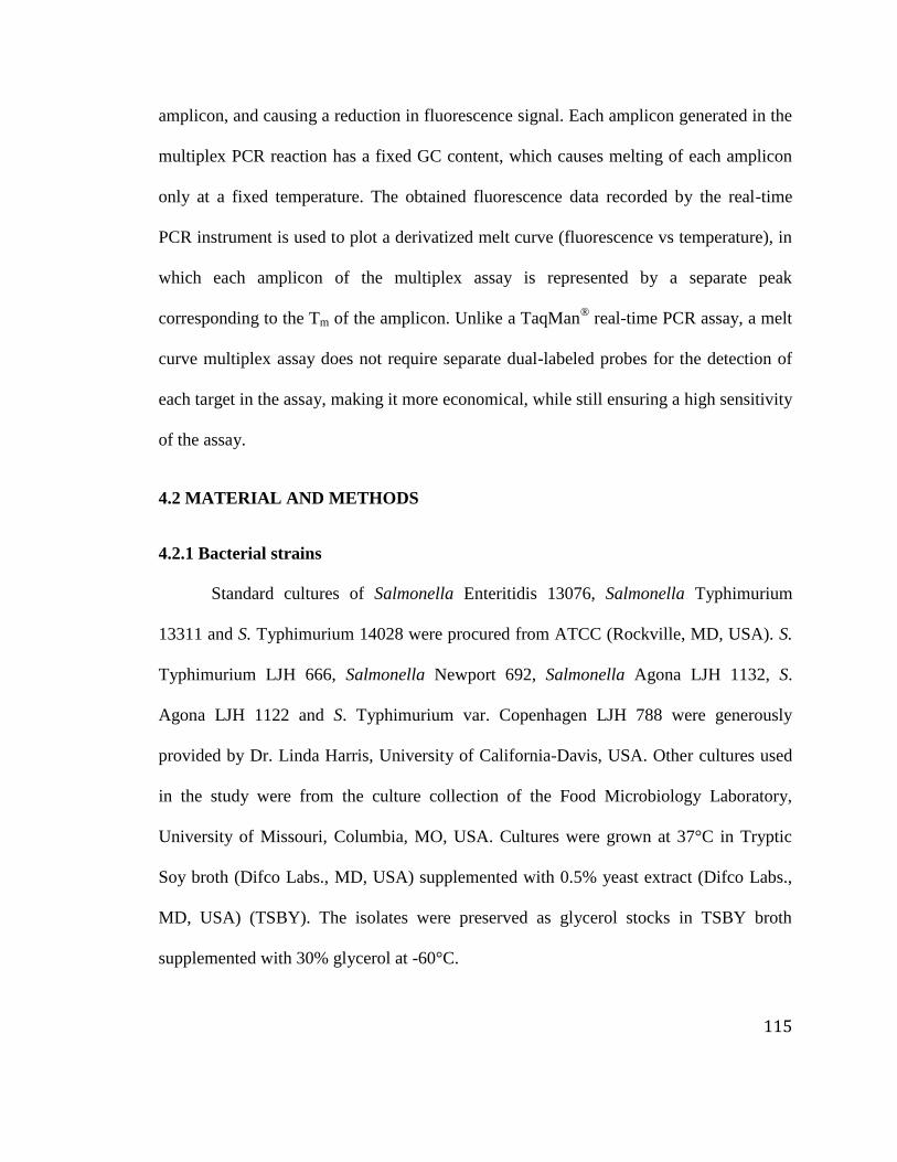
115
amplicon, and causing a reduction in fluorescence signal. Each amplicon generated in the
multiplex PCR reaction has a fixed GC content, which causes melting of each amplicon
only at a fixed temperature. The obtained fluorescence data recorded by the real-time
PCR instrument is used to plot a derivatized melt curve (fluorescence vs temperature), in
which each amplicon of the multiplex assay is represented by a separate peak
corresponding to the Tm of the amplicon. Unlike a TaqMan® real-time PCR assay, a melt
curve multiplex assay does not require separate dual-labeled probes for the detection of
each target in the assay, making it more economical, while still ensuring a high sensitivity
of the assay.
4.2 MATERIAL AND METHODS
4.2.1 Bacterial strains
Standard cultures of Salmonella Enteritidis 13076, Salmonella Typhimurium
13311 and S. Typhimurium 14028 were procured from ATCC (Rockville, MD, USA). S.
Typhimurium LJH 666, Salmonella Newport 692, Salmonella Agona LJH 1132, S.
Agona LJH 1122 and S. Typhimurium var. Copenhagen LJH 788 were generously
provided by Dr. Linda Harris, University of California-Davis, USA. Other cultures used
in the study were from the culture collection of the Food Microbiology Laboratory,
University of Missouri, Columbia, MO, USA. Cultures were grown at 37°C in Tryptic
Soy broth (Difco Labs., MD, USA) supplemented with 0.5% yeast extract (Difco Labs.,
MD, USA) (TSBY). The isolates were preserved as glycerol stocks in TSBY broth
supplemented with 30% glycerol at -60°C.

116
4.2.2 Identification of antibiotic resistance and antibiotic resistance genes
All cultures of Salmonella were screened for antibiotic resistance phenotypes by
the standard agar disk diffusion assay on Mueller Hinton agar according to the criteria
suggested by the Clinical and Laboratory Standards Institute (CLSI) (2012) with
Escherichia coli ATCC 25922 and Staphylococcus aureus ATCC 25923 as quality
control strains. Isolates were further tested for the presence of ampicillin,
chloramphenicol, streptomycin, tetracycline and sulphonamide resistance genes by
conventional PCR, as described by Chiu and others (2006).
4.2.3 Bacterial DNA extraction
Genomic DNA from all bacterial isolates was isolated from actively growing cells
in TSBY broth. One milliliter of an overnight culture was centrifuged at 16,100 ×g for 3
min and the obtained cell pellet was resuspended in 100 µL of PrepMan®
Ultra Sample
Preparation Reagent (Applied Biosystems, Foster City, CA, USA). Samples were heated
at 100 °C for 10 min in a dry bath, to initiate cell lysis and release of bacterial genomic
DNA. Samples were later removed from the dry bath, cooled at room temperature for 2
min and centrifuged at 13,200 ×g (2 min) to sediment the cell debris. Fifty microliters of
the supernatant containing the genomic DNA was transferred to a fresh microcentrifuge
tube. The concentration and purity of the obtained DNA were determined using a
Nanodrop 2000 spectrophotometer (Thermo Fisher Scientific, Wilmington, DE, USA).
4.2.4 Primer design
Specific primers for amplification of virulence and antibiotic resistance genes
were designed using the Primer3 software (Untergasser and others 2012) (Table 4.1) and

117
commercially synthesized (IDT, Coraville, IA, USA). The primers were designed such as
to keep the melting temperature (Tm) of the PCR amplicons between 70 °C to 87 °C and
each amplicon Tm separated by approximately 3 °C from the melting peak of the
neighboring amplicon. The specificity of the designed primers was tested using the NCBI
Primer BLAST tool. The Tm of all amplicons was calculated using the BioEdit software
(Hall 1999). The multiplex real-time melt curve assay was designed as two sets based on
the compatibility of primers in the multiplex PCR reaction and the location occupied by
each amplicon peak on the melt curve plot to avoid any overlapping of peaks. The first
multiplex set amplified the Salmonella invasion virulence gene (invA), with three other
commonly found antibiotic resistances genes, viz. tetracycline (tetG), streptomycin
(aadA2) and sulphonamide (sulI); while the second multiplex reaction included primers
targeting the ampicillin (blaPSE and blaTEM) and chloramphenicol (floR) resistance genes.

118
Table 4.1 Primers used for real-time PCR melt curve assay.
Primer Primer sequence (5’-3’) Target
gene
Product size
(bp)
Primer
Conc.
(µM)
References
Str F CAGCCA(T/C)GATCGACATTGATCT aadA2 66 0.16 Singh and Mustapha 2013
Str R CCAAGGCAACGCTATGTTCTC
Tet F GTTCTGTTACGGCTGGATGATG tetG 66 0.22 This study
Tet R AATAATGAAGAAAACTGCGAAAAGC
Chl F GGCAGGCGATATTCATTACT floR 197 0.12 Singh and Mustapha 2013
Chl R CGAGAAGAAGACGAAGAAGG
Amp F GATTTGGTGCTCGGAGTATT blaPSE 92 0.12 Singh and Mustapha 2013
Amp R CATTGAAGCCTGTGTTTGAG
Inv A1 GTGAAATTATCGCCACGTTCGGGCAA invA 284 0.15 Rahn and others 1992
Inv A2 TCATCGCACCGTCAAAGGAAC
Sul I-F CGGATCAGACGTCGTGGATGT sulI 351 0.16 Chiu and others 2006
Sul I-R TCGAAGAACCGCACAATCTCGT
blaTEM-1 CAGCGGTAAGATCCTTGAGA blaTEM 323 0.15 Chen and others 2004
blaTEM-R TTACATGATCCCCCATGTTG This study

119
4.2.5 Real-time PCR
In this study, we evaluated the suitability of two real-time PCR master mixes
(Applied Biosystems, Foster City, CA, USA) for the development of a multiplex assay:
(1) SYBR® Green I-based Power SYBR
® Green master mix, and (2) SYTO9-based
MeltDoctor™
HRM master mix. PCR amplification was performed in duplicate in a 20
µL reaction volume using 2× of each master mixes with 20 ng of genomic DNA, using
the primer concentrations mentioned in Table 4.1. A StepOnePlus™ real-time PCR
(Applied Biosystems, Foster City, CA, USA) instrument was utilized. A two-step
amplification protocol included initial denaturation at 95 °C for 10 min; 40 cycles of 95
°C for 15 s, 60 °C for 45 s; and a melt curve step at the end of the PCR (from 60 °C to 95
°C, with gradual temperature increments of 0.1 °C/s). A melt curve plot was prepared by
plotting the negative derivative of fluorescence (–Rn) versus temperature. Fluorescence
signal for the MeltDoctor™
HRM master mix was detected in the FAM channel of the
real-time PCR instrument. The mean Tm values for each product was calculated by
averaging the Tm values obtained after amplification using the MeltDoctor™
HRM or
Power SYBR® Green master mix in uniplex reactions, respectively.
4.2.6 Sensitivity of the real-time PCR assay
To determine the sensitivity of the developed multiplex assay, DNA from MDR
isolate S. Typhimurium var. Copenhagen LJH 788 was isolated using PrepMan® Ultra
Sample Preparation Reagent (Applied Biosystems, Foster City, CA, USA) according to
the manufacturer’s instructions. Stock DNA sample was standardized to 10 ng/µL and
ten-fold serially diluted from 10 ng/µL to 0.1 fg/µL of DNA. Two microliters of each

120
serially diluted DNA, in triplicate, was used in the reactions for the construction of a
standard curve. Microsoft Excel software was used to compute regression coefficients
and standard deviations for the standard curves.
To further validate the sensitivity of the assay, an overnight culture of S.
Typhimurium var. Copenhagen LJH 788 was serially diluted in peptone water (1.0 g/L)
and enumerated using Tryptic Soy Agar (Difco Labs., MD, USA). DNA was isolated
from 1 mL of each serially diluted peptone water tube using PrepMan®
Ultra Sample
Preparation Reagent. Real-time PCR was performed using 2 µL genomic DNA isolated
from each dilution tube.
4.2.7 Preparation of artificially spiked food samples
Ground beef of different fat contents (73% lean/27% fat, 80% lean/20% fat, 85%
lean/15% fat and 93% lean/7% fat), whole chicken, ground chicken, egg, spinach, apple
cider, green peppers and tomato were purchased from a local supermarket. These food
products were tested for the presence of Salmonella using standard culture-based
protocols (Andrews and others 2007). Chicken rinse was prepared as previously
described (Wang and Mustapha 2010). Twenty-five grams of food sample was diluted in
225 mL buffered peptone water (BPW) (Remel, Lenexa, KS, USA) and inoculated with
MDR isolate S. Typhimurium var. Copenhagen LJH 788 at different levels, starting from
10 CFU/g to 105
CFU/g. Positive and negative process controls were also included in this
study as described by Malorny and others (2007)a. The negative process control consisted
of sterilized enrichment media with the food sample and was enriched for 8 h. The
positive process control comprised of enrichment media, food sample and S.

121
Typhimurium var. Copenhagen LJH 788 inoculated at 10 CFU/g food sample.
Enrichment of spiked samples was performed in sterile filter stomacher bags
(Fisherbrand, Houston, Texas, USA) containing BPW (Remel, Lenexa, KS, USA) as the
enrichment medium in a shaking incubator at 35 ºC. The DNA from spiked food samples
was isolated from 2 mL of enriched broth withdrawn at intervals of 0, 4, 6 and 8 h.
Samples were centrifuged at 0.1 ×g for 1 min to separate suspended food particles from
the media, and DNA was isolated from the obtained supernatant using PrepMan® Ultra
Sample Preparation Reagent according to the manufacturer’s instructions.
4.3 RESULTS
4.3.1 Antibiotic resistance of Salmonella strains
Standard agar disk diffusion assay results showed that S. Typhimurium strains
14028 and 13311 were resistant to streptomycin, tetracycline and sulphonamide, and S.
Enteritidis 13076 was found to be sensitive to all antibiotics, except sulphonamide (Table
2). S. Typhimurium var. Copenhagen var. LJH 788 was found to be resistant to all five
antibiotics used in this study (Table 4.2). Thus, it was considered to be a MDR isolate
and was subsequently used for standardization of the melt curve multiplex real-time PCR
for simultaneous detection of virulent and antibiotic resistant Salmonella.
All isolates used to validate this assay was also tested for the presence of the
respective antibiotic resistance genes by conventional PCR. The antibiotic resistance
profiles of all Salmonella isolates used in this study are listed in Table 4.2 as presence or
absence of the respective antibiotic resistance gene. Results of the agar disk diffusion
assay were mostly consistent with the genetic profile of antibiotic resistance genes of the

122
Salmonella isolates as determined by PCR. However, there were some instances where
the phenotypic and genotypic traits did not match. One of the reasons for this observed
variation may be due to the extensive diversity of antibiotic resistance genes found in
members of the Enterobacteriaceae family. The other reason for the observed variation in
phenotypic traits was the fact that some strains may have lost their ability to express
resistance phenotype during storage, as was found when compared with our previous
publication (Singh and Mustapha 2013). Similar findings where some antibiotic
resistance genes were present in the bacterial genome but were not phenotypically
expressed were also reported by Perreten and others (2005).

123
Table 4.2 Phenotypic and genotypic antibiotic resistance profile of Salmonella isolates.
Isolate STRa aadA2 TET
b tetG CHL
c floR AMP
d blaPSE blaTEM Sulfisoxazole sulI
S. Typhimurium 13311 R + R + S - S - - R +
S. Typhimurium 14028 R + R + S - S - - R +
S. Enteritidis 13076 S - S - S - S - - R +
S. Typhimurium 788 R + R + R + R + - R +
S. Typhimurium LJH 666 R + S - S - S - - R +
S. Newport LJH 692 R + R + S - S - - R +
S. Agona LJH 1122 R + R + S - S - - R +
S. Agona LJH 1132 R + R + S - S - - R +
S. Berta D(O), G comp H S - S - S - S - - R +
S. Drypool E2(O), G comp H S - S - S - S - - R +
S. Rubislaw S - S - S - S - - R +
S. Group B(4,5) S - S - S - S - - S +
Salmonella Spp S - S - S - S - - R +
12
3

124
S. Arizona S + S - S - S - - R +
S. Thompson B&B3 R + R + R + R - + R +
S. Typhimurium 13311 S - S - S - S - - R +
S. Pulloram 13036 S - S - S - S - - R +
Salmonella API 670752 S - S - S - S - - R +
S. Seftenberg O Group E S - S - S - S - - R +
S. Enteritidis F3-1 R + S + S - S - - R +
S. Enteritidis F3-2 R + R + S - S - - R +
S. Enteritidis F3-6 R + R + S - S - - R +
S. Enteritidis F3-7 R + R + S - S - - R +
S. Enteritidis F3-8 R + S + S - S - - R +
S. Enteritidis F3-9 R + S + S - S - - R +
S. Enteritidis F3-10 R + R + S - S - - R +
S. Enteritidis G5-02 R + S + S - S + - R +
S. Enteritidis G5-08 R + S + R + S - - R +
12
4

125
S. Enteritidis G5-09 R + R + S - S - - R +
S. Enteritidis G5-10 R + S + S - S - - R +
S. Enteritidis I4-1 R + S - S - S - - R +
S. Enteritidis I4-2 R + R + R - S - - S +
S. Enteritidis I4-3 R + R + S - S - - S +
S. Enteritidis I4-4 R + S + R - S + - R +
S. Enteritidis I4-5 R + S + S - S + - R +
S. Enteritidis I4-6 R + R + S - S - - R +
S. Enteritidis I4-7 R + R + S - S - - R +
S. Enteritidis I4-8 R + R + S - S - - R +
S. Enteritidis I4-9 R + R - S - S - - R +
S. Enteritidis I4-10 R + S - R - S - - R +
S. Enteritidis I4-11 R + R + S - S - - R +
a Streptomycin,
b Tetracycline,
c Chloramphenicol,
d Ampicillin
12
5

126
4.3.2 Real-time PCR
The suitability of two real-time PCR master mixes (SYBR® Green I and
MeltDoctor™
HRM) was evaluated for the detection of MDR Salmonella. The average
melting temperatures (Tm) of all amplicons generated from the targeted genes are
outlined in Table 4.3. In the developed multiplex assay, SYBR®
Green I showed
preferential binding to the smaller amplicons generated in the reaction. The melt curve of
the tetraplex assay using this dye generated only two peaks for the streptomycin (66 bp)
and tetracycline (66 bp) resistance genes (Figure 4.1A). This preferential binding to
smaller amplicons was expected because of the manner in which SYBR® Green
intercalates to the minor-groove region of the DNA. Therefore, the dye was not further
investigated for the development of the assay. The MeltDoctor™
HRM master mix,
however, showed no preferential binding and generated all four peaks in the same assay
(Figure 4.1B). The SYTO9 dye in the MeltDoctor™
HRM master mix also produced a
melt curve with a higher resolution. For the second set of reactions, all targets gave
defined peaks irrespective of the master mix used (Figure 4.1C). Because the melt curve
step was performed at the slowest ramp rate (gradual temperature increments of 0.1 °C/s),
the melt curve peaks obtained using both master mixes were equally sharp.

127
Table 4.3 Melting temperature (Tm) of the amplicons.
Target Mean Tm (oC)
SG I SYTO9
blaPse 78.28 ± 0.10 78.62 ± 0.00
floR 83.21 ± 0.00 83.41 ± 0.20
invA 84.11 ± 0.40 84.37 ± 0.30
aadA2 74.80 ± 0.30 75.47 ± 0.21
sulI 86.95 ± 0.07 87.37 ± 0.03
tetG 77.96 ± 0.20 78.86 ± 0.48
blaTEM 81.91 ± 0.53 81.96 ± 0.16

128
Figure 4.1 Multiplex assay for the detection of virulent and antibiotic resistant Salmonella.: A) streptomycin (aadA2), tetracycline
(tetG), invasin (invA), sulphonamide (sulI) multiplex using SYBR® Green I dye-based master mix; B) aadA2, tetG, invA, sulI
multiplex using SYTO9-based MeltDoctor™ master mix; C) ampicillin (blaPSE and blaTEM) and chloramphenicol (floR) resistance
gene multiplex using MeltDoctor™ master mix.
aadA2
tetG
aadA2
tetG InvA SulI
blaPSE
blaTEM
floR
A B C

129
4.3.3 Sensitivity of the assay
The multiplex assays using the MeltDoctor™
HRM master mix developed in this
study was found to be working efficiently over a DNA concentration range of 20 ng to
200 fg (Figure 4.2A, 4.2B). The obtained standard curve for each targeted gene with its
respective regression coefficient (r2 value) is represented in Figure 4.3. At 20 fg (4
genomic equivalent) DNA per reaction, only aadA2, sulI and blaPSE genes were detected.
The starting cell concentration of Salmonella broth culture was 2.9 × 10⁹ CFU/mL. DNA
isolated from this decimally diluted broth culture was used to validate the sensitivity of
the assay, and a detection limit of 290 CFU/mL was obtained for both the tetraplex and
triplex assays, viz. all targets were successfully detected at this cell concentration.
Figure 4.2 Sensitivity of multiplex assay: DNA concentrations ranging from 20 ng to
200 fg using MeltDoctor™ master mix. A) sensitivity of streptomycin (aadA2),
tetracycline (tetG), invasin (invA), and sulphonamide (sulI) resistance gene multiplex; B)
aadA2tetG
InvA
SulIblaPSE
blaTEM
floR
BA

130
sensitivity of ampicillin (blaPSE and blaTEM) and chloramphenicol (floR) resistance gene
multiplex.
Figure 4.3 Standard curve constructed using serially diluted DNA: streptomycin (aadA2,
R2 =0.9954), tetracycline (tetG, R
2 =0.9979), invasion (invA, R
2 =0.9837), sulphonamide
(sulI, R2 =0.9866), ampicillin (blaPSE, R
2 =0.9798), chloramphenicol (floR, R
2 =0.9928)
and ampicillin (blaTEM, R2 =0.9976).
4.3.4 Detection of antibiotic resistant Salmonella in spiked samples
Using standard culture methods, all food samples, except ground turkey, were
found to be negative for Salmonella. These Salmonella-negative samples were artificially
inoculated with MDR S. Typhimurium var. Copenhagen LJH 788. The developed
multiplex assay in this study was able to detect as low as 104 CFU/g in the food samples
without an enrichment step (Table 4.4). Following an enrichment step, DNA isolated
from enriched samples was directly used for real-time PCR. Initially, no amplification
15
20
25
30
35
40
45
200pg 20pg 2pg 200fg 20fg
Ct
DNA concentration/reaction
aadA2
tetG
invA
sulI
blaPSE
floR
blaTEM

131
was observed from these DNA samples. The concentration of the DNA samples
following enrichment ranged between 470 to 923 ng/µL, which is too high for accurate
detection by real-time PCR. This high concentration of DNA might be the reason for our
initial PCR detection failure. When the DNA was diluted (1:2) with nuclease-free water,
the real-time PCR could detect all targets of the multiplex after an enrichment period of 6
h (Table 4.4).
Table 4.4 Multiplex real-time PCR results of artificially contaminated food samples.
Food Samples Spiked
Salmonella
concentration
(CFU/g)
Real-time PCR result after
enrichment
0 h 4 h 6 h 8 h
Ground beef (73% lean) 10 - - + +
102 - - + +
103 - - + +
104 + + + +
105 + + + +
Ground beef (80% lean) 10 - - + +
102 - - + +
103 - - + +
104 + + + +
105 + + + +
Ground beef (85% lean) 10 - - + +
102 - - + +
103 - - + +
104 + + + +
105 + + + +
Ground beef (93% lean) 10 - - + +
102 - - + +
103 - - + +
104 + + + +
105 + + + +

132
Whole chicken 10 - - + +
102 - - + +
103 - - + +
104 + + + +
105 + + + +
Ground chicken 10 - - + +
102 - - + +
103 - - + +
104 + + + +
105 + + + +
Egg 10 - - + +
102 - - + +
103 - - + +
104 + + + +
105 + + + +
Spinach 10 - - + +
102 - - + +
103 - - + +
104 + + + +
105 + + + +
Apple cider 10 - - + +
102 - - + +
103 - - + +
104 + + + +
105 + + + +
Green pepper 10 - - + +
102 - - + +
103 - - + +
104 + + + +
105 + + + +
Tomato 10 - - + +
102 - - + +
103 - - + +
104 + + + +
105 + + + +
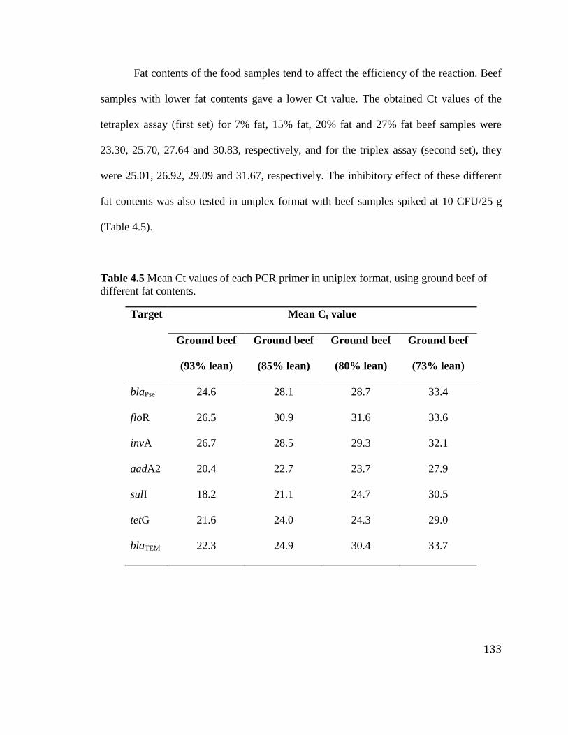
133
Fat contents of the food samples tend to affect the efficiency of the reaction. Beef
samples with lower fat contents gave a lower Ct value. The obtained Ct values of the
tetraplex assay (first set) for 7% fat, 15% fat, 20% fat and 27% fat beef samples were
23.30, 25.70, 27.64 and 30.83, respectively, and for the triplex assay (second set), they
were 25.01, 26.92, 29.09 and 31.67, respectively. The inhibitory effect of these different
fat contents was also tested in uniplex format with beef samples spiked at 10 CFU/25 g
(Table 4.5).
Table 4.5 Mean Ct values of each PCR primer in uniplex format, using ground beef of
different fat contents.
Target Mean Ct value
Ground beef
(93% lean)
Ground beef
(85% lean)
Ground beef
(80% lean)
Ground beef
(73% lean)
blaPse 24.6 28.1 28.7 33.4
floR 26.5 30.9 31.6 33.6
invA 26.7 28.5 29.3 32.1
aadA2 20.4 22.7 23.7 27.9
sulI 18.2 21.1 24.7 30.5
tetG 21.6 24.0 24.3 29.0
blaTEM 22.3 24.9 30.4 33.7

134
4.4 DISCUSSION
In recent years, melt curve-based real-time PCR assays have emerged as a
powerful tool for pathogen detection. In this study, we developed a multiplex real-time
PCR assay for the detection of pathogenic as well as antibiotic resistant strains of
Salmonella using melt curve technology. Based on previously published literature, the
invA gene was selected as the marker for virulent strains of Salmonella. This gene is
located on the Salmonella pathogenicity island 1 (SPI1) and it encodes a protein for the
type III secretion system. The gene, invA, encodes a 71 kDa putative inner membrane
protein and plays an important role in the internalization of Salmonella into the
mammalian epithelial cells. The absence of this gene in Salmonella indicates that the
strain is either non-invasive or the strain possesses an alternate mechanism for invading
mammalian cells (Galán and Curtiss 1991). The invA primer set used in this study had
previously been tested on 630 Salmonella strains and 142 non-Salmonella strains with a
99.4% specificity (Rahn and others 1992). The assay developed in this study showed a
sensitivity of 200 fg/reaction (40 genomic equivalents) for the DNA sample obtained
from pure cultures and a sensitivity of 10 CFU/g after a 6 h enrichment period for spiked
food samples. A duplex melt curve assay for the simultaneous detection of Salmonella
and Listeria was reported with a detection limit of 1 log CFU/mL (Singh and others
2012). Another study (Nam and others 2005) reported a uniplex melt curve assay for the
detection of Salmonella with a detection limit of 10 CFU/mL. Even though the assay
developed in our study is a multiplex one, the sensitivity of the assay is comparable to

135
that reported by Nam and others (2005) for the detection of Salmonella in artificially
contaminated food samples.
Wang and Mustapha (2010) reported a duplex TaqMan®
assay for detection of
live Salmonella with a sensitivity of 100 CFU/mL and 1000 CFU/mL in uniplex and
multiplex format, respectively. Anderson and others (2011), Hein and others (2006) and
Malorny and others (2007b) reported 5′ nuclease assays with a detection limit of 10
genomic equivalents. The Salmonella genomic equivalent can be calculated using the
formula: m × n (1.013 × 10 -21
g/bp), where “m” is the mass and “n” is the number of
base pairs in the Salmonella genome. McClelland and other (2001) reported the genome
size of S. Typhimurium 4951 kilobase pairs. Using the aforementioned equation, the
weight of one S. Typhimurium double stranded genomic DNA is about 5.0 fg. Thus, one
genomic equivalent of pure Salmonella DNA is equal to 5.0 fg of Salmonella genomic
DNA (McClelland and others 2001). In this present melt curve PCR study a detection
limit of 40 genomic equivalents was achieved. This detection limit is also comparable
with the abovementioned 5′ nuclease assays. However, it demonstrated a lower
sensitivity when compared to the assays described by Hein and others (2006) and
Anderson and others (2011), which could be attributed to the higher number of primers
used.
Food samples artificially contaminated with Salmonella concentrations lower than
4 log CFU/mL could not be detected without enrichment, thus necessitating the addition
of an enrichment step. For all the artificially contaminated food samples tested in this
study, a detection limit of 10 CFU/g was achieved after an enrichment period of 6 h,

136
which is a much shorter time than the 16-20 h enrichment periods reported by Malorny
and others (2007b), Hein and others (2006) and Anderson and others (2011).
Food samples contain many organic and inorganic substances, such as phenolic
compounds, fat, enzymes, polysaccharides, proteins and salts which can either inhibit
PCR amplification or may lead to a reduction in amplification efficiency of the reaction
(Demeke and Jenkins 2010; Španová and others 2000). However, all these PCR inhibitors
cannot be detected spectrophotometrically (A280), which is commonly done to check the
purity of isolated DNA samples (Rossen and others 1992). Ground beef samples have
higher fat and protein concentrations that affect the quality of the isolated DNA and
which, in turn, affect the PCR amplification efficiency. In this study, an increase in Ct
values was observed with an increase in fat content of beef samples. The increasing fat
content seems to exert some inhibitory effect on the amplification of the reaction. The use
of filter stomacher bags and low temperature centrifugation in this study facilitated
separation of the fat from enriched broth samples during the DNA isolation process.
Other measures that could also be taken to reduce the level of these PCR inhibitors in the
DNA samples might include a low speed (0.1 ×g for 1 min) centrifugation step to
sediment large fat and protein particles, coupled with the use of a sterile cotton swab to
physically remove the fat in enriched broth sample that tends to stick on the wall of the
tubes. A recent study conducted by the USDA (Bainet and others 2014), reported that
most DNA samples obtained using PrepMan Ultra Sample Preparation Reagent (Applied
Biosystems, Foster City, CA, USA) were not very pure and harbored PCR inhibitors,
effecting outcome of the multiplex PCR reaction. A DYNAL®
Magnetic Bead based

137
DNA purification kit (e.g. DYNAL DNA Direct, Dynabeads MyOne SILANE) or a silica
column based sample preparation kit (e.g. Genomic DNA Clean & Concentrator™,
PureLink® Genomic DNA Kit,) can also be employed for the removal of these impurities
from the DNA obtained from enriched food samples.
Although it is commonly accepted that probe-based real-time PCR assays have a
higher specificity than melt curve real-time PCR assays. However, it can be debated,
specificity of a hybridizing probe-based assay is bestowed by the specificity of the
primer-pair as well as the probe. On the other hand, in melt curve real-time PCR assays,
the PCR reaction specificity is conferred by the use of a specific primer pair and a unique
melting peak generated by the amplicons during the melt-curve analysis. It is very rare
that a non-specific amplicon generated in real-time PCR reaction can produce a melt-
curve peak similar to that of a target amplicon. Even in some cases, a high-resolution
melt (HRM) analysis can be performed to resolve the ambiguity of the melting curve
peaks, without additional costs. The hybridizing probe-based assays are commonly used
for pathogen detection but these assays have a higher running cost. Probe-based real-time
chemistry requires licensed softwares for probe design, multiple probes for multiplexing,
and calibration of multiple detectors in the real-time PCR instrument. Further, the probe
itself has a limited storage life. Thus, intercalating dyes, such as SYBR® Green I have
become the preferred alternative for performing real-time PCR and are being increasingly
used for bacterial identification (Tong and Giffard 2012). However, the SYBR® Green I
dye also has some limitations. It has been reported that this dye can inhibit PCR reactions
in a concentration-dependent manner (Nath and others 2000). The degradation product of

138
this dye has also been reported to be inhibitory for the PCR reaction (Karsai and others,
2002). Another crucial drawback of this dye is its limited application in multiplex real-
time PCR. It has been reported to show preferential binding to PCR products with higher
Tm or higher GC content in a multiplex reaction, thus allowing for the detection of only a
limited number of targets in a multiplex reaction format (Giglio and others 2003). Unlike
other dyes like SYTO9, SYBR® Green I binds to the minor groove region of DNA
(Dragan and others 2012), and is reported to exhibit PCR inhibitory effect at higher dye
concentrations. SYBR® Green I also suffers from the dye-jumping phenomenon whereby,
upon melting of a bound double stranded DNA, it may be reincorporated into regions of
the DNA that have not yet melted (Reed and others 2007). Hence, the resolution of a melt
curve obtained using the SYBR® Green I dye is inferior when compared with a saturating
fluorescent dye. SYTO9 dye is a saturating dye, which can be used at a broad range of
dye concentrations without inhibiting the PCR reaction. This dye also does not show any
preferential binding of PCR amplicons and, thus, is more suitable for multiplex melt
curve real-time PCR assays (Monis and others 2005).
Good primer design, the real-time PCR instrument and the instruments analysis
software all play a crucial role in the development of multiplex melt curve real-time PCR
assays. Out of the various software’s used for the estimation of melting temperatures of
PCR products (BioEdit, Oligo Calc: Oligonucleotide Properties Calculator, Primer
Express Version 2.0, Primer3) for the development of this melt curve multiplex assay,
BioEdit Sequence Alignment Editor (Hall 1999) was found to be the most accurate in
predicting the melt temperatures of the amplicons (data not shown).

139
Initial attempts were made to standardize this assay on the ABI 7500 platform
with SDS software version 1.4 (Applied Biosystems, Foster City, CA, USA). However,
only two peaks were seen for the tetraplex assay using both the master mixes. The SDS
software 1.3 has a fixed ramp rate for the melt curve step, which cannot be changed. The
StepOnePlus™ Software v2.1 (Applied Biosystems, Foster City, CA, USA) allows for
manually changing the default ramp rate during the melt curve step (from 0.3 °C/s to 0.1
°C/s), leading to a much slower rate of melting of the PCR amplicons and generation of
data with a higher resolution.
While traditional culture-based methods are still the gold standard, they require 3-
5 days for the confirmation of Salmonella in food samples. Samples containing
antibiotics, such as animal feed or blood drawn after administration of antibiotics, cannot
reliably be tested with culture-based methods because the presence of antibiotics will
inhibit the growth of bacteria (Jeng and others 2012). The current assay with an
enrichment step of 6 h, followed by 4 h for DNA isolation and real-time PCR enables
rapid detection of MDR and virulent Salmonella in 10 h. This process can be further
shortened by using fast real-time PCR machine (e.g. 7500 Fast real-time PCR instrument)
that allows for a faster DNA amplification and melt curve analysis. Another advantage of
this melt curve assay is that it does not require any post-PCR processing, such as gel
electrophoresis or DNA sequencing, which, in turn, helps to save time and to avoid post-
PCR contaminations. The assay can also be used in a clinical setting as an adjunctive test
to provide physicians with an early detection tool of pathogens causing bacteremia.

140
Apart from the above-mentioned advantages, there are also a few limitations of
genotype- based assays. A limitation of the method described in this study is that out of a
vast number of antibiotic resistance genes found in members of the Enterobacteriaceae
family, the assay only targets the most frequently reported antibiotic resistance genes.
Hence, Salmonella isolates tested negative by this assay might be positive for other less
frequently occurring antibiotic resistance genes. Another common drawback of all
genotypic methods is that they cannot differentiate the origin of genetic markers in
environmental samples, including food, which might lead to false positive results.
The melting profile of the amplicons generated in a melt curve- based real-time
PCR assay is dependent on the quality of the initial DNA template used for the reaction
(Ajitkumar and others 2012). DNA isolated from samples containing PCR inhibitors or
high salt contents might lead to a Tm shift or reduction in efficiency of the PCR
amplification. Therefore, the inclusion of a positive process control is a very important
component of melt curve assays. Positive and negative process controls were included in
this study, as described by Malorny and others (2007)a. The negative process control
consisted of sterilized enrichment media containing the food sample, which was enriched
for 8 h. The positive process control comprised of enrichment media and food sample
inoculated with 10 CFU/g of S. Typhimurium var. Copenhagen LJH 788. The method
described in this study targeted more number of antibiotic resistance genes, required a
shorter enrichment time, and was also more cost effective than our previously published
TaqMan®
assay for the detection of antibiotic and virulence strains of Salmonella (Singh
and Mustapha 2013).

141
4.5 CONCLUSIONS
The multiplex melt curve real-time PCR assay described in this study offers a
simple work flow with a total turn-around time of 10 h for the detection of MDR and
virulent strains of Salmonella from food samples with an initial count of 10 CFU/g. This
multiplex assay can be a useful tool for epidemiologic, laboratory, and traceback
investigations of tainted food samples during an outbreak caused by MDR Salmonella.
Further, this assay is an economical alternative to the hybridization probe-based real-time
PCR assay, which is currently more widely used for the detection of Salmonella.

142
REFERENCES
Aarestrup FM, Wegener HC, Collignon P. 2008. Resistance in bacteria of the food chain:
epidemiology and control strategies. Expert Rev Anti Infect Ther 6(5):733-50.
Agarwal A, Makker A, Goel S. 2002. Application of the PCR technique for a rapid,
specific and sensitive detection of Salmonella spp. in foods. Mol Cell Probes 16(4):243-
50.
Ajitkumar P, Barkema HW, De Buck J. 2012. Rapid identification of bovine mastitis
pathogens by high-resolution melt analysis of 16S rDNA sequences. Vet Microbiol
155(2):332-40.
Anderson A, Pietsch K, Zucker R, Mayr A, Müller-Hohe E, Messelhäusser U, Sing A,
Busch U, Huber I. 2011. Validation of a duplex real-time PCR for the detection of
Salmonella spp. in different food products. Food Analy Methods 4(3):259-67.
Andrews WH, Jacobson A, Hammack T. 2007. Salmonella. In: G.J. Jackson (Ed.),
Bacteriological Analytical Manual 8th Ed. Revision A Online. U.S. Food and Drug
Administration, Gaithersburg, MD. Available from:
http://www.fda.gov/Food/FoodScienceResearch/ LaboratoryMethods/ucm070149.htm.
Accessed 2014 November 15.
Bertrand S, Dierick K, Heylen K, De Baere T, Pochet B, Robesyn E, Lokietek S, Van
Meervenne E, Imberechts H, De Zutter L, Collard JM. 2010. Lessons learned from the
management of a national outbreak of Salmonella Ohio linked to pork meat processing
and distribution. J Food Prot 73(3):529-34.
Binet R, Deer DM, Uhlfelder SJ. 2014. Rapid detection of Shigella and enteroinvasive
Escherichia coli in produce enrichments by a conventional multiplex PCR assay. Food
Microbiol 40:48-54.
Brenner F, Villar R, Angulo F, Tauxe R, Swaminathan B. 2000. Salmonella
nomenclature. J Clin Microbiol 38(7):2465-7.
Centers for Disease Control and Prevention (CDC). 2009. Preliminary FoodNet data on
the incidence of infection with pathogens transmitted commonly through food-10 states.
Morbidity and Mortality Weekly Report 59:418-22.
Chen S, Zhao S, White DG, Schroeder CM, Lu R, Yang H, McDermott PF, Ayers S,
Meng J. 2004. Characterization of multiple-antimicrobial-resistant Salmonella serovars
isolated from retail meats. Appl Environ Microbiol. 70(1):1-7.
Chiu CH, Su LH, Chu CH, Wang MH, Yeh CM, Weill FX, Chu C. 2006. Detection of
multidrug-resistant Salmonella enterica serovar Typhimurium phage types DT102,
DT104, and U302 by multiplex PCR. J Clin Microbiol 44(7):2354-8.

143
Clinical and Laboratory Standards Institute (CLSI), Clinical and Laboratory Standards
Institute (CLSI). Performance Standards for Antimicrobial Disk Susceptibility Tests;
Approved Standard, Eleventh Edition (M02-A11). Wayne, PA: CLSI; 2012.
de Freitas CG, Santana ÂP, da Silva PHC, Gonçalves VSP, Barros MAF, Torres FAG,
Murata LS, Perecmanis S. 2010. PCR multiplex for detection of Salmonella Enteritidis,
Typhi and Typhimurium and occurrence in poultry meat. Int J Food Microbiol 139(1):15-
22.
Demeke T, Jenkins GR. 2010. Influence of DNA extraction methods, PCR inhibitors and
quantification methods on real-time PCR assay of biotechnology-derived traits. Anal
Bioanal Chem 396(6):1977-90.
Dragan AI, Pavlovic R, McGivney JB, Casas-Finet JR, Bishop ES, Strouse RJ,
Schernerman MA, Geddes CD. 2012. SYBR Green I: fluorescence properties and
interaction with DNA. J Fluoresc 22(4):1189-99.
European Food Safety Authority (EFSA). 2013. The European Union summary report on
trends and sources of zoonoses, zoonotic agents and food-borne outbreaks in 2011. The
EFSA Journal 11:3129.
Galán JE, Curtiss R. 1991. Distribution of the invA,-B,-C, and-D genes of Salmonella
Typhimurium among other Salmonella serovars: invA mutants of Salmonella Typhi are
deficient for entry into mammalian cells. Infect Immun 59(9):2901-8.
Giglio S, Monis PT, Saint CP. 2003. Demonstration of preferential binding of SYBR
Green I to specific DNA fragments in real-time multiplex PCR. Nucleic Acids Res
31(22):e136.
Hall TA. 1999. BioEdit: a user-friendly biological sequence alignment editor and analysis
program for Windows 95/98/NT. Nucleic Acids Symposium Series 41:95-8.
Hein I, Flekna G, Krassnig M, Wagner M. 2006. Real-time PCR for the detection of
Salmonella spp. in food: An alternative approach to a conventional PCR system
suggested by the FOOD-PCR project. J Microbiol Methods 66(3):538-47.
Jeng K, Yang S, Won H, Gaydos CA, Hsieh YH, Kecojevic A, Carroll KC, Hardick J,
Rothman, RE. 2012. Application of a 16S rRNA PCR-high-resolution melt analysis assay
for rapid detection of Salmonella bacteremia. J Clin Microbiol 50(3):1122-24.
Josefsen MH, Krause M, Hansen F, Hoorfar J. 2007. Optimization of a 12-hour TaqMan
PCR-based method for detection of Salmonella bacteria in meat. Appl Environ
Microbiol 73(9):3040-8.

144
Karsai A, Müller S, Platz S, Hauser MT. 2002. Evaluation of a homemade SYBR green I
reaction mixture for real-time PCR quantification of gene expression. Biotechniques
32(4):790-6.
Khan SA, Sung K, Nawaz MS. 2011. Detection of aacA-aphD, qacEδ1, marA, floR, and
tetA genes from multidrug-resistant bacteria: Comparative analysis of real-time multiplex
PCR assays using EvaGreen® and SYBR® Green I dyes. Mol Cell Probes 25(2):78-86.
Koluman A, Dikici A. 2013. Antimicrobial resistance of emerging foodborne pathogens:
Status quo and global trends. Crit Rev Microbiol 39(1):57-69.
Li X, Bethune L, Jia Y, Lovell R, Proescholdt T, Benz S, Schell T, Kaplan G,
McChesney D. 2012. Surveillance of Salmonella prevalence in animal feeds and
characterization of the Salmonella isolates by serotyping and antimicrobial susceptibility.
Foodborne Pathog Dis 9(8):692-8.
Majowicz SE, Musto J, Scallan E, Angulo FJ, Kirk M, O'Brien SJ, Jones TF, Fazil A,
Hoekstra RM. 2010. The global burden of nontyphoidal Salmonella gastroenteritis. Clin
Infect Dis 50(6):882-9.
Malorny B, Mäde D, Teufel P, Berghof-Jäger C, Huber I, Anderson A, Helmuth R.
2007a. Multicenter validation study of two blockcycler and one capillary-based real-time
PCR methods for the detection of Salmonella in milk powder. Int J Food Microbiol
117(2):211-8.
Malorny B, Bunge C, Helmuth R. 2007b. A real-time PCR for the detection of
Salmonella Enteritidis in poultry meat and consumption eggs. J Microbiol Methods
70(2):245-51.
McClelland M, Sanderson KE, Spieth J, Clifton SW, Latreille P, Courtney L, Porwollik
S, Ali J, Dante M, Du F, Hou S, Layman D, Leonard S, Nguyen C, Scott K, Holmes A,
Grewal N, Mulvaney E, Ryan E, Sun H, Florea L, Miller W, Stoneking T, Nhan M,
Waterston R, Wilson RK. 2001. Complete genome sequence of Salmonella enterica
serovar Typhimurium LT2. Nature 413(6858):852-6.
Monis PT, Giglio S, Saint CP. 2005. Comparison of SYTO9 and SYBR Green I for real-
time polymerase chain reaction and investigation of the effect of dye concentration on
amplification and DNA melting curve analysis. Anal Biochem 340(1):24-34.
Nam HM, Srinivasan V, Gillespie BE, Murinda SE, Oliver SP. 2005. Application of
SYBR Green real-time PCR assay for specific detection of Salmonella spp. in dairy farm
environmental samples. Int J Food Microbiol 102(2):161-71.
Nath K, Sarosy JW, Hahn J, Di Como CJ. 2000. Effects of ethidium bromide and SYBR
Green I on different polymerase chain reaction systems. J Biochem Biophys Methods
42(1):15-29.

145
O'Regan E, McCabe E, Burgess C, McGuinness S, Barry T, Duffy G, Whyte P, Fanning
S. 2008. Development of a real-time multiplex PCR assay for the detection of multiple
Salmonella serotypes in chicken samples. BMC Microbiol 8(1):156.
Perreten V, Vorlet-Fawer L, Slickers P, Ehricht R, Kuhnert P, Frey J. 2005. Microarray-
based detection of 90 antibiotic resistance genes of gram-positive bacteria. J Clin
Microbiol 43(5):2291-302.
Podolak R, Enache E, Stone W, Black DG, Elliott PH. 2010. Sources and risk factors for
contamination, survival, persistence, and heat resistance of Salmonella in low-moisture
foods. J Food Prot 73(10):1919-36.
Rahn K, De Grandis SA, Clarke RC, McEwen SA, Galán JE, Ginocchio C, Curtiss Iii R.,
Gyles CL. 1992. Amplification of an invA gene sequence of Salmonella Typhimurium by
polymerase chain reaction as a specific method of detection of Salmonella. Mol Cell
Probes 6(4):271-9.
Rossen L, Nørskov P, Holmstrøm K, Rasmussen OF. 1992. Inhibition of PCR by
components of food samples, microbial diagnostic assays and DNA-extraction solutions.
Int J Food Microbiol 17(1):37-45.
Singh J, Batish VK, Grover S. 2012. Simultaneous detection of Listeria monocytogenes
and Salmonella spp. in dairy products using real time PCR-melt curve analysis. J Food
Sci Technol 49(2):234-9.
Singh P, Mustapha A. 2013. Multiplex TaqMan® detection of pathogenic and multi-drug
resistant Salmonella. Int J Food Microbiol 166(2):213-8.
Sjölund-Karlsson M, Rickert R, Matar C, Pecic G, Howie RL, Joyce K, Medalla F,
Barzilay EJ, Whichard JM. 2010. Salmonella isolates with decreased susceptibility to
extended-spectrum cephalosporins in the United States. Foodborne Pathog Dis
7(12):1503-9.
Španová A, Rittich B, Karpíšková R, Čechová L, Škapova D. 2000. PCR identification of
Salmonella cells in food and stool samples after immunomagnetic separation.
Bioseparation 9(6):379-84.
Tong SYC, Giffard PM. 2012. Microbiological applications of high-resolution melting
analysis. J Clin Microbiol 50(11):3418-21.
Untergasser A, Cutcutache I, Koressaar T, Ye J, Faircloth BC, Remm M, Rozen SG.
2012. Primer3-new capabilities and interfaces. Nucleic Acids Res 40(15):e115
Van der Zee H, Huis in’t Veld JHJ. 2000. Methods for the rapid detection of Salmonella.
In C. Wray, & A. Wray (Eds.), Salmonella in Domestic Animals (pp. 373-391).
Wallingford, UK: CABI Publishing.

146
Wang L, Mustapha A. 2010. EMA real-time PCR as a reliable method for detection of
viable Salmonella in chicken and eggs. J Food Sci 75(3):M134-9.
White PA, McIver CJ, Rawlinson WD. 2001. Integrons and gene cassettes in the
Enterobacteriaceae. Antimicrob Agents Chemother 45(9):2658-61.

147
Chapter 5 Multiplex real-time PCR assay for the Detection of Extended-Spectrum
β-Lactamases and Carbapenemase Producing Genes Using Melting Curve Analysis
ABSTRACT
Antibiotic resistance among Gram-negative pathogens is a matter of great
concern. Out of the different antibiotic groups, resistance towards extended-spectrum β-
lactamase (ESBL) and carbapenem antibiotics in Enterobacteriaceae Family has more
severe consequences and recent surveillance data have shown their increasing trend. The
objective of this study was to develop a real-time PCR melt curve assay for the detection
of extended-spectrum β-lactam and carbapenem resistant pathogens. Two multiplex real-
time PCR melt curve assays were standardized for the detection of 10 most common
ESBL and carbapenem resistance genes: blaKPC, blaNDM, blaCTX, blaCMY, blaVIM, blaIMP,
blaOXA, blaSHV, blaTEM, blaACC. The multiplex was evaluated using a total of 38 DNA
samples obtained from previous studies. The results of the multiplex PCR assay either
mostly paralleled the previous studies or the presence of more antibiotic resistance genes
were detected using our assay. The assay developed in this study offers a simple, low cost
method for the detection of ESBL and carbapenem resistance among enteric pathogens.

148
5.1 INTRODUCTION
Broad-spectrum β-lactamase genes coding for extended-spectrum β-lactamases
and enzymes conferring resistance towards others antibiotics have been frequently
isolated from the microbiota of food-producing animals. Food animals colonized with
strains of ESBL-producing Escherichia coli and Salmonella have been reported as
important sources of community infections. ESBL- and carbapenemase-producing
members of the Enterobacteriaceae have been increasingly isolated from food animals
and have gained considerable attention worldwide (Smet and others 2010). Recent data
from the National Antimicrobial Resistance Monitoring System (NARMS) have shown
an increase in extended-spectrum cephalosporins resistance among Salmonella
Heidelberg isolated from food animals at slaughter, retail meat, and humans (Folster and
others 2012). β-lactams are one of the most important groups of antimicrobial agents used
in veterinary medicine on food animals. These antimicrobial agents can be divided into
three major groups: the penicillins, the first- to fourth-generation cephalosporins and the
β-lactamase inhibitors. Cephalosporins have been approved for treatment of various
diseases in food animals such as foot rot and mastitis in cattle, and respiratory diseases in
ruminants (Smet and others 2010). Additionaly, these antibiotics are also mixed with
animal feed to improve feed efficiency and avoid animal disease in cattle feedlots.
The extensive use of antibiotics for various purposes on animal feedlots exerted
selective pressure leading to evolution and selection of newer beta-lactamase enzymes
conferring resistance to a broad range of antibiotics. Datta and Kontomichalou (1965)
reported the first beta-lactamase enzyme, TEM-1, in Gram-negative bacteria. The SHV-2

149
enzyme was the first enzyme that showed ESBL-hydrolyzing capability and was isolated
from a Klebsiella ozaenae in Germany (Kliebe and others 1985). Because of the
increased spectrum of activity of these beta-lactamase enzymes, especially against the
oxyimino-cephalosporins, these enzymes were called ESBLs (Bradford 2001). Hence,
ESBLs were classified as a class of enzymes that confer resistance against penicillins,
broad-spectrum cephalosporins with an oxyimino side chain (cefotaxime, ceftriaxone and
ceftazidime) and the oxyimino-monobactam aztreonam antibiotics. Another common
property of these ESBLs is that they can be inhibited by serine-type β-lactamase
inhibitors, such as sulbactam, clavulanate and tazobactam (Philippon and others 1989;
Bradford 2001). At present, ESBL enzymes are spread over ten families and have been
documented all around the world e.g. CTX-M, SHV, TEM, PER, VEB, BES, GES, TLA,
SFO and OXA (Paterson and Bonomo 2005). Today, ESBLs are one of the most
influential mechanisms for cephalosporin resistance in Enterobacteriaceae, particularly in
E. coli and K. pneumoniae.
In the family of ESBLs, CTX-Ms are plasmid–mediated enzymes that
preferentially hydrolyze cefotaxime, and are the latest addition that have significant
clinical impacts. This new class of ESBL enzymes is commonly found among the
antibiotic resistant strains of Salmonella and E. coli, but they have also been frequently
reported among other members of the Enterobacteriaceae (Sabaté and others 2000). The
CTX-M enzymes are one of the biggest and most diverse groups of ESBL enzymatic
variants (CTX-M-1 to CTX-M-160) and exhibit strong activity against cefotaxime and
ceftriaxone, but not ceftazidime. The diversity of CTX-M enzymes varies with

150
geographical locations, but CTX-M-15 and CTX-M-14 (followed by CTX-M-2, CTX-M-
3 and CTX-M-1) are the most common enzymatic variants worldwide among all major
clinically important pathogens (Zhao and Hu 2013). Class C chromosomal AmpC
(cAmpC) is a class of β-lactamases that confers resistance towards third-generation
cephalosporins and β-lactamase inhibitors. However, they are susceptible to fourth-
generation cephalosporins, such as cefepime and the carbapenem group of antibiotics
(Bush and Jacoby 2010). There are several families of cAmpC enzymes e.g. CMY, FOX,
LAT, MIR, ACT, DHA, ACC, MOX. Out of these, CMY-2 is the most prevalent pAmpC
among members of the Enterobacteriaceae.
Carbapenemase enzymes are produced by pathogenic bacteria to degrade and
neutralize the effects of almost all beta-lactam antibiotics, including carbapenems
(Cantón and others 2012). Based on the composition of the active site, carbapenemases
enzymes can be broadly classified into two major groups: (1) Serine carbapenemases:
consist of a serine amino-acid in their active site and are subdivided into two classes viz.
class A penicillinases and class D oxacillinases. The class A enzymes are coded by IMI,
NMC, SME, KPC, and GES genes, which confer resistance to the carbapenems group of
antibiotics at various levels, ranging from complete resistance to reduced susceptibility
(Queenan and Bush 2007). On the other hand, OXA-48, OXA-23, OXA-24, and OXA-58
genes are the most dominant genetic determinants encoding Class D oxacillinases
enzyme in A. baumannii and K. pneumoniae. (2) Metallo-betalactamases: also known as
Class B carbapenemases. Enzymes of this group have one or more zinc atoms in their
active site. This group includes the GIM, IMP, NDM, SIM and VIM enzymes that are

151
found on the transferrable plasmids of Enterobacteriaceae members, P. aeruginosa and A.
baumannii (Nordmann and others 2011). Out of the five above-mentioned enzymes; IMP,
NDM and VIM are the most prominent enzymes. NDM is the most recent addition to the
class B carbapenemases group of enzymes. Since the first report of NDM-1 in the year
2009, it has spread worldwide and become the most common carbapenemases enzyme in
all of the Enterobacteriaceae (Rolain and others 2010).
Antimicrobial susceptibility testing for ESBL or carbapenemase producing
Enterobacteriaceae is generally performed using commercially available selective agar
plates. Standard microbiological procedures can take up to several days for the culture,
isolation and characterization of antibiotic resistance profile of pathogens from a
provided sample. The total time required for this process can add up to 4-6 days for
getting an accurate antimicrobial susceptibility test results (Lupo and others 2013).
However, many recent comparative studies have shown that the commercially available
selective agars have lower sensitivity than PCR-based methods (Naas and others 2011;
Singh and others 2012). The objective of this study was to develop a multiplex real-time
melt curve assay for the detection of genes coding for ESBL and carbapenemase
enzymes.
5.2 MATERIALS AND METHODS
5.2.1 Procurement of standard DNA sample and cultures
DNA of antibiotic resistant Salmonella and members of the Enterobacteriaceae was
obtained from Robert Koch Institute, (Berlin, Germany), Calgary Laboratory Services,

152
(Calgary, AB, Canada), Institute of Microbiology, University of Lausanne and University
Hospital Center, (Lausanne, Switzerland), Infectious Diseases, VA Medical Center
(Minneapolis, MN, USA) and University of Missouri Hospital (Columbia, MO, USA).
5.2.2 Bacterial DNA extraction
Genomic DNA from all bacterial isolates used in the study and from enriched
food samples was isolated using PrepMan®
Ultra Sample Preparation Reagent (Applied
Biosystems, Foster City, CA, USA) according to the manufacturer’s instructions. The
concentrations and purity of the obtained DNA samples was determined using a
Nanodrop 2000 spectrophotometer (Thermo Fisher Scientific, Wilmington, DE, USA).
5.2.3 Primer design
A total of 85 primer pairs targeting 10 antibiotic resistance genes (blaKPC, blaNDM,
blaCTX, blaCMY, blaVIM, blaIMP, blaOXA, blaSHV, blaTEM, blaACC) were designed and evaluated
for the standardization of the multiplex real-time PCR melt curve assay. The primers
were designed such as to keep the melting temperature (Tm) of the PCR amplicons
between 67 °C to 87 °C and each amplicon Tm separated from neighboring amplicon Tm
by approximately 2-3 °C. The specificity of the designed PCR primers was tested using
the NCBI/Primer-BLAST (http://www.ncbi.nlm.nih.gov/tools/primer-blast/). The
designed oligonucleotides were commercially synthesized (IDT, Coraville, IA, USA).
PCR amplification conditions of each primer pair were standardized using conventional
gradient PCR. Real-time PCR was performed using MeltDoctor™ HRM master mix
(Applied Biosystems, Foster City, CA, USA) in singleplex format for the estimation of
the Tm of each PCR amplicon. Based on the obtained Tm values of the amplicons in

153
singleplex reaction, multiplex real-time melt curve assay was standardized in a stepwise
manner. PCR primers generating low Tm amplicons (e.g. 70 °C) were tested for their
ability to work in a multiplex reaction with primer-pairs of another gene generating
amplicons of Tm values 2-3 °C higher and subsequently targeting primers with higher
amplicon Tm in a step-wise manner.
5.2.4 Internal amplification control (IAC) design
Two reaction-specific, single-stranded, 75-100 base long DNA sequences were
designed to act as IAC in the multiplex real-time PCR reaction. These IAC molecules
could be amplified using one of the primer pairs of the multiplex assay (but generating a
separate melt curve peak). The IAC concentrations of each multiplex assay was
optimized and kept as low as possible to prevent any possible competition for primers
and facilitating the preferential amplification of the antibiotic resistant genes.
5.2.5 Real-time PCR
All primers generating specific amplicons in the conventional PCR and real-time
PCR reactions were tested for their suitability for the development of the multiplex real-
time melt curve assay. The final primer sets for the assay were selected based on the
following criteria: (a) PCR reaction efficiency: A reaction efficiency around 90% or
higher in singleplex reaction was preferred (b) Tm difference between amplicons: a
minimum of 2-3 °C difference in melting temperature between neighboring peaks was
preferred, this allowed us to achieve a better resolution between melting peaks and avoid
any overlap of melting peak (c) Product size: amplicon size between 51 - 250 bp was
more desirable.

154
The multiplex assay for the detection of ESBL and carbapenemase resistant
pathogens was designed in two sets. The first set included five primer-pairs for the
amplification of blaKPC, blaNDM, blaCTX, blaCMY, blaVIM genes with IAC (Table 5.1). The
second set targeted blaIMP, blaOXA, blaSHV, blaTEM, blaACC antibiotic resistant genes and
IAC (Table 5.2). Real-time PCR assay was performed in 15 μL reaction volume with 2×
MeltDoctor™ HRM master mix (Applied Biosystems, Foster City, CA, USA). A
touchdown PCR amplification protocol was used on LightCycler®
96 real-time PCR
platform (Roche Diagnostics Corporation, Indianapolis, USA). The PCR program for the
amplification consisted for following step: 10 min of initial denaturation step at 95 °C
followed by 40 cycles of denaturation at 95 °C for 10 s, initial annealing at 65 °C for 40 s
(gradual temperature decrement of 0.2 °C per cycle, final annealing temperature 60 °C),
extension and data collection at 72°C for 10 sec. Melt Cure was performed at 0.04 °C/s
(25 reading/ °C).

155
Table 5.1 Primer set for the first multiplex assay.
Oligo Oligo sequence Oligo conc. Product
size (bp)
Amplicon
Tm (°C)
KPC-688-F GTCGGAGACAAAACCGGAAC 0.26 μM 51 77.8
KPC-738-R ATAGTCATTTGCCGTGCCATA 0.26 μM
NDM-310-F TGGATCAAGCAGGAGATCAA 0.26 μM 250 88.4
NDM-559-R GGCCGGGGTAAAATACCTT 0.26 μM
CTX-583-F AATCTGACGCTGGGTAAAG 0.16 μM 140 85.5
CTX-722-R CCGCTGCCGGTTTTATC 0.16 μM
CMY-55-F GTTCAGGAGAAAACGCTCCA 0.2 μM 87 82.2
CMY-141-R CCAGCCTAATCCCTGGTACA 0.2 μM
VIM-215-F TCATTGTCCGTGATGGTGAT 0.26 μM 51 74.4
VIM-265-R ACCCCACGCTGTATCAATCC 0.26 μM
IAC-NDM-pp
catataTGGATCAAGCAGGAGATCAAAT
ATATATATTATCAAAATAAGACTAAT
AAAGAAGGTATTTTACCCCGGCCcatac
10 fg/15 μl 74 69
15
5

156
Table 5.2 Primer set for the second multiplex assay.
Oligo Oligo sequence Oligo conc. Product
size (bp)
Amplicon
Tm (°C)
IMP-311-F TAGAGTGGCTTAATTCTCRATC 0.16 μM 75 77.8
IMP-577-R CTTCTAWATTTGCGTCACCC 0.16 μM
OXA48-63-F AGCAAAGGAATGGCAAGAAA 0.23 μM 65 73.6
OXA48-127-R CGCCCTGTGATTTATGTTCA 0.23 μM
SHV-309-F GGTCAGCGAAAAACAYCTTG 0.4 μM 195 88.2
SHV-503-R GCCTCATTCAGTTCCGTTTC 0.4 μM
TEM-94-F GATACGGGAGGGCTTACCAT 0.2 μM 146 85.0
TEM-239-R GGATGGAGGCGGATAAAGTT 0.2 μM
ACC-238-F GAGCAAATTCGGCAGAGAAA 0.2 μM 115 81.6
ACC-352-R CAAGATGCAACAGGCTCTGA 0.2 μM
IAC-SHV-pp
catataGGTCAGCGAAAAACAYCTTGAT
ATATATATTATCAAAATAAGACTAAT
AAAGGAAACGGAACTGAATGAGGCcatac
10 fg/15 μl 75 70
15
6

157
5.2.6 Validation of the assay
The assay for the detection of ESBL resistant pathogens developed in this study
was validated using DNA samples obtained from four laboratories: Robert Koch Institute
(Berlin, Germany), Calgary Laboratory Services (Calgary, AB, Canada), Institute of
Microbiology, University of Lausanne and University Hospital Center (Lausanne,
Switzerland), and University of Missouri Hospital (MO, USA). A total of 38 strains that
have been previously characterized for ESBL and carbapenem resistance genetic markers
were analyzed using the multiplex assay developed in our study.
5.2.7 In silico analysis of primer pairs used for multiplex assay
The diversity of the family of enzymes conferring antibiotic resistance is very high.
In addition, each family of enzymes comprises of hundreds of allelic variants of the
specific gene. Primer-BLAST (http://www.ncbi.nlm.nih.gov/tools/primer-blast/) was
performed for each of the primer-pairs used in this study to find allelic variants that
possessed similar primer binding sites.
5.3 RESULTS
5.3.1 Multiplex real-time PCR
Two multiplex real-time PCR reactions were developed for the identification of
10 genes, coding for ESBLs and carbapenemase enzymes. The first set targeted blaCTX,
blaCMY, blaVIM, blaKPC, blaNDM and IAC (Figure 5.1). The IAC molecule (IAC-NDM-pp)
in this reaction was amplified using primer pairs, NDM-310-F and NDM-559-R,
generating a separate melting peak of 69 °C. Because two targets (blaNDM and IAC) were

158
simultaneously amplified using one primer pair, the IAC molecule was added at a very
low concentration (10 pg/15 μL reaction) in order to allow preferential amplification of
the target DNA (instead of IAC). As a result, the melt peak formed by the IAC amplicon
was much smaller than the other peaks of the multiplex assay (Figure 5.1A). However, in
the absence of the NDM target or in the non-template control (NTC), a bigger IAC melt
peak was observed (Figure 5.1B). The second multiplex reaction targeted blaIMP, blaOXA,
blaSHV, blaTEM, blaACC and IAC (Figure 5.2A). In this multiplex reaction, the IAC
molecule (IAC-SHV-pp) was co-amplified by SHV-309-F and SHV-503-R primer-pairs
and the IAC amplicon generated a separate melt peak of 70 °C (Figure 5.2B).
Figure 5.1 Multiplex assay for the detection of blaVIM, blaKPC, blaCMY, blaCTX and blaNDM
with IAC (A) blaVIM, blaKPC, blaCMY, blaCTX and blaNDM with IAC. (B) Non-template
control sample showing the IAC melt peak.
VIM KPC
CMY CTX NDM
IAC
A
IAC

159
Figure 5.2: Multiplex assay for the detection blaOXA, blaIMP, blaACC, blaTEM and blaSHV
with IAC (A) blaOXA, blaIMP, blaACC, blaTEM and blaSHV with IAC. (B) Non-template
control sample showing the IAC melt peak.
5.3.2 Validation of the assay
The result of our multiplex PCR assay mostly concurred with earlier reports (Eller
and others 2013; Pfeifer and others 2011; Pfeifer and others 2012; Yong and others 2009;
Peirano and others 2011a; Peirano and others 2011b), but in addition to the previously
reported antibiotic resistant (ABR) determinants (+), our multiplex assay was also able to
detect the presence of a few other ABR genes (+) (Table 5.3). Salmonella enterica
Newport 209/10 and Escherichia coli 2/10, were previously reported as positive for
blaCTX and blaOXA respectively. However, Salmonella enterica Newport 209/10 and
Escherichia coli 2/10 tested negative for blaCTX and blaOXA genes, respectively, using our
multiplex assay. This might be due to the high allelic diversity within each antibiotic
hydrolyzing enzyme family.
IAC OXA
IMP ACC TEM
SHV
A
IAC

160
Table 5.3 Antibiotic resistance gene profiling using our multiplex assay.
Organism CTX CMY NDM KPC VIM OXA-
48
like
IMP ACC TEM SHV
Salmonella enterica Infantis 50/07 +
Salmonella enterica Typhimurium 58/07 +
Salmonella enterica Infantis 49/07 + +
Salmonella enterica Newport 209/10 ND
Salmonella enterica Kentucky 184/10 +
Salmonella enterica Paratyphi 77/08 + +
Salmonella enterica Bareilly 277/10 +
Klebsiella pneumoniae 93/08 + + +
Klebsiella pneumoniae 229/09 + + + +
Klebsiella pneumoniae 93/10 + + + + +
Escherichia coli 2/10 + + ND +
Enterobacter cloacae 146/09 + +
Pseudomonas aeruginosa 82/10 +
Escherichia coli 85.01 + +
Escherichia coli MHO1 + + +
Klebsiella pneumoniae KPCG02 + + +
Escherichia coli 14-115-002523 +
Escherichia coli 14-139-00319 +
Escherichia coli 14-125-002399 + +
Escherichia coli 229-1384 + +
+ = Antibiotic resistant marker reported by previous reports.
+ = Other antibiotic resistant genetic marker found positive using our multiplex-assay
ND = Antibiotic resistant genes reported by previous researcher but not detected using our multiplex assay
16
0

161
In another comparative study, DNA samples obtained from the Institute of
Microbiology, University of Lausanne and University Hospital Center (Lausanne,
Switzerland) was evaluated using our multiplex assay. In a previous study, these isolates
were evaluated using MALDI-TOF (Vogne and others 2014) for the presence of
antibiotic resistance. The multiplex assay developed in our study, worked equally well for
a wide range of pathogenic bacteria that included Enterobacter cloacae, E. coli,
Klebsiella pneumoniae, Morganella morganii, Pseudomonas aeruginosa, Providencia
stuartii, Serratia marcescens and Salmonella. With the exception of Serratia marcescens
8057, the antibiotic resistant profile of all other isolates were either in agreement with
data obtained from MALDI-TOF or our assay generated much more superior results
detecting a greater number of antibiotic resistant genes for the same strain (Table 5.4).
Table 5.4 Antibiotic resistant profiling using multiplex real-time PCR assay and its
comparison with previous reports.
Strain MALDI TOF Our real-time PCR
Multiplex Assay
Enterobacter cloacae 8121 VIM VIM
Serratia marcescens 5965 OXA-48 OXA-48
Serratia marcescens 8057 KPC --
Morganella morganii 7572 NDM NDM
Providencia stuartii 8117 VIM VIM, TEM
Providencia stuartii 8118 VIM VIM, TEM
Pseudomonas aeruginosa 7622 VIM VIM
Pseudomonas aeruginosa 6487 VIM VIM
Pseudomonas aeruginosa 7072 VIM VIM
Klebsiella pneumoniae 7677 VIM VIM, SHV
Klebsiella pneumoniae 7678 VIM VIM, SHV
Klebsiella pneumoniae 7877 NDM NDM, CTX, TEM, SHV
Klebsiella pneumoniae 7932 NDM NDM, CTX, SHV
Klebsiella pneumoniae 8052 KPC KPC, TEM, SHV
Klebsiella pneumoniae 8083 KPC KPC, TEM, SHV
Klebsiella pneumoniae 8161 KPC KPC, TEM, SHV
Escherichia coli 7469 OXA-48 OXA-48, TEM
Enterobacter cloacae 8088 OXA-48 OXA-48

162
5.3.3 In silico analysis of primer pair used for multiplex assay
Conserved primer pairs were designed for the multiplex real-time PCR assays to
amplify the dominant as well as other less commonly or geographically restricted allelic
variants. In silico analysis (Primer-BLAST) was performed in order to check the number
of allelic variants that can be targeted using conserved primer-pairs used in our study.
The primer-BLAST results shows that, in addition to dominant allelic variants, our
primer pairs were capable of amplifying a vast number of allelic variants that are
commonly found in the Enterobacteriaceae (Table 5.5).
5.4 DISCUSSION
In this study, a multiplex real-time PCR assay for the detection of ESBL and
carbapenem resistance genes was developed. Because the diversity of genes expressing
resistance towards ESBL and the carbapenem group of antibiotics is very high, only 10 of
the most common genes (blaKPC, blaNDM, blaCTX, blaCMY, blaVIM, blaIMP, blaOXA, blaSHV,
blaTEM, blaACC) were targeted for development of this multiplex real-time PCR assay.
According to a recent review (Lupo and others 2013), blaCTX-M, blaTEM, blaSHV, blaCMY,
blaKPC, blaNDM, blaOXA-48, 16S rRNA methylases genes and mutations in gyrA and parC
are the most common antibiotic resistance genes in the Enterobacteriaceae family and
these genes should be targeted for the development of non-phenotypic tests to detect and
characterize antibiotic resistance mechanisms. The same review also emphasized the
importance of blaVIM and blaIMP for the detection of carbapenem resistance. The 16S
rRNA methylases gene is responsible for conferring high-level aminoglycoside
resistance, whereas mutations in gyrA, gyrB and parC genes provide resistance towards

163
quinolone (Doi and other 2004; Tankovic and other 1996). Consequently, 16S rRNA
methylases, gyrA, gyrB and parC were not included in our study.
The assay developed in the study detected all the genetic determinants that have
been previously reported for the sample. However, for some of the samples this multiplex
PCR assay detected the presence of more antibiotic resistance genes. Because conserved
regions of the antibiotic resistance genes were targeted for primer designing, that may
have facilitated the detection of a higher of number allelic variants. A large number of
samples were found to be positive for KPC, CMY, CTX-M genes using this multiplex
real-time PCR assay, which is conflicting with previous studies. DNA sequencing of the
KPC, CMY and CTX-M PCR amplicons must be performed for the validation of the
assay. The diversity of allelic variants of each antibiotic resistance genes targeted in this
study is high. However, there are a few allelic variants that are dominant in the enteric
pathogens (e.g. KPC-1, KPC-2, NDM-1, CTX-M-15, CMY-2, VIM-1, VIM-2, IMP-1,
IMP-2, OXA-48, SHV-12, TEM-1, ACC-1). In addition to these dominant allelic
variants, PCR primers designed in this study were able to detect the presence of other less
frequently found variants (Table 5.5). Salmonella enterica Newport 209/10 possess the
CTX-M-8 enzyme, which cannot be amplified by the primers used in this study, hence it
was not detected. Similarly, E. coli 2/10 codes for OXA-1 and OXA-2 enzymes, which
have a very different sequence from OXA-48, thus their detection is not possible with
OXA-48-like primers.

164
Table 5.5 Allelic variants with the same primer-binding site
Gene Primer sequence Allelic variants
KPC GTCGGAGACAAAACCGGAAC ATAGTCATTTGCCGTGCCATA
blaKPC-1, blaKPC-2 , blaKPC-3, blaKPC-4,
blaKPC-5, blaKPC-6, blaKPC-7, blaKPC-8,
blaKPC-9, blaKPC-11, blaKPC-13, blaKPC-15,
blaKPC-16, blaKPC-17, blaKPC-19
NDM TGGATCAAGCAGGAGATCAA GGCCGGGGTAAAATACCTT
blaNDM-1, blaNDM-3, blaNDM-4, blaNDM-5, blaNDM-6, blaNDM-7, blaNDM-8, blaNDM-9, blaNDM-10, blaNDM-12
CTX AATCTGACGCTGGGTAAAG CCGCTGCCGGTTTTATC
blaCTX-M-1, blaCTX-M-2, blaCTX-M-3,
blaCTX-M-4, blaCTX-M-5, blaCTX-M-12,
blaCTX-M-15, blaCTX-M-22, blaCTX-M-29,
blaCTX-M-32, blaCTX-M-28, blaCTX-M-30,
blaCTX-M-31, blaCTX-M-34, blaCTX-M-35,
blaCTX-M-36, blaCTX-M-37, blaCTX-M-44,
blaCTX-M-54, blaCTX-M-55, blaCTX-M-56,
blaCTX-M-57, blaCTX-M-58, blaCTX-M-59,
blaCTX-M-61, blaCTX-M-66, blaCTX-M-68,
blaCTX-M-69, blaCTX-M-71, blaCTX-M-79,
blaCTX-M-82, blaCTX-M-89, blaCTX-M-101,
blaCTX-M-103,blaCTX-M-108, blaCTX-M-
109, blaCTX-M-114, blaCTX-M-116, blaCTX-
M-117, blaCTX-M-131, blaCTX-M-132,
blaCTX-M-136, blaCTX-M-137, blaCTX-M-
141, blaCTX-M-142, blaCTX-M-144
CMY GTTCAGGAGAAAACGCTCCA CCAGCCTAATCCCTGGTACA
blaCMY-2, blaCMY-4, blaCMY-6, blaCMY-7, blaCMY-12, blaCMY-14, blaCMY-15, blaCMY-16, blaCMY-18, blaCMY-21, blaCMY-22, blaCMY-23, blaCMY-25, blaCMY-26, blaCMY-27, blaCMY-28, blaCMY-29, blaCMY-30, blaCMY-31, blaCMY-32, blaCMY-33, blaCMY-37, blaCMY-38,blaCMY-39, blaCMY-40, blaCMY-41, blaCMY-42, blaCMY-43, blaCMY-45, blaCMY-46, blaCMY-47, blaCMY-48, blaCMY-49, blaCMY-50, blaCMY-51, blaCMY-53, blaCMY-54, blaCMY-55, blaCMY-56, blaCMY-57, blaCMY-58, blaCMY-59, blaCMY-60, blaCMY-61, blaCMY-62, blaCMY-63, blaCMY-64, blaCMY-65, blaCMY-66, blaCMY-67, blaCMY-68, blaCMY-69, blaCMY-71, blaCMY-72, blaCMY-73, blaCMY-75, blaCMY-77, blaCMY-78, blaCMY-79, blaCMY-80, blaCMY-81, blaCMY-84, blaCMY-85, blaCMY-

165
86, blaCMY-87, blaCMY-90, blaCMY-96, blaCMY-97, blaCMY-99, blaCMY-102, blaCMY-104, blaCMY-105, blaCMY-108, blaCMY-110, blaCMY-111
VIM TCATTGTCCGTGATGGTGAT CACCCCACGCTGTATCAATC
blaVIM-1, blaVIM-2, blaVIM-4, blaVIM-5, blaVIM-19, blaVIM-23, blaVIM-24, blaVIM-25, blaVIM-27, blaVIM-31, blaVIM-33, blaVIM-34, blaVIM-35, blaVIM-39, blaVIM-40
IMP TAGAGTGGCTTAATTCTCRATC CTTCTAWATTTGCGTCACCC
blaIMP-1, blaIMP-2, blaIMP-4, blaIMP-6, blaIMP-8, blaIMP-10, blaIMP-13, blaIMP-19, blaIMP-24, blaIMP-26, blaIMP-27, blaIMP-32, blaIMP-34, blaIMP-38
OXA AGCAAAGGAATGGCAAGAAA CGCCCTGTGATTTATGTTCA
blaOXA-48, blaOXA-162, blaOXA-163, blaOXA-181, blaOXA-232, blaOXA-244, blaOXA-247, blaOXA-370
SHV GGTCAGCGAAAAACAYCTTG GCCTCATTCAGTTCCGTTTC
blaSHV-1, blaSHV-2, blaSHV-5, blaSHV-7, blaSHV-8, blaSHV-11, blaSHV-12, blaSHV-13, blaSHV-14, blaSHV-18, blaSHV-24, blaSHV-25, blaSHV-26, blaSHV-27, blaSHV-28, blaSHV-29, blaSHV-30, blaSHV-31, blaSHV-33, blaSHV-36, blaSHV-37, blaSHV-38, blaSHV-40, blaSHV-41, blaSHV-42, blaSHV-44, blaSHV-45, blaSHV-46, blaSHV-49, blaSHV-50, blaSHV-51, blaSHV-52, blaSHV-53, blaSHV-55, blaSHV-56, blaSHV-59, blaSHV-60, blaSHV-61, blaSHV-62, blaSHV-63, blaSHV-64, blaSHV-65, blaSHV-66, blaSHV-67, blaSHV-69, blaSHV-71, blaSHV-72, blaSHV-73, blaSHV-74, blaSHV-76, blaSHV-77, blaSHV-78, blaSHV-79, blaSHV-80, blaSHV-82, blaSHV-83, blaSHV-85, blaSHV-86, blaSHV-92, blaSHV-93, blaSHV-94, blaSHV-95, blaSHV-98, blaSHV-99, blaSHV-101, blaSHV-102, blaSHV-103, blaSHV-104, blaSHV-105, blaSHV-106, blaSHV-107, blaSHV-108, blaSHV-109, blaSHV-110, blaSHV-119, blaSHV-120, blaSHV-121, blaSHV-122, blaSHV-123, blaSHV-124, blaSHV-125, blaSHV-126, blaSHV-128, blaSHV-129, blaSHV-132, blaSHV-133, blaSHV-134, blaSHV-135, blaSHV-137, blaSHV-140, blaSHV-141, blaSHV-142, blaSHV-143, blaSHV-144, blaSHV-145, blaSHV-147, blaSHV-148, blaSHV-149, blaSHV-150, blaSHV-152, blaSHV-153, blaSHV-154,

166
blaSHV-155, blaSHV-156, blaSHV-157, blaSHV-158, blaSHV-159, blaSHV-160, blaSHV-161, blaSHV-162, blaSHV-163, blaSHV-164, blaSHV-165, blaSHV-167, blaSHV-168, blaSHV-172, blaSHV-173, blaSHV-178, blaSHV-179, blaSHV-183,
TEM GATACGGGAGGGCTTACCAT GGATGGAGGCGGATAAAGTT
blaTEM-1, blaTEM-2, blaTEM-15, blaTEM-17, blaTEM-20, blaTEM-21, blaTEM-22, blaTEM-24, blaTEM-34, blaTEM-40, blaTEM-43, blaTEM-52, blaTEM-53, blaTEM-54, blaTEM-63, blaTEM-70, blaTEM-71, blaTEM-76, blaTEM-78, blaTEM-79, blaTEM-81, blaTEM-82, blaTEM-83, blaTEM-84, blaTEM-88, blaTEM-89, blaTEM-90, blaTEM-95, blaTEM-106, blaTEM-107, blaTEM-109, blaTEM-112, blaTEM-113, blaTEM-114, blaTEM-115, blaTEM-116, blaTEM-120, blaTEM-121, blaTEM-123, blaTEM-124, blaTEM-125, blaTEM-126, blaTEM-131, blaTEM-132, blaTEM-134, blaTEM-135, blaTEM-136, blaTEM-137, blaTEM-142, blaTEM-143, blaTEM-144, blaTEM-149, blaTEM-150, blaTEM-153, blaTEM-154, blaTEM-155, blaTEM-158, blaTEM-159, blaTEM-160, blaTEM-167, blaTEM-169, blaTEM-184, blaTEM-185, blaTEM-195, blaTEM-197, blaTEM-201, blaTEM-207, blaTEM-209, blaTEM-212, blaTEM-216, blaTEM-217
ACC GAGCAAATTCGGCAGAGAAA CAAGATGCAACAGGCTCTGA
blaACC-1, blaACC-4, blaACC-5
Monteiro and other (2012) reported a real-time PCR melt curve assay for the
detection of carbapenemase genes. The assay targeted the most common types of serine-
β-lactamase and metallo-β-lactamase genes: blaKPC type, blaIMP type, blaGES type,
blaVIM type, blaOXA-48 and blaNDM-1. The assay generated some melt peaks that were very
close to one another (blaIMP type -Tm 80.1 °C, blaOXA-48 - Tm 81.6 °C, blaVIM type -
Tm 90.3 °C and blaKPC type - Tm 91.6 °C). Additionally, the assay also lacked an IAC,

167
which is an essential component of all diagnostic assays. A similar SYBR® Green dye-
based multiplex real-time PCR melt curve assay for the detection of carbapenemases
resistance was reported (Hofko and others 2014). The assay’s panel 1 amplified IMP-1,
IMP-2, GES, KPC, VIM-2, and 16S rRNA as an IAC, whereas panel 2 targeted the
OXA-23-like, VIM-1, OXA-48- like, and NDM genes. The assay not only generated
overlapping peaks (IMP-1 and IMP-2, GES and VIM-2, OXA-23-like and OXA-48- like,
VIM-1 and NDM), which can be barely separated if bacterial samples possess both the
genes. Another drawback of the assay was that panel 2 of the assay lacked an IAC. At
present, there are two commercial real-time PCR kits available for the detection of ESBL
and carbapenem resistant pathogens. Check-Direct CPE Kit (Check-Points Health B.V.,
Wageningen, Netherlands) targets four genes (e.g. KPC, OXA-48, VIM, NDM) whereas
Check-MDR ESBL kit (Check-Points Health B.V., Wageningen, Netherlands) targets
three genes (e.g. CTX-M, TEM, SHV). The ESBL and carbapenem detection assay
developed in our study targets a much higher number of genes (10 genes) that is much
higher than the combined number of targets of the two kits. Further, our assay does not
use expensive dual-labeled fluorescent probes or require a high end real-time PCR
machine like ABI 7500, CFX96TM, LightCycler® 480 which are required by these
commercial kits. Our assay has a modest requirement of oligonucleotides, real-time PCR
master mix with a high resolution melting dye and a basic real-time PCR machine
making it one of the most commercially viable methods. Irrespective of the above-
mentioned facts, the assay developed in this study does have some limitations. The assay
does not target CTX-M-14, which is the most common enzyme of the CTX family and it
is not designed to detect other rare variants of the CTX-M-8 and CTX-M-25 groups.

168
5.5 CONCLUSIONS
In this study, a multiplex real-time melt curve PCR assay was developed that can
be used to detect the presence of 10 genes that confer resistance towards the ESBL and
carbapenem groups of antibiotics. This assay can be used by regulatory agencies (USDA
and CDC) for conducting surveillance of antibiotic resistant foodborne pathogens.

169
REFERENCES
Bradford PA. 2001. Extended-spectrum β-lactamases in the 21st century:
characterization, epidemiology, and detection of this important resistance threat. Clin
Microbiol Rev 14(4):933-51.
Bush K, Jacoby GA. 2010. Updated functional classification of β-lactamases. Antimicrob
Agents Chemother 54(3):969-76.
Cantón R, Akóva M, Carmeli Y, Giske CG, Glupczynski Y, Gniadkowski M, Livermore
DM, Miriagou V, Naas T, Rossolini GM, Samuelsen Ø, Seifert H, Woodford N,
Nordmann P. 2012. Rapid evolution and spread of carbapenemases among
Enterobacteriaceae in Europe. Clin Microbiol Infec 18(5):413-31.
Datta N, Kontomichalou P. 1965. Penicillinase synthesis controlled by infectious R
factors in Enterobacteriaceae. Nature 208(5007):239-41.
Doi Y, Yokoyama K, Yamane K, Wachino JI, Shibata N, Yagi T, Shibayama K, Kato H,
Arakawa Y. 2004. Plasmid-mediated 16S rRNA methylase in Serratia marcescens
conferring high-level resistance to aminoglycosides. Antimicrob Agents
Chemother 48(2):491-6.
Eller C, Leistner R, Guerra B, Fischer J, Wendt C, Rabsch W, Werner G, Pfeifer Y. 2013.
Emergence of extended-spectrum β-lactamase (ESBL) CTX-M-8 in Germany. J
Antimicrob Chemother 69(2):562-4.
Folster JP, Pecic G, Singh A, Duval B, Rickert R, Ayers S, Abbott J, McGlinchey B,
Bauer-Turpin J, Haro J, Hise K, Zhao S, Fedorka-Cray PJ, Whichard J, McDermott PF.
2012. Characterization of extended-spectrum cephalosporin–resistant Salmonella
enterica Serovar Heidelberg isolated from food animals, retail meat, and humans in the
United States 2009. Foodborne Pathog Dis 9(7):638-45.
Hofko M, Mischnik A, Kaase M, Zimmermann S, Dalpke AH. 2014. Detection of
carbapenemases by real-time PCR and melt curve analysis on the BD Max System. J Clin
Microbiol 52(5):1701-4.
Kliebe C, Nies BA, Meyer JF, Tolxdorff-Neutzling RM, Wiedemann B. 1985. Evolution
of plasmid-coded resistance to broad-spectrum cephalosporins. Antimicrob Agents
Chemother 28(2):302-7.
Lupo A, Papp-Wallace KM, Sendi P, Bonomo RA, Endimiani A. 2013. Non-phenotypic
tests to detect and characterize antibiotic resistance mechanisms in
Enterobacteriaceae. Diagn Microbiol Infect Dis 77(3):179-94.
Monteiro J, Widen RH, Pignatari AC, Kubasek C, Silbert S. 2012. Rapid detection of
carbapenemase genes by multiplex real-time PCR. J Antimicrob Chemother 67(4):906-9.

170
Naas T, Ergani A, Carrër A, Nordmann P. 2011. Real-time PCR for detection of NDM-1
carbapenemase genes from spiked stool samples. Antimicrob Agents Chemother
55(9):4038-43.
Nordmann P, Naas T, Poirel L. 2011. Global spread of carbapenemase-producing
Enterobacteriaceae. Emerg Infect Dis 17(10). http://dx.doi.org/10.3201/eid1710.110655
Paterson DL, Bonomo RA. 2005. Extended-spectrum β-lactamases: a clinical
update. Clin Microbiol Rev 18(4):657-86.
Peirano G, Pillai DR, Pitondo-Silva A, Richardson D, Pitout JD. 2011a. The
characteristics of NDM-producing Klebsiella pneumoniae from Canada. Diagn Microbiol
Infect Dis 71(2):106-9.
Peirano G, Schreckenberger PC, Pitout JD. 2011b. Characteristics of NDM-1-producing
Escherichia coli isolates that belong to the successful and virulent clone
ST131. Antimicrob Agents Chemother 55(6):2986-8.
Pfeifer Y, Schlatterer K, Engelmann E, Schiller RA, Frangenberg HR, Stiewe D,
Holfelder M, Witte W, Nordmann P, Poirel L. 2012. Emergence of OXA-48-type
carbapenemase-producing Enterobacteriaceae in German hospitals. Antimicrob Agents
Chemother 56(4):2125-8.
Pfeifer Y, Witte W, Holfelder M, Busch J, Nordmann P, Poirel L. 2011. NDM-1-
producing Escherichia coli in Germany. Antimicrob Agents Chemother 55(3):1318-9.
Philippon A, Labia R, Jacoby G. 1989. Extended-spectrum beta-lactamases. Antimicrob
Agents Chemother 33(8):1131-6.
Queenan AM, Bush K. 2007. Carbapenemases: the versatile β-lactamases. Clin Microbiol
Rev 20(3):440-58.
Rolain JM, Parola P, Cornaglia G. 2010. New Delhi metallo‐ beta‐ lactamase (NDM‐ 1):
towards a new pandemia?. Clin Microbiol Infec 16(12):1699-701.
Sabaté M, Tarragó R, Navarro F, Miró E, Vergés C, Barbé J, Prats G. 2000. Cloning and
sequence of the gene encoding a novel cefotaxime-hydrolyzing β-lactamase (CTX-M-9)
from Escherichia coli in Spain. Antimicrob Agents Chemother 44(7):1970-3.
Singh K, Mangold KA, Wyant K, Schora DM, Voss B, Kaul KL, Hayden MK, Chundi V,
Peterson LR. 2012. Rectal screening for Klebsiella pneumoniae carbapenemases:
comparison of real-time PCR and culture using two selective screening agar plates. J Clin
Microbiol 50(8):2596-600.
Smet A, Martel A, Persoons D, Dewulf J, Heyndrickx M, Herman L, Haesebrouck F,
Butaye P. 2010. Broad‐ spectrum β‐ lactamases among Enterobacteriaceae of animal

171
origin: molecular aspects, mobility and impact on public health. FEMS Microbiol
Rev 34(3):295-316.
Tankovic J, Perichon B, Duval J, Courvalin P. 1996. Contribution of mutations in gyrA
and parC genes to fluoroquinolone resistance of mutants of Streptococcus pneumoniae
obtained in vivo and in vitro. Antimicrob Agents Chemother 40(11):2505-10.
Vogne C, Prod'Hom G, Jaton K, Decosterd L, Greub G. 2014. A simple, robust and rapid
approach to detect carbapenemases in Gram-negative isolates by MALDI‐ TOF mass
spectrometry: validation with triple quadripole tandem mass spectrometry, microarray
and PCR. Clin Microbiol Infect doi: 10.1111/1469-0691.12715.
Yong D, Toleman MA, Giske CG, Cho HS, Sundman K, Lee K, Walsh TR. 2009.
Characterization of a new metallo-β-lactamase gene, blaNDM-1, and a novel
erythromycin esterase gene carried on a unique genetic structure in Klebsiella
pneumoniae sequence type 14 from India. Antimicrob Agents Chemother 53(12):5046-
54.
Zhao WH, Hu ZQ. 2013. Epidemiology and genetics of CTX-M extended-spectrum β-
lactamases in Gram-negative bacteria. Crit Rev Microbiol 39(1):79-101.

172
Chapter 6 Multiplex real-time PCR assays for the Detection of Eight Shiga Toxin-
Producing Escherichia coli Using Melting Curve Analysis
ABSTRACT
Shiga toxin-producing Escherichia coli (STEC) are pathogenic strains of E. coli that
cause bloody diarrhea. Seven STEC serogroups, O157, O26, O45, O103, O111, O121
and O145 are responsible for more than 71% of total infections caused by this group of
pathogen. Thus, in addition to E. coli O157, the USDA-FSIS declared additional six
STEC serogroups as adulterants. Two sets of multiplex melt curve real-time PCR assays
with an internal amplification control were standardized for the detection of STEC
serogroups. The first multiplex assay targeted E. coli O145, E. coli O121, E.
coli O104, E. coli O157, E. coli O26, E. coli O45, E. coli O103 and E. coli O111; while
the second set detected E. coli O145, E. coli O121, E. coli O157, E. coli O26, E.
coli O45, E. coli O103, E. coli O111, stx1 and stx2, and Salmonella. The applicability of
the assays was tested using 11 different meat and produce samples. For food samples
spiked with a cocktail of four STEC serogroups with a combined count of 10 CFU/25 g
food, all targets of the multiplex assays were detected after an enrichment period of 6 h.
The assays also worked efficiently when 325 g of food sample was spiked with 10 CFU
of STEC. The assays developed in this study can be used for the detection of eight STEC
serogroups and can be completed in less than 11 h.

173
6.1 INTRODUCTION
Shiga toxin-producing Escherichia coli (STEC) are a group of E. coli strains with
the ability to produce shiga toxin via the expression of the stx1 and stx2 genes. STECs are
broadly divided into E. coli serotype O157 and non-O157 serogroups. Non-O157
serogroups in the last decade has emerged as a major food-borne pathogen of concern
worldwide (Smith and others 2014). In Canada, they were involved in 63% of the total
STEC infections (Thompson and others 2005), in Denmark, 74% of cases are related to
non-O157 (Nielsen and others 2006), and data from Germany showed the highest
incidence rate (82%) (Werber and others 2008), whereas the infection rate in Netherland
was reported to be 80% (Van Duynhoven and others 2008). STEC infections lead to a
variety of illnesses with varying severity, including diarrhea, hemorrhagic colitis,
hemolytic uremic syndrome (HUS), and death. Hence in the year 2000, STEC strains
causing human illness were included as notifiable pathogens to the Nationally Notifiable
Diseases Surveillance System (Gould and others 2013). Till date, a large number of
STEC serogroups have been identified, but not all serogroups are pathogenic to humans.
The frequency of infections caused by a few serogroups is much higher than by other
STEC serogroups. Among various non-O157 STEC serogroups, six serogroups are most
commonly reported: O26 (26%), O103 (22%), O111 (19%), O121 (6%), O45 (5%), and
O145 (4%) (Gould and others 2013). According to the Centers for Disease Control and
Prevention (CDC), in the last five years (2010 to 2014), STECs led to 14 foodborne
outbreaks in the United States (US). Hale and others (2012), reported that around 231,157

174
cases of infections are caused by this group of pathogens in the U.S. annually. Out of
these, E. coli O157 led to 40.3% of infections, whereas non-O157 serogroups caused the
remaining 59.7% cases. In another study that analyzed data collected from 2000 to 2010
by USDA Food Safety and Inspection Service (FSIS), a total of 7694 STEC-related cases
was reported, out of which 5688 (73.9%) were linked to the O157 serogroup, while as of
2006, 35.2% cases were caused by non-O157 STECs. Hence, in light of these new data
and also due to increasing incidences of non-O157 STECs, the USDA in 2012 declared
these six non-O157 STEC serogroups, also known as the big six non-O157 (O26, O103,
O111, O121, O45, and O145) as adulterants (zero tolerance policy) in non-intact raw beef
products (USDA 2011).
STEC serogroup O104 has been associated with sporadic cases of food-borne
outbreaks related to milk and sprouts. In 2011, E. coli O104:H4 led to a major multi-
country outbreak in Europe, which was linked to the consumption of fenugreek sprouts
(Baranzoni and others 2014). In the recent past, consumption of fresh produce and
sprouts has increased and along with it, the foodborne outbreaks associated with them.
Detection of STECs in sprouts is a challenging task as the product has a high background
microflora, the pathogen is internalized and the presence of other coliforms can interfere
with their detection (Weagant and Bound 2001).
STECs are zoonotic pathogens, beef cattle and ruminants act as common reservoirs
of STECs, including O157 and non-O157 serogroups (Kaspar and others 2010). These
pathogens are found in the intestinal tract and animal feces, hence they make their way
into food products of animal origin during the slaughtering process. Beef products
contaminated with animal feces have been associated with STEC infections in human

175
(Smith and Fratamico 2005). These pathogens have been also reported to contaminate
milk, cheese and other dairy products (Kaspar and others 2010).
Immunological methods, such as immunomagnetic separation (IMS) and latex
agglutination, are rapid and easy to perform for the detection of STECs. However,
manufacturing these kits requires high quality antibodies and reagents. Further,
commercially available differential agar media used for the isolation of non-O157 suffer
from their inability to differentiate STECs from non-pathogenic E. coli in mixed bacterial
samples (Smith and others 2014). Molecular methods, such as TaqMan™ real-time PCR
are specific as well as sensitive methods for the detection of STECs, but these methods
have a high running cost. Thus, the objective of this study was to develop a low-cost and
equally sensitive multiplex real-time PCR assay for simultaneously detecting eight
serogroups of STEC, viz O145, O121, O104, O157, O26, O45, O103, and O111.
6.2 MATERIALS AND METHODS
6.2.1 Bacterial strains
Non-O157 STEC reference strains set was procured from the STEC Center at
Michigan State University (Michigan, USA) (Table 6.1). E coli O157:H7 and Salmonella
strains were obtained from the University of Missouri Food Microbiology Lab culture
collection. Cultures were grown at 37 °C in Tryptic Soy broth (TSB) (Difco Labs., MD,
USA). The cultures were maintained at -50 °C in TSB (Difco Labs., MD, USA)
supplemented with 30% glycerol.

176
Table 6.1 STEC and Salmonella strains used in this study
Serotype Strain
1. E. coli O26:H11 DEC10B
2. E. coli O26:H11 97-3250
3. E. coli O26:H MT#10
4. E. coli O26:H N TB352A
5. E. coli O45:H2 M103-19
6. E. coli O45:H2 MI01-88
7. E. coli O45:H2 MI05-14
8. E. coli O45:H NM DA-21
9. E. coli O103:H2 MT#80
10. E. coli O103:H6 TB154A
11. E. coli O103:H25 8419
12. E. coli O103:H N PT91-24
13. E. coli O111:H2 RD8
14. E. coli O111:H8 3215-99
15. E. coli O111:H11 0201 9611
16. E. coli O111:H NM 3007-85
17. E. coli O121:H19 3377-85
18. E. coli O121:H19 MT#2
19. E. coli O121:H MT#18
20. E. coli O121:H[19] DA-5
21. E. coli O145:H NT D177
22. E. coli O145:H[28] 4865/96
23. E. coli O145:H NM GS G5578620
24. E. coli O145:H NT IH 16
25. E. coli O104:H ECOR 228
26. E. coli O104:H TW04909
27. E. coli O104:H TW04911
28. E. coli O104:H TW01435
29. E. coli O157:H7 505B
30. E. coli O157:H7 3178-85
31. E. coli O157:H7 43894
32. E. coli O157:H7 C7927
33. E. coli O157:H7 MF 1847
34. S. Typhimurium 13311
35. S. Typhimurium 14028
36. S. Enteritidis 13076
37. S. Typhimurium LJH 666
38. S. Newport 692
39. S. Agona LJH 1132
40. S. Agona LJH 1122
41. S. Typhimurium LJH 788
42. S. Enteritidis F3-1
43. S. Enteritidis G5-02

177
6.2.2 Bacterial DNA extraction
Genomic DNA from all bacterial isolates used in the study and from enriched
food samples was isolated using PrepMan®
Ultra Sample Preparation Reagent (Applied
Biosystems, Foster City, CA, USA) according to the manufacturer’s instructions. The
concentrations and purity of the obtained DNA samples were determined using a
Nanodrop 2000 spectrophotometer (Thermo Fisher Scientific, Wilmington, DE, USA).
6.2.3 Primer design
Serotype-specific primer-pairs for the identification of six STEC serogroups were
designed based on the wzx gene which encodes a flippase, whereas serotype-specific
primer pairs for amplification of E. coli O104 was designed based on the glycosyl
transferase gene sequence using the Primer3 software (Untergasser and others 2012). The
uidA gene primer designed by (Cebula and others 1995; Wang and others 2007) was used
for detection of E. coli O157:H7. In order to test for the presence of STEC stx1 and stx2,
virulence genes, two pairs of conserved primers were designed for the detection seven stx
subtypes. The stx1 primer pair amplified stx1a, stx1c and stx1d subtypes, whereas the stx2
primers targeted stx2a, stx2c stx2d, and stx2e subtypes. For the detection of Salmonella, PCR
primers based on the Salmonella invasin (invA) virulence gene were designed (Table 6.2,
6.3, 6.4 and 6.5). The oligonucleotides were commercially synthesized (IDT, Coraville,
IA, USA). The primers were designed such as to keep the melting temperature (Tm) of the
PCR amplicons between 67 °C and 87 °C. The specificity of the designed PCR primers
was tested using the NCBI/Primer-BLAST (http://www.ncbi.nlm.nih.gov/tools/primer-
blast/). The Tm of all amplicons was estimated using the BioEdit software (Hall 1999).

178
Table 6.2 Primer set for the detection of eight Shiga toin producing E. coli.
Oligo name Oligo sequence Product
size (bp)
Oligo
conc.
Amplicon
Tm (°C)
O145-F-602 ACTGGGATTGGACGTGGATA 137 0.20 μM 77.54 ± 0.05
O145-R-738 TCCTCCCAAAACTTCTAGGC 0.20 μM
O121-F-716 TGGCTAGTGGCATTCTGATG 150 0.20 μM 75.49 ± 0.06
O121-R-865 ATGCGCTTACTCCCAAGATG 0.20 μM
O104-F-491 CCGTAATTGAAAAGCTTGGTG 81 0.20 μM 71.45 ± 0.11
O104-R-571 CGGCTGCAAGTATCCTAAGC 0.20 μM
O157:H7-F TTGACCCACACTTTGCCGTAA 226 0.30 μM 83.32 ± 0.07
PT-2 GCGAAAACTGTGGAATTGGG 0.30 μM
IAC-
104121145157
5’-TGGCTAGTGGCATTCTGATGCATGGTGGC
ATGGGGATTTTTTGCTGCAAGTGGGCTGTCC
AGACAGTTCATAGTTGGTTTGGCCATATCTG
TCGCATTACGAGAAACTTTCATCGTTGGTTT
AACATGGCATCTTGGGAGTAAGCGCAT-3’
151 100 fg/10 μl 80.14 ±0.10
17
8

179
Table 6.3 Primer set for the detection of eight Shiga toxin producing E. coli.
Oligo name Oligo sequence Product
size (bp)
Oligo
conc.
Amplicon
Tm (°C)
O26-F- 562 TCTGGCGTGCTATCGCTTAT 72 0.40 μM 67.6 ± 0.27
O26-R- 633 TTCCGCCCATTGAATTTTAG 0.40 μM
O45-F- 305 GTCTGGCTGCAGGGACTTT 160 0.07 μM 73.4 ± 0.11
O45-R- 464 AGACGAGCCTGGCTTTGATA 0.07 μM
O103-F- 752 TAGAGGATGCCGGATATTGG 169 0.07 μM 77.09 ± 0.10
O103-R- 920 GCGAGCGGTACAACAATACA 0.07 μM
O111-F-287 AAGGCGAGGCAACACATTAT 85 0.20 μM 81.34 ± 0.13
O111-R-371 CGATGTTGATCATCTGGGAGA 0.20 μM
IAC-
2611110345
5’-AAGGCGAGGCAACACATTATTGACCC
TGCGCTCTACCCGATAGCTGAGGCGGAC
TGCAGGCTGGTGGTAGCACTCAGCGCAGCGGGAT
GGCATCGCCACCCGCACCGGTCACCTCGACCCGA
GACGCGCTCGATCTCCCAGATGATCAACATCG-3’
155 100 fg/10 μl 87.35 ± 0.13
17
9

180
Table 6.4 Primer set for the detection of seven Shiga toxin producing E. coli and Salmonella.
Oligo name Oligo Sequence Product
Size (bp)
Oligo
conc.
Amplicon Tm (°C)
O145-F-602 ACTGGGATTGGACGTGGATA 137 0.20 μM 77.54 ± 0.05
O145-R-738 TCCTCCCAAAACTTCTAGGC 0.20 μM
O121-F-716 TGGCTAGTGGCATTCTGATG 150 0.30 μM 75.49 ± 0.06
O121-R-865 ATGCGCTTACTCCCAAGATG 0.30 μM
O157:H7-F TTGACCCACACTTTGCCGTAA 226 0.40 μM 83.32 ± 0.07
O157:H7-R GCGAAAACTGTGGAATTGGG 0.40 μM
Stx2-F TACCACTCTGCAACGTGTCG 164 0.12 μM 81.5 ± 0.2
Stx2-R AGGCTTCTGCTGTGACAGTG 0.12 μM
Stx1-F ATCGCTTTRCTGATTTTTCA 56 0.40 μM 73.8 ± 0.2
Stx1-R CAATGTAACCGCWSTTGTACC 0.40 μM
IAC-O157-
set-121pp
CATATATGGCTAGTGGCATTCTGATGATA
TATATATTATCAAAATAAGACTAATAAAG
CATCTTGGGAGTAAGCGCATCATAC
72 30 fg/10 μl 70.56 ± 0.05
18
0

181
Table 6.5 Primer set for the detection of seven Shiga toxin producing E. coli and Salmonella
Oligo name Oligo Sequence Product Size
(bp)
Oligo
conc.
Amplicon Tm (°C)
O26-F- 999 AAGCGCGTTCATCCCTTTAT 83 0.30 μM 72.2 ± 0.07
O26-R- 1081 ACAATCCAACCGAACCAAAC 0.30 μM
O45-F- 317 GGACTTTCGTTGCGTTGTG 142 0.10 μM 80.9± 0.1
O45-R- 458 GCCTGGCTTTGATACCATGT 0.10 μM
O103-F- 752 TAGAGGATGCCGGATATTGG 169 0.25 μM 77.8 ± 0.09
O103-R- 920 GCGAGCGGTACAACAATACA 0.25 μM
O111-F-287 AAGGCGAGGCAACACATTAT 72 0.20 μM 75.1± 0.1
O111-R- 385 GCCAAAGGTATTCACGATGTT 0.20 μM
Salmonella-F CGGTGGGTTTTGTTGTCTTC 237 0.10 μM 83.3± 0.05
Salmonella-R TCATCGCACCGTCAAAGGA 0.10 μM
IAC- Sal-set-
103pp
CATATATAGAGGATGCCGGATATTGGATA
TATATATTATCAAAATAAGACTAATAAAG
TGTATTGTTGTACCGCTCGCCATAC
73 10 fg/10 μl 69.7 ± 0.05
18
1

182
6.2.4 Development of real-time PCR melt curve assay
All primer pairs were initially screened for their specificity using DNA samples
from standard cultures. Final primer sets for the multiplex assay were selected based on
PCR product melting temperature, reaction efficiency and product size. The multiplex
assay was designed in four sets: (A) E. coli O145, E. coli O121, E. coli O104, E. coli
O157 and IAC (Table 6.2); (B) second set targeted E. coli O26, E. coli O45, E. coli
O103, E. coli O111 and IAC (Table 6.3); (C) E. coli O26, E. coli O45, E. coli O103, E.
coli O111, Salmonella and IAC (Table 6.4); and (D) E. coli O145, E. coli O121, E. coli
O157, stx1, stx2 and IAC (Table 6.5).
6.2.5 IAC design
A synthetic single stranded DNA sequence was designed that could be amplified
using one of the primer pairs of the multiplex assay (generating a separate melt curve
peak). IAC-104121145157, IAC-2611110345, IAC-O157-set-121pp and IAC- Sal-set-
103 pp was added to the multiplex assay at a concentration of 100 fg, 10 fg, 500 fg and
500 fg per 10 μL reaction, respectively.
6.2.6 Real-time PCR
Real-time PCR assay was performed using 2× MeltDoctor HRM Master Mix
(Applied Biosystems, Foster City, CA, USA). The multiplex assay was standardized on a
StepOnePlus® real-time PCR (Applied Biosystems, Foster City, CA, USA) and also
tested on a LightCycler® 96 real-time PCR (Roche Diagnostics Corp., Indianapolis,
USA). The StepOnePlus® real-time PCR (Applied Biosystems, Foster City, CA, USA)
was additionally calibrated for the MeltDoctor™ dye, whereas the fluorescence data on

183
LightCycler®
96 real-time PCR (Roche Diagnostics Corp., Indianapolis, USA) was
collected in the FAM channel. PCR was performed with a 10µL reaction volume in
duplicate with primer concentrations mentioned in Tables 6.2, 6.3, 6.4 and 6.5. A two-
step amplification protocol included an initial denaturation at 94 °C for 10 min; 40 cycles
of 94 °C for 30 s, 60 °C for 45 s; and a melt curve step at the end of the PCR (from 60 °C
to 95 °C, with gradual temperature increments of 0.1 °C/s). A melt curve plot was
prepared by plotting the negative derivative of fluorescence (–Rn) versus temperature.
Mean Tm values for each product was calculated by averaging the Tm values.
6.2.7 Limit of detection of the real-time PCR assay
To determine the limit of detection of the developed multiplex assay, DNA from
standard cultures of STEC strains was isolated using PrepMan® Ultra Sample Preparation
Reagent (Applied Biosystems, Foster City, CA, USA). Two equal mixture DNA samples
was prepared: mixture 1 comprised of E. coli O145, E. coli O121, E. coli O104 and E.
coli O157; and mixture 2 comprised of E. coli O26, E. coli O45, E. coli O103, E. coli
O111 DNA sample. These DNA mixtures were then serially diluted. One microliter of
each serially diluted DNA, in triplicate, was used to determine the limit of detection of
the multiplex assay.
To further validate the limit of detection of the assay, overnight cultures of STECs
were serially diluted in 9 ml peptone water (1.0 g/L) and enumerated using Tryptic Soy
Agar (TSA) (Difco Labs., MD, USA). DNA was isolated from 1 mL of each dilution,
using PrepMan® Ultra Sample Preparation Reagent (Applied Biosystems, Foster City,
-time
PCR in a singleplex format. The obtained real-time PCR results were then correlated with

184
bacterial count (CFU/mL) to estimate the limit of detection of the assay in singleplex
format.
6.2.8 Comparison of enrichment media
For the selection of suitable enrichment media for the enrichment of STECs in food
samples, the applicability of three enrichment media: brain heart infusion broth (BHI)
(Difco Labs., MD, USA), TSB (Difco Labs., MD, USA) and buffered peptone water
(BPW) (Remel, Lenexa, KS, USA) was tested and compared. All trials for the selection
of suitable enrichment media were performed using ground beef with the highest fat
content (73% lean/27% fat), which were inoculated with a cocktail of four STECs at a
rate of 10 CFU (combined count of four STEC strains) per 25 g of ground beef. The
effect of adding vancomycin cefixime cefsulodin (VCC) selective supplement (Sigma-
Aldrich, United States) on the enrichment time was also tested using the three enrichment
media. Because the addition of the VCC supplement slowed the growth of the STECs
during enrichment, the effect of each antibiotic constituent of the VCC supplement
(vancomycin - 8.0 mg/L [Sigma-Aldrich], cefixime - 0.05 mg/L [Fluka, Sigma-Aldrich],
and cefsulodin - 10.0 mg/L [Sigma-Aldrich]) was further investigated using BPW
(Remel) as the enrichment media.
6.2.9 Preparation of artificially spiked food samples
Ground beef of different fat contents (73% lean/27% fat, 80% lean/20% fat, 85%
lean/15% fat and 93% lean/7% fat), beef stew meat, ground chicken, ground turkey (85%
Lean/15% Fat), apple cider, alfalfa sprouts, spinach, shredded iceberg lettuce, and
shredded romaine lettuce were purchased from a local supermarket. Beef trims (80%

185
lean/20% fat) were obtained from the University of Missouri (Columbia, MO, USA)
Meat Lab. To study the effect of natural microflora of each food sample on the multiplex
PCR assay, aerobic plate counts of each food sample were determined using the pour
plate method. The inoculum for artificially contaminating food samples was prepared by
growing STEC strains in TSB overnight at 37 °C. The bacterial cultures were serially
diluted in 9 mL peptone water (1.0 g/L), enumerated using TSA (Difco Labs.) and the
inocula was stored in the refrigerator. Based on the obtained count, calculated amount
was used to inoculate the food samples and simultaneously the inoculum was also plated
using TSA. This approach helped to achieve close to accurate inoculation levels. The
enrichment process for the spiked food samples was performed in sterile filter stomacher
bags (Fisherbrand, Houston, Texas, USA). Twenty-five grams of food sample were
inoculated with either of the two STEC cocktails at a rate of 10 CFU (combined count of
four STEC strains). Cocktail 1 comprised of E. coli O157:H7, E. coli O145, E. coli O121
and E. coli O104; whereas cocktail 2 comprised of E. coli O26, E. coli O45, E. coli O103
and E. coli O111. Cefixime and cefsulodin antibiotics were not added to the enrichment
medium used in this study, which are used to deter the growth of Pseudomonas and
Proteus during enrichment process. To verify the robustness of the assays food samples
were additionally spiked with 104
CFU of Pseudomonas aeruginosa and 104
CFU of
Proteus mirabilis per 25 g of food sample. The inoculated food samples were allowed to
attach to the food matrix for 15 min at room temperature. After attachment, spiked food
samples were diluted with 225 mL pre-warmed (42 °C) BPW (Remel, Lenexa, KS,
USA) containing vancomycin (8 mg/L) (Sigma-Aldrich, St. Louis, USA). A negative
process control was also included in the study as described by Malorny and others

186
(2007). A negative process control consisted of sterilized enrichment broth, with 25 g of
food sample. The spiked food samples were stomached (Seward, London, UK) for 2 min
after which it was incubated at 42 ± 1 °C, without shaking for 6 h. Each food sample for
the two multiplex assays for the detection of eight STEC was processed in duplicates.
After enrichment, DNA was isolated from 2 mL of enriched broth. Samples were
centrifuged at 0.1 ×g for 1 min to separate suspended food particles from the media,
DNA was isolated using PrepMan® Ultra Sample Preparation Reagent (Applied
Biosystems, Foster City, CA, USA) and 1.5 μL of the obtained DNA samples were used
for performing real-time PCR.
6.2.10 Spiked food sample according to USDA recommendation
As according to USDA recommendations, 325 g of ground beef (85% lean/15% fat)
and beef trim were measured. Each was inoculated with all eight strains of STECs at a
concentration of 10 CFU/325 g (combined inoculum concentration of 80 CFU/325 g food
sample). After inoculation, the cells were allowed to attach for 15 min at room
temperature and the samples were diluted with 975 mL of pre-warmed (42 °C) BPW
(Remel, Lenexa, KS, USA) containing vancomycin (8 mg/L) (Sigma-Aldrich, St. Louis,
USA). Samples were stomached (Seward, London, UK) for 2 min and incubated for 8 h
at 42 °C ± 1 °C without shaking. Two milliliters of enrichment broth were collected at 6
h and 8 h. DNA from the enriched broth samples was isolated as described above and
diluted to (1:1) with nuclease free water. Two microliters of the obtained DNA sample
were used for performing real-time PCR in both singleplex and multiplex formats.
Additionally, the second set of multiplex reaction targeting seven STECs, stx1, stx2 and
Salmonella food samples were similarly spiked and enriched as mentioned above.

187
6.3 RESULTS
6.3.1 Real-time PCR
The grouping of STEC serogroups in the multiplex assay was based on the
frequency of occurrence of these serogroups. Among the non-O157 STEC serogroups, E.
coli O26, E. coli O103, E. coli O111, and E. coli O45 are found at a much higher
frequency than other serogroups (USDA, 2012). The first attempt to standardize the
multiplex assay was performed without an IAC. This approach helped us to locate the
free region on the melt curve that can be targeted for designing a reaction-specific IAC
molecule. The melt curve showed a big gap between E. coli O 145 and E. coli O157
peaks for multiplex 1 (Figure 6.1A), while the region after the E. coli O45 peak was also
found to be free in multiplex reaction 2 (Figure 6.1B). Two reaction-specific single
stranded DNA molecules (151bp and 155bp) were designed to target the vacant regions
on the melt curve. These molecules acted as the IAC for each multiplex reaction. The
amount of IAC for both multiplex PCR assays was separately optimized for each of the
multiplex real-time reactions.
Both multiplex assays amplified all targets and generated five peaks without any
overlaps (Figure 6.2). The assay was developed using MeltDoctor™
HRM Master Mixes,
which being a high resolution saturating dye unlike SYBR® Green, it equally
intercalated to all five amplicons of the multiplex and generated a melt curve whereby all
five peaks were clearly resolved (Figure 6.2). The average melting temperatures (Tm) of
each amplicon generated in the multiplex reaction and IAC are mentioned in Table 6.2,
6.3, 6.4 and 6.5.

188
Figure 6.1 Multiplex real-time PCR melt curve assay without IAC: A) E. coli O104,
O121, O145, and O157. B) E. coli O26, E. coli O45, E. coli O103 and E. coli O111.
Figure 6.2 Multiplex real-time PCR melt curve assay with: A) E. coli O145, E. coli
O121, E. coli O104, E. coli O157 and IAC. B) E. coli O26, E. coli O45, E. coli O103, E.
coli O111 and IAC.

189
The presence of Shiga toxin genes (stx1, stx2) in a STEC strain is a prerequisite for
the strain to cause HUS complications following infection. Salmonella is the second most
frequently reported cause of foodborne outbreak and illness in the United States.
Therefore, additional primer-pairs were added to our multiplex reactions for the detection
of seven stx gene subtypes and Salmonella. In order to accommodate new targets (stx1,
stx2 and Salmonella) in the melt curve plot, a few of the old primer pairs were replaced
(E. coli O26, E. coli O45, E. coli O111) and new IAC molecules were also designed. The
improved multiplex assay generated all six melt curve peak without any overlap (Figure
6.3).
Figure 6.3 Multiplex real-time PCR assay for the detection of STEC and Salmonella: A)
stx1, E. coli O121, E. coli O145, stx2, E. coli O157 and IAC. B) E. coli O26, E. coli O111,
E. coli O103, E. coli O45, Salmonella and IAC.
IAC
stx1
E. coli
O121
E. coli
O145
stx2
E. coli
O157 A
IAC
E. coli
O26
E. coli
O111
E. coli
O103
E. coli
O45
Salmonella

190
The new multiplex reactions obtained after incorporation of additional primer pairs were
able to detect: stx1 (stx1a, stx1c and stx1d subtypes), stx2 (stx2a, stx2c, stx2d, and stx2e
subtypes), E. coli O121, E. coli O145, E. coli O157, E. coli O26, E. coli O111, E. coli
O103, E. coli O45 and Salmonella. Reaction-specific IAC molecules were designed and
their concentrations were separately optimized for each multiplex reaction (Figure 6.3).
6.3.2 Limit of detection of the assay
The two multiplex assays developed in this study, when tested using equal
mixtures of eight STEC DNA samples, was found to be working efficiently over a broad
DNA concentration range. Multiplex 1 targeting E. coli O145, E. coli O121, E. coli
O104, E. coli O157 and IAC generated positive results for all targets from13.5 ng to 135
pg per reaction, whereas multiplex 2 targeting E. coli O26, E. coli O45, E. coli O103, E.
coli O111 and IAC detected all targets in a DNA concentration range of 10.3 ng to 103
pg per reaction (Figure 6.4).
Figure 6.4 Sensitivity of multiplex real-time PCR at different DNA concentration: A)
Multiplex 1 targeting E. coli O145, E. coli O121, E. coli O104 and E. coli O157 with
IAC B) Multiplex 2 targeting E. coli O26, E. coli O45, E. coli O103 and E. coli 111 and
IAC.

191
The limit of detection for each primer set, using DNA isolated from decimally
diluted pure broth culture, in singleplex format was found to be in between 1.4 × 10²
to
4.3 × 10² CFU/mL. The corresponding Ct values obtained for each primer pair are
shown in the Table 6.6.
Table 6.6 Limit of detection of each primer in singleplex.
Bacteria Count (CFU/ml) Ct
E. coli O26 4.3 × 10², 1.6 × 10
2 33.8, 34.8
E. coli O 45 2.6 × 10², 2.2 × 10
2 34.5, 34.5
E. coli O103 3.0 × 10², 1.9 × 10
2 34.9, 35.6
E. coli O111 1.4 × 10², 1.4 × 10
2 35.4, 35.2
E. coli O121 1.8 × 10², 1.6 × 10
2 34.8, 34.2
E. coli O145 1.7 × 10², 1.5 × 10
2 34.6, 34.8
E. coli O104 3.3 × 10², 2.7 × 10
2 36.7, 34.0
E. coli O157 1.5 × 10², 1.5 × 10
2 36.0, 35.2
6.3.3 Comparison of enrichment media
The applicability of BHI, TSB and BPW broths was tested for the enrichment of
STECs in food samples. With ground beef (73% lean/27% fat), artificially contaminated
with a cocktail of four STECs at a rate of 10 CFU/25 g, all targets of the multiplex assays
were detected after an enrichment period of 6 h. The addition of VCC supplement to all
three enrichment media slowed the growth of STEC during the incubation. When the
three antibiotics that are constituents of VCC supplement (vancomycin, cefixime,

192
cefsulodin) were individually tested for their inhibitory effect on enrichment time,
vancomycin was the only antibiotic that had no inhibitory effect on the growth of STEC
serogroups. Hence, BPW with vancomycin (8 mg/L) was selected to be the only
antibiotic added to the BPW enrichment medium for further studies.
6.3.4 Artificially spiked food samples
Standard aerobic plate count of food samples used in this study were as follows:
ground beef (96% Lean/4% Fat) - 5.9 × 10⁴
CFU/g, ground beef (85% Lean/15% Fat) -
2.3 × 10³ CFU/g, beef trims (80% Lean/20% Fat) - 2.0 × 10
3 CFU/g, ground beef (73%
Lean/27% Fat) - 2.0 × 10⁵
CFU/g, beef stew meat - 1.2 × 10⁴ CFU/g, ground chicken -
9.1 × 10⁸
CFU/g, ground turkey (85% Lean/15% Fat) - 1.1 × 10⁷ CFU/g, apple cider - <
10 CFU/g, alfalfa sprouts - 1.3 × 10⁹ CFU/g. In all food samples inoculated with a
cocktail of four STECs, (10 CFU/25 g food: combined count of four STEC strains), all
targets of the multiplex assay were detected following a 6-h enrichment period. The IAC
in each multiplex reaction formed a separate melt peak. The IAC molecule generated a
very small peak in the multiplex real-time PCR assay in presence of E. coli O121 or E.
coli O103 target DNA. But, the size of IAC peaks was comparable to other peaks in
absence of E. coli O121 or E. coli O 103 DNA or negative control samples. The addition
of 104
CFU of P. aeruginosa and P. mirabilis to the 25 g of artificially contaminated food
sample showed no negative effect on the assay. All targets of the multiplex assay were
detected even in the presence of a high level of P. aeruginosa and P. mirabilis. The assay
performed robustly with food samples of different fat contents, phenolic compounds and
microbial load.

193
6.3.5 Spiked food sample according to USDA recommendation
As according to the USDA recommendations, 325 g of ground beef (85%
lean/15% fat) was artificially inoculated with all eight strains of STECs at a concentration
of 10 CFU/325 g of each strain. All targets of the multiplex assay were detected after an
enrichment period of 8 h (Figure 6.5).
Figure 6.5 Melt curve real-time PCR using 325 g ground beef spiked with 8 STEC
serogroups after 6 h of enrichment.
The DNA samples obtained after the enrichment process was used to perform
real-time PCR in uniplex and multiplex format both. The obtained Ct value of each target
is mentioned in Table 6.7. Almost all targets of the multiplex real-time PCR assay were
detected following 6 h enrichment period, but the Ct value obtained from singleplex
reaction was high. These Ct values were lowered after 8 h enrichment.

194
Table 6.7 Mean Ct values of each PCR primer in uniplex format, inoculated with 10
CFU/325g of ground beef.
Bacteria Inoculum Count Ct
6h 8h
E. coli O26 10 CFU, 21 CFU 27.9, 34.2 27.5, 24.0
E. coli O45 5 CFU, 19CFU 28.5, 33.4 22.3, 28.2
E. coli O103 9 CFU, 15 CFU 27.8, 32.2 22.4, 20.0
E. coli O111 8 CFU, 16 CFU 28.8, 32.3 23.7, 24.5
E. coli O121 16 CFU, 14 CFU 27.0, 33.7 21.5, 21.9
E. coli O145 11 CFU, 14 CFU 29.8, 32.7 21.3, 21.6
E. coli O104 10 CFU, 12 CFU 25.6, 29.4 18.1, 21.9
E. coli O157 7CFU, 21 CFU UD, 34.6 29.8, 31.7
The multiplex reactions targeting seven STEC serogroups with stx1, stx2 and
Salmonella also generated very similar results. Samples of 325 g ground beef (85% lean/
15% fat) samples inoculated with 10-20 CFU of each pathogen could be detected
following 8-h enrichment period (Figure 6.6). Addition of new primer-pairs to the
multiplex real-time PCR assay and also new IAC molecules showed no adverse effect on
the amplification efficiency of all primers of the multiplex assay. Hence, the enrichment
time required for the detection of all the targets by multiplex assay was unaffected.

195
Figure 6.6 Melt curve real time PCR using 325 g ground beef spiked with seven STEC
serogroups and Salmonella after 8 h of enrichment.
6.4 DISCUSSION
Detection of non-O157 STECs in meat and produce is a challenging task because
these organisms lack typical phenotypic characteristics that differentiate them from non-
pathogenic E. coli already present in food as part of a natural microflora. At present,
various culture-based chromogenic media are available for the detection of STECs, but
these methods are not stand-alone methods. They typically require an additional IMS step
that confers specificity to the culture media, but in the process, increases the per-sample
stx1
E. coli
O121
E. coli
O145 stx
2
E. coli
O157
IAC
A
IAC
E. coli
O26
E. coli
O111
E. coli
O103
E. coli
O45
Salmonella

196
cost of testing. Apart from these limitations, culture-based methods also require a much
longer processing time that can vary from 18-48 h. Hence, the development of molecular
methods targeting serotype-specific genetic determinants, appears to be a promising
solution. O-antigen gene clusters of E. coli encodes the wzx O-antigen flippase and wzy
O-antigen polymerase. These genes are involved in processing and translocation of the
specific O units across the membrane and their polymerization into O antigen
respectively (Lin and others 2011). As a result, these genes act as excellent targets for
detection of non-O157 STECs at the serotype level. Multiplex assays targeting serotype-
specific O-antigen cluster genes for the detection of multiple strains of STECs have been
previously described (Bai and others 2012; Fratamico and others 2011; Valadez and
others 2011).
A total of 115 primer pairs were designed and screened for the standardization of
these multiplex assays. The primers were initially screened for their specificity using
DNA isolated from a standard non-O157 STEC reference set (STEC Center, Michigan
State University, MI, USA). Melting temperature (Tm) was calculated for all the primers
generating specific amplicons. Multiple primers were found working for the multiplex
assay. The final primer pair for the assay was selected on the basis of the following
criteria: primers with higher amplification efficiency, greater Tm difference with
neighboring peaks, and smaller amplicon size.
IAC is the most important component of a diagnostic assay because food matrices
can harbor inhibitory components that can interfere with PCR amplification and lead to
false negative results. In this study, a single-stranded synthetic oligonucleotide sequence
was added to the real-time PCR reaction. The IAC oligonucleotides were amplified by

197
one of the primer pairs already present in the multiplex reaction mixture. IAC-
104121145157 was amplified by O121-F-716 and O121-R-865, whereas IAC-
2611110345 was amplified using O111-F-287 and O111-R-371 (Tables 6.2 and 6.3). For
the other two multiplex reactions, IAC-O157-set-121pp was amplified using O121-F-716
and O121-R-865 while IAC- Sal-set-103pp used O103-F- 752, O103-R- 920 primer pairs
for amplification (Tables 6.4 and 6.5). This approach helped in reducing the total number
of primer-pairs used for the multiplex assays. The IAC oligonucleotides were added to
the PCR reaction at a very low concentration. The lower concentration of IAC
oligonucleotide allowed preferential amplification of STEC genomic DNA, and in the
process, the size of the IAC peak generated in the multiplex reaction was smaller when
compared with other peaks. However, in the case of the negative control, the size of the
IAC was peak was bigger as all primers was available for its amplification. Furthermore,
to the best of our knowledge, this is the first reported melt curve real time PCR with an
IAC. All targets of each multiplex assay developed in this study were very clearly
resolved and the obtained melt curve peaks showed no overlap with each other. The
superior resolution of all melt curve peaks in the assay can be associated with the use of
the high resolution melting (HRM) dye, SYTO9. Unlike commonly used intercalating
dyes like SYBR® Green, SYTO9 binds to all amplicons generated in the PCR reaction in
high concentrations without inhibiting the reaction, therefore generating melt curves of
higher resolution (Monis and others 2005; Singh and Mustapha 2014).
Novobiocin is the most commonly recommended antibiotic used for the enrichment
of E. coli O157:H7. But various reports in recent past suggested its inhibitory and
significantly lower recovery rate of non-O157 STEC cells (Kanki and others 2011; Baylis

198
2008). Fratamico and others (2011) reported that mTSB containing novobiocin (8 mg/L)
slowed the growth of STEC O111. VCC supplement is also recommended for the
selective enrichment of E. coli O157:H7. VCC supplement comprises of three antibiotics:
cefixime suppresses the growth of Proteus spp., cefsulodin is inhibitory towards
Pseudomonas spp., whereas vancomycin deters the growth of Gram-positive bacteria.
Because cefixime and cefsulodin were excluded from the enrichment broth used in this
study, the assay was challenged with the addition of 104
CFU of P. aeruginosa and 104
CFU of P. mirabilis per 25 g of food sample to ensure that these organisms do not
interfere with the assay. The presence of both organisms and a high microflora of food
samples showed no negative effect on the performance of the assay. This might be due to
the fact that enrichment was performed at 42 °C, which may have conferred additional
selectivity to the enrichment process by discouraging the growth of psychotropic bacteria
present in the food samples.
Lin and others (2011) described the development of a TaqMan® real-time PCR
assay for the identification of nine STEC serogroups. The assay can be performed in
three multiplex reactions and can be used to detect STEC serogroups O26, O45, O91,
O103, O111, O113, O121, O128, and O145. Milk, apple juice, lettuce, and ground beef,
samples contaminated with ≤30 CFU/25 g or mL can be detected following an
enrichment period of 24 h (except for O113 in apple juice). Fratamico and others (2011)
reported another TaqMan® assay, which used four multiplex reactions for the
identification of six STEC serogroups, namely O26, O45, O103, O111, O121, O145, and
their virulence characterization. All samples of ground beef artificially inoculated with 1–
2 and 10–20 CFU/25 g gave positive results after a 24-h enrichment period. In

199
comparison to both the above-mentioned TaqMan® assays, our method took a lesser
number of reactions and generated results with similar sensitivity in a shorter time period.
Wang and others (2012) described a loop-mediated isothermal amplification (LAMP)
assay for the detection of seven STEC serogroups O26, O45, O103, O111, O121, O145,
and O157. Similar to our study, this group also used BPW for the enrichment of STECs.
Beef samples spiked with two low STEC concentrations (1 to 2 and 10 to 20 CFU/25 g)
gave positive LAMP results after an enrichment period of 6 h but the threshold time (Tt)
values were high, and the Tt values became low and stable following an 8-h enrichment.
Fratamico and others (2014) performed a comparative study between the DuPont BAX
System (DuPont Qualicon, Wilmington, DE, USA) which is a USDA-approved method
for detection of STECs and the FSIS Microbiology Laboratory Guidebook (MLG)
methods for detection of STEC strains in ground beef and beef trim. The study reported
limit of detection of the BAX assay in the range of 1.2 × 10³ to 4.1 × 10
2 CFU. The limit
of detection results obtained in this present study (Table 6.6) are better than when
compared with the results of the BAX system. In the same study, 325 g of ground beef
samples (90% lean/10% fat) were inoculated with 5-9 CFU of STEC strains and positive
results were obtained for all spiked samples using both the BAX System and FSIS MLG
method after an 18-h enrichment (USDA-FSIS 2014). When compared to our data in
which 325 g of ground beef (85% lean/15% fat) were spiked with 5-21 CFU of STEC
strains (Table 6.7), all targets were detected by the multiplex assay after an enrichment
period of 6 h, the Ct values of the PCR reaction was reduced after 8 h of enrichment. One
ground beef sample, which was inoculated with 7 CFU of E. coli O157:H7 per 325 g
gave negative results after 6 h of enrichment. The assay described in the present study

200
generated comparable results to both the BAX system and the FSIS MLG method in a
much shorter time.
Our first multiplex assay detected all eight STEC serogroups including E. coli
O104, but at present in the United States, there is no regulation regarding the presence of
E. coli O104 in food product. Therefore another pair of multiplex reactions was
standardized in which E. coli O104 was omitted and three new targets (stx1, stx2 and
Salmonella) were added. The stx gene is a diverse family of genes and consists of a total
of 10 stx subtypes: stx1 has three subtypes (stx1a, stx1c, and stx1d), whereas stx2 has seven
subtypes (stx2a, stx2b, stx2c, stx2d, stx2e, stx2f, and stx2g) (Feng and other 2011). The
presence of stx2 in the STEC has been correlated with STEC infection leading to HUS.
The assay developed in this study detected all three stx1 subtypes but only four stx2
subtypes (stx2a, stx2c stx2d, and stx2e). STEC strains possessing the stx2 gene, more
specifically the stx2a and stx2c, are more likely to result in HUS (Friedrich and other 2002;
Russo and other 2014), which is a more severe and advanced form of the disease.
Friedrich and other (2002) further reported that stx2d and stx2e are two other commonly
found stx2 subtypes and are responsible for a milder form of the disease with a minimal
risk of HUS. Out of seven stx2 subtypes, our assay failed to detect the stx2b
stx2f and stx2g
subtypes. The stx2f subtype has been primarily reported from pigeons (Schmidt and others
2000), whereas the stx2g has been isolated from humans, but in most of the cases, was not
expressed (Prager and others 2011). Hence, it can be concluded that the assay developed
in the present study can be used for the detection of all STECs that has been declared as
adulterants by the USDA, the most important Shiga toxin subtypes, and Salmonella.

201
Two real-time PCR instruments (StepOnePlus®
and LightCycler®
96) were used
for the standardization and development of this assay. Out of these two instruments, the
LightCycler® 96 real-time PCR was found to be superior, as it possessed the capability to
perform melt-curve analysis at a ramp-rate of 0.04CS-1
, generating melt curves with a
higher resolution. The instrument also required a much shorter time to perform the melt
curve analysis which helped in reducing the total time required for performing the real-
time PCR. Even though the assay described in this study can detect all STEC serogroups
that have been declared as an adulterants by the USDA in less than 11 h, there are still
some limitations of the method. As with any PCR-based methods, the assay can only
detect the presence of serotype-specific genes and does not result in bacterial colonies
which would enable agencies to perform further testing, such as virulence
characterization, antibiotic resistance, and genetic profiling.
6.5 CONCLUSIONS
The assay described in this study can be performed in less than 11 h, which
includes sample preparation, enrichment, DNA isolation and real-time PCR, making it
one of the fastest methods for detection of STECs. The assay does not use dual-labeled
fluorescent probes nor antibody-based immunomagnetic separation, both of which are
expensive and have shorter storage lives. Our assay has a modest requirement of
oligonucleotides and real-time PCR master mix, hence making it one of the most
commercially viable methods for rapid and accurate screening of STECs in food samples.
In addition to food samples, the assay can also be used for screening of STECs in clinical
samples of acute or bloody diarrhea cases.

202
REFERENCES
Bai J, Paddock ZD, Shi X, Li S, An B, Nagaraja TG. 2012. Applicability of a multiplex
PCR to detect the seven major Shiga toxin-producing Escherichia coli based on genes
that code for serogroup-specific O-antigens and major virulence factors in cattle
feces. Foodborne Pathog Dis 9(6): 541-8.
Baranzoni GM, Fratamico PM, Rubio F, Glaze T, Bagi LK, Albonetti S. 2014. Detection
and isolation of Shiga toxin-producing Escherichia coli (STEC) O104 from sprouts. Int J
Food Microbiol 173:99-104.
Baylis CL. 2008. Growth of pure cultures of Verocytotoxin‐ producing Escherichia coli
in a range of enrichment media. J Appl Microbiol 105(5):1259-65.
Cebula TA, Payne WL, Feng P. 1995. Simultaneous identification of strains of
Escherichia coli serotype O157:H7 and their Shiga-like toxin type by mismatch
amplification mutation assay-multiplex PCR. J Clin Microbiol 33(1):248-50.
Feng PC, Jinneman K, Scheutz F, Monday SR. 2011. Specificity of PCR and serological
assays in the detection of Escherichia coli Shiga toxin subtypes. Appl Environ
Microbiol 77(18):6699-6702.
Fratamico PM, Bagi LK, Cray Jr WC, Narang N, Yan X, Medina M, Liu Y. 2011.
Detection by multiplex real-time polymerase chain reaction assays and isolation of Shiga
toxin–producing Escherichia coli serogroups O26, O45, O103, O111, O121, and O145 in
ground beef. Foodborne Pathog Dis 8(5):601-7.
Fratamico PM, Wasilenko JL, Garman B, DeMarco DR, Varkey S, Jensen M, Rhoden K,
Tice G. 2014. Evaluation of a multiplex real-time PCR method for detecting shiga toxin-
producing Escherichia coli in beef and comparison to the U.S. Department of Agriculture
Food Safety and Inspection Service Microbiology Laboratory Guidebook Method. J Food
Protect 77(2):180-8.
Friedrich AW, Bielaszewska M, Zhang WL, Pulz M, Kuczius T, Ammon A, Karch H.
2002. Escherichia coli harboring Shiga toxin 2 gene variants: frequency and association
with clinical symptoms. J Infect Dis 185(1):74-84.
Gould LH, Mody RK, Ong KL, Clogher P, Cronquist AB, Garman KN, Lathrop S,
Medus C, Spina NL, Webb TH, White PL, Wymore K, Gierke RE, Mahon BE, Griffin
PM. 2013. Increased recognition of non-O157 Shiga toxin–producing Escherichia coli
infections in the United States during 2000–2010: Epidemiologic features and
comparison with E. coli O157 Infections. Foodborne Pathog Dis 10(5):453-60.
Hale CR, Scallan E, Cronquist AB, Dunn J, Smith K, Robinson T, Lathrop S, Tobin-
D'Angelo M, Clogher P. 2012. Estimates of enteric illness attributable to contact with
animals and their environments in the United States. Clin Infect Dis 54(suppl 5): S472-9.

203
Hall TA. 1999. BioEdit: a user-friendly biological sequence alignment editor and analysis
program for Windows 95/98/NT. Nucl Acid S 41:95-8.
Kanki M, Seto K, Harada T, Yonogi S, Kumeda Y. 2011. Comparison of four enrichment
broths for the detection of non-O157 Shiga-toxin producing Escherichia coli O91, O103,
O111, O119, O121, O145 and O165 from pure culture and food samples. Lett Appl
Microbiol 53(2):167-73.
Kaspar C, Doyle ME, Archer J. 2010. White paper on non-O157:H7 Shiga toxin-
producing E. coli from meat and non-meat sources. FRI Food Saf Rev 1-26. Available
from: http://fri.wisc.edu/files/Briefs_File/FRI_Brief_NonO157STEC_4_10.pdf Accessed
2014 December 12
Lin A, Sultan O, Lau HK, Wong E, Hartman G, Lauzon CR. 2011. O serogroup specific
real-time PCR assays for the detection and identification of nine clinically relevant non-
O157 STECs. Food Microbiol 28(3):478-83.
Malorny B, Mäde D, Teufel P, Berghof-Jäger C, Huber I, Anderson A, Helmuth R. 2007.
Multicenter validation study of two block cycler and one capillary-based real-time PCR
methods for the detection of Salmonella in milk powder. Int J Food Microbiol
117(2):211-8.
Monis PT, Giglio S, Saint CP. 2005. Comparison of SYTO9 and SYBR Green I for real-
time polymerase chain reaction and investigation of the effect of dye concentration on
amplification and DNA melting curve analysis. Anal Biochem 340(1):24-34.
Nielsen EM, Scheutz F, Torpdahl M. 2006. Continuous surveillance of Shiga toxin-
producing Escherichia coli infections by pulsed-field gel electrophoresis shows that most
infections are sporadic. Foodborne Pathog Dis 3(1):81-7.
Prager R, Fruth A, Busch U, Tietze E. 2011. Comparative analysis of virulence genes,
genetic diversity, and phylogeny of Shiga toxin 2g and heat-stable enterotoxin STIa
encoding Escherichia coli isolates from humans, animals, and environmental sources. Int
J Med Microbiol 301(3):181-91.
Russo LM, Melton-Celsa AR, Smith MA, Smith MJ, O'brien AD. 2014. Oral intoxication
of mice with Shiga toxin type 2a (Stx2a) and protection by anti-Stx2a monoclonal
antibody 11E10. Infect immun 82(3):1213-21.
Schmidt H, Scheef J, Morabito S, Caprioli A, Wieler LH, Karch H. 2000. A New Shiga
Toxin 2 Variant (Stx2f) from Escherichia coli Isolated from Pigeons. Appl Environ
Microbiol 66(3):1205-8.
Singh P, Mustapha A. 2014. Development of a real-time PCR melt curve assay for
simultaneous detection of virulent and antibiotic resistant Salmonella. Food Microbiol
14:6-14.

204
Smith JL, Fratamico PM, Gunther NW. 2014. Shiga toxin-producing Escherichia coli. In:
Sofos J, editor. Advances in Applied Microbiology. Elsevier. 86:p 145-179.
Smith JL, Fratamico PM. 2005. Diarrhea-inducing Escherichia coli. Foodborne
Pathogens Microbiology and Molecular Biology. Norfolk: Caister Academic Press, p
357-382.
Thompson LH, Giercke S, Beaudoin C, Woodward D, Wylie JL. 2005. Enhanced
surveillance of non-O157 verotoxin-producing Escherichia coli in human stool samples
from Manitoba. Can J Infect Dis Med Microbiol 16(6):329-34.
Untergasser A, Cutcutache I, Koressaar T, Ye J, Faircloth BC, Remm M, Rozen SG.
2012. Primer3—new capabilities and interfaces. Nucleic Acids Res 40(15):e115.
U.S. Department of Agriculture, Food Safety and Inspection Service (USDA-FSIS).
2014. Detection and isolation of non-O157 Shiga toxin–producing Escherichia coli
(STEC) from meat products, chap. 5B.05. In Microbiology laboratory guidebook.
Available from: http://www.fsis.usda.gov/wps/wcm/connect/7ffc02b5-3d33-4a79-b50c-
81f208893204/MLG-5B.pdf?MOD=AJPERES. Accessed 2014 December 7.
U.S. Department of Agriculture (USDA). 2011. Draft risk profile for pathogenic non-
O157 Shiga toxin-producing Escherichia coli. U.S. Department of Agriculture,
Washington, DC. Available from: http://www.fsis.usda.gov/OPPDE/rdad/FRPubs/2010-
0023PM.pdf. Accessed 2014 December 7.
U.S. Department of Agriculture (USDA). 2012. Risk Profile for Pathogenic Non-O157
Shiga Toxin-Producing Escherichia coli (non-O157 STEC). U.S. Department of
Agriculture, Washington, DC. Available from:
http://www.fsis.usda.gov/wps/wcm/connect/92de038d-c30e-4037-85a6-
065c3a709435/Non_O157_STEC_Risk_Profile_May2012.pdf?MOD=AJPERES.
Accessed 2014 December 7.
Valadez AM, Debroy C, Dudley E, Cutter CN. 2011. Multiplex PCR detection of Shiga
toxin-producing Escherichia coli strains belonging to serogroups O157, O103, O91,
O113, O145, O111, and O26 experimentally inoculated in beef carcass swabs, beef trim,
and ground beef. J Food Protect 74(2):228-39.
Van Duynhoven YT, Friesema IH, Schuurman T, Roovers A, van Zwet AA, Sabbe LJ,
van der Zwaluw WK, Notermans DW, Mulder B, van Hannen EJ, Heilmann FG, Buiting
A, Jansen R, Kooistra-Smid AM. 2008. Prevalence, characterization and clinical profiles
of Shiga toxin producing Escherichia coli in The Netherlands. Clin Microbiol
Infec 14(5):437-45.
Wang L, Li Y, Mustapha A. 2007. Rapid and simultaneous quantitation of Escherichia
coli O157: H7, Salmonella, and Shigella in ground beef by multiplex real-time PCR and
immunomagnetic separation. J Food Protect 70(6):1366-72.

205
Wang F, Jiang L, Yang Q, Prinyawiwatkul W, Ge B. 2012. Rapid and specific detection
of Escherichia coli serogroups O26, O45, O103, O111, O121, O145, and O157 in ground
beef, beef trim, and produce by loop-mediated isothermal amplification. Appl Environ
Microbiol 78(8):2727-36.
Weagant SD, Bound AJ. 2001. Evaluation of techniques for enrichment and isolation of
Escherichia coli O157:H7 from artificially contaminated sprouts. Int J Food Microbiol
71(1):87-92.
Werber D, Beutin L, Pichner R, Stark K, Fruth A. 2008. Shiga toxin–producing
Escherichia coli serogroups in food and patients, Germany. Emerg Infect
Dis 14(11):1803-06.

206
Chapter 7 CONCLUSIONS AND FUTURE STUDIES
The United States has one of the safest food supplies in the world. The real-time
PCR assays developed in this study can be used for further enhancing food safety. In the
first two studies, multiplex PCR assays for the detection of pathogenic as well as
antibiotic resistant Salmonella were developed. Both the multiplex TaqMan® assay and
melt-curve assay developed in this study targeted a common virulence gene (invA) and
antibiotic resistance determinants that are responsible for conferring resistance towards
tetracycline, streptomycin, ampicillin, chloramphenicol, sulphonamide in Salmonella. An
innovative approach was used to add an internal amplification control (IAC) in the
TaqMan®
assay. A single stranded (ss) 98-base long DNA sequence was uniquely
designed to serve as the IAC, which has a primer-binding site for the invA gene primers,
but possessed a different probe binding sequence. Thus, the IAC ssDNA could be co-
amplified with the same primer set (invA) but detected using a different dual-labeled
probe. This approach helped to reduce the number of primers used in the multiplex
reaction. Another important feature of the TaqMan® assay developed in this study, was
the use of a locked nucleic acid (LNA) dual-labeled probe. LNA bases have the property
of binding to complementary target molecules (DNA, RNA or LNA) with higher affinity,
providing higher specificity. Both the multiplex assays developed for the detection of
antibiotic resistant Salmonella could detect 1000 genomic equivalents of Salmonella from
serially diluted DNA samples. The detection limit for un-enriched inoculated food
samples was 104 CFU/g, but this was improved to 10 CFU/g after a 12-h enrichment for

207
the TaqMan® assay, whereas a 6-h enrichment was required for the melt-curve real-time
PCR assay. The two multiplex assays developed in this study can be a useful tool for
epidemiologic, laboratory, and traceback investigations of tainted food samples during an
outbreak caused by MDR Salmonella.
The extended-spectrum beta-lactam (ESBL) and carbapenem group of antibiotics
are used to treat severe infections caused by members of the Family Enterobacteriaceae.
The emergence of ESBL and carbapenem resistant pathogens is an important issue
among zoonotic and clinical pathogens. Zoonotic pathogens often lead to human
infections via food animals. Two multiplex real-time PCR melt-curve reactions were
standardized for the detection of 10 genes that confer resistance towards ESBL and
carbapenem group of antibiotics. The first reaction targeted blaCTX, blaCMY, blaVIM, blaKPC,
blaNDM with IAC, whereas the second reaction targeted blaIMP, blaOXA, blaSHV, blaTEM,
blaACC and IAC. A similar approach as described above was used for designing the IAC.
The results of the multiplex PCR assay using DNA samples obtained from previous
studies either mostly paralleled the previous studies or the presence of more antibiotic
resistance genes were detected using our assay. This assay can be used for rapid
antibiotic resistance profiling of isolates obtained from foodborne disease outbreaks and
blood or urine cultures. The assay will be valuable for detecting isolates with a low level
of gene expression, which might give a negative result using traditional culture-based
methods. Application of this assay in clinical settings will help clinicians come up with
stewardship interventions and reduce treatment failures. The assay can also be used by
regulatory agencies, such as USDA and CDC for conducting antibiotic resistant
surveillance of foodborne pathogens in food, animals and the environment.

208
Shiga toxin producing Escherichia coli (STEC) contamination in food and water is
one of the most serious food related concerns worldwide. Seven STEC serogroups have
been declared as adulterants in the non-intact raw beef product, whereas pathogenic
strains of Salmonella lead to 1.2 million illnesses each year in the United States. A
multiplex real-time PCR assay was developed for the detection of seven STEC
serogroups, STEC virulence genes (stx1 and stx2) and virulent Salmonella. The first
multiplex assay targeted E. coli O145, E. coli O121, E. coli O104, E. coli O157, E.
coli O26, E. coli O45, E. coli O103, E. coli O111 and IAC; while the second set detected
E. coli O145, E. coli O121, E. coli O157, E. coli O26, E. coli O45, E. coli O103, E.
coli O111, Salmonella, stx1, stx2 and an IAC. Food samples spiked with a cocktail of four
STEC serogroups with a combined count of 10 CFU/25 g food, all targets of the
multiplex assays could be detected after an enrichment period of 6 h, whereas 325 g of
food sample spiked with 10 CFU of each STEC serotype required an enrichment period
of 8 h. The assay described in this study can be performed in less than 11 h, which
includes sample preparation, enrichment, DNA isolation and real-time PCR, making it
one of the fastest methods for detection of STECs. A number of commercial tests are
presently available, but unlike other assays, our assay does not use dual-labeled
fluorescent probes nor antibody-based immunomagnetic separation, both of which are
expensive and have shorter storage lives. Our assay has a modest requirement of
oligonucleotides and real-time PCR master mix, making it one of the most commercially
viable methods for rapid and accurate screening of STECs in food samples. In addition to
food samples, the assay can also be used for screening STECs in clinical samples of acute
or bloody diarrhea cases.

209
Future Studies
Multiplex real-time PCR melt-curve assays have become a method of choice for
real-time PCR-based detection of foodborne pathogens. As a common extension to all the
multiplex assays developed in this study, there is a need for development of real-time
PCR tubes that are pre-loaded with lyophilized primers and other PCR reagents. These
will not only simplify the real-time PCR process, but will also help to avoid
contamination, save time, increase the reliability of the assay and, most importantly,
make the assay easier to perform.
Recently, diagnostic companies have come up with in vitro diagnostic (IVD)
platforms that are approved for performing nucleic acid testing in clinical laboratories,
such as the BD MAX System (BD Diagnostics, Sparks, MD). These platforms can
perform automated DNA extractions from a wide variety of samples, purify the obtained
nucleic acids, perform real-time PCR amplification and detection of targeted pathogenic
bacteria automatically. The multiplex assays for the detection of antibiotic resistant
Salmonella, STEC, ESBL and carbapenem resistance developed in this study could be
tested for their compatibility on these IVD platforms. If successful, it will lead to a
reduction in total time required to perform the assays, avoid manual errors, and will also
pave the way for commercialization of the assays developed in this study.
Real-time PCR instrument softwares are very well equipped for analyzing signals
from multiplex TaqMan® or other types of dual-labeled probes but these softwares are
not very user-friendly for melt-curve based multiplex assays. Each peak of the melt-curve
plot obtained from individual reaction tubes needs to be manually analyzed and assigned.
There is a need for a software tool that can analyze data from melt-curve plots,

210
automatically assign target name to each peak and generate a presence/absence report for
each sample.
The multiplex assay for the detection of ESBL and carbapenem resistance
developed in this study was validated using a limited number of DNA samples. This
assay can be further tested with a larger number of clinical or zoonotic strains for the
presence of antibiotic resistance genes, and the obtained results can be compared with
antimicrobial susceptibility data obtained using standard microbiological methods.

211
VITA
Prashant Singh; born on 16th
August 1982, grew up in Bokaro Steel City in the
eastern part of India which is where he finished his schooling. He earned his Bachelor’s
Degree from Delhi University (Delhi, India), in 2004. His interest in Applied
Microbiology took him to Vellore Institute of Technology (TN, India) that is where he
finished his Master’s Program. He worked as a Senior Research Fellow from 2006-2009
at National Bureau of Animal Genetic Resources (Haryana, India) and National Dairy
Research Institute (Haryana, India) in area of dairy microbiology. He joined University of
Missouri-Columbia, department of Food Science in the summer of 2010 and received a
Ph.D. degree in Food Science in spring 2015. In addition to his Ph.D. he also received
Food Safety and Defense Graduate Certificate, which is an inter-institutional certificate
offered in cooperation with Kansas State University, Iowa State University, University of
Nebraska (Lincoln) and the University of Missouri.