Monitoring of Cytomegalovirus Reactivation in Bone Marrow Transplant Recipients by Real-time PCR
11
ORIGINAL PAPER Monitoring of Cytomegalovirus Reactivation in Bone Marrow Transplant Recipients by Real-time PCR Seyed H. Ghaffari & Narghes Obeidi & Mehdi Dehghan & Kamran Alimoghaddam & Ahmad Gharehbaghian & Ardashir Ghavamzadeh Received: 1 March 2008 / Accepted: 5 March 2008 / Published online: 8 April 2008 # Arányi Lajos Foundation 2008 Abstract Cytomegalovirus (CMV) has been recognized as the most important viral pathogen in persons undergoing bone marrow transplantation (BMT). The aim was to develop a quantitative PCR assay to quantify CMV DNA in peripheral blood leukocytes (PBLs) of bone marrow transplantation (BMT) patients. An in-house real-time PCR assay based on TaqMan technology was developed to monitor the quantity of CMV DNA in PBLs of the BMT recipients. Sequential blood samples (415 specimens) were collected from 43 patients as weekly intervals until day 100 after transplantation. The CMV DNA was quantified in parallel with the pp65 antigenemia assay in PBL samples. Viral reactivation occurred in 51% and 41.8% of the re- cipients as detected by RQ-PCR and antigenemia assays respectively. There was a significant correlation between both assays (P <0.0001); however, the RQ-PCR was more sensitive than the antigenemia. CMV DNA was detected by the RQ-PCR by a median of 14 days earlier than the antigenemia. Preemptive therapy was implemented in the antigenemia positive cases. The administration of ganciclo- vir led to a rapid decrease in the viral load. After preemp- tive therapy, the antigenemia achieved a negative result earlier than the RQ-PCR assay (a median of 17.5 days). An increase of viral load in both quantitative assays and of cyclosporine serum level were identified as the most significant risk factors for CMV reactivation. The quanti- tative CMV PCR might be a useful tool for monitoring the CMV reactivation and guiding the efficacy of the CMV preemptive therapy in BMT recipients. Keywords Cytomegalovirus . Cyclosporine . pp65 Antigenemia . Real-time PCR . TaqMan Introduction Human Cytomegalovirus (CMV) causes significant mor- bidity and mortality in severely immunosuppressed indi- viduals such as bone marrow transplantation (BMT) patients [1]. The prevention of CMV disease is therefore a major goal in the clinical management of these patients. Although, antiviral prophylaxis has led to a reduction in both morbidity and mortality of CMV disease in the past few years, however, the severe marrow toxicity, increased invasive fungal infection [2], and delayed CMV-specific T- cell reconstitution [3] associated with currently available antiviral agents (i.e., ganciclovir, foscarnet, and cidofovir) has remained a significant problem [1]. There are two strategies that are currently being used to prevent the development of CMV disease in bone marrow transplant recipients. First, the universal prophylaxis consists of effective viral therapy is given to all recipients at risk of CMV reactivation [4, 5]. Second, the preemptive therapy consists of selective viral therapy is given only to patients with proven CMV reactivation [6, 7]. Preemptive therapy reduces the incidence and the severity of the CMV disease; however, it depends on early laboratory identification of those at a high risk of disease [8]. The key to efficient and effective management of CMV infection in these patients is to develop a highly sensitive and quantitative detection Pathol. Oncol. Res. (2008) 14:399–409 DOI 10.1007/s12253-008-9030-3 S. H. Ghaffari (*) : M. Dehghan : K. Alimoghaddam : A. Ghavamzadeh Hematology, Oncology and BMT Research Center, Tehran University Medical Sciences, Shariati Hospital, Tehran 11411, Iran e-mail: [email protected] N. Obeidi : A. Gharehbaghian Iranian Blood Transfusion Organization Research Center, Tehran, Iran
Transcript of Monitoring of Cytomegalovirus Reactivation in Bone Marrow Transplant Recipients by Real-time PCR

ORIGINAL PAPER
Monitoring of Cytomegalovirus Reactivation in BoneMarrow Transplant Recipients by Real-time PCR
Seyed H. Ghaffari & Narghes Obeidi & Mehdi Dehghan &
Kamran Alimoghaddam & Ahmad Gharehbaghian &
Ardashir Ghavamzadeh
Received: 1 March 2008 /Accepted: 5 March 2008 / Published online: 8 April 2008# Arányi Lajos Foundation 2008
Abstract Cytomegalovirus (CMV) has been recognized asthe most important viral pathogen in persons undergoingbone marrow transplantation (BMT). The aim was todevelop a quantitative PCR assay to quantify CMV DNAin peripheral blood leukocytes (PBLs) of bone marrowtransplantation (BMT) patients. An in-house real-time PCRassay based on TaqMan technology was developed tomonitor the quantity of CMV DNA in PBLs of the BMTrecipients. Sequential blood samples (415 specimens) werecollected from 43 patients as weekly intervals until day 100after transplantation. The CMV DNA was quantified inparallel with the pp65 antigenemia assay in PBL samples.Viral reactivation occurred in 51% and 41.8% of the re-cipients as detected by RQ-PCR and antigenemia assaysrespectively. There was a significant correlation betweenboth assays (P<0.0001); however, the RQ-PCR was moresensitive than the antigenemia. CMV DNAwas detected bythe RQ-PCR by a median of 14 days earlier than theantigenemia. Preemptive therapy was implemented in theantigenemia positive cases. The administration of ganciclo-vir led to a rapid decrease in the viral load. After preemp-tive therapy, the antigenemia achieved a negative resultearlier than the RQ-PCR assay (a median of 17.5 days). Anincrease of viral load in both quantitative assays and ofcyclosporine serum level were identified as the most
significant risk factors for CMV reactivation. The quanti-tative CMV PCR might be a useful tool for monitoring theCMV reactivation and guiding the efficacy of the CMVpreemptive therapy in BMT recipients.
Keywords Cytomegalovirus . Cyclosporine .
pp65 Antigenemia . Real-time PCR . TaqMan
Introduction
Human Cytomegalovirus (CMV) causes significant mor-bidity and mortality in severely immunosuppressed indi-viduals such as bone marrow transplantation (BMT)patients [1]. The prevention of CMV disease is therefore amajor goal in the clinical management of these patients.Although, antiviral prophylaxis has led to a reduction inboth morbidity and mortality of CMV disease in the pastfew years, however, the severe marrow toxicity, increasedinvasive fungal infection [2], and delayed CMV-specific T-cell reconstitution [3] associated with currently availableantiviral agents (i.e., ganciclovir, foscarnet, and cidofovir)has remained a significant problem [1]. There are twostrategies that are currently being used to prevent thedevelopment of CMV disease in bone marrow transplantrecipients. First, the universal prophylaxis consists ofeffective viral therapy is given to all recipients at risk ofCMV reactivation [4, 5]. Second, the preemptive therapyconsists of selective viral therapy is given only to patientswith proven CMV reactivation [6, 7]. Preemptive therapyreduces the incidence and the severity of the CMV disease;however, it depends on early laboratory identification ofthose at a high risk of disease [8]. The key to efficient andeffective management of CMV infection in these patients isto develop a highly sensitive and quantitative detection
Pathol. Oncol. Res. (2008) 14:399–409DOI 10.1007/s12253-008-9030-3
S. H. Ghaffari (*) :M. Dehghan :K. Alimoghaddam :A. GhavamzadehHematology, Oncology and BMT Research Center,Tehran University Medical Sciences,Shariati Hospital,Tehran 11411, Irane-mail: [email protected]
N. Obeidi :A. GharehbaghianIranian Blood Transfusion Organization Research Center,Tehran, Iran

method capable of quantifying the CMV viral load andrapidly identifying patients at high risk of developing CMVdisease, and monitoring the preemptive antiviral therapeuticstrategies [9].
Several techniques are presently available for detectionof CMV include shell viral culture [10], the CMVantigenemia assay [11], hybrid capture assay [12], andqualitative [13] and quantitative PCR assays [14–23]. Atpresent, the CMV antigenemia assay is widely used tomonitor BMT recipients. This method aims to detect pp65antigen expressed in CMV-infected all nucleated cells(ANC) using a monoclonal antibody [24]. Although, acorrelation was found between the number of pp65-positivePBLs and the development of clinical symptoms, thismethod poses a number of problems. It is difficult toperform before engraftment because the number of leuko-cytes is limited and false-negative results are obtained dueto the poor sensitivity of the technique and the weakexpression of the pp65 antigen in white blood cells in somepatients who develop CMV disease [25]. Alternatively, aPCR method using CMV-specific primers has been used todiagnose CMV reactivation early after BMT. In addition toits high sensitivity and specificity for detecting CMV, thistest is highly advantageous in that it is not influenced by thewhite blood cell count in peripheral blood or by pp65antigen expression. PCR is definitely a useful diagnosticmethod for detecting CMV reactivation, but it may be toosensitive for clinical use. That is, when the results of CMVPCR are positive, they do not necessarily indicate animminent risk of CMV disease and the results obtained arefrequently overestimated [15, 26]. Recently, quantitativePCRs based on TaqMan technologies for detection of CMVreactivation after BMT have been investigated. This methodmeasures PCR product accumulation by means of a dual-labeled fluorogenic probe and provides a very accurate andreproducible measure of gene copies [9, 15, 21, 23].
The aim of our study was to develop an in-housequantitative TaqMan-based PCR assay capable of quanti-fying the CMV load in PBLs with a high precision andreproducibility and to evaluate the feasibility and advan-tages of this real-time PCR assay to monitor CMV infectionin allogeneic bone marrow recipients in comparison withthe pp65 antigenemia reference method.
Materials and Methods
Patients and Samples
A total of 415 samples from 43 patients who underwentrelated allogeneic BMT between April 2004 and February2005 were enrolled in this study. All patients gave theirwritten informed consent. The blood samples for the CMV
antigenemia assay and CMV DNA detection were drawnweekly from the day prior to the initiation of conditioningregimen until day +100 post-transplantation. CMV PP65antigenemia test and CMV DNA were prospectivelyquantified in PBLs samples. The patients were evaluatedweekly for development of any signs or symptoms of CMVinfection, WBC, acute graft versus host disease (GVHD),cyclosporine level, and number of platelet and packed RBCtransfusions. GVHD prophylaxis consisted of cyclosporine-A(CSA) 3 mg/kg I.V. from day −2 to +6 and changed to oralCSA 12 mg/kg until day +60. The cyclosporine serum levelwas checked weekly and tried to maintain it between 100–300 ng/ml. The drug tapered off by 5% weekly from day +60to 180 in cases with no GVHD.
Monitoring and Preemptive Therapy of CMV Viremia
The patients were monitored weekly by antigenemia andRQ-PCR assays for evidence of CMV viremia. The resultof antigenemia was used to determine each patient’spreemptive treatment. Preemptive ganciclovir therapy wasinitiated when more than five antigen positive cells/5×104
PBL were found to be positive by the antigenemia assay.Preemptive therapy consisted of the I.V. administration ofganciclovir at 5 mg/kg two times daily for at least 19 days oruntil the patient was negative by antigenemia, followed by1 month of maintenance therapy (ganciclovir at 5 mg/kg/dayfor 5 days/week).
Samples Preparation
About 5 ml of an EDTA-treated peripheral blood (PB)samples were drawn from The BMT recipients and from thehealthy volunteers once a week from the time of admissionuntil the 100 days posttransplant. About 2 ml of wholeblood was used for the CMV antigenemia assay and theremaining blood was used for DNA extraction. Nucleatedcells were obtained by centrifugation of whole blood, andred blood cells were destroyed with a hypotonic solution(0.2% NaCl). About 5×10⁶ nucleated cells were lysed andDNAwas extracted. The extracted DNAwas then dissolvedin 100 μl of distilled water and the concentration of DNAwas quantified by spectrophotometric measurement at awavelength of 260 nm.
CMV pp65 Antigenemia Assays
The CMV antigenemia assays were performed by indirectimmunofluorescence detection of pp65 (65 to 68 kDa) inPBLs, by the standard procedures using CMV Brite TurboKit (Argene Biosoft, Varilhes, France). Briefly, cytospinslides with 200,000 cells per glass slide were prepared,fixed and permeabilized. The presence of the CMV pp65
400 S.H. Ghaffari et al.

antigen was detected with monoclonal antibody against thepp65 antigen of CMV and was visualized with a specificsecondary antibody. The numbers of CMV antigen-positivecells were counted and the results were expressed as thenumber of positively staining cells per 50,000 leukocytes.
Real-time Quantification PCR
For generation of a standard curve for the routine TaqManruns, a plasmid containing the 435 bp region of UL83 genewas constructed. The corresponding sequence of the 450 bpgene region was inserted into pTZ57R/T vector using a InT/A cloning Kit (Fermentas UAB, Lithuania) and termedpTZ-UL83. The ligated product was transformed intoDH5α bacterial strain. The colonies that were obtainedwere prescreened by PCR to confirm the size of the insert.After plasmid preparation, linearization with restrictionenzyme, and purification from agarose gel, the DNAconcentration was determined with a spectrophotometerand the corresponding copy number was then calculated. Astandard graph of the Ct values obtained from seriallydiluted pTZ-UL83 (10 to 107 copies per capillary) was con-structed. Using a Roche LightCycler system (Roche Diag-nostics GmbH, Mannheim, Germany), the Ct values fromclinical samples were plotted on the standard curve, and thecopy numbers were automatically calculated. A sampleconsisting of distilled water was used as a negative control.
The sequences of the PCR primers and the probe used toquantify CMV were selected from the phosphorylatedmatrix protein (pp65) gene (UL83 region; locus HSPPBC;GenBank) of CMV as described by Griscelli et al. [9]. Thesequences of the forward and reverse primers were 5′-GCAGCC ACG GGATCG TAC T-3′ and 5′-GGC TTT TAC CTCACA CGA GCATT-3′, respectively. The TaqMan probe (5′-FAM-CGC GAG ACC GTG GAA CTG CG-TAMRA-3′)selected between both primers was fluorescence labeled with6-carboxy fluorescein (FAM) at the 5′ end as the reporter dyeand 6-carboxytetramethylrhodamine (TAMRA) at the 3′ endas the quencher. All PCR reactions were preformed in a totalvolume of 20 μl containing 1× Taq Polymerase buffer,1.5 mM MgCl2, 200 μM dNTP, 200 nM each primer,100 nM TaqMan probe, 1 U Taq DNA Polymerase and100 ng of DNA. The thermocycler condition was 1 cycle at95°C for 4 min; followed by 45 cycles, each at 95°C for10 s, 60°C for 45 s; and 1 cycle at 40°C for 1 min.
Criteria for Diagnosis of CMV-reactivation, Infectionand CMV Disease
A patient was considered to have CMV-reactivation whenCMV was detected in his blood by antigenemia and/or PCRassays. A patient was classified as symptomatic if he hadone or more of the symptoms defined by the International
CMV work shop [27]. A patient was considered to haveCMV-disease when CMV was demonstrated in biopsyspecimens by immunohistochemical analysis and this wasaccompanied by clinical signs and symptoms.
Data Analysis
Comparison of data was performed by the nonparametricSpearman correlation coefficients and Mann–Whitney Utest with the assistance of SPSS 13 software ( SPSS,Chicago, IL). By this test, the average ranks of two inde-pendent samples are statistically compared. The Wilcoxontest was used to compare the value of matching samples.Two-tailed P values of <0.05 were considered to be ofstatistical significance.
Results
Patient Characteristics
The patient’s characteristics are summarized in Table 1.Forty three bone narrow transplants recipients wereenrolled in this study. There were 11 females and 32 males,with a median age of 22 years (age range, 9 to 51 years).All of the recipient patients were seropositive for CMVbefore BMT, and they received a graft either from sero-negative donors (n=2) or from seropositive donors (n=41).Acute graft versus host disease (GVHD) grading was basedon criteria defined by Przepiorka et al. [28].
Establishment of a Real-time PCR Assay for QuantifyingCMV-DNA
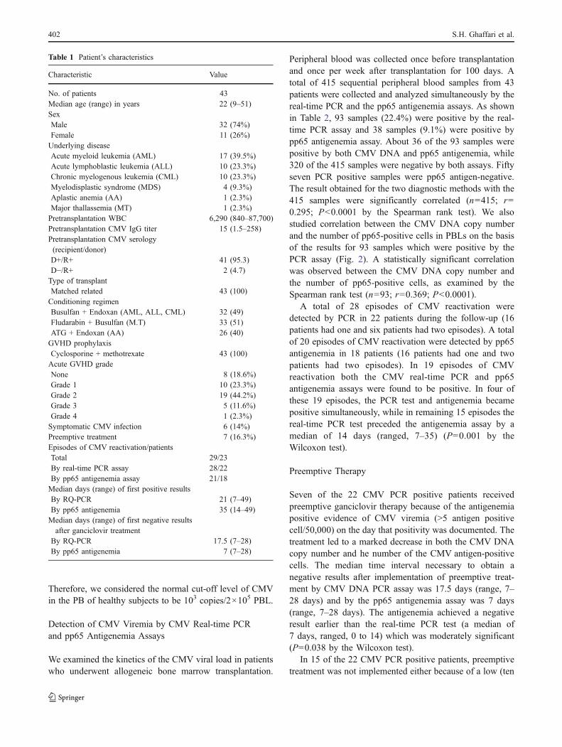
A recombinant plasmid (pTZ-UL83) containing 450-bpregion of a sequence located in the UL83 gene, whichcodes for the lower matrix protein detected in the pp65antigenemia test, was constructed. A serially diluted pTZ-UL83 plasmid was then tested by the real-time PCR assay,and a standard curve of the Ct values was constructed(Fig. 1a). A wide linear range from 10 to 107 copies of thecontrol plasmid was established (Fig. 1b). A minimum offive copies of the plasmid could be detected by this system. Inorder to confirm the specificity of this assay, a CMV-negativecell line, PBLs from CMV-seronegative patients, and severalvirus strains were tested by this system and all were negativefor CMV. No cross-reactivity between CMV and herpessimplex viruses or Epstein–Barr virus was observed.
In order to determine the CMV viral load in blood of theBMT recipients by real-time PCR, we first tested for viralDNA in the PB samples taken from ten healthy volunteers.The prevalence of CMV in the PB samples was 60%;however, the CMV number was less than 103 copies/assay.
Monitoring CMV in BMT recipients 401
Therefore, we considered the normal cut-off level of CMVin the PB of healthy subjects to be 103 copies/2×105 PBL.
Detection of CMV Viremia by CMV Real-time PCRand pp65 Antigenemia Assays
We examined the kinetics of the CMV viral load in patientswho underwent allogeneic bone marrow transplantation.
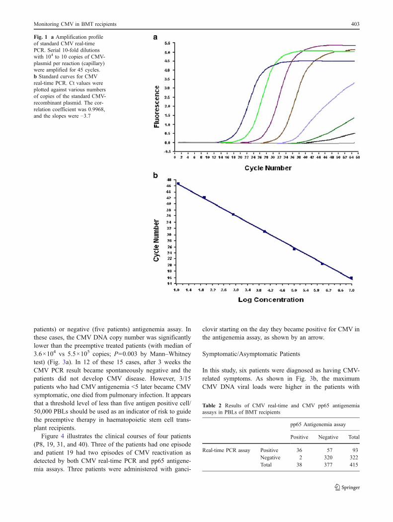
Peripheral blood was collected once before transplantationand once per week after transplantation for 100 days. Atotal of 415 sequential peripheral blood samples from 43patients were collected and analyzed simultaneously by thereal-time PCR and the pp65 antigenemia assays. As shownin Table 2, 93 samples (22.4%) were positive by the real-time PCR assay and 38 samples (9.1%) were positive bypp65 antigenemia assay. About 36 of the 93 samples werepositive by both CMV DNA and pp65 antigenemia, while320 of the 415 samples were negative by both assays. Fiftyseven PCR positive samples were pp65 antigen-negative.The result obtained for the two diagnostic methods with the415 samples were significantly correlated (n=415; r=0.295; P<0.0001 by the Spearman rank test). We alsostudied correlation between the CMV DNA copy numberand the number of pp65-positive cells in PBLs on the basisof the results for 93 samples which were positive by thePCR assay (Fig. 2). A statistically significant correlationwas observed between the CMV DNA copy number andthe number of pp65-positive cells, as examined by theSpearman rank test (n=93; r=0.369; P<0.0001).
A total of 28 episodes of CMV reactivation weredetected by PCR in 22 patients during the follow-up (16patients had one and six patients had two episodes). A totalof 20 episodes of CMV reactivation were detected by pp65antigenemia in 18 patients (16 patients had one and twopatients had two episodes). In 19 episodes of CMVreactivation both the CMV real-time PCR and pp65antigenemia assays were found to be positive. In four ofthese 19 episodes, the PCR test and antigenemia becamepositive simultaneously, while in remaining 15 episodes thereal-time PCR test preceded the antigenemia assay by amedian of 14 days (ranged, 7–35) (P=0.001 by theWilcoxon test).
Preemptive Therapy
Seven of the 22 CMV PCR positive patients receivedpreemptive ganciclovir therapy because of the antigenemiapositive evidence of CMV viremia (>5 antigen positivecell/50,000) on the day that positivity was documented. Thetreatment led to a marked decrease in both the CMV DNAcopy number and he number of the CMV antigen-positivecells. The median time interval necessary to obtain anegative results after implementation of preemptive treat-ment by CMV DNA PCR assay was 17.5 days (range, 7–28 days) and by the pp65 antigenemia assay was 7 days(range, 7–28 days). The antigenemia achieved a negativeresult earlier than the real-time PCR test (a median of7 days, ranged, 0 to 14) which was moderately significant(P=0.038 by the Wilcoxon test).
In 15 of the 22 CMV PCR positive patients, preemptivetreatment was not implemented either because of a low (ten
Table 1 Patient’s characteristics
Characteristic Value
No. of patients 43Median age (range) in years 22 (9–51)SexMale 32 (74%)Female 11 (26%)Underlying diseaseAcute myeloid leukemia (AML) 17 (39.5%)Acute lymphoblastic leukemia (ALL) 10 (23.3%)Chronic myelogenous leukemia (CML) 10 (23.3%)Myelodisplastic syndrome (MDS) 4 (9.3%)Aplastic anemia (AA) 1 (2.3%)Major thallassemia (MT) 1 (2.3%)Pretransplantation WBC 6,290 (840–87,700)Pretransplantation CMV IgG titer 15 (1.5–258)Pretransplantation CMV serology(recipient/donor)D+/R+ 41 (95.3)D−/R+ 2 (4.7)Type of transplantMatched related 43 (100)Conditioning regimenBusulfan + Endoxan (AML, ALL, CML) 32 (49)Fludarabin + Busulfan (M.T) 33 (51)ATG + Endoxan (AA) 26 (40)GVHD prophylaxisCyclosporine + methotrexate 43 (100)Acute GVHD gradeNone 8 (18.6%)Grade 1 10 (23.3%)Grade 2 19 (44.2%)Grade 3 5 (11.6%)Grade 4 1 (2.3%)Symptomatic CMV infection 6 (14%)Preemptive treatment 7 (16.3%)Episodes of CMV reactivation/patientsTotal 29/23By real-time PCR assay 28/22By pp65 antigenemia assay 21/18Median days (range) of first positive resultsBy RQ-PCR 21 (7–49)By pp65 antigenemia 35 (14–49)Median days (range) of first negative resultsafter ganciclovir treatment
By RQ-PCR 17.5 (7–28)By pp65 antigenemia 7 (7–28)
402 S.H. Ghaffari et al.
patients) or negative (five patients) antigenemia assay. Inthese cases, the CMV DNA copy number was significantlylower than the preemptive treated patients (with median of3.6×104 vs 5.5×10⁵ copies; P=0.003 by Mann–Whitneytest) (Fig. 3a). In 12 of these 15 cases, after 3 weeks theCMV PCR result became spontaneously negative and thepatients did not develop CMV disease. However, 3/15patients who had CMV antigenemia <5 later became CMVsymptomatic, one died from pulmonary infection. It appearsthat a threshold level of less than five antigen positive cell/50,000 PBLs should be used as an indicator of risk to guidethe preemptive therapy in haematopoietic stem cell trans-plant recipients.
Figure 4 illustrates the clinical courses of four patients(P8, 19, 31, and 40). Three of the patients had one episodeand patient 19 had two episodes of CMV reactivation asdetected by both CMV real-time PCR and pp65 antigene-mia assays. Three patients were administered with ganci-
clovir starting on the day they became positive for CMV inthe antigenemia assay, as shown by an arrow.
Symptomatic/Asymptomatic Patients
In this study, six patients were diagnosed as having CMV-related symptoms. As shown in Fig. 3b, the maximumCMV DNA viral loads were higher in the patients with
Fig. 1 a Amplification profileof standard CMV real-timePCR. Serial 10-fold dilutionswith 104 to 10 copies of CMV-plasmid per reaction (capillary)were amplified for 45 cycles.b Standard curves for CMVreal-time PCR. Ct values wereplotted against various numbersof copies of the standard CMV-recombinant plasmid. The cor-relation coefficient was 0.9968,and the slopes were −3.7
Table 2 Results of CMV real-time and CMV pp65 antigenemiaassays in PBLs of BMT recipients
pp65 Antigenemia assay
Positive Negative Total
Real-time PCR assay Positive 36 57 93Negative 2 320 322Total 38 377 415
Monitoring CMV in BMT recipients 403
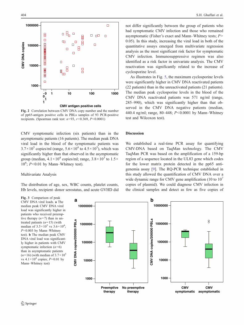
CMV symptomatic infection (six patients) than in theasymptomatic patients (16 patients). The median peak DNAviral load in the blood of the symptomatic patients was3.7×105 copies/ml (range, 5.6×10⁴ to 4.5×10⁵), which wassignificantly higher than that observed in the asymptomaticgroup (median, 4.1×10⁴ copies/ml; range, 3.8×10³ to 1.5×10⁶; P<0.01 by Mann–Whitney test).
Multivariate Analysis
The distribution of age, sex, WBC counts, platelet counts,Hb levels, recipient–donor serostatus, and acute GVHD did
not differ significantly between the group of patients whohad symptomatic CMV infection and those who remainedasymptomatic (Fisher’s exact and Mann–Whitney tests; P>0.05). In this study, increasing the viral load in both of thequantitative assays emerged from multivariate regressionanalysis as the most significant risk factor for symptomaticCMV infection. Immunosuppressive regimen was alsoidentified as a risk factor in univariate analysis. The CMVreactivation was significantly related to the increase ofcyclosporine level.
As illustrates in Fig. 5, the maximum cyclosporine levelswere significantly higher in CMV DNA reactivated patients(22 patients) than in the unreactivated patients (21 patients).The median peak cyclosporine levels in the blood of theCMV DNA reactivated patients was 571 ng/ml (range,285–990), which was significantly higher than that ob-served in the CMV DNA negative patients (median,440.4 ng/ml; range, 80–448; P<0.0001 by Mann–Whitneytest and Wilcoxon test).
Discussion
We established a real-time PCR assay for quantifyingCMV-DNA based on TaqMan technology. The CMVTaqMan PCR was based on the amplification of a 159-bpregion of a sequence located in the UL83 gene which codesfor the lower matrix protein detected in the pp65 anti-genemia assay [9]. The RQ-PCR technique established inthis study allowed the quantification of CMV DNA over awide dynamic range for CMV gene amplification (10 to 107
copies of plasmid). We could diagnose CMV infection inthe clinical samples and detect as few as five copies of
10001001010.1
-0.1
1000000
100000
10000
1000
CM
V D
NA
co
pie
s
CMV antigen positive cellsFig. 2 Correlation between CMV DNA copy number and the numberof pp65-antigen positive cells in PBLs samples of 93 PCR-positiverecipients. (Spearman rank test: n=93, r=0.369, P<0.0001)
a
No preemptivetherapy
Preemptivetherapy
10000000
1000000
100000
10000
1000
CM
V D
NA
co
pie
s/20
0000
PB
Ls
b
CMVasymptomatic
CMVsymptomatic
10000000
1000000
100000
10000
1000
CM
V D
NA
co
pie
s/20
0000
PB
Ls
Fig. 3 Comparison of peakCMV DNA viral loads. a Themedian peak CMV DNA viralload was significantly higher inpatients who received preemp-tive therapy (n=7) than in un-treated patients (n=15) (withmedian of 3.5×10⁵ vs 3.6×10⁴;P=0.003 by Mann–Whitneytest). b The median peak CMVDNA viral load was significant-ly higher in patients with CMVsymptomatic infection (n=6)than in asymptomatic patients(n=16) (with median of 3.7×10⁵
vs 4.1×10⁴ copies; P=0.01 byMann–Whitney test)
404 S.H. Ghaffari et al.

CMV DNA per 2×10⁵ PBL of the patient’s samples. Usingthis technique, 415 PBL samples from the 43 BMTrecipients were evaluated in parallel with the pp65antigenemia assay. We used PBL since these cells are themain CMV carriers during the active CMV infection. Thedetection of CMV antigenemia in PBLs has been shown to
be an early marker of CMV infection [9]. Some groupshave also found that detection and/or quantitation of DNAin PBL provides better clinical correlation than detectionand/or quantitation of DNA in plasma [29–33].
In the present study, viral reactivations occurred in 51%and 41.8% of allogeneic bone marrow recipients as detected
CMV PCRnegativepatients
CMV PCRpositivepatients
1000
800
600
400
200
0
Cyc
losp
ori
ne
leve
l (n
g/m
l)
a bFig. 5 The serum cyclosporinelevels in the allogeneic post-BMT recipients during the100 days post-BMT. a Graphicrepresentation of cyclosporinelevels in serum of the CMVDNA positive and negativepatients. b Comparison of thepeak cyclosporine levels inpatients with CMV DNA posi-tive and negative. The medianpeak cyclosporine level wassignificantly higher in the CMVreactivated patients (n=22) thanin unreactivated patients (n=21).(Wilcoxon and Mann–Whitneytest: P<0.0001)
Fig. 4 Clinical course of CMV reactivation in BMT patients.Sequential samples from patients were analyzed by both real-timePCR and pp65 antigenemia assays. CMV viral load profiles wereplotted for four patients who had elevated viral loads. Preemptivetreatment with ganciclovir was implemented in three patients basedupon antigenemia assay positivity (as shown by an arrow head). Solid
line, the number of CMV DNA copy number detected by the real-timeTaqMan assay; bars and numbers, the number of CMV antigen-positive cells/5×104 PBLs detected by the pp65 antigenemia assay;hatched line, serum cyclosporine level reported as nanogram permilliliter; shaded line with open circles, white blood cells (WBC)
Monitoring CMV in BMT recipients 405

by RQ-PCR and antigenemia assays, respectively. Wefound a significant correlation between the results of theCMV RQ-PCR and the antigenemia assays in PBL samples(P=0.0001). This is in agreement with the results of otherstudies that have used real-time PCR assays [9, 14, 15, 20,22, 23, 33–35]. Both assays were concordant for 85% ofthe patients and 86% of the specimens. However, the PCRquantification of CMV DNA was more sensitive than theantigenemia assay for the detection and monitoring ofCMV reactivation in BMT patients. The sensitivities of thePCR assay and the antigenemia were found to be 96.5%and 69%, respectively. By the PCR assay, 28 of 29 episodesof CMV viremia could be diagnosed, while the antigenemiatest was able to detect only 20 episodes. The real-time PCRproduced a 1.4% increase in the rate of detection of CMV.Also, as shown in Fig. 2, there were samples with anegative or a low level of antigenemia with a large numberof CMV DNA copies. Similar differences between theresults of an antigenemia assay and quantitative PCR havebeen reported by others [9, 14, 15, 20, 22, 23, 33–36].
While detection of CMV pp65-antigen is still widelyused for monitoring CMV infection, real-time PCR assayshave been recently developed for routine quantitation ofCMV DNA. However, correlations are lacking betweenresults of pp65 antigenemia and quantitative PCR assays[34]. Identification of a cutoff level for RQ-PCR assaywould be an important indicator of time to initiate an anti-CMV treatment [15] and would therefore reduce thenumber of patients treated with preemptive therapy whoare not destined to develop CMV disease [8]. Severalstudies have attempted to determine a CMV DNA copynumber equivalent to the levels of antigenemia and toimplement CMV PCR as pp65 antigenemia in clinicalpractice. Martin-Davila et al. [37] found an antigenemia often positive cells/2×105 PMNs equated with a plasmaCMV DNA of 1,330 copies/ml or a PMN cutoff of 713/5×106 cells. For an antigenemia level of 20 positive cells/2×105, the comparable copy numbers for plasma was again1,330 copies/ml, but increased to 4,755 copies/5×106 PMNcells. Mhiri et al. [35] defined a positive cut-off valueequivalent to 2,150 CMV DNA copies/ml of white bloodcell samples by hybrid capture assay and a viral load higherthan 400 copies/ml of plasma samples by RQ-PCR.Garrigue et al. [34] reported thresholds of ten and 50positive cells/2×105 cells were equivalent to 3.3 log10copies/ml (2,000 copies/ml) and 3.8 log10 copies/ml (6,300copies/ml), respectively. Three additional studies alsoreported the threshold of 50 pp65 antigen-positive cells/2×105 PBLs were correlated to ∼4 log10 genome copies/mlof whole blood [38–40]. Guiver et al. [41] found a higherthreshold (4.6 log10 copies/ml), possibly because of adifferent technology and DNA input. In our study, wedefined a positive cut-off value higher than 1,000 CMV
DNA copies/2×105 of PBL by RQ-PCR; however, theestablishment of a threshold to initiate an anti-CMVtreatment was not possible and needed confirmation bytesting a larger number of patients. All these results showsthat studies evaluating these tests are highly heterogeneous,there is no available international standard for CMV PCR,and each study determined its own assays characteristics fortheir own setup. There is also no consensus as the optimalblood compartment (whole blood, Peripheral blood leuko-cytes, plasma) as of yet [34]. Most reports on detectingCMV DNA by PCR have utilized ‘in-house’ assays withvariable primers for the same gene or different genes (e.g.the immediate early CMV gene or the CMV DNApolymerase gene) making it difficult to extrapolate resultsfrom one institution to another [42]. Therefore, each clinicallaboratory needs to determine its own assays characteristicsfor an appropriate patients’ monitoring [34]. There is somecommercially available assay that would allow laboratoriesto generate data with more widespread applicability.Lengerke et al. [43] found 89–92% concordance betweenan in-house quantitative PCR (amplified a portion of theimmediate early CMV gene) on whole blood and CACM(commercially available assay amplifies a 365 bp region ofthe CMV DNA polymerase gene) on plasma. Allice et al.[44] found a 98% correlation between a TaqMan real timePCR amplifying the immediate early gene and CACM, bothassays using PBL. The authors conclude that the real timePCR assay offers several advantages, including speed andprecision versus CACM. Pumannova et al. [45] alsoconcluded that the light cycler real time quantitative PCRwas superior to an ELISA-PCR, both amplified the samesegment of CMV genome, the UL 83 gene encoding pp65.These authors felt that the light cycler was superior inperformance and rapidity and it was more suitable forroutine diagnosis.
The analysis of CMV viremic episodes in which theresults of both tests eventually turned positive revealed thata detection of CMV DNA by real-time PCR in PBLallowed for an early diagnosis of CMV replication aftertransplantation. CMV DNA was detected by PCR in 15 ofthe 19 episodes by a median of 14 days prior to thedetection of an antigenemia result which indicates that apositive PCR test result is an earlier marker of CMVviremia than a positive antigenemia result (P=0.001 by theWilcoxon test). Similar results have been reported byothers. Griscelli et al. [9] found that CMV replication inblood leukocytes was detectable by PCR with a median of15 days prior to antigenemia. Leruez-Ville et al. [23] alsoreported that Real-time CMV PCR in blood plasma allowedfor an early diagnosis of CMV replication after transplan-tation, with a positive CMV PCR result occurring before apositive CMV antigenemia result by a median of 8 days.Schvoerer et al. [36] results showed that the PCR was
406 S.H. Ghaffari et al.

regularly positive before a antigenemia assay (4 to 52 daysbefore). Mori et al. [46] studied the clinical significance ofearly diagnosis of CMV in 19 allogeneic stem cell trans-planted patients. They concluded that CMV antigenemiahad a limited value in prediction of early diagnosis of CMVgastro-intestinal disease and suggested that PCR could havea more diagnostic significance. Also, it has been reportedthat the use of antigenemia assay will not give an accurateindication of a viral load or viral load rate increase since itcounts the number of infected cells only. On the other hand, aviral dynamics study in CMV showed that the initial CMVload and the rate of viral load increase are significantlyassociated with the development of disease [35].
The treatment induced a rapid decrease in both thenumber of viral DNA copies and the number of CMVantigen-positive cells. The first negative antigenemia resultspreceded the first negative PCR results by a median of7 days (range, 0 to 14). It has been suggested that the CMVDNA may represent the true viral copy number in thespecimens; a negative PCR result after treatment might be abetter indicator of a completely successful treatment thanthe pp65 antigemia [23]. Therefore, continuing anti-CMVtherapy until the PBL CMV PCR becomes negative mightprevent a recurrence of CMV disease. By contrast,discontinuation based on the first negative antigenemiaresults has led to a significant number of CMV pneumonia[11]. It is also shown, by Weinberg et al. [47], that thepatients who developed a recurrent CMV disease were stillpositive by PCR at the time the therapy was discontinuedfor the preceding episode; whereas, the patients who did notdeveloped a recurrent disease tended to clear the CMVDNA from their blood faster. These observations suggestthat quantitative CMV PCR might be a useful tool tomonitor the efficacy of anti-CMV therapy in bone marrowrecipients and the discontinuation of CMV preemptivetherapy based on quantitative CMV PCR might prevent arecurrent CMV disease. Ikewaki et al. [48] observed thatreal-time PCR was more suitable for monitoring CMVreactivation in adult T-cell leukemia–lymphoma patientsthan the antigenemia assay. Ksouri et al. [33] suggests thatthe CMV-DNA assay is better assay for monitoring patientsreceiving preemptive therapy, especially in CMV-GI dis-ease, and that after CMV-infected cell destruction, thegenome of defective virus could still be remaining anddetected by PCR.
In this study, increasing the viral load in both of thequantitative assays emerged from multivariate regressionanalysis as the most significant risk factor for CMVinfection. Also, the immunosuppressive regimen wasidentified as a risk factor in univariate analysis. Increasingcyclosporine serum level was significantly related to theCMV reactivation. In general, the maximum cyclosporinelevels were significantly (P<0.0001) higher in the CMV
reactivated patients than in the PCR negative patients(Fig. 5). We examined the patient’s cyclosporine level thatwas performed for all the patients, and attempted tocorrelate changes in the cyclosporine level with variationsin the CMV viral load in PBL. As shown in Fig. 3, as thecyclosporine level rises to the maximum, there is aconcomitant rise in the CMV viral load to the highestlevel. More than 81% of the patients who had a viralreactivation showed a very close correlation between thecyclosporine level and the viral load, where the patientsbegan very low in cyclosporine, and as the cyclosporinelevels increase, there was a concomitant rise in the viralload. In some cases, the cyclosporine levels can rise with nochange in the viral load, and in others, changes in the viralload were not read out as a change in the cyclosporinelevel. This information should be helpful in developingfuture therapeutic approaches including decreasing theimmune suppression and the use of CMV-specific T cellsimmunotherapy. Our results suggest that with more fre-quent and careful monitoring of cyclosporine serum levelsin BMT patients, some of the CMV reactivations may beprevented.
In conclusion, the increases of the viral load in bothquantitative assays and of cyclosporine serum levels wereidentified as the most significant risk factors for CMVreactivation. The results of both quantitative assays weresignificantly correlated; however, the RQ-PCR assay wasmore sensitive than the pp65 antigenemia assay. Thequantitative CMV PCR might be a useful tool formonitoring the CMV reactivation and the patient’s responseto antiviral therapy in BMT recipients.
Acknowledgement This work was supported by grants fromHematology, Oncology and BMT Research Center, Tehran UniversityMedical Sciences. We would like to thank Mr. Chahardouli forassistance in lab and Dr. Shamshiri for assistance in the statisticalanalysis. We would like to thank all clinicians and nurses whoparticipated and provided material for this study. In particular weacknowledge Drs. Jahani, Mosavi, Bibordi, Bahar, Irvani.
References
1. Boeckh M, Boivin G (1998) Quantitation of cytomegalovirus:methodologic aspects and clinical applications. Clin MicrobiolRev 11:533–554
2. Salzberger B, Bowden RA, Hackman R et al (1997) Neutropeniain allogeneic marrow transplant recipients receiving ganciclovirforprevention of CMV disease: risk factors and outcome. Blood 90:2502–2508
3. Li CR, Greenberg PD, Gilbert MJ et al (1994) Recovery of HLA-restricted cytomegalovirus (CMV)-specific T-cell responses afterallogeneic bone marrow transplant: correlation with CMV diseaseand effect of ganciclovir prophylaxis. Blood 83:1971–1979
4. Goodrich JM, Bowden RA, Fisher L et al (1993) Ganciclovirprophylaxis to prevent cytomegalovirus disease after allogeneicmarrow transplant. Ann Intern Med 118:173–178
Monitoring CMV in BMT recipients 407

5. Winston DJ, Ho WG, Bartoni K et al (1999) Ganciclovirprophylaxis of cytomegalovirus infection and disease in alloge-neic bone marrow transplant recipients. Results of a placebo-controlled, double-blind trial. Ann Intern Med 118:179–184
6. Goodrich JM, Mori M, Gleaves CA et al (1991) Early treatmentwith ganciclovir to prevent cytomegalovirus disease after alloge-neic bone marrow transplantation. N Engl J Med 325:1601–1607
7. Schmidt GM, Horak DA, Niland JC et al (1991) A randomizedcontrolled trial of prophylactic ganciclovir for cytomegaloviruspulmonary infection in recipients of allogeneic bone marrowtransplants. N Engl J Med 324:1005–1011
8. Aitken CW, Barrett-Muir C, Millar K et al (1999) Use ofmolecular assays in diagnosis and monitoring of cytomegalovirusdisease following renal transplantation. J Clin Microbiol 37:2804–2807
9. Griscelli F, Barrois M, Chauvin S et al (2001) Quantification ofhuman cytomegalovirus DNA in bone marrow transplant recipi-ents by real-time PCR. J Clin Microbiol 39:4362–4369
10. Gleaves CA, Smith TH, Shuster EA et al (1984) Rapid detectionof cytomegalovirus in MRC-5 cells inoculated with urine speci-mens by using low-speed centrifugation and monoclonal antibodyto an early antigen. J Clin Microbiol 19:917–919
11. Boeckh M, Gooley TA, Myerson D et al (1996) Cytomegaloviruspp65 antigenemia-guided early treatment with ganciclovir atengraftment after allogeneic marrow transplantation: a randomizeddouble-blind study. Blood 10:4063–4071
12. Hebart H, Gamer D, Loeffer J et al (1998) Evaluation of MurexCMV DNA hybrid capture assay for the detection and quantifi-cation of cytomegalovirus infection in patients following alloge-neic stem cell transplantation. J Clin Microbiol 36:1333–1337
13. Einsele H, Steidle M, Vallbracht A et al (1991) Early occurrenceof human cytomegalovirus infection after bone marrow transplan-tation as demonstrated by the polymerase chain reaction tech-nique. Blood 77:1104–1110
14. Gault E, Miche Y, Dehe’e A et al (2001) Quantification of humancytomegalovirus DNA by real-time PCR. J Clin Microbiol 39:772–775
15. Machida U, Kami M, Fukui T et al (2000) Real-time automatedPCR for early diagnosis and monitoring of cytomegalovirusinfection after bone marrow transplantation. J Clin Microbiol 38:2536–2542
16. Mori T, Okamoto S, Matsuoka S et al (2000) Risk-adaptedpreemptive therapy for cytomegalovirus disease in patientsundergoing allogeneic bone marrow transplantation. Bone MarrowTransplant 25:765–769
17. Nitsche A, Steuer N, Schmidt CA et al (2000) Detection of humancytomegalovirus DNA by real-time quantitative PCR. J ClinMicrobiol 38:2734–2737
18. Razonable RR, Brown RA, Espy MJ et al (2001) Comparativequantification of cytomegalovirus (CMV) DNA in solid-organtransplant recipients with CMV infection by using two high-throughput automated systems. J Clin Microbiol 39:4472–4476
19. Schaade L, Kockelkorn P, Ritter K et al (2000) Detection ofcytomegalovirus DNA in human specimens by lightCycler PCR.J Clin Microbiol 38:4006–4009
20. Tanaka N, Kimura H, Iida K et al (2000) Quantitative analysis ofcytomegalovirus load using a real-time PCR assay. J Med Virol60:455–462
21. Yun Z, Lewensohn-Fuchs I, Ljungman P et al (2000) Real-timemonitoring of cytomegalovirus infections after stem cell trans-plantation using the TaqMan polymerase chain reaction assays.Transplantation 69:1733–1736
22. Yakushiji K, Gondo H, Kamezaki K et al (2002) Monitoring ofcytomegalovirus reactivation after allogeneic stem cell transplanta-tion: comparison of an antigenemia assay and quantitative real-timepolymerase chain reaction. Bone Marrow Transplant 29:599–606
23. Leruez-Ville M, Ouachee M, Delarue R et al (2003) Monitoringcytomegalovirus infection in adult and pediatric bone marrowtransplant recipients by a real-time PCR assay performed withblood plasma. J Clin Microbiol 41:2040–2046
24. Tanabe K, Tokumoto T, Ishikawa N et al (1997) Comparativestudy of cytomegalovirus (CMV) antigenemia assay, polymerasechain reaction, serology, and shell vial assay in the early diagnosisand monitoring of CMV infection after renal transplantation.Transplantation 64:1721–1725
25. Seropian S, Ferguson D, Salloum E et al (1998) Lack of reactivityto CMV pp65 antigenemia testing in a patient with CMV diseasefollowing allogeneic bone marrow transplant. Bone MarrowTransplant 22:507–509
26. Solano C, Munoz I, Gutierrez A et al (2001) Qualitative plasmaPCR Assay (AMPLICOR CMV Test) versus pp65 antigenemiaassay for monitoring cytomegalovirus viremia and guidingpreemptive ganciclovir therapy in allogeneic stem cell transplan-tation. J Clin Microbiol 39:3938–3941
27. Ljungman P, Plotkin S (1995) Workshop on CMV disease—definitions, clinical severity scores and new syndromes. Scand JInfect Dis Suppl 99:87–89
28. Przepiorka D, Weisdorf D, Martin P et al (1994) Consensusconference on Acute GVHD grading. Bone Marrow Transplant15:825–828
29. Jabs DA, Forman M, Enger C et al (1999) Comparison ofcytomegalovirus loads in plasma and leukocytes of patients withcytomegalovirus retinitis. J Clin Microbiol 37:1431–1435
30. Pellegrin I, Garrigue I, Ekouevi D et al (2000) New molecularassays to predict occurrence of cytomegalovirus disease in renaltransplant recipients. J Infect Dis 182:36–42
31. Schafer P, Tenschert W, Cremaschi L, et al (2001) Area under theviraemia curve versus absolute viral load: utility for predictingsymptomatic cytomegalovirus infections in kidney transplantpatients. J Med Virol 65:85–89
32. Boeckh M, Hawkins G, Myerson D et al (1997) Plasma PCR forcytomegalovirus DNA after allogeneic marrow transplantation:comparison with PCR using peripheral blood leukocytes, pp65antigenemia and viral culture. Transplantation 64:108–113
33. Ksouri H, Eljed H, Greco A et al (2007) Analysis of cytomega-lovirus (CMV) viremia using the pp65 antigenemia assay, theamplicor CMV test, and a semi- quantitative polymerase chainreaction test after allogenic marrow transplantation. Transpl InfectDis 9:16–21
34. Garrigue I, Boucher S, Couzi L et al (2006) Whole blood real-time quantitative PCR for cytomegalovirus infection follow-up intransplant recipients. J Clin Virol 36:72–75
35. Mhiri L, Kaabi B, Houimel M, Arrouji Z, Slim A (2007)Comparison of pp65 antigenemia, quantitative PCR and DNAhybrid capture for detection of cytomegalovirus in transplantrecipients and AIDS patients. J Virol Methods 143:23–28
36. Schvoerer E, Henriot S, Zachary P et al (2005) Monitoring lowcytomegalovirus viremia in transplanted patients by a real-timePCR on plasma. J Med Virol 76:76–81
37. Martin-Davila P, Fortun J, Gutierrez C et al (2005) Analysis of aquantitative PCR assay for CMV infection in liver transplantrecipients: an intent to find the optimal cut-off value. J Clin Virol 33:138–144
38. Gouarin S, Vabret A, Gault E et al (2004) Quantitative analysis ofHCMV DNA load in whole blood of renal transplant patientsusing real-time PCR assay. J Clin Virol 29:194–201
39. Li H, Dummer S, Estes WR, et al (2003) Measurement of humancytomegalovirus loads by quantitative real-time PCR for monitor-ing clinical intervention in transplant recipients. J Clin Microbiol41:187–191
40. Mengelle C, Sandres-Saun’e K, Pasquier C et al (2003) Auto-mated extraction and quantification of human cytomegalovirus
408 S.H. Ghaffari et al.

DNA in whole blood by real-time PCR assay. J Clin Microbiol41:3840–3845
41. Guiver M, Fox AJ, Mutton K, Mogulkoc N, Egan J (2001)Evaluation of CMV viral load using TaqMan CMV quantitativePCR and comparison with CMV antigenemia in heart and lungtransplant recipients. Transplant 71:1609–1615
42. Drew WL (2007) Laboratory diagnosis of cytomegalovirusinfection and disease in immunocompromised patients. Curr OpinInfect Dis 20:408–411
43. Lengerke C, Ljubicic T, Meisner C et al (2006) Evaluation of theCOBAS Amplicor HCMV Monitor for early detection andmonitoring of human cytomegalovirus infection after allogenicstem cell transplantation. Bone Marrow Transplant 38:53–60
44. Allice T, Enrietto M, Pittaluga F et al (2006) Quantitation ofcytomegalovirus DNA by real-time polymerase chain reaction inperipheral blood specimens of patients with solid organ trans-plants: comparison with end-point PCR and pp65 antigen test.J Med Virol 78:915–922
45. Pumannova M, Roubalova K, Vitek A, Sajdova J (2006)Comparison of quantitative competitive polymerase chain reaction-enzyme-linked immunosorbent assay with LightCycler-based poly-merase chain reaction for measuring cytomegalovirus DNA inpatients after hematopoietic stem cell transplantation. DiagnMicrobiol Infect Dis 54:115–120
46. Mori T, Mori S, Kanda Y et al (2004) Clinical significance ofcytomegalovirus (CMV) antigenemia in the prediction and diagno-sis of CMV gastrointestinal disease after allogeneic hematopoieticstem cell transplantation. Bone Marrow Transplant 33:431–434
47. Weinberg A, Hodge TN, Li S et al (2000) Comparison of PCR,antigenemia assay, and rapid blood culture for detection andprevention of cytomegalovirus disease after lung transplantation.J Clin Microbiol 38(2):768–772
48. Ikewaki J, Ohtsuka E, Kawano R et al (2003) Real-time PCRassay compared to nested PCR and antigenemia assays fordetecting cytomegalovirus reactivation in adult T-cell leukemia-lymphoma patients. J Clin Microbiol 41:4382–4387
Monitoring CMV in BMT recipients 409