THE ROLE OF HUMAN CYTOMEGALOVIRUS INFECTION IN ...
92
From Department of Medicine, Solna Karolinska Institutet, Stockholm, Sweden THE ROLE OF HUMAN CYTOMEGALOVIRUS INFECTION IN CANCER Chato Taher M.D. Stockholm 2013
-
Upload
khangminh22 -
Category
Documents
-
view
0 -
download
0
Transcript of THE ROLE OF HUMAN CYTOMEGALOVIRUS INFECTION IN ...

From Department of Medicine, Solna Karolinska Institutet, Stockholm, Sweden
THE ROLE OF HUMAN CYTOMEGALOVIRUS INFECTION IN CANCER
Chato Taher M.D.
Stockholm 2013

All previously published papers were reproduced with permission from the publishers. Figure 5, 7, 8 and 9 were produced using Servier Medical Art. The cover photos: front cover is figure 7 from the thesis and back cover is an immunohistochemistry staining for HCMV proteins in breast cancer, both made by the author. Published by Karolinska Institutet. Printed by Larserics Digital Print AB. © Chato Taher, 2013 ISBN 978-91-7549-200-1

1
To my family

2
ABSTRACT
Cancer is a major cause of morbidity and mortality worldwide. It is thought that up to 20% of cancers are caused by infectious agents, and an oncogenic role of several viruses has been established for certain tumours. Increasing evidence implies that human cytomegalovirus (HCMV) infection is associated with a number of malignancies. Several studies have suggested different mechanisms by which HCMV could modulate the tumour environment and dysregulate several key pathways relevant in tumour development and progression. However, the role of HCMV in cancer has remained highly controversial. The studies in this thesis investigated the possible role of HCMV in certain types of cancers. Additionally, they addressed the question of whether HCMV targeted therapy could be used as a treatment option for cancer patients. In study I, we found that HCMV proteins were abundantly expressed in all breast cancer specimens examined and in 94% of sentinel lymph node specimens with metastases. HCMV infections were mostly confined to the neoplastic cells, while some inflammatory cells were also HCMV positive in 60% of lymph nodes without metastases. In study II, we investigated brain metastases and paired primary tissue samples of breast and colon cancer patients for HCMV proteins and nucleic acids. Interestingly, HCMV proteins were abundantly expressed in the majority (98.7%) of brain metastases and paired primary breast and colorectal cancer specimens. Patients with high grade HCMV infection tended to have shorter time to tumour progression and shorter survival, both after primary tumour diagnosis, as well as after establishment of brain metastases. In study III, we found that the majority of primary medulloblastomas and medulloblastoma cell lines were infected with HCMV. HCMV infection induced expression of cyclooxygenase-2 (COX-2) activity and prostaglandin-E2 (PGE2) production in vitro. Additionally, expression of HCMV proteins and COX-2 were strongly correlated in primary tumours as well as in meduloblastoma xenografts. Targeting viral replication using an anti-viral drug and a COX-2 inhibitor prevented HCMV replication in vitro, inhibited PGE2 production and reduced medulloblastoma tumour cell growth both in vitro and in vivo. In study IV, we discovered a novel genetic variant of HCMV, which lacks a gene segment in a regulatory gene. This viral strain was frequently detected in cancers of different origins and was associated with non-productive infection and expression of splice variant immediate early proteins. In contrast, this variant was less frequently detected in healthy donors, and in patients with HCMV viremia or myocardial infarction. We isolated this variant from 3 out of 110 clinical isolates. Thus, our results demonstrate a high prevalence of this novel genetic variant of HCMV in cancer patients; this virus variant may be tumour promoting virus for cancers of different origin. Understanding molecular pathways modulated by this virus is therefore highly necessary to further understand the behaviour of this unique HCMV variant, and its possible role in cancer development or progression.

3
LIST OF PUBLICATIONS
I. Taher C, de Boniface J, Mohammad AA, Religa P, Hartman J, Yaiw
KC, Frisell J, Rahbar A, Söderberg-Naucler C. High prevalence of human cytomegalovirus proteins and nucleic acids in primary breast cancer and metastatic sentinel lymph nodes. PLoS One. 2013;8(2):e56795.
II. Taher C, Gabriella Frisk, Stina Fuentes, Piotr Religa, Alice Assinger, Koon-Chu Yaiw, Karin Ekström Smedby, Magnus Bäcklund*, Cecilia Söderberg-Naucler*, and Afsar Rahbar*. High prevalence of human cytomegalovirus in brain metastases of patients with primary breast and colorectal cancer. Submitted manuscript
III. Ninib Baryawno, Afsar Rahbar, Nina Wolmer-Solberg*, Taher C*,
Jenny Odeberg, Anna Darabi, Zahidul Khan, Baldur Sveinbjörnsson, O.-M. FuskevÅg, Lova Segerström, Magnus Nordenskjöld, Peter Siesjö, Per Kogner, John Inge Johnsen, and Cecilia Söderberg-Nauclér. Detection of human cytomegalovirus in medulloblastomas reveals a potential therapeutic target. J Clin Invest. 2011;121(10):4043-55.
IV. Taher C*, Koon-Chu Yaiw*, Vanessa Wilhelmi, Abdul-Aleem Mohammad, Alice Assinger, Zahidul Khan, Jessica Pettersson, Jenny Odeberg, Mensur Dzabic, Xingling Xu, Giuseppe Stragliotto, Johan Hartman, Jan Frisell, Anna Martling, Stefania Varani, Claes Örvell, Peter Siesjö, Per Kogner, Inti Peredo, Rahbar Afsar, and Cecilia Söderberg-Nauclér. High Prevalence of a Novel Genetic Variant of Cytomegalovirus in Cancer Patients. Manuscript * Authors contributed equally

4
Related publications:
I. Stragliotto G, Rahbar A, Solberg NW, Lilja A, Taher C, Orrego A, Bjurman B, Tammik C, Skarman P, Peredo I, Söderberg-Nauclér C. Effects of valganciclovir as an add-on therapy in patients with cytomegalovirus-positive glioblastoma: A randomized, double-blind, hypothesis-generating study. Int J Cancer. 2013 Feb 13.
II. Rahbar A, Stragliotto G, Orrego A, Peredo I, Taher C, Willems J, Söderberg-Naucler C. Low levels of Human Cytomegalovirus Infection in Glioblastoma multiforme associates with patient survival; -a case-control study. Herpesviridae. 2012 Mar 16; 3:3.
III. Wolmer-Solberg N, Baryawno N, Rahbar A, Fuchs D, Odeberg J, Taher C, Wilhelmi V, Milosevic J, Mohammad AA, Martinsson T, Sveinbjörnsson B, Johnsen JI, Kogner P, Söderberg-Nauclér C. Frequent detection of human cytomegalovirus in neuroblastoma: A novel therapeutic target? Int J Cancer. 2013 May 10.

5
TABLE OF CONTENTS
1 Introduction .......................................................................................................................... 8 1.1 Herpesviridae ............................................................................................................. 8 1.2 Human cytomegalovirus (HCMV) ........................................................................... 9
1.2.1 Discovery and history of HCMV ...................................................................... 9 1.2.2 Characteristic features of HCMV .................................................................... 9
1.2.2.1 The HCMV genome ................................................................................... 10 1.2.2.2 Structure of HCMV ................................................................................... 10
1.2.3 Entry, replication and viral assembly ............................................................. 12 1.2.4 Virus latency and reactivation ........................................................................ 15 1.2.5 Epidemiology, transmission and clinical features ......................................... 15 1.2.6 HCMV and the immune system ...................................................................... 17 1.2.7 Diagnosis of HCMV infection ......................................................................... 19 1.2.8 Treatment of HCMV infection ........................................................................ 20
2 Infection and cancer ........................................................................................................ 22 2.1 Oncogenic viruses .................................................................................................... 23
2.1.1 Epstein–Barr virus (EBV) ............................................................................... 23 2.1.2 Human herpesvirus 8 (HHV-8) ....................................................................... 26 2.1.3 Hepatitis B virus (HBV) and hepatitis C virus (HCV) ................................. 27 2.1.4 Human papillomavirus (HPV) ........................................................................ 28 2.1.5 Human T-cell lymphotropic virus (HTLV-1) ................................................ 29
2.2 Human cytomegalovirus in cancer ......................................................................... 30 2.2.1 Presence of HCMV in tumours ....................................................................... 30 2.2.2 HCMV and the ‘Hallmarks of Cancer’ .......................................................... 30
2.2.2.1 Sustained proliferation .............................................................................. 32 2.2.2.2 Evasion of apoptosis .................................................................................. 33 2.2.2.3 Limitless replicative potential .................................................................. 34 2.2.2.4 Insensitivity to antigrowth signal ............................................................. 35 2.2.2.5 Genomic instability .................................................................................... 37 2.2.2.6 Angiogenesis ............................................................................................... 38 2.2.2.7 Immune evasion ......................................................................................... 39
2.2.2.7.1 The effect of HCMV on antigen presentation ................................. 41 2.2.2.7.2 The effect of HCMV on the NK cell response .................................. 41 2.2.2.7.3 The effect of HCMV on antibody mediated immunity ................... 41 2.2.2.7.4 HCMV mediated immunosuppression ............................................. 42
2.2.2.8 Tumour invasion and metastasis .............................................................. 42 2.2.2.9 Inflammation and the tumour microenvironment ................................. 46
2.3 Is HCMV oncogenic? ............................................................................................... 48 2.4 Can HCMV be targeted in tumours to improve patient's outcome? .................. 50
3 Aims of this thesis ............................................................................................................ 52
4 Results and discussion .................................................................................................... 53 4.1 Study I ....................................................................................................................... 53 4.2 Study II ..................................................................................................................... 57 4.3 Study III .................................................................................................................... 60 4.4 Study IV .................................................................................................................... 64 4.5 Summary and conclusion ........................................................................................ 67
5 Acknowledgements .......................................................................................................... 69
6 References ........................................................................................................................ 74

6
LIST OF ABBREVIATIONS
ALT
Autologous lymphocyte transfer ATL
Adult T-cell leukemia
Bcl-2 B-cell lymphoma 2 BL
Burkitt's lymphoma
BMT
Bone marrow transplant CDK
Cyclin-dependent kinase
COX-2 Cycloxygenase-2 CSC
Cancer stem cell
CTL
Cytotoxic T-cell DCs
Dendritic cells
DNMT
DNA methyltransferase E2F
Transcription factor
EBV Epstein Barr-virus EBNA Epstein-Barr virus nuclear antigen EC Endothelial cells ECM
Extracellular matrix
EGFR
Epidermal growth factor receptor EMT
Epithelial–mesenchymal transition
ER Endoplasmic reticulum ERK
Extracellular signal-regulated kinases
FACS
Fluorescence-activated cell sorting GSK-3β
Glycogen synthase kinase 3-β
HBV Hepatitis B virus HCMV Human cytomegalovirus HCV Hepatitis C virus HHV Human herpesvirus HPV Human papilloma virus HSPGs
Heparan sulfate proteoglycans
HSV Herpes simplex virus HTLV-1
Human T-cell lymphotropic virus type 1
IE Immediate early IHC
Immunohistochemistry
IL-10
Interleukin-10 JAK
Janus-activated kinase
KIR
Killer inhibitory receptor KS
Kaposi sarcoma
LA Late LANA
Latency-associated nuclear antigen
LIR-1
Leukocyte immunoglobulin-like receptor-1 LMP
Latent membrane protein
MAPK Mitogen activated protein kinase MHC
Major histocompatibility complex
MICA
MHC Class I-related chain A MICB
MHC Class I-related chain B
MMP
Matrix metalloproteinase

7
mTOR
Mammalian target of rapamycin NF-κB
Nuclear factor kappa B
NKG2D
Natural killer group 2D NPC
Neuroprogenitor cells
PCR Polymerase chain reaction PDGFR-α Platelet derived growth factor receptor-α PGE2 Prostaglandin E2 PI3K
Phosphatidylinositol-3-kinase
PTEN
Phosphatase and tensin homologue pRb
Retinoblastoma protein
RTKs
Receptor tyrosine kinases SLN Sentinel lymph node STAT Signal transducer and activator of transcription TERT
Telomerase reverse transcriptase
TGF-β
Transforming growth factor beta TME
Tumour microenvironment
TNF Tumour necrosis factor TSP-1
Thrombospondin 1
UL Unique long US
Unique short
v-FLIP
Viral FLICE inhibitory protein VEGF
Vascular endothelial growth factor
VEGFR Vascular endothelial growth factor receptor vGPCR
Viral G protein-coupled receptor
VZV Varicella zoster virus

8
1. INTRODUCTION
1.1 HERPESVIRIDAE
Herpesviruses are a large family of viruses (Herpesviridae) comprising of more than hundred different types, which can infect most vertebrates, from fish to mammals. All have similar virion architecture (120-300 nm in diameter) with a genome consisting of a linear double-stranded DNA molecule (124 to 230 kbp) encapsidated by an icosahedral capsid made up of 162 hollow-centered capsomeres, a tegument surrounding the nucleocapsid and a cell derived envelope containing viral glycoprotein spikes on its surface (Roizman 2007). Herpesviruses share four common important biological properties: 1. A large array of enzymes involved in nucleic acid metabolism. 2. Viral DNA synthesis and capsid assembly occurs in the nucleus, followed by processing and maturation of the virion in the cytoplasm. 3. Production of infectious progeny virus is followed by lysis of the infected cells. 4. Primary infection will be followed by latency in their natural host. Among hundreds of known herpesviruses, there are 8 herpesviruses that can infect humans, and these are divided into three subfamilies (alpha, beta and gamma herpesviruses), based primarily on difference in their biological properties such as replication cycle, host cell tropism, latency features and their different clinical manifestations (Roizman 2007). Alphaherpesvirinae: This subfamily of herpesviruses contains the genera Simplex virus (Herpes simplex virus type 1 and 2) and Varicella virus (Varicella zoster virus) and they have a variable host range. They rapidly grow in different cell types and cause efficient destruction of infected cells. They are able to establish latent infections primarily, but not exclusively, in sensory ganglia. Betaherpesvirinae: This subfamily of virus includes the genera Cytomegalovirus (CMV) and Roseolovirus (Human herpesvirus type 6 and 7). They have a restricted human host range, a long replication cycle and the infection slowly progresses in culture. Infected cells frequently become enlarged (cytomegalic). The virus maintained in latent form in secretory glands, lymphoreticular cells, bone marrow cells and possibly others. Gammaherpesvirinae: Two genera are included in this subfamily, Lymphocryptovirus (Epstein-Barr virus (EBV)) and Rhadinovirus (Human herpesvirus type 8 (HHV-8)). All members of this group replicate in lymphoblastoid cells, and also cause lytic infections in some types of epithelioid and fibroblast cells. Viruses in this group are usually T- or B-lymphocyte specific and, establish latency in these cells. Both EBV and HHV-8 are implied as oncogenic viruses.

9
1.2 HUMAN CYTOMEGALOVIRUS (HCMV)
1.2.1 Discovery and history of HCMV
The discovery of HCMV dates back to 1881, when a German scientist observed enlarged cells (protozoan-like cells) in kidney specimens from a still-born infant. Jesionek described a similar finding in 1904, when he examined several organs from an eight months old foetus. In 1907 Löwenstein described nuclear and cytoplasmic inclusions, surrounded by clear zone, in these protozoal-like cells (Riley 1997; Weller 2000; Ho 2008). In 1921 Goodpasture and Talbot rejected opinions from others that the formation of these inclusion bodies were caused by protozoan. They used cytomegalia to describe these abnormal cells in lesions of infancy. However, it remained unclear what caused this pathology (Goodpasture E. W. 1921). In the same decade inclusion bodies were observed in cells infected by herpesviruses by Von Glahn and Pappenheimer, who concluded that the inclusion seen in cytomegalic cells were likely viral induced, rather than protozoa (von Glahn and Pappenheimer 1925). Cole provided further evidence to support this statement by inducing formation of inclusion bodies in salivary glands using viruses (Wyatt, Saxton et al. 1950). After many authors agreed that the cytomegalic cells were pathognomonic for this condition, the condition was termed a ‘generalised cytomegalic inclusion disease’ by Wyatt and Saxton in 1950s (Wyatt, Saxton et al. 1950), for the unknown viral aetiology of this pathology. The isolation and propagation of the responsible virus was possible after the successful culture of human cells in vitro in the 1950s. Three independent research groups isolated the virus in the same year (Rowe, Hartley et al. 1956; Smith 1956). At that time, the virus was called the ‘salivary gland virus’ and thereafter the term “cytomegalovirus’ was proposed by Weller et al (Craig, Macauley et al. 1957). Once HCMV was isolated and cultured, it enabled the development of different tests for detecting HCMV, as well as opportunity to investigate the molecular pathogenesis of HCMV in different pathologies. This, in turn, has led to many important clinical and epidemiological observations. 1.2.2 Characteristic features of HCMV
HCMV is a virus that commonly infects humans and many other animals. It is highly species-specific and infects different cell types. HCMV belongs to the subfamily of herpesviruses. The virion of HCMV has a typical herpesvirus structure. It consists of an inner core of a double-stranded linear DNA molecule surrounded by a nucleocapsid and a thick layer of tegument protein that is surrounded by a lipid bilayer envelope (Mocarski, Shenk et al. 2007).

10
1.2.2.1 The HCMV genome HCMV is the largest and most complex of all known herpesviruses. It consists of a genome of approximately 235 kbp and containing 252 open reading frames (ORFs), which was believed to encode 180 proteins. However a recent study suggests that this number may in fact exceed 750 proteins (Stern-Ginossar, Weisburd et al. 2012), revealing that HCMV may be far more complex than previously believed. Interestingly, only about 50 proteins are believed to be essential for its replication and the vast majority of HCMV proteins interfere with cellular and immunological functions to enable the virus to coexist with its host (Murphy, Yu et al. 2003).
Figure 1. Structure of the HCMV genome. The top line is a size scale in kilobase pair (kbp). The completely sequenced AD169 and Toledo strains are shown on second and third line, respectively. Adapted from (Edward S. Mocarski, Thomas Shenk et al. 2007). The genome of HCMV, and the closely related chimpanzee CMV, exhibit the class E genome with a unique long (UL) and unique short (US) regions flanked by terminal repeats (TR) and internal repeats (IR) (Figure. 1) (Mocarski, Shenk et al. 2007). Herpesvirus genomes are not simple lengths of unique DNA, but characteristically contain direct and inverted repeats. The reasons for this are not known, but it is intriguing that similar structures appear to have arisen independently on several occasions during herpesvirus evolution (Baines and Pellett 2007). 1.2.2.2 Structure of HCMV The HCMV virion has a typical herpesvirus structure, about 200-300 nm in size. It consists of a 125-nm diameter icosahedral nucleocapsid containing a double stranded linear DNA genome surrounded by a proteinaceous layer defined as the tegument or matrix, which, in turn, is enclosed by a lipid bilayer containing a large number of viral glycoproteins. The nucleocapsid is composed of five herpesvirus core proteins (major capsid protein (MCP), triplexes, minor capsid protein, smallest capsid protein and portal protein) (Britt and Boppana 2004; Mocarski Jr 2007).
0 50 100 150 200kbp
AD169Toledo
aLanb UL b’am’c’ US canasTRIRTR

11
Figure 2. Structure of the HCMV virion and different virus-like particles released during the lytic cycle of herpesvirus replication. Modified from (Liu and Hong Zhou 2007). Based on capsid assembly during viral replication, three distinct types of capsids are observed termed A (only capsid shell), B (capsid shell and assembled protein) and C (capsid shell containing DNA genome), which is the only form that has completed maturation (Figure 2). A and B capsids do not contain viral DNA, and thus they fail to form mature virions, instead they constitute a type of virus particles called defective virus particles such as noninfectious virus particles and dense bodies, which can be found in the nucleus, cytoplasm and in cell free virus particle preparations (Gibson 2006; Britt 2007). The nucleocapsid is covered by a thick layer of mainly viral derived proteins called the tegument, which constitutes about 40% of the total virion mass. In addition, the tegument contains a selection of viral and cellular RNAs. Most tegument proteins are phosphorylated, which may facilitate stable association and incorporation into the virion compartment (Munger, Yu et al. 2006). Proteins in this compartment carry out a remarkably diverse range of activities during infection and tend to be highly immunogenic (Britt and Boppana 2004). The most abundant HCMV tegument proteins are pp65, pp71 and pp150 (Varnum, Streblow et al. 2004). These proteins play important roles during the un-coating of the particle during entry and virion assembly. They are also suggested to play important functional roles through modulation of the host immune response to infection, providing a favourable intracellular environment for viral replication (reviewed in (Kalejta 2008)).
Envelope
Genome
NucleocapsidTegument
12
A lipid bilayer envelope surrounds the tegumented capsid, which contains both host cell proteins and viral glycoproteins. The phospholipid envelope contains several virus encoded glycoproteins, including gpUL55 (gB), gpUL73 (gN), gpUL74 (gO), gpUL75 (gH), UL100 (gM), gpUL115 (gL) and the UL128-131 complex. These glycoproteins play essential roles in virus entry into host cells, cell-to-cell spread, and virion maturation (Britt and Mach 1996; Ryckman, Rainish et al. 2008). Virus budding occurs mainly in Golgi-derived intracellular vacuoles (Homman-Loudiyi, Hultenby et al. 2003). The mature virions are then subsequently transported to the plasma membrane, where they are released following fusion of the transport vesicle and the plasma membrane (Gibson 2006). During a productive viral infection only about 1% of viral particles are infectious and thus, the vast majority are defective viral particles that have failed to package viral DNA (Edward S. Mocarski, Thomas Shenk et al. 2007). Although all these types of viral particles do not contain viral DNA, they contain viral RNA molecules that are non-specifically incorporated into the defective particles (Terhune, Schroer et al. 2004). The consequences of the transmission of these contents are poorly studied. 1.2.3 Entry, replication and viral assembly
A series of distinct steps are required for initiation of viral entry. Firstly, attachment to specific cellular receptors, followed by viral envelope fusion with cell membrane prior to the release of nucleocapsids into the cytoplasm. After translocation of nucleocapsid to the nucleus, an interaction with nuclear pores occur, which is followed by release of the viral genome into the nucleus (Figure 2). It has been known for some time that HCMV initiates infection by binding to cell surface heparan sulfate proteoglycans (HSPGs). Engagement of HSPGs is known as one of the relatively conserved features of herpesvirus entry pathways. HCMV binding to HSPGs is thought to play a crucial role in initial stage of entry, by enhancing the attachment to subsequent receptors in a cascade that ultimately leads to fusion (Compton, Nowlin et al. 1993). Numerous specific receptor candidates have been proposed in the last decades. HCMV can bind to β2 microglobulin, annexin II, aminopeptidase (CD13), epidermal growth factor receptor (EGFR), cellular integrins, (specifically αvβ3) and platelet derived growth factor receptor-α (PDGFR-α) (Söderberg C, Giugni Td et al. 1993; Wang, Huong et al. 2003; Feire, Koss et al. 2004; Soroceanu, Akhavan et al. 2008). However, none of these receptors have been found to be absolutely necessary for infection of all susceptible cell types (Compton T. 2007). HCMV can attach and penetrate both permissive and non-permissive cell types. However, productive replication is observed in a very restricted range of human cells, indicating that a post penetration block in viral gene expression restricts replication in non-permissive cells (Sinzger, Kahl et al. 2000).

13
After virus fusion with the cell membrane, the HCMV nucleocapsid is deposited into the cytoplasm and translocated to the nucleus, where viral DNA is released (Dohner and Sodeik 2005). Following insertion of viral DNA into the host cell nucleus, the HCMV genome is expressed in a highly organised and sequential order. It starts with immediate early (IE) expression, followed by early (E) and late (LA) viral gene expressions (Fortunato and Spector 1999). IE proteins are the most abundantly expressed proteins in the initial phase; it takes around 1-4 hours following infection, while the complete replication of HCMV requires 48-72 hours. Once the IE genes are expressed, the IE proteins, alone or in synergism, regulate the subsequent expression of other viral genes (E and LA genes), by acting as transactivators or autostimulators. E and LA genes are encoding proteins, mainly responsible for building up the structure of the virus particle (capsid, tegument and envelope proteins), as well as the coding of other proteins that modulate host cell functions (Figure 3) (Fortunato and Spector 1999; Mocarski, Shenk et al. 2007).
Figure 3. HCMV gene expression and viral protein functions. Modified from (Landolfo, Gariglio et al. 2003).
IE genes
E genes
LA genes
IE proteins
E proteins
LA proteins
Viral DNA replication
Regulation of host cell functionsGene expressionCell cycle progressionApoptosis Immune responses
E and L gene expressionIE gene expression
Virion morphogenesis

14
The immediate-early gene (UL122 and UL123) of HCMV is a complex region consisting of a promoter, five exons, and two poly (A) signals (figure 4). During HCMV infection in vitro, multiple mRNA species are generated through differential splicing and polyadenylation (Stenberg, Depto et al. 1989; Stenberg 1996; Castillo and Kowalik 2002; Awasthi, Isler et al. 2004). The IE-72 protein is encoded by exons 2, 3 and 4, while the IE-86 by exons 2, 3 and 5 (Figure 4) (Castillo and Kowalik 2002). Five additional transcripts are initiated from exon 2 (IE-55, IE-18, IE-19, IE-17.5 and IE-9) and two splice variants of IE-86 (IE-40 and IE-60) have been described (Figure 4) (White, Del Rosario et al. 2007).
Figure 4. Splice variants encoded by HCMV IE gene. Modified from (Awasthi, Isler et al. 2004).
Two major gene products, IE-72 and IE-86, regulate the expression of the majority of the HCMV genes. IE-72 is dispensable for virus growth, while IE-86 is essential for regulating HCMV DNA replication. Both IE-72 and IE-86 exert fundamental effects on cell cycle regulation through interactions with tumour suppressor proteins, promotion of cell-cycle progression, induction of DNA synthesis, induction of telomerase activity and inhibition of apoptosis, as will be further discussed in section 2.2.2. When the replication cycle is completed the viral DNA is packaged into the synthesized capsid and exported through various cellular compartments where it acquires its remaining structural components (the tegument and envelope). The release of virions occur through cell lysis or by cell-to-cell contact (Gibson 2006). Observations by our group show that Golgi-derived secretory vacuoles containing mature virus particles also fuse with the plasma membrane, which leads to release of new infectious viral particles from infected cells (Homman-Loudiyi, Hultenby et al. 2003).

15
1.2.4 Virus latency and reactivation
The ability of the virus to establish lifelong persistence in its host after a primary infection is a biological property common to all herpesviruses. However, the exact cellular sites in which HCMV establishes latency, and the mechanisms regulating this latency and reactivation during natural infection remains poorly understood (Sinclair 2008). Detailed analysis of the peripheral blood compartment showed that monocytes are major sites of HCMV latency in vivo (Taylor-Wiedeman, Sissons et al. 1991). Furthermore, CD34+ progenitors isolated from bone marrow also carried HCMV genome in vivo (Mendelson, Monard et al. 1996) indicating that myeloid progenitor cells are most likely the cells that carry this virus during latency. Why other cells in this lineage e.g. T cells, B cells and polymorphonuclear cells do not carry this virus is thus far unknown. During latency, HCMV maintains its genome without the production of infectious virus, but it possesses the capability to reactivate under certain circumstances. Viral reactivation occurs mainly following inflammatory stimuli, when monocytes differentiate to macrophage or dendritic cells (DCs) (Soderberg-Naucler, Fish et al. 1997; Reeves, MacAry et al. 2005). This is asymptomatic in immunocompetent individuals, but might be life-threatening in immune compromised patients (Rubin 1990). It is clear that during HCMV latency there is a lack of IE transcription, and hence a lack of any subsequent lytic gene expression. However, attempts have been made to detect specific latency-associated transcripts in experimental models. For example, HCMV encodes a viral interleukin (IL)-10 homologue (UL111a; cmvIL-10) in HCMV infected cells. IL-10 is a well-known immunosuppressive cytokine. During latency, the transcript of this protein undergoes alternative splicing, which results in the expression of latency-associated cmvIL-10. This helps the virus to avoid recognition by immune system and clearance during latency (Jenkins, Garcia et al. 2008). Recently, it has been shown that expression of the viral transcript UL138 is required for the establishment and maintenance of viral latency (Weekes, Tan et al. 2013). UL138, through down regulation of multidrug resistance–associated protein-1 (MRP1) reduced cellular leukotriene C4 export, which may be able to inhibit the migration of infected DCs to draining lymph nodes, and impair the generation of an HCMV-specific immune response. (Robbiani, Finch et al. 2000). This could be another viral strategy for protection during latency. 1.2.5 Epidemiology, transmission and clinical features
HCMV is known to infect 45 to 100% of the population worldwide, depending on geographical location and socioeconomical status (Cannon, Schmid et al. 2010). The virus is capable of infecting humans at different ages. About 40% of children being infected with the virus by one year of age (Asanuma, Numazaki et al. 1996).

16
HCMV can be transmitted by different routes, either horizontally via close personal contact, salivary secretions, respiratory droplets, breast feeding, urine, blood transfusions, organ transplantation, sexual contact (Pomeroy and Englund 1987) or vertically from mother to child. It has been shown that primary HCMV infection of mothers during pregnancy increases the risk of virus transmission to the foetus (Syggelou, Iacovidou et al. 2010), causing a congenital infection with risk of birth defects. In the new born baby, HCMV infection is the most common cause of congenital abnormalities, occurring in 0.2% to 2.5% of all births, most commonly it causes hearing loss, but mental retardation and visual impairment are also observed (Syggelou, Iacovidou et al. 2010). Jaundice, petechiae and hepatosplenomegaly are the most common clinical signs, which occur in about 10% of congenitally infected children. Other clinical manifestations include growth retardation, seizures, lethargy, microcephaly, thrombocytopenia and anaemia (Syggelou, Iacovidou et al. 2010). However, most of the children with congenital infection are asymptomatic at birth. HCMV infection in normal immunocompetent hosts is generally subclinical, while associated with significant morbidity and mortality in immunocompromised patients. In immunocompetent patients, HCMV infection, when clinically evident, will present with mononucleosis-like symptoms such as fever, headache, sore throat, malaise, myalgias, lymphadenopathy and splenomegaly (Eddleston, Peacock et al. 1997). It is a significant cause of morbidity and mortality in patients with acquired immunodeficiency syndrome (AIDS) and stem cell and solid organ transplant patients. The severity of the disease varies according to the degree of immunosuppression. The symptoms can vary from mild symptomatic viremia (mononucleosis like symptom), to full-blown end organ disease with high mortality. The virus can be found in multiple sites causing retinitis, gastrointestinal diseases, pneumonitis and encephalitis (Ljungman 1996). An increasing body of epidemiological evidence suggest that HCMV may be a possible cofactor in the development of various inflammatory diseases and cancers. Several clinical studies have demonstrated the existence of a correlation between HCMV seropositivity and the presence of atherosclerosis and increased cardiovascular mortality (Sorlie, Nieto et al. 2000). In transplant patients, HCMV has been strongly associated with rejection and post-transplant complications (Linares, Sanclemente et al. 2011). Furthermore, HCMV has also been associated with inflammatory bowel diseases (Dimitroulia, Spanakis et al. 2006), rheumatoid arthritis (Pierer, Rothe et al. 2012), systemic lupus erythematosus (Perez-Mercado and Vila-Perez 2010) and Sjögrens syndrome (Shillitoe, Daniels et al. 1982), implying a link between the virus and autoimmune diseases. As HCMV is reactivated by inflammation, this virus may represent an epiphenomenon, but it may also contribute to inflammatory disease progression by further enhancing and maintaining the inflammatory process. In cancer patients, HCMV proteins and nucleic acids are frequently detected in tissue specimens from patients with cancers of different origin, including colon (Harkins,

17
Volk et al. 2002), breast (Harkins, Matlaf et al. 2010; Study I), prostate (Samanta, Harkins et al. 2003), mucoepidermoid salivary gland tumours (Melnick, Sedghizadeh et al. 2012), medulloblastoma (Study III), glioblastoma (Cobbs, Harkins et al. 2002; Rahbar, Stragliotto et al. 2012; Stragliotto, Rahbar et al. 2013), neuroblastoma (Wolmer-Solberg, Baryawno et al. 2013) and rhabdomyosarcoma (Price, Bingmer et al. 2012) as well as metastatic tumours (study I and II). However, it is yet not known whether this virus plays a causative role in connection with these diseases or simply represents an epiphenomenon. 1.2.6 HCMV and the immune system
HCMV infection induces both an innate immune response as well as an adaptive immune response, which control primary HCMV and/or recurrent infections. However, despite a strong host immune response, HCMV is still capable of establishing latency (Jackson, Mason et al. 2011). Most likely this is due to the numerous strategies this virus has developed to avoid detection and destruction by the immune system. HCMV and NK cell responses The innate immune ‘natural killer’ cells (NK cells) play an important role in the early control of viral infections, against certain tumours, and they also help to drive subsequent adaptive immunity. The importance of an immune response by NK cells against HCMV was noticed in patients with NK cell defects, who had serious recurrent episodes of HCMV disease (Biron, Byron et al. 1989). Inhibitory/killer inhibitory receptors, KIRs and leukocyte immunoglobulin like receptor (LIR), as well as activating receptors (natural killer group 2D, NKG2D), on NK cells regulate their functions. Inhibitory receptors recognise major histocompatibility complex type I (MHC-I) antigens thus, preventing an attack against healthy cells. Loss of MHC-I antigen expression activates NK cell function through “the missing-self hypothesis”. NK activating receptor recognises a broad spectrum of cell surface ligands, such as MHC-I related chain A or B (MICA, MICB), which is expressed on stressed cells as well as infected and tumour cells (Arase, Mocarski et al. 2002). Cell mediated immunity to HCMV CD8+ T cells are the key immune cell type involved in controlling HCMV infection, as they can recognise HCMV viral peptides presented on MHC-I molecules and mediate killing of infected cells. Upon recognition of pp65 and IE-72 peptides, CD8+ T cells induce an immune response to destroy and eradicate infected cells (Kern, Surel et al. 1999; Kern, Bunde et al. 2002). Results from experimental murine models of bone marrow transplantation (BMT) showed that removal of reconstituted CD8+ cells leads to lethal infection, while transferring reconstituted CD8+ T cells to immunocompromised mice could prevent murine CMV disease (Polic, Hengel et al. 1998). In a clinical study in human BMT patients, there was a strong correlation

18
between recovery of the CD8+ T cell population and protection from HCMV disease (Cwynarski, Ainsworth et al. 2001). Adaptive transfer of HCMV specific T cells has successfully been performed at some medical centers. Patients who have received ex vivo expanded HCMV specific CD8+ T cells are protected from both primary, and reactivating HCMV infection (Einsele, Roosnek et al. 2002). It is believed that HCMV is among the most immuno-dominant antigens that the immune system will ever encounter, and the HCMV specific immune response is remarkably strong. Furthermore, the specific T-cell response against HCMV increases with age, and remarkably, in elderly persons it may constitute up to 50% of the CD8+ T cell repertoire (Moss and Khan 2004). Despite the strong CD8+ T cells response towards HCMV infected cells, sometimes it is not sufficient to control HCMV infection. CD4+ T cells also help to control HCMV infection by recognising HCMV peptides displayed by MHC-II molecules on antigen presenting cells (APCs) (Le Roy, Baron et al. 2002). An accumulating body of evidences suggest that HCMV specific CD4+ T cells can act as effectors directly to virally infected cells (Rentenaar, Gamadia et al. 2000; Gamadia, Rentenaar et al. 2004). In addition, following bone marrow transplantation, the maintenance of HCMV specific CD8+ T cell infusions was shown to be dependent on the presence of HCMV-specific CD4+ T cells (Einsele, Roosnek et al. 2002), suggesting that CD4+ helper T cells are essential for effective CD8+ T cell responses. A minor subset of T cells, γδ T cells also expand following HCMV infection and these cells have the ability to mediate cytotoxicity in HCMV infected cells (Halary, Pitard et al. 2005). Humoral immunity to HCMV infection Humoral immunity has been also shown to assist in controlling HCMV infection through the secretion of antibodies from B cells differentiated into effector plasma cells that help to neutralise and eliminate the virus via phagocytic cells. Furthermore, antibody coated particles induce complement activation as well as antibody-dependent cell cytotoxicity (ADCC) and elimination by NK cells. The humoral immune response is not able to fully protect against viral infections, instead it helps in reducing the severity of infection and limits its spread (Landini, Rossier et al. 1988).

19
1.2.7 Diagnosis of HCMV infection
HCMV infection cannot be reliably distinguished based on clinical grounds from other infectious agents causing similar illnesses, such as EBV and hepatitis virus. A number of laboratory investigations are used to diagnose HCMV infection, which include: Serology: Serologic tests for detection of HCMV specific antibodies are useful for determining whether a patient had HCMV infection or not, and to determine whether the infection occurred recently by detecting the conversion of HCMV-IgG antibodies from negative to positive, or by demonstration of HCMV-specific IgM antibodies. However, HCMV-IgM lacks specificity for primary infection, due to possible false positive results, and should be followed by additional serum tests over time (Edward S. Mocarski, Thomas Shenk et al. 2007). Acute HCMV infection can be also confirmed by performing an avidity test for HCMV-IgG antibody, which increases with time after initial infection. Demonstration of low HCMV-IgG avidity can improve the accuracy of identification of recent infection (Grangeot-Keros, Mayaux et al. 1997). Furthermore, a neutralisation assay can be used as a reliable method for differentiating between acute primary and non-primary infection (Eggers, Bader et al. 2000). Virus culture: Virus isolation in culture is still a gold standard method for the detection of HCMV. It is performed by co-culturing clinical specimens with fibroblasts, followed by identification of the slowly developing cytopathic effect that is characteristic for HCMV. This approach is labour intensive, time consuming and less sensitive than more modern methods like PCR. However, this period can be shortened to 24 - 48 hours through enhancement of infection by low speed centrifugation and detection of IE gene by monoclonal antibody (Gleaves, Smith et al. 1984). PCR: PCR is a method for detection of HCMV infection used at most clinical laboratories today. It gained its popularity because it is rapid, sensitive, specific, provides a quantitative read out and it is amenable for automatic sample processing (Gimeno, Solano et al. 2008). Antigenimia assay: This assay has probably been the most widely used method for quantitating HCMV in blood. It is a relatively simple method, cheap to perform, and commercially available kits have made it widely accessible to hospital laboratories. This assay uses a monoclonal antibody to detect the tegument protein pp65 in blood leukocytes by immunostaining of cyto centrifuge preparations of blood cells. The number of pp65-positive white blood cells (WBCs) correlates with risk of disease, although the threshold number that predicts disease varies according to the clinical setting (Boeckh and Boivin 1998).
20
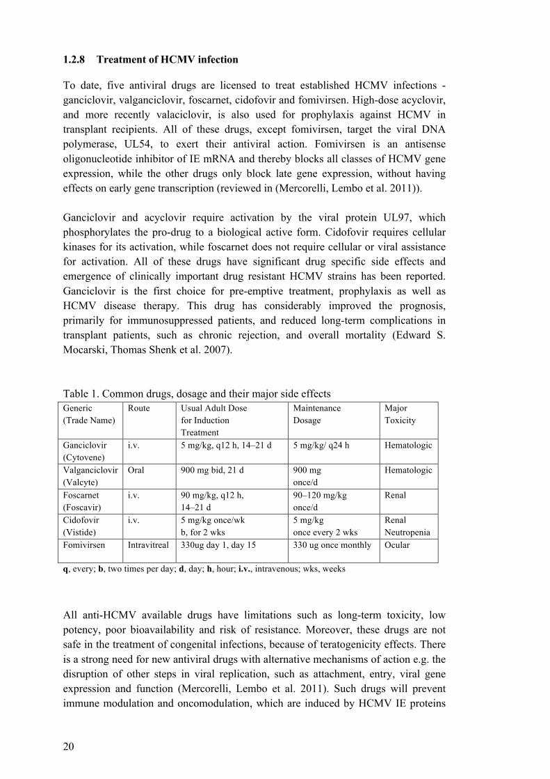
1.2.8 Treatment of HCMV infection
To date, five antiviral drugs are licensed to treat established HCMV infections - ganciclovir, valganciclovir, foscarnet, cidofovir and fomivirsen. High-dose acyclovir, and more recently valaciclovir, is also used for prophylaxis against HCMV in transplant recipients. All of these drugs, except fomivirsen, target the viral DNA polymerase, UL54, to exert their antiviral action. Fomivirsen is an antisense oligonucleotide inhibitor of IE mRNA and thereby blocks all classes of HCMV gene expression, while the other drugs only block late gene expression, without having effects on early gene transcription (reviewed in (Mercorelli, Lembo et al. 2011)). Ganciclovir and acyclovir require activation by the viral protein UL97, which phosphorylates the pro-drug to a biological active form. Cidofovir requires cellular kinases for its activation, while foscarnet does not require cellular or viral assistance for activation. All of these drugs have significant drug specific side effects and emergence of clinically important drug resistant HCMV strains has been reported. Ganciclovir is the first choice for pre-emptive treatment, prophylaxis as well as HCMV disease therapy. This drug has considerably improved the prognosis, primarily for immunosuppressed patients, and reduced long-term complications in transplant patients, such as chronic rejection, and overall mortality (Edward S. Mocarski, Thomas Shenk et al. 2007). Table 1. Common drugs, dosage and their major side effects Generic (Trade Name)
Route Usual Adult Dose for Induction Treatment
Maintenance Dosage
Major Toxicity
Ganciclovir (Cytovene)
i.v. 5 mg/kg, q12 h, 14–21 d 5 mg/kg/ q24 h Hematologic
Valganciclovir (Valcyte)
Oral 900 mg bid, 21 d 900 mg once/d
Hematologic
Foscarnet (Foscavir)
i.v. 90 mg/kg, q12 h, 14–21 d
90–120 mg/kg once/d
Renal
Cidofovir (Vistide)
i.v. 5 mg/kg once/wk b, for 2 wks
5 mg/kg once every 2 wks
Renal Neutropenia
Fomivirsen Intravitreal 330ug day 1, day 15 330 ug once monthly Ocular
q, every; b, two times per day; d, day; h, hour; i.v., intravenous; wks, weeks All anti-HCMV available drugs have limitations such as long-term toxicity, low potency, poor bioavailability and risk of resistance. Moreover, these drugs are not safe in the treatment of congenital infections, because of teratogenicity effects. There is a strong need for new antiviral drugs with alternative mechanisms of action e.g. the disruption of other steps in viral replication, such as attachment, entry, viral gene expression and function (Mercorelli, Lembo et al. 2011). Such drugs will prevent immune modulation and oncomodulation, which are induced by HCMV IE proteins

21
not currently targeted by most HCMV specific drugs (except for Fomivirsen, which is only locally distributed to the eye during HCMV retinitis) (Mercorelli, Lembo et al. 2011). Hyperimmune globulin preparations, containing high levels of antibody to HCMV (HCMVIg), have also been used for prevention of HCMV disease especially in organ transplant patients, pregnant women and for prevention of post natal infection in high risk premature newborns (Edward S. Mocarski, Thomas Shenk et al. 2007). HCMVIg acts through potentiation of antibody-dependent cell-mediated cytotoxicity responses and by neutralisation of HCMV itself. It has been shown that combination of ganciclovir and HCMVIg reduces HCMV-related mortality in transplant patients (Bonaros, Kocher et al. 2004).
22
2 INFECTION AND CANCER
Infection is one of the most important causes of cancer. During the past decade it has become obvious that several viruses play significant roles in the development of human cancers. In fact, approximately 15% to 20% of cancers are associated with viral infections (Parkin 2006). In malignancies that are not currently attributable to infectious agents, chronic inflammation plays a critical role in tumour progression, a feature recently classified as the seventh hallmark of cancer (Colotta, Allavena et al. 2009). To date, a few DNA viruses are consistently associated with human neoplasms, such as EBV with B-cell lymphoproliferative diseases and nasopharyngeal carcinoma, human papillomaviruses with cervical carcinoma, kaposi’s sarcoma associated Herpesvirus with kaposi’s sarcoma and primary effusion lymphomas, hepatitis B and hepatitis C viruses with hepatocellular carcinoma and Human T-cell leukemia virus-1 with T-cell leukemias (Carrillo-Infante, Abbadessa et al. 2007). Although these oncogenic viruses do not all belong to the same virus family they often contribute to cancer development in similar ways (Figure 5), as they share many common features. Importantly, these viruses have the ability to infect, but not kill their host cells. They establish persistent and long-term infections as they have evolved strategies that enable them to evade mechanisms of viral clearance by the host immune system (reviewed in (Pagano, Blaser et al. 2004)). Even though certain viruses have been confirmed as the causative agents of certain cancers, the majority are only associated with cancers, and their oncogenic mechanisms are not clearly understood. Since the majority of virus-infected individuals do not develop tumours, it seems that viral infections can contribute to, but are not sufficient for carcinogenesis. Therefore, additional factors such as chronic inflammation, defects in host immune responses and cellular mutations may play a role in the transformation process. For these reasons, establishing a causal relationship between a ubiquitous virus that causes persistent infection in the majority of adults worldwide and the development of cancer is difficult. In the 19th century Robert Koch introduced his ideas about how to prove a causal relationship between a microorganism and a disease, as reviewed by (Fredericks and Relman 1996). In fact, it cannot be directly applied in the context of a potential oncogenic viruses, since some of the guidelines are difficult to meet and others are not applicable to all viruses. For example, in many cases viral carcinogenesis is associated with an abortive, non-productive infection and it takes years to induce or promote malignancy, which does not fulfill Koch’s postulation. For this reason, different guidelines have been suggested to establish a causal relationship between viruses and human cancers. Fredericks and Relman introduced a modified criteria based on Hill's 9 criteria to address this issue with modern molecular techniques which are outlined below (Fredericks and Relman 1996).
23
For a potential infectious agent to be considered as an aetiological agent in human cancers, Fredericks and Relman proposed the following molecular guidelines for establishing microbial disease causation:
I. The putative pathogen is present in most cases of the disease. II. Normal tissue should harbour no, or significantly less, putative pathogen.
III. Disease resolution should be accompanied by decreased genome of putative pathogen.
IV. Microbial sequences should be present before disease or correlate with disease severity.
V. The nature of the microbial organism detected should be consistent with known biological characteristics of that group of organisms.
VI. Microbe-associated sequences are detected in the diseased tissue and should be corroborated at the cellular level.
VII. Molecular evidence should be reproducible. 2.1 Oncogenic viruses
2.1.1 Epstein–Barr virus (EBV)
The first herpesvirus discovered to be oncogenic was EBV (Epstein, Achong et al. 1964). EBV is a ubiquitous double-stranded DNA virus belonging to the gamma-herpesvirus sub-family. It infects more than 95% of the world’s population and after the primary infection individuals remain as asymptomatic carriers. EBV preferentially infects B-lymphocytes, although it can infect and replicate in other cell types, such as epithelial cells (Sixbey, Nedrud et al. 1984). EBV infection is linked to the etiology of several different lymphoid and epithelial malignancies, such as nasopharyngeal carcinoma, Burkitt's lymphoma (BL), post-transplant lymphomas and gastric carcinomas (Parkin 2006). Many oncogenic proteins that are encoded by this virus have been identified. They include EBV latent membrane protein 1 and 2 (LMP1 and LMP2) and EBV nuclear antigen 2 and 3 (EBNA2 and EBNA3). These proteins are essential for the ability of EBV to immortalise B cells and transform a variety of other cell types, including rodent fibroblasts, through the alteration of cellular gene transcription and the constitutive activation of key cell-signalling pathways (Wang, Liebowitz et al. 1985). EBV can induce up regulation of c-myc through translocation of c-myc proto-oncogene from position of 8q24 to one of the Ig heavy chain gene loci on 14q; subsequently c-myc cooperates with the transcriptional factor Sp1 leading to enhanced telomerase reverse transcriptase (TERT) expression (Figure 5)(Kyo, Takakura et al. 2000). Furthermore, LMP1 is shown to be essential for EBV transformation of lymphocytes (Kaye, Izumi et al. 1993) and is an important factor in rendering the cells resistant to apoptosis, either through inhibition of the pro-apoptotic gene BAX (Grimm, Schneider et al. 2005) or by encoding a homologue of

24
the anti-apoptotic protein Bcl-2 (Figure 5) (Henderson, Huen et al. 1993). Moreover, LMP-1 constitutively activates members of the tumour necrosis factor receptor (TNFR) superfamily, through which it induces several signalling pathways, including the nuclear factor kappa B (NF-κB), mitogen activated protein kinase (MAPK) and janus-activated kinase/signal transducer and activator of transcription (JAK/STAT) (Eliopoulos and Young 2001), which consequently lead to cell growth and proliferation. The EBV genome also encodes viral IL-10, a homologue to human IL-10. EBV vIL-10 can act to down regulate class I and II MHC molecules and inhibit the expression of co-stimulatory molecules required for proper cytotoxic T-cell (CTL) activation. Thereby, EBV demonstrates strategies to evade the immune response to protect virally infected cells from immune clearance (Hsu, de Waal Malefyt et al. 1990; Moore, Vieira et al. 1990). In vitro experiments have shown that EBNAs, specifically EBNA 2, EBNA 3A and EBNA 3C, can transform cells (Tomkinson, Robertson et al. 1993; Young and Rickinson 2004). They can bind to the DNA-binding protein, Jκ-recombination-binding protein (RBP-Jκ) to transcriptionally activate cellular genes such as CD21 and other key regulatory viral genes (Grossman, Johannsen et al. 1994). In addition, EBNAs can cooperate with RAS, disrupt cell cycle checkpoints and influence cell cycle progression (Parker, Touitou et al. 2000). Utilising the above-mentioned mechanisms, EBV has a potential ability to cause multiple cancer types.

25
Gen
om
ic in
sta
bilit
y
DN
A r
ep
air
DN
A r
ep
licati
on
Mit
oti
c s
pin
dle
mach
inery
Cell p
ro
life
rati
on
Ap
op
tosis
Telo
merase s
ho
rte
nin
g
Telo
merase s
tab
iliz
ati
on
Nor
mal
cel
l se
nesc
ence
P53 s
yn
thesis
an
d a
ccu
mu
lati
on
Tran
scrip
tio
nal co
ntr
ol o
f
p53 r
eg
ula
ted
gen
e
p53
in a
ctiv
atio
n &
pr
oteo
som
al d
egra
datio
n
pR
b-b
ou
nd
E2F
+ f
ree E
2F
Pro
ap
op
toti
c p
ro
tein
s
e.g
bax, casp
ase
An
tiap
op
toti
c p
ro
tein
e.g
bcl2
cell
stre
ss (v
iral i
nfec
tion
anti
grow
th s
igna
l, D
NA
dam
age,
car
cino
gen
..etc
) Cyclin
/CD
K
co
mp
lexes
WA
FI/C
IP1
p21
pR
bp
cell
cycl
e
G1
S
G2
M
KS
HV
LA
NA
-1 (S
P-1
)H
PV
E6,
E
BV
(c-m
yc)
HP
V E
6H
BV-
XE
BV
EB
V L
MP
-1Vi
a N
FkB
HP
V E
6
KS
HV
LA
NA
-1
HB
V-X
EB
V E
BN
A
X H
PV
E7
X H
TLV-
1 Ta
x
HP
V E
7H
CV
HB
V-X
EB
V L
MP
-1K
SH
V v
-FLI
P
KS
HV
vB
cl2
HB
V-X
EB
V L
MP
-1
Cell c
ris
es
HLT
-1 T
axH
CV
HC
VH
TLV-
1 Ta
x
XIn
duct
ion
Inhi
bitio
nD
isru
ptio
n
Fig
ure 5
. O
vervie
w o
f cellu
lar p
ath
ways t
arg
ete
d b
y k
no
wn
tu
mo
r v
iru
ses. M
odifi
ed fr
om (E
lgui
de
Oliv
eira
200
7)
HLT
-1 T
axH
PV
E7

26
2.1.2 Human herpesvirus 8 (HHV-8)
Human herpesvirus 8, also referred to as kaposi's sarcoma-associated herpesvirus (KSHV), is another oncogenic herpesvirus that was rather recently discovered (Schalling, Ekman et al. 1995). KSHV is a double-stranded DNA virus that also belongs to the gamma herpesvirinae sub-family. Epidemiological studies link KSHV to the human malignancies such as kaposi sarcoma (KS) and primary effusion lymphoma (Parkin 2006). Of all the herpesviruses, KSHV encodes the largest number of potential oncogenes, including lytic and latent oncoproteins and is able to transform primary human endothelial cells in vitro (Flore, Rafii et al. 1998). KSHV establishes latent infection in its host, persisting episomally in B-lymphocytes (Chang, Cesarman et al. 1994). The KSHV latency-associated nuclear antigen 1 (LANA-1) is a well-described KSHV oncoprotein, which is constitutively expressed during latent infection. LANA-1 can interfere with different cellular processes such as the induction of telomerase activity through binding to Sp1 and it can form a complex that trans-activates the TERT promoter leading to telomere elongation and cellular immortalisation (Figure 4) (Verma, Borah et al. 2004). Like EBV, KSHV can also exert anti-apoptotic effects through different mechanisms, such as the repression of p53 transcriptional activity by LANA-1 (Figure 5) (Friborg, Kong et al. 1999). KSHV also encodes oncoproteins with anti-apoptotic functions including: viral b-cell lymphoma-2 oncoprotein (vBcl-2) a homologue to the human anti-apoptotic protein (Figure 5) (reviewed in (Elgui de Oliveira 2007)), and viral FLICE inhibitory protein (v-FLIP), which exerts anti- apoptotic function by either binding to Fas-associated death domain and caspase 8 or via activation of the nuclear factor kappa B (NF-κB) pathway (Figure 5) (Matta, Sun et al. 2003; Matta and Chaudhary 2004). It has been shown that transgenic mice expressing another KSHV encoded oncoprotein, viral G protein-coupled receptor (vGPCR), develop vascular tumours in vivo, which resemble KS lesions in humans (Sodhi, Montaner et al. 2004) indicating oncogenic role for this viral protein. KSHV also encodes chemokines vCCL1, 2 and 3, which unlike their cellular homologues display strong immunologic inhibitory properties (Moore and Chang 2003). Other viral encoded proteins such as modulator of immune recognition-1 and 2 (MIR1 and MIR2) accelerate MHC-I degradation and help infected cells evade cytotoxic killing (Coscoy, Sanchez et al. 2001). KSHV induced cellular transformation, and malignant progression involves a combination of induction of cell proliferation, enhanced survival and transformation inducing processes, induced by latently expressed viral proteins and through paracrine mechanisms exerted by expressed viral cytokines and vGPCRs (as reviewed in (McLaughlin-Drubin and Munger 2008)).
27
2.1.3 Hepatitis B virus (HBV) and hepatitis C virus (HCV)
HBV is a DNA virus and HCV is an RNA virus, both are hepatotropic viruses that can cause acute and chronic hepatic infections. Once these viruses bypass the immune response they may establish a chronic active hepatitis. Both are known to be major risk factors for developing hepatocellular carcinoma (HCC), and are present in about 80% of HCC cases (Tsai and Chung 2010). The typical natural history of viral carcinogenesis in HCC involves many years of chronic viral infection (Tsai and Chung 2010), leading to development of cirrhosis in about 20-30% of patients, which subsequently leads to HCC (Kremsdorf, Soussan et al. 2006). HBV has a small circular double-stranded DNA genome, which can integrate into the cellular DNA. The target cell DNA sequence, as well as the target site of integration in chromosomes seems to be a random event, suggesting that HBV DNA integration could bring about random mutagenic effects (Paterlini-Brechot, Saigo et al. 2003). HBV may target genes involved in cellular signalling pathways at sites of viral integration, such as the hTERT pathway (Paterlini-Brechot, Saigo et al. 2003). It may cause deletions in chromosomal regions carrying the p53 gene, which might result in a loss of p53 gene. Additionally, HBV may cause disruptions and translocations in the cellular DNA, resulting in genetic instability (as reviewed in (Matsubara and Tokino 1990)), a mechanism known to be involved in cancer development (Hanahan and Weinberg 2011). The HBV gene X (HBV-X) is the most commonly integrated HBV gene. Through its function as a transcriptional transctivator it seems to contribute indirectly to carcinogenesis through activating several kinases and signalling pathways, such as (MAPK), c-jun N-terminal kinase (JNK), phosphatidylinositol 3-kinase (PI3K), and JAK/STAT signaling cascades (as reviewed in (Ng and Lee 2011)). It also enhances cell cycle progression through deregulation of p53 and retinoblastoma pRb genes (Edamoto, Hara et al. 2003). Through these interactions the virus promotes dysplasia and cancer after years of chronic infection (Figure 5). HCV is a single-stranded RNA virus of the flaviviridae family. It has been recognised as a post-transfusion transmitted cause of hepatitis and is also spread among injecting-drug users (Farci 2002). The exact mechanisms for HCV induced HCC are not fully understood. It has been proposed that chronic inflammation and cirrhosis play key roles also in HCV-induced carcinogenesis (Fattovich, Stroffolini et al. 2004). The multiple functions of HCV proteins and their impact on cell signalling have led to the idea that both viral and host factors play a role in HCC. HCV lacks the capacity to integrate itself into the host genome. It encodes ten proteins, characterised into three structural (core, E1, E2) and seven non-structural (p7, NS2, NS3, NS4a, NS4B, NS5A and NS5B) proteins (Lindenbach and Rice 2005). HCV viral proteins core, NS3, NS4B and NS5A have been shown to transform murine fibroblasts (Liang and Heller 2004).

28
Transgenic mice expressing the HCV core protein develop HCC (Moriya, Fujie et al. 1998). In addition, these proteins are shown to activate cellular oncoproteins and inactivate tumour suppressors proteins, such as p53 (Macdonald and Harris 2004) and the pRb (Munakata, Nakamura et al. 2005). HCV gene products also cause genomic instability, consequently leading to cellular transformation (Smirnova, Aksenov et al. 2006). 2.1.4 Human papillomavirus (HPV)
Human papillomavirus (HPV), a member of the papillomaviridae family, is a small non-enveloped double-stranded DNA virus and it is a common cause of sexually transmitted diseases (STD). To date, approximately 200 HPV types have been described, which can infect squamous epithelia of a variety of species, causing a range of epithelial hyperplastic lesions (de Villiers, Fauquet et al. 2004). HPV is classified into low- and high-risk types, depending on the tendency of the associated lesion to undergo malignant progression. The low-risk HPV types, such as HPV6 and 11 cause genital warts (Smith, Lindsay et al. 2007), while the high-risk HPVs such as HPV16 and 18 are known to cause squamous intraepithelial lesions that can progress to invasive squamous cell carcinoma. It has been shown that over 99% of all cervical cancers as well as many oral and other anogenital malignancies (reviewed in (zur Hausen 2002)) are associated with high-risk HPV infections. The HPV genome integrates randomly into the host genome (Jeon, Allen-Hoffmann et al. 1995). The virus encodes six viral regulatory proteins (E1, E2, E4, E5, E6 and E7). Two proteins, E6 and E7 are considered as oncogenic proteins (Munger, Phelps et al. 1989). HPV proteins can induce genomic instability in infected cells through different mechanisms. For example HPV-16 E7 can disrupt the mitotic spindle apparatus inducing centrosome duplication errors (Duensing and Munger 2003), which are recognised as histomorphological hallmarks of high-risk HPV infections (Crum, Ikenberg et al. 1984). Furthermore, HPV-16 E7 interacts with the pRb, disrupting its control over E2F and leading to activation of proteins, such as cyclin–CDK complexes, consequently resulting in unrestricted cell progression through the G1 to S phases of the cell cycle (Figure 5) (as reviewed in (Elgui de Oliveira 2007)). HPV-16 E6 is also known to induce genetic instability through inhibition of an important enzyme in the DNA repair machinery called “O6-methylguanine-DNA methyltransferase (MGMT)”, which inhibits repair of critical genes including p53 (Srivenugopal and Ali-Osman 2002). Furthermore, it induces TERT expression in human foreskin keratinocytes (Klingelhutz, Foster et al. 1996) and causes proteasomal degradation of p53 (Scheffner and Whitaker 2003), subsequently leading to cellular immortalization (Figure 5). Interestingly, expression of these proteins alone is insufficient for cellular transformation, suggesting that other additional mechanisms are required for cancer to occur. However, both epidemiological and molecular evidence strongly supports an association between infection with high-risk HPVs and the development of cervical cancer. On the other hand, the incidence of

29
malignant progression of high-risk HPV associated lesions is relatively low. Malignant progression usually occurs in the presence of other risk factors, such as genomic abnormality, suppressed immune functions or other environmental factors (reviewed in (McLaughlin-Drubin and Munger 2008)). 2.1.5 Human T-cell lymphotropic virus (HTLV-1)
HTLV-1 is a single-stranded RNA virus which was the first human retrovirus described to be associated with malignancy (Gallo 1986). HTLV-1 is a T-cell tropic virus that promotes T-cell activation and proliferation, subsequently leading to adult T-cell leukemia (ATL) (Hinuma, Nagata et al. 1981). This virus integrates randomly into the host chromosome without producing insertional mutagenesis. The viral genome contains a unique region called pX, encoding for a protein called Tax, which is known to be an oncoprotein capable of cellular transformation (Yoshida 2001). Tax directly affects the mitotic arrest defective protein (MAD1) altering mitotic checkpoints causing aneuploidy. It also interferes with DNA repair mechanisms through an interaction with histone deacetylase 1, a chromatid remodeling factor, or by repressing the activity of DNA ß-polymerase, thereby contributing to genomic instability and malignant transformation (Figure 5) (Matsuoka and Jeang 2007; Grassmann, Aboud et al. 2005). Additionally, Tax activates NF-κB, and activator protein 1 (AP-1) pathways that have particularly potent pro-proliferative effects on lymphocytes. In addition, it promotes anti-apoptotic effects by impairing p53 function (Matsuoka and Jeang 2007). Tax also suppresses pRb indirectly, via the activation of the cyclin D/CDK4-6 complex leading to cell cycle progression (Figure 5) (Haller, Wu et al. 2002; Matsuoka and Jeang 2007). Despite the lack of conclusive data about the role of other viruses in the development of human cancers, it has been suggested that polyomaviruses, adenoviruses, human endogenous retroviruses, human mammary tumour virus, xenotropic murine leukemia virus-related virus and torque teno virus could be involved in tumour etiology. However, the possible molecular mechanisms involved remains to be determined (reviewed in (McLaughlin-Drubin and Munger 2008)). As mentioned earlier, two members of the Herpesviridae family are known to be oncogenic - EBV and KSHV, with clearly defined mechanisms of transformation. There is increasing evidence to suggest that another herpesvirus - HCMV could play a role in the pathogenesis of certain cancers. Indeed, HCMV has been detected in an increasing number of malignancies. However, the oncogenicity for this virus is not yet established, although HCMV is clearly able to interfere with different cellular processes favouring cancer related onco-modulatory and oncogenic pathways. This will be discussed in more detail in the following section.

30
2.2 HUMAN CYTOMEGALOVIRUS IN CANCER
2.2.1 Presence of HCMV in tumours
Accumulating evidences suggest a link between persistent HCMV infection and cancer (reviewed in (Michaelis, Doerr et al. 2009; Soroceanu and Cobbs 2011)). However, the presence and exact role of HCMV in cancer is heavily debated and still a matter of controversy. HCMV proteins and nucleic acids are frequently detected in tissue specimens from the vast majority of patients with cancers of different origin, including colon (Harkins, Volk et al. 2002), breast (Harkins, Matlaf et al. 2010)(study I), prostate (Samanta, Harkins et al. 2003), mucoepidermoid salivary gland tumours (Melnick, Sedghizadeh et al. 2012), medulloblastoma (study III), glioblastoma (Cobbs, Harkins et al. 2002; Rahbar, Stragliotto et al. 2012), neuroblastoma (Wolmer-Solberg, Baryawno et al. 2013), rhabdomyosarcoma (Price, Bingmer et al. 2012) as well as metastatic tumours (study I and II). In sharp contrast, HCMV proteins are not detected in healthy tissues surrounding HCMV positive tumours (Harkins, Volk et al. 2002). HCMV protein expression is mainly detected in tumour cells, but virus proteins are sometimes found in endothelial cells or inflammatory cells within the tumour. So far infectious virus has not been recovered from a primary tumour. Whether HCMV is present in all cancers is a matter of debate. However, in certain cancers such as glioblastoma, the presence of HCMV is no longer a controversy (Dziurzynski, Chang et al. 2012). The relevant issue is to determine if HCMV infection contributes to cancers of different origins, or whether it represents an epiphenomenon of cancer. 2.2.2 HCMV and the “Hallmarks of Cancer”
What differentiates malignant cells from normal cells? This question has occupied scientists for decades and a simple answer remains elusive. Cancer is a term used for diseases in which cells abnormally undergo uncontrolled proliferation and growth and are able to invade other tissues. Cancer cells spread to other parts of the body through the blood and lymph systems and may result in metastatic diseases, which is considered incurable. Several lines of evidence indicate that tumourgenesis in humans is a multistep process, and several hallmarks of malignancy have been identified. These critical features include uncontrolled and sustained cell growth, insensitivity to negative growth regulation, resistance to induced cell death, lack of senescence, genomic instability, angiogenesis and invasion and metastasis (Figure 6), reviewed by (Hanahan and Weinberg 2011). Until recently, it was believed that HCMV encoded about 180 proteins that exhibited multiple biological activities. They interfere with different physiological functions in infected cells. However, recent work suggests this number is exceeds 750 proteins (Stern-Ginossar, Weisburd et al. 2012) revealing that HCMV may be far more complex than previously believed. Most HCMV proteins are non-essential and many

31
of these HCMV proteins interfere with cellular and immunological functions that may affect tumour biology (Soderberg-Naucler 2006). For example, they enable the virus to provide mechanisms for oncomodulation, to evade recognition by the immune system and aid in oncogenic transformation (Geder, Sanford et al. 1977; Michaelis, Doerr et al. 2009). Although HCMV has not been causally linked to different cancers, it is apparently found in malignant diseases from different cancer entities. Whether HCMV has direct oncogenic properties is still debatable, but clearly this virus confers oncomodulatory functions (Michaelis, Doerr et al. 2009). It has been shown that HCMV gene products can affect cell cycle progression differently in different cells, which may depend on the cellular state of differentiation and/or immortalisation, making it possible for the virus to play a paradoxical role promoting growth in tumour cells while blocking growth in non-transformed cells (Cobbs, Soroceanu et al. 2007; Cobbs, Soroceanu et al. 2008). In the following section the influence of HCMV infection relevant to the earlier described hallmarks of cancer (Figure 6 and 7) will be discussed in further depth.
Figure 6. Overview of HCMV gene products that induce the hallmarks of cancer. Modified from (Hanahan and Weinberg 2011).
IE-72, IE-86, pp71, UL97, US28
IE-72, IE-86, UL36, UL37, UL38, B2.7
IE-72, IE-86, UL76 gB, US28
UL16, UL18, US2, US3, US6, US11 IE-86, pp71, pp65
IE-72, US28 IE-72, IE-86, US28
IE-72
IE-72, IE-86, pp71,UL97, US28, PuL38
Viral infectionViral infection
Viral infection
Viral infection
Viral infection

32
2.2.2.1 Sustained proliferation The most essential feature of a “cancer cell” is the capability to sustain chronic proliferation. In normal cells proliferation is controlled through coordinated regulation of complex signalling pathways, which transduce signals from growth factors and cytokines into decisions that direct the cell fate. Deregulation of these signalling networks in cancerous cells result in sustain chronic proliferation, which can be acquired in a number of alternative ways. For instance, they may produce growth factors to which they respond (autocrine effect), or induce cells in the tumour microenvironment to supply cancer cells with various growth factors (paracrine effect) creating a positive feedback loop (Bhowmick, Neilson et al. 2004; Cheng, Chytil et al. 2008). In addition, induction and increased sensitivity of receptors expressed on the surface of cancer cells can enable them to become hyper sensitive to growth factors, resulting in increased cellular proliferation (Bhowmick, Neilson et al. 2004). Furthermore, defects in the feedback mechanisms are capable of enhancing proliferative signalling such as mutations in PTEN phosphatase or the mammalian target of rapamycin (mTOR) kinase (Figure 7), which are known to counter act PI3K pathways under normal conditions (O'Reilly, Rojo et al. 2006; Jiang and Liu 2009). Mutations or loss of function subsequently results in amplification of PI3K signalling leading to promotion of tumourigensis in different cancers. In tumours, two major signalling pathways are frequently mutated- the MAPK and the PI3K/AKT signalling pathways (Figure 7). Mutations or induced activation in these pathways are shown to stimulate cell growth, proliferation and survival (Vivanco and Sawyers 2002; Downward 2003). It has been shown that HCMV infection in normal human fibroblasts causes a rapid activation of host cell mitogen pathways, e.g the PI3K pathway. It was also shown that PI3K activation is important for initiation of viral DNA replication (Johnson, Wang et al. 2001). HCMV has developed multiple mechanisms to ensure activation of the MAPK in infected cells, a kinase that is known to be critical for viral infection (Johnson, Huong et al. 2000). Furthermore, Smit and colleagues recently demonstrated a role of HCMV infection, specifically the HCMV-US28 protein (a constitutively active chemokine receptor homologue) in tumour development through induced activation of STAT-3, induction of IL-6, vascular endothelial growth factor (VEGF), and cyclooxygenase-2 (COX-2) in US28 positive cells. These factors play an important role in angiogenesis, tumour cell migration and tumour progression (Maussang, Verzijl et al. 2006; Bongers, Maussang et al. 2010). An increasing body of experimental evidence shows that mTOR inhibitors may decrease the incidence of HCMV infection and activation of mTOR is essential for viral replication during late phase of viral life cycle (Poglitsch, Weichhart et al. 2012). HCMV was reported to encode the mTOR complex-1 (mTORC1)-activating protein pUL38, which is shown to block the tuberous sclerosis protein 2 (TSC2), which negatively regulates mTOR, thereby providing survival signals for infected

33
cells (Moorman, Cristea et al. 2008). Thus, pUL38 supports virus replication at least in part by blocking cellular responses to stress. Several HCMV regulatory proteins such IE-72, IE-86, (Castillo and Kowalik 2002), the tegument protein pp71 (Kalejta, Bechtel et al. 2003) and UL97 (Hume, Finkel et al. 2008) were shown to inactivate proteins of the pRb family, thereby promoting entry into the S phase of the cell cycle. Both HCMV IE-72 and IE-86 can also deregulate cell cycle checkpoint controls through interaction with the p53 suppressor proteins (Castillo and Kowalik 2002). HCMV IE proteins have also been shown to induce proto-oncogenes, cyclins and kinases involved in cell division (Jault, Jault et al. 1995). HCMV IE proteins can also induce expression of transcription factors such as NF-κB that activate cell survival pathways in normal and tumour cells (Figure 6 and 7) (Yurochko, Kowalik et al. 1995). 2.2.2.2 Evasion of apoptosis Apoptosis or programmed cell death is a mechanism by which cells are eliminated from an organism. It is crucial during embryonic development and for the maintenance of tissue homeostasis. In addition, apoptosis acts as a powerful innate defence mechanism against viral infection. Two distinct pathways exist in mammals for the initiation of apoptosis- the intrinsic and extrinsic pathways that ultimately converge at the point of caspase activation to induce cell death (Figure 7) (reviewed in (Danial and Korsmeyer 2004)). The intrinsic pathway or mitochondrial pathway is regulated by the Bcl-2 family of proteins and is activated by signals resulting from DNA damage, loss of cell-survival factors or other types of severe cell stress, hinges on the balance of activity between pro- and anti-apoptotic signals of the Bcl-2 family. Recent research implies that in this cascade, the anti-apoptotic proteins (eg, Bcl-2) antagonise Bax and Bak inhibiting cells to undergo apoptosis (Danial and Korsmeyer 2004). The extrinsic pathway begins outside the cell through ligand activation of pro-apoptotic “death receptors” on the cell surface (FAS ligand and its receptor, and TNF-α and its receptor), but many other proteins also regulate this pathway e.g. the p53 tumour suppressor protein and the pRb protein, which elicit apoptosis in response to DNA damage and are major mechanisms of cancer control (reviewed in (Strasser, O'Connor et al. 2000; Danial and Korsmeyer 2004)). Deregulated cell proliferation, coupled with an acquired resistance to apoptosis has been proposed to constitute a platform necessary and sufficient for tumour growth and malignant progression (Green and Evan 2002). Viruses, including HCMV, have evolved multiple strategies to inhibit apoptosis (Benedict, Norris et al. 2002) either through direct interaction with anti-apoptotic proteins or indirectly through its effect on other pathways. Several HCMV gene products have distinct anti-apoptotic properties, which can prevent apoptosis and enhance the survival of HCMV-infected tumour cells (Figure 6 and 7). Both HCMV IE-72 and IE-86 gene products can block apoptosis mediated by TNF-α (Zhu, Shen et al. 1995). It was also shown that HCMV IE-86 binds to p53 and inhibits its
34
transactivating function, thereby protecting cells from p53-mediated apoptosis (Tanaka, Zou et al. 1999). Other HCMV gene products such as UL36 (viral inhibitor of caspase activation, vICA) and UL37 (viral mitochondria-localized inhibitor of apoptosis, vMIA) can also block apoptosis; vICA by inhibiting caspase 8 activation and inhibiting Fas-mediated apoptosis (Skaletskaya, Bartle et al. 2001) and vMIA, by inhibiting the activity of the proapoptotic proteins Bax and Bak resulting in their functional neutralisation (Figure 7) (Goldmacher, Bartle et al. 1999). In addition, it has been shown that HCMV proteins induce the expression of the anti-apoptotic protein, Bcl-2 in persistently infected colon cancer cells and neuroblastoma cells, which results in acquired resistance to cytotoxic drugs that can be reversed after treating the cells with an antiviral drug against HCMV (Cinatl, Cinatl et al. 1998; Harkins, Volk et al. 2002). Furthermore, the non-coding RNA β2.7 was recently found to inhibit apoptosis in infected U373 glioma cells, through the stabilisation of the mitochondrial respiratory chain complex I (Reeves, Davies et al. 2007). HCMV may also alter the apoptotic potential by engaging a number of different signalling pathways either directly through activation of pro-survival pathways, e.g PI3K/AKT and Wnt/β-catenin (Bongers, Maussang et al. 2010), or indirectly as a consequence of induced tumour promoting cytokines (Figure 7) (Slinger, Maussang et al. 2010). 2.2.2.3 Limitless replicative potential Under normal circumstances when the cells divide a certain number of times, they undergo senescence. This process can be circumvented by disabling the pRb and p53 suppressor proteins, which in turn enables these cells to continue multiplying for more generations leading to the emergence of cell variants acquiring an ability to multiply without a limit- a process termed immortalisation (Wright, Pereira-Smith et al. 1989). This is the phenotype that most tumour cells have in culture suggesting that essential characteristics of these cells were acquired during malignant transformation and tumour progression (Hayflick 1997). Tumours commonly express the enzyme telomerase that protects the telomeres from shortening, giving the cancer cell endless replication ability and preventing cellular senescence (Deng and Chang 2007), as progressive loss of telomeres (the counting device for cell generations) normally results in cellular senescence and death of affected cells. Enhanced telomerase activity is therefore necessary for tumour cells to enable them indefinite doublings, and this ability is commonly induced by oncogenic viruses (discussed earlier) providing virus-infected cells a capability to circumvent the senescence program, which is essential for the long-term survival and proliferation of infected cells as well as for tumour cells (Bellon and Nicot 2008). Recently, our group found that the HCMV IE-72 protein directly interacts with the hTERT promoter at SP1 binding sites to induce telomerase activity and telomere lengthening in HCMV infected glioblastoma cells (Figure 6). Furthermore, HCMV IE-72 and hTERT proteins were co-expressed in glioblastoma tissue samples (Straat, Liu et al. 2009).

35
2.2.2.4 Insensitivity to antigrowth signal In addition to the strict control of apoptosis and growth-stimulatory signals, another essential mechanism to maintain tissue homeostasis under normal physiological conditions is to restrain cell growth through the action of anti-proliferative signals (Hanahan and Weinberg 2000). For normal cells to acquire oncogenic capability they should be able to circumvent powerful programs that negatively regulate cell proliferation in addition to inducing sustained growth stimulatory signal. Two proteins, pRb and p53 govern a central role within the cellular regulatory circuit by controlling cell fate either to proliferate or to undergo senescence. Disruption of the pRb pathway releases E2Fs and thereby allows cell proliferation, making cells insensitive to anti-growth factors (Hanahan and Weinberg 2000). As it has been discussed earlier, different HCMV proteins are known to interact with p53 and pRb proteins, which exert anti-apoptotic mechanisms and induce proliferation in tumour cells (Figure 6 and 7) (Speir, Huang et al. 1995). Additionally, the HCMV gene product IE-72 can induce cellular proliferation in quiescent cells by mediating transactivation of E2F-responsive promoters through alleviation of p107 transcriptional repression (Poma, Kowalik et al. 1996), which play a key role in the cell cycle progression.

36

37
2.2.2.5 Genomic instability Genomic instability is a characteristic of most cancers. It leads to a progressive destabilization of the cancer cell’s genome and represents a major driving force of tumourigenesis. The ultimate goal of normal cell division is to ensure that the daughter cells will have exactly the same genetic material as their parent cell. Failure to achieve this purpose, will result in various forms of genome alterations in the daughter cells (Coleman and Tsongalis 1999). Advances in the molecular-genetic analysis of cancer cell genomes have provided the most compelling demonstrations of function-altering mutations and of on-going genomic instability during tumour progression. Such genetic alterations induce various forms of mutations in specific genes, amplifications, deletions or rearrangements of chromosome segments, gain or loss of an entire chromosome(s). Accumulation of these genomic alterations, e.g mutations in the p53, pRb, Bcl2 genes beside over activation of cellular oncogenes, provide a driving force for several of the hallmarks of malignancy that may cause deregulation of cell division leading to an imbalance between cell growth and death and subsequently cancer development (Figure 7) (reviewed in (Shen 2011)). Several studies showed that HCMV induces genetic damage (Figure 6) (Albrecht, Deng et al. 2004) more specifically in chromosome 1, in which DNA strand breaks were observed at positions 1q42, 1q21 and 1q23.3 in HCMV infected cells especially when cells were infected in S phase of the cell cycle (Fortunato, Dell'Aquila et al. 2000; Nystad, Fagerheim et al. 2008). Interestingly, deletion of 1q42 chromosome has been connected to the development of glioblastoma (Li, Ramsay et al. 1995). Furthermore, certain tumour suppressor genes in patients with breast cancer are proposed to be located at 1q21-31 (Bieche, Champeme et al. 1995), thereby representing a potential target of 1q21 strand break consequences that can occur in HCMV infected cells. It has been shown that the HCMV protein UL76 can induce micronuclei, misaligned chromosomes, lagging and bridging, which further suggest that this virus induces DNA damage and accumulation of chromosome aberrations (Siew, Duh et al. 2009). Moreover, HCMV IE-72 and IE-86 gene products in cooperation with adenovirus E1A protein can transform primary baby rat kidney cells. Transformed cells had a mutated p53 gene, which might be one of mechanisms by which IE proteins contribute to transformation. However, as the virus was not detectable in transformed cells, it was considered that HCMV may cause transformation through a “hit and run” mechanism (Shen, Zhu et al. 1997). In addition, three different cell lines transformed by HCMV were shown to harbour an activating mutation in both alleles in H-Ras (Boldogh, Huang et al. 1994), which may be another component relevant for maintaining an oncogenic phenotype in HCMV-IE transformed cells.

38
Epigenetic role in tumourigenesis Epigenetics describe heritable changes in gene expression or cellular phenotype caused by mechanisms regulating gene expression other than changes in the underlying DNA sequence. Epigenetic marks including histone modifications and DNA methylation are essential players in the gene function regulation in eukaryotic cells (Jones and Baylin 2007). DNA methylation is considered an important player both in early development and cancer. It is well known that genome-wide hypo methylation and local hyper methylation of tumour suppressor genes are common features of cancer cells (Feinberg and Tycko 2004). Increasing evidence reveals that viral genes are key players in regulating DNA methylation. Thus, viral control of epigenetic mechanisms appears to be essential for the life cycle of viruses (reviewed in (Ernberg, Karimi et al. 2011)). Recently, studies from our group investigated the interactions between HCMV infection and the DNA methylation machinery in different cell types. Our group showed that HCMV infection of fibroblast cells (MRC-5) caused significant global hypo methylation, a phenomenon that was virus strain-specific and associated with the delocalization of DNA methyltransferases (DNMT1 and DNMT3b) from the nucleus to the cytoplasm of infected cells. In colon cancer cells (HTC-116), drug inhibition or knocking out DNMT1 and DNMT3b enzyme (both are key enzymes in DNA methylation machinery) made these cells more susceptible to viral infection (Esteki-Zadeh, Karimi et al. 2012). This might be an important strategy for viruses to disrupt the epigenetic regulatory system of their host cells and to provide the opportunity for the virus to express viral genes and replicate its genome as well as to impair host cell protective mechanisms (Li, Leu et al. 2005). 2.2.2.6 Angiogenesis In the course of solid tumour development, it is well recognised that the avascular tumour mass becomes dependent on angiogenesis for maintenance and progression, leading to the concept known as the ‘angiogenic switch’. The initiation of this process is controlled by the relative balance of pro-angiogenic (e.g. VEGF and basic fibroblast growth factor (bFGF)) and anti-angiogenic factors (e.g thrombospondin-1 (TSP-1)) in a tumour favouring the balance towards pro-angiogenic factors, which results in the stimulation of blood vessel formation with great impact on tumour proliferation and metastasis formation (as reviewed in (Hanahan and Weinberg 2000)). Currently, more than dozens of pro-angiogenic and anti-angiogenic factors are well documented in different tumour types (Hanahan and Folkman 1996). Besides the direct regulation of inducers and inhibitors of angiogenesis, they can be regulated indirectly via activation of the oncogene or loss of tumour suppressor genes in certain cell types, which leads to up regulation of VEGF expression (Rak, Filmus et al. 1995; Maxwell, Wiesener et al. 1999). Loss of p53 function, which occurs in most human tumours, can cause thrombospondin-1 (TSP-1) levels to fall,

39
consequently releasing tumour cells from neighbouring cells through the inhibitory effects of TSP-1 (Dameron, Volpert et al. 1994). The question is How does HCMV promote angiogenesis? Secretome analysis of HCMV-infected cells revealed enhanced levels of pro-angiogenic molecules as well as increased pro-angiogenic activity of cell-free supernatants (Dumortier, Streblow et al. 2008). More specifically, the HCMV gene product US28, induces pro-angiogenic factors by up-regulating the expression of VEGFs. Importantly in glioblastoma cells infected with HCMV US28 was shown to be involved in the viral induced angiogenic phenotype through induced production of VEGF (Maussang, Verzijl et al. 2006). Furthermore, in infected glioma cells, HCMV IE is known to induce angiogenesis either through suppression of TSP-1 expression or transactivation of IL-8 (Murayama, Ohara et al. 1997). Transactivation of IL-8 is a well-known promoter of tumour angiogenesis in glioma cells (Brat, Bellail et al. 2005). Recently, HCMV was shown to induce angiogenic response through binding to and signalling through the β1 and β3 integrins, epidermal growth factor receptor and downstream activation of the PI3K as well as the MAPK signalling pathways (Bentz and Yurochko 2008). 2.2.2.7 Immune evasion A critical component in malignancy, specifically in inflammation-associated malignancies, is the loss of normal anti-tumour immune functions in the tumour microenvironment (TME). Epidemiological data increasingly supports the existence of this concept after finding striking increases of certain cancers specifically infection-related cancers in immunocompromised individuals (Grulich, van Leeuwen et al. 2007). During the last decade, an increased understanding of the molecular mechanisms responsible for mounting a proper anti-tumour immune response show that both the protective capacity of the immune system against tumour cells (host related) and the evasion of tumour cells from attack by immune cells (tumour related) can lead to a failure to mount a proper anti-tumour- immune response. There are several known key factors interfering with this process, such as T cell anergy, the existence of regulatory T cells and systemic defects of DCs derived from cancer patients. Furthermore, tumour-related factors, including secretion of immunosuppressive cytokines, resistance to apoptosis and deficient expression of immunomodulatory molecules and MHC-I antigens play an important role in modulating the immune response to cancer (reviewed in (Seliger 2005)). Cancer patients who have tumours that are heavily infiltrated with CTL and NK cells, have a better prognosis in terms of disease free and overall survival time at all stages of clinical disease than patients who lack such abundant killer lymphocytes (Pages, Galon et al. 2010). In contrast, presence of immunosuppressive cells including T regulatory (T regs) that can suppress the action of cytotoxic lymphocyte function and consequently associates with poor patient outcome (Mougiakakos, Choudhury et al. 2010).
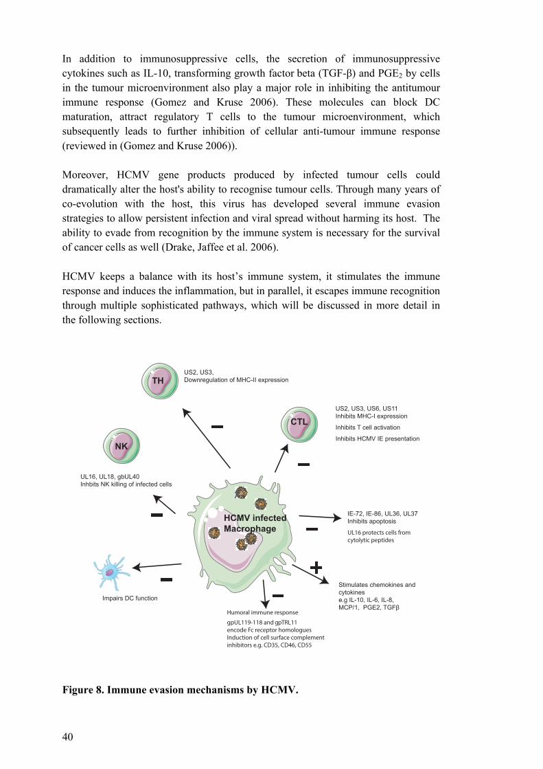
40
In addition to immunosuppressive cells, the secretion of immunosuppressive cytokines such as IL-10, transforming growth factor beta (TGF-β) and PGE2 by cells in the tumour microenvironment also play a major role in inhibiting the antitumour immune response (Gomez and Kruse 2006). These molecules can block DC maturation, attract regulatory T cells to the tumour microenvironment, which subsequently leads to further inhibition of cellular anti-tumour immune response (reviewed in (Gomez and Kruse 2006)). Moreover, HCMV gene products produced by infected tumour cells could dramatically alter the host's ability to recognise tumour cells. Through many years of co-evolution with the host, this virus has developed several immune evasion strategies to allow persistent infection and viral spread without harming its host. The ability to evade from recognition by the immune system is necessary for the survival of cancer cells as well (Drake, Jaffee et al. 2006). HCMV keeps a balance with its host’s immune system, it stimulates the immune response and induces the inflammation, but in parallel, it escapes immune recognition through multiple sophisticated pathways, which will be discussed in more detail in the following sections.
Figure 8. Immune evasion mechanisms by HCMV.
CTL
TH
NK
HCMV infected
Macrophage
US2, US3, US6, US11
Inhibits MHC-I expression
Inhibits T cell activation
Inhibits HCMV IE presentation
US2, US3,
Downregulation of MHC-II expression
UL16, UL18, gbUL40
Inhbits NK killing of infected cells
Impairs DC function
IE-72, IE-86, UL36, UL37
Inhibits apoptosis
Stimulates chemokines and
cytokines
e.g IL-10, IL-6, IL-8,
MCP/1, PGE2, 7*)ȕ�
UL16 protects cells from cytolytic peptides
Humoral immune response
gpUL119-118 and gpTRL11encode Fc receptor homologuesInduction of cell surface complement inhibitors e.g. CD35, CD46, CD55
+
_
_
_
_
_
_

41
2.2.2.7.1 The effect of HCMV on antigen presentation Several HCMV gene products are known to interfere with antigen presentation by MHC molecules. US3 causes retention of MHC-I molecules in the endoplasmic reticulum (ER), US6 inhibits TAP-mediated peptide translocation into the ER, US11 causes dislocation of the MHC-I heavy chain into the cytoplasm while US2 causes export of the MHC-I heavy chain from the ER (Figure 8). MHC peptide presentation is required for CD8+ cytotoxic tumour killing and suppression of antigen presentation will prevent T cell mediated killing of HCMV infected cells (as reviewed by (Soderberg-Naucler 2006)). In addition, HCMV US2 and US3 have been shown to decrease surface expression of MHC-II thereby evading recognition by CD4+ T cells (Miller, Cebulla et al. 2001). In tumour tissues, it has been shown that the HCMV protein pp65, which is consistently detected in human glioma, and it is known to phosphorylate HCMV IE-72 peptides derived from the IE-72 protein. Subsequently, pp65 blocks IE-72 presentation as target epitope preventing it for being recognised by immune system (Griffiths 2004), which may cause HCMV mediated immune evasion of infected tumor cells.
2.2.2.7.2 The effect of HCMV on the NK cell response Natural killer cells provide an important line of defence in killing tumour cells and viral infected cells. HCMV subverts NK mediated killing of infected tumour cells by several different strategies. It has evolved a unique strategy to evade its own virally mediated downregulation of MHC-I antigen to overcome NK cell recognition of infected cells (reviewed in (Soderberg-Naucler 2006)). An HCMV-encoded MHC-I homologue UL18, inhibits NK responses by triggering LIR-1 (Wilkinson, Tomasec et al. 2008). Furthermore, the HCMV encoded protein UL16 binds to some of the ligands such as UL16 binding protein 1 and 2 and MICB, which are recognised by the NKG2D receptor. UL16 therefore prevent NKG2D mediated NK cell activation and this protein also protect infected cells from cytolytic proteins released from NK cells by an unknown mechanism (Figure 8) (Odeberg, Browne et al. 2003; Rolle, Mousavi-Jazi et al. 2003).
2.2.2.7.3 The effect of HCMV on antibody mediated immunity HCMV is known to counteract the effects of HCMV specific antibodies through two HCMV encoded Fc gamma receptor homologue, gpUL119-118 and gpTRL11, which binds to HCMV infected cells, covering antigens presented on the cell surface (Atalay, Zimmermann et al. 2002). Furthermore, the complement system provides a main line of immunologic defence against invading pathogens, to prevent activation of this pathway. HCMV interferes with complement activation through induction of the cellular proteins CD35, CD46 and CD55 that will inhibit opsonisation by phagocytic cells and subsequently prevent complement mediated cell lysis (Figure 8) (Spiller, Hanna et al. 1997).
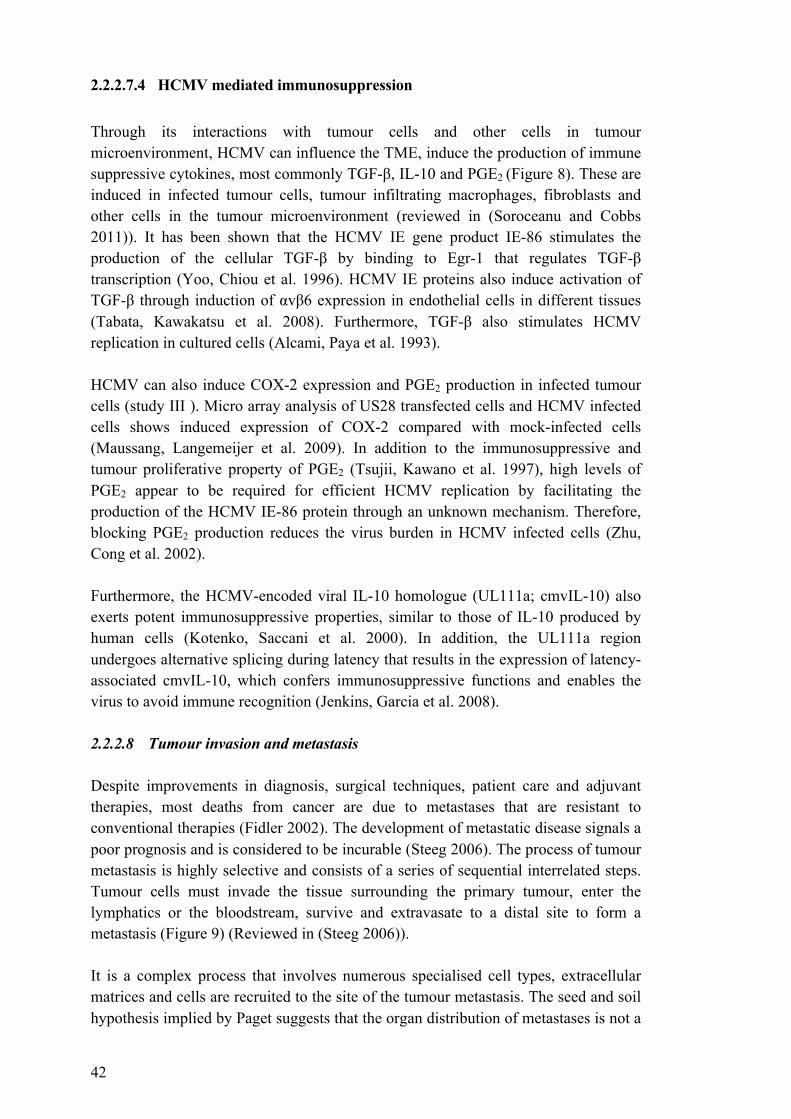
42
2.2.2.7.4 HCMV mediated immunosuppression Through its interactions with tumour cells and other cells in tumour microenvironment, HCMV can influence the TME, induce the production of immune suppressive cytokines, most commonly TGF-β, IL-10 and PGE2 (Figure 8). These are induced in infected tumour cells, tumour infiltrating macrophages, fibroblasts and other cells in the tumour microenvironment (reviewed in (Soroceanu and Cobbs 2011)). It has been shown that the HCMV IE gene product IE-86 stimulates the production of the cellular TGF-β by binding to Egr-1 that regulates TGF-β transcription (Yoo, Chiou et al. 1996). HCMV IE proteins also induce activation of TGF-β through induction of αvβ6 expression in endothelial cells in different tissues (Tabata, Kawakatsu et al. 2008). Furthermore, TGF-β also stimulates HCMV replication in cultured cells (Alcami, Paya et al. 1993). HCMV can also induce COX-2 expression and PGE2 production in infected tumour cells (study III ). Micro array analysis of US28 transfected cells and HCMV infected cells shows induced expression of COX-2 compared with mock-infected cells (Maussang, Langemeijer et al. 2009). In addition to the immunosuppressive and tumour proliferative property of PGE2 (Tsujii, Kawano et al. 1997), high levels of PGE2 appear to be required for efficient HCMV replication by facilitating the production of the HCMV IE-86 protein through an unknown mechanism. Therefore, blocking PGE2 production reduces the virus burden in HCMV infected cells (Zhu, Cong et al. 2002). Furthermore, the HCMV-encoded viral IL-10 homologue (UL111a; cmvIL-10) also exerts potent immunosuppressive properties, similar to those of IL-10 produced by human cells (Kotenko, Saccani et al. 2000). In addition, the UL111a region undergoes alternative splicing during latency that results in the expression of latency-associated cmvIL-10, which confers immunosuppressive functions and enables the virus to avoid immune recognition (Jenkins, Garcia et al. 2008). 2.2.2.8 Tumour invasion and metastasis Despite improvements in diagnosis, surgical techniques, patient care and adjuvant therapies, most deaths from cancer are due to metastases that are resistant to conventional therapies (Fidler 2002). The development of metastatic disease signals a poor prognosis and is considered to be incurable (Steeg 2006). The process of tumour metastasis is highly selective and consists of a series of sequential interrelated steps. Tumour cells must invade the tissue surrounding the primary tumour, enter the lymphatics or the bloodstream, survive and extravasate to a distal site to form a metastasis (Figure 9) (Reviewed in (Steeg 2006)). It is a complex process that involves numerous specialised cell types, extracellular matrices and cells are recruited to the site of the tumour metastasis. The seed and soil hypothesis implied by Paget suggests that the organ distribution of metastases is not a

43
matter of chance and that metastases develop only when the ‘seed’ (certain tumour cells with metastatic ability) and the ‘soil’ (organs providing growth advantage to the seeds) are compatible (Paget 1989). Tumour metastasis growth will be initiated by tumour cell adherence to the extra cellular matrix (ECM), this can be mediated through integrin, CD44 or cadherin binding (Tang and Honn 1994). Before a tumour cell invades the tissues surrounding the main tumour there is a ‘switch’ in cell cadherin expression. E-cadherin, which promotes tumour cell adherence and blocks invasion, is lost and N-cadherin, which is normally expressed on mesenchymal cells and facilitates tumour cell binding to the stroma during invasion, is expressed (Figure 9) (reviewed in (Cavallaro and Christofori 2004)). Furthermore, proteases such as matrix metalloproteinases augment invasion by destroying ECM (Folgueras, Pendas et al. 2004). After invasion into the lymphatic or vascular system, the cancer cells arrest though the binding of coagulation factors or by becoming physically trapped in the capillary beds (Weiss, Grundmann et al. 1986). Expression of endothelial E and P-selectin, can also mediate the initial tethering and rolling of the tumour cell on the vascular wall, followed by firm endothelial–tumour cell adhesion involving cadherins or immunoglobulin- like cell adhesion molecules (ICAM) (Figure 9) (Mannori, Santoro et al. 1997; Kim, Borsig et al. 1998). Successful colonisation crucially depends on interaction of metastatic tumour cells with the microenvironment or “soil” of the distant tissue (Kaplan, Riba et al. 2005). Recently, the epithelial-mesenchymal transition (EMT)- pathway, a process important for embryonic development, and believed to be involved in many pathological processes, was proposed to be crucial in cancer progression and metastasis (Figure 9) (reviewed in (Yang and Weinberg 2008)). During the process of tumour metastasis, which is often enabled by EMT (Thiery 2003), tumour cells loose their adhesive connection with adjacent cells, become invasive and also appear to acquire stem cell characteristics in order to spawn macroscopic metastases (Morel, Lievre et al. 2008). This process is likely induced by growth factors, regulatory proteins and other signalling proteins such as TGF-β, snail, sonic hedgehog (Shh), Wnt/β-catenin and ECM components, which subsequently induce invasive and migratory capabilities of cancer cells (reviewed in (Gos, Miloszewska et al. 2009)). It has been shown that over expression of the transcription factor snail in glioblastoma patients associates with glioblastoma invasiveness in animal model (Savary, Caglayan et al. 2013). The main cytokine responsible for initiating EMT is TGF-β, which is highly induced by tumour cells, and other cells in the tumour microenvironment, and provides widely diverse functions in tumour formation. In early stage malignancy TGF-β can have a tumour suppressive, but its role in the induction of EMT suggests that it could later promote the formation of metastatic disease (as reviewed in (Heldin, Vanlandewijck et al. 2012)).

44
TGF-β plays a central role in inducing EMT response by binding to its surface receptors which then signal via intracellular effectors (SMADs) (reviewed in (Lonn, Moren et al. 2009)). This induces, a cellular reprogramming, subsequently activating complementary signalling cascades that mobilise embryonic transcription factors, reprogramming the epithelial cell so that it acquires both progenitor-like, pro-motility and mesenchymal features (reviewed in (Moustakas and Heldin 2012)), thereby enhancing the metastatic potential of tumour cells. Thus, TGF-β has tumour immune suppressive as well as tumour promoting effects supporting metastasis formation (reviewed in (Moustakas and Heldin 2012)). Recently it has been shown that p53 plays an important role in regulating EMT induced by TGF-β in mouse mammary epithelial cells (Termen, Tan et al. 2013). There have been a number of studies suggesting HCMV proteins may also play a role in the induction of tumour dissemination. Studies conducted by Scholz et al., show that HCMV infection in neuroblastoma cells can induce tumour cell adhesion to endothelial cells (EC) resulting in focal disruption of the EC monolayer integrity, enhanced trans-endothelial migration and increased neuroblastoma invasiveness (Scholz, Blaheta et al. 2000). Moreover, HCMV infected prostatic cancer cells showed increased adhesion to the endothelium, which was mediated by activation of β1α5 integrin on the surface of infected tumour cells, leading to a focal disruption of endothelial cell integrity, thus facilitating tumour cell transmigration (Blaheta, Weich et al. 2006). The HCMV gene product US28 also promotes cell migration through a ligand-dependent process (Streblow, Soderberg-Naucler et al. 1999); US28 mediated migration occurred towards the chemokines RANTES and monocyte chemo attractant protein-1 (MCP-1) (Streblow, Soderberg-Naucler et al. 1999), which are abundantly expressed in malignant gliomas (Desbaillets, Tada et al. 1994). This could also explain how HCMV may mediate enhanced tumour invasiveness. Enhanced expression of both integrin αvβ3 and platelet-derived growth factor receptor-α (PDGFRα) has been suggested to play a role in promoting cellular migration in glioblastoma tumours (Ding, Stewart et al. 2003). Cobbs and co-workers demonstrated that HCMV can engage glioblastoma cell PDGFRα and the αvβ3 integrin, which leads to an increased migratory capacity. They showed that blocking antibodies can reverse this effect, suggesting that HCMV may act through αvβ3 integrin/PDGFRα to promote glioma cell invasiveness (reviewed in (Soroceanu and Cobbs 2011)). HCMV can also utilise the PDGFRα as a receptor on neuronal cells (Soroceanu, Akhavan et al. 2008). One study by Shimamura and coworkers (Shimamura, Murphy-Ullrich et al. 2010) investigated the role of HCMV in the induction of TGF-β and its role in EMT. They infected human renal tubular epithelial cells in vitro and found that they underwent morphologic and transcriptional analogous to EMT. In addition, TGF-β and MMP-2 expression was induced. HCMV IE proteins likely control this process as their overexpression recapitulated these effects and targeting late gene expression did not inhibit these changes (Shimamura, Murphy-Ullrich et al. 2010).
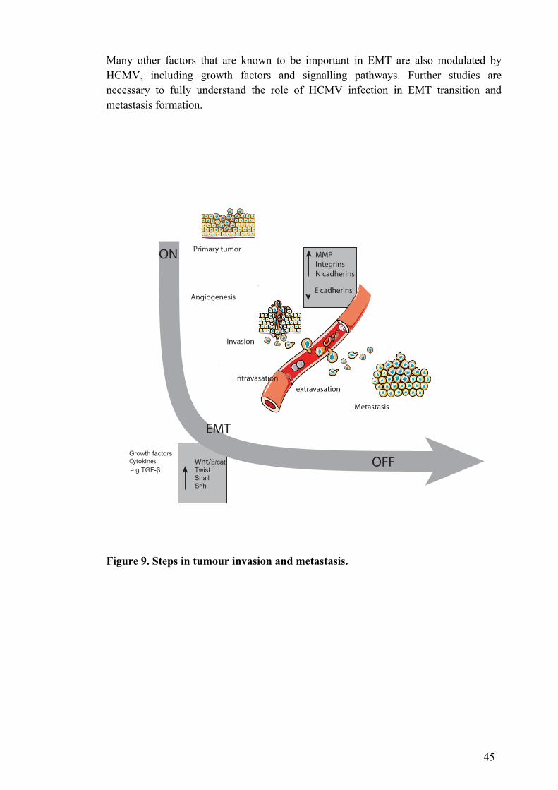
45
Many other factors that are known to be important in EMT are also modulated by HCMV, including growth factors and signalling pathways. Further studies are necessary to fully understand the role of HCMV infection in EMT transition and metastasis formation.
Figure 9. Steps in tumour invasion and metastasis.
extravasation
Metastasis
Intravasation
Angiogenesis
Primary tumor
Cytokines
Invasion
OFF
EMT
ON
e.g TGF-ȕ
Growth factors
MMPIntegrinsN cadherins
E cadherins
Wnt/ȕ�FDWTwistSnailShh

46
2.2.2.9 Inflammation and the tumour microenvironment Since Virchow first proposed in 1863 that tumours could originate from sites of chronic inflammation, it has been well established that chronic inflammation both contributes to cancer progression and predisposes tissues to various types of cancer. Recently, inflammation was recognised as a hallmark of cancer (Colotta, Allavena et al. 2009). Today, many studies have correlated the prolonged presence of the inflammatory milieu with an increased risk of cancer development (Keibel, Singh et al. 2009). Furthermore, blocking inflammatory pathways COX-2 by anti-inflammatory drugs (aspirin) have been shown to reduce the long-term risk of several cancers and the risk of distant metastasis (Algra and Rothwell 2012). With an increased understanding of molecular pathways of cancer in recent years, we know that the formation of a clinically relevant tumour requires support from the TME. Important TME components include stromal cells, soluble factors, signalling molecules and extracellular matrix (Lorusso and Ruegg 2008). This microenvironment provides support for tumour growth and metastatic spread. Furthermore, inflammatory cells and immunomodulatory mediators present in the TME polarise the host immune response toward specific phenotypes that facilitate tumour progression (reviewed in (Lorusso and Ruegg 2008)). Infectious agents that induce a chronic, low level of infection may promote oncogenesis by interfering with different molecular mechanisms important in tumour transformation, such as the induction of host cell genetic damage and the modulation of the inflammatory cytokine milieu and subsequent immune suppression (Grivennikov, Greten et al. 2010). Myeloid lineage cells are important contributors to the inflammatory cytokine in TME. In tumour tissue from patients with various cancers e.g. colon, breast and glioma, HCMV was detected in not only the tumour cells, but also in these inflammatory cells and fibroblasts (Harkins, Matlaf et al. 2010; Soroceanu, Matlaf et al. 2011). HCMV establishes latency in myeloid lineage cells, but probably never exists in a true latent state (Reinke, Prosch et al. 1999). Its reactivation depends on inflammation and differentiation of monocytes into macrophages or DCs (Sinclair and Sissons 1996; Soderberg-Naucler and Nelson 1999; Reeves, MacAry et al. 2005). It has been shown that HCMV proteins representing all stages of permissive HCMV infection were detected in macrophages, suggesting that these cells can support the complete viral replication cycle, although infected cells do not show any cytopathic effects (Sinzger, Plachter et al. 1996). Macrophages in the tumour microenvironment, known as tumour-associated macrophages (TAMs), exist in two types, pro-inflammatory M1 and anti-inflammatory M2. HCMV infection of macrophages promotes both the M1 and M2 phenotypes, generating what is known as a “schizophrenic” phenotype of macrophage that can promote neoplastic progression (Allavena, Sica et al. 2008). HCMV infection of macrophages can induce the secretion of both pro-inflammatory and anti-inflammatory cytokines (Chan, Bivins-Smith et al. 2008; Chan, Bivins-Smith et al.
47
2008). Pro-inflammatory cytokines that are secreted from M1 type macrophages include IL-1, IL-6 and TNF alpha, which are known to promote viral replication (Chan, Bivins-Smith et al. 2008). Chronic expression of these cytokines is also directly linked to oncogenic transformation in inflammation-induced animal models of cancer (Grivennikov, Greten et al. 2010). Also the anti-inflammatory molecules produced by infected macrophages; TGF-β, IL-10 and PGE2 can play important roles in tumour progression (as discussed above). Cyclooxygenase (COX) is the key enzyme that is required for the conversion of arachidonic acid to prostanoids (e.g. prostaglandins, thromboxanes and prostacyclins). It is shown to be over- expressed in different cancers (as reviewed in (Wang and Dubois 2010)), were high expression often is correlated with poor prognosis (Shono, Tofilon et al. 2001). It is also shown PGE2, was induced in several tumour types (Loh, Hwang et al. 2002) and it promotes tumour growth through enhancement of cellular proliferation, induction of angiogenesis, invasion and resistance to apoptosis in an autocrine and/or paracrine fashion (Wang and Dubois 2010). PGE2 is also an important meditator providing an inflammatory microenvironment as well as suppressing tumour immune responses (Wang and Dubois 2010). This pathway has been well investigated by many researchers especially after the introduction of nonsteroidal anti-inflammatory drugs (NSAIDs), which are promising agents for the prevention and treatment of various cancers (Din, Theodoratou et al. 2010; Mathers, Movahedi et al. 2012). Interestingly, it has been reported that NSAIDs can reduce HCMV replication in infected cells, thus acting as an anti-viral agent (Speir, Yu et al. 1998). PGE2 was shown to be required for efficient viral replication (Zhu, Cong et al. 2002). We recently showed that HCMV proteins are expressed in medulloblastoma and that HCMV infection induces COX-2 expression both in vitro and in vivo (study III). The anti-viral drug valganciclovir and the specific COX-2 inhibitor celecoxib prevented HCMV replication in vitro, inhibited PGE2 production, and reduced medulloblastoma cell growth in vivo and in vitro. Furthermore in the TME there are rare subpopulations of tumour cells that are responsible for the initiation and maintenance of the tumour, these are called cancer stem cells (CSCs). CSC are defined as “tumour cells that contain self-renewal capacity and multi-lineage differentiation capacity” (Clarke, Dick et al. 2006), and can be identified by several markers including CD133 and CD44 (Singh, Hawkins et al. 2004). However, no specific marker has yet been defined for all CSCs. CSCs have the capacity to both divide and expanding the CSC pool, and to differentiate into the heterogeneous non tumourigenic cancer cell types that constitute the bulk of the tumour. CSCs are believed to be the subpopulation of cells resistant to therapy and play a main role in tumour recurrence (Medema 2013) and tumour metastasis (Croker, Goodale et al. 2009). Interestingly, we found that substantial proportion of brain tumour cells that expressed stem cell markers also expressed HCMV proteins (study III). Furthermore, in studies of adult mice infected with murine CMV showed

48
IE gene expression could be detected in nestin positive, glial fibrillary acidic protein (GFAP) positive, cells in the subventricular zone suggesting that either partial viral gene expression or full infectious viral life cycle occurs in these neuro-glial stem cells throughout adult life (Shinmura, Aiba-Masago et al. 1997). Furthermore, HCMV infection in neuroprogenitor cells (NPC) was shown minimally cytopathic, suggesting that these NPC might remain persistently infected in vitro (Luo, Schwartz et al. 2008). When NPC are infected with HCMV they are unable to differentiate, which may also be an important step in tumour transformation. 2.3 IS HCMV ONCOGENIC?
The possible oncomodulatory potential of HCMV has been mentioned earlier in this thesis. However, as several evidences also support an oncogenic role of HCMV; here it will be discussed in more detail. The oncogenic potential of HCMV was originally demonstrated by the ability of HCMV to transform a variety of rodent and human cells in vitro (Albrecht and Rapp 1973; Geder, Lausch et al. 1976). The transformation ability of HCMV suggested the presence of possible transforming genes region in the HCMV genome. In vitro transformation studies by several groups found three HCMV morphologic transforming regions (mtr), i.e., mtrI, mtrII, and mtrIII. (Clanton, Jariwalla et al. 1983; Nelson, Fleckenstein et al. 1984; el-Beik, Razzaque et al. 1986). Initially, Nelson et al. identified the mtrI sequence in the laboratory HCMV strain (AD169) as an inducer of transformation. However, mtrI was not retained in the transformed cells, suggesting that its presence was not essential for the transformed phenotype (Nelson, Fleckenstein et al. 1984). Subsequently, Clanton et al. identified another region in the HCMV strain Towne, based on its hybridisation to transforming regions of herpes simplex virus (HSV-2) under non-stringent conditions and observed that it immortalised hamster embryo cells and transformed NIH 3T3 cells (Clanton, Jariwalla et al. 1983). Furthermore, El-Beik et al. showed that sub-clones of Towne containing either the (mtrII) or (mtrIII) were capable of transforming NIH 3T3 and rat-2 cells and these cells were tumourigenic when injected into 5-week old rats (el-Beik, Razzaque et al. 1986). Surprisingly, transfection of rat-2 cells with both mtrII plus mtrIII exhibited a sevenfold-greater transformation frequency than cells transfected with either mtrII or mtrIII alone and double transfected cell lines produced tumours in syngeneic rats much earlier (5 to 7 days) than those transformed by either mtr alone (25 to 35 days) (Jariwalla, Razzaque et al. 1989). Expression of mtrI and III decreased by time; only mtrII was retained and expressed in both transformed and tumour-derived cells. It has been shown that mtrII expressed proteins bound to the tumour suppressor protein p53 and inhibited its ability to transactivate a p53-responsive promoter (Muralidhar, Doniger et al. 1996). In later

49
studies using normal rodent cells, HCMV infection was shown to induce mutations in genes that are critical for malignant transformation. However, viral DNA was not detected in most transformants. These findings led to the speculation that HCMV contributes to oncogenesis by a “hit-and-run” mechanism (Nelson, Fleckenstein et al. 1984; Shen, Zhu et al. 1997). Further studies failed to reproduce these results and so the term oncomodulation has been applied to explain the role of HCMV tumourigenesis (Cinatl, Vogel et al. 2004). However, numerous recent data indicate that several other HCMV encoded proteins have biological properties that are known to be directly related to cellular transformation and tumour development, such as US28 and the IE proteins. Several studies provide evidence for a potential role of US28 in promoting tumourigenesis (Maussang, Verzijl et al. 2006; Maussang, Langemeijer et al. 2009; Slinger, Maussang et al. 2010). It has been shown that mice embryonic fibroblast expressing HCMV-US28 exhibited a number of properties consistent with a transformed phenotype, such as increased cell growth, enhanced cell cycle progression and expression of the pro-angiogenic factor VEGF (Maussang, Verzijl et al. 2006). HCMV US28 is also shown to induce NF-κB activation and results in STAT3 phosphorylation, COX-2 expression and IL-6 production, which ultimately induces cellular proliferation in cells expressing HCMV US28 (Slinger, Maussang et al. 2010). Furthermore, US28 targeted expression in intestinal cells inhibits GSK-3β function, resulting in increased β-catenin activity and deregulated expression of a several Wnt target genes, including cyclin D, survivin and c-myc, which control cellular proliferation (Bongers, Maussang et al. 2010). Moreover, transgenic mice (VS28) in which US28 expression was targeted to intestinal epithelial cells, developed intestinal neoplasia (Bongers, Maussang et al. 2010). These findings provide a direct molecular link between the expression of US28 and oncogenesis. Enhanced telomerase activity is necessary for tumour cells to divide indefinitely. Many oncogenic viruses commonly induce telomerase activity, which has a pivotal role in tumour development (Bellon and Nicot 2008). Recently, our group showed that the HCMV IE-72 protein directly interacts with the hTERT promoter at SP1 binding sites to induce telomerase activity and telomere lengthening (Straat, Liu et al. 2009). Using the revised Koch´s postulates, which explains a causal relationship of viral infection in cancer as mentioned earlier, Melnick et al. suggested that HCMV fulfills these criteria and that this virus therefore should be classified as an “oncovirus” (Melnick, Sedghizadeh et al. 2012). They demonstrated the presence of HCMV in 97% of mucoepidermoid carcinomas of salivary glands and HCMV protein expression (HCMV IE and pp65) positively correlated with disease severity. They also showed that HCMV protein expression correlated with activation of known oncogenic pathways, such as EGFR, COX-2, ERK and ampheregulin (Melnick, Abichaker et al. 2011; Melnick, Sedghizadeh et al. 2012).

50
Today, it has become apparent that HCMV encoded proteins induce different cellular signalling pathways, such as Wnt/ β-catenin, PI3K/Akt, JAK/STAT and MAPK pathways, which play an important role in oncogenic transformation (Chang, Lee et al. 2003; Cinatl, Vogel et al. 2004; Soroceanu and Cobbs 2011). In addition, it has been demonstrated that different HCMV proteins can inhibit apoptosis (Goldmacher, Bartle et al. 1999; McCormick, Roback et al. 2008). Furthermore, HCMV encoded proteins can activate proto-oncogenes and interact with key cell-cycle regulatory proteins such as p53 and pRb, which leads to cell cycle deregulation and increased cellular proliferation (Boldogh, AbuBakar et al. 1991; Allart, Martin et al. 2002; Hume, Finkel et al. 2008). All these may provide a direct molecular link between HCMV infections in favour of oncogenic transformation. 2.4 CAN HCMV BE TARGETED IN TUMOURS TO IMPROVE PATIENT’S
OUTCOME?
The evidence that HCMV has been detected in a majority of human cancers has raised the idea of treating HCMV positive cancers by targeting the virus and has encouraged several clinical trials. Recently, we performed the first clinical trial to examine the safety and efficacy of anti-viral treatment of HCMV in glioblastoma patients. The study failed its primary end point to demonstrate decreased tumour growth at 6 months, but we observed improved survival in patients receiving long-term anti-viral treatment in exploratory analyses; patients exhibited a median overall survival of 24.1 months compared to contemporary controls (13.7 months) (p < 0.0031) (Stragliotto, Rahbar et al. 2013). Additionally, we found that the use of the antiviral drug valganciclovir in nude mice inhibited tumour growth from engrafted human medulloblastoma cells. Surprisingly, this treatment effect was enhanced when combined with the COX-2 specific inhibitor celecoxib, which can also inhibit HCMV infection (study III). In sharp contrast, the drug had no effect neither on the clonogenic capacity of tumour cells or tumour growth in vivo of two HCMV-negative cell lines derived from prostate and pancreas adenocarcinomas (study III). These observations strongly suggests that the inhibitory effect of valganciclovir on medulloblastoma growth is HCMV specific, and is not mediated by potential non-specific drug effects inhibiting cellular proliferation. Furthermore, HCMV is highly immunogenic, which makes it an interesting target option for immunotherapy (Sampson and Mitchell 2011). Several clinical trials are currently ongoing, targeting HCMV in glioblastoma patients (reviewed in (Hickey, Malone et al. 2010)). HCMV antigens are delivered to cells of interest (cancer cells), then after programing the patient’s own immune cells against HCMV antigens in vitro, cells are transferred back to the patient as adoptive therapy. These “armed” cells theoretically target HCMV antigens in the human tumour and utilise the host immunity to destroy the tumour.

51
Recently, Crough et al. utilised HCMV immunotherapy in cancer treatment, by expanding HCMV positive T cells in vitro before transferring them back into one patient with recurrent glioblastoma. This patient had an unexpected long-term disease free survival. (Crough, Beagley et al. 2012). In addition, several other clinical trials suggest that targeting HCMV positive tumour cells may improve patient outcome, especially in glioblastoma. In an ongoing phase II clinical trial at Duke University, investigators are using autologous lymphocyte transfer (ALT) with HCMV pp65-activated T cells, after vaccination with HCMV pp65 mRNA-loaded DCs, in patients with glioblastoma, determining if ALT with HCMV-specific T cells with or without vaccination with HCMV pp65- mRNA loaded DCs extends progression-free survival of these patients when compared with historical cohorts (reviewed in (Hickey, Malone et al. 2010)). Furthermore, autologous HCMV-specific CTL genetically modified to express chimeric antigen receptors (CAR) targeting the HER2 molecule in patients with HER2-positive GBM is another example of an ongoing clinical trial targeting HCMV antigens in cancer patients (reviewed in (Hickey, Malone et al. 2010)). Thus, if HCMV infection is confirmed to be associated with initiation or tumour progression, then targeting HCMV could become a potential therapeutic approach motivating extensive exploration for medical and immunological therapies targeting this virus.
52
3 AIMS OF THIS THESIS
The aims of my thesis were:
1. To study the prevalence of HCMV in tumours from colon and breast cancer, as well as in sentinel nodes and brain metastasis, obtained from colon and breast cancer patients.
2. To investigate if HCMV infection grade in tumour tissues from patients with breast cancer and colon cancer with brain metastasis correlate with clinical outcome.
3. To investigate the prevalence of HCMV in medulloblastoma and whether
HCMV induces COX-2 expression in tumour cells.
4. To investigate if targeting HCMV replication with antiviral and/or a COX-2 inhibitor can prevent tumour growth in vitro and in vivo.
5. To investigate the prevalence of a unique HCMV strain in cancers of different origin.

53
4 RESULTS AND DISCUSSION
4.1 STUDY I
Emerging evidence demonstrates a high prevalence of HCMV protein and nucleic acid in several malignancies originating in different organs, but it is not known whether HCMV is present in metastatic cells. In this study, we aimed to examine the prevalence of HCMV in breast cancer and paired sentinel lymph node (SLN) specimens, using sensitive IHC and Taqman PCR methods. Furthermore, we wanted to determine if there was any association between HCMV infections and known prognostic factors. Background Breast cancer is the most common malignancy and a leading cause of cancer death in women worldwide (reviewed in (Key, Verkasalo et al. 2001)). It comprises 22% of all cancers in females (Dumitrescu and Cotarla 2005). The exact etiology of this disease is unknown, although multiple risk factors (age, hormones, alcohol, diet, familial history, etc.) have been documented (Dumitrescu and Cotarla 2005). Environmental and epidemiological factors that contribute to breast cancer are poorly understood, which has created an increased interest to identify additional risk factors that contribute to the disease. Infections, most commonly viral infections, have been proposed to be responsible for over 20% of all malignancies worldwide (Kuper, Adami et al. 2000). The identification of a viral agent for breast cancer, however, has eluded researchers for decades (Mant, Hodgson et al. 2004) and generated considerable controversy (Lawson, Gunzburg et al. 2006). So far, there is lack of convincing evidence that the viruses are truly present in human mammary epithelium or are associated with breast cancer (reviewed in (Lawson, Gunzburg et al. 2006)). Many studies have demonstrated the absence of oncogenic viruses, such as EBV (Herrmann and Niedobitek 2003) and HPV (Silva and da Silva 2011) in breast cancer, while others demonstrated the presence of oncogenic viruses (EBV (Labrecque, Barnes et al. 1995)) and HPV (Heng, Glenn et al. 2009)) in breast cancer. Emerging evidence suggests that HCMV is associated with several human malignancies, including breast cancer, and that HCMV gene products can modulate oncomodulatory properties of cells in vitro, as discussed earlier. The possible role of HCMV in breast cancer was first elucidated based on epidemiological considerations, as investigators demonstrated that breast cancer patients have increased IgG antibody titers to HCMV, compared to controls (Richardson 1997; Richardson, Cox et al. 2004). Similar findings were observed by El- Shinawi et. al., who found higher HCMV IgG titres in women with breast cancer than in a non breast cancer control group. Furthermore, Tsai et al, suggested that HCMV positivity in breast cancer was associated with lower relapse-free time and overall survival (Tsai, Hsu et al. 2007). Recent observations by several groups

54
confirmed the presence of HCMV infection in breast cancer patients (Tsai, Tsai et al. 2005; Harkins, Matlaf et al. 2010; El-Shinawi, Mohamed et al. 2013). In addition, HCMV-infected breast cancer tissues were found to be enhanced in NF-κB signalling in inflammatory breast cancer (El-Shinawi, Mohamed et al. 2013). However, others have failed to identify HCMV nucleic acid in breast cancer tissue specimens (Utrera-Barillas, Valdez-Salazar et al. 2013) or in normal glandular breast tissue (El-Shinawi, Mohamed et al. 2013). In contrast, several studies have reported a high prevalence of HCMV protein expression in normal glandular epithelium (Tsai, Tsai et al. 2005) (Harkins, Matlaf et al. 2010). Based on up-to-date findings there is still a controversy whether HCMV is present in breast cancer or not. Furthermore, HCMV infection in metastatic cells has not been investigated so far and was an area that we were interested to explore further. Results and discussion High prevalence of HCMV infection in breast cancer and paired sentinel lymph node specimens In this study, we collected formalin-fixed paraffin-embedded tissue specimens from breast cancer and paired sentinel lymph node (SLN) samples from patients with (n=35) and without SLN metastasis (n=38). Of these, paired SLN specimens were available from 34 and 35 patients, respectively. We used a high sensitive immunohistochemistry (IHC) method optimised for detection of HCMV proteins in tissue specimens (Cobbs, Harkins et al. 2002; study I) and a TaqMan PCR for detection of HCMV nucleic acids. Our findings were in line with the recent observations by Harkins et al (Harkins, Matlaf et al. 2010), demonstrating a high prevalence of HCMV protein expression in breast cancer. We detected HCMV IE and LA antigens in all examined breast cancer specimens (n=73). Furthermore, both HCMV IE and LA proteins were detected in 94% of SLN positive samples and in 60% of the SLN-negative group. HCMV protein expression was mainly confined to metastatic tumour cells. HCMV-positive inflammatory cells were also found in 79% of SLN samples with metastasis, and very few HCMV-positive inflammatory cells were also detected in 60% of metastasis-free SLNs. We graded HCMV IE and LA protein expression in the tumour tissues based on the percentage of IE and LA positive cells. Sections were graded as negative (0), grade I (< 25% positive cells), grade II (25-49%), grade III (50-75%) or grade IV (> 75%). In this cohort we found that the majority of breast samples were of high HCMV grade (grade III and IV), both in the SLN-positive and -negative groups. Consistent with our findings in other tumours (study II; study III; Rahbar, Stragliotto et al. 2012), HCMV LA protein expression was less abundant than HCMV IE. In the current study, grade II and III were the most common grades for HCMV LA, which may indicate that there is non-productive viral replication in these cancer cells.

55
In positive SLNs, 76% samples were graded as HCMV IE grade III or IV; most metastatic cells were HCMV positive. However, the HCMV IE protein expression grade was higher in the primary breast cancer specimen compared with the SLN tissue specimens. This is in contrast to our findings in study II, where the grade of viral protein expression was higher in distal metastasis compared with the primary tumour. This may be explained by a difference in disease stage, or by immune related mechanisms, but no firm conclusions can be made from this small number of patients. Interestingly, we noted that HCMV protein expression was mainly restricted to tumour cells, although other cells e.g. some inflammatory cells sometimes also expressed HCMV IE proteins. HCMV infection grade in breast cancer tissue specimens does not correlate with patient outcome. In patients with glioblastoma, we recently found a strong association between low grade of HCMV infection in the glioblastoma tumour at time of diagnosis and prolonged survival, which implies that HCMV may be involved in tumour development (Michaelis, Doerr et al. 2009; Rahbar, Stragliotto et al. 2012). In study II, we also found that HCMV infection grade appeared to be associated with shorter time to tumour metastasis and shorter survival after diagnosis of distal metastasis. In the current study only 7 patients died from breast cancer, which did not allow for further statistical analyses or conclusions. However, we noted that 6 of 7 patients who died of breast cancer had high-grade HCMV infection (grade IV), while patients with low HCMV IE expression were all among the survivors. Our observations imply that HCMV may influence tumour progression and survival, and opens up a discussion of whether HCMV targeted therapies may improve the outcome of patients with HCMV positive cancers (see discussion for study III). HCMV infection level does not correlate with clinical prognostic factors Estrogen receptor alpha (ERα)) and progesterone receptor (PR) status and Elston grade are well-known prognostic markers for breast cancer patients and impact patient treatment. Loss of ERα and PR expression are predictors of poor survival (Xu, Schlesinger et al. 2012). The Elston-Ellis histopathological grading provides important prognostic information for breast cancer patients by assessing tubular formation, mitotic activity and nuclear pleomorphism. Elston grade I tumours have a significantly better survival than those with grade II and III tumours (Dalton, Page et al. 1994). In the small number of patients we studied, we did not find any significant association between HCMV infection grade and these prognostic markers. Although the finding of an infectious agent in tumour tissues does not provide evidence for its causal involvement in carcinogenesis, the presence of HCMV both in the primary breast tumour and in most SLN metastases (while adjacent healthy tissues remained HCMV negative) supports the hypothesis that HCMV may play an active role in tumourigenesis and metastasis of breast cancer. Therefore, further

56
studies are highly warranted to further evaluate the role of and understand possible mechanisms utilised by HCMV that could contribute to breast cancer tumourigenesis and metastatic diseases. .
57
4.2 STUDY II
HCMV proteins and nucleic acids were shown to be highly prevalent in several different malignancies and local SLN metastasis (study I). In this study, we aimed to extend our scope to investigate HCMV infection in brain metastases (BMs), from both breast and colorectal cancer patients and also to investigate whether HCMV infection grades impact tumour characteristics or tumour progression. Background Despite all the advances in cancer management, the development of metastases represent a particularly devastating consequence of a primary tumour, as metastases negatively impact on both mortality and quality of life and are considered to be incurable (Steeg 2006). BMs are the most common intracranial neoplasms in adults, which occur in approximately 10–30% of patients with solid tumours, but their prevalence is likely higher, as regular screening is not routine for cancer patients (Posner and Chernik 1978; Gavrilovic and Posner 2005). The incidence appears to be increasing (Smedby, Brandt et al. 2009; Noura, Ohue et al. 2012), probably as a result of improved overall survival in cancer patients (Shmueli, Wigler et al. 2004), as well as earlier and more accurate detection with modern neuroimaging modalities (Palmieri, Chambers et al. 2007). The incidence of brain metastasis in patients with breast cancer and colon cancer is 15% and 4%, respectively. However, the major source for brain metastasis is shown to be lung cancer (Farnell, Buckner et al. 1996; Go, Klaassen et al. 2011), since 48% of brain metastasis originate from tumours in the lung. In study II we decided to include colon and breast cancer patients due to our (and others) previous observations demonstrating a high prevalence of HCMV infection in colon and breast cancers ((Harkins, Volk et al. 2002; Harkins, Matlaf et al. 2010; study II). In the current study, we investigated the prevalence of HCMV in metastatic brain tumours of breast and colon cancer. Results and discussion High prevalence of HCMV infection in primary tumour and paired metastasis tissue specimens from colon and breast cancer patients We examined 78 tissue specimens of brain metastasis (BMs) obtained during neurosurgery of patients with breast (n=41) or colorectal (n=37) cancers. In addition, paired primary tumour samples were available from 13 breast and 12 colorectal cancer patients. Patient data were collected from medical records. Using IHC, we detected HCMV IE and LA protein in 98.7% and 98.6% of the BM tumours respectively. HCMV proteins were detected in 92–100% of corresponding primary breast and colorectal cancer specimens. Thus, consistent with our previous findings in study I, we detected very high prevalence of HCMV infection in metastatic tumour tissue specimens.
58
In this cohort, we graded the level of infection into low grade and high grade, due to small number of patients included in this study. Thus, instead of a four grading system that we used in study I, we combined grade I and II into low grade infection, while grade III and IV represented high grade HCMV infection. The grading was done based on the estimated percentages of IE- and LA-positive cells: negative (0% positive cells), low-grade infection (< 50% positive cells), or high-grade infection (≥ 50% positive cells). Among 78 patients examined for HCMV IE protein expression, one patient was HCMV IE negative (not included in our further analysis), 28% of patients had low-grade infection while 71% had high-grade infections. In line with our results in study I, HCMV LA expression was less abundant than HCMV IE expression. One patient was negative for HCMV LA protein expression. 73% of patients had low-grade infections, while 26% had high-grade infections for HCMV LA - which may indicate that the infection was not productive. The HCMV infection level both in primary tumour and brain metastasis correlates with time to tumour metastasis and patient survival Patients included in this study survived longer than expected for patients with BMs. Patients who were not eligible for brain surgery were not included in this study, making our cohort a selected group with favourable disease characteristics. The median overall survival after diagnosis of BMs was 8.6 months (15.4 months for breast cancer patients versus 6.0 months for colorectal cancer patients). We observed that the grade of HCMV infection in BMs was significantly associated with patient outcome. Patients with low grade HCMV infection tended to have a higher median overall survival from BM diagnosis; 13.5 months in patients with low-grade HCMV infection compared to 6.9 months in those with high-grade infection in the BM. Furthermore, we examined 25-paired available specimens of primary breast cancer (n=13) and colorectal cancer (n=12). HCMV IE proteins were detected in 92% of primary breast cancer specimens and all primary colon cancer specimens. Patients with high-grade HCMV infection in primary tumours had significantly shorter median time to diagnosis of BM than those with low-grade HCMV infection (30 vs. 65.1 months). These patients also had significantly shorter median overall survival than those with low-grade HCMV infection (median 37.8 vs. 81.5 months). Our observations further support the hypothesis that HCMV infection may contribute to tumourigensis and metastasis of breast and colorectal cancers, rather than simply representing an epiphenomenon of the disease. HCMV infection level correlates with clinical prognostic markers Since hormonal receptor expression (ERα, PR) and Her2 in primary and metastatic breast cancers are well-known prognostic markers (Barbieri, Sanpaolo et al. 2011; Xu, Schlesinger et al. 2012), we further evaluated the grade of HCMV infection in
59
relation to ERα (n=29) and PR (n=21) expression in patients with breast cancer as a primary diagnosis. ERα and PR expression was also available for 15 and 14 BMs, respectively. Among primary breast cancer patients with available ERα and PR staining results, tissue specimens were available for 10 patients with ERα staining and for 6 patients with PR staining. In accordance with published observations, both tumour aggressiveness and survival data differed significantly between receptor-positive (ERα, PR and Her2) and receptor-negative breast cancer cells in specimens of primary tumours and BMs. We observed that the time to brain metastasis was shorter in patients with ERα negative (median, 30 vs. 73.3 months) or PR negative tumours (median, 30 vs. 70.4 months), compared to those with ERα and PR positive tumours. These patients also had shorter survival after primary tumour diagnosis (median: 44.9 vs. 97.3 months for ERα negative, and 37.0 vs. 88.1 months for patients with PR negative tumours). Staining results for these receptors on BMs tissue samples, also showed shorter survival after diagnosis of the metastasis in patients with ERα- and PR-negative tumours, than in those with receptor-positive tumours (median: 12 vs. 34.8 months for ERα, and 12.7 vs. 57.5 months for PR negative BM). Correlating our HCMV staining result with ERα staining both in primary and metastatic tumour samples revealed that 67% of primary breast cancers that were ERα negative had high-grade HCMV infection, whereas 0% of ERα positive samples did. Furthermore, 83% of patients with ERα-negative and 44% with ERα-positive metastatic tumours had high-grade HCMV infection. Progesterone receptor expression in primary and metastatic tissues also tended to correlate with HCMV infection grade, as all PR-negative primary tumour samples and half of PR-positive samples had a high-grade HCMV infection. In BM tissue samples, 73% PR-negative and 0% of the PR-positive BM had high-grade HCMV infection (Supplementary figure 1 and 2 in study II). Unfortunately, Her2 staining was only available from a few patients, which did not allow for further analysis. Here we did observe a correlation between hormone receptor expression and HCMV grade, in contrast to our previous study (study I). This difference in results could be due to different grading systems and tumour state and disease staging protocols in the two studies. Although it is very hard to draw conclusions based on the small number of patients studied, we raise important questions for future projects to investigate the mechanistic link between HCMV infection and breast cancer. In conclusion, we found that HCMV infection is highly prevalent in BMs of breast and colorectal cancers and in paired primary cancer specimens. Further studies with larger patient cohorts are needed to assess the possible role of HCMV infection in metastatic cancer and to determine whether HCMV-targeted therapies have a place in the treatment of metastatic cancer.

60
4.3 STUDY III
In this study, we aimed to determine the prevalence of HCMV in medulloblastoma and to investigate if HCMV can modulate inflammatory factors, such as COX-2, and if targeting HCMV replication with antiviral drugs, alone or in combination with a drug inhibiting COX-2 pathways (COX-2 inhibitor), affected tumour growth in vitro and in vivo. Background Medulloblastoma is the most common pediatric malignant tumour of the central nervous system, which usually arises in the cerebellum. Although the exact mechanism of medulloblastoma development is unknown, it has been linked to disorders of normal development (Scotting, Walker et al. 2005). A majority of medulloblastoma cells exhibit abnormal activation of developmental signalling cascades such as Wnt/β-catenin, PI3K/Akt and Sonic Hedgehog, which all have crucial functions during normal brain development (Baryawno, Sveinbjornsson et al. 2010). These cells express many characteristics similar to those of progenitor cells of the embryonic brain, suggesting the presence and a potential role of a tumour-initiating or CSC population in these tumours (Fan and Eberhart 2008). Several studies have suggested that infectious agents may influence the risk of medulloblastoma in children (Altieri, Castro et al. 2006). As we, and others, have found that HCMV is highly prevalent in glioblastoma and brain metastases representing two intracranial tumours, we set out to determine the prevalence of HCMV in medulloblastoma. Results and discussion Prevalence of HCMV infection in meduloblastoma tumour tissue and medulloblastoma cell lines We utilised a sensitive IHC for detecting HCMV proteins in 37 formalin fixed paraffin embedded medulloblastoma tissue specimens. We found that 92% of tumour samples expressed IE proteins and 73% expressed late proteins. IE proteins were widely expressed in this tumour tissue. Moreover, detection of HCMV genome using in situ hybridisation further confirmed the presence of HCMV nucleic acids in medulloblastoma. Beside these samples, frozen tumour tissue samples were also available for 6 patients, in which we were able to detect HCMV proteins by IHC and HCMV genome by TaqMan PCR in all samples examined, and in two fresh tumour samples, we also detected CMV proteins by fluorescence-activated cell sorting (FACS). Next, we analysed 8 human medulloblastoma cell lines (D324 MED, D283 MED, UW228-3, MEB-MED-8A, D458 MED, PFSK-1, D384 MED, and D425 MED) for

61
HCMV proteins by FACS and for HCMV DNA and RNA by PCR. FACS data revealed a wide variation of HCMV protein expression ranging from 9% to 39% of the medulloblastoma cells being positive for viral antigens. TaqMan PCR showed low level of genomic DNA and RNA in all 8 examined cell lines. Sequencing of the PCR products revealed that the HCMV genome in medulloblastoma cell lines was distinctly different from that in 4 commonly used strains (Merlin, AD169, TB40E, and VR1814), which excluded the possibility of contamination. We also performed a FISH analysis for HCMV in medulloblastoma cells, using a probe harbouring the whole HCMV genome in bacterial artificial chromosome. All cell lines tested were positive for the virus, but did not show any signs of genomic integration in these cells. We consistently observed only few cells positive for HCMV DNA. Surprisingly, after we injected these cells into immunodeficient mice, we observed a remarkable increase in HCMV protein expression in tumours that were established in the animals. Still only few cells were HCMV DNA positive in the xenografts tumours, which highlights the fact that there is a discrepancy between the number of DNA positive cells and protein positive cells in tumours. Several lines of evidence suggest that CSCs exist in medulloblastoma and that these cells are known to be important in medulloblastoma tumourigenesis (Fan and Eberhart 2008). To investigate if HCMV present in medulloblastoma stem cell population, we double stained the medulloblastoma cell line D283 MED for CD133 (a stem cell marker) and HCMV IE proteins. We found that 57% of the CD133-positive cells co-expressed HCMV IE proteins, indicating that medulloblastoma cells with stem cell characteristics may be favourable sites for HCMV infection. HCMV induced the expression of COX-2 As it has been mentioned earlier, COX-2 and its end product PGE2 are overexpressed in different cancers, including medulloblastomas (reviewed in (Wang and Dubois 2010)). Moreover high expression of COX-2 is often correlated with poor prognosis (Shono, Tofilon et al. 2001). In addition COX-2 inhibitors are shown to be a promising agent for the prevention and treatment of various human cancers (Mathers, Movahedi et al. 2012; Din, Theodoratou et al. 2010). Due to the potential inhibitory effects of COX-2 inhibitors on cancer cell growth and the possible link between HCMV and COX-2 in medulloblastomas, we hypothesised that the ability of COX-2 inhibitors to protect against cancer in clinical trials may be mediated by control of HCMV replication. We therefore investigated whether HCMV protein expression correlated with COX-2 expression in tumour cells both in vivo and in vitro. Double staining of medulloblastoma tumour tissue for both HCMV IE and COX-2 showed a clear correlation of HCMV IE and COX-2 expression. Similar findings were obtained from medulloblastoma cell lines. Both in vitro and after injection into mice. We observed that COX-2 was expressed predominantly by HCMV positive cells in medulloblastoma, implying that HCMV could control the expression of COX-2.

62
HCMV superinfection in medulloblastoma cells further induced COX-2 expression in vitro. TaqMan PCR results from our in vitro experiment showed that the expression of COX-2 mRNA was already increased at 1 and 3 hours after infection in cells infected with both HCMV- and ultraviolet (UV)-treated virus inoculum, even before induced IE expression. As HCMV protein US28 has been demonstrated to induce COX-2 expression (Maussang, Langemeijer et al. 2009). We therefore examined whether US28 was expressed at early time points after infection. Unexpectedly, we found that US28 mRNA was expressed in cells as early as 15 minutes after infection. Low US28 mRNA levels were also detected in treated HCMV (UVHCMV)– infected cells. This was consistent with the increased expression of COX-2 mRNA observed in both HCMV- and UVHCMV– infected cells at 1 and 3 hours after infection, and hence suggest that US28 controls COX-2 expression in HCMV infected cells.
HCMV may represent a novel target for future therapy in tumour patients
Both specific and nonspecific COX-2 inhibitors can prevent virus-mediated accumulation of PGE2 and efficiently inhibit viral production, as viral replication appears to be dependent on PGE2 (Speir, Yu et al. 1998; Zhu, Cong et al. 2002). We therefore sought to establish if inhibition of viral replication by antiviral drug (ganciclovir) or COX-2 inhibitors (celecoxib) could affect tumour growth in vitro and in vivo. For this purpose, we used a clonogenic assay and treated medulloblastoma cells with increasing concentrations of the antiviral drug ganciclovir, to assess effects on tumour cell growth. We observed that ganciclovir significantly reduced the clonogenic capacity of medulloblastoma cells.
Since all the screened medulloblastoma cell lines were HCMV positive, we tested the effect of ganciclovir on the growth of two tumour cell lines that were negative for HCMV. In sharp contrast, ganciclovir had no effect on the clonogenic capacity or tumour growth of HCMV-negative cell lines (PC-3 and BxPC-3). Similar results were obtained with the COX-2 inhibitor (celecoxib). Interestingly, when we combined ganciclovir and celecoxib we observed additive effect. In addition, we wanted to know if HCMV super infection could alter the production of PGE2. Consistent with our previous result, HCMV had significantly increased PGE2 synthesis, blocking COX-2 by COX-2 inhibitor or targeting HCMV replication by antivirals significantly reduced PGE2 production, while combining the drug further inhibit PGE2 production. We further investigated the general cytotoxic effects of these drugs, either alone or in combination, with maximum concentration used in our experiment by performing a survival assay (MTT). We found that neither the drug alone nor in combination with celecoxib affected the survival of MRC-5 cells, which imply that the antitumour response in HCMV-positive medulloblastoma cell lines reflects a specific antitumour effect involving HCMV, rather than a general cytotoxicity effect.

63
To investigate drug effects on medulloblastoma cells and to see whether the growth-suppressing effect of ganciclovir and celecoxib is dependent on the presence HCMV in these cells, we treated D283 MED cells with different concentrations of ganciclovir. After 2 weeks of continuous treatment, ganciclovir markedly reduced the number of HCMV-positive cells, but did not eliminate the virus. Drug treated cells (D283 MEDgan) were still able to grow in soft agar but they formed significantly fewer colonies than untreated D283 MED (D283 MEDWT) cells. However, when we implanted these cells in mice, we did not observe any establishment of tumours.
We next went on to show that the antiviral drug valganciclovir and the specific COX-2 inhibitor celecoxib have similar effects in vivo, in a human medulloblastoma xenograft model, using D283 MED cells in NMRI nu/nu mice. Treatment with increasing concentrations of ganciclovir or celecoxib significantly reduced the tumourigenic capacity of HCMV-infected medulloblastoma cells in a dose-dependent manner. Drug combination augmented the effect and growth was reduced by 81%-97% in clonogenic assay in superinfected cells. Consistent with our in vitro findings each drug inhibited the growth of established medulloblastomas by approximately 40%, while used in combination they reduced tumour growth by 72% in NMRI nu/nu mice. Surprisingly, these drugs did not affect tumour growth in animals carrying non-infected xenografts (PC-3, BxPC-3).
Our findings imply that the inhibitory effect of ganciclovir on the growth of medulloblastoma cell lines and tumour tissue is HCMV specific and is not mediated by potential nonspecific drug effects on cellular proliferation. Since HCMV has strong oncomodulatory effects, our results suggest that HCMV is a novel etiological factor linked to medulloblastoma development. Our findings motivate to explore the role of the nontoxic anti-inflammatory and antiviral drugs targeting HCMV as potential therapeutic agents, along with conventional chemotherapy, to treat patients with medulloblastoma, and also patients with other HCMV positive tumours.

64
4.4 STUDY IV
While detecting HCMV nucleic acids in colon cancer samples, using our in-house conventional PCR assay, we observed a PCR product amplified from DNA tumour samples that was smaller than the expected size. Following this, study IV was specifically designed to investigate the prevalence of this unique genetic variant in different cancer types, as well as to isolate this virus from cancer patients. We also investigated if this unique variant expressed specific proteins in these tumours or in in vitro cultures.
Background
The HCMV genome and proteins have been detected in different cancers as well as in metastases, but not in healthy surrounding tissues, which might indicate a potential role of this virus in these tumours. However, the subject has remained controversial, mainly due to the difficulties some laboratories have in detecting HCMV in tumours, due to an apparently altered HCMV infection. For example, following in vitro cellular infection HCMV IE proteins are usually found in the nucleus, where they are serving as transcription factors for viral and cellular promoters. In tumours, IE proteins are consistently observed both in the cytoplasm and nucleus of infected tumour cells. There is a large discrepancy between the low number of DNA positive cells and the high number of protein positive cells in tumours (study III). Viral DNA has an aberrant location to the periphery of the nucleus in tumour cells, while it is detected in the centre of the cell nucleus in normal (non-tumour) infected cells. We hypothesised that this novel genetic variant of HCMV, which we named “HCMVdelta”, may be a potential oncogenic virus with a unique phenotype, which may explain the altered behaviour of HCMV in tumours.
Results and discussion In study IV, we aimed to investigate the prevalence of the unique HCMVdelta variant in tumour samples. For this purpose, we collected formalin-fixed, paraffin-embedded or fresh tumour samples of 35 glioblastomas, 23 neuroblastomas, 2 medulloblastomas, 18 colon cancers, 40 breast cancers and 24 primary cell cultures, which were established in our lab (Rahbar, Orrego et al. 2013). We also examined normal tissue samples from 2 colon and 3 breast tissue samples. Furthermore, plasma samples of 24 myocardial infarction patients and 11 viremic patients (9 mononucleosis and 2 transplant patients), 125 healthy blood donors and 110 clinical CMV isolates were also examined for the detection of the HCMVdelta virus.
We used an in-house conventional two step nested PCR assay with primer pairs targeting different parts of HCMV genes, In addition, we also developed a TaqMan PCR assay for the detection of this variant (HCMVdelta) as well as wild type CMV

65
strains. This method was highly specific as it did not detect cDNA and DNA preparations from other herpesviruses infected cells (HSV-1, HSV-2 and HHV-6).
Using these PCR techniques, we determined that 89% (87/98) of cancer specimens were positive for CMV DNA, while 89% (77/87) of these were positive for the HCMVdelta variant. We further examined 24 established primary glioblastoma cell cultures - 71% (17/24) of these were positive for CMV DNA, while 79% (19/24) were positive for the HCMVdelta variant. We used the same PCR technique for screening 125 monocyte-enriched blood cell samples of healthy blood donors and found that 46% were carrying the wild type HCMV virus, of which 16% were positive for HCMVdelta virus. Further screening for this unique variant in viremic patient and patients with cardiovascular diseases revealed this variant in 9% and 11% of the sample, respectively. We next tested 110 clinical isolates and we were able to amplify wild type HCMV IE genes in all these viral isolates and in three of those were also positive for the HCMVdelta variant. Consistent with published observations (Cobbs, Harkins et al. 2002; Harkins, Volk et al. 2002; Samanta, Harkins et al. 2003; Harkins, Matlaf et al. 2010), we were able to detect HCMV IE proteins in 100 of 101 examined samples using either IHC, western blotting or FACS analysis.
As mentioned earlier, many different splice variant proteins have been identified as being encoded from HCMV IE genes (figure 4). We were interested to further investigate which IE proteins could be detected in tumour tissues and in cells infected with the HCMVdelta variant. We performed western blot analysis of 15 primary glioblastomas, 10 primary glioblastoma cell cultures, 18 neuroblastomas, 11 breast tumours, 3 colon cancer tissue specimens with 2 paired adjacent normal colon cancer tissue specimens and 3 samples of normal breast, to verify the presence of CMV IE proteins. Although we sometimes detected the expected predominant IE proteins (55, 72, and 86 kDa: most commonly IE-55), interestingly, smaller IE-reactive proteins (50, 40, 38, 31, 25, 19, 10 kDa) were the most frequently IE reactive proteins observed in tumour specimens, at varying levels. However, the clinical material was not sufficient for protein sequencing to clarify their identity. Additional studies are therefore highly warranted to further investigate the identity of these proteins and to explore the functional role of these proteins in tumourigenesis.
We were able to successfully isolate the HCMVdelta variant from 3 of 110 clinical isolates and from 2 glioblastoma tumour samples. Further characterisation of these viruses in culture showed that the HCMVdelta variant was always present in association with a wild type virus. Plaque purification revealed that the HCMVdelta variant was replication defective, expressed IE protein splice variants, was cell associated, did not cause a lytic infection and was rapidly lost in fibroblast cultures.
Immunocytochemistry analyses of cells infected with the HCMVdelta virus exhibited perinuclear and cytoplasmic expression of CMV IE proteins, further supporting the idea that we isolated a virus with similar phenotype as the HCMV strain that is

66
present in most tumours, which exhibits a cytoplasmic and perinuclear expression of IE proteins.
This study shows that a novel genomic virus variant (HCMVdelta) was highly prevalent in tumours of different origins. We speculate that the viral proteins expressed in cells of HCMVdelta carriers have increased potential to promote tumour biology and oncogenic transformation, as they are expressed in the absence of a lytic infection that would protect against oncogenic transformation. In support of this statement, Shen et al reported earlier that expression of IE-72 and IE-86 was mutagenic and resulted in tumour transformation in the presence of the adenovirus E1A protein (Shen, Zhu et al. 1997). It was hypothesised that cells expressing IE proteins in the absence of lytic virus replication may undergo transformation (Shen, Zhu et al. 1997). Du et. al. also recently reported that mutations in the IE exon 4 region resulted in a mutant virus with a delayed early and late viral gene expression shifting cells into the S phase of the cell cycle (Du, Dutta et al. 2011). Thus, IE proteins produced by the HCMVdelta virus, or its associated wild type virus, may under certain circumstances cause tumour transformation as they are expressed in the absence of a lytic infection.
Our findings offer important insights into the biology of CMV. The high prevalence of the HCMVdelta virus in tumour tissue, but not in blood samples of healthy individuals or non-cancer patients, suggests that this virus may in fact be an oncogenic virus, rather than a simple epiphenomenon association. As this virus is potentially transmitted through blood, stem cell and organ grafts, it is urgent to define the role of the HCMVdelta virus in cancer, to establish if this causal relationship exists.
67
4.5 Summary and conclusion
In my thesis work, my co-workers and I have focused on studies of several important topics of potential significance related to understanding the role of HCMV in various cancers. This is an example of a truly translational research project, which utilises methods from molecular biology to study patient samples and animal experiments. In the articles related to this work we have demonstrated the potential positive impact of anti-viral treatment in HCMV positive glioblastoma patients.
• We found HCMV proteins and nucleic acids in cancer specimens of different origins, including breast cancer, colon cancer, medulloblastoma, neuroblastoma and glioblastoma. We demonstrated a very high prevalence (90-100%) of HCMV proteins expression in these tumours. Importantly, viral protein expression was mainly confined to tumour cells, although some endothelial cells and inflammatory cells in the tumours were also HCMV positive. In sharp contrast, HCMV protein expression was not found in healthy tissues adjacent to the primary tumours.
• Despite improvements in diagnosis, surgical techniques, patient care and
adjuvant therapies, most deaths from cancer are due to metastases. The development of metastatic disease signals a poor prognosis and is considered an incurable disease stage. Since HCMV was present in different cancers we thought it might be present in metastatic tumours as well. In this thesis, we screened both regional and distant metastasis from patients with colon and breast cancer. We investigated regional lymph nodes from patients with breast cancer and we examined both lymph nodes with metastasis and without metastasis for HCMV. We found HCMV proteins in significantly higher proportion in SLN positive lymph nodes (94% vs 60%). We further investigated whether HCMV was present in metastatic brain tissue samples from colon and breast cancer patients. We demonstrated that 99% of metastatic tumour tissue samples expressed HCMV proteins and nucleic acids of different grades.
• We observed a longer time to tumour progression and prolonged survival in patients who had low-grade HCMV infection in the primary breast or colon tumour or brain metastases. These observations, in combination with our previous results demonstrating a better outcome among glioblastoma patients with low grade HCMV infection at diagnosis, further support a role of HCMV in tumour progression.
• Despite a high prevalence of HCMV proteins and nucleic acids in different cancers, the role of HCMV in tumourigenesis and tumour progression remains controversial. In this thesis we showed that HCMV could induce COX-2

68
expression and production of its end product PGE2 in medulloblastoma cells. Targeting HCMV by antiviral drug and/or COX-2 inhibitors markedly reduced tumour growth both in vitro and in vivo, suggesting that HCMV may be a novel etiological factor linked to medulloblastoma. We propose that non-toxic anti-inflammatory and antiviral drugs targeting HCMV should be further evaluated as an add on therapy to conventional surgical and chemoradiotherapy in the treatment of patients with medulloblastoma. We showed that this is indeed a strategy that needs further evaluation for glioblastoma patients who demonstrate highly improved survival when receiving long-term anti-viral treatment. We hope that HCMV targeted therapies may add a new therapeutic option to be used in combination with conventional therapies in patients carrying HCMV-infected tumours.
• Establishing a causal relationship between cancer and a ubiquitous virus that
causes persistent infection in the majority of adults worldwide is difficult. Here we discovered a high prevalence of a novel genetic variant of HCMV in cancer patients, which may represent a unique virus with oncogenic capacity. This novel virus strain was detected in 89% of HCMV DNA positive tumours but only in 16% of healthy blood donors. Our findings offer explanations to several controversial facts regarding the altered behavior of HCMV in tumours. It is now critical to further investigate its role of the HCMVdelta virus in cancer, and to rapidly determine whether it can be transferred by blood products or stem cell/organ grafts to eliminate the spread of this virus in the health care system.
In summary, my thesis work has provided further insights into the importance of HCMV in the pathogenesis of cancers of different origin. Our results have shed new light on HCMV as an important factor in tumour progression and for the first time we have shown that HCMV targeted therapy may have a role in the treatment of HCMV positive cancers. Importantly, we discovered a novel genetic HCMV variant that is highly prevalent in tumours of different origin and suggest that this virus may be an oncogenic virus. It is therefore urgent to now understand its role in tumourigenesis, and to determine whether it is transferred by blood products, stem cell/organ grafts and in such case eliminate iatrogenic spread of the HCMVdelta virus through testing.

69
5 ACKNOWLEDGEMENTS
Getting PhD is a long and, I should say, hard process that is never build up by one person alone. Therefore, here, I would like to express my sincere gratitude to everybody who saw me through. If you are holding this thesis book in your hand you have most likely been involved or crossed my scientific road in one way or another. Although it is not possible to mention all your names here, there are a number of people that I would like to mention. In particular, I would like to express my sincere gratitude and thank my main supervisor Cecilia Söderberg Nauclér for providing me the opportunity to work in your lab and for getting me involved in this exciting project. Although there were no believers in the field for what I was doing at that time, it has been a great journey! Your passion for science and interest in your research encouraged me a lot. You have constantly challenged me to do better every day and taught me never to give up. You have been there every step of the way for me. You have helped me to evolve into an independent scientist from a person who didn’t know how to hold pipettes. You empowered me to achieve more than I ever thought I was capable of. Your brilliant mind, energy and generosity are outstanding. Thank you! My co-supervisor Afsar Rahbar. You are such a wonderful person. Thank you for introducing me into immunohistochemistry field, for being a nice person and a reliable friend with everlasting energy, always providing guidance and a helping hand. Thanks for being supportive and for always proposing a fruitful way forward. Thanks for the very delicious homemade rose jam. By the way, I’m still waiting for the recipe! I would like to thank Koon-Chu Yaiw, my youngest and talented co-supervisor, for being inspirational and an energetic researcher, for your enthusiastic support and your continuous good scientific advices, for your excellent sense of humor and for always being available and willing to explain all the details and basics for experimental work. I never saw you annoyed with my questions. Your existence, made me enjoying doing research much more than I did before. Thanks for extending my working hour from 18:00 to 20:00 or even later and arranging our fika at 18:00. I must thank your wife too for preparing all the wonderful cookies for our early evening fika. Ola Winqvist: Thanks for accepting to be my co-supervisor. Although we didn't manage to accomplished the planned project, I hope we will be able to collaborate in future projects.
70
Catharina Larsson: Thanks for accepting to be my mentor and for your advices throughout my PhD life. Your personality and brilliant knowledge had a great deal of positive impact on me as a PhD student. Much appreciation goes to my halftime committee members Tina Dalinanis, Rolf Kissling and Irma Freriksson for your discussions and constructive comments, which have added very much to the successful completion of this work. All past and present members of CMV family, thank you so much for being true inspirational people and for always being helpful, for creating a positive work atmosphere and for making it pleasure to come to work every day. Special thanks go to: Mensur, a very smart, young and very ambitious person. Thanks for all your help from early days, fixing with official formalities when I was new to Sweden. Thanks for all the running sessions around lovely archipelagos in Stockholm (Kungsholmen and Djurgården) making our running much more interesting with nice views and for the nice bicycle tour with you and your lovely wife Natalie. Thank you for watching movies and eating very delicious Kebab pizza at your place. It was a lot of fun and I have enjoyed it a lot. Lotta, for being a very positive and an energetic person, for being the big support in my practical life, teaching me lab routines and correcting all my mistakes (still afraid when I do mistakes!!). You were full of constructive advices and helped very much to make me work properly in the lab. It was tough from beginning, but it soon became fun. Klas, thanks for teaching me PCR at the very beginning, for the entertaining barbeque evenings in your nice house and long days of discussions and analyses of the EPHB2 project, I hope we can submit the paper before I leave! Rainier de Klark, thanks for the trip to Stockholm suburbs to pick up your favorite car (Beetle), it was a real fun. Wish you a good luck with your new job. Giulio, for teaching me western blots, and for the nightlife in Stockholm. I don't know why I was afraid of you in the beginning when I first joined the lab; thought you were the boss with 100 papers!! “Exaattlyy” !!!! Nina for always being lively and full of energy, for inspiring me with new candies and inviting me to the greatest liquorice festival. Vanessa for providing free and endless supply of candy to our lab and for your effort in arranging social events.

71
Lynn for proof reading my papers and my thesis, don't know who is gonna help you to move out again! And practising halkbana!! You have to be careful not to forget the key again! Alice for all the nice times and fun running! And for all the cinema tickets, who will be such available volunteer for donating blood all the time in the future!? Hannah for always being around in the lab, I never felt lonely especially in the late evenings when writing my thesis. Hoyin for all gym sessions early in the morning, it was really fun, good luck with your PhD! Belghis for always being there and willing to help, never heard you say “No” when I request any help from you! Jessica for all your efforts in organising the lab and teaching and encouraging everyone how to share their calendars. Thanks for all gym sessions in early morning of hard and cold days of Stockholm winter, it was fun, without you it was impossible. Jenny I have never managed to come earlier than you in the morning, never knew when you came to the lab, wish you good luck with yor work at the new place. Zahidul for being my office mate and teaching me the analysis of my western figures by ImajeJ, wish you all the best with your clinical practice. Aleem for your help with the immunohistochemistry during your master period! Thanks for the Biriani night in Ewa’s place, it was very delicious looking forward to for Hyderabadi Biryani for the next time!!. I would also like to thank Soley, Ling, Stefania, Magnus, Monika, Madeleine, Rickard, Lonneke, Sadia, Atusa, Giuseppe, Mala, Inti, Jiri, Maciek, Olejsa, Gitta for being true inspirational people and creating a nice and comfortable working atmosphere among co-workers in the CMV group. My special gratitude for the new group under the CMV family, Piotr’s group (Piotr, Ewa, Sharan, Natalie, and Varsha) for arranging all the social events for Friday nights, for being my wonderful office mates, your never ending group discussions in the office, which made me aware of all the details about everyone’s results and projects. Ami and Ann, thanks for all your administrative help. Ann, for always helping out with a smile, for taking care of all those official formalities, VISA issues, support letters,..etc, on such a short notice! thanks

72
To all past and present research colleagues on the third floor in CMM; members of Göran Hansson’s group, Ulf Hedin’s group and Anders Hamsten’s group who made the days brighter and lab nights less lonely. Thank you for your willingness to share reagents, methods and ideas, as well as for nice lunch discussions and fika, making the third floor a great place to work at. Special thanks to Ingrid, for your safety training and introducing me to the lab and for training me to work with the fancy RNA extraction machine and thank you for all the stuff you let me borrow during the years, without you I was never able to do my unplanned experiments! My warm thanks to all my coauthors for the fruitful collaborations, without them this work would have not been possible. I would like to acknowledge the Ministry of Higher Education and Ministry of health in Kurdistan regional government (KRG) for providing the opportunity for many graduates like myself to study in well-known universities in the world such as Karolinska Institutet. I would also like to acknowledge Hawler Medical University (HMU), Hawler Research Centre and special thanks to the Kurdish Organisation for Medical Research (KOMAR), especially the founding members, for their support, and finding me a position in Naucler group as well as for their extra time and effort dedicated in establishing a scientific bridge between HMU and KI. I would also like to thank all KRG representatives in Stockholm for their encouragement and for arranging many lovely social events. Without the contribution of patients’ samples this thesis would not have been possible, I would I like to convey thanks to the patients and their families who donated their samples for research purposes, and to make these studies possible. My friends outside the lab, without you all I would not have been able to carry through with my research. To all my friends, the Kurdish PhD students and their partners in Stockholm at KI, Luqman, Aram, Hazhar, Hogir, Dashti, Hozan, Abdul-Sattar, Aram Galali, Ali Raza, Hevi as well as other friends in Stockholm Pirmam, Zagros, Bestoun, Talar Boskani and their partners and other colleagues from Malmö and Lund University and friends from Denmark for their encouragements, support and arranging many social activities. Time passed much faster after your existence, you all eased my feelings of missing home. Thanks for arranging all fun nights during the weekends and BBQ sessions during summer time. Special thanks to Chaniya, my very special and loyal friend and my coauthor. Thanks for all long days of optimising fluorescent staining and confocal sessions, for introducing me to all those lovely THAI dishes (don’t know how to survive without them), for the nice badminton sessions, you are such a great and supportive person,

73
Iam very proud to know you. Wish you all the best and success in your PhD defense (which will be soon!). My warm thanks to my Indian friends for arranging badminton sessions, Indian food and lovely Indian New Year ceremonies (don't know how many New Year you guys have!). I would like specially thank Kak Ismael and his wonderful wife Jamileh khan for their great help, arranging accommodation and furnishing it for me even without knowing me before!. Thanks for all your support during my stay here. Wish you all the best, hope we could be able to see each other when I go back home. Kak Azad and his wife Samea Khan and your lovely children Ranj and Vera, every time visiting your lovely house I feel like I’m home. Thanks for all the delicious foods and your cares, wish you all the best. To my family, I’m so proud to have a family like you. I could not have done this without you. I love you all very much. Thanks for your endless support and encouragement, you have been there for me the whole way. Mom and dad, thanks for giving me the skills to succeed and for being there for me unconditionally. My brothers and sisters and their families, for your endless support, you always worry about me as I am the only one from the family who is far from home. My little sister (you are not little anymore) for your supportive phone calls and for making me laugh with your endless supply of funny stories and unforgettable jokes, still amazed as I have never seen you stressed! My grand mother for all your praying and blessing, sorry for making you anxious and always think whether you could be able to see me again every time I say good bye to you and come back to Sweden! My extended family members and relatives in Kurdistan for the care and support during my stay in Stockholm and also families in Sweden for your support and encouragement. I would like to thank all funding agencies; and Kurdistan regional government for their financial support. Finally, I would like apologise to those that by any chance I have forgotten to mention your names, please forgive me.

74
6 REFERENCES
Albrecht, T., C. Z. Deng, et al. (2004). "Differential mutagen sensitivity of peripheral blood
lymphocytes from smokers and nonsmokers: effect of human cytomegalovirus infection." Environmental and molecular mutagenesis 43(3): 169-178.
Albrecht, T. and F. Rapp (1973). "Malignant transformation of hamster embryo fibroblasts following exposure to ultraviolet-irradiated human cytomegalovirus." Virology 55(1): 53-61.
Alcami, J., C. V. Paya, et al. (1993). "Antagonistic modulation of human cytomegalovirus replication by transforming growth factor beta and basic fibroblastic growth factor." The Journal of general virology 74 ( Pt 2): 269-274.
Algra, A. M. and P. M. Rothwell (2012). "Effects of regular aspirin on long-term cancer incidence and metastasis: a systematic comparison of evidence from observational studies versus randomised trials." The lancet oncology 13(5): 518-527.
Allart, S., H. Martin, et al. (2002). "Human cytomegalovirus induces drug resistance and alteration of programmed cell death by accumulation of deltaN-p73alpha." The Journal of biological chemistry 277(32): 29063-29068.
Allavena, P., A. Sica, et al. (2008). "The Yin-Yang of tumour-associated macrophages in neoplastic progression and immune surveillance." Immunological Reviews 222: 155-161.
Altieri, A., F. Castro, et al. (2006). "Number of siblings and the risk of lymphoma, leukemia, and myeloma by histopathology." Cancer epidemiology, biomarkers & prevention : a publication of the American Association for Cancer Research, cosponsored by the American Society of Preventive Oncology 15(7): 1281-1286.
Arase, H., E. S. Mocarski, et al. (2002). "Direct recognition of cytomegalovirus by activating and inhibitory NK cell receptors." Science 296(5571): 1323-1326.
Asanuma, H., K. Numazaki, et al. (1996). "Role of milk whey in the transmission of human cytomegalovirus infection by breast milk." Microbiology and immunology 40(3): 201-204.
Atalay, R., A. Zimmermann, et al. (2002). "Identification and expression of human cytomegalovirus transcription units coding for two distinct Fcgamma receptor homologs." Journal of virology 76(17): 8596-8608.
Awasthi, S., J. A. Isler, et al. (2004). "Analysis of splice variants of the immediate-early 1 region of human cytomegalovirus." J Virol 78(15): 8191-8200.
Baines, J. D. and P. E. Pellett (2007). Genetic comparison of human alphaherpesvirus genomes. Human Herpesviruses: Biology, Therapy, and Immunoprophylaxis. A. Arvin, G. Campadelli-Fiume, E. Mocarskiet al. Cambridge.
Barbieri, V., P. Sanpaolo, et al. (2011). "Prognostic impact of triple negative phenotype in conservatively treated breast cancer." The breast journal 17(4): 377-382.
Baryawno, N., B. Sveinbjornsson, et al. (2008). "Tumour-growth-promoting cyclooxygenase-2 prostaglandin E2 pathway provides medulloblastoma therapeutic targets." Neuro-oncology 10(5): 661-674.
Baryawno, N., B. Sveinbjornsson, et al. (2010). "Medulloblastoma: a disease with disorganized developmental signaling cascades." Cell Cycle 9(13): 2548-2554.
Bellon, M. and C. Nicot (2008). "Regulation of telomerase and telomeres: human tumour viruses take control." Journal of the National Cancer Institute 100(2): 98-108.
Benedict, C. A., P. S. Norris, et al. (2002). "To kill or be killed: viral evasion of apoptosis." Nat Immunol 3(11): 1013-1018.
Bentz, G. L. and A. D. Yurochko (2008). "Human CMV infection of endothelial cells induces an angiogenic response through viral binding to EGF receptor and beta1 and beta3 integrins." Proceedings of the National Academy of Sciences of the United States of America 105(14): 5531-5536.
Bhowmick, N. A., E. G. Neilson, et al. (2004). "Stromal fibroblasts in cancer initiation and progression." Nature 432(7015): 332-337.

75
Bieche, I., M. H. Champeme, et al. (1995). "Loss and gain of distinct regions of chromosome 1q in primary breast cancer." Clinical cancer research : an official journal of the American Association for Cancer Research 1(1): 123-127.
Biron, C. A., K. S. Byron, et al. (1989). "Severe herpesvirus infections in an adolescent without natural killer cells." The New England journal of medicine 320(26): 1731-1735.
Blaheta, R. A., E. Weich, et al. (2006). "Human cytomegalovirus infection alters PC3 prostate carcinoma cell adhesion to endothelial cells and extracellular matrix." Neoplasia 8(10): 807-816.
Boeckh, M. and G. Boivin (1998). "Quantitation of cytomegalovirus: methodologic aspects and clinical applications." Clinical microbiology reviews 11(3): 533-554.
Boldogh, I., S. AbuBakar, et al. (1991). "Transcriptional activation of cellular oncogenes fos, jun, and myc by human cytomegalovirus." Journal of virology 65(3): 1568-1571.
Boldogh, I., E. S. Huang, et al. (1994). "Alteration in the coding potential and expression of H-ras in human cytomegalovirus-transformed cells." Intervirology 37(6): 321-329.
Bonaros, N. E., A. Kocher, et al. (2004). "Comparison of combined prophylaxis of cytomegalovirus hyperimmune globulin plus ganciclovir versus cytomegalovirus hyperimmune globulin alone in high-risk heart transplant recipients." Transplantation 77(6): 890-897.
Bongers, G., D. Maussang, et al. (2010). "The cytomegalovirus-encoded chemokine receptor US28 promotes intestinal neoplasia in transgenic mice." The Journal of clinical investigation 120(11): 3969-3978.
Brat, D. J., A. C. Bellail, et al. (2005). "The role of interleukin-8 and its receptors in gliomagenesis and tumoural angiogenesis." Neuro-oncology 7(2): 122-133.
Britt, B. (2007). Maturation and egress. Human Herpesviruses: Biology, Therapy, and Immunoprophylaxis. A. Arvin, G. Campadelli-Fiume, E. Mocarskiet al. Cambridge.
Britt, W. J. and S. Boppana (2004). "Human cytomegalovirus virion proteins." Human Immunology 65(5): 395-402.
Britt, W. J. and M. Mach (1996). "Human cytomegalovirus glycoproteins." Intervirology 39(5-6): 401-412.
Cannon, M. J., D. S. Schmid, et al. (2010). "Review of cytomegalovirus seroprevalence and demographic characteristics associated with infection." Reviews in medical virology 20(4): 202-213.
Carrillo-Infante, C., G. Abbadessa, et al. (2007). "Viral infections as a cause of cancer (review)." International journal of oncology 30(6): 1521-1528.
Castillo, J. P. and T. F. Kowalik (2002). "Human cytomegalovirus immediate early proteins and cell growth control." Gene 290(1-2): 19-34.
Cavallaro, U. and G. Christofori (2004). "Cell adhesion and signalling by cadherins and Ig-CAMs in cancer." Nature reviews. Cancer 4(2): 118-132.
Chan, G., E. R. Bivins-Smith, et al. (2008). "Transcriptome analysis reveals human cytomegalovirus reprograms monocyte differentiation toward an M1 macrophage." Journal of immunology 181(1): 698-711.
Chan, G., E. R. Bivins-Smith, et al. (2008). "Transcriptome analysis of NF-kappaB- and phosphatidylinositol 3-kinase-regulated genes in human cytomegalovirus-infected monocytes." Journal of virology 82(2): 1040-1046.
Chang, F., J. T. Lee, et al. (2003). "Involvement of PI3K/Akt pathway in cell cycle progression, apoptosis, and neoplastic transformation: a target for cancer chemotherapy." Leukemia 17(3): 590-603.
Chang, Y., E. Cesarman, et al. (1994). "Identification of herpesvirus-like DNA sequences in AIDS-associated Kaposi's sarcoma." Science 266(5192): 1865-1869.
Cheng, N., A. Chytil, et al. (2008). "Transforming growth factor-beta signaling-deficient fibroblasts enhance hepatocyte growth factor signaling in mammary carcinoma cells to promote scattering and invasion." Molecular cancer research : MCR 6(10): 1521-1533.
Cinatl, J., Jr., J. Cinatl, et al. (1998). "Persistent human cytomegalovirus infection induces drug resistance and alteration of programmed cell death in human neuroblastoma cells." Cancer research 58(2): 367-372.

76
Cinatl, J., Jr., J. Cinatl, et al. (1996). "Modulatory effects of human cytomegalovirus infection on malignant properties of cancer cells." Intervirology 39(4): 259-269.
Cinatl, J., Jr., J. U. Vogel, et al. (2004). "Oncomodulatory signals by regulatory proteins encoded by human cytomegalovirus: a novel role for viral infection in tumour progression." FEMS Microbiol Rev 28(1): 59-77.
Clanton, D. J., R. J. Jariwalla, et al. (1983). "Neoplastic transformation by a cloned human cytomegalovirus DNA fragment uniquely homologous to one of the transforming regions of herpes simplex virus type 2." Proceedings of the National Academy of Sciences of the United States of America 80(12): 3826-3830.
Clarke, M. F., J. E. Dick, et al. (2006). "Cancer stem cells--perspectives on current status and future directions: AACR Workshop on cancer stem cells." Cancer research 66(19): 9339-9344.
Cobbs, C. S., L. Harkins, et al. (2002). "Human cytomegalovirus infection and expression in human malignant glioma." Cancer research 62(12): 3347-3350.
Cobbs, C. S., L. Harkins, et al. (2002). "Human cytomegalovirus infection and expression in human malignant glioma." Cancer Res 62(12): 3347-3350.
Cobbs, C. S., L. Soroceanu, et al. (2007). "Human cytomegalovirus induces cellular tyrosine kinase signaling and promotes glioma cell invasiveness." Journal of neuro-oncology 85(3): 271-280.
Cobbs, C. S., L. Soroceanu, et al. (2008). "Modulation of oncogenic phenotype in human glioma cells by cytomegalovirus IE1-mediated mitogenicity." Cancer research 68(3): 724-730.
Coleman, W. B. and G. J. Tsongalis (1999). "The role of genomic instability in human carcinogenesis." Anticancer research 19(6A): 4645-4664.
Colotta, F., P. Allavena, et al. (2009). "Cancer-related inflammation, the seventh hallmark of cancer: links to genetic instability." Carcinogenesis 30(7): 1073-1081.
Compton, T., D. M. Nowlin, et al. (1993). "Initiation of human cytomegalovirus infection requires initial interaction with cell surface heparan sulfate." Virology 193(2): 834-841.
Compton T., F. A. (2007). Early events in human cytomegalovirus infection. Human Herpesviruses; Biology, Therapy, and Immunoprophylaxis. G. C.-F. Ann Arvin, Edward Mocarski, Patrick S. Moore, Bernard Roizman, Richard Whitley, and Koichi Yamanishi. Cambridge: Cambridge University Press: 229-238.
Coscoy, L., D. J. Sanchez, et al. (2001). "A novel class of herpesvirus-encoded membrane-bound E3 ubiquitin ligases regulates endocytosis of proteins involved in immune recognition." The Journal of cell biology 155(7): 1265-1273.
Craig, J. M., J. C. Macauley, et al. (1957). "Isolation of intranuclear inclusion producing agents from infants with illnesses resembling cytomegalic inclusion disease." Proceedings of the Society for Experimental Biology and Medicine. Society for Experimental Biology and Medicine 94(1): 4-12.
Croker, A. K., D. Goodale, et al. (2009). "High aldehyde dehydrogenase and expression of cancer stem cell markers selects for breast cancer cells with enhanced malignant and metastatic ability." Journal of cellular and molecular medicine 13(8B): 2236-2252.
Crough, T., L. Beagley, et al. (2012). "Ex vivo functional analysis, expansion and adoptive transfer of cytomegalovirus-specific T-cells in patients with glioblastoma multiforme." Immunology and cell biology 90(9): 872-880.
Crum, C. P., H. Ikenberg, et al. (1984). "Human papillomavirus type 16 and early cervical neoplasia." The New England journal of medicine 310(14): 880-883.
Cwynarski, K., J. Ainsworth, et al. (2001). "Direct visualization of cytomegalovirus-specific T-cell reconstitution after allogeneic stem cell transplantation." Blood 97(5): 1232-1240.
Dalton, L. W., D. L. Page, et al. (1994). "Histologic grading of breast carcinoma. A reproducibility study." Cancer 73(11): 2765-2770.
Dameron, K. M., O. V. Volpert, et al. (1994). "Control of angiogenesis in fibroblasts by p53 regulation of thrombospondin-1." Science 265(5178): 1582-1584.
Danial, N. N. and S. J. Korsmeyer (2004). "Cell death: critical control points." Cell 116(2): 205-219.

77
de Villiers, E. M., C. Fauquet, et al. (2004). "Classification of papillomaviruses." Virology 324(1): 17-27.
Deng, Y. and S. Chang (2007). "Role of telomeres and telomerase in genomic instability, senescence and cancer." Laboratory investigation; a journal of technical methods and pathology 87(11): 1071-1076.
Desbaillets, I., M. Tada, et al. (1994). "Human astrocytomas and glioblastomas express monocyte chemoattractant protein-1 (MCP-1) in vivo and in vitro." International journal of cancer. Journal international du cancer 58(2): 240-247.
Dimitroulia, E., N. Spanakis, et al. (2006). "Frequent detection of cytomegalovirus in the intestine of patients with inflammatory bowel disease." Inflammatory bowel diseases 12(9): 879-884.
Din, F. V., E. Theodoratou, et al. (2010). "Effect of aspirin and NSAIDs on risk and survival from colorectal cancer." Gut 59(12): 1670-1679.
Ding, Q., J. Stewart, Jr., et al. (2003). "The pattern of enhancement of Src kinase activity on platelet-derived growth factor stimulation of glioblastoma cells is affected by the integrin engaged." The Journal of biological chemistry 278(41): 39882-39891.
Dohner, K. and B. Sodeik (2005). "The role of the cytoskeleton during viral infection." Current topics in microbiology and immunology 285: 67-108.
Downward, J. (2003). "Targeting RAS signalling pathways in cancer therapy." Nature reviews. Cancer 3(1): 11-22.
Drake, C. G., E. Jaffee, et al. (2006). "Mechanisms of immune evasion by tumours." Advances in Immunology 90: 51-81.
Du, G., N. Dutta, et al. (2011). "Alternative splicing of the human cytomegalovirus major immediate-early genes affects infectious-virus replication and control of cellular cyclin-dependent kinase." J Virol 85(2): 804-817.
Duensing, S. and K. Munger (2003). "Human papillomavirus type 16 E7 oncoprotein can induce abnormal centrosome duplication through a mechanism independent of inactivation of retinoblastoma protein family members." Journal of virology 77(22): 12331-12335.
Dumitrescu, R. G. and I. Cotarla (2005). "Understanding breast cancer risk -- where do we stand in 2005?" Journal of cellular and molecular medicine 9(1): 208-221.
Dumortier, J., D. N. Streblow, et al. (2008). "Human cytomegalovirus secretome contains factors that induce angiogenesis and wound healing." Journal of virology 82(13): 6524-6535.
Dziurzynski, K., S. M. Chang, et al. (2012). "Consensus on the role of human cytomegalovirus in glioblastoma." Neuro-oncology 14(3): 246-255.
Edamoto, Y., A. Hara, et al. (2003). "Alterations of RB1, p53 and Wnt pathways in hepatocellular carcinomas associated with hepatitis C, hepatitis B and alcoholic liver cirrhosis." International journal of cancer. Journal international du cancer 106(3): 334-341.
Eddleston, M., S. Peacock, et al. (1997). "Severe cytomegalovirus infection in immunocompetent patients." Clinical infectious diseases : an official publication of the Infectious Diseases Society of America 24(1): 52-56.
Edward S. Mocarski, Thomas Shenk, et al. (2007). Cytomegaloviruses. Fields Virology. D. M. H. Knipe, Peter M. Lippincott Williams & Wilkins: 2702-2758.
Eggers, M., U. Bader, et al. (2000). "Combination of microneutralization and avidity assays: improved diagnosis of recent primary human cytomegalovirus infection in single serum sample of second trimester pregnancy." Journal of medical virology 60(3): 324-330.
Einsele, H., E. Roosnek, et al. (2002). "Infusion of cytomegalovirus (CMV)-specific T cells for the treatment of CMV infection not responding to antiviral chemotherapy." Blood 99(11): 3916-3922.
el-Beik, T., A. Razzaque, et al. (1986). "Multiple transforming regions of human cytomegalovirus DNA." Journal of virology 60(2): 645-652.
El-Shinawi, M., H. T. Mohamed, et al. (2013). "Human cytomegalovirus infection enhances NF-kappaB/p65 signaling in inflammatory breast cancer patients." PLoS One 8(2): e55755.
78
Elgui de Oliveira, D. (2007). "DNA viruses in human cancer: an integrated overview on fundamental mechanisms of viral carcinogenesis." Cancer letters 247(2): 182-196.
Eliopoulos, A. G. and L. S. Young (2001). "LMP1 structure and signal transduction." Seminars in cancer biology 11(6): 435-444.
Epstein, M. A., B. G. Achong, et al. (1964). "Virus Particles in Cultured Lymphoblasts from Burkitt's Lymphoma." Lancet 1(7335): 702-703.
Ernberg, I., M. Karimi, et al. (2011). "Epigenetic mechanisms as targets and companions of viral assaults." Annals of the New York Academy of Sciences 1230(1): E29-36.
Esteki-Zadeh, A., M. Karimi, et al. (2012). "Human cytomegalovirus infection is sensitive to the host cell DNA methylation state and alters global DNA methylation capacity." Epigenetics : official journal of the DNA Methylation Society 7(6): 585-593.
Fan, X. and C. G. Eberhart (2008). "Medulloblastoma stem cells." Journal of clinical oncology : official journal of the American Society of Clinical Oncology 26(17): 2821-2827.
Farci, P. (2002). "Choo QL, Kuo G, Weiner AJ, Overby LR, Bradley DW, Houghton M. Isolation of a cDNA clone derived from a blood-borne non-A, non-B viral hepatitis genome [Science 1989;244:359-362]." Journal of hepatology 36(5): 582-585.
Farnell, G. F., J. C. Buckner, et al. (1996). "Brain metastases from colorectal carcinoma. The long term survivors." Cancer 78(4): 711-716.
Fattovich, G., T. Stroffolini, et al. (2004). "Hepatocellular carcinoma in cirrhosis: incidence and risk factors." Gastroenterology 127(5 Suppl 1): S35-50.
Feinberg, A. P. and B. Tycko (2004). "The history of cancer epigenetics." Nature reviews. Cancer 4(2): 143-153.
Feire, A. L., H. Koss, et al. (2004). "Cellular integrins function as entry receptors for human cytomegalovirus via a highly conserved disintegrin-like domain." Proceedings of the National Academy of Sciences of the United States of America 101(43): 15470-15475.
Fidler, I. J. (2002). "Critical determinants of metastasis." Seminars in cancer biology 12(2): 89-96.
Flore, O., S. Rafii, et al. (1998). "Transformation of primary human endothelial cells by Kaposi's sarcoma-associated herpesvirus." Nature 394(6693): 588-592.
Folgueras, A. R., A. M. Pendas, et al. (2004). "Matrix metalloproteinases in cancer: from new functions to improved inhibition strategies." The International journal of developmental biology 48(5-6): 411-424.
Fortunato, E. A., M. L. Dell'Aquila, et al. (2000). "Specific chromosome 1 breaks induced by human cytomegalovirus." Proceedings of the National Academy of Sciences of the United States of America 97(2): 853-858.
Fortunato, E. A. and D. H. Spector (1999). "Regulation of human cytomegalovirus gene expression." Advances in Virus Research 54: 61-128.
Fredericks, D. N. and D. A. Relman (1996). "Sequence-based identification of microbial pathogens: a reconsideration of Koch's postulates." Clinical microbiology reviews 9(1): 18-33.
Friborg, J., Jr., W. Kong, et al. (1999). "p53 inhibition by the LANA protein of KSHV protects against cell death." Nature 402(6764): 889-894.
Gallo, R. C. (1986). "The first human retrovirus." Scientific American 255(6): 88-98. Gamadia, L. E., R. J. Rentenaar, et al. (2004). "Properties of CD4(+) T cells in human
cytomegalovirus infection." Human Immunology 65(5): 486-492. Gavrilovic, I. T. and J. B. Posner (2005). "Brain metastases: epidemiology and
pathophysiology." Journal of neuro-oncology 75(1): 5-14. Geder, K. M., R. Lausch, et al. (1976). "Oncogenic transformation of human embryo lung
cells by human cytomegalovirus." Science 192(4244): 1134-1137. Geder, L., E. J. Sanford, et al. (1977). "Cytomegalovirus and cancer of the prostate: in vitro
transformation of human cells." Cancer Treatment Reports 61(2): 139-146. Gibson, W. (2006). Assembly and Maturation of the Capsid. In Cytomegaloviruses molecular
biology and immunology. N. L. Matthias Johannes Reddehase. Wymondham, Caister Academic Press : 231-244.

79
Gimeno, C., C. Solano, et al. (2008). "Quantification of DNA in plasma by an automated real-time PCR assay (cytomegalovirus PCR kit) for surveillance of active cytomegalovirus infection and guidance of preemptive therapy for allogeneic hematopoietic stem cell transplant recipients." Journal of clinical microbiology 46(10): 3311-3318.
Gleaves, C. A., T. F. Smith, et al. (1984). "Rapid detection of cytomegalovirus in MRC-5 cells inoculated with urine specimens by using low-speed centrifugation and monoclonal antibody to an early antigen." Journal of clinical microbiology 19(6): 917-919.
Go, P. H., Z. Klaassen, et al. (2011). "Gastrointestinal cancer and brain metastasis: a rare and ominous sign." Cancer 117(16): 3630-3640.
Goldmacher, V. S., L. M. Bartle, et al. (1999). "A cytomegalovirus-encoded mitochondria-localized inhibitor of apoptosis structurally unrelated to Bcl-2." Proceedings of the National Academy of Sciences of the United States of America 96(22): 12536-12541.
Gomez, G. G. and C. A. Kruse (2006). "Mechanisms of malignant glioma immune resistance and sources of immunosuppression." Gene therapy & molecular biology 10(A): 133-146.
Goodpasture E. W., T. F. B. (1921). "Concerning the nature of "protozoan-like" cells in certain lesions of infancy." American Journal of Diseases of Children 21(5): 415-425.
Gos, M., J. Miloszewska, et al. (2009). "[Epithelial-mesenchymal transition in cancer progression]." Postepy biochemii 55(2): 121-128.
Grangeot-Keros, L., M. J. Mayaux, et al. (1997). "Value of cytomegalovirus (CMV) IgG avidity index for the diagnosis of primary CMV infection in pregnant women." The Journal of infectious diseases 175(4): 944-946.
Grassmann, R., M. Aboud, et al. (2005). "Molecular mechanisms of cellular transformation by HTLV-1 Tax." Oncogene 24(39): 5976-5985.
Green, D. R. and G. I. Evan (2002). "A matter of life and death." Cancer Cell 1(1): 19-30. Griffiths, P. D. (2004). Cytomegalovirus. Principles and Practice of Clinical Virology. J. E.
B. Arie J. Zuckerman, J. R. Pattison,Paul Griffiths,Barry Schoub. England, John Wiley & Sons Ltd: 86-119.
Grimm, T., S. Schneider, et al. (2005). "EBV latent membrane protein-1 protects B cells from apoptosis by inhibition of BAX." Blood 105(8): 3263-3269.
Grivennikov, S. I., F. R. Greten, et al. (2010). "Immunity, inflammation, and cancer." Cell 140(6): 883-899.
Grossman, S. R., E. Johannsen, et al. (1994). "The Epstein-Barr virus nuclear antigen 2 transactivator is directed to response elements by the J kappa recombination signal binding protein." Proceedings of the National Academy of Sciences of the United States of America 91(16): 7568-7572.
Grulich, A. E., M. T. van Leeuwen, et al. (2007). "Incidence of cancers in people with HIV/AIDS compared with immunosuppressed transplant recipients: a meta-analysis." Lancet 370(9581): 59-67.
Halary, F., V. Pitard, et al. (2005). "Shared reactivity of V{delta}2(neg) {gamma}{delta} T cells against cytomegalovirus-infected cells and tumour intestinal epithelial cells." The Journal of experimental medicine 201(10): 1567-1578.
Haller, K., Y. Wu, et al. (2002). "Physical interaction of human T-cell leukemia virus type 1 Tax with cyclin-dependent kinase 4 stimulates the phosphorylation of retinoblastoma protein." Molecular and cellular biology 22(10): 3327-3338.
Hanahan, D. and J. Folkman (1996). "Patterns and emerging mechanisms of the angiogenic switch during tumourigenesis." Cell 86(3): 353-364.
Hanahan, D. and R. A. Weinberg (2000). "The hallmarks of cancer." Cell 100(1): 57-70. Hanahan, D. and R. A. Weinberg (2011). "Hallmarks of cancer: the next generation." Cell
144(5): 646-674. Harkins, L., A. L. Volk, et al. (2002). "Specific localisation of human cytomegalovirus
nucleic acids and proteins in human colorectal cancer." Lancet 360(9345): 1557-1563.
Harkins, L. E., L. A. Matlaf, et al. (2010). "Detection of human cytomegalovirus in normal and neoplastic breast epithelium." Herpesviridae 1(1): 8.
80
Hayflick, L. (1997). "Mortality and immortality at the cellular level. A review." Biochemistry. Biokhimiia 62(11): 1180-1190.
Heldin, C. H., M. Vanlandewijck, et al. (2012). "Regulation of EMT by TGFbeta in cancer." FEBS letters 586(14): 1959-1970.
Henderson, S., D. Huen, et al. (1993). "Epstein-Barr virus-coded BHRF1 protein, a viral homologue of Bcl-2, protects human B cells from programmed cell death." Proceedings of the National Academy of Sciences of the United States of America 90(18): 8479-8483.
Heng, B., W. K. Glenn, et al. (2009). "Human papilloma virus is associated with breast cancer." British journal of cancer 101(8): 1345-1350.
Herrmann, K. and G. Niedobitek (2003). "Lack of evidence for an association of Epstein-Barr virus infection with breast carcinoma." Breast cancer research : BCR 5(1): R13-17.
Hickey, M. J., C. C. Malone, et al. (2010). "Cellular and vaccine therapeutic approaches for gliomas." Journal of translational medicine 8: 100.
Hinuma, Y., K. Nagata, et al. (1981). "Adult T-cell leukemia: antigen in an ATL cell line and detection of antibodies to the antigen in human sera." Proceedings of the National Academy of Sciences of the United States of America 78(10): 6476-6480.
Ho, M. (2008). "The history of cytomegalovirus and its diseases." Medical microbiology and immunology 197(2): 65-73.
Homman-Loudiyi, M., K. Hultenby, et al. (2003). "Envelopment of human cytomegalovirus occurs by budding into Golgi-derived vacuole compartments positive for gB, Rab 3, trans-golgi network 46, and mannosidase II." Journal of virology 77(5): 3191-3203.
Hsu, D. H., R. de Waal Malefyt, et al. (1990). "Expression of interleukin-10 activity by Epstein-Barr virus protein BCRF1." Science 250(4982): 830-832.
Hsu, S. C., O. V. Volpert, et al. (1996). "Inhibition of angiogenesis in human glioblastomas by chromosome 10 induction of thrombospondin-1." Cancer research 56(24): 5684-5691.
Hume, A. J., J. S. Finkel, et al. (2008). "Phosphorylation of retinoblastoma protein by viral protein with cyclin-dependent kinase function." Science 320(5877): 797-799.
Jackson, S. E., G. M. Mason, et al. (2011). "Human cytomegalovirus immunity and immune evasion." Virus research 157(2): 151-160.
Jariwalla, R. J., A. Razzaque, et al. (1989). "Tumour progression mediated by two cooperating DNA segments of human cytomegalovirus." Journal of virology 63(1): 425-428.
Jault, F. M., J. M. Jault, et al. (1995). "Cytomegalovirus infection induces high levels of cyclins, phosphorylated Rb, and p53, leading to cell cycle arrest." Journal of virology 69(11): 6697-6704.
Jenkins, C., W. Garcia, et al. (2008). "Immunomodulatory properties of a viral homolog of human interleukin-10 expressed by human cytomegalovirus during the latent phase of infection." Journal of virology 82(7): 3736-3750.
Jeon, S., B. L. Allen-Hoffmann, et al. (1995). "Integration of human papillomavirus type 16 into the human genome correlates with a selective growth advantage of cells." Journal of virology 69(5): 2989-2997.
Jiang, B. H. and L. Z. Liu (2009). "PI3K/PTEN signaling in angiogenesis and tumourigenesis." Advances in cancer research 102: 19-65.
Johnson, R. A., S. M. Huong, et al. (2000). "Activation of the mitogen-activated protein kinase p38 by human cytomegalovirus infection through two distinct pathways: a novel mechanism for activation of p38." Journal of virology 74(3): 1158-1167.
Johnson, R. A., X. Wang, et al. (2001). "Human cytomegalovirus up-regulates the phosphatidylinositol 3-kinase (PI3-K) pathway: inhibition of PI3-K activity inhibits viral replication and virus-induced signaling." Journal of virology 75(13): 6022-6032.
Jones, P. A. and S. B. Baylin (2007). "The epigenomics of cancer." Cell 128(4): 683-692. Kalejta, R. F. (2008). "Tegument proteins of human cytomegalovirus." Microbiology and
molecular biology reviews : MMBR 72(2): 249-265, table of contents. Kalejta, R. F., J. T. Bechtel, et al. (2003). "Human cytomegalovirus pp71 stimulates cell
cycle progression by inducing the proteasome-dependent degradation of the

81
retinoblastoma family of tumour suppressors." Molecular and cellular biology 23(6): 1885-1895.
Kaplan, R. N., R. D. Riba, et al. (2005). "VEGFR1-positive haematopoietic bone marrow progenitors initiate the pre-metastatic niche." Nature 438(7069): 820-827.
Kaye, K. M., K. M. Izumi, et al. (1993). "Epstein-Barr virus latent membrane protein 1 is essential for B-lymphocyte growth transformation." Proceedings of the National Academy of Sciences of the United States of America 90(19): 9150-9154.
Keibel, A., V. Singh, et al. (2009). "Inflammation, microenvironment, and the immune system in cancer progression." Current pharmaceutical design 15(17): 1949-1955.
Kern, F., T. Bunde, et al. (2002). "Cytomegalovirus (CMV) phosphoprotein 65 makes a large contribution to shaping the T cell repertoire in CMV-exposed individuals." The Journal of infectious diseases 185(12): 1709-1716.
Kern, F., I. P. Surel, et al. (1999). "Target structures of the CD8(+)-T-cell response to human cytomegalovirus: the 72-kilodalton major immediate-early protein revisited." Journal of virology 73(10): 8179-8184.
Key, T. J., P. K. Verkasalo, et al. (2001). "Epidemiology of breast cancer." Lancet Oncol 2(3): 133-140.
Kim, Y. J., L. Borsig, et al. (1998). "P-selectin deficiency attenuates tumour growth and metastasis." Proceedings of the National Academy of Sciences of the United States of America 95(16): 9325-9330.
Klingelhutz, A. J., S. A. Foster, et al. (1996). "Telomerase activation by the E6 gene product of human papillomavirus type 16." Nature 380(6569): 79-82.
Kotenko, S. V., S. Saccani, et al. (2000). "Human cytomegalovirus harbors its own unique IL-10 homolog (cmvIL-10)." Proceedings of the National Academy of Sciences of the United States of America 97(4): 1695-1700.
Kremsdorf, D., P. Soussan, et al. (2006). "Hepatitis B virus-related hepatocellular carcinoma: paradigms for viral-related human carcinogenesis." Oncogene 25(27): 3823-3833.
Kuper, H., H. O. Adami, et al. (2000). "Infections as a major preventable cause of human cancer." Journal of internal medicine 248(3): 171-183.
Kyo, S., M. Takakura, et al. (2000). "Sp1 cooperates with c-Myc to activate transcription of the human telomerase reverse transcriptase gene (hTERT)." Nucleic acids research 28(3): 669-677.
Labrecque, L. G., D. M. Barnes, et al. (1995). "Epstein-Barr virus in epithelial cell tumours: a breast cancer study." Cancer research 55(1): 39-45.
Landini, M. P., E. Rossier, et al. (1988). "Antibodies to human cytomegalovirus structural polypeptides during primary infection." Journal of Virological Methods 22(2-3): 309-317.
Landolfo, S., M. Gariglio, et al. (2003). "The human cytomegalovirus." Pharmacology & therapeutics 98(3): 269-297.
Lawson, J. S., W. H. Gunzburg, et al. (2006). "Viruses and human breast cancer." Future microbiology 1(1): 33-51.
Le Roy, E., M. Baron, et al. (2002). "Infection of APC by human cytomegalovirus controlled through recognition of endogenous nuclear immediate early protein 1 by specific CD4(+) T lymphocytes." Journal of immunology 169(3): 1293-1301.
Li, H. P., Y. W. Leu, et al. (2005). "Epigenetic changes in virus-associated human cancers." Cell research 15(4): 262-271.
Li, Y. S., D. A. Ramsay, et al. (1995). "Cytogenetic evidence that a tumour suppressor gene in the long arm of chromosome 1 contributes to glioma growth." Cancer genetics and cytogenetics 84(1): 46-50.
Liang, T. J. and T. Heller (2004). "Pathogenesis of hepatitis C-associated hepatocellular carcinoma." Gastroenterology 127(5 Suppl 1): S62-71.
Linares, L., G. Sanclemente, et al. (2011). "Influence of cytomegalovirus disease in outcome of solid organ transplant patients." Transplantation Proceedings 43(6): 2145-2148.
Lindenbach, B. D. and C. M. Rice (2005). "Unravelling hepatitis C virus replication from genome to function." Nature 436(7053): 933-938.

82
Liu, F. and Z. Hong Zhou (2007). Comparative virion structures of human herpesviruses. Human Herpesviruses: Biology, Therapy, and Immunoprophylaxis. A. Arvin, G. Campadelli-Fiume, E. Mocarskiet al. Cambridge.
Ljungman, P. (1996). "Cytomegalovirus infections in transplant patients." Scandinavian journal of infectious diseases. Supplementum 100: 59-63.
Loh, J. K., S. L. Hwang, et al. (2002). "The alteration of prostaglandin E2 levels in patients with brain tumours before and after tumour removal." Journal of neuro-oncology 57(2): 147-150.
Lonn, P., A. Moren, et al. (2009). "Regulating the stability of TGFbeta receptors and Smads." Cell research 19(1): 21-35.
Lorusso, G. and C. Ruegg (2008). "The tumour microenvironment and its contribution to tumour evolution toward metastasis." Histochemistry and cell biology 130(6): 1091-1103.
Luo, M. H., P. H. Schwartz, et al. (2008). "Neonatal neural progenitor cells and their neuronal and glial cell derivatives are fully permissive for human cytomegalovirus infection." Journal of virology 82(20): 9994-10007.
Macdonald, A. and M. Harris (2004). "Hepatitis C virus NS5A: tales of a promiscuous protein." The Journal of general virology 85(Pt 9): 2485-2502.
Mannori, G., D. Santoro, et al. (1997). "Inhibition of colon carcinoma cell lung colony formation by a soluble form of E-selectin." The American journal of pathology 151(1): 233-243.
Mant, C., S. Hodgson, et al. (2004). "A viral aetiology for breast cancer: time to re-examine the postulate." Intervirology 47(1): 2-13.
Mathers, J. C., M. Movahedi, et al. (2012). "Long-term effect of resistant starch on cancer risk in carriers of hereditary colorectal cancer: an analysis from the CAPP2 randomised controlled trial." The lancet oncology 13(12): 1242-1249.
Matsubara, K. and T. Tokino (1990). "Integration of hepatitis B virus DNA and its implications for hepatocarcinogenesis." Molecular biology & medicine 7(3): 243-260.
Matsuoka, M. and K. T. Jeang (2007). "Human T-cell leukaemia virus type 1 (HTLV-1) infectivity and cellular transformation." Nature reviews. Cancer 7(4): 270-280.
Matta, H. and P. M. Chaudhary (2004). "Activation of alternative NF-kappa B pathway by human herpes virus 8-encoded Fas-associated death domain-like IL-1 beta-converting enzyme inhibitory protein (vFLIP)." Proceedings of the National Academy of Sciences of the United States of America 101(25): 9399-9404.
Matta, H., Q. Sun, et al. (2003). "Molecular genetic analysis of human herpes virus 8-encoded viral FLICE inhibitory protein-induced NF-kappaB activation." The Journal of biological chemistry 278(52): 52406-52411.
Maussang, D., E. Langemeijer, et al. (2009). "The human cytomegalovirus-encoded chemokine receptor US28 promotes angiogenesis and tumour formation via cyclooxygenase-2." Cancer research 69(7): 2861-2869.
Maussang, D., D. Verzijl, et al. (2006). "Human cytomegalovirus-encoded chemokine receptor US28 promotes tumourigenesis." Proceedings of the National Academy of Sciences of the United States of America 103(35): 13068-13073.
Maxwell, P. H., M. S. Wiesener, et al. (1999). "The tumour suppressor protein VHL targets hypoxia-inducible factors for oxygen-dependent proteolysis." Nature 399(6733): 271-275.
McCormick, A. L., L. Roback, et al. (2008). "HtrA2/Omi terminates cytomegalovirus infection and is controlled by the viral mitochondrial inhibitor of apoptosis (vMIA)." PLoS pathogens 4(5): e1000063.
McLaughlin-Drubin, M. E. and K. Munger (2008). "Viruses associated with human cancer." Biochimica et biophysica acta 1782(3): 127-150.
Medema, J. P. (2013). "Cancer stem cells: The challenges ahead." Nature cell biology 15(4): 338-344.
Melnick, M., G. Abichaker, et al. (2011). "Small molecule inhibitors of the host cell COX/AREG/EGFR/ERK pathway attenuate cytomegalovirus-induced pathogenesis." Experimental and molecular pathology 91(1): 400-410.
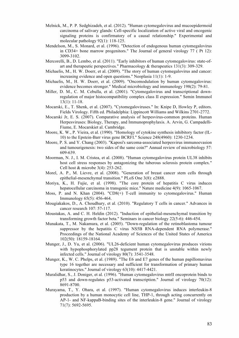
83
Melnick, M., P. P. Sedghizadeh, et al. (2012). "Human cytomegalovirus and mucoepidermoid carcinoma of salivary glands: Cell-specific localization of active viral and oncogenic signaling proteins is confirmatory of a causal relationship." Experimental and molecular pathology 92(1): 118-125.
Mendelson, M., S. Monard, et al. (1996). "Detection of endogenous human cytomegalovirus in CD34+ bone marrow progenitors." The Journal of general virology 77 ( Pt 12): 3099-3102.
Mercorelli, B., D. Lembo, et al. (2011). "Early inhibitors of human cytomegalovirus: state-of-art and therapeutic perspectives." Pharmacology & therapeutics 131(3): 309-329.
Michaelis, M., H. W. Doerr, et al. (2009). "The story of human cytomegalovirus and cancer: increasing evidence and open questions." Neoplasia 11(1): 1-9.
Michaelis, M., H. W. Doerr, et al. (2009). "Oncomodulation by human cytomegalovirus: evidence becomes stronger." Medical microbiology and immunology 198(2): 79-81.
Miller, D. M., C. M. Cebulla, et al. (2001). "Cytomegalovirus and transcriptional down-regulation of major histocompatibility complex class II expression." Semin Immunol 13(1): 11-18.
Mocarski, E., T. Shenk, et al. (2007). "Cytomegaloviruses." In: Knipe D, Howley P, editors. Fields Virology. Fifth ed. Philadelphia: Lippincott Williams and Wilkins 2701-2772.
Mocarski Jr, E. S. (2007). Comparative analysis of herpesvirus-common proteins. Human Herpesviruses: Biology, Therapy, and Immunoprophylaxis. A. Arvin, G. Campadelli-Fiume, E. Mocarskiet al. Cambridge.
Moore, K. W., P. Vieira, et al. (1990). "Homology of cytokine synthesis inhibitory factor (IL-10) to the Epstein-Barr virus gene BCRFI." Science 248(4960): 1230-1234.
Moore, P. S. and Y. Chang (2003). "Kaposi's sarcoma-associated herpesvirus immunoevasion and tumourigenesis: two sides of the same coin?" Annual review of microbiology 57: 609-639.
Moorman, N. J., I. M. Cristea, et al. (2008). "Human cytomegalovirus protein UL38 inhibits host cell stress responses by antagonizing the tuberous sclerosis protein complex." Cell host & microbe 3(4): 253-262.
Morel, A. P., M. Lievre, et al. (2008). "Generation of breast cancer stem cells through epithelial-mesenchymal transition." PLoS One 3(8): e2888.
Moriya, K., H. Fujie, et al. (1998). "The core protein of hepatitis C virus induces hepatocellular carcinoma in transgenic mice." Nature medicine 4(9): 1065-1067.
Moss, P. and N. Khan (2004). "CD8(+) T-cell immunity to cytomegalovirus." Human Immunology 65(5): 456-464.
Mougiakakos, D., A. Choudhury, et al. (2010). "Regulatory T cells in cancer." Advances in cancer research 107: 57-117.
Moustakas, A. and C. H. Heldin (2012). "Induction of epithelial-mesenchymal transition by transforming growth factor beta." Seminars in cancer biology 22(5-6): 446-454.
Munakata, T., M. Nakamura, et al. (2005). "Down-regulation of the retinoblastoma tumour suppressor by the hepatitis C virus NS5B RNA-dependent RNA polymerase." Proceedings of the National Academy of Sciences of the United States of America 102(50): 18159-18164.
Munger, J., D. Yu, et al. (2006). "UL26-deficient human cytomegalovirus produces virions with hypophosphorylated pp28 tegument protein that is unstable within newly infected cells." Journal of virology 80(7): 3541-3548.
Munger, K., W. C. Phelps, et al. (1989). "The E6 and E7 genes of the human papillomavirus type 16 together are necessary and sufficient for transformation of primary human keratinocytes." Journal of virology 63(10): 4417-4421.
Muralidhar, S., J. Doniger, et al. (1996). "Human cytomegalovirus mtrII oncoprotein binds to p53 and down-regulates p53-activated transcription." Journal of virology 70(12): 8691-8700.
Murayama, T., Y. Ohara, et al. (1997). "Human cytomegalovirus induces interleukin-8 production by a human monocytic cell line, THP-1, through acting concurrently on AP-1- and NF-kappaB-binding sites of the interleukin-8 gene." Journal of virology 71(7): 5692-5695.
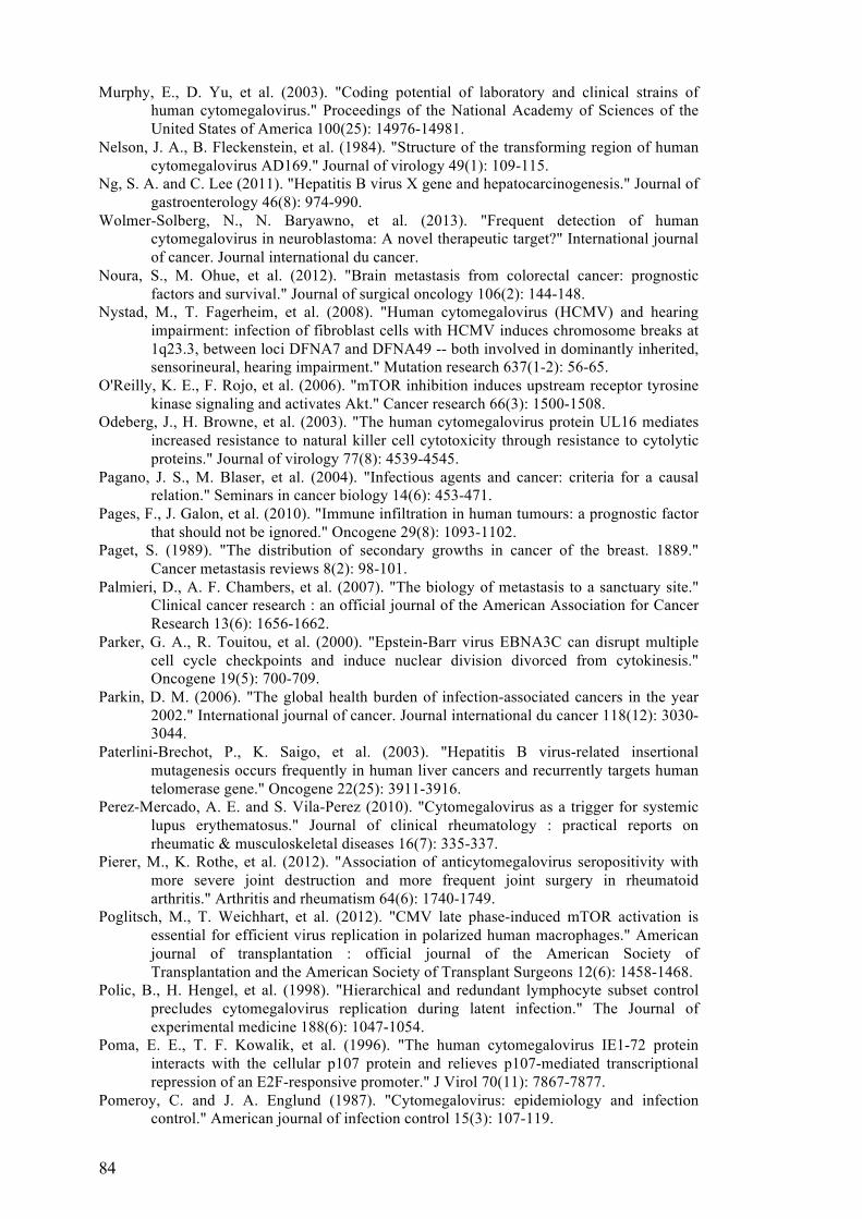
84
Murphy, E., D. Yu, et al. (2003). "Coding potential of laboratory and clinical strains of human cytomegalovirus." Proceedings of the National Academy of Sciences of the United States of America 100(25): 14976-14981.
Nelson, J. A., B. Fleckenstein, et al. (1984). "Structure of the transforming region of human cytomegalovirus AD169." Journal of virology 49(1): 109-115.
Ng, S. A. and C. Lee (2011). "Hepatitis B virus X gene and hepatocarcinogenesis." Journal of gastroenterology 46(8): 974-990.
Wolmer-Solberg, N., N. Baryawno, et al. (2013). "Frequent detection of human cytomegalovirus in neuroblastoma: A novel therapeutic target?" International journal of cancer. Journal international du cancer.
Noura, S., M. Ohue, et al. (2012). "Brain metastasis from colorectal cancer: prognostic factors and survival." Journal of surgical oncology 106(2): 144-148.
Nystad, M., T. Fagerheim, et al. (2008). "Human cytomegalovirus (HCMV) and hearing impairment: infection of fibroblast cells with HCMV induces chromosome breaks at 1q23.3, between loci DFNA7 and DFNA49 -- both involved in dominantly inherited, sensorineural, hearing impairment." Mutation research 637(1-2): 56-65.
O'Reilly, K. E., F. Rojo, et al. (2006). "mTOR inhibition induces upstream receptor tyrosine kinase signaling and activates Akt." Cancer research 66(3): 1500-1508.
Odeberg, J., H. Browne, et al. (2003). "The human cytomegalovirus protein UL16 mediates increased resistance to natural killer cell cytotoxicity through resistance to cytolytic proteins." Journal of virology 77(8): 4539-4545.
Pagano, J. S., M. Blaser, et al. (2004). "Infectious agents and cancer: criteria for a causal relation." Seminars in cancer biology 14(6): 453-471.
Pages, F., J. Galon, et al. (2010). "Immune infiltration in human tumours: a prognostic factor that should not be ignored." Oncogene 29(8): 1093-1102.
Paget, S. (1989). "The distribution of secondary growths in cancer of the breast. 1889." Cancer metastasis reviews 8(2): 98-101.
Palmieri, D., A. F. Chambers, et al. (2007). "The biology of metastasis to a sanctuary site." Clinical cancer research : an official journal of the American Association for Cancer Research 13(6): 1656-1662.
Parker, G. A., R. Touitou, et al. (2000). "Epstein-Barr virus EBNA3C can disrupt multiple cell cycle checkpoints and induce nuclear division divorced from cytokinesis." Oncogene 19(5): 700-709.
Parkin, D. M. (2006). "The global health burden of infection-associated cancers in the year 2002." International journal of cancer. Journal international du cancer 118(12): 3030-3044.
Paterlini-Brechot, P., K. Saigo, et al. (2003). "Hepatitis B virus-related insertional mutagenesis occurs frequently in human liver cancers and recurrently targets human telomerase gene." Oncogene 22(25): 3911-3916.
Perez-Mercado, A. E. and S. Vila-Perez (2010). "Cytomegalovirus as a trigger for systemic lupus erythematosus." Journal of clinical rheumatology : practical reports on rheumatic & musculoskeletal diseases 16(7): 335-337.
Pierer, M., K. Rothe, et al. (2012). "Association of anticytomegalovirus seropositivity with more severe joint destruction and more frequent joint surgery in rheumatoid arthritis." Arthritis and rheumatism 64(6): 1740-1749.
Poglitsch, M., T. Weichhart, et al. (2012). "CMV late phase-induced mTOR activation is essential for efficient virus replication in polarized human macrophages." American journal of transplantation : official journal of the American Society of Transplantation and the American Society of Transplant Surgeons 12(6): 1458-1468.
Polic, B., H. Hengel, et al. (1998). "Hierarchical and redundant lymphocyte subset control precludes cytomegalovirus replication during latent infection." The Journal of experimental medicine 188(6): 1047-1054.
Poma, E. E., T. F. Kowalik, et al. (1996). "The human cytomegalovirus IE1-72 protein interacts with the cellular p107 protein and relieves p107-mediated transcriptional repression of an E2F-responsive promoter." J Virol 70(11): 7867-7877.
Pomeroy, C. and J. A. Englund (1987). "Cytomegalovirus: epidemiology and infection control." American journal of infection control 15(3): 107-119.

85
Posner, J. B. and N. L. Chernik (1978). "Intracranial metastases from systemic cancer." Advances in neurology 19: 579-592.
Price, R. L., K. Bingmer, et al. (2012). "Cytomegalovirus infection leads to pleomorphic rhabdomyosarcomas in Trp53+/- mice." Cancer Res 72(22): 5669-5674.
Rahbar, A., A. Orrego, et al. (2013). "Human cytomegalovirus infection levels in glioblastoma multiforme are of prognostic value for survival." Journal of clinical virology : the official publication of the Pan American Society for Clinical Virology 57(1): 36-42.
Rahbar, A., G. Stragliotto, et al. (2012). "Low levels of Human Cytomegalovirus Infection in Glioblastoma Multiforme associates with patient survival; -a case-control study." Herpesviridae 3(1): 3.
Rak, J., J. Filmus, et al. (1995). "Oncogenes as inducers of tumour angiogenesis." Cancer metastasis reviews 14(4): 263-277.
Reeves, M. B., A. A. Davies, et al. (2007). "Complex I binding by a virally encoded RNA regulates mitochondria-induced cell death." Science 316(5829): 1345-1348.
Reeves, M. B., P. A. MacAry, et al. (2005). "Latency, chromatin remodeling, and reactivation of human cytomegalovirus in the dendritic cells of healthy carriers." Proc Natl Acad Sci U S A 102(11): 4140-4145.
Reinke, P., S. Prosch, et al. (1999). "Mechanisms of human cytomegalovirus (HCMV) (re)activation and its impact on organ transplant patients." Transplant infectious disease : an official journal of the Transplantation Society 1(3): 157-164.
Rentenaar, R. J., L. E. Gamadia, et al. (2000). "Development of virus-specific CD4(+) T cells during primary cytomegalovirus infection." The Journal of clinical investigation 105(4): 541-548.
Richardson, A. (1997). "Is breast cancer caused by late exposure to a common virus?" Medical hypotheses 48(6): 491-497.
Richardson, A. K., B. Cox, et al. (2004). "Cytomegalovirus, Epstein-Barr virus and risk of breast cancer before age 40 years: a case-control study." British journal of cancer 90(11): 2149-2152.
Riley, H. D., Jr. (1997). "History of the cytomegalovirus." Southern medical journal 90(2): 184-190.
Robbiani, D. F., R. A. Finch, et al. (2000). "The leukotriene C(4) transporter MRP1 regulates CCL19 (MIP-3beta, ELC)-dependent mobilization of dendritic cells to lymph nodes." Cell 103(5): 757-768.
Roizman, P. E. P. B. (2007). The Family Herpesviridae: A Brief Introduction. Fields Virology. D. M. H. Knipe, Peter M. Lippincott Williams & Wilkins: 2480-2497.
Rolle, A., M. Mousavi-Jazi, et al. (2003). "Effects of human cytomegalovirus infection on ligands for the activating NKG2D receptor of NK cells: up-regulation of UL16-binding protein (ULBP)1 and ULBP2 is counteracted by the viral UL16 protein." Journal of immunology 171(2): 902-908.
Rowe, W. P., J. W. Hartley, et al. (1956). "Cytopathogenic agent resembling human salivary gland virus recovered from tissue cultures of human adenoids." Proceedings of the Society for Experimental Biology and Medicine. Society for Experimental Biology and Medicine 92(2): 418-424.
Rubin, R. H. (1990). "Impact of cytomegalovirus infection on organ transplant recipients." Reviews of Infectious Diseases 12 Suppl 7: S754-766.
Ryckman, B. J., B. L. Rainish, et al. (2008). "Characterization of the human cytomegalovirus gH/gL/UL128-131 complex that mediates entry into epithelial and endothelial cells." Journal of virology 82(1): 60-70.
Samanta, M., L. Harkins, et al. (2003). "High prevalence of human cytomegalovirus in prostatic intraepithelial neoplasia and prostatic carcinoma." The Journal of urology 170(3): 998-1002.
Samanta, M., L. Harkins, et al. (2003). "High prevalence of human cytomegalovirus in prostatic intraepithelial neoplasia and prostatic carcinoma." J Urol 170(3): 998-1002.

86
Sampson, J. H. and D. A. Mitchell (2011). "Is cytomegalovirus a therapeutic target in glioblastoma?" Clinical cancer research : an official journal of the American Association for Cancer Research 17(14): 4619-4621.
Savary, K., D. Caglayan, et al. (2013). "Snail depletes the tumourigenic potential of glioblastoma." Oncogene.
Schalling, M., M. Ekman, et al. (1995). "A role for a new herpes virus (KSHV) in different forms of Kaposi's sarcoma." Nature medicine 1(7): 707-708.
Scheffner, M. and N. J. Whitaker (2003). "Human papillomavirus-induced carcinogenesis and the ubiquitin-proteasome system." Seminars in cancer biology 13(1): 59-67.
Scholz, M., R. A. Blaheta, et al. (2000). "Cytomegalovirus-infected neuroblastoma cells exhibit augmented invasiveness mediated by beta1alpha5 integrin (VLA-5)." Tissue Antigens 55(5): 412-421.
Scotting, P. J., D. A. Walker, et al. (2005). "Childhood solid tumours: a developmental disorder." Nature reviews. Cancer 5(6): 481-488.
Seliger, B. (2005). "Strategies of tumour immune evasion." BioDrugs : clinical immunotherapeutics, biopharmaceuticals and gene therapy 19(6): 347-354.
Shen, Y., H. Zhu, et al. (1997). "Human cytomagalovirus IE1 and IE2 proteins are mutagenic and mediate "hit-and-run" oncogenic transformation in cooperation with the adenovirus E1A proteins." Proceedings of the National Academy of Sciences of the United States of America 94(7): 3341-3345.
Shen, Z. (2011). "Genomic instability and cancer: an introduction." Journal of molecular cell biology 3(1): 1-3.
Shillitoe, E. J., T. E. Daniels, et al. (1982). "Antibody to cytomegalovirus in patients with Sjogren's syndrome. As determined by an enzyme-linked immunosorbent assay." Arthritis and rheumatism 25(3): 260-265.
Shimamura, M., J. E. Murphy-Ullrich, et al. (2010). "Human cytomegalovirus induces TGF-beta1 activation in renal tubular epithelial cells after epithelial-to-mesenchymal transition." PLoS pathogens 6(11): e1001170.
Shinmura, Y., S. Aiba-Masago, et al. (1997). "Differential expression of the immediate-early and early antigens in neuronal and glial cells of developing mouse brains infected with murine cytomegalovirus." The American journal of pathology 151(5): 1331-1340.
Shmueli, E., N. Wigler, et al. (2004). "Central nervous system progression among patients with metastatic breast cancer responding to trastuzumab treatment." European journal of cancer 40(3): 379-382.
Shono, T., P. J. Tofilon, et al. (2001). "Cyclooxygenase-2 expression in human gliomas: prognostic significance and molecular correlations." Cancer research 61(11): 4375-4381.
Siew, V. K., C. Y. Duh, et al. (2009). "Human cytomegalovirus UL76 induces chromosome aberrations." Journal of biomedical science 16: 107.
Silva, R. G., Jr. and B. B. da Silva (2011). "No evidence for an association of human papillomavirus and breast carcinoma." Breast cancer research and treatment 125(1): 261-264.
Sinclair, J. (2008). "Human cytomegalovirus: Latency and reactivation in the myeloid lineage." Journal of clinical virology : the official publication of the Pan American Society for Clinical Virology 41(3): 180-185.
Sinclair, J. and P. Sissons (1996). "Latent and persistent infections of monocytes and macrophages." Intervirology 39(5-6): 293-301.
Singh, S. K., C. Hawkins, et al. (2004). "Identification of human brain tumour initiating cells." Nature 432(7015): 396-401.
Sinzger, C., M. Kahl, et al. (2000). "Tropism of human cytomegalovirus for endothelial cells is determined by a post-entry step dependent on efficient translocation to the nucleus." The Journal of general virology 81(Pt 12): 3021-3035.
Sinzger, C., B. Plachter, et al. (1996). "Tissue macrophages are infected by human cytomegalovirus in vivo." The Journal of infectious diseases 173(1): 240-245.
Sixbey, J. W., J. G. Nedrud, et al. (1984). "Epstein-Barr virus replication in oropharyngeal epithelial cells." The New England journal of medicine 310(19): 1225-1230.

87
Skaletskaya, A., L. M. Bartle, et al. (2001). "A cytomegalovirus-encoded inhibitor of apoptosis that suppresses caspase-8 activation." Proceedings of the National Academy of Sciences of the United States of America 98(14): 7829-7834.
Slinger, E., D. Maussang, et al. (2010). "HCMV-encoded chemokine receptor US28 mediates proliferative signaling through the IL-6-STAT3 axis." Science signaling 3(133): ra58.
Slobedman, B. and E. S. Mocarski (2012). "Mechanisms modulating immune clearance during human cytomegalovirus latency." Proceedings of the National Academy of Sciences of the United States of America 109(36): 14291-14292.
Smedby, K. E., L. Brandt, et al. (2009). "Brain metastases admissions in Sweden between 1987 and 2006." British journal of cancer 101(11): 1919-1924.
Smirnova, I. S., N. D. Aksenov, et al. (2006). "Hepatitis C virus core protein transforms murine fibroblasts by promoting genomic instability." Cellular oncology : the official journal of the International Society for Cellular Oncology 28(4): 177-190.
Smith, J. S., L. Lindsay, et al. (2007). "Human papillomavirus type distribution in invasive cervical cancer and high-grade cervical lesions: a meta-analysis update." International journal of cancer. Journal international du cancer 121(3): 621-632.
Smith, M. G. (1956). "Propagation in tissue cultures of a cytopathogenic virus from human salivary gland virus (SGV) disease." Proceedings of the Society for Experimental Biology and Medicine. Society for Experimental Biology and Medicine 92(2): 424-430.
Söderberg C, Giugni Td, et al. (1993). "CD13 (human aminopeptidase N) mediates human cytomegalovirus infection." Journal of Virology 67(11): 6576-6585.
Soderberg-Naucler, C. (2006). "Does cytomegalovirus play a causative role in the development of various inflammatory diseases and cancer?" J Intern Med 259(3): 219-246.
Soderberg-Naucler, C. (2006). "Human cytomegalovirus persists in its host and attacks and avoids elimination by the immune system." Critical reviews in immunology 26(3): 231-264.
Soderberg-Naucler, C., K. N. Fish, et al. (1997). "Reactivation of latent human cytomegalovirus by allogeneic stimulation of blood cells from healthy donors." Cell 91(1): 119-126.
Soderberg-Naucler, C. and J. Y. Nelson (1999). "Human cytomegalovirus latency and reactivation - a delicate balance between the virus and its host's immune system." Intervirology 42(5-6): 314-321.
Sodhi, A., S. Montaner, et al. (2004). "Akt plays a central role in sarcomagenesis induced by Kaposi's sarcoma herpesvirus-encoded G protein-coupled receptor." Proceedings of the National Academy of Sciences of the United States of America 101(14): 4821-4826.
Sorlie, P. D., F. J. Nieto, et al. (2000). "A prospective study of cytomegalovirus, herpes simplex virus 1, and coronary heart disease: the atherosclerosis risk in communities (ARIC) study." Archives of Internal Medicine 160(13): 2027-2032.
Soroceanu, L., A. Akhavan, et al. (2008). "Platelet-derived growth factor-alpha receptor activation is required for human cytomegalovirus infection." Nature 455(7211): 391-395.
Soroceanu, L. and C. S. Cobbs (2011). "Is HCMV a tumour promoter?" Virus Res 157(2): 193-203.
Soroceanu, L., L. Matlaf, et al. (2011). "Human cytomegalovirus US28 found in glioblastoma promotes an invasive and angiogenic phenotype." Cancer research 71(21): 6643-6653.
Speir, E., E. S. Huang, et al. (1995). "Interaction of human cytomegalovirus with p53: possible role in coronary restenosis." Scandinavian journal of infectious diseases. Supplementum 99: 78-81.
Speir, E., Z. X. Yu, et al. (1998). "Aspirin attenuates cytomegalovirus infectivity and gene expression mediated by cyclooxygenase-2 in coronary artery smooth muscle cells." Circulation Research 83(2): 210-216.
Spiller, O. B., S. M. Hanna, et al. (1997). "Neutralization of cytomegalovirus virions: the role of complement." The Journal of infectious diseases 176(2): 339-347.

88
Srivenugopal, K. S. and F. Ali-Osman (2002). "The DNA repair protein, O(6)-methylguanine-DNA methyltransferase is a proteolytic target for the E6 human papillomavirus oncoprotein." Oncogene 21(38): 5940-5945.
Steeg, P. S. (2006). "Tumour metastasis: mechanistic insights and clinical challenges." Nature Medicine 12(8): 895-904.
Stenberg, R. M. (1996). "The human cytomegalovirus major immediate-early gene." Intervirology 39(5-6): 343-349.
Stenberg, R. M., A. S. Depto, et al. (1989). "Regulated expression of early and late RNAs and proteins from the human cytomegalovirus immediate-early gene region." J Virol 63(6): 2699-2708.
Stern-Ginossar, N., B. Weisburd, et al. (2012). "Decoding human cytomegalovirus." Science 338(6110): 1088-1093.
Straat, K., C. Liu, et al. (2009). "Activation of telomerase by human cytomegalovirus." Journal of the National Cancer Institute 101(7): 488-497.
Stragliotto, G., A. Rahbar, et al. (2013). "Effects of valganciclovir as an add-on therapy in patients with cytomegalovirus-positive glioblastoma: A randomised, double-blind, hypothesis-generating study." International journal of cancer. Journal international du cancer.
Strasser, A., L. O'Connor, et al. (2000). "Apoptosis signaling." Annual Review of Biochemistry 69: 217-245.
Streblow, D. N., C. Soderberg-Naucler, et al. (1999). "The human cytomegalovirus chemokine receptor US28 mediates vascular smooth muscle cell migration." Cell 99(5): 511-520.
Syggelou, A., N. Iacovidou, et al. (2010). "Congenital cytomegalovirus infection." Annals of the New York Academy of Sciences 1205: 144-147.
Tabata, T., H. Kawakatsu, et al. (2008). "Induction of an epithelial integrin alphavbeta6 in human cytomegalovirus-infected endothelial cells leads to activation of transforming growth factor-beta1 and increased collagen production." The American journal of pathology 172(4): 1127-1140.
Taher, C., J. de Boniface, et al. (2013). "High prevalence of human cytomegalovirus proteins and nucleic acids in primary breast cancer and metastatic sentinel lymph nodes." PLoS One 8(2): e56795.
Tanaka, K., J. P. Zou, et al. (1999). "Effects of human cytomegalovirus immediate-early proteins on p53-mediated apoptosis in coronary artery smooth muscle cells." Circulation 99(13): 1656-1659.
Tang, D. G. and K. V. Honn (1994). "Adhesion molecules and tumour metastasis: an update." Invasion & metastasis 14(1-6): 109-122.
Taylor-Wiedeman, J., J. G. Sissons, et al. (1991). "Monocytes are a major site of persistence of human cytomegalovirus in peripheral blood mononuclear cells." The Journal of general virology 72 ( Pt 9): 2059-2064.
Terhune, S. S., J. Schroer, et al. (2004). "RNAs are packaged into human cytomegalovirus virions in proportion to their intracellular concentration." Journal of virology 78(19): 10390-10398.
Termen, S., E. J. Tan, et al. (2013). "p53 regulates epithelial-mesenchymal transition induced by transforming growth factor beta." Journal of Cellular Physiology 228(4): 801-813.
Thiery, J. P. (2003). "Epithelial-mesenchymal transitions in development and pathologies." Current Opinion in Cell Biology 15(6): 740-746.
Tomkinson, B., E. Robertson, et al. (1993). "Epstein-Barr virus nuclear proteins EBNA-3A and EBNA-3C are essential for B-lymphocyte growth transformation." Journal of virology 67(4): 2014-2025.
Tsai, J. H., C. S. Hsu, et al. (2007). "Relationship between viral factors, axillary lymph node status and survival in breast cancer." J Cancer Res Clin Oncol 133(1): 13-21.
Tsai, J. H., C. H. Tsai, et al. (2005). "Association of viral factors with non-familial breast cancer in Taiwan by comparison with non-cancerous, fibroadenoma, and thyroid tumour tissues." J Med Virol 75(2): 276-281.
Tsai, W. L. and R. T. Chung (2010). "Viral hepatocarcinogenesis." Oncogene 29(16): 2309-2324.

89
Tsujii, M., S. Kawano, et al. (1997). "Cyclooxygenase-2 expression in human colon cancer cells increases metastatic potential." Proceedings of the National Academy of Sciences of the United States of America 94(7): 3336-3340.
Utrera-Barillas, D., H. A. Valdez-Salazar, et al. (2013). "Is human cytomegalovirus associated with breast cancer progression?" Infectious agents and cancer 8(1): 12.
Varnum, S. M., D. N. Streblow, et al. (2004). "Identification of proteins in human cytomegalovirus (HCMV) particles: the HCMV proteome." Journal of virology 78(20): 10960-10966.
Verma, S. C., S. Borah, et al. (2004). "Latency-associated nuclear antigen of Kaposi's sarcoma-associated herpesvirus up-regulates transcription of human telomerase reverse transcriptase promoter through interaction with transcription factor Sp1." Journal of virology 78(19): 10348-10359.
Vivanco, I. and C. L. Sawyers (2002). "The phosphatidylinositol 3-Kinase AKT pathway in human cancer." Nature reviews. Cancer 2(7): 489-501.
von Glahn, W. C. and A. M. Pappenheimer (1925). "Intranuclear inclusions in visceral disease." American J. Pathology 1: 445-465.
Wang, D. and R. N. Dubois (2010). "Eicosanoids and cancer." Nature reviews. Cancer 10(3): 181-193.
Wang, D., D. Liebowitz, et al. (1985). "An EBV membrane protein expressed in immortalized lymphocytes transforms established rodent cells." Cell 43(3 Pt 2): 831-840.
Wang, X., S. M. Huong, et al. (2003). "Epidermal growth factor receptor is a cellular receptor for human cytomegalovirus." Nature 424(6947): 456-461.
Weekes, M. P., S. Y. Tan, et al. (2013). "Latency-associated degradation of the MRP1 drug transporter during latent human cytomegalovirus infection." Science 340(6129): 199-202.
Weinstat-Saslow, D. L., V. S. Zabrenetzky, et al. (1994). "Transfection of thrombospondin 1 complementary DNA into a human breast carcinoma cell line reduces primary tumour growth, metastatic potential, and angiogenesis." Cancer research 54(24): 6504-6511.
Weiss, L., E. Grundmann, et al. (1986). "Haematogenous metastatic patterns in colonic carcinoma: an analysis of 1541 necropsies." The Journal of pathology 150(3): 195-203.
Weller, T. H. (2000). "Cytomegaloviruses: a Historical Perspective." Herpes : the journal of the IHMF 7(3): 66-69.
White, E. A., C. J. Del Rosario, et al. (2007). "The IE2 60-kilodalton and 40-kilodalton proteins are dispensable for human cytomegalovirus replication but are required for efficient delayed early and late gene expression and production of infectious virus." J Virol 81(6): 2573-2583.
Wilkinson, G. W., P. Tomasec, et al. (2008). "Modulation of natural killer cells by human cytomegalovirus." Journal of clinical virology : the official publication of the Pan American Society for Clinical Virology 41(3): 206-212.
I. Wolmer-Solberg, N., N. Baryawno, et al. (2013). "Frequent detection of human cytomegalovirus in neuroblastoma: A novel therapeutic target?" International journal of cancer. 2011;121(10):4043-55.
Wright, W. E., O. M. Pereira-Smith, et al. (1989). "Reversible cellular senescence: implications for immortalization of normal human diploid fibroblasts." Molecular and cellular biology 9(7): 3088-3092.
Wyatt, J. P., J. Saxton, et al. (1950). "Generalized cytomegalic inclusion disease." The Journal of pediatrics 36(3): 271-294, illust.
Xu, Z., D. Schlesinger, et al. (2012). "Impact of triple-negative phenotype on prognosis of patients with breast cancer brain metastases." International journal of radiation oncology, biology, physics 84(3): 612-618.
Yang, J. and R. A. Weinberg (2008). "Epithelial-mesenchymal transition: at the crossroads of development and tumour metastasis." Developmental cell 14(6): 818-829.

90
Yoo, Y. D., C. J. Chiou, et al. (1996). "The IE2 regulatory protein of human cytomegalovirus induces expression of the human transforming growth factor beta1 gene through an Egr-1 binding site." Journal of virology 70(10): 7062-7070.
Yoshida, M. (2001). "Multiple viral strategies of HTLV-1 for dysregulation of cell growth control." Annual review of immunology 19: 475-496.
Young, L. S. and A. B. Rickinson (2004). "Epstein-Barr virus: 40 years on." Nature reviews. Cancer 4(10): 757-768.
Yurochko, A. D., T. F. Kowalik, et al. (1995). "Human cytomegalovirus upregulates NF-kappa B activity by transactivating the NF-kappa B p105/p50 and p65 promoters." Journal of virology 69(9): 5391-5400.
Zhu, H., J. P. Cong, et al. (2002). "Inhibition of cyclooxygenase 2 blocks human cytomegalovirus replication." Proceedings of the National Academy of Sciences of the United States of America 99(6): 3932-3937.
Zhu, H., Y. Shen, et al. (1995). "Human cytomegalovirus IE1 and IE2 proteins block apoptosis." Journal of virology 69(12): 7960-7970.
zur Hausen, H. (2002). "Papillomaviruses and cancer: from basic studies to clinical application." Nature reviews. Cancer 2(5): 342-350.