Comparison of DNA extraction methods for multiplex polymerase chain reaction
Upload
khangminh22Category
view
0download
0
The Human Cytomegalovirus DNA PolymeraseProcessivity Factor UL44 Is Modified by SUMO in a DNA-Dependent MannerElisa Sinigalia1., Gualtiero Alvisi1,2,3., Chiara V. Segre4., Beatrice Mercorelli1., Giulia Muratore1,
Michael Winkler5, He-Hsuan Hsiao6, Henning Urlaub6,7, Alessandro Ripalti8, Susanna Chiocca4,
Giorgio Palu1*, Arianna Loregian1*
1 Department of Molecular Medicine, University of Padua, Padua, Italy, 2 Department of Hematology and Medical Oncology Seragnoli, University of Bologna, Bologna,
Italy, 3 Department of Infectious Diseases, Molecular Virology, University of Heidelberg, Heidelberg, Germany, 4 European Institute of Oncology, Milan, Italy, 5 Leibniz-
Institut fur Primatenforschung, Goettingen, Germany, 6 Bioanalytical Mass Spectrometry Group, Max Planck Institute for Biophysical Chemistry, Goettingen, Germany,
7 Bioanalytics, Department of Clinical Chemistry, University Medical Center, Goettingen, Germany, 8 Azienda Ospedaliero-Universitaria di Bologna, Policlinico S. Orsola-
Malpighi, Bologna, Italy
Abstract
During the replication of human cytomegalovirus (HCMV) genome, the viral DNA polymerase subunit UL44 plays a key role,as by binding both DNA and the polymerase catalytic subunit it confers processivity to the holoenzyme. However, severallines of evidence suggest that UL44 might have additional roles during virus life cycle. To shed light on this, we searched forcellular partners of UL44 by yeast two-hybrid screenings. Intriguingly, we discovered the interaction of UL44 with Ubc9, anenzyme involved in the covalent conjugation of SUMO (Small Ubiquitin-related MOdifier) to cellular and viral proteins. Wefound that UL44 can be extensively sumoylated not only in a cell-free system and in transfected cells, but also in HCMV-infected cells, in which about 50% of the protein resulted to be modified at late times post-infection, when viral genomereplication is accomplished. Mass spectrometry studies revealed that UL44 possesses multiple SUMO target sites, locatedthroughout the protein. Remarkably, we observed that binding of UL44 to DNA greatly stimulates its sumoylation bothin vitro and in vivo. In addition, we showed that overexpression of SUMO alters the intranuclear distribution of UL44 inHCMV-infected cells, and enhances both virus production and DNA replication, arguing for an important role forsumoylation in HCMV life cycle and UL44 function(s). These data report for the first time the sumoylation of a viralprocessivity factor and show that there is a functional interplay between the HCMV UL44 protein and the cellularsumoylation system.
Citation: Sinigalia E, Alvisi G, Segre CV, Mercorelli B, Muratore G, et al. (2012) The Human Cytomegalovirus DNA Polymerase Processivity Factor UL44 Is Modifiedby SUMO in a DNA-Dependent Manner. PLoS ONE 7(11): e49630. doi:10.1371/journal.pone.0049630
Editor: Heinrich Sticht, Universitat Erlangen-Nurnberg, Germany
Received June 6, 2012; Accepted October 11, 2012; Published November 15, 2012
Copyright: � 2012 Sinigalia et al. This is an open-access article distributed under the terms of the Creative Commons Attribution License, which permitsunrestricted use, distribution, and reproduction in any medium, provided the original author and source are credited.
Funding: This work was supported by MURST EX60%, Progetto di Ricerca di Ateneo 2007 (grant n. CPDA074945), and PRIN 2008 (grant n. 20085FF4J4) to AL, byRegione Veneto and Progetto Strategico di Ateneo 2008 to GP, by Associazione Italiana Ricerca sul Cancro and Italian Ministry of Health to SC, and by FondazioneUmberto Veronesi to CVS. The funders had no role in study design, data collection and analysis, decision to publish, or preparation of the manuscript.
Competing Interests: The authors have declared that no competing interests exist.
* E-mail: [email protected] (GP); [email protected] (AL)
. These authors contributed equally to this work.
Introduction
Most replicative DNA polymerases include a catalytic subunit,
responsible for DNA polymerization, and a processivity factor that
holds the catalytic subunit on DNA to allow continuous DNA
synthesis. One of the best-studied processivity factors is prolifer-
ating cell nuclear antigen (PCNA) of eukaryotic DNA polymerases
d and e [1]. PCNA, which belongs to the family of so-called
‘‘sliding clamps’’, has no inherent DNA-binding capacity, but with
the aid of clamp loader proteins is assembled onto DNA as a
toroidal homotrimer [2]. In addition to DNA replication, PCNA
has been implicated in DNA recombination and repair, as well as
in DNA methylation, chromatin remodeling, and cell cycle
regulation [1]. Consistent with its pleiotropic functions, it interacts
with a plethora of proteins [3] and undergoes a number of post-
translational modifications, including phosphorylation, acetyla-
tion, ubiquitination and sumoylation, which are believed to
regulate its subcellular localization, stability and protein binding
specificity [4,5,6].
The human cytomegalovirus (HCMV) DNA polymerase
includes a catalytic subunit, UL54 (the UL54 gene product), and
an accessory, homodimeric subunit, UL44 (the UL44 gene
product), that binds DNA without the aid of clamp loaders [7]
yet wraps around DNA akin to PCNA [8]. While UL44 shows no
apparent sequence homology with PCNA, there is striking
structural similarity between UL44 and PCNA monomers [2,9].
Similarly to PCNA, UL44 is a phosphoprotein [10]. Intriguingly,
the phosphorylation state of UL44 has been shown to regulate its
nuclear import rate by controlling its interaction with host cell
factors [11,12,13]. The best-characterized function of UL44
during HCMV infection is that of binding to UL54 through a
region named connector loop [14,15,16], stimulating its activity
PLOS ONE | www.plosone.org 1 November 2012 | Volume 7 | Issue 11 | e49630
and conferring processivity to the holoenzyme [17,18]. However,
UL44 continues to accumulate to strikingly high levels at late times
after infection, when DNA replication is accomplished [19,20]. Its
early-late kinetics of transcription and the high level of expression
suggest that UL44 might play additional roles during the viral life
cycle.
To investigate this possibility, we conducted yeast two-hybrid
(Y2H) screenings to search for cellular partners of UL44. To our
surprise, Ubc9, an enzyme involved in the sumoylation process,
was identified as a UL44 protein interaction partner. Sumoylation
is a post-translational protein modification analogous to ubiqui-
tination. It consists of reversible and covalent conjugation of
SUMO (Small Ubiquitin-related MOdifier) to a protein target
[21,22]. In the sumoylation cascade, the C-terminus of SUMO is
activated by an activating enzyme (E1), transferred to a
conjugating enzyme (E2, that is Ubc9), and linked to a lysine
residue of the substrate protein with the aid of a ligase (E3).
Mainly, three SUMO paralogs (SUMO-1, -2, -3) have been
identified so far [23,24]. SUMO-2 and SUMO-3 are highly
homologous to one another (95% identity) while they differ from
SUMO-1 by 50%. Conjugation of SUMO-1 has been shown to
play a functional role in a number of biological processes, ranging
from nucleocytoplasmic transport to transcription, the mainte-
nance of genome stability, nucleic acid DNA metabolism, cell
signaling, and many others [21], whereas the role of SUMO-2/23
modification is less clear.
Here we report that the association of Ubc9 and UL44 leads to
conjugation of SUMO molecules on multiple lysine residues. Both
SUMO-1 and SUMO-2/3 were found to be conjugated to UL44.
Sumoylation of UL44 was detected not only in vitro and in
transiently transfected cells but, more importantly, also in HCMV-
infected human cells during virus replication. Interestingly, we
observed that binding of UL44 to DNA greatly stimulates SUMO
conjugation to the protein both in vitro and in cells. In addition, we
show that overexpression of SUMO-1 alters the intranuclear
distribution of UL44 in HCMV-infected cells, and enhances both
viral DNA replication and virus production in an Ubc9-dependent
manner. These data represent the first report of sumoylation of a
viral processivity factor and show that there is a complex interplay
between the HCMV UL44 protein and the cellular sumoylation
system.
Materials and Methods
PlasmidsThe Y2H plasmids expressing LexA-UL44 and LexA-Ubc9
were generated by cloning the UL44 and Ubc9 coding sequences
from pRSET44 (a gift of P. F. Ertl, GlaxoSmithKline, UK) and
pACT2-Ubc9 (from G. Gao, Chinese Academy of Sciences,
Beijing, China) respectively, in pBTMK, derived from
pBTM116 [25]. The pACT-UL44 and pACT2-Ubc9 plasmids,
encoding GAD-UL44 and GAD-Ubc9 fusions, respectively,
have been described in [26,27]. The plasmid expressing
GAD-UL54 was created by cloning the UL54 coding sequence
from pRSET-Pol (a gift of P. F. Ertl) in pACT2 (Clontech).
Plasmid pRSET44 was used to express 6His-UL44 in Escherichia
coli. Plasmid pRSET-Ubc9 was constructed by cloning the Ubc9
coding sequence from pACT2-Ubc9 in pRSET (Invitrogen).
Plasmid pCDNA3-PB1, used for in vitro transcription of the PB1
subunit of influenza A virus RNA polymerase, was described
previously [28]. Plasmids pD15-GST and pD15-UL44, which
express GST and GST-UL44, respectively, have been described
in [29]. Plasmid pTE1E2S1 [30] was provided by H. Saitoh
(Kusamoto University, Japan). Plasmids GFP-UL44,
pDESTnV5-UL44, and pDESTnV5-UL44DNLS have been
described previously [11,31]. Plasmids pDsRed2-Ubc9 [27]
and pDsRed2-UL53 [32] were kindly provided by G. Gao
(Chinese Academy of Sciences, Beijing, China) and D. Camozzi
(University of Bologna, Italy), respectively. Plasmid pCDNA3.1-
UL44-FLAG (from M. Marschall, Universitat Erlangen-Num-
berg, Germany) was used to express C-terminally FLAG-tagged
UL44 [33], while plasmid pDEST-nFLAG [34] was used to
express N-terminally FLAG-tagged UL44 [35], UL44Dloop [36]
and UL44L86A/L87A in mammalian cells. Plasmids pCDNA3-
Ubc9, pCDNA3-Ubc9C93S, pCDNA3-HA-SUMO-1,
pCDNA3-HA-SUMO-2, and pCDNA3-HA-SUMO-3 used in
overexpression experiments in mammalian cells were a gift of
R. T. Hay (University of Dundee, UK). The deletion mutants
LexA-UL441–100, LexA-UL441–200, LexA-UL441–300, LexA-
UL441–350, LexA-UL441–390, and LexA-UL441–420 were gener-
ated by PCR amplification of plasmid pBTMK-UL44 with
appropriate primers (Table S1). The LexA-UL44114–433, LexA-
UL44201–433, and LexA-UL44313–433 constructs were generated
by deleting part of the UL44 coding sequence from pBTMK-
UL44 with restriction enzymes. Plasmids pDESTnFLAG-
UL44(1–300) and pDESTnFLAG-UL44(313–433) were gener-
ated using the Gateway Technology (see Supplementary
Material and Methods in Text S1). All other UL44 mutants
and the Ubc9C93S mutant were obtained by using the
QuikChange mutagenesis kit (Stratagene) with primers contain-
ing appropriate nucleotide change(s). More details on plasmid
construction and mutagenesis are given in Supplementary
Material and Methods in Text S1. The sequences of all
primers used in this work are reported in Table S1. All DNA
sequences were confirmed by sequencing.
Y2H screenings and interaction assays. Growth media
and standard methods for manipulating yeast cells were as
described [37]. Saccharomyces cerevisiae strain L40 was transformed
[38] with the bait plasmid pBTMK-UL44 and subsequently with
either of two cDNA libraries fused to GAD (see Supplementary
Material and Methods in Text S1). Primary transformants were
selected for growth on -His-Leu-Trp dropout plates. His+ colonies
were thereafter analyzed for b-galactosidase activity by filter lift
experiments [39]. Double positive clones were subjected to
another cycle of screening (for further details see Supplementary
Material and Methods in Text S1). cDNA inserts of interactor
plasmids were sequenced and analyzed with BLAST (www.ncbi/
blastn). To quantify b-gal expression, the method of Breeden and
Nasmyth [40] was used.
Proteins. E. coli-expressed, purified GST and GST- or 6His-
tagged UL44 proteins were obtained as previously described [29],
with modifications (Supplementary Material and Methods in Text
S1). In some preparations, samples were treated with polymin P as
described [41] to eliminate residual bacterial nucleic acids.
Preparation of UL44 SUMO-modified in E. coli was accomplished
as described in Supplementary Material and Methods in Text S1.
GST-pulldown assays. Assays were performed using GST
and GST-UL44 and in vitro-translated UL54, PB1, or Ubc9 as
previously described [29], with modifications (Supplementary
Material and Methods in Text S1). In vitro transcription-translation
of proteins was performed from the appropriate plasmid by using
the TNT T7 coupled transcription-translation system (Promega).
The translation products were labeled with [35S]methionine
(Amersham Pharmacia Biotech).
In vitro sumoylation assays. The assays to test in vitro
sumoylation of UL44 by SUMO-1, -2, and -3 were performed
using purified wild-type and mutant 6His-UL44 or GST-UL44
fusion proteins and the SUMOlink SUMO-1 kit from Active Motif
Sumoylation of HCMV UL44
PLOS ONE | www.plosone.org 2 November 2012 | Volume 7 | Issue 11 | e49630
or the SUMOylation Kit from Enzo Life Science according to the
manufacturer’s suggestions. In some experiments, double stranded
(ds) DNA (e.g., activated calf thymus DNA from Amersham
Pharmacia Biotec or salmon testes DNA from Sigma) or single-
stranded (ss) DNA (e.g., single-stranded calf lung DNA, from
Crinos, Como, Italy) was added to the reaction mixture at a final
concentration of 500 nM.
Cells and VirusHuman foreskin fibroblasts (HFF; from the American Type
Culture Collection [ATCC]), HeLa (from ATCC), eco Phoenix (a
generous gift from G. P. Nolan, Stanford, USA; [42]), and Human
Embryonic Kidney 293T (HEK 293T; from ATCC) cells were
maintained in Dulbecco’s modified Eagle’s medium (DMEM, Life
Biotechnologies) supplemented with 10% fetal calf serum (FCS),
100 U/ml penicillin and 100 mg/ml streptomycin (P/S). COS-1
cells (from ATCC) were maintained in DMEM with 5% FCS and
P/S. The U373-SUMO-1 cell line, which constitutively expresses
FLAG-tagged SUMO-1 [43], and the control U373-Neo cell line,
stably transfected with an empty vector carrying a Neomycin
resistance marker [44], were kindly provided by G. S. Hayward
(Johns Hopkins University School of Medicine, Baltimore, USA)
and were maintained in medium containing 0.5 mg/ml Neomycin
(G418, Gibco-BRL). HCMV strain AD169 was purchased from
ATCC.
In vivo Sumoylation AssaysTo analyze UL44 sumoylation in transient expression assays,
HeLa or Phoenix cells were transfected for 48 h with
appropriate plasmids using the calcium phosphate precipitation
method. For analysis of UL44 sumoylation during HCMV
replication, HFF, U373-Neo, and U373-SUMO-1 cells were
mock-infected or infected with HCMV at a multiplicity of
infection (MOI) of 5 PFU/cell. Cells were harvested at different
time points after infection and analyzed by western blotting as
described below.
Western Blot and Immunoprecipitation AnalysisTransfected or infected cells were lysed in an appropriate
volume of buffer I (5% SDS, 0.15 M Tris-HCl pH 6.8, 30%
glycerol) diluted 1:3 in buffer II (25 mM Tris-HCl pH 8, 50 mM
NaCl, 0.5% NP-40, 0.5% sodium deoxycholate, 0.1% SDS)
supplemented with Complete protease inhibitors (Roche Molec-
ular Biochemicals) and 5 mM N-ethylmaleimide (NEM). Lysates
were then incubated on ice for 20 minutes and boiled at 95uC for
10 min. Proteins were separated by SDS-PAGE, electroblotted
onto a polyvinylidene fluoride membrane (Bio-Rad), and analyzed
by western blotting with indicated antibodies (details are given in
Supplementary Material and Methods in Text S1).
For immunoprecipitation analysis, lysates were diluted 1:5 in
E1A buffer (50 mM Hepes pH 7.5, 250 mM NaCl, 0.1% NP-40)
supplemented with Complete protease inhibitors and 5 mM
NEM. Immunoprecipitation was performed with 2–3 mg of total
lysate using a ratio of 3–7 mg antibody/mg of total proteins (see
Supplementary Material and Methods in Text S1 for details on
antibodies) and protein A-Sepharose beads. For co-immunopre-
cipitation analysis, cells were lysed in E1A buffer supplemented
with Complete protease inhibitors and 5 mM NEM, and
successively co-immunoprecipitations were performed with 1.5–
5 mg of total lysate and 50 ml of 50% slurry of anti-FLAG-M2-
Agarose beads (Sigma).
Mass Spectrometric (MS) Determination of SumoylationSites of UL44
Mass spectrometric identification of sumoylated lysine residues
within UL44 was performed after in-gel-digestion of E. coli-
expressed and SUMO-modified UL44 with endoproteinase
Trypsin. Extracted peptides were analyzed by LC-MSMS on an
Orbitrap Velos (ThermoFisherScientific) exactly under the condi-
tions described in Hsiao et al. [45]. Data analysis was performed
by the use of software ‘‘ChopNSpice’’ [45] in combination with
MASCOT as search engine. See Supplementary Material and
Methods in Text S1 for details.
Immunofluorescence and Confocal Microscopy AnalysisFor confocal laser-scanning microscopy (CLSM) analysis, COS-
1 were transfected using the Arrest-INTM (Biosystems) reagent,
according to the manufacturer’s recommendations. At 24 h post-
transfection, cells were fixed with 4% paraformaldehyde. Cells
were imaged using a Leica TCS-SP2 confocal microscope
equipped with a 636 oil immersion objective.
For analysis of UL44 intranuclear localization in HCMV-
infected U373-SUMO-1 and U373-Neo cell lines, cells were
seeded at 2.5 6 105/well on glass coverslips in 6-well plates and
allowed to attach. The next day, cells were infected with HCMV
AD169 at an MOI of 1 or of 5 PFU/cell. Cells were fixed in 4%
paraformaldehyde in PBS for 15 min at room temperature, and
then permeabilized with acetone for 2 min at –20uC. After
washing extensively with PBS, cells were incubated first with 4%
FBS in PBS for 1 h at room temperature and then with a primary
mouse monoclonal antibody against UL44 (10-C50, Fitzgerald
Industries International) at a dilution of 1:100 in FBS 4% in PBS
for 1 h at 37uC. Cells were then washed extensively with 4% FBS
in PBS and incubated with a secondary goat anti-mouse
fluorescein-conjugated antibody (Ig-FITC, Chemicon Internation-
al) at a dilution of 1:1000 for 1 h at 37uC. Cells were successively
washed with PBS and mounted in 70% glycerol in PBS. For better
visualization, cells were counterstained with Evans Blue and
analyzed also for red fluorescence. CLMS analysis was then
performed as described above.
Quantitative Real-time PCR (qPCR)To analyze the effects of SUMO-1 overexpression on viral DNA
synthesis, U373-Neo and U373-SUMO-1 cells transduced with
either shUbc9 or non-silencing lentiviral particles (see below) or
non-transduced, were seeded at a density of 56104 per well in 24-
well plates. The next day, cells were infected with HCMV AD169
at an MOI of 1 PFU/cell. At 72 h post-infection (p.i.), cells were
collected and total DNA was extracted using the QiAmp DNA
Extraction Kit (Qiagen). The levels of viral DNA were then
determined by qPCR and normalized to the cellular b-globin gene
copies as described [46].
Virus Yield AssaysTo analyze the effects of SUMO-1 overexpression on virus
production, virus yield assays were performed as described
previously [47], with some modifications. Briefly, U373-Neo and
U373-SUMO-1 cells transduced with either shUbc9 or non-
silencing lentiviral particles (see below) or non-transduced, 56104
cells per well were seeded in 24-well plates, incubated overnight,
and infected with HCMV AD169 at an MOI of 1. At 120 h p.i.,
cells were subjected to one cycle of freezing and thawing, and titers
were determined by transferring 100-ml aliquots from each of the
wells to a fresh 96-well monolayer culture of HFF cells followed by
1:5 serial dilution across the plate. After incubation at 37uC for 7
Sumoylation of HCMV UL44
PLOS ONE | www.plosone.org 3 November 2012 | Volume 7 | Issue 11 | e49630
days, cell monolayers were stained with crystal violet and plaques
were counted.
Lentivirus Production and Ubc9 Knock-downLentiviral particles were produced by transient transfection of
HEK 293T cells with packaging plasmids helper D8.9 (Addgene)
and helper Ampho (kindly provided by the Tissue Culture Facility
at the IEO, Milan, Italy) and either pTRIPZ-shUBC9 vector
(Open Biosystems) or non-silencing pTRIPZ control vector (Open
Biosystems) by the calcium phosphate precipitation method.
Supernatants were collected at 48 h post-transfection and used
for the transduction of U373-SUMO-1 and U373-Neo target cells
in the presence of polybrene (8 mg/ml, Sigma). Selection was done
in puromycin (0.5 mg/ml, Invitrogen) for two weeks prior to 4-day
doxycycline (1 mg/ml, Sigma) induction to obtain Ubc9 silencing.
Ubc9-knocked-down cells were screened for Red Fluorescent
Protein expression and used for further experiments.
Results
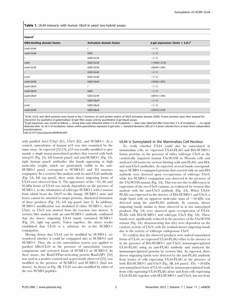
Identification of Human Ubc9 as a Cellular InteractionPartner of UL44
To identify cellular proteins that interact with UL44, Y2H
screens were carried out with a bait consisting of full-length UL44
protein (amino acids 1–433) fused to the E. coli LexA protein.
Control experiments demonstrated that the LexA-UL44 protein
did not activate expression of either HIS3 or lacZ reporter gene by
itself and, as expected [9,14], could both interact with UL54 and
dimerize (Table 1 and data not shown). Thus, this bait was used to
screen two different cellular cDNA libraries fused to S. cerevisiae
GAL4 activation domain (GAD), one derived from human B
lymphocytes [48] and the other from promyelocytic HL-60 cells
[49]. A total of 167 or 85 colonies, respectively, were positive for
both HIS3 and lacZ reporter genes. Plasmids encoding putative
interactors of UL44 were isolated from double-positive clones and
retransformed into yeasts expressing LexA-UL44 in order to
confirm the interaction. The 28 and 13 positive clones after this
retransformation, respectively, were sequenced.
In total, from the two screenings we identified 7 cellular UL44-
binding proteins; here we report the identification of human Ubc9
as a specific interaction partner of UL44. In particular, 15 out of
28 clones of B lymphocytes library and 6 out of 13 clones of the
HL-60 cells library contained the whole Ubc9 coding sequence,
plus 59 and 39 untranslated regions which varied in length in
different clones. By co-transformation experiments with each
individual interactor clone expressing Ubc9 and the LexA vector,
it was excluded that Ubc9 could activate the reporter genes in the
absence of the bait protein (Table 1). Ubc9 specifically interacted
with UL44 in Y2H assays also in the reverse combination, i.e.,
with Ubc9 fused to LexA and UL44 fused to GAD (Table 1). In
addition, in quantitative assays the b-galactosidase activity of
yeasts expressing both UL44 and Ubc9 turned out to be
comparable to that of yeasts expressing UL44 and UL54
(Table 1), which served as a positive control.
UL44 Physically Interacts with Ubc9 through N-terminaland C-terminal Regions
Having identified Ubc9 as a potential interaction partner of
UL44 in Y2H screens, we wished to confirm their physical
interaction by an independent experimental approach. For this
purpose, pulldown assays with a purified GST-UL44 protein and
in vitro-translated, 35S-labeled Ubc9 were performed. As positive
and negative controls, we also assayed the interaction between
UL44 and in vitro-translated UL54 or the PB1 subunit of influenza
A virus RNA polymerase. As expected, we could detect the
interaction of UL54 [14,29], but not of PB1, with GST-UL44
(Fig. 1A). Consistent with the Y2H results, Ubc9 specifically
associated with GST-UL44, while no interaction with GST was
observed (Fig. 1A).
Since our data suggested that UL44 can interact with Ubc9
both in vitro and in yeast cells, we sought to examine whether
UL44 and Ubc9 could also co-localize in mammalian cells. To this
end, aggregates distributed on a diffuse background fluorescence
throughout the nucleus (Fig. 1B). As previously reported [50],
when expressed alone Ubc9 exhibited a nuclear punctate pattern
(Fig. 1B). Upon co-expression of GFP-UL44 and DsRed2-Ubc9,
co-localization of UL44 we analyzed the subcellular localization of
GFP-UL44 when transiently expressed either alone or in the
presence of DsRed2-Ubc9. As a negative control, we also
expressed the DsRed2-UL53 fusion protein, which localizes to
the cell nucleus but does not interact with UL44 [32]. In GFP-
UL44-transfected cells, UL44 localized in a large number of
discrete nuclear with Ubc9 was observed (Fig. 1B). UL44 also co-
localized with a catalytically impaired Ubc9 mutant (Ubc9C93S)
[51], but not with UL53 [32]. In addition, co-immunoprecipita-
tion experiments confirmed that UL44 could physically interact
with endogenous Ubc9 in mammalian cells. The specificity of the
interaction was confirmed by the inability of UL44 to co-
immunoprecipitate with Cyclin D1 (CycD1; Fig. 1C).
To further explore the interaction between UL44 and Ubc9, we
sought to map the domain of UL44 that interacts with Ubc9. To
this aim, several N- and C-terminal deletion mutants of UL44
fused to LexA were generated and tested for the ability to interact
with GAD-Ubc9 by Y2H assays. Control western blot experiments
with an anti-LexA antibody evidenced protein bands of the
expected molecular mass for all mutants (data not shown). As
shown in Fig. 1D, the truncated protein UL441–300, lacking most
of the C-terminal disordered region of UL44, exhibited interaction
with Ubc9. Further C-terminal truncation of UL44 revealed that a
protein fragment corresponding to the first 200 amino acids of
UL44 (UL441–200) was still capable of interacting with Ubc9
(Fig. 1D). In contrast, the N-terminal 100 residues of UL44
(UL441–100) exhibited no interaction with Ubc9 (Fig. 1D).
Although unfolding of the UL441–100 mutant protein cannot be
excluded, these results suggested that this region of UL44 may not
contain sequences important and/or sufficient for Ubc9 binding.
Therefore, we analyzed the effects of N-terminal truncations.
Deletion of the N-terminal 113 residues of UL44 (UL44114–433) did
not impair the ability of UL44 to bind Ubc9. Similarly, the
UL44201–433 mutant, lacking the N-terminal 200 amino acids,
interacted with Ubc9. Interestingly, a mutant that only expresses
the C-terminal 121 residues of UL44 (UL44313–433) still retained
the ability to bind Ubc9 (Fig. 1D). Control Y2H experiments
showed that none of the truncated UL44 proteins was able to
activate transcription by itself (data not shown). Mapping studies in
mammalian cells expressing UL44 deletion mutants showed that
both the UL441–300 and UL44313–433 mutants could immunopre-
cipitate endogenous Ubc9, similarly to wild-type UL44 (Fig. S1).
Thus, our results suggest that UL44 contains two domains capable
of independently binding to Ubc9, located at the N- and C-
terminus of the protein (likely within residues 1–200 and 313–433,
respectively).
Ubc9 Mediates Extensive Sumoylation of UL44The observation that UL44 interacts with the SUMO-conju-
gating enzyme Ubc9 prompted us to investigate the possibility that
UL44 may be sumoylated. This hypothesis was first tested in a cell-
free system by incubating a purified 6His-UL44 fusion protein
Sumoylation of HCMV UL44
PLOS ONE | www.plosone.org 4 November 2012 | Volume 7 | Issue 11 | e49630
with purified Aos1/Uba2 (E1), Ubc9 (E2), and SUMO-1. As a
control, sumoylation of human p53 was also examined by the
same assay. As expected [52,53], p53 was readily modified to give
mainly a single mono-sumoylated product that reacted with both
anti-p53 (Fig. 2A, left bottom panel) and anti-SUMO-1 (Fig. 2A,
right bottom panel) antibodies (the bands appearing at high
molecular weight, which are particularly visible in the anti-
SUMO-1 panel, correspond to SUMO-E1 and -E2 enzymes
conjugates). In a western blot analysis with an anti-UL44 antibody
(Fig. 2A, left top panel), three main slower migrating forms of
UL44 were observed (lane 3). The appearance of the ,65, 80, and
95-kDa forms of UL44 was strictly dependent on the presence of
SUMO-1, as the substitution of wild-type SUMO-1 with a mutant
form which bears the Gly97-to-Ala change (SUMO-1 mut) and
hence cannot be attached to target proteins, eliminated formation
of these products (Fig. 2A, left top panel, lane 4). In addition,
SUMO-1 modification was abolished if either SUMO-1, Aos1/
Uba2, or Ubc9 was omitted from the reaction (not shown). A
western blot analysis with an anti-SUMO-1 antibody confirmed
that the slower migrating UL44 bands contained SUMO-1
(Fig. 2A, right top panel). Taken together, the above results
established that UL44 is a substrate for in vitro SUMO-1
conjugation.
Having shown that UL44 can be modified by SUMO-1, we
wondered whether it could be conjugated also to SUMO-2 and
SUMO-3. Thus, the in vitro sumoylation system was applied to
purified 6His-UL44 in the presence of sumoylation enzyme
components and activated forms of SUMO-2 or SUMO-3. In
these assays, the RanGTPase-activating protein RanGAP1 [54]
was used as a positive control and as previously observed [55], was
modified in the presence of SUMO-2 and SUMO-3 (data not
shown). As shown in Fig. 2B, UL44 was also modified by either of
the two SUMO peptides.
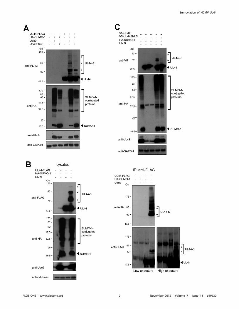
UL44 is Sumoylated in the Mammalian Cell NucleusTo verify whether UL44 could also be sumoylated in
mammalian cells, we expressed UL44-FLAG and HA-SUMO-1
fusion proteins, in the presence of either wild-type Ubc9 or the
catalytically impaired mutant Ubc9C93S in Phoenix cells and
analyzed cell lysates by western blotting with anti-FLAG, anti-HA,
and anti-Ubc9 antibodies. As expected, several bands correspond-
ing to SUMO-1-conjugated proteins that reacted with an anti-HA
antibody were detected upon co-expression of wild-type Ubc9,
while less SUMO-1 conjugation was observed in the presence of
the Ubc9C93S mutant (Fig. 3A). This was not due to differences in
expression of the two Ubc9 variants, as evidenced by western blot
analysis with the anti-Ubc9 antibody (Fig. 3A). When UL44-
FLAG was expressed in the absence of HA-SUMO-1 and Ubc9, a
single band with an apparent molecular mass of ,50 kDa was
detected using the anti-FLAG antibody. In contrast, slower
migrating bands similar to those observed in in vitro sumoylated
products (Fig. 2A) were observed upon co-expression of UL44-
FLAG with HA-SUMO-1 and wild-type Ubc9 (Fig. 3A). These
bands were significantly reduced in the presence of the Ubc9C93S
mutant (Fig. 3A), demonstrating that they were dependent on the
catalytic activity of Ubc9, with the residual slower migrating bands
due to the activity of wild-type endogenous Ubc9.
To confirm that the observed products were indeed sumoylated
forms of UL44, we expressed UL44-FLAG either in the absence or
in the presence of HA-SUMO-1 and Ubc9, immunoprecipitated
UL44-FLAG using an anti-FLAG antibody and analyzed the
immunoprecipitated proteins by western blot. As expected, three
slower migrating bands were detected by the anti-FLAG antibody
from lysates of cells expressing UL44-FLAG in the presence of
both HA-SUMO-1 and Ubc9 (Fig. 3B, left panel). The ,50-kDa
non-sumoylated form of UL44 could be immunoprecipitated both
from cells expressing UL44-FLAG alone and from cells expressing
UL44-FLAG together with HA-SUMO-1 and Ubc9, but not from
Table 1. UL44 interacts with human Ubc9 in yeast two-hybrid assays.
Hybrid1
DNA-binding domain fusion Activation domain fusion b-gal expression (Units ± S.D.)2
LexA-UL44 / 2(,1)
LexA-UL44 GAD 2(,1)
/ GAD-UL54 2(,1)
LexA GAD-UL54 6(10436213)
LexA-UL44 GAD-UL54 +(82446245)
/ GAD-UL44 2(,1)
LexA GAD-UL44 2(,1)
LexA-UL44 GAD-UL44 +(76436397)
LexA-Ubc9 / 2(,1)
LexA-Ubc9 GAD 2(,1)
LexA-Ubc9 GAD-UL44 +(69156343)
/ GAD-Ubc9 2(,1)
LexA GAD-Ubc9 2(,1)
LexA-UL44 GAD-Ubc9 +(70126214)
1UL44, UL54, and Ubc9 proteins were fused to the C-terminus of LexA protein and/or of GAL4 activation domain (GAD). Fusion proteins were then assayed forinteraction by qualitative b-galactosidase (b-gal) filter assays and by quantitative b-gal liquid assays.2b-gal expression was scored as follows +, strong blue color detected within 2 h of incubation; 6, blue color detected after more than 2 h of incubation; 2, no signaldetected after 16–24 h of incubation. Values within parentheses represent b-gal units 6 standard deviation (SD) of 3–4 yeast colonies from at least three independenttransformations.doi:10.1371/journal.pone.0049630.t001
Sumoylation of HCMV UL44
PLOS ONE | www.plosone.org 5 November 2012 | Volume 7 | Issue 11 | e49630

cells expressing only HA-SUMO-1 and Ubc9. Slower migrating
forms of UL44 could be detected by both the anti-FLAG and anti-
HA antibody in the immunoprecipitation reaction of cells
expressing UL44-FLAG in the presence of HA-SUMO and
Ubc9, but not in immunoprecipitates obtained from cells
expressing only UL44-FLAG (Fig. 3B, right panel). Similar results
were obtained in HeLa cells (Fig. S2). Altogether, these results
demonstrate that Ubc9 can mediate the conjugation of SUMO-1
Figure 1. HCMV UL44 interacts with human Ubc9. (A) GST-pulldown assays showing interaction of purified GST or GST-UL44 with in vitro-expressed, radiolabeled Ubc9 (right panels), HCMV UL54 (central panels, positive control), and influenza A virus PB1 (left panels, negative control). I,input; E, eluted by glutathione. Arrowheads indicate the proteins of interest. (B) COS-1 cells were transfected to express the indicated GFP andDsRed2 fusion proteins and imaged by CLSM. Merged images of the green (GFP) and red (DsRed2) channels are shown on the right, with yellowcoloration indicative of co-localization. (C) Phoenix cells were transfected to express a UL44-FLAG fusion. At 48 h post-transfection, cell lysates wereanalyzed by western blotting with anti-FLAG, anti-Ubc9, anti-CycD1, and anti-vinculin antibodies. Cell lysates were also incubated with anti-FLAG-M2-Agarose beads and the immunoprecipitated samples were analyzed by western blotting with anti-FLAG, anti-Ubc9, and anti-CycD1 antibodies.Asterisks indicate the IgG heavy chains, while arrowheads indicate the proteins of interest. (D) Several UL44 mutants were created by deletingdifferent portions of UL44 coding sequence and expressed as fusions with LexA. A diagram of the full-length and truncated UL44 proteins, withfunctional domains indicated, is reported. Numbers refer to remaining amino acid residues of UL44. Blue bars, residues important for dimerization; CL,connector loop; FL, flexible loop; grey boxes, glycine strings; red box, Nuclear Localization Signal. The ability of the mutants to physically interact withUbc9 was determined by b-gal filter assays and scored as follows: +, strong blue color detected within 2 h of incubation; 6, blue color detected aftermore than 2 h of incubation; 2, no signal detected after 16–24 h of incubation. Values within parentheses represent b-gal units 6 standard deviation(SD) of 3–4 yeast colonies from at least three independent transformations, determined by quantitative b-gal assays.doi:10.1371/journal.pone.0049630.g001
Sumoylation of HCMV UL44
PLOS ONE | www.plosone.org 6 November 2012 | Volume 7 | Issue 11 | e49630

Sumoylation of HCMV UL44
PLOS ONE | www.plosone.org 7 November 2012 | Volume 7 | Issue 11 | e49630

to UL44 in mammalian cells. Moreover, sumoylation of UL44 in
mammalian cells by both SUMO-2 and SUMO-3 could also be
detected (Fig. S3).
Since sumoylation mainly occurs in the nucleus of mammalian
cells [56] and UL44 is translocated to the host cell nucleus during
HCMV infection [11], we decided to investigate whether nuclear
localization might be a prerequisite for conjugation of SUMO-1 to
UL44. We therefore analyzed the ability of UL44bDNLS, a
derivative of UL44 bearing point mutations within the nuclear
localization signal (NLS, 425PNTKKQK431) which prevent UL44
nuclear accumulation [11], to be modified by SUMO-1.
Interestingly, mutations of UL44 NLS impaired the sumoylation
of a V5-UL44 fusion protein (V5-UL44DNLS, Fig. 3C), suggesting
that SUMO-1 modification of UL44 most likely occurs into the
nucleus or during nuclear import.
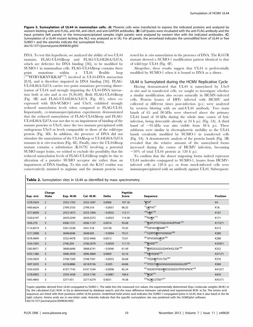
Mapping of Sumoylation Sites in UL44We next sought to identify the SUMO-1 acceptor sites of UL44.
A prediction analysis with the SUMOplot program (Abgent)
identified seven residues in UL44 with a certain probability to be
sumoylated: K73, K172, K224, K339, K371, K410, and K431.
To test whether one or more of these lysines could be a SUMO-1
acceptor site, each of the candidate residues was conservatively
mutated to arginine, both individually and in combination, and
the mutant proteins were tested for in vitro sumoylation. None of
the single point mutants exhibited a consistently altered SUMO-1
modification pattern as compared to wild-type UL44 (Fig. S4A).
Mutants carrying two or three K/R substitutions also showed a
SUMO-1 modification pattern identical to that of wild-type UL44
(Fig. S4A and data not shown). Similar results were obtained when
in vitro sumoylation reactions with SUMO-2/23 were performed
(data not shown). We decided to also test the ability of these K/R
mutants to be modified by SUMO-1 in mammalian cells.
Consistent with the in vitro data, none of the tested mutations
significantly affected the ability of UL44 to undergo SUMO-1
conjugation (Fig. S4B and data not shown).
These results suggested that UL44 might possess multiple lysine
residues that could alternatively serve as SUMO-1 acceptors, as
reported for other proteins [57,58], and/or that UL44 might be
sumoylated on lysine residues other than those predicted by
SUMOplot. UL44 contains 31 lysines, most of which are solvent-
exposed in the crystal structure [9] and therefore potentially
accessible to SUMO molecules, which makes it difficult to identify
the target lysines by mutational approaches. Therefore, we
attempted to map the sites of UL44 where SUMO-1 is conjugated
by mass spectrometry analysis. To this end, we expressed UL44 in
an E. coli expression/modification system that produces SUMO-
conjugated proteins [30]. The 6His-tagged UL44 construct was
co-expressed in E. coli with the pTE1E2S1 plasmid, which contains
a linear fusion of genes for E1 and E2 enzymes and SUMO-1
under the control of an IPTG-inducible promoter [30]. To
confirm sumoylation of UL44 with this system, UL44 was purified
from the bacterial cultures expressing UL44 alone or in
combination with the SUMO conjugation system and analyzed
by western blotting with both the anti-UL44 (Fig. S5, left panel)
and the anti-SUMO-1 (Fig. S5, right panel) antibody. Shifted
bands with an apparent molecular mass of ,65, 80, and 95 kDa,
similar to those detected in in vitro reactions (Fig. 2A) and in cells
(Fig. 3), were observed in UL44 purified from bacteria
cotransformed with pTE1E2S1, but not in UL44 purified from
bacteria expressing only UL44 (Fig. S5). Then, to identify lysine
acceptor sites by mass spectrometry, sumoylated UL44 was
separated by SDS-PAGE, in-gel-digested with trypsin and peptides
were analyzed by LC-MSMS. Upon application of the software
‘‘ChopNSpice’’ for database search, we identified 16 sumoylation
sites in UL44, including the predicted sites K172, K339, K371,
K410, and K431. Table 2 summarizes the UL44 peptides that
have been found to be sumoylated. The corresponding MS spectra
are shown in Fig. S6.
These results indicated that UL44 possesses multiple SUMO
target lysines that are located throughout the protein, in
accordance with the observation that Ubc9 could bind to both
N- and C-terminal portions of UL44 (Fig. 1D and Fig. S1).
Mutagenesis of several of these lysines in combination caused a
strong decrease of UL44 expression (data not shown), likely due to
protein misfolding and/or instability, making impossible to
analyze the sumoylation state of the mutated protein and to
compare it to that of wild-type UL44.
UL44 sumoylation is stimulated by binding to DNA. As
mentioned above, UL44 possesses a structural fold similar to that
of the eukaryotic processivity factor PCNA [9]. In addition, like
UL44, PCNA is SUMO-conjugated and its sumoylation involves
both a consensus and a non-consensus site [4]. Since it has been
shown that PCNA needs DNA to be sumoylated efficiently [59],
we wished to investigate whether UL44 might behave similarly
and its sumoylation could be stimulated by the presence of DNA.
Thus, we performed in vitro sumoylation experiments in the
absence and in the presence of DNA using as a substrate a
purified 6His-UL44 fusion protein treated with polymin P to
eliminate residual bacterial nucleic acids. In a western blot analysis
with an anti-UL44 antibody (Fig. 4A, left panel), only a faint band
corresponding to mono-sumoylated UL44 was observed in the
absence of DNA (lane 2). Upon addition of dsDNA (e.g., activated
calf thymus DNA) to the reaction mixture, a marked increase of
the mono-sumoylated product and the appearance of bi- and tri-
sumoylated forms of UL44 were observed (Fig. 4A, lane 4 of left
panel). A western blot analysis with an anti-SUMO-1 antibody
confirmed that these bands indeed contained SUMO-1 (data not
shown). Similar results were obtained when GST-UL44 was used
as a substrate (Fig. S7). Thus, like PCNA sumoylation, UL44
sumoylation is strongly stimulated by the presence of DNA.
To investigate whether the nature of DNA could influence the
stimulation of UL44 sumoylation, we also performed in vitro
sumoylation reactions in the presence of different DNA substrates.
Similar stimulation levels were obtained when different dsDNAs
were added, regardless of their sequence (data not shown), in
keeping with previous observations that UL44 binds DNA in a
sequence-independent manner [7]. In contrast, consistently less
stimulation of UL44 sumoylation was observed in the presence of
ssDNA (Fig. 4A, right panel), for which UL44 has been shown to
possess an apparent affinity lower than for dsDNA [7]. This
suggested that UL44 sumoylation could depend on binding to
Figure 2. Sumoylation of UL44 in vitro. (A) To analyze UL44 sumoylation in vitro, purified 6His-UL44 was incubated in the absence or thepresence of sumoylation enzymes and either wild-type SUMO-1 (SUMO-1 wt) or a mutant form of SUMO-1 (SUMO-1 mut) which cannot be covalentlylinked to substrates. The reaction products were analyzed by western blotting with anti-UL44 and anti-SUMO-1 antibodies. As a positive control,in vitro sumoylation of p53 was also analyzed. (B) Purified 6His-UL44 was incubated in the absence or the presence of sumoylation enzymes andeither SUMO-2 or SUMO-3 and analyzed by western blotting with anti-UL44 and anti-SUMO-2/23 antibodies. For all panels, the arrowhead indicatesthe unmodified form of UL44 or p53 and the asterisks indicate the respective sumoylated forms.doi:10.1371/journal.pone.0049630.g002
Sumoylation of HCMV UL44
PLOS ONE | www.plosone.org 8 November 2012 | Volume 7 | Issue 11 | e49630
Sumoylation of HCMV UL44
PLOS ONE | www.plosone.org 9 November 2012 | Volume 7 | Issue 11 | e49630
DNA. To test this hypothesis, we analyzed the ability of two UL44
mutants, FLAG-UL44Dloop and FLAG-UL44L86A/L87A,
which are defective for DNA binding [36], to be modified by
SUMO-1 in mammalian cells. FLAG-UL44Dloop contains three
point mutations within a UL44 flexible loop
(163HTRVKRNVKKAP174) involved in UL44-DNA interaction
[8,9], and is therefore impaired in DNA binding [36]. FLAG-
UL44L86A/L87A carries two point mutations preventing dimer-
ization of UL44 and strongly impairing the UL44-DNA interac-
tion both in vitro and in vivo [9,36,60]. Both FLAG-UL44Dloop
(Fig. 4B) and FLAG-UL44L86A/L87A (Fig. 4C), when co-
expressed with HA-SUMO-1 and Ubc9, exhibited strongly
reduced sumoylation levels when compared to FLAG-UL44.
Importantly, co-immunoprecipitation experiments demonstrated
that the reduced sumoylation of FLAG-UL44Dloop and FLAG-
UL44L86A/L87A was not due to an impairment of binding of the
mutant proteins to Ubc9, since the two mutants precipitated with
endogenous Ubc9 at levels comparable to those of the wild-type
protein (Fig. 4D). In addition, the presence of DNA did not
stimulate the sumoylation of the UL44Dloop or UL44L86A/L87A
mutants in in vitro reactions (Fig. 4E). Finally, since the UL44Dloop
mutant contains a substitution (K167N) involving a potential
SUMO target lysine, we wished to exclude the possibility that the
reduced sumoylation levels of FLAG-UL44Dloop might be due to
alteration of a putative SUMO acceptor site rather than an
impairment of DNA binding. To this end, the K167 residue was
conservatively mutated to arginine and the mutant protein was
tested for in vitro sumoylation in the presence of DNA. The K167R
mutant showed a SUMO-1 modification pattern identical to that
of wild-type UL44 (Fig. 4F).
Altogether, these results suggest that UL44 is preferentially
modified by SUMO-1 when it is bound to DNA as a dimer.
UL44 is Sumoylated during the HCMV Replicative CycleHaving demonstrated that UL44 is sumoylated by Ubc9
in vitro and in transfected cells, we sought to investigate whether
a similar modification also occurs naturally in HCMV-infected
cells. Protein lysates of HFFs infected with HCMV and
collected at different times post-infection (p.i.) were analyzed
by western blotting with an anti-UL44 antibody. Two main
bands of 65 and 80 kDa were observed above the primary
UL44 band of 50 kDa during the whole time course of lytic
infection, being detectable already at 24 h p.i. (Fig. 5A). A third
band of ,95 kDa was also visible from 48 h p.i. These
subforms were similar in electrophoretic mobility to the UL44
bands covalently modified by SUMO-1 in transfected cells
(Fig. 3A). A densitometric analysis of the protein bands (Fig. 5B)
revealed that the relative amount of the sumoylated forms
increased during the course of HCMV infection, becoming
,50% of total UL44 protein at 120 h p.i.
To confirm that the slower migrating forms indeed represent
UL44 molecules conjugated to SUMO-1, lysates from HCMV-
infected cells at 120 h p.i. or from mock-infected cells were
immunoprecipitated with an antibody against UL44. Subsequent-
Figure 3. Sumoylation of UL44 in mammalian cells. (A) Phoenix cells were transfected to express the indicated proteins and analyzed bywestern blotting with anti-FLAG, anti-HA, anti-Ubc9, and anti-GAPDH antibodies. (B) Cell lysates were incubated with the anti-FLAG antibody and theinput proteins (left panels) or the immunoprecipitated samples (right panels) were analyzed by western blot with the indicated antibodies. (C)Sumoylation of a UL44 mutant lacking the NLS was analyzed as in (A). For all panels, the arrowhead indicates the unmodified form of UL44 or freeSUMO-1 and the asterisks indicate the sumoylated forms.doi:10.1371/journal.pone.0049630.g003
Table 2. Sumoylation sites in UL44 as identified by mass spectrometry.
Exp. m/zChargeState Exp. M.W. Cal. M.W. Delta
PeptideScore Sequence Position
1278.5924 2 2555.1703 2555.1697 0.0006 107.18 4KTR6 K4
1400.6624 2 2799.3103 2799.316 20.0057 96.35 17LKPYK21 K18
1277.6009 2 2553.1873 2553.1904 20.0032 113.11 166VKR168 K167
1320.6197 2 2639.2249 2639.2272 20.0023 114.38 169NVKK172 K171
1436.379 3 4306.1151 4306.1137 0.0014 95.68 172KAPCPTGTVQILVHAGPPAIK192 K172(*)
1114.8519 3 3341.5338 3341.518 0.0158 74.35 209VSFHGVKNMR218 K215
1217.2088 3 3648.6046 3648.605 20.0004 79.21 275GDPFDKNYVGNSGK288 K280
1078.4899 3 3232.4478 3232.4466 0.0012 73.01 281NYVGNSGKSR290 K288
1354.1093 2 2706.204 2706.2079 20.0039 111.75 339KHDR342 K339(*)
1283.8971 3 3848.6696 3848.6741 20.0046 61.49 352KMSSGGGGGDHDHGLSSK369 K352
1033.1386 3 3096.3939 3096.3869 0.0069 52.16 370EKYEQHK376 K371(*)
1250.5829 3 3748.7269 3748.7301 20.0033 56.68 372YEQHKITSYLTSK384 K376
1407.3035 3 4218.8886 4218.9136 20.025 64.86 377ITSYLTSKGGSGGGGGGGGGGLDR400 K384
1550.4359 4 6197.7145 6197.7244 20.0098 85.24 401NSGNYFNDAKEESDSEDSVTFEFVPNTK428 K410(*)
1278.0902 2 2554.1658 2554.1745 20.0087 106.9 429KQK431 K429
1093.4843 3 3277.431 3277.4279 0.0031 74.38 429KQKCGTEEF437 K431(*)
Tryptic peptides derived from UL44 conjugated to SUMO-1. The table lists the measured m/z values, the experimentally determined (Exp.) molecular weights (M.W.) inDa, the calculated (Cal.) M.W. in Da as determined by database search, and the mass difference between calculated and experimental M.W. in Da. The amino acidsequences are listed with their positions within UL44 protein. Underlined bold amino acid indicates the SUMO-1-conjugated lysine in UL44, that is also listed in the farright column. Amino acids are in one-letter code. Asterisks indicate that the specific sumoylation site was predicted with the SUMOplot software.doi:10.1371/journal.pone.0049630.t002
Sumoylation of HCMV UL44
PLOS ONE | www.plosone.org 10 November 2012 | Volume 7 | Issue 11 | e49630
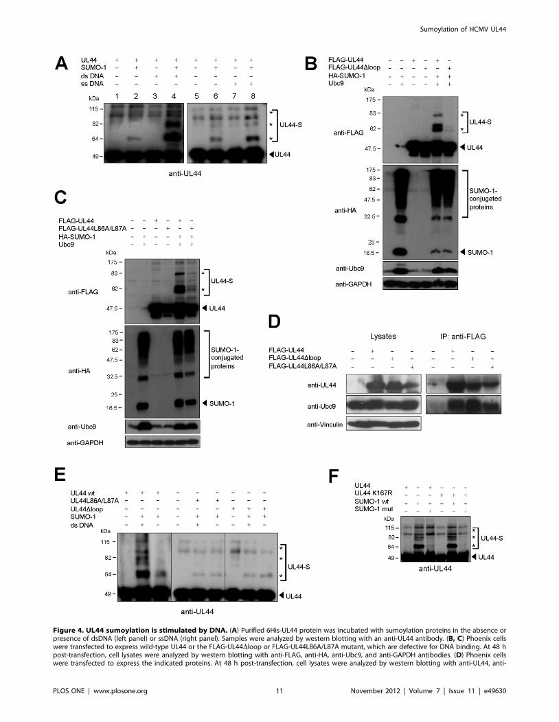
Figure 4. UL44 sumoylation is stimulated by DNA. (A) Purified 6His-UL44 protein was incubated with sumoylation proteins in the absence orpresence of dsDNA (left panel) or ssDNA (right panel). Samples were analyzed by western blotting with an anti-UL44 antibody. (B, C) Phoenix cellswere transfected to express wild-type UL44 or the FLAG-UL44Dloop or FLAG-UL44L86A/L87A mutant, which are defective for DNA binding. At 48 hpost-transfection, cell lysates were analyzed by western blotting with anti-FLAG, anti-HA, anti-Ubc9, and anti-GAPDH antibodies. (D) Phoenix cellswere transfected to express the indicated proteins. At 48 h post-transfection, cell lysates were analyzed by western blotting with anti-UL44, anti-
Sumoylation of HCMV UL44
PLOS ONE | www.plosone.org 11 November 2012 | Volume 7 | Issue 11 | e49630

ly, the immunocomplexes were analyzed by western blotting with
an anti-SUMO-1 antibody. Three main bands of the expected
molecular mass (,65, 80, and 95 kDa) were recognized in the
anti-UL44 immunoprecipitate (Fig. 5C, left panel). Finally, the
same immunocomplexes were analyzed by western blotting with
the anti-UL44 antibody to demonstrate that the SUMO-1 cross-
reactive proteins were indeed modified UL44 forms (Fig. 5C, right
panel).
Thus, these results clearly indicate that UL44 is covalently
modified by SUMO-1 in HCMV-infected cells.
Overexpression of SUMO-1 Alters the IntranuclearDistribution of UL44 during HCMV Replication andPromotes Virus Replication
Considering the difficulties in expressing a UL44 mutant
completely impaired in sumoylation, whose activities could be
compared to that of wild-type UL44, to gain some insight on the
functional role of UL44 sumoylation in the context of HCMV
replication we sought to undertake a different approach. We
overexpressed SUMO-1 in virus-infected cells and analyzed the
effects on the intracellular distribution of UL44, as the targeting to
specific subcellular domains is one of most common biological
effect exerted by the conjugation of SUMO to a substrate protein.
It has been previously shown that during HCMV replication
UL44 localizes to large globular intranuclear structures that
correspond to viral DNA replication compartments [61,62,63]. A
U373-MG cell line that constitutively overexpresses FLAG-
SUMO-1 and control U373-Neo cells were mock-infected or
infected with HCMV at an MOI of 1 or of 5 PFU/cell and the
intracellular localization of UL44 was successively analyzed by
indirect immunofluorescence with an anti-UL44 antibody. Con-
trol western blotting experiments (Fig. S8A) confirmed that UL44
is sumoylated in the HCMV-infected U373 cells. In fact, slower
migrating bands of the expected molecular mass and similar to the
UL44 sumoylated forms observed in infected HFFs (Fig. 5A) were
detected. Furthermore, as expected, they increased upon SUMO-
1 overexpression. In immunofluorescence assays, the nuclei of
mock-infected cells were oval-shaped with no anti-UL44 staining
(Fig. 6A, upper panels). Control HCMV-infected U373-Neo cells
showed deformed nuclei, many of which exhibited a kidney shape
(Fig. 6A, upper panels). Indeed, it has been observed that infection
by HCMV causes this kind of distortions in nuclear shape [64,65].
Moreover, UL44 showed a globular fluorescent pattern consistent
with previously described viral replication compartments in
HCMV-infected cells [61,66]. In contrast, in HCMV-infected
U373-SUMO-1 cells UL44 staining was more distributed
throughout the nucleus and, especially at the lower MOI (MOI
1), also failed to coalesce into any recognizable globular structures
(Fig. 6A, upper panels). Therefore, overexpression of SUMO-1
during HCMV replication appears to alter the intranuclear
distribution of UL44, likely leading to significantly decreased
localization of UL44 in viral DNA replication compartments.
Importantly, the intranuclear distribution of another HCMV
protein localizing to the replication compartments, i.e. UL57, the
single-stranded DNA (ssDNA)-binding protein [61,67], appeared
not to change upon SUMO-1 overexpression (Fig. 6A, lower
panels).
To investigate whether sumoylation is indeed involved in the
altered intranuclear distribution of UL44, an RNAi approach
was employed to suppress the sumoylation system of the cells by
silencing Ubc9 since Ubc9 is the only unique and essential
enzyme in the SUMO-conjugating pathway [21]. U373-Neo and
U373-SUMO-1 cells were transduced with a lentivirus expressing
shUbc9, followed by selection with puromycin and induction
with doxycyline to establish Ubc9-knocked-down cell lines
(U373-Neo shUbc9 and U373-SUMO-1 shUbc9, respectively).
As a control, U373-SUMO-1 cells were also transduced with a
lentivirus expressing a non-silencing shRNA sequence (U373-
SUMO-1 NS). As shown in Figure 6B, Ubc9 was almost
completely silenced in cells infected with the shUbc9 lentivirus
(U373-SUMO-1 shUbc9) as compared to the cells transduced
with the control lentivirus (U373-SUMO-1 NS) and to non-
transduced cells (U373-Neo and U373-SUMO-1). To examine
the effect of Ubc9 knock-down on intranuclear distribution of
UL44, these cell lines were infected with HCMV. As shown in
Fig. 6A (upper panels), upon doxycycline induction, the nuclear
UL44 staining in Ubc9-knocked-down U373-SUMO-1 cells was
similar to that of U373 cells not overexpressing SUMO-1 (U373-
Neo and U373-Neo shUbc9; Fig. 6A, upper panels, and Fig.
S8B). In contrast, the U373-SUMO-1 cells transduced with the
non-silencing lentivirus (U373-SUMO-1 NS, Fig. 6A, upper
panels) retained an altered intranuclear distribution of UL44
similar to that observed in the non-transduced cells (U373-
SUMO-1) or in the shUbc9-transduced U373-SUMO-1 cells
with no doxycycline induction (data not shown). Altogether, these
results established that the altered intranuclear distribution of
UL44 in HCMV-infected cells upon SUMO-1 overexpression
depends on Ubc9-mediated sumoylation.
These observations raised the question whether the altered
intranuclear distribution of UL44 observed upon SUMO-1
overexpression affected the viral replication efficiency. To examine
the effects of SUMO-1 overexpression on viral DNA replication,
U373-Neo and U373-SUMO-1 cells were infected with HCMV at
an MOI of 1 and viral DNA levels were measured by quantitative
real-time PCR. Viral DNA production from U373-SUMO-1 cells
was ,two-fold higher than that from the control cells (U373-Neo
and U373-Neo shUbc9; Fig. 6C). This increase was not observed
in Ubc9-knocked down cells (U373-SUMO-1 shUbc9), while the
U373-SUMO-1 cells transduced with the non-silencing lentivirus
(U373-SUMO-1 NS) exhibited augmented viral DNA levels like
the non-transduced U373-SUMO-1 cells (Fig. 6C).
We also examined the effects of SUMO-1 overexpression on
virus yield. The titers of viral particles produced from non-
transduced U373-SUMO-1 and from transduced U373-SUMO-1
shUbc9 and U373-SUMO-1 NS cells after infection with HCMV
at an MOI of 1 were determined and compared to those produced
from infected U373-Neo and U373-Neo shUbc9 control cells. A 2-
3-fold increase in viral progeny titers was observed in U373-
SUMO-1 and U373-SUMO-1 NS with respect to U373-Neo,
while the U373-SUMO-1 shUbc9 cells exhibited yields of
Ubc9, and anti-vinculin antibodies (left panel). Cell lysates were incubated with anti-FLAG-M2-Agarose beads and the immunoprecipitated sampleswere analyzed by western blotting with anti-UL44 and anti-Ubc9 antibodies (right panel). (E) The sumoylation in vitro of wild-type 6His-UL44 andmutant 6His-UL44Dloop and 6His-UL44L86A/L87A proteins was carried out as in (A) and analyzed by western blotting with an anti-UL44 antibody. (F)The sumoylation in vitro of a UL44 mutant bearing the K167R substitution in the flexible loop of UL44 involved in DNA binding was carried out in thepresence of DNA and compared to that of wild-type UL44. For all panels, the arrowhead indicates the unmodified form of UL44 or free SUMO-1 andthe asterisks indicate the sumoylated forms.doi:10.1371/journal.pone.0049630.g004
Sumoylation of HCMV UL44
PLOS ONE | www.plosone.org 12 November 2012 | Volume 7 | Issue 11 | e49630

Figure 5. Sumoylation of UL44 in HCMV-infected cells. (A) HFFs were either mock-infected or infected with HCMV for the indicated times. Celllysates were analyzed by western blotting with an anti-UL44 antibody. (B) Blots were analyzed by densitometry and the percentage of sumoylatedUL44 bands relative to that of unmodified UL44 at each time p.i. was plotted versus the p.i. time point. Data represent the means 6 standarddeviations (error bars) of values from three independent experiments such as that shown in (A). (C) Lysates from either mock-infected or HCMV-infected HFF cells were prepared at 120 h p.i. and immunoprecipitated with an anti-UL44 antibody. Immunoprecipitates were analyzed by western
Sumoylation of HCMV UL44
PLOS ONE | www.plosone.org 13 November 2012 | Volume 7 | Issue 11 | e49630
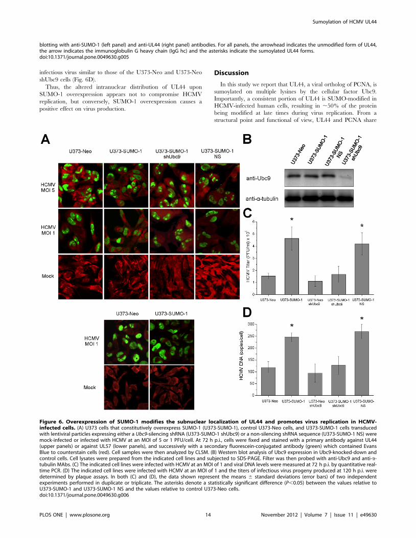
infectious virus similar to those of the U373-Neo and U373-Neo
shUbc9 cells (Fig. 6D).
Thus, the altered intranuclear distribution of UL44 upon
SUMO-1 overexpression appears not to compromise HCMV
replication, but conversely, SUMO-1 overexpression causes a
positive effect on virus production.
Discussion
In this study we report that UL44, a viral ortholog of PCNA, is
sumoylated on multiple lysines by the cellular factor Ubc9.
Importantly, a consistent portion of UL44 is SUMO-modified in
HCMV-infected human cells, resulting in ,50% of the protein
being modified at late times during virus replication. From a
structural point and functional of view, UL44 and PCNA share
blotting with anti-SUMO-1 (left panel) and anti-UL44 (right panel) antibodies. For all panels, the arrowhead indicates the unmodified form of UL44,the arrow indicates the immunoglobulin G heavy chain (IgG hc) and the asterisks indicate the sumoylated UL44 forms.doi:10.1371/journal.pone.0049630.g005
Figure 6. Overexpression of SUMO-1 modifies the subnuclear localization of UL44 and promotes virus replication in HCMV-infected cells. (A) U373 cells that constitutively overexpress SUMO-1 (U373-SUMO-1), control U373-Neo cells, and U373-SUMO-1 cells transducedwith lentiviral particles expressing either a Ubc9-silencing shRNA (U373-SUMO-1 shUbc9) or a non-silencing shRNA sequence (U373-SUMO-1 NS) weremock-infected or infected with HCMV at an MOI of 5 or 1 PFU/cell. At 72 h p.i., cells were fixed and stained with a primary antibody against UL44(upper panels) or against UL57 (lower panels), and successively with a secondary fluorescein-conjugated antibody (green) which contained EvansBlue to counterstain cells (red). Cell samples were then analyzed by CLSM. (B) Western blot analysis of Ubc9 expression in Ubc9-knocked-down andcontrol cells. Cell lysates were prepared from the indicated cell lines and subjected to SDS-PAGE. Filter was then probed with anti-Ubc9 and anti-a-tubulin MAbs. (C) The indicated cell lines were infected with HCMV at an MOI of 1 and viral DNA levels were measured at 72 h p.i. by quantitative real-time PCR. (D) The indicated cell lines were infected with HCMV at an MOI of 1 and the titers of infectious virus progeny produced at 120 h p.i. weredetermined by plaque assays. In both (C) and (D), the data shown represent the means 6 standard deviations (error bars) of two independentexperiments performed in duplicate or triplicate. The asterisks denote a statistically significant difference (P,0.05) between the values relative toU373-SUMO-1 and U373-SUMO-1 NS and the values relative to control U373-Neo cells.doi:10.1371/journal.pone.0049630.g006
Sumoylation of HCMV UL44
PLOS ONE | www.plosone.org 14 November 2012 | Volume 7 | Issue 11 | e49630
some remarkable similarities and some differences. Monomers of
UL44 and PCNA are structurally very similar, despite having
extremely different primary sequences [2,9]. However, while
PCNA forms toroidal-homotrimers, UL44 binds to dsDNA as a
head-to-head homodimer [7,9]. In addition, PCNA must be
loaded onto DNA in an ATP-dependent process by so-called
clamp loaders [68]; in contrast, UL44 directly binds DNA without
the need for ATP hydrolysis or accessory proteins [7,14,18].
Similarities and differences also emerge from the comparison of
the sumoylation processes of UL44 and PCNA. The most striking
similarity is the DNA-dependence of such post-translational
modification: in the case of PCNA, clamp loading rather than the
mere presence of DNA was shown to be important for stimulation,
implying a change in the properties of PCNA upon loading that
enhances its capacity to be sumoylated [59]. This could also be the
case for UL44: in fact, point mutations preventing DNA binding
[8,9,36,60] strongly impaired UL44 sumoylation in cells (Fig. 4C)
and abolished the ability of dsDNA to stimulate UL44 sumoylation
in vitro (Fig. 4E). Furthermore, the ability of DNA to stimulate UL44
sumoylation appears to correlate with the efficiency of the UL44-
DNA interaction. In fact, addition of dsDNA -for which UL44 has a
,3- to 8-fold higher affinity than for ssDNA [7]- caused a much
stronger increase of UL44 sumoylation as compared to ssDNA
(Fig. 4A). In this context, it is important to note that point mutations
impairing DNA binding and sumoylation in cells did not
compromise the UL44-Ubc9 interaction (Fig. 4D), suggesting that
binding to DNA does not promote UL44 sumoylation by facilitating
its binding to Ubc9.
Another similarity between UL44 and PCNA sumoylation is
that both proteins are modified on canonical (K127 for PCNA,
and K410 for UL44) and non-canonical residues (K164 for
PCNA, and all other sumoylation sites for UL44, see Table 2).
However, while PCNA is exclusively sumoylated at the N-
terminus, both N- and C-terminal residues of UL44 can be
modified. In addition, according to our MS-analysis UL44 can be
alternatively sumoylated at 16 different sites, while K127 and
K164 appear to be the only target sites in PCNA. This flexibility of
UL44 in terms of sumoylation target sites, which is reminiscent of
the ones described for Daxx and the small hepatitis delta antigen
[57,58], is arguably the main difference from PCNA. This makes it
extremely difficult to study the physiological importance of UL44
sumoylation, also because mutation of several of these lysines
caused protein instability. Currently it is therefore impossible to
test if, like in the case of budding yeast PCNA, SUMO-
modification of UL44 also plays a role in DNA repair/
recombination [69].
In terms of functional effects, sumoylation is known to regulate
protein activity and/or intracellular location [21,22]. As for the
latter, the targeting to specific subcellular domains is one of the
best-characterized biological effects exerted by the conjugation of
SUMO to a substrate protein. This effect is exemplified by the
targeting of cellular protein RanGAP1 to the cytosolic side of the
nuclear pore complex upon sumoylation [54,70]. As a first,
preliminary attempt to characterize the role of UL44’s sumoyla-
tion, here we show that overexpression of SUMO-1 in the context
of HCMV replication alters the intranuclear distribution of UL44
as it appears to result in a more diffuse pattern and in decreased
localization of UL44 in viral DNA replication compartments
(Fig. 6A), suggesting that sumoylation of UL44 may retarget the
protein to other nuclear site(s). Importantly, Ubc9 knock-down
studies confirmed that sumoylation is responsible for such altered
intranuclear pattern of UL44. The observation that SUMO-1
overexpression causes a positive effect on HCMV replication
suggests that sumoylation of UL44 could be important for its
function(s) in the context of the virus life cycle, although effects on
HCMV replication mediated by sumoylation of other viral or
cellular proteins cannot be excluded. However, at the moment
understanding the molecular details of how SUMO alters the
intranuclear distribution of UL44 is rather difficult. Most likely,
sumoylation does not affect the functions of UL44 as a DNA
polymerase processivity factor - the only role currently well
established for UL44. In fact, several reports have shown that the
UL44 protein expressed in E. coli, which is non-sumoylated, is
capable of performing all known biochemical activities related to
this role (e.g., [7,14]). In addition, our observation that UL44
sumoylation peaks at late times during virus replication, once the
viral DNA replication has been accomplished (Fig. 5), suggests that
SUMO-conjugation might enable UL44 to fulfill some role(s) in
HCMV replication other than that of conferring processivity to
the viral DNA polymerase. A role of UL44 in late gene expression
has been previously suggested [71,72]; however, no conclusive
demonstration has been provided yet.
Intriguingly, higher molecular mass forms of the Epstein-Barr
virus DNA polymerase processivity factor BMRF1 compatible
with sumoylated products have been observed [73], suggesting
that such post-translational modification could be a general feature
of the DNA polymerase accessory subunit in herpesviruses. In
addition, sumoylation of Vaccinia Virus G8R protein has been
recently predicted on the basis of structural similarities to PCNA
[74], but not yet experimentally demonstrated. Thus, our findings
could stimulate further studies on sumoylation of DNA polymerase
subunits in other herpesviruses or, more in general, in other viral
systems.
Supporting Information
Figure S1 Both N-terminal and C-terminal regions ofUL44 bind endogenous Ubc9 in mammalian cells.Phoenix cells were transfected to express full-length UL44 or
truncated UL44 mutants. (A) At 48 h post-transfection, cell lysates
were analyzed by western blotting with anti-FLAG, anti-Ubc9,
and anti-GAPDH antibodies. (B) Cell lysates were incubated with
anti-FLAG-M2-Agarose beads and the immunoprecipitated
samples were analyzed by western blotting with anti-FLAG and
anti-Ubc9 antibodies.
(TIF)
Figure S2 UL44 is sumoylated in HeLa cells. HeLa cells
were transfected to express the indicated proteins. (A) At 48 h post-
transfection, cell lysates were analyzed by western blotting with
anti-FLAG, anti-HA, anti-Ubc9, and anti-a-tubulin antibodies. (B)
Cell lysates were incubated with the anti-FLAG antibody and the
immunoprecipitated samples were analyzed by western blotting
with anti-HA and anti-FLAG antibodies. For all panels, the
arrowhead indicates the unmodified form of UL44 or free SUMO-
1 and the asterisks indicate the sumoylated forms.
(TIF)
Figure S3 Sumoylation of UL44 by SUMO-2/23 inmammalian cells. Phoenix cells were transfected to express
the indicated proteins. At 48 h post-transfection, cell lysates were
analyzed by western blotting with anti-UL44, anti-HA, anti-Ubc9,
and anti-vinculin antibodies (left panel). Cell lysates were
incubated with anti-FLAG-M2-Agarose beads and the immuno-
precipitated samples were analyzed by western blotting with anti-
HA and anti-FLAG antibodies (right panel). For all panels, the
arrowhead indicates the unmodified form of UL44 or free SUMO-
2/23 and the asterisks indicate the UL44 sumoylated forms.
(TIF)
Sumoylation of HCMV UL44
PLOS ONE | www.plosone.org 15 November 2012 | Volume 7 | Issue 11 | e49630

Figure S4 Mutational analysis of predicted SUMO-1target sites of UL44. (A) Lysine substitution mutants of UL44
were produced and analyzed for sumoylation in vitro. (B) Phoenix
cells were transfected to express the indicated proteins and
analyzed by western blotting with anti-FLAG, anti-HA, anti-
Ubc9, and anti-GAPDH antibodies. For all panels, the arrowhead
indicates the unmodified form of UL44 or free SUMO-1 and the
asterisks indicate the sumoylated forms.
(TIF)
Figure S5 Sumoylation of UL44 in E. coli. The pRSET44
plasmid, encoding 6His-tagged UL44, was introduced into E. coli
together with the pTE1E2S1 plasmid, which expresses E1 and E2
sumoylation enzymes and SUMO-1. As a control, bacteria were
also transformed only with pRSET44. The 6His-tagged UL44 was
purified from bacterial cultures expressing UL44 alone or in
combination with the SUMO conjugation system and analyzed by
western blotting with anti-UL44 and anti-SUMO-1 antibodies.
(TIF)
Figure S6 Mass spectrometry analysis of sumoylationsites of UL44. MSMS analysis of tryptic peptides conjugated to a
tryptic peptide of SUMO-1 (ELGMEEEDVIEVYQEQTGG or
IADNHTPKELGMEEEDVIEVYQEQTGG, 1 tryptic misclea-
vage) derived from E. coli-expressed, sumoylated UL44. Sequence
of the conjugate is listed in each table above the spectra. Y-type
and b-type fragment ions are assigned in the spectra. The table
below each spectrum summarizes the database search. Highlight-
ed are the m/z values that match the fragment ions obtained from
in silico fragmentation of the conjugate. Conjugated lysine residues
to SUMO-1 are highlighted in red and the position in UL44 is
listed. Modifications, measured (observed) m/z values, the actual
mass (in Da), the charge state, and the mass error (ppm, parts per
million) are listed as well in the table above each spectrum. The
different colors represent the various measured and calculated
fragment ions of each conjugate in the spectrum and table
underneath, respectively. The question marks in some of the
spectra indicate fragment ions that do not match to any calculated
y- and b-type ions of the conjugate.
(PDF)
Figure S7 Sumoylation in vitro of a GST-UL44 fusion isstimulated by DNA. E. coli-expressed, purified GST-tagged
UL44 was incubated with purified sumoylation enzymes in the
absence or presence of SUMO-1 and/or DNA. The samples were
analyzed by western blotting with an anti-UL44 antibody.
(TIF)
Figure S8 UL44 is sumoylated in HCMV-infected U373cells. (A) U373-Neo and U373-SUMO-1 cells were either mock-
infected or infected with HCMV at MOI of 5 PFU/cell for 72 h.
Cell lysates were then analyzed by western blotting with an anti-
UL44 antibody. The arrowhead indicates the unmodified form of
UL44 and the asterisks indicate the sumoylated UL44 forms. (B)
Control U373-Neo cells, and U373-Neo cells transduced with
lentiviral particles expressing either a Ubc9-silencing shRNA
(U373-Neo shUbc9) or a non-silencing shRNA sequence (U373-
Neo NS) were mock-infected or infected with HCMV at an MOI
of 5 or 1 PFU/cell. At 72 h p.i., cells were fixed and stained with a
primary antibody against UL44 and successively with a secondary
fluorescein-conjugated antibody (green) which contained Evans
Blue to counterstain cells (red). Cell samples were then analyzed by
CLSM.
(TIF)
Table S1 Oligonucleotides used in this work for cloning and
mutagenesis.
(DOC)
Text S1 Supplementary Material and Methods and Supplemen-
tary References.
(DOC)
Acknowledgments
We thank D. Camozzi, P. F. Ertl, G. Gao, G. George, R. T. Hay, H-J.
Kung, M. Marschall, and H. Saitoh for plasmids, S. P. Goff and S. J.
Elledge for Y2H libraries, and G. P. Nolan and G. S. Hayward for Phoenix
cells and U373-SUMO-1 and U373-Neo cell lines, respectively. We also
wish to acknowledge I. Castagliuolo and M. Nadai for helping with CLSM
analysis, and D. Zanocco and R. Cinetto for experimental help.
Author Contributions
Conceived and designed the experiments: ES GA CVS BM MW HU AR
SC GP AL. Performed the experiments: ES GA CVS BM GM MW H-HH
HU AL. Analyzed the data: ES GA CVS BM MW H-HH HU AR SC GP
AL. Wrote the paper: ES GA BM HU SC GP AL.
References
1. Moldovan GL, Pfander B, Jentsch S (2007) PCNA, the maestro of the replication
fork. Cell 129: 665–679.
2. Krishna TS, Kong XP, Gary S, Burgers PM, Kuriyan J (1994) Crystal structure
of the eukaryotic DNA polymerase processivity factor PCNA. Cell 79: 1233–
1243.
3. Maga G, Hubscher U (2003) Proliferating cell nuclear antigen (PCNA): a dancer
with many partners. J Cell Sci 116: 3051–3060.
4. Hoege C, Pfander B, Moldovan GL, Pyrowolakis G, Jentsch S (2002) RAD6-
dependent DNA repair is linked to modification of PCNA by ubiquitin and
SUMO. Nature 419: 135–141.
5. Naryzhny SN, Lee H (2004) The post-translational modifications of proliferating
cell nuclear antigen: acetylation, not phosphorylation, plays an important role in
the regulation of its function. J Biol Chem 279: 20194–20199.
6. Wang SC, Nakajima Y, Yu YL, Xia W, Chen CT, et al. (2006) Tyrosine
phosphorylation controls PCNA function through protein stability. Nat Cell Biol
8: 1359–1368.
7. Loregian A, Sinigalia E, Mercorelli B, Palu G, Coen DM (2007) Binding
parameters and thermodynamics of the interaction of the human cytomegalo-
virus DNA polymerase accessory protein, UL44, with DNA: implications for the
processivity mechanism. Nucleic Acids Res 35: 4779–4791.
8. Komazin-Meredith G, Petrella RJ, Santos WL, Filman DJ, Hogle JM, et al.
(2008) The human cytomegalovirus UL44 C clamp wraps around DNA.
Structure 16: 1214–1225.
9. Appleton BA, Loregian A, Filman DJ, Coen DM, Hogle JM (2004) The
cytomegalovirus DNA polymerase subunit UL44 forms a C clamp-shaped
dimer. Mol Cell 15: 233–244.
10. Gibson W, Murphy TL, Roby C (1981) Cytomegalovirus-infected cells contain a
DNA-binding protein. Virology 111: 251–262.
11. Alvisi G, Jans DA, Guo J, Pinna LA, Ripalti A (2005) A protein kinase CK2 site
flanking the nuclear targeting signal enhances nuclear transport of human
cytomegalovirus ppUL44. Traffic 6: 1002–1013.
12. Alvisi G, Marin O, Pari G, Mancini M, Avanzi S, et al. (2011) Multiple
phosphorylation sites at the C-terminus regulate nuclear import of HCMV DNA
polymerase processivity factor ppUL44. Virology 417: 259–267.
13. Fulcher AJ, Roth DM, Fatima S, Alvisi G, Jans DA (2010) The BRCA-1 binding
protein BRAP2 is a novel, negative regulator of nuclear import of viral proteins,
dependent on phosphorylation flanking the nuclear localization signal. FASEB J
24: 1454–1466.
14. Loregian A, Appleton BA, Hogle JM, Coen DM (2004) Specific residues in the
connector loop of the human cytomegalovirus DNA polymerase accessory
protein UL44 are crucial for interaction with the UL54 catalytic subunit. J Virol
78: 9084–9092.
15. Appleton BA, Brooks J, Loregian A, Filman DJ, Coen DM, et al. (2006) Crystal
Structure of the Cytomegalovirus DNA Polymerase Subunit UL44 in Complex
with the C Terminus from the Catalytic Subunit: DIFFERENCES IN
STRUCTURE AND FUNCTION RELATIVE TO UNLIGANDED UL44.
J Biol Chem 281: 5224–5232.
Sumoylation of HCMV UL44
PLOS ONE | www.plosone.org 16 November 2012 | Volume 7 | Issue 11 | e49630

16. Alvisi G, Ripalti A, Ngankeu A, Giannandrea M, Caraffi SG, et al. (2006)Human cytomegalovirus DNA polymerase catalytic subunit pUL54 possesses
independently acting nuclear localization and ppUL44 binding motifs. Traffic 7:1322–1332.
17. Ertl PF, Powell KL (1992) Physical and functional interaction of human
cytomegalovirus DNA polymerase and its accessory protein (ICP36) expressed ininsect cells. J Virol 66: 4126–4133.
18. Weiland KL, Oien NL, Homa F, Wathen MW (1994) Functional analysis ofhuman cytomegalovirus polymerase accessory protein. Virus Res 34: 191–206.
19. Stinski MF (1978) Sequence of protein synthesis in cells infected by humancytomegalovirus: early and late virus-induced polypeptides. J Virol 26: 686–701.
20. Geballe AP, Leach FS, Mocarski ES (1986) Regulation of cytomegalovirus late
gene expression: gamma genes are controlled by posttranscriptional events.J Virol 57: 864–874.
21. Hay RT (2005) SUMO: a history of modification. Mol Cell 18: 1–12.
22. Wilkinson KA, Henley JM (2010) Mechanisms, regulation and consequences of
protein SUMOylation. J Biochem 428: 133–145.
23. Kamitani T, Kito K, Nguyen HP, Fukuda-Kamitani T, Yeh ET (1998)
Characterization of a second member of the sentrin family of ubiquitin-like
proteins. J Biol Chem 273: 11349–11353.
24. Saitoh H, Hinchey J (2000) Functional heterogeneity of small ubiquitin-related
protein modifiers SUMO-1 versus SUMO-2/3. J Biol Chem 275: 6252–6258.
25. Moretti P, Freeman K, Coodly L, Shore D (1994) Evidence that a complex of
SIR proteins interacts with the silencer and telomere-binding protein RAP1.
Genes Dev 8: 2257–2269.
26. Strang BL, Sinigalia E, Silva LA, Coen DM, Loregian A (2009) Analysis of the
association of the human cytomegalovirus DNA polymerase subunit UL44 withthe viral DNA replication factor UL84. J Virol 83: 7581–7589.
27. Fan Z, Zhuo Y, Tan X, Zhou Z, Yuan J, et al. (2006) SARS-CoV nucleocapsidprotein binds to hUbc9, a ubiquitin conjugating enzyme of the sumoylation
system. J Med Virol 78: 1365–1373.
28. Muratore G, Goracci L, Mercorelli B, Foeglein A, Digard P, et al. (2012) Smallmolecule inhibitors of influenza A and B viruses that act by disrupting subunit
interactions of the viral polymerase. Proc Natl Acad Sci U S A 109: 6247–6252.
29. Loregian A, Appleton BA, Hogle JM, Coen DM (2004) Residues of human
cytomegalovirus DNA polymerase catalytic subunit UL54 that are necessary andsufficient for interaction with the accessory protein UL44. J Virol 78: 158–167.
30. Uchimura Y, Nakamura M, Sugasawa K, Nakao M, Saitoh H (2004)
Overproduction of eukaryotic SUMO-1- and SUMO-2-conjugated proteins inEscherichia coli. Anal Biochem 331: 204–206.
31. Alvisi G, Jans DA, Ripalti A (2006) Human cytomegalovirus (HCMV) DNApolymerase processivity factor ppUL44 dimerizes in the cytosol before
translocation to the nucleus. Biochemistry 45: 6866–6872.
32. Camozzi D, Pignatelli S, Valvo C, Lattanzi G, Capanni C, et al. (2008)
Remodelling of the nuclear lamina during human cytomegalovirus infection:
role of the viral proteins pUL50 and pUL53. J Gen Virol 89: 731–740.
33. Marschall M, Freitag M, Suchy P, Romaker D, Kupfer R, et al. (2003) The
protein kinase pUL97 of human cytomegalovirus interacts with and phosphor-ylates the DNA polymerase processivity factor pUL44. Virology 311: 60–71.
34. Panza E, Marini M, Pecci A, Giacopelli F, Bozzi V, et al. (2008) Transfection ofthe mutant MYH9 cDNA reproduces the most typical cellular phenotype of
MYH9-related disease in different cell lines. Pathogenetics 1: 5.
35. Alvisi G, Avanzi S, Musiani D, Camozzi D, Leoni V, et al. (2008) Nuclearimport of HSV-1 DNA polymerase processivity factor UL42 is mediated by a C-
terminally located bipartite nuclear localization signal. Biochemistry 47: 13764–13777.
36. Alvisi G, Roth DM, Camozzi D, Pari GS, Loregian A, et al. (2009) The flexibleloop of the human cytomegalovirus DNA polymerase processivity factor
ppUL44 is required for efficient DNA binding and replication in cells. J Virol
83: 9567–9576.
37. Rose MD, Winston R, Hieter P (1990) Methods in yeast genetics: A laboratory
manual. Cold Spring Harbor, New York: Cold Spring Harbor Laboratory Press.
38. Schiestl RH, Gietz RD (1989) High efficiency transformation of intact yeast cells
using single stranded nucleic acids as a carrier. Curr Genet 16: 339–346.
39. Breeden L, Nasmyth K (1985) Regulation of the yeast HO gene. Cold Spring
Harb Symp Quant Biol 50: 643–650.
40. Breeden L, Nasmyth K (1987) Cell cycle control of the yeast HO gene: cis- andtrans-acting regulators. Cell 48: 389–397.
41. Ayyagari R, Impellizzeri KJ, Yoder BL, Gary SL, Burgers PM (1995) Amutational analysis of the yeast proliferating cell nuclear antigen indicates
distinct roles in DNA replication and DNA repair. Mol Cell Biol 15: 4420–4429.
42. Swift S, Lorens J, Achacoso P, Nolan GP (2001) Rapid production of
retroviruses for efficient gene delivery to mammalian cells using 293T cell-
based systems. Curr Protoc Immunol Chapter 10: Unit 10.17C.
43. Xu Y, Ahn JH, Cheng M, apRhys CM, Chiou CJ, et al. (2001) Proteasome-
independent disruption of PML oncogenic domains (PODs), but not covalentmodification by SUMO-1, is required for human cytomegalovirus immediate-
early protein IE1 to inhibit PML-mediated transcriptional repression. J Virol 75:
10683–10695.
44. Ahn JH, Hayward GS (2000) Disruption of PML-associated nuclear bodies by
IE1 correlates with efficient early stages of viral gene expression and DNAreplication in human cytomegalovirus infection. Virology 274: 39–55.
45. Hsiao HH, Meulmeester E, Frank BT, Melchior F, Urlaub H (2009)‘‘ChopNSpice,’’ a mass spectrometric approach that allows identification of
endogenous small ubiquitin-like modifier-conjugated peptides. Mol Cell
Proteomics 8: 2664–2675.
46. Mengoli C, Cusinato R, Biasolo MA, Cesaro S, Parolin C, et al. (2004)
Assessment of CMV load in solid organ transplant recipients by pp65
antigenemia and real-time quantitative DNA PCR assay: correlation with
pp67 RNA detection. J Med Virol 74: 78–84.
47. Loregian A, Coen DM (2006) Selective anti-cytomegalovirus compounds
discovered by screening for inhibitors of subunit interactions of the viral
polymerase. Chem Biol 13: 191–200.
48. Durfee T, Becherer K, Chen PL, Yeh SH, Yang Y, et al. (1993) The
retinoblastoma protein associates with the protein phosphatase type 1 catalytic
subunit. Genes Dev 7: 555–569.
49. Kalpana GV, Marmon S, Wang W, Crabtree GR, Goff SP (1994) Binding and
stimulation of HIV-1 integrase by a human homolog of yeast transcription factor
SNF5. Science 266: 2002–2006.
50. Ahn JH, Xu Y, Jang WJ, Matunis MJ, Hayward GS (2001) Evaluation of
interactions of human cytomegalovirus immediate-early IE2 regulatory protein
with small ubiquitin-like modifiers and their conjugation enzyme Ubc9. J Virol
75: 3859–3872.
51. Bernier-Villamor V, Sampson DA, Matunis MJ, Lima CD (2002) Structural
basis for E2-mediated SUMO conjugation revealed by a complex between
ubiquitin-conjugating enzyme Ubc9 and RanGAP1. Cell 108: 345–356.
52. Gostissa M, Hengstermann A, Fogal V, Sandy P, Schwarz SE, et al. (1999)
Activation of p53 by conjugation to the ubiquitin-like protein SUMO-1. EMBO J
18: 6462–6471.
53. Rodriguez MS, Desterro JM, Lain S, Midgley CA, Lane DP, et al. (1999)
SUMO-1 modification activates the transcriptional response of p53. EMBO J
18: 6455–6461.
54. Matunis MJ, Coutavas E, Blobel G (1996) A novel ubiquitin-like modification
modulates the partitioning of the Ran-GTPase-activating protein RanGAP1
between the cytosol and the nuclear pore complex. J Cell Biol 135: 1457–1470.
55. Lee GW, Melchior F, Matunis MJ, Mahajan R, Tian Q, et al. (1998)
Modification of Ran GTPase-activating protein by the small ubiquitin-related
modifier SUMO-1 requires Ubc9, an E2-type ubiquitin-conjugating enzyme
homologue. J Biol Chem 273: 6503–6507.
56. Seeler JS, Dejean A (2003) Nuclear and unclear functions of SUMO. Nat Rev
Mol Cell Biol 4: 690–699.
57. Lin DY, Huang YS, Jeng JC, Kuo HY, Chang CC, et al. (2006) Role of SUMO-
interacting motif in Daxx SUMO modification, subnuclear localization, and
repression of sumoylated transcription factors. Mol Cell 24: 341–354.
58. Tseng CH, Cheng TS, Shu CY, Jeng KS, Lai MM (2010) Modification of small
hepatitis delta virus antigen by SUMO protein. J Virol 84: 918–927.
59. Parker JL, Bucceri A, Davies AA, Heidrich K, Windecker H, et al. (2008)
SUMO modification of PCNA is controlled by DNA. EMBO J 27: 2422–2431.
60. Sinigalia E, Alvisi G, Mercorelli B, Coen DM, Pari GS, et al. (2008) Role of
homodimerization of human cytomegalovirus DNA polymerase accessory
protein UL44 in origin-dependent DNA replication in cells. J Virol 82:
12574–12579.
61. Penfold ME, Mocarski ES (1997) Formation of cytomegalovirus DNA
replication compartments defined by localization of viral proteins and DNA
synthesis. Virology 239: 46–61.
62. Ahn JH, Jang WJ, Hayward GS (1999) The human cytomegalovirus IE2 and
UL112–113 proteins accumulate in viral DNA replication compartments that
initiate from the periphery of promyelocytic leukemia protein-associated nuclear
bodies (PODs or ND10). J Virol 73: 10458–10471.
63. Fortunato EA, Spector DH (1998) p53 and RPA are sequestered in viral
replication centers in the nuclei of cells infected with human cytomegalovirus.
J Virol 72: 2033–2039.
64. Albrecht T, Cavallo T, Cole NL, Graves K (1980) Cytomegalovirus:
development and progression of cytopathic effects in human cell culture. Lab
Invest 42: 1–7.
65. Hamirally S, Kamil JP, Ndassa-Colday YM, Lin AJ, Jahng WJ, et al. (2009)
Viral mimicry of Cdc2/cyclin-dependent kinase 1 mediates disruption of nuclear
lamina during human cytomegalovirus nuclear egress. PLoS Pathog 5:
e1000275.
66. Xu Y, Colletti KS, Pari GS (2002) Human cytomegalovirus UL84 localizes to
the cell nucleus via a nuclear localization signal and is a component of viral
replication compartments. J Virol 76: 8931–8938.
67. Silva LA, Loregian A, Pari GS, Strang BL, Coen DM (2010) The carboxy-
terminal segment of the human cytomegalovirus DNA polymerase accessory
subunit UL44 is crucial for viral replication. J Virol 84: 11563–11568.
68. Jeruzalmi D, O’Donnell M, Kuriyan J (2002) Clamp loaders and sliding clamps.
Curr Opin Struct Biol 12: 217–224.
69. Pfander B, Moldovan GL, Sacher M, Hoege C, Jentsch S (2005) SUMO-
modified PCNA recruits Srs2 to prevent recombination during S phase. Nature
436: 428–433.
70. Mahajan R, Delphin C, Guan T, Gerace L, Melchior F (1997) A small
ubiquitin-related polypeptide involved in targeting RanGAP1 to nuclear pore
complex protein RanBP2. Cell 88: 97–107.
71. Isomura H, Stinski MF, Kudoh A, Murata T, Nakayama S, et al. (2008)
Noncanonical TATA sequence in the UL44 late promoter of human
cytomegalovirus is required for the accumulation of late viral transcripts.
J Virol 82: 1638–1646.
Sumoylation of HCMV UL44
PLOS ONE | www.plosone.org 17 November 2012 | Volume 7 | Issue 11 | e49630
72. Isomura H, Stinski MF, Kudoh A, Nakayama S, Iwahori S, et al. (2007) The late
promoter of the human cytomegalovirus viral DNA polymerase processivityfactor has an impact on delayed early and late viral gene products but not on
viral DNA synthesis. J Virol 81: 6197–6206.
73. Adamson AL (2005) Effects of SUMO-1 upon Epstein-Barr virus BZLF1
function and BMRF1 expression. Biochem Biophys Res Commun 336: 22–28.74. Da Silva M, Upton C (2009) Vaccinia virus G8R protein: a structural ortholog
of proliferating cell nuclear antigen (PCNA). PLoS One 4: e5479.
Sumoylation of HCMV UL44
PLOS ONE | www.plosone.org 18 November 2012 | Volume 7 | Issue 11 | e49630
Copyright © 2022 FDOKUMEN