Nuclear Import of HSV1 DNA Polymerase Processivity Factor UL42 Is Mediated by a C-Terminally Located...
14
Nuclear Import of HSV-1 DNA Polymerase Processivity Factor UL42 Is Mediated by a C-Terminally Located Bipartite Nuclear Localization Signal † Gualtiero Alvisi,* ,‡ Simone Avanzi, ‡ Daniele Musiani, ‡ Daria Camozzi, ‡,§ Valerio Leoni, ‡ Jennifer D. Ly-Huynh, | and Alessandro Ripalti ⊥ Dipartimento di Ematologia e Scienze Oncologiche “L.A. Seragnoli”, UniVersita ` degli Studi di Bologna, Bologna, Italia, Department of Biochemistry and Molecular Biology, Monash UniVersity, Clayton, Victoria, Australia and ARC Centre of Excellence in Biotechnology and DeVelopment, and Azienda Ospedaliera UniVersitaria di Bologna Policlinico S. Orsola-Malpighi, Dipartimento di Patologia Clinica, Microbiologia, e Medicina Trasfusionale - Unita ` OperatiVa di Microbiologia, Bologna, Italia ReceiVed May 12, 2008; ReVised Manuscript ReceiVed October 18, 2008 ABSTRACT: The polymerase accessory protein of the human herpes simplex virus type 1 (HSV-1) DNA polymerase UL42 plays an essential role in viral replication, conferring processivity to the catalytic subunit UL30. We show here that UL42 is imported to the nucleus of living cells in a Ran- and energy-dependent fashion, through a process that requires a C-terminally located bipartite nuclear localization signal (UL42- NLSbip; PTTKRGRSGGEDARADALKKPK 413 ). Moreover cytoplasmic mutant derivatives of UL42 lacking UL42-NLSbip are partially relocalized into the cell nucleus upon HSV-1 infection or coexpression with UL30, implying that the HSV-1 DNA polymerase holoenzyme can assemble in the cytoplasm before nuclear translocation occurs, thus explaining why the UL42 C-terminal domain is not strictly required for viral replication in cultured cells. However, mutation of both UL30 and UL42 NLS results in retention of the DNA polymerase holoenzyme in the cytoplasm, suggesting that simultaneous inhibition of both NLSs could represent a viable strategy to hinder HSV-1 replication. Intriguingly, UL42-NLSbip is composed of two stretches of basic amino acids matching the consensus for classical monopartite NLSs (NLSA, PTTKRGR 397 ; NLSB, KKPK 413 ), neither of which are capable of targeting GFP to the nucleus on their own, consistent with the hypothesis that P and G residues in position +3 of monopartite NLSs are not compatible with nuclear transport in the absence of additional basic sequences located in close proximity. Our results showing that substitution of G or P of the NLS with an A residue partially confers NLS function will help to redefine the consensus for monopartite NLSs. Human herpes simplex virus type 1 (HSV-1), 1 the principal alpha herpesvirus of humans, establishes latent infections and reactivates to cause recurrent infections causing a wide variety of clinical syndromes in newborns, children, and adults (1). Replication of its 153 kbp double-stranded DNA genome occurs in the nucleus of infected cells and requires a set of seven viral-encoded proteins (2), including a DNA polymerase, composed of a catalytic subunit, UL30, and a polymerase accessory protein (PAP), UL42, conferring processivity to the holoenzyme through a still unresolved mechanism (3-8). UL42 is a 488 amino acid, 65 kDa phosphoprotein that can be purified from infected cells together with the 1235 amino acid phosphoprotein UL30 (9). Interaction of UL30 with UL42 is crucial for the HSV-1 life cycle in that small molecules interfering with such an interaction also impair viral replication (10). The N-terminal two-thirds of the protein are sufficient to perform all known biochemical properties as a processivity factor, including the ability to bind dsDNA and UL30, as well as stimulate the activity of the latter (11-13). On the other hand, the C-terminal domain (CTD) of UL42 is not strictly required for viral replication in cultured cells, and its role in the context of viral infection is unknown (14). Consistent with its crucial role in HSV-1 DNA replication, UL42 is detectable within the nucleus of infected cells within 3 h after infection (15). Recent studies identified nuclear localization signals (NLSs) within the CTD of PAPs from several herpesviruses, including murine and human cytomegalovirus (CMV) and † This work was partly supported by the University of Bologna and the Italian Ministry of Education (60% and 40%) and the AIDS Project of the Italian Ministry of Public Health. * Corresponding author. Tel: +39(0)514290919. Fax: +39(0)51307397. E-mail: [email protected]. ‡ Universita ` degli Studi di Bologna. § Present address: Laboratory of Cell Biology, Istituto Ortopedico Rizzoli, Bologna, Italia | Monash University and ARC Centre of Excellence in Biotechnology and Development. ⊥ Azienda Ospedaliera Universitaria di Bologna Policlinico S. Orsola- Malpighi. 1 Abbreviations: HSV-1, herpes simplex virus type 1; NLS, nuclear localization signal; NLShyd, hydrophobic NLS; NLSbip, bipartite NLS; UL42-BD, UL42 binding domain; CMV, cytomegalovirus; HHV, human herpesvirus; CTD, C-terminal domain; NPC, nuclear pore complex; IMP, importin; IBB domain, importin beta binding domain; trIMPR, IBB truncated IMPR; GFP, green fluorescent protein; DMEM, Dulbecco’s modified Eagle’s medium; DAPI, 4′,6-diamidino-2-phenyl- indole; PAGE, polyacrylamide gel electrophoresis; mAb, monoclonal antibody; NTF2, nuclear transport factor 2; ORF, open reading frame; PBS, phosphate-buffered saline; PMSF, phenylmethanesulfonyl fluoride; PVDF, polyvinylidene difluoride; TBS, Tris-buffered saline; TRITC, tetramethyl rhodamine isothiocyanate; Hepes, N-2-hydroxyethylpiper- azine-N′-2-ethanesulfonic acid; SDS, sodium dodecyl sulfate. Biochemistry 2008, 47, 13764–13777 13764 10.1021/bi800869y CCC: $40.75 2008 American Chemical Society Published on Web 12/03/2008
Transcript of Nuclear Import of HSV1 DNA Polymerase Processivity Factor UL42 Is Mediated by a C-Terminally Located...

Nuclear Import of HSV-1 DNA Polymerase Processivity Factor UL42 Is Mediatedby a C-Terminally Located Bipartite Nuclear Localization Signal†
Gualtiero Alvisi,*,‡ Simone Avanzi,‡ Daniele Musiani,‡ Daria Camozzi,‡,§ Valerio Leoni,‡ Jennifer D. Ly-Huynh,|
and Alessandro Ripalti⊥
Dipartimento di Ematologia e Scienze Oncologiche “L.A. Seragnoli”, UniVersita degli Studi di Bologna, Bologna, Italia,Department of Biochemistry and Molecular Biology, Monash UniVersity, Clayton, Victoria, Australia and ARC Centre of
Excellence in Biotechnology and DeVelopment, and Azienda Ospedaliera UniVersitaria di Bologna Policlinico S.Orsola-Malpighi, Dipartimento di Patologia Clinica, Microbiologia, e Medicina Trasfusionale - Unita OperatiVa di
Microbiologia, Bologna, Italia
ReceiVed May 12, 2008; ReVised Manuscript ReceiVed October 18, 2008
ABSTRACT: The polymerase accessory protein of the human herpes simplex virus type 1 (HSV-1) DNApolymerase UL42 plays an essential role in viral replication, conferring processivity to the catalytic subunitUL30. We show here that UL42 is imported to the nucleus of living cells in a Ran- and energy-dependentfashion, through a process that requires a C-terminally located bipartite nuclear localization signal (UL42-NLSbip; PTTKRGRSGGEDARADALKKPK413). Moreover cytoplasmic mutant derivatives of UL42lacking UL42-NLSbip are partially relocalized into the cell nucleus upon HSV-1 infection or coexpressionwith UL30, implying that the HSV-1 DNA polymerase holoenzyme can assemble in the cytoplasm beforenuclear translocation occurs, thus explaining why the UL42 C-terminal domain is not strictly required forviral replication in cultured cells. However, mutation of both UL30 and UL42 NLS results in retention ofthe DNA polymerase holoenzyme in the cytoplasm, suggesting that simultaneous inhibition of both NLSscould represent a viable strategy to hinder HSV-1 replication. Intriguingly, UL42-NLSbip is composedof two stretches of basic amino acids matching the consensus for classical monopartite NLSs (NLSA,PTTKRGR397; NLSB, KKPK413), neither of which are capable of targeting GFP to the nucleus on theirown, consistent with the hypothesis that P and G residues in position +3 of monopartite NLSs are notcompatible with nuclear transport in the absence of additional basic sequences located in close proximity.Our results showing that substitution of G or P of the NLS with an A residue partially confers NLSfunction will help to redefine the consensus for monopartite NLSs.
Human herpes simplex virus type 1 (HSV-1),1 the principalalpha herpesvirus of humans, establishes latent infections andreactivates to cause recurrent infections causing a widevariety of clinical syndromes in newborns, children, and
adults (1). Replication of its 153 kbp double-stranded DNAgenome occurs in the nucleus of infected cells and requiresa set of seven viral-encoded proteins (2), including a DNApolymerase, composed of a catalytic subunit, UL30, and apolymerase accessory protein (PAP), UL42, conferringprocessivity to the holoenzyme through a still unresolvedmechanism (3-8). UL42 is a 488 amino acid, 65 kDaphosphoprotein that can be purified from infected cellstogether with the 1235 amino acid phosphoprotein UL30 (9).Interaction of UL30 with UL42 is crucial for the HSV-1 lifecycle in that small molecules interfering with such aninteraction also impair viral replication (10). The N-terminaltwo-thirds of the protein are sufficient to perform all knownbiochemical properties as a processivity factor, including theability to bind dsDNA and UL30, as well as stimulate theactivity of the latter (11-13). On the other hand, theC-terminal domain (CTD) of UL42 is not strictly requiredfor viral replication in cultured cells, and its role in thecontext of viral infection is unknown (14). Consistent withits crucial role in HSV-1 DNA replication, UL42 is detectablewithin the nucleus of infected cells within 3 h after infection(15). Recent studies identified nuclear localization signals(NLSs) within the CTD of PAPs from several herpesviruses,including murine and human cytomegalovirus (CMV) and
† This work was partly supported by the University of Bologna andthe Italian Ministry of Education (60% and 40%) and the AIDS Projectof the Italian Ministry of Public Health.
* Correspondingauthor.Tel:+39(0)514290919.Fax:+39(0)51307397.E-mail: [email protected].
‡ Universita degli Studi di Bologna.§ Present address: Laboratory of Cell Biology, Istituto Ortopedico
Rizzoli, Bologna, Italia|Monash University and ARC Centre of Excellence in Biotechnology
and Development.⊥ Azienda Ospedaliera Universitaria di Bologna Policlinico S. Orsola-
Malpighi.1 Abbreviations: HSV-1, herpes simplex virus type 1; NLS, nuclear
localization signal; NLShyd, hydrophobic NLS; NLSbip, bipartite NLS;UL42-BD, UL42 binding domain; CMV, cytomegalovirus; HHV,human herpesvirus; CTD, C-terminal domain; NPC, nuclear porecomplex; IMP, importin; IBB domain, importin beta binding domain;trIMPR, IBB truncated IMPR; GFP, green fluorescent protein; DMEM,Dulbecco’s modified Eagle’s medium; DAPI, 4′,6-diamidino-2-phenyl-indole; PAGE, polyacrylamide gel electrophoresis; mAb, monoclonalantibody; NTF2, nuclear transport factor 2; ORF, open reading frame;PBS, phosphate-buffered saline; PMSF, phenylmethanesulfonyl fluoride;PVDF, polyvinylidene difluoride; TBS, Tris-buffered saline; TRITC,tetramethyl rhodamine isothiocyanate; Hepes, N-2-hydroxyethylpiper-azine-N′-2-ethanesulfonic acid; SDS, sodium dodecyl sulfate.
Biochemistry 2008, 47, 13764–1377713764
10.1021/bi800869y CCC: $40.75 2008 American Chemical SocietyPublished on Web 12/03/2008

human herpesviruses 7 and 8 (HHV-7 and -8) stronglysuggesting an important role in viral replication for thisdomain (16-19). Because nuclear targeting of herpesviralDNA polymerases is a key event for viral replication, thoseNLSs have been proposed as potential therapeutic targets(16, 17, 20, 21).
The eukaryotic cell nucleus is separated from the rest ofthe cell by a double membrane structure, the nuclearenvelope, the only passage through which is provided bythe multiprotein-constituted nuclear pore complexes (NPCs).Molecules larger than 60-90 kDa need to be activelytranslocated into the nucleus in an NLS-dependent fashionthrough the action of members of the importin (IMP)superfamily of intracellular transporters, which mediatedocking of the NLS-containing protein to the NPC andtranslocation through it into the nucleus (22). A well-characterized class of NLS is recognized by the IMPR/�heterodimer, where IMPR recognizes the NLS, and IMP�facilitates the IMP-NLS interaction by directly binding toan autoinhibitory domain in IMPR, the IMP� binding (IBB)domain, thus increasing the affinity of the IMPR-NLSinteraction (23). Once translocation through the NPC iscomplete, binding of Ran complexed with GTP to IMP�results in the dissociation of IMP� from the IMPR-NLScomplex (24), releasing the IBB autoinhibitory domain, thuspromoting the dissociation between IMPR and the cargoprotein within the nucleus. Ran is a small GTPase protein,which is mainly found in its GDP-bound form in thecytoplasm, whereas it is almost exclusively bound to GTPin the nucleus, thus ensuring the directionality of nucleartransport processes (25).
In mammalian cells, six different isoforms of IMPR havebeen identified, which fall in three phylogenetically distinctgroups, the R-S, R-P, and R-Q, differing for cargo specificityand exhibiting unique temporal and spatial expressionpatterns (26, 27). IMPR-recognized NLSs comprise bothmonopartite NLSs, single clusters of basic residues, similarto that characterized extensively for the simian virus SV40large tumor antigen, and bipartite NLSs formed by twoclosely located basic clusters, reminiscent of that originallydescribed for the Xenopus laeVis histone chaperone nucleo-plasmin (28). Based on structural and thermodynamicdata (29, 30), the consensus sequence for classical mono-partite NLSs has been defined as K-(K/R)-X-(K/R), with Xbeing any amino acid. However, recent findings from ourgroup demonstrated that a P residue in position +3 of theNLS core is not compatible with NLS function, suggestingK-(K/R)-X′-(K/R), with X′ being any amino acid except P,as a new, more appropriate consensus (20).
In HHV-8, the C-terminally located NLS of the PAP, PF-8, is believed to be responsible for nuclear targeting of theDNA polymerase holoenzyme, in that PF-8 is able torelocalize to the cell nucleus the otherwise cytoplasmiccatalytic subunit (17). The HCMV PAP ppUL44 alsocontains a functional NLS, whose activity is important fornuclear targeting of other viral proteins, including the DNApolymerase catalytic subunit pUL54 and the uracil DNAglycosilase, ppUL114 (16, 21, 31). In the case of HSV-1,two functional NLSs have already been identified on thecatalytic subunit of the DNA polymerase UL30: one of themis a noncanonical NLS, containing crucial hydrophobicresidues (NLShyd: RRMLHR1229) and is located at the
C-terminus of the protein within the binding domain forUL42 (residues 1209-1235) (32, 33). On the other hand, aclassical bipartite NLS (NLSbip: PAKRPRETPSPADPPG-GASKPRK1136) is located upstream of the UL42 bindingdomain (UL42-BD) and is believed to play a major role indetermining UL30 nuclear localization (20). Intriguingly,UL42 CTD possesses two putative basic NLSs, which areclosely located and could potentially form a bipartite NLS.Hence, although UL42 CTD has been shown not to beabsolutely required for viral replication (14), it might playan important role in HSV-1 infection.
The aim of this study was to address the importance ofUL42 CTD in UL42 nuclear import and to investigatewhether HSV-1 DNA polymerase holoenzyme can beimported to the nucleus as a complex, in a similar fashionto that reported for HHV-8 and HCMV (17, 21). Here wereport the characterization of the UL42 nuclear importpathway, identifying a bipartite NLS (NLSbip, PTT-KRGRSGGEDARADALKKPK413) encompassing two puta-tive monopartite NLSs (NLSA and NLSB), which isnecessary for UL42 active nuclear transport. We also showthat nuclear translocation of UL42 is similar to that of otherherpesvirus homologues in terms of sensitivity to a Randominant negative mutant, energy requirement, and IMPinvolvement, implying a classical nuclear import pathway.Finally we show that mutant derivatives of UL30 and UL42impaired for nuclear targeting can be efficiently importedinto the nucleus when expressed with the wild-type form ofthe other subunit, suggesting that the HSV-1 DNA poly-merase holoenzyme can be imported into the nucleus as acomplex and explaining why NLS mutant derivatives ofUL42 can be targeted to nuclear replication compartments(RC) upon viral infection. Importantly mutation of both UL30and UL42 NLSs resulted in retention of the DNA polymeraseholenzyme in the cytoplasm, strongly suggesting that bothUL42 and UL30 NLSs could represent viable therapeutictargets to hinder HSV-1 replication.
MATERIALS AND METHODS
Construction of Expression Plasmids. UL42 fusion proteinexpressing vectors were generated using the Gateway system(Invitrogen). Primers including the attB1 and attB2 recom-bination sites were used to amplify the UL42 sequences ofinterest, with plasmid pBE5.1 as a template (6). Polymerasechain reaction fragments were introduced into plasmid vectorpDONOR207 (Invitrogen) via the BP recombination reaction,according to the manufacturer’s recommendations to generatethe entry clones pDNR-UL42(2-488), pDNR-UL42(2-413),pDNR-UL42(2-397), pDNR-UL42(2-390), pDNR-UL42(391-488), pDNR-UL42(391-413). pDNR-UL42(391-488)-NLSAm, pDNR-UL42(391-488)-NLSBm,and pDNR-UL42(391-488)-NLSABm, carrying point muta-tions within UL42-NLSA (wdpGRSGGEDARADTAL-KKPK413), UL42-NLSB (PTTKRGRSGGEDARADrg-LlePK413), or both NLSs, respectively, were generated usingthe Quickchange mutagenesis kit (Stratagene) and appropriateoligo pairs, according to the manufacturer’s recommenda-tions, using vector pDNR-UL42(391-488) as a template.The mutagenic primers were designed to insert a BamHIrestriction site within NLSA and an XhoI site within NLSB.To generate full-length UL42-NLSs point mutants, we first
Nuclear Import of HSV-1 DNA Polymerase Holoenzyme Biochemistry, Vol. 47, No. 52, 2008 13765

created the pDNR-UL42(2-397)-NLSAm construct, con-taining a BamHI site within the mutagenized NLSA (PTrd-pGRS397), followed by a HindIII restriction site, usingplasmid pDNR-UL42(2-488) as a template. TheBamHI-HindIII fragment obtained by enzymatic restrictionof plasmid pDNR-UL42(391-488)-NLSAm, was then clonedinto plasmid pDNR-UL42(2-397)-NLSAm to generate plas-mid pDNR-UL42(2-488)-NLSAm, containing the pointmutation PTrdpGRSGGEDARADTALKKPK413. We alsocreated pDNR-UL42(2-413)-NLSBm (PTTKRGRSGGE-DARADTALlePK413), containing a XhoI restriction sitewithin mutagenized NLSB, using plasmid pDNR-UL42(2-488) as a template and an appropriate oligo pair.The XhoI-HindIII fragment obtained by enzymatic restric-tion of plasmid pDNR-UL42(391-488)-NLSBm was thencloned into plasmid pDNR-UL42(2-413)-NLSBm to gener-ate plasmid pDNR-UL42(2-488)-NLSBm, containing thepoint mutation (PTTKRGRSGGEDARADTALlePK413) withinUL42-NLSB. We also generated plasmid pDNR-UL42(2-413)-NLSABm (PTrdpGRSGGEDARADTALleP-KT413), containing an XhoI site within mutagenized NLSB,by using plasmid pDNR-UL42(2-488)-NLSAm as a tem-plate and an appropriate oligo pair. The XhoI-HindIIIfragment obtained by enzymatic restriction of plasmidpDNR-UL42(391-488)-NLSBm was then cloned into plas-mid pDNR-UL42(2-413)-NLSABm to generate plasmidpDNR-UL42(2-488)-NLSABm, containing point mutationswithin both UL42-NLSs (PTrdpGRSGGEDARADTALleP-KT413). Construct pDNR-UL42(391-488);G396A-NLSBm,carrying a G to A substitution in position +3 within the coreof UL42-NLSA and point mutations within UL42-NLSB(PTTKRaRSGGEDARADrgLlePK413), was generated usingthe Quickchange mutagenesis kit (Stratagene) and an ap-propriate oligo pair, according to the manufacturer’s recom-mendations, using construct pDNR-UL42(391-488)-NLSBmas a template. Plasmids pDNR-UL42(391-397) and pDNR-UL42(391-397);G396A were generated using plasmid pDNR-UL42(2-488) as a template and an appropriate oligo pair.These constructs, in turn, were used to perform LR recom-bination reactions with the Gateway system compatibleexpression plasmids pEPI-DEST-GFP (34), pBkCMV-DsRed2 (35), or pDEST-FLAG (36), in order to generatemammalian expression vectors encoding differently taggedfusion proteins (see Figure 2A).
Primers including the attB1 and attB2 recombination siteswere used to amplify the mouse IMPR2 and IMPR4 codingsequences lacking the IBB autoinhibitory domain, usingplasmids pGEX6P2-GFP-mRch1 and pGEX6P2-GFP-Qip astemplates, respectively (37). Polymerase chain reactionfragments were introduced into plasmid vector pDONOR207(Invitrogen) via BP recombination reactions to generate theentry clones pDNR-IMPR2(70-530) and pDNR-IMPR4(95-522). These constructs, in turn, were used toperform LR recombination reactions with the Gatewaysystem compatible expression plasmid pEPI-DEST-GFP (34),in order to generate mammalian expression vectorspEPIGFP-trIMPR2 and pEPIGFP-trIMPR4, encoding GFP-tagged mouse IMPRs lacking the autoinhibitory IBB domain.Plasmids pEPIGFP-UL30(2-1235), pEPIGFP-UL30(2-1235)-NLS2m, pEPIGFP-UL30(1114-1136), pEPIGFP-UL44(2-433), pEPIGFP-UL44(2-433)∆NLS, pBkDsRed2-UL44(2-433),andpBkDsRed2-UL44(2-433)∆NLSencoding
fusion proteins between spontaneously fluorescent proteinsand the HSV-1 DNA polymerase catalytic subunit UL30 ortheHCMVprocessivityfactorppUL44havebeendescribed(20,38).Plasmid DsRed-RanQ69L, encoding a dominant negativeform of Ran unable to hydrolyze GTP and bind to NTF2,thus impairing active nuclear transport (39), was kindlyprovided by Michel Green (University of Massachusetts).Plasmid pBE5.1, containing the UL42 ORF from HSV-1 (6)was a generous gift from Charles Hwang (State Universityof New York Upstate Medical University). Plasmid pEGFP-N1-H1E (40), enabling the expression of a fusion proteinbetween GFP and histone H1E was provided by Gabi Gerlitz(NIH). The Gateway technology compatible vector pDEST-FLAG (36) was provided by Emanuele Panza (Universityof Bologna). Plamids pGEX6P2-GFP-mRch1, pGEX6P2-GFP-Qip, and pEGFP-C1-trIMPR6 (37), encoding a fusionprotein between GFP and mouse IMPR6 lacking the auto-inhibitory IBB domain (residues 101-534) were generousgifts from Yoichi Miyamoto (Monash University). Theintegrity of all constructs was confirmed by DNA sequencing(Primm).
Cell Culture and Transfection. COS-7, Vero, and HEK293 cells were maintained in Dulbecco’s modified Eagle’smedium (DMEM) supplemented with 5% (v/v) fetal bovineserum, 50 U/mL penicillin, 50 U/mL streptomycin, and 2mM L-glutamine. For live cell imaging experiments, cellswere trypsinized and seeded onto 2 mm glass-bottom WillCodishes (WillCo wells) 1 day before transfection, which wasperformed using Lipofectamine 2000 (Invitrogen), accordingto the manufacturer’s specifications. For transfection/infectionexperiments, Vero cells were trypsinized and seeded ontoglass coverslips 1 day before transfection, which wasperformed as above. Thirty hours after transfection, cellswere infected with a HSV-1 clinical isolate at a multiplicityof infection (MOI) of 5 for 1 h at 37 °C. Seven hours afterinfection, cells were fixed in methanol/acetone 3:1 for 20min at -20 °C. For Western blot and coimmunoprecipitationexperiments, cells were trypsinized and seeded onto six wellmultiwell plates or onto 10 cm dishes, respectively, 1 daybefore transfection.
Fluorescence Microscopy and Image Analysis. Live COS-7and Vero cells, grown on WillCo dishes (WillCo wells) andexpressing GFP and DsRed fusion proteins, were imagedusing a Nikon Eclipse TE2000-U inverted microscope(Nikon) equipped with a Nikon DXN1200 digital cameraand a Nikon Plan Fluor 40× objective (Nikon). Digitalimages were obtained using the NIS elements software(Nikon), using the following settings: gain, 50; offset, 0;acquisition times 500-2000 ms. Semiquantitative analysisof the levels of nuclear accumulation relative to each GFP-fusion protein was performed using the ImageJ 1.62 publicdomain software (NIH) to perform single cell measurementsof the nuclear (Fn) and cytoplasmic (Fc) fluorescence,subsequent to the subtraction of fluorescence due to auto-fluorescence/background, to determine the nuclear to cyto-plasmic fluorescence ratio (Fn/Fc). Localization of each cellwas classified on the basis of the Fn/Fc ratio as exclusivelynuclear (N; Fn/Fc > 10), mainly nuclear (N > C; 10 > Fn/Fc > 2), diffuse (D; 2 > Fn/Fc > 1) or mainly cytoplasmic(C > N; Fn/Fc < 1). At least 100 GFP expressing cells wereanalyzed for each fusion protein. Each experiment has beenrepeated three times.
13766 Biochemistry, Vol. 47, No. 52, 2008 Alvisi et al.

For indirect immunofluorescence assays (IIFA), cellsgrown on coverlips and fixed in methanol/acetone wereincubated with the gD specific mouse monoclonal antibody(mAb) HD1 (1:400, a generous gift from Gabriella Cam-padelli-Fiume, University of Bologna), followed by incuba-tion with a TRITC coupled secondary antibody (Cappel;1:100). DNA was stained with 4′,6-diamidino-2-phenylindole(DAPI; 300 nM) for 5 min. Coverslips were mounted withPBS/glycerol on glass slides and analyzed using a NikonEclipse E600 microscope (Nikon), equipped with a NikonDXN1200 digital camera and a Nikon Plan Fluor 100× oilimmersion objective (Nikon).
Western Blot Analysis. For the analysis of GFP-UL42fusion protein expression levels, cells grown on six wellmultiwell plates were harvested in 100 µL of RIPA buffer(50 mM Tris-cl, pH 7.4; 150 mM NaCl; 1% NP40; 0.25%sodium deoxycholate; 1 mM PMSF) 48 h after transfection.Ten microliters of the cell lysates was loaded onto 10%
bistrisacrylamide gels and separated by polyacrylamide gelelectrophoresis (PAGE). Electrophoretically separated pro-teins were then transferred to a PVDF membrane (Amer-sham), which was blocked in buffer A [5% bovine serumalbumin (BSA) (w/v) and TBS 1×] for 1 h at roomtemperature and washed 3 times with buffer B (0.05% Tweenand TBS 1×). Detection of fusion proteins was performedby incubating the membranes with anti-GFP mouse mAbsc-9996 (SantaCruz Biotechnologies; 1:400) and a peroxidasecoupled secondary antibody (SIGMA; 1:400). The immuno-blots were developed with the horseradish peroxidasesubstrate 4-Cl-1-naphtol (Biorad) in the presence of H2O2.
Energy Depletion Assays. Intracellular ATP was depletedby incubating cells for 30 min at 37 °C in DMEM containingno glucose, 5% fetal bovine serum, and supplemented with10 mmol/L sodium azide and 6 mmol/L 2-deoxy-D-glucose(Sigma). After this treatment, the ATP intracellular levelswere restored by replacing the energy depletion medium with
FIGURE 1: HSV-1 DNA polymerase processivity factor UL42 interacts with IMPR and its nuclear import is inhibited by RanQ69L. Fluorescentimages of live COS-7 cells 16-24 h after transfection to express the indicated GFP fusion proteins in the absence (A) or presence (B) ofDsRed-RanQ69L. The bright field (BF), green (GFP), and red (DsRed) channels are shown, and a merged image is shown in the rightpanels. (C) HEK 293 cells were transfected to express the indicated fusion proteins. Cells were harvested 24 h after transfection andimmunoprecipitated using the anti-FLAG mAb, as described in Materials and Methods. After SDS-PAGE separation and transfer tonitrocellulose, purified proteins were detected using anti-GFP (top lanes) or anti-FLAG mAbs (bottom lanes). The presence ofimmunoprecipitated fusion proteins is indicated by arrows.
Nuclear Import of HSV-1 DNA Polymerase Holoenzyme Biochemistry, Vol. 47, No. 52, 2008 13767

DMEM, as used in normal growing conditions. Eachexperiment has been repeated three times.
Immunoprecipitation of FLAG Fusion Proteins. Twenty-four hours post-transfection, HEK 293 cells were harvestedand washed with PBS. Cells were lysed in lysis buffer [50mM Hepes (pH 7.4), 100 mM NaCl, 1% Nonidet-P 40,Complete Mini EDTA-free Protease Inhibitor CocktailTablets (Roche Applied Science)] for 10 min on ice andsonicated at low intensity for 10 s. After clarification,supernatants were incubated with 4 µg of the anti-FLAGmAb (SIGMA) overnight at 4 °C, with gentle rocking. Thefollowing day, 30 µL of protein A/G beads (Santa CruzBiotechnology) were added and incubated for 4 h. Afterwashes with lysis buffer, beads were resuspended in 2×Laemmli’s buffer, boiled at 95 °C and centrifuged at 16000× g for 5 min before the supernatant was analyzed by 10%SDS-PAGE and Western blotting using the anti-GFP sc-9996 (SantaCruz Biotechnologies; 1:10000) and anti-FLAG(SIGMA; 1:5000) mAbs, followed by incubation with aperoxidase coupled secondary antibody (SIGMA; 1:10000).The immunoblots were developed with ECL plus (Amer-sham).
RESULTS
UL42 Is ActiVely Transported to the Nucleus of Mam-malian Cells and Binds to IMPs in the Absence of OtherViral Proteins. Consistently with its pivotal role in HSV-1DNA replication, UL42 strongly localizes within the cellnucleus both during viral infection and when expressed inthe absence of other viral proteins (14). We decided to
visualize UL42 subcellular localization within living cellsby expressing a GFP-UL42 fusion protein. We transientlyexpressed GFP-UL42(2-488) in COS-7 cells and analyzedits subcellular localization by live imaging using an invertedmicroscope (Figure 1). We also expressed GFP alone, whichlacks specific localization signals, GFP-UL44(2-433),which is transported to the nucleus by the IMPR/� het-erodimer, and GFP-H1E, which is imported into the nucleusvia multiple pathways and strongly binds to chromatin, asnegative and positive controls for nuclear targeting (16, 41).As expected, GFP localized with a diffuse pattern withinthe cells, whereas both GFP-UL44(2-433) and GFP-H1Estrongly accumulated in the nucleus, with GFP-UL44(2-433)showing its typical speckled pattern and GFP-H1E ac-cumulating in the perinucleolar region, consistent with itsability to interact with chromatin (41). Importantly, GFP-UL42(2-488) also strongly localized in the nucleus oftransfected cells, often resulting in the formation of nuclearspeckles, reminiscent of those reported for its HCMV andHHV-7 homologues (16, 19), showing that tagging UL42with GFP did not alter its nuclear localization properties.
UL42 is a 65 kDa protein and may be therefore able toenter into the nucleus by passive diffusion. However, itsstrong nuclear localization during both viral infection andectopic expression (14) suggests that UL42 is activelytransported into the cell nucleus. To verify this hypothesis,weanalyzedthesubcellular localizationofGFP-UL42(2-488)in living cells in the presence of DsRed-RanQ69L, a Randominant negative mutant unable to hydrolyze GTP (42).Since binding of Ran-GTP to IMP� determines the release
FIGURE 2: UL42 contains two putative basic NLSs located at the C-terminus, which are conserved among R-herpesviruses. (A) Schematicrepresentation of the HSV-1 UL42 DNA polymerase processivity factor coding sequence, of its putative bipartite NLS, and of the GFP-UL42mutant derivatives expressed in this study. Basic monopartite NLSs, black bars; NLSs containing point mutations, gray bars; UL42-NLSAcontaining a G396A substitution, red bar; the single letter amino acid code is used; basic residues are in bold; putative monopartite NLSsare underlined. (B) Schematic representation of the HSV-1 UL30 DNA polymerase catalytic subunit coding sequence, of its NLSs, of theUL42 binding site, and of the GFP-UL30 mutant derivatives expressed in this study. Basic arms of UL30 bipartite NLSs, black bars;noncanonical hydrophobic NLS, dotted bars; UL42-BD, purple boxes; NLSs containing point mutations, gray bars; the single letter aminoacid code is used. (C) Comparison of human HSV-1 UL42 bipartite NLS coding sequence to that of homologues from other R-herpesvirusesand to HSV-1 UL30 DNA catalytic subunit; the single letter amino acid code is used; basic residues are in bold; putative monopartite NLSsare underlined. (D) Comparison of human HSV-1 UL42 bipartite NLS coding sequence to that of homologues from other herpesviruses;the single letter amino acid code is used; bold indicates basic residues; putative monopartite NLSs are underlined.
13768 Biochemistry, Vol. 47, No. 52, 2008 Alvisi et al.

of the NLS-containing cargoes from IMPR, overexpressionof RanQ69L impairs active nuclear transport by preventingthe interaction of classical NLSs with IMPs (39). We alsoexpressed GFP, GFP-UL44(2-433), and GFP-H1E ascontrols.
As expected, coexpression with RanQ69L did not affectGFP subcellular localization. Moreover, coexpression ofRanQ69L with GFP-H1E did not significantly inhibit itsnuclear localization, reflecting its ability to enter the nucleusby passive diffusion and then to strongly bind to cellularchromatin (Figure 1B). By contrast, upon coexpression withRanQ69L, GFP-UL44(2-433) was retained mainly in thecytoplasm, with a localization similar to that of GFP-UL44∆NLS, a ppUL44 point mutant derivative containingthe PNTKKQK431 to PNTvaQl431 substitutions inactivatingits NLS (16) (see also Figure 3), consistent with its IMPR/�-dependent import pathway. When GFP-UL42(2-488)was expressed in the presence of RanQ69L, a significantamount of protein became detectable in the cytoplasm (Figure1B), indicating that UL42 nuclear import is inhibited byRanQ69L and is therefore likely to be an IMP-mediatedprocess. To verify this hypothesis, we transfected HEK293cells to express the FLAG epitope versions of both UL44and UL42 in the presence of truncated IMPRs (trIMPRs),
lacking the autoinhibitory IBB domain and thus binding toNLSs with an affinity comparable to that of the IMPR/�heterodimer (43, 44). Since different IMPR isoforms haveunique NLS binding specificity, we tested one member ofeach IMPR subgroup: IMPR2, IMPR4, and IMPR6, belong-ing to the IMPR-P, R-Q, and R-S subgroups, respectively.Proteins were immunoprecipitated using the FLAG antibodyfrom cells expressing either the FLAG-UL44(2-433) orthe FLAG-UL42(2-488) fusion proteins in the presenceof the GFP-trIMPR2, GFP-trIMPR4, or GFP-trIMPR6fusionproteins.AsshowninFigure1C,theFLAG-UL44(2-433)fusion protein could be coimmunoprecipitated with GFP-trIMPR6 and to a lesser extent with GFPtr-IMPR2. Nospecific signal could be detected for GFP-trIMPR4. On theother hand FLAG-UL42(2-488) could be efficiently im-munoprecipitatedwithbothGFP-trIMPR4andGFP-trIMPR6and, as in the case of ppUL44, only to a lesser extent withGFP-trIMPR2(Figure1C).NospecificsignalforGFP-trIMPRscould be detected after immunoprecipitation of lysates ofcells transfected to express GFP-trIMPRs but not FLAGfusion proteins (see Figure 1C).
UL42 CTD Is Required for Optimal Nuclear Targeting.IMPs mediate active nuclear import of cargo proteins byrecognizing specific NLSs on the latter. We therefore decided
FIGURE 3: UL42 residues 391-488 are required for optimal nuclear targeting. (A) Fluorescent images of live COS-7 cells 16-24 h aftertransfection to express the indicated GFP fusion proteins. The bright field (BF) and the green (GFP) channels are shown, and a mergedimage (merge) is shown in the right panels. (B) GFP-expressing cells (n > 100) were scored on the basis of the GFP fusion subcellularlocalization. N ) exclusively nuclear; N > C ) more nuclear than cytoplasmic; D ) diffuse; C > N ) more cytoplasmic than nuclear (seeMaterials and Methods).
Nuclear Import of HSV-1 DNA Polymerase Holoenzyme Biochemistry, Vol. 47, No. 52, 2008 13769

to analyze the UL42 primary sequence for the presence ofputative NLSs responsible for its nuclear targeting. Sequenceanalysis performed using the PSORT software (45) revealedthe presence of two putative monopartite NLSs: PTT-KRGR397 (NLSA) and KKPK413 (NLSB). Intriguingly thetwo NLSs, despite matching the consensus for IMPR/�binding, contain residues in position +3 of the NLS corethat could impair IMPR binding and are closely located,defining a putative bipartite NLS (NLSbip, PTTKRGRSGGE-DARADTALKKPK413) strongly resembling the one werecently identified on HSV-1 UL30 (20), which appears tobe conserved among other R-herpesviruses, including humanHSV-2 (see Figure 2C). To test the role of these sequencesin UL42 nuclear import, we expressed several mutantderivatives of UL42 fused to GFP (Figure 2A) in mammaliancells, and analyzed their subcellular localization by live cellimaging.WealsoanalyzedthelocalizationofGFP-UL44(2-433)and GFP-UL44∆NLS (16) as positive and negative controlsfor nuclear targeting, respectively. As expected, GFP-UL44(2-433) localized to the nucleus of transfected cells,whereas GFP-UL44∆NLS was retained in the cytoplasmas a consequence of the lack of a functional NLS (Figure3A). GFP-UL42(2-488) and GFP-UL42(2-413), contain-ing both UL42 putative NLSs, localized strongly to the cellnucleus in almost 100% of analyzed cells, whereas GFP-UL42(2-397), lacking UL42-NLSB, partially localized inthe cytoplasm in ca. 75% of analyzed cells, indicating thatUL42-NLSA (PTTKRGR397) alone is not sufficient tomediate optimal nuclear targeting of UL42 (see Figure 3A,B).
Localization of GFP-UL42(2-390), which lacks bothUL42-NLSs, was even more cytoplasmic, being exclusivelynuclear only in ca. 10% of transfected cells (Figure 3A,B).These results indicate that UL42 CTD is responsible foractive nuclear transport and that UL42-NLSA, despitematching the consensus for a monopartite NLS (20), is notfully functional in terms of nuclear import in the absence ofNLSB.
UL42 Residues 391-413 Confer Nuclear Accumulationto GFP. To identify the UL42-NLS, defined as the minimalsequence sufficient to confer nuclear targeting to a heter-ologous protein, we expressed in COS-7 cells several UL42mutant derivatives fused to GFP and compared their sub-cellular localization to that of GFP alone and that ofGFP-UL30(1114-1136), a fusion protein between GFP andthe classical bipartite NLS of HSV-1 DNA polymerasecatalytic subunit (20). As expected, GFP alone localized witha diffuse pattern in both the nucleus and cytoplasm oftransfected cells, while GFP-UL30(1114-1136) accumu-lated within the cell nucleus (Figure 4A,B). Importantly,GFP-UL42(391-488) and GFP-UL42(391-413) localizedto the nucleus of transfected cells. In contrast, GFP-UL42(391-488)-NLSAm, GFP-UL42(391-488)-NLSBm,and GFP-UL42(391-488)-NLSABm, containing point mu-tations within the basic core of NLSA, NLSB, or both,respectively (see Materials and Methods), failed to ac-cumulate in the nucleus. These results show that both basicstretches present in UL42 CTD are required for NLSfunction, implying that both UL42-NLSA (PTTKRGR397)
FIGURE 4: UL42 residues 391-413 are sufficient and necessary to target GFP to the nucleus. (A) Fluorescent images of live COS-7 cells16-24 h after transfection to express the indicated GFP fusion proteins. The bright field (BF) and the green (GFP) channels are shown, anda merged image (merge) is shown in the bottom panels. (B) GFP-expressing cells (n > 100) were scored on the basis of the GFP fusionsubcellular localization. N ) exclusively nuclear; N > C ) more nuclear than cytoplasmic; D ) diffuse (see Materials and Methods). (C)COS-7 cells expressing GFP fusion proteins were lysed, separated by PAGE and analyzed by Western blotting as described in the Materialsand Methods section, (1) GFP; (2) GFP-UL42(391-488); (3) GFP-UL42(391-488)-NLSAm; (4) GFP-UL42(391-488)-NLSBm; (5)GFP-UL42(391-488)-NLSABm; (6) GFP-UL42(391-488);G396A-NLSBm; (7) GFP-UL42(391-413).
13770 Biochemistry, Vol. 47, No. 52, 2008 Alvisi et al.

and UL42-NLSB (KKPK413) are not functional as mono-partite NLSs. That the aforementioned mutant derivativesof UL42 are still capable of partially entering the nucleus islikely due to passive diffusion rather than active nucleartransport, since their apparent molecular weight is well belowthe NPC cutoff for diffusion (Figure 4C).
We have already shown that the presence of a P residuein position +3 of the NLS core is not compatible with nucleartransport as mediated by monopartite NLSs, which explainsthe lack of functionality of UL42-NLSB as a monopartiteNLS (46), and we reasoned that the G residue in position+3 of UL42-NLSA could similarly prevent its activity as amonopartite NLS. To test this hypothesis, we analyzed thesubcellular localization of GFP-UL42(391-488);G396A-NLSBm, a point mutant derivative carrying a G to Asubstitution in position +3 within the core of UL42-NLSA,and point mutations within UL42-NLSB (PTTKRaRSGGE-DARADrgLlePK413), when transiently expressed in COS-7cells. As expected, the G396A substitution in position 3 ofUL42-NLSA core partially restored nuclear targeting whencoupled to the mutation of NLSB, highlighting the negativeeffect on nuclear import of a G residue present in position+3 of the NLS core (see Figure 4A,B). This hypothesis isfurther supported by the evidence that GFP-UL42(391-397),containing only UL42-NLSA (PTTKRGR397) localized witha diffuse pattern within the nucleus and the cytoplasm ofVero cells, whereas GFP-UL42(391-397);G396A accu-
mulated preferentially to the cell nucleus (see Figure S1,Supporting Information).
UL42 Residues 391-413 Represent a Functional BipartiteNLS. Our results suggest that UL42 nuclear import ismediated by a bipartite NLS encompassing residues 391-413.However, because molecules up to 60-90 kDa can passivelydiffuse into the nucleus through the NPC (47), it is possiblethat accumulation of GFP-UL42(391-413) within thenucleus is due to intranuclear binding after passive diffusionrather than to an active import process. To verify thatUL42(391-413) is a functional NLS, we analyzed thelocalization of GFP-UL42(391-413) in the presence ofDsRed-RanQ69L when transiently expressed in COS-7cells. We also expressed GFP-UL30(1114-1136) and GFPalone as positive and negative controls for nuclear accumula-tion, respectively. Coexpression with DsRed-RanQ69L didnot influence GFP subcellular localization, whereas itprevented nuclear accumulation of GFP-UL30(1114-1136)and GFP-UL42(391-413), indicating that UL42 residues391-413 represent a functional bipartite NLS (Figure 5).
To confirm this hypothesis, we investigated the energyrequirements of GFP-UL42(391-413) nuclear accumula-tion. We expressed GFP-UL30(1114-1136), GFP-H1E,GFP-UL42(391-413), and GFP alone in COS-7 cells. Allproteins except GFP alone localized mainly within the cellnucleus upon normal cell growth conditions (GFP not shown,Figure 6A,B). We then depleted adenosine and guanosine
FIGURE 5: UL42-NLSbip mediated nuclear import is inhibited by RanQ69L. Fluorescent images of live COS-7 cells 16-24 h after transfectionto express the indicated GFP fusion proteins in the absence (A) or presence (B) of DsRed-RanQ69L. The bright field (BF), green (GFP),and red (DsRed) channels are shown, and a merged image is shown in the right panels. (C) Classification of the degree of nuclear localizationof the indicated GFP fusion proteins on the basis of their subcellular localization as shown in panels A and B (n > 50). N ) exclusivelynuclear; N > C ) more nuclear than cytoplasmic; D ) diffuse (see Materials and Methods).
Nuclear Import of HSV-1 DNA Polymerase Holoenzyme Biochemistry, Vol. 47, No. 52, 2008 13771
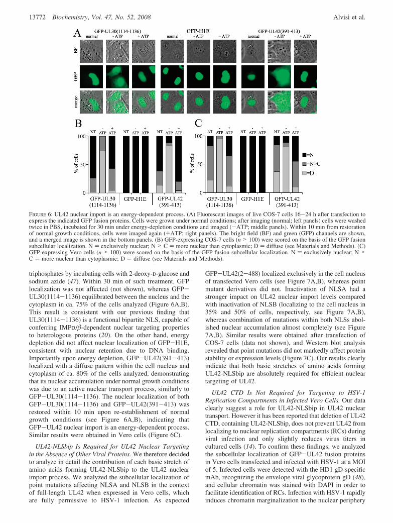
triphosphates by incubating cells with 2-deoxy-D-glucose andsodium azide (47). Within 30 min of such treatment, GFPlocalization was not affected (not shown), whereas GFP-UL30(1114-1136) equilibrated between the nucleus and thecytoplasm in ca. 75% of the cells analyzed (Figure 6A,B).This result is consistent with our previous finding thatUL30(1114-1136) is a functional bipartite NLS, capable ofconferring IMPR/�-dependent nuclear targeting propertiesto heterologous proteins (20). On the other hand, energydepletion did not affect nuclear localization of GFP-H1E,consistent with nuclear retention due to DNA binding.Importantly upon energy depletion, GFP-UL42(391-413)localized with a diffuse pattern within the cell nucleus andcytoplasm of ca. 80% of the cells analyzed, demonstratingthat its nuclear accumulation under normal growth conditionswas due to an active nuclear transport process, similarly toGFP-UL30(1114-1136). The nuclear localization of bothGFP-UL30(1114-1136) and GFP-UL42(391-413) wasrestored within 10 min upon re-establishment of normalgrowth conditions (see Figure 6A,B), indicating thatGFP-UL42 nuclear import is an energy-dependent process.Similar results were obtained in Vero cells (Figure 6C).
UL42-NLSbip Is Required for UL42 Nuclear Targetingin the Absence of Other Viral Proteins. We therefore decidedto analyze in detail the contribution of each basic stretch ofamino acids forming UL42-NLSbip to the UL42 nuclearimport process. We analyzed the subcellular localization ofpoint mutations affecting NLSA and NLSB in the contextof full-length UL42 when expressed in Vero cells, whichare fully permissive to HSV-1 infection. As expected
GFP-UL42(2-488) localized exclusively in the cell nucleusof transfected Vero cells (see Figure 7A,B), whereas pointmutant derivatives did not. Inactivation of NLSA had astronger impact on UL42 nuclear import levels comparedwith inactivation of NLSB (localizing to the cell nucleus in35% and 50% of cells, respectively, see Figure 7A,B),whereas combination of mutations within both NLSs abol-ished nuclear accumulation almost completely (see Figure7A,B). Similar results were obtained after transfection ofCOS-7 cells (data not shown), and Western blot analysisrevealed that point mutations did not markedly affect proteinstability or expression levels (Figure 7C). Our results clearlyindicate that both basic stretches of amino acids formingUL42-NLSbip are absolutely required for efficient nucleartargeting of UL42.
UL42 CTD Is Not Required for Targeting to HSV-1Replication Compartments in Infected Vero Cells. Our dataclearly suggest a role for UL42-NLSbip in UL42 nucleartransport. However it has been reported that deletion of UL42CTD, containing UL42-NLSbip, does not prevent UL42 fromlocalizing to nuclear replication compartments (RCs) duringviral infection and only slightly reduces virus titers incultured cells (14). To confirm these findings, we analyzedthe subcellular localization of GFP-UL42 fusion proteinsin Vero cells transfected and infected with HSV-1 at a MOIof 5. Infected cells were detected with the HD1 gD-specificmAb, recognizing the envelope viral glycoprotein gD (48),and cellular chromatin was stained with DAPI in order tofacilitate identification of RCs. Infection with HSV-1 rapidlyinduces chromatin marginalization to the nuclear periphery
FIGURE 6: UL42 nuclear import is an energy-dependent process. (A) Fluorescent images of live COS-7 cells 16-24 h after transfection toexpress the indicated GFP fusion proteins. Cells were grown under normal conditions; after imaging (normal; left panels) cells were washedtwice in PBS, incubated for 30 min under energy-depletion conditions and imaged (-ATP; middle panels). Within 10 min from restorationof normal growth conditions, cells were imaged again (+ATP; right panels). The bright field (BF) and green (GFP) channels are shown,and a merged image is shown in the bottom panels. (B) GFP-expressing COS-7 cells (n > 100) were scored on the basis of the GFP fusionsubcellular localization. N ) exclusively nuclear; N > C ) more nuclear than cytoplasmic; D ) diffuse (see Materials and Methods). (C)GFP-expressing Vero cells (n > 100) were scored on the basis of the GFP fusion subcellular localization. N ) exclusively nuclear; N >C ) more nuclear than cytoplasmic; D ) diffuse (see Materials and Methods).
13772 Biochemistry, Vol. 47, No. 52, 2008 Alvisi et al.

upon formation of RCs, where viral DNA replication occursandfromwhichcellularDNAandhistonesareexcluded(49,50).As expected, in mock infected cells GFP-UL42(2-488)exclusively localized to the cell nucleus with a speckledpattern, whereas both GFP-UL42(2-390) and GFP-UL42(2-488)-NLSABm mainly localized to the cytoplasmiccompartment (Figure 8A). On the other hand, in HSV-1infected cells not only GFP-UL42(2-488) but bothGFP-UL42(2-390) and GFP-UL42(2-488)NLSABm wererecruited to RCs, although some cytoplasmic staining wasdetectable for the NLS defective mutants (Figure 8B). Theseresults suggest that, in the absence of its functional NLSbip,
UL42 could be imported to the nucleus after interaction inthe cytoplasm with other viral proteins.
The HSV-1 DNA Polymerase Holoenzyme Can Be Im-ported to the Nucleus as a Heterodimeric Complex. Sincethe DNA polymerase holoenzymes from HHV-8 and HCMVcan assemble in the cytoplasm before being translocated tothe nucleus as multiprotein complexes (21), we hypothesizedthat this could be true for HSV-1 as well. To verify whetherHSV DNA polymerase subunits could be translocated to thenucleus as a complex, we coexpressed several GFP-UL30mutant derivatives with DsRed2-UL42(2-488) and DsRed2-UL42(2-390) and analyzed their subcellular localization. Wealso expressed DsRed2-UL44(2-433), which is unable tobind to UL30 as a negative control. As expected, bothDsRed2-UL42(2-488) and DsRed2-UL44(2-433) local-ized strongly within the nucleus with a speckled patterntypical of herpetic PAPs, while DsRed2-UL42(2-390) wasmainly retained in the cytoplasm of transfected cells as aconsequence of the lack of a functional NLS (see Figure9A,B and ref 22). That the localization of DsRed2-UL42(2-390) was markedly more cytoplasmic comparedwith that of GFP-UL42(2-390) is probably due to thetendency of the DsRed2 protein tag to form aggregates whenexpressed to high levels, thus preventing passive diffusionthrough the NPC into the nucleus (compare Figures 3A,Band 9A,E). HSV-1 DNA polymerase catalytic subunitGFP-UL30(2-1235) localized within the cell nucleus witha diffuse pattern (Figure 9B), but when coexpressed withDsRed2-UL42(2-488) the two proteins strongly colocalizedin nuclear speckles, indicating that the two proteins couldefficiently heterodimerize to form the DNA polymeraseholoenzyme. No colocalization was observed betweenGFP-UL30(2-1235) and DsRed2-UL44(2-433). Impor-tantly, coexpression of GFP-UL30(2-1235) with DsRed2-UL42(2-390) resulted in a partial relocalization of the latterto the cell nucleus, with the two proteins colocalizing innuclear speckles. To further verify the specificity of theobserved interactions, we also expressed GFP-UL30(1114-1136), containing UL30 NLSbip but lacking the binding sitefor UL42 (see also Figure 2B and Table 1), in the absenceor in the presence of the aforementioned DsRed2-UL42fusions. As expected, coexpression with GFP-UL30(1114-1136) had no effect on the subcellular localization DsRed2-UL42(2-390), and no colocalization could be observedbetween the UL30 and UL42 fusion proteins (Figure 9D,FandTable1).WealsotestedthebehaviorofGFP-UL30(2-1235)-NLS2m, a point mutant derivative containing a pointmutation inactivating UL30-NLSbip, but still bearing anoncanonical hydrophobic NLS (NLShyd) located within thebinding domain for UL42 (20, 32) (see Figure 2B). Aspreviously shown (20), when expressed in the absence ofother viral proteins GFP-UL30(2-1235)-NLS2m partiallyentered the nucleus due to the activity of its NLShyd (Figure9C). Upon coexpression with DsRed2-UL42(2-488), thetwo proteins strongly colocalized within nuclear speckles,resulting in a marked increase in the nuclear fluorescenceof GFP-UL30(2-1235)-NLS2m, suggesting that DsRed2-UL42(2-488) too could piggy back GFP-UL30(2-1235)-NLS2m into the cell nucleus (Figure 9C,F, and Table 1).However, when expressed with DsRed2-UL42(2-390),GFP-UL30(2-1235)-NLS2m was mainly retained in thecytoplasm, presumably due to masking of its NLShyd by
FIGURE 7: UL42 nuclear localization is dependent on a bipartiteNLS. (A) Fluorescent images of live Vero cells 16-24 h aftertransfection to express the indicated GFP fusion proteins. The brightfield (BF) and the green (GFP) channels are shown, and a mergedimage (merge) is shown in the bottom panels. (B) GFP-expressingcells (n > 100) were scored on the basis of the GFP fusionsubcellular localization. N ) exclusively nuclear; N > C ) morenuclear than cytoplasmic; D ) diffuse; C > N ) more cytoplasmicthan nuclear (see Materials and Methods). (C) COS-7 cellsexpressing GFP fusion proteins were lysed, separated by PAGE,and analyzed by Western blotting as described under the MaterialsandMethodssection,(1)GFP-UL44(2-433);(2)GFP-UL42(2-488);(3) GFP-UL42(2-488)-NLSAm; (4) GFP-UL42(2-488)-NLSBm;(5) GFP-UL42(2-488)-NLSABm.
Nuclear Import of HSV-1 DNA Polymerase Holoenzyme Biochemistry, Vol. 47, No. 52, 2008 13773

UL42 upon interaction with UL30. Taken together, theseresults suggest that the DNA polymerase holoenzyme canbe imported to the nucleus as a complex, so that in theabsence of its functional NLS, UL42 can interact with UL30in the cytoplasm and be translocated to the nucleus via apiggy back mechanism mediated by UL30-NLSbip, whichis located outside the UL42-BD on UL30 (20, 33).
DISCUSSION
A C-Terminally Located NLS on HSV-1 UL42. This is thefirst study describing the relevance of the CTD of HSV-1DNA polymerase processivity factor UL42 to its function,proposing a role for such domain during viral infection. Herewe show that UL42 CTD contains a functional bipartite NLS,necessary for efficient nuclear localization, located at residues391-413. This finding is consistent with the recent identi-fication of functional NLSs at the C-terminus of PAPs fromother herpesviruses (16-19). Our data clearly show thatUL42 binds to different IMPR isoforms and localizesexclusively to the cell nucleus when transiently expressedin the absence of other viral proteins, as detected bycoimmunoprecipitation assays and fluorescent microscopicanalysis of living cells (see Figures 1, 3, 7, 8, and 9). UL42is transported to the nucleus through a mechanism that isinhibited by RanQ69L, similar to its HCMV (see above) andHHV-7 homologues (19). RanQ69L is a dominant negativeform of the small GTPase Ran, which lacks the ability tohydrolyze GTP and to bind to NTF2 and is commonly usedto inhibit active nuclear import both in Vitro and inViVo (42, 51, 52). Since coexpression with RanQ69L resultedin significant relocalization of UL42 to the cytoplasm, weconclude that UL42 is targeted to the cell nucleus via anactive process.
Deletion of UL42 CTD resulted in the relocalization of asignificant amount of protein to the cytoplasmic compart-ment, although a considerable amount of protein was stillcapable of entering the nucleus, and very similar results wereobtained by substituting core basic residues within the basicstretches forming UL42-NLSbip (see Figures 3 and 7). Theresidual nuclear localization observed upon coexpression ofGFP-UL42 with RanQ69L or after inactivation of its NLSis likely due to passive diffusion (see Figures 1-4), becausemolecules with an apparent molecular weight up to 60-90kDa can still partially diffuse through the NPC (47), and
the apparent molecular weight of GFP-UL42(2-488) isaround 85 kDa (see Figure 7C). This hypothesis is alsosupported by the evidence that the oligomer formingmolecule DsRed2-UL42(2-390) was more cytoplasmicthan its GFP-tagged counterpart (see Figure 9A).
Nuclear Import of HSV-1 DNA Polymerase Holoenzyme.Thus, even in the absence of active nuclear import, a fractionof UL42 can enter into the nucleus by passive diffusion.Moreover, it seems likely that the assembly of the HSV-1DNA polymerase holoenzyme occurs in the cytoplasm beforetransport into the nucleus, as suggested by our cotransfectionexperiments (see Figure 9). A mutant of UL42 lacking itsfunctional NLSbip can be retargeted to the nucleus bycoexpression with GFP-UL30(2-1235) in the absence ofother viral proteins, further supporting our hypothesis thatUL30 can piggy-back the UL42 NLS mutant into the cellnucleus (see Figure 9B). Similarly, strong nuclear accumula-tion of GFP-UL30(2-1235)-NLS2m, a UL30 mutantderivative impaired in nuclear targeting carrying a pointmutation inactivating its NLSbip, could be observed uponcoexpression with DsRed2-UL42(2-488), indicating thateither component of the holoenzyme can complement formutations inactivating the NLS function on the othersubunint. Taken together, these results may explain why amutant virus bearing only the 338 N-terminal amino acidsof UL42, retaining all its known biochemical properties inVitro but lacking a functional NLS, is only partially impairedin viral replication, yielding viral titers reduced ca. 3-foldcompared with wt virus in Vero cells (14). Our results alsoprovide experimental evidence to understand why deletionor mutation of UL42-NLSbips does not prevent UL42 fromcorrectly localizing to viral RCs within the cell nucleusduring viral infection (see Figure 8B). Thus our results clearlyshow that HSV-1 evolved multiple NLSs on its DNApolymerase holoenzyme to maximize nuclear import pos-sibilities. Importantly, mutation of both UL42-NLSbip andUL30-NLSbip results in retention of the DNA polymeraseholoenzyme in the cytoplasm (see Figure 9D,F, and Table1), strongly suggesting that it could be possible to preventHSV-1 DNA polymerase nuclear targeting by blocking thefunctionality of both UL30- and UL42-NLSbip.
Why Did HCMV EVolVe a Monopartite NLS and HSV-1Bipartite NLSs on Their DNA Polymerase Subunits? Asmentioned earlier, NLSs are similarly located within HCMV
FIGURE 8: UL42 CTD is not required for localization to nuclear replication compartments during viral infection. Images of Vero cellstransfected to express the indicated GFP fusion proteins and infected with HSV-1 (5 MOI) 30 h post-transfection. Seven hours postinfection,cells were fixed with methanol/acetone 3:1 and processed by IIFA to detect the late antigen gD. DNA was stained with DAPI. The blue(DAPI), green (GFP), and red (TRITC) channels are shown in gray scale, and a merged CYMK image is shown in the right panels.
13774 Biochemistry, Vol. 47, No. 52, 2008 Alvisi et al.

and HSV-1 DNA polymerase subunits. Both processivityfactors possess a C-terminally located NLS, whereas bothcatalytic subunits possess two functional NLSs, one locatedwithin the binding site for their PAP and another locatedoutside such domain (16, 20, 21, 32). While it is easy to
imagine that the presence of two NLSs on the catalyticsubunit is required to maximize the nuclear import possibili-ties for both the catalytic subunits on their own and ascomplexed with their processivity factors, it is not clear whyHSV-1 evolved bipartite NLSs, whereas HCMV evolved
FIGURE 9: HSV-1 DNA polymerase holoenzyme can be translocated to the nucleus as a complex. (A) Images of COS-7 cells transfectedto express the indicated DsRed2 fusion proteins and incubated with Hoechst 33342 to visualize DNA. The blue (Hoechst) and red (DsRed2)channels are shown, and a merged image is shown in the right panels. (B-D) Images of COS-7 cells transfected to express the indicatedGFP fusion proteins in the absence (no addition) or in the presence of the indicated DsRed2 fusion proteins. The green (GFP) and red(DsRed2) channels are shown, and a merged image (merge) is shown in the right panels. (E) DsRed2-UL42(2-390) expressing cells (n> 100) were scored on the basis of its subcellular localization in the absence (no addition) or in the presence of coexpression with theindicated GFP-UL30 fusion proteins. N ) exclusively nuclear; N > C ) more nuclear than cytoplasmic; D ) diffuse, C > N ) morecytoplasmic than nuclear (see Materials and Methods). (F) GFP-expressing cells (n > 100) were scored on the basis of the GFP fusionsubcellular localization when expressed in the presence of the indicated DsRed2 fusion protein. N ) exclusively nuclear; N > C ) morenuclear than cytoplasmic; D ) diffuse; C > N ) more cytoplasmic than nuclear (see Materials and Methods).
Table 1: Summary of the Effect of Expression of DsRed2-UL42 Fusion Proteins on the Subcellular Localization of GFP-UL30 Fusion Proteinsa
presence of functional domains subcellular localization of GFP-UL30 fusion protein
GFP-UL30 fusion protein NLSbip UL42-BD NLShyd no addition +UL42(2-433) +UL42(2-390)
GFP-UL30(2-1235) + + + N N; Nsp N; NspGFP-UL30(2-1235)-NLS2m - + + N > C N; Nsp C > NGFP-UL30(1114-1136) + - - N > C N > C N > Ca NLSbip, UL30 bipartite NLS (residues 1114-1136); UL42-BD, UL30 binding domain for UL42 (residues 1209-1235); NLShyd, UL30
hydrophobic NLS (residues 1224-1229); N, exclusively nuclear fluorescence; N > C, significant amount of fluorescence present in the cytoplasm; C >N, mainly cytoplasmic fluorescence; Nsp, colocalizing with DsRed2-UL42 fusions in nuclear speckles.
Nuclear Import of HSV-1 DNA Polymerase Holoenzyme Biochemistry, Vol. 47, No. 52, 2008 13775

monopartite NLSs. An explanation for that could lie in theevidence that HSV-1 UL42 and HCMV ppUL44 appear tohave different IMP binding capabilities: while ppUL44 bindsefficiently only to IMPR6 (and to a lesser extent to IMPR2),belonging to the R-S subtype, UL42 also efficiently boundto the R-Q member IMPR4 (see Figure 1C). Since differentIMPR family members have particular cargo specificity andexhibit unique temporal and spatial tissue-specific expression(26, 27) and HSV-1 and HCMV possess different cellulartropisms, with HSV-1 being strongly neurotropic (54), it istempting to speculate that HSV-1 evolved bipartite NLSs toensure optimal nuclear targeting in specific tissues. Furtherwork is required to assess this hypothesis.
A New Consensus for Monopartite NLSs. UL42-NLSbipencompasses two basic stretches of amino acids (NLSA,PTTKRGR397; NLSB, KKPK413), both of which are requiredfor NLS function (see Figures 5-7). Intriguingly bothstretches match the consensus for putative monopartite NLSs,(K)-(K/R)-(X)-(K/R) (30), but possess a G and a P residuein position +3 of the NLS core, respectively. We haverecently shown that a P residue in position +3 of the NLScore is not compatible with monopartite NLS activity (20),and here we suggest that a G residue in position +3 of theNLS core could similarly be not compatible with monopartiteNLS function (see Figure 4). Consistent with our hypothesis,we showed that the sequence PTTKRaR397 but not PTT-KRGR397 is capable of targeting GFP to the cell nucleus (seeFigure 4 and Figure S1, Supporting Information). Our resultshave thus relevance for the identification of classical mono-partite NLSs and further support the redefinition of theconsensus motif for monopartite NLSs to (K)-(K/R)-(X′)-(K/R), where X′ is any amino acid apart from P or G. Theextreme rigidity/flexibility conferred by such residues,respectively, can be overcome by the presence of additionalbasic residues surrounding the NLS as in the case of themouse polycomb protein, M33 (53), or by the presence ofan additional stretch of basic residues located within 10-12amino acids, thus forming a bipartite NLS as in the case ofHSV-1 UL30 (20). Consistent with this hypothesis, putativebipartite NLSs resembling that identified here are locatedwithin UL42 homologues from several R-herpesviruses,including human HSV-2 and cercopithecine and suid HSV-1(see Figure 2C).
CONCLUSIONS AND PERSPECTIVES
The data presented here allow us to clearly define UL42nuclear import as an energy-dependent, IMPR/�-mediatedprocess, which is impaired by RanQ69L and depends on aC-terminally located bipartite NLS. Our results should provedeterminant for the understanding of the controversial roleplayed by UL42 CTD during viral infection, explaining whya mutant virus lacking the 150 C-terminal amino acids ofUL42 yielded infectious titers reduced almost 3 timescompared with a virus encoding full-length UL42, althoughall known biochemical properties of UL42 reside in itsN-terminal domain. These findings could constitute the basisfor the development of new anti-HSV-1 therapeutics basedon the inhibition of the DNA polymerase holoenzyme nuclearimport. Furthermore, the definition of a new consensus fora monopartite NLS should help the identification of novelfunctional NLSs.
ACKNOWLEDGMENT
We thank Gabriella Campadelli-Fiume (University ofBologna) for mouse mAb HD1 directed against HSV-1structural protein gD, and Charles Hwang (State Universityof New York Upstate Medical University), Michael Green(University of Massachusetts), Gabi Gerlitz (NIH), YoichiMiyamoto (Monash University), and Emanuele Panza (Uni-versity of Bologna) for providing plasmids.
SUPPORTING INFORMATION AVAILABLE
Figure demonstrating that the presence of a G residue atposition +3 of UL42-NLSA core impairs its functionality.This material is available free of charge via the Internet athttp://pubs.acs.org.
REFERENCES
1. Roizman, B., and Whitley, R. J. (2001) The nine ages of herpessimplex virus. Herpes 8, 23–27.
2. Wu, C. A., Nelson, N. J., McGeoch, D. J., and Challberg, M. D.(1988) Identification of herpes simplex virus type 1 genes requiredfor origin-dependent DNA synthesis. J. Virol. 62, 435–443.
3. Gottlieb, J., Marcy, A. I., Coen, D. M., and Challberg, M. D. (1990)The herpes simplex virus type 1 UL42 gene product: A subunit ofDNA polymerase that functions to increase processivity. J. Virol.64, 5976–5987.
4. Chaudhuri, M., and Parris, D. S. (2002) Evidence against a simpletethering model for enhancement of herpes simplex virus DNApolymerase processivity by accessory protein UL42. J. Virol. 76,10270–10281.
5. Jiang, C., Hwang, Y. T., Wang, G., Randell, J. C., Coen, D. M.,and Hwang, C. B. (2007) Herpes simplex virus mutants withmultiple substitutions affecting DNA binding of UL42 are impairedfor viral replication and DNA synthesis. J. Virol. 81, 12077–12079.
6. Jiang, C., Hwang, Y. T., Randell, J. C., Coen, D. M., and Hwang,C. B. (2007) Mutations that decrease DNA binding of theprocessivity factor of the herpes simplex virus DNA polymerasereduce viral yield, alter the kinetics of viral DNA replication, anddecrease the fidelity of DNA replication. J. Virol. 81, 3495–3502.
7. Komazin-Meredith, G., Mirchev, R., Golan, D. E., van Oijen,A. M., and Coen, D. M. (2008) Hopping of a processivity factoron DNA revealed by single-molecule assays of diffusion. Proc.Natl. Acad. Sci. U.S.A. 105, 10721–10726.
8. Komazin-Meredith, G., Santos, W. L., Filman, D. J., Hogle, J. M.,Verdine, G. L., and Coen, D. M. (2008) The positively chargedsurface of herpes simplex virus UL42 mediates DNA binding.J. Biol. Chem. 283, 6154–6161.
9. Marsden, H. S., Campbell, M. E., Haarr, L., Frame, M. C., Parris,D. S., Murphy, M., Hope, R. G., Muller, M. T., and Preston, C. M.(1987) The 65,000-Mr DNA-binding and virion trans-inducingproteins of herpes simplex virus type 1. J. Virol. 61, 2428–2437.
10. Pilger, B. D., Cui, C., and Coen, D. M. (2004) Identification of asmall molecule that inhibits herpes simplex virus DNA Polymerasesubunit interactions and viral replication. Chem. Biol. 11, 647–654.
11. Hamatake, R. K., Bifano, M., Tenney, D. J., Hurlburt, W. W., andCordingley, M. G. (1993) The herpes simplex virus type 1 DNApolymerase accessory protein, UL42, contains a functional protease-resistant domain. J. Gen. Virol. 74 (10), 2181–2189.
12. Monahan, S. J., Barlam, T. F., Crumpacker, C. S., and Parris, D. S.(1993) Two regions of the herpes simplex virus type 1 UL42protein are required for its functional interaction with the viral DNApolymerase. J. Virol. 67, 5922–5931.
13. Tenney, D. J., Hurlburt, W. W., Bifano, M., Stevens, J. T.,Micheletti, P. A., Hamatake, R. K., and Cordingley, M. G. (1993)Deletions of the carboxy terminus of herpes simplex virus type 1UL42 define a conserved amino-terminal functional domain.J. Virol. 67, 1959–1966.
14. Gao, M., DiTusa, S. F., and Cordingley, M. G. (1993) TheC-terminal third of UL42, a HSV-1 DNA replication protein, isdispensable for viral growth. Virology 194, 647–653.
15. Goodrich, L. D., Rixon, F. J., and Parris, D. S. (1989) Kinetics ofexpression of the gene encoding the 65-kilodalton DNA-bindingprotein of herpes simplex virus type 1. J. Virol. 63, 137–147.
13776 Biochemistry, Vol. 47, No. 52, 2008 Alvisi et al.

16. Alvisi, G., Jans, D. A., Guo, J., Pinna, L. A., and Ripalti, A. (2005)A protein kinase CK2 site flanking the nuclear targeting signalenhances nuclear transport of human cytomegalovirus ppUL44.Traffic 6, 1002–1013.
17. Chen, Y., Ciustea, M., and Ricciardi, R. P. (2005) Processivityfactor of KSHV contains a nuclear localization signal and bindingdomains for transporting viral DNA polymerase into the nucleus.Virology 340, 183–191.
18. Loh, L. C., Keeler, V. D., and Shanley, J. D. (1999) Sequencerequirements for the nuclear localization of the murine cyto-megalovirus M44 gene product pp50. Virology 259, 43–59.
19. Takeda, K., Haque, M., Nagoshi, E., Takemoto, M., Shimamoto,T., Yoneda, Y., and Yamanishi, K. (2000) Characterization ofhuman herpesvirus 7 U27 gene product and identification of itsnuclear localization signal. Virology 272, 394–401.
20. Alvisi, G., Musiani, D., Jans, D. A., and Ripalti, A. (2007) Animportin alpha/beta-recognized bipartite nuclear localization signalmediates targeting of the human herpes simplex virus type 1 DNApolymerase catalytic subunit pUL30 to the nucleus. Biochemistry46, 9155–9163.
21. Alvisi, G., Ripalti, A., Ngankeu, A., Giannandrea, M., Caraffi, S. G.,Dias, M. M., and Jans, D. A. (2006) Human cytomegalovirus DNApolymerase catalytic subunit pUL54 possesses independently actingnuclear localization and ppUL44 binding motifs. Traffic 7, 1322–1332.
22. Stewart, M. (2007) Molecular mechanism of the nuclear proteinimport cycle. Nat. ReV. Mol. Cell Biol. 8, 195–208.
23. Cingolani, G., Petosa, C., Weis, K., and Muller, C. W. (1999)Structure of importin-beta bound to the IBB domain of importin-alpha. Nature 399, 221–229.
24. Lee, S. J., Matsuura, Y., Liu, S. M., and Stewart, M. (2005)Structural basis for nuclear import complex dissociation byRanGTP. Nature 435, 693–696.
25. Gorlich, D., Pante, N., Kutay, U., Aebi, U., and Bischoff, F. R.(1996) Identification of different roles for RanGDP and RanGTPin nuclear protein import. EMBO J. 15, 5584–5594.
26. Hogarth, C. A., Calanni, S., Jans, D. A., and Loveland, K. L. (2006)Importin alpha mRNAs have distinct expression profiles duringspermatogenesis. DeV. Dyn. 235, 253–262.
27. Jans, D. A., Xiao, C. Y., and Lam, M. H. (2000) Nuclear targetingsignal recognition: a key control point in nuclear transport?Bioessays 22, 532–544.
28. Lange, A., Mills, R. E., Lange, C. J., Stewart, M., Devine, S. E.,and Corbett, A. H. (2007) Classical nuclear localization signals:Definition, function, and interaction with importin alpha. J. Biol.Chem. 282, 5101–5105.
29. Conti, E., Uy, M., Leighton, L., Blobel, G., and Kuriyan, J. (1998)Crystallographic analysis of the recognition of a nuclear localizationsignal by the nuclear import factor karyopherin alpha. Cell 94, 193–204.
30. Hodel, M. R., Corbett, A. H., and Hodel, A. E. (2001) Dissectionof a nuclear localization signal. J. Biol. Chem. 276, 1317–1325.
31. Prichard, M. N., Lawlor, H., Duke, G. M., Mo, C., Wang, Z., Dixon,M., Kemble, G., and Kern, E. R. (2005) Human cytomegalovirusuracil DNA glycosylase associates with ppUL44 and acceleratesthe accumulation of viral DNA. Virol. J. 2, 55.
32. Loregian, A., Piaia, E., Cancellotti, E., Papini, E., Marsden, H. S.,and Palu, G. (2000) The catalytic subunit of herpes simplex virustype 1 DNA polymerase contains a nuclear localization signal inthe UL42-binding region. Virology 273, 139–148.
33. Marsden, H. S., Murphy, M., McVey, G. L., MacEachran, K. A.,Owsianka, A. M., and Stow, N. D. (1994) Role of the carboxyterminus of herpes simplex virus type 1 DNA polymerase in itsinteraction with UL42. J. Gen. Virol. 75 (11), 3127–3135.
34. Poon, I. K., Oro, C., Dias, M. M., Zhang, J., and Jans, D. A. (2005)Apoptin nuclear accumulation is modulated by a CRM1-recognizednuclear export signal that is active in normal but not in tumor cells.Cancer Res. 65, 7059–7064.
35. Hubner, S., Eam, J. E., Wagstaff, K. M., and Jans, D. A. (2006)Quantitative analysis of localization and nuclear aggregate forma-tion induced by GFP-lamin A mutant proteins in living HeLa cells.J. Cell. Biochem. 98, 810–826.
36. Panza, E., Marini, M., Pecci, A., Giacopelli, F., Bozzi, V., Seri,M., Balduini, C., and Ravazzolo, R. Transfection of the mutantMYH9 cDNA reproduces the most typical cellular phenotype ofMYH9-related disease in different cell lines, PathoGenetics, inpress.
37. Miyamoto, Y., Hieda, M., Harreman, M. T., Fukumoto, M.,Saiwaki, T., Hodel, A. E., Corbett, A. H., and Yoneda, Y. (2002)Importin alpha can migrate into the nucleus in an importin beta-and Ran-independent manner. EMBO J. 21, 5833–5842.
38. Alvisi, G., Jans, D. A., and Ripalti, A. (2006) Human cytomegalo-virus (HCMV) DNA polymerase processivity factor ppUL44dimerizes in the cytosol before translocation to the nucleus.Biochemistry 45, 6866–6872.
39. Heilman, D. W., Teodoro, J. G., and Green, M. R. (2006) Apoptinnucleocytoplasmic shuttling is required for cell type-specificlocalization, apoptosis, and recruitment of the anaphase-promotingcomplex/cyclosome to PML bodies. J. Virol. 80, 7535–7545.
40. Gerlitz, G., Livnat, I., Ziv, C., Yarden, O., Bustin, M., and Reiner,O. (2007) Migration cues induce chromatin alterations. Traffic 8,1521–1529.
41. Bauerle, M., Doenecke, D., and Albig, W. (2002) The requirementof H1 histones for a heterodimeric nuclear import receptor. J. Biol.Chem. 277, 32480–32489.
42. Stewart, M., Kent, H. M., and McCoy, A. J. (1998) The structureof the Q69L mutant of GDP-Ran shows a major conformationalchange in the switch II loop that accounts for its failure to bindnuclear transport factor 2 (NTF2). J. Mol. Biol. 284, 1517–1527.
43. Fontes, M. R., Teh, T., Toth, G., John, A., Pavo, I., Jans, D. A.,and Kobe, B. (2003) Role of flanking sequences and phosphoryl-ation in the recognition of the simian-virus-40 large T-antigennuclear localization sequences by importin-alpha. Biochem. J. 375,339–349.
44. Kobe, B. (1999) Autoinhibition by an internal nuclear localizationsignal revealed by the crystal structure of mammalian importinalpha. Nat. Struct. Biol. 6, 388–397.
45. Nakai, K., and Horton, P. (1999) PSORT: A program for detectingsorting signals in proteins and predicting their subcellular localiza-tion. Trends Biochem. Sci. 24, 34–36.
46. Advani, S. J., Weichselbaum, R. R., and Roizman, B. (2003) Herpessimplex virus 1 activates cdc2 to recruit topoisomerase II alphafor post-DNA synthesis expression of late genes. Proc. Natl. Acad.Sci. U.S.A. 100, 4825–4830.
47. Cardarelli, F., Serresi, M., Bizzarri, R., Giacca, M., and Beltram,F. (2007) In vivo study of HIV-1 Tat arginine-rich motif unveilsits transport properties. Mol. Ther. 15, 1313–22.
48. Dix, R. D., Pereira, L., and Baringer, J. R. (1981) Use ofmonoclonal antibody directed against herpes simplex virus glyco-proteins to protect mice against acute virus-induced neurologicaldisease. Infect. Immun. 34, 192–199.
49. Dupuy-Coin, A. M., Arnoult, J., and Bouteille, M. (1978) Quantita-tive correlation of morphological alterations of the nucleus withfunctional events during in vitro infection of glial cells with herpessimplex hominis (HSV 2). J. Ultrastruct. Res. 65, 60–72.
50. Monier, K., Armas, J. C., Etteldorf, S., Ghazal, P., and Sullivan,K. F. (2000) Annexation of the interchromosomal space duringviral infection. Nat. Cell Biol. 2, 661–665.
51. Avis, J. M., and Clarke, P. R. (1996) Ran, a GTPase involved innuclear processes: its regulators and effectors. J. Cell Sci. 109 (10),2423–2427.
52. Bischoff, F. R., Klebe, C., Kretschmer, J., Wittinghofer, A., andPonstingl, H. (1994) RanGAP1 induces GTPase activity of nuclearRas-related Ran. Proc. Natl. Acad. Sci. U.S.A. 91, 2587–2591.
53. Hirose, S., Komoike, Y., and Higashinakagawa, T. (2006) Iden-tification of a nuclear localization signal in mouse polycombprotein, M33. Zool. Sci. 23, 785–791.
54. Bello-Morales, R., Fedetz, M., Alcina, A., Tabares, E., and Lopez-Guerrero, J. A. (2005) High susceptibility of a human oligoden-droglial cell line to herpes simplex type 1 infection. J. NeuroVirol.11, 190–198.
BI800869Y
Nuclear Import of HSV-1 DNA Polymerase Holoenzyme Biochemistry, Vol. 47, No. 52, 2008 13777