Transposition into Replicating DNA Occurs through Interaction with the Processivity Factor
22
Transposition into replicating DNA occurs through interaction with the processivity factor Adam R. Parks 1,3 , Zaoping Li 1 , Qiaojuan Shi 1 , Roisin M. Owens 2 , Moonsoo M. Jin 2 , and Joseph E. Peters 1,* 1 Department of Microbiology; Cornell University; Ithaca, NY, 14853;USA 2 Department of Biomedical Engineering; Cornell University; Ithaca, NY, 14853;USA SUMMARY The bacterial transposon Tn7 directs transposition into actively replicating DNA by a mechanism involving the transposon-encoded protein TnsE. Here we show that TnsE physically and functionally interacts with the processivity factor of the DNA replication machinery in vivo and in vitro. Our work establishes an in vitro TnsABC+E transposition reaction reconstituted from purified proteins and target DNA structures. Using the in vitro reaction we confirm that the processivity factor specifically reorders TnsE-mediated transposition events on target DNAs in a way that matches the bias with active DNA replication in vivo. The TnsE interaction with an essential and conserved component of the replication machinery and a DNA structure reveals a new mechanism by which Tn7, and probably other elements, select target-sites associated with DNA replication. Keywords beta sliding clamp; transposition; processivity factor; in vitro transposition INTRODUCTION Transposons are genetic elements that are capable of moving from one location to another within a cell. The bacterial transposon Tn7 and its relatives are abundantly distributed amongst bacteria in a wide variety of medical and environmental settings (Parks and Peters, 2007; Parks and Peters, 2009). Tn7 has served as a model system for transposition, especially for the understanding of transposon target-site selection (reviewed in Peters and Craig, 2001b, and Craig et al., 2002). Target-site selection is the process by which transposons assess new DNA molecules for potential insertion. While most transposable elements possess a weak target DNA sequence preference that guides target-site selection, Tn7 utilizes two distinct target-site selection pathways. In one pathway a sequence-specific DNA binding protein directs transposition into a single site within the bacterial chromosome, and in the other a separate protein recognizes a process associated with DNA replication. These two target selection © 2009 Elsevier Inc. All rights reserved. *Corresponding author: Department of Microbiology; Cornell University; 175a Wing Hall, Ithaca, NY, 14853; USA; Email: [email protected], Phone: (607) 255−2271, Fax: (607) 255−3904. 3 Present address: NCI Center for Cancer Research, Frederick, MD 21702, USA Publisher's Disclaimer: This is a PDF file of an unedited manuscript that has been accepted for publication. As a service to our customers we are providing this early version of the manuscript. The manuscript will undergo copyediting, typesetting, and review of the resulting proof before it is published in its final citable form. Please note that during the production process errors may be discovered which could affect the content, and all legal disclaimers that apply to the journal pertain. NIH Public Access Author Manuscript Cell. Author manuscript; available in PMC 2010 August 21. Published in final edited form as: Cell. 2009 August 21; 138(4): 685–695. doi:10.1016/j.cell.2009.06.011. NIH-PA Author Manuscript NIH-PA Author Manuscript NIH-PA Author Manuscript
Transcript of Transposition into Replicating DNA Occurs through Interaction with the Processivity Factor

Transposition into replicating DNA occurs through interactionwith the processivity factor
Adam R. Parks1,3, Zaoping Li1, Qiaojuan Shi1, Roisin M. Owens2, Moonsoo M. Jin2, andJoseph E. Peters1,*1Department of Microbiology; Cornell University; Ithaca, NY, 14853;USA2Department of Biomedical Engineering; Cornell University; Ithaca, NY, 14853;USA
SUMMARYThe bacterial transposon Tn7 directs transposition into actively replicating DNA by a mechanisminvolving the transposon-encoded protein TnsE. Here we show that TnsE physically and functionallyinteracts with the processivity factor of the DNA replication machinery in vivo and in vitro. Ourwork establishes an in vitro TnsABC+E transposition reaction reconstituted from purified proteinsand target DNA structures. Using the in vitro reaction we confirm that the processivity factorspecifically reorders TnsE-mediated transposition events on target DNAs in a way that matches thebias with active DNA replication in vivo. The TnsE interaction with an essential and conservedcomponent of the replication machinery and a DNA structure reveals a new mechanism by whichTn7, and probably other elements, select target-sites associated with DNA replication.
Keywordsbeta sliding clamp; transposition; processivity factor; in vitro transposition
INTRODUCTIONTransposons are genetic elements that are capable of moving from one location to anotherwithin a cell. The bacterial transposon Tn7 and its relatives are abundantly distributed amongstbacteria in a wide variety of medical and environmental settings (Parks and Peters, 2007; Parksand Peters, 2009). Tn7 has served as a model system for transposition, especially for theunderstanding of transposon target-site selection (reviewed in Peters and Craig, 2001b, andCraig et al., 2002). Target-site selection is the process by which transposons assess new DNAmolecules for potential insertion. While most transposable elements possess a weak targetDNA sequence preference that guides target-site selection, Tn7 utilizes two distinct target-siteselection pathways. In one pathway a sequence-specific DNA binding protein directstransposition into a single site within the bacterial chromosome, and in the other a separateprotein recognizes a process associated with DNA replication. These two target selection
© 2009 Elsevier Inc. All rights reserved.*Corresponding author: Department of Microbiology; Cornell University; 175a Wing Hall, Ithaca, NY, 14853; USA; Email:[email protected], Phone: (607) 255−2271, Fax: (607) 255−3904.3Present address: NCI Center for Cancer Research, Frederick, MD 21702, USAPublisher's Disclaimer: This is a PDF file of an unedited manuscript that has been accepted for publication. As a service to our customerswe are providing this early version of the manuscript. The manuscript will undergo copyediting, typesetting, and review of the resultingproof before it is published in its final citable form. Please note that during the production process errors may be discovered which couldaffect the content, and all legal disclaimers that apply to the journal pertain.
NIH Public AccessAuthor ManuscriptCell. Author manuscript; available in PMC 2010 August 21.
Published in final edited form as:Cell. 2009 August 21; 138(4): 685–695. doi:10.1016/j.cell.2009.06.011.
NIH
-PA Author Manuscript
NIH
-PA Author Manuscript
NIH
-PA Author Manuscript

pathways optimize vertical and horizontal transmission of the transposable elementrespectively (Craig, 2002; Parks and Peters, 2009).
Tn7 encodes five genes whose products conduct transposition (Craig et al., 2002). TnsA andTnsB comprise the transposase that catalyzes the DNA breakage and joining reactions at thetransposon ends to mobilize the element. TnsC is an AAA regulator protein that activates thetransposase when an appropriate target DNA has been found (Stellwagen and Craig, 1998).TnsD and TnsE identify target DNAs and signal TnsABC to activate transposition (Craig,2002). Target-site selection is a prerequisite for activation of transposition with Tn7;transposon excision and insertion does not occur until an appropriate target has been identified.TnsD recognizes a specific site, called its attachment site or attTn7, by binding to a highlyconserved DNA sequence within the 3’ end of the glmS gene. The TnsE protein recognizes anincompletely defined feature associated with discontinuous DNA replication (Peters and Craig,2001a) that is over-represented or especially accessible in mobile plasmids, called conjugalplasmids, as they enter a new host cell (Wilkins and Lanka, 1993; Wolkow et al., 1996).
While TnsE-mediated transposition preferentially occurs into mobile plasmids undergoingconjugal DNA replication, at a lower frequency, the TnsABC+E machinery also recognizessites within the bacterial chromosome with a preference for the region where DNA replicationterminates and regions proximal to DNA double-strand breaks (Peters and Craig, 2000; Shi etal., 2008). The orientation of the transposon ends following TnsE-mediated transpositionindicates that discontinuously replicated DNA is in some way recognized by TnsE (Peters andCraig, 2001a; Wolkow et al., 1996). As mobile plasmids enter a new host cell, they replicatein a single direction by a discontinuous process, like lagging-strand DNA synthesis (Wilkinsand Lanka, 1993). In both mobile plasmids and in the chromosome, transposition events occurin a single orientation correlating with the direction of replication progression (Peters andCraig, 2001a; Peters and Craig, 2001b; Wolkow et al., 1996). It has been shown that TnsE isa DNA binding protein that preferentially binds to DNA structures that present a free 3’-recessed end (Peters and Craig, 2001a). Given that TnsD relies in part on additional host factorsin activating transposition (Sharpe and Craig, 1998), it is conceivable that host factorsassociated with discontinuous DNA replication allow the selection of targets during TnsE-mediated transposition.
An intriguing host factor candidate that could allow the TnsABC+E transposition machineryto target lagging-strand DNA synthesis is the DNA replication processivity factor. Processivityfactors are essential clamp proteins that encircle DNA and serve as a mobile platform, linkingproteins to DNA (Johnson and O'Donnell, 2005; Warbrick, 2000). Interestingly, the inactivepogo element, found in Drosophila, encodes a transposase that has been shown to interact withthe processivity factor (Warbrick et al., 1998). Because the element is no longer active, nofunctional link between this interaction and transposition has yet been established (Warbrick,2000). It therefore remains unclear if the interaction was important in the original element. Aninteraction with the processivity factor could possibly regulate transposition with DNAreplication and repair, or could be involved in target-site selection. We reasoned that TnsEmight use an interaction with the processivity factor to direct transposition into certain formsof DNA replication.
The processivity factor, β clamp in Bacteria and PCNA in Eukaryotes and Archaea, is enrichedon discontinuously replicating DNA (reviewed in Johnson and O'Donnell, 2005, and referencestherein). β and PCNA have been shown to interact with many proteins involved in DNA repair,Okazaki fragment maturation, and regulation of the cell cycle (Johnson and O'Donnell, 2005;Warbrick, 2000). The processivity factor binding motif found in proteins that interact with βand PCNA fits into a hydrophobic cleft found in the clamp proteins (Dalrymple et al., 2001;Jeruzalmi et al., 2001a; Johnson and O'Donnell, 2005; Warbrick, 2000). Competition for this
Parks et al. Page 2
Cell. Author manuscript; available in PMC 2010 August 21.
NIH
-PA Author Manuscript
NIH
-PA Author Manuscript
NIH
-PA Author Manuscript

common region of the clamp appears to play a role in coordination of proteins involved inDNA metabolism (Lopez de Saro et al., 2004).
In this work we reveal and characterize an interaction between TnsE and the β processivityfactor, and reconstitute the TnsABC+E transposition pathway using purified components. Thein vitro reaction confirms that two factors, interaction with a DNA structure and with theprocessivity factor account for the bias of TnsABC+E transposition with active DNAreplication. In vitro and in vivo analysis of the TnsE-β interaction explains how Tn7 targetsDNA replication without negatively affecting the cell. These findings likely reveal a generalstrategy used by other unrelated transposons for directing transposition to DNA replicationintermediates.
RESULTSTnsE interacts with the processivity factor
In an effort to understand how TnsE identifies a target DNA we looked for conserved motifswithin the amino acid sequence of TnsE. We found a sequence that shows a modestresemblance to the consensus processivity factor binding motifs found in bacterial host proteins(QL(S/D)LF (Dalrymple et al., 2001) and QLxLxL (Wijffels et al., 2004))(Figure 1). Analysisof amino acid alignments of all known and predicted tnsE gene products (Parks and Peters,2009) using the ClustalW algorithm (Thompson et al., 1994) revealed a highly conservedsequence PQLELARALFL (Figure 1). We hypothesized that TnsE recognizes lagging-strandDNA synthesis through an interaction with the processivity factor. We first tested for the TnsE-β interaction using TnsE and β protein derivatives fused to the yeast GAL4 transcriptionactivation and DNA binding domains, respectively (Fields and Song, 1989). The presence andextent of the interaction in the two-hybrid assay was monitored by determining the β-galactosidase (β-gal) activity in a reporter strain containing the lacZ gene under control of aGAL4 promoter (Liachko and Tye, 2005; Miller, 1992). We also included a positive controlfor the β interaction, the δ subunit of the clamp-loader, which has been shown to interact withβ in multiple assays (Johnson and O'Donnell, 2005). The yeast two-hybrid assay indicated thatTnsE and β do indeed interact (Figure 2).
To confirm the interaction between TnsE and β in vitro, we purified a modified β protein thatcould be labeled by the addition of 32P phosphate by PKA enzyme (32P-β)(Kelman et al.,1995a; Kelman et al., 1995b). We tested the TnsE-β interaction using a protein mobility shiftassay (Lopez de Saro et al., 2006; Lopez de Saro and O'Donnell, 2001). As a negative controlwe tested increasing concentrations of BSA, which is similar TnsE in size and charge (69.2KDa, pI 5.8, for BSA versus 61.2 KDa, pI 5.7, for TnsE), and found no shifted product (Figure3.A., lanes 1−3). A shift in the electrophoretic mobility of 32P-β upon addition of increasingconcentrations of TnsE demonstrated that TnsE and β do form a complex (Figure 3.A., lanes4−7). We used a far western blot technique to further confirm the in vitro interaction (Einarsonet al., 2007). Serial dilutions of TnsE and BSA were spotted on a membrane in triplicate usinga slot blot apparatus (Amersham Bioscience). The membrane was then probed with 32P-β.Consistent with the band shift assay, we found that TnsE retained 32P-β, indicating interaction,while equivalent concentrations of BSA did not (Figure 3.B.).
For a more thorough and quantitative analysis of the TnsE-β interaction we utilized a Biacoreinstrument that is capable of monitoring relatively weak protein-protein interactions by surfaceplasmon resonance (SPR). For these experiments we immobilized purified β protein in varyingconcentrations by cross-linking them to the surface of a CM-5 SPR chip (GE Healthcare).Various concentrations of test proteins were then flowed over the surface of the chip to monitorany interaction between proteins, followed by dissociation under the flow of buffer alone(Figure 3.C. and 3.D.). We determined the KD of the TnsE-β interaction to be ∼2.44 μM
Parks et al. Page 3
Cell. Author manuscript; available in PMC 2010 August 21.
NIH
-PA Author Manuscript
NIH
-PA Author Manuscript
NIH
-PA Author Manuscript

(EXPERIMENTAL PROCEDURES). For comparison, we tested the β interaction with 1 μM(as monomers) of three different components of the minimal clamp-loader complex (the γ-complex, γ3δδ’), providing us with a pre-established set of standard proteins with varyingdissociation constants (Leu and O'Donnell, 2001). We found that the TnsE-β interaction isintermediate between the primary β contacting subunit δ (Figure 3.C.) and the γ subunit (Figure3.D.); however, direct comparison with the γ subunit is complicated by the fact purified γ formstetramers in solution (Dallmann and McHenry, 1995). As a negative control we tested forinteraction with δ’ and found no interaction, similar to previously reported results (Leu andO'Donnell, 2001). To confirm that our calculations were comparable to previously reportedvalues, we calculated the KD of the δ-β interaction. Under the conditions used here wedetermine the KD for the δ-β interaction to be ∼0.06 μM, similar to the ∼0.03 μM value reportedby Leu and O'Donnell (Leu and O'Donnell, 2001)(EXPERIMENTAL PROCEDURES).
The putative β clamp binding motif of TnsE is involved in binding βTo test if the region of TnsE that resembles the β clamp binding motif is important for bindingβ, we constructed a set of tnsE mutants replacing specific amino acids within this region withalanines (collectively referred to as tnsEβMA). Amino acids were chosen for mutation based ontheir presumed relationship to the consensus β clamp binding and their conservative amongTnsE proteins (Figure 1). The tnsEβMAmutations were first tested in the yeast two-hybrid assaydescribed above. Each alanine substitution resulted in decreased β-gal activity in the reporterstrain, supporting the idea that the conserved region is important for interaction with β andpossibly comprises a region that interacts with the hydrophobic pocket of the β clamp (Figure4.A.). Western blots did not indicate changes in stability or expression resulting from theTnsEβMA mutations that would account for these observations (Figure 4.A.).
To confirm that the TnsEβMA mutants are weaker in interaction with β in vitro we purified asubset of the TnsEβMA proteins. Using the SPR system we compared interaction between βand 1 μM of each purified TnsE allele. In all cases, the SPR sensograms indicated weakerinteraction between each of the mutant TnsE alleles and β (Figure 4.B.) and looselycorresponded to the relative loss in interaction observed in the yeast two-hybrid assay. The βinteraction with the mutant TnsE proteins varied in KD from ∼6.0 to ∼10.0 μM, as analyzedby the BIAevaluation software (Biacore, 1997). The SPR results and the yeast two-hybrid datasupport the view that the conserved region of TnsE is involved in the interaction between TnsEand β.
Interaction with the processivity factor is essential for transposition in vivoIf binding to the β clamp is required to activate transposition, we expected to see a decrease intransposition with the tnsEβMA mutants, due to their attenuated β clamp binding ability.Interaction with the clamp via the interaction motif is required for activity of DNA polymerases(Johnson and O'Donnell, 2005), although these protein-protein interactions include moreextensive surface interactions beyond the conserved motif (Bunting et al., 2003). Using an invivo transposition assay (McKown et al., 1988), we found that the alanine mutations abolishedor significantly reduced (p<0.005, two-tailed unequal variance t-test) the frequency of TnsABC+E transposition (Figure 5.A.). Western blots show that the stability and expression of thesemutants could not account for such sharp reduction or complete loss of transposition (Figure5.A.). We found no negative effects with the tnsEβMAmutants on other Tn7 transpositionpathways (data not shown). These results indicate that the decreases we observed intransposition frequency are consistent with decreased β clamp binding ability, supporting theview that activation of transposition via the TnsE pathway is dependent on binding to the βclamp. Similarly, others have shown that mutant host proteins with attenuated β clamp bindingability display decreased or abolished activity in vivo (Beuning et al., 2006; Johnson andO'Donnell, 2005; Simmons et al., 2008; Sutton, 2004). In vivo, TnsE must compete with many
Parks et al. Page 4
Cell. Author manuscript; available in PMC 2010 August 21.
NIH
-PA Author Manuscript
NIH
-PA Author Manuscript
NIH
-PA Author Manuscript

proteins for binding to the clamp (Johnson and O'Donnell, 2005), which may explain somediscrepancies between in vitro TnsEWe hypothesized that if TnsE-β interaction and in vivoTnsE activity.
We hypothesized that if TnsE relies on the β clamp for target recognition we might increasethe likelihood of TnsE recognizing a target by increasing the concentration of β in the cell. Weperformed a transposition assay with moderate over-expression of β and found that thetransposition frequency increased, compared to the empty vector control (Figure 5.B.). Wefound this effect to be specific to the TnsABC+E transposition pathway, as there was nosignificant change in the TnsABC+D transposition frequency when β was over-expressed (3.3(+/−0.9) ×10−5 transposition events per donor molecule with wild type levels of β, and 3.6 (+/−0.6) × 10−5 with β over-expressed). The responsiveness of TnsABC+E transposition to theconcentration of β is consistent with a dependence on interaction with β for target-site selectionand subsequent activation of transposition.
TnsABC+E transposition reaction can be reconstituted in vitroTo confirm the molecular components required for targeting active DNA replication in vivowe established an in vitro system for TnsABC+E transposition using purified components.While in vitro systems for two other Tn7 transposition pathways exist (the TnsD pathway(Bainton et al., 1993) and an untargeted mutant core machinery pathway termed TnsABC*(Stellwagen and Craig, 2001)), there have been no reports of the reconstitution of the TnsEpathway. We took advantage of previous analyses of TnsE DNA binding that indicated thatthe protein shows a strong preference for DNA structures containing 3'-recessed ends (Petersand Craig, 2001a), a structure more commonly found on the lagging-strand than the leading-strand (Johnson and O'Donnell, 2005). For our in vitro assay, we constructed DNA-targetmolecules that contain a 20 bp single-stranded gap in the duplex DNA (EXPERIMENTALPROCEDURES). Transposition was monitored by transforming the deproteinated reactionproducts into highly competent E.coli (DH5α) . The donor plasmid, which contains the Tn7element, possesses a conditional origin of replication that will not replicate in E. coli that donot express the π protein (Shafferman et al., 1982). Therefore, chloramphenicol resistantcolonies will only result if the Tn7 element (carrying the chloramphenicol resistance cassette)moves into the target plasmid during the in vitro transposition reaction.
An initial test to determine if the gapped plasmid could be used as a target and recoveredfollowing in vitro transposition was necessary. The cell must repair multiple gaps in the DNA,one created intentionally as described above and two created by the transposition reaction(Craig, 2002). To ensure that we could recover the plasmids, we carried out in vitrotransposition reactions using purified components of the TnsABC core transposition machineryalone containing a mutant form of TnsC (TnsCA225V or TnsC*) which is still sensitive totargeting signals, but does not require the TnsD or TnsE proteins in vivo or in vitro (Stellwagenand Craig, 1997). We found that we could readily monitor TnsABC* transposition using thisnew assay, and the 20 bp gap that was constructed into these DNAs could be repaired by thehost following transformation (Figure 6.A., data not shown). There was no apparent differencein the recovery of gapped versus non-gapped DNAs in the transformation assay (Figure 6.A.).As expected, the wild-type TnsABC control yielded no detectable transposition events (Figure6.A.)
We assayed transposition with purified wild-type TnsABC+E proteins using gapped andungapped DNA substrates. We also monitored transposition using hyperactive mutants of TnsE(TnsE*) that were isolated in previous work, all of which were shown to strictly obey the sametarget site preference for DNA replication and the orientation bias with DNA replication foundwith the wild type protein in vivo (Peters and Craig, 2001a). The mutant proteins allow a∼300 to ∼1000-fold increase in transposition when compared to the wild type protein in vivo
Parks et al. Page 5
Cell. Author manuscript; available in PMC 2010 August 21.
NIH
-PA Author Manuscript
NIH
-PA Author Manuscript
NIH
-PA Author Manuscript
(Peters and Craig, 2001a). We found that TnsABC+E transposition could be reconstituted invitro, but that this process required the use of a gapped DNA substrate and could only bedetected using hyperactive TnsE mutants (Figure 6.A. and data not shown). In multiple trialsof this experiment no transposition events were ever detected unless the target DNA containedthe single-strand gap (Figure 6.A.). The dependence on increased-activity mutant TnsEs couldindicate that the signal is simply too low with the wild type protein to be detected in our currentassay, or that the mutant proteins no longer require some factor needed by the wild type protein.Sequencing of DNA adjacent to both of the transposon ends, in plasmids isolated fromchloramphenicol resistant colonies, confirmed that transposition had occurred as indicated bythe presence of the characteristic 5 bp duplication found with Tn7 transposition (Craig,2002). As expected we found that transposition with TnsABC* was random across the gappedDNA substrates with respect to position and orientation (see below, Figure 6.B., data notshown). Interestingly, we found that in vitro transposition with TnsABC+E* also appeared tooccur randomly in the gapped DNA substrate (Figure 6.C.). This was somewhat surprisinggiven the absolute bias with DNA replication found in vivo, but was consistent with theseparation of activation and targeting found in other Tn7 in vitro reactions (See DISCUSSION)(Rao et al., 2000).
To determine the effect of β in TnsE-mediated transposition in vitro, the β clamp was loadedonto the DNA structure using a purified minimal clamp-loader system (γ3δδ’). The gappedstructure presents a site in the DNA that can be used to load β onto the DNA substrate (Leu etal., 2000). Clamps are expected to be loaded onto the gapped DNA with a single orientationthat is dependent on which strand of DNA contains a free 3’ end (Johnson and O'Donnell,2005), and may be preferentially retained at the 3’ end through interactions with the DNA(Georgescu et al., 2008). We loaded the β-clamp onto the DNA substrates using a 20-fold molarexcess of β with respect to DNA, then purified the β-loaded structures away from the clamp-loader components and free β proteins (EXPERIMENTAL PROCEDURES) (Leu et al.,2000). We did not detect significant increases in TnsE-mediated transposition using the β-loaded gapped DNA structures (data not shown). However, upon mapping the location andorientation of transposon insertions, we found a dramatically different pattern compare Figuresthan that exhibited by the gapped-DNA substrate without β (compare Figures 6.C. and 6.D.We found that 80% of the insertion events occurring in the β-loaded substrate were in a singleorientation, and that 40% of them were found at a single base pair junction (at position 1481),proximal to the location of the single-stranded DNA gap (66 bp from the 3’ end). Theseinsertions were isolated from separate transformations, ensuring that they are independenttransposition events and not siblings. The presence of a single site where many insertions wererecovered is reminiscent of TnsD-mediated insertion into attTn7 (Craig, 2002)) and theinfluence of triplex DNA structures on TnsABC* transposition (Rao and Craig, 2001) (seeDISCUSSION). When we monitored TnsABC* transposition on the same β-loaded gappedDNA substrates we found no change in the distribution or orientation of insertion events fromwhat was found in gapped substrates without β; the insertions were found in both orientationswithout any preference for any position in the plasmid (Figure 6.B.). Significantly, the TnsABC+E* insertions are consistent with in vivo data with respect to the orientation of transposonend alignment with the 3’ end of the nascent lagging-strand. While β may not be necessary toactivate transposition in vitro, its presence on DNA is required to recapitulate the exacttargeting activity observed with TnsE-mediated transposition in vivo (Peters and Craig,2001a).
TnsE is capable of disrupting normal coordination of host protein interaction with theprocessivity factor
What are the consequences to the host cell when a foreign protein binds to the processivityfactor? Since TnsE is able to bind the β clamp, we reasoned that over-expression of TnsE might
Parks et al. Page 6
Cell. Author manuscript; available in PMC 2010 August 21.
NIH
-PA Author Manuscript
NIH
-PA Author Manuscript
NIH
-PA Author Manuscript

interrupt the coordination of host proteins that normally bind to the clamp. To test this idea wemonitored SOS induction following considerable over-expression of TnsE. The SOS responseis a regulatory network that is normally repressed until DNA damage occurs. Persistentlystalled, blocked, or collapsed replication forks are known to trigger the SOS response(Kuzminov, 1995). Constitutive induction of the SOS response is a phenotype associated withsome mutant β clamp proteins (Maul et al., 2007; Sutton, 2004). We found that very highexpression levels of TnsE do induce the SOS response (Figure 7.A.). Moreover, we found thatthe ability of TnsE to induce SOS is dependent on the putative β clamp interaction motif. Eachof the tnsEβMA mutants significantly reduced (p<0.0005, two-tailed unequal variance t-test)the level of SOS induction (Figure 7.A.). Similar to the constitutive SOS phenotype observedin cells expressing a defective β protein (Flores et al., 2005), elimination of the RecFOR systemsuppressed SOS induction in cells over-expressing TnsE (Figure 7.B.). This result indicatesthat over-expression of TnsE leads to single-stranded gaps in DNA, not double-stranded DNAbreaks (Kuzminov, 1995). Wild-type and moderately over-expressed levels of TnsE (fromwild-type and Plac promoters, respectively) do not lead to the induction of the SOS response(data not shown), and TnsE-mediated transposition is not dependent on SOS induction sinceall in vivo transposition assays in this study were carried out in recA− cells. These experimentsand the results from our analysis using SPR indicate that TnsE has evolved to minimizeinterference with normal DNA metabolism while maintaining the ability to interact with thehighly conserved and essential processivity factor for use in targeting transposition.
DISCUSSIONOur results indicate that there are two critical features recognized by TnsE during DNAreplication: a specific DNA structure and the processivity clamp on DNA. While structuralcomponents are needed for activating TnsE-mediated transposition in vitro, the presence ofthe β clamp processivity factor is essential for specifically redirecting TnsE-mediatedtransposition events in a manor that recapitulates the profile found in vivo with DNAreplication. We conclude that Tn7 likely evolved the capacity to interact with the processivityfactor as a way of recognizing discontinuous DNA replication displayed by mobile plasmids.
Tn7 participates in multiple transposition pathways that have been established in vitro (Baintonet al., 1993; Stellwagen, 2001). These systems provide unique perspectives on the relationshipbetween activation and target-site selection of transposons, and highlight the importance oftarget DNA structure and interaction with host factors for Tn7 transposition. Multiple mutantTnsC alleles can allow Tn7 transposition that does not require either the TnsD or TnsE proteinfor activation (Stellwagen and Craig, 1997). TnsABC* transposition events appear to occurrandomly in vitro and in vivo (Biery et al., 2000; Peters and Craig, 2000; Seringhaus et al.,2006; Stellwagen and Craig, 1997)(Figure 6.B. and data not shown). Rao and Craig showedthat random TnsABC* transposition events can be redirected in vitro within target DNAmolecules to a hotspot adjacent to a pyrimidine triplex (Rao and Craig, 2001; Rao et al.,2000). While triplex forming DNAs cannot activate the wild type protein, they are sufficientto redirect active TnsABC* complexes to a specific hotspot in target DNAs. This work stronglysuggested that a structure induced in target DNAs is an important component of Tn7 targetrecognition and that the activation and targeting signals are separable. It is also thought thatthe primary role of the TnsD protein in targeting transposition events into the attTn7 siteinvolves the creation of a distortion in the DNA adjacent to its binding site in the glmS gene(Kuduvalli et al., 2001).
While activation of TnsABC+E transposition requires a structure in the target DNA, a separatesignal is required to target transposition events, similar to what was found in the TnsABC*pathway with triplex DNA target structures (Rao and Craig, 2001). In the in vitro transpositionexperiments, clamps were loaded onto target DNAs in a single orientation, which was
Parks et al. Page 7
Cell. Author manuscript; available in PMC 2010 August 21.
NIH
-PA Author Manuscript
NIH
-PA Author Manuscript
NIH
-PA Author Manuscript

determined by the strand of DNA that contained the gap (Johnson and O'Donnell, 2005).Transposition events received by the clamp-loaded DNA substrates were predominantly in asingle orientation and at a site in the DNA near where clamps are expected to transiently reside(Figure 6.D.)(Georgescu et al., 2008).
The dependence of TnsABC+E transposition on interaction with β and a DNA structure allowsTn7 to modulate the activity of this transposition pathway depending on the replication statusof the target DNA. Given that β clamps and single-stranded DNA gaps accumulate on the DNAstrand that is replicated discontinuously (Johnson and O'Donnell, 2005), the TnsE-β interactionand DNA structure specific activation helps explain how TnsE directs transposon insertion tothe conjugal DNA replication process. The nature of conjugal DNA replication may leave βclamps especially vulnerable to TnsE interaction. Conjugal plasmids replicate by adiscontinuous process that resembles lagging-strand DNA synthesis that occurs duringchromosomal replication (Wilkins and Lanka, 1993). While processivity clamps are expectedto be enriched both on the chromosome and on conjugal plasmids (Johnson and O'Donnell,2005), a notable difference is that discontinuous conjugal replication is not physically coupledto continuous replication within the same replisome as it is in chromosomal replication(Wilkins and Lanka, 1993). The protein complex that is present at a standard DNA replicationfork may limit the exposure of the β clamp and gapped DNA structures more effectively thanthe uncoupled DNA replication found in conjugal replication. Our data suggest that TnsE hasevolved to not interrupt the exchange of proteins on clamps during normal DNA replicationwhen expressed at moderate concentrations (Figures 5, 7, and data not shown). The distributionof TnsE-mediated insertions in the chromosome (Peters and Craig, 2001a) is likely explainedby the ability of TnsE to interact with β clamps that are still topologically linked to DNA, butnot actively involved in chromosomal DNA replication. For example, in cells lacking mobileplasmids TnsE-mediated insertions occur in the region where DNA replication terminates andsites proximal to repaired DNA double-strand breaks (Peters and Craig, 2000; Shi et al.,2008).
The processivity factor appears to play a pivotal role in the coordination of activity at thereplication fork. TnsE likely binds to the same face of the clamp as MutS, Ligase, Pol III andothers (Johnson and O'Donnell, 2005; Lopez de Saro and O'Donnell, 2001; Simmons et al.,2008). The presence and importance of β clamp binding motif in TnsE suggests that interactionwith the clamp occurs at least in part through the same hydrophobic pocket on the C-terminalface that appears to be involved in coordination of protein-protein interactions. The orientationbias and location of transposition events we observed in vitro were consistent with interactionwith the C-terminal face of the clamp, which is expected to preferentially reside at the 3’junction of the gap in the target DNA (Georgescu et al., 2008)(Figure 6.D.). Clamps are alsofree to slide about the DNA (Johnson and O'Donnell, 2005; Laurence et al., 2008), which mayexplain insertions that occurred into other parts of the target DNA, yet in an orientation thatappears to be dictated by the clamp. The SOS induction phenotype observed with TnsE over-expression further supports the notion that TnsE interacts with the clamp on the same face ashost proteins involved in DNA replication and repair (Figure 7). The TnsABC+E transpositioncomplex may use a similar mechanism for detecting strand polarity as has been suggested formismatch repair systems (MMR) in Eukaryotes and in some Prokaryotes. The ability to interactwith only one face of the processivity factor has been suggested to allow strand discriminationin MMR so that newly-replicated DNA containing errors can be selectively removed (Jiricny,2006; Simmons et al., 2008). Based on our in vitro transposition experiments, interaction withthe β clamp directs the activity of TnsE in a similarly directional manner (Figure 6.D.), resultingin the orientation bias with replication that we observe in vivo with TnsE-mediated insertions(Peters and Craig, 2001b).
Parks et al. Page 8
Cell. Author manuscript; available in PMC 2010 August 21.
NIH
-PA Author Manuscript
NIH
-PA Author Manuscript
NIH
-PA Author Manuscript
Interaction with the processivity factor may constitute a general mechanism for targetingtransposition into actively replicating DNA. The transposase of the inactive pogo element,found in Drosophila, has been shown to bind to the DNA replication processivity factor(PCNA), but the function of this interaction remains a mystery (Warbrick, 2000; Warbrick etal., 1998). Transposases of other inactive transposons that are abundant in humans (tiggerelements, estimated to be present at ∼3000 copies) and in Arabidopsis (lemi1 elements) alsopossess putative PCNA binding motifs (Warbrick, 2000; Warbrick et al., 1998). Because noneof these elements are active, determination of the functional relevance of their interaction withthe processivity factor is not possible. We suggest that TnsE has evolved that ability to identifythe β clamp as a mechanism for targeting processing events found during the mobilization ofplasmids. A wide range of transposable elements may use a similar mechanism to target DNAreplication and/or DNA repair. While mechanistically very different from Tn7, the transposonTn917 displays target selection profile that resembles that of Tn7 transposition with theTnsABC+E pathway in the chromosome (Garsin et al., 2004). The single polypeptidetransposase of Tn917 contains an amino acid sequence (QLCLAR) that resembles the β clampbinding motif described in this work (Figure 1). In plants, the transposase of the Ac elementhas been shown to be stimulated by active DNA replication (Chen et al., 1992) and containsthe sequence QKRIVGFF (A. Parks and J. Peters, unpublished observation), similar to manypreviously reported PCNA interaction motifs, or PIP-boxes, with the consensus sequenceQxxIxxFF (Warbrick, 2000).
For Tn7, the interaction with the processivity factor appears to be primarily used to activatetransposition directed into mobilized plasmids, providing Tn7 with a means of moving to anew host. Since Tn7-like elements are found in a wide variety of hosts (Parks and Peters,2009), TnsE-mediated transposition shows promise as a new tool for probing the mechanismsand evolution of genetic processes involving processivity factors.
EXPERIMENTAL PROCEDURESPlasmids and Strains
All E. coli strains were constructed using P1 transduction according to standard genetictechniques (Table S1) (Peters, 2007). All primers were purchased from Integrated DNATechnologies (Table S2). Plasmid construction was accomplished by established methods anddetailed in Table S3 (Sambrook et al., 1989). Site directed mutagenesis of the putative β clampbinding motif within tnsE was carried out using PCR and sub-cloned into the various expressionvectors (Table S2). The bottom strand primers are shown in Table S3 to indicate the specificmutations. Yeast two-hybrid vectors were constructed using the Gateway system (Invitrogen)essentially as described in the manufacturers recommendations and elsewhere (Liachko andTye, 2005). pGAP plasmid was constructed from a pGEM-T cloning vector containing theattTn7 locus (pGEM-attTn7) by insertion of a fragment containing two recognition sites (20bp apart from each other) for the nicking enzyme Nb. BbvCI (NEB).
Computational analysisThe tnsE genes identified in previous work (Parks and Peters, 2009) were aligned using theClustalW algorithm (Thompson et al., 1994) through the online Jalview server (Clamp et al.,2004)(Table S4). A consensus sequence was generated using the Jalview software (Clamp etal., 2004).
Yeast two-hybrid assayYeast strain EGY40[pSH18−34] (Golemis, 2002) was co-transformed with pBTMgw andpGADgw derivatives and selected on SC-Leu-Trp plates. Overnight yeast cultures of eachisolate were grown in 5 ml SC–Trp–Leu dropout media (Sunrise Science Products) to an
Parks et al. Page 9
Cell. Author manuscript; available in PMC 2010 August 21.
NIH
-PA Author Manuscript
NIH
-PA Author Manuscript
NIH
-PA Author Manuscript

OD600 ∼1. 1ml of cells were spun down and resuspended in Z-buffer. Miller assays wereperformed as described by Amberg et. al. (Amberg et al., 2006)
Protein purification and labelingHis-6-tagged TnsE proteins were purified as described (Peters and Craig, 2001a). TnsA, TnsB,TnsC, and TnsCA225V were purified as described in (Bordi et al., 2008). A modified β proteinthat could be labeled with 32P phosphate was purified using the previously described methodand vector (Kelman et al., 1995b). β was labeled with 32P as described using αAMP ProteinKinase (PKA) purchased from NEB (Kelman et al., 1995a).
The δ, δ’, and γ proteins were each purified using the IMPACT system from NEB accordingto manufacturers recommendations. Cleaved proteins were eluted and dialyzed in storagebuffer (20 mM Tris (pH 7.5), 100 mM KCl, 20% glycerol, 0.5 mM EDTA, 1mM DTT).
The minimal clamp-loader γ complex (γ3δδ’) (Jeruzalmi et al., 2001b) was assembled fromindividually purified proteins in clamp-loading buffer (20 mM Tris-HCl (pH 7.5), 4% glycerol,8mM MgCl2, 1mM ATP, 2mM DTT, 0.1 mM EDTA) and further purified away frommonomers by size exclusion chromatography on a Superdex G-200 column (GE Healthcare).Fractions were assayed for clamp loading activity as described in (Leu et al., 2000) using nickedpGAP plasmid and 32P-β and the most active fractions were used in experiments requiring β-loaded DNA.
Preparation of target DNA substratesThe gapped substrate was made as described in (Wang and Hays, 2001). Briefly, supercoiledpGAP plasmid containing two Nb. BvcCI nicking sites separated by 20 bp was digested withNb. BbvCI enzyme (NEB). A competitor DNA oligo (JEP 348) complementary to the sequenceflanked by the nick was then added in 50-fold molar excess and incubated at 85°C for 10minutes to displace the 20 nucleotide fragment. After reannealing, gapped plasmids werepurified away from the 20-mer duplex DNA and the excess single-stranded oligomers usingAmicon unitra-4 (MWCO 100 kDa) centrifugal filter (Millipore).
The β-clamp was loaded onto gapped or nicked pGAP plasmid as described in (Leu et al.,2000) by incubation of minimal clamp-loader (1 pmol), β (25 pmol as β dimer), and gappedpGAP plasmid (1.25 pmol) in clamp-loading buffer (20 mM Tris-HCl (pH 7.5), 4% glycerol,8mM MgCl2, 1mM ATP, 2mM DTT, 0.1 mM EDTA). The β-loaded DNA was purified awayfrom clamp-loader and free β-clamp by size exclusion chromatography on a 2 ml 4% agarosebead column (MP biomedicals) in gel-filtration buffer (20 mM Tris-HCl (pH 7.5), 4% glycerol,100 μg/ml BSA , 2mM DTT). 200-μl fractions were collected and two peak fractions containingβ-loaded DNA were pooled and used for in vitro transposition assays.
Protein Gel Mobility Shift AssaysNative polyacrylamide gel electrophoresis assays using 32P-β were performed as described(Lopez de Saro and O'Donnell, 2001). Each 15 μl reaction was composed of 25 mM Tris-Cl(pH 7.5), 0.5 mM EDTA, 10% glycerol, 50 μg/ml bovine serum albumin, 100 mM KCl, 5 mMDTT, 10 nM 32P-β, and TnsE or BSA as indicated in the figure legends. Samples were incubatedat 37 °C for 5 min, then 10 μl of the reaction was run on a 4% native polyacrylamide gel (4%acrylamide:bisacrylamide 29:1, 0.5 × TBE buffer, 5% glycerol). Electrophoresis wasperformed in 0.5 × TBE buffer (45 mM Tris borate, 45 mM boric acid, 2.5 mM EDTA) at 70V for 8 hours (23°C). Gels soaked in 20% ethanol and 5% glycerol for 10 minutes then driedovernight. 32P-β was detected using a PhosphorImager (Molecular Dynamics). Images werequanitifed using ImageJ software (Rasband, 1997), and the KD was calculated by plotting thearea of shifted product peaks (32P signal) vs. TnsE concentration. KD is equal to the
Parks et al. Page 10
Cell. Author manuscript; available in PMC 2010 August 21.
NIH
-PA Author Manuscript
NIH
-PA Author Manuscript
NIH
-PA Author Manuscript

concentration of TnsE at which ½ predicted maximal 32P-signal in the shifted product isachieved.
Far Western BlotA Hybond-ECL nitrocellulose membrane was prepared according to manufacturersrecommendations (Amersham Biosciences). Proteins were bound to the wet membrane in thequantities noted in the figure legend using a slot blot apparatus (Amersham Biosciences). Themembrane was then blocked overnight at 25°C, in blocking buffer containing 20 mM HEPES(pH 7.5), 50 mM KCl, 10 mM MgCl2, 1 mM DTT, 0.1% Triton X-100, 1% BSA. Themembrane was probed as described previously (Einarson et al., 2007). An interaction buffercomposed of 20 mM HEPES (pH 7.5), 50 mM KCl, 10 mM MgCl2, 1 mM DTT, 0.1% TritonX-100, 5% glycerol, 1% BSA, and containing 5 nM 32P-β was used to probe the membrane at4°C for 3.5 hours. Following interaction, the membrane was washed four times with PBS (10mM Na-phosphate, pH7.2, 0.9% NaCl, 0.2% Triton X-100), and twice with PBS augmentedwith 100 mM KCl. The membrane was dried for 30 min at 42°C and 32P-β was detected usinga PhosphorImager (Molecular Dynamics).
Surface Plasmon ResonanceSurface plasmon resonance experiments were carried out essentially as reported in (Leu andO'Donnell, 2001) using a Biacore 2000 instrument (GE healthcare). All proteins used in theseexperiments were further purified by gel filtration using a Superdex G-200 column (GEhealthcare) immediately prior to these experiments and eluted directly in running bufferconsisting of 10 mM HEPES (pH 7.4), 150 mM NaCl, 3.4 mM EDTA, and 0.005% Tween 20.The β protein was diluted 1:10 into 100mM sodium acetate (pH 5.5), then immobilized at aflow rate of 5 μl/min on a CM-5 chip using N-hydroxysulfosuccinimide (NHS) and 1-Ethyl-3-[3-dimethylaminopropyl]carbodiimide (EDC) cross-linking to final amounts of 1900, 6300,and 9600 response units (RU) (GE Healthcare). 1M Ethanolamine-HCl was used to block allremaining reactive groups. A reference cell containing no protein was also prepared forsubtraction of non-specific interaction signal between proteins and the chip. All Interactionexperiments were carried out at a flow rate of 10 μl/min. Kinetic calculations were carried outusing the BIAevaluation software (Biacore, 1997) and KD values were confirmed by plottingthe RU at equilibrium versus protein concentration, and calculating the protein concentrationat which ½ the maximum RU has been reached.
In vivo Transposition AssayTransposition assays were conducted in the strain NLC51 containing tns genes encoded onplasmids as described (McKown et al., 1988). A miniTn7 element containing a KamR cassettewas introduced using a defective lambda phage (γKK1) that cannot integrate or replicate in theNLC51 background strains used in this assay. Transposition frequency was obtained bydividing the number of Kanamycin resistant colonies by the number of infectious γKK1 phageused in the assay.
In vitro transposition assayIn vitro transposition reactions contained 26 mM HEPES (pH 7.6), 15 mM Tris (pH 7.6), 4%glycerol, 50 μg/ml bovine serum albumin, 2 mM ATP (pH 7.0), 1.5 mM dithiothreitol, 100μg/ml tRNA, 0.05 mM EDTA, 8.3 mM NaCl, 9.4 mM KCl, 0.06 mM MgCl2,15 mMmagnesium acetate, 0.2 nM of the donor plasmid pGPS2.1 (NEB), 3.2 nM of recipient plasmid,12.5 nM TnsA, 3 nM TnsB, 5 nM TnsCA225V or TnsCwt, and 3.6 nM of TnsE in a final volumeof 100 μl. All of the reaction mixture components except TnsA, TnsB, magnesium acetate, andthe donor DNA were preincubated for 20 min at 30°C. The final reaction mixture componentswere then added, and the reaction was allowed to proceed for an additional 45 min at 30°C.
Parks et al. Page 11
Cell. Author manuscript; available in PMC 2010 August 21.
NIH
-PA Author Manuscript
NIH
-PA Author Manuscript
NIH
-PA Author Manuscript

The reaction was stopped by phenol:chloroform extraction and the DNA was ethanolprecipitated and resuspended in 40 μl H2O. The DNA was then assayed for transposition bytransforming E. coli (DH5α) cells and selecting for chloramphenicol resistance. Plasmids wereisolated from chloramphenicol resistant colonies, and the position of the transposition wasdetermined using a primer specific to the left end of the Tn7 element (Table S3). About 50%of the insertions were sequenced from both ends to ensure that real transposition occurred.Inclusion of sequences from separate transformation pools insured that siblings were notconfused with independent insertions.
SOS induction assayAP330 or its derivatives containing pBAD24 derivatives were grown overnight in LB mediacontaining 0.2% glucose and ampicillin (100 μg/ml) at 37°C. Overnight cultures were diluted1:100 in fresh LB with 0.2% arabinose and ampicillin then grown two hours at 37°C. βgalactosidase activity was measured by the Miller assay as described (Miller, 1992).
Supplementary MaterialRefer to Web version on PubMed Central for supplementary material.
ACKNOWLEDGEMENTSThis work was supported by NIH grant R01 GM069508. We thank Mark Sutton and Bik Tye for strains and plasmidsand helpful discussions. We also thank Eric Alani, Bik Tye, Jeff Roberts, and the members of the Peters laboratoryfor comments on the manuscript.
REFERENCESAmberg DC, Burke DJ, Strathern JN. Assay of {beta}-Galactosidase in Yeast: Permeabilized Cell Assay.
Cold Spring Harbor Protocols 2006. 200610.1101/pdb.prot4158pdb.prot4158-Bainton RJ, Kubo KM, Feng J-N, Craig NL. Tn7 transposition: target DNA recognition is mediated by
multiple Tn7-encoded proteins in a purified in vitro system. Cell 1993;72:931–943. [PubMed:8384534]
Beuning PJ, Sawicka D, Barsky D, Walker GC. Two processivity clamp interactions differentially alterthe dual activities of UmuC. Mol Microbiol 2006;59:460–474. [PubMed: 16390442]
Biacore, I. BIAevaluation software handbook, version 3.0. Biacore (Inc); Uppsala, Sweden: 1997.Biery MC, Steward F, Stellwagen AE, Raleigh EA, Craig NL. A simple in vitro Tn7-based transposition
system with low target site selectivity for genome and gene analysis. Nucleic Acids Res 2000;28:1067–1077. [PubMed: 10666445]
Bordi C, Butcher BG, Shi Q, Hachmann AB, Peters JE, Helmann JD. In vitro mutagenesis of Bacillussubtilis by using a modified Tn7 transposon with an outward-facing inducible promoter. Appl EnvironMicrobiol 2008;74:3419–3425. [PubMed: 18408063]
Bunting KA, Roe SM, Pearl LH. Structural basis for recruitment of translesion DNA polymerase Pol IV/DinB to the beta-clamp. Embo J 2003;22:5883–5892. [PubMed: 14592985]
Chen J, Greenblatt IM, Dellaporta SL. Molecular analysis of Ac transposition and DNA replication.Genetics 1992;130:665–676. [PubMed: 1312981]
Clamp M, Cuff J, Searle SM, Barton GJ. The Jalview Java alignment editor. Bioinformatics 2004;20:426–427. [PubMed: 14960472]
Craig, N.; Craigie, R.; Gellert, M.; Lambowitz, A. Mobile DNA II. ASM Press; Washington, D.C.: 2002.Craig, NL. Tn7.. In: Craig Nancy, L.; Craigie, R.; Gellert, M.; Lambowitz-Alan, M., editors. Mobile
DNA II. ASM Press; Washington, DC: 2002. p. 423-456.Dallmann HG, McHenry CS. DnaX complex of Escherichia coli DNA polymerase III holoenzyme.
Physical characterization of the DnaX subunits and complexes. J Biol Chem 1995;270:29563–29569.[PubMed: 7493999]
Parks et al. Page 12
Cell. Author manuscript; available in PMC 2010 August 21.
NIH
-PA Author Manuscript
NIH
-PA Author Manuscript
NIH
-PA Author Manuscript

Dalrymple BP, Kongsuwan K, Wijffels G, Dixon NE, Jennings PA. A universal protein-proteininteraction motif in the eubacterial DNA replication and repair systems. Proc Natl Acad Sci U S A2001;98:11627–11632. [PubMed: 11573000]
Einarson MB, Pugacheva EN, Orlinick JR. Far Western: Probing Membranes. Cold Spring HarborProtocols 2007. 200710.1101/pdb.prot4759pdb.prot4759-
Fields S, Song O. A novel genetic system to detect protein-protein interactions. Nature 1989;340:245–246. [PubMed: 2547163]
Flores MJ, Sanchez N, Michel B. A fork-clearing role for UvrD. Mol Microbiol 2005;57:1664–1675.[PubMed: 16135232]
Garsin DA, Urbach J, Huguet-Tapia JC, Peters JE, Ausubel FM. Construction of a Enterococcus faecalisTn917-mediated-gene-disruption library offers insight into Tn917 insertion patterns. Journal ofBacteriology 2004;186:7280–7289. [PubMed: 15489440]
Georgescu RE, Kim SS, Yurieva O, Kuriyan J, Kong XP, O'Donnell M. Structure of a sliding clamp onDNA. Cell 2008;132:43–54. [PubMed: 18191219]
Golemis, E. Protein-Protein Interactions. CSHL Press; Cold Spring Harbor NY: 2002.Jeruzalmi D, O'Donnell M, Kuriyan J. Crystal structure of the processivity clamp loader gamma (gamma)
complex of E. coli DNA polymerase III. Cell 2001a;106:429–441. [PubMed: 11525729]Jeruzalmi D, Yurieva O, Zhao Y, Young M, Stewart J, Hingorani M, O'Donnell M, Kuriyan J. Mechanism
of processivity clamp opening by the delta subunit wrench of the clamp loader complex of E. coliDNA polymerase III. Cell 2001b;106:417–428. [PubMed: 11525728]
Jiricny J. The multifaceted mismatch-repair system. Nat Rev Mol Cell Biol 2006;7:335–346. [PubMed:16612326]
Johnson A, O'Donnell M. Cellular DNA replicases: components and dynamics at the replication fork.Annu Rev Biochem 2005;74:283–315. [PubMed: 15952889]
Kelman Z, Naktinis V, O'Donnell M. Radiolabeling of proteins for biochemical studies. MethodsEnzymol 1995a;262:430–442. [PubMed: 8594367]
Kelman Z, Yao N, O'Donnell M. Escherichia coli expression vectors containing a protein kinaserecognition motif, His6-tag and hemagglutinin epitope. Gene 1995b;166:177–178. [PubMed:8529886]
Kuduvalli P, Rao JE, Craig NL. Target DNA structure plays a critical role in Tn7 transposition. EMBOJ 2001;20:924–932. [PubMed: 11179236]
Kuzminov A. Instability of inhibited replication forks in E. coli. Bioessays 1995;17:733–741. [PubMed:7661854]
Laurence TA, Kwon Y, Johnson A, Hollars CW, O'Donnell M, Camarero JA, Barsky D. Motion of aDNA sliding clamp observed by single molecule fluorescence spectroscopy. J Biol Chem. 2008
Leu FP, Hingorani MM, Turner J, O'Donnell M. The delta subunit of DNA polymerase III holoenzymeserves as a sliding clamp unloader in Escherichia coli. J Biol Chem 2000;275:34609–34618.[PubMed: 10924523]
Leu FP, O'Donnell M. Interplay of clamp loader subunits in opening the beta sliding clamp of Escherichiacoli DNA polymerase III holoenzyme. J Biol Chem 2001;276:47185–47194. [PubMed: 11572866]
Liachko I, Tye BK. Mcm10 is required for the maintenance of transcriptional silencing in Saccharomycescerevisiae. Genetics 2005;171:503–515. [PubMed: 16085704]
Lopez de Saro F, Georgescu RE, Leu F, O'Donnell M. Protein trafficking on sliding clamps. Philos TransR Soc Lond B Biol Sci 2004;359:25–30. [PubMed: 15065653]
Lopez de Saro FJ, Marinus MG, Modrich P, O'Donnell M. The beta sliding clamp binds to multiple siteswithin MutL and MutS. J Biol Chem 2006;281:14340–14349. [PubMed: 16546997]
Lopez de Saro FJ, O'Donnell M. Interaction of the beta sliding clamp with MutS, ligase, and DNApolymerase I. Proc Natl Acad Sci U S A 2001;98:8376–8380. [PubMed: 11459978]
Maul RW, Ponticelli SK, Duzen JM, Sutton MD. Differential binding of Escherichia coli DNApolymerases to the beta-sliding clamp. Mol Microbiol 2007;65:811–827. [PubMed: 17635192]
McKown RL, Orle KA, Chen T, Craig NL. Sequence requirements of Escherichia coli attTn7, a specificsite of transposon Tn7 insertion. J Bacteriol 1988;170:352–358. [PubMed: 2826397]
Miller, JH. A short course in bacterial genetics. Cold Spring Harbor; New York: 1992.
Parks et al. Page 13
Cell. Author manuscript; available in PMC 2010 August 21.
NIH
-PA Author Manuscript
NIH
-PA Author Manuscript
NIH
-PA Author Manuscript

Parks AR, Peters JE. Transposon Tn7 is widespread in diverse bacteria and forms genomic islands. JBacteriol 2007;189:2170–2173. [PubMed: 17194796]
Parks AR, Peters JE. Tn7 elements: engendering diversity from chromosomes to episomes. Plasmid2009;61:1–14. [PubMed: 18951916]
Peters, JE. Gene transfer - Gram-negative bacteria, Chapter 31.. In: Reddy, CA.; Beveridge, TJ.; Breznak,JA.; Marzluf, GA.; Schmidt, TM.; S. L. R., editors. Methods for General and MolecularMicrobiology. ASM Press; Washington D. C.: 2007. p. 735-755.
Peters JE, Craig NL. Tn7 transposes proximal to DNA double-strand breaks and into regions wherechromosomal DNA replication terminates. Mol Cell 2000;6:573–582. [PubMed: 11030337]
Peters JE, Craig NL. Tn7 recognizes target structures associated with DNA replication using the DNAbinding protein TnsE. Genes & Dev 2001a;15:737–747. [PubMed: 11274058]
Peters JE, Craig NL. Tn7: smarter than we thought. Nature Reviews/Molecular Cell Biology 2001b;2:806–814.
Rao JE, Craig NL. Selective recognition of pyrimidine motif triplexes by a protein encoded by thebacterial transposon Tn7. J Mol Biol 2001;307:1161–1170. [PubMed: 11292332]
Rao JE, Miller PS, Craig NL. Recognition of triple-helical DNA structures by transposon Tn7. Proc NatlAcad Sci USA 2000;97:3936–3941. [PubMed: 10737770]
Rasband, W. ImageJ. US National Institutes of Health; Bethesda, Maryland, USA: 1997. (Version 1.38x)Sambrook, J.; Fritsch, EF.; Maniatis, T. Molecular cloning: a laboratory manual. Vol. 2nd edn. Cold
Spring Harbor Press; Cold Spring Harbor, NY: 1989.Seringhaus M, Kumar A, Hartigan J, Snyder M, Gerstein M. Genomic analysis of insertion behavior and
target specificity of mini-Tn7 and Tn3 transposons in Saccharomyces cerevisiae. Nucleic Acids Res2006;34:e57. [PubMed: 16648358]
Shafferman A, Kolter R, Stalker D, Helinski DR. Plasmid R6K DNA replication. III. Regulatoryproperties of the pi initiation protein. J Mol Biol 1982;161:57–76. [PubMed: 6818353]
Sharpe P, Craig NL. Host proteins can stimulate Tn7 transposition: a novel role for the ribosomal proteinL29 and the acyl carrier protein. EMBO J 1998;17:5822–5831. [PubMed: 9755182]
Shi Q, Parks AR, Potter BD, Safir IJ, Luo Y, Forster BM, Peters JE. DNA damage differentially activatesregional chromosomal loci for Tn7 transposition in Escherichia coli. Genetics 2008;179:1237–1250.[PubMed: 18562643]
Simmons LA, Davies BW, Grossman AD, Walker GC. Beta clamp directs localization of mismatch repairin Bacillus subtilis. Mol Cell 2008;29:291–301. [PubMed: 18280235]
Stellwagen A, Craig NL. Gain-of-function mutations in TnsC, an ATP-dependent transposition proteinwhich activates the bacterial transposon Tn7. Genetics 1997;145:573–585. [PubMed: 9055068]
Stellwagen A, Craig NL. Mobile DNA elements: controlling transposition with ATP-dependentmolecular switches. TIBS 1998;23:486–490. [PubMed: 9868372]
Stellwagen AE. Analysis of gain of function mutants of an ATP-dependent regulator of Tn7 transposition.J Mol Biol 2001;305:633–642. [PubMed: 11152618]
Stellwagen AE, Craig NL. Analysis of Gain of Function Mutants of an ATP-dependent Regulator of Tn7Transposition. J Mol Biol 2001;305:633–642. [PubMed: 11152618]
Sutton MD. The Escherichia coli dnaN159 mutant displays altered DNA polymerase usage and chronicSOS induction. J Bacteriol 2004;186:6738–6748. [PubMed: 15466025]
Thompson JD, Higgins DG, Gibson TJ. CLUSTAL W: improving the sensitivity of progressive multiplesequence alignment through sequence weighting, position-specific gap penalties and weight matrixchoice. Nucleic Acids Res 1994;22:4673–4680. [PubMed: 7984417]
Wang H, Hays JB. Simple and rapid preparation of gapped plasmid DNA for incorporation of oligomerscontaining specific DNA lesions. Mol Biotechnol 2001;19:133–140. [PubMed: 11725483]
Warbrick E. The puzzle of PCNA's many partners. Bioessays 2000;22:997–1006. [PubMed: 11056476]Warbrick E, Heatherington W, Lane DP, Glover DM. PCNA binding proteins in Drosophila
melanogaster : the analysis of a conserved PCNA binding domain. Nucleic Acids Res 1998;26:3925–3932. [PubMed: 9705499]
Wijffels G, Dalrymple BP, Prosselkov P, Kongsuwan K, Epa VC, Lilley PE, Jergic S, Buchardt J, BrownSE, Alewood PF, et al. Inhibition of protein interactions with the beta 2 sliding clamp of Escherichia
Parks et al. Page 14
Cell. Author manuscript; available in PMC 2010 August 21.
NIH
-PA Author Manuscript
NIH
-PA Author Manuscript
NIH
-PA Author Manuscript

coli DNA polymerase III by peptides from beta 2-binding proteins. Biochemistry 2004;43:5661–5671. [PubMed: 15134440]
Wilkins, B.; Lanka, E. DNA processing and replication during plasmid transfer between gram-negativebacteria.. In: Clewell, DB., editor. Bacterial Conjugation. Plenum Press; New York: 1993.
Wolkow CA, DeBoy RT, Craig NL. Conjugating plasmids are preferred targets for Tn7. Genes & Dev1996;10:2145–2157. [PubMed: 8804309]
Parks et al. Page 15
Cell. Author manuscript; available in PMC 2010 August 21.
NIH
-PA Author Manuscript
NIH
-PA Author Manuscript
NIH
-PA Author Manuscript

Figure 1.Alignment of TnsE homologs reveals a putative β clamp binding motif. A representation ofthe ∼538 amino acid TnsE protein found in E. coli is shown with the amino (N) and carboxy(C) termini indicated. An alignment of TnsE homologs encompassing the putative β clampbinding motif is presented with the putative motif boxed. The consensus sequence of the regionbetween residues 121−131 containing the putative β clamp binding motif are shown in bold.Underlined residues correspond to positions where alanine substitutions were made in theTnsEβMA mutants. The consensus β clamp binding motifs found in bacterial host proteins (QL[S/D]LF (Dalrymple et al., 2001), QLxLxL (Wijffels et al., 2004) are given for reference.
Parks et al. Page 16
Cell. Author manuscript; available in PMC 2010 August 21.
NIH
-PA Author Manuscript
NIH
-PA Author Manuscript
NIH
-PA Author Manuscript
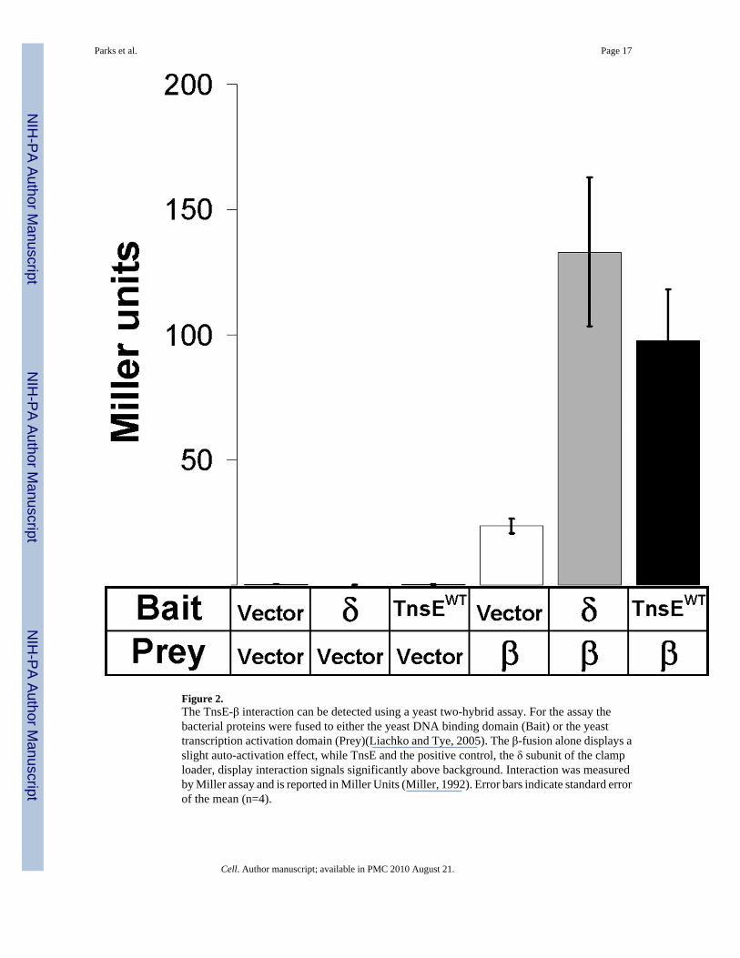
Figure 2.The TnsE-β interaction can be detected using a yeast two-hybrid assay. For the assay thebacterial proteins were fused to either the yeast DNA binding domain (Bait) or the yeasttranscription activation domain (Prey)(Liachko and Tye, 2005). The β-fusion alone displays aslight auto-activation effect, while TnsE and the positive control, the δ subunit of the clamploader, display interaction signals significantly above background. Interaction was measuredby Miller assay and is reported in Miller Units (Miller, 1992). Error bars indicate standard errorof the mean (n=4).
Parks et al. Page 17
Cell. Author manuscript; available in PMC 2010 August 21.
NIH
-PA Author Manuscript
NIH
-PA Author Manuscript
NIH
-PA Author Manuscript

Figure 3. TnsE and β clamp interact in three distinct in vitro assaysA. Protein mobility shift assays confirm the TnsE-β interaction in vitro. 32P-labeled β monomer(10 nM) (32P-β alone in lane 1) is unaffected by the addition of up to 10 μM BSA (lanes 1− 3)in a 4% native polyacrylamide gel, but produces a shifted product (black arrow) with theaddition of TnsEwt (lanes 4−7).B. Far western blots show that TnsE retains 32P signal when probed with 32P-labeled β clamp.C. Surface Plasmon Resonance experiments reveal interaction between β and TnsE and enablecomparisons to be drawn between this interaction and the well-characterized clamp loaderinteractions. Reference subtracted traces are shown for 1 μM δ (positive interaction control),5 μM TnsE and 1μM TnsE. The KD of the TnsE-β interaction is calculated to be ∼2.44 μM.D. Comparison between TnsE-β interaction and γ-β and δ’-β interactions provide furtherreference for the strength of the TnsE-β interaction. Reference subtracted sensograms for 1 μMγ (as monomer), 1 μM δ’, and 1 μM TnsE are shown.
Parks et al. Page 18
Cell. Author manuscript; available in PMC 2010 August 21.
NIH
-PA Author Manuscript
NIH
-PA Author Manuscript
NIH
-PA Author Manuscript

Figure 4.A. Quantification of the yeast two-hybrid assay reveals a defect in the β interaction with TnsEproteins with alanine substitutions in the putative β interaction motif. The alanine substitutionsin the putative β clamp binding motif consistently reduce TnsE-β interaction levels. Interactionwas measured by Miller assay and is reported in Miller Units (Miller, 1992). Western blotsusing anti-TnsE antibodies are given below the bar graph. Error bars indicate standard error ofthe mean (n=4).B. Surface Plasmon Resonance results for purified TnsEβMA mutants confirm a loss ininteraction between TnsE and β in vitro. All four TnsEβMA mutants tested show a decrease inβ interaction. Reference subtracted results from 1 μM of each of the TnsEβMA mutant proteinstested are shown (black lines) overlaid with results from 1 μM TnsEWT (gray lines) forcomparison.
Parks et al. Page 19
Cell. Author manuscript; available in PMC 2010 August 21.
NIH
-PA Author Manuscript
NIH
-PA Author Manuscript
NIH
-PA Author Manuscript

Figure 5. An in vivo transposition assay reveals a defect in the ability of TnsEβMA mutants toactivate transposition, and an increase in transposition frequency when β is over-expressedA. Transposition frequency is reduced significantly below wild-type with each of the sixTnsEβMA mutations. Transposition was monitored in cells expressing TnsABC and wild typeTnsE or a mutant TnsE containing an alanine substitution at one of six positions in the putativeβ clamp interacting motif (see text for details). A western blot using an anti-TnsE antibody isdisplayed below the graph. Transposition assays were conducted in recA− cells containingtns genes on plasmids using a lambda delivery vector (McKown et al., 1988). Error bars indicatethe standard error of the mean (n=3).B. Over-expression of β results in a significant increase in TnsE-mediated transposition.Transposition was monitored in cells expressing TnsABC+E with a plasmid expressing β oran empty vector. Error bars indicate standard error of the mean (n=3).
Parks et al. Page 20
Cell. Author manuscript; available in PMC 2010 August 21.
NIH
-PA Author Manuscript
NIH
-PA Author Manuscript
NIH
-PA Author Manuscript

Figure 6. An in vitro TnsABC+E transposition assay shows that DNA structures are important forTnsE-mediated transposition, and that the β clamp is required to direct the orientation oftransposon endsA. A representative bar graph representing the number of transposon insertions recovered perin vitro reaction shows that the presence of a single-stranded gap in the target DNA substrateis essential for activation of TnsE-mediated transposition, but shows no effect on the untargetedTnsABC* pathway.B. Transposition via the TnsABC* pathway into the β-loaded gapped substrate indicates thatthere are no indirect affects of β or gapped DNA on the core transposition machinery that couldaccount for the results observed in TnsABC+E reactions. Black circles represent the targetDNA, with a gap in one circle representing the location of the ssDNA gap. Black arrows onthe outside of the DNA represent the left-to right orientation of the transposon ends with respectto the free 3’ end of the gapped DNA, while gray arrows inside the circles represent the opposite(right-to-left) orientation. The position of each insertion is given next to the arrow. The ssDNAgap resides between positions 1415−1435.C. In vitro reactions containing TnsABC+E* and gapped DNA alone. Transposition eventsmapped in the target DNA appear to be randomly distributed, and with no particular orientationof transposon ends.D. Reactions containing TnsABC+E* and β clamp loaded onto the gapped DNA substratedisplay a rearrangement of transposon insertions. Many insertions were found at a single basepair junction 66 bases from the free 3’ end of the ssDNA gap (position 1481), and occurred ina single left-to-right orientation at that site. Other insertions were found throughout the targetDNA, almost all in the left-to-right orientation.
Parks et al. Page 21
Cell. Author manuscript; available in PMC 2010 August 21.
NIH
-PA Author Manuscript
NIH
-PA Author Manuscript
NIH
-PA Author Manuscript

Figure 7. Over-expression of TnsE results in activation of the SOS DNA damage responseA. Activation of SOS is consistent with interference with normal traffic on the β clamp. Cellsthat over-express wild type TnsE activate the SOS response. Strains expressing theTnsEβMA mutant proteins show a significant reduction in the SOS response compared to thewild type protein, likely due to a compromised interaction with β. Western blot results formembranes probed with anti-TnsE that are displayed below the graph. SOS was monitored byMiller assay and in a reporter strain containing a sulA::lac fusion and is reported in Miller units(Miller, 1992; Sutton, 2004). Error bars indicate standard error of the mean (n=3).B. SOS induction provoked by TnsE over-expression is abolished in the recF-background.RecF is essential for the RecFOR pathway of RecA loading onto single-stranded DNA gaps.Error bars indicate standard error of the mean (n=3).
Parks et al. Page 22
Cell. Author manuscript; available in PMC 2010 August 21.
NIH
-PA Author Manuscript
NIH
-PA Author Manuscript
NIH
-PA Author Manuscript