Sequence- and Species-Dependence of Proteasomal Processivity
20
Sequence- and Species-Dependence of Proteasomal Processivity Daniel A. Kraut a,e , Eitan Israeli a , Erin K. Schrader a , Ashwini Patil b , Kenta Nakai b , Dhaval Nanavati c , Tomonao Inobe d , and Andreas Matouschek a,* a Department of Molecular Biosciences, Northwestern University, Evanston, IL 60208, USA b Human Genome Center, The Institute of Medical Science, The University of Tokyo, 4-6-1 Shirokane-dai, Minato-ku, Tokyo, Japan c Proteomics core facility, Chemistry of Life Processes Institute, Northwestern University, Evanston, IL 60208, USA d Frontier Research Core for Life Sciences, University of Toyama, 3190 Gofuku, Toyama-shi, Toyama 930-8555, Japan e Department of Chemistry, Villanova University, Villanova, PA 19085, USA Abstract The proteasome is the degradation machine at the center of the ubiquitin-proteasome system and controls the concentrations of many proteins in eukaryotes. It is highly processive so that substrates are degraded completely into small peptides, avoiding the formation of potentially toxic fragments. Nonetheless, some proteins are incompletely degraded, indicating the existence of factors that influence proteasomal processivity. We have quantified proteasomal processivity and determined the underlying rates of substrate degradation and release. We find that processivity increases with species complexity over a 5-fold range between yeast and mammalian proteasome, and the effect is due to slower but more persistent degradation by proteasomes from more complex organisms. A sequence stretch that has been implicated in causing incomplete degradation, the glycine-rich region of the NFκB subunit p105, reduces the proteasome’s ability to unfold its substrate, and polyglutamine repeats such as found in Huntington’s disease reduce the processivity of the proteasome in a length-dependent manner. The proteasome is at the center of the ubiquitin-proteasome system (UPS), which controls the concentrations of many proteins in eukaryotes. The proteasome is a molecular machine of around 2.5 MDa and consists of approximately 40 proteins, organized into a large barrel- shaped structure (1-3). The proteolytic sites are buried deep within the particle and are accessible only through a narrow channel such that proteins have to be unfolded before they can be degraded (1-3). A ring of six ATP-dependent motor proteins, called Rpt1-6 in yeast, is located at the entrance of the degradation channel and controls access (1, 4). The degradation signal or degron in most proteasome substrates contains a proteasome-binding tag such as a poly-ubiquitin modification and an unstructured initiation region (5, 6). The binding tags can be recognized by several proteasome subunits and the initiation region is probably recognized by the motor proteins (1, 7). The motors engage the initiation region and then translocate the substrate into the degradation chamber, unfolding any structures that the proteasome encounters along the way (8-10). * To whom correspondence may be addressed: [email protected]. Supporting Information Available: This material is free via the Internet. NIH Public Access Author Manuscript ACS Chem Biol. Author manuscript; available in PMC 2013 August 17. Published in final edited form as: ACS Chem Biol. 2012 August 17; 7(8): 1444–1453. doi:10.1021/cb3001155. NIH-PA Author Manuscript NIH-PA Author Manuscript NIH-PA Author Manuscript
-
Upload
independent -
Category
Documents
-
view
5 -
download
0
Transcript of Sequence- and Species-Dependence of Proteasomal Processivity

Sequence- and Species-Dependence of ProteasomalProcessivity
Daniel A. Krauta,e, Eitan Israelia, Erin K. Schradera, Ashwini Patilb, Kenta Nakaib, DhavalNanavatic, Tomonao Inobed, and Andreas Matouscheka,*
aDepartment of Molecular Biosciences, Northwestern University, Evanston, IL 60208, USAbHuman Genome Center, The Institute of Medical Science, The University of Tokyo, 4-6-1Shirokane-dai, Minato-ku, Tokyo, JapancProteomics core facility, Chemistry of Life Processes Institute, Northwestern University,Evanston, IL 60208, USAdFrontier Research Core for Life Sciences, University of Toyama, 3190 Gofuku, Toyama-shi,Toyama 930-8555, JapaneDepartment of Chemistry, Villanova University, Villanova, PA 19085, USA
AbstractThe proteasome is the degradation machine at the center of the ubiquitin-proteasome system andcontrols the concentrations of many proteins in eukaryotes. It is highly processive so thatsubstrates are degraded completely into small peptides, avoiding the formation of potentially toxicfragments. Nonetheless, some proteins are incompletely degraded, indicating the existence offactors that influence proteasomal processivity. We have quantified proteasomal processivity anddetermined the underlying rates of substrate degradation and release. We find that processivityincreases with species complexity over a 5-fold range between yeast and mammalian proteasome,and the effect is due to slower but more persistent degradation by proteasomes from more complexorganisms. A sequence stretch that has been implicated in causing incomplete degradation, theglycine-rich region of the NFκB subunit p105, reduces the proteasome’s ability to unfold itssubstrate, and polyglutamine repeats such as found in Huntington’s disease reduce the processivityof the proteasome in a length-dependent manner.
The proteasome is at the center of the ubiquitin-proteasome system (UPS), which controlsthe concentrations of many proteins in eukaryotes. The proteasome is a molecular machineof around 2.5 MDa and consists of approximately 40 proteins, organized into a large barrel-shaped structure (1-3). The proteolytic sites are buried deep within the particle and areaccessible only through a narrow channel such that proteins have to be unfolded before theycan be degraded (1-3). A ring of six ATP-dependent motor proteins, called Rpt1-6 in yeast,is located at the entrance of the degradation channel and controls access (1, 4). Thedegradation signal or degron in most proteasome substrates contains a proteasome-bindingtag such as a poly-ubiquitin modification and an unstructured initiation region (5, 6). Thebinding tags can be recognized by several proteasome subunits and the initiation region isprobably recognized by the motor proteins (1, 7). The motors engage the initiation regionand then translocate the substrate into the degradation chamber, unfolding any structures thatthe proteasome encounters along the way (8-10).
*To whom correspondence may be addressed: [email protected].
Supporting Information Available: This material is free via the Internet.
NIH Public AccessAuthor ManuscriptACS Chem Biol. Author manuscript; available in PMC 2013 August 17.
Published in final edited form as:ACS Chem Biol. 2012 August 17; 7(8): 1444–1453. doi:10.1021/cb3001155.
NIH
-PA Author Manuscript
NIH
-PA Author Manuscript
NIH
-PA Author Manuscript

The proteasome is thought to be highly processive so that substrate proteins are degradedcompletely into small peptides (8, 11, 12). This processivity is important biologicallybecause it avoids the formation of potentially toxic protein fragments. ATP-dependentproteases similar to the proteasome control protein concentrations in bacteria and archaea(13). These proteases vary in their unfolding ability over several orders of magnitude, andthese differences may contribute to substrate selection (12, 14-16). The eukaryoticproteasome is the most processive of the ATP-dependent proteases analyzed so far (12).Nonetheless, it degrades a handful of proteins only partially in vivo: several transcriptionfactors are converted into fragments that have biological activities distinct from those of thefull-length proteins (17-23). The molecular mechanism that causes this partial degradation ispoorly understood. Presumably, the proteasome’s processivity depends on the resistance of agiven domain to unfolding, the mechanical capability of the motors, and how well theproteasome holds on to the substrate during degradation (12, 15, 19). Indeed, some evidencesuggests that domains that are able to resist unfolding stall the proteasome’s progressionalong a substrate (19, 24). The sequence stretch just preceding a folded domain also seemsto play a role in inducing processing or reducing processivity (18, 19, 25, 26).
The first characterized example of processing was the conversion of p105 into the NFκBsubunit p50 as part of its activation in mammalian cells (17). In p105, a Rel homologydomain is followed first by a glycine-rich region (GRR) and then the degron responsible fordegradation. Presumably, processing occurs when the proteasome encounters the folded Relhomology domain as the Rpt motors are interacting with the GRR (18, 27). The NFκBsignaling pathway is not found in yeast (28) but it can be reconstituted by expressing thevarious components from plasmids (29, 30). Surprisingly, the GRR is not required forprocessing in yeast (30), suggesting there might be differences in proteasomal processivityacross eukaryotic species.
Polyglutamine diseases may also provide an example of a protein that changes theproteasome’s processivity. The most common of this group of disorders is Huntington’sdisease (HD). HD is associated with the formation of an N-terminal fragment of theHuntingtin protein (Htt), which contains a polyglutamine stretch (31, 32). Expansion of thisrepetitive amino acid region to more than 35 glutamines correlates with earlier onset of thedisease(31). The Htt fragment accumulates (32) despite being ubiquitinated (32, 33) and thisleads to cytotoxicity. There is some evidence that protein fragments containing thesepolyglutamine (polyQ) repeats resist degradation and may even impede the UPS, perhaps byclogging up the proteasome (34-36).
Here we define the processivity of proteasomal degradation quantitatively by measuring therate constants with which the proteasome progresses along a substrate and with whichpartially degraded substrates are released. We find that the processivity varies betweenspecies over an approximately 5-fold range, with the mammalian and frog proteasome beingthe most processive, worm and yeast proteasome the least processive, and fly proteasome inbetween. The higher processivity of the mammalian proteasome is due to a roughly fifteen-fold slower substrate release rate than for the yeast proteasome, which is partially offset by aslower unfolding rate. Thus, the mammalian proteasome has a slower, more careful, motorthan the yeast proteasome. The GRR increases fragment formation primarily by decreasingthe ability of the proteasome to move forward to unfold its substrate and not by increasingsubstrate dissociation rates. Similarly, increasing the length of a polyglutamine stretchprogressively decreases the processivity of the proteasome, which may at least in partexplain why Htt fragments accumulate in cells.
Kraut et al. Page 2
ACS Chem Biol. Author manuscript; available in PMC 2013 August 17.
NIH
-PA Author Manuscript
NIH
-PA Author Manuscript
NIH
-PA Author Manuscript
RESULTS and DISCUSSIONMeasuring proteasome processivity
To investigate proteasomal processivity, we followed the degradation of a two-domainprotein by proteasome purified from different organisms. The substrate consisted of twodomains fused to each other and to a degron: an E. coli dihydrofolate reductase (DHFR)domain at the N terminus, followed by a barnase domain, and then a degron derived fromyeast Sic1 (37) at the C terminus (N-DHFR-barnase-degron-C; Figure 1) (12). The barnasedomain is easily unfolded by the proteasome, whereas the DHFR domain is considerablyharder to degrade (8, 12) and can be further stabilized by the addition of ligands. Thesubstrate analog methotrexate (MTX) binds DHFR tightly (Kd ~ 1 nM) and prevents itsdegradation by the proteasome, whereas the cofactor NADPH binds less tightly (Kd ~ 1 μM)and should stabilize DHFR to a lesser extent (38-41).
The substrate was synthesized by coupled in vitro transcription and translation, ubiquitinatedby the ubiquitin-protein ligase (E3) Rsp5 (37), and highly ubiquitinated forms were purified.The ubiquitinated substrate was then presented to the proteasome under single-turnoverconditions, which means that proteasome was present in great excess over substrate andeach proteasome complex likely degraded at most one substrate molecule during thereaction. When the substrate binds to the proteasome it can either become degraded or it canbecome deubiquitinated before the unfolding motors are fully engaged, leading todissociation (42). Degradation occurs sequentially from the C-terminal degron through theproximal barnase domain and then the N-terminal DHFR domain (Figure 1). When theproteasome encounters the DHFR domain, the reaction partitions between two branches. Inthe first branch, the DHFR domain becomes unfolded and the remainder of the substrate istranslocated into the proteasome and proteolysed with the rate constant kdeg
frag. The processreflected by this rate constant in principle includes unfolding, translocation and proteolysissteps, but it is dominated by its slowest step, the unfolding reaction (see supplementaryinformation). In the second branch, the DHFR domain together with the remainder of thesubstrate dissociates from the proteasome irreversibly with the rate constant krel
frag, so that apartially degraded fragment is released (Figure 1a). We define the unfolding ability orprocessivity of the proteasome as the ratio of the two branches of the reaction (U = kdeg
frag/krel
frag) (12). This ratio can be determined most easily from the endpoint of a degradationreaction by comparing the amount of the full-length protein that disappears (i.e., all theprotein for which at least the barnase domain is degraded) to the amount of DHFR fragmentformed (U = (Fraction Barnase degraded/Fraction DHFR fragment released) – 1) (Figure 1)(12).
In our degradation assays, the yeast proteasome was highly processive, as we had expected,and it degraded the N-DHFR-barnase-degron-C substrate completely without the formationof any detectable fragments, except for a small fraction of full-length protein that wasdeubiquitinated and therefore escaped degradation (Figure 1b). Stabilizing the DHFRdomain with 500 μM NADPH stalled the proteasome’s progression such that a third of thetime the DHFR domain, together with a small remnant of preceding sequence that had notyet been degraded, dissociated and escaped degradation; the other two thirds of the time thesubstrate was degraded completely (Figure 1c). This ratio of partitioning between the partialand complete degradation corresponds to an unfolding ability U of 1.9 ± 0.2 and is a directmeasure of proteasomal processivity.
The mammalian proteasome is more processive than the yeast proteasomeWe next carried out this partitioning assay with mammalian proteasome that had beenaffinity-purified from rabbit reticulocytes (Figure 2). We found the mammalian proteasome
Kraut et al. Page 3
ACS Chem Biol. Author manuscript; available in PMC 2013 August 17.
NIH
-PA Author Manuscript
NIH
-PA Author Manuscript
NIH
-PA Author Manuscript

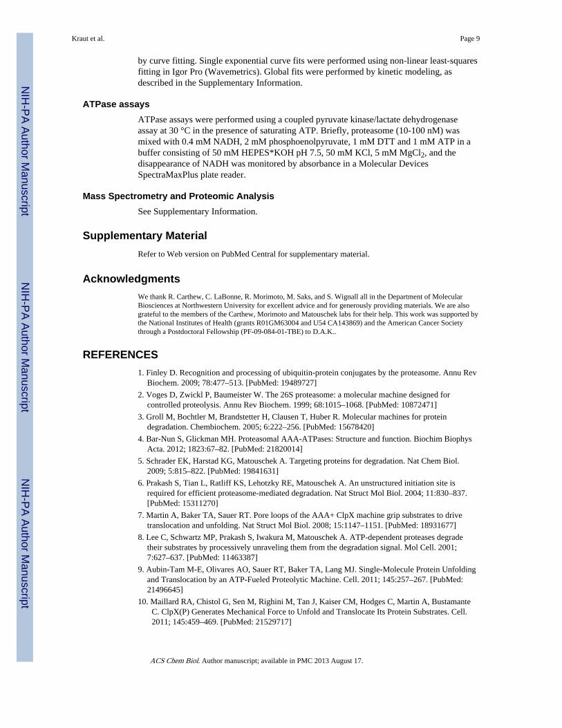
to be ~5-fold more processive than the yeast proteasome with an unfolding ability of U = 11± 2 compared to U = 1.9 ± 0.2 for the yeast proteasome (Table 1). This difference inunfolding ability is surprisingly large considering that the machines have been optimized forostensibly the same purpose in both organisms, and the amino acid sequences of the motorsubunits are approximately 60-70% identical between yeast and mammals. To establish thatthis difference is not simply an idiosyncrasy of the particular substrate chosen, we reversedthe order of the sequence elements within the substrate, such that DHFR is degraded fromthe N-terminus rather than the C-terminus. As the local structure first encountered by theproteasome determines a folded domain’s ability to resist degradation (8), this reversedsubstrate orientation effectively presents the proteasome with a different structure to unfold.The mammalian proteasome was much more processive (U = 29 ± 7) than the yeastproteasome (U = 2.0 ± 0.2) from this direction as well (Supplementary Figure 1).
Several observations suggest that this difference in processivity reflects intrinsic propertiesof the proteasomes and not the presence of processivity factors co-purified with oneproteasome but not the other, or other defects in one of the proteasome preparations. First,both yeast and mammalian proteasome preparations contained comparable proportions ofsingly- and doubly-capped core particles, with little if any free core particle (SupplementaryFigure 2a). Second, mass spectroscopy analysis of the purified proteasomes showed thatboth yeast and mammalian proteasomes contained similar sets of known proteasomesubunits and proteasome-associated proteins such as Rad23 and Cdc48/p97 (SupplementaryInformation). Indeed, the yeast proteasome preparation contained more additional associatedproteins than the rabbit proteasome, including members of the hsp20, hsp70, and hsp104chaperone families, which are not found associated with the rabbit proteasome, suggestingthat the yeast proteasome has not lost any factors that might aid in unfolding or processivityin the purification. Third, yeast proteasome preparation using different conditions such asthe higher salt washes used for mammalian proteasome purification, or affinity purificationvia a different tag all consistently yielded yeast proteasome with similar processivities, allsignificantly lower than that of mammalian proteasome (Supplementary Figure 2b). Finally,we mixed yeast proteasome with rabbit proteasome treated with a proteasome inactivator(AdaAhx3L3-vinyl sulfone) to test whether the rabbit proteasome preparation might containa processivity factor that could be transferred to the yeast proteasome preparation. The yeastproteasome’s ability to unfold the DHFR domain did not increase, although we cannot ruleout very slowly exchanged processivity factors (Supplementary Figure 2c-g). Together,these observations suggest that the processivities we measured reflect the proteasomeparticles’ intrinsic abilities to unfold domains in their substrates.
Processivity of proteasome from other speciesThe observed difference in processivity between yeast and mammalian proteasomes couldbe merely an idiosyncratic difference between the two proteasomes, or it could reflect theresult of an underlying evolutionary pressure for a more processive proteasome in morecomplex eukaryotes. To test this idea, we compared the unfolding abilities of affinity-purified proteasomes from three additional species: worm (C. elegans), fly (D.melanogaster) and frog (X. laevis). All proteasomes were characterized by massspectrometry (Supplementary Information) and native gel analysis (Supplementary Figure2a) and the preparations appeared to be of similar purity. The unfolding abilities of theproteasomes increased in the order yeast ~ worm < fly < frog ~ rabbit (Figure 2c and Table1).
There are at least two potential driving forces for greater proteasome processivity. First, thecomplex regulatory network in metazoans might require a more processive degradationmachine, simply because there are more pathways where an undegraded fragment wouldwreak havoc. Second, more complex eukaryotes have a higher proportion of multi-domain
Kraut et al. Page 4
ACS Chem Biol. Author manuscript; available in PMC 2013 August 17.
NIH
-PA Author Manuscript
NIH
-PA Author Manuscript
NIH
-PA Author Manuscript

proteins than simpler eukaryotes (Supplementary Information). In multi-domain proteins thedomains must be unfolded and degraded one after the other and each domain can potentiallycause the dissociation of an undegraded fragment. Thus, the more complex proteinarchitecture could lead to a need for a proteasome with greater processivity. Indeed, theability of the proteasome to unfold a domain increased with the prevalence of multi-domainproteins in the proteome (Figure 3). In addition, proteomic studies have begun to identify thecollection of all proteasome substrates in different organisms and it appears that mammalianproteasome substrates also include more multi-domain proteins than their yeast orthologs(Supplementary Table).
A transient intermediate allows determination of unfolding and release ratesThe mammalian proteasome could be more processive than the yeast proteasome because itunfolds and proteolyzes DHFR more rapidly (larger rate constant kdeg
frag) or because itreleases the partially degraded DHFR fragment more slowly (smaller rate constant krel
frag)(Figure 1a). To measure these rate constants, we increased the proteasome concentration inthe reaction to accelerate substrate encounter and thus the rate with which the full-lengthprotein is degraded (larger rate constant kdeg
full-length). This allowed us to observe the rapidformation of DHFR fragment as the proteasome first runs into the DHFR domain and stalls.The intermediate consisted of the DHFR domain and a tail of some 80 amino acids. Mostlikely, the DHFR domain was located at the entrance to the degradation channel and theundegraded tail reached down the degradation channel past the ATPase subunits to theproteolytic sites. Some of the fragment then decayed as the proteasome was able to unfoldand degrade the DHFR domain. During this time, the undecayed fragment remainedassociated with the proteasome as a productive reaction intermediate, as it could not becompeted away with an excess of unlabeled substrate added to the reaction at different times(Supplementary Figure 3). At the end of the reaction a certain amount of fragment that theproteasome failed to degrade remained as an inert end product (Figure 4). The final amountof fragment formed and thus the unfolding strength of the proteasome did not depend on theproteasome concentration in the reaction.
To determine the rate constants kdegfrag and krel
frag from the degradation curves we usedglobal curve fitting of the disappearance of full-length protein and the formation anddegradation of fragment to the reaction model described above (Figure 4; Table 1) (seeSupplementary Methods for details). The unfolding abilities calculated from the ratio ofkdeg
frag to krelfrag obtained by curve fitting were essentially identical to those determined
directly from the final levels of fragment formed by yeast and rabbit proteasome in thepresence of NADPH (Table 1), which validates the parameters obtained by curve fitting.
Mammalian proteasome releases substrate more slowly than yeast proteasomeNow that the individual rate constants could be determined, we could establish themechanism for the greater processivity of the rabbit proteasome. The ~5-fold greaterunfolding ability results from a ~16-fold slower fragment release rate (krel
frag), which isoffset by a ~4-fold slower rate of unfolding and proteolyzing of DHFR (kdeg
frag) (Figure 4 &Table 1). Thus, the mammalian proteasome is slower but also more persistent than the yeastproteasome. The slower degradation rate of the rabbit proteasome may be a reflection of aslower overall catalytic cycle for the proteasome, and consistent with this hypothesis themammalian proteasome had a 4-fold slower ATPase rate than the yeast proteasome (Table1). The situation is similar for proteasome from flies and frogs: the increase in processivitywas due principally to a decrease in krel
frag for proteasome from more complex organisms(Figure 4e & Table 1), offset by smaller changes in kdeg
frag (Figure 4f & Table 1).
Kraut et al. Page 5
ACS Chem Biol. Author manuscript; available in PMC 2013 August 17.
NIH
-PA Author Manuscript
NIH
-PA Author Manuscript
NIH
-PA Author Manuscript

Despite its lower unfolding rate, the mammalian proteasome is as powerful as the yeastproteasome. If each ATP hydrolysis corresponds to one cycle of pulling, we can determinethe number of pulls required to degrade DHFR by dividing the ATP hydrolysis rate by theforward degradation rate kdeg
frag (43). According to this calculation, the mammalianproteasome can degrade DHFR in 9 ± 2 ATPs or pulls, while the yeast proteasome requires8 ± 2 ATPs or pulls on average. In the presence of saturating NADPH, ~140 pulls arerequired to degrade the stabilized DHFR for both yeast and mammalian proteasome. Thus,approximately the same number of ATP-hydrolysis events are required for any of theproteasomes to unfold DFHR. This result suggests that unfolding and translocation arecoupled tightly to the ATP-hydrolysis cycle – regardless of the species of origin each ATP-dependent powerstroke has the same probability of unfolding the domain, and thereforelikely has the same translocation step-size and exerts the same amount of force on thesubstrate. However, yeast proteasome releases its substrate once every ~300 ATP hydrolysisevents, while the mammalian proteasome only releases once every ~1300 cycles (ATPaserate divided by krel
frag).
A processing element decreases the unfolding rateAlthough the proteasome is quite processive, certain amino acid sequences can reduce thisprocessivity and lead to the release of undigested protein fragments. The partial degradationseems to be part of some regulatory processes in the cell and to control the activities ofseveral transcription factors including p105, Spt23, Mga2, Ci and the Gli proteins (17-23).The conversion of p105 to p50 in mammalian cells requires a specific amino acid sequence,the 35 amino acid long GRR (18, 27). The GRR also directs fragment formation in modelproteins when placed adjacent to folded domains such as DHFR (18, 19). To determine howthe GRR reduces proteasome processivity, we inserted the GRR or a 35 amino acid longcontrol sequence between barnase and DHFR. The GRR reduced the processivity of theyeast proteasome by roughly an order of magnitude relative to the control sequence both inthe presence and absence of NADPH (Figure 5). In the presence of NADPH less than half ofthe protein containing the GRR is degraded completely. Global fitting of the degradationdata again allowed us to determine the rate constants for partitioning between fragmentdegradation and release (Figure 1a; Table 2). These showed that the GRR reduced theeffective unfolding ability of the proteasome entirely by reducing the degradation rateconstant kdeg
frag ~17-fold while leaving the release rate constant krelfrag largely unaffected.
Thus, the GRR promotes fragment formation by preventing the proteasome from unfoldingDHFR rather than by accelerating the release of the fragment. The GRR is dispensable forprocessing of p105 when it is expressed artificially in yeast (30), and this could be due to thelower intrinsic unfolding ability of the yeast proteasome.
This reduction in kdegfrag suggests that the Rpt motors exert less unfolding force than usual
on the folded domain while interacting with the GRR. ClpXP and the other bacterial AAA+proteases use aromatic paddles to move the substrate through the degradation channel andthe proteasome likely uses a similar mechanism (7, 9, 10, 44, 45). Perhaps the GRR containsfewer chemical features that the paddles can interact with to pull on the substrate. Thepolypeptide chain may also interact transiently with additional binding sites in the motor ortranslocation channel as it moves through the protease, for example to prevent back-slippingof the substrate. Perhaps the GRR sequence can bind to these sites only weakly, such thatthe substrate slips back more frequently allowing partially unfolded domains to snap backmore frequently. Similar effects of the GRR on unfolding ability were seen with mammalianproteasome, but the kinetics were such that the underlying rate constants could not bedetermined (Supplementary Figure 4).
Kraut et al. Page 6
ACS Chem Biol. Author manuscript; available in PMC 2013 August 17.
NIH
-PA Author Manuscript
NIH
-PA Author Manuscript
NIH
-PA Author Manuscript

Glutamine repeats reduce unfolding ability in a length-dependent mannerNext, we asked whether polyglutamine (polyQ) repeats such as those found in the Httprotein associated with HD might affect proteasomal processivity. We inserted stretches ofdifferent numbers of glutamine residues (11, 33 and 53) into the substrate proteins and foundthat the processivity of the rabbit and yeast proteasomes decreased with longer polyQinsertions up to 10-fold (Figure 6). We could not determine the unfolding and release rateconstants for the fragment because the kinetics of its formation followed single exponentialbehavior at all tested proteasome concentrations. The fact that the polyQ sequence preventedthe transient accumulation of intermediate observed in the parent construct by itself does notprovide a clue for the mechanism. Its transient accumulation could be prevented eitherbecause the polyQ sequence caused the proteasome-bound fragment to disappear morerapidly through degradation or release, or because the polyQ sequence caused the fragmentto be formed more slowly. The simplest explanation is that polyQ sequence acts like theGRR sequence and slows the forward movement of the proteasome along its substrates,however we cannot establish this mechanism directly. In the absence of NADPH, fragmentformation was seen only for the longer 53 glutamine repeats (Figure 6 & SupplementaryFigure 5). Thus, the proteasome is able to degrade polyQ sequences, but they appear to slowproteasome progression along a substrate, presumably allowing the release of partiallydegraded Htt fragments that can go on to form aggregates. PolyQ tracts might also preventdegradation due to their resistance to proteolytic cleavage (36) (i.e., due to sequencepreferences of the proteolytic sites on the proteasome) although more recent studies indicatethat the proteasome can cleave the polyQ tract at multiple places (46).
PolyQ stretches appear to have a similar effect on proteasomal processivity as the GRR.PolyQ and GRR sequences are both characterized by a strongly biased amino acidcomposition and it has been proposed that these low complexity regions are part of a generalprocessing signal (19). It is possible that low complexity regions in general slow theprogression of the proteasome along a substrate because they lack sequence features that canbe recognized by the ATPase motors. The likelihood of particular consensus motifsoccurring by chance in an amino acid sequence becomes lower as the amino acidcomposition of that sequence becomes less complex. Interestingly, inserting the controlsequence into the substrate increased the unfolding ability of the proteasome compared toconstructs without the insert (essentially comparing the effect of the control sequence to thatof the barnase sequence), suggesting that amino acid sequence in general, and not just GRRand polyQ sequences, can affect processivity and that sequence complexity is not the onlyfactor.
In Huntington’s disease, ubiquitinated aggregates of Htt exon1 fragments persist in neuronsand accumulate slowly over many years to eventually form visible inclusions (32). Theaccumulation of aggregates correlates with the disease phenotype, though soluble forms ofHtt exon1 rather than the aggregates themselves may be the toxic species (47). The poordegradation of polyQ-containing proteins observed here might be a reason why Htt exon1protein is not cleared from cells effectively.
In our assays, all of the polyQ insertions decreased the degradation of DHFR stabilized byNADPH, and did so the more effectively the longer they were. Only the longestpolyglutamine stretch examined here (53 glutamines) began to prevent degradation ofDHFR in the absence of NADPH. This observation is reminiscent of the finding that onlyrepeats of more than 35 glutamines in Htt appear to lead to significant aggregation and HDclinically (31). The test substrates with the unstabilized DHFR domain may represent amodel for pre-aggregation degradation, where longer glutamine expansions begin to hamperproteasomal degradation and allow the slow formation and accumulation of polyQfragments that will eventually aggregate. Indeed, at least some htt aggregates contain polyQ
Kraut et al. Page 7
ACS Chem Biol. Author manuscript; available in PMC 2013 August 17.
NIH
-PA Author Manuscript
NIH
-PA Author Manuscript
NIH
-PA Author Manuscript

stretches of different lengths as would be created when the proteasome is no longer fullyprocessive (48). The test substrates in which the DHFR domain is stabilized by the NADPHligand might represent a model for htt exon1 proteins where some of the polyQ stretchbegan to form aggregates, which act like stable folded domains so that the proteasome isunable to unfold and clear the proteins. If the polyQ sequences inhibit proteasome progressalong its substrate just as the GRR does, these sequences may attenuate overall proteasomeactivity in the cell by increasing the time that partially degraded substrates remain associatedwith proteasomes. However, the evidence whether or not the UPS is attenuated in aclinically meaningful way in HD is contradictory (34, 49-52).
ConclusionWe have established an assay to describe the processivity of the proteasome quantitativelyby measuring the forward degradation and substrate dissociation rates that determinewhether the proteasome digests or releases a substrate as it proceeds along the polypeptidechain. The mammalian proteasome is the most processive proteasome amongst those weinvestigated. Its higher unfolding and degradation power is caused by a much slower releaseof partially degraded substrates than was observed for the yeast proteasome. This may allowthe mammalian proteasome to handle complex proteins reliably. However, the mammalianproteasome’s slower release rate may also make it more likely to choke on hard-to-degradesubstrates such as Htt aggregates, potentially leading to an increase in toxicity in anynumber of protein misfolding diseases.
METHODSProteasome purification
Proteasomes were purified as described previously or by adapting previous protocols(Supplementary Information).
ConstructsConstructs encoding substrate proteins were cloned into pGEM-3Zf+ (Promega). The mainunfolding ability substrate consisted of an N-terminal His-tag followed by E. colidihydrofolate reductase (DHFR) followed by a catalytically inactive (H102A) Bacillusamyloliquefaciens barnase with all lysines replaced by arginine, methionine or alaninefollowed by 60 amino acids from the Sic1 protein containing a PPXY Rsp5 ubiquitinationmotif. The complex insertion between DHFR and barnase corresponded to the first 35 aminoacids of the S. cerevisiae cytochrome b2 mitochondrial targeting presequence with lysineschanged to arginines or glutamines. The GRR insertion corresponded to residues 369-403 ofhuman p105. The polyQ insertions consisted of 11, 33 or 53 glutamine repeats. For proteinexpression in E. coli, the unfolding ability construct was subcloned into pet44a justdownstream of the T7-lac promoter. Constructs were assembled using a combination oftraditional restriction enzyme cloning and In-Fusion (Clontech).
SubstratesRadioactive substrates were in vitro translated using the RTS 100 E. coli HY kit (5 PRIME),supplemented with 35S methionine. After high-speed ultracentrifugation, substrates wereaffinity purified using Talon magnetic beads (Clontech) as described previously (24). Thesubstrates were then in vitro ubiquitinated and purified (Supplementary Information).
Proteasomal degradation assayAssays were carried out under single turnover conditions (enzyme in vast excess ofsubstrate) at 30 °C essentially as described previously (24). Degradation data were analyzed
Kraut et al. Page 8
ACS Chem Biol. Author manuscript; available in PMC 2013 August 17.
NIH
-PA Author Manuscript
NIH
-PA Author Manuscript
NIH
-PA Author Manuscript
by curve fitting. Single exponential curve fits were performed using non-linear least-squaresfitting in Igor Pro (Wavemetrics). Global fits were performed by kinetic modeling, asdescribed in the Supplementary Information.
ATPase assaysATPase assays were performed using a coupled pyruvate kinase/lactate dehydrogenaseassay at 30 °C in the presence of saturating ATP. Briefly, proteasome (10-100 nM) wasmixed with 0.4 mM NADH, 2 mM phosphoenolpyruvate, 1 mM DTT and 1 mM ATP in abuffer consisting of 50 mM HEPES*KOH pH 7.5, 50 mM KCl, 5 mM MgCl2, and thedisappearance of NADH was monitored by absorbance in a Molecular DevicesSpectraMaxPlus plate reader.
Mass Spectrometry and Proteomic AnalysisSee Supplementary Information.
Supplementary MaterialRefer to Web version on PubMed Central for supplementary material.
AcknowledgmentsWe thank R. Carthew, C. LaBonne, R. Morimoto, M. Saks, and S. Wignall all in the Department of MolecularBiosciences at Northwestern University for excellent advice and for generously providing materials. We are alsograteful to the members of the Carthew, Morimoto and Matouschek labs for their help. This work was supported bythe National Institutes of Health (grants R01GM63004 and U54 CA143869) and the American Cancer Societythrough a Postdoctoral Fellowship (PF-09-084-01-TBE) to D.A.K..
REFERENCES1. Finley D. Recognition and processing of ubiquitin-protein conjugates by the proteasome. Annu Rev
Biochem. 2009; 78:477–513. [PubMed: 19489727]
2. Voges D, Zwickl P, Baumeister W. The 26S proteasome: a molecular machine designed forcontrolled proteolysis. Annu Rev Biochem. 1999; 68:1015–1068. [PubMed: 10872471]
3. Groll M, Bochtler M, Brandstetter H, Clausen T, Huber R. Molecular machines for proteindegradation. Chembiochem. 2005; 6:222–256. [PubMed: 15678420]
4. Bar-Nun S, Glickman MH. Proteasomal AAA-ATPases: Structure and function. Biochim BiophysActa. 2012; 1823:67–82. [PubMed: 21820014]
5. Schrader EK, Harstad KG, Matouschek A. Targeting proteins for degradation. Nat Chem Biol.2009; 5:815–822. [PubMed: 19841631]
6. Prakash S, Tian L, Ratliff KS, Lehotzky RE, Matouschek A. An unstructured initiation site isrequired for efficient proteasome-mediated degradation. Nat Struct Mol Biol. 2004; 11:830–837.[PubMed: 15311270]
7. Martin A, Baker TA, Sauer RT. Pore loops of the AAA+ ClpX machine grip substrates to drivetranslocation and unfolding. Nat Struct Mol Biol. 2008; 15:1147–1151. [PubMed: 18931677]
8. Lee C, Schwartz MP, Prakash S, Iwakura M, Matouschek A. ATP-dependent proteases degradetheir substrates by processively unraveling them from the degradation signal. Mol Cell. 2001;7:627–637. [PubMed: 11463387]
9. Aubin-Tam M-E, Olivares AO, Sauer RT, Baker TA, Lang MJ. Single-Molecule Protein Unfoldingand Translocation by an ATP-Fueled Proteolytic Machine. Cell. 2011; 145:257–267. [PubMed:21496645]
10. Maillard RA, Chistol G, Sen M, Righini M, Tan J, Kaiser CM, Hodges C, Martin A, BustamanteC. ClpX(P) Generates Mechanical Force to Unfold and Translocate Its Protein Substrates. Cell.2011; 145:459–469. [PubMed: 21529717]
Kraut et al. Page 9
ACS Chem Biol. Author manuscript; available in PMC 2013 August 17.
NIH
-PA Author Manuscript
NIH
-PA Author Manuscript
NIH
-PA Author Manuscript

11. Nussbaum AK, Dick TP, Keilholz W, Schirle M, Stevanović S, Dietz K, Heinemeyer W, Groll M,Wolf DH, Huber R, Rammensee HG, Schild H. Cleavage motifs of the yeast 20S proteasome betasubunits deduced from digests of enolase 1. Proc Natl Acad Sci USA. 1998; 95:12504–12509.[PubMed: 9770515]
12. Koodathingal P, Jaffe NE, Kraut DA, Prakash S, Fishbain S, Herman C, Matouschek A. ATP-dependent proteases differ substantially in their ability to unfold globular proteins. J Biol Chem.2009; 284:18674–18684. [PubMed: 19383601]
13. Sauer RT, Baker TA. AAA+ proteases: ATP-fueled machines of protein destruction. Annu RevBiochem. 2011; 80:587–612. [PubMed: 21469952]
14. Herman C, Prakash S, Lu CZ, Matouschek A, Gross CA. Lack of a robust unfoldase activityconfers a unique level of substrate specificity to the universal AAA protease FtsH. Mol Cell. 2003;11:659–669. [PubMed: 12667449]
15. Gur E, Sauer RT. Degrons in protein substrates program the speed and operating efficiency of theAAA+ Lon proteolytic machine. Proc Natl Acad Sci USA. 2009; 106:18503–18508. [PubMed:19841274]
16. Gur E, Vishkautsan M, Sauer RT. Protein unfolding and degradation by the AAA+ lon protease.Protein Sci. 2012; 21:268–278. [PubMed: 22162032]
17. Palombella VJ, Rando OJ, Goldberg AL, Maniatis T. The ubiquitin-proteasome pathway isrequired for processing the NF-kappa B1 precursor protein and the activation of NF-kappa B. Cell.1994; 78:773–785. [PubMed: 8087845]
18. Lin L, Ghosh S. A glycine-rich region in NF-kappaB p105 functions as a processing signal for thegeneration of the p50 subunit. Mol Cell Biol. 1996; 16:2248–2254. [PubMed: 8628291]
19. Tian L, Holmgren RA, Matouschek A. A conserved processing mechanism regulates the activity oftranscription factors Cubitus interruptus and NF-kappaB. Nat Struct Mol Biol. 2005; 12:1045–1053. [PubMed: 16299518]
20. Aza-Blanc P, Ramírez-Weber FA, Laget MP, Schwartz C, Kornberg TB. Proteolysis that isinhibited by hedgehog targets Cubitus interruptus protein to the nucleus and converts it to arepressor. Cell. 1997; 89:1043–1053. [PubMed: 9215627]
21. Rape M, Jentsch S. Taking a bite: proteasomal protein processing. Nat Cell Biol. 2002; 4:E113–116. [PubMed: 11988749]
22. Hoppe T, Matuschewski K, Rape M, Schlenker S, Ulrich HD, Jentsch S. Activation of amembrane-bound transcription factor by regulated ubiquitin/proteasome-dependent processing.Cell. 2000; 102:577–586. [PubMed: 11007476]
23. Wang B, Fallon JF, Beachy PA. Hedgehog-regulated processing of Gli3 produces an anterior/posterior repressor gradient in the developing vertebrate limb. Cell. 2000; 100:423–434. [PubMed:10693759]
24. Kraut DA, Matouschek A. Proteasomal Degradation from Internal Sites Favors Partial Proteolysisvia Remote Domain Stabilization. ACS Chem Biol. 2011; 6:1087–1095. [PubMed: 21815694]
25. Hoyt MA, Zich J, Takeuchi J, Zhang M, Govaerts C, Coffino P. Glycine-alanine repeats impairproper substrate unfolding by the proteasome. EMBO J. 2006; 25:1720–1729. [PubMed:16601692]
26. Daskalogianni C, Apcher S, Candeias MM, Naski N, Calvo F, Fåhraeus R. Gly-Ala repeats induceposition- and substrate-specific regulation of 26 S proteasome-dependent partial processing. J BiolChem. 2008; 283:30090–30100. [PubMed: 18757367]
27. Orian A, Schwartz AL, Israël A, Whiteside S, Kahana C, Ciechanover A. Structural motifsinvolved in ubiquitin-mediated processing of the NF-kappaB precursor p105: roles of the glycine-rich region and a downstream ubiquitination domain. Mol Cell Biol. 1999; 19:3664–3673.[PubMed: 10207090]
28. Gilmore TD. Introduction to NF-kappaB: players, pathways, perspectives. Oncogene. 2006;25:6680–6684. [PubMed: 17072321]
29. Epinat JC, Whiteside ST, Rice NR, Israël A. Reconstitution of the NF-kappa B system inSaccharomyces cerevisiae for isolation of effectors by phenotype modulation. Yeast. 1997;13:599–612. [PubMed: 9200810]
Kraut et al. Page 10
ACS Chem Biol. Author manuscript; available in PMC 2013 August 17.
NIH
-PA Author Manuscript
NIH
-PA Author Manuscript
NIH
-PA Author Manuscript

30. Sears C, Olesen J, Rubin D, Finley D, Maniatis T. NF-kappa B p105 processing via the ubiquitin-proteasome pathway. J Biol Chem. 1998; 273:1409–1419. [PubMed: 9430676]
31. Zuccato C, Valenza M, Cattaneo E. Molecular mechanisms and potential therapeutical targets inHuntington’s disease. Physiol Rev. 2010; 90:905–981. [PubMed: 20664076]
32. DiFiglia M, Sapp E, Chase KO, Davies SW, Bates GP, Vonsattel JP, Aronin N. Aggregation ofhuntingtin in neuronal intranuclear inclusions and dystrophic neurites in brain. Science. 1997;277:1990–1993. [PubMed: 9302293]
33. Kalchman MA, Graham RK, Xia G, Koide HB, Hodgson JG, Graham KC, Goldberg YP, GietzRD, Pickart CM, Hayden MR. Huntingtin is ubiquitinated and interacts with a specific ubiquitin-conjugating enzyme. J Biol Chem. 1996; 271:19385–19394. [PubMed: 8702625]
34. Bence NF, Sampat RM, Kopito RR. Impairment of the ubiquitin-proteasome system by proteinaggregation. Science. 2001; 292:1552–1555. [PubMed: 11375494]
35. Verhoef LGGC, Lindsten K, Masucci MG, Dantuma NP. Aggregate formation inhibitsproteasomal degradation of polyglutamine proteins. Hum Mol Genet. 2002; 11:2689–2700.[PubMed: 12374759]
36. Venkatraman P, Wetzel R, Tanaka M, Nukina N, Goldberg AL. Eukaryotic proteasomes cannotdigest polyglutamine sequences and release them during degradation of polyglutamine-containingproteins. Mol Cell. 2004; 14:95–104. [PubMed: 15068806]
37. Saeki Y, Isono E, Toh-E A. Preparation of ubiquitinated substrates by the PY motif-insertionmethod for monitoring 26S proteasome activity. Meth Enzymol. 2005; 399:215–227. [PubMed:16338358]
38. Cayley PJ, Dunn SM, King RW. Kinetics of substrate, coenzyme, and inhibitor binding toEscherichia coli dihydrofolate reductase. Biochemistry. 1981; 20:874–879. [PubMed: 7011378]
39. Johnston JA, Johnson ES, Waller PR, Varshavsky A. Methotrexate inhibits proteolysis ofdihydrofolate reductase by the N-end rule pathway. J Biol Chem. 1995; 270:8172–8178. [PubMed:7713922]
40. Ainavarapu SRK, Li L, Badilla CL, Fernandez JM. Ligand binding modulates the mechanicalstability of dihydrofolate reductase. Biophys J. 2005; 89:3337–3344. [PubMed: 16100277]
41. Touchette NA, Perry KM, Matthews CR. Folding of dihydrofolate reductase from Escherichia coli.Biochemistry. 1986; 25:5445–5452. [PubMed: 3535877]
42. Crosas B, Hanna J, Kirkpatrick DS, Zhang DP, Tone Y, Hathaway NA, Buecker C, Leggett DS,Schmidt M, King RW, Gygi SP, Finley D. Ubiquitin chains are remodeled at the proteasome byopposing ubiquitin ligase and deubiquitinating activities. Cell. 2006; 127:1401–1413. [PubMed:17190603]
43. Kenniston JA, Baker TA, Fernandez JM, Sauer RT. Linkage between ATP consumption andmechanical unfolding during the protein processing reactions of an AAA+ degradation machine.Cell. 2003; 114:511–520. [PubMed: 12941278]
44. Park E, Rho YM, Koh O-J, Ahn SW, Seong IS, Song J-J, Bang O, Seol JH, Wang J, Eom SH,Chung CH. Role of the GYVG pore motif of HslU ATPase in protein unfolding and translocationfor degradation by HslV peptidase. J Biol Chem. 2005; 280:22892–22898. [PubMed: 15849200]
45. Hinnerwisch J, Fenton WA, Furtak KJ, Farr GW, Horwich AL. Loops in the central channel ofClpA chaperone mediate protein binding, unfolding, and translocation. Cell. 2005; 121:1029–1041. [PubMed: 15989953]
46. Pratt G, Rechsteiner M. Proteasomes cleave at multiple sites within polyglutamine tracts:activation by PA28gamma(K188E). J Biol Chem. 2008; 283:12919–12925. [PubMed: 18343811]
47. Slow EJ, Graham RK, Osmand AP, Devon RS, Lu G, Deng Y, Pearson J, Vaid K, Bissada N,Wetzel R, Leavitt BR, Hayden MR. Absence of behavioral abnormalities and neurodegenerationin vivo despite widespread neuronal huntingtin inclusions. Proc Natl Acad Sci USA. 2005;102:11402–11407. [PubMed: 16076956]
48. Mitsui K, Nakayama H, Akagi T, Nekooki M, Ohtawa K, Takio K, Hashikawa T, Nukina N.Purification of polyglutamine aggregates and identification of elongation factor-1alpha and heatshock protein 84 as aggregate-interacting proteins. J Neurosci. 2002; 22:9267–9277. [PubMed:12417652]
Kraut et al. Page 11
ACS Chem Biol. Author manuscript; available in PMC 2013 August 17.
NIH
-PA Author Manuscript
NIH
-PA Author Manuscript
NIH
-PA Author Manuscript

49. Maynard CJ, Böttcher C, Ortega Z, Smith R, Florea BI, Díaz-Hernández M, Brundin P, OverkleeftHS, Li J-Y, Lucas JJ, Dantuma NP. Accumulation of ubiquitin conjugates in a polyglutaminedisease model occurs without global ubiquitin/proteasome system impairment. Proc Natl Acad SciUSA. 2009; 106:13986–13991. [PubMed: 19666572]
50. Bowman AB, Yoo S-Y, Dantuma NP, Zoghbi HY. Neuronal dysfunction in a polyglutaminedisease model occurs in the absence of ubiquitin-proteasome system impairment and inverselycorrelates with the degree of nuclear inclusion formation. Hum Mol Genet. 2005; 14:679–691.[PubMed: 15661755]
51. Tokui K, Adachi H, Waza M, Katsuno M, Minamiyama M, Doi H, Tanaka K, Hamazaki J, MurataS, Tanaka F, Sobue G. 17-DMAG ameliorates polyglutamine-mediated motor neuron degenerationthrough well-preserved proteasome function in an SBMA model mouse. Hum Mol Genet. 2009;18:898–910. [PubMed: 19066230]
52. Ortega Z, Díaz-Hernández M, Maynard CJ, Hernández F, Dantuma NP, Lucas JJ. Acutepolyglutamine expression in inducible mouse model unravels ubiquitin/proteasome systemimpairment and permanent recovery attributable to aggregate formation. J Neurosci. 2010;30:3675–3688. [PubMed: 20220001]
Kraut et al. Page 12
ACS Chem Biol. Author manuscript; available in PMC 2013 August 17.
NIH
-PA Author Manuscript
NIH
-PA Author Manuscript
NIH
-PA Author Manuscript

Figure 1. Measuring the unfolding ability of the proteasomea) Schematic depiction of the substrate, which consists of an N-terminal DHFR domain,which is followed by a barnase domain and finally a degron derived from the yeast Sic1protein at the C terminus. Ubiquitinated substrate binds to the proteasome and eitherbecomes deubiquitinated with the rate constant kdeubi, or degraded. During degradation, thebarnase domain disappears with the rate constant kdeg
full-length but the proteasome stalls atthe DHFR domain. At this point the proteasome can either proceed degrade DHFR (kdeg
frag)or release a DHFR fragment (krel
frag). The partitioning between the pathways is determinedby the ratio of the rate constants for the two processes. b) Degradation of radiolabeledubiquitinated full-length substrate (N-DHFR-barnase-degron-C; circles) by 40 nM yeastproteasome. No detectable DHFR fragment (N-DHFR---C’; triangles) is formed. Theamounts of full-length protein and fragment are shown as a percentage of the ubiquitinatedsubstrate presented to the proteasome at the beginning of the reaction. Error bars are theSEM from 3 experiments. c) Degradation assay as in b but in the presence of 500 μMNADPH. Approximately 35% of full-length protein that is degraded is converted tofragment. The lower fragment band most likely arises from trimming of the initial fragmentthat is formed, which contains an unstructured tail in addition to the DHFR domain. Errorbars are the SEM of 15 experiments.
Kraut et al. Page 13
ACS Chem Biol. Author manuscript; available in PMC 2013 August 17.
NIH
-PA Author Manuscript
NIH
-PA Author Manuscript
NIH
-PA Author Manuscript

Figure 2. The unfolding ability of proteasome from rabbit as well as worms, flies, and frogsa,b) Degradation of radiolabeled ubiquitinated substrate (N-DHFR-barnase-degron-C;circles) by 10 nM rabbit proteasome and the accumulation of partially degraded DHFRfragment (N-DHFR---C’; triangles) in the absence (a) and presence of 500 μM NADPH (b).The amounts of full-length protein and fragment are shown as a percentage of theubiquitinated substrate presented to the proteasome at the beginning of the reaction. Errorbars are SEM of 3-4 experiments. c) Unfolding ability for proteasome prepared fromdifferent organisms as determined from degradation reactions with radiolabeledubiquitinated substrate.
Kraut et al. Page 14
ACS Chem Biol. Author manuscript; available in PMC 2013 August 17.
NIH
-PA Author Manuscript
NIH
-PA Author Manuscript
NIH
-PA Author Manuscript

Figure 3. Correlation between proteasomal processivity and protein complexity in differentspeciesThe processivity of proteasomes from different organisms (yeast, worm, fly, frog, andrabbit) was compared to the fraction of multi-domain proteins in the proteome of thedifferent organisms. The fraction was calculated by dividing the number of proteins withmore than one annotated domain by the number of proteins with at least one annotateddomain to eliminate any species differences in either annotation quality or the number ofunstructured proteins.
Kraut et al. Page 15
ACS Chem Biol. Author manuscript; available in PMC 2013 August 17.
NIH
-PA Author Manuscript
NIH
-PA Author Manuscript
NIH
-PA Author Manuscript

Figure 4. Kinetic mechanism of proteasomal processivity for different speciesa-d) Degradation of radiolabeled ubiquitinated full-length substrate (N-DHFR-barnase-degron-C; circles) and formation and decay of DHFR fragment (N-DHFR---C’; triangles)with distinct kinetics under the following conditions: 100 nM yeast proteasome (a), 100 nMyeast proteasome and 500 μM NADPH (b), 40 nM rabbit proteasome (c), and 40 nM rabbitproteasome and 500 μM NADPH (d). Solid lines are fits to the kinetic model in Figure 1a,in which a DHFR fragment is formed transiently and is then either released or degraded. Theamounts of full-length protein and fragment are shown as a percentage of the ubiquitinatedsubstrate presented to the proteasome at the beginning of the reaction. Errors bars are SEMof 5-10 experiments. e,f) Rate constant for fragment release krel
frag (e) and fragmentdegradation kdeg
frag (f) as determined by global curve fitting of degradation reactions withN-DHFR-barnase-degron-C and proteasome from different organisms.
Kraut et al. Page 16
ACS Chem Biol. Author manuscript; available in PMC 2013 August 17.
NIH
-PA Author Manuscript
NIH
-PA Author Manuscript
NIH
-PA Author Manuscript

Figure 5. The GRR sequence leads to a decrease in proteasomal processivitya) Degradation of N-DHFR-GRR-barnase-degron-C (circles) and formation of DHFRfragment (N-DHFR---C’; triangles) by 100 nM yeast proteasome. b) Degradation ofDHFR-35ctrl-barnase-degron, where 35ctrl is a control sequence derived from the pre-sequence to yeast cytochrome b2, by 100 nM yeast proteasome. For the control sequence, nofragment is formed in the absence of NADPH. c,d) As in a,b but in the presence of 500 μMNADPH. The GRR sequence leads to significantly more fragment than the control sequence.Solid lines are fits from kinetic modeling. The amounts of full-length protein and fragmentare shown as a percentage of the ubiquitinated substrate presented to the proteasome at thebeginning of the reaction. Error bars are SEM of 4-9 experiments.
Kraut et al. Page 17
ACS Chem Biol. Author manuscript; available in PMC 2013 August 17.
NIH
-PA Author Manuscript
NIH
-PA Author Manuscript
NIH
-PA Author Manuscript

Figure 6. PolyQ insertions lead to a length-dependent decrease in processivitya-f) Degradation of N-DHFR-polyQ-barnase-degron-C (circles) by 40 nM rabbit proteasomeand formation of partially degraded substrate (N-DHFR---C’; triangles) in the absence (a-c)and presence of 500 μM NADPH (d-f). In the absence of NADPH, only Q53 leads tosubstantial fragment formation (a-c), whereas in the presence of NADPH, increasingnumbers of glutamines lead to increasing final levels of fragment (d-f). The amounts of full-length protein and fragment are shown as a percentage of the ubiquitinated substratepresented to the proteasome at the beginning of the reaction. g,h) Plots of unfolding abilityas determined by the endpoints of degradation kinetics against the length of the polyQinserts in N-DHFR-polyQ-barnase-degron-C substrates for rabbit proteasome (g) and foryeast proteasome (h). Error bars are SEM of 4-6 experiments.
Kraut et al. Page 18
ACS Chem Biol. Author manuscript; available in PMC 2013 August 17.
NIH
-PA Author Manuscript
NIH
-PA Author Manuscript
NIH
-PA Author Manuscript
NIH
-PA Author Manuscript
NIH
-PA Author Manuscript
NIH
-PA Author Manuscript
Kraut et al. Page 19
Tabl
e 1
Unf
oldi
ng a
bilit
ies
and
kine
tic p
aram
eter
s of
pro
teas
omes
fro
m d
iffe
rent
spe
cies
.
UN
AD
PH
aU
NA
DP
Hb
Ufr
eeb
k rel
frag
(min
−1)
k deg
frag
(min
−1)
k deg
frag
NA
DP
H
(min
−1)
AT
Pas
ec
(min
−1)
Yea
st1.
9±0.
22.
3±0.
442
±6
0.35
±0.
0514
.5±
0.7
0.80
±0.
0811
0±30
Wor
ms
1.9±
0.1
ND
ND
ND
ND
ND
ND
Fly
7.6±
26.
0±0.
556
±5
0.12
±0.
016.
6±0.
30.
71±
0.03
27 ±
5
Frog
ND
9±1
110±
100.
028±
0.00
23.
1±0.
10.
26±
0.01
16±
3
Rab
bit
11±
19±
114
0±20
0.02
1±0.
002
3.0±
0.2
0.19
±0.
0127
±5
ND
: Not
Det
erm
ined
a From
end
poin
ts o
f si
ngle
-exp
onen
tial f
its a
t low
pro
teas
ome
conc
entr
atio
n.
b From
glo
bal f
ittin
g ki
netic
par
amet
ers
to th
e m
odel
of
Figu
re 1
b.
c Mea
sure
d in
the
abse
nce
of s
ubst
rate
. For
rab
bit p
rote
asom
e, n
o di
ffer
ence
in A
TPa
se r
ate
was
det
ecte
d in
the
pres
ence
of
1 μ
M u
biqu
itina
ted
subs
trat
e.
ACS Chem Biol. Author manuscript; available in PMC 2013 August 17.

NIH
-PA Author Manuscript
NIH
-PA Author Manuscript
NIH
-PA Author Manuscript
Kraut et al. Page 20
Tabl
e 2
Unf
oldi
ng a
bilit
ies
and
kine
tic p
aram
eter
s fo
r G
RR
and
con
trol
inse
rts
calc
ulat
ed f
rom
kin
etic
mod
elin
g.
UN
AD
PH
Uk r
elfr
ag
(min
−1)
k deg
frag
(min
−1)
k deg
frag
NA
DP
H
(min
−1)
GR
R0.
9±0.
3a18
±8
0.08
±0.
031.
5±0.
30.
072±
0.00
7
35ct
rl6±
156
±5
0.18
±0.
0110
.2±
0.2
1.05
±0.
05
a Cal
cula
ting
the
unfo
ldin
g ab
ility
fro
m th
e en
dpoi
nts
of s
ingl
e-ex
pone
ntia
l fits
yie
lds
U =
0.4
7±0.
05
ACS Chem Biol. Author manuscript; available in PMC 2013 August 17.