Glutamine starvation of monocytes inhibits the ubiquitin–proteasome proteolytic pathway
Upload
khangminh22Category
view
3download
0
ARTICLE
Received 31 Mar 2014 | Accepted 8 Jul 2014 | Published 21 Aug 2014
Ubiquitin-binding site 2 of ataxin-3 prevents itsproteasomal degradation by interacting with Rad23Jessica R. Blount1,2,*, Wei-Ling Tsou1,2,*, Gorica Ristic1, Aaron A. Burr1,3, Michelle Ouyang1, Holland Galante4,
K. Matthew Scaglione4 & Sokol V. Todi1,2,3
Polyglutamine repeat expansion in ataxin-3 causes neurodegeneration in the most common
dominant ataxia, spinocerebellar ataxia type 3 (SCA3). Since reducing levels of disease
proteins improves pathology in animals, we investigated how ataxin-3 is degraded. Here we
show that, unlike most proteins, ataxin-3 turnover does not require its ubiquitination, but is
regulated by ubiquitin-binding site 2 (UbS2) on its N terminus. Mutating UbS2 decreases
ataxin-3 protein levels in cultured mammalian cells and in Drosophila melanogaster by
increasing its proteasomal turnover. Ataxin-3 interacts with the proteasome-associated
proteins Rad23A/B through UbS2. Knockdown of Rad23 in cultured cells and in Drosophila
results in lower levels of ataxin-3 protein. Importantly, reducing Rad23 suppresses ataxin-3-
dependent degeneration in flies. We present a mechanism for ubiquitination-independent
degradation that is impeded by protein interactions with proteasome-associated factors.
We conclude that UbS2 is a potential target through which to enhance ataxin-3 degradation
for SCA3 therapy.
DOI: 10.1038/ncomms5638
1 Department of Pharmacology, Wayne State University School of Medicine, 540 E Canfield, Scott Hall Room 3108, Detroit, Michigan 48201, USA.2Department of Neurology, Wayne State University School of Medicine, 540 E Canfield, Scott Hall Room 3108, Detroit, Michigan 48201, USA. 3 CancerBiology Program, Wayne State University School of Medicine, 540 E Canfield, Scott Hall Room 3108, Detroit, Michigan 48201, USA. 4Department ofBiochemistry and Neuroscience Research Center, Medical College of Wisconsin, BC038, 8701 Watertown Plank Road, Milwaukee, Wisconsin 53226, USA.* These authors contributed equally to this work. Correspondence and requests for materials should be addressed to S.V.T. (email: [email protected]).
NATURE COMMUNICATIONS | 5:4638 | DOI: 10.1038/ncomms5638 |www.nature.com/naturecommunications 1
& 2014 Macmillan Publishers Limited. All rights reserved.

Spinocerebellar ataxia type 3 (SCA3), which is also known asMachado–Joseph disease, is an age-related neurodegenera-tive disease that belongs to the family of polyglutamine
(polyQ)-dependent disorders1,2. The maladies that comprise thisgroup result from abnormal expansions in the polyQ region ofdifferent proteins. PolyQ diseases also include Huntington’s,spinobulbar muscular atrophy, dentatorubral-pallidoluysianatrophy and five more SCAs2–4.
SCA3, considered to be the most common dominantlyinherited ataxia in the world, is caused by an abnormal CAGexpansion in the ATXN3 gene that is normally 12–42 repeats inlength, but is expanded to B52–84 repeats in diseasedindividuals1. PolyQ expansion occurs in the protein ataxin-3, adeubiquitinase (DUB) that is involved in protein quality control.Ataxin-3 protects mammalian cells against various forms ofstress5 and serves a protective role against toxic polyQ proteins inDrosophila6,7. How pathogenic expansion of the polyQ region ofataxin-3 causes SCA3 is unknown. There is presently no cure forthis disease.
Reducing the levels of disease proteins improves degenerationin various animal models of proteinopathies, including polyQdiseases3,8–17. Consequently, a potential therapeutic strategy forSCA3 entails reducing the levels of the ataxin-3 protein. Thisconcept is supported by recent work that used viral-mediateddelivery of RNA-interference (RNAi) constructs to reduce levelsof ataxin-3 protein in mouse brain18, and alleviated SCA3-likepathology in one study19.
The mechanism of degradation of the ataxin-3 protein is notclear1. Most cellular proteins are degraded by being post-translationally modified with the small protein ubiquitin, whichtargets them for proteasomal degradation20. Here, we presentevidence that ubiquitination of ataxin-3 is not necessary for itsproteasomal degradation, and find that the turnover of this polyQprotein is regulated by the ubiquitin-binding site 2 (UbS2) on itsN terminus. UbS2 mediates the interaction of ataxin-3 withthe proteasome-associated proteins Rad23A and Rad23B21–24.According to our studies, this interaction prevents theproteasomal turnover of ataxin-3. Our findings describe aprecise molecular target through which to enhance thedegradation of ataxin-3 protein for SCA3 therapeutics, perhapsby designing molecules that bind UbS2 and prevent its interactionwith Rad23A/B.
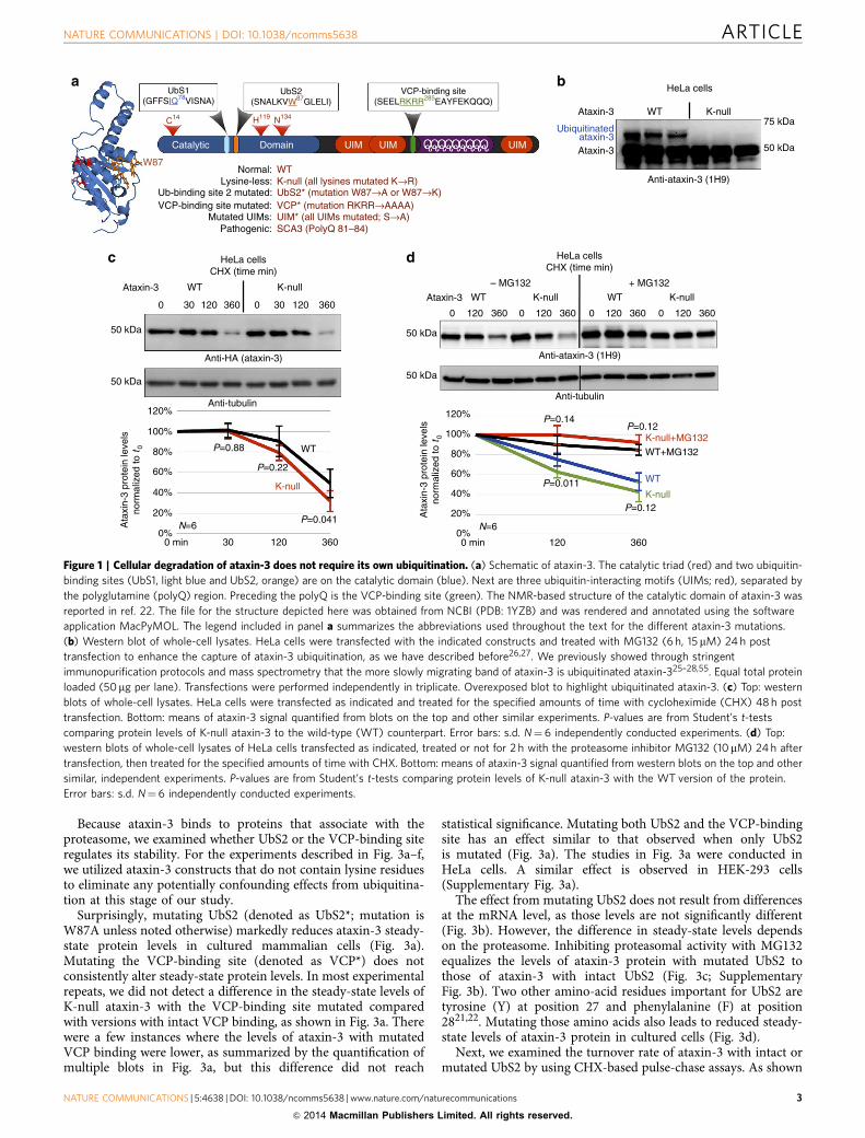
ResultsAtaxin-3 does not require ubiquitination to be degraded.Previous reports presented evidence that the turnover of normaland pathogenic ataxin-3 (Fig. 1a) depends on the proteasome1,and we showed that ataxin-3 protein is ubiquitinated inmammalian cells, in mouse brain and in Drosophila7,25–29. Onthe basis of these observations, one would expect ataxin-3 to bedegraded by becoming ubiquitinated and thus targeted for theproteasome. Yet, while the role of the proteasome in ataxin-3degradation is well supported1, it is not entirely clear thatataxin-3 needs to be ubiquitinated to be degraded.
To examine this possibility, we used a version of ataxin-3whose lysine residues are mutated into the similar but non-ubiquitinatable amino-acid arginine (K-null). We do not detectubiquitinated species with K-null ataxin-3 in cultured mamma-lian cells (Fig. 1b) or in Drosophila (Fig. 2b, as well as othersupportive data published by us7,27). Mutating all of the 15 lysineresidues of ataxin-3 into arginines does not affect its subcellularlocalization in mammalian cells, or disrupt its ability to cleaveubiquitin chains in reconstituted systems in vitro27. Lysine-lessataxin-3 retains some of its functions: similarly to the wild-type(WT) version of this DUB, the K-null variant is able to somewhat
suppress neurodegeneration in Drosophila, although this functioncannot be enhanced by its own ubiquitination to reach the fullprotective effect of ubiquitinatable ataxin-3 (ref. 7).
We first examined the turnover of K-null ataxin-3 proteincompared with its wild-type counterpart in cultured cells.Figure 1c summarizes cycloheximide (CHX)-based pulse-chasestudies, where we observe that the turnover of K-null ataxin-3 isnot slower than that of WT ataxin-3. In fact, based onquantification of data from multiple western blots, K-nullataxin-3 might be degraded slightly more quickly than thenormal version of this DUB in cells (Fig. 1c, 360min time point;Fig. 1d, 120min time point). Both WT and K-null ataxin-3proteins are stabilized in cells by the addition of the proteasomeinhibitor MG132 (Fig. 1d), a finding that is in accordance withprevious reports that ataxin-3 turnover in cells is proteasomedependent1,25.
Subsequently, we used the fruit fly Drosophila melanogaster toinvestigate whether K-null ataxin-3 accumulates in vivo in anintact organism. We utilized the Gal4–UAS system30 to expressataxin-3 constructs throughout the fly7. We generated flies thatexpress WT, K-null or K-117 (the lysine predominantlyubiquitinated in vitro and in mammalian cell culture27)ataxin-3 by using the ubiquitous Gal4 driver sqh-Gal4 (refs31,32). We compared fly lines that express the various ataxin-3transgenes at similar messenger RNA (mRNA) levels, accordingto quantitative reverse transcriptase-PCR (Fig. 2a). As shown inFig. 2b, K-null ataxin-3 protein does not accumulate comparedwith WT or K-117 ataxin-3. Together, results in Figs 1 and 2 leadus to conclude that ubiquitination of ataxin-3 is not absolutelynecessary for its degradation.
UbS2 regulates ataxin-3 protein levels and turnover in cells.Since ubiquitination of ataxin-3 does not appear to be necessaryfor its turnover (Figs 1 and 2), we wondered whether protein-binding domains of this DUB are involved in its degradation.Several domains on ataxin-3 enable its direct interaction withother proteins (Fig. 1a). Starting at the N terminus, first is UbS1on the catalytic domain. UbS1 binds ubiquitin and is necessaryfor the catalytic activity of ataxin-3 (ref. 23). Next is UbS2, whichbinds ubiquitin, Rad23A and Rad23B (refs 21,23). Rad23A/Bfacilitate substrate protein degradation by reversibly associatingwith the proteasome33,34. Previous structural work focusing onthe catalytic domain of ataxin-3 indicated that mutating theamino-acid tryptophan (W) at position 87 disrupts the ability ofUbS2 to interact with the isolated ubiquitin-like domain of Rad23(refs 21,23). Through an in vitro pull-down assay that utilizedrecombinant, full-length ataxin-3 and Rad23A, we confirmed thatmutating W87 into an alanine residue diminishes the ability ofthese two proteins to interact (Supplementary Fig. 1a). We alsoverified in mammalian cell culture that this mutation reducesthe capacity of an otherwise normal, full-length ataxin-3 toco-immunoprecipitate endogenous Rad23A and Rad23B(Supplementary Fig. 1b). Mutating UbS2 does not abrogate thecatalytic activity of ataxin-3 (ref. 23) or affect its subcellularlocalization (Supplementary Fig. 2). In the C-terminal half ofataxin-3 are three ubiquitin-interacting motifs (UIMs), whichbind ubiquitin chains at least four moieties long29. TheC-terminal portion also contains an arginine-rich region thatbinds the AAA ATPase protein valosin-containing protein (VCP)(also known as p97)35–37. VCP functions at least in part as aproteasomal shuttle protein33. Mutating one or more of thearginine residues of the VCP-binding site of full-length ataxin-3into histidines or alanines has been demonstrated to disrupt itsinteraction with VCP in reconstituted systems in vitro and inmammalian cells35–37.
ARTICLE NATURE COMMUNICATIONS | DOI: 10.1038/ncomms5638
2 NATURE COMMUNICATIONS | 5:4638 |DOI: 10.1038/ncomms5638 | www.nature.com/naturecommunications
& 2014 Macmillan Publishers Limited. All rights reserved.
Because ataxin-3 binds to proteins that associate with theproteasome, we examined whether UbS2 or the VCP-binding siteregulates its stability. For the experiments described in Fig. 3a–f,we utilized ataxin-3 constructs that do not contain lysine residuesto eliminate any potentially confounding effects from ubiquitina-tion at this stage of our study.
Surprisingly, mutating UbS2 (denoted as UbS2*; mutation isW87A unless noted otherwise) markedly reduces ataxin-3 steady-state protein levels in cultured mammalian cells (Fig. 3a).Mutating the VCP-binding site (denoted as VCP*) does notconsistently alter steady-state protein levels. In most experimentalrepeats, we did not detect a difference in the steady-state levels ofK-null ataxin-3 with the VCP-binding site mutated comparedwith versions with intact VCP binding, as shown in Fig. 3a. Therewere a few instances where the levels of ataxin-3 with mutatedVCP binding were lower, as summarized by the quantification ofmultiple blots in Fig. 3a, but this difference did not reach
statistical significance. Mutating both UbS2 and the VCP-bindingsite has an effect similar to that observed when only UbS2is mutated (Fig. 3a). The studies in Fig. 3a were conducted inHeLa cells. A similar effect is observed in HEK-293 cells(Supplementary Fig. 3a).
The effect from mutating UbS2 does not result from differencesat the mRNA level, as those levels are not significantly different(Fig. 3b). However, the difference in steady-state levels dependson the proteasome. Inhibiting proteasomal activity with MG132equalizes the levels of ataxin-3 protein with mutated UbS2 tothose of ataxin-3 with intact UbS2 (Fig. 3c; SupplementaryFig. 3b). Two other amino-acid residues important for UbS2 aretyrosine (Y) at position 27 and phenylalanine (F) at position2821,22. Mutating those amino acids also leads to reduced steady-state levels of ataxin-3 protein in cultured cells (Fig. 3d).
Next, we examined the turnover rate of ataxin-3 with intact ormutated UbS2 by using CHX-based pulse-chase assays. As shown
a bUbS1
(GFFSIQ78VISNA)
C14
Catalytic Domain
Normal:Lysine-less:
Ub-binding site 2 mutated:VCP-binding site mutated:
Mutated UIMs:Pathogenic:
WTK-null (all lysines mutated K→R)UbS2* (mutation W87→A or W87→K)VCP* (mutation RKRR→AAAA)UIM* (all UIMs mutated; S→A)SCA3 (PolyQ 81–84)
UIM UIM UIM
W87
H119 N134
UbS2(SNALKVW87GLELI)
VCP-binding site(SEELRKRR285EAYFEKQQQ)
Ataxin-3 WT
Anti-ataxin-3 (1H9)
K-null75 kDa
50 kDa
HeLa cells
Ubiquitinatedataxin-3Ataxin-3
c dHeLa cellsCHX (time min)
HeLa cellsCHX (time min)
WT
0 30 120 360 0 30 120 360
Anti-HA (ataxin-3)
Anti-tubulin
Anti-ataxin-3 (1H9)
Anti-tubulin
K-nullAtaxin-3
50 kDa
50 kDa
120%
100%
80%
60%
40%
Ata
xin-
3 pr
otei
n le
vels
norm
aliz
ed to
t0
20%
0%N=6
0 min 30 120
K-null
360
120%
100%
80%
60%
40%
Ata
xin-
3 pr
otei
n le
vels
norm
aliz
ed to
t0
20%
0%0 min 120 360
P=0.041
P=0.22
P=0.88 WT
50 kDa
50 kDa
WT
– MG132 + MG132
K-null WT K-nullAtaxin-3
0 120 360 0 120 360 0 120 360 0 120 360
N=6
P=0.12
P=0.011
P=0.12P=0.14
K-null+MG132
WT+MG132
WT
K-null
Figure 1 | Cellular degradation of ataxin-3 does not require its own ubiquitination. (a) Schematic of ataxin-3. The catalytic triad (red) and two ubiquitin-
binding sites (UbS1, light blue and UbS2, orange) are on the catalytic domain (blue). Next are three ubiquitin-interacting motifs (UIMs; red), separated by
the polyglutamine (polyQ) region. Preceding the polyQ is the VCP-binding site (green). The NMR-based structure of the catalytic domain of ataxin-3 was
reported in ref. 22. The file for the structure depicted here was obtained from NCBI (PDB: 1YZB) and was rendered and annotated using the software
application MacPyMOL. The legend included in panel a summarizes the abbreviations used throughout the text for the different ataxin-3 mutations.
(b) Western blot of whole-cell lysates. HeLa cells were transfected with the indicated constructs and treated with MG132 (6 h, 15 mM) 24 h post
transfection to enhance the capture of ataxin-3 ubiquitination, as we have described before26,27. We previously showed through stringent
immunopurification protocols and mass spectrometry that the more slowly migrating band of ataxin-3 is ubiquitinated ataxin-325–28,55. Equal total protein
loaded (50mg per lane). Transfections were performed independently in triplicate. Overexposed blot to highlight ubiquitinated ataxin-3. (c) Top: western
blots of whole-cell lysates. HeLa cells were transfected as indicated and treated for the specified amounts of time with cycloheximide (CHX) 48 h post
transfection. Bottom: means of ataxin-3 signal quantified from blots on the top and other similar experiments. P-values are from Student’s t-tests
comparing protein levels of K-null ataxin-3 to the wild-type (WT) counterpart. Error bars: s.d. N¼ 6 independently conducted experiments. (d) Top:
western blots of whole-cell lysates of HeLa cells transfected as indicated, treated or not for 2 h with the proteasome inhibitor MG132 (10mM) 24h after
transfection, then treated for the specified amounts of time with CHX. Bottom: means of ataxin-3 signal quantified from western blots on the top and other
similar, independent experiments. P-values are from Student’s t-tests comparing protein levels of K-null ataxin-3 with the WT version of the protein.
Error bars: s.d. N¼ 6 independently conducted experiments.
NATURE COMMUNICATIONS | DOI: 10.1038/ncomms5638 ARTICLE
NATURE COMMUNICATIONS | 5:4638 | DOI: 10.1038/ncomms5638 |www.nature.com/naturecommunications 3
& 2014 Macmillan Publishers Limited. All rights reserved.

in Fig. 3e, ataxin-3 protein with mutated UbS2 is turned overmarkedly more rapidly than ataxin-3 with intact UbS2. Mutatingonly the VCP-binding site does not significantly affect turnover(Fig. 3f). CHX-based analyses are consistent with steady-stateexaminations, indicating that UbS2 inhibits ataxin-3 degradationin cells, while VCP binding does not appear to have a significanteffect. On the basis of these findings, we focus the rest of thestudies in this report on UbS2.
As already stated, for experiments in Fig. 3a–f we used K-nullataxin-3 that contains intact or mutated UbS2. We nextinvestigated whether the protein levels of ataxin-3 with all of itslysines present are also regulated by UbS2. Mutating UbS2 in anotherwise normal ataxin-3 markedly reduces its steady-stateprotein levels (Fig. 3g) and turnover (Fig. 3h), similar to what weobserved with non-ubiquitinatable ataxin-3. Collectively, theseresults from mammalian cell culture indicate that UbS2 inhibitsthe proteasomal turnover of non-pathogenic ataxin-3.
UIMs of ataxin-3 have an opposite effect to UbS2 on turnover.For ataxin-3 to be degraded by the proteasome, it needs to comeinto contact with this cellular machinery. One possibility throughwhich this interaction could occur would be through ataxin-3ubiquitination. However, our results argue against ubiquitinationbeing necessary for ataxin-3 degradation (Figs 1 and 2). Anothermechanism that could bridge ataxin-3 to the proteasome could be
the poly-ubiquitin binding UIMs of this polyQ protein, whichcould bind ubiquitinated proteins targeted for the proteasome.
We mutated a conserved serine residue in each UIM of non-pathogenic ataxin-3 into an alanine, disrupting its interactionwith ubiquitin chains26,29,38. Results in Fig. 4 indicate that theUIMs regulate the turnover of ataxin-3. Mutating all three UIMscounteracts the degradative effect of UbS2 mutation both at thesteady-state level (Fig. 4a; Supplementary Fig. 4) and turnoverrate (Fig. 4b). We conclude that the UIMs of ataxin-3 have apositive effect on its cellular turnover.
Rad23 regulates ataxin-3 protein levels in mammalian cells.Two proteins that bind ataxin-3 directly at UbS2 are Rad23Aand Rad23B21,23. Therefore, we examined whether Rad23A/Baffect ataxin-3 protein levels in cultured cells by using RNAi.Knockdown of endogenous Rad23A or Rad23B through different,non-overlapping short interfering RNA constructs noticeablydecreases levels of Rad23A or Rad23B protein. Their reductionis concomitant with a statistically significant reduction in thelevels of endogenous ataxin-3 protein (Fig. 4c). Simultaneousknockdown of Rad23A and Rad23B has an even stronger effect inreducing normal, endogenous ataxin-3 protein levels (Fig. 4c).Together, these RNAi-based data are indicative of a protectiverole from Rad23A and Rad23B on ataxin-3.
UbS2 regulates normal ataxin-3 protein levels in Drosophila.Data in Figs 3 and 4 make the case that UbS2 of ataxin-3 isimportant for its stability in cultured cells. Next, we examined thesignificance of UbS2 in ataxin-3 protein levels in Drosophila. Wegenerated new transgenic fly lines that express non-pathogenicataxin-3 with mutated UbS2 and selected lines that express WTand UbS2* ataxin-3 transgenes at similar mRNA levels, as well asones that express the UbS2* variant more strongly (Fig. 5a). Asshown in Fig. 5b, the protein levels of UbS2* ataxin-3 aremarkedly lower than those of WT ataxin-3 that is expressedsimilarly at the mRNA level. Even in transgenic lines where themRNA levels of UbS2* ataxin-3 are approximately sixfold higherthan WT ataxin-3, those protein levels do not quite approach thelevels of normal ataxin-3. These data from an intact organism areconsistent with our findings from cultured cells, and collectivelysupport our overall conclusion that UbS2 regulates ataxin-3protein levels by inhibiting its degradation.
UbS2 and Rad23 regulate pathogenic ataxin-3 in Drosophila.Finally, we examined whether mutating UbS2 on pathogenicataxin-3 affects its stability (pathogenic variant denoted as SCA3;polyQ length of 81 for the intact version and 84 for UbS2*;these constructs contains all lysine residues). We found thatmutating UbS2 of SCA3-causing ataxin-3 reduces its steady-statelevels in cultured mammalian cells (Fig. 6a). Just as we observedwith normal ataxin-3 (Fig. 3g,h), mutating UbS2 enhances theturnover rate of pathogenic ataxin-3 in cells (Fig. 6b). Also,proteasomal inhibition diminishes the marked difference inprotein levels between ataxin-3 versions that have an intact ormutated UbS2 (Fig. 6c). Together, these data indicate that theproteasomal turnover of pathogenic ataxin-3 in cells is alsoregulated by UbS2.
Because knockdown of Rad23A/B leads to lower levels ofataxin-3 protein in cultured cells (Fig. 4c), we reasoned thatreducing Rad23 levels in Drosophila should suppress degenera-tion caused by its pathogenic version by lowering the levelsof the polyQ protein. Whereas humans have separate genesfor Rad23A and Rad23B, flies appear to have one gene forRad23 (refs 21,23,24,39).
We utilized a transgenic fly line generated by the Bonini labthat expresses a full-length version of pathogenic ataxin-3 with 84
Drosophila melanogaster
Drosophila melanogaster
a
b
Ata
xin-
3 m
RN
Ano
rmal
ized
to W
T-1 167%
200%
133%
100%
67%
33%
0%
Ubiquitinated
WT
-1
K-n
ull-4
K-1
17-1
WT
-3
K-n
ull-3
K-1
17-3
WT
-1
K-n
ull-4
K-1
17-1
WT
-3
K-n
ull-3
K-1
17-3
ataxin-3
Ubiquitinatedataxin-3Ataxin-3
Exposure ShorterAnti-ataxin-3 (1H9)
Anti-tubulin
50 kDa
Longer
75 kDa
50 kDa
WT
-1
WT
-2
WT
-3
WT
-4
K-n
ull-1
K-n
ull-2
K-n
ull-3
K-n
ull-4
K-1
17-1
K-1
17-2
K-1
17-3
K-1
17-4
Figure 2 | Lysine-less ataxin-3 does not accumulate in vivo in Drosophila.
(a) Selection of transgenic fly lines that express similar levels of ataxin-3
variants. Quantitative reverse transcriptase (qRT)–PCR results from whole
flies expressing the versions of UAS-ataxin-3 noted in the panel. At least
five flies were used per genotype per experiment. Driver was sqh-Gal4
(ubiquitous expression31,32). All flies were heterozygous for UAS-ataxin-3
and sqh-Gal4 transgenes. Red and blue arrows: lines that we chose for
western blotting shown in panel b. Experiment performed independently in
triplicate. Shown are mean ataxin-3 mRNA levels normalized to WT-1. Error
bars: s.d. Flies were 1–3 days old. (b) Western blots from whole-fly lysates
expressing the indicated UAS-ataxin-3 constructs based on qRT-PCR
data from panel a. Driver was sqh-Gal4. All flies were heterozygous for
UAS-ataxin-3 and sqh-Gal4 transgenes, as in panel a. Ten or more flies
per genotype were homogenized. Blots are representative of experiments
performed independently in triplicate, with similar results. Flies were 1–3
days old. WT, wild-type.
ARTICLE NATURE COMMUNICATIONS | DOI: 10.1038/ncomms5638
4 NATURE COMMUNICATIONS | 5:4638 |DOI: 10.1038/ncomms5638 | www.nature.com/naturecommunications
& 2014 Macmillan Publishers Limited. All rights reserved.

polyQ repeats and has intact UbS2, VCP-binding site and UIMs6.First, we examined ataxin-3-SCA3 protein levels in fly eyes whenRad23 levels are reduced by either knocking down this genethrough RNAi, or by using a chromosomal deletion in thegenomic area that contains Rad23 (line Df(4)ED6369). Thisdeficiency deletes chromosomal segment 102A1–102C1, whichincludes Rad23 at locus 102B3; these flies are hemizygous viable.
As shown in western blots in Fig. 6d, knocking down Rad23 orremoving one copy of this gene leads to lower levels of ataxin-3in Drosophila eyes. Knocking down other proteins related toubiquitin (ubiquilin, nedd8 or SUMO, all of which reportedlyinteract with ataxin-3 (ref. 1)) does not reduce ataxin-3 proteinlevels in vivo (Supplementary Fig. 5), indicating a specific role forRad23 in regulating ataxin-3.
HeLa cells– MG132 + MG132
K-null
0 30 120 360 0 30 120 360
HeLa cells
CHX (time min)K-null-UbS2*
HeLa cells
Ataxin-3
K-n
ull
K-n
ull-U
bS2*
K-n
ull-V
CP
*
K-n
ull-U
bS2*
-VC
P*
Ataxin-3
Ataxin-3
50 kDa
37 kDa
K-n
ull-U
bS2*
K-n
ull
K-n
ull-U
bS2*
-VC
P*
K-n
ull-U
bS2*
K-n
ull
K-n
ull-U
bS2*
-VC
P*
50 kDa
37 kDa
Ata
xin-
3 pr
otei
n le
vels
norm
aliz
ed to
K-n
ull
Ata
xin-
3 pr
otei
n le
vels
norm
aliz
ed to
K-n
ull
Ata
xin-
3 pr
otei
n le
vels
norm
aliz
ed to
t0
Anti-ataxin-3 (1H9)
Anti-GAPDH
Anti-ataxin-3 (1H9)
Anti-GAPDH120%100%80%60%40%20%
0%
0%25%50%75%
100%125%
100%90%80%70%60%50%40%30%20%10%
0%0 min 30 120
P<0.0001P<0.0001
P<0.0001
K-null-UbS2*
K-null-VCP*
K-null
N=8
360
Ata
xin-
3 pr
otei
n le
vels
norm
aliz
ed to
t0
100%90%80%70%60%50%40%30%20%10%0%
100%90%80%70%
60%50%
40%30%20%10%
0%
0 min 30 120 360
K-n
ull
K-n
ull-U
bS2*
K-n
ull-V
CP
*
K-n
ull-U
bS2*
-VC
P*
K-n
ull
K-n
ull-U
bS2*
K-n
ull-U
bS2*
-VC
P*
P=0.050
P<
0.00
01
P<
0.00
01
N=12
N=5
a c e f
+ MG132
P=0.175
P=0.447
50 kDa
50 kDa
Anti-HA (Ataxin-3)
Anti-tubulin
K-null0 30 120 360 0 30 120 360
HeLa cells
CHX (time min)
K-null-VCP*Ataxin-3
50 kDa
50 kDa
Anti-HA (Ataxin-3)
Anti-tubulin
N=3
P=0.16
P=0.35
P=0.39
K-null
HeLa cells HeLa cells HeLa cells
128%114%100%85%71%57%43%28%
mR
NA
rel
ativ
e to
K-n
ull
14%0%
b d g hP=0.98
P=0.22
K-n
ull
K-n
ull-U
bS2*
K-n
ull-U
bS2*
-VC
P*
Ataxin-3
Ataxin-3
50 kDa
37 kDaAnti-ataxin-3 (1H9)
Anti-GAPDH
WT W87A W87K
N=8 N=6
K-n
ull
WT
UbS
2*(W
87A
)
UbS
2*(W
87K
)
K-n
ull-W
87A
K-n
ull-Y
27F
28-A
K-n
ull-Y
27F
28-R
50 kDa
50 kDa
Anti-HA (Ataxin-3)
Anti-tubulin
Ataxin-3 WT
0 30 120 360 0 30 120 360
UbS2*
50 kDa
50 kDa
HeLa cells
CHX (time min)
Anti-ataxin-3 (1H9)
Anti-tubulin
100%
75%
50%
25%
0%
Ata
xin-
3 pr
otei
n le
vels
norm
aliz
ed to
K-n
ull 100%
75%
50%
25%
0%Ata
xin-
3 pr
otei
n le
vels
norm
aliz
ed to
WT
K-n
ull
K-n
ull-W
87A
K-n
ull-Y
27F
28-A
K-n
ull-Y
27F
28-R
P<
0.00
01
P<
0.00
01
P<
0.00
01
P<
0.00
01
P<
0.00
01
Ata
xin-
3 pr
otei
n le
vels
norm
aliz
ed to
t0
0 min 30
N=8P<0.001
P<0.001
P<0.0001WT
UbS2*
120 360
Figure 3 | UbS2 of ataxin-3 regulates its degradation in cells. (a) Top: western blots of whole-cell lysates. HeLa cells were transfected as indicated and
harvested 24 h later. Bottom: means of ataxin-3 signal quantified from blots on the top and other similar experiments. Error bars: s.d. (b) Quantitative
reverse transcriptase-PCR of HeLa cells expressing the indicated constructs. Endogenous control: GAPDH. N¼ 3 independently conducted experiments.
Shown are mean ataxin-3 mRNA levels ±s.d. (c,d) Top: western blots of whole-cell lysates of HeLa cells transfected as indicated, treated or not with
MG132 (4 h, 15 mM) 48h later and harvested. Bottom: means of ataxin-3 signal quantified from blots on the top and other similar experiments. Error bars:
s.d. (e,f) Top: HeLa cells were transfected as indicated and 24h later were treated with CHX for the specified amounts of time. For panel e, to have
comparable protein amounts of both ataxin-3 versions at time 0min, we transfected three times more K-null-UbS2* construct than K-null, which was
supplemented with empty vector to equate total DNA per group. Western blots of whole-cell lysates. Bottom: means of ataxin-3 signal quantified from
blots on the top and other independent experiments. Error bars: s.d. (g) Top: western blots of whole-cell lysates. Two different mutations were used to
disrupt UbS2: W87A and W87K, with similar results. Bottom: means of ataxin-3 signal quantified from blots on the top and other independent
experiments. Error bars: s.d. (h) Top: HeLa cells were transfected as indicated and 24 h later were treated with CHX for the specified time points.
Three times more ataxin-3-UbS2* construct was transfected than ataxin-3-WT to have comparable protein levels at time 0min. Bottom: means of ataxin-3
signal quantified from blots on the top and other independent experiments. Error bars: s.d. For panels a–d and g, P-values are from analysis of variance with
Tukey’s post hoc correction comparing the various versions of ataxin-3 with their panel controls. For panels e,f and h, P-values are from Student’s t-tests.
N of independently repeated experiments is specified in panels.
NATURE COMMUNICATIONS | DOI: 10.1038/ncomms5638 ARTICLE
NATURE COMMUNICATIONS | 5:4638 | DOI: 10.1038/ncomms5638 |www.nature.com/naturecommunications 5
& 2014 Macmillan Publishers Limited. All rights reserved.

a b cHeLa cells HeLa cells HeLa cells
CHX (time h)
Ataxin-3
Ataxin-3-K-null
Anti-ataxin-3 (1H9)
Anti-GAPDH
Ata
xin-
3 pr
otei
n le
vels
norm
aliz
ed to
K-n
ull
Ata
xin-
3 pr
otei
n le
vels
norm
aliz
ed to
t 0
Ata
xin-
3 pr
otei
n le
vels
norm
aliz
ed to
siR
NA
con
trol
N=10120%
100%
75%
50%
25%
0%N=6
P<0.0001
UbS2*
UbS2*-UIM*
P<0.001
0 h 2 6
120%
** **** **
** ** **
100%
80%
60%
40%
20%
0%
siR
NA
1-R
ad23
A
siR
NA
2-R
ad23
A
siR
NA
-con
trol
siR
NA
-con
trol
100%80%60%40%20%0%
P=
0.63
8
P<
0.00
01
Anti-ataxin-3 (1H9)
Anti-ataxin-3 (1H9)
Anti-Rad23A
Anti-Rad23B
Anti-GAPDH
N=7
Anti-tubulin
50 kDa
37 kDa
K-n
ull
K-n
ull-U
bS2*
K-n
ull-U
bS2*
-UIM
*
50 kDa
0 2
UbS2* UbS2*-UIM*
6 0 2 6
50 kDa
50 kDa
50 kDa
75 kDa
50 kDa
37 kDa
K-n
ull
K-n
ull-U
bS2*
K-n
ull-U
bS2*
-UIM
*
siR
NA
1-R
ad23
B
siR
NA
-Rad
23A
+B
siR
NA
-Rad
23A
+B
siR
NA
-Rad
23A
+B
siR
NA
2-R
ad23
B
siR
NA
1-R
ad23
A
siR
NA
2-R
ad23
A
siR
NA
-con
trol
siR
NA
-con
trol
siR
NA
1-R
ad23
B
siR
NA
-Rad
23A
+B
siR
NA
-Rad
23A
+B
siR
NA
-Rad
23A
+B
siR
NA
2-R
ad23
B
Figure 4 | UIMs of ataxin-3 oppose the effect of UbS2 mutation. (a) Top: HeLa cells were transfected as indicated and harvested 48 h later. Western blots
from whole-cell lysates. Bottom: means of ataxin-3 signal quantified from blots on the top and other similar experiments. P-values are from analysis of
variance (ANOVA) with Tukey’s post hoc correction comparing K-null ataxin-3 with UbS2 mutated, and K-null ataxin-3 with UbS2 and UIMs mutated
to K-null ataxin-3 with intact domains. Error bars: s.d. N¼ 10 independently conducted experiments. (b) Top: HeLa cells were transfected with the indicated
constructs. Three times more UbS2* DNA was used than UbS2*-UIM* to begin with approximately the same amount of protein at time 0h. CHXwas added
to cells 24 h post transfection for the specified time points. Western blots of whole-cell lysates. Bottom: means of ataxin-3 signal quantified from blots on
the top and other similar experiments. P-values are from Student’s t-tests comparing ataxin-3 with UbS2 mutated with ataxin-3 with UbS2 and UIMs
mutated. Error bars: s.d. N¼ 6 independently conducted experiments. (c) Top: HeLa cells were transfected with the indicated short interfering RNA (siRNA)
constructs to knock down endogenous Rad23A, endogenous Rad23B or both, and harvested 48 h later. Shown are western blots of whole-cell lysates.
siRNA control: scramble controls. Bottom: means of ataxin-3 signal quantified from blots on the top and other similar experiments. P-values of o0.01 are
indicated by ‘**’, and are from ANOVA/Tukey comparing the levels of ataxin-3 protein in RNAi lanes with those in scramble control. Error bars: s.d. N¼ 7
independently conducted experiments.
Drosophila melanogaster
Drosophila melanogaster
SimilarmRNA levels
HigherUbS2* mRNA levels
a b
150% N=3
P<
0.00
01 P=
0.01
3
P=
0.01
1
P=
0.84
4 P=
0.04
4
125%
100%
75%
50%
25%
0%50 kDa
WT
-3
UbS
2*-2
WT
-1
WT
-2
UbS
2*-1
UbS
2*-3
UbS
2*-4
Anti-ataxin-3 (1H9)
Anti-tubulin
50 kDa
Ata
xin-
3 m
RN
A le
vels
norm
aliz
ed to
WT
-1
800%
700%
600%
500%
400%
300%
200%
100%
0%
WT
-1
WT
-2
WT
-3
UbS
2*-1
UbS
2*-2
UbS
2*-3
UbS
2*-4
Nor
mal
ized
ata
xin-
3pr
otei
n le
vels
SimilarmRNA levels
HigherUbS2* mRNA levels
WT
-3
UbS
2*-2
WT
-1
WT
-2
UbS
2*-1
UbS
2*-3
UbS
2*-4
Figure 5 | UbS2 mutation leads to reduced ataxin-3 protein levels in Drosophila. (a) Quantitative reverse transcriptase (qRT)-PCR results from
whole flies expressing the noted versions of UAS-ataxin-3 driven by sqh-Gal4. All flies were heterozygous for UAS-ataxin-3 and sqh-Gal4 transgenes.
Red arrows: wild-type (WT) and UbS2* lines that have comparable ataxin-3 mRNA levels. Blue arrows: UbS2* lines that have markedly higher ataxin-3
mRNA levels than WTversions. Experiment performed independently in triplicate, utilizing at least five flies per genotype per experiment. Shown are mean
ataxin-3 mRNA levels normalized to WT-1. Error bars: s.d. (b) Left: western blots from whole flies based on qRT–PCR results from a. At least five flies were
homogenized per genotype. Driver was sqh-Gal4. All flies were heterozygous for UAS-ataxin-3 and sqh-Gal4 transgenes, as in a. Note that for this blot 1.5
times more lysate was loaded for the line that expresses UbS2*-2 to enable visualization of ataxin-3 protein in this line by western blotting without
saturating the signal from other lysates. Right: means of ataxin-3 signal quantified from blots on the left and other independent experiments. P-values are
from the Student’s t-test (WT-3 and UbS2*-2) and analysis of variance/Tukey (the other lines). Error bars: s.d. N¼ 3 independently conducted
experiments. Flies were 1–3 days old.
ARTICLE NATURE COMMUNICATIONS | DOI: 10.1038/ncomms5638
6 NATURE COMMUNICATIONS | 5:4638 |DOI: 10.1038/ncomms5638 | www.nature.com/naturecommunications
& 2014 Macmillan Publishers Limited. All rights reserved.
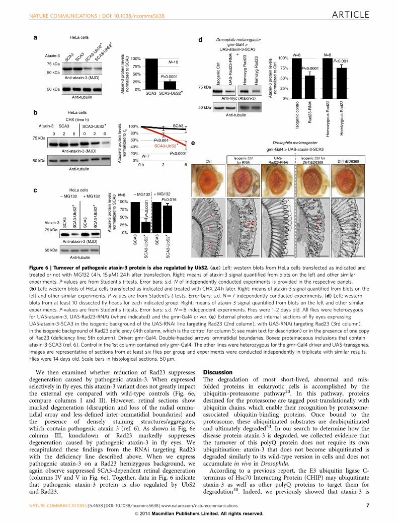
We then examined whether reduction of Rad23 suppressesdegeneration caused by pathogenic ataxin-3. When expressedselectively in fly eyes, this ataxin-3 variant does not greatly impactthe external eye compared with wild-type controls (Fig. 6e,compare columns I and II). However, retinal sections showmarked degeneration (disruption and loss of the radial omma-tidial array and less-defined inter-ommatidial boundaries) andthe presence of densely staining structures/aggregates,which contain pathogenic ataxin-3 (ref. 6). As shown in Fig. 6ecolumn III, knockdown of Rad23 markedly suppressesdegeneration caused by pathogenic ataxin-3 in fly eyes. Werecapitulated these findings from the RNAi targeting Rad23with the deficiency line described above. When we expresspathogenic ataxin-3 on a Rad23 hemizygous background, weagain observe suppressed SCA3-dependent retinal degeneration(columns IV and V in Fig. 6e). Together, data in Fig. 6 indicatethat pathogenic ataxin-3 protein is also regulated by UbS2and Rad23.
DiscussionThe degradation of most short-lived, abnormal and mis-folded proteins in eukaryotic cells is accomplished by theubiquitin–proteasome pathway20. In this pathway, proteinsdestined for the proteasome are tagged post-translationally withubiquitin chains, which enable their recognition by proteasome-associated ubiquitin-binding proteins. Once bound to theproteasome, these ubiquitinated substrates are deubiquitinatedand ultimately degraded20. In our search to determine how thedisease protein ataxin-3 is degraded, we collected evidence thatthe turnover of this polyQ protein does not require its ownubiquitination: ataxin-3 that does not become ubiquitinated isdegraded similarly to its wild-type version in cells and does notaccumulate in vivo in Drosophila.
According to a previous report, the E3 ubiquitin ligase C-terminus of Hsc70 Interacting Protein (CHIP) may ubiquitinateataxin-3 as well as other polyQ proteins to target them fordegradation40. Indeed, we previously showed that ataxin-3 is
HeLa cells
SCA3SCA3
SCA3-UbS
2*
SCA3-UbS
2*
75 kDa
50 kDa
50 kDa
Anti-ataxin-3 (MJD)
Anti-tubulin
Ataxin-3100%
75%
50%
25%
0%SCA3
P<0.0001
N=10
Ata
xin-
3 pr
otei
n le
vels
norm
aliz
ed to
SC
A3
SCA3-UbS2*Anti-myc (Ataxin-3)
Isog
enic
Ctr
l
UA
S-R
ad23
-RN
Ai
Hom
ozyg
Rad
23
Hem
izyg
Rad
23
Drosophila melanogastergmr-Gal4 >
UAS-ataxin-3-SCA3+
75 kDa
50 kDa
Anti-tubulin
100%
75%
50%
25%
0%Ata
xin-
3 pr
otei
n le
vels
norm
aliz
ed to
Ctr
l
Isog
enic
con
trol
Rad
23-R
NA
i
Hom
ozyg
ous
Rad
23
Hem
izyg
ous
Rad
23
N=8
P<0.0001
P<0.001
N=8
HeLa cells
CHX (time h)
SCA3
0 2 6 0 2 6
SCA3-UbS2*Ataxin-3
75 kDa
50 kDa
Anti-tubulin
Anti-ataxin-3 (MJD)
100%
80%
60%
40%
20%
0%0 h 2 6
P<0.0001
P<0.001
SCA3
N=7
SCA3-UbS2*
Ata
xin-
3 pr
otei
n le
vels
norm
aliz
ed to
t 0
Drosophila melanogaster
gmr-Gal4 > UAS-ataxin-3-SCA3+
CtrlIsogenic Ctrl
for RNAiUAS-
Rad23-RNAiIsogenic Ctrl for
Df(4)ED6369 Df(4)ED6369
HeLa cells
– MG132 + MG132
SC
A3
SC
A3-
UbS
2*
SC
A3
SC
A3-
UbS
2 *
Ataxin-3
75 kDa
Anti-ataxin-3 (MJD)
Anti-tubulin
50 kDa
– MG132 + MG132
Ata
xin-
3 pr
otei
n le
vels
norm
aliz
ed to
SC
A3
SC
A3
SC
A3-
UbS
2*
SC
A3
SC
A3-
UbS
2 *
N=6
100%
75%
50%
25%
0%
P=0.016
P<
0.00
01
a
b
c
d
e
Figure 6 | Turnover of pathogenic ataxin-3 protein is also regulated by UbS2. (a,c) Left: western blots from HeLa cells transfected as indicated and
treated or not with MG132 (4 h, 15 mM) 24 h after transfection. Right: means of ataxin-3 signal quantified from blots on the left and other similar
experiments. P-values are from Student’s t-tests. Error bars: s.d. N of independently conducted experiments is provided in the respective panels.
(b) Left: western blots of HeLa cells transfected as indicated and treated with CHX 24 h later. Right: means of ataxin-3 signal quantified from blots on the
left and other similar experiments. P-values are from Student’s t-tests. Error bars: s.d. N¼ 7 independently conducted experiments. (d) Left: western
blots from at least 10 dissected fly heads for each indicated group. Right: means of ataxin-3 signal quantified from blots on the left and other similar
experiments. P-values are from Student’s t-tests. Error bars: s.d. N¼ 8 independent experiments. Flies were 1–2 days old. All flies were heterozygous
for UAS-ataxin-3, UAS-Rad23-RNAi (where indicated) and the gmr-Gal4 driver. (e) External photos and internal sections of fly eyes expressing
UAS-ataxin-3-SCA3 in the isogenic background of the UAS-RNAi line targeting Rad23 (2nd column), with UAS-RNAi targeting Rad23 (3rd column),
in the isogenic background of Rad23 deficiency (4th column, which is the control for column 5; see main text for description) or in the presence of one copy
of Rad23 (deficiency line; 5th column). Driver: gmr-Gal4. Double-headed arrows: ommatidial boundaries. Boxes: proteinaceous inclusions that contain
ataxin-3-SCA3 (ref. 6). Control in the 1st column contained only gmr-Gal4. The other lines were heterozygous for the gmr-Gal4 driver and UAS-transgenes.
Images are representative of sections from at least six flies per group and experiments were conducted independently in triplicate with similar results.
Flies were 14 days old. Scale bars in histological sections, 50mm.
NATURE COMMUNICATIONS | DOI: 10.1038/ncomms5638 ARTICLE
NATURE COMMUNICATIONS | 5:4638 | DOI: 10.1038/ncomms5638 |www.nature.com/naturecommunications 7
& 2014 Macmillan Publishers Limited. All rights reserved.
ubiquitinated in mammalian cells and in Drosophila7,26–28, andthat ubiquitination directly and markedly enhances its DUBactivity7,26–28. However, recent work indicated that CHIPubiquitinates ataxin-3 to modify its catalytic activity during theprocess of substrate ubiquitination, where ataxin-3 and CHIPcooperate to enhance CHIP substrate degradation26–28. Inaddition, CHIP knockout mice do not show accumulation ofataxin-3 protein, and knockdown of CHIP in Drosophila does notdetectably impact ataxin-3 protein levels7. On the basis of thesepreviously published findings and our present results, we proposethat ubiquitination of ataxin-3 serves to regulate its DUBfunctions, rather than directly dictate its proteasomal turnover.This is not to say that ubiquitination of ataxin-3 might notenhance its turnover under some circumstances. What our resultsindicate is that ataxin-3 does not need to be ubiquitinated to bedegraded. Other proteins have been reported to be degraded bythe proteasome in the absence of their own ubiquitination,including p21/CiP1 (ref. 41), calmodulin42, thymidylatesynthase43, tau44 and so on45.
We found that ataxin-3 degradation is regulated by its UbS2.Mutation of this domain accelerates the turnover of ataxin-3 incells and leads to markedly lower levels of this protein in vivo.The general structural organization of the catalytic domain ofataxin-3 appears to not be impacted by mutating residue W87 ofUbS2, according to previously published nuclear magneticresonance (NMR) studies21,23. The localization of full-lengthataxin-3-UbS2* in cultured mammalian cells is not different fromversions of this protein with intact UbS2. Full-length ataxin-3with mutated UbS2 is capable of cleaving ubiquitin chainsin vitro23, and this site is dispensable for the ubiquitination-dependent activation of ataxin-3 that we have reportedpreviously23,26,27. On the basis of these findings, mutating UbS2does not detrimentally impact ataxin-3’s folding and localization,or abrogate its basic catalytic activities.
UbS2 binds directly to Rad23A/B21–23. Knockdown of Rad23Aand/or B in cultured cells leads to significantly lower levels ofataxin-3 protein. Importantly, Rad23 knockdown in Drosophilaleads to lower levels of pathogenic ataxin-3 and amelioratesretinal degeneration caused by this protein. On the basis of our
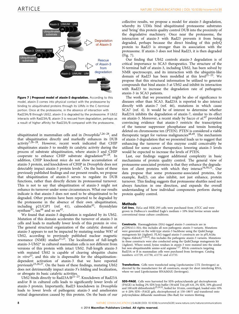
collective results, we propose a model for ataxin-3 degradation,whereby its UIMs bind ubiquitinated proteasome substratesand ‘bring’ this protein quality control DUB into the proximity ofthe degradative machinery. Once near the proteasome, theinteraction of ataxin-3 with Rad23 prevents it from beingdegraded, perhaps because the direct binding of this polyQprotein to Rad23 is stronger than its association with theproteasome. If ataxin-3 does not bind Rad23, it is then degraded(Fig. 7).
Our finding that UbS2 controls ataxin-3 degradation is ofcritical importance to SCA3 therapeutics. The structure of theN-terminal half of ataxin-3, including UbS2, has been solved byNMR spectroscopy, and its interaction with the ubiquitin-likedomain of Rad23 has been modelled at this level21–23. Wepropose that this structural information be utilized to generatecompounds that bind ataxin-3 at UbS2 and inhibit its interactionwith Rad23 to increase the degradation rate of pathogenicataxin-3 in SCA3 patients.
The work that we presented might be also of significance todiseases other than SCA3. Rad23A is reported to also interactdirectly with ataxin-7 (ref. 46), mutations in which causeSCA7 (ref. 4). It would be of interest to determine whetherRad23A inhibits the degradation of ataxin-7, similar to its effecton ataxin-3. Moreover, a recent study by Sacco et al.47 providedconvincing evidence that ataxin-3 restricts the transcriptionof the tumour suppressor phosphatase and tensin homologdeleted on chromosome ten (PTEN). PTEN is considered a viabletherapeutic target for various malignancies48,49. The mechanismof ataxin-3 degradation that we presented leads us to suggest thatenhancing the turnover of this enzyme could conceivably beutilized for some cancer therapeutics: lowering ataxin-3 levelswould be expected to increase the levels of PTEN.
Last, our findings suggest additional complexity in basicmechanisms of protein quality control. The general view ofproteasome-associated proteins is that they enhance the degrada-tion of client proteins with which they interact34,50,51. Ourdata propose that some proteasome-associated proteins, forexample, Rad23, can also inhibit, not just enhance, proteinturnover. This finding suggests that proteasome shuttles need notalways function in one direction, and expands the overallunderstanding of how individual components perform duringprotein quality control.
MethodsCell lines. HeLa and HEK-293 cells were purchased from ATCC and weregrown in Dulbecco’s modified Eagle’s mediumþ 10% fetal bovine serum underconventional tissue culture conditions.
Constructs. Haemagglutinin (HA)-tagged ataxin-3 constructs are inpCDNA3.1-HA; this includes all non-pathogenic ataxin-3 variants. Mutationswere generated on the wild-type ataxin-3 backbone using the QuikChangemutagenesis kit (Agilent). FLAG-tagged ataxin-3 constructs are in pFLAG6a(Sigma-Aldrich)25,26,29; this includes the pathogenic ataxin-3 variants. Mutationsin these constructs were also conducted using the QuikChange mutagenesis kit(Agilent). Where noted, lysine residues in ataxin-3 were mutated into the similarbut non-ubiquitinatable amino-acid arginine7,27. RNAi constructs targetingRad23A or B in mammalian cells were purchased from Invitrogen. Catalognumbers: s11729, s11730, s11731 and s11733.
Transfections. Cells were transfected using Lipofectamine LTX (Invitrogen) asdirected by the manufacturer for all constructs, except for short interfering RNA,where we used Lipofectamine RNAiMAX (Invitrogen).
SDS–PAGE. Cells were harvested for SDS–polyacrylamide gel electrophoresis(PAGE) in boiling 2% SDS lysis buffer (50mM Tris pH 6.8, 2% SDS, 10% glyceroland 100mM dithiothreitol)25,29,52, boiled for 10min, centrifuged, loaded onto 10%or 4–20% SDS—PAGE gels, electrophoresed at 150–180V and transferred ontopolyvinylidene difluoride membrane (Bio-Rad) for western blotting.
Ataxin-3
Ubiquitinated substrate
Ub
Rad23
Ataxin-3
Rescueof Ataxin-3
fromdegradation
Ataxin-3
UIMUIM UIM
UIMUIM UIM
UIMUIM UIM
Degradationof Ataxin-3
Ubiquitinated substrate
Ub
Figure 7 | Proposed model of ataxin-3 degradation. According to this
model, ataxin-3 comes into physical contact with the proteasome by
binding to ubiquitinated proteins through its UIMs in the C-terminal
portion. Once at the proteasome, in the absence of interaction with
Rad23A/B through UbS2, ataxin-3 is degraded by the proteasome. If UbS2
interacts with Rad23A/B, ataxin-3 is rescued from degradation, perhaps as
a result of higher affinity for Rad23A/B compared with the proteasome.
ARTICLE NATURE COMMUNICATIONS | DOI: 10.1038/ncomms5638
8 NATURE COMMUNICATIONS | 5:4638 |DOI: 10.1038/ncomms5638 | www.nature.com/naturecommunications
& 2014 Macmillan Publishers Limited. All rights reserved.

Antibodies. Anti-ataxin-3 (MJD; rabbit polyclonal, 1:15,000 (ref. 53)), anti-ataxin-3 monoclonal (clone 1H9, mouse, 1:500-1:1,000; Millipore), anti-HA (rabbitpolyclonal Y11, 1:500; Santa Cruz Biotech), anti-tubulin (mouse monoclonalT5168, 1:10,000; Sigma-Aldrich), anti-GAPDH (mouse monoclonal MAB374,1:500; Millipore), anti-Rad23A (rabbit polyclonal YH62308, 1:5,000; Origene),anti-Rad23B (rabbit polyclonal A302-306A, 1:2,000; Bethyl Labs), peroxidase-conjugated secondary antibodies (goat anti-rabbit and goat anti-mouse, 1:5,000;Jackson Immunoresearch).
Western blotting and signal quantification. Western blots were imaged with acharge-coupled device-equipped VersaDoc 5000MP system (Bio-Rad)26,27,29.Quantification of signals from sub-saturated blots was conducted in the QuantityOne software (Bio-Rad) with universal background subtraction. Signal from theprotein of interest was normalized to its own loading control (tubulin or GAPDH).Experimental lanes were normalized to respective controls. Student’s t-tests withtwo tails and analysis of variance with Tukey’s post hoc correction were used forstatistical comparisons, as specified in the figure legends. Full western blots forimages in Figs 1–6 are shown in Supplementary Fig. 6.
Chemicals. CHX was purchased from A.G. Scientific, dissolved in ultra-pure waterand used at final concentration of 50 mgml� 1. MG132 was purchased from A.G.Scientific, dissolved in DMSO and utilized at final concentrations specified in figurelegends.
Drosophila lines. Common stocks and the line expressing pathogenic ataxin-3(stock number 33610 with genotype w[1118]; P{w[þmC]¼UAS-SCA3.fl-Q84.myc}7.2/MKRS) were purchased from the Bloomington Drosophila StockCenter. The UAS-RNAi line targeting Rad23 and its isogenic background werepurchased from the Vienna Drosophila RNAi Center (transformants 30498 and60000). The Rad23 deficiency line was purchased from Bloomington DrosophilaStock Center (stock number: 9422 with genotype w[1118]; Df(4)ED6369,P{w[þmW.Scer\FRT.hs3]¼ 3’.RS5þ 3.3’}ED6369/l(4)102EFf[1]). Non-pathogenic UAS-ataxin-3 flies were generated by us, using ataxin-3 with a polyQrepeat of 22 residues cloned into the pUASt vector. Injections were done by theDuke University Model System Genomics into the w1118 background7. All fly lineswere on a w[-] background. Flies were maintained in a diurnally controlled 25 �Cenvironment atB60% humidity. Whole flies or dissected heads were homogenizedin boiling SDS lysis buffer (50mM Tris pH 6.8, 2% SDS, 10% glycerol and 100mMdithiothreitol), sonicated, boiled for 10min, centrifuged to remove debris, loadedonto SDS–PAGE gels, electrophoresed and transferred onto polyvinylidenedifluoride membrane for western blotting7,54. We used 5–10 whole flies or 10–15heads per group, and 50ml lysis buffer per whole fly or 10ml per dissected head.Analysis of Drosophila samples was conducted in a blinded manner. Choice ofsample size for Drosophila studies was based on common practices among fly labs.No samples were excluded from analyses.
Quantitative reverse transcriptase-PCR. Total RNA was extracted from culturedmammalian cells or 1–3-day-old adult flies using TRIzol reagent (Invitrogen).Extracted RNA was treated with TURBO DNAse (Ambion) to eliminate con-taminating DNA. Reverse transcription was performed with the High Capacity Kit(ABI), and messenger RNA levels were quantified by using the PlusOne real-timequantitative system with fast SYBR green (ABI). rp49 and GAPDH were used asinternal controls. Primers: rp49-F: 50-AGATCGTGAAGAAGCGCACCAAG-30 ,rp49-R: 50-CACCAGGAACTTCTTGAATCCGG-30 ; GAPDH-F: 50-GCTCAGACACCATGGGGAAGGT-30, GAPDH-R: 50-GTGGTGCAGGAGGCATTGCTGA-30;ataxin-3 in flies: AT3-F: 50-GAATGGCAGAAGGAGGAGTTACTA-30 , AT3-R:50-GACCCGTCAAGAGAGAATTCAAGT-30 ; ataxin-3 in mammalian cells:AT3-F: 50-TTCCAGATTACGCTTCTAGAGGAT-30 , AT3-R: 50-TAGTAACTCCTCCTTCTGCCATTC-30 .
Fly histology. Whole flies with removed proboscises were fixed in 2%glutaraldehyde/2% paraformaldehyde in Tris-buffered saline overnight. Fixed flieswere then dehydrated in a series of 30, 50, 75 and 100% ethanol and propyleneoxide, embedded in Poly/Bed812 (Polysciences) and sectioned at 5 mm. Sectionswere stained with toluidine blue.
References1. Costa Mdo, C. & Paulson, H. L. Toward understanding Machado-Joseph
disease. Prog. Neurobiol. 97, 239–257 (2012).2. Todi, S. V., Williams, A. & Paulson, H. L. in Molecular Neurology 1st edn (ed.
Waxman, S. G.) (Academic Press, 2007).3. Williams, A. J. & Paulson, H. L. Polyglutamine neurodegeneration: protein
misfolding revisited. Trends Neurosci. 31, 521–528 (2008).4. Orr, H. T. & Zoghbi, H. Y. Trinucleotide repeat disorders. Ann. Rev. Neurosci.
30, 575–621 (2007).5. Reina, C. P., Zhong, X. & Pittman, R. N. Proteotoxic stress increases nuclear
localization of ataxin-3. Hum. Mol. Genet. 19, 235–249 (2010).
6. Warrick, J. M. et al. Ataxin-3 suppresses polyglutamine neurodegeneration inDrosophila by a ubiquitin-associated mechanism. Mol. Cell 18, 37–48 (2005).
7. Tsou, W.-L. et al. Ubiquitination regulates the neuroprotective function of thedeubiquitinase ataxin-3 in vivo. J. Biol. Chem. 288, 34460–34469 (2013).
8. Pedersen, J. T. & Heegaard, N. H. Analysis of protein aggregation inneurodegenerative disease. Anal. Chem. 85, 8254–8261 (2013).
9. Rodriguez-Lebron, E., Gouvion, C. M., Moore, S. A., Davidson, B. L. & Paulson,H. L. Allele-specific RNAi mitigates phenotypic progression in a transgenicmodel of Alzheimer’s disease. Mol. Ther. 17, 1563–1573 (2009).
10. Alves, S. et al. Silencing ataxin-3 mitigates degeneration in a rat model ofMachado-Joseph disease: no role for wild-type ataxin-3? Hum. Mol. Genet. 19,2380–2394 (2010).
11. Alves, S. et al. Striatal and nigral pathology in a lentiviral rat model ofMachado-Joseph disease. Hum. Mol. Genet. 17, 2071–2083 (2008).
12. Williams, A. J., Knutson, T. M., Colomer Gould, V. F. & Paulson, H. L. In vivosuppression of polyglutamine neurotoxicity by C-terminus of Hsp70-interacting protein (CHIP) supports an aggregation model of pathogenesis.Neurobiol. Dis. 33, 342–353 (2009).
13. Harper, S. Q. et al. RNA interference improves motor and neuropathologicalabnormalities in a Huntington’s disease mouse model. Proc. Natl Acad. Sci.USA 102, 5820–5825 (2005).
14. Xia, H. et al. RNAi suppresses polyglutamine-induced neurodegeneration in amodel of spinocerebellar ataxia. Nat. Med. 10, 816–820 (2004).
15. Tsou, W. L., Soong, B. W., Paulson, H. L. & Rodriguez-Lebron, E. Spliceisoform-specific suppression of the Cav2.1 variant underlying spinocerebellarataxia type 6. Neurobiol. Dis. 43, 533–542 (2011).
16. Alves, S. et al. Allele-specific RNA silencing of mutant ataxin-3 mediatesneuroprotection in a rat model of Machado-Joseph disease. PLoS ONE 3, e3341(2008).
17. Bove, J., Martinez-Vicente, M. & Vila, M. Fighting neurodegeneration withrapamycin: mechanistic insights. Nat. Rev. Neurosci. 12, 437–452 (2011).
18. do Carmo Costa, M. et al. Toward RNAi therapy for the polyglutamine diseaseMachado-Joseph disease. Mol. Ther. 21, 1898–1908 (2013).
19. Nobrega, C. et al. Silencing mutant ataxin-3 rescues motor deficits andneuropathology in machado-joseph disease transgenic mice. PLoS ONE 8,e52396 (2013).
20. Heride, C., Urbe, S. & Clague, M. J. Ubiquitin code assembly and disassembly.Curr. Biol. 24, 215–220 (2014).
21. Nicastro, G. et al. Josephin domain of ataxin-3 contains two distinct ubiquitin-binding sites. Biopolymers 91, 1203–1214 (2009).
22. Nicastro, G. et al. The solution structure of the Josephin domain of ataxin-3:structural determinants for molecular recognition. Proc. Natl Acad. Sci. USA102, 10493–10498 (2005).
23. Nicastro, G. et al. Understanding the role of the Josephin domain in the PolyUbbinding and cleavage properties of ataxin-3. PLoS ONE 5, e12430 (2010).
24. Wang, G., Sawai, N., Kotliarova, S., Kanazawa, I. & Nukina, N. Ataxin-3, theMJD1 gene product, interacts with the two human homologs of yeast DNArepair protein RAD23, HHR23A and HHR23B. Hum. Mol. Genet. 9, 1795–1803(2000).
25. Todi, S. V. et al. Cellular turnover of the polyglutamine disease protein ataxin-3is regulated by its catalytic activity. J. Biol. Chem. 282, 29348–29358 (2007).
26. Todi, S. V. et al. Ubiquitination directly enhances activity of thedeubiquitinating enzyme ataxin-3. EMBO J. 28, 372–382 (2009).
27. Todi, S. V. et al. Activity and cellular functions of the deubiquitinating enzymeand polyglutamine disease protein ataxin-3 are regulated by ubiquitination atlysine 117. J. Biol. Chem. 285, 39303–39313 (2010).
28. Scaglione, K. M. et al. Ube2w and ataxin-3 coordinately regulate the ubiquitinligase CHIP. Mol. Cell 43, 599–612 (2011).
29. Winborn, B. J. et al. The deubiquitinating enzyme ataxin-3, a polyglutaminedisease protein, edits Lys63 linkages in mixed linkage ubiquitin chains. J. Biol.Chem. 283, 26436–26443 (2008).
30. Brand, A. H. & Perrimon, N. Targeted gene expression as a means of alteringcell fates and generating dominant phenotypes. Development 118, 401–415(1993).
31. Todi, S. V., Franke, J. D., Kiehart, D. P. & Eberl, D. F. Myosin VIIA defects,which underlie the Usher 1B syndrome in humans, lead to deafness inDrosophila. Curr. Biol. 15, 862–868 (2005).
32. Franke, J. D., Boury, A. L., Gerald, N. J. & Kiehart, D. P. Native nonmusclemyosin II stability and light chain binding in Drosophila melanogaster. CellMotil. Cytoskeleton 63, 604–622 (2006).
33. Buchberger, A., Bukau, B. & Sommer, T. Protein quality control in the cytosoland the endoplasmic reticulum: brothers in arms. Mol. Cell 40, 238–252 (2010).
34. Dantuma, N. P., Heinen, C. & Hoogstraten, D. The ubiquitin receptor Rad23: atthe crossroads of nucleotide excision repair and proteasomal degradation. DNARepair (Amst) 8, 449–460 (2009).
35. Boeddrich, A. et al. An arginine/lysine-rich motif is crucial forVCP/p97-mediated modulation of ataxin-3 fibrillogenesis. EMBO J. 25,1547–1558 (2006).
NATURE COMMUNICATIONS | DOI: 10.1038/ncomms5638 ARTICLE
NATURE COMMUNICATIONS | 5:4638 | DOI: 10.1038/ncomms5638 |www.nature.com/naturecommunications 9
& 2014 Macmillan Publishers Limited. All rights reserved.
36. Morreale, G., Conforti, L., Coadwell, J., Wilbrey, A. L. & Coleman, M. P.Evolutionary divergence of valosin-containing protein/cell division cycleprotein 48 binding interactions among endoplasmic reticulum-associateddegradation proteins. FEBS J. 276, 1208–1220 (2009).
37. Zhong, X. & Pittman, R. N. Ataxin-3 binds VCP/p97 and regulatesretrotranslocation of ERAD substrates. Hum. Mol. Genet. 15, 2409–2420(2006).
38. Shoesmith Berke, S. J., Chai, Y., Marrs, G. L., Wen, H. & Paulson, H. L.Defining the role of ubiquitin interacting motifs in the polyglutamine diseaseprotein, ataxin-3. J. Biol. Chem. 280, 32026–32034 (2005).
39. A database of Drosophila genes & genomes. Flybase.org (2014).40. Jana, N. R. et al. Co-chaperone CHIP associates with expanded polyglutamine
protein and promotes their degradation by proteasomes. J. Biol. Chem. 280,11635–11640 (2005).
41. Sheaff, R. J. et al. Proteasomal turnover of p21Cip1 does not require p21Cip1ubiquitination. Mol. Cell 5, 403–410 (2000).
42. Tarcsa, E., Szymanska, G., Lecker, S., O’Connor, C. M. & Goldberg, A. L.Ca2þ -free calmodulin and calmodulin damaged by in vitro aging areselectively degraded by 26 S proteasomes without ubiquitination. J. Biol. Chem.275, 20295–20301 (2000).
43. Forsthoefel, A. M., Pena, M. M., Xing, Y. Y., Rafique, Z. & Berger, F. G.Structural determinants for the intracellular degradation of human thymidylatesynthase. Biochemistry 43, 1972–1979 (2004).
44. Blair, L. J. et al. Accelerated neurodegeneration through chaperone-mediatedoligomerization of tau. J. Clin. Invest. 123, 4158–4169 (2013).
45. Hoyt, M. A. & Coffino, P. Ubiquitin-free routes into the proteasome. Cell. Mol.Life Sci. 61, 1596–1600 (2004).
46. Lim, J. et al. A protein-protein interaction network for human inherited ataxiasand disorders of Purkinje cell degeneration. Cell 125, 801–814 (2006).
47. Sacco, J. J. et al. The deubiquitylase Ataxin-3 restricts PTEN transcription inlung cancer cells. Oncogene 33, 4265–4272 (2014).
48. Song, M. S., Salmena, L. & Pandolfi, P. P. The functions and regulation of thePTEN tumour suppressor. Nat. Rev. Mol. Cell. Biol. 13, 283–296 (2012).
49. Leslie, N. R. & Foti, M. Non-genomic loss of PTEN function in cancer: not inmy genes. Trends Pharmacol. Sci. 32, 131–140 (2011).
50. Claessen, J. H., Kundrat, L. & Ploegh, H. L. Protein quality control in the ER:balancing the ubiquitin checkbook. Trends Cell Biol. 22, 22–32 (2012).
51. Yamanaka, K., Sasagawa, Y. & Ogura, T. Recent advances in p97/VCP/Cdc48cellular functions. Biochim. Biophys. Acta 1823, 130–137 (2012).
52. Blount, J. R., Burr, A. A., Denuc, A., Marfany, G. & Todi, S. V. Ubiquitin-specific protease 25 functions in Endoplasmic Reticulum-associateddegradation. PLoS ONE 7, e36542 (2012).
53. Paulson, H. L. et al. Intranuclear inclusions of expanded polyglutamine proteinin spinocerebellar ataxia type 3. Neuron 19, 333–344 (1997).
54. Tsou, W.-L. et al. Systematic analysis of the physiological importance ofdeubiquitinating enzymes. PLoS ONE 7, e43112 (2012).
55. Scaglione, K. M. et al. The ubiquitin-conjugating enzyme (E2) Ube2wubiquitinates the N terminus of substrates. J. Biol. Chem. 288, 18784–18788(2013).
AcknowledgementsThis work was funded by R00NS064097 and R01NS086778 to S.V.T. from NINDS, byawards to S.V.T. and W.-L.T. from the National Ataxia Foundation, by a T32 trainingslot to A.A.B. (CA009531) and by R00NS073936 to K.M.S. from NINDS.
Author contributionsJ.R.B., W.-L.T., K.M.S. and S.V.T. designed the experiments. J.R.B., W.-L.T., G.R., A.A.B.,M.O., H.G., K.M.S. and S.V.T. conducted the experiments. J.R.B., W.-L.T., G.R., A.A.B.,K.M.S. and S.V.T. analysed the data. J.R.B., W.-L.T., K.M.S. and S.V.T. prepared thefigures. W.-L.T., K.M.S. and S.V.T. wrote the manuscript.
Additional informationSupplementary Information accompanies this paper at http://www.nature.com/naturecommunications
Competing financial interests: The authors declare no competing financial interests.
Reprints and permission information is available online at http://npg.nature.com/reprintsandpermissions/
How to cite this article: Blount, J. R. et al. Ubiquitin-binding site 2 of ataxin-3 preventsits proteasomal degradation by interacting with Rad23. Nat. Commun. 5:4638doi: 10.1038/ncomms5638 (2014).
ARTICLE NATURE COMMUNICATIONS | DOI: 10.1038/ncomms5638
10 NATURE COMMUNICATIONS | 5:4638 |DOI: 10.1038/ncomms5638 | www.nature.com/naturecommunications
& 2014 Macmillan Publishers Limited. All rights reserved.
Copyright © 2022 FDOKUMEN