and Rosmarinic acid- mediated life extension in C. elegans ...
Upload
independentCategory
view
3download
0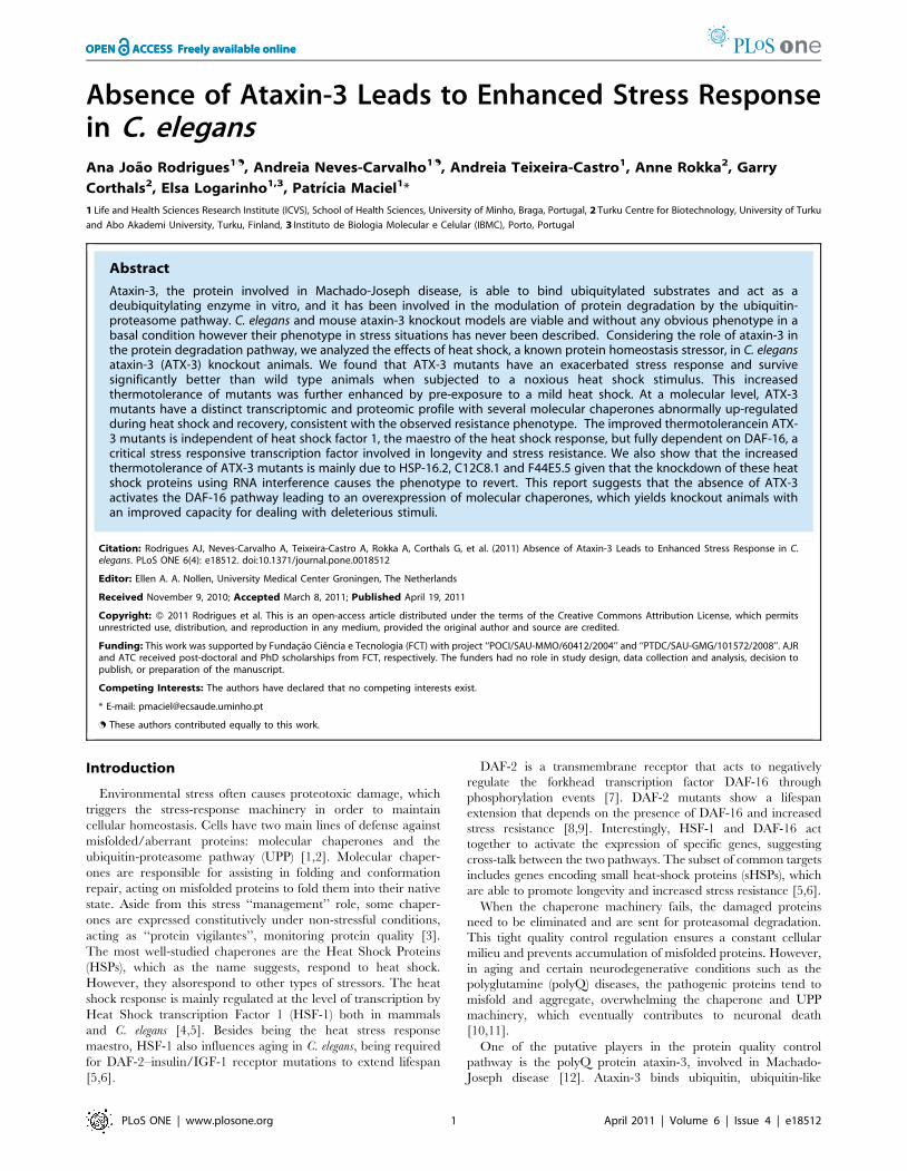
Absence of Ataxin-3 Leads to Enhanced Stress Responsein C. elegansAna Joao Rodrigues1., Andreia Neves-Carvalho1., Andreia Teixeira-Castro1, Anne Rokka2, Garry
Corthals2, Elsa Logarinho1,3, Patrıcia Maciel1*
1 Life and Health Sciences Research Institute (ICVS), School of Health Sciences, University of Minho, Braga, Portugal, 2 Turku Centre for Biotechnology, University of Turku
and Abo Akademi University, Turku, Finland, 3 Instituto de Biologia Molecular e Celular (IBMC), Porto, Portugal
Abstract
Ataxin-3, the protein involved in Machado-Joseph disease, is able to bind ubiquitylated substrates and act as adeubiquitylating enzyme in vitro, and it has been involved in the modulation of protein degradation by the ubiquitin-proteasome pathway. C. elegans and mouse ataxin-3 knockout models are viable and without any obvious phenotype in abasal condition however their phenotype in stress situations has never been described. Considering the role of ataxin-3 inthe protein degradation pathway, we analyzed the effects of heat shock, a known protein homeostasis stressor, in C. elegansataxin-3 (ATX-3) knockout animals. We found that ATX-3 mutants have an exacerbated stress response and survivesignificantly better than wild type animals when subjected to a noxious heat shock stimulus. This increasedthermotolerance of mutants was further enhanced by pre-exposure to a mild heat shock. At a molecular level, ATX-3mutants have a distinct transcriptomic and proteomic profile with several molecular chaperones abnormally up-regulatedduring heat shock and recovery, consistent with the observed resistance phenotype. The improved thermotolerancein ATX-3 mutants is independent of heat shock factor 1, the maestro of the heat shock response, but fully dependent on DAF-16, acritical stress responsive transcription factor involved in longevity and stress resistance. We also show that the increasedthermotolerance of ATX-3 mutants is mainly due to HSP-16.2, C12C8.1 and F44E5.5 given that the knockdown of these heatshock proteins using RNA interference causes the phenotype to revert. This report suggests that the absence of ATX-3activates the DAF-16 pathway leading to an overexpression of molecular chaperones, which yields knockout animals withan improved capacity for dealing with deleterious stimuli.
Citation: Rodrigues AJ, Neves-Carvalho A, Teixeira-Castro A, Rokka A, Corthals G, et al. (2011) Absence of Ataxin-3 Leads to Enhanced Stress Response in C.elegans. PLoS ONE 6(4): e18512. doi:10.1371/journal.pone.0018512
Editor: Ellen A. A. Nollen, University Medical Center Groningen, The Netherlands
Received November 9, 2010; Accepted March 8, 2011; Published April 19, 2011
Copyright: � 2011 Rodrigues et al. This is an open-access article distributed under the terms of the Creative Commons Attribution License, which permitsunrestricted use, distribution, and reproduction in any medium, provided the original author and source are credited.
Funding: This work was supported by Fundacao Ciencia e Tecnologia (FCT) with project ‘‘POCI/SAU-MMO/60412/2004’’ and ‘‘PTDC/SAU-GMG/101572/2008’’. AJRand ATC received post-doctoral and PhD scholarships from FCT, respectively. The funders had no role in study design, data collection and analysis, decision topublish, or preparation of the manuscript.
Competing Interests: The authors have declared that no competing interests exist.
* E-mail: [email protected]
. These authors contributed equally to this work.
Introduction
Environmental stress often causes proteotoxic damage, which
triggers the stress-response machinery in order to maintain
cellular homeostasis. Cells have two main lines of defense against
misfolded/aberrant proteins: molecular chaperones and the
ubiquitin-proteasome pathway (UPP) [1,2]. Molecular chaper-
ones are responsible for assisting in folding and conformation
repair, acting on misfolded proteins to fold them into their native
state. Aside from this stress ‘‘management’’ role, some chaper-
ones are expressed constitutively under non-stressful conditions,
acting as ‘‘protein vigilantes’’, monitoring protein quality [3].
The most well-studied chaperones are the Heat Shock Proteins
(HSPs), which as the name suggests, respond to heat shock.
However, they alsorespond to other types of stressors. The heat
shock response is mainly regulated at the level of transcription by
Heat Shock transcription Factor 1 (HSF-1) both in mammals
and C. elegans [4,5]. Besides being the heat stress response
maestro, HSF-1 also influences aging in C. elegans, being required
for DAF-2–insulin/IGF-1 receptor mutations to extend lifespan
[5,6].
DAF-2 is a transmembrane receptor that acts to negatively
regulate the forkhead transcription factor DAF-16 through
phosphorylation events [7]. DAF-2 mutants show a lifespan
extension that depends on the presence of DAF-16 and increased
stress resistance [8,9]. Interestingly, HSF-1 and DAF-16 act
together to activate the expression of specific genes, suggesting
cross-talk between the two pathways. The subset of common targets
includes genes encoding small heat-shock proteins (sHSPs), which
are able to promote longevity and increased stress resistance [5,6].
When the chaperone machinery fails, the damaged proteins
need to be eliminated and are sent for proteasomal degradation.
This tight quality control regulation ensures a constant cellular
milieu and prevents accumulation of misfolded proteins. However,
in aging and certain neurodegenerative conditions such as the
polyglutamine (polyQ) diseases, the pathogenic proteins tend to
misfold and aggregate, overwhelming the chaperone and UPP
machinery, which eventually contributes to neuronal death
[10,11].
One of the putative players in the protein quality control
pathway is the polyQ protein ataxin-3, involved in Machado-
Joseph disease [12]. Ataxin-3 binds ubiquitin, ubiquitin-like
PLoS ONE | www.plosone.org 1 April 2011 | Volume 6 | Issue 4 | e18512

molecule NEDD8, and ubiquitylated proteins. Ataxin-3 is able to
act as a deubiquitylating (DUB) enzyme in vitro [13,14,15],
cleaving chains of four or more ubiquitins, which is the minimal
signal for proteasomal degradation. Ataxin-3 also associates with
HHR23, the UPP-escort protein VCP/p97, UBXN-5 protein and
with the proteasome, suggesting a role in the modulation of
protein degradation [14,16,17,18]. Through its DUB activity,
ataxin-3 can process substrates and either facilitate their
proteasomal degradation or rescue proteins from irreversible
degradation through the removal of the ubiquitin signal.
Although human, mouse and C. elegans ataxin-3 are highly
conserved and ubiquitously expressed, the worm and mouse
knockout animals do not display any major phenotype [19,20].
The C. elegans atx-3 deletion strains are apparently normal, with
similar lifespan and brood size when compared to controls in
basal conditions [19]. The mouse knockout strain only displays a
mild increase in ubiquitylation levels in brain and testis [20].
However, all these studies were performed under basal condi-
tions, and, to our knowledge, nothing is known regarding the
behavior of these knockout strains in UPP-demanding situations.
Considering the role of ataxin-3 in protein quality control, we
decided to analyze the effects of its absence in protein
homeostasis stress using C. elegans ataxin-3 (ATX-3) knockout
strains. Surprisingly, the ATX-3 knockout animals displayed a
significantly increased resistance to stress. This improved
thermotolerance was due to a higher level of several molecular
chaperones, as confirmed by transcriptomic and proteomic
analysis, and was fully dependent on the transcription factor
DAF-16, but less so on HSF-1. We found that HSP-16.2 was
necessary for the increased thermoresistance phenotype of ATX-
3 knockout animals, while HSP-16.1 and -16.48 were not.
Results
C. elegans ATX-3 knockouts have increased resistance toheat stress
Since the C. elegans knockoutswere created by random
mutagenesis and might bear additional mutation(s), we used two
different deletion alleles of ATX-3; both were backcrossed five
times to wild-type animals (N2 strain), to exclude other mutations
relevant for the phenotype, as previously described [19].
Interestingly, as depicted in Figure 1A, ATX-3 knockout
animals grown at 20uC exhibited a significant higher rate of
survival compared to wild-type animals when exposed to a lethal
heat shock at 35uC. Wild-type animals displayed a median life
span of 9 hours, while both mutant strains had a median lifespan
of 10 hours (p,0,0001), a 10% increase in survival. As both
mutants behaved similarly with regards to their thermotolerance,
we decided to use only thegk193 allele for further studies. As
expected, daf-2 mutants, who are known to be long-lived and
stress-resistant [8,9,21,22], lived significantly longer than both
strains with more than 80% of animals living when no wild type or
atx-3 animals remained.
In addition to analyzing the animals under baseline conditions
(20uC), we also grew the atx-3 mutants at a stress-threshold
situation, at 25uC, and analyzed their survival when subjected to
a 35uC heat shock. Regardless of the genotype, all strains
displayed enhanced survival at 35uC compared to the same
strains grown at 20uC. Similarly to the basal condition, atx-3 null
animals survived significantly better than wild type animals, with
a median survival time of 17 hours versus 13 hours respectively,
representing an increase of 30% in atx-3 knockout animals
(p,0,0001) (Figure 1B). Table with lifespan results and p values
in Table S1.
Increased resistance to stress is further enhanced byhormesis
To investigate whether the stress machinery could be activated
more rapidly/efficiently in atx-3 mutants, we performed an
additional experiment, in which animals were pre-exposed to a
non-lethal heat shock at 30uC and then transferred to the lethal
temperature (35uC). This pre-exposure at 30uC is known to
stimulate protective cellular mechanisms and improve the
organism’s ability to cope with that and other types of stress in a
process known as hormesis, which occurs in several animal species
including worms [23].
We pre-exposed the animals to the sub-lethal heat shock for 2 or
5 hours and then transferred these animals to the lethal
temperature and measured their survival. After a pre-heat shock
for 2 hours at 30uC, the survival curves of both wild type and atx-3
mutants shifted to the right, representing an increased survival.
However, the atx-3-null animals were significantly more resistant
than wild type, with a median lifespan of 14 hours compared to
10 hours in wild type, corresponding to an increase of 40% in
survival time in mutants (p,0,0001) (Figure 1C). When we pre-
exposed the animals to a 5-hour pre-heat shock treatment, the
median lifespans were 15 hours and 13 hours for atx-3 and wild
type animals, respectively. These values corresponded to a 13%
increase in survival time among mutants (p,0,0001) (Figure 1D).
When animals grown at 25uC were subjected to the same
protocol, with the 2-hour pre-heat shock, atx-3 mutants’ median
survival was 22 hours compared to 18 hours of N2 animals (22%
increase) (p,0,0001) (Figure 1E). In the case of the 5-hour pre-
treatment, the knockout animals’ median survival was 20 hours
versus 17 hours for N2 animals (18% increase) (Figure 1F).
atx-3 knockouts show distinct changes in chaperonemRNA levels
The next step was to try to understand the mechanism of this
increased resistance at a molecular level. To do so, we analyzed
the gene expression of several chaperones in the atx-3 mutant
strain, including three small heat shock proteins (sHSP) (HSP-
16.49, HSP-16.1, HSP-16.2) and four members of the HSP70
family (F44E5.5, HSP-4, HSP-1, C12C8.1), using real-time PCR.
Atx-3 mutants displayed no pre-activation of the chaperone
machinery at 20uC, as determined by analyzing mRNA expression
(Figure 2A).
When we analyzed the chaperone expression levels during the
course of a standard non-lethal 33uC heat shock, we found that most
of the chaperones tested were significantly up-regulated in atx-3
mutants 60 minutes after the beginning of the heat shock. The
expression of HSP-16.2 and -4 was augmented in mutants
30 minutes after heat shock was initiated, while HSP-16.49,
F44E5.5 and C12C8.1 up-regulation was obvious at 60 minutes.
HSP-16.1 was significantly up-regulated in atx-3 mutants only
90 minutes after the stimulus (Figure 2B).
HSP-1, -12 and DAF-21 (HSP-90 homologue) levels did not
differ between wild type and atx-3 mutants during the time-course
of the heat shock (data not shown).
Proteomic profile of atx-3 mutants after heat shockReal-time PCR results suggested that the atx-3 strain exhibited
an enhanced activation of the chaperone machinery, at least at the
mRNA level, during the heat shock. Our next step was to analyze
the proteomic profile of atx-3 after a standard non-lethal heat
shock in C. elegans (2 h at 33uC). Detecting and quantifying whole
proteins from a complex protein extract in a comprehensive
manner remains a challenge in the fields of proteomics,
Stress Response in Ataxin-3 Mutants
PLoS ONE | www.plosone.org 2 April 2011 | Volume 6 | Issue 4 | e18512

nevertheless, using the iTRAQ technique, which allows simulta-
neous quantification of 2–8 samples by using different isotopes
(Applied Biosystems), we were able to obtain acceptable (albeit
incomplete) results. In the baseline condition (20uC), 35 proteins
were altered in the atx-3 knockout animals when compared to wild
type (Table S2). These proteins belong to several heterogeneous
classes such as ribosomal proteins (rpl-5, -33, -20), vitelogenins (vit-
1, -4, -5) and histones (his-12, -14, -71). After heat shock, 148
proteins were significantly altered; with a predominance of
ribosomal proteins, molecular chaperones, enzymes and histones.
There was partial overlap between these two conditions in terms of
protein alterations in atx-3 mutants (rpl-5, rpl-20, his-71, hi-12, his-
14, vit-1, vit-5, vit-4, sodh-1). Interestingly, as depicted in
Figure 3A, both atx-3 knockout strains revealed a significant
increase in the levels of the molecular chaperones HSP-16.49,
HSP-16.1 and F44E5.5. Although the levels of HSP-1, HSP-4 and
DAF-21 (HSP90 homologue) were only slightly up-regulated, these
differences were statistically significant. We observed the same
pattern of changes in both atx-3 mutant strains (see Table S2 for
complete proteomic results).
The most consistent difference was in the expression of HSP-16
family members - HSP-16.1 and HSP-16.49, which were clearly
up-regulated in atx-3 strains. We were unable to quantify the levels
of HSP-16.2 protein. The SIP-1 protein (sHSP) was altered in the
Figure 1. Atx-3 knockout animals are more resistant to heat stress than wild type. (A) Survival curves at 35uC of wild type (N2) and atx-3mutants (gk193, tm1689) previously grown at 20uC (p,0,0001). Median survival of N2 and atx-3 mutants is 9 h and 10 h respectively. (B) Survivalcurves at 35uC of N2 and atx-3 mutantspreviously grown at 25uC (p,0,0001). Median survival of N2 and atx-3 mutants is 12.5 h and 17 h respectively(p,0,0001). Pre-exposure to a non-lethal heat shock of 2 h (C) or 5 h (D) at 33uC further enhances the thermoresistance phenotype of atx-3 mutants(p,0,0001). Median survivals are: N2: 9 h, atx-3: 10 h, N2 pre-HS2h: 10 h, atx-3 pre-HS2h: 14 h, N2 pre-HS5h: 13 h, atx-3 pre-HS5h: 15 h. The same trendis observed with animals previously grown at 25uC (p,0,0001) both with the 2 h (E) and 5 h (F) pre-HS. Median survivals are: N2: 12.5 h, atx-3: 17 h,N2 pre-HS2h: 18 h, atx-3 pre-HS2h: 22 h, N2 pre-HS5h: 17 h, atx-3 pre-HS5h: 20 h. One representative experiment is shown (at least three independentreplicates were performed).doi:10.1371/journal.pone.0018512.g001
Stress Response in Ataxin-3 Mutants
PLoS ONE | www.plosone.org 3 April 2011 | Volume 6 | Issue 4 | e18512

Figure 2. Atx-3 knockout animals display increased up-regulation of molecular chaperones upon heat stress at mRNA level. (A) At20uC, atx-3 mutants do not show any significant difference in the levels of several molecular chaperones, although HSP-12.6 shows a trend to bedown-regulated. (B) Atx-3 mutants have mRNA up-regulation of several chaperones during the course of heat shock (HS) as measured by Real-TimePCR in young adult animals. HSP-4 was up-regulated from 30 until 60 minutes after the beginning of the HS and returned to wild type levels at
Stress Response in Ataxin-3 Mutants
PLoS ONE | www.plosone.org 4 April 2011 | Volume 6 | Issue 4 | e18512

knockout strains but displayed a divergent profile in the biological
replicates we analyzed (data not shown). Other chaperones such as
HSP-3, -6, -12.2 and -60 were present at similar levels in wild type
and atx-3 mutant animals while HSP-12.6 levels were diminished
(Table S2).
We aimed to confirm some of these findings by western blot.
Aside from analyzing the time-course of the heat shock, as
previously performed using real-time PCR, we also analyzed the
chaperone profile of the recovery after heat shock. We were able to
confirm HSP-16 overexpression using anti-HSP16 antibody
(kindly provided by Dr. Christopher Link). Since this antibody
recognizes several members of the HSP-16 family, the overex-
pression detected (Figure 3B) was less pronounced than in the
proteomic analysis, probably due to the masking effect of other
Figure 3. Absence of atx-3 leads to up-regulation of several molecular chaperones during heat shock and in the recovery at aprotein level. (A) Summary of the proteomic results regarding the expression levels of several chaperones after heat shock of 2 h and recovery of30 minutes at 20uC. Shown is the average of the relative expression of chaperones normalizing wild type levels to 1. Error bars correspond tostandard deviation. (B) Western blot analysis of constitutive (HSP-4) and inducible chaperones (HSP-16 family) in wild type and atx-3 animals duringthe timecourse of a heat shock. HSP-40 is expressed at all timepoints while the HPS-16 family of proteins is only detected at 60 minutes after thebeginning of the heat shock. Mutants have higher levels of HSP-16 from 60–90 minutes after the beginning of the stimulus. (C) Atx-3 mutants havethe HSP-16 family up-regulated during the timecourse of the recovery. Only 24 h after heat shock, the HSP-16 levels become similar to wild type.* p,0.05.doi:10.1371/journal.pone.0018512.g003
90 minutes. HSP-16.1 was only significantly increased 90 minutes after the stimulus while HSP-16.49, F44E5.5 and C12C8.1 were up-regulated at60 minutes. Error bars correspond to standard error. * p,0.05.doi:10.1371/journal.pone.0018512.g002
Stress Response in Ataxin-3 Mutants
PLoS ONE | www.plosone.org 5 April 2011 | Volume 6 | Issue 4 | e18512

proteins of this family, which were unaltered in the atx-3 animals.
We found that HSP-16 proteins were first detected 60 minutes
after the beginning of heat shock. HSP-16 levels were elevated in
atx-3 animals when compared to controls at 60–90 minutes of heat
shock. We also analyzed the expression levels of HSP-40, a
constitutive chaperone, and we found no differences between atx-3
and N2 animals.
Several time-points of recovery after heat shock were also
analyzed: 1 h, 3 h, 6 h and 24 h. The HSP-16 proteins remained
detectable at all time-points and were found to be consistently up-
regulated in atx-3 nulls (Figure 3C), although at 24 h, levels were
comparable to those observed inwild type animals.
We tested various commercial antibodies against other
chaperones (hsp70, hsp105) with no success, since they did not
recognize the worm proteins (data not shown).
atx-3 mutant animals have a lower temperaturethreshold for the induction of chaperones
Our next step was to assess how these animals would behave if
grown at a stress-threshold temperature, at 25uC. At 25uC, atx-3
mutants exhibited significantly increased levels of mRNA for the
inducible HSP-16.1 and -16.49 chaperones, as well as F44E5.5 in
comparison to N2 animals (Figure 4A). No changes were observed
in HSP-16.2 and C12C8.1 expression levels. This ‘‘pre-activation’’
of the chaperone machinery, however, is probably not translated
at the protein level, since we were unable to detect HSP-16
proteins by western blot at this temperature (data not shown).
We also analyzed the HSP mRNA and protein expression levels
during and after heat shock in animals grown at 25uC. As depicted
in Figure 4B, mRNA levels of HSP-4, -16.1, -16.49, F44E5.5 and
C12C8.1 were up-regulated in atx-3 animals at 15 minutes after
the beginning of heat shock until at least 30 minutes after the
stimulus. At the protein level, atx-3 animals expressed higher levels
of HSP-16 than controls 90 minutes after the beginning of heat
shock, and these proteins remained up-regulated at all recovery
time-points except at 24 h, when the HSP-16 levels were close to
those of wild-type (Figure 5A, B). Surprisingly, the levels of so-
called ‘‘constitutive’’ HSP-40 varied in some experiments; in fact,
it was often undetectable 24 h after heat shock (Figure 5B), which
suggests that it may also be regulated by stress. Interestingly, HSP-
16.2 was not up-regulated in atx-3 mutants under these conditions
and was even down-regulated 60 minutes after the beginning of
heat shock (Figure 4A, B).
Increased resistance to stress but no extension in lifespanin atx-3 mutants
Although stress-resistant animals are usually long-lived [24] and
a raise in chaperone levels is often correlated with increased
lifespan [25,26], we have previously shown that atx-3 mutants have
normal lifespan [19]. However, we had not analyzed their lifespan
after a heat insult; hence, we exposed the mutant animals to a
33uC heat shock for two hours and then transferred them to 20uC,
checking their survival daily. Atx-3 animals lived for the same
amount of time as the wild type after the exposure to heat shock
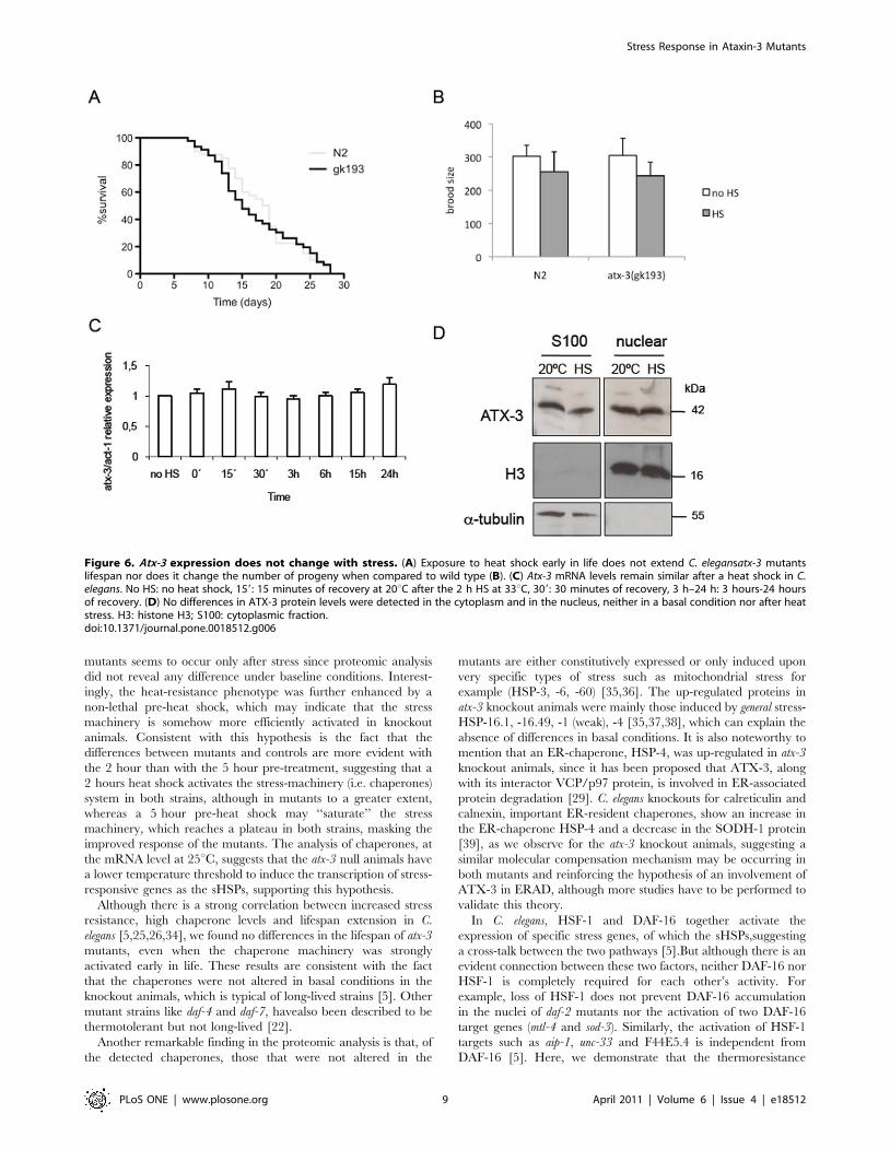
(Figure 6A). The brood size of these animals after heat shock was
also similar to that of wild type (Figure 6B).
Since atx-3 knockout animals displayed a clear resistance to
stress, we decided to assess the levels of atx-3mRNA after exposure
to heat shock, in order to see if atx-3 behaved as a negative stress-
responsive gene, being repressed upon exposure to stress. As can
be seen in Figure 6C, the number of transcripts of atx-3 was not
altered significantly after the insult. Since it has been shown that
human ataxin-3 is translocated to the nucleus in cell lines exposed
to heat shock, we also assessed the subcellular distribution of ATX-
3 protein in a baseline condition and after heat shock in C. elegans.
In contrast to the above-mentioned observations, no differences
were found in nuclear ATX-3 levels in both conditions (Figure 6D).
atx-3 modulates stress resistance in a daf-16-dependentmanner
In order to further understand the mechanism of improved
stress resistance in atx-3 knockout animals, we decided to analyze
whether atx-3 could genetically interact with the transcription
factors HSF-1 and DAF-16, which are important for lifespan and
stress resistance and have been shown to co-regulate sHSP
expression together [5]. To do so, we crossed atx-3 animals with
worms bearingan hsf-1 point mutation allele (hsf-1(sy441)), which
results in a truncated version of HSF-1, unable to activate
transcription [27].To assess the thermotolerance profile of hsf-
1;atx-3 animals, we exposed them to varying durations of 35uCheat shock and 12 h later analyzed their survival, a protocol
adapted from thatdescribed previously [28]. The thermoresistance
phenotype of atx-3-null animals was independent of the HSF-1
transcriptionfactor, sincehsf-1; atx-3 double-mutants behaved as
atx-3 single-mutants when exposed to a 5 h heat shock (Figure 7A,
B). However, after a 9 h heat shock, the double mutant animals no
longer displayed this increased thermotolerance, suggesting that
the phenotype is partially dependent on HSF-1 (Figure 7B). We
were unable to perform this assay with animals grown at 25uC,
given that the hsf-1-null animals are extremely sensitive to
temperatures above 20uC. The thermoresistance phenotype of
atx-3 mutants, however, was completely dependent on DAF-16, as
the daf-16; atx-3 double-knockout animals behaved as single daf-16
mutants in the thermotolerance assay (p = 0,18) (Figure 7C). This
daf-16 phenotype-dependence was observed with animals grown at
25uC as well (Figure 7D). To further confirm the dependence of
DAF-16 for the observed resistance phenotype, we used a rescue
strain (here named daf-16 rescue) that is homozygous for the
mgDf47 mutation in the daf-16 gene and contains an integrated
plasmid with DAF-16B::GFP under the control of the endogenous
promoter [7]. Knockdown of atx-3 by RNAi in this rescue
background restored the enhanced thermotolerancephenotype
(Figure 7E). The results presented so far suggest an activation of
DAF-16 with concomitant overexpression of the molecular
chaperones. In support of this hypothesis was the fact that sod-3
and mtl-1 (other DAF-16 transcriptional targets) were also up-
regulated in atx-3 mutants (Figure S1).
To identify the molecular chaperones important for the
observed phenotype, we performed RNA interference (RNAi)
against several HSPs. As depicted in Figure 7, atx-3 mutants were
still more resistant than wild type when grown in bacteria
transformed with L4440 (RNAi empty vector) (Figure 7F). Gene
knockdown of hsp-16.1 and hsp-16.49 did not revert the increased
thermotolerance phenotype of atx-3 mutants (data not shown),
while knockdown of hsp-16.2 reverted this phenotype (Figure 7F).
In addition, RNAi of the hsp70 family members C12C8.1 and
F44E5.5 also reverted thethermotolerance phenotype (Figure 7G,
H; complete results in Table S3).
Discussion
Ataxin-3 is a DUB enzyme involved in the ubiquitin-protea-
some pathway, where it seems to act as a ‘‘processing’’ protein,
cleaving ubiquitin from substrates and modulating their protea-
somal degradation [14,16,17,18]. Ataxin-3 is ubiquitously ex-
pressed and is present in several species, ranging from plants to
humans, as well as in almost every type of cells. Therefore it is
Stress Response in Ataxin-3 Mutants
PLoS ONE | www.plosone.org 6 April 2011 | Volume 6 | Issue 4 | e18512

Figure 4. Atx-3 mutants have a lower threshold for induction of heat sock response. (A) When grown at 25uC, atx-3 animals present HSP-16.1, -16.49 and F44E5.5 up-regulated when compared to control animals (p,0.05). (B) Atx-3 mutants have mRNA up-regulation of severalchaperones during the course of heat shock (HS) as measured by Real-Time PCR in young adult animals previously grown at 25uC. HSP-4 was up-regulated in mutants from 15 until 60 minutes after the beginning of the HS and returned to wild type levels at 90 minutes. HSP-16.1 was only
Stress Response in Ataxin-3 Mutants
PLoS ONE | www.plosone.org 7 April 2011 | Volume 6 | Issue 4 | e18512

quite surprising that the ataxin-3 knockout mice only display a
mild increase in ubiquitylation levels and that a C. elegans model of
ataxin-3 deficiency shows no obvious phenotype and no
differences in lifespan, brood size or general ubiquitylation levels
[19,20]. This could indicate adaptive/compensatory mechanisms
or very limited substrate specificity. Although the ataxin-3
knockout models are apparently normal in baseline conditions,
little is known about the behavior of these mutants under stress.
Recently, we have shown that C. elegans atx-3 mutants display a
temperature-dependent motor phenotype [29].
Since this protein shares the sequence, structure and biochem-
ical activity of its human counterpart [19], we studied the effects of
ATX-3 absence in C. elegans under stressconditions. We observed
that ATX-3 knockout animals are significantly more resistant to
heat stress than wild type animals. Our first hypothesis was that
the atx-3 gene might be a stress-responsive gene/protein,
potentially with a role in termination of the stress response
mediated by degradation of specific substrate proteins. However,
this hypothesis is contradicted by the fact that heat shock did not
change atx-3 expression levels and ATX-3 subcellular distribution
in C. elegans.
Interestingly, while this manuscript was being prepared, it was
shown that in fibroblasts, ataxin-3 responds to stress by moving to
the nucleus, and that knockout fibroblasts were more sensitive to
heat stress than controls [30], which is in contrast with our
findings. Although it can be argued that the absence of ataxin-3 in
mammals and worms can lead to dissimilar consequences, this
seems unlikely as the two proteins have the same biochemical
function, localization and expression pattern [19]. More likely to
explain the differences is the fact that the effects of heat shock in a
cell line are certainly different from those in a whole organism
such as C. elegans. In isolated cells, the heatshock response is
initiated by the presence of misfolded proteins (cell-autonomous),
while in C. elegans, the heatshock response of somatic cells depends
on the thermosensoryneuron AFD which regulates temperature-
related behavior [31].
Underlying thethermoresistance phenotype of atx-3 mutants was
the significant up-regulation of several molecular chaperones,
namely HSP-1, HSP-16.1, HSP-16.2, HSP-16.49, HSP-4,
F44E5.5, C12C8.1 and DAF-21. This up-regulation was observed
after heat shock in animals grown at 20uC and, for most of the
molecular chaperones listed above when animals were grown at
25uC as well. Increased levels of HSPs have a protective effect and
enhance the stress response in C. elegans. For example, elevated
levels of HSP-16 increase stress resistance [26] and can predict
both thermotolerance and survival of individual worms [32].
Overexpressing other HSPs such as HSP-70 also induces increased
resistance to stress in worms [33].The increase in HSPs in atx-3
Figure 5. Atx-3 knockout animals display significant changes in the levels of molecular chaperones when grown at 256C. (A) Westernblot analysis of constitutive (HSP-4) and inducible chaperones (HSP-16 family) in wild type and atx-3 animals during the timecourse of a heat shock.HSP-40 is expressed at all timepoints while HPS-16 is only detected 60 minutes after the beginning of the heat shock. Mutants have higher levels ofHSP-16 from 60–90 minutes after the beginning of the stimulus. (C) Atx-3 mutants have the HSP-16 family up-regulated during the timecourse of therecovery. Only at 24 h, the HSP-16 levels return back to wild type levels.doi:10.1371/journal.pone.0018512.g005
significantly increased during the first 30 minutes; HSP-16.49 and C12C8.1 expression levels were increased from 15–60 minutes while F44E5.5 wasup-regulated until 30 minutes after the beginning of HS. Error bars correspond to standard error. * p,0.05.doi:10.1371/journal.pone.0018512.g004
Stress Response in Ataxin-3 Mutants
PLoS ONE | www.plosone.org 8 April 2011 | Volume 6 | Issue 4 | e18512
mutants seems to occur only after stress since proteomic analysis
did not reveal any difference under baseline conditions. Interest-
ingly, the heat-resistance phenotype was further enhanced by a
non-lethal pre-heat shock, which may indicate that the stress
machinery is somehow more efficiently activated in knockout
animals. Consistent with this hypothesis is the fact that the
differences between mutants and controls are more evident with
the 2 hour than with the 5 hour pre-treatment, suggesting that a
2 hours heat shock activates the stress-machinery (i.e. chaperones)
system in both strains, although in mutants to a greater extent,
whereas a 5 hour pre-heat shock may ‘‘saturate’’ the stress
machinery, which reaches a plateau in both strains, masking the
improved response of the mutants. The analysis of chaperones, at
the mRNA level at 25uC, suggests that the atx-3 null animals have
a lower temperature threshold to induce the transcription of stress-
responsive genes as the sHSPs, supporting this hypothesis.
Although there is a strong correlation between increased stress
resistance, high chaperone levels and lifespan extension in C.
elegans [5,25,26,34], we found no differences in the lifespan of atx-3
mutants, even when the chaperone machinery was strongly
activated early in life. These results are consistent with the fact
that the chaperones were not altered in basal conditions in the
knockout animals, which is typical of long-lived strains [5]. Other
mutant strains like daf-4 and daf-7, havealso been described to be
thermotolerant but not long-lived [22].
Another remarkable finding in the proteomic analysis is that, of
the detected chaperones, those that were not altered in the
mutants are either constitutively expressed or only induced upon
very specific types of stress such as mitochondrial stress for
example (HSP-3, -6, -60) [35,36]. The up-regulated proteins in
atx-3 knockout animals were mainly those induced by general stress-
HSP-16.1, -16.49, -1 (weak), -4 [35,37,38], which can explain the
absence of differences in basal conditions. It is also noteworthy to
mention that an ER-chaperone, HSP-4, was up-regulated in atx-3
knockout animals, since it has been proposed that ATX-3, along
with its interactor VCP/p97 protein, is involved in ER-associated
protein degradation [29]. C. elegans knockouts for calreticulin and
calnexin, important ER-resident chaperones, show an increase in
the ER-chaperone HSP-4 and a decrease in the SODH-1 protein
[39], as we observe for the atx-3 knockout animals, suggesting a
similar molecular compensation mechanism may be occurring in
both mutants and reinforcing the hypothesis of an involvement of
ATX-3 in ERAD, although more studies have to be performed to
validate this theory.
In C. elegans, HSF-1 and DAF-16 together activate the
expression of specific stress genes, of which the sHSPs,suggesting
a cross-talk between the two pathways [5].But although there is an
evident connection between these two factors, neither DAF-16 nor
HSF-1 is completely required for each other’s activity. For
example, loss of HSF-1 does not prevent DAF-16 accumulation
in the nuclei of daf-2 mutants nor the activation of two DAF-16
target genes (mtl-4 and sod-3). Similarly, the activation of HSF-1
targets such as aip-1, unc-33 and F44E5.4 is independent from
DAF-16 [5]. Here, we demonstrate that the thermoresistance
Figure 6. Atx-3 expression does not change with stress. (A) Exposure to heat shock early in life does not extend C. elegansatx-3 mutantslifespan nor does it change the number of progeny when compared to wild type (B). (C) Atx-3 mRNA levels remain similar after a heat shock in C.elegans. No HS: no heat shock, 159: 15 minutes of recovery at 20uC after the 2 h HS at 33uC, 309: 30 minutes of recovery, 3 h–24 h: 3 hours-24 hoursof recovery. (D) No differences in ATX-3 protein levels were detected in the cytoplasm and in the nucleus, neither in a basal condition nor after heatstress. H3: histone H3; S100: cytoplasmic fraction.doi:10.1371/journal.pone.0018512.g006
Stress Response in Ataxin-3 Mutants
PLoS ONE | www.plosone.org 9 April 2011 | Volume 6 | Issue 4 | e18512

Figure 7. Atx-3 thermoresistance is dependent on DAF-16 transcription factor and independent on HSF-1. Wild type animals die morethan atx-3 mutants after a 5 h (A) or 9 h (B) exposure to 35uC heat shock. Hsf-1 animals have enhanced sensitivity to this temperature at bothtimepoints, as expected. Double atx-3; hsf-1 mutants still display enhanced thermoresistance at 5 h timepoint but behave as single hsf-1 mutants inthe 9 h heat shock condition. Atx-3 mutants are more resistant to the exposure to a lethal heat shock at 35uC than wild type animals, but daf-16; atx-3double mutants are not, both when grown at 20uC (C) or 25uC (D). (E) Restoring DAF-16 levels in a daf-16; atx-3 (daf-16 deletion and atx-3knockdown; daf-16 rescue strain) background leads to the appearance of the thermotolerance phenotype. Increased thermotolerance of atx-3mutants is still observed when grown on bacteria expressing RNAi empty vector (L4440) (F) and is dependent on hsp-16.2 (G), C12C8.1 (H) andF44E5.5 (I). Median survivals in animals grown in (C) are: N2: 12.5 h, atx-3: 17 h, daf-16: 13 h, daf-16; atx-3: 13 h, daf-2: not determined. Mediansurvivals in (D) are: N2: 17 h, atx-3: 19 h, daf-16: 16 h, daf-16; atx-3: 17 h, daf-2: not determined. Median survivals in (E) are: daf-16 rescue in L4440:14 h, daf-16 rescue in RNAi(ATX-3): 16 h. Median survivals in (F) are: N2 L4440: 21 h, atx-3 L4440: 23 h. N2 in RNAi(HSP-16.2): 19 h, atx-3 in RNAi(HSP-16.2): 17 h. Median survivals in (G) are: N2 L4440: 14 h, atx-3 L4440: 16 h. N2 in RNAi(C12C8.1): 14 h, atx-3 in RNAi(C12C8.1): 14 h. Median survivals in(H) are: N2 L4440: 13 h, atx-3 L4440: 15 h. N2 in RNAi(F44E5.5): 13 h, atx-3 in RNAi(F44E5.5): 13 h. *p,0.05.doi:10.1371/journal.pone.0018512.g007
Stress Response in Ataxin-3 Mutants
PLoS ONE | www.plosone.org 10 April 2011 | Volume 6 | Issue 4 | e18512

phenotype of atx-3 strains is independent on HSF-1, at least to a
certain extent. This finding was surprising given the fact that the
HSF-1 is a major player in the stress response; however, it has also
been shown that DAF-16 per se is able to activate molecules of the
stress response. Thus, it was interesting to find thatthe atx-3
mutants’ phenotype was fully dependent on DAF-16. Double daf-
16; atx-3 mutants behaved exactly as single daf-16 mutants in the
thermotolerance assays but the rescue strain displays enhanced
thermotolerance, suggesting that this transcription factor is
required to mount the enhanced stress response in atx-3 mutants.
While this manuscript was being prepared, Hoppe and colleagues
showed that cdc-4; atx-3 double knockouts were long lived (up to
50%) and this seemed to be mediated by the DAF-16 pathway
[40]; this relationship between ataxin-3 absence and DAF-16
activation is in accordance with our findings.
When we further dissected the molecular events responsible for
this thermoresistance phenotype, we found that the hsp-16.2 gene,
a DAF-16 target, was essential for the increased survival of atx-3
strain: the atx-3 knockout animals not only lost their increased
thermoresistance but also displayed more sensitivity than wild type
animals to the lethal temperature when subjected to hsp-16.2
RNAi. Hsp-16.2 encodes a sHSP similar to sip-20 (stress induced
protein 20) which is activated in response to heat shock and other
stressors. Expression of hsp-16.2 is a good predictor of stress
response and longevity [32] and it has been shown to reduce the
aggregation of beta amyloid peptide in vivo [41]. Besides, hsp-16.2
expression is known to be modulated by HSF-1 and by DAF-16
[5,34]. The finding that hsp-16.2 was essential for the phenotype
was quite surprising since although its expression was higher after
heat shock in atx-3 animals when compared to controls when
grown at 20uC, the same was not observed with animals grown at
25uC, at least at the mRNA level. One possibility is that the down-
regulation result from negative feedback by accumulated HSP-
16.2 at the protein level, a hypothesis we not verified due to the
lack of HSP-16.2 specific antibody. For example, Hsp70 is able to
function as a repressor of the heat shock response in eukaryotes
[42] and in bacteria, chaperones DnaK, DnaJ and GrpE
negatively regulate the transcription of heat shock genes [43,44].
In addition to HSP-16.2, we found that C12C8.1 and F44E5.5,
which were significantly up-regulated after heat shock, were also
essential for the thermoresistance phenotype of atx-3 mutants.
C12C8.1 and F44E5.5 are hsp70 members, highly activated after
heat shock [37] and apparently regulated by HSF-1 [31].
Although it appears that their expression is not modulated by
DAF-16, the existence of a parallel transcriptional mechanism
cannot be ruled out [31]. Noteworthy, RNAi against other
chaperones did not lead to phenotype reversion, enhancing the
specificity of the abovementioned chaperones for the observed
thermotolerance.
The previously described translocation of ataxin-3 into the
nucleus of cell lines following heat shock has been shown to be
independent of HSF-1 [30], which suggests that alternative
pathways may be related to ataxin-3’s potential involvement in
the stress response. At least in atx-3 animals, it seems that
chaperone overexpression is being activated mainly by DAF-16,
given the requirement of DAF-16 for the phenotype. Nevertheless,
the hsf-1; atx-3 mutants still require HSF-1 in the longer heat shock
situation, suggesting that after a certain degree of damage/
exposure, both pathways are necessary.
One possible explanation for the molecular and physiological
phenotype of atx-3 nulls is that the absence of ataxin-3 at some
timepoint of the development causes cellular (proteotoxic?) stress,
which activates the stress machinery, and once they are needed
again, chaperones and other effectors will be more effectively and
rapidly activated- a process known as hormesis. Another possibility
is that ataxin-3 is normally regulating chaperone levels via the
DAF-16 pathway, or even modulating their levels through the
ubiquitin-proteasome degradation of a specific target upstream
DAF-16 or of DAF-16 itself. This last option seems unlikely as we
did not find significant differences in DAF-16 protein levels in atx-
3 mutant animals (data not shown), in agreement with very recent
findings [40].
In summary, we show that the absence of ataxin-3 leads to an
enhanced stress response in C. elegans. This phenotype was
correlated with a significant increase in chaperones and fully
dependent on the transcription factorDAF-16 and on its target
HSP-16.2, and on the hsp70-like C12C8.1 chaperone.These
findings can be relevant in the disease context, since a partial loss
of the normal function of ataxin-3 may occurdue to the expansion,
as has been observed for other polyQ disorders. Long-term
deregulation of HSPscan be detrimental for cell growth, division
and viability [45,46] and, along with the proteotoxic stress, this
may contribute to neuronal demise in the context of MJD.
Materials and Methods
C. elegans strains culture and crossesC. elegans strains were maintained at 20uC or 25uC in 2%
peptone or NGM plates seeded with E. coli OP50 (28).
Synchronous cultures were obtained by bleaching.
Strain atx-3(gk193) was kindly provided by the C. elegans Reverse
Genetics Core Facility at the University of British Columbia.
Strain atx-3(tm1698) was obtained from the National Bioresource
Project. Strain GR1352 (daf-16(mgDf47) I; xrIs87 [daf-16al-
pha::GFP::DAF-16B+rol-6(su1006)] was kindly provided by Dr.
Gary Ruvkun. Other strains used in this work were kindly
provided by the Caenorhabditis Genetics Center(CGC).
Strain atx-3(gk193) was crossed with strain daf-16(mu86) and hsf-
1(sy441) using standard procedures. Daf-16 genotyping was done
using primers mu86+U5311, mu86+L16642 and mu86-L6429 as
previously described [47]. An allele specific PCR using primers
sy441F1, sy441F2 and sy441R1 was performed for hsf-1
genotyping. Atx-3 genotyping was described before [19]. Primer
sequences in Table S4.
C. elegans growth conditionsFor the heat shock experiments, young adult animals were
submitted to the indicated temperature in a temperature-
controlled incubator, and after the desired period, animals were
collected as described above.
C. elegans life span and brood size assaysFor regular life span, animals were kept at 20uC and checked
daily for viability. Animals were also subjected to a 2 h 30uC heat
shock and then transferred to 20uC and checked daily. For brood
size measurements, animals were allowed to put eggs at 20uC with
or without a heat shock of 2 h at 33uC. Progeny was allowed to
eclode and counted the next day.
RNAi experimentsGenes of interest cloned in L4440 vector were grown from
standard C. elegans libraries and verified by sequencing. L4440
empty vector was used as a control. L4440/mex-3 was used as
positive RNAi control (easy to score phenotype). HT115 bacteria
transformed with L4440 with gene of interest or empty L4440
vector were grown in standard LB media with ampicillin (50 mg/
ml) overnight at 37uC. The next day, bacteria were seeded onto
LB+ampicillin plates supplemented with 1 mM IPTG to induce
Stress Response in Ataxin-3 Mutants
PLoS ONE | www.plosone.org 11 April 2011 | Volume 6 | Issue 4 | e18512
expression. 2–3 days later, synchronized L1 animals were
transferred to plates, placed at the correct temperature and
checked every day.
Thermotolerance assaysFor regular thermotolerance assays, synchronized young adult
animals (72 h post hatching) were grown at 20uC or 25uC and
then transferred to 35uC and checked every hour for death
animals. In the case of pre-heat shock, animals were initially
submitted to 30uC for two or five hours and immediately
transferred to 35uC and checked every 1–2 hours. To test HSF-
1 phenotype dependence, animals were subjected to a 35uC heat
shock for 5–9 hours, transferred to 20uC for recovery and scored
for dead animals 12 hours later [31]. More than 50 animals per
group were analyzed in each experiment and at least three
independent replicates were performed for each assay.
Western BlottingSynchronous worm cultures were collected with M9 buffer and
rinsed several times. After that, they were pelleted, frozen in liquid
nitrogen. An equal amount of Lysis buffer (50 mMTris-HCl
pH 7.4, 150 mMNaCl, 1% NP-40, 1 mM PMSF, complete
protease inhibitors (Roche)) was added to each pellet. Animals
were frozen and thawed several times, briefly sonicated and after
centrifugation at 13000 rpm 10 minutes at 4uC, the supernatant
was collected. Proteins were quantified using the Bradford
method. Forty micrograms of total protein was loaded into SDS-
PAGE gels and then transferred to nitrocellulose membranes.
Nuclear and cytosolic extracts were prepared as previously
described [19]; four times more of nuclear extract was used in
order to detect ATX-3.
After incubation with the primary antibodies: anti-HSP16
(1:5000, kindly provided by Dr. Christopher Link), anti-HSP40
(1:2000, Stressgene), anti-alpha-tubulin (1:100, DSHB), anti-
histone H3 (1:5000, Abcam), anti-ATX-3 (1:200) the secondary
antibodies were incubated at a 1:10,000 dilution. Detection was
done using ECL kit (Pierce). Band quantification was performed
using ImageJ(http://rsbweb.nih.gov/ij/) as advised by the soft-
ware manufacturers using alpha-tubulin as the loading control.
Quantitative PCRTotal RNA from synchronized populations of C. elegans at
various conditions was isolated using Trizol, following the
manufacturer’s instructions. Two micrograms of total RNA were
DNAse treated (Amersham) and converted into cDNA using the
iScript kit (Biorad). Q-PCR was performed using SyberGreen
(Qiagen) and the Biorad q-PCR CFX96 apparatus. Actin was used
as a housekeeping gene. We used relative quantification to
determine the fold change difference between wild type and atx-
3 mutants, using the DDCT method as described before [48].
Mass spectrometry sample preparationProteomic lysis buffer (8 M urea, 2 M thiourea, 4% CHAPS,
100 mM DTT, 1 mM PMSF, protease inhibitors (Roche)) was
added to the frozen pellets of young adult animals (72 h post
hatching) in basal (20uC) or heat-shocked conditions (2 hours at
33uC with 30 minutes recovery at 20uC). Pellets were frozen and
thawed several times and centrifuged. One hundred micrograms
of total protein were precipitated using six volumes of cold acetone
to remove interfering substances. Protein pellets wereressuspended
in 40 ml of 500 mMtriethyl ammonium bicarbonate with 1% SDS,
vortexed, and briefly sonicated until fully dissolved. The proteins
were reduced using 2 ml of 50 mMtris-(2-carboxyethyl) phosphine
for 1 hour at 60uC, and cysteines were blocked with 200 mM
methyl methane thiosulfonate for 10 minutes at room tempera-
ture. Ten micrograms of sequencing grade modified trypsin
(Promega) diluted in water was added to each sample and
incubated overnight at 37uC. The iTRAQ reagents (Applied
Biosystems) were reconstituted in 70% ethanol, transferred to the
respective sample and allowed to incubate for one hour. Reagent
114 was added to N2, 115 to atx-3(gk193), 116 to atx-3(tm1689);
and the labelled peptides were then combined and evaporated to
dryness in a SpeedVac. Each sample was re-dissolved in 50 ml of
0,1% trifluoroacetic acid (TFA) and desalted using C18 empore
disks.
Isolelectric focusingDesalted samples were applied on a 13 cm IPG strip, pH 3–10
(GE Healthcare), rehydrated for 12 hours and then focused in a
IPGphorapparatus (GE Healthcare)using the following parame-
ters: hold 500 V 1 h, linear gradient from 500–1000 V 15 min-
utes, hold at 1000 V 1 h, linear gradient from 1000 V–8000 V
30 minutes, hold 8000 2 hours. The strips were cut into 12
fractions and focused peptides were extracted from the gel using a
TFA gradient. The sample were evaporated and desalted as
mentioned above. Peptides were dissolved in 1% HCOOH just
before LC-ESI-MS/MS analyses.
LC-ESI-MS/MS analysesAround 6–8 ug of peptides from each fraction were analyzed
using a nanoflow LC coupled to a QSTAR Pulsar i ESI-hybrid Q-
TOF tandem mass spectrometer (Applied Biosystems/MDS
Sciex). Peptides were concentrated and desalted on a precolumn
(0.365 mm C18 PepMap100, LC Packings), and eluted at 200 nl/
min by increasing concentration of acetonitrile onto a self-packed
C18 reverse phase column (75 mm615 cm, Magic 5 mm 100 A
C18, MichromBioResources Inc.) A linear 60 min gradient from
98% solvent A (97.8% water, 2% acetonitrile, and 0.2% formic
acid) to 35% solvent B (95% acetonitrile, 4.8% water, and 0.2%
formic acid) was used.
Data from LC-MS/MS runs were converted to peak list files
with the Analyst QS software (version 1.1). For QSTAR data, the
acquisition parameters were: extract only +2 or +3 charged
percursor ions, 300.000–1300.000 m/z range, with a minimum of
20 counts. Data from LC-MS/MS runs were converted to peak list
files with the Analyst QS software (version 1.1).
Data analysis and protein identificationMS/MS spectra generated was analyzed using Mascot software
(version 2.1.02) using the Wormbasewormprep 178 (available in
ftp://ftp.wormbase.org/pub/wormbase/genomes/elegans/sequences/
protein/) as the reference database. Trypsin was set as the digestion
enzyme with a maximum of one missed cleavage site. The mass
accuracy for parent ions was set as 100 ppm, and 0.3 Da was
used for the fragment ion mass tolerance. The default search
settings used for quantitative analysis and protein identification
were: trypsin cleavage with fixed MMTS modification of cysteine,
iTRAQ labelling and variable methionine oxidation.
A 5% peptide false positive rate was chosen and only proteins
identified by two or more peptides were selected.
Statistical analysisGeneral comparisons were performed using ANOVA in the
SPSS version 16 software. Direct comparisons of the brood size
were performed using the T-test. For the plotting and analysis of
the Kaplan-Meyer life span curves, GraphPad Prism version 5 was
Stress Response in Ataxin-3 Mutants
PLoS ONE | www.plosone.org 12 April 2011 | Volume 6 | Issue 4 | e18512

used. For PCR and immunoblotting analysis, a T-test was
performed. A p-value of 0.05 was considered the cut off for
statistical significance.
Supporting Information
Table S1 Lifespan results depicted in Figure 1.(PDF)
Table S2 Proteomic results from LC-ESI-MS/MS anal-ysis of two ataxin-3 knockout strains in a basal conditionand after heat shock.(XLS)
Table S3 Lifespan results depicted in Figure 7.(PDF)
Table S4 Primers used in this study.(PDF)
Figure S1 DAF-16 targets sod-3 and mtl-1 are activateddifferentially in atx-3 mutants both at 206C (A) and 256C(B). At 20uC, sod-3 is up-regulated 90 minutes after heat shock
while mtl-1 is up-regulated 15 minutes after the stress. At 25uC,
sod-3 is up-regulated in all time points analyzed in atx-3 mutants
while mtl-1 expression is increased starting at 15 min until
60 minutes after stimulus. *p,0.05.
(PDF)
Acknowledgments
Authors would like to acknowledge Dr. Christopher Link for providing the
HSP-16 antibody; Cristina Mota for technical help. We thank the PM
group for helpful discussions and VeenaPrahlad for critical analysis of the
data. A special thanks to Caenorhabditis Genetics Center (CGC), C.elegans
Reverse Genetics Core Facility at the University of British Columbia and
National BioresourceProject for strains.
Author Contributions
Conceived and designed the experiments: AJR AN-C AT-C EL PM.
Performed the experiments: AJR AN-C. Analyzed the data: AJR AN-C.
Contributed reagents/materials/analysis tools: AR GC EL. Wrote the
paper: AJR PM.
References
1. Morimoto RI (2008) Proteotoxic stress and inducible chaperone networks in
neurodegenerative disease and aging. Genes Dev 22: 1427–1438.
2. Goldberg AL (2003) Protein degradation and protection against misfolded or
damaged proteins. Nature 426: 895–899.
3. Stirling PC, Lundin VF, Leroux MR (2003) Getting a grip on non-native
proteins. EMBO Rep 4: 565–570.
4. Morimoto RI (1998) Regulation of the heat shock transcriptional response: cross
talk between a family of heat shock factors, molecular chaperones, and negative
regulators. Genes Dev 12: 3788–3796.
5. Hsu AL, Murphy CT, Kenyon C (2003) Regulation of aging and age-related
disease by DAF-16 and heat-shock factor. Science 300: 1142–1145.
6. Morley JF, Morimoto RI (2004) Regulation of longevity in Caenorhabditis
elegans by heat shock factor and molecular chaperones. Mol Biol Cell 15:
657–664.
7. Lee RY, Hench J, Ruvkun G (2001) Regulation of C. elegans DAF-16 and its
human ortholog FKHRL1 by the daf-2 insulin-like signaling pathway. Curr Biol
11: 1950–1957.
8. Murakami S, Johnson TE (1996) A genetic pathway conferring life extension
and resistance to UV stress in Caenorhabditis elegans. Genetics 143: 1207–1218.
9. Larsen PL, Albert PS, Riddle DL (1995) Genes that regulate both development
and longevity in Caenorhabditis elegans. Genetics 139: 1567–1583.
10. Ross CA, Poirier MA (2004) Protein aggregation and neurodegenerative disease.
Nat Med 10 Suppl: S10–S17.
11. Scheibel T, Buchner J (2006) Protein aggregation as a cause for disease.
HandbExpPharmacol. pp 199–219.
12. Kawaguchi Y, Okamoto T, Taniwaki M, Aizawa M, Inoue M, et al. (1994)
CAG expansions in a novel gene for Machado-Joseph disease at chromosome
14q32.1. Nat Genet 8: 221–228.
13. Burnett B, Li F, Pittman RN (2003) The polyglutamine neurodegenerative
protein ataxin-3 binds polyubiquitylated proteins and has ubiquitin protease
activity. Hum Mol Genet 12: 3195–3205.
14. Doss-Pepe EW, Stenroos ES, Johnson WG, Madura K (2003) Ataxin-3
interactions with rad23 and valosin-containing protein and its associations with
ubiquitin chains and the proteasome are consistent with a role in ubiquitin-
mediated proteolysis. Mol Cell Biol 23: 6469–6483.
15. Ferro A, Carvalho AL, Teixeira-Castro A, Almeida C, Tome RJ, et al. (2007)
NEDD8: a new ataxin-3 interactor. Biochim Biophys Acta 1773: 1619–1627.
16. Hirabayashi M, Inoue K, Tanaka K, Nakadate K, Ohsawa Y, et al. (2001)
VCP/p97 in abnormal protein aggregates, cytoplasmic vacuoles, and cell death,
phenotypes relevant to neurodegeneration. Cell Death Differ 8: 977–984.
17. Zhong X, Pittman RN (2006) Ataxin-3 binds VCP/p97 and regulates
retrotranslocation of ERAD substrates. Hum Mol Genet 15: 2409–2420.
18. Wang Q, Li L, Ye Y (2006) Regulation of retrotranslocation by p97-associated
deubiquitinating enzyme ataxin-3. J Cell Biol 174: 963–971.
19. Rodrigues AJ, Coppola G, Santos C, Costa MC, Ailion M, et al. (2007)
Functional genomics and biochemical characterization of the C. elegans
orthologue of the Machado-Joseph disease protein ataxin-3. FASEB J 21:
1126–1136.
20. Schmitt I, Linden M, Khazneh H, Evert BO, Breuer P, et al. (2007) Inactivation
of the mouse Atxn3 (ataxin-3) gene increases protein ubiquitination. Biochem-
Biophys Res Commun 362: 734–739.
21. Kenyon C, Chang J, Gensch E, Rudner A, Tabtiang R (1993) A C. elegans
mutant that lives twice as long as wild type. Nature 366: 461–464.
22. Lithgow GJ, White TM, Melov S, Johnson TE (1995) Thermotolerance and
extended life-span conferred by single-gene mutations and induced by thermalstress. Proc Natl Acad Sci USA 92: 7540–7544.
23. Cypser J, Johnson TE (2001) Hormesis extends the correlation between stress
resistance and life span in long-lived mutants of Caenorhabditis elegans. HumExp Toxicol 20: 295–296.
24. Johnson TE, Henderson S, Murakami S, de Castro E, de Castro SH, et al.
(2002) Longevity genes in the nematode Caenorhabditis elegans also mediateincreased resistance to stress and prevent disease. J Inherit Metab Dis 25:
197–206.
25. Walker GA, White TM, McColl G, Jenkins NL, Babich S, et al. (2001) Heat
shock protein accumulation is upregulated in a long-lived mutant ofCaenorhabditis elegans. J Gerontol A Biol Sci Med Sci 56: B281–B287.
26. Walker GA, Lithgow GJ (2003) Lifespan extension in C. elegans by a molecular
chaperone dependent upon insulin-like signals. Aging Cell 2: 131–139.
27. Hajdu-Cronin YM, Chen WJ, Sternberg PW (2004) The L-type cyclin CYL-1and the heat-shock-factor HSF-1 are required for heat-shock-induced protein
expression in Caenorhabditis elegans. Genetics 168: 1937–1949.
28. Gidalevitz T, Ben Zvi A, Ho KH, Brignull HR, Morimoto RI (2006) Progressive
disruption of cellular protein folding in models of polyglutamine diseases.Science 311: 1471–1474.
29. Rodrigues AJ, Neves-Carvalho A, Ferro A, Rokka A, Corthals G, et al. (2009)
ATX-3, CDC-48 and UBXN-5: a new trimolecular complex in Caenorhabditiselegans. Biochem Biophys Res Commun 386: 575–581.
30. Reina CP, Zhong X, Pittman RN (2009) Proteotoxic stress increases nuclear
localization of ataxin-3. Hum Mol Genet.
31. Prahlad V, Cornelius T, Morimoto RI (2008) Regulation of the cellular heat
shock response in Caenorhabditis elegans by thermosensory neurons. Science320: 811–814.
32. Rea SL, Wu D, Cypser JR, Vaupel JW, Johnson TE (2005) A stress-sensitive
reporter predicts longevity in isogenic populations of Caenorhabditis elegans.NatGenet 37: 894–898.
33. Yokoyama K, Fukumoto K, Murakami T, Harada S, Hosono R, et al. (2002)
Extended longevity of Caenorhabditis elegans by knocking in extra copies ofhsp70F, a homolog of mot-2 (mortalin)/mthsp70/Grp75. FEBS Lett 516: 53–57.
34. Murphy CT, McCarroll SA, Bargmann CI, Fraser A, Kamath RS, et al. (2003)
Genes that act downstream of DAF-16 to influence the lifespan of
Caenorhabditis elegans. Nature 424: 277–283.
35. Heschl MF, Baillie DL (1989) Characterization of the hsp70 multigene family ofCaenorhabditis elegans. DNA 8: 233–243.
36. Yoneda T, Benedetti C, Urano F, Clark SG, Harding HP, et al. (2004)
Compartment-specific perturbation of protein handling activates genes encodingmitochondrial chaperones. J Cell Sci 117: 4055–4066.
37. Snutch TP, Heschl MF, Baillie DL (1988) The Caenorhabditis elegans hsp70
gene family: a molecular genetic characterization. Gene 64: 241–255.
38. Stringham EG, Dixon DK, Jones D, Candido EP (1992) Temporal and spatial
expression patterns of the small heat shock (hsp16) genes in transgenicCaenorhabditis elegans. Mol Biol Cell 3: 221–233.
39. Lee W, Kim KR, Singaravelu G, Park BJ, Kim DH, et al. (2006) Alternative
chaperone machinery may compensate for calreticulin/calnexin deficiency inCaenorhabditis elegans. Proteomics 6: 1329–1339.
40. Kuhlbrodt K, Janiesch PC, Kevei E, Segref A, Barikbin R, et al. (2011) The
Machado-Joseph disease deubiquitylase ATX-3 couples longevity and proteos-
tasis. Nat Cell Biol.
Stress Response in Ataxin-3 Mutants
PLoS ONE | www.plosone.org 13 April 2011 | Volume 6 | Issue 4 | e18512

41. Fonte V, Kipp DR, Yerg J, 3rd, Merin D, Forrestal M, et al. (2008) Suppression
of in vivo beta-amyloid peptide toxicity by overexpression of the HSP-16.2 smallchaperone protein. J Biol Chem 283: 784–791.
42. Shi Y, Mosser DD, Morimoto RI (1998) Molecular chaperones as HSF1-specific
transcriptional repressors. Genes Dev 12: 654–666.43. Tilly K, McKittrick N, Zylicz M, Georgopoulos C (1983) The dnaK protein
modulates the heat-shock response of Escherichia coli. Cell 34: 641–646.44. Straus D, Walter W, Gross CA (1990) DnaK, DnaJ, and GrpE heat shock
proteins negatively regulate heat shock gene expression by controlling the
synthesis and stability of sigma 32. Genes Dev 4: 2202–2209.
45. Feder JH, Rossi JM, Solomon J, Solomon N, Lindquist S (1992) The
consequences of expressing hsp70 in Drosophila cells at normal temperatures.Genes Dev 6: 1402–1413.
46. Krebs RA, Feder ME (1997) Deleterious consequences of Hsp70 overexpression
in Drosophila melanogaster larvae. Cell Stress Chaperones 2: 60–71.47. Lin K, Dorman JB, Rodan A, Kenyon C (1997) daf-16: An HNF-3/forkhead
family member that can function to double the life-span of Caenorhabditiselegans. Science 278: 1319–1322.
48. Pfaffl MW (2001) A new mathematical model for relative quantification in real-
time RT-PCR. Nucleic Acids Res 29: e45.
Stress Response in Ataxin-3 Mutants
PLoS ONE | www.plosone.org 14 April 2011 | Volume 6 | Issue 4 | e18512
Copyright © 2022 FDOKUMEN