Olfaction as a Digital Sense: Implementation of Real Time ...
Upload
khangminh22Category
view
5download
0
Copyright 2004 by the Genetics Society of AmericaDOI: 10.1534/genetics.103.024786
A Network of Stimulatory and Inhibitory G�-Subunits RegulatesOlfaction in Caenorhabditis elegans
Hannes Lans, Suzanne Rademakers and Gert Jansen1
MGC Department of Cell Biology and Genetics, Center for Biomedical Genetics, Erasmus Medical Center,3000 DR Rotterdam, The Netherlands
Manuscript received November 20, 2003Accepted for publication April 26, 2004
ABSTRACTThe two pairs of sensory neurons of C. elegans, AWA and AWC, that mediate odorant attraction, express
six G�-subunits, suggesting that olfaction is regulated by a complex signaling network. Here, we describethe cellular localization and functions of the six olfactory G�-subunits: GPA-2, GPA-3, GPA-5, GPA-6, GPA-13, and ODR-3. All except GPA-6 localize to sensory cilia, suggesting a direct role in sensory transduction.GPA-2, GPA-3, GPA-5, and GPA-6 are also present in cell bodies and axons and GPA-5 specifically localizesto synaptic sites. Analysis of animals with single- to sixfold loss-of-function mutations shows that olfactioninvolves a balance between multiple stimulatory and inhibitory signals. ODR-3 constitutes the main stimula-tory signal and is sufficient for the detection of odorants. GPA-3 forms a second stimulatory signal in theAWA and AWC neurons, also sufficient for odorant detection. In AWA, signaling is suppressed by GPA-5.In AWC, GPA-2 and GPA-13 negatively and positively regulate signaling, respectively. Finally, we show thatonly ODR-3 plays a role in cilia morphogenesis. Defects in this process are, however, independent ofolfactory behavior. Our findings reveal the existence of a complex signaling network that controls odorantdetection by C. elegans.
mann 1995). Part of the odorant specificity arises fromHETEROTRIMERIC G-proteins transduce diversethe ability of the AWA and AWC cells to detect differentsignals, varying from intercellular mediators toodorants (Bargmann et al. 1993). A functional asymme-environmental stimuli (Hamm 1998). Activation of thetry between the left and the right AWC cell provides aG-protein complex, consisting of an �-, a �-, and a �-sub-further means to discriminate between odorants (Wesunit, results in exchange of GDP for GTP and releaseand Bargmann 2001). Still, even functional differencesof the GTP-bound G�- and the G��-dimer. Both entities
can activate effector molecules. In many species, ranging between all four AWA and AWC cells cannot accountfrom yeast to mammals, multiple G�-, �-, and �-genes, for all observed odorant discrimination (Wes and Barg-which may function simultaneously in the same cells, mann 2001), suggesting that additional intracellular mech-have been identified. It is often unclear how many and anisms exist that establish odorant specificity.which G-protein pathways can be activated by a certain Genetic screens have identified several genes involvedsignal. Parallel activation of multiple pathways implies that in odorant detection (Table 1). Surprisingly, signalspecificity and cross-talk must be precisely regulated. transduction in the AWA and AWC neurons is not iden-
The olfactory system of Caenorhabditis elegans is ade- tical. In both cell types, binding of an odorant to a GPCRquately suited for studying G-protein signaling and its is thought to activate the G�-protein ODR-3 (Roayaie etspecificity. Five pairs of neurons, AWA, AWB, AWC, al. 1998). In the AWA cells, ODR-3 probably activates aASH, and ADL, are involved in olfaction, but only two TRPV channel consisting of the subunits OSM-9, OCR-1,of these, the AWA and AWC cells, detect attractive odor- and/or OCR-2 (Colbert et al. 1997; Tobin et al. 2002).ants (Bargmann et al. 1993; Troemel et al. 1995, 1997). In the AWC cells, ODR-3 induces an increase in intracel-Both AWA and AWC neurons express many G-protein- lular cGMP, mediated by the guanylyl cyclases ODR-1coupled receptors (GPCRs) and at least three G�-sub- and DAF-11, leading to opening of the cyclic nucleotide-units (Troemel et al. 1995; Zwaal et al. 1997; Roayaie gated channel TAX-2/TAX-4 (Coburn and Bargmannet al. 1998; Jansen et al. 1999). Using these four cells, 1996; Komatsu et al. 1996; Birnby et al. 2000; L’EtoileC. elegans can detect and discriminate many odorants and Bargmann 2000). In addition to ODR-3, the G�-pro-(Bargmann et al. 1993) and adapt to an odorant while teins GPA-2 and GPA-5 also are involved in regulatingremaining responsive to another (Colbert and Barg- odorant responses (Roayaie et al. 1998; Jansen et al.
1999). However, their exact function remains unclear.The functions of other olfactory G�-subunits have not
1Corresponding author: MGC Department of Cell Biology and Genet- been determined.ics, Center for Biomedical Genetics, Erasmus Medical Center, P.O.
Previously, we have shown that, in addition to ODR-3Box 1738, 3000 DR Rotterdam, The Netherlands.E-mail: [email protected] and GPA-2, the G�-proteins GPA-3, GPA-5, GPA-6, and
Genetics 167: 1677–1687 ( August 2004)

1678 H. Lans, S. Rademakers and G. Jansen
number of animals at the control, divided by the total numberTABLE 1of animals. The data obtained were analyzed using a one-way
Signaling molecules involved in odorant detection by ANOVA test.AWA and AWC Immunofluorescence: Animals were permeabilized, fixed,
and stained according to standard methods (Finney and Ruv-kun 1990). Polyclonal antibodies were generated by collectingCells G�-subunits and signaling pathwayserum from rabbits immunized with full-length, E. coli -pro-
AWA ODR-3, GPA-3, GPA-5, GPA-6 (G�-subunits) duced, SDS-PAGE-purified G�-proteins fused to glutathioneOSM-9, OCR-1, OCR-2 (TRPV channel subunits) S-transferase. Staining was performed using dilutions of crude
sera, or, if necessary, sera that were first affinity purified orAWC ODR-3, GPA-2, GPA-3, GPA-13 (G�-subunits) precleared with acetone-fixed mutant animals. Secondary anti-
bodies were goat-anti-rabbit Alexa-594-conjugated (MolecularODR-1, DAF-11 (guanylyl cyclases)Probes, Eugene, OR). Specificity of all antibodies was con-TAX-2, TAX-4 (cGMP gated channel subunits)firmed by the absence of immunoreactivity in G� null mutants.
G�-subunits expressed in AWA and AWC with their down- Microscopy: Antibody staining and cilia morphology werestream signaling targets. See text for references. examined using a Leica DMRBE microscope, equipped with
a Hamamatsu C4880 camera (Figure 1) and a Zeiss Axiovert200, equipped with a Hamamatsu ORCA-ER camera (Figure4). Individual cells were identified by using a combination ofGPA-13 function in the AWA and AWC neurons (Jansentheir position and morphology (White et al. 1986) and theet al. 1999). In this study, we report a further character- gpc-1::GFP construct ( Jansen et al. 2002). AWA and AWC cilia
ization of all six olfactory G�-genes. Using antibodies, were identified by using gpa-6::GFP and gpa-13::GFP ( Jansenwe show that they all, except GPA-6, localize to the et al. 1999) constructs, respectively. While observing cells and
cilia, we did not observe obvious defects in axon morphologyciliated endings of the sensory neurons. GPA-2, -3, -5,or outgrowth in G�-mutants.and -6 are also localized to the cell bodies and axons,
GPA-5 is enriched at synaptic sites. Next, we show that,in the AWA neurons, ODR-3 and GPA-3 stimulate olfac-
RESULTStion, whereas GPA-5 suppresses signaling. In the AWCneurons, olfaction is regulated by a balance between Six G�-subunits localize to the cilia and axons of thethe stimulatory G�-subunits ODR-3, GPA-3, and GPA- AWA and AWC neurons: Of the 21 G�-genes of C. elegans,13 and the inhibitory G� GPA-2. Finally, we show that 14 are specifically expressed in the amphid sensory neu-only ODR-3 is involved in cilia morphogenesis. Al- rons (Zwaal et al. 1997; Roayaie et al. 1998; Jansen etthough odr-3 mutants have flattened AWC cilia, this does al. 1999; Cuppen et al. 2003). We focused on G�-functionnot affect olfaction. Our analysis suggests that odorant in two pairs of amphid neurons, AWA and AWC. Eachperception requires a precisely balanced signaling net- pair senses a different set of odorants (Bargmann et al.work, consisting of both stimulatory and inhibitory sig- 1993) and expresses at least three G�-genes. The AWCnaling routes. cells express gpa-2, gpa-13, and odr-3, while the AWA
cells express gpa-5, gpa-6, and odr-3, as visualized usingLacZ and GFP fusion constructs (Zwaal et al. 1997;
MATERIALS AND METHODSRoayaie et al. 1998; Jansen et al. 1999). To determinethe subcellular localization of the G�-subunits, we gen-Strains and plasmids: Nematodes were grown at 20� or 25�
on Escherichia coli strain OP50 using standard methods (Bren- erated polyclonal antibodies and immunostained nema-ner 1974). Wild-type animals were C. elegans variety Bristol, todes. The overall expression patterns of the G�-sub-strain N2. Alleles used in this study were gpa-2(pk16) and gpa-3
units, as observed with the antibodies, correlated well(pk35) (Zwaal et al. 1997); gpa-3XS(pkIs508), gpa-5(pk376), gpa-5with the described expression patterns (Table 2).(pk377), gpa-5XS(pkIs379), gpa-6(pk480), gpa-6::GFP(pkIs583),
gpa-6XS(pkIs519), and gpa-13(pk1270) ( Jansen et al. 1999); odr- The AWA and AWC cells each have an axon innervat-1(n1936) (L’Etoile and Bargmann 2000); odr-3(n1605) ing the nerve ring, contacting other neurons, and a(Roayaie et al. 1998); odr-7(ky4) (Sengupta et al. 1994); and dendrite ending in elaborate cilia structures at the tipstr-2::GFP(kyIs140) (Troemel et al. 1999). Injection of trans-
of the nose (Ward et al. 1975; Ware et al. 1975; Whitegenes, at 5–50 ng/�l, was performed according to standardet al. 1986). The AWA and AWC cilia can be recognizedmethods (Mello et al. 1991). Extrachromosomal transgenes
were gpa-5::snb-1::GFP, gpa-6::GFP ( Jansen et al. 1999), gpa- due to their widespread, wing-like morphology (see Fig-13::GFP ( Jansen et al. 1999), and gpc-1::GFP ( Jansen et al. ure 4, A and E), in contrast to the single or double-2002). gpa-5::snb-1::GFP was created by fusing a 1385-bp NaeI- rod-like cilia of most of the other amphid neurons. InEcoRI snb-1::GFP fragment from pSB120 (a gift from M. Nonet;
agreement with previous data (Roayaie et al. 1998), anti-Nonet 1999) to a 3724-bp PstI-SmaI fragment of the gpa-5ODR-3 antibodies were detected in the cilia of amphidpromoter in vector pPD95.79 (a gift from A. Fire).
Chemotaxis assays and statistical analysis: Olfactory chemo- neurons, while only faint staining of the dendrites andtaxis was tested at least four times on two separate days, as cell bodies was observed (Table 2). At high magnifica-described (Bargmann et al. 1993). Odorants used were diace- tion, wing-shaped cilia, probably of the AWA, AWB, andtyl, pyrazine, benzaldehyde, 2,3-pentanedione, isoamyl alco-
AWC neurons, and the rod-like cilia of other amphidhol, 2,4,5-trimethylthiazole, and butanone (Sigma Chemie,neurons, probably ASH and ADF, could be discernedAcros Organics, and Fluka Chemie). A chemotaxis index was
calculated as the number of animals at the odorant minus the (results not shown). In addition, our anti-ODR-3 serum
1679Olfactory Signaling in C. elegans
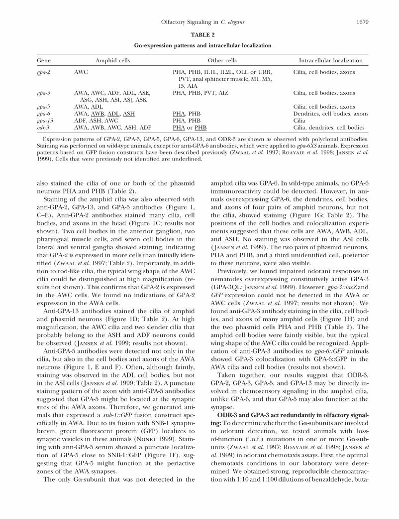
TABLE 2
G�-expression patterns and intracellular localization
Gene Amphid cells Other cells Intracellular localization
gpa-2 AWC PHA, PHB, IL1L, IL2L, OLL or URB, Cilia, cell bodies, axonsPVT, anal sphincter muscle, M1, M5,I5, AIA
gpa-3 AWA, AWC, ADF, ADL, ASE, PHA, PHB, PVT, AIZ Cilia, cell bodies, axonsASG, ASH, ASI, ASJ, ASK
gpa-5 AWA, ADL Cilia, cell bodies, axonsgpa-6 AWA, AWB, ADL, ASH PHA, PHB Dendrites, cell bodies, axonsgpa-13 ADF, ASH, AWC PHA, PHB Ciliaodr-3 AWA, AWB, AWC, ASH, ADF PHA or PHB Cilia, dendrites, cell bodies
Expression patterns of GPA-2, GPA-3, GPA-5, GPA-6, GPA-13, and ODR-3 are shown as observed with polyclonal antibodies.Staining was performed on wild-type animals, except for anti-GPA-6 antibodies, which were applied to gpa-6XS animals. Expressionpatterns based on GFP fusion constructs have been described previously (Zwaal et al. 1997; Roayaie et al. 1998; Jansen et al.1999). Cells that were previously not identified are underlined.
also stained the cilia of one or both of the phasmid amphid cilia was GPA-6. In wild-type animals, no GPA-6immunoreactivity could be detected. However, in ani-neurons PHA and PHB (Table 2).
Staining of the amphid cilia was also observed with mals overexpressing GPA-6, the dendrites, cell bodies,and axons of four pairs of amphid neurons, but notanti-GPA-2, GPA-13, and GPA-5 antibodies (Figure 1,
C–E). Anti-GPA-2 antibodies stained many cilia, cell the cilia, showed staining (Figure 1G; Table 2). Thepositions of the cell bodies and colocalization experi-bodies, and axons in the head (Figure 1C; results not
shown). Two cell bodies in the anterior ganglion, two ments suggested that these cells are AWA, AWB, ADL,and ASH. No staining was observed in the ASI cellspharyngeal muscle cells, and seven cell bodies in the
lateral and ventral ganglia showed staining, indicating (Jansen et al. 1999). The two pairs of phasmid neurons,PHA and PHB, and a third unidentified cell, posteriorthat GPA-2 is expressed in more cells than initially iden-
tified (Zwaal et al. 1997; Table 2). Importantly, in addi- to these neurons, were also visible.Previously, we found impaired odorant responses intion to rod-like cilia, the typical wing shape of the AWC
cilia could be distinguished at high magnification (re- nematodes overexpressing constitutively active GPA-3(GPA-3QL; Jansen et al. 1999). However, gpa-3::lacZ andsults not shown). This confirms that GPA-2 is expressed
in the AWC cells. We found no indications of GPA-2 GFP expression could not be detected in the AWA orAWC cells (Zwaal et al. 1997; results not shown). Weexpression in the AWA cells.
Anti-GPA-13 antibodies stained the cilia of amphid found anti-GPA-3 antibody staining in the cilia, cell bod-ies, and axons of many amphid cells (Figure 1H) andand phasmid neurons (Figure 1D; Table 2). At high
magnification, the AWC cilia and two slender cilia that the two phasmid cells PHA and PHB (Table 2). Theamphid cell bodies were faintly visible, but the typicalprobably belong to the ASH and ADF neurons could
be observed (Jansen et al. 1999; results not shown). wing shape of the AWC cilia could be recognized. Appli-cation of anti-GPA-3 antibodies to gpa-6::GFP animalsAnti-GPA-5 antibodies were detected not only in the
cilia, but also in the cell bodies and axons of the AWA showed GPA-3 colocalization with GPA-6::GFP in theAWA cilia and cell bodies (results not shown).neurons (Figure 1, E and F). Often, although faintly,
staining was observed in the ADL cell bodies, but not Taken together, our results suggest that ODR-3,GPA-2, GPA-3, GPA-5, and GPA-13 may be directly in-in the ASI cells (Jansen et al. 1999; Table 2). A punctate
staining pattern of the axon with anti-GPA-5 antibodies volved in chemosensory signaling in the amphid cilia,unlike GPA-6, and that GPA-5 may also function at thesuggested that GPA-5 might be located at the synaptic
sites of the AWA axons. Therefore, we generated ani- synapse.ODR-3 and GPA-3 act redundantly in olfactory signal-mals that expressed a snb-1::GFP fusion construct spe-
cifically in AWA. Due to its fusion with SNB-1 synapto- ing: To determine whether the G�-subunits are involvedin odorant detection, we tested animals with loss-brevin, green fluorescent protein (GFP) localizes to
synaptic vesicles in these animals (Nonet 1999). Stain- of-function (l.o.f.) mutations in one or more G�-sub-units (Zwaal et al. 1997; Roayaie et al. 1998; Jansen eting with anti-GPA-5 serum showed a punctate localiza-
tion of GPA-5 close to SNB-1::GFP (Figure 1F), sug- al. 1999) in odorant chemotaxis assays. First, the optimalchemotaxis conditions in our laboratory were deter-gesting that GPA-5 might function at the periactive
zones of the AWA synapses. mined. We obtained strong, reproducible chemoattrac-tion with 1:10 and 1:100 dilutions of benzaldehyde, buta-The only G�-subunit that was not detected in the

1680 H. Lans, S. Rademakers and G. Jansen
Figure 1.—Intracellular localization of olfactory G�-subunits. In adult C. elegans (A), the olfactory G�-subunits locate to specificcompartments of the amphid neurons. (B) The location and structures (dd, dendrite; cb, cell body; ax, axon) of the amphidneurons. GPA-2 (C) localizes to cilia (arrow) and cell bodies (arrowheads) of amphid neurons. GPA-13 (D) localizes to amphidcilia (arrow). GPA-5 (red) can be observed in the cilia (arrow), axons (open arrow), and cell bodies (arrowhead) of the AWAcells (E) and is localized nearby snb-1::GFP (green) in the axons (F, open arrows). GPA-6, in gpa-6XS animals, was not detectedin the cilia, but in the dendrites (arrows), cell bodies (arrowhead), and axons (open arrow) of four pairs of amphid cells (G).GPA-3 localizes to the cilia of many amphid cells (H). GPA-3 staining in the rod-like cilia is clearly visible (arrow), surroundedby the more fuzzy wing-shaped AWA and AWC cilia staining (arrowheads). Bars: C, D, E, G, and H, 20 �m; F, 5 �m. (A–H)�400.
none, and isoamyl alcohol, whereas diacetyl, 2,4,5- hol almost completely depended on AWC, whereas onlylow concentrations of benzaldehyde were specific totrimethylthiazole and 2,3-pentanedione gave reliable re-
sults at dilutions ranging from 1:10 to 1:1000. Optimal AWC. Low concentrations of diacetyl and pyrazine werespecifically detected by the AWA cells, but high concen-pyrazine chemotaxis was achieved with 10 and 100 mg/
ml concentrations. Low odorant concentrations are spe- trations involved both AWA and AWC. The responsesto 2,4,5-trimethylthiazole and 2,3-pentanedione seemedcifically detected by either the AWA or the AWC cells
and higher concentrations by both (Bargmann et al. not specific to the AWA or AWC cells at the concentra-tions that gave strong, reproducible results (Table 3 and1993; Chou et al. 2001). Therefore, we determined the
cellular specificity of the odorant response, using odr-7 results not shown).We tested the odr-3(n1605) l.o.f. mutant and con-and odr-1 animals, which are defective in AWA and AWC
olfaction, respectively (Bargmann et al. 1993; Sengupta firmed that odr-3 is necessary for the detection of allodorants, at all concentrations tested (Roayaie et al.et al. 1994). Chemotaxis to butanone and isoamyl alco-

1681Olfactory Signaling in C. elegans
TABLE 3
Cellular specificity of odorant detection
Odorant Wild type odr-7 : AWA odr-1 : AWC
Pyrazine 10 mg/ml 0.78 � 0.03 0.21 � 0.16 0.80 � 0.05Diacetyl 1:1000 0.65 � 0.04 0.20 � 0.18 0.76 � 0.06Trimethylthiazole 1:1000 0.66 � 0.05 0.54 � 0.17 0.75 � 0.07Pentanedione 1:1000 0.80 � 0.04 0.83 � 0.07 0.56 � 0.08Benzaldehyde 1:100 0.83 � 0.06 0.71 � 0.12 0.29 � 0.09Isoamyl alcohol 1:100 0.77 � 0.06 0.85 � 0.06 0.06 � 0.06Butanone 1:100 0.61 � 0.05 0.94 � 0.02 0.24 � 0.08
Shown are the mean chemotaxis indexes (�SEM) of wild-type, odr-7, and odr-1 animals. odr-7 and odr-1animals lack functional AWA and AWC, respectively. AWA or AWC specific responses are indicated in italics.
1998; Figure 2, A–C; results not shown). In our assays, azole (Jansen et al. 1999; Figure 3, A–C), suggestingthat GPA-5 might have an inhibitory function. Furtherthe response to butanone and low concentrations of
diacetyl and benzaldehyde seemed to depend solely on analysis of the olfactory response of gpa-5 odr-3 mutantsshowed that loss of GPA-5 suppressed the chemotaxisodr-3. In contrast, the response to 2,3-pentanedione was
only slightly affected. Other odorants gave intermediate defect of odr-3 animals for all odorants detected by theAWA cells (Figure 3, A and B; results not shown). Sup-phenotypes.
To identify the G� responsible for the residual re- pression was strongest when using high odorant concen-trations. The impaired chemotaxis of odr-3 animals tosponse of odr-3 animals, we also tested other G� l.o.f.
mutants, but observed no or only very mild effects (re- isoamyl alcohol, butanone, and low concentrations ofbenzaldehyde could not be suppressed by loss of GPA-5,sults not shown). This suggested that ODR-3 alone is
sufficient for odorant detection. To test this, we gener- in agreement with the fact that these odorants are de-tected by AWC, in which gpa-5 is not expressed (Figureated animals with mutations in all six G�-subunits except
odr-3. These gpa-2 gpa-3 gpa-5 gpa-6 gpa-13 mutants 3C; results not shown). To confirm that GPA-5 causedthe suppression of the odr-3 phenotype, we tested anshowed wild-type levels of chemotaxis to all odorants at
all concentrations tested (Figure 2, A–C; results not independent gpa-5 l.o.f. allele (pk377), which gave simi-lar results (results not shown).shown), validating the idea that ODR-3 constitutes the
major signaling pathway in AWA and in AWC. Addi- Next, we tested chemotaxis of gpa-2 odr-3 animals.Previously, a stimulatory function for GPA-2 was foundtional loss of odr-3 abrogated chemotaxis to all odorants,
except to pyrazine and high concentrations of 2,4,5- in butanone perception (Roayaie et al. 1998). We foundthat GPA-2 negatively affects odorant detection, sincetrimethylthiazole (Figure 2, A–C; results not shown).
These odorants may also be sensed by other cells (Barg- gpa-2 odr-3 animals showed improved odorant responsesas compared to odr-3 animals (Figure 3, A–C; results notmann et al. 1993).
Subsequently, to identify the G� mediating the resid- shown). Like GPA-5, the inhibitory function of GPA-2was clearest when using high odorant concentrations.ual response in odr-3 animals, double mutants between
odr-3 and gpa-2, gpa-3, gpa-5, gpa-6, or gpa-13 were tested. Only isoamyl alcohol and butanone responses seemednot affected. Since the butanone response was com-Chemotaxis to all odorants was completely abolished in
gpa-3 odr-3 mutants, at all concentrations tested (Figure pletely abolished in odr-3 animals, a redundant stimula-tory function for GPA-2 in butanone detection could2, A–C; results not shown), indicating that GPA-3 also
has a stimulatory role in odorant detection, redundant not be analyzed (Roayaie et al. 1998).Chemotaxis of gpa-6 odr-3 and gpa-13 odr-3 mutantsto ODR-3. This phenotype could be rescued by the intro-
duction of a gpa-3 transgene (Figure 2D). To confirm also was tested. A function for GPA-6 in odorant detec-tion could not be found, which is in agreement with itsthat GPA-3 is functionally redundant to ODR-3, animals
were generated with mutations in all six G�-subunits absence from the cilia. The behavior of gpa-13 odr-3animals was indistinguishable from that of odr-3 animals,except gpa-3. Surprisingly, these gpa-2 gpa-5 gpa-6 gpa-13
odr-3 animals showed strong chemotaxis to all odorants, except for the response to low concentrations of 2,3-pentanedione (Figure 3D). In addition to this pheno-except to butanone (Figure 2, A–C; results not shown).
Especially, chemotaxis to AWA-detected odorants was type, we observed that inactivation of gpa-13 in gpa-2odr-3 or gpa-5 odr-3 animals significantly reduced chemo-very strong. These results indicate that GPA-3 also is
sufficient for detection of most odorants. taxis responses (Figure 3; results not shown), suggestinga minor stimulatory role for GPA-13 in olfactory signaling.GPA-2, GPA-5, and GPA-13 modulate signaling via
ODR-3 and GPA-3: Previously, we have shown that GPA-5 Taken together, our results confirm that ODR-3 con-stitutes the main stimulatory olfactory signaling path-negatively influences the response to 2,4,5-trimethylthi-

1682 H. Lans, S. Rademakers and G. Jansen
Figure 2.—ODR-3 and GPA-3 form the major stimulatory signals in odorant detection. Chemotaxis of odr-3 animals and gpa-2gpa-3 gpa-5 gpa-6 gpa-13 animals (shown as gpa-2 3 5 6 13) shows that ODR-3 is necessary and sufficient for chemotaxis to allodorants (see also Roayaie et al. 1998). GPA-3 is redundant to ODR-3 and is sufficient for chemotaxis at almost wild-type levels,as is evident from chemotaxis of gpa-3 animals, gpa-3 odr-3 double mutants, and gpa-2 gpa-5 gpa-6 gpa-13 odr-3 mutants (shownas gpa-2 5 6 13 odr-3). Mutants that lack all six olfactory G�-subunits (shown as “all”) display no odorant attraction. Shown arethe responses to odorants sensed primarily by AWA [A, 10 mg/ml pyrazine (pyr) and 1:1000 diacetyl (dia)], by AWA and AWC[B, 1:1000 2,4,5-trimethylthiazole (tmt) and 2,3-pentanedione (pent)], and by AWC [C, 1:100 benzaldehyde (benz), isoamylalcohol (iaa), and butanone (but)]. D shows that chemotaxis to 50 mg/ml pyrazine of gpa-3 odr-3 mutants can be restored byintroducing a gpa-3 transgene (shown as gpa-3 odr-3 gpa-3XS). Error bars denote standard error of the mean. Significant differencesof odr-3 and gpa-3 animals compared to wild type (*, P � 0.01), gpa-3 odr-3 compared to odr-3 animals (, P � 0.01; , P �0.05), and gpa-2 gpa-3 gpa-5 gpa-6 gpa-13 and gpa-2 gpa-5 gpa-6 gpa-13 odr-3 animals compared to gpa-2 gpa-3 gpa-5 gpa-6 gpa-13odr-3 mutants (�, P � 0.01; ��, P � 0.05) are shown. In D, # denotes significant difference from gpa-3 odr-3 (P � 0.01).
way. Furthermore, our findings suggest that the olfac- activated upon odorant stimulation and may be neces-sary to obtain a precisely tuned response.tory defect of odr-3 mutants, for most odorants, is caused
by the inhibitory action of GPA-2 and GPA-5. GPA-3 can Olfactory defects are not caused by altered cilia mor-phology: The odorant chemotaxis defects of G�-subunitfully substitute for ODR-3, but only when the function
of GPA-2 or GPA-5 is lost and with the aid of GPA-13 mutants could be the result of developmental or struc-tural defects of the sensory neurons. Such defects haveor when all five other G�-subunits have been inactivated.
Our data suggest that the olfactory response in C. elegans been described for gpa-3 and odr-3 animals. The amphidneurons of gpa-3QL animals have lost the ability to takeis regulated by a complex signaling network, which is

1683Olfactory Signaling in C. elegans
Figure 3.—GPA-5 andGPA-2 negatively regulatechemotaxis to most odorants,and GPA-13 has a stimulatoryfunction. Chemotaxis of ani-mals that lack ODR-3 andGPA-5 (gpa-5 odr-3) or GPA-2(gpa-2 odr-3) reveals thatGPA-5 and GPA-2 inhibitodorant detection in odr-3 an-imals. Furthermore, GPA-13has a stimulatory function,which becomes apparent whengpa-2 is inactivated in an odr-3background (compare gpa-2odr-3 with gpa-2 gpa-13 odr-3).Shown are the responses tohigh concentrations of odor-ants sensed primarily by AWA[A, 100 mg/ml pyrazine (pyr)and 1:10 diacetyl (dia)], byAWA and AWC [B, 1:10 2,4,5-trimethylthiazole (tmt) and2,3-pentanedione (pent)],and by AWC (C, 1:10 benzal-dehyde (benz), isoamyl alco-hol (iaa), and butanone (but)].Low concentrations of diace-tyl (1:1000, A) are also shown.D shows that GPA-13 is spe-cifically involved in 2,3-pen-tanedione (1:1000 diluted;pent) signaling. Error barsdenote standard error of themean. Significant differencesof gpa-5 odr-3, gpa-2 odr-3, andgpa-13 odr-3 animals com-pared to odr-3 animals (*, P �0.01; **, P � 0.05), gpa-2 gpa-13 odr-3 compared to gpa-2odr-3 animals (, P � 0.01;, P � 0.05).
up fluorescent dyes (Zwaal et al. 1997), probably be- tants, except odr-3 animals, had normal axon, dendrite,and cilia structures (Figure 4, B and F; results notcause of structural defects of the cilia (J. Burghoorn
and G. Jansen, unpublished results). Furthermore, the shown). As previously described (Roayaie et al. 1998),the AWC cilia of odr-3 animals were flatter and less welllevel of odr-3 expression controls AWA and AWC cilia
morphology (Roayaie et al. 1998). Low levels of ODR-3 differentiated, whereas the AWA cilia appeared normal.Next, gpa-3 odr-3 double mutants were analyzed. Al-define flattened, filamentous cilia, but this is probably
not the cause of the olfactory defects of odr-3 mutants. though these mutants are more severely defective thanodr-3 animals, their AWA and AWC cilia resembled thoseTo rule out the possibility that altered cilia morphology
causes the olfactory differences seen in G�-mutants, we of odr-3 animals (Figure 4C; results not shown). Like-wise, the cilia of animals with combinations of l.o.f.analyzed the AWA and AWC cilia of these mutants in
more detail. For this purpose, we used the G�-specific mutations in gpa-2, gpa-5, gpa-6, and gpa-13, some ofwhich chemotax better than odr-3 animals (Figures 2antibodies and the AWA-expressed gpa-6::GFP and AWC-
expressed str-2::GFP and gpa-13::GFP transgenes. and 3), were comparable to odr-3 mutants (results notshown). These observations suggest that altered ciliaThe AWA cilia have a branched, filamentous shape,
while the AWC cilia have a wide, wing-like structure morphology, as observed in odr-3 animals, does notcause the apparent olfactory defects or that impaired(Ward et al. 1975; Figure 4, A and E). Unlike the major-
ity of the amphid cilia, these cilia are not exposed to sensory signaling causes cilia degradation.Next, five- and sixfold G�-mutants were examined.the environment (Ward et al. 1975; Ware et al. 1975).
First, single mutant animals were analyzed. All mu- The AWA cilia appeared as wild type in animals that

1684 H. Lans, S. Rademakers and G. Jansen
Figure 4.—ODR-3 plays a role in cilia morphogenesis. (A–D) gpa-6::GFP expression in the AWA cilia. (E–H) gpa-13::GFP inthe AWC and ADF/ASH cilia. The branched, filamentous structure of the AWA cilia is apparent from A–D. Because the structureof the AWC cilia is less apparent in E–H, a schematic of the cilia of AWC (blue) and ADF and ASH (red) is provided. The AWAcilia of wild type (A), odr-3 (B), gpa-3 odr-3 (C), and gpa-2 gpa-3 gpa-5 gpa-6 gpa-13 odr-3 sixfold mutants (D) are all extensivelybranched, despite olfactory differences. The AWA cilia of gpa-2 gpa-3 gpa-5 gpa-6 gpa-13 odr-3 mutants were difficult to visualizeproperly, because gpa-6::GFP is poorly expressed in these mutants. The AWC cilia of odr-3 (F) and gpa-2 gpa-3 gpa-5 gpa-6 gpa-13odr-3 sixfold mutants (H) are less well differentiated than the AWC cilia of wild-type (E) and gpa-2 gpa-3 gpa-5 gpa-6 gpa-13 animals(G). For odr-3 cilia, see also Roayaie et al. (1998). Left is anterior. Bars, 5 �m. (A–H) �630.
expressed only gpa-3, gpa-5, gpa-6, or odr-3, although AWA and the AWC cells, respectively, and GPA-13 hasa minor stimulatory role in the AWC cells. Althoughthese mutants show varying chemotaxis (Figure 2; re-
sults not shown). Also, animals that had lost all six G�- the odorant concentrations used in this study are notall detected specifically by either AWA or AWC, it issubunits showed wild-type AWA cilia (Figure 4D). With
regard to the AWC cilia, nematodes that expressed only highly likely that the observed regulation by G�-subunitsoccurs only in these cells. First of all, no other cells havegpa-2, gpa-3, or gpa-13 and animals with l.o.f. mutations
in all six olfactory G�-subunits had flattened AWC cilia, been reported to be involved in odorant attraction inC. elegans. Second, GPA-5 is expressed only in AWA andsimilar to odr-3 animals (Figure 4H; results not shown).
In contrast, animals that expressed only odr-3, i.e., gpa-2 faintly in ADL, whereas AWC is the only sensory amphidcell in which GPA-2 is expressed. Finally, AWA-specificgpa-3 gpa-5 gpa-6 gpa-13 mutants, had wild-type AWC
cilia (Figure 4G). These results indicate that, of the four expression of GPA-3 rescues AWA-mediated olfactionin a gpa-3 odr-3 background (H. Lans and G. Jansen,AWC-expressed G�-subunits, only ODR-3 is necessary
to establish normal AWC cilia morphology. The other unpublished results).We cannot exclude that other ubiquitously expressedfive olfactory G�-subunits are not involved in AWA or
AWC cilia development. G�-subunits, like goa-1 (Mendel et al. 1995; Segalat etal. 1995), gsa-1 (Korswagen et al. 1997), egl-30 (Lack-ner et al. 1999), and gpa-7 (Jansen et al. 1999), also
DISCUSSIONfunction in the AWA and AWC cells. However, on thebasis of the data presented in this study, it is not likelyOlfaction using four cells requires signaling via five
G�-subunits: Our results show that olfactory signaling that one of these is required for olfactory signaling. Itis striking that signaling in AWA and AWC, the onlyin C. elegans is more complex than initially realized.
Five G�-subunits regulate the response of C. elegans to cells required for odorant attraction (Bargmann et al.1993), involves different G�-subunits. Interestingly, thisattractive odorants (Figure 5). ODR-3 constitutes the
main stimulatory signal in the AWA and AWC cells. correlates well with the existence of dissimilar down-stream signaling pathways in the two neuron pairs (Ta-GPA-3 also provides a stimulatory signal in both neuron
pairs. GPA-5 and GPA-2 have inhibitory functions in the ble 1; Figure 5). In AWA, signaling is reminiscent of

1685Olfactory Signaling in C. elegans
Figure 5.—Model of G� signaling during odorant detection via the AWA and AWC cells. In AWA, both ODR-3 and GPA-3can transduce the odorant signal to the TRPV channel OSM-9/OCR-1/OCR-2, but ODR-3 forms the major pathway. GPA-5, atan unknown effector, cross-interacts with this signal to inhibit signaling. GPA-5 might be activated by the same receptor as ODR-3and GPA-3 or by another receptor. In AWC, mainly ODR-3, but also GPA-3, transduces the odorant signal to the guanylyl cyclasesODR-1 and DAF-11, leading to the opening of the TAX-2/TAX-4 ion channel. GPA-2 and GPA-13 inhibit and stimulate thissignal, respectively.
Drosophila phototransduction, due to its dependence In addition, overlapping G-protein pathways couldfacilitate discrimination between odorants detected byon TRP channel proteins (Colbert et al. 1997; Hardie
and Raghu 2001; Tobin et al. 2002). AWC signaling, the same neuron (Wes and Bargmann 2001). Our find-ing that two G�-subunits, ODR-3 and GPA-3, can medi-on the other hand, resembles that in the mammalian
main olfactory epithelium, where all signals converge ate the detection of all odorants provides a basis for thisidea. Inhibition of ODR-3 and GPA-3 signaling by GPA-2on a cyclic nucleotide-gated channel (Coburn and Barg-
mann 1996; Komatsu et al. 1996; Firestein 2001). Sig- and GPA-5 could also be a means for odorant discrimi-nation. Both adaptation and discrimination assays couldnaling in this system also involves a separate modulatory
pathway and multiple G�-proteins (Jones and Reed shed more light on the involvement of G-proteins inadaptation and discrimination.1989; Belluscio et al. 1998; Wekesa and Anholt 1999;
Luo et al. 2002; Spehr et al. 2002). Finally, another hint at the biological significance ofthe G-protein network could be provided by the findingWhy would C. elegans need several G�-subunits with
overlapping or antagonizing functions to modulate ol- that vulval induction by the Ras-mitogen-activated pro-tein kinase (MAPK) pathway is negatively regulated byfaction? We hypothesize that such a balanced signaling
network is necessary to allow adjustment of the response GPA-5 and the GPCR SRA-13, depending on food condi-tions (Battu et al. 2003). This indicates that olfactoryto an odorant when odorant concentrations or other
environmental conditions change. The inhibitory effects signaling not only serves to direct movement towardfood, but also may regulate developmental processes.of GPA-2 and GPA-5 were most obvious when the animals
were exposed to high odorant concentrations. In these Specificity of G-protein signaling pathways: Our ge-netic analysis of G-protein function in olfactory signal-circumstances, desensitization or adaptation mechanisms
may become activated. Following prolonged exposure to ing shows that the G�-subunits have specific functions,despite their shared localization in the cell. This raiseshigh odorant concentrations, C. elegans shows a dimin-
ished response to that odorant, but not to other odor- the question of how specificity is organized and main-tained.ants (Colbert and Bargmann 1995). This process, which
is called adaptation, involves the unknown adp-1 gene, First of all, specificity may be defined by specific inter-actions with receptors, G��-subunits, and effectors. Itthe TRPV channel subunit OSM-9, and the cGMP-depen-
dent protein kinase EGL-4 (Colbert and Bargmann is likely that ODR-3 and GPA-3 are activated by the samereceptors during odorant detection. GPA-2 and GPA-51995; L’Etoile et al. 2002). Since odorant adaptation
is triggered by calcium and cGMP levels, it seems likely might inhibit signaling by competing for these recep-tors, but could also be activated by different receptorsthat this response is regulated by stimulating and inhib-
iting G�-subunits. Furthermore, odorant-specific adap- or in a receptor-independent fashion. Recently, GPA-5and SRA-13 have been found to negatively regulate atation is modulated by other environmental cues, which
are probably transduced by G-proteins. For example, Ras/MAPK pathway downstream of ODR-3 in the AWCcells (Hirotsu et al. 2000; Battu et al. 2003). Althoughthe absence of food stimulates benzaldehyde adapta-
tion, but not isoamyl alcohol adaptation (Colbert and this could provide a mechanism through which ODR-3and GPA-3 signaling is inhibited, it is unclear how GPA-5Bargmann 1997). Similarly, the presence of food sup-
presses benzaldehyde adaptation (Nuttley et al. 2002). could function in the AWC cells. Furthermore, it isstriking that no effect of GPA-2 on the Ras/MAPK path-Although it is uncertain where the integration of sig-
nals like food availability and odorants occurs, multiple way has been observed (Battu et al. 2003).Another determinant is provided by the ��-dimerG-proteins within one cell might be essential to control
the proper response of an animal to several simultane- that associates with the G�-subunits. C. elegans has twoG�- and two G�-subunits, GPB-1 and -2 and GPC-1 and -2ous cues.

1686 H. Lans, S. Rademakers and G. Jansen
Brenner, S., 1974 The genetics of Caenorhabditis elegans. Genetics(van der Voorn et al. 1990; Zwaal et al. 1996; Jansen77: 71–94.
et al. 1999). GPC-1 functions specifically in a subset of Chase, D. L., G. A. Patikoglou and M. R. Koelle, 2001 Two RGSproteins that inhibit Galpha(o) and Galpha(q) signaling in C.sensory cells, but not in AWA and AWC (Jansen et al.elegans neurons require a Gbeta(5)-like subunit for function.2002). Therefore, the ubiquitously expressed GPC-2Curr. Biol. 11: 222–231.
(Jansen et al. 2002) is the likely candidate to interact Chou, J. H., C. I. Bargmann and P. Sengupta, 2001 The Caenorhab-ditis elegans odr-2 gene encodes a novel Ly-6-related protein re-with the olfactory G�-subunits. In AWA and AWC,quired for olfaction. Genetics 157: 211–224.GPC-2 may dimerize with either GPB-1 or GPB-2, which
Coburn, C. M., and C. I. Bargmann, 1996 A putative cyclic nucleo-are both widely expressed (Chase et al. 2001; van der tide-gated channel is required for sensory development and func-
tion in C. elegans. Neuron 17: 695–706.Linden et al. 2001). Alternatives to GPC-2, however,Colbert, H. A., and C. I. Bargmann, 1995 Odorant-specific adapta-could be the G�-like domain containing RGS proteins
tion pathways generate olfactory plasticity in C. elegans. NeuronEAT-16 and EGL-10, which can interact with GPB-2 14: 803–812.
Colbert, H. A., and C. I. Bargmann, 1997 Environmental signals(Chase et al. 2001; Robatzek et al. 2001; van der Lin-modulate olfactory acuity, discrimination, and memory in Caeno-den et al. 2001). Thus, together there are four differentrhabditis elegans. Learn. Mem. 4: 179–191.
G��(-like) partners. It is difficult to test which of these Colbert, H. A., T. L. Smith and C. I. Bargmann, 1997 OSM-9, anovel protein with structural similarity to channels, is requiredpartners functions in vivo because both gpb-1 and gpc-2for olfaction, mechanosensation, and olfactory adaptation inmutations are lethal (Zwaal et al. 1996; Gotta andCaenorhabditis elegans. J. Neurosci. 17: 8259–8269.
Ahringer 2001). Cuppen, E., A. M. Van Der Linden, G. Jansen and R. H. A. Plasterk,2003 Proteins interacting with Caenorhabditis elegans G� sub-A spatial separation of different pathways is a furtherunits. Comp. Funct. Genomics 4: 479–491.means to confer specificity. Within the cilia, compart-
Finney, M., and G. Ruvkun, 1990 The unc-86 gene product couplesmentalization could separate the different G�-subunits, as cell lineage and cell identity in C. elegans. Cell 63: 895–905.
Firestein, S., 2001 How the olfactory system makes sense of scents.was previously suggested to insulate cGMP signaling dur-Nature 413: 211–218.ing odorant discrimination and adaptation (L’Etoile and
Gotta, M., and J. Ahringer, 2001 Distinct roles for Galpha andBargmann 2000; L’Etoile et al. 2002). Gbetagamma in regulating spindle position and orientation in
Caenorhabditis elegans embryos. Nat. Cell Biol. 3: 297–300.A third method to provide specificity could be a tem-Hamm, H. E., 1998 The many faces of G protein signaling. J. Biol.poral induction of G-protein signaling. For example,
Chem. 273: 669–672.the translocation of the cGMP-dependent protein ki- Hardie, R. C., and P. Raghu, 2001 Visual transduction in Drosophila.
Nature 413: 186–193.nase EGL-4 to the nucleus of the AWC cells, followingHirotsu, T., S. Saeki, M. Yamamoto and Y. Iino, 2000 The Ras-long-term adaptation to odorants (L’Etoile et al. 2002),
MAPK pathway is important for olfaction in Caenorhabditis elegans.may induce the expression of genes necessary for adap- Nature 404: 289–293.
Jansen, G., K. L. Thijssen, P. Werner, M. Van Der Horst, E. Hazen-tation that would otherwise interfere with olfactory sig-donk et al., 1999 The complete family of genes encoding Gnaling.proteins of Caenorhabditis elegans. Nat. Genet. 21: 414–419.
It is becoming increasingly clear that most signaling Jansen, G., D. Weinkove and R. H. Plasterk, 2002 The G-proteingamma subunit gpc-1 of the nematode C. elegans is involved inpathways are part of complex, intracellular signalingtaste adaptation. EMBO J. 21: 986–994.networks. Our characterization of the olfactory system
Jones, D. T., and R. R. Reed, 1989 Golf: an olfactory neuron specific-of C. elegans provides us with the possibility of identifying G protein involved in odorant signal transduction. Science 244:
790–795.the molecular mechanisms that regulate specificity inKomatsu, H., I. Mori, J. S. Rhee, N. Akaike and Y. Ohshima, 1996vivo.
Mutations in a cyclic nucleotide-gated channel lead to abnormalthermosensation and chemosensation in C. elegans. Neuron 17:We thank Ronald Plasterk in whose lab this project was started. We707–718.acknowledge the assistance of Marieke van der Horst and Gert van
Korswagen, H. C., J. H. Park, Y. Ohshima and R. H. Plasterk,Cappellen. We thank Cori Bargmann and the Caenorhabditis Genetics1997 An activating mutation in a Caenorhabditis elegans Gs pro-Center for strains and Andy Fire and Michael Nonet for plasmids. Thistein induces neural degeneration. Genes Dev. 11: 1493–1503.work was supported by the Netherlands Organization for Scientific
Lackner, M. R., S. J. Nurrish and J. M. Kaplan, 1999 FacilitationResearch (NWO, grant ALW 805-48-009), the Royal Dutch Academy of synaptic transmission by EGL-30 Gqalpha and EGL-8 PLCbeta:of Sciences (KNAW), and the Center for Biomedical Genetics, Rotter- DAG binding to UNC-13 is required to stimulate acetylcholinedam, The Netherlands. release. Neuron 24: 335–346.
L’Etoile, N. D., and C. I. Bargmann, 2000 Olfaction and odordiscrimination are mediated by the C. elegans guanylyl cyclaseODR-1. Neuron 25: 575–586.
L’Etoile, N. D., C. M. Coburn, J. Eastham, A. Kistler, G. GallegosLITERATURE CITEDet al., 2002 The cyclic GMP-dependent protein kinase EGL-4
Bargmann, C. I., E. Hartwieg and H. R. Horvitz, 1993 Odorant- regulates olfactory adaptation in C. elegans. Neuron 36: 1079–selective genes and neurons mediate olfaction in C. elegans. Cell 1089.74: 515–527. Luo, A. H., E. H. Cannon, K. S. Wekesa, R. F. Lyman, J. G. Vanden-
Battu, G., E. F. Hoier and A. Hajnal, 2003 The C. elegans G-protein- bergh et al., 2002 Impaired olfactory behavior in mice deficientcoupled receptor SRA-13 inhibits RAS/MAPK signalling during in the alpha subunit of G(o). Brain Res. 941: 62–71.olfaction and vulval development. Development 130: 2567–2577. Mello, C. C., J. M. Kramer, D. Stinchcomb and V. Ambros, 1991
Belluscio, L., G. H. Gold, A. Nemes and R. Axel, 1998 Mice Efficient gene transfer in C. elegans: extrachromosomal mainte-deficient in G(olf) are anosmic. Neuron 20: 69–81. nance and integration of transforming sequences. EMBO J. 10:
Birnby, D. A., E. M. Link, J. J. Vowels, H. Tian, P. L. Colacurcio 3959–3970.et al., 2000 A transmembrane guanylyl cyclase (DAF-11) and Mendel, J. E., H. C. Korswagen, K. S. Liu, Y. M. Hajdu-Cronin,Hsp90 (DAF-21) regulate a common set of chemosensory behav- M. I. Simon et al., 1995 Participation of the protein Go in multi-
ple aspects of behavior in C. elegans. Science 267: 1652–1655.iors in Caenorhabditis elegans. Genetics 155: 85–104.

1687Olfactory Signaling in C. elegans
Nonet, M. L., 1999 Visualization of synaptic specializations in live ing mediated by axon contact and calcium entry regulates asym-metric odorant receptor expression in C. elegans. Cell 99: 387–398.C. elegans with synaptic vesicle protein-GFP fusions. J. Neurosci.
Methods 89: 33–40. van der Linden, A. M., F. Simmer, E. Cuppen and R. H. Plasterk,2001 The G-protein �-subunit GPB-2 in Caenorhabditis elegansNuttley, W. M., K. P. Atkinson-Leadbeater and D. Van Der Kooy,
2002 Serotonin mediates food-odor associative learning in the regulates the Go�-Gq� signaling network through interactionswith the regulator of G-protein signaling proteins EGL-10 andnematode Caenorhabditis elegans. Proc. Natl. Acad. Sci. USA 99:
12449–12454. EAT-16. Genetics 158: 221–235.van der Voorn, L., M. Gebbink, R. H. Plasterk and H. L. Ploegh,Roayaie, K., J. G. Crump, A. Sagasti and C. I. Bargmann, 1998
The G alpha protein ODR-3 mediates olfactory and nociceptive 1990 Characterization of a G-protein beta-subunit gene fromthe nematode Caenorhabditis elegans. J. Mol. Biol. 213: 17–26.function and controls cilium morphogenesis in C. elegans olfactory
neurons. Neuron 20: 55–67. Ward, S., N. Thomson, J. G. White and S. Brenner, 1975 Electronmicroscopical reconstruction of the anterior sensory anatomyRobatzek, M., T. Niacaris, K. Steger, L. Avery and J. H. Thomas,
2001 eat-11 encodes GPB-2, a Gbeta(5) ortholog that interacts of the nematode Caenorhabditis elegans. J. Comp. Neurol. 160:313–337.with G(o)alpha and G(q)alpha to regulate C. elegans behavior.
Curr. Biol. 11: 288–293. Ware, R. W., D. Clark, K. Crossland and R. L. Russell, 1975 Thenerve ring of the nematode C. elegans : sensory input and motorSegalat, L., D. A. Elkes and J. M. Kaplan, 1995 Modulation of
serotonin-controlled behaviors by Go in Caenorhabditis elegans. output. J. Comp. Neurol. 162: 71–110.Wekesa, K. S., and R. R. Anholt, 1999 Differential expression ofScience 267: 1648–1651.
Sengupta, P., H. A. Colbert and C. I. Bargmann, 1994 The C. G proteins in the mouse olfactory system. Brain Res. 837: 117–126.Wes, P. D., and C. I. Bargmann, 2001 C. elegans odour discriminationelegans gene odr-7 encodes an olfactory-specific member of the
nuclear receptor superfamily. Cell 79: 971–980. requires asymmetric diversity in olfactory neurons. Nature 410:698–701.Spehr, M., C. H. Wetzel, H. Hatt and B. W. Ache, 2002 3-phospho-
inositides modulate cyclic nucleotide signaling in olfactory recep- White, J. G., E. Southgate, J. N. Thomson and S. Brenner,1986 The structure of the nervous system of the nematodetor neurons. Neuron 33: 731–739.
Tobin, D., D. Madsen, A. Kahn-Kirby, E. Peckol, G. Moulder et Caenorhabditis elegans. Phil. Trans. R. Soc. Lond. B Biol. Sci. 314:al., 2002 Combinatorial expression of TRPV channel proteins 1–340.defines their sensory functions and subcellular localization in C. Zwaal, R. R., J. Ahringer, H. G. van Luenen, A. Rushforth, P.elegans neurons. Neuron 35: 307–318. Anderson et al., 1996 G proteins are required for spatial orien-
Troemel, E. R., J. H. Chou, N. D. Dwyer, H. A. Colbert and C. I. tation of early cell cleavages in C. elegans embryos. Cell 86: 619–Bargmann, 1995 Divergent seven transmembrane receptors 629.are candidate chemosensory receptors in C. elegans. Cell 83: 207– Zwaal, R. R., J. E. Mendel, P. W. Sternberg and R. H. Plasterk,218. 1997 Two neuronal G proteins are involved in chemosensation
Troemel, E. R., B. E. Kimmer and C. I. Bargmann, 1997 Reprogram- of the Caenorhabditis elegans Dauer-inducing pheromone. Geneticsming chemotaxis responses: sensory neurons define olfactory 145: 715–727.preferences in C. elegans. Cell 91: 161–169.
Troemel, E. R., A. Sagasti and C. I. Bargmann, 1999 Lateral signal- Communicating editor: K. Kemphues

Copyright © 2022 FDOKUMEN