Longevity in Caenorhabditis elegans - Semantic Scholar
14
A Mitochondrial Superoxide Signal Triggers Increased Longevity in Caenorhabditis elegans Wen Yang, Siegfried Hekimi* Department of Biology, McGill University, Montreal, Quebec, Canada Abstract The nuo-6 and isp-1 genes of C. elegans encode, respectively, subunits of complex I and III of the mitochondrial respiratory chain. Partial loss-of-function mutations in these genes decrease electron transport and greatly increase the longevity of C. elegans by a mechanism that is distinct from that induced by reducing their level of expression by RNAi. Electron transport is a major source of the superoxide anion (O ˙ 2 ), which in turn generates several types of toxic reactive oxygen species (ROS), and aging is accompanied by increased oxidative stress, which is an imbalance between the generation and detoxification of ROS. These observations have suggested that the longevity of such mitochondrial mutants might result from a reduction in ROS generation, which would be consistent with the mitochondrial oxidative stress theory of aging. It is difficult to measure ROS directly in living animals, and this has held back progress in determining their function in aging. Here we have adapted a technique of flow cytometry to directly measure ROS levels in isolated mitochondria to show that the generation of superoxide is elevated in the nuo-6 and isp-1 mitochondrial mutants, although overall ROS levels are not, and oxidative stress is low. Furthermore, we show that this elevation is necessary and sufficient to increase longevity, as it is abolished by the antioxidants NAC and vitamin C, and phenocopied by mild treatment with the prooxidant paraquat. Furthermore, the absence of effect of NAC and the additivity of the effect of paraquat on a variety of long- and short-lived mutants suggest that the pathway triggered by mitochondrial superoxide is distinct from previously studied mechanisms, including insulin signaling, dietary restriction, ubiquinone deficiency, the hypoxic response, and hormesis. These findings are not consistent with the mitochondrial oxidative stress theory of aging. Instead they show that increased superoxide generation acts as a signal in young mutant animals to trigger changes of gene expression that prevent or attenuate the effects of subsequent aging. We propose that superoxide is generated as a protective signal in response to molecular damage sustained during wild-type aging as well. This model provides a new explanation for the well-documented correlation between ROS and the aged phenotype as a gradual increase of molecular damage during aging would trigger a gradually stronger ROS response. Citation: Yang W, Hekimi S (2010) A Mitochondrial Superoxide Signal Triggers Increased Longevity in Caenorhabditis elegans. PLoS Biol 8(12): e1000556. doi:10.1371/journal.pbio.1000556 Academic Editor: Heidi A. Tissenbaum, University of Massachusetts Medical School, United States of America Received August 27, 2010; Accepted October 27, 2010; Published December 7, 2010 Copyright: ß 2010 Yang, Hekimi. This is an open-access article distributed under the terms of the Creative Commons Attribution License, which permits unrestricted use, distribution, and reproduction in any medium, provided the original author and source are credited. Funding: The work was supported in part by a grant from the Canadian Institutes of Health Research to SH (grant #216377) and by McGill University. The funders had no role in study design, data collection and analysis, decision to publish, or preparation of the manuscript. Competing Interests: The authors have declared that no competing interests exist. Abbreviations: GFP, green fluorescent protein; HIF, hypoxia-inducible factor; JNK-1, c-Jun N-terminal kinase 1; NAC, N-acetyl-cysteine; PQ, paraquat; ROS, reactive oxygen species; SOD, superoxide dismutase * E-mail: [email protected] Introduction Mitochondrial function has been linked to the aging process in a number of ways [1]. In particular, mitochondria are crucial in energy metabolism and as such have been implicated in the aging process by one of the very first theories of aging [2], the rate-of- living theory of aging [3], which suggested that the rate of aging is proportional to the rate of energy metabolism (reviewed in [4]). Mitochondrial function in animals is also known to decline with age [5,6], which, together with the finding that mitochondria are an important source of toxic reactive oxygen species (ROS), has led to the oxidative stress (or free radical) theory of aging [7,8]. Two types of mutations that affect mitochondrial function have been found to affect the rate of aging in C. elegans, mutations that shorten lifespan, such as mev-1 [9] and gas-1 [10], and mutations that lengthen lifespan, such as clk-1 [11], isp-1 [12], lrs-2 [13], and nuo-6 [14]. lrs-2 encodes a mitochondrial leucyl-tRNA-synthetase, and its effect on the function of mitochondrial electron transport is likely relatively indirect, via partial impairment of mitochondrial translation. However, clk-1 encodes an enzyme necessary for the biosynthesis of ubiquinone, a lipid antioxidant and an electron transporter of the respiratory chain [15], and mev-1, gas-1, isp-1, and nuo-6 all encode subunits of mitochondrial respiratory complexes. On the strength of the oxidative stress theory of aging it has been suggested, and supported by a number of observations (reviewed in [16,17]), that the mev-1 and gas-1 mutations reduce lifespan by increasing mitochondrial oxidative stress, and clk-1, isp-1, and nuo-6 increase lifespan by reducing it. In addition to genomic mutations that affect mitochondrial proteins, it has been found that knockdown by RNA interference of C. elegans genes that encode subunits of mitochondrial complexes, including isp-1 and nuo-6, also prolongs lifespan [13,18,19]. Although the effect of RNAi on ETC subunits, which is conserved in Drosophila [20], was initially believed to be similar to that of the mutations [21,22,23], it was recently found that it is in fact distinct and separable [14]. A recent study analyzed patterns of gene expression in isp-1 mutants together with those in clk-1 and cyc-1(RNAi) [23] and PLoS Biology | www.plosbiology.org 1 December 2010 | Volume 8 | Issue 12 | e1000556
-
Upload
khangminh22 -
Category
Documents
-
view
3 -
download
0
Transcript of Longevity in Caenorhabditis elegans - Semantic Scholar

A Mitochondrial Superoxide Signal Triggers IncreasedLongevity in Caenorhabditis elegansWen Yang, Siegfried Hekimi*
Department of Biology, McGill University, Montreal, Quebec, Canada
Abstract
The nuo-6 and isp-1 genes of C. elegans encode, respectively, subunits of complex I and III of the mitochondrial respiratorychain. Partial loss-of-function mutations in these genes decrease electron transport and greatly increase the longevity of C.elegans by a mechanism that is distinct from that induced by reducing their level of expression by RNAi. Electron transport isa major source of the superoxide anion (O 2), which in turn generates several types of toxic reactive oxygen species (ROS),and aging is accompanied by increased oxidative stress, which is an imbalance between the generation and detoxificationof ROS. These observations have suggested that the longevity of such mitochondrial mutants might result from a reductionin ROS generation, which would be consistent with the mitochondrial oxidative stress theory of aging. It is difficult tomeasure ROS directly in living animals, and this has held back progress in determining their function in aging. Here we haveadapted a technique of flow cytometry to directly measure ROS levels in isolated mitochondria to show that the generationof superoxide is elevated in the nuo-6 and isp-1 mitochondrial mutants, although overall ROS levels are not, and oxidativestress is low. Furthermore, we show that this elevation is necessary and sufficient to increase longevity, as it is abolished bythe antioxidants NAC and vitamin C, and phenocopied by mild treatment with the prooxidant paraquat. Furthermore, theabsence of effect of NAC and the additivity of the effect of paraquat on a variety of long- and short-lived mutants suggestthat the pathway triggered by mitochondrial superoxide is distinct from previously studied mechanisms, including insulinsignaling, dietary restriction, ubiquinone deficiency, the hypoxic response, and hormesis. These findings are not consistentwith the mitochondrial oxidative stress theory of aging. Instead they show that increased superoxide generation acts as asignal in young mutant animals to trigger changes of gene expression that prevent or attenuate the effects of subsequentaging. We propose that superoxide is generated as a protective signal in response to molecular damage sustained duringwild-type aging as well. This model provides a new explanation for the well-documented correlation between ROS and theaged phenotype as a gradual increase of molecular damage during aging would trigger a gradually stronger ROS response.
Citation: Yang W, Hekimi S (2010) A Mitochondrial Superoxide Signal Triggers Increased Longevity in Caenorhabditis elegans. PLoS Biol 8(12): e1000556.doi:10.1371/journal.pbio.1000556
Academic Editor: Heidi A. Tissenbaum, University of Massachusetts Medical School, United States of America
Received August 27, 2010; Accepted October 27, 2010; Published December 7, 2010
Copyright: � 2010 Yang, Hekimi. This is an open-access article distributed under the terms of the Creative Commons Attribution License, which permitsunrestricted use, distribution, and reproduction in any medium, provided the original author and source are credited.
Funding: The work was supported in part by a grant from the Canadian Institutes of Health Research to SH (grant #216377) and by McGill University. Thefunders had no role in study design, data collection and analysis, decision to publish, or preparation of the manuscript.
Competing Interests: The authors have declared that no competing interests exist.
Abbreviations: GFP, green fluorescent protein; HIF, hypoxia-inducible factor; JNK-1, c-Jun N-terminal kinase 1; NAC, N-acetyl-cysteine; PQ, paraquat; ROS,reactive oxygen species; SOD, superoxide dismutase
* E-mail: [email protected]
Introduction
Mitochondrial function has been linked to the aging process in a
number of ways [1]. In particular, mitochondria are crucial in
energy metabolism and as such have been implicated in the aging
process by one of the very first theories of aging [2], the rate-of-
living theory of aging [3], which suggested that the rate of aging is
proportional to the rate of energy metabolism (reviewed in [4]).
Mitochondrial function in animals is also known to decline with
age [5,6], which, together with the finding that mitochondria are
an important source of toxic reactive oxygen species (ROS), has
led to the oxidative stress (or free radical) theory of aging [7,8].
Two types of mutations that affect mitochondrial function have
been found to affect the rate of aging in C. elegans, mutations that
shorten lifespan, such as mev-1 [9] and gas-1 [10], and mutations
that lengthen lifespan, such as clk-1 [11], isp-1 [12], lrs-2 [13], and
nuo-6 [14]. lrs-2 encodes a mitochondrial leucyl-tRNA-synthetase,
and its effect on the function of mitochondrial electron transport is
likely relatively indirect, via partial impairment of mitochondrial
translation. However, clk-1 encodes an enzyme necessary for the
biosynthesis of ubiquinone, a lipid antioxidant and an electron
transporter of the respiratory chain [15], and mev-1, gas-1, isp-1,
and nuo-6 all encode subunits of mitochondrial respiratory
complexes. On the strength of the oxidative stress theory of aging
it has been suggested, and supported by a number of observations
(reviewed in [16,17]), that the mev-1 and gas-1 mutations reduce
lifespan by increasing mitochondrial oxidative stress, and clk-1,
isp-1, and nuo-6 increase lifespan by reducing it.
In addition to genomic mutations that affect mitochondrial
proteins, it has been found that knockdown by RNA interference
of C. elegans genes that encode subunits of mitochondrial
complexes, including isp-1 and nuo-6, also prolongs lifespan
[13,18,19]. Although the effect of RNAi on ETC subunits, which
is conserved in Drosophila [20], was initially believed to be similar to
that of the mutations [21,22,23], it was recently found that it is in
fact distinct and separable [14].
A recent study analyzed patterns of gene expression in isp-1
mutants together with those in clk-1 and cyc-1(RNAi) [23] and
PLoS Biology | www.plosbiology.org 1 December 2010 | Volume 8 | Issue 12 | e1000556

suggested that the overlap between these patterns could define the
biochemical processes that underlie the effect of all interventions
that impact mitochondria. However, our recent findings that isp-
1(qm150) and isp-1(RNAi) trigger fully separable mechanisms
suggests that the overlapping gene expression changes identified by
Cristina et al. [23] might not be sufficient to prolong lifespan.
Rather some of the gene expression changes that are specific to
each type of intervention are necessary for their effect on lifespan
and can act additively.
isp-1 mutants show a trend toward low levels of oxidative
damage to proteins, increased expression of the cytoplasmic Cu/
Zn superoxide dismutase (SOD-1) and of the mitochondrial Mn
superoxide dismutase (SOD-2) [24], and increased resistance to
acute treatment with the prooxidant paraquat [14]. However,
although knocking down the genes encoding the major superoxide
dismutase by RNAi results in normal or elevated levels of oxidative
damage, it had no effect on the lifespan of the mutants [24],
suggesting that the reduced oxidative damage found in isp-1
mutants is not responsible for their longevity. Furthermore, the
notion that mitochondrial oxidative stress could be the cause of
aging has recently been challenged by a number of studies in C.
elegans [24,25,26,27,28], in Drosophila [29], and in mice (reviewed
in [30]).
ROS are not just toxic metabolites that lead to oxidative stress
but are also signaling molecules that are believed to be involved in
a mitochondria-to-nucleus signaling pathway that could impact
aging [1,31,32,33]. Interfering with mitochondrial function has the
potential to alter the rate and/or the pattern of production of ROS
by mitochondria, including in counter-intuitive ways. For
example, reducing oxygen concentration increases ROS produc-
tion by mitochondrial complex III in vertebrate cells [34,35], and
the knockout of sod-2 in C. elegans can lead to normal [25] or
increased lifespan in spite of increased oxidative damage [26].
Here we examined ROS production by mitochondrial mutants
and found that isp-1 and nuo-6 mutants have increased generation
of the superoxide anion but not increased levels of other ROS and
that this increase is necessary and sufficient for longevity,
suggesting that superoxide triggers mechanisms that slow down
aging, presumably at the level of gene expression.
Results
isp-1, nuo-6, and daf-2 Mutant Mitochondria DisplayElevated Generation of Superoxide But Not of OverallROS
To measure changes in mitochondrial ROS generation that
could affect signaling, it is not adequate to measure the level of
ROS damage, as a change in ROS damage levels can be brought
about by changes in detoxification of ROS, in protein turnover, or
in damage repair. However, it is notoriously difficult to directly
visualize or measure ROS generation and ROS levels in intact
organisms including in living worms. To overcome this difficulty
we have adapted a technique originally developed for vertebrates
that uses flow cytometry to sort isolated intact mitochondria and
measure ROS levels with indicator dyes (Figure S1) [37].
Mitochondria were extracted from worms by standard techniques
and loaded with either one of two fluorescent indicator dyes,
H2DCFDA, a dye that is sensitive to a variety of ROS but rather
insensitive to superoxide [38,39], and MitoSox, a dye that is
exclusively sensitive to superoxide [40]. The prooxidant paraquat
(PQ) induces mitochondrial superoxide generation [41], and the
antioxidant N-acetyl-cysteine (NAC) has an antioxidant effect on
all types of ROS [42,43]. As expected, when purified mitochon-
dria were treated with PQ, the fluorescence of both H2DCFDA
and MitoSox increased, and the fluorescence of both decreased
when treated with NAC (Figures 1A, 1B, and S1B). One limitation
of this technique is the need for a rather large amount of
mitochondria. For example, a sufficient amount of worms is not
readily obtained from worms treated by RNAi, and we have
therefore focused on long-lived mutants only.
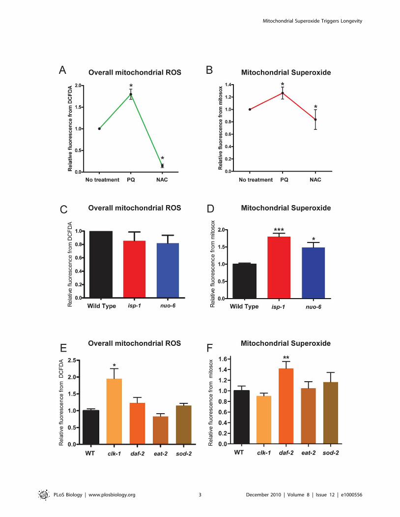
We used the cytometry technique to determine the generation
of mitochondrial superoxide and of overall mitochondrial ROS in
a number of long-lived mutants. Both isp-1 and nuo-6 mutations
did not affect H2DCFDA fluorescence (overall ROS) significantly,
but both showed elevated MitoSox fluorescence (superoxide)
(Figure 1C and 1D). Mutants of four other genes (clk-1(qm30), eat-
2(ad1116), daf-2(e1370), and sod-2(ok1030)) were also tested
(Figure 1E and 1F). clk-1 mutants showed an elevation of overall
ROS-associated fluorescence but not of superoxide-associated
fluorescence. daf-2 mutants were most similar to the mitochondrial
respiratory chain mutants with an elevation of superoxide-
associated fluorescence but no significant elevation in overall
ROS-associated fluorescence. Finally, eat-2 and sod-2 mutants
showed no significant elevation in either signal but only a trend for
low overall ROS in the case of eat-2 mutants and a trend for
increased superoxide in the case of sod-2 mutants.
The elevated MitoSox signal in isp-1, nuo-6, and daf-2 corre-
sponds mostly to increased superoxide generation, as all three
mutants are known for elevated levels of the mitochondrial SOD-2
and SOD-3 [12,14,24,44], whose activity would prevent the
accumulation of superoxide. Elevated superoxide detoxification,
however, should not prevent measuring increased superoxide
generation as superoxide is generated at prosthetic electron carriers
such as ubiquinone in complex III [45,46] and FMN in complex I
[47,48], which are at least partially buried in the complexes. Thus a
small molecular weight dye that has access to these sites can trap the
superoxide before it has the opportunity to diffuse toward the SOD-
2 and SOD-3 proteins. There is no increase in the H2DCFDA
signal in these mutants likely because this dye is not particularly
sensitive to superoxide [49]. It appears therefore that in the presence
of efficient detoxification the level of overall ROS is not significantly
increased by the increased superoxide generation that we observe.
This is consistent with the finding that these mutants do not have
increased oxidative damage [14,24].
Author Summary
An unequivocal demonstration that mitochondria areimportant for lifespan comes from studies with thenematode Caenorhabditis elegans. Mutations in mitochon-drial proteins such as ISP-1 and NUO-6, which functiondirectly in mitochondrial electron transport, lead to adramatic increase in the lifespan of this organism. Onetheory proposes that toxicity of mitochondrial reactiveoxygen species (ROS) is the cause of aging and predictsthat the generation of the ROS superoxide should be lowin these mutants. Here we have measured superoxidegeneration in these mutants and found that it is in factelevated, rather than reduced. Furthermore, we found thatthis elevation is necessary and sufficient for longevity, as itis abolished by antioxidants and induced by mildtreatment with oxidants. This suggests that superoxidecan act as a signal triggering cellular changes thatattenuate the effects of aging. This idea suggests a newmodel for the well-documented correlation between ROSand the aged phenotype. We propose that a gradualincrease of molecular damage during aging triggers aconcurrent, gradually intensifying, protective superoxideresponse.
Mitochondrial Superoxide Triggers Longevity
PLoS Biology | www.plosbiology.org 2 December 2010 | Volume 8 | Issue 12 | e1000556
Mitochondrial Superoxide Triggers Longevity
PLoS Biology | www.plosbiology.org 3 December 2010 | Volume 8 | Issue 12 | e1000556

sod-2 deletion mutants do not show a significant increase in the
MitoSox signal (Figure 1E and 1F), indicating that decreased
detoxification does not lead to an easily measurable increase in this
signal in purified mitochondria. The signal from H2DCFDA, a dye
which has very broad sensitivity but is not very sensitive to superoxide
[49], is also unchanged, suggesting that, at least in isolated worm
mitochondria, electron transport is not the main source of the type of
ROS to which H2DCFDA dye is significantly sensitive. The level of
superoxide generation in these mutants might also be kept
moderately low because of their reduced electron transport [26],
although low electron transport could in principle also result in
elevated superoxide as we have observed in isp-1 and nuo-6 mutants.
clk-1 mutants have only a small deficit in electron transport
[24,50,51], in spite of a strongly altered content in quinones
[51,52,53,54]. Indeed, while wild-type animals contain endoge-
nously synthesized UQ9 as well as a small amount of dietary
bacterial UQ8, clk-1 mutants contain only the dietary ubiquinone
and no UQ9. Here we found that clk-1 mutants have normal
superoxide generation but enhanced overall ROS levels, which
suggests that the antioxidant function of UQ9 is a crucial sink for
mitochondrial ROS, whose absence appears to lead to an increase
of overall ROS even in the absence of increase superoxide
generation. eat-2 mutants are long-lived because of reduced food
intake (dietary restriction) [55]. Although dietary restriction has
been found to impinge on mitochondrial function in other systems,
no changes in mitochondrial superoxide and overall ROS signals
were observed.
Elevated Superoxide Promotes LongevityTo determine how the elevated superoxide affects the lifespan of
mutants, we treated worms with 10 mM of NAC and scored their
survival (Figure 2 and Table 1). The treatment had no effect on the
survival of the wild type (Figure 2A), which shows that it is not
toxic for lifespan at the concentration used. However, NAC
treatment fully abolished the increased longevity of nuo-6 and
severely limited that of isp-1 (Figure 2B and 2C). The lesser effect
on isp-1 is consistent with the larger increase of superoxide in these
mutants (Figure 1D), given that the effect of NAC is gradual
(1 mM has less effect than 8 mM, which has less than 10 mM;
Table S1). At high concentration (.10–15 mM) NAC can be
deleterious even on the wild type, but at the concentration used
(10 mM) NAC had no effect on the apparent health of the
mutants, whose overall aspect after treatment was indistinguish-
able from that of the untreated worms (Figure S2A). We have also
quantified several phenotypes, including defecation, swimming,
brood size, and post-embryonic development, after NAC treat-
ment of the wild type and of nuo-6, which is the mutant that is most
sensitive to NAC (10 mM NAC completely abolishes its increased
longevity). Treatment with 1 mM vitamin C also significantly
shortened the lifespan of both isp-1 and nuo-6 mutants without
affecting the wild type (Table S1). Most effects of NAC were quite
small (Figure S2B–E), except on the post-embryonic development
of the wild type (Figure S2C). Furthermore, for defecation, brood
size, and post-embryonic development, the effect of NAC on the
mutant produced a change in the same direction as on the wild
type but of a lesser extent. Only for swimming is the effect greater
on the mutant. But the effect consists of swimming faster after
NAC treatment and thus bringing the mutant phenotype closer to
the wild-type. We conclude that there is little evidence of an
indirect deleterious effect of NAC.
NAC had only a moderate effect on the lifespan of the insulin-
signaling daf-2 mutants (Figure 2E), suggesting that only a small
part of the increased longevity of these mutants requires elevated
mitochondrial superoxide. However, NAC fully abolished the
increased lifespan of sod-2 mutants (Figure 2F), suggesting that,
although increased generation of superoxide and other ROS as
detected by our techniques were not significantly altered in these
mutants, their increased lifespan depends on an elevation of
superoxide or some other ROS. NAC did not shorten the lifespan
of clk-1 mutants at 10 mM (Figure 2D), or even at 15 mM (Table
S1), indicating that ROS metabolism is relatively irrelevant to the
aging phenotype of these mutants. The effect of NAC on the
lifespan of eat-2 could not be scored because NAC treatment
rendered the animals unable to lay their eggs and they died from
internal hatching at a young age. The origin of this effect is
unknown. We also could not score the effect of NAC on RNAi-
treated worms because 10 mM NAC was excessively damaging to
the dsRNA-producing bacterial strain (HT115).
Treatment With PQ at Low Concentration (0.1 mM)Increases Lifespan
To determine whether an elevation in mitochondrial superox-
ide generation is sufficient to increase lifespan, we used the
superoxide generator PQ. Treatment of C. elegans with high
concentration of PQ (.0.2 mM) is severely deleterious. We thus
first tested the ability of PQ to increase ROS damage in the
animals at a very low concentration (0.1 mM). We found that this
treatment indeed measurably increased the level of oxidative
damage to proteins at the young adult stage as assessed by
determination of protein carbonylation (Figure 3A) and increased
the expression of both the main cytoplasmic (SOD-1) and the
main mitochondrial (SOD-2) superoxide dismutases (Figure 3B
and 3C). We then tested whether PQ could increase the lifespan
of the wild type at three different concentrations (0.05, 0.1, and
0.2 mM) and found that at all three concentrations both the
mean and maximum lifespan were increased, with a maximal
effect at 0.1 mM (Figures 3D and 4A, and Tables 1 and S1). The
effect of 0.2 mM was less pronounced than that of 0.1 mM and
similar to that of 0.05 mM, likely because at 0.2 mM a toxic
effect starts to balance the pro-longevity effect. The effect does
not depend on the exact chemical structure of paraquat, as
benzyl-viologen, a compound with similar activity as PQ but
structurally different, also increases lifespan (Table S1). A small
effect of the prooxidant juglone under different conditions has
also been documented previously [56]. The effect did not depend
on an effect of PQ on the E. coli (OP50) food source, as the effect
was also observed with heat-killed cells (Table S1). Finally, the
effect was not confined to development or adulthood as PQ
prolongs lifespan whether provided only during adult lifespan or
only during development (Table S1).
Figure 1. Reactive oxygen species (ROS) in isolated mitochondria of long-lived mutants and in response to paraquat (PQ) and N-acetyl-cysteine (NAC) treatment. Global ROS levels were measured by quantifying the fluorescence of the reporter dye H2DCFDA, and superoxidewith the dye MitoSox, in FACS-sorted mitochondria. Values are normalized to the value of the untreated sample or the wild type. PQ and NAC,respectively, increase and decrease the levels of both global ROS (A) and superoxide (B). Mitochondria isolated from isp-1(qm150) and nuo-6(qm200)mutants show slightly decreased global ROS generation (C) but significantly increased superoxide generation (D). Mitochondria from clk-1 mutants,but not from daf-2(e1370), eat-2(ad1116), and sod-2(ok1030) mutants, show significantly increased global ROS levels (E). Mitochondria from daf-2mutants, but not from clk-1, eat-2, and sod-2 mutants, show increased superoxide levels (F). * p,0.05, ** p,0.001, *** p,0.0001, by the paired t test.doi:10.1371/journal.pbio.1000556.g001
Mitochondrial Superoxide Triggers Longevity
PLoS Biology | www.plosbiology.org 4 December 2010 | Volume 8 | Issue 12 | e1000556

PQ treatment failed to significantly prolong the lifespan on nuo-
6 and isp-1 mutants (Figure 4B and 4C, and Tables 1 and S1).
This experiment is equivalent to genetic epistasis analysis and
suggests that nuo-6, isp-1, and PQ increase lifespan by the same
mechanism. It also suggests that the maximum level of lifespan
extension that can be obtained by increasing mitochondrial
superoxide generation is already reached in these two mutants
and further increase of superoxide generation through PQ
Figure 2. Lifespans of wild-type animals and mutants treated with 10 mM NAC. The treatment has no effect on wild type (A) butdramatically suppresses the lifespan extension of the respiratory chain subunit mutants nuo-6 (B) and isp-1 (C). In contrast, it has no lifespan-shortening effects on the long-lived clk-1 mutants (D). NAC treatment has only a moderate effect on the very long-lived daf-2 mutants (E) but alsocompletely abolished the extended longevity of sod-2 mutants (F). See Tables 1 and S1 for details of genotype, sample size, and statistical analysis.doi:10.1371/journal.pbio.1000556.g002
Mitochondrial Superoxide Triggers Longevity
PLoS Biology | www.plosbiology.org 5 December 2010 | Volume 8 | Issue 12 | e1000556

treatment cannot increase lifespan further. This was not due to a
resistance of these mutants to PQ as 0.2 mM PQ shortened the
lifespan of the two mutants (Table S1). sod-2 mutants, whose
longevity is suppressed by NAC, are not as long-lived when
untreated as wild type animals that are treated with PQ.
However, treatment with PQ makes the sod-2 mutants live as
long as wild type animals treated with PQ (Figure 4G). This
absence of additivity suggests that the longevity of sod-2 mutants is
indeed due to a small increase in superoxide, as expected from
the function of SOD-2, and the suppressing effect of NAC on the
mutant lifespan. In contrast to what we observed with nuo-6, isp-1,
and sod-2, PQ treatment dramatically enhanced the lifespan of
clk-1 and eat-2 mutants, significantly beyond the longevity
increases induced by the mutations alone or by PQ applied to
the wild type (Figure 4D and 4E, and Tables 1 and S1). This
indicates that the effects of these mutations and the effect of
superoxide are mechanistically distinct and additive, as expected
from the finding that clk-1 and eat-2 mutants did not show
increased mitochondrial superoxide levels (Figure 1F) and that
the lifespan of clk-1 mutants could not be shortened by NAC
treatment (Figure 2D). PQ treatment had only a small lifespan-
lengthening effect on daf-2 (Figure 4F, and Tables 1 and S1),
which is consistent with the finding that daf-2 mutants already
show a substantial increase in superoxide generation.
NAC and PQ Treatments Do Not Affect OtherMitochondrial Parameters in a Manner That Could PredictTheir Effect on Lifespan
We sought to determine whether the mutations and the PQ
treatment had other common effects on mitochondrial function that
could be responsible for the increased lifespans, besides elevation of
superoxide levels. Work in other systems has suggested that
increased mitochondrial biogenesis could impact lifespan positively
[57,58,59], and mitochondrial defects in C. elegans have been found
to stimulate mitochondrial biogenesis, resulting in a denser
mitochondrial network [13]. We have examined mitochondrial
density in the two mitochondrial mutants and in PQ-treated worms
with Mitotracker Red, which is specific to mitochondria in
mammalian cells [60,61], which stains worms uniformly, and
whose staining fully overlaps with that of mitochondrially targeted
green fluorescent protein (GFP) (Figure S3). We found that isp-1 and
nuo-6 display a denser mitochondrial network, as expected (Figure 5).
However, this was not observed in wild type worms treated with PQ
Table 1. Longevity after paraquat and NAC treatment.
Control PQ NAC
Mean ± SDSample Size
MaximumLifespan
Mean ± SDSample Size
MaximumLifespan
% Changep Value
Mean ± SDSample Size
MaximumLifespan
% Changep Value
Wild Type (N2) 18.463.5 29 29.065.1 43 +58% 20.263.2 30 +10%
(n = 400) (n = 200) p,0.0001 (n = 100) 0.1655
nuo-6(qm200) 33.467.6 49 36.168.3 54 +8% 20.763.0 28 238%
(n = 200) (n = 200) p = 0.0125 (n = 73) ,0.0001
isp-1(qm150) 33.967.9 53 35.067.3 50 +3% 25.665.6 38 224%
(n = 200) (n = 150) p = 0.0961 (n = 200) ,0.0001
clk-1(qm30) 20.763.9 32 36.867.4 58 +78% 23.063.9 30 +11%
(n = 150) (n = 200) p,0.0001 (n = 100) ,0.0001
daf-2(e1370) 43.869.0 67 47.968.8 74 +9% 37.369.8 54 215%
(n = 150) (n = 150) p = 0.0002 (n = 100) 0.0006
eat-2(ad1116) 29.467.1 44 39.466.3 58 +34% ND ND ND
(n = 100) (n = 150) p,0.0001
daf-16(m26) 16.762.3 21 22.663.6 28 +35% ND ND ND
(n = 100) (n = 100) p,0.0001
aak-2(ok524) 17.362.5 23 22.464.2 30 +29% ND ND ND
(n = 100) (n = 100) p,0.0001
jnk-1(gk7) 18.163.0 25 29.066.0 39 +60% ND ND ND
(n = 100) (n = 100) p,0.0001
wwp-1(ok1102) 21.962.9 28 29.365.3* 37 +34% ND ND ND
(n = 100) (n = 100) p,0.0001
skn-1(zn67) 17.662.3 22 22.86 2.8* 25 +30% ND ND ND
(n = 50) (n = 50) p,0.0001
hsf-1(y441) 14.963.1 22 22.565.7 37 +51% ND ND ND
(n = 100) (n = 100) p,0.0001
hif-1(ia4) 23.464.1 31 28.766.8 42 +23% ND ND ND
(n = 50) (n = 50) p,0.0001
*For these genotypes paraquat was only applied after the worms reached adulthood because of lethal effects during development. They should be compared to thewild type (N2) treated only during adulthood (not shown in the table), whose lifespan is 25.864.8, which is a 39% increase compared to non-treated worms.doi:10.1371/journal.pbio.1000556.t001
Mitochondrial Superoxide Triggers Longevity
PLoS Biology | www.plosbiology.org 6 December 2010 | Volume 8 | Issue 12 | e1000556

(Figure 5), indicating that the mechanism by which the superoxide
triggers longevity does not require increased mitochondrial
biogenesis. We also tested the effects of PQ and NAC treatment
on oxygen consumption and ATP levels in the wild type and in the
two mitochondrial mutants (Figure S4). NAC treatment increased
oxygen consumption in the wild type and in the mutants. This result
uncouples oxygen consumption from lifespan as NAC has no effect
on the lifespan of the wild type, and its effect on the oxygen
consumption of isp-1 mutants is larger than on that of nuo-6 mutants,
although its effect on aging is smaller. PQ had an effect only on nuo-
6, and it was small. Thus the effect of PQ on oxygen consumption
also did not mirror its effect on lifespan. For ATP levels the only
effect observed was a reduction by PQ of the elevated ATP levels
that are observed in nuo-6 mutants.
PQ Is Able to Considerably Lengthen the Lifespan of aVariety of Mutants That Define Genetic Pathways ofAging
daf-2 mutants have elevated superoxide levels, and they are
sensitive to NAC (lifespan shortening by 15%). However, the
level of superoxide in daf-2 appears not to be sufficient for a
maximal effect as these mutants remain somewhat sensitive to
PQ (lifespan lengthening by 9%). To further study how
superoxide plays a role in the lifespan of daf-2 we studied
genes that function downstream of daf-2. At least three genes
are known to be required for the full lifespan extension of daf-2,
that is, daf-16, aak-2, and hsf-1 [62,63,64]. If one of these genes
were necessary for an activity that mediates the small effect of
PQ on daf-2 mutants, PQ should not be able to prolong the
lifespan of mutants of such a gene. In fact, however, we found
that PQ prolonged the lifespan of all three mutants tested
(Table 1). The lifespan increase upon PQ treatment of daf-16
(35% increase) and aak-2 (29% increase) is not as large as upon
treatment of the wild type (58% increase). This suggests that
part but not all of the lifespan increase determined by
superoxide requires daf-16 and aak-2. These findings are
consistent with the observations that the lifespan extension
provided by nuo-6 and daf-2(e1370) are only partially additive
(Table S1), similarly to what was found previously for isp-1 and
daf-2 [12], and that elimination of daf-16 partially shortens the
lifespan of isp-1 [12].
Figure 3. Treatment with 0.1 mM of paraquat (PQ) increases protein oxidative damage, superoxide dismutase expression, andlifespan. (A) Young wild type adults treated with 0.1 mM PQ have higher protein oxidative damage compared to untreated control. (B) Young wildtype adults treated with 0.1 mM PQ since hatching express significantly more SOD-1 protein than untreated animals. (C) Young wild type adultstreated with 0.1 mM paraquat since hatching express significantly more SOD-2 protein compared to untreated wild type worms. (D) Treatment with0.05, 0.1, or 0.2 mM PQ increases mean and maximum lifespan significantly (see also Tables 1 and S1). *,0.05, ** p,0.001.doi:10.1371/journal.pbio.1000556.g003
Mitochondrial Superoxide Triggers Longevity
PLoS Biology | www.plosbiology.org 7 December 2010 | Volume 8 | Issue 12 | e1000556
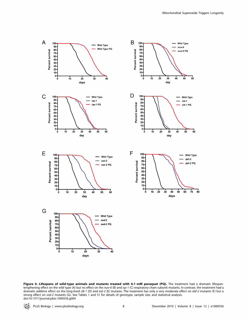
Figure 4. Lifespans of wild-type animals and mutants treated with 0.1 mM paraquat (PQ). The treatment had a dramatic lifespan-lengthening effect on the wild type (A) but no effect on the nuo-6 (B) and isp-1 (C) respiratory chain subunit mutants. In contrast, the treatment had adramatic additive effect on the long-lived clk-1 (D) and eat-2 (E) mutants. The treatment has only a very moderate effect on daf-2 mutants (F) but astrong effect on sod-2 mutants (G). See Tables 1 and S1 for details of genotype, sample size, and statistical analysis.doi:10.1371/journal.pbio.1000556.g004
Mitochondrial Superoxide Triggers Longevity
PLoS Biology | www.plosbiology.org 8 December 2010 | Volume 8 | Issue 12 | e1000556

Mitochondrial Superoxide Triggers Longevity
PLoS Biology | www.plosbiology.org 9 December 2010 | Volume 8 | Issue 12 | e1000556

We also tested the sensitivity to PQ of mutants that are
diagnostic of a variety of pathways of aging. In particular mutants
of genes that, based on their known functions in C. elegans or that of
their homologues in other systems, might encode the targets of
superoxide signaling or be otherwise necessary for implementing
superoxide signaling. The c-Jun N-terminal kinase 1 (JNK-1) is
involved in stress responses in vertebrate cells and is a positive
regulator of DAF-16 that acts in parallel to the effect of daf-2 on
daf-16 [65]. We treated jnk-1(gk7) mutants with PQ and obtained a
particularly large lifespan increase (Table 1). Although it is not
clear what activities lie upstream of jnk-1 in C. elegans nor whether it
has other targets than daf-16, its activity does not appear necessary
for the effect of superoxide. The transcription factor SKN-1
defends against oxidative stress by mobilizing the conserved phase
II detoxification response and can delay aging independently of
DAF-16 [66]. Although PQ induces oxidative stress and induces
enzymes that protect from oxidative stress (Figure 3), it was still
able to prolong the lifespan of skn-1(zn67) mutants (Table 1),
indicating that skn-1 does not act downstream of superoxide.
wwp-1 encodes a conserved E3 ubiquitin ligase that is necessary for
lifespan extension by dietary restriction [67]. Treatment of
wwp-1(ok1102) with PQ prolonged lifespan of these mutants,
which is consistent with our finding that PQ can considerably
extend the lifespan of eat-2 mutants (Figure 4E). This confirmed
that the lifespan increase produced by the superoxide increase in
mitochondrial mutants is distinct from the mechanisms that
support the lifespan effects of dietary restriction [14]. hif-1 encodes
a worm homologue of the vertebrate hypoxia inducible factor 1a(HIF-1a), a transcription factor involved in a number of protective
mechanisms. In C. elegans hif-1 is necessary for a lifespan pathway
that involves proteolysis and that is distinct from insulin signaling
[68] and has also been involved in the dietary restriction pathway
[69]. In vertebrates HIF-1a is positively regulated by mitochon-
drial ROS [34,35], which would make it an interesting candidate
to mediate the effects of superoxide. However, PQ was fully
capable of increasing the lifespan of the hif-1 mutants (Table 1).
Several of the genes whose mutants remain sensitive to PQ,
including daf-16, have been involved in stress responses, including
oxidative stress, yet they do not seem necessary for the effect of
PQ. Similarly we have shown previously that although the
expression of SOD-1 and SOD-2 are elevated in isp-1(qm150)
mutants, the elevation is not necessary for the extended lifespan of
these mutants [24]. nuo-6(qm200) mutants also show elevated
SOD-1 and SOD-2 expression [14], but this too is unnecessary for
the longevity of the mutants, as RNAi against sod-1 an sod-2, which
we have shown to be efficient in reducing enzyme levels [24], does
not shorten the lifespan of nuo-6 mutants (Figure S5). We conclude
that the mitochondrial mutants protect from an aspect of the aging
process that has not yet been studied through mutants that affect
stress. In addition, our observations suggest that the lifespan effect
we observed is not hormetic, as neither superoxide-detoxifying
enzymes, nor the regulatory factors that are involved in protection
from oxidative stress, are crucially implicated.
Discussion
We have shown previously that mutations in isp-1 and nuo-6
prolong lifespan by a common mechanism [14]. Using direct
measurement of ROS and superoxide we find here that this
mechanism involves an increase in mitochondrial superoxide
generation that is necessary and sufficient for the longevity of these
mutants. As ROS, including superoxide [70,71,72], are known to
be intracellular messengers, the increased superoxide might trigger
a signal transduction pathway that ultimately results in changes in
nuclear gene expression [23]. Superoxide is highly reactive and
could trigger such a signal by modifying proteins in the
mitochondria or in the nearby cytosol after having escaped from
the mitochondria through an appropriate channel [73,74].
Although no superoxide sensor has yet been identified, a similar
type of mechanism, in which a highly reactive, quickly diffusing,
molecule modifies a signal transduction protein, has been
evidenced for nitric oxide (NO), which covalently and perma-
nently modifies guanylyl cyclases. Similarly, hydrogen peroxide
(H2O2), the product of superoxide dismutation, can inactivate
phosphatases involved in signal transduction. Future work will aim
at using forward and reverse genetic screens in C. elegans to uncover
the molecular machinery that reacts to the superoxide signal, as
well as the transcription factors that are needed to regulate nuclear
gene expression in response to the pathway’s activation. In
addition, the pattern of gene expression that results in increased
lifespan in these mutants could be defined very specifically by
identifying changes in the gene expression patterns that are
common to isp-1, nuo-6, and PQ treatment and that are suppressed
by treatment with NAC.
A number of studies in C. elegans have explored hormesis by
treating animals with sub-lethal but clearly deleterious treatments
for a short period of time and observing subsequent prolongation
of lifespan [75]. These hormetic effects are different from what we
have observed and describe here, as both the genetic mutations
and the very low level PQ are present throughout life and as only a
part of the effect we observe might require the insulin signaling
pathway. Furthermore, although in nuo-6 and isp-1 mutants the
expression levels of the superoxide dismutases SOD-1 and SOD-2
are elevated, likely in response to the elevated superoxide
generation, and as one expects in the hormetic response, these
elevations are not necessary for the lifespan prolongation of nuo-6
(Figure S5) or isp-1 [24].
CLK-1 is a mitochondrial protein that is required for
ubiquinone biosynthesis and its absence affects mitochondrial
function [50], although it could potentially affect many other
processes as ubiquinone is found in all membranes. Furthermore,
ubiquinone is both a prooxidant as co-factor in the respiratory
chain and an anti-oxidant. Interestingly, the mechanism of lifespan
prolongation induced by clk-1 appears to be entirely distinct from,
but particularly synergistic with, that induced by elevated
superoxide. Indeed, clk-1 mutants do not show elevated superoxide
generation and are not affected by NAC. Furthermore, although
double mutant combinations of clk-1 with nuo-6 and isp-1 are not
Figure 5. Treatment with 0.1 mM PQ does not affect mitochondrial abundance. Worms were treated with Mitotracker Red at 50 nM (finalconcentration) in M9 buffer for 20 min. All pictures were taken by confocal microscopy at 4006. A scale bar of 20 mm is shown in the upper rightcorner of the figure. For each genotype/treatment three tissues (hypodermis, muscle, and germline) were selected, and for each tissue at least fivepictures from different worms were taken. An equal section of each picture was enlarged for quantitative comparisons. The percentage of surfaceoccupied by mitochondria as stained by Mitotracker Red was measured and related to the total area of the selected region. A representative examplefor each tissue and condition is shown in the figure. The quantification for each sample is also shown in the figure below the enlarged areas. Thesample size for the hypodermis was .15, and it was 5 for muscles and the germline. In muscles and in the hypodermis, the difference between theETC mutants and the wild type was significant (p,0.001), while the difference between PQ treatment and untreated wild type worms was not. Thus,the nuo-6 and isp-1 mutations, but not treatment with 0.1 mM PQ, affect mitochondrial abundance.doi:10.1371/journal.pbio.1000556.g005
Mitochondrial Superoxide Triggers Longevity
PLoS Biology | www.plosbiology.org 10 December 2010 | Volume 8 | Issue 12 | e1000556
viable (unpublished data) the lifespans of clk-1 mutants treated with
PQ (Figure 4D), or of sod-2;clk-1 mutants [26], or of clk-1;daf-2
mutants [76] are much greater than expected from simple
additivity of the effects of individual mutations or treatments.
Studies in yeast [77] and in worms [78] have suggested that an
increase in ROS from mitochondria might also be important in
triggering the lifespan extension produced by glucose restriction.
However, our results here with an eat-2 mutation, one of the ways
in which global dietary restriction can be produced in worms, as
well as with a wwp-1 and hif-1, which may function downstream of
dietary restriction, did not reveal an involvement of superoxide
signaling, providing further evidence for a distinction between the
mechanisms of glucose restriction and dietary restriction. It
remains possible, however, that DR could lead to superoxide or
ROS production when it is induced by other methods than the use
of an eat-2 mutant, as it is well documented that different types of
DR induce different molecular mechanisms [79].
One question that our current experiments do not address is
whether the mitochondrial dysfunction in the mutants, or the
effect of PQ, is necessary in every tissue in order to increase
longevity. There are indications for both the insulin signaling
pathway mutants [80,81] and dietary restriction [67,82] that the
entire effect might be mediated by action in particular cells that
influence the physiology of the whole organism. Similarly, the
presence or absence of the germline is sufficient for a dramatic
effect on lifespan [83]. For mitochondrial dysfunction the question
could be addressed in the future by mosaic analysis and by
purifying and analyzing mitochondria from specific tissues using
our flow cytometry technique to purify mitochondria expressing
GFP in a tissue-specific manner.
The oxidative stress theory of aging has been one of the most
acknowledged theories of aging for the simple reason of the
strikingly good correlation between the levels of oxidative stress
and the aged phenotype [8]. A number of recent results in worms
and in mice, however, have suggested that oxidative stress cannot
be the cause of aging [24,25,26,30]. Our findings suggest a
conceptual framework for why oxidative stress and the aged
phenotype are so tightly correlated [31]. In this model mitochon-
dria, like the rest of the cell, sustain a variety of age-dependent
insults (not only and not even principally from oxidative stress) that
trigger an increase in superoxide, which acts as a signal that
induces general protective and repair mechanisms. However,
aging in most animals is clearly irreversible, indicating that the
protective mechanisms, which must have evolved to control
damage in young organisms, are unable to fully prevent the
accumulation of age-related damage. Thus, as superoxide is a
reactive molecule as well as a signal, and as age-dependent
damage cannot be fully reversed, it is possible that at high ages the
chronically elevated superoxide will participate in creating some of
the damage itself. This could explain the strong tendency for aged
animals to have high oxidative stress and high oxidative damage,
although it does not imply that ROS cause aging or even that they
are a major source of age-dependent damage. In this model, the
nuo-6 and isp-1 mutations lead to increased longevity because they
turn on the stress signal prematurely and thus slow down the entire
process.
Materials and Methods
Lifespan ScoringEggs were placed on plates at 20uC and left for 1 h to hatch.
Larvae that had hatched during that period were placed onto fresh
plates and monitored once daily until death. The animals were
transferred once daily while producing eggs to keep them separate
from their progeny. Animals were scored as dead when they no
longer responded with movement to light prodding on the head
and tail. Missing worms and worms that have died because of
internal hatching (bagging) were replaced from a backup group.
Survival was scored every day.
Drug TreatmentDrugs were added into NGM media from a high concentration
stock solution (500 mM for NAC, 1 M for PQ, and 500 mM for
vitamin C) before pouring of the plates. Plates were made fresh
each week. Gravid adult worms were transferred from normal
NGM plates to drug plates and left to lay eggs for 3 h. With each
transfer of worms a substantial amount of bacteria was also
transferred onto the new plates. The progeny was then scored for
different phenotypes.
Staining and Confocal ImagingAll dyes except MitoSox were diluted in DMSO at high
concentration (all at 5 mM except H2DCFDA, which is at
10 mM) and frozen at 220uC as a stock. MitoSox was prepared
fresh at 5 mM for each use. Before staining stocks were diluted in
M9 buffer at a 1:1000 dilution. Young adult worms were
transferred into staining solution and stained for 20 min. Worms
were mounted on a thick layer of half-dried agar pad on
microscopic glass slides and then subjected to confocal microscopy
(Zeiss LSM 510 Meta). Pictures were taken by Zeiss LSM Imaging
software and analyzed by Volocity V4.0 software.
Oxygen ConsumptionFive young adult worms (1st day of adulthood) were placed into
0.25 ml M9 buffer in a 0.5 ml sealed chamber at 22uC. A fiber
optical oxygen sensor (AL300 FOXY probe from Ocean Optics)
was inserted into this chamber and oxygen partial pressure was
monitored for 15 to 30 min. Oxygen consumption measured in
this way was normalized to body volume. For this worms were
photographed at each measurement day under a binocular
microscope and their cross-section was calculated with ImageJ
software. Worm volume was determined by the formula: volume
(nl) = 1.849 N 10–7 (nl/mm3) N area 1.5 (mm3) [84].
Expression Levels of Superoxide Dismutases (SODs)After RNAi treatment, 100 young adult worms of each
genotype were picked, lysed in 26 loading buffer, and subjected
to electrophoresis in 12% SDS–polyacrylamide gels (SDS–PAGE),
and then blotted onto nitrocellulose membrane (Bio-Rad). After
applying primary antibody (1:1000, rabbit polyclonal antibody
against worm SOD-1 or SOD-2) and secondary antibody
(1:10,000 mouse anti-rabbit IgG, Invitrogen), the membranes
were incubated with the ECL plus detection reagent (Amersham
Biosciences) and scanned using a Typhoon trio plus scanner. Band
densities were analyzed by ImageQuant TL V2003.03.
Fluorescence Activated Cell SortingFor fluorescence activated cell sorting [37], adult worms grown
on large NGM plates were collected and washed 3 times with M9
buffer. Worms were then suspended in 56 isolation buffer
(200 mM mannitol; 120 mM sucrose; 10 mM Tris; 1 mM EGTA;
pH 7.4) and set on ice. Worms were broken up with a 5 ml glass-
glass homogenizer and centrifuged at 600 g for 10 min, the
supernatant was collected and re-centrifuged at 7,800 g for
10 min, and the pellet was washed once with isolation buffer
and then suspended in isolation buffer and kept on ice. Different
dyes were added from stocks into the analysis buffer (250 mM
Mitochondrial Superoxide Triggers Longevity
PLoS Biology | www.plosbiology.org 11 December 2010 | Volume 8 | Issue 12 | e1000556
sucrose; 20 mM MOPS; 100 uM KPi; 0.5 mM MgCl2; 1 uM
CsA pH 7.0) at a 1:1000 dilution before staining. 100 ml of
mitochondria was added to 900 ml of analysis buffer with dye and
substrate and incubated for 1 h at room temperature. Mitochon-
dria were recollected by 7,800 g centrifugation and then
suspended in 500 ml analysis buffer. A FACSCalibur instrument
equipped with a 488 nm Argon laser and a 635 nm red diode laser
(Becton Dickinson) was used. Data from the experiments were
analyzed using the CellQuest software (Becton Dickinson). To
exclude debris, samples were gated based on light-scattering
properties in the SSC (side scatter) and FSC (forward scatter)
modes, and 20,000 events per sample were collected, using the
‘‘low’’ setting for sample flow rate (Figure S1).
ATP Content200 age-synchronized young adult worms were collected in M9
buffer and washed three times. Worm pellets were treated with
three freeze/thaw cycles and boiled for 15 min to release ATP and
destroy ATPase activity, and then spun at 4uC and 11,000 g for
10 min. ATP contents were measured with a kit (Invitrogen,
Carlsbad, California, USA; Cat: A22066). The ATP content value
was then normalized to the soluble protein level of the same
preparation, measured with the protein assay from Bio-Rad.
Dyes Used for Staining and FACSMitotracker green (Invitrogen M7514) stock concentration
5 mM; H2DCFDA (Invitrogen D399) stock concentration
10 mM; Mitotracker red (Invitrogen M7512) stock concentration
5 mM.
Supporting Information
Figure S1 Selection of isolated mitochondria and ROS-sensitive dye analysis. Mitochondria were prepared as
described in Materials and Methods. (A) A FACSCalibur flow
cytometry cell sorter from Becton Dickinson equipped with a
488 nm Argon laser and a 635 nm red diode laser was used. Data
from the experiments were analyzed using the CellQuest software
(Becton Dickinson). To exclude debris, samples were gated based on
light-scattering properties in the SSC (side scatter) and FSC
(forward scatter) modes, and 20,000 events per sample within the
region (gate) delimitated by a square in (A) were collected, using the
‘‘low’’ setting for sample flow rate. 99% of the particles in that
region successfully stained with the mitochondria-specific dye
Mitotracker Green. (B) Isolated mitochondria were incubated with
analysis buffer containing substrate (see Materials and Methods) and
MitoSox (1 mM) at room temperature for 1 h and then sorted and
the fluorescence of mitochondria in the gate measured. The purple
area represents un-stained control. Paraquat (red line) was able to
increase ROS generation over the untreated control (green line),
while NAC (blue line) decreased the superoxide signal. Note that the
x-axis shows a log scale. (C) Isolated mitochondria (see A) from wild-
type worms were stained with both H2DCF-DA (the signal plotted
on FL1-H; 530615 nm channel) and Mitotracker Red (the signal
plotted on FL3-H; $670 nm channel). When particles were stained
by both dyes (upper-right region), the signals were strongly
correlated. Furthermore, 89.6%62.4% (n = 4) of the particles
stained by H2DCF-DA were also stained by Mitotracker Red.
Found at: doi:10.1371/journal.pbio.1000556.s001 (0.11 MB PDF)
Figure S2 Absence of deleterious effects of N-acetyl-cysteine. (A) NAC (N-acetyl-cysteine) had no effect on the
apparent health of isp-1 or nuo-6 mutants. Mutant animals were
treated or not with 10 mM NAC throughout their lives and all
pictures in the panel were taken on day 23 of their lifespan, when
less than 25% of untreated mutants but more than 75% of NAC-
treated mutants had already died. NAC-treated isp-1 and nuo-6
mutants did not show any visible ill effects from the treatment. All
worms are shown at the same magnification; scale bar is 0.5 mm.
Phenotypes possibly resulting from NAC treatment of nuo-6(qm200)
mutants were also quantified. We chose to examine nuo-6 mutants
because their longevity was the most sensitive to NAC (completely
suppressed at 10 mM). Adult worms were allowed to lay eggs on
NAC plates and phenotypes of the resulting F1 progeny were
scored. (B) NAC significantly decreased defecation cycle length of
the wild type (p = 0.0104), while it has no significant effect on that of
nuo-6(qm200) mutant (n = 15). (C) NAC significantly increased post-
embryonic development length of both the wild type and nuo-
6(qm200) mutants (n = 100). (D) NAC has no significant effect on
brood size of both the wild type and nuo-6(qm200) mutants (n = 50).
(E) NAC has no significant effect on the swimming rate (frequency
of thrashing) of the wild type, but it significantly increased that of
nuo-6(qm200) mutants (p = 0.0024) (n = 15).
Found at: doi:10.1371/journal.pbio.1000556.s002 (0.06 MB PDF)
Figure S3 Co-localization of Mitotracker Red and GFPsignals in C. elegans mitochondria. We used Mitotracker
Red to stain worms carrying the transgene qmIs16[Pclk-1::clk-
1::gfp], which expresses the mitochondrial protein CLK-1 fused to
GFP [50]. Staining was as described in Materials and Methods.
Worms were mounted on agar pads on slides and subjected to
confocal microscopical analysis. (A) Mitotracker Red expression in
hypodermal tissue. (B) The same region as in (A) expressing the
mitochondrial GFP fusion. (C) The merged images of (A) and (B).
The Mitotracker Red and GFP signals are co-localized.
Found at: doi:10.1371/journal.pbio.1000556.s003 (0.10 MB PDF)
Figure S4 Effect of paraquat (PQ) and N-acetylcysteine(NAC) on energy metabolism. Untreated wild type controls
and animals treated with 0.1 mM PQ or 10 mM NAC since
hatching were collected at the first day of adulthood for both
experiments. (A) Animals in groups of 5 (n$3) were transferred in
0.25 ml M9 buffer into a 0.5 ml chamber where oxygen
concentration was measured with a fiber optic oxygen sensor
(AL300 FOXY probe from Ocean Optics) for 15–30 min. The
body volume of animals was calculated from pictures of the same
worms and used for normalization. PQ had a small but significant
consumption-increasing effect only on nuo-6 mutants. NAC
increased oxygen consumption in all three genotypes, with the
largest effect on isp-1 mutants. (B) The ATP content from 200
worms was normalized to the amount of soluble protein from the
same sample (n$6). Both PQ and NAC treatment had no effect on
ATP content with the exception of PQ-treated nuo-6mutants, in
which the treatment suppressed the high ATP content that is
observed in the untreated animals. For all statistic analyses we used
the Student’s t test.* p,0.05, ** p,0.01 and *** p,0.001.
Found at: doi:10.1371/journal.pbio.1000556.s004 (0.06 MB PDF)
Figure S5 SOD-1 and SOD-2 are not necessary for thelongevity of nuo-6(qm200). Knocking down sod-1 (red) or sod-2
(blue) does not shorten the long lifespan of nuo-6(qm200) mutant.
In fact silencing these two genes slightly increases the lifespan of
nuo-6 mutants. Mean lifespan of control (empty vector) is 33 d
(green), mean lifespan after sod-1 RNAi treatment is 35 d, and
mean lifespan after sod-2 RNAi treatment is 36.5 d; p,0.05 for
both RNAi experiments compared to control, analyzed by curve
comparison using the log-rank test.
Found at: doi:10.1371/journal.pbio.1000556.s005 (0.02 MB PDF)
Table S1 Individual aging experiments and statistics.
Found at: doi:10.1371/journal.pbio.1000556.s006 (0.14 MB PDF)
Mitochondrial Superoxide Triggers Longevity
PLoS Biology | www.plosbiology.org 12 December 2010 | Volume 8 | Issue 12 | e1000556
Acknowledgments
We thank Jerome Lapointe and Robyn Branicky for carefully reading and
commenting on the manuscript.
Author Contributions
Conceived and designed the experiments: WY SH. Performed the
experiments: WY. Analyzed the data: WY SH. Wrote the paper: WY SH.
References
1. Balaban RS, Nemoto S, Finkel T (2005) Mitochondria, oxidants, and aging. Cell
120: 483–495.
2. Rubner M (1908) Das Problem det Lebensdaur und seiner beziehunger zum
Wachstum und Ernarnhung (The problem of longevity and its relation to growthand nutrition). Munich: Oldenberg.
3. Pearl R (1928) The rate of living. London: University of London Press.
4. Speakman JR (2005) Body size, energy metabolism and lifespan. J Exp Biol 208:
1717–1730.
5. Navarro A, Boveris A (2007) The mitochondrial energy transduction system and
the aging process. Am J Physiol Cell Physiol 292: C670–686.
6. Shigenaga MK, Hagen TM, Ames BN (1994) Oxidative damage and
mitochondrial decay in aging. Proc Natl Acad Sci U S A 91: 10771–10778.
7. Harman D (1956) Aging: a theory based on free radical and radiation chemistry.
J Gerontol 11: 298–300.
8. Beckman KB, Ames BN (1998) The free radical theory of aging matures. Physiol
Rev 78: 547–581.
9. Ishii N, Fujii M, Hartman PS, Tsuda M, Yasuda K, et al. (1998) A mutation in
succinate dehydrogenase cytochrome b causes oxidative stress and ageing innematodes. Nature 394: 694–697.
10. Kayser EB, Morgan PG, Hoppel CL, Sedensky MM (2001) Mitochondrial
expression and function of GAS-1 in Caenorhabditis elegans. J Biol Chem 276:
20551–20558.
11. Wong A, Boutis P, Hekimi S (1995) Mutations in the clk-1 gene of
Caenorhabditis elegans affect developmental and behavioral timing. Genetics
139: 1247–1259.
12. Feng J, Bussiere F, Hekimi S (2001) Mitochondrial electron transport is a key
determinant of life span in Caenorhabditis elegans. Dev Cell 1: 633–644.
13. Lee SS, Lee RY, Fraser AG, Kamath RS, Ahringer J, et al. (2003) A systematic
RNAi screen identifies a critical role for mitochondria in C. elegans longevity.
Nat Genet 33: 40–48.
14. Yang W, Hekimi S (2010) Two modes of mitochondrial dysfunction lead
independently to lifespan extension in Caenorhabditis elegans. Aging Cell In
Press.
15. Ewbank JJ, Barnes TM, Lakowski B, Lussier M, Bussey H, et al. (1997)
Structural and functional conservation of the Caenorhabditis elegans timing
gene clk-1. Science 275: 980–983.
16. Hekimi S, Guarente L (2003) Genetics and the specificity of the aging process.
Science 299: 1351–1354.
17. Van Raamsdonk J, Hekimi S (2010) Reactive Oxygen Species and Aging in
Caenorhabditis elegans: Causal or Casual Relationship? Antioxid Redox Signal
Comprehensive invited review.
18. Dillin A, Hsu AL, Arantes-Oliveira N, Lehrer-Graiwer J, Hsin H, et al. (2002)
Rates of behavior and aging specified by mitochondrial function during
development. Science 298: 2398–2401.
19. Hamilton B, Dong Y, Shindo M, Liu W, Odell I, et al. (2005) A systematic RNAi
screen for longevity genes in C. elegans. Genes Dev 19: 1544–1555.
20. Copeland JM, Cho J, Lo T, Jr., Hur JH, Bahadorani S, et al. (2009) Extension of
Drosophila life span by RNAi of the mitochondrial respiratory chain. Curr Biol
19: 1591–1598.
21. Rea SL, Ventura N, Johnson TE (2007) Relationship between mitochondrial
electron transport chain dysfunction, development, and life extension in
Caenorhabditis elegans. PLoS Biol 5: e259.
22. Durieux J, Dillin A (2007) Mitochondria and aging: dilution is the solution. CellMetab 6: 427–429.
23. Cristina D, Cary M, Lunceford A, Clarke C, Kenyon C (2009) A regulated
response to impaired respiration slows behavioral rates and increases lifespan in
Caenorhabditis elegans. PLoS Genet 5: e1000450.
24. Yang W, Li J, Hekimi S (2007) A Measurable increase in oxidative damage due
to reduction in superoxide detoxification fails to shorten the life span of long-
lived mitochondrial mutants of Caenorhabditis elegans. Genetics 177:2063–2074.
25. Doonan R, McElwee JJ, Matthijssens F, Walker GA, Houthoofd K, et al. (2008)
Against the oxidative damage theory of aging: superoxide dismutases protect
against oxidative stress but have little or no effect on life span in Caenorhabditis
elegans. Genes Dev 22: 3236–3241.
26. Van Raamsdonk JM, Hekimi S (2009) Deletion of the mitochondrial superoxide
dismutase sod-2 extends lifespan in Caenorhabditis elegans. PLoS Genet 5:e1000361.
27. Van Raamsdonk J, Meng Y, Camp D, Yang W, Jia X, et al. (2010) Decreased
energy metabolism extends lifespan in Caenorhabditis elegans without reducing
oxidative damage. Genetics in press.
28. Keaney M, Matthijssens F, Sharpe M, Vanfleteren J, Gems D (2004) Superoxide
dismutase mimetics elevate superoxide dismutase activity in vivo but do not
retard aging in the nematode Caenorhabditis elegans. Free Radic Biol Med 37:239–250.
29. Magwere T, West M, Riyahi K, Murphy MP, Smith RA, et al. (2006) The
effects of exogenous antioxidants on lifespan and oxidative stress resistance in
Drosophila melanogaster. Mech Ageing Dev 127: 356–370.
30. Lapointe J, Hekimi S (2010) When a theory of aging ages badly. Cell Mol Life
Sci 67: 1–8.
31. Storz P (2006) Reactive oxygen species-mediated mitochondria-to-nucleus
signaling: a key to aging and radical-caused diseases. Sci STKE 2006: re3.
32. Weinberg F, Chandel NS (2009) Reactive oxygen species-dependent signaling
regulates cancer. Cell Mol Life Sci 66: 3663–3673.
33. Owusu-Ansah E, Yavari A, Mandal S, Banerjee U (2008) Distinct mitochondrial
retrograde signals control the G1-S cell cycle checkpoint. Nat Genet 40:
356–361.
34. Brunelle JK, Bell EL, Quesada NM, Vercauteren K, Tiranti V, et al. (2005)
Oxygen sensing requires mitochondrial ROS but not oxidative phosphorylation.
Cell Metab 1: 409–414.
35. Guzy RD, Hoyos B, Robin E, Chen H, Liu L, et al. (2005) Mitochondrial
complex III is required for hypoxia-induced ROS production and cellular
oxygen sensing. Cell Metab 1: 401–408.
36. Van Raamsdonk JM, Meng Y, Camp D, Yang W, Jia X, et al. (2010) Decreased
Energy Metabolism Extends Lifespan in Caenorhabditis elegans Without
Reducing Oxidative Damage. Genetics.
37. Mattiasson G (2004) Analysis of mitochondrial generation and release of reactive
oxygen species. Cytometry A 62: 89–96.
38. Myhre O, Andersen JM, Aarnes H, Fonnum F (2003) Evaluation of the probes
29,79-dichlorofluorescin diacetate, luminol, and lucigenin as indicators of
reactive species formation. Biochem Pharmacol 65: 1575–1582.
39. LeBel CP, Ischiropoulos H, Bondy SC (1992) Evaluation of the probe 29,79-
dichlorofluorescin as an indicator of reactive oxygen species formation and
oxidative stress. Chem Res Toxicol 5: 227–231.
40. Robinson KM, Janes MS, Pehar M, Monette JS, Ross MF, et al. (2006) Selective
fluorescent imaging of superoxide in vivo using ethidium-based probes. Proc
Natl Acad Sci U S A 103: 15038–15043.
41. Drechsel DA, Patel M (2009) Differential contribution of the mitochondrial
respiratory chain complexes to reactive oxygen species production by redox
cycling agents implicated in parkinsonism. Toxicol Sci 112: 427–434.
42. Benrahmoune M, Therond P, Abedinzadeh Z (2000) The reaction of superoxide
radical with N-acetylcysteine. Free Radic Biol Med 29: 775–782.
43. Aruoma OI, Halliwell B, Hoey BM, Butler J (1989) The antioxidant action of N-
acetylcysteine: its reaction with hydrogen peroxide, hydroxyl radical, superoxide,
and hypochlorous acid. Free Radic Biol Med 6: 593–597.
44. Honda Y, Honda S (1999) The daf-2 gene network for longevity regulates
oxidative stress resistance and Mn-superoxide dismutase gene expression in
Caenorhabditis elegans. FASEB J 13: 1385–1393.
45. Cadenas E, Boveris A, Ragan CI, Stoppani AO (1977) Production of superoxide
radicals and hydrogen peroxide by NADH-ubiquinone reductase and ubiquinol-
cytochrome c reductase from beef-heart mitochondria. Arch Biochem Biophys
180: 248–257.
46. Turrens JF, Alexandre A, Lehninger AL (1985) Ubisemiquinone is the electron
donor for superoxide formation by complex III of heart mitochondria. Arch
Biochem Biophys 237: 408–414.
47. Kudin AP, Bimpong-Buta NY, Vielhaber S, Elger CE, Kunz WS (2004)
Characterization of superoxide-producing sites in isolated brain mitochondria.
J Biol Chem 279: 4127–4135.
48. Kussmaul L, Hirst J (2006) The mechanism of superoxide production by
NADH:ubiquinone oxidoreductase (complex I) from bovine heart mitochondria.
Proc Natl Acad Sci U S A 103: 7607–7612.
49. Haughland R (2009) Molecular probes. Handbook of fluorescent probes and
research chemicals. 10th Edition ed. Eugene, OR: Molecular Probes Inc. 862 p.
50. Felkai S, Ewbank JJ, Lemieux J, Labbe JC, Brown GG, et al. (1999) CLK-1
controls respiration, behavior and aging in the nematode Caenorhabditis
elegans. Embo J 18: 1783–1792.
51. Miyadera H, Amino H, Hiraishi A, Taka H, Murayama K, et al. (2001) Altered
quinone biosynthesis in the long-lived clk-1 mutants of Caenorhabditis elegans.
J Biol Chem 276: 7713–7716.
52. Miyadera H, Kano K, Miyoshi H, Ishii N, Hekimi S, et al. (2002) Quinones in
long-lived clk-1 mutants of Caenorhabditis elegans. FEBS Lett 512: 33–37.
53. Jonassen T, Larsen PL, Clarke CF (2001) A dietary source of coenzyme Q is
essential for growth of long-lived Caenorhabditis elegans clk-1 mutants. Proc
Natl Acad Sci U S A 98: 421–426.
54. Branicky R, Nguyen PA, Hekimi S (2006) Uncoupling the pleiotropic
phenotypes of clk-1 with tRNA missense suppressors in Caenorhabditis elegans.
Mol Cell Biol 26: 3976–3985.
55. Lakowski B, Hekimi S (1998) The genetics of caloric restriction in
Caenorhabditis elegans. Proc Natl Acad Sci U S A 95: 13091–13096.
Mitochondrial Superoxide Triggers Longevity
PLoS Biology | www.plosbiology.org 13 December 2010 | Volume 8 | Issue 12 | e1000556
56. Heidler T, Hartwig K, Daniel H, Wenzel U. Caenorhabditis elegans lifespan
extension caused by treatment with an orally active ROS-generator is dependenton DAF-16 and SIR-2.1. Biogerontology 11: 183–195.
57. Lopez-Lluch G, Hunt N, Jones B, Zhu M, Jamieson H, et al. (2006) Calorie
restriction induces mitochondrial biogenesis and bioenergetic efficiency. ProcNatl Acad Sci U S A 103: 1768–1773.
58. Lagouge M, Argmann C, Gerhart-Hines Z, Meziane H, Lerin C, et al. (2006)Resveratrol improves mitochondrial function and protects against metabolic
disease by activating SIRT1 and PGC-1alpha. Cell 127: 1109–1122.
59. Reznick RM, Zong H, Li J, Morino K, Moore IK, et al. (2007) Aging-associatedreductions in AMP-activated protein kinase activity and mitochondrial
biogenesis. Cell Metab 5: 151–156.60. Hailey DW, Rambold AS, Satpute-Krishnan P, Mitra K, Sougrat R, et al.
(2010) Mitochondria supply membranes for autophagosome biogenesis duringstarvation. Cell 141: 656–667.
61. Hattori F, Chen H, Yamashita H, Tohyama S, Satoh YS, et al. (2010)
Nongenetic method for purifying stem cell-derived cardiomyocytes. NatMethods 7: 61–66.
62. Hsu AL, Murphy CT, Kenyon C (2003) Regulation of aging and age-relateddisease by DAF-16 and heat-shock factor. Science 300: 1142–1145.
63. Curtis R, O’Connor G, DiStefano PS (2006) Aging networks in Caenorhabditis
elegans: AMP-activated protein kinase (aak-2) links multiple aging andmetabolism pathways. Aging Cell 5: 119–126.
64. Kenyon C, Chang J, Gensch E, Rudner A, Tabtiang R (1993) A C. elegansmutant that lives twice as long as wild type. Nature 366: 461–464.
65. Oh SW, Mukhopadhyay A, Svrzikapa N, Jiang F, Davis RJ, et al. (2005) JNKregulates lifespan in Caenorhabditis elegans by modulating nuclear translocation
of forkhead transcription factor/DAF-16. Proc Natl Acad Sci U S A 102:
4494–4499.66. Tullet JM, Hertweck M, An JH, Baker J, Hwang JY, et al. (2008) Direct
inhibition of the longevity-promoting factor SKN-1 by insulin-like signaling inC. elegans. Cell 132: 1025–1038.
67. Carrano AC, Liu Z, Dillin A, Hunter T (2009) A conserved ubiquitination
pathway determines longevity in response to diet restriction. Nature.68. Mehta R, Steinkraus KA, Sutphin GL, Ramos FJ, Shamieh LS, et al. (2009)
Proteasomal regulation of the hypoxic response modulates aging in C. elegans.Science 324: 1196–1198.
69. Chen D, Thomas EL, Kapahi P (2009) HIF-1 modulates dietary restriction-mediated lifespan extension via IRE-1 in Caenorhabditis elegans. PLoS Genet 5:
e1000486.
70. Huang WC, Chio CC, Chi KH, Wu HM, Lin WW (2002) Superoxide anion-
dependent Raf/MEK/ERK activation by peroxisome proliferator activatedreceptor gamma agonists 15-deoxy-delta(12,14)-prostaglandin J(2), ciglitazone,
and GW1929. Exp Cell Res 277: 192–200.
71. Madesh M, Hajnoczky G (2001) VDAC-dependent permeabilization of theouter mitochondrial membrane by superoxide induces rapid and massive
cytochrome c release. J Cell Biol 155: 1003–1015.72. Madesh M, Hawkins BJ, Milovanova T, Bhanumathy CD, Joseph SK, et al.
(2005) Selective role for superoxide in InsP3 receptor-mediated mitochondrial
dysfunction and endothelial apoptosis. J Cell Biol 170: 1079–1090.73. Budzinska M, Galganska H, Karachitos A, Wojtkowska M, Kmita H (2009) The
TOM complex is involved in the release of superoxide anion from mitochondria.J Bioenerg Biomembr 41: 361–367.
74. Piskernik C, Haindl S, Behling T, Gerald Z, Kehrer I, et al. (2008) Antimycin Aand lipopolysaccharide cause the leakage of superoxide radicals from rat liver
mitochondria. Biochim Biophys Acta 1782: 280–285.
75. Cypser JR, Tedesco P, Johnson TE (2006) Hormesis and aging inCaenorhabditis elegans. Exp Gerontol 41: 935–939.
76. Lakowski B, Hekimi S (1996) Determination of life-span in Caenorhabditiselegans by four clock genes. Science 272: 1010–1013.
77. Lin SJ, Kaeberlein M, Andalis AA, Sturtz LA, Defossez PA, et al. (2002) Calorie
restriction extends Saccharomyces cerevisiae lifespan by increasing respiration.Nature 418: 344–348.
78. Schulz TJ, Zarse K, Voigt A, Urban N, Birringer M, et al. (2007) Glucoserestriction extends Caenorhabditis elegans life span by inducing mitochondrial
respiration and increasing oxidative stress. Cell Metab 6: 280–293.79. Greer EL, Brunet A (2009) Different dietary restriction regimens extend lifespan
by both independent and overlapping genetic pathways in C. elegans. Aging Cell
8: 113–127.80. Libina N, Berman JR, Kenyon C (2003) Tissue-specific activities of C. elegans
DAF-16 in the regulation of lifespan. Cell 115: 489–502.81. Wolkow CA, Kimura KD, Lee MS, Ruvkun G (2000) Regulation of C. elegans
life-span by insulinlike signaling in the nervous system. Science 290: 147–150.
82. Bishop NA, Guarente L (2007) Two neurons mediate diet-restriction-inducedlongevity in C. elegans. Nature 447: 545–549.
83. Hsin H, Kenyon C (1999) Signals from the reproductive system regulate thelifespan of C. elegans. Nature 399: 362–366.
84. Suda H, Shouyama T, Yasuda K, Ishii N (2005) Direct measurement of oxygenconsumption rate on the nematode Caenorhabditis elegans by using an optical
technique. Biochem Biophys Res Commun 330: 839–843.
Mitochondrial Superoxide Triggers Longevity
PLoS Biology | www.plosbiology.org 14 December 2010 | Volume 8 | Issue 12 | e1000556