Insight into the family of Na+/Ca2+ exchangers of Caenorhabditis elegans
18
NOTE Insight into the Family of Na + /Ca 2+ Exchangers of Caenorhabditis elegans Vishal Sharma,* ,† Chao He,* ,† Julian Sacca-Schaeffer,* Eric Brzozowski,* Daniel E. Martin-Herranz, ‡ Zelda Mendelowitz,* ,† David A. Fitzpatrick, ‡ and Damien M. O’Halloran* ,†,1 *Department of Biological Sciences, The George Washington University, Washington, DC 20052, † Institute for Neuroscience, The George Washington University, Washington, DC 20052, and ‡ Department of Biology, Genome Evolution Laboratory, National University of Ireland Maynooth, Maynooth, County Kildare, Ireland ABSTRACT Here we provide the first genome-wide in vivo analysis of the Na + /Ca 2+ exchanger family in the model system Caeno- rhabditis elegans. We source all members of this family within the Caenorhabditis genus and reconstruct their phylogeny across humans and Drosophila melanogaster. Next, we provide a description of the expression pattern for each exchanger gene in C. elegans, revealing a wide expression in a number of tissues and cell types including sensory neurons, interneurons, motor neurons, muscle cells, and intestinal tissue. Finally, we conduct a series of behavioral and functional analyses through mutant characterization in C. elegans. From these data we demonstrate that, similar to mammalian systems, the expression of Na + /Ca 2+ exchangers in C. elegans is skewed toward excitable cells, and we propose that C. elegans may be an ideal model system for the study of Na + /Ca 2+ exchangers. C ALCIUM functions as a diverse signaling molecule in a variety of cell types through activation and conforma- tional changes of proteins, as well as via modulation of cellular capacitance (Berridge et al. 2000, 2003; Bootman et al. 2001). Neurotransmitter release, muscular contrac- tion, apoptosis, and lymphocyte activation are some of the many cellular processes mediated by calcium signaling, and accordingly, strict balance of calcium levels must be main- tained to prevent cellular dysfunction. Cells accomplish this primarily by extruding calcium through plasma membrane- embedded plasma membrane Ca 2+ ATPase (PMCA) pumps and utilizing exchanger ion transporters. PMCA proteins are high-affinity/low-capacity pumps that maintain calcium ho- meostasis over sustained periods of time by removing one Ca 2+ ion for every ATP hydrolyzed (Tidow et al. 2012). Ex- changers such as Na + /Ca 2+ exchangers (NCX), Na + /Ca 2+ /K + exchangers (NCKX), and calcium/cation exchangers (CCX) are low-affinity/high-capacity ion transporters that rapidly expel calcium ions (Philipson and Nicoll 2000; Philipson et al. 2002; Lytton 2007; Nicoll et al. 2013). The NCX, NCKX, and CCX families of exchangers comprise the three branches of the family of Na + /Ca 2+ exchangers in animals (Cai and Lytton 2004a,b; Lytton 2007). Under normal phys- iological conditions, NCX ion transporters utilize the energy stored in the transmembrane gradient to allow influx of three Na + ions and extrusion of one Ca 2+ ion (Hilge 2012; Ottolia and Philipson 2013). In the case of the NCKX transporter, there is one Ca 2+ and one K + ion ex- changed in return for Na + ion influx, and in the case of the CCX exchangers, both Na + /Ca 2+ and Li + /Ca 2+ ex- changes have been observed (Lytton. 2007; Visser and Lytton 2007). As of yet, the nematode Caenorhabditis elegans has not been used as an in vivo model organism to study the NCX, NCKX, CCX exchanger family. Here we provide a detailed description of the phylogeny of this family of transporters in C. elegans, examine the expres- sion patterns of each member, and uncover roles for one NCX member and one CCX member in muscle contrac- tions, lipid accumulation, and longevity in C. elegans. Our results show that exchanger proteins are widely ex- pressed in a number of tissues and cell types in C. elegans including sensory neurons, interneurons, motor neurons, muscle cells, and intestinal tissue, and we propose that C. elegans may be an ideal in vivo model system to contribute to our understanding of Na + /Ca 2+ exchanger biology. Copyright © 2013 by the Genetics Society of America doi: 10.1534/genetics.113.153106 Manuscript received May 13, 2013; accepted for publication July 17, 2013 Supporting information is available online at http://www.genetics.org/lookup/suppl/ doi:10.1534/genetics.113.153106/-/DC1. 1 Corresponding author: George Washington University, Ross Hall 636, 2300 I St. NW, Washington, DC 20052. E-mail: [email protected] Genetics, Vol. 195, 611–619 October 2013 611
-
Upload
independent -
Category
Documents
-
view
0 -
download
0
Transcript of Insight into the family of Na+/Ca2+ exchangers of Caenorhabditis elegans

NOTE
Insight into the Family of Na+/Ca2+ Exchangersof Caenorhabditis elegans
Vishal Sharma,*,† Chao He,*,† Julian Sacca-Schaeffer,* Eric Brzozowski,* Daniel E. Martin-Herranz,‡
Zelda Mendelowitz,*,† David A. Fitzpatrick,‡ and Damien M. O’Halloran*,†,1
*Department of Biological Sciences, The George Washington University, Washington, DC 20052, †Institute for Neuroscience, TheGeorge Washington University, Washington, DC 20052, and ‡Department of Biology, Genome Evolution Laboratory, National
University of Ireland Maynooth, Maynooth, County Kildare, Ireland
ABSTRACT Here we provide the first genome-wide in vivo analysis of the Na+/Ca2+ exchanger family in the model system Caeno-rhabditis elegans. We source all members of this family within the Caenorhabditis genus and reconstruct their phylogeny acrosshumans and Drosophila melanogaster. Next, we provide a description of the expression pattern for each exchanger gene in C. elegans,revealing a wide expression in a number of tissues and cell types including sensory neurons, interneurons, motor neurons, muscle cells,and intestinal tissue. Finally, we conduct a series of behavioral and functional analyses through mutant characterization in C. elegans.From these data we demonstrate that, similar to mammalian systems, the expression of Na+/Ca2+ exchangers in C. elegans is skewedtoward excitable cells, and we propose that C. elegans may be an ideal model system for the study of Na+/Ca2+ exchangers.
CALCIUM functions as a diverse signaling molecule ina variety of cell types through activation and conforma-
tional changes of proteins, as well as via modulation ofcellular capacitance (Berridge et al. 2000, 2003; Bootmanet al. 2001). Neurotransmitter release, muscular contrac-tion, apoptosis, and lymphocyte activation are some of themany cellular processes mediated by calcium signaling, andaccordingly, strict balance of calcium levels must be main-tained to prevent cellular dysfunction. Cells accomplish thisprimarily by extruding calcium through plasma membrane-embedded plasma membrane Ca2+ ATPase (PMCA) pumpsand utilizing exchanger ion transporters. PMCA proteins arehigh-affinity/low-capacity pumps that maintain calcium ho-meostasis over sustained periods of time by removing oneCa2+ ion for every ATP hydrolyzed (Tidow et al. 2012). Ex-changers such as Na+/Ca2+ exchangers (NCX), Na+/Ca2+/K+
exchangers (NCKX), and calcium/cation exchangers (CCX)are low-affinity/high-capacity ion transporters that rapidlyexpel calcium ions (Philipson and Nicoll 2000; Philipson
et al. 2002; Lytton 2007; Nicoll et al. 2013). The NCX,NCKX, and CCX families of exchangers comprise the threebranches of the family of Na+/Ca2+ exchangers in animals(Cai and Lytton 2004a,b; Lytton 2007). Under normal phys-iological conditions, NCX ion transporters utilize the energystored in the transmembrane gradient to allow influx ofthree Na+ ions and extrusion of one Ca2+ ion (Hilge2012; Ottolia and Philipson 2013). In the case of theNCKX transporter, there is one Ca2+ and one K+ ion ex-changed in return for Na+ ion influx, and in the case ofthe CCX exchangers, both Na+/Ca2+ and Li+/Ca2+ ex-changes have been observed (Lytton. 2007; Visser andLytton 2007). As of yet, the nematode Caenorhabditiselegans has not been used as an in vivo model organismto study the NCX, NCKX, CCX exchanger family. Here weprovide a detailed description of the phylogeny of thisfamily of transporters in C. elegans, examine the expres-sion patterns of each member, and uncover roles for oneNCX member and one CCX member in muscle contrac-tions, lipid accumulation, and longevity in C. elegans.Our results show that exchanger proteins are widely ex-pressed in a number of tissues and cell types in C. elegansincluding sensory neurons, interneurons, motor neurons,muscle cells, and intestinal tissue, and we propose that C.elegans may be an ideal in vivo model system to contributeto our understanding of Na+/Ca2+ exchanger biology.
Copyright © 2013 by the Genetics Society of Americadoi: 10.1534/genetics.113.153106Manuscript received May 13, 2013; accepted for publication July 17, 2013Supporting information is available online at http://www.genetics.org/lookup/suppl/doi:10.1534/genetics.113.153106/-/DC1.1Corresponding author: George Washington University, Ross Hall 636, 2300 I St. NW,Washington, DC 20052. E-mail: [email protected]
Genetics, Vol. 195, 611–619 October 2013 611

Results
Phylogenetic analysis of NCX, NCKX, and CCXtransporters in C. elegans
We first performed a detailed phylogenetic characterizationof the NCX, NCKX, and CCX genes in C. elegans. To identifyorthologs and paralogs of these exchangers, we interrogatedgenomes within the Caenorhabditis genus (C. briggsae, C.brenneri, C. japonica, C. remanei) and within Pristionchuspacificus, Drosophila melanogaster, and humans. A total of10 Na+/Ca2+ exchanger genes were detected in the C. ele-gans genome and are designated ncx-1–ncx-10 at Wormbase(http://www.wormbase.org). Phylogenetic analysis revealedthat ncx-1, ncx-2, and ncx-3 belong to the NCX family ofexchangers (Figure 1A). Orthologs of these three NCX geneswere detected throughout the Caenorhabditis genus (i.e., inC. briggsae, C. brenneri, C. japonica, and C. remanei) withone exception: a clear ortholog of ncx-3 was missing fromC. brenneri, suggesting an example of gene loss. Orthologs ofall three NCX genes were also detected in the Diplogastridaefamily member Pristionchus pacificus. For ncx-1 there aretwo predicted isoforms and for ncx-2 there are four pre-dicted isoforms in C. elegans. In humans there are also threeNCX loci (ncx1, ncx2, and ncx3), with ncx-1 in C. elegansbeing the most closely related NCX transporter to the humanorthologs. NCX-2 and NCX-3 in C. elegans are more diver-gent from the human NCX/C.elegans NCX-1 cluster. In Dro-sophila we detected only a single NCX gene, called CalX,which is in keeping with previous reports (Cai and Lytton2004a). Next, we found that ncx-4 and ncx-5 in C. elegansencode for proteins that belong to the NCKX branch (Figure1A). Orthologs of the C. elegans NCKX members weredetected in all Caenorhabditis members examined and alsoin the more divergent nematode species P. pacificus. Wedetected two NCKX orthologs in Drosophila, called nckx30cand nckx-X-SC. Both NCX-4 and NCX-5 in C. elegans aremore closely related to nckx30c in Drosophila. Withinhumans there is an expansion of the NCKX branch to pro-duce five members—NCKX1, NCKX2, NCKX3, NCKX4, andNCKX5. Human NCKX1 and NCKX2 are most closely relatedto the C. elegans NCKXmembers, and human NCKX3, NCKX4,and NCKX5 form a separate clade that includes the Drosoph-ila nckx-X-SC gene. From our phylogenetic analysis we foundthat the remaining C. elegans calcium cation exchanger genesbelong to the CCX branch (Figure 1A); these are ncx-6, ncx-7,ncx-8, ncx-9, and ncx-10. In humans there is one memberof this branch named NCKX6, and we detected four un-characterized members of this branch in Drosophila mela-nogaster: NP_610408.2, NP_001097232.2, NP_610405.3,and NP_611156.3. With the exception of ncx-8, which wasdetected only in C. briggsae (CB08638), orthologs of all the C.elegans CCX members were detected in all Caenorhabditismembers examined: C. briggsae, C. brenneri, C. japonica,and C. remanei. In the case of P. pacificus, we detected ortho-logs for ncx-7, ncx-8, ncx-9, and ncx-10. Synteny of the NCX,NCKX, and CCX genes are highly conserved between C.
elegans and C. briggsae (Figure 1B). The NCKX exchanger ncx-4is located on chromosome I; ncx-6 and ncx-7 are located onchromosome III; ncx-3 is located on chromosome IV; andncx-1, ncx-2, ncx-5, ncx-8, ncx-9, and ncx-10 are located onchromosome V for both species. The genomic positions ofncx-6 and ncx-7 and also of ncx-8 and ncx-9 are consecutivewithin both genomes, indicating the possibility of more recentgene-duplication events (Figure 1B).
Predicted protein structure of NCX, NCKX, and CCXtransporters in C. elegans
We examined the predicted protein structure of all 10 C.elegans Na+/Ca2+ exchanger genes and their isoforms usingSMART, InterPro, and TM Finder programs (Figure 1C). TheC. elegans NCX-1, NCX-2, and NCX-3 proteins that form theNCX branch from our phylogenetic analysis all have similarstructure to the mammalian NCX exchangers. The C. elegansNCX exchanger proteins contain two Na+/Ca2+ exchangerdomains that are spanned by multiple transmembrane seg-ments. Between the Na+/Ca2+ exchanger sites is a largeintracellular loop (between TM4 and TM5 in NCX-1 andNCX-2 isoforms As), which contains two calcium-bindingdomains (Figure 1C). NCX-1 isoform A, NCX-1 isoform B,and NCX-2 isoforms A, B, and D contain a signal peptide thatis absent in NCX-2 isoform C and NCX-3 (Figure 1C). Fromour phylogenetic analysis we identified two NCKX exchang-ers in C. elegans: NCX-4 and NCX-5. Both NCKX exchangershave similar protein structures characterized by a large K+-dependent Na+/Ca2+ exchanger-like domain with multipletransmembrane segments separated by a hydrophilic middleregion (Figure 1C). NCX-4 isoform B is the smallest ex-changer found within the C. elegans Na+/Ca2+ exchangergene family comprising 154 amino acids and interestinglydoes not contain a predicted K+-dependent Na+/Ca2+ ex-changer domain but instead is predicted to contain onlya single Ca2+ exchanger domain. This suggests a surprisinglevel of functional complexity mediated at the transcrip-tional level within the NCKX branch. The C. elegansNCX-6, NCX-7, NCX-8, NCX-9, and NCX-10 exchangers areall structurally distinct from the NCX and NCKX proteins.Although NCX-6, NCX-7, NCX-8, NCX-9, and NCX-10 con-tain two Na+/Ca2+ exchanger domains similar to the NCXexchanger proteins, they lack calcium-binding domainsand have multiple transmembrane segments positioned be-tween both Na+/Ca2+ exchanger domains. NCX-6/NCX-7and NCX-8/NCX-9 share very similar overall protein struc-tures. A predicted signal peptide is found in NCX-6, NCX-7,NCX-8, and NCX-10 and is absent in NCX-9.
NCX, NCKX, and CCX expression patterns in C. elegans
To examine the tissue and cell distribution of NCX, NCKX,and CCX genes in C. elegans, we generated reporter GFPfusions by fusing promoter sequences (Supporting InformationFile S1, Figure S1 and Table S1) to a GFP gene. Stabletransgenic lines expressing these reporters were exam-ined microscopically to identify the cells in which each gene
612 V. Sharma et al.
http://www.genetics.org/lookup/suppl/doi:10.1534/genetics.113.153106/-/DC1/genetics.113.153106-1.pdf

was expressed (Table 1 and Figure 2). From this analysis wefound that the Na+/Ca2+ exchanger genes in C. elegans areexpressed in very diverse cell types including primary sen-sory neurons, interneurons, and motor neurons, as well asvarious pharyngeal, body-wall, and vulval muscle cells. Spe-cific branches of the Na+/Ca2+ exchanger family alsoexhibited tissue specificity; for example, the NCKX membersncx-4 and ncx-5 are exclusively expressed in neuronal cells.These neuronal cells include sensory neurons, interneurons,
and motor neuron cells (Table 1 and Figure 2). Interestingly,in the case of ncx-5 it is not very widely expressed, suggest-ing a more specific role in sensory neurons: ncx-5 isexpressed in three pairs of amphid neurons called theBAG, URX, and AQR cells as well as in one pair of phasmidneurons called the PQR cells. These neurons are specializedoxygen-sensing cells that play a critical role in social feedingbehavior (Coates and De Bono 2002; Gray et al. 2004,2005). The NCX genes ncx-1, ncx-2, and ncx-3 are, as
Figure 1 (A) Phylogenetic analysis of NCX, NCKX, and CCX exchangers. Na+/Ca2+ exchangers from C. elegans, D. melanogaster, and Homo sapienswere used to build a phylogeny for this superfamily. The cyclic nucleotide gated channel subunits were used as an outgroup. Genomes were down-loaded from Wormbase (C. elegans), National Center for Biotechnology Information (Drosophila), and Ensembl (human) and interrogated for NCX,NCKX, and CCX exchanger sequences. Orthologs were aligned using the multiple sequence alignment software MUSCLE v3.8.31, and gaps weresystematically stripped after alignment. Phylogenetic relationships were inferred by reconstructing trees by maximum likelihood using the PhyMLcommand-line application. The appropriate model was selected using Prottest v.3 and determined to be the “WAG+I+G+F” model with a fixed gammadistribution parameter of 1.5, proportion of invariable sites set to 0.003, and four substitution rate categories. (B) Analysis of synteny between C. elegansand its sister species C. briggsae for NCX, NCKX, and CCX exchanger genes. Chromosomal coordinates were mined from WormBase. The C. briggsaeortholog of ncx-8 was inferred from bidirectional BlastP searches, phylogenetic analysis, and synteny position to be CBG08638. The y-axis numbersindicate mega-base- pair position. (C) Predicted protein structure of NCX, NCKX, and CCX exchangers. Protein structure was determined using SMARTdatabase, Interpro database, and the transmembrane prediction program TM Finder. Calcium-binding domains were detected only for the C. elegansNCX exchangers (NCX-1, NCX-2, NCX-3). The CCX exchangers (NCX-6, NCX-7, NCX-8, NCX-9, and NCX-10) lack predicted calcium-binding domainsand have additional transmembrane segments positioned between the Na+/Ca2+ exchanger domains. The NCKX proteins, NCX-4 and NCX-5, arepredicted to contain K+-dependent Na+/Ca2+ exchanger-like domains. The alternative isoform NCX-4(b) does not contain a predicted K+-dependentNa+/Ca2+ exchanger domain. Red arrowheads indicate the position of the genetic lesion in ncx-2(gk879849) and ncx-8(gk234217).
Note 613

a family, not restricted to a specific tissue, but individuallythey are. The ncx-1 and ncx-3 are exclusively expressed inneurons, while ncx-2 is expressed only in non-neuronal cellsincluding the pharyngeal tissue, enteric muscle, and vulvalmuscle (Figure 2B). The CCX genes exhibit much more di-versity in their expression patterns, which is similar to theCCX transporter in humans (Cai and Lytton 2004a,b). TheCCX class gene ncx-6 is expressed in a pair of head neuronstermed the ADL and also in the intestine (Table 1 and Figure2). The ADL amphid neurons play a role in detecting highconcentrations of toxic heavy metals (Cu2+ and Cd2+) andrepellant odors, avoiding high oxygen levels, and regulatesocial feeding behavior (Troemel et al. 1995; Sambongi et al.1999). The ncx-7 gene is also expressed in the ADL neuronpair and was also detected more faintly in another pair ofamphid neurons as well as the intestine and one pair ofposterior neurons. The CCX exchanger ncx-8 is expressedstrongly in the intestinal cells and also in the pharyngealmuscle, ncx-9 was faintly expressed in neurons particularlyin the head region, and finally ncx-10 was detected only inpharyngeal muscle cells (Table 1 and Figure 2).
Behavioral analysis of ncx-2 and ncx-8 mutant animals
To functionally characterize Na+/Ca2+ exchangers in C. ele-gans, we obtained mutants in the ncx-2 (NCX) and ncx-8(CCX) genes. The lesion and gene structure for each mutantis illustrated in Figure 3, A and F. In each case a single base-pair mutation resulting in a premature stop codon definesthe lesion, and in each case it occurs before the C-terminalexchanger domain (see red arrowheads in Figure 1C). Tounderstand the function of each gene, we examined behav-iors based upon the expression pattern of each gene. In thecase of ncx-2 we observed strong expression in the pharynx,body-wall muscle, vulval muscle, and enteric muscles(Figure 2B). We reasoned that examining the defecation
motor program (DMP) cycle in these mutants may uncovera role for NCX-2 in regulating rhythmical motor programs.The DMP cycle is a stereotyped sequence of muscle contrac-tions and occurs approximately every 45 sec (Thomas1990). In the adult hermaphrodite the defecation programbegins with the contraction of the posterior body-wallmuscles, which increases internal pressure; this contractionlasts for only �1 sec, and upon relaxation of these musclesthe intestinal contents flow posteriorly, thereby allowing theintestinal contents to collect in the pre-anal region. Finally,the contents of the pre-anal region are expelled by contrac-tion of the enteric muscles. We assayed ncx-2(gk879849)and wild-type animals cultivated on bacteria expressingdouble-stranded RNA (dsRNA) targeting ncx-2, and in eachcase we observed a significant delay in the number of expul-sions per DMP cycle when compared with wild-type animals[Figure 3B: P , 0.005 for wild-type vs. ncx-2(gk879849)mutants; P , 0.005 for wild-type vs. ncx-2(RNAi) animals].We also observed strong expression of ncx-2 in vulval mus-cle, and so next we examined the rate of egg laying for ncx-2(gk879849) mutants and wild-type animals cultivated onbacteria expressing dsRNA targeting ncx-2 (Figure 3C). Ineach case, we observed significantly reduced egg-laying fre-quency in the ncx-2 mutant background [Figure 3C: P ,0.05 for wild-type vs. ncx-2(gk879849) mutants; P , 0.05for wild-type vs. ncx-2(RNAi) animals]. We also examinedthe number of fertilized unlaid eggs for ncx-2(gk879849)mutants compared with those for wild-type animals andfound that ncx-2mutants do not lay fewer eggs due to a lackof eggs in the uterus but also do not accumulate more eggs[mean number of unlaid eggs for wild type = 12 6 0.6 andmean for ncx-2(gk879849) = 9 6 0.4]. We also observedexpression of ncx-2 in body-wall muscle and tested a role forNCX-2 in locomotion behavior. To this end, we examined thespeed of movement for ncx-2(gk879849) mutant animals
Table 1 Na+/Ca2+ exchanger gene expression patterns in C. elegans
Gene Sequencea Coordinatesb Expression pattern in adult hermaphrodites
ncx-1 Y113G7A.4 V:20105601.0.20120644 AIY interneuronsncx-2 C10G8.5 V:5303671.0.5311230 Pharyngeal muscle including procorpus, metacorpus,
isthmus and terminal bulb, body-wall muscle, entericmuscle, vulval muscle
ncx-3 ZC168.1 IV:10716636.0.10721735 Head neurons, dorsal/ventral nerve cord and commissures,phasmid neurons
ncx-4 F35C12.2 I:9808895.0.9813066 AWC, ASE, two pairs of labial neurons, ventral/dorsal nervecord and commissures, faint expression in one pair ofnondye-filling posterior neurons
ncx-5 Y32F6B.2 V:10486450.0.10490025 URX, AQR, and BAG anterior neurons and PQR phasmidneurons
ncx-6 C07A9.4 III:9710165.0.9713844 ADL neuron, intestinencx-7 C07A9.11 III:9715799.0.9720207 ADL neuron (faint expression in one other amphid neuron
pair), intestine, one pair of posterior cellsncx-8 C13D9.7 V:4983735.0.4987183 Pharyngeal muscle including procorpus, metacorpus, isthmus
and terminal bulb, intestinencx-9 C13D9.8 V:4991490.0.4995049 Very faint expression in neuronsncx-10 Y97E10B.7 V:7930455.0.7933078 Pharyngeal muscle—predominately the terminal bulba Identifier at http://www.wormbase.org.b Indicates the chromosome number and genomic region.
614 V. Sharma et al.
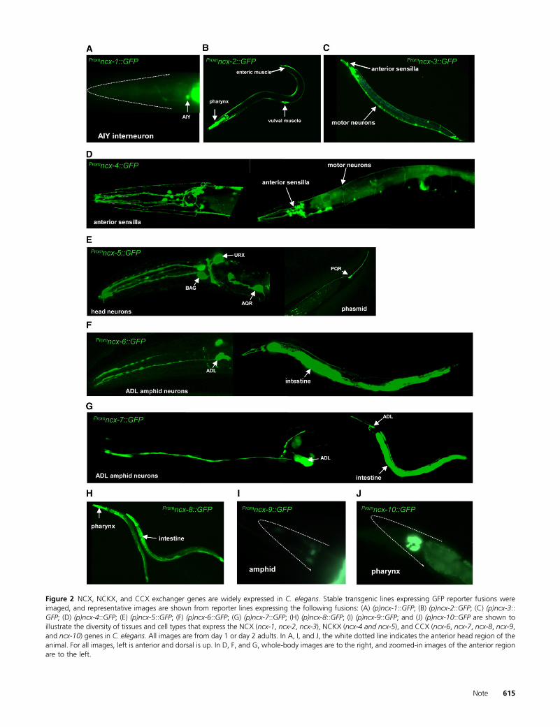
Figure 2 NCX, NCKX, and CCX exchanger genes are widely expressed in C. elegans. Stable transgenic lines expressing GFP reporter fusions wereimaged, and representative images are shown from reporter lines expressing the following fusions: (A) (p)ncx-1::GFP; (B) (p)ncx-2::GFP; (C) (p)ncx-3::GFP; (D) (p)ncx-4::GFP; (E) (p)ncx-5::GFP; (F) (p)ncx-6::GFP; (G) (p)ncx-7::GFP; (H) (p)ncx-8::GFP; (I) (p)ncx-9::GFP; and (J) (p)ncx-10::GFP are shown toillustrate the diversity of tissues and cell types that express the NCX (ncx-1, ncx-2, ncx-3), NCKX (ncx-4 and ncx-5), and CCX (ncx-6, ncx-7, ncx-8, ncx-9,and ncx-10) genes in C. elegans. All images are from day 1 or day 2 adults. In A, I, and J, the white dotted line indicates the anterior head region of theanimal. For all images, left is anterior and dorsal is up. In D, F, and G, whole-body images are to the right, and zoomed-in images of the anterior regionare to the left.
Note 615

Figure 3 Characterization of the NCX transporter ncx-2 and the CCX transporter ncx-8. (A) Predicted gene structure of the ncx-2 splice forms onchromosome V. The amino acid position of the gk879849 lesion is shown. The lesion in gk879849 is defined by a premature opal stop codon at aminoacid 634. (B) Bar chart of the percentage of expulsions per DMP cycle from 20 animals for wild-type animals, ncx-2(gk879849)mutant animals, and wild-type animals that were cultivated on bacteria expressing dsRNA targeting the ncx-2 locus [ncx-2(RNAi)]. In the case of the ncx-2 mutants, there isa significant defect in the number of expulsions per cycle when compared with wild-type animals at P, 0.005 for ncx-2(gk879849)mutant animals andncx-2(RNAi) animals. (C) Bar chart of the average rate of egg laying per hour for wild-type, ncx-2(gk879849), and wild-type animals that were cultivatedon bacteria expressing dsRNA targeting the ncx-2 locus [ncx-2(RNAi)]. Error bars represent the S.E.M; *P , 0.05. For each genotype a sample size of 10animals was obtained and the mean was calculated. (D) Representative plots of the velocity (mm/sec) for wild-type and ncx-2(gk879849)mutant animalswhen off food. The animals were transferred to an unseeded plate and allowed to roam for 10 min and then movies of movement were taken. (E) Barchart of the average velocity for wild-type and ncx-2(gk879849)mutant animals when off food. Error bars represent the S.E.M. P-values were calculatedusing the Student’s t-test. (F) Predicted gene structure of the ncx-8 gene on chromosome V. The amino acid position of the lesion is highlighted for ncx-8(gk234217), and the lesion in gk324217 is defined by a premature stop codon at amino acid 513. (G) Expression of the ncx-8 gene during theembryonic late morphogenesis stage. Bright-field image of an embryo (left) and GFP image of the same embryo (right). (H) Oil-Red-O staining in the
616 V. Sharma et al.

(Figure 3, D and E). We recorded movies of wild-type andncx-2 mutant animals moving while off food to computetheir velocity. Representative plots of velocity of an individ-ual wild-type and ncx-2(gk879849) mutant animal are plot-ted in Figure 3D, and bars of the average velocity for wildtype and ncx-2(gk879849) are graphed in Figure 3E. We didnot observe a significant difference in movement speed be-tween wild-type and ncx-2mutant animals. Next we attemp-ted to characterize the ncx-8(gk234217) mutant (Figure3F). We observed expression of ncx-8 in the pharynx andintestine during late embryonic morphogenesis (Figure 3G)and reasoned that NCX-8 may be contributing to metabolicfunction in C. elegans. To test this, we examined lipid accu-mulation in animals using Oil-Red-O stain. Oil-Red-O is usedto stain lipids in C. elegans (Figure 3H), and quantification ofOil-Red-O staining is used to measure lipid accumulation(O’Rourke et al. 2009). Wild-type animals and ncx-8(gk234217) mutant animals were fixed and soaked in Oil-Red-O, followed by quantification of Oil-Red-O staining. Inthe case of the ncx-8(gk234217) mutant animals, we ob-served significantly lower Oil-Red-O staining (Figure 3I:P , 0.05). We also cultivated wild-type animals on plateswith bacteria expressing dsRNA targeting the ncx-8 geneand also cultivated wild-type animals on control plates withbacteria transformed with empty control vectors. In the caseof the animals cultivated on the RNA interference (RNAi)plates targeting ncx-8, we observed significant decreases inOil-Red-O staining as compared with the animals cultivatedon the control plates (Figure 3I: P , 0.05). Mutations inmetabolic processes have been shown to affect longevity inC. elegans (Crawford et al. 2007; Wang et al. 2008), and sowe investigated the life span of ncx-8(gk234217) mutantanimals at 20�. Here we observed a significant differencein the percentage of survival of ncx-8(gk234217) mutantanimals as compared with wild-type animals (Figure 3J:P , 0.005 for ncx-8 mutants vs. wild-type animals at 20�).We also cultivated wild-type animals on plates with bacteriaexpressing dsRNA targeting the ncx-8 gene and, as a control,cultivated wild-type animals on control plates with bacteriatransformed with empty control vectors. In the case of theanimals cultivated on the RNAi plates against ncx-8, weobserved a significant decrease in the percentage of survivalcompared with the animals cultivated on the control plates(Figure 3J: P , 0.005).
Discussion
Here we analyze the family of Na+/Ca2+ exchangers in C.elegans. Our analysis places these transporters into a phylo-genetic framework, provides a comprehensive guide to theirexpression patterns, and reveals details about their func-tional roles through mutant analysis. From these data it isclear that nematode NCX, NCKX, and CCX exchangers arewidely expressed and evolutionarily conserved. Current datafrom mammalian NCX exchangers show that these proteinsfunction in muscles, neuronal cells, and renal tissue. Mam-malian NCX1 exhibits expression in cardiac muscle, neuro-nal tissue, and the kidneys (Philipson et al. 2002; Nicoll et al.2007; Valsecchi et al. 2013). NCX2 and NCX3 are expressedin skeletal muscle and neuronal cells (Li et al. 1994; Nicollet al. 1996; Lytton 2007). The mammalian NCKX exchang-ers are more widely expressed in various cell types includingphotoreceptor cells, retinal ganglion cells, platelets, vascularsmooth muscle, uterine tissue, intestinal cells, lung tissue,thymus, and epidermal cells (Lytton et al. 2002; Cai andLytton 2004a; Lytton 2007; Visser and Lytton 2007;Altimimi et al. 2013). The CCX protein in humans is also verywidely expressed in neurons, cardiac cells, skeletal muscle,lungs, kidneys, intestinal cells, and testes (Cai and Lytton2004b). There are examples of tissue and cell-type specific-ity within the NCKX branch of mammals: for example,NCKX2 has been reported only in photoreceptors cells, ret-inal ganglion cells, and other neurons of the brain (Li et al.1994). So within both NCX and NCKX there are broadlyutilized exchangers and also regionally specified exchang-ers. Within the C. elegans Na+/Ca2+ exchanger family weobserve a similar pattern. Both NCX and CCX exchangersexhibit diverse expression patterns, but within each branchthere are also more narrowly tuned exchangers. For exam-ple, the NCX gene ncx-2 was detected only in muscle cells,while ncx-1 and ncx-3 were observed only in neuronal celltypes. Similarly within the CCX branch, the ncx-8 gene wasobserved in both muscle cells and intestinal cells. The otherCCX genes, ncx-6, ncx-7, ncx-9, and ncx-10, were observed inboth neuronal cells and other cell types (Table 1). Interest-ingly, in the case of the NCKX genes we observed tissuespecificity as ncx-4 and ncx-5 were observed only in neuro-nal cells. Overall, the wide expression pattern observed formammalian Na+/Ca2+ exchangers is also true for C. elegansand shows a similar overall bias toward excitable cells.
intestine of a wild-type animal (top) and ncx-8(gk324217) mutant animal (bottom). The ncx-8(gk324217) mutant exhibits less Oil-Red-O staining. Oil-Red-O is used as a read-out for lipid accumulation in C. elegans (O’Rourke et al. 2009). Bar, 100 mm. (I) Quantification of Oil-Red-O staining in wild-type,ncx-8(gk234217) mutant animals, wild-type animals that were fed an empty control vector (L4440), and wild-type animals that were fed bacteriaexpressing dsRNA targeting the ncx-8 locus. In the case of the ncx-8(gk234217) mutants and the animals that were fed RNAi clones targeting the ncx-8locus, the average Oil-Red-O staining is significantly lower when compared to wild-type animals or wild-type animals that were fed the control RNAivector. P-values were calculated using the Student’s t-test. Error bars represent the S.E.M.; *P , 0.05. (J) Longevity assays of ncx-8(gk234217) mutantanimals and wild-type animals at 20�C (left), and longevity assays of wild-type animals cultivated on control RNAi plates and RNAi plates targeting thencx-8 locus (right). In each case a significant difference in survival was observed as compared with controls: P , 0.005 for ncx-8 mutants vs. wild-typeanimals at 20�C, and P, 0.005 for wild-type animals cultivated on control RNAi plates compared with wild-type animals cultivated on ncx-8 RNAi platesat 20�C. The P-values were calculated from log-rank tests.
Note 617

Within Drosophila there are three characterized Na+/Ca2+
exchangers, two of which belong to the NCKX branch andone of which belongs to the NCX branch. In the case of theNCKX gene nckx-x (nckx-x-SC in Figure 1A), neuronal ex-pression has been observed throughout the nervous systemand also in the eye-antennal disc, mushroom body, embry-onic brain, and ventral nerve cord (Winkfein et al. 2004).The NCKX gene nckx30c is also widely expressed in neuronalcells of the brain and also in the craniofacial tissue, renalcells, and abdomen (Haug-Collet et al. 1999). The NCX genein Drosophila, Calx, has been reported to be expressed only inphotoreceptor cells (Schwarz and Benzer 1997; Webel et al.2002). Through our phylogenetic analysis we also detectedfour uncharacterized CCX candidates in Drosophila (Figure1A), and the precise expression of these genes is yet to bedetermined. By examining the family of Na+/Ca2+ exchang-ers in three different systems, we now have data suggestingthat Na+/Ca2+ exchangers are represented by both a broadlytuned more general cohort and a more narrowly tuned andspecified subset. However, exactly how these differences areregulated and how these differences in expression patternmap onto functional specializations is an unresolved question.
Acknowledgments
We thank Anthony LaMantia, John Hawdon, Sally Moody,Mary Ann Stepp, and Piali Sengupta for advice and insight-ful comments; Theresa Stiernagle and Aric Daul for the CGCstrains used; the Moerman and Waterston labs and MillionMutation Project team for mutants; our anonymous reviewersfor suggestions and feedback; and Thomas Maynard andAnastas Popratiloff for imaging advice. Funding was providedby The George Washington University Department of Bi-ological Sciences and Columbian College of Arts and Sciences(to V.S., C.H., Z.M., and D.O.); The George WashingtonUniversity Luther Rice Undergraduate Research Fellowship (toE.B.); and The George Washington University Department ofBiological Sciences Wilbur V. Harlan Scholarship Trust (to J.S.).
Literature Cited
Altimimi, H. F., R. T. Szerencsei, and P. P. Schnetkamp,2013 Functional and structural properties of the NCKX2 na(+)-ca (2+)/K (+) exchanger: a comparison with the NCX1na (+)/ca (2+) exchanger. Adv. Exp. Med. Biol. 961: 81–94.
Berridge, M. J., P. Lipp, and M. D. Bootman, 2000 The versatility anduniversality of calcium signalling. Nat. Rev. Mol. Cell Biol. 1: 11–21.
Berridge, M. J., M. D. Bootman, and H. L. Roderick, 2003 Calciumsignalling: dynamics, homeostasis and remodelling. Nat. Rev.Mol. Cell Biol. 4: 517–529.
Bootman, M. D., P. Lipp, and M. J. Berridge, 2001 The organisa-tion and functions of local ca(2+) signals. J. Cell Sci. 114:2213–2222.
Cai, X., and J. Lytton, 2004a The cation/ca(2+) exchanger super-family: phylogenetic analysis and structural implications. Mol.Biol. Evol. 21: 1692–1703.
Cai, X., and J. Lytton, 2004b Molecular cloning of a sixth memberof the K+-dependent na+/Ca2+ exchanger gene family,NCKX6. J. Biol. Chem. 279: 5867–5876.
Coates, J. C., and M. de Bono, 2002 Antagonistic pathways inneurons exposed to body fluid regulate social feeding inCaenorhabditis elegans. Nature 419: 925–929.
Crawford, D., N. Libina, and C. Kenyon, 2007 Caenorhabditiselegans integrates food and reproductive signals in lifespandetermination. Aging Cell 6: 715–721.
Gray, J. M., D. S. Karow, H. Lu, A. J. Chang, J. S. Chang et al.,2004 Oxygen sensation and social feeding mediated by a C.elegans guanylate cyclase homologue. Nature 430: 317–322.
Gray, J. M., J. J. Hill, and C. I. Bargmann, 2005 A circuit fornavigation in Caenorhabditis elegans. Proc. Natl. Acad. Sci.USA 102: 3184–3191.
Haug-Collet, K., B. Pearson, R. Webel, R. T. Szerencsei, R. J. Winkfeinet al., 1999 Cloning and characterization of a potassium-dependent sodium/calcium exchanger in Drosophila. J. CellBiol. 147: 659–670.
Hilge, M., 2012 Ca2+ regulation of ion transport in the na+/Ca2+exchanger. J. Biol. Chem. 287: 31641–31649.
Li, Z., S. Matsuoka, L. V. Hryshko, D. A. Nicoll, M. M. Bersohn et al.,1994 Cloning of the NCX2 isoform of the plasma membranena(+)-Ca2+ exchanger. J. Biol. Chem. 269: 17434–17439.
Lytton, J., 2007 Na+/Ca2+ exchangers: Three mammalian genefamilies control Ca2+ transport. Biochem. J. 406: 365–382.
Lytton, J., X. F. Li, H. Dong, and A. Kraev, 2002 K+-dependentna+/Ca2+ exchangers in the brain. Ann. N. Y. Acad. Sci. 976:382–393.
Nicoll, D. A., B. D. Quednau, Z. Qui, Y. R. Xia, A. J. Lusis et al.,1996 Cloning of a third mammalian na+-Ca2+ exchanger,NCX3. J. Biol. Chem. 271: 24914–24921.
Nicoll, D. A., X. Ren, M. Ottolia, M. Phillips, A. R. Paredes et al.,2007 What we know about the structure of NCX1 and how itrelates to its function. Ann. N. Y. Acad. Sci. 1099: 1–6.
Nicoll, D. A., M. Ottolia, J. I. Goldhaber, and K. D. Philipson,2013 20 years from NCX purification and cloning. Milestones.Adv. Exp. Med. Biol. 961: 17–23.
O’Rourke, E. J., A. A. Soukas, C. E. Carr, and G. Ruvkun, 2009 C.elegans major fats are stored in vesicles distinct from lysosome-related organelles. Cell Metab. 10: 430–435.
Ottolia, M., and K. D. Philipson, 2013 NCX1: Mechanism of trans-port. Adv. Exp. Med. Biol. 961: 49–54.
Philipson, K. D., and D. A. Nicoll, 2000 Sodium-calcium exchange:A molecular perspective. Annu. Rev. Physiol. 62: 111–133.
Philipson, K. D., D. A. Nicoll, M. Ottolia, B. D. Quednau, H. Reuteret al., 2002 The na+/Ca2+ exchange molecule: An overview.Ann. N. Y. Acad. Sci. 976: 1–10.
Sambongi, Y., T. Nagae, Y. Liu, T. Yoshimizu, K. Takeda et al.,1999 Sensing of cadmium and copper ions by externally ex-posed ADL, ASE, and ASH neurons elicits avoidance response incaenorhabditis elegans. Neuroreport 10: 753–757.
Schwarz, E. M., and S. Benzer, 1997 Calx, a na-ca exchanger geneof drosophila melanogaster. Proc. Natl. Acad. Sci. USA 94:10249–10254.
Thomas, J. H., 1990 Genetic analysis of defecation in Caenorhab-ditis elegans. Genetics 124: 855–872.
Tidow, H., L. R. Poulsen, A. Andreeva, M. Knudsen, K. L. Hein et al.,2012 A bimodular mechanism of calcium control in eukar-yotes. Nature 491: 468–472.
Troemel, E. R., J. H. Chou, N. D. Dwyer, H. A. Colbert, and C. I.Bargmann, 1995 Divergent seven transmembrane receptorsare candidate chemosensory receptors in C. elegans. Cell 83:207–218.
Valsecchi, V., G. Pignataro, R. Sirabella, C. Matrone, F. Boscia et al.,2013 Transcriptional regulation of ncx1 gene in the brain.Adv. Exp. Med. Biol. 961: 137–145.
Visser, F., and J. Lytton, 2007 K+ -dependent na+/Ca2+ ex-changers: Key contributors to Ca2+ signaling. Physiology (Be-thesda) 22: 185–192.
618 V. Sharma et al.

Wang, M. C., E. J. O’Rourke, and G. Ruvkun, 2008 Fat metabo-lism links germline stem cells and longevity in C. elegans. Sci-ence 322: 957–960.
Webel, R., K. Haug-Collet, B. Pearson, R. T. Szerencsei, R. J. Winkfeinet al., 2002 Potassium-dependent sodium-calcium ex-change through the eye of the fly. Ann. N. Y. Acad. Sci. 976:300–314.
Winkfein, R. J., B. Pearson, R. Ward, R. T. Szerencsei, N. J.Colley et al., 2004 Molecular characterization, functionalexpression and tissue distribution of a second NCKX na+/Ca2+ -K+ exchanger from drosophila. Cell Calcium 36:147–155.
Communicating editor: P. Sengupta
Note 619

GENETICSSupporting Information
http://www.genetics.org/lookup/suppl/doi:10.1534/genetics.113.153106/-/DC1
Insight into the Family of Na+/Ca2+ Exchangersof Caenorhabditis elegans
Vishal Sharma, Chao He, Julian Sacca-Schaeffer, Eric Brzozowski, Daniel E. Martin-Herranz,Zelda Mendelowitz, David A. Fitzpatrick, and Damien M. O’Halloran
Copyright © 2013 by the Genetics Society of AmericaDOI: 10.1534/genetics.113.153106

2 SI V. Sharma et al.
ncx-120106k 20108k 20110k 20112k 20114k
V
20116k 20118k 20120k 20122k 20124k
GFP1948bp
ncx-2 5304k 5305k 5306k 5307k 5308k
V
5309k 5310k
GFP1927bp
10713k 10715k 10717k 10719k 10721k
IV
9808k 9809K 9810k 9811k 9812k
I
10486k 10487k 10488k 10489k 10490k
V
GFP1330bp
9711k 9713k 9715k 9717k 9719k
III
4984k 4986k 4988k 4990k 4992k
GFP2550bp
5303k 5302k
ncx-3
GFP3730bp
ncx-4
GFP1669bp
ncx-5
9721k
GFP1855bp
ncx-6
GFP1902bp
ncx-7
4994k 4996k
GFP1397bp
ncx-8
GFP1412bp
ncx-9
V
ncx-10 7931k 7932k 7933k 7934k 7935k
V
Figure S1 Promoter elements used in the GFP reporter fusions. Genomic loci were downloaded from Wormbase (www.wormbase.org). The promoter sequence for each gene that was PCR amplified is represented in beige and the GFP coding sequence is indicated in green.

V. Sharma et al. 3 SI
File S1
Supplemental Materials and Methods
Animals and Maintenance
The following animals were used in our study: Bristol N2 (wildtype), DMH22 ncx-‐2 (gk879849), DMH25 ncx-‐8(gk234217),
OH4350 otIs151[(p)ceh-‐36::RFP; rol-‐6(su1006)]; otEx2504[(p)gcy-‐28::GFP; (p)unc-‐122::GFP], DMH23 otIs151[(p)ceh-‐36::RFP; rol-‐
6(su1006)]; hanEx4[(p)ncx-‐4::GFP; (p)elt-‐2::GFP], DA1290 lin-‐15B(n765) X; adEx1290[(p)gcy-‐33::GFP + lin-‐15(+)], OH7511
otIs197[(p)unc14::hif-‐1(P621A); (p)ttx-‐3::RFP], PY2417 oyIs44[(p)odr-‐1::RFP], ZG611 isIs19[(p)gcy-‐32::GFP; unc-‐119(+)], DMH1
hanEx4[(p)ncx-‐4::GFP; (p)elt-‐2::GFP], DMH10 hanEx2[(p)ncx-‐2::GFP], DMH9 hanEx3[(p)ncx-‐3::GFP], DMH12 hanEx1[(p)ncx-‐
1::GFP; (p)elt-‐2::GFP], DMH6 hanEx5[(p)ncx-‐5::GFP], DMH13 hanEx6[(p)ncx-‐6::GFP], DMH14 hanEx7[(p)ncx-‐7::GFP], DMH11
hanEx8[(p)ncx-‐8::GFP], DMH15 hanEx9[(p)ncx-‐9::GFP; (p)elt-‐2::GFP], DMH7 hanEx10[(p)ncx-‐10::GFP; (p)elt-‐2::GFP] -‐ for more
transgene details of the ncx reporters, refer to Table S1. In each case, multiple (between three and six) independent transgenic
lines were examined for each reporter. The strain DMH22 was obtained by outcrossing the strain VC40914 two times through
wildtype N2 animals, and similarly the strain DMH25 was obtained by outcrossing the strain VC40058 two times through N2
animals, and in each case homozygous mutants were selected by PCR and sequencing of the lesion. NGM plates were seeded
with E. coli strain OP50 and maintained according to standard protocol (Brenner. 1974).
Phylogenetic Analysis
Genomes were downloaded from Wormbase (Caenorhabditis species -‐ C. briggsae, C. brenneri, C. japonica, C. remanei -‐ and
Pristionchus pacificus), NCBI (Drosophila), and Ensembl (human), and interrogated for NCX, NCKX and CCX transporter
sequences with bidirectional BlastP searches [>blastall -‐p blastp –d.../...-‐e 0.00001] using local Blast executables
(ftp://ftp.ncbi.nlm.nih.gov/blast/executables/blast+/LATEST/) and motif scanning analysis. Orthologs were aligned using the
multiple sequence alignment software MUSCLE v3.8.31 (Edgar. 2004), and gaps were systematically stripped after alignment.
Phylogenetic relationships were inferred by reconstructing trees by Maximum Likelihood using the PhyML command-‐line
application (Guindon and Gascuel. 2003). The appropriate model was selected using Prottest v3 (Abascal et al. 2005; Darriba et
al. 2011) , and determined to be the “WAG+I+G+F” model with a fixed gamma distribution parameter of 1.5, the proportion of
invariable sites set to 0.003, and four substitution rate categories. For completeness we also reconstructed Bayesian trees (data
not shown) using MrBayes version 3.2 and obtained the same topology using the WAG model (Ronquist and Huelsenbeck.
2003). The analysis used a Markov Chain Monte Carlo (MCMC) method that ran for 2 million generations, sampled every 10th
4 SI V. Sharma et al.
generation. The burnin was set after examining the log probability of the data using the sump command, and clade probabilities
for each phylogeny were determined using the sumt command. Protein structure of Na+/Ca2+ exchangers in C. elegans was
predicted using SMART (http://smart.embl-‐heidelberg.de/) and InterPro (http://www.ebi.ac.uk/interpro) databases, and the
TM-‐Finder program (http://tmfinder.research.sickkids.ca/cgi-‐bin/TMFinderForm).
Generation of GFP Reporter Gene Fusions
The transcriptional GFP reporter fusions were made by PCR fusion as described previously (Boulin et al. 2006) . The promoter
sequence (see Figure S1) was amplified by PCR using High Fidelity polymerase (Roche). A GFP coding region which contained
the unc-‐54 3’UTR was PCR amplified from the plasmid pPD95.75 (available from AddGene: http://www.addgene.org/1494/)
using the primers:
GFP-‐F: AGCTTGCATGCCTGCAGGTCGACT
GFP-‐R: AAGGGCCCGTACGGCCGACTAGTAGG
Following PCR amplification and purification of the promoter and GFP fragment, a fusion PCR step was performed using 10ng of
each amplicon with a nested promoter specific primer (see Table S1) and the nested primer:
GFP nested primer: GGAAACAGTTATGTTTGGTATATTGGG
The resulting PCR fusions were PCR purified and micro-‐injected at between 30-‐100ng/μl into the germ-‐line of wildtype (N2)
animals.
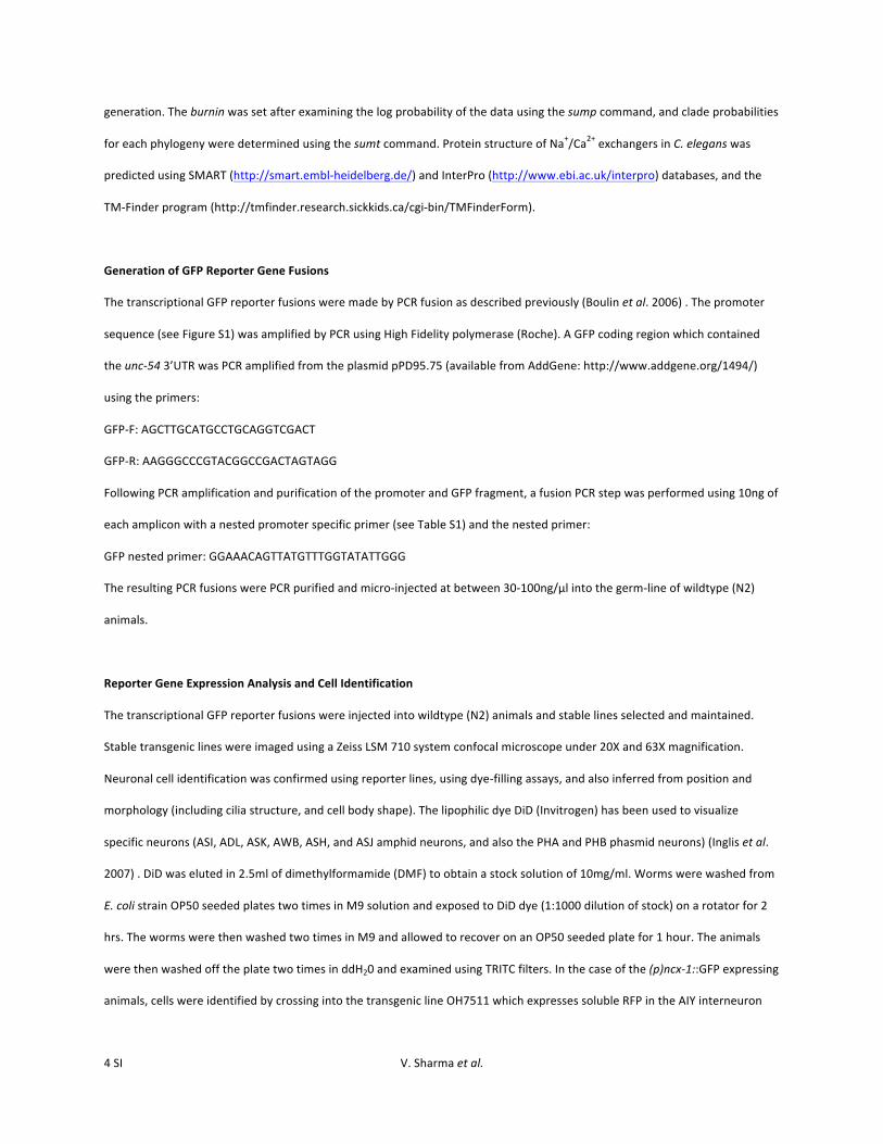
Reporter Gene Expression Analysis and Cell Identification
The transcriptional GFP reporter fusions were injected into wildtype (N2) animals and stable lines selected and maintained.
Stable transgenic lines were imaged using a Zeiss LSM 710 system confocal microscope under 20X and 63X magnification.
Neuronal cell identification was confirmed using reporter lines, using dye-‐filling assays, and also inferred from position and
morphology (including cilia structure, and cell body shape). The lipophilic dye DiD (Invitrogen) has been used to visualize
specific neurons (ASI, ADL, ASK, AWB, ASH, and ASJ amphid neurons, and also the PHA and PHB phasmid neurons) (Inglis et al.
2007) . DiD was eluted in 2.5ml of dimethylformamide (DMF) to obtain a stock solution of 10mg/ml. Worms were washed from
E. coli strain OP50 seeded plates two times in M9 solution and exposed to DiD dye (1:1000 dilution of stock) on a rotator for 2
hrs. The worms were then washed two times in M9 and allowed to recover on an OP50 seeded plate for 1 hour. The animals
were then washed off the plate two times in ddH20 and examined using TRITC filters. In the case of the (p)ncx-‐1::GFP expressing
animals, cells were identified by crossing into the transgenic line OH7511 which expresses soluble RFP in the AIY interneuron

V. Sharma et al. 5 SI
((p)ttx-‐3::RFP). The (p)ncx-‐4::GFP expressing animals were crossed into the transgenic line OH4350, which was previously
selected against the extra-‐chromosomal array otEx2504, and thereby retained only animals containing the otIs51 transgene
which expresses soluble RFP in the AWC and ASE neurons; this line was named DMH23. For (p)ncx-‐5::GFP expressing animals,
the transgenic lines ZG611and DA1290 were used to compare expression patterns; ZG611 animals harbor a transgene
expressing soluble GFP under the gcy-‐32 promoter in the URX, PQR, and AQR cells, and DA1290 animals express soluble GFP
under the gcy-‐33 promoter (BAG cell reporter). The ADL neuron was identified as a site of expression for (p)ncx-‐6::GFP and
(p)ncx-‐7::GFP fusions based upon the distinctive ‘tuning-‐fork’ shaped cilia, and also by testing positive for DiD dye overlap.
Defecation Motor Program Assays
To examine the DMP cycle we measured the number of expulsions per DMP cycle time by examining day1 adult animals under
a dissecting microscope. The DMP cycle in C. elegans represents a stereotyped intrinsic rhythm that occurs every ~45secs
(Thomas. 1990; Xing et al. 2008) . For each genotype 20 animals were screened. For RNAi experiments, animals were grown
from eggs to day 1 of adulthood on E. coli expressing dsRNA specific to the ncx-‐2 gene (CUUkp3304M134Qclone,
SourceBioscience). The RNAi insert clone was sequence verified prior to use. NGM plates for RNAi experiments contained
standard NGM recipe and also 25μg/ml Carbenicillin and 1mM IPTG.
Egg-‐laying Assays
To quantify the rate of egg-‐laying, L4 stage animals were picked onto staging plates seeded with E. coli strain OP50 and 24hrs
later an individual day 1 adult animal was transferred to a seeded plate for two hours at room temperature. The number of
eggs laid during this time was counted by examining the plate under a dissection microscope. This was repeated ten times for
each genotype. For RNAi experiments, animals were grown from eggs to day 1 of adulthood on E. coli expressing dsRNA specific
to the ncx-‐2 gene (CUUkp3304M134Q clone, SourceBioscience) and then transferred individually to a seeded NGM plate for
2hrs. The average number of fertilized eggs within each animal for ncx-‐2(gk879849) mutant animals and wildtype animals was
compared by counting the number of unlaid eggs in ten individual day 1 adults.
Tracking Worm Movement
Movies of worms were captured using a Leica IC80 HD digital microscope camera connected to a Leica M165F dissecting
microscope and visualized through a HDMI connection into an adjacent monitor. Movies were captured as mp4 files and
converted to TIFF series using the ffMPEG Binary (only available on a MAC) at this URL -‐http://ffmpegmac.net/.

6 SI V. Sharma et al.
Files were converted and extracted using the following command line:
> ffmpeg -‐y -‐i sample.mp4 -‐an -‐r 3 -‐pix_fmt rgb24 -‐vcodec tiff tif/%010d.tif
Prior to capturing movies, animals were grown to the L4 stage at room temperature and transferred to a separate E. coli strain
OP50 seeded plate at room temperature. 24hrs later day 1 adult animals were picked from this plate onto an unseeded NGM
plate and allow to roam for 10mins. After this time movies were recorded of their movement over ~60secs. Analysis of
movement was performed using the ‘manual tracking’ (http://rsbweb.nih.gov/ij/plugins /track/track.html) and ‘wrmtrk’
(http://www.phage.dk/plugins/wrmtrck.html) plugins in ImageJ (http://rsb.info.nih.gov/ij/).
Quantification of Oil-‐Red-‐O Staining
Oil-‐Red-‐O staining of lipid accumulation was performed as described previously (O'Rourke et al. 2009) . Oil-‐Red-‐O (O0625
Sigma) was prepared by adding 0.5g of Oil-‐Red-‐O into 100ml of isopropanol. This solution was equilibrated for three days by
stirring at room temperature and then diluted to 60% in ddH20 and filtered using a 0.2µm filter. 10 L4 staged animals were
transferred to a seeded (E. coli strain OP50) NGM staging plate at room temperature and 24hours later the day 1 adult animals
were transferred to multiple seeded (E. coli strain OP50) NGM plates and allowed to lay eggs for 8hrs and then each individual
adult animal was removed. The plates were left at room temperature until the progeny grew to the L4 stage. The ncx-‐8 gene is
expressed in the intestine from embryogenesis (Figures 2H and 3G), and so L4 stage animals were fixed and quantified so as to
permit intestinal quantification and avoid staining of the germ-‐line. Animals were collected and washed in 1X PBS prior to
fixation and reduction and then soaked in Oil-‐Red-‐O dye overnight on a rotator and then washed in 1X PBS and placed onto
microscope slides for imaging using a Zeiss LSM 710 system confocal microscope. Whole body Oil-‐Red-‐O staining was then
quantified using ImageJ software to obtain the mean staining intensity for between 15 and 20 animals in each experiment. For
RNAi experiments, animals were cultivated from eggs to L4 stage on E. coli expressing either an empty control vector (L4440,
Addgene) or the L4440 vector expressing dsRNA specific to the ncx-‐8 gene (CUUkp3304C014Q clone, SourceBioscience). The
RNAi insert clone was sequence verified prior to use. NGM plates for RNAi experiments contained standard NGM recipe and
also 25μg/ml Carbenicillin and 1mM IPTG.
Longevity Assays
Animals were synchronized by a timed egg laying (TEL), and cultivated on NGM plates seeded with E. coli strain OP50 at 20ºC.
The L4 stage was set as Day 0 and the next Day (i.e. adult Day 1) was set as Day 1. Longevity assays were conducted and
measured as described previously (Apfeld and Kenyon. 1999). Animals were transferred every few days to fresh NGM seeded

V. Sharma et al. 7 SI
plates at 20ºC and the number of dead animals scored. FuDR was not used in the NGM plates to avoid confounding our results
(Aitlhadj and Sturzenbaum. 2010). Survival curves and statistics were analyzed using OASIS (Yang et al. 2011) . The Log-‐Rank
test was used to infer significance. Number of dead worms, not including censored animals, was: 79(N2), 87(ncx-‐8), 69(control
RNAi), 109(ncx-‐8 RNAi). For RNAi experiments, animals were grown on E. coli expressing either an empty control vector (L4440,
Addgene) or the L4440 vector expressing dsRNA targeting the ncx-‐8 gene (CUUkp3304C014Q clone, SourceBioscience).
SUPPLEMENTAL LITERATURE CITED
Abascal, F., R. Zardoya and D. Posada, 2005 ProtTest: Selection of best-‐fit models of protein evolution. Bioinformatics 21: 2104-‐
2105.
Aitlhadj, L., and S. R. Sturzenbaum, 2010 The use of FUdR can cause prolonged longevity in mutant nematodes. Mech. Ageing
Dev. 131: 364-‐365.
Apfeld, J., and C. Kenyon, 1999 Regulation of lifespan by sensory perception in caenorhabditis elegans. Nature 402: 804-‐809.
Boulin, T., J. F. Etchberger and O. Hobert, 2006 Reporter gene fusions. WormBook 1-‐23.
Brenner, S., 1974 The genetics of caenorhabditis elegans. Genetics 77: 71-‐94.
Darriba, D., G. L. Taboada, R. Doallo and D. Posada, 2011 ProtTest 3: Fast selection of best-‐fit models of protein evolution.
Bioinformatics 27: 1164-‐1165.
Edgar, R. C., 2004 MUSCLE: Multiple sequence alignment with high accuracy and high throughput. Nucleic Acids Res. 32: 1792-‐
1797.
Guindon, S., and O. Gascuel, 2003 A simple, fast, and accurate algorithm to estimate large phylogenies by maximum likelihood.
Syst. Biol. 52: 696-‐704.
Inglis, P. N., G. Ou, M. R. Leroux and J. M. Scholey, 2007 The sensory cilia of caenorhabditis elegans. WormBook 1-‐22.
O'Rourke, E. J., A. A. Soukas, C. E. Carr and G. Ruvkun, 2009 C. elegans major fats are stored in vesicles distinct from lysosome-‐
related organelles. Cell. Metab. 10: 430-‐435.

8 SI V. Sharma et al.
Ronquist, F., and J. P. Huelsenbeck, 2003 MrBayes 3: Bayesian phylogenetic inference under mixed models. Bioinformatics 19:
1572-‐1574.
Thomas, J. H., 1990 Genetic analysis of defecation in caenorhabditis elegans. Genetics 124: 855-‐872.
Xing, J., X. Yan, A. Estevez and K. Strange, 2008 Highly Ca2+-‐selective TRPM channels regulate IP3-‐dependent oscillatory Ca2+
signaling in the C. elegans intestine. J. Gen. Physiol. 131: 245-‐255.
Yang, J. S., H. J. Nam, M. Seo, S. K. Han, Y. Choi et al, 2011 OASIS: Online application for the survival analysis of lifespan assays
performed in aging research. PLoS One 6: e23525.

V. Sharma et al. 9 SI
Table S1 PCR Primers used to generate reporter GFP constructs
Prim
ers
Resulting
Tran
sgen
ic
Arrays
ha
nEx1
ha
nEx2
ha
nEx3
hanEx4
hanEx5
ha
nEx6
ha
nEx7
hanEx8
hanEx9
hanEx10
Reverse
AGTC
GAC
CTGCA
GGCA
TGCA
AGCT
CGCA
TTTG
TAGTT
GTC
TCTG
GAA
AAAA
ATT
AGTC
GAC
CTGCA
GGCA
TGCA
AGCT
TCAA
CTGAA
AAT
CAGAT
TTTA
GGCT
TTTA
A
AGTC
GAC
CTGCA
GGCA
TGCA
AGCT
GTTTG
ATCC
TGAA
AG
TAAT
AAAA
ACTTTTA
AGTC
GAC
CTGCA
GGCA
TGCA
AGCT
TATTTTTA
AAA
ATCA
AAAT
ATGAT
AATTTG
AGTC
GAC
CTGCA
GGCA
TGCA
AGCT
CTTTTG
AAAT
AC
TCAA
CTCA
CATTCC
GGAT
AGTC
GAC
CTGCA
GGCA
TGCA
AGCT
TGAT
ACAA
AATT
GAC
CTGAA
AACT
TTAA
AA
AGTC
GAC
CTGCA
GGCA
TGCA
AGCT
AACA
TAGAT
CT
GAA
AATG
TTTA
AAAT
AAGA
AGTC
GAC
CTGCA
GGCA
TGCA
AGCT
GAG
AAGGAG
TTT
GAA
ATTTAG
TTGAA
ACGA
AGTC
GAC
CTGCA
GGCA
TGCA
AGCT
TTCC
TCAT
CAAA
AG
GGAA
TCCA
TCAT
AAAT
AGTC
GAC
CTGCA
GGCA
TGCA
AGCT
ACCT
GAA
AAAG
AA
ACAG
TTGAT
AAGCG
GGT
F-‐Nested
GAA
ACTC
TGAC
TTCC
AGGTTG
ATTTTG
G
TCCT
TCAT
TTTA
AAAC
TCCA
CG
GCC
AAG
CTGCA
TCTC
CTGCT
CTCT
CCTT
ACAT
TA
TGTA
GTA
GAT
CCTC
CTCC
CCG
CCTTTTT
AGTC
GAT
GCG
GAG
AAAT
GTC
T GAA
GCC
AAA
CATA
GAT
GCG
AACA
ATTTTC
AACA
AAAA
CTGCT
GCT
GCT
GAT
TATG
CTC
TTTTGGT
GAT
CAAA
GAC
ACGAG
GTA
GC
GAA
TTCC
A
CTCA
ATTC
CTTTCC
ACCA
CTC
ACTG
AAC
TACA
CAGTTGCA
GAG
GCG
TTT
AATC
AGA
F1
ACTTTC
ACCT
TTTC
TTCT
CCAT
CT
CCTC
TCCT
TCAT
TTTA
AAAC
TCCA
C GGCC
AAG
CTTC
ACAC
AAAA
GCT
CAAC
G
GTA
CTTTC
TGGTG
GTA
CGTC
ATAA
TCCA
G
TCTC
AAG
ATGTTTG
AAAT
TGGGGAA
TG
AATG
CAAA
A
CAAC
CGTA
ATAT
CCCC
TGAG
ATCA
ATAA
AAAA
GAT
GTTCC
TTCG
ATGC
CTGAC
CTC
ACTC
ATTTTTGCC
GTG
CCTA
TC
GTTCA
T
AACA
CAGCG
CAAG
TAGCA
A GTA
GCA
AGT
GTTCT
TTCA
ACAT
TGCA
AAAA
GGCA
CCA
Prom
oter
ncx-‐1
ncx-‐2
ncx-‐3
ncx-‐4
ncx-‐5
ncx-‐6
ncx-‐7
ncx-‐8
ncx-‐9
ncx-‐10