Insight into the family of Na+/Ca2+ exchangers of Caenorhabditis elegans
Upload
independentCategory
view
0download
0
Ecotoxicology and Environmental Safety 77 (2012) 64–70
Contents lists available at SciVerse ScienceDirect
Ecotoxicology and Environmental Safety
0147-65
doi:10.1
n Corr
E-m
journal homepage: www.elsevier.com/locate/ecoenv
Interaction of Silver Nanoparticles with Biological Surfacesof Caenorhabditis elegans
Shin Woong Kim, Sun-Hwa Nam, Youn-Joo An n
Department of Environmental Science, Konkuk University, 1 Hwayang-dong, Gwangjin-gu, Seoul 143-701, Korea
a r t i c l e i n f o
Article history:
Received 30 June 2011
Received in revised form
18 October 2011
Accepted 25 October 2011Available online 9 November 2011
Keywords:
Silver nanoparticles
Caenorhabditis elegans
Nanotoxicity
Nematode growth medium
13/$ - see front matter & 2011 Elsevier Inc. A
016/j.ecoenv.2011.10.023
esponding author. Fax: þ82 2 2201 6295.
ail address: [email protected] (Y.-J. An).
a b s t r a c t
Silver nanoparticles (AgNPs) are being used in an increasing number of industrial and commercial
applications; this has resulted in an increased release of AgNPs into the environment. Understanding
the interaction of AgNPs with biological surfaces is important, as such understanding will facilitate
predictions of the further effects of nanoparticles on biological systems. This study highlights the
interaction of citrate-coated silver nanoparticles (cAgNPs) with the biological surfaces of the nematode
C. elegans. General toxicity, as proxied by factors such as mortality and reproduction, was evaluated in
nematode growth medium (NGM), which provides a more homogeneous distribution of cAgNPs than in
K-medium. The survival and reproduction of C. elegans evidenced a clear reduction in up to 100 mg/L
and 10 mg/L of cAgNPs, respectively. We also noted significant interactions of cAgNPs with the
biological surfaces of C. elegans. Severe epidemic edema and burst were detected in the exposure group,
which may be associated with secondary infections in soil ecosystems. We observed no evidence of
cAgNPs intake by C. elegans. This is, to the best of our knowledge, the first report to investigate the
nanotoxicity of cAgNPs as related to biological surfaces of C. elegans; further research is needed to study
the fate of cAgNPs inside of C. elegans.
& 2011 Elsevier Inc. All rights reserved.
1. Introduction
Silver nanoparticles (AgNPs) are used in a huge range of applica-tions. AgNPs evidence a variety of properties, including spectrallyselective coating (Rand et al., 2004; Cole and Halas, 2006), surface-enhanced Raman scattering (Yamamoto and Watarai, 2006), andantibacterial activity (Savage and Diallo, 2005; Sambhy et al., 2006;Pal et al., 2007). However, AgNPs also appear to exert some adverseeffects, including the production of reactive oxygen species, DNAdamage, inhibition of physical indicators, and genetic damage(Matsumura et al., 2003; Roh et al., 2009). Additionally, silver ionscan be released from AgNPs, and have been shown to induce toxiceffects such as the prevention of DNA replication and cell membranedamage (Feng et al., 2000), as well as the inhibition of respiratoryenzymes (Matsumura et al., 2003).
Caenorhabditis elegans is a broadly distributed nematode speciesin ecosystems. C. elegans lives in various microbe-rich habitats (Felixand Braendle, 2010), and plays important roles in decomposition andnutrient cycling. C. elegans is employed broadly as a test organism innanotoxicology (Wang et al., 2009; Ma et al., 2009, 2011; Kim et al.,2008; Roh et al., 2009; Roh et al., 2010). The toxicity of AgNPs hasbeen previously reported and AgNPs were shown to be taken beyond
ll rights reserved.
the gut of C. elegans (Meyer et al., 2010). K-medium was used as atest medium in these studies. However, K-medium has greaterionic strength than many other media, which could reasonably beimplicated in the observed nanoparticle aggregations. On an imageanalysis study using C. elegans, fluorescent nanodiamonds werefound to have accumulated in the gonads and digestive organs ofexposed groups (Mohan et al., 2010)
In the present study, we assessed the toxicity of citrate-cappingsilver nanoparticles (cAgNPs) to the nematode C. elegans. Theresearch focused on the interaction of cAgNPs with the biologicalsurface of a test species to determine the influence of cAgNPs on theC. elegans surface, which is obviously the first organ to make contactwith the nanoparticles. Additionally, the dissolution and aggregationof cAgNPs was characterized in the nematode growth medium(NGM) selected herein.
2. Materials and methods
2.1. Nanoparticle characterization
Citrate-coated silver nanoparticles (cAgNPs, 20%) were obtained in an aqueous
colloidal state from ABC Nanotech (Daejeon, Korea). The cAgNP contained 1%
of the capping agent that provided stable suspension in water. The particle
morphology was measured using field emission transmission electron microscope
(FE-TEM, JEM2200FS, JEOL). The Brunauer Emmet Teller (BET) surface area of
particle was estimated using a particle size analyzer (UPA-150, microtrac, USA).
S.W. Kim et al. / Ecotoxicology and Environmental Safety 77 (2012) 64–70 65
The particle size distribution of the hydrodynamic diameter of cAgNPs with the
zeta potential was measured by the electrophoretic light scattering (ELS-8000,
Otsuka Electronics Co., Japan). To measure the dissolution of test nanoparticles,
the cAgNPs were treated identically to the bioassay conditions for up to maximum
concentrations of 1000 and 100 mg/L for the survival and reproduction assays,
respectively. All dissolution experiments were carried out for maximum exposure
periods of 24 and 48 h for the survival and reproduction assays, respectively.
cAgNPs in NGM solution (without agar) satisfying the test conditions were filtered
through a 200 nm nylon filter (Whatman International, Maidstone) and a 50 nm
VMTP filter (ISOPORE membrane filters, Millipore, Ireland). The filtrant was
filtered again using stirred ultrafiltration cells (50 mL, Model 8050, Millpore,
USA). The concentration of silver ions dissolved from cAgNPs was quantified via
ICP-AES (ICP-AES; JY 138; Ultrace, Jobin Yvon, France).
0
5
10
15
20 Laser scattering intensity distribution Cumulative
aser
sca
tterin
g in
tens
ity d
istri
butio
n (%
)
0
20
40
60
80
100
Cum
ulat
ive
(%)
2.2. Nanoparticle preparation in test medium
The cAgNPs suspensions were maintained by storage at 4 1C in darkness.
K-medium was used as a medium for the nematode toxicity test, as per Williams
and Dusenbery (1990). Since most nematodes live in soil pore water, K-medium
provides analogous test conditions (Wang et al., 2009). When the cAgNPs
suspensions were added to K-medium in this study, significant aggregation was
immediately observed, and lumps were produced over time. As reported by Meyer
et al. (2010), K-medium has relatively high ionic strength and favors charge
screening. The conductivity of the K-medium was measured at 8600 ms/cm in this
study. Therefore, we concluded that that K-medium was not appropriate for use in
tests of cAgNPs exposure. As an alternative, we employed NGM agar, which was a
much better medium to achieve a homogenous distribution of cAgNPs. This was
confirmed by the observations of high-resolution microscopy (HRM), as shown in
Fig. 1C. The HRM observations were carried out using a high-resolution adapter
(Cytoviva Inc., Alabama, USA) affixed to the microscope (Olympus BX51, Olympus
Cooperation, Tokyo, Japan). NGM agar was prepared, autoclaved (121 1C, 15 min),
and cooled to 60–65 1C. The cAgNPs suspension was subsequently mixed with
NGM solution, and the temperature promptly cooled to 40–45 1C. Approximately
0.2 mL of mixed solution was then added to each well of 24-well microplates
(ID 17 mm, height 17 mm, volume 3.8 ml for each well). 0.2 mL was sufficient to
cover the entire surface of each well. The solution was immediately hardened in
the refrigerator to prevent precipitation and dissolution of nanoparticles. Since the
warm temperature during the nanoparticle preparation may affect the dissolution
of nanoparticles, the dissolution of cAgNPs was measured identically to that of the
nanoparticle preparation. This method was considered to be the best available
technique for the prevention of aggregation and precipitation of the nanoparticles
in the present study.
0Hydrodynamic diameter (nm)
L
20 40 60 80 100 120
2.3. Test organism and toxicity assayCaenorhabditis elegans (wild type, Bristol strain N2) was obtained from the Animal
Genomics laboratory, Konkuk University (Seoul, Korea). All cultures were maintained
on nematode growth medium (NGM: NaCl 3 g/L; peptone 2.5 g/L; agar 17 g/L; 1 M
potassium phosphate 25 mL; 1 M CaCl2 �2H2O 1 mL; 1 M MgSO4 �7H2O 1 mL;
cholesterol 1 mL) plates at 2172 1C, in darkness for several months. Escherichia coli
strain OP50 was supplied as a food source. To obtain synchronized worms, Clorox
solution (5% NaOCl:1 N NaOH, 2:5) was added to the NGM plates for 15 min, and
1 mL solution was replaced into 1.5 ml centrifuge tubes. The tubes were then
centrifuged for 2 min at 2000 rpm, and the supernatant was removed and washed
with K-medium (0.032 M KCl, 0.051 M NaCl) (Williams and Dusenbery, 1990). The
eggs at the bottom were replaced into new NGM plates, and the worms were
synchronized for 24 h.
The nematode assay was carried out in the NGM agar plate mixtures with
nanoparticle suspensions. One or ten individuals were transferred to each well in
four replicates for reproduction or survival tests, respectively. The exposure
periods for the survival and reproduction assays were 24 h and 48 h, respectively.
The samples were observed under a microscope. The reproduction was evaluated
by counting the number of juvenile offspring. The prepared exposure concentra-
tions of cAgNPs were 0, 10, 50, 100, 500, and 1000 mg/L for the survival test, and at
0, 1, 5, 10, 50, and 10 mg/L for the reproduction test.
Fig. 1. (A) TEM image, and (B) particle size distribution of cAgNPs. (C) High
resolution-microscopic (HRM) image (x1000) of cAgNPs (100 mg/L) distributed in
NGM agar plate.
2.4. Toxicity of silver ions released from cAgNPs
As the ionization of cAgNPs can increase during the nanoparticle preparation
and exposure period, the silver ions (Agþ) released from cAgNPs were measured
for each test duration in NGM solution. According to the ionization rate of cAgNPs
(about one percent in NGM solution), exposure concentrations of silver ions was
prepared at 0, 0.5, 2.5, 5.0, 25, and 50 mg/L for the survival test, and were prepared
at 0, 0.1, 0.5, 1.0, 5.0, and 10.0 mg/L for the reproduction test. To evaluate the ion
toxicity, silver nitrate (AgNO3, Sigma–Aldrich, St. Louis, Mo, USA) was used. All
processes of the Agþ toxicity test were carried out in the same manner as the
cAgNPs toxicity test.
2.5. HRM analysis
To confirm the remainder of C. elegans-exposed cAgNPs, we employed an HRM
image analysis technique. The HRM system employs a novel dark field-based solar
lighting scheme, which improves contrast and signal-to-noise ratio. Thus, this

S.W. Kim et al. / Ecotoxicology and Environmental Safety 77 (2012) 64–7066
system makes observations based on the intrinsic scattering properties of objects
such as nanoparticles and other particulate materials, via a quick and easy process.
All samples were prepared to identical test conditions, and exposure concentra-
tions of 100 mg/L of cAgNPs were used.
2.6. TEM analysis
To prepare the C. elegans pellet for TEM image analysis, the survivors exposed
to 100 mg/L of cAgNPs during the survival assay were collected. Then, individuals
were washed twice with deionized water because the cAgNPs were aggregated in
PBS buffer or K-medium. The samples were fixed by placement on 2.5% glutar-
aldehyde diluted with 0.1 M cacodylate buffer for 2 h. The samples were then
washed twice in 1�PBS buffer (pH 7.470.1) for 10 min, and fixed for 1 h in a 1:1
mixture of 1% OsO4 and 1xPBS buffer. The samples were rinsed with 1xPBS buffer
and dehydrated in a graded series of ethanol (50%, 70%, 80%, 90%, and 99.9%). Resin
infiltration and embedding was carried out in a graded resin (30%; 50%; 70%) for
2 h each, and at 100% overnight. The samples were embedded in beam capsules
and polymerized for 72 h at 60 1C. After semi-thin sections (150 nm) and ultra-
thin sections (below 70 nm) were cut, they were stained with uranyl acetate
(45 min) and lead citrate (3 min). The sections were then assessed via TEM (TechaiTM
G2 spirit BioTWTN; FEI Company, Hillsboro, OR, USA) and TEM-energy-dispersive
spectroscopy, with an EDS resolution of 142 eV (200 kV, JEM 2100 F; JEOL, Tokyo,
Japan).
00
20
40
60
80
100
120
Sur
viva
l of C
. ele
gans
(% c
ontro
l)
cAgNPs (mg/L)
0.00
20
40
60
80
100
120
Dissolved Ag+ from cAgNPs
Sur
viva
l of C
. ele
gans
(% c
ontro
l)
Ag+ (mg/L)
50 100 400 600 800 1000
2.5 5.0 7.5 20 30 40 50
Fig. 2. (A) Survival and (B) reproduction effects of C. elegans exposed cAgNPs. A
2.7. SEM analysis
To confirm the effects of C. elegans exposed to 10 and 100 mg/L of cAgNPs in
the survival assay, Scanning Electron Microscopy (SEM, Hitachi S-3500N, 10 keV)
analysis was conducted. The survivors exposed to 10 and 100 mg/L of cAgNPs,
including control concentrations, were collected and centrifuged for 1 min at
2,000 rpm. The pellets were then washed twice with deionized water because the
cAgNPs had aggregated in the K-medium. They were fixed for 2 h in 2.5%
glutaraldehyde diluted with 0.1 M cacodylate buffer. The samples were subse-
quently rinsed 3 times with 1xPBS buffer (pH 7.470.1). To dehydrate the samples,
a graded series of ethanol (50%, 60%, 70%, 80%, 90%, and 99.9%) was added at
30 min intervals. Isoamyl acetate diluted with EtOH was prepared to concentra-
tions of 33%, 50%, 66%, and 100%, and added to the samples for 1 h (100%-2 hr).
The samples were then dried with hexamethyldisilazane reagent (HMDS, EMS #
16700, Hatfield, UK), and coated with an Au-Pd coating (Hitachi E-1010).
2.8. Statistical analysis
The no-observed-effect concentration (NOEC) and effective concentrations
(EC50) for survival and reproduction effects on C. elegans were caculated. The
NOEC values were estimated via Dunnett’s procedure for multiple comparisons
(Dunnett, 1955). The Dunnett’s program (Ver. 1.5) (USEPA, 1999a) provides a 95%
significance level (po0.05) for all comparisons. The EC50 and LC50 values were
00
20
40
60
80
100
120R
epro
duct
ion
of C
. ele
gans
(% c
ontro
l)
cAgNPs (mg/L)
0.00
20
40
60
80
100
120
Dissolved Ag+ from cAgNPs
Ag+ (mg/L)
Rep
rodu
ctio
n of
C. e
lega
ns (%
con
trol)
5 10 40 60 80 100
0.5 1.0 1.5 4 6 8 10
nd toxicity of Agþ released from cAgNP. (C) Survival and (D) reproduction.
S.W. Kim et al. / Ecotoxicology and Environmental Safety 77 (2012) 64–70 67
estimated using the Trimmed Spearman–Karber method (Hamilton et al., 1977).
The SPEARMAN computer program (USEPA, 1999b) was provided by the U.S. EPA’s
Center for Exposure Assessment Modeling (CEAM).
3. Results
Fig. 1A and B show TEM image and particle size distribution of ahydrodynamic diameter of cAgNPs dispersed in deionized water,respectively. cAgNPs were spherical and the hydrodynamic diameterwas estimated to be 50.6 nm, as a result of replicate measurementsof fifty particles. Zeta potential was measured to be �31.33 mV,indicating that the nanoparticle dispersion was moderately stable.The BET (Brunauer, Emmett and Teller) surface area of cAgNPs was1.402970.0096 m2 g�1.
Table 1The no-observed-effect concentration (NOEC) and effective concentrations (EC50)
for survival and reproduction of C. elegans exposed to cAgNPs or Agþ in NGM for
24 h (unit: mg/L).
Endpoint cAgNPs Agþ
NOECa L/EC50b NOECa L/EC50b
Survival o10 55 (29–105) 40.5 22 (11–46)
Reproduction 1 4100 1 410
a Dunnett’s procedure with po0.05.b Trimmed Spearman–Karber method. Values in parentheses represent the
95% confidence level.
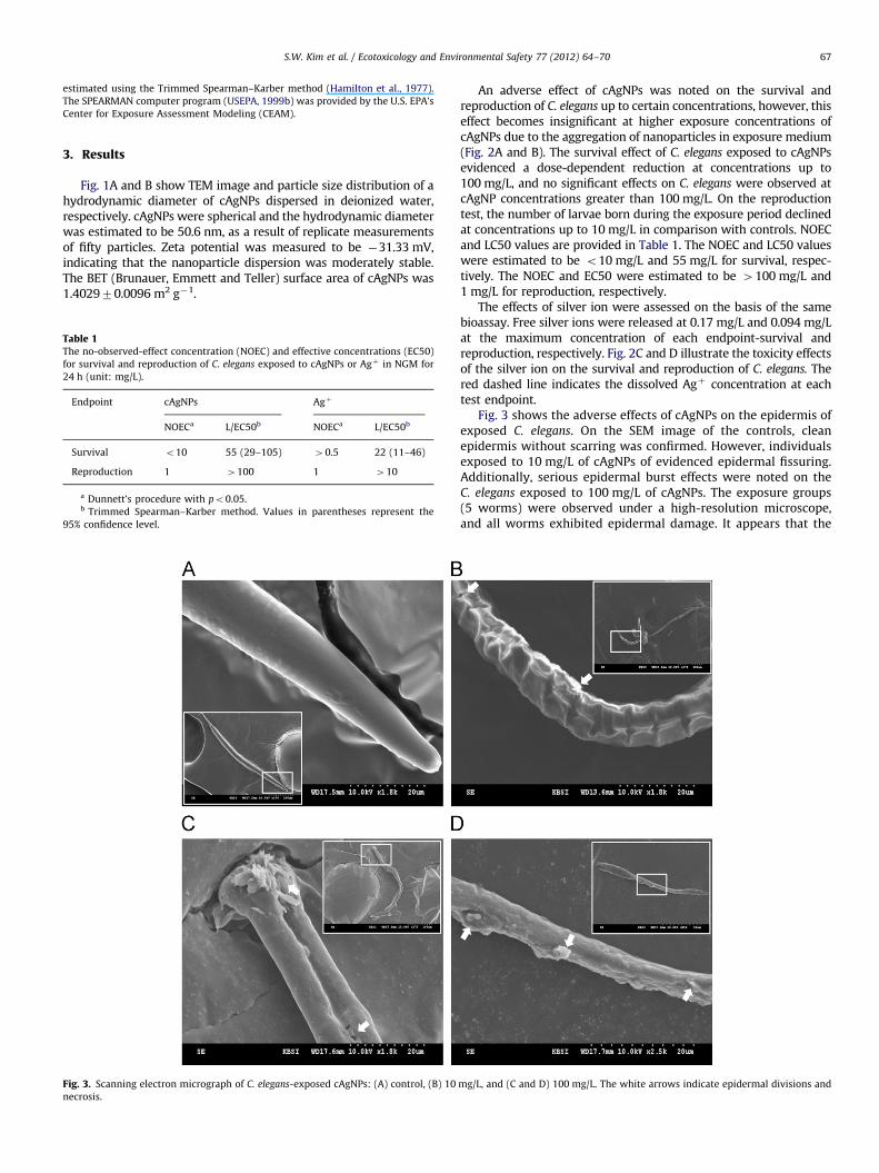
Fig. 3. Scanning electron micrograph of C. elegans-exposed cAgNPs: (A) control, (B) 10
necrosis.
An adverse effect of cAgNPs was noted on the survival andreproduction of C. elegans up to certain concentrations, however, thiseffect becomes insignificant at higher exposure concentrations ofcAgNPs due to the aggregation of nanoparticles in exposure medium(Fig. 2A and B). The survival effect of C. elegans exposed to cAgNPsevidenced a dose-dependent reduction at concentrations up to100 mg/L, and no significant effects on C. elegans were observed atcAgNP concentrations greater than 100 mg/L. On the reproductiontest, the number of larvae born during the exposure period declinedat concentrations up to 10 mg/L in comparison with controls. NOECand LC50 values are provided in Table 1. The NOEC and LC50 valueswere estimated to be o10 mg/L and 55 mg/L for survival, respec-tively. The NOEC and EC50 were estimated to be 4100 mg/L and1 mg/L for reproduction, respectively.
The effects of silver ion were assessed on the basis of the samebioassay. Free silver ions were released at 0.17 mg/L and 0.094 mg/Lat the maximum concentration of each endpoint-survival andreproduction, respectively. Fig. 2C and D illustrate the toxicity effectsof the silver ion on the survival and reproduction of C. elegans. Thered dashed line indicates the dissolved Agþ concentration at eachtest endpoint.
Fig. 3 shows the adverse effects of cAgNPs on the epidermis ofexposed C. elegans. On the SEM image of the controls, cleanepidermis without scarring was confirmed. However, individualsexposed to 10 mg/L of cAgNPs of evidenced epidermal fissuring.Additionally, serious epidermal burst effects were noted on theC. elegans exposed to 100 mg/L of cAgNPs. The exposure groups(5 worms) were observed under a high-resolution microscope,and all worms exhibited epidermal damage. It appears that the
mg/L, and (C and D) 100 mg/L. The white arrows indicate epidermal divisions and
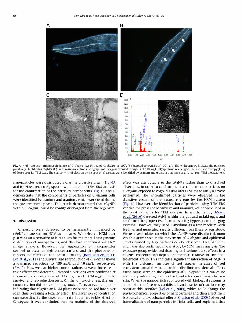
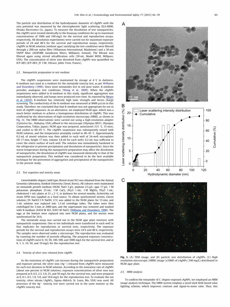
Fig. 4. High resolution-microscopic image of C. elegans. (A) Untreated C. elegans (x1000). (B) Exposed to cAgNPs of 100 mg/L. The white arrows indicate the particles
putatively identified as cAgNPs. (C) Transmission-electron micrographs of C. elegans exposed to cAgNPs of 100 mg/L. (D) Spectrum of energy-dispersive spectroscopy (EDS)
of dense spot for TEM scan. The components of electron dense spot on C. elegans were identified by osmium and uranium that were originated from TEM pretreatment.
S.W. Kim et al. / Ecotoxicology and Environmental Safety 77 (2012) 64–7068
nanoparticles were distributed along the digestive organ (Fig. 4Aand B). However, no Ag spectra were noted on TEM-EDS analysisfor the confirmation of the particles’ components. Fig. 4C and Ddemonstrate that the components of particles on C. elegans cellswere identified by osmium and uranium, which were used duringthe pre-treatment phase. This result demonstrated that cAgNPswithin C. elegans could be readily discharged from the organism.
4. Discussion
C. elegans were observed to be significantly influenced bycAgNPs dispersed on NGM agar plates. We selected NGM agarplates as an alternative to K-medium for the more homogeneousdistribution of nanoparticles, and this was confirmed via HRMimage analysis. However, the aggregation of nanoparticlesseemed to occur at high concentrations, and this phenomenonhinders the effects of nanoparticle toxicity (Baek and An, 2011;Lee et al., 2011) The survival and reproduction of C. elegans showsa dynamic reduction to 100 mg/L and 10 mg/L, respectively(Fig. 2). However, at higher concentrations, a weak increase intoxic effects was detected. Released silver ions were confirmed atmaximum concentrations of 0.17 mg/L and 0.094 mg/L on thesurvival and reproduction tests. On the ion toxicity test, this Agþ
concentration did not exhibit any toxic effects at each endpoint,indicating that cAgNPs on NGM plates were not ionized into silverions, thus revealing a toxicity effect. The silver ion concentrationcorresponding to the dissolution rate has a negligible effect onC. elegans. It was concluded that the majority of the observed
effect was attributable to the cAgNPs rather than to dissolvedsilver ions. In order to confirm the intercellular nanoparticles onC. elegans exposed to cAgNPs, HRM and TEM image analyses wereperformed. The unconfirmed particles were observed in thedigestive organs of the exposure group by the HRM system(Fig. 4). However, the identification of particles using TEM-EDSverified the presence of osmium and uranium, which were used inthe pre-treatments for TEM analysis. In another study, Meyeret al. (2010) detected AgNP within the gut and unlaid eggs, andconfirmed the properties of particles using hyperspectral imagingsystems. However, they used K-medium as a test medium withfeeding, and generated results different from those of our study.We used agar plates on which the cAgNPs were distributed, uponwhich disturbances in the movement of C. elegans and epidermaleffects caused by tiny particles can be observed. This phenom-enon was also confirmed in our study by SEM image analysis. Theexposure group evidenced fissuring and serious burst effects in acAgNPs concentration-dependent manner, relative to the non-treatment group. This indicates significant interaction of cAgNPswith the biological surfaces of test species. In cases of soilecosystem containing nanoparticle distributions, dermal effectscause burst scars on the epidermis of C. elegans; this can causesecondary infections, such as bacterial infection through brokenskin. When the nanoparticles contacted with biological systems, a‘nano-bio’ interface was established, and a series of reactions mayoccur at this interface (Nel et al., 2009), which could change thephysicochemical properties of nanoparticles and then affect theirbiological and toxicological effects. Gratton et al. (2008) observedinternalization of nanoparticles in HeLa cells, and explained that
S.W. Kim et al. / Ecotoxicology and Environmental Safety 77 (2012) 64–70 69
the mechanisms maybe due to energy-dependent phagocytes anda clathrin mediated mechanism. Chithrani and Chan (2007)reported that the cellular uptake of nanoparticles was controlledby the wrapping time of binding sites on the cell surface andthermodynamic driving force required to get the nanoparticlesinto the cell.
Samberg et al. (2010) went into great detail regarding thepenetration of human skin using porcine skin. Inflammation andedema were shown to be increased with increasing concentra-tions of AgNPs, and epidermal hyperplasia was confirmed at thehighest concentration. They found that silver nanoparticles canpenetrate the stratum corneum, and induce inflammation due tothe ionic flux. Wu et al. (2009) assessed the various types of tissuedamage resulting from the penetration of differently-sized tita-nium oxide particles into hairless mice and porcine skin. Theyobserved the possibility of epidermal damage in the AgNPsexposure group, and determined that the penetration of nano-particles into the exposure group induced oxidative stress.
It is noteworthy that metal toxicity and adverse physicaleffects can be induced via the dermal route. Adverse physicaleffects via the dermal route include epithelial thinning (Cunhaet al., 2011), a decreased number of coelomocytes (Homa et al.,2005) and molting disruption (Dabrunz et al., 2011). Since theepidermis plays an important role in the flux of metal ions,dermal exposure is a main route for metal uptake in worms(Vijver et al., 2003). Metal ions can be assembled with mucopo-lysaccharides on the worm surface, coupled with changes in theepidermal thickness and number of goblet cells (Back, 1990,Cunha et al., 2011). A previous study reported that coating ofthe surface of Daphnia magna by TiO2 nanoparticles caused amolting disruption, resulting in mortality (Dabrunz et al., 2011).Since bacterial infection commonly occurs through brokenepidermis, the epidermal edema and burst in C. elegans exposedto cAgNPs observed in this study can induce secondary infections,which may serve to increase toxicity. Since the biological contactof organisms by nanoparticles frequently occurs in nature, thephysical effect of nanoparticles can be significant.
Our findings do not constitute dispositive evidence as towhether or not cAgNPs are capable of penetrating into C. elegans
in NGN agar plates. On TEM-EDS analysis, no cAgNPs were observedin the exposure group, and some of the detected particles wereconfirmed as components that were used in the pre-treatmentprocess. In this study, which unlike other previous studies employedNGM agar-distributed nanoparticles, we confirmed that nanopar-ticles can induce dermal effects on biological surfaces withouttoxicity from ions dissolved from nanoparticles. Additionally, thiseffect can exert effects on survival, reproduction and secondaryinfection in soil ecosystems.
Acknowledgments
We thank Dr. Hee-Seok Kweon of Korea Basic Science Institutefor the TEM analysis. Financial support by the National Institute ofEnvironmental Research (NIER) and the Korean Ministry ofEnvironment (MOE) is gratefully acknowledged. This work wasalso supported by the National Research Foundation Grant fundedby the Korean Government (NRF 2009-0079204).
References
Back, H., 1990. Epidermal uptake of Pb, Cd, and Zn in tubificid worms. Oecologia85, 226–232.
Baek, Y.-W., An, Y.-J., 2011. Microbial toxicity of metal oxide nanoparticles (CuO,NiO, ZnO, and Sb2O3) to Escherichia coli, Bacillus subtilis, and Streptococcusaureus. Sci. Total Environ. 409, 1603–1608.
Chithrani, B.D., Chan, W.C.W., 2007. Elucidating the mechanism of cellular uptakeand removal of protein-coated gold nanoparticles of different sizes and shapes.Nano Lett. 7, 1542–1550.
Cole, J.R., Halas, N.J., 2006. Optimized plasmonic nanoparticle distributions forsolar spectrum harvesting. Appl. Phys. Lett. 89 (153120-3).
Cunha, L., Campos, I., Montiel, R., Rodrigues, A., Morgan, A.J., 2011. Morphometryof epidermis of an invasive megascoelecid earthworm (Amynthas gracilis,Kinberg, 1867) inhabiting actively volcanic soils in the Azores archipelago.Ecotoxicol. Environ. Saf. 74, 25–32.
Dabrunz, A., Duester, L., Prasse, C., Seitz, F., Risenfeldt, R., Schilde, C., Schaumann,G.E., Schulz, R., 2011. Biological surface coating and molting inhibition asmechanisms of TiO2 nanoparticle toxicity in Daphnia magna. PLoS ONE 6,e20112.
Dunnett, C.W., 1955. A multiple comparison procedure for comparing severaltreatments with a control. J. Am. Stat. Assoc. 50, 1096–1121.
Felix, A., Braendle, C., 2010. The natural history of Caenorhabditis elegans. Curr. Biol.20, R965–R969.
Feng, Q.L., Chen, G.Q., Cui, F.Z., Kim, T.N., Kim, J.O., 2000. A mechanistic study of theantibacterial effect of silver ions on Escherichia coli and Staphylococcus aureus.Biomed. Mater. 52, 662–668.
Gratton, S.E.A., Ropp, P.A., Pohlhaus, P.D., Luft, C., Madden, V.J., Napier, M.E.,DeSimone, J.M., 2008. The effect of particle design on cellular internalizationpathways. PNAS 105, 11613–11618.
Hamilton, M.A., Russo, R.C., Thurston, R.V., 1977. Trimmed Spearman–Karbermethod for estimating median lethal concentrations in toxicity bioassays.Environ. Sci. Technol. 11, 714–719.
Homa, J., Olchawa, E., Sturzenbaum, S.R., Morgan, A.J., Plytycz, B., 2005. Early-phase immunodetection of metallothionein an heat shock proteins in extrudedearthworm coelomocytes after dermal exposure to metal ions. Environ. Pollut.135, 275–280.
Kim, J., Takahashi, M., Shimizu, T., Shirasawa, T., Kajita, M., Kanayama, A.,Miyamoto, Y., 2008. Effects of a potent antioxidant, platinum nano-particle, on the lifespan of Caenorhabditis elegans. Mech. Ageing Dev. 129,322–331.
Lee, W.-M., Ha, S.-W., Yang, C.-Y., Lee, J.-K., An, Y.-J., 2011. Effect of fluorescentsilica nanoparticles in embryo and larva of Oryzias latipes: sonic effect innanoparticle dispersion. Chemosphere 82, 451–459.
Ma, H., Bertsch, P.M., Glenn, T.C., Kabengi, N.J., Williams, P.L., 2009. Toxicity ofmanufactured zinc oxide nanoparticles in the nematode Caenorhabditis ele-gans. Environ. Toxicol. Chem. 28, 1324–1330.
Ma, H., Kabengi, N.J., Bertsch, P.M., Unrine, J.M., Glenn, T.C., Williams, P.L., 2011.Comparative phototoxicity of nanoparticulate and bulk ZnO to a free-livingnematode Caenorhabditis elegans: the importance of illumination mode andprimary particle size. Environ. Pollut. 159, 1473–1480.
Matsumura, Y., Yoshikata, K., Kunisaki, S.I., Tsuchido, T., 2003. Mode of bactericidalaction of silver zeolite and its comparison with that of silver nitrate. Appl.Enciron. Microbiol. 69, 4278–4281.
Meyer, J.N., Lord, C.A., Yang, X.Y., Turner, E.A., Badireddy, A.R., Marinakos, S.M.,Chilkoti, A., Wiesner, M.R., Auffan, M., 2010. Intracellular uptake and asso-ciated toxicity of silver nanoparticles in Caenorhabditis elegans. Aquat. Toxicol.100, 140–150.
Mohan, N., Chen, C.-S., Hsieh, H.-H., Wu, Y.-C., Chang, H.-C., 2010. In vivo imagingand toxicity assessments of fluorescent nanodiamonds in Caenorhabditiselegans. Nano Lett. 10, 3692–3699.
Nel, A.E., Madler, L., Velegol, D., Xia, T., Hoek, E.M.V., Somasundaran, P., Klaessig, F.,Casranova, Thompson, M., 2009. Understanding biophysicochemical interac-tion at the nano-bio interface. Nat. Mater. 8, 543–557.
Pal, S., Tak, Y.K., Song, J.M., 2007. Dose the antibacterial activity of silvernanoparticles depend on the shape of the nanoparticle? A study of thegram-negative bacterium Escherichia coli. Appl. Environ. Microbiol. 73,1712–1720.
Rand, B.P., Peumans, P., Forres, S.R., 2004. Long-range absorption enhancement inorganic tandem thin-film solar cells containing silver nanoclusters. J. Appl.Phys. 96, 7519–7526.
Roh, J.-Y., Sim, S.J., Yi, J., Park, K., Chung, K.H., Ryu, D.-Y., Choi, J., 2009. Ecotoxicityof silver nanoparticles on the soil nematode Caenorhabditis elegans usingfunctional ecotoxicogenomics. Environ. Sci. Technol. 43, 3933–3940.
Roh, J.-Y., Park, Y.-K., Park, K., Choi, J., 2010. Ecotoxicological investigation of CeO2
and TiO2 nanoparticles on the soil nematode Caenorhabditis elegans using geneexpression, growth, fertility and mortality as endpoints. Environ. Toxicol.Pharmacol. 29, 167–172.
Samberg, M.E., Oldenburg, S.J., Montrito-Riviere, N.A., 2010. Evaluation of silvernanoparticle toxicity in skin in vivo and keratinocytes in vitro. Environ. HealthPerspect. 118, 407–413.
Sambhy, V., MacBride, M.M., Peterson, B.R., 2006. Silver bromide nanoparticle/polymer composites: dual action tunable antimicrobial materials. J. Am. Chem.Soc. 128, 9798–9808.
Savage, N., Diallo, M.S., 2005. Nanomaterials and water purification: opportunitiesand challenges. J. Nanoparticle Res. 7, 331–342.
USEPA, 1999a. Dunnett Program Version 1.5 Users’ Manual. U.S. EPA, Environ-mental Monitorign Systems Laboratory, Ecological Monitoring Research Divi-sion, Cincinnati, Ohio.
USEPA, 1999b. Trimmed Spearman–Karber Estimation of LC50 Values Users’Manual. U.S. EPA, Office of Research and Development, National ExposureResearch Laboratory—Ecosystems Research Division. Center for ExposureAssessment Modeling (CEAM), Athens, Georgia.

S.W. Kim et al. / Ecotoxicology and Environmental Safety 77 (2012) 64–7070
Vijver, M.G., Vink, J.P.M., Miermans, C.J.H., van Gestel, C.A.M., 2003. Oral sealingusing glue: a new method to distinguish between intestinal an dermal uptakeof metals in earthworms. Soil Biol. Biochem. 35, 125–132.
Wang, H., Wick, R.L., Xing, B., 2009. Toxicity of nanoparticulate and bulk ZnO, Al2O3 andTiO2 to the nematode Caenorhabditis elegans. Environ. Pollut. 157, 1171–1177.
Williams, P.L., Dusenbery, D.B., 1990. Aquatic toxicology testing using thenematode Caenorhabditis elegans. Environ. Toxicol. Chem. 9, 1285–1290.
Wu, J., Liu, W., Xue, C., Zhou, S., Lan, F., Bi, L., Xu, H., Yang, X., Zeng, F.-D., 2009.Toxicity and penetration of TiO2 nanoparticles in hairless mice and porcineskin after subchronic dermal exposure. Toxicol. Lett. 191, 1–8.
Yamamoto, S., Watarai, H., 2006. Surface-enhanced Raman spectroscopy ofdodecanethiol-bound silver nanoparticles at the liquid/liquid interface. Lang-muir 22, 6562–6569.
Copyright © 2022 FDOKUMEN