effects in the model organism Caenorhabditis elegans
163
Natural products against neurodegenerative diseases: effects in the model organism Caenorhabditis elegans D ISSERTATION submitted to the Combined Faculties for the Natural Sciences and for Mathematics of the Ruperto-Carola University of Heidelberg, Germany for the degree of Doctor of Natural Sciences presented by Dipl.-Biol. Pille Link born in Tallinn, Estonia
-
Upload
khangminh22 -
Category
Documents
-
view
0 -
download
0
Transcript of effects in the model organism Caenorhabditis elegans
Natural products against neurodegenerativediseases: effects in the model organism
Caenorhabditis elegans
DISSERTATION
submitted to theCombined Faculties for the Natural Sciences and for Mathematics
of the Ruperto-Carola University of Heidelberg, Germany
for the degree ofDoctor of Natural Sciences
presented byDipl.-Biol. Pille Linkborn in Tallinn, Estonia
DISSERTATION
submitted to theCombined Faculties for the Natural Sciences and for Mathematics
of the Ruperto-Carola University of Heidelberg, Germany
for the degree ofDoctor of Natural Sciences
presented byDipl.-Biol. Pille Linkborn in Tallinn, Estonia
Oral examination: 24/02/2016
Natural products against neurodegenerativediseases: effects in the model organism
Caenorhabditis elegans
Referees: Prof. Dr. Michael WinkProf. Dr. Gert Fricker
Acknowledgements
First, I would like to thank Prof. Dr. Michael Wink for giving me the opportunity to work inhis group at IPMB, Heidelberg University.
Special thanks are due to Prof. Dr. Gert Fricker for serving as second referee for this work.
I would also like to acknowledge Prof. Dr. Yujie Fu (Key Laboratory of Forest Plant Ecology,China), Dr. Quijun Lu (Wangjing Science and Technology Park, China), Prof. Dr. ThomasEfferth (University of Mainz, Germany), and Dr. Egon Koch (Dr. Willmar Schwabe GmbH &Co. KG, Germany) for providing the plant material and pure substances used in this work.
I would like to thank Dr. Christopher Link (University of Colorado at Boulder, USA) for pro-viding me the C. elegans strains CL2355 and PD8120, as well as for his kind help wheneverI was at a loss about my worms. Furthermore, I also thank the former and current mem-bers of the C. elegans group at IPMB, especially Dr. Sami Abbas, Dr. Leila Rezaizadehnajafi,Dr. Chen Wei, Steffen Breinlinger, and Felix Heiner, for many useful tips and insightfuldiscussions about our worms.
I owe my thanks to Dr. Dorothea Kaufmann, who gave me a first introduction to Alzheimer’sdisease and always brightened the day with her colourful personality. She and Dr. FlorianHerrmann also provided me with the first TCM extracts and helpful tips to get started withmy project.
Special thanks go to Kevin Roth and Mariam Baalbaki, who contributed to this work duringtheir laboratory practicals. You did a nice job.
The analytical work presented here was supported by Dr. Bernhard Wetterauer, who taughtme more about mass spectrometers than I wanted to know, and Frank Sporer, who con-tributed the GLC-MS data. I would also like to acknowledge Eva Arnold and Dr. IkhwanSudji for their help with HPLC analysis and Dr. Ahmad Tahrani for initial LC-MS data.Thank you for a fun time in the analytics lab.
I am grateful for the support of Hedwig Sauer-Gürth and Dr. Thomas Tietze with DNAsequencing and data analysis.
Many thanks are also due to Heidi Staudter and Astrid Backhaus for practical tips andtroubleshooting, to Dr. Holger Schäfer for asking nasty questions and for wide-ranging sci-entific discussions, to Petra Fellhauer for helping me through the German bureaucracy, andto the whole AG Wink for a friendly working atmosphere.
Microscopic data evaluation was mostly conducted in the Nikon Imaging Centre at Heidel-berg University. I am grateful to Dr. Ulrike Engel, Dr. Christian Ackermann, and PeterBankhead for providing the equipment and their knowledge to support my work.
Last but not least I thank my mother Sirje Link, who has supported me throughout mystudies.
iii
Publications based on this work
Journal articles
Link P, Wetterauer B, Fu Y, Wink M. Extracts of Glycyrrhiza uralensis and the purecompound isoliquiritigenin counteract amyloid-beta toxicity in C. elegans. PlantaMedica 81: 357-362
Conference abstracts
Link P, Roth K, Wink M (2014) Carlina acaulis has antioxidant effects and ame-liorates beta-amyloid toxicity in a C. elegans model. Alzheimer’s & Dementia: TheJournal of the Alzheimer’s Association 10: P465.
Link P, Wink M. C. elegans as a model for Alzheimer’s disease: screening medicinalplants for potential activity against Alzheimer. Berlin C. elegans Meeting. 2014Berlin C. elegans Meeting book of abstracts #P82.
Link P, Wink M. Effects of liquorice on beta-amyloid aggregation and toxicity inCaenorhabditis elegans. 27th International Conference of Alzheimer’s Disease In-ternational. 2012.ADI Conference book of abstracts #OC001.
Link P, Abbas S, Wink M. Traditional Chinese medicinal drugs inhibiting beta-amy-loid aggregation in C. elegans. Jahrestagung der Deutschen Pharmazeutischen Ge-sellschaft. 2010.DPhG Conference book of abstracts #P205.
Articles in preparation
Link P, Roth K, Sporer F, Wink M. In vivo antioxidant activity and amelioration ofAβ toxicity by Carlina acaulis and its active compound Carlina oxide.
Link P, Wink M. Antioxidant activity of isoliquiritigenin in C. elegans via activatingthe transcription factor DAF-16/FOXO.
iv

Table of contents
Acknowledgements iii
Publications iv
Abbreviations ix
Summary xi
Zusammenfassung xii
1 Introduction 11.1 Alzheimer’s disease . . . . . . . . . . . . . . . . . . . . . . . . . . . . . . . . . . . 1
1.1.1 β-Amyloid . . . . . . . . . . . . . . . . . . . . . . . . . . . . . . . . . . . . . 21.1.2 Tau . . . . . . . . . . . . . . . . . . . . . . . . . . . . . . . . . . . . . . . . . 61.1.3 Oxidative stress, mitochondrial dysfunction, and inflammation . . . . . 81.1.4 Risk factors and biomarkers . . . . . . . . . . . . . . . . . . . . . . . . . . 101.1.5 Treatment . . . . . . . . . . . . . . . . . . . . . . . . . . . . . . . . . . . . . 11
1.2 Medicinal plants . . . . . . . . . . . . . . . . . . . . . . . . . . . . . . . . . . . . . 131.2.1 Phytotherapy in Traditional Chinese Medicine . . . . . . . . . . . . . . . 141.2.2 Glycyrrhiza uralensis . . . . . . . . . . . . . . . . . . . . . . . . . . . . . . 211.2.3 Carlina acaulis . . . . . . . . . . . . . . . . . . . . . . . . . . . . . . . . . . 23
1.3 Caenorhabditis elegans . . . . . . . . . . . . . . . . . . . . . . . . . . . . . . . . . 241.3.1 Insulin-like signalling pathway . . . . . . . . . . . . . . . . . . . . . . . . 261.3.2 Nervous system . . . . . . . . . . . . . . . . . . . . . . . . . . . . . . . . . 291.3.3 Caenorhabditis elegans as a model for AD . . . . . . . . . . . . . . . . . 30
1.4 Objectives . . . . . . . . . . . . . . . . . . . . . . . . . . . . . . . . . . . . . . . . . 32
2 Material and methods 352.1 Material . . . . . . . . . . . . . . . . . . . . . . . . . . . . . . . . . . . . . . . . . . 35
2.1.1 Instruments . . . . . . . . . . . . . . . . . . . . . . . . . . . . . . . . . . . 352.1.2 Laboratory material . . . . . . . . . . . . . . . . . . . . . . . . . . . . . . . 362.1.3 Chemicals . . . . . . . . . . . . . . . . . . . . . . . . . . . . . . . . . . . . . 362.1.4 Buffers, solutions, media . . . . . . . . . . . . . . . . . . . . . . . . . . . . 392.1.5 Plants . . . . . . . . . . . . . . . . . . . . . . . . . . . . . . . . . . . . . . . 442.1.6 Caenorhabditis elegans strains . . . . . . . . . . . . . . . . . . . . . . . . 452.1.7 Software . . . . . . . . . . . . . . . . . . . . . . . . . . . . . . . . . . . . . . 46
2.2 Methods . . . . . . . . . . . . . . . . . . . . . . . . . . . . . . . . . . . . . . . . . . 472.2.1 Preparation of extracts . . . . . . . . . . . . . . . . . . . . . . . . . . . . . 472.2.2 Isolation of Carlina oxide . . . . . . . . . . . . . . . . . . . . . . . . . . . . 472.2.3 HPLC analysis . . . . . . . . . . . . . . . . . . . . . . . . . . . . . . . . . . 472.2.4 LC-MS/MS analysis . . . . . . . . . . . . . . . . . . . . . . . . . . . . . . . 472.2.5 GLC-MS analysis . . . . . . . . . . . . . . . . . . . . . . . . . . . . . . . . 482.2.6 Identification of Glycyrrhiza uralensis via DNA barcoding . . . . . . . . 48
v
Table of contents
2.2.7 DPPH• assay . . . . . . . . . . . . . . . . . . . . . . . . . . . . . . . . . . . 492.2.8 SDS-PAGE and Western blot analysis of Aβ . . . . . . . . . . . . . . . . 492.2.9 Caenorhabditis elegans culture conditions . . . . . . . . . . . . . . . . . 512.2.10 Quantification of Aβ aggregates in CL2006 . . . . . . . . . . . . . . . . . 512.2.11 Paralysis assay in CL4176 . . . . . . . . . . . . . . . . . . . . . . . . . . . 522.2.12 Chemotaxis assay in CL2355 . . . . . . . . . . . . . . . . . . . . . . . . . 532.2.13 Serotonin sensitivity assay in CL2355 . . . . . . . . . . . . . . . . . . . . 532.2.14 Heat shock protein expression . . . . . . . . . . . . . . . . . . . . . . . . . 532.2.15 Survival assay . . . . . . . . . . . . . . . . . . . . . . . . . . . . . . . . . . 542.2.16 DAF-16 delocalisation . . . . . . . . . . . . . . . . . . . . . . . . . . . . . . 542.2.17 Lifespan assay . . . . . . . . . . . . . . . . . . . . . . . . . . . . . . . . . . 542.2.18 Statistical analysis . . . . . . . . . . . . . . . . . . . . . . . . . . . . . . . 55
3 Results 573.1 Screening the TCM extracts against Aβ aggregation . . . . . . . . . . . . . . . 573.2 Areca catechu . . . . . . . . . . . . . . . . . . . . . . . . . . . . . . . . . . . . . . . 61
3.2.1 Dose-dependence . . . . . . . . . . . . . . . . . . . . . . . . . . . . . . . . . 613.2.2 Paralysis assay . . . . . . . . . . . . . . . . . . . . . . . . . . . . . . . . . . 61
3.3 Alpinia oxyphylla . . . . . . . . . . . . . . . . . . . . . . . . . . . . . . . . . . . . . 613.3.1 Paralysis assay . . . . . . . . . . . . . . . . . . . . . . . . . . . . . . . . . . 61
3.4 Glycyrrhiza uralensis . . . . . . . . . . . . . . . . . . . . . . . . . . . . . . . . . . 633.4.1 Identification of the species . . . . . . . . . . . . . . . . . . . . . . . . . . 633.4.2 Phytochemical analysis of the extracts . . . . . . . . . . . . . . . . . . . . 633.4.3 Dose-dependence . . . . . . . . . . . . . . . . . . . . . . . . . . . . . . . . . 773.4.4 Western blot analysis . . . . . . . . . . . . . . . . . . . . . . . . . . . . . . 793.4.5 Paralysis assay . . . . . . . . . . . . . . . . . . . . . . . . . . . . . . . . . . 793.4.6 Chemotaxis assay . . . . . . . . . . . . . . . . . . . . . . . . . . . . . . . . 813.4.7 Serotonin sensitivity assay . . . . . . . . . . . . . . . . . . . . . . . . . . . 813.4.8 Antioxidant properties . . . . . . . . . . . . . . . . . . . . . . . . . . . . . 833.4.9 DAF-16 delocalisation . . . . . . . . . . . . . . . . . . . . . . . . . . . . . . 843.4.10 Lifespan assay . . . . . . . . . . . . . . . . . . . . . . . . . . . . . . . . . . 86
3.5 Carlina acaulis . . . . . . . . . . . . . . . . . . . . . . . . . . . . . . . . . . . . . . 883.5.1 GLC-MS analysis of the extract . . . . . . . . . . . . . . . . . . . . . . . . 883.5.2 Paralysis assay . . . . . . . . . . . . . . . . . . . . . . . . . . . . . . . . . . 903.5.3 Chemotaxis assay . . . . . . . . . . . . . . . . . . . . . . . . . . . . . . . . 913.5.4 Serotonin sensitivity assay . . . . . . . . . . . . . . . . . . . . . . . . . . . 913.5.5 Antioxidant properties . . . . . . . . . . . . . . . . . . . . . . . . . . . . . 923.5.6 DAF-16 delocalisation . . . . . . . . . . . . . . . . . . . . . . . . . . . . . . 93
4 Discussion 954.1 Screening of TCM drug extracts . . . . . . . . . . . . . . . . . . . . . . . . . . . . 954.2 Areca catechu . . . . . . . . . . . . . . . . . . . . . . . . . . . . . . . . . . . . . . . 984.3 Alpinia oxyphylla . . . . . . . . . . . . . . . . . . . . . . . . . . . . . . . . . . . . . 994.4 Glycyrrhiza uralensis . . . . . . . . . . . . . . . . . . . . . . . . . . . . . . . . . . 100
4.4.1 Determination of the used Glycyrrhiza species . . . . . . . . . . . . . . . 1004.4.2 Glycyrrhiza uralensis and its major compounds decrease Aβ aggregation1004.4.3 Glycyrrhiza uralensis and isoliquiritigenin counteract Aβ toxicity . . . 1014.4.4 Isoliquiritigenin has antioxidant activity via activating DAF-16 . . . . 1044.4.5 Toxicity of long-term treatment with Glycyrrhiza uralensis . . . . . . . 1064.4.6 Is isoliquiritin a viable drug candidate? . . . . . . . . . . . . . . . . . . . 107
vi
Table of contents
4.5 Carlina acaulis . . . . . . . . . . . . . . . . . . . . . . . . . . . . . . . . . . . . . . 1084.5.1 Isolation of Carlina oxide . . . . . . . . . . . . . . . . . . . . . . . . . . . . 1084.5.2 Dichlormethane extract of Carlina acaulis but not Carlina oxide has
an effect against Aβ toxicity . . . . . . . . . . . . . . . . . . . . . . . . . . 1084.5.3 Carlina oxide exhibits in vivo antioxidant activity . . . . . . . . . . . . . 1094.5.4 Toxicity of Carlina acaulis and Carlina oxide . . . . . . . . . . . . . . . . 110
4.6 Conclusion . . . . . . . . . . . . . . . . . . . . . . . . . . . . . . . . . . . . . . . . . 111
References 113
vii

AbbreviationsAAP-1 AGE-1 adaptor proteinAβ beta-amyloidABAD Aβ-binding alcohol
dehydrogenaseACh acetylcholineAChE acetylcholine esteraseAChEI acetylcholine esterase inhibitorACN acetonitrileAD Alzheimer’s diseaseADAM a disintegrin and
metalloproteinaseAGE-1 ageing alteration 1AICD APP intracellular C-terminal
domainAKT protein kinase BANOVA analysis of varianceAPOE apolipoprotein EAPL-1 APP-like 1APP amyloid precursor proteinAPS ammonium persulfateATP adenosine triphosphateBACE1 β-site APP-cleaving enzyme 1BSA bovine serum albumin°C degrees CelsiusCar0 residual extract of Carlina
acaulis after isolation of Carlinaoxide
CarOx Carlina oxide fraction isolatedfrom the dichlormethane extractof Carlina acaulis
CGC Caenorhabditis Genetics CenterCI chemotaxis indexDAF abnormal dauer formationDMSO dimethyl sulfoxideDNA deoxyribonucleic acidDPPH• 2,2-diphenyl-1-picrylhydrazylEC50 mean effective concentrationEDTA ethylenediaminetetraacetic acide.g. exempli gratiaEGCG (−)-epigallocatechin gallateeV electron voltFOXO forkhead box OFTT-1 14-3-3 family protein 1
FYN FYN proto-oncogeneg gramGA glycyrrhizic acidGAA glycyrrhizic acid
monoammoniumGLC gas-liquid chromatographyGFP green fluorescent proteinGRA glycyrrhetinic acidh hourHPLC high pressure liquid
chromatographyHSP heat shock proteinHSF-1 heat shock factor 1HRP horse radish peroxidaseIGF-1 insulin-like growth factor 1ILG isoliquiritigeninILS insulin-like signallingINS insulin related peptideIRS insulin receptor substrate,
protein in H. sapiensIST-1 insulin receptor substrate,
protein in C. elegansITS internal transcribed spacerJNK-1 c-Jun N-terminal kinase 1kDa kilodaltonl litreLG liquiritigeninLRP1 low density lipoprotein
receptor-related protein 1µg microgramµM micromolarµl microlitreM molar[M−H+]– negatively charged molecular
ionmA milliampereMAP microtubule-associated proteinmAU arbitrary unitmg milligrammin minutemM millimolarml millilitremRNA messenger ribonucleic acid
ix
Abbreviations
MS mass spectrometerNF-κB nuclear factor of kappa light
polypeptide gene enhancer inB-cells
NGM nematode growth mediumnm nanometreNMD non-sense mediated mRNA
decayNMDAR N-methyl-D-aspartate receptorNRF2 nuclear factor erythroid 2-like 2p probabilityPAGE polyacrylamide gel
electrophoresisPFA paraformaldehydePBS phosphate buffered salinePBST phosphate buffered saline
containing 0.05 % Tween® 20PCR polymerase chain reactionPDK-1 3-phosphoinositide-dependent
kinase-1PHA-4 defective pharynx development 4pH potential of hydrogenPI3K phosphoinositide 3-kinasePIP3 phosphatidylinositol-3,4,5-
trisphosphatPLSD protected least significant
differenceppm parts per millionPrP prion proteinPSEN presenilinPT50 mean time of paralysisPTEN phosphatase and tensin
homologueRAGE advanced glycosylation end
product-specific receptorrbcL ribulose bisphosphate
carboxylase large chainRNA ribonucleic acidROS reactive oxygen speciesrRNA ribosomal ribonucleic acidRT retention times secondsAPPα soluble APP ectodomain
released by α-secretasesAPPβ soluble APP ectodomain
released by β-secretaseSDS sodium dodecyl sulphateSIR-2.1 yeast silent information
regulator related 2.1
SKN-1 skinhead 1SMG-1 suppressor with morphogenetic
effect on genitaliaSOD superoxide dismutaseSGK-1 serum- and
glucocorticoid-inducible kinase 1spp. speciesssp. subspeciesTCM Traditional Chinese MedicineTE Tris-EDTATEMED tetramethylethylenediamineThT thioflavin TThS thioflavin STLC thin layer chromatographyTris tris(hydroxymethyl)-
aminomethaneV volt
x

SummaryNeurodegenerative diseases are a growing burden in the modern ageing societies. EspeciallyAlzheimer’s disease (AD)—the most common form of dementia—has gained lot of attentionlately. Although several drugs are available to enhance the life-quality of people with AD,none of them can stop the progression or cure this disease. Therefore new medications fortreatment and prevention are needed. Medicinal plants are a rich source for drug leads andactive compounds. Furthermore, plant extracts are potential multitarget drugs that can beparticularly useful for diseases with complex pathology like AD. Therefore, in the presentwork plants from Traditional Chinese Medicine (TCM) were tested for their efficacy againsttwo prominent pathological markers in AD: beta-amyloid (Aβ) aggregates and oxidativedamage.
For the present study the model organism Caenorhabditis elegans was deployed. In ascreening of 55 TCM plant extracts on a C. elegans strain expressing human Aβ peptide inmuscles, several extracts that could reduce Aβ aggregation were identified. From those thethree most active ones were chosen for further evaluation. The methanol extract of Gly-cyrrhiza uralensis proved to have the best characteristics for therapeutic use. Additionallyto the reduction of Aβ aggregates by 30 %, this extract could also counteract Aβ toxicityin a paralysis assay by increasing the mean time of paralysis (PT50) by 1.8 h and showedantioxidant activity in the heat shock protein (HSP) expression assay.
The major compounds in the G. uralensis extract were identified via LC-MS/MS. Foursubstances—glycyrrhizic acid (GA), glycyrrhetinic acid (GRA), liquiritigenin (LG), and iso-liquiritigenin (ILG)—were chosen as possible active compounds. From those ILG showedthe strongest activity by reducing Aβ aggregation by 26 % and counteracting Aβ toxicityin paralysis assay (1.2 h delay in PT50). It also affected serotonergic neurotransmissionin C. elegans with neuronal Aβ expression. Furthermore, significant antioxidant activitywas shown in the HSP expression assay, and the survival of worms under oxidative stresswas increased by 82 % after treatment with ILG. This compound could induce nucleartranslocation of the transcription factor DAF-16 that is responsible for stress resistanceand longevity in C. elegans. The mechanism of action of ILG in counteracting Aβ toxicitycould therefore involve hormesis and modulation of serotonergic neurotransmission.
The present work also reports for the first time the effect of Carlina acaulis against Aβtoxicity and its antioxidant activity in vivo. The dichloromethane extract of this plant de-layed the Aβ-induced paralysis by 1.6 h. GLC-MS analysis identified Carlina oxide as themain compound in this extract. Carlina oxide alone was not as active as the extract in paral-ysis assay, but it was responsible for the antioxidant activity. Both the extract and Carlinaoxide were active in the HSP expression assay and could induce DAF-16 delocalisation. Themechanism of action for C. acaulis against Aβ toxicity still needs further study, althoughhormetic effects and the antioxidant activity may contribute to this effect.
The plants and compounds identified in this study should be considered for further inves-tigation in vertebrate models. Especially their bioavailability and drug safety need broaderattention. The initial results reported here suggest G. uralensis, C. acaulis, and ILG aspossible candidates for prevention or treatment of AD. Their positive effects counteractingprotein aggregation and oxidative stress might also be useful against other neurodegenera-tive diseases and for healthy ageing in general.
xi

ZusammenfassungNeurodegenerative Erkrankungen werden immer mehr zu einer größeren Last in der zeitgenössi-schen, alternden Gesellschaft. Besonders die Alzheimer Erkrankung (AD) – die am häufigsten vor-kommende Demenzart – wurde diesbezüglich in den letzten Jahren in den Fokus gerückt. Obwohlmehrere Medikamente zur Verbesserung der Lebensqualität der Menschen mit AD zur Verfügungstehen, kann keines dieser den Krankheitsverlauf anhalten oder die Patienten heilen. Daher bestehtBedarf an neuen Medikamenten zur Behandlung und Prävention. Arzneipflanzen stellen eine reich-haltige Quelle für Leitsubstanzen und Wirkstoffe dar. Darüber hinaus sind Pflanzenextrakte in derLage mit mehreren Targets zu interagieren, was bei einer komplexen Pathologie wie die der AD vonVorteil sein kann. Daher wurden in der vorliegenden Arbeit Pflanzen aus der Traditionellen Chine-sischen Medizin (TCM) auf ihre Wirksamkeit an zwei prominenten pathologischen Markern von AD– Beta-Amyloid (Aβ)-Aggregate und oxidativer Schaden – getestet.
Für die vorliegende Arbeit wurde der Modellorganismus Caenorhabditis elegans benutzt. In einemScreening von 55 Extrakten aus TCM Pflanzen an einer C. elegans Linie, die das menschliche Aβ-Peptid in den Muskeln exprimiert, wurden mehrere Extrakte gefunden, die die Aβ-Aggregationhemmten. Von diesen wurden die drei Effektivsten für weitere Untersuchungen ausgewählt. DerMethanolextrakt aus Glycyrrhiza uralensis erwies sich als der am besten Geeigneteste für eine Be-handlung. Zusätzlich zu der Hemmung der Aβ-Aggregation um 30 % konnte dieser Extrakt auchdie Toxizität von Aβ in einem Paralyse-Test herabsetzen, wobei die mittlere Zeit zur Paralyse (PT50)um 1,8 h verlängert wurde, und zeigte antioxidative Wirkung in dem Hitzeschock-Protein (HSP)-Expressions-Test.
Die Hauptbestandteile des G. uralensis Extrakts wurden mittels LC-MS/MS identifiziert. VierSubstanzen – Glycyrrhizinsäure (GA) , Glycyrrhetinsäure (GRA) , Liquiritigenin (LG) und Isoliquiri-tigenin (ILG) – wurden als mögliche Wirkstoffe ausgewählt. Von diesen zeigte ILG die höchste Akti-vität, indem es die Aβ-Aggregation um 26 % hemmte und der Aβ-Toxizität in dem Paralyse-Test ent-gegenwirkte (1,2 h Verzögerung in PT50). ILG beeinflusste auch die serotonerge Signalübertragungin C. elegans mit neuronaler Aβ-Expression. Darüber hinaus konnte eine signifikante Aktivität indem HSP-Expressions-Test gezeigt sowie die Überlebensrate von Würmern unter oxidativem Stressdurch die Behandlung mit ILG um 82 % erhöht werden. Diese Substanz konnte die Translokationdes Transkriptionsfaktors DAF-16, der für Stressresistenz und Langlebigkeit in C. elegans zuständigist, in den Zellkern induzieren. Der Wirkmechanismus von ILG gegen Aβ-Toxizität könnte dahereine Hormesis und die Modulation der serotonergen Signalübertragung mit einschließen.
In der vorliegenden Arbeit wurde auch zum ersten mal eine Wirkung von Carlina acaulis gegendie Aβ-Toxizität und seine antioxidative Wirkung in vivo aufgezeigt. Der Dichlormethanextraktvon dieser Pflanze verzögerte die Aβ-induzierte Paralyse um 1,6 h. Eine GLC-MS Analyse identi-fizierte Carlinaoxid als den Hauptbestandteil dieses Extraktes. Carlinaoxid als Reinsubstanz zeigtein dem Paralyse-Test eine geringere Wirksamkeit als der Extrakt, aber war für die antioxidativeWirkung verantwortlich. Der Extrakt und das Carlinaoxid waren beide wirksam in dem HSP-Expressions-Test und induzierten die DAF-16 Delokalisation. Der Wirkmechanismus von C. acaulisgegen Aβ-Toxizität bedarf weiterer Untersuchungen, obwohl hormetische Effekte und die antioxida-tive Wirkung dazu beitragen können.
Die Pflanzen und Reinstoffe, die sich in dieser Arbeit als wirksam gezeigt haben, sollten für wei-tere Untersuchungen an Vertebraten in Betracht gezogen werden. Besonders die Bioverfügbarkeitund Arzneimittelsicherheit benötigen umfassende Aufmerksamkeit. Die Ergebnisse dieser Arbeitmachen G. uralensis, C. acaulis und ILG zu möglichen Kandidaten für Prävention und Behandlungvon AD. Ihre positive Wirkungen auf Proteinaggregation und oxidativen Stress könnten auch gegenandere neurodegenerative Erkrankungen sowie für ein gesundes Altern allgemein hilfreich sein.
xii

1 Introduction
1.1 Alzheimer’s disease
Alzheimer’s disease (AD) was first described by Alois Alzheimer as a presenile dementiahe observed in one of his patients, Auguste D. The 51 year old woman suffered from lossof memory, confusion, auditory hallucinations, and anxiety (Alzheimer, 1907). After thedeath of his patient, Alzheimer examined her brain. He found an uniform atrophy of thebrain accompanied by fibrillar inclusions in the neurons—neurofibrillary tangles—and an-other kind of extracellular inclusions, that became known as senile plaques. The plaquesare mostly found in the isocortex, whereas the neurofibrillary tangels first appear in theentorhinal cortex, then in limbic areas, and only in late stages of the disease affect also theisocortex (Braak & Braak, 1991). Memory dysfunction is the most common early symptomin AD. It is usually accompanied by a complex mixture of several other cognitive deficitsand neuropsychiatric comorbidities that are partly related to the brain areas affected by thepathology (Lyketsos et al., 2011; Peña-Casanova et al., 2012).
The senile plaques can be divided, based on their morphology, into diffuse plaques andclassical cored plaques. All of them contain a protein known as beta-amyloid (Aβ) and vari-able other constituents (reviewed in Armstrong, 2009). The cored plaques are characterizedby dense cores consisting of fibrillar Aβ and are often accompanied by dystrophic neurites(then also called neuritic plaques). Due to the insolubility of Aβ fibrils in most commonsolvents it was possible to isolate and purify them (Selkoe et al., 1986) and the protein wasfirst sequenced by Masters et al. (1985). Comparison of the sequences of the protein formingthe plaque cores and an amyloid from cerebral blood vessels from AD patients (Glenner &Wong, 1984b) showed that also the latter inclusions contain Aβ. The neurofibrillary tanglesproved to be more difficult to purify, but by immunochemical methods they were shown tocontain the protein tau (Grundke-Iqbal et al., 1986a).
In the beginning of 20th century only a few presenile cases of AD caught the attention ofphysicians like A. Alzheimer, but as the disease became better known, it was also detectedin many old patients. Today age is considered the most important risk factor for AD. In onlyabout 4 % of the cases the symptoms occur before the age of 65 (Alzheimer’s Association,2015). A small portion of this early onset AD is caused by genetic mutations in genes relatedto Aβ (see section 1.1.1) and has an autosomal dominant heredity. In these cases the peopleaffected can be as young as 30 years. In the much more common late onset AD no singlegene is responsible for the condition (see section 1.1.4).
AD comprises 60–80 % of all dementia cases (Alzheimer’s Association, 2015). About 5 %of people over the age of 60 worldwide suffer from AD, and since the population of manycountries is ageing, the number of people with AD is predicted to triple until 2050 (Prince& Jackson, 2009). Although AD is the most common form of dementia and has been knownand studied for over a century, it is still not clear what causes this syndrome. Figure 1.1shows some of the proteins and cellular processes found to contribute to the pathology. Thesefactors and their interplay as well as approaches for diagnosis and treatment are describedin following sections.
1

1. Introduction
Figure 1.1: Overview of some cellular defects in AD. Next to Aβ overload and tau hyperphosphory-lation also mitochondrial dysfunction, oxidative stress, neuroinflammation, and other abnormal-ities are found in AD brains, ultimately leading to neuronal damage and cell death. The arrowspoint to possible causative connections between them. APOE: apolipoprotein E; APP: amyloidprecursor protein; Aβ: beta-amyloid; ROS: reactive oxygen species.
1.1.1 β-Amyloid
Extracellular depositions of Aβ are one pathological hallmark of AD. Soon after the se-quencing of this peptide, the gene coding for it was discovered and located on chromosome21 (Goldgaber et al., 1987; Kang et al., 1987; Robakis et al., 1987). Aβ is a cleavage productof a type I transmembrane protein called amyloid precursor protein (APP) (Koo & Squazzo,1994). APP is a glycosylated protein with a length of 695–770 amino acids that is expressedin several cell types with the highest expression rate in neurons (Beyreuther et al., 1993;Dyrks et al., 1988; Weidemann et al., 1989). There predominantly the splicing form with 695amino acids is found, and it is concentrated in synapses (Kang & Müller-Hill, 1990; Schu-bert et al., 1991). Although the exact function of APP is still not clear, the different cleavageproducts of it have both trophic and toxic effects and are essential to the development of anorganism (reviewed in Nhan et al., 2015).
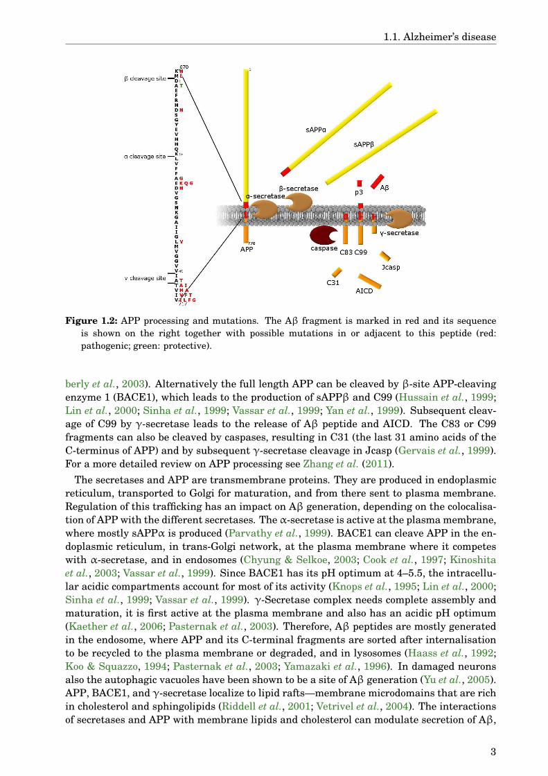
APP can be cleaved by different secretases (Fig. 1.2). α-Secretase has its cleavage sitewithin the Aβ peptide between Lys-16 and Leu-17 (Esch et al., 1990). Different zinc bind-ing metalloproteinases from a disintegrin and metalloproteinase (ADAM) family have α-secretase activity, especially ADAM10 (reviewed in Lichtenthaler, 2011). This cleavage re-sults in a soluble APP ectodomain (sAPPα) and a C-terminal membrane bound peptide of 83amino acids (C83). The latter can be further cleaved to p3 peptide and APP intracellular C-terminal domain (AICD) by γ-secretase, a complex of presenilin (PSEN), nicastrin, anteriorpharynx defective 1 (APH1), and presenilin enhancer (PEN2) (Edbauer et al., 2003; Kim-
2
1.1. Alzheimer’s disease
Figure 1.2: APP processing and mutations. The Aβ fragment is marked in red and its sequenceis shown on the right together with possible mutations in or adjacent to this peptide (red:pathogenic; green: protective).
berly et al., 2003). Alternatively the full length APP can be cleaved by β-site APP-cleavingenzyme 1 (BACE1), which leads to the production of sAPPβ and C99 (Hussain et al., 1999;Lin et al., 2000; Sinha et al., 1999; Vassar et al., 1999; Yan et al., 1999). Subsequent cleav-age of C99 by γ-secretase leads to the release of Aβ peptide and AICD. The C83 or C99fragments can also be cleaved by caspases, resulting in C31 (the last 31 amino acids of theC-terminus of APP) and by subsequent γ-secretase cleavage in Jcasp (Gervais et al., 1999).For a more detailed review on APP processing see Zhang et al. (2011).
The secretases and APP are transmembrane proteins. They are produced in endoplasmicreticulum, transported to Golgi for maturation, and from there sent to plasma membrane.Regulation of this trafficking has an impact on Aβ generation, depending on the colocalisa-tion of APP with the different secretases. The α-secretase is active at the plasma membrane,where mostly sAPPα is produced (Parvathy et al., 1999). BACE1 can cleave APP in the en-doplasmic reticulum, in trans-Golgi network, at the plasma membrane where it competeswith α-secretase, and in endosomes (Chyung & Selkoe, 2003; Cook et al., 1997; Kinoshitaet al., 2003; Vassar et al., 1999). Since BACE1 has its pH optimum at 4–5.5, the intracellu-lar acidic compartments account for most of its activity (Knops et al., 1995; Lin et al., 2000;Sinha et al., 1999; Vassar et al., 1999). γ-Secretase complex needs complete assembly andmaturation, it is first active at the plasma membrane and also has an acidic pH optimum(Kaether et al., 2006; Pasternak et al., 2003). Therefore, Aβ peptides are mostly generatedin the endosome, where APP and its C-terminal fragments are sorted after internalisationto be recycled to the plasma membrane or degraded, and in lysosomes (Haass et al., 1992;Koo & Squazzo, 1994; Pasternak et al., 2003; Yamazaki et al., 1996). In damaged neuronsalso the autophagic vacuoles have been shown to be a site of Aβ generation (Yu et al., 2005).APP, BACE1, and γ-secretase localize to lipid rafts—membrane microdomains that are richin cholesterol and sphingolipids (Riddell et al., 2001; Vetrivel et al., 2004). The interactionsof secretases and APP with membrane lipids and cholesterol can modulate secretion of Aβ,
3

1. Introduction
but also its aggregation and clearance as reviewed by Walter & van Echten-Deckert (2013).
The Aβ peptide has a length of 37–43 amino acids due to the multiple cleavage sites ofγ-secretase (Qi-Takahara et al., 2005). The most abundant form under normal conditions isAβ1–40, followed by a smaller amount of Aβ1–42. Also N-terminal truncated forms (Aβx–40/42)and other C-termini can be found in small amounts (Miller et al., 1993; Vigo-Pelfrey et al.,1993). These peptides can aggregate into oligomers, protofibrils, and finally fibrils thatdeposit into plaques. Diffuse and cored plaques contain mostly Aβ1/x–42, plaques in the wallsof leptomeningeal and intracortical blood vessels (a sign of cerebral amyloid angiopathyoften found in AD brains) contain both Aβ1/x–40 and Aβ1/x–42 (Gowing et al., 1994; Gravinaet al., 1995; Iwatsubo et al., 1994; Miller et al., 1993; Roher et al., 1993a,b). Monomeric Aβis naturally unfolded in aqueous solutions. In solid plaque cores, fibrils of Aβ with a cross-βstructure are found. The formation of fibrils is dependent on pH, solvent, and concentrationof the peptide. Aβ1–42 has a higher tendency to aggregate into fibrils because of a morestable β-sheet formation, and it is the first aggregate forming species in the course of AD(Barrow et al., 1992; Burdick et al., 1992; Iwatsubo et al., 1994). This process probably startswith an intraneuronal aggregation (Capetillo-Zarate et al., 2012; Gouras et al., 2000; Linget al., 2014). The N-truncated forms and the p3 peptide (Aβ17–x) are found in diffuse plaquesand tend to aggregate more rapidly than the full length Aβ (Gowing et al., 1994; Kumar-Singh et al., 2000; Pike et al., 1995). These species, Aβ with pyroglutamate at positions 3or 11, and phosphorylated peptides are found in later stages of AD (Rijal Upadhaya et al.,2014).
Several missense mutations have been found in the APP gene in families with herita-ble AD or cerebral haemorrhage. Most of the pathogenic mutations are located near thesecretase cleavage sites (Fig. 1.2). The Swedish mutation (Mullan et al., 1992) is a dou-ble mutation affecting two amino acid residues near the β-cleavage site (K670N/M671L,positions given for APP770 transcript) that increases the production of both Aβ1–40 andAβ1–42 (Citron et al., 1992; Scheuner et al., 1996). Recently a mutation at codon 673 hasbeen shown to be protective against AD by inhibiting the β-cleavage (Jonsson et al., 2012).Many of the other mutations are located near the γ-secretase cleavage sites (Ancolio et al.,1999; Chartier-Harlin et al., 1991; De Jonghe et al., 2001; Eckman et al., 1997; Goate et al.,1991; Guardia-Laguarta et al., 2010; Kumar-Singh et al., 2000; Murrell et al., 1991, 2000;Pasalar et al., 2002; Terreni et al., 2002) and increase the Aβ1–42/Aβ1–40 ratio (De Jongheet al., 2001; Eckman et al., 1997; Guardia-Laguarta et al., 2010; Scheuner et al., 1996).Two mutations lying at the cytoplasmic transmembrane junction at the residues 723 and724 of APP770 have a similar effect (Kwok et al., 2000; Theuns et al., 2006). Consistentwith the notion that γ-secretase cleavage plays an important role in familial AD, thereare 207 pathogenic mutations in PSEN1 gene and 13 in PSEN2 (AD & FTD MutationDatabase (http://www.molgen.vib-ua.be/ADMutations, accessed on 20/11/2015), Cruts et al.,2012). PSENs also play a role in cellular processes independent of Aβ such as calcium ho-moeostasis, endocytosis, and autophagy that are found to be impaired in both familial andsporadic AD (reviewed in Smolarkiewicz et al., 2013). Mutations lying inside the Aβ se-quence are associated with cerebral amyloid angiopathy and cerebral haemorrhage, but insome cases also AD symptoms were accompanied with the vascular ones (Giaccone et al.,2002; Grabowski et al., 2001; Hendriks et al., 1992; Kamino et al., 1992; Levy et al., 1990;Nilsberth et al., 2001; Obici et al., 2005; Rossi et al., 2004). Some of these mutations leadto differences in APP processing, others affect the aggregation kinetics of the Aβ peptide(Haass et al., 1994; Nilsberth et al., 2001; Watson et al., 1999). Additionally to these muta-tions, duplication of the APP gene can lead to AD symptoms as seen in people with Down’ssyndrome, a condition arising from trisomy of chromosome 21 (Glenner & Wong, 1984a).
4
1.1. Alzheimer’s disease
Aβ-fibrils were first thought to be the toxic species, but due to the lack of correlation be-tween plaques and cognitive decline (Gómez-Isla et al., 1997; Lue et al., 1999; Snowdon,2003) and evidence that oligomers of Aβ are far more toxic than the fibrils (Lambert et al.,1998; Walsh et al., 2002), most of the recent research has concentrated on low molecularweight aggregates. The oligomers are heterogeneous and sensitive to solution conditions,which complicates the study of these aggregates (Benilova et al., 2012). The exact sizeor structure of the toxic Aβ species is not yet known, as an example globular or annu-lar oligomers that interact with membranes are shown to be toxic (Sebollela et al., 2014;Tsigelny et al., 2014). It is also not clear how these oligomers exert their toxicity. SinceCa2+ levels are raised in affected neurons and Aβ is known to interact with membranes, ithas been suggested that the oligomers can increase membrane permeability unspecifically(Kayed et al., 2004) or by forming pores that are selective for Ca2+ (Arispe, 2004; Durellet al., 1994; Lin et al., 2001a). This mechanism would lead to unspecific toxicity in all cells.Another hypothesis states that there is a ‘toxin receptor’ that allows the oligomers to targeta specific population of cells at the presynaptic sites. Many such receptors for Aβ oligomershave been proposed (reviewed in Dinamarca et al., 2012). One of them is the prion pro-tein (PrP) that can activate FYN proto-oncogene (FYN), a cytoplasmic tyrosin kinase (Chinet al., 2004; Lacor et al., 2004; Laurén et al., 2009; Um et al., 2012). Binding to PrP or otherpossible receptors leads to exitotoxicity, aberrant morphology with loss of dendritic spines,and decreases in long term potentiation (Lacor et al., 2007; Laurén et al., 2009; Um et al.,2012). Some of the possible mechanisms are discussed in a recent review by Viola & Klein(2015). For effects of Aβ on oxidative stress, mitochondrial dysfunction, and inflammationin AD see section 1.1.3.
The Aβ peptides can be cleared from brain by several routes. The observation of de-greased Aβ clearance in patients with late onset AD (Mawuenyega et al., 2010) has pointedout a possible defect in one of these pathways as a cause for this most common form of AD.One important way of clearance is the active transport of Aβ over the blood brain barrier.Transport from the brain into blood is mediated by P-glycoprotein (Cirrito et al., 2005), lowdensity lipoprotein receptor-related protein 1 (LRP1) (Shibata et al., 2000), and low densitylipoprotein receptor (Castellano et al., 2012). The latter two are dependent on apolipoproteinE (APOE) (Castellano et al., 2011), a major risk factor for late onset AD (see section 1.1.4).Aβ can also be transported in the other direction, from blood to brain, dependent on ad-vanced glycosylation end product-specific receptor (RAGE) (Deane et al., 2003). This raisesthe possibility that part of the Aβ burden in brain actually originates in systemic circula-tion. Changed levels and localization of LRP1 and RAGE in AD brains probably contributeto the pathogenesis (Donahue et al., 2006). Another minor pathway for Aβ clearance isthe perivascular drainage of interstitial fluid. This can also explain the arterial depositionof Aβ in cerebral amyloid angiopathy (Weller et al., 1998). Along this pathway Aβ is re-moved by perivascular macrophages, whereas microglia and peripheral macrophages haveonly a minor role in phagocytotic Aβ clearance (Hawkes & McLaurin, 2009; Mildner et al.,2011). Aβ can be taken up by cells and transported to lysosomes, where Aβ1–40 is rapidlydegraded. Aβ1–42 is more resistant to degradation and tends to accumulate (Burdick et al.,1997). This can lead to lysosomal dysfunction and release of the contents of lysosomes intocytosol (Yang et al., 1998). Microglia, neurons, astrocytes, and cells of the blood vessels syn-thesise a variety of enzymes that are able to degrade Aβ (reviewed in Miners et al., 2011).The most important ones among them are insulin degrading enzyme (Qiu et al., 1998) andneprilysin, a membrane bound protein that can be released into extracellular space and ac-counts for the most Aβ degrading activity in vivo (Iwata et al., 2000). The activity of theseenzymes is up-regulated in ageing brain and in AD, probably as a physiological response to
5
1. Introduction
increased Aβ level. However, since these enzymes also have a variety of other substrates,the up-regulation can lead to detrimental effects like vasoconstriction and reduced cerebralblood flow (Miners et al., 2014). Others have also reported decreased levels of neprilysin(reviewed in Grimm et al., 2013) and the ability of insulin degrading enzyme to process Aβmay be compromised due to brain insulin resistance (see section 1.1.4).
The mutations found in APP, causing autosomal dominant AD, and the observation of dif-fuse plaques before any other abnormal lesions or neuronal damage led to the formulationof amyloid cascade hypothesis (Hardy & Allsop, 1991; Hardy & Higgins, 1992). According toits original version the aggregation of Aβ is the first event in the pathogenesis of AD. Theplaques then exert their toxic effects on neurons and lead to tau aggregation and neuronaldeath, eventually causing dementia. During the next decades new findings essentially sup-ported this hypothesis with the replacement of Aβ fibrils by oligomers as the toxic speciesand addition of synaptic dysfunction as an early event in the pathology (Ferreira & Klein,2011; Hardy & Selkoe, 2002; Selkoe, 2002). Opponents of this hypothesis have argued thatthere is no correlation between plaque load and cognitive decline; the cell culture and mousemodels, used to produce most of the evidence supporting Aβ as the cause for AD, have littleresemblance with in vivo disease conditions; and there are evidence that Aβ is in fact pro-tective, produced as a response to cellular stress (Herrup, 2015; Lee et al., 2004; Perry et al.,2000). More controversy arose when the first drugs based on lowering the amount of Aβfailed to improve the cognitive function of patients in clinical trials. Alternatives and mod-ifications to the amyloid cascade hypothesis have been proposed (Armstrong, 2014; Ethell,2010; Pimplikar, 2009; Small & Duff, 2008). Others argue that the damage made by Aβmay be irreversible. Therefore, possible treatments should be tested earlier in course of thedisease, before the patients develop severe pathology and overt symptoms (Karran et al.,2011; Musiek & Holtzman, 2015; Tam & Pasternak, 2012). To test this theory, biomarkersto detect early stages of the disease are needed (see section 1.1.4). Nevertheless, the amyloidcascade hypothesis is still the predominant explanation for the aetiology of AD.
1.1.2 Tau
Tau tangles are the other prominent aggregates in AD. Tau is a microtubule-associatedprotein (MAP) that is necessary for tubulin assembly and stabilisation of microtubules (Bré& Karsenti, 1990; Fellous et al., 1977; Panda et al., 1995; Weingarten et al., 1975). Micro-tubules are part of the cytoskeleton that give differentiated cells their typical shape. Theyare also involved in movement, outgrowth of axons in developing neurons, and polarizedtransport of vesicles. There are several MAPs that regulate the dynamic equilibrium ofmicrotubules with monomeric tubulin and they are specific for different cells and compart-ments. Tau is found specifically in axons of neurons where microtubules are important forboth stability and axonal transport (Binder et al., 1985; Dotti et al., 1987).
The gene encoding tau is located on chromosome 17 (Neve et al., 1986) and gives riseto 7 isoforms through alternative splicing. In peripheral nervous system a high molecularweight isoform is found that contains an additional exon 4A (Couchie et al., 1992). Theother 6 isoforms are expressed in central nervous system and differ in splicing of exons 2,3, and 10 (Goedert et al., 1989a,b). Exon 10 encodes one of the four possible tandem repeatsat the C-terminal portion of tau that, together with their flanking regions, act as tubulinbinding sites (Gustke et al., 1994; Himmler et al., 1989; Maccioni et al., 1989). Therefore,isoforms lacking this exon have only three binding repeats, and their binding to microtubuliis weaker. In early developmental stages only the shortest isoform lacking exons 2, 3, and10 is expressed (Goedert et al., 1989a,b). Mutations in tau gene are not associated with AD,
6

1.1. Alzheimer’s disease
but they can lead to frontotemporal dementia (reviewed in Ghetti et al., 2015).Tau protein can be posttranscriptionally modified. A number of these modifications af-
fect its interaction with microtubules and aggregation to neurofibrillary tangels (reviewedin Fontaine et al., 2015). The best studied modification is phosphorylation at the tandemrepeats or residues flanking this region by different kinases (Lovestone & Reynolds, 1997;Wang et al., 2007). This occurs in a healthy brain, varying at different ages, to regulate thebinding affinity of tau to microtubules (Lindwall & Cole, 1984a,b; Trinczek et al., 1995). InAD brain tau is hyperphosphorylated and loses its ability to bind to microtubules (Grundke-Iqbal et al., 1986b). In this form tau can aggregate into paired helical filaments and furtherinto the tangles found in AD brains (Bancher et al., 1989; Ihara et al., 1986; Kidd, 1963;Luna-Muñoz et al., 2007). All tau isoforms found in adult brain are involved in formation ofpaired helical filaments (Greenberg et al., 1992). The aggregation of tau can be prevented bymolecular chaperones that alter its binding to microtubules and prevent toxicity (Abisambraet al., 2010; Dou et al., 2003; Patterson et al., 2011b; Voss et al., 2012).
By aggregating tau loses its function as stabilizer of microtubules, leading to dysfunctionsin cytoskeleton and axonal transport. Additionally to this loss of function effect, the solubletau oligomers or hyperphosphorylated monomers might gain a toxic function, similar to theAβ oligomers (Ding & Johnson, 2008). As for Aβ, it is also not clear for tau what kind ofoligomers mediate the toxicity (Cowan et al., 2012). However, they have been shown to ac-cumulate in AD brain early in the disease progression and correlate with memory loss andsynapse dysfunction in tauopathy models (Berger et al., 2007; Lasagna-Reeves et al., 2011;Patterson et al., 2011a; Sydow et al., 2011). Several mechanisms have been proposed to ex-plain the toxicity of tau. A phosphatase-activating domain at the N-terminus, that becomesaccessible through conformation change in pathogenic tau, can inhibit kinesin dependentaxonal transport (Kanaan et al., 2011). Defects in mitochondrial distribution have been ob-served probably as a consequence of dysfunctional axonal transport (Kopeikina et al., 2011),and tau fragments contribute to mitochondrial dysfunction (see section 1.1.3). Tau can alsomediate some aspects of Aβ toxicity (Roberson et al., 2007). Aβ oligomers lead to activationof kinases that phosphorylate tau, missorting of tau and other axonal proteins into den-drites, elevated Ca2
+ levels, and destabilization of microtubules (Yu et al., 2012; Zempel &Mandelkow, 2012; Zempel et al., 2010). Tau is necessary for postsynaptic targeting of FYNthat can phosphorylate a subunit of N-methyl-D-aspartate receptor (NMDAR) leading to itsanchoring at the postsynaptic sites. This causes exitotoxicity and seizures in AD models(Ittner et al., 2010). Tau itself can also be phosphorylated by FYN that is activated by Aβvia PrP (Larson et al., 2012). Phosphorylation by this and other kinases leads to aberrantcell cycle re-entry that leads to cell death (Seward et al., 2013). It is not clear which of thesemechanisms is most relevant for AD.
It is widely accepted that tau pathology occurs downstream of Aβ in accordance with theamyloid cascade hypothesis. What remains unclear are the mechanisms by which Aβ leadsto changes in tau. Aβ seems to activate kinases that lead to tau hyperphosphorylation likediscussed above and reviewed by Lloret et al. (2015). On the other hand, there are reportsthat human tau can increase Aβ levels (Bright et al., 2015), suggesting a more complexinteraction between these proteins. While most of the treatment strategies today aim atAβ as the main culprit of the disease, some researchers argue that tau should be the target(Crespo-Biel et al., 2012). Indeed, tau pathology correlates better with cognitive decline thanAβ (Arriagada et al., 1992; Gómez-Isla et al., 1997), and tau knockout in APP expressingmouse models rescues the memory deficits (Ittner et al., 2010; Roberson et al., 2007). Still,neuronal and synapse loss are better correlates for cognitive decline than either Aβ or tautangles, suggesting the possibility of other pathogenic mechanisms. Oxidative stress and
7
1. Introduction
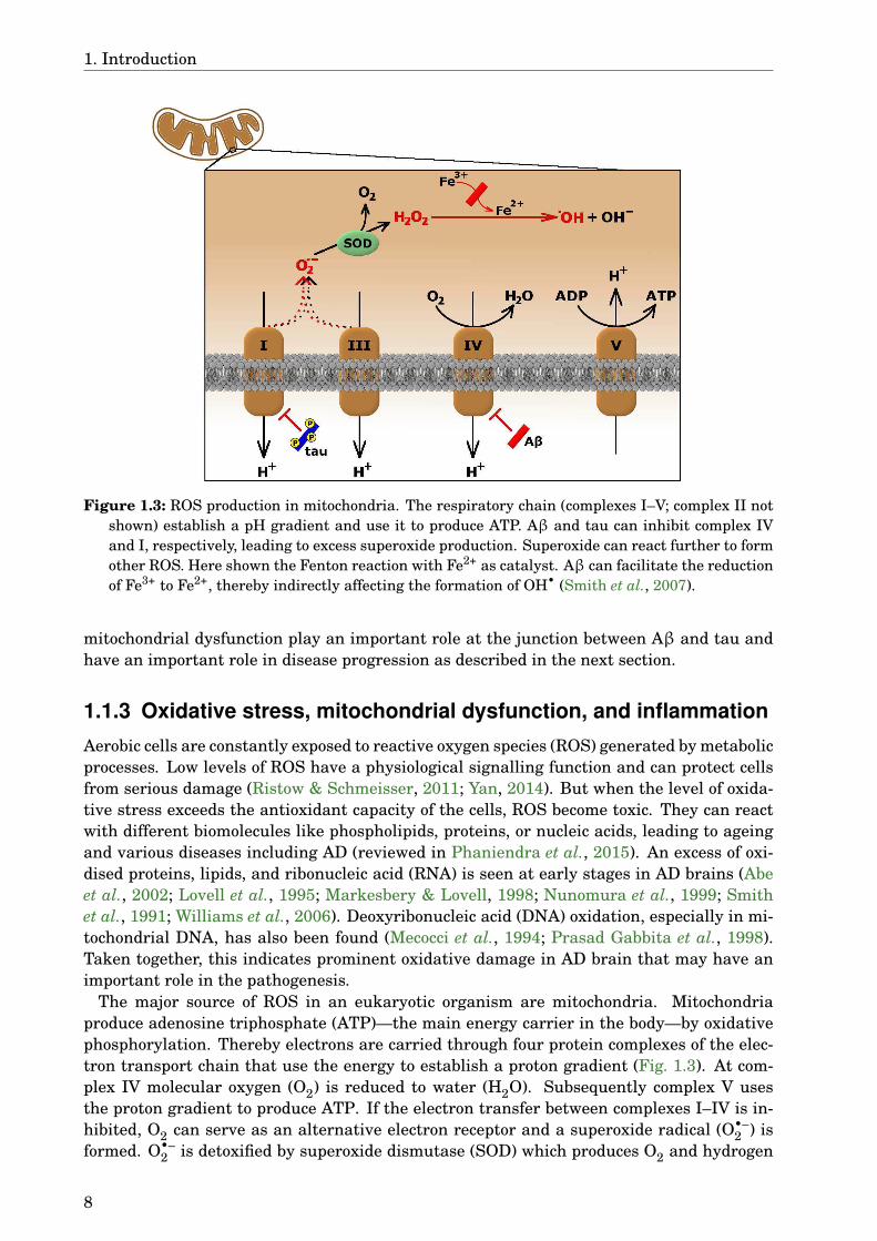
Figure 1.3: ROS production in mitochondria. The respiratory chain (complexes I–V; complex II notshown) establish a pH gradient and use it to produce ATP. Aβ and tau can inhibit complex IVand I, respectively, leading to excess superoxide production. Superoxide can react further to formother ROS. Here shown the Fenton reaction with Fe2+ as catalyst. Aβ can facilitate the reductionof Fe3+ to Fe2+, thereby indirectly affecting the formation of OH• (Smith et al., 2007).
mitochondrial dysfunction play an important role at the junction between Aβ and tau andhave an important role in disease progression as described in the next section.
1.1.3 Oxidative stress, mitochondrial dysfunction, and inflammation
Aerobic cells are constantly exposed to reactive oxygen species (ROS) generated by metabolicprocesses. Low levels of ROS have a physiological signalling function and can protect cellsfrom serious damage (Ristow & Schmeisser, 2011; Yan, 2014). But when the level of oxida-tive stress exceeds the antioxidant capacity of the cells, ROS become toxic. They can reactwith different biomolecules like phospholipids, proteins, or nucleic acids, leading to ageingand various diseases including AD (reviewed in Phaniendra et al., 2015). An excess of oxi-dised proteins, lipids, and ribonucleic acid (RNA) is seen at early stages in AD brains (Abeet al., 2002; Lovell et al., 1995; Markesbery & Lovell, 1998; Nunomura et al., 1999; Smithet al., 1991; Williams et al., 2006). Deoxyribonucleic acid (DNA) oxidation, especially in mi-tochondrial DNA, has also been found (Mecocci et al., 1994; Prasad Gabbita et al., 1998).Taken together, this indicates prominent oxidative damage in AD brain that may have animportant role in the pathogenesis.
The major source of ROS in an eukaryotic organism are mitochondria. Mitochondriaproduce adenosine triphosphate (ATP)—the main energy carrier in the body—by oxidativephosphorylation. Thereby electrons are carried through four protein complexes of the elec-tron transport chain that use the energy to establish a proton gradient (Fig. 1.3). At com-plex IV molecular oxygen (O2) is reduced to water (H2O). Subsequently complex V usesthe proton gradient to produce ATP. If the electron transfer between complexes I–IV is in-hibited, O2 can serve as an alternative electron receptor and a superoxide radical (O•–
2 ) isformed. O•–
2 is detoxified by superoxide dismutase (SOD) which produces O2 and hydrogen
8
1.1. Alzheimer’s disease
peroxide (H2O2), another ROS that is further reduced to H2O by catalase or glutathioneperoxidase. If these ROS escape the cells defensive mechanism, they can either oxidisebiomolecules themselves or form the more reactive hydroxyl radical (OH•). This can hap-pen via two reaction mechanisms, both of which depend on metal ions (Men+) like copper oriron (reviewed in Halliwell & Gutteridge, 1984):
Fenton reaction: Men++H2O2 −−−→Me(n+1)++OH•+OH−
Haber-Weiss reaction: O•−2 +H2O2
Men+−−−−→OH•+OH−+O2
Imbalances in copper, iron, and zinc ions have been reported in AD brains, and thesemetals are found in senile plaques (Deibel et al., 1996; Lovell et al., 1998). Copper canbind to Aβ, especially under acidic conditions, and facilitate the formation of H2O2, possiblyfollowed by Fenton reaction (Atwood et al., 2000; Huang et al., 1999; Opazo et al., 2002). Thethree histidine residues of Aβ are thereby involved in complexing the metal ion, whereasMet35 and Tyr10 are important for the redox chemistry (Smith et al., 2007; Varadarajanet al., 1999; Yatin et al., 1999). In nanomolar concentrations Aβ binding to copper exhibitsneurotrophic and antioxidant effects, although higher concentrations lead to pro-oxidantactivity (reviewed in Atwood et al., 2003). Binding of zinc to Aβ leads to aggregation of thepeptide and reduces its toxicity (Bush et al., 1994; Cuajungco et al., 2000). Zink can also bindto tubulin, and dyshomeostasis of this metal can lead to depolymerisation of microtubules(Craddock et al., 2012). The expression of APP is regulated by iron (Rogers et al., 2002), andcopper binding to APP increases Aβ production (Noda et al., 2013), further emphasising theimportant role of metal ions in AD pathogenesis.
Mitochondrial dysfunction is seen in different neurodegenerative disorders, such as AD,Parkinson’s disease, amyotrophic lateral sclerosis, and Huntington’s disease (reviewed inLezi & Swerdlow, 2012). In AD interactions of Aβ with mitochondrial proteins can leadto inhibition of the electron transport chain (Fig. 1.3), increased ROS levels, and apop-tosis (Cha et al., 2012). The activity of complex IV is decreased in AD (Maurer et al.,2000; Mutisya et al., 1994), possibly because of decreased expression and deletions in mito-chondrial DNA (Krishnan et al., 2011; Rhein et al., 2009) or blockage of the translocases—proteins needed for proper import of mitochondrial proteins encoded by nuclear DNA—byAPP (Devi et al., 2006). Furthermore, Aβ is taken up into mitochondria and is found inthe inner membrane, making a direct interaction with the complexes of the respiratorychain or other mitochondrial proteins like Aβ-binding alcohol dehydrogenase (ABAD) pos-sible (Hansson Petersen et al., 2008; Lustbader et al., 2004). Deficiency of complex IV isdependent on Aβ, whereas tau oligomers decrease complex I levels. Both these effects aresynergistic in impairing mitochondrial membrane potential and ATP production (Amadoroet al., 2012; Lasagna-Reeves et al., 2011; Quintanilla et al., 2014; Rhein et al., 2009). Ex-posure to Aβ leads to higher ROS levels due to its interaction with mitochondrial proteins(Hernandez-Zimbron et al., 2012; Lustbader et al., 2004). Additionally, increased fission anddecreased fusion of mitochondria have been observed in AD, resulting in changed morphol-ogy (Baloyannis, 2011). Aβ can influence the expression of proteins involved in mitochon-drial dynamics and this leads to altered transport of mitochondria in neurons (see also sec-tion 1.1.2) that correlates with loss of dendritic spines (Calkins & Reddy, 2011; Wang et al.,2009). Aβ can also support the expression of permeability transition pore, whereas tauoligomers activate caspase 9, implicating both peptides in induction of apoptosis (Lasagna-Reeves et al., 2011; Moreira et al., 2002). In normal cells dysfunctional mitochondria aredegraded via autophagy, but under pathological conditions this process is impaired andleads to further damage of the cells (reviewed in Nixon & Yang, 2011; Schiavi & Ventura,2014).
9
1. Introduction
Decreased ATP production due to mitochondrial dysfunction can lead to higher Aβ levels(Scheffler et al., 2012; Velliquette et al., 2005), and phosphorylated tau has been detectedafter inhibition of complex IV (Szabados et al., 2004). The evidence of defect mitochondriain AD have led to the formulation of a ‘mitochondrial cascade hypothesis’ (Swerdlow et al.,2014; Swerdlow & Khan, 2004). It states that mitochondrial dysfunction is the driving forceof sporadic late onset AD, leading to Aβ aggregation due to changes in electron transportchain as well as tau phosphorylation and aggregation due to cell cycle re-entry. This hy-pothesis includes the free radical theory of ageing (Harman, 1956), using it to explain thelate onset of sporadic AD. The ‘two hit’ hypothesis also states that oxidative stress has animportant role in AD pathogenesis, but according to this theory a second factor like mitoticstress is needed to initiate the disease progression (Zhu et al., 2001, 2007, 2004).
Another possible source of ROS are activated immune cells, that use these reactive mole-cules for fighting intruders like bacteria. Indeed, fibrillar Aβ can induce O•–
2 production inmacrophages (Colton et al., 2000). Already Alzheimer observed microglia, the brain residentimmune cells, surrounding the senile plaques (Alzheimer, 1911). Aβ aggregates activate mi-croglia that can bind and phagocytose Aβ via different membrane receptors (reviewed in Yu& Ye, 2015) and recruit peripheral monocytes that also contribute to Aβ clearance in brainparenchyma (Krabbe et al., 2013; Simard et al., 2006). In the course of the disease, however,microglia exposed to Aβ lose this protective function and change to a more pro-inflammatoryform, releasing cytokines that further inhibit clearance of Aβ (Hickman et al., 2008; Krabbeet al., 2013). Additionally, uptake of Aβ from the periphery by RAGE causes neurovascu-lar stress and leads to further expression of cytokines (Deane et al., 2003). On the otherhand, chronic systemic inflammation has been shown to induce AD-like pathology in mice,suggesting that inflammatory conditions can trigger excess Aβ production and initiate AD(Krstic et al., 2012). The ROS and pro-inflammatory cytokines produced by microglia can ac-tivate different signalling cascades in neurons and lead to damage of the cells. For examplethe activation of kinases can lead to tau hyperphosphorylation (Kitazawa et al., 2011). Therole of neuroinflammation in the aetiology of AD is still under debate, but its associationwith the disease is supported also by genetic studies discussed in the next section.
1.1.4 Risk factors and biomarkers
Since curing AD has proven to be a difficult task, more and more attention is paid to pre-vention and early detection of the people at risk. The most important risk factor is high age,followed by cerebrovascular disease, traumatic brain injury, cardiovascular disease, hyper-tension, and metabolic disorders like type II diabetes or obesity. Mediterranean diet andphysical and mental activity, on the other hand, decrease the risk (reviewed in Mayeux &Stern, 2012; Reitz & Mayeux, 2014). Metabolic disorders and AD could be linked togethervia insulin resistance as proposed already decades ago (Hoyer, 1988; Hoyer et al., 1991).Insulin has an important role in energy control and also in synaptic plasticity in centralnervous system (reviewed in Ma et al., 2015a). Signalling trough insulin and insulin-likegrowth factor 1 (IGF-1) receptors promotes clearance of Aβ oligomers (Zhao et al., 2009) andis linked to other stress signals involved in AD by different pathways (reviewed in Lourencoet al., 2015). One of the hubs where several of these pathways converge is the transcrip-tion factor forkhead box O (FOXO), that regulates the response of a cell to both oxidativestress and glucose deprivation, but can lead to cell death and several processes implicatedin AD by prolonged activity (Manolopoulos et al., 2010). The evidence for the involvementof insulin signalling in AD pathology has led to the notion that this disease can be seen asa form of brain diabetes (Lourenco et al., 2015; Morgen & Frölich, 2015). According to a
10
1.1. Alzheimer’s disease
recent study, however, Aβ oligomers can induce peripheral glucose intolerance by a mech-anism involving inflammation and endoplasmic reticulum stress in hypothalamus (Clarkeet al., 2015). Therefore, the role of insulin resistance and diabetes in AD pathology remainsa subject of further study.
The genetic background of familial AD is well known (see section 1.1.1), but the geneticcomponent of the more prevalent sporadic AD seems to be more complex. Several genomewide association studies have been conducted to find risk genes, but most of the genes foundhave only a weak association with the disease. Among those are genes coding for pro-teins involved in lipid transport (APOE; clusterin), immune system (ATP-binding cassettesub-family A member 7; complement component receptor 1; CD33; membrane-spanning4-domains, subfamily A, members 6A and 4E; CD-2 associated protein), and endocyto-sis (bridging integrator 1; phosphatidylinositol binding clathrin assembly protein; sortilin-related receptor 1) (AlzGene database (http://www.alzgene.org/, accessed on 20/11/2015),Bertram et al., 2007; Reitz & Mayeux, 2014). The strongest association, found in almostall of these studies, is for APOE. There are three common alleles of the APOE gene: ε2, ε3,and ε4. The ε4 allele is associated with higher risk for late onset AD (Corder et al., 1993;Strittmatter et al., 1993), whereas ε2 is protective (Corder et al., 1994; Hardy et al., 1993).APOE is responsible for cholesterol transport and homoeostasis in brain. Lower cholesterollevels in microglia have been shown to enhance intracellular degradation of Aβ by facili-tating endocytic trafficking (Lee et al., 2012a). Also the degradation by insulin degradingenzyme is enhanced by lipidated APOE (Jiang et al., 2008). Additionally, APOE can directlyinteract with Aβ and alter its aggregation properties (Arold et al., 2012; Garai et al., 2014;LaDu et al., 2011).
The pathological diagnosis of AD relies on the extent of Aβ and tau pathology in the brainof the patient (reviewed in Serrano-Pozo et al., 2011). In order to diagnose AD in livingpatients, biomarkers and imaging techniques are needed together with cognitive tests toseparate AD from other dementias. Available biomarkers can be divided into two groups:biochemical markers found in cerebrospinal fluid and imaging biomarkers. In cerebrospinalfluid concentrations of Aβ and tau can be measured. Aβ1–42 is lower in patients with ADwhereas total and phosphorylated tau levels are higher than in controls. Especially theratios Aβ1–42/Aβ1–40 and Aβ1–42/phospho-tau have shown high predictive value for earlyAD (reviewed in Lewczuk et al., 2014). Different imaging techniques can be applied forthe diagnosis (reviewed in Ahmed et al., 2014). Positron emission tomography using 18-F-flourodeoxyglucose typically shows bilateral hypometabolism and hypoperfusion in the ADbrains. Specific tracers like Pittsburgh Compound B used with positron emission tomog-raphy can visualize amyloid plaques in living patients. Additionally, structural magneticresonance imaging can detect brain atrophy. Using combinations of these biomarkers, it ispossible to strengthen the diagnosis made by cognitive testing and differentiate between dif-ferent types of dementias. Still, there are many problems including availability and cost ofsuitable instrumentation and insufficient standardisation of the acquisition methods, there-fore these techniques are not everywhere used as a standard procedure for diagnosis. Newmethods and markers, that would be easier to handle and have better sensitivity and selec-tivity, are in development (Ahmed et al., 2014).
1.1.5 Treatment
The first prescription drug for AD was tacrine (Cognex®), an acetylcholine esterase in-hibitor (AChEI) (Summers, 2006). The neurotransmitter acetylcholine (ACh) has an im-portant role in forming the concious awareness, and impairment of the cholinergic system
11
1. Introduction
underlies symptoms of different dementias (Perry et al., 1999). In AD there is a lack ofACh due to selective loss of cholinergic neurons (Davies & Maloney, 1976; Francis et al.,1999). By inhibiting the enzyme that degrades ACh—acetylcholine esterase (AChE)—theamount of this neurotransmitter in the synaptic cleft can be increased. Hence, the treatmentslows down the cognitive decline and helps to maintain activities of daily living. The usageof tacrine was discontinued due to hepatotoxicity (e.g. Blackard et al., 1998), but newerAChEI donepezil (Aricept®), rivastigmine (Exelon®), and galantamine (Reminyl®) are stillused in mild to moderate AD (Birks, 2006). For moderate to severe disease stages anotherdrug called memantine (Namenda®) is approved (Matsunaga et al., 2015). Memantine isa non-competitive NMDAR antagonist, that counteracts the impaired glutamate signallingand can protect neurons from excitotoxicity. Combinations of both drug types have showngreater benefits in mild to moderate AD than one drug alone (Parsons et al., 2013). Still,these treatments only counteract the symptoms and cannot halt the course of the disease.Therefore, a lot of effort has been made to find new, more efficient drugs.
Most of the research in this area has concentrated on Aβ: substances that can inhibit Aβoligomerization (reviewed in Doig & Derreumaux, 2015), reduce its production, or facilitateits clearance. An example of a small molecule that can reduce brain amyloid load, proba-bly due to modulation of BACE1, and is currently tested in clinical trials is methylene blue(Mori et al., 2014). BACE1 is the rate limiting enzyme for Aβ production and therefore alogical drug target. Several small molecules that can inhibit BACE1 or modulate its activ-ity have been found and some of them have reached phase III clinical trials (reviewed inEvin & Hince, 2013; Vassar et al., 2014). However some adverse effects have been noticeddue to other targets of this enzyme and cross reactivity with other aspartyl proteases ofthe same family like BACE2 or cathepsin D. Similar problems occurred with γ-secretaseinhibitors, where an increased risk of skin cancer and worsening of cognitive abilities havebeen observed (reviewed in Mikulca et al., 2014). There the focus has shifted from inhibitingthe enzyme to modulating its activity in favour of the shorter Aβ species. These modula-tors do not inhibit other functions of γ-secretase and therefore should have less side-effects(D’Avanzo et al., 2015). For increasing the clearance of Aβ, immunotherapy is utilized (re-viewed in Spencer & Masliah, 2014). Although the first clinical trials in this field failed dueto severe side effects and no change in cognitive abilities of the patients was observed, newapproaches for both active and passive immunization are hoped to lead to safer and moreeffective treatment.
Failure of the first anti-Aβ drugs has triggered a search for new targets and treatmentpossibilities. Immunotherapy against tau is considered along with antioxidant and anti-inflammatory approaches, and a lot of other targets are explored (Geldenhuys & Darvesh,2014). Since AD is an multi-factorial disease with many possible drug targets, a trend ofdeveloping multi-target-directed-ligands has emerged. Also, prevention and early pharma-cological intervention are gaining more and more attention (Sindi et al., 2015), althoughthere is still a lack of reliable biomarkers to identify those at risk in presymptomatic stages(see section 1.1.4). In the search for new multitarget treatments and lead substances fordrugs traditional medicine, especially Traditional Chinese Medicine (TCM) (reviewed in Suet al., 2014; Zeng et al., 2015), and natural products have received considerable attention.Several natural products have been found to be effective: the AChEI galantamine was firstisolated from snowdrop (Galanthus spp. ) (Heinrich & Teoh, 2004); huperzin A, an AChEIfrom Huperzia serrata, is in phase II clinical trials (Yang et al., 2013b); (−)-epigallocatechingallate (EGCG) (Camellia sinensis), curcumin (Curcuma longa), resveratrol, scyllo-inositol(Cocos nucifera), and several flavonoids can modulate Aβ aggregation (reviewed in Bu et al.,2015); the standardized Ginkgo biloba extract EGb761® has shown beneficial effects (Can-
12

1.2. Medicinal plants
evelli et al., 2014; Gauthier & Schlaefke, 2014). Despite all these efforts, more research isstill needed to find an effective treatment for AD.
1.2 Medicinal plantsPhytotherapy is one of the oldest forms of medicine, known to mankind already for thou-sands of years. The traditional practices differ by culture and region. Today we have accessto a lot of ethnobotanical information from around the world. Some of the plants used tra-ditionally are scientifically proved to be effective and are still used in modern phytotherapy,some others are used in alternative medicine or as food supplements. The basis of effec-tiveness of medicinal plants are almost exclusively secondary metabolites—small moleculesproduced by plants to protect themselves. These molecules can either directly interact witha pharmaceutical target in human body or they can be used as leads for synthesis of activecompounds. Therefore, medicinal plants are interesting for both traditional medicine andmodern pharmaceutical research.
Plants produce secondary metabolites as a means to interact with their environment. Inorder to reproduce, plants need the help of insects and animals for pollination and distri-bution of their seeds. Therefore, they have developed molecules that attract these helpers.They also have to protect their territory and resources from other plants, so they have devel-oped phytotoxins. Plants do not have the ability to move away from their enemies. Hence,they depend on their chemical defence system to protect themselves from phytopathogensand herbivores. Upon an attack they produce phytoalexins—molecules that can ward offthe danger and send a warning to systemic tissues. As a reaction to this signal protectivesecondary metabolites and proteins are produced for enhanced protection in whole plant,leading to a systemic acquired resistance (Ahuja et al., 2012; Spoel & Dong, 2012). Nextto this induced expression of phytoalexins, many protective secondary metabolites are pro-duced constitutively, stored, and released upon an attack. These molecules, that have an-timicrobial, antifungal, antioxidant, and several other properties, are most interesting forpharmaceutical research.
Secondary metabolites probably developed early in the evolution of plants and have beenrefined through natural selection (Wink, 2003), giving them an advantage over de novo syn-thesis of medical compounds. The chemical structures of these molecules are diverse, as aretheir mechanisms of action. Some mimic molecules in animals and can interact with specificreceptors or enzymes (e.g. some alkaloids are similar to neurotransmitters and can activateor inhibit neuroreceptors or re-uptake transporters), but most (e.g. polyphenols, terpenes)have rather unspecific interactions with proteins, biomembranes, or nucleic acids (reviewedin Wink, 2008). One plant usually produces various compounds to have the best protectionagainst as many different threats as possible. Hence, plant extracts are multitarget drugsthat can modulate several cellular processes and possibly even act on targets that have notbeen identified yet.
Natural selection has not only helped the plants to perfect their toxins, the herbivoreshave gone through an evolutionary adaptation process as well. Thereby, mutations to thetarget proteins have led to resistance in some specialised herbivores. Other insects andanimals have developed several mechanisms to either store the toxins and use them fortheir own protection or detoxify these phytochemicals. The latter option is mostly used byvertebrates. Proteins in these organisms can be protected by chaperones like heat shockproteins (HSPs); oxidative damage can be prevented by SOD, catalase, or other enzymes.Additionally, the toxic compounds are actively modified in liver and excreted, or their in-take is inhibited by ATP-binding cassette transporters in epithelia (Murugaiyah & Mattson,
13
1. Introduction
2015). Expression of protective proteins is controlled by transcription factors like nuclearfactor erythroid 2-like 2 (NRF2), heat shock factor 1 (HSF-1), or FOXO (Murugaiyah &Mattson, 2015; Son et al., 2008). Small amounts of toxic compounds, that manage to crossthe epithelia, activate these transcription factors and trigger the transcription of the stressresistance proteins that can protect the organism from the toxin that was ingested, but alsogive it a wider resistance to many types of stress. This is the underlying mechanism ofhormesis—the phenomenon that small amounts of a compound can be beneficial, even if alarger amount of the same substance is toxic (Calabrese, 2015; Mattson, 2008). Hormesisis proposed as the mechanism of action for several phytochemicals including polyphenolsfrom green tea like EGCG (Murakami, 2014; Son et al., 2008). Also other types of stresslike exercise or caloric restriction lead to hormetic responses. Most interestingly, hormesisis essential for healthy ageing and the health of nervous system (Murugaiyah & Mattson,2015).
1.2.1 Phytotherapy in Traditional Chinese Medicine
TCM is one of the oldest medicine systems dating back more than 5000 years. Since at thattime there was no technology available to look inside a living body, TCM uses superficialobservations—ranging from voice quality to pulse feeling—to diagnose a condition. A dis-ease is seen as an imbalance in yin and yang, the two basic forces in Taoism. Without themodern knowledge about anatomy and physiology, the ancient healers used this philosophyto describe the function of human body. In TCM the body is divided into Organs, whereby‘Organ’ does not refer to an anatomical entity but is rather a concept of body parts and func-tions that react together to certain changes in the body (for better distinction these Organsare written with a capital letter). Each Organ is governed by one of the five elements—wood,fire, earth, metal, water—and has either a yin- or yang-character depending on its function.The normal functioning of the Organs is ensured by the flow of qi, often translated as ‘lifeenergy’. Qi flows through meridians—‘energy channels’ that belong to the Organs—therebynourishing all tissues. If the flow of qi is obstructed, the balance between yin and yang willbe disturbed and the person feels sick.
TCM uses different forms of therapy like acupuncture, tuina massage, qigong exercises,and also phytotherapy. Using medicinal plants is common in different cultures and can berationalised by the physiological activity of secondary metabolites. TCM, however, has itsspecific way of using plants. Each plant is described by its flavour—sour, bitter, sweet, acrid,salty. These flavours correspond to the five elements and thereby indicate the effect of theplant on different Organs. Some plants are processed, e.g. by roasting, to obtain the desiredeffect. A phytotherapeutical drug always consists of at least four plants: emperor—theplant with the main effect; minister—supports the effect of emperor; ambassador—guidesthe effect to the right Organ; assistant—helps to treat secondary symptoms, harmonizesthe other plants, and reduces toxicity (Bürkland, 2014). This concept of using many plantstogether in one drug is in contrast to western medicine, where one plant or, better still,one pure compound is used. Extracting several plants together can, however, change thecomposition of the extract, possibly leading to less toxicity and synergistic effects (Wanget al., 2013b; Yang et al., 2009b).
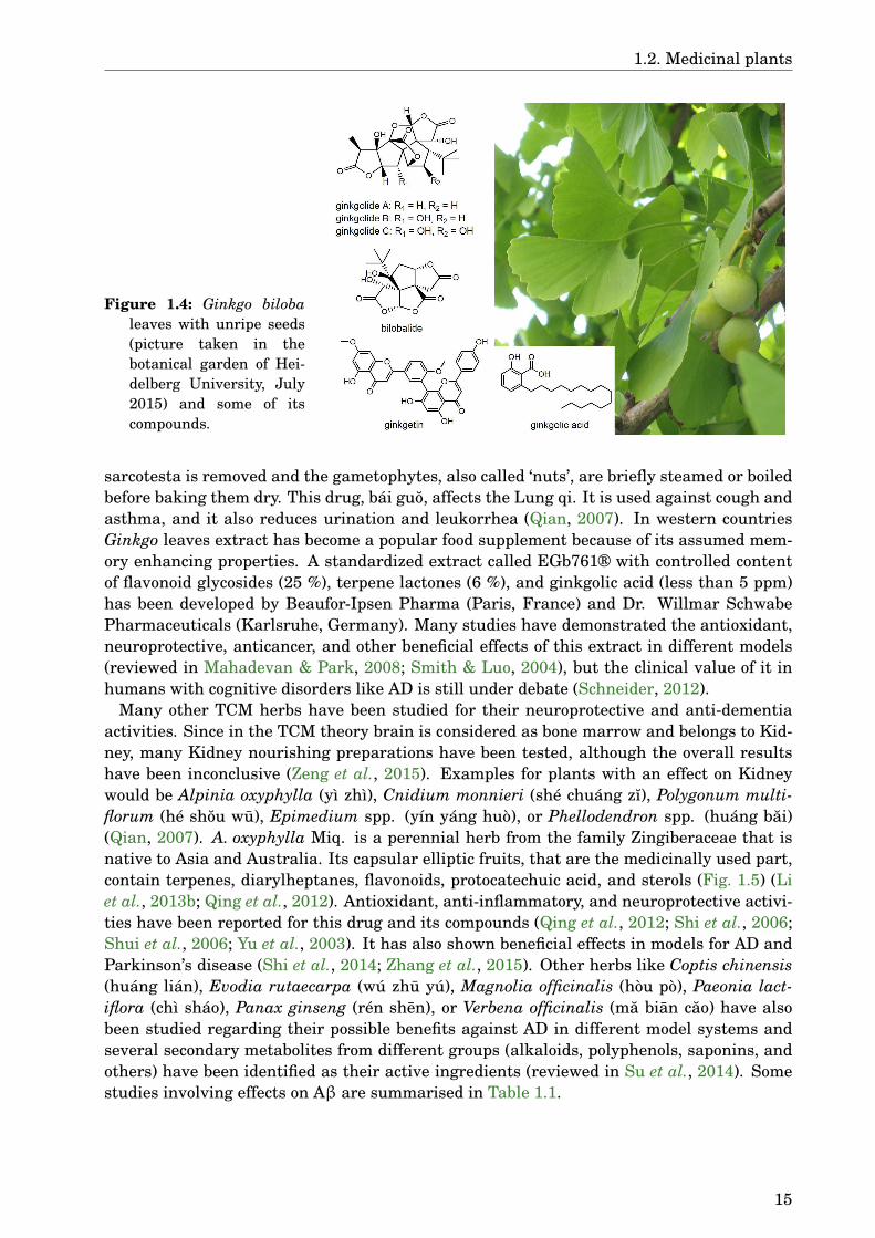
One of the TCM herbs that has become popular also in western countries is Ginkgo bilobaL. (Ginkgoaceae) (Fig. 1.4). This dioecious tree can grow up to 35 m high, it has uniquefan-shaped leaves, and the female plants produce fruit-like seeds that smell like rancid but-ter due to the high content of butyric acid (Van Wyk et al., 2015). In TCM originally theseeds are used, only later the leaves have been also listed as a medicinal drug. The fleshy
14
1.2. Medicinal plants
Figure 1.4: Ginkgo bilobaleaves with unripe seeds(picture taken in thebotanical garden of Hei-delberg University, July2015) and some of itscompounds.
sarcotesta is removed and the gametophytes, also called ‘nuts’, are briefly steamed or boiledbefore baking them dry. This drug, bái guo, affects the Lung qi. It is used against cough andasthma, and it also reduces urination and leukorrhea (Qian, 2007). In western countriesGinkgo leaves extract has become a popular food supplement because of its assumed mem-ory enhancing properties. A standardized extract called EGb761® with controlled contentof flavonoid glycosides (25 %), terpene lactones (6 %), and ginkgolic acid (less than 5 ppm)has been developed by Beaufor-Ipsen Pharma (Paris, France) and Dr. Willmar SchwabePharmaceuticals (Karlsruhe, Germany). Many studies have demonstrated the antioxidant,neuroprotective, anticancer, and other beneficial effects of this extract in different models(reviewed in Mahadevan & Park, 2008; Smith & Luo, 2004), but the clinical value of it inhumans with cognitive disorders like AD is still under debate (Schneider, 2012).
Many other TCM herbs have been studied for their neuroprotective and anti-dementiaactivities. Since in the TCM theory brain is considered as bone marrow and belongs to Kid-ney, many Kidney nourishing preparations have been tested, although the overall resultshave been inconclusive (Zeng et al., 2015). Examples for plants with an effect on Kidneywould be Alpinia oxyphylla (yì zhì), Cnidium monnieri (shé chuáng zı), Polygonum multi-florum (hé shou wu), Epimedium spp. (yín yáng huò), or Phellodendron spp. (huáng bai)(Qian, 2007). A. oxyphylla Miq. is a perennial herb from the family Zingiberaceae that isnative to Asia and Australia. Its capsular elliptic fruits, that are the medicinally used part,contain terpenes, diarylheptanes, flavonoids, protocatechuic acid, and sterols (Fig. 1.5) (Liet al., 2013b; Qing et al., 2012). Antioxidant, anti-inflammatory, and neuroprotective activi-ties have been reported for this drug and its compounds (Qing et al., 2012; Shi et al., 2006;Shui et al., 2006; Yu et al., 2003). It has also shown beneficial effects in models for AD andParkinson’s disease (Shi et al., 2014; Zhang et al., 2015). Other herbs like Coptis chinensis(huáng lián), Evodia rutaecarpa (wú zhu yú), Magnolia officinalis (hòu pò), Paeonia lact-iflora (chì sháo), Panax ginseng (rén shen), or Verbena officinalis (ma bian cao) have alsobeen studied regarding their possible benefits against AD in different model systems andseveral secondary metabolites from different groups (alkaloids, polyphenols, saponins, andothers) have been identified as their active ingredients (reviewed in Su et al., 2014). Somestudies involving effects on Aβ are summarised in Table 1.1.
15

1. Introduction
Table 1.1: Activity against Aβ aggregation and toxicity of selected TCM plants.
Plant Drug Model Effect Reference
Alpinia galanga ethanol extractof rhizome
mice treated withAβ25–35
memory enhancing,antioxidant
Hanish Singhet al. (2011)
Alpinia oxyphylla ethanol extractof fruits
mice treated withAβ1–42
memory enhancing,decreased Aβ1–42 levels,inhibits AChE, antioxidant
Shi et al.(2014)
5-hydroxy-methylfurfural
mice treated withAβ1–42
memory enhancing,decreased Aβ1–42 levels,inhibits BACE1,antioxidant
Liu et al.(2014)
Camellia sinensis EGCG α-synuclein,Aβ1–42, cellculture
binds unfolded proteins,inhibits fibrillisation, leadsto formation of non-toxicoligomers
Ehrnhoeferet al. (2008)
green teacatechins
SAMP8 mice1 memory enhancing, reducedlevels of Aβ1–42 oligomers
Li et al.(2009)
EGCG C. elegans reduced Aβ deposits andoligomerisation, decreasedlipofuscin formation,antioxidant
Abbas &Wink (2010)
EGCG α-synuclein,Aβ1–42, cellculture
binds to amyloid fibrils,remodels their structure
Bieschkeet al. (2010)
digested waterextract of greentea
cell culture antioxidant, protectiveagainst Aβ1–42 toxicity
Okello et al.(2011)
EGCG metal-free andmetal-associatedAβ1–40, cellculture
higher antiamyloidogenicactivity in metal-associatedsystem, reduced Aβ toxicity,produces ternary complexeswith metal-associated Aβ
Hyung et al.(2013)
Centella asiatica extract PSAPP mice2 decreased Aβ1–40 andAβ1–42 in hippocampus,antioxidant
Dhanasekaranet al. (2009)
water extract Tg2576 mice3,cell culture
memory enhancing,neuroprotective against Aβtoxicity, no change in Aβlevels
Soumyanathet al. (2012)
water extract,caffeoylquinicacids
cell culture antioxidant, normalizescalcium homoeostasis,attenuates mitochondrialdysfunction
Gray et al.(2015)
Continued on next page
1Naturally occurring mouse line with accelerated senescence and several features similar to AD.2Doubly transgenic mice expressing APP with the Swedish mutation and PSEN1 with M146L mutation.3Transgenic mice expressing APP with the Swedish mutation.
16
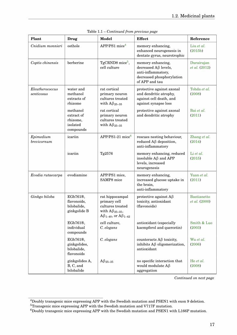
1.2. Medicinal plants
Table 1.1 – Continued from previous page
Plant Drug Model Effect Reference
Cnidium monnieri osthole APP/PS1 mice4 memory enhancing,enhanced neurogenesis indentate gyrus, neurotrophic
Liu et al.(2015b)
Coptis chinensis berberine TgCRND8 mice5,cell culture
memory enhancing,decreased Aβ levels,anti-inflammatory,decreased phosphorylationof APP and tau
Durairajanet al. (2012)
Eleutherococcussenticosus
water andmethanolextracts ofrhizome
rat corticalprimary neuroncultures treatedwith Aβ25–35
protective against axonaland dendritic atrophy,against cell death, andagainst synapse loss
Tohda et al.(2008)
methanolextract ofrhizome,isolatedcompounds
rat corticalprimary neuroncultures treatedwith Aβ25–35
protective against axonaland dendritic atrophy
Bai et al.(2011)
Epimediumbrevicornum
icariin APP/PS1-21 mice6 rescues nesting behaviour,reduced Aβ deposition,anti-inflammatory
Zhang et al.(2014)
icariin Tg2576 memory enhancing, reducedinsoluble Aβ and APPlevels, increasedneurogenesis
Li et al.(2015)
Evodia rutaecarpa evodiamine APP/PS1 mice,SAMP8 mice
memory enhancing,increased glucose uptake inthe brain,anti-inflammatory
Yuan et al.(2011)
Ginkgo biloba EGb761®,flavonoids,bilobalide,ginkgolide B
rat hippocampalprimary cellcultures treatedwith Aβ25–35,Aβ1–40, or Aβ1–42
protective against Aβtoxicity, antioxidant(flavonoids)
Bastianettoet al. (2000)
EGb761®,individualcompounds
cell culture,C. elegans
antioxidant (especiallykaempferol and quercetin)
Smith & Luo(2003)
EGb761®,ginkgolides,bilobalide,flavonoids
C. elegans counteracts Aβ toxicity,inhibits Aβ oligomerization,antioxidant
Wu et al.(2006)
ginkgolides A,B, C, andbilobalide
Aβ25–35 no specific interaction thatwould modulate Aβaggregation
He et al.(2008)
Continued on next page
4Doubly transgenic mice expressing APP with the Swedish mutation and PSEN1 with exon 9 deletion.5Transgenic mice expressing APP with the Swedish mutation and V717F mutation.6Doubly transgenic mice expressing APP with the Swedish mutation and PSEN1 with L166P mutation.
17
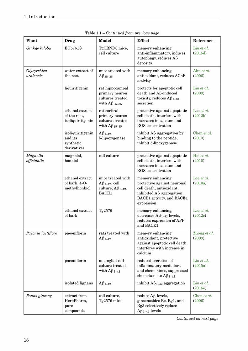
1. Introduction
Table 1.1 – Continued from previous page
Plant Drug Model Effect Reference
Ginkgo biloba EGb761® TgCRND8 mice,cell culture
memory enhancing,anti-inflammatory, inducesautophagy, reduces Aβdeposits
Liu et al.(2015d)
Glycyrrhizauralensis
water extract ofthe root
mice treated withAβ25–35
memory enhancing,antioxidant, reduces AChEactivity
Ahn et al.(2006)
liquiritigenin rat hippocampalprimary neuroncultures treatedwith Aβ25–35
protects for apoptotic celldeath and Aβ-inducedtoxicity, reduces Aβ1–40secretion
Liu et al.(2009)
ethanol extractof the root,isoliquiritigenin
rat corticalprimary neuroncultures treatedwith Aβ25–35
protective against apoptoticcell death, interfere withincreases in calcium andROS concentration
Lee et al.(2012b)
isoliquiritigeninand itssyntheticderivatives
Aβ1–42,5-lipoxygenase
inhibit Aβ aggregation bybinding to the peptide,inhibit 5-lipoxygenase
Chen et al.(2013)
Magnoliaofficinalis
magnolol,honkiol
cell culture protective against apoptoticcell death, interfere withincreases in calcium andROS concentration
Hoi et al.(2010)
ethanol extractof bark, 4-O-methylhonkiol
mice treated withAβ1–42, cellculture, Aβ1–42,BACE1
memory enhancing,protective against neuronalcell death, antioxidant,inhibited Aβ aggregation,BACE1 activity, and BACE1expression
Lee et al.(2010a)
ethanol extractof bark
Tg2576 memory enhancing,decreases Aβ1–42 levels,reduces expression of APPand BACE1
Lee et al.(2012c)
Paeonia lactiflora paeoniflorin rats treated withAβ1–42
memory enhancing,antioxidant, protectiveagainst apoptotic cell death,interferes with increase incalcium
Zhong et al.(2009)
paeoniflorin microglial cellculture treatedwith Aβ1–42
reduced secretion ofinflammatory mediatorsand chemokines, suppressedchemotaxis to Aβ1–42
Liu et al.(2015a)
isolated lignans Aβ1–42 inhibit Aβ1–42 aggregation Liu et al.(2015e)
Panax ginseng extract fromHerbPharm,purecompounds
cell culture,Tg2576 mice
reduce Aβ levels,ginsenosides Re, Rg1, andRg3 selectively reduceAβ1–42 levels
Chen et al.(2006)
Continued on next page
18

1.2. Medicinal plants
Table 1.1 – Continued from previous page
Plant Drug Model Effect Reference
Panax ginseng ginsenoside Rg3 cell culture reduces Aβ levels, enhancesneprilysin gene expression
Yang et al.(2009a)
fermentedwater extract ofroot
cell culture,scopolamin-induced amnesicmice, APP/PS1mice
memory enhancing, reducesAβ1–42 level
Kim et al.(2013)
ginsenosidecompound K
mouse primaryastrocyte culture
promotes Aβ clearancethrough enhancingautophagy
Guo et al.(2014a)
Panax notiginseng panaxynol,panaxydol
rat corticalprimary neuroncultures treatedwith Aβ25–35
protective against apoptoticcell death, interfere withincreases in calcium andROS concentration
Nie et al.(2008)
quercetin3-O-β-D-xylo-pyranosyl-β-D-galacto-pyranoside
cell culture,Aβ1–40,scopolamin-induced amnesicmice
oestrogenic activity,antioxidant, inhibits Aβaggregation, protectiveagainst apoptotic cell death,interferes with increase incalcium, memory enhancing
Choi et al.(2010)
notoginsenosideR1
cell culture protective against apoptoticcell death andmitochondrial damage,antioxidant
Ma et al.(2014)
notoginsenosideR1
mousehippocampalprimary neuroncultures, acutebrain slices,APP/PS1 mice
memory enhancing,increases intrinsicexcitability of hippocampalneurons, rescuesAβ-induced decline in longterm potentiation
Yan et al.(2014)
Phellodendronchinense /amurense
ethanol extractof cortex
cell culture protective against apoptoticcell death, P. amurense ismore potent thanP. chinense
Xian et al.(2013)
Polygonummultiflorum
water extract ofroots
mice treated withAβ25–35
memory enhancing, inhibitsAChE, antioxidant
Um et al.(2006)
ethanol extractof roots, 2,3,5,4’-tetrahydroxy-stilbene-2-O-β-D-glucoside
DrosophilamelanogasterexpressingAβ1–42, cellculture
ameliorated neurologicalphenotypes, protectiveagainst cell death,antioxidant
Liu et al.(2015c)
Punica granatum pomegranatejuice
Tg2576 mice improved learning, reducedAβ levels
Hartmanet al. (2006)
Rhodiola rosea salidroside cell culture protective against apoptoticcell death, antioxidant
Zhang et al.(2010)
Continued on next page
19

1. Introduction
Table 1.1 – Continued from previous page
Plant Drug Model Effect Reference
Rhodiola rosea salidroside rats treated withAβ1–40
memory enhancing,antioxidant,anti-inflammatory, inhibitsAChE
Zhang et al.(2013a)
Rosa laevigata methanolextract
cell culture,mice treated withAβ1–42
antioxidant, protectiveagainst cell death, memoryenhancing
Choi et al.(2006)
ethanol extract,1,2-benzenedi-carboxylic aciddinonyl ester
cell culture,mice treated withAβ1–42
antioxidant;1,2-benzenedicarboxylicacid dinonyl ester: memoryenhancing, inhibits AChE
Choi et al.(2009)
Sanguisorbaofficinalis
gallic acid rat corticalprimary neuroncultures treatedwith Aβ25–35
in low concentrationsprotective against apoptoticcell death, interfere withincreases in calcium andROS concentration andglutamate release
Ban et al.(2008)
Scutellariabaicalensis
baicalein,baicalin
cell culture antioxidant, protectiveagainst cell death
Heo et al.(2004)
oroxylin A mice treated withAβ25–35
memory enhancing,anti-inflammatory,antioxidant
Kim et al.(2008a)
stem-leaf totalflavonoid
rats treated withAβ25–35
protective against apoptoticcell death
Wang et al.(2013a)
baicalein cell culture,mouse primaryneuron culture,Tg2576 mice
reduces Aβ production,increases α-secretaseactivity, memory enhancing
Zhang et al.(2013b)
Verbena officinalis water extract ofaerial parts
rat corticalprimary neuroncultures treatedwith Aβ25–35 orAβ1–42
protective against apoptoticcell death and neuritedestruction
Lai et al.(2006)
Plants with an effect on the acetylcholinergic system could also be beneficial in treatmentof AD. One such plant from TCM is Areca catechu L. (Arecaceae). This palm tree is na-tive to south and south-east Asia, it can grow 10–25 m high, and bears hard orange fruitswith fibrous mesocarp (Van Wyk et al., 2015). Medicinally used part are the seeds, thatcan be used raw (bın láng), baked, or charred. Bın láng is indicated for the treatment ofparasitic infections, malaria, abdominal pain, and gastrointestinal disorders (Qian, 2007).Beyond the traditional usage, extracts of A. catechu seeds have been shown to exert severalpharmacological activities such as antioxidant, anti-inflammatory, analgesic, antidepres-sive, regulating blood glucose and lipids, and others (reviewed in Peng et al., 2015). Theeffects are in part mediated by a cholinomimetic activity due to stimulation of muscarinicACh receptors and inhibition of AChE (Ghayur et al., 2011; Gilani et al., 2004). The maincompounds in A. catechu are alkaloids, mainly arecoline, and polyphenols (Fig. 1.5). Arecol-ine, while being one of the main active compounds, has also been shown to have toxic effectsincluding enhancement of oxidative stress and inducing apoptosis (Peng et al., 2015; Shihet al., 2010).
20
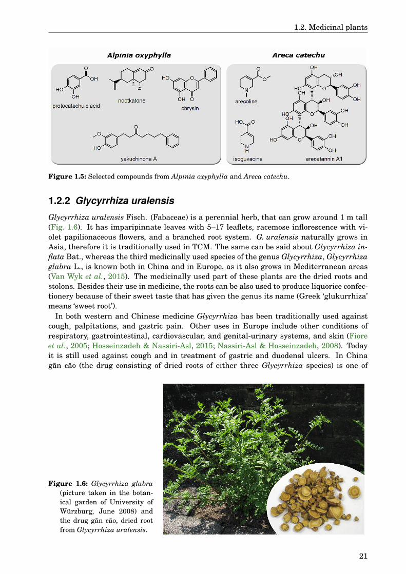
1.2. Medicinal plants
Figure 1.5: Selected compounds from Alpinia oxyphylla and Areca catechu.
1.2.2 Glycyrrhiza uralensis
Glycyrrhiza uralensis Fisch. (Fabaceae) is a perennial herb, that can grow around 1 m tall(Fig. 1.6). It has imparipinnate leaves with 5–17 leaflets, racemose inflorescence with vi-olet papilionaceous flowers, and a branched root system. G. uralensis naturally grows inAsia, therefore it is traditionally used in TCM. The same can be said about Glycyrrhiza in-flata Bat., whereas the third medicinally used species of the genus Glycyrrhiza, Glycyrrhizaglabra L., is known both in China and in Europe, as it also grows in Mediterranean areas(Van Wyk et al., 2015). The medicinally used part of these plants are the dried roots andstolons. Besides their use in medicine, the roots can be also used to produce liquorice confec-tionery because of their sweet taste that has given the genus its name (Greek ‘glukurrhiza’means ‘sweet root’).
In both western and Chinese medicine Glycyrrhiza has been traditionally used againstcough, palpitations, and gastric pain. Other uses in Europe include other conditions ofrespiratory, gastrointestinal, cardiovascular, and genital-urinary systems, and skin (Fioreet al., 2005; Hosseinzadeh & Nassiri-Asl, 2015; Nassiri-Asl & Hosseinzadeh, 2008). Todayit is still used against cough and in treatment of gastric and duodenal ulcers. In Chinagan cao (the drug consisting of dried roots of either three Glycyrrhiza species) is one of
Figure 1.6: Glycyrrhiza glabra(picture taken in the botan-ical garden of University ofWürzburg, June 2008) andthe drug gan cao, dried rootfrom Glycyrrhiza uralensis.
21
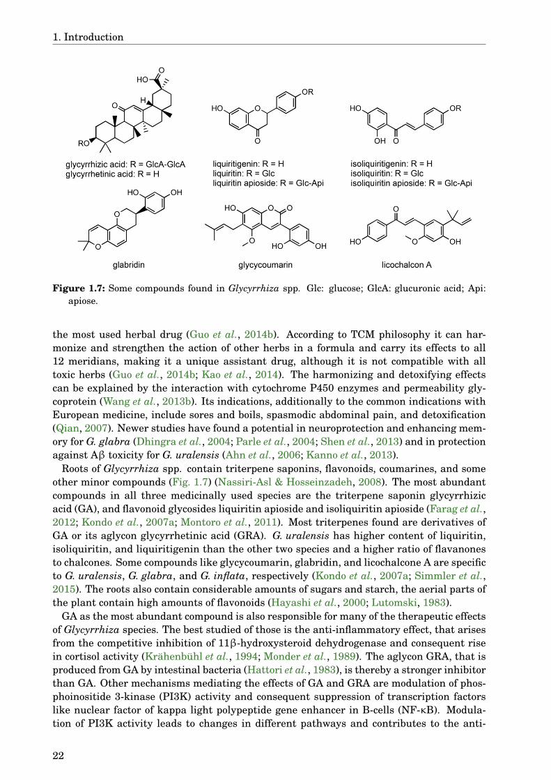
1. Introduction
Figure 1.7: Some compounds found in Glycyrrhiza spp. Glc: glucose; GlcA: glucuronic acid; Api:apiose.
the most used herbal drug (Guo et al., 2014b). According to TCM philosophy it can har-monize and strengthen the action of other herbs in a formula and carry its effects to all12 meridians, making it a unique assistant drug, although it is not compatible with alltoxic herbs (Guo et al., 2014b; Kao et al., 2014). The harmonizing and detoxifying effectscan be explained by the interaction with cytochrome P450 enzymes and permeability gly-coprotein (Wang et al., 2013b). Its indications, additionally to the common indications withEuropean medicine, include sores and boils, spasmodic abdominal pain, and detoxification(Qian, 2007). Newer studies have found a potential in neuroprotection and enhancing mem-ory for G. glabra (Dhingra et al., 2004; Parle et al., 2004; Shen et al., 2013) and in protectionagainst Aβ toxicity for G. uralensis (Ahn et al., 2006; Kanno et al., 2013).
Roots of Glycyrrhiza spp. contain triterpene saponins, flavonoids, coumarines, and someother minor compounds (Fig. 1.7) (Nassiri-Asl & Hosseinzadeh, 2008). The most abundantcompounds in all three medicinally used species are the triterpene saponin glycyrrhizicacid (GA), and flavonoid glycosides liquiritin apioside and isoliquiritin apioside (Farag et al.,2012; Kondo et al., 2007a; Montoro et al., 2011). Most triterpenes found are derivatives ofGA or its aglycon glycyrrhetinic acid (GRA). G. uralensis has higher content of liquiritin,isoliquiritin, and liquiritigenin than the other two species and a higher ratio of flavanonesto chalcones. Some compounds like glycycoumarin, glabridin, and licochalcone A are specificto G. uralensis, G. glabra, and G. inflata, respectively (Kondo et al., 2007a; Simmler et al.,2015). The roots also contain considerable amounts of sugars and starch, the aerial parts ofthe plant contain high amounts of flavonoids (Hayashi et al., 2000; Lutomski, 1983).
GA as the most abundant compound is also responsible for many of the therapeutic effectsof Glycyrrhiza species. The best studied of those is the anti-inflammatory effect, that arisesfrom the competitive inhibition of 11β-hydroxysteroid dehydrogenase and consequent risein cortisol activity (Krähenbühl et al., 1994; Monder et al., 1989). The aglycon GRA, that isproduced from GA by intestinal bacteria (Hattori et al., 1983), is thereby a stronger inhibitorthan GA. Other mechanisms mediating the effects of GA and GRA are modulation of phos-phoinositide 3-kinase (PI3K) activity and consequent suppression of transcription factorslike nuclear factor of kappa light polypeptide gene enhancer in B-cells (NF-κB). Modula-tion of PI3K activity leads to changes in different pathways and contributes to the anti-
22
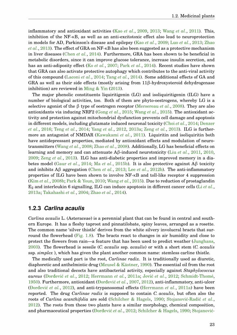
1.2. Medicinal plants
inflammatory and antioxidant activities (Kao et al., 2009, 2013; Wang et al., 2011). This,inhibition of the NF-κB, as well as an anti-excitotoxic effect also lead to neuroprotectionin models for AD, Parkinson’s disease and epilepsy (Kao et al., 2009; Luo et al., 2013; Zhaoet al., 2013). The effect of GRA on NF-κB has also been suggested as a protective mechanismin liver diseases (Chen et al., 2014). Furthermore, GRA has been shown to be beneficial inmetabolic disorders, since it can improve glucose tolerance, increase insulin secretion, andhas an anti-adiposity effect (Ko et al., 2007; Park et al., 2014). Recent studies have shownthat GRA can also activate protective autophagy which contributes to the anti-viral activityof this compound (Laconi et al., 2014; Tang et al., 2014). Some additional effects of GA andGRA as well as their side effects (mostly arising from 11β-hydroxysteroid dehydrogenaseinhibition) are reviewed in Ming & Yin (2013).
The major phenolic constituents liquiritigenin (LG) and isoliquiritigenin (ILG) have anumber of biological activities, too. Both of them are phyto-oestrogens, whereby LG is aselective agonist of the β type of oestrogen receptor (Mersereau et al., 2008). They are alsoantioxidants via inducing NRF2 (Gong et al., 2015; Wang et al., 2015). The antioxidant ac-tivity and protection against mitochondrial dysfunction prevents cell damage and apoptosisin different models, including glutamate induced neuronal toxicity (Choi et al., 2014; Denzeret al., 2016; Teng et al., 2014; Yang et al., 2012, 2013a; Zeng et al., 2013). ILG is further-more an antagonist of NMDAR (Kawakami et al., 2011). Liquiritin and isoliquiritin bothhave antidepressant properties, mediated by antioxidant effects and modulation of neuro-transmitters (Wang et al., 2008; Zhao et al., 2008). Additionally, LG has beneficial effects onlearning and memory and can attenuate Aβ-induced neurotoxicity (Liu et al., 2011, 2010,2009; Zeng et al., 2013). ILG has anti-diabetic properties and improved memory in a dia-betes model (Gaur et al., 2014; Ma et al., 2015b). It is also protective against Aβ toxicityand inhibits Aβ aggregation (Chen et al., 2013; Lee et al., 2012b). The anti-inflammatoryproperties of ILG have been shown to involve NF-κB and toll-like receptor 4 suppression(Kim et al., 2008b; Park & Youn, 2010; Wang et al., 2015). Due to reduction of prostaglandinE2 and interleukin 6 signalling, ILG can induce apoptosis in different cancer cells (Li et al.,2013a; Takahashi et al., 2004; Zhao et al., 2014).
1.2.3 Carlina acaulis
Carlina acaulis L. (Asteraceae) is a perennial plant that can be found in central and south-ern Europe. It has a fleshy taproot and pinnatilobate, spiny leaves, arranged as a rosette.The common name ‘silver thistle’ derives from the white silvery involucral bracts that sur-round the flowerhead (Fig. 1.8). The bracts react to changes in air humidity and close toprotect the flowers from rain—a feature that has been used to predict weather (Junghans,2003). The flowerhead is sessile (C. acaulis ssp. acaulis) or with a short stem (C. acaulisssp. simplex ), which has given the plant another common name: stemless carline thistle.
The medically used part is the root, Carlinae radix. It is traditionally used as diuretic,diaphoretic and anthelmintic drug (Meusel & Kästner, 1990). The essential oil from the rootand also traditional decocts have antibacterial activity, especially against Staphylococcusaureus (Ðordevic et al., 2012; Herrmann et al., 2011a; Jovic et al., 2012; Schmidt-Thomé,1950). Furthermore, antioxidant (Ðordevic et al., 2007, 2012), anti-inflammatory, anti-ulcer(Ðordevic et al., 2012), and anti-trypanosomal effects (Herrmann et al., 2011a) have beenreported. The drug Carlinae radix is supposed to contain C. acaulis, but often also theroots of Carlina acanthifolia are sold (Schilcher & Hagels, 1990; Stojanovic-Radic et al.,2012). The roots from these two plants have a similar morphology, chemical composition,and pharmaceutical properties (Ðordevic et al., 2012; Schilcher & Hagels, 1990; Stojanovic-
23
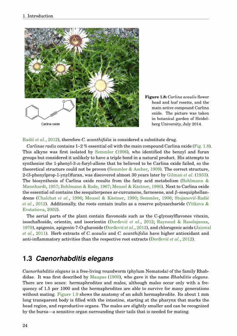
1. Introduction
Figure 1.8: Carlina acaulis flowerhead and leaf rosette, and themain active compound Carlinaoxide. The picture was takenin botanical garden of Heidel-berg University, July 2014.
Radic et al., 2012), therefore C. acanthifolia is considered a substitute drug.Carlinae radix contains 1–2 % essential oil with the main compound Carlina oxide (Fig. 1.8).
This alkyne was first isolated by Semmler (1906), who identified the benzyl and furangroups but considered it unlikely to have a triple bond in a natural product. His attempts tosynthesize the 1-phenyl-3-α-furyl-allene that he believed to be Carlina oxide failed, so thetheoretical structure could not be proven (Semmler & Ascher, 1909). The correct structure,2-(3-phenylprop-1-ynyl)furan, was discovered almost 30 years later by Gilman et al. (1933).The biosynthesis of Carlina oxide results from the fatty acid metabolism (Bohlmann &Mannhardt, 1957; Bohlmann & Rode, 1967; Meusel & Kästner, 1990). Next to Carlina oxidethe essential oil contains the sesquiterpenes ar-curcumene, farnesene, and β-sesquiphellan-drene (Chalchat et al., 1996; Meusel & Kästner, 1990; Semmler, 1906; Stojanovic-Radicet al., 2012). Additionally, the roots contain inulin as a reserve polysaccharide (Vitkova &Evstatieva, 2002).
The aerial parts of the plant contain flavonoids such as the C-glycosylflavones vitexin,isoschaftoside, orientin, and isoorientin (Ðordevic et al., 2012; Raynaud & Rasolojaona,1979), apigenin, apigenin-7-O-glucoside (Ðordevic et al., 2012), and chlorogenic acids (Jaiswalet al., 2011). Herb extracts of C. acaulis and C. acanthifolia have higher antioxidant andanti-inflammatory activities than the respective root extracts (Ðordevic et al., 2012).
1.3 Caenorhabditis elegans
Caenorhabditis elegans is a free-living roundworm (phylum Nematoda) of the family Rhab-didae. It was first described by Maupas (1900), who gave it the name Rhabditis elegans.There are two sexes: hermaphrodites and males, although males occur only with a fre-quency of 1.5 per 1000 and the hermaphrodites are able to survive for many generationswithout mating. Figure 1.9 shows the anatomy of an adult hermaphrodite. Its about 1 mmlong transparent body is filled with the intestine, starting at the pharynx that marks thehead region, and reproductive organs. The males are slightly smaller and can be recognizedby the bursa—a sensitive organ surrounding their tails that is needed for mating.
24

1.3. Caenorhabditis elegans
Figure 1.9: Anatomy of an adult C. elegans hermaphrodite. Picture taken with Keyence BZ9000,scale bar corresponds to 100 µm.
Brenner (1974) introduced it as a model organism for genetic studies on the developmentand function of nervous system. He chose this organism because of the simplicity of han-dling and a manageable amount of cells. Together with H. R. Horwiz and J. E. Sulston, Sid-ney Brenner became the Nobel Prize in Physiology or Medicine in 2002 for his work concern-ing organ development and programmed cell death that they had done on this model system.C. elegans became the first multicellular organism with completely sequenced genome (TheCaenorhabditis elegans Sequencing Consortium, 1998) and mapped connectome (Albertson& Thomson, 1976; White et al., 1986), and the entire cell lineage of this organism is known(Sulston et al., 1983). The genome of C. elegans consists of five autosomal chromosomesand one sex chromosome with a constitution XX in hermaphrodites and OX in males. Thehaploid genome contains 97 megabase DNA with over 19,000 genes. 36 % of the proteinsin C. elegans have a homologue in Homo sapiens (The Caenorhabditis elegans SequencingConsortium, 1998). The availability of the complete sequence and high homology to hu-man genome together with the ease of handling make this nematode an excellent modelorganism.
The normal lifespan of C. elegans is about three weeks, depending on temperature andfood concentration (Klass, 1977). After hatching from the egg, the larvae pass through fourmoults (larval stages L1–L4) before they become fertile adults (Fig. 1.10) (Cassada & Rus-sell, 1975). Each moult is preceded by a short period called lethargus, during which thelarvae stop feeding and are less agile. The time between moults is dependent on tempera-ture. At lower temperatures the larvae grow slower and therefore need more time to reachthe size at which the moult occurs (Byerly et al., 1976; Cassada & Russell, 1975). The youngadult hermaphrodites produce sperm during the first hours of adulthood, then eggs beginto form. The sperm remains stored in spermatheca. It fertilizes the ripe eggs as they passfrom ovary to uterus. One hermaphrodite can lay about 280 eggs before all the sperm isused, unless it mates with a male and gets additional sperm (Byerly et al., 1976). After thatit can lay unfertilized eggs. The eggs are laid shortly before the larvae hatch. In some casesthe larvae hatch already in the uterus where they feed on unfertilized eggs and eventuallybreak through in the body cavity of their mother, thereby killing it (Maupas, 1900). Thisphenomenon is referred to as ‘bagging’, because the dead hermaphrodite looks like a bag oflarvae.
25
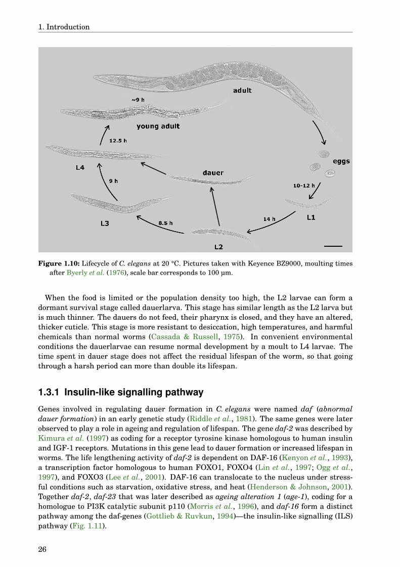
1. Introduction
Figure 1.10: Lifecycle of C. elegans at 20 °C. Pictures taken with Keyence BZ9000, moulting timesafter Byerly et al. (1976), scale bar corresponds to 100 µm.
When the food is limited or the population density too high, the L2 larvae can form adormant survival stage called dauerlarva. This stage has similar length as the L2 larva butis much thinner. The dauers do not feed, their pharynx is closed, and they have an altered,thicker cuticle. This stage is more resistant to desiccation, high temperatures, and harmfulchemicals than normal worms (Cassada & Russell, 1975). In convenient environmentalconditions the dauerlarvae can resume normal development by a moult to L4 larvae. Thetime spent in dauer stage does not affect the residual lifespan of the worm, so that goingthrough a harsh period can more than double its lifespan.
1.3.1 Insulin-like signalling pathway
Genes involved in regulating dauer formation in C. elegans were named daf (abnormaldauer formation) in an early genetic study (Riddle et al., 1981). The same genes were laterobserved to play a role in ageing and regulation of lifespan. The gene daf-2 was described byKimura et al. (1997) as coding for a receptor tyrosine kinase homologous to human insulinand IGF-1 receptors. Mutations in this gene lead to dauer formation or increased lifespan inworms. The life lengthening activity of daf-2 is dependent on DAF-16 (Kenyon et al., 1993),a transcription factor homologous to human FOXO1, FOXO4 (Lin et al., 1997; Ogg et al.,1997), and FOXO3 (Lee et al., 2001). DAF-16 can translocate to the nucleus under stress-ful conditions such as starvation, oxidative stress, and heat (Henderson & Johnson, 2001).Together daf-2, daf-23 that was later described as ageing alteration 1 (age-1), coding for ahomologue to PI3K catalytic subunit p110 (Morris et al., 1996), and daf-16 form a distinctpathway among the daf-genes (Gottlieb & Ruvkun, 1994)—the insulin-like signalling (ILS)pathway (Fig. 1.11).
26

1.3. Caenorhabditis elegans
Figure 1.11: The ILS pathway in C. elegans. Activation of DAF-2 leads to auto-phosphorylationand recruits IST-1. IST-1 interacts with AAP-1 that recruits AGE-1 to the plasmamembranewhere it can produce PIP3. This is antagonized by the phosphatase DAF-18. PIP3 activatesPDK-1 and together they activate AKT-1 and 2. SGK-1 is also activated by PDK-1. AKT-1, AKT-2, and SGK-1 phosphorylate DAF-16, thereby inhibiting its translocation into nucleus. DAF-2antagonists lead to nuclear localization of DAF-16. ILS and DAF-16 interact with other pathwaysand transcription factors to achieve differential gene expression.
The ILS pathway is conserved throughout evolution. Most of the genes involved have ho-mologues in humans and other organisms (Tab. 1.2) (Papatheodorou et al., 2014). Addition-ally to the three named genes, others have been found to be associated with this pathway.The proteins insulin receptor substrate (IST-1) (homologue to human insulin receptor sub-strate (IRS)) and AGE-1 adaptor protein (AAP-1) (homologue to human PI3K adaptor sub-unit p55) help to recruit AGE-1 to plasma membrane upon DAF-2 activation (Wolkow et al.,2002). The phosphatidylinositol-3,4,5-trisphosphat (PIP3) produced by AGE-1 leads to acti-vation of 3-phosphoinositide-dependent kinase-1 (PDK-1) (Paradis et al., 1999), protein ki-nase B (AKT)-1 and AKT-2 (Paradis & Ruvkun, 1998). This process is antagonized by DAF-18, a PIP3 phosphatase homologous to human phosphatase and tensin homologue (PTEN)(Ogg & Ruvkun, 1998). Serum- and glucocorticoid-inducible kinase 1 (SGK-1) is activatedthrough phosphorylation by PDK-1 and forms a possibly temporary complex with AKT-1and 2 (Hertweck et al., 2004). These three kinases are responsible for phosphorylatingDAF-16 and preventing its translocation to nucleus.
DAF-16 activation is regulated by environmental signals via sensory neurons (Apfeld &Kenyon, 1999), by reproductive system (Hsin & Kenyon, 1999), and by stress such as heatshock (Lin et al., 2001b). Insulin related peptides (INSs) (Pierce et al., 2001) and DAF-28(Li et al., 2003) are produced in neurons and possibly released upon sensory input. Some
27
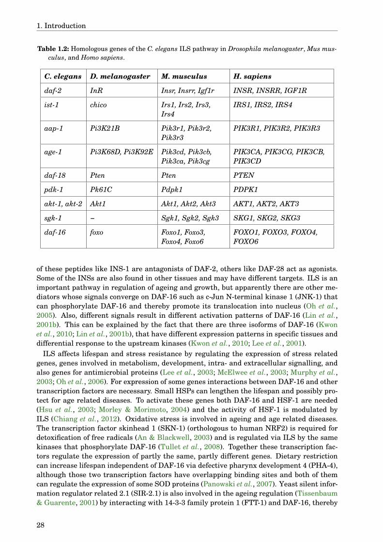
1. Introduction
Table 1.2: Homologous genes of the C. elegans ILS pathway in Drosophila melanogaster, Mus mus-culus, and Homo sapiens.
C. elegans D. melanogaster M. musculus H. sapiens
daf-2 InR Insr, Insrr, Igf1r INSR, INSRR, IGF1R
ist-1 chico Irs1, Irs2, Irs3,Irs4
IRS1, IRS2, IRS4
aap-1 Pi3K21B Pik3r1, Pik3r2,Pik3r3
PIK3R1, PIK3R2, PIK3R3
age-1 Pi3K68D, Pi3K92E Pik3cd, Pik3cb,Pik3ca, Pik3cg
PIK3CA, PIK3CG, PIK3CB,PIK3CD
daf-18 Pten Pten PTEN
pdk-1 Pk61C Pdpk1 PDPK1
akt-1, akt-2 Akt1 Akt1, Akt2, Akt3 AKT1, AKT2, AKT3
sgk-1 − Sgk1, Sgk2, Sgk3 SKG1, SKG2, SKG3
daf-16 foxo Foxo1, Foxo3,Foxo4, Foxo6
FOXO1, FOXO3, FOXO4,FOXO6
of these peptides like INS-1 are antagonists of DAF-2, others like DAF-28 act as agonists.Some of the INSs are also found in other tissues and may have different targets. ILS is animportant pathway in regulation of ageing and growth, but apparently there are other me-diators whose signals converge on DAF-16 such as c-Jun N-terminal kinase 1 (JNK-1) thatcan phosphorylate DAF-16 and thereby promote its translocation into nucleus (Oh et al.,2005). Also, different signals result in different activation patterns of DAF-16 (Lin et al.,2001b). This can be explained by the fact that there are three isoforms of DAF-16 (Kwonet al., 2010; Lin et al., 2001b), that have different expression patterns in specific tissues anddifferential response to the upstream kinases (Kwon et al., 2010; Lee et al., 2001).
ILS affects lifespan and stress resistance by regulating the expression of stress relatedgenes, genes involved in metabolism, development, intra- and extracellular signalling, andalso genes for antimicrobial proteins (Lee et al., 2003; McElwee et al., 2003; Murphy et al.,2003; Oh et al., 2006). For expression of some genes interactions between DAF-16 and othertranscription factors are necessary. Small HSPs can lengthen the lifespan and possibly pro-tect for age related diseases. To activate these genes both DAF-16 and HSF-1 are needed(Hsu et al., 2003; Morley & Morimoto, 2004) and the activity of HSF-1 is modulated byILS (Chiang et al., 2012). Oxidative stress is involved in ageing and age related diseases.The transcription factor skinhead 1 (SKN-1) (orthologous to human NRF2) is required fordetoxification of free radicals (An & Blackwell, 2003) and is regulated via ILS by the samekinases that phosphorylate DAF-16 (Tullet et al., 2008). Together these transcription fac-tors regulate the expression of partly the same, partly different genes. Dietary restrictioncan increase lifespan independent of DAF-16 via defective pharynx development 4 (PHA-4),although those two transcription factors have overlapping binding sites and both of themcan regulate the expression of some SOD proteins (Panowski et al., 2007). Yeast silent infor-mation regulator related 2.1 (SIR-2.1) is also involved in the ageing regulation (Tissenbaum& Guarente, 2001) by interacting with 14-3-3 family protein 1 (FTT-1) and DAF-16, thereby
28

1.3. Caenorhabditis elegans
activating the stress related gene expression in a DAF-16 dependent manner (Berdichevskyet al., 2006; Wang et al., 2006). FTT-1 can also bind the phosphorylated form of DAF-16 inthe cytoplasm, inhibiting its entry into the nucleus (Berdichevsky et al., 2006).
Taken together the ILS is a central pathway in a complex network that regulates age-ing and stress resistance. The evolutionary conservation of this pathway suggests that it isimportant for normal function of a multicellular organism. It also opens the possibility to in-vestigate these signals and the impact of modifications to them in a simple model organismsuch as C. elegans.
1.3.2 Nervous system
The nervous system of C. elegans consists of 302 neurons. Their cell bodies are organizedinto 11 ganglia and connected by processes that are concentrated in the circumpharyngealnerve ring and the ventral and dorsal chords (Ware et al., 1975; White et al., 1986). Theganglia and process bundles are highly organized and symmetrical. The neurons can bedivided into sensory, moto-, and interneurons. They have a simple morphology with fewbranches and connect to each other via gap junctions or chemical synapses. As neurotrans-mitters acetylcholin (Lewis et al., 1987), biogenic amines (Alkema et al., 2005; Horvitz et al.,1982; Sulston et al., 1975), γ-aminobutyric acid (GABA) (McIntire et al., 1993), glutamate(Arena et al., 1992; Schaeffer et al., 1990), and various neuropeptides are described. Thepharynx and its 20 neurons form a separated, nearly self-contagious system, which gets in-put from somatic nervous system only by one pair of interneurons (Albertson & Thomson,1976). Additionally to the neurons, 56 glial cells support the functions of the nervous system(Oikonomou & Shaham, 2011).
The body wall muscles of C. elegans are obliquely striated and allow dorso-ventral con-tractions of the body, leading to the sinuous movement of the worms. The muscle cells sendarms towards motoneurons in the nerve ring or ventral chord to meet their innervating neu-ral processes that make synapses en passant. Gap junctions between the muscle cells mightensure the uniform distribution of a signal in adjacent cells (White et al., 1986). Some mo-toneurons have also sensory properties that mediate a withdrawal in response to a touch orhelp to maintain the oscillating body movement by monitoring the stretch of one side of thebody (McIntire et al., 1993; White et al., 1986). In contrast to the rest of the body, the headcan also be moved in lateral directions. The head muscles are innervated by neurons fromthe nerve ring which probably allows for finer adjustment of movements than the relativelysimple ventral and dorsal chords. One sensory neuron is also found to synapse directly on amuscle cell in the head (Ware et al., 1975; White et al., 1986).
C. elegans is able to sense different chemical and mechanic stimuli. The main chemosen-sors are organized in the amphids, that are located on the lateral lips of the mouth (Fig. 1.12).Each amphid contains 12 neurons and two glial cells that form the amphid channel. Most ofthe neuronal endings lie in this channel and are in contact with the outer medium througha pore in the cuticula, some are buried in one of the glial cells called the sheath cell. Thenerve ending of AFD, a thermosensory neuron (Mori & Ohshima, 1995), is also buried inthe sheath cell, but outside of the amphid channel (Ward et al., 1975). The tree neuronswhich are not directly exposed to the environment—AWA, AWB, and AWC—are responsiblefor the chemotaxis to volatile molecules, therefore they can be considered olfactory neu-rons (Bargmann et al., 1993). The exposed neurons can sense water-soluble molecules likecAMP and several ions, ASE being the most important chemosensory neuron in this set(Bargmann & Horvitz, 1991; Dusenbery, 1974; Ward, 1973). Several of these neurons playa role in dauer formation (Schackwitz et al., 1996; Vowels & Thomas, 1992).
29
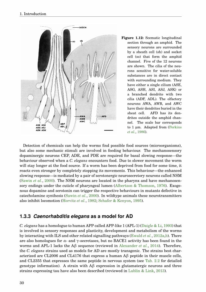
1. Introduction
Figure 1.12: Scematic longitudinalsection through an amphid. Thesensory neurons are surroundedby a sheath cell (sh) and socketcell (so) that form the amphidchannel. Five of the 12 neuronsare shown. The cilia of the neu-rons sensitive for water-solublesubstances are in direct contactwith surrounding medium. Theyhave either a single cilium (ASE,ASG, ASH, ASI, ASJ, ASK) ora branched dendrite with twocilia (ADF, ADL). The olfactoryneurons AWA, AWB, and AWChave their dendrites buried in thesheat cell. AFD has its den-drites outside the amphid chan-nel. The scale bar correspondsto 1 µm. Adapted from (Perkinset al., 1986).
Detection of chemicals can help the worms find possible food sources (microorganisms),but also some mechanic stimuli are involved in feeding behaviour. The mechanosensorydopaminergic neurons CEP, ADE, and PDE are required for basal slowing response—thebehaviour observed when a C. elegans encounters food. Due to slower movement the wormwill stay longer at the food source. If a worm has been deprived from food for some time, itreacts even stronger by completely stopping its movements. This behaviour—the enhancedslowing response—is mediated by a pair of serotonergic neurosecretory neurons called NSM(Sawin et al., 2000). The NSM neurons are located in the pharynx and have mechanosen-sory endings under the cuticle of pharyngeal lumen (Albertson & Thomson, 1976). Exoge-nous dopamine and serotonin can trigger the respective behaviours in mutants defective incatecholamine synthesis (Sawin et al., 2000). In wildtype animals these neurotransmittersalso inhibit locomotion (Horvitz et al., 1982; Schafer & Kenyon, 1995).
1.3.3 Caenorhabditis elegans as a model for AD
C. elegans has a homologue to human APP called APP-like 1 (APL-1) (Daigle & Li, 1993) thatis involved in sensory responses and plasticity, development and metabolism of the wormsby interacting with ILS and other related signalling pathways (Ewald et al., 2012a,b). Thereare also homologues for α- and γ-secretases, but no BACE1 activity has been found in theworms and APL-1 lacks the Aβ sequence (reviewed in Alexander et al., 2014). Therefore,the C. elegans strains used as models for AD are mostly transgenic. The strains best char-acterized are CL2006 and CL4176 that express a human Aβ peptide in their muscle cells,and CL2355 that expresses the same peptide in nervous system (see Tab. 2.2 for detailedgenotype information). A strain with Aβ expression in glutamatergic neurons and threestrains expressing tau have also been described (reviewed in Lublin & Link, 2013).
30

1.3. Caenorhabditis elegans
CL2006 was the first transgenic C. elegans strain expressing human Aβ. It was made inthe lab of Link (1995). This strain expresses Aβ constitutively in its body wall muscles,where it aggregates and forms plaques. The transgenic construct contains a signal peptidefor secretion, so the model was expected to produce extracellular Aβ1–42. Further exami-nation of this model, however, showed that Aβ fibrils were formed intracellularly and thepeptide produced was Aβ3–42, probably due to cleavage of the signal peptide at a differentsite than expected (Link et al., 2001; McColl et al., 2009). The aggregates can be stainedwith anti-Aβ antibodies or amyloid specific dyes like thioflavin S, Congo red, or X-34 (Fayet al., 1998; Link, 1995; Link et al., 2001). These dyes can also be used to stain senile plaquesin AD brains, indicating that the plaques in worms have a similar structure to the plaquesfound in humans. This was also confirmed by electron microscopy (Link et al., 2001). There-fore, this model is suitable for investigating Aβ aggregation and possible modulators of thisprocess. Aβ expression and aggregation in muscles of these worms leads to progressiveparalysis. This phenotype is more pronounced in the strain CL4176 that also expresses Aβin muscles, but the expression can be induced by temperature change (Link et al., 2003).These worms paralyse and suffer under oxidative stress without the formation of fibrillaramyloid deposits, suggesting that the toxicity of Aβ in this strain is mediated by oligomers(Drake et al., 2003). Hence, this model can be used to study the effects on oligomers andoligomer toxicity. Both of these strains express Aβ in their muscles, representing ratherinclusion body myositis (a disease with Aβ aggregates in muscles) than AD. To better re-capitulate the neuronal disease, the strain CL2355 with pan-neuronal Aβ expression wasdeveloped. These worms have deficits in chemotaxis, associative memory, and serotonin sig-nalling (Dosanjh et al., 2010; Wu et al., 2006), being therefore useful for testing effects onthe nervous system.
In addition to the Aβ aggregation and toxicity, these models share some similarities withthe human disease. As already mentioned, Aβ dependent oxidative stress is observed inthese worms (Drake et al., 2003). This is accompanied by higher levels of iron (Wan et al.,2011), copper can have protective or enhancing activity on the toxicity depending on itsconcentration as also seen in other models (Luo et al., 2011; Rebolledo et al., 2011). Oxi-dation of specific proteins involved in several cellular functions corresponding to defects inAD have been identified (Boyd-Kimball et al., 2006). Aβ causes synaptic dysfunction andmislocalisation of ACh receptors (Rebolledo et al., 2011). It induces expression of HSPs inworms and also in AD brain (Link et al., 2003). HSP-16.2 can interact with Aβ oligomersand reduce their toxicity in C. elegans model (Fonte et al., 2002, 2008; Wu et al., 2010). Aβtoxicity is also reduced by a proteasomal protein, also upregulated by Aβ expression, andits human homologue (Hassan et al., 2009). Defective autophagy has also been observed inthe C. elegans model of Aβ toxicity as well as in AD neurons (Florez-McClure et al., 2007).Aβ is degraded through autophagy in this model. The degradation can be enhanced andAβ toxicity reduced by decreasing the activity of the ILS pathway (Cohen et al., 2006, 2010;Florez-McClure et al., 2007). The same pathway in humans is impaired in diabetes melli-tus, a risk factor for AD, and is also involved in Aβ clearance (see section 1.1.4). The firstmetabolomic study in a C. elegans AD model could also draw parallels to human disease(Van Assche et al., 2015).
Despite these similarities, C. elegans is obviously not a perfect model for AD. Due to thelack of BACE1 activity this model has no relevance to APP processing and Aβ production.It also lacks processes comparable to neuroinflammation, several pathways relevant to Aβclearance in humans, and even cell types and organs like astrocytes, microglia, or the bloodbrain barrier. These models are still useful for studying the aggregation of Aβ in an invivo environment, the response of a single cell to Aβ toxicity, or the effect of compounds
31
1. Introduction
interacting directly with Aβ, independent of an vertebrate organism. In the pan-neuronalAβ strain also effects on neurons, neurotransmission, and simple behaviour can be studied.In contrast to mammalian models like mice or rats C. elegans has a short life span, makingit a good model organism for ageing and age related diseases. The short reproduction cycleand easy handling of these worms make it possible to carry out high throughput screens,that would not be possible in mammalian models. On the other hand, this is an in vivosystem, having the advantage over cell culture where usually only one cell type is kept inan artificial medium. Hence, C. elegans can be seen as a bridge between in vitro experimentsand more complex vertebrate models.
1.4 ObjectivesNeurodegenerative diseases are a growing social and medical problem in our ageing soci-ety. Although neurodegeneration can be caused by different mechanisms, the diseases tendto share many features like protein aggregation, oxidative stress, neuroinflammation, ordefects of lysosomal degradation. For this work AD was chosen as a representative neu-rodegenerative disease, since it is becoming a major health problem and new medicationsfor its treatment are desperately needed. Phytotherapy can be beneficial in such complexdisorders because of the multitarget action and good tolerability, which is important in old,fragile patients. Identifying active extracts and secondary metabolites is therefore imperial.C. elegans was chosen as a model organism for this study because of its easy handling andavailability of several strains that can model aspects of AD.
The objectives of the present work are:
• Screening different extracts of selected plants for their activity on Aβ aggre-gation to identify active extracts
25 potentially effective plants from TCM were identified via literature research. Meth-anol and dichloromethane extracts of these plants will be studied on the C. elegansstrain CL2006. Most active extracts will be chosen for further analysis.
• Testing the chosen extracts for their ability to counteract Aβ-induced toxic-ity
An effect on Aβ aggregation in CL2006 does not verify a reduction of Aβ toxicity,if the toxic species are oligomers, not fibrils. To study the effect on oligomers, twoother strains will be used: CL4176, to identify the general potential to counteractAβ-induced toxicity, and CL2355, to study the effect on neurons.
• Phytochemical characterization of active extracts
In order to identify active constituents, the extracts will be analysed via HPLC, LC-MS, or GLC-MS. Major constituents will be tested for their activity on Aβ aggregationand toxicity in the systems mentioned before.
• Evaluation of in vivo antioxidant activity
Since oxidative stress is an important feature in neurodegenerative disease, the an-tioxidant activity of the extract and of the major compounds will be studied. TheC. elegans strain TJ375 will be used to detect HSP expression, a marker for generalstress resistance.
32
1.4. Objectives
• Testing effects on ILS pathway and lifespan
ILS pathway is highly conserved throughout evolution, which makes it possible todraw conclusions about the effects in humans based on the study in C. elegans. Insulinsignalling in disturbed in AD, therefore the extracts and compounds will be tested fortheir ability to activate DAF-16, the downstream transcription factor of ILS. Activa-tion of DAF-16 leads to lengthened lifespan in C. elegans, hence this effect will be alsotested.
33

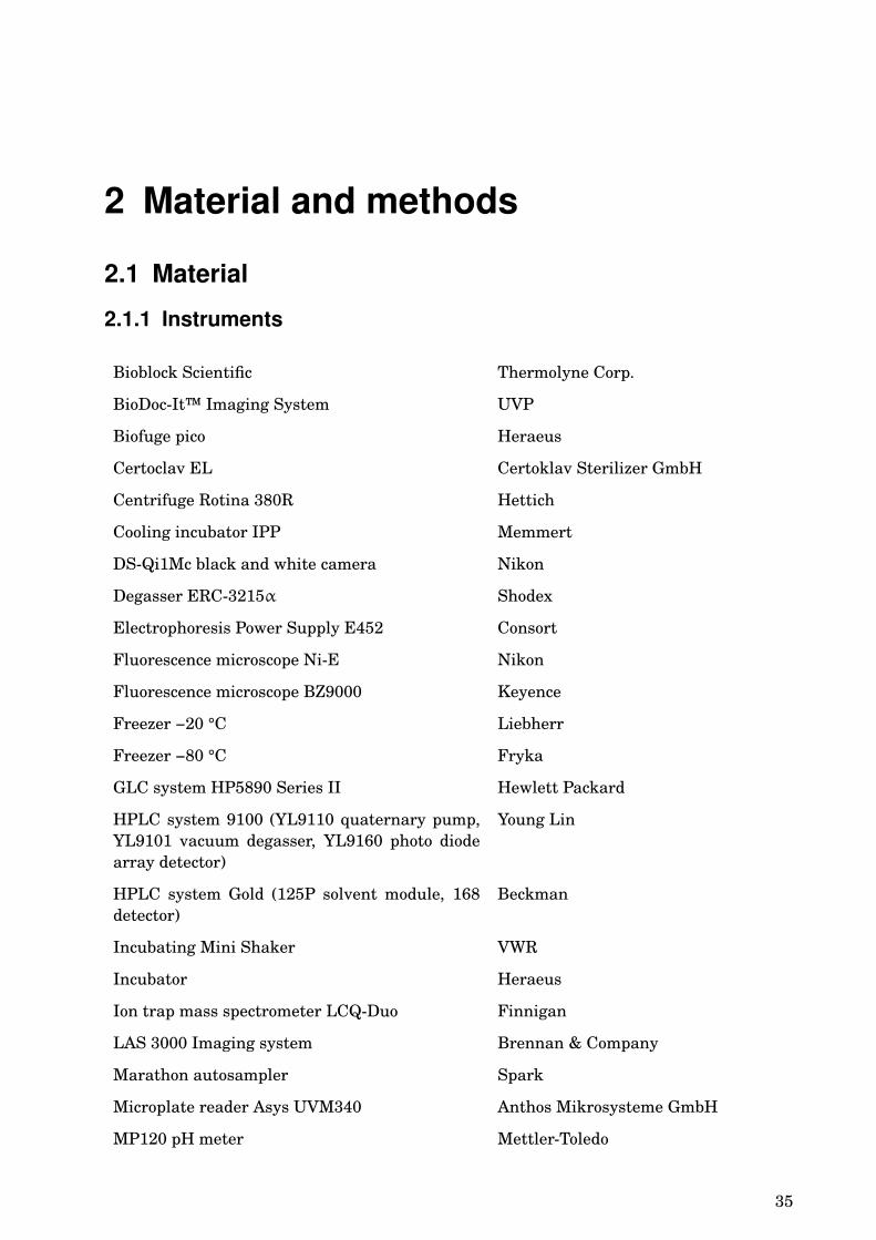
2 Material and methods
2.1 Material
2.1.1 Instruments
Bioblock Scientific Thermolyne Corp.
BioDoc-It™ Imaging System UVP
Biofuge pico Heraeus
Certoclav EL Certoklav Sterilizer GmbH
Centrifuge Rotina 380R Hettich
Cooling incubator IPP Memmert
DS-Qi1Mc black and white camera Nikon
Degasser ERC-3215α Shodex
Electrophoresis Power Supply E452 Consort
Fluorescence microscope Ni-E Nikon
Fluorescence microscope BZ9000 Keyence
Freezer −20 °C Liebherr
Freezer −80 °C Fryka
GLC system HP5890 Series II Hewlett Packard
HPLC system 9100 (YL9110 quaternary pump,YL9101 vacuum degasser, YL9160 photo diodearray detector)
Young Lin
HPLC system Gold (125P solvent module, 168detector)
Beckman
Incubating Mini Shaker VWR
Incubator Heraeus
Ion trap mass spectrometer LCQ-Duo Finnigan
LAS 3000 Imaging system Brennan & Company
Marathon autosampler Spark
Microplate reader Asys UVM340 Anthos Mikrosysteme GmbH
MP120 pH meter Mettler-Toledo
35

2. Material and methods
NanoPhotometer Implien
PerfectBlue™ Dual Gel Twin S Peqlab
Quadrupole mass spectrometer SSQ 7000 Finnigan
Refrigerator Liebherr
Rotary evaporator Heidolph
Sartoblot® II Satorius
Shaker GFL
Stereomicroscope SM 22-AD 24 Hund
Sterile bench Labortechnik Fröbel
Thermocycler TGradient Biometra
2.1.2 Laboratory material
8-Chambre slides BD Biosciences
96-Well plates Greiner Bio-One
Cellstar® tubes (15, 50 ml) Greiner Bio-One
Cover slips Marienfeld
GLC column Zebron (30 m × 0.25 mm, film thick-ness 0.25 µm)
Phenomenex
LiChroCART (4-4) RP-18e (5 µm) guard column Merck
LiChroCART RP-18 column (250 × 4 mm, 5 µm) Merck
Object slides Menzel-Gläser
Petri dishes (35 × 10, 60 × 15, 100 × 20 mm) Greiner Bio-One
pH indicator Macherey-Nagel
Preparative TLC plates (30 × 30 cm, silica gel 60,F254 0.5 mm)
Merck
Round filter Schleicher & Schuell
Safe-lock tubes (0.1, 0.5, 1.5, 2 ml) Eppendorf
Semi-micro cuvettes 10 × 4 mm Sarstedt
2.1.3 Chemicals
2,2-Diphenyl-1-picrylhydrazyl Sigma-Aldrich
β-Mercaptoethanol Sigma-Aldrich
λ-DNA Fermentas
Acetic acid J.T. Baker
36
2.1. Material
Acetonitrile (ACN) J.T. Baker
Acrylamide/bisacrylamide (30:1) Roth
Actin antibody C11 Santa Cruz Biotechnology
Agarose Sigma-Aldrich
Ammonium acetate (NH4Ac) Grüssing
Ammonium persulfate (APS) Amersham Biosciences
Bacto™ Agar Beckton, Dickinson and Company
Bacto™ Tryptone Beckton, Dickinson and Company
Bacto™ Yeast Extract Beckton, Dickinson and Company
Benzaldehyde Merck
Bovine serum albumin (BSA) Sigma-Aldrich
Beta-amyloid 1-42, human PeptaNova
Beta-amyloid monoclonal antibody 6E10, horseradish peroxidase (HRP) labelled
Signet
Bromphenolblue Fulka
Calcium chloride (CaCl2) Merck
Cholesterol Sigma-Aldrich
Citric acid monohydrate Applichem
Complete PCR reaction buffer, 10 × Bioron
Coomassie blue G-250 Fulka
Copper sulphate pentahydrate (CuSO4·5H2O) Grüssing
Cyclohexane Grüssing
Deoxynucleotides Bioron
Dichlormethane Sigma-Aldrich
Dimethyl sulfoxide (DMSO) VWR
Dipotassium hydrogen phosphate (K2HPO4) Merck
Disodium hydrogen phosphate (Na2HPO4) Sigma-Aldrich
Ethylenediaminetetraacetic acid (EDTA)disodium salt dihydrate
Applichem
(−)-Epigallocatechin gallate (EGCG) Sigma-Aldrich
Ethanol Berkel
Ethidium bromide Serva
Ethylacetate University Heidelberg
37

2. Material and methods
Formic acid (HCOOH) Merck
Glycerin AppliChem
Glycin AppliChem
Glycyrrhizic acid Northeast Forestry University, China
Glycyrrhizic acid monoammonium Northeast Forestry University, China
Glycyrrhetinic acid Northeast Forestry University, China
Hydrochloric acid (HCl) AnalaR Normapur
Isoliquiritigenin Phytolab
Iron sulphate septahydrate (FeSO4·7H2O) Sigma-Aldrich
Juglone (5-hydroxy-1,4-naphthochinon) Sigma-Aldrich
Liquiritigenin Phytolab
Methanol Sigma-Aldrich
Magnesium sulphate (MgSO4) Merck
Milk powder Granovita
Manganese chloride tetrahydrate (MnCl2·4H2O) Merck
Nitocellulose blotting membrane Satorius
Nitrogen Air Liquide
Paraformaldehyde (PFA) Sigma-Aldrich
Phosphoric acid Roth
Poly-L-lysin Sigma-Aldrich
Primers ITS4, ITS5 NWG
Protease inhibitor cocktail Complete Mini Roche
PST I Fermentas
Potassium chloride (KCl) Merck
Potassium dihydrogen phosphate (KH2PO4) Applichem
Sodium azide Applichem
Sodium chloride (NaCl) Applichem
Sodium dodecyl sulphate (SDS) Merck
Sodium hydroxide (NaOH) Sigma-Aldrich
Sodium hypochlorite (NaClO) Grüssing
Spectra™ Multicolor low range proteinladder
Fermentas
Taq polymerase Bioron
38
2.1. Material
Tetramethylethylenediamine (TEMED) Roth
Thioflavin S Sigma-Aldrich
Thioflavin T Sigma-Aldrich
Tricine Roth
Tri-potassium citrate monohydrate Roth
Tris(hydroxymethyl)aminomethane (Tris) Roth
Triton® X-100 Fulka
Tween® 20 Roth
Water HiPerSolv Chromanorm® VWR
WEST-ZOL™ Plus Western blot detectionsystem
iNtRON Biotechnology
Whatman™ blotting paper, 0.34 mm GE Healthcare
Zinc sulphate septahydrate (ZnSO4·7H2O) Merck
2.1.4 Buffers, solutions, mediaPhosphate buffered saline (PBS), 1 ×
• 140 mM NaCl
• 2.7 mM KCl
• 10 mM Na2HPO4
• 1.8 mM KH2PO4
pH was adjusted to 7.4 with HCl.
4 % Paraformaldehyde (PFA) solution
50 ml H2O was heated to about 60 °C. 4 g PFA was added and stirred, to get a suspension.Then 1 M NaOH was added until the PFA was dissolved and the solution turned transpar-ent. It was let to cool down and filtered. 10 ml of 10 × PBS was added to the solution and itwas then filled up to 100 ml with H2O. The pH value was checked using a pH indicator andadjusted with HCl to be between 7 and 8.
S-basal
• 5.85 g NaCl
• 1 g K2HPO4
• 6 g KH2PO4
• 1 ml 5 mg/ml Cholesterol in ethanol
• H2O to 1 litre
The solution was autoclaved for 15 min at 125 °C .
39
2. Material and methods
Trace metals solution
• 1.86 g Disodium EDTA
• 0.69 g FeSO4·7H2O
• 0.2 g MnCl2·4H2O
• 0.29 g ZnSO4·7H2O
• 0.025 g CuSO4·5H2O
• H2O to 1 litre
The solution was autoclaved for 15 min at 125 °C.
1 M Potassium citrate pH 6
• 20 g Citric acid monohydrate
• 293.5 g Tri-potassium citrate monohydrate
• H2O to 1 litre
The solution was autoclaved for 15 min at 125 °C.
S-Medium
• 1 l S-Basal
• 3 ml 1 M CaCl2
• 3 ml 1 M MgSO4
• 10 ml 1 M Potassium citrate pH 6
• 10 ml Trace metals solution
1M Potassium phosphate (kpo4), pH 6
• 35.6 g K2HPO4
• 108.3 g KH2PO4
• H2O to 1 litre
The solution was autoclaved for 15 min at 125 °C.
40
2.1. Material
NGM
• 17 g Agar
• 3 g NaCl
• 2.5 g Tryptone
• 975 ml H2O
The solution was autoclaved for 15 min at 125 °C. After cooling down to 55 °C the followingingredients were added:
• 1 ml 1 M CaCl2
• 1 ml 1 M MgSO4
• 25 ml 1 M KPO4
• 1 ml 5 mg/ml Cholesterol in ethanol
Test-agar for chemotaxis assay
• 19 g Agar
• 975 ml H2O
The solution was autoclaved for 15 min at 125 °C. After cooling down to 55 °C the followingingredients were added:
• 1 ml 1 M CaCl2
• 1 ml 1 M MgSO4
• 25 ml 1 M KPO4
M9-Buffer
• 5 g NaCl
• 6 g Na2HPO4
• 3 g KH2PO4
• 1 ml 1 M MgSO4
• H2O to 1 litre
The solution was autoclaved for 15 min at 125 °C.
LB Medium
• 10 g Tryptone
• 5 g Yeast extract
• 10 g NaCl
• 950 ml H2O
The solution was autoclaved for 15 min at 125 °C.
41

2. Material and methods
LB Agar
• 10 g Tryptone
• 5 g Yeast extract
• 10 g NaCl
• 15 g Agar
• 950 ml H2O
The solution was autoclaved for 15 min at 125 °C and poured into 100 mm petri dishes.
TE Buffer
• 10 mM Tris
• 1 mM EDTA
Sample buffer for agarose gel electrophoresis
• 500 µl Glycerin
• 2.5 mg Bromphenolblue
• 500 µl H2O
TAE-Buffer, 50 ×
• 2420 g Tris
• 186 g EDTA
• 571 ml Acetic acid
• H2O to 10 litre
Bradford reagent
• 25 mg Coomassie blue G-250
• 12.5 ml Ethanol
• 25 ml Phosphoric acid
• H2O to 250 ml
Coomassie solution
• 0.5 % Coomassie blue G-250
• 50 % Ethanol
• 10 % Acetic acid
• 39.5 % H2O
42

2.1. Material
Destaining solution
• 20 % Ethanol
• 10 % Acetic acid
• 70 % H2O
4 × Laemmli buffer
• 5 ml 1 M Tris, pH 6.8
• 40 ml Glycerin
• 5 g SDS
• 5 mg Bromphenolblue
• H2O to 100 ml
10 % β-Mercaptoethanol were added prior to use.
Gels for SDS-PAGE15 % resolving gel 5 % stacking gel
H2O 2.3 ml 3.4 ml
30 % Acrylamide mix 5 ml 0.8 ml
1.5 M Tris (pH 8.8) 2.5 ml -
1 M Tris (pH 6.8) - 0.6 ml
10 % SDS 0.1 ml 0.05 ml
10 % APS 0.1 ml 0.05 ml
TEMED 4 µl 5 µl
Cathode buffer
• 12.11 g Tris
• 17.92 g Tricine
• 1 g SDS
• 1 l H2O
Final concentrations: 0.1 M Tris, 0.1 M tricine, 0.1 % SDS.
Anode buffer, 5 ×
• 121.1 g Tris
• 500 ml H2O
The pH was adjusted to 8.9 with HCl, then water was added to 1 l.
43
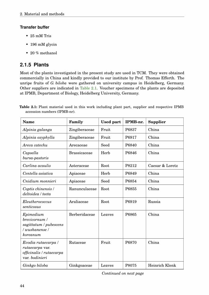
2. Material and methods
Transfer buffer
• 25 mM Tris
• 196 mM glycin
• 20 % methanol
2.1.5 PlantsMost of the plants investigated in the present study are used in TCM. They were obtainedcommercially in China and kindly provided to our institute by Prof. Thomas Efferth. Theunripe fruits of G. biloba were gathered on university campus in Heidelberg, Germany.Other suppliers are indicated in Table 2.1. Voucher specimens of the plants are depositedat IPMB, Department of Biology, Heidelberg University, Germany.
Table 2.1: Plant material used in this work including plant part, supplier and respective IPMBaccession numbers (IPMB-nr).
Name Family Used part IPMB-nr. Supplier
Alpinia galanga Zingiberaceae Fruit P6837 China
Alpinia oxyphylla Zingiberaceae Fruit P6917 China
Areca catechu Arecaceae Seed P6840 China
Capsellabursa-pastoris
Brassicaceae Herb P6846 China
Carlina acaulis Asteraceae Root P8212 Caesar & Loretz
Centella asiatica Apiaceae Herb P6849 China
Cnidium monnieri Apiaceae Seed P6854 China
Coptis chinensis /deltoidea / teeta
Ranunculaceae Root P6855 China
Eleutherococcussenticosus
Araliaceae Root P6919 Russia
Epimediumbrevicornum /sagittatum / pubescens/ wushanense /koreanum
Berberidaceae Leaves P6865 China
Evodia rutaecarpa /rutaecarpa var.officinalis / rutaecarpavar. bodinieri
Rutaceae Fruit P6870 China
Ginkgo biloba Ginkgoaceae Leaves P8075 Heinrich Klenk
Continued on next page
44
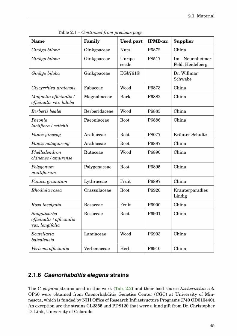
2.1. Material
Table 2.1 – Continued from previous page
Name Family Used part IPMB-nr. Supplier
Ginkgo biloba Ginkgoaceae Nuts P6872 China
Ginkgo biloba Ginkgoaceae Unripeseeds
P8517 Im NeuenheimerFeld, Heidelberg
Ginkgo biloba Ginkgoaceae EGb761® Dr. WillmarSchwabe
Glycyrrhiza uralensis Fabaceae Wood P6873 China
Magnolia officinalis /officinalis var. biloba
Magnoliaceae Bark P6882 China
Berberis bealei Berberidaceae Wood P6883 China
Paeonialactiflora / veitchii
Paeoniaceae Root P6886 China
Panax ginseng Araliaceae Root P8077 Kräuter Schulte
Panax notoginseng Araliaceae Root P6887 China
Phellodendronchinense / amurense
Rutaceae Wood P6890 China
Polygonummultiflorum
Polygonaceae Root P6895 China
Punica granatum Lythraceae Fruit P6897 China
Rhodiola rosea Crassulaceae Root P6920 KräuterparadiesLindig
Rosa laevigata Rosaceae Fruit P6900 China
Sanguisorbaofficinalis / officinalisvar. longifolia
Rosaceae Root P6901 China
Scutellariabaicalensis
Lamiaceae Wood P6903 China
Verbena officinalis Verbenaceae Herb P6910 China
2.1.6 Caenorhabditis elegans strains
The C. elegans strains used in this work (Tab. 2.2) and their food source Escherischia coliOP50 were obtained from Caenorhabditis Genetics Center (CGC) at University of Min-nesota, which is funded by NIH Office of Research Infrastructure Programs (P40 OD010440).An exception are the strains CL2355 and PD8120 that were a kind gift from Dr. ChristopherD. Link, University of Colorado.
45
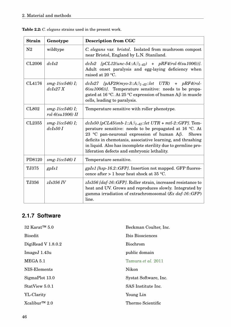
2. Material and methods
Table 2.2: C. elegans strains used in the present work.
Strain Genotype Description from CGC
N2 wildtype C. elegans var. bristol. Isolated from mushroom compostnear Bristol, England by L.N. Staniland.
CL2006 dvIs2 dvIs2 [pCL12(unc-54::Aβ1–42) + pRF4(rol-6(su1006))].Adult onset paralysis and egg-laying deficiency whenraised at 20 °C.
CL4176 smg-1(cc546) I;dvIs27 X
dvIs27 [pAF29(myo-3::Aβ1–42::let UTR) + pRF4(rol-6(su1006))]. Temperature sensitive: needs to be propa-gated at 16 °C. At 25 °C expression of human Aβ in musclecells, leading to paralysis.
CL802 smg-1(cc546) I;rol-6(su1006) II
Temperature sensitive with roller phenotype.
CL2355 smg-1(cc546) I;dvIs50 I
dvIs50 [pCL45(snb-1::Aβ1–42::let UTR + mtl-2::GFP]. Tem-perature sensitive: needs to be propagated at 16 °C. At23 °C pan-neuronal expression of human Aβ. Showsdeficits in chemotaxis, associative learning, and thrashingin liquid. Also has incomplete sterility due to germline pro-liferation defects and embryonic lethality.
PD8120 smg-1(cc546) I Temperature sensitive.
TJ375 gpIs1 gpIs1 [hsp-16.2::GFP]. Insertion not mapped. GFP fluores-cence after > 1 hour heat shock at 35 °C.
TJ356 zIs356 IV zIs356 [daf-16::GFP]. Roller strain, increased resistance toheat and UV. Grows and reproduces slowly. Integrated bygamma irradiation of extrachromosomal (Ex daf-16::GFP)line.
2.1.7 Software
32 Karat™ 5.0 Beckman Coulter, Inc.
Bioedit Ibis Biosciences
DigiRead V 1.8.0.2 Biochrom
ImageJ 1.43u public domain
MEGA 5.1 Tamura et al. 2011
NIS-Elements Nikon
SigmaPlot 13.0 Systat Software, Inc.
StatView 5.0.1 SAS Institute Inc.
YL-Clarity Young Lin
Xcalibur™ 2.0 Thermo Scientific
46
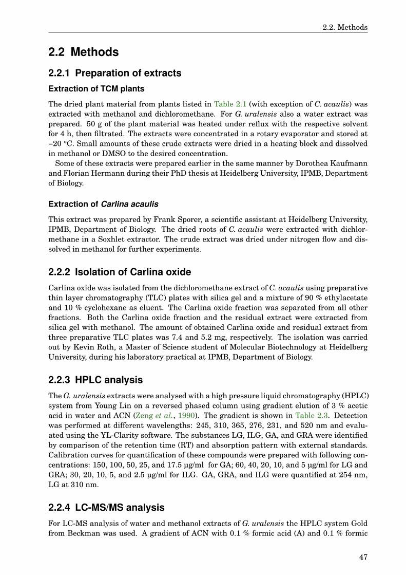
2.2. Methods
2.2 Methods
2.2.1 Preparation of extractsExtraction of TCM plants
The dried plant material from plants listed in Table 2.1 (with exception of C. acaulis) wasextracted with methanol and dichloromethane. For G. uralensis also a water extract wasprepared. 50 g of the plant material was heated under reflux with the respective solventfor 4 h, then filtrated. The extracts were concentrated in a rotary evaporator and stored at−20 °C. Small amounts of these crude extracts were dried in a heating block and dissolvedin methanol or DMSO to the desired concentration.
Some of these extracts were prepared earlier in the same manner by Dorothea Kaufmannand Florian Hermann during their PhD thesis at Heidelberg University, IPMB, Departmentof Biology.
Extraction of Carlina acaulis
This extract was prepared by Frank Sporer, a scientific assistant at Heidelberg University,IPMB, Department of Biology. The dried roots of C. acaulis were extracted with dichlor-methane in a Soxhlet extractor. The crude extract was dried under nitrogen flow and dis-solved in methanol for further experiments.
2.2.2 Isolation of Carlina oxideCarlina oxide was isolated from the dichloromethane extract of C. acaulis using preparativethin layer chromatography (TLC) plates with silica gel and a mixture of 90 % ethylacetateand 10 % cyclohexane as eluent. The Carlina oxide fraction was separated from all otherfractions. Both the Carlina oxide fraction and the residual extract were extracted fromsilica gel with methanol. The amount of obtained Carlina oxide and residual extract fromthree preparative TLC plates was 7.4 and 5.2 mg, respectively. The isolation was carriedout by Kevin Roth, a Master of Science student of Molecular Biotechnology at HeidelbergUniversity, during his laboratory practical at IPMB, Department of Biology.
2.2.3 HPLC analysisThe G. uralensis extracts were analysed with a high pressure liquid chromatography (HPLC)system from Young Lin on a reversed phased column using gradient elution of 3 % aceticacid in water and ACN (Zeng et al., 1990). The gradient is shown in Table 2.3. Detectionwas performed at different wavelengths: 245, 310, 365, 276, 231, and 520 nm and evalu-ated using the YL-Clarity software. The substances LG, ILG, GA, and GRA were identifiedby comparison of the retention time (RT) and absorption pattern with external standards.Calibration curves for quantification of these compounds were prepared with following con-centrations: 150, 100, 50, 25, and 17.5 µg/ml for GA; 60, 40, 20, 10, and 5 µg/ml for LG andGRA; 30, 20, 10, 5, and 2.5 µg/ml for ILG. GA, GRA, and ILG were quantified at 254 nm,LG at 310 nm.
2.2.4 LC-MS/MS analysisFor LC-MS analysis of water and methanol extracts of G. uralensis the HPLC system Goldfrom Beckman was used. A gradient of ACN with 0.1 % formic acid (A) and 0.1 % formic
47
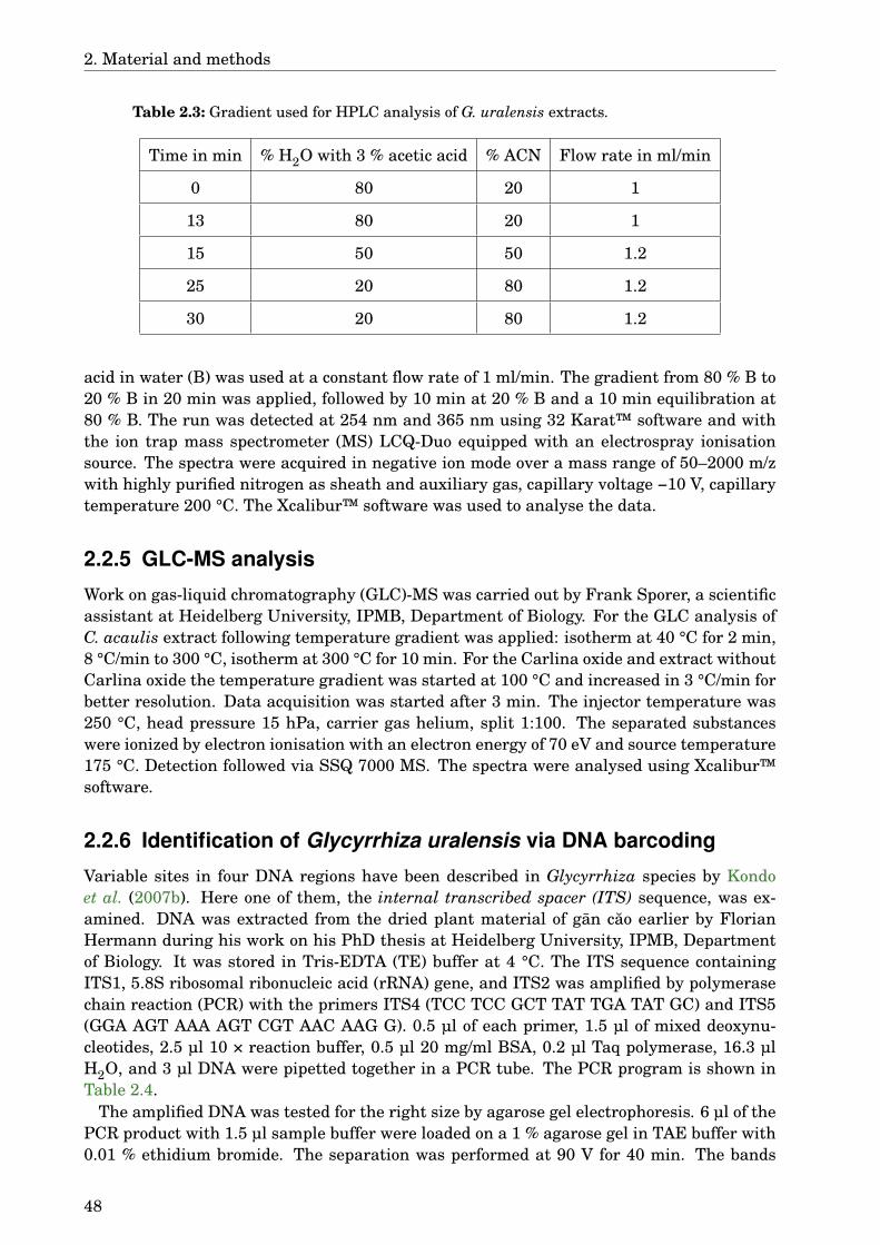
2. Material and methods
Table 2.3: Gradient used for HPLC analysis of G. uralensis extracts.
Time in min % H2O with 3 % acetic acid % ACN Flow rate in ml/min
0 80 20 1
13 80 20 1
15 50 50 1.2
25 20 80 1.2
30 20 80 1.2
acid in water (B) was used at a constant flow rate of 1 ml/min. The gradient from 80 % B to20 % B in 20 min was applied, followed by 10 min at 20 % B and a 10 min equilibration at80 % B. The run was detected at 254 nm and 365 nm using 32 Karat™ software and withthe ion trap mass spectrometer (MS) LCQ-Duo equipped with an electrospray ionisationsource. The spectra were acquired in negative ion mode over a mass range of 50–2000 m/zwith highly purified nitrogen as sheath and auxiliary gas, capillary voltage −10 V, capillarytemperature 200 °C. The Xcalibur™ software was used to analyse the data.
2.2.5 GLC-MS analysis
Work on gas-liquid chromatography (GLC)-MS was carried out by Frank Sporer, a scientificassistant at Heidelberg University, IPMB, Department of Biology. For the GLC analysis ofC. acaulis extract following temperature gradient was applied: isotherm at 40 °C for 2 min,8 °C/min to 300 °C, isotherm at 300 °C for 10 min. For the Carlina oxide and extract withoutCarlina oxide the temperature gradient was started at 100 °C and increased in 3 °C/min forbetter resolution. Data acquisition was started after 3 min. The injector temperature was250 °C, head pressure 15 hPa, carrier gas helium, split 1:100. The separated substanceswere ionized by electron ionisation with an electron energy of 70 eV and source temperature175 °C. Detection followed via SSQ 7000 MS. The spectra were analysed using Xcalibur™software.
2.2.6 Identification of Glycyrrhiza uralensis via DNA barcoding
Variable sites in four DNA regions have been described in Glycyrrhiza species by Kondoet al. (2007b). Here one of them, the internal transcribed spacer (ITS) sequence, was ex-amined. DNA was extracted from the dried plant material of gan cao earlier by FlorianHermann during his work on his PhD thesis at Heidelberg University, IPMB, Departmentof Biology. It was stored in Tris-EDTA (TE) buffer at 4 °C. The ITS sequence containingITS1, 5.8S ribosomal ribonucleic acid (rRNA) gene, and ITS2 was amplified by polymerasechain reaction (PCR) with the primers ITS4 (TCC TCC GCT TAT TGA TAT GC) and ITS5(GGA AGT AAA AGT CGT AAC AAG G). 0.5 µl of each primer, 1.5 µl of mixed deoxynu-cleotides, 2.5 µl 10 × reaction buffer, 0.5 µl 20 mg/ml BSA, 0.2 µl Taq polymerase, 16.3 µlH2O, and 3 µl DNA were pipetted together in a PCR tube. The PCR program is shown inTable 2.4.
The amplified DNA was tested for the right size by agarose gel electrophoresis. 6 µl of thePCR product with 1.5 µl sample buffer were loaded on a 1 % agarose gel in TAE buffer with0.01 % ethidium bromide. The separation was performed at 90 V for 40 min. The bands
48

2.2. Methods
Table 2.4: PCR program for ampli-fying ITS.
Temperature Duration
94 °C 5 min
38 cycles:
94 °C 45 s
45 °C 50 s
72 °C 50 s
72 °C 5 min
4 °C 5 min
were compared to a λ-marker (λ-DNA cut by PST I enzyme).Finally the samples containing correct PCR product were purified and sequenced. For
the purification DNA was precipitated with NH4Ac and ethanol and centrifuged for 30 minat 13,000 rpm. The solvent was discarded, the pellet washed with 70 % ethanol. The sol-vent was completely removed and the pellet dried at 56 °C. The dry pellet was then dis-solved in sterile H2O and sent for sequencing to StarSEQ® GmbH. The raw sequences wereedited using Bioedit and a divergence analysis was performed using MEGA 5.1. For thedivergence analysis the sample P6873 was compared to sequences retrieved from GenBank,Glycyrrhiza astragalina was used as an outgroup (Tab. 2.5).
2.2.7 DPPH• assay2,2-Diphenyl-1-picrylhydrazyl (DPPH•) is a free radical that can be reduced by hydrogendonors. This reaction can be quantified photometrically, since DPPH• has a strong ab-sorbance at 517 nm but the reduced form, hydrazine, does not. DPPH• free radical scaveng-ing assay was performed using a modified version of the method described by Blois (1958).DPPH• was prepared as a 0.2 mM solution in methanol. Equal volumes of the tested sub-stance and DPPH• solution were mixed and incubated for 30 min at room temperature.After the incubation the absorbance of each sample in duplicates was measured at 517 nmin a microplate reader. For each tested substance concentration a blank without DPPH•
was prepared and measured in the same manner. DPPH• free radical scavenging effect wascalculated using the following equation:
% DPPH• scavenging effect= A0 − A1
A0×100
A0: absorbance of DPPH•
A1: absorbance of DPPH• with the tested compound
2.2.8 SDS-PAGE and Western blot analysis of Aβ
To separate the different forms of Aβ present in the worms and evaluate the effect of theextracts on the amount of oligomers and monomers of this peptide, a Western blot analy-sis was conducted. CL2006 worms were treated the same way as for the quantification of
49

2. Material and methods
Table 2.5: Sequences used for the divergence analysis.
GenBank accession nr. Species Encoded region
JF778868 G. inflata 5.8S rRNA, partitial sequence; ITS2;26S rRNA, partitial sequence
AB280740 G. inflata ITS1; 5.8S rRNA; ITS2
AY065623 G. glabra 18S rRNA, partitial sequence; ITS1;5.8S rRNA; ITS2; 26S rRNA, partialsequence
JF778867 G. glabra 5.8S rRNA, partitial sequence; ITS2;26S rRNA, partitial sequence
HE602402 G. glabra ITS1; 5.8S rRNA; ITS2
AB280739 G. glabra ITS1; 5.8S rRNA; ITS2
JF421504 G. uralensis 5.8S rRNA, partitial sequence; ITS2;26S rRNA, partitial sequence
JF778869 G. uralensis 5.8S rRNA, partitial sequence; ITS2;26S rRNA, partitial sequence
HQ229003 G. uralensis 5.8S rRNA, partitial sequence; ITS2;26S rRNA, partitial sequence
GQ246129 G. uralensis ITS1; 5.8S rRNA; ITS2
AB280738 G. uralensis ITS1; 5.8S rRNA; ITS2
EU418258 G. uralensis ITS1, partitial sequence; 5.8S rRNA;ITS2, partitial sequence
AB649775 G. uralensis 18S rRNA, partitial sequence; ITS1;5.8S rRNA; ITS2; 26S rRNA, partitialsequence
JF421503 G. uralensis 5.8S rRNA, partitial sequence; ITS2;26S rRNA, partitial sequence
GQ246133 G. astragalina ITS1; 5.8S rRNA; ITS2
GQ246134 G. astragalina ITS1; 5.8S rRNA; ITS2
plaques (see section 2.2.10). Instead of fixing the worms at the end of the treatment, theywere collected in a Eppendorf tube, washed twice with 1 × PBS, and resuspended in 100 µlPBS with protease inhibitor cocktail. These samples were frozen and kept at −80 °C untilfurther use.
The samples were thawed on ice and homogenized. The protein concentration was mea-sured after Bradford (1976). Briefly, 2 µl of the sample were mixed with 1 ml diluted Brad-ford reagent (1:5 in water) and the absorption was measured at 595 nm after 5 min incu-bation at room temperature. A standard curve was prepared using BSA in a concentrationrange of 0.5–3 mg/ml.
50

2.2. Methods
A SDS-tricine polyacrylamide gel electrophoresis (PAGE) was performed with 15 % re-solving gel and 5 % stacking gel. The samples (final concentration of 50 µg/ml protein) wereheated at 100 °C for 3 min in Laemmli buffer prior to loading on the gel. A low range proteinladder was used as marker. 0.5 µg Aβ were used as positive control. The gel was run forapproximately 1 h at 100 V in tricine containing cathode buffer and anode buffer withouttricine.
For detection either a Coomassie staining or Western blot were used. For the Coomas-sie staining the gel was covered with Coomassie solution and stained on shaker for severalhours followed by destaining with the destain solution. For Western blot analysis the pro-teins were blotted on a nitrocellulose membrane in transfer buffer for 2 h at 48 mA . Thenthe membrane was blocked with 5 % milk powder in phosphate buffered saline containing0.05 % Tween® 20 (PBST) over night at 4 °C. On the next day the membrane was incubatedwith either anti-Aβ or anti-actin antibodies coupled with HRP (1:1000 dilution in 2.5 %milk powder in PBST) for 2 h. The membrane was washed 3× 10 min in PBST and detectedwith WEST-ZOL™ Western blot detection system. The blots were documented by LAS 3000Imaging system.
2.2.9 Caenorhabditis elegans culture conditions
The temperature sensitive strains CL4176, CL802, CL2355, and PD8120 were cultivated at16 °C, all others at 20 °C. The worms grew on nematode growth medium (NGM) and werefed with E. coli OP50, an uracil deficient E. coli strain (Brenner, 1974). The bacteria werecultured in liquid LB medium at 37 °C overnight. This overnight culture was stored at 4 °Cfor further use. Aliquots of it were spread on LB agar plates and left at 37 °C overnightprior to resuspension in S-medium. Most of the experiments were conducted in S-mediumcontaining 109 cells/ml E. coli OP50 (OD600 ≈ 1).
To obtain eggs for the experiments the hypochlorite treatment described earlier (Sulston &Hodgkin, 1988) was used. Briefly, the worms were treated with 0.5 M NaOH and 1 % NaClOfor 10 minutes or until the worms were dissolved. The eggs were pelleted by centrifuging at1300 × g for 40 s and washed with sterile water.
2.2.10 Quantification of Aβ aggregates in CL2006
To see the effect of the treatments on Aβ aggregation, the C. elegans strain CL2006 wasused. These worms constitutively express human Aβ in their muscle cells where the pep-tides aggregate causing difficulties in movement (Link, 1995). The eggs of CL2006 wormswere isolated, placed in S-medium with E. coli OP50 and kept at 20 °C. The worms weretreated on day 2 after hatching. 100 µg/ml of the methanolic TCM extracts were used,except for Magnolia officinalis, Rhodiola rosea, and Ginkgo biloba unripe seeds, where50 µg/ml was used. For the dichloromethane extracts different concentrations were useddepending on the solubility of the extracts: 100 µg/ml for Ginkgo biloba nuts, Berberisbealei, Panax notoginseng, Phellodendron spp., and Alpinia oxyphylla; 50 µg/ml for Alpiniagalanga, Coptis spp., Epimedium spp., Evodia rutaecarpa, Rosa laevigata, Sanguisorba of-ficinalis, Ginkgo biloba leaves and unripe seeds; 25 µg/ml for Capsella bursa-pastoris, Cen-tella asiatica, Glycyrrhiza uralensis, Magnolia officinalis, Paeonia spp., Punica granatum,Scutellaria baicalensis, Panax ginseng, and Rhodiola rosea; 10 µg/ml for Areca catechu,Cnidium monnieri, Polygonum multiflorum, Verbena officinalis, and Eleutherococcus sen-ticosus. As a positive control the worms were treated with 100 µg/ml EGCG, which hasbeen shown to be effective in this assay (Abbas & Wink, 2010). The negative controls were
51
2. Material and methods
treated with the respective amount of solvent, maximum 1 % as final concentration. Addi-tionally, the compound thioflavin T (ThT), that has been shown to decrease the number ofAβ aggregates in C. elegans by Alavez et al. (2011), was tested.
For G. uralensis and the pure compounds GA, glycyrrhizic acid monoammonium (GAA),GRA, LG, and ILG dose-dependence curves were prepered by testing different concentra-tions of these substances. The G. uralensis extract was tested in a range from 0 to 500 µg/ml,the pure compounds from 0 to 100 µg/ml. This experiment was repeated several times atdifferent time points with the G. uralensis extract.
On day 6 after hatching the worms were collected and fixed in 4 % PFA in 1 × PBS overnight. Then the PFA was removed and the worms were permeabilized in a solution con-taining 1 % Triton® X-100 and 5 % β-mercaptoethanol in 125 mM Tris-buffer for 24 h at37 °C. The permeabilization solution was removed by washing with 1 × PBS twice. The per-meabilized worms were transferred in a well of a 8-chamber microscope slide coated withpoly-L-lysin and air-dried. The Aβ aggregates were stained with 0.0125 % thioflavin S (ThS)solution in 50 % ethanol for 1 minute, the excess stain was washed away with 50 % ethanol,2 × 1 minute. The Aβ aggregates were quantified on fluorescence microscope Ni-E (excita-tion wavelength 480 nm, emission wavelength 525 nm) by counting the aggregates in thehead region of 25 worms per sample.
2.2.11 Paralysis assay in CL4176
The paralysis assay was described by Dostal & Link (2010) as a method for assaying Aβ tox-icity. For this assay the strains CL4176 and CL802 are used. CL4176 expresses human Aβpeptide in its muscle cells, similar to CL2006 (section 2.2.10), but the expression is temper-ature inducible and the paralysis phenotype is more penetrative than in the latter strain.The temperature sensitivity is accomplished via a mutation in the smg-1 (suppressor withmorphogenetic effect on genitalia) gene and a long 3’ untranslated region on the messengerribonucleic acid (mRNA) of the Aβ minigene. SMG-1 is a key component of the non-sensemediated mRNA decay (NMD) pathway. At 16 °C it functions normally despite the mutationand the AβmRNA is degraded. At temperatures higher then 20 °C the SMG-1 protein losesits function and so in this strain Aβ can be expressed. The control strain CL802 has thetemperature sensitive mutation in the smg-1 gene and the roller transfection marker butno transgenic Aβ.
Eggs from the worms of the strains CL4176 and CL802 were isolated and placed on smallNGM plates containing the test substances and E. coli OP50. In this assay the used con-centration for the methanol extract of A. catechu and the control EGCG was 100 µg/ml.The methanol extract of G. uralensis was used in concentrations of 200 and 500 µg/ml, forthe water extract of G. uralensis and methanol extract of A. oxyphylla a concentration of200 µg/ml was used. For the pure substances GA, GRA, LG, and ILG the concentrationwas 50 µg/ml. The dichloromethane extract of C. acaulis, the residual extract without Car-lina oxide, and Carlina oxide were tested in concentrations of 25 and 50 µg/ml each. As anegative control the respective amount of methanol or water was used.
The worms were cultivated at 16 °C for 36 h. Then the temperature was risen to 25 °C.After 24 h the worms on each plate were counted, and paralysed worms were scored understereomicroscope every 2 h for 12 h in total. Worms were counted as paralysed when theyfailed to react to a touch of platinum wire or only moved their heads creating a halo in thebacterial lawn.
52

2.2. Methods
2.2.12 Chemotaxis assay in CL2355CL2355 worms have a similar genotype as CL4176 but with pan-neuronal expression ofthe transgene. The Aβ in this strain interferes mostly with behaviours like chemotaxis,enhanced slowing response or egg laying. In chemotaxis assay these worms fail to respondto the attractant benzaldehyde. The control strain PD8120 has the temperature sensitivemutation but no transgene, and therefore it has no behavioural impairments.
CL2355 and PD8120 worms were raised from eggs in S-medium at 16 °C for 36 h. Then thetemperature was risen to 23 °C and kept there for 36 h. 24 h after experiment start the testsubstances GA, GRA, LG, and ILG in a concentration of 50 µg/ml, or the methanol extractof G. uralensis in concentrations of 200 or 500 µg/ml were added to the medium. As positivecontrol EGCG in a concentration of 100 µg/ml was used, as negative control 1 % methanolor water. After 72 h in total the worms were washed twice with M9 buffer and placed inthe middle of a 100 mm test plate. The test agar was prepared without NaCl, tryptone, andcholesterol to avoid undesired chemotactic effects. 1 µl of 1 M sodium azide in water wasadded to two spots on opposite sides of the plate. To one side 1 µl of ethanol and to the otherside 1 µl of 0.1 % benzaldehyde in ethanol was added. The plates were incubated at 23 °Cfor 1 h. Afterwards the number of worms was counted on both sides in 2 cm radius of thebenzaldehyde and ethanol spots. The chemotaxis index (CI) was calculated with followingformula:
CI = worms on the side of benzaldehyde−worms on the side of ethanoltotal number of worms on the plate
2.2.13 Serotonin sensitivity assay in CL2355C. elegans can take up exogenous serotonin, and it can trigger physiological effects likethe enhanced slowing response. The CL2355 worms are reported to be hypersensitive toserotonin due to the transgenic Aβ expression in the nervous system (Wu et al., 2006). Thecontrol strain PD8120 shows a wildtype reaction.
CL2355 and PD8120 worms were raised from eggs in S-medium at 16 °C for 36 h. Thenthe temperature was risen to 23 °C and kept there for 36 h. 24 h after experiment startthe test substances GA, GRA, LG, and ILG in a concentration of 50 µg/ml, or the methanolextract of G. uralensis in concentrations of 200 or 500 µg/ml were added to the medium. Thedichloromethane extract of C. acaulis was tested in concentrations of 25, 10, and 5 µg/ml,the residual extract without Carlina oxide with 25 µg/ml and Carlina oxide with 10 µg/ml.Additionally, 200 µg/ml ascorbic acid was tested. As positive control EGCG in a concentra-tion of 100 µg/ml was used, as negative control 1 % methanol or water.
After 36 h at 23 °C the worms were washed twice with M9 buffer to remove all of thebacteria. They were placed in 1 mM serotonin solution in M9 buffer and the relative numberof active worms was determined after 5 min.
2.2.14 Heat shock protein expressionSmall HSPs are molecular chaperones that are expressed under stress conditions. Herelow doses of the pro-oxidant juglone are used to induce oxidative stress. The worm strainTJ375 with the transgenic construct hsp-16.2::GFP was used to visualize oxidative stress.Treatment with an antioxidant, that can reduce the oxidative stress induced by juglone,leads to decreased expression of HSP-16.2 and therefore lower fluorescence in this strain.
Eggs of TJ375 worms were raised in S-medium at 20 °C for 5 days. On day 1 after hatchingthe test substances GA, GRA, LG, and ILG in a concentration of 50 µg/ml, the methanol
53
2. Material and methods
extract of G. uralensis in concentrations of 200 or 500 µg/ml, the dichloromethane extractof C. acaulis, 50, 10, or 5 µg/ml, residual Carlina extract without Carlina oxide or Carlinaoxide, 25, 10, or 5 µg/ml each, were added. As positive controls EGCG (100 µg/ml) andascorbic acid in concentrations of 100, 200, and 500 µg/ml were used, as negative controls1 % methanol or water.
On day 4 after hatching 20 µM juglone in ethanol were added. 24 h after adding juglonethe worms were collected in an Eppendorf tube, the medium was removed, and the wormswere washed with 1 × PBS. After removing the PBS, 20 µl 10 mM sodium azide in PBS wasadded to the worms, they were transferred to an object slide and covered with a cover slip.The worms were observed on flourescence microscope Ni-E at excitation wavelength 480 nm,emission wavelength 525 nm. Pictures were taken with the DS-Qi1Mc black and whitecamera of 25 worms per sample. For the experiments with C. acaulis BZ9000 from Keyencewas used. The fluorescence intensity in the head region was measured using ImageJ.
2.2.15 Survival assay
Eggs of wildtype (N2) worms were cultivated in S-medium at 20 °C. On day 1 after hatchingthe test substances GA, GRA, LG, and ILG in a concentration of 50 µg/ml, or the methanolextract of G. uralensis in concentrations of 200 or 500 µg/ml were added to the medium. Aspositive controls EGCG (100 µg/ml) and ascorbic acid (200 µg/ml) were used, as negativecontrols 1 % methanol or water. On day 3 after hatching 80 µM juglone was added—a dosethat usually kills the worms. 24 h after that the survivors were counted.
2.2.16 DAF-16 delocalisation
DAF-16 is a transcription factor that triggers the expression of many stress related genes.The strain TJ356 carries the transgenic construct daf-16::GFP, which makes it possible tomonitor the stress level in C. elegans. Under stress the DAF-16::GFP protein delocalisesfrom cytoplasm into the nuclei, which then become visible under microscope as fluorescingdots (Henderson & Johnson, 2001).
Eggs of TJ356 worms were cultivated in S-medium at 20 °C. On day 1 after hatchingthe larvae were treated with the test substances for one hour. GA, GRA, LG, and ILG ina concentration of 50 µg/ml, the methanol extract of G. uralensis in concentrations of 200or 500 µg/ml, the dichloromethane extract of C. acaulis, residual Carlina extract withoutCarlina oxide, and Carlina oxide, 25, 10, and 5 µg/ml each, or EGCG, 100 µg/ml (dissolvedin water or in methanol), were tested. As positive controls 20 µM juglone or a heat shock at37 °C for 15 min were used, as negative controls 1 % methanol or water. Then the wormswere collected, anesthesized with 10 mM sodium azide and transferred to an object slide.25 worms per sample were evaluated under fluorescence microscope, excitation wavelength480 nm, emission wavelength 525 nm. Worms with green fluorescent protein (GFP) in thecell nuclei were counted as positive, worms with fluorescence spread through the whole bodyas negative.
2.2.17 Lifespan assay
In the lifespan assay the effect of methanol extract from G. uralensis and the pure sub-stances GA, GRA, LG, and ILG on longevity of the worms was tested. The test was carriedout with the wildtype (N2) and the Aβ expressing strain CL2006 to see possible effects re-lated to Aβ. Eggs were placed in S-medium and cultivated at 20 °C. On day 2 after hatching
54
2.2. Methods
methanolic G. uralensis extract in concentrations of 50, 200, or 500 µg/ml or the pure sub-stances in a concentration of 50 µg/ml were added to the worms. On day 3 100 worms persample were transferred to a new culture dish with fresh S-medium and same treatmentas before. From days 3–10 after hatching the worms were transferred to a new dish everyday to exclude larvae from the experiment. Afterwards the worms were transferred everysecond day. While transferring, the number of living and dead animals, that did not movewhen touched with a platinum wire, was counted. Worms that had died from bagging orunnatural causes like wounds taken from the platinum wire were censored.
2.2.18 Statistical analysisStatistics were carried out using the StatView software. Treatments were compared totheir respective solvent controls in an unpaired Students t-test. Results were consideredsignificant when p < 0.05 . In case of the dose-dependence curves one-way analysis of vari-ance (ANOVA) and Fisher’s protected least significant difference (PLSD) test with signif-icance level p < 0.05 were used. For survival analysis in paralysis and lifespan assays alife table analysis was performed. The determined median values were compared to therespective solvent controls using an unpaired Students t-test with p < 0.05 as significancelevel. For the determination of relative mean effective concentration (EC50) (Sebaugh, 2011)values in the DPPH• assay the measured values were normalized, so that the highest gave100 % effectiveness and the lowest 0 %. Then standard curve analysis in the softwareSigmaPlot was used. All experiments, except the lifespan assay, were repeated at least 3times.
55

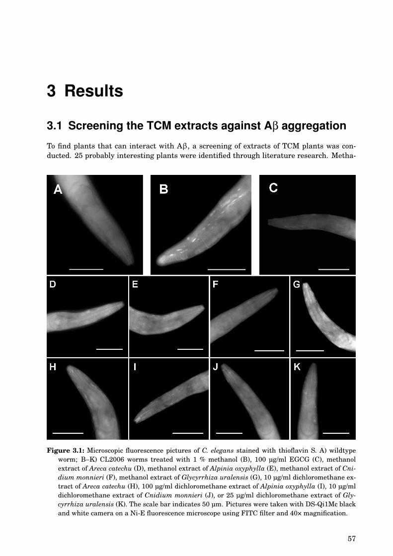
3 Results
3.1 Screening the TCM extracts against Aβ aggregation
To find plants that can interact with Aβ, a screening of extracts of TCM plants was con-ducted. 25 probably interesting plants were identified through literature research. Metha-
Figure 3.1: Microscopic fluorescence pictures of C. elegans stained with thioflavin S. A) wildtypeworm; B–K) CL2006 worms treated with 1 % methanol (B), 100 µg/ml EGCG (C), methanolextract of Areca catechu (D), methanol extract of Alpinia oxyphylla (E), methanol extract of Cni-dium monnieri (F), methanol extract of Glycyrrhiza uralensis (G), 10 µg/ml dichloromethane ex-tract of Areca catechu (H), 100 µg/ml dichloromethane extract of Alpinia oxyphylla (I), 10 µg/mldichloromethane extract of Cnidium monnieri (J), or 25 µg/ml dichloromethane extract of Gly-cyrrhiza uralensis (K). The scale bar indicates 50 µm. Pictures were taken with DS-Qi1Mc blackand white camera on a Ni-E fluorescence microscope using FITC filter and 40× magnification.
57
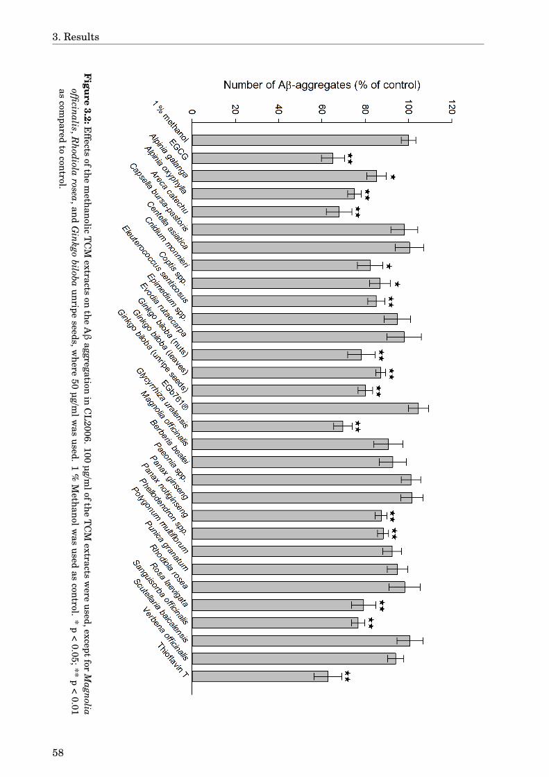
3. Results
Figure
3.2:Effects
ofthem
ethanolicT
CM
extractson
theAβ
aggregationin
CL
2006.100µg/m
loftheT
CM
extractsw
ereused,except
forM
agnoliaofficinalis,R
hodiolarosea,and
Ginkgo
bilobaunripe
seeds,where
50µg/m
lwas
used.1
%M
ethanolwas
usedas
control.*
p<
0.05;**p
<0.01
ascom
paredto
control.
58

3.1. Screening the TCM extracts against Aβ aggregation
nol and dichloromethane extracts of these plants were tested on their ability to inhibit Aβaggregation (see section 2.2.10). Aβ aggregates are only seen in the transgenic CL2006worms but not in the wildtype (Fig. 3.1 A & B). EGCG, that was used as positive control,decreased the number of Aβ aggregates by 35 % (p < 0.01) (Fig. 3.1 C & Fig. 3.2), whichis comparable to the results of Abbas & Wink (2010). Some methanol extracts like Arecacatechu (32 % decrease, p < 0.01) and Glycyrrhiza uralensis (30 % decrease, p < 0.01) showeda similar effect. Several other extracts were effective to a lesser extent: Alpinia oxyphyllawith 25 % decrease (p < 0.01), Sanguisorba officinalis with 23 % (p < 0.01), Rosa laevi-gata 21 % (p < 0.01), Cnidium monnieri 18 % (p = 0.01), Eleutherococcus senticosus 15 %(p < 0.01), Alpinia galanga 15 % (p = 0.01), Panax notoginseng 13 % (p < 0.01), Coptis spp.13 % (p = 0.03), phellodendron spp. 12 % (p < 0.01). All tested methanol extracts of Ginkgobiloba were effective (22 % decrease in the number of Aβ aggregates for nuts, 20 % for un-ripe seeds, and 13 % for leaves, respectively, p < 0.01), but the special extract EGb761® hadno effect. Thioflavin T decreased the number of plaques by 37 % (p < 0.01). Representativepictures for some treatments are shown in Figure 3.1 and the results are summarised inFigure 3.2.
The dichloromethane extracts were poorly soluble in the aqueous culture medium, so thatfor some extracts smaller concentrations had to be used (Fig. 3.4). Even so, the extracts ofAreca catechu and Cnidium monnieri had a strong effect decreasing the Aβ aggregates by32 % and 35 %, respectively (p < 0.01). Polygonum multiflorum (21 % decrease, p = 0.01) andEleutherococcus senticosus (15 % decrease, p = 0.04) also showed an effect in the 10 µg/mltreatment group (Fig. 3.4 A). Among the extracts used in the concentration of 25 µg/ml(Fig. 3.4 B) only Magnolia officinalis had an significant effect (14 % decrease, p = 0.02).Raw seeds of Ginkgo biloba could significantly decrease (by 20 %, p < 0.01) the numberof Aβ aggregates at 50 µg/ml (Fig. 3.4 C), whereas Sanguisorba officinalis increased thenumber by 14 % (p = 0.04). Panax notoginseng and Alpinia oxyphylla showed an effect ata concentration of 100 µg/ml (Fig. 3.4 D), decreasing Aβ aggregation by 17 % and 14 %,respectively (p < 0.01).
Figure 3.3: Effect of DMSO on the Aβ aggregation in CL2006. * p < 0.05;** p < 0.01 as compared to untreated control.
All values were compared to respective solvent controls. For methanol no dose-dependenteffect was observed but DMSO, that was used to solve the dichloromethane extracts, had
59

3. Results
an effect on the number of Aβ aggregates as shown in Figure 3.3. Concentrations above0.25 % were significantly different from untreated worms with 18 % decrease in number ofAβ aggregates for 0.5 % DMSO (p = 0.02) and 24 % for 1 % DMSO (p < 0.01).
Figure 3.4: Effects of the dichloromethane extracts of TCM plants on the Aβ aggregation in CL2006.A) 10 µg/ml of the TCM extracts solved in DMSO were used; B) 25 µg/ml of the TCM extractssolved in DMSO were used; C) 50 µg/ml of the TCM extracts solved in DMSO were used; D)100 µg/ml of the TCM extracts solved in DMSO were used. * p < 0.05; ** p < 0.01 as compared torespective controls.
60
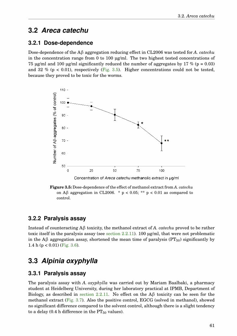
3.2. Areca catechu
3.2 Areca catechu
3.2.1 Dose-dependence
Dose-dependence of the Aβ aggregation reducing effect in CL2006 was tested for A. catechuin the concentration range from 0 to 100 µg/ml. The two highest tested concentrations of75 µg/ml and 100 µg/ml significantly reduced the number of aggregates by 17 % (p = 0.03)and 32 % (p < 0.01), respectively (Fig. 3.5). Higher concentrations could not be tested,because they proved to be toxic for the worms.
Figure 3.5: Dose-dependence of the effect of methanol extract from A. catechuon Aβ aggregation in CL2006. * p < 0.05; ** p < 0.01 as compared tocontrol.
3.2.2 Paralysis assay
Instead of counteracting Aβ toxicity, the methanol extract of A. catechu proved to be rathertoxic itself in the paralysis assay (see section 2.2.11). 100 µg/ml, that were not problematicin the Aβ aggregation assay, shortened the mean time of paralysis (PT50) significantly by1.4 h (p < 0.01) (Fig. 3.6).
3.3 Alpinia oxyphylla
3.3.1 Paralysis assay
The paralysis assay with A. oxyphylla was carried out by Mariam Baalbaki, a pharmacystudent at Heidelberg University, during her laboratory practical at IPMB, Department ofBiology, as described in section 2.2.11. No effect on the Aβ toxicity can be seen for themethanol extract (Fig. 3.7). Also the positive control, EGCG (solved in methanol), showedno significant difference compared to the solvent control, although there is a slight tendencyto a delay (0.4 h difference in the PT50 values).
61

3. Results
Figure 3.6: Results of the paralysis assay for methanol extract of A. catechu. ** p < 0.01 ascompared to control.
Figure 3.7: Results of the paralysis assay for methanol extract of A. oxyphylla.
62
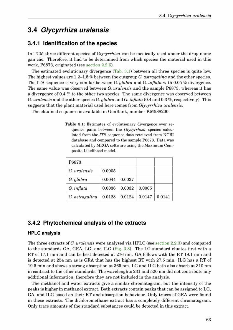
3.4. Glycyrrhiza uralensis
3.4 Glycyrrhiza uralensis
3.4.1 Identification of the species
In TCM three different species of Glycyrrhiza can be medically used under the drug namegan cao. Therefore, it had to be determined from which species the material used in thiswork, P6873, originated (see section 2.2.6).
The estimated evolutionary divergence (Tab. 3.1) between all three species is quite low.The highest values are 1.2–1.5 % between the outgroup G. astragalina and the other species.The ITS sequence is very similar between G. glabra and G. inflata with 0.05 % divergence.The same value was observed between G. uralensis and the sample P6873, whereas it hasa divergence of 0.4 % to the other two species. The same divergence was observed betweenG. uralensis and the other species G. glabra and G. inflata (0.4 and 0.3 %, respectively). Thissuggests that the plant material used here comes from Glycyrrhiza uralensis.
The obtained sequence is available in GenBank, number KM588200.
Table 3.1: Estimates of evolutionary divergence over se-quence pairs between the Glycyrrhiza species calcu-lated from the ITS sequence data retrieved from NCBIdatabase and compared to the sample P6873. Data wascalculated by MEGA software using the Maximum Com-posite Likelihood model.
P6873
G. uralensis 0.0005
G. glabra 0.0044 0.0037
G. inflata 0.0036 0.0032 0.0005
G. astragalina 0.0128 0.0124 0.0147 0.0141
3.4.2 Phytochemical analysis of the extracts
HPLC analysis
The three extracts of G. uralensis were analysed via HPLC (see section 2.2.3) and comparedto the standards GA, GRA, LG, and ILG (Fig. 3.8). The LG standard eluates first with aRT of 17.1 min and can be best detected at 276 nm. GA follows with the RT 19.1 min andis detected at 254 nm as is GRA that has the highest RT with 27.5 min. ILG has a RT of19.5 min and shows a strong absorption at 365 nm. LG and ILG both also absorb at 310 nmin contrast to the other standards. The wavelenghts 231 and 520 nm did not contribute anyadditional information, therefore they are not included in the analysis.
The methanol and water extracts give a similar chromatogram, but the intensity of thepeaks is higher in methanol extract. Both extracts contain peaks that can be assigned to LG,GA, and ILG based on their RT and absorption behaviour. Only traces of GRA were foundin these extracts. The dichloromethane extract has a completely different chromatogram.Only trace amounts of the standard substances could be detected in this extract.
63
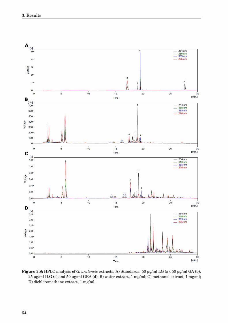
3. Results
Figure 3.8: HPLC analysis of G. uralensis extracts. A) Standards: 50 µg/ml LG (a), 50 µg/ml GA (b),25 µg/ml ILG (c) and 50 µg/ml GRA (d); B) water extract, 1 mg/ml; C) methanol extract, 1 mg/ml;D) dichloromethane extract, 1 mg/ml.
64
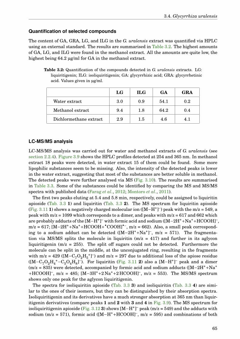
3.4. Glycyrrhiza uralensis
Quantification of selected compounds
The content of GA, GRA, LG, and ILG in the G. uralensis extract was quantified via HPLCusing an external standard. The results are summarised in Table 3.2. The highest amountsof GA, LG, and ILG were found in the methanol extract. All the amounts are quite low, thehighest being 64.2 µg/ml for GA in the methanol extract.
Table 3.2: Quantification of the compounds detected in G. uralensis extracts. LG:liquiritigenin; ILG: isoliquiritigenin; GA: glycyrrhizic acid; GRA: glycyrrhetinicacid. Values given in µg/ml.
LG ILG GA GRA
Water extract 3.0 0.9 54.1 0.2
Methanol extract 9.4 1.8 64.2 0.4
Dichlormethane extract 2.9 1.5 4.6 4.1
LC-MS/MS analysis
LC-MS/MS analysis was carried out for water and methanol extracts of G. uralensis (seesection 2.2.4). Figure 3.9 shows the HPLC profiles detected at 254 and 365 nm. In methanolextract 18 peaks were detected, in water extract 15 of them could be found. Some morelipophilic substances seem to be missing. Also, the intensity of the detected peaks is lowerin the water extract, suggesting that most of the substances are better soluble in methanol.The detected peaks were further analysed via MS (Fig. 3.10). The results are summarisedin Table 3.3. Some of the substances could be identified by comparing the MS and MS/MSspectra with published data (Farag et al., 2012; Montoro et al., 2011).
The first two peaks eluting at 5.4 and 5.8 min, respectively, could be assigned to liquiritinapioside (Tab. 3.3 1) and liquiritin (Tab. 3.3 2). The MS spectrum for liquiritin apioside(Fig. 3.11 1) shows a negatively charged molecular ion ([M−H+]–) peak with the m/z = 549, apeak with m/z = 1099 which corresponds to a dimer, and peaks with m/z = 617 and 662 whichare probably adducts of the [M−H+]– with formic acid and sodium ([M−2H++Na++HCOOH]–,m/z = 617; [M−2H++Na++HCOOH+•COOH]•–, m/z = 662). Also, a small peak correspond-ing to a sodium adduct can be detected ([M−2H++Na+]–, m/z = 571). The fragmenta-tion via MS/MS splits the molecule in liquiritin (m/z = 417) and further in its aglyconliquiritigenin (m/z = 255). The split off sugars could not be detected. Furthermore themolecule can be split in the middle, at the unconjugated ring, resulting in the fragmentswith m/z = 429 ([M−C7O2H5
+]–) and m/z = 297 due to additional loss of the apiose residue([M−C7O2H4
+−C5O4H8]–). For liquiritin (Fig. 3.11 2) also a [M−H+]– peak and a dimer(m/z = 835) were detected, accompanied by formic acid and sodium adducts ([M−2H++Na+
+HCOOH]–, m/z = 485; [M−3H++2 Na++2 HCOOH]–, m/z = 553). The MS/MS spectrumshows only one peak for the aglycon liquiritigenin.
The spectra for isoliquiritin apioside (Tab. 3.3 3) and isoliquiritin (Tab. 3.3 4) are simi-lar to the ones of their isomers, but they can be distinguished by their absorption spectra.Isoliquiritigenin and its derivatives have a much stronger absorption at 365 nm than liquir-itigenin derivatives (compare peaks 1 and 2 with 3 and 4 in Fig. 3.9). The MS spectrum forisoliquiritigenin apioside (Fig. 3.12 3) shows [M−H+]– peak (m/z = 549) and the adducts withsodium (m/z = 571), formic acid ([M−H++HCOOH]–, m/z = 595) and combinations of both
65
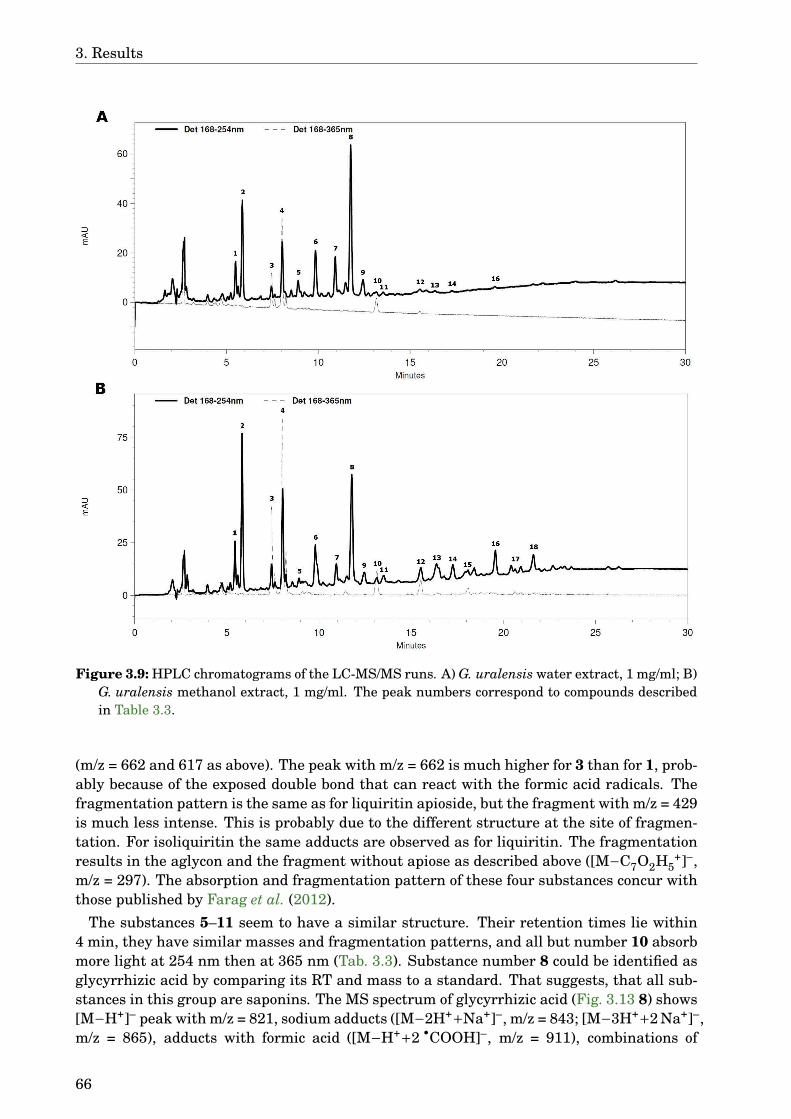
3. Results
Figure 3.9: HPLC chromatograms of the LC-MS/MS runs. A) G. uralensis water extract, 1 mg/ml; B)G. uralensis methanol extract, 1 mg/ml. The peak numbers correspond to compounds describedin Table 3.3.
(m/z = 662 and 617 as above). The peak with m/z = 662 is much higher for 3 than for 1, prob-ably because of the exposed double bond that can react with the formic acid radicals. Thefragmentation pattern is the same as for liquiritin apioside, but the fragment with m/z = 429is much less intense. This is probably due to the different structure at the site of fragmen-tation. For isoliquiritin the same adducts are observed as for liquiritin. The fragmentationresults in the aglycon and the fragment without apiose as described above ([M−C7O2H5
+]–,m/z = 297). The absorption and fragmentation pattern of these four substances concur withthose published by Farag et al. (2012).
The substances 5–11 seem to have a similar structure. Their retention times lie within4 min, they have similar masses and fragmentation patterns, and all but number 10 absorbmore light at 254 nm then at 365 nm (Tab. 3.3). Substance number 8 could be identified asglycyrrhizic acid by comparing its RT and mass to a standard. That suggests, that all sub-stances in this group are saponins. The MS spectrum of glycyrrhizic acid (Fig. 3.13 8) shows[M−H+]– peak with m/z = 821, sodium adducts ([M−2H++Na+]–, m/z = 843; [M−3H++2 Na+]–,m/z = 865), adducts with formic acid ([M−H++2 •COOH]–, m/z = 911), combinations of
66
3.4. Glycyrrhiza uralensis
Figure 3.10: Total ion current in MS, ESI negative. A) G. uralensis water extract, 1 mg/ml; B)G. uralensis methanol extract, 1 mg/ml. The peak numbers correspond to compounds describedin Table 3.3.
both kinds of adducts ([M−2H++Na++•COOH]•–, m/z = 888; [M−2H++Na++2 •COOH]–,m/z = 933; [M−2H++Na++2 •COOH+HCOOH]–, m/z = 979), and a dimer peak (m/z = 1643).The MS/MS spectrum is dominated by a fragment with m/z = 351, which corresponds to thenegatively charged ion of the two glucuronic acid molecules (C12O12H15
–), the sugars linkedto the aglycon of glycyrrhizic acid. This fragment can be found in all detected saponins,suggesting they all have the same sugar moiety. The same fragment was reported in alldetected saponins by Farag et al. (2012). The remaining triterpene fragment is poorly ion-ized, there is only a very small peak with m/z = 469. The other visible peaks belong to[M−H+−H2O]– (m/z = 803), [M−H+−H2O−CO2]– (m/z = 759) and the fragment without oneglucuronic acid moiety ([M−C6O6H9
+]–, m/z = 645).Substance 9 has the same molecular mass as glycyrrhizic acid, a very similar RT and
almost identical fragmentation pattern (Tab. 3.3 & Fig. 3.13 9). Based on this data it couldbe licorice saponin H2, a stereoisomer of glycyrrhizic acid. The fragments correspond tothose reported by Montoro et al. (2011). The fragment with m/z = 583, that was not presentin the MS/MS spectrum of glycyrrhizic acid, belongs to [M−H2O−CO2−C6O6H9
+]–.Substance number 5 could be identified as licorice saponin A3 by comparison of the data
with Farag et al. (2012) and Montoro et al. (2011) (Tab. 3.3 5). It has a [M−H+]– withm/z = 983 (Fig. 3.14 5) and adducts with sodium ([M−2H++Na+]–, m/z = 1005; [M−3H++2 Na+]–, m/z = 1027) and formic acid ([M−2H++Na++HCOOH]–, m/z = 1051; [M−3H++2 Na+
+HCOOH]–, m/z = 1073) can be seen in the MS spectrum as for the other substances. TheMS/MS spectrum shows a base peak with m/z = 821 that corresponds to glycyrrhizic acid
67
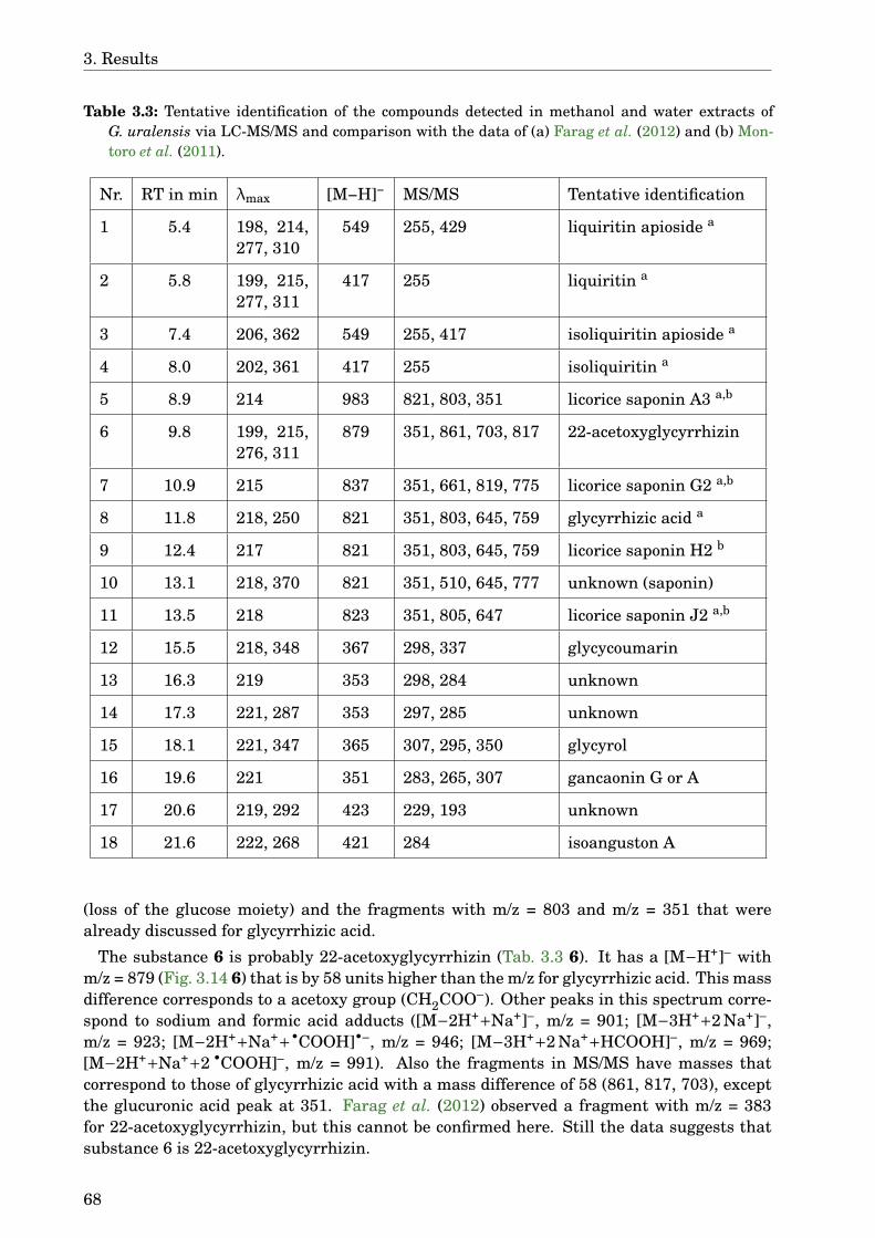
3. Results
Table 3.3: Tentative identification of the compounds detected in methanol and water extracts ofG. uralensis via LC-MS/MS and comparison with the data of (a) Farag et al. (2012) and (b) Mon-toro et al. (2011).
Nr. RT in min λmax [M−H]− MS/MS Tentative identification
1 5.4 198, 214,277, 310
549 255, 429 liquiritin apioside a
2 5.8 199, 215,277, 311
417 255 liquiritin a
3 7.4 206, 362 549 255, 417 isoliquiritin apioside a
4 8.0 202, 361 417 255 isoliquiritin a
5 8.9 214 983 821, 803, 351 licorice saponin A3 a,b
6 9.8 199, 215,276, 311
879 351, 861, 703, 817 22-acetoxyglycyrrhizin
7 10.9 215 837 351, 661, 819, 775 licorice saponin G2 a,b
8 11.8 218, 250 821 351, 803, 645, 759 glycyrrhizic acid a
9 12.4 217 821 351, 803, 645, 759 licorice saponin H2 b
10 13.1 218, 370 821 351, 510, 645, 777 unknown (saponin)
11 13.5 218 823 351, 805, 647 licorice saponin J2 a,b
12 15.5 218, 348 367 298, 337 glycycoumarin
13 16.3 219 353 298, 284 unknown
14 17.3 221, 287 353 297, 285 unknown
15 18.1 221, 347 365 307, 295, 350 glycyrol
16 19.6 221 351 283, 265, 307 gancaonin G or A
17 20.6 219, 292 423 229, 193 unknown
18 21.6 222, 268 421 284 isoanguston A
(loss of the glucose moiety) and the fragments with m/z = 803 and m/z = 351 that werealready discussed for glycyrrhizic acid.
The substance 6 is probably 22-acetoxyglycyrrhizin (Tab. 3.3 6). It has a [M−H+]– withm/z = 879 (Fig. 3.14 6) that is by 58 units higher than the m/z for glycyrrhizic acid. This massdifference corresponds to a acetoxy group (CH2COO–). Other peaks in this spectrum corre-spond to sodium and formic acid adducts ([M−2H++Na+]–, m/z = 901; [M−3H++2 Na+]–,m/z = 923; [M−2H++Na++•COOH]•–, m/z = 946; [M−3H++2 Na++HCOOH]–, m/z = 969;[M−2H++Na++2 •COOH]–, m/z = 991). Also the fragments in MS/MS have masses thatcorrespond to those of glycyrrhizic acid with a mass difference of 58 (861, 817, 703), exceptthe glucuronic acid peak at 351. Farag et al. (2012) observed a fragment with m/z = 383for 22-acetoxyglycyrrhizin, but this cannot be confirmed here. Still the data suggests thatsubstance 6 is 22-acetoxyglycyrrhizin.
68
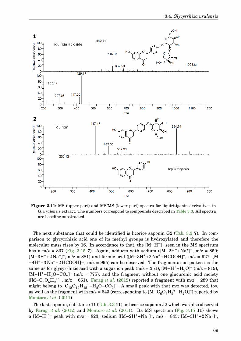
3.4. Glycyrrhiza uralensis
Figure 3.11: MS (upper part) and MS/MS (lower part) spectra for liquiritigenin derivatives inG. uralensis extract. The numbers correspond to compounds described in Table 3.3. All spectraare baseline substracted.
The next substance that could be identified is licorice saponin G2 (Tab. 3.3 7). In com-parison to glycyrrhizic acid one of its methyl groups is hydroxylated and therefore themolecular mass rises by 16. In accordance to that, the [M−H+]– seen in the MS spectrumhas a m/z = 837 (Fig. 3.15 7). Again, adducts with sodium ([M−2H++Na+]–, m/z = 859;[M−3H++2 Na+]–, m/z = 881) and formic acid ([M−3H++2 Na++HCOOH]–, m/z = 927; [M−4H++3 Na++2 HCOOH]−, m/z = 995) can be observed. The fragmentation pattern is thesame as for glycyrrhizic acid with a sugar ion peak (m/z = 351), [M−H+−H2O]– (m/z = 819),[M−H+−H2O−CO2]– (m/z = 775), and the fragment without one glucuronic acid moiety([M−C6O6H9
+]–, m/z = 661). Farag et al. (2012) reported a fragment with m/z = 289 thatmight belong to [C12O12H15
–−H2O−CO2]–. A small peak with that m/z was detected, too,as well as the fragment with m/z = 643 (corresponding to [M−C6O6H9
+−H2O]–) reported byMontoro et al. (2011).
The last saponin, substance 11 (Tab. 3.3 11), is licorice saponin J2 which was also observedby Farag et al. (2012) and Montoro et al. (2011). Its MS spectrum (Fig. 3.15 11) showsa [M−H+]– peak with m/z = 823, sodium ([M−2H++Na+]–, m/z = 845; [M−3H++2 Na+]–,
69
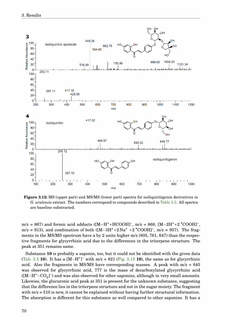
3. Results
Figure 3.12: MS (upper part) and MS/MS (lower part) spectra for isoliquiritigenin derivatives inG. uralensis extract. The numbers correspond to compounds described in Table 3.3. All spectraare baseline substracted.
m/z = 867) and formic acid adducts ([M−H++HCOOH]–, m/z = 868; [M−2H++2 •COOH]–,m/z = 913), and combination of both ([M−3H++2 Na+ +2 •COOH]–, m/z = 957). The frag-ments in the MS/MS spectrum have a by 2 units higher m/z (805, 761, 647) than the respec-tive fragments for glycyrrhizic acid due to the differences in the triterpene structure. Thepeak at 351 remains same.
Substance 10 is probably a saponin, too, but it could not be identified with the given data(Tab. 3.3 10). It has a [M−H+]– with m/z = 821 (Fig. 3.15 10), the same as for glycyrrhizicacid. Also the fragments in MS/MS have corresponding masses. A peak with m/z = 645was observed for glycyrrhizic acid, 777 is the mass of decarboxylated glycyrrhizic acid([M−H+−CO2]–) and was also observed for other saponins, although in very small amounts.Likewise, the glucuronic acid peak at 351 is present for the unknown substance, suggestingthat the difference lies in the triterpene structure and not in the sugar moiety. The fragmentwith m/z = 510 is new, it cannot be explained without having further structural information.The absorption is different for this substance as well compared to other saponins. It has a
70
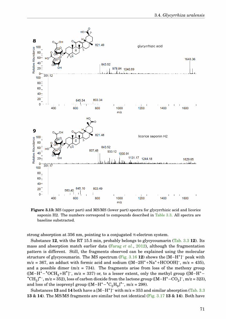
3.4. Glycyrrhiza uralensis
Figure 3.13: MS (upper part) and MS/MS (lower part) spectra for glycyrrhizic acid and licoricesaponin H2. The numbers correspond to compounds described in Table 3.3. All spectra arebaseline substracted.
strong absorption at 356 nm, pointing to a conjugated π-electron system.Substance 12, with the RT 15.5 min, probably belongs to glycycoumarin (Tab. 3.3 12). Its
mass and absorption match earlier data (Farag et al., 2012), although the fragmentationpattern is different. Still, the fragments observed can be explained using the molecularstructure of glycycoumarin. The MS spectrum (Fig. 3.16 12) shows the [M−H+]– peak withm/z = 367, an adduct with formic acid and sodium ([M−2H++Na++HCOOH]–, m/z = 435),and a possible dimer (m/z = 734). The fragments arise from loss of the methoxy group([M−H+−•OCH3+H•]–, m/z = 337) or, to a lesser extent, only the methyl group ([M−H+−•CH3]•–, m/z = 352), loss of carbon dioxide from the lactone group ([M−H+−CO2]–, m/z = 323),and loss of the isoprenyl group ([M−H+−•C5H9]•–, m/z = 298).
Substances 13 and 14 both have a [M−H+]– with m/z = 353 and similar absorption (Tab. 3.313 & 14). The MS/MS fragments are similar but not identical (Fig. 3.17 13 & 14). Both have
71
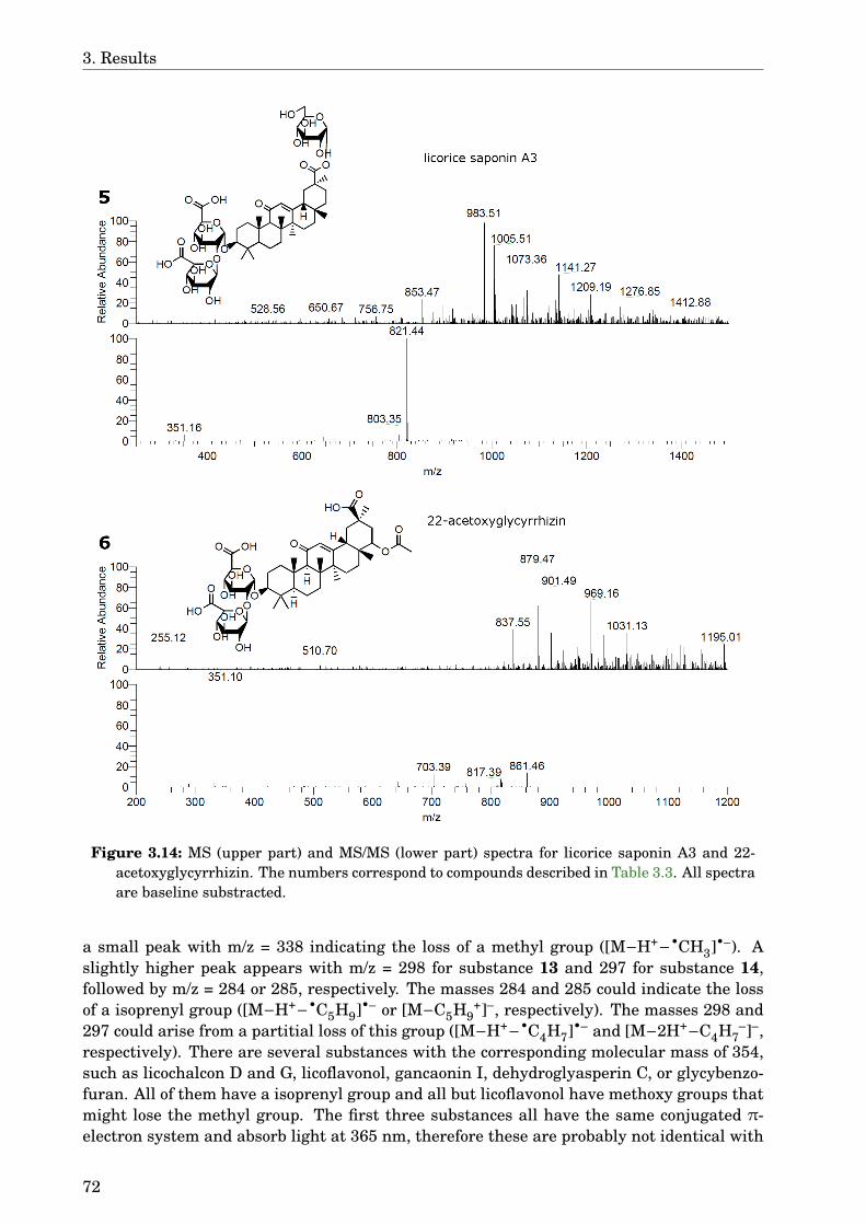
3. Results
Figure 3.14: MS (upper part) and MS/MS (lower part) spectra for licorice saponin A3 and 22-acetoxyglycyrrhizin. The numbers correspond to compounds described in Table 3.3. All spectraare baseline substracted.
a small peak with m/z = 338 indicating the loss of a methyl group ([M−H+−•CH3]•–). Aslightly higher peak appears with m/z = 298 for substance 13 and 297 for substance 14,followed by m/z = 284 or 285, respectively. The masses 284 and 285 could indicate the lossof a isoprenyl group ([M−H+−•C5H9]•– or [M−C5H9
+]–, respectively). The masses 298 and297 could arise from a partitial loss of this group ([M−H+−•C4H7]•– and [M−2H+−C4H7
–]–,respectively). There are several substances with the corresponding molecular mass of 354,such as licochalcon D and G, licoflavonol, gancaonin I, dehydroglyasperin C, or glycybenzo-furan. All of them have a isoprenyl group and all but licoflavonol have methoxy groups thatmight lose the methyl group. The first three substances all have the same conjugated π-electron system and absorb light at 365 nm, therefore these are probably not identical with
72
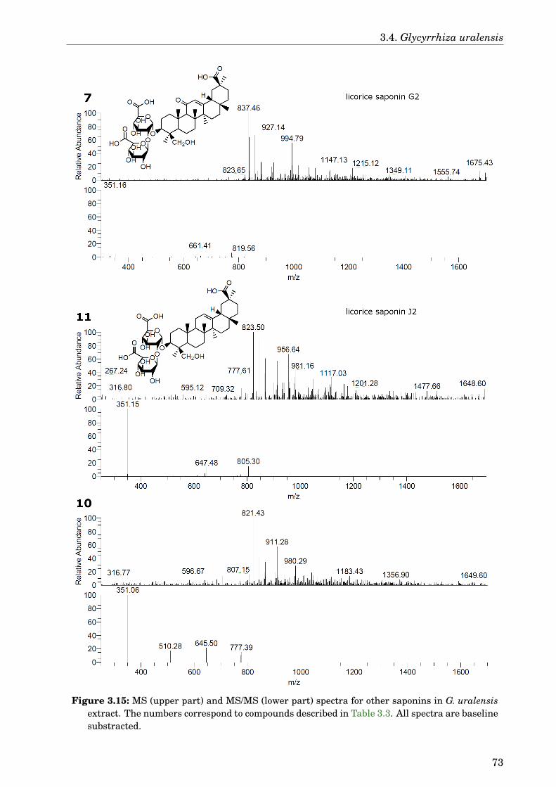
3.4. Glycyrrhiza uralensis
Figure 3.15: MS (upper part) and MS/MS (lower part) spectra for other saponins in G. uralensisextract. The numbers correspond to compounds described in Table 3.3. All spectra are baselinesubstracted.
73
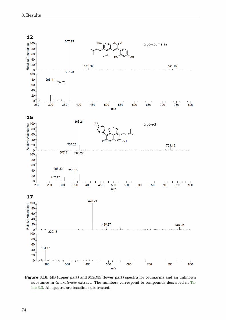
3. Results
Figure 3.16: MS (upper part) and MS/MS (lower part) spectra for coumarins and an unknownsubstance in G. uralensis extract. The numbers correspond to compounds described in Ta-ble 3.3. All spectra are baseline substracted.
74
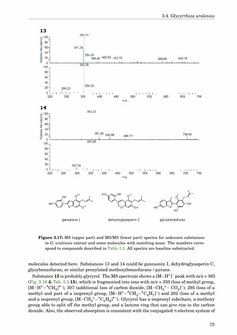
3.4. Glycyrrhiza uralensis
Figure 3.17: MS (upper part) and MS/MS (lower part) spectra for unknown substancesin G. uralensis extract and some molecules with matching mass. The numbers corre-spond to compounds described in Table 3.3. All spectra are baseline substracted.
molecules detected here. Substances 13 and 14 could be gancaonin I, dehydroglyasperin C,glycybenzofuran, or similar prenylated methoxybenzofurans /-pyrans.
Substance 15 is probably glycyrol. The MS spectrum shows a [M−H+]– peak with m/z = 365(Fig. 3.16 & Tab. 3.3 15), which is fragmented into ions with m/z = 350 (loss of methyl group,[M−H+−•CH3]•–), 307 (additional loss of carbon dioxide, [M−CH3
+− CO2]–), 295 (loss of amethyl and part of a isoprenyl group, [M−H+−•CH3−•C4H7]–) and 282 (loss of a methyland a isoprenyl group, [M−CH3
+−•C5H9]•–). Glycyrol has a isoprenyl sidechain, a methoxygroup able to split off the methyl group, and a lactone ring that can give rise to the carbondioxide. Also, the observed absorption is consistent with the conjugated π-electron system of
75

3. Results
Figure 3.18: MS (upper part) and MS/MS (lower part) spectra for gancaonin and isoangustonA. The numbers correspond to compounds described in Table 3.3. All spectra are baselinesubstracted.
glycyrol. Montoro et al. (2011) reported this compound in G. glabra, although unfortunatelyno MS/MS data have been published.
Substance number 16 has a [M−H+]– with m/z = 351 (Fig. 3.18 16), corresponding toa substance with a molecular mass of 352 like gancaonin A or G. The MS/MS fragmentssuggest a isoprenyl chain ([M−C5H9
+]–, m/z = 283) and a methyl group ([M−H+−•CH3]•–,m/z = 336) that are present in both gancaonin isomers. The m/z = 265 suggest a loss ofwater, additionally to the loss of an isoprenyl group ([M−C5H9
+−H2O]–). The fragment withm/z = 307 could result from loss of three methyl groups: one of the methoxy group, the othertwo from the isoprenyl group, made possible by a ring formation between this group and theadjacent hydroxy group ([M−C3H9
+]–). In case of gancaonin G there would be the oxygenradical of the methoxy group instead of a hydroxy group. That might facilitate the ringformation, making gancaonin G the more probable candidate for the identity of substance16, but there is no definite proof for either of the isomers.
The substance 17 could not be identified. It has a [M−H+]– with m/z = 423 and the spec-trum shows also a dimer with m/z = 847 (Fig. 3.16 17). This molecule is fragmented into
76
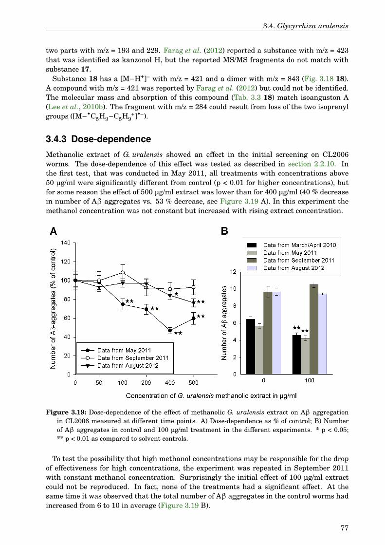
3.4. Glycyrrhiza uralensis
two parts with m/z = 193 and 229. Farag et al. (2012) reported a substance with m/z = 423that was identified as kanzonol H, but the reported MS/MS fragments do not match withsubstance 17.
Substance 18 has a [M−H+]– with m/z = 421 and a dimer with m/z = 843 (Fig. 3.18 18).A compound with m/z = 421 was reported by Farag et al. (2012) but could not be identified.The molecular mass and absorption of this compound (Tab. 3.3 18) match isoanguston A(Lee et al., 2010b). The fragment with m/z = 284 could result from loss of the two isoprenylgroups ([M−•C5H9−C5H9
+]•–).
3.4.3 Dose-dependence
Methanolic extract of G. uralensis showed an effect in the initial screening on CL2006worms. The dose-dependence of this effect was tested as described in section 2.2.10. Inthe first test, that was conducted in May 2011, all treatments with concentrations above50 µg/ml were significantly different from control (p < 0.01 for higher concentrations), butfor some reason the effect of 500 µg/ml extract was lower than for 400 µg/ml (40 % decreasein number of Aβ aggregates vs. 53 % decrease, see Figure 3.19 A). In this experiment themethanol concentration was not constant but increased with rising extract concentration.
Figure 3.19: Dose-dependence of the effect of methanolic G. uralensis extract on Aβ aggregationin CL2006 measured at different time points. A) Dose-dependence as % of control; B) Numberof Aβ aggregates in control and 100 µg/ml treatment in the different experiments. * p < 0.05;** p < 0.01 as compared to solvent controls.
To test the possibility that high methanol concentrations may be responsible for the dropof effectiveness for high concentrations, the experiment was repeated in September 2011with constant methanol concentration. Surprisingly the initial effect of 100 µg/ml extractcould not be reproduced. In fact, none of the treatments had a significant effect. At thesame time it was observed that the total number of Aβ aggregates in the control worms hadincreased from 6 to 10 in average (Figure 3.19 B).
77

3. Results
Due to the different results gained in the previous experiments, the test was repeatedagain in August 2012. This time only 400 µg/ml and 500 µg/ml extract treatments hadsignificant effects (16 %, p = 0.01 and 24 % decrease, p < 0.01, respectively). None of thetreatments reached the effect observed for 100 µg/ml treatment in the initial screening. Thetotal number of plaques was on the same level as a year ago.
In 2013 and 2014 more tests with the CL2006 strain were done to investigate the cause ofthe differences. New CL2006 worms were ordered from CGC together with new E. coliOP50. Fresh S-medium was prepared for these experiments. Even the incubator waschanged. None of this changed the result that even the positive control EGCG was noteffective any more. The number of plaques continued to rise up to 30 in untreated worms.This number is normal for CL2006 under standard conditions, but can be reduced at lowertemperatures (Dr. Christopher D. Link, P. I. at University of Colorado, Boulder, Institutefor Behavioural Genetics, personal communication). Since the rapid increase in the numberof plaques coincided with the breakdown of our old incubator and bringing into service of anew one in May 2011, it is possible that the low numbers of plaques in earlier experimentsresulted from a malfunction of temperature control in the old incubator.
Figure 3.20: Dose-dependence of the effect of pure compounds from G. uralensis on Aβ aggregationin CL2006. * p < 0.05; ** p < 0.01 as compared to solvent controls.
The dose-dependence was also measured for pure compounds contained in G. uralensisextracts: GA, its aglycon GRA and the salt GAA, LG, and ILG (Fig. 3.20). The diammoniumsalt of glycyrrhizic acid showed no effect. GA had a significant effect only for the highesttested concentration of 100 µg/ml with 14 % decrease in number of Aβ aggregates (p = 0.04).GRA was active at 50 and 100 µg/ml with 18 % and 22 % decrease (p < 0.01), GAA had asignificant effect already for 10 µg/ml (12 % decrease, p = 0.01), followed by 16 % and 17 %decrease for the following concentrations (p < 0.01), but the treatment with 100 µg/ml hadno significant difference to control. The strongest effect was observed for ILG by 50 µg/mltreatment that decreased the number of Aβ aggregates by 26 % (p < 0.01), but 100 µg/ml ofthis substance had a somewhat lesser effect (19 % decrease, p < 0.01). LG had its strongesteffect at 100 µg/ml with 23 % decrease and 15 % decrease at 50 µg/ml (p < 0.01). The treat-ment with 50 µg/ml was effective for most of the substances, therefore this concentrationwas used in following experiments.
78
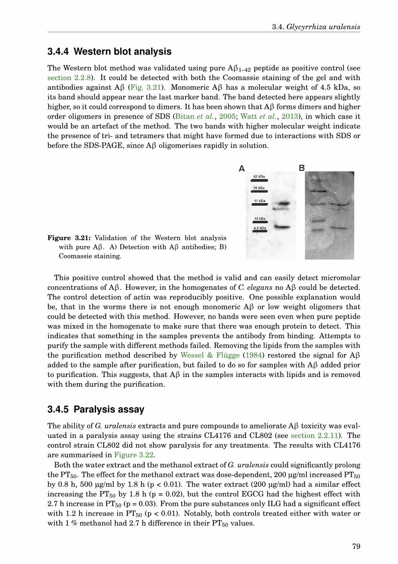
3.4. Glycyrrhiza uralensis
3.4.4 Western blot analysis
The Western blot method was validated using pure Aβ1–42 peptide as positive control (seesection 2.2.8). It could be detected with both the Coomassie staining of the gel and withantibodies against Aβ (Fig. 3.21). Monomeric Aβ has a molecular weight of 4.5 kDa, soits band should appear near the last marker band. The band detected here appears slightlyhigher, so it could correspond to dimers. It has been shown that Aβ forms dimers and higherorder oligomers in presence of SDS (Bitan et al., 2005; Watt et al., 2013), in which case itwould be an artefact of the method. The two bands with higher molecular weight indicatethe presence of tri- and tetramers that might have formed due to interactions with SDS orbefore the SDS-PAGE, since Aβ oligomerises rapidly in solution.
Figure 3.21: Validation of the Western blot analysiswith pure Aβ. A) Detection with Aβ antibodies; B)Coomassie staining.
This positive control showed that the method is valid and can easily detect micromolarconcentrations of Aβ. However, in the homogenates of C. elegans no Aβ could be detected.The control detection of actin was reproducibly positive. One possible explanation wouldbe, that in the worms there is not enough monomeric Aβ or low weight oligomers thatcould be detected with this method. However, no bands were seen even when pure peptidewas mixed in the homogenate to make sure that there was enough protein to detect. Thisindicates that something in the samples prevents the antibody from binding. Attempts topurify the sample with different methods failed. Removing the lipids from the samples withthe purification method described by Wessel & Flügge (1984) restored the signal for Aβadded to the sample after purification, but failed to do so for samples with Aβ added priorto purification. This suggests, that Aβ in the samples interacts with lipids and is removedwith them during the purification.
3.4.5 Paralysis assay
The ability of G. uralensis extracts and pure compounds to ameliorate Aβ toxicity was eval-uated in a paralysis assay using the strains CL4176 and CL802 (see section 2.2.11). Thecontrol strain CL802 did not show paralysis for any treatments. The results with CL4176are summarised in Figure 3.22.
Both the water extract and the methanol extract of G. uralensis could significantly prolongthe PT50. The effect for the methanol extract was dose-dependent, 200 µg/ml increased PT50by 0.8 h, 500 µg/ml by 1.8 h (p < 0.01). The water extract (200 µg/ml) had a similar effectincreasing the PT50 by 1.8 h (p = 0.02), but the control EGCG had the highest effect with2.7 h increase in PT50 (p = 0.03). From the pure substances only ILG had a significant effectwith 1.2 h increase in PT50 (p < 0.01). Notably, both controls treated either with water orwith 1 % methanol had 2.7 h difference in their PT50 values.
79
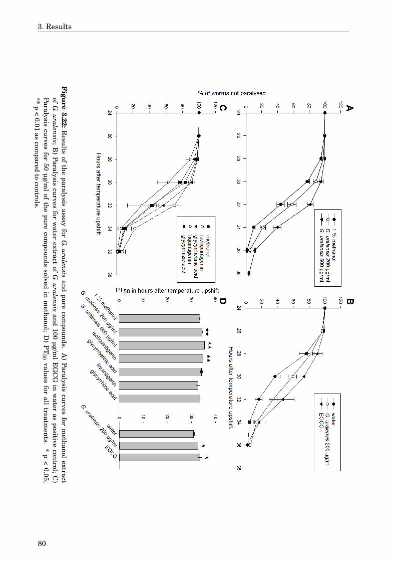
3. Results
Figure
3.22:R
esultsof
theparalysis
assayfor
G.uralensis
andpure
compounds.
A)
Paralysiscurves
form
ethanolextract
ofG
.uralensis;B
)Paralysis
curvesfor
water
extractof
G.uralensis
and100
µg/ml
EG
CG
inw
ateras
positivecontrol;
C)
Paralysiscurves
for50
µg/ml
ofthe
purecom
poundssolved
inm
ethanol;D
)P
T50
valuesfor
alltreatm
ents.*
p<
0.05;**
p<
0.01as
compared
tocontrols.
80
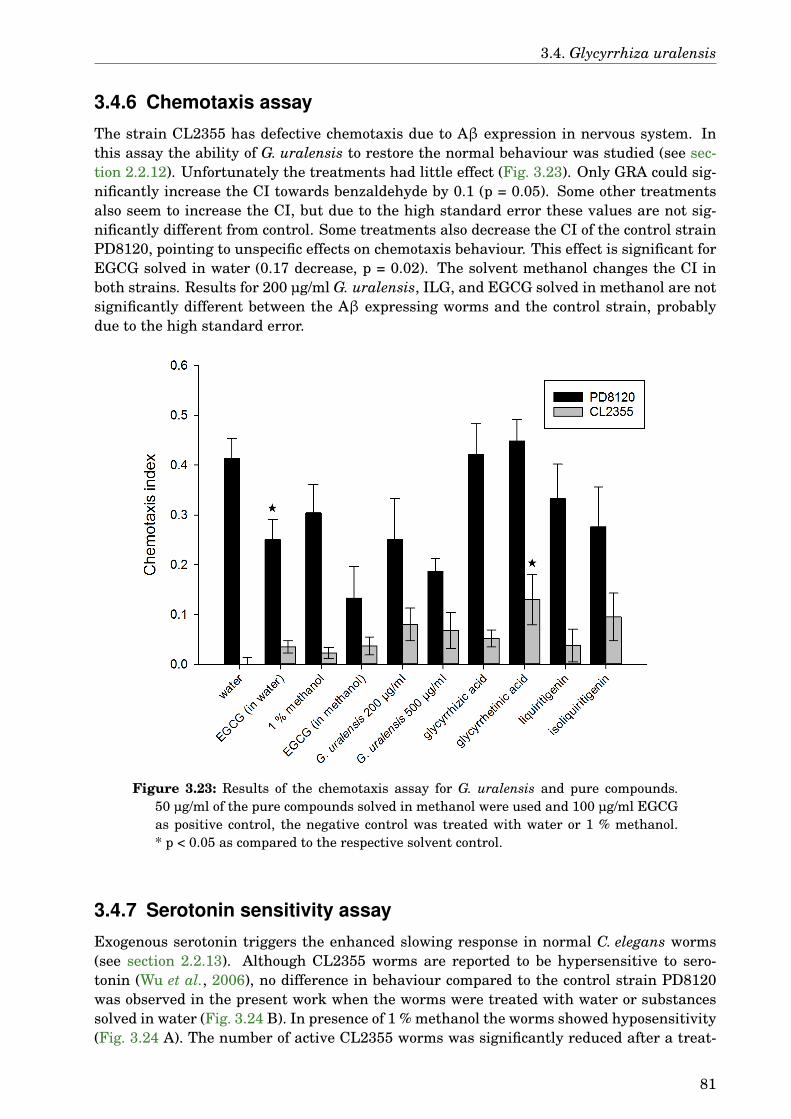
3.4. Glycyrrhiza uralensis
3.4.6 Chemotaxis assay
The strain CL2355 has defective chemotaxis due to Aβ expression in nervous system. Inthis assay the ability of G. uralensis to restore the normal behaviour was studied (see sec-tion 2.2.12). Unfortunately the treatments had little effect (Fig. 3.23). Only GRA could sig-nificantly increase the CI towards benzaldehyde by 0.1 (p = 0.05). Some other treatmentsalso seem to increase the CI, but due to the high standard error these values are not sig-nificantly different from control. Some treatments also decrease the CI of the control strainPD8120, pointing to unspecific effects on chemotaxis behaviour. This effect is significant forEGCG solved in water (0.17 decrease, p = 0.02). The solvent methanol changes the CI inboth strains. Results for 200 µg/ml G. uralensis, ILG, and EGCG solved in methanol are notsignificantly different between the Aβ expressing worms and the control strain, probablydue to the high standard error.
Figure 3.23: Results of the chemotaxis assay for G. uralensis and pure compounds.50 µg/ml of the pure compounds solved in methanol were used and 100 µg/ml EGCGas positive control, the negative control was treated with water or 1 % methanol.* p < 0.05 as compared to the respective solvent control.
3.4.7 Serotonin sensitivity assay
Exogenous serotonin triggers the enhanced slowing response in normal C. elegans worms(see section 2.2.13). Although CL2355 worms are reported to be hypersensitive to sero-tonin (Wu et al., 2006), no difference in behaviour compared to the control strain PD8120was observed in the present work when the worms were treated with water or substancessolved in water (Fig. 3.24 B). In presence of 1 % methanol the worms showed hyposensitivity(Fig. 3.24 A). The number of active CL2355 worms was significantly reduced after a treat-
81
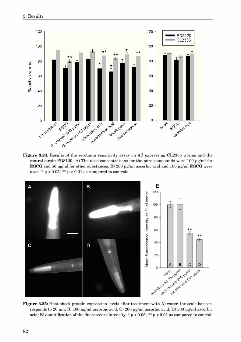
3. Results
Figure 3.24: Results of the serotonin sensitivity assay on Aβ expressing CL2355 worms and thecontrol strain PD8120. A) The used concentrations for the pure compounds were 100 µg/ml forEGCG and 50 µg/ml for other substances; B) 200 µg/ml ascorbic acid and 100 µg/ml EGCG wereused. * p < 0.05; ** p < 0.01 as compared to controls.
Figure 3.25: Heat shock protein expression levels after treatment with A) water, the scale bar cor-responds to 20 µm; B) 100 µg/ml ascorbic acid; C) 200 µg/ml ascorbic acid; D) 500 µg/ml ascorbicacid; E) quantification of the fluorescence intensity. * p < 0.05; ** p < 0.01 as compared to control.
82
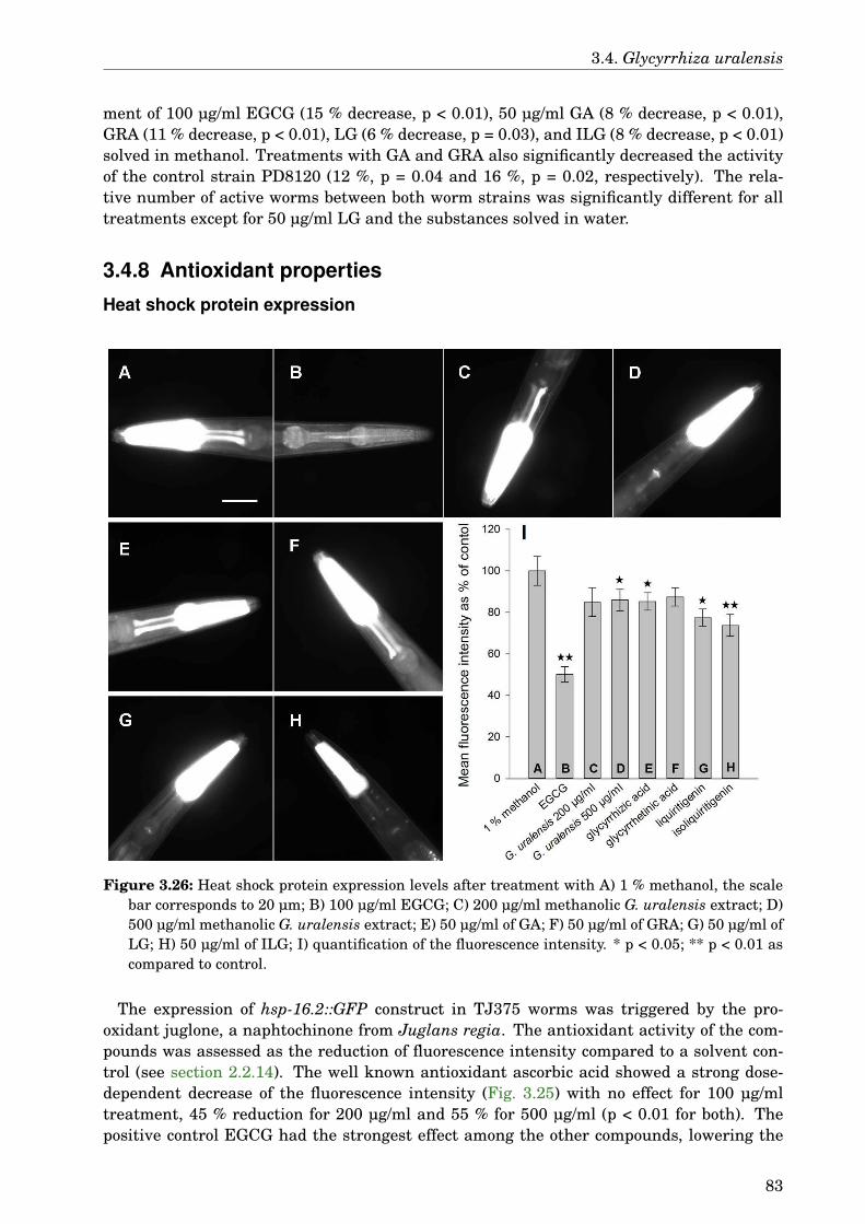
3.4. Glycyrrhiza uralensis
ment of 100 µg/ml EGCG (15 % decrease, p < 0.01), 50 µg/ml GA (8 % decrease, p < 0.01),GRA (11 % decrease, p < 0.01), LG (6 % decrease, p = 0.03), and ILG (8 % decrease, p < 0.01)solved in methanol. Treatments with GA and GRA also significantly decreased the activityof the control strain PD8120 (12 %, p = 0.04 and 16 %, p = 0.02, respectively). The rela-tive number of active worms between both worm strains was significantly different for alltreatments except for 50 µg/ml LG and the substances solved in water.
3.4.8 Antioxidant properties
Heat shock protein expression
Figure 3.26: Heat shock protein expression levels after treatment with A) 1 % methanol, the scalebar corresponds to 20 µm; B) 100 µg/ml EGCG; C) 200 µg/ml methanolic G. uralensis extract; D)500 µg/ml methanolic G. uralensis extract; E) 50 µg/ml of GA; F) 50 µg/ml of GRA; G) 50 µg/ml ofLG; H) 50 µg/ml of ILG; I) quantification of the fluorescence intensity. * p < 0.05; ** p < 0.01 ascompared to control.
The expression of hsp-16.2::GFP construct in TJ375 worms was triggered by the pro-oxidant juglone, a naphtochinone from Juglans regia. The antioxidant activity of the com-pounds was assessed as the reduction of fluorescence intensity compared to a solvent con-trol (see section 2.2.14). The well known antioxidant ascorbic acid showed a strong dose-dependent decrease of the fluorescence intensity (Fig. 3.25) with no effect for 100 µg/mltreatment, 45 % reduction for 200 µg/ml and 55 % for 500 µg/ml (p < 0.01 for both). Thepositive control EGCG had the strongest effect among the other compounds, lowering the
83

3. Results
mean fluorescence intensity by 50 %, p < 0.01 (Fig. 3.26). ILG decreased the fluorescenceintensity by 26 % (p < 0.01), the treatments with 500 µg/ml G. uralensis extract, GA, andLG had smaller effects (14 %, p = 0.04, 15 %, p = 0.03, and 23 %, p = 0.01, respectively).
Survival assay
Survival assay measures the antioxidant activity by determining the increase in survivalrate under lethal oxidative stress (see section 2.2.15). The effects are summarised in Fig-ure 3.27. Both controls ascorbic acid and EGCG increased the number of living wormsmarkedly (by 72 % for ascorbic acid, by 67 % for EGCG solved in water, and by 74 % forEGCG solved in methanol, p < 0.01 for all). The G. uralensis extract could not protect theworms from oxidative stress, but the pure compound ILG had an even stronger effect thanthe controls, increasing the survival by 82 % (p < 0.01). Its isomer LG had compared to thatonly a small effect (19 % increase, p = 0.03).
Figure 3.27: Survival under lethal oxidative stress after treatment with G. uralensis and purecompounds. A) 50 µg/ml of the pure compounds solved in methanol were used, 100 µg/mlEGCG as positive control, as negative control 1 % methanol was used; B) control treat-ments with 200 µg/ml ascorbic acid and 100 µg/ml EGCG solved in water. * p < 0.05;** p < 0.01 as compared to controls.
3.4.9 DAF-16 delocalisationDelocalisation of the transcription factor DAF-16 into the nucleus is necessary to start theexpression of several stress resistance genes (see section 2.2.16). Under normal conditionsthe fluorescence in daf-16::GFP worms is spread throughout the body (Fig. 3.28 A), underoxidative or heat stress the fluorescence concentrates in the cell nuclei (Fig. 3.28 B, C). Atreatment with methanol delocated some of the protein into the nucleus, but around 50 %of the treated worms were still negative. Because of this solvent effect only samples with
84
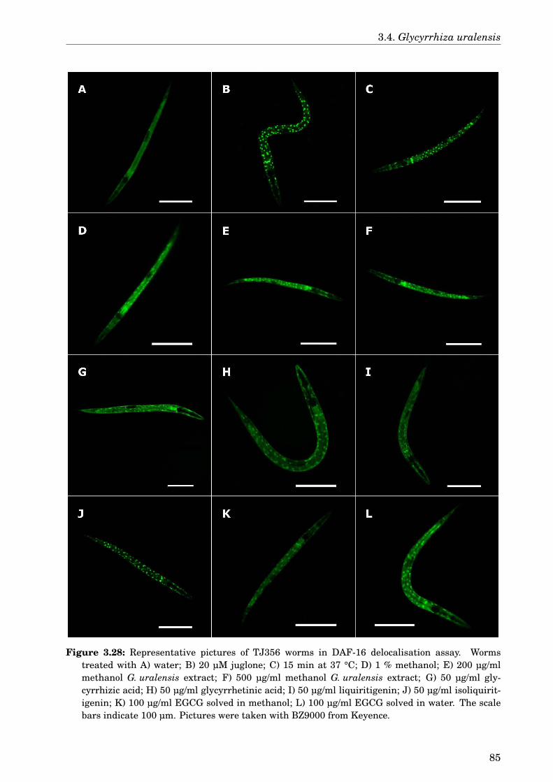
3.4. Glycyrrhiza uralensis
Figure 3.28: Representative pictures of TJ356 worms in DAF-16 delocalisation assay. Wormstreated with A) water; B) 20 µM juglone; C) 15 min at 37 °C; D) 1 % methanol; E) 200 µg/mlmethanol G. uralensis extract; F) 500 µg/ml methanol G. uralensis extract; G) 50 µg/ml gly-cyrrhizic acid; H) 50 µg/ml glycyrrhetinic acid; I) 50 µg/ml liquiritigenin; J) 50 µg/ml isoliquirit-igenin; K) 100 µg/ml EGCG solved in methanol; L) 100 µg/ml EGCG solved in water. The scalebars indicate 100 µm. Pictures were taken with BZ9000 from Keyence.
85
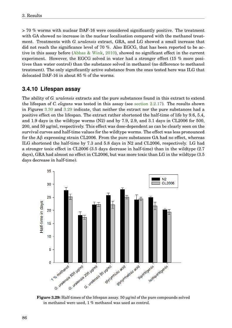
3. Results
> 70 % worms with nuclear DAF-16 were considered significantly positive. The treatmentwith GA showed no increase in the nuclear localisation compared with the methanol treat-ment. Treatments with G. uralensis extract, GRA, and LG showed a small increase thatdid not reach the significance level of 70 %. Also EGCG, that has been reported to be ac-tive in this assay before (Abbas & Wink, 2010), showed no significant effect in the currentexperiment. However, the EGCG solved in water had a stronger effect (15 % more posi-tives than water control) than the substance solved in methanol (no difference to methanoltreatment). The only significantly active substance from the ones tested here was ILG thatdelocated DAF-16 in about 85 % of the worms.
3.4.10 Lifespan assay
The ability of G. uralensis extracts and the pure substances found in this extract to extendthe lifespan of C. elegans was tested in this assay (see section 2.2.17). The results shownin Figures 3.30 and 3.29 indicate, that neither the extract nor the pure substances had apositive effect on the lifespan. The extract rather shortened the half-time of life by 9.6, 5.4,and 1.9 days in the wildtype worms (N2) and by 7.9, 2.9, and 3.1 days in CL2006 for 500,200, and 50 µg/ml, respectively. This effect was dose-dependent as can be clearly seen on thesurvival curves and half-time values for the wildtype worms. The effect was less pronouncedfor the Aβ expressing strain CL2006. From the pure substances GA had no effect, whereasILG shortened the half-time by 7.3 and 5.8 days in N2 and CL2006, respectively. LG hada stronger toxic effect in CL2006 (3.5 days decrease in half-time) than in the wildtype (2.7days), GRA had almost no effect in CL2006, but was more toxic than LG in the wildtype (3.5days decrease in half-time).
Figure 3.29: Half-times of the lifespan assay. 50 µg/ml of the pure compounds solvedin methanol were used, 1 % methanol was used as control.
86

3.4. Glycyrrhiza uralensis
Fig
ure
3.30
:Res
ults
ofth
elif
espa
nas
say
for
G.u
rale
nsis
.50
µg/m
loft
hepu
reco
mpo
unds
solv
edin
met
hano
lwer
eus
ed,1
%m
etha
nolw
asus
edas
cont
rol.
87

3. Results
3.5 Carlina acaulis
3.5.1 GLC-MS analysis of the extractThis analysis was carried out by Frank Sporer, a scientific assistant at Heidelberg Univer-sity, IPMB, Department of Biology (see section 2.2.5).
Figure 3.31: GLC-MS total ion current of dichloromethane extract of C. acaulis and correspondingMS spectra.
The extract contains mainly Carlina oxide and small amounts of benzaldehyde and ar-
88
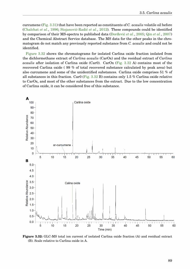
3.5. Carlina acaulis
curcumene (Fig. 3.31) that have been reported as constituents of C. acaulis volatile oil before(Chalchat et al., 1996; Stojanovic-Radic et al., 2012). These compounds could be identifiedby comparison of their MS-spectra to published data (Ðordevic et al., 2005; Qin et al., 2007)and the Chemical Abstract Service database. The MS data for the other peaks in the chro-matogram do not match any previously reported substance from C. acaulis and could not beidentified.
Figure 3.32 shows the chromatograms for isolated Carlina oxide fraction isolated fromthe dichlormethane extract of Carlina acaulis (CarOx) and the residual extract of Carlinaacaulis after isolation of Carlina oxide (Car0). CarOx (Fig. 3.32 A) contains most of therecovered Carlina oxide ( 99 % of total recovered substance calculated by peak area) butalso curcumene and some of the unidentified substances. Carlina oxide comprises 51 % ofall substances in this fraction. Car0 (Fig. 3.32 B) contains only 1.5 % Carlina oxide relativeto CarOx, and most of the other substances from the extract. Due to the low concentrationof Carlina oxide, it can be considered free of this substance.
Figure 3.32: GLC-MS total ion current of isolated Carlina oxide fraction (A) and residual extract(B). Scale relative to Carlina oxide in A.
89
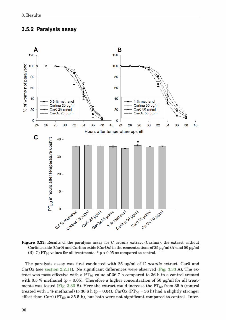
3. Results
3.5.2 Paralysis assay
Figure 3.33: Results of the paralysis assay for C. acaulis extract (Carlina), the extract withoutCarlina oxide (Car0) and Carlina oxide (CarOx) in the concentrations of 25 µg/ml (A) and 50 µg/ml(B). C) PT50 values for all treatments. * p < 0.05 as compared to control.
The paralysis assay was first conducted with 25 µg/ml of C. acaulis extract, Car0 andCarOx (see section 2.2.11). No significant differences were observed (Fig. 3.33 A). The ex-tract was most effective with a PT50 value of 36.7 h compared to 36 h in a control treatedwith 0.5 % methanol (p = 0.05). Therefore a higher concentration of 50 µg/ml for all treat-ments was tested (Fig. 3.33 B). Here the extract could increase the PT50 from 35 h (controltreated with 1 % methanol) to 36.6 h (p = 0.04). CarOx (PT50 = 36 h) had a slightly strongereffect than Car0 (PT50 = 35.5 h), but both were not significant compared to control. Inter-
90
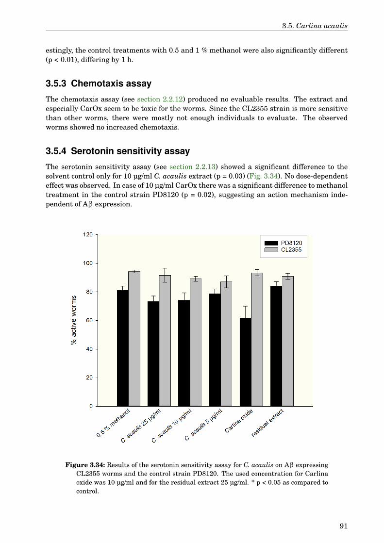
3.5. Carlina acaulis
estingly, the control treatments with 0.5 and 1 % methanol were also significantly different(p < 0.01), differing by 1 h.
3.5.3 Chemotaxis assay
The chemotaxis assay (see section 2.2.12) produced no evaluable results. The extract andespecially CarOx seem to be toxic for the worms. Since the CL2355 strain is more sensitivethan other worms, there were mostly not enough individuals to evaluate. The observedworms showed no increased chemotaxis.
3.5.4 Serotonin sensitivity assay
The serotonin sensitivity assay (see section 2.2.13) showed a significant difference to thesolvent control only for 10 µg/ml C. acaulis extract (p = 0.03) (Fig. 3.34). No dose-dependenteffect was observed. In case of 10 µg/ml CarOx there was a significant difference to methanoltreatment in the control strain PD8120 (p = 0.02), suggesting an action mechanism inde-pendent of Aβ expression.
Figure 3.34: Results of the serotonin sensitivity assay for C. acaulis on Aβ expressingCL2355 worms and the control strain PD8120. The used concentration for Carlinaoxide was 10 µg/ml and for the residual extract 25 µg/ml. * p < 0.05 as compared tocontrol.
91
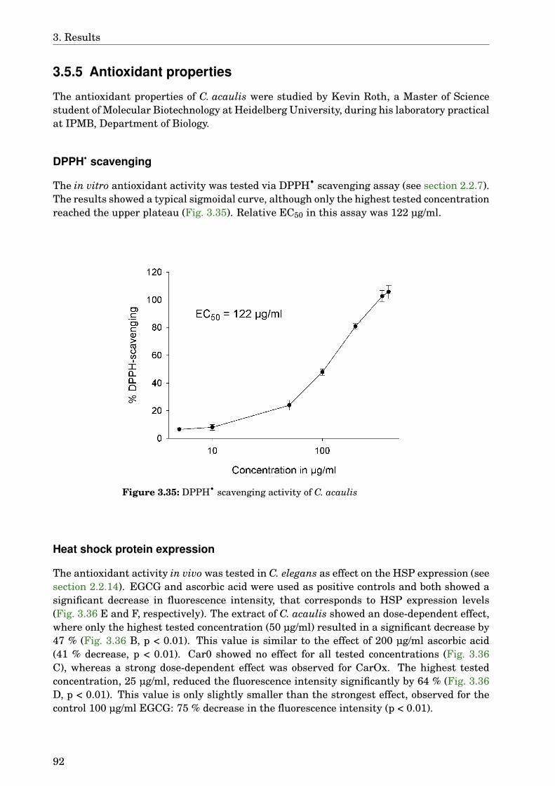
3. Results
3.5.5 Antioxidant properties
The antioxidant properties of C. acaulis were studied by Kevin Roth, a Master of Sciencestudent of Molecular Biotechnology at Heidelberg University, during his laboratory practicalat IPMB, Department of Biology.
DPPH• scavenging
The in vitro antioxidant activity was tested via DPPH• scavenging assay (see section 2.2.7).The results showed a typical sigmoidal curve, although only the highest tested concentrationreached the upper plateau (Fig. 3.35). Relative EC50 in this assay was 122 µg/ml.
Figure 3.35: DPPH• scavenging activity of C. acaulis
Heat shock protein expression
The antioxidant activity in vivo was tested in C. elegans as effect on the HSP expression (seesection 2.2.14). EGCG and ascorbic acid were used as positive controls and both showed asignificant decrease in fluorescence intensity, that corresponds to HSP expression levels(Fig. 3.36 E and F, respectively). The extract of C. acaulis showed an dose-dependent effect,where only the highest tested concentration (50 µg/ml) resulted in a significant decrease by47 % (Fig. 3.36 B, p < 0.01). This value is similar to the effect of 200 µg/ml ascorbic acid(41 % decrease, p < 0.01). Car0 showed no effect for all tested concentrations (Fig. 3.36C), whereas a strong dose-dependent effect was observed for CarOx. The highest testedconcentration, 25 µg/ml, reduced the fluorescence intensity significantly by 64 % (Fig. 3.36D, p < 0.01). This value is only slightly smaller than the strongest effect, observed for thecontrol 100 µg/ml EGCG: 75 % decrease in the fluorescence intensity (p < 0.01).
92
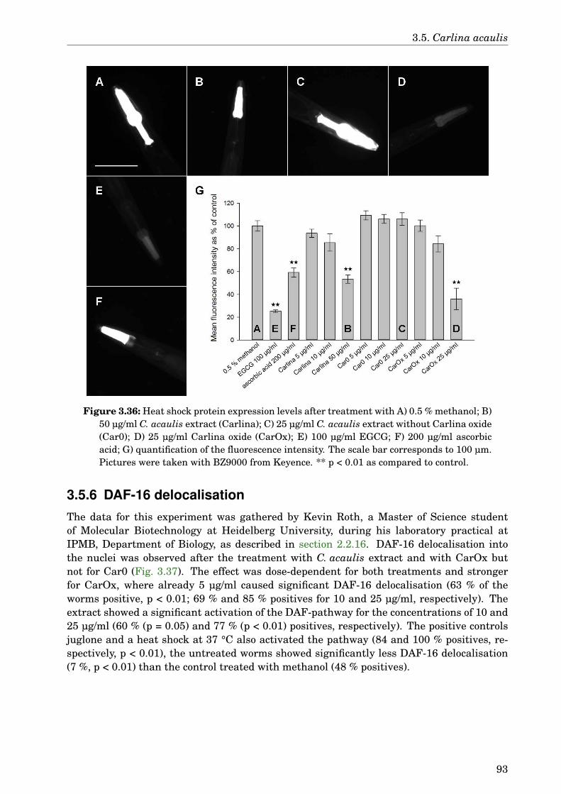
3.5. Carlina acaulis
Figure 3.36: Heat shock protein expression levels after treatment with A) 0.5 % methanol; B)50 µg/ml C. acaulis extract (Carlina); C) 25 µg/ml C. acaulis extract without Carlina oxide(Car0); D) 25 µg/ml Carlina oxide (CarOx); E) 100 µg/ml EGCG; F) 200 µg/ml ascorbicacid; G) quantification of the fluorescence intensity. The scale bar corresponds to 100 µm.Pictures were taken with BZ9000 from Keyence. ** p < 0.01 as compared to control.
3.5.6 DAF-16 delocalisationThe data for this experiment was gathered by Kevin Roth, a Master of Science studentof Molecular Biotechnology at Heidelberg University, during his laboratory practical atIPMB, Department of Biology, as described in section 2.2.16. DAF-16 delocalisation intothe nuclei was observed after the treatment with C. acaulis extract and with CarOx butnot for Car0 (Fig. 3.37). The effect was dose-dependent for both treatments and strongerfor CarOx, where already 5 µg/ml caused significant DAF-16 delocalisation (63 % of theworms positive, p < 0.01; 69 % and 85 % positives for 10 and 25 µg/ml, respectively). Theextract showed a significant activation of the DAF-pathway for the concentrations of 10 and25 µg/ml (60 % (p = 0.05) and 77 % (p < 0.01) positives, respectively). The positive controlsjuglone and a heat shock at 37 °C also activated the pathway (84 and 100 % positives, re-spectively, p < 0.01), the untreated worms showed significantly less DAF-16 delocalisation(7 %, p < 0.01) than the control treated with methanol (48 % positives).
93
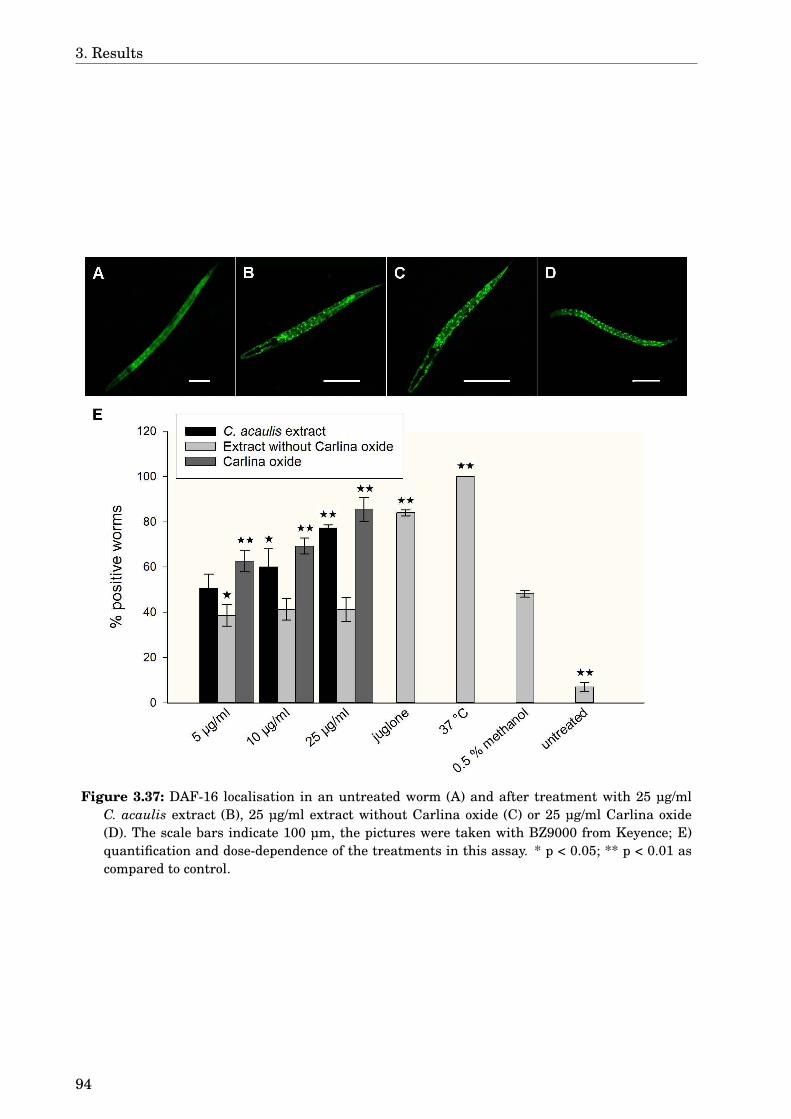
3. Results
Figure 3.37: DAF-16 localisation in an untreated worm (A) and after treatment with 25 µg/mlC. acaulis extract (B), 25 µg/ml extract without Carlina oxide (C) or 25 µg/ml Carlina oxide(D). The scale bars indicate 100 µm, the pictures were taken with BZ9000 from Keyence; E)quantification and dose-dependence of the treatments in this assay. * p < 0.05; ** p < 0.01 ascompared to control.
94
4 Discussion
4.1 Screening of TCM drug extracts
For this screen 25 plants were chosen based on earlier reports of neuroprotective activity. Asa control EGCG was used because of its proven activity in the used C. elegans model (Abbas& Wink, 2010) and known mechanism of action (Bieschke et al., 2010; Ehrnhoefer et al.,2008; Hyung et al., 2013). In the study by Abbas & Wink (2010) 220 µM EGCG loweredthe number of Aβ aggregates by 33 %, which is in good accordance to the 35 % decrease for100 µg/ml (≈ 220 µM) EGCG observed in the present study (Fig. 3.2). Thioflavin T showedan even stronger effect than EGCG in the present study. This substance was reported todecrease amyloid pathology in C. elegans strain CL4176 (Alavez et al., 2011). These wormsdevelop only a few small plaques before they paralyse, which makes it difficult to imagethem, and therefore this strain is usually not used for quantification of Aβ aggregates. Dueto the different methods these results are not directly comparable to the present work, butin both cases thioflavin T treatment led to decrease in the number of Aβ aggregates.
Of the 55 tested extracts 12 methanol and 9 dichloromethane extracts showed significantactivities. The effects of methanol extracts reached a maximum effect of 32 % decrease in Aβdeposits for A. catechu. Two other methanol extracts reduced the number of Aβ aggregatesby at least 25 %, namely G. uralensis (30 %) and A. oxyphylla (25 %). These three extractswere pursued further and are discussed in the following sections. The effect of other extractswas smaller but still noteworthy. S. officinalis and R. laevigata have not had much atten-tion in relation to AD, but both of them decreased Aβ deposition in the C. elegans model.Gallic acid from S. officinalis has shown neuroprotective effects against Aβ toxicity (Banet al., 2008). Although the substances underlying the effect on Aβ aggregation were not fur-ther elucidated, the dichloromethane extract of this plant was not effective, suggesting thatthe active compounds are rather hydrophilic like gallic acid. In case of R. laevigata the onecompound reported to be neuroprotective (1,2-benzenedicarboxylic acid dinonyl ester; Choiet al., 2009) is more lipophilic and would be expected to be contained in dichloromethane ex-tracts. The methanol extract was more effective though, indicating other active substancesin this plant. This result is in accordance with the study of Choi et al. (2006), who showedprotective effects against Aβ toxicity for the methanol extract of R. laevigata in cell cultureand mice.
Four of the seven extracts tested for G. biloba were significantly effective. The seed ex-tracts were more effective than leaves with 22 % decrease in the number of Aβ aggregatesfor the methanol extract of the TCM drug and 20 % for both extracts of raw seeds, althoughonly half the concentration was used for the dichloromethane extract. This indicates thatthere are several active ingredients. Some of them are rather lipophilic and are lost eitherwhen the seeds ripen, when the sacrotesta is removed, or when the nuts are baked to pre-pare bái guo. Other compounds remain in the nuts or are produced during the preparationof bái guo. To our knowledge there are to date no studies of the effects of G. biloba seedsor bái guo on AD published in international journals. The results presented here, however,suggest that the seeds, either raw or prepared, are possibly more potent than the leaves anddeserve further study.
95

4. Discussion
The methanol extract of G. biloba leaves showed moderate activity, decreasing the num-ber of Aβ aggregates by 13 %. The dichloromethane extract as well as the special extractEGb761 had no significant effect. The result for EGb761 was unexpected, since many stud-ies have shown its effectiveness in different models (see Tab. 1.1). Two studies have alsoused the C. elegans model CL2006. Smith & Luo (2003) examined the ROS content in thismodel and found that single compounds found in EGb761 can lower the ROS concentration,but the results for the extract itself were not significant. Wu et al. (2006) performed thesame Aβ aggregation assay, that was also used in the present work, although with somemodifications in treatment protocol and data analysis. The treatment was started at day 4in contrast to day 2 in the present study, the worms were evaluated on day 6 in both studies.Therefore it is possible that EGb761 has some immediate effects that can be seen after twodays, but have vanished after four days. To check this possibility, the experiment should berepeated with different treatment times to compare the effects. Due to limited availabilityof EGb761 this was omitted in the present study.
Two of the most studied plants besides G. biloba in context of cognitive enhancementare P. ginseng and P. notoginseng. While P. ginseng had no effect on Aβ aggregation in thepresent study, both extracts of P. notoginseng had moderate activity. In previous studiesthe compound quercetin 3-O-β-D-xylopyranosyl-β-D-galactopyranoside has been reportedto inhibit Aβ aggregation (Choi et al., 2010). Other substances like notoginsenoside R1 andthe polyacetylenes panaxynol and panaxydol could counteract Aβ toxicity in cell cultureand mouse models (Ma et al., 2014; Nie et al., 2008; Yan et al., 2014). These reports arein accordance with the present study, indicating that P. notoginseng contains several sub-stances that are able to ameliorate Aβ pathology. Since the activity of the extracts was notso high, it would be advisable to identify these active compounds and use them in higherconcentrations. Further studies should reveal the suitability of this plant for patients withAD.
E. senticosus, the Siberian ginseng, showed positive results for both methanol and dichlor-methane extracts. The decrease in Aβ deposits was 15 % for both extracts, but a ten timeslower concentration was used for the dichloromethane extract, so mostly lipophilic com-pounds seem to be the active ingredients. The methanol extract has been tested before onprimary neuron culture, where it could protect the cells from Aβ-induced neurotoxicity (Baiet al., 2011; Tohda et al., 2008). Bai et al. (2011) noted activity against axonal and neuriticatrophy mostly in more hydrophilic fractions, whereas hexane fraction of the extract hadno effect. They isolated some compounds that were responsible for the protective effect likeeleutherosides E and B, isofraxidine, and stigmasterol and sitosterol glycosides. Althoughaglycons of these compounds might be contained in a dichloromethane extract, it seemsmore likely that some other compounds are responsible for the effect on Aβ aggregationobserved in the present study. This underlines the benefits of using plant extracts in a com-plex disease like AD, where several compounds can lead to synergistic effects by influencingdifferent targets.
Significant, although low activity was also observed for the methanol extracts of Phel-lodendron spp. (huáng bai), A. galanga, and Coptis spp. (huáng lían). P. amurense andP. chinense, both species used in the TCM drug huáng bai, have been reported to counteractAβ toxicity in cell culture (Xian et al., 2013). It is possible that this protection is at leastpartly conferred by inhibiting Aβ aggregation. Same can be said for A. galanga that hasshown memory enhancing and antioxidant activities in mice treated with Aβ25–35. Berber-ine, the active ingredient of Coptis spp., has been shown to lower Aβ levels in a mousemodel of AD. This activity was replicated in the present study in the C. elegans model withmethanol extract of huáng lían. The Phellodendron species also contain berberine, although
96

4.1. Screening of TCM drug extracts
in smaller amounts than Coptis spp. (Van Wyk et al., 2015). Therefore, the effect of huángbai extract might be due to this compound, too. Berberine has several effects besides low-ering Aβ levels, including AChE inhibition and should therefore be considered as a possiblelead drug for AD therapy (Ji & Shen, 2011).
C. monnieri yielded the most effective dichloromethane extract that decreased the num-ber of Aβ aggregates by 35 %. The methanol extract of this plant had lower but still signif-icant activity with 18 % decrease. This activity might be due to osthole, a rather lipophiliccoumarine derivative, that would be expected to be contained mostly in dichloromethaneextract. This compound can improve memory in a mouse model of AD via enhancing neu-rogenesis (Liu et al., 2015b). However, in the study of Liu et al. (2015b) no decrease inAβ deposition was detected. Therefore the present study is the first to report this effectfor C. monnieri extracts and further research is needed to identify the active compounds.Two other dichloromethane extracts, namely P. multiflorum and M. officinalis, showed asignificant effect. For both only the dichloromethane but not methanol extract was effective,indicating lipophilic substances as active compounds. In M. officinalis these compoundscould be magnolol and honkiol, that have been reported to counteract Aβ-induced toxicitybefore (Hoi et al., 2010). For P. multiflorum water and ethanol extracts have been studiedearlier in context of Aβ toxicity (Liu et al., 2015c; Um et al., 2006). The water extract hada memory enhancing effect in mice and inhibited AChE (Um et al., 2006). The ethanol ex-tract and its active compound 2,3,5,4’-tetrahydroxystilbene-2-O-β-D-glucoside were testedon a Drosophila model, where they could counteract Aβ toxicity, but no effect on Aβ levelswas observed (Liu et al., 2015c). The lipophilic compounds, shown here to act against Aβaggregation, complement the actions of hydrophilic compounds, making P. multiflorum aninteresting plant for AD research.
The most effective dichloromethane extract (C. monnieri) had stronger activity than themethanol extracts with only tenth of the amount used for the treatment. This might in-dicate that lipophilic compounds are more active then the hydrophilic substances. Theyare able to penetrate biomembranes and could therefore have higher bioavailability. How-ever, since the culture medium for C. elegans is water based, these substances are poorlysoluble and difficult to test. This might account for the fact that there are more methanolextracts showing significant activity. On the other hand, several dichloromethane extractsalso lead to an increase in the number of Aβ aggregates. It has to be considered that thesolvent DMSO, in which these extracts were solved, has itself a plaque decreasing effect(Fig. 3.3). This observation is in accord with a study showing that 0.8 % DMSO can increasethe lifespan of C. elegans and even lower concentrations reduce paralysis in CL4176 worms(Frankowski et al., 2013). To account for additive effects, all extracts were compared withthe respective concentration of DMSO as control, but a possible synergistic interaction cannot be excluded. Interactions counteracting the effect of DMSO could also explain the appar-ent increase in number of Aβ aggregates for extracts that have no anti-aggregation activity.Due to the complications with dichloromethane extracts and the fact that there were morehighly active methanol extracts, further work concentrated on the latter.
Some of the plants tested here were reported to have effects on Aβ levels or toxicity inearlier studies (see Tab. 1.1) but showed no activity in the C. elegans model. Many of theother studies tested pure compounds rather than extracts, which might explain the differ-ent results. The content of active compounds in the extract is often low, so their effects canbe missed. Most studies working with extracts used water as a solvent, hence these extractsalso have different composition compared to methanol and dichloromethane extracts usedin the present study, making them not directly comparable. Another difference is also themodel used. Compared to cell culture C. elegans is a more complex model. Some substances
97

4. Discussion
might be active on cells, but fail in the C. elegans model due to low bioavailability, intesti-nal metabolism, or other factors that are not present in cell culture. On the other hand,mice and rats are much more complex animals than a nematode like C. elegans. They havedeveloped organs, cell types, and signalling cascades that are not present in nematodes.Therefore, the C. elegans model is only suitable for testing direct interactions with Aβ oreffects on basic, evolutionary conserved signalling pathways. To detect interactions withspecific targets that are only found in vertebrates, other models are needed. These differ-ences in the methodology have to be considered when comparing different studies and areprobably responsible for the differing results.
4.2 Areca catechuA. catechu is known for its antiparasitic and cholinomimetic activities, but to date there areno studies about its effects on amyloid aggregation. In the present study both methanoland dichloromethane extracts showed high reduction in number of Aβ aggregates in theC. elegans model. The methanol extract, that was studied further, showed a dose-dependenteffect (Fig. 3.5), but concentrations higher than 100 µg/ml could not be tested due to toxicityfor the worms. A. catechu contains mainly alkaloids and polyphenols. To clarify, which ofthese groups is responsible for the effect, arecoline as the main alkaloid was also testedin the Aβ aggregation assay. It showed no significant activity for concentrations up to125 µg/ml (data not shown), suggesting that the effect of the extract is due to the polyphe-nols or some other minor compound.
To examine whether A. catechu can also counteract Aβ toxicity, the paralysis assay withthe C. elegans strain CL4176 was conducted. 100 µg/ml of the extract were used, since thisconcentration was effective in the first experiment and showed no toxic effects. However, inthe paralysis assay this treatment significantly shortened the time until paralysis (Fig. 3.6)and also some worms of the control strain CL802, that do not express Aβ, appeared paral-ysed after the treatment. The CL4176 and CL802 strains seem to be more sensitive to thetoxicity of A. catechu, possibly because the worms were treated earlier in this assay. Theextract might interact with important processes during development of the worms, makingthem more vulnerable to the Aβ toxicity or toxic substances in general. Another possibilityis that the extract and Aβ both interact with the same target in CL4176, leading to an ad-ditive or even synergistic toxicity. A direct interaction with Aβ is also possible and wouldlead to higher toxicity, if the toxic oligomers were stabilised and further aggregation to lesstoxic plaques prevented. In this case, however, the toxicity should have also been observedin the CL2006 worms.
The organoselenium compound diphenylselenide, that has shown several positive effectsin different AD models, also increased the paralysis rate in C. elegans (Zamberlan et al.,2014). Zamberlan et al. (2014) showed, that the effect on paralysis is due to muscle inhibi-tion via increased AChE activity. A. catechu has cholinomimetic activity and might inhibitAChE (Gilani et al., 2004). Diphenylselenide has shown AChE-inhibitory activity as wellin mammalian models, but activated the enzyme in C. elegans (Zamberlan et al., 2014).Whether a similar effect could be the cause of increased paralysis after treatment withA. catechu needs further testing. Until then other causes of toxicity cannot be excluded.
The toxicity could have been prevented by using lower concentrations of the drug. Inthe Aβ aggregation assay the concentration 75 µg/ml also showed a significant decreasein the number of deposits, but the effect size was only a half of the higher concentration.Therefore, lowering the concentration would lead to loss of therapeutic effect. If the extracthas an effect on larval development, treating the worms later, when they are fully developed,
98
4.3. Alpinia oxyphylla
would also solve the problem. This would be a mechanism specific for C. elegans that is notrelevant for AD therapy. In the case of an effect on acetylcholinergic neurotransmission, theeffect might also be specific for C. elegans and further studies in mammals are required toevaluate it. In the present study the work with A. catechu was discontinued in favour of theless toxic extract of G. uralensis.
For possible usage of A. catechu in AD therapy it would be important to further charac-terise the toxicity of this plant. The effects seen in C. elegans might be specific for nematodesand not affect humans or they might be more general. Arecoline has been shown to causeneurotoxicity (Shih et al., 2010), to be cytotoxic, and chewing the seeds can cause oral can-cer (Peng et al., 2015). On the other hand, a small study on patients with mild to moderateAD reported no adverse effects for low doses of arecoline that improved the memory of re-sponders (Soncrant et al., 1993). Arecoline acts as a cholinomimetic but had no effect on Aβaggregation. Therefore, the role of polyphenols and other constituents of Areca nuts in thebeneficial and toxic activity of the extract should be further clarified. If the toxic compoundscan be separated from the beneficial ones, the development of a modified extract or usingpure compounds would be advisable.
4.3 Alpinia oxyphyllaThe methanol extract of A. oxyphylla decreased the number of Aβ aggregates by 25 %, beingthe third most effective methanol extract in the present study. The chloroform fraction ofan ethanol extract has been reported before to decrease Aβ levels in mice (Shi et al., 2014).This extract was rich in sesquiterpenes like nootkatone, had antioxidant effects, and inhib-ited AChE. The decrease in Aβ aggregates shown in the present study is in accordance withthese results. The methanol extract probably contains at least some of the sesquiterpenesbut also other constituents like protocatechuic acid and 5-hydroxymethylfurfural. Both lat-ter substances have been shown to decrease Aβ levels and have other neuroprotective prop-erties (Liu et al., 2014; Song et al., 2014; Zhang et al., 2015). Based on this initial positiveactivity the extract of A. oxyphylla was studied further.
The paralysis assay, however, did not show a significant difference between treatmentwith the methanol extract of A. oxyphylla and control (Fig. 3.7). There was also no signifi-cant difference between control and treatment with EGCG (solved in methanol), in contrastto the results obtained with EGCG solved in water (Fig. 3.22). The effect of EGCG on theCL4176 strain has only been investigated in one study before (Brown et al., 2006). There thecompound was solved in ethanol and a small, not significant effect in the paralysis assay wasobserved. A tendency to longer time until paralysis was also observed in the present studyfor EGCG solved in methanol. The EGCG solved in water, on the other hand, had a strong,significant effect. In the light of this data it seems possible that the used solvent modulatesthe activity of EGCG. The main mode of action for EGCG as a polyphenol is building un-specific hydrogen bonds with proteins. The possible interaction with solvent molecules andits impact on the hydrogen bond building ability of EGCG would be an interesting topic forfurther studies.
Assuming, that the lack of activity for the positive control EGCG was because of a spe-cific solvent effect that does not affect other substances, then the results indicate that themethanol extract of A. oxyphylla has no activity against Aβ toxicity. It is possible, that thecompounds in this extract only interact with Aβ fibrils, but not the oligomers that causetoxicity in CL4176. However, other studies have shown that this plant can ameliorate cog-nitive deficits in mice treated with Aβ1–42 (Liu et al., 2014; Shi et al., 2014). The lack of acorresponding effect in the present study might be due to a different model organism. It is
99
4. Discussion
also possible, that a higher dose of the extract or a pure active compound is needed to see aneffect. Given the promising activity of A. oxyphylla in the Aβ aggregation experiment andits low toxicity compared to some other plants, e.g. A. catechu, the possible beneficial effectsof this plant on AD pathology should be studied more intensively in the future.
4.4 Glycyrrhiza uralensis
4.4.1 Determination of the used Glycyrrhiza speciesThe TCM drug gan cao can contain three different species of Glycyrrhiza: G. uralensis,G. glabra, and G. inflata. Therefore, DNA barcoding was used to determine the exact species.The comparison of ITS sequences from the drug and sequences from known species fromdatabase showed the highest similarity with G. uralensis (Tab. 3.1). The ITS sequencedoes not differ between G. glabra and G. inflata, but compared to G. uralensis there are twovariable sites: one single nucleotide polymorphism in ITS1 and a substitution of three nu-cleotides in ITS2 (Kondo et al., 2007b). Hence, in this case the analysis of the ITS sequencewas sufficient for the identification.
DNA of the same plant material has been used before to analyse the chloroplast gene forribulose bisphosphate carboxylase large chain (rbcL) (Herrmann et al., 2011b). According toKondo et al. (2007b), there are two genotypes for rbcL in Glycyrrhiza species. The first oneis found in all three species (G. glabra, G. inflata, G. uralensis), the second one (GenBankaccession number AB012126) was only found in G. uralensis in the study of Kondo et al.(2007b). In contrast to that, Hayashi et al. (2000) reported that the rbcL sequence is iden-tical in G. uralensis and G. inflata (GenBank accession numbers AB012126 and AB012127,respectively). Herrmann et al. (2011b) reported the Glycyrrhiza species used to be G. inflata(GenBank accession number JF950025). The two rbcL genotypes differ by two nucleotidesubstitutions at the bases 706 and 736 (based on the sequence AB012126). By comparingthe sequences from Kondo and Hayashi to the sequence reported by Herrmann, it occursthat the latter sequence only contains 698 base pairs and is lacking the essential bases.Hence, this sequence cannot be used for identification.
The identity of the plant material as G. uralensis is also supported by the phytochemicalanalysis. Glycycoumarin, a compound specific for the species G. uralensis (Kondo et al.,2007a), was found in the methanol extract of the plant (peak 12 in Fig. 3.9, Fig. 3.16)whereas no masses corresponding to glabridin (324 g/mol) or licochalcone A (338 g/mol)were observed (Tab. 3.3). Also the ratio of flavanones to chalchones seems to match mostclosely the conditions reported for G. uralensis (Kondo et al., 2007a; Simmler et al., 2015),although this ratio was not quantitatively determined in the present study. Altogether, boththe genetic and phytochemical analysis point to G. uralensis, and this result is also consis-tent with the fact that the drug was purchased in China, where G. uralensis is the mostcommon species.
4.4.2 Glycyrrhiza uralensis and its major compounds decrease Aβ
aggregationThe methanol extract of G. uralensis decreased the number of Aβ aggregates in the initialscreening by 30 % (Fig. 3.2) and was therefore chosen for further study as one of the mostpotent extracts. The dichloromethane extract had no significant activity, possibly becausedue to poor solubility only 25 µg/ml of this extract could be used or because of the differ-ent composition of the extracts. Water and ethanol extracts have been studied before on
100
4.4. Glycyrrhiza uralensis
mice and primary neuron culture (Ahn et al., 2006; Lee et al., 2012b). Memory enhancingand neuroprotective activities were reported, but the effects on Aβ aggregation were notinvestigated in these studies. The compound LG has been shown to reduce Aβ secretionfrom neurons and ILG can inhibit Aβ aggregation by binding to this peptide (Chen et al.,2013; Liu et al., 2009). The phytochemical analysis of the extract showed that the methanolextract—in contrast to the dichloromethane extract (Fig. 3.8)—contains glycosylated formsof these compounds (peaks 1–4 in Fig. 3.9). Therefore, these substances could be responsiblefor the effect seen for the extract.
Quantification of the compounds in the different extracts showed, that 54–64 µg/ml GAis present in water and methanol extracts, 9 µg/ml of LG were found in methanol extract,the other studied compounds were present only in traces (Tab. 3.2). The dichloromethaneextract contained only trace amounts of all four compounds in correlation with the lackof significant activity in the aggregation assay. In the LC-MS/MS analysis the aglyconscould not be recovered, but liquiritin, liquiritin apioside, isoliquiritin, isoliquiritin apioside,and GA were present in high amounts (Fig. 3.9, Fig. 3.10). Glycosides often function asprodrugs that have to be activated by bacteria in the gut. It is known that the active formof GA in humans is GRA that is produced form GA by intestinal bacteria (Hattori et al.,1983; Krähenbühl et al., 1994; Monder et al., 1989). The C. elegans model system usedhere contains living E. coli as a food source for the worms. These bacteria probably alsometabolise the extracts, so it can be expected that the worms are exposed to more aglyconsthan originally present in the extract. Therefore, although only trace amounts were foundin the extract, the high amount of glycosides can lead to higher concentrations of LG, ILG,and GRA, and these compounds can significantly contribute to the activity of the extract.
Most of the saponins found in the methanol extract are derivatives of GRA. Assumingthat the sugar residues are removed by the bacteria, the aglycon GRA should be represen-tative for possible activity of these compounds. Besides the already mentioned substances,isoprenylated flavonoids and coumarins were found. Glycycoumarin has shown protectiveactivity against Aβ toxicity before (Kanno et al., 2015), but since only a small amount ofcoumarins was found in the active extract, it is unlikely that these are the main active con-stituents. Of the isoprenylated flavonoids only isoangustone A could be identified (Tab. 3.3,Fig. 3.18) and there are to date no reports about this substance interacting with Aβ. There-fore, GA as the major compound (peak 8 in Fig. 3.10) and the aglycons LG, ILG, and GRAwere chosen for further study.
In the Aβ aggregation assay in CL2006 GA showed a significant effect only for the highesttested dose (100 µg/ml), the other substances were already active at 50 µg/ml, decreasingthe number of Aβ aggregates by up to 26 % (Fig. 3.20). LG and ILG had slightly higheractivity than the other compounds. These results are in accordance with earlier reports(Chen et al., 2013; Liu et al., 2009), but they also suggest that LG and ILG are not the onlyactive compounds in G. uralensis in respect of Aβ aggregation. For further experiments allfour compounds were tested in addition to the methanol extract.
4.4.3 Glycyrrhiza uralensis and isoliquiritigenin counteract Aβ
toxicity
Protective effects against Aβ-induced neurotoxicity have been reported before for differentG. uralensis extracts, ILG, and LG (Ahn et al., 2006; Lee et al., 2012b; Liu et al., 2009). Herethe effects of methanol and water extract as well as the pure compounds GA, GRA, LG, andILG were tested in C. elegans models expressing Aβ in muscles and neurons. The paraly-sis assay with the strain CL4176 produced the most significant results. Both G. uralensis
101

4. Discussion
extracts and ILG delayed the time until paralysis (Fig. 3.22). The control substance EGCGwas also active in this assay and had the highest activity of all tested compounds with 2.7 hdelay in PT50. This activity is much stronger pronounced than the moderate delay reportedby Brown et al. (2006). As discussed in section 4.3, there might be an effect of solvent modi-fying the action of EGCG.
Regarding solvent effects, it was also observed that the treatment with 1 % methanol alonehad a significantly different PT50 compared to the water-treated control. This effect wasdose-dependent, since there was also a significant difference between 0.5 and 1 % methanol(Fig. 3.33; p < 0.01). That confirms the observation made by the treatment with A. catechuextracts (see section 4.2) that the CL4176 strain is more sensitive than CL2006. Methanolhad no effect on the latter strain in concentrations up to 1 % (data not shown). In contrast toA. catechu, the effect observed for methanol was not toxic but beneficial. The solvent alonecould delay paralysis to the same extent as EGCG. Possibly this activity was mediated bya hormetic effect of early life exposure to a low concentration of a toxin, since methanol hasshown hormetic effects at concentrations below 1 % before (Qin et al., 2010). To account forthis effect, all treatments were compared to the respective solvent control.
From the two G. uralensis extracts tested, the water extract showed higher activity. At aconcentration of 200 µg/ml it led to the same delay in paralysis as 500 µg/ml of the methanolextract. The water extract contains slightly more saponins than the methanol extract (com-pare peaks 5, 6, and 7 in Fig. 3.10), so this could be the cause of higher activity. However,the only saponin tested as a pure compound—GA—showed no difference to the control.Saponins have an amphiphilic structure which allows them to interact with biomembranesand increase their permeability. Therefore, if saponins are not the active ingredients them-selves, they can still lead to higher activity of an extract by increasing the bioavailability ofthe active compounds. Same can be said for the aglycon GRA which showed only minimal,non-significant activity in this assay. Although this molecule has no hydrophilic sugars, thelipophilic triterpene structure can still interact with the lipids and cholesterol molecules inthe biomembrane.
ILG was the only pure compound with significant activity in this assay. This result is inaccordance with an earlier report of a protective effect against Aβ-induced neuronal death(Lee et al., 2012b). Similar activity has been reported for its isomer LG (Liu et al., 2009).In the paralysis assay, however, LG rather shortened the time until paralysis, althoughthis effect was not significant. The worms of the control strain CL802 did not react to thetreatment with LG, suggesting that this compound is not toxic in general. ILG has beenreported to interact directly with Aβ (Chen et al., 2013) and similar interactions have beenobserved for other flavonoids (Bu et al., 2015). ILG and LG are polyphenols like EGCG, sothey share a possible mechanism of action. ILG has one more hydroxy group compared toLG, allowing it to build more hydrogen bonds. Hence, if this is the mechanism of action, ILGwould be expected to be more potent. This, however, does not explain the opposite activity ofLG. Possibly, the unspecific binding via hydrogen bonds is not the only mechanism of actionfor these flavonoids in the paralysis assay.
The common properties these flavonoids share with EGCG also suggest, that if the sol-vent affects the action of EGCG (section 4.3), this would also be the case with ILG and LG.The effects of methanol and EGCG are clearly not additive, but rather lead to an abolish-ment of any visible effect (Fig. 3.7). If the same happens to ILG and LG, then the smallnegative effect of LG could be interpreted as an abolishment of the methanol effect. It wassuggested, that the latter arises from a hormetic response. In this case the antioxidant andneuroprotective effects of LG could protect the worms from methanol toxicity and preventthe induction of hormesis. The same should happen with ILG, but apparently ILG has a
102

4.4. Glycyrrhiza uralensis
higher protective capacity, so that the delay in paralysis still reaches significance above themethanol control.
Aβ is degraded via autophagy in the CL4176 worms and this process is regulated by thetranscription factors HSF-1 and DAF-16 (Cohen et al., 2006; Florez-McClure et al., 2007).Therefore, ILG or other compounds in the G. uralensis extract can lower its toxicity also byinteracting with proteins involved in autophagy or its regulation. Fluoxetine, a selectiveserotonin reuptake inhibitor, has been shown to delay paralysis in this model dependent onDAF-16 activation and serotonin (Keowkase et al., 2010). ILG induced DAF-16 delocalisa-tion in the present study (Fig. 3.28), indicating that this transcription factor may be involvedin the mechanism of action for this compound as well. However, G. uralensis extract failedto activate DAF-16 significantly, although it could delay paralysis. Therefore, there must beother mechanisms underlying its activity.
ILG and LG can modulate serotonin and norepinephrine concentrations (Wang et al.,2008) and several compounds in G. uralensis have antioxidant activity. These properties,besides the direct interaction with Aβ peptide, could counteract the downstream toxic ef-fects triggered by Aβ. To further elucidate the mechanisms behind the effects seen in theparalysis assay, the antioxidant activity in C. elegans (discussed in section 4.4.4) and theeffects on behaviour in the strain CL2355, that expresses Aβ in neurons, were investigated.
Neuronal toxicity in CL2355 leads to impaired chemotaxis behaviour. As can be seen inFigure 3.23, the control strain PD8120 has a CI of about 0.4, whereas CL2355 worms showno chemotaxis at all. This defect is rescued to some extent by the treatments, most signifi-cantly by GRA. GRA also showed a small protective effect in the paralysis assay, although itwas not statistically significant. Still, these results suggest, that GRA could have protectiveproperties against Aβ toxicity, maybe at higher concentrations. It is also evident, that thechemotaxis behaviour of the control strain is affected as well. This indicates, that the sub-stances have unspecific effects on the behaviour that are not related to Aβ expression. Thisassay is very sensitive to all environmental factors, leading to high standard deviations.Therefore the effects of G. uralensis extract, ILG, or EGCG are not significant, althoughthere is a tendency for higher CI in CL2355 worms after these treatments (p É 0.1).
In the serotonin sensitivity assay an unexpected hyposensitivity in CL2355 worms treatedwith methanol was observed (Fig. 3.24). By control treatment with water there was no dif-ference between CL2355 and the control strain. It has been reported before that the CL2355strain is hypersensitive to serotonin (Wu et al., 2006). In later experiments this effect wasalso observed in our lab (Felix Heiner, PhD student at Heidelberg University, IPMB, Depart-ment of Biology, personal communication). To date there is no logical explanation, why theworms behaved differently during the present study. However, there is a difference in sensi-tivity to serotonin between CL2355 and the control strain, when the worms are treated withmethanol. Therefore, the results were evaluated as positive, if a treatment could reduce thisdifference.
Significant activity was observed for EGCG, LG, and ILG, suggesting that these sub-stances can normalise the serotonergic neurotransmission in this model. ILG and LGhave been reported before to modulate serotonin concentrations in mice, resulting in anantidepressant-like effect (Wang et al., 2008), and the antidepressant fluoxetine delays Aβ-induced paralysis in C. elegans dependent on serotonin (Keowkase et al., 2010). Similarly,green tea extract containing EGCG has been shown to elevate serotonin concentration inhuman plasma (Hodgson et al., 2014). Therefore, the effect on serotonergic transmissionreported here could also be involved in the mechanism of action of the active compounds incounteracting Aβ toxicity in the paralysis assay.
103

4. Discussion
The compounds GA and GRA can significantly suppress the activity of both the Aβ ex-pressing strain and the control strain. This indicates, that the effect is not specificallyrelated to Aβ. It also suggest that these substances have a general effect on neurotrans-mission or the enhanced slowing response in C. elegans. The inhibition of gap junctions byGRA (Davidson & Baumgarten, 1988) may play a role in this effect. The muscle cells ofC. elegans are connected by gap junctions (White et al., 1986), therefore GRA could disturbsignalling between these cells, leading to reduced mobility. However, such an effect was notobserved in other assays following treatment with GRA. On the contrary, in paralysis assaythis compound slightly delayed paralysis. Therefore, effects related to serotonin cannot beruled out and may play a role in the small protective effect of GRA observed in the otherassays.
In summary, it can be said that ILG was the most potent substance in counteracting Aβtoxicity. It had a highly significant activity in paralysis and serotonin sensitivity assaysand a positive tendency in enhancing chemotaxis. Serotonergic neurotransmission mayplay a role in this protective effect. The extracts of G. uralensis as well as the substancesLG and GRA also showed protective effects, although smaller than the effect seen for ILG.G. uralensis extracts probably contain several protective compounds that may have additiveor synergistic activitiy. ILG as one of the more potent protectors deserves further investi-gation, weather as pure compound or in combination with other active substances found inG. uralensis.
4.4.4 Isoliquiritigenin has antioxidant activity via activating DAF-16
Oxidative stress is one of the important characteristics of AD and ageing in general. Aβincreases the production of ROS (Huang et al., 1999; Opazo et al., 2002). Therefore, one wayto counteract Aβ toxicity downstream of the peptide aggregation is antioxidant therapy.The antioxidant capacity of G. uralensis extract and its active compounds in C. elegans wasinvestigated using two assays. In the HSP expression assay the worms are exposed to lowdoses of the prooxidant juglone that induces mild oxidative stress. The survival assay is amore rigorous test of antioxidant activity, where the worms are subjected to lethal oxidativestress by high concentrations of juglone.
In the HSP expression assay all substances except the lower concentration of G. uralensisextract and GRA showed a moderate activity (Fig. 3.26). As a positive control this ex-periment was also conducted with ascorbic acid, a well known antioxidant, that exhibiteddose-dependent antioxidant activity with up to 55 % reduction in fluorescence intensity(Fig. 3.25). The control substance EGCG reached similar values in accordance with earlierstudies in C. elegans (Abbas & Wink, 2009, 2010). The effects of other substances were muchlower in this assay. In the survival assay only ascorbic acid, EGCG, and the flavonoids LGand ILG showed a significant effect.
From all test substances ILG had the strongest effect in both assays. This is the firstreport of antioxidant activity of ILG in C. elegans, although such effects have been reportedbefore in cell culture systems (Gong et al., 2015; Lee et al., 2012b). In HSP expression assaythe fluorescence was decreased only by 26 %, whereas in the survival assay ILG increasedthe survival rate by 82 %, being even more effective than EGCG. EGCG showed a strongantioxidant effect in both assays. This illustrates, that both compounds act via differentmechanisms. EGCG is a radical scavenger, it can directly react with the ROS generated byjuglone and thereby prevent damage to the cells. ILG has shown radical scavenging activityin in vitro assays as well (Chin et al., 2007; Vaya et al., 1997), but due to the differencesin structure it is a less potent radical scavenger than EGCG. Instead it can activate the
104
4.4. Glycyrrhiza uralensis
cells own protective mechanisms via the transcription factor NRF2 (Gong et al., 2015). Thispathway may be combined with the activation of other protective mechanisms like HSPexpression, so that the effect is not so strongly seen in the HSP-expression assay, but ishighly effective under severe oxidative stress as in survival assay. Since the activity ofHSF-1, that controls the expression of HSPs, is necessary to counteract Aβ toxicity (Cohenet al., 2006), the rather small effect of ILG in suppressing HSP expression may be relatedto the effective delay of paralysis in CL4176.
LG had only a small effect in the antioxidant assays. The antioxidant activity of LG andILG has been attributed to the activation of NRF2. Wang et al. (2015) have shown, that ILGhas a much stronger effect on the expression of genes that are under the control of NRF2than LG. This correlates well with the results of antioxidant activity in C. elegans and alsowith the results in Aβ toxicity assays. NRF2 and its C. elegans homologue SKN-1 regulatethe expression of stress related genes that could also be involved in detoxification of the Aβoligomers. In AD patients the function of NRF2 has been shown to be impaired (Mota et al.,2015). Since ILG can activate NRF2 and also influences the expression of some antioxidantgenes independent of NRF2 activation (Wang et al., 2015), this compound could be usefulfor counteracting oxidative stress in early stages of AD.
In C. elegans SKN-1 activation is regulated by the same kinases that also control DAF-16and both transcription factors share some of their target genes (Tullet et al., 2008). Theeffect of G. uralensis and its compounds on DAF-16 nuclear translocation was studied usingthe C. elegans strain TJ356. As shown in Fig. 3.28, the treatment with ILG led to activationof DAF-16, other Glycyrrhiza compounds and the extract had no significant effect. EGCGshowed also no significant activity in both tested solvents, although it has been reported tobe active before. Methanol alone had an effect in this assay, therefore low activities of somecompounds like LG might have gone unnoticed.
Treatment with GRA or GA did not lead to nuclear localisation of DAF-16 although theyare reported to modulate the activity of PI3K, the human homologue of AGE-1 (Kao et al.,2009, 2013; Wang et al., 2011). In C. elegans inhibition of AGE-1 leads to longer lifespanof the worms which is dependent on DAF-16 (Lin et al., 1997; Morris et al., 1996). Kaoet al. (2009, 2013) have reported activation of PI3K by GA and GRA. This is in accordancewith the result that no DAF-16 activation was observed for these treatments in C. elegans.Wang et al. (2011) reported inhibition of PI3K in active macrophages, contributing for theanti-inflammatory activity of GA and GRA. This shows, that this effect is dependent onthe inflammatory state of the organism. Since in the present study no inflammation wasinduced in the worms, the treatment did not lead to DAF-16 activation. This result is alsoin line with the lack of antioxidant activity for GA and GRA.
The controls juglone and heat treatment in this test are active because they cause stressto the worms. This rises the question, weather the activity of ILG indicates a protective ora toxic effect? DAF-16, when active, prolongs the lifespan of C. elegans (Kenyon et al., 1993)and activates the expression of stress resistance genes (Lee et al., 2003; McElwee et al.,2003; Murphy et al., 2003). This activity is triggered by environmental stimuli that suggestthe need of such protection—stressors and toxins (Henderson & Johnson, 2001). Therefore,it is a matter of concentration weather a toxin overpowers the protective effects and killsor damages the worm or is warded off effectively. In the latter case it comes to a hormeticresponse—a low concentration of the toxic substance leads to higher fitness of the organism,making it more resistant to stress and, at least in C. elegans, prolonging its life. Both heatand mild oxidative stress induced by juglone have been shown to trigger a hormetic response(Cypser & Johnson, 2002).
105
4. Discussion
ILG was shown to be protective against the proteotoxicity of Aβ aggregates and againstoxidative stress in the previously discussed experiments. Hormesis is a frequent mechanismof action for phytochemicals that is especially important for healthy ageing (Murugaiyah& Mattson, 2015; Son et al., 2008). Hormetic pathways include transcription factors likeNRF2, that has been shown to be activated by ILG before, or FOXO. Therefore, it can beassumed that at the used concentration ILG evokes a hormetic response by activating theC. elegans homologue of FOXO, DAF-16. Furthermore, this may be the mechanism of actionalso in other organisms, since NRF2 activation has been reported in mice and human cellcultures (Gong et al., 2015; Wang et al., 2015). To test this hypothesis, a dose-responseanalysis with a wide range of doses should be conducted.
Prolonged activity of FOXO can lead to AD-like symptoms (Manolopoulos et al., 2010).This is in contrast to the positive effects seen for ILG via activation of DAF-16. In the exper-iments conducted for the present work, the worms were treated with the substances once,so the activation of DAF-16 was only transitional. It is possible that in an organism withconstitutive stress and FOXO/DAF-16 activation an additional induction of this transcrip-tion factor has no beneficial effect (see section 4.4.5). If the activity of ILG is mediated bythe suggested hormetic effect, it is probably better suited for prevention than treatment offully developed dementia. However, the role of insulin signalling and FOXO in AD is notentirely established yet. Therefore, further studies with ILG in mammalian models areneeded, to test the effect on FOXO, probable interactions with other signalling pathways,and the resulting consequences on AD.
4.4.5 Toxicity of long-term treatment with Glycyrrhiza uralensis
DAF-16 activity leads to longer lifespan in C. elegans. To test, if G. uralensis and its com-pounds are able to prolong life, lifespan assays were conducted with wildtype worms andCL2006. The experiment was discontinued after the observation that the compounds rathershortened the lifespan in the first run (Fig. 3.30). This effect was dose dependent for theG. uralensis extract and more pronounced in wildtype than in CL2006 worms. Possibly theprotective effect of this extract against Aβ aggregation and toxicity has some benefits in theCL2006 worms that can counterbalance the toxicity resulting in shorter lifespan.
It is also notable that the lifespan of control worms was about twice as long than thethree weeks reported before for worms growing at 20 °C (Klass, 1977). Several factorslike temperature or food restriction can influence the lifespan of the worms but could beexcluded as a cause for lifespan extension here. One possible explanation that could bedetermined was a measurable electromagnetic field in the incubator that led to DAF-16activation in TJ356 worms (data not shown). C. elegans contains biogenic magnetite andstatic magnetic fields have an effect on gene expression of proteins related to developmentand ageing (Cranfield et al., 2004; Hung et al., 2010). Weather the long lifespan observedwas related to the electromagnetic field was not further investigated. Another possibilityis, that the treatment with 1 % methanol prolonged the lifespan. To our knowledge theeffects of this solvent on C. elegans lifespan have not been studied before, but as discussedabove, low methanol concentrations have an hormetic effect (Qin et al., 2010). Thereforethis treatment could affect stress resistance and lifespan.
From the pure compounds ILG shortened the half-time of life by 7 days and the maxi-mum lifespan by 18 days in the wildtype worms. Its effect in CL2006 was smaller, but stillstronger than for other treatments. LG had a similar effect in CL2006 but was closer tothe control in the wildtype worms. GA and GRA were rather neutral in CL2006, althoughGRA shortened the lifespan in wildtype worms by 3.5 days. In wildtype this pattern resem-
106
4.4. Glycyrrhiza uralensis
bles the activity in DAF-16 delocalisation assay: ILG had the highest activity, followed byLG and GRA, GA had no effect. Hence, the toxicity might be related to prolonged activa-tion of DAF-16, similar to the effects observed for FOXO (Manolopoulos et al., 2010). ILGand LG had an effect against Aβ toxicity and showed antioxidant activity upon a singletreatment, whereas GA and GRA did not. ILG and probably also LG act at least in partvia hormetic pathways as discussed in section 4.4.4. Therefore, long-term treatment withthese compounds would lead to constant activation of stress-related transcription factorslike DAF-16/FOXO. That could explain why these compounds are more toxic, especially inthe CL2006 strain that is already under stress because of Aβ toxicity, compared to the othercompounds.
The results of the lifespan assay demonstrate, that long term treatment with high doses ofG. uralensis or ILG might be toxic. This might be related to prolonged activation of DAF-16,but it could also have other causes. Therefore this possible toxicity needs further investiga-tion to determine the concentration and time range in which these treatments are non-toxic.It would be also important to establish, weather this toxicity is specific to C. elegans or isalso found in other species. Liquorice has been used in confectionery and as a food additivefor a long time without adverse effects in humans. However, a medicinal preparation withhigh concentration of the plant extract or a pure compound that is used regularly over along period of time might have different toxicological effects.
4.4.6 Is isoliquiritin a viable drug candidate?
The results for ILG in the present study prove efficacy against Aβ aggregation, Aβ toxicity,and oxidative stress that are prominent factors in the pathogenesis of AD. These resultsare in accordance with earlier studies in vitro as well as on cell cultures and mice (Chenet al., 2013; Gong et al., 2015; Lee et al., 2012b; Vaya et al., 1997). Additionally, ILG hasanti-inflammatory (Kim et al., 2008b; Park & Youn, 2010; Takahashi et al., 2004; Wanget al., 2015) and anti-diabetic properties (Gaur et al., 2014; Ma et al., 2015b) that wouldhelp against inflammation and insulin resistance in AD. The antidepressant-like activityreported for ILG (Wang et al., 2008) could also be beneficial, since depression is one of themost frequent co-morbidities in patients with AD. Taken together, these evidence showreasonable efficacy and suggest ILG for further drug development.
Next to the efficacy bioavailability is an important factor in developing a drug. ILG hasshown good permeability in a human colon carcinoma cell line (Caco-2) and a blood-brain-barrier model (Yang et al., 2014). The permeability resulted mostly from passive diffusion.Studies in rats and humans with formulations containing ILG have reported low plasmaconcentrations (Sadakane et al., 2015; Wu et al., 2013). The absorption of ILG is rapid, and92 % of the drug are absorbed after per oral administration (Choi et al., 2015; Lee et al.,2013; Qiao et al., 2014; Sadakane et al., 2015). However, the bioavailability is low due tohigh rate of metabolism in gastrointestinal tract and liver, and high protein binding wasreported (Choi et al., 2015; Lee et al., 2013). In contrast to the results of Yang et al. (2014),only small amounts of ILG were found in the brain of rats and mice (Choi et al., 2015; Leeet al., 2013; Qiao et al., 2014), suggesting a low permeability over the blood-brain-barrier invivo.
In the current study the bioavailability in C. elegans was not investigated, but it has beenreported before that the drug concentration in worms is similar to that in mice (Zheng et al.,2013). The usage of live E. coli could have also mimicked some of the metabolic effects foundin intestine of mammals. Therefore, it can be assumed that about 20 % of the substancereached its targets. This may explain, why only relatively small effects were seen in some
107
4. Discussion
assays. On the other hand, a treatment of about 100 worms with 50 µg/ml still yieldedsignificant improvement, suggesting that at least in C. elegans only small amounts of thedrug are necessary for the desired activity.
The low bioavailability might be overcome by higher doses or alternative administrationmethods. However, a treatment with high concentrations of a substance needs careful con-sideration of drug safety. Glycyrrhiza spp. have been used for a long time in medicineand food industry, they are generally considered to be safe, and there are little toxicologi-cal studies available. Still, using high concentrations of a single compound can have sideeffects, especially considering the much higher tissue concentrations of ILG in gastroin-testinal tract, liver, and kidney than in brain as target organ (Choi et al., 2015; Lee et al.,2013; Qiao et al., 2014). Yang et al. (2014) found ILG to be cytotoxic to rat brain microves-sel endothelial cells in concentrations higher than 20 µM. In the present study long termtreatment of C. elegans shortened its lifespan. Therefore, further studies to determine safeconcentrations in mammals are needed.
ILG is a promising drug candidate, but before it can be used for human treatment theissues of bioavailability and drug safety need to be solved. Derivatives of the substancemight be useful and could also lead to better efficacy (Chen et al., 2013). Also a combinationwith other substances that can alter the metabolism of ILG or add to its efficacy should beconsidered. Anyhow, this substance deserves further investigation in other model systemsfor AD.
4.5 Carlina acaulis
4.5.1 Isolation of Carlina oxideCarlina oxide is the major compound in the dichloromethane extract, as can be seen in Fig-ure 3.31. Small amounts of benzaldehyde and ar-curcumene were also detected, consistentwith previous analysis of the essential oil of C. acaulis (Chalchat et al., 1996; Stojanovic-Radic et al., 2012). The extract also contains some other substances that could not be identi-fied. Based on their masses, these might be oxidation products of Carlina oxide. Since thereis no published data about such substances, further analytical characterisation would benecessary to confirm this assumption. Chalchat et al. (1996) have reported the presence ofsmall amounts of farnesene and sesquiphellandrene in the essential oil of C. acaulis. Thesehighly lipophilic compounds were not found in the dichloromethane extract.
The purification via TLC yielded two fractions. Figure 3.32 shows that the first fractioncontains most of the Carlina oxide, ar-curcumene and some of the unknown substances. Thesecond fraction contains negligible amounts of Carlina oxide, but mostly other unidentifiedsubstances. Since the small amount of ar-curcumene in the first fraction is not likely to haveany notable effect over Carlina oxide, and the unknown compounds are probably related toCarlina oxide, this fraction was handled as a pure substance. The second fraction containingmore other substances than Carlina oxide was tested as a control to see, if a possible effectis attributable to Carlina oxide alone.
4.5.2 Dichlormethane extract of Carlina acaulis but not Carlina oxidehas an effect against Aβ toxicity
C. acaulis has not been studied in context of AD before. It was chosen for the present studybecause of unexpected activity in the paralysis assay in a preliminary experiment. The effectagainst Aβ toxicity could be reproduced for 50 µg/ml. The lower concentration of the extract
108
4.5. Carlina acaulis
and both fractions had no activity (Fig. 3.33). This was unexpected, because the C. acaulisextract comprises of over 80 % Carlina oxide. Therefore, this compound would be expected tohave some activity, or else the extract without Carlina oxide should be active. Instead bothfractions showed a small, not significant delay in paralysis. Apparently Carlina oxide andother compounds in the extract have an additive or even synergistic effect. Further researchshould identify all compounds responsible for the effect and elucidate their interactions.
The effect of C. acaulis was further studied in the C. elegans strain CL2355 with neuronalAβ expression. Unfortunately, Carlina oxide seemed to be toxic in the chemotaxis assayand no meaningful results could be produced. In serotonin sensitivity assay lower concen-trations were used, reducing the toxic effects. However, there seems to be no specific effecton serotonergic neurotransmission. C. acaulis extract showed a small significant effect for10 µg/ml, but both a higher and a lower concentration had no activity. Carlina oxide had noeffect on the CL2355 worms but affected the control stain. This suggests that the observedeffects were unspecific and not related neither to Aβ toxicity nor to serotonin signalling.
The extract and Carlina oxide could activate DAF-16. This suggests, that Carlina oxidecould have hormetic effects at low concentrations, similar to ILG. There are however noreports that would support or refute this assumption. It would also not explain the smalleffect of the extract without Carlina oxide in the paralysis assay, since this fraction couldnot activate DAF-16. C. acaulis is known to have antioxidant activity that could play a rolein delaying paralysis and is discussed in the next section. Further research is needed toelucidate other possible mechanisms of action.
4.5.3 Carlina oxide exhibits in vivo antioxidant activity
Antioxidant activity of Carlina extracts and the essential oil has been observed before(Ðordevic et al., 2007, 2012), but only in vitro tests have been deployed. In the presentwork we showed the antioxidant activity of the dichloromethane extract of C. acaulis andproved that Carlina oxide is the active compound.
The extract has an EC50 of 122 µg/ml in the DPPH• assay (Fig. 3.35). Ðordevic et al. havereported an EC50 of 13.6 µl/ml for the essential oil of C. acanthifolia (Ðordevic et al., 2007)and 208 and 155 µg/ml for the methanol extracts of C. acaulis and C. acanthifolia roots,respectively (Ðordevic et al., 2012). Our result lies between the reported values for essentialoil and methanol extracts. Since the essential oil only contains lipophilic compounds in highconcentration, whereas a methanol extract can also include more hydrophilic compoundslike flavonoids, these values are not directly comparable with the dichloromethane extractstudied here. Nevertheless, the results for the dichloromethane extract are in the samerange as earlier reported values. This data suggest, that the active antioxidant compoundis rather lipophilic.
The main constituent of the extract is Carlina oxide (Fig. 3.31), leading to the assumptionthat it is also the active ingredient. Ðordevic et al. (2007) showed via TLC that Carlina oxidehas radical scavenging activity. Its antioxidant activity was confirmed in this work by theexperiments with the isolated Carlina oxide and the extract without Carlina oxide. Both thefull extract and Carlina oxide showed dose-dependent activity in HSP-16.2 expression andDAF-16 delocalisation assays. DAF-16 activation possibly contributes to the antioxidantactivity by triggering the expression of SOD, glutathione S-transferase, catalase, metalloth-ioneine, and other protective proteins, including HSP-16.2. Carlina oxide is slightly moreactive in both assays, whereas the extract without Carlina oxide has no effect. This wouldbe expected when Carlina oxide is the active compound.
109

4. Discussion
Carlina oxide was identified as the responsible compound for antioxidant activity, butwas not significantly active in the paralysis assay. This suggests, that antioxidant activityand activation of DAF-16 alone are not sufficient to counteract the Aβ toxicity in CL4176worms. Drake et al. (2003) have proposed that the Aβ-induced toxicity in CL4176 is relatedto build-up of free radicals and the resulting oxidative stress. The results presented hereargue that this is not the only mechanism. The transcription factors DAF-16 and HSF-1,that regulates the expression of HSPs, are both needed to achieve the detoxification of Aβand protect ageing worms (Cohen et al., 2006; Hsu et al., 2003). Carlina oxide activatedDAF-16, but could also effectively prevent HSP expression, indicating that its antioxidantaction is not related to HSF-1 activity. In contrast to that, ILG, that could counteract Aβtoxicity, had only a small effect on the HSP expression (see section 4.4.4).
4.5.4 Toxicity of Carlina acaulis and Carlina oxide
C. acaulis is a known antimicrobial drug with Carlina oxide as the main active compound(Schmidt-Thomé, 1950), it is also toxic to Trypanosoma burcei burcei (Herrmann et al.,2011a) and 0.1 ml of the oil can be deadly to rats (Schmidt-Thomé, 1950). This poses thequestions, if there might be toxic effects against C. elegans, if the extract is toxic to E. coliused as a food source in experiments with C. elegans, and if there might be problems con-sidering usage of the drug in humans.
Ðordevic et al. (2007, 2012) report good antimicrobial effects against several species in-cluding E. coli, but in general the activity is stronger against gram positive bacteria. Thiswas also observed by Schmidt-Thomé (1950), who reported an initial bacteriostasis but nor-mal growth after 24 h incubation for the gram negative bacteria. Since the bacteria used asfood source for C. elegans are not supposed to grow during the experiments, the treatmentwith C. acaulis and Carlina oxide is not problematic.
Carlina oxide has been reported to be toxic for small organisms like trypanosomes (Her-rmann et al., 2011a). The authors suggest, that it inhibits trypanothione reductase, anenzyme found only in trypanosomes. Still, there might be other not so specific mechanisms.Carlina oxide in higher concentrations (> 50 µg/ml) was also toxic to C. elegans, thereforelower concentrations had to be used. In case of paralysis assay, where the worms are exposedto the drug from hatching, and in chemotaxis assay, which is in general more sensitive, adelay in development was observed. Otherwise the worms seemed to tolerate the treatmentwell, so there is no toxicity to C. elegans at tested concentrations. Interestingly, less toxic-ity was observed in a dichloromethane extract, that had been stored at −20 °C for severalmonths. This extract was still effective in paralysis assay (data not shown), indicating thatthe active compounds are not necessarily the toxic ones.
Schmidt-Thomé (1950) planned to test the antimicrobial activity of C. acaulis essential oilon infected rats but failed because of the toxic effects of intraperitoneal and subcutaneousinjections. The rats died in 10 hours. Ðordevic et al. (2007, 2012) administered the oilor extracts to rats orally and reported no adverse effects. Herrmann et al. (2011a) testedextracts of the drug and Carlina oxide against HeLa cells and found no notable toxicity. Intraditional medicine the drug is usually applied topically. Therefore it can be said, thatthe topical and oral administration of C. acaulis poses no threat to mammals, but injectionsshould be avoided.
110
4.6. Conclusion
4.6 ConclusionIn the present study G. uralensis and ILG were identified as promising drug candidates forthe prevention or treatment of AD. ILG exhibited protective properties against Aβ toxicityand oxidative stress possibly via hormetic pathways and by modifying serotonergic neuro-transmission. It activated DAF-16, the C. elegans homologue of FOXO, that is responsiblefor several anti-stress reactions and extends lifespan in C. elegans. However, the treatmentswith G. uralensis and ILG shortened the life of worms, pointing to a possible long-term tox-icity of these treatments. Further research should elucidate the efficacy and therapeuticdosage in mammals, and eventually in humans, allowing for metabolism and bioavailabilityof ILG and other active substances from G. uralensis.
C. acaulis was studied in the context of AD for the first time. A dichlormethane extractof this plant could counteract Aβ toxicity, but the main constituent Carlina oxide was notalone responsible for this effect. This compound, however, was the active constituent inantioxidant assays. The in vivo antioxidant activity for C. acaulis and Carlina oxide wasreported for the first time. Further research is needed to clarify the active constituents andmechanism of action against Aβ toxicity and to elucidate possible toxicological issues inmammals.
The screening identified several other plants that modified Aβ aggregation and may there-fore have beneficial effects for people with AD. ILG as well as other secondary metabolitesfrom the effective plants may serve as useful lead structures for the development of newdrugs. The plant extracts containing several active compounds can also be effective multi-target therapeutics, especially since there might be still unknown targets in AD. Sincefinding a cure for AD has proven to be difficult, maybe even impossible for the advancedstages of the disease, prevention is gaining more and more importance. Adding effectiveplants like G. uralensis to the regular diet might be a practical option for prevention.
Some secondary metabolites have specific targets, but more often the mechanism of actionis more general. For example, ILG can activate transcription factors related to stress resis-tance and was suggested to act via hormetic pathways in the present work. Since proteinaggregation, oxidative stress, and inflammation are not limited to AD but rather a commontheme in many age related neurodegenerative diseases, the plants identified here mightalso be helpful in Parkinson’s disease, dementia with Lewy bodies, or other similar condi-tions. Therefore, the identified plants and active secondary metabolites should be studiedfurther in other models. Their usage for prevention and treatment of diseases as well as forhealthy ageing in general should be considered.
111

ReferencesAbbas S, Wink M (2009) Epigallocatechin gallate from green tea (Camellia sinensis) increases lifes-
pan and stress resistance in Caenorhabditis elegans. Planta Medica 75: 216–221.
Abbas S, Wink M (2010) Epigallocatechin gallate inhibits β amyloid oligomerization in Caenorhab-ditis elegans and affects the daf-2/insulin-like signaling pathway. Phytomedicine 17: 902–909.
Abe T, Tohgi H, Isobe C, Murata T, Sato C (2002) Remarkable increase in the concentration of 8-hydroxyguanosine in cerebrospinal fluid from patients with Alzheimer’s disease. Journal of Neu-roscience Research 70: 447–450.
Abisambra JF, Blair LJ, Hill SE, Jones JR, Kraft C, Rogers J, Koren J, Jinwal UK, Lawson L, JohnsonAG, Wilcock D, O’Leary JC, Jansen-West K, Muschol M, Golde TE, Weeber EJ, Banko J, Dickey CA(2010) Phosphorylation dynamics regulate Hsp27-mediated rescue of neuronal plasticity deficitsin tau transgenic mice. The Journal of Neuroscience 30: 15374–15382.
Ahmed RM, Paterson RW, Warren JD, Zetterberg H, O’Brien JT, Fox NC, Halliday GM, Schott JM(2014) Biomarkers in dementia: clinical utility and new directions. Journal of Neurology, Neuro-surgery and Psychiatry 85: 1426–1434.
Ahn J, Um M, Choi W, Kim S, Ha T (2006) Protective effects of Glycyrrhiza uralensis Fisch. on thecognitive deficits caused by β-amyloid peptide 25–35 in young mice. Biogerontology 7: 239–247.
Ahuja I, Kissen R, Bones AM (2012) Phytoalexins in defense against pathogens. Trends in PlantScience 17: 73–90.
Alavez S, Vantipalli MC, Zucker DJS, Klang IM, Lithgow GJ (2011) Amyloid-binding compoundsmaintain protein homeostasis during ageing and extend lifespan. Nature 472: 226–229.
Albertson DG, Thomson JN (1976) The pharynx of Caenorhabditis elegans. Philosophical Transac-tions of the Royal Society of London Series B, Biological Sciences 275: 299–325.
Alexander AG, Marfil V, Li C (2014) Use of Caenorhabditis elegans as a model to study Alzheimer’sdisease and other neurodegenerative diseases. Frontiers in Genetics 5: 279–299.
Alkema MJ, Hunter-Ensor M, Ringstad N, Horvitz HR (2005) Tyramine functions independently ofoctopamine in the Caenorhabditis elegans nervous system. Neuron 46: 247–260.
Alzheimer A (1907) Über eine eigenartige Erkrankung der Hirnrinde. Allgemeine Zeitschrift für Psy-chiatrie und Psychisch-gerichtliche Medizin 64: 146–148.
Alzheimer A (1911) Über eigenartige Krankheitsfälle des späteren Alters. Zeitschrift für die gesamteNeurologie und Psychiatrie 4: 356–385.
Alzheimer’s Association (2015) 2015 Alzheimer’s disease facts and figures. Alzheimer’s & Dementia11: 332–384.
Amadoro G, Corsetti V, Atlante A, Florenzano F, Capsoni S, Bussani R, Mercanti D, Calissano P(2012) Interaction between NH2-tau fragment and Aβ in Alzheimer’s disease mitochondria con-tributes to the synaptic deterioration. Neurobiology of Aging 33: 833.e1–833.e25.
113

References
An JH, Blackwell TK (2003) SKN-1 links C. elegans mesendodermal specification to a conservedoxidative stress response. Genes and Development 17: 1882–1893.
Ancolio K, Dumanchin C, Barelli H, Warter JM, Brice A, Campion D, Frébourg T, Checler F (1999)Unusual phenotypic alteration of β amyloid precursor protein (βAPP) maturation by a new Val-715→ Met βAPP-770 mutation responsible for probable early-onset Alzheimer’s disease. Proceed-ings of the National Academy of Sciences of the United States of America 96: 4119–4124.
Apfeld J, Kenyon C (1999) Regulation of lifespan by sensory perception in Caenorhabditis elegans.Nature 402: 804–809.
Arena JP, Liu KK, Paress PS, Schaeffer JM, Cully DF (1992) Expression of a glutamate-activatedchloride current in Xenopus oocytes injected with Caenorhabditis elegans RNA: evidence for mod-ulation by avermectin. Molecular Brain Research 15: 339–348.
Arispe N (2004) Architecture of the Alzheimer’s AβP ion channel pore. The Journal of MembraneBiology 197: 33–48.
Armstrong RA (2009) The molecular biology of senile plaques and neurofibrillary tangles inAlzheimer’s disease. Folia Neuropathologica 47: 289–299.
Armstrong RA (2014) A critical analysis of the ‘amyloid cascade hypothesis’. Folia Neuropathologica52: 211–225.
Arold S, Sullivan P, Bilousova T, Teng E, Miller CA, Poon WW, Vinters HV, Cornwell LB, Saing T, ColeGM, Gylys KH (2012) Apolipoprotein E level and cholesterol are associated with reduced synapticamyloid β in Alzheimer’s disease and apoE TR mouse cortex. Acta Neuropathologica 123: 39–52.
Arriagada PV, Growdon JH, Hedley-Whyte ET, Hyman BT (1992) Neurofibrillary tangles but notsenile plaques parallel duration and severity of Alzheimer’s disease. Neurology 42: 631–639.
Atwood CS, Obrenovich ME, Liu T, Chan H, Perry G, Smith MA, Martins RN (2003) Amyloid-β: achameleon walking in two worlds: a review of the trophic and toxic properties of amyloid-β. BrainResearch Brain Research Reviews 43: 1–16.
Atwood CS, Scarpa RC, Huang X, Moir RD, Jones WD, Fairlie DP, Tanzi RE, Bush AI (2000) Charac-terization of copper interactions with alzheimer amyloid β peptides: identification of an attomolar-affinity copper binding site on amyloid β1–42. Journal of Neurochemistry 75: 1219–1233.
Bai Y, Tohda C, Zhu S, Hattori M, Komatsu K (2011) Active components from Siberian ginseng(Eleutherococcus senticosus) for protection of amyloid β(25–35)-induced neuritic atrophy in cul-tured rat cortical neurons. Journal of Natural Medicines 65: 417–423.
Baloyannis SJ (2011) Mitochondria are related to synaptic pathology in Alzheimer’s disease. Inter-national Journal of Alzheimer’s Disease 2011: 305395.
Ban JY, Nguyen HT, Lee HJ, Cho SO, Ju HS, Kim JY, Bae K, Song KS, Seong YH (2008) Neuro-protective properties of gallic acid from Sanguisorbae radix on amyloid β protein (25–35)-inducedtoxicity in cultured rat cortical neurons. Biological and Pharmaceutical Bulletin 31: 149–153.
Bancher C, Brunner C, Lassmann H, Budka H, Jellinger K, Wiche G, Seitelberger F, Grundke-IqbalI, Iqbal K, Wisniewski HM (1989) Accumulation of abnormally phosphorylated tau precedes theformation of neurofibrillary tangles in Alzheimer’s disease. Brain Research 477: 90–99.
Bargmann CI, Hartwieg E, Horvitz HR (1993) Odorant-selective genes and neurons mediate olfactionin C. elegans. Cell 74: 515–527.
114

References
Bargmann CI, Horvitz HR (1991) Chemosensory neurons with overlapping functions direct chemo-taxis to multiple chemicals in C. elegans. Neuron 7: 729–742.
Barrow CJ, Yasuda A, Kenny PTM, Zagorski MG (1992) Solution conformations and aggregationalproperties of synthetic amyloid β-peptides of Alzheimer’s disease: analysis of circular dichroismspectra. Journal of Molecular Biology 225: 1075–1093.
Bastianetto S, Ramassamy C, Doré S, Christen Y, Poirier J, Quirion R (2000) The Ginkgo biloba ex-tract (EGb 761) protects hippocampal neurons against cell death induced by β-amyloid. EuropeanJournal of Neuroscience 12: 1882–1890.
Benilova I, Karran E, De Strooper B (2012) The toxic Aβ oligomer and Alzheimer’s disease: anemperor in need of clothes. Nature Neuroscience 15: 349–357.
Berdichevsky A, Viswanathan M, Horvitz HR, Guarente L (2006) C. elegans SIR-2.1 interacts with14-3-3 proteins to activate DAF-16 and extend life span. Cell 125: 1165–1177.
Berger Z, Roder H, Hanna A, Carlson A, Rangachari V, Yue M, Wszolek Z, Ashe K, Knight J, DicksonD, Andorfer C, Rosenberry TL, Lewis J, Hutton M, Janus C (2007) Accumulation of pathologicaltau species and memory loss in a conditional model of tauopathy. Journal of Neuroscience 27:3650–3662.
Bertram L, McQueen MB, Mullin K, Blacker D, Tanzi RE (2007) Systematic meta-analyses ofAlzheimer disease genetic association studies: the AlzGene database. Nature Genetics 39: 17–23.
Beyreuther K, Pollwein P, Multhaup G, Mönning U, König G, Dyrks T, Schubert W, Masters CL (1993)Regulation and expression of the Alzheimer’s β/A4 amyloid protein precursor in health, disease,and Down’s syndrome. Annals of the New York Academy of Sciences 695: 91–102.
Bieschke J, Russ J, Friedrich RP, Ehrnhoefer DE, Wobst H, Neugebauer K, Wanker EE (2010) EGCGremodels mature alpha-synuclein and amyloid-β fibrils and reduces cellular toxicity. Proceedingsof the National Academy of Sciences of the United States of America 107: 7710–7715.
Binder LI, Frankfurter A, Rebhun LI (1985) The distribution of tau in the mammalian central ner-vous system. Journal of Cell Biology 101: 1371–1378.
Birks J (2006) Cholinesterase inhibitors for Alzheimer’s disease. The Cochrane Database of System-atic Reviews Cd005593.
Bitan G, Fradinger EA, Spring SM, Teplow DB (2005) Neurotoxic protein oligomers—what you seeis not always what you get. Amyloid 12: 88–95.
Blackard J W G, Sood GK, Crowe DR, Fallon MB (1998) Tacrine. A cause of fatal hepatotoxicity?Journal of Clinical Gastroenterology 26: 57–59.
Blois MS (1958) Antioxidant determinations by the use of a stable free radical. Nature 181: 1199–1200.
Bohlmann F, Mannhardt HJ (1957) Acetylenverbindungen im Pflanzenreich. Fortschritte der ChemieOrganischer Naturstoffe / Progress in the Chemistry of Organic Natural Products / Progrès dansla Chimie des Substances Organiques Naturelles 14: 1–70.
Bohlmann F, Rode KM (1967) Polyacetylenverbindungen, CXXVII. Die Polyine der Gattung CarlinaL. Chemische Berichte 100: 1507–1514.
115
References
Boyd-Kimball D, Poon HF, Lynn BC, Cai J, Pierce WM Jr, Klein JB, Ferguson J, Link CD, ButterfieldDA (2006) Proteomic identification of proteins specifically oxidized in Caenorhabditis elegans ex-pressing human Aβ(1–42): Implications for Alzheimer’s disease. Neurobiology of Aging 27: 1239–1249.
Braak H, Braak E (1991) Neuropathological stageing of Alzheimer-related changes. Acta Neu-ropathologica 82: 239–259.
Bradford MM (1976) A rapid and sensitive method for the quantitation of microgram quantities ofprotein utilizing the principle of protein-dye binding. Analytical Biochemistry 72: 248–254.
Bré MH, Karsenti E (1990) Effects of brain microtubule-associated proteins on microtubule dynamicsand the nucleating activity of centrosomes. Cell Motility and the Cytoskeleton 15: 88–98.
Brenner S (1974) The genetics of Caenorhabditis elegans. Genetics 77: 71–94.
Bright J, Hussain S, Dang V, Wright S, Cooper B, Byun T, Ramos C, Singh A, Parry G, Stagliano N,Griswold-Prenner I (2015) Human secreted tau increases amyloid-β production. Neurobiology ofAging 36: 693–709.
Brown MK, Evans JL, Luo Y (2006) Beneficial effects of natural antioxidants EGCG and α-lipoicacid on life span and age-dependent behavioral declines in Caenorhabditis elegans. Pharmacology,Biochemistry and Behavior 85: 620–628.
Bu XL, Rao PP, Wang YJ (2015) Anti-amyloid aggregation activity of natural compounds: implica-tions for Alzheimer’s drug discovery. Molecular Neurobiology in press.
Burdick D, Kosmoski J, Knauer MF, Glabe CG (1997) Preferential adsorption, internalization andresistance to degradation of the major isoform of the Alzheimer’s amyloid peptide, A β 1–42, indifferentiated PC12 cells. Brain Research 746: 275–284.
Burdick D, Soreghan B, Kwon M, Kosmoski J, Knauer M, Henschen A, Yates J, Cotman C, GlabeC (1992) Assembly and aggregation properties of synthetic Alzheimer’s A4/β amyloid peptideanalogs. Journal of Biological Chemistry 267: 546–554.
Bürkland R (2014) Tervise alkeemia. Pilgrim: Tallinn.
Bush AI, Pettingell WH, Multhaup G, d Paradis M, Vonsattel JP, Gusella JF, Beyreuther K, MastersCL, Tanzi RE (1994) Rapid induction of Alzheimer Aβ amyloid formation by zinc. Science 265:1464–1467.
Byerly L, Cassada RC, Russell RL (1976) The life cycle of the nematode Caenorhabditis elegans: I.Wild-type growth and reproduction. Developmental Biology 51: 23–33.
Calabrese EJ (2015) Hormesis within a mechanistic context. Homeopathy 104: 90–96.
Calkins MJ, Reddy PH (2011) Amyloid β impairs mitochondrial anterograde transport and degen-erates synapses in Alzheimer’s disease neurons. Biochimica et Biophysica Acta (BBA) - MolecularBasis of Disease 1812: 507–513.
Canevelli M, Adali N, Kelaiditi E, Cantet C, Ousset PJ, Cesari M (2014) Effects of Gingko bilobasupplementation in Alzheimer’s disease patients receiving cholinesterase inhibitors: data fromthe ICTUS study. Phytomedicine 21: 888–892.
Capetillo-Zarate E, Gracia L, Tampellini D, Gouras GK (2012) Intraneuronal Aβ accumulation, amy-loid plaques, and synapse pathology in Alzheimer’s disease. Neurodegenerative Diseases 10: 56–59.
116
References
Cassada RC, Russell RL (1975) The dauerlarva, a post-embryonic developmental variant of the ne-matode Caenorhabditis elegans. Developmental Biology 46: 326–342.
Castellano JM, Deane R, Gottesdiener AJ, Verghese PB, Stewart FR, West T, Paoletti AC, Kasper TR,Demattos RB, Zlokovic BV, Holtzman DM (2012) Low-density lipoprotein receptor overexpressionenhances the rate of brain-to-blood Aβ clearance in a mouse model of β-amyloidosis. Proceedingsof the National Academy of Sciences of the United States of America 109: 15502–15507.
Castellano JM, Kim J, Stewart FR, Jiang H, DeMattos RB, Patterson BW, Fagan AM, Morris JC,Mawuenyega KG, Cruchaga C, Goate AM, Bales KR, Paul SM, Bateman RJ, Holtzman DM (2011)Human apoE isoforms differentially regulate brain amyloid-β peptide clearance. Science Transla-tional Medicine 3: 89ra57.
Cha MY, Han SH, Son SM, Hong HS, Choi YJ, Byun J, Mook-Jung I (2012) Mitochondria-specificaccumulation of amyloid β induces mitochondrial dysfunction leading to apoptotic cell death. PLoSOne 7: e34929.
Chalchat JC, Ðordevic S, Gorunovic M (1996) Composition of the essential oil from the root of Carlinaacaulis L. Asteraceae. Journal of Essential Oil Research 8: 577–578.
Chartier-Harlin MC, Crawford F, Houlden H, Warren A, Hughes D, Fidani L, Goate A, Rossor M,Roques P, Hardy J, Mullan M (1991) Early-onset Alzheimer’s disease caused by mutations at codon717 of the β-amyloid precursor protein gene. Nature 353: 844–846.
Chen F, Eckman EA, Eckman CB (2006) Reductions in levels of the Alzheimer’s amyloid β peptideafter oral administration of ginsenosides. FASEB Journal 20: 1269–1271.
Chen HJ, Kang SP, Lee IJ, Lin YL (2014) Glycyrrhetinic acid suppressed NF-κB activation in TNF-α-induced hepatocytes. Journal of Agricultural and Food Chemistry 62: 618–625.
Chen YP, Zhang ZY, Li YP, Li D, Huang SL, Gu LQ, Xu J, Huang ZS (2013) Syntheses and evaluationof novel isoliquiritigenin derivatives as potential dual inhibitors for amyloid-β aggregation and5-lipoxygenase. European Journal of Medicinal Chemistry 66: 22–31.
Chiang WC, Ching TT, Lee HC, Mousigian C, Hsu AL (2012) HSF-1 regulators DDL-1/2 link insulin-like signaling to heat-shock responses and modulation of longevity. Cell 148: 322–334.
Chin J, Palop JJ, Yu GQ, Kojima N, Masliah E, Mucke L (2004) Fyn kinase modulates synaptotoxicity,but not aberrant sprouting, in human amyloid precursor protein transgenic mice. The Journal ofNeuroscience 24: 4692–4697.
Chin YW, Jung HA, Liu Y, Su BN, Castoro JA, Keller WJ, Pereira MA, Kinghorn AD (2007) Anti-oxidant constituents of the roots and stolons of licorice (Glycyrrhiza glabra). Journal of Agricul-tural and Food Chemistry 55: 4691–4697.
Choi EM, Suh KS, Lee YS (2014) Liquiritigenin restores osteoblast damage through regulating ox-idative stress and mitochondrial dysfunction. Phytotherapy Research 28: 880–886.
Choi RC, Zhu JT, Leung KW, Chu GK, Xie HQ, Chen VP, Zheng KY, Lau DT, Dong TT, Chow PC,Han YF, Wang ZT, Tsim KW (2010) A flavonol glycoside, isolated from roots of Panax notoginseng,reduces amyloid-β-induced neurotoxicity in cultured neurons: signaling transduction and drugdevelopment for Alzheimer’s disease. Journal of Alzheimer’s Disease 19: 795–811.
Choi SJ, Kim MJ, Heo HJ, Kim HK, Hong B, Kim CJ, Kim BG, Shin DH (2006) Protective effect ofRosa laevigata against amyloid β peptide-induced oxidative stress. Amyloid 13: 6–12.
117
References
Choi SJ, Kim MJ, Heo HJ, Kim JK, Jun WJ, Kim HK, Kim EK, Kim MO, Cho HY, Hwang HJ, Kim YJ,Shin DH (2009) Ameliorative effect of 1,2-benzenedicarboxylic acid dinonyl ester against amyloidβ peptide-induced neurotoxicity. Amyloid 16: 15–24.
Choi YH, Kim YJ, Chae HS, Chin YW (2015) In vivo gastroprotective effect along with pharmacoki-netics, tissue distribution and metabolism of isoliquiritigenin in mice. Planta Medica 81: 586–593.
Chyung JH, Selkoe DJ (2003) Inhibition of receptor-mediated endocytosis demonstrates generationof amyloid β-protein at the cell surface. Journal of Biological Chemistry 278: 51035–51043.
Cirrito JR, Deane R, Fagan AM, Spinner ML, Parsadanian M, Finn MB, Jiang H, Prior JL, Sagare A,Bales KR, Paul SM, Zlokovic BV, Piwnica-Worms D, Holtzman DM (2005) P-glycoprotein deficiencyat the blood-brain barrier increases amyloid-β deposition in an Alzheimer disease mouse model.Journal of Clinical Investigation 115: 3285–3290.
Citron M, Oltersdorf T, Haass C, McConlogue L, Hung AY, Seubert P, Vigo-Pelfrey C, LieberburgI, Selkoe DJ (1992) Mutation of the β-amyloid precursor protein in familial Alzheimer’s diseaseincreases β-protein production. Nature 360: 672–674.
Clarke JR, Lyra ESNM, Figueiredo CP, Frozza RL, Ledo JH, Beckman D, Katashima CK, Razolli D,Carvalho BM, Frazão R, Silveira MA, Ribeiro FC, Bomfim TR, Neves FS, Klein WL, MedeirosR, LaFerla FM, Carvalheira JB, Saad MJ, Munoz DP, Velloso LA, Ferreira ST, De Felice FG(2015) Alzheimer-associated Aβ oligomers impact the central nervous system to induce periph-eral metabolic deregulation. EMBO Molecular Medicine 7: 190–210.
Cohen E, Bieschke J, Perciavalle RM, Kelly JW, Dillin A (2006) Opposing activities protect againstage-onset proteotoxicity. Science 313: 1604–1610.
Cohen E, Du D, Joyce D, Kapernick EA, Volovik Y, Kelly JW, Dillin A (2010) Temporal requirementsof insulin/IGF-1 signaling for proteotoxicity protection. Aging Cell 9: 126–134.
Colton CA, Chernyshev ON, Gilbert DL, Vitek MP (2000) Microglial contribution to oxidative stressin Alzheimer’s disease. Annals of the New York Academy of Sciences 899: 292–307.
Cook DG, Forman MS, Sung JC, Leight S, Kolson DL, Iwatsubo T, Lee VM, Doms RW (1997)Alzheimer’s Aβ(1–42) is generated in the endoplasmic reticulum/intermediate compartment ofNT2N cells. Nature Medicine 3: 1021–1023.
Corder EH, Saunders AM, Risch NJ, Strittmatter WJ, Schmechel DE, Gaskell J P C, Rimmler JB,Locke PA, Conneally PM, Schmader KE, Small GW, Roses AD, Haines JL, Pericak-Vance MA(1994) Protective effect of apolipoprotein E type 2 allele for late onset Alzheimer disease. NatureGenetics 7: 180–184.
Corder EH, Saunders AM, Strittmatter WJ, Schmechel DE, Gaskell PC, Small GW, Roses AD,Haines JL, Pericak-Vance MA (1993) Gene dose of apolipoprotein E type 4 allele and the riskof Alzheimer’s disease in late onset families. Science 261: 921–923.
Couchie D, Mavilia C, Georgieff IS, Liem RK, Shelanski ML, Nunez J (1992) Primary structure ofhigh molecular weight tau present in the peripheral nervous system. Proceedings of the NationalAcademy of Sciences of the United States of America 89: 4378–4381.
Cowan CM, Quraishe S, Mudher A (2012) What is the pathological significance of tau oligomers?Biochemical Society Transactions 40: 693–697.
Craddock TJ, Tuszynski JA, Chopra D, Casey N, Goldstein LE, Hameroff SR, Tanzi RE (2012) Thezinc dyshomeostasis hypothesis of Alzheimer’s disease. PLoS One 7: e33552.
118

References
Cranfield CG, Dawe A, Karloukovski V, Dunin-Borkowski RE, de Pomerai D, Dobson J (2004) Bio-genic magnetite in the nematode Caenorhabditis elegans. Proceedings Biological sciences / TheRoyal Society 271: S436–439.
Crespo-Biel N, Theunis C, Van Leuven F (2012) Protein tau: prime cause of synaptic and neuronaldegeneration in Alzheimer’s disease. International Journal of Alzheimer’s Disease 2012: 251426–251438.
Cruts M, Theuns J, Van Broeckhoven C (2012) Locus-specific mutation databases for neurodegener-ative brain diseases. Human Mutation 33: 1340–1344.
Cuajungco MP, Goldstein LE, Nunomura A, Smith MA, Lim JT, Atwood CS, Huang X, Farrag YW,Perry G, Bush AI (2000) Evidence that the β-amyloid plaques of Alzheimer’s disease representthe redox-silencing and entombment of Aβ by zinc. Journal of Biological Chemistry 275: 19439–19442.
Cypser JR, Johnson TE (2002) Multiple stressors in Caenorhabditis elegans induce stress hormesisand extended longevity. Journals of Gerontology Series A, Biological Sciences and Medical Sciences57: B109–114.
Daigle I, Li C (1993) apl-1, a Caenorhabditis elegans gene encoding a protein related to the humanβ-amyloid protein precursor. Proceedings of the National Academy of Sciences of the United Statesof America 90: 12045–12049.
D’Avanzo C, Sliwinski C, Wagner SL, Tanzi RE, Kim DY, Kovacs DM (2015) γ-Secretase modulatorsreduce endogenous amyloid β42 levels in human neural progenitor cells without altering neuronaldifferentiation. FASEB Journal 29: 3335–3341.
Davidson JS, Baumgarten IM (1988) Glycyrrhetinic acid derivatives: a novel class of inhibitors ofgap-junctional intercellular communication. Structure-activity relationships. Journal of Pharma-cology and Experimental Therapeutics 246: 1104–1107.
Davies P, Maloney AJ (1976) Selective loss of central cholinergic neurons in Alzheimer’s disease.Lancet 2: 1403.
De Jonghe C, Esselens C, Kumar-Singh S, Craessaerts K, Serneels S, Checler F, Annaert W,Van Broeckhoven C, De Strooper B (2001) Pathogenic APP mutations near the γ-secretase cleav-age site differentially affect Aβ secretion and APP C-terminal fragment stability. Human Molecu-lar Genetics 10: 1665–1671.
Deane R, Du Yan S, Submamaryan RK, LaRue B, Jovanovic S, Hogg E, Welch D, Manness L, LinC, Yu J, Zhu H, Ghiso J, Frangione B, Stern A, Schmidt AM, Armstrong DL, Arnold B, LiliensiekB, Nawroth P, Hofman F, Kindy M, Stern D, Zlokovic B (2003) RAGE mediates amyloid-β peptidetransport across the blood-brain barrier and accumulation in brain. Nature Medicine 9: 907–913.
Deibel MA, Ehmann WD, Markesbery WR (1996) Copper, iron, and zinc imbalances in severely de-generated brain regions in Alzheimer’s disease: possible relation to oxidative stress. Journal ofthe Neurological Sciences 143: 137–142.
Denzer I, Münch G, Pischetsrieder M, Friedland K (2016) S-allyl-l-cysteine and isoliquiritigenin im-prove mitochondrial function in cellular models of oxidative and nitrosative stress. Food Chemistry194: 843–848.
Devi L, Prabhu BM, Galati DF, Avadhani NG, Anandatheerthavarada HK (2006) Accumulation ofamyloid precursor protein in the mitochondrial import channels of human Alzheimer’s diseasebrain is associated with mitochondrial dysfunction. Journal of Neuroscience 26: 9057–9068.
119

References
Dhanasekaran M, Holcomb LA, Hitt AR, Tharakan B, Porter JW, Young KA, Manyam BV (2009)Centella asiatica extract selectively decreases amyloid β levels in hippocampus of Alzheimer’sdisease animal model. Phytotherapy Research 23: 14–19.
Dhingra D, Parle M, Kulkarni SK (2004) Memory enhancing activity of Glycyrrhiza glabra in mice.Journal of Ethnopharmacology 91: 361–365.
Dinamarca MC, Ríos JA, Inestrosa NC (2012) Postsynaptic receptors for amyloid-β oligomers asmediators of neuronal damage in Alzheimer’s disease. Frontiers in Physiology 3: 464–450.
Ding H, Johnson GV (2008) The last tangle of tau. Journal of Alzheimer’s Disease 14: 441–447.
Ðordevic S, Petrovic S, Dobric S, Milenkovic M, Vucicevic D, Žižic S, Kukic J (2007) Antimicrobial,anti-inflammatory, anti-ulcer and antioxidant activities of Carlina acanthifolia root essential oil.Journal of Ethnopharmacology 109: 458–463.
Ðordevic S, Petrovic S, Ristic M, Ðokovic D (2005) Composition of Carlina acanthifolia root essentialoil. Chemistry of Natural Compounds 41: 410–412.
Ðordevic S, Tadic V, Petrovic S, Kukic-Markovic J, Dobric S, Milenkovic M, Hadžifejzovic N (2012)Bioactivity assays on Carlina acaulis and C. acanthifolia root and herb extracts. Digest Journal ofNanomaterials and Biostructures 7: 1213–1222.
Doig AJ, Derreumaux P (2015) Inhibition of protein aggregation and amyloid formation by smallmolecules. Current Opinion in Structural Biology 30c: 50–56.
Donahue JE, Flaherty SL, Johanson CE, Duncan JAI, Silverberg GD, Miller MC, Tavares R, YangW, Wu Q, Sabo E, Hovanesian V, Stopa EG (2006) RAGE, LRP-1, and amyloid-β protein inAlzheimer’s disease. Acta Neuropathologica 112: 405–415.
Dosanjh LE, Brown MK, Rao G, Link CD, Luo Y (2010) Behavioral phenotyping of a transgenicCaenorhabditis elegans expressing neuronal amyloid-β. Journal of Alzheimers Disease 19: 681–690.
Dostal V, Link CD (2010) Assaying β-amyloid toxicity using a transgenic C. elegans model. Journalof Visualized Experiments 44: e2252.
Dotti CG, Banker GA, Binder LI (1987) The expression and distribution of the microtubule-associated proteins tau and microtubule-associated protein 2 in hippocampal neurons in the ratin situ and in cell culture. Neuroscience 23: 121–130.
Dou F, Netzer WJ, Tanemura K, Li F, Hartl FU, Takashima A, Gouras GK, Greengard P, Xu H (2003)Chaperones increase association of tau protein with microtubules. Proceedings of the NationalAcademy of Sciences of the United States of America 100: 721–726.
Drake J, Link CD, Butterfield DA (2003) Oxidative stress precedes fibrillar deposition of Alzheimer’sdisease amyloid β-peptide (1–42) in a transgenic Caenorhabditis elegans model. Neurobiology ofAging 24: 415–420.
Durairajan SS, Liu LF, Lu JH, Chen LL, Yuan Q, Chung SK, Huang L, Li XS, Huang JD, Li M (2012)Berberine ameliorates β-amyloid pathology, gliosis, and cognitive impairment in an Alzheimer’sdisease transgenic mouse model. Neurobiology of Aging 33: 2903–2919.
Durell SR, Guy HR, Arispe N, Rojas E, Pollard HB (1994) Theoretical models of the ion channelstructure of amyloid β-protein. Biophysical Journal 67: 2137–2145.
120
References
Dusenbery DB (1974) Analysis of chemotaxis in the nematode Caenorhabditis elegans by counter-current separation. Journal of Experimental Zoology 188: 41–47.
Dyrks T, Weidemann A, Multhaup G, Salbaum JM, Lemaire HG, Kang J, Müller-Hill B, Masters CL,Beyreuther K (1988) Identification, transmembrane orientation and biogenesis of the amyloid A4precursor of Alzheimer’s disease. EMBO Journal 7: 949–957.
Eckman CB, Mehta ND, Crook R, Perez-tur J, Prihar G, Pfeiffer E, Graff-Radford N, Hinder P, YagerD, Zenk B, Refolo LM, Prada CM, Younkin SG, Hutton M, Hardy J (1997) A new pathogenic mu-tation in the APP gene (I716V) increases the relative proportion of Aβ 42(43). Human MolecularGenetics 6: 2087–2089.
Edbauer D, Winkler E, Regula JT, Pesold B, Steiner H, Haass C (2003) Reconstitution of γ-secretaseactivity. Nature Cell Biology 5: 486–488.
Ehrnhoefer DE, Bieschke J, Boeddrich A, Herbst M, Masino L, Lurz R, Engemann S, Pastore A,Wanker EE (2008) EGCG redirects amyloidogenic polypeptides into unstructured, off-pathwayoligomers. Nature Structural and Molecular Biology 15: 558–566.
Esch FS, Keim PS, Beattie EC, Blacher RW, Culwell AR, Oltersdorf T, McClure D, Ward PJ (1990)Cleavage of amyloid β peptide during constitutive processing of its precursor. Science 248: 1122–1124.
Ethell DW (2010) An amyloid-notch hypothesis for Alzheimer’s disease. Neuroscientist 16: 614–617.
Evin G, Hince C (2013) BACE1 as a therapeutic target in Alzheimer’s disease: rationale and currentstatus. Drugs and Aging 30: 755–764.
Ewald CY, Cheng R, Tolen L, Shah V, Gillani A, Nasrin A, Li C (2012a) Pan-neuronal expression ofAPL-1, an APP-related protein, disrupts olfactory, gustatory, and touch plasticity in Caenorhabdi-tis elegans. Journal of Neuroscience 32: 10156–10169.
Ewald CY, Raps DA, Li C (2012b) APL-1, the Alzheimer’s amyloid precursor protein in Caenorhabdi-tis elegans, modulates multiple metabolic pathways throughout development. Genetics 191: 493–507.
Farag MA, Porzel A, Wessjohann LA (2012) Comparative metabolite profiling and fingerprinting ofmedicinal licorice roots using a multiplex approach of GC-MS, LC-MS and 1D NMR techniques.Phytochemistry 76: 60–72.
Fay DS, Fluet A, Johnson CJ, Link CD (1998) In vivo aggregation of β-amyloid peptide variants.Journal of Neurochemistry 71: 1616–1625.
Fellous A, Francon J, Lennon AM, Nunez J (1977) Microtubule assembly in vitro. Purification ofassembly-promoting factors. European Journal of Biochemistry 78: 167–174.
Ferreira ST, Klein WL (2011) The Aβ oligomer hypothesis for synapse failure and memory loss inAlzheimer’s disease. Neurobiology of Learning and Memory 96: 529–543.
Fiore C, Eisenhut M, Ragazzi E, Zanchin G, Armanini D (2005) A history of the therapeutic use ofliquorice in Europe. Journal of Ethnopharmacology 99: 317–324.
Florez-McClure ML, Hohsfield LA, Fonte G, Bealor MT, Link CD (2007) Decreased insulin-receptorsignaling promotes the autophagic degradation of β-amyloid peptide in C. elegans. Autophagy 3:569–580.
121
References
Fontaine SN, Sabbagh JJ, Baker J, Martinez-Licha CR, Darling A, Dickey CA (2015) Cellular factorsmodulating the mechanism of tau protein aggregation. Cellular and Molecular Life Sciences 72:1863–1879.
Fonte V, Kapulkin V, Taft A, Fluet A, Friedman D, Link CD (2002) Interaction of intracellular βamyloid peptide with chaperone proteins. Proceedings of the National Academy of Sciences of theUnited States of America 99: 9439–9444.
Fonte V, Kipp DR, Yerg J, Merin D, Forrestal M, Wagner E, Roberts CM, Link CD (2008) Suppressionof in vivo β-amyloid peptide toxicity by overexpression of the HSP-16.2 small chaperone protein.Journal of Biological Chemistry 283: 784–791.
Francis PT, Palmer AM, Snape M, Wilcock GK (1999) The cholinergic hypothesis of Alzheimer’sdisease: a review of progress. Journal of Neurology, Neurosurgery & Psychiatry 66: 137–147.
Frankowski H, Alavez S, Spilman P, Mark KA, Nelson JD, Mollahan P, Rao RV, Chen SF, LithgowGJ, Ellerby HM (2013) Dimethyl sulfoxide and dimethyl formamide increase lifespan of C. elegansin liquid. Mechanisms of Ageing and Development 134: 69–78.
Garai K, Verghese PB, Baban B, Holtzman DM, Frieden C (2014) The binding of apolipoprotein Eto oligomers and fibrils of amyloid-β alters the kinetics of amyloid aggregation. Biochemistry 53:6323–6331.
Gaur R, Yadav KS, Verma RK, Yadav NP, Bhakuni RS (2014) In vivo anti-diabetic activity of deriva-tives of isoliquiritigenin and liquiritigenin. Phytomedicine 21: 415–422.
Gauthier S, Schlaefke S (2014) Efficacy and tolerability of Ginkgo biloba extract EGb 761® in de-mentia: a systematic review and meta-analysis of randomized placebo-controlled trials. ClinicalInterventions in Aging 9: 2065–2077.
Geldenhuys WJ, Darvesh AS (2014) Pharmacotherapy of Alzheimer’s disease: current and futuretrends. Expert Review of Neurotherapeutics 1–3.
Gervais FG, Xu D, Robertson GS, Vaillancourt JP, Zhu Y, Huang J, LeBlanc A, Smith D, Rigby M,Shearman MS, Clarke EE, Zheng H, Van Der Ploeg LH, Ruffolo SC, Thornberry NA, XanthoudakisS, Zamboni RJ, Roy S, Nicholson DW (1999) Involvement of caspases in proteolytic cleavage ofAlzheimer’s amyloid-β precursor protein and amyloidogenic Aβ peptide formation. Cell 97: 395–406.
Ghayur MN, Kazim SF, Rasheed H, Khalid A, Jumani MI, Choudhary MI, Gilani AH (2011) Identi-fication of antiplatelet and acetylcholinesterase inhibitory constituents in betel nut. Zhong Xi YiJie He Xue Bao (Journal of Chinese Integrative Medicine) 9: 619–625.
Ghetti B, Oblak AL, Boeve BF, Johnson KA, Dickerson BC, Goedert M (2015) Frontotemporal de-mentia caused by microtubule-associated protein tau gene (MAPT) mutations: a chameleon forneuropathology and neuroimaging. Neuropathology and Applied Neurobiology 41: 24–46.
Giaccone G, Rossi G, Morbin M, Tagliavini F, Bugiani O (2002) A713T mutation of the APP genein an Italian family with Alzheimer disease and severe congophilic angiopathy. Neurobiology ofAging 23: 320.
Gilani AH, Ghayur MN, Saify ZS, Ahmed SP, Choudhary MI, Khalid A (2004) Presence of choli-nomimetic and acetylcholinesterase inhibitory constituents in betel nut. Life Sciences 75: 2377–2389.
Gilman H, Van Ess PR, Burtner RR (1933) The constitution of Carlina-oxide. Journal of the AmericanChemical Society 55: 3461–3463.
122
References
Glenner GG, Wong CW (1984a) Alzheimer’s disease and Down’s syndrome: sharing of a unique cere-brovascular amyloid fibril protein. Biochemical and Biophysical Research Communications 122:1131–1135.
Glenner GG, Wong CW (1984b) Alzheimer’s disease: initial report of the purification and characteri-zation of a novel cerebrovascular amyloid protein. Biochemical and Biophysical Research Commu-nications 120: 885–890.
Goate A, Chartier-Harlin MC, Mullan M, Brown J, Crawford F, Fidani L, Giuffra L, Haynes A, Irv-ing N, James L, Mant R, Newton P, Rooke K, Roques P, Talbot C, Pericak-Vance M, Roses A,Williamson R, Rossor M, Owen M, Hardy J (1991) Segregation of a missense mutation in theamyloid precursor protein gene with familial Alzheimer’s disease. Nature 349: 704–706.
Goedert M, Spillantini MG, Jakes R, Rutherford D, Crowther RA (1989a) Multiple isoforms of hu-man microtubule-associated protein tau: sequences and localization in neurofibrillary tangles ofAlzheimer’s disease. Neuron 3: 519–526.
Goedert M, Spillantini MG, Potier MC, Ulrich J, Crowther RA (1989b) Cloning and sequencing of thecDNA encoding an isoform of microtubule-associated protein tau containing four tandem repeats:differential expression of tau protein mRNAs in human brain. EMBO Journal 8: 393–399.
Goldgaber D, Lerman MI, McBride OW, Saffiotti U, Gajdusek DC (1987) Characterization and chro-mosomal localization of a cDNA encoding brain amyloid of Alzheimer’s disease. Science 235: 877–880.
Gómez-Isla T, Hollister R, West H, Mui S, Growdon JH, Petersen RC, Parisi JE, Hyman BT (1997)Neuronal loss correlates with but exceeds neurofibrillary tangles in Alzheimer’s disease. Annalsof Neurology 41: 17–24.
Gong H, Zhang B, Yan M, Fang P, Li H, Hu C, Yang Y, Cao P, Jiang P, Fan X (2015) A protectivemechanism of licorice (Glycyrrhiza uralensis): isoliquiritigenin stimulates detoxification systemvia Nrf2 activation. Journal of Ethnopharmacology 162: 134–139.
Gottlieb S, Ruvkun G (1994) daf-2, daf-16 and daf-23: genetically interacting genes controlling dauerformation in Caenorhabditis elegans. Genetics 137: 107–120.
Gouras GK, Tsai J, Naslund J, Vincent B, Edgar M, Checler F, Greenfield JP, Haroutunian V,Buxbaum JD, Xu H, Greengard P, Relkin NR (2000) Intraneuronal Aβ42 accumulation in humanbrain. American Journal of Pathology 156: 15–20.
Gowing E, Roher AE, Woods AS, Cotter RJ, Chaney M, Little SP, Ball MJ (1994) Chemical char-acterization of Aβ 17–42 peptide, a component of diffuse amyloid deposits of Alzheimer disease.Journal of Biological Chemistry 269: 10987–10990.
Grabowski TJ, Cho HS, Vonsattel JP, Rebeck GW, Greenberg SM (2001) Novel amyloid precursorprotein mutation in an Iowa family with dementia and severe cerebral amyloid angiopathy. Annalsof Neurology 49: 697–705.
Gravina SA, Ho L, Eckman CB, Long KE, Otvos L Jr, Younkin LH, Suzuki N, Younkin SG (1995)Amyloid β protein (Aβ) in Alzheimer’s disease brain. Biochemical and immunocytochemical anal-ysis with antibodies specific for forms ending at Aβ40 or Aβ42(43). Journal of Biological Chemistry270: 7013–7016.
Gray NE, Sampath H, Zweig JA, Quinn JF, Soumyanath A (2015) Centella asiatica attenuatesamyloid-β-induced oxidative stress and mitochondrial dysfunction. Journal of Alzheimer’s Disease45: 933–946.
123
References
Greenberg SG, Davies P, Schein JD, Binder LI (1992) Hydrofluoric acid-treated τPHF proteins displaythe same biochemical properties as normal τ. Journal of Biological Chemistry 267: 564–569.
Grimm MO, Mett J, Stahlmann CP, Haupenthal VJ, Zimmer VC, Hartmann T (2013) Neprilysinand Aβ clearance: impact of the APP intracellular domain in NEP regulation and implications inAlzheimer’s disease. Frontiers in Aging Neuroscience 5: 98–124.
Grundke-Iqbal I, Iqbal K, Quinlan M, Tung YC, Zaidi MS, Wisniewski HM (1986a) Microtubule-associated protein tau. A component of Alzheimer paired helical filaments. Journal of BiologicalChemistry 261: 6084–6089.
Grundke-Iqbal I, Iqbal K, Tung YC, Quinlan M, Wisniewski HM, Binder LI (1986b) Abnormal phos-phorylation of the microtubule-associated protein τ(tau) in Alzheimer cytoskeletal pathology. Pro-ceedings of the National Academy of Sciences of the United States of America 83: 4913–4917.
Guardia-Laguarta C, Pera M, Clarimón J, Molinuevo JL, Sánchez-Valle R, Lladó A, Coma M, Gómez-Isla T, Blesa R, Ferrer I, Lleó A (2010) Clinical, neuropathologic, and biochemical profile of theamyloid precursor protein I716F mutation. Journal of Neuropathology and Experimental Neurol-ogy 69: 53–59.
Guo J, Chang L, Zhang X, Pei S, Yu M, Gao J (2014a) Ginsenoside compound K promotes β-amyloidpeptide clearance in primary astrocytes via autophagy enhancement. Experimental and Thera-peutic Medicine 8: 1271–1274.
Guo J, Shang E, Zhao J, Fan X, Duan J, Qian D, Tao W, Tang Y (2014b) Data mining and fre-quency analysis for licorice as a “Two-Face” herb in Chinese Formulae based on Chinese FormulaeDatabase. Phytomedicine 21: 1281–1286.
Gustke N, Trinczek B, Biernat J, Mandelkow EM, Mandelkow E (1994) Domains of τ protein andinteractions with microtubules. Biochemistry 33: 9511–9522.
Haass C, Hung AY, Selkoe DJ, Teplow DB (1994) Mutations associated with a locus for familialAlzheimer’s disease result in alternative processing of amyloid β-protein precursor. Journal ofBiological Chemistry 269: 17741–17748.
Haass C, Koo EH, Mellon A, Hung AY, Selkoe DJ (1992) Targeting of cell-surface β-amyloid precursorprotein to lysosomes: alternative processing into amyloid-bearing fragments. Nature 357: 500–503.
Halliwell B, Gutteridge JM (1984) Oxygen toxicity, oxygen radicals, transition metals and disease.Biochemical Journal 219: 1–14.
Hanish Singh JC, Alagarsamy V, Diwan PV, Sathesh Kumar S, Nisha JC, Narsimha Reddy Y (2011)Neuroprotective effect of Alpinia galanga (L.) fractions on Aβ(25–35) induced amnesia in mice.Journal of Ethnopharmacology 138: 85–91.
Hansson Petersen CA, Alikhani N, Behbahani H, Wiehager B, Pavlov PF, Alafuzoff I, Leinonen V,Ito A, Winblad B, Glaser E, Ankarcrona M (2008) The amyloid β-peptide is imported into mito-chondria via the TOM import machinery and localized to mitochondrial cristae. Proceedings of theNational Academy of Sciences of the United States of America 105: 13145–13150.
Hardy J, Allsop D (1991) Amyloid deposition as the central event in the aetiology of Alzheimer’sdisease. Trends in Pharmacological Sciences 12: 383–388.
Hardy J, Houlden H, Collinge J, Kennedy A, Newman S, Rossor M, Lannfelt L, Lilius L, WmbladB, Crook R, Duff K (1993) Apolipoprotein E genotype and Alzheimer’s disease. The Lancet 342:737–738.
124
References
Hardy J, Selkoe DJ (2002) The amyloid hypothesis of Alzheimer’s disease: progress and problems onthe road to therapeutics. Science 297: 353–356.
Hardy JA, Higgins GA (1992) Alzheimer’s disease: the amyloid cascade hypothesis. Science 256:184–185.
Harman D (1956) Aging: a theory based on free radical and radiation chemistry. Journal of Geron-tology 11: 298–300.
Hartman RE, Shah A, Fagan AM, Schwetye KE, Parsadanian M, Schulman RN, Finn MB, HoltzmanDM (2006) Pomegranate juice decreases amyloid load and improves behavior in a mouse model ofAlzheimer’s disease. Neurobiology of Disease 24: 506–515.
Hassan WM, Merin DA, Fonte V, Link CD (2009) AIP-1 ameliorates β-amyloid peptide toxicity in aCaenorhabditis elegans Alzheimer’s disease model. Human Molecular Genetics 18: 2739–2747.
Hattori M, Sakamoto T, Kobashi K, Namba T (1983) Metabolism of glycyrrhizin by human intestinalflora. Planta Medica 48: 38–42.
Hawkes CA, McLaurin J (2009) Selective targeting of perivascular macrophages for clearance of β-amyloid in cerebral amyloid angiopathy. Proceedings of the National Academy of Sciences of theUnited States of America 106: 1261–1266.
Hayashi H, Hosono N, Kondo M, Hiraoka N, Ikeshiro Y, Shibano M, Kusano G, Yamamoto H, TanakaT, Inoue K (2000) Phylogenetic relationship of six Glycyrrhiza species based on rbcL sequencesand chemical constituents. Biological and Pharmaceutical Bulletin 23: 602–606.
He J, Petrovic AG, Dzyuba SV, Berova N, Nakanishi K, Polavarapu PL (2008) Spectroscopic investi-gation of Ginkgo biloba terpene trilactones and their interaction with amyloid peptide Aβ(25–35).Spectrochimica Acta Part A, Molecular and Biomolecular Spectroscopy 69: 1213–1222.
Heinrich M, Teoh HL (2004) Galanthamine from snowdrop—the development of a modern drugagainst Alzheimer’s disease from local Caucasian knowledge. Journal of Ethnopharmacology 92:147–162.
Henderson ST, Johnson TE (2001) daf-16 integrates developmental and environmental inputs tomediate aging in the nematode Caenorhabditis elegans. Current Biology 11: 1975–1980.
Hendriks L, van Duijn CM, Cras P, Cruts M, Van Hul W, van Harskamp F, Warren A, McInnisMG, Antonarakis SE, Martin JJ, Hofman A, Van Broeckhoven C (1992) Presenile dementia andcerebral haemorrhage linked to a mutation at codon 692 of the β-amyloid precursor protein gene.Nature Genetics 1: 218–221.
Heo HJ, Kim DO, Choi SJ, Shin DH, Lee CY (2004) Potent inhibitory effect of flavonoids in Scutel-laria baicalensis on amyloid β protein-induced neurotoxicity. Journal of Agricultural and FoodChemistry 52: 4128–4132.
Hernandez-Zimbron LF, Luna-Muñoz J, Mena R, Vazquez-Ramirez R, Kubli-Garfias C, Cribbs DH,Manoutcharian K, Gevorkian G (2012) Amyloid-β peptide binds to cytochrome c oxidase subunit1. PLoS One 7: e42344.
Herrmann F, Hamoud R, Sporer F, Tahrani A, Wink M (2011a) Carlina oxide—a natural polyacety-lene from Carlina acaulis (Asteraceae) with potent antitrypanosomal and antimicrobial proper-ties. Planta Medica 77: 1905–1911.
125
References
Herrmann F, Romero MR, Blazquez AG, Kaufmann D, Ashour ML, Kahl S, Marin JJ, Efferth T, WinkM (2011b) Diversity of pharmacological properties in Chinese and European medicinal plants:cytotoxicity, antiviral and antitrypanosomal screening of 82 herbal drugs. Diversity 3: 547–580.
Herrup K (2015) The case for rejecting the amyloid cascade hypothesis. Nature Neuroscience 18:794–799.
Hertweck M, Göbel C, Baumeister R (2004) C. elegans SGK-1 is the critical component in theAkt/PKB kinase complex to control stress response and life span. Developmental Cell 6: 577–588.
Hickman SE, Allison EK, El Khoury J (2008) Microglial dysfunction and defective β-amyloid clear-ance pathways in aging Alzheimer’s disease mice. Journal of Neuroscience 28: 8354–8360.
Himmler A, Drechsel D, Kirschner MW, Martin DW Jr (1989) Tau consists of a set of proteins withrepeated C-terminal microtubule-binding domains and variable N-terminal domains. Molecularand Cellular Biology 9: 1381–1388.
Hodgson AB, Randell RK, Mahabir-Jagessar TK, Lotito S, Mulder T, Mela DJ, Jeukendrup AE, Ja-cobs DM (2014) Acute effects of green tea extract intake on exogenous and endogenous metabolitesin human plasma. Journal of Agricultural and Food Chemistry 62: 1198–1208.
Hoi CP, Ho YP, Baum L, Chow AH (2010) Neuroprotective effect of honokiol and magnolol, com-pounds from Magnolia officinalis, on β-amyloid-induced toxicity in PC12 cells. Phytotherapy Re-search 24: 1538–1542.
Horvitz HR, Chalfie M, Trent C, Sulston JE, Evans PD (1982) Serotonin and octopamine in thenematode Caenorhabditis elegans. Science 216: 1012–1014.
Hosseinzadeh H, Nassiri-Asl M (2015) Pharmacological effects of Glycyrrhiza spp. and its bioactiveconstituents: update and review. Phytotherapy Research n/a–n/a.
Hoyer S (1988) Glucose and related brain metabolism in dementia of Alzheimer type and its mor-phological significance. AGE 11: 158–166.
Hoyer S, Nitsch R, Oesterreich K (1991) Predominant abnormality in cerebral glucose utilization inlate-onset dementia of the Alzheimer type: a cross-sectional comparison against advanced late-onset and incipient early-onset cases. Journal of Neural Transmission Parkinsons Disease andDementia Section 3: 1–14.
Hsin H, Kenyon C (1999) Signals from the reproductive system regulate the lifespan of C. elegans.Nature 399: 362–366.
Hsu AL, Murphy CT, Kenyon C (2003) Regulation of aging and age-related disease by DAF-16 andheat-shock factor. Science 300: 1142–1145.
Huang X, Cuajungco MP, Atwood CS, Hartshorn MA, Tyndall JD, Hanson GR, Stokes KC, LeopoldM, Multhaup G, Goldstein LE, Scarpa RC, Saunders AJ, Lim J, Moir RD, Glabe C, Bowden EF,Masters CL, Fairlie DP, Tanzi RE, Bush AI (1999) Cu(II) potentiation of alzheimer Aβ neuro-toxicity. Correlation with cell-free hydrogen peroxide production and metal reduction. Journal ofBiological Chemistry 274: 37111–37116.
Hung YC, Lee JH, Chen HM, Huang GS (2010) Effects of static magnetic fields on the developmentand aging of Caenorhabditis elegans. Journal of Experimental Biology 213: 2079–2085.
Hussain I, Powell D, Howlett DR, Tew DG, Meek TD, Chapman C, Gloger IS, Murphy KE, SouthanCD, Ryan DM, Smith TS, Simmons DL, Walsh FS, Dingwall C, Christie G (1999) Identification of anovel aspartic protease (Asp 2) as β-secretase. Molecular and Cellular Neurosciences 14: 419–427.
126

References
Hyung SJ, DeToma AS, Brender JR, Lee S, Vivekanandan S, Kochi A, Choi JS, Ramamoorthy A,Ruotolo BT, Lim MH (2013) Insights into antiamyloidogenic properties of the green tea extract (−)-epigallocatechin-3-gallate toward metal-associated amyloid-β species. Proceedings of the NationalAcademy of Sciences of the United States of America 110: 3743–3748.
Ihara Y, Nukina N, Miura R, Ogawara M (1986) Phosphorylated tau protein is integrated into pairedhelical filaments in Alzheimer’s disease. Journal of Biochemistry 99: 1807–1810.
Ittner LM, Ke YD, Delerue F, Bi M, Gladbach A, van Eersel J, Wölfing H, Chieng BC, Christie MJ,Napier IA, Eckert A, Staufenbiel M, Hardeman E, Götz J (2010) Dendritic function of tau mediatesamyloid-β toxicity in Alzheimer’s disease mouse models. Cell 142: 387–397.
Iwata N, Tsubuki S, Takaki Y, Watanabe K, Sekiguchi M, Hosoki E, Kawashima-Morishima M, LeeHJ, Hama E, Sekine-Aizawa Y, Saido TC (2000) Identification of the major Aβ1–42-degradingcatabolic pathway in brain parenchyma: suppression leads to biochemical and pathological de-position. Nature Medicine 6: 143–150.
Iwatsubo T, Odaka A, Suzuki N, Mizusawa H, Nukina N, Ihara Y (1994) Visualization of Aβ42(43)and Aβ40 in senile plaques with end-specific Aβmonoclonals: evidence that an initially depositedspecies is Aβ42(43). Neuron 13: 45–53.
Jaiswal R, Deshpande S, Kuhnert N (2011) Profiling the chlorogenic acids of Rudbeckia hirta, He-lianthus tuberosus, Carlina acaulis and Symphyotrichum novae-angliae leaves by LC-MSn. Phy-tochemical Analysis 22: 432–441.
Ji HF, Shen L (2011) Berberine: a potential multipotent natural product to combat Alzheimer’sdisease. Molecules 16: 6732–6740.
Jiang Q, Lee CYD, Mandrekar S, Wilkinson B, Cramer P, Zelcer N, Mann K, Lamb B, Willson TM,Collins JL, Richardson JC, Smith JD, Comery TA, Riddell D, Holtzman DM, Tontonoz P, LandrethGE (2008) ApoE promotes the proteolytic degradation of Aβ. Neuron 58: 681–693.
Jonsson T, Atwal JK, Steinberg S, Snaedal J, Jonsson PV, Bjornsson S, Stefansson H, Sulem P, Gudb-jartsson D, Maloney J, Hoyte K, Gustafson A, Liu Y, Lu Y, Bhangale T, Graham RR, HuttenlocherJ, Bjornsdottir G, Andreassen OA, Jönsson EG, Palotie A, Behrens TW, Magnusson OT, Kong A,Thorsteinsdottir U, Watts RJ, Stefansson K (2012) A mutation in APP protects against Alzheimer’sdisease and age-related cognitive decline. Nature 488: 96–99.
Jovic J, Mihajilov-Krstev T, Žabar A, Stojanovic-Radic Z (2012) Influence of solvent on antimicrobialactivity of Carlinae radix essential oil and decoct. Biologica Nyssana 3: 61–67.
Junghans T (2003) Die Eberwurz—Wetterbote in Silber und Gold. Pharmazie in unserer Zeit 32:167–167.
Kaether C, Schmitt S, Willem M, Haass C (2006) Amyloid precursor protein and Notch intracellulardomains are generated after transport of their precursors to the cell surface. Traffic 7: 408–415.
Kamino K, Orr HT, Payami H, Wijsman EM, Alonso ME, Pulst SM, Anderson L, O’Dahl S, NemensE, White JA, Sadovnick A, Ball M, Kaye J, Warren A, McInnis M, Antonarakis SE, Korenberg JR,Sharma V, Kukull W, Larson E, Heston LL, Martin GM, Bird TD, Schellenberg GD (1992) Linkageand mutational analysis of familial Alzheimer disease kindreds for the APP gene region. AmericanJournal of Human Genetics 51: 998–1014.
Kanaan NM, Morfini GA, LaPointe NE, Pigino GF, Patterson KR, Song Y, Andreadis A, Fu Y, BradyST, Binder LI (2011) Pathogenic forms of tau inhibit kinesin-dependent axonal transport through amechanism involving activation of axonal phosphotransferases. Journal of Neuroscience 31: 9858–9868.
127
References
Kang J, Lemaire HG, Unterbeck A, Salbaum JM, Masters CL, Grzeschik KH, Multhaup G,Beyreuther K, Müller-Hill B (1987) The precursor of Alzheimer’s disease amyloid A4 protein re-sembles a cell-surface receptor. Nature 325: 733–736.
Kang J, Müller-Hill B (1990) Differential splicing of Alzheimer’s disease amyloid A4 precursor RNAin rat tissues: PreA4695 mRNA is predominantly produced in rat and human brain. Biochemicaland Biophysical Research Communications 166: 1192–1200.
Kanno H, Kawakami Z, Iizuka S, Tabuchi M, Mizoguchi K, Ikarashi Y, Kase Y (2013) Glycyrrhiza andUncaria Hook contribute to protective effect of traditional Japanese medicine yokukansan againstamyloid β oligomer-induced neuronal death. Journal of Ethnopharmacology 149: 360–370.
Kanno H, Kawakami Z, Tabuchi M, Mizoguchi K, Ikarashi Y, Kase Y (2015) Protective effects of gly-cycoumarin and procyanidin B1, active components of traditional Japanese medicine yokukansan,on amyloid β oligomer-induced neuronal death. Journal of Ethnopharmacology 159: 122–128.
Kao TC, Shyu MH, Yen GC (2009) Neuroprotective effects of glycyrrhizic acid and 18β-glycyrrhetinicacid in PC12 cells via modulation of the PI3K/Akt pathway. Journal of Agricultural and FoodChemistry 57: 754–761.
Kao TC, Wu CH, Yen GC (2013) Glycyrrhizic acid and 18β-glycyrrhetinic acid recover glucocorticoidresistance via PI3K-induced AP1, CRE and NFAT activation. Phytomedicine 20: 295–302.
Kao TC, Wu CH, Yen GC (2014) Bioactivity and potential health benefits of licorice. Journal of Agri-cultural and Food Chemistry 62: 542–553.
Karran E, Mercken M, De Strooper B (2011) The amyloid cascade hypothesis for Alzheimer’s disease:an appraisal for the development of therapeutics. Nature Reviews Drug Discovery 10: 698–712.
Kawakami Z, Ikarashi Y, Kase Y (2011) Isoliquiritigenin is a novel NMDA receptor antagonist inKampo medicine yokukansan. Cellular and Molecular Neurobiology 31: 1203–1212.
Kayed R, Sokolov Y, Edmonds B, McIntire TM, Milton SC, Hall JE, Glabe CG (2004) Permeabilizationof lipid bilayers is a common conformation-dependent activity of soluble amyloid oligomers inprotein misfolding diseases. Journal of Biological Chemistry 279: 46363–46366.
Kenyon C, Chang J, Gensch E, Rudner A, Tabtiang R (1993) A C. elegans mutant that lives twice aslong as wild type. Nature 366: 461–464.
Keowkase R, Aboukhatwa M, Luo Y (2010) Fluoxetine protects against amyloid-beta toxicity, in partvia daf-16 mediated cell signaling pathway, in Caenorhabditis elegans. Neuropharmacology 59:358–365.
Kidd M (1963) Paired helical filaments in electron microscopy of Alzheimer’s disease. Nature 197:192–193.
Kim DH, Kim S, Jeon SJ, Son KH, Lee S, Yoon BH, Cheong JH, Ko KH, Ryu JH (2008a) The effectsof acute and repeated oroxylin A treatments on Aβ(25–35)-induced memory impairment in mice.Neuropharmacology 55: 639–647.
Kim J, Kim SH, Lee DS, Lee DJ, Kim SH, Chung S, Yang HO (2013) Effects of fermented ginsengon memory impairment and β-amyloid reduction in Alzheimer’s disease experimental models.Journal of Ginseng Research 37: 100–107.
Kim JY, Park SJ, Yun KJ, Cho YW, Park HJ, Lee KT (2008b) Isoliquiritigenin isolated from the rootsof Glycyrrhiza uralensis inhibits LPS-induced iNOS and COX-2 expression via the attenuation ofNF-κB in RAW 264.7 macrophages. European Journal of Pharmacology 584: 175–184.
128
References
Kimberly WT, LaVoie MJ, Ostaszewski BL, Ye W, Wolfe MS, Selkoe DJ (2003) γ-Secretase is a mem-brane protein complex comprised of presenilin, nicastrin, aph-1, and pen-2. Proceedings of theNational Academy of Sciences of the United States of America 100: 6382–6387.
Kimura KD, Tissenbaum HA, Liu Y, Ruvkun G (1997) daf-2, an insulin receptor-like gene that regu-lates longevity and diapause in Caenorhabditis elegans. Science 277: 942–946.
Kinoshita A, Fukumoto H, Shah T, Whelan CM, Irizarry MC, Hyman BT (2003) Demonstration byFRET of BACE interaction with the amyloid precursor protein at the cell surface and in earlyendosomes. Journal of Cell Science 116: 3339–3346.
Kitazawa M, Cheng D, Tsukamoto MR, Koike MA, Wes PD, Vasilevko V, Cribbs DH, LaFerla FM(2011) Blocking IL-1 signaling rescues cognition, attenuates tau pathology, and restores neuronalβ-catenin pathway function in an Alzheimer’s disease model. Journal of Immunology 187: 6539–6549.
Klass MR (1977) Aging in the nematode Caenorhabditis elegans: major biological and environmentalfactors influencing life span. Mechanisms of Ageing and Development 6: 413–429.
Knops J, Suomensaari S, Lee M, McConlogue L, Seubert P, Sinha S (1995) Cell-type and amyloidprecursor protein-type specific inhibition of Aβ release by bafilomycin A1, a selective inhibitor ofvacuolar ATPases. Journal of Biological Chemistry 270: 2419–2422.
Ko BS, Jang JS, Hong SM, Sung SR, Lee JE, Lee MY, Jeon WK, Park S (2007) Changes in com-ponents, glycyrrhizin and glycyrrhetinic acid, in raw Glycyrrhiza uralensis Fisch, modify insulinsensitizing and insulinotropic actions. Bioscience, Biotechnology, and Biochemistry 71: 1452–1461.
Kondo K, Shiba M, Nakamura R, Morota T, Shoyama Y (2007a) Constituent properties of licoricesderived from Glycyrrhiza uralensis, G. glabra, or G. inflata identified by genetic information. Bio-logical and Pharmaceutical Bulletin 30: 1271–1277.
Kondo K, Shiba M, Yamaji H, Morota T, Zhengmin C, Huixia P, Shoyama Y (2007b) Species identifica-tion of licorice using nrDNA and cpDNA genetic markers. Biological and Pharmaceutical Bulletin30: 1497–1502.
Koo EH, Squazzo SL (1994) Evidence that production and release of amyloid β-protein involves theendocytic pathway. Journal of Biological Chemistry 269: 17386–17389.
Kopeikina KJ, Carlson GA, Pitstick R, Ludvigson AE, Peters A, Luebke JI, Koffie RM, Frosch MP,Hyman BT, Spires-Jones TL (2011) Tau accumulation causes mitochondrial distribution deficitsin neurons in a mouse model of tauopathy and in human Alzheimer’s disease brain. AmericanJournal of Pathology 179: 2071–2082.
Krabbe G, Halle A, Matyash V, Rinnenthal JL, Eom GD, Bernhardt U, Miller KR, Prokop S, Ket-tenmann H, Heppner FL (2013) Functional impairment of microglia coincides with β-amyloiddeposition in mice with Alzheimer-like pathology. PLoS One 8: e60921.
Krähenbühl S, Hasler F, Frey BM, Frey FJ, Brenneisen R, Krapf R (1994) Kinetics and dynamicsof orally administered 18β-glycyrrhetinic acid in humans. Journal of Clinical Endocrinology andMetabolism 78: 581–585.
Krishnan KJ, Ratnaike TE, Gruyter HL, Jaros E, Turnbull DM (2011) Mitochondrial DNA deletionscause the biochemical defect observed in Alzheimer’s disease. Neurobiology of Aging 33: 2210–2214.
129

References
Krstic D, Madhusudan A, Doehner J, Vogel P, Notter T, Imhof C, Manalastas A, Hilfiker M, Pfister S,Schwerdel C, Riether C, Meyer U, Knuesel I (2012) Systemic immune challenges trigger and driveAlzheimer-like neuropathology in mice. Journal of Neuroinflammation 9: 151.
Kumar-Singh S, De Jonghe C, Cruts M, Kleinert R, Wang R, Mercken M, De Strooper B, Vander-stichele H, Löfgren A, Vanderhoeven I, Backhovens H, Vanmechelen E, Kroisel PM, Van Broeck-hoven C (2000) Nonfibrillar diffuse amyloid deposition due to a γ42-secretase site mutation pointsto an essential role for N-truncated Aβ42 in Alzheimer’s disease. Human Molecular Genetics 9:2589–2598.
Kwok JB, Li QX, Hallupp M, Whyte S, Ames D, Beyreuther K, Masters CL, Schofield PR (2000)Novel Leu723Pro amyloid precursor protein mutation increases amyloid β42(43) peptide levelsand induces apoptosis. Annals of Neurology 47: 249–253.
Kwon ES, Narasimhan SD, Yen K, Tissenbaum HA (2010) A new DAF-16 isoform regulates longevity.Nature 466: 498–502.
Laconi S, Madeddu MA, Pompei R (2014) Autophagy activation and antiviral activity by a licoricetriterpene. Phytotherapy Research 28: 1890–1892.
Lacor PN, Buniel MC, Chang L, Fernandez SJ, Gong Y, Viola KL, Lambert MP, Velasco PT, BigioEH, Finch CE, Krafft GA, Klein WL (2004) Synaptic targeting by Alzheimer’s-related amyloid βoligomers. The Journal of Neuroscience 24: 10191–10200.
Lacor PN, Buniel MC, Furlow PW, Sanz Clemente A, Velasco PT, Wood M, Viola KL, Klein WL (2007)Aβ oligomer-induced aberrations in synapse composition, shape, and density provide a molecularbasis for loss of connectivity in Alzheimer’s disease. Journal of Neuroscience 27: 796–807.
LaDu MJ, Munson GW, Jungbauer L, Getz GS, Reardon CA, L MT, Yu C (2011) Preferential interac-tions between ApoE-containing lipoproteins and Aβ revealed by a detection method that combinessize exclusion chromatography with non-reducing gel-shift. Biochimica et Biophysica Acta 1821:295–302.
Lai SW, Yu MS, Yuen WH, Chang RC (2006) Novel neuroprotective effects of the aqueous extractsfrom Verbena officinalis Linn. Neuropharmacology 50: 641–650.
Lambert MP, Barlow AK, Chromy BA, Edwards C, Freed R, Liosatos M, Morgan TE, Rozovsky I,Trommer B, Viola KL, Wals P, Zhang C, Finch CE, Krafft GA, Klein WL (1998) Diffusible, nonfib-rillar ligands derived from Aβ1–42 are potent central nervous system neurotoxins. Proceedings ofthe National Academy of Sciences of the United States of America 95: 6448–6453.
Larson M, Sherman MA, Amar F, Nuvolone M, Schneider JA, Bennett DA, Aguzzi A, Lesné SE (2012)The complex PrPc-Fyn couples human oligomeric Aβ with pathological tau changes in Alzheimer’sdisease. Journal of Neuroscience 32: 16857–16871.
Lasagna-Reeves CA, Castillo-Carranza DL, Sengupta U, Clos AL, Jackson GR, Kayed R (2011) Tauoligomers impair memory and induce synaptic and mitochondrial dysfunction in wild-type mice.Molecular Neurodegeneration 6: 39–52.
Laurén J, Gimbel DA, Nygaard HB, Gilbert JW, Strittmatter SM (2009) Cellular prion protein medi-ates impairment of synaptic plasticity by amyloid-β oligomers. Nature 457: 1128–1132.
Lee CY, Tse W, Landreth GE (2012a) ApoE promotes Aβ trafficking and degradation by modulatingmicroglial cholesterol levels. Journal of Biological Chemistry 287: 2032–2044.
130
References
Lee HG, Casadesus G, Zhu X, Takeda A, Perry G, Smith MA (2004) Challenging the amyloid cascadehypothesis: senile plaques and amyloid-β as protective adaptations to Alzheimer disease. Annalsof the New York Academy of Sciences 1019: 1–4.
Lee HK, Yang EJ, Kim JY, Song KS, Seong YH (2012b) Inhibitory effects of Glycyrrhizae radix andits active component, isoliquiritigenin, on Aβ(25–35)-induced neurotoxicity in cultured rat corticalneurons. Archives of Pharmacal Research 35: 897–904.
Lee JW, Lee YK, Lee BJ, Nam SY, Lee SI, Kim YH, Kim KH, Oh KW, Hong JT (2010a) Inhibitoryeffect of ethanol extract of Magnolia officinalis and 4-O-methylhonokiol on memory impairmentand neuronal toxicity induced by β-amyloid. Pharmacology, Biochemistry and Behavior 95: 31–40.
Lee RYN, Hench J, Ruvkun G (2001) Regulation of C. elegans DAF-16 and its human orthologFKHRL1 by the daf-2 insulin-like signaling pathway. Current Biology 11: 1950–1957.
Lee SS, Kennedy S, Tolonen AC, Ruvkun G (2003) DAF-16 target genes that control C. elegans life-span and metabolism. Science 300: 644–647.
Lee YJ, Choi DY, Han SB, Kim YH, Kim KH, Hwang BY, Kang JK, Lee BJ, Oh KW, Hong JT (2012c)Inhibitory effect of ethanol extract of Magnolia officinalis on memory impairment and amyloido-genesis in a transgenic mouse model of Alzheimer’s disease via regulating β-secretase activity.Phytotherapy Research 26: 1884–1892.
Lee YK, Chin YW, Bae JK, Seo JS, Choi YH (2013) Pharmacokinetics of isoliquiritigenin and itsmetabolites in rats: low bioavailability is primarily due to the hepatic and intestinal metabolism.Planta Medica 79: 1656–1665.
Lee YS, Kim SH, Kim JK, Shin HK, Kang YH, Park JH, Lim SS (2010b) Rapid identification andpreparative isolation of antioxidant components in licorice. Journal of Separation Science 33: 664–671.
Levy E, Carman MD, Fernandez-Madrid IJ, Power MD, Lieberburg I, van Duinen SG, Bots GT,Luyendijk W, Frangione B (1990) Mutation of the Alzheimer’s disease amyloid gene in hereditarycerebral hemorrhage, Dutch type. Science 248: 1124–1126.
Lewczuk P, Mroczko B, Fagan A, Kornhuber J (2014) Biomarkers of Alzheimer’s disease and mildcognitive impairment: a current perspective. Advances in Medical Sciences 60: 76–82.
Lewis JA, Fleming JT, McLafferty S, Murphy H, Wu C (1987) The levamisole receptor, a cholinergicreceptor of the nematode Caenorhabditis elegans. Molecular Pharmacology 31: 185–193.
Lezi E, Swerdlow RH (2012) Mitochondria in neurodegeneration. Advances in Experimental Medicineand Biology 942: 269–286.
Li F, Dong HX, Gong QH, Wu Q, Jin F, Shi JS (2015) Icariin decreases both APP and Aβ levels andincreases neurogenesis in the brain of Tg2576 mice. Neuroscience 304: 29–35.
Li Q, Zhao HF, Zhang ZF, Liu ZG, Pei XR, Wang JB, Li Y (2009) Long-term green tea catechin ad-ministration prevents spatial learning and memory impairment in senescence-accelerated mouseprone-8 mice by decreasing Aβ1–42 oligomers and upregulating synaptic plasticity-related proteinsin the hippocampus. Neuroscience 163: 741–749.
Li W, Kennedy SG, Ruvkun G (2003) daf-28 encodes a C. elegans insulin superfamily member that isregulated by environmental cues and acts in the DAF-2 signaling pathway. Genes and Development17: 844–858.
131
References
Li Y, Zhao H, Wang Y, Zheng H, Yu W, Chai H, Zhang J, Falck JR, Guo AM, Yue J, Peng R, Yang J(2013a) Isoliquiritigenin induces growth inhibition and apoptosis through downregulating arachi-donic acid metabolic network and the deactivation of PI3K/Akt in human breast cancer. Toxicologyand Applied Pharmacology 272: 37–48.
Li YH, Chen F, Wang JF, Wang Y, Zhang JQ, Guo T (2013b) Analysis of nine compounds from Alpiniaoxyphylla fruit at different harvest time using UFLC-MS/MS and an extraction method optimizedby orthogonal design. Chemistry Central Journal 7: 134–142.
Lichtenthaler SF (2011) α-Secretase in Alzheimer’s disease: molecular identity, regulation and ther-apeutic potential. Journal of Neurochemistry 116: 10–21.
Lin H, Bhatia R, Lal R (2001a) Amyloid β protein forms ion channels: implications for Alzheimer’sdisease pathophysiology. The FASEB Journal 15: 2433–2444.
Lin K, Dorman JB, Rodan A, Kenyon C (1997) daf-16: an HNF-3/forkhead family member that canfunction to double the life-span of Caenorhabditis elegans. Science 278: 1319–1322.
Lin K, Hsin H, Libina N, Kenyon C (2001b) Regulation of the Caenorhabditis elegans longevityprotein DAF-16 by insulin/IGF-1 and germline signaling. Nature Genetics 28: 139–145.
Lin X, Koelsch G, Wu S, Downs D, Dashti A, Tang J (2000) Human aspartic protease memapsin 2cleaves the β-secretase site of β-amyloid precursor protein. Proceedings of the National Academyof Sciences of the United States of America 97: 1456–1460.
Lindwall G, Cole RD (1984a) Phosphorylation affects the ability of tau protein to promote micro-tubule assembly. Journal of Biological Chemistry 259: 5301–5305.
Lindwall G, Cole RD (1984b) The purification of tau protein and the occurrence of two phosphoryla-tion states of tau in brain. Journal of Biological Chemistry 259: 12241–12245.
Ling D, Magallanes M, Salvaterra PM (2014) Accumulation of amyloid-like Aβ1–42 in AEL(autophagy-endosomal-lysosomal) vesicles: potential implications for plaque biogenesis. ASNNeuro 6: e00139.
Link CD (1995) Expression of human β-amyloid peptide in transgenic Caenorhabditis elegans. Pro-ceedings of the National Academy of Sciences of the United States of America 92: 9368–9372.
Link CD, Johnson CJ, Fonte V, Paupard MC, Hall DH, Styren S, Mathis CA, Klunk WE (2001) Visu-alization of fibrillar amyloid deposits in living, transgenic Caenorhabditis elegans animals usingthe sensitive amyloid dye, X-34. Neurobiology of Aging 22: 217–226.
Link CD, Taft A, Kapulkin V, Duke K, Kim S, Fei Q, Wood DE, Sahagan BG (2003) Gene expressionanalysis in a transgenic Caenorhabditis elegans Alzheimer’s disease model. Neurobiology of Aging24: 397–413.
Liu A, Zhao X, Li H, Liu Z, Liu B, Mao X, Guo L, Bi K, Jia Y (2014) 5-Hydroxymethylfurfural, anantioxidant agent from Alpinia oxyphylla Miq. improves cognitive impairment in Aβmouse modelof Alzheimer’s disease. International Immunopharmacology 23: 719–725.
Liu H, Wang J, Wang J, Wang P, Xue Y (2015a) Paeoniflorin attenuates Aβ1–42-induced inflammationand chemotaxis of microglia in vitro and inhibits NF-κB- and VEGF/Flt-1 signaling pathways.Brain Research 1618: 149–158.
Liu H, Xue X, Shi H, Qi L, Gong D (2015b) Osthole upregulates BDNF to enhance adult hippocampalneurogenesis in APP/PS1 transgenic mice. Biological and Pharmaceutical Bulletin 38: 1439–1449.
132
References
Liu QF, Lee JH, Kim YM, Lee S, Hong YK, Hwang S, Oh Y, Lee K, Yun HS, Lee IS, Jeon S, ChinYW, Koo BS, Cho KS (2015c) In vivo screening of traditional medicinal plants for neuroprotectiveactivity against Aβ42 cytotoxicity by using Drosophila models of Alzheimer’s disease. Biologicaland Pharmaceutical Bulletin In press.
Liu RT, Tang JT, Zou LB, Fu JY, Lu QJ (2011) Liquiritigenin attenuates the learning and memorydeficits in an amyloid protein precursor transgenic mouse model and the underlying mechanisms.European Journal of Pharmacology 669: 76–83.
Liu RT, Zou LB, Fu JY, Lu QJ (2010) Effects of liquiritigenin treatment on the learning and memorydeficits induced by amyloid β-peptide (25–35) in rats. Behavioural Brain Research 210: 24–31.
Liu RT, Zou LB, Lu QJ (2009) Liquiritigenin inhibits Aβ25–35-induced neurotoxicity and secretion ofAβ1–40 in rat hippocampal neurons. Acta Pharmacologica Sinica 30: 899–906.
Liu X, Hao W, Qin Y, Decker Y, Wang X, Burkart M, Schötz K, Menger MD, Fassbender K, LiuY (2015d) Long-term treatment with Ginkgo biloba extract EGb 761 improves symptoms andpathology in a transgenic mouse model of Alzheimer’s disease. Brain, Behavior and Immunity46: 121–131.
Liu X, Yang MH, Wang XB, Xie SS, Li ZR, Kim DH, Park JS, Kong LY (2015e) Lignans from the rootof Paeonia lactiflora and their anti-β-amyloid aggregation activities. Fitoterapia 103: 136–142.
Lloret A, Fuchsberger T, Giraldo E, Viña J (2015) Molecular mechanisms linking amyloid β toxicityand Tau hyperphosphorylation in Alzheimer’s disease. Free Radical Biology and Medicine 83: 186–191.
Lourenco MV, Ferreira ST, De Felice FG (2015) Neuronal stress signaling and eIF2α phosphorylationas molecular links between Alzheimer’s disease and diabetes. Progress in Neurobiology 129: 37–57.
Lovell MA, Ehmann WD, Butler SM, Markesbery WR (1995) Elevated thiobarbituric acid-reactivesubstances and antioxidant enzyme activity in the brain in Alzheimer’s disease. Neurology 45:1594–1601.
Lovell MA, Robertson JD, Teesdale WJ, Campbell JL, Markesbery WR (1998) Copper, iron and zincin Alzheimer’s disease senile plaques. Journal of the Neurological Sciences 158: 47–52.
Lovestone S, Reynolds CH (1997) The phosphorylation of tau: a critical stage in neurodevelopmentand neurodegenerative processes. Neuroscience 78: 309–324.
Lublin AL, Link CD (2013) Alzheimer’s disease drug discovery: in vivo screening using Caenorhabdi-tis elegans as a model for β-amyloid peptide-induced toxicity. Drug Discovery Today: Technologies10: e115–119.
Lue LF, Kuo YM, Roher AE, Brachova L, Shen Y, Sue L, Beach T, Kurth JH, Rydel RE, Rogers J(1999) Soluble amyloid β peptide concentration as a predictor of synaptic change in Alzheimer’sdisease. American Journal of Pathology 155: 853–862.
Luna-Muñoz J, Chávez-Macías L, García-Sierra F, Mena R (2007) Earliest stages of tau conforma-tional changes are related to the appearance of a sequence of specific phospho-dependent tauepitopes in Alzheimer’s disease. Journal of Alzheimer’s Disease 12: 365–375.
Luo L, Jin Y, Kim ID, Lee JK (2013) Glycyrrhizin attenuates kainic acid-induced neuronal cell deathin the mouse hippocampus. Experimental Neurobiology 22: 107–115.
133

References
Luo YF, Zhang J, Liu NQ, Luo Y, Zhao BL (2011) Copper ions influence the toxicity of β-amyloid(1–42)in a concentration-dependent manner in a Caenorhabditis elegans model of Alzheimer’s disease.Science China Life Sciences 54: 527–534.
Lustbader JW, Cirilli M, Lin C, Xu HW, Takuma K, Wang N, Caspersen C, Chen X, Pollak S, ChaneyM, Trinchese F, Liu S, Gunn-Moore F, Lue LF, Walker DG, Kuppusamy P, Zewier ZL, Arancio O,Stern D, Yan SS, Wu H (2004) ABAD directly links Aβ to mitochondrial toxicity in Alzheimer’sdisease. Science 304: 448–452.
Lutomski J (1983) Chemie und therapeutische Verwendung von Süßholz (Glycyrrhiza glabra L.).Pharmazie in unserer Zeit 12: 49–54.
Lyketsos CG, Carrillo MC, Ryan JM, Khachaturian AS, Trzepacz P, Amatniek J, Cedarbaum J, Bras-hear R, Miller DS (2011) Neuropsychiatric symptoms in Alzheimer’s disease. Alzheimer’s & De-mentia 7: 532–539.
Ma B, Meng X, Wang J, Sun J, Ren X, Qin M, Sun J, Sun G, Sun X (2014) Notoginsenoside R1 attenu-ates amyloid-β-induced damage in neurons by inhibiting reactive oxygen species and modulatingMAPK activation. International Immunopharmacology 22: 151–159.
Ma L, Wang J, Li Y (2015a) Insulin resistance and cognitive dysfunction. Clinica Chimica Acta 444:18–23.
Ma X, Fang F, Song M, Ma S (2015b) The effect of isoliquirigenin on learning and memory impair-ments induced by high-fat diet via inhibiting TNF-α/JNK/IRS signaling. Biochemical and Bio-physical Research Communications 464: 1090–1095.
Maccioni RB, Vera JC, Dominguez J, Avila J (1989) A discrete repeated sequence defines a tubulinbinding domain on microtubule-associated protein tau. Archives of Biochemistry and Biophysics275: 568–579.
Mahadevan S, Park Y (2008) Multifaceted therapeutic benefits of Ginkgo biloba L.: chemistry, effi-cacy, safety, and uses. Journal of Food Science 73: R14–19.
Manolopoulos KN, Klotz LO, Korsten P, Bornstein SR, Barthel A (2010) Linking Alzheimer’s diseaseto insulin resistance: the FoxO response to oxidative stress. Molecular Psychiatry 15: 1046–1052.
Markesbery WR, Lovell MA (1998) Four-hydroxynonenal, a product of lipid peroxidation, is increasedin the brain in Alzheimer’s disease. Neurobiology of Aging 19: 33–36.
Masters CL, Simms G, Weinman NA, Multhaup G, McDonald BL, Beyreuther K (1985) Amy-loid plaque core protein in Alzheimer disease and Down syndrome. Proceedings of the NationalAcademy of Sciences 82: 4245–4249.
Matsunaga S, Kishi T, Iwata N (2015) Memantine monotherapy for Alzheimer’s disease: a systematicreview and meta-analysis. PLoS One 10: e0123289.
Mattson MP (2008) Hormesis defined. Ageing Research Reviews 7: 1–7.
Maupas E (1900) Modes et formes de reproduction des nématodes. Archives de zoologie expérimentaleet générale 8: 463–624.
Maurer I, Zierz S, Möller HJ (2000) A selective defect of cytochrome c oxidase is present in brain ofAlzheimer disease patients. Neurobiology of Aging 21: 455–462.
Mawuenyega KG, Sigurdson W, Ovod V, Munsell L, Kasten T, Morris JC, Yarasheski KE, BatemanRJ (2010) Decreased clearance of CNS β-amyloid in Alzheimer’s disease. Science 330: 1774.
134
References
Mayeux R, Stern Y (2012) Epidemiology of Alzheimer disease. Cold Spring Harbor Perspectives inMedicine 2: a006239.
McColl G, Roberts BR, Gunn AP, Perez KA, Tew DJ, Masters CL, Barnham KJ, Cherny RA, Bush AI(2009) The Caenorhabditis elegans Aβ1–42 model of Alzheimer disease predominantly expressesAβ3–42. Journal of Biological Chemistry 284: 22697–22702.
McElwee J, Bubb K, Thomas JH (2003) Transcriptional outputs of the Caenorhabditis elegans fork-head protein DAF-16. Aging Cell 2: 111–121.
McIntire SL, Jorgensen E, Kaplan J, Horvitz HR (1993) The GABAergic nervous system ofCaenorhabditis elegans. Nature 364: 337–341.
Mecocci P, MacGarvey U, Beal MF (1994) Oxidative damage to mitochondrial DNA is increased inAlzheimer’s disease. Annals of Neurology 36: 747–751.
Mersereau JE, Levy N, Staub RE, Baggett S, Zogovic T, Chow S, Ricke WA, Tagliaferri M, CohenI, Bjeldanes LF, Leitman DC (2008) Liquiritigenin is a plant-derived highly selective estrogenreceptor β agonist. Molecular and Cellular Endocrinology 283: 49–57.
Meusel H, Kästner A (1990) Lebensgeschichte der Gold- und Silberdisteln: Monographie dermediterran-mitteleuropäischen Compositen-Gattung Carlina. Volume 1 of Denkschriften / Öster-reichische Akademie der Wissenschaften, Mathematisch-Naturwissenschaftliche Klasse, Springer-Verlag: Wien.
Mikulca JA, Nguyen V, Gajdosik DA, Teklu SG, Giunta EA, Lessa EA, Tran CH, Terak EC, Raffa RB(2014) Potential novel targets for Alzheimer pharmacotherapy: II. Update on secretase inhibitorsand related approaches. Journal of Clinical Pharmacy and Therapeutics 39: 25–37.
Mildner A, Schlevogt B, Kierdorf K, Böttcher C, Erny D, Kummer MP, Quinn M, Brück W, BechmannI, Heneka MT, Priller J, Prinz M (2011) Distinct and non-redundant roles of microglia and myeloidsubsets in mouse models of Alzheimer’s disease. Journal of Neuroscience 31: 11159–11171.
Miller DL, Papayannopoulos IA, Styles J, Bobin SA, Lin YY, Biemann K, Iqbal K (1993) Peptidecompositions of the cerebrovascular and senile plaque core amyloid deposits of Alzheimer’s disease.Archives of Biochemistry and Biophysics 301: 41–52.
Miners JS, Barua N, Kehoe PG, Gill S, Love S (2011) Aβ-degrading enzymes: potential for treatmentof Alzheimer disease. Journal of Neuropathology and Experimental Neurology 70: 944–959.
Miners JS, Palmer JC, Tayler H, Palmer LE, Ashby E, Kehoe PG, Love S (2014) Aβ degradation orcerebral perfusion? Divergent effects of multifunctional enzymes. Frontiers in Aging Neuroscience6: 238–250.
Ming LJ, Yin AC (2013) Therapeutic effects of glycyrrhizic acid. Natural Product Communications 8:415–418.
Monder C, Stewart PM, Lakshmi V, Valentino R, Burt D, Edwards CR (1989) Licorice inhibits corti-costeroid 11β-dehydrogenase of rat kidney and liver: in vivo and in vitro studies. Endocrinology125: 1046–1053.
Montoro P, Maldini M, Russo M, Postorino S, Piacente S, Pizza C (2011) Metabolic profiling of rootsof liquorice (Glycyrrhiza glabra) from different geographical areas by ESI/MS/MS and determi-nation of major metabolites by LC-ESI/MS and LC-ESI/MS/MS. Journal of Pharmaceutical andBiomedical Analysis 54: 535–544.
135
References
Moreira PI, Santos MS, Moreno A, Rego AC, Oliveira C (2002) Effect of amyloid β-peptide on perme-ability transition pore: a comparative study. Journal of Neuroscience Research 69: 257–267.
Morgen K, Frölich L (2015) The metabolism hypothesis of Alzheimer’s disease: from the conceptof central insulin resistance and associated consequences to insulin therapy. Journal of NeuralTransmission 122: 499–504.
Mori I, Ohshima Y (1995) Neural regulation of thermotaxis in Caenorhabditis elegans. Nature 376:344–348.
Mori T, Koyama N, Segawa T, Maeda M, Maruyama N, Kinoshita N, Hou H, Tan J, Town T (2014)Methylene blue modulates β-secretase, reverses cerebral amyloidosis, and improves cognition intransgenic mice. Journal of Biological Chemistry 289: 30303–30317.
Morley JF, Morimoto RI (2004) Regulation of longevity in Caenorhabditis elegans by heat shock factorand molecular chaperones. Molecular Biology of the Cell 15: 657–664.
Morris JZ, Tissenbaum HA, Ruvkun G (1996) A phosphatidylinositol-3-OH kinase family memberregulating longevity and diapause in Caenorhabditis elegans. Nature 382: 536–539.
Mota SI, Costa RO, Ferreira IL, Santana I, Caldeira GL, Padovano C, Fonseca AC, Baldeiras I,Cunha C, Letra L, Oliveira CR, Pereira CM, Rego AC (2015) Oxidative stress involving changesin Nrf2 and ER stress in early stages of Alzheimer’s disease. Biochimica et Biophysica Acta 1852:1428–1441.
Mullan M, Crawford F, Axelman K, Houlden H, Lilius L, Winblad B, Lannfelt L (1992) A pathogenicmutation for probable Alzheimer’s disease in the APP gene at the N-terminus of β-amyloid. NatureGenetics 1: 345–347.
Murakami A (2014) Dose-dependent functionality and toxicity of green tea polyphenols in experi-mental rodents. Archives of Biochemistry and Biophysics 557: 3–10.
Murphy CT, McCarroll SA, Bargmann CI, Fraser A, Kamath RS, Ahringer J, Li H, Kenyon C (2003)Genes that act downstream of DAF-16 to influence the lifespan of Caenorhabditis elegans. Nature424: 277–283.
Murrell J, Farlow M, Ghetti B, Benson MD (1991) A mutation in the amyloid precursor proteinassociated with hereditary Alzheimer’s disease. Science 254: 97–99.
Murrell JR, Hake AM, Quaid KA, Farlow MR, Ghetti B (2000) Early-onset Alzheimer disease causedby a new mutation (V717L) in the amyloid precursor protein gene. Archives of Neurology 57: 885–887.
Murugaiyah V, Mattson MP (2015) Neurohormetic phytochemicals: an evolutionary-bioenergetic per-spective. Neurochemistry International 89: 271–280.
Musiek ES, Holtzman DM (2015) Three dimensions of the amyloid hypothesis: time, space and‘wingmen’. Nature Neuroscience 18: 800–806.
Mutisya EM, Bowling AC, Beal MF (1994) Cortical cytochrome oxidase activity is reduced inAlzheimer’s disease. Journal of Neurochemistry 63: 2179–2184.
Nassiri-Asl M, Hosseinzadeh H (2008) Review of pharmacological effects of Glycyrrhiza sp. and itsbioactive compounds. Phytotherapy Research 22: 709–724.
Neve RL, Harris P, Kosik KS, Kurnit DM, Donlon TA (1986) Identification of cDNA clones for thehuman microtubule-associated protein tau and chromosomal localization of the genes for tau andmicrotubule-associated protein 2. Brain Research 387: 271–280.
136

References
Nhan HS, Chiang K, Koo EH (2015) The multifaceted nature of amyloid precursor protein and itsproteolytic fragments: friends and foes. Acta Neuropathologica 129: 1–19.
Nie BM, Jiang XY, Cai JX, Fu SL, Yang LM, Lin L, Hang Q, Lu PL, Lu Y (2008) Panaxydol andpanaxynol protect cultured cortical neurons against Aβ25–35-induced toxicity. Neuropharmacol-ogy 54: 845–853.
Nilsberth C, Westlind-Danielsson A, Eckman CB, Condron MM, Axelman K, Forsell C, Stenh C,Luthman J, Teplow DB, Younkin SG, Näslund J, Lannfelt L (2001) Localization and trafficking ofamyloid-β protein precursor and pecretases: impact on Alzheimer’s disease. Nature Neuroscience4: 887–893.
Nixon RA, Yang DS (2011) Autophagy failure in Alzheimer’s disease—locating the primary defect.Neurobiology of Disease 43: 38–45.
Noda Y, Asada M, Kubota M, Maesako M, Watanabe K, Uemura M, Kihara T, Shimohama S, Taka-hashi R, Kinoshita A, Uemura K (2013) Copper enhances APP dimerization and promotes Aβproduction. Neuroscience Letters 547: 10–15.
Nunomura A, Perry G, Pappolla MA, Wade R, Hirai K, Chiba S, Smith MA (1999) RNA oxidationis a prominent feature of vulnerable neurons in Alzheimer’s disease. Journal of Neuroscience 19:1959–1964.
Obici L, Demarchi A, de Rosa G, Bellotti V, Marciano S, Donadei S, Arbustini E, Palladini G, DiegoliM, Genovese E, Ferrari G, Coverlizza S, Merlini G (2005) A novel Aβ PP mutation exclusivelyassociated with cerebral amyloid angiopathy. Annals of Neurology 58: 639–644.
Ogg S, Paradis S, Gottlieb S, Patterson GI, Lee L, Tissenbaum HA, Ruvkun G (1997) The Fork headtranscription factor DAF-16 transduces insulin-like metabolic and longevity signals in C. elegans.Nature 389: 994–999.
Ogg S, Ruvkun G (1998) The C. elegans PTEN homolog, DAF-18, acts in the insulin receptor-likemetabolic signaling pathway. Molecular Cell 2: 887–893.
Oh SW, Mukhopadhyay A, Dixit BL, Raha T, Green MR, Tissenbaum HA (2006) Identification ofdirect DAF-16 targets controlling longevity, metabolism and diapause by chromatin immunopre-cipitation. Nature Genetics 38: 251–257.
Oh SW, Mukhopadhyay A, Svrzikapa N, Jiang F, Davis RJ, Tissenbaum HA (2005) JNK regulateslifespan in Caenorhabditis elegans by modulating nuclear translocation of forkhead transcriptionfactor/DAF-16. Proceedings of the National Academy of Sciences of the United States of America102: 4494–4499.
Oikonomou G, Shaham S (2011) The glia of Caenorhabditis elegans. Glia 59: 1253–1263.
Okello EJ, McDougall GJ, Kumar S, Seal CJ (2011) In vitro protective effects of colon-available ex-tract of Camellia sinensis (tea) against hydrogen peroxide and β-amyloid (Aβ1–42) induced cyto-toxicity in differentiated PC12 cells. Phytomedicine 18: 691–696.
Opazo C, Huang X, Cherny RA, Moir RD, Roher AE, White AR, Cappai R, Masters CL, Tanzi RE,Inestrosa NC, Bush AI (2002) Metalloenzyme-like activity of Alzheimer’s disease β-amyloid. Cu-dependent catalytic conversion of dopamine, cholesterol, and biological reducing agents to neuro-toxic H2O2. Journal of Biological Chemistry 277: 40302–40308.
Panda D, Goode BL, Feinstein SC, Wilson L (1995) Kinetic stabilization of microtubule dynamics atsteady state by tau and microtubule-binding domains of tau. Biochemistry 34: 11117–11127.
137
References
Panowski SH, Wolff S, Aguilaniu H, Durieux J, Dillin A (2007) PHA-4/Foxa mediates diet-restriction-induced longevity of C. elegans. Nature 447: 550–555.
Papatheodorou I, Petrovs R, Thornton JM (2014) Comparison of the mammalian insulin signallingpathway to invertebrates in the context of FOXO-mediated ageing. Bioinformatics 30: 2999–3003.
Paradis S, Ailion M, Toker A, Thomas JH, Ruvkun G (1999) A PDK1 homolog is necessary and suf-ficient to transduce AGE-1 PI3 kinase signals that regulate diapause in Caenorhabditis elegans.Genes and Development 13: 1438–1452.
Paradis S, Ruvkun G (1998) Caenorhabditis elegans Akt/PKB transduces insulin receptor-like sig-nals from AGE-1 PI3 kinase to the DAF-16 transcription factor. Genes and Development 12: 2488–2498.
Park M, Lee JH, Choi JK, Hong YD, Bae IH, Lim KM, Park YH, Ha H (2014) 18β-glycyrrhetinic acidattenuates anandamide-induced adiposity and high-fat diet induced obesity. Molecular Nutritionand Food Research 58: 1436–1446.
Park SJ, Youn HS (2010) Suppression of homodimerization of toll-like receptor 4 by isoliquiritigenin.Phytochemistry 71: 1736–1740.
Parle M, Dhingra D, Kulkarni SK (2004) Memory-strengthening activity of Glycyrrhiza glabra inexteroceptive and interoceptive behavioral models. Journal of Medicinal Food 7: 462–466.
Parsons CG, Danysz W, Dekundy A, Pulte I (2013) Memantine and cholinesterase inhibitors: comple-mentary mechanisms in the treatment of Alzheimer’s disease. Neurotoxicity Research 24: 358–369.
Parvathy S, Hussain I, Karran EH, Turner AJ, Hooper NM (1999) Cleavage of Alzheimer’s amyloidprecursor protein by α-secretase occurs at the surface of neuronal cells. Biochemistry 38: 9728–9734.
Pasalar P, Najmabadi H, Noorian AR, Moghimi B, Jannati A, Soltanzadeh A, Krefft T, Crook R, HardyJ (2002) An Iranian family with Alzheimer’s disease caused by a novel APP mutation (Thr714Ala).Neurology 58: 1574–1575.
Pasternak SH, Bagshaw RD, Guiral M, Zhang S, Ackerley CA, Pak BJ, Callahan JW, Mahuran DJ(2003) Presenilin-1, nicastrin, amyloid precursor protein, and γ-secretase activity are co-localizedin the lysosomal membrane. Journal of Biological Chemistry 278: 26687–26694.
Patterson KR, Remmers C, Fu Y, Brooker S, Kanaan NM, Vana L, Ward S, Reyes JF, Philibert K,Glucksman MJ, Binder LI (2011a) Characterization of prefibrillar tau oligomers in vitro and inAlzheimer disease. Journal of Biological Chemistry 286: 23063–23076.
Patterson KR, Ward SM, Combs B, Voss K, Kanaan NM, Morfini G, Brady ST, Gamblin TC, BinderLI (2011b) Heat shock protein 70 prevents both tau aggregation and the inhibitory effects of pre-existing tau aggregates on fast axonal transport. Biochemistry 50: 10300–10310.
Peña-Casanova J, Sánchez-Benavides G, de Sola S, Manero-Borrás RM, Casals-Coll M (2012) Neu-ropsychology of Alzheimer’s disease. Archives of Medical Research 43: 686–693.
Peng W, Liu YJ, Wu N, Sun T, He XY, Gao YX, Wu CJ (2015) Areca catechu L. (Arecaceae): a review ofits traditional uses, botany, phytochemistry, pharmacology and toxicology. Journal of Ethnophar-macology 164: 340–356.
Perkins LA, Hedgecock EM, Thomson JN, Culotti JG (1986) Mutant sensory cilia in the nematodeCaenorhabditis elegans. Developmental Biology 117: 456–487.
138
References
Perry E, Walker M, Grace J, Perry R (1999) Acetylcholine in mind: a neurotransmitter correlate ofconsciousness? Trends in Neurosciences 22: 273–280.
Perry G, Nunomura A, Raina AK, Smith MA (2000) Amyloid-β junkies. Lancet 355: 757.
Phaniendra A, Jestadi DB, Periyasamy L (2015) Free radicals: properties, sources, targets, and theirimplication in various diseases. Indian Journal of Clinical Biochemistry 30: 11–26.
Pierce SB, Costa M, Wisotzkey R, Devadhar S, Homburger SA, Buchman AR, Ferguson KC, Heller J,Platt DM, Pasquinelli AA, Liu LX, Doberstein SK, Ruvkun G (2001) Regulation of DAF-2 receptorsignaling by human insulin and ins-1, a member of the unusually large and diverse C. elegansinsulin gene family. Genes and Development 15: 672–686.
Pike CJ, Overman MJ, Cotman CW (1995) Amino-terminal deletions enhance aggregation of β-amyloid peptides in vitro. Journal of Biological Chemistry 270: 23895–23898.
Pimplikar SW (2009) Reassessing the amyloid cascade hypothesis of Alzheimer’s disease. Interna-tional Journal of Biochemistry and Cell Biology 41: 1261–1268.
Prasad Gabbita S, Lovell MA, Markesbery WR (1998) Increased nuclear DNA oxidation in the brainin Alzheimer’s disease. Journal of Neurochemistry 71: 2034–2040.
Prince M, Jackson J (eds.) (2009) World Alzheimer Report, Alzheimer’s Disease International.
Qi-Takahara Y, Morishima-Kawashima M, Tanimura Y, Dolios G, Hirotani N, Horikoshi Y, KametaniF, Maeda M, Saido TC, Wang R, Ihara Y (2005) Longer forms of amyloid β protein: implicationsfor the mechanism of intramembrane cleavage by γ-secretase. The Journal of Neuroscience 25:436–445.
Qian XZ (ed.) (2007) An illustrated atlas of the commonly used Chinese materia medica. People’sMedical Publishing House: Beijing, China.
Qiao H, Zhang X, Wang T, Liang L, Chang W, Xia H (2014) Pharmacokinetics, biodistribution andbioavailability of isoliquiritigenin after intravenous and oral administration. Pharmaceutical Bi-ology 52: 228–236.
Qin LT, Liu SS, Liu HL, Zhang YH (2010) Support vector regression and least squares support vectorregression for hormetic dose-response curves fitting. Chemosphere 78: 327–334.
Qin NY, Yang FQ, Wang YT, Li SP (2007) Quantitative determination of eight components in rhizome(Jianghuang) and tuberous root (Yujin) of Curcuma longa using pressurized liquid extraction andgas chromatography-mass spectrometry. Journal of Pharmaceutical and Biomedical Analysis 43:486–492.
Qing ZJ, Yong W, Hui LY, Yong LW, Long LH, Ao DJ, Xia PL (2012) Two new natural products from thefruits of Alpinia oxyphylla with inhibitory effects on nitric oxide production in lipopolysaccharide-activated RAW264.7 macrophage cells. Archives of Pharmacal Research 35: 2143–2146.
Qiu WQ, Walsh DM, Ye Z, Vekrellis K, Zhang J, Podlisny MB, Rosner MR, Safavi A, Hersh LB,Selkoe DJ (1998) Insulin-degrading enzyme regulates extracellular levels of amyloid β-protein bydegradation. Journal of Biological Chemistry 273: 32730–32738.
Quintanilla RA, von Bernhardi R, Godoy JA, Inestrosa NC, Johnson GV (2014) Phosphorylated taupotentiates Aβ-induced mitochondrial damage in mature neurons. Neurobiology of Disease 71:260–269.
139
References
Raynaud J, Rasolojaona L (1979) Flavonoïdes des feuilles de Carlina acaulis. Planta Medica 37:168–171.
Rebolledo DL, Aldunate R, Kohn R, Neira I, Minniti AN, Inestrosa NC (2011) Copper reduces Aβoligomeric species and ameliorates neuromuscular synaptic defects in a C. elegans model of inclu-sion body myositis. Journal of Neuroscience 31: 10149–10158.
Reitz C, Mayeux R (2014) Alzheimer disease: epidemiology, diagnostic criteria, risk factors andbiomarkers. Biochemical Pharmacology 88: 640–651.
Rhein V, Song X, Wiesner A, Ittner LM, Baysang G, Meier F, Ozmen L, Bluethmann H, Dröse S,Brandt U, Savaskan E, Czech C, Götz J, Eckert A (2009) Amyloid-β and tau synergistically impairthe oxidative phosphorylation system in triple transgenic Alzheimer’s disease mice. Proceedingsof the National Academy of Sciences of the United States of America 106: 20057–20062.
Riddell DR, Christie G, Hussain I, Dingwall C (2001) Compartmentalization of β-secretase (Asp2)into low-buoyant density, noncaveolar lipid rafts. Current Biology 11: 1288–1293.
Riddle DL, Swanson MM, Albert PS (1981) Interacting genes in nematode dauer larva formation.Nature 290: 668–671.
Rijal Upadhaya A, Kosterin I, Kumar S, von Arnim CA, Yamaguchi H, Fändrich M, Walter J, ThalDR (2014) Biochemical stages of amyloid-β peptide aggregation and accumulation in the humanbrain and their association with symptomatic and pathologically preclinical Alzheimer’s disease.Brain 137: 887–903.
Ristow M, Schmeisser S (2011) Extending life span by increasing oxidative stress. Free Radical Bi-ology and Medicine 51: 327–336.
Robakis NK, Ramakrishna N, Wolfe G, Wisniewski HM (1987) Molecular cloning and characteriza-tion of a cDNA encoding the cerebrovascular and the neuritic plaque amyloid peptides. Proceedingsof the National Academy of Sciences 84: 4190–4194.
Roberson ED, Scearce-Levie K, Palop JJ, Yan F, Cheng IH, Wu T, Gerstein H, Yu GQ, Mucke L (2007)Reducing endogenous tau ameliorates amyloid β-induced deficits in an Alzheimer’s disease mousemodel. Science 316: 750–754.
Rogers JT, Randall JD, Cahill CM, Eder PS, Huang X, Gunshin H, Leiter L, McPhee J, SarangSS, Utsuki T, Greig NH, Lahiri DK, Tanzi RE, Bush AI, Giordano T, Gullans SR (2002) An iron-responsive element type II in the 5’-untranslated region of the Alzheimer’s amyloid precursorprotein transcript. Journal of Biological Chemistry 277: 45518–45528.
Roher AE, Lowenson JD, Clarke S, Woods AS, Cotter RJ, Gowing E, Ball MJ (1993a) β-Amyloid-(1–42) is a major component of cerebrovascular amyloid deposits: implications for the pathology ofAlzheimer disease. Proceedings of the National Academy of Sciences 90: 10836–10840.
Roher AE, Palmer KC, Yurewicz EC, Ball MJ, Greenberg BD (1993b) Morphological and biochemicalanalyses of amyloid plaque core proteins purified from Alzheimer disease brain tissue. Journal ofNeurochemistry 61: 1916–1926.
Rossi G, Giaccone G, Maletta R, Morbin M, Capobianco R, Mangieri M, Giovagnoli AR, Bizzi A,Tomaino C, Perri M, Di Natale M, Tagliavini F, Bugiani O, Bruni AC (2004) A family withAlzheimer disease and strokes associated with A713T mutation of the APP gene. Neurology 63:910–912.
140
References
Sadakane C, Watanabe J, Fukutake M, Nisimura H, Maemura K, Kase Y, Kono T (2015) Pharmacoki-netic profiles of active components after oral administration of a Kampo medicine, Shakuyakukan-zoto, to healthy adult Japanese volunteers. Journal of Pharmaceutical Sciences 104: 3952–3959.
Sawin ER, Ranganathan R, Horvitz HR (2000) C. elegans locomotory rate is modulated by the en-vironment through a dopaminergic pathway and by experience through a serotonergic pathway.Neuron 26: 619–631.
Schackwitz WS, Inoue T, Thomas JH (1996) Chemosensory neurons function in parallel to mediate apheromone response in C. elegans. Neuron 17: 719–728.
Schaeffer JM, White T, Bergstrom AR, Wilson KE, Turner MJ (1990) Identification of glutamate-binding sites in Caenorhabditis elegans. Pesticide Biochemistry and Physiology 36: 220–228.
Schafer WR, Kenyon CJ (1995) A calcium-channel homologue required for adaptation to dopamineand serotonin in Caenorhabditis elegans. Nature 375: 73–78.
Scheffler K, Krohn M, Dunkelmann T, Stenzel J, Miroux B, Ibrahim S, von Bohlen und Halbach O,Heinze HJ, Walker LC, Gsponer JA, Pahnke J (2012) Mitochondrial DNA polymorphisms specifi-cally modify cerebral β-amyloid proteostasis. Acta Neuropathologica 124: 199–208.
Scheuner D, Eckman C, Jensen M, Song X, Citron M, Suzuki N, Bird TD, Hardy J, Hutton M, KukullW, Larson E, Levy-Lahad E, Viitanen M, Peskind E, Poorkaj P, Schellenberg G, Tanzi R, Wasco W,Lannfelt L, Selkoe D, Younkin S (1996) Secreted amyloid β-protein similar to that in the senileplaques of Alzheimer’s disease is increased in vivo by the presenilin 1 and 2 and APP mutationslinked to familial Alzheimer’s disease. Nature Medicine 2: 864–870.
Schiavi A, Ventura N (2014) The interplay between mitochondria and autophagy and its role in theaging process. Experimental Gerontology 56: 147–153.
Schilcher H, Hagels H (1990) Carlinae radix: Verfälschung, Verwechslung oder Ersatzdroge?Deutsche Apotheker Zeitung 130: 2186–2190.
Schmidt-Thomé J (1950) Über die antibakterielle Wirkung der Silberdistelwurzel. Zeitschrift fürNaturforschung 5b: 409–412.
Schneider LS (2012) Ginkgo and AD: key negatives and lessons from GuidAge. Lancet Neurology 11:836–837.
Schubert W, Prior R, Weidemann A, Dircksen H, Multhaup G, Masters CL, Beyreuther K (1991)Localization of Alzheimer β A4 amyloid precursor protein at central and peripheral synaptic sites.Brain Research 563: 184–194.
Sebaugh JL (2011) Guidelines for accurate EC50/IC50 estimation. Pharmaceutical Statistics 10: 128–134.
Sebollela A, Mustata GM, Luo K, Velasco PT, Viola KL, Cline EN, Shekhawat GS, Wilcox KC, DravidVP, Klein WL (2014) Elucidating molecular mass and shape of a neurotoxic Aβ oligomer. ACSChemical Neuroscience 5: 1238–1245.
Selkoe DJ (2002) Alzheimer’s disease is a synaptic failure. Science 298: 789–791.
Selkoe DJ, Abraham CR, Podlisny MB, Duffy LK (1986) Isolation of low-molecular-weight proteinsfrom amyloid plaque fibers in Alzheimer’s disease. Journal of Neurochemistry 46: 1820–1834.
Semmler FW (1906) Zusammensetzung des ätherischen Öls der Eberwurzel (Carlina acaulis L.).Berichte der deutschen chemischen Gesellschaft 39: 726–731.
141

References
Semmler FW, Ascher E (1909) Zur Kenntnis der Bestandteile ätherischer Öle. (Über Carlinaoxydund über einige synthetische Versuche). Berichte der deutschen chemischen Gesellschaft 42: 2355–2360.
Serrano-Pozo A, Frosch MP, Masliah E, Hyman BT (2011) Neuropathological alterations inAlzheimer disease. Cold Spring Harbor Perspectives in Medicine 1: a006189.
Seward ME, Swanson E, Norambuena A, Reimann A, Cochran JN, Li R, Roberson ED, Bloom GS(2013) Amyloid-β signals through tau to drive ectopic neuronal cell cycle re-entry in Alzheimer’sdisease. Journal of Cell Science 126: 1278–1286.
Shen B, Truong J, Helliwell R, Govindaraghavan S, Sucher NJ (2013) An in vitro study of neuropro-tective properties of traditional Chinese herbal medicines thought to promote healthy ageing andlongevity. BMC Complementary and Alternative Medicine 13: 373.
Shi GF, An LJ, Jiang B, Guan S, Bao YM (2006) Alpinia protocatechuic acid protects against oxidativedamage in vitro and reduces oxidative stress in vivo. Neuroscience Letters 403: 206–210.
Shi SH, Zhao X, Liu B, Li H, Liu AJ, Wu B, Bi KS, Jia Y (2014) The effects of sesquiterpenes-rich extract of Alpinia oxyphylla Miq. on amyloid-induced cognitive impairment and neuronalabnormalities in the cortex and hippocampus of mice. Oxidative Medicine and Cellular Longevity2014: 451802–451813.
Shibata M, Yamada S, Kumar SR, Calero M, Bading J, Frangione B, Holtzman DM, Miller CA, Strick-land DK, Ghiso J, Zlokovic BV (2000) Clearance of Alzheimer’s amyloid-β1–40 peptide from brainby LDL receptor-related protein-1 at the blood-brain barrier. Journal of Clinical Investigation 106:1489–1499.
Shih YT, Chen PS, Wu CH, Tseng YT, Wu YC, Lo YC (2010) Arecoline, a major alkaloid of the arecanut, causes neurotoxicity through enhancement of oxidative stress and suppression of the antiox-idant protective system. Free Radical Biology and Medicine 49: 1471–1479.
Shui G, Bao YM, Bo J, An LJ (2006) Protective effect of protocatechuic acid from Alpinia oxyphyllaon hydrogen peroxide-induced oxidative PC12 cell death. European Journal of Pharmacology 538:73–79.
Simard AR, Soulet D, Gowing G, Julien JP, Rivest S (2006) Bone marrow-derived microglia play acritical role in restricting senile plaque formation in Alzheimer’s disease. Neuron 49: 489–502.
Simmler C, Anderson JR, Gauthier L, Lankin DC, McAlpine JB, Chen SN, Pauli GF (2015) Metabo-lite profiling and classification of DNA-authenticated licorice botanicals. Journal of Natural Prod-ucts 78: 2007–2022.
Sindi S, Mangialasche F, Kivipelto M (2015) Advances in the prevention of Alzheimer’s disease.F1000Prime Reports 7: 50–61.
Sinha S, Anderson JP, Barbour R, Basi GS, Caccavello R, Davis D, Doan M, Dovey HF, Frigon N,Hong J, Jacobson-Croak K, Jewett N, Keim P, Knops J, Lieberburg I, Power M, Tan H, Tatsuno G,Tung J, Schenk D, Seubert P, Suomensaari SM, Wang S, Walker D, Zhao J, McConlogue L, JohnV (1999) Purification and cloning of amyloid precursor protein β-secretase from human brain.Nature 402: 537–540.
Small SA, Duff K (2008) Linking Aβ and tau in late-onset Alzheimer’s disease: a dual pathwayhypothesis. Neuron 60: 534–542.
142
References
Smith CD, Carney JM, Starke-Reed PE, Oliver CN, Stadtman ER, Floyd RA, Markesbery WR (1991)Excess brain protein oxidation and enzyme dysfunction in normal aging and in Alzheimer disease.Proceedings of the National Academy of Sciences of the United States of America 88: 10540–10543.
Smith DG, Cappai R, Barnham KJ (2007) The redox chemistry of the Alzheimer’s disease amyloid βpeptide. Biochimica et Biophysica Acta (BBA) - Biomembranes 1768: 1976–1990.
Smith JV, Luo Y (2003) Elevation of oxidative free radicals in Alzheimer’s disease models can beattenuated by Ginkgo biloba extract EGb 761. Journal of Alzheimer’s Disease 5: 287–300.
Smith JV, Luo Y (2004) Studies on molecular mechanisms of Ginkgo biloba extract. Applied Micro-biology and Biotechnology 64: 465–472.
Smolarkiewicz M, Skrzypczak T, Wojtaszek P (2013) The very many faces of presenilins and theγ-secretase complex. Protoplasma 250: 997–1011.
Snowdon DA (2003) Healthy aging and dementia: findings from the nun study. Annals of InternalMedicine 139: 450–454.
Son TG, Camandola S, Mattson MP (2008) Hormetic dietary phytochemicals. NeuromolecularMedicine 10: 236–246.
Soncrant TT, Raffaele KC, Asthana S, Berardi A, Morris PP, Haxby JV (1993) Memory improve-ment without toxicity during chronic, low dose intravenous arecoline in Alzheimer’s disease. Psy-chopharmacology 112: 421–427.
Song Y, Cui T, Xie N, Zhang X, Qian Z, Liu J (2014) Protocatechuic acid improves cognitive deficitsand attenuates amyloid deposits, inflammatory response in aged AβPP/PS1 double transgenicmice. International Immunopharmacology 20: 276–281.
Soumyanath A, Zhong YP, Henson E, Wadsworth T, Bishop J, Gold BG, Quinn JF (2012) Centella asi-atica extract improves behavioral deficits in a mouse model of Alzheimer’s disease: investigationof a possible mechanism of action. International Journal of Alzheimer’s Disease 2012: 381974–381982.
Spencer B, Masliah E (2014) Immunotherapy for Alzheimer’s disease: past, present and future.Frontiers in Aging Neuroscience 6: 114–120.
Spoel SH, Dong X (2012) How do plants achieve immunity? Defence without specialized immunecells. Nature Reviews Immunology 12: 89–100.
Stojanovic-Radic Z, Comic L, Radulovic N, Blagojevic P, Mihajilov-Krstev T, Rajkovic J (2012) Com-mercial Carlinae radix herbal drug: botanical identity, chemical composition and antimicrobialproperties. Pharmaceutical Biology 50: 933–940.
Strittmatter WJ, Saunders AM, Schmechel D, Pericak-Vance M, Enghild J, Salvesen GS, Roses AD(1993) Apolipoprotein E: high-avidity binding to β-amyloid and increased frequency of type 4 allelein late-onset familial Alzheimer disease. Proceedings of the National Academy of Sciences of theUnited States of America 90: 1977–1981.
Su Y, Wang Q, Wang C, Chan K, Sun Y, Kuang H (2014) The treatment of Alzheimer’s disease us-ing Chinese medicinal plants: from disease models to potential clinical applications. Journal ofEthnopharmacology 152: 403–423.
Sulston J, Dew M, Brenner S (1975) Dopaminergic neurons in the nematode Caenorhabditis elegans.The Journal of Comparative Neurology 163: 215–226.
143
References
Sulston J, Hodgkin J (1988) Methods, in: The nematode Caenorhabditis elegans (ed. WB Wood). ColdSpring Harbor Laboratory Press: New York.
Sulston JE, Schierenberg E, White JG, Thomson JN (1983) The embryonic cell lineage of the nema-tode Caenorhabditis elegans. Developmental Biology 100: 64–119.
Summers WK (2006) Tacrine, and Alzheimer’s treatments. Journal of Alzheimer’s Disease 9: 439–445.
Swerdlow RH, Burns JM, Khan SM (2014) The Alzheimer’s disease mitochondrial cascade hypothe-sis: progress and perspectives. Biochimica et Biophysica Acta 1842: 1219–1231.
Swerdlow RH, Khan SM (2004) A “mitochondrial cascade hypothesis” for sporadic Alzheimer’s dis-ease. Medical Hypotheses 63: 8–20.
Sydow A, Van der Jeugd A, Zheng F, Ahmed T, Balschun D, Petrova O, Drexler D, Zhou L, Rune G,Mandelkow E, D’Hooge R, Alzheimer C, Mandelkow EM (2011) Tau-induced defects in synapticplasticity, learning, and memory are reversible in transgenic mice after switching off the toxic taumutant. Journal of Neuroscience 31: 2511–2525.
Szabados T, Dul C, Majtenyi K, Hargitai J, Penzes Z, Urbanics R (2004) A chronic Alzheimer’s modelevoked by mitochondrial poison sodium azide for pharmacological investigations. BehaviouralBrain Research 154: 31–40.
Takahashi T, Takasuka N, Iigo M, Baba M, Nishino H, Tsuda H, Okuyama T (2004) Isoliquiriti-genin, a flavonoid from licorice, reduces prostaglandin E2 and nitric oxide, causes apoptosis, andsuppresses aberrant crypt foci development. Cancer Science 95: 448–453.
Tam JH, Pasternak SH (2012) Amyloid and Alzheimer’s disease: inside and out. Canadian Journalof Neurological Sciences 39: 286–298.
Tamura K, Peterson D, Peterson N, Stecher G, Nei M, Kumar S (2011) MEGA5: molecular evolution-ary genetics analysis using maximum likelihood, evolutionary distance, and maximum parsimonymethods. Molecular Biology and Evolution 28: 2731–2739.
Tang ZH, Li T, Chang LL, Zhu H, Tong YG, Chen XP, Wang YT, Lu JJ (2014) Glycyrrhetinic acidtriggers a protective autophagy by activation of extracellular regulated protein kinases in hepato-cellular carcinoma cells. Journal of Agricultural and Food Chemistry 62: 11910–11916.
Teng L, Meng Q, Lu J, Xie J, Wang Z, Liu Y, Wang D (2014) Liquiritin modulates ERK and AKT/GSK-3β-dependent pathways to protect against glutamate-induced cell damage in differentiated PC12cells. Molecular Medicine Reports 10: 818–824.
Terreni L, Fogliarino S, Franceschi M, Forloni G (2002) Novel pathogenic mutation in an Italianpatient with familial Alzheimer’s disease detected in APP gene. Neurobiology of Aging 23: S319.
The Caenorhabditis elegans Sequencing Consortium (1998) Genome sequence of the nematode C. el-egans: a platform for investigating biology. Science 282: 2012–2018.
Theuns J, Marjaux E, Vandenbulcke M, Van Laere K, Kumar-Singh S, Bormans G, Brouwers N,Van den Broeck M, Vennekens K, Corsmit E, Cruts M, De Strooper B, Van Broeckhoven C, Van-denberghe R (2006) Alzheimer dementia caused by a novel mutation located in the APP C-terminalintracytosolic fragment. Human Mutation 27: 888–896.
Tissenbaum HA, Guarente L (2001) Increased dosage of a sir-2 gene extends lifespan in Caenorhab-ditis elegans. Nature 410: 227–230.
144
References
Tohda C, Ichimura M, Bai Y, Tanaka K, Zhu S, Komatsu K (2008) Inhibitory effects of Eleutherococ-cus senticosus extracts on amyloid β(25–35)-induced neuritic atrophy and synaptic loss. Journalof Pharmacological Sciences 107: 329–339.
Trinczek B, Biernat J, Baumann K, Mandelkow EM, Mandelkow E (1995) Domains of tau protein,differential phosphorylation, and dynamic instability of microtubules. Molecular Biology of theCell 6: 1887–1902.
Tsigelny IF, Sharikov Y, Kouznetsova VL, Greenberg JP, Wrasidlo W, Gonzalez T, Desplats P, MichaelSE, Trejo-Morales M, Overk CR, Masliah E (2014) Structural diversity of Alzheimer’s diseaseamyloid-β dimers and their role in oligomerization and fibril formation. Journal of Alzheimer’sDisease 39: 583–600.
Tullet JMA, Hertweck M, An JH, Baker J, Hwang JY, Liu S, Oliveira RP, Baumeister R, BlackwellTK (2008) Direct inhibition of the longevity-promoting factor SKN-1 by insulin-like signaling inC. elegans. Cell 132: 1025–1038.
Um JW, Nygaard HB, Heiss JK, Kostylev MA, Stagi M, Vortmeyer A, Wisniewski T, Gunther EC,Strittmatter SM (2012) Alzheimer amyloid-β oligomer bound to postsynaptic prion protein acti-vates Fyn to impair neurons. Nature Neuroscience 15: 1227–1235.
Um MY, Choi WH, Aan JY, Kim SR, Ha TY (2006) Protective effect of Polygonum multiflorum Thunbon amyloid β-peptide 25–35 induced cognitive deficits in mice. Journal of Ethnopharmacology 104:144–148.
Van Assche R, Temmerman L, Dias DA, Boughton B, Boonen K, Braeckman BP, Schoofs L, RoessnerU (2015) Metabolic profiling of a transgenic Alzheimer model. Metabolomics 11: 477–486.
Van Wyk BE, Wink C, Wink M (2015) Handbuch der Arzneipflanzen: ein Bildatlas. 3 edition, Wis-senschaftliche Verlagsgesellschaft: Stuttgart.
Varadarajan S, Yatin S, Kanski J, Jahanshahi F, Butterfield DA (1999) Methionine residue 35 isimportant in amyloid β-peptide-associated free radical oxidative stress. Brain Research Bulletin50: 133–141.
Vassar R, Bennett BD, Babu-Khan S, Kahn S, Mendiaz EA, Denis P, Teplow DB, Ross S, Amarante P,Loeloff R, Luo Y, Fisher S, Fuller J, Edenson S, Lile J, Jarosinski MA, Biere AL, Curran E, BurgessT, Louis JC, Collins F, Treanor J, Rogers G, Citron M (1999) β-Secretase cleavage of Alzheimer’samyloid precursor protein by the transmembrane aspartic protease BACE. Science 286: 735–741.
Vassar R, Kuhn PH, Haass C, Kennedy ME, Rajendran L, Wong PC, Lichtenthaler SF (2014) Func-tion, therapeutic potential and cell biology of BACE proteases: current status and future prospects.Journal of Neurochemistry 130: 4–28.
Vaya J, Belinky PA, Aviram M (1997) Antioxidant constituents from licorice roots: isolation, struc-ture elucidation and antioxidative capacity toward LDL oxidation. Free Radical Biology andMedicine 23: 302–313.
Velliquette RA, O’Connor T, Vassar R (2005) Energy inhibition elevates β-secretase levels and activ-ity and is potentially amyloidogenic in APP transgenic mice: possible early events in Alzheimer’sdisease pathogenesis. The Journal of Neuroscience 25: 10874–10883.
Vetrivel KS, Cheng H, Lin W, Sakurai T, Li T, Nukina N, Wong PC, Xu H, Thinakaran G (2004)Association of γ-secretase with lipid rafts in post-Golgi and endosome membranes. Journal ofBiological Chemistry 279: 44945–44954.
145
References
Vigo-Pelfrey C, Lee D, Keim P, Lieberburg I, Schenk DB (1993) Rapid communication: characteriza-tion of β-amyloid peptide from human cerebrospinal fluid. Journal of Neurochemistry 61: 1965–1968.
Viola K, Klein W (2015) Amyloid β oligomers in Alzheimer’s disease pathogenesis, treatment, anddiagnosis. Acta Neuropathologica 1–24.
Vitkova A, Evstatieva L (2002) Plants rich in inulin from family Asteraceae. Godishnik na SofiskiyaUniversitet “Sv Kliment Okhridski”, Kniga 2: 107–111.
Voss K, Combs B, Patterson KR, Binder LI, Gamblin TC (2012) Hsp70 alters tau function and aggre-gation in an isoform specific manner. Biochemistry 51: 888–898.
Vowels JJ, Thomas JH (1992) Genetic analysis of chemosensory control of dauer formation inCaenorhabditis elegans. Genetics 130: 105–123.
Walsh DM, Klyubin I, Fadeeva JV, Cullen WK, Anwyl R, Wolfe MS, Rowan MJ, Selkoe DJ (2002)Naturally secreted oligomers of amyloid β protein potently inhibit hippocampal long-term poten-tiation in vivo. Nature 416: 535–539.
Walter J, van Echten-Deckert G (2013) Cross-talk of membrane lipids and Alzheimer-related pro-teins. Molecular Neurodegeneration 8: 34–45.
Wan L, Nie G, Zhang J, Luo Y, Zhang P, Zhang Z, Zhao B (2011) β-Amyloid peptide increases levels ofiron content and oxidative stress in human cell and Caenorhabditis elegans models of Alzheimerdisease. Free Radical Biology and Medicine 50: 122–129.
Wang CY, Kao TC, Lo WH, Yen GC (2011) Glycyrrhizic acid and 18β-glycyrrhetinic acid modulatelipopolysaccharide-induced inflammatory response by suppression of NF-κB through PI3K p110δand p110γ inhibitions. Journal of Agricultural and Food Chemistry 59: 7726–7733.
Wang JZ, Grundke-Iqbal I, Iqbal K (2007) Kinases and phosphatases and tau sites involved inAlzheimer neurofibrillary degeneration. European Journal of Neuroscience 25: 59–68.
Wang R, Shen X, Xing E, Guan L, Xin L (2013a) Scutellaria baicalensis stem-leaf total flavonoidreduces neuronal apoptosis induced by amyloid β-peptide (25-35). Neural Regeneration Research8: 1081–1090.
Wang R, Zhang CY, Bai LP, Pan HD, Shu LM, Kong AN, Leung EL, Liu L, Li T (2015) Flavonoidsderived from liquorice suppress murine macrophage activation by up-regulating heme oxygenase-1 independent of Nrf2 activation. International Immunopharmacology 28: 917–924.
Wang W, Hu X, Zhao Z, Liu P, Hu Y, Zhou J, Zhou D, Wang Z, Guo D, Guo H (2008) Antidepressant-like effects of liquiritin and isoliquiritin from Glycyrrhiza uralensis in the forced swimming testand tail suspension test in mice. Progress in Neuro-Psychopharmacology and Biological Psychiatry32: 1179–1184.
Wang X, Su B, Lee HG, Li X, Perry G, Smith MA, Zhu X (2009) Impaired balance of mitochondrialfission and fusion in Alzheimer’s disease. Journal of Neuroscience 29: 9090–9103.
Wang X, Zhang H, Chen L, Shan L, Fan G, Gao X (2013b) Liquorice, a unique "guide drug" of tradi-tional Chinese medicine: a review of its role in drug interactions. Journal of Ethnopharmacology150: 781–790.
Wang Y, Oh SW, Deplancke B, Luo J, Walhout AJ, Tissenbaum HA (2006) C. elegans 14-3-3 pro-teins regulate life span and interact with SIR-2.1 and DAF-16/FOXO. Mechanisms of Ageing andDevelopment 127: 741–747.
146
References
Ward S (1973) Chemotaxis by the nematode Caenorhabditis elegans: identification of attractantsand analysis of the response by use of mutants. Proceedings of the National Academy of Sciences70: 817–821.
Ward S, Thomson N, White JG, Brenner S (1975) Electron microscopical reconstruction of the ante-rior sensory anatomy of the nematode Caenorhabditis elegans. Journal of Comparative Neurology160: 313–337.
Ware RW, Clark D, Crossland K, Russell RL (1975) The nerve ring of the nematode Caenorhabditiselegans: sensory input and motor output. The Journal of Comparative Neurology 162: 71–110.
Watson DJ, Selkoe DJ, Teplow DB (1999) Effects of the amyloid precursor protein Glu693 → Gln‘Dutch’ mutation on the production and stability of amyloid β-protein. Biochemical Journal 340:703–709.
Watt AD, Perez KA, Rembach A, Sherrat NA, Hung LW, Johanssen T, McLean CA, Kok WM, HuttonCA, Fodero-Tavoletti M, Masters CL, Villemagne VL, Barnham KJ (2013) Oligomers, fact or arte-fact? SDS-PAGE induces dimerization of β-amyloid in human brain samples. Acta Neuropatho-logica 125: 549–564.
Weidemann A, König G, Bunke D, Fischer P, Salbaum JM, Masters CL, Beyreuther K (1989) Identi-fication, biogenesis, and localization of precursors of Alzheimer’s disease A4 amyloid protein. Cell57: 115–126.
Weingarten MD, Lockwood AH, Hwo SY, Kirschner MW (1975) A protein factor essential for micro-tubule assembly. Proceedings of the National Academy of Sciences of the United States of America72: 1858–1862.
Weller RO, Massey A, Newman TA, Hutchings M, Kuo YM, Roher AE (1998) Cerebral amyloid an-giopathy: amyloid β accumulates in putative interstitial fluid drainage pathways in Alzheimer’sdisease. American Journal of Pathology 153: 725–733.
Wessel D, Flügge UI (1984) A method for the quantitative recovery of protein in dilute solution inthe presence of detergents and lipids. Analytical Biochemistry 138: 141–143.
White J, Southgate E, Thomson J, Brenner S (1986) The structure of the nervous system of the ne-matode C. elegans. Philosophical Transactions of the Royal Society of London Series B: BiologicalSciences 314: 1–340.
Williams TI, Lynn BC, Markesbery WR, Lovell MA (2006) Increased levels of 4-hydroxynonenal andacrolein, neurotoxic markers of lipid peroxidation, in the brain in Mild Cognitive Impairment andearly Alzheimer’s disease. Neurobiology of Aging 27: 1094–1099.
Wink M (2003) Evolution of secondary metabolites from an ecological and molecular phylogeneticperspective. Phytochemistry 64: 3–19.
Wink M (2008) Evolutionary advantage and molecular modes of action of multi-component mixturesused in phytomedicine. Current Drug Metabolism 9: 996–1009.
Wolkow CA, Muñoz MJ, Riddle DL, Ruvkun G (2002) Insulin receptor substrate and p55 ortholo-gous adaptor proteins function in the Caenorhabditis elegans daf-2/insulin-like signaling pathway.Journal of Biological Chemistry 277: 49591–49597.
Wu Y, Cao Z, Klein WL, Luo Y (2010) Heat shock treatment reduces beta amyloid toxicity in vivo bydiminishing oligomers. Neurobiology of Aging 31: 1055–1058.
147

References
Wu Y, Wu Z, Butko P, Christen Y, Lambert M, Klein W, Link C, Luo Y (2006) Amyloid-β-inducedpathological behaviors are suppressed by Ginkgo biloba extract EGb 761 and ginkgolides in trans-genic Caenorhabditis elegans. The Journal of Neuroscience 26: 13102–13113.
Wu YP, Meng XS, Bao YR, Wang S (2013) Pharmacokinetic study of four flavones of Glycyrrhiza inrat plasma using HPLC-MS. Journal of Ethnopharmacology 148: 266–270.
Xian YF, Lin ZX, Ip SP, Su ZR, Chen JN, Lai XP (2013) Comparison the neuropreotective effectof Cortex Phellodendri chinensis and Cortex Phellodendri amurensis against β-amyloid-inducedneurotoxicity in PC12 cells. Phytomedicine 20: 187–193.
Yamazaki T, Koo EH, Selkoe DJ (1996) Trafficking of cell-surface amyloid β-protein precursor. II.Endocytosis, recycling and lysosomal targeting detected by immunolocalization. Journal of CellScience 109: 999–1008.
Yan LJ (2014) Positive oxidative stress in aging and aging-related disease tolerance. Redox Biology2c: 165–169.
Yan R, Bienkowski MJ, Shuck ME, Miao H, Tory MC, Pauley AM, Brashier JR, Stratman NC, Math-ews WR, Buhl AE, Carter DB, Tomasselli AG, Parodi LA, Heinrikson RL, Gurney ME (1999)Membrane-anchored aspartyl protease with Alzheimer’s disease β-secretase activity. Nature 402:533–537.
Yan S, Li Z, Li H, Arancio O, Zhang W (2014) Notoginsenoside R1 increases neuronal excitabilityand ameliorates synaptic and memory dysfunction following amyloid elevation. Scientific Reports4: 6352–6359.
Yang AJ, Chandswangbhuvana D, Margol L, Glabe CG (1998) Loss of endosomal/lysosomal mem-brane impermeability is an early event in amyloid Aβ1–42 pathogenesis. Journal of NeuroscienceResearch 52: 691–698.
Yang EJ, Min JS, Ku HY, Choi HS, Park Mk, Kim MK, Song KS, Lee DS (2012) Isoliquiritigenin iso-lated from Glycyrrhiza uralensis protects neuronal cells against glutamate-induced mitochondrialdysfunction. Biochemical and Biophysical Research Communications 421: 658–664.
Yang EJ, Park GH, Song KS (2013a) Neuroprotective effects of liquiritigenin isolated from licoriceroots on glutamate-induced apoptosis in hippocampal neuronal cells. Neurotoxicology 39: 114–123.
Yang G, Wang Y, Tian J, Liu JP (2013b) Huperzine A for Alzheimer’s disease: a systematic reviewand meta-analysis of randomized clinical trials. PLoS One 8: e74916.
Yang L, Hao J, Zhang J, Xia W, Dong X, Hu X, Kong F, Cui X (2009a) Ginsenoside Rg3 promotesβ-amyloid peptide degradation by enhancing gene expression of neprilysin. Journal of Pharmacyand Pharmacology 61: 375–380.
Yang WJ, Li DP, Li JK, Li MH, Chen YL, Zhang PZ (2009b) Synergistic antioxidant activities of eighttraditional Chinese herb pairs. Biological and Pharmaceutical Bulletin 32: 1021–1026.
Yang Y, Bai L, Li X, Xiong J, Xu P, Guo C, Xue M (2014) Transport of active flavonoids, based oncytotoxicity and lipophilicity: an evaluation using the blood-brain barrier cell and Caco-2 cellmodels. Toxicology in Vitro 28: 388–396.
Yatin SM, Varadarajan S, Link CD, Butterfield DA (1999) In vitro and in vivo oxidative stress asso-ciated with Alzheimer’s amyloid β-peptide (1–42). Neurobiology of Aging 20: 325–330.
148
References
Yu W, Polepalli J, Wagh D, Rajadas J, Malenka R, Lu B (2012) A critical role for the PAR-1/MARK-tau axis in mediating the toxic effects of Aβ on synapses and dendritic spines. Human MolecularGenetics 21: 1384–1390.
Yu WH, Cuervo AM, Kumar A, Peterhoff CM, Schmidt SD, Lee JH, Mohan PS, Mercken M, FarmeryMR, Tjernberg LO, Jiang Y, Duff K, Uchiyama Y, Näslund J, Mathews PM, Cataldo AM, Nixon RA(2005) Macroautophagy—a novel β-amyloid peptide-generating pathway activated in Alzheimer’sdisease. The Journal of Cell Biology 171: 87–98.
Yu X, An L, Wang Y, Zhao H, Gao C (2003) Neuroprotective effect of Alpinia oxyphylla Miq. fruitsagainst glutamate-induced apoptosis in cortical neurons. Toxicology Letters 144: 205–212.
Yu Y, Ye RD (2015) Microglial Aβ receptors in Alzheimer’s disease. Cellular and Molecular Neurobi-ology 35: 71–83.
Yuan SM, Gao K, Wang DM, Quan XZ, Liu JN, Ma CM, Qin C, Zhang LF (2011) Evodiamine im-proves congnitive abilities in SAMP8 and APPswe/PS1∆E9 transgenic mouse models of Alzheimer’sdisease. Acta Pharmacologica Sinica 32: 295–302.
Zamberlan DC, Arantes LP, Machado ML, Golombieski R, Soares FA (2014) Diphenyl-diselenide sup-presses amyloid-β peptide in Caenorhabditis elegans model of Alzheimer’s disease. Neuroscience278: 40–50.
Zempel H, Mandelkow EM (2012) Linking amyloid-β and tau: amyloid-β induced synaptic dysfunc-tion via local wreckage of the neuronal cytoskeleton. Neuro-Degenerative Diseases 10: 64–72.
Zempel H, Thies E, Mandelkow E, Mandelkow EM (2010) Aβ oligomers cause localized Ca2+ el-evation, missorting of endogenous tau into dendrites, tau phosphorylation, and destruction ofmicrotubules and spines. Journal of Neuroscience 30: 11938–11950.
Zeng L, Zhang RY, Meng T, Lou ZC (1990) Determinatin of nine flavonoids and coumarins in licoriceroot by high-performance liquid chromatography. Journal of Chromatography A 513: 247–254.
Zeng LF, Wang NS, Wang Q, Zou YP, Liang ZH, Kong LS, Wu HT, Liao NY, Liang XW, Mo YS (2015)Oral Chinese herbal medicine for kidney nourishment in Alzheimer’s disease: a systematic reviewof the effect on MMSE index measures and safety. Complementary Therapies in Medicine 23: 283–297.
Zeng LH, Zhang HD, Xu CJ, Bian YJ, Xu XJ, Xie QM, Zhang RH (2013) Neuroprotective effects offlavonoids extracted from licorice on kainate-induced seizure in mice through their antioxidantproperties. Journal of Zhejiang University Science B 14: 1004–1012.
Zhang J, Zhen YF, Pu Bu Ci R, Song LG, Kong WN, Shao TM, Li X, Chai XQ (2013a) Salidrosideattenuates β amyloid-induced cognitive deficits via modulating oxidative stress and inflammatorymediators in rat hippocampus. Behavioural Brain Research 244: 70–81.
Zhang L, Yu H, Zhao X, Lin X, Tan C, Cao G, Wang Z (2010) Neuroprotective effects of salidrosideagainst β-amyloid-induced oxidative stress in SH-SY5Y human neuroblastoma cells. Neurochem-istry International 57: 547–555.
Zhang SQ, Obregon D, Ehrhart J, Deng J, Tian J, Hou H, Giunta B, Sawmiller D, Tan J (2013b)Baicalein reduces β-amyloid and promotes nonamyloidogenic amyloid precursor protein process-ing in an Alzheimer’s disease transgenic mouse model. Journal of Neuroscience Research 91: 1239–1246.
Zhang YW, Thompson R, Zhang H, Xu H (2011) APP processing in Alzheimer’s disease. MolecularBrain 4: 3–15.
149
References
Zhang Z, Li G, Szeto SS, Chong C, Quan Q, Huang C, Cui W, Guo B, Wang Y, Han Y, Michael SiuKW, Lee SM, Chu IK (2015) Examining the neuroprotective effects of protocatechuic acid andchrysin on in vitro and in vivo models of Parkinson’s disease. Free Radical Biology and Medicine84: 331–343.
Zhang ZY, Li C, Zug C, Schluesener HJ (2014) Icariin ameliorates neuropathological changes, TGF-β1 accumulation and behavioral deficits in a mouse model of cerebral amyloidosis. PLoS One 9:e104616.
Zhao H, Wang SL, Qian L, Jin JL, Li H, Xu Y, Zhu XL (2013) Diammonium glycyrrhizinate attenu-ates Aβ1–42-induced neuroinflammation and regulates MAPK and NF-κB pathways in vitro andin vivo. CNS Neuroscience & Therapeutics 19: 117–124.
Zhao H, Zhang X, Chen X, Li Y, Ke Z, Tang T, Chai H, Guo AM, Chen H, Yang J (2014) Isoliquirit-igenin, a flavonoid from licorice, blocks M2 macrophage polarization in colitis-associated tumori-genesis through downregulating PGE2 and IL-6. Toxicology and Applied Pharmacology 279: 311–321.
Zhao WQ, Lacor PN, Chen H, Lambert MP, Quon MJ, Krafft GA, Klein WL (2009) Insulin receptordysfunction impairs cellular clearance of neurotoxic oligomeric Aβ. Journal of Biological Chem-istry 284: 18742–18753.
Zhao Z, Wang W, Guo H, Zhou D (2008) Antidepressant-like effect of liquiritin from Glycyrrhizauralensis in chronic variable stress induced depression model rats. Behavioural Brain Research194: 108–113.
Zheng SQ, Ding AJ, Li GP, Wu GS, Luo HR (2013) Drug absorption efficiency in Caenorhbditis elegansdelivered by different methods. PLoS One 8: e56877.
Zhong SZ, Ge QH, Li Q, Qu R, Ma SP (2009) Peoniflorin attentuates Aβ1–42-mediated neurotoxic-ity by regulating calcium homeostasis and ameliorating oxidative stress in hippocampus of rats.Journal of the Neurological Sciences 280: 71–78.
Zhu X, Castellani RJ, Takeda A, Nunomura A, Atwood CS, Perry G, Smith MA (2001) Differentialactivation of neuronal ERK, JNK/SAPK and p38 in Alzheimer disease: the ‘two hit’ hypothesis.Mechanisms of Ageing and Development 123: 39–46.
Zhu X, Lee HG, Perry G, Smith MA (2007) Alzheimer disease, the two-hit hypothesis: an update.Biochimica et Biophysica Acta 1772: 494–502.
Zhu X, Raina AK, Perry G, Smith MA (2004) Alzheimer’s disease: the two-hit hypothesis. LancetNeurology 3: 219–226.
150