Autosomal Genes of Autosomal/X-Linked Duplicated Gene Pairs and Germ-Line Proliferation in...
16
Copyright 2005 by the Genetics Society of America DOI: 10.1534/genetics.104.040121 Autosomal Genes of Autosomal/X-Linked Duplicated Gene Pairs and Germ-Line Proliferation in Caenorhabditis elegans John Maciejowski,* James Hyungsoo Ahn,* Patricia Giselle Cipriani,* Darrell J. Killian,* Aisha L. Chaudhary,* Ji Inn Lee,* Roumen Voutev,* Robert C. Johnsen, † David L. Baillie, † Kristin C. Gunsalus,* ,‡ David H. A. Fitch* and E. Jane Albert Hubbard* ,1 *Department of Biology, New York University, New York, New York 10003, † Department of Molecular Biology and Biochemistry, Simon Fraser University, Burnaby, British Columbia V5A 1S6, Canada and ‡ Center for Comparative Functional Genomics, New York University, New York, New York 10003 Manuscript received December 23, 2004 Accepted for publication January 11, 2005 ABSTRACT We report molecular genetic studies of three genes involved in early germ-line proliferation in Caenorhab- ditis elegans that lend unexpected insight into a germ-line/soma functional separation of autosomal/ X-linked duplicated gene pairs. In a genetic screen for germ-line proliferation-defective mutants, we identified mutations in rpl-11.1 (L11 protein of the large ribosomal subunit), pab-1 [a poly(A)-binding protein], and glp-3/eft-3 (an elongation factor 1- homolog). All three are members of autosome/X gene pairs. Consistent with a germ-line-restricted function of rpl-11.1 and pab-1, mutations in these genes extend life span and cause gigantism. We further examined the RNAi phenotypes of the three sets of rpl genes (rpl-11, rpl-24, and rpl-25) and found that for the two rpl genes with autosomal/X-linked pairs (rpl-11 and rpl-25 ), zygotic germ-line function is carried by the autosomal copy. Available RNAi results for highly conserved autosomal/X-linked gene pairs suggest that other duplicated genes may follow a similar trend. The three rpl and the pab-1/2 duplications predate the divergence between C. elegans and C. briggsae, while the eft-3/4 duplication appears to have occurred in the lineage to C. elegans after it diverged from C. briggsae. The duplicated C. briggsae orthologs of the three C. elegans autosomal/X-linked gene pairs also display functional differences between paralogs. We present hypotheses for evolutionary mechanisms that may underlie germ-line/soma subfunctionalization of duplicated genes, taking into account the role of X chromosome silencing in the germ line and analogous mammalian phenomena. G ENOMES are shaped over evolution by a variety and male mammals. In mammals, the XY body forms of forces, including gene duplication. The func- during spermatogenesis (see Handel 2004 and refer- tions of duplicated genes can change over evolutionary ences therein). In the nematodes, repressing histone time as well. One copy of a duplicated gene pair may modifications have been observed on the X throughout acquire deleterious mutations, ultimately rendering it much of hermaphrodite and all of male germ-line devel- useless to the organism. Alternatively, both genes may opment (Fong et al. 2002; Kelly et al. 2002). remain functional. In the latter case, the duplicates may Silencing of the X chromosome during germ-line de- remain fully redundant or they may take on nonredun- velopment could conceivably place constraints on ge- dant roles. If the ancestral gene fulfilled multiple roles nome organization. Taking cell-essential or housekeep- (e.g., was expressed in multiple regions of an organism), ing genes into account, this phenomenon suggests that subfunctionalization can follow gene duplication such (1) cell-essential genes are not located on the X; (2) if that the duplicated genes acquire different functional located on the X, these genes are not subject to inactiva- roles (Thomas 1993; Hughes 1994; Lynch and Conery tion; or (3) germ-line-active copies of these genes must 2000). be located in at least one additional place in the genome One potential factor postulated to influence the fate (i.e., on an autosome). The first possibility is suggested of duplicated genes is germ-line-specific X chromosome by functional-genomic and microarray studies that have inactivation (McKee and Handel 1993). This phenom- demonstrated a dearth of essential genes (Piano et al. enon occurs in the genomes of many organisms, includ- 2002; Kamath et al. 2003) and germ-line-enriched genes ing the nematodes Caenorhabditis elegans and C. briggsae (Reinke et al. 2000) on the X of C. elegans and of late spermatogenesis-acting genes in mammals (Khil et al. 2004). The second possibility is still unresolved. The 1 Corresponding author: Department of Biology, New York University, last possibility was suggested by McKee and Handel 100 Washington Square East, 1009 Silver Center, New York, NY 10003. E-mail: [email protected] (1993) and may be a determining factor for the exis- Genetics 169: 1997–2011 (April 2005)
Transcript of Autosomal Genes of Autosomal/X-Linked Duplicated Gene Pairs and Germ-Line Proliferation in...

Copyright ! 2005 by the Genetics Society of AmericaDOI: 10.1534/genetics.104.040121
Autosomal Genes of Autosomal/X-Linked Duplicated Gene Pairs andGerm-Line Proliferation in Caenorhabditis elegans
John Maciejowski,* James Hyungsoo Ahn,* Patricia Giselle Cipriani,* Darrell J. Killian,*Aisha L. Chaudhary,* Ji Inn Lee,* Roumen Voutev,* Robert C. Johnsen,†
David L. Baillie,† Kristin C. Gunsalus,*,‡ David H. A. Fitch*and E. Jane Albert Hubbard*,1
*Department of Biology, New York University, New York, New York 10003, †Department of Molecular Biology andBiochemistry, Simon Fraser University, Burnaby, British Columbia V5A 1S6, Canada and ‡Center for
Comparative Functional Genomics, New York University, New York, New York 10003
Manuscript received December 23, 2004Accepted for publication January 11, 2005
ABSTRACTWe report molecular genetic studies of three genes involved in early germ-line proliferation in Caenorhab-
ditis elegans that lend unexpected insight into a germ-line/soma functional separation of autosomal/X-linked duplicated gene pairs. In a genetic screen for germ-line proliferation-defective mutants, weidentified mutations in rpl-11.1 (L11 protein of the large ribosomal subunit), pab-1 [a poly(A)-bindingprotein], and glp-3/eft-3 (an elongation factor 1-! homolog). All three are members of autosome/X genepairs. Consistent with a germ-line-restricted function of rpl-11.1 and pab-1, mutations in these genes extendlife span and cause gigantism. We further examined the RNAi phenotypes of the three sets of rpl genes(rpl -11, rpl-24, and rpl-25) and found that for the two rpl genes with autosomal/X-linked pairs (rpl -11and rpl -25), zygotic germ-line function is carried by the autosomal copy. Available RNAi results for highlyconserved autosomal/X-linked gene pairs suggest that other duplicated genes may follow a similar trend.The three rpl and the pab-1/2 duplications predate the divergence between C. elegans and C. briggsae, whilethe eft -3/4 duplication appears to have occurred in the lineage to C. elegans after it diverged from C.briggsae. The duplicated C. briggsae orthologs of the three C. elegans autosomal/X-linked gene pairs also displayfunctional differences between paralogs. We present hypotheses for evolutionary mechanisms that mayunderlie germ-line/soma subfunctionalization of duplicated genes, taking into account the role of Xchromosome silencing in the germ line and analogous mammalian phenomena.
GENOMES are shaped over evolution by a variety and male mammals. In mammals, the XY body formsof forces, including gene duplication. The func- during spermatogenesis (see Handel 2004 and refer-
tions of duplicated genes can change over evolutionary ences therein). In the nematodes, repressing histonetime as well. One copy of a duplicated gene pair may modifications have been observed on the X throughoutacquire deleterious mutations, ultimately rendering it much of hermaphrodite and all of male germ-line devel-useless to the organism. Alternatively, both genes may opment (Fong et al. 2002; Kelly et al. 2002).remain functional. In the latter case, the duplicates may Silencing of the X chromosome during germ-line de-remain fully redundant or they may take on nonredun- velopment could conceivably place constraints on ge-dant roles. If the ancestral gene fulfilled multiple roles nome organization. Taking cell-essential or housekeep-(e.g., was expressed in multiple regions of an organism), ing genes into account, this phenomenon suggests thatsubfunctionalization can follow gene duplication such (1) cell-essential genes are not located on the X; (2) ifthat the duplicated genes acquire different functional located on the X, these genes are not subject to inactiva-roles (Thomas 1993; Hughes 1994; Lynch and Conery tion; or (3) germ-line-active copies of these genes must2000). be located in at least one additional place in the genome
One potential factor postulated to influence the fate (i.e., on an autosome). The first possibility is suggestedof duplicated genes is germ-line-specific X chromosome by functional-genomic and microarray studies that haveinactivation (McKee and Handel 1993). This phenom- demonstrated a dearth of essential genes (Piano et al.enon occurs in the genomes of many organisms, includ- 2002; Kamath et al. 2003) and germ-line-enriched genesing the nematodes Caenorhabditis elegans and C. briggsae (Reinke et al. 2000) on the X of C. elegans and of late
spermatogenesis-acting genes in mammals (Khil et al.2004). The second possibility is still unresolved. The
1Corresponding author: Department of Biology, New York University, last possibility was suggested by McKee and Handel100 Washington Square East, 1009 Silver Center, New York, NY 10003.E-mail: [email protected] (1993) and may be a determining factor for the exis-
Genetics 169: 1997–2011 (April 2005)

1998 J. Maciejowski et al.
per et al. (2003a)] at three time points corresponding to earlytence of several autosomal intronless genes that func-L1 (within 2 hr of hatching at 25"), L3 (48 hr at 15" or 24–26tion late in mouse spermatogenesis and that are closelyhr at 25"), and early adult (96 hr at 15" or 48 hr at 25").related to intron-containing genes on the X (Handel All C. elegans RNAi experiments were conducted by feeding
2004). at 25" as described (Timmons et al. 2001). L4 animals werefed RNAi-inducing or control [L4440 vector in HT115(DE3)]Here, we provide genetic evidence suggesting thatbacteria and removed as adults after 24 hr. Progeny fromthe activity of certain X-linked housekeeping genes inthese animals were maintained on the same bacteria untilC. elegans is restricted to the soma and that the presencebeing scored as adults, 48 hr later. All animals were scored firstof an autosomal duplicate in the genome ensures that under low magnification and sterile animals were inspected
this activity is maintained in the germ line. In a forward under higher magnification—both live and after fixation andstaining with DAPI. The RNAi feeding plasmid was pGC10 forgenetic screen for genes required for early germ-line pro-rpl-11.1: a PCR product was amplified from N2 worms (usingliferation, we identified three alleles representing threes j j_T22F3.4 primers; Kamath et al. 2003), digested with SacI,different germ-line-active genes (rpl-11.1, pab-1, andand cloned into SacI/HincII of L4440 (Timmons et al. 2001).glp-3/eft-3), all of which have a paralog on the X chromo- Feeding constructs were used for all additional experiments
some. Further functional analysis identified a fourth (MRC gene service; Fraser et al. 2000; Kamath et al. 2003).Genomic DNA templates for C. briggsae RNAi were PCRgene with these properties, rpl -25.2, and suggests that
amplified from the following primers (each forward primerall four of these gene pairs are subfunctionalized withsequence was preceded by gggaaggtacc and each reverserespect to the germ line and soma. Additional analysisprimer was preceded by caaaacggccg, where the underlinedof the C. elegans genome and results of large-scale RNAi sequence is the KpnI or EagI recognition site in the case of each
screens suggests that members of other autosome/X forward and reverse primer, respectively, that was subsequentlyused to clone each fragment into the same sites in the L4440gene pairs may be similarly subfunctionalized. An addi-vector):tional rpl gene, rpl -24, is an autosome/autosome dupli-
cate pair and does not display the same degree of soma/ CBG01314—F:CAAGATTCAAAAGCTCTGCC, R:CACCTGAAAgerm-line subfunctionalization. Of the five duplications AAAGTCCGAAC;
CBG14053—F:GAGATAGACTTACCCGTGCC, R:TCATACTTCwe examined in the rpl, pab, and eft genes, eft-3/eft-4TGTTGGAACCAC;occurred recently over the course of C. elegans evolution,
CBG22273—F:ATCAGATGGACCGTCCTTTACAG, R:TTTATCwhile the others preceded the divergence of C. elegans GCTTTCCTCCGACGC;and C. briggsae lineages. Functional analysis of the or- CBG03702—F:CTGCTCTTCTCCGATCTACCC, R:TCTTCTTCthologous pairs of the four duplicated C. briggsae genes TCCTTTGTTGCTG;
CBG14529—F:AGAAGGTCGCCAAGGGAC, R:GTTGGAGTGGsuggests that the members of the gene pairs are func-GGTGATGAG;tionally distinct, but may exhibit a lesser degree of germ-
CBG04080—F:CCTCGGTTCATTTCAGACG, R:TAGTCGGAAline/soma distinction. We present several hypotheses GCCAATCGCAC;for evolutionary mechanisms that may underlie germ- CBG02207—F:CAAGATGGTCTGCTCGAAGC, R:GTCCACCGline/soma subfunctionalization of duplicated genes, X ATGACGATTCC;
CBG07431—F:GGATTCGGATTCGTTGCC, R:GGATAGATTGchromosome silencing, and genome organization.GAGCCATTCC.
The identity of each construct was verified by sequencingMATERIALS AND METHODS with M13F(-21) primer. Prior to in vitro transcription (Ribomax;
Promega, Madison, WI), each construct or the “empty” L4440Worm handling and strains: Wild types were the C. elegans (negative control) was linearized with appropriate (blunt or 5#-
var. Bristol N2 strain and Hawaiian CB4856 strain. The follow- overhang) enzymes. Transcribed RNA products were checkeding mutations were used [from Brenner (1974) unless indi- by gel electrophoresis prior to injection. dsRNA was injectedcated]: LGI—lin-11(n566) (Trent et al. 1983), unc-75(e950) into both arms of the germ line as described by Guo and(Anderson and Brenner 1984), and rrf-1(pk1417) (Sijen et al. Kemphues (1995) and Fire et al. (1998). Injected animals2001); LGIII—unc-32(e189), glp-1(q175) (Austin and Kimble were raised at 20", transferred to a new plate after overnight1987), glp-3(q145) (Kadyk et al. 1997), sma-3(e491), and unc- recovery from the injection, and subsequently transferred as36(e251); and LGV—dpy-11(e224) and unc-34(e566). Deficien- necessary to score for maternal sterility (injected worm) andcies used were qDf7 (Ellis and Kimble 1995), sDf70 (Stewart progeny survivorship. The negative control was the L4440et al. 1991), sDf50 ( Johnsen and Baillie 1988), and Df(e2173) plasmid carried through the entire protocol in parallel. A( Johnsen 1990). The following rearrangements were used: positive control dsRNA (Cb PAR-1) was also injected and aeT1 (III;V) (Rosenbluth and Baillie 1981); hT2[let-? qIs48] highly penetrant maternal embryonic lethality phenotype was(I;III), hereafter referred to as hT2[let GFP]; nT1[unc-?(n754) observed.let-? qIs50](IV;V) (Mathies et al. 2003); and the latter nT1 The glp-1; rpl-11.1 double-mutant analysis was carried out atwithout qIs50 (Ferguson and Horvitz 1985). The C. briggsae 15" and 25" by picking Unc-32 Dpy progeny of unc-32 glp-1/eT1;strain was PB800. dpy-11 rpl-11.1(ar228)/eT1 mutant animals as L4 larvae and
Phenotypic analyses: For time course analysis of ar228 and examining the germ line after fixation and DAPI staining 24ar232 germ-line proliferation, worms (homozygous mutants hr later. Control animals were Unc-32 Dpy progeny from theand sibling heterozygotes expressing a dominant GFP marker) unc-32/eT1; dpy-11 rpl-11.1(ar228)/eT1.were synchronized by hatch-off at 25" and cultured at either Molecular identification of rpl-11.1(ar228), pab-1(ar232),15" and 25" (ar228) or 25" (ar232) before being examined live and glp-3/eft -3(ar229): rpl-11.1(ar228): Two- and three-factorunder Nomarski optics and/or individually fixed and stained mapping and deficiency complementation tests narrowed
the ar228 mutation to the area deleted by the deficiencywith DAPI [synchronization and staining as described in Pep-

1999Gene Duplication and Germ-Line Function
e2173 ( Johnsen 1990), previously identified as an allele of sidered dead when they no longer responded to prodding.Body length and width at midbody were measured at $50lin-40 (WormBase). In the course of mapping ar228, we posi-
tioned the left breakpoint of the e2173 deletion between ORFs magnification using a KR-851 1 mm/100 divisions stage mi-crometer, at ages indicated, after immobilization with 0.1 mF35F10.10 and T20D4.5, while the right breakpoint was lo-
cated near egr-1, between T27C4.4 and T27C4.1 (Figure 1A). NaN3. For volume calculations, worms were treated as cylin-ders: V % &(1/2D)2L, where D is the width and L is the lengthThis deletion is homozygous viable yet sterile, suggesting that
no genes causing zygotic embryonic or larval lethality are (McCulloch and Gems 2003).Analysis of RNAi results for X autosome gene pairs: Recipro-located in this region. We determined the left endpoint of
e2173, by amplifying PCR fragments from homozygous sterile cal whole proteome BLASTP analysis (Altschul et al. 1997)and comparative analysis of RNAi results were performed ase2173 animals (with a positive control amplification outside
the region). On the basis of these data, we determined that described (Fernandez et al. 2005). Homologous gene pairswere defined on the basis of BLASTP matches between C.the left breakpoint of Df(e2173) is within the !100-kb interval
between T20D4.5 and F35F10.10 (Figure 1). elegans and human or between C. elegans protein pairs thatmet a reciprocal best hit criterion and had an expectationSNP mapping using the Hawaiian strain CB4856 further
reduced the interval containing ar228 by taking advantage of value of '10(50 and )75% identity over the length of bothproteins.marked recombinants that lost ar228 and thereby produced
progeny. From the right side, 86 Dpy nonsterile self-progeny Phylogenetic analysis: BLASTP was used with WormBase (re-lease WS128, http://www.WormBase.org) to identify predictedof ar228 dpy-11/Hawaiian mothers were selected, animals ho-
mozygous for the recombinant chromosome were identified proteins in C. briggsae that are homologous to the pab-1/2, rpl-11.1/11.2, rpl-24.1/24.2, rpl-25.1/25.2, and eft-3/4 genes of C.in the next generation, and the strain was tested for the N2
or Hawaiian sequence of snp_T27C4[2]. Two recombinants that elegans. Corresponding gene sequences (minus inferred in-trons) were aligned using Clustal X version 1.81 (Thompsonhad undergone a crossover event to the left of snp_T27C4[2]
(N2 DNA) both contained Hawaiian sequence at snp_T22F3[3]. et al. 1997) using a gap-open penalty of 90.0, a gap-extensionpenalty of 0.1, and default parameters otherwise. These align-From the left side, 35 Unc nonsterile self-progeny were picked
from unc-34 ar228/Hawaiian mothers. Two of the 35 recombi- ments were imported into MacClade version 4.05 (Maddisonand Maddison 2002) and refined by hand relative to thenant chromosomes had crossed over to the right of snp_
F27E11[1] and were further tested: in one of two recombi- encoded amino acids. These final alignments were phyloge-netically analyzed with maximum likelihood (GTR * SS, gen-nants, the crossover occurred between snp_T24A6[3] anderal time-reversible model, allowing site-specific rates for thesnp_T22F3[3].three different codon positions, with all parameters estimatedDNA was amplified by PCR from the rpl-11.1 locus (ATGby likelihood from the data), implemented in PAUP* 4.0b10to stop, including introns) from animals homozygous for ar228(Swofford 2002). For each set of homologs, jackknife analy-and our laboratory stock of the wild-type N2 strain. Compari-ses (using 50% deletion) and log-likelihood tree comparisonsson of the sequence indicated a single base pair change (veri-were performed using maximum likelihood with the GTR *fied on both strands) in the coding region of the rpl -11.1I * G model (Swofford 2002), with parameters estimatedgene as indicated in results.from the data. In all cases, the root was assumed to occur inpab-1(ar232): After linkage testing placed ar232 on LGI,the longest branch, which was always the internal branch ofthree-factor analysis was performed: from the self-progeny ofeach set of homologs. Orthologs were inferred to be the closestlin-11 unc-75/ar232, 1/10 Lin non-Unc recombinants segre-gene relatives occurring in different species.gated sterile worms and 6/9 Unc non-Lin segregated sterile
worms (Figure 1B). The deficiency qDf7 failed to complementar232 : all (!50) non-GFP progeny of qDf7/hT2[let GFP] $ar232/hT2[let GFP] males were sterile. RNAi feeding was per- RESULTSformed on 8 of 16 candidate genes among the !270 genesin the region (WormBase version 110) that were previously To investigate zygotic requirements for the reestab-identified as sterile (Ste or Stp) by RNAi (Fraser et al. 2000; lishment and maintenance of early germ-line prolifera-Maeda et al. 2001). Among this set, only pab-1(RNAi) (clone tion, we performed an EMS mutagenesis screen andY106G6H.2) conferred a highly penetrant low-proliferation
looked for adult worms that, as judged by Nomarskisterile phenotype. DNA was amplified by PCR from the pab-1optics, had a normal somatic gonad but displayed littlelocus and analyzed as noted above for rpl-11.1(ar228).
glp-3/eft -3(ar229): After linkage testing placed ar229 on or no germ line (Pepper et al. 2003a; E. J. A. Hubbard,chromosome III, three-factor crosses narrowed ar229 to the unpublished data). To identify mutants with an earlyleft of dpy-18 (16/16 Unc-25 non-Dpy-18 and 0/13 Dpy non- germ-line proliferation defect (as opposed to germ-lineUnc segregated ar229) and between dpy-17 and unc-32 (22/
specification defects), we concentrated on mutants in42 Unc non-Dpy and 25/53 Dpy non-Unc segregated ar229)which Z2 and Z3 were present in the L1 gonad primor-(Figure 1C). Complementation between glp-3(q145) and ar229
was tested as follows: (1) of 60 F1 non-Unc cross-progeny of glp- dium but subsequently underwent few or no additional3/sma-3 unc-36 hermaphrodites crossed to ar229/eT1 males, 48 rounds of division and did not differentiate. This pheno-were fertile and segregated Unc or Sma Unc and 12 were type is distinct from the glp-1(loss-of-function) mutantsterile (presumed genotype glp-3/ar229) and (2) from the phenotype, in which Z2 and Z3 undergo several roundsreciprocal cross, of 36 F1 non-Unc cross-progeny, 27 were
of division, complete meiosis, and mature as spermfertile and segregated Unc or Sma Unc and 12 were sterile(presumed genotype glp-3/ar229). (Kimble and Hirsh 1979), and is more similar to that
Life-span and size assays: Life-span and size assays were of glp-3 (Kadyk et al. 1997) and glp-4 (Beanan andconducted at 20". Individual L4 animals were transferred every Strome 1992). A severe germ-line proliferation defectother day until the end of the reproductive period, after which without subsequent maturation is also observed whenthey were maintained on the same plate (sterile animals were
RNAi is directed against the chromodomain-containingmaintained on the same plates throughout). Missing animalswere not included in life-span calculations. Worms were con- protein mrg-1, although the requirement for this gene

2000 J. Maciejowski et al.
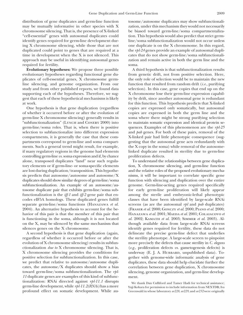
Figure 1.—Mapping of (A) rpl-11.1(ar228), (B)pab-1(ar232), and (C) glp-3/eft-3(ar229). Relevantsections of linkage groups V, I, and III are de-picted with genetic map positions indicated formarkers used in the analysis. Complementationinformation is given for deficiencies tested in eachregion. For A, SNPs used to map ar228 from theleft (blue) and right (green) are given with therelevant intervals and recombinants observed ineach interval [see materials and methods for de-tails and further information regarding Df(e2173)].
in early germ-line proliferation is maternal (Fujita et al. sperm. At 15", the majority of the double-mutant ani-mals (26/28), like the ar228 single mutants (n % 28),2002). Other genes required for germ-line proliferation
have been identified using RNAi (Hanazawa et al. 2001; did not produce any sperm and were morphologicallysimilar to ar228 single mutants. At 25", the germ linesMaeda et al. 2001; Colaiacovo et al. 2002), and a large-
scale analysis of RNAi-mediated germ-line proliferation of 4/19 animals contained a total of 0–2 germ cells thatdid not differentiate (similar to ar228 single mutants),defects is in progress (E. J. A. Hubbard, unpublished
data). while in 5/19 animals, the germ line contained a totalof 5–30 sperm [similar to glp-1(q175) single mutants].Phenotypic analysis of ar228 : ar228 is recessive and
causes sterility at all temperatures, although the most Genetically matched control animals produced 0–9 (n %28 animals) and 0–17 (n % 19) total undifferentiatedsevere and most penetrant proliferation defect (+80% of
adults with 10 or fewer germ cells per animal) is seen germ cells per animal at 15" and 25", respectively. Sur-prisingly, the germ lines of the remaining 10 double-at a lower temperature (Figure 2). Further phenotypic
analysis of the ar228 mutant revealed that Z2 and Z3 mutant animals at 25" produced 46–200 sperm, far morethan are normally seen in glp-1(q175) mutants alone.are present in the early L1 gonad (n + 40), but they
do not appear to proliferate well in subsequent larval One possibility is that the germ cells produced by ar228mutants are resistant to differentiation and proliferatedstages. On the basis of the distribution of germ cell
counts at two time points (L2/L3 and adult; Figure 2), abnormally in the glp-1 mutant background prior todifferentiating en masse. Nevertheless, the results at thegerm cells in animals with very few germ cells in the
L2/L3 did not appear to proliferate further, while germ restrictive temperature suggest that the ar228 prolifera-tion defect precludes differentiation.cells that underwent some early proliferation did appear
to proliferate further over time (Figure 2). In some Molecular identification of rpl-11.1(ar228): ar228 wasmapped to a region of the left arm of LGV containing !37cases, germ cells appeared to be in various stages of
disintegration. Because Z2 and Z3 were visible in the ORFs (materials and methods; Figure 1). One gene inthe region, rpl-11.1, was reported to confer sterile (Ste),L1 primordium of all animals examined, we assume
that Z2 and Z3 or their early descendents disintegrated sterile progeny (Stp), or embryonic-lethal (Emb) andgrowth defective (Gro) phenotypes in previous RNAi exper-completely in those animals with no visible germ cells
in the L3 or adult. iments (Piano et al. 2000, 2002; Simmer et al. 2003). Thisgene also displayed a strong germ-line expression patternTo assess whether the few germ cells produced in ar228
animals were capable of entering meiosis, we examined (NEXTDB http://nematode.lab.nig.ac.jp; Y. Kohara, per-sonal communication). Sequence analysis of DNA fromthe phenotype of glp-1(q175); ar228 double-mutant ani-
mals under restrictive (15") and less restrictive (25") the rpl-11.1 gene amplified from ar228 homozygotesrevealed a single C-to-T change that causes a prematureconditions for the ar228 proliferation defect (see mate-
rials and methods). In glp-1 loss-of-function mutants, termination at position Q175 (materials and meth-ods). Therefore, we refer to this allele as rpl-11.1(ar228).all germ cells enter meiosis early and differentiate as

2001Gene Duplication and Germ-Line Function
Figure 2.—Germ-line prolifera-tion in rpl-11.1(ar228) and pab-1(ar232). (A) Images of DAPI-stainedearly adult animals: wild type (WT),rpl-11.1(ar228), and pab-1(ar232)andcorresponding cartoons. Bars, 50,m. (B) Histograms depicting dis-tributions of germ cell counts inrpl-11.1(ar228) and pab-1(ar232).Solid bars represent total countstaken per animal at 25" and openbars represent counts taken fromanimals at 15". Larval counts wereobtained 24 hr post-hatch at 25"(early L3) or 48 hr post-hatch at15" (late L2). Adult counts weretaken 48 hr post-hatch at 25" and96 hr post-hatch at 15" (both earlyadult). Dashed bar indicates thecell count category for the wildtype at the equivalent time point(Hirsh et al. 1976; Kimble andHirsh 1979; Pepper et al. 2003b).
RPL-11.1 is a large ribosomal subunit L11 protein. These RNAi observations (Piano et al. 2000, 2002; Simmer et al.2003), animals that continue to feed on RNAi-inducingresults suggest that robust translation is an absolute re-
quirement for early germ-line proliferation. bacteria produce dead embryos and, eventually, no em-bryos at all. These results suggest that the homozygousBecause the mutant phenotype of rpl-11.1(ar228) is zy-
gotic sterility rather than embryonic lethality, L11 may be rpl-11.1(ar228) self-progeny of heterozygous mothersare rescued from embryonic lethality by the maternalprovided to the embryo maternally. To investigate this
possibility we turned to RNA interference (RNAi) that contribution of rpl-11.1. The similarity of the RNAi-induced sterility defect to that of the mutant phenotypedegrades both the maternal and the zygotic mRNAs and
found that the rpl-11.1(RNAi) phenotype is more severe lends further credence to the supposition that the muta-tion at 15" confers an essentially null phenotype in thethan the rpl-11.1(ar228) phenotype. Early progeny of
animals consuming rpl-11.1(RNAi) bacteria (see mate- germ line despite the fact that the predicted proteinproduct from rpl-11.1(ar228) is truncated by only 22 ofrials and methods) exhibit a proliferation-defective ste-
rility phenotype identical to that seen in rpl-11.1(ar228) 196 amino acids.Germline-specific zygotic role of rpl-11.1: Among thehomozygous animals. However, consistent with previous

2002 J. Maciejowski et al.
TABLE 1
RNAi analysis of duplicated rpl genes in wild-type (N2) and rrf-1(pk1417) mutant animals
Phenotype a
Germ-line defect Somatic defectRNAitarget LG Strain WT b (%) Sterile c (%) Unhealthy d (%) Gro/Lva e (%) n
(None) N2 76 0 21 0.7 275rrf-1 24 25 f 40 0 114
rpl-11.1 V N2 0 77 f 5 0.7 298rrf-1 0 75 f ,g 5 0 59
rpl-11.2 X N2 3 12 3 78 295rrf-1 0 15 g 45 10 80
rpl-25.1 X N2 73 0 25 0 149rrf-1 44 43 f ,g 7 4 99
rpl-25.2 I N2 8 86 f 6 0 152rrf-1 10 68 f ,g 5 0 19
rpl-24.1 I N2 1 22 f ,g 15 62 112rrf-1 0 76 f 17 0.2 58
rpl-24.2 I N2 24 10 g 18 47 180rrf-1 0 89 f 11 5 121
a Data are reported here for one parallel experiment; several experiments gave similar results (see materialsand methods). Percentages for phenotypes that were most penetrant for each treatment are in italic type.Remaining animals fell into “other” phenotypic categories that were not scoreable, such as lethal (Let) orruptured (Rup).
b Normal-sized fertile adults.c Sterile adults (both normal sized and reduced size).d Fertile adults but small or sick looking.e Larval arrest (Lva) or larval-sized animals.f Majority of sterility due to strong Glp (germ-line proliferation defective) phenotype; this phenotype is
observed variably in the rrf-1 background and may be temperature sensitive. In the particular experimentreported here, the penetrance is on the high side of the range of variability compared to other experiments.
g Sterility largely due to mild germ-line proliferation defect often accompanied by signs of gametogenesis( f,g indicates that a large number of worms exhibited both types of sterility defect).
genes encoding proteins highly similar to RPL-11.1 from (Gro) that is not observed in parallel rpl-11.1 RNAiexperiments (Table 1). We also investigated the RNAiother phyla is a second C. elegans L11-encoding gene,
rpl-11.2, located on the X chromosome (WormBase). phenotypes of the two genes in a genetic backgroundthat reduces the efficacy of RNAi in the soma whileGiven previous reports of a dearth of germ-line-enriched
genes on the X chromosome (Reinke et al. 2000, 2004), retaining it in the germ line, rrf-1(pk1417) (Sijen et al.2001). We found that in an rrf-1 mutant background, rpl-underrepresentation of expression of essential X-linked
genes (Piano et al. 2002; Reinke et al. 2004), and reports 11.1 still conferred a penetrant germ-line proliferationdefect, while the somatic growth defect of rpl -11.2 wasof X chromosome silencing in the germ line (Fong et
al. 2002; Kelly et al. 2002), we wondered if there might largely suppressed. Other adult defects were observedin association with rpl-11.2(RNAi) in the rrf-1 mutantbe a clear functional separation of the L11-encoding genes
in the germ line and soma, with the autosome-linked rpl- background, including sterility (Table 1).Second, we compared the available expression pat-11.1 functioning in the germ line and the X-linked rpl-
11.2 functioning in the soma. We used several assays terns of rpl -11.1 and rpl-11.2 images from the NEXTDBexpression pattern database (http://nematode.lab.nig.to assess the germ-line vs. soma activities of these two
genes. ac.jp; Y. Kohara, personal communication). Weakly de-tectable levels of rpl -11.1 mRNA are observed in a lim-First we examined carefully the RNAi phenotypes of
both rpl-11 genes. Consistent with previous reports, we ited number of cells in the embryo. Strong levels aredetected, however, in a small number of cells locatedfound that rpl-11.2 confers a penetrant growth defect

2003Gene Duplication and Germ-Line Function
centrally in the larvae (likely Z2 and Z3 and their descen- autosomal copy of the gene encoding the L11 proteinof the large ribosome subunit is dedicated to germ-linedants) and thereafter in the germ line, primarily in the
distal proliferating regions. In contrast, rpl-11.2 mRNA function and affects early embryogenesis as a conse-quence of its germline role.is detectable in a subset of cells in the embryo, with
high levels in what appear to be intestinal cells in later rpl -25 genes are duplicated and are organized simi-larly to rpl -11: We next asked if other rpl genes wereembryonic stages. During larval development, rpl-11.2
mRNA is weakly detectable, with somewhat higher levels duplicated in the C. elegans genome and if their genomedistribution correlated with a separation of germ-line/in the adult. These observations suggest that vastly differen-
tial levels of mRNAs encoding ribosome subunits could soma function. Of the 43 rpl genes in the database(WormBase Release WS130), we found 2 additional du-be synthesized in different tissues, a phenomenon ob-
served in other organisms as well (Agarwal et al. 1999). plicated genes: rpl-24 (likely encoding the L24 proteinof the large ribosome subunit), with both duplicatesGiven the presumably cell-essential function of the L11
protein, we found the dynamic and nonubiquitous levels (rpl-24.1 and rpl-24.2) on LGI, and rpl-25 (likely encod-ing the L23a protein of the large ribosome subunit)of expression surprising. These results likely reflect tissue-
specific requirements for high translational capacity. Pre- with an X-linked paralog, rpl-25.1, and an autosomalparalog, rpl-25.2, on LGI. Previous RNAi studies indicateviously reported microarray analysis comparing wild type
with the germ-line-deficient glp-4(bn2) mutant also firmly that reduction of rpl-24.1 and rpl.25.2 function cancause embryonic lethality and sterility (Fraser et al.places rpl-11.1 in the germ-line-enriched class (log2 ratio
of *4.1; Reinke et al. 2004) while rpl-11.2 is not enriched 2000; Simmer et al. 2003) and that rpl-25.2 transcriptsare germ-line enriched (log2 ratio for */glp-4(bn2) ofin the germ line (log2 ratio of (1.3; Reinke et al. 2004).
Third, previous studies documented two dramatic *1.6 vs. (1.2 for rpl-25.1; Reinke et al. 2004) while rpl-24.1 and rpl-24.2 are not (log2 ratios of 0.174 and 0.5,germ-line-associated phenotypes after removal of Z2
and Z3 by laser microsurgery: life-span extension (Hsin respectively; Reinke et al. 2004).To assess the relative germ-line and somatic roles ofand Kenyon 1999) and gigantism (Patel et al. 2002).
We reasoned that if only the germ line is affected by rpl - these gene pairs, we examined the effects of RNAi tar-geted against these genes individually in the wild-type11.1(ar228), we should see the same phenotypes pre-
viously reported for germ-line-ablated animals. If, on (N2) and the rrf-1 mutant backgrounds (Sijen et al.2001; Table 1; see materials and methods). In thisthe other hand, rpl-11.1 also plays a role in somatic
development that would compromise the somatic con- experiment, rpl-11 and rpl-25, the two gene pairs withan autosome/X chromosome duplication, behave moretribution to life-span or size determination, these phe-
notypes may be less severe in our mutant than those similarly to each other than does the autosome/autosomeduplicated rpl-24 gene pair. For the rpl-11 and rpl-25observed in the cell-ablation studies. We found that in
rpl-11.1(ar228) animals, both life-span extension and pairs in the N2 background, the primary defect afterRNAi of the autosome-linked genes of each pair (rpl-overall increase in the volume of the worms is similar to
that observed in germ-line-ablation experiments (Figure 11.1 and rpl-25.2) is a germ-line proliferation defect (77and 86%). However, RNAi directed against the X-linked3). Life span is extended in ar228 sterile homozygotes
to 27 days (starting from the L4 stage) from a mean life genes of each pair confers either growth/larval arrest(rpl-11.2, 78%) or a weak overall health defect (rpl-25.1,span of 14 days in control animals, an increase of 1.9-
fold. These results are comparable to the increase in 25%) but not a significant germ-line proliferation defect(12 and 0%, respectively). As expected for genes actingmean life span observed after ablation of Z2 and Z3 (an
extension from 19 to 32 days or 1.7-fold starting from primarily in the germ line, RNAi directed against theautosomal copies of these genes confers similar pheno-day of birth; Hsin and Kenyon 1999). We also observed
a positive correlation between rpl-11.1(ar228) mutant types in the rrf-1 mutant background and in the wildtype, while as expected for genes acting in the soma,body length, width, and volume. Homozygous mutant
and wild-type L4 larvae and early adults were similar in the phenotypic profile changed significantly betweenN2 and rrf-1 when the X-linked paralogs were targeted.size, but adult size increased more in the mutant than
in the wild type as the animals aged (Figure 3). The Specifically, the percentage of growth defective/larvalarrest animals in rpl-11.2(RNAi) is reduced from 78 tooverall volume increase in adult sterile rpl-11.1(ar228)
animals (as compared to genetically matched controls) 10% in N2 vs. rrf-1 and the unhealthy phenotype ob-served in rpl-25.1(RNAi) is reduced from 25 to 7%. Inis from 8.7 ,m3 to 11.9 ,m3 8 days post-L4 (or 7 days
after reaching adulthood), an increase of 1.4-fold. These addition, rpl-25.1(RNAi) displayed a significant increasein sterility in the rrf-1 background, but the germ-lineresults are similar to the previously reported increase
in volume from 5.8 to 8.4 ,m3, also 1.4-fold, 7 days proliferation defect was not as severe as that observedwith rpl-25.2 (Table 1).postadulthood after Z2/Z3 cell ablations (Patel et al.
2002). Finally, the effect of RNAi-mediated depletion of rpl-24.1 and rpl-24.2 resulted in quite similar phenotypicTaken together, the results of RNAi experiments, ex-
pression data, and phenotypic analysis suggest that the profiles, but differed from that of rpl-11 and rpl-25 (Ta-

2004 J. Maciejowski et al.
Figure 3.—Life span and gigantism in rpl-11.1(ar228) and pab-1(ar232). Survival curves (A) for rpl-11.1(ar228) and (B) for pab-1(ar232) and (C) worm volumes, with respective control strains and the wild-type strain N2, are shown. All non-N2 strains (mutantand respective controls) are homozygous progeny of heterozygous mothers balanced by nT1[unc-?(n754) let-?] for rpl-11.1(ar228)(control r) and by hT2[let-? qIs48] for pab-1(ar232) (control p). Survival curves are given for two independent experiments foreach non-N2 strain. Taken together, for N2 wild type (brown and orange), mean life span (m) % 14.2 - 3.5 days and numberof animals observed (n) % 163; for */* control r, m % 14.2 - 3.3 days and n % 127; for rpl-11.1(ar228), m % 27.4 - 6.9 daysand n % 126; for */* control p, m % 16.9 - 3.4 days, n % 126; and for pab-1(ar232), m % 26.4 - 6.9 days, n % 185. (C) Volumecalculations are based on length and width measurements (see materials and methods); n % 20 worms of each genotype pertime point. Brackets link mutants with relevant genetically matched controls. Measurements were taken at the mid-L4 (day 0),as early adults (day 1), and a week later (day 8) as older adults. Actual volumes (in cubic micrometers) calculated for the threeindicated time points (-1 SD) are as follows: for pab-1(ar232), 1.55 - 0.27, 3.82 - 0.43, and 14.7 - 1.12; for rpl-11(ar228),1.37 - 0.31, 4.55 - 0.90, and 11.94 - 5.84; for control p, 1.37 - 0.19, 4.98 - 0.77, and 10.50 - 1.67; for control r, 1.37 -0.23, 5.14 - 1.00, and 8.72 - 2.12; and for N2, 1.37 - 0.27, 4.43 - 1.27, and 7.64 - 1.21. (D) Digital images of wild-type (N2),rpl-11.1(ar228), and pab-1(ar232) animals 10 days after the L4 stage at 20". Bars, 200 ,m.
ble 1). After RNAi depletion of each gene in the rpl- (both autosomal) appears to act in both the soma andthe germ line.24 pair in wild type and rrf-1, each displayed a similar
decrease in severe growth/larval arrest phenotypes and Identification and characterization of pab-1(ar232): Wepositionally cloned pab-1(ar232), another mutation identi-concomitant increase in sterility in the rrf-1 background
as compared to the wild-type background. Thus, in con- fied in our screen (Figure 1; materials and methods).DNA sequence revealed a C-to-T transition, resulting intrast to the germ-line-specific roles of the autosomal
copies of rpl-11 and rpl-25, it is likely that both rpl-24 a conceptual translation of Q408 in Y106G6H2.a andQ428 in Y106G6H2.b and Y106G6H2.c to an ambergenes (both autosomal) are redundant in the soma and
the germ line. termination. This mutation causes a recessive fully pene-trant severe germ-line proliferation defect (n % 120)In summary (Table 2), our functional analysis of the
three rpl genes (and the pab genes, see below) is consis- and an incompletely penetrant protruding vulva (Pvl)phenotype. pab-1 encodes a cytoplasmic poly(A)-bind-tent with the hypothesis that the two X-linked members
of the X/A gene pairs act in the soma while the au- ing protein (PABP) and is the autosomal copy of anautosome/X-linked gene pair: pab-1 and pab-2.tosome-linked members of each pair act either in the
germ line alone (e.g., rpl-11.1) or in the germ line and Further analysis suggested that pab-1 has an essentialgerm-line function whereas pab-2 does not [consistentsoma (e.g., rpl-25.2). In contrast, the pair of rpl-24 genes

2005Gene Duplication and Germ-Line Function
TABLE 2
Functional comparison of C. elegans and C. briggsae orthologs
C. elegansortholog A/X a Phenotype b C. briggsae ortholog Phenotype c (n)
rpl-11.1 A Sterile CBG01314 Sterile (7/7)rpl-11.2 X Fertile (Gro) CBG14053 Sterile (1/8), sub-Fertile d (7/8)
Progeny: Lva/Gro
rpl-24.1 A Fertile (Gro) CBG22273 Sterile (2/8), sub-Fertile d (6/8)Progeny: Lva/Gro
rpl-24.2 A Fertile (Gro) CBG03702 sub-Fertile d (7/7)Progeny: Lva/Gro
rpl-25.1 X Fertile (WT) CBG14529 Fertile e (5/5)Progeny: WT
rpl-25.2 A Sterile CBG04080 Sterile (3/3)
pab-1 A Sterile CBG02207 Sterile (8/8)pab-2 X Fertile (WT) CBG07431 sub-Fertile d (7/7)
Progeny: WT/Gro
L4440 (()ctrl Fertile (5/5) f
Progeny: WT
Lva, larval arrest.a Autosome or X-linked locus in C. elegans; Gro, growth defect.b Summarized from Table 1 and from the text. Phenotype refers to progeny phenotype from RNAi feeding
experiments (see materials and methods).c Phenotype of injected animals (n, number of animals displaying phenotype per number of surviving injected
animals) as assessed after overnight transfer to deplete animals of embryos produced prior to the injection(see materials and methods). In addition, few of the adult progeny produced by these fertile injected animalsexhibited sterility compared to a high penetrance of sterility observed in the progeny (first 12 hr) from injectedanimals that quickly became sterile.
d These animals were subfertile, producing few embryos, some embryonic lethality, and few live progeny:CBG14053, average .4 surviving progeny per injected animal; CBG22273 and CBG03702, average .2 survivingprogeny per injected animal; CBG07431, average .5 surviving progeny per injected animal.
e These animals were fertile, producing many embryos but few live progeny due to embryonic lethality:CBG14529, average .2 surviving progeny per injected animal.
f Negative control (see materials and methods), )20 progeny per injected animal over 2–3 days scored.
with recent findings of Ciosk et al. (2004)]. RNAi di- (from an average of 17 days post-L4 in the control strainto an average of 26 days in the mutant, an increase ofrected against pab-1 resulted in several phenotypes, in-
cluding a high penetrance of sterility (43%, n % 84) 1.5-fold; Figure 3) and causes gigantism, with wormsaveraging 1.4-fold greater volume (Figure 3). Previousand Pvl (32%, n % 84) and, at lower penetrance, a
ruptured vulva (Rup) and flaccid-looking body mor- microarray analysis does not indicate strong germ-lineenrichment for pab-1 or pab-2 transcripts (log2 ratio forphology. In the rrf-1 mutant background all pab-1(RNAi)
animals were sterile (n % 54) and 25% also displayed */glp-4 of *0.1 for pab-1 and (1.25 for pab-2 ; Reinkeet al. 2004).the Pvl phenotype. These results suggest that the sterility
is a result of germ-line function of pab-1. In contrast, RNAi Identification and characterization of glp-3(ar229);glp-3 is eft-3 : A third allele from our screen mapped todirected against the X-linked pab-2 in N2 resulted in 94%
wild-type animals with low-penetrance sterility, Rup, and a small interval on LGIII (see materials and meth-ods). We noted that mutations in glp-3, a gene thatPvl phenotypes (2%, 4%, and 0.3%, respectively; n %
325). Very similar observations were made in the rrf-1 maps genetically to the region, had been analyzed pre-viously at the phenotypic level and conferred a similarmutant background where pab-2(RNAi) produced 90%
wild-type, 7% sterile, and 4% Rup animals (n % 119). zygotic germ-line proliferation defect (Kadyk et al.1997). We performed complementation analysis (seeThese results indicate that pab-1 and pab-2 act redun-
dantly in the soma but that pab-1 is required in the germ materials and methods) with glp-3(q145) and ar229,and they failed to complement, indicating that ar229 isline.
Consistent with a role for pab-1 in the germ line and an allele of glp-3.We next examined genes in the region, looking forsimilar to rpl-11.1(ar228), pab-1(ar232) extends life span

2006 J. Maciejowski et al.
predicted essential genes with an X-linked paralog. eft-3, an be required to assess on a genomic scale the prevalenceof the X/A trend we observed. Nonetheless, our findingEF-1-! ortholog, satisfied these criteria. One of severalthat three alleles from a nonbiased screen for germ-lineclosely related genes, eft-4, resides on the X chromo-proliferation defects led to the identification of threesome. Interestingly, one alternative splice form of eft-4X/A duplicated genes is striking.apparently encodes a protein identical to eft-3 [WormBase
Unlike many autosome/X-linked gene pairs in mam-WS130]. In addition to identical protein sequences, themals and Drosophila, retrotransposition is not the mech-nucleotide sequences of the coding regions of the twoanism for the rpl, pab, and eft duplications: The implica-genes are very similar (94% identical over 1392 bases),tions of gene duplication, genome organization, andprecluding the possibility of separate RNAi-based func-large-scale gene silencing mechanisms in the germ linetional analysis of these two genes. Sequence analysis ofare relevant to other organisms. Indeed, in an analogousglp-3(q145) in the eft-3 region revealed a single base pairprocess to C. elegans X chromosome germ-line silencingchange (G to A) that would result in an the amino acid(Fong et al. 2002; Kelly et al. 2002), the X chromosomechange A301T. Our mutant, ar229, contains a C-to-Tis inactivated in mouse during meiosis, a general phe-transition in eft-3 that encodes a S414F amino acidnomenon referred to as meiotic sex chromosome inacti-change. This analysis thus identified glp-3 as eft-3.vation (MSCI) (McKee and Handel 1993). The possi-Another gene, glp-4, confers a severe zygotic germ-bility of an autosome/X-linked distribution of geneline proliferation defect (Beanan and Strome 1992).duplicates and correlated germ-line/soma functionalWe examined the predicted genes in the region to whichseparation has been noted and proposed as one possibleglp-4 maps (LGI, 21.4 - 2 cM) for candidate genes for“coping” mechanism to allow important genes to func-which previously reported RNAi experiments conferredtion despite MSCI in mammals (McKee and Handela sterility or lethality phenotype (Stp, Ste, Lvl, Lva, or1993 and references therein; Handel 2004). There areEmb) and for which a closely related paralog exists onsix documented cases of mammalian genes with au-the X chromosome. For genes with no available RNAitosome/X duplications in which only the autosomalinformation, we checked for X-linked paralogs. No obvi-copy is expressed during spermatogenesis. For five ofous candidates were identified. rpl-31 maps very closethese genes, the X-linked copies contain introns whileto glp-4, but no change in the rpl-31 coding sequencethe autosomal genes are intronless, a hallmark of dupli-was detected in the DNA of glp-4(bn2) animals (data notcative retrotransposition to the autosome from an ances-shown).tral X-linked gene (see Handel 2004 and referencesX/autosome duplications of highly conserved genes intherein). Moreover, in humans there are five active du-the C. elegans genome: We examined the C. elegans genome plicates of the cytoplasmic PABP, at least two of which,for other X/autosome gene pairs and found that 395 including the X-linked PABPC5, are intronless, againX-linked genes have a paralog on an autosome. Of these, suggesting retrotransposase-mediated gene duplication
168 have a highly conserved homolog in human (E ' (see Mangus et al. 2003 and references therein). Studies10(50 and )75% identity over the length of the protein), in Drosophila also suggest a high rate of X-to-autosomean indication of likely “cell-essential” function. Of these gene duplication by way of retrotransposons with a signifi-168 X-linked genes, 156 have been assayed by RNAi, cant enrichment of testes-specific expression in the intron-and 128 have no reported phenotype in any large-scale less autosomal members of the gene pairs (Betran et al.RNAi study (i.e., were scored as “wild-type” in every 2002). We therefore examined the predicted intronreported assay) (Fraser et al. 2000; Gonczy et al. 2000; structure (WormBase WS130) of the duplicated C. ele-Piano et al. 2000; Maeda et al. 2001; Kamath et al. 2003; gans rpl, pab, and eft genes identified in this study andSimmer et al. 2003). Of these 128 genes, 23 have an found no evidence for retrotransposition: all autosomalautosomal paralog that conferred an Emb, Ste, or Stp and X-linked copies contain introns and, in the casesphenotype in one or more of the above studies, indicat- of the rpl-11, rpl-25, and eft duplicates, some predicteding likely germ-line function. Large-scale studies thus intron/exon boundaries are conserved to the base.provide evidence suggestive of germ-line/soma subfunc- These results suggest that mechanisms of gene duplica-tionalization for !16% of these highly conserved X/A tion other than retrotransposition may be important forgene pairs. No definitive conclusion can be reached duplications that evolve separate germ-line and somaticregarding the remainder of these X/A pairs for various functions in C. elegans.technical reasons, including likely false negatives in Duplicated genes in C. briggsae : To determine if thethese studies (Piano and Gunsalus 2002; Fernandez duplication of the three rpl genes, the pab genes, andet al. 2005), lack of an RNAi assay for the autosomal the eft genes arose prior to the evolutionary separationparalog (19 cases), or possible redundancy among mem- of lineages that gave rise to C. elegans and C. briggsae orbers of multigene families (which may prevent the detec- if these duplications are derived within the C. eleganstion of phenotypes for single-gene knockouts). Thus, a branch, we searched for closely related homologs ofmore directed combined bioinformatic and functional these genes in C. briggsae. The presence of closely related
paralogs within the C. elegans genome required someanalysis of highly conserved paralogous gene pairs will

2007Gene Duplication and Germ-Line Function
Figure 4.—Phylogenetic relationships of pab,rpl, and eft gene pairs in C. elegans (Cel) and C.briggsae (Cbr). Branch lengths (substitutions persite, estimated by likelihood) are indicated on thebranches and with a scale for each set of genes.For each set of homologs, all possible unrootedphylogenies were compared using a log-likeli-hood ratio test; in all cases, the maximum likeli-hood (ML) tree was significantly supported (P .0.05) over all other trees. Jackknife analyses dem-onstrated 100% support for the internal branch in2000 Monte-Carlo replicates for each set of fouror five homologous genes. Note that H19N07.1is the C. elegans ortholog of the CBG23104 genein C. briggsae; these genes are paralogs of the eft-3/4 genes in these two species. After divergingfrom C. briggsae , the eft-3/4 ortholog in a C. elegansancestor duplicated to produce eft-3 and eft-4,which are therefore both orthologs of the singleC. briggsae eft-3/4 gene.
care in assignment of the corresponding orthologs in after diverging from the lineage leading to C. briggsae(Figure 4). It is likely that the C. briggsae chromosomeC. briggsae, in part because similarity alone may not re-
flect the degree of phylogenetic relationship since rates that shares synteny with the C. elegans X chromosomeis the C. briggsae sex chromosome, but genetic analysesof evolution can be very different in different gene lin-
eages. Therefore, rather than relying solely on the best in C. briggsae will be required to verify this prediction.To determine if the related duplicated C. briggsaeBLASTP match, we examined syntenic regions between
the two species and performed a maximum likelihood genes (Figure 4) also show functional separation in thegerm line and soma, we performed RNAi for each genephylogenetic analysis (see materials and methods;
Figure 4). The results indicate that each of the rpl-11, of the four gene pairs (Table 2). Because C. briggsaeis not amenable to RNAi analysis by feeding, double-rpl-24, rpl-25, and pab genes has a single corresponding
ortholog in C. briggsae. Our BLASTP, synteny, and phylo- stranded RNA was delivered by injection into the germline. We found that injection of dsRNA correspondinggenetic analyses agree for the three rpl genes and the
pab genes, identifying the same pairs of genes as putative to the C. briggsae orthologs of the autosome-linked mem-ber of each A/X pair caused complete sterility (no em-orthologs. Our analysis is consistent with the hypothesis
that four duplications (three rpl genes and the pab bryos) in the injected animal within 12 hr of injection(Table 2). In contrast, all or most of the worms injectedgenes) preceded the divergence between C. elegans and
C. briggsae. Unlike the rpl and pab genes, the duplication with the orthologs of the X-linked C. elegans genes pro-duced embryos and/or live progeny. The latter injectedevent that produced eft-3 and eft-4 appears to be more
recent, having occurred within the C. elegans lineage animals, however, displayed various degrees of embry-

2008 J. Maciejowski et al.
onic lethality and/or were subfertile, suggesting the pos- Gene duplication, genome organization, and the germline: Our phenotypic analysis of the three duplicated rplsibility of germ-line function for these genes. Larval
arrest and growth defects were also observed among the genes and pab genes and previous analysis of glp-3 (Kadyket al. 1997) in C. elegans suggest that the functional roleprogeny of these animals (Table 2), however, suggestingof autosomal members of each autosome/X-linked pairthat these genes act in the soma as well as the germis critical in the germ line and that the X-linked copyline. As was observed from RNAi in C. elegans, injectionis largely if not exclusively active in the soma. We alsoof dsRNA corresponding to each gene of the C. briggsaeidentified orthologous genes in the C. briggsae genome,rpl-24 pair (an A/A pair in C. elegans) produced almostand in four of the five cases (all but eft-3/eft-4), the geneidentical results. In addition, for the C. briggsae orthologsduplication preceded the evolutionary divergence ofof the C. elegans autosome members of X/A pairs, manythese two species. Among the paralogous pairs withinprogeny produced within the first 12 hr after injectionC. briggsae that correspond to X/A pairs in C. elegans,became sterile adults, whereas adult sterility was not asome degree of subfunctionalization of these gene du-prevalent phenotype among early progeny from theplicates occurs, whereas the pair corresponding to theother experiments (Table 2).autosome/autosome duplication does not display theTherefore, the paralogs in C. briggsae that correspondsame degree of subfunctionalization. Given that the germ-to C. elegans autosome/X paralogs display consistentline phenotypes were much more severe in the C. briggsaefunctional differences between paralogs, including aorthologs of the autosome-linked members of X/A pairspotential somatic role for the orthologs of C. elegansin C. elegans, if the chromosome that carries the paralogsX-linked members of X/A pairs. Because both genes inis, indeed, the C. briggsae X chromosome, we speculateeach pair caused phenotypes that are associated withthat germ-line-expressed genes may be similarly under-germ-line function, however, there may be a lesser de-represented on the X chromosome of C. briggsae, as aregree of germ-line/soma subfunctionalization betweengerm-line-expressed genes in C. elegans (Reinke et al.these genes in C. briggsae. The extent to which this differ-2000, 2004). Interestingly, the same mode of X chromo-ence between C. elegans and C. briggsae is a consequencesome silencing that operates in the C. elegans germ lineof differences in experimental methods, species-specificappears to operate in C. briggsae (Kelly et al. 2002).response to RNAi, or other developmental differences is
That a similar pattern of germ-line function of theunclear. In summary, our analysis in C. briggsae suggestsautosomal copy of autosome/X-linked paralogs is abun-that RNAi directed against the C. briggsae orthologs of C.dant among many cell proliferation-essential genes iselegans autosomal members of the three autosome/X geneevidenced by our genetic identification of three inde-pairs (rpl-11, rpl-25, pab) caused a more profound germ-pendent loci (from three alleles) that are required forline defect than did RNAi directed against the C. briggsaerobust germ-line proliferation. Given that these threeorthologs of C. elegans X-linked members of these pairsmutants (of seven obtained from a forward geneticor against the autosome/autosome pair (rpl-24).screen covering 11,586 genomes that were screened ina way that could identify putative severe proliferationdefective mutants; Pepper et al. 2003a; E. J. A. Hubbard,DISCUSSIONunpublished data) uncovered autosomal copies of du-
RPL, PAB, and EFT proteins and early germ-line pro- plicated genes with the same kind of autosome/X chro-liferation: Using forward genetics, we identified rpl-11.1, mosome distribution, we speculate that other genes es-pab-1, and glp-3/eft-3 as genes required zygotically for sential for early germ-line proliferation may be encodedearly germ-line proliferation. These genes encode the by the autosome duplicate of autosome/X-linked pairsL11 protein of the large ribosome subunit, a cytoplasmic of genes encoding proteins essential for cell prolifera-PABP, and an elongation factor (EF-1-!), respectively. tion.All are important for translation and are likely essential Recent studies of X-linked genes in mammals suggestfor cell proliferation. Their critical roles in early germ- that only genes expressed after the onset of male germ-line proliferation suggest that the processes of reinitia- line X chromosome silencing (MSCI) are underrepre-tion and maintenance of early proliferation from the sented on the X chromosome (Khil et al. 2004). In C.quiescent state of the germ-line precursor cells Z2 and elegans the X chromosome lacks histone modificationsZ3 is dependent on proper ribosome biogenesis and/ that correlate with transcriptional activation throughoutor active translation. That the primordial germ cells are the adult male germ line and in the zone of mitosis andborn and locate properly to the somatic gonad in the early meiosis in adult hermaphrodites (Fong et al. 2002;mutants suggests that the maternal copy is sufficient for Kelly et al. 2002). Therefore, it is conceivable that inthese events and/or that the X-linked paralogs of each addition to genes required in spermatogenesis, genesgene are functionally redundant in the germ line until required in earlier stages of germ-line development in C.Z2 and Z3 resume cell division. We also identified a elegans may be particularly sensitive to selective pressurecritical germ-line proliferation function for rpl-25.2 that keeping them off the X chromosome.
Identification of correlations between the genomicis not shared by its X-linked paralog, rpl-25.1.
2009Gene Duplication and Germ-Line Function
distribution of gene duplicates and germ-line function tosome/autosome duplicates may show subfunctionali-zation, under this mechanism they would not necessarilymay be mutually informative in other species with X
chromosome silencing. That is, the presence of X-linked be biased toward germ-line/soma compartmentaliza-tion. This hypothesis would also predict that strict germ-“cell-essential” genes with autosomal duplicates could
identify genes required for germ-line development dur- line/soma subfunctionalization would not occur unlessone duplicate is on the X chromosome. In this regard,ing X chromosome silencing, while those that are not
duplicated could point to genes that are required at a the rpl-24 genes provide an example of autosomal dupli-cates that do not show germ-line/soma subfunctionali-time in development when the X is not silenced. This
approach may be useful in identifying autosomal genes zation and remain active in both the germ line and thesoma.required for fertility.
Evolutionary hypotheses: We propose three possible A third hypothesis is that subfunctionalization resultsfrom genetic drift, not from positive selection. Here,evolutionary hypotheses regarding functional gene du-
plicates of cell-essential genes, X chromosome germ- the only role of selection would be to maintain the newfunction that resulted from random drift (i.e., purifyingline silencing, and genome organization. From our
study and from other published reports, we found data selection). In this case, gene copies that end up on theX chromosome lose their germ-line expression capabil-supporting each of the hypotheses. Therefore, we sug-
gest that each of these hypothetical mechanisms is likely ity by drift, since another autosomal gene is redundantfor this function. This hypothesis predicts that X-linkedat work.
One hypothesis is that gene duplication (regardless copies are expressed only somatically, but autosomalcopies are expressed in both the germ line and theof whether it occurred before or after the evolution of
germ-line X chromosome silencing) generally results in soma where there might be strong purifying selectionto maintain somatic expression and identical protein se-“subfunctionalization” (Lynch and Conery 2000) into
germ-line/soma roles. That is, when there is positive quences. Examples of this phenomenon are the rpl-25and pab genes. For both of these pairs, removal of theselection to subfunctionalize into different expression
compartments, it is generally the case that these com- X-linked pair had little phenotypic consequence (sug-gesting that the autosomal gene acts redundantly withpartments correspond to germ-line and soma compart-
ments. Such a general trend might result, for example, the X copy in the soma) while removal of the autosome-linked duplicate resulted in sterility due to germ-lineif many regulatory regions in the genome had elements
controlling germ-line vs. soma expression and if, by chance proliferation defects.To understand the relationships between gene duplica-alone, transposed duplicates “land” near such regula-
tory elements or if germ-line- or soma-specific elements tion, X chromosome silencing, and germ-line functionand the relative roles of the proposed evolutionary mecha-are lost during duplication/transposition. This hypothe-
sis predicts that autosome/autosome and autosome/X nisms, it will be important to correlate specific genefunction with silencing and duplication over the entireduplicates should show similar degrees of germ-line/soma
subfunctionalization. An example of an autosome/au- genome. Germ-line-acting genes required specificallyfor early germ-line proliferation will likely appeartosome duplicate pair that exhibits germ-line/soma sub-
functionalization is the iff-1 and iff-2 gene pair that en- among the sterile and embryonic-lethal phenotypicclasses that have been identified by large-scale RNAicodes eIF5A homologs. These duplicated genes fulfill
separate germ-line/soma functions (Hanazawa et al. screens (as are the autosomal rpl and pab duplicates)(Fraser et al. 2000; Gonczy et al. 2000; Piano et al. 2000;2004). An alternative hypothesis to account for the be-
havior of this pair is that the member of this pair that Hanazawa et al. 2001; Maeda et al. 2001; Colaiacovo etal. 2002; Kamath et al. 2003; Simmer et al. 2003). Al-is functioning in the soma, although it is not located
on the X, may be silenced by the same mechanism that though available data from large-scale RNAi screensidentify genes required for fertility, these data do notsilences genes on the X chromosome.
A second hypothesis is that gene duplication (again, delineate the precise germ-line defect that underliesthe sterility phenotype. A large-scale screen to pinpointregardless of whether it occurred before or after the
evolution of X chromosome silencing) results in subfun- more precisely the defects that cause sterility in C. elegansctionalization due to X chromosome silencing. That is, (e.g., proliferation defects vs. gametogenesis defects) isX chromosome silencing provides the conditions for underway (E. J. A. Hubbard, unpublished data). To-positive selection for subfunctionalization. In this case, gether with genome-wide informatic analysis of genewe predict that relative to autosome/autosome dupli- duplicates, these data should help elucidate further thecates, the autosome/X duplicates should show a bias correlation between gene duplication, X chromosometoward germ-line/soma subfunctionalization. The rpl- silencing, genome organization, and germ-line develop-11 duplicate genes are examples of this kind of subfunc- ment.tionalization: RNAi directed against rpl-11.1 disrupts We thank Dan Culliford and Tamer Hadi for technical assistance;germ-line development, while rpl-11.2(RNAi) has a more Yuji Kohara for permission to include information from NEXTDB; Iva
Greenwald in whose laboratory ar228, ar229, and ar232 were originallyprofound effect on somatic development. While au-

2010 J. Maciejowski et al.
Bennett et al., 2004 The Caenorhabditis elegans eukaryoticisolated; and the Hubbard and Fitch laboratory members (especiallyinitiation factor 5A homologue, IFF-1, is required for germ cellKarin Kiontke), Fabio Piano, Scott Baird, and Bill Kelly for helpfulproliferation, gametogenesis and localization of the P-granulediscussions. We also thank the C. elegans Genetics Center for strains.component PGL-1. Mech. Dev. 121: 213–224.This work was supported in part by National Institutes of Health (NIH)
Handel, M. A., 2004 The XY body: a specialized meiotic chromatingrant GM-61706 to E.J.A.H., National Science Foundation (NSF) grant domain. Exp. Cell Res. 296: 57–63.DEB-0228692 to D.H.A.F., the National Sciences and Engineering Re- Hirsh, D., D. Oppenheim and M. Klass, 1976 Development of thesearch Council of Canada (R.C.J. and D.L.B.), NIH grant HD046236 to reproductive system of Caenorhabditis elegans. Dev. Biol. 49:Fabio Piano, and NSF grant DBI-0137617 to K.C.G. 200–219.
Hsin, H., and C. Kenyon, 1999 Signals from the reproductive systemregulate the lifespan of C. elegans. Nature 399: 362–366.
Hughes, A. L., 1994 The evolution of functionally novel proteins aftergene duplication. Proc. R. Soc. Lond. B Biol. Sci. 256: 119–124.LITERATURE CITED
Johnsen, R. C., 1990 Genetic analysis of the left half of linkageAgarwal, A. K., S. N. Parrish and D. D. Blumberg, 1999 Ribosomal group V in Caenorhabditis elegans. Ph.D. Thesis, Simon Fraser
protein gene expression is cell type specific during development University, Burnaby, BC, Canada.in Dictyostelium discoideum. Differentiation 65: 73–88. Johnsen, R. C., and D. L. Baillie, 1988 Formaldehyde mutagenesis
Altschul, S. F., T. L. Madden, A. A. Schaffer, J. Zhang, Z. Zhang of the eT1 balanced region in Caenorhabditis elegans: dose-et al., 1997 Gapped BLAST and PSI-BLAST: a new generation response curve and the analysis of mutational events. Mutat. Res.of protein database search programs. Nucleic Acids Res. 25: 3389– 201: 137–147.3402. Kadyk, L., E. Lambie and J. Kimble, 1997 glp-3 is required for
Anderson, P., and S. Brenner, 1984 A selection for myosin heavy mitosis and meiosis in the Caenorhabditis elegans germ line. Genet-chain mutants in the nematode Caenorhabditis elegans. Proc. ics 145: 111–121.Natl. Acad. Sci. USA 81: 4470–4474. Kamath, R. S., A. G. Fraser, Y. Dong, G. Poulin, R. Durbin et
Austin, J., and J. Kimble, 1987 glp-1 is required in the germ line al., 2003 Systematic functional analysis of the Caenorhabditisfor regulation of the decision between mitosis and meiosis in C. elegans genome using RNAi. Nature 421: 231–237.elegans. Cell 51: 589–599. Kelly, W. G., C. E. Schaner, A. F. Dernburg, M. H. Lee, S. K.
Beanan, M., and S. Strome, 1992 Characterization of a germ-line Kim et al., 2002 X-chromosome silencing in the germline of C.proliferation mutation in C. elegans. Development 116: 755–766. elegans. Development 129: 479–492.
Betran, E., K. Thornton and M. Long, 2002 Retroposed new genes Khil, P. P., N. A. Smirnova, P. J. Romanienko and R. D. Camerini-out of the X in Drosophila. Genome Res. 12: 1854–1859. Otero, 2004 The mouse X chromosome is enriched for sex-
Brenner, S., 1974 The genetics of Caenorhabditis elegans. Genetics biased genes not subject to selection by meiotic sex chromosome77: 71–94. inactivation. Nat. Genet. 36: 642–646.
Ciosk, R., M. DePalma and J. R. Priess, 2004 ATX-2, the C. elegans Kimble, J., and D. Hirsh, 1979 The postembryonic cell lineages ofortholog of ataxin 2, functions in translational regulation in the the hermaphrodite and male gonads in Caenorhabditis elegans.germline. Development 131: 4831–4841. Dev. Biol. 70: 396–417.
Colaiacovo, M. P., G. M. Stanfield, K. C. Reddy, V. Reinke, S. K. Lynch, M., and J. S. Conery, 2000 The evolutionary fate and conse-Kim et al., 2002 A targeted RNAi screen for genes involved in quences of duplicate genes. Science 290: 1151–1155.chromosome morphogenesis and nuclear organization in the Maddison, D. R., and W. P. Maddison, 2002 MacClade 4: Analysis ofCaenorhabditis elegans germline. Genetics 162: 113–128. Phylogeny and Character Evolution, Version 4.05. Sinauer Associates,
Ellis, R. E., and J. Kimble, 1995 The fog-3 gene and regulation of Sunderland, MA.cell fate in the germ line of Caenorhabditis elegans. Genetics 139: Maeda, I., Y. Kohara, M. Yamamoto and A. Sugimoto, 2001 Large-561–577. scale analysis of gene function in Caenorhabditis elegans by high-
Ferguson, E. L., and H. R. Horvitz, 1985 Identification and charac- throughput RNAi. Curr. Biol. 11: 171–176.terization of 22 genes that affect the vulval cell lineages of the Mangus, D. A., M. C. Evans and A. Jacobson, 2003 Poly(A)-bindingnematode Caenorhabditis elegans. Genetics 110: 17–72. proteins: multifunctional scaffolds for the post-transcriptional
Fernandez, A. G., K. C. Gunsalus, J. Huang, L.-S. Chuang, N. Ying control of gene expression. Genome Biol. 4: 223.et al., 2005 New genes with roles in the C. elegans embryo Mathies, L. D., S. T. Henderson and J. Kimble, 2003 The C. elegansrevealed using RNAi of ovary-enriched ORFeome clones. Ge- Hand gene controls embryogenesis and early gonadogenesis. De-nome Res. 15: 250–259. velopment 130: 2881–2892.
Fire, A., S. Xu, M. K. Montgomery, S. A. Kostas, S. E. Driver et McCulloch, D., and D. Gems, 2003 Body size, insulin/IGF signalingal., 1998 Potent and specific genetic interference by double- and aging in the nematode Caenorhabditis elegans. Exp. Geron-stranded RNA in Caenorhabditis elegans. Nature 391: 806–811. tol. 38: 129–136.
Fong, Y., L. Bender, W. Wang and S. Strome, 2002 Regulation of McKee, B. D., and M. A. Handel, 1993 Sex chromosomes, recombi-the different chromatin states of autosomes and X chromosomes nation, and chromatin conformation. Chromosoma 102: 71–80.in the germ line of C. elegans. Science 296: 2235–2238. Patel, M. N., C. G. Knight, C. Karageorgi and A. M. Leroi, 2002
Fraser, A., R. Kamath, P. Zipperlen, M. Martinez-Campos, M. Sohr- Evolution of germ-line signals that regulate growth and aging inmann et al., 2000 Functional genomic analysis of C. elegans nematodes. Proc. Natl. Acad. Sci. USA 99: 769–774.chromosome I by systematic RNA interference. Nature 408: 325– Pepper, A. S., D. J. Killian and E. J. Hubbard, 2003a Genetic330. analysis of Caenorhabditis elegans glp-1 mutants suggests receptor
Fujita, M., T. Takasaki, N. Nakajima, T. Kawano, Y. Shimura et interaction or competition. Genetics 163: 115–132.al., 2002 MRG-1, a mortality factor-related chromodomain pro- Pepper, A. S., T. W. Lo, D. J. Killian, D. H. Hall and E. J. Hubbard,tein, is required maternally for primordial germ cells to initiate 2003b The establishment of Caenorhabditis elegans germlinemitotic proliferation in C. elegans. Mech. Dev. 114: 61–69. pattern is controlled by overlapping proximal and distal somatic
Gonczy, P., C. Echeverri, K. Oegema, A. Coulson, S. J. Jones et gonad signals. Dev. Biol. 259: 336–350.al., 2000 Functional genomic analysis of cell division in C. ele- Piano, F., and K. C. Gunsalus, 2002 RNAi-based functional geno-gans using RNAi of genes on chromosome III. Nature 408: 331– mics in C. elegans. Curr. Genomics 3: 69–81.336. Piano, F., A. J. Schetter, M. Mangone, L. Stein and K. J. Kemphues,
Guo, S., and K. J. Kemphues, 1995 par-1, a gene required for estab- 2000 RNAi analysis of genes expressed in the ovary of Caeno-lishing polarity in C. elegans embryos, encodes a putative Ser/ rhabditis elegans. Curr. Biol. 10: 1619–1622.Thr kinase that is asymmetrically distributed. Cell 81: 611–620. Piano, F., A. J. Schetter, D. G. Morton, K. C. Gunsalus, V. Reinke
Hanazawa, M., M. Mochii, N. Ueno, Y. Kohara and Y. Iino, 2001 et al., 2002 Gene clustering based on RNAi phenotypes of ovary-Use of cDNA subtraction and RNA interference screens in combi- enriched genes in C. elegans. Curr. Biol. 12: 1959–1964.nation reveals genes required for germ-line development in Reinke, V., H. E. Smith, J. Nance, J. Wang, C. Van Doren et al.,Caenorhabditis elegans. Proc. Natl. Acad. Sci. USA 98: 8686–8691. 2000 A global profile of germline gene expression in C. elegans.
Mol. Cell 6: 605–616.Hanazawa, M., I. Kawasaki, H. Kunitomo, K. Gengyo-Ando, K. L.

2011Gene Duplication and Germ-Line Function
Reinke, V., I. S. Gil, S. Ward and K. Kazmer, 2004 Genome-wide Swofford, D. L., 2002 PAUP*. Phylogenetic Analysis Using Parsi-mony (*and Other Methods), version 4.0b10. Sinauer Associates,germline-enriched and sex-biased expression profiles in Caeno-
rhabditis elegans. Development 131: 311–323. Sunderland, MA.Thomas, J. H., 1993 Thinking about genetic redundancy. TrendsRosenbluth, R. E., and D. L. Baillie, 1981 The genetic analysis
of a reciprocal translocation, eT1(III; V), in Caenorhabditis elegans. Genet. 9: 395–399.Thompson, J., T. Gibson, F. Plewniak, F. Jeanmougin and D. Hig-Genetics 99: 415–428.
Sijen, T., J. Fleenor, F. Simmer, K. L. Thijssen, S. Parrish et al., gins, 1997 The CLUSTAL_X windows interface: flexible strate-2001 On the role of RNA amplification in dsRNA-triggered gies for multiple sequence alignment aided by quality analysisgene silencing. Cell 107: 465–476. tools. Nucleic Acids Res. 25: 4876–4882.
Simmer, F., C. Moorman, A. M. Van Der Linden, E. Kuijk, P. V. Timmons, L., D. L. Court and A. Fire, 2001 Ingestion of bacteriallyVan Den Berghe et al., 2003 Genome-wide RNAi of C. elegans expressed dsRNAs can produce specific and potent genetic inter-using the hypersensitive rrf-3 strain reveals novel gene functions. ference in Caenorhabditis elegans. Gene 263: 103–112.PLoS Biol. 1: E12. Trent, C., N. Tsuing and H. R. Horvitz, 1983 Egg-laying defective
Stewart, H. I., R. E. Rosenbluth and D. L. Baillie, 1991 Most mutants of the nematode Caenorhabditis elegans. Genetics 104:ultraviolet irradiation induced mutations in the nematode Caeno- 619–647.rhabditis elegans are chromosomal rearrangements. Mutat. Res.249: 37–54. Communicating editor: B. J. Meyer