Genotype/Phenotype Correlation in Autosomal Recessive Lamellar Ichthyosis
10
Am. J. Hum. Genet. 62:1052–1061, 1998 1052 Genotype/Phenotype Correlation in Autosomal Recessive Lamellar Ichthyosis Hans Christian Hennies, 1 Wolfgang Ku ¨ ster, 2 Victor Wiebe, 1 Alice Krebsova ´, 1,3 and Andre ´ Reis 1 1 Institute of Human Genetics, Charite ´, Humboldt University, Berlin; 2 TOMESA Clinic for Allergy, Skin and Joint Diseases, and Rheumatism, Bad Salzschlirf, Germany; and 3 Department of Medical Genetics II, University Hospital Motol, Charles University, Prague Summary Autosomal recessive lamellar ichthyosis is a severe con- genital disorder of keratinization, characterized by var- iable erythema of the whole body surface and by dif- ferent scaling patterns. Recently, mutations have been identified in patients with lamellar ichthyosis in the TGM1 gene coding for keratinocyte transglutaminase, and a second locus has been mapped to chromosome 2. We have now analyzed the genotype/phenotype corre- lation in a total of 14 families with lamellar ichthyosis. Linkage analyses using microsatellites in the region of the TGM1 gene confirmed genetic heterogeneity. In pa- tients not linked to the TGM1 gene, the second region identified on chromosome 2 and a further candidate re- gion on chromosome 20 were excluded, confirming as well the existence of at least three loci for lamellar ich- thyosis. Sequence analyses of the TGM1 gene in families compatible with linkage to this locus revealed seven dif- ferent missense mutations, five of these unpublished so far, and one splice mutation. No genotype/phenotype correlation for mutations in the TGM1 gene was found in this group of patients, which included two unrelated patients homozygous for the same mutation. Similarly, no clear difference in the clinical picture was seen be- tween patients with TGM1 mutations and those un- linked to the TGM1 locus. Comparison of genetic and clinical classifications for patients with lamellar ichthy- osis shows no consistency and thus indicates that clinical criteria currently in use cannot discriminate between the molecularly different forms of the disease. Received August 5, 1997; accepted for publication February 27, 1998; electronically published April 17, 1998. Address for correspondence and reprints: Dr. Andre ´ Reis, Institut fu ¨ r Humangenetik, Charite ´ Campus Virchow-Klinikum, Humboldt- Universita ¨t zu Berlin, Augustenburger Platz 1, D-13353 Berlin, Ger- many. E-mail: [email protected] 1998 by The American Society of Human Genetics. All rights reserved. 0002-9297/98/6205-0009$02.00 Introduction Lamellar ichthyosis (LI) is a heterogeneous group of se- vere hereditary disorders of keratinization. Marked dif- ferences in the clinical picture and course of LI are ob- served (Traupe 1989). In most cases, LI is inherited autosomal recessively; a rare form, however, of auto- somal dominantly inherited LI has been described as well (Traupe et al. 1984). In the autosomal recessive type of LI (McKusick 1994; MIM 242100), newborns often present a pergament-like collodion membrane. After los- ing this encasement, patients show a characteristic gen- eralized scaling on the entire body, including scalp and flexural surfaces, and a variable erythema. Various clas- sifications of LI forms have been proposed, according to clinical, histological, and ultrastructural parameters (reviewed in Traupe 1989). Depending on the finding of erythema, two major types of recessive LI were clinically discriminated (Hazell and Marks 1985; Williams and Elias 1986). The erythematous form of LI, also referred to as nonbullous congenital ichthyosiform erythro- derma, is characterized by a strong redness of the entire body surface. Patients often also show an ectropion. In most cases, this form of LI is associated with a fine, whitish scaling of the skin and marked palmoplantar hyperkeratosis and hyperlinearity (Traupe 1989). In con- trast, a nonerythematous form was described that often exhibits large dark scales and a rough, barklike pattern of the skin (Williams and Elias 1986). A divergent clas- sification of patients with LI is obtained on the basis of histological and ultrastructural investigations. Five types of LI can be distinguished by assessment of ultrastruc- tural depositions in the stratum corneum and of the pat- terns of membranes and lamellar structures (Anton- Lamprecht 1992). The horny cells of the stratum corneum are charac- terized by a stable cell envelope formed of several com- ponents that are covalently bound to each other. These components include a number of proteins, such as in- volucrin, loricrin, and the small proline-rich proteins, along with lipids on the outer surface of the cell enve- lope. Keratinocyte transglutaminase (TGK), which cat- alyzes the formation of covalent e-(g-glutamyl)lysine
-
Upload
independent -
Category
Documents
-
view
3 -
download
0
Transcript of Genotype/Phenotype Correlation in Autosomal Recessive Lamellar Ichthyosis

Am. J. Hum. Genet. 62:1052–1061, 1998
1052
Genotype/Phenotype Correlation in Autosomal Recessive LamellarIchthyosisHans Christian Hennies,1 Wolfgang Kuster,2 Victor Wiebe,1 Alice Krebsova,1,3 and Andre Reis1
1Institute of Human Genetics, Charite, Humboldt University, Berlin; 2TOMESA Clinic for Allergy, Skin and Joint Diseases, and Rheumatism,Bad Salzschlirf, Germany; and 3Department of Medical Genetics II, University Hospital Motol, Charles University, Prague
Summary
Autosomal recessive lamellar ichthyosis is a severe con-genital disorder of keratinization, characterized by var-iable erythema of the whole body surface and by dif-ferent scaling patterns. Recently, mutations have beenidentified in patients with lamellar ichthyosis in theTGM1 gene coding for keratinocyte transglutaminase,and a second locus has been mapped to chromosome 2.We have now analyzed the genotype/phenotype corre-lation in a total of 14 families with lamellar ichthyosis.Linkage analyses using microsatellites in the region ofthe TGM1 gene confirmed genetic heterogeneity. In pa-tients not linked to the TGM1 gene, the second regionidentified on chromosome 2 and a further candidate re-gion on chromosome 20 were excluded, confirming aswell the existence of at least three loci for lamellar ich-thyosis. Sequence analyses of the TGM1 gene in familiescompatible with linkage to this locus revealed seven dif-ferent missense mutations, five of these unpublished sofar, and one splice mutation. No genotype/phenotypecorrelation for mutations in the TGM1 gene was foundin this group of patients, which included two unrelatedpatients homozygous for the same mutation. Similarly,no clear difference in the clinical picture was seen be-tween patients with TGM1 mutations and those un-linked to the TGM1 locus. Comparison of genetic andclinical classifications for patients with lamellar ichthy-osis shows no consistency and thus indicates that clinicalcriteria currently in use cannot discriminate between themolecularly different forms of the disease.
Received August 5, 1997; accepted for publication February 27,1998; electronically published April 17, 1998.
Address for correspondence and reprints: Dr. Andre Reis, Institutfur Humangenetik, Charite Campus Virchow-Klinikum, Humboldt-Universitat zu Berlin, Augustenburger Platz 1, D-13353 Berlin, Ger-many. E-mail: [email protected]
� 1998 by The American Society of Human Genetics. All rights reserved.0002-9297/98/6205-0009$02.00
Introduction
Lamellar ichthyosis (LI) is a heterogeneous group of se-vere hereditary disorders of keratinization. Marked dif-ferences in the clinical picture and course of LI are ob-served (Traupe 1989). In most cases, LI is inheritedautosomal recessively; a rare form, however, of auto-somal dominantly inherited LI has been described as well(Traupe et al. 1984). In the autosomal recessive type ofLI (McKusick 1994; MIM 242100), newborns oftenpresent a pergament-like collodion membrane. After los-ing this encasement, patients show a characteristic gen-eralized scaling on the entire body, including scalp andflexural surfaces, and a variable erythema. Various clas-sifications of LI forms have been proposed, accordingto clinical, histological, and ultrastructural parameters(reviewed in Traupe 1989). Depending on the finding oferythema, two major types of recessive LI were clinicallydiscriminated (Hazell and Marks 1985; Williams andElias 1986). The erythematous form of LI, also referredto as nonbullous congenital ichthyosiform erythro-derma, is characterized by a strong redness of the entirebody surface. Patients often also show an ectropion. Inmost cases, this form of LI is associated with a fine,whitish scaling of the skin and marked palmoplantarhyperkeratosis and hyperlinearity (Traupe 1989). In con-trast, a nonerythematous form was described that oftenexhibits large dark scales and a rough, barklike patternof the skin (Williams and Elias 1986). A divergent clas-sification of patients with LI is obtained on the basis ofhistological and ultrastructural investigations. Five typesof LI can be distinguished by assessment of ultrastruc-tural depositions in the stratum corneum and of the pat-terns of membranes and lamellar structures (Anton-Lamprecht 1992).
The horny cells of the stratum corneum are charac-terized by a stable cell envelope formed of several com-ponents that are covalently bound to each other. Thesecomponents include a number of proteins, such as in-volucrin, loricrin, and the small proline-rich proteins,along with lipids on the outer surface of the cell enve-lope. Keratinocyte transglutaminase (TGK), which cat-alyzes the formation of covalent e-(g-glutamyl)lysine
Hennies et al.: Genotype/Phenotype Correlation in LI 1053
bonds, is involved in the cross-linking of structural pro-teins in the upper granular layer. TGK is encoded by thetransglutaminase 1 gene (TGM1) on chromosome14q11 (Kim et al. 1992; Phillips et al. 1992; Yamanishiet al. 1992). Moreover, epidermal transglutaminase,which is encoded by the transglutaminase 3 gene(TGM3), is present in suprabasal keratinocytes. TGM3has been localized to chromosome 20q11-q12 (Gentileet al. 1994; Wang et al. 1994).
In patients with autosomal recessive LI, a lack of TGKexpression has been shown (Hohl et al. 1993; Huber etal. 1995a; Lavrijsen and Maruyama 1995), and linkageof autosomal recessive LI to TGM1 has been described(Russell et al. 1994). Subsequently, mutations have beenidentified in the TGM1 gene in patients with LI (Huberet al. 1995a; Parmentier et al. 1995; Russell et al. 1995).In a number of families with autosomal recessive LI,however, mutations in the TGM1 gene could be ruledout either by showing normal TGK activity (Huber etal. 1995b; Lavrijsen and Maruyama 1995) or by ex-cluding linkage to TGM1 (Parmentier et al. 1995; Baleet al. 1996). Furthermore, a second locus for LI (ICR2B)has recently been mapped, in three consanguineous fam-ilies from Morocco, to a 6.6-cM interval between themarkers at D2S325 and D2S137 on chromosome 2q33-q35 (Parmentier et al. 1996).
Here we present the investigation of a total of 14families with clinically different forms of autosomal re-cessive LI. Our findings confirm that LI is geneticallyheterogeneous but do not show consistency of the mo-lecular results with clinical classifications. Mutations inthe TGM1 gene were identified in a number of LI pa-tients, but no genotype/phenotype correlation could bederived.
Material and Methods
Patients with LI
A total of 14 families with autosomal recessive LI wereanalyzed clinically and molecularly. Thirteen of theseoriginate from Germany, and one is of Moroccan ex-traction. In two families, only one patient each and thepatient’s parents were available. All others are two- orthree-generation families with one or two affected in-dividuals. The family from Morocco is consanguineous,an unresolved relationship is remembered in one of theGerman families, and the parents of all other patientsare unrelated.
Microsatellite Analysis
DNA was prepared according to standard methods,from blood samples drawn from consenting individuals.Linkage analysis on chromosome 14 was performedwith the microsatellite located within the TGM1 gene
(Compton et al. 1992) and with two microsatellites atanonymous loci, D14S64 and D14S264 (Dib et al.1996), closely linked to TGM1. On chromosome 20,microsatellite markers at D20S101, D20S107, andD20S119 were analyzed. Genotyped markers on chro-mosome 2 are at D2S116, D2S325, D2S157, andD2S143 (Dib et al. 1996). Analyses were performed byuse of an automated DNA sequencer (ALF, Pharmacia,or 373, Perkin Elmer). Microsatellites were amplified in15-ml PCR mixtures containing 30 ng DNA, 0.33 mMof each primer, one of which was end-labeled with fluo-rescent dye, 0.1 mM of each dNTP, and 0.5 U Taq DNApolymerase (Perkin Elmer) in 50 mM KCl, 10 mM Tris/HCl (pH 8.0), and 1.5 mM MgCl2. Reactions consistedof 27 cycles of 15 s at 94�C, 15 s at annealing temper-ature, and 30 s at 72�C. PCR products were separatedon 6.6% polyacrylamide sequencing gels and were an-alyzed by use of the Fragment Manager (Pharmacia) orthe Genotyper software (Perkin Elmer).
Linkage Analysis
Linkage analyses were performed with the computerprograms in the LINKAGE package, version 5.1 (La-throp et al. 1984). Independent segregation of the phe-notype in question and a locus analyzed is consideredsignificant for LOD scores . Autosomal recessiveZ ! �2inheritance with complete penetrance was assumed inall LI families investigated.
Mutation Analysis
The translated exons 2–15 of the TGM1 gene wereamplified by PCR with intronic primers designed ac-cording to published TGM1 sequences (Kim et al. 1992;Phillips et al. 1992). DNA was amplified by PCR asdescribed above. For SSCP analyses, ∼10% of the vol-ume of the PCR sample was denatured and separatedon 8% polyacrylamide gels according to the proceduredescribed by Orita et al. (1989). Electrophoresis wasperformed at 40 W and 4�C or 15�C for 1.5–6 h. PCRproducts were sequenced directly either by use of a cycle-sequencing protocol (Amersham) or by solid-phase se-quencing with magnetic beads (Dynal) and T7 DNApolymerase (Pharmacia). Sequencing products were an-alyzed on an automated DNA sequencer (Pharmacia).
A detailed description of the transglutaminase activityassay will be given elsewhere (M. Huber and D. Hohl,personal communication). In brief, frozen skin sectionswere incubated in 100 mM Tris/Cl pH 8, 1% BSA for30 min, to block nonspecific binding, and then in 100mM Tris/Cl pH 8, 5 mM CaCl2, and 12 mM monodan-sylcadaverine for 1 h, to detect transglutaminase activity.For a negative control in normal human skin, EDTAwas added to a final concentration of 20 mM. After thetransglutaminase reaction was stopped in PBS/10 mM
1054 Am. J. Hum. Genet. 62:1052–1061, 1998
EDTA, sections were incubated with rabbit antidansylantibody (1:100) (Aeschlimann et al. 1993) and mono-clonal anti-TGK antibody B.C1 (1:1) (Thacher and Rice1985) in 12 % BSA/PBS for 3 h. Sections were thenincubated with biotinylated horse antimouse antibody(1:100) in 12% BSA/PBS and normal horse serum (1:100) for 30 min and afterwards with fluorescein iso-thiocyanate–labeled swine antirabbit antibody (1:40)and Texas Red streptavidin (1:400) in 12% BSA/PBS for30 min.
Results
Clinical Investigations
Fourteen families with autosomal recessive LI, fromGermany and Morocco, were investigated clinically, mo-lecularly, and immunohistochemically (fig. 1 and table1). The clinical picture of the patients analyzed washighly variable. Most were born as collodion babies, andmany showed joint contractures and palmoplantar hy-perkeratosis. Nine unrelated patients suffered from er-ythema, accompanied—in the more severely affectedcases—by ectropion, whereas five patients from differentfamilies presented a nonerythrodermic form of LI. Thetype of scaling was highly variable, ranging from finescales of fair color to dark brownish, polygonal, barklikescales (table 1). The LI phenotype breeds true, in allfamilies where ascertainable. Thirteen LI patients fromnine different families of German origin, who were re-vealed to carry inactivating mutations in the TGM1gene, are described in more detail below.
Patient NE.—The parents of patient NE are not relatedand have a healthy younger son. The affected girl wasborn at term, as a collodion baby with ectropion. Therewas a remarkable improvement of the skin conditionafter rupturing and solving of the collodion membranessome weeks after birth. At the age of 1 year, only aminimal, superficial, white scaling of the thighs remainedvisible. There were no clinical signs of generalized ery-thema, scaling, ectropion, or palmoplantar kera-toderma. Similar disorders are not known within thefamily.
Patient SW.—The female patient has one olderbrother, one older sister, and one daughter, all of whomare healthy. Ichthyosis was present in the patient at birth,with large brown scaling without erythema. Duringchildhood, the color of scales was light brown to darkbrown, but later, scales were of black-brown, especiallyat flexural folds. Clinical examination of the patient atthe age of 33 years presented no visible erythema of theskin, light scales on the entire surface of the body, noectropion, and normal skin of palms and soles. Therewere no ichthyotic disorders in the family history andno consanguinity of the parents.
Patients PB and EB.—The patients are brothers, 10and 17 years of age. Family history was unremarkable,the patients have two unaffected siblings, and the parentswere not consanguineous. Both brothers were born ascollodion babies. Their clinical presentation was iden-tical, with absent or only slight erythema; platelike,large, dark brown scales of the whole skin, includingscalp, face, and the flexural folds; slight ectropion; mod-erate hyperkeratosis of palms and soles; andhypohidrosis.
Patients FS and JS.—The children are identical femaletwins, aged 6 years. Consanguinity of the parents is notdiscerned, but both grandparents originate from smallneighboring villages. There is one healthy youngerbrother. Both children were born in the 31st wk of ges-tation, as collodion babies with ectropion, eclabium, andcontractures of fingers and toes. Clinical investigationpresented identical findings in both children, withmarked erythema, fine white scaling of the entire skin,including the scalp, a darker, grey-brownish scaling onlyon the back of the feet, and slight palmoplantar kera-tosis. Both children have a marked hypohidrosis.
Patient JL.—Both parents and three of the grandpar-ents were born in the same town, but consanguinity ofthe parents is not known. There are no siblings. At birth,patient JL had no collodion membranes and no markederythema. The clinical findings in the 35-year-old manshowed a generalized erythema of moderate intensityand a fine scaling of the skin, lighter on the arms andthe trunk and darker on the lower legs. There is hy-pohidrosis and marked keratosis on palms and soles.Similar disorders are not remembered in the family.
Patient KM.—The parents are not related, and the pa-tient, a boy, is their only child. He was born in the 34thwk of gestation, with generalized erythema; probablywith collodion membrane; partially with platelike ker-atoses similar to those seen in harlequin ichthyosis; ec-tropion; and eclabium. Clinical examination at the ageof 3 years revealed a marked erythroderma; moderateectropion; light grey-brown scales of the entire skin withmarked, platelike scaling of the scalp; moderate pal-moplantar keratoderma; and hypohidrosis. Keratiniza-tion disorders are unknown in the family.
Patient VB.—The 5-year-old girl has a healthy olderbrother. She was born as a collodion baby. Clinical find-ing showed a moderate erythema; brown scales, espe-cially on the lower limbs; platelike light brown hyper-keratosis of the scalp; marked palmoplantar kera-toderma; and generalized hypohidrosis. Similar disor-ders are not remembered in the family. Consanguinityof the parents is not known, but both grandparents comefrom a small village of about 3,000 inhabitants.
Patients AW and PW.—The patients are siblings andhave one healthy older brother. Their parents are healthyand not related. Both children were born as collodion
Hennies et al.: Genotype/Phenotype Correlation in LI 1055
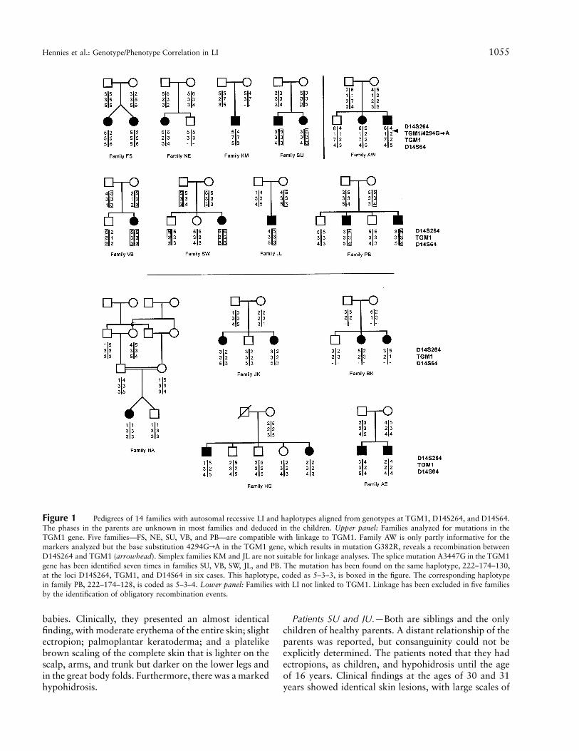
Figure 1 Pedigrees of 14 families with autosomal recessive LI and haplotypes aligned from genotypes at TGM1, D14S264, and D14S64.The phases in the parents are unknown in most families and deduced in the children. Upper panel: Families analyzed for mutations in theTGM1 gene. Five families—FS, NE, SU, VB, and PB—are compatible with linkage to TGM1. Family AW is only partly informative for themarkers analyzed but the base substitution 4294GrA in the TGM1 gene, which results in mutation G382R, reveals a recombination betweenD14S264 and TGM1 (arrowhead). Simplex families KM and JL are not suitable for linkage analyses. The splice mutation A3447G in the TGM1gene has been identified seven times in families SU, VB, SW, JL, and PB. The mutation has been found on the same haplotype, 222–174–130,at the loci D14S264, TGM1, and D14S64 in six cases. This haplotype, coded as 5–3–3, is boxed in the figure. The corresponding haplotypein family PB, 222–174–128, is coded as 5–3–4. Lower panel: Families with LI not linked to TGM1. Linkage has been excluded in five familiesby the identification of obligatory recombination events.
babies. Clinically, they presented an almost identicalfinding, with moderate erythema of the entire skin; slightectropion; palmoplantar keratoderma; and a platelikebrown scaling of the complete skin that is lighter on thescalp, arms, and trunk but darker on the lower legs andin the great body folds. Furthermore, there was a markedhypohidrosis.
Patients SU and JU.—Both are siblings and the onlychildren of healthy parents. A distant relationship of theparents was reported, but consanguinity could not beexplicitly determined. The patients noted that they hadectropions, as children, and hypohidrosis until the ageof 16 years. Clinical findings at the ages of 30 and 31years showed identical skin lesions, with large scales of

1056 Am. J. Hum. Genet. 62:1052–1061, 1998
Table 1
Summary of Clinical Features, Linkage, and Mutation Data for 14 Families with LI
FAMILY ORIGIN
CONSAN-GUINITY
CLINICAL FEATURES LINKAGEa
MUTATIONS IN
TGKTGK
ACTIVITYErythema Scaling TGM1 ICR2B TGM2/3
NE* German � � Whitish, fine � NT NT R323Q NASW* German � � Fair color NI NT NT A3447G/A3447G NAPB* German � � Dark, platelike � NT NT R315C/A3447G �FS* German � � Whitish, fine � NT NT V518M NAJL* German � � Fine NA NA NA G144R/A3447G �
KM* German � �Light brown,
medium-size NA NA NA V518M/V518M �VB* German � � Platelike � NT NT A3447G/A3447G NAAW* German � � Platelike NI NT NT R225H/G382R NASU* German ? � Barklike � NT NT R142H/A3447G NA
NA Moroccan � � Dark � � � NT NAHG German � � Platelike � � � NT NAAE German � � Fair color, fine � � � NT NABK German � � Fair color, fine � � � NT NA
JK German � �Light brown,
medium-size � NI � NT NA
NOTE.—A detailed clinical description of the patients with mutations in TGK, indicated by an asterisk (*), is given in the text.Linkage analyses were not possible in two simplex families, but mutation analyses in the TGM1 gene were performed in patientsfrom these families as well. NI � not informative; NA � not available; and NT � not tested. A plus sign (�) indicates presence; aminus sign (�) indicates absence; and the question mark (?) indicates that a possible consanguinity could not be clarified.
a Assessment for linkage to candidate regions with microsatellites on chromosomes 14 (TGM1), 2 (ICR2B), and 20 (TGM2 andTGM3); see text for details.
the scalp; fine, light scaling of the face; and large brownscales of trunk and limbs, with a barklike pattern of theskin. A patchlike erythema was seen, along with hyper-keratosis of palms and soles and clubbing of the nails.
Linkage Studies
In 12 families with autosomal recessive LI—one ofthese consanguineous, with four to six family membersfrom two or three generations—linkage analyses wereperformed by use of microsatellites in the chromosomalregion of TGM1 on chromosome 14q (Kim et al. 1992;Phillips et al. 1992; Yamanishi et al. 1992). The re-maining two families were simplex families and not suit-able for linkage analyses. To directly assess the segre-gation at the locus TGM1, we analyzed the highlyinformative microsatellite in intron 14 of the TGM1gene (Compton et al. 1992). In addition, we inviestigatedmicrosatellites at the anonymous loci D14S64 andD14S264. A cDNA identified by the Whitehead Insti-tute, WI-9158, is identical to the TGM1 coding se-quence. D14S64 has been localized on the distal sideand D14S264 on the proximal side of WI-9158 by anal-ysis of YAC contigs (Hudson et al. 1995, with supple-mentary data from the Whitehead Institute/MIT Centerfor Genome Research, Human Genome MappingProject).
Five families were compatible with linkage of LI tothe locus TGM1 according to genotype and haplotype
data for TGM1, D14S64, and D14S264 (fig. 1 and table1). However, these results give only a clue to linkage,since single families taken into account are too small togive firm evidence. For the loci D14S264 and D14S64,neither recombination to TGM1 (Russell et al. 1994)nor to one another (Dib et al. 1996) has been reportedso far. In family AW, haplotypes could not be aligned atthese three loci because the mother is uninformative forthe microsatellite at TGM1. A mutation identified inexon 7 of the TGM1 gene in the mother and her affectedchildren, however, revealed a recombination betweenD14S264 and TGM1 in this family (fig. 1). No furtherrecombination event was found in this interval, in thefamilies described here. Family SW was not informativefor the three markers. Five families show obligatory re-combination events between the LI phenotype and thethree microsatellites close to the TGM1 locus (fig. 1).
The families shown not to be linked to TGM1 wereadditionally analyzed for linkage to two other candidateregions for LI. It has been demonstrated by FISH thatthe locus TGM3 is located in close proximity to thetissue transglutaminase gene locus (TGM2) on the longarm of chromosome 20 (Wang et al. 1994). The cDNAWI-7847 of the Whitehead Institute, which is identicalto the TGM2 coding sequence, has been mapped to the1.2-cM interval between D20S107 and D20S99 by ra-diation hybrid mapping (Schuler et al. 1996). The mi-crosatellite markers at D20S101, D20S107, and

Hennies et al.: Genotype/Phenotype Correlation in LI 1057
Figure 2 Pedigrees of 5 families with autosomal recessive LI not linked to TGM1 and haplotypes in candidate regions for LI on chromosomes2 and 20. The phases in the parents are unknown, in most families, and are deduced in the children. Upper panel: Haplotypes aligned fromgenotypes at D2S116, D2S325, D2S157, and D2S143 in the region of the second locus for LI, ICR2B. This locus has recently been mappedbetween the markers at D2S325 and D2S137 (Parmentier et al. 1996). Linkage to ICR2B could be excluded in families BK, HG, and AE. Infamily NA, linkage can most likely be excluded by the lack of homozygosity by descent. Family JK is not informative because of a recombinationbetween D2S157 and D2S143 (arrowhead). Lower panel: Haplotypes aligned from genotypes at D20S101, D20S107, and D20S119 in theregion of the TGM2 and TGM3 genes. Linkage to this region could be excluded in four families. In family NA, again, linkage can most likelybe excluded by the lack of homozygosity by descent.
D20S119 cover a 13-cM region containing the TGM2and TGM3 gene loci. Although the number of probandsin each of the five LI families analyzed is too small forobtaining effective LOD scores, the transglutaminasegene loci on chromosome 20 could be excluded fromlinkage. Obligatory recombination events were identifiedfor at least two of the chromosome 20 markers analyzed,
and exclusion of linkage is confirmed by haplotype anal-yses in four of the families (fig. 2). Linkage can mostlikely be excluded by lack of homozygosity in the con-sanguineous Moroccan family NA, in which both grand-fathers are brothers and both grandmothers are sisters.Furthermore, microsatellites at D2S116, D2S325,D2S157, and D2S143 were genotyped. These loci are

1058 Am. J. Hum. Genet. 62:1052–1061, 1998
Figure 3 Schematic depiction of the putative secondary structureof the TGK polypeptide, based on the secondary structure of bloodcoagulation factor XIII A (Takahashi et al. 1986; Yee et al. 1994)whose primary structure is highly similar to TGK. The positions ofmutations identified in patients with autosomal recessive LI describedhere are marked by arrows. In the case of the splice mutation A3447G,the position of the sequence insertion is indicated, which is caused byfailure in excision of intron 5 and results in a premature stop codon.
located on chromosome 2q33-q35 in the region to whichthe second locus for LI, ICR2B, has recently beenmapped (Parmentier et al. 1996). Linkage to this locuscould also be excluded in at least three of the familiesnot linked to TGM1 (fig. 2 and table 1). In family NA,again, only lack of homozygosity could be demon-strated, and in family JK, which shows a recombinationbetween D2S157 and D2S143, linkage to the second LIlocus cannot be excluded (fig. 2).
Mutation Studies
Mutation analyses in the TGM1 gene were performedin nine families with autosomal recessive LI by SSCPanalyses and direct sequencing of TGM1 exons. Sevendifferent missense mutations and one splice mutationwere found in TGK (fig. 3 and table 1). Besides muta-tions R142H (Russell et al. 1995) and R323Q (Huberet al. 1995a), five novel missense mutations not de-scribed so far were detected: G144R (nucleotideexchange 1494GrA in TGM1, according to Kim et al.1992), R225H (2730GrA), R315C (3515CrT),G382R (4294GrA), and V518M (7915GrA). Thesemutations were not present on 100 chromosomes of un-affected control individuals of German origin. The splicemutation affecting the 3′ splice site of intron 5, A3447G(according to Kim et al. 1992), has been described byHuber et al. (1995a) as A3366G. This mutation wasidentified on seven chromosomes. Two LI patients, SWand VB, are homozygous for this splice mutation,whereas patients JL, PB/EB, and SU/JU are compoundheterozygous for A3447G and one missense mutation.Remarkably, six of seven chromosomes carrying muta-tion A3447G have the same haplotype, 222–174–130,aligned from genotypes at D14S264, TGM1, andD14S64 (fig. 1). In the seventh case, family PB, the cor-responding haplotype is 222–174–128 (i.e., it deviatesat D14S64 by only one [CA] repeat). The respectivemutations were also found in the parents of affectedindividuals, and, in all cases, both parents of homozy-gous patients were shown to be heterozygous for themutation. This finding indicates that all cases are trulyfamilial and recessively inherited. To confirm that theTGM1 mutations detected in affected individuals reallyimpair the function of the enzyme, we investigated skinsections immunohistochemically from three LI patients(PB, JL, and KM) from whom skin biopsies were avail-able (table 1). All of these patients showed markedlydecreased activity of TGK.
Discussion
Linkage Studies
To address the question whether a genotype/pheno-type correlation can be found in patients with autosomal
recessive LI, we have analyzed 14 families with LI, fromGermany and Morocco. Twelve of these were suitablefor linkage studies, and five families could be excludedfrom linkage to TGM1 (table 1). Two families were un-informative, but the remaining five families were com-patible with TGM1 linkage. The families not linked tothe TGM1 gene were additionally assessed for linkageto two further candidate regions. Linkage of LI to theloci TGM2 and TGM3 on chromosome 20, however,could be excluded. A second locus for autosomal reces-sive LI, ICR2B, has been localized to chromosome 2q(Parmentier et al. 1996). This locus could be clearly ruledout as well in three of the families. Consanguineous fam-ily NA, from Morocco, can most likely be excluded bylack of homozygosity for the markers at ICR2B, whichhas originally been identified in Moroccan families.
It has been noted elsewhere that TGK expression isin fact detectable in a number of LI patients (Huber etal. 1995b; Lavrijsen and Maruyama 1995), and a totalof 15 families not linked to the TGM1 region have beendescribed so far (Parmentier et al. 1995; Bale et al. 1996).The findings presented here substantiate the genetic het-erogeneity of LI and demonstrate that mutations re-sulting in a LI phenotype must exist in at least threedifferent genes. There is no obvious candidate for a newLI locus, and we have therefore embarked on a ge-nomewide search for further loci.
Mutation Studies
SSCP and sequencing analyses of the TGM1 gene wereperformed in LI patients, and a TGK assay was carriedout in three cases in which a skin biopsy was available.Eight different mutations were found, including sevenmissense mutations and one splice mutation (fig. 3 and

Hennies et al.: Genotype/Phenotype Correlation in LI 1059
table 1). Among these, mutations R142H and R323Qhave been described elsewhere (Huber et al. 1995a; Rus-sell et al. 1995). Mutation G144R resides in a region ofTGK that obviously exhibits a mutational hot spot. Atotal of four different mutations have been identified inthe residues R142, R143, and G144 (Huber et al. 1995a;Russell et al. 1995). The lack of TGK expression foundin patient JL strengthens the likelihood that G144R isan inactivating mutation. It resides in a segment sup-posed to be crucial for the correct structure and functionof the enzyme (Kim et al. 1994). Moreover, the residuesat positions 142–145 are extremely conserved betweenvarious transglutaminases and among many species. Themutation R225H changes a moderately conserved res-idue, which is also part of the presumed b-sandwichdomain of TGK. It might also act by altering the foldingof the protein. Missense mutation R315C again affectsa highly conserved residue. Furthermore, a mutation hasbeen found in the arginine residue at position 252 ofanother transglutaminase, blood coagulation factor XIIIA, causing inherited factor XIII deficiency (Mikkola etal. 1996). The primary structure of factor XIII A (Tak-ahashi et al. 1986) is highly similar to the structure ofTGK, and the position 252 in factor XIII A correspondsto residue 315 in TGK.
Mutation G382R significantly alters a conserved res-idue. Glycine or alanine residues are found at the cor-responding positions in other transglutaminases, but nocharged side chains, such as those in the arginine residue,are introduced here. Moreover, G382R affects a residueclose to C377 that is part of the supposed catalytic triadof TGK (Yee et al. 1994). It belongs to the highly con-served region around the active site of the enzyme (Tak-ahashi et al. 1986). Remarkably, mutation V518M re-sides much more distal in the TGM1 gene than any othermutation described so far. It affects a residue even distalto the active site and the potential catalytic triad (fig. 3),and this residue is only moderately conserved in trans-glutaminases. The mutation, however, has been identi-fied on a total of three chromosomes, patients FS beingheterozygous and KM being homozygous for this mu-tation. Moreover, the lack of TGK activity in patientKM confirms that this is also a truly inactivatingmutation.
Splice mutation A3447G results in premature stop co-don, as described by Huber et al. (1995a). It has beenobserved on the same haplotype in six cases, whichpoints at a common origin of the chromosomes withmutation A3447G and suggests that this is a rather oldmutation. In the brothers PB and EB and in their mother,A3447G was identified on a haplotype identical atD14S264 and TGM1. The allele at D14S64 is differentby only 2 bp (i.e., one [CA] repeat). This chromosomecould therefore present either the same ancient haplo-type, with a mutation in the microsatellite at D14S64,
or an older recombination between TGM1 and D14S64.Patients SU and JU are compound heterozygotes forTGM1 mutations, although their parents are related. Nodetails about this distant relationship are known, how-ever, and the mutations were found on different hap-lotypes, one of these the common haplotype carrying therecurrent mutation A3447G. In two cases, the patientNE and the sisters FS and JS, only one mutation in theTGM1 gene was identified. The apparent absence of thesecond TGM1 mutation is presumably due to the in-sufficient sensitivity of the SSCP analysis. In both of thesecases, the mutation found has also been described inother LI patients with absent TGK activity. All the mu-tations identified in the TGM1 gene can be assumed toresult in impairment of TGK activity and are thereforethe cause for the phenotype of LI.
Genotype/Phenotype Correlation
Autosomal recessive LI is characterized by remarkableclinical heterogeneity. An erythematous and a nonery-thematous type of LI were distinguished clinically (Ha-zell and Marks 1985; Williams and Elias 1986). More-over, patients might present different patterns and colorsof the scales, they might or might not have an involve-ment of palmoplantar hyperkeratosis, and most but notall were born as collodion babies. We have thereforecompared two genetically differentiated groups of LI pa-tients: one group comprising five unrelated patients ex-cluded from linkage to the TGM1 gene (referred to asnon–TGM1 LI patients), and one group of nine patientswith LI unambiguously caused by TGM1 gene muta-tions (TGM1 LI).
The group of non–TGM1 LI patients included ery-thematous as well as nonerythematous cases, and theypresented the whole range between fine scales of fair,whitish color and large, dark brown scales (table 1).These differences in the clinical picture could still beexplained by different genes causing the disorder. Theentire spectrum of LI variants, however, was also seenin TGM1 LI patients (table 1). Patients with erythemafrom both groups often showed fine and light scaling(six of nine); patients with the nonerythematous formof LI were mostly affected by large, dark brown scales(three of five).
In seeking a genotype/phenotype correlation for spe-cific mutations in the TGM1 gene, we were able to com-pare two patients who carry the same TGM1 genotype.Both the patients VB and SW are homozygous for thesplice mutation A3447G, but they differed in their clin-ical picture. VB suffers from a moderate erythema. Sheshows brownish scales, particularly affecting the lowerlimbs, and marked palmoplantar hyperkeratosis. On thecontrary, SW presented no visible erythema, scaling offair color on the entire surface of the skin, but no in-
1060 Am. J. Hum. Genet. 62:1052–1061, 1998
volvement of palms and soles. The size and color ofscales in patient SW, however, seem to have changed overthe course of time, even in the absence of treatment withretinoids. These findings, together with the clinical var-iability seen within the entire group of TGM1 LI pa-tients, contradict a genotype/phenotype correlation formutations in the TGM1 gene. Particularly, this is alsotrue for the phenotypical differentiation between the er-ythematous and the nonerythematous forms of LI.
In summary, no characteristic differences could be de-tected in the clinical presentation between the TGM1 LIand non–TGM1 LI patients. Each group displayed thewhole spectrum of symptoms for LI, such as erythem-atous and nonerythematous forms, various scaling pat-terns, and existence or lack of palmoplantar kerato-derma. Hence, there are currently no conclusive criteriafor the clinical differentiation of LI types correspondingto their genetic classification. There are many gene prod-ucts involved in the terminal differentiation of the epi-dermis, and mutations in quite a few of these can beenvisaged as a cause for the symptoms seen in LI. Onlythe identification of further genes for LI will allow arefined genetic classification of autosomal recessive LI.Correspondingly, as those findings give further insightinto differentiation processes in the skin, they will pro-vide the basis for further understanding of the etiologyof LI.
Acknowledgments
We are indebted to all the patients and their relatives whocontributed to this study. We are most grateful to MarcelHuber and Daniel Hohl, Lausanne, for performing the TGKactivity assay, and we wish to thank Silke Appel, Berlin, whoparticipated in SSCP analyses, and Martin Jung, Berlin, for hissupport in automated linkage analysis. This work was sup-ported by the Deutsche Forschungsgemeinschaft.
References
Aeschlimann D, Wetterwald A, Fleisch H, Paulsson M (1993)Expression of tissue transglutaminase in skeletal tissues cor-relates with events of terminal differentiation of chondro-cytes. J Cell Biol 120:1461–1470
Anton-Lamprecht I (1992) The skin. In: Papadimitriou JM,Henderson DW, Spagnolo DV (eds) Diagnostic ultrastruc-ture of non-neoplastic diseases. Churchill Livingstone, Ed-inburgh, pp 459–550
Bale SJ, Russell LJ, Lee ML, Compton JG, DiGiovanna JJ(1996) Congenital recessive ichthyosis unlinked to loci forepidermal transglutaminases. J Invest Dermatol 107:808–811
Compton JG, DiGiovanna JJ, Santucci SK, Kearns KS, AmosCI, Abangan DL, Korge BP, et al (1992) Linkage of epider-molytic hyperkeratosis to the type II keratin gene cluster onchromosome 12q. Nat Genet 1:301–305
Dib C, Faure S, Fizames C, Samson D, Drouot N, Vignal A,
Millasseau P, et al (1996) A comprehensive genetic map ofthe human genome based on 5,264 microsatellites. Nature380:152–154
Gentile V, Davies PJ, Baldini A (1994) The human tissue trans-glutaminase gene maps on chromosome 20q12 by in situfluorescence hybridization. Genomics 20:295–297
Hazell M, Marks R (1985) Clinical, histologic, and cell kineticdiscriminants between lamellar ichthyosis and nonbullouscongenital ichthyosiform erythroderma. Arch Dermatol121:489–493
Hohl D, Huber M, Frenk E (1993) Analysis of the cornifiedcell envelope in lamellar ichthyosis. Arch Dermatol 129:618–624
Huber M, Rettler I, Bernasconi K, Frenk E, Lavrijsen SP, PonecM, Bon A, et al (1995a) Mutations of keratinocyte trans-glutaminase in lamellar ichthyosis. Science 267:525–528
Huber M, Rettler I, Bernasconi K, Wyss M, Hohl D (1995b)Lamellar ichthyosis is genetically heterogeneous—cases withnormal keratinocyte transglutaminase. J Invest Dermatol105:653–654
Hudson TJ, Stein LD, Gerety SS, Ma J, Castle AB, Silva J,Slonim DK, et al (1995) An STS-based map of the humangenome. Science 270:1945–1954
Kim IG, McBride OW, Wang M, Kim SY, Idler WW, SteinertPM (1992) Structure and organization of the human trans-glutaminase 1 gene. J Biol Chem 267:7710–7717
Kim SY, Kim IG, Chung SI, Steinert PM (1994) The structureof the transglutaminase 1 enzyme: deletion cloning revealsdomains that regulate its specific activity and substrate spec-ificity. J Biol Chem 269:27979–27986
Lathrop GM, Lalouel JM, Julier C, Ott J (1984) Strategies formultilocus linkage analysis in humans. Proc Natl Acad SciUSA 81:3443–3446
Lavrijsen AP, Maruyama T (1995) Absent transglutaminaseTGK expression in two of three patients with lamellar ich-thyosis. Arch Dermatol 131:363–364
McKusick VA (1994) Mendelian inheritance in man, 11th ed.Johns Hopkins University Press, Baltimore
Mikkola H, Yee VC, Syrjala M, Seitz R, Egbring R, Petrini P,Ljung R, et al (1996) Four novel mutations in deficiency ofcoagulation factor XIII: consequences to expression andstructure of the A-subunit. Blood 87:141–151
Orita M, Suzuki TS, Hayashi K (1989) Rapid and sensitivedetection of point mutations and DNA polymorphisms usingthe polymerase chain reaction. Genomics 5:874–879
Parmentier L, Blanchet-Bardon C, Nguyen S, Prud’homme J-F, Dubertret L, Weissenbach J (1995) Autosomal recessivelamellar ichthyosis: identification of a new mutation intransglutaminase 1 and evidence for genetic heterogeneity.Hum Molec Genet 4:1391–1395
Parmentier L, Lakhdar H, Blanchet-Bardon C, Marchand S,Dubertret L, Weissenbach J (1996) Mapping of a secondlocus for lamellar ichthyosis to chromosome 2q33–35. HumMolec Genet 5:555–559
Phillips MA, Stewart BE, Rice RH (1992) Genomic structureof keratinocyte transglutaminase: recruitment of new exonfor modified function. J Biol Chem 267:2282–2286
Russell LJ, DiGiovanna JJ, Hashem N, Compton JG, Bale SJ(1994) Linkage of autosomal recessive lamellar ichthyosisto chromosome 14q. Am J Hum Genet 55:1146–1152

Hennies et al.: Genotype/Phenotype Correlation in LI 1061
Russell LJ, DiGiovanna JJ, Rogers GR, Steinert PM, HashemN, Compton JG, Bale SJ (1995) Mutations in the gene fortransglutaminase 1 in autosomal recessive lamellar ichthy-osis. Nat Genet 9:279–283
Schuler GD, Boguski MS, Stewart EA, Stein LD, Gyapay G,Rice K, White RE, et al (1996) A gene map of the humangenome. Science 274:540–546
Takahashi N, Takahashi Y, Putnam FW (1986) Primary struc-ture of blood coagulation factor XIIIa (fibrinoligase, trans-glutaminase) from human placenta. Proc Natl Acad Sci USA83:8019–8023
Thacher SM, Rice RH (1985) Keratinocyte-specific transglu-taminase of cultured human epidermal cells: relation tocross-linked envelope formation and terminal differentia-tion. Cell 40:685–695
Traupe H (1989) The ichthyoses: a guide to clinical diagnosis,genetic counseling, and therapy. Springer, Berlin
Traupe H, Kolde G, Happle R (1984) Autosomal dominantlamellar ichthyosis: a new skin disorder. Clin Genet 26:457–461
Wang M, Kim IG, Steinert PM, McBride OW (1994) Assign-ment of the human transglutaminase 2 (TGM2) and trans-glutaminase 3 (TGM3) genes to chromosome 20q11.2. Ge-nomics 23:721–722
Williams ML, Elias PM (1986) The ichthyoses. In: Thiers BH,Dobson RL (eds) Pathogenesis of skin disease. Livingston,New York, pp 519–555
Yamanishi K, Inazawa J, Liew FM, Nonomura K, Ariyama T,Yasuno H, Abe T, et al (1992) Structure of the gene forhuman transglutaminase 1. J Biol Chem 267:17858–17863
Yee VC, Pedersen LC, Le Trong I, Bishop PD, Stenkamp RE,Teller DC (1994) Three-dimensional structure of a trans-glutaminase: human blood coagulation factor XIII. ProcNatl Acad Sci USA 91:7296–7300