Mutations in SNX14 Cause a Distinctive Autosomal-Recessive Cerebellar Ataxia and Intellectual...
26
REPORT Mutations in SNX14 Cause a Distinctive Autosomal-Recessive Cerebellar Ataxia and Intellectual Disability Syndrome Anna C. Thomas, 1,14 Hywel Williams, 1,2,14 Nu ´ ria Seto ´ -Salvia, 1,14 Chiara Bacchelli, 1,2 Dagan Jenkins, 1 Mary O’Sullivan, 1 Konstantinos Mengrelis, 1 Miho Ishida, 1 Louise Ocaka, 1,2 Estelle Chanudet, 1,2 Chela James, 1,2 Francesco Lescai, 1,2,3 Glenn Anderson, 4 Deborah Morrogh, 5 Mina Ryten, 6,7 Andrew J. Duncan, 1 Yun Jin Pai, 8 Jorge M. Saraiva, 9,10 Fabiana Ramos, 9 Bernadette Farren, 11 Dawn Saunders, 12 Bertrand Vernay, 8 Paul Gissen, 1 Anna Straatmaan-Iwanowska, 1 Frank Baas, 13 Nicholas W. Wood, 6 Joshua Hersheson, 6 Henry Houlden, 6 Jane Hurst, 11 Richard Scott, 11 Maria Bitner-Glindzicz, 1,11 Gudrun E. Moore, 1 Se ´rgio B. Sousa, 1,9,15, * and Philip Stanier 1,15, * Intellectual disability and cerebellar atrophy occur together in a large number of genetic conditions and are frequently associated with microcephaly and/or epilepsy. Here we report the identification of causal mutations in Sorting Nexin 14 (SNX14) found in seven affected individuals from three unrelated consanguineous families who presented with recessively inherited moderate-severe intellectual disability, cerebellar ataxia, early-onset cerebellar atrophy, sensorineural hearing loss, and the distinctive association of progressively coarsening facial features, relative macrocephaly, and the absence of seizures. We used homozygosity mapping and whole-exome sequencing to identify a homozygous nonsense mutation and an in-frame multiexon deletion in two families. A homozygous splice site mutation was identified by Sanger sequencing of SNX14 in a third family, selected purely by phenotypic similarity. This discovery confirms that these characteristic features represent a distinct and recognizable syndrome. SNX14 encodes a cellular protein containing Phox (PX) and regulator of G protein signaling (RGS) domains. Weighted gene coexpression network analysis predicts that SNX14 is highly coexpressed with genes involved in cellular protein metabolism and vesicle-mediated transport. All three mutations either directly affected the PX domain or diminished SNX14 levels, implicating a loss of normal cellular function. This manifested as increased cytoplasmic vacuolation as observed in cultured fibroblasts. Our findings indicate an essential role for SNX14 in neural development and function, particularly in development and maturation of the cerebellum. Intellectual disability (ID) syndromes with a small cere- bellum constitute a clinically and genetically heteroge- neous group of neurological disorders for which the under- lying molecular etiology is diverse and established in only a small subset. Several different cellular mechanisms have been implicated including mutations in SIL1, coding for an endoplasmic reticulum resident cochaperone, which cause Marinesco-Sjogren syndrome (MSS [MIM 248800]); 1 sialic acid disorders such as Salla disease (MIM 604369); 2 disorders of peroxisome biogenesis in atypical Refsum dis- ease (MIM 614879); 3 congenital disorders of glycosylation, especially type 1a caused by mutations in PMM2 (MIM 212065); 4 and the X-linked ID-small cerebellum syndrome caused by mutations in OPHN1 (MIM 300486), a Rho- GTPase-activating protein (GAP). 5 Related phenotypes include the group designated as pontocerebellar hypopla- sias, 6 within which causal mutations have been found in a number of genes involved in tRNA biogenesis (including RARS2 [MIM 611523], TSEN54 [MIM 225753], TSEN34 [MIM 612390], TSEN2 [MIM 612389], and CLP1 [MIM 615803]), 6–10 in rRNA processing (the exosomal genes EXOSC3 [MIM 614678] and EXOSC8 [MIM 606019]), 11,12 and another (CHMP1A [MIM 614961]), with a dual role in protein sorting at the endosome and chromatin modifica- tion at the nucleus. 13 Other cellular processes include syn- aptic and cell junction function (CASK [MIM 300749]), 14 cell cycle progression, and cell division (the serine-threo- nine kinase VRK1 [MIM 607596]). 15 At the biochemical level, it is not clear how disruption of many of these genes leads to hindbrain hypoplasia or atrophy. The classification and diagnostic approach of cerebellar disease associated with ID in childhood is complex and challenging, often de- pending on the careful identification and assessment of neuroradiological and other clinical findings. 15–18 1 Genetics and Genomic Medicine, UCL Institute of Child Health, London WC1N 1EH, UK; 2 Centre for Translational Omics-GOSgene, UCL Institute of Child Health, London WC1N 1EH, UK; 3 Department of Biomedicine, Aarhus University, 8000 Aarhus, Denmark; 4 Histopathology Department, Great Or- mond Street Hospital, London WC1N 3JH, UK; 5 NE Thames Regional Genetics Laboratory Service, London WC1N 3BH, UK; 6 UCL Institute of Neurology, London WC1N 3BG, UK; 7 Department of Clinical Genetics, Guy’s Hospital, London SE1 9RT, UK; 8 Developmental Biology and Cancer, UCL Institute of Child Health, London WC1N 1EH, UK; 9 Servic ¸ o de Gene ´tica Me ´dica, Hospital Pedia ´trico, Centro Hospitalar e Universita ´rio de Coimbra, 3000-602 Coimbra, Portugal; 10 University Clinic of Pediatrics, Faculty of Medicine, University of Coimbra, 3000-602 Coimbra, Portugal; 11 Clinical Genetics, Great Ormond Street Hospital, London WC1N 3JH, UK; 12 Radiology, Great Ormond Street Hospital, London WC1N 3JH, UK; 13 Department of Genome Analysis, Aca- demic Medical Center, University of Amsterdam, 1105AZ Amsterdam, the Netherlands 14 These authors contributed equally to this work 15 These authors contributed equally to this work *Correspondence: [email protected] (S.B.S.), [email protected] (P.S.) http://dx.doi.org/10.1016/j.ajhg.2014.10.007. Ó2014 The Authors This is an open access article under the CC BY license (http://creativecommons.org/licenses/by/3.0/). The American Journal of Human Genetics 95, 611–621, November 6, 2014 611
Transcript of Mutations in SNX14 Cause a Distinctive Autosomal-Recessive Cerebellar Ataxia and Intellectual...
REPORT
Mutations in SNX14 Cause a DistinctiveAutosomal-Recessive Cerebellar Ataxiaand Intellectual Disability Syndrome
Anna C. Thomas,1,14 Hywel Williams,1,2,14 Nuria Seto-Salvia,1,14 Chiara Bacchelli,1,2 Dagan Jenkins,1
Mary O’Sullivan,1 Konstantinos Mengrelis,1 Miho Ishida,1 Louise Ocaka,1,2 Estelle Chanudet,1,2
Chela James,1,2 Francesco Lescai,1,2,3 Glenn Anderson,4 Deborah Morrogh,5 Mina Ryten,6,7
Andrew J. Duncan,1 Yun Jin Pai,8 Jorge M. Saraiva,9,10 Fabiana Ramos,9 Bernadette Farren,11
Dawn Saunders,12 Bertrand Vernay,8 Paul Gissen,1 Anna Straatmaan-Iwanowska,1 Frank Baas,13
Nicholas W. Wood,6 Joshua Hersheson,6 Henry Houlden,6 Jane Hurst,11 Richard Scott,11
Maria Bitner-Glindzicz,1,11 Gudrun E. Moore,1 Sergio B. Sousa,1,9,15,* and Philip Stanier1,15,*
Intellectual disability and cerebellar atrophy occur together in a large number of genetic conditions and are frequently associated with
microcephaly and/or epilepsy. Here we report the identification of causal mutations in Sorting Nexin 14 (SNX14) found in seven affected
individuals from three unrelated consanguineous families who presented with recessively inherited moderate-severe intellectual
disability, cerebellar ataxia, early-onset cerebellar atrophy, sensorineural hearing loss, and the distinctive association of progressively
coarsening facial features, relative macrocephaly, and the absence of seizures. We used homozygosity mapping and whole-exome
sequencing to identify a homozygous nonsense mutation and an in-frame multiexon deletion in two families. A homozygous splice
site mutation was identified by Sanger sequencing of SNX14 in a third family, selected purely by phenotypic similarity. This discovery
confirms that these characteristic features represent a distinct and recognizable syndrome. SNX14 encodes a cellular protein containing
Phox (PX) and regulator of G protein signaling (RGS) domains. Weighted gene coexpression network analysis predicts that SNX14 is
highly coexpressed with genes involved in cellular protein metabolism and vesicle-mediated transport. All three mutations either
directly affected the PX domain or diminished SNX14 levels, implicating a loss of normal cellular function. This manifested as increased
cytoplasmic vacuolation as observed in cultured fibroblasts. Our findings indicate an essential role for SNX14 in neural development and
function, particularly in development and maturation of the cerebellum.
Intellectual disability (ID) syndromes with a small cere-
bellum constitute a clinically and genetically heteroge-
neous group of neurological disorders for which the under-
lyingmolecular etiology is diverse and established in only a
small subset. Several different cellular mechanisms have
been implicated including mutations in SIL1, coding for
an endoplasmic reticulum resident cochaperone, which
cause Marinesco-Sjogren syndrome (MSS [MIM 248800]);1
sialic acid disorders such as Salla disease (MIM 604369);2
disorders of peroxisome biogenesis in atypical Refsum dis-
ease (MIM 614879);3 congenital disorders of glycosylation,
especially type 1a caused by mutations in PMM2 (MIM
212065);4 and the X-linked ID-small cerebellum syndrome
caused by mutations in OPHN1 (MIM 300486), a Rho-
GTPase-activating protein (GAP).5 Related phenotypes
include the group designated as pontocerebellar hypopla-
sias,6 within which causal mutations have been found in
1Genetics and Genomic Medicine, UCL Institute of Child Health, London W
Child Health, London WC1N 1EH, UK; 3Department of Biomedicine, Aarhus U
mond Street Hospital, London WC1N 3JH, UK; 5NE Thames Regional Genetics
London WC1N 3BG, UK; 7Department of Clinical Genetics, Guy’s Hospital, L
Child Health, LondonWC1N 1EH, UK; 9Servico de GeneticaMedica, Hospital P
Portugal; 10University Clinic of Pediatrics, Faculty of Medicine, University of
Street Hospital, London WC1N 3JH, UK; 12Radiology, Great Ormond Street H
demic Medical Center, University of Amsterdam, 1105AZ Amsterdam, the Ne14These authors contributed equally to this work15These authors contributed equally to this work
*Correspondence: [email protected] (S.B.S.), [email protected] (P.S.
http://dx.doi.org/10.1016/j.ajhg.2014.10.007. �2014 The Authors
This is an open access article under the CC BY license (http://creativecommon
The American
a number of genes involved in tRNA biogenesis (including
RARS2 [MIM 611523], TSEN54 [MIM 225753], TSEN34
[MIM 612390], TSEN2 [MIM 612389], and CLP1 [MIM
615803]),6–10 in rRNA processing (the exosomal genes
EXOSC3 [MIM 614678] and EXOSC8 [MIM 606019]),11,12
and another (CHMP1A [MIM 614961]), with a dual role in
protein sorting at the endosome and chromatin modifica-
tion at the nucleus.13 Other cellular processes include syn-
aptic and cell junction function (CASK [MIM 300749]),14
cell cycle progression, and cell division (the serine-threo-
nine kinase VRK1 [MIM 607596]).15 At the biochemical
level, it is not clear how disruption of many of these genes
leads to hindbrain hypoplasia or atrophy. The classification
and diagnostic approach of cerebellar disease associated
with ID in childhood is complex and challenging, often de-
pending on the careful identification and assessment of
neuroradiological and other clinical findings.15–18
C1N 1EH, UK; 2Centre for Translational Omics-GOSgene, UCL Institute of
niversity, 8000 Aarhus, Denmark; 4Histopathology Department, Great Or-
Laboratory Service, London WC1N 3BH, UK; 6UCL Institute of Neurology,
ondon SE1 9RT, UK; 8Developmental Biology and Cancer, UCL Institute of
ediatrico, Centro Hospitalar e Universitario de Coimbra, 3000-602 Coimbra,
Coimbra, 3000-602 Coimbra, Portugal; 11Clinical Genetics, Great Ormond
ospital, London WC1N 3JH, UK; 13Department of Genome Analysis, Aca-
therlands
)
s.org/licenses/by/3.0/).
Journal of Human Genetics 95, 611–621, November 6, 2014 611

Figure 1. Phenotype of Affected Individuals from the Three Families PresentedPhotographs and brainMRI scans from family 1 individual IV.3 aged 19 years (A) and 22 years (E); family 1 individual IV.6 aged 6 years (B,F) and 7 years (I, I0); family 2 individual V.1 aged 4.5 years (C, G, J, J0); family 3 individual III.2 aged 22 years (D, H) and 10 years (K, K0);and family 2 individual V.2 aged 9 months (L, L0). Notice the similar facial features mainly characterized by broad face, fullness of theupper eyelid, broad nasal base and slight underdevelopment of the alae, broad and long philtrum, thick lower lip vermillion, and fifthfinger brachycamptodactyly. In the first years of life, no neuroradiological anomalies were observed in affected individuals, as depictedhere by the normal T1-weightedmid-sagittal (L) and coronal (L0) MRI sections, which have not yet been repeated for this individual. MRIimages performed for other individuals during infancy are unavailable but were reported to be normal. At older ages, affected individualshave a small cerebellum with thin folia and enlarged fissures, suggestive of global cerebellar atrophy as shown here in the T1-weightedMRI images from three of the children, one from each family (mid-sagittal sections I–K; coronal sections I0–K0). The pons appear small butin comparison are well preserved.
We recently described an autosomal-recessive condition
in a consanguineous Portuguese family (family 1) in which
two sisters (Figures 1 and 2A) share a similar phenotype,
characterized by severe cerebellar ataxia, severe intellectual
disability (ID), absent speech, coarse facial features, relative
macrocephaly, brachycamptodactyly of fifth fingers, and
early-onset cerebellar atrophy (Table 1).21 To perform ge-
612 The American Journal of Human Genetics 95, 611–621, Novemb
netic analysis aimed at identifying the causal mutation,
informed consents were obtained for all of the parents, pro-
bands, and siblings according to protocols approved by the
ethical review committees at Great Ormond Street Hospital
andCoimbraHospitalCentre. Specificparental consentwas
also given for the use of all of the clinical data and facial
photographs included in this manuscript. The family was
er 6, 2014

Figure 2. Identification of SNX14 Mutations in Three Affected Families(A) Pedigrees of the three families showing genotypes in tested individuals.(B) Sequence traces for families 1 and 3 show pointmutations in genomic DNA (top trace, mutant; bottom trace, wild-type). For family 2,the sequence trace spans the deletion breakpoint, in genomic DNA, and indicates the location of the breakpoints within two Alu repeatsin the schematic diagram of the SNX14 locus shown in (C) (see also Figure S1 for further details).(C) Schematic representation of part of the SNX14 genomic locus (top) and the SNX14 protein (bottom) indicating the location andeffect of the mutations detected in the three families. The protein consists of two predicted transmembrane domains (TM) at theN terminus, followed by the PXA domain, RGS domain, conserved PX phosphoinositide binding domain, and PXC domain situatedtoward the C terminus. The deletion in family 2 is predicted to remove the RGS and PX domains, whereas the splice site mutation infamily 3 removes part of the PX domain.
then investigated by first delineating regions of shared ho-
mozygosity followed by whole-exome sequencing to iden-
tify variants in the implicated regions. Homozygosity map-
pingwas performed on the two parents (III.1 and III.2), two
affected siblings (IV.3 and IV.6), and fourunaffected siblings
(IV.1, IV.4, IV.5, and IV.7) using the InfiniumHDHumanCy-
toSNP-12 BeadChip (Illumina). This revealed 20 candidate
The American
regions spanning a total of 35,846,704 bp,which contained
450RefSeq and886UCSC transcripts (Table S1 available on-
line). Haplotype analysis of the SNP data defined the largest
homozygous region of ~18 Mb on 6q13-q14 (hg19; chr6:
70,500,118–88,497,536). Exome sequencing of the pro-
band (IV.3) from family 1wasperformedusingAgilent Sure-
Select v.4 and Illumina TruSeq. Enriched libraries were
Journal of Human Genetics 95, 611–621, November 6, 2014 613

Table 1. Clinical Findings in the Affected Individuals
Mutation in SNX14(RefSeq NM_153816.3)
Family 1 Family 2 Family 3
Totalhomoz p.Gln866*(c.2596C>T)
homoz p.Val369_Leu702del(c.1108þ1181_2108�2342del)
homozp.Ala603_Gly632del(c.1894þ1G>A)
Subject ID IV.3 IV.6 IV.1 IV.2 V.1 V.2 III.2
Sex F F F M F M F 5F, 2M
Present age (years) 26 14 32 16 10 3 23
Growth at Birtha
Gestational age (weeks) 40 34 (twin) U U 40 40 U
Length, cm (centile) 49 (50th) 42 (10th) U U U U U
Weight, g (centile) 3,600 (50th) 2,100 (25th) U U 2,650 (3–10th) 2,800 (10–25th) U
Head circumference, cm(centile)
35.5 (50th) 32 (50th) U U U 35.5 (90th) U
Growth, Postnatala
Age (years) 22 14.5 29 14 6.7 1.0 22
Height, cm (centile) 155 (10th) 140 (<3rd) U U (91st) 76.1 (<75th) 157.7 (9–25th)
Head circumference(centile)
56.4 (75–90th) 57.5 (90–97th) 59 (>97th) 55 (50th) 55 (>97th) 49.5 (97th) 56.5 (75–90th)
Neurodevelopment
Intellectual disability severe severe severe severe severe NA moderate
Speech (1st words, years) absent sev impaired (13) absent absent absent absent impaired (3) 5/7
Hypotonia þ þ - þ þ þ þ 6/7
Sitting (age, months) 18 16 very late >36 8 >12 12 18b
Walking with help(age, months)
þ (24) þ (20) crawls crawls þ (24) � þ (18)c 4/7
Ataxia þ þ wheelchair wheelchair þ NA þ 5/6
Talipes equino-varum þ þ þ U � � � 3/6
Hypo/areflexia þ þ þ þ � NA þ 5/6
Craniofacial Features
Coarse features þ þ þ þ þ þ þ 7/7
Short palpebral fissures þ þ þ þ þ þ � 6/7
Fullness of the uppereyelid
þ þ þ þ þ þ � 6/7
Broad/bulbous nose þ þ þ þ þ þ þ 7/7
Broad deep long philtrum þ þ þ þ þ þ � 6/7
Thick lip vermilions(upper þ lower)
þ þ þ þ þ þ þ (lower) 7/7
Skeleton and Limbs
Scoliosis/kyphosis � � þ þ � � � 2/7
Brachy/camptodactyly of5th fingers
þ þ þ þ þ � þ 6/7
Short and broadfinger/toes
þ þ þ þ þ þ (þ) 7/7
Elbow motion limitation þ þ U � � � þ 3/6
Hearing loss SN þ � SN SN � SN 5/7
(Continued on next page)
614 The American Journal of Human Genetics 95, 611–621, November 6, 2014

Table 1. Continued
Mutation in SNX14(RefSeq NM_153816.3)
Family 1 Family 2 Family 3
Totalhomoz p.Gln866*(c.2596C>T)
homoz p.Val369_Leu702del(c.1108þ1181_2108�2342del)
homozp.Ala603_Gly632del(c.1894þ1G>A)
Brain Imaging MRI MRI CT MRI MRI MRI MRI
Age at last evaluation 20 years 7 years U 10 months 4.5 years 9 months 10 years
Cerebellar atrophy þ P þ P þ � þ P � þ P 5/7
Pontine thinning þ � U � þ � (þ) 4/7
Abbreviations and symbols are as follows: þ, positive; �, negative/normal; U, data unknown; P, progressive: i.e., two or more sequential scans showed develop-ment/progression of the cerebellar atrophy; SN, sensorineural, moderate-severe, bilateral; NA, not applicable.aGrowth measurements: For the Turkish individuals, the charts used for head circumference measurements were described by Evereklioglu et al.19 and Elmaliet al.20 The Portuguese individuals were compared to national charts except for head circumference >36 cm, or where not available, the charts described by Ever-eklioglu et al.19 were used.bMedian for sitting age.cSubject III.2 from family 3 was the only individual who progressed to independent ambulation, which was reached at age 3 years.
sequenced on an Illumina HiSeq2000 by Perkin Elmer,
resulting in amean of 663 read depth with 68% of targeted
bases covered at least 13. We sequenced one sample
per lane, aligning the resulting reads to the reference
genome build GRCh37/hg19 using Burrows-Wheeler
Aligner (v.0.5.7) and for variant calling we applied GATK
base quality score recalibration,22 INDEL realignment,
and duplicate removal and performed SNP and INDEL dis-
covery and genotyping across all samples simultaneously
using variant quality score recalibration.23 Variant annota-
tion and interpretation analysis was generated through the
use of Ingenuity Variant Analysis software v.3.0.20140422
from Ingenuity Systems. With the use of filters outlined in
Table S2 designed to pinpoint novel or rare homozygous
damaging variants, we reduced the number of variants
from an initial 159,274 genome wide, to a single likely
causalmutation. This was a unique, homozygous nonsense
variant (c.2596C>T [p.Gln866*]) within SNX14, located at
6q14.3,which iswithin the largest autozygous interval (Fig-
ures 2B and 2C; Table S3). Sanger sequencing using Big Dye
Terminator v.1.1 (Life Technologies) on an ABI 3730
sequencer (Applied Biosystems) confirmed that the muta-
tion was homozygous in both affected sisters and segre-
gated as an autosomal recessive, being heterozygous in
both parents and absent or heterozygous in four unaffected
siblings (Figure 2A). The SNX14 locus generates two
transcripts consisting of either 29 exons encoding the
946 amino acid isoform a (RefSeq NM_153816.2) or 26
exons, lacking exons 14, 23, and 24, encoding a shorter
protein of 893 amino acids known as isoform b (RefSeq
NM_020468.3). Both isoforms share the same four
conserved domains: the PX (phosphoinositide binding,
Phox homology), RGS (regulator of G protein signaling),
PXA (PX-associated domain A), and PXC (PX-associated
domain C) (Figure 2C). The c.2596C>T mutation was
identified within an exon that codes for both transcripts
(exon 26, based on the longer transcript) andwas predicted
to result in a protein truncation that would either re-
move the last 81 amino acids, including part of the PXC
The American
domain or, alternatively, trigger nonsense-mediated decay
(Figure 2C).
We identified a Turkish consanguineous family (family
2) with four affected individuals sharing similar clinical
features to family 1 (Table 1; Figures 1 and 2A). SNP geno-
typing (as above), revealed 11 regions of homozygosity
shared between all three of the affected individuals avail-
able at the time of mapping (IV.1, IV.2, and V.1), with
the largest (8,056,114 bp) including the interval contain-
ing SNX14 in 6q14.3, being the only autozygous region
in common with family 1 (Table S1). Individual V.1 under-
went exome sequencing at Dasman Diabetes Institute
(Kuwait City, Kuwait) using methodologies as described
for family 1, with 89% of target bases covered at least 13
with an average depth of 333 per base. Using the same
filtering parameters as those for family 1, we were able to
reduce the number of variants from 199,920 to 18 (Table
S2). As examination of these failed to identify any of these
within the regions of shared homozygosity, we decided to
look in more detail at SNX14. Specifically, we viewed the
reads across SNX14 using Integrative Genomics Viewer
(IGV) software (Broad Institute),24 which revealed a homo-
zygous deletion of 9 consecutive exons from exon 13 to 21
(with respect to isoform a) (Figure S1A and Table S4), while
all DNAs sequenced at the same time from unaffected sub-
jects had good read depth for this interval (Figure S1A). The
deletion was confirmed, first noting the lack of PCR ampli-
fication of individual exons from genomic DNAs, then by
testing a series of primers flanking the approximate break-
point position (Figure S1B). The latter strategy allowed
determination of the precise sequence of the junction
fragment. The deletion most likely occurred as a conse-
quence of illegitimate recombination between two Alu-
repeat sequences found in intron 12 and intron 21. This
resulted in a deletion spanning 25,640 bases, described as
c.1108þ1181_2108�2342del with reference to isoform a
cDNA or hg19 coordinates chr6: 86,255,692–86,230,052
(Figure S1C). In addition, Affymetrix CytoScan 750K array
analysis of the youngest affected individual (V.2) revealed a
Journal of Human Genetics 95, 611–621, November 6, 2014 615

Figure 3. SNX14 RNA Expression in Fibroblasts from Affectedand Control IndividualsqRT-PCR showing relative quantification (RQ DDCT method) ofSNX14 expression in affected fibroblasts samples from each familycompared with controls. Both GAPDH and HPRT1 were used ascombined endogenous controls, with RQ calculated using Ste-pOne analysis software v.2.1 (Life Technologies). Each experimentwas biologically replicated three times with each sample analyzedin triplicate. Using a two-tailed t test, family 1 individual IV.3shows a 60% decrease in expression compared to combined con-trols, where fibroblasts from five different control individualshave been analyzed (N ¼ 5), **p ¼ 0.009. Family 3 individualIII.2 shows a 51% drop in expression compared to controls, *p ¼0.05. However, family 2 individual V.1 shows no significant differ-ence in expression compared to controls, p ¼ 0.74. Error barscorrespond to mean 5 SEM.
homozygous deletion spanning five consecutive probes
within SNX14, corresponding to the interval described
above (Figure S2). Sanger sequencing of cDNA synthe-
sized from fibroblast mRNA (V.1) confirmed that there
was in-frame splicing between exon 12 and exon 22
(Figure S1D), which is predicted to remove 334 amino
acids (p.Val369_Leu702del) from the full-length protein,
including the entire RGS and PX domains (Figure 2C).
We genotyped all of the available family members using
a PCR-based approach to confirm segregation of the dele-
tion. This revealed that only the four affected individuals
were homozygous for the deletion, although eight unaf-
fected individuals were heterozygous and three were ho-
mozygous for the reference allele (Figure 2A).
We next searched the Great Ormond Street Hospital
Clinical Genetics records to identify affected individuals
based solely on similar features. We performed text
searches of the electronic medical records of all children
known to the Clinical Genetics department using the
terms ‘‘pontocerebellar hypoplasia,’’ ‘‘cerebellar hypopla-
sia,’’ OR ‘‘cerebellar atrophy’’ AND ‘‘hearing loss’’ and
manually examined all sets of notes and clinical photo-
graphs, excluding all affected individuals with an alter-
native diagnosis or with microcephaly. This strategy
identified family 2 (above) and a single further individual
in an unrelated Turkish consanguineous family (family 3)
among more than 50 case subjects matching the pontocer-
ebellar or cerebellar search terms. The individual III.1 from
family 3 (Figure 2A) had cerebellar ataxia, sensorineural
hearing loss, slightly coarse facial features, relative macro-
616 The American Journal of Human Genetics 95, 611–621, Novemb
cephaly, and bilateral fifth finger camptodactyly but gener-
ally had milder features than the other affected individuals
in terms of the radiological degree of cerebellar atrophy
and her motor, intellectual, and speech development (Ta-
ble 1). Sanger sequencing of SNX14 exons and exon-intron
boundaries in III.1 identified a homozygous canonical
splice site mutation (c.1894þ1G>A), which was heterozy-
gous in both parents (Figure 2A). Using mRNA isolated
from peripheral blood lymphocytes, we confirmed in-
frame splicing of exon 18 to exon 20 (isoform a),
completely skipping exon 19 (Figure S3). This was pre-
dicted to result in the deletion of 30 amino acids
(p.Ala603_Gly632del) from within the PX domain
(Figure 2C).
Using fibroblast cDNA, we compared mutant and wild-
type transcript levels using quantitative RT-PCR with
Power SYBR green PCR Master Mix on StepOnePlus Real-
Time PCR Systems (Life Technologies). Compared to con-
trols, we noted significantly reduced levels of expression
in families 1 and 3, whereas levels in family 2 were similar
(Figure 3). To investigate protein levels, SNX14 was
analyzed by immunoblotting (Figure S4). A single band
of ~110 kDa was reproducibly obtained in control fibro-
blasts, but no band was detected in either family 1:IV.6
or 2:V.1. For family 1:IV.6, only a low level of mRNA was
present, and this may produce an unstable or subdetect-
able level of protein. This also ties in with the possibility
of activation of the nonsense-mediated decay pathway.
For family 2:V.1, the deletion removes 112 of the 131
amino acid peptides used to raise the anti-SNX14 antibody.
Therefore, it is conceivable that a truncated protein is pre-
sent but not detectable with this assay. For family 3:III.2, a
protein of slightly lower molecular weight (~107 kDa) is
detected, approximating to the loss of amino acids due to
skipping of exon 19. Collectively, the mutation data are
supportive of a loss of normal biological function for
SNX14.
Therefore, we identified a total of seven affected individ-
uals from three unrelated families who share a distinct
rare syndrome resulting from SNX14 mutations. Neither
point mutation was present in the NHLBI-ESP-EVS 4,870
exomes or in our internal database of 358 exomes. The
Database of Genomic Variants lists 13 unaffected individ-
uals who are heterozygous for 7 different but overlapping
microdeletions of ~31–130 kb (n ¼ 1/2,026; 1/2,026;
7/2026; 1/1,557; 1/443; 1/17,421; 1/17,421)25–28 and a sin-
gle 40 kb microduplication (n ¼ 1/2,026),25 all involving
parts of the SNX14 locus. However, a review of 4,500
GOSH children screened using the Affymetrix 750K array
did not identify any further individuals, other than from
family 2, who carry deletions or duplications that included
SNX14.
Only five individuals with microdeletions involving
SNX14 are well described in the literature.29–32 Two of
these are included in DECIPHER, which lists seven individ-
uals with intellectual disability and other various defects
who have large (~5 Mb) heterozygous deletions, and a
er 6, 2014

further two with duplications that encompass SNX14 and
other nearby genes. Of these, only the two unrelated indi-
viduals described by Wentzel et al.,31 who have large inter-
stitial deletions (8.7 Mb and 4.5 Mb) containing SNX14
and three neighboring genes, have some phenotypic simi-
larity to our affected individuals. In particular this includes
the presence of ID, similar facial dysmorphic features,
hearing loss, and macrocephaly. Additionally, one of these
individuals had camptodactyly and limited movement of
the elbows. None of them were reported to have cerebellar
atrophy or ataxia, but both of them had walking diffi-
culties and one was reported to have dyspraxia. Currently,
we cannot exclude the presence of a second, SNX14-spe-
cific point mutation on the non-CNV-carrying chromo-
some in these cases.
In an attempt to further extend the phenotypic spec-
trum associated with SNX14 mutations, we investigated
whole-exome sequencing data obtained for a series of 36
individuals with idiopathic pontocerebellar hypoplasia,
and 168 from dominant and recessive families with idio-
pathic cerebellar ataxia, of which 138 were recessive/early
onset or no family history. However, no likely causal vari-
ants or CNVs affecting SNX14 were identified. These data
suggest that autosomal-recessive SNX14 mutations are
associated with a narrow clinical spectrum. The presence
of coarse faces and the absence of microcephaly and epi-
lepsy are distinctive features within this group of condi-
tions, confirming our findings reported in the description
of the first family.21 Additionally, sensorineural hearing
loss and camptodactyly of fifth fingers seem also to be
prevalent (Table 1). Nevertheless, it should be noted
that clinical recognition is challenging in infants because
dysmorphic features, cerebellar involvement, ID, speech
impairment, and ataxia are progressive and absent at an
early age. Neuroradiological scans performed in the first
years of life appeared normal but later were characterized
by a globally small cerebellum (Table 1, Figure 1). Both
the hemispheres and vermis are affected and there is sepa-
ration of the folia indicating atrophy rather than hypopla-
sia (Figure 1). Evidence for slight pontine thinning was
seen in older patients. This phenotype is significantly
different from the pontocerebellar hypoplasias group of
conditions, which are usually more severe, with prenatal-
onset hypoplasia/atrophy of cerebellum and pons, associ-
ated with progressive microcephaly and seizures.
SNX14 is a member of the large family of sorting nexin
proteins but it has only recently been investigated for
its tissue distribution and cellular function. The earliest
report describes Snx14 mRNA expression using an
in vitro motoneuron selection method.33 Mice in this
study were found to have the highest levels of Snx14
mRNA expression at E12.5, restricted to neuronal lineages
such as the spinal cord. Expression in the brain was seen in
the ventral ventricular region, the floor plate, V (trigemi-
nal) and VIII (vestibulocochlear) cranial ganglia, the
saccule of the inner ear, the developing pituitary gland,
and eye. In general, mRNA distribution was described
The American
as colocalizing with Islet-1 expression, including Islet-1-
positive motoneurons.33 Furthermore, Northern blot anal-
ysis in different adult mouse tissues revealed Snx14
expression in cerebellum and hippocampus and at much
lower levels in cortex, muscle, liver, lung, and heart.33
We investigated SNX14 mRNA expression in human
fetal tissue using RT-PCR and found it to be ubiquitously
present in all tissues analyzed, including heart, skin, brain,
kidney, bone, liver, eye, and placenta (Figure S5). The Allen
Brain Atlas reports in situ hybridization for the p56 mouse
brain, where the highest levels of Snx14 expression were
found in the granule and Purkinje cell layers of the cere-
bellar cortex but also in the hippocampus (granule layers
and dentate gyrus) and the piriform. This was supported
by data from both the human UK Brain Expression Con-
sortium (UKBEC) and the Human Brain Transcriptome
database.34,35 SNX14 is expressed in all brain regions,
with generally increasing levels during prenatal develop-
ment and then plateauing. Interestingly, the pattern the
cerebellum is the region where SNX14 is most highly ex-
pressed and transcript levels continue to increase through
postnatal life until adult (UKBEC data, Figure S6). Most
recently, Huang et al.36 described mouse SNX14 levels as
being high in brain, testis, and lung, similarly showing
an increase in cerebellar levels from embryonic to post-
natal stages (E16.5–p63). Despite a broadly distributed
spatiotemporal pattern, which might suggest function in
many tissues, high levels in the central nervous system
and particularly cerebellum correspond to the described
phenotype (with internal organs spared), particularly the
cerebellar atrophy seen in affected individuals from all
three families presented here.
To date there are 49 mammalian proteins known to
contain a PX domain and the majority of these are classi-
fied as sorting nexin proteins.37 The PX domain binds to
phosphoinositides (PtdIns3P) on the cytoplasmic leaflets
of various organelles and it defines both subcellular locali-
zation and function of different PX domain-containing
proteins including endosomal sorting and trafficking.37
Mutations in the p47phox subunit of NADPH oxidase are
known to cause autosomal-recessive chronic granuloma-
tous disease (MIM 233700),38 whereas SNX27 has been
indirectly linked to synaptic dysfunction in Down syn-
drome through impaired transcriptional regulation,39
and recently SNX10 mutations have been demonstrated
to cause a nonsyndromic autosomal-recessive form of
osteopetrosis (MIM 615085), an osteoclast-related bone
disease.40
Based on predicted domain structure, SNX14 is classified
within the PXA-RGS-PX-PXC subfamily along with
SNX13, SNX19, and SNX25.41 Like SNX13, SNX14 also
contains a putative double transmembrane domain
including a short cytoplasmic leader sequence and an
RGS domain, implying they share a similar function
(Figure 2C). The RGS domain of SNX13 can bind to and in-
crease the GTPase activity of the G-alphas (Gas) subunit of
G-protein coupled receptors (GPCRs). Mediated by the PX
Journal of Human Genetics 95, 611–621, November 6, 2014 617

Figure 4. Electron Micrographs of SkinBiopsy and Cultured Fibroblasts(A) Control cultured fibroblast at 8003magnification.(B and C) Skin section from family 3 indi-vidual III.2 at 8003 magnification.(B) In the epidermis, there was mildhyperkeratosis with keratinocytes showingincreased vacuolation, containing finenonspecific granular material (arrows).(C) In the dermis, collagen and elastictissue had a normal appearance and distri-bution with infrequent vacuoles in thefibroblasts.(D–F) Cultured fibroblasts from family1 individual IV.6, family 2 individualV.1, and family 3 individual III.2, respec-tively, at 8003 magnification. The cellsshowed numerous cytoplasmic vacuoles,often containing dense staining materialsuggestive of lipid degeneration. Samecells at 3,0003 magnification shown in(D0)–(F0). Vacuoles contained granularmaterial or multilamellar bodies or wereempty (see white and black arrows forexamples).
domain, this activity can occur at the endosome, allowing
Gas signal attenuation at the interface with this protein
sorting and degradation pathway.42,43 Loss of functional
SNX13 in the mouse resulted in dramatically altered endo-
cytic/lysosomal compartmentalization in visceral yolk sac
endoderm, with abnormal localization of several endocytic
markers and with the appearance of abundant autophagic
vacuoles.44
A role for SNX14 in endosomal sorting and the regula-
tion of protein degradation is supported by recent analyses
of the prenatal human brain transcriptome.45 Weighted
gene coexpression network analysis (WGCNA) was used
to group genes expressed within mid-fetal human
neocortex into modules in an unsupervised manner.46
This approach is extremely useful in identifying modules
of biologically related genes that are not just coexpressed,
but coregulated.47 This method was applied to predict
that SNX14 is highly coexpressed within a module (C25,
black),45 which is significantly enriched for genes involved
in cellular protein metabolism (Gene ontology term
GO:0044267~cellular protein metabolic process, Bonfer-
roni-corrected p value¼ 9.183 10�5) and vesicle-mediated
transport between the ER and Golgi (GO:0006888~ER to
Golgi vesicle-mediated transport, Bonferroni-corrected p
value ¼ 7.59 3 10�4).
This prompted us to examine cellular ultrastructural
morphology in skin biopsy material and fibroblast cell
lines, using a JEOL 1400 transmission electron microscope
(Figure 4). Compared to control samples, the skin biopsy
showed an adequate epithelial layer with a moderate de-
gree of hyperkeratosis and occasional apoptotic bodies.
618 The American Journal of Human Genetics 95, 611–621, Novemb
Vacuoles with fine nonspecific granular material were
identified in keratinocytes (Figure 4B). In the dermis,
collagen and elastic tissue had a normal appearance and
distribution. Fibroblasts demonstrated infrequent vacu-
oles with granular material and rare electron-dense lami-
nated inclusions suggestive of lipid degeneration. No
sweat glands were available for assessment and small
myelinated and unmyelinated nerves were unremarkable.
Next, we investigated ultrathin sections from cultured skin
fibroblasts from one affected individual from each of the
three families and control subjects. Affected fibroblasts
were found to have frequent cytoplasmic vacuolation
with electron-dense material of variable morphology
including some with evidence of lamella structure as
seen in multilamellar bodies (Figures 4D–4F). Interestingly,
a proportion of fibroblasts from affected individuals but
not control subjects immunostain positive for p62 in a
granular pattern (Figure S7), which might suggest a defect
in the autophagy pathway, as seen in many neurodegener-
ative and neurodevelopmental disorders.48 Interestingly,
cultured fibroblasts from individuals with MSS similarly
contain numerous cytoplasmic electron-dense, sometimes
multilamellar inclusion, bodies in both individuals with
and without SIL1 mutations.1 MSS and the SNX14 pheno-
type share the presence of ID, cerebellar atrophy, hypoto-
nia, and ataxia. Therefore SNX14 should be screened
in MSS-like individuals not carrying an SIL1 mutation,
especially in those without microcephaly, cataracts, or
myopathy (creatinine kinase levels were in the normal
range for the four individuals tested who have SNX14
mutations).
er 6, 2014

Zheng et al.44 reported that heterozygous Snx13þ/� mice
do not have an obvious phenotype, whereas Snx13�/�
mice die between E10.5 and E14.5. Mutants are small
and have exencephaly and abnormal cephalic vasculariza-
tion, presumably in response to defective nutrient uptake
and transport, particularly in the yolk sac endoderm,
which could account for embryonic developmental delay.
Snx14 knockdown studies using lentiviral shRNA, specif-
ically in mouse cortical pyramidal neurons, were recently
reported.36 Despite incomplete (60%) knockdown, signifi-
cantly reduced intrinsic excitability and synaptic function
was recorded. The study of Huang et al.36 also reported
Snx14 to be maternally imprinted but suggest that this
might be mouse specific. Our data support this because un-
affected heterozygous individuals inherit the mutation on
either the maternal or paternal allele (Figure 2A).
In conclusion, in terms of a potential mechanism there
is a compelling case for a role of SNX14 in synaptic trans-
mission.40 Synaptic dysfunction is well known to be impli-
cated in neurodevelopmental disorders associated with
ID49 and this may underlie the pathogenesis of SNX14mu-
tations. Other genes coding RGS proteins, especially of the
Rho GTPase family, cause ID syndromes and have a crucial
role in synaptic structure/function.50 Interestingly, a sig-
nificant phenotypic overlap is seen with OPHN1 encoding
a Rho-GAP protein, in which mutations cause an X-linked
form of syndromic ID typically associated with cerebellar
abnormalities, dysmorphic (often coarse) facial features,
and sometimes macrocephaly.5,50 Overall, our results
implicate an essential role for SNX14 with the breakdown
and recycling of cellular components in human cerebellar
development and maintenance.
Supplemental Data
Supplemental Data include seven figures and four tables and can
be found with this article online at http://dx.doi.org/10.1016/j.
ajhg.2014.10.007.
Acknowledgments
Wewould like to thank the families who contributed to this study.
We would also like to thank the expert assistance of Kerra Pearce
(UCL Genomics) for running the SNP genotyping arrays, Osama
Alsmadi and Dinu Antony for exome sequencing at The Dasman
Diabetes Institute Genome Centre (Kuwait City), and Manuela
Grazina (Faculty of Medicine, University of Coimbra) and Biljana
Lukovic (Chemical Pathology, Great Ormond Street Hospital for
Children) for providing an affected individual’s fibroblast culture.
P.S. is supported by the Great Ormond Street Hospital Children’s
Charity. S.B.S. was supported by Fundacao para a Ciencia e Tecno-
logia (SFRH/BD/46778/2008). A.C.T. is a Wellbeing of Women
Research Associate. G.E.M.’s Fetal Growth and Development
research group is supported byWellbeing of Women, MRC, Sparks
and the National Institute for Health Research Biomedical
Research Centre (NIHR BRC) at Great Ormond Street Hospital
for Children NHS Foundation Trust, and UCL Institute of Child
Health. F.B. is supported by the Joshua Deeth foundation. We
thank the additional members of the GOSgene Scientific Advisory
The American
Board (Philip Beales, Robert Kleta, Horia Stanesku, Nicholas
Lench, Bobby Gaspar, Michael Hubank, and Elia Stupka). GOS-
gene is supported by the NIHR BRC at Great Ormond Street Hos-
pital for Children NHS Foundation Trust and UCL Institute of
Child Health. This report is independent research by the NIHR
BRC Funding Scheme. The views expressed in this publication
are those of the author(s) and not necessarily those of the NHS,
the National Institute for Health Research, or the Department of
Health.
Received: August 21, 2014
Accepted: October 13, 2014
Published: November 6, 2014
Web Resources
The URLs for data presented herein are as follows:
1000 Genomes, http://browser.1000genomes.org
Allen Mouse Brain Atlas, http://mouse.brain-map.org
Database of Genomic Variants (DGV), http://dgv.tcag.ca/dgv/
app/home
dbSNP, http://www.ncbi.nlm.nih.gov/projects/SNP/
Ingenuity Variant Analysis, http://www.ingenuity.com/products/
variant-analysis
NHLBI Exome Sequencing Project (ESP) Exome Variant Server,
http://evs.gs.washington.edu/EVS/
Online Mendelian Inheritance in Man (OMIM), http://www.
omim.org/
RefSeq, http://www.ncbi.nlm.nih.gov/RefSeq
UCSC Genome Browser, http://genome.ucsc.edu
References
1. Ezgu, F., Krejci, P., Li, S., de Sousa, C., Graham, J.M., Jr., Hans-
mann, I., He, W., Porpora, K., Wand, D., Wertelecki, W., et al.
(2014). Phenotype-genotype correlations in patients with
Marinesco-Sjogren syndrome. Clin. Genet. 86, 74–84.
2. Strehle, E.M. (2003). Sialic acid storage disease and related
disorders. Genet. Test. 7, 113–121.
3. Waterham, H.R., and Ebberink, M.S. (2012). Genetics and
molecular basis of human peroxisome biogenesis disorders.
Biochim. Biophys. Acta 1822, 1430–1441.
4. Matthijs, G., Schollen, E., Pardon, E., Veiga-Da-Cunha, M.,
Jaeken, J., Cassiman, J.J., and Van Schaftingen, E. (1997). Mu-
tations in PMM2, a phosphomannomutase gene on chromo-
some 16p13, in carbohydrate-deficient glycoprotein type I
syndrome (Jaeken syndrome). Nat. Genet. 16, 88–92.
5. Philip, N., Chabrol, B., Lossi, A.M., Cardoso, C., Guerrini, R.,
Dobyns, W.B., Raybaud, C., and Villard, L. (2003). Mutations
in the oligophrenin-1 gene (OPHN1) cause X linked congen-
ital cerebellar hypoplasia. J. Med. Genet. 40, 441–446.
6. Namavar, Y., Chitayat, D., Barth, P.G., van Ruissen, F., de Wis-
sel, M.B., Poll-The, B.T., Silver, R., and Baas, F. (2011). TSEN54
mutations cause pontocerebellar hypoplasia type 5. Eur. J.
Hum. Genet. 19, 724–726.
7. Edvardson, S., Shaag, A., Kolesnikova, O., Gomori, J.M., Taras-
sov, I., Einbinder, T., Saada, A., and Elpeleg, O. (2007). Delete-
rious mutation in the mitochondrial arginyl-transfer RNA
synthetase gene is associated with pontocerebellar hypoplasia.
Am. J. Hum. Genet. 81, 857–862.
Journal of Human Genetics 95, 611–621, November 6, 2014 619

8. Budde, B.S., Namavar, Y., Barth, P.G., Poll-The, B.T., Nurnberg,
G., Becker, C., van Ruissen, F., Weterman, M.A., Fluiter, K., te
Beek, E.T., et al. (2008). tRNA splicing endonucleasemutations
cause pontocerebellar hypoplasia. Nat. Genet. 40, 1113–1118.
9. Karaca, E., Weitzer, S., Pehlivan, D., Shiraishi, H., Gogakos, T.,
Hanada, T., Jhangiani, S.N., Wiszniewski, W., Withers, M.,
Campbell, I.M., et al.; Baylor Hopkins Center for Mendelian
Genomics (2014). Human CLP1 mutations alter tRNA biogen-
esis, affecting both peripheral and central nervous system
function. Cell 157, 636–650.
10. Schaffer, A.E., Eggens, V.R., Caglayan, A.O., Reuter, M.S., Scott,
E., Coufal, N.G., Silhavy, J.L., Xue, Y., Kayserili, H., Yasuno, K.,
et al. (2014). CLP1 founder mutation links tRNA splicing and
maturation to cerebellar development and neurodegenera-
tion. Cell 157, 651–663.
11. Wan, J., Yourshaw, M., Mamsa, H., Rudnik-Schoneborn, S.,
Menezes, M.P., Hong, J.E., Leong, D.W., Senderek, J., Salman,
M.S., Chitayat, D., et al. (2012). Mutations in the RNA exo-
some component gene EXOSC3 cause pontocerebellar hypo-
plasia and spinal motor neuron degeneration. Nat. Genet.
44, 704–708.
12. Boczonadi, V., Muller, J.S., Pyle, A., Munkley, J., Dor, T., Quar-
tararo, J., Ferrero, I., Karcagi, V., Giunta, M., Polvikoski, T.,
et al. (2014). EXOSC8 mutations alter mRNA metabolism
and cause hypomyelination with spinal muscular atrophy
and cerebellar hypoplasia. Nat. Commun. 5, 4287.
13. Mochida, G.H., Ganesh, V.S., de Michelena, M.I., Dias, H.,
Atabay, K.D., Kathrein, K.L., Huang, H.T., Hill, R.S., Felie,
J.M., Rakiec, D., et al. (2012). CHMP1A encodes an essential
regulator of BMI1-INK4A in cerebellar development. Nat.
Genet. 44, 1260–1264.
14. Najm, J., Horn, D., Wimplinger, I., Golden, J.A., Chizhikov,
V.V., Sudi, J., Christian, S.L., Ullmann, R., Kuechler, A., Haas,
C.A., et al. (2008). Mutations of CASK cause an X-linked brain
malformation phenotype with microcephaly and hypoplasia
of the brainstem and cerebellum. Nat. Genet. 40, 1065–1067.
15. Renbaum, P., Kellerman, E., Jaron, R., Geiger, D., Segel, R., Lee,
M., King, M.C., and Levy-Lahad, E. (2009). Spinal muscular
atrophy with pontocerebellar hypoplasia is caused by a muta-
tion in the VRK1 gene. Am. J. Hum. Genet. 85, 281–289.
16. D’Arrigo, S., Vigano, L., Grazia Bruzzone, M., Marzaroli, M.,
Nikas, I., Riva, D., and Pantaleoni, C. (2005). Diagnostic
approach to cerebellar disease in children. J. Child Neurol.
20, 859–866.
17. Anheim, M., Fleury, M., Monga, B., Laugel, V., Chaigne, D.,
Rodier, G., Ginglinger, E., Boulay, C., Courtois, S., Drouot,
N., et al. (2010). Epidemiological, clinical, paraclinical and
molecular study of a cohort of 102 patients affected with auto-
somal recessive progressive cerebellar ataxia from Alsace,
Eastern France: implications for clinical management. Neuro-
genetics 11, 1–12.
18. Poretti, A., Wolf, N.I., and Boltshauser, E. (2008). Differential
diagnosis of cerebellar atrophy in childhood. Eur. J. Paediatr.
Neurol. 12, 155–167.
19. Evereklioglu, C., Doganay, S., Er, H., Gunduz, A., Tercan, M.,
Balat, A., and Cumurcu, T. (2002). Craniofacial anthropom-
etry in a Turkish population. Cleft Palate Craniofac. J. 39,
208–218.
20. Elmali, F., Altunay, C., Mazicioglu, M.M., Kondolot, M., Oz-
turk, A., and Kurtoglu, S. (2012). Head circumference growth
reference charts for Turkish children aged 0-84 months.
Pediatr. Neurol. 46, 307–311.
620 The American Journal of Human Genetics 95, 611–621, Novemb
21. Sousa, S.B., Ramos, F., Garcia, P., Pais, R.P., Paiva, C., Beales,
P.L., Moore, G.E., Saraiva, J.M., and Hennekam, R.C.M.
(2014). Intellectual disability, coarse face, relative macroce-
phaly, and cerebellar hypotrophy in two sisters. Am. J. Med.
Genet. A. 164A, 10–14.
22. McKenna, A., Hanna, M., Banks, E., Sivachenko, A., Cibulskis,
K., Kernytsky, A., Garimella, K., Altshuler, D., Gabriel, S., Daly,
M., and DePristo, M.A. (2010). The Genome Analysis Toolkit:
a MapReduce framework for analyzing next-generation DNA
sequencing data. Genome Res. 20, 1297–1303.
23. DePristo, M.A., Banks, E., Poplin, R., Garimella, K.V., Maguire,
J.R., Hartl, C., Philippakis, A.A., del Angel, G., Rivas, M.A.,
Hanna, M., et al. (2011). A framework for variation discovery
and genotyping using next-generation DNA sequencing data.
Nat. Genet. 43, 491–498.
24. Thorvaldsdottir, H., Robinson, J.T., and Mesirov, J.P. (2013).
Integrative Genomics Viewer (IGV): high-performance geno-
mics data visualization and exploration. Brief. Bioinform.
14, 178–192.
25. Shaikh, T.H., Gai, X., Perin, J.C., Glessner, J.T., Xie, H., Mur-
phy, K., O’Hara, R., Casalunovo, T., Conlin, L.K., D’Arcy, M.,
et al. (2009). High-resolution mapping and analysis of copy
number variations in the human genome: a data resource
for clinical and research applications. Genome Res. 19,
1682–1690.
26. Itsara, A., Cooper, G.M., Baker, C., Girirajan, S., Li, J., Absher,
D., Krauss, R.M., Myers, R.M., Ridker, P.M., Chasman, D.I.,
et al. (2009). Population analysis of large copy number vari-
ants and hotspots of human genetic disease. Am. J. Hum.
Genet. 84, 148–161.
27. Jakobsson, M., Scholz, S.W., Scheet, P., Gibbs, J.R., VanLiere,
J.M., Fung, H.C., Szpiech, Z.A., Degnan, J.H., Wang, K., Guer-
reiro, R., et al. (2008). Genotype, haplotype and copy-number
variation in worldwide human populations. Nature 451, 998–
1003.
28. Cooper, G.M., Coe, B.P., Girirajan, S., Rosenfeld, J.A., Vu, T.H.,
Baker, C.,Williams, C., Stalker, H., Hamid, R., Hannig, V., et al.
(2011). A copy number variation morbidity map of develop-
mental delay. Nat. Genet. 43, 838–846.
29. Turleau, C., Demay, G., Cabanis, M.O., Lenoir, G., and de
Grouchy, J. (1988). 6q1 monosomy: a distinctive syndrome.
Clin. Genet. 34, 38–42.
30. Van Esch, H., Rosser, E.M., Janssens, S., Van Ingelghem, I.,
Loeys, B., and Menten, B. (2010). Developmental delay
and connective tissue disorder in four patients sharing a
common microdeletion at 6q13-14. J. Med. Genet. 47,
717–720.
31. Wentzel, C., Lynch, S.A., Stattin, E.L., Sharkey, F.H., Anneren,
G., and Thuresson, A.C. (2010). Interstitial deletions at
6q14.1-q15 associated with obesity, developmental delay
and a distinct clinical phenotype. Mol. Syndromol. 1, 75–81.
32. Lowry, R.B., Chernos, J.E., Connelly, M.S., and Wyse, J.P.
(2013). Interstitial deletions at 6q14.1q15 associated with
developmental delay and a marfanoid phenotype. Mol. Syn-
dromol. 4, 280–284.
33. Carroll, P., Renoncourt, Y., Gayet, O., De Bovis, B., and Alonso,
S. (2001). Sorting nexin-14, a gene expressed in motoneurons
trapped by an in vitro preselection method. Dev. Dyn. 221,
431–442.
34. Trabzuni, D., Ryten, M., Walker, R., Smith, C., Imran, S., Ram-
asamy, A., Weale, M.E., and Hardy, J. (2011). Quality control
parameters on a large dataset of regionally dissected human
er 6, 2014

control brains for whole genome expression studies.
J. Neurochem. 119, 275–282.
35. Kang, H.J., Kawasawa, Y.I., Cheng, F., Zhu, Y., Xu, X., Li, M.,
Sousa, A.M., Pletikos, M., Meyer, K.A., Sedmak, G., et al.
(2011). Spatio-temporal transcriptome of the human brain.
Nature 478, 483–489.
36. Huang, H.-S., Yoon, B.-J., Brooks, S., Bakal, R., Berrios, J.,
Larsen, R.S., Wallace, M.L., Han, J.E., Chung, E.H., Zylka,
M.J., and Philpot, B.D. (2014). Snx14 regulates neuronal excit-
ability, promotes synaptic transmission, and is imprinted in
the brain of mice. PLoS ONE 9, e98383.
37. Teasdale, R.D., and Collins, B.M. (2012). Insights into the PX
(phox-homology) domain and SNX (sorting nexin) protein
families: structures, functions and roles in disease. Biochem.
J. 441, 39–59.
38. Casimir, C.M., Bu-Ghanim, H.N., Rodaway, A.R., Bentley,
D.L., Rowe, P., and Segal, A.W. (1991). Autosomal recessive
chronic granulomatous disease caused by deletion at a dinu-
cleotide repeat. Proc. Natl. Acad. Sci. USA 88, 2753–2757.
39. Wang, X., Zhao, Y., Zhang, X., Badie, H., Zhou, Y., Mu, Y., Loo,
L.S., Cai, L., Thompson, R.C., Yang, B., et al. (2013). Loss of
sorting nexin 27 contributes to excitatory synaptic dysfunc-
tion by modulating glutamate receptor recycling in Down’s
syndrome. Nat. Med. 19, 473–480.
40. Pangrazio, A., Fasth, A., Sbardellati, A., Orchard, P.J., Kasow,
K.A., Raza, J., Albayrak, C., Albayrak, D., Vanakker, O.M., De
Moerloose, B., et al. (2013). SNX10 mutations define a
subgroup of human autosomal recessive osteopetrosis
with variable clinical severity. J. Bone Miner. Res. 28,
1041–1049.
41. Worby, C.A., and Dixon, J.E. (2002). Sorting out the cellular
functions of sorting nexins. Nat. Rev. Mol. Cell Biol. 3,
919–931.
The American
42. Zheng, B., Ma, Y.-C., Ostrom, R.S., Lavoie, C., Gill, G.N., Insel,
P.A., Huang, X.-Y., and Farquhar, M.G. (2001). RGS-PX1, a
GAP for GalphaS and sorting nexin in vesicular trafficking.
Science 294, 1939–1942.
43. Zheng, B., Lavoie, C., Tang, T.-D., Ma, P., Meerloo, T., Beas, A.,
and Farquhar, M.G. (2004). Regulation of epidermal growth
factor receptor degradation by heterotrimeric Galphas pro-
tein. Mol. Biol. Cell 15, 5538–5550.
44. Zheng, B., Tang, T., Tang, N., Kudlicka, K., Ohtsubo, K., Ma, P.,
Marth, J.D., Farquhar, M.G., and Lehtonen, E. (2006). Essen-
tial role of RGS-PX1/sorting nexin 13 in mouse development
and regulation of endocytosis dynamics. Proc. Natl. Acad.
Sci. USA 103, 16776–16781.
45. Miller, J.A., Ding, S.L., Sunkin, S.M., Smith, K.A., Ng, L., Szafer,
A., Ebbert, A., Riley, Z.L., Royall, J.J., Aiona, K., et al. (2014).
Transcriptional landscape of the prenatal human brain.
Nature 508, 199–206.
46. Langfelder, P., and Horvath, S. (2008). WGCNA: an R package
for weighted correlation network analysis. BMC Bioinformat-
ics 9, 559.
47. Konopka, G. (2011). Functional genomics of the brain: uncov-
ering networks in the CNS using a systems approach. Wiley
Interdiscip. Rev. Syst. Biol. Med. 3, 628–648.
48. Lee, K.M., Hwang, S.K., and Lee, J.A. (2013). Neuronal auto-
phagy and neurodevelopmental disorders. Exp. Neurobiol.
22, 133–142.
49. Zoghbi, H.Y., and Bear, M.F. (2012). Synaptic dysfunction in
neurodevelopmental disorders associated with autism and
intellectual disabilities. Cold Spring Harb. Perspect. Biol. 4,
a009886.
50. Ba, W., van der Raadt, J., and Nadif Kasri, N. (2013). Rho
GTPase signaling at the synapse: implications for intellectual
disability. Exp. Cell Res. 319, 2368–2374.
Journal of Human Genetics 95, 611–621, November 6, 2014 621

The American Journal of Human Genetics, Volume 95
Supplemental Data
Mutations in SNX14 Cause a Distinctive
Autosomal-Recessive Cerebellar Ataxia
and Intellectual Disability Syndrome
Anna C. Thomas, Hywel Williams, Núria Setó-Salvia, Chiara Bacchelli, Dagan Jenkins,
Mary O’Sullivan, Konstantinos Mengrelis, Miho Ishida, Louise Ocaka, Estelle Chanudet,
Chela James, Francesco Lescai, Glen Anderson, Deborah Morrogh, Mina Ryten,
Andrew J. Duncan, Yun Jin Pai, Jorge M. Saraiva, Fabiana Ramos, Bernadette Farren,
Dawn Saunders, Bertrand Vernay, Paul Gissen, Anna Straatmaan-Iwanowska, Frank
Baas, Nicholas W. Wood, Joshua Hersheson, Henry Houlden, Jane Hurst, Richard
Scott, Maria Bitner-Glindzicz, Gudrun E. Moore, Sérgio B. Sousa, and Philip Stanier
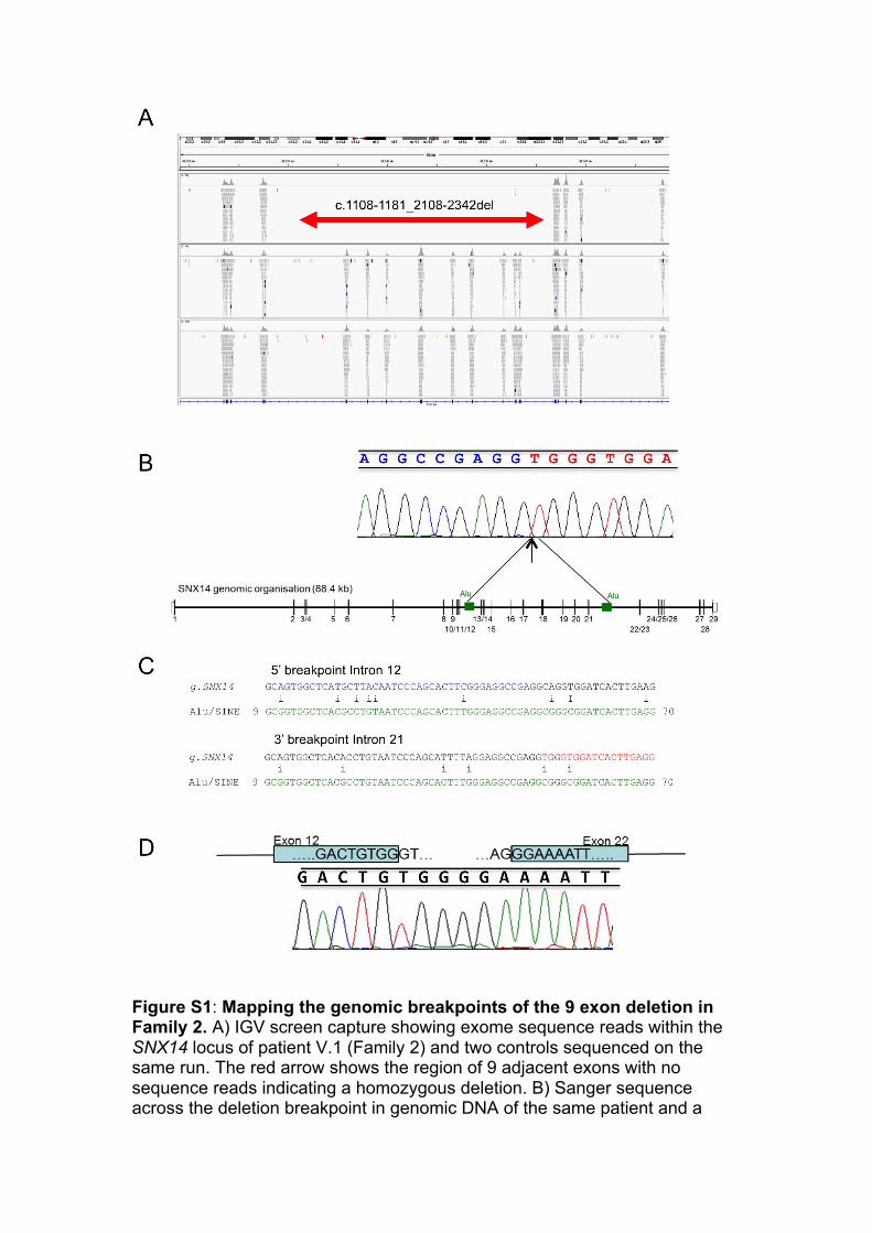
Figure S1: Mapping the genomic breakpoints of the 9 exon deletion in Family 2. A) IGV screen capture showing exome sequence reads within the SNX14 locus of patient V.1 (Family 2) and two controls sequenced on the same run. The red arrow shows the region of 9 adjacent exons with no sequence reads indicating a homozygous deletion. B) Sanger sequence across the deletion breakpoint in genomic DNA of the same patient and a

schematic diagram of the locus to show the position of the flanking Alu/Sine repeat sequences. The arrow represents the breakpoint by indicating the first base of the second Alu/Sine sequence. C) SNX14 genomic sequences from Introns 12 and 21 aligned against the Alu/Sine repeat sequence using ClustalW show the origin of the breakpoint. In the deletion, blue and red sequences are contiguous (as seen in B). D) Sanger sequence of cDNA from patient V.1 and a schematic diagram illustrate that exon 12 splices to exon 22.

Figure S2. Copy number analysis on Family 2 patient V.2 using the Affymetrix CytoScan 750K array. The minimum deleted region is defined by CGH probes marked B (86,233,056) and C (86,252,888); the maximum deleted region is defined by A (86,223,224) to D (86,257,848).

Figure S3. SNX14 cDNA sequences from Family 3, demonstrating that the splice site variant results in skipping of exon 19.

Figure S4. SNX14 levels in affected and control fibroblasts were
detected by western blotting. SNX14 levels in cases and controls were
analysed by immuno-blotting total protein extracted from fibroblast cells using
an anti-SNX14 antibody (1:500; Sigma HPA017639). Protein loading is shown
using the image obtained from a ChemiDoc MP imaging system with Mini-
PROTEAN TGX Stain-Free precast gels (Bio Rad, UK). The Image data was
analysed using Image Lab version 4.1 (Bio Rad, UK). A representative blot of
three independent experiments with similar results is shown.
Fam
ily 2
V.1
Fa
mily
3 II
I.1
Fam
ily 1
II.3
Co
ntro
l 1
Cont
rol 2
SNX14
Protein loading
110 kDa
107 kDa

Figure S5. RT-PCR of SNX14 expression in fetal tissues. Tissues include heart, skin, brain, kidney, bone, liver, eye and placenta. Where available, tissues were used from two separate fetal samples.

Figure S6. Human brain SNX14 and SNX13 transcript analysis. Top: Spatio-temporal SNX14 (left) and SNX13 (right) transcriptomes of the human brain using data from the Human Brain Transcriptome (HBT) database.34 This study assessed 6 brain regions (cerebellar cortex (CBC), mediodorsal nucleus of the thalamus (MD), striatum (STR), amygdale (AMY), hippocampus (HIP) and the neocortex (NCX)) over 15 periods of the human pre and post-natal development. These data were generated from Affymetrix Human Exon 1.0 ST Arrays performed on 1,340 tissue samples collected from 57 developing and adult post-mortem brains of clinically unremarkable donors representing males and females of multiple ethnicities. Levels of SNX14 increase throughout the brain during fetal development, before plateauing. In the cerebellum, expression continues to rise during postnatal life reaching its highest level during adulthood. Contrast with SNX13, which decreases towards birth, then more modestly increases in most brain regions. Bottom: Regional distribution of SNX14 (left) and SNX13 (right) expression in human brain. Box plots of mRNA levels from 10 brain regions (left to right: the cerebellum (CRBL, n=130), frontal cortex (FCTX, n=127), hippocampus (HIPP, n=122), medulla (specifically inferior olivary nucleus, MEDU, n=119), occipital cortex (specifically primary visual cortex, OCTX, n=129), putamen (PUTM, n=129), substantia nigra (SNIG, n=101), temporal cortex (TCTX, n=119), thalamus (THAL, n=124), and intralobular white matter (WHMT, n=131)) are based on microarray experiments and plotted on a log2 scale (y-axis). Material and methods were as previously reported.34,35 In brief, these
Age (Days)
Sign
al in
tens
ity (L
og2)
50 100 200 500 2000 10000 30000
3
4
5
6
7
8
9
10
11
●
●● ●
●●●●●
● ●
●●
●●
●
●●●
●
●●
●●
●●
●
●
●
●●
●
●
●
●
●
● ●●
●
●
●●● ●
●
●●●●
●
●●
● ●
●
●
●● ●
●
●
●
●
●
●
●
●
●●
●●●
●●●
●
●
●
●
●
●
●
●●
●
● ●
●●●
●
●
●
●
●●
●●
●
●
●
●
●
●
●
●
●●
●●●
●●
●●●
●
●
●
●
●
●●●●
●●●
●
● ●
●
●
●●●
●
●
●
●
●●
●●
●
●●
●
●
●
●
●
●
●
●●
● ●● ●●
●
●●●
●
●●
●
●●
●
●
●
●
●
●
●●●
●●
●
●●
●
●
●●
●●● ●
●
●●
●
●
●●
●
●
●
●
●●
●
●
●
●
●●
●
●●
● ●●
●
●●
●
●
●
●
●
●●
●
●● ●
●
●●
●
● ●
●●
●●●
●●
●
●
●
●
●
●
●
●
●
●●
●
●●
●
●
●
●
●●
●
●
●
●
●
●
●
●
●
●
●
●
●
●
●●
●
●
●
●
●
●●●
●
●●
●●
●●
●
●
●
●
●●●
●
Period: 1 2 3 4 5 6 7 8 9 10 11 12 13 14 15
SNX14
●
●
●
●
●
●
NCXHIPAMY
STRMDCBC
Age (Days)
Sign
al in
tens
ity (L
og2)
50 100 200 500 2000 10000 30000
3
4
5
6
7
8
9
10
●●
●
● ●
●
●●●●
●
●
●●
●
●●
●
●
●
●
●
●
●
●●
● ●
●
●
●
●
●
●
●
●
●
● ●
●
●
●
●
●●●
● ●●●
●
●
●●
●
●
●●
●
●
●
●●
●●
●
●
●
●
●
●
●
●
●
●●●
●
●
●
●
● ● ●●
●
●
● ●
● ●
●
●
●●
●
●
● ●
●
●
●
●
●
●
●
●
●
●
●●
●
●●
●
●
●
●
●
●
●
●●●
●●●
●
●
●
●
●
●
●
●
●
●
●
●
●
●
●
●
●●
●
●●
●
●
●
●
●
●
●
●●●
●
● ●
●
●
●
●●
●
●
●
●
●●
●
●
●●
●
●●
●
●
●
●
●
●
●
●
●
●●
●
●
● ●
●
●●
●
●
●
● ●
●
●
●
●●
●
●
●
●
●
●
●
●
●
●
●
●
●
●
●●
●
●
●
●
●
●
●
●
●●
●●
●
●●
●
●
●
●●
● ●●●●
●
●
●
●
●
●●●
●
●
●●
●●
●
●●●
●●
●
●
●
●
●●
●
●
●
●
●
●
●●
●
●
●
●
●
●
●
●
●
●●
●
●●●
●
●●
●
●
●
●●
Period: 1 2 3 4 5 6 7 8 9 10 11 12 13 14 15
SNX13
●
●
●
●
●
●
NCXHIPAMY
STRMDCBC
●
●
●
●
●●
●
●
●
●●
●
●
●
●
●●
●
●
●●●
●
●
●
●
●●
●
●
●
●
●
●
●
●
●
●
●
●
●●
●
5.5
6.0
6.5
7.0
7.5
8.0
Affymetrix ID t3040073
Expr
essi
on le
vel i
n lo
g 2 s
cale
FCTX(N=127)
CRBL(N=130)
HIPP(N=122)
TCTX(N=119)
THAL(N=124)
SNIG(N=101)
WHMT(N=131)
OCTX(N=129)
MEDU(N=119)
PUTM(N=129)
Source:BRAINEACFold change between FCTX and PUTM = 1.2 (p=2.8e−06)
●
●
●
●
●
●
●
●
●●
●
●
●
●
●
●
●
●
●
●
● ●
●●
●
●●
●
●●
5.0
5.5
6.0
6.5
7.0
7.5
8.0
Affymetrix ID t2963313
Expr
essi
on le
vel i
n lo
g 2 s
cale
CRBL(N=130)
TCTX(N=119)
FCTX(N=127)
OCTX(N=129)
THAL(N=124)
HIPP(N=122)
SNIG(N=101)
MEDU(N=119)
WHMT(N=131)
PUTM(N=129)
Source:BRAINEACFold change between CRBL and PUTM = 1.4 (p=1.8e−17)

samples originate from 134 adult individuals from the UK Brain Expression Consortium and were profiled on 1231 Affymetrix Human Exon 1.0 ST arrays. Whiskers extend from the box to 1.5 times the inter-quartile range.

Figure S7. p62 immunostaining of fibroblasts suggest defective autophagy. Cultured fibroblast cell suspensions from two patients and two controls were centrifuged to prepare smears using a Thermo Scientific Cytospin. A p62 antibody (1:500 dilution, BD Bioscience), was used to investigate the autophagy pathway, employing standard protocols on a Leica BOND MAX immunostainer. Many cells in the Family 3:III.2 sample were strongly positive with a granular appearance. A smaller number of positive cells with the same distinct morphology were seen in Family 2:V.1 fibroblasts. For the controls, one sample was negative, whilst the other C(2) showed a higher level of background staining without the granular morphology.

Table S1. Regions of homozygosity shared between affected individuals in Families 1 and 2.
Family 1 (II.3 +II.6) Family2 (IV.1+IV.2+V.1) Chr Start End Size (bp) Chr Start End Size (bp) 1 49215178 50025098 809920 2 167634200 168219266 585066 2 14334445 15105276 770831 3 87497605 87550022 52417 2 98451967 98972624 520657 3 87550022 98418129 767324 4 52732440 53571137 838697 3 137516694 138461351 944657 4 59336192 60143936 807744 5 122710247 123328850 618603 5 172038414 173762787 1724373 6 80467595 88523709 8056114 6 44642275 45480198 837923 6 103137113 103711271 574158 6 67688533 69401011 1712478 8 47965922 48816429 850507 6 70500118 88497536 17997418 9 88677735 89150849 473114 6 91529972 91695918 165946 11 32236138 34302743 2066605 6 107328569 108056760 728191 17 19741141 20387780 646639 8 89115291 89766139 650848 8 115018872 115580787 561915 8 119514102 120079494 565392 8 144993574 145971916 978342 11 8403642 9363862 960220 14 103823162 104381937 558775 15 56763412 57589554 826142 15 72095961 73111070 1015109 21 43733078 46548861 2815783 The largest shared region in each family, which is the only common region between the two families and contains the SNX14 locus, is highlighted in yellow. Nucleotide positions are from the hg19 assembly.

Table S2: Filtering parameters used in Ingenuity Variant Analysis.
Data corresponds to individual II.3 in Family 1 and V.1 in Family 2.
Number of variants
(Family 1)
Number of variants
(Family 2)Keep Exclude
159,274 199,920 All variants
141,401 171,260
Call quality ≥ 20, Read Depth ≥ 10 AND outside top 0.2% most exonically variable 100 base windows in healthy public genomes (1000 genomes) AND outside top 1.0% most exonically variable genes
25,032 31,313Observed with allele frequency ≥ 0.1% in the 1000 genomes project or the public Complete Genomics genomes or NHLBI ESP exomes
1,096 1,326
Pathogenic, Possibly Pathogenic OR established gain of function in the literature OR inferred activating mutations by Ingenuity OR predicted gain of function by BSIFT OR Frameshift, in-‐frame indel, or stop codon change OR Missense and not predicted to be innocuous by SIFT or Polyphen-‐2 OR disrupt splice site upto 2.0 bases into intron
2 22 Homozygous in case Homozygous or heterozygous in ≥1 control exome (N=6)
1 18
within 1 hop upstream and that are known or predicted to affect: mental retardation, developmental delay or diseases consistent with these phenotypes OR genes within 1 hop downstream of them

Table S3 RefSeq genes in the Family 1 region of shared homozygosity chr6:70500118-88497536, showing mean coverage and % bases covered at >1x and >10x read depth.
Gene mean coverage % >1x % >10x
LMBRD1 71 98 96 COL19A1 66 96 94 COL9A1 90 99 98 FAM135A 58 88 83 C6orf57 89 100 93 SMAP1 64 98 91 B3GAT2 33 88 80 OGFRL1 70 99 87 RIMS1 77 92 89 KCNQ5 85 92 90 KHDC1L 67 100 100 KHDC1 50 94 74 C6orf147 1 42 0 DPPA5 183 100 95 KHDC3L 65 96 92 OOEP 33 100 93 DDX43 92 99 93 MB21D1 72 100 100 MTO1 82 96 87 EEF1A1 27 76 41 SLC17A5 65 97 84 CD109 76 98 96 COL12A1 84 99 96 COX7A2 45 100 90 TMEM30A 52 89 86 FILIP1 99 75 72 SENP6 76 96 94 MYO6 56 95 94 IMPG1 67 96 93 HTR1B 77 81 83 IRAK1BP1 78 91 90 PHIP 86 98 95 HMGN3 39 99 96 LCA5 60 76 70 SH3BGRL2 19 83 53 RNY4 0 0 0 C6orf7 0 30 0 ELOVL4 51 87 82 TTK 64 95 94 BCKDHB 68 92 90 FAM46A 89 76 73 IBTK 92 96 93 TPBG 16 41 15

UBE3D 77 97 95 DOPEY1 78 97 96 PGM3 102 97 88 RWDD2A 50 95 65 ME1 61 96 84 PRSS35 19 46 21 SNAP91 46 97 85 RIPPLY2 40 100 97 CYB5R4 66 98 95 MRAP2 51 83 58 CEP162 92 95 94 TBX18 37 92 79 NT5E 85 91 84 SNX14 77 99 89 SYNCRIP 70 86 72 SNHG5 11 55 23 HTR1E 95 50 48 CGA 19 60 54 ZNF292 47 98 96 GJB7 25 54 16 SMIM8 27 43 38 C6orf163 72 100 92 C6orf164 9 61 46 C6orf165 64 92 91 SLC35A1 65 94 92 RARS2 81 100 96 ORC3 66 100 98 AKIRIN2 27 87 75 Average 60 86 77

Table S4 Family 2 V.1 exome sequencing coverage of SNX14 showing percentage coverage at >1x or >10x sequence reads Target % >1x % >10x exon 6:86215215-86215723 100 90.4 SNX14_exon29 6:86216951-86217007 100 100 SNX14_exon28 6:86217686-86217777 100 100 SNX14_exon27 6:86223518-86223613 100 100 SNX14_exon26 6:86223788-86223952 100 86.7 SNX14_exon25 6:86224224-86224347 100 100 SNX14_exon24 6:86227474-86227593 100 100 SNX14_exon23 6:86227726-86227766 100 100 SNX14_exon22 6:86235844-86235955 0 0 SNX14_exon21 6:86237980-86238080 0 0 SNX14_exon20 6:86239910-86239993 0 0 SNX14_exon19 6:86243317-86243518 0 0 SNX14_exon18 6:86246510-86246642 0 0 SNX14_exon17 6:86248556-86248582 0 0 SNX14_exon16 6:86251703-86251761 0 0 SNX14_exon15 6:86252900-86253024 58.4 0 SNX14_exon14 6:86253323-86253478 0 0 SNX14_exon13 6:86256830-86256944 100 100 SNX14_exon12 6:86257035-86257115 100 100 SNX14_exon11 6:86257224-86257268 100 100 SNX14_exon10 6:86258019-86258094 100 100 SNX14_exon9 6:86259441-86259597 100 81.5 SNX14_exon8 6:86267694-86267778 100 100 SNX14_exon7 6:86275050-86275137 100 100 SNX14_exon6 6:86277252-86277295 100 100 SNX14_exon5 6:86281855-86281933 100 100 SNX14_exon4 6:86282016-86282092 100 100 SNX14_exon3 6:86283976-86284096 100 100 SNX14_exon2 6:86303297-86303629 91 57.1 SNX14_exon1 Average 71 66 Average excl del exons 100 96