Whole-Exome Sequencing Identifies Mutated C12orf57 in Recessive Corpus Callosum Hypoplasia
Upload
independentCategory
view
1download
0
Spinocerebellar ataxia with axonal neuropathy:consequence of a Tdp1 recessive neomorphicmutation?
Ryuki Hirano1,2,10,11, Heidrun Interthal3,11,Cheng Huang4, Tomonori Nakamura5,Kimiko Deguchi6, Kunho Choi1,2,Meenakshi B Bhattacharjee6, KimiyoshiArimura5, Fujio Umehara5, Shuji Izumo7,Jennifer L Northrop4, Mustafa AM Salih8,Ken Inoue9, Dawna L Armstrong6,James J Champoux3, Hiroshi Takashima5
and Cornelius F Boerkoel1,2,*1Centre for Molecular Medicine and Therapeutics, Child and FamilyResearch Institute, University of British Columbia, Vancouver, BritishColumbia, Canada, 2Department of Medical Genetics, University ofBritish Columbia, Vancouver, British Columbia, Canada, 3Department ofMicrobiology, School of Medicine, University of Washington, Seattle,WA, USA, 4Department of Molecular and Human Genetics, BaylorCollege of Medicine, Houston, TX, USA, 5Department of Neurology andGeriatrics, Kagoshima University Graduate School of Medical andDental Sciences, Kagoshima, Japan, 6Department of Pathology, BaylorCollege of Medicine, Houston, TX, USA, 7Division of MolecularPathology, Center for Chronic Viral Disease, Kagoshima UniversityGraduate School of Medical and Dental Sciences, Kagoshima, Japan,8Division of Pediatric Neurology, Department of Pediatrics, College ofMedicine, Riyadh, Saudi Arabia and 9Department of Mental Retardationand Birth Defect Research, National Institute of Neuroscience, NationalCenter of Neurology and Psychiatry, Tokyo, Japan
Tyrosyl-DNA phosphodiesterase 1 (Tdp1) cleaves the phos-
phodiester bond between a covalently stalled topoisome-
rase I (Topo I) and the 30 end of DNA. Stalling of Topo I at
DNA strand breaks is induced by endogenous DNA da-
mage and the Topo I-specific anticancer drug camptothe-
cin (CPT). The H493R mutation of Tdp1 causes the
neurodegenerative disorder spinocerebellar ataxia with
axonal neuropathy (SCAN1). Contrary to the hypothesis
that SCAN1 arises from catalytically inactive Tdp1, Tdp1�/�
mice are indistinguishable from wild-type mice, physi-
cally, histologically, behaviorally, and electrophysiologi-
cally. However, compared to wild-type mice, Tdp1�/� mice
are hypersensitive to CPT and bleomycin but not to etopo-
side. Consistent with earlier in vitro studies, we show that
the H493R Tdp1 mutant protein retains residual activity
and becomes covalently trapped on the DNA after CPT
treatment of SCAN1 cells. This result provides a direct
demonstration that Tdp1 repairs Topo I covalent lesions
in vivo and suggests that SCAN1 arises from the recessive
neomorphic mutation H493R. This is a novel mechanism
for disease since neomorphic mutations are generally
dominant.
The EMBO Journal (2007) 26, 4732–4743. doi:10.1038/
sj.emboj.7601885; Published online 18 October 2007
Subject Categories: genome stability & dynamics; molecular
biology of disease
Keywords: camptothecin; neurodegeneration; SCAN1; Tdp1;
topoisomerase I
Introduction
DNA topoisomerases, glycosylases, methyltransferases, and
recombinases act via formation of a transient covalent inter-
mediate with DNA. When these DNA-processing enzymes
become covalently trapped on the DNA, they cause a parti-
cularly harmful kind of DNA damage. The repair pathways
for these types of lesions are of great interest because they
influence the effectiveness of widely used antibacterial and
antitumor drugs that act by stabilizing such covalent com-
plexes (Connelly and Leach, 2004).
Inherited defects of DNA repair are associated with a
predisposition to cancer and neurological abnormalities
(Friedberg et al, 2006). To our knowledge, spinocerebellar
ataxia with axonal neuropathy (SCAN1) is the first example
of a human genetic disorder that results from a failure to
repair DNA–protein covalent complexes. More importantly,
the mutant protein responsible for the disease becomes itself
covalently trapped on the DNA.
SCAN1 is an autosomal recessive disorder characterized by
ataxia, cerebellar atrophy, and peripheral neuropathy
(Takashima et al, 2002). The patients are usually wheelchair
bound by early adulthood but retain normal cognitive func-
tion suggesting that the disease arises from degeneration or
impairment of specific neurons (Takashima et al, 2002).
SCAN1 has been associated with the TDP1 1478A4G muta-
tion, which encodes the missense change H493R that disrupts
the active site of tyrosyl-DNA phosphodiesterase 1 (Tdp1)
(Interthal et al, 2001, 2005b; Takashima et al, 2002).
Tdp1 catalyzes the hydrolysis of the phosphodiester bond
between a DNA 30 end and a tyrosine residue, a linkage
specific to the enzyme–DNA covalent complex formed when
a type IB DNA topoisomerase cleaves DNA (Yang et al, 1996).
Topoisomerase I (Topo I) becomes covalently trapped in a
dead-end complex on the DNA when it fails to religate the
DNA after cleavage near endogenous lesions (nicks, gaps, or
abasic sites) (Pommier, 2004). Tdp1 participates in the repair
of Topo I–DNA complexes as a member of the mammalian
DNA single-strand break repair complex (Pouliot et al, 2001;
El-Khamisy et al, 2005; Interthal et al, 2005b).Received: 23 April 2007; accepted: 19 September 2007; publishedonline: 18 October 2007
*Corresponding author. Provincial Medical Genetics Program,Department of Medical Genetics, Children’s and Women’s Health Centreof BC, 4500 Oak St, Rm C234, Vancouver, British Columbia, Canada V6H3N1. Tel.: þ 604 875 2157; Fax: þ 604 875 2376;E-mail: [email protected] address: Department of Neurology and Geriatrics, KagoshimaUniversity Graduate School of Medical and Dental Sciences, Kagoshima,Japan11These authors contributed equally to this work
The EMBO Journal (2007) 26, 4732–4743 | & 2007 European Molecular Biology Organization | All Rights Reserved 0261-4189/07
www.embojournal.org
The EMBO Journal VOL 26 | NO 22 | 2007 &2007 European Molecular Biology Organization
EMBO
THE
EMBOJOURNAL
THE
EMBOJOURNAL
4732
The anticancer drug CPT also causes Topo I to stall on the
DNA (Pourquier and Pommier, 2001). The predominant cyto-
toxic lesion is a DNA double-strand break resulting from the
collision of a replication fork with the covalent Topo I–DNA
complex (D’Arpa et al, 1990). In yeast, these DNA double-
strand breaks can also be repaired through Tdp1-independent
pathways involving the structure-specific endonucleases
Rad1/Rad10 and Mus81/Mms4 (Liu et al, 2002; Vance and
Wilson, 2002).
Consistent with these proposed functions of Tdp1, the
lymphoblastoid cell lines from SCAN1 patients are CPT
hypersensitive (Interthal et al, 2005b) and accumulate CPT-
induced DNA single- and double-strand breaks (El-Khamisy
et al, 2005; El-Khamisy and Caldecott, 2007). Furthermore,
Miao et al (2006) recently showed that SCAN1 cells accumu-
late Topo I–DNA complexes. These observations have led to
the conclusion that SCAN1 arises solely from the catalytic
inactivity of H493R Tdp1 (El-Khamisy et al, 2005).
Despite the role of Tdp1 in DNA repair, SCAN1 patients do
not have an increased incidence of cancer or other health
problems (Takashima et al, 2002). Nor do cancer patients
receiving Topo I inhibitors such as CPT have an increased
incidence of neurodegeneration. These observations suggest
that the etiology of SCAN1 is more complex than simple loss
of Tdp1 function.
We describe the first mouse model to study the etiology of
SCAN1 and the organismal function of Tdp1. Tdp1-deficient
mice are CPT and bleomycin hypersensitive, but do not
develop SCAN1-like symptoms. However upon CPT treat-
ment, Tdp1-deficient mouse cells expressing H493R Tdp1
accumulate DNA breaks to higher levels than control
Tdp1�/� cells. These data suggest that in addition to reduced
Tdp1 activity, the qualitative change in enzymatic activity of
human H493R Tdp1 that causes it to become covalently
trapped on the DNA may contribute to the etiology of SCAN1.
Results and discussion
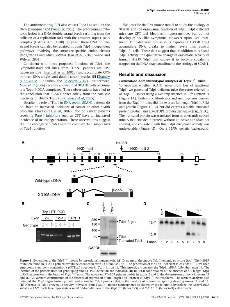
Generation and phenotypic analysis of Tdp1�/� mice
To ascertain whether SCAN1 arises from loss of functional
Tdp1, we generated Tdp1-deficient mice (hereafter referred to
as Tdp1�/� mice) using a Geo trap inserted in Tdp1 intron 11
(Figure 1A). Embryonic fibroblasts and neurospheres derived
from the Tdp1�/� mice did not express full-length Tdp1 mRNA
and protein (Figure 1B, C) but did express a stable truncated
protein product and a geoTDP1 protein derivative (Figure 1C).
The truncated protein was translated from an aberrantly spliced
mRNA that encoded a protein without an active site (data not
shown), and consistent with this, Tdp1 enzymatic activity was
undetectable (Figure 1D). On a 129Sv genetic background,
A
1 2 3 4 5 6 7 8 9 10 11 12 13 14 15 16
H493R
HKD motif 1 HKD motif 2
10 1211
10 1211
Wild-type cDNA
XD105 cDNA
75
100
150250
Tdp
1−/−
Tdp
1+/+
GAPDH
Truncated Tdp1Tdp1
DC
+/+−/−
12-Y
12-Y
12-P
1 2 3 4 5 6 7 8 9Lanes:
+/+
+/−
−/−
GAPDH2–6 12–16
Genotype
Tdp1 RT–PCR
+/+
+/−
−/−
+/+
+/−
−/−
B
Exon
Figure 1 Generation of the Tdp1�/� mouse by insertional mutagenesis. (A) Diagram of the mouse Tdp1 genomic structure (top). The H493Rmutation found in SCAN1 patients would be encoded in exon 13 of mouse Tdp1. For generation of the Tdp1-deficient mice (Tdp1�/�), we usedembryonic stem cells containing a pGT1Lxf insertion in Tdp1 intron 11. This insertion truncates the Tdp1 mRNA following exon 11. Thelocation of the primers used for genotyping and RT–PCR detection are indicated. (B) RT–PCR confirmation of the absence of full-length Tdp1mRNA expression in the brain of Tdp1�/� mice. The upstream RT–PCR primers reside in exons 2 and 6, the downstream primers in exons 12and 16. (C) Western confirmation of the absence of expression of full-length Tdp1 protein in Tdp1�/� neurospheres. The western analysis alsodetected the Tdp1-b-geo fusion protein and a smaller Tdp1 product that is the product of alternative splicing deleting exons 12 and 13.(D) Absence of Tdp1 enzymatic activity in lysates from Tdp1�/� mouse neurospheres as shown by the failure to hydrolyze the tyrosyl-DNAsubstrate 12-Y. Each lane represents a serial 10-fold dilution of the Tdp1�/� (lanes 2–5) and Tdp1þ /þ (lanes 6–9) cell extracts.
A Tdp1 recessive neomorphic mutation causes SCAN1?R Hirano et al
&2007 European Molecular Biology Organization The EMBO Journal VOL 26 | NO 22 | 2007 4733

Tdp1þ /� parents produced all genotypic classes in Mendelian
ratios. Thus, in contrast to Drosophila (Dunlop et al, 2004) in
which deficiency of glaikit, the Tdp1 homolog, causes defective
embryonic neurogenesis and embryonic lethality, Tdp1-defi-
cient mice did not exhibit embryonic or neonatal lethality.
At birth, the Tdp1�/� mice had body weights and lengths,
brain weights and histopathology comparable to Tdp1þ /þ
mice (Figure 2A–I). This phenotype correlates with that of
SCAN1 patients who had normal growth and motor, lan-
guage, social and intellectual development throughout child-
hood (Takashima et al, 2002). However, in contrast to the
SCAN1 patients who develop clinical symptoms in their
second decade of life (Takashima et al, 2002), the Tdp1�/�
mice examined at 270 days by histopathology and peripheral
nerve electrophysiology did not show abnormalities (Figure
2J–S) and those analyzed at 14–17 months did not exhibit
behavioral or other abnormalities. Thus, we propose that the
Tdp1�/� mice are able to repair endogenous levels of Topo I–
DNA complexes by alternate DNA-repair pathways.
Alternative pathways for the repair of Topo I–DNA
covalent complexes
In Saccharomyces cerevisiae, the two best-studied alternative
pathways for repairing stalled Topo I are those mediated by
0
0.8
0.4
1.2
1.6
Mea
n bo
dy w
eigh
t (g)
Mea
n br
ain
wei
ght (
g)
30
20
10
0
Mea
n bo
dy le
ngth
(m
m)
30
20
10
0
P0 phenotype P270 phenotype
Tdp1+/+
Tdp1+/–
Tdp1–/–
Tdp1+/+
Tdp1–/–
IGL
EGL
ML
A
IGL
EGL
ML
B IIIIIIIVV
VI
J IIIIIIIVV
VI
K
ML
GL
PC
LML
GL
PC
M
N O
Tdp1+/+ Tdp1−/− Tdp1−/−Tdp1+/+
C D
E F
G H
I
P
R
EGL
PC
EGL
PC
CM
AP
(m
V)
8
6
4
2
0
1.8
1.6
1.0
1.4
1.2
Dis
tal l
aten
cy (
ms)
Mea
n bo
dy w
eigh
t (g)
30
20
10
0
40 P=
0.21
Males
Mea
n bo
dy w
eigh
t (g) P=
0.01
30
20
10
0
40
Females
S
Q
Figure 2 Comparative phenotypic and histopathological evaluation of the Tdp1þ /þ and Tdp1�/� mice at P0 (A–I) and P270 (J–S). P0 mice:Hematoxylin and eosin (H&E) staining of cortex (A, B), cerebellar vermis (C, D), spinal cord (E, F), and dorsal root ganglion (G, H). Note thecomparability of the external granular cell (EGL), molecular (ML), and internal granular cell layers (IGL) as well as the Purkinje cell number(PC). (I) Comparison of the body weights and lengths and brain weights of Tdp1þ /þ , Tdp1þ /�, and Tdp1�/� littermates. P270 mice: H&Estaining of cortex (J, K), cerebellar vermis (L, M), spinal cord (N, O), and dorsal root ganglia (P, Q). The cortical layers are labeled in panels Jand K and the cerebellar molecular (ML) and granular cell (GL) layers in panels L and M. The insets in panels N and O are highermagnifications of the dorsal (upper) and ventral (lower) horns. (R) Comparison of male and female body weights. (S) Comparison of theperipheral nerve electrophysiology of Tdp1þ /þ and Tdp1�/� littermates. Scale bar¼ 50 mm.
A Tdp1 recessive neomorphic mutation causes SCAN1?R Hirano et al
The EMBO Journal VOL 26 | NO 22 | 2007 &2007 European Molecular Biology Organization4734

the structure-specific endonucleases Rad1/Rad10 and Mus81/
Mms4 (Liu et al, 2002; Vance and Wilson, 2002), which
correspond to mammalian XPF/ERCC1 and Mus81/Eme1,
respectively (Matsunaga et al, 1995; Brookman et al, 1996;
Ciccia et al, 2003; Ogrunc and Sancar, 2003). By in situ
hybridization and RT–PCR across a spectrum of Tdp1þ /þ
mouse tissues, Xpf, Ercc1, and Mus81 were expressed at each
time point investigated, whereas Eme1 generally had much
lower neural expression except in the developing cerebellum
(Supplementary Figures 1 and 2). Notably, Eme1 was ex-
pressed in non-neural tissues such as the thymus, heart,
intestine, liver, skin, vertebrae, teeth, and testes
(Supplementary Figure 1M, N). We observed a similar lack
of neural expression of Eme1 in the adult human brain by
RT–PCR (Supplementary Figure 2T) and northern analyses
(data not shown). The mRNAs encoding these endonucleases
(Supplementary Figure 2U) as well as the mRNAs encoding
the base excision repair enzymes Apex1 and Apex2 (data not
shown) were not increased in Tdp1�/� brain tissue. Thus,
although not upregulated in Tdp1�/� tissue, the XPF/ERCC1
pathway may function as one alternative pathway for repair
of Topo I–DNA complexes in the absence of Tdp1 (Vance and
Wilson, 2002), and the low or absent Eme1 expression in the
majority of neuronal tissues might contribute to the neuronal
specificity of SCAN1.
Tdp1 is expressed in the nervous system of mice and
humans
To test whether variation in expression across species ac-
counted for the difference in phenotype between Tdp1�/�
mice and SCAN1 patients, we compared the expression of
Tdp1 in mice and humans. By northern blot analysis, human
Tdp1 mRNA was present in all analyzed adult tissues
(Supplementary Figure 3A) and in all tested areas of the
central nervous system (Supplementary Figure 3B). Similarly,
mouse Tdp1 mRNA was expressed in all analyzed adult
tissues (Supplementary Figure 3C) and in the brain from
embryonic day (E) 9.5 through adulthood (Supplementary
Figure 3D).
In the human brain, Tdp1 was highly expressed in neurons
from 10 gestational weeks (GW) through 16 years of age and
at very low levels in glia (Figure 3A–C). Similar to the
Drosophila glaikit protein (Dunlop et al, 2000), human
Tdp1 was expressed in subependymal neural progenitors
and cultured neurospheres (Figure 3A and Supplementary
Figure 4). Cerebellar, granule and Purkinje cells (Figure 3D–
F) and neurons of the dentate nucleus (Figure 3G), spinal
cord (Figure 3H), and dorsal root ganglia (DRG) (Figure 3I),
the neurons putatively affected by SCAN1 (Takashima et al,
2002), also expressed Tdp1. Similar to the glaikit protein in
Drosophila, the Tdp1 protein was prominently expressed
in the cytoplasm of some neurons (Figure 3F–H), a poorly
understood finding; however, in contrast to human Tdp1,
glaikit is not thought to participate in DNA repair but in
localization of membrane proteins (Dunlop et al, 2000, 2004).
Antiserum nonspecificity does not account for this staining
pattern because we obtained the same results with two
independently produced antisera, and the preimmune
serum did not recognize either a nuclear or a cytoplasmic
antigen (Figure 3J–K). Moreover, the antisera recognized a
70 kDa protein in mouse Tdp1�/� fibroblasts transfected with
a human Tdp1 expression plasmid but not in untransfected
Tdp1�/� fibroblasts (Figure 3L), and competitive inhibition
with recombinant Tdp1 protein blocked interaction of the
anti-human (Figure 3M) and anti-mouse sera with Tdp1
(data not shown). Although we observed this staining pattern
in the tissues from several individuals, this finding could be
an artefact of tissue fixation since in our experience Tdp1
readily diffuses out of the nucleus of dying human and mouse
cells. Alternatively, this might suggest either cytoplasmic
sequestration or a cytoplasmic function for Tdp1 in humans
but not in mice.
With the exception of the cytoplasmic localization, the
mouse brain showed a similar expression pattern for the
Tdp1 RNA and protein (Figure 4). This suggested to us that
SCAN1 arises either from loss of a human-specific function as
reflected by the differences in protein localization or from a
novel function of Tdp1 created by the H493R mutation.
Tdp1�/� mice, neurospheres, and embryonic fibroblasts
are CPT hypersensitive
Like cells from the SCAN1 patients (El-Khamisy et al, 2005;
Interthal et al, 2005b), Tdp1�/� mouse embryonic fibroblasts
(MEFs, Supplementary Figure 6A, D–G) and neurospheres
(Figure 5A) were hypersensitive to CPT. Therefore, we hy-
pothesized that the Tdp1�/� mice, and perhaps in particular
the nervous system of these mice, would have increased
sensitivity to CPT and that CPT treatment might induce a
SCAN1-like phenotype. Intraperitoneal administration of the
Topo I poison CPT-11 as a single (80 mg/kg) or a weekly
(40 mg/kg, for 20 weeks) dose elicited no detectable pheno-
type in Tdp1�/� or Tdp1þ /þ mice aged 70–210 days
(Figure 5B). But five consecutive daily doses of CPT-11
(40 mg/kg/day) or of topotecan (1.6 or 4.8 mg/kg/day)
were toxic to Tdp1�/� mice ranging in age from 70 to 80
days (Figure 5B). Although the same age Tdp1þ /þ mice were
unaffected, the Tdp1�/� mice developed weakness and diar-
rhea and expired within 2 days after completion of CPT-11 or
topotecan administration. By histopathology and immuno-
histochemistry, there was extensive necrosis in the more
slowly proliferating intestinal, renal, and hepatic tissues
(Figure 5C–J), and extensive apoptosis and marked tissue
loss in the more rapidly proliferating lymphoid and hemato-
poietic tissues (Figure 5K–R). The induction of apoptosis in
these rapidly proliferating tissues indicates that Tdp1, as
expected, is involved in the repair of Topo I-associated DNA
double-strand breaks that are generated when the replication
machinery collides with CPT-trapped Topo I. The treated
Tdp1�/� mice also had electrophysiological changes, pro-
longed distal latencies, and reduced compound muscle action
potentials (Supplementary Figure 5A), changes typically seen
in acute illness but different from those of the SCAN1 patients
(Takashima et al, 2002). Whether treated with CPT-11 or
topotecan, Tdp1�/� mice did not exhibit ataxia or have
detectable atrophy or apoptosis in the cerebrum, cerebellum,
spinal cord, DRG, or peripheral nerve by histological,
immunohistochemical, TUNEL, or ultrastructural analyses
(Supplementary Figure 5B–U). In addition, the Tdp1�/�
glia and neurons differentiated from neurospheres
(Supplementary Figure 5V, W) or cultured Tdp1�/� cortical
neurons (data not shown) failed to exhibit cell death
or apoptosis at CPT doses that impaired neurosphere
proliferation.
A Tdp1 recessive neomorphic mutation causes SCAN1?R Hirano et al
&2007 European Molecular Biology Organization The EMBO Journal VOL 26 | NO 22 | 2007 4735

In summary, these results confirm that the CPT hypersen-
sitivity of SCAN1 lymphoblastoid cells is likely arising be-
cause of deficient Tdp1 activity. However, they also highlight
the possibility that the neuropathology of SCAN1 may not be
caused solely by deficient Tdp1 activity because accentuation
of the formation of Topo I–DNA complexes with the admin-
Figure 3 Immunohistochemical analysis of Tdp1 protein expression in the human brain. (A) Tdp1 expression in the cerebrum of a human 10gestational week (GW) brain. Cells adjoining the ventricle (V), and in the subventricular zone (SVZ), intermediate zone (IMZ), and corticalplate (CP) express Tdp1. (B, C) Tdp1 expression in layer two of the cortex from a 40 GW and a 16-year brain. (D–F) Tdp1 expression in thecerebella from a 22 GW, a 9-month and a 4-year brain. Note the staining in the external granular cell layer (EGL), Purkinje cells (PC),and internal granular cell layer (IGL). (G) Tdp1 expression in a 19-year dentate gyrus. (H, I) Tdp1 expression in spinal cord anterior horncells (AHC) and a dorsal root ganglion of a 10-year old, respectively. (J–M) Specificity of the immune serum is shown by absence of stainingof the spinal cord (J) and dorsal root ganglion (K) from a 10-year old with preimmune serum and also by absence of antigen recognition inmouse Tdp1�/� mouse embryonic fibroblasts compared to Tdp1�/� fibroblasts transfected with a human Tdp1 expression construct(Tdp1�/�þ pCMV5 .Tdp1) (L), and competitive inhibition of the antiserum by recombinant human Tdp1 (M). Scale bar¼ 50mm.
Figure 4 In situ hybridization and immunohistochemistry showing the spatial and temporal expression of mouse Tdp1 mRNA and protein,respectively. Localization of the Tdp1 mRNA in P1 (A–D), P10 (E–H), and adult (I–L) mice. (A, E, I) Expression of Tdp1 mRNA in a midline sagittalsection from a P1, P10, and adult mouse or brain, respectively. Note that Tdp1 is expressed throughout the cortex (Cx), cerebellum (Cb), spinal cord(SC), and dorsal root ganglia (DRG) as well as most other organs including the skin, thymus (Th), heart, lungs, liver, and intestines. (B, F, J)Expression of Tdp1 mRNA in the cortex (Cx) and hippocampus (Hip) of a P1, P10, and adult brain, respectively. (C, G, K) Expression of Tdp1mRNA in a coronal section of a P1, P10, and adult cervical spinal cord, respectively. The highest expression is in the dorsal horns (DH) and theventral horns (VH). (D, H, L) Expression of Tdp1 mRNA in a cross-section of a P1, P10, and adult dorsal root ganglion, respectively. (M–O) Serialsections from the P1 mouse hybridized with a sense probe for Tdp1 showing the specificity of the hybridization for the antisense probe used inpanels A–L. Scale bar¼ 1 cm (A, E, I, M), 0.5 mm (B, C, F, G, J, K, N, O), 0.2 mm (D, H, L). Localization of the Tdp1 protein in the P0 and the P270CNS (P-AA). (P) P0 cortex and hippocampus, (Q) P0 layer II cortical neurons, (R) P270 cortex, (S) P270 layer II cortical neurons, (T) P0 cerebellum,(U) P270 cerebellum, (V) P270 dentate nucleus, (W) P270 spinal cord. Nonspecific staining of the P0 cortex (X), P270 cortex (Y), P0 cerebellum(Z), and P270 cerebellum (AA). Scale bar¼ 0.5 mm (P, R, T, U, Y, Z, AA, BB), 0.2 mm (X), 50mm (Q, S, and all insets). Abbreviations: EGL, externalgranular layer; PC, Purkinje cell; ML, molecular layer; VH, ventral horn; VHC, ventral horn cell; DH, dorsal horn.
A Tdp1 recessive neomorphic mutation causes SCAN1?R Hirano et al
The EMBO Journal VOL 26 | NO 22 | 2007 &2007 European Molecular Biology Organization4736

istration of Topo I inhibitors did not cause SCAN1-like neural
dysfunction. Rather, nonproliferating Tdp1�/� neural cells in
culture and in vivo did not show acute toxicity to Topo I–DNA
complexes, whereas proliferating Tdp1�/� cells in vivo were
quite sensitive to the toxicity of Topo I–DNA complexes
(Figure 5 and Supplementary Figure 5). The resistance of
nonproliferating neural cells to topotecan and CPT-11 cannot
be ascribed to the failure of these drugs to penetrate the
blood–brain barrier since topotecan penetration of the
blood–brain barrier is well established in rodents (El-Gizawy
and Hedaya, 1999; Zhuang et al, 2006). In addition, the
DRG, which resides outside of the blood–brain barrier
(Arvidson, 1979), did not exhibit histopathological changes
(Supplementary Figure 5).
P1
Sense probe
P1
P10
Adult
CbHip
OL
Cx
THy
Hip
Cx
CbCx DRG
Th
SCHip
Cx
HipCx
DH
VH
DH
VH
CbCx
DRG
Th
SC
DH
VH
CortexSpinal Dorsal root
ganglion cord In situ hybridization
Immunohistochemistry
PreimmuneAAZYX
P
WVUT
SRQ
AmH
DG
AmH
DG
EGL
MLPC
VH
DH
A B C D
E
I
F G H
J K L
M N O
PC VHC
A Tdp1 recessive neomorphic mutation causes SCAN1?R Hirano et al
&2007 European Molecular Biology Organization The EMBO Journal VOL 26 | NO 22 | 2007 4737

The resistance of nonproliferating neural cells to CPT might
suggest that alternative pathways are able to repair low levels of
Topo I–DNA complexes. The existence of such alternative
pathways is supported by the observation of Xpf induction in
lung tumors commonly resistant to camptothecin (Sestili et al,
2006) and by the increased levels of homologous recombination
in SCAN1 patient lymphoblastoid cells (El-Khamisy et al, 2005).
Additionally, we have evidence for the existence of an endonu-
clease-dependent repair pathway for stalled Topo I (J Leppard,
H Interthal, and J Champoux, unpublished).
Tdp1�/� mice and embryonic fibroblasts are bleomycin
but not etoposide hypersensitive
SCAN1 cells are also deficient in processing of DNA phos-
phoglycolate 30 ends (Inamdar et al, 2002; Zhou et al, 2005).
Therefore, we hypothesized that the Tdp1�/� cells and mice
would have increased sensitivity to bleomycin. By the comet
assay, Tdp1�/� MEFs were hypersensitive to bleomycin
(Supplementary Figure 6B), and with intraperitoneal admin-
istration of the bleomycin for 10 consecutive days (10 mg/kg/
day), Tdp1�/� mice developed weakness and expired within
4 days following treatment. Histopathology and immunohis-
tochemistry showed extensive apoptosis and marked tissue
loss in the more rapidly proliferating lymphoid and hemato-
poietic tissues (Supplementary Figure 6H–K). The treated
Tdp1�/� mice did not exhibit ataxia or electrophysiological
changes typical of SCAN1 or have detectable atrophy or
apoptosis in the cerebrum, cerebellum, spinal cord, or DRG
peripheral nerve (Supplementary Figure 6L–O and data not
shown).
Tdp1 is also proposed to participate in the repair of
DNA double-strand breaks resulting from stalling
of Topoisomerase II (Topo II) (Barthelmes et al, 2004; Nitiss
et al, 2006). If Tdp1 played a key role in this repair process,
then Tdp1�/� cells and mice might have increased sensitivity
to etoposide, a Topo II inhibitor (Liu, 1989). However,
consistent with the studies of lymphoblastoid cells from
SCAN1 patients (Interthal et al, 2005b), neither Tdp1�/�
MEFs nor mice showed increased sensitivity to etoposide
(Supplementary Figure 6C, P–W). Both Tdp1þ /þ and
Tdp1�/� mice became ill and died 2 or 3 days after
5 consecutive days of 30 or 40 mg/kg/day of etoposide, and
both were equally symptom free 60 days after 5 days of
20 mg/kg/day of etoposide. The Tdp1þ /þ and Tdp1�/�
mice did not have detectable pathological differences by
histological, immunohistochemical, or TUNEL analyses
(Supplementary Figure 6P–W and data not shown).
These in vivo and cell culture studies confirm the partici-
pation of Tdp1 in the repair of DNA phosphoglycolate 30 ends
but not in the repair of Topo II–DNA complexes. Moreover,
the absence of an SCAN1-like phenotype in the treated mice
again highlights the possibility that SCAN1 might not arise
solely from deficient Tdp1 activity.
CPT treatment of cells expressing Tdp1 H493R causes
the accumulation of H493R–DNA complexes and the
accumulation of DNA strand breaks
The above observations suggested that SCAN1 is caused by a
novel and distinct function of Tdp1 created by the SCAN1
point mutation. Previously, Interthal et al (2005a, b) hypothe-
sized that SCAN1 might not only arise from the 25-fold
reduction in activity caused by the H493R change, but also
from the accumulation of the Tdp1 H493R–DNA covalent
reaction intermediate which has a half-life of B13 min. The
removal of H493R Tdp1 from the DNA by wild-type Tdp1
explains the recessive nature of SCAN1 (Interthal et al,
2005a, b). To test whether Tdp1 H493R becomes trapped on
the genomic DNA in vivo in response to CPT treatment, we
analyzed telomerase-immortalized skin fibroblasts from
unaffected controls and SCAN1 patients using a modified
in vivo complex of enzyme (ICE) assay (Subramanian et al,
1995). CPT-treated fibroblasts from both sources accumulated
the Topo I–DNA covalent intermediate as expected, but only
SCAN1 cells accumulated a covalent Tdp1–DNA intermediate
(Figure 6A). Based on alkaline comet assay analyses and
accumulation of gH2AX, CPT-treated Tdp1�/� MEFs expres-
sing the human H493R Tdp1 protein accumulated DNA
strand breaks more rapidly and to a higher level than CPT-
treated Tdp1�/� MEFs transfected with the control expression
plasmid (Figure 6B and Supplemental Figure 7). In contrast,
reduced accumulation of DNA strand breaks was evident
upon expressing the wild-type Tdp1 protein (Figure 6B and
Supplementary Figure 7).
These data show that the human H493R Tdp1 has an
enzymatic activity in vivo that is qualitatively different from
the Tdp1 activity in Tdp1�/� cells or Tdp1�/� cells comple-
mented with wild-type Tdp1. This defines Tdp1 H493R as a
neomorphic mutation. Moreover, the covalent stalling of
H493R Tdp1 on the DNA in CPT-treated SCAN1 cells provides
direct evidence that human Tdp1 removes CPT-trapped Topo I
from the DNA in vivo.
Model for the repair of covalent Topo I–DNA complexes
and the molecular mechanism of SCAN1
We previously had hypothesized that SCAN1 arises from cell
death or cellular malfunction secondary to the accumulation
of Topo I–DNA complexes; however, our data here suggest
that Tdp1–DNA intermediates arising from antecedent Topo
I–DNA complexes may be the underlying basis for the disease
(Figure 6C). Although treatment of Tdp1�/� mice with Topo I
inhibitors or bleomycin might have recapitulated the SCAN1
phenotype if sufficient DNA damage was able to accumulate,
Tdp1�/� mice treated with either high or low doses of drug
did not develop an SCAN1-like phenotype. This suggests that
the resultant DNA complexes are not the initiating lesions;
however, it seems more likely that for the high doses, the
drug-induced damage greatly exceeded the repair capacity
of the mice and therefore failed to mimic the low level
of naturally occurring lesions that accumulate in SCAN1
patients. And for low doses of drug, redundant path-
ways effectively repaired the DNA damage. Therefore, we
hypothesize that the prolonged half-life of the H493R Tdp1–
DNA complexes and the increased level of DNA damage
in neuronal cells is the origin of the cellular malfunction
underlying SCAN1. Such an increase in DNA damage in
neuronal cells could arise from decreased efficiency Tdp1–
DNA adduct removal since some DNA repair pathways are
attenuated in terminally differentiated cells (Nouspikel and
Hanawalt, 2002). Testing this model requires analysis of
another mouse model solely expressing the mutant H493R
Tdp1 rather than the loss of function mutation described here.
Relevant to the treatment of cancer, the absence of detect-
able acute effects of CPT and bleomycin treatment on the
murine nervous system suggests that nonproliferating cells of
A Tdp1 recessive neomorphic mutation causes SCAN1?R Hirano et al
The EMBO Journal VOL 26 | NO 22 | 2007 &2007 European Molecular Biology Organization4738

the nervous system are insensitive to Topo I–DNA complexes
and 30 phosphoglycolate-DNA damage. Thus, in contrast to
the accentuation of demyelinating neuropathies by some
chemotherapies (Weimer and Podwall, 2006), short-term
administration of these chemotherapeutic agents is unlikely
to induce neurological disease even in the absence of Tdp1.
Additionally, our observations suggest that Tdp1 is also a
reasonable chemotherapeutic target as long as the formation
and consequences of Tdp1–DNA complexes in non-neoplastic
tissues are carefully assessed.
In summary, SCAN1 is the first example of a human
genetic disease that results from a failure to repair DNA–
protein covalent complexes. SCAN1 likely arises not only
from a quantitative change in Tdp1 activity but also from a
Figure 5 Analysis of CPT-11-treated and untreated Tdp1þ /þ and Tdp1�/� cells and mice. (A) CPT sensitivity of Tdp1þ /þ and Tdp1�/�
neurosphere cells following 72 h of incubation with camptothecin (CPT) at the indicated concentrations. (B) CPT-11 and topotecan treatmentprotocols and outcomes for Tdp1þ /þ and Tdp1�/� mice. Note that only mice treated repetitively at short intervals developed a phenotype; thissuggests a high level of redundancy for the removal of stalled Topo I. (C–R) Histopathology and TUNEL staining of liver and spleen derivedfrom Tdp1þ /þ and Tdp1�/� mice treated with 40 mg/kg of CPT-11 for 5 days. Note the extensive vacuolization (F) but paucity of TUNEL-positive cells (J) in the liver of Tdp1�/� mice suggesting necrotic cell death. In contrast, the lymphoid and hematopoietic tissues such as thespleen showed marked loss of tissue (M versus N) with a large number of TUNEL-positive cells (Q versus R) suggesting cell death by apoptosis.
A Tdp1 recessive neomorphic mutation causes SCAN1?R Hirano et al
&2007 European Molecular Biology Organization The EMBO Journal VOL 26 | NO 22 | 2007 4739

A
Tdp1
Topo I250 kDa150100
100 kDa75
SCAN1 WT
1 2 3 4
− + − +
Tdp1
Topo I
β-Actin
CPT
Whole cell extracts
RT–PCR + BsaAI digestion1 2 3 4
C
1 2 3 4
B
Tdp1−/−−/−
No phenotype
Tdp1
Blocked 3′ end
PNKXRCCILigase 3
ds DNA
Blocked 3′ end
HO
P
DNA fill-in and ligation
No phenotype
Alternative DNArepair pathways
ds DNA
Tdp1H493R/H493R
Tdp1H493R
Blocked 3′ end
Stalled Tdp1
Accumulation ofstalled Tdp1H493R
Increased DNA damage
Neuronal dysfunction
SCAN1 phenotype
No
trea
tmen
tT
reat
men
t
Tdp1+/++/+
No phenotype CPT hypersensitivity
Alternative DNArepair pathwaysoverwhelmed
Alternative DNA repair pathways overwhelmed or inefficient repairing
H493R Tdp1–DNA complexes
Adequate repair of blocked 3′ ends by Tdp1
and alternative pathways
CPT hypersensitivityBleomycin hypersensitivity
Blocked 3′ end
HOP
DNA fill-in and ligation
ds DNA
Alternative DNArepair pathways
TDP11478A>GTDP1WT
pcDNA3.1(−)
0
10
20
30
40
10 30
No
CP
T
Mea
n co
met
mom
ent
Recovery time
****
(min)
** **
Potential model for SCAN1
OHP
Figure 6 Stalling of H493R Tdp1 on DNA and a model for the causation of SCAN1. (A) In vitro complex of enzyme assay showing thattreatment of unaffected and SCAN1 patient skin fibroblasts with camptothecin (CPT) causes formation of a covalent Tdp1–DNA complex in theSCAN1 fibroblasts but not in unaffected fibroblasts (upper panel). The expected covalent Topo I–DNA complex (middle panel) and the relativeabundance of each protein (lower panel) are also shown. (B) Alkaline comet assay showing increased CPT induction of DNA strand breaks inTdp1�/� MEFs expressing H493R Tdp1 (pcDNA3.1(�) .TDP11478A4G) and reduced induction of DNA breaks in Tdp1�/� MEFs expressing wild-type Tdp1 (pcDNA3.1(�) .TDP1WT). The comet moments were measured following treatment with 1mM CPT for 1 h and recovery in CPT-freemedium for the indicated times. The comet moment of Tdp1�/� MEFs transfected with only the expression vector (pcDNA3.1(�)) wassignificantly different from TDP1WT and TDP11478A4G (**P50.001). The expression of human wild type and H493R Tdp1 was detected in thetransfected cells by western blot (data not shown) and RT–PCR followed by BsaAI digestion of the products (lower panel) as previouslydescribed (Takashima et al, 2002). Lane 1: molecular weight markers; lane 2: Tdp1�/� MEFs transfected with plasmid pcDNA3.1(�), the emptyvector; lane 3: Tdp1�/� MEFs transfected with plasmid pcDNA3.1(�) .TDP1WT, the expression vector for wild-type human TDP1 cDNA; lane 4:Tdp1�/� MEFs transfected with plasmid pcDNA3.1(�) .TDP11478A4G, the expression vector for human H493R Tdp1 cDNA. Note that the1478A4G mutation creates a BsaAI restriction site. (C) Potential model for the molecular basis of SCAN1. DNA breaks with blocked 30 ends(e.g., Topo I or phosphoglycolate) undergo Tdp1-facilitated DNA repair via both DNA single-strand break repair (SSBR) and double-strandbreak repair (DSBR) mechanisms. With loss of functional Tdp1 (Tdp1�/�), there is sufficient redundant activity for adequate DNA repair byalternative pathways (e.g. endonuclease-dependent pathways) unless the system is further stressed as by administration of CPT or bleomycin.In contrast, when Tdp1 carries the H493R mutation, it not only has a quantitative reduction in overall activity, but also a qualitative changeresulting in accumulation of Tdp1–DNA complexes. These complexes are efficiently removed from the DNA by wild-type Tdp1 in all tissues ofheterozygotes, whereas they are only removed in replicating cells of homozygotes by alternative DNA strand break repair mechanisms.According to this model, the transcriptional interference and/or apoptosis resulting from the Tdp1–DNA complexes in nondividing neuronscauses SCAN1 via neurodegeneration.
A Tdp1 recessive neomorphic mutation causes SCAN1?R Hirano et al
The EMBO Journal VOL 26 | NO 22 | 2007 &2007 European Molecular Biology Organization4740

qualitative change that renders the enzyme different from
wild-type Tdp1 causing it to become covalently trapped
on the DNA. Furthermore, since the previously described
disease-causing neomorphic mutations are dominant
(Antonarakis et al, 2001; Beaudet et al, 2001), the recessive
nature of the neomorphic Tdp1 H493R mutation (Interthal
et al, 2005a, b) defines a novel mechanism for human
disease.
Materials and methods
Human subjectsPatients gave informed consent approved by the InstitutionalReview Board of Baylor College of Medicine (H-9669, Houston,TX, USA) and the University of British Columbia (C06-0283,Vancouver, BC, Canada). The clinical data were collected fromquestionnaires completed by the referring primary care physicianand from medical records and summaries provided by thatphysician.
Animal subjectsMice used in this study were housed, bred, and killed in accordancewith accepted ethical guidelines. These procedures were approvedby the Institutional Review Board of Baylor College of Medicine(Houston, TX, USA, IRB protocol: AN-2983), the University ofBritish Columbia (Vancouver, BC, Canada, Animal Care Certificate:A06-0257), and Kagoshima University (Kagoshima, Japan).
Transgenic miceWe purchased the gene trap embryonic stem (ES) cell line XD105from BayGenomics (http://baygenomics.ucsf.edu/). XD105 wasgenerated by integration of pGT1Lxf into intron 11 of Tdp1.This cell line was injected into albino C57 blastocysts that werethen implanted into pseudopregnant mothers. The resultingchimeric mice were bred to 129/SvEv females to generate founders.The progeny from these heterozygous founders were used forall subsequent analyses. Mice were genotyped by PCR of genomicDNA using a common forward primer in intron 11 and a reverseprimer in either intron 11 or in pGT1Lxf (Supplementary Table I)and data not shown. The intron 11 primer pair detects the wild-typeallele but not the trapped allele, and the intron 11 forwardprimer with the pGT1Lxf reverse primer detects the mutant allelebut not the wild-type allele. The PCR was carried outusing HotMasterMix (Eppendorf) with 50 ng of genomic DNA and10 pmol of each primer. After initial denaturation at 941C for2 min, amplification was performed for 40 cycles with denatura-tion at 941C for 20 s, annealing at 551C for 10 s, extension at651C for 90 s.
To determine if the gene trap allowed expression of full-lengthTdp1 mRNA by splicing around the gene trap, we performed RT–PCR on brain RNA extracted from Tdp1�/� mice (Figure 1B). TheRT–PCR primers 50 of the trap reside in exon 2, and the primers 30 ofthe trap reside in exons 12 and 16, respectively (SupplementaryTable I). The RT–PCR amplification was performed as describedbelow. To determine if the gene trap allowed expression ofaberrantly spliced mRNA, we performed RT–PCR using primersresiding in exons 9 and 14 (Supplementary Table I).
Bleomycin, CPT-11, etoposide, and topotecan treatment ofmiceBleomycin (Bristol-Myers Squibb, Montreal, Canada), CPT-11(Yakult Honsha Co., Tokyo, Japan), etoposide (Novapharm,Toronto, Canada), or topotecan (LKT laboratories, Inc.) werediluted with 5% Dextrose solution prior to intraperitoneal injec-tion. Drug or PBS was given to five wild-type and five Tdp1�/� miceaccording to the dosages and administration schedules indicated inthe Results and Figure 5B. The mice were evaluated by electro-physiology (Supplementary data) 2 days after the last dose ofCPT-11, topotecan, or bleomycin.
Comet assayFor Figure 6 and Supplementary Figure 6, the MEFs were treatedwith 1mM CPT, 12.5mg/ml bleomycin, or 12.5mg/ml etoposide for60 min. For Figure 6, the CPTcontaining medium was then replaced
with fresh medium and the cells were allowed to recover for theindicated times. The MEFs were then removed from the plates withtrypsin, washed with medium, resuspended in LMAgarose, andlayered on agarose-coated slides cooling on ice.
Alkali comet assay (Singh et al, 1988). For CPT- or bleomycin-treated MEFs, the slides were immersed in lysis solution (2.5 MNaCl, 100 mM EDTA, 10 mM Trizma base, 1% Triton X-100, 10%dimethyl sulfoxide; pH 10) at 41C for 1 h. After a rinse in deionizedwater, slides were immersed in a 41C alkaline solution (50 mMNaOH, 1 mM EDTA, 1% dimethyl sulfoxide; pH413) for 25 min.Electrophoresis was carried out at a constant voltage of 25 V for25 min at 41C. After electrophoresis, slides were neutralized in0.4 M Tris–HCl pH 7.5.
Neutral comet assay (Olive et al, 1992). For etoposide-treatedMEFs, the slides were immersed in lysis solution (50 mM EDTA,0.5% SDS; pH 7.5) at 41C for 1 h. After a rinse in deionized water,slides were immersed in 41C TBE for 25 min. Electrophoresis wascarried out at a constant voltage of 25 V for 25 min at 41C.
After electrophoresis for both methods, slides were dehydratedin ice-cold 70% ethanol for 5 min and air-dried. DNA was stainedwith Sybr Green I. A total of 100 comets were scored for eachsample using CASP (Konca et al, 2003) and statistically significantdifferences in the distribution of comet moments were determinedusing the Student t-test.
Anti-Tdp1 serum productionAn anti-human Tdp1 serum was generated in rabbits againstTdp1 amino acids 1–152. An anti-mouse Tdp1 serum was generatedin guinea pigs against the full-length protein (amino acids 1–608).Both antigens were produced in Escherichia coli with thepET28a expression system (Novagen). Neither antigen displayedhomology to other human proteins by BLASTp. The specificity ofeach antiserum was confirmed by the absence of crossreactivitywith recombinant Tdp1 expressed in Tdp1�/� MEFs and bycompetitive blocking with recombinant human or mouse Tdp1,respectively.
Immunohistochemistry and histopathologyHuman and mouse brains were fixed by immersion in 10% bufferedformalin or 4% PFA in PBS. The brain tissue was processed,embedded in paraffin, and cut into 8 mm sections according tostandard protocols (Deguchi et al, 2003). Immunohistochemistrywas carried out as previously described (Kilic et al, 2005). We usedthe polyclonal rabbit anti-human Tdp1 at a dilution of 1:50 and thepolyclonal guinea pig anti-mouse Tdp1 at a dilution of 1:50. TUNELdetection of apoptotic cells was carried out using the ApopTagPeroxidase In Situ Apoptosis Detection Kit (Chemicon, S7100) andthe tissue was counterstained with 1% methyl green.
Modified ICE assay (Subramanian et al, 1995)Briefly, two confluent 15 cm Petridishes of telomerase immortalizedSCAN1 or control human fibroblasts were treated with 20mM CPTfor 1 h. Cells were lysed in 0.8% SDS in TE and the genomic DNAwas sheared with a syringe. Small aliquots of the whole cell extractswere mixed with SDS loading buffer for later analysis. Theremaining cell lysates were diluted four-fold with 1% N-lauroyl-sarcosine and the extracts were layered onto a CsCl cushion (1.5 g/cc). After ultracentrifugation, the pellet fraction was treated withmicrococcal nuclease to remove the majority of the DNA bound tothe proteins. Samples were mixed with SDS sample buffer andsubjected to SDS–PAGE and western blotting.
Tdp1 activity assay with mouse neurosphere cell extractsExponentially growing neurospheres were harvested by centrifuga-tion, washed twice in PBS, and resuspended to 8�107 cells/mlin lysis buffer (10 mM Tris–HCl (pH 7.5), 50 mM KCl, 2 mM MgCl2,1% Triton X-100, 15 mM DTT, 0.2 mg/ml PMSF, 1/1000 volume ofprotease inhibitor mixes Pic-D (5 mg/ml Pepstatin A, 1 mg/mlchymostatin in DMSO), and Pic-W (208 mg/ml benzamidine,5 mg/ml aprotinin, and 1 mg/ml leupeptin in H2O)). The cells werelysed by vortexing for 1 min and the cell extracts were clarified bycentrifugation.
Cell extracts were first diluted 1:2 with 2� reaction buffer(200 mM KCl, 40 mM Tris–HCl (pH 7.5), 40 mM EDTA, 2 mM DTT)and then 10-fold serially diluted in reaction buffer. For the
A Tdp1 recessive neomorphic mutation causes SCAN1?R Hirano et al
&2007 European Molecular Biology Organization The EMBO Journal VOL 26 | NO 22 | 2007 4741

experiment shown in Figure 1D, 10 ml of the extract dilutions wasadded to 5ml of reaction buffer containing 0.01 pmol of 32P-50 end-labeled substrate 12-Y (a 12-mer DNA oligonucleotide with a 30
phosphotyrosine (Interthal et al, 2005b)) and the reactions wereincubated at 371C for 30 min and stopped with an equal volume offormamide loading dye. Assays were analyzed on a 15% sequen-cing gel. Image retrieval and quantitation were carried out usinga PhosphorImager and ImageQuant software (Amersham Bio-sciences).
Supplementary dataSupplementary data are available at The EMBO Journal Online(http://www.embojournal.org).
Acknowledgements
We thank Millan Patel, Leah Elizondo, J Marietta Clewing, andKensuke Shiga for critical reviews of this manuscript. We thank
Barbara A Antalffy and Pauline Grennan for preparation of tissue;Monica J Justice, Darlene Skapura, and Wei Yu for mouse tissue;and Akiko Yoshimura and Yuko Shirahama for technical assistance.This work was supported in part by grants from the NationalInstitute of Diabetes, Digestive, and Kidney Diseases, NIH (CFB, DLA),the New Development Award, Microscopy, and AdministrativeCores of the Mental Retardation and Developmental DisabilitiesResearch Center at Baylor College of Medicine (CFB, DLA), theNational Ataxia Foundation (CFB), the Burroughs WellcomeFoundation (CFB), the Nervous and Mental Disorders andResearch Committee for Ataxic Disease of the Japanese Ministryof Health, Welfare and Labor (grant 17A-1, HT, KA), FrontierScience Research Center Program of Kagoshima University (HT),and the Ministry of Education, Culture, Sports, Science, andTechnology of Japan (grant 19591001, HT) and by NIH GrantGM049156 (JJC). NHLBI’s Programs for Genomic Applicationssupported the generation of insertion trap ES cells atBayGenomics (grant U01 HL66621).
References
Antonarakis SE, Krawczak M, Cooper DN (2001) The natureand mechanisms of human gene mutation. In The Metabolic &Molecular Bases of Inherited Disease, Scriver CR, Beaudet AL,Sly WS, Valle D, Childs B, Kinzler KW, Vogelstein B (eds),pp 343–377. New York: McGraw-Hill
Arvidson B (1979) Distribution of intravenously injected proteintracers in peripheral ganglia of adult mice. Exp Neurol 63:388–410
Barthelmes HU, Habermeyer M, Christensen MO, Mielke C,Interthal H, Pouliot JJ, Boege F, Marko D (2004) TDP1 over-expression in human cells counteracts DNA damage mediated bytopoisomerases I and II. J Biol Chem 279: 55618–55625
Beaudet AL, Scriver CR, Sly WS, Valle D (2001) Genetics, biochem-istry, and molecular bases of variant human phenotypes. In TheMetabolic & Molecular Bases of Inherited Disease, Scriver CR,Beaudet AL, Sly WS, Valle D, Childs B, Kinzler KW, Vogelstein B(eds), pp 3–45. New York: McGraw-Hill
Brookman KW, Lamerdin JE, Thelen MP, Hwang M, Reardon JT,Sancar A, Zhou ZQ, Walter CA, Parris CN, Thompson LH (1996)ERCC4 (XPF) encodes a human nucleotide excision repairprotein with eukaryotic recombination homologs. Mol Cell Biol16: 6553–6562
Ciccia A, Constantinou A, West SC (2003) Identification and char-acterization of the human mus81-eme1 endonuclease. J BiolChem 278: 25172–25178
Connelly JC, Leach DR (2004) Repair of DNA covalently linked toprotein. Mol Cell 13: 307–316
D’Arpa P, Beardmore C, Liu LF (1990) Involvement of nucleic acidsynthesis in cell killing mechanisms of topoisomerase poisons.Cancer Res 50: 6919–6924
Deguchi K, Inoue K, Avila WE, Lopez-Terrada D, Antalffy BA,Quattrocchi CC, Sheldon M, Mikoshiba K, D’Arcangelo G,Armstrong DL (2003) Reelin and disabled-1 expression in devel-oping and mature human cortical neurons. J Neuropathol ExpNeurol 62: 676–684
Dunlop J, Corominas M, Serras F (2000) The novel gene glaikit, isexpressed during neurogenesis in the Drosophila melanogasterembryo. Mech Dev 96: 133–136
Dunlop J, Morin X, Corominas M, Serras F, Tear G (2004) Glaikit isessential for the formation of epithelial polarity and neuronaldevelopment. Curr Biol 14: 2039–2045
El-Gizawy SA, Hedaya MA (1999) Comparative brain tissue dis-tribution of camptothecin and topotecan in the rat. CancerChemother Pharmacol 43: 364–370
El-Khamisy SF, Caldecott KW (2007) DNA single-strand breakrepair and spinocerebellar ataxia with axonal neuropathy-1.Neuroscience 145: 1260–1266
El-Khamisy SF, Saifi GM, Weinfeld M, Johansson F, Helleday T,Lupski JR, Caldecott KW (2005) Defective DNA single-strandbreak repair in spinocerebellar ataxia with axonal neuropathy-1. Nature 434: 108–113
Friedberg EC, Walker GC, Siede W, Wood RD, Schultz RA,Ellenberger T (2006) Disease States associated with defective
biological responses to DNA damage. In DNA Repair andMutagenesis, pp 863–1080. Washington: ASM Press
Inamdar KV, Pouliot JJ, Zhou T, Lees-Miller SP, Rasouli-Nia A,Povirk LF (2002) Conversion of phosphoglycolate to phosphatetermini on 30 overhangs of DNA double strand breaks by thehuman tyrosyl-DNA phosphodiesterase hTdp1. J Biol Chem 277:27162–27168
Interthal H, Chen HJ, Champoux JJ (2005a) Human Tdp1 cleaves abroad spectrum of substrates, including phosphoamide linkages.J Biol Chem 280: 36518–36528
Interthal H, Chen HJ, Kehl-Fie TE, Zotzmann J, Leppard JB,Champoux JJ (2005b) SCAN1 mutant Tdp1 accumulates theenzyme–DNA intermediate and causes camptothecin hypersensi-tivity. EMBO J 24: 2224–2233
Interthal H, Pouliot JJ, Champoux JJ (2001) The tyrosyl-DNAphosphodiesterase Tdp1 is a member of the phospholipase Dsuperfamily. Proc Natl Acad Sci USA 98: 12009–12014
Kilic SS, Donmez O, Sloan EA, Elizondo LI, Huang C, Andre JL,Bogdanovic R, Cockfield S, Cordeiro I, Deschenes G, Frund S,Kaitila I, Lama G, Lamfers P, Lucke T, Milford DV, Najera L,Rodrigo F, Saraiva JM, Schmidt B et al. (2005) Association ofmigraine-like headaches with Schimke immuno-osseous dyspla-sia. Am J Med Genet A 135: 206–210
Konca K, Lankoff A, Banasik A, Lisowska H, Kuszewski T, Gozdz S,Koza Z, Wojcik A (2003) A cross-platform public domain PCimage-analysis program for the comet assay. Mutat Res 534:15–20
Liu C, Pouliot JJ, Nash HA (2002) Repair of topoisomerase Icovalent complexes in the absence of the tyrosyl-DNA phospho-diesterase Tdp1. Proc Natl Acad Sci USA 99: 14970–14975
Liu LF (1989) DNA topoisomerase poisons as antitumor drugs.Annu Rev Biochem 58: 351–375
Matsunaga T, Mu D, Park CH, Reardon JT, Sancar A (1995) HumanDNA repair excision nuclease. Analysis of the roles of thesubunits involved in dual incisions by using anti-XPG andanti-ERCC1 antibodies. J Biol Chem 270: 20862–20869
Miao ZH, Agama K, Sordet O, Povirk L, Kohn KW, Pommier Y(2006) Hereditary ataxia SCAN1 cells are defective for the repairof transcription-dependent topoisomerase I cleavage complexes.DNA Repair (Amst) 5: 1489–1494
Nitiss KC, Malik M, He X, White SW, Nitiss JL (2006) Tyrosyl-DNAphosphodiesterase (Tdp1) participates in the repair of Top2-mediated DNA damage. Proc Natl Acad Sci USA 103: 8953–8958
Nouspikel T, Hanawalt PC (2002) DNA repair in terminally differ-entiated cells. DNA Repair (Amst) 1: 59–75
Ogrunc M, Sancar A (2003) Identification and characterizationof human MUS81-MMS4 structure-specific endonuclease. J BiolChem 278: 21715–21720
Olive PL, Wlodek D, Durand RE, Banath JP (1992) Factors influen-cing DNA migration from individual cells subjected to gelelectrophoresis. Exp Cell Res 198: 259–267
Pommier Y (2004) Camptothecins and topoisomerase I: a foot in thedoor. Targeting the genome beyond topoisomerase I with camp-
A Tdp1 recessive neomorphic mutation causes SCAN1?R Hirano et al
The EMBO Journal VOL 26 | NO 22 | 2007 &2007 European Molecular Biology Organization4742

tothecins and novel anticancer drugs: importance of DNAreplication, repair and cell cycle checkpoints. Curr Med ChemAnti-Canc Agents 4: 429–434
Pouliot JJ, Robertson CA, Nash HA (2001) Pathways for repair oftopoisomerase I covalent complexes in Saccharomyces cerevisiae.Genes Cells 6: 677–687
Pourquier P, Pommier Y (2001) Topoisomerase I-mediated DNAdamage. Adv Cancer Res 80: 189–216
Sestili P, Martinelli C, Stocchi V (2006) The fast halo assay: animproved method to quantify genomic DNA strand breakage atthe single-cell level. Mutat Res 607: 205–214
Singh NP, McCoy MT, Tice RR, Schneider EL (1988) A simpletechnique for quantitation of low levels of DNA damage inindividual cells. Exp Cell Res 175: 184–191
Subramanian D, Kraut E, Staubus A, Young DC, Muller MT (1995)Analysis of topoisomerase I/DNA complexes in patients adminis-tered topotecan. Cancer Res 55: 2097–2103
Takashima H, Boerkoel CF, John J, Saifi GM, Salih MA, ArmstrongD, Mao Y, Quiocho FA, Roa BB, Nakagawa M, Stockton DW,Lupski JR (2002) Mutation of TDP1, encoding a topoisomerase
I-dependent DNA damage repair enzyme, in spinocerebellarataxia with axonal neuropathy. Nat Genet 32: 267–272
Vance JR, Wilson TE (2002) Yeast Tdp1 and Rad1–Rad10 function asredundant pathways for repairing Top1 replicative damage. ProcNatl Acad Sci USA 99: 13669–13674
Weimer LH, Podwall D (2006) Medication-induced exacerbationof neuropathy in Charcot Marie Tooth disease. J Neurol Sci 242:47–54
Yang SW, Burgin Jr AB, Huizenga BN, Robertson CA, Yao KC, NashHA (1996) A eukaryotic enzyme that can disjoin dead-endcovalent complexes between DNA and type I topoisomerases.Proc Natl Acad Sci USA 93: 11534–11539
Zhou T, Lee JW, Tatavarthi H, Lupski JR, Valerie K, Povirk LF (2005)Deficiency in 30-phosphoglycolate processing in human cells witha hereditary mutation in tyrosyl-DNA phosphodiesterase (TDP1).Nucleic Acids Res 33: 289–297
Zhuang Y, Fraga CH, Hubbard KE, Hagedorn N, Panetta JC, WatersCM, Stewart CF (2006) Topotecan central nervous system pene-tration is altered by a tyrosine kinase inhibitor. Cancer Res 66:11305–11313
A Tdp1 recessive neomorphic mutation causes SCAN1?R Hirano et al
&2007 European Molecular Biology Organization The EMBO Journal VOL 26 | NO 22 | 2007 4743
Copyright © 2022 FDOKUMEN